Embed Size (px)
Citation preview
Paresh Thakkar et al. An Interesting Case of Visceral Larva Migrans
101 International Journal of Medical Science and Public Health | 2012 | Vol 1 | Issue 2
CASE REPORT
An Interesting Case of Visceral Larva Migrans (VLM)
Paresh A Thakkar, Amit Dahat, Omprakash Shukla, Bakul Javadekar
Department of Pediatrics, Medical College and S S G Hospital, Vadodara, Gujarat, India
Correspondence to: Paresh A Thakkar ([email protected])
Received Date: 01.02.2012 Accepted Date: 06.03.2012
DOI: 10.5455/ijmsph.2012.1.101-104
ABSTRACT
Human toxocariasis is primarily a soil transmitted zoonosis.We report a case of two and half year
old male child who presented with fever, anorexia and hepatosplenomegaly. Hemogram showed
anaemia (Hb 5.8), leukocytosis (TLC 26,500) and marked eosinophilia (AEC 14,100). Further
investigations revealed hypergammaglobulinemia, CT scan of Abdomen showed low density lesions
in liver and liver biopsy showed noncaeseating epitheloid cell granulomas and infiltrates of
lymphocytes and eosinophils. We decided to get a confirmatory serological diagnostic test for
Toxocara and the result was positive .Child was treated for it with Oral Albendazole and responded
dramatically.
Key Words: Visceral Larva Migrans; Eosinophilia; Noncaseating Liver Granuloma
INTRODUCTION
Toxocariasis is a soil transmitted
helminthozoonosis due to the infection by
ascarid larvae of genus Toxocara.[1] Poor
personal hygiene, consumption of raw
vegetables grown in contaminated kitchen
gardens, geophagia or soil eating is a specific
type of pica that increases the risk of
toxocariasis, especially in children living in
homes with puppies[2]. Humans are accidental
hosts. Physiological reactions to Toxocara
infection depend on the host’s immune response
and the parasitic load.[3] Majority of patients are
asymptomatic. Symptoms occur as result of
migration of second stage Toxocara larvae
through the body.[3-5] Toxocara infection
commonly resolves itself within weeks. In VLM,
larvae migration incites inflammation of
internal organs and sometimes the central
nervous system. Symptoms depend on the
organ(s) affected.[3]
CASE REPORT
A 2 and a half year old boy, was admitted on 11th
august 2011 with chief complaints of abdominal
distension since 1month, fever since 15 days and
decreased oral intake. There was no history of
cough or worm expulsion. In past he had
received two unit blood transfusions with no
other significant past illness, was immunised
upto age. Growth and development were
normal. He was born of non consanguineous
marriage and with no significant family history.
On general examination the child had moderate
pallor and vitals were stable. Systemic
examination of the abdomen revealed distension
with non tender hepatosplenomegaly (liver firm
with smooth surface, rounded margin, 7cm
below costal margin in midclavicular line, left
lobe enlarged 4cm below xiphisternum; spleen
enlarged 3 cm along the splenic axis). Other
systemic examinations were normal.
Ophthalmological evaluation was normal. We
considered Differential diagnostic possibilities
like tuberculosis, malaria, sepsis, malignancy,
storage disorder and langerhan cell
histiocytosis. Upon investigation patient had
Paresh Thakkar et al. An Interesting Case of Visceral Larva Migrans
102 International Journal of Medical Science and Public Health | 2012 | Vol 1 | Issue 2
anaemia (Hb-5.8), leukocytosis (TLC 26,500),
differential leukocyte count showed Eosinophilia
(DLC-60% Eosinophils) (Absolute eosiniphilic
count - 14,100). Other investigations like renal
profile, coagulogram, urine and stool
examination, malarial antigen test serum Widal
were normal and HIV, sickling test, Koch’s
workup and viral markers for hepatitis were
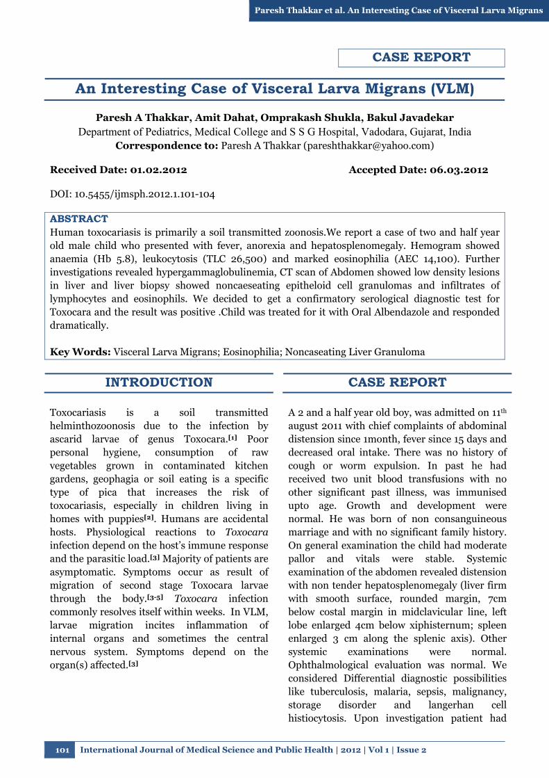
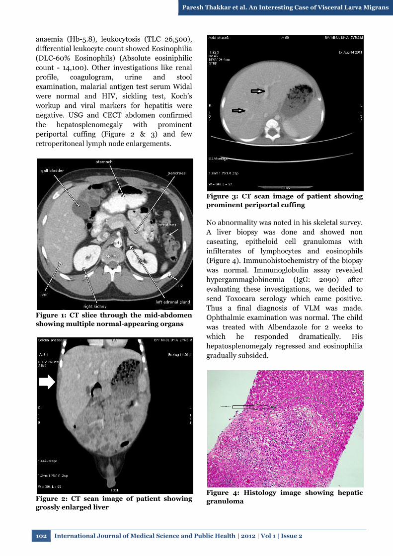
negative. USG and CECT abdomen confirmed
the hepatosplenomegaly with prominent
periportal cuffing (Figure 2 & 3) and few
retroperitoneal lymph node enlargements.
Figure 1: CT slice through the mid-abdomen
showing multiple normal-appearing organs
Figure 2: CT scan image of patient showing
grossly enlarged liver
Figure 3: CT scan image of patient showing
prominent periportal cuffing
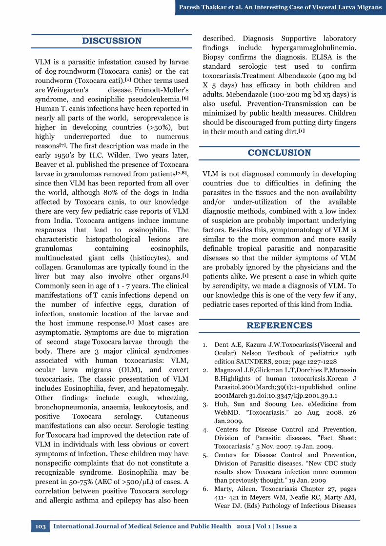
No abnormality was noted in his skeletal survey.
A liver biopsy was done and showed non
caseating, epitheloid cell granulomas with
infilterates of lymphocytes and eosinophils
(Figure 4). Immunohistochemistry of the biopsy
was normal. Immunoglobulin assay revealed
hypergammaglobinemia (IgG: 2090) after
evaluating these investigations, we decided to
send Toxocara serology which came positive.
Thus a final diagnosis of VLM was made.
Ophthalmic examination was normal. The child
was treated with Albendazole for 2 weeks to
which he responded dramatically. His
hepatosplenomegaly regressed and eosinophilia
gradually subsided.
Figure 4: Histology image showing hepatic
granuloma
Paresh Thakkar et al. An Interesting Case of Visceral Larva Migrans
103 International Journal of Medical Science and Public Health | 2012 | Vol 1 | Issue 2
DISCUSSION
VLM is a parasitic infestation caused by larvae
of dog roundworm (Toxocara canis) or the cat
roundworm (Toxocara cati).[1] Other terms used
are Weingarten's disease, Frimodt-Moller's
syndrome, and eosiniphilic pseudoleukemia.[6]
Human T. canis infections have been reported in
nearly all parts of the world, seroprevalence is
higher in developing countries (>50%), but
highly underreported due to numerous
reasons[7]. The first description was made in the
early 1950's by H.C. Wilder. Two years later,
Beaver et al. published the presence of Toxocara
larvae in granulomas removed from patients[7,8],
since then VLM has been reported from all over
the world, although 80% of the dogs in India
affected by Toxocara canis, to our knowledge
there are very few pediatric case reports of VLM
from India. Toxocara antigens induce immune
responses that lead to eosinophilia. The
characteristic histopathological lesions are
granulomas containing eosinophils,
multinucleated giant cells (histiocytes), and
collagen. Granulomas are typically found in the
liver but may also involve other organs.[1]
Commonly seen in age of 1 - 7 years. The clinical
manifestations of T canis infections depend on
the number of infective eggs, duration of
infection, anatomic location of the larvae and
the host immune response.[1] Most cases are
asymptomatic. Symptoms are due to migration
of second stage Toxocara larvae through the
body. There are 3 major clinical syndromes
associated with human toxocariasis: VLM,
ocular larva migrans (OLM), and covert
toxocariasis. The classic presentation of VLM
includes Eosinophilia, fever, and hepatomegaly.
Other findings include cough, wheezing,
bronchopneumonia, anaemia, leukocytosis, and
positive Toxocara serology. Cutaneous
manifestations can also occur. Serologic testing
for Toxocara had improved the detection rate of
VLM in individuals with less obvious or covert
symptoms of infection. These children may have
nonspecific complaints that do not constitute a
recognizable syndrome. Eosinophilia may be
present in 50-75% (AEC of >500/µL) of cases. A
correlation between positive Toxocara serology
and allergic asthma and epilepsy has also been
described. Diagnosis Supportive laboratory
findings include hypergammaglobulinemia.
Biopsy confirms the diagnosis. ELISA is the
standard serologic test used to confirm
toxocariasis.Treatment Albendazole (400 mg bd
X 5 days) has efficacy in both children and
adults. Mebendazole (100-200 mg bd x5 days) is
also useful. Prevention-Transmission can be
minimized by public health measures. Children
should be discouraged from putting dirty fingers
in their mouth and eating dirt.[1]
CONCLUSION
VLM is not diagnosed commonly in developing
countries due to difficulties in defining the
parasites in the tissues and the non-availability
and/or under-utilization of the available
diagnostic methods, combined with a low index
of suspicion are probably important underlying
factors. Besides this, symptomatology of VLM is
similar to the more common and more easily
definable tropical parasitic and nonparasitic
diseases so that the milder symptoms of VLM
are probably ignored by the physicians and the
patients alike. We present a case in which quite
by serendipity, we made a diagnosis of VLM. To
our knowledge this is one of the very few if any,
pediatric cases reported of this kind from India.
REFERENCES
1. Dent A.E, Kazura J.W.Toxocariasis(Visceral and
Ocular) Nelson Textbook of pediatrics 19th
edition SAUNDERS, 2012; page 1227-1228
2. Magnaval J.F,Glickman L.T,Dorchies P,Morassin
B.Highlights of human toxocariasis.Korean J
Parasitol.2001March;39(1):1-11published online
2001March 31.doi:10.3347/kjp.2001.39.1.1
3. Huh, Sun and Sooung Lee. eMedicine from
WebMD. “Toxocariasis.” 20 Aug. 2008. 26
Jan.2009.
4. Centers for Disease Control and Prevention,
Division of Parasitic diseases. "Fact Sheet:
Toxocariasis." 5 Nov. 2007. 19 Jan. 2009.
5. Centers for Disease Control and Prevention,
Division of Parasitic diseases. “New CDC study
results show Toxocara infection more common
than previously thought.” 19 Jan. 2009
6. Marty, Aileen. Toxocariasis Chapter 27, pages
411- 421 in Meyers WM, Neafie RC, Marty AM,
Wear DJ. (Eds) Pathology of Infectious Diseases
Paresh Thakkar et al. An Interesting Case of Visceral Larva Migrans
104 International Journal of Medical Science and Public Health | 2012 | Vol 1 | Issue 2
Volume I: Helminthiases. Armed Forces Institute
of Pathology, Washington DC. 2000;
7. Holland, Celia and H.V. Smith. Toxocara: The
Enigmatic Parasite. Wallingford, UK and
Cambridge, MA: CABI Publishing, 2006. 26 Jan.
2009
8. Despommier D. (2003). "Toxocariasis: clinical
aspects, epidemiology, medical ecology, and
molecular aspects". Clin Microbiol Rev 16 (2):
265–272.
Cite this article as: Thakkar PA, Dahat A, Shukla O,
Javadekar B. An interesting case of Visceral Larva
Migrans (VLM). Int J Med Sci Public Health 2012;
1:101-104.
Source of Support: Nil
Conflict of interest: None declared