Embed Size (px)
Citation preview

1104/19/23
Phototrophic Bacteria

2204/19/23
Morphologies The five major morphological types
among cyanobacteria are: – Unicellular – Colonial– Filamentous– Filamentous heterocystous– Filamentous branching

3304/19/23
Unicellular
Many cyanobacteria produce extensive mucilaginous envelopes, or sheaths, that bind cells or filaments together in aggregates.
Gloeothece, phase contrast; a single cell measures 5-6 um in diameter. (Brock6, 717)

4404/19/23
Colonial
A colony, which contains millions of cells and can be seen visually, develops from the asexual reproduction of a single cell. A colony contains a population of a single bacterial species.
Dermocarpa, phase contrast (Brock6, 717)

5504/19/23
Filamentous
Marine Oscillatoria fix nitrogen without heterocysts, and seem to produce of cells in the centre of the filament that lack photosystem II activity.
Oscillatoria, bright field; a single cell measures about 15 um wide. (Brock6, 717)

6604/19/23
Filamentous Heterocystous
Some cyanobacteria form heterocysts, which are rounded, seemingly empty cells, usually distributed individually along a filament or at one end of a filament.
Anabaena, phase; single cell measures about 5 um wide. (Brock6, 717)

7704/19/23
Filamentous Branching
Fischerella represents filamentous cyanobacteria that divide in more than one plane to form branches. These branching cyanobacteria also form heterocysts and akinetes.
Fischerella, bright field (Brock6, 717)

8804/19/23
Habitats Nutrient rich rivers and lakes Hot Springs Mud flats Stagnant water Swamps - hydrogen sulfide

9904/19/23
Yankee Springs Close up view of the
purple veils contains several different species of purple sulphur bacteria. (Perry, pg. 518)
The darker green growth that occurs on the top in some areas is due to cyanobacteria. (Perry, pg. 518)

101004/19/23
Hot Spring Grand Prismatic Spring (Yellowstone National Park) shows
the development of extensive mats of photosynthetic bacteria (primarily cyanobacteria and green bacteria).
The orange color at the edge predominates because of the rich carotenoid pigments in these organisms.Brock4 pg 240 Plate 2

111104/19/23
Floating Cyanobacteria in Lake
Floatation of cyanobacteria from a bloom on a nutrient-rich lake, caused by the presence of gas vesicles
Brock6 pg 78

121204/19/23
Bacterial mat Close-up of a small bacterial mat, in
Octopus Spring, Yellowstone National Park. The temperature where the mat shows the most extensive development is about 55 degrees C.
Brock4 pg 240 Plate 2

131304/19/23
Chromatium
Micrograph of the purple sulfur bacterium Chromatium species. These bacteria deposit sulfur granules within their cells that are iridescent and appear multicolored.

141404/19/23
Chromatium vinosum and okenii
A phase photomicrograph of Chromatium okenii. Note the polar flagellum (a tuft) as well as the internal sulfur granules. The bar is 5 um. (Perry, 515)
Chromatium vinosum, a purple sulfur bacterium, with intracellular sulfur granules, light field (X2,000). (Prescott, 47)
Perry, 516
Prescott, 47

151504/19/23
ChromatiumChromatium and Thiocystis
Phase contrast photomicrographs of layers containing purple sulfur bacteria (large celled Chromatium species and Thiocystis)
brock6 pg 715

161604/19/23
“Chlorochromatium aggregatum”
Several microcolonies of the consortium species “Chlorochromatium aggregatum” are shown by phase microscopy in this lake sample. (Perry, pg. 524)

171704/19/23
Thiospirillum Thiospirillum from a lake sample showing sulphur granules and polar flagellar tuft. (Perry, pg. 517)
Cells are curved rod or vibrioid-shaped, sigmoid or spiral with rounded ends, 2.5-4.0 um in diameter.
Motile by means of a polar flagellar tuft.
Anaerobic and phototrophic. Found in mud and stagnant
water of ditches and freshwater ponds containing hydrogen sulphide.

181804/19/23
Thiodictyon Thiodictyon are rod
shaped bacteria joined together at their cell ends to form a net. (Perry, pg 517

191904/19/23
Thiopedia Cells are spherical to ovoid or
elongated ovoid, 1.4-1.8 um wide and 1.5-2.5 um long.
Anaerobic and phototrophic. Inhabit the mud and stagnant
water of ponds and lakes containing hydrogen sulphide.
The optimum pH and growth temperature is 7.3 and 20 degrees C respectively.
Cells of Thiopedia grow in flat sheets. Note the bright appearance of the gas vacuoles in each cell. (Perry, pg. 517)

202004/19/23
Pelodictyon Cells of Pelodictyon
clathratiforme showing net formation in small individual cells with gas vacuoles. (Perry, pg. 523)
Pelodictyon phaeum showing brownish color of cells in net. (Perry, pg. 523)

212104/19/23
ProchloronProchloron
Colorized electron micrograph of the photosynthetic bacterium Prochloron didemni reveals that it has extensive internal membranes that have photosynthetic pigments (green). Magnification 6400X. (Atlas, pg 145)
Prochloron. Each cell in a cluster contains chlorophyll arranged on membranous layers. (Black2, pg 261)

222204/19/23
Thiocapsa
Tubular type from Thiocapsa pfennigii (Perry, 516).
Thiocapsa roseopersicina. Bar = 10 um. (Prescott, pg. 466)

232304/19/23
Pelodictyon clathratiforme
Brock4 pg 637 Pelodictyon clathratiforme, a bacterium forming a three dimensional network. Magnification 1700X.
Typical green sulfur bacteria- Pelodictyon clathratiforme. (Prescott, pg. 469)
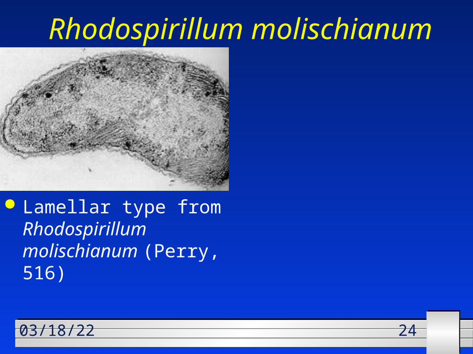
242404/19/23
Rhodospirillum molischianum
Lamellar type from Rhodospirillum molischianum (Perry, 516)

252504/19/23
Rhodospirillum rubrum R. rubrum in transmission
electron microscope (X100,000) (Prescott, pg. 31)
Rhodospirillum rubrum in phase-contrast light microscope (600X) (Prescott, pg. 31)

262604/19/23
Rhodocyclus purpureus Rhodocyclus
purpureus, phase contrast. Bar = 10 um. (Prescott, pg. 468)

272704/19/23
Rhodopseudomonas
Transmission electron micrograph of thin section of dividing cells of Rhodopseudomonas marina. Cells are 0.7 um in diameter. (Brock6, pg. 44)
Rhodopseudomonas acidophila, phase contrast. (Prescott, pg. 468)

282804/19/23
Rhodomicrobium vannielii
Rhodomicrobium vannielii with vegetative cells and buds, phase contrast. (Prescott, pg. 468)

292904/19/23
Spiral-shaped
Rhodospirillum rubrum, an anoxygenic photosynthetic bacterium. (Black2, pg. 260)

303004/19/23
Gas Vesicles
Electron micrographs of gas vesicles purified from the bacteria Microcyclus aquaticus and examined in negatively stained preparations
A single gas vesicle is about 100 nm in diameter. Brock6 pg 78

313104/19/23
Gloeocapsa Unicellular colonial, Gloeocapsa sp.
Magnification 1500X pg 645 brock4

323204/19/23
Heterocytst
Micrograph of the cyanobacterium Anabaena cylindrica, showing vegetative cells and a heterocyst (enlarged cell) in which nitrogen fixation occurs. (Atlas, pg. 49)
Filamentous chains of cells of a cyanobacterium in the genus Anabaena. The distinctive ellipsoidal cells are called heterocysts and are the sites where the process of nitrogen fixation is carried out. (VanDemark, pg 40)

333304/19/23
Brock4 pg 637 Electron micrograph of Oscillochloris.
The chromosomes in this preparation are darkly stained. Magnification 14800X.

343404/19/23
Oscillatoria

353504/19/23
Chlorobium limicola
Green bacterium: Chlorobium limicola. The refractile bodies are sulfur granules deposited outside the cell. (Brock6 pg 570)

363604/19/23
Thiopedia roseopersicinia Massive accumulation of
purple sulfur bacteria, Thiopedia roseopersicinia, in a spring in Madison,Wisconsin. The bacteria grow near the bottom of the spring pool and float to the top (by virtue of their gas vesicles) when disturbed. (Brock6, pg. 716)

373704/19/23
Thiothrix A rosette of Thiothrix from a sulfur spring. (Perry, pg. 543)
Phase photo-micrograph of a sulfur bacterium Tiothrix nivea. (Perry, 87)

383804/19/23
Gloeothece Phase photograph
of a Gloeothece sp. showing the cells within the sheath.

393904/19/23
Prochlorothrix
Scanning electron micrograph of Prochlorothrix, a marine prochlorophyte. Bar is 3 um. (Perry, pg. 532)

404004/19/23
DONE!!!DONE!!!

414104/19/23
Endospores
Light photomicrographs illustrating several types of endospore morphologies and intracellular locations
(A) Central endospore (B) Subterminal endospore (C) Terminal endospore Brock6 pg 79

424204/19/23
Endospore