Embed Size (px)
Citation preview

1+1 National Library.:>1 Canada
Bibliothèque nationaledu Canada
Acquisitions a; Id Direction des ac-quisitions elBibliographie Services Branch des services bibliographiques
395 Wellington SHoot 395. tllO Wel;;nglonOltawa.OnlBrio Ottawa (Ontario)KlAON4 K1AON4
NOTICE AVIS
The quality of this microform isheavily dependent upon thequality of the original thesissubmitted for microfilming.Every effort has been made toensure the highest quality ofreproduction possible.
If pages are missing, contact theuniversity which granted thedegree.
Some pages may have indistinctprint especially if the originalpages were typed with a poortypewriter ribbon or if theuniversity sent us an inferiorphotocopy.
Reproduction in full or in part ofthis microform is governed bythe Canadian Copyright Act,R.S.C. 1970, c. C·30, andsubsequent amendments.
Canada
La qualité de cette microformedépend grandement de la qualitéde la thèse soumise aumicrofilmage. Nous avons toutfait pour assurer une qualitésupérieure de reproduction.
S'il manque des pages, veuillezcommuniquer avec l'universitéqui a conféré le grade.
La qualité d'impression decertaines pages peut laisser àdésirer, surtout si les pagesoriginales ont étédactylographiées à l'aide d'unruban usé ou si l'université nousa fait parvenir une photoc!lpie dequalité inférieure.
La reproduction, même partielle,de cette microforme est soumiseà la Loi canadienne sur le droitd'auteur, SRC 1970, c. C·30, etses amendements subséquents.
THE COMBINED EFFECT OF MODIFIED ATMOSPHEREPACKAGING (MAP) AND CHITOSAN ON THE GROWTH OFLlSTERIA MONOCYTOGENES IN MODEL SYSTEMS AND IN
FRESH PORK LOIN.
By
JENNIFER E. MORRIS
Department of Food Scienceand Agricultural Chemistry
Macdonald CampusMcGiII UniversityMontreal, Québec
A Thesis submiUed to the Faculty of Graduate Studies and Research in partialfultillment of the requirements for the degree of Master of Science.
July 1995
cJennifer Morris, 1995

.+. National Libraryof Canada
Bibliothèque nalionaledu Canada
Acquisitions and Direclion des acquisitions etBibliographic Services Branch des services bibliographiques
395 Wellington Sireet 395, rue WellingtonOttewa, Ontario Onawa (Onlario)K1A ON4 KIA ON4
The author has granted anIrrevocable non-exclusive licenceallowing the National Library ofCanada to reproduce, loan,distribute or sell copies ofhis/her thesis by any means andin any form or format, makingthls thesls available to Interestedpersons.
The author retains ownership ofthe copyright ln hls/her thesls.Neither the thesis nor substantialextracts from It may be prlnted orotherwise reproduced withouthis/her permission.
l'auteur a accordé une licenceIrrévocable et non exclusivepermettant à la Bibliothèquenationale du Canada dereproduire, prêter, distribuer ouvendre des copies de sa thèsede quelque manière et sousquelque forme que ce soit pourmettre des exemplaires de cettethèse à la disposition despersonnes Intéressées.
l'auteur conserve la propriété dudroit d'auteur qui protège sathèse. Ni la thèse ni des extraitssubstantiels de celle-ci nedoivent être imprimés ouautrement reproduits sans sonautorisation.
ISBN 0-612-08031-5
Canada
Suggested short title:
COMBINED EFFECTS OF MAP AND CIflTOSAN ONUSTERIA MONOCYTOGENES
ii
M.Sc.
ABSTRAC'f
Jennifer Morris Food Science
THE COMBINED EFFECT OF MODIFIED ATMOSPHERE PACKAGING(MAP) AND CIDTOSAN ON THE GROWTH OF L1STERIAMONOCYTOGENES IN MODEL SYSTEMS AND IN FRESH PORK LOIN
Listeria monocytogenes is a pathogenic, psychrotrophic microorganism that is
ubiquitous in nature. L monocytogenes has been isolated from numerous meat
products, both fresh and processed, the incidence of contamination varying greatly.
The ability of Listeria to grow in meats depends on temperature, pH, water activity
(aw), nutrients, species and numbers of competing microorganisms, gaseous
conditions, and levels of additional barriers. Therefore, methods to control the growth
of L.monocytogenes are of great importance to food processors since this organism can
grow under a wide range of environmental and storage conditions. Two methods of
control, in conjunction with temperature, were studied in this project: (i) modified
atmosphere packaging (MAP) and (ii) chitosan, to determine the optimum levels of
these "hurdles" needed to effectively control the outgrowth of L.monocytogenes in both
model broth and agar systems and in fresh pork loin. Initial studies in broth and agar
medium indicated that the optimum level of chitosan needed for complete inhibition of
Listeria was 0.2%(w/v) at a pH of 6.0 over a wide temperature range. Modified
atmosphere packaging studies in a model agar system showed that the optimal gaseous
conditions for inhibition of Listeria were: a) lOO%N2 + an Ageless FX oxygen
absorbent, b) SO%e02+20%N2 and c) an Ageless SS Oxygen absorbent alone. These
gaseous conditions were effective at temperatures of IOoe or Jess, indicating the
importance of adequate storage temperature control, in conjunction with any other
control methods used, i.e., "the hurdle approach" to food safety. On the basis of these
preliminary studies, a combination of chitosan as a dipping solution and modified
atmosphere packaging were investigated to control the growth of L.monocytogenes in
fresh pork loin. Pork loin samples were dipped in a 0.2% chitosan solution for 60
seconds and packaged under various almospheres in Cryovac bags and slored at 5, 10
iii
and 150 C up to 28 days. Samples were monitored for physical, chemical and
microbiological changes throughout the storage period. Optimum control over the
growth of L.monocytogenes was achieved using a combination of l00%N2 + an
Ageless FX oxygen absorbent and dipping in a 0.2% chitosan solution. Based on these
studies, a combination of 0.2 % chitosan and MAP could be used to extend the shelf Iife
of pork without adversely affecting color, odor and exudate loss white inhibiting the
growth of the pathogenic microorganism, L. monocytogenes
;v

M.Sc.
RESU'ME
Jennifer Morris Sciences de ('alimentation
INFLUENCE DE L'EMBALLAGE SOUS ATMOSPHERES MODIFIEESCOMBINEE AVEC CmTOSAN SUR LA CROISSANCE DE LlSTERIAMONOCYTOGENES UTILISANT DES SYSTEMES MODELES ET DU PORCFRAIS
L. monocytogenes est un microorganisme psychrotrophique pathogène, fréquemmentretrouvé dans la nature. L.monocyotogenes a été isolé à partir de plusieurs produits dela viande où la fréquence de contamination est sujette à de grande variation. L'habilitéede cette bactérie a se reproduire dépend principalement de la température, du pH, del'activité de l'eau (aw), des nutriments disponibles, des concentrations des gaz (Oz, Nz,
COz), et finalement du nombre et des différentes espèces de microorganismes encompétition avec elle. En conséquences, les méthodes de contrôle de croissance deL. monocytogenes représentent un grand problème pour les producteurs de viandepuisque cet organisme peut se reproduire sous plusieurs conditions environnementalles.Deux traitements: l'emballage sous atmosphères modifiées (ESM) d'un part et chitosande l'autre, en conjonction avec la température, furent étudiés dans ce projet avecl'intention de déterminer les niveaux optimum des conditions qui pourraienteffectivement contrôler la croissance de L.monocytogenes, en utilisant premièrementdes systèmes modèles, puis du porc frais. Des études préliminaires utilisant desmilieue liquides et solides (agar) ont montré qu'une concentration de 0.2 % (plv) dechitosan à un pH de 6.0 était requise pour l'inhibition complête de L. monocytogenesen utilisant une distribution variée de températures. De plus, les études utilisantl'emballage sous atmosphères modifiées de L.monocytogenes dans un système modèlesolide (agar) ont démontrées que plusieurs conditions gazeuses pûuvaient arrêter lacroissance de cet organisme. Ces conditions comprel1r:"lli: (a) 100% Nz + unabsorbant d'oxygène (Ageless FX), (b) 80% CO~ + 20% Nz et (c) un absorbantd'oxygène seul (Ageless 55). Ces conditions gazeuses sont valables seulement lorsquela température ne dépasse pas IOoC. Ceci démontre l'importance de l'utilisation de laréfrigération en conjonction avec d'autres méthodes de contrôle. En se basant sur cesétudes préliminaires, une méthode combinant l'utilisation du chitosan (comme solutiontrempage) et E5M fut etudiée pour contrôler la croissance de L.monocytogenes sur lesfilets du porc frais. Les échantillons de filet de porc frais furent trempés dans une
solution de 0.2 % de chitosan pour une période de 60 secondes furent emballés sous
v
differentes conditions atmosphériques. Les échantillons furent entreposés à S. 10 ouISoC pour une période maximale de 28 jours. Les échantillons furent examiner à
differentes temps préetablis pour suivre les changements chimiques, !,~ysique etmicrobiologiques. L'utilisation d'une atmosphère de 100% d'azote, avec une absorbantd'oxygène (Ageless FX) contenu dans l'emballage, le tout combiné avec trempage des
échantillons dans une solution de 0.2%chitosan furent les conditions optimales pour le
contrôle de L. monocytogenes dans le porc frais. En conclusion de ces études, une
combinaison, chitosan et ESM, pourrait être utilisée pour prolonger la période defraîcheur du porc et arrêter la croissance des microorganismes pathogéniques tel queL.monocytogenes sans affecter les caractéristiques sensorielles du porc.
vi

PREFACE
Claim of Original Research
(1) The use of Ageless oxygen absorbents. types FX, SS and SE to delay the growth of
Listeria monocytogenes in model agar systems and in frrsh pork loin.
(2) The use of combination treatments: chitosan hydrochloride in conjunction with
modified atmosphere packaging (MAP) to control the growth of Listeriamonocytogenes in broth, agar and pork systems.
vii

ACKNOWLEDGMENTS
1 am sincerely grateful to my supervisor, DrJ.P.Smith for his guidance, patience,encouragement and great sense of humor over the last few years.
1 would Iike to thank IIsemarie Tarte for al1 her invaluable help in the lab, herassistance with tabulating results, her patience, and also for her amazing baking which
made our lives a lot sweeter!
1would Iike to express my appreciation and thanks to my family, Ursula, Michael andCarolyn Morris, for their love and support throughout my studies, without whom 1
would have never made it this far. Thank you for being there, understanding andencouraging me. 1 would also Iike to thank Richard Dumont, my greatest friend andcompanion, who gave me the will-power and determination to achieve this work. Aisothanks to Richard for translation of the abstract.
1 would also like to thank Veronique Barthet for her help with the correction of theabstract.
1would like to thank the Microbial Hazards Bureau of Health and Welfare Canada fortheir training and 'the microbial cultures. In particular, 1 would Iike to thank Dr.JeffFarber and Ms. Elaine Daley for their advice and assistance.
1 would Iike to thank the fol1owing company for providing supplies to complete thisstudy: Mitsubishi Gas Chemical Co., Japan for the Ageless absorbents.
Final1y, 1 would Iike to thank CORPAQ (Conseil de Recherches en Peche et Agroalimenentaire du Quebec) and Les Fonds FCAR for their financial assistance of thisstudy.
viü

TABLE OF CONTENTS
~~~C:1r••••••.••••••••..•••.•••.••.••••••..••..••••••••• .........•..•....•••••••••••••........ili~~••••••••••••••••••••••••••••••••,•••.••••••••••••••••••••••••••••••••••••••••••••••••••••••vl'~Jf~<:~••••••••••...•..•••••••••.•••••••.•••••••••.•.•• ...••••••••.••••••...•..•..•.....•.•.....viiACKN'OWLEDGl\oIENTS.•.....•••.........•.•............••.•......•......•...•...•.......•.viii~IST O~ JrIG~S xii1JIST O~ T~II~~S .•...•.•.•••••..••••••.......••...•....•••.•..•. ......•..••...•.••••••........xv
1. INTRODUCTION AND LITERATURE REVIEW...............•..................... I1.1. Introduction..•.......•...••..................•..................................•... 11.2. Pork: Shelf Ufe and Spoilage................................•..........•.........1
1.2.1. Microbiological Concerns...................................•.....•...21.3. Listeritl monocytogenes 21.4. Factors Influencing the Growth of L.monocytogenes 31.5. L.monocytogenes in Food Products......•....•.............•........•..........6
1.5.1. Meat and Meat Products 71.6. Methods to Control the Growth of L.monocytogenes 9
1.6.1. Thermal Processing.............•....•..............•........•..........91.6.2. Low Temperature Control of L.monocytogenes l1
1.7. Novel Methods for the Control of L.monocytogenes 121.7.1. Bacteriocins.........•.•.........•........•.....•.........••.............131.7.2. Preservatives...•..........•.•.....•.....•.........•.•................•..141.7.3. Irradiation....••...•..............•..........•......•.•................. 161.7.4. Sanitizers•......•....•.••...•.................•.•.........•..............171.7.5. Modified Atmosphere Packaging...................•..•...•........18
1.7.5.1. Gases used in Gas Flush Packaging 181.7.5.1.1. Carbon Dioxide..•...••.....•.•........•....•.•..181.7.5.1.2. Nitrogen...............•••....•.•.......•.....•.•..191.7.5.1.3. Oxygen.....•......•..•••.•.•••...••......•....•...19
1.7.5.2. New Developments:Oxygen Absorbent Technology 19
1.7.5.3. The Effect of MAP on Meat Sensory andMicrobiological Characteristics 20
1.7.5.3.1. Color•...•.••...........••......•...••••...•........2l1.7.5.3.2. MicrobialPopulation•.•...........•........••...21
1.7.5.4. The Safety of MAP..•.....•••.•••...•••...•...•.•..........221:1.5.5. Effect ofMAP on the Growth of
L.monocytogenes.•...•....•......••..••...•••••..••....•.•..231.7.5.5.1. Produce....•...........••......•..........•.•......231.7.S.S.2. Poultry.....••••••.....•.•....•..•••..••.•.••••....14
ix

1.7.5.5.3. Red Meats..............•.........................251.7.6. Chitosan: A Novel Antimicrobial Agent••••••••••••••••••.•••••••26
1.7.6.1. Extraction of Chitin and Chitosan 271.7.6.2. Applications of Chitosan..•...............•...•........•..31
1.7.6.2.1. Agriculture......................•................311.7.6.2.2. F10cculating Agent•.......•................•....321.7.6.2.3. Control of Fungal Diseases in Plants 321.7.6.2.4. Control of Toxigenic Fungi 341.7.6.2.5. Control of Senescence and Ripening 351.7.6.2.6. Control of Food Spoilagel
Patbogenic Bacteria....•....•...................351.8. Hurdle Approach to Food Safety 36
2. CHITOSAN STUDmS 382.1. Introduction...................•...........•....•..•................................382.2. Materials and Methods......•...........•..•....•...............................39
2.2.1. Microorganisms and Inoculum Preparation 392.2.2. Growtb Curve.•..••..........•.....•............•...•............•.....392.2.3. Antilisterial Activity of a Non-Water Soluble Chitosan.......39
2.2.3.1. Preparation of Chitosan...............•..................392.2.3.2. Preparation Growth Media 40
2.2.4. Effect of pH on the Antilisterial Activity of Chitosan 402.2.5. Antilisterial Effect of Chitosan in an Agar System 41
2.3. Results and DiscllSSioD...•...•........•..........•....•............•....•......•.422.3.1. Growth Curve of L.monocytogenes 422.3.2. Antilisterial Activity of a Non-Water Soluble Chitosan 442.3.3. Effect of pH on the Antilisterial Activity of Chitosan 482.2.4. Antilisterial Effect of Chitosan in an Agar System 51
3. MODIFIED ATMOSPHERE PACKAGING ANDCO~II~A1rI()~ STUD~ 54
3.1. Introduction.....•......•.....•...••.•.•.•.............•.•.....•....................543.2. Materials and Methods....•......•.......•.•........•...•..........•............55
3.2.1. Microorganisms and Preparation of Inoculum 553.2.2. Effect of MAP on L. monocytogenes 55
3.2.2.1. Headspace Gas Analysis 563.2.3. Effect ofVarious Oxygen Absorbents on the Growth of
L.monocytogenes..•.•....••.•.••....•.•..••..•.•••.•.••.....•..563.2.4. Combined Effects of ChitOSBD, pH and MAP on
L.monocytogenes in an Agar System 573.3. Results and DiscllSSion..••...•.......•••..•..........•...•.............•••..•.••.59
3.3.1. Effect of MAP on L.monocytogenes...•...•..............•....•..•.593.3.2. Effect of Various Oxygen Absorbents on the Growth 'lf
L.monocytogenes.••••.•••.••.....•...•.........•....••..•.••..63
x

3.3.3. Combined Effects of Chitosan, pH and MAP onL. monocytogenes in an Agar System 65
4. PORK CHALLENGE STUDŒS.......................................•..................684.1. IntrodllctioD 684.2. Materials and Methods........•............•...........................69
4.2.1. Microorganisms and Preparation of Inoculum 694.2.2. Preparation of Chitosan Dipping Solution••••••••••••••694.2.3. Preparation of Pork , 694.2.4. Packaging 704.2.5. Head Space Gas Analysis 714.2.6. Sensory Analysis....•.....•...................................71
4.2.6.1. Color Analysis..•.....................•.............714.2.6.2. Odor Analysis...........•..........................72
4.2.7. Microbiological Analysis.....•..............................724.2.8. Drip Loss.......•.................•....•............•...........734.2.9. pH Measurement•..•...................•..•...................73
4.3. Results and Discussion.........•.......•....••...•..•..........................•.744.3.1. Introduction.................•..........................................744.3.2. Physiochemical Analysis........•.....................................74
4.3.2.1. Changes in Headspace Gas Composition 744.3.2.2. Changes in pH..•.•.........•...............................824.3.2.3. Changes in Drip Loss.......•..•..............•............844.3.2.4. Color Cbanges...•....•.•.................•..........•......84
4.3.3. Sensory Evaluation..••...•..........•.................................904.3.3.1. Color.......•.......•....•.•.....................•..........•..904.3.3.2. Odor.......•.......•....•.•................••. 1 ••••••••••••••••90
4.3.4. Microbiological Changes 954.3.5. Shelf Lire 103
4.3.6. ChaUenge Studies with L.monocytogenes••••••••••••••••••••••••l064.3.6.1. Changes in L.monocytogenes Counts in :-,.;·\(•••••••1064.3.6.2. Changes in L.monocytogenes Counts in
E~date ~•••••••••••••••••••••••••••••••••••••••••••••••1()74.3.7. Sbelr Lire aud Safety 112
GENERAL CONCLUSION....•.........••...••..••.......••..•...........•.................115REFERENCES..............................•....•.............................................117
xi

Figure 1.
Figure 2.
Figure 3.
Figure 4.
Figure 5.
Figure 6.
Figure 7.
Figure 8.
Figure 9.
Figure 10.
Figure 11.
Figure 12.
Figure 13.
Figure 14.
Figure 15.
LIST OF FIGURES
Reaction of iron to rust that occurs in oxygenabsorbeots....•...............................................•........................................20
The various forms of myoglobin..•........•..............•................................21
Structures of the repeating units of cellulose, chitin and chitosan......28
Flow diagram of chitiD extraction process 29
Flow diagram of chitos8n extraction process......••...............................30
The hurdle approach to food safety and shelf lire extension. (A)Comhination oflow temperature (f) and MAP, (D) combination oflow temperature (T), pH and MAP and (q combination of lowperature (l'), MAP and cbitosao•.........•......•..•.....••....••................•.......37
Growth curve of Lmonocytogenes in tryptose soya broth at 5,
10 and 15°C....•.....••....•...•...........•........••.......•.................•..............•......43
Effect of non-water soluble chitosan on L monocytogenesiDcubated at 300C•.......•......•..•.....•.............•........••........•.................•....•46
Effect of non-water soluble chitosan on Listeria innocua incubated at
300C.•.•.•...•......•.....•••...............•...•........•.......•........••....••.........•......•........47
Effect of chitosan hydrochloride (pH 7) on the growth of
Lmonocytogenes incubated at 300C..•...............•••....•.....•....................49
Effect of chitosan hydrochloride (pH 6) on the growth of
Lmonocytogenes incubated at 300C.......................•...•.......•.........•.....•50
Effect of chitosan at pH 6 in a model agar system on the growth ofLmonocytogenes. 5%
Effect of chitosan at pH 7 in a model agar system on the growth ofI-monocytoge.nes. ...•...••..•......•....••............•..••.•.•..•••.•..••..•.•..•..•...•..•.......53
Treatment scheme used for 3.2.4.(Combined effects of chitosan andMAP on Lmonocytogenes iD an agar system).•..•...•••..•.•..•.••.•.•..••....•.•58
Scheme of treatments used in pork challenge studies••••••••••••••••••••••••••70
xii
Figure 16. Changes in head space gas composition of non-chitosan dipped porkat SOC and stored under (A) Air; (D) Air+Ageless SS;(C) 80O/oCOz/20%N1; and (D) l00%N1+Ageless FX. 76
Figure 17 Changes in head space gas composition of non-chitosan dipped porkat 10·C and stored under (A) Air; (D) Air+Ageless SS;(C) 80o/oCOz/20%N1; and (D) l00%N1+Ageless FX. 77
Figure 18 Changes in head space gas composition of non-chitosan dipped porkat lSOC and stored under (A) Air; (D) Air+Ageless SS;(C) 80o/oCOz/20%Nl; and (D) l00%N1+Ageless FX. 78
Figure 19 Changes in head space gas composition of chitosan dipped porkat SOC and stored under (A) Air; (D) Air+Ageless SS;(C) 80O/oCOz/20%Nl; and (D) l00%N1+Ageless FX. 79
Figure 20 Changes in head space gas composition of chitosan dipped porkat 10·C and stored under (A) Air; (D) Air+Ageless SS;(C) 80o/oCOz/20%Nl; and (D) l00%N1+Ageless FX 80
Figure 21 Changes in head space gas composition of chitosan dipped porkat 15·C and stored under (A) Air; (D) Air+Ageless SS;(C) 80o/oCOz/20%Nz; and (D) l00%Nz+Ageless FX. 81
Figure 22 Changes in pB v31ues of MAP pork non-chitosan dipped and chitosandipped and stored at 5, 10 and l~C.......•............................................83
Figure 23 Changes in drip loss of MAP pork non-chitosan dipped and chitosandipped and stored at 5, 10 and lS-C......................•.............................86
Figure 24 Changes in LlO values of MAP pork non-chitosan dipped and chitosandipped and stored at 5, 10 and 1seC 87
Figure 25 Changes in a'" values of MAP pork non-chitosan dipped and chitosandipped and stored at 5, 10 and 1seC••••••••••••••••••••••••••••••••••••••••••••••••••••88
Figure 26 Changes in b'" values of MAP pork non-chitosan dipped and chitosandipped and stored at 5, 10 and tS-C•.....••...............•....••.....•....•....•......89
Figure 27 Effed of MAP alone on the rejection point of pork color 91
Figure 28 Effed of MAP and chitosan dipping on the rejection point of porkcolor .•..•.•.•..•.•.•.••.•...•••.•...••••.•...•..••........•........•...•..••.•.•••.•..•..••.•............•92
xiii

Figure 29
Figure 30
Figure 31
Figure 32
Figure 33
Figure 34
Figure 35
Figure 36
Figure 37
Figure 38
Figure 39
Figure 40
Effed of MAP alone on tbe rejeetion point of pork odor 93
Effectt of MAP and ebitosan dipping on the rejection point of porkodor...............................................................•........••.........•...................94
Cbanges in total aerobie eounts of non-ehitosan dipped pork stored at5, 10 and lS-C.............................•.........................•.........•.............•.......97
Changes in total Pseudomollas eounts of non-ehitosan dipped porkstored st 5, 10 and lS-C...........................................................•............98
Changes in totallaetie aeid bacteria eounts of non-ehitosan dippedpork stored st 5, 10 and 1SOC•••••••••••••••••••••••••••••••••••••••••••••••••••••••••••••••99
Cbanges in total aerobie eounts of ehitosan dipped pork stored at5, 10 and IS-C................................................................•.......•............100
Changes in Pseudomonas eounts of ehitosan dipped pork storedat 5, 10 and ls-'C 101
Changes in totallactie aeid bacteria aerobie eounts of ehitosan dippedpork stored at 5, 10 and IS-C....•...........................•......•................•...•102
Changes in l-monocytogenes eounts of non-ehitosan dipped porkstored at S, 10 and IS"C.......•....•.......•.....•..•..................•.......•....•........108
Changes in l-monocytogenes eounts on ehitosan dipped pork stored atS, 10 and 15"C....•.••.................................•.............................•.............109
Changes in l-monocytogenes eounts from drip loss of non~hitosandipped pork nt S, 10 and 15"C...................•..................•.......•....•••......110
Changes in l-monocytogenes eounts from drip loss of ehitosan dippedpork at 5, 10 and lS"C..................•........•..•.•................•........•....•••.....•111
xiv

Table 1.
Table 2.
Table 3.
Table 4.
Table 5.
Table 6.
Table 7.
Table 8.
Table 9.
Table 10.
Tt.ble 11.
Table 12.
Table 13.
Table 14.
Table 15
Table 16
LIST OF TABLES
Food borne outbreaks due to Lmonocytogenes•....................................7
Incidence of LmoftOCJ'togenes 8
Beat resistance of L.monocytogenes in some meat and fishproducts 11
Preservatives and novel compounds studied to control the growth ofLmonocytogenes (various strains) 15
Gas mixtures used for 3.2.2. (Effect of MAP onLmonoqtogenes) 55
Absorbents used in 3.2.3.(Effect of various oxygen absorbents on thegrowtb of Lmonocytogenes) 56
Effect of MAP on L.monocytogenes at 100e, no oxygen absorbentused 61
Effect of MAP on L.monocytogenes at 150e, no oxygen absorbentused......••......•.........................................................................................61
Effect of MAP on L.monocytogenes at 100e using an Ageless FXoxygen absorbent. 62
Effect of MAP on L.monocytogenes at 150e using an Ageless FXoxygen absorbeDt. 62
Effect of various oxygen absorbents on L.monocytogenes 64
Effect of MAP, pB, temperature and chitosan on L.monocytogenes••67
Estimated shelf Iife of pork loin at 5, 10 and lS"e for aU packagingtreatments, non-chitosan dipped. 104
Estimated shelf Iife of pork loin at 5, 10 and lS"e for aU pack&gingtreatments, chitosan dipped•...............•.............•................................10S
Shelf Iife vs. safety for non-ehitosan dipped pork.•••••••••••••••••••••••••••••1l3
Shelf Iife vs. safety for chitosan dippped pork 114
xv

CHAPTERILITERATURE REVIEW
1.1. INTRODUCTION
The incidence of food poisoning illnesses has been increasing steadily year by year
(Kn~chel and Gould, 1995; Todd, 1992). The reason for the increase in reported illnesses
is probably due to a combination of factors, including: improved reporting, changes inagricultural practices, changes in food marketing and eating habits, identification of new
pathogens, development of superior microbiological methods and changing population
sensitivities. There have been reports ofthe emergence ofnew pathogens (Schofield, 1992)
but in reality they are mostly the recognition of organisms that have probably caused foodborne illness for thousands of years (Baird-Parker,1994). One such pathogen, relatively
weil known today, is Listeria monocytogelles. Two aspects ofits control in foods are: (i)
improvement in analytical methods and (ü) the development ofnew methods to mhibit or
delay its growth in foods (Farber, 1993). At the same time, the demand for foods that are
more convenient, safe, fresh and less heavüy processed and preserved is ever increasing.Fresh pork represents a major class of food produced and consumed in Québec, that
normally does not have a shelf-life of more than four days. Minima! processing methodsare currently being applied to pork in order to extend its short shelf-life while
L.mollocytogenes has been implicated in severa! cases of food poisoning involving the
consumption ofpork (Cooksey, 1993; Farber and Peterkin, 1991). With such a reduction
in the preservation of foods, how will it be possible to achieve a real reduction in the
incidence of food poisoning from manufaetured foods? Combination treatments, or the"hurelle approach" to food safety and shelf life extension could play a major raie in theresolution ofthis important problem.
1.2. Pork: Shelf life and Spoilage
Fresh pork is a highly perishable product with a refrigerated shelf life of about four days,
Wldergoing progressive deterioration from the time of slaughter to consumption. Pork iscomposed of water, proteins, lipids, carbohydrates (in the forro of glycogen) and other
minor components such as vitamins, minerais, enzymes and pigments, the relative
proportions of which contnbute to its structure, texture, flavor, color and nutritive value.
The shelf life of pork is affected by temperature, atmospheric oxygen, enzymes, light,

2
moisture and most importantly, microorganisms. Meat spoilage is defined as "any single
symptom or group of symptoms of overt microbial activity, manifested by changes in meat
odor, flavor or appearance 'XGill, 1986).
1.2.1. Microbiological Concerns
Microbial growtb is by far the most important factor influencing the shelf life of pork.
Most microbial contamination occurs post-slaughter on the surface of the meat carcass
when spoilagtl bacteria, pathogenic bacteria, molds and yeasts may be present. The deep
muscle tissues remain relatively free from contamination if the slaughtering practices are
hygienic. The microbial population ofpork is affected by the species, health and handling
of the live animal, slaughtering practices, proper chilling of the carcass, sanitation prior to
fabrication, the type of packaging as well as the handling of tbe meat throughout
distribution and storage. The spoilage microorganisms most likely to be found include
species such as Pseudomonas, Moraxe/la, Acinetobacter, Aeromonas, A/teromonas,
Lactobaci//us, Brocothrix. Pathogenic bacteria commonly found include: Sa/monel/a,
Staphy/ococcus aureus, Yersinia enteroco/itica, C/ostridium botu/in/lm, C/ostridiumperfringens, Campy/obacter, Aeromonas hydrophi/a and Listeria monocytogelles. These
microorganisms can originate from the skin ofthe animal, from fecal material, air, soil and
water (Gill and Greer, 1993). Of particnlar concem to meat processors, consumers and
regulatory authorities, both nationally and intemationally,.is the growtb of the
psychrotrophic pathogen L.monocytogenes.
1.3. Listeria monocytogenes
L.monocytogenes is a pathogenic, psychrotrophic microorganism that is ubiquitous in
nature and has been implicated in numerous food poisoning outbreaks. It is a gram
positive, facultatively anaerobic, non-spore forming rod, capable of growtb between 0.4
and 50°C (Farber and Peterkin, 1991). It is motile by peritrichous flagella, showing
tumbling motility under a light microscope in a wet mount at 25°C. The colonies
demonstrate a characteristic blue-green sheen by obliquely transmitted light. It is catalase
positive, oxidase negative and expresses a l3-hemolysin which produces zones of clearing
on blood agar. The hemolysin is recognized as a major virulence factor whose secretion is
essential for promoting intracellular growtb and T-cell recognition of the organism The
hemolysin is called Listeriolysin-O.

3
L.monocytogenes was first recognized as a pathogen in animais 60 years ago, and waslater implicated in human outbreaks of food poisoning. The disease that L.mollocytogenescauses is tenned Usteriosis, with most cases being sporadic. The source and route ofinfection is usually unclear in these cases, although a portion of them may be previously
unrecognized common source clusters. The recent association of L. monocytogenes with
several large food poisoning outbreaks suggests that contaminated food is the prlmarysource of the organism Meningitis (the inflammation of the brain or spinal cord) is themost common fonn of Listeriosis. The symptoms include respiratory illness, sore throat
interrupted by vomiting, headache, confusion, lethargy and stiffuess in the back and neck.
There are other bacteria that cause meningitis such as Streptococeus p"eumo"iae,Neisseria melligitidis. Staphy/ococeus aureus and several others.
The majority of human cases of Listeriosis occur in individuals who have an underiying
condition which leads to suppression of their T-cell mediated immunity. Thert" are
incidents in which apparently healthy individuals became ill. The highest incidence ofinfection appears to be in newborn babies, followed by those older than 60 years of age.
Pregnant women can a\so be victims of Listeriosis- a mild flu-like illness is contracted,
rarely leading to a fiùl blown case of Listeriosis. Maternai Listeriosis is associated withabortion late in the third trimester of pregnancy. Usteriosis is also more common among
cancer patients and tends to attack the central nervous system The highest mortality rate
from Listeriosis is associated with this group of individuals. ARother widely affected
group is patients who are taking drugs to suppress their immune system i.e., patients who
are receiving organ transplants and taking immuno-suppressants so that the body will notreject the new organ (Farber and Peterkin, 1991).
1.4. Factors InOuencing tbe Growtb of L.monocytogenes
L mOllocytogelles is also highly adaptable and hardy and is capable ofgrowth over a wide
range ofphysical and chemicnl conditions. It can grow at pH values between 4.7 and 7.0
in tryptic soy broth supplemented with 0.6% yeast extmct Ïncubated at 30·C, with no
growth occurring at pH 4.0 or lower (Parish and IDggins, 1989). Acetic acid was found tobe the best growth inhibitor out of a range of acids used to lower pH. However,
L.mollocytogelles could grow at lower pH values when incubated at a higher storage
tempemture. Petran and Zottola (1989) found that L.monocytogenes could grow at

temperatures between 4 and 45°C and at pH values between 4.7 and 9.2.
L.mollocytogelles grew optimally at pH 7.0 and between 30 and 37°C (Petran and Zottola,
1989). Recently, a surface response mathematical model was developed descnbing the
effects of temperature, pH and NaC~ NaN02, and gaseous conditions on the growth
kinetics of L.mollocytogelles Scott A in tryptone phosphate broth (Buchanan and Philips.
1990). In this experiment, a total of 709 growth curves were generated with individual
curves fitted using non-linear regression analysis in conjunction with the Gompertz
function. The data was adapted into an easy to use spreadsheet program commonly
referred to as the USDA Pathogen Modeling Program (Smittle and F1owers, 1994). This
study indicated that in model systems, Listeria is able to withstand reasonably acidic
conditions and that its ability to grow at low pH is highly dependent on incubation
temperature. These workers also concluded that Listeria was highly adaptable to
anaerobic conditions when cultures were flushed with nitrogen and that in the absence of
nitrate, the microorganism's growth kinetics were similar for both aerobic and anaerobic
conditions. Buchanan and Klawitter( 1990) also reported that the growth of
L.mollocytogelles under aerobic conditions was highly dependent on incubation
temperature at pH 4.5, and, that when oxygen was restricted, the organism recovered and
survived for extended periods. These results are not in agreement with those of George
and Lund (1992) who deterrnined that L.mollocytogelles was capable of more rapid
growth in air than under nitrogen at a pH of 4.5. McClure et al.( 1989) used gradient
plates to assess the effects ofNaCI and pH in combination with different temperatures on
the growth ofL.mollocytogelles. Their results suggested that growth did not occur below
pH values of 4.95 at 20°C or pH 4.6 at 25, 30 and 35°C after 24 hours of incubation.
These workers also deterrnined that growth at high NaCI concentrations (10%) occurs
ooly within a cleariy defined pH range (6.6 to 7.0) at 30 and 35°C. An automated
turbidimetric system was used to examine the effects of different combinations of NaC~
NaN02, pH and temperature on the growth of L.mollocytogelles (McClure et al., 1991).
The data presented illustrated the combinations that perrnitted the organism to grow to
visible levels. In this study, the ~bility ofL.mollocytogelles to grow at low pH values was
again strongly influenced by incubation temperature as weil as NaN02 concentration. At
20°C and below, no visible growth was detected, even with 50uglml of NaN02 at pH 5.3
(or below) within 21 days. At pH 6 or above, NaNÛ2 had \ittle effeet in delaying visible
growth except at higher concentrations and also at lower incubation temperatures. Sorrels
et al. (1989), studied the effeet of different acids, pH, incubation time and incubation
temperature on the growth and survival of L.mollocytogenes in tryptic soy broth.
5
Hydrochloric, acetic, Iactic, malic and citric acids were used to acidifY the broth to pH
values of4.4, 4.6, 4.8, 5.0, and 5.2. The inhibition ofL.monocytogenes again appeared to
be a function of the type of acid used to modifY the pH and incubation temperature. The
antimicrobial activity of the acids was: acetic>lactic>citric>malic>hydrochloric at constant
pH values and at ail incubation temperature/time combinations. One significant
observation was that L.monocytogenes was not only able to grow at pH levels of 4.4, but
tolerate such an acidic pH at low storage temperatures. Kroll and Patchett (1992) studied
the response of L.monocytogenes to increasing acidic pH in order to determine its ability
to withstand a pH shock. L.monocytogenes incubated in broth at pH 5.0 did not increase
growth of the organism at pH 7.0 after exposure to this low pH compared with ceUs
initially icubated in broth at pH 7.0. However, growth at pH 5.0 significantly increased
survival of cells at low pH as determined by plate counts compared with cells grown at
neutral pH. These workers concluded that pH adaptation occurs in L.monocytogenes and
that alterations in the cytoplasmic membrane could be responsible for this adaptation.
There have been few studies examining the etfect of water activity (aw) alone or the
combined etfect ofaw and temperature on the growth ofL.monocytogelles. The ability of
L.monocytogelles to initiate growth at five ditferent temperatures in brain heart infusion
broth adjusted to various awvalues using NaCI, sucrose or glycerol was investigated by
Farber et al. (1992). G1ycerol was found to be the least toxic of the three solutes tested
and L.mollocytogelles was capable ofgrowth at an awvalue of0.9 at 30·C compared to a
minimum of 0.93 and 0.92 in the broths adjusted with sucrose and NaCI respectively.
These workers also determined that the minimum aw for growth was dependent on
incubation temperature and, as temperature decreased the minimum awincreased. Farber
et al.,( 1992) concluded that L.mollocytogelles is one of the few food pathogens capable
ofgrowth at awvalues below 0.93. These results agree with those of Miller (1992) who
determined that the minimum awfor growth ofL.monocytogelles was 0.9, 0.92 and 0.97 in
broths adjusted with glycerol, NaCI and propylene glycol respectively at 28·C. Tapia
deDaza et al. (1991) and Petran and Zottola (1989) also reported similar results. The
relationship between water activity, lactate and the growth of L.monocytogelles was
further studied in a meat model system consisting of cooked strained beef ranging in
moisture content from 25 to 85% (w/w) by Chen and Shelef (1992). Lactate depressed
meat water activity and ditferences between water activity values in control and lactate
treated samples at each moisture level increased progressively with decrease in moisture,
from 85% moisture to 25% moisture. The etfect of NaCI on the survival of
6
L.mo/locytogelles at refrigeration and frozen temperatures was examined by Hudson
(1992). AIl concentrations of salts tested (6, 16 and 26% (w/v» were ineifective in
reducing numbers over 6 hours of incubation at refrigeration temperatures. Over a longer
time, (33 days) at refrigeration temperature, the organism grew in 6% NaCI, remained
static in 16% NaCI and was destroyed in 26% NaCI. Storage at _18°C for 33 days caused
no significant reduction in numbers at any of the combinations of salt and temperatures
tested.
1.5. Listeria monocytogenes in Food Products
Table 1 outlines the wide range of food products that have been implicated in Listeriaoutbreaks and infections. The number of cases varies, as does the number of deaths.
Foods implicated range from fresh produce to fish, milk and meats including pork. 11Ie
tolerance levels for L.mollocytogelles in foods have been set by Health and Welfare,
Canada, as follows: (i) a zero toierance level in foods that have been linked to Listeriosis
outbreaks e.g., coleslaw, soft cheeses, pate, (ü) a zero tolerance for foods with a shelf lire
of more than 10 days that are capable of supporting its growth, e.g., vacuum packaged
meats and (ili) in fro~n foods, a tolerance level ofless than 100 cfulg of food is accepted
as long as the food was processed and packaged under good manufacturing practiccs
(GMP). 11Iere is limited evidence that indicates that foods containing 100 or fewer
L.mo/locytogelles per gram do not pose a health risk for healthy individuals. However,
foods in which L.mo/locytogelles has multiplied to high levels can pose a threat to healthy
individuals. The risk of listeriosis in normal persons has been classified as "moderate,
direct, potentially extensive spread type health hazard" and for immuno-compromised
individuals as a "severe, direct health hazard" (ICMSF, 1994).

7
Table 1: Food borne oUlbreaks due 10 Lisleria monoçylogenes
Location & Year Number of Cases Number of Dealhs Foods Associaled
Boslon, 1979 20 5 Raw vegetables
NewZeaIand, 1980 29 9 Shellfish,raw fish
Maritimes, 1981 41 17 Coleslaw
Massachusells. 1983 49 14 Pasteurized milk
California, 1985 142 48 Cheese
Philadelphia, 1987 36 16 Salanti, perk
Connecticut, 1989 9 1 Shrimp
U.K., 1987-1989 30(}+ NA Palé, perk
Adapted from Farber and Peterkin (1991)
1.5.1. Meat and Meat Products
A wide variety of meats are contaminated with L.monocylogenes, with the incidence of
contamination varying gready. The variation is due in part to differences in the detection
methods including such factors as the method used, the sample size and the source from
which the samples were purchased. Table 2 illustrates the incidence of L.monocytogelles
in pork and pork products. Most observed contamination occurs on the meat surface.
However, there have been reports ofthe presence of Listeria in the interior muscle tissue
ofbeet; pork and lamb (Johnson et al, 1988). The growth ofListeria in meat is generaUy
dependent on temperature, pH of the ·muscle tissue and the type and amount of
background microf\ora present. However, there have been conflicting results with respect
to Lister/a's ability to grow in meats (Farber and Peterkin, 1991). Studies have shown
that it is not capable of growth on meat at 4 and 25°C, while others have shown the
reverse or refuted this observation. Buchanan and Klawitter (1991) observed that growth

8
of L.mollocytogelles did not occur in either untreated or irradiation-sterilized raw growld
beet; even though the culture had been incubated at 5°C prior to inoculation. However,
L.mollocytogelles was able to survive for extended periods of time in the sterile meat. At
7°C and lower, Listeria was able to grow in meat with a low initial background microl1ora
present (105 cfulg), whereas at 25°C, no growth of Listeria was observed in meat with a
background microf\ora of 107 or higher (Farber and Peterkin, 1991). Growth of
L.mollocytogelles is highly dependent on product type and pH, and tends to grow well on
meat products with a pH value of6.0 or above and not well on meats with a pH of 5.0 or
below (Glass and Doyle, 1989). McKellar et al. (1994) determined the factors inl1uencing
the survival and growth of L.mollocytogelles on the surface of Canadian retail wieners.
either ail beet; pOultry or beef7pork. The wieners were surface inoculated with
L.mollocytogelles and stored under vacuum at 5°C for up to 28 days. Of a total of 61
samples tested, 40 supported growth of this pathogen. The aerobic growth rate and the
duration of the lag period was determined for L.mollocytogelles on ground lean beef and
on pieces of fatty tissue by Grau and Vanderlinde (1993). The organism grew nt OuC on
lean tissue at pH 6 and on fatty tissue. It failed to grow at O°C on lean tissue at pH 5.6 but
did grow at 2.SoC at this pH. The growth rate was described by a modified Arhenius
equation. The lag period increased with decreasing temperature and pH. In summary,
most ofthe research to date confirms that L.mollocytogelles is capable ofgrowth in meats.
Ta"le 2: Incidence ofLisleria monocylogenes in pork
NumberofProduet Source Samples %Posilive Reference
Minced Park Commercial 30 80 Schmidt et al.. 1988
Park Retail 25 68 Lowry and Tiong, 1988
Palé Retail 101 7 Farber and Daley, 1994
Frankfurters Retail 46 5 Wang and Muriana, 1994
Park Various 71 Adesiyun, 1993
Ham Commercial 71 29 Grau and Vanderlinde, 1992
Park Retail 25 60 Wang et al., 1992
Park Retail 153 35 Comi et al., 1992
Mincedpork Commercial 51 63 Skovgaard and Norrung, 1989

9
1.6 Metbods to CllDtrol the Growtb of L.monocytogenes
1.6.1. Tbermal Processing
While high temperatures can be used to control many pathogenic bacteria in foods,
there is concern over the thermotolerance of L.monocytogenes following the
Massachusetts outbreak in 1983 in which pasteurized milk was implicated as the vehicle
of infection (Farber and Peterkin, 1991). There is still sorne disagreement as to the
question of the thermal resistance of L.monocytogenes. A phenomenon called heat
shock response and the methods used to recover heat stressed organisms are the main
causes of discrepancies found in the Iiterature. If L.monocytogenes is exposed to sub
lethal temperatures of around 44 to 48°C before being subjected to the final test
temperature, the cells acquire an enhanced thermotolerance (Farber and Peterkin,
1991). If the methods used in enumerating heat stressed cells are carried out under
strict anaerobic conditions, significantly more cells are recovered. Also, the selectivity
of the enumeration media appears to be a factor in the recovery of organisms. The
oxygen sensitivity of the heat stressed cells is thought III be due to the inactivation of
the enzymes catalase and superoxidase dimutase durlUg heating. Limited studies on
Listeria 's heat resistance have been carried out in model systems. The heat resistance
of L.monocytogenes was determined in sucrose solutions with an aw range of 0.98 to
0.90 (Sumner, 1991). The D6S•S•C value shifted from 0.36 to 3.8 minutes (a 10 fold
increase) and the z value ranged from 7.6 to 12.9°C. ln other words the D value
increased as sucrose concentrations increased and as aw decreased. ln tryptone soya
broth containing 0.3 % yeast extract, the D values at 60, 63 and 66°C were 7.3, 3.0 and
\.0 respectively, white the z value was 6.0°C (Quintvalla and Campanini, 1991). Table
3 illustrates the heat resistance of L.monocytogenes in various products. The survival
of small populations of L.monocytogenes on poultry processed using a moist heating
method was determined by Carpenter and Harrison (1989). ln this study, various
inoculum levels were applied to chicken breasts which were cooked to an internai
endpoint of 73.9°C. After cooking, portions were either vacuum packaged or wrapped
in an oxygen permeable film and stored for up to 4 weeks at 4°C or up to 10 days at
\0
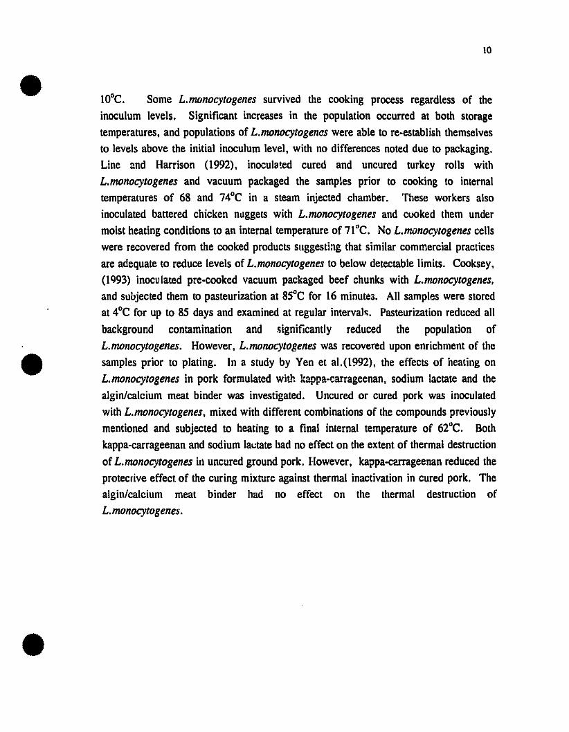
lQOC. Some L.mtJnocytogenes survived the cooking process regardless of theinoculum levels. Significant increases in the population occurred at both storagetemperatures, and populations of L.monocytogenas were able ta re-establish themselvesto levels above the initial inoculum level, with no differences noted due to packaging.Une and Harrison (1992), inocula!ed cured and uncured turkey rolls withL.monocytogenes and vacuum packaged the samples prior to cooking to internaitemperatures of 68 and 74°C in a steam injected chamber. These workers alsoinoculated battered chicken nuggets with L. monocytogenes and cùOked them undermoist heating conditions to an internai temperature of 71°C. No L.monocytogenes cellswere recovered from the cooked products suggesting that similar commercial practicesare adequate to reduce levels of L.monocytogenes to below detectable limits. Cooksey,(1993) inoculated pre-cooked vacuum packaged beef chunks with L.monocytogenes.and suojected them te pasteurization at 85°C for 16 minutes. Ali samples were storedat 4°C for up to 85 days and examined at regular intervals. Pasteurization reduced ailbackground contamination and significantly reduced the population ofL.monocytogenes. However, L.monocytogenes was recovered upon enrichment of thesamples prior to plating. In a study by Yen et al.(1992), the effects of heating onL. monocytogenes in pork formulated with kappa-c.arrageenan, sodium lactate and thealgin/calcium meat binder was investigated. Uncured or cured pork was inoculatedwith L.monocytogenes, mixed with different combinations of the compounds previouslymentioned and subjected to heating to a final internai temperature of 62°C. Bothkappa-carrageenan and sodium lactate had no effect on the extent of thermal destructionof L.monocytogenes in uncured ground pork. However, kappa-cerrageenan reduced theprotecrive effec! of the curing mixture against thermal inactivation in cured pork. Thealgin/calcium meat binder had no effect on the thermal destruction ofL.monocytogenes.

Il
Table 3: Beat resistance of Listeria monocytogenes in some meat and fish Droducts
Produet Temperature ("C) Dvalue z value ("C) Reference(Minutes)
Lean Ground Beef 52 81.3 5.4 Fain, (1991)57 2.663 0.6
Fatty Ground Beef 52 71.1 6.3 Fain, (1991)57 5.863 l.l
Pork Emulsion 60 12.95 6.8 Quintva11a and63 5.4 Campanini, (1991)66 2.3
Fermented beaker 60 9.13 NA Schoeni et al. (1991)Sausage
Ground beef roast 60 4.47 NA Schoeni et al. (1991)
Crawfish lail meat 55 10.23 NA Dorsa et al. (1993)60 1.9865 0.19
Cod fi\lets 60 1.98 NA Embarek & Huss, (1993)65 0.28
Salmon fi\lets 60 4.30 NA Embarek & Huss, (1993)65 1.02
1.6.2 Low Temperature Control of Listeria monocytogenes
Freezing and holding at or below _lOGC commonly extends the shelf life of foods by 5
to 50 fold compared to chilling preservation (Ciobanu,1976 cited in EI-Kest et al.,

12
1991). Liquid nitrogen (-198°C) is widely used to preserve bacterial cultures for lateruse in food processes and fermentations. Temperatures below optimum for growthreduce metabolic activity of microorganisms and so are bacteriostatic rather thanbactericidal. However, death of microorganisms may occur as a consequence ofextended frozen storage due to injury. Although found in frozen foods, there is littleinformation about the behavior of L.monocytogenes during freezing and frozen storage.EI-Kest et al., (1991) studied the effect of freezing and frozen storage ofL.monocytogenes in phosphate buffer and tryptose broth by freezing cell suspensionsfor either 30 minutes at -18°C or 10 minutes in liquid nitrogen. Freezing and storagefor one month in phosphate buffer at -18°C caused 87% death and 79% injury, white54% death and 45% injury was observed in tryptose broth. Freezing and storage inliquid nitrogen for one month caused no death or injury of cells in phosphate buffer,whereas minimal death and injury was observed in tryptose broth. These authors alsostudied the effect of freezing :nd thawing and re-freezing, which caused significantlymore death/injury than a single freeze thaw cycle. If liquid nitrogen is to be used topreserve food the fact that L.monocytogenes is resistant to death and injury duringmany months storage must be considered (EI-Kest and Marth, 1992). Recently, thefate of L.monocytogenes on packaged, frozen seafood was determined by Harrison etal. (1991). No increase in numbers of the organism was noted, and populationsdecreased by less than 1 log cycle after 3 months storage.
1.7. Novel Methods for the Control of L.monocytogenes:
Many novel methods for controlling the growth of L.monocytogenes in foods have beenproposed and applied with varying degrees of success.
Little is known about the influence of nutritional factors on the virulence ofL.monocytogenes. The limited knowledge was reviewed by Benedict (1990) whostressed the importance of understanding the role of environmental parameters in thesecretion of virulence factors. Listeriolysin-O (LLO) is probably the best studiedvirulence factor and the extent of its secretion may he influenced by temperature, NaCIand calcium. It has been established that the ferric ion has the most significant effect(McKellar, 1993). The influence of severaJ preservatives and growth factors on LLOsecretion by L.monocytogenes was examined and found to be maximal in tryptic soybroth (McKellar, 1993). Both growth and secretion was inhibited by nitrite while

13
secretion only was selectively inhibited by sorbate and NaCI. These results suggest thatLLO secretion is more sensitive than growth to the inhibitory action of preservatives.
1.7.1. Bacterioclns
Of particular interest to the processed/fermented Meat industry is the use of bacteriocinproducing strains of Lactobacillus, Pediococcus, Leuconostoc and Camobacteriumspecies (Daba et al., 1991; Dallas and Hitehins, 1993; Degnan et al., 1992; Foegedinget al., 1992; Luchansky, 1992; Mattila-Sandholm et al., 1991; Motlagh et al., 1992;Schillinger and Mattila -Sandholm, 1990; Skytta, 1991; Sobrino, 1991; Winkowski andMontville, 1992). ln recent years, antimicrobial Metabolites from food-grade starterculture bacteria, especially certain bacteriocins and bactericidal peptides, havegenerated interest as potential biopreservatives in minimally processed foods.Traditionally, fermented foods have been considered as pathogen-free. However,L.monocytogenes has been shown to grow in these products, possibly due to thesuspected emergence of bacteriocin resistant variants of Listeria.
Lactic acid bacteria produce a variety of antibacterial factors. The inhibitory spectrumvaries between narrow and broad within different lactic acid bacteria. Lactic acidbacteria show a number of antibacteria1 mechanisms, i.e., production of acid, hydrogenperoxide, or carbon dioxide. More importantly, the antibacterial effects May be basedon the competition for available nutrients and formation of antibacterial compounds,such as bacteriocins (Skytta and Mattila-Sandholm, 1991). Most of the experimentalwork hal: been carried out with Pediococus damnosus (Mattila-Sandholm et al., 1991),which has shown inhibitory activity towards the growth of Gram positive spoilagemicroorganisms such as lactobacilli and streptococci and gram positive pathogens suchas S.aureus and Bacil/us cereus and L.monocytogenes. Pediococcus acidilacti Hproduces pediocin AcH which has been shown to be inhibitory towardsL.monocytogenes (Motlagh et al., 1992). Leuconostoc mesenteroides was found toproduce a bateriocin called mesenterocin 5 active against L.monocytogenes strains(Daba et al., 1991). Studies with Lactobacillus bavaricus (Winkowski and Montville.1992), showed' that it also produces a bacteriocin that was able to inhibitL.monocytogenes at 10·C. Sensitivity of L.monocytogenes to nisin has also been

14
demonstrated (Harris et al., 1991). Nisin is an antibacterial peptide produced byLactobacillus lactis subsp. lactis that exhibits a broad spectrum of inhibitory activity
towards Gram positive bacteria and spores. Ming and Daeschel (1993) and Harris et
al. (1991) were able to isolate a mutant of L.monocytogenes that was resistant to nisin
due to fundamental changes that had occurred in bacterial membrane structure andfunction. Despite its bactericidal effect, nisin should not be relied on to control
L.monocytogenes in the food supply.
The lactoperoxidase system is a natural antimicrobial system present in milk that hasalso been tested for its antilisterial abilities (zapico, 1993; Gaya et al., 1991). The
enzyme lactoperoxidase catalyses the oxidation of thiocyanate by hydrogen peroxidewith the antimicrobial effect due to intermediate reaction products. ln cow's milk, the
lactoperoxidase system exhibited bactericidal activity against L. monocytogenes at 4and goe, with activity being dependent on temperature, length of incubation and strain
of Listeria used (Gaya et al., 1991). The lactoperoxidase system was also found to
have antilisterial properties in raw goat's milk (zapico, 1993).
1.7.2. Preservatives
ln more recent studies, the effects of various traditional and novel preservatives have
been studied (Table 4). Most of these compounds have been studied in model brothand model agar systems. Only a few preservatives have actually been applied in a food
matrix to test their inhibitory effects on L.monocytogenes. The compounds Iisted in
table 4 have also been studied in combination with one another (EI-Shenawy and
Marth, 1992; Johansen et al., 1994; McKellar et al., 1992; Moir and Eyles, 1992; Ohand Marshall, 1993a and b; Shelef and Yang, 1991; Stillmunkes, 1993; Wang and
Johnson, 1992; Wang and Shelef, 1992; zaika and Kim, 1993). In general, results ofthese combination studies show that most preservatives, when used together. ilIustrate a
synergistic effect, enhancing the inhibitory properties of the individual compounds.Thus, lower levels of the individual compounds may he used. This effecl is one of the
underlying principles behind the hurdle approach to food safety.
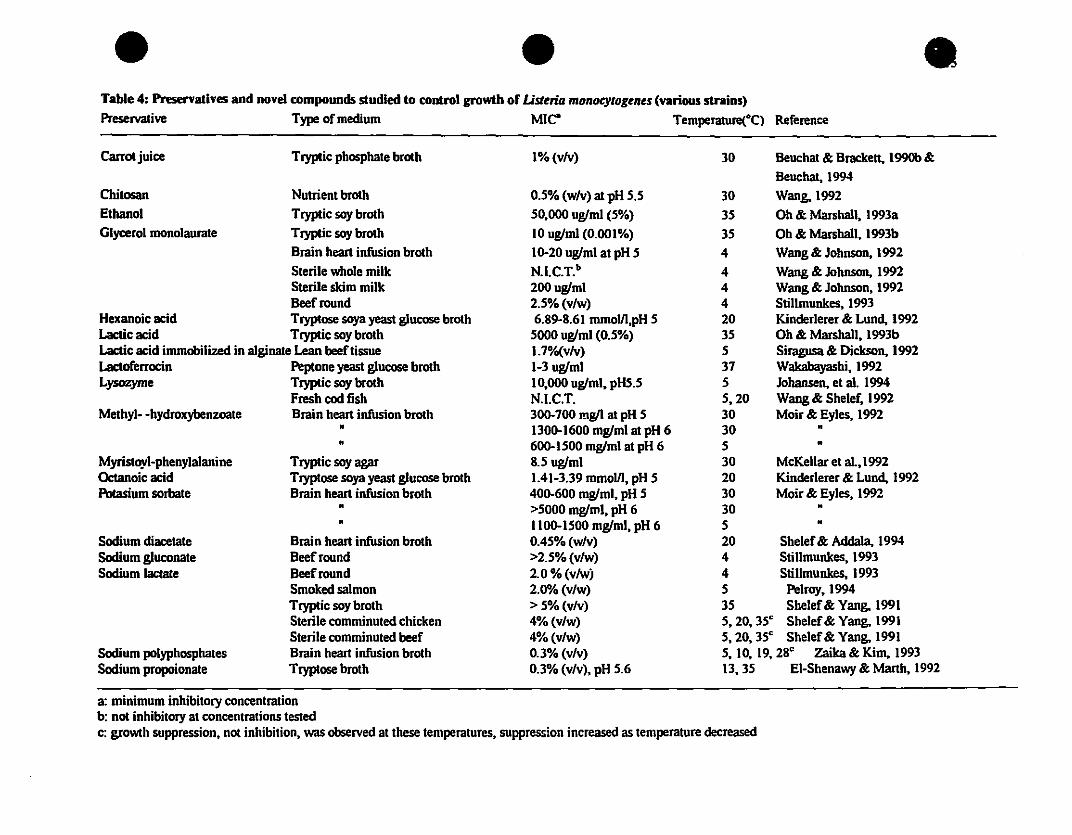
Table 4: Pnservatlves and novel compounds studled to control growth of Lisuria monocytogents (various straiDS)
Preservative Type of medium MIe" Temperature(°C) Reference
Carratjuice Tryptic pbosphate brath 1% (vlv) 30 Beuchat & Bracken, 1990b &
Beuchat, 1994
Chilosan Nutrient broth 0.5% (wlv) at pH 5.5 30 Wang, 1992Ethanol Tryptic soy broth 50,000 uglml (5%) 35 Oh & Marsball, 1993a
Glyœrol monolaurate Tryptic soy brotb 10 uglml (0.001%) 35 Ob & Marshall, 1993b
Brain heart infusion broth 10-20 uglml at pH 5 4 Wang & Johnson, 1992
Sterile whole milk N.I.C.T.b 4 Wang & Johnson, 1992Sterile skim milk 200uglml 4 Wang & Johnson, 1992Beefround 2.5% (v/w) 4 Stillmunkes, 1993
Hexanoic acid Tryptose soya yeast glucose broth 6.89-8.61 mmolll,pH 5 20 Kinderlerer & Lund, 1992Lacticacid Tryptic 50y broth 5000 uglml (0.5%) 35 Oh & Marshall, 1993bLactic acid immobilized in alginate Lean beef tissue 1.7%(v/v) 5 Siragusa & Dickson, 1992Lactoferrocïn Peptone yeast glucose broth 1-3 uglml 37 Wakabayashi, 1992Lysozyme Tryptic soy broth 10,000 uglml, pHS.5 5 Jobansen, et al. 1994
Fresb cod fisb N.I.C.T. 5,20 Wang & Sbelef. 1992Methyl- -bydroxybenzoate Brainhearti~onbroth 300-700 rng/l al pH 5 30 Moir & Eyles, 1992
• 1300-1600 mglml at pH 6 30 •• 600-1500 mglml at pH 6 5 •
Myristoyl-pbenylalanine Tryptic soy agar 8.5 uglml 30 McKellar et a1.,1992Oc:tanoic acid Tryptose soya yeast glucose brotb 1.41-3.39 mmolll, pH 5 20 Kinderlerer & Lund, 1992Potasium 50rbate Brain beart infusion brotb 400-600 mglml, pH 5 30 Moir & Eyles, 1992
• >5000 mglml, pH 6 30 •• 1100-1500 mglml, pH 6 5 •
SodIum dlacetate Brain beart infusion brotb 0.45% (wlv) 20 Sbelef& AddaIa. 1994SodIum g1uconate Beefround >2.5% (v/w) 4 Stillmunkes, 1993Sodium lactate Beefround 2.0 % (vlwj 4 Stillmunkes, 1993
Smoked salmon 2.0"/0 (v/w) 5 Pelroy,1994Tryptic soy broth > 5% (v/v) 35 Sbelef& Yang, 1991Sterile comminuted chicken 4% (v/w) 5,20,35" Sbelef& Yang, 1991Sterile comminuted beef 4% (v/w) 5,20,35" Sbelef& Yang, 1991
SodIum polypbosphates Brain beart infusion brotb 0.3% (v/v) 5,10,19,28" zaika & Kim. 1993Sodium propoionate Tryptose brotb 0.3% (v/v), pH 5.6 13,35 EI-Sbenawy & Marth, 1992
a: minimum inbibitory concentrationb: not inbibitory at concentrations testedc: growtb suppression, not inhibition, was observed at tbese temperatures, suppression increased as temperalUre decreased

16
Other interesting "natural" antilisterial compounds include plant essential oils, such ascinnamon, clove, origanum, pimento and thyme which have ail been shown to retardthe growth of L.monocytogenes to a Iimited extent (Aureli et al., 1992). Pimento oilwas able to completely inhibit growth within two hours, whereas clove, origanum andthyme oils showed a much milder inhibitory effect, taking four or more hours tocompletely inhibit growth.. The antilisteric effect of thyme oil was also tested inminced pork meat in which the Listeria population was reduced by approximately 100fold over the first week of storage (Aureli et al., 1992). In another similar study byPandit and Shelef (1!J94) a different effect was found, with rosemary and cloves theonly spices to exhibit antilisterial activity. Addition of 0.5 % finely ground rosemary or1%rosemary oil to ready-to-eat pork liver sausage prior to cooking delayed growth ofL.monocytogenes during refrigerated storage. Hefnawy et al. (1993) also studied thesensitivity of L.monocytogenes to selected spices in tryptose soya broth. CounlS ofL.monocytogenes decreased to less than 10/ml in one, four and seven days by 1%sage,1% allspice and by 1% cumin, garlic powder, paprika and red pepper respectively.Black pepper and mace at 1% did not inactivate L.monocytogenes while white pepperappeared to enhance ilS growth.
1.7.3. Irradiation
Gamma or electron beam irradiation treatment of meat to eliminate Listeriamonocytogenes has been proposed (Grant and Patterson, 1992; Thayer and Boyd, 1995)Radiation D-values were determined for inactivation of L.monocytogenes at S"Cintervals from -20 to 5°C in inoculated beef (Thayer and Boyd, 1995). This data wasused to develop an equation that predicts the response to gamma irradiation within aselected temperature range. An increase in resistance occurred at sub-zerotemperatures, i.e., _5°C and -20°C. The radiation D-values were 0.45 kGy at 0 andSnC, 0.77 kGy at _5°C and 1.21 kGy at -20°C. This resistance has been attributed to thedecreased OH mobility at subfreezing temperatures. The D-value resullS at SnC aresimilar to those obtained by Grant and Patterson (1992) in roast beef meal stored atrefrigeration temperatures. Lebepe (1990) studied the effeclS of irradiation (3.0 kGy)on the microbiological changes in fresh, vacuum packaged pork loins which werestored at 2 to 4°C for up to 98 days and at 25°C for approximately 48 hours. L.

17
monocytogenes was found in two irradiated samples. The microbiological shelf life ofthe pork Joins stored at 2 to 4°C was more than 90 days compared with unirradiatedloins Varabioff et al.(1992) determined the effects of irradiation on bacterial load andL.monocytogenes in raw chicken that was either vacuum packaged or packaged in air.After irradiation of chickens to a dose of 2.5 kGy, L.monocytogenes was onlyrecove.ed from the vacuum packaged chickens after 7 days of cold storage. Theseauthors concluded that irradiation may not be effective in destroying L.monocytogenes.
1.7.4. Sanitizers
Preventing contamination of processed foods requires emphasis on sanitation in thefood plant environment. L.monocytogenes has been reported to attach itself to a widevariety of surfaces. In the case of stainless steel, L.monocytogenes demonstratedsignificant resistance to chemical sanitizers and heat (Krysinski et al., 1992), and it wasoften isolated from moist surfaces in the processing and packaging areas of food plants(Ren and Frank, 1993). These observations indicate inadequate cleaning andsanitation. The germicidal effect of three common sanitizers, iodophor, quartenaryammonium compounds and chlorine on L.monocytogenes were evaluated by Tuncan(1993) using the suspension test method at various exposure temperatures and times.Ali three sanitizers were effective against Listeria at 25°C regardless of theirconcentrations (200-800ppm), giving a 5.0 log reduction after 30 seconds of exposure.As temperature decreased, the effects of the sanitizers were diminished. However; thiscould be reversed by extending the exposure time. Sallam and Donnelly(1992)observed similar results with a variety of sanitizers, and, in addition, discovered thatthe sanitizers used induced injury, not death of the microorganism, and that the lethaleffect of a sanitizer was found to increase by increasing its concentration or exposuretime. These results illustrate that exposure to a sanitizer does not mean destruction ofL.monocytogenes and that the organism becomes sublethally injured, and capable ofrepair and multiplication when conditions become more favorable, Le., in foodproducts. This poses a public health hazard. Biofilm prevention lies in adherencewith GMP (good manufacturing principles) and HACCP (Hazard Analysis CriticalControl Point) processing. Strict attention should be paid to sanitizing of foodprocessing plants to use lethal concentrations of sanitizer for optimal exposure times.

18
1.7.5. Modified Atmospheres
Modified atmosphere packaging has been defined as "the enclosure of foods in high gasbarrier materials in which the gaseous environment has been changed or modified toslow respiration rates, reduce microbial growth and retard enzymatic spoilage- with theintent of extending shelf life" (Young et al., 1988). When MAP is viewed as a totalcontrol quality packaging technology, rather than a simple packaging step combinedwith some special gases, it can then be realized to its fullest potential and can becomethe revolutionary teehnology that it has proven to be in the European market place(Lioutas, 1988). MAP is a rapidly expanding technology considering that the energycosts associated with other preservation methods are relatively high i.e., heatprocessing, freezing and frozen storage and distribution and drying. Consumers arealso more and more reluctant to accept chemical additives and preservatives in theirfoods and are demanding high quality, fresh-like foods ever increasingly. There areseveral methods that can be employed for modifying the internai gas atmosphere of aproduct's package head space. These methods are: vacuum packaging or skinpackaging and gas packaging. Gas packaging is the technique in which variouscombinations and mixtures of carbon dioxide, nitrogen and oxygen are used dependingon the type of product. The air is moved physically out of the package head space andthe desired gas mixture is flushed in.
1.7.5.1. Gases used in Gas Flush Packaging:
1.7.5.1.1. Carbon dioxide
Carbon dioxide is considered to be the most important gas used in most MAPapplications. CO2 is bacteriostatic, fungistatic, prevents insect growth, and is highlysoluble in fat and water where it forms carbonic acid. This solubility may lower thepH of the product resulting in slight flavor changes. The mechanism of action of CO2
against microbes is explained by several theories. It is known that CO2 inhibits thegrowth of Gram negative bacteria such as pseudomonads and other relatedpsychrotrophs. Carbon dioxide increases the lag phase of the growth curve of thesebacteria. Lactic acid bacteria, such as streptococei and lactobacilli are less affected by

19
elevated CO2 levels, and in fact, elevated levels of CÛ2 enhance their growth. CO2
interferes with enzyme systems attached to the bacterial cell, such as thedehydrogenating enzymes and enters into the mass-action equilibria for enzymaticdecarboxylation (Finne, 1982). Changes in surface pH have also been suggested as amechanism of inhibition. The change in pH is due to the absorption of CO2 on thefood surface and subsequent ionization of the carbonic acid. Other theories suggest thatCO2 may act on bacterial cell membrane permeability and f1uidity and toxicity ofcarbonic acid in its undissociated form (Finne, 1982).
1.7.5.1.2. Nitrogen
Nitrogen is an inert gas which apparently has no antimicrobial properties. It is termedthe "filler gas" because its main function is to prevent package collapse since it is lesswater and fat soluble than carbon dioxide. N2 prevents oxidative rancidity by replacingthe oxygen present, especially in low water activity foods.
1.7.5.1.3.0xygen
Oxygen is generally not used in gas packaging applications unless it is used to prevent
the growth of strict anaerobes such as Clostridium botulinum and to maintain the brightred color or "bloom" associated with fresh red meats. Oxygen is also used in lowconcentrations in the packaging of respiring products.
1.7.5.2. New Developments: Oxygen Absorbent Tecbnology
Oxygen absorbents are defined as a range of chemical compounds introduced into the
MAP package (and not the product) to alter the atmosphere within the package.Oxygen absorbent technology was developed in Japan and forms an important role infood preservation and distribution in Japanese supermarkets. Oxygen absorbents arecomposed of compounds chemically reactive to oxygen. When oxygen absorbers are
placed in gas permeable materials (in the forro of small pouches usually made out of
co-polymers of propene and ethylene vinyl alcohol. EVA) and placed in packages. they

20
are capable of removing residual oxygen from the package head space. It is importantto note that gas packaging, which is a physical process, usually does not remove alltraces of oxygen present, whereas oxygen absorbents are capable of removing allresidual oxygen when used properly. First developed by the Mitsubishi Gas ChemicalCompany, Inc., in 1977, an iron-powder based oxygen absorbent under the name ofAGELESS was introduced. The principle behind these absorbents is simply thereaction that occurs when iron rusts. The reaction is explained in Figure 1.
Figure 1: Reaction of iron to rust that occurs in oxygen absorbents
Fe--' Fé+ + 2e'
2+Fe + 20H' -_1 Fe(OHh
There are several types of Ageless absorbents available including an absorbent knownas Ageless SS which is a self reacting type of absorbent capable of absorbing oxygen atlow temperatures and is of particular interest to the chilled and frozen food industry.Ageless type SE is another absorbent of interest, especially to the bakery industry,since it not only removes the oxygen present, thus lowering the chance for moldspoilage, but it a1so emits ethano1 vapor into the package head space. Ethanol vaporhas been shown to be an effective, broad spectrum antimicrobial agent.
1.7.S.3. The Effect ofMAP on Meat Sensory and Microbiologiesl Characteristics
Several reviews and studies have been published on the topic of applying MAP to freshmeat products, the focus of which have been primarily extending the meat's shelf lifefrom a microbiological and sensory perspective (Eyles et al., 1993; Finne, 1982; Gilland Tan, 1980; Greer et al., 1992; Holley et al., 1993; Venugopal et al., 1993;Wolfe, 1980; Young et al., 1988). In discussing the packaging of fresh meats, the twomost important meat properties (aside from safety) are the color and microbial spoilage
population.

21
1.7.5.3.1. Color
The color of fresh meat depends on the relative proportions of three forms ofmyoglobin; reduced myoglobin, oxymyoglobin and metmyoglobin (Holley et al., 1993;Young et al., 1988). Reduced myoglobin is the predominant muscle pigment in theabsence of oxygen, producing the characteristic purple color meat exhibits when firstcut as well as the expected color of vacuum packaged meats. Oxymyoglobin is theoxygenated form of the muscle pigment and is responsible for the bright red colorconsumers associate with fresh meat. Metmyoglobin is the form with an undesirablebrown color that causes consumers to reject prepackaged meats, formed from theoxidation of oxymyoglobin and reduced myoglobin in low oxygen tensions or from theoxidation of iron heme in he myoglobin molecule (Young et al., 1988). Figure 2il\ustrates this phenomenon.
Figure 2: The variollS fOl'DlS of myoglobin
02 O~dation
/
Reduction
1.7.5.3.2. Microbial Population
High CÛ2 levels (above 20%) cause a shift in the meat microflora, from fast growingaerobic spoilage bacteria to slow growing facultatively aerobic non-spoilage species.C~ inhibits pseudomonads and favors the outgrowth of their competitors that are lessaffected by C~ • mainly lactic acid bacteria. Since these organisms grow more slowlyand generally yield metabolic products that are less offensive than those of

22
pseudomonads, such as lactic acid, spoilage of meat is delayed (Holley et al., 1993).The ability of COz to inhibit respiration and growth of representative strains of sevenspecies of meat spoilage bacteria was examined by Gill and Tan (1980). Enterobacterand Brocothrix thermosphacta were unaffected by COz (25 and 75 %). Both respirationand growth of the other species was inhibited (Pseudomonas (fluorescent and nonfluorescent), Acinetobacter, Pseudomonas putrefaciens and Yersinia enterocolitica).The degree of inhibition with a constant concentration of COz in solution increasedwith decreasing temperature for all COz susceptible species except non-fluorescentPseudomonas. Anaerobic growth of COz susceptible facultative anaerobes wasunaffected by COz. Eyles et al. (1993) further studied the effects of MAP on thegrowth of psychrotrophic Pseudomonads on the surface of a model agar system.Atmospheres containing concentrations of COz as low as 20% (balance nitrogen)inhibited the growth of Pseudomonas fluorescens and Pseudomonas putida on thesurface of Brain heart infusion agar plates at pH 6.8 incubated at 5 and 150 C in tlexiblepackages. The modified atmospheres decreased the growth rates and reduced thepopulations attained at the end of the exponential phase of growth, but had no effect onthe lag phase. Once again, the inhibitory effect of COz increased as the temperature
decreased. This effect is due to the increased solubility of COz in the media, or muscletissue at lower temperatures resulting in a lowering of the pH.
1.7.5.4. The Safety of MAP
MAP foods may pose a public health risk, especially if the MAP product is subjected totemperature abuse during storage and/or distribution. Consumers may also mishandleand/or subject the product to temperature abuse conditions, and they may also be proneto overextend the products' normal shelf life. Research on the safety of MAP foods,from a microbiological perspective, is lacking even though much is known about MAPin general, indicating that the wrong emphasis has been put into the development ofMAP technology. Any research done should place emphasis on consumer safety first
and freshness second. From a safety standpoint, in MAP foods, the normal aerobicspoilage microorganisms which would normally warn consumers of spoi1ag~ (by offodors and off colors) are inhibited, while the growth of pathogens may be allowed oreven stimulated (Farber, 1991). ln recent years, the focus of concern over the safety ofMAP foods has shifted form the toxin producing Clostridia (Genigeorgis, 1985;
23
Hintlian and Hotchkiss, 1986; Silliker and Wolfe, 1980) to several "emergingpathogens". Due to the emergence of psychrotrophic pathogens, such as L.
monocytogenes, Aeromonas hydrophilia, and Yersinia enterocolytica, new safety issueshave been raised. Hotchkiss (1988) presents a detailed analysis on the experimentalapproaches to determine the safety of food packaged in modified atmospheres, howeverthis review does not mention possible methods in evaluating whether or not theemerging pathogens pose health risks.
1.7.5.5 Effect of Modified Atmosphere Packaging (MAP) on the Growth ofL.monocytogenes
Limited studies that have been done on the effect of MAP on L.monocytogenes. Todate, few studies have been carried out in model systems. Razavilar and Genigeorgis(1992) studied the interactive effect of temperature, atmosphere and storage time on theprobability of colony formation on blood agar by four Listeria species. The speciesused were L.monocytogenes, L.seeligeri, [,.ivanovii and L.innocua. Sheep blood agarplates were inoculated with the test organisms and were stored at 4,8,20 and 300Cunder air, vacuum, candie jar, 100% COz or a MA consisting of5%Oz+ 10%COz+85%Nz, for 7, 14,21, 42 and 56 days. None of the interactions oftemperature, time, or species with atmospheric conditions were found to be significant.An atmosphere of 100% COz was found ta be significantly more inhibitory to growththan any of the other atmospheres and its effect on delaying Listeria 's growth wasenhanced at lower temperatures. L.ianovii was the most sensitive to LOz. Packaging in100% COz extended the lag phase at less than 8°C and decreased the rate of growth at4°C but not at higher temperatures.
1.7.5.5.1. Produce
The behavior of Listeria inoculated onto fresh vegetables and stored under CA(Controlled Atmosphere) conditions was examined by Berang et al. (1989). The CAused consisted of Il % Oz, 10% COz and 79% Nz . Listeria grew as well on CAvegetables as on control vegetables (air packaged) either at 4 or 15°C. Enumeration ofasparagus at the end of storage showed that only slightly higher levels present in air

24
stored samples. In .he same study, broccoli stored under II %Oz, 10% COz and 79%Nz showed no decrease in the levels of Listeria present. The fate of Listeria inshredded cabbage at 5 and 25·C stored under MAP conditions (70% COz, 30%Nz)WllS determined by Kallander et al. (1991). In air packaged samples. Listeriapopulations increased by 2 log cycles within two days of storage at 25·C. but thendecreased to undeteetable levels within 6 days of storage. Packaging under a 70% COz
and 30%Nz gas atmosphere and storage at 25·C also decreased levels of Listeria toundetectable levels within 6 days but with a less marked initial increase than thecontrol samples. At 5·C, counts ir...:reased gradually but by only about 1 log in boththe control and MAP storl~d samples. However. in control samples, a marked increasein L.monocytogenes was observed after 13 days of storage. The combined effects ofshredding, chlorine treatment and MAP (97% Nz, 3% Oz) on the survival and growthof L. monocytogene.< on lettuce stored at 5 and 10·C were determined by Beuchat andBrackett (1990a). None of the treatments. either alone or in combination. influencedthe growth of this organism indicating that Listeria was able to grow on lellucesubjected to commonly used packaging and distribution procedures used in the foodindustry. An excellent review on the safety of produce packaged is under MAPconditions is provided by Hotchkiss and Banco (1992).
1.7.5.5.2. Poultry
The development of L.monocytogenes on minced raw chicken stored at 4. 10 and 27"Cunder various MAP conditions i.e., 75%COz• 25%Nz (MAI) and n.5%COz•22.5%Nz, 5%Oz (MA2) was sturlied by Wimpfheimer et al. (1990). Listeria grewunder the aerobic control conditions at ail temperatures. However, it failed to growunder any MAP conditions containing no oxygen, i.e., MAL When oxygen waspresent (MA2) growth of Listeria was not inhibited and numbers increased by nearly 6log cycles at 4·C after 21 days. However, growth of the aerobic spoilagemicroorganisms was inhibited. This study ilIustrates that under MAP conditions usedcommercially i.e., containing oxygen for bloom, L.monocytogenes is capable of growthwhile the spoilage microflora of chicken are inhibited. In another study, skinlesschicken breast meat was inoculated with L.monocytogenes and stored at 1,6 or 15·Cunder aerobic conditions and atmospheres of 30%COz + air (MAI). 30%COzI70%Nz(MA2) and 1()()%COz (MA3) (Hart et al., 1991). At I·C the organism did not grow

25
under any of the test conditions. However, at 6°C Listeria counts incl'eased 10 fold inaerobic conditions before spoilage of the meat was observed, but only when the culturewas pre-incubated at IUC rather than 35°C. In other words, subjecting the cells torefrigeration temperatures prior to inoculating the poultry appeared to enhance theirability to grow at lower temperatures since they were accustomed to these lowtemperatures. In CO2 atmospheres, the growth of Listeria was inhibited on meat held at6uC. especially under 100% CO2• At 15°C, spoilage of the meat occurred within twodays in ail gaseous conditions and levels of L.monocytogenes increased 100 foldindicating the importance of strict temperature control to ensure the safety/quality ofMAP muscle foods. The combined effect of MAP (90%C~1 10%02) and lactic acidtreatment on the growth of L.monocytogenes on chicken legs at 6°C was investigated byZeitoun and Debevere, (1991). While MAP alone did not have an inhibitory effect on
the growth of Listen'a, combination treatmèilts significantly reduced the numbers ofListeria present, iIIustrating a synergistic effect between lactic acid treatment and MAPon the growth of L. monocytogenes on chicken legs. The influence of various gaseousconditions, i.e., 76%C02, 13.3%N2, 10.7%02 (MAI) and 80%C02, 20%N2 (MA2)
on the competitive growth L.monocytogenes and Pseudomonas fluorescens on precooked chicken nuggets stored at 3,7 and 11°C was determined by Marshall etal. (1990). When the bacteria were co-inoculated in nuggets, growth of Listeria wasstimulated at 3°C by the presence of P.fluorescens in air and under atmosphere MA l,
but not in atmosphere MA2 where growth of P.fluoresecens was inhibited. Thisgrowth stimulation was also absent at higher temperatures. The stimulatory effect ofP.fluorescens is thought ta be due ta a proteolysis production effect and certain aminoacids which enhances the growth of L.monocytogenes.
1.7.5.5.3. Red Meats
Gill and Reichel (1989) studied the growth of L.monocytogenes on high pH beef (pH
6.0) stored under vacuum or 100% C~ at -2, 0, 2, 5 and 10°C. L.monocytogenes didnot grow in vacuum packs at _2°C, and generally grew at rates less than the spoilagemicroflora at higher temperatures. In CO2 packs, Listeria grew at 10°C, but not at thelower temperatures. The combined effect of MAP (25%Co,.n5%Nù and irradiation
(1.75 kGy) on the growth of L.monocytogenes in beef stored at 10 and 15°C was
investigated by Grant and Patterson (1991). These authors showed that the safety of
26
MAP pork was less than that of MAP/irradiated pork indicating that combinationtreatments could provide an additional safety margin panicularly when products arestored at temperature abuse conditions. Hudson et al. (1994) studied the growth ofL.monocytogenes on sliced roast beef packaged under vacuum or saturated controlledCOz atmospheres at -1,.5 and 3·C. At -l.S·C, the pathogen declined in numbers inCOz packs but not under vacuum. However, at 3·C, growth occurred under ailpackaging conditions with maximum numbers being reached by the end of an extendedstorage period of 166 days.
1.7.6. Chitosan: A Novel Antùnicrobial Agent
Chitin is the second most abundant natural polymer on earth (the first being cellulose)and is widely distributed throughout nature. Chitin is a polymer of Nacetylglucosamine units, white chitosan is the deacetylation product of chilin, i.e., apolymer of glucosamine. Figure 3 shows the structures of these compounds.
The most easity exploited sources of chitin are the protective shells of crustaceans, suchas crabs and shrimps. 1t is estimated that fungi could provide approximately 32,000tons of chitin annually. ln the U.S., the main types of shellfish that are processed intochitin and chitosan are: Cancer maeister (Dungeness crab), Paralithodes camshatjca
(King crab), and Pandalus borealis (Pacifie shrimp) (Skjak-Braek, et al., 1989). Sincecrab and shrimp are harvested in different seasons, raw materials for chitin productionare always readity avaitable. Other sources of chitin include seafood waste, such aslobster, krill (an aggregate of small marine crustaceans), clams, oysters and squid.
Chitin is also found in the cell walls of fungi as weil as the cuticle of insects.
Chitosan is now produced in North America on a large scale. For sorne lime it hasbeen sold commercially as a solution, flaked and fine powder, and more recently in
bead and fiber forms. 115 avaitability in a variety of useful forms and its uniquechemical and biological properties make it a very attractive biomaterial (Skjak-Braek etal., 1989). It is for the above mentioned reasons that chitosan already has a widevariety of applications, white more are currently the subject of extensive research.
Recently, research on chitosan has been directed to its potential use as a foodpreservative and antimicrobial agent. It is estimated that the total sales of chitin and
27
chitosan will reach almost $2 billion U.S. in the next ten years, with sales of chitin andchitosan in the food and beverage industry comprising about 110 million dollars of this
total.
1.7.6.1. Extraction of Chitin and Chitosan
The principle steps in the extraction of chitin from seafood waste are iIIustrated inFigure 4. Proteins are first removed from the ground up shells by treating the shellswith sodium hydroxide. Using hydrochloric acid, minerais such as calcium carbonateand calcium phosphate are extraeted. After rinsing, the chitin is dried as a f1akedmaterial. The general scheme to manufacture chitosan from chitin is shown in Figure5. Chitin is first treated with concentrated sodium hydroxide to hydrolyze the N-acetyllinkages. It is then rinsed and the pH is adjusted. At this stage, chitosan can be driedto give what is called "f1aked chitosan" which is a coarse mesh product whose particlesize may be reduced by milling to give a finer mesh powder. The powder can then bedry blended with an organic acid, such as adipic acid, to give a chitosan acid blend thatis self dissolving. One scheme to prepare chitosan of improved purity is to dissolve it inacetic acid and filter it to remove extraneous materials. The clarified product can thenbe reisolated to give an ultrapure product known as chitosonium acid salts, which aresoluble in water. Another procedure used is to precipitate the chitosan out of solutionusing a base. This precipitate is then washed and dried to give chitosan in the freeamine form.
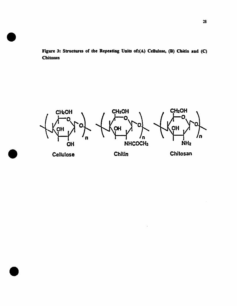
28
Figure 3: Structures of the BepeatiDg UDits of:(A) CeUulole, (D) ChitiD aDd (C)
ChitolaD
OHCellulose
nNHCOCH3
Chilin
NHz
Chilosan

•Figure 4. Flow Diagram of Cbjtin Extraction Process
SEAFOOD WASTE
lCRABISBRIMP SHELLS
lDErRl
WII1I NoOB
DIEWII1I BeJ
RlNSE AND DRY
lCHITIN
29
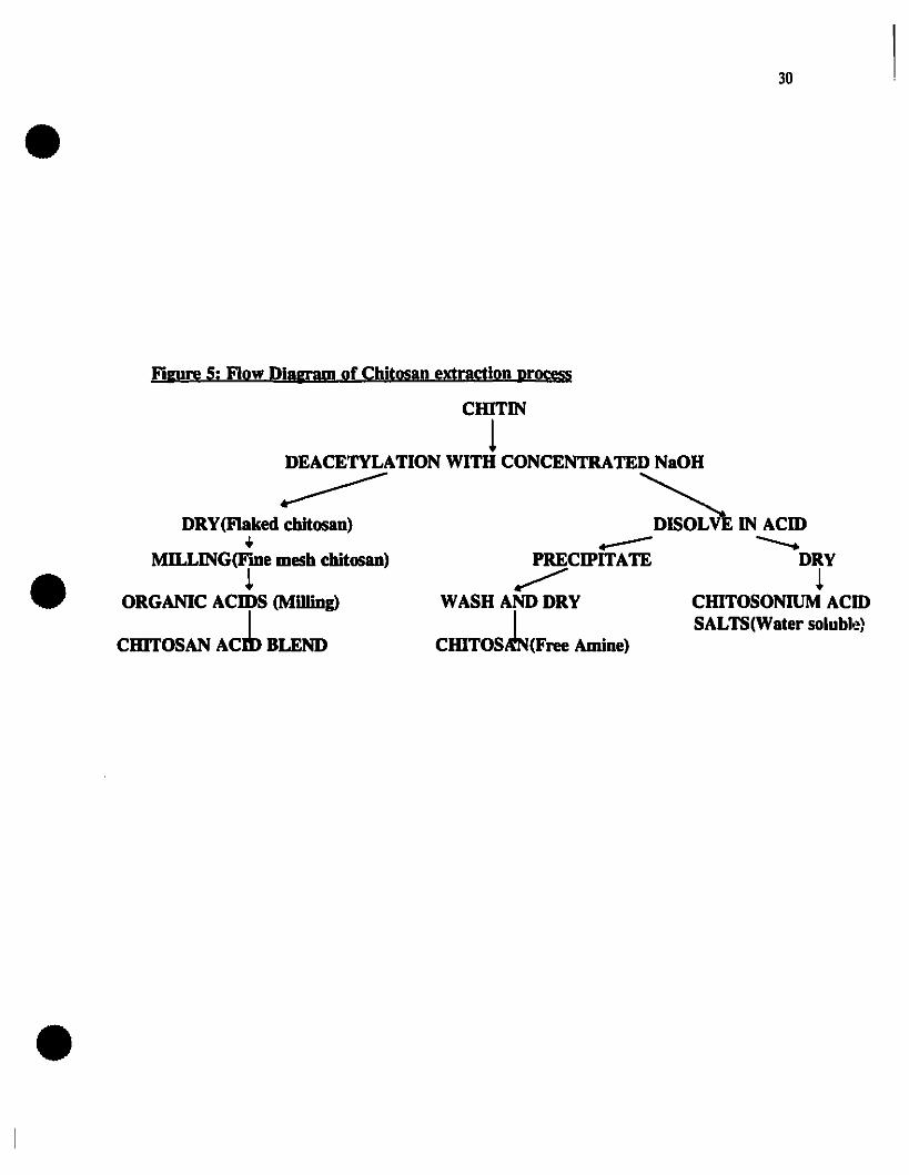
30
Figure Si Flow Di!Wl!ID of Chitosan extraction prnrm
CIDTIN
!DEACETYLATION WITH CONCENTRATED NaOH
DRY(FIaked~ ~ IN ACm.. ....----MILLING(F"me mesh chitosan) PRECIPITATE DRY
! ~ !ORGANIC ACmS (Mllling) WASH AND DRY CHITOSONIUM ACm
1 1 SALTS(Water solubl.~}CIDTOSAN ACm BLEND CHITOSRN(Free Amine)

31
1.7.6.2. Applications of Chïtosan
Chitosan has numerous applications, Many of which depend on its cationic nature. It isa linear polyelectrolyte at acidic pH values and it has a high charge density, one perglucosamine unit. Since Many materials carry negative charges (proteins, anionicpolysaccharides, nucleic acids ete.) the positive charge of chitosan interacts stronglywith negative surfaces to give electric neutrality. Due to the vast number of NH3+groups that can interact with negatively charged colloids, chitosan is also an excellentflocculating agent. It has been found that chitosan can also form complexes with Manymetal ions, thus it is useful in chelating iron, copper and toxic heavy Metal ions, suchas silver cadmium, Mercury, lead, nickel and chromium (Muzzarelli, 1977).
Being a high molecular weight polymer, chitosan is a linear polyamine whose aminogroups are readily available for chemical reactions and salt formation with acids. Sinceit can be viewed as a cellulose derivative, the primary and secondary hydroxyl groupscan be used to make derivatives, Many of which have commercial potential. In the freeamine form, chitosan is not soluble in water at neutral pH. At acid pH values, the freeamine groups become protonated to form cationic amine groups.
1.7.6.2.1. Agriculture
ln agriculture, chitosan has been used as a seed coating and it has been shown toincrease crop yields. The EPA (Environmental Protection Agency) in the V.S.,permits the use of chitosan for this purpose and in 1989 it was being used in elevenstates in the V.S. Chitosan triggers a response in the seed. signaling the plant toprotect itself from natural predaulrs. such as pathogenic fungi (a more detaileddiscussion on the effects of chitosan on fungi is presented later). Chitosan has alsobeen approved for use as a flocculating agent to recover proteinaceous wastes foranimal feed. Continuing studies investigate the use of chitosan coating as a controlledrelease agent for pesticides and herbicides. One other use in agriculture is the additionof chitosan to cropland to stimulate natural microbes that provide protection to certaincrops. Hirano (1990) tested chitosan for use as an ingredient for domestic animalfeeds. No abnormal symptoms were observed with hens and broilers by feeding < 1.4g of chitosanlkg of body weight per day for up to 239 days and with rabbits by feedingthem with <0.8 g of chitosanlkg of body weight per day for up 10 239 days. Both

32
chitin and chitosan were digested 35-83% by rabbits and 88-98% by hens and tlroilers.Chitosan also had an effect in decreasing serum cholesterol levels of the animais tested.However, for long term feeding, hen's appetites and egg laying rates decreased byfeeding an excessive amount of chitosan due te its incomplete digestion.
1.7.6.2.2. F10cculating agent
The largest single use of chitosan is the clarification of waste water in Japan. Sincechitosan is a natural polymer, it is preferred over synthetic polymer fiocculants whichmay contain hazardous monomers. Several organizations in the U.S.A. (Division ofAnimal Feed, Department of Health and Human Service, FDA, Association of AnimalFeed Control Officiais (AAFCO», have issued the following description of chitosan asa fiocculent for recovering proteinaceous materials: "Chitosan is a cationiccarbohydrate polymer intended for use as a precipitation agent of proteinaceousmaterial from food processing plants. It is chemically derived by deacetylation of thenaturally occurring chitin in crab and shrimp shells. It may be used in an amount notto exceed that necessary to accomplish its intended effect. Chitosan, when incorporatedinto the feed of livestock, should be present at no more than 0.1 % of the feed.Proteinaceous material coagulated with chitosan must have safety and efficacy dataapproved before it can be registered or offered for sale" (Skjak-Braek et al., 1989).Imeri and Knorr (1988) and Soto Peralta et al. (1989), also found that acid solublechitosan and water soluble chitosan proved equally effective as fining agents for appleand carrot juices. The one step application proved as effective as conventionaltreatments using silicalgelatin/bentonite. Knorr (1991) cited Bade and Wick (1988)who suggested the use of chitinous polymers for environmental protection and wastereduction. This is based on the fact that chitin and chitosan have proven film formingcapabilities and that chitosan degrading microorganisms are abundant. Recently, it hasbeen discovered that a chitosan based film had superior oxygen barrier properties, butless tensile strength, than synthetic packaging films, such as Mylar and polypropylene(Knorr, 1991).
1.7.6.2.3. Control of fongal diseases in plants
Fungal diseases are one of the major causes of post harvest losses of fruits andvegetables. Chemical control programs face may problems: (i) increasing amounts of

33
fungicide tolerant post harvest pathogens, (ii) increased public awareness and (Hi)resistance to fungicide treated produce. As a result, a number of fungicides are nowunder review in many countries. Therefore, there is a growing need to developalternative approaches for the efficient control of storage diseases. Chitosan has beenshown to have an antimycotic effeet against a wide range of fungL Allan andHadwinger (1979) studied the fungicidal action of chitosan on 46 fungi of varying cellwall composition. They observed that chitosan had more antifungal activity than chitinand, out of the 46 fungi tested, chitosan inhibited 32 of the fungi investigated. Alifungi that were sensitive to chitin were more or equally sensitive to chitosan. At theconcentrations used, chitosan seemed to have no effect on the fungi that containedchitosan in their cell walls. Stossel and Leuba (1984) confirmed the results of Allanand Hadwinger (1979). They reported that the antifungal mechanism of chitosan mustresemble the mechanism described for plant cells, Le., a polycationic effect thatinterferes with the membrane (Young et al., 1982).
Young et al. (1982), studied the effeet of chitosan on the membrane permeability ofplant cells. Treatment of plant cells with chitosan resulted in an increase in membranepermeability, as shown by leakage of eleetrolytes, protein, and UV absorbingmaterials. This effeet, they concluded, was clearly due to the polycationic nature ofchitosan. The effeet of the chitosan was inhibited by divalent cations. An increase inmembrane permeability would be expeeted in the presence of soluble chitosan, or whenehitosan on the fungal cell wall surface makes contact with the host cell membrane.The eell wall may help to protect the plasma membrane from such effects by binding toehitosan, although its effeetiveness as a natural barrier would be Iimited by its bindingcapacity. lt seems possible that interactions of chitosan with membrane or cell wallcomponents of plants might be involved in its ability to elicit phytoalexin synthesis, toinduee lignification and to inhibit fungal growth. ln contrast, increased permeability ofthe eell wall could also facilitate infection by enhancing the f10w of nutrients to thepathogen and possibly enabling fungal products to enter host cells. This is aninteresting dilemma and should be considered carefully when applying ehitosan toplants.
Chitosan can aetivate specifie genes in plants and, at similar concentrations, inhibit ailRNA synthesis in sorne fungal organisms and thus suppress gene activity (Hadwinger,1985). This phenomenon is explained by the interaction of chitosan's positively

34
charged amino groups and the negatively charged phosphate groups of nucleic acids.The chitosan must be 7 or more sugar units in length to optimally induce plant genesand to inhibit fungal growth (Kendra & Hadwinger, 1984). This length requirementsuggests that a series of positive charges match up with phosphate negative charges inthe grooves of the DNA helix in the form. Using radio-Iabeled chitosan, chitosan wasapplied to fungal cells in concentrations sufftcient to inhibit their growth. DNA wasfound to precipitate into solution. Hirano and Nagao(1989) reported that strong growthinhibition was observed for low molecular weight chitosan but only weak inhibitionwas observed with high molecular weight chitosan. The effect of chitosan preparationswith different levels of deactylation and other polyanions on the growth of post harvestpathogens was investigated by El Ghaouth et al. (1992 a and b). Chitosan markedlyreduced the radial growth of ail fungi tested, with a greater effect being observed athigher concentrations. Chitosan's inhibitory activity appeared to increase withincreasing levels of deacetylation. In addition to inducing cellular leakage of aminoacids and proteins in Botrytus cinerea and Rhizopus stolinifer, chitosan also inducedmorphological changes in R.stolinifer. An ultrastructural study, using electronmicroscopy, showed that chitosan caused deep erosion of the cell wall as weil asincreasing cell wall thickness. Chitosan was also found to stimulate the activity ofchitin deacetylase, the enzyme involved in the biosynthesis of chitosan. This increasein activity of chitin deacetylase could upset the balance between biosynthesis turnoverof chitin thereby rendering the cell wall more intact.
1.7.6.2.4. Control of Toxigenic Fungi
Toxigenic fungi are often found in pre-harvest crops, such as maize and peanuts, inassociation with other fungi, yeasts and bacteria. Aspergillus parasiticus andAspergillus flavus are two such toxigenic fungi, both producing aflatoxin. Numerousattempts have been made to control pre-harvest aflatoxin production by Iimiting growthof toxigenic fungi through application of chemical or microbial agents with Iimitedsuccess. It has been reported that chitosan exhibits extensive inhibitory effects onA.flavus growth and/or aflatoxin production in Iiquid culture and in preharvest maizeand groundnuts. ln a study by Cuero et al. (1991) chitosan was used alone or in
combination with microbial agents to inhibit growth of aflatoxin-producing fungalspecies and to control aflatoxin production in pre-harvest maize. They found that thegrowth of and aflatoxin production by A.flavus were significantiy reduced in chitosan

35
treated pre-harvest maize. A single component treatment was found to be moreeffective than combination treatments. Fang et al. (1994) studied the inhibitory effectsof chitosan on the growth of Aspergillus niger and the aflatoxin production ofA.parasiticus. The inhibitory effeet of chitosan on A.niger was increased as thechitosan concentration increased from 0.1 to 5.0 mg/ml at a pH of 5.4. The greatestinhibitory effeet of chitosan of A.parasiticus was found at 3.0-5.0 mg/ml. In addition
chitosan could completely prevent aflatoxin production at these concentrations.
1.7.6.2.5. Control of Senescence and Ripening
The effect of chitosan on respiration, ethyIene production and quality attributes of
tomatoes stored at 20°C was investigated by El Ghaouth et al. (1992a). Coating thetomatoes with chitosan solutions reduced their respiration rates and ethyleneproduction, with the greatest effect being observed with 2%(w/v) chitosan solutions.Chitosan was also used as a dipping solution to extend the shelf Iife of freshstrawberries to approximately 30 days at 13°C (EI-Ghaouth et al., 1991).
1.7.6.2.6. CODtrol of Food SpoilagelPatbogenic Bacteria
In lapan, several patents have been issued on the use of chitosan as a food preservativeto extend the shelf Iife of foods. Reports have shown that chitosan can inhibit thegrowth of S.aureus, Escherichia coli, Pseudomonas aeruginosa and Bacillus subtilus.Although no antibacterial mechanism has been elucidated, it has been shown thatchitosan is only effective against certain bacteria and under certain conditions. Wang(1992) studied the inhibition of rive species of food borne pathogens: S.aureus, E.coli, Y.enterocolitica, L.monocytogenes, and Salmonella ryphimurium. Nutrient broths
were supplemented with 0.5, 1.0, 1.5, 2.0 and 2.5% chitosan and adjusted to either pH5.5 or 6.5. It was shown that the antibacterial action of chitosan was greatest onS.aureus followed by S.ryphimurium, E.coli. and Y.enterocolitica at pH 6.5. At thispH, L.monocytogenes was not inhibited. However, at pH 5.5, the outgrowth of ail
bacteria was inhibited. Wang (1992) concluded that it was difficult to predict thechitosan concentrations actually needed to inhibit bacteria due to many factors, such asthe character of the chitosan used, pH, water activity, storage temperature of the food
36
as wel1 as the initial degree of contamination of the food. AI1 of these factors caninfluence the effectiveness of chitosan as an inhibitor either alone or in conjunctionwith one another.
ln another recent investigation, the effect of chitosan on meat preservation was studied(Darmadji and Izuminoto, 1994). In liquid medium, 0.01 % chitosan inhibited thegrowth of spoilage bacteria e.g., B.subtilis, E.coli, Pseudomonas Jragi and S.aureus.At higher concentrations (0.1 % and 1.0%), it inhibited the growth of the meat startercultures Lactobacillus plantarium, Pediococcus pentosaceus and Micrococcus varians.ln beef, stored at 30°C for 48 hours or at 4°C for 10 days, 0.5 to 1.0% chitosaninhibited the growth of spoilage bacteria, reduced lipid oxidation and putrefaction andresulted in improved sensory qualities of beef. Chitosan 8.1S0 had a beneficial effect onthe development of the red beef color during storage (Darmadji and Izuminoto, 1994).
1.8. Hurdle Approach to Food Safety
Aiso cal1ed combined methods, combined processes, combination preservation,
combination techniques or barrier teehnology, the concept of the hurdle approach tofood safety/preservation was developed several years aga as a comprehensive strategyfor the production of safe, stable nutritious, tasty and economical foods. Thistechnique deliberately combines existing and innovative methods in order to establish aseries of preservative factors, hurdles, that any microorganisms present should not beable to overcome. These hurdles may be temperature, pH, water activity, redoxpotential, preservatives, MAP, irradiation, dipping solutions etc... Themicroorganisms must make a specific amount of effort to overcome each hurdle, the"Iarger" the hurdle, the greater the effort the microorganism must make. Because oftheir combined or synergistic effect, the individual hurdles may be set at lower levelsthan would be required if only a single hurdle was used as a preservation technique.Any hurdles that are included in a food should affect the microorganisms present insevera! different ways, e.g., by affecting the cel1 membranes' DNA, enzymes. internai
pH ete. (Baker, 1989; Leistner and Gorris,1995). Examples of the hurdle approach tofood safety are shown in Figure 6.

•37
Figure 6: The burdle approacb to food safety and sbelf-Ufe extension (A)Combinatlon of 10" temperature (T) and MAP, (B) combination of lowtemperature (T), pH and MAP, (C) combinatIon of low temperature (T;, MAPand Cbïtosan
MAP
MAP
. .1 ".,- '".' ," t,. .
." ..: ~. ., .: :· .· .· .· .
• 1, .• 11 •· .· ... .
1 •· .· .· .· ..1 T ..· ..· ..• l'1 .1• ,1: ~:
..---- .,e e..... ....
,.- t.. .: -',: ~• 1· .1 •· .· .· .· .· .· .· .1 •· .· .
• 11 •· .· .: : :: T ~:· ..1 ••· '.· '.• '1: ~
...........- "..' "••- f,
t l,.- ~
• 11 1. .. .1 11 •. . ~: ~ .t...... ...•••....
• ':.t t.: : ••' f. .'_: : .t -', l: ~ ! ... :: : : .. :· . . ..• • • ••: : . ~:· .: .:
l 'T '. MAP• • 1· '.· "· ..: a·
B
c
A
•
•

38
CHAPTER2Cm'I'OSAN STUDIES
2.1. INTRODUCTION
The effeclS of various antimicrobial agenlS have been studied for their inhibitory actionagainst L.monocytogenes in mode! systems. However, the effect of r.hitosan andenvironmental factors such as pH, temperature and gaseous atmospheres on th~ t;rowthof L.monocytogenes has not been studied extensively. In this preliminary study. theantimicrobial effect of various concentrations of chitosan un the growth ofL.monocytogenes and Listeria innocua was investigated in a model broth and agarsystems adjusted to various pH levels and stored at various temperatures prior to ilSapplication in pork.
Objectives:
The objectives of this study were:
(i) To construct a growth curve for L.monocytogenes in a model broth system atvarious storage temperatures;
(ii) To determine the effect of a non-water soluble chitosan on L.monocytogenes andL.innocua;
(iii) To determine the effect of pH and concentration of water soluble chitosan on thegrowth of L.monocytogenes in a model broth system;
(iv) To determine the level of chitosan needed to inhibit L.monocytogenes in a modelagar system;
(v) To determine the optimum combination of temperature, pH and chitosanconcentration for inhibition of L.monocytogenes.
Based on these studies. the level of chitosan as a potential dipping solution for freshpork loin was determined.
39
2.2 MATERIALS AND METROnS
2.2.1. Microorgaoisms and Inoculum Preparation
L.monocytogenes Scott A (human isolate), and L.innocua, were obtained from theMicrobial Hazards Bureau (Health Protection Branch, Health and Welfare Canada,Ottawa) from the culture collection of Dr.J.Farber. Cultures were maintained on trypticsoy agar (TSA) slants (Difco, Michigan, USA) at 40 C and were transferred monthly toensure viability. The inoculum was prepared by transferring isolated colonies ofL.monocytogenes Scott A from TSA into 5 ml of tryptic soy broth with 0.6% yeastextract (TSBYE, Difco) and incubating at 300 C for 24 hours to give a workingsuspension of 2x10⁹ cfu/ml. Appropriate dilutions were made in 0.1 % peptone broth(Difco) to give an inoculum level of2x106cfu/ml for ail the studies as described below.
2.2.2. Growth Curve
Experimental f1asks were filled with 99 ml of tryptic soy broth and sterilized byautoclaving (STM-E, Sterilmatic, Market Forge Co., Everett Mass.) at 15 psi for 15minutes (i.e.,. 1210C) and then cooled. Upon cooling, 1 ml of the working dilution ofL.monocytogenes was asceptically transferred into each f1ask to give an initial inoculumlevel of 2x10² cfu/ml. The f1asks (three at each temperature) were stored at S, 10 and150 C and monitored for growth at regular intervals for up to 42 days. Counts weremade at each sampling time in duplicate by spread plating 0.2 ml of the samples ontryptose soya agar (Difco). Ali plates were incubatell at 300 C and the colonies wereenumerated after 24 hours.
2.2.3. Antilisterial Activity of a Non-Water Soluble Chitosan
2.2.3.1. Preparation of Chitosan
A non-water soluble chitosan (Sigma, USA) was prepared according to the method ofAllan and Hadwinger (1979) by dissolving it in 0.1N acetic acid and stirring forapproximately 30 hours. This solution was then centrifuged at 10,000 rpm for 10minutes at SoC using a Beckman centrifuge (model #J2-21, Beckman). Thesupernatant was neutralized with IN NaOH until a precipitate formed. The precipitate

•40
was then washed with distilled deionized water 10 times and then freeze dried using aLabconco Freeze drier (Lyph-Iok 18, Labconco, Kansas, Missouri) for 48 hours. Thedried chitosan was then ground into a fine powder and dissolved in distilled deionizedwater to give a 1% working solution.
2.2.3.2. Preparation Growth Media
Experimental flasks were filled with appropriate amounts of tryptose soya broth (Difco)and the 1% stock solution of chitosan (prepared as described previously) giving a totalvolume of 100 ml in each flask and the following concentrations of chitosan: 0, 0.05,0.10, 0.15 and 0.20%. The tlasks were then autoclaved as described previously andcooled. The pH was monitored in control tlasks using a previously calibrated pH meter(Model 220, Corning Glass Works, Corning, NY) with a gel filled polymer bodycombination electrode with Ag/AgCI reference (Fisher Scientific, model 13-620-104).The pH of ail the broths was 7.0±.1. The tlasks were then inoculated with eitherL.monocytogenes Scott A or L.innocua to give an initial inoculum level of 2x 10⁴cfu/ml. Ail flasks were incubated at 300C. Counts were enumerated in duplicate on
TSA plates using the spread plate technique at regular intervals throughout a 30 daystorage period. Ail plates were incubated at 300C.
Since the solubilization of the non-water soluble chitosan was quite time consuming, awater soluble chitosan, "chitosan hydrochloride" (Pronova Biopolymers Inc., Raymond,Washington, USA) was used in ail further studies.
2.2.4. Effect of pH on the Antilisterial Activity of Cbitosan
ln this study, a water soluble chitosan powder (Pronova Biopolymers Inc., Raymond,Washington, USA) was used. Chitosan was added directly to the tryptic soy brothmedium to give concentrations ranging from 0 to 0.20% (w/v). The pH of the broths
was adjusted to S, 6 and 7 using food-grade lactic acid or 1 M NaOH. The pH wasmeasured using a previously calibrated pH meter (Mollel 220, Corning Glass Works,Corning, NY) with a gel filled polymer body combination electrode with Ag/AgCIreference (Fischer Scientific, model 13-620-104). The broths were autoclaved and
cooled as described previously. The pH of control samples was monitored afterautoclaving. The final pH of the broths were the same as the pH values obtained prior

41
to autoelaving (Le., 5.0, 6.0 and 7.0 ±0.1). Inoculation with L.monocytogenes,incubation and enumeration of broths over a 30 day storage period were as described in
2.2.2.
2.2.5. AntiIisterial Efrect of Chitosan in an Agar System
Chitosan hydrochloride was incorporated into TSA at concentrations of 0, 0.05, 0.10,
0.15 and 0.20% prior to autoclaving. The pH of the agar medium was adjusted to 6 or
7 using food grade lactic acid or 1M NaOH. The agar was autoclaved at 15 psi for 15
minutes (i.e., 121°C), cooled to 500C, and then poured into petri dishes to give plates
of uniform agar thickness (l cm). This thickness was used to ensure that the water
activity <aw) of the media did not decrease significantly over prolonged storage and
therefore interfere with growth. Ali plates were inoculated with 0.1 ml of a working
suspension of 2x1Os cfu/ml L.monocytogenes using the spread plate technique to give a
final inoculum level of 2xl04cfu/ml/plate. The plates were stored at 20 C, and at 10
and 150 C, Le. conditions of mild temperature abuse, and monitored daily for visible
signs of growth.
42
2.3. RESULTS AND DISCUSSION
2.3.1. Growtb Curve of L.monocytogenes
Initial growth curves for L.monocytogenes in tryptose soya broth (TSB) at S, 10 andISoC are shown in Figure 7. The exponential growth phase of L.monocytogenes isfaster at both 10 and lSoC, with numbers reaching 109 cfu/ml within 7 days. At SoC.similar counts were reached after 20 days. At SoC, L.monocytogenes reached thestationary phase at day 21, compared to day 7 or 8 for broths stored at 10 and lSoCrespectively. The stationary phase lasted for approximately 3S days at both 10 andlSoC, while at SoC the death phase commenced after only 9 days in the stationaryphase. Further incubation studies showed that at 3SoC (results not shown). counts ofL.monocytogenes reached 109 cfu/ml within 18 hours. This study iIIustrates thatL.monocytogenes has an optimum growth temperature of about 3SoC. However. theseresults c1early indicate that L.monocytogenes is able to grow at low temperatures. i.e.,it is a psychrotrophic pathogen, or at mild temperature abuse conditions. Therefore,low temperature alone cannot be relied upon to control the growth of L. monocytogenes.Alternative or additional methods of control in conjunction with low temperature mustbe used to control the growth of this food borne pathogen.
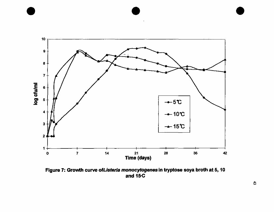
• • •107---------------------------------,
9
8
7
1 6
fiCD 5o-
4
3
2
-10-C
42352821lime (days)
147
1~----_t__----_f_----+__----1___---__1!___---____I
o
Figure 7: Growth curve ofListeria monocytogenes in tryptose soya broth at 5, 10and 150C
44
2.3.2. Antilisterial Activity of a Non-Water Soluble Chitosan
The effects of various concentrations (0-0.2 % w/v) of non-water soluble chitosan on the
growth of L.monocytogenes and L.innocua in tryptose soya broth at pH 7 and incubated
at 300 C are shown in Figures 8 and 9 respectively. After 1-2 days, counts of
L.monocytogenes reached 1010 cfu/ml in the control sample (0% chitosan), 109 cfu/ml
in broths containing 0.05-0.15% chitosan and approximately 107cfu/ml in broths
containing 0.2 % chitosan. However, after 2 days counts began to decrease in ail
treatments. In control broths, counts decreased rapidly to approximately loScfu/ml
after 10 days and then began to gradually increase throughout the storage period
reaching approximately 107 cfu/ml after day 30. Counts decreased more slowly in ail
broths containing chitosan and did not increase again throughout the storage period.
indicating a possible residual antimicrobial effect of chitosan. A 3·4 log reduction in
L.monocytogenes was observed in broths containing 0.2 % chitosan after 5 days, and
throughout the storage period (Figure 8).After 15 days, counts in broths containing
0.05 % chitosan were similar to counts of control broths while in broths containing O. l,
0.15 and 0.2 % chitosan, there was a 1.5 to 2 log reduction in counts relative to the
control.
Similar, but less dramatic trends, were observed for L.innocua, i.e., ail counts
increased to approximately 109 cfu/ml in ail broths and then decreased gradually
throughout storage. This decrease was most pronounced in broths containing 0.15 and
0.2 % chitosan. However, unlike L.monocytogenes, counts began to increase again
throughout the storage period. At the stationary phase counts of 109cfu/ml were
observed for control samples along with samples containing 0.05 and 0.1 % chitosan.
At the 0.15 % chitosan level, a 3 log reduction was observed after 8 days, with numbers
of L.innocua increasing after this point to 108 cfu/ml by the end of storage. At 0.2 %chitosan, a 5 log reduction was observed after 8 days, followed by a slight increase in
numbers and then reaching a 3 log reduction by the end of the storage period. This
study shows that both L.monocytogenes and L.innocua would appear to exhibit a
biphasic growth pattern in the presence of a non-water soluble chitosan, especially at
lower concentrations of chitosan. It can be concluded from this study that levels of
0.2 %or higher of non-water soluble chitosan are required to give moderate control of
the growth of L.monocytogenes and L.innocua in tryptose soya broth at pH 7 for 30
days. Wang (1992) observed that L.monocytogenes was not inhibited by 2.5% chitosan

45
at pH 6.5 but was completely inhibited at pH 5.5 with chitosan concentrations of 0.5 to
2.5 %. The differences in results may be attributed to the nature of the chitosan used
and differences in environmental storage conditions of this study.
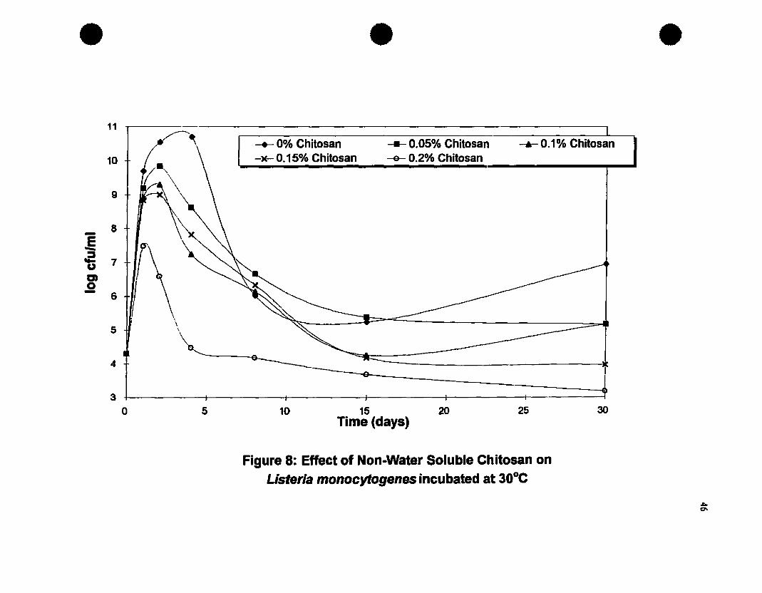
• • •
---9 f---e-- +
-+- 0% Chitosan --- 0.05% Chitosan-M- 0.15% Chitosan -&- 0.2% Chitosan
-...- 0.1% Chitosan
30252015Time (days)
10
11
10
9
- 8E-'fi 7
m0- 6
5\,
4
30 5
Figure 8: Effect of Non-Water Soluble Chitosan onListerla monocytogenes incubated at 3DoC

• • •10 -r-------------------------------------..,
- ..... 0% Chitosan 0.05% Chitosan~ 0.1 % Chitosan -)(- 0.15% Chitosan -0- 0.2% Chitosan
9
B
- \E \- \\.= 7uen0-
6 \5
30252015
time (days)105
4-t------t------t------t-------t------+------;o
Figure 9: Effect of non-water soluble chitosan onListeria innocua incubated at 3DoC
48
2.3.3. Effect of pH on the Antilisterial Activity of Chitosan
The initial study on the effect of various concentrations of chitosan was carried out atpH 7 only. The intent of this study was to determine the effect of a range of pH values(5-7) on the antilisterial activity of a water soluble chitosan. The results of these studiesare shown in Figures 10 and 11. Complete inhibition of L.monocytogenes wasobserved at pH 5 in control broths and at ail chitosan concentrations, indicating that theinhibition was due to a pH effect and not te the presence of chitosan (results notshown). At pH 6 and 7, growth of L.monocytogenes was observed in control broths.
Counts were approximately 1 log cycle less at pH 6 compared to pH 7, indicating aslight pH effect on growth. At pH 6, 0.05 % chitosan and 0.1 % chitosan increased thelag phase of L.monocytogenes. Only at higher concentrations (0.15 and 0.2%) wascomplete inhibition observed at pH 6.
At pH 7 (Figure 10) growth occurred at ail chitosan concentrations tested. Countsincreased from the initial inoculum level by 4 log cycles in broths containing 0.05 %chitosan, by 3 log cycles in broths containing 0.1 % chitosan and by 1.5 log cycles in
broths containing 0.15 and 0.2% chitosan. In broths containing 0.2% chitosan, a 3 logreduction occurred by the third day of storage with numbers decreasing to < 101 cfu/mlby the end of the 30 day storage period. After 30 days of storage, samples at pH 6 hadreached the death phase faster than those at pH 7. This study clearly shows that the
antimicrobial activity of chitosan is pH dependent and that it acts in a similar manner toorganic acids, i.e., sorbic acid, propionic acid. Again, Wang (1992) noticed a similarpH effect on the antiiisteriaI activity of chitosan, with complete inhibition occurring atpH 5.5 in broths containing 0.5 to 2.5% chitosan. However, this authe; did observe
growth in control broths at pH 5.5. For example, while counts of L.monocytogenesreached 107/cfu/ml after 8 days in control broths, these levels were not reached untilday 15 in broths containing 0.05 % chitosan. At slightly higher levels (0.1 % chitosan)
counts reached 107 only after 28 days.

• • •10 .-------------------------------------..,
9-
3025201510
-.- 0% Chitosan - 0.05% Chitosan ---.- 0.1 % Chitosan
-M- 0.15% Chitosan -&- O.2°,{. Chitosan
5o
8
4
2
3
1
7
~ 6
-fiDJ 5Ji!
lime (days)
Figure iD: Effect of chitosan hydrochloride (pH 7) on the growth ofL.monocytogenes incubated at 30°C

• • •9 -y----~------:==========================--___,.
8 -
7
6
-.ë 5
~aJ4o-
3
2
1
-.- 0% Chitosan-JE- 0.15°" Chitosan
-- 0.05°" Chitosan-e- 0.2% Chitosan
-.- 0.1 % Chitosan
302515 20lime (days)
105O-t-------+-----f------+----------;------t-------;
o
Figure 11: Effect of chitosan hydrochloride (pH 6) on the growth ofL.monocytogenes incubated at 30°C
tAo
51
2.2.4. Antillsterial Effect of Chitosan in an Agar System
The combined effect of temperature, pH and chitosan concentration on the growth ofL.monocytogenes was investigated in a model agar system ( Figures 12 and 13). At20C and pH 6, growth of L.monocytogenes was inhibited for approximately 25 days incontrol plates. Addition of chitosan to plates delayed the onset of growth for a further 3days at 20 C. At higher storage temperatures (lO and 150 C), cok;nies ofL.monocytogenes were visible after 7 and 12 days respectively. At pH 6, a level of0.2% chitosan was required ta inhibit the growth of L.monocytogenes at 10 and 150Cfor 23 and 19 days respectively compared to 23 and 7 days at pH 7 (Figures 12 and13). Lower concentrations of chitosan did not inhibit L.monocytogenes in the modelagar system to the same extent. Furthermore, the inhibition of L.monocytogenes isenhanced at lower storage temperatures, iIIustrating the importance of low storagetemperature used in conjunction with this potential antimicrobial agent.
This study clearly indicates that chitosan has an antimicrobial effect in a solid mediumon the growth of L.monocytogenes. This may be due to a specific antimicrobial effect ofchitosan e.g., interference with cell membrane, or to a reduction in llw caused by the
dissolution of chitosan in the agar medium. Therefore, it could be added to a solid foodsubstrate e.g. fermented meat products, ground meats and other minced or groundproducts where L.monocytogenes may pose a public health threat. Furthermore, thelevels of chitosan used i.e., 0.15-0.2 %, are comparable to levels of other preservativesused in foods e.g., sorbates, propionates. However, while chitosan is used as anantimicrobial agent in lapan, its GRAS status ii1 North America is currently underscrutiny .

25
•15
fi-!~ 10
!.EG)
~
2
o 5
•
o Control00.1% chitosanIII 0.2% chitosan
10 15 20Time to observable growth (days)
•
&il 0.05% chitosan.0.15% chilosan
30
Figure 12: Effect of chitosan al pH 6 in a model agar system on the growth ofL.monocytogenes
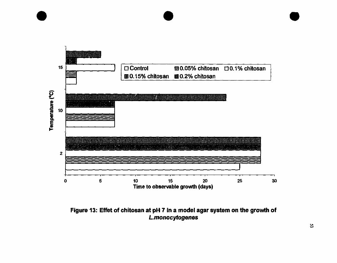
•15
-~
i! 10
lEG)
1-
2
•o Control m0.05% chitosan 00.1 % chitosanfIA 0.15% chitosan Il 0.2°,{. chitosan
•
o 5 10 15 20Time to observable growth (days)
25 30
Figure 13: Effet of chitosan at pH 7 in a model agar system on the growth ofL.monocytogenes

54
CHAPTER3MODIFIED ATMOSPHERE PACKAGING AND COMBINATION
STUDIES
3.1. INTRODUCTION
There is a paucity of data available on the ability of L. monocytogenes to grow undermodified atmospheres, especially in model systems. Furthermore, new melhods of gasatmosphere modification involving oxygen absorbents and ethanol vapor generalors arcavailable on the market. Again, Iiule is known about the effects of such melhods ofatmospherc mooification to control the growth of L.monocytogenes.
Objectives
On the basis of the above comments, the objectives of this study were:
(i) To investigate the potential of various methods of almosphere modification (gaspackaging and oxygen absorbents) to control the growlh of L.monocytogenes in amodel agar system;
(ii)To determine the effect of MAP and chitosan in conjunction with each other on thegrowth of L.monocytogenes in a model agar system.
Based on these studies, an appropriate combination of chitosan and gas atmospherescould be investigated ta extend the shelf Iife and safety of fresh pork.

55
3.2 MATERIALS AND METHOOS
3.2.1. Microorganisms and Preparation of Inoculum
Storage, sub-culturing and preparation of L.monocytogenes inoculum was as describedin section 2.2.1.
3.2.2. Effect of MAP on Usteria monocytogenes
To determine the effect of various gaseous conditions on the growth ofL.monocytogenes, TSA plates were prepared as described previously in section 2.2.3,without the addition of chitosan. The pH of ail plates was monitored. The plates wereinoculated with 2x10⁴cfu/mi of L.monocytogenes as described previously and packagedin 21Ox210 mm Cryovac high barrier bags (Oxygen transmission rate: 3-6 cm3/m2/day
at 4.4°C, O%RH). Bags were f1ushed with levels of CO2 ranging from 0 to 100% at20% increments (balance NI> using a Multivac chamber type, heat seal packaging
machine (Model KMloo-3M). A Smith's proportional gas mixer (Model 299-028,Tescom corp., Minneapolis, Minnesota) was used to give the desired proportions ofgases in the package head space. in half of the packages, an oxygen absorbent sachet,Ageless type FX (Mitsubishi Gas Chemical Company LId., Japan) was incorporatedprior to gas f1ushing to remove ail traces of residual oxygen in the package head space.The gaseous combinations used in this study are shown in Table 5.
Table 5: Gas Mixtures used for 3.2.2.Sample code(No Ageless) %N2 %C02
Sample code(With Ageless FX)
ABCoEF
10080604020o
o20406080100
AFBFCFOFEFFF

S6
3.2.2.1. Headspace Gas Analysis
Samples were analyzed for changes in headspace gas at regular intervals throughoutstorage or until growth was observed. Gas samples were withdrawn using a 0.5 ml gas
tight Pressure-lok syringe (Precision Sampling Corp., Baton Rouge, La.) through
silicone seals attached to the package exterior. The gas samples were injected ioto a
Varian gas chromatograph (Model 3400, Varian Canada Inc.) equipped with a thermalconductivity detector (TCD) and using Porapak Q (SO-Ioo mesh) and Molecular Sievc
SA (80-100 mesh) columns in series (Supelco Canada Ltd). The carrier gas was heliumset at a f10w rate of 30 ml/min. The column oyen was set at SOOC, the injector al
IOOoC and the detector filament at 150oC. Peaks were recorded using a HcwleuPackard integrator (Model 3390A, Hewlell Packard Co., Avondale, PA.).
3.2.3. Effeet of Various Oxygen Absorbent!, on the Growth of L.monocytogenes
TSA plates (without chitosan) were inoculated with L.Tlt?nocytogenes as described
previously in 2.2.3. Plates were packaged in air in Cryovac bags each containing oneof three types of oxygen absorbents: Ageless FX, Ageless SS and Ageless SE. Ali
absorbents were obtained from the Mitsubishi Gas Chemical Company Ltd., Tokyo,Japan. The characteristics of each of these absorbents is summarized in Table 6. Ali
packages were sealed with an impulse heat sealer and stored at 2oC, and at ID and150C, Le.• conditions of mild temperature abuse. Plates were monitored daily for
visible signs of growth.
Table 6; Absorbents used in 3.2.3.
Absorbent type
Ageless FX
Ageless SS
Ageless SE
Description
Moisture dependent oxygen absorbent
Self reacting oxygen absorbent, low temperature applications
Moisture dependent oxygen absorbent and ethanol vapor generator
57
3.2.4. Combined Effects of Chitosan and MAP on L.monocytogenes in an AgarSystem
ln this study, the combined effect of chitosan and MAP on the growth ofL.monocytogenes was investigated in a model agar system. Modified Oxford agar(MOX) was obtained from Difco (Michigan, USA). The medium was preparedaccording to the manufacturer's directions, autoclaved, cooled to SOOC and poured intopetri dishes. Half of the medium contained chitosan hydrochloride at a level of 0.2%added prior to autoclaving. The pH of the media was adjusted to 6.0 and 7.0 using lMNaOH or food grade lactic acid. MOX was used in this study instead of TSA to
distinguish growth since L.monocytogenes appears as black colonies on this mediumdue to the fermentation of esculin present. Ali MOX plates were inoculated with 2x10⁴
cfu/ml of L.monocytogenes by the spread plate technique and placed in Cryovac bags.Plates were packaged under various gas atmospheres as i1iustrated in Figure 14.Packaging was done as described in section 3.2.3.. Ali packages were stored at S, 10and lSoC and monitored for visible growth daily. Headspace gas analysis was done asdescribed in section 3.2.3.1. when visible growth was observed.

•
•
•
58
Figure 14: Treatment scbeme used for 3.2.4.
I-------f------+-.... AIR
]------+-----1--. AIR +AGELESS SS ....--+----f-----f
1-------1-----+-.... 100% N2
I------~---_t_ ...... 100% N1 +AGELESS SS
I-------f------+-.... 100% COZ +-------+----+-----i
1------+-----1--. 1000/0 COZ +AGELESS SS+J----+----.-,
!-------1-----+-.... 80% N2 + %0% CO.....-~I---+-----{
~ __z. ~.... 60% N2 +40% COI 4--..1.---...z...----
59
3.3. RESULTS AND DISCUSSION
3.3.1. Effect of MAP on L.monocytogenes
Initial studies were done to determine the effect of gas packaging, with and without anoxygen absorbent (Ageless FX), on the growth of L.monocytogenes on agar plates
stored at mild temperature abuse conditions (Tables 7 and 8). The percentage of O2
,CÛ2 and N2 in the package headspace shown was the headspace gas composition of thepackages when visible growth of L.monocytogenes was detected.
For plates packaged without an absorbent, colonies of L.monocytogenes were visible in
control plates (air) after 7 days at iOoC. Law levels of CO2 (approximately 20%)appeared to stimulate the growth of L. monocytogenes, with growth appearing within 5
days in ail plates stored at iOoC. Several studies have reported the stimulatory effect oflow levels of CO2 on the growth of molds in cereals and lactic acid bacteria in vacuum
and gas packaged beef (Lambert et al.. 1991). More recently, Lambert at al. (1992)
reported the stimulatory effect of low levels of CO2 (20 %) on the growth and toxinproduction by Clostridium botulinum in gas packaged pork.
When plates were packaged in higher CO2 levels (approximately 40%) growth
appeared at about the same time as control plates, while growth was delayed until day 9
for plates packaged in atmospheres of approximately >60% CO2• For gas packagedplates without an absorbent, CO2 levels of 70% or higher were required to inhibit the
growth of L.monocytogenes at iOoC until 14 days. However, no additional control
was achieved with CO2 levels above 70%. Razavilar and Genigeorgis (1992)determined that 100% CO2 was the most inhibitory modified atmosphere ta delay thegrowth of L.monocytogenes. However, these authors did not compare other levels of
CO2•
Similar trends were observed for ail gas packaged plates without an absorbent stored at
150 C. However, growth was detected within 3 to 5 days in ail plates stored at 150 C,
indicating once again the importance of low temperature to enhance the antimicrobialeffect of CO2 enriched atmospheres.
60
The combined effect of packaging in a COz enriched atmosphere with an oxygenabsorbent (Ageless FX) on the growth of L.monocytogenes at 10 and 150 C is shown inTables 9 and 10 respectively. The addition of an oxygen absorbent delayed the growthof L.monocytogenes in control plates and in plates packaged with various levels of COz.This can be attributed to lower levels of Oz in ail packaged plates indicaling anantimicrobial synergism between low Oz and high COz levels. However, similar trendswere observed in this study with an absorbent, Le., a stimulatory effect of lower COz(20 to 40%) resulting in visible growth approximately 2 days prior to control plates andan inhibitory effect of higher COz levels (60 to 100%) on the growth ofL.monocytogenes. However, an interesting effect was observed for plates packaged in100% Nz with an oxygen absorbent and stored at 10 and 150C. In these plates, growthwas delayed for approximately the same lime or longer than in plates packaged in 100%COz. This can be attributed to the low level of residual Oz (i.e., < 1%).
This initial study has shown that low levels of COz (20 to 40%) appear to stimulate thegrowth of L.monocytogenes. For moderate control of L.monocytogenes, products need
to be packaged in approximately 60-80% COz. For optimum control ofL.monocytogenes, products need to be stored in either high COz levels (70-100%) orvery high Nz levels (100%) in conjunction with an oxygen absorbent. To date, therehave been no other studies carried out on the effects of oxygen absorbents on the growthof L.monocytogenes
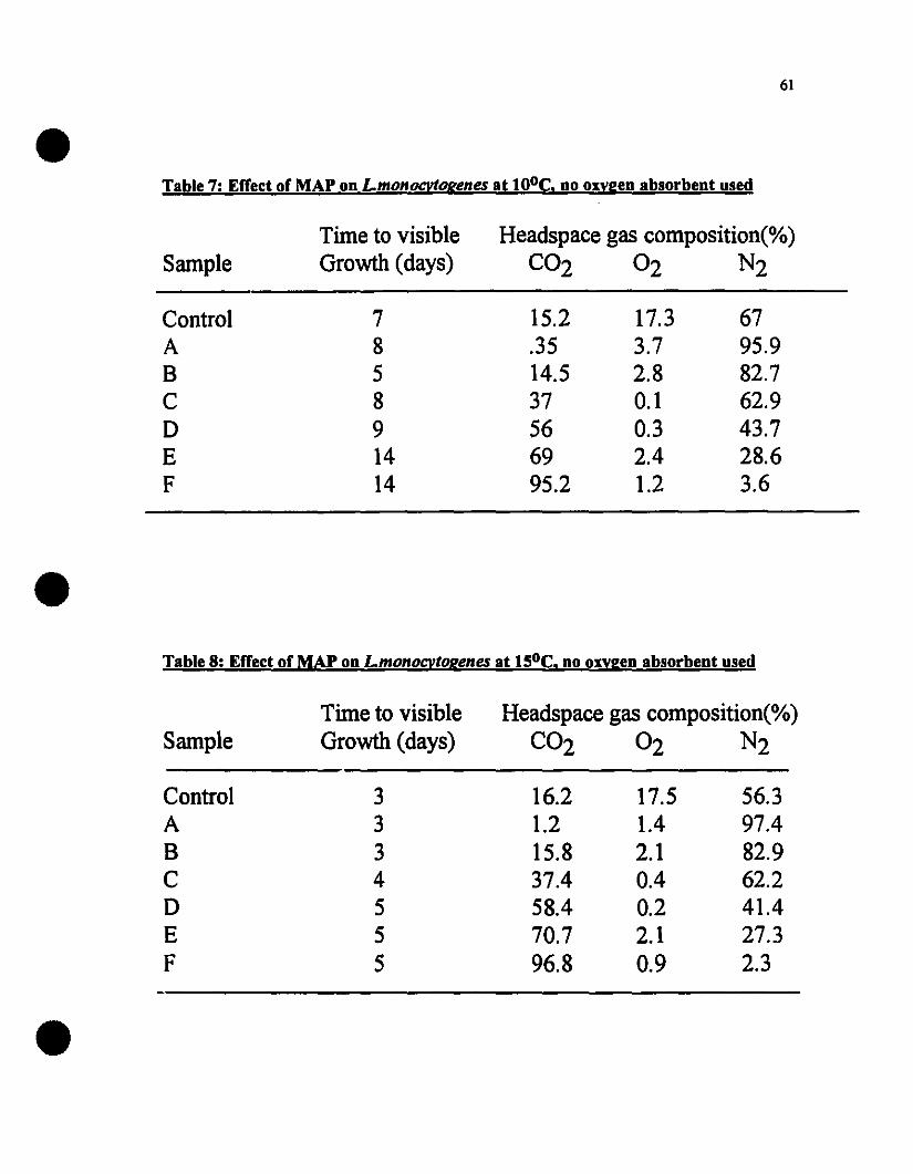
61
Table 7: Errect of MAP on Lmonocytogenes at tOoe. no ollygen absorbent used
Samplerime to visibleGrowth (days)
Headspace gas composition(%)C02 02 N2
ControlABCDEF
785891414
15.2.3514.537566995.2
17.33.72.80.10.32.41.2
6795.982.762.943.728.63.6
Table 8: Errect of MAP on Lmonocvtogenes at t50e. no oxvgen absorbent used
Samplerime to visibleGrowth (days)
Headspace gas composition(%)C02 02 N2
ControlABCDEF
3334555
16.21.215.837.458.470.796.8
17.51.42.10.40.22.10.9
56.397.482.962.241.427.32.3

62
Table 9: Effect of MAr on L.monocvtogenes at 100e using an Ageless FX oxygenabsorbent
SampleTime to visibleGrowth (days)
Headspace gas composition(%)C02 02 N2
Air+FXAFBFCFDFEFFF
12251010152025
25.51.28.433.342.562.594.2
0.30.30.40.30.20.10.1
74.298.491.266.457.337.45.7
Table 10: Effect of MAr on L.monocvtogenes at 150e using an Ageless FX oxygenabsorbent
SampleTime to visibleGrowth (days)
Headspace gas composition(%)C02 02 N2
Air+FXAFBFCFDFEFFF
101856101214
30.21.49.83552.675.292.6
0.40.30.40.30.10.20.1
69.498.389.864:747.324.67.3

63
3.3.2. Errect of Various Oxygen Absorbents on the Growth of L.monocytogenes
The combined use of oxygen absorbents and CO2 enriched atmospheres can be usedeffectively to control the growth of L.nwnocytogenes. However, this combinationmeans additional costs to the processor. Therefore, preliminary studies were done todetermine the effect of various oxygen absorbents alone to control the growth of thisimportant pathogen. The effects of three oxygen absorbents, Ageless types FX, SS andSE and temperature on the growth of L.monocytogenes on TSA plates are shown inTable Il. In control plates, growth occurred within 25, 7 and 2 days at 2, 10 and 150Crespectively. Addition of an oxygen absorbent, Ageless type SS delayed growth ofL.monocytogenes for greater than 52 days at 20 C. Ageless types SE and FX were notstudied at this temperature (2°C) since they are not designed to scavenge oxygen atrefrigeration temperatures. However, the effects of oxygen absorbents on the growth ofL.nwnocytogenes at higher temperatures (10 and 150C) is evident. Growth was inhibitedfor approximately 23 to 30 days with all absorbents stored at 100 C. At higher storagetemperatures, growth of L.nwnocytogenes was delayed for 12 days using Ageless FXand for 20 and 30 days using Ageless SS and SE respectively. The greater effect of
Ageless type SE at higher storage temperatures can be attributed to the fact that this dualfunctional absorber not only absorbs oxygen but also generates ethanol vapor. Whilethe level of ethanol vapor generated was not measured in this study, approximately 1%ethanol vapor is generated by Ageless SE (Mitsubishi Gas Chemical Corporation,
Personal Communication). This study has shown that the use of absorbents alone cangive similar or greater control over the growth of L.nwnocytogenes compared tocombination of elevated levels of CO2 and oxygen absorbents. The use of oxygenabsorbents alone is therefore a simple, cost effective method of controlling the growth
of L.monocytogenes even at mild temperature abuse conditions.

64
Table Il: Effect of Various Oxygen Absorbent! on L.monocvtogenes
AbsorbentType
Temperature (oC)_2 10 15
Air-Control
Ageless FX
Ageless SS
Ageless SE
25*
NIA
>52
NIA
7
30
23
30
2
12
20
30
* Values represent time to visible growth in days and are the means of threereplicates
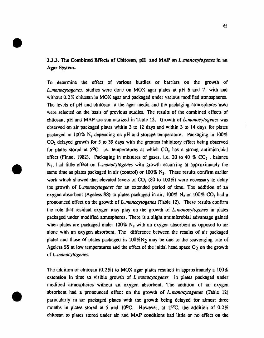
65
3.3.3. The Combined Effects of Chitosan, pH and MAP on L.monocytogenes in an
Agar System.
To determine the effeet of various hurdles or barriers on the growth of
L.monocytogenes, studies were done on MOX agar plates at pH 6 and 7, with and
without 0.2 % ehitosan in MOX agar and paekaged under various modified atmospheres.
The levels of pH and ehitosan in the agar media and the paekaging atmospheres 'used
were seleeted on the basis of previous studies. The results of the eombined effeets of
ehitosan, pH and MAP are summarized in Table 12. Growth of L.monocytogenes was
observed on air paekaged plates within 3 to 12 days and within 3 to 14 days for plates
paekaged in 100% N2 depending on pH and storage temperature. Paekaging in 100%
COz delayed growL't for 5 to 39 days with the greatest inhibitory effeet being observed
for plates stored at 50 C. i.e. temperatures at whieh CO2 has a strong antimierobial
effeet (Finne, 1982). Paekaging in mixtures of gases, i.e. 20 to 40 % COz • balance
Nz, had \iule effeet on L.monocytogenes with growth oceurring at approximately the
same time as plates paekaged in air (control) or 100% N2• These results eonfirm ear\ier
work whieh showed that elevated levels of CO2 (80 to 100%) were neeessary to delay
the growth of L. monocytogenes for an extended period of time. The addition of an
oxygen absorbent (Ageless 55) to plates paekaged in air, 100% N2 or 100% COz had a
pronouneed effeet on the growth of L.monocytogenes (Table 12). The~e ~esults eonfirm
the raIe that residual oxygen may playon the growth of L.monocytogenes in plates
paekaged under modified atmospheres. There is a slight antimierobial advantage gained
when plates are paekaged under 100% N2 with an oxygen absorbent as opposed to air
alone with an oxygen absorbent. The differenee between the results of air paekaged
plates and those of plates paekaged in loo%N2 may be due to the scavenging rate of
Ageless 55 at low temperatures and the effeet of the initial head spaee 02 on the growth
of L.monocytogenes.
The addition of ehitosan (0.2%) to MOX agar plates resulted in approximately a 100%
extension in time to visible growth of L.monocytogenes in plates paekaged under
modified atmospheres without an oxygen absorbent. The addition of an oxygen
absorbent had a pronouneed effeet on the growth of L.monocytogenes (Table 12)
partieularly in air paekaged plates with the growth being delayed for almost three
months in plates stored at 5 and l00C. However, at l~oC. the addition of 0.2%
ehitosan 10 plates stored under air and MAP conditions had \ittle or no effeet on the

66
time to visible growth. These results confirm previous studies which showed a
synergistic effect between pH, chitosan, gaseous atmosphere and storage temperature onthe growth of L.monocytogenes. Furthermore, the results confirm that an oxygenabsorbent could be added to gas packaged products to reduce the residual O2 level in the
pac:kage headspace thereby enhancing the antimicrobial effect of modified atmosphere
packaging.
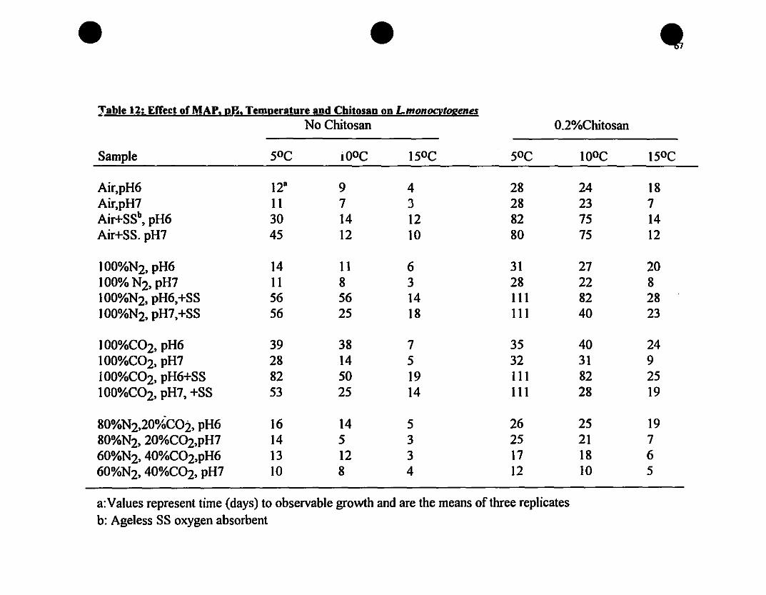
Jable 12: Effect of MAP. pH. Temperature and Chitosan on LmonocvtogenesNo Chitosan 0.2%Chitosan
Sample 5°C iOoC 15°C 5°C JOoC 15°C
Air,pH6 128 9 4 28 24 18Air,pH7 11 7 J 28 23 7Air+SSb
, pH6 30 14 12 82 75 14Air+SS. pH7 45 12 10 80 75 12
100%N2, pH6 14 11 6 31 27 20JOO%N2, pH7 11 8 3 28 22 8100%N2, pH6,+SS 56 56 14 111 82 28JOO%N2, pH7,+SS 56 25 18 111 40 23
100%C02, pH6 39 38 7 35 40 24JOO%C02, pH7 28 14 5 32 31 9100%C02, pH6+SS 82 50 19 III 82 25100%C02, pH7, +SS 53 25 14 111 28 19
80%N2,20%COi, pH6 16 14 5 26 25 1980%N2, 20%C02,pH7 14 5 3 25 21 760%N2, 40%C02,pH6 13 12 3 17 18 660%N2, 40%C02, pH7 10 8 4 12 JO 5
a:Values represent time (days) to observable growth and are the means ofthree replicatesb: Ageless SS oxygen absorbent

68
CHAPTER4PORK CHALLENGE STUDIES
4.1. INTRODUCTION
MAP is a rapidly expanding technology being used for the preservation of foodsincluding bakery products, meat, fish, poultry and snack food products (Smith et al,1990). However, there is concern about the public health safety of these foods,particularly muscle foods, for the following reasons: (i) the normal aerobic spoilagemicroorganisms which would normally warn consumers of spoilage (by off odors andoff colors) are inhibited, (ii) the growth of pathogens may be allowed or evenstimulated, (iii) the emergence of psychrotrophic pathogens such as L.monocytogenesand (iv) the potential for temperature abuse (Farber, 1991). It is evident thatrefrigeration alone or in conjunction with MAP is Inadequate and that additionalbarriers, such as chitosan, must be considered to prolong the shelf Iife and ensure thesafety of MAP products, i.e., the hurdle concept. While minimal studies to date have
been done on model agarlbroth systems, challenge studies with L.monocytogenes inpork are essential ta evaluate the public health safety of MAP pork subjected to mildtemperature abuse storage conditions.
Objectives;
Based on previous results, the objectives of this study were:
(i) To determine the physical, chemical, microbiological and sensorial changes
occurring in the packaged pork product.
(ii) To determine the effect of chitosan, MAP and storage temperature on the growth
of L.monocytogenes in fresh pork loin;

69
4.2. MATERIALS AND METRODS
4.2.1. Mlcroorganisms and Preparation of Inoculum:
Storage, sub-culturing and preparation uf !...monocytogenes inoculum was as described
in section 2.2.1
4.2.2. Preparation of Chitosan Dipping Solution
A 0.2 % (w/v) dipping solution was prepared in 1 litre batches by adding 20g of
chitosan hydrochloride powder 10 980 ml of distilled deionised water. The dipping
solution was stirred for one hour and the pH adjusted to 6.0 with 1.0M NaOH. The
dipping solution was then autoclaved at lS psi for lS minutes (Le.• 1210 C) and then
cooled. The pH was checked as described previously in section 2.2.3., and was
aseptically readjusted to 6.0 if necessary with lM NaOH or food grade lactic acid. The
dipping solution was changed after dipping 10 pork samples. A control dipping solution
of autoclaved distilled deionized water was used for control samples and this was also
changed after every 10 samples.
4.2.3. Preparation of Pork
Fresh, trimmed, pork loins were obtained from "Les Viandes de l'Ue Perrot Inc.·, (Ile
Perrot, Quebec). The meat had been de-boned and pre-eut into samples of 100 g ± lOg
loins. The samples were transported in a cooler to our laboratory al Macdonald
Campus. Half of the pork samples were dipped in a 0.2 % chitosan dipping solution
(pH 6.0) for sixty seconds prior to inoculation, the remainder of the loins were dipped
in a beaker containing distilled deionized water for 60 seconds. After dipping, the pork
loin slices (one per bag) were placed in 210x210mm Cryovac barrier bags (OTR:3-6
cc/m2/day at 4.40C and O%RH). Ail samples were weighed in their bags prior to
packaging and storage. The pork loins were then inoculated with 1.0 ml of .') working
suspension of L.monocytogenes (2x10⁴ cfu/ml) at four different places on the meat
sample to distribule the inoculum. The pork samples were then gently massaged in the
bags to further distribute the cells. Sample preparation. dipping and inoculation were
•
•
•
70
carried out under a Purifier™ Class Il Safety Cabinet (Labconco, Model #36205-04.Labconco, Kansas, Missouri) equipped with a HEPA fil ter to ensure minimalcontamination of samples and safety to the technicians. The samples were rand\)mlyassigned to one of the treatments as shown in Figure 15. Uninoculated control sampleswere also prepared in a similar manner. Ali treatments were carried ou~ in triplicate.
Figure 15: Scheme of treatmenu used in pork chanenge studies
t------+------t-.... AIR
I-------f.----+-.... A1R+AGELESS SS +--+---+---i
1000/0 N2 + AGELESS r~I+-_--+__-{
80°./0 C02 +20% N2 +--- -3
4.2.4. Packaging
Ali inoculated and un;noculated pork sampies were packaged in Cryovac barrier bagsusing a Multivac chamber type. heat seal packaging machine (Model KM1OO-3M). ASmith's proportional gas mixer (Model 299-028, Tescom Corp., Minneapolis,Minnesota) was used to give the desired proportions of Û2, CÛ2 and N2 in the packageheadspace. Residual OA~ gen levels were kept to a minimum in certain tteatments usingan oxygen absorbent sachet (Ageless type FX or S5) obtained from the Mitsubishi Gas
Chemical Company Ltd.• Tokyo. Japan.
71
4.2.5. Headspace Gas Analysis
Samples were analyzed for changes in headspace gas at regular intervals throughout the28 day storage period (i.e., days 0,3,7,14,21 and 28 at 5 and lQoC and up to 14 days at150 C). Gas samples were withdrawn using a 0.5 ml gas tight Pressure-Iok syringe(Precision Sampling Corp., Baton Rouge, La.) Through silicone seals attached to thepackage exterior. The gas samples were injected into a Varian gas chromatograph(Model 3400, Varian Canada 'nc.) equipped with a thermal conductivity detector (TCO)and using Porapak Q (80-100 mesh) and Molecular Sieve SA (80-100 mesh) columns inseries (Supelco Canada Ltd). The carrier gas was helium set at a tlow rate of 30ml/min. The column oyen was set at 800 C, the injector at loooC and the detectorfilament at 1500 C. Peaks were recorded using a Hewlett Packard integrator (Model3390A, Hewlett Packard Co., Avonda1e, PA.).
4.2.6. Sensory Ana1ysis
At day 0 and at each subsequent sampling time (days 0,3,7,14,21 and 28 at 5 and lQoCand up to 14 days at 150C), pork samples were evaluated su~iective1y for color andodor. Ali samples were evaluated in a random sequence. Fres~ samples were used ascontrols at each sampling time.
4.1.6.1. Co1or Analysis
Samples were evaluated by the 1aboratory technicians in a random sequence for surfacediscoloration using a five point scale (l = No surface discoloration, 5= completesurface discoloration). A color chart from Agriculture Canada (publication 5180/8)served as a reference for the normal cClllor of pork. A score of 3 or more was considered10 be the upper Iimit of acceptability, implying that shelf life was terminated when thisscore was reached.
Color was also analyzed objectively for L* (lightness), a*(hue) and b* (chroma) colorvalues using a Mino1ta Chroma Meter colorimeter (Model CR-loo, Mino1ta Corp.Ramsey, New Jersey) with aluminant C (6774K) light source. The colol'imeter wascalibrated using a s:andard white retlector plate prior to measuring meat calor.
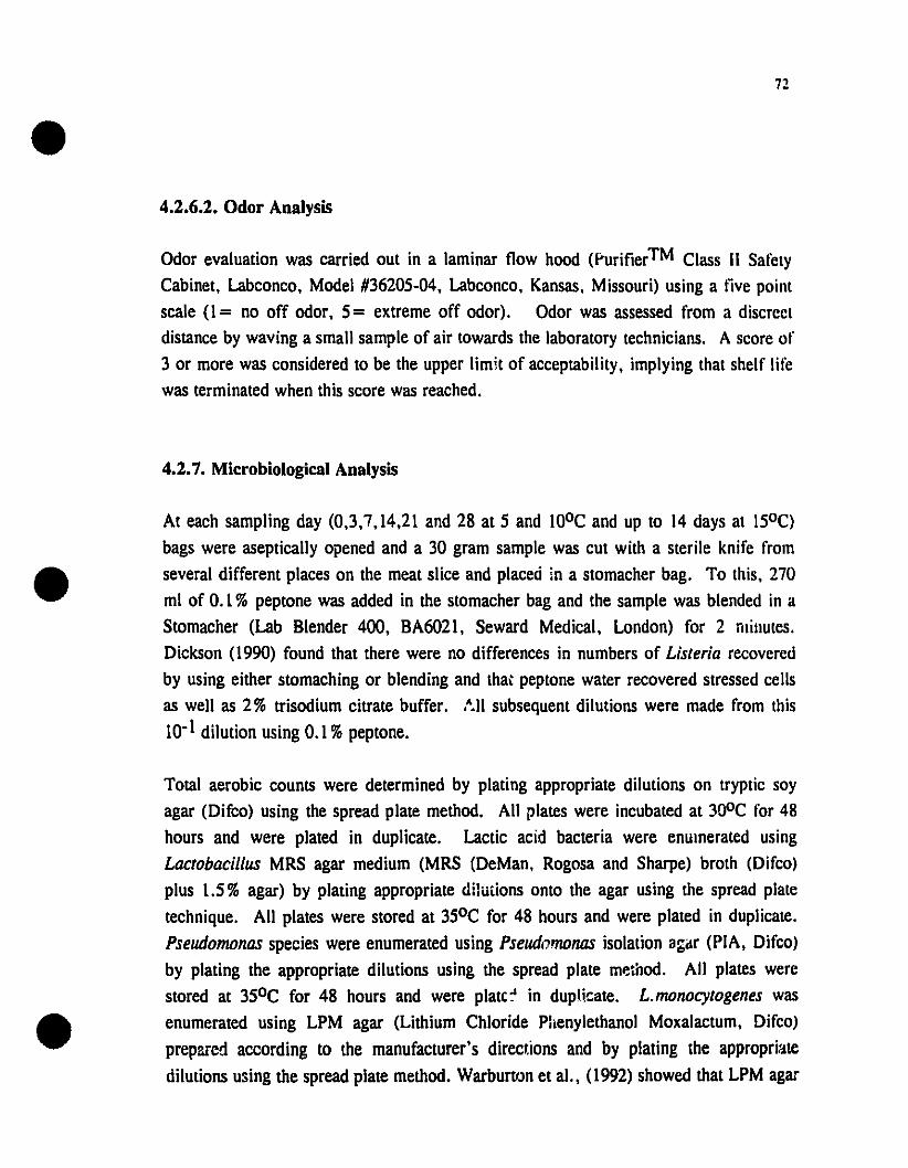
72
4.2.6.2. Odor Analysis
Odor evaluation was carried out in a laminar f10w hood (Purifier™ Class Il SafetyCabinet, Labconco, Model #36205-04, Labconco, Kansas, Missouri) using a live pointscale (1 = no off odor, 5= extreme off odor). Odor was assessed from a discreeldistance by waving a small sample of air towards the laboratory technicians. A score of3 or more was considered to be the upper Iimit of acceptability, implying that shelf Iifewas terminated when this score was reached.
4.2.7. Microbiological Analysis
At each sampling day (0,3,7,14,21 and 28 at 5 and 100C and up to 14 days at l50C)bags were aseptically opened and a 30 gram sample was cut with a sterile knife fromseveral different places on the meat slice and placeô in a stomacher bag. To this, 270ml of 0.1 % peptone was added in the stomacher bag and the sample was blended in aStomacher (Lab Blender 400, BA6021, Seward Medical, London) for 2 minules.Dickson (1990) found that there were no differences in numbers of Listeria recoveredby using either stomaching or blending and lha~ peptone water recovered stressed cellsas weil as 2% trisodium citrate buffer. ,A.l1 subsequent dilutions were made from lhis10-1 dilution using 0.1 %peptone.
Total aerobic counts were determined by plating appropriate dilutions on lryptic soyagar (Difco) using the spread plate method. Ali plates were incubated at 300C for 48hours and were plated in duplicate. Lactic acid bacteria were enumerated usingLactobacillus MRS agar medium (MRS (DeMan, Rogosa and Sharpe) brolh (Difco)plus 1.5% agar) by plating appropriate dilutions onto the agar using the spread plaietechnique. Ali plates were stored at 350C for 48 hours and were plated in duplicate.Pseudomonas species were enumerated using Pseudomonas isolation agilr (PIA, Difco)by plating the appropriate dilutions using the spread plate method. Ali plates werestored at 350C for 48 hours and were plate:' in dupHcate. L.monocytogenes wasenumerated using LPM agar (Lithium Chloride Phenylethanol Moxalactum, Difco)prepllfed according to the manufacturer's directions and by plating the approprhtedilutions using the spread plate method. WarburtrJn et al., (1992) showed that LPM agar

•73
was comparable ta Oxford agar and PALCAM agar in its ability to recover andenumerate stressed L.monocytogenes cells. Ali plates were stored at 300 C and wereplated in duplicate and enumerated after 48 hours. In't Veld and Boer (1991) found thatincubating for 48 hours on LPM agar gave more reproducible results as compared toincubating for only 24 hours. Confirmation tests were carried out on randomly selectedcolonies, i.e, Henry's illumination, hemolysis reaction on defibril'.ated horse blood agar,API Listeria strips, motility test and Gram stain reaction. Johnson and Lattuada, (1993)reported that the API Listeria system could readily differentiate between theL.monocytogenes and L.innocua species without the need for additional tests. Theseauthors also reported that API Listeria strips were convenient, easy to use andreasonably priced and that reactions were not ambiguous and results were availablewithin 24 hours. These results were in agreement with Dever at al. (1993). Hitehens etal. (1992) reported that there is no official method for the enumeration ofL.monocytogenes. However, direct plate counts on LPM or MOX agar were acceptablemethod for enumeration.
The exudate loss from randomly selected samples was also analyzed for the presence ofListeria monocytogenes using LPM agar by using a spread plating technique induplicate for all tested samples
4.2.8. Drip Loss
At each sampling time (days 0,3,7,14,21 and 28 at 5 and 100 C and up to 14 days atIS°C), individual bags were weighed upon removal of the pork slice. Individualsamples had been pre-weighed prior to storage and thus a percent w/w value wasobtained. Le., the weight of the drip loss divided by the original sample weight.
4.2.9. pH Measurement
The pH of the pork samples was measured with a previously calibrated Corning pHmeter (Model 2220. Corning Glass Works, Corning. NY) by placing the electrode intothe 10-1 dilution of the pork samples after stomaching for 2 minutes, once allmicrobiological analysis was complete. Analysis was carried out at each sampling time(days 0,3,7,14,21 and 28 at 5 and l00C and up to 14 days al 150C).

74
4.3. RESULTS AND DISCUSSION
4.3.1. Introduction
ln previous studies, the combined effeet of chitosan and modified atmospheres on thegrowth of L.monocytogenes was investigated in model broth and model agar systems.Results of these initial studies clearly indicated that control of this pathogen waspossible using a combination of 0.2% chitosan and MAP. However, liule is knownabout their combined effeet in a food system. Therefore, this present study examinedthe combined effect of a 0.2%chitosan dipping solution and MAP to control the growthof the indigenous spoilage bacteria and L.monocytogenes in fresh pork.
4.3.2. Physiochemical An;.lysis
4.3.2.1. Changes in Headspace Gas Composition
The changes in headspace gas composition for non-chitosan dipped pork stored at S"Care shown in Figure 16. For air packaged samples, head space O2 decreased toapproximately 11% after 28 days with a concomitant increase in CO2 to approximately28% (Figure 16, A). This can be attributed to the action of meat respiratory enzymesand aerobic microorganisms which utilize O2 and produce CO2 (Gill, 1980).
For samples packaged with an Ageless oxygen absorbent (Figure 16B), headspaccoxygen decreased to approximately 1%after 3 days of storage and remained unchangedthroughout the storage period while head space CO2 increased to approximately 20%.
The increase in CO2 in the non-chitosan dipped can again be auributed to meat tissuerespiration, to the growth of aerobic and facultative microorganisms and possibleheterofermentative strains of lactic acid hacteria.
For gas packaged samples (80%C02/20%N2), headspace oxygen increased slightlyfrom l ' , 25 % throughout storage indicating slight leakage. Headspace CO2 decreasedslightly to approximately 60% due to possible dissolution in the meat tissue and thengradually increased throughout storage to approximately 80%. This increase can again
be attributed to the growth of aerobic and facultative spoilage microorganisms in pork.Several studies have shown that Pseudomonas species can grow in low levels of O2•

75
i.e., less than 1%even in the presence of elevated levels of CO2 (De Vore and Solberg,
1974; Nottigham 1982).
For samples packaged in 100%N2 with an Ageless FX oxygen absorbent, fluctuations inhead space gas composition were similar to those for pork packaged with an Ageless SSabsorbent, i.e., O2 levels remained at very low levels throughout the 28 day storageperiod. However, after day 3, head space O2 increased to approximately 8% indicatingleakage and then decreased to less than 1% throughout storage. A similar increase inCO2 production to approximately 15 % was observed for this set of packagingconditions, i.e., similar te Ageless SS packaged samples.
Similar but more dramatic trends were observed for ail non-chitosan dipped samplesstored at 10 and 15°C (Figures 17 and 18 respectively). In ail instances CO2 productionwas greater at higher storage temperatures again emphasizing the importance of lowtemperature to control the growth of spoilage microorganisms in both air and MAPpackaged pork.. Similar trends were observed by Enfors et al. (1979) and Blickstad etal. (1981) in pork samples packaged initially with air and stored at 2,4, and 14°C.
The changes in headspace gas composition for chitosan dipped pork and packaged undervarious gaseous conditions and stored at 5, 10 and 15"C are shown in Figures 19, 20and 21 respectively. In ail cases, similar trends in headspace gas changes wereobserved for both chitosan dipped and non-chitosan dipped pork. This would infer thatchitosan l!ad no noticeable effect on the normal spoilage microorganisms of pork underthe storage conditions investigated.

e e e100 100
A A A B80 -t. 80 ., V- A
~A... 60 - ... 60 -
~ ~li! 40 - li! 40 -
20 20 -{~1>~
V "V"
0..( 0~
1 1 1 1 1 1
0 7 14 21 28 0 7 14 21 28Time (days)
Time (days)-0- CO2
-0- 02
---b- N2
C~
~~
~
-
-A •
,.
1 1 1
14 21Time (days)
100
80
~60
li! 40
20
oo 7 14
Time (days)21 28
100~
80 -
CI> 60 -lUClli! 40 -
20 - (").
~0 - T
0 7T T
D
28
Figure 16: Changes in head space gas composition of non-chitosan dipped pork at 5°C and stored under(A)Air; (B)Air+Ageless 55; (C) 80%CO/20%N2; and (D) 100%N2+Ageless FX.
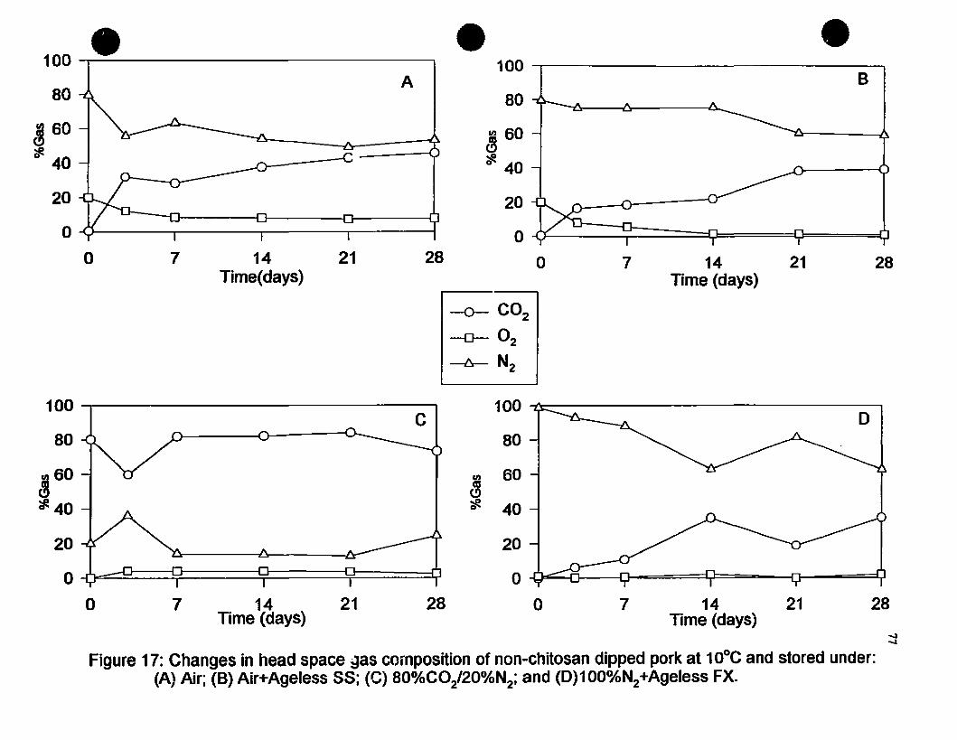
28
B
14 21Time (days)
7o
•
20
O~~=:!S!=====::::=Çl:====O=====:O
100 -,------------------,
80
ln 60~~ 40
•
28
A
2114Time(days)
7o
•100 --r---------------------,
80
~ 60
cIe 40
20
o ~---ooy_____--_____r---___r_---__1
-0- CO2
-0- 02-6- N2
D
20
o ...[Jos:;;;;...",r1-----1o--====t...~=-__fl_--===:i.,.J
100 ~-------------------,
aDln 60~~ 40
C100 -,-----------------,
80
~60
~40
20
O~:::::Q==~====~!=====:::j~======O
o 7 14Time (days)
21 28 o 7 14 21Time (days)
28
Figure 17: Changes in head space ~as composition of non-chitosan dipped pork al 1DoC and stored under:(A) Air; (B) Air+Ageless 58; (C) aO%CO/20%N2; and (D)100%N2+Ageless FX.

• • •100 100A B
80 80
~ 60 en 60~
"#. 40 * 40
20 20
0 0
0 7 14 0 . 7 14Time (days) Tlme (days)
-0- CO2
-0- 02
-6- N2
100 --f\c----------------,D
80
en 60~èt! 40
C100 1
80 ~--f"\----~......:::>--------y'\-0 ~
~60
"#. 40 -
7 14 0 1 14Time (days) Time {days) ....
00
Figure 18: Changes in head space gas composition on non-chitosan dipped pork at 15°C and stored under:(A) Air; (B) Air+Ageless SS; (C)SO%C0t'20%N2; and (D) 1OOOA.N2+Ageless FX.
.A20 -4r----~__.........,6-A-- _l
o ~-CTJ--===:::=:::::-lï-!:d:=======:::!~1==========~===::::1;l1
o
20
O~====:::::i...J:,===~J======----_o

A
'1'""-
.=
-
~~;J
1 1 1
C-~
~~
-
-A
.A
r T T
100
80
en ao~~ 40
20
o
100
80
~ao
~40
20
o
o
o
7
7
14Time (days)
14Time (days)
21
21
28
28
e e100 -1
A A A B80 .,~
en aD -IIICl~ 40-
20 ~r"-..'"'- ~
0../Ç ~
1 T ,.0 7 14 21 28
Time (days)-0- COz
-D- Oz---b- N2
100~ A . D
80 -Lr
en ao -~~ 40 -
20 -~
0~ .,. .,. .,.
0 7 14 21 28Time (days)
.....""
Figure 19:Changes in head space gas composition of chitosan dipped pork at 5°C and stored under(A}Air; (B) Air+Ageless 55; (C) aO%C0:l20%Nz ;and (D) 1OO%Nz+Ageless FX.
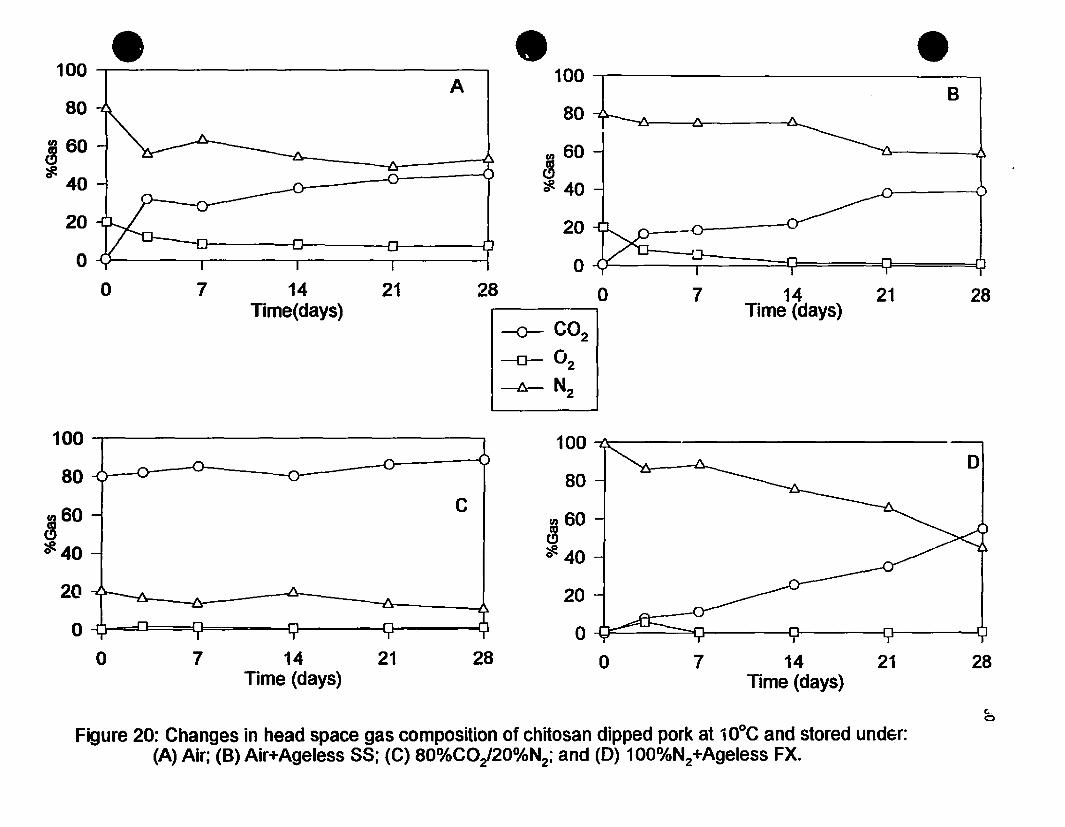
• • •100 100A B
80 80
~ 60 CI) 60
'#- 40 ~fi. 40
20 20
0 0a 7 14 21 28 0 7 14 21 28
Time(days) Time (days)-0- CO2
-a- 02
-Â- N2
100 100D
80 80
~60C
CI) 60l3
fi. 40 ~40
20 20
0 00 7 14 21 28 0 7 14 21 28
Time (days) Time (days)
c..0
Figure 20: Changes in head space gas composition of chitosan dipped park at 10°C and stored undsr:(A) Air; (8) Air+Ageless 58; (C) 80%C0t'20%N2; and (0) 1OO%N2+Ageless FX.

• • •100 100A 8
80 80
~60 60CIJ
';1. ~40 ?f!. 40
20 20
0 0
0 7 14 0 7 14lime (days) Time (days)
~C02
~O2
~N2
~ -0- C;. ~
....'0
-
-
A
....
l~ ]-
1 1
14
D
7Time (days)
o
20
o ..Q!:::=====:U:===--rl--~._------fi
100 ~-----------------,
80
~ 60
';1. 40
147Time (days)
o
100
80
60
~40';1.
20
o
Figure 21: Changes in head space gas composition of chitosan dipped park at 15°C and stared under:(A) Air; (8) Air+Ageress SS; (C) 80%CO/20%N2; and (0)1OO°A.N2+Ageress FX.

82
4.3.2.2. Changes in pH
Changes in pH for non-chitosan dipped and chitosan dipped pork are shown in Figure 25.
Air packaged samples were generaUy of lower pH than MAP samples, particu1arly at the
end of storage life. The lower pH in these samples can be attributed to a slightly higher
level of headspace COz and the dissolution of COz in the aqueous phase of the product.
The lower pH in the air packaged samples MaY also he attributed to the growth of lactic
acid bacteria which lare favored by a COz enriched atmosphere. An interesting trend was
observed in aU MAP products, irrespective of dipping treatment. ln these samples, pH
decreased initiaUy then increased towards the end of storage. This may be due to the lower
levels of headspace COz produced in products packaged with absorbents and a greater
buffering effect of meat proteins. Another explanation cou1d be a graduaI permeation of
headspace Oz through the packaging film. Again, no noticeable differences iu pH values
were observed between non-chitosan dipped pork and chitosan dipped pork, irrespective of
packaging conditions.

147Time (days)
o5 -r--------,r------!
147Time (days)
oS-;-------,r------l
5 5
0 7 14 21 28 0 7 14 21 28Time (days) Time (days)
7 710°C 10°C
CI)~6::::J
~6 ~:I: J:c. Co
5
.S0 7 14 21 28 0 7 14 21 28
Time (days) Time (days)7 7
1SoC 15°C
cD CI)
::::J .=il
~6 ~6J: J:Co Co
MAPOnly MAP+Chitosan83
7 7
• SoC SoC
cD cD::::J ::::J
~6 ~6J: J:c. c.
•-0- Control-0-- Air+Ageless S8-b- aO°AtCO/20%N2-v- 1OO%N2+Ageless FX
-0- Control+Chitosan-0- Air+Ageless 88+Chitosan~ aO%CO/20°AtN2+Chitosan--z;J- 100%N2+Ageless FX+Chitosan
Figure 22: Changes in pH values of MAP pork stored at S, 10 and 15°C.

84
4.3.2.3. Changes in Drip Loss
Changes in exudate or drip loss for non chitosan dipped and chitosan dipped packaged
pork are shown in Figure 23. Drip loss either decreased and then increased throughout
storage or vice versa. This could be attributed to changes in pH, Le., the closer the pH
to the isoelectric point of pork (6.2-6.5) the less the \Vater holding capacity.
conversely, as pH moved further away from the isoelectric point of meat proteins, the
greater the water holding capacity of pork. Again, no observable differences were
observed in drip loss between non-chitosan dipped and chitosan dipped pork.
4.3.2.4. Color Changes
Changes in the L* (lightness), a* (redness) and b* (yellowness) color values are shown
in Figures 24, 25 and 26 respectively.
Generally, the L* values of samples increased slightly throughout storage indicating that
the meat was becoming paler (Figure 24). Non-chitosan dipped pork sampies packaged
under MAP conditions were Iighter than air packaged samples at 50C. This can be
attributed to a lower residual head space O2 level (1-2 %) compared to 11 % in the air
packaged samples. However, as temperature increased and the residual O2 in air
packaged samples decreased (4%) this trend was reversed. At 15°C, air packaged
samples were paler than MAP samples. This can be attributed to the enhanced growth
of spoilage microorganisms which favors the oxidation of myoglobin by reducing the O2
tension at the meat surface (Seideman et al., 1984). Similar trends in L* values were
observed for chitosan dipped pork (Figure 24). In some samples, chitosan dipped pork
had higher L* values (Le., paler) than non-dipped samples. This may be attributed to
chitosan forming a thin film barrier at the meat surface thereby reducing the O2 tension
at the meat surface. In general, there was no noticeable difference between the L*
values of non-dipped chitosan and chitosan dipped pork.
Changes in a* values (redness) are shown in Figure 25. Generally, a* values decreased
throughout storage in both non-chitosan and chitosan dipped samples. However, the
decreases in redness was less apparent in MAP samples than in air packaged products.
Samples packaged in air with an Ageless SS oxygen absorbent maintained and even

85
increased their a* values throughout storage. This may be due to the conversion ofmyoglobin to a more stable bright red pigment {oxymyoglobin} due to low levels ofresidual O2 in the package headspace. Control samples at lS"C also showed an increasein a* values. this can be attributed to an increase in redllctase activity in the meat tissueat higher storage temperatures resulting in the conversion of any metmyoglobin tomyoglobin. This, in time, would react with the residual oxygen {4 %} converting il 10
the red oxymyoglobin pigment. Again, there was no observable difference between a*values in non-chitosan dipped or in chitosan dipped pork.
Changes in b* values {yellowness} for ail dipping/packaging treatments are shown inFigure 24. Generally, b* values decreased and then increased throughout storage, Le..samples were more yellow in color. Furthermore, chitosan dipped samples had higherb* values than non-chitosan dipped samples. Overall, samples with higher b* values hadlower a* values and higher L* values and there appears to be no observable differencein these values for chitosan dipped and non-chitosan dipped samples.

MAP Only 86
1D -,.-------------,
:!9~ S-ln~ 7Q.occ 6
5 ....l--.-r-----r------r---.--1
7 14 21Time (days)
28 7 14 21Time (days)
28
14 ~---'----------,
J.2
iDo
";'s
!6'C04
12l'~10e....ln
..2 8Q.'co 6
7 14 21Time (days)
28 7 14 21Time (days)
28
11 ....,......-----------,10-i 9~S
! 7.ê" 6o 5
4.J------.----------4
11 -,------------
t D
~9-ln 8
!7'CCs5-l.-------.--------l
14
•
7 Time (days)
-0- Control-0- Air+Ageless SS-A- 80°fc.CO/20%N2-v-- 100%N2+Ageless FX
14 7 Time (days)
-0-- Control+Chitosan-0- Air+Ageless SS+Chitosan-b- SO°fc.COI20%N2+Chitosan-ty-- 100%N2+Ageless FX+Chitosan
Figure 23: Changes in drip loss of MAP pork stored at 5, 10 and 15°C.
28
87MAP+Chitosan
7 14 21Time (days)
o
50
65
~60
~...J55
287 14 21Time (days)
o
50
45 -+---.----------,------r-----i
MAP Only
!60'ii~55
70 -r--------------,
• 65
• o 7 14 21Time (days)
28 o 7 14 21Time (days)
28
60
CD..2~55..J
50
o 7Time (days)
60CD::l
---~ ~55..J
50
14 o 7Time (days)
14
•-0- Control-0- Air+Ageless 88-6- aOohC02l20%N2~ 100%N2+Ageless FX
--0- Control+Chitosan--0- Air+Ageless 8S+Chitosan-6- aO%Co/20ohN2+Chitosan
--.r- 100%N2+Ageless FX+Chitosan
Figure 24: Changes in L* values of MAP park stored at 5,10 and 15°C

28
88
14 21Time (days)
7o
MAP+Chitosan11 .........---------------;:0.,10 soC
9~ 8~ 7tU 6
S432 -t----r---_r----,-----1
287 14 21Time (days)
o
MAP Only14 -.--------------,
SoC.12
~O
~8cu
6
42 -!---.,......-----,---,--4
287 14 21Time (days)
14 --.-----------.10°C12
10~8"iü
~642-!----r----r------r---I
28 07 14 21Time (days)
12 -----------,10°C
10
Cl) 8::::1
~6cu 4
2.0 0
14Timl(days)
o
12 .........-----------.11 15°C10
~ 9? acu 7
654 -1---......:::.---,------1
147Time (days)
o
11 -.--------------:10 15°C
9~ a~ 7cu 6
543 -4.--------,-------1
•-0- Control-D- Air+Ageless SS-6.- aO%C0t'20ohN2
~ 100%N2+Ageless FX
-0- Control+Chitosan-D- Air+Ageless SS+Chitosan-6.- aO%C0t'20%N2+Chitosan
-.r- 100%N2+Ageless FX+Chitosan
Figure 25:Changes in a* values of MAP park stored at 5, 10 and 15°C
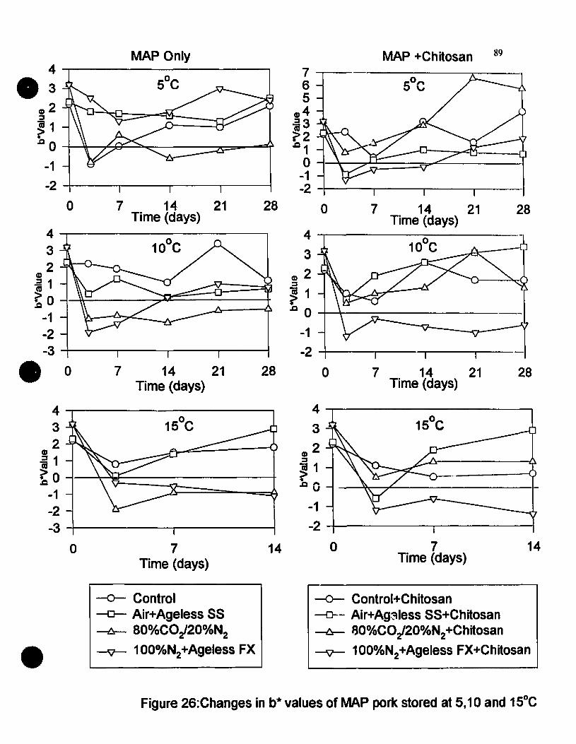
28
28
89MAP +Chitosan
7 14 21Time (days)
7....,-65
ID4~3
~2.01
o 1-'c:~====r----1-1-2 -+----r--------,---r----I
28 a 7 14 21Time (days)
4 ....-----------l10°C {J
3
G)2~
~ 1.0 0 --+-+-- _
-1-2 -+---~---r___-_r--
28 07 14 21Time (days)
a 7 14· 21Time (days)
4 ....,-------------,3 10°C2
ID
; 1 1 ÏQC~~~==~~ 0 -1--1-2-3 -+---.,------,-----.----1.0
4 ...,.-------------,.3ID2~
~1oC 0 --+-"""n-~l::::r_~~--____,=-~
-1-2 -!---.,------,-----.----l
147Time (days)
o
-1-2 -i-------,r----------i
4 ...,.-----------~
3
G)2~
~ 1.oû
147Time (days)
a
4 ....,-----.-------,32
ID
j1~ a --l-~~~---------+
-1-2-3 -+---------,r-------i
•-0- Control~ Air+Ageless 55--I::r- 8001OCO/20o1ON2
--9- 1OoolON2+Ageless FX
~ Control+Chitosan-0-- Air+Ag~lessSS+Chitosan--I::r- aoolOCO/20olON2+Chitosan
-v- 100%N2+Ageless FX+Chitosan
Figure 26:Changes in b* values of MAP park stored at 5,10 and 15°C

90
4.3.3. Sensory Evaluation
4.3.3.1. Color
Changes in color scores (results not shown) and time until rejection based on a score of
3 out of 5 for ail dippingl packaging treatments are summarized in Figure 27 and 28.
For non-chitosan dipped pork, samples were rejected within 7-14 days at 10 and 15"C.
However, at SuC, most samples (with the exception of air packaged pork) were deemed
acceptable after 21 to 25 days. However, l'rom a sensory viewpoir.t, the effect of
dipping pork in chitosan had a pronounced effect on shelf-Iife. Most MAP samples had
an acceptable color even al'ter 25 days especially at lower storage temperatures. These
results indicate that both MAP and chitosan had a pronounced effect on color when
viewed subjectively (through the eyes of the sensory panel). The rejection of control
(air packaged) samples al'ter 7 days is in agreement with the higher L* and b* values
particularly at higher storage temperatures.
4.3.3.2. Odor
Changes in odor scores (results not shown) and lime unlil rejection, based on a score of
3 out of 5 for ail dipping/packaging treatments are summarized in Figures 29 and 30.
For non-chitosan dipped pork, air packaged samples were rejected al'ter 3 to 7 days
depending on the storage temperature. This is in agreement with higher microbial
cOlmts, increasing L* and b* values and higher color scores observed for these samples.
Packaging under modified atmospheres increased the odor she1f Iife of pork, particularly
at lower storage temperatures. Dipping in chitosan prior to packaging did not appear to
have an appreciable effect on the odor shelf life of por\(. Most chitosan dipped
treatments had a ~imilar shelf-Iife to non-chitosan dipped products (Figure 30).

•
15
5
•D100%N1+Ageless FX
III BO%COz/20%Nz
oAir+Ageless 55~Ai(
•
o 7 14Tlme to reJection (days)
21 28
Figure 27: Effect of MAP alone on the time required to reject park samples on the basis ofcalor

•
15
5
•D100%N1+Ageless FX
1180%COz/20%N,t
oAir+Ageless 55œA;r
•
o 7 14Tfme to rejection (days)
21 28
Figure 28: Effect of MAP and dippilJin chitosan on the lime required to reject pork sampleson the basis of color

•
15
5
•
o 100%NL+Ageless FX
1IlI80%COl /20%Nz.
o Air+Ageless 88
iii Air
•
o 7 14lime to rejectfon (days)
21 28
Figure 29: Effect of MAP alone on the time required to reject pork samples on the basis ofodor

•
15
5
•
o 1OO°A»NI.+Ageless FX
1180%C(),/20%N~
DAir+Ageless 55IlAir
•
o 7 14Time to reJection (days)
21 28
Figure 30: Effect of MAP and dipping in chitosan on the lime required ta reject park sampleson the basis of odor

95
4.3.4. Microbiological Changes
The microbiological changes for various dipping/packaging treatments stored at S, 10and IS·C are shown in Figures 31, 32, 33, 34, 3S and 37. Changes in non-chitosandipped pork will be discussed first (Figures 32,33 and 34).
Initial1y, total aerobic counts increased more rapidly for air packaged samples thansamples packaged under a modified atmosphere. For example, at SoC. counts increasedfrom an initial level of 104 cfu/g to 109 cfu/g after 3-7 days and remained at this levelthroughout the 28 day storage period (Figure 31) due to a depletion in head space Ozand a build up of COz. For pork packaged under MAP conditions, aerobic countseither decreased or increased slightly throughout storage depending on the level ofresidual head space oxygen. However, counts never reached an unacceptable level (107
cfu/g) and at the end of the storage period, counts were about 2 to 4 log cycles less thanair packaged samples at S·C. A total number of 107 cfu/g of bacteria in meat isconsidered to be the upper Iimit of microbiological acceptability (Niemand et al., 198\).
Similar but more rapid trends were observed for pork stored at 10 and IS·C. However,counts at the end of the storage period were approximately 2 log cycles higher for ailpackaging conditions compared to those at S·C. These observations emphasize theimportance of strict temperature control to obtain the maximum beneficial effect onshelf Iife when packaging muscle foods under modified atmospheres.
The changes in Pseudomonas counts of pork samples stored at S, 10 and ISoC areshown in Figure 32. For non-chitosan dipped pork at S·C, Pseudomonas countsincreased more rapidly in air packaged samples than in MAP samples. Again, this isdue to the fact that a major proportion of the spoilage bacterial population of fresh porkis composed of Pseudomonas strains which are favored by aerobic conditions and lowstorage temperatures (Gill and Greer, 1993). In pork packaged in 80%COz and 20%Nzand in lOO%Nz with an Ageless FX oxygen absorbent, a lag time of about 3 days wasobserVed fol1owed by an active growth phase (Figure 32). Pseudomonas counts in thesepackaged samples were less than samples packaged in air or with Ageless SS. Theresults with Ageless SS were slightly surprising since counts were higher than expected.Again, this can probably be explained by package leakage and residual oxygen in thepackage head space. Similar trends were observed for samples stored at 10 and ISoC

96
(Figure 32). Even at higher storage temperatures Pseudomonas counts never reached
unacceptable levels, particularly in MAP packaged pork.
Changes in lactic acid bacteria (LAB) counts for non-chitosan dipped pork and stored at
S, 10 and 15°C are shown in Figure 33. LAB counts increased in nearly ail packaging
conditions at ail storage temperatures. Since LAB are facultative anaerobes, they soon
become the predominant spoilage microorganism in chilled meat stored under anaerobic
conditions (Gill and Greer, 1993). This is again apparent in Figure 33, where LAB
counts were higher in ail MAP samples than in air packaged pork since the gsaeous
conditions, Le., depleted O2: elevated CO2 are ail conducive to the growth of LAB.
While the counts of LAB reached 107 cfu/g after 28 days, these counts are not of major
concern with respect to meat spoilage. Indeed, similar and even higher counts have
been reported without adverse effect on the sensory attributes of these products
(Lambert et al., 1991).
The effect of dipping pork in a 0.2 % solution of chitosan for 60 seconds prior to
packaging under various gaseous conditions on the growth of indigenous
microorganisms (total aerobes, Pseudomonas and lactic acid bacteria) are shown in
Figures 34, 35 and 36 respectively. Similar changes/fluctuations in counts were
observed for chitosan dipped and non-chitosan dipped pork. At the end of the stcrage
period there was no significant difference in total aerobic counts, Pseudomonas counts
or lactic acid bacteria counts between the two dipping treatments. Indeed,
Pseudomonas counts at 5°C and packaged under 100% N2 with an Ageless oxygen
absorbent were higher in chitosan dipped pork than non-chitosan dipped pork.
However, these results may be explained through leakage, resulting in levels of O2
conducive to the growth of Pseudomonas species even in the presence of elevated levels
of N2 and an oxygen absorbent. Therefore, dipping pork in a 0.2 % chitosan solution in
conjunction with MAP would appear to offer a Iimited extension in microbiological
spoilage shelf Iife of pork over packaging under MAP conditions alone. These results
are in agreement with Darmadji and Izumimoto (1994) who studied the effect of
chitosan on the shelf-Iife of fresh ground beef. Only when higher levels of chitosan
(0.5 to 1%) were used, was a significant decrease in spoilage bacteria and an extension
in shelf Iife observed.

9710
• 98
.g> 7.a(,) 6C) 5.2
43 5°C2
0 7 14 21 28Time (days)
109
~8
.a 7(,)
en 6.2 5
4 10°C3
• 0 7 14 21 28Time (days)
1110
9~ 8-=CJ 7t:J) 6.2
54 15°C3
0 7 14Time (days)
-0- Control-0- Air+Ageless SS-6- 80°.4CO/20%N2
--JV-- 100%N2+Ageless FX
• Figure 31: Changes in total aerobic counts of non-chitosan dippedpork stored at 5, 10 and 15°C
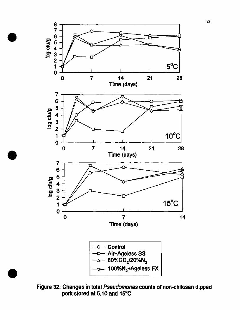
898
7• 6~5
~ 4~3- 2
1 5°C0
0 7 14 21 28Time (days)
76
0) 5 -......a 4u0)3.2 2
1 10°C0
0 7 14 21 28• Time (days)76
C)5......'5 4C)3,g 2
15°C10
0 7 14Time (days)
•~ Control-0- Air+Ageless SS~ aO%COJ20%N2
-v- 100%N2+Ageless FX
Figure 32: Changes in total Pseudomonas counts of non-chitosan dippedpork stored at 5,10 and 15°C

•9-.-------------------.87 -9J
&2 6(,)
C)5.2 4
32
99
o 7 14Time (days)
21 28
282114Time (days)
7o
9~----------------,
8C) 7-.-..~ 6oC) 5.2 4
32-1--------..1..-----------'
9~------------------.
8C) 7-.-..~ 6oC) 5.2 4
32-l- ....L...- ..1..- ..L.-__-.l
•
oTime (dayS)
14
•
-0- Control-0-- Air+Ageless SS-l::r- aO%COi20%N2
--v- 100%N2+Ageless FX
Figure 33: Changes in ladie aeid baelaris counts of non-chitosan dippedpork stored at 5, 10 and 1SOC
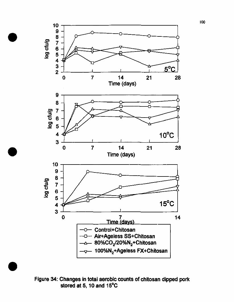
•10 """'T"'""'---------------,
98
9J 7ti 6E 5
432-'------'-----........---~-~~
100
o 7 14 21lime (days)
28
282114Time (days)
7o
J2'7'fi 6C)
,g 54
3-'------'-----........---~-------J
9-.-----------------,8
•10 ~---------------989J
'5 7C) 6.2 5
43.....1--------..L-------~
o 7~ 1_1 .'\.
lime 1
14
•
-0- Control+Chitosan-0- Air+Ageless SS+Chitosan-6-- 80%CO/20%N2+Chitosan-'V- 100%N2+Ageless FX+Chitosan
Figure 34: Changes in total aerobic counts of chitosan dipped POrkstored at 5, 10 and 15°C

101
8
• 70) 6.....'0 50)4.2 3
2 5°C1
0 7 14 21 28Time (days)
876
~5~ 4~ 3.2 2
1 10°C0
• 0 7 14 21 28Time (days)
76
~5~u 4C)
.2 3
2 15°C1
0 7 14Time (days)
-0- Control+Chitosan~ Air+Ageless SS+Chitosan---b- aO%COi200AN2+Chitosan
• --v-- 100%N2+Ageless FX+Chitosan
Figure 35: Total Pseudomonas counts of chitosan dippedpark stored at 5, 10 and 15°C.

•9-.---------------~
8C) 7......~ 6~ 5.9 4
32-L-.---..I------....I....----..l....-------J
102
o 7 14Time (days)
21 28
2814 21Time (days)
7o
9-.--------------------,8
C) 7......~ 6C) 5.2 4
32~----'------....I....----..l....-------J
• 8~----------------'11.
7
!!!6-n 5C)
.9 432......r...--------....I....-----------J
o 7Time (days)
14
•
-0- Control+Chitosan-0- Air+Ageless SS+Chitosan~ aO%CO/20%N2+Chitosan
-v- 100o~N2+AgelessFX+Chitosan
Figure 37: Total ladie aeid baderia counts of chitosan dippedpork stored at 5, 10 and 15°C

103
4.3.5. Shelf-Life
Based on this study, the estimated shelf Iife of ail dipping packaging treatments at 5, IDand 15°C is summarized in Table 13. The microbiological shelf Iife of pork was basedon the time necessary to reach an aerobic plate count of 107 cfu/g (Ehioba et al ..1987). However, while there is disagreement with respect to the use of the total aerobicplate count as an indicator of spoliage, it was used in this study based on theobservations of Lambert et al.(l991). The storage life of pork, estimated from thesensory evaluation scores for color and odor, Le. two of the most important factorsconsumers associate with meat quality and freshness is shown in Table 13. The averagetime (in days) to reach either a color or odor score of 3 (rejection point) was used as anindicator of the shelf Iife. It is evident from Table 13, that shelf Iife of both nonchitosan dipped and chitosan dipped samples were very similar irrespective of packagingconditions. Thus, dipping in chitosan prior to packaging would appear to offer noadvantage to the meat processor in terms of additional shelf Iife. This can best beobtained through packaging in a modified atmosphere in conjunction with low storagetemperatures. However, MAP is no substitute for proper temperature control as shelfIife decreases as products are subjected to mild temperature abuse conditions.However, even at 15°C, a 100 to 200% extension in shelf Iife is possible using sorneform of MAP technology, Le., gas fIushing or oxygen absorbents. Ageless 55 did notperform as weil as expected since it is designed to function at low temperature storageconditions. However, the time taken to scavenge headspace O2 may have influenced thegrowth rate of L.monocytogenes. However, of concern is the fact that for productsstored at 10 and 15°C, Le., mild temperature abuse conditions, sensory shelf Iife wasstill acceptable although aerobic plate counts had reached high levels (107 cfu/g).Therefore, such MAP products may pose a public health hazard if contaminated withpathogenic bacteria which could reach similar dangerous levels, yet from a sensoryviewpoint the product would still be acceptable to the consumer.

104
Table 13: Estimated Shelf-Life of Pork Loin at 5, 10 and 15"C for aU Packaging
Treatments Non-Chitosan Dipped
Packaging Storage Microbial' Rejection Pointb Sensory'Conditions Temperature Shelf-Life Color Odor Shelt:Life
("C) (Days) (Days) (Days) (Days)
Air 5 3 7 7 7
Air+Ageless SS 5 >25 21 25 23
SO%COJ20%N2 5 >25 25 25 25
100%N2+Ageless FX 5 >25 25 14 20
Air 10 3 7 7 7
Air+Ageless SS 10 14 7 25 16
SO%COJ20%N2 10 14 25 25 25
100%N2+Ageless FX 10 25 14 14 14
Air 15 2 7 3 5
Air+Ageless SS 15 10 14 7 Il
SO%COJ20%N2 15 14 7 7 7
100%N2+Ageless FX 15 14 7 14 Il
a: Time necessary to reach an aerobic plate count of 107 cfulg
b: Time at which samples reached a color or odor score of 3 out of5
c: The average time (days) to reach either a color or odor score of3 out of5

105
Table 14: Estimated Shelf-Life of Pork Loin at 5. 10 and IS-C for ail Packaging
Treatments Dipped in Chitosan
Packaging Storage Microbiala Rejection Pointb SensoryCConditions Temperature Shelf-Life Color Odor Shelf-Life
("C) (Days) (Days) (Days) (Days)
Air 5 3 7 7 7
Air+Ageless SS 5 >25 25 25 25
SO%C02/20%N2 5 >25 25 21 23
100%N2+Ageless FX 5 >25 25 25 25
Air 10 3 7 14 II
Air+Ageless SS 10 14 7 25 16
SO%C02/20%N2 10 14 25 14 19
100%N2+Ageless FX 10 3 25 14 19
Air 15 3 7 3 5
Air+Ageless SS 15 10 25 7 16
SO%CO~20%N2 15 14 7 14 Il
100%N2+Ageless FX 15 14 14 14 14
a: Time necessary to reach an aerobic plate count of 107 cfulg
b: Time at which samples reached a color or odor score of3 out of5
c: The average lime (days) to reach either a color or odor score of3 out of5
106
4.3.6. Challenge Studles with L.monocytogenes
Modified almosphere packaging (MAP) of muscle foods has becn gaining in popularilY
as a preservation technique for shelf Iife extension. Under conditions of low oxygentension, growth of aerobic spoilage microorganisms is inhibited and shelf Iife is
extended.
Recenlly, concerns have becn expressed over the microbiological safety of MAP foods,
particularly with regards 10 lhe possible growth of L.monocytogenes. These concernsare justified in view of (i) the ability of L.monocytogenes 10 grow under anaerobiccondilions and al refrigerated temperalures; (ii) the inhibition of the normal spoilagemicroorganisms of meal which are the indicalors of spoilage and (iii) the polenlial for
temperature abuse.
lndeed, previous studies have shown !hat the sensory qualily of fresh pork is stillacceptable although numbers of spoilage microorganisms have reached high levels, i.e.,I07cfu/g. If such a scenario is possible with pathogenic bacteria, particularly
L.monocytogenes, then MAP pork poses a potential public health hazard 10 consumers.
4.3.6.1. Changes..:: L.monocytogenes counts in Pork
Changes in counts of L.monocytogens in non-chitosan dipped pork packaged undervarious gas atmospheres are shown in Figure 37. L.monocytogenes was not delecled inany samples prior 10 inoculation studies. Therefore, changes in L.monocytogenescounts were solely due 10 the inoculum added on day O. In ail cases. counts ofL.monocytogenes increased throughout storage. At SoC, counts reached 107 cfu/g after
Il days in air packaged pork and after 12 days in pork packaged in air with an Ageless
SS oxygen absorbent. For pork packaged under a COz enriched atmosphere
(80%COz/20%Nz) or with lOO%Nz and an Ageless FX oxygen absorbent, counts did
nol reach these unacceptable levels until 18 days and 27 days respectively. Similartrends were observed for park slOred at 10°C while at ISoC with counts reachingI07cfu/g after only 3-S days irrespective of packaging conditions. These results show
that MAP can be used lO delay the growth of L.monocytogenes to unacceptable levels,
particularly at refrigerated slOrage temperalUres. However, when. the product is
subjecled 10 temperalure abuse, MAP offers Httle advantage, with counts of
107
L.monoeytogenes reaching unacceptable levels at approximately the same time as airpackaged samples. These results once again emphasize the importance of stricttemperature control if MAP is to be an effective barrier to control microbial growth.
However. when pork was dipped in a chitosan dipping solution prior to packaging undersimilar gaseous conditions. sorne interesting results were obtained as shown in Figure38. While growth of L.monoeytogenes occurred in ail treatments. dipping in chitosandelayed its growth to unacceptable levels by approximately 100%. For example. in airpackaged pork at SaC. counts of L.monoeytogenes reached 107 cfu/g after 21 days(compared to Il days for non-chitosan dipped control) and after 24 days in porkpackaged with Agele~s SS (compared to 14 daY3 in non-chitosan dipped control).Counts never reached these unacceptable levels in gas packaged pork (80%C02/20%N2)
or pork packaged in 1oo%N2 with and Ageless FX oxygen absorbent. (Figure 38).Dipping in chitosan prior to packaging also offered additional control in pork stored atlOoC and 1SoC indicating a synergistic effect between gas packaging and chitosan.
It is evident from both Figures 37 and 38 that MAP and refrigeration can delay thegrowth of L.monoeytogenes to unacceptable levels. Optimum control can be achievedby packaging in a CO2 enriched atmosphere or with 100% N2 and an Ageless FXoxygen absorbent. Similar results are obtained for chitosan dipped pork. These resultsconfirm previous agar studies which showed that growth of L.monoeytogenes wasdelayed in plates containing 0.2% chitosan and packaged in 80%C02/20%N2 or in100% N2 with an Ageless FX oxygen absorbent.
4.3.6.2. Changes in L.monocytogenes in Exudate Loss
Since there was considerable drip loss in ail packaged ï'0rk. studies were done todetermine levels of L.monoeytogenes in this drip loss. results of which are shown inFigures 39 and 40. It is evident that while counts of L.monoeytogenes increased in theexudate. they did not reach the levels found in the pork itself. The lower counts in theexudate may be due to the fact that there are insufficient nutrients in the exudate 10
support the growth of L.monoeytogenes. Another possible explanation rnay be that theexudate contains natural substances that are inhibitory 10 the growth of L.monoeytogenesor that the water soluble chitosan dissolves in the exudate and delays the growth of
L.monocytogenes.

10810
• 98
~ 7'5 60) 5.2
45°C3
20 7 14 21 28
Time (days)
109
~8
.2 7CJ
60)
.2 54 10°C
• 30 7 14 21 28
Time (days)
109
~8
ti 70) 6.2 5
4 15°C3 -
0 7 14Time (days)
-0- Control-0- Air+Ageless SS-6- aO°A»CO/20°A»N2
• ----v- 1OO°A.N2+Ageless FX
Figure 37: Changes in total Lmonocytogenes counts of nan-chitosan dippedpark stored at 5, 10 and 15°C

8 l09
• 7~~(,) 6C)
.25
5°C4
0 7 14 21 28Time (days)
8
~~ 60C)
.2 410°C
20 7 14 21 28
• Time (days)
109
~ 8~ 70C) 6g
54 15°C3
0 7 14Time (days)
•
-0- Control+Chitosan~ Air+Ageless SS+Chitosan-b-- aO%CO/20%N2+Chitosan-'V- 100%N2+Ageless FX+Chitosan
Figure 38:Changes in L.monocytogenes counts o~ chitosan dippedpark stored at 5, 10 and 15°C
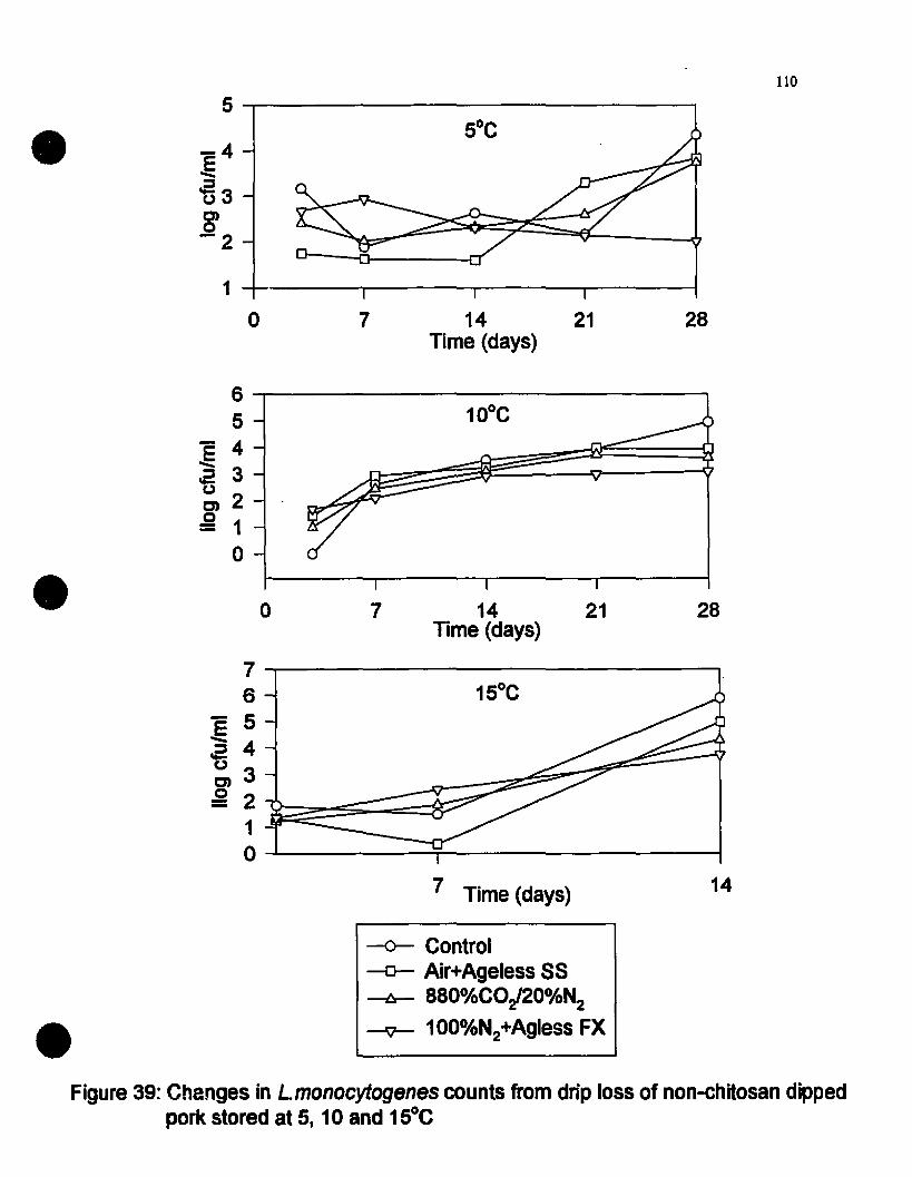
110
5
• -4.e::s'630)0-2
1
0 7 14 21Time (days)
28
282114Time (days)
7o
6 -,----------------.....,
5
:§ 4.a 3~2g 1
o
•7.....-------------------.,6
E 5--2 4~3g 2
1O......l.------.:r------------l
7 Time (days) 14
•-<r- Control-0- Air+Ageless 58-I::r- 880%CO/20°A»N2
-ÇJ- 100%N2+Agless FX
Figure 39: Changes in Lmonocytogenes counts trom drip loss of non-chitosan dippedpork stored at 5, 10 and 15°C

III5
• 4
:€3~(,,)
0)2.2
1
0
0 7 14 21Time (days)
28
282114Time (days)
7o
o
4 ...,-------------------,
3:ê~ 2(,,)
CD 1g
•5-.-------------------,4
:ê~ 3(,,)
:g ~ J:~~§:::5~=-------=]O......L...------~----------1
7Time (days) 14
•-0- Control +Chitosan-0- Air+Ageless SS+Chitosan~ aO%CO/20%N2+Chitosan-t;]-- 100oAlN2+Ageless FX+Chitosan
Figure 40: Changes in L.monocytogenes counts from drip IOS5 of chitosan dippedpark stored at 5, 10 and 15°C

112
4.3.7. Sbelr Life and Sarety
Wben shelf life and safety data are merged, some interesting results are obtained as shown
in Tables 15 and 16.
It is evident from Table 15 tbat for MOst treatments, sensory shelf life of non-chitosan
dipped pork was still acceptable altbough counts of L.monocytogenes bad reached
unacceptable levels (approximately 107 cfu/g). Therefore, tbese results would appear to
conlirm opinions tbat MAP alone could pose a public bealtb hazard witb respect to tbe
growth of L.monocytogenes. However, wben pork is pre-dipped in a chitosan dipping
solution prior to packaging, this trend would appear to be reversed, particularly at 5 and
10·C. At tbese storage temperatures, spoilage generally precedes growth of
L.monocytogenes to unacceptable levels. However at 15·C, tbe synergistic effect of
chitosan and MAP is reduced witb levels ofL.monocytogenes reaching unacceptable levels
while sensory shelflife was still acceptable.

113
Table 15: Shelf LiCe vs. Safety for Non-Chitosan Dipped Pork.
Packaging Storage Sensorya SafetlConditions Temperaturll Shelf·Life Shelf-Life
("C) (Days) (Days)
Air 5 7 Il
Air+Ageless SS 5 23 12
80%CO~20%N2 5 25 18
100%N2+Ageless FX 5 20 27
Air 10 7 7
Air+Ageless SS 10 16 7
80%CO~20%N2 10 25 9
100%N2+Ageless FX 10 14 14
Air 15 5 3
Air+Ageless SS 15 11 5
80%CO~20%N2 15 7 4
100%N2+Ageless FX 15 11 3
a: The average time (days) to reach either an odor or color score of3 out of5
b: Time (days) required for L.monocytogenes counts to reach unacceptable levels, 10'cfu/g

114
Table 14: Shelf Life vs Safety for Pork Dipped in Chitosan
Packaging Storage Sensol)~ SafetybConditions Temperature Shelf-Life Shelf·Life
(oC) (Days) (Days)
Air 5 7 21
Air+Ageless SS 5 25 24
80%CO~20%N2 5 23 >28
100%N2+Ageless FX 5 25 >28
Air 10 11 14
Air+Ageless SS 10 16 21
80%CO~20%N2 10 19 28
100%N2+Ageless FX 10 19 >28
Air 15 5 3
Air+Ageless SS 15 16 3
80%CW20%N2 15 11 12
100%N2+Ageless FX 15 14 >14
a: The average time (days) to reach either an odor or color score of3 out of5
b: Time (days) required for L.monocytogenes counts ta reach unacceptable levels, 107cfulg

115
GENERAL CONCLUSION
Increasing consumer demands for minimally processed, MAP foods has placed an onuson meat processors ta ensure the safety of such products. It has been demonstrated thattemperature alone cannot be regarded as a reliable barrier since temperature abuse,particularly at the retail level, occurs readily. Therefore. additional barriers. such asantimicrobial dipping solutions. could achieve this additional control. particularly undertemperature abuse conditions.
Preliminary studies clearly indicated that chitosan could be used effectively to controlthe growth of L.monocytogenes in a broth system. Further studies in an agar mediumindicated that MAP alone or in conjunction with chitosan could also he used to controlthe growth of L.monocytogenes for a substantial time period.
However. when these combinations treatments were extrapolated to fresh pork,somewhat different results were obtained. White MAP alone or in combination withchitosan could be used to delay the growth of meat spoilage microorganisms andL.monocytogenes, the extension time observed was significantly less than the timeobserved in the agar system. This is perhaps to be expected since the meat system mayhave provided L.monocytogenes with nutrients conducive to its growth. Anotherpossible reason for the observed differences in growth may be due to the nature of thesubstrate. i.e•• agar vs. meat. In the latter system. L.monocytogenes had to competewith the indigenous spoitage microorganisms of meal. Indeed. these microorganismsmay have enhanced the growth of L.monocytogenes by providing breakdown productsof their own metabolism e.g.. amino acids, which would enhance the growth ofL.monocytogenes Another possible reason for the differences could be attributed tochanges in headspace gas composition. Increasing concentrations of COz may haveactually stimulated the growth of L.monocytogenes. This has been demonstrated inprevious studies with L.monocytogenes as weil as C.botulinum. Despite these
differences. this study bas shown that MAP alone can he used ta delay spoilage and thegrowth of L.monocytogenes. However. the results of this study show that the concernsabout the growth of L.monocytogenes in MAP systems are justified since in nearly ail

116
instance "toxigenesis" preceded spoilage, a potentially dangerous scenario. However,when pork was dipped in chitosan, this trend was reversed, i.e., spoilage generallypreceded "toxigenesis" atlower storage temperatures, i.e., 5 and 10°C.
ln conclusion, this study has shown that control of the psychrotrophic pathogen,L.monocytogenes. can be obtained through combinations of low temperature control,MAP and a 0.2 % chitosan dipping solution. Optimum packaging conditions wouldappear to be SO%C02/20%N2 or 100% N2 and an Ageless FX oxygen absorbent. Thisstudy has also shown that chitosan offers the meat industry a simple, natura) andinexpensive "antimicrobial" which could be used to delay the growth of
L.monocytogenes.

111
REFERENCES
Adesiyun. A. (1993). Prevalence of Listeria species, Campylobacter species, Salmonellaspecies, fersinia species and toxigenic Escherichia coli on meat and seafoods inTrinidad. Food Microbiology, 10:395-403.
Agriculture Canada. Pork Quality. A guide to understanding color and structure of porkmuscle. Joint Publication of Research Branch (Lacombe Meat Research Centre) andFood Production and Inspection Branch, Ottawa, Publication No.5180/B.
Allan, C.R. and Hadwinger, L.A. (1979). The fungicidal effect of chitosan on fungi ofvarying cell wall composition. Experimental Mycology, 3: 285-287.
Aureli, P., Constanini, A. and Zolea, S. (1992). Antimicrobial activity of sorne plantessential oils against Listeria monocytogenes. Journal of Food Protection, 55: 344-348.
Baird-Parker, A.C. (1994). Foods and microbiological risks. Microbiology, 140: 687695.
Baker, D.A. (1989). Assessing packaging safety through the hurdle method. FoodEngineering, February: 40-41.
Benedict, R.C.(1990). Listeria monocytogenes: physiology and metabolism. In:Foodborne Listeriosis. (Eds.: Miller, A.J., Smith, J.L., and Somkuti, G.A.). AviPublications, N. Y., New York, pp. 13-24
Berang, M.E., Brackett, R.E. and Beuchat, L.R. (1989). Growth of Listeriamonocytogenes on fresh vegetables stored under a controlled atmosphere. Journal ofFood Protection, 52: 702-705.
Beuchat, L. R. and Brackett, R.E. (1990a). Survival and growth of Listeriamonocytogenes on lettuce as infIuenced by shredding, chlorine treatment, modifiedatmosphere packaging and temperature. Journal of Food Science, 55: 755-758.
Beuchat, L.R. and Brackett, R.E. (1990b), Inhibitory effects of raw carrots on Listeriamonocytogenes. Applied and Environmental Microbiology, 56: 1734-1742.
Beuchat, L.R., Brackett, R.E. and Doyle, M.P. (1994). Lethality of carrot juice taListeria monocytogenes as affected by pH, sodium chloride and temperature. Journal ofFood Protection, 57: 470-474.
Blikstad, E., Enfors, S.O. and Molin, G. (1981). Effeet of high concentrations of COzon the microbial fIora of pork stored at 4°C and 14°C. p. 345-347. In Psychrotrophicmicroorganisms in spoiLage and pathogenicity. Academie Press, London.

118
Buchanan, R.L. and Klawitter, L.A. (1990). Effeets of temperature and oxygen on thegrowth of Listeria monocytogenes at pH 4.5. Journal ofFood Science, 55: 1754-1756.
Buchanan, R.L. and Klawitter, L.A. (1991). Effeet of temperature history on the growthof Listeria monocytogenes Scott A at refrigeration temperatures. International Journal ofFood Microbiology, 12: 235-246.
Buchanan, R.L. and Philips, J.G. (1990). Response surface model for predicting theeffects of temperature, pH, sodium chloride content, sodium nitrite concentration andatmosphere on the growth of Listeria monocytogenes. Journal of Food Protection, 54:370-376.
Carpenter, S.L. and Harrison, M.A. (1989). Fate of small populations of Listeriamonocytogenes on poultry processed using moist heat. Journal of Food Protection, 52:768-770.
Chen, N. and Shelef, L.A. (1992). Relationship between water activity, salts of lacticacid, and growth of Listeria monocytogenes in a meat model system. Journal of FoodProtection, 55: 574-578.
Comi, G., Frigerio, R. and Cantoni, C. (1992). Listeria monocytogenes serotypes inltalian meat products. Letters in Applied Microbiology, 15: 168-171.
Cooksey, O.K. (1993). Reduction of Listeria monocytogenes in preeooked vacuumpackaged beef using post-packaging pasteurization. Journal of Food Protection, 12:1034-1038.
Cuero, R.G., Duffus, E., Osuji, G. and Petit, R. (1991). Aflatoxin control inpreharvest maize: effect of chitosan and two microbial agents. Journal of AgriculturalScience, 117: 165-169.
Daba, H., Pandian, S., Gosselin, J.F., Simard, R.E., Huang, J. and Lacroix, C. (1991).Detection and activity of a bacteriocin produced by Leuconostroc menesteroides. Appliedand Environmental Microbiology, 57: 3450-3455.
Dallas, H.L. and Hitchins, A.D.(1993). Antagonism of bacterial competitors towardListeria monocytogenes in enrichment culture. Journal ofFood Safety, 13: 113-123.
Darmadji, P. and 1zumimoto, M. (1994). Effeet of chitosan in meat preservation. MeatScience, 38: 243-254.
Degnan, A.J., Ahmed, E.Y. and Luchansky, J.B. (1992). Use of Pediococcusacidilactici to control Listeria monocytogenes in temperature-abused vacuum-packagedwieners. Journal ofFood Protection, 55: 98-103.
119
Dever, F.P., Schaffner, D.W., Slade, P.J. (1993). Methods for the detection offoodborne Listeria monocytogenes in the United States. Journal of Food Safety. 13: 263292.
DeVore, D.P. and Solberg, M. (1974). Oxygen uptake in postrigor bovine muscle.Journal of Food Science, 39:577-580.
Dickson, J.S. (1990). Comparison of homogenization by blending or stomaching on therecovery of Listeria monocytogenes l'rom beef tissue. Journal of Food Science, 55: 655657.
Dorsa, W.J., Marshall, D., Moody, M. and Hackney, C.R. (1993). Low temperaturegrowth and thermal inactivation of Listeria monocytogenes in precooked crawfish tailmeat. JournaL ofFood Protection, 56: 106-109.
Ehioba, R.M., Kraft, A.A., Molins, R.A., Walker, H.W., Oison, D.G., Subbaraman,G. and Skowronski, R.P. (1987). Effect of low-dose (lOOkrad) radiation on themicrof\ora of vacuum packaged ground pork with and without added sodium phosphates.JournaL ofFood Science, 52: 1477-1480, 1505.
El Ghaouth, A., Arul, J., Grenier, J. and Asselin, A. (1991). Antifungal activity ofchitosan on two post harvest pathogens of strawberry fruits. Phytopathology, 82:398402.
El Ghaouth, A., Ponnampalan, R., Castaingne, F. and Arul, J. (1992a). Chitosancoating to extend the storage Iife of tomatoes. HorticulturaL Science, 27: 1016-1018.
El Ghaouth, A., Arul, J., Asselin. A. and Benhamou, N. (1992b). Antifungal activityof chitosan on postharvest pathogens: induction of morphological and cytologicalalterations in Rhil.opus stoLinifer. MycoLogical Research, 96: 769-779.
EI-Kest, S.E. and Marth, E.H. (1992). Freezing of Listeria monocytogenes and othermicroorganisms: A Review. JournaL of Food Protection, 55: 639-648.
EI-Kest, S.E., Yousef, A.E. and Marth, E.H. (1991). Fate of Listeria monocytogenesduring freezing and frozen SlOrage. JournaL ofFood Science, 56: 1068-1071.
EI-Shenawy, M.A. and Marth, E.H. (1992). Behaviour of Listeria monocytogenes in thepresence of sodium propionate together with food acids. JournaL of Food Protection, 55:241-245.
120
Embarek, P.K.B. and Huss. H.H. (1993). Heat resistance to Listeria monocytogenes invacuum packaged pasteurized fish fillets. International Journal of Food Microbiology,20: 85-95.
Enfors, s.a., Molins, G. and Ternstrom, A. (1979). Effect of packaging in carbondioxide, nitrogen or air on the microbial f10ra of pork stored at 4°C. Journal (ll'AppliedBacteriology, 47: 197-208.
Eyles, M,J., Moir, C,J. and Davey, J.A. (1993). The effect of modified atmospheres onthe growth of psychrotrophic pseudomonads on a surface in a model system.International Journal ofFood Microbiology, 20: 97-107.
Fain, A.R. (1991). Lethality of heat to Listeria monocytogenes Scott A: D-Value and Zvalue determinations in ground beef and turkey. Journal of Food Protection, 54: 756761.
Fang, S.W., Li, C.F. and Shih, D. Y.C. (1994). Antifungal activity of chitosan and itspreservative effect on low sugar candied kumquat. Journal of Food Protection, 56: 136140.
Farber, J.M. (1991). Microbiological aspects of modified atmosphere packagingtechnology- a review. Journal ofFood Protection, 54: 58-70.
Farber, J.M. and Peterkin, P.1. (1991). Listeria monocytogenes, a food-borne pathogen.Microbiological Reviews, 55: 476-511.
Fr.rber, J.M., Coates, F. and Daley, E. (1992). Minimal water activity requirements forthe growth of Listeria monocytogenes. Letters in AJ?plied Microbiology, 15: 100-IOS.
Farber, J.M. (1993). Current research on Listeria monocytogenes in foods: an overview.Journal of Food Protection, 56: 640-643.
Farber, J.M. and Daley, E. (1994). Presence and growth of Listeria monocytogenes innaturally-contaminated meats. International Journal of Food Microbi%gy, 22: 33-42.
Finne, G. (1982). Modified and controlled atmosphere storage of muscle foods. FoodTechnology, 36: 128-133.
Foegeding, P.M., Thomas, A.B., Pilkington, D.H. and Klaenhammer, T.R. (1992).l:::nhanced control of Listeria monocytogenes by in-situ produced pediocin during dryfermented sausage production. Applied and Environmental Microbi%gy, 58: 884-890.

121
Gaya, P., Mendina, M. and Nunez, M. (1991). Effect of the lactoperoxidase system onListeria monocyogenes behaviour in raw milk at refrigeration temperatures. Applied andEnviroilmental Mocrobiology, 57: 3355-3360.
Genigeorgis, C.A. (1985). Microbial safety implications of the use of modified~tmospheres to extend the storage life of fresh meat and fish. International Journal ofMicrobiology, 1: 237-251.
George, S.M. and Lund, B.M. (1992). The effect of culture medium and aeration on:.wth of Listeria monocytogenes at pH 4.5. Letters in Applied Microbiology, 15: 49-52.
Gill, c.a. and Tan, K.H. (1980). Effect of carbon dioxide on growth of meat spoilagebacteria. Applied and Environmental Microbiology, 39: 317-319.
Gill, c.a. (1986). The control of microbial spoilage in fresh meats. In: Advances inMeat Research. Volume 2. Meat and Poultry Microbiology. A.M. Pearson, T.R. Dutson(Ed.), pp 49-88, Avi Publishing Co. Inc. Westport, Conneticut.
Gill, c.a. and Reichel, M.P. (1989). Growth of the cold-tolerant pathogens Yersiniaenterocolitica, Aeromonas hydrophila and Listeria moncytogenes on high pH beefpackaged under vacuum or carbon dioxide. Food Microbiology, 6: 223-230.
Gill, c.a. and Greer, G.G. (1993). Enumeration and identification of meat spoilagebacteria. Technical Bulletin 1993-8E, Research Branch, Agriculture Canada.
Glass, K.A. and Doyle, M.P.(1989). Listeria monocytogenes in processed meat productsduring refrigerated storage. Applied and Environmental Microbiology, 55: 1565-1569
Grant, l.R. and Patterson, M.F. (1991). Effect of irradiation and modified atmospherepackaging on the microbiological safety of minced pork stored under temperature abuseconditions. International Journal ofFood Micobiology, 26: 521-533.
Grant, l.R. and Patterson, M.F. (1992). Sensitivity of foodborne pathogens to irradiationin the components of a chilled ready meal. Food Microbiology, 9: 95-103.
Grau, F.H. and Vanderlinde, P.B. (1992). accurence, numbers, and growth of Listeriamonocytogenes on some vacuum-packaged processerl meats. Journal of Food Protection,55: 4-7.
Grau, F.H. and Vanderlinde, P.B. (1993). Aerobic growth of Listeria monocytogenes onbeef lean and fany tissue: equations describing the effects of temperature and pH.Journal ofFood Protection, 56: 96-101.

122
Greer, G.G., Dilts, B.D. and Jeremiah, L.E. (1992). Bacleriology and retail case life ofpork afler strorage in carbon dioxide. Journal ofFood Protection, 56: 689-693.
Hadwinger, L.A. (1985). Chi10san both activaleS genes in plants and inhibits RNAsynthesis in fungi. In: Chitin in Nature and Technology, (Eds. Muzzarelli et aL), PlenumPress, New York, NY.
Harris, L.J., Fleming, H.P. and Klaenhammer, T.R. (1991). Sensitivity and resistanceof Listeria monocytogenes ATCC 19115, Scott A, and UALSOO to nisin. Journal ofFood Protection, 54: 836-840.
Harrison, M.A., Huang, Y., Chao, C. and Shineman, T. (1991). Fate of Listeriamonocytogenes on packaged, refrigerated, and frozen seafood. Journal of FoodProtecûon, 54: 524-527.
Hart, C.D., Mead, G.C. and Norris, A.P. (1991). Effects of gaseous environment andtemperature on the storage behaviour of Listeria monocytogenes on chicken breast meat.Journal of Applied Bacteriology, 70: 40-46.
Hefnawy, Y.A., Moustafa, S.1. and Marth, E.H.(1993). Sensitivity of Listeriamonocytogenes 10 selec1ed spices. Journal ofFood Protection, 56:876-878.
Hintlian, C.B. and Hotchkiss, J.H. (1986). The safety of modified atmospherepackaging: a review. Food Technology, 40: 70-76.
Hitchins, A.D. (1992). Listeria monocytogenes. Chapler 10 in FDA BacteriologicalAnalytical Manual, 7th Edition, p141-151.
Hirano, S. and Nagao, N. (1989). Effects of chitosan, pectic acid, Iysosyme andchitinase on the growth of several phytopathogens. Agricultural and BiologicalChemistry, 53: 3065-3066.
Hirano, S. (1990). Chitosan as an ingredient for domestic animal feeds. Journal ofAgricultural Food Chemistry, 38:1214-1217.
Holley, R., Delaquis, P., Gagnon, J., Doton, G. and Gariepy, C. (1993). Modifiedatmosphere packaging of fresh pork. Alimentech, 6: 16-17.
Hotchkiss, J.H. (1988). Experimental approaches 10 delermining the safety of foodpackaged in modified atmospheres. Food Technology, 42:55-64.
Hotchkiss, J.H. and Banco, M.J. (1992). Influence of new packaging technologies on thegrowth of microorganisms in produce. Journal ofFood Protecûon, 55: 815-820.

123
Hudson, J.A. (1992). Efficacy of high sodium chloride concentrations for the destruclionof Listeria monocytogenes. Letters in Applied Microbiology, 14: 178-180.
Hudson, J.A. and Avery, S.M. (1994). Growth of Listeria monocytogenes. Aeromonashydrophila and Yersinia enterocolitica on cooked muscle tissue under refrigera!ion andmild temperature abuse. Journal ofFood Safety. 14: 41-52.
Hudson,. J.A., Mott, S.J. and Penney, N. (1994). Growt:l of Listeria mollocytogenes.Aeromonas hydrophillia and Yersinia enterocolitica on vacuum and saturated carbondioxide controlled atmosphere-packaged sliced roast beef. Journal of Food Protection,57: 204-208.
International Comission on Microbiological Specifications of Foods (ICMSF) (1994).Choice of sampling plan and criteria for Listeria monocytogenes. International JournalofFood Microbiology, 22: 89-96.
Imeri, A.G. and Knorr, D. (1988). Effects of chitosan on yield and compositional dataof carrot and apple juice. Journal ofFood Science, 53: 1707.
In't Veld, P.H and DeBoer, E. (1991). Recovery of Listeria monocytogenes on s.:lectiveagar media in a collaborative study using reference samples. International Journal ofFood Microbiology, 13: 295-300.
Johansen, C., Gram, L. and Meyer, A.S. (1994). The combined inhibitory effect oflysozyme and low pH on growth of Listeria monocytogenes. Journal of Food Protection,57: 561-566.
Johnson, J.L. and Lattuada, C.P. (1993). Comparison of nucleic acid hybridizationassays and biochemical characterization tests for the confirmation of Listeriamonocytogenes. Journal ofFood Protection, 56: 834-840.
Johnson, J.L., Doyle, M.P, and Cassens, R.G. (1988). Survival of Listerianonocytogenes in ground beef. International Journal ofFood Microbiology, 6: 243-247.
~lIander, K.D., Hitchins, A.D., Lancette, G.A., Schmeig, J.A., Garcia, G.R.,Solomon, H.M. and Sofos, J.N. (1991). Fate of Listeria monocytogenes in shreddedcabbage stored at 5 and 250 C under a modified atmosphere. Journal of Food Protection,54: 302-304.
Kendra, D.F. and Hadwinger, L.A. (1984). Characterization of the smallest chitosanoligomer that is maximally antifungal to Fusarium solani and Elicitis pisatin formation inPisum sativum. Experimental Mycology, 8: 276-281

124
Kinderlerer, J.L. and Lund., B.M. (1992). Inhibition of Listeria monocytogenes andListeria innocua by hexanoic and octanoic acids. Letters in Applied Microbiology, 14:271-274.
KlWChel, S. and Gould, G. (1995). Preservation microbiology and safety: quo vadis?Trends in Food Science and Technology, 6: 127-131.
Knorr, D. (1991). Recovery and utilization of chitin and chitosan in food processingwaste management. Food Technology, 45: 114-122.
Kroll, R.G. and Patchett, R.A. (1992). Induced acid tolerance in Listeriamonocytogenes. Letters in Applied Microbiology, 14: 224, 227.
Krysinski, E.P., Brown, Lol. and Marchisello, Tol. (1992). Effect of cleaners andsanitizers on Listeria monocytogenes attached to product contact surfaces. Journal ofFood Protection, 55: 246-251.
Lambert, A.D., Smith, J.P. and Dodds, K.L. (1991). Shelf Life extension andmicrobiological safety of fresh meat- a review. Food Microbiology, 8: 267-297.
Lambert, A.D., Smith, J.P. and Dodds, K.L. (1992). Effect of headspace C02concentration on toxin production by Clostridium botulinum in MAP, irradiated freshpork. Journal ofFood Protection, 54:588-592.
Lebepe, S. (1990). Changes in microt1ora and other characteristics of vacuum-packagedpork loins irradiated at 3.0 kGy. Journal ofFood Science, 55: 918-924.
Leistner, L. and Gorris, G.M. (1995). Food preservation by hurdle technology. Trendsin Food Science and Technology, 6: 41-46.
Line, J.E. and Harrison, M.A., (1992), Listeria monocytogenes inactivation in turkeyrolls and battered chicken nuggets subjected to simulated commercial cooking, Journal ofFood Science, 57, 787-793.
Lioutas, T.S. (1988). Challenges of controlled and modified atmosphere packaging: afood company's perspective. Food Technology. 42: 78-86.
Lowry, P.D. and Tiong, 1. (1988). The incidence of Listeria monocytogenes in meatand meat products-factors affecting distribution. In: Proceedings of the 34thInternational congress of Meat Science and Technology, Part B, Brisbane, Australia,pp.528-530.

125
Luchansky, J.B. (1992). Genomic analysis of Pediococcus starter cultures used to controlListeria monocytogenes in turkey summer sausage. Applied and EnvironmentalMicrobi%gy, 58: 3053-3059.
Marshall, D.L., Andrews, L.S., Wells, J.H.and Farr, A.J. (1990). Influence ofmodified atmosphere packaging on the competitive growth of Listeria monocytogenesand Pseudomonasfluorescens on preeooked chicken. Food Microbiology, 9: 303-309..
Mattila-Sandholm, T., Haikara, A. and Skytta, E. (1991). The effect of Pediococcusdamnosis and Pediococcus pentosaceus on the growth of pathogens in minced meal.International Journal ofFood Microbiology, 13: 87-94.
McClure, P.J., Kelly, T.M. and Roberts, T.A. (1991). The effeets of temperature, pH,sodium chloride, and sodium nitrate on the growth of Listeria monocytogenes.International Journal of Food Microbiology, 14: 77-92.
McClure, P.J., Roberts, T.A. and Oguru, P.O. (1989). Comparison of the effects ofsodium chloride, pH, and temperature on the growth of Listeria monocytogenes ongradient plates and in liquid media: Letters in Applied Microbiology, 9: 95-99.
McKellar, R.C. (1993). Effeet of preservatives and growth factors on secretion ofliteriolysin-O by Listeria monocytogenes. Journal ofFood Protection, 56: 380-384.
McKellar, R.C., Moir, R. and Kalab, M. (1994). Factors influencing the survival andgrowth of Listeria monocytogenes on the surface of Canadian retail wieners. Journal ofFood Protection, 57: 387-392.
McKellar, R.C., Paquet, A. and Ma, C.Y. (1992). Antimicrobial activity of fatty Nacylamino acids against gram-positive foodborne pathogens. Food Microbiology, 9: 6776.
Miller, A.J. (1992). Combined water activity and solute effeets on the growth of Listeriamonocytogenes Scott A. Journal ofFood Protection, 55: 414-418.
Ming, X. and Daeschel, M.A. (1993). Nisin resitance of foodborne bacteria and thespecifie resistance response of Listeria monocytogenes Scott A. Jounal of FoodProtection, 56: 944-948.
Moir, C.J. and Eyles, M.J. (1992). Inhibition, injury, and inactivation of fourpsychrolrophic foodborne bacteria by the preservatives methyl-hydroxybenzoate andpotassium sorbate. Journal ofFood Protection, 55: 360-366.
126
Motlagh, A.M., Holla, S., Johnson, M.C., Ray, B. and Field, R.A. (1992). Inhibitionof Listeria spp. in sterile food systems by pediocin AcH, a bacleriocin produced byPediococcus acidilocti- H. Journal of Food Science, 55: 337-343.
Muzzarelli, R.A. (1977). Chilin. Ist Edition, Oxford, New York, Pergamon Press.
Niemand, K.G., van der Linde, H.J. and Smith, K.M. (1981). Radurization of primebeef cuts. Journal ofFood Protection, 44:53-59.
Nottingham, P. M. (1982). Microbiology of carcass meats. In Meat Microbiology. M. H.Brown (Ed.), p. 13-65. Applied Science Publishers Ltd., London.
Oh, D.H. and Marshall, D.L. (1993a). Influence of temperature, pH, and glycerolmonolaurate on growth and survival of Listeria monocytogenes. Journal of FoodProtection, 56: 744-749.
Oh, D.H. and Marshall, D.M. (1993b). Antimicrobial activity of ethanol, glycerolmonolaurale or laclic acid against Listeria monocytogenes. International Journal of FoodMicrobiology, 20: 239-246.
Pandit, V.A. and Shelef, L.A. (1994). Sensitivity of Listeria monocytogenes 10
rosemary (Rosemorinus ojJicinalis L.). Food Microbiology, 11: 57-63.
Parish, M.E. and Higgins, D.P. (1989). Survival of Listeria monocytogenes in low pHmodel brolh systems. Journal ofFood Protection, 52: 144-147.
Pelroy, G.A. (1994). Inhibition of Listeria monocytogenes in cold process (smoked)salmon by sodium lactate. Journal ofFood Protection, 57: 108-113.
Petran, R.L. and Zoltola, E.A. (1989). A study offactors affecting growth and recoveryof Listeria monocytogenes Scott A. Journal ofFood Science, 54: 458-460.
Quintvalla, S. and Campanini, M. (1991). Effect of rising temperature on the heatresistance of Listeria monocytogenes in meat emulsions. Letters in Applied Microbiology,12: 184-187.
Razavilar, V. and Genegeorgis, C. (1992). Interactive effecl of temperalure, almosphereand slorage lime on the probability of colony formation on blood agar by four Listeriaspecies. Journal ofFond Protection, 55: 88-92
Ren, Tyh-jenq and Frank, J.F. (1993). Susceptibility of starved planclonic and biofilmListeria monocytogenes 10 quaternary amonium sanitizer as determined by direct viableand agar plate counts. Journal ofFood Protection, 56: 573-576.

127
Sallam, S.S. and Donnelly, C.W. (1992). Destruction, injury, and repair of Listeriaspecies exposed to sanitizing compounds. Journal of Foot! Protection, 55: 771-776.
Schilliger, U. and Ho1zapfel, W.H. (1990). Antibacterial activity of Carnobacteria.Food Microbiology, 7: 305-310.
Schmidt, U., Seeliger, H.P.R., Glenn, E., Langer, B. and Leistner, L. (1988). Listeriafindings in raw meat products. Fleischwirtschaft, 68: 1313-1316.
Schoeni, J.L., Brunner, K. and Doyle, M.P. (1991). Rates of thermal inactivation ofListeria monocytogenes in beef and fermented beaker sausage. Journal of FoodProtection, 54: 334-337.
Schofield, G.M. (1992). Emerging food-borne pathogens and their significance in chilledfoods. Journal ofApplied Bacteriology, 72: 267-273.
Seidemen, S.C., Cross, H.R., Smith, G.C. and Durland, P.R. (1984). Factorsassociated with fresh meat colour: a review. Journal of f'ood Quality, 6:211-237.
Shelef, L.A. and Addala, L. (1994). Inhibition of Listeria monocyfogenes and otherbacteria by sodium diacetate. Journal of Food Safety, 14: 103-115.
Shelef., L.A. and Yang, Q. (1991). Growth suppression of Listeria moncytogenes bylactates in broth, chicken and beef. Journal ofFood Protection, 54: 238-287.
Silliker, J.H. and Wolfe, S.K. (1980). Microbiological safety considerations incontrolled atmosphere storage of meats. Food Technology, 34:59-63.
Siragusa, G.R. and Dickson, J.S. (1992). Inhibition of Listeria monocytogenes on beeftissue by application of organic acids immobilized in a calcium alginate gel. Journal ofFood Science, 57: 293-296.
Skjak-Braek, G., Anthonsen, T. and Sandford, P. (1989). Chitin and chitosan: sources,biochemistry, physical properties and applications. Proceedings from the 4thInternational Conference on Chitin and Chitosan held in Trodheim, Norway, August 2224, 1988. Elsevier Applied Science, London, England.
Skovgaard, N. and Norrung, B. (1989). The incidence of Listeria species in faeces ofDanish pigs and in minced pork. International Journal ofFood Microbiology, 8: 59-63.
Skytta, E. and Mamila-Sandholm, T. (1991). Broad spectrum antibacterial activity ofPediococcus damnosis and Pediococcus pentosaceus in minced meat. Food Microbiology,8: 231-237.

128
Smith, J.P., Simpson, B.K. and Ramaswamy, H. (1990). Developments in foodpackaging ieChnology. Part 2. Storage Aspects. Trends in Food Sceience and TechnologyToday. 1: 112.
Smittle, R.B. and Flowers, R.S. (1994). Predictive modelling and microbiological foodstudies. S.C.O.P.E. a Technical Bulletinfrom Silliker Laboratories, 9: 1-7.
Sobrino, O.J. (1991). Antibacterial activity of Lactobacillus sake isolated from dryfermented sausages. International Journal ofFood Microbiology. 13: 1-10.
Sorrels, K.M., Enigl, D.C. and Hatfield, J.R. (1989). Effect of acidulant, time andtemperature on the growth of Listeria monocytogenes. Journal of Food Protection, 52:57\-573.
Soto Peralta, N. V., Muller, H. and Knorr, D. (1989). Effects of chitosan treatment onthe c1arity and colour of apple juice. Journal ofFood Science, 54: 495.
Stillmunkes, A.A. (1993). Microbiological safety of cooked beef roasts treated withlactate, monolaurin or gluconate. Journal ofFood Science, 58: 953-958.
Stossel, P. and Leuba, J.L. (1984). Effect of chitosan, chitin and some aminosugars onthe growth of various soilborne phytopathogenic fungi. Phytopathologische Zeitschrift,11: 82-90.
Sumner, S.S. (1991). Heat resistance of Salmonella typhimurium and Listeriamonocytogenes in sucrose solutions of various water activities. Journal of Food Science,56: 1741-1743.
Tapia deDaza, M.S.T., Villegas, Y. and Martinez, A. (1991). Minimal water activityfor growth of Listeria monocytogenes as affected by solute and temperature.International Journal ofFood Microbiology, 14: 333-337.
Thayer, D.W. and Boyd. G. (1995). Radiation sensitivity of Listeria monocytogenes onbeef as affected by temperature. Journal ofFood Science. 60: 237-240.
Todd. E.C.D. (1992). Foodborne disease in Canada- a 10 year summary from 1975 to1984. Journal ofFood Protection, 55: 123-132.
Tuncan. E. U. (1993). Effect of cold temperature on germicidal efficacy of quartenaryammonium compound. iodophor, and chlorine on Usteria. Journal of Food Protection,56: 1029-1033.

129
Varabioff, Y., Mitchel1, G.E. and Nottingham, S.M. (1992). Effects of irradiation onbacterial load and Listeria monocytogenes in raw chicken. Journal of Food Protection.55: 389-391.
Venugopal, R.J., Ingham, S.C., McCurdy. A.R. and Jones, G.A. (1993). Anaerobicmicrobiology of fresh beef packaged under modified atmosphere or vacuum. Journal ofFood Science, 53: 935-938.
Wakabayashi, H. (1992). Inactivation of Listeria monocytogenes by lactoferricin, apotent antimicrobia1 peptide derived from cow's milk. Journal of Food Protection, 55:238-240.
Wang, C. and She1ef, L.A. (1992). Behaviour of Listeria monocytogenes and thespoilage microf1ora in fresh cod fish treated with lysozyme and EDTA. FoodMicrobiology, 9: 207-213.
Wang, C. and Muriana, P.M. (1994). Incidence of Listeria moncytogenes in packages ofretail franks. Journal of Food Protection, 57: 382-386.
Wang, G.H. (1992). Inhibition and inactivation of five species of foodborne pathogensby chitosan. Journal ofFood Protection, 55: 916-919.
Wang, G.H., Yan, K.T., Feng, X.M., Chen, S.M., Lui, A.P. and Kokuburo. Y.(1992), Isolation and identification of Listeria monocytogenes from retail meats inBeijing. Journal ofFood Protection, 55: 56-58.
Wang L.L. and Johnson, E.A. (1992). Inhibition of Listeria monocytogenes by fattyacids and monoglycerides. Applied and Environmental Microbiology, 58: 624-629.
Warbunon, D.W., Farber, J.M., Powel1, C., Tiwari, N.P., Read,S., Plante, R.,Babiuk, T., Laffey, P, Kauri, T., Mayers, P., Champagne, M.J., Hunt, T., laCasse, P,Viet, K., Smando, R. and Coates F. (1992), Comparison of methods for optimumdetection of stressed and low levels of Listeria monocytogenes. Food Microbiology, 9:127-145.
Wimpfheimer, L., Altman, N.S. and Hotchkiss, J.H. (1990). Growth of Listeriamonocytogenes, Scott A, serotype 4 and competitive spoilage organisms in raw chickenpackaged under modified atmospheres and in air. International Journal of FoodMicrobiology, 11: 205-214.
Winkowski, K. and Montville, T.J. (l992). Use of meat isolate, Lactobacillus bavaricusMN, to inhibit Listeria monocytogenes growth in a model meat gravy system. Journal ofFood Safety, 13: 19·31.

130
Wolfe, S.K. (1980). Use of CO and C02 enriched atmospheres for meats and produce,Food Technology, 43:55-58, 63.
Yen,L.C., Sofos, J.N. and Schmidt, G.R. (1992). Destruction of Listeriamonocytogenes by heat in ground pork formulated with kappa-carageenan, sodium lactateand the algin/calcium meat binder. Food MicrobiollJgy, 9: 223-230.
Young, L.L., Reviere, R.D. and Cole, B.A. (1988). Fresh red meats: a place to applymodified atmospheres. Food Technology, 42: 65-68.
Young, D.H., Kohle, H. and Kauss, H. (1982). Effeet of chitosan on membranepermeability of suspension cultured Glycine max and Phaseolus vulgaris cells. PlantPhysiology, 70: 1449-1454.
zaika, L. and Kim, A. (1993). Effeet of sodium polyphosphates on growth of Listeriamonocytogenes. Journal ofFood Protection, 56: 577-580.
zapico, P. (1993). Goats' milk lactoperoxidase system against Listeria monocytogenes.Journal ofFood Protection, 56: 988-990.
Zeitoun, A.A.M. and Debevere, J.M. (1991). Inhibition, survival, and growth ofListeria monocytogenes on poultry as influenced by buffered lactic acid treatment andmodified atmosphere packaging. International Journal of Food Microbiology, 14: 161170.


