Embed Size (px)
Citation preview

07MAN1056.A1 Effective Date: 05-Oct-09
ClonePix TM FL Application Guide for New Users

GENETIX > CLONEPIX FL APPLICATION GUIDE FOR NEW US ERS
2 of 57
Contents
Table of Figures .................................. ............................................................... 4
Introduction ...................................... ................................................................. 6
Setting up mammalian cell line selection experiment s ........................................................ 6
Principles of cell line selection using ClonePix FL ........................................ 7
Cell line selection ............................... ....................................................................................... 7
Semi-solid media .................................. ..................................................................................... 8
Selecting cell lines on ClonePix FL ............... ........................................................................ 10
Monitoring clones post selection .................. ........................................................................ 11
Stages of optimization ............................ ........................................................ 12
1. Understanding the characteristics of the cell li ne ........................................................... 12
Cell growth and selection ..................................................................................................... 12
Complexity of the cell population ......................................................................................... 13
2. Defining the objectives of the project ......... ...................................................................... 14
3. Semi-solid culture ............................. .................................................................................. 15
Seeding cells ........................................................................................................................ 15
Addition of fluorescent detection agent ............................................................................... 16
Monitoring clone outgrowth .................................................................................................. 16
Determining the picking window .......................................................................................... 18
Determining the optimum density of clones ......................................................................... 18
Key points for obtaining good colony growth ....................................................................... 19
4. Imaging Clones on ClonePix FL .................. ...................................................................... 19
Filters and exposure times ................................................................................................... 19
Colony detection .................................................................................................................. 20
Groups ................................................................................................................................. 21
5. Optimizing conditions for post-pick outgrowth .. ............................................................. 22
6. Defining parameters to find the best clones .... ................................................................ 23
Example experiment ............................................................................................................ 24
7. Analyzing stability and sub-cloning............. ..................................................................... 27
Stability analysis .................................................................................................................. 27
Sub-cloning .......................................................................................................................... 28
Specific protocols ................................ ........................................................... 29
Reducing timelines for DHFR / Methotrexate selectio n on ClonePix FL........................... 29
Introduction .......................................................................................................................... 29
An alternative process on ClonePix FL ............................................................................... 29
Recommended DHFR work flow using ClonePix FL ........................................................... 30

GENETIX > CLONEPIX FL APPLICATION GUIDE FOR NEW US ERS
3 of 57
Compatible media ................................................................................................................ 31
Protocol guidelines............................................................................................................... 31
Conclusions ......................................................................................................................... 32
Using CloneMedia-CHO for selection of DG44 cells . ......................................................... 33
Introduction .......................................................................................................................... 33
Materials .............................................................................................................................. 33
Methods ............................................................................................................................... 33
Results ................................................................................................................................. 33
Conclusions ......................................................................................................................... 35
Supplementary information .................................................................................................. 35
Growth of CHOK1SV in semi-solid media and selection of high expressers ................... 36
Introduction .......................................................................................................................... 36
Methods and Workflow ........................................................................................................ 36
Troubleshooting ................................................................................................................... 38
Frequently Asked Questions ........................ .................................................. 40
Growing colonies .................................. .................................................................................. 40
Fluorescent detection in semi-solid media ......... ................................................................. 43
Imaging and picking on ClonePix FL ................ .................................................................... 44
Post-pick growth .................................. ................................................................................... 46
Reagents and Supplies ............................. ...................................................... 48
Detection reagents ................................ .................................................................................. 48
CloneMatrix ....................................... ....................................................................................... 49
CloneMedia / XPMedia .............................. .............................................................................. 49
PetriWell cell culture plates ..................... .............................................................................. 50
Other ............................................. ............................................................................................ 51
Glossary of Terms ................................. .......................................................... 52
Contact Details ................................... ............................................................. 57

GENETIX > CLONEPIX FL APPLICATION GUIDE FOR NEW US ERS
4 of 57
Table of Figures
Figure 1: Typical workflows for limiting dilution a nd ClonePix FL .................................... ..... 8
Figure 2: ClonePix FL technology .................. ............................................................................ 9
Figure 3: Detection of high value clones on ClonePi x FL .............................................. ........ 10
Figure 4: Monitoring of the growth of cells in a 96 well plate ....................................... ......... 11
Figure 5: Correlation between ClonePix FL fluoresce nce and ELISA productivity with and
without confluence correction. .................... ............................................................................. 11
Figure 6: Monitoring cell number and viability of a cell population during standard
passaging ......................................... ........................................................................................... 12
Figure 7: Monitoring cell number of a cell populati on during overgrow .............................. 1 3
Figure 8: Examples of clones grown in semi-solid me dia ............................................... ...... 17
Figure 9: Monitoring clone growth ................. .......................................................................... 17
Figure 10: Colonies detected on ClonePix FL ....... .................................................................. 20
Figure 11: Verification of good outgrowth after pic king on ClonePix FL ............................. 2 2
Figure 12: Composite image from ClonePix FL showing the diversity of colony
characteristics ................................... ......................................................................................... 23
Figure 13: Rank plot showing the [FITC] Exterior Me an Intensity values for clones within a
batch ............................................. ............................................................................................... 25
Figure 14: Productivity results of clones picked by different selection parameters .......... 25
Figure 15: Results of refining criteria to enhance selection of best clones......................... 2 6
Figure 16: Screening for clone instability on Clone Pix FL ............................................ ......... 28
Figure 17: A mixed cell population of transfected C HO DG44 in HT- selection only .......... 30
Figure 18: Rank Plot from ClonePix FL software show ing the order of detected colonies
from high to low fluorescence ..................... ............................................................................. 32
Figure 19: An image of the same cell line shown in Figure 18 showing detected colonies
and groups used to analyze colonies ............... ........................................................................ 32
Figure 20: Images of DG44 colony growth in CloneMed ia-CHO at day 6 with or without HT
and MTX ........................................... ............................................................................................ 34

GENETIX > CLONEPIX FL APPLICATION GUIDE FOR NEW US ERS
5 of 57
Figure 21: Images of a DG44 non-clonal population o f cells forming colonies in
CloneMedia-CHO .................................... .................................................................................... 35
Figure 22: A CHOK1SV transfected cell line plated 1 4 days post transfection in 50µM MSX
...................................................................................................................................................... 37
Figure 23: Workflow for transfected CHOK1SV cells i n serum free or chemically defined
media.............................................. .............................................................................................. 37
Figure 24: A CHOK1SV stable cell line plated into C loneMedia with CloneDetect .............. 38
Figure 25: Image of loose or migrating colonies tak en with the CloneSelect Imager ......... 41
Figure 26: Fluorescent microscope images of high Ig G expressing PER.C6 colonies
grown in semi-solid media with CloneDetect ........ .................................................................. 42
Figure 27: Multiplex imaging of hybridoma colonies on ClonePix FL showing minimal
bleed-through ..................................... ......................................................................................... 45
Figure 28: Graph to show area of the colonies picke d and resulting growth rate of the
clones............................................. .............................................................................................. 46

GENETIX > CLONEPIX FL APPLICATION GUIDE FOR NEW US ERS
6 of 57
Introduction
Setting up mammalian cell line selection experiments This manual is designed to assist new ClonePix™ FL users and more experienced users set up different applications. There are a broad range of applications which can be performed using ClonePix FL and each user may have slightly different objectives. For this reason it is difficult to compile a complete step by step protocol. Instead, this guide explains the science behind the technology and lays out the key parameters to be aware of when optimizing an experiment or standardizing projects. The experience of Genetix scientists and application specialists has been combined here to give useful tips and techniques to optimize ClonePix FL technology and get the best out of your cells.
It is important to understand the material in the first half of the manual before setting up projects. The protocols are general so that they are applicable to different research groups. Understanding the technology will help the user to refine these protocols and tailor them to their own needs. Media-specific, cell-specific and application-specific protocols are presented in the second half of the manual.
ClonePix FL and its associated reagents have been used successfully to select clones from a wide range of cell types and applications. This guide is not exhaustive but should be used as a starting point. For each of the products mentioned in this manual, please refer to the instruction leaflet provided with each product for further specific information.
Please read the ClonePix FL Software Applications Manual before using the ClonePix FL.

GENETIX > CLONEPIX FL APPLICATION GUIDE FOR NEW US ERS
7 of 57
Principles of cell line selection using ClonePix FL
The following section is divided into 4 parts:
• Cell line selection • Semi-solid media • Selecting cell lines on ClonePix FL • Monitoring clones post-selection
Cell line selection When selecting cells from a mixed population it is important to be able to quickly isolate those that are important and discard the rest. Although applications may be very different, there are some general principles which apply in all situations: 1) Screening cells when the population has greatest diversity will give the highest probability of finding the exceptional candidates, and 2) Screening more cells will increase the likelihood of finding rare-event clones up to a limit that is not well-defined and is likely to be different for each parental cell type. Traditional methods for cell screening include limiting dilution and Fluorescence Activated Cell Sorting (FACS).
Using limiting dilution, a cell population is diluted down to a statistical value of between 0.1 and 1 cell per well of a 96-well plate. Since not all cells plated will grow successfully, a large number of plates is needed. Each well must be checked by microscope to ensure the presence of only a single cell. Wells with multiple cells cannot be considered clonal. After the cells have grown to a suitable density, all wells containing cells must then be screened for positives and scaled up for further analysis.
FACS involves taking a population of cells and incubating with a fluorescent marker e.g. a FITC tagged anti-IgG. Cells are separated and screened via a laser. Positives are directed down one route based on electrostatic charge while negatives are discarded. In principle, the technology works well when the marker to be detected is on the cell surface, but is weaker for secreted proteins. Cells are then typically cloned using one or more rounds of limiting dilution, or individual cells can be collected using a single cell sorter. The physical stresses involved in cell sorting are detrimental to the cells and even an experienced user may expect to loose 50% of the cells in the process.
ClonePix FL technology bypasses the time and handling constraints of these methods by screening high numbers of colonies of cells in situ, and is particularly effective for secreted proteins. Only clones that are positive for the protein of interest are isolated to a 96 well plate and taken on. It is a much more productive approach and is far quicker in workflow. Figure 1 shows the comparison in workflow between limiting dilution and ClonePix FL.

GENETIX > CLONEPIX FL APPLICATION GUIDE FOR NEW US ERS
8 of 57
Figure 1: Typical workflows for limiting dilution a nd ClonePix FL There is a large difference in the number of clones it is possible to screen between the two methods due to manual labor time, incubator space and cost. The culture method and screening time also has a large bearing on the total time to isolated, high value clones.
Semi-solid media Semi-solid media can be used to grow discrete colonies from single cells, isolated from each other in space but within the same plate. The cells are seeded at low density into a semi-solid medium containing all the components needed for growth. The viscous property of the medium is achieved using methylcellulose, an inert substance derived from plant cellulose. At the correct concentration and molecular complexity, the cells are prevented from movement while nutrients are free to diffuse. The benefit of cloning in semi-solid medium is that each clone is screened as a colony so it has already been shown to grow well, and will have high probability of survival after picking. The process of using semi-solid medium for mammalian cell cloning is well established and was first described in 1982 (Davis, J.M. et al. J. Immun. Methods 50, 161-171).

GENETIX > CLONEPIX FL APPLICATION GUIDE FOR NEW US ERS
9 of 57
The authors described the method as being “…easier to plate out large numbers of cells and to recover many independent hybridoma clones”.
ClonePix FL technology extends the use of semi-solid media by visualizing and quantifying in situ the specificity or productivity of a target protein produced by the cells. The target protein can be secreted, expressed on the cell surface, or an intrinsically expressed fusion protein (with GFP for example). For secretion assays, the secreted protein is trapped in the vicinity of the colony by a molecule present in excess in the medium that diffuses freely through the medium until it recognizes and complexes with the secreted protein. In the simplest scenario, this molecule is fluorescently conjugated such that the amount of fluorescence accumulating around a colony is proportional to the amount of target protein secreted by the colony. The CloneDetect range of products have been developed and optimized for detection of IgG or other immunoglobulins.
An additional benefit of screening fluorescently in semi-solid medium is that the secreted protein is accumulated over time enabling differences between clones to be multiplied over the time, and eliminating cell-cycle effects. This is a much more effective method of determining secretion than measuring a secreted protein at the surface of a single cell as is the case with FACS.
After a period of culture at 37ºC, which can vary from 5-14 days depending on the cell type, clonal colonies can be screened on ClonePix FL and the positives automatically isolated and transferred back to liquid culture media in a 96-well plate. The process is illustrated in Figure 2 .
Figure 2: ClonePix FL technology
Genetix semi-solid media products are available in two forms: CloneMedia TM and CloneMatrix TM. These products are based on a form of methylcellulose that is optimal for robust growth of round, suspended clonal colonies, for good formation of the fluorescent secretion complex, and for successful automated picking of clones.
The CloneMedia products are complete, ready to use cell line specific semi-solid media. Each bottle contains sufficient for 100mls of medium in 90mls volume thus permitting cells and any preferred supplements to be added directly to the bottle. CloneMedia are designed for maximal cell growth from low cell numbers, and provide ample nutrients for the colonies throughout their culture time. CloneMedia products for CHO cell lines are animal derived component free.
CloneMedia products for hybridomas contain fetal bovine serum (FBS) but not hypoxanthine, aminopterin and thymidine (HAT). The XP Media products are liquid versions of their respective CloneMedia products and are designed for optimal cell survival and expansion post-picking.
CloneMatrix is a 2.5X concentrated solution of pure methylcellulose for the addition of user-defined liquid medium. Each bottle contains sufficient methylcellulose for 100mls of medium in

GENETIX > CLONEPIX FL APPLICATION GUIDE FOR NEW US ERS
10 of 57
40mls volume thus permitting liquid medium and cells to be added directly to the bottle. The liquid medium must be at least 2X concentrated to attain the working concentration after mixing with CloneMatrix and other components. CloneMatrix-CHO is a variant designed specifically for CHO cell lines that contains a chemically-defined supplement essential for good growth in semi-solid media.
For full details on the range of reagents and consumables, please see Page 49.
Selecting cell lines on ClonePix FL Batches of up to twelve PetriWell-1 or PetriWell-6 plates with grown colonies are placed into a cassette and then loaded into the source stacker (back left slot). Plates are fed through for imaging automatically under software control. Once the batch has been imaged, the software processes the data and allows the user to create one or more groups of colonies to pick. The software automatically takes into account clonality features such as size, shape and proximity to nearest neighbor. Once a “pick list” is generated by the software, and the cassette of imaged plates has been manually transferred back to the left side of the source stacker (if using ‘Batch mode’), ClonePix FL automatically picks the identified colonies plate by plate into one or more 96-well destination plates.
The illumination system for imaging the plates is non-laser based so there is minimal photo-bleaching. The picking is carried out under HEPA-filtered laminar flow and the picking pins go through a sanitization cycle after each pick. Only the positive clones are picked and all others are left in the plates. Figure 3 shows images of one area of a plate captured by ClonePix FL where a single clone shows intense fluorescent precipitation around it confirming that it is a high secretor. Only this clone would be automatically picked from this area of the plate.
Figure 3: Detection of high value clones on ClonePi x FL ClonePix FL captures images by white light (left panel) and by fluorescence (right panel). The white light image is used to detect colonies of cells, and the fluorescent image is used to visualize protein of interest secreted from each colony and trapped by the fluorescent detection probe. In this example, CloneDetect™ FITC was used to find highest IgG secretors.

GENETIX > CLONEPIX FL APPLICATION GUIDE FOR NEW US ERS
11 of 57
Monitoring clones post selection The optimal size of colonies for picking is between 32 to 200 cells, although the pickable range is much wider. The process of automated picking from semi-solid medium is very gentle such that each colony is transferred with little or no loss of viability. Once the colony is transferred, ClonePix FL has an option for dispersing the cells so that they are more evenly spread across the well thus encouraging faster growth. The number of picked clones growing out successfully is very much dependent on the composition of the media. In some cases, for example when picking small CHOK1SV colonies, the outgrowth rate may be higher if the colonies are left undispersed.
Cells should be monitored for growth post-picking so that a suitable scale up time can be determined, and to eliminate poor growing clones. Genetix recommends CloneSelect™ Imager, a separate imaging system that rapidly scans microplates, records microscope quality images and detects cells to generate a measure of confluence. By capturing images daily, growth rates can be determined for every clone.
Figure 4 shows an example of cells picked into a 96-well plate using ClonePix FL. The green overlay is the software detection of the cells. Having the recorded images and analysis takes the manual guesswork out of the equation and means processes can be established for scaling up at a particular confluence, giving more consistency between projects.
Figure 4: Monitoring of the growth of cells in a 96 well plate One quarter of the well is shown in the image above, taken from CloneSelect Imager system (shown above). Cells start at day 0, just after picking, evenly dispersed. They start to divide over the next few days to form small clusters which link up to form a confluent well. During the post-pick phase, it is also recommended that early productivity measurements should be made such as by ELISA. These measurements can easily be corrected for confluence by using the data generated by CloneSelect Imager. Figure 5 shows how data normalized in this way provides a more reliable measure of productivity.
Figure 5: Correlation between ClonePix FL fluoresce nce and ELISA productivity with and without confluence correction. Normalizing 96-well stage productivity data for confluence (right panel) shows better correlation than uncorrected data (left panel).
Day 0 Day 2 Day 4 Day 6

GENETIX > CLONEPIX FL APPLICATION GUIDE FOR NEW US ERS
12 of 57
Stages of optimization 1. Understanding the characteristics of the cell li ne 2. Defining the objectives of the project 3. Semi-solid culture 4. Imaging clones on ClonePix FL 5. Optimizing conditions for post-pick outgrowth 6. Defining parameters to find the best clones 7. Analyzing stability and sub-cloning
1. Understanding the characteristics of the cell line Cell growth and selection
Cell lines will behave differently from each other but there are general characteristics that define each type or sub-type. It is important to be familiar with the specific needs of the cells.
If the cell line has been established in other related projects there will probably already be optimized media for the transfection or hybridoma fusion process. Certain supplements may have been evaluated to support good cell growth, survival or high productivity. Most likely, a selective agent will be required to discourage growth of transient expressers or unfused myelomas. Where available, refer to the suppliers instructions on handling the cells and the transfection / fusion process. Semi-solid medium is an inert tool for cloning cells and should not normally require any adaption to the transfection or fusion protocol.
The viability and cell number of a fresh transfection, fusion or bulk selected pool should be monitored over a period of 2-3 weeks in liquid culture (see Figure 6 ). This will demonstrate the effects of the selection agent and the transfection / fusion method on the growth of the population over time. Initially the cell number may drop or remain static as the growth of selected and non-selected cells balances out. Monitoring provides valuable information on how the cells behave just after transfection / fusion as well as during subsequent cloning stages.
Figure 6: Monitoring cell number and viability of a cell population during standard passaging Cell counts and viability of an established pool of CHOK1SV cells expressing a therapeutic antibody were taken on consecutive days during a standard passage routine. The cells were grown in an Erlenmeyer flask on a shaking platform at 37ºC, 5% CO2.

GENETIX > CLONEPIX FL APPLICATION GUIDE FOR NEW US ERS
13 of 57
The growth profile for the same CHOK1SV population during overgrow in an Erlenmeyer flask is shown in Figure 7 . Both sets of data provide useful information that can be used to decide the optimal point to seed the cells in semi-solid medium as well as when to pick them.
Figure 7: Monitoring cell number of a cell population during overgrow Cell counts taken on consecutive days from an overgrow flask seeded from the same population as in Fig 6 showing the full growth profile.
Complexity of the cell population
As well as understanding the effects of cell growth and selective agents, it is important to have knowledge of the transfection / fusion efficiency and the likely complexity of the population. In other words, the expected size of the transfection / fusion population and how many desirable events might be present in the population. This will determine the number of cells that need to be screened to find the required clones. ClonePix FL can generally screen up to 10,000 colonies in a single run of 8-10 plates in 2 - 2.5 hours, but is very flexible depending on the number of plates that need to be processed.
If the transfection or fusion efficiency is low, a high proportion of the cells will be killed or remain static depending on the nature of the selective or metabolic agent applied. A larger number of cells may need to be seeded to obtain an appropriate number of colonies per well (see section 2). This may also affect the way the detection agent is applied since the presence of a large number of dead and dying cells may accumulate the fluorescent detection complex around non-viable cells in the media. Application by atomizer 48h before picking may be advisable in such cases.
A low complexity (low number of different genetic events) in the population may mean that screening a large number of colonies may not result in a better clone than would be obtained by screening a small number. For example, if a pool of cells has been bulk selected, e.g. DG44 in a high level of methotrexate without cloning, the cells with low DHFR will die and the population will consist of more genetically similar cells (high redundancy). The same is also true of a cloned cell line which theoretically has no genetic variation and therefore has very low complexity. In reality, established cell lines will gain genetic diversity (complexity) with time, and hence ClonePix FL can still be used very effectively to re-clone a cell line or even recover a badly deteriorated one with rare high producers within the population. It should not normally, however, be expected that ClonePix FL will significantly improve a cloned cell line, or find a good clone from a poor transfection or fusion. The technology can only work within the limits of the cell line and elements in the method used. ClonePix FL technology is most powerful where complexity of the cell population is high and where high value clone frequency is low.

GENETIX > CLONEPIX FL APPLICATION GUIDE FOR NEW US ERS
14 of 57
2. Defining the objectives of the project Use the following questions as a guide to determining the aims of each project.
• How many clones need to be screened? This relates to transfection / fusion efficiency, the number of independent events and whether it is important to screen the whole population or a representative sample.
• How many clones would you like to have after CloneP ix FL screening? This may be dependent on throughput of clones in later liquid handling or screening steps.
• Is the protein you are looking for inside the cell, on the cell surface or secreted? The location of the protein will affect the detection agent used, the time of application and the statistical parameters to be used on ClonePix FL.
• Are you doing a primary screen or subcloning? This may influence the plating size and density as well as the stringency of the colony groups such as exclusion by Proximity. In general, subcloning should be undertaken with highest stringency, e.g. Proximity < 1mm. For primary screening many users prefer to plate at higher density and use lower exclusion stringency to screen the largest possible experimental space, with acceptance that a round of subcloning will be necessary to be assured of clonality.
• Are you screening for highest producers or antigen specificity or both? This will determine if you need to set up a secretion assay for immunoglobulin quantification (e.g. using CloneDetect) or an antigen-specificity assay for finding antigen positives (tagged / conjugated antigen + Complex Initiation Factor (CIF)) or a combination of both to find the best positives (tagged / conjugated antigen + CloneDetect).
• Do you have an appropriate fluorescent detection ag ent? CloneDetect fluorescent detection agents are specific to an immunoglobulin or part of an immunoglobulin. It is essential to be sure that you use CloneDetect with the correct species specificity and that the conjugated fluorophore is compatible with the ClonePix FL filter sets that you are licensed to use. For detection of any other secreted protein, it is essential to understand that the detection agent must generate a large precipitation complex with the secreted protein. Simple complexes such as a fluorescent monoclonal antibody probe bound to a maximum of two secreted monomeric protein molecules is not sufficiently complex to trap it in the vicinity of the secreting colony, so it will diffuse through the semi-solid medium. A monomeric secreted protein can be probed with a polyclonal or a mix of two or more complementary monoclonals, while a multimeric protein should be detectable with one monoclonal probe.
Care should be taken if it is necessary to use a fluorescent secondary probe, i.e. when the primary probe cannot be fluorescently conjugated, as the two may precipitate together in the background of the semi-solid medium. Ideally therefore, secondary probes should be monovalent such as Fab fragments.
• Do you need to multiplex more than one fluorophore? ClonePix FL can multiplex using a maximum of 3 different fluorescent channels:
1) CFP or FITC/EGFP
2) Rhodamine
3) Cy5
Examples where more than one fluorophore may be required are:
1) Antigen + immunoglobulin for antigen-specificity assay
2) Detection of multiple antigens after immunizations with multiple antigens
3) Quantification of IgG production and a post-translational modification

GENETIX > CLONEPIX FL APPLICATION GUIDE FOR NEW US ERS
15 of 57
• Are any other factors, such as growth important? The size of the clonal colony is recorded on ClonePix FL. The downstream use of the clone may determine the relative growth rate required to work well. For example, a transfected clone being used for large scale manufacture of a biotherapeutic molecule will need to grow at a reasonable rate without growing too slowly and not producing enough protein or too fast and causing a faster decline of the viability of the population. Although growth rate is not measured directly, the size of the clonal colony is a good indication.
3. Semi-solid culture Seeding cells
In initial experiments, an established transfected or hybridoma cell line should be used to practice growing cells in the appropriate CloneMedia. A higher seeding density will be needed for a non-selected population at an earlier stage after transfection or fusion but these initial experiments will confirm that the media conditions are correct and give the operator some experience in handling the media.
Cells should be seeded into Genetix CloneMedia or CloneMatrix-based media as they enter exponential growth – usually 48 hours post split. If the cells cannot not be split because the whole population must be kept together, fresh liquid medium should be added 24 – 48 hours before. A freshly transfected population should generally be left for 48 hours to recover before counting and seeding into semi-solid media, although some users prefer to bulk-select the population for a week or more before seeding. Hybridoma fusions should be left to recover for at least 18-24 hours.
It is crucial to determine the optimized seeding density empirically for each cell line. The following provides starting guidelines:
Stable serum free cell line: 250-500cells/ml
Stable serum containing cell line: 50-200cells/ml
Serum free transfection: 1000-2500cells/ml
Serum containing transfection: 500-2000cells/ml
Hybridoma Fusion: 105 – 106 cells/ml
During the initial stages of optimization, a variety of cell densities should be set up spanning at least the range recommended above. CloneMedia should be used because it is a complete ready-to-use semi-solid medium, and there are different compositions optimized for specific cell types. As an alternative, CloneMatrix concentrate can be used with a high quality cloning media that will support low cell numbers and continued growth. This may mean mixing media or adding single cell cloning supplements. Refer to the relevant product booklets for dedicated handling instructions. In initial experiments, it is a good idea to test CloneMatrix with a variety of media compositions as well as CloneMedia to rapidly ascertain what optimal conditions for your cell type.
CloneMedia and CloneMatrix are conveniently provided in bottles that provide an appropriate amount for one screen. If using less than a whole bottle of CloneMatrix, any common components should be added to the bottle to decrease the viscosity prior to aliquoting. Alternatively, CloneMatrix can be aliquoted using a sterile 10ml syringe to insure maximum transfer of the viscous concentrated product.

GENETIX > CLONEPIX FL APPLICATION GUIDE FOR NEW US ERS
16 of 57
Addition of fluorescent detection agent
At time of plating The CloneDetect fluorescent detection agents are optimized for use on ClonePix FL and are designed for ease of use. In most cases, any detection agents such as CloneDetect or a tagged/conjugated antigen should be added to the semi-solid medium immediately prior to adding the cells. This requires minimal processing and gives the best signal to background ratio because all components have maximum time to diffuse through the medium and generate the fluorescent precipitation complexes around the colonies.
By atomizer An alternative method for addition of detection agent is via an atomizer at least 48 hours before picking. As this requires an extra manual plate handling step, it is only recommended in a few select situations where: 1) transient expressers are abundant in the semi-solid medium. These tend to form small, very bright fluorescent precipitation complexes and can give a false positive signal, even if they are no longer viable by the time picking is initiated; 2) the detection agent is likely to deteriorate during the incubation period such as a labile antigen; and 3) a number of experimental media conditions or plating conditions are being established. It may be advisable to wait until the conditions for successful growth are identified before adding detection agent to confirm expression of the target protein.
Monitoring clone outgrowth
The plating of cells to semi-solid medium is effectively the single cell cloning stage where each cell is held away from all others. For this reason, cells disturbed during the very early adaption and growth phase may fail to form colonies. After plating out, the plates should ideally be placed to the back of an incubator that is not used for everyday cell culture work as this can cause significant vibration and fluctuation in CO2 levels. The plates should be left undisturbed for at least four days.
If it is necessary to observe colonies as they grow, a monitoring plate can be set aside from a batch of identical plates that can be imaged without disturbing the rest of the plates. By day 5, hybridoma colonies should be starting to become evident against the fusion background. For cells grown in chemically defined media, the cells may have only just started dividing (i.e. expect the initial growth rate to be slower than in liquid culture). If none of the cells has started dividing at this stage, it is unlikely that they will start. In this case, the experiment should be repeated trying new media conditions or higher seeding density. If advice is needed, contact Customer Support.
When transferring plates always be sure to carry them flat – DO NOT tilt. Avoid touching the base of the plates as much as possible as this may affect image quality. For most cell types grown in chemically defined media, the colonies will not be visible to the naked eye until day 7. The quickest way to check for growth is to hold a plate up to the light and look from below. Colonies should be visible as small dots of white in the clear media. If there are fluorescent reagents in the media, avoid overexposure to the light (more than a few minutes). If plates need to be placed on the bench for any length of time it is advisable to place a sheet of aluminum foil over them to protect the fluorophore from photo-degradation.
Figure 8 shows examples of clones grown in semi-solid media. Further examples are available in the CloneMatrix and CloneMedia protocol guides.
Repeated imaging of one plate from a batch allows monitoring of colony growth and physiology. Figure 9 shows an example of where tracking of clone growth is useful. It can be used to show continued growth of colonies and helps determine the optimum time for picking or with media optimization – for example if cells start to divide but growth arrests or the majority of cells fail to divide. The appropriate changes can be made to the media formulation to correct for this.

GENETIX > CLONEPIX FL APPLICATION GUIDE FOR NEW US ERS
17 of 57
A) B)
C)
Figure 8: Examples of clones grown in semi-solid me dia A: CHO DG44 cell line in chemically defined media, day 9. B: Freshly transfected CHOK1SV non-selected cell population at day 10 showing small clones growing from a background of debris. The debris is a result of the selective agent. C: An IgG producing clone from a mouse hybridoma fusion after 10days in semi-solid medium. The precipitation complex between CloneDetect and the secreted IgG is just visible around the clone by white light. Images captured on CloneSelect Imager.
A) B)
Figure 9: Monitoring clone growth A high expressing cell line (> 2g/l IgG) was plated into chemically defined conditions in semi-solid medium. The cells were not large enough to pick at day 12 (A) so were left for another four days (B) by which time growth had picked up significantly and clones still appeared healthy. The cell line continued to grow well. It was discarded at day 20. Images captured on CloneSelect Imager.

GENETIX > CLONEPIX FL APPLICATION GUIDE FOR NEW US ERS
18 of 57
Determining the picking window
The optimal size of colonies for picking in terms of growth and productivity is 32 - 256 cells (5-9 cell divisions). The ideal day for picking will vary depending on the cell line used and it’s doubling time which may be different in semi-solid medium compared to liquid culture. Within a population there may be very small colonies (less than 32 cells) and very large colonies (greater than 256 cells). Very small or very large colonies may be undesirable to pick because of their extrapolated growth rates. A colony of less than 32 cells at the day of picking may have growth issues as a clone and may struggle to adapt to liquid media afterwards or be too slow to be of practical use. A very large colony greater than 500 cells may turn necrotic in the centre and is probably dividing too fast to be producing the protein of interest in sufficient quantity. The following is a guideline for the best picking window based on experience at Genetix:
Hybridoma/ transfected myeloma stable cell lines in serum: day 5-7
Hybridoma fusion: day 7-10
CHO DG44/ CHO-S in serum: day 7-10
CHO DG44/ CHO-S serum free or chemically defined: day 9-12
CHOK1SV chemically defined, serum free or serum containing: day 14-16
CHO fresh transfectants: day 12-16
PerC6 serum free: day 12-14
Other cell types should be judged based on the above guidelines and observations of size and quality of the clonal colonies. In general, serum-containing cultures will grow quicker than serum-free cultures, which will grow quicker than chemically defined cultures. Transfected cells will grow slower than their non-transfected parents. Very high levels of expression may result in a lower growth rate. Cell lines which are designed to grow in culture at high density and with high expression may need a higher level of supplementation to allow them to be cultured from single cells in the semi-solid media. They may undergo a lag period without division when first seeded into semi-solid media and again once picked, before adjusting to their normal growth rate.
Determining the optimum density of clones
The optimum number of clones per well depends on the assay and cell type. A balance is needed between screening the appropriate number of colonies and sufficient confidence of clonality. For fresh transfections or hybridoma fusion screens, it may be necessary to screen a very large number of clones in order to screen the whole population at once. One strategy would be to seed a high density of cells to generate greater than 200 clones in a well of a PetriWell-6 plate or 1000 in a PetriWell-1 plate. To pick rare high expressers or antigen positive cells, it will be necessary to drop the Proximity group cut-off to 0.4mm or less, and the Irregular group parameters (Roundness & Axis Ratio) to 0.3-0.5. As the probability of clonality is reduced in this situation, the best clones may need to be re-cloned. Where clonality must be achieved in a one-step process only, colony number should not exceed 50 per well in a PetriWell-6 plate or 250 in a PetriWell-1 plate. Proximity should be kept at 1mm and Irregular kept at 0.6 or greater to give a high degree of confidence in clonality. There is no benefit of setting Proximity higher than 1mm.
Another factor to consider is the morphology of clones formed in semi-solid media, which is cell type dependent. Cells in chemically defined or serum-free conditions tend to be small and compact so can be plated at higher density. It is possible to have up to 200 clones per well of a 6-well plate without a significant loss of pickable clones to proximity parameters. Cells such as hybridomas and some adherent CHO may form large colonies of loosely packed cells and so may need to be plated at lower density such as 50 clones per well. Initial experiments plating cells at different densities can easily be analyzed using the Imaging Run process on ClonePix FL (see below) to determine how many colonies fall into the Accept and Proximity groups for each seeding density. Seeding densities of future experiments can then be adjusted accordingly.

GENETIX > CLONEPIX FL APPLICATION GUIDE FOR NEW US ERS
19 of 57
Key points for obtaining good colony growth
• Ensure viability is high (>95%) and that cells are in the exponential phase of growth. The health of the cells and the stage of their growth prior to plating in the semi-solid media is the key to reliable colony formation. With most cells, passaging ~48 hrs prior to plating will give the best results, though this will vary with different cell types and growth conditions. If cells are not healthy before plating, they will not be healthy after plating!
• Bring all media components to room temperature before plating. Do not heat/thaw to 37°C.
It is essential that CloneMatrix/CloneMedia is not heated above 30°C prior to plating. Heating above this affects the physical properties of the media. CloneMatrix/CloneMedia will thaw at room temperature in ~3 hours.
• Mix CloneMatrix + 2x media (or CloneMedia) and add any supplements required. Add
ddH2O, if required, to bring the final volume up to 100 ml. Shake bottle to mix. Add 1% Clone Detect. Mix by rolling/inverting bottle.
• Add the appropriate amount of cells. Mix gently but thoroughly (it is best to turn the bottle
upside down a few times). Leave to stand for ~5 minutes to allow any large bubbles to rise to the surface. This makes plating far easier and far more efficient.
• Plate as need be (~2ml/well of a 6 well plate or ~9ml/well of a 1 well plate). It’s easiest to
use a 10ml or 25 ml pipette for this (draw up an extra 2ml into the pipette in order to minimize any air bubbles getting into the wells). Tilt the plate to distribute the media. Pop any large air bubbles with a sterile pipette tip or move them to the edge.
• Add a couple of mls of sterile ddH2O or PBS to the outside reservoirs. Without this, the
semi-solid media will dry out and the cells will not survive. • Incubate undisturbed for at least 4 days.
4. Imaging Clones on ClonePix FL New users should refer to the ClonePix FL Quick Set -Up Instructions for an overview of how to use ClonePix FL, and to the Software Applica tions Manual for detailed instructions.
To ensure meaningful statistics are obtained so that the best clones are picked, it is vital to determine and apply the correct imaging and picking parameters. The Imaging Run process allows you to image a plate or a batch without committing to a picking run. The aim of this is to determine the range of sizes of colonies and determine the ideal picking day. It should save time on the day of picking because the optimal fluorescence and size settings will already be defined.
Filters and exposure times
The correct filters and exposure times need to be set up in the Acquisition tab, and should always include a White Light (Trans) setting. Exposure time and LED intensity should be set low for white light – approximately 150ms and LED intensity of 3. The image should display the colonies clearly without overexposure and saturation of the pixels (indicated by patches of red on the image). The LED intensity for a fluorescent wavelength should normally be set at the maximum of 128. The exposure time should generally be between 500-2000ms. For very high expressers it can be lowered for better discrimination between the clones. For cases where the protein of interest is expressed at low levels, exposure can be raised to 10 seconds, although his may significantly increase the run time. A control plate with a cell line not reactive to the fluorescent detection agent should be used to determine the level of cellular auto fluorescence.
GENETIX > CLONEPIX FL APPLICATION GUIDE FOR NEW US ERS
20 of 57
Colony detection
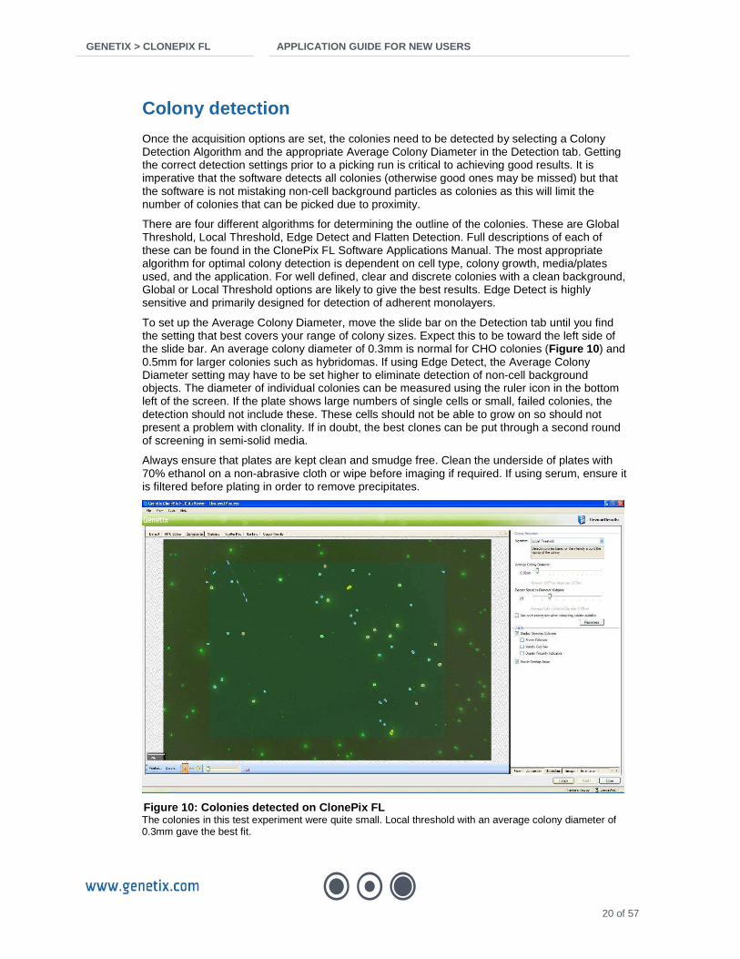
Once the acquisition options are set, the colonies need to be detected by selecting a Colony Detection Algorithm and the appropriate Average Colony Diameter in the Detection tab. Getting the correct detection settings prior to a picking run is critical to achieving good results. It is imperative that the software detects all colonies (otherwise good ones may be missed) but that the software is not mistaking non-cell background particles as colonies as this will limit the number of colonies that can be picked due to proximity.
There are four different algorithms for determining the outline of the colonies. These are Global Threshold, Local Threshold, Edge Detect and Flatten Detection. Full descriptions of each of these can be found in the ClonePix FL Software Applications Manual. The most appropriate algorithm for optimal colony detection is dependent on cell type, colony growth, media/plates used, and the application. For well defined, clear and discrete colonies with a clean background, Global or Local Threshold options are likely to give the best results. Edge Detect is highly sensitive and primarily designed for detection of adherent monolayers.
To set up the Average Colony Diameter, move the slide bar on the Detection tab until you find the setting that best covers your range of colony sizes. Expect this to be toward the left side of the slide bar. An average colony diameter of 0.3mm is normal for CHO colonies (Figure 10 ) and 0.5mm for larger colonies such as hybridomas. If using Edge Detect, the Average Colony Diameter setting may have to be set higher to eliminate detection of non-cell background objects. The diameter of individual colonies can be measured using the ruler icon in the bottom left of the screen. If the plate shows large numbers of single cells or small, failed colonies, the detection should not include these. These cells should not be able to grow on so should not present a problem with clonality. If in doubt, the best clones can be put through a second round of screening in semi-solid media.
Always ensure that plates are kept clean and smudge free. Clean the underside of plates with 70% ethanol on a non-abrasive cloth or wipe before imaging if required. If using serum, ensure it is filtered before plating in order to remove precipitates.
Figure 10: Colonies detected on ClonePix FL The colonies in this test experiment were quite small. Local threshold with an average colony diameter of 0.3mm gave the best fit.

GENETIX > CLONEPIX FL APPLICATION GUIDE FOR NEW US ERS
21 of 57
Groups
Once the whole plate or batch has been imaged and the colonies automatically detected, the Groups tab can be set up.
Getting the correct settings for groups allows the user to ensure the resulting clones are monoclonal (by using the settings for colony shape). In addition, it enables the user to influence the growth rates of the resulting clones (by using the settings for colony size). Perhaps most importantly, the user can ensure that only the very highest expressing colonies are picked (by using the fluorescent settings and colony size settings). The Rank Plot and Scatter Plot can be used to analyze different statistic features across the population, aiding the set-up of groups to include or exclude certain types of colonies.
The default group values provide a good starting point:
Too big
This parameter excludes unwanted objects (e.g. bubbles) and colonies that are too big (as colonies growing too quickly are unlikely to be high producers). Keep this set to the default value of > 0.7 mm2.
Too small
The recommended minimum colony area is 0.1 mm2 but it is feasible to go as low as 0.05 mm2 with minimal loss, or even 0.03 mm2 with some loss of outgrowth success. Lower settings will result in very poor outgrowth as the limited number of cells will be unlikely to survive the low plating density. A small colony is also indicative of a slow growth rate. See below for more information on size of clones.
Irregular 1 - Compactness
This measures the compactness of a colony. A perfectly circular colony would have a value of 1. A colony with a perimeter twice as long as it would be if it were a perfect circle would have a value of 0.5, and so on.
Irregular 2 - Axis Ratio This measures the ratio of the axes of a colony. A perfectly circular colony would have a value of 1. A colony with the shortest axis half the length of the longest axis would have a value of 0.5, and so on.
In both case, a value of less than 0.6 is not advisable as picked colonies are likely to have derived from more than 1 cell, and will therefore not result in a monoclonal population.

GENETIX > CLONEPIX FL APPLICATION GUIDE FOR NEW US ERS
22 of 57
Proximity How close the colony is to its nearest neighbor. 1mm (~30 pixels) is considered a safe setting for clonality work. This may be reduced down to 0.8 or 0.7mm without significant increase in risk, or can be reduced substantially to 0.3 or 0.2mm if the user intends to re-clone.
5. Optimizing conditions for post-pick outgrowth Optimal cell growth after picking is key to the overall success of the cloning experiment. Genetix’ range of XP Media liquid expansion media are specifically designed for this purpose and provide continuity in formulation when used in conjunction with the respective CloneMedia semi-solid media product. Superior outgrowth rates will generally be achieved if picked clones are initially collected into XP Media and then moved to your preferred growth medium than to pick directly into the growth medium.
If you choose to develop your own post-pick outgrowth media, it should be formulated to support low cell numbers. It is recommended to pre-test a number of different outgrowth media variants with cells seeded at low density (50-200 cells/well), and select the medium that provides healthy cells with the best outgrowth rate. A good outgrowth medium should reach >70% confluence within 14 days as measured by CloneSelect Imager.
Cells may undergo some stress as a consequence of being moved to liquid conditions from a semi-solid environment, but the picking of a colony of clonal cells greatly improves the chances of successful outgrowth. Other ways to maximize the outgrowth success rate are 1) pick colonies into the destination well without dispersal so that they remain in close proximity to each other for the first few days, and 2) start with a low initial volume in the destination well (for example 100µl) to increase the rate that the cells condition the medium, and then supplement with further medium after a few days. Where selective pressure is high it may be beneficial to lower the concentration of the selective agent or remove it completely for at least the first few days after picking.
A representative sample of clones should be picked from the test plates into the chosen outgrowth media to verify survival. If survival is low, the media may require additional supplementation or the colonies may need to be grown further before picking. CloneSelect Imager allows monitoring of growth post-picking and the confluence data may be used to normalize early ELISA data (Figure 11 ).
Figure 11: Verification of good outgrowth after pic king on ClonePix FL Example of good outgrowth of clones picked from a transfected CHO-S population under serum free conditions. Confluence measurements and growth curves produced on CloneSelect Imager.

GENETIX > CLONEPIX FL APPLICATION GUIDE FOR NEW US ERS
23 of 57
6. Defining parameters to find the best clones A small improvement in productivity can make a huge difference to cost when it comes to producing a biotherapeutic on an industrial scale. Colonies grown in semi-solid medium from a heterogeneous population will often show diverse physical characteristics (Figure 12 ). Understanding what physical characteristics identify a clone as a good producer enables ClonePix FL to work most efficiently at picking the best.
Figure 12: Composite image from ClonePix FL showing the diversity of colony characteristics A transfected CHO cell population showing a variety of clones with different size and expression profiles. Using the statistical features on ClonePix FL, these can be divided into separate groups for exclusion or picking.
Once good semi-solid media conditions have been established, it is recommended that an experiment be carried out to determine the parameters that will best identify your high value clones. A large number of identical test plates should be set up so that each parameter can be analyzed separately. Four plates for each parameter should be sufficient to provide good data confidence, although fewer can be used for a small test experiment. Each batch should be picked from separately, keeping all settings identical except the parameter used to identify the best clones. The parameters most likely to successfully and efficiently detect the highest producers are the fluorescence measurements of Sum Total Intensity, Exterior Mean Intensity, Interior Mean Intensity and Normalized Intensity. In the software they will be prefixed by the filter used e.g. [FITC] Exterior Mean Intensity.
High interior intensity clones
Good size clone, good secretion
Small, bright clones
Small clone with high secretion

GENETIX > CLONEPIX FL APPLICATION GUIDE FOR NEW US ERS
24 of 57
Example experiment
Aim To take a mixed population of transfected CHO cells and identify the best parameters to select the highest expressers.
Materials • A bulk selected population of IgG-secreting CHO DG44 cells selected in 250nM
methotrexate • CloneMedia-CHO (K8710) • CloneDetect FITC anti-human (K8200) • 0.1mM stock Methotrexate • Dialyzed FBS to a final volume of 5% • XP Media-CHO (K8750) • PetriWell-1 plates non-TC treated (W1055) • PetriWell-96 plates non-TC treated (W1555)
Method A mixed population of DG44 cells was seeded into CloneMedia to give 20 x 1-well plates.
CloneDetect was added to the media and cells at Day 0.
At Day 10, the plates were divided into 4 batches to be picked using the 4 main fluorescence parameters.
• Group A: Exterior Mean Fluorescence Intensity • Group B: Normalized Fluorescence Intensity • Group C: Interior Mean Fluorescence Intensity • Group D: Sum Total Fluorescence Intensity
Figure 13 shows the Rank Plot for [FITC] Exterior Mean Intensity. The threshold was set so that any clone with medium or high Exterior Mean Intensity was included in the Accept group. A group was set up so that any clone with low Exterior Mean Intensity was gated off. Only cells which passes the clonality aspects of the groups filter, and which were over a threshold value of fluorescence were picked. The same was done for each of the other batches.
96 clones (or as close to that as possible without picking clones of low fluorescence) were picked from each batch. These were then grown in 96 well plates for 7 days while monitoring for growth using CloneSelect Imager. The media was then changed and the cells left for another 5 days before supernatant samples were taken for IgG quantification by ELISA.
ELISA values were compared across the groups to determine which statistical feature gave the best clones. The best clones were then compared back to the fluorescence values to determine where they were placed in the ranking and to look for correlation.

GENETIX > CLONEPIX FL APPLICATION GUIDE FOR NEW US ERS
25 of 57
Figure 13: Rank plot showing the [FITC] Exterior Me an Intensity values for clones within a batch The threshold should not be set below the level where the curve flattens out if picking for high expression. The low Exterior Mean Intensity clones (green) are excluded from picking by adding another group to discriminate between clones. In the example above, any clones with a FITC exterior mean intensity value lower than 100 would not form part of the Accept group and so would not be picked.
Results The results of the experiment are shown in Figure 14 . In this example [FITC] Exterior Mean Intensity gave the best result with Sum Total Intensity a close second.
n Average productivity (µg/ml)
Productivity corrected to 100% confluence (µg/ml)
A: Exterior Mean 57 34.91 54.28
B: Normalized 22 23.22 35.31
C: Interior Mean 23 21.05 29.33
D: Sum Total 40 24.80 47.79
Figure 14: Productivity results of clones picked by different selection parameters n = number of clones grown successfully post-picking. Only clones showing growth were used in the average productivity calculation. Productivity corrected for 100% confluence uses the percentage confluence measured at the time of supernatant harvest. IgG concentration measured by ELISA was divided by the confluence and multiplied by 100. In most cases this is only required minor adjustments to normalize the data for cell number.
Rank Order of Clones in Group A
0.00
200.00
400.00
600.00
800.00
1,000.00
1,200.00
0 20 40 60 80 100 120 140 160
Clone rank
[FIT
C 3
00m
s] E
xter
ior
Mea
n In
tens
ity
High Exterior Mean Intensity
Low Exterior Mean Intensity
Average Productivity of Groups A-D
0
10
20
30
40
50
60
A: Exterior Mean B: Normalized C: Interior Mean D: Sum Total
Ave
rage
pro
duct
ivity
(ug/
ml)
Average productivity
Average productivity correctedfor 100% confluence

GENETIX > CLONEPIX FL APPLICATION GUIDE FOR NEW US ERS
26 of 57
Refining the criteria In order to refine the criteria for selecting the best clones, the data for the high FITC Exterior Mean Intensity group was re-analyzed by removing data points below 400U, increasing the minimum colony size to 0.05mm2 and decreasing the maximum size to 0.2mm2 then 0.1mm2. The results of this re-analysis are shown in Figure 15 . Tightening the fluorescence and size criteria together doubled the average productivity of the included clones.
n Average productivity (µg/ml)
Productivity corrected to 100% confluence (µg/ml)
Original Group A 57 34.00 54.3
<0.2mm2 32 55.25 86.3
<0.1mm2 20 78.45 124.6
Figure 15: Results of refining criteria to enhance selection of best clones. n = number of clones grown successfully post-picking that fell into the specified cut-off group. Second group criteria: [FITC] exterior mean <400U and colonies 0.05mm2 to 0.2mm2. Third group criteria: [FITC] exterior mean <400U and colonies 0.05mm2 to 0.1mm2.
Conclusions and application to other projects In this example, Exterior Mean Intensity was found to be the statistical feature which best selected the clones with highest productivity. Figure 11 showed that only clones with very low fluorescence had been excluded from picking. Refining the criteria to analyze the group with fluorescence over 400U instead of the threshold of 100U set in the original experiment eliminated a number of low productivity clones.
When analyzing the data, it was clear that the size of the clone was a factor in predicting productivity and that the larger clones are undesirable. Large clones can give a falsely high value for Exterior Mean Intensity because it will be producing a large quantity of antibody overall (giving a high mean intensity reading immediately around the clone) even if the same is not true on a per cell basis. The two refinements of the picking criteria eliminated large colonies by taking the maximum size down from 0.7mm2 to 0.2mm2 then 0.1mm2. The decrease of maximum size from 0.2mm2 to 0.1mm2 notably improved prediction of the better-producing clones showing that, with this data set, clones larger than 0.1mm2 were not high producers.
The results are not entirely unexpected because this cell line is secreting the IgG in large quantities beyond the colony perimeter so an exterior measurement of fluorescence would be expected to be predictive of productivity. The high productivity levels of the best clones in this population suggest that the higher secretors do not have sufficient resources for high growth rate, and conversely that fast growers are not going to be the highest producers.
Selecting clones by Sum Total Intensity was the second-best predictor of high productivity. This feature takes the total fluorescence associated with the colony and its exterior area into account. It might be expected that Sum Total Intensity will not be as strong a predictor as Exterior Mean Intensity because a large colony with a high intensity fluorescent area would be ranked superior to a small colony with a high intensity fluorescent area, whereas by Exterior Mean Intensity the
0
20
40
60
80
100
120
140
Original Group A <0.2mm2 <0.1mm2
Ave
rage
pro
duct
ivity
(ug
/ml)
Average productivity
Average confluence correctedproductivity

GENETIX > CLONEPIX FL APPLICATION GUIDE FOR NEW US ERS
27 of 57
effect of the colony is excluded. Interior Mean Intensity is primarily designed for measuring fluorescence closely associated with the colony (e.g. internal GFP expression or probe detection of surface proteins) and so takes no account of secretion. It would thus not be expected to be as predictive where secretion from colonies is evident. Normalized Intensity takes the Sum Total Intensity value and corrects for the size of the colony. This might be expected to be the best predictor of specific production rate but unfortunately tends to bias towards the smallest clones (for further information please refer to Appendix C of the Software Manual). In some cases, users have reported that Normalized Intensity can be a good predictor so long as minimum colony size is kept high enough to reduce the effect of the bias.
The above example only provides guidelines on how to test for best parameters for your cells in your working environment. With any cell line it is important to consider the location of the fluorescence and apply this knowledge to determine which of the features should be predictive of productivity. Intracellular or cell-surface fluorescence may be best represented by Interior Mean Intensity. Isolating hybridomas from fusions where expression of IgG may be low and there may be much background interference will most likely be best isolated by Sum Total Intensity. Re-analysis of statistical data generated by ClonePix FL can provide vital information for improving the result of a picking run.
7. Analyzing stability and sub-cloning Stability analysis
Ensuring genetic stability in a clonal population of cells is a crucial aspect in cell line selection. A cell line in which a percentage of the cells become unstable will show a drop in production rate leading to a potentially serious loss of product generation. The quest to find the highest possible producers means that clones are often selected that are inputting so much energy into production that growth rate is limited. As a consequence, if a single cell mutates such that it bypasses production of the protein of interest, it can then grow substantially faster leading to a rapid loss of productivity as the mutant strain takes over the population. Therefore, it is important to verify stability of high producing cell lines soon after initial selection and advisable to repeat this at multiple points through the expansion process.
ClonePix FL provides a simple and powerful way to screen for early instability by allowing the user to observe daughter clones within a cell line that are failing to secrete the protein of interest (Figure 16 ).
Method • Label up sufficient PetriWell-6 plates for the number of clones to be analyzed for stability.
Up to 60 can be handled fairly easily in a batch. • Dilute appropriate CloneMedia to final working volume including the fluorescent detection
probe of interest. For CloneDetect use 100µl per 10mls of CloneMedia. • Mix thoroughly by gentle rotation. Do not add cells. • Pipette 2mls per well into PetriWell-6 plates. • Pipette approximately 500-2000 cells from the first clone into the CloneMedia in the first well
while stirring around, and then keep stirring to spread the cells. A typical transfer volume would be 20-50µl.
• Repeat for all other clones. • Incubate for 7 days. • Inspect for stability by carrying out an Imaging Run on ClonePix FL.
For each well, a large number of small clones in close proximity should be observed (note: proximity is unimportant here as this is simply an offline visual check). The best way to check the stability of each clone is to toggle between white light and fluorescent images, and any that are not stable should be obvious. There may be minor differences in size and fluorescence but there

GENETIX > CLONEPIX FL APPLICATION GUIDE FOR NEW US ERS
28 of 57
should not be any low or non-expressers in the population. Any cell lines that show heterogeneity should be discarded. However, some users have reported that re-cloning unstable clones by picking high producing daughter clones can recover cell lines and make them stable.
Stable Unstable
White light
(=colonies)
FITC
(=secretion)
Figure 16: Screening for clone instability on Clone Pix FL A stable clone (left panel) is identified by all daughter clones showing fluorescence and all at a similar level. An unstable clone (right panel) is identified by a heterogeneous mix of fluorescence levels. In this case, about a third of the daughter clones are not expressing at all.
Sub-cloning
In a similar manner to the above stability analysis, ClonePix FL provides a robust way to do sub-cloning or cell line rescue. This method is very effective and widely used. Because discrete clones will be picked, it is important that the cell population is plated at lower density, and that the distribution of cells is optimal. It requires more handling than the stability analysis method.
Method • Label up sufficient PetriWell-6 plates for the number of best candidates clones to be sub-
cloned. A maximum of 24 clones is suggested and it is best to assign 2 wells to each clone. • Dilute appropriate CloneMedia to final working volume including the fluorescent detection
probe of interest. For CloneDetect use 100µl per 10mls of CloneMedia. • Mix thoroughly by gentle rotation. Do not add cells. • Aliquot the CloneMedia into 15ml tubes with 4-5ml in each. • Determine viable cell counts for each clone. • To the 15ml tubes, pipette an appropriate number of cells from each clone to generate ~50
colonies per well. The number required will depend on your cell type. For guidelines, aim for the lower end of recommended seeding densities (see page 15).
• Mix tubes well to distribute cells evenly. • Pipette 2mls per well into PetriWell-6 plates using a 10ml disposable pipette. Some users
prefer to pipette 1ml to the middle of the well and then pipette a further 1ml of CloneMedia + CloneDetect without cells around the perimeter to maintain the cells in the center of the well.
• Incubate for the required culture time (see page 18). • Pick sub-clones using the ClonePix FL.

GENETIX > CLONEPIX FL APPLICATION GUIDE FOR NEW US ERS
29 of 57
Specific protocols
Reducing timelines for DHFR / Methotrexate selection on ClonePix FL Introduction
Dihydrofolate reductase (DHFR) catalyses the reduction of folate to tetrahydrofolate and so is part of the biosynthesis pathway for purine, thymidylate and glycine. The cell lines CHO DG44 and DUXB11 have mutant alleles of dhfr and cannot survive in the absence of hypoxanthine and thymidine (HT) which enable use of a salvage pathway.
Cells are transfected with dhfr and the gene of interest (on 1 or 2 co-transfected vectors), and selected for ability to grow in HT minus (HT-) media. If two vectors are co-transfected, a second selectable marker such as neomycin resistance is normally used.
Cells may be cloned out at this stage and analyzed for productivity of the protein of interest, or they may be subject to methotrexate (MTX) selection to increase productivity. MTX is a folate analogue which binds DHFR. The concentration of the drug is increased in a stepwise manner to encourage amplification of the dhfr locus in response. Only cells which produce DHFR at a high enough level will survive. A large region of the surrounding DNA is amplified at the same time, so the gene of interest should also be present at high copy number.
At each round of amplification, it is necessary to split the population into separate flasks and expose to varying concentrations of MTX. The flask which shows about 10% survival will be selected, stabilized, split and exposed to the next range of concentrations. Attaining the maximum level of expression may require raising the level of MTX up to 2µM. Reports suggest that there is no benefit from using greater concentrations. The resulting cell lines can show very high levels of productivity in the multiple grams per liter range.
The whole process of amplification and selection routinely takes 6 months. There is a high cost in terms of timescales and manual input.
An alternative process on ClonePix FL
After selection in HT- media, the population is diverse in expression of DHFR and the protein of interest. ClonePix FL can be used at this stage to screen and pick out those clones with highest expression of the protein of interest. Figure 17 shows that a minority of clones demonstrate high secretion at this stage, which may represent an early increase in copy number and thus a potentially higher propensity for amplification. If higher expression is needed the best picked clones can be exposed directly to high levels of MTX, thus shortcutting the process of selection. This can be done using separate clones, or by pooling the best clones together prior to MTX escalation.

GENETIX > CLONEPIX FL APPLICATION GUIDE FOR NEW US ERS
30 of 57
A) White light B) FITC
Figure 17: A mixed cell population of transfected C HO DG44 in HT- selection only DG44 cells were transfected to express human IgG and then bulk selected in the absence of hypoxanthine and thymidine (HT-) prior to being plated in CloneMedia-CHO under HT- conditions in the presence of CloneDetect FITC anti-human IgG (B). The high expressing clones are circled in red on image A.
Recommended DHFR work flow using ClonePix FL
Day 2
Day 12
Day 24
Day 54
2-3 months
Day 0 Transfect cells with dhfr plus gene of interest
Grow cells in HT- media (plus G418 if using)
Plate whole population in semi-solid media
Screen on ClonePix FL and isolate best 200-300 clon es
Scale up cells for further characterization or pool and expose to high methotrexate – 0.1 to 1µM
Isolate clonal cell lines using ClonePix FL or chec k progress of amplification at key points by re-plating a sample of the population in semi-solid media
Verify stability of highest expressers on ClonePix FL using stability screening method

GENETIX > CLONEPIX FL APPLICATION GUIDE FOR NEW US ERS
31 of 57
Compatible media
The following products are recommended for DHFR / MTX selection.
• Semi-solid media: CloneMedia-CHO (K8710) • Expansion media: XP Media-CHO (K8750) • Methylcellulose concentrate: CloneMatrix optimized for CHO (K8530) • Antibody detection: CloneDetect anti-human IgG (K8200)
CloneMedia-CHO and XP Media-CHO have been shown to support good growth of dhfr+ cells but not dhfr- cells. All media products are chemically defined but can be supplemented with serum if required. Refer to the product documentation for specific instructions on use.
Protocol guidelines
Selection of DG44 cells by ClonePix FL is most effective early in the process during HT- selection or at low MTX levels. The power of ClonePix FL at high levels of MTX should be expected to be much less due to the reduced heterogeneity of secretion in the population. However, it may be useful to analyze the stability of the highly amplified population or to re-clone using ClonePix FL.
Cells must be left for a short recovery period post-transfection to improve viability and to allow the initial selection of transfected cells to take place. 48 hours is sufficient. When plating cells within the first two weeks after transfection, a high seeding density is required. 1000-2000cells/ml is sufficient. A mixed population established for more than two weeks without MTX selection or in low concentrations (up to 500nM MTX) can be plated at 350-500 cells/ml. A cell line or mixed population in high MTX (greater than 1µM) will be under significant pressure and so the seeding density will need to be increased up to 2000 cells/ml. Supplementation such as glutamine (if making up own media from CloneMatrix, CloneMedia products contain 8mM glutamine), insulin or other supplements may help cell viability. The recommended density for viable, established (at least four cell divisions) colonies is 100-200 per well of a 6-well plate or 500-1000 clones per 1-well plate.
Incubate the cells in semi-solid media for 10-12 days. Image and pick cells on ClonePix FL, selecting the top 5-10% of expressers based on high Exterior Mean Fluorescence Intensity or Sum Total Fluorescence Intensity. Colonies should be between 0.05 and 0.2mm2 for the best result.
Monitor the outgrowth of colonies and do an ELISA screen after 7-10 days when the clones have reached 40-70% confluence. If using CloneSelect Imager, the growth rate and final confluence may be taken into account when assessing the highest producers on a per cell basis.
Once the best clones are established, they may be scaled up and characterized or pooled and amplified. Rejecting the poor expressers early on in the process will leave an enriched pool of higher quality clones to amplify where expression levels are already good.
Stability analysis can be done at any stage to assess the variability within the population. A few thousand cells from a clone are seeded into an aliquot of semi-solid media in one or more wells in a 6-well plate. The plates should be incubated for 7 days and analyzed using ClonePix FL software. The rank plot and scatter plot can be used to view the relevant statistical features of fluorescence and size. The Rank Plot should show a slight slope to the left hand side, a leveling off and then a slightly steeper curve (S-shape). Figure 18 shows a cell line demonstrating this effect. If there is a very steep drop off from the left of the graph and a significant number of low producers then the cell line is not stable or clonal. It can be discarded in favor of better clones or re-cloned.

GENETIX > CLONEPIX FL APPLICATION GUIDE FOR NEW US ERS
32 of 57
Figure 18: Rank Plot from ClonePix FL software show ing the order of detected colonies from high to low fluorescence
Figure 19 shows a fluorescent image of the same cell line. Although some clones show a greater exterior fluorescence area the intensity of the clones is very similar. In this case small differences are due to colony size.
Figure 19: An image of the same cell line shown in Figure 18 showing detected colonies and groups used to analyze colonies The image was captured at 500ms FITC exposure.
Conclusions
Production of therapeutic or research antibodies in DG44 cells by MTX amplification can result in very high levels of protein of interest but requires up to 6 months to complete the process.
ClonePix FL offers an alternative process of early screening to identify and pick medium to high expressing clones that are better candidates for rapid amplification. Both the time line and quality of the cell lines produced can be improved using this method.

GENETIX > CLONEPIX FL APPLICATION GUIDE FOR NEW US ERS
33 of 57
Using CloneMedia-CHO for selection of DG44 cells Introduction
The cell lines CHO DG44 and DUXB11 have mutant alleles of the dhfr gene and cannot survive in the absence of hypoxanthine, thymidine and glycine which enable use of a salvage pathway. CloneMedia-CHO is a complete semi-solid media product compatible with a broad range of CHO cell types. The optimal composition of CloneMedia-CHO means that it is free of hypoxanthine or glycine, but contains a trace amount of thymidine at a concentration 8-fold lower than that necessary for rescue of dhfr-- cell lines. The equivalent liquid media for cell line expansion, XP Media-CHO, also contains the trace amount of thymidine. The semi-solid concentrate for CHO cells, CloneMatrix-CHO, is provided with a vial of CloneXL that contains a 50x concentrate of thymidine to provide the same working concentration in final CloneMatrix-based media. The following study investigates the whether this level of thymidine is sufficient to support growth of dhfr- cells, and if this is affected by the presence of methotrexate (MTX).
Materials
• CHO/dhfr- cells (ATCC CRL-9096) • CloneMedia-CHO (Genetix K8710) • Dialyzed FBS (dFBS) (Gibco 26400-044) • MTX (Sigma M9929) • 100x Hypoxanthine, Thymidine (HT) solution (Sigma 11067-030) • PetriWell-6 plates, non TC treated (Genetix W1155)
Methods
CHO DG44 dhfr- cells were grown in static culture in alpha MEM media minus nucleosides, 5% dFBS and 1xHT solution. Cells were trypsinized, counted and seeded into aliquots of CloneMedia-CHO supplemented with 5%dFBS at 500cells/ml.
The following conditions were set up:
• A. No HT or MTX • B. 1x HT • C. 100nM MTX • D. 100nM MTX and 1x HT
All reagents were brought to room temperature before mixing, and the cells were 98% viable before plating. Each condition was prepared to a volume of 15mls in a 15ml Falcon tube, and then one 6-well plate was plated at 2mls per well for each condition. Plated cells were incubated undisturbed for 4 days and then imaged at days 4, 5 and 6 on CloneSelect Imager.
Results
Images of colony growth for each condition at day 6 are shown in Figure 20 . The results show that CloneMedia-CHO without HT supplementation is not sufficient to support the growth of dhfr- cells (A and C). The addition of 1X HT is required to rescue the cells whether in the absence or presence of 100nM MTX (B and D).

GENETIX > CLONEPIX FL APPLICATION GUIDE FOR NEW US ERS
34 of 57
A. No HT or MTX
B. 1x HT
C. 100nM MTX
D. 100nM MTX and 1x HT
Figure 20: Images of DG44 colony growth in CloneMed ia-CHO at day 6 with or without HT and MTX Colony growth was evident only in the presence of 1x HT irrespective of the absence or presence of MTX. A Single cells are visible but no cell division can be seen. B Compact colonies of about 32 cells are clearly visible and cells are showing expected level of growth. C Only single cells can be seen with no evidence of cell division. D Cells have undergone at least 5 cell divisions and are looking very healthy.

GENETIX > CLONEPIX FL APPLICATION GUIDE FOR NEW US ERS
35 of 57
Figure 21 shows good selective growth of a non-clonal pool of DG44 cells under MTX selection in CloneMedia-CHO.
Figure 21: Images of a DG44 non-clonal population of cells for ming colonies in CloneMedia-CHO
Cells expressing a human IgG were incubated with FITC anti-human CloneDetect (K8200). The image on the left is a 90ms white light exposure taken on ClonePix FL. The image on the right is of the same population taken with a 500ms FITC exposure, showing the comparative expression levels of the colonies.
Conclusions
The cells had a high viability before plating and all the plates were identical except for the supplementation with HT and/or the selection agent, MTX. The seeding density used in the experiment was 500 cells/ml (=1000 cells/well). This is about twice as high as would be needed for a cloning experiment and the colonies can be seen quite close to each other in the conditions that grew well. A high seeding density was necessary to increase the number of cells screened in this experiment and give the cells every chance of growing through.
In a dhfr- cell line, the cells will not be able to use the primary pathway for purine and pyrimidine biosynthesis involving reduction of folate to tetrahydrofolate. The alternative salvage pathway minimally requires the presence of hypoxanthine and thymidine. The addition of MTX should not additionally affect dhfr- cell lines because they are already using the salvage pathway but it will suppress any dhfr+ revertants. In this study, there were no revertants, as evidenced by the lack of any colony growth in CloneMedia-CHO only (condition A).
This study demonstrates that the level of thymidine at 8-fold lower than the amount required for mutation rescue is not sufficient to support growth of dhfr- cells. CloneMedia-CHO and XP Media-CHO are therefore suitable for selection of dhfr+ cells whether in the absence or presence of MTX.
Supplementary information
Levels of thymidine, hypoxanthine and glycine requir ed to rescue and culture dhfr- cells:
• Hypoxanthine 6.8mg/L (0.05mM) • Glycine 20mg/L (0.26mM) • Thymidine 1.94mg/L (0.008mM)
Final working concentrations in CloneMedia-CHO and XP Media-CHO:
• Hypoxanthine nil • Glycine nil • Thymidine 0.24mg/L (0.0010mM)

GENETIX > CLONEPIX FL APPLICATION GUIDE FOR NEW US ERS
36 of 57
Growth of CHOK1SV in semi-solid media and selection of high expressers Introduction
CHO cells use glutamine synthase (GS) to catalyze the synthesis of glutamine from glutamate and ammonia. Methionine sulfoximine (MSX) blocks endogenous GS activity, allowing MSX to be used as a selectable marker. A plasmid containing GS and the gene of interest is transfected into CHOK1SV cells and the population is then selected for growth in glutamine free media.
The CHOK1SV line is adapted for good growth and expression at high density in a bioreactor but this makes them difficult to clone as single cells. Cloning of transfected cells is traditionally done using limiting dilution at 4000 cells/well in a 96 well plate. Because of low transfection efficiency and survivability of the cells, only about 15 clones will grow across each plate.
ClonePix FL bypasses this by screening the whole population of cells after the initial effect of the selective agent. This gives better cloning efficiency and fewer transients.
The stability of these cells can be an issue. ClonePix FL allows re-screening of cells within three weeks of isolation. A few thousand cells are plated back into a small volume (2ml) of semi-solid media and analyzed for divergence of the population.
Methods and Workflow
The guidelines given here are specifically for CHOK1SV cells in serum free or chemically defined media and under MSX selection.
Transfected cells Transfected cells are grown and bulk selected in media containing 25 or 50µM MSX for 14 days prior to seeding into semi-solid media. Cells should be kept in static culture at about 2x10⁵ viable cells/ml in Gibco CD CHO or other similar media as recommended by Lonza. Experiments at Genetix have shown that the total cell number changes little during this period as non-transfected cells die and are replaced by the expanding transfected population.
At day 14, cell viability will start to pick up and this is the best time to plate cells. Cells should be seeded into a 100ml bottle of CloneMedia CHOK1SV (K8725) at 2000 cells/ml, although optimal plating density will be dependent on the transfection efficiency and cell handling practices of each laboratory and may need to be determined empirically. MSX can be supplemented into the semi-solid media but if cell growth problems are encountered it may be necessary to not add selective pressure during the semi-solid media phase. By separating the bulk selected cells from each other in semi-solid media and then screening for high antibody expression, the most productive clonal colonies can be isolated efficiently without the need for MSX. The isolated clones can be returned to MSX selection shortly after picking.
CloneDetect anti-human detection agent (K8200) can be added to the semi-solid media at the point of cell plating. However, to prevent transient expressers that remain viable after bulk selection from being visualized as IgG-positive clones, it is recommended that CloneDetect should be sprayed on via atomizer applicator (K8201) ten days after plating. At day 14, the clones should be imaged on ClonePix FL (Figure 22 ).
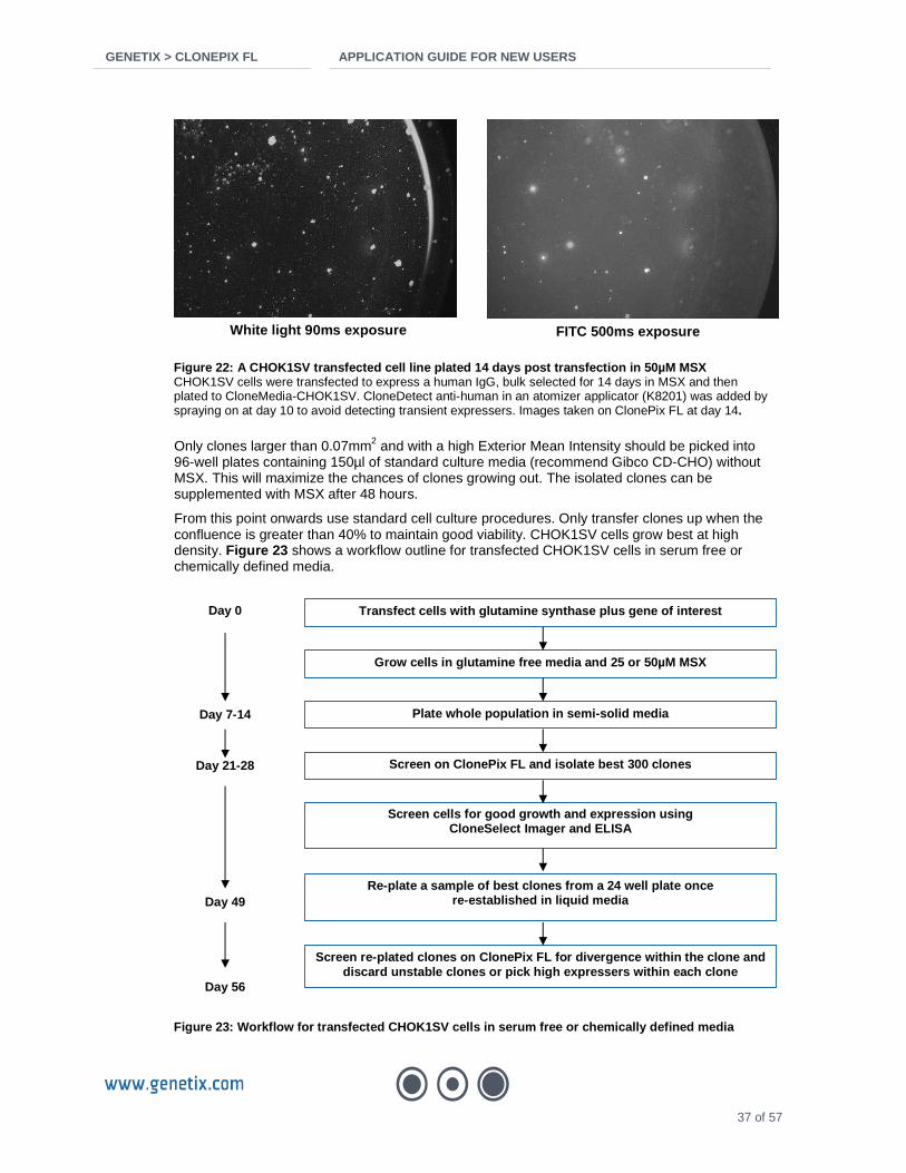
GENETIX > CLONEPIX FL APPLICATION GUIDE FOR NEW US ERS
37 of 57
Figure 22: A CHOK1SV transfected cell line plated 1 4 days post transfection in 50µM MSX CHOK1SV cells were transfected to express a human IgG, bulk selected for 14 days in MSX and then plated to CloneMedia-CHOK1SV. CloneDetect anti-human in an atomizer applicator (K8201) was added by spraying on at day 10 to avoid detecting transient expressers. Images taken on ClonePix FL at day 14.
Only clones larger than 0.07mm2 and with a high Exterior Mean Intensity should be picked into 96-well plates containing 150µl of standard culture media (recommend Gibco CD-CHO) without MSX. This will maximize the chances of clones growing out. The isolated clones can be supplemented with MSX after 48 hours.
From this point onwards use standard cell culture procedures. Only transfer clones up when the confluence is greater than 40% to maintain good viability. CHOK1SV cells grow best at high density. Figure 23 shows a workflow outline for transfected CHOK1SV cells in serum free or chemically defined media.
Figure 23: Workflow for transfected CHOK1SV cells i n serum free or chemically defined media
White light 90ms exposure FITC 500ms exposure
Transfect cells with glutamine synthase plus gene o f interest
Grow cells in glutamine free media and 25 or 50µM M SX
Plate whole population in semi-solid media
Screen on ClonePix FL and isolate best 300 clones
Screen cells for good growth and expression using CloneSelect Imager and ELISA
Re-plate a sample of best clones from a 24 well pla te once re-established in liquid media
Screen re-plated clones on ClonePix FL for divergen ce within the clone and discard unstable clones or pick high expressers wit hin each clone
Day 7-14
Day 21-28
Day 49
Day 56
Day 0

GENETIX > CLONEPIX FL APPLICATION GUIDE FOR NEW US ERS
38 of 57
Established cell lines Established cell lines should be cultured in Gibco CD CHO or similar media as per standard protocol. 48 hours after passage, cells should be plated at 500-1000 cells/ml into CloneMedia-CHOK1SV. The protocol for transfected cells can then be followed with the exception that cells can be seeded in the presence of CloneDetect (K8200) since there is no risk of detecting false positives from transient expressers. Figure 24 shows a stable high expressing CHOK1SV cell line seeded into CloneMedia with CloneDetect.
Figure 24: A CHOK1SV stable cell line plated into C loneMedia with CloneDetect A human IgG expressing CHOK1SV cell line selected on ClonePix FL and grown to 24 well stage was re-seeded into CloneMedia-CHOK1SV to assess clone stability. CloneDetect anti-human (K8200) was added prior to plating. Images taken on ClonePix FL at day 10.
Stability analysis and sub-cloning of high expresse rs Once clones are established in 24 well plates, the highest expressers can be counted and a small quantity re-seeded into semi-solid media. It is sufficient to seed into one well of a 6-well plate. Use densities and conditions recommended for a stable cell line. Between days 7-10 depending on growth, the cells can be re-screened on ClonePix FL. The clones should be detected on ClonePix FL using the Local Threshold and 0.2mm Average Colony Diameter settings. Data can be analyzed on ClonePix FL or exported for further analysis.
Even within a highly cloned cell line there will be some variation in growth rate and productivity. A cell line with a normal distribution of fluorescence intensities as detected by ClonePix FL can be considered stable. If the distribution is skewed towards low fluorescing clones then the cell line may have diversified. In this case, the cell line in question should re-cloned to see if it can be recovered. The unstable phenotype does not appear to be heritable.
Troubleshooting
Plating conditions should be established with a high viability stable cell line. Several practice attempts at plating are advised if the user is unfamiliar with semi-solid media.
CHOK1SV cells grow slowly in semi-solid media. After 4 days, small clusters of 2-8 cells should be expected. If no growth is seen in the plates at this time, the plating conditions are not sufficient for cell growth. The plating should be repeated after reviewing plating conditions.
Ensure the plates are kept at high humidity by adding sterile PBS or water to the reservoirs around the wells of the PetriWell-6 PetrWell-1 plates. If the incubator used is not giving high enough humidity then cells may need to be isolated in a sterilized plastic container within the incubator with a cover placed loosely over the top.
The two time points when the cells are at low density in their immediate environment are when
White light 90ms exposure FITC 500ms exposure

GENETIX > CLONEPIX FL APPLICATION GUIDE FOR NEW US ERS
39 of 57
initially adding to semi-solid media and immediately after picking in the 96 well plate. The semi-solid media is supplemented with chemically defined components to support low cell numbers.
If problems with growth are seen post-picking, MSX could be removed from the post-pick media until cell number is higher. Alternatively the volume of the media could be dropped from 150µl to 100 or 75µl. This speeds up the rate of conditioning of the medium by the cells. Pre-conditioned media up to 10% can also be used if available.
Poor viability can be an issue when transferring cells from semi-solid media to 96-well plates in the same way as it can when transferring cells at each scale up stage. This is not directly due to the transfer method but is mostly due to CHOK1SV not coping well with low cell density. This effect can be minimized by transferring larger cell numbers – colonies larger than 0.07mm2 in semi-solid media and wells above 40% during routine cell culture.

GENETIX > CLONEPIX FL APPLICATION GUIDE FOR NEW US ERS
40 of 57
Frequently Asked Questions
Growing colonies Semi-solid media
Can cells be cultured for over 14 days in semi-soli d media? Culture periods of 14 days would be considered uncommonly long. Any decline in colony viability is NOT specific to semi-solid media.
Beyond this length of incubation, media is likely to show signs of nutrient depletion and desiccation, both of which will affect colony viability.
Furthermore, any very large colonies may start showing signs of necrosis in the colony centre due to insufficient diffusion of oxygen and nutrients. This will hamper downstream steps of colony outgrowth and cell line expansion after picking.
Can agarose or our own methylcellulose be used as a n alternative to CloneMatrix? The use of agarose is not recommended as an alternative to methylcellulose as this does not give the best conditions for growing colonies of mammalian cells or formation of the secretion complex. ClonePix FL is able to collect agarose plugs but this is primarily for its application in picking plaques of baculovirus-infected insect cells. CloneMatrix has been formulated such that the concentration and chain length of the methylcellulose is optimal for formation and automated picking of mammalian cell clones. Meticulous optimization will need to be performed if using your own methylcellulose.
The CloneMedia protocol does not recommend a serum concentration more than 1%. Why is this? What is the risk of incr easing serum concentration in CloneMedia? It is possible to use concentrations of serum over 1% but this may decrease the number of pickable colonies. Innately adherent cell types such as CHO, even after adaption to serum free conditions, will display varying degrees of adherence when grown in the presence of serum, and this becomes more of an issue at higher serum concentrations. The ideal scenario is for cells to form suspended colonies or tightly packed adherent colonies. High levels of serum may result in more dispersed colonies, making them harder to detect on ClonePix FL. Dispersed colonies also require more space and thus need to be seeded at lower seeding densities to ensure monoclonality.
Colonies are not tightly-packed and the cells have migrated away from the bulk of the colony. How can this be avoided? A case of loose or migrating colonies is shown in Figure 25 . This is most likely to occur with adherent cells particularly when the serum concentration is high (see above), if plates have been moved, or if the wrong plate type has been used.

GENETIX > CLONEPIX FL APPLICATION GUIDE FOR NEW US ERS
41 of 57
Figure 25: Image of loose or migrating colonies tak en with the CloneSelect Imager
To maximize the formation of tightly-packed colonies:
• Reduce the serum concentration where using serum. • Leave plates undisturbed for at least 4 days post-plating. This includes frequent opening of
the incubator door. • Do not use tissue culture treated plates as this will encourage colonies to grow along the
plate surface. Use non-TC treated plates such as PetriWell-1 (W1055) or PetriWell-6 (W1155).
• The compactness of the colonies can be increased by reducing the seeding density. At very high seeding densities, cells may migrate towards conditioning growth factors produced by nearby cells. The timing of imaging and picking in situations such as this can be crucial. Colonies that show this behavior often grow as discrete colonies, before abruptly disintegrating. It is therefore worth monitoring the growth of colonies (e.g. repeated imaging of a test plate on CloneSelect Imager) to determine the best time to pick. It is preferable to pick earlier using a smaller Average Colony Diameter than to pick less compact colonies. Dispersed colonies (such as those in the diagram above) are difficult to accurately image, define and group and are therefore unlikely to produce monoclonal populations.
• Some users have reported that their particular cell type benefits from being plated to a different medium viscosity. Modulation of the methylcellulose concentration should only be necessary in very exceptional circumstances. Contact [email protected] for advice.
Why is the number of colony outgrowths so low? Even with high measured viability at plating, not all cells will form colonies. This is primarily because each cell is effectively being plated singly in semi-solid medium. Cell populations that have recently undergone a major treatment such as hybridoma fusion or transfection will need to be plated at high seeding density. Newly transfected cells (4-10 days post transfection) can be plated as high as 1 x 10⁶ cells/ml. When starting optimization with a new cell type, it is advisable to test a range of cell densities.
A second consideration is the composition of the semi-solid medium. The CloneMedia range provides a good base for many cells but it may be necessary to add supplements such as those that that your cells may need in liquid conditions.
A third possibility is if the plated have been moved or subjected to vibration or knocks within the first few days. The cells are particularly sensitive at this time.

GENETIX > CLONEPIX FL APPLICATION GUIDE FOR NEW US ERS
42 of 57
Why does the semi-solid medium appear cloudy as if contaminated, despite no other indicators of contamination? The secretion complex formed between CloneDetect and the secreted protein is often mistaken for contamination. When viewed by eye or by brightfield microscopy, the precipitation complex can appear as a cloud around each colony, or as a speckled mass of tiny spots. This is normal. If viewed with a fluorescent microscope, the secretion complex can be seen to fluoresce (Figure 26).
White light/FITC composite image FITC image
Figure 26: Fluorescent microscope images of high Ig G expressing PER.C6 colonies grown in semi-solid media with CloneDetect
In addition, some turbidity of the semi-solid media itself is normal. FBS will often give a cloudy appearance to semi-solid media and should be filtered before use. Media turbidity can be minimized by bringing all components of the semi-solid media to room temperature before mixing. If any components are cold they may cause precipitation of the methylcellulose and this will not re-dissolve once plated. This will give a cloudy appearance and won’t encourage cell growth.
It is important that the semi-solid medium is thawed at 4°C or room temperature and is not heated.
Why has the semi-solid medium dried out during incu bation? Assess the humidity control capacity of the incubator being used. Incubators that are continually accessed may have a limited capacity to maintain humidity. In such cases, it can be beneficial to store the plates in sealable boxes (such as sandwich boxes) in order to maintain humidity locally. This is more important for colonies that require 12+ days to form. Please contact [email protected] for assistance.
Check that sterile H2O or PBS is added to the PetriWell plate reservoirs when plating.

GENETIX > CLONEPIX FL APPLICATION GUIDE FOR NEW US ERS
43 of 57
CloneXL CloneXL is an integral component of CloneMedia-CHO and XP Media-CHO, and is provided as a 50x concentrate in a small vial with each bottle of CloneMatrix-CHO. It is a chemically defined supplement designed to enhance cell growth at low seeding densities. It is used predominantly with CHO cells in serum free or chemically defined media. If you have serum in the media or a cell line which grows well anyway the difference plus and minus will be minimal. CloneXL is suitable for use with all CHO selection systems e.g. glutamine synthase, DHFR selection, G418 etc.
What is CloneXL? A 50x concentrate of amino acids and nucleotides.
A precipitate is visible on thawing - is this norma l? Formation of a precipitate on thawing is normal and does not affect product quality. However, it may affect product efficacy in semi-solid culture and so must be completely resuspended. Any precipitate occurring during defrost should dissolve upon mixing at room temperature. Re-suspension can be aided by aspirating and dispensing the product several times with a 1ml pipette.
Fluorescent detection in semi-solid media CloneDetect CloneDetect protocol states the concentration in Un its/ml. What is the actual concentration in mg/ml? The CloneDetect protein amount varies between products with a typical concentration of 750µg/ml (for a 7.5µg/ml final concentration). The exact concentration is specified on the Certificate of Analysis (CofA) issued with each batch.
Should I modulate the concentration of CloneDetect to match the expected secretion rate? The recommended CloneDetect working concentration of 100 Units/ml (10µl per ml of semi-solid media) has been extensively optimized for a many cell types with a wide range of expression levels and should not need adjusting. Modulation of the CloneDetect concentration should only be necessary in very exceptional circumstances. Please contact [email protected] for assistance.
Will the CloneDetect signal be affected if the cell s are repeatedly brought out of the incubator? It is good practice to ensure that plates are covered (with aluminum foil or similar) if they are to be exposed to bright light for longer than a few minutes. It is recommended that lights in tissue culture hoods are turned off when handling CloneDetect.
What is CIF? Complex Initiation Factor. CIF is unconjugated CloneDetect. The primary use is where it is desirable to create a precipitation complex around each IgG-secreting clone but without visualizing by fluorescence. For example, when also probing with FITC-conjugated antigen so

GENETIX > CLONEPIX FL APPLICATION GUIDE FOR NEW US ERS
44 of 57
that any fluorescent colony must be an antigen-specific IgG secretor. A CloneDetect with a different fluorescent wavelength to the antigen could equally be used. A case where CIF would be obligatory would be where 3 different antigens are probed simultaneously with 3 different fluorophores thus using all available multiplex channels.
Probing with antigen
What antigen concentration should be added to hybri domas plated in semi-solid medium? A suitable working concentration for antigens is 5µg/ml. If the antigen is small, it is advisable to reduce the mass to maintain a good molar ratio otherwise there is a risk of a high background of fluorescent antigen which may mask the weak positive colonies.
Recommended antigen concentrations are:
• 50-150 kDa proteins: 5 to 10 µg/ml • 10-50 kDa proteins: 1 to 5 µg/ml • 2-3 kDa peptides: 0.1 to 0.2 µg/ml
Antigen adsorption: If peptides are small, it might be worth considering conjugating them to a carrier protein. Some customers have found that this helps to prevent non-specific adsorption of the fluorescent antigen to proteins in the medium.
How can I get my antigen probe fluorescently conjug ated? There are many commercially available kits for conjugation of proteins and antibodies. Prior to using on cells in semi-solid medium, it is important to validate that the conjugated antigen is still active, i.e. comparable with unconjugated antigen. Genetix only offers a conjugation service for large scale studies.
Can I avoid fluorescently conjugating my antigen pr obe? The best scenario is always for the primary probe to be directly conjugated. However, it is possible to use a fluorescent secondary probe but it is essential to validate beforehand that there will be no cross-reaction that would complex the primary and secondary probes together in the medium thus stopping them migrating to the positive clones.
Imaging and picking on ClonePix FL Multiplexing
Can multiple fluorescent probes be used in the same experiment? Yes. ClonePix FL has 5 fluorescent filter sets. Up to three of these sets can be used simultaneously with minimal bleed-through. These are:
• CFP or EGFP/FITC • RHODAMINE/DS-RED • Cy5

GENETIX > CLONEPIX FL APPLICATION GUIDE FOR NEW US ERS
45 of 57
Will a strong FITC signal interfere with a weak rho damine label? The EGFP/FITC and RHODAMINE/DS-RED filter sets are discrete channels. However, very strong FITC fluorescence can show some bleed-through into the rhodamine channel. Rhodamine fluorescence has not been observed to bleed into the FITC channel. A “true” rhodamine signal should be clearly distinguishable from a “false” signal leaking through from the FITC channel (see Figure 27 ). Adjustments to the exposure times and LED intensities (in the Acquisition tab) should be made until the best images are obtained. If one probe is expected to show a stronger signal than the others, then it is advisable for it to be conjugated with the fluorophore with longer wavelength. For example, CloneDetect549 or CloneDetect649 should be used in conjunction with a FITC-conjugated antigen.
Figure 27: Multiplex imaging of hybridoma colonies on ClonePix FL showing minima l bleed-through Two Hybridoma populations: one expressing IgG1, the other expressing IgG2A. Colonies were grown in semi-sold media containing AlexaFluor488 anti-IgG1 (visible through the FITC filter) and AlexaFluor546 anti-IgG1 (visible through the rhodamine filter). Plates were imaged with white light, FITC 300 ms exposure and rhodamine 2000 ms exposure.
Autofluorescence
Colony fluorescence matches the shape of my colonie s. Is this really secreted protein? This is likely to be cellular autofluorescence. It is not an issue for small, healthy colonies but can become significant when colonies are large (500+ cells), and especially when cells are dying or dead. When colonies are small it is reasonable to assume there is no autofluorescence (e.g. when using Fluorescent Sum Total Intensity). However, when colonies are large, it is best to use ‘External’ parameters to exclude any contribution by autofluorescence (e.g. Exterior Mean Intensity). The main characteristic of autofluorescence is that it appears as the same shape as the colony, whereas fluorescence related to secreted product is more diffuse and often much larger than the colony.
If high autofluorescence is observed, another option may be to run ClonePix FL assays at an earlier timepoint (e.g. day 10 rather than 12). This may improve the quantification of the halo (making it smaller and brighter) as well as substantially reducing autofluorescence.
If possible, use CloneDetect 649, as cells show less autofluorescence at longer wavelengths.

GENETIX > CLONEPIX FL APPLICATION GUIDE FOR NEW US ERS
46 of 57
Post-pick growth The survival of cells after picking is very low. Is this normal? Under optimal conditions, >90% of the picked clones are expected to survive and grow after picking. The physical act of picking the cells does not harm them. It is low cell density that is the primary cause of failure of clones to grow out. For particularly delicate cells, the dispersal settings may be reduced or not used at all.
If the survival of cells after picking is low, it can be due to one or more of the following:
• Poor selection parameters (e.g. picking clones that are too small). • Unsuitable outgrowth media • Incorrect picking settings (e.g. pin height, dispersal)
Size of clones The survival and growth rate of a clone after picking is highly dependent on the size of the colony when picked (Figure 28 ).
Relationship between colony area Vs resulting growth rate of the clone
R2 = 0.2785
-0.1
0
0.1
0.2
0.3
0.4
0.5
0 0.1 0.2 0.3 0.4 0.5
colony area (mm2)
Ave
rage
gro
wth
rate
(cha
nge
in
conf
luen
ce/d
ays)
Figure 28: Graph to show area of the colonies picked and resul ting growth rate of the clones.
If a colony is small, it should not be picked because:
• That particular clone has a slow growth rate • The colony may no longer be viable • There is a risk of transferring an insufficient number of cells to the destination plate for the
clone to survive.
It is recommended to set the “Too Small” group to include colonies < 0.1 mm2, though it may be reduced to as low as 0.03 mm2 and still achieve good results.

GENETIX > CLONEPIX FL APPLICATION GUIDE FOR NEW US ERS
47 of 57
Outgrowth media The destination plate should also be checked immediately after picking in order to verify that picking was successful. If picking efficiency is low (i.e. <90% of wells have cells in them immediately after picking) the user should check that the correct pins are being used and check that the pin height and camera alignment are set correctly (see ClonePix FL Software Applications Manual).

GENETIX > CLONEPIX FL APPLICATION GUIDE FOR NEW US ERS
48 of 57
Reagents and Supplies Please refer to the Genetix website for the latest reagents & supplies, replacement parts and optional extras www.genetix.com
Detection reagents Cat No. Description Pack Size
K8290 CloneDetect-Rec. Recombinant anti-human detection agent. FITC label. Sterile, azide free.
5000U / 0.5ml
K8200 CloneDetect anti-human detection agent. FITC label. Sterile, azide free. 10000U / 1ml
K8201 CloneDetect anti-human detection agent. FITC label. Sterile, azide free. In Atomizer applicator
10000U / 5ml
K8210 CloneDetect anti-human detection agent. 549 label. Sterile, azide free. 10000U / 1ml
K8211 CloneDetect anti-human detection agent. 549 label. Sterile, azide free. In Atomizer applicator
10000U / 5ml
K8212 CloneDetect anti-human detection agent. 649 label. Sterile, azide free. 10000U / 1ml
K8213 CloneDetect anti-human detection agent. 649 label. Sterile, azide free. In Atomizer applicator
10000U / 5ml
K8215 CloneDetect anti-human CIF (Complex Initiation Factor) 10000U / 1ml
K8205 CloneDetect anti-human detection agent. Gamma chain specific. FITC label. Sterile, azide free.
10000U / 1ml
K8206 CloneDetect anti-human detection agent. Gamma chain specific. FITC label. Sterile, azide free. In Atomizer applicator
10000U / 5ml
K8252 CloneDetect anti-human detection agent. Kappa chain specific. FITC label. Sterile, azide-free.
10000U / 1ml
K8250 CloneDetect anti-human detection agent. Kappa chain specific. 549 label. Sterile, azide-free.
10000U / 1ml
K8251 CloneDetect anti-human detection agent. Kappa chain specific. 649 label. Sterile, azide-free.
10000U / 1ml
K8255 CloneDetect anti-human detection agent. IgM specific. FITC label. Sterile, azide-free. 10000U / 1ml
K8253 CloneDetect anti-human detection agent. IgM specific. 549 label. Sterile, azide-free. 10000U / 1ml
K8254 CloneDetect anti-human detection agent. IgM specific. 649 label. Sterile, azide-free. 10000U / 1ml
K8220 CloneDetect anti-mouse detection agent. FITC label. Sterile, azide free. 10000U / 1ml
K8221 CloneDetect anti-mouse detection agent. FITC label. Sterile, azide free. In Atomizer applicator
10000U / 5ml
K8230 CloneDetect anti-mouse detection agent. 549 label. Sterile, azide free. 10000U / 1ml
K8231 CloneDetect anti-mouse detection agent. 549 label. Sterile, azide free. In Atomizer applicator
10000U / 5ml
K8232 CloneDetect anti-mouse detection agent. 649 label. Sterile, azide free. 10000U / 1ml
K8233 CloneDetect anti-mouse detection agent. 649 label. Sterile, azide free. In Atomizer applicator
10000U / 5ml
K8225 CloneDetect anti-mouse detection agent. Gamma chain specific. FITC label. Sterile, azide free.
10000U / 1ml
K8226 CloneDetect anti-mouse detection agent. Gamma chain specific. FITC label. Sterile, azide free. In Atomizer applicator
10000U / 5ml
K8235 CloneDetect anti-mouse CIF (Complex Initiation Factor) 10000U / 1ml
K8240 CloneDetect anti-rat detection agent. FITC label 10000U / 1ml
K8300 LiveDetect Green (494nm Ex/ 517nm Em). Sterile. In Atomizer applicator 50ul (for 5ml)

GENETIX > CLONEPIX FL APPLICATION GUIDE FOR NEW US ERS
49 of 57
CloneMatrix Cat No. Description Pack Size K8510 CloneMatrix 1 x 40ml (100ml final media volume)
K8500 CloneMatrix 6 x 40ml (6 x 100ml final media volume)
K8530 CloneMatrix: optimized for CHO cells 1 x 40ml (100ml final media volume)
K8520 CloneMatrix: optimized for CHO cells 6 x 40ml (6 x 100ml final media volume)
CloneMedia / XPMedia Cat No. Description Pack Size K8610 CloneMedia (semi-solid media for hybridomas/myelomas) 1 x 90ml
K8600 CloneMedia (semi-solid media for hybridomas/myelomas) 6 x 90ml
K8640 CloneMedia-G (glutamine free semi-solid media for hybridomas/myelomas) 1 x 90ml
K8630 CloneMedia-G (glutamine free semi-solid media for hybridomas/myelomas) 6 x 90ml
K8685 CloneMedia-HEK (semi-solid media for serum-free HEK 293 cells) 1 x 90ml
K8680 CloneMedia-HEK (semi-solid media for serum-free HEK 293 cells) 6 x 90ml
K8710 CloneMedia-CHO (semi-solid media for CHO-s, CHOK1 and DG44 cells) 1 x 90ml
K8700 CloneMedia-CHO (semi-solid media for CHO-s, CHOK1 and DG44 cells) 6 x 90ml
K8740 CloneMedia-CHO-G (glutamine free semi-solid media for CHO-s, CHOK1 and DG44 cells)
1 x 90ml
K8730 CloneMedia-CHO-G (glutamine free semi-solid media for CHO-s, CHOK1 and DG44 cells)
6 x 90ml
K8725 CloneMedia-CHOK1SV (semi-solid media for CHOK1SV cells; glutamine-free)
1 x 90ml
K8720 CloneMedia-CHOK1SV (semi-solid media for CHOK1SV cells; glutamine-free)
6 x 90ml
K8650 XP Media (liquid media for cell line expansion of hybridoma/myeloma cells) 500ml
K8660 XP Media-G (Glutamine-free liquid media for cell line expansion of hybridoma/myeloma cells)
500ml
K8690 XP Media-HEK (liquid media for cell line expansion of suspension HEK cells) 500ml
K8750 XP Media-CHO (liquid media for cell line expansion of CHO-S cells) 500ml
K8760 XP Media-CHO-G (Glutamine-free liquid media for cell line expansion of CHO-S cells)
500ml

GENETIX > CLONEPIX FL APPLICATION GUIDE FOR NEW US ERS
50 of 57
PetriWell cell culture plates Cat No. Description Pack Size
W1000 PetriWell-1 Plate. Black with clear base. Polystyrene. TC treated 1/40
W1005 PetriWell-1 Plate. Black with clear base. Polystyrene. Non TC treated 1/40
W1007 PetriWell-1 Plate. Black with clear base. Polystyrene. Collagen coated 1/40
W1009 PetriWell-1 Plate. Black with clear base. Polystyrene. Poly-D-Lysine coated 1/40
W1050 PetriWell-1 Plate. Black with clear base. EquiGlass. TC treated 1/40
W1055 PetriWell-1 Plate. Black with clear base. EquiGlass. Non TC treated 1/40
W1055BC PetriWell-1 Plate. Black with clear base. EquiGlass. Non TC treated. Bar-coded. 1/40
W1057 PetriWell-1 Plate. Black with clear base. EquiGlass. Collagen coated 1/40
W1059 PetriWell-1 Plate. Black with clear base. EquiGlass. Poly-D-Lysine coated 1/40
W1100 PetriWell-6 Plate. Black with clear base. Polystyrene. TC treated 1/40
W1105 PetriWell-6 Plate. Black with clear base. Polystyrene. Non TC treated 1/40
W1107 PetriWell-6 Plate. Black with clear base. Polystyrene. Collagen coated 1/40
W1109 PetriWell-6 Plate. Black with clear base. Polystyrene. Poly-D-Lysine coated 1/40
W1150 PetriWell-6 Plate. Black with clear base. EquiGlass. TC treated 1/40
W1155 PetriWell-6 Plate. Black with clear base. EquiGlass. Non TC treated 1/40
W1155BC PetriWell-6 Plate. Black with clear base. EquiGlass. Non TC treated. Bar-coded. 1/40
W1157 PetriWell-6 Plate. Black with clear base. EquiGlass. Collagen coated 1/40
W1159 PetriWell-6 Plate. Black with clear base. EquiGlass. Poly-D-Lysine coated 1/40
W1500 PetriWell-96 Plate. Black with clear base. Polystyrene. TC treated 1/40
W1505 PetriWell-96 Plate. Black with clear base. Polystyrene. Non TC treated 1/40
W1507 PetriWell-96 Plate. Black with clear base. Polystyrene. Collagen coated 1/40
W1509 PetriWell-96 Plate. Black with clear base. Polystyrene. Poly-D-Lysine coated 1/40
W1510 PetriWell-96 Plate. Clear. Polystyrene. TC treated 1/40
W1515 PetriWell-96 Plate. Clear. Polystyrene. Non TC treated 1/40
W1515BC PetriWell-96 Plate. Clear. Polystyrene. Non TC treated. Bar-coded. 1/40
W1517 PetriWell-96 Plate. Clear. Polystyrene. Collagen coated 1/40
W1519 PetriWell-96 Plate. Clear. Polystyrene. Poly-D-Lysine coated 1/40
W1550 PetriWell-96 Plate. Black with clear base. EquiGlass. TC treated 1/40
W1555 PetriWell-96 Plate. Black with clear base. EquiGlass. Non TC treated 1/40
W1557 PetriWell-96 Plate. Black with clear base. EquiGlass. Collagen coated 1/40
W1559 PetriWell-96 Plate. Black with clear base. EquiGlass. Poly-D-Lysine coated 1/40
Other bar-coded plates are available. Please enquir e.

GENETIX > CLONEPIX FL APPLICATION GUIDE FOR NEW US ERS
51 of 57
Other Cat No. Description Pack Size
K2505 aQu Clean pin cleaning solution 1L
K8080 Genetix Sterilizing Agent (1L per Sachet) 10
K8085 Genetix Sterilizing Agent (1L per Sachet) 50
K8150 CaliBeads: Fluorescent beads for ClonePix FL. 200µm diameter. Pan-wavelength.
100ml
K8010 Adherent Cell Picking Reagent – Type A 1 x 500ml
K8020 Adherent Cell Picking Reagent – Type B 1 x 500ml
K8030 Adherent Cell Picking Reagent – Type C 1 x 500ml
K8040 Adherent Cell Picking Reagent – Type D 1 x 500ml
K8005 Adherent Cell Picking Reagent Test Kit Types A – D 4 x 250ml
K8100 ProbeClean: Fluorescently labeled Antibody Clean-up Columns 50

GENETIX > CLONEPIX FL APPLICATION GUIDE FOR NEW US ERS
52 of 57
Glossary of Terms Animal component free media A media free from any animal derived components such as serum or BSA. Plant derived components may be used.
Antibiotic selection An antibiotic resistance gene is often included on the plasmid as a selectable marker. Neomycin and puromycin are the most common examples of this. Selection using this method is rapid, usually being effective in just 48 hours. Transient expressers may still be present at this stage and antibiotic resistance does not select for the location of integration or the copy number present.
Biotherapeutic molecule or biologic A biological product produced from a cell culture whether mammalian or bacterial. The product will generally have some therapeutic use e.g. monoclonal antibody or clotting factor. The product is purified from the cells, generally having been produced on a large scale (50L plus). Culture conditions may be strict to prevent contact with animal based products or contamination with viruses.
Cell line An established population of cells which has come from one progenitor cell.
Chemically defined media A media containing synthesized components rather than those that have come from a biological background. These media tend to be nutrient poor since they do not contain the complexity of factors found in animal derived products.
CHO cell An immortal cell line derived from Chinese Hamster ovary tissue. The cell line is frequently used for production of biotherapeutic molecules. The cell line in its native form is referred to as CHOK1. Modifications to the cell line have led to the development of CHO-s, CHOK1SV, CHO DG44 and CHO DXB11.
Clone A population of cells arising from a single progenitor.
Colony A cluster of cells in contact with each other that originate from one cell. These occur when cells are seeded at low density in semi-solid media or in tissue culture plates as adherent colonies.
Complex Initiation Factor Used to precipitate a secreted IgG around the colony it was secreted from. Another fluorescent detection molecule needs to be applied to detect the secretion complex, e.g. an antigen.
Conditioned media Media which has been incubated with viable cells growing in log phase for 48 hours. The cells release growth factors into the media which conditions it. The cells are removed by filtering the media through a 0.2µm filter. They do not need to be spun down. This should be made fresh if possible and is usually used to improve the growth of cells grown at a low density. Between 10-20% conditioned media should be sufficient to improve growth.
Confluence The percentage coverage of a well plate or tissue culture flask with cells.
GENETIX > CLONEPIX FL APPLICATION GUIDE FOR NEW US ERS
53 of 57
Detection agent Usually an antibody used to signal the presence of a compound of interest (primary detection) e.g. CloneDetect used to signal the presence of secreted antibody from a colony. The detection agent may bind to an unlabelled capture probe designed to precipitate the secreted product (secondary detection).
DHFR Dihydrofolate reductase. Involved in nucleotide synthesis. DG44 cells are knocked out for this gene.
DHFR selection CHO cells deficient in dihydrofolate reductase (DHFR) require hypoxanthine, thymidine and glycine to allow nucleotide production via the salvage pathway. DHFR is re-introduced on a plasmid. When hypoxanthine and thymidine are withdrawn from the media, only transfected cells can survive. Methotrexate can be used as an inhibitor of DHFR activity so that only cells with a high level of DHFR expression can survive.
Dialyzed FBS Commercially available version of Fetal Bovine Serum which has been dialyzed to remove certain products, among them glucose, thymidine and hypoxanthine, each of which plays a part in two of the main selection systems for transfected CHO cells.
ELISA Enzyme Linked Immunosorbant Assay. A method of detecting the concentration of an antigen, usually a secreted antibody or other protein. The most common use of this technique is to coat a microtiter plate with a capture antibody, then add the sample. A detection antibody attached to a horseradish peroxidase enzyme is added. When the substrate for the enzyme is added, the resulting colorimetric change can be used to determine the amount of the specific protein in the sample.
Expansion of clones Culture of cells from a low volume to a high volume. Generally cells should be between 40-70% confluent before being transferred up to the next volume of media. This is a gradual process designed to keep cells within the log phase throughout the process.
FACS Fluorescence Activated Cell Sorting. Cells are labeled with a fluorescent marker and passed as single cells through a laser which excites the fluorophore. The signal is recorded and the cells are sorted into groups.
FBS Fetal Bovine Serum. Commercially available. Used to supplement cells used for research use but is usually omitted for any product which may be used in therapeutic applications due to the difficulties of ensuring purification from viruses.
Fed batch Similar to an overgrow culture but the culture is fed with fresh nutrients while toxic elements are removed. This is more similar to conditions in a bioreactor.
FITC Fluorescein isothiocyanate. This is the most commonly used fluorophore in bio-therapeutic production. It is usually attached through an amine group.
Gene of interest The recombinant gene introduced into a cell, e.g. a monoclonal antibody or other secreted biologic which the cell is being used to produce for harvest.

GENETIX > CLONEPIX FL APPLICATION GUIDE FOR NEW US ERS
54 of 57
GS selection Endogenous glutamine synthase (GS) activity is blocked by the drug methionine sulfoximine (MSX) and cells can only survive if they have additional glutamine synthase introduced on a plasmid. Selection pressure in the form of MSX is maintained throughout culture.
HAT Hypoxanthine, Aminopterin and Thymidine used in combination for the selection of fused hybridoma cells from unfused myeloma fusion partners.
Hybridoma A cell resulting from the fusion of a myeloma and B cell creating an immortal cell line expressing a monoclonal antibody.
Hybridoma fusion The fusion of B cells derived from the spleen of a host animal (usually a mouse) that has been repeatedly immunized against a target molecule. The B cells express the animals’ natural antibody repertoire with antibodies against the target molecule present in high numbers. The cells are fused via electrofusion or PEG to myeloma cells, commonly SP2/0, to immortalize them.
Limiting dilution A cloning technique which works on the principle of diluting cells down to a statistical value of 0.25 cells per well. This should result in 1 in 4 wells containing 1 cell. Wells must be checked for the presence of a single cell only. Wells containing multiple cells cannot be considered clonal. The cells are then grown in the plates until there are sufficient numbers for testing.
Liquid handling The transfer of cells between well plates and flasks. Usually done manually in culture and subculture of cells but can be automated.
Metabolic selection An essential metabolite is withdrawn or an inhibitor applied so that a cell can only survive if it has taken up and integrated a selectable gene on the plasmid.
Methylcellulose A plant based thickening agent used to convert cell culture media to a more viscous form which can allow the formation of colonies and secretion complex in three dimensions.
Mixed population An established population of cells which originates from more than one progenitor cell or has diversified substantially over time creating sub-populations. One sub-population may come to dominate the population due to a higher growth rate.
Monoclonal antibody An antibody that has been derived from a clonal population expressing the same antibody.
MSX Methionine sulfoximine. A drug used to inhibit glutamine synthase to knock down endogenous glutamine production.
MTX Methotrexate. A folate analogue which inhibits the activity of dihydrofolate reductase, preventing nucleotide synthesis. It is used in cancer treatment and to apply selection pressure to transfected DG44 cells.

GENETIX > CLONEPIX FL APPLICATION GUIDE FOR NEW US ERS
55 of 57
Multiplexing (on ClonePix FL) Using multiple fluorophores to visualize two different factors e.g. antigen recognition and antibody isotype. Colorized images can be overlaid on the white light background to show colonies positive for one or the other fluorophore or both.
Passage Also called splitting or subculture. This involves maintaining the cells in their log phase of growth by taking a small volume of the growing culture and adding it to fresh media to create a new, lower density culture.
Pick list (ClonePix FL) The list of colonies which fit the criteria specified to the software in terms of size, shape, proximity, fluorescence and order of priority.
Polyclonal antibody An antibody that has been derived from a mixed population of cells expressing antibodies to the same target but with different epitopes and affinities. These can be derived from the sera of an immunized animal or from multiple monoclonal cell lines pooled together.
Population complexity The number of independent chromosomal integration sites represented within the population. This will affect the diversity of the population and the potential for isolating highly expressing cells. This is a function of the transfection efficiency, any targeting of specific locations by the plasmid, time after transfection and any selection conditions used.
Post-pick media The media used to pick the colonies in to. The composition of this media is a crucial factor in re-adaption to liquid media and good growth/productivity.
Productivity The amount of protein produced per cell. A cell line has high productivity if each cell is producing a high level of antibody. This is unrelated to growth rate.
Outgrowth The continued growth of cells from a low number. This is generally used to describe the growth of a confluent well from a colony transferred from semi-solid media back to liquid media (in a 96 well plate).
Overgrow flask Seeding a starting culture at a normal passage density and allowing the culture to reach peak cell density then drop in viability. This is a useful tool for characterization of a cell line by monitoring cell numbers and titer over time.
Seeding cells Diluting cells to a defined number and culturing them in media whether semi-solid or liquid. For colony formation, the seeding density must be low in order to prevent the formation of an undefined lawn of cells.
Selectable marker Usually a gene introduced on the plasmid which confers resistance or metabolic advantage over non-transfected cells, e.g. antibiotic resistance, glutamine synthase or dihydrofolate reductase. An external stimulus is applied to the cells such as a drug to kill non-transfectants or withdrawal of an essential metabolite.
Selection of cells The process of isolating cells or colonies selectively based on one or several parameters e.g. growth, fluorescence, responsiveness to a drug or lack of an essential metabolite.

GENETIX > CLONEPIX FL APPLICATION GUIDE FOR NEW US ERS
56 of 57
Semi-solid media A complete media containing all components needed for cell growth but made sufficiently viscous with methylcellulose to allow colony formation in three dimensions, e.g. CloneMedia.
Serum containing medium A medium containing animal derived serum e.g. fetal bovine serum. This is a nutrient rich medium but may be difficult to purify sufficiently to be used in therapeutic use. If the product is for research purposes only then serum containing media provides excellent growth conditions for the cells.
Serum free media A media which lacks animal derived serum e.g. fetal bovine serum. The media is less nutrient rich but also has a lower risk of virus contamination of the product. Other animal components such as BSA may be present.
Shake flask 30ml Erlenmeyer suspension cultures incubated at 37ºC, 5% CO2 on a shaking platform.
Stable cell line An established population of cells which has come from one progenitor cell and shows repeatable growth and productivity over successive generations.
Sub-clone A cell line or colony arising from one daughter cell of a parent cell line. The new cell line has been twice cloned.
Titer The amount of target protein produced by a population of cells. This is a result of the productivity and growth rate of the population. For example a slow growing, high productivity cell line and a fast growing, low productivity cell line may produce comparable titers.
Transfectant A cell line which has taken up the plasmid DNA either stably or transiently.
Transfection The process of introducing DNA into cells via electroporation or chemical agent.
Transfection efficiency The number of cells or percentage of cells which have stably integrated the DNA introduced. This should be determined by the growth of the cells under selective conditions.
Viability The percentage viable cells over dead cells typically measured by Trypan Blue staining and counting on a hemocytometer.

GENETIX > CLONEPIX FL APPLICATION GUIDE FOR NEW US ERS
57 of 57
Contact Details
Corporate Headquarters Genetix Ltd
Queensway, New Milton Hampshire BH25 5NN, UK
Tel: +44 (0) 1425 624 600 Fax: +44 (0) 1425 624 700
Visit www.genetix.com for contact details of your nearest Genetix sales and support.
Trademarks:
CellReporter, ClonePix and CloneSelect are trademarks of Genetix Ltd., UK.
Notice
ClonePix FL and its related applications are covered by US Patent No 7,310,147 and pending patent applications filed by Genetix. The purchase of this product conveys to the purchaser the limited, non-exclusive and non-transferable right under these patent rights to use the screening method as claimed for research purposes only in conjunction with ClonePix FL. No other license is granted to the purchaser whether expressly, by implication, by estoppel or otherwise. In particular, the purchase of this product does not include nor carry any right or license to use, develop, or otherwise exploit this product commercially, and no rights are conveyed to the purchaser to use the product, its components or the method for any other purposes, including without limitation, provision of services to a third party or generation of commercial databases. For information concerning the availability of licenses for other uses, please contact Genetix Limited Queensway, New Milton, Hants BH25 5NN T: +44 (0) 1425 624600 – F: +44 (0) 1425 624700
Copyright © 2009 Genetix Ltd
All rights reserved. No part of this publication may be reproduced, stored in a retrieval system, or transmitted, in any form by any means, electronic, mechanical, by photocopying, recording, or otherwise, without the prior written permission of Genetix Ltd.
Information furnished by Genetix Ltd is believed to be accurate and reliable; however, no responsibility is assumed by Genetix Ltd, for its use; nor for any infringements of patents or other rights of third parties which may result from its use. No license is granted by implication or otherwise under any patent rights of Genetix Ltd.
Revised October 2009