Embed Size (px)
Citation preview

CHAPTER I
1NTRI,IDUCTION
Ruminant animals can be considered as essentially two systems,
the microbial ecosystem of the rumen and the tissue metabolism within
the animal. The rumen microbes enable ruminants to utilize fibrous
materials in which the carbohydrates have (3 1, 4 linkages; these
linkages are relatively indigestible in animals. The rumen
microorganisms provide the host with protein synthesized from non-
protein nitrogen and so that the animal is at times independent of
a dietary source of amino acids.
The rumen ecosystem has been studied intensively in the past 25
years (see for instance Baldwin and Allison, 1983). However, knowledge
of rumen fermentation is still limited due to diversities and
complexities of the rumen ecosystem (Owens and Bergen, 1983). The
microbial ecosystem of the rumen was believed to be composed mostly.
of bacteria and protozoa. However, studies of Clarke and Dimenna (1961),
Lund (1974), Orpin (1975, 1977a,b,c), Bauchop (1979a), Cgimoto and Imai
(1981) have shown that, besides bacteria and protozoa, yeasts, anaerobic
fungi, bacteriophages and mycoplasmas are also rumen inhabitants.
The relatively recent discovery ofrumen-anaerobic fungi by Orpin
(1975, 1977a,b,c) and Bauchop (1979a) and their suggested apparent
importance in fibre breakdown in the rumen (Bauchop, 1979a; Akin et aZ.,
1983) has emphasized an important area for research, that is to
determine the quantitative role of these microorganisms in the rumen of
animals on high-fibre feeds and the factors that influence their
activity. The fungi appear to be highly cellulolytic and therefore
this knowledge may assist in manipulation of their activity to increasefibre digestion substantially.
Large numbers of fungi are found in the rumen of animals fed high-
fibre diets; their population has been estimated to constitute up to8% of the total microbial biomass (Orpin, 1981). As a result research
on their role in fibre digestion has increased (see for example, Orpin
and Letcher, 1979; Bauchop and Mountfort, 1981).
-3-

From recent in vitro studies, there is no doubt that rumen fungi
have a high capacity to digest the structural components of plant cell
walls. Ctpin and Hart (1980) reported that cellulose, hemicellulose,
and lignin of wheat straw were digested by pure cultures of rumen
fungi to as much as 58%, 52% and 22%, respectively under in vitro
conditions. However, there is no information available at present
on the role of rumen fungi in feed digestion in vivo.
There is no doubt that a number of factors affect the density
of nicrobes in the rumen; among these is the presence of a substantial
population of rumen protozon, The engulfment of bacteria by protozoa
in the rumen has been studied extensively by Coleman (1964; 1975)
and Coleman and Laurie (1974a,b). Because of predation on bacteria by
protozoa, the bacterial population density is generally lower in
faunated than in defaunated animals. A large population of =protozoa
in the rumen reduces the amount of protein available for digestion by
the host animal because of this predation, and moreover protozoa appear
to be preferentially retained in the rumen (Weller and Pilgrim, 1974;
Leng, 1982). Recently, Orpin (1975) observed in in vitro incubationsthat the protozoon Entodinium spp. engulfed the spores of rumen fungi
indicating an important interaction between fungi and protozoa.
Experiments with protozoa-free ruminants generally indicate that
overall digestibility is depressed by the elimination of protozoa,
although some contradictary results have appeared (see Demeyer, 1981
for review). However, in the studies reviewed by Demeyer (1981) the
animals were fed substantial amounts of concentrates. Over the last
seven years, Bird and Leng have published results from a research
program indicating the beneficial effect of the absence of protozoa
from the rumen of sheep and cattle fed high-fibre diets. Growth rate
and wool production of defaunated animals increased substantially when
animals were fed high energy, low protein diets and under grazing
conditions (Bird and Leng, 1978; 1983).
The studies presented in this thesis are part of a continuing
research program to examine the effects of defaunation on ruminants
given low quality, forage-based diets. In the studies reported here

the effect of defaunation on rumen anaerobic fungi and on fibre
digestion have been examined in sheep given high-fibre diets. The
research program was a comparative study on the fungal population
and related metabolic parameters in groups of faunated and defaunated
sheep.

CHAPTER 11
REVIEW OF LITERATURE
2.1 Scope of Review
The utilization of feed by ruminants involves complex
relationships among plant components, microorganisms in the rumen,
and the animal. Among the three major groups of rumen microbes:
bacteria, protozoa and fungi, only the first two have been studied
extensively in relation to their role in the digestion of various
kinds of feed in the alimentary tract of ruminants. However, with
respect to the role of protozoa in. fibre digestion,equivocal
conclusions have been drawn from a number of studies carried out in the
last 10 years. This is due, at least to three possible reasons:
variation in the basal diets used in the experiments (Demeyer, 1981),
the relatively small number of cellulolytic protozoa in comparison
with bacteria (Hungate, 1975), and lack of strong evidence for
cellulase secretion by protozoa (Delfosse-Debusscher et al., 1979).
Furthermore, since the discover of rumen fungi by Orpin (1975) and a
report of Bauchop (1979a) which suggests that high numbers of rumen
fungi are found in animals fed on high-fibre diets, these microorganisms
have opened a new area for further studies on their role in fibre
digestion.
This review will, therefore, be directed on those aspects of
rumen fibre digestion and metabolism in which protozoa and fungi are
directly involved. Several recent reviews (e.g. Demeyer, 1981;
Russell and Hespell, 1981; Leng, 1981; Mertens and Ely, 1982; Orpin,
1981; Orskov, 1982; . Van Soest, 1982) discuss the more general aspects
of rumen microbiology. The role of protozoa in rumen digestion has
been reviewed by Burggraaf (1980) and Bird (1982). Emphasis in this
review is given to the involvement of rumen fungi in rumen digestion.
-6-

2.2 The Kinetics of Fibreplaeltion in the Rumen
2.2.1 General
Digestion in the rumen is a dynamic process involving the
inflow of feed to the rumen through ingestion, and the outflow of
fluid, microbes and undigested feed residues through the omasum
to the lower tract.
There is ample evidence to show that with diets consisting
mainly of roughages, voluntary feed intake is limited by the capacityof the reticulo-rumen and by the rate of disappearance of digesta
from this organ and passage to the lower digestive tract (Balch
and Campling, 1962; Freer and Campling, 1963; Doyle, 1981). The
rate of breakdown of digesta in the reticulo-rumen, in which microbialand mechanical processes are involved, largely determines the rate of
disappearance of digesta from this organ. The soluble products of
digestion are absorbed, ,gaseous products are eructated, and the
remainders, undigested food particles, are transferred to the abomasum
and intestines (Campling, 1970).
Ruminal digestion can be divided into four components (Mertens,
1977): digestion lag, digestion rate, potential digestibility and
passage rate of particles. Digestion rate is believed to be a very
important factor in affecting digestibility (Orskov, 1982) and intake,
although other authors suggest that the size of the potentially
digestible fraction is probably more important than other components
of digestion (Mertens and Ely, 1982).
It is generally believed that digesta particles do not pass
through the reticulo-omasal orifice until sufficiently reduced in size.
In this regard, chewing during eating and rumination can be considered
as the main factor responsible for size reduction of food particles
(Balch and Campling, 1962; Ulyatt, 1982). The digestion of such
roughages as cereal straw in the rumen requires the attachment of the
microorganisms and the penetration of their enzymes, since the major
component of roughage organic matter is insoluble in water (Weston, 1984),

Leng (1982) stated that the digestibility of straw is probably
limited to the retention time of feed particles in the rumen which
in turn is limited by its rate of comminution to sizes small enough
to move out of the rumen.
2.2.2 Potential extent of digestion
Several factors such as chemical composition, plant morphology
and cr3stallinity are known to affect the , potential extent of digestion
(see Smith et al., 1971; Waldo and Smith, 1972; Mertens, 1977;
Mertens and Ely, 1982). Of the chemical components, lignin has been
shown by a number of authors to play an important role in limiting
biodegradability of cell wall materials.Waldo and Smith (1972) reported
that the extent of rimxinal digestion.in vivo is influenced by the lignin
concentration in fibre, with correlations of 0.78 and higher being
obtained in the study-of Smith et al. (1971). Delignification of
forages such as lucerne has been shown to increase the potential
digestibility of cellulose from 14% to 72% (Belyea et al., 1983).
Similarly, Van Soest (1975) stated that hemicellulose which is also
responsible for variations in digestibility, can be altered to
become more soluble in water and ultimately digestible by exhaustive
non-hydrolytic oxidative delignification. In contrast,the effect
of silica on the extent of forage digestion is still uncertain (see
Manson, 1971; Hartley, 1981; Roxas et al., 1984), even though this
fraction is often associated with low digestibility of straws.
Recently, Akin (1982) has shown that certain tissues in plants
are virtually indigestible, indicating that plant morphology also
influences the potential extent of digestion. With regard to
morphological effects on digestion Mertens and Ely (1982) postulated
that the lignin content of those indigestible tissues may be a major
determinant since these tissues are known to contain lignin in high
, proportions. However, this opinion may be misleading since Orpin and
Hart (1980) have shown that the lignin fraction of wheat straw leaves
can be digested to the extent of 20% by rumen anaerobic fungi even
though other workers (Cordon and Ashes, 1984) were unable to demonstrate
the digestion of lignin of wheat straw by rumen fungi.
-8-

Another factor which seems likely to influence the extent of
forage digestion is the presence of phenolic compounds such as
cinnamic and vanilin (Varel and Jung, 1984). Certain phenolic
compounds apparently depress dry matter disappearance in vitro.
Addition of these compounds to the medium has been reported to
reduce dry matter disappearance of cellulose in vitro 10% to 50%
when compared with controls. This may be associated with toxicity of
various phenolic acids to rumen bacteria and protozoa (Chesson et al.,
1982; Akin, 1982).
2.2.3 Rate of Digestion
Rate of digestion is the quantity of feed that is digestedq)yr
unit of time (frskov, 1982; Van Soest, 1982). It is influenced by
plant, microbial, and animal factors (Mertens and Ely, 1982).
Digestible fibrous feed can be divided into two fractions in
terms of its rate of digestion: fast-digesting and slow-digesting
(Mertens, 1973 cited by Mertens and Ely, 1979). These fractions
are largely composed of mesophyll and phloem tissues (fast-digesting
fraction), and bundle sheaths and epidermal cells (slow-digesting
fraction), while the indigestible fibrous feed is mainly composed
of vascular bundles and schlerenchyma tissues (Akin et al., 1984;
Akin and Amos, 1975). Each tissue contains cellulose, hemicellulose
pectins and lignin in various proportions. Among the substances
mentioned, cellulose is the major constituent of plant cell walls
(Morrison, 1981). However, cellulose can vary considerably in the
amount present in the plant cell walls and the difference in rate of
degradation due, primarily, to differences in the overall structure
of the basic composition of cellulose (Morrison, 1981).
Mesophyll cell wall is relatively easily digested because its
cellulose has a low degree of order (amorphous) (Wood, 1981).
Furthermore, Cordon (1977) as cited by Wood (1981) reported that
mesophyll cell walls isolated from grasses showed a low crystalinity-
of cellulose. Therefore, it seems likely that the degradation of
mesophyll and phloem tissues occurs without the direct adherence
-9_

of rumen bacteria or enzymes freed from the bacteria, although
bacteria are always found near the degraded zones (Akin, 1981).
Both lignin and silica appear to be inversely related to the
digestibility of cell wall polysaccharides in ruminants (Hartley,
1981). However, the differences in rates of digestion between
fibrous feed are not correlated with lignin content since it does
not directly affect digestion rate (see Lechtenberg et al., 1974;
Mertens, 1977). In addition, Thiago et aZ.,(1979) have also shown
that the contents of cellulose, hemicellulose, and lignin in forages
do not correlate directly with fractional digestion rate'from cell
wall. Thus, it appears that there are some factors affecting
digestion rate which are not detected by present chemical analyses.
This opinion is supported by evidence provided by Akin and Amos
(1975) who showed that detergent analyses and even cellulose analyses
do not isolate fibre components having a relationship to the
morphological structure of plant tissue. Moreover, by means of Scanning
Electron Microscopy (SEM), it is clear, that bacteria (Akin, 1973;
1974), protozoa and anaerobic fungi (Bauchop, 1980) attack different
morphological structures in plant at different rates.
Cheng et al. (1984) showed that in the rumen, the surfacesof easily digested cell walls are heavily colonized and digested
rapidly by a wide range of bacterial species, whereas the slow and
indigestible fractions (e.g. vascular and scierenchymal tissues)
are sparsely colonized and no pitting is observed. Protozoa have
been reported by Bauchop (1980) to attach to damaged regions of
Lucerne stem (Medicago sativa. L) forming a complete ring betweenthe epidermis and the vascular cylinder. However, significant
degradation was only detected in phloem and cortex tissues, but
did not appear in the epidermis. In addition, despite a complex
protozoal fauna present in the rumen, only a single protozoan genus,
idinium, was found attached to and degrading the tissues (Bauchop,
1980). Fungi, on the other hand, have been shown to be associated
with more slowly digested plant materials in the rumen (Bauchop,
1979a). The digestion of thin-walled tissues of stem such as
mesophyll and epidermal cells by these fungi has been observed by
Bauchop (1980), whereas silicified short cells of epidermis were

resistant to digestion (Bauchop, 1979b). The factors which can be
related to the control of the rate of fibre digestionwe(1) the
fragility of plant tissue structures and (2) the areas exposed
by particle-size reduction (Mertens, 1977). Consequently,
grinding or alkali treatment or swelling via hydration that improve
the accessibility of microbes to the cell wall increase the rate
of digestion (Hogan and Leech, 1981; Mertens and Ely, 1982).
Laredo and Minson (1973; 1975a, b) demonstrated that at the
same digestibility the voluntary feed intake of leaves of grasses was
higher than stems. The main reason for this appeared to be the
lower resistance of leaf to physical breakdown and therefore the
retention time of the leaf fraction in the rumen was shorter than
that of stem.
2.2.4 Digestion lapcphase
The slow rate of fermentation of cell wall constituents is
shown by the extent of the lag phase, which occurs when the
fibrous materials are suspended in nylon bags in the rumen.
Regardless of the rumen conditions and the method for comparing
different forages, there appears to be no doubt that forages differ in
the lag time of commencement of solubilisation. This may be due
to difference in rate of hydration of the forage or rate of chemical
or physical alteration before enzymic degradation occurs (Mertens
and Ely, 1982). Brazle and Harbers (1977) showed that penetration
of the epidermal layer may provide an initial barrier to digestion,
although Akin (1979) supported the opinion of Brazle and Harbers
(1977) that microbial attack of fibres is enhanced when the
epidermal layer is fractured even though the tissues are not ground
to small particle size.
The fermentative environment and the presence of non-fibre
components of the diets such as starch have been shown to increase
the lag time of fibre digestion (Mertens and Loften, 1980). They postulated
that this was due to preferential digestion of starch by rumen bacteria
before cellulose was attacked. Orpin and Letcher (1979) have also
shown that in the presence of glucose in the medium the digestion of

cellulose by rumen-anaerobic fungi was delayed until the glucose
was exhausted. More recently Van Gylswyk and Schwartz (1984) have
shown that although cellulolysis, in general, is delayed when
starch is in the medium, there are considerable differences in
susceptability among rumen bacterial species. The lag phenomena
could also be prolonged due to low numbers of fibre-digesting
microorganisms and fibre-digesting enzymes not reaching high enough
levels (Mertens and Ely, 1982).
2.2.5 Rdte of passage
There is no doubt that the volume of digesta in the rumen and
its rate of removal from the rumen are very important to the nutrition
of ruminants, particularly when they are fed on high-fibre diets of
low digestibility (Dixon et al.,1981; Orskov, 1982).
Although studies in this area have been conducted extensively
in the last two decades (Balch and Campling, 1962; Weston, 1983)
factors controlling the rate at which long particles are; broken
down of different forages are largely unknown (Orskov, 1982). The
firmest conclusion drawn and apparently accepted among scientists
is confined to the area of the effect on digestibility of reducing
feed particles either naturally or artificially. The, digestibility
of these materials generally decreases as the rate of passage
increases (see Orskov, 1982).
Several factors known to be associated with the removal of
organic matter from the rumen have been described by Weston (1984)
and presented in Figure 2.1.
TWo major factors altering the rate of passage are chewing
during both eating and rumination, and microbial digestion. Recently
:Ullyatt (1982) concluded that the rate of particle size reduction is
a dominant factor regulating fibre digestion in the rumen. Further
Ullyatt (1982) concluded that to determine the feeding value of
forages, attention should be paid to the inherent factors which
determine resistance to particle-size breakdown such as tensile
strength, shear strength, elaticity, brittleness, anatom y, and
morphology.

OM REMOVAL FROM RUMEN
Absorption and ► Transfer toeructation
4omasum
Propulsive movements
Digesta particlesize, shape,
S.G. etc.
MICROBIAL DIGESTION __rE+S=ES- •E +products
Microbiota
Molecular —environment
(nutrients pH etc.)
DIGESTA SubstrateLOAD availability
Eating _FMASTICATION
Ruminating
—FEED PARTICLE SIZE —
—Feed fibre properties
Figure 2.1 Factors associated with the removal of organic matter ( OM )
from the rumen. S.G. is specific gravit y , f is enzyme andS. is substrate ( Weston,ML )
-13-

Leng (1982) and Weston (1984) have stated that the availability
of substrates for microbial growth in the rumen influences the
breakdown of fibrous materials. This is supported by evidence that
sulphur fertilization (+S) of the grass Digitaria pentL;ii
its retention time and increases voluntary intake and digestibility
compared with unfertilized (-S) 1Xgitaria spp. (Rees et al., 1974;
Rees and Minson, 1978). A further study carried out by Akin et al.(1983) concluded that the greater intake of sheep eating sulphur-
fertilized compared with unfertilized D. pentzii forage is due to
the heavily colonization by rumen fungi on +S forage increasing the
rate of particle breakdown. Moreover, sheep fed +S D. pentzii
digested about four times more dry matter in the rumen in 24 h than
did sheep eating -S forage. Thus, it seems that the established
conclusion on the relationship between digestibility and rate of
passage (see for example, Mertens, 1977) cannot be generalised to
fibrous feeds, since higher voluntary intake, which is correlated
with rate of=passage, is not always accompanied by decreasing
digestibility of fibre. An increase in fibre digestion in the
rumen through dietary manipulation may result in higher voluntary
feed intake as has been shown by Akin et al.(1983). Furthermore,
the estimate of the size of particulate material passing through
the reticulo-omasal orifice (Ullyatt, 1982) requires a review,
since the more recent work carried out by McBride et al. (1984)
has clearly shown that large particles (10mm in length) could pass
through the reticulo-omasal orifice as against the size usually=passed.
This probably can be used to explain why such discrepancies occur
in the relationship of voluntary intake, digestibility and rate of
passage of highly fibrous feeds.
2.3 Determinants of Rumen Ecology
2.3.1 Substrate affinities and preferences
Since the work of Monad in the 1940's on the relationship between
bacterial growth and substrate concentrations, it is generally agreed
that the bacterial growth rate follows a Michelis-Menten relationship
(Russell, 1984):
K = Kmax
K+Ss
-14-

where K is the specific growth rate, max
is the maximum growth rate,
S is the substrate concentration, and K s is the substrate concentration
that will allow one-half maximum growth rate. Ks is also termed the
affinity constant (Russell and Hespell, 1981) and it is inversely
related to the organism's affinity for the substrate and the
capacity to grow rapidly in an environment with no limiting substrates(see
Russell &f Baldwin, 1979). Recent information suggests that affinity for the same
substrate can differ greatly among species and that a species can
have higher affinities for some substrates than others (Russell and
Baldwin, 1979).
In the rumen, during much of the feeding cycle, soluble substrate
concentrations are low (Hungate, 1966). :Under these conditions, the
microbial growth rate can be increased by increments of substrate
concentrations and the pattern follows saturation kinetics typical
of enzyme systems (Monod, 1949). However, Russell and Baldwin (1979)
showed that the growth of five rumen bacteria used in their study,
Selemonas ruminantium, Bacteriodes ruminicola, Megasphaera eZsdenii,
Streptococcus bovis, and Butyrivibrio fibrisolvens, did not always
follow typical Michaelis-Menten kinetics. This discrepancy is
possibly caused by the fact that wide variations in substrate affinities
were seen among the substrates utilized by a species and among species
for the same substrate. From their study, it can be assumed that
substrate affinity may be a significant determinant of bacterial
competition in the rumen, especially where the concentrations of
soluble substrates are low as found in animals fed on straw-based
diets. Unfortunately, similar work on other rumen microbes, protozoa
and fungi, has not yet been conducted. Therefore, it is not possible
at the present time to elucidate the relative importance of substrate
affinity as a possible determinant of the growth and competition of
these rumen microbes.
Russell and Baldwin (1978) concluded that substrate preference
in rumen bacteria can play an important role in determining competition
between bacteria. These authors have clearly demonstrated that, among
the five bacteria used in the experiment, different strategies of
substrate utilization were seen. In this regard, the utilization of
every substrate was inhibited by one other substrate (Russell and
Baldwin, 1978). This may be the reason for the delay in cellulose
digestion by rumen bacteria when starch was added to a medium
(Mertens and loften, 1980) and also by the rumen fungi N. frontons
in the presence of glucose (Orpin and Letcher, 1979). 'It can be
assumed that other species of rumen fungi will preferentially
utilize simple carbon compounds before attacking more complex
substrates since most Phycomycetes are the first organisms involved
in the decomposition of sugar (Burnett, 1976).
2.3.2 Maximum Growth Rate
Maximum growth rate determines the survival of microbes
under certain conditions such as an excess of soluble nutrients.
In this condition, an organism with a higher maximum growth rate is
able to grow faster than an organism with a lower maximum growth rate.
There is no doubt that bacteria grow exponentially; because
of this it is impossible for them to maintain high rates of growth
for an extended period (Russell and Respell, 1981). Mandels (1965)
stated that exponential growth does not normally occur in fungi,
although there appear to be some exceptions (Plomley, 1959 cited by
Mandels, 1965). The reason for an exceptional pattern of growth of
fungi is provided by Smith and Berry (1974) who stated that fungi
differs from bacteria in this manner because growth in fungi is
restricted to the apical region of the filament body. Therefore, the
cells produced in the previous generation which are now situated in
a subapical position do not contribute to growth. With protozoa,
Information in this area is scarce, although Clarke (1977b) stated that
rumen protozoa are highly specialized organisms that compete
successfully with the larg
Under favourable conditions, for instance in ruminants fed
large quantities of grain, the number of Entodinium becomes very high
(Hungate, 1966). Clarke (1977b) mentioned that up to 40% of the
microbial nitrogen and 60% of the total microbial fermentation products
might be through protozoal activity. Whether or not rumen ciliates
follow an exponential growth pattern is difficult to assess as a major
e population of bacteria.
-16-

difficulty in estimating the microbial biomass in the rumen has been
to distinguish it chemically from dietary organic matter (Latham,
1979). In addition, the work of Eadie (1967) has clearly demonstrated
that certain species of ciliates, for example Ophryoscolex spp. couldappear and disappear in the rumen of lambs without any explanation at the
present time.
2.3.3 Cell Yields and Maintenance
It is generally agreed that on low protein diets the majority
of protein which reaches the small intestine is of microbial origin
(Latham, 1979). Thus, the quantitative aspects of microbial growth
in the rumen are of considerable nutritional significance to the
host.
The productivity of the rumen fermentation can be assessed in
terms of microbial cell synthesis, with the possibility of splitting this
parameter into subunits according to individual cell types (Hungate,
1966). In general, energy (ATP) is the first limiting factor for
microbial growth and therefore energy supply and energetic efficiency
of growth become important (Tamminga, 1978) in determining the balance
of end products.
The early work of Bauchop and Elsden (1960) concluded that the
yield of bacterial cells in grams per mole of ATP produced (defined
as YATp ) was 10.5. However, the value of YATP
could be as high as
32.5 for growth of an organism in a complex medium, as has been
calculated by Stouthamer (1973 and 1979 cited by Russell and Hespell,
1981). Bergen and Yokoyama (1977) also stated that the value of
YMAX was apparently around 25, although this value is largely dependentATPon the growth rate of the microorganisms. YATp as has been reported
by those authors, can be lower than 10 in slow-growing cultures butMAX
it may approach the YATp of 25 at high growth rates. Nevertheless,
since the factors that affect the efficiency of microbial growth in
the rumen are so numerous and complex, it is not surprising that YATp
varies considerably.
-17-

Hespell and Bryant (1979) stated that at low bacterial growth rates,
the majority of the ATP produced by fermentation is used for
maintenance resulting in low cell yields. Therefore, such manipulation
of rumen fermentation which reduces the maintenance ATP requirements of
the population of microbes is advantageous, especially where animals
depend largely on microbial protein supply for their essential amino
acids for tissue growth and wool production.
Several factors have been recognized to influence the maintenance
requirement of microbial cells, including conditions of growth (e.g.
excess or limited substrate availability), osmolarity, redox state, the
presence of growth inhibiting substances and unfavourable shifts in
the ionic composition in the culture media (Stouthamer and
Beltenhaussen, 1973 cited by Bergen and Yokoyama, 1977). Leng (1981)
has emphasized the effects of protozoa in the rumen on the MATp
requirements and the estimated YATp . Protozoa have been shown by a
number of authors (Weller and Pilgrim, 1974; Bird et al.,1978; Harrisonet al., 1979; Leng, 1982) to be preferentially retained in the rumen,
and in the absence of protozoa the microbial protein flow to the
duodenum was significantly increased (Lindsay and Hogan, 1972;
Veira et al., 1983; 1984). In addition, the engulfment of bacteria
(Coleman, 1975) and fungal zoospores (Orpin, 1975) by protozoa
suggests a low rate of microbial protein synthesis and flow rate to
the small intestine of faunated animals.
From the evidence presented, it seems likely the MATp of
protozoa may be high as they move continually, their apparent retention
in the rumen is extended and their numbers are some times very large
(Leng, 1981).
Estimates of YATP for other rumen microorganisms is lacking.
Nuzback and co-workers (1983) have recently reported that protozoa
contribute up to 95% of the rumen fluid ATP concentrations of cattle
fed 50:50 alfalfa concentrates. It appears that the supply of
microbial cells to the host animal is greatly reduced in the faunated
compared with the defaunated animals for a number of reason:, mentioned
above with respect to the common behaviour of protozoa in the rumen.

Wool growth which is very sensitive to the supply of amino acid in
the small intestine is highly likely to be affected by the absence
or the presence of-protozoa. Wool growth increases in defaunated
animals fed on high-energy low-protein diets by Bird et al. (1979).
In the absence of ciliate protozoa, the contribution of flagellates,
some of which have been reported by Orpin (1975, 1976a, 1977b) to be
the fungal zoospores, to Y ATp may be quite significant as their numbers
are generally increased (Orpin, 1984). Although the numbers of these
microorganisms were reported to reach up to 1 x 10⁴ per ml of rumen
contents (see Warner, 1962), Nuzback et al. (1983) have found in thedefaunated state of the rumen, their numbers reached 1.3 x 10
6 per
ml of rumen fluid. This number is in agreement with the report of
Orpin (1976a) who stated that in the defaunated sheep the number
ranged from 3.9 x 10⁵ to 2.2 x 10
7 per ml, while in the normally
faunated animals it ranged from 4.2 x 10³ to 6.4 x 10
5 per ml.
Nuzback et aZ. (1983) estimated the contribution of flagellates
to the microbial ATP in the defaunated animals was 0.6 pg ATP per ml
where the number of flagellates was equal to 47.8 x 10⁴ per ml of
rumen contents. However, since this value was obtained on the basis
of the ATP concentrations of the rumen fluid, the real value of YATP
of rumen anaerobic fungi may be difficult to assess. This is mainly
due to the fact that the life cycle of rumen fungi is divided into
two stages: a motile stage which occurs in the rumen fluid followed
by rapid attachment to the feed particles resulting in the growth of
sporangia (a vegetative-reproductive stage) (Orpin, 1975, 1977a).
The relative contribution of fungal protein to the microbial
protein supply to the host animal may be small as most of the nitrogen
is bound in the chitin which is likely to be unavailable to the host
animal when the nicrobial biomass is subjected to digestion in the
abomasum or lower alimentary tract (Orpin, 1981a). Thus, it seems
likely the major role of rumen fungi is confined in the rumen to
breakdown of the fibrous materials rather than providing protein for the
host.
2.3.4 Rumen _pH
Evidence that ruminants have only a limited ability to control
rumen 03 has been demonstrated by Schwartz and Gilchrist, (1975);

Russell and Hespell (1981). Many factors influence rumen:pH,
including availability of food (Hungate, 1966) a low rumen pH is
associated with an accumulation of lactic acid in the rumen (Russell
and Hespell, 1981). Lactic acid-utilizing bacteria which may be
as high as 108 per ml or more, cannot metabolize lactate fast enough
to-prevent an accumulation in the rumen (Schwartz and Gilchrist, 1975).
Kaufmann et al. (1980) reviewed the changes in the microbialcomposition dealing with the changes of rumen pH. Reducing rumen pH
from 7.0 to 5.5 which is generally associated with the involvement of
some grain in the diet has a detrimental effect on cellulolytic
bacteria. Under these conditions amylolytic bacteria become a
=predominant species in the rumen.
The rumen protozoa are generally more sensitive to pH changes
than bacteria, and the first organisms to be influenced by an increase
(Clarke, 1977b) as well as a decrease (Leng, 1976) in acidity. Hungate
et al. (1964) retorted that at rumen pH 5.3 the numbers of rumen
protozoa were 3 x 10⁵ per ml, and on the same diet at pH 5.9 their
numbers were increased to 6.2 x 10 5 per ml. An increase of rumen pH
above 7.8 (Myburgh and Quinn, 1943 cited by Clarke, 1977b) and
lowering rumen pH below 5.5 (Hume, 1976) had been reported to inhibit
and to kill the rumen protozoa.
The recent discovery of rumen fungi by 0rpin (1975) has opened
the way to more detailed study of these organisms in relation to the
microbial environment of the rumen. From a number of studies carried
out by Orpin (1975, 1976a, b, 1977b) it was concluded that the
activity of rumen fungi is optimized at rumen pH 6.0 - 7.0. There is
no evidence to show that rumen fungi are inhibited or killed at rumen pH
below 5.5 or above 7.0. However, Orpin (1976b) reported it to drop
in Piromonas communis as low as 25% when rumen pH was altered to 5.5
or 8.0.
The effect of rumen pH is not only confined to the composition
of rumen microbes, but the rate of absorption of the fermentative
products (e.g. VFA) is also altered by changes of rumen acidity.

Kaufmann et al. (1980) showed that the rate of absorption of the
VFA is increased with a greater proportion of undissociated
acid molecules. In general, decreasing rumen pH narrowed, and
increasing pH widened the ratios of acetate:propionate (Chalupa, 1977).
However, VFA concentrations were not apparently altered at rumen pH
between 6.2 and 6.8 but they were influenced at pH between 5.6 and
6.2 (Esdale and Satter, 1972). From evidence presented above, there
is no doubt that the environmental pH can affect both the types of
microbes and their products in the rumen. Thus,regulation of ruminal
pH may be important in manipulation of ruminal fermentation.
2.3.5
Cell lysis
As has been suggested by Hungate (1966), under certain conditions
of.limited nutrients such as starvation, the rumen microorganisms start
to lose their ability to ferment a substrate even after only relatively
short periods without food. The potential growth capacity of rumen
bacteria decreases and is low when the nutrients are again made
available (Russell and Hespell, 1981). In the absence of substrate,
it was reported by Hespell (1979) that about 60% of rumen bacteria
died and about 30% were lysed shortly. This suggests that adaptation
for a certain period of time is important when study of rumen
microorganisms requires the starvation of animals for certain reasons
such as defaunation.
2.3.6 Predation by protozoa
There appears to be little doubt that the presence of
protozoa in the rumen is not essential to ruminants (Hungate, 1966).
In the past 50 years, there have been a number of conflicting results
on the effect of the absence or presence of rumen protozoa on the
growth of ruminants under different feeding regimens as reviewed
by Coleman (1979), Demeyer (1981), Bird and Leng (1983) and Leng
(1984).
Predation of microbes by protozoa is well documented (see
Coleman,. 1975; Orpin, 1975). In a recent review Hobson and Wallace
-21-

(1982) stated that in very active conditions, the potential increase
in bacterial population could be nullified due to predation by
protozoa. An example of reducing the number of bacteria in the rumen by
engulfment of protozoa is provided by Russell and Hespell (1981); rates
of engulfment range from 130 to 21200 bacteria/protozoon/h at bacterial
densities of 10 9 cells/ml (see also Coleman, 1975). Intracellular
digestion rates of bacteria range from 345 to 1200 bacteria/protozoon/h,
and in a sheep's rumen, with high protozoal concentrations (106/ml),
approximately 2.4 - 45g per day could be digested by protozoa.
Moreover, selective engulfment of bacterial species in vivo waslead to altered ruminal fermentation patterns. Nevertheless, the
effect varies markedly as this is dependent on numbers as well as
species of protozoa.
Considering the preferential retention in the rumen in one
respect, and the predation on other rumen inhabitants, in other
respects, several authors have concluded that a large protozoal
biomass reduces bacterial protein leaving the rumen (see Leng, 1976;
1981, Leng, Bird and Burgraff, 1980; Bergen and Yokoyama, 1977; Owens
and Isaacson, 1977). This opinion is supported by recent evidence
provided by Bird and Leng (1983) who reported their 6-year research
programme designed to investigate the effect of defaunation on sheep
and cattle production. The absence of protozoa from the rumen, has
increased the availability of protein for digestion in and absorption
from the intestines. The effect of defaunation on fibre digestion is
relatively known (see Demeyer, 1981). Despite the extent of predation
on cellulolytic bacteria and fungi by protozoa, Kurihara,
et al. (1968) have stated that the bacterial metabolism was
stimulated due to predatory action resulting in higher digestibility
of fibrous materials in the rumen (see Demeyer, 1981). This
opinion is also supported by a number of authors (see for example Prins
and Van Den Vbrstenbosch, 1975; Kurihara et aZ., 1978; Jouany and
Senaud, 1979). Unfortunately, the reason for such stimulation remains
unclear as has been stated recently by Hobson and Wallace (1982).
From the evidence presented, it is clear that the major effect
of removing protozoa from the rumen should be an increase in bacterial-
protein availability as a large-protozoal biomass causes a high

and a low YATP
(Leng, 1981). Moreover, with regard to predatory
action by protozoa, a high bacterial-cell turnover may occur.
Apart from the arguments mentioned, the predation by protozoa on
rumen fungi may result in a detrimental effect on fibre digestion,
especially when animals are fed on high-fibre diets, as rumen fungi
have been shown to be cellulolytic microorganisms (Orpin, 1981a).
Thus, there is no doubt the effect of predation by protozoa on
other rumen inhabitants is dependent on complex conditions with the
major aspect being dietary conditions.
2.3.7 Rumen dilution rates
Rumen fermentation can be likened to a continuous, anaerobic
microbial-culture system in which there is a more or less continuous
substrate supply, end-product removal and a buffering system to keep
the fermentation active (Hungate, 1966; Bergen and Yokoyama, 1977),
however Bergen (1979a) argued that ruminal fermentation does not
strictly resemble a continuous culture under typical production
conditions because the fermentation is not in a steady state and is
characterized by wide fluctuations in microbial cell and substrate
concentrations. Suporting the latter authors, Russell and Hespell
(1981) stressed that in the rumen, most nutrients are supplied in an
insoluble form, and nutrient addition is discontinuous with meals,
which clearly differ from a continuous culture. Thus, the rumen does
not operate as a homogenous system because there are, at least, two
major dilution processes occurring in the solids and the liquid phases
(Russell and Hespell, 1981).
Several factors have been associated with the changes of
liquid dilution rate in the rumen such as inclusion of mineral
salts in the diet (Rodgers et al., 1979), and infusion of
artificial saliva (Harrison et aZ., 1975). It is well recognized
that alteration of dilution rates causes changes in rumen fermentation
patterns (Harrison et al., 1975; Chalupa, 1977) with the most marked
change in this fermentation shift appearing to be reduction in the
molar proportion of propionic acid (Russell and Hespell, 1981). In
addition, the growth rate of rumen bacteria is a function of dilution
rate (see Bergen and Yokoyama, 1977; Hes Tell and Bryant, 1979).
-23--

An increase in dulution rates has been associated with an
increase of microbial cell yields in the rumen (Harrison and McAllan,
1980) even though the improvement in microbial growth declines rapidly
with increasing dilution rate and little or no improvement occurs
above a rate of 0.1. Moreover, Leng (1981), who summarized the
recent work in this area, suggested that although theoretically
dilution rates alter the microbial cell-yields, under practical
conditions, there appears to be no measurable increase in cells
flowing to duodenum occurs with increasing dilution rate of rumen
contents in cattle and sheep. In addition, Russell and Respell (1981)
indicated that it is still difficult to associate an increase of
microbial growth rates when dilution rates increase, since a complex
of compensated factors is involved. These include the contribution
of dead cells of bacteria which wash out the rumen and the:possibility
of a reduction in protozoal predation on the bacteria. The factors
mentioned contribute to the increased availability of microbial
protein to the animal.
2.3.8 Availability of other factors
There is evidence that the efficiency of growth of rumen microbes
are limited by the availability of certain substances in the rumen
such as vitamins, inorganic compounds or toxic compounds. Within
the rumen some bacteria are probably growing on a carbohydrate-vitamin-
fatty acids-inorganic salts type of medium with ammonia as N source,
others,for instance cellulolytic bacteria, are growing on simple
media composed of carbohydrates, certain volatile fatty acids, some
vitamins and inorganic salts. Maeng et al•. (1976) emphasized the
importance of amino-acid availability for the microbial growth and
possibly other unidentified co-factors needed for optimal cell
synthesis. This is particularly relevant when low-protein diets
incorporating NPN are fed to ruminants. Hume and co-workers (Hume
et al., 1970; Hume, 1970a, b; Hume and Bird, 1970) showed that
microbial protein synthesis in the rumen of sheep was increased by
a number of factors including (1) increasing the level of nitrogen
intake, (2) the addition of a mixture of higher VFA (3) the addition
of amino acids as casein and zein in the diet, and (4) raising the
sulphur intake to 1.95 g per day.

The understanding of sulphur requirements bacterial protein
synthesis is greater than for the other rumen microorganisms.
Recently, Akin et al.(1983) and Gordon et ca.(1983) have shown thatthe growth of fungi is low in the rumen of sheep fed low-sulphur
diets. Addition of methionine to the diet clearly increased the
the number of fungal zoospores in the rumen (Gordon, et al. 1983).
With the increased knowledge of fermentation pathways in the
rumen, there appears to be considerable scope for the manipulation
of fermentation to improve the efficiency of the rumen ecosystem
of the animal. Chemical substances commonly used for manipulation
can be classified into seven main groups: (i) propionate enhancer
(ii) methane inhibitor, (iii) protease/deaminase inhibitor, (iv)
urease inhibitor, (v) deaminase inhibitor, (vi) dilution rate
enhancer (no pH effect) and (vii) pH regulator (Chalupa, 1977; 1980;
Bergen, 1979b). Monensin, a propionate enhancer, has been reported
to have no effect on the numbers of protozoa, total bacteria, and
cellulolytic bacteria when up to 33 ppm of this material was fed to
steers in a forage diet (Dinius, et al. 1976). Similarly Leng, et al.,(1984) have reported that the protozoa were not significantly affected
by the inclusion of monensin in the diet of sheep 0 Methane inhibitor,
on the other hand, was reported to have impaired microbial cell yields
(Chalupa, 1980) which is apparently related to the result of Hungate
(1970) who reported that in the presence of hydrogen in a batch culture
which contained glucose, the growth of rumen cellulolytics was
decreased. This evidence suggests that methane formation is a means
of hydrogen disposal in the rumen which increases rather than decreases
the ATP available (Hungate, 1966). Other rumen-fermentation manipulators
have been reviewed extensively by Chalupa (1977; 1870).
More recent information-provided by Fonty et ca.(1983) revealedthat apart from the essential requirement for growth, the establishment
of cellulolytic bacteria and protozoa in the rumen depends on the
presence of other micro-organisms, requires an abundant and
diversified surrounding flora and is favoured by early animal
innoculations.

2.4 The Rumen Anaerobic Fungi
2.4.1 General
Clarke (1977a) divided the rumen fungi into two major groups,
yeasts and moulds. Many of them are present in the rumen only as
transients since many yeasts can only be found in low numbers (Lund,
1974); also considerable numbers of moulds are known to enter into
the rumen continually but are unable to grow anaerobically (Clarke,
1977a). Therefore, they will not be included in this review.
Other groups of fungi which resemble protozoan flagellates
were considered as a rumen , phycomycete for the first time by Orpin
(1975). However, not all rumen flagellates are rumen phycomycetes;
as indicated in a recent publication, some flagellates are still
classified as rumen protozoa (Ogimoto and Imai, 1982) and among these
microorganisms there are only five species which have been shown to
be genuine fungi (Orpin, 1971). In this thesis, the term rumen-
anaerobic fungi means the rumen phycomycetes.
The unique characteristics of rumen fungi which differentiate
them from all other fungi are, their requirement of strict-anaerobic
conditions for growth (Orpin, 1975; 1976a; 1977a, b; Munn, Orpin and
Hall, 1981, Heath et ai.1983). In addition, phycomycetes, unlike
other rumen inhabitants, require two stages in their life cycle,
that is, a motile zoospore stage and a vegetative-reproductive stage,
which occur in liquid medium and plant tissues, respectively
(Orpin, 1981) (see Figure 2.2).
The time span of the life cycle of these phycomycetes in the
rumen was reported in the order of 24-30 h (Baucliop, 1981; Joblin,
1981). The morphology of the fungi varies in shape both in the
generative and vegetative stages. The zoospores of the three-known
species, Neocallimaatix Piromonas communis, and
Sphauromonas communis vary in shape (see Table 2.1).
-26-

0Reproductive
stage
Free stage Vegetative
stage
Iffirifff=
Igo WO OM MI •
MID
Figure 2.2 Life cycle of rumen-anaerobic fungi ( redrawn from Bauchop,1981 )

Table 2.1 Morphological variations of rumen-anaerobic fungi commonly found in the rumen ( summarized from
Orpin, 1975 ; 1976a, c ; 1981a )
Characteristic
1
a. Zoospores :
- shape variable but
normally bean .
shape
- axial dimension 20.6 X 8.7
( um )
- number of flagella up to 14
- position of flagella anterior region
of the straighter
of the two sides
of the cell
Species
2 3 4 5
pyriform or
elongate
spherical
to ovoid
n.i small
7.1 X 14.6 9.23 X 24.9 n.i 4.5 X 3.0
single single single single
posterior posterior n.i n.i
- motility erratic and amoeboid n.i n.i n.i
b. Sporangia :
- size ( um.):
under in vivo varies from
conditions 21 X 9 to
74 X 52
n.i n.i n.i n.i
under in vitro up to 155 X 83 up to 89 X 45 up to 95 X 64 n.i up to 45 in diameter_
conditions with rhizoid with rhizoid
up to 1380 in up to 365 in
' length length
- shape variable cylindrical to variable but variable spherical
depending on ovoid usually ovoid but usually
the carbon ovoid
source for growth
- rhizoid systems single and single,non- short-thick thallus single highly branched-
branches septate highly- single and commonly bore rhizoid
branched rhizoid sometimes 2 and up to 4
branches sporangia
1 = Neocanimastix frontalis;Z = Piromonas comTunis;3 = Sphaeromonas communis;465 =. unidentified species
n.i no information
As reported by Bauchop (1982), these fungi are found in the
gut of a wide range of herbivorous animals, being detected in the
gut of ruminants, horses, elephants and the kangaroo.
With regard to the ability of rumen-anaerobic fungi to digest
structuralplant polysaccharides, several authors have reported that
a wide range of plant polysaccharides, including cellulose,
hemicellulose, starch, xylan and lignin, are utilized by these
organisms for growth (Orpin and Letcher, 1979; Orpin, 1984; Gordon
and Ashes, 1984) under in vitro conditions. Unfortunately, such
information under in vivo conditions is scarce.
2.4.2 Classification
The affinity of rumen-anaerobic fungi to certain taxonomic
position is still unclear (Heath et al., 1983). From a number of
studies, Orpin (1975; 1976a; 1977) has demonstrated that the three
species of rumen flagellates: N. frontalis, S. •communin and
P. eommunis are the zoospores of a phycomycete fungus. This opinion
is supported by evidence provided by a subsequent study which
observed that the cell walls of those three species contain a chitin
(Orpin, 1977b) which is specific for Chytridiomycetes (Bartnicki-
Garcia, 1968). However, despite a close resemblance of these
fungi to the characteristics of aquatic Phycomycetes as described by
Sparrow (1960), the number of flagella found in the rumen has
definitely posed a question on their position in the existing
taxonomy. Therefore, it is not surprising that Barr (1980) stated
that the present systems of classifying the Chytridiales are still
far from satisfactory due to morphological variations.
Earlier authors such as Sparrow (1960) still consider the
concept of operculation, methods of development and thallus structure,
the number and position of flagella to determine the orders of
Phycomycetes. In his classic book, he divided the zoospores of
Phycomecetes in terms of the number of flagella into two: uniflagell.ate
and biflagellate zoospores.
-2q-

Recently, Barr (1980) has revised the concept of classification
of the Chytridiales on the basis of zoospores ultrastructure which
allows the accommodation of N. frontalis which produces polyflagellate
zoospores into the family Neocallimasticaceae in the order
Spizellomycetales (Heath et al., 1983).
The other known species of rumen fungi can be tentatively
classified into Chytridiales on the basis of their uniflagellate
even though their family's names cannot be given at the present time
since the species names of P. communis and S. communis were originally
given to the flagellated protozoa found in the rumen by previous
authors (see Orpin, 1976a; 1977c).
Table 2.2 A tentative classification of rumen-anaerobic fungi
commonly found in the rumen
Kingdom : Mycetae
Division : Mastigomycota
Class : Chytridiomycetes
Order : 1. Chytridiales 2. Spizellomycetales
Family Neocallimasticaceae
Genus : a. Sphaeromonas Neocallimastix
b. Piromonas
Species : a. Sphaeromonas communis Neocallimastix frontalis
b. Piromonas communis
2.4.3 Factors influencing fungal population densities in the rumen
i) Enumeration of rumen fungi
Although a number of studies have been conducted to elucidate
the relative importance of these microorganisms in the digestion of
fibre in the rumen, in no single sutdy has the successful quantifica-
tion of the fungi been accomplished either under in vitro or in vivo
conditions. In early work Orpin (1974; 1975; 1976a; 1977b) used direct
microscopic enumeration of the live zoospores and also sporangia on
leaf blades. It is difficult to determine accurately the number of

zoospores in the rumen fluid using the Orpin's method since it often
depends on the movement of flagella of zoospores which is in some
species the flagella become inert very quickly resulting in the chages
of morphology (Orpin,l981a). In addition, in faunated animals, the
accuracy of this method is largely influenced by the movement of
protozoa.
Bauchop (1979a), Joblin (1981) and Akin et al. (1983) used
a culture technique on the basis of the method developed by
Hungate (1969) to enumerate the number of zoospores in the rumen.
This method has several advantages over Orpin's method in which
the fungal zoospores are enumerated in fresh rumen fluid microscopically,
the results are in good agreement with the estimated zoospore
population reported in the literature; the presence of protozoa
in rumen fluid used as a source of inoculum does not influence the
enumeration of fungal zoospores; and the culture can be stored for
long periods. However, the accuracy of the culture method is
sometimes reduced by overgrowing of the colonies which produce a
mycelium; poor results generally appear as the dilution factor of
inoculum increased. Determination of fungal biomass in the rumen
on the basis of the numbers of live zoospores is not advisable
as they cannot serve this purpose due to their rapid attachment to
plant fragments (Bauchop, 1979a).
The numbers of sporangia on the feed particles have been
enumerated within a defined area of a light microscope by Bauchop
(1979a) and Akin et al. (1983) after the feed particles have been
stained with a particular dye. This method is more meaningful
since the sporangia which are found to attach to plant tissues
indicate by their activity that digestion is in progress.
Unfortunately, the surface area of plant materials used as a medium
is not colonized evenly by rumen fungi resulting in a large variation
within and between samples.
Recent findings of Orpin (1977c) on the occurrence of chitin
in the cell walls of rumen fungi prompted him to use chitin as an
indicator to determine the, biomass of rumen fungi. This was estimated
to be as high as 8% of the total microbial biomass in the rumen
(Orpin, 1981a). The accuracy of this method is probably affected
-31--

by the bacterial wall components as well as by the amount of chitin
in the fungal walls and the amount of fungus in the digesta. More
information in this area is obviously awaited with interest.
Another possibility for quantifying fungal biomass in the
rumen is to determine the concentration of the total rumen cis,1
24:1(p5 ) fatty acid and triterpenol, as done by Kemp et aZ. (1984).
It was shown by these authors that the rumen fungi contained these
chemical substances. Nevertheless, they found some two-fold
variability of the C24 : content of the fungi even when they were.
grown under laboratory conditions. The variations might be from
dietary sources; if this is so, the:problem may be eliminated by
the use of specific ion monitoring by mass-spectrometry as suggested
by Kemp et al. (1984). In conclusion, the importance of the rumen
fungi cannot be assessed by enumeration of any stages of their life
(Bauchop, 1979a) until an appropriate method is developed to
quantify their biomass in the rumen.
ii) Diurnal fluctuations
Among the known species of rumen fungi, only the diurnal
variations of the three species N. frontaZis, P. communis and
S. communis have been studied (Orpin 1975; 1976a; 1977b).
Depending on the individual species, the peak production of fungal
zoospores occurs between 15 and 60 minutes after feeding. However,
there is not doubt that animal variations also determine the
fluctuation in the number of fungal zoospores.Figure 2.3 shows the fluctuations of fungal population density of
the three known species in the rumen of sheep ( adapted from Orpin,
1975; 1976a and 1977b).
Despite the intimate association of the vegetative stage of
rumen fungi with slowly-degraded materials in the rumen, Bauchop(1979a) observed a low number of sporangia attached to the wheat
straw used in his experimont after 24 h, and the big sporangia were
found after rumen incubation periods of 2 to 4 days. In'contrast,large numbers of spherical to ovoid bodies were found attached to
exposed vascular cylinders of lucerne stems suspended in the rumen
of sheep at 2 h (Bauchop, 1979a).
-32-

4 8 12 16 20 24
Time after feeding (h)
•
I
-, 25
0
20A
15 EEr
10 00OC
5 x
10 12 14 16 18 20
22
Time (h)
■
■
7E 25
E 8• •
- .•-• •
O v•
vc-
8 E IS "'o 6 0 10 -0 X 6
C 5X
0
—
0 2 4 6
20I
v 15uID --6 X,.6
10—=
C. 20
E 5—x oX
0—3 0
t I I I
4 8 12 16Time after feeding (h)
20 24
Neocallimastix frontons
Typical population density curves for various phases in the life history of N. fronialis.IL Sporangia > 35 pm long; •, non-motile, non-flagellated cells and sporangia < 35 pm long;♦, motile neocallimastix cells. Inducer from 30o g crushed oats was added to the rumen at zerotime to synchronize the growth of the neocallimastix.
Figure 2.3 Fluctuation 'of fungal population density of the three-
known species in the rumen of sheep ( adapted from Orpin,
1975; 1976a and 1977b )

iii) Influence of the diets
As has been suggested by Bauchop (1979a), the rumen fungi are
closely associated-with the more slowly digested fractions of plant
tissues, and together with the results of Orpin's work (1976a,b;
1977a,b) on substrate preferences by the fungi, the population of fungi
in the rumen is largely influenced by the nature of the diets consumed
by the host animals. Orpin (1977d) found indications that zoosporo-
genesis in the rumen was induced by the plant components which were,
in some plants, principally present in the leaves and aerial tissues
with no apparent taxonomic relationship. The inducers were probably
simple sugars, common amino acids or fatty acids.
A high population of rumen fungi were found in sheep, and cattle
when the diets were chaffed lucerne and meadow hay, respectively
(Bauchop, 1979b); also a very high number of fungi were found in
rumen of animals grazing stalky pastures such as , perenial ryegrass
(LoZium perenne, L.) compared with sheep grazing on soft, leafy dietssuch as a pure stand of young lucerne, red clover (Trifoliwn pratense,L.)
or white clover (Irifolium repens ,L.).
The influence of diet upon the numbers of rumen fungi is not
fully understood under in vivo conditions, although under in vitro
incubations, Orpin (1977d) has demonstrated that the increment of oats
from 1.6 mg to 3.1 mg per ml of rumen fluid resulted in the increase
of the numbers of N. frontons zoospores.
iv) ? Rumen pH
The acidity of the rumen is,probably the most important factor
governing the changes in fungal populations. The absence of rumen
fungi in animals fed on high-concentrate diets (Bauchop, 1981) may
provide a good example of the relationship between the fall in rumen
which is generally associated with the inclusion of a high
proportion of grain in the diet, and the survival of the fungi in the
rumen.
- I -

The zoosporogenesis of the three known species N. frontons,
P. communis, and S. communis has been demonstrated by Orpin (1975;
1976a; 1977b) to reach a maximum rate of rumen pH 6.5. However, an
increase in rumen pH up to 7.5 had little effect on the rate of
zoospore production by these three species of fungi (Orpin, 1975;
1976a). In contrast, P. communis was shown to be very sensitive to
changes in rumen pH. The rate of production of P. communis zoospores
diminished rapidly at pH below 6.0 and above 7.0 (Orpin, 1977b).
v) The presence of toxic substances
Some antibiotics and anti-fungal agents have been demonstrated
by Orpin (1975; 1976a; 1977b) to inhibit zoospore production by some
spgcies of rumen fungi. Polymixin B and cytochalasin B are the
antibiotics which were shown by Orpin to impair the production of
zoospores of N. frontais and P. communis. The effect of such
antibiotics as polymixin B on the genesis of rumen fungi is probably
through blocking or reducing the cation binding to the futgal cell
walls (Burnett, 1976).
Actidione (cyclohexamide), an anti-fungal agent, has also been
shown by Orpin (1975; 1977b) to inhibit completely the growth of
rumen fungi at a very low concentration. Cyclohexamide (a protein
synthesis inhibitor) is known to inhibit the incorporation of L-alanine
into protein in Basidiomycetes (Burnett, 1976). However, in other
organisms this compound is believed to inhibit the transfer of activated
amino acids to ribosomes (Niederpruem, 1964 cited by Burnett, 1976),
causing the premature release of polypeptides from ribosomes.
vi) Microbial interactions
Leng (1984) has recently discussed the occurrence of microbial
interactions within the rumen. The existing interactions between
bacteria and fungi in the rumen apparently vary between competition and
synergism. For instance, the number of rumen fungi is increased in the
presence of antibiotics in the medium to control the bacteria. In
contrast, Orpin (1981b) reported that the activity of zoospore inducer
-35-

was rapidly destroyed by the rumen bacteria but relatively little
destruction occurred by protozoa. Therefore, it seems reasonable to
assume that rumen bacteria and fungi compete for substrate and/or
essential nutrients. Another kind of relationship between these
microorganisms is synergism which has recently been demonstrated by
Bauchop and Mountfort (1981) and Mountfort, Asher and Bauchop (1982).
It was shown by these authors that the intermicrobial hydrogen
transfer occurred between rumen-anaerobic fungi and rumen-methanogenic
bacteria. A synergistic relationship between bacteria and fungi in
fibre digestion is also postulated by a number of authors; rumen
fungi penetrate deeply into plant tissues, enabling the extracellular
enzymes of cellulolytic bacteria to contact plant polysaccharides
prior to digestion (Bauchop, 1982).
With protozoa such intermicrobial relationships as reported
between bacteria and fungi are not clear. Orpin (1975) reported that
predation by the protozoon Entodinium app. of the zoospores ofN. frontalis occurred in vitro; this seems likely to also occur in vivo.There is ample evidence to show that fungal zoospores (some of which
had been believed to falgellated protozoa) increase in numbers in the
rumen of defaunated animals (Sadie and Gill, 1971; Orpin, 1976a).
Under these conditions, Leng (1984) suggested that protozoa either
compete for essential nutrients or prey upon fungi.
2.5 Recapitulation
Fibre digestion in the rumen involves complex relationships
between host animals and the microorganisms which ferment fibrous
materials into products which are available for further digestion and
absorption by the host animal. Although rumen-anaerobic fungi have
only recently come to our attention since their discovery by Orpin
(1975), there is no doubt that these organisms are highly cellulolytic
and able to digest a wide range of plant polysaccharides. The
quantitative significance of these microorganisms is still unclear
because no suitable method has been established to quantify their
biomass in the rumen. However, in the absence of rumen ciliates the
-36-

numbers of fungi zoospores increase markedly (Orpill, 1976a) and
from the report of Orpin (1975) who showed the rumen protozoa preyed
on the fungal zoospores, it seems likely that the role of rumen fungi
in fibre digestion is reduced in faunated animals because of predation
by protozoa. On the other hand, there is no strong evidence for a
significant in fibre digestion (see for example Delfosse-Debusscher
et al., 1979) even though some authors (see for example Demeyer, 1981,
Kayouli et al., 1984) have reported that, in general, overall fibre
digestibility is impaired in the absence of protozoa. However, the
earlier work reviewed by Demeyer (1981) and the recent work of
Kayouli et al. (1984) was based on high-concentrate diets. There are
no reports of the effects of defaunation of sheep fed high-fibre
diets on the rumen anaerobic fungi. The project described in this
thesis is therefore based on this aspect of rumen microbiology with
the view to enhancing our ability to effectively use high-fibre
materials in ruminant diets.

CHAPTER III
GENERAL MATERIALS AND METHODS
3.1 Management of experiment animals
The design of each experiment, diets and feeding procedures
are described in the relevant experimental sections. Sheep used in
all experiments had been in the animal house for at least a month
before the experiments were begun. In the experiment reported in
Chapter VI; both defaunated and faunated animals were held in the
same animal house, but the defaunated sheep were in a separate room.
All animals were held in individual pens and had access to water at
all times.
The defaunated animals were always handled first to minimize
the risk of re-infection with protozoa.
3.2 Parasite control
The animals were subjected to a drenching programme which had
previously been proved to be effective in eliminating internal
parasites. The programmes used were the following drenches: Ranizole
(Merck, Sharp & Dohne, Australia Pty. Ltd.), Ranide (Merck, Sharp &
Dohne) and Nilverm (1CI, Australia, Ltd.) given at 4d intervals.
Animals were dosed according to the manufacturers recommendation.
3.3 Surgical methods
Rumen cannulae were inserted into sheep using the method of
Hecker (1969) at least five months before the experiments were
commenced.
3.4 Experimental methods
3.4.1 Defaunation
An anionic detergent, Alkanate 3SL3 (ICI, Australia, Ltd.,
active ingredient: Sodium Lauryl Diethoxy Sulphate) was used to
dA'aunate the rumen of the sheep. Animals were given 125 ml of a

10% solution of Alkanate 3SL3 intraruminally via the cannula
on each of three consecutive days. Feed was withdrawn one day
before and on the day of treatment.
3.4.2 Refaunation
Animals were refaunated by inoculating with rumen digesta
from sheep with a high population density of protozoa. Approximately
100 ml of rumen digesta was given on each of three consecutive days.
Data collection commenced only when the number of protozoa had reached
between 104 and 10
5/ml rumen fluid. The time required for complete
refaunation was usually 7-10 days.
3.4.3 Collection of samples in vivo
a. Rumen fluid
Rumen fluid samples were collected per fistulum using a samplingprobe positioned in the dorsal sac of the rumen. The probe consisted
of a metal cage covered with nylon gauze and was connected to the
cannula plug by a length of plastic or metal tubing. Subsamples of rumen
fluid were taken for the following determinations:
i) Rumen pH: The pH of a 20 ml sample of rumen fluid in a
McCartney bottle was measured with a pH meter.
ii) Rumen ammonia and VFA concentrations: A 10 ml sample of
rumen fluid in a McCartney bottle was acidified with approximately 5
drops of concentrated H2SO4 and stored at -20°C until required for
analysis. These were thawed and and centrifuged at 3000 rpm for 15 min.
The supernatants were analysed for NH3 concentration and for total
VFA and individual acid proportions.
iii) Number of protozoa: Five ml of rumen fluid was added to
20 ml of formol saline 0.9% (W/V) NaC1 in a 10% (V/V) solution of
40% formaldehyde in a McCartney bottle. Subsamples for protozoa
counts were taken while mixing using a pasteur pipette and placed in
a 0.2mm deep counting chamber (Hawkslev, Sussex, England). The counting
procedure has been described by Warner (1962).

b. Faeces
In digestibility experiments faeces were collected daily,
weighed, sampled and stored at -20°C until required for analyses.
Samples were thawed and subsamples were taken for dry matter (DM)
and organic matter (OM) determinations.
3.4.4 Measurement of the digestibility of feedstuffs
Three different methods were employed in the present studies
to determine the rate of disappearance of feedstuffs (using a nylon
bag technique, Orskov et al., 1980) and the digestibility of
feedstuffs (in vivo and in vitro).
a. Nylon bag technique
The nylon bag technique involves the suspension of several
bags containing a known weight of sample (3-5g of air-dried material),
which are then removed at timed intervals, dried and weighed. The
nylon bags used in all experiments were 7 x 15cm in size, pore size
25vm unless specified otherwise. Nylon bags were weighted with a
marble to prevent them floating on top of the digesta in the rumen.
After removal of the rumen,the bags were washed (with gentle squeezing)
under running tap water until the washings were clear. The bags were
then oven dried (70°C for 24 h). The percentage of dry matter which
had disappeared at the end of 6, 12, 24, 48 and 72 h was termed dry
matter disappearance. In addition the undigested materials in the
bags were sometimes analysed for acid-detergent fibre (ADF), based
on the method described by Goering and Van Soest (1970). The
calculations of ADF disappearance were similar to dry matter disappearance.
b. Digestibility in vivo
In the sheep in Experiment 3 (Chapter V) daily feed intake and
faecal output were recorded for the last 6 consecutive days at the end
of each period. The feed and feed residues were sampled for dry matter
and organic matter determinations. On each morning of the collection
period the faeces were weighed and about 10% of the total output was
stored at -20°C. When the collection period was finished the faeces

samples were thawed and mixed in a plastic bucket and subsampled for
a partial dry matter analysis. The dried faeces were then ground
through a 1mm screen and oven dried (70°C for 24 h) for dry matter,
followed by ashing those samples in a muffle furnace (500-600°C for 4 h)
for organic matter determinations.
Apparent dry matter digestibility (DMD) and apparent organic matter
digestibility (ct4D) were calculated based on the formula:
ApparentDigestibility % = nutrient in feed (g/d) - nutrient in faeces (g/d) X 100
nutrient in feed (g/d)
(Harris, 1970).
c. Digestibility in vitro
In the experiment reported in Chapter VI, a one step
digestion in vitro (Tilley and Terry, 1963) as used by Graham and
Amon (1984) was employed followed by determination of cell wall
constitutants (neutral-detergent fibre = BDF) in the undigested
residue as outlined by Goering and.Vian Soest (1970). Feed
digestibility and rate of cell wall digestion in vitro were
determined as described by Smith et al. (1971).
3.4.5 Enumeration of fungal sporangia and zoospores
An estimate of fungal biomass was obtained in two ways.
a. Fungal sporangia counts by light microscopy
Leaves and stems of the feed were suspended in nylon bags in
the rumen and bags were removed at intervals. Following removal from
the rumen the bags were rinsed under running tap water for a few
seconds to remove the digesta on the outside of the bags. The bags
were then opened and plant materials were transferred to a small
bottle and rinsed twice with normal saline solution (0.9% NaC1)
and then fixed with 4% (Wt/ yol) unbuffered formal dehyde (Bauchop,
1979a) and stored at room temperature until required for enumeration.
The plant materials were then randomly subsampled and placed in a
glass tube, rinsed twi g: with distilled water, and then stained using
lactophenol cotton blue (Curt, 1963) for approximately 60 seconds.
The stain was then sucked out and plant materials were rinsed with
distilled water until the washings were colourless. Three plant
blades were placed on a glass slide with water before being covered
with a cover slip. The materials were then observed for sporangial
colonies by means of a light microscope. The sporangial colonies
‘:ere counted within the defined area of the 20 x objective lens
(area = 0.64 mm2 ).
b. Anaerobic culturing_of rumen fungi
This measurement was only done in Experiments 3 (Chapter V)
and 4 (Chapter VI). The procedures, medium and antibiotics
(Benzyl penicillin, 2 x 10⁴ IU/m1; and streptomycin sulphate,
2 mg/ml) for culturing rumen fungi were as described by Joblin
(1981) and used recently by Akin et al. (1983) using Hungate roll
tubes.
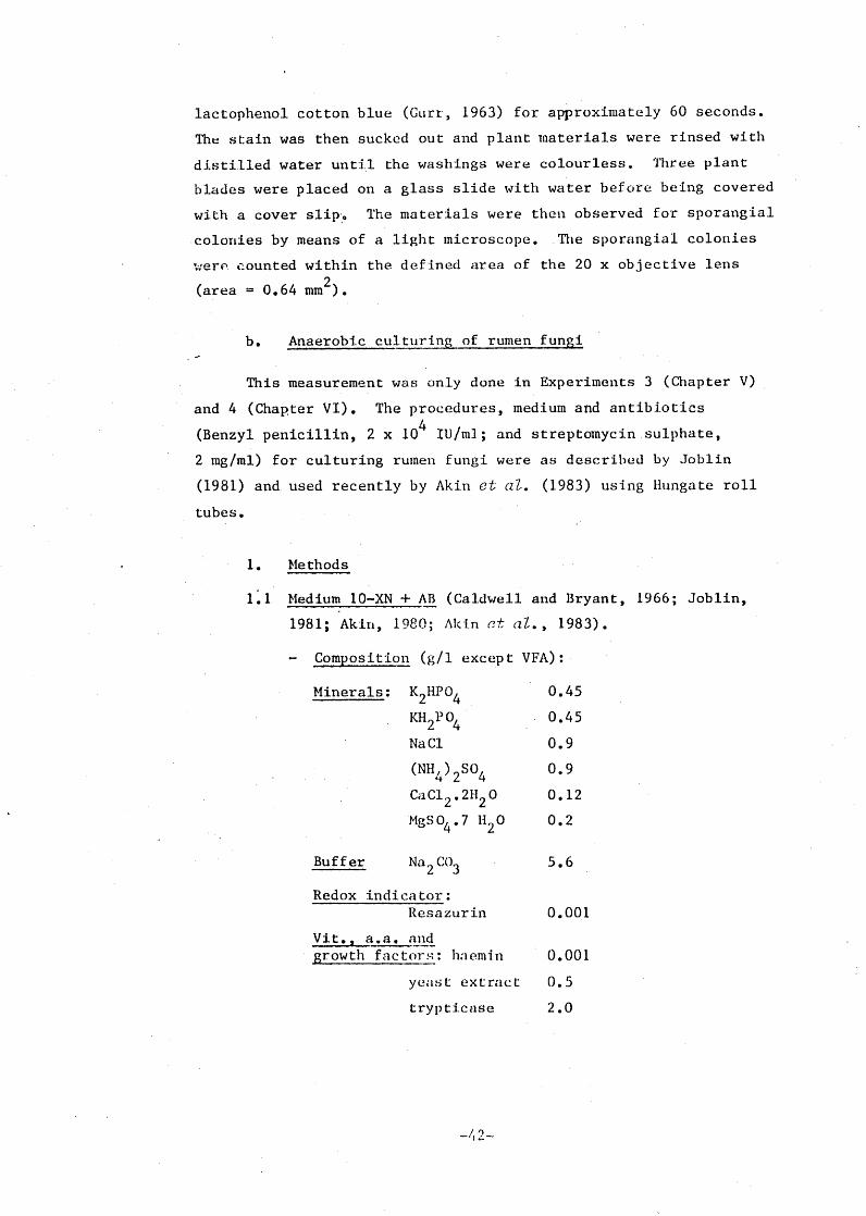
1. Methods
1.1 Medium 10-XN + AB (Caldwell and Bryant, 1966; Joblin,
1981; Akin, 1980; Akin et al., 1983).
- Composition (g/1 except VFA):
Minerals: K2HPO
4KH
2PC
'4Na Cl
(NH4
)2
SO4
CaC12. 2H2 0
MgSO4 .7 H20
Buffer Na2CO
3
Redox indicator:Resazurin
a.a. and• srowth factors: hoemin
yeast extract
trypticase
0.45
0.45
0.9
0.9
0.12
0.2
5.6
0.001
0.001
0.5
2.0
42-
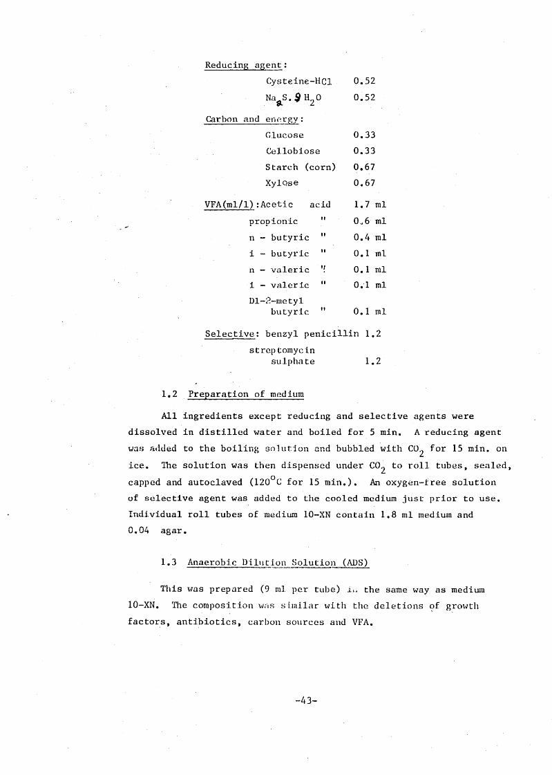
Reducing_ agent:Cysteine-HC1 0.52
Na S.,911 0 0.529. 2
Carbon and energy:
Glucose 0.33
Ceilobiose 0.33
Starch (corn) 0.67
Xylc)se 0.67
VFA(m1/1):Acetic acid 1.7 ml
propionic ml
n - butyric 0.4 ml
11i - butyric 0.1 ml
n valeric 0.1 ml
i valeric ml
Ill-2-metylbutyric 0.1 ml
Selective: benzyl penicillin 1.2
streptomycinsulphate 1.2
1.2 Preparation of medium
All ingredients except reducing and selective agents were
dissolved in distilled water and boiled for 5 min. A reducing agent
was added to the boiling solution and bubbled with C for 15 min. on02
ice. The solution was then dispensed under CO 2 to roll tubes, sealed,
capped and autoclaved (120°C for 15 min.). An oxygen-tree solution
of selective agent was added to the cooled medium just prior to use.
Individual roll tubes of medium 10-XN contain 1.8 ml medium and
0.04 agar.
1.3 Anaerobic Dilution Solution (ADS)
This was prepared (9 ml per tube) the same way as medium
10-XN. The composition similar with the deletions of growth
factors, antibiotics, carbon sources and VFA.
-43-

Inoculum was 0.2 ml rumen fluid of each dilution per tube.
The dilution factors were 10°, 101 , 10
2 and 10
3 and all tubes were
incubated at 38° C for 4 days. Enumeration of fungal colonies (on
the assumptionthat each colony was formed from one zoospore) was
done under a dissecting microscope.
3.5 Analytical Methods
3.5.1 Drymatter and Or anic matter
Air-dried feed and faeces were finely ground (1 mm screen),
weighed into a crucible and dried to a constant weight in a force-
draught oven at 70°C to determine DM. Dried samples were
snbsequently ashed in an oven (500-600°C for 4 h) to determine OM.
3.5.2 Neutral-Detergent Fibre (NDF)
Approximately 1.0g ground material was placed in a round-
bottomed flask. The following reagents were added, in order, 100 ml
neutral detergent solution (see next for the composition) (Goering
and Van Soest, 1970), 2 ml decahydronaphthalene and 0.5 g sodium
sulphide, with 'a calibrated scoop. The composition of neutral
detergent solution (per litre) was as follows:
30 g sodium lauryl sulphate, USP grade a
18.61 g disodium dihydrogen ethylene diaminotetraketate (EDTA)
dihydrate crystal, reagent grade
4.56 g disodium hydrogen phosphate, reagent grade
6.81 g sodium borate decahydrate, reagent grade
10 ml 2-ethoxyethanol (ethylene glycol, monoethyl
ether), purified grade.
Samples were ref luxed for 60 min., timed from onset of boiling and
then filtered through a sintered glass crucible. Samples were rinsed
with a hot water (80° C) water, then washed twice with acetone in the
same manner and sucked dry. Crucibles were dried at 80°C overnight
and weighed.
-44-

3.5.3 Acid-Detergent Fibre (ADF)
The procedure for analysing ADF was similar to the NDF
determination except for the reagents used in this analysis. After
placing approximately 1.0 g of sample in a round-bottomed flask the
following reagents were added: 100 ml acid-detergent solution
(Goering and Van Soest, 1970) and 2 ml decahydro-naphthmlene. The
composition of acid-detergent solution (per litre) Was as follows:
49.04 g sulfuric acid, reagent grade standardized to 1N
20.00 g Cetyl trimethylammonium bromide (CTAB), technical
grade.
Samples were refluxed, filtered, washed twice with hot water,
washed with acetone, and dried at 80°C overnight.
3.5.4 Nitrogen analysis
a. Total N
The total N content of feedstuffs was converted to NH4 by
kjeldahl oxidation and then made alkaline with NaOH. The distilled
ammonia was collected into boric acid (2% W/V) and titrated against 0.0075
N H2SO4.
b. NH -N
Ammonia concentrations in Experiment . 1 (Chapter V) were
determined by the steam-distillation technique described by Breemer
and Keeney (1965) and modified by Nolan (1971), while in Experiments
2, 3 (Chapter V) and 4 (Chapter VI) the ammonia concentrations were
determined by an autoanaiyser (Technicon). Rumen-fluid supernatants
of Experiment 1 were made alkaline with sodium ter. aborate (5% W/V).
The distilled ammonia was then treated as for total-N using 0.03 N
H2SO
4 for titrations.
In Experiments 2, 3 and 4, samples (0.5 ml) were diluted with
4.5 ml H20. If the ammonia concentration of samples was more than
200 mg N/1, the dilution factor was 0.25 ml sample plus 4.75 ml H20.
-45--

( 4
)2SO
4 was used as a standard at 10, 20, 30, 50, 70, 90, 110,
130, 150, and 200 mg N/1. The reagents used were 2% H 2 SO4 , 0.75% NaOH,
sodium salicylate, and sodium cyanurate.
3.5.5 VFA
VFA concentrations of rumen fluid supernatant were determined
by the method of Geissler et aZ. (1976) using isocaproic acid as an
internal standard in a dual-column gas chromatograph (Model 427,
Packard Instrument Company, Illinois, USA).
3.5.6 Chromium
The concentration of chromium in rumen fluid supernatant was
determined with an atomic-absorption spectrophotometer (Perkins-Elmer,
Model 360, Norwalk, Connecticut, USA) which had been calibrated with
a set of potassium chromate standards made up in rumen fluid.
3.6 Estimation of Rumen Fluid Kinetics
Rumen liquid volume and outflow rate were estimated with the
Cr-EDTA complex as a soluble marker (Downes and McDonald, 1964) and
given as a single injection just prior to the morning feeding.
Animals were dosed at 0.5 ml Cr-EDTA per kg body weight injected into
the rumen via the cannula; the first sample of rumen fluid was
sampled at 3 h post injection. Samples of rumen fluid were again
taken at 20, 24 and 26 h post-injection. The concentrations of
chromium in rumen fluid was related to time on a lag-linear basis
according to first order kinetics. The repression of the line was
represented by the equation : Dorict = Conroe
-mt
where Conco and Gonc
t = tracer concentrations at zero time and at time t,
respectively, m = the rate-constant described by the slope of line;
t = hours after injection.
Rumen volume and outflow route were calculated as follows:
a) Rumen volume (ml) = Dose (mg) (V) Conc
o (mg p1)
b) Outflow rate (1/day) = V x m x 1440.

CHAPTER IV
PRELIMINARY STUDY ON METHODS FOR THE ENUMERATION OF FUNGAL SPORANGIA
4.1 Introduction
As mentioned in Chapter 2, there is no satisfactory method
available at present to quantify the fungal population in the rumen.
Direct enumeration of fungal zoospores in fresh rumen fluid under a
light microscope gives poor results, particularly when rumen protozoa
are present. On the other hand, when the enumeration is done with
preserved rumen fluid, the morphology of fungal zoospores is often
difficult to recognize due to disintegration of the flagellum and the
presence of flagellated protozoa. The use of roll tubes as proposed
by Joblin (1981) to estimate the number of zoospores in the rumen,
quantifying the concentration of chitin in the rumen digesta to
determine the pool size of rumen fungi (Orpin, 1981a), and estimating
the pool size of rumen fungi by analysing the concentration of total
cis,24:1 (A15
) fatty acids and triterpenol (Kemp et al., 1984) areall demanding in terms of equipment and labour.
This preliminary study was designed to evaluate the method of
sporangial enumeration of Bauchop (1979a) and Akin et al. (1983) toestimate the fungal biomass in the rumen of sheep on different feeding
regimens by counting the sporangia on forage samples suspended in
nylon bags in the rumen for up to 48 h. The main objectives of this
study were to establish the validity of the method to screen forages
as possible suitable substrates. Two experiments were carried out.
4.2 Experimental
4.2.1 Animals and diets
Four mature merino wethers, each fitted with ruminal and abomasal
cannulae, were used in Experiment 1. They were fed with the following
diets:
Sheep no. 9288 : 60% lucerne chaff + 40% oat grain given at 800 g/d
Sheep nos. 8259
and 8246 : 60% wheaten chaff + 40% oat grain . given at 800 g/d
Sheep no. 2277 : 200 g lucerne chaff + 400 g oat grain.
All rations were given once a day at 0900 h and water was available
ad libitum.
In Experiment 2 twelve mature crossbred wethers, each fitted
with a ruminal cannula, were fed a basal diet of rice straw which had
been ensiled with 4% urea for four weeks, and supplemented with either
lucerne chaff or fish meal or both:
Diet A : Urea-treated rice straw (UTRS) ad libitumDiet B : UTRS ad Zib. + 75 g lucerneDiet C : UTRS ad lib. + 75 g fish meal
Diet D : UTRS ad Zib. + 75 g lucerne + 75 g fish meal
All diets were fed once a day at 0900 h; water was freely available.
4.2.2 Experimental procedures
In Experiment 1 leaf blades of three forages (white clover,
kikuyu, and grazed oats) and the flower heads of meadow grass (probably
barley grass, Hordeum Zeporinum) were incubated in the rumen of sheep
in nylon bags with an aperture size of 45 pm. The leaf blades were
cut into 2-3 cm lengths with a razor blade. The bags were weighted
with a marble and anchored with 75 g iron weights and suspended in
the rumen at timed intervals.
In Experiment 2 leaf blades of Phalaris aquatica, and untreated
and treated straw particles were cut into 2-3 cm lengths and incubated
in the rumen of sheep in nylon bags (see Section 3,4.4) at timed
intervals.
All bags were suspended in the rumen immediatel y prior to the
morning feeding. The staining and counting methods were as already
outlined in Section 3•4.5.a. unless specified otherwise.

4.3 Results and Discussion
The numbers of sporangia appearing on plant particles in
Experiments 1 and 2 are presented in Tables 4.1 and 4.2 respectively.
The results of sporangial enumeration from these studies suggest that
rumen fungi are highly selective microorganisms in colonizing the
plant tissues. As reported by Bauchop (1980), the sites of fungal
colonization on leaves observed from these studies were principally
near the stomata and in areas close to leaf ribs (Plates 4.1 and 4.2).
With straw particles, sporangial colonization was prominent on inner
surfaces of rice straw stems (Plate 4.3). This selective colonization
by fungal sporangia is the main reason for the large coefficient of
variation in sporangial counts (mean value 70%) regardless of diet
and substrate used for counting the sporangia.
The use of leguminous leaves such as white clover and
graminaceous leaves such as oats and phalaris as substrates appears
to give a poor result when the incubation of these materials in the
rumen exceeds 12 h. This is due to the fact that all these materials
were substantially degraded by 24 h, and no intact plant material was
found at 48 g for most of the diets (Tables 4.1 and 4.2), resulting
in a coefficient of variation higher than 70%. Straw materials,
however, gave better results, mainly because they were still intact
at 72 h. The number of sporangia on phalaris grass and untreated rice
straw after 12 h incubation, and on untreated and treated straw after
48 h and 72 h incubation were similar (Table 4.2), suggesting that
the counting method of Bauchop (1979a) and Akin et al. (1983) can be
used to compare the number of anaerobic fungi present in the rumen
on different substrates at the same sampling time.
The finding that rates of fungal colonization on urea-treated
straw were slower than those on other substrates was unexpected (Table 4.2).
There is no obvious explanation for these findings. Orpin and Bountiff (1978)
stated that rumen fungi showed chemotaxis to a range of carbohydrates,
but not to the common amino acids, purines, pyrimidines or vitamins,
suggesting that rumen fungi are highly selective colonizers. It is
possible that ammoniated substrates are not attractive to rumen fungi.

If this is so, the lower numbers of fungal sporangia found at 6 and
12 h of incubation do not necessarily mean that rumen fungi were not
present or that they were present at low population densities, since
higher counts of sporangia were obtained on non-ammoniated substrates
suspended in the same rumen. For this reason, it is suggested that
the substrate used for sporangial enumeration should be relatively
resistant to microbial degradation in the rumen and free from
chemical treatment.
In conclusion, regardless of diet and animal variations, the
results of sporangial counts obtained from small numbers of plant
particles in these studies are not likely to be meaningful estimates
of the rumen fungal population; estimates of sporangial population
densities in these studies can only be used as approximate values.
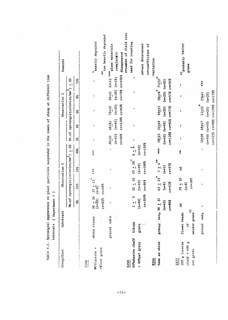
Table 4.1. Sporangial appearance on plant particles suspended in the rumen of sheep at different time
intervals ( Experiment 1 )
Sheep/Diet
Substrate
Observation 1
Observation 2
Remarks
No.of sporangia(colonies/mm2 ) * SD No.of sporangia(colonies/mm2 )
+ SD
6h
16h
24h
48h
2h
4h
6h
8h
10h
9288
60161ucerne +
white clover
39 + 36 27 + 13
* **
*
(n=50) (n=8.7
* *
*heavily degraded
40%oat grain
cv=92% cv=48%
* *too heavily degraded
grazed oats
- 39+35
68+82
79+50
39+31 12+12 **
* plant materials
(n=43) (n=44) (n=20) (n=20) (n= 1
8)
completely
cv=90% cv=120% cv=63% cv=79% cv=100% disappeared
n=number of field area
8259
**
used for counting
60%wheaten chaff
kikuyu
1+'
321 + 18
10 + 1
6 + 9
-
+ 40%oat grain
grass
(n=40)
(n=10)
(n=10)
(n=10)
8246
cv=300%
cv=86%
cv=100%
cv=150%
nd=not determined
cv=coefficient of
vvariation
Same as above
grazedoats 36 + 31
43 ± 23
7 + 4**
***
18+23
52+49
69+53
28+16
7!437*
(n=40)
(n=8)
(n=5)
(n=40)(n=40)
(n=20)
(n=20)(n=20)
cv=86%
cv=53%
cv=57%
cv=128% cv=94% cv=77%
cv=57% cv=50%
2277
200 g lucerne
nd
flower heads
79 + 30
nd
**
--
+)probably barley
chaff + 400 g
of
(n=8)
grass
oat grain
+)meadow grass
cv=38%
grazed oats
- 16+20 48+47
11+12
* 23+41
***
(n=4
0) (n=40) (n=20) (n=20)
cv=125% cv=98% cv=109% cv=178%

Table 4.2 Sporangial appearance on plant particles suspended in the rumen of sheep given a basal diet of urea-treatedcstraw(A),
supplemented with 75 g lucerne(B), 75 g fish meal(C) and 75 g lucerne + 75 g fish meal(D).( Experiment 2 ). Values are
means
of three sheep per treatment;45 field areas used for counting sporangia in each case.
Substrate
Diet
...
Numbers of sporangia f_colOniea/mm
2:)-+ SD
Remark,
6h
12h
24h
48h
72h
-Phalaria grass
A C
38 + 23
cv-612
8 + 6
cv-75%
19+3
52 + 28
cv-54X
9 + 1
cv-111
30 + 30
16 + 13*
cv-81l
*13 + 12
cv-92%
7 + 6*
*A
** **
**
* heavily.degraded
**
**too heavily degraded
**
cv•coefficient of variatiol
cv-161
cv-100%
cv-86X
60 + 25
24+11
*17 +15
.**
**
cv-421
cv-46%
cv-88Z
-Untreated rice straw
A71+63
47 + 23
38 + 22
38 + 11
19 + 6
cv-891
cv■491
cv-581
cv-29%
cv..32Z
4 + 4
11+9
33+8
28+9
11+5
cv-100%
cv82X
cv-242
cv-32X
cv-451
C14 + 12
24 + 19
21 + 6
32 + 12
22 + 5
cv
.,861
cv..
791
cv-29X
cv-38X
cv -231
56 + 51
22 + 12
44 + 19
25 + 8
21+5
cv-91X
ev■551
cv-43X
cv-32%
cv-24X
-Urea treated rice straw
A B C D
O1 + 1
14 + 6
32+6
20+6
cv-100%
cv-43%
cv-191
cv..
302
01 + 1
11+6
27+5
13+8
cv
..100%
cv..
551
cv-19Z
cv■621
O4 + 1
18+8
53 + 14
33 + 23
cv-251
cv-44X
cv■261
cv-70X
O5 + 2
30+8
31 + 15
25 + 13
cv-40X
cv-27%
cv■4
87.
cv=52Z

Plate 4.1 Sporangia of rumen fungi colonizing grazed oat leaves
( 8 h incubation ); penetration is through the stomata
( Magnification 200 X )
Plate 4.2 Sporangia of rumen fungi are found principally along the mid
ribs of kikuvu leaves ( 24 h incubation, magnification 200 X )

Plate 4.3 Unstained straw showing an early stage of development of
rumen fungi ( arrow ) colonizing the inner surface of the
stem ( Magnification 100 X )


![CHAPTERS Exploring Geography I ;ill Population and Culture I Climate and Vegetation ... Geography_1_2_3.pdf · 2017-02-03 · 30 CHAPTERS ~ . ] Exploring Geography I ;ill Population](https://img.dokumen.tips/doc/110x75/5b3202487f8b9a2c0b8c2565/chapters-exploring-geography-i-ill-population-and-culture-i-climate-and-vegetation.jpg)