Embed Size (px)
Citation preview

Fetal Pig Dissection Guide
http://www.whitman.edu/biology/vpd/main.html
Dissection Contract:
1. You must use caution and proper procedures with dissections tools and instruments (scalpels, pins, scissors, etc)
a. Always cut away from yourself and othersb. Never try to catch a utensil if you drop it
2. You MUST remain with your assigned group at all times. You may not wander around and visit other lab groups, unless you have asked for permission.
3. We are using these animals for learning purposes. These animals should be treated with respect at all times. I am the judge of what is considered respectful or not.
4. You must read and follow all lab procedures at all times. You are not allowed to make any incisions that are not part of the lab procedure.
5. No materials are to be removed from the dissection tray without instruction.
***If you are in violation of these expectations, you will lose lab privileges and have to learn the same material from a text book. You will lose points, and potentially receive a referral.
I have read the expectations above and agree to follow all expectations.
Student signature: _____________________________________

Organ Functions Bank:
You will use this Organ Function bank to match the organs within each system to their appropriate function.
External Anatomy
● Funnels sound into the ear● Protects eyes● Warms, cleans, and moistens air as it enters the body● Connects fetus to mother to help transfer oxygen and nutrients and remove waste● Helps push food to the back of the throat to allow for swallowing● Protects against external environment, regulates temperature, detects sensation
Digestive System
● Uses saliva and teeth to start the process of breaking down food, specifically carbohydrates● Pushes food down from the mouth to the stomach● Makes bile to break down fats, helps detoxify blood● Stores bile until ready for use in the small intestine● Stores food, helps expose surface area, breaks down proteins● Contains bacteria to help break down cell walls, helps compact feces● Completes the breakdown of food including fats, absorbs nutrients into the blood stream● Absorbs excess water, vitamins, and minerals. Compacts feces● Makes a chemical to break down fats, carbohydrates, and proteins. Makes a base to neutralize
stomach acid● Exit for solid waste ● Helps push out solid waste for removal from the body
Excretory system
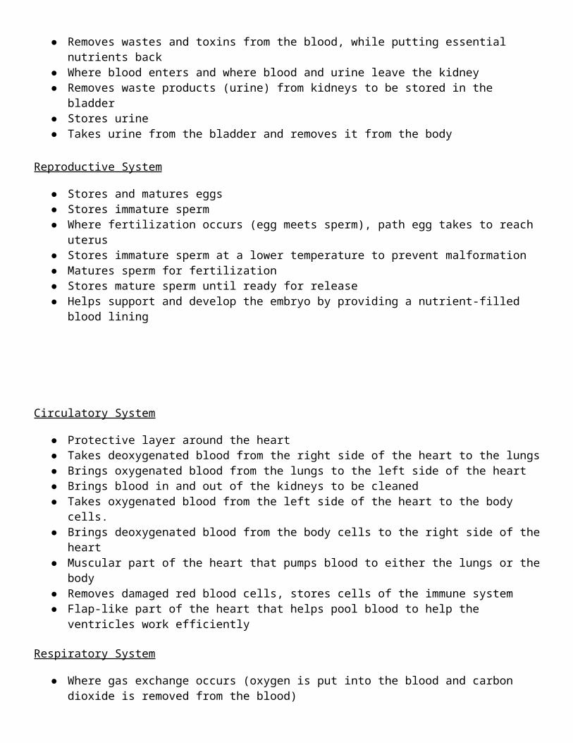
● Removes wastes and toxins from the blood, while putting essential nutrients back ● Where blood enters and where blood and urine leave the kidney● Removes waste products (urine) from kidneys to be stored in the bladder● Stores urine ● Takes urine from the bladder and removes it from the body
Reproductive System
● Stores and matures eggs● Stores immature sperm● Where fertilization occurs (egg meets sperm), path egg takes to reach uterus● Stores immature sperm at a lower temperature to prevent malformation● Matures sperm for fertilization● Stores mature sperm until ready for release● Helps support and develop the embryo by providing a nutrient-filled blood lining
Circulatory System
● Protective layer around the heart● Takes deoxygenated blood from the right side of the heart to the lungs ● Brings oxygenated blood from the lungs to the left side of the heart● Brings blood in and out of the kidneys to be cleaned● Takes oxygenated blood from the left side of the heart to the body cells.● Brings deoxygenated blood from the body cells to the right side of the heart● Muscular part of the heart that pumps blood to either the lungs or the body● Removes damaged red blood cells, stores cells of the immune system● Flap-like part of the heart that helps pool blood to help the ventricles work efficiently
Respiratory System
● Where gas exchange occurs (oxygen is put into the blood and carbon dioxide is removed from the blood)
● Tube that directs air to the lungs● Small tubes that direct air to different parts of the lung and control the amount of air coming in.● Muscle that helps air flow in and out of the lungs. Works on pressure differences.
________________________________________________________________________________________
Anatomical Directions Notes:
____________________ – front or in front of
____________________ – back or in back of
____________________ – toward the belly
____________________ – toward the back
____________________ – nearer the surface
____________________ – farther away from the body surface
Practice:
1. The dog’s heart is ________________ to the spleen
2. The dog’s heart is _______________ to the esophagus.
3. The skin’s epidermis is ______________ to the dermis.
Part 1: External Anatomy

Obtain a dissection tray, blunt probes, and a fetal pig. Label your tray with a piece of tape and your group members’ names. Open the outer bag (and save it) to take out the pig. Cut the inner pig bag open over the sink and drain the fluid. This bag may be thrown away. Rinse the pig under a gentle stream of water. Place the pig on the dissection tray.
Label the anatomical directions on the diagram below:
Fill in the functions from your list (on the first page). Locate each of
Organ Function Observations ✓
Umbilical cord
Tongue
Nares (nostrils)
Pinna (ears)

Eyelids
Skin
Tasks:
1. Determine the sex of your pig by looking at the urogenital opening. On females, this opening is near the anus. On males, this opening is near the umbilical cord. Sex (circle one): Male/Female
a. If your pig is a female, you should also note that urogenital papilla is present near the genital opening. Males do not have urogenital papilla. Does your pig have urogenital papilla? _________
2. Find a group that has a pig of the opposite sex. Explain the difference in the external genitalia.
____________________________________________________________________________________
____________________________________________________________________________________
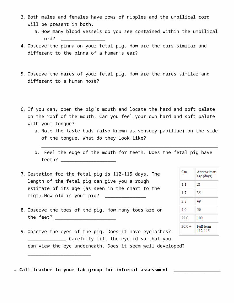
3. Both males and females have rows of nipples and the umbilical cord will be present in both.a. How many blood vessels do you see contained within the umbilical cord? ________________
4. Observe the pinna on your fetal pig. How are the ears similar and different to the pinna of a human’s ear?
5. Observe the nares of your fetal pig. How are the nares similar and different to a human nose?
6. If you can, open the pig’s mouth and locate the hard and soft palate on the roof of the mouth. Can you feel your own hard and soft palate with your tongue?

a. Note the taste buds (also known as sensory papillae) on the side of the tongue. What do they look like? ________________________________________________________________
b. Feel the edge of the mouth for teeth. Does the fetal pig have teeth? ____________________
7. Gestation for the fetal pig is 112-115 days. The length of the fetal pig can give you a rough estimate of its age (as seen in the chart to the rigt).How old is your pig? _______________
8. Observe the toes of the pig. How many toes are on the feet? ______________________
9. Observe the eyes of the pig. Does it have eyelashes? ______________ Carefully lift the eyelid so that you can view the eye underneath. Does it seem well developed? _______________________
→ Call teacher to your lab group for informal assessment _________________
Analysis:
1. The largest organ of your body is your skin. Explain why skin is classified as an organ.
2. The expression “sweating like a pig” isn’t actually very true. Pigs have very few sweat glands in their skin. What effect does this have on their body? What do pigs do instead of sweating?
3. Thinking about the functions of the tongue, explain at least two types of cells that must be in the tongue.
4. Agree or disagree with the following statement and explain your position: Human tongues have adapted and are more advanced than pig tongues.
5. Explain how the above system contributes to maintaining homeostasis of the whole body.
6. Research a disease/disorder of the above system and how homeostasis is changed, interfered with, etc.
Part 2: Digestive System
Digestive System Notes:
What is the main function of the digestive system? Which tissue types would you expect to be present in the digestive system?
Organs & Functions: Mouth: Uses saliva and teeth to start the process of breaking down food_________________________
Mechanical: Chemical:
Esophagus: Pushes food down from ____________________________________
Stomach: Stores food, helps expose surface area,______________________________
Small Intestine: Completes the breakdown of food___________________________ &
____________________________________________________________________

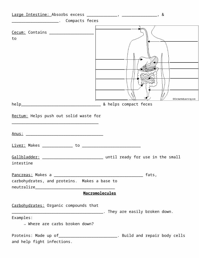
Large Intestine: Absorbs excess _____________, _______________, & ____________________. Compacts feces
Cecum: Contains ___________________ to help__________________________________ & helps compact feces
Rectum: Helps push out solid waste for _____________________________________
Anus: _________________________________
Liver: Makes _____________ to _________________________
Gallbladder: __________________________ until ready for use in the small intestine
Pancreas: Makes a ______________________________________ fats, carbohydrates, and proteins. Makes a base to neutralize__________________________________
Macromolecules
Carbohydrates: Organic compounds that _______________________________________. They are easily broken down. Examples:
→ Where are carbs broken down?
Proteins: Made up of_________________________. Build and repair body cells and help fight infections. → Where are proteins broken down?
Lipids (Fats): Large molecules that provide___________________________________________→ Where are fats broken down?
Vitamins & Minerals: ___________________________________
Opening your fetal pig
In this activity, you will open the abdominal and thoracic cavity of the fetal pig and identify structures. Remember, that to dissect means to "expose to view" - a careful dissection will make it easier for you to find the organs and structures. Be sure to follow all directions.

The First Incision
Place your fetal pig in the dissecting pan belly up. Use string to tie your pig so that the legs are not in your way. Use scissors to cut through the skin and muscles according to the diagram. Do not remove the umbilical cord. In the first section, you will only examine the abdominal cavity (the area below the ribcage).
After completing the cuts, locate the umbilical vein that leads from the umbilical cord to the liver. You will need to cut this vein in order to open up the abdominal cavity.
Pin the skin and muscle to the side so that the internal organs are visible.
Your pig may be filled with water and preservative, drain over the sink if necessary and rinse organs.
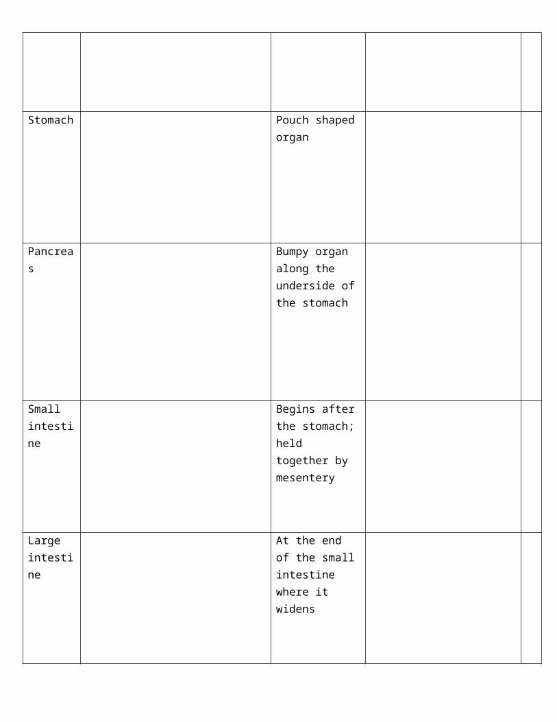
Locate: Fill out the function of each organ by referencing the Function Bank. Find each organ in the pig by reading the description and make observations. Check the box when you have found it.
Organ Function Description Observations ✓
Mouth
Liver Lobed, largest organ
Gall bladder
Greenish organ under the liver
Esophagus Connects to top of stomach
Stomach Pouch shaped organ
Pancreas Bumpy organ along the underside of the stomach
Small intestine
Begins after the stomach; held together by mesentery

Large intestine
At the end of the small intestine where it widens
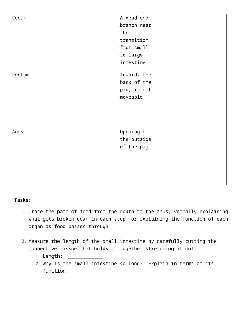
Cecum A dead end branch near the transition from small to large intestine
Rectum Towards the back of the pig, is not moveable
Anus Opening to the outside of the pig
Tasks:
1. Trace the path of food from the mouth to the anus, verbally explaining what gets broken down in each step, or explaining the function of each organ as food passes through.
2. Measure the length of the small intestine by carefully cutting the connective tissue that holds it

together stretching it out. Length: ____________a. Why is the small intestine so long? Explain in terms of its function.
3. Cut the stomach so you can open it and view the inside. There are two important valves in the stomach: the cardiac sphincter (at the top end) and the pyloric sphincter (at the bottom end).
a. What is the purpose of each of these valves? (Hint: think about their location)
→ Call teacher to your lab group for informal assessment _________________
Analysis:
1. Explain how the structure of the small intestine and the many villi on its inner surface increase the rate of absorption of digested food molecules from the lumen (inside) of the small intestine to the blood.
2. Why is it useful to have lots of capillaries inside each of the villi and surrounding the small intestine?
3. Imagine a digestive system where food entered the small intestine directly without first going through the oral cavity and stomach. What would be the disadvantages of this type of digestive system? Why is it useful to have food processed in the oral cavity and stomach before entering the small intestine?
4. Explain how the above system contributes to maintaining homeostasis of the whole body.
5. Research a disease/disorder of the above system and how homeostasis is changed, interfered with, etc.
Part 3: Excretory System & Reproductive System Dissection
Excretory & Reproductive Systems NotesExcretory System:
Purpose: Regulate _________________, remove __________________________________ which makes ____________________
Organs: Kidneys: Filter waste _____________________________________________ using millions of nephrons
Regions of the kidney:
• ________________________: Removes wastes and toxins from blood while putting essential nutrients back in
• ________________________: Full of nephrons!
• ________________________: where blood enters and where blood and urine leave the kidney (via Renal Artery & Vein)
How do the kidneys filter the blood?
Ureters: Removes _________________________________________________ to be stored in the bladder
Bladder: ______________________________
Urethra: Takes urine from the bladder and ___________________________________
How does the excretory system create urine?● Molecules in blood leave the blood as it enters the kidney● Unneeded materials are ____________________________● Anything the body needs is __________________________● Whatever is left in the tubules becomes _____________________ and is gathered in collecting ducts,
which lead to ___________________
Reproductive System:
Purpose: _________________________ and help organisms mix gametes with another to produce
offspring that are ___________________________________________________________
Female:Ovaries: ______________ and __________________ egg
Fallopian Tubes: Where _________________________________, path egg takes to reach uterus
Uterus: Helps _________________________________________________________________ by providing a nutrient-filled blood lining
Male:Testes: stores ________________________
Scrotum (scrotal sac): stores immature sperm at lower temperature to prevent malformation
Epididymis: _____________________________________ for fertilization
Vas Deferens: stores __________________________________ until ready for release
Excretory System Dissection: Locate: Fill out the function of each organ by referencing the Function Bank. Find each organ in the pig
and make observations. Check the box when you have found it.
Organ Function Description Observations ✓
Kidneys
● Cortex:
● Pelvis:
**Locate these after you slice open the kidney
Innermost section of kidney
Outer section of kidney
Ureter Tubes leading away from the kidneys

Urinary Bladder Pouch between the umbilical vessels (see diagram)
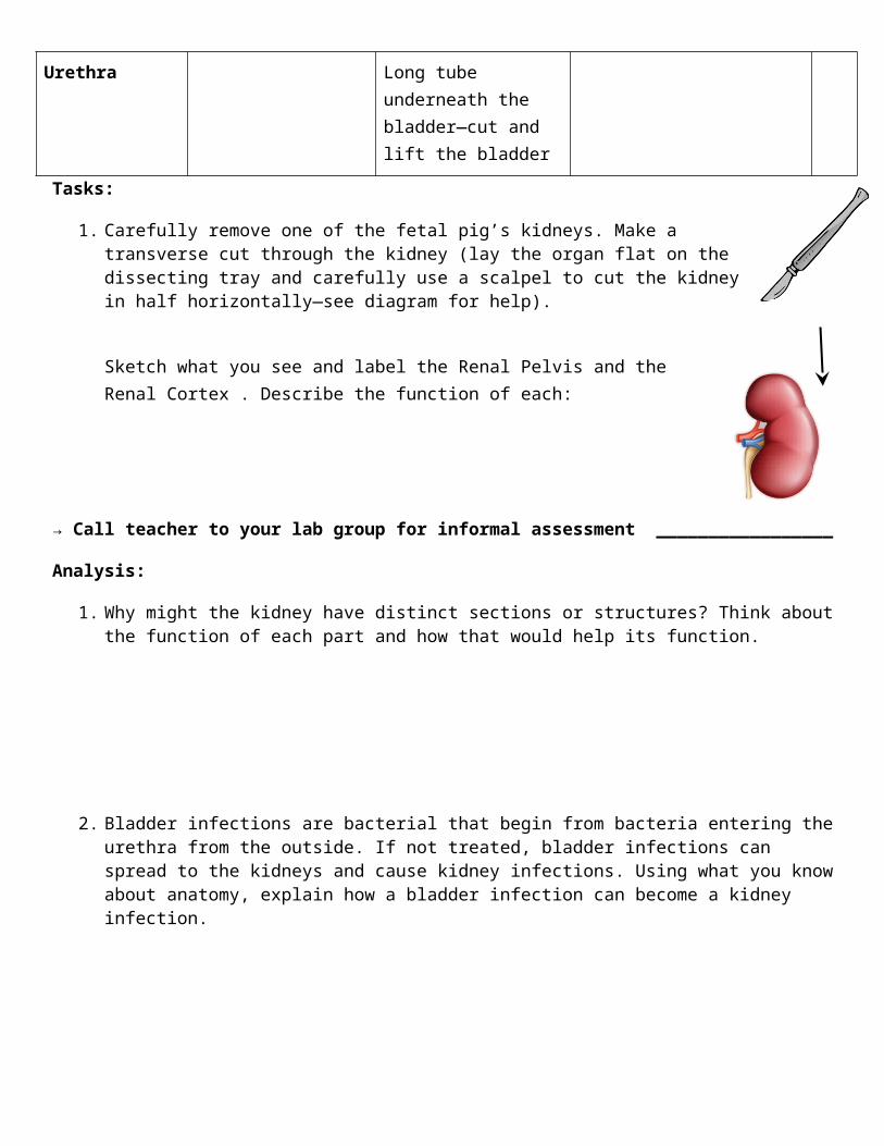
Urethra Long tube underneath the bladder—cut and lift the bladder
Tasks:
1. Carefully remove one of the fetal pig’s kidneys. Make a transverse cut through the kidney (lay the organ flat on the dissecting tray and carefully use a scalpel to cut the kidney in half horizontally—see diagram for help).
Sketch what you see and label the Renal Pelvis and the Renal Cortex . Describe the function of each:
→ Call teacher to your lab group for informal assessment _________________
Analysis:
1. Why might the kidney have distinct sections or structures? Think about the function of each part and how that would help its function.
2. Bladder infections are bacterial that begin from bacteria entering the urethra from the outside. If not treated, bladder infections can spread to the kidneys and cause kidney infections. Using what you know about anatomy, explain how a bladder infection can become a kidney infection.

3. Explain how the above system contributes to maintaining homeostasis of the whole body.
4. Research a disease/disorder of the above system and how homeostasis is changed, interfered with, etc.
Reproductive System Dissection
Locate: Fill out the function of each organ by referencing the Function Bank. Find each organ in the pig and make observations. Check the box when you have found it.
Function Description Observations ✓
Ovaries located below the kidneys

Fallopian Tubes
Connected to ovaries
Uterus underneath the bladder
Scrotum
(scrotal sac)
Located on the exterior, below the anus
Testes contained within the scrotal sac
Epididymis coiled on the top of each testes

Vas Deferens crosses over the ureter
Tasks:
1. Male: Locate and open the scrotal sac to isolate the testes. On each testicle, find the coiled epididymis.Female: Find the bean-shaped ovaries. Follow the path from the ovaries, down the fallopian tubes, to the uterus.
2. Find a group who has the opposite sex pig. Ask them to show you the relevant reproductive organs. You will be responsible for knowing both the male and female anatomy.
→ Call teacher to your lab group for informal assessment _________________
Analysis:
1. In males, a vasectomy is the cutting of the vas deference. Explain how this procedure helps prevent fertilization.
2. In males, the leading cause of infertility is temperature imbalance of the scrotal sac. Use your knowledge of the male reproductive system to account for this infertility issue.
3. In females, the ovaries take turns maturing and releasing eggs, however, some females have a genetic
condition, which causes both of the ovaries to release eggs every month. Explain why these females are more likely to have fraternal twins (non-identical).
4. There are many different causes for infertility in females. Using your knowledge of the female reproductive system, pick one female organ, determine how it may cause infertility, and explain how it would affect pregnancy.
5. Explain how the above system contributes to maintaining homeostasis of the whole body.
6. Research a disease/disorder of the above system and how homeostasis is changed, interfered with, etc.
Part 4: Circulatory System
Circulatory System Notes:
Purpose: Transports __________________ throughout the ____________________. Transports wastes, hormones, and nutrients as well.
Blood Vessels:● Arteries: Blood vessels that carry blood
___________________________________○ Describe structure:

● Veins: Blood vessels that carry blood __________________________________________○ Describe structure:
● Capillaries: Thin blood vessels responsible for _________________________○ Describe structure:
Which way is the blood going?● Blood coming from the lungs is oxygen-rich ● Blood coming from the body is oxygen-poor
○ Why?
Heart: _____________________________ with 4 chambers
Pathway: Map out blood flow (Blue=oxygen-poor, Red= oxygen-rich & label the functions of each structure
Renal Artery & Vein: Brings blood in & out of _________________________________________________
Spleen: Removes damaged _______________________________________
Circulatory System Dissection: You will need to use the sheep heart and your fetal pig to answer the questions.
Locate: Fill out the function of each organ by referencing the Function Bank. Find each organ in the pig and make observations. Check the box when you have found it.
Function Description Observations ✓
Pericardium sac surrounding the heart
left and right ventricles
lower part of the heart. Hard, and muscular
left and right atrium
flaps located on the top
pulmonary artery
arches from the front of the heart to the lungs

pulmonary vein
aorta behind the pulmonary artery
vena cava
renal artery and vein
small tubes directed going in and out of the kidneys
spleen Overlays the stomach
Tasks:
1. Before you look at the heart of your fetal pig, obtain a sheep heart. Arrange the heart so that the front side (anterior) is face up on the tray. (See Figure 1) You will see a line running from the upper right to the lower left. This is how it would sit in the chest; it’s as if you were looking down into a chest.

2. Find the two flaps on top of the heart. These are the atria. Describe the structure (size, shape, color, feel, etc) of atria:
a. What do the atria do?
b. Why do you think the atria are so small compared to the ventricles which make up the rest of the heart?
3. Next, identify the right ventricle and the left ventricle (remember that right and left refer to the animal’s right and left when the heart was in their chest). There should be a cut in each chamber so you can open them and examine inside
a. Measure and describe the thickness of the muscle wall of the right ventricle:
b. Measure and describe the thickness of the muscle wall of the left ventricle:
4. Now, identify the blood vessels.
Identify the pulmonary artery.
a. Do arteries carry blood to the heart or away from the heart? ________________b. And, ‘pulmonary’ means lungs, so….What is the function of the pulmonary artery?
c. Which chamber pumps blood to the lungs? ______________________ d. Find the pulmonary artery by gently inserting your finger (or probe) into the vessels to find the
one that is connected to the right ventricle.e. Once you have found it, describe the structure of the artery.
Identify the aorta.f. What is the function of the aorta?
g. Which chamber pumps blood out to the body?_________________________h. So, find the aorta by gently inserting your finger (or probe) into the vessels to find one that is
connected to the left ventricle.
Identify the vessels that are going into the atria. i. What type of vessels go into the heart? ___________________j. Examine and describe the structure of the veins. How does the structure of veins compare to
the structure of arteries. Why?
**Return your sheep heart only after you can confidently identify the four heart chambers, the aorta, pulmonary artery, pulmonary vein, and the vena cava.

5. Now, you will try to identify the same parts of the heart in the fetal pig. In order to see the upper part of the circulatory system, you will need to cut up under the pig's throat and make to more lateral incisions in order to fold back the flaps of skin covering the throat. Once this is done, carefully grab on to the heart and pull it away from the chest cavity. Using your scalpel carefully cut away any connected areas. Use your scissors to cut away the pericardium and start identifying parts of the heart.
→ Call teacher to your lab group for informal assessment _________________
Analysis:
1. The left ventricle of the heart is significantly more muscular than the right ventricle. Why is this? (HINT: Think of where the blood is going on each side of the heart)
2. The circulatory system includes the blood, blood vessels (e.g. capillaries), and the heart, which pumps blood through the blood vessels to all the parts of your body. Explain how the digestive system and circulatory system work together to bring nutrients to all the cells in your body.
3. Explain how the above system contributes to maintaining homeostasis of the whole body.
4. Research a disease/disorder of the above system and how homeostasis is changed, interfered with, etc.
Part 5: Respiratory System
Respiratory System Notes:
Purpose: Delivers ___________________to the body and removes _________________________

Pathway:1. Trachea : Tube that directs ____________to _____________2. Bronchial Tube s: Small tubes that direct air to ____________________________________________
and control the amount of air coming in3. Lungs : Where ________________________________ occurs (oxygen is put into the blood and carbon
dioxide is removed from the blood)
Diaphragm: ______________ that helps air flow in and out of ________________● Works by _______________________________
● Movement of Gases: Gases move from high pressure to low pressure
○ What does this sound like…?
Lungs have a HUGE surface area… •Which other organ that we’ve talked about has a large surface area?__________________________
Why?
So…why do the lungs have a large surface area?
Respiratory System Dissection:Locate: Fill out the function of each organ by referencing the Function Bank. Find each organ in the pig and make observations. Check the box when you have found it.
Function Description Observations ✓
Trachea contains cartilage rings

Bronchial Tubes
(bronchi)
where trachea branches off into two
Lungs spongy
Diaphragm sheet of muscle that stretches above the abdominal cavity
Tasks:
1. Cut open the throat and chest of your pig to view the respiratory and circulatory systems.
2. Make an incision in the trachea, insert a straw, and gently blow into the straw. Describe what happens.
→ Call teacher to your lab group for informal assessment _________________
Analysis
1. In anatomy, the respiratory and circulatory systems are considered to be partners. In fact, the two systems are often referred to as the cardiopulmonary system (heart, lung system). Why do you think they are always referenced together?
2. Explain how the above system contributes to maintaining homeostasis of the whole body.
3. Research a disease/disorder of the above system and how homeostasis is changed, interfered with,

etc.
You will be turning this dissection lab in the day of your test, so make sure every function is filled in and every analysis question is answered thoughtfully!
Pig Dissection SummaryFor your test, you should be able to: ● Locate and identify organs in the pig● Describe how the structure of each organ relates to its function● Explain homeostasis and how body systems work together to achieve a balance
Homeostasis Definition:
Homeostasis Example:
Organ Example: ____________________________
Structure:
Function:
How does its structure help its function?: