Embed Size (px)
Citation preview

熊本大学学術リポジトリ
Kumamoto University Repository System
Title Thrombin induces striatal neurotoxicity depending
on mitogen-activated protein kinase pathways in
vi…
Author(s) Fujimoto, Shinji; Katsuki, Hiroshi; Onishi,
Masatoshi; Takagi, Mikako; Kume, Toshiaki; Akaike,
Akinori
Citation Neuroscience, 144(2): 694-701
Issue date 2007-01-19
Type Journal Article
URL http://hdl.handle.net/2298/10011
Right © 2006 IBRO Published by Elsevier Ltd.

Research Paper
Thrombin induces striatal neurotoxicity depending on mitogen-activated protein
kinase pathways in vivo
Shinji Fujimoto, Hiroshi Katsuki, Masatoshi Ohnishi, Mikako Takagi, Toshiaki Kume,
Akinori Akaike
Department of Pharmacology, Graduate School of Pharmaceutical Sciences, Kyoto
University, 46-29 Yoshida-shimoadachi-cho, Sakyo-ku, Kyoto 606-8501, Japan.
Address correspondence to Akinori Akaike, Ph.D.
Department of Pharmacology,
Graduate School of Pharmaceutical Sciences, Kyoto University
46-29 Yoshida-shimoadachi-cho, Sakyo-ku, Kyoto 606-8501, Japan.
Phone: +81-75-753-4550 FAX: +81-75-753-4579
E-mail: [email protected]
Appropriate Section Editor: Dr. Yoland Smith (Neuropharmacology)
1
Abbreviations
EGTA; ethyleneglycol bis(2-aminoethylether)tetraacetic acid, ERK; extracellular signal
regulated kinase, ICH; intracerebral hemorrhage, iNOS; inducible nitric oxide synthase, i.p.;
intraperitoneal injection, JNK; c-Jun N-terminal kinase, MAPK; mitogen-activated protein
kinase, NeuN; neuronal nuclei, o.d.; outer diameter, PBS; phosphate buffered saline, SDS;
sodium dodecyl sulfate, SEM; standard error of mean, Tris;
tris(hydroxymethyl)aminomethane.
2

Abstract
Intracerebral hemorrhage represents stroke characterized by formation and expansion of
hematoma within brain parenchyma. Blood-derived factors released from hematoma are
considered to be involved in poor prognosis of this disorder. We previously reported that
thrombin, a blood-derived serine protease, induced cytotoxicity in the cerebral cortex and the
striatum in organotypic slice cultures, which depended on mitogen-activated protein kinase
(MAPK) pathways. Here we investigated the mechanisms of thrombin cytotoxicity in the
striatum in vivo. Thrombin microinjected into the striatum of adult rats induced neuronal
death and microglial activation around the injection site. Neuronal loss without any sign of
nuclear fragmentation was observed as early as 4 h after thrombin injection, which was
followed by gradual neuronal death exhibiting nuclear fragmentation. Thrombin-induced
damage assessed at 72 h after injection was partially but significanty reduced by concomitant
administration of inhibitors of MAPK pathways. Activation of extracellular signal-regulated
kinase (ERK) and p38 MAPK in response to thrombin was verified by western blot analysis.
Moreover, phosphorylated ERK and p38 MAPK were localized prominently in reactive
microglia, and inhibition of microglial activation by minocycline attenuated thrombin-induced
damage, suggesting that reactive microglia were responsible for thrombin-induced neuronal
death. Thus, MAPK pathways and microglial activation may serve as therapeutic targets of
pathogenic conditions associated with hemorrhagic stroke.
Keywords: apoptosis, intracerebral hemorrhage, microglia, microinjection, striatum
3

Intracerebral hemorrhage (ICH) represents approximately 15% of all strokes. Undesirable
prognosis due to the lack of efficient therapeutic interventions makes ICH a serious clinical
problem (Fayad and Awad, 1998). Ganglionic hemorrhages are the most common forms of
ICH, followed by lobar ones (Xi et al., 2006). While physical trauma and mass effect could
participate in brain damage after hemorrhagic events (Sinar et al., 1987), accumulating
evidence demonstrates that brain edema after ICH is caused by hematoma-derived
components (Huang et al., 2002; Sansing, et al., 2003) including thrombin (Lee et al., 1996;
Kitaoka et al., 2002).
Thrombin is a blood-derived serine protease essential to coagulation and is abundantly
present in hematoma. Uncontrolled thrombin activity after ICH could cause neuronal cell
death as well as brain edema. For example, evidence has shown that thrombin induces cell
death with characteristics of apoptosis in cultured neurons and astrocytes (Donovan et al.,
1997) and that intrastriatal injection of thrombin increases apoptotic cells and causes brain
damage (Xue and Del Bigio, 2001). Moreover, in vitro and in vivo studies have
demonstrated that thrombin activates microglial cells in the midbrain, thereby causing
dopaminergic neuronal death (Choi et al., 2003; Katsuki et al., 2006).
Despite increasing amount of information on thrombin and ICH, few studies have
addressed the mechanisms of neuronal damage after ICH in the striatum. In this context, we
have previously reported that thrombin induces shrinkage of the striatal tissue in organotypic
cortico-striatal slice cultures. The tissue shrinkage was dependent on activation of microglia
and mitogen-activated protein kinase (MAPK) pathways (Fujimoto et al., 2006). Based on
these findings, here we examined the mechanisms of cytotoxicity of thrombin microinjected
into the striatum of adult rats, with special reference to microglia and MAPKs.
4

Experimental procedures
Drugs and chemicals
Drugs and chemicals were obtained from Nacalai Tesque (Kyoto, Japan), unless otherwise
indicated. Thrombin from bovine plasma (catalog No. T4648 and T3399), bovine serum
albumin (catalog No. A2153) and minocycline hydrochloride were obtained from Sigma (St.
Louis, MO, USA). Argatroban was from Sawai Pharmaceutical (Osaka, Japan). PD98059
and SB203580 were from Calbiochem (San Diego, CA, USA). SP600125 was from Tocris
Cookson (Bristol, UK).
Surgical procedures and drug administration
Male Sprague-Dawley rats initially weighing 220 - 280 g were used. Animals were kept
at constant ambient temperature (24 ± 1 °C) under a 12-h light and dark cycle with free access
to food and water. Experiments were conducted in accordance with the ethical guidelines of
Kyoto University animal experimentation committee, and the guidelines of the Japanese
Pharmacological Society. Drug administration was performed according to a previous report
(Xue and Del Bigio, 2001) with modification. Briefly, rats were anesthetized with
pentobarbital (50 mg/kg, i.p., Dainippon Sumitomo Pharmaceutical, Osaka, Japan) and placed
in a stereotaxic frame (Narishige, Tokyo, Japan). After scalp incision, a hole was drilled on
the skull. For intrastriatal injection, each rat was unilaterally implanted with a stainless steel
guide cannula (o.d. 0.7 mm) above the right striatum (0.2 mm anterior, 3.0 mm lateral from
bregma). The guide cannula was held firmly in place by dental acrylic cement (Nisshin
Dental Products Inc., Kyoto, Japan). Drugs were administered via a stainless steel injection
cannula (o.d. 0.35 mm), whose tip was inserted 6.0 mm below the surface of the skull. Drug
5

solution in a volume of 5 μl/rat was infused at a constant rate of 0.5 μl/min. Unless
otherwise indicated, thrombin (catalog No. T4648, Lot 023K7602, 24.5 U/mg solid) dissolved
in saline at 4000 U/ml (163 mg/ml) was used, where thrombin activity was expressed by NIH
units. The injection cannula was left in place for at least additional 5 min to prevent
backflow of drugs. Minocycline was given intraperitoneally at a dose of 45 mg/kg at 1 and
13 h after thrombin injection, and at 22.5 mg/kg at 25, 37 and 49 h. These doses
approximate those used in a study on experimental intracerebral hemorrhage (Power et al.,
2003). Control rats received intraperitoneal injection of the equivalent volume of saline.
Histological examination
After indicated periods from intrastriatal injection, rats were anesthetized again and
perfused through the heart with phosphate buffered saline (PBS) followed by 4%
paraformaldehyde. Brains were removed and postfixed in 4% paraformaldehyde and
dehydrated with 15% sucrose solution for overnight at 4°C. After freezing, coronal brain
sections (16 μm) containing the injection site were prepared and mounted onto slides. They
were then subjected to nuclear staining or to immunohistochemistry. Cell nuclei were
stained with Hoechst 33342 (0.1 mg/ml, Molecular Probes, Eugene, OR, USA) for 1 h at
room temperature and visualized by ultraviolet illumination. For immunohistochemitry,
specimens were autoclaved (121°C for 15 min) for epitope retrieval. After rinse with PBS,
they were permeabilized and blocked with 0.5% Triton X-100 in PBS containing 1.5% horse
serum or goat serum for 1 h at room temperature. Specimens were then incubated with
primary antibodies overnight at 4°C. Primary antibodies were mouse anti-NeuN (1:200,
Chemicon International, Temecula, CA, USA), mouse anti-OX42 (1:300, Dainippon
6

Pharmaceutical, Osaka, Japan), rabbit anti-phospho-p44/42 MAP kinase (T202/Y204) (1:250,
Cell Signaling Technology, Beverly, MA, USA), rabbit anti-phospho-p38 MAP kinase
(T180/Y182) (1:250, Cell Signaling Technology) and rabbit anti-iNOS (1:250, BD
Transduction Laboratories, San Diego, CA, USA). For immunohistochemistry with
anti-OX42 antibody, epitope retrieval was not performed. After rinse with PBS, specimens
were incubated with secondary antibodies for 1 h at room temperature. Alexa Fluor
568-labeled goat anti-rabbit IgG (1:200, Molecular Probes), Alexa Fluor 488-labeled goat
anti-mouse IgG (1:200, Molecular Probes) and biotinylated horse anti-mouse IgG (1:200,
Vector Laboratories, Burlingame, CA, USA) were used as secondary antibodies. After
incubation with the biotinylated secondary antibody, specimens were treated with
avidin-biotinylated horseradish peroxidase complex (Vectastain Elite ABC kit, Vector
Laboratories), and then peroxidase was visualized with 0.07% diaminobenzidine and 0.018%
H2O2. Bright-field images were captured through a monochrome chilled CCD camera
(C5985; Hamamatsu Photonics, Hamamatsu, Japan) and stored as image files. We defined
the striatal region without NeuN-positive cells as the injured region, and measured the injured
area with NIH Image 1.63. Fluorescence signals were observed with a laser-scanning
confocal microscopic system (MRC1024, Biorad, Hercules, CA, USA).
Western blot analysis
After indicated periods from thrombin injection, the striatal tissue including the injection
site was harvested and homogenized in ice-cold lysis buffer containing 20 mM Tris–HCl (pH
7.0), 25 mM β-glycerophosphate (Sigma), 2 mM EGTA•2Na, 1% Triton X-100, 1 mM
vanadate, 1% aprotinin (Sigma), 1 mM phenylmethylsulfonyl fluoride and 2 mM
7

dithiothreitol. The contralateral striatum was also harvested as a control in several
experiments. Samples were mixed with a sample buffer composed of 124 mM Tris–HCl (pH
6.8), 4% sodium dodecyl sulfate (SDS), 10% glycerol, 0.02% bromophenol blue and 4%
2-mercaptoethanol. After boiling for 5 min, samples were subjected to 12%
SDS-polyacrylamide gel electrophoresis for 70 min, followed by transfer to PVDF membrane
(Millipore, Bedford, MA, USA) for 70 min. Membranes were blocked for at least 1 h by 5%
nonfat milk at room temperature and subsequently incubated overnight with mouse
anti-phospho-p44/42 MAP kinase (T202/Y204) (1:2000, Cell Signaling Technology), rabbit
anti-p44/42 MAP kinase (1:1000, Cell Signaling Technology), rabbit anti-phospho-p38 MAP
kinase (1:500, Cell Signaling Technology), rabbit anti-p38 MAP kinase (1:1000, Cell
Signaling Technology) and mouse anti-β-actin (1:50000, Sigma). The membranes were
rinsed and incubated with horseradish peroxidase-conjugated goat anti-mouse IgG (1:10000,
Jackson Immunoresearch Laboratories, West Grove, PA, USA) and goat anti-rabbit IgG
(1:10000, Jackson Immunoresearch Laboratories). After incubation with secondary
antibodies, membranes were rinsed and bound antibodies were detected with enhanced
chemiluminescence kit (Amersham Biosciences, Buckinghamshire, UK) according to the
manufacturer's instructions. The band intensities were analyzed with NIH image 1.63.
Statistics
Data are expressed as means ± SEM. Statistical significance of difference was evaluated
with Student t-test, or with one-way analysis of variance followed by
Student–Newman–Keuls' test for multiple comparisons. Probability values less than 5%
were considered significant.
8
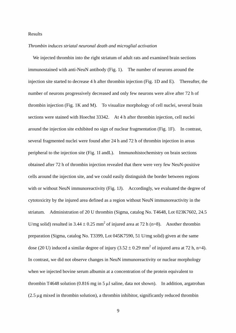
Results
Thrombin induces striatal neuronal death and microglial activation
We injected thrombin into the right striatum of adult rats and examined brain sections
immunostained with anti-NeuN antibody (Fig. 1). The number of neurons around the
injection site started to decrease 4 h after thrombin injection (Fig. 1D and E). Thereafter, the
number of neurons progressively decreased and only few neurons were alive after 72 h of
thrombin injection (Fig. 1K and M). To visualize morphology of cell nuclei, several brain
sections were stained with Hoechst 33342. At 4 h after thrombin injection, cell nuclei
around the injection site exhibited no sign of nuclear fragmentation (Fig. 1F). In contrast,
several fragmented nuclei were found after 24 h and 72 h of thrombin injection in areas
peripheral to the injection site (Fig. 1I andL). Immunohistochemistry on brain sections
obtained after 72 h of thrombin injection revealed that there were very few NeuN-positive
cells around the injection site, and we could easily distinguish the border between regions
with or without NeuN immunoreactivity (Fig. 1J). Accordingly, we evaluated the degree of
cytotoxicity by the injured area defined as a region without NeuN immunoreactivity in the
striatum. Administration of 20 U thrombin (Sigma, catalog No. T4648, Lot 023K7602, 24.5
U/mg solid) resulted in 3.44 ± 0.25 mm2 of injured area at 72 h (n=8). Another thrombin
preparation (Sigma, catalog No. T3399, Lot 045K7590, 51 U/mg solid) given at the same
dose (20 U) induced a similar degree of injury (3.52 ± 0.29 mm2 of injured area at 72 h, n=4).
In contrast, we did not observe changes in NeuN immunoreactivity or nuclear morphology
when we injected bovine serum albumin at a concentration of the protein equivalent to
thrombin T4648 solution (0.816 mg in 5 μl saline, data not shown). In addition, argatroban
(2.5 μg mixed in thrombin solution), a thrombin inhibitor, significantly reduced thrombin
9

toxicity (2.43 ± 0.28 mm2 of injured area, n=8; P=0.017 vs. thrombin T4648 alone). These
results suggest that thrombin induces necrotic neuronal death shortly after injection, as well as
delayed apoptotic neuronal degeneration.
Contrary to the loss of NeuN immunoreactivity, thrombin injection caused drastic
augmentation of immunoreactivity for OX42, a marker protein of microglia. At the injection
site, microglia changed their morphology from ramified shape into activated amoeboid shape
at 4 h after thrombin injection. At 72 h, thrombin-injected striatum was occupied by a
markedly increased number of activated microglia, whereas microglia retained their
population and ramified appearance in the contralateral striatum (Fig. 2B-D). Some but
fewer microglia showing amoeboid morphology were observed in the saline-injected striatum,
which probably resulted from mechanical damage caused by needle insertion (Fig. 2A).
Nevertheless, the number of activated microglia in thrombin-injected striatum increased
approximately 10 times larger than that in saline-injected striatum (3374 ± 408 vs. 312 ± 40
cells/section, n=4 for each condition, P=0.005). These results suggest that thrombin induces
microglial proliferation and activation in the striatum.
MAPKs are involved in thrombin-induced striatal neuronal death
We previously showed that inhibition of MAPK pathways perturbed thrombin-induced
striatal shrinkage in organotypic slice cultures (Fujimoto et al., 2006). Therefore we
examined the effects of inhibitors of MAPK pathways on thrombin cytotoxicity in vivo. We
used PD98059 (5 nmol), a MAPK kinase inhibitor, SB203580 (5 nmol), a p38 MAPK
inhibitor, and SP600125 (5 nmol), a c-Jun N-terminal kinase (JNK) inhibitor. Vehicle (saline
with DMSO as solvent) or MAPK inhibitors were mixed with thrombin solution and the
10

mixture was injected into the striatum. We found that all of these inhibitors were
neuroprotective against thrombin cytotoxicity. The protective effects were manifested by
significant reduction of the injured area (Fig. 3). A clear border between the injured area and
the intact area was observed also in the striatum treated with thrombin plus MAP kinase
inhibitors, and MAPK inhibitors did not increase the number of NeuN-positive cells within
the injured area. The density of NeuN-positive cells in the contralateral striatum in the brain
section prepared from thrombin-injected rats was 1172 ± 33 cells/mm2 (n=9), and
thrombin-induced decrease of NeuN immunoreactivity corresponded to the loss of 4330 ± 321
neurons/section. Likewise, the numbers of NeuN-positive cells lost after treatment with
thrombin plus PD98059, SB203580 and SP600125 were estimated to be 3492 ± 239 (n=9),
3220 ± 209 (n=8) and 3451 ± 223 (n=8) cells/mm2, respectively, all of which were
significantly smaller (P<0.05) than that after treatment with thrombin.
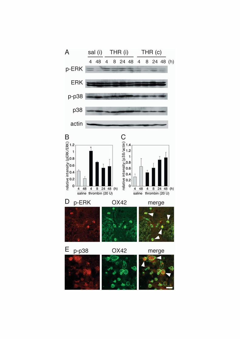
To further clarify the involvement of MAPK pathways, we examined the phosphorylation
level of MAPKs by western blot analysis. Phosphorylated extracellular signal regulated
kinase (ERK) increased promptly after thrombin injection and declined thereafter, whereas
phosphorylated p38 MAPK increased gradually, which was accompanied by an increase in
total p38 MAPK (Fig. 4A-C). To determine cell types exhibiting ERK phosphorylation after
4 h of thrombin injection, we performed double immunofluorescence with combination of
antibodies against phosphorylated ERK and cell type-specific marker proteins.
Immunostaining revealed that most of phosphorylated ERK-positive cells were microglia (Fig.
4D). We also tried to identify cells that express phosphorylated p38 MAPK in brain sections
obtained after 72 h of thrombin injection. As shown in Fig. 4E, phosphorylated p38 MAPK
also colocalized with OX42, a marker of microglia.
11

We further examined the involvement of reactive microglia in thrombin neurotoxicity.
After thrombin injection into the striatum, minocycline, an inhibitor of microglial activation
(Power et al., 2003), or saline was intraperitoneally administered. Minocycline treatment
significantly reduced the number of reactive microglia around the injection site (Fig. 5A) and
the injured area (Fig. 5B). Moreover, double immunofluorescence with antibodies against
inducible nitric oxide synthase (iNOS) and OX42 revealed that some reactive microglia
expressed iNOS after 24 h of thrombin injection in the ipsilateral striatum but not in the
contralateral side (Fig. 5C, D). These results suggest that reactive microglia are responsible
for thrombin neurotoxicity.
12

Discussion
Thrombin induces neuronal loss in vitro (Donovan et al., 1997; Katsuki et al., 2006) and in
vivo (Xue and Del Bigio, 2001; Choi et al., 2003). The detrimental effects of thrombin are
considered to be one of the major causes of ICH-induced neurologic deficits (Gingrich and
Traynelis, 2000; Xi et al., 2003). Thus the regulation of thrombin cytotoxicity could have a
therapeutic potential for ICH (Kitaoka et al., 2002). In previous work on cortico-striatal
slice cultures, we demonstrated that thrombin induces apoptotic neuronal death with a long
delay (Fujimoto et al., 2006). Here we investigated whether delayed apoptotic neuronal
death occurs in adult rat striatum in response to thrombin. Predictably, thrombin induced
neuronal loss around the injection site in the striatum. Two thrombin preparations with
different purities induced a similar degree of injury when administered at the same dosage of
total activity. In addition, thrombin toxicity was significantly attenuated by a thrombin
inhibitor, argatroban. Previous results in vitro also showed that cytotoxicity of thrombin in
the striatal tissue was blocked by prior heat-inactivation or by co-application of argatroban
(Fujimoto et al., 2006). Overall, our observations indicate that proteolytic activity
contributes to the neurotoxicity of thrombin in the striatum, although the involvement of
non-proteolytic potential of thrombin preparation could not be entirely ruled out (Weinstein et
al., 2005).
Unexpectedly, thrombin reduced the number of NeuN-positive cells around the injection
site as early as 4 h of injection. This acute loss of neurons may result from necrotic death of
some neuronal populations (Xue and Del Bigio, 2001), because we did not observe any cell
nuclei undergoing fragmentation at 4 h after thrombin injection. On the other hand, many
fragmented nuclei were observed at 1 or 3 days after thrombin injection, suggesting that the
13

loss of NeuN-positive cells in later days is a consequence of apoptotic neuronal death.
Consistent with our observations, a previous report has demonstrated that apoptotic cells do
not increase at 6 h after thrombin infusion (Gong et al., 2001). Our results are also in
agreement with the time window of delayed thrombin- and ICH-induced apoptosis
(Matsushita et al., 2000; Gong et al., 2001; Xue and Del Bigio, 2001). We did not examine
the effect of caspase inhibitors against thrombin neurotoxicity, because we found previously
that a caspase-3 inhibitor had no effect on thrombin cytotoxicity in cortico-striatal slice
cultures (Fujimoto et al., 2006).
In the present study, phosphorylation of ERK was found in the ipsilateral striatum at 4 h
after thrombin injection. Prompt ERK phosphorylation is consistent with our previous
findings in vitro (Fujimoto et al., 2006; Katsuki et al., 2006). In addition, blockade of ERK
pathway by PD98059 could reduce thrombin-induced loss of neurons, as reflected by the
decrease in the injured area. These results suggest that ERK pathway participates in
thrombin neurotoxicity in the striatum. We also detected a gradual increase in the level of
phosphorylated p38 MAPK after thrombin injection. Interestingly, however,
phosphorylation of p38 MAPK was accompanied by elevation of total p38 MAPK protein
level. It is thus plausible that elevated level of phosphorylated p38 MAPK was a
consequence of an increase in p38 MAPK itself. Nevertheless, inhibition of p38 MAPK by
SB203580 decreased thrombin-induced injury, indicating that p38 MAPK pathway is also
involved in thrombin-induced neuronal death in the striatum. Together with the protective
effect of a JNK inhibitor SP600125, our present results demonstrated that ERK, p38 MAPK
and JNK are all involved in thrombin-induced striatal damage in vivo. These observations
are consistent with our previous findings in vitro that PD98059, SB203580 and SP600125 all
14
suppressed thrombin-induced shrinkage of the striatal tissue in organotypic culture (Fujimoto
et al., 2006). PD98059 and SB203580 were also shown to attenuate thrombin-induced
dopaminergic neuronal death in the midbrain in vivo (Choi et al., 2003). On the other hand,
SP600125 exacerbates, and SB203580 shows no effect on, thrombin-induced cortical injury in
vitro (Fujimoto et al., 2006), suggesting differential involvement of MAPKs in the regulation
of thrombin neurotoxicity between the cerebral cortex and the striatum. Whether cortical
injury induced by thrombin in vivo is regulated by MAPKs in a similar way to that in vitro
remains to be determined.
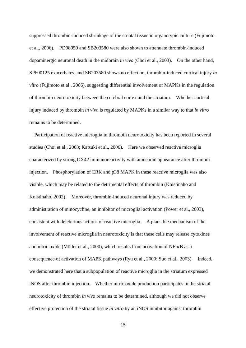
Participation of reactive microglia in thrombin neurotoxicity has been reported in several
studies (Choi et al., 2003; Katsuki et al., 2006). Here we observed reactive microglia
characterized by strong OX42 immunoreactivity with amoeboid appearance after thrombin
injection. Phosphorylation of ERK and p38 MAPK in these reactive microglia was also
visible, which may be related to the detrimental effects of thrombin (Koistinaho and
Koistinaho, 2002). Moreover, thrombin-induced neuronal injury was reduced by
administration of minocycline, an inhibitor of microglial activation (Power et al., 2003),
consistent with deleterious actions of reactive microglia. A plausible mechanism of the
involvement of reactive microglia in neurotoxicity is that these cells may release cytokines
and nitric oxide (Möller et al., 2000), which results from activation of NF-κB as a
consequence of activation of MAPK pathways (Ryu et al., 2000; Suo et al., 2003). Indeed,
we demonstrated here that a subpopulation of reactive microglia in the striatum expressed
iNOS after thrombin injection. Whether nitric oxide production participates in the striatal
neurotoxicity of thrombin in vivo remains to be determined, although we did not observe
effective protection of the striatal tissue in vitro by an iNOS inhibitor against thrombin
15

toxicity (Fujimoto et al., 2006). In this context, Choi et al. (2005) has demonstrated that
intrahippocampal injection of thrombin induces microglial activation associated with
expression of iNOS and NADPH oxidase, and that resultant increase in oxidative stress
contributes to thrombin neurotoxicity.
Overall, we demonstrated that thrombin injected into the striatum caused neuronal damage,
which in part involves a delayed phase of neuronal loss with apoptotic features. Prominent
activation of microglia exhibiting MAPK activation, as well as a significant protective effect
of MAPK inhibitors, suggests that regulation of MAPK pathways and/or microglial activation
could be a useful therapeutic intervention against hemorrhagic stroke.
Acknowledgements
This study was supported by Grant-in-aid for Scientific Research from The Ministry of
Education, Culture, Sports, Science and Technology, Japan and Japan Society for the
Promotion of Science. S. F. is supported as a Research Assistant by 21st Century COE
Program “Knowledge Information Infrastructure for Genome Science”.
16

References
Choi, S.H., Joe, E.H., Kim, S.U., Jin, B.K., 2003. Thrombin-induced microglial activation
produces degeneration of nigral dopaminergic neurons in vivo. J. Neurosci. 23,
5877-5886.
Choi, S.H., Lee, D.Y., Kim, S.U., Jin, B.K., 2005. Thrombin-induced oxidative stress
contributes to the death of hippocampal neurons in vivo: role of microglial NADPH
oxidase. J. Neurosci. 25, 4082-4090.
Donovan, F.M., Pike, C.J., Cotman, C.W., Cunningham, D.D., 1997. Thrombin induces
apoptosis in cultured neurons and astrocytes via a pathway requiring tyrosine kinase and
RhoA activities. J. Neurosci. 17, 5316-5326.
Fayad, P.B., Awad, I.A., 1998. Surgery for intracerebral hemorrhage. Neurology 51, S69-S73.
Fujimoto, S., Katsuki, H., Kume, T., Akaike, A., 2006. Thrombin-induced delayed injury
involves multiple and distinct signaling pathways in the cerebral cortex and the striatum in
organotypic slice cultures. Neurobiol. Dis. 22, 130-142.
Gingrich, M.B., Traynelis, S.F., 2000. Serine proteases and brain damage - is there a link?
Trends Neurosci. 23, 399-407.
Gong, C., Boulis, N., Qian, J., Turner, D.E., Hoff, J.T., Keep, R.F., 2001. Intracerebral
hemorrhage-induced neuronal death. Neurosurgery 48, 875-882.
Huang, F.P., Xi, G., Keep, R.F., Hua, Y., Nemoianu, A., Hoff, J.T., 2002. Brain edema after
experimental intracerebral hemorrhage: role of hemoglobin degradation products. J.
Neurosurg. 96, 287-293.
Katsuki, H., Okawara, M., Shibata, H., Kume, T., Akaike, A., 2006. Nitric oxide-producing
microglia mediate thrombin-induced degeneration of dopaminergic neurons in rat
17

midbrain slice culture. J. Neurochem. 97, 1232-1242.
Kitaoka, T., Hua, Y., Xi, G., Hoff, J.T., Keep, R.F., 2002. Delayed argatroban treatment
reduces edema in a rat model of intracerebral hemorrhage. Stroke 33, 3012-3018.
Koistinaho, M., Koistinaho, J., 2002. Role of p38 and p44/42 mitogen-activated protein
kinases in microglia. Glia 40, 175-183.
Lee, K.R., Colon, G.P., Betz, A.L., Keep, R.F., Kim, S., Hoff, J.T., 1996. Edema from
intracerebral hemorrhage: the role of thrombin. J. Neurosurg. 84, 91-96.
Matsushita, K., Meng, W., Wang, X., Asahi, M., Asahi, K., Moskowitz, M.A., Lo, E.H., 2000.
Evidence for apoptosis after intercerebral hemorrhage in rat striatum. J. Cereb. Blood
Flow Metab. 20, 396-404.
Möller, T., Hanisch, U.K., Ransom, B.R., 2000. Thrombin-induced activation of cultured
rodent microglia. J. Neurochem. 75, 1539-1547.
Power, C., Henry, S., Del Bigio, M.R., Larsen, P.H., Corbett, D., Imai, Y., Yong, V.W., Peeling,
J., 2003. Intracerebral hemorrhage induces macrophage activation and matrix
metalloproteinases. Ann. Neurol. 53, 731-742.
Ryu, J., Pyo, H., Jou, I., Joe, E., 2000. Thrombin induces NO release from cultured rat
microglia via protein kinase C, mitogen-activated protein kinase, and NF-κB. J. Biol.
Chem. 275, 29955-29959.
Sansing, L.H., Kaznatcheeva, E.A., Perkins, C.J., Komaroff, E., Gutman, F.B., Newman, G.C.,
2003. Edema after intracerebral hemorrhage: correlations with coagulation parameters and
treatment. J. Neurosurg. 98, 985-992.
Sinar, E.J., Mendelow, A.D., Graham, D.I., Teasdale, G.M., 1987. Experimental intracerebral
hemorrhage: effects of a temporary mass lesion. J. Neurosurg. 66, 568-576.
18

Suo, Z., Wu, M., Citron, B.A., Gao, C., Festoff, B.W., 2003. Persistent protease-activated
receptor 4 signaling mediates thrombin-induced microglial activation. J. Biol. Chem. 278,
31177-31183.
Weinstein, J.R., Hong, S., Kulman, J.D., Bishop, C., Kuniyoshi, J., Andersen, H., Ransom,
B.R., Hanisch, U.K., Möller, T., 2005. Unraveling thrombin's true microglia-activating
potential: markedly disparate profiles of pharmaceutical-grade and commercial-grade
thrombin preparations. J. Neurochem. 95, 1177-1187.
Xi, G., Reiser, G., Keep, R.F., 2003. The role of thrombin and thrombin receptors in ischemic,
hemorrhagic and traumatic brain injury: deleterious or protective? J. Neurochem. 84, 3-9.
Xi, G., Keep, R.F., Hoff, J.T., 2006. Mechanisms of brain injury after intracerebral
haemorrhage. Lancet Neurol. 5, 53-63.
Xue, M., Del Bigio, M.R., 2001. Acute tissue damage after injections of thrombin and
plasmin into rat striatum. Stroke 32, 2164-2169.
19

Figure legends
Fig. 1. Thrombin-induced neuronal death and nuclear fragmentation. Brains were
transcardially fixed by 4% paraformaldehyde, and coronal cryosections were prepared.
Shown are representative images of coronal sections immunostained with anti-NeuN antibody
at low (A, D, G and J) and high (B, E, H and K) magnification and Hoechst 33342-stained
sections (C, F, I and L). The sections were prepared from brains at 72 h after saline injection
(A-C), 4 h (D-F), 24 h (G-I) and 72 h (J-L) after thrombin injection. Arrowheads in I and L
indicate fragmented nuclei. Scale bars, 1 mm (A, D, G and J), 100 μm (B, E, H and K) and
20 μm (C, F, I and L). (M) The number of NeuN-positive cells at the injection site, at
indicated periods after saline and thrombin treatment. n=4-6.
Fig. 2. Representative images of brain coronal sections immunostained with anti-OX42
antibody. Images are of the injection site at 72 h after saline (A), 4 h (B) and 72 h (C) after
thrombin injection, and of the contralateral striatum at 72 h after thrombin injection (D).
Scale bar, 20 μm.
Fig. 3. Effects of MAPK inhibitors administered by intrastriatal injection concomitant with
thrombin. (A-D) Representative images of NeuN-immunostained coronal sections prepared
from brains after 72 h of injection of thrombin alone (20 U with DMSO, 1:3; A), thrombin
plus PD98059 (5 nmol; B), SB203580 (5 nmol; C) and SP600125 (5 nmol; D). Scale bar, 1
mm. (E) The degree of injury was determined by the area devoid of NeuN immunoreactivity
in the striatum. Vehicle (veh, DMSO diluted 1:3 with saline) was also injected as control.
# P < 0.001 vs. vehicle, * P < 0.05 vs. thrombin alone, n=7-9.
20

Fig. 4. Thrombin-induced phosphorylation of ERK and p38 MAPK. (A) Representative
immunoblots showing ERK and p38 MAPK phosphorylation in response to thrombin. After
indicated periods of injection, homogenates were prepared from 2 mm-thick sections of
ipsilateral (i) and contralateral (c) striatum containing the injection site, and lysates were
subjected to western blot analysis with specific antibodies against phosphorylated ERK, total
ERK, phosphorylated p38 MAPK, total p38 MAPK and β-actin. (B) Time course of ERK
phosphorylation. Ratio of phosphorylated ERK vs. total ERK at each time point was
calculated. * P < 0.05 vs. saline (4 h), n=3. (C) Time course of p38 upregulation. Ratio
of total p38 MAPK vs. β-actin at each time point was calculated (n=3). (D) Confocal
immunofluorescence images of phosphorylated ERK (left) and OX42 (center), and their
merged image (right) after 4 h of thrombin injection. (E) Confocal immunofluorescence
images of phosphorylated p38 MAPK (left) and OX42 (center), and their merged image
(right) after 72 h of thrombin injection. Arrowheads in D and E indicate colocalization.
Scale car, 20 μm.
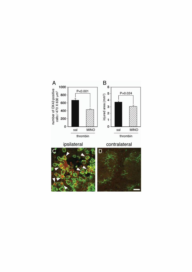
Fig. 5. Involvement of activated microglia in thrombin neurotoxicity. (A, B) Effect of
minocycline (MINO) on the number of activated microglia at the injection site (A) and the
area injured by thrombin (B). MINO was intraperitoneally administered at a dose of 45
mg/kg at 1 and 13 h after thrombin injection and 22.5 mg/kg at 25, 37 and 49 h. n=5. (C,
D) Confocal immunofluorescence images of iNOS (red) and OX42 (green) at the ipsilateral
(C) and the contralateral (D) striatum after 24 h of thrombin injection. Arrowheads indicate
iNOS-positive microglia. Scale car, 10 μm.
21


![熊本大学学術リポジトリ Kumamoto University Repository System · [Keywords] ". English inPrinciplePolicy,ClassroomEnglish,BICS/CALP 1. Introduction Inrecentmonths, anewpolicy](https://img.dokumen.tips/doc/110x75/60476e8cfa95dc318b4a8b05/coeeffff-kumamoto-university-repository-system-keywords.jpg)