Embed Size (px)
Citation preview

© 2004 Wadsworth – Thomson Learning
Chapter 6Chapter 6The Genetics of The Genetics of MicroorganismsMicroorganisms

© 2004 Wadsworth – Thomson Learning
Structure of DNA• Two strands• Nucleotides
– Hydrogen bonds between strands
– Neighboring deoxyribose connected
• 3’ of one deoxyribose to 5’ of next deoxyribose
• Phosphate in between
• Double helix• Base pairing
– G and C– A and T
Figure 6.1

© 2004 Wadsworth – Thomson Learning
Roles of DNA
• Replication– cell division– need accurate copy
• Gene expression– DNA– RNA– Protein
Figure 6.2

© 2004 Wadsworth – Thomson Learning
DNA Replication
• Semi-conservative– old strand-template– new strand-
complementary
• Replication fork– multiple enzymes– DNA unwinds– exposes nucleotides– synthesize new strand– one direction: 5’ to 3’
Figure 6.3

© 2004 Wadsworth – Thomson Learning
DNA Replication
• Complementary nucleotides match (A=T; G=C)• DNA polymerase III binds nucleotides releasing
pyrophosphate
Figure 6.3

© 2004 Wadsworth – Thomson Learning
Bacterial chromosomes• Replication of circular
chromosome• Origin of replication
– bubble forms– DNA unwinds
• Replication occurs in both directions
• Two replication forks• Continues until
replication forks meet• Strands separate
Figure 6.4
© 2004 Wadsworth – Thomson Learning
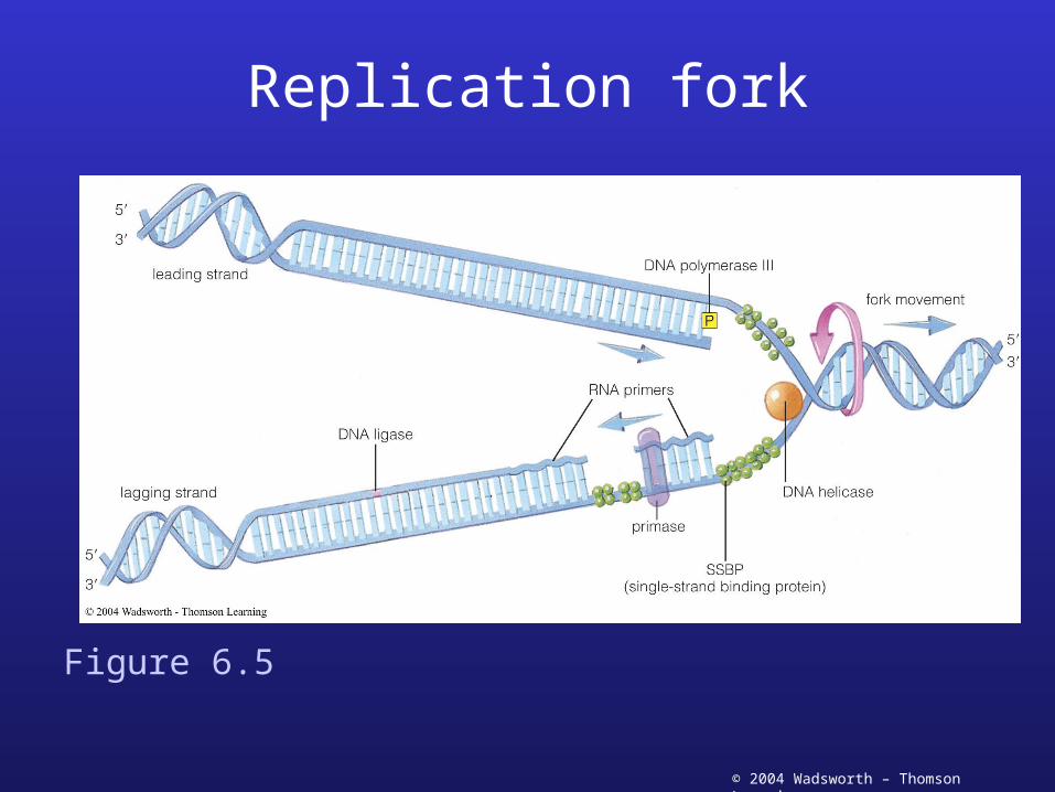
Replication fork
Figure 6.5

© 2004 Wadsworth – Thomson Learning
DNA Replication
• Leading strand– replication is continuous (5’ to 3’)
• primase makes primer• DNA added to primer• fork opens and replication continues
• Lagging strand– polymerization in only one direction
• can’t go 3’ to 5’
– short segments synthesized (Okazaki fragments)• when fork opens, new primer is made• synthesis in direction away from fork• fragments are joined together by DNA ligase

© 2004 Wadsworth – Thomson Learning
Transcription
• RNA polymerase binds DNA at site of promoter
Figure 6.6

© 2004 Wadsworth – Thomson Learning
Transcription
• DNA unwinds• nucleotide bases are
exposed
Figure 6.6
© 2004 Wadsworth – Thomson Learning
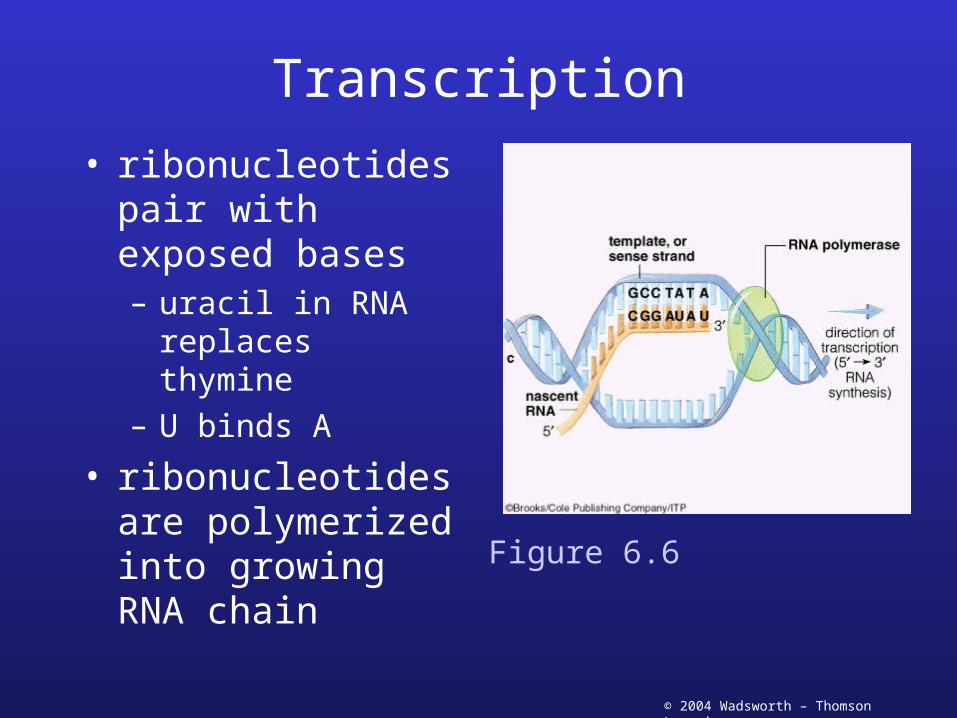
Transcription
• ribonucleotides pair with exposed bases– uracil in RNA
replaces thymine– U binds A
• ribonucleotides are polymerized into growing RNA chain
Figure 6.6
© 2004 Wadsworth – Thomson Learning
Transcription
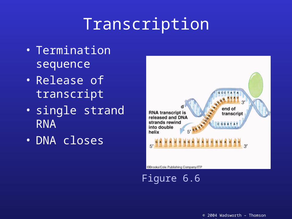
• Termination sequence
• Release of transcript• single strand RNA• DNA closes
Figure 6.6

© 2004 Wadsworth – Thomson Learning
Transcription
• Role of RNA from transcription– mRNA
• template which encodes the protein
– tRNA• transfer amino acids used to build the protein
– rRNA• part of ribosome which is the site of protein
synthesis
• All used for translation

© 2004 Wadsworth – Thomson Learning
Translation• Production of proteins• Based on genetic
information of DNA• Genetic code
– Codon has three nucleotide
– Four different nucleotides
– 64 possible combinations
– 20 amino acids• Redundancy• Nonsense codons
Figure 6.8

© 2004 Wadsworth – Thomson Learning
Translation
• tRNA– binds an amino acid
• specific amino acid for each tRNA
– Anticodon• recognizes codon• three nucleotide
sequence in mRNA which encodes a specific amino acid
– activated with ATP
Figure 6.7

© 2004 Wadsworth – Thomson Learning
Translation
• Ribosome binds to mRNA– specific region– start codon
• Methionine
– Ribosome binding region
• Shine-Dalgarno sequence
Figure 6.9

© 2004 Wadsworth – Thomson Learning
Translation• tRNA with appropriate
anticodon and specific amino acid binds to the codon on the mRNA– A site
• second tRNA binds in similar fashion– P site
• two amino acids are joined in a peptide bond
Figure 6.9

© 2004 Wadsworth – Thomson Learning
Translation
• Ribosome moves along mRNA
• first tRNA without amino acid is removed
• second tRNA with both amino acids moves to P site
Figure 6.9

© 2004 Wadsworth – Thomson Learning
Translation
• New tRNA enters A site• Growing amino acid chain is transferred to
new amino acid
Figure 6.9

© 2004 Wadsworth – Thomson Learning
Translation
• steps repeat– ribosome moves– one codon at a time
• protein chain– one amino acid
added for every codon
Figure 6.9

© 2004 Wadsworth – Thomson Learning
Translation
• Continues until nonsense (stop) codon is reached
• no tRNA matches• ribosome is removed• protein chain is
released
Figure 6.9

© 2004 Wadsworth – Thomson Learning
Transcription and Translation
• Simultaneous transcription and translation
• mRNA chain is transcribed
• translation begins• multiple ribosomes on
single mRNA– polysome
Figure 6.10

© 2004 Wadsworth – Thomson Learning
Regulation of genes
• Transcription– Production of
regulatory proteins• Bind DNA near the
promoter• Example: Lactose
operon
– Interruption of transcription
• Attenuation
• Translation– Ribosomal proteins
• Global regulation– Catabolite
repression– Nitrogen regulation– Phosphorus
regulation– Stringent response– Heat shock proteins

© 2004 Wadsworth – Thomson Learning
Transcriptional regulation
lac operon• lacZ• lacY• lacA
– regulated by lacI
• Lactose absent– repressor binds– stops transcription
Figure 6.11

© 2004 Wadsworth – Thomson Learning
Transcriptional regulation
• Lactose present– repressor bound
by product of lactose
• allolactose
– transcription occurs
– gene products of all genes are made
Figure 6.11

© 2004 Wadsworth – Thomson Learning
Attenuation
• Histidine operon– Histidine present– Leader protein made
• Translation occurring simultaneously with transcription
• Requires histidine
– Attenuator loop forms on mRNA
• Displaces RNA polymerase• Stops transcription
Figure 6.12a

© 2004 Wadsworth – Thomson Learning
Attenuation
• Histidine absent– Leader protein not
made• Not enough histidines to
complete protein
– Antiterminator loop forms
• Prevents attenuator loop from forming
• RNA polymerase continues
Figure 6.12b
© 2004 Wadsworth – Thomson Learning
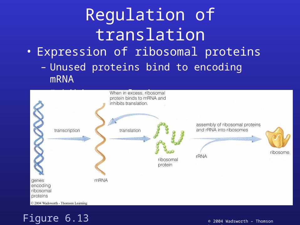
Regulation of translation
• Expression of ribosomal proteins– Unused proteins bind to encoding mRNA– Inhibit translation
Figure 6.13

© 2004 Wadsworth – Thomson Learning
Two component regulation
• Phosphorylation of sensor
• Phosphate passed to response regulator
• Response regulator reacts with DNA changing gene expression– Increase– decrease
Figure 6.14

© 2004 Wadsworth – Thomson Learning
Genetic Information• Genome
– total DNA of a cell– most have single circular chromosome– some have linear chromosome
• Plasmids– small, circular, extrachromosomal DNA
• encode beneficial factors• resistance factors (antibiotic)• conjugative plasmids
– transfer to other cells
• Genotype: genetic makeup• Phenotype: appearance and function

© 2004 Wadsworth – Thomson Learning
Changes in Genetic Information
• Mutations– chemical change
in DNA• chemical
mutagens– Bind DNA– Change in DNA
• physical mutagens– UV light– Ionizing radiation
• biological mutagens– Transposable
elements» Insertion
sequences» transposons
Figure 6.16

© 2004 Wadsworth – Thomson Learning
Consequence of mutations• Types of mutations
– base substitution• wrong nucleotide
– deletion mutation• nucleotides deleted
– Inversion• reverses order of a
segment
– Transposition• moves a segment of
DNA
– Duplication• identical new segment
• Results– Lethal mutation– Conditional expressed
mutations
Figure 6.15

© 2004 Wadsworth – Thomson Learning
Physical mutagen--UV damage
• UV light– stimulates neighboring
bases to form dimers• thymine dimers
– activate repair systems
Figure 6.17

© 2004 Wadsworth – Thomson Learning
Physical mutagen--UV damage
• Thymine dimers distort the DNA structure
• Enzymes remove the damaged nucleotides
Figure 6.17

© 2004 Wadsworth – Thomson Learning
Physical mutagen--UV damage
• Repairs may result in incorrect nucleotide replacement
• Mutation is result
Figure 6.17

© 2004 Wadsworth – Thomson Learning
Selecting and identifying mutants• Direct selection
– Conditions favor growth of desired mutant– Growth of bacteria in presence of antibiotic– Only successful growth are mutants
• Indirect selection– Prevent growth of mutant– Kill growing cells– Desired mutants larger percentage of population– Isolate mutants
• Site-directed mutagenesis– Recombinant DNA manipulation

© 2004 Wadsworth – Thomson Learning
Selecting and identifying mutants
• Brute strength– Screen large numbers– Replica plating
• Transfer large numbers of colonies• Track growth
Figure 6.18

© 2004 Wadsworth – Thomson Learning
Ames Test
Figure 6.19

© 2004 Wadsworth – Thomson Learning
Transformation
• DNA exits one cell, taken up by another cell– Natural
• few bacteria take up DNA fragments
– Artificial--induced in laboratory• useful tool for recombinant DNA technology
Figure 6.20

© 2004 Wadsworth – Thomson Learning
Conjugation
• Conjugative plasmids– plasmids transfer– genetically encoded– F plasmid in E. coli– sex pilus connect two
cells• one cell F+
• one cell F-
Figure 6.21

© 2004 Wadsworth – Thomson Learning
Conjugation– One strand of
plasmid DNA is broken (nicked)
– replication begins– synthesized linear
strand enters F- cell– linear strand is
copied forming a complete plasmid
– both cells are F+
Figure 6.21
© 2004 Wadsworth – Thomson Learning
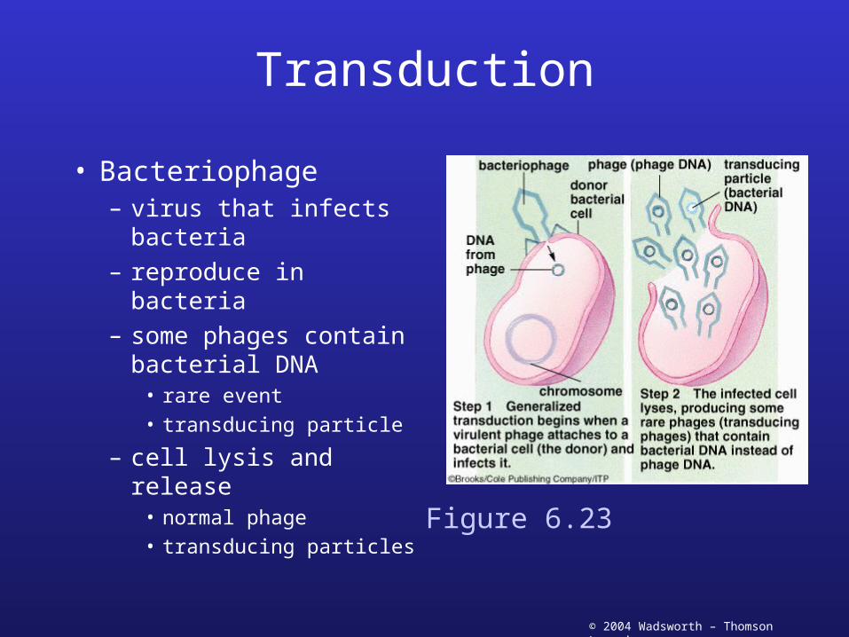
Transduction
• Bacteriophage– virus that infects
bacteria– reproduce in bacteria– some phages contain
bacterial DNA• rare event• transducing particle
– cell lysis and release• normal phage• transducing particles
Figure 6.23
© 2004 Wadsworth – Thomson Learning
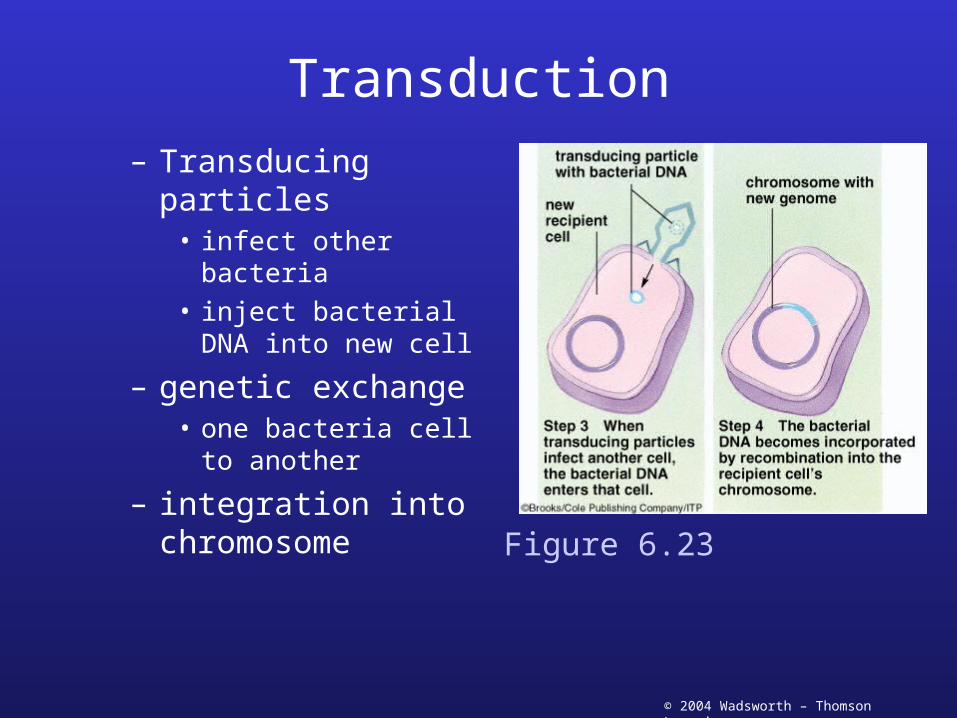
Transduction– Transducing particles
• infect other bacteria• inject bacterial DNA
into new cell
– genetic exchange• one bacteria cell to
another
– integration into chromosome
Figure 6.23

© 2004 Wadsworth – Thomson Learning
Eukaryotic Microorganisms
• Genetic exchange– Similar to plants
and animals– Haploid
gametes fuse
Figure 6.23