Embed Size (px)
Citation preview
WHOLE-GENOME ANALYSES OF ROTAVIRUS STRAINS CIRCULATING PRE- AND POST- ROTATEQ™
VACCINE INTRODUCTION IN RWANDA
Sebotsana Paula Rasebotsa
2013067463
Submitted in fulfilment of the requirements in respect of the Master’s degree
Masters of Medical Sciences with specialisation in Virology
In the
Division of Virology, Faculty of Heath Sciences
Next Generation Sequencing Unit
University of the Free State
Bloemfontein
South Africa
30 September 2020
Supervisor: Dr. Martin Nyaga
Next Generation Sequencing Unit, Office of the Dean: Health Sciences
and Division of Virology, Faculty of Heath Sciences
University of the Free State
Co-supervisor: Dr. Saheed Sabiu
Biotechnology and Food Technology Department Faculty of Applied Sciences
Durban University of Technology [email protected]

ii
DECLARATION
“I, Sebotsana Paula Rasebotsa, declare that the Master’s Degree research dissertation or interrelated,
publishable manuscripts/published articles, or coursework Master’s Degree mini-dissertation that I
herewith submit for the Master’s Degree qualification Masters of Medical Sciences with specialisation
in Virology at the University of the Free State is my independent work and that I have not previously
submitted it for a qualification at another institution of higher education.”
___________________
Sebotsana Paula Rasebotsa

iii
This dissertation is dedicated to my family, especially my mother, Mrs. Moshupologo Hilda
Rasebotsa for being my pillar of strength

iv
ACKNOWLEDGEMENTS
I would like to express my deepest gratitude to:
God, for giving me the strength, knowledge, and opportunity to undertake this research study
and to persevere until the completion of my master’s degree.
Dr. Martin Munene Nyaga, my supervisor, for the professional guidance and valuable
support, encouragement and useful criticism that allowed me to grow as an upcoming
scientist.
Dr. Saheed Sabiu, my co-supervisor, for the invaluable assistance, insightful suggestions,
guidance and support throughout the study.
The World Health Organization (WHO), for the financial assistance and the contribution to
formulating the study’s original terms of reference that were subdivided into several sub-
studies including this one. Opinions expressed and conclusions arrived at are those of the
author and are not necessarily attributed to the WHO. (Grant no. 2017/757922-0).
The children and parents/guardians, for donating the stool samples for this research.
Collaborators from Rwanda, for collecting the samples from the patients and providing all the
necessary patient information and demographics required for the study.
South African Medical Research Council/Diarrhoeal Pathogens Research Unit, Sefako
Makgatho Health Sciences University for providing me with the archived stool samples and
material transfer agreement.
Dr. Matthew Esona, for his expert advice and guidance with bioinformatics and software
support available at the Centers for Disease Control and Prevention, Atlanta, Georgia, USA.
I have great pleasure in acknowledging my gratitude to my colleagues at Next Generation
Sequencing Unit for their continued support, encouragement, and assistance throughout the
study.
My family and friends for their undying love, support and encouragement throughout my
studies and for never doubting the decisions I made in pursuing my master’s degree.
The University of the Free State Postgraduate School for financial assistance and the facilities
made disposable to me throughout the study.
The financial assistance of the National Research Foundation (NRF) towards this research is
hereby acknowledged. Opinions expressed and conclusions arrived at are those of the author
and are not necessarily attributed to the NRF. (Grant no. SFH180514329137).
The financial assistance of the Poliomyelitis Research Foundation (PRF) towards this research
is hereby acknowledged. Opinions expressed and conclusions arrived at are those of the
author and are not necessarily attributed to the PRF. (Grant no. 19/63).

v
The financial assistance of the South Africa Medical Research Council towards this research
is hereby acknowledged. Opinions expressed and conclusions arrived at are those of the
author and are not necessarily attributed to the SAMRC. (Self-Initiated Research grant)
The financial assistance of the Bill and Melinda Foundation towards this research is hereby
acknowledged. Opinions expressed and conclusions arrived at are those of the author and are
not necessarily attributed to the BMGF.(Grant no. BMGF OPP1180423 2017)
vi
RESEARCH DISSEMINATION
PUBLICATIONS IN INTERNATIONAL ACCREDITED AND INDEXED SCIENTIFIC JOURNALS Rasebotsa SP, Mwangi PN, Mogotsi MT, Sabiu S, Magagula NB, Rakau K, Uwimana J, Mutesa L,
Muganga N, Murenzi D, Tuyisenge L, Jaimes J, Esona MD, Bowen MD, M. Mphahlele J, Seheri ML,
Mwenda JM, Nyaga MM., 2020. Whole-genome and in-silico analyses of G1P[8] rotavirus strains from
pre- and post-vaccination periods in Rwanda. Sci. Rep. 10, 1–22. https://doi.org/10.1038/s41598-020-
69973-1 Impact factor: 4.120
Rasebotsa SP, Mwenda JM, Mogotsi MT, Mwangi PN, Uwimana J, Rakau K, Muganga N, Seheri ML,
Mihigo R, M. Mphahlele J, Mutesa L, Sabiu S, Nyaga MM. Whole-genome analyses of rotaviruses
identifies multiple reassortant rotavirus strains in Rwanda post-vaccine introduction. Draft manuscript
under internal review for submission to Frontiers of Microbiology.
OTHER PUBLICATIONS DURING THE COURSE OF THIS STUDY Mwangi PN, Mogotsi MT, Rasebotsa SP, Seheri ML, M. Mphahlele J, Ndze VN, Dennis FE, Jere KC,
Nyaga MM., 2020. Uncovering the first atypical DS-1-like G1P[8] rotavirus strains that circulated
during pre-rotavirus vaccine introduction era in South Africa. Pathogens 9, 391.
https://doi.org/10.3390/pathogens9050391 Impact factor: 3.018
CONFERENCE CONTRIBUTIONS
a) 12th African Rotavirus Symposium, The Emperors Palace Conference Centre, Johannesburg,
South Africa, 30 July–01 August 2019 (International conference)
Rasebotsa SP, Mwangi PN, Mogotsi MT, Sabiu S, Mosime LP, Magagula NB, Rakau K, Uwimana J,
Mutesa L, Muganga N, Murenzi D, Tuyisenge L, M. Mphahlele J, Seheri ML, Mwenda JM, Nyaga MM.
Analysis of G1P[8] whole-genome constellations identified a vaccine-derived strain in Rwanda. (Oral
Presentation)
Mwangi PN, Mogotsi MT, Rasebotsa SP, Sabiu S, Simwaka J, Monze M, Mpabalwani EM, Matapo B,
Magagula NB, Rakau K, Seheri ML, M. Mphahlele J, Mwenda JM, Nyaga MM. Molecular
characterization of rotavirus strains using whole-genome sequencing reveals unique changes post-
rotavirus vaccine introduction in Zambia. (Oral Presentation)

vii
Mogotsi MT, Mwangi PN, Mosime LB, Rasebotsa SP, Bester AP, Seheri ML, M. Mphahlele J, O’Neill
HG, Nyaga MM. Characterization of the gut virome of South African infants using a viral metagenomics
approach. (Poster Presentation)
Sabiu S, Mwangi PN, Rasebotsa SP, Mogotsi MT, Magagula NB, Rakau K, Uwimana J, Mutesa L,
Muganga N, Murenzi D, Tuyisenge L, Seheri ML, M. Mphahlele J, Mwenda JM, Nyaga MM. Variability
of the P-types in stable genotype constellations pre- and post-vaccine introduction in Rwanda. (Poster
Presentation)
Mugweru JN, PN, Rasebotsa SP, Mogotsi MT, Sabiu S, Uwimana J, Mutesa L, Muganga N, Murenzi D,
Tuyisenge L, Magagula NB, Rakau K, Seheri ML, M. Mphahlele J, Mwenda JM, Nyaga MM. Whole-
genome constellations of five reassortant rotavirus strains detected during post-rotavirus vaccine
introduction period in Rwanda. (Poster Presentation)
b) University of the Free State (UFS) Health Science Faculty Forum, UFS, Bloemfontein, South Africa,
29-30 August 2019 (Local conference)
Rasebotsa SP, Mwangi PN, Mogotsi MT, Sabiu S, Mosime LP, Magagula NB, Rakau K, Uwimana J,
Mutesa L, Muganga N, Murenzi D, Tuyisenge L, M. Mphahlele J, Seheri ML, Mwenda JM, Nyaga MM.
Whole-genome analyses of rotavirus G1P[8] strains circulating pre- and post RotaTeq™ vaccine
introduction in Rwanda. (Oral Presentation)
Presented with the best junior laboratory paper award for this presentation.
Mwangi PN, Mogotsi MT, Rasebotsa SP, Sabiu S, Simwaka J, Monze M, Mpabalwani EM, Matapo B,
Magagula NB, Rakau K, Seheri ML, M. Mphahlele J, Mwenda MJ, Nyaga MM. Whole-genome
characterization of Zambian rotavirus strains reveals remarkable changes in the post-rotavirus vaccine
introduction. (Oral Presentation)
c) The Free State Provincial Research day, UFS, Bloemfontein, South Africa, 07-08 November 2019
(Local conference)
Rasebotsa SP, Mwangi PN, Mogotsi MT, Sabiu S, Mosime LP, Magagula NB, Rakau K, Uwimana J,
Mutesa L, Muganga N, Murenzi D, Tuyisenge L, M. Mphahlele J, Seheri ML, Mwenda JM, Nyaga MM.
Whole-genome analyses of rotavirus G1P[8] strains circulating pre- and post RotaTeq™ vaccine
introduction in Rwanda. (Oral Presentation)

viii
d) Virology Africa, Radisson Blue Hotel, V&A Waterfront, Cape Town, South Africa, 10-14 February
2020 (International conference)
Maringa WM, Mwangi PN, Mogotsi MT, Rasebotsa SP, Simwaka J, Magagula NB, Rakau K, Seheri ML,
M. Mphahlele J, Mwenda JM, Nyaga MM. Whole-genome sequencing identifies idiosyncratic changes
post rotavirus vaccine introduction in Zambia. (Oral Presentation)
Mwangi PN, Mogotsi MT, Rasebotsa SP, Seheri ML, M. Mphahlele J, Ndze VN, Dennis FE, Jere KC,
Nyaga MM. Uncovering the first atypical DS-1-like G1P[8] strains in South Africa, pre-rotavirus vaccine
introduction. (Oral Presentation)

ix
LIST OF FIGURES
Figure 2.1: Rotavirus structure and the 11 gene segments………………………………………………………………28
Figure 2.2: The rotavirus replication cycle…………………………………………………………………………………………31
Figure 3.1: The map of Rwanda indicating all the five provinces……………………………………………………….45
Figure 3.2: Qubit 4 Fluorometer workflow……………………………………………………………………………………50
Figure 3.3: DNA fragment ligated with indexes and Illumina chemistry specific adapter sequences…52
Figure 3.4: Bioanalyzer gel image showing the library size distribution…………………………………………….55
Figure 3.5: A) Bioanalyzer electropherogram showing library size distribution determined by high
sensitivity dsDNA assay……………………………………………………………………………………………………………………56
Figure 3.6: B) Electropherogram of the ladder depicting the lower (green) and the upper (purple)
markers used as a control…………………………………………………………………………………………………………………56
Figure 4.3: Maximum likelihood phylogram revealing the genetic relatedness of the concatenated,
whole-genome ORF sequences (17,492 base pairs) for the 25 post-vaccine G1P[8] and 10 pre-vaccine
G1P[8] RVA study strains characterized by whole-gene analysis………………………………………………………65
Figure 4.4: Phylogenetic relatedness of rotavirus group A species base on VP7 of the study strains from
Rwanda with representatives of known human and animal rotavirus genotypes………………………………67
Figure 4.5: A) The alignment of the G1 component of Rotarix® and RotaTeq® vaccines and Rwandan
wildtypes circulating from 2011- 2016 RVA seasons, based on the three VP7 antigenic residues (7-1a,
7-1b, and 7-2)…………………………………………………………………………………………………………………………………..69
Figure 4.6: B) Location of surface-exposed amino acids differences between VP7 protein of RotaTeq®
G1 vaccine component versus a G1 wild-type strain from Rwanda……………………………………………………69
Figure 4.7: Alignment of antigenic residues in T-cell antigen epitopes of the G1 vaccine component
contained in Rotarix® and RotaTeq® compared to Rwanda G1 wild type strains circulating from 2011–
2016 RVA seasons……………………………………………………………………………………………………………………………70
Figure 4.8: Phylogenetic relatedness of rotavirus group A species base on VP4 of the study strains from
Rwanda with representatives of known human and animal rotavirus genotypes………………………………71
Figure 4.9: A) The alignment of the P[8] component of Rotarix® and RotaTeq® vaccines and wild type
P[8] strains circulating in Rwanda from 2011–2016 RVA seasons, based on the antigenic residues in
VP4………………………………………………………………………………………………………………………………………………….73
Figure 4.10: B) Location of surface-exposed amino acids differences between VP8* protein of
RotaTeq® P[8] vaccine component versus a wild-type P[8] strain from
Rwanda………………………………………………..73
x
Figure 4.11: A) – F) Phylogenetic relatedness of rotavirus group A species base on A) VP1, B) VP2, C)
VP3, D) NSP1, E) NSP4 and F) NSP5 of the study strains from Rwanda with representatives of known
human and animal rotavirus genotypes……………………………………………………………………………………………81
Figure 4.12: A) – C) Phylogenetic relatedness of rotavirus group A species base on A) VP6, B) NSP2 and
C) NSP3 of the study strains from Rwanda with representatives of known human and animal rotavirus
genotypes………………………………………………………………………………………………………………………………………..86
Figure 5.1: A) The alignment of the G4 VP7 component of the RotaTeq® vaccine strain and G4
Rwandan study strain based on the three surface exposed epitope regions (7-1a, 7-1b, and 7-
2)……………………………………………………………………………………………………………………………………………………102
Figure 5.1: B) Surface representation of the VP7 protein………………………………………………………………..102
Figure 5.2: The alignment of the P[8] VP4 component of the RotaTeq® vaccine strain and P[8] Rwandan
study strain based on the two VP4 domains, the VP8* (8-1 to 8-4) and VP5 *(5-1 to 5-
5)……………………………………………………………………………………………………………………………………………………103
Figure 5.3: Phylogenetic tree of RVA strains based on the full length of the VP7 (G4, G9, and G12) gene
displaying the relatedness of the study strains (◆) and reference strains from
GenBank………………………………………………………………………………………………………………………………………..104
Figure 5.4: Phylogenetic tree of RVA strains based on the full length of the VP4 (P[4] and P[8]) gene
displaying the relatedness of the study strains (◆) and reference strains from GenBank ………………..105
Figure 5.5: A) – D) Phylogenetic tree of RVA strains based on the full length of the A) VP6 B) VP1 C)
VP2 D) VP3 genes displaying the relatedness of the study strains (◆) and reference strains from
GenBank…………………………………..……………………………………………………………………………………………………110
Figure 5.6: A) – E) Phylogenetic tree of RVA strains based on the full length of the A) NSP1 B) NSP2 C)
NSP3 D) NSP4 E) NSP5 genes displaying the relatedness of the study strains (◆) and reference strains
from GenBank………………………………………………………………………………………………………………………………..116

xi
LIST OF TABLES
Table 2.1 Properties of the rotavirus proteins along with their descriptions and location in the three-
layered icosahedral capsids……………………………………………………………………………………………………………..28
Table 2.2 Basic features and administration recommendations of currently licensed rotavirus
vaccines…………………………………………………………………………………………………………………………………………..34
Table 5.3 Full genotype constellations of five Rwandan strains detected post-vaccine introduction
(2013-2015) along with the contig lengths and number of reads mapped to each contig…………………100

xii
LIST OF ABBREVIATIONS
A-type Interferon Antagonist
aa Amino Acid
AICc Akaike Information Criterion
ATM Amplicon Tagment Mix BLASTn Nucleotide Basic Local Alignment Search Tool
C-type Core Shell Protein
cDNA Complementary Deoxyribonucleic Acid
CTL Cytotoxic T-lymphocytes
ddH20 Double-distilled Water
DLP Double-layered Particle dNTPs Deoxyribonucleotide Triphosphates
DPRU Diarrheal Pathogens Research Unit
dsRNA Double-stranded Ribonucleic acid
E-type Enterotoxin
EB Elution Buffer
EIA Enzyme Immuno-Assay ELISA Enzyme-Linked immunosorbent Assay
EM Electron Microscopy
ER Endoplasmic Reticulum
G-type Glycoprotein
GAVI Global Alliance for Vaccines and Immunizations
H-type Phosphoprotein HSREC Health Sciences Research Ethics Committee
HT1 Hybridization Buffer 1
I-type Intermediate Protein
IFIH1 Interferon-induced Helicase C Domain-containing Protein 1
ISGF3 Interferon-stimulated Gene Factor 3
JAK/STAT Janus Kinase/signal Transducer and Activators of Transcription LiCl2 Lithium Chloride
M-type Methyltransferase
MEGA 6 Molecular Evolutionary Genetics Analysis 6
mRNA Messenger RNA
N-type NTPase
NaOH Sodium Hydroxide NF-kB Nuclear Factor Kappa-light-chain-enhancer of Activated B cells
NGS Next-generation Sequencing
NPM Nextera PCR Master Mix

xiii
NSP Non-structural Proteins
NT Neutralize Tagment Buffer
nt Nucleotide OPV Oral Poliovirus Vaccines
ORF Open Reading Frame
ORS Oral Rehydration Solutions
ORT Oral Rehydration Therapy
P-type Protease Sensitive
PCR Polymerase Chain Reaction PGM Ion Torrent Personal Genome Machine
R-type Viral RNA-dependent RNA Polymerase
RdRp RNA-dependent RNA Polymerase
RRL-SA Rotavirus Reference Laboratory in South Africa
RSB Resuspension Buffer
RT-PCR Reverse Transcription Polymerase Chain Reaction RVA Rotavirus Group A
RVB Rotavirus Group B
RVC Rotavirus Group C
RVD Rotavirus Group D
RVE Rotavirus Group E
RVF Rotavirus Group F RVG Rotavirus Group G
RVH Rotavirus Group H
RVI Rotavirus Group I
RVJ Rotavirus Group J
ssRNA Single-stranded RNA
T-type Translation Enhancer TD Tagment DNA Buffer
ToRs Terms of References
TSA Technical Service Agreement
UFS University of the Free State
UFS-NGS Unit University of the Free State-Next Generation Sequencing Unit
vdG1P[8] Vaccine-derived G1P[8] VP Viral Proteins
WHO World Health Organization
WHO/AFRO World Health Organization Regional Office for Africa

xiv
ABSTRACT
Children living in developing countries are constantly faced with the burden of diarrheal infections
that account for over 1.6 million death cases globally. Rotavirus group A (RVA) has been identified as
one of the viruses implicated in most viral-induced diarrhoeal infections in children less than five years
worldwide. In Rwanda, over 3500 RVA related mortality cases were reported yearly prior to the
implementation of the RotaTeq® vaccine in 2012 to overcome this burden, which led to a significant
decrease in rotavirus infections. Africa has a huge diversity of rotavirus strains compared to other
developed continents especially Europe and North America, thus requiring a deeper understanding of
this phenomenon. This study aimed at characterizing all the 11-segments of RVA strains circulating in
Rwanda pre- and post-vaccine introduction as part of the World Health Organization (WHO) supported
African rotavirus pilot surveillance program. The study was based on 158 rotavirus positive samples
that were collected from children presenting symptoms associated with rotavirus infection between
2011 and 2016. The rotavirus double-stranded ribonucleic acid (dsRNA) was extracted from the viral
particles and converted into complementary deoxyribonucleic acid (cDNA) prior to library preparation
for whole-genome sequencing with an Illumina MiSeq platform. Several bioinformatics tools were
utilized to construct phylogenetic trees and the proteins structures. From the sequenced samples, 36
samples were identified as G1P[8] strains, and five samples were reassortant strains. Ten G1P[8]
strains were identified pre-vaccine introduction while 26 were identified post-vaccine introduction.
Thirty-five of the G1P[8] strains expressed pure Wa-like genome constellations, while one of the
strains that was identified in 2012 exhibited a genome constellation typical of a RotaTeq® vaccine
strain. On the other hand, the five reassortant strains were identified post-vaccine introduction
between 2013-2015. Whole-genome analysis revealed that the G4P[4], G9P[4] and one G12P[8]
reassortant strains exhibited both the Wa-like and the DS-1-like genome constellations while two
G12P[8] strains had all the three genogroup constellations. Furthermore, the phylogenetic analysis of
most of the G1P[8] strains revealed that they segregated according to their vaccination status; strains
identified pre-vaccine introduction clustered together while post-vaccine strains also formed a
separate cluster. The five reassortant strains were closely related to human RVA strains in all the gene
segments and RotaTeq® vaccine strains in the VP1, VP2, NSP2, NSP4, and NSP5 gene segments.
Analysis of the neutralization epitopes and cytotoxic T-lymphocytes (CTL) of the G1P[8] strains
revealed multiple amino acid substitutions, with some changes influencing the change in polarity thus
deemed to be radical in nature. A similar trend was also observed in the reassortant strains, with 27
amino acid substitutions in the VP7 epitope region and only three substitutions in the VP4 epitope
region. Changes observed in these epitope regions have the potential of generating vaccine-escape
mutants that may undermine the effectiveness of the rotavirus vaccine with time. Whole-genome
xv
sequencing has proven to provide information that could have been missed when looking only at the
outer capsid proteins. It is thus important to continue conducting rotavirus whole-genome studies to
unpack the hidden information behind the huge diversity of rotavirus strains in African countries such
as Rwanda.
Keywords: rotavirus, Rwanda, whole-genome characterization, reassortment, epitope region,
RotaTeq®, Rotarix®, diarrhoea, vaccine-derived strain, genome constellation

xvi
Table of Contents
DECLARATION .......................................................................................................................... ii
ACKNOWLEDGEMENTS ............................................................................................................ iv
RESEARCH DISSEMINATION ..................................................................................................... vi
CONFERENCE CONTRIBUTIONS ................................................................................................ vi
LIST OF FIGURES ...................................................................................................................... ix
LIST OF TABLES ........................................................................................................................ xi
LIST OF ABBREVIATIONS ......................................................................................................... xii
ABSTRACT ............................................................................................................................. xiv
CHAPTER ONE: INTRODUCTION ...................................................................................... 19
1.1. Background ................................................................................................................. 20
1.2. Problem Statement ..................................................................................................... 21
1.3. Aim ............................................................................................................................. 22
1.4. Hypothesis .................................................................................................................. 22
1.5. Objectives ................................................................................................................... 23
1.6. Dissertation organization ............................................................................................ 23
CHAPTER TWO: LITERATURE REVIEW .............................................................................. 24
2.1. Background ................................................................................................................. 25
2.2. Rotavirus structure ...................................................................................................... 27
2.3. Diagnosis and genome classification ............................................................................ 29
2.4. Replication cycle ......................................................................................................... 30
2.5. Immune response ....................................................................................................... 32
2.6. Rotavirus evolutionary mechanisms ............................................................................ 33
2.7. Prevention and treatment ........................................................................................... 34
2.7.1. WHO prequalified vaccines ...................................................................................... 35
2.7.2. Nationally licenced vaccine ...................................................................................... 36
2.7.3. Treatment of rotavirus infection .............................................................................. 37
2.8. Rotavirus vaccine introduction in African countries ...................................................... 38
2.9. Surveillance data of African countries .......................................................................... 38
2.10. Next-generation Sequencing .................................................................................... 40
2.11. Rotavirus whole-genome studies ............................................................................. 42

xvii
CHAPTER THREE: METHODOLOGY ................................................................................... 44
3.1. Ethical statement and study design .............................................................................. 45
.............................................................................................................................................. 45
3.2. Extraction of rotavirus dsRNA from faecal samples ...................................................... 46
3.3. Complementary DNA synthesis .................................................................................... 47
3.4. cDNA purification ........................................................................................................ 48
3.5. Quality control using Qubit 4 Fluorometer ................................................................ 49
3.6. DNA Library preparation .............................................................................................. 51
3.6.1. Detailed genomic DNA tagmentation and library amplification ................................. 51
3.6.2. Library clean-up and quantification .......................................................................... 52
3.6.3. Quality control using an Agilent Technology 2100 Bioanalyzer .................................. 53
3.6.4. Library pooling and denaturation ............................................................................. 56
3.7. Data analysis ............................................................................................................... 57
CHAPTER FOUR: WHOLE-GENOME AND IN-SILICO ANALYSES OF G1P[8] ROTAVIRUS STRAINS FROM PRE- AND POST-VACCINATION PERIODS IN RWANDA. ............................................... 59
4.1. Abstract ....................................................................................................................... 61
4.2. Introduction ................................................................................................................. 62
4.3. Results......................................................................................................................... 64
4.3.1. Whole-genome constellation analyses. .................................................................... 64
4.3.2. Phylogenetic and sequence analyses. ....................................................................... 65
4.3.3. Phylogenetic and sequence analyses of the VP7 gene. .............................................. 66
4.3.4. Comparative analyses of neutralizing antigenic epitopes in the VP7 proteins of Rwandan G1P[8] and vaccine strains of RVA. .......................................................................... 68
4.3.5. Comparative analyses of cytotoxic T lymphocytes epitopes of the G1 proteins of Rwandan and vaccine strains of RVA. ...................................................................................... 69
4.3.6. Phylogenetic and sequence analyses of VP4 genes. .................................................. 70
4.3.7. Comparative analyses of neutralizing antigenic epitopes in the VP4 protein of Rwandan G1P[8] and vaccine strains of RVA. .......................................................................... 72
4.3.8. Phylogenetic and sequence analyses of VP1-VP3, NSP1, NSP4, and NSP5. ................. 74
4.3.9. Phylogenetic and sequence analyses of VP6, NSP2, and NSP3. .................................. 82
4.4. Discussion .................................................................................................................... 87
4.5. Methods ...................................................................................................................... 90
4.5.1. Sample collection ...................................................................................................... 90
4.5.2. Double-stranded RNA extraction and purification ......................................................... 90
4.5.3. cDNA synthesis ......................................................................................................... 91

xviii
4.5.4. DNA library preparations and whole-genome sequencing ............................................. 91
4.5.5. Genome assembly ..................................................................................................... 92
4.5.6. Identification of genotype constellations ..................................................................... 92
4.5.7. GenBank accession numbers ...................................................................................... 92
4.5.8. Phylogenetic, sequence analyses and protein modelling ............................................... 92
CHAPTER FIVE: WHOLE-GENOME ANALYSES IDENTIFIES MULTIPLE REASSORTANT ROTAVIRUS STRAINS IN RWANDA POST-VACCINE INTRODUCTION ......................................................... 94
5.1. Abstract ....................................................................................................................... 96
5.2. Introduction ................................................................................................................. 97
5.3. Results......................................................................................................................... 99
5.3.1. Genome genotypes .................................................................................................. 99
5.3.2. The VP4 and VP7 antigenic region analyses ............................................................ 101
5.3.3. Phylogenetic analysis of the VP7 gene of G4, G9 and G12 ....................................... 103
5.3.4. Phylogenetic analysis of the VP4 gene of P[4] and P[8] ........................................... 105
5.3.5. Phylogenetic analyses of the VP1-VP3 and VP6 genes ............................................. 106
5.3.6. Phylogenetic analyses of the NSP1-NSP5 genes ...................................................... 111
5.4. Discussion .................................................................................................................. 117
5.5. Conclusion ................................................................................................................. 119
5.6. Methods .................................................................................................................... 119
5.6.1. Ethics statement .................................................................................................... 119
5.6.2. Sample collection .................................................................................................. 119
5.6.3. Sample preparation for rotavirus whole-genome sequencing ................................. 120
5.6.4. DNA library preparations and whole-genome sequencing ....................................... 120
5.6.5. Computational analysis .......................................................................................... 120
CHAPTER SIX: GENERAL DISCUSSION, CONCLUSION AND RECOMMENDATIONS ................... 122
6.1. Study summary .......................................................................................................... 123
6.2. Limitations and recommendations ............................................................................... 125
REFERENCES ......................................................................................................................... 126
APPENDICES ......................................................................................................................... 158 Appendix A ................................................................................................................................................ 158 Appendix B ................................................................................................................................................ 159 Appendix C................................................................................................................................................. 160

19
CHAPTER ONE: INTRODUCTION

20
1.1. Background
Rotaviruses are among the leading cause of virus-induced mortality in children less than 5 years globally,
with more than half of these deaths occurring in sub-Saharan Africa (Tate et al., 2016; Troeger et al.,
2018a). In 2016, more than 1.6 million global mortality was attributed to diarrhoeal diseases (Troeger et
al., 2018a). Rotavirus is commonly implicated in gastroenteritis, a medical condition characterized by
inflammation of the stomach and intestines (Leung et al., 2005). The virus can easily be transmitted
through the faecal-oral route, causing symptoms such as diarrhoea, malaise, vomiting, and fever (Leung
and Robson, 2007). The symptoms usually start two days after infection, with an acute onset of fever and
vomiting, followed by frequent watery stool, and the infection may last for three to eight days (Leung and
Robson, 2007; Parashar et al., 2013). It is also associated with significant dehydration that could lead to
death in children due to their high vulnerability to electrolyte imbalance resulting from diarrhoea and
vomiting (Leung and Robson, 2007).
Rotavirus is regarded as “the democratic virus” due to its ability to infect populations across the social-
economic status (Parashar et al., 2013). Although rotavirus infection has been implicated in high mortality
in children, vaccination against the virus has proven to be the most reliable prevention measure
accompanied by improved sanitation (Fischer et al., 2007; Tate et al., 2016). Furthermore, zinc
supplements, oral rehydration therapy (ORT), and intravenous fluids have also been recommended as
effective treatments for diarrhoea by the WHO (Santosham et al., 2010). Despite this recommendation,
such treatments are not holistically accessible by several low-income countries including sub-Saharan
African countries. For instance, in 2013 alone, four countries (Democratic Republic of the Congo, India,
Nigeria, and Pakistan) accounted for half of all rotavirus deaths in the world (Tate et al., 2016). The lack
of timely access to quality health care, frequent concurrent infections, malnutrition, and climatic factors
contributes to the high rate of rotavirus mortality in third world countries (Elliott, 2007; Parashar et al.,
2009; Wazny et al., 2013).
Four rotavirus vaccines (Rotarix®, RotaTeq®, Rotavac® and Rotasil®) are currently pre-qualified by WHO for
prevention of rotavirus infections (WHO, 2018). The efficacy of rotavirus vaccines differs between
countries with different socio-economic statuses. High-income countries that introduced Rotarix® in their
national immunization program reported 84% vaccine effectiveness while low- and middle-income
countries reported efficacy of 57-75% (Jonesteller et al., 2017). Furthermore, high-income countries that

21
introduced RotaTeq® vaccine in their national immunization program reported 90% vaccine effectiveness
while low-income countries documented efficacy of 45% (Jonesteller et al., 2017). Low-income countries
have been suspected to experience lower efficacy of the vaccines due to factors such as maternal
antibodies, malnutrition, and co-administration of rotavirus vaccines and oral poliovirus vaccines (OPV)
(Moon et al., 2010). Rotavirus vaccines are more effective when administered before natural immunity is
acquired through natural infections; therefore, administering the vaccine after natural rotavirus infection
may also affect the effectiveness of the rotavirus vaccines (Patel et al., 2009). The most prevalent rotavirus
genotype combinations detected in Africa between 2006 and 2015 were G1P[8], G2P[4], G9P[8], G2P[6],
G12P[8], and G3P[6] (Mwenda et al., 2010; Seheri et al., 2018, 2014).
The establishment of the Global Rotavirus Surveillance Network in 2008 has created a platform to
constantly monitor changes that may have been influenced by the introduction of rotavirus vaccines and
to understand better the dynamics of rotavirus strain diversity in different settings all over the world
(Aliabadi et al., 2019). The mandate of the network is to perform exceptional diagnostic tests (Reverse
transcription-polymerase chain reaction, antigen detection assays, polyacrylamide gel electrophoresis,
and electron microscopy) for diarrhoea induced by rotavirus and report on the strains circulating
worldwide (Parashar et al., 2013; WHO, 2017). This study forms part of the WHO pilot program to
undertake rotavirus surveillance at the whole-genome level in Africa, in collaboration with the University
of the Free State-Next Generation Sequencing (UFS-NGS) Unit, with a particular focus on rotavirus strains
circulating in Rwanda.
1.2. Problem Statement
Over the years, rotavirus surveillance studies in Africa were conducted through conventional genotyping
of the two outer capsid proteins (VP4 and VP7), thus providing an information gap with regards to the
whole-genome constellation of the virus (Nyaga et al., 2020). The first African rotavirus surveillance study
was conducted in June 2006 through December 2008, established in 12 sites in 10 African countries
(Mwenda et al., 2010). Ghana, Kenya, Uganda, and Zambia were the initial countries included in the
surveillance study in 2006, while Cameroon, Tanzania, Zimbabwe, and Ethiopia constituted the four
additional countries included in the study in 2007. Togo and Mauritius were subsequently included in the
study in 2008. The study revealed a considerable diversity of strains and a higher burden of rotavirus
disease in African countries. Untypable strains were also documented in the study, and most of them were

22
suspected to originate from animals (Mwenda et al., 2010). Similarly, Seheri and co-workers reported on
a huge diversity of rotavirus strains circulating in African countries with a high percentage of untypable
strains and mixed genotypes between 2007 and 2011 (Seheri et al., 2014).
In a six-year rotavirus surveillance study conducted by Seheri et al. (2018), G1P[8], G2P[4] and G9P[8]
rotavirus strains were detected through conventional genotyping as the prevalent cause of rotavirus
induced acute gastroenteritis in young children less than five years in 15 Eastern and Southern African
countries pre- and post-vaccine introduction. Furthermore, Botswana, Brazil, Malawi, South Africa, and
Thailand have reported on the changes in strain circulation pre- and post- rotavirus vaccine introduction
(Carvalho-Costa et al., 2019; Gómez et al., 2014; Jere et al., 2018; Luchs et al., 2019; Mokomane et al.,
2019; Page et al., 2018; Tacharoenmuang et al., 2016). Interestingly, temporal strain variation was also
reported in Rwanda after RotaTeq® introduction with G8P[4] circulating at a high proportion in 2013,
followed by G4P[8] and G12P[8] in 2014, which were replaced by G1P[8] strains in 2015 (Seheri et al.,
2018). With the dynamic changes in circulating strains in most countries globally, including Rwanda, it is
imperative to study the overall genotype and evolution of the circulating strains before and after vaccine
rollout.
1.3. Aim
The aim of this study was to determine the vaccine impact on the circulating rotavirus strains on a whole-
genome level pre- and post- RotaTeq® vaccine introduction in Rwanda.
1.4. Hypothesis
The introduction of RotaTeq® vaccine in Rwanda influenced the change in circulating strains post-vaccine introduction.

23
1.5. Objectives
The aim of the study was achieved through these specific objectives:
• Synthesis of cDNA for whole-genome sequencing of rotavirus A positive specimens from Rwanda.
• Determination of distinctive phylogenetic features pre- and post-vaccination from the whole-genome
sequence data from Rwanda.
• Exploration of the probable evidence of rotavirus vaccine pressure from Rwanda between 2011 and
2016.
1.6. Dissertation organization
The dissertation consists of contributions in the form of reprints of a published article and an article under
internal review for publication. Specifically, chapter one is the introductory chapter which outlines the
general background of the project based on the research proposal, as well as the aim and objective of the
study. Chapter two provides information on the literature review based on the comprehensive summary
of published research on rotavirus. Chapter three outlines the comprehensive methodology utilized to
address the study objectives that could not be placed under publication chapters. The subsequent
chapters (Chapter four and five) are presented to address the study objectives using two peer-reviewed
manuscripts with an introduction, literature review, methodology, general discussion, and concluding
remarks. Chapter four has been published in Scientific Reports journal, while chapter five is under internal
review for publication in Frontiers in Microbiology journal. The final chapter of the dissertation (Chapter
six) highlights the general discussion, conclusion of the project as well as limitations and probable
recommendations. A reference list is provided at the end of the dissertation, followed by the appendices.
24
CHAPTER TWO: LITERATURE REVIEW

25
2.1. Background
Rotaviruses were first identified in humans in 1973 by Ruth Bishop and colleagues in the duodenal mucosa
of children under five years (Bishop et al., 1973). Their viral particles have a wheel-like structure and an
icosahedral shape with rotational symmetry as seen under an electron microscope, hence the name
rotavirus originates from a Latin word “rota,” meaning wheel (Flewett, 1983). Rotavirus is a genus within
the family Reoviridae associated with acute gastroenteritis in both animals and humans, grouped into ten
distinct antigenic Groups A-J, based on the distinct antigenic and genetic differences on the VP6 protein
(Bányai et al., 2017; Estes and Kapikian, 2007; Matthijnssens et al., 2011; Mihalov-Kovács et al., 2015).
Rotavirus Groups A, B (RVB), C (RVC), and H (RVH) are implicated in infecting humans (Bányai et al., 2012;
Parashar et al., 2006). Rotavirus Groups D-G and I are only found in animals. Rotavirus Group B can infect
humans, cattle, sheep, pigs, deer, and rats, while RVC has been detected in pigs, humans, cattle, dogs,
and ferrets (Estes and Kapikian, 2007). Groups D (RVD) , F (RVF) and G (RVG) rotaviruses exclusively affect
birds, while Group E (RVE) was detected only in pigs, albeit with no sequence available (Estes and Kapikian,
2007; Johne et al., 2011; Martella et al., 2010; Stucker et al., 2015). Group H rotavirus affects humans and
pigs (Molinari et al., 2014; Nagashima et al., 2008; Nyaga et al., 2016), and Group I (RVI) rotavirus affects
dogs (Mihalov-Kovács et al., 2015). Group J (RVJ) has also been recently identified in bats and currently
undergoing ratification (Bányai et al., 2017). Of the rotavirus groups, RVA is of most medical importance
and the most prevalent rotavirus group in humans, especially in children less than five years of age (Estes
and Kapikian, 2007). RVA has also been sporadically detected in numerous species, including monkeys,
giraffes, racoons, camels, and mice (Evans, 1984; Jere et al., 2014; Matthijnssens et al., 2011; Mulherin et
al., 2008).
In 2016, diarrhoea was identified as the eighth leading cause of mortality responsible for over 1.6 million
death globally (Naghavi et al., 2017; Troeger et al., 2018a). Diarrhoeal diseases can be attributed to over
20 different viral, bacterial, and parasitic pathogens (Glass et al., 2014). A considerable proportion of
diarrheal induced death cases in children under five years are attributable to rotavirus, norovirus,
calicivirus, and Escherichia coli infections (Lanata et al., 2013). In sub-Saharan Africa, approximately
104 733 of those death cases were attributed to RVA in children under five years (Troeger et al., 2018b).
Infections due to RVA are primarily established in the small intestine, where surface tissues are destroyed
and nutrient absorption is prevented thus resulting in diarrhoea (Leung et al., 2005; Leung and Robson,

26
2007). Rotaviruses have an incubation period of one to three days with an estimated infectious dose of
100–1000 viral particles (Bernstein, 2009; Ward et al., 1986). Symptoms associated with infected infants
are fever, vomiting, abdominal pain, and watery diarrhoea that may last three to seven days (Crawford et
al., 2017; Glass et al., 2014; Leung and Robson, 2007). Immunocompromised children often present a
prolonged virus shedding period for a year or more, which may constitute a potent reservoir for infection
(Glass et al., 2014). The most common cause of rotavirus mortality is dehydration and electrolyte
imbalance resulting in cardiovascular failure (Glass et al., 2014). In addition to gastroenteritis, RVA
infection has been associated with hepatic abscess, respiratory infections, seizures and pneumatosis
intestinalis (Capitanio and Greenberg, 1991; Grunow et al., 1985; Lynch et al., 2001; Zheng et al., 1991).
The route of transmission for RVA is primarily through the faecal-oral route influenced by poor hygiene
practises and can survive at ambient temperatures for long periods (Abad et al., 1994; Estes and Kapikian,
2007; Parashar et al., 2013; Ansari et al., 1991).
The seasonality of rotavirus infections varies in numerous countries, reflecting the difference in climate
conditions (Mwenda et al., 2010). In Ghana, increased rotavirus infections are usually observed in dry,
cool months (January – February), while Uganda reports less distinct seasonal peaks due to the equatorial
weather all year round (Bwogi et al., 2016; Enweronu-Laryea et al., 2014; Mwenda et al., 2010).
Furthermore, studies report that in Morocco, rotavirus infections peak in cold months (October -
December), while South Africa usually reports the rotavirus seasonal peak in winter, between April and
August (Benhafid et al., 2013, 2012; Mwenda et al., 2010; Page, 2006). In Rwanda, the RVA infections are
most prevalent in the dry season (July) due to limited water supply and poor hygiene practices (Uwimana
et al., 2015). Although rotaviruses are generally species-specific, evidence of zoonotic transmission has,
however, been documented (Cook et al., 2004; Maringa et al., 2020). The segmented nature of RVA makes
them prone to reassortment events during coinfection with two or more strains, thus influencing the
diversity of the virus (Nyaga et al., 2015).

27
2.2. Rotavirus structure
Rotaviruses are non-enveloped dsRNA viruses with a segmented genome classified by size, ranging from
667 bp to 3302 bp (Figure 2.1) (Estes and Kapikian, 2007; Estes and Cohen, 1989). The segments encode
for six structural viral proteins (VP); VP1-VP4, VP6, and VP7, and five sometimes six non-structural proteins
(NSP) (NSP1-NSP5/6) forming 11 segments (Table 2.1). The mature viral particles, of approximately 100
nm in diameter, are made up of three-layered icosahedral capsids; outer, middle, and inner capsid layers
(Estes and Greenberg, 2013).
The outer capsid layer incorporates the VP7 (Glycoprotein [G-type]) and spike-like projections of VP4
(Protease Sensitive [P-type]) (Estes and Kapikian, 2007; Estes and Cohen, 1989). The VP7 is highly
immunogenic, induces the formation of neutralizing antibodies, and essential in viral attachment to the
host cell (Estes and Cohen, 1989). The VP4 is also immunogenic and plays a major role in attachment and
cellular penetration (Jayaram et al., 2004; Svensson et al., 1987). In the presence of trypsin, VP4 splits into
VP5* and VP8*, which increases the viral infectivity and penetration of the virus into the cell (Estes and
Cohen, 1989). The middle capsid layer, which is entirely made up of VP6 (Intermediate Protein [I-type]),
is highly antigenic and often the target of the serological diagnosis when determining the rotavirus
sero/genogroups A-J (Bányai et al., 2017; Estes and Kapikian, 2007; Estes and Cohen, 1989; Matthijnssens
et al., 2011; Mihalov-Kovács et al., 2015). The VP2 (Core Shell Protein [C-type]) encases the VP1 (Viral
RNA-dependent RNA Polymerase [R-type]) and the VP3 (Methyltransferase [M-type]) forming the inner
capsid layer which encloses the nucleic acid material (Estes and Kapikian, 2007; Estes and Cohen, 1989;
Gentsch et al., 2005; Greenberg and Estes, 2009). The NSP is generally essential for replication and
morphogenesis. The NSP1 (Interferon Antagonist [A-type]), is an RNA-binding protein that blocks
interferon response, and NSP2 (NTPase [N-type]) is involved in RNA packaging. The NSP3 (Translation
Enhancer [T-type]) is responsible for shutting down the cellular protein synthesis while NSP4 (Enterotoxin
[E-type]) induces diarrhoea. The NSP5/NSP6 (Phosphoprotein [H-type]) are single-stranded RNA (ssRNA)
and dsRNA binding modulators of NSP2 (Estes and Cohen, 1989).

28
Figure 2.13: Rotavirus structure and the 11 gene segments. A) Rotavirus viral particles visualized by electron microscopy. B) The triple-layered structure of rotavirus representing the dsRNA viral genome, the outer capsid proteins (VP7 and VP4), the middle capsid layer (VP6), and the inner capsid layer (VP2 enclosing the VP1 and VP3). C) The electrophoretic separation of the 11 RNA segment along with their gene-protein assignments. Adapted with permission (Appendix B) (Crawford et al., 2017).
Table 2.4 Properties of the rotavirus proteins along with their descriptions and location in the three-layered icosahedral capsids.
The 11-genome segments grouped into structural (orange) and non-structural (green) proteins. VP: Viral Protein, NSP: Non-structural Proteins, bp: base pairs (Adapted from Estes and Greenberg, 2013).

29
2.3. Diagnosis and genome classification
Over the years, the outer capsid proteins (VP7 and VP4) have been used for binary classification of
rotavirus strains into G (Glycoprotein) and P (Protease-sensitive) genotypes, respectively (Estes and
Kapikian, 2007). Initially, RVA was diagnosed using electron microscopy (EM), which proved to be tedious,
expensive, time consuming, and required a high level of expertise (Anderson and Weber, 2004). Improved
diagnostic methods introduced over the years to detect and characterize RVA strains were antigenic-
based immunoassays (Enzyme-linked immunosorbent Assay [ELISA] and Enzyme Immuno-Assay [EIA]),
RNA-RNA hybridization, Reverse Transcription Polymerase Chain Reaction (RT-PCR), real-time or
quantitative qPCR, Sanger sequencing and next-generation sequencing (NGS) (Anderson and Weber,
2004; Gentsch et al., 1992; Pang et al., 2004; Wilde et al., 1991). An extended schematic nomenclature
for classification of rotavirus, the genome is Gx-P[x]-Ix-Rx-Cx-Mx-Ax-Nx-Tx-Ex-Hx, which encodes for VP7-
VP4-VP6-VP1-VP2-VP3-NSP1-NSP2-NSP3-NSP4-NSP5/6 gene segments (x indicates the genotype number)
was endorsed for the whole-genome classification of rotaviruses in 2008 (Matthijnssens et al., 2008;
Matthijnssens et al., 2011).
Three genogroup constellations have been established for RVA strains. The Wa-like (G1-P[8]-I1-R1-C1-M1-
A1-N1-T1-E1-H1) and DS-1- like (G2-P[4]-I2-R2-C2-M2-A2-N2-T2-E2-H2) are the major constellations,
while the AU-1-like (G3-P[9]-I3-R3-C3-M3-A3-N3-T3-E3-H3) is a minor genogroup. Most strains that
possess the Wa-like constellation are from porcine origin, while DS-1-like strains are from the bovine
origin and the AU-1-like strains are mostly of canine and feline origin (Matthijnssens et al., 2008).
Presently, at least 36 G, 51 P, 26 I, 22 R, 20 C, 20 M, 31 A, 22 N, 22 T, 27 E, and 22 H genotypes of humans
and various animal rotavirus species have been assigned by the Rotavirus Classification Working Group
(RCWG, 2020). Some genotypes are endemic and spread rapidly across human populations like the G1P[8]
and the G2P[4], while others are sporadically detected in humans, mostly those that have evolved as a
result of several mechanisms of genetic diversity, other factors such as atypical genotypes, mixed or
coinfections and animal strains with zoonotic potential (Bányai et al., 2012; Maringa et al., 2020; Mwangi
et al., 2020; Nyaga et al., 2015, 2014; Strydom et al., 2019a, 2019b). Genotype G3 has been reported to
have the widest host-range in contrast to other genotypes such as G13-G27 and P[16]-P[37], which have
been observed in cattle, pigs, and avian species with limited detection (Abe et al., 2010; Matthijnssens et
al., 2011).
30
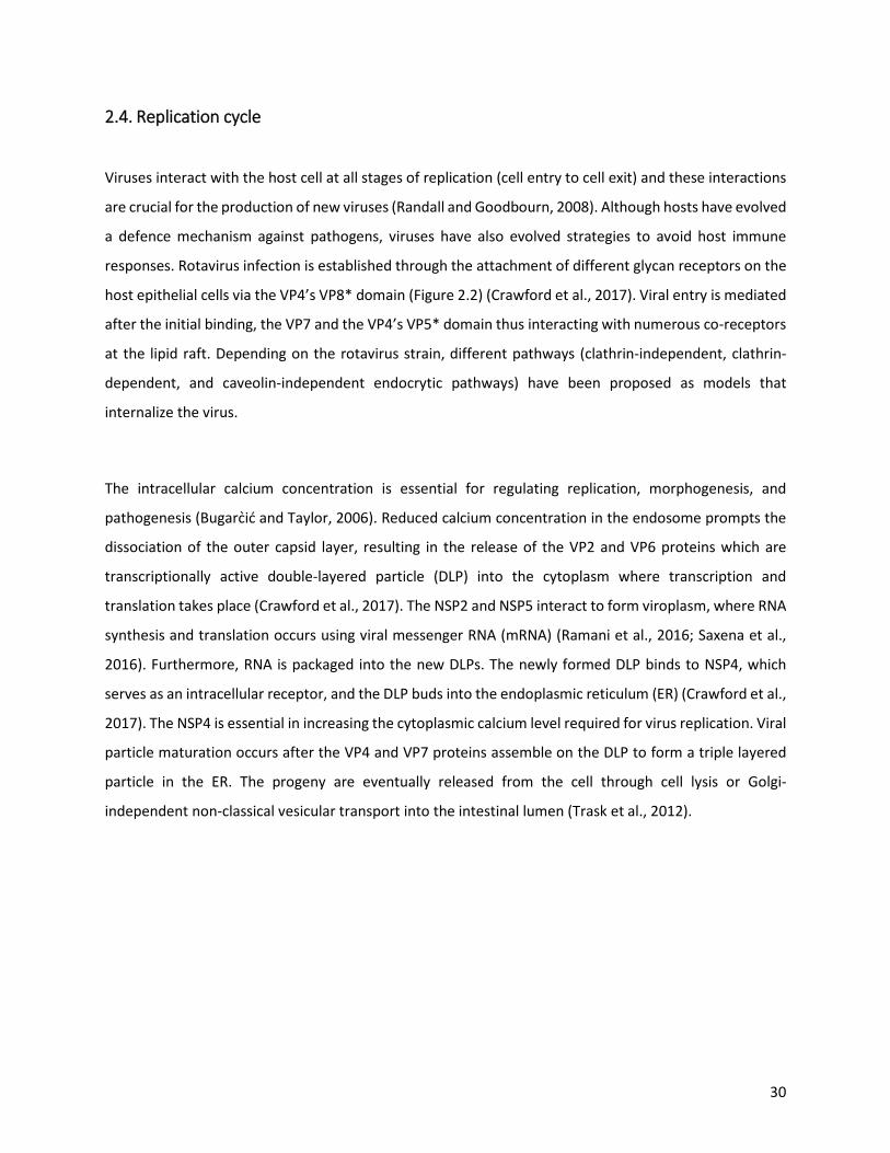
2.4. Replication cycle
Viruses interact with the host cell at all stages of replication (cell entry to cell exit) and these interactions
are crucial for the production of new viruses (Randall and Goodbourn, 2008). Although hosts have evolved
a defence mechanism against pathogens, viruses have also evolved strategies to avoid host immune
responses. Rotavirus infection is established through the attachment of different glycan receptors on the
host epithelial cells via the VP4’s VP8* domain (Figure 2.2) (Crawford et al., 2017). Viral entry is mediated
after the initial binding, the VP7 and the VP4’s VP5* domain thus interacting with numerous co-receptors
at the lipid raft. Depending on the rotavirus strain, different pathways (clathrin-independent, clathrin-
dependent, and caveolin-independent endocrytic pathways) have been proposed as models that
internalize the virus.
The intracellular calcium concentration is essential for regulating replication, morphogenesis, and
pathogenesis (Bugarcić and Taylor, 2006). Reduced calcium concentration in the endosome prompts the
dissociation of the outer capsid layer, resulting in the release of the VP2 and VP6 proteins which are
transcriptionally active double-layered particle (DLP) into the cytoplasm where transcription and
translation takes place (Crawford et al., 2017). The NSP2 and NSP5 interact to form viroplasm, where RNA
synthesis and translation occurs using viral messenger RNA (mRNA) (Ramani et al., 2016; Saxena et al.,
2016). Furthermore, RNA is packaged into the new DLPs. The newly formed DLP binds to NSP4, which
serves as an intracellular receptor, and the DLP buds into the endoplasmic reticulum (ER) (Crawford et al.,
2017). The NSP4 is essential in increasing the cytoplasmic calcium level required for virus replication. Viral
particle maturation occurs after the VP4 and VP7 proteins assemble on the DLP to form a triple layered
particle in the ER. The progeny are eventually released from the cell through cell lysis or Golgi-
independent non-classical vesicular transport into the intestinal lumen (Trask et al., 2012).
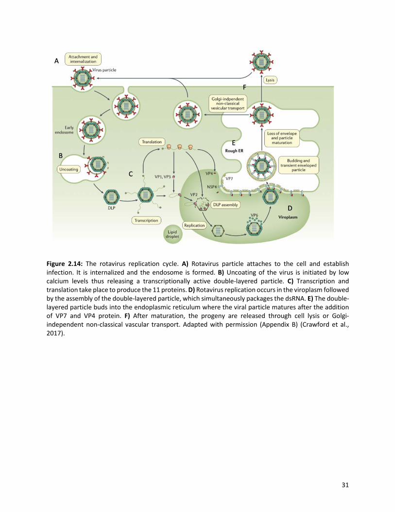
31
Figure 2.14: The rotavirus replication cycle. A) Rotavirus particle attaches to the cell and establish infection. It is internalized and the endosome is formed. B) Uncoating of the virus is initiated by low calcium levels thus releasing a transcriptionally active double-layered particle. C) Transcription and translation take place to produce the 11 proteins. D) Rotavirus replication occurs in the viroplasm followed by the assembly of the double-layered particle, which simultaneously packages the dsRNA. E) The double-layered particle buds into the endoplasmic reticulum where the viral particle matures after the addition of VP7 and VP4 protein. F) After maturation, the progeny are released through cell lysis or Golgi-independent non-classical vascular transport. Adapted with permission (Appendix B) (Crawford et al., 2017).

32
2.5. Immune response
Rotavirus-induced immune response in humans is not entirely understood; however, data from animal
models are available (Desselberger and Huppertz, 2011; Franco et al., 2006). Cellular and humoral immune
responses are reported to be involved in acquiring immunity against rotavirus infection (Offit, 1996).
Rotavirus antigens are recognized by pattern recognition receptors (Adenosine triphosphate-dependent
RNA helicase DDX58 and interferon-induced helicase C domain-containing protein 1 [IFIH1]) in
enterocytes or cells of the immune system (microphages, dendritic cells or adaptive B cells and T cells)
(Broquet et al., 2011; Offit, 1996). The rotavirus-specific B cells and CTL are stimulated and directed to the
VP6 protein inhibiting the viral transcription process (Aiyegbo et al., 2013). Pattern recognition receptors,
interferons and proinflammatory cytokines are responsible for initiating the type I and type III interferon
response mediating the clearance of rotaviruses by the innate immune response.
The recognition of rotavirus antigens by the pattern recognition receptors influences the assembly of
signalling complexes by ligand-activated sensors (López et al., 2016). This process ultimately promotes the
activation of the antiviral program. The signalling complexes triggers the activation of host transcription
factors (interferon 3, interferon 7, and Nuclear Factor kappa-light-chain-enhancer of activated B cells [NF-
kB]) and their translocation to the nucleus where type I and type III interferon, proinflammatory
molecules, and cytokines responses are initiated (Lin et al., 2016; López et al., 2016). Type I and type III
interferons are thus recognized by interferon receptors present on the surface of neighbouring cells,
ultimately activating the Janus kinase/signal transducer and activators of transcription (JAK/STAT)
signalling pathway (López et al., 2016). The STAT1 and STAT2 proteins are phosphorylated and interact
with the interferon regulatory factor 9 protein thus forming a heterotrimeric complex interferon-
stimulated gene factor 3 (ISGF3). The ISGF3 complex is translocated to the nucleus leading to downstream
transcription and expression of interferon-stimulated genes. The products from the interferon-stimulated
genes subsequently inhibit the viral infection and alters other cellular functions. Rotaviruses are also
reported to have several features that influence a poor innate immune response, such as the inhibition of
the signalling pathways by NSP1 or other rotavirus proteins when activated by recognition receptors
(Crawford et al., 2017; Holloway et al., 2014; López et al., 2016).
33
2.6. Rotavirus evolutionary mechanisms
Rotaviruses are constantly evolving through numerous evolutionary mechanisms (Taniguchi and Urasawa,
1995). The error prone RNA-dependent RNA polymerase (RdRp) is implicated in influencing genetic drifts
(generating spontaneous sequential point mutations) within a viral population, especially for dsRNA
viruses like rotaviruses. It has been reported that a progeny rotavirus genome will contain at least one
mutation in the genome that differentiates it from the parent strain (Blackhall et al., 1996). The same
RdRp replicates each gene. However, different rates of mutations are established due to different immune
or host-selection pressures. The outer capsid proteins are suspected of evolving more rapidly than those
of internal structural proteins because they are targets for host-neutralization antibody response
(Taniguchi and Urasawa, 1995). Genomic rearrangement is commonly observed in rotavirus strains
excreted by chronically infected immunocompromised children and animals (Pedley et al., 1984). It can
also be generated in cell culture through serial passages of rotavirus strains at a high multiplicity of
infections (Alam et al., 2008; Patton et al., 2001). The rearranged genes are usually involved in head-to-
tail duplication downstream of the open reading frame (ORF) and rarely occur within the ORF (Patton et
al., 2006; Taniguchi et al., 1996). This has been observed in RNA segments coding for NSP1, NSP3-NSP5,
and VP6 (Desselberger, 1996; Taniguchi et al., 1996).
Genetic recombination in viruses occurs when there is coinfection in the same host cell by two different
parent strains, resulting in a progeny containing genes from both parent strains. A limited number of
recombination events have been reported within the RVA (Maringa et al., 2020; Parra et al., 2004; Phan
et al., 2007). Genomic reassortment in rotaviruses occurs when a single cell is infected with several distinct
but compatible rotavirus strains, also known as genetic shift (Mwangi et al., 2020; Nyaga et al., 2013;
Ramig and Ward, 1991). Over the years, natural reassortment between human and animal strains has
been frequently reported, also known as zoonotic transmission. The zoonotic transmission has been
observed at a higher frequency in developing countries because of humans and domestic animals living in
close proximity (Cook et al., 2004; Glass et al., 2006; Maringa et al., 2020; Moon et al., 2016; Naylor et al.,
2015). Reassortment between rotavirus strains of different groups has not been detected (Ramig and
Ward, 1991; Taniguchi and Urasawa, 1995). However, the NSP1 gene of avian group A strain clusters
together with strains in the NSP1 of group D, thus suggesting a possible reassortment may have occurred
between the two groups (Taniguchi and Urasawa, 1995; Trojnar et al., 2010).

34
2.7. Prevention and treatment
Children under five years were prone to severe RVA disease presentation with exposure to multiple
rotavirus infections in their first two to three years of life before widespread vaccine introduction in 2006
(Velázquez et al., 1996). Primary rotavirus infections are generally more severe than subsequent
infections that are usually mild or asymptomatic (Bishop et al., 1983; Ward, 2008). Children presenting
signs of dehydration due to rotavirus induced diarrhoea are usually treated with zinc supplements, ORT,
and intravenous fluids (Crawford et al., 2017). However, the best prevention method is the use of
rotavirus vaccines. Several rotavirus vaccines have thus been developed over the years to ease the
rotavirus disease burden (Table 2.2).
Table 5.2 Basic features and administration recommendations of currently licensed rotavirus vaccines.
Product Manufacturer Composition Doses Formulation/storage Globally licensed
RotaTeq® Merck & Co. Inc., USA G1, G2, G3, G4, P[8] and
G6P[5]
3 doses (6, 10 and 32 weeks)
Liquid 2-8 °C or below -20 °C
for 24 months
Rotarix® GlaxoSmithKline (GSK) Biologicals, Belgium G1P[8] 2 doses (6 and 24
weeks)
Liquid 2-8 °C or below -20 °C
for 36 months
Rotavac® Bharat Biotech
International Limited India, India
G9P[11] 3 doses (6, 10 and 14 weeks)
Liquid frozen 2-8 for 7 months or °C -
20 °C long term
Rotasil® Serum Institute of India, India
G1, G2, G3, G4, G9 and
G6P[5]
3 doses (6, 10 and 14 weeks)
Lyophilized < 25 °C for 30 months or
< 40 °C for 18 months Nationally licensed
Rotavin-M1
POPLYVAC, Centre for Research and
Production of Vaccines and Biologicals, Vietnam
G1P[8] 2 doses (6 and 12 weeks)
Liquid frozen 2-8 °C for 2 moths or -
20 °C for 24 months
Lanzhou lamb
rotavirus vaccine
Lanzhou Institute of Biological Products Co.,
Ltd., China G10P[15] 1 dose (6 weeks
to 3 years) Liquid
(Adapted from Burke et al., 2019)

35
2.7.1. WHO prequalified vaccines
The first rotavirus vaccine introduced to the national immunization program of the United States in 1998
was RotaShield® (Wyeth Laboratories, U.S), a rhesus-human reassortant vaccine (CDC, 2011). However,
the vaccine was discontinued from the market because it was suspected to cause intussusception and
bloody stools in children (CDC, 1999; Glass et al., 2005). In 2006, the WHO pre-qualified the use of two
live attenuated oral vaccines, Rotarix® (RV1, GlaxoSmithKline Biologicals, Belgium) and RotaTeq® (RV5,
Merck & Co. Inc., USA). Rotarix® and RotaTeq® were introduced in the immunization program of many
countries including African countries like Botswana, Ghana, Malawi, Rwanda, South Africa and Zambia by
the end of 2013 (ROTA Council, 2016). In addition to these two vaccines, India has recently introduced
two indigenously developed vaccines, Rotavac® (nHRV, Bharat Biotech International Limited India, India)
and Rotasil® (BRV-PV, Serum Institute of India, India) (WHO, 2018). The estimated efficacies of both the
indigenous Indian vaccines are comparable to that of Rotarix® and RotaTeq® when administered in low-
income countries (Armah et al., 2010; Zaman et al., 2010). For several low-income nationals, the
implementation of the rotavirus vaccine is supported by the Global Alliance for Vaccines and
Immunizations (GAVI), the Vaccine alliance. In 2019, 46 countries received GAVI support for rotavirus
vaccine introduction out of 73 countries that are GAVI eligible (IVAC, 2019).
RotaTeq® is a pentavalent human-bovine reassortant vaccine administered as three oral doses at 6, 10,
and 32 weeks (Heaton and Ciarlet, 2007; Matthijnssens et al., 2010). This vaccine consists of four human
reassortant strains expressed on the VP7 protein (G1, G2, G3, or G4) and a bovine rotavirus strain on the
VP4 protein (P7[5]). The fifth reassortment was derived from the VP7 protein (G6) of a bovine rotavirus
strain and the VP4 protein (P1A[8]) from a human rotavirus strain. Rotarix® is a monovalent live
attenuated human G1P[8] rotavirus strain (RIX4414) given as two oral doses, administered at 6 and 24
weeks (GSK, 2019; Ward and Bernstein, 2009). Post vaccine introduction, rotavirus related hospitalization
declined among children less than five years and demonstrated herd immunity among adults above the
age bracket to receive the vaccine (Patel et al., 2009). Specifically, since the global introduction of these
two oral vaccines globally, rotavirus hospitalization of children less than five years has drastically declined
from about 527 000 mortality rate in 2000 to approximately 215 000 in 2013 and it further decreased to
128 500 deaths in 2016 (Armah et al., 2010; Begue and Perrin, 2010; Troeger et al., 2018b). Both Rotarix®

36
and RotaTeq® vaccines have been reported to confer homotypic and heterotypic protection against
multiple RVA strains (Glass et al., 2006; Ward and Bernstein, 2009).
Similarly, the WHO also prequalified the use of Rotavac® and Rotasil® in the national immunization
program of India in 2018. Rotavac® is a monovalent neonatal human attenuated G9P[11] rotavirus vaccine
administered at 6, 10, and 14 weeks (Bharat Biotech, 2019). It has been reported to have a higher efficacy
as compared to other rotavirus vaccines in high mortality settings and the infectivity/immunogenicity of
Rotavac® is enhanced when it interacts with breast milk (Bharat Biotech, 2019). In contrast, Rotasil® is a
bovine-human reassortant live attenuated lyophilized rotavirus vaccine consisting of human G1, G2, G3,
G4, G9, and 10 genes from the UK bovine rotavirus G6P[5], administered at 6, 10, and 14 weeks (Kapikian
et al., 2005; Zade et al., 2014). It is the first thermostable (-20 °C to 42 °C) rotavirus vaccine, unlike
RotaTeq®, Rotarix® and Rotasil® which require an uninterrupted cold chain (2-8 °C or below -20 °C) during
storage and transportation (GSK, 2019; Merck, 2008; Naik et al., 2017). Maintaining a cold chain in
developing countries is a considerable challenge; thus Rotasil® offers a promising avenue to simplify
vaccine distribution, storage, and transportation (Naik et al., 2017; WHO, 2018).
2.7.2. Nationally licenced vaccine
Other vaccines such as Rotavin-M1 (POPLYVAC, Centre for Research and Production of Vaccines and
Biologicals, Vietnam) and Lanzhou lamb rotavirus vaccine (LLR, Lanzhou Institute of Biological Products
Co., Ltd., China) have been licensed locally in Vietnam and China, respectively (Table 2.2) (Kirkwood and
Steele, 2018). Rotavin-M1 is a live attenuated G1P[8] oral vaccine-derived from a strain (KH0118-2003)
isolated from a child in Vietnam (Anh et al., 2012; Burke et al., 2019). It was licensed in 2012 and
administered in two doses scheduled at 6 and 12 weeks of age. Rotavin-M1 has been reported to show
safety and immunogenicity profile in children similar to that of Rotarix®. However, one interesting
distinction between the two vaccines is that 65% vaccine shedding was observed for Rotarix® after the
first dose, while Rotavin-M1 exhibited only 44-48% vaccine shedding (Anh et al., 2012). Lanzhou lamb
rotavirus vaccine is a live attenuated lamb G10P[15] group A strain administered as a single oral dose to
infants scheduled at 6 weeks to 3 years of age (Kirkwood and Steele, 2018). It was licensed in China in
2000 (Li et al., 2018). The Lanzhou lamb rotavirus vaccine is 35% effective against gastroenteritis and 53%
against moderate to severe diarrhoea (Zhen et al., 2015). Zhang and co-workers reported that the Lanzhou

37
lamb rotavirus vaccine provides cross-protection against gastroenteritis caused by both G9 and G3 strains
(Li et al., 2019).
2.7.3. Treatment of rotavirus infection
Children presenting with the rotavirus disease may develop symptoms for about five days with zero to
two vomiting episodes within 12 hours; a few lose watery stools per day with a low-grade fever (Crawford
et al., 2017). Medical intervention will be required if the symptoms last longer than a week with increased
vomiting episodes and frequent episodes of watery diarrhoea (Crawford et al., 2017). The key treatment
concept includes ORT or intravenous rehydration. Oral rehydration therapy is recommended in response
to mild and moderate dehydration in children with acute diarrhoea which includes rehydration and
maintenance of fluids with oral rehydration solutions (ORS) combined with the adequate dietary intake
(CDC, 2003; Hartling et al., 2006). Intravenous rehydration is mostly recommended when dehydration
becomes severe due to excessive vomiting, reduced consciousness, and intestinal ileus (CDC, 2003). The
use of ORT and intravenous rehydration is highly advantageous for economically disadvantaged
populations as they are less expensive but equally effective oral solutions. However, several low-income
countries are still phased with challenges accessing such treatments.
Other treatment methods include zinc supplements, probiotics, and antiviral therapy. Zinc supplements
administered for 10 – 14 days can also significantly decrease the prevalence and duration of diarrhoeal
episodes by improved absorption of water and electrolytes in the intestine, promoting faster regeneration
of the gut epithelium and enhancing immune response (Bettger and O’Dell, 1981; Gebhard et al., 1983;
Shankar and Prasad, 1998). Lactic acid-producing bacteria such as Lactobacillus rhamnosus, Lactobacillus
plantarum, Bifidobacteria, and Enterococcus faecium, as well as the yeast Saccharomyces boulardii are
also commonly used as probiotics. These probiotics have been reported to limit the duration of
gastroenteritis successfully. However, they are not included in the global standard of treatment for
children with rotavirus (Crawford et al., 2017). Studies have also reported that an antiviral drug such as
nitazoxanide can reduce the duration of diarrhoeal episodes by interfering with the viral morphology thus
inhibiting the replication of rotaviruses (La Frazia et al., 2013; Mahapatro et al., 2017; Rossignol, 2014).
38
2.8. Rotavirus vaccine introduction in African countries
Rotavirus vaccines have enormously improved child health morbidity and reduced diarrhoea-associated
mortality (Jonesteller et al., 2017). As of April 2020, 107 countries had introduced rotavirus vaccines (ROTA
Council, 2020). These includes 103 national introductions and 4 sub-national introductions. Although
more than 70% of sub-Saharan African countries have introduced rotavirus vaccines into their national
immunization programs, the rotavirus disease burden still remains high compared to high-income
countries (Tate et al., 2016, 2012). Five sub-Saharan African and South Asian countries including Angola,
the Democratic Republic of Congo, India, Nigeria, and Pakistan, account for half of the rotavirus deaths
worldwide. Of the 35 African countries that have embraced and introduced the rotavirus vaccines,
RotaTeq® was introduced only in five countries (Burkina Faso, Gambia, Libya, Morocco, and Rwanda)
(IVAC, 2019; Seheri et al., 2018). The first African country to introduce Rotarix® was South Africa in 2009,
while Rwanda was the first low-income country in the world to introduce RotaTeq® vaccine in May 2012
but switched to Rotarix® in April 2017 (Madhi et al., 2012; Ngabo et al., 2016b; Seheri et al., 2018).
Countries such as Ethiopia, Kenya, Tanzania, Uganda, Zambia, and Zimbabwe introduced Rotarix® vaccine
from 2012 to 2015 (Weldegebriel et al., 2018). In Rwanda, RotaTeq® vaccine coverage of 99% was reached
by 2013, administered at 6, 10, and 14 weeks of age (Gatera et al., 2016; Ngabo et al., 2016a; Sibomana
et al., 2018; WHO, 2020). In 2019, the RotaTeq® vaccine coverage of 98% was reported in Rwanda (WHO,
2020). Studies suggest that if all GAVI-eligible countries introduce rotavirus vaccines, an estimate of 180
000 deaths would be averted (Atherly et al., 2012).
2.9. Surveillance data of African countries
Although, rotavirus vaccines were intended to provide protection against multiple rotavirus strains
(Buttery et al., 2011; Tsolenyanu et al., 2016). There are constant gene reassortment, point mutation, and
gene rearrangement of the virus that are probably responsible for lower efficacy in regions that have
reported excellent strain diversity, thus necessitated continuous surveillance (Bányai et al., 2011). The
outer capsid proteins are primary targets for vaccine development due to their ability to induce
neutralizing antibodies and thus the antigenic changes in their structure may also impact vaccine
effectiveness (Maranhão et al., 2012; Zeller et al., 2012). Generally, while the impact of rotavirus vaccines
has been significant post introduction, studies have reported poor performance of live oral vaccines for
rotavirus in developing countries (Velasquez et al., 2017). The documented feasible justification for the
39
low efficacy of the vaccines in Africa and Asia may be interference by maternal antibodies, co-infection
with multiple enteric pathogens, genetic diversity, malnutrition, and limited access to treatment (Boschi-
Pinto et al., 2008; Dagan et al., 1990; Glass et al., 2006; Moon et al., 2016; Naylor et al., 2015). Rotavirus
associated mortality in sub-Saharan Africa accounts for > 80% of the disease burden with only 8 countries;
Burkina Faso, Chad, Cote d’Ivoire, Democratic Republic of Congo, Ethiopia, Niger, Nigeria, and Uganda
(Troeger et al., 2018b).
The analysis of Wa-like and DS-1-like genome constellations has revealed that both constellations are
successful in causing and rapidly spreading rotavirus infection in humans (Matthijnssens and Van Ranst,
2012). The most prevalent rotavirus genotypes worldwide in the last 20 years are G1P[8], G2P[4], G3P[8],
G4P[8], G9P[8] and G12P[8] which accounted for 74.7% of all strains circulating globally (Bányai et al.,
2012; Leshem et al., 2014; Mwenda et al., 2010; Nyaga et al., 2020; Santos and Hoshino, 2005; Seheri et
al., 2014; Todd et al., 2010). These six strain combinations cause over 90% of rotavirus disease in Australia,
Europe, and North America. In South America and Africa, they account for 83% and 55%, respectively,
with frequent detection of unusual genotypes (Santos and Hoshino, 2005).
Unusual strains have been detected at high frequencies in countries such as Malawi (G8P[6] and G8P[4]),
Brazil (G5P8]), Hungary (G6P[8]), and India (G10P[11]). In the years between 2010 and 2015, G1P[8]
(23.8%), G2P[4] (11.8%) and G9P[8] (10.4%) were reported as the predominant cause of acute
gastroenteritis in Eastern and Southern African countries among children less than five years (Seheri et
al., 2018). The yearly fluctuation of G1P[8] strain was observed in Rwanda (2011 and 2015), Zambia (2012
and 2014), Tanzania (2010-2011 and 2014- 2015) and Zimbabwe (2014-2015). An increase in the
prevalence of G2P[4] and G2P[6] genotypes in Ethiopia, Tanzania, Zambia, Zimbabwe, and Madagascar
was also reported (Seheri et al., 2018). Moreover, uncommon genotypes such as G9P[4], G9P[6], G3P[4]
and G3P[6] are generally detected in low frequencies globally (Collins et al., 2015; Cunliffe et al., 2010;
Giri et al., 2019; João et al., 2020; Lartey et al., 2018; Lennon et al., 2008; Page et al., 2018; Rasebotsa et
al., 2020; Seheri et al., 2018; Yamamoto et al., 2015).

40
In Rwanda, a strong seasonal pattern was observed with more diarrhoeal hospital admissions from May
to September and a peak in July (Ngabo et al., 2016b; Uwimana et al., 2015). The pattern coincided with
the rotavirus season (dry season), where there are less rainfall and insufficient water supply coupled with
poor hygiene and sanitation (Uwimana et al., 2015). Studies have also associated infections with climate
change response, with the highest number of infections observed during colder and drier times of the
year (Levy et al., 2009). After the introduction of RotaTeq® in Rwanda, the proportion of rotavirus induced
diarrhoeal hospitalization declined by 25 - 44% among children less than five years during the first three
years post-vaccine introduction (Sibomana et al., 2018). Immediately after rotavirus vaccine
implementation, an increase in rotavirus hospital admissions to children over five years of age was
observed (Ngabo et al., 2016b). However, a decrease in rotavirus positive diarrhoeal admissions was
observed in all age groups, suggesting that children not eligible for vaccination are also protected through
the decrease of RVA transmission in the population (Clarke et al., 2011; Lambert et al., 2009; Ngabo et al.,
2016b). Temporal strain variation was reported in Rwanda post-vaccine introduction with G8P[4]
circulating at high proportion in 2013, G4P[8] and G12P[8] in 2014, which were replaced by G1P[8] strains
in 2015 (Seheri et al., 2018). The global use of the vaccines has been suspected to be the potential cause
of evolutionary changes in the genome (Hoshino et al., 2004; Kirkwood, 2010). As numerous mechanisms
influence diversity of the rotaviruses, long-term rotavirus surveillance is essential in determining the
vaccine impact along with changes in the circulating strains in the African region (Kirkwood, 2010; Seheri
et al., 2018).
2.10. Next-generation Sequencing
In previous years, detecting viruses in a clinical or environmental sample was very challenging and time-
consuming. In 1977, Frederick Sanger and colleagues developed a ground breaking DNA sequencing
method, Sanger sequencing, that enabled the detection of various organisms, including viruses (Sanger et
al., 1977). Sanger sequencing has been used for many years and is considered as the gold standard in
elucidating genetic information. This triggered the development of new technologies such as NGS over
the years that substantially reduced DNA sequencing time and costs while remarkably producing massive
sequencing data (Grada and Weinbrecht, 2013). Moreover, NGS produces more accurate, less biased, and
reliable results enabling comprehensive analysis of genomes, epigenome, transcriptomes that make a
significant contribution to biological and biomedical research (Metzker, 2010; Shendure and Ji, 2008).
Next-generation sequencing platforms such as LifeTechnology Ion Torrent Personal Genome Machine
41
(PGM) and the Illumina Miseq are frequently applied in health, agricultural, and environmental fields
(Grada and Weinbrecht, 2013).
Next-generation sequencing technologies require library preparation, amplification, and sequencing
steps. The PGM uses emulsion PCR to amplify single library fragments onto microbes, while the Illumina
Miseq platform uses bridge amplification to form template clusters on a flow cell (Berglund et al., 2011;
Quail et al., 2012). Moreover, both platforms rely on sequencing by synthesis. The DNA libraries are used
as templates, from which new DNA fragments are synthesized. Sequencing is carried out by cycles of
washing and flooding the DNA library fragments with known nucleotides in a sequential order to
significantly reduce raw error rates, while simultaneously recording the DNA sequence digitally. The PGM
platform detects and records a new nucleotide when the pH changes each time a hydrogen ion is released
upon incorporation in a growing DNA strand (Quail et al., 2012). In contrast, the Illumina Miseq platform
detects and records the sequence each time a new fluorescently labelled reversible-terminator nucleotide
is incorporated in a growing DNA strand (Bentley et al., 2008). The sequencing data generally undergoes
several analysis steps such as removal of adaptor sequence and low quality read as well as mapping the
data to a reference genome or conducting de novo assembly and finally the analysis of the compiled
sequences through various bioinformatics platforms (Grada and Weinbrecht, 2013). Both NGS platforms
are reported to be viable options for sequencing in terms of utility and ease of workflow (Marine et al.,
2020; Quail et al., 2012).
The Illumina MiSeq platform was utilized in this project. The key application of an Illumina MiSeq platform
offers capabilities for small whole-genome sequencing of microbes and viruses, targeted gene sequencing
and 16S metagenomic (Illumina, 2020). Using a high throughput kit, the platform can produce up to 15Gb
of output with 25 million sequencing reads and 2 x 300 bp reads lengths in a single run with a run time of
4-55 hours. The use of NGS platforms has been efficient in characterizing and deciphering the whole-
genome sequence of circulating rotavirus strains globally (Bwogi et al., 2017; Esona et al., 2017; Ianiro et
al., 2013; Jere et al., 2018, 2011; Komoto et al., 2014; Maringa et al., 2020; Mukherjee et al., 2011; Mwangi
et al., 2020; Nyaga et al., 2018, 2015, 2014, 2013, 2020; Rasebotsa et al., 2020; Saikruang et al., 2016).
Whole-genome studies have a significant potential in providing comprehensive insight into the impact of
vaccines on both wild type and atypical rotavirus strains (Nyaga et al., 2020). Such studies can also aid in
the rational design of the next-generation subunit vaccines. Moreover, the frequency of reassortment

42
events under natural conditions and their epidemiological fitness against wild type strains can be
determined.
2.11. Rotavirus whole-genome studies
The advancements in sequencing methods, such as NGS, have made it easier to determine the overall
genetic makeup and evolutionary pattern of RVA strains (Nyaga et al., 2020). Over the years, evolutionary
mechanisms such as gene reassortment, rearrangement, and zoonotic transmissions have led to the
emergence of novel RVA strains (Kirkwood, 2010). Moreover, some instances of zoonotic transmissions
of RVA strains can only be confirmed through whole-genome analysis (Martella et al., 2010; Santos and
Hoshino, 2005). The pure Wa-like and DS-1-like rotavirus genome constellations generally have an
evolutionary fitness advantage (Heiman et al., 2008; McDonald et al., 2009). However, the emergence of
novel strains has been documented worldwide (Agbemabiese et al., 2017; Arana et al., 2016; Donato et
al., 2012; Jere et al., 2018; Mwangi et al., 2020; Strydom et al., 2019a). The 11 rotavirus genome segments
exhibits different levels of diversity, with NSP1 and NSP4 genome segment being more diverse than the
other segments (Dunn et al., 1994; Nyaga et al., 2015). The VP1 and VP2 genome segments were reported
to be more conserved (Matthijnssens et al., 2008).
The DS-1-like constellation is generally linked to G2P[4] strains (Matthijnssens et al., 2008). Although, non
G2P[4] DS-1-like strains have been identified in Africa (G1P[8], G3P[6] and G8P[4]), Asia (G1P[4] and
G12P[6]) and Europe (G3P[8]) (Agbemabiese et al., 2017; Arana et al., 2016; Bwogi et al., 2017; Heylen et
al., 2013; Mwangi et al., 2020; Nyaga et al., 2015; Saikruang et al., 2016; Strydom et al., 2019a). The G1P[8]
strains commonly exhibits a typical Wa-like genotype constellation (Magagula et al., 2015). However,
several whole-genome studies have reported intergenogroup reassortment of G1P[8] strains in Brazil,
Japan, Philippines, Thailand, Vietnam (Fujii et al., 2014; Komoto et al., 2015; Luchs et al., 2019; Nakagomi
et al., 2017). In Africa, the first atypical G1P[8] strains with a DS-1-like backbone were detected in Blantyre,
Malawi, predominantly during the post-vaccine introduction period (Jere et al., 2018). Recently, South
Africa also reported the first DS-1-like G1P[8] strain. This particular reassortment occurred pre-vaccine
introduction, suggesting that the event might have been influenced by natural evolutionary events
(Mwangi et al., 2020).
43
In a study conducted in Kenya, a G2P[4] strain with a Wa-like NSP2 genotype on the DS-1-like backbone
was observed (Ghosh et al., 2011). The phylogenetic analysis of the strain also revealed that while the
other segments were closely related to the prototype and other human G2P[4] strains, the NSP2 genome
appeared to be closely related to rotavirus genes of artiodactyl origin highlighting the complex
evolutionary dynamics of African G2P[4] rotavirus strains. Moreover, a G9P[6] strain from South Africa
with a predominantly DS-1-like backbone also exhibited a Wa-like NSP2 genotype which was suggested
to have emerged through intergenogroup reassortment (Nyaga et al., 2013). A similar trend was observed
on G12 rotavirus strains isolated in the Philippines and Bangladesh (Rahman et al., 2007). Most G4P[6]
strains have the Wa-like genome constellation and have been frequently detected in Korea, they are
considered to be rare in other countries (Lee et al., 2019). However, a complete genome sequence of a
G4P[6] strain isolated from Zambia was submitted on GenBank as RVA/Human-wt/ZMB/MRC-
DPRU1752/XXXX/G4P[6] exhibiting a DS-1-like genome constellation.
Despite the benefit of rotavirus vaccines, cases of vaccine shedding, horizontal transmission of vaccine
strains and vaccine-derived strains have been documented globally (Boom et al., 2012; Gower et al., 2020;
Hemming and Vesikari, 2014; Hsieh et al., 2014; Than et al., 2015). Whole-genome analysis has made it
easier to differentiate wild-type rotavirus strains from vaccine-derived strains (Magagula et al., 2015;
Sakon et al., 2017). A study conducted in Nicaragua reported two G1P[8] strains that exhibited an NSP2
gene with a DS-1-like genotype identical to that of the RotaTeq® vaccine. In Brazil, where Rotarix® was
introduced, a G1P[8] strain with an AU-1-like NSP3 was reported. The strain was evident of a vaccine-
derived reassortment because it also clustered along with the Rotarix® vaccine during phylogenetic
analysis (Rose et al., 2013a). The detection of vaccine-derived strains may have led to reduced rotavirus
vaccine effectiveness over time. Such findings emphasize the importance of conducting rotavirus whole-
genome studies in order to obtain conclusive data on the complex evolutionary dynamics of RVA strains,
especially in Africa (Nyaga et al., 2020; Seheri et al., 2018). Although studies have been conducted on the
outer capsid proteins of rotavirus strains circulating in Rwanda (Seheri et al., 2018; Uwimana et al., 2015),
there still a dearth of information regarding the entire backbone of those strains.

44
CHAPTER THREE: METHODOLOGY

45
3.1. Ethical statement and study design
The ethical clearance for this study was granted by the UFS, Health Sciences Research Ethics Committee
(HSREC) under the reference UFS-HSD2019/1601/2810. The research was conducted at the UFS-NGS Unit,
Division of Virology, Faculty of Health Sciences, UFS. Archived genotyped rotavirus stool specimen (n=158)
from children younger than five years from Rwanda were retrieved from the Diarrheal Pathogens
Research Unit (DPRU), which is a WHO rotavirus Regional Reference Laboratory in South Africa (RRL-SA).
The targeted stool samples were from the pre- (2011) and post- (2012-2016) vaccine introduction era
sampled from children in all five provinces (Figure 3.1). The viral dsRNA was isolated from the stool
samples and purified in preparation for cDNA synthesis. The cDNA was synthesized and fragmented to
prepare DNA libraries for sequencing on an Illumina MiSeq platform, followed by data analysis using
various bioinformatics software. This study forms part of a bigger project aimed at addressing the Terms
of References (ToRs) for Technical Service Agreement (TSA) between the UFS-NGS Unit and the WHO, to
perform whole-genome sequencing of rotavirus strains from the World Health Organization Regional
Office for Africa (WHO/AFRO) rotavirus surveillance network, under the reference HSREC130/2016(UFS-
HSD2016/1082).
Figure 3.1: The map of Rwanda indicating all the five provinces. Each province is indicated with a different colour; orange (Western province), yellow (Northern province), blue (Southern province), green (Kigali province) and red (Eastern province). Adapted (Kalisa et. al., 2016)

46
3.2. Extraction of rotavirus dsRNA from faecal samples
Rotavirus dsRNA was extracted following a protocol described previously by Nyaga et al. (2018). Briefly,
in a Biosafety cabinet (Esco Technologies Pty Ltd, Gauteng, South Africa), a 10% dilution was formulated
using approximately 100 mg of the stool sample added to a sterile 2 ml microcentrifuge tube (Thermo
Scientific, QSP, New Hampshire, United States) containing 200 µl of sterile phosphate buffer solution, pH
7.2 (PBS, Sigma-Aldrich, St. Louis, USA). To extract the dsRNA in the aqueous phase, 1 ml of TRI-reagent
(Molecular Research Center, Inc, Cincinnati, OH, USA) was added to the tube and thoroughly mixed by
vortexing (Labnet International,Inc., New Jersey, United States), followed by an incubation period of five
minutes at room temperature. Subsequently, 270 μl of chloroform (Sigma-Aldrich, St. Louis, USA) was
added to the tube, vortexed (Labnet International,Inc., New Jersey, United States) again, and incubated
for three minutes at room temperature. The blend was later centrifuged (Eppendorf, Hamburg, Germany)
at 16 000 x g for 20 minutes at 4°C. Thereafter, the supernatant was added to a new 2 ml microcentrifuge
tube (Thermo Scientific, QSP, New Hampshire, United States) containing 1 ml ice-cold isopropanol (Sigma-
Aldrich, St. Louis, USA) to dissolve any salts that might be chelated to the dsRNA sample while precipitating
the dsRNA for five minutes at room temperature. The blend was centrifuged (Eppendorf, Hamburg,
Germany) at 16 000 x g for 30 minutes at 20°C and the supernatant was discarded, leaving behind a pellet
at the bottom of the tube, which was air-dried for ten minutes. The dsRNA was recovered from the bottom
of the tube by adding 95 μl of the Qiagen Elution Buffer, EB (Qiagen, Hilden, Germany). The tube was
sporadically vortexed (Labnet International,Inc., New Jersey, United States) and each sample (2 μl) was
visualized on a 1% agarose gel (ThermoFisher Scientific, Waltham MA, USA) stained with Pronasafe
(Condalab, Madrid, Spain).
Lithium chloride, 8 M LiCl2, (Sigma-Aldrich, St. Louis, USA) was used to enrich the extracted total RNA by
removing ssRNA because rotavirus is a dsRNA virus. LiCl2 (Sigma-Aldrich, St. Louis, USA) was prepared by
dissolving 1.69 g LiCI2 (Sigma-Aldrich, St. Louis, USA) into 5 ml nuclease-free water. A volume of 30 μl of
the 8 M LiCl2 (Sigma-Aldrich, St. Louis, USA), while still hot, was transferred to a 2 ml microcentrifuge tube
(Thermo Scientific, QSP, New Hampshire, United States) containing the extracted nucleic material and
incubated for 20 minutes at room temperature with a further incubation period of 16 hours in a 4°C water
bath. The mixture was centrifuged (Eppendorf, Hamburg, Germany) at 16 000 x g for 30 minutes at 4°C to
precipitate the LiCl2 (Sigma-Aldrich, St. Louis, USA) and the supernatant was transferred into a new 2 ml
microcentrifuge tube (Thermo Scientific, QSP, New Hampshire, United States).
47
The Qiagen MinElute gel extraction kit (Qiagen, Hilden, Germany) was used to purify the dsRNA. Briefly, a
Prism Mini centrifuge (Labnet International, Inc., New Jersey, United States) was used to spin down the
sample and 330 μl QG buffer (Qiagen, Hilden, Germany) was added to the 2 ml microcentrifuge tube
(Thermo Scientific, QSP, New Hampshire, United States) containing the dsRNA sample. The sample was
gently mixed by vortexing (Labnet International,Inc., New Jersey, United States), centrifuged (Prism Mini
centrifuge, Labnet International, Inc., New Jersey, United States) and subsequently transferred into a new
1.5 ml tube containing a MinElute spin column (Qiagen, Hilden, Germany) to allow adsorption of the
dsRNA on the silica-gel membrane of the column while washing away impurities introduced during the
RNA extraction. The tube was centrifuged (Eppendorf, Hamburg, Germany) at 16 000 x g for one minute
at 4°C. The column was transferred into a new 1.5 ml microcentrifuge tube (Qiagen, Hilden, Germany)
and 35 μl of the EB buffer (Qiagen, Hilden, Germany) was added at the centre of the membrane to elute
the bound dsRNA. The tube was incubated for one minute then centrifuged (Prism Microcentrifuge,
Labnet International,Inc., New Jersey, United States) at 16 000 x g for another minute. The column was
discarded leaving behind the purified dsRNA visualized on a 1% agarose gel (ThermoFisher Scientific,
Waltham MA, USA) and quantified using BioDrop-µLITE spectrophotometer (Biodrop, Cambridge, United
Kingdom), which measures the concentration of low volume nucleic acid samples.
3.3. Complementary DNA synthesis
A maxima H Minus ds cDNA synthesis kit (ThermoFisher Scientific, Waltham MA, USA) was used to
synthesize cDNA from total RNA. The conversion of RNA to cDNA was brought by the introduction of
reverse transcriptase to the rotavirus sample, which catalyses the formation of DNA from utilizing RNA as
a template. Minor modifications to the maxima H Minus ds cDNA synthesis protocol were made as per
UFS-NGS Unit cDNA synthesis standard operating procedure, to optimize it specifically for rotavirus by
adding 13 μl of the dsRNA into a PCR (Polymerase Chain Reaction) tube (Eppendorf, Hamburg, Germany)
and incubating it in a pre-heated (105°C) thermocycler (Merck, Darmstadt, Germany) at 95°C for five
minutes. The incubation step allowed the dsRNA to unwind and separate into an elongated ssRNA. The
tube was centrifuged (Prism Mini centrifuge, Labnet International, Inc., New Jersey, United States)
followed by the addition of 1 μl of the random hexamer primer (ThermoFisher Scientific, Waltham MA,
USA). The mixture was briefly centrifuged on a Prism Mini Centrifuge (Labnet International,Inc., New
Jersey, United States). Subsequently, the tube was incubated at 65°C for five minutes in a thermocycler
(Merck, Darmstadt, Germany) to allow the random primer to anneal randomly to the RNA template
48
strand. The tube was briefly pulse centrifuged (Prism Mini Centrifuge, Labnet International,Inc., New
Jersey, United States) and placed on ice. A volume of 5 μl of the 4X first strand reaction mix (ThermoFisher
Scientific, Waltham MA, USA) along with 1 μl of the first strand enzyme mix (ThermoFisher Scientific,
Waltham MA, USA) were added to the mixture in the PCR tube (Eppendorf, Hamburg, Germany) followed
by ten minutes of incubation at 25°C and elongation of the strand for 2 hours at 50°C in a Thermocycler
(Merck, Darmstadt, Germany). Final denaturation was achieved by heating the blend for five minutes at
85°C. The final product of this reaction was the first strand.
The first strand was used as a template to generate the second strand by introducing an E.coli RNase H
which inserts nicks into the RNA, allowing the DNA polymerase I to replace the RNA strand with
deoxyribonucleotides. Finally, the E.coli DNA ligase closed the gaps to complete the ds cDNA strand. This
was all achieved by adding 55 μl of the second strand nuclease-free water (ThermoFisher Scientific,
Waltham MA, USA), 20 μl 5X second strand reaction mix (ThermoFisher Scientific, Waltham MA, USA) and
5 μl second strand enzyme mix (ThermoFisher Scientific, Waltham MA, USA) to a PCR tube (Eppendorf,
Hamburg, Germany) containing 12 μl of the reaction mixture from the first strand synthesis. The tube was
incubated for 60 minutes at 16°C in a Thermocycler (Merck, Darmstadt, Germany). Thereafter, the
reaction was halted by adding 6 μl of 0.5 M EDTA, pH 8.0 (ThermoFisher Scientific, Waltham MA, USA).
Residual RNA was removed by adding 10 μl RNase I (ThermoFisher Scientific, Waltham MA, USA) to the
second strand reaction tube leaving behind the cDNA.
3.4. cDNA purification
An MSB PCRAPACE purification kit (Invitek Molecular, Berlin, Germany) was used to purify the synthesized
rotavirus cDNA samples. Briefly, 250 μl of the Binding Buffer (Invitek Molecular, Berlin, Germany) was
transferred to a 2 ml microcentrifuge tube (Thermo Scientific, QSP, New Hampshire, United States)
containing 50 μl of the cDNA sample and vortexed (Labnet International,Inc., New Jersey, United States).
The blend was transferred into a 2 ml receiver tube (Invitek Molecular, Berlin, Germany) containing a spin
filter (Invitek Molecular, Berlin, Germany), incubated for one minute at room temperature, and
centrifuged (Prism Microcentrifuge, Labnet International,Inc., New Jersey, United States) for four minutes
at 21 000 x g. This step allows the separation of the PCR product from impurities such as
deoxyribonucleotide triphosphates (dNTPs), primers, and enzymes introduced during the synthesis of the
cDNA. Furthermore, the spin filter (Invitek Molecular, Berlin, Germany) was inserted into a sterile 1.5 ml
49
receiver tube (Invitek Molecular, Berlin, Germany) and 10 μl of the EB (Invitek Molecular, Berlin, Germany)
was added directly at the centre of the spin filter (Invitek Molecular, Berlin, Germany) to elute the cDNA.
The tube was incubated for one minute at room temperature and centrifuged (Prism Microcentrifuge,
Labnet International,Inc., New Jersey, United States) for one minute at 11 000 x g in readiness for
validation using a fluorometric assay.
3.5. Quality control using Qubit 4 Fluorometer
Qubit 4 Fluorometer (Life Technologies, California, United States), a benchtop fluorometer, was used
to quantify the purified rotavirus cDNA in preparation for DNA library preparation. A Qubit working
solution was prepared by making a 1:200 dilution of the Qubit reagent (Life Technologies, California,
United States) and Qubit buffer (Life Technologies, California, United States) (Figure 3.2). The assay
standards (Life Technologies, California, United States) were prepared by adding 10 μl of standard 1 and
2 in separate tubes containing 190 μl of the working solution. The standards (Life Technologies, California,
United States) were used to calibrate the instrument prior to quantifying the samples. Furthermore, 1 μl
of the rotavirus sample was added to a Qubit tube (Life Technologies, California, United States)
containing 199 μl of the working solution. All the Qubit tubes (Life Technologies, California, United
States) were vortexed (Labnet International,Inc., New Jersey, United States) and incubated at room
temperature for two minutes. The Qubit tubes (Life Technologies, California, United States) were
inserted in the Qubit 4 Fluorometer (Life Technologies, California, United States) and the readings were
recorded. Samples with high concentration (concentrations higher than 0.2 ng/μl) were normalized by
adding EB (Qiagen, Hilden, Germany) while samples that had low concentration (concentration lower than
0.2 ng/μl) were excluded from further processing.

50
Figure 3.2: Qubit 4 Fluorometer workflow. The Qubit working solution was prepared by adding 199 x n μl of the Qubit buffer and 1 x n μl of the Qubit reagent into a single tube. A volume of 190 μl of the solution was transferred into a tube containing 1 μl of the sample, while 190 μl of the working solution was transferred to two tubes containing standard 1 and standard 2, respectively. The standards were used to calibrate the Qubit 4 Fluorometer in preparation for quantifying the samples (Created with BioRender.com, 26 August 2020)

51
3.6. DNA Library preparation
The Nextera® XT DNA library preparation kit (Illumina, Inc., California, United States) was used for the
library preparation for sequencing on an Illumina MiSeq (Illumina, Inc., California, United States) platform.
The rotavirus cDNA samples with concentrations ranging from 0.2–0.3 ng/μl were used for library
preparation as the starting cDNA concentration for the enzymatic fragmentation and adapter ligation to
the templates. In brief, the genomic DNA was enzymatically fragmented into smaller templates (~ 300 bp)
using Nextera transposomes. The ends of the templates were ligated by amplification using a
thermocycler (Merck, Darmstadt, Germany), which simultaneously added the sequencing oligos, the
Illumina instrument-specific P5 and P7 adaptors and unique indexes for de-multiplexing into individual
entities. Prior to that, the amplified DNA was size-selected and purified by removing PCR inhibitors to
approximately 300 bp templates using AMPure XP beads (Beckman Coulter, Indiana, United States). This
allowed removal of low and high molecular weight while library fragments for an optimal library free from
over or under-clustering during sequencing. The DNA libraries were normalized to a uniform
concentration of 4 nM after fragment length validation on the Agilent Bioanalyzer (Agilent Technologies,
California, United States) and ultimately normalized to 20 pM and into a final concentration of 8 pM. The
libraries were pooled together and denatured in readiness for sequencing using an Illumina MiSeq
(Illumina, Inc., California, United States) platform.
3.6.1. Detailed genomic DNA tagmentation and library amplification
The tagmentation step introduced transposome enzymes into the template DNA, which fragmented the
template into small fragments. Illumina chemistry specific adapter sequences (P5 and P7) were then
added to the fragmented genomic DNA (Figure 3.3). In a 96-well PCR plate (Soreson Bioscience Inc., Utah,
United States), 10 μl of the Tagment DNA Buffer, TD, (Illumina, Inc., California, United States) was added
followed by 5 μl of the normalized DNA (0.2 – 0.3 ng/μl). A volume of 5 μl of the Amplicon Tagment Mix,
ATM, (Illumina, Inc., California, United States) was added to the blend and centrifuged (Hermle, Labnet
International, Inc., New Jersey, United States) at 280 x g for one minute at 20°C. Furthermore, the 96-well
PCR plate (Soreson Bioscience Inc., Utah, United States) was placed on a thermal cycler (Multigene
Optimax, Labnet International, Inc., New Jersey, United States) with a pre-heated lid (105°C) at 55°C for
five minutes. Subsequently, 5 μl of the Neutralize Tagment Buffer, NT, (Illumina, Inc., California, United
States) was added to the plate and centrifuged (Hermle, Labnet International, Inc., New Jersey, United
States) at 20°C for one minute at 280 x g and incubated at room temperature for five minutes.

52
Figure 3.3: DNA fragment ligated with indexes and Illumina chemistry specific adapter sequences (Created with BioRender.com, 26 August 2020)
An Illumina sample manager software (Experiment Manager Software version 1.15, Illumina, Inc.,
California, United States) was utilized to assign each sample with a specific index to uniquely label and
differentiate between each specific sample after de-multiplexing in Illumina control software. The libraries
were uniquely labelled by adding 5 μl of the prescribed index (1 and 2) (Illumina, Inc., California, United
States) (Illumina, Inc., California, United States) to each specific sample. A Nextera PCR Master Mix, NPM,
(Illumina, Inc., California, United States) of 15 μl was added to the blend and centrifuged (Hermle, Labnet
International, Inc., New Jersey, United States) at 280 x g for one minute at 20 °C. The 96-well PCR plate
(Soreson Bioscience Inc., Utah, United State) containing the blend was placed on a pre-heated (105°C)
thermal cycler (Multigene Optimax, Labnet International, Inc., New Jersey, United States). The
programme run on the thermal cycler (Multigene Optimax, Labnet International, Inc., New Jersey, United
States) with the following conditions: 72°C for three minutes, 95°C for 30 seconds and 12 cycles of 95°C
for 10 seconds, 55°C for 30 seconds and 72°C for 30 seconds followed by 72°C for five minutes and the
reaction was put on hold at 4°C.
3.6.2. Library clean-up and quantification
Firstly, the libraries were centrifuged (Hermle, Labnet International, Inc., New Jersey, United States) at
280 x g for one minute at 20°C to prevent sample loss by concentrate the liquid at the bottom of the well.
The PCR product (50 μl) was transferred to a new 96-well PCR plate (Soreson Bioscience Inc., Utah, United
States) followed by the addition of 30 μl AMPure XP beads (Beckman Coulter, Indiana, United States). The
beads selectively bind to the 300 bp DNA fragments. The blend was thoroughly mixed using a Micro-
Multiple Genie (Scientific industries, Inc., New York, United States) at 280 x g for two minutes and placed
on a magnetic stand (PerkinElmer, Massachusetts, United States) for two minutes allowing the beads to
concentrate at the bottom of the plate. The supernatant was discarded and the beads were washed twice
with freshly prepared 80% ethanol (Merck, Darmstadt, Germany) (200 μl), followed by an incubation

53
period of 30 seconds on the magnetic stand (PerkinElmer, Massachusetts, United States) to remove
impurities (excess ATM, NPM, and indexes) introduced during the tagmentation and library amplification
step. The supernatant was carefully removed to prevent the carry-over of ethanol that might disrupt
downstream processes. The beads were air-dried for five minutes at room temperature and 52.5 μl of the
Resuspension Buffer, RSB, (Illumina, Inc., California, United States) was added before the beads started
showing cracks. The plate was centrifuged (Hermle, Labnet International, Inc., New Jersey, United States)
at 280 x g for two minutes and incubated at room temperature for an additional two minutes.
Furthermore, the plate was placed on a magnetic stand (PerkinElmer, Massachusetts, United States)
allowing the beads to form a pellet at the bottom. A volume of 50 μl of the supernatant was transferred
to a new 96-well PCR plate (Soreson Bioscience Inc., Utah, United States).
Library quantification was conducted using Agilent Technology 2100 Bioanalyzer (Agilent Technologies,
California, United States) and Qubit 4 Fluorometer (Life Technologies, California, United States) (Figure
3.2). Briefly, 1 μl of the library was added to the high sensitivity chip and ran on an Agilent Technologies
2100 Bioanalyzer (Agilent Technologies, California, United States) which allowed for easy viewing of the
fragment size of the libraries. In preparation for library pooling and to ensure equal library representation
in the pooled samples, the Bioanalyzer (Agilent Technologies, California, United States) and the Qubit 4
Fluorometer (Life Technologies, California, United States) results were normalized to 4 nM following unit
conversion from ng/μl into nM using the expression below:
𝑐𝑐𝑐𝑐𝑐𝑐𝑐𝑐𝑐𝑐𝑐𝑐𝑐𝑐𝑐𝑐𝑐𝑐𝑐𝑐𝑐𝑐𝑐𝑐𝑐𝑐 𝑐𝑐𝑐𝑐 𝑐𝑐𝑛𝑛/𝜇𝜇𝜇𝜇
(660 𝑛𝑛𝑔𝑔𝑐𝑐𝜇𝜇/𝑥𝑥 𝐴𝐴𝐴𝐴𝑐𝑐𝑐𝑐𝑐𝑐𝑛𝑛𝑐𝑐 𝜇𝜇𝑐𝑐𝑙𝑙𝑐𝑐𝑐𝑐𝑐𝑐𝑙𝑙 𝑠𝑠𝑐𝑐𝑠𝑠𝑐𝑐)× 106 = 𝑐𝑐𝑐𝑐𝑐𝑐𝑐𝑐𝑐𝑐𝑐𝑐𝑐𝑐𝑐𝑐𝑐𝑐𝑐𝑐𝑐𝑐𝑐𝑐𝑐𝑐 𝑐𝑐𝑐𝑐 𝑐𝑐𝑛𝑛
3.6.3. Quality control using an Agilent Technology 2100 Bioanalyzer
An Agilent Technology 2100 Bioanalyzer (Agilent Technologies, California, United States), a chip-based
capillary electrophoresis machine, was used to assess the size and integrity of the amplified library as a
quality control measure. This quality control step was done before pooling the libraries to ensure the
quality of the prepared libraries were good to avoid generating a poor quality run. Briefly, the Gel-Dye
mix was prepared by adding 15 μl of the high sensitivity DNA dye concentrate (Agilent Technologies,
California, United States) into the high sensitivity DNA gel matrix vial (Agilent Technologies, California,
United States). The mixture was vortexed (Labnet International,Inc., New Jersey, United States) for ten
seconds and pulse centrifuged (Prism Mini Centrifuge, Labnet International,Inc., New Jersey, United

54
States). The Gel-Dye mix was transferred to a spin filter and centrifuged (Prism Centrifuge, Labnet
International,Inc., New Jersey, United States) at 2240 x g for ten minutes and the spin filter was discarded,
the 2 ml microcentrifuge tube (Thermo Scientific, QSP, New Hampshire, United States) was protected
from light by covering it with foil.
A new high sensitivity DNA chip (Agilent Technologies, California, United States) was placed on the chip
priming station where the Gel-Dye mix (9 μl) was dispensed at the bottom of the well, labelled capital ‘G’
(located on the third row, the fourth column from the top right side of the chip). A sterile syringe was also
placed on the priming station with the plunger positioned at 1 ml. The pressure was applied on the plunger
until it was held in place by the clip, followed by a waiting period of 60 seconds. The plunger was released
from the clip until it reached a 0.3 ml mark and it was pulled back to the 1 ml mark after five seconds. The
Gel-Dye mix (9 μl) was dispensed into the rest of the wells marked G (located on the fourth column on the
right) on the high sensitivity DNA chip (Agilent Technologies, California, United States). A volume of 5 μl
of the high sensitivity DNA marker (Agilent Technologies, California, United States) was added into a well,
marked with a ladder and also the 11 sample wells.
A volume of 1 μl of the high sensitivity DNA ladder (Agilent Technologies, California, United States) was
dispensed in the well, marked with the ladder symbol. Subsequently, each DNA library (1 μl) was added
to the 11 sample wells. The high sensitivity DNA chip (Agilent Technologies, California, United States) was
placed horizontally on an IKA MS3 vortex mixer (IKA®-Werke GmbH & Co. KG, Staufen, Germany) and
vortexed for 60 seconds at 16 x g. The chip was placed on an Agilent Technology 2100 Bioanalyzer (Agilent
Technologies, California, United States) and the assay was run for an hour . The results were noted and
used to covert ng/ μl to nM. The Agilent Technology 2100 Bioanalyzer results were illustrated in the form
of a gel image and an electropherogram to show the library size distribution (Figures 3.4 and 3.5).
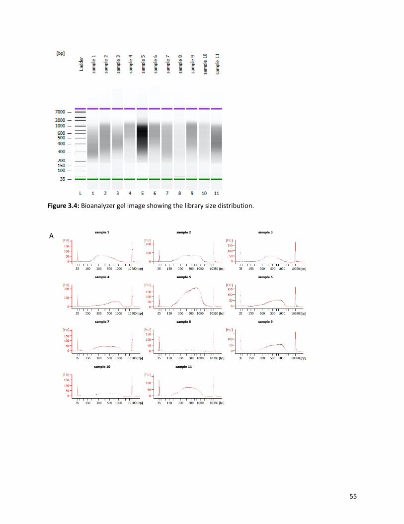
55
Figure 3.4: Bioanalyzer gel image showing the library size distribution.
A

56
Figure 3.5: A) Bioanalyzer electropherogram showing library size distribution determined by high sensitivity dsDNA assay. B) Electropherogram of the ladder depicting the lower (green) and the upper (purple) markers used as a control. 3.6.4. Library pooling and denaturation
In preparation for sequencing, libraries with equal concentrations were pooled together by adding 5 μl of
each library in a 1.5 ml microcentrifuge tube (Thermo Scientific, QSP, New Hampshire, United States),
denatured with 0.2 N Sodium hydroxide, NaOH, (Sigma Aldrich, Missouri, United States) and then diluted
to a final concentration of 20 pM at a volume of 600 μl. Briefly, 0.2 N NaOH (Sigma Aldrich, Missouri,
United States) was prepared by adding 11 μl of NaOH (Sigma Aldrich, Missouri, United States) into a tube
containing 250 μl molecular grade water, ultimately adding 739 μl of molecular grade water to a final
volume of 1 ml. A volume of 5 μl of the freshly prepared 0.2 N NaOH (Sigma Aldrich, Missouri, United
States) was added to a 1.5 ml microcentrifuge tube (Thermo Scientific, QSP, New Hampshire, United
States) containing the 5 μl pooled libraries to a final volume of 10 μl. The blend was vortexed (Labnet
International,Inc., New Jersey, United States) briefly and centrifuged (Prism Microcentrifuge, Labnet
International,Inc., New Jersey, United States) at 280 x g for one minute. Furthermore, the blend was
diluted to 20 pM by adding 990 μl of the prechilled Hybridization Buffer 1, HT1, (Illumina Inc., California,
United States). The 20 pM denatured library was further constituted to 8 pM by transferring 240 μl of the
denatured library to a new tube along with 360 μl prechilled HT1 buffer (Illumina Inc., California, United
States).
B

57
The PhiX (Illumina Inc., California, United States), a bacteriophage genome, was used as a sequencing
control. The PhiX (Illumina Inc., California, United States) was also diluted to 20 pM to produce an optimal
cluster density using the Illumina MiSeq v3 kit, 600 cycles, (Illumina Inc., California, United States). Briefly,
the 10 nM PhiX (Illumina Inc., California, United States) was diluted to 4nM. This was achieved by adding
2 μl of PhiX (Illumina Inc., California, United States) to a 1.5 ml microcentrifuge tube (Thermo Scientific,
QSP, New Hampshire, United States) containing 3 μl of 10 mM Tris-CI, pH 8.5 with 0.1% Tween 20, (Merck,
Darmstadt, Germany). The 4 nM PhiX library (5 μl) was added to a tube containing 5 μl of 0.2 N NaOH
(Sigma Aldrich, Missouri, United States) to denature the library. The blend was centrifuged (Prism
Microcentrifuge, Labnet International,Inc., New Jersey, United States) at 280 x g for one minute and
incubated at room temperature for five minutes. The denatured PhiX library (10 μl) was added to a tube
containing 990 μl of the pre-chilled HT1 (Illumina Inc., California, United States) to dilute the PhiX library
to 20 pM. Lastly, 30 μl of the 20 pM PhiX control was added to a tube containing 570 μl of the 8 pM library
and the blend was heat-denatured for two minutes at 96°C on a heating block (AccuBlock™ Digital Dry
Bath, Labnet International,Inc., New Jersey, United States) and immediately incubated on ice. The final
library (660 μl) was loaded on an Illumina MiSeq v3 kit, 600 cycles, (Illumina, Inc., California, United States)
and sequenced on an Illumina MiSeq platform (Illumina, Inc., California, United States) to generate 2 x
300 bp paired-end reads.
3.7. Data analysis
The data retrieved from the Illumina MiSeq platform (Illumina, Inc., California, United States) was analysed
using various bioinformatics tools. Genome assembly and mapping of reads to reference-based sequences
were conducted using Geneious Prime software v11.1.5 (Kearse et al., 2012,
https://www.geneious.com/prime/) and CLC genomics Workbench v11 (CLC Bio, Qiagen, Hilden,
Germany) as a complementary tool. The assembled contigs were used as query sequences in Rota C v2.0
(Maes et al., 2009, http://rotac.regatools.be/) and the Nucleotide Basic Local Alignment Search Tool
(BLASTn), (https://blast.ncbi.nlm.nih.gov/Blast.cgi) to determine the genotype of each gene and their full-
length nucleotide sequence. Reference strains were obtained from GenBank (Benson et al., 2000,
http://www.ncbi.nlm.gov/genbank). Molecular Evolutionary Genetics Analysis 6 (MEGA 6), (Tamura et al.,
2013, https://www.megasoftware.net) was used to construct the phylogenetic trees by aligning the
sequences (Multiple alignments) using the MUSCLE algorithm, identifying the optimal evolutionary
58
models that best fits the sequence datasets by conducting a DNA Model Test and constructing the trees
using Maximum Likelihood trees with 1000 bootstrap replicates to estimate branch support. Genetic
distance matrixes for the nucleotide and amino acid sequence levels were calculated using the p-distance
algorithm (Tamura et al., 2013). Swiss-Model protein structure homology-modelling server (Waterhouse
et al., 2018, https://swissmodel.expasy.org) was used for protein modelling and the structures were
modified using UCSF Chimera (Pettersen et al., 2004) and PyMoL v2. The structures were validated by
YASARA (Krieger et al., 2002) and verify 3D (https://servicesn.mbi.ucla.edu/Verify3D/).

59
CHAPTER FOUR: WHOLE-GENOME AND IN-SILICO ANALYSES OF G1P[8]
ROTAVIRUS STRAINS FROM PRE- AND POST-VACCINATION PERIODS IN
RWANDA.

60
This chapter was published in Scientific Reports (2020) 10:13460 (Appendix C)
https://doi.org/10.1038/s41598-020-69973-1

61
4.1. Abstract
Rwanda was the first low-income African country to introduce RotaTeq® vaccine into its Expanded
Programme on Immunization in May 2012. To gain insights into the overall genetic make-up and evolution
of Rwandan G1P[8] strains pre- and post-vaccine introduction, rotavirus positive faecal samples collected
between 2011 and 2016 from children under the age of five years as part of ongoing surveillance were
genotyped with conventional RT-PCR based methods and whole-genome sequenced using the Illumina
MiSeq platform. From a pool of samples sequenced (n=158), 36 were identified as G1P[8] strains (10 pre-
vaccine and 26 post-vaccine), of which 35 exhibited a typical Wa-like genome constellation. However, one
post-vaccine strain, RVA/Human-wt/RWA/UFS-NGS:MRC-DPRU442/2012/G1P[8], exhibited a RotaTeq®
vaccine strain constellation of G1-P[8]-I2-R2-C2-M2-A3-N2-T6-E2-H3, with most of the gene segments
having a close relationship with a vaccine-derived reassortant strain, previously reported in the USA in
2010 and Australia in 2012. The study strains segregated into two lineages, each containing a paraphyletic
pre- and post-vaccine introduction sub-lineages. In addition, the study strains demonstrated a close
relationship amongst each other when compared with globally selected RVA G1P[8] reference strains. For
VP7 neutralization epitopes, amino acid substitutions observed at positions T91A/V, S195D, and M217T
in relation to the RotaTeq® vaccine were radical in nature and resulted in a change in polarity from a polar
to a non-polar molecule, while for the VP4, amino acid differences at position D195G was radical in nature
and resulted in a change in polarity from a polar to a non-polar molecule. The polarity change at position
T91A/V of the neutralizing antigens might play a role in generating vaccine-escape mutants, while
substitutions at positions S195D and M217T may be due to natural fluctuation of the RVA. Surveillance of
RVA at the whole-genome level will enhance further assessment of vaccine impact on circulating strains,
the frequency of reassortment events under natural conditions and epidemiological fitness generated by
such events.
Keywords: rotavirus, Rwanda, genome constellation, epitope region, vaccine-derived strain

62
4.2. Introduction
Group A rotavirus is a significant viral etiological agent of AGE, resulting in approximately 125 000 deaths
annually in children under five years worldwide (Troeger et al., 2018b). Rwanda rolled out the RotaTeq®
(Merck and Co., Whitehouse Station, NJ, USA) vaccine into her Expanded Program on Immunization in
May 2012. The vaccination coverage rate (>95% ) has been consistently high over the years since 2013,
resulting in a significant reduction in RVA-associated hospitalization among children younger than five
years old (Ngabo et al., 2016b). Specifically, in 2013 and 2014 post introduction of the vaccine, 49% and
48% decreases in gastroenteritis cases requiring hospitalization, respectively, and 61% and 70% decreases
in RV-specific diagnosis were observed, respectively (Vesikari, 2016).
The RVA is dsRNA viruses with a segmented genome enclosed within a triple-protein layer. The 11 gene
segments code for six structural proteins (VP1-VP4, VP6, and VP7) and depending on the strain, five or six
non-structural proteins (NSP1-NSP5/6) (Estes and Greenberg, 2013). A more complex RVA classification is
in use based on the genotype properties of all the 11 gene segments as Gx-Px-Ix-Rx-Cx-Mx-Ax-Nx-Tx-Ex-
Hx, which encodes for VP7-VP4-VP6-VP1-VP2-VP3-NSP1-NSP2-NSP3-NSP4-NSP5/6 proteins (x represents
the genotype number) (Matthijnssens et al., 2011). The segmented nature of RVA enables reassortment
events that drive RVA evolution along with genomic rearrangement, genetic drift, deletion of gene
sequences, and zoonotic transmission (Bányai et al., 2011; Dóró et al., 2015; Kirkwood, 2010). Globally,
the majority of the human RVA strains possess either the Wa-like genotype constellation (I1-R1-C1-M1-
A1-N1-T1-E1-H1) or the DS-1-like genotype constellation (I2-R2-C2-M2-A2-N2-T2-E2-H2) (Bányai et al.,
2012; Gentsch et al., 2005; J. Matthijnssens et al., 2008). Currently, 36 G, 51 P, 26 I, 22 R, 20 C, 20 M, 31
A, 22 N, 22 T, 27 E and 22 H genotypes have been approved by the Rotavirus Classification Working Group
(https://rega.kuleuven.be/cev/viralmetagenomics/virus-classification). Of the six common RVA
genotypes (G1P[8], G2P[4], G3P[8], G4P[8], G9P[8] and G12P[8]) circulating globally, the G1P[8] genotype
constitutes most of the human RVA infections (Dóró et al., 2014). In Africa, the G1P[8] genotype accounts
for approximately 29% of all the circulating RVA strains (Dóró et al., 2014).

63
To date, four oral live attenuated RVA vaccines, Rotarix® (GlaxoSmithKline, Rixensart, Belgium), RotaTeq®
(Merck and Co., Whitehouse Station, NJ, USA), Rotavac® (Bharat Biotech, India) and Rotasil® (Serum
Institute of India Pvt. Ltd., India) have been prequalified for global use by WHO (Kirkwood and Steele,
2018). All these vaccines are reported to be safe and highly efficacious based on clinical trial evaluations.
In 2009, WHO recommended routine immunization of the RVA vaccine to all infants to reduce the high
mortality associated with RVA infections (WHO, 2009). Rwanda was the first low-income country to
introduce RotaTeq® vaccine in May 2012 but switched to Rotarix® in April 2017 (Seheri et al., 2018).
RotaTeq® is an oral live attenuated RV vaccine comprising five bovine-human RVA reassortant strains
(G1P[5], G2P[5], G3P[5], G4P[5] and G6P[8]) with a WC3 bovine backbone. The genotypes G1, G2, G3, G4
and P[8] are human rotavirus derived, while genotypes G6 and P[5] are bovine RVA derived (Dennehy,
2008). RotaTeq® is co-administered with other routine childhood vaccines in a three-dose schedule
recommended at 6, 10, and 14 weeks of age (Heaton and Ciarlet, 2007).
The rationale for the RVA vaccine introduction was to combat the high morbidity and mortality of RVA-
associated disease burden in Rwanda (Ngabo et al., 2014). Rotavirus strain G1P[8] is the most prevalent
genotype globally, several countries have analyzed the full genome of G1P[8] strains; however, Rwanda
lacks such reports (Arora and Chitambar, 2011; Bányai et al., 2011; Magagula et al., 2015; Rahman et al.,
2010; Santos et al., 2019; Shintani et al., 2012; Zeller et al., 2015). Amongst these studies, one investigated
the whole gene sequences of G1P[8] RVA strains collected from the pre- and post-vaccination era in South
Africa and the second investigated only G1P[8] strains collected during the post-vaccination era in Brazil
(Magagula et al., 2015; Santos et al., 2019). Magagula and colleagues concluded that all the G1P[8] strains
analyzed exhibited a Wa-like genetic constellation and shared a moderate nucleotide identity of 89-96%
and 93-95% to Rotarix® and RotaTeq® G1P[8] vaccine components, respectively. In the second study,
Santos and colleagues, reported double reassortments between Wa- and DS-1-like genogroups and also,
Wa- and AU-1-like genogroups. A comparison of the amino acid residues present in the antigenic epitopes
of VP7 and VP4 indicated differences in the electrostatic charge distribution, between the Brazilian G1P[8]
wild-types strains and Rotarix® and RotaTeq® G1P[8] vaccine strains. The availability of RVA strains
collected from 2011 to 2016 in Rwanda during the pre- and post-vaccination periods with the RotaTeq®
vaccine presented an opportunity to assess vaccine impact on the molecular diversity of circulating
strains. To gain insight into the overall genetic makeup and evolution of the G1P[8] strains from Rwanda,
whole-genome analyses was conducted and the data was compared with RVA strains from other parts of

64
the world. To our knowledge, this study is the first to analyze circulating RVA strains in Rwanda on a whole-
genome level as compared to the traditional binary classification into G /P genotypes.
4.3. Results
4.3.1. Whole-genome constellation analyses. A pool of 158 randomly selected Rwandan stool samples was sequenced and only 36 were genotype
G1P[8], which was the inclusion criteria for this study. The genome constellations of the 35 pre- and post-
vaccine G1P[8] strains described in this study were the typical G1-P[8]-I1-R1-C1-M1-A1-N1-T1-E1-H1. One
strain was a reassortant G1P[8] with the genetic backbone of I2-R2-C2-M2-A3-N2-T6-E2-H3 (Figure 4.1).
The reassortant G1P[8] Rwandan strain was detected from a two months old infant collected four days
after vaccination with the first dose of RotaTeq® vaccine. Almost all gene segments of the reassortant
G1P[8] strain shared a near absolute nucleotide and amino acid identity to cognate gene sequences of the
RotaTeq® vaccine G1P[8] strain. This suggested that the reassortant G1P[8] strain detected in a Rwandan
child with diarrhoea was a RotaTeq® vaccine-derived G1P[8] (vdG1P[8]) strain. The lengths of the open
reading frame (ORFs) for segments 1-11 for all 35 typical G1P[8] strains were 3264 bp, 2637 bp, 2505 bp,
2325 bp, 1458 bp, 1194 bp, 930 bp, 951 bp, 978 bp, 525 bp, and 594 bp, respectively. The ORF sequences
for all 11 genes of these 35 Rwandan G1P[8] were sequenced and deposited in GenBank under accession
numbers MN632673-MN633067 for all the gene segments.

65
Figure 4.15: Maximum likelihood phylogram revealing the genetic relatedness of the concatenated, whole-genome ORF sequences (17,492 base pairs) for the 25 post-vaccine G1P[8] and 10 pre-vaccine G1P[8] RVA study strains characterized by whole-gene analysis. The strain-specific genotype constellations are included to the right of each strain and were identified using the assembled ORF sequences. Black circle denotes post-vaccine strains, while red square denotes pre-vaccine strains. Bootstrap values ≥ 70% are indicated at branch nodes where applicable and the scale bar shows the number of nucleotide substitutions per site. On the table, coloured in yellow and sky blue are lineage 2 pre- and post-vaccine strains belonging to sub-lineage 2a and sub-lineage 2b, respectively. Coloured in green and brown are lineage 1 pre- and post-vaccine strains belonging to sub-lineage 1a and sub-lineage 1b, respectively. 4.3.2. Phylogenetic and sequence analyses. The concatenated ORF sequence analyses revealed the clustering of the Rwandan pre- and post-vaccine
RVA strains into two lineages (Figure 4.1). Within each lineage there were two paraphyletic sub-lineages
with pre-vaccine introduction strains occupying one of the sub-lineages and post-vaccine introduction
strains occupying the other (Figure 4.1). Overall, the 35 typical pre- and post-vaccine introduction G1P[8]
strains demonstrated a close genetic relationship amongst themselves as compared with other RVA
G1P[8] strains circulating on a global scale (Figure 4.2). Even though the Rotarix® vaccine was not used in
Rwanda at the time of the study, we included the Rotarix® vaccine gene sequences in all the analyses.

66
4.3.3. Phylogenetic and sequence analyses of the VP7 gene. Phylogenetically, RVA G1 reference genotypes from human and animal RVA strains utilized in this analyses
mapped into the seven known lineages (I-VII) and 35 of 36 Rwandan pre- and post-vaccine introduction
G1P[8] strains clustered into lineage I, which consisted of a global collection of G1 strains detected during
2003-2015 RVA seasons (Le et al., 2010; Magagula et al., 2015). The lineage I strains were further
segregated into sub-lineages usually by year and in some cases by vaccination status. Within this lineage
I, the G1 strains segregated into two sub-lineages: one consisted of 19 G1 strains detected in the post-
vaccination era and the other was a mixture of both pre-vaccine (ten G1) and post-vaccine (five G1)
introduction strains (Figure 4.2). The nucleotide (nt) and amino acid (aa) identities of the 19 G1 post-
vaccine introduction strains amongst themselves was in the range of 99.2-100% and 98.0-100%,
respectively, whereas the nt (aa) similarities of the five post- and ten pre-vaccine introduction mixed G1
sub-lineage was 94.7-100% (92.7-100%). Further comparison of the G1 strains in the typical post-vaccine
sub-lineage with those in the mixed pre- and post-vaccine sub-lineage revealed a nt (aa) similarities of
92.7-96.3% (90.7-96.0%). The VP7 gene of Rwandan post-vaccination introduction strain RVA/Human-
wt/RWA/UFS-NGS-MRC-DPRU669/2013/G1P[8] was an orphan gene that did not cluster in either the
typical post-vaccine sub-lineage or mixed pre-and post-vaccine introduction sub-lineage (Figure 4.2). The
VP7 gene sequence of the RotaTeq® vdG1P[8] strain, RVA/Human-wt/RWA/UFS-NGS-MRC-
DPRU442/2012/G1P[8], clustered into lineage III (Figure 4.2) alongside cognate gene sequence of the
RotaTeq® vaccine strain RVA/Vaccine/USA/RotaTeq-WI79-9/1992/G1P7[5] (Figure 4.2). The VP7 gene
sequences of strain RVA/Human-wt/RWA/UFS-NGS-MRCDPRU442/2012/G1P[8] displayed absolute
identity with the VP7 gene of RotaTeq® reassortant strain, RVA/Vaccine/USA/RotaTeq-WI79-
9/1992/G1P7[5] and appeared to be a RotaTeq VP7 gene.

67
Figure 4.16: Phylogenetic relatedness of rotavirus group A species based on VP7 of the study strains from Rwanda with representatives of known human and animal rotavirus genotypes. Pre- and post-vaccine strains are indicated with red squares and black circles, respectively. Bootstrap values ≥ 70% are indicated at each branch node. Scale bars represent substitutions per nucleotide site.
RVA/Human-wt/RWA/UFS-NGS-MRC-DPRU8000/2015/G1P[8]
RVA/Human-wt/RWA/UFS-NGS-MRC-DPRU8033/2015/G1P[8]
RVA/Human-wt/RWA/UFS-NGS-MRC-DPRU7995/2015/G1P[8]
RVA/Human-wt/RWA/UFS-NGS-MRC-DPRU7986/2015/G1P[8]
RVA/Human-wt/RWA/UFS-NGS-MRC-DPRU16679/2016/G1P[8]
RVA/Human-wt/RWA/UFS-NGS-MRC-DPRU10007/2015/G1P[8]
RVA/Human-wt/RWA/UFS-NGS-MRC-DPRU16681/2016/G1P[8]
RVA/Human-wt/RWA/UFS-NGS-MRC-DPRU16712/2016/G1P[8]
RVA/Human-wt/RWA/UFS-NGS-MRC-DPRU16715/2016/G1P[8]
RVA/Human-wt/RWA/UFS-NGS-MRC-DPRU16724/2016/G1P[8]
RVA/Human-wt/RWA/UFS-NGS-MRC-DPRU16725/2016/G1P[8]
RVA/Human-wt/RWA/UFS-NGS-MRC-DPRU8040/2015/G1P[8]
RVA/Human-wt/RWA/UFS-NGS-MRC-DPRU16675/2016/G1P[8]
RVA/Human-wt/RWA/UFS-NGS-MRC-DPRU7990/2015/G1P[8]
RVA/Human-wt/RWA/UFS-NGS-MRC-DPRU8019/2015/G1P[8]
RVA/Human-wt/RWA/UFS-NGS-MRC-DPRU16703/2016/G1P[8]
RVA/Human-wt/RWA/UFS-NGS-MRC-DPRU16691/2016/G1P[8]
KX638552-RVA/Human-wt/IND/RV1326/2013/G1P[X]
RVA/Human-wt/RWA/UFS-NGS-MRC-DPRU16728/2016/G1P[8]
RVA/Human-wt/RWA/UFS-NGS-MRC-DPRU8001/2015/G1P[8]
DQ492674-RVA/Human-wt/BGD/Dhaka16/2003/G1P[8]
MF168306-RVA/Human-wt/USA/VU12-13-188/2013/G1P[X]
KJ919818-RVA/Human-wt/HUN/ERN5014/2012/G1P[8]
KJ919824-RVA/Human-wt/HUN/ERN5026/2012/G1P[8]
MF161695-RVA/Hu-wt/BRA/1A0830/2009/G1P[8]
MF161808-RVA/Hu-wt/BRA/1A3024/2010/G1P[8]
MF161833-RVA/Hu-wt/BRA/1A2703/2011/G1P[8]
DQ512981-RVA/Human-wt/THA/Thai-1604/2004/G1P[X]
KJ919828 RVA/Human-wt/HUN/ERN5040/2012/G1P[8]
DQ508167-RVA/Human-wt/VNM/VN-281/2003/G1P[X]
KP752987-RVA/Human-wt/ZAF/MRC-DPRU1923/2009/G1P[8]
KP752758-RVA/Human-wt/TGO/MRC-DPRU4562/2011/G1P[8]
RVA/Human-wt/RWA/UFS-NGS-MRC-DPRU669/2013/G1P[8]
KP753262-RVA/Human-wt/KEN/MRC-DPRU1608/2009/G1P[8]
KP882736-RVA/Human-wt/KEN/Keny-111/2009/G1P[8]
JQ069510-RVA/Human-wt/CAN/RT172-07/2008/G1P[8]
JQ069512-RVA/Human-wt/CAN/RT178-07/2008/G1P[8]
JQ069515-RVA/Human-wt/CAN/RT186-07/2008/G1P[8]
RVA/Human-wt/RWA/UFS-NGS-MRC-DPRU1565/2011/G1P[8]
RVA/Human-wt/RWA/UFS-NGS-MRC-DPRU1569/2011/G1P[8]
RVA/Human-wt/RWA/UFS-NGS-MRC-DPRU1571/2011/G1P[8]
RVA/Human-wt/RWA/UFS-NGS-MRC-DPRU1609/2011/G1P[8]
RVA/Human-wt/RWA/UFS-NGS-MRC-DPRU1582/2011/G1P[8]
RVA/Human-wt/RWA/UFS-NGS-MRC-DPRU1572/2011/G1P[8]
RVA/Human-wt/RWA/UFS-NGS-MRC-DPRU1605/2011/G1P[8]
RVA/Human-wt/RWA/UFS-NGS-MRC-DPRU1575/2011/G1P[8]
RVA/Human-wt/RWA/UFS-NGS-MRC-DPRU659/2013/G1P[8]
RVA/Human-wt/RWA/UFS-NGS-MRC-DPRU697/2013/G1P[8]
RVA/Human-wt/RWA/UFS-NGS-MRC-DPRU568/2013/G1P[8]
RVA/Human-wt/RWA/UFS-NGS-MRC-DPRU691/2013/G1P[8]
RVA/Human-wt/RWA/UFS-NGS-MRC-DPRU714/2013/G1P[8]
RVA/Human-wt/RWA/UFS-NGS-MRC-DPRU1621/2011/G1P[8]
RVA/Human-wt/RWA/UFS-NGS-MRC-DPRU1626/2011/G1P[8]
KJ870927--RVA/Human-wt/COD/KisB504/2009/G1P[6]
LC172290-RVA/Human/JPN/KN067/2013/G1P[8]
LC105192-RVA/Human-wt/JPN/MU14-18/2014/G1P[8]
LC311226-RVA/Human-wt/JPN/Ns15-09/2015/G1P[X]
G1 Lineage I
AB971568-RVA/Human-wt/JPN/81A007/1981/G1P[8]
M64666-RVA/Human-tc/xxx/RV-4/xxxx/G1P[X] Lin-II
JQ069533-RVA/Human-wt/CAN/RT070-09/2009/G1P[8]
KJ919912-RVA/Human-wt/HUN/ERN5611/2012/G1P[8]
KC579514-RVA/Human-wt/USA/DC3669/1989/G1P[8]
U88717-RVA/Human-wt/USA/HOU8697//G1P[X]
RVA/Vaccine/USA/Rotarix-A41CB052A/1988/G1P[8]
G1 Lineage II
G1 Lineage IV AB081795-RVA/Human-wt/JPN/88H249/xxxx/G1P[X]
DQ377587-RVA/Human-wt/ITA/PA10/90/1990/G1PX]
DQ377573-RVA/Human-wt/ITA/PA5/90/1990/G1P[X]G1 Lineage V
D16343-RVA/Human-wt/JPN/KU/1988/G1P[8]
JX406755-RVA/Human-wt/USA/1974/ACN-JX406755/G1P8
KT694944-RVA/Human-wt/USA/Wa/1974/G1P8
LC384335-RVA/Human-wt/JPN/HKD0825/2016/G1P[8]
RVA/Human-wt/RWA/UFS-NGS-MRC-DPRU442/2012/G1P[8]
GU565057-RVA/Vaccine/USA/RotaTeq-WI79-9/1992/G1P7[5]
G1 Lineage III
G1 Lineage VI AB018697-RVA/Human-wt/JPN/AU19/xxxx/G1P[X]
M92651/T44
L24165-RVA/Pig-tc/ARG/C95/xxxx/G1P[X]G1 Lineage VII
RVA/Human-tc/USA/DS-1/1976/G2P[4]
KC834637-RVA/Human-wt/AUS/V203/2009/G2P[4]
JQ069504-RVA/Human-wt/CAN/RT125-07/2008/G2P[4]
KM660414-RVA/Human-wt/CMR/BA368/2010/G2P[4]
KJ919915-RVA/Human-wt/HUN/ERN5635/2012/G2P[4]
KC834646-RVA/Human-wt/AUS/336190/2004/G2P[4]
G2 strains
Outgroup AB971765-RVA/SugarGlider-tc/JPN/SG385/2012/G27P[36]
85
89
100
100
99
100
98
72
87
76
96
100
99
92
7599
99
90
85
100
98
97
82
77
99
9896
77
100
84
82
96
85
97
95
72
100
87
100
0.05

68
4.3.4. Comparative analyses of neutralizing antigenic epitopes in the VP7 proteins of Rwandan G1P[8] and vaccine strains of RVA.
Structurally, the VP7 gene contains two defined neutralization epitopes: 7-1 and 7-2 (Aoki et al., 2009).
Aoki and colleagues further subdivided 7-1 epitopes into 7-1a and 7-1b (Aoki et al., 2009). These three
antigenic epitopes comprise 29 amino acid residues (14 residues in 7-1a; 6 residues in 7-1b and 9 residues
in 7-2). Using the G1 alignment for the VP7 gene, we identified amino acid differences in these
neutralization epitopes between the wild-type Rwandan G1 RVA strains and the RotaTeq® and Rotarix®
vaccine G1 strain (Figure 4.3A). Out of these 29 amino acid residues, 20 (amino acids G96, W98, K99, Q104,
V129 and D130 in 7-1a region, all positions in 7-1b region and K143, D145, Q146, N221, G264 in the 7-2
region) were completely conserved among all the Rwandan G1 strains. Relative to the G1 component of
the RotaTeq® vaccine, the Rwandan G1 strains showed up to 10-11 differences (T87I, T91A/V, N94S, D97E,
D100E, S123N, K291R, S147N, L148F, and M217T) located on the surface of the protein structure
RotaTeq® (Figure 4.3A and 4.3B). Among these changes seen relative to RotaTeq® vaccine G1 strain, only
the T91A/V, S190D, and M217T substitution at positions 91, 190, and 217 involve a change from a polar
to a non-polar molecule, polar to a negative molecule, and non-polar to a polar molecule, respectively,
were radical in nature. The remainder of the changes seen were neutral or conservative in nature.
7-1a 7-1b 7-2● ● ● ● ● ● ● ● ● ● ● ● ● ● ● ● ● ● ●
Lineage 87 91 94 96 97 98 99 100 104 123 125 129 130 291 201 211 212 213 238 242 143 145 146 147 148 190 217 221 264RVA/Vaccine/USA/Rotarix-A41CB052A/1988/G1P1A[8] II T T N G E W K D Q S V V D K Q N V D N T K D Q N L S M N GRVA/Vaccine/USA/RotaTeq-WI79-9/1992/G1P7[5] III T T N G D W K D Q S V V D K Q N V D N T K D Q S L S M N GRVA/Human-wt/RWA/UFS-NGS-MRC-DPRU442/2012/G1P[8] III T T N G D W K D Q S V V D K Q N V D N T K D Q S L S M N GRVA/Human-wt/RWA/UFS-NGS-MRC-DPRU16675/2016/G1P[8] I T A S G E W K D Q N V V D K Q N V D N T K D Q N L S T N GRVA/Human-wt/RWA/UFS-NGS-MRC-DPRU16679/2016/G1P[8] I T A S G E W K D Q N V V D K Q N V D N T K D Q N L S T N GRVA/Human-wt/RWA/UFS-NGS-MRC-DPRU16681/2016/G1P[8] I T V S G E W K D Q N V V D K Q N V D N T K D Q N L S T N GRVA/Human-wt/RWA/UFS-NGS-MRC-DPRU16691/2016/G1P[8] I T T S G E W K D Q N V V D K Q N V D N T K D Q N L S T N GRVA/Human-wt/RWA/UFS-NGS-MRC-DPRU16703/2016/G1P[8] I T A S G E W K D Q N V V D K Q N V D N T K D Q N L S T N GRVA/Human-wt/RWA/UFS-NGS-MRC-DPRU16712/2016/G1P[8] I T V S G E W K D Q N V V D K Q N V D N T K D Q N L S T N GRVA/Human-wt/RWA/UFS-NGS-MRC-DPRU16715/2016/G1P[8] I T V S G E W K D Q N V V D K Q N V D N T K D Q N L S T N GRVA/Human-wt/RWA/UFS-NGS-MRC-DPRU16724/2016/G1P[8] I T V S G E W K D Q N V V D K Q N V D N T K D Q N L S T N GRVA/Human-wt/RWA/UFS-NGS-MRC-DPRU16725/2016/G1P[8] I T V S G E W K D Q N V V D K Q N V D N T K D Q N L S T N GRVA/Human-wt/RWA/UFS-NGS-MRC-DPRU16728/2016/G1P[8] I T T S G E W K D Q N V V D K Q N V D N T K D Q N L S T N GRVA/Human-wt/RWA/UFS-NGS-MRC-DPRU7986/2015/G1P[8] I T A S G E W K D Q N V V D K Q N V D N T K D Q N L S T N GRVA/Human-wt/RWA/UFS-NGS-MRC-DPRU7990/2015/G1P[8] I I A S G E W K D Q N V V D K Q N V D N T K D Q N L S T N GRVA/Human-wt/RWA/UFS-NGS-MRC-DPRU7995/2015/G1P[8] I T A S G E W K D Q N V V D K Q N V D N T K D Q N L S T N GRVA/Human-wt/RWA/UFS-NGS-MRC-DPRU8000/2015/G1P[8] I T A S G E W K D Q N V V D K Q N V D N T K D Q N L S T N GRVA/Human-wt/RWA/UFS-NGS-MRC-DPRU8001/2015/G1P[8] I T T S G E W K D Q N V V D K Q N V D N T K D Q N L S T N GRVA/Human-wt/RWA/UFS-NGS-MRC-DPRU8019/2015/G1P[8] I T V S G E W K D Q N V V D K Q N V D N T K D Q N L S T N GRVA/Human-wt/RWA/UFS-NGS-MRC-DPRU8033/2015/G1P[8] I T A S G E W K D Q N V V D K Q N V D N T K D Q N L S T N GRVA/Human-wt/RWA/UFS-NGS-MRC-DPRU8040/2015/G1P[8] I T A S G E W K D Q N V V D K Q N V D N T K D Q N L S T N GRVA/Human-wt/RWA/UFS-NGS-MRC-DPRU10007/2015/G1P[8] I T A S G E W K D Q N V V D K Q N V D N T K D Q N L S T N GRVA/Human-wt/RWA/UFS-NGS-MRC-DPRU568/2013/G1P[8] I T T S G E W K E Q N V V D R Q N V D N T K D Q N L S T N GRVA/Human-wt/RWA/UFS-NGS-MRC-DPRU659/2013/G1P[8] I T T S G E W K E Q N V V D R Q N V D N T K D Q N L S T N GRVA/Human-wt/RWA/UFS-NGS-MRC-DPRU669/2013/G1P[8] I T T S G E W K D Q N V V D R Q N V D N T K D Q N L S T N GRVA/Human-wt/RWA/UFS-NGS-MRC-DPRU691/2013/G1P[8] I T T S G E W K E Q N V V D R Q N V D N T K D Q N L S T N GRVA/Human-wt/RWA/UFS-NGS-MRC-DPRU697/2013/G1P[8] I T T S G E W K E Q N V V D R Q N V D N T K D Q N L S T N GRVA/Human-wt/RWA/UFS-NGS-MRC-DPRU714/2013/G1P[8] I T T S G E W K E Q N A V D R Q N V D N T K D Q N L S T N GRVA/Human-wt/RWA/UFS-NGS-MRC-DPRU1565/2011/G1P[8] I T T S G E W K E Q N V V D R Q N V D N T K D Q N F S T N GRVA/Human-wt/RWA/UFS-NGS-MRC-DPRU1569/2011/G1P[8] I T T S G E W K E Q N V V D R Q N V D N T K D Q N F S T N GRVA/Human-wt/RWA/UFS-NGS-MRC-DPRU1571/2011/G1P[8] I T T S G E W K E Q N V V D R Q N V D N T K D Q N F S T N GRVA/Human-wt/RWA/UFS-NGS-MRC-DPRU1572/2011/G1P[8] I T T S G E W K E Q N V V D R Q N V D N T K D Q N F S T N GRVA/Human-wt/RWA/UFS-NGS-MRC-DPRU1575/2011/G1P[8] I T T S G E W K E Q N V V D K Q N V D N T K D Q N F D I N GRVA/Human-wt/RWA/UFS-NGS-MRC-DPRU1582/2011/G1P[8] I T T G G E W K E Q N V V D R Q N V D N T K D Q N F S T N GRVA/Human-wt/RWA/UFS-NGS-MRC-DPRU1605/2011/G1P[8] I T T S G E W K E Q N V V D R Q N V D N T K D Q N F S T N GRVA/Human-wt/RWA/UFS-NGS-MRC-DPRU1609/2011/G1P[8] I T T S G E W K E Q N V V D R Q N V D N T K D Q N F S T N GRVA/Human-wt/RWA/UFS-NGS-MRC-DPRU1621/2011/G1P[8] I T T S G E W K E Q N V V D R Q N V D N T K D Q N L S T N GRVA/Human-wt/RWA/UFS-NGS-MRC-DPRU1626/2011/G1P[8] I T T S G E W K E Q N V V D R Q N V D N T Q D Q N L S T N GRVA/Human-wt/TGO/MRC-DPRU4562/2011/G1P[8] I T T S G E W K D Q N V V D R Q N V D N T K D Q N L S T N GRVA/Human-wt/JPN/MU14-18/2014/G1P[8] I T T S G E W K E Q N V V D R Q N V D N T K D Q N L S T N GRVA/Human-wt/CAN/RT070-09/2009/G1P[8] II T T N G E W K D Q S V V D K Q N V D N T K D Q N L S M N GRVA/Human-wt/USA/Wa/1974/G1P[8] III T T N G D W K D Q S V V D K Q N V D N T K D Q S L S M N GRVA/Human-wt/JPN/88H249/xxxx/G1P[X] IV T T S G E W K D Q N V V D R Q N V D N T K D Q N L S M N GRVA/Human-wt/ITA/PA10/90/1990/G1P[X] V T T N G E W K D Q S V A D K Q N V D N T K D Q N L S M N GRVA/Human-wt/JPN/AU19/xxxx/G1P[X] VI I N N G E W K D Q S V V D K Q N V D N T K D Q N L S I D GRVA/Pig-tc/ARG/C95/xxxx/G1P[X] VII V N N G E W K D Q N V V D K Q N V D N T K D Q N L S I N G
A

69
Figure 4.17: A) The alignment of the G1 component of Rotarix® and RotaTeq® vaccines and Rwandan wildtypes circulating from 2011- 2016 RVA seasons, based on the three VP7 antigenic residues (7-1a, 7-1b, and 7-2). Amino acids residues at positions 97 and 147 differ between Rotarix® and RotaTeq® and are indicated in boldface. Study strains amino acid residues highlighted in sky blue differs from both Rotarix® and RotaTeq®, while the green and brown coloured residues are different from Rotarix® and RotaTeq®, respectively. The black dot indicates changes in the residues associated with escape neutralization with monoclonal antibodies. Post-vaccine and pre- vaccine G1P[8] study strains are bolded in black and red, respectively. B) Location of surface-exposed amino acids differences between the VP7 protein of RotaTeq® G1 vaccine component versus a G1 wild-type strain from Rwanda (indicated in red). Antigenic epitopes in 3B are coloured in yellow (7-1a), green (7-1b), and blue (7-2) (created with PyMoL, 4 June 2019) 4.3.5. Comparative analyses of cytotoxic T lymphocytes epitopes of the G1 proteins of
Rwandan and vaccine strains of RVA. Cytotoxic T-lymphocytes have been linked to RV infection clearance, resulting in complete short-term and
partial long-term protection against re-infection (Franco et al., 1997). Two linear CTL epitopes responsible
for this activity have been reported on VP7 protein at amino acid positions 16-28 and 40-52 (Morozova et
al., 2015; Wei et al., 2009). An analyses of the Rwandan G1 pre- and post-vaccine introduction strains
showed three amino acid differences at positions T41F/S, V42M, and A46V with G1 component of the
RotaTeq® and Y41F/S, V42M and A46V to Rotarix® vaccines (Figure 4.4). Amongst these amino acid
differences, the change at position T41F relative to the RotaTeq® G1 gene sequence was radical in nature
and resulted in a change in polarity from polar to non-polar, while the other two substitutions were
conservative in nature. Twenty-three out of the 26 amino acids that comprise the T-cell antigen epitopes
were completely conserved.
B

70
Figure 4.18: Alignment of antigenic residues in T-cell antigen epitopes of the G1 vaccine component contained in Rotarix® and RotaTeq® compared to Rwanda G1 wild type strains circulating from 2011–2016 RVA seasons. Amino acid changes are highlighted in sky blue. 4.3.6. Phylogenetic and sequence analyses of VP4 genes. The VP4 gene sequences of the 35 Rwandan strains and a single RotaTeq® vdG1P[8] strain collected in the
pre- and post-vaccination periods were compared with representative human RVA from the four
established VP4 P[8] genotype lineages (I to IV) (Arista et al., 2006; Le et al., 2010). Except for RotaTeq®
vdG1P[8] strain RVA/Human-wt/RWA/UFS-NGS-MRC-DPRU442/2012/G1P[8] which clustered in P[8]-
lineage II with the P[8] component of RotaTeq® vaccine strain RVA/vaccine/USA/RotaTeq-W-179-
4/1992/G6P1A[8], the other 35 study strains clustered in P[8]-lineage III (Figure 4.5). Within P[8]-lineage
III cluster, the Rwandan strains segregated into three typical and one mixed sub-lineages (Figure 4.5). The
first sub-lineage consisted of eight pre-vaccine introduction P[8] and a single post-vaccine introduction
P[8] strains and they shared nt (aa) identities in the range of 99.7-100% (99.4-100%) amongst themselves
(Figure 4.5). The second sub-lineage comprised of only two pre-vaccine introduction strains detected in
2011 RVA season and shared an absolute identity with each other (Figure 4.5). The third sub-lineage was
characterised of five post-vaccine introduction strains circulating in the 2013 RVA season and shared a nt
(aa) similarities amongst themselves ranging from 99.4-99.9% (98.6-100%) (Figure 4.5) and the fourth sub-
lineage comprised of 19 post-vaccine introduction P[8] strains and shared nt (aa) identities amongst
themselves in the range of 98.8-100% (98.0-100%) (Figure 4.5). Comparison of pre-vaccine introduction
strains in the mixed sub-lineage to the post-vaccine introduction strains in the third and fourth sub-
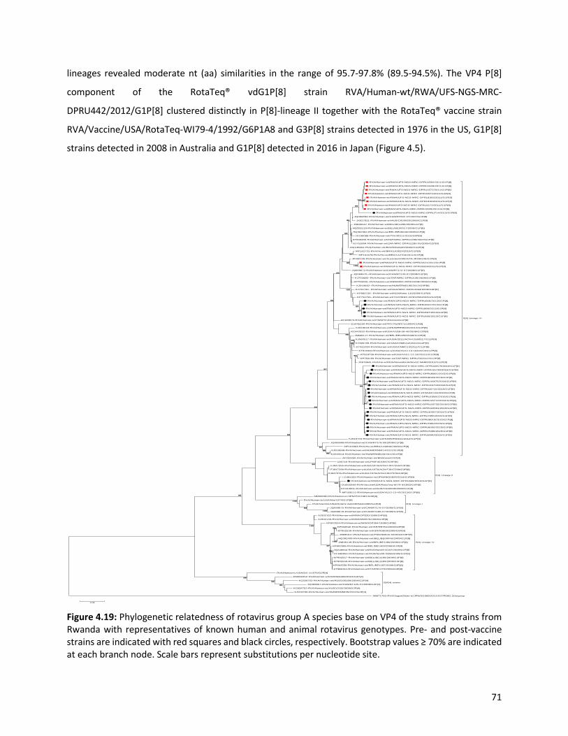
71
lineages revealed moderate nt (aa) similarities in the range of 95.7-97.8% (89.5-94.5%). The VP4 P[8]
component of the RotaTeq® vdG1P[8] strain RVA/Human-wt/RWA/UFS-NGS-MRC-
DPRU442/2012/G1P[8] clustered distinctly in P[8]-lineage II together with the RotaTeq® vaccine strain
RVA/Vaccine/USA/RotaTeq-WI79-4/1992/G6P1A8 and G3P[8] strains detected in 1976 in the US, G1P[8]
strains detected in 2008 in Australia and G1P[8] detected in 2016 in Japan (Figure 4.5).
Figure 4.19: Phylogenetic relatedness of rotavirus group A species base on VP4 of the study strains from Rwanda with representatives of known human and animal rotavirus genotypes. Pre- and post-vaccine strains are indicated with red squares and black circles, respectively. Bootstrap values ≥ 70% are indicated at each branch node. Scale bars represent substitutions per nucleotide site.
RVA/Human-wt/RWA/UFS-NGS-MRC-DPRU1582/2011/G1P[8]
RVA/Human-wt/RWA/UFS-NGS-MRC-DPRU1609/2011/G1P[8]
RVA/Human-wt/RWA/UFS-NGS-MRC-DPRU1572/2011/G1P[8]
RVA/Human-wt/RWA/UFS-NGS-MRC-DPRU1571/2011/G1P[8]
RVA/Human-wt/RWA/UFS-NGS-MRC-DPRU1565/2011/G1P[8]
RVA/Human-wt/RWA/UFS-NGS-MRC-DPRU1569/2011/G1P[8]
RVA/Human-wt/RWA/UFS-NGS-MRC-DPRU1575/2011/G1P[8]
RVA/Human-wt/RWA/UFS-NGS-MRC-DPRU1605/2011/G1P[8]
RVA/Human-wt/RWA/UFS-NGS-MRC-DPRU714/2013/G1P[8]
JQ069653-RVA/Human-wt/CAN/RT092-07/2007/G1P[8]
JX027821-RVA/Human-wt/AUS/CK00083/2008/G1P[8]
JN849147-RVA/Human-wt/BEL/BE1286/2009/G1P[8]
HQ392119-RVA/Human-wt/BEL/BE00017/2006/G1P[8]
HQ392362-RVA/Human-wt/BEL/BE00040/2008/G1P[8]
JX195088-RVA/Human-wt/ITA/JES11/2010/G9P[8]
KF636336-RVA/Human-wt/ZAF/MRC-DPRU1289/2007/G1P[8]
KJ751804-RVA/Human-wt/ZAF/MRC-DPRU1280-05/2005/G2P[8]
DQ146652-RVA/Human-wt/BGD/Dhaka25/2002/G12P[8]
MF161731-RVA/Hu-wt/BRA/1A3024/2010/G1P[8]
MF161678-RVA/Hu-wt/BRA/1A2703/2011/G1P[8]
JF490245-RVA/Human-wt/Australia/2005/ACN-JF490245/G1P[8]
RVA/Human-wt/RWA/UFS-NGS-MRC-DPRU1621/2011/G1P[8]
RVA/Human-wt/RWA/UFS-NGS-MRC-DPRU1626/2011/G1P[8]
JQ069673 RVA/Human-wt/CAN/RT172-07/2008/G1P[8]
JQ069670--RVA/Human-wt/CAN/RT133-07/2008/G1P[8]
KJ753085--RVA/Human-wt/ZAF/MRC-DPRU135/2009/G1P[8]
KP753260--RVA/Human-wt/KEN/MRC-DPRU1608/2009/G1P[8]
KJ919662--RVA/Human-wt/HUN/ERN5195/2012/G9P[8]
KJ751760--RVA/Human-wt/UGA/MRC-DPRU1944/2008/G9P[8]
KP882726--RVA/Human-wt/KEN/Keny-110/2009/G1P[8]
KP752756--RVA/Human-wt/TGO/MRC-DPRU4562/2011/G1P[8]
RVA/Human-wt/RWA/UFS-NGS-MRC-DPRU669/2013/G1P[8]
RVA/Human-wt/RWA/UFS-NGS-MRC-DPRU691/2013/G1P[8]
RVA/Human-wt/RWA/UFS-NGS-MRC-DPRU568/2013/G1P[8]
RVA/Human-wt/RWA/UFS-NGS-MRC-DPRU697/2013/G1P[8]
RVA/Human-wt/RWA/UFS-NGS-MRC-DPRU659/2013/G1P[8]
AF183870-RVA/Human-wt/TWN/TF101/xxxx/G1P[8]
EU045220-RVA/Human-wt/PRY/Py99371/1999/G1P[8]
KJ919618 RVA/Human-wt/HUN/ERN5040/2012/G1P[8]
KC442932-RVA/Human-wt/USA/VU08-09-40/2008/G12P[8]
JN849117-RVA/Human-wt/BEL/BE0258/2008/G12P[8]
KJ560517-RVA/Human-wt/USA/2011/ACN-KJ560517/G12P[8]
KT920709-RVA/Human-wt/USA/CNMC12/2011/G12P[8]
KT921094-RVA/Human-wt/USA/CNMC13/2011/G12P[8]
KT919340-RVA/Human-wt/USA/VU12-13-103/2013/G12P[8]
KT918735-RVA/Human-wt/USA/VU11-12-192/2012/G12P[8]
KP753159-RVA/Human-wt/ZAF/MRC-DPRU76/2012/G12P[8]
JX878641-RVA/Hu-wt/RUS/Novosibirsk/Nov12-N4489/2012/G12P[8]
RVA/Human-wt/RWA/UFS-NGS-MRC-DPRU16679/2016/G1P[8]
RVA/Human-wt/RWA/UFS-NGS-MRC-DPRU16728/2016/G1P[8]
RVA/Human-wt/RWA/UFS-NGS-MRC-DPRU8001/2015/G1P[8]
RVA/Human-wt/RWA/UFS-NGS-MRC-DPRU8033/2015/G1P[8]
RVA/Human-wt/RWA/UFS-NGS-MRC-DPRU16675/2016/G1P[8]
RVA/Human-wt/RWA/UFS-NGS-MRC-DPRU16703/2016/G1P[8]
RVA/Human-wt/RWA/UFS-NGS-MRC-DPRU16715/2016/G1P[8]
RVA/Human-wt/RWA/UFS-NGS-MRC-DPRU16724/2016/G1P[8]
RVA/Human-wt/RWA/UFS-NGS-MRC-DPRU16681/2016/G1P[8]
RVA/Human-wt/RWA/UFS-NGS-MRC-DPRU16712/2016/G1P[8]
RVA/Human-wt/RWA/UFS-NGS-MRC-DPRU16725/2016/G1P[8]
RVA/Human-wt/RWA/UFS-NGS-MRC-DPRU16691/2016/G1P[8]
RVA/Human-wt/RWA/UFS-NGS-MRC-DPRU10007/2015/G1P[8]
RVA/Human-wt/RWA/UFS-NGS-MRC-DPRU7995/2015/G1P[8]
RVA/Human-wt/RWA/UFS-NGS-MRC-DPRU8019/2015/G1P[8]
RVA/Human-wt/RWA/UFS-NGS-MRC-DPRU7990/2015/G1P[8]
RVA/Human-wt/RWA/UFS-NGS-MRC-DPRU8000/2015/G1P[8]
RVA/Human-wt/RWA/UFS-NGS-MRC-DPRU7986/2015/G1P[8]
RVA/Human-wt/RWA/UFS-NGS-MRC-DPRU8040/2015/G1P[8]
KJ919703-RVA/Human-wt/HUN/ERN5611/2012/G1P[8]
JQ069698-RVA/Human-wt/CAN/RT070-09/2009/G1P[8]
MF161863-RVA/Hu-wt/BRA/1A0830/2009/G1P[8]
KJ919608-RVA/Human-wt/HUN/ERN5014/2012/G1P[8]
KJ919614-RVA/Human-wt/HUN/ERN5026/2012/G1P[8]
P[8] Lineage III
AF052450-RVA/Human-wt/BRA/xxxx/GXP[8]
U30716-RVA/Human-wt/JPN/F45/1987/G9P[8]
FJ947255-RVA/Human-wt/USA/1976/ACN-FJ947255/G3P[8]
FJ947299-RVA/Human-wt/USA/1976/ACN-FJ947299/G3P[8]
FJ947376-RVA/Human-wt/USA/1976/ACN-FJ947376/G3P[8]
LC384333-RVA/Human-wt/JPN/HKD0825/2016/G1P[8]
RVA/Human-wt/RWA/UFS-NGS-MRC-DPRU442/2012/G1P[8]
GU565044-RVA/Vaccine/USA/RotaTeq-WI79-4/1992/G6P[8]
KC443601-RVA/Human-wt/AUS/CK20039/2008/G1P[8]
MF168111-RVA/Human-wt/USA/VU12-13-42/2013/G12P[8]
P[8] Lineage II
AB008280-RVA/Human-tc/JPN/ITO/1981/G3P[8]
RVA/Human-tc/USA/Wa/1974/G1P[8]
RVA/Vaccine/USA/Rotarix-A41CB052A/1988/G1P[8]
JQ069675-RVA/Human-wt/CAN/RT178-07/2008/G1P[8]
JQ069678-RVA/Human-wt/CAN/RT186-07/2008/G1P[8]
P[8] Lineage I
AJ302152-RVA/Human-wt/MWI/OP530/1999/G4P[8]
AJ302146-RVA/Human-wt/MWI/MW670/1999/G4P[8]
KP902534-RVA/Human-wt/MWI/OP354/1998/G4P[8]
KP902540-RVA/Human-wt/ISR/R5751/2009/G9P[8]
KP902539-RVA/Human-wt/ISR/R5808/2009/G9P[8]
JN695617-RVA/Human-wt/PAK/NIBGE-59/2010/G9P[8]
HQ392409-RVA/Human-wt/BEL/BE00045/2009/G1P[8]
JN849149-RVA/Human-wt/BEL/BE1280/2009/G1P[8]
KP902555-RVA/Human-wt/BEL/BE1400/2008/G1P[8]
JQ248944-RVA/Human-wt/RUS/Nov09-D187/2009/G1P[8]
KF648953-RVA/Human-wt/RUS/Nov09-D296/2009/G1P[8]
KP902557-RVA/Human-wt/BEL/BE1190/2009/G3P[8]
KP902559-RVA/Human-wt/BEL/BE1183/2009/G3P[8]
KP902556-RVA/Human-wt/BEL/BE1187/2009/G3P[8]
KT988164-RVA/Human-wt/ITA/PR1470/2009/G3P[8]
P[8] Lineage IV
RVA/Human-tc/USA/DS-1/1976/G2P[4]
KM660352-RVA/Human-wt/CMR/BA368/2010/G2P[4]
KC834703-RVA/Human-wt/AUS/336190/2004/G2P[4]
JQ069667-RVA/Human-wt/CAN/RT125-07/2008/G2P[4]
KC834702-RVA/Human-wt/AUS/V203/2009/G2P[4]
KJ919706-RVA/Human-wt/HUN/ERN5635/2012/G2P[4]
G2P[4] strains
Outgroup AB971763-RVA/SugarGlider-tc/JPN/SG385/2012/G27P[36]
95
99
98
100
99
100
98
97
98100
99
95
98
99
99
100
93
95
95
94
9999
96
96
90
97
96
96
97
99
99
100
93
100
100
98
97
95
97
100
95
0.02
72
4.3.7. Comparative analyses of neutralizing antigenic epitopes in the VP4 protein of Rwandan G1P[8] and vaccine strains of RVA.
The VP4 protein, which in this study represented P[8] strains, was divided into the VP8* and VP5* regions
for comparison. The VP8* region contains four (8-1 to 8-4) neutralizing antigenic epitopes, while the VP5*
region has five (5-1 to 5-5) (Dormitzer et al., 2002). These two epitopes contain 37 amino acid residues,
25 in the VP8* and 12 in the VP5* antigenic epitope regions. Out of these 37 amino acid residues that
spans the neutralization epitopes, those at positions 100, 146, 148, 150, 188, 190 and 194 (8-1 region),
180 and 183 (8-2 region), 114, 116, 132, 133, and 135 (8-3 region), 87, 88, and 89 (8-4 region), 384, 386,
388, 393, 394, 398, 440, and 441 (5-1 region), 434 (5-2 region), 459 (5-3 region), 429 (5-4 region), and 306
(5-5 region), are known neutralization escape mutation sites (Fig. 6A) (Dormitzer et al., 2004; Monnier et
al., 2006). A comparison of the Rwandan P[8] strains to the P[8] component of RotaTeq® and Rotarix®
vaccines showed 29 identical amino acid residues distributed through the antigenic epitopes of the VP4
gene (Figure 4.6A). The differences between the P[8] study strains and vaccine P[8] component of
RotaTeq® and Rotarix® were mostly contained in VP8* epitopes 8-1 and 8-3. Relative to the RotaTeq®,
the P[8] component of the Rwandan G1P[8] strains exhibited 3-4 amino acid differences. The changes
identified relative to the RotaTeq® P[8] vaccine strain were seen at positions E150D and D195G in the
neutralization epitope 8-1 region (Figure 4.6A-4.6B) and were all located on the surface of the protein
structure. Overall, the amino acid changes identified at positions N113D, S131R and N135D, resulted in a
change in charge from a polar molecule with a potential side chain with five hydrogen bonds to a
negatively charged molecule with four hydrogen bonds. Finally, amino acid substitutions at position
N195G for both vaccines, resulted in a change in polarity.

73
Figure 4.20: A) The alignment of the P[8] component of Rotarix® and RotaTeq® vaccines and wild type P[8] strains circulating in Rwanda from 2011–2016 RVA seasons, based on the antigenic residues in VP4. VP4 is divided into two regions VP8* (antigenic epitope 8-1, 8-2, 8-3, and 8-4) and VP5* (antigenic epitope 5-1, 5-2, 5-3, 5-4, and 5-5). The black dot indicates changes in the residues associated with escape neutralization with monoclonal antibodies. Amino acids differences between Rotarix® and RotaTeq® are shown at positions 195 (antigenic epitope 8-1), 125, 131, and 135 (antigenic epitope 8-3) and are indicated in boldface. Study strains amino acid residues highlighted in green and brown are residues that are different from Rotarix® and RotaTeq®, respectively. Also, amino acids differences between study strains and Rotarix® and RotaTeq® are highlighted in sky blue. Post-vaccine and pre- vaccine G1P[8] study strains are bolded in black and red, respectively. B) Location of surface-exposed amino acids differences between
8-1 8-2 8-3 8-4 5-1 5-2 5-3 5-4 5-5● ● ● ● ● ● ● ● ● ● ● ● ● ● ● ● ● ● ● ● ● ● ● ● ● ● ● ● ●
Lineage 100 146 148 150 188 190 192 193 194 195 196 180 183 113 114 115 116 125 131 132 133 135 87 88 89 384 386 388 393 394 398 440 441 434 459 429 306RVA/Vaccine/USA/Rotarix-A41CB052A/1988/G1P1A[8] P[8]-I D S Q E S T N L N N I T A N P V D S S N D N N T N Y F I W P G R T P E L RRVA/Vaccine/USA/RotaTeq-WI79-4/1992/G6P1A[8] P[8]-II D S Q E S T N L N D I T A N P V D N R N D D N T N Y F L W P G R T P E L RRVA/Human-wt/RWA/UFS-NGS-MRC-DPRU442/2012/G1P[8] P[8]-II D S Q E S T N L N D I T A N P V D N R N D D N T N Y F L W P G R T P E L RRVA/Human-wt/RWA/UFS-NGS-MRC-DPRU16675/2016/G1P[8] P[8]-III D S Q D S T N L N G I T A N P V D N R N D D N T N Y F I W P G R T P E L RRVA/Human-wt/RWA/UFS-NGS-MRC-DPRU16679/2016/G1P[8] P[8]-III D S Q D S T N L N G I T A N P V D N R N D D N T N Y F I W P G R T P E L RRVA/Human-wt/RWA/UFS-NGS-MRC-DPRU16681/2016/G1P[8] P[8]-III D S Q D S T N L N G I T A S P V D N R N D D N T N Y F I W P G R T P E L RRVA/Human-wt/RWA/UFS-NGS-MRC-DPRU16691/2016/G1P[8] P[8]-III D S Q D S T N L N G I T A N P V D N R N D D N T N Y F I W P G R T P E L RRVA/Human-wt/RWA/UFS-NGS-MRC-DPRU16703/2016/G1P[8] P[8]-III D S Q D S T N L N G I T A N P V D N R N D D N T N Y F I W P G R T P E L RRVA/Human-wt/RWA/UFS-NGS-MRC-DPRU16712/2016/G1P[8] P[8]-III D S Q D S T N L N G I T A S P V D N R N D D N T N Y F I W P G R T P E L RRVA/Human-wt/RWA/UFS-NGS-MRC-DPRU16715/2016/G1P[8] P[8]-III D S Q D S T N L N G I T A S P V D N R N D D N T N Y F I W P G R T P E L RRVA/Human-wt/RWA/UFS-NGS-MRC-DPRU16724/2016/G1P[8] P[8]-III D S Q D S T N L N G I T A S P V D N R N D D N T N Y F I W P G R T P E L RRVA/Human-wt/RWA/UFS-NGS-MRC-DPRU16725/2016/G1P[8] P[8]-III D S Q D S T N L N G I T A S P V D N R N D D N T N Y F I W P G R T P E L RRVA/Human-wt/RWA/UFS-NGS-MRC-DPRU16728/2016/G1P[8] P[8]-III D S Q D S T N L N G I T A N P V D N R N D D N T N Y F I W P G R T P E L RRVA/Human-wt/RWA/UFS-NGS-MRC-DPRU10007/2015/G1P[8] P[8]-III D S Q D S T N L N G I T A N P V D N R N D D N T N Y F I W P G R T P E L RRVA/Human-wt/RWA/UFS-NGS-MRC-DPRU7986/2015/G1P[8] P[8]-III D S Q D S T N L N G I T A N P V D N R N D D N T N Y F I W P G R T P E L RRVA/Human-wt/RWA/UFS-NGS-MRC-DPRU7990/2015/G1P[8] P[8]-III D S Q D S T N L N G I T A N P V D N R N D D N T N Y F I W P G R T P E L RRVA/Human-wt/RWA/UFS-NGS-MRC-DPRU7995/2015/G1P[8] P[8]-III D S Q D S T N L N G I T A N P V D N R N D D N T N Y F I W P G R T P E L RRVA/Human-wt/RWA/UFS-NGS-MRC-DPRU8000/2015/G1P[8] P[8]-III D S Q D S T N L N G I T A N P V D N R N D D N T N Y F I W P G R T P E L RRVA/Human-wt/RWA/UFS-NGS-MRC-DPRU8001/2015/G1P[8] P[8]-III D S Q D S T N L N G I T A N P V D N R N D D N T N Y F I W P G R T P E L RRVA/Human-wt/RWA/UFS-NGS-MRC-DPRU8019/2015/G1P[8] P[8]-III D S Q D S T N L N G I T A N P V D N R N D D N T N Y F I W P G R T P E L RRVA/Human-wt/RWA/UFS-NGS-MRC-DPRU8033/2015/G1P[8] P[8]-III D S Q D S T N L N G I T A N P V D N R N D D N T N Y F I W P G R T P E L RRVA/Human-wt/RWA/UFS-NGS-MRC-DPRU8040/2015/G1P[8] P[8]-III D S Q D S T N L N G I T A N P V D N R N D D N T N Y F I W P G R T P E L RRVA/Human-wt/RWA/UFS-NGS-MRC-DPRU568/2013/G1P[8] P[8]-III D S Q D S T N L N G I T A D P V D N R N D D N T N Y F I W P G R T P E L RRVA/Human-wt/RWA/UFS-NGS-MRC-DPRU659/2013/G1P[8] P[8]-III D S Q D S T N L N G I T A D P V D N R N D D N T N Y F I W P G R T P E L RRVA/Human-wt/RWA/UFS-NGS-MRC-DPRU669/2013/G1P[8] P[8]-III D S Q D S T N L N G I T A D P V D N R N D D N T N Y F I W P G R T P E L RRVA/Human-wt/RWA/UFS-NGS-MRC-DPRU691/2013/G1P[8] P[8]-III D S Q D S T N L N G I T A D P V D N R N D D N T N Y F I W P G R T P E L RRVA/Human-wt/RWA/UFS-NGS-MRC-DPRU697/2013/G1P[8] P[8]-III D S Q D S T N L N G I T A D P V D N R N D D N T N Y F I W P G R T P E L RRVA/Human-wt/RWA/UFS-NGS-MRC-DPRU714/2013/G1P[8] P[8]-III D S Q D S T N L N G I T A N P V D N R N D D N T N Y F I W P G R T P E L RRVA/Human-wt/RWA/UFS-NGS-MRC-DPRU1565/2011/G1P[8] P[8]-III D S Q D S T N L N G I T A N P V D N R N D D N T N Y F I W P G R T P E L RRVA/Human-wt/RWA/UFS-NGS-MRC-DPRU1569/2011/G1P[8] P[8]-III D S Q D S T N L N G I T A N P V D N R N D D N T N Y F I W P G R T P E L RRVA/Human-wt/RWA/UFS-NGS-MRC-DPRU1571/2011/G1P[8] P[8]-III D S Q D S T N L N G I T A N P V D N R N D D N T N Y F I W P G R T P E L RRVA/Human-wt/RWA/UFS-NGS-MRC-DPRU1572/2011/G1P[8] P[8]-III D S Q D S T N L N G I T A N P V D N R N D D N T N Y F I W P G R T P E L RRVA/Human-wt/RWA/UFS-NGS-MRC-DPRU1575/2011/G1P[8] P[8]-III D S Q D S T N L N G I T A N P V D N R N D D N T N Y F I W P G R T P E L RRVA/Human-wt/RWA/UFS-NGS-MRC-DPRU1582/2011/G1P[8] P[8]-III D S Q D S T N L N G I T A N P V D N R N D D N T N Y F I W P G R T P E L RRVA/Human-wt/RWA/UFS-NGS-MRC-DPRU1605/2011/G1P[8] P[8]-III D S Q D S T N L N G I T A N P V D N R N D D N T N Y F I W P G R T P E L RRVA/Human-wt/RWA/UFS-NGS-MRC-DPRU1609/2011/G1P[8] P[8]-III D S Q D S T N L N G I T A N P V D N R N D D N T N Y F I W P G R T P E L RRVA/Human-wt/RWA/UFS-NGS-MRC-DPRU1621/2011/G1P[8] P[8]-III D S Q D S T N L N G I T A D P V D N R N D D N T N Y F I W P G R T P E L RRVA/Human-wt/RWA/UFS-NGS-MRC-DPRU1626/2011/G1P[8] P[8]-III D S Q D S T N L N G I T A D P V D N R N D D N T N Y F I W P G R T P E L RRVA/Human-tc/USA/Wa/1974/G1P[8] P[8]-I D S Q E S T N L N N I T A N P V D S S N D N N T N Y F I W P G R T P E L RRVA/Human-tc/JPN/ITO/1981/G3P[8] P[8]-I D S Q E S T N L H H I T A N P V D S S N D N N T N Y F L W P G R T P E L RRVA/Human-wt/JPN/F45/1987/G9P[8] P[8]-II D S Q E S T N L N N I T A N P V D N R N D N N T N Y F I W P G R T P E L RRVA/Human-wt/ZAF/MRC-DPRU76/2012/G12P[8] P[8]-III D S Q D S T N L N G I T A N P V D N R N D D N T N Y F I W P G R T P E L RRVA/Human-wt/MWI/OP354/1998/G4P[8] P[8]-IV D S Q E S T D L T S I T A D P V D S R N D N N T N Y F L W P G R T P D L R
VP8* VP5*
180o
RotaTeq VP8* of P[8] vaccine component
RotaTeq VP8* of P[8] vaccine component
Rwanda VP8* of wild type P[8] strain
A
B

74
VP8* protein of RotaTeq® P[8] vaccine component versus a wild-type P[8] strain from Rwanda (indicated in red). Antigenic epitopes in (B) are coloured in yellow (8-1), green (8-2), blue (8-3), and purple (8-4) (created with PyMoL, 4 June 2019) 4.3.8. Phylogenetic and sequence analyses of VP1-VP3, NSP1, NSP4, and NSP5. These six genes segregated into typical pre- and post-vaccine introduction clusters, hence presented
together. Phylogenetic analyses based on VP1-VP3, NSP1, NSP4, and NSP5 nucleotide sequences of each
Rwandan G1P[8] strain showed that each gene (exception of RotaTeq® vdG1P[8] strain) segregated and
clustered with cognate gene sequences of Wa-like strains belonging to genotype 1 (Figure 4.7A-4.7F). The
VP1 gene sequences of the 35 typical Rwandan G1P[8] strains segregated into a single pre-vaccine
introduction sub-lineage exhibiting nt (aa) similarities in the range of 99.9-100% (99.8-100%) and two
post-vaccine introduction sub-lineages with nt (aa) identities in the range of 96.8-98.0% (98.7-99.5%).
(Figure 4.2A). Four pre-vaccine introduction orphan strains that did not cluster in either of the three sub-
lineages were also observed (Figure 4.7A). For VP2, the C1 genes segregated into three typical sub-
lineages (one typical pre- and two post-vaccine introduction) and five orphan strains which did not cluster
into either of the three sub-lineages (Figure 4.7B). The two post-vaccine introduction sub-lineage
consisting of five and 19 strains shared nt (aa) similarities in the range of 99.6-100% (99.6-100%) and 99.3-
100% (99.7-100%), respectively. Furthermore, the typical pre-vaccine introduction sub-lineage
incorporated strains (n=6) detected in the 2011 RVA season and with nt (aa) sequence identities amongst
themselves in the range of 99.9-100% (99.8-100%).
Phylogenetically, the VP3 gene sequences of the Rwandan pre- and post-vaccine introduction strains
separated into three typical sub-lineages and three orphan strains were observed (Figure 4.7C). The first
post-vaccine sub-lineage consisted of 19 strains and the second one had five strains that shared nt (aa)
similarities ranging from 99.2-100% (98.9-100%) and 99.4-100% (99.3-100%), respectively. The pre-
vaccine introduction sub-lineage contained strains that shared nt (aa) identities of 99.3-100% (98.5-
100%). The NSP1 gene sequences of the typical Rwandan pre- and post-vaccine introduction G1P[8]
strains were grouped into a single sub-lineage consisted of all ten pre-vaccine introduction strains and
three typical post-vaccination introduction sub-lineages (Figure 4.7D). The pre-vaccine strains shared nt
(aa) similarities that ranged from 99.0-99.9% (97.5-99.8%). The three post-vaccine introduction NSP1
genes that segregated into three sub-lineages revealed nt (aa) identities of 98.7-99.9% (98.1-99.8%), 99.0-
99.9% (98.4-99.8%) and 98.2-99.9% (98.1-99.8%), respectively.

75
The NSP4 gene sequences of the Rwandan strains were also segregated into two typical post-vaccination
sub-lineages and a single pre-vaccination sub-lineage (Figure 4.7E). Six orphan Rwandan E1 genotypes did
not cluster into either the pre- or post-vaccine introduction sub-lineages (Figure 4.7E). The first typical
post-vaccine introduction strain sub-lineage comprised of 19 NSP4 genes and the second one had five E1
strains detected in 2011 RVA season which exhibited nt (aa) identities in the range of 99.4-100% (99.4-
100%) and 99.4-100% (99.4-100%), respectively. The pre-vaccination sub-lineage is comprised of five NSP4
strains with absolute gene identities among themselves. The NSP5 gene did not resolve phylogenetically,
and the bootstrap support was not enough to separate the strains into distinct sub-lineages. On the other
hand, a homologous relationship was observed for the VP1, VP2, VP3, NSP1, NSP4, and NSP5 gene
segments of strain RVA/Human-wt/RWA/UFS-NGS-MRC-DPRU442/2012/G1P[8] with cognate gene
sequences of RVA/Vaccine/USA/Rotateq-W179-4/1992/G6P1A8.

76
RVA/Human-wt/RWA/UFS-NGS-MRC-DPRU7995/2015/G1P[8]
RVA/Human-wt/RWA/UFS-NGS-MRC-DPRU8000/2015/G1P[8]
RVA/Human-wt/RWA/UFS-NGS-MRC-DPRU8019/2015/G1P[8]
RVA/Human-wt/RWA/UFS-NGS-MRC-DPRU10007/2015/G1P[8]
RVA/Human-wt/RWA/UFS-NGS-MRC-DPRU8033/2015/G1P[8]
RVA/Human-wt/RWA/UFS-NGS-MRC-DPRU7986/2015/G1P[8]
RVA/Human-wt/RWA/UFS-NGS-MRC-DPRU8040/2015/G1P[8]
RVA/Human-wt/RWA/UFS-NGS-MRC-DPRU7990/2015/G1P[8]
RVA/Human-wt/RWA/UFS-NGS-MRC-DPRU16724/2016/G1P[8]
RVA/Human-wt/RWA/UFS-NGS-MRC-DPRU16681/2016/G1P[8]
RVA/Human-wt/RWA/UFS-NGS-MRC-DPRU16715/2016/G1P[8]
RVA/Human-wt/RWA/UFS-NGS-MRC-DPRU16712/2016/G1P[8]
RVA/Human-wt/RWA/UFS-NGS-MRC-DPRU16725/2016/G1P[8]
RVA/Human-wt/RWA/UFS-NGS-MRC-DPRU16691/2016/G1P[8]
RVA/Human-wt/RWA/UFS-NGS-MRC-DPRU16679/2016/G1P[8]
RVA/Human-wt/RWA/UFS-NGS-MRC-DPRU16675/2016/G1P[8]
RVA/Human-wt/RWA/UFS-NGS-MRC-DPRU16703/2016/G1P[8]
RVA/Human-wt/RWA/UFS-NGS-MRC-DPRU8001/2015/G1P[8]
RVA/Human-wt/RWA/UFS-NGS-MRC-DPRU16728/2016/G1P[8]
RVA/Human-wt/IDN/STM453/2018/G1P[8]
RVA/Human-wt/IDN/STM387/2017/G1P[8]
KT919337-RVA/Human-wt/USA/VU12-13-103/2013/G12P[8]
KT919569-RVA/Human-wt/USA/VU12-13-21/2013/G12P[8]
HQ392349-RVA/Human-wt/BEL/BE00039/2008/G1P[8]
MF161795-RVA/Hu-wt/BRA/1A2703/2011/G1P[8]
MF161885-RVA/Hu-wt/BRA/1A3024/2010/G1P[8]
DQ146649-RVA/Human-wt/BGD/Dhaka25/2002/G12P[8]
KJ626818-RVA/Human-wt/PRY/468/2000/G9P[8]
RVA/Human-wt/RWA/UFS-NGS-MRC-DPRU1621/2011/G1P[8]
RVA/Human-wt/RWA/UFS-NGS-MRC-DPRU1626/2011/G1P[8]
KJ752004-RVA/Human-wt/ETH/MRC-DPRU5002/2010/G12P[8]
KP752790-RVA/Human-wt/ETH/MRC-DPRU4970/2010/G12P[8]
JF490820-RVA/Human-wt/USA/2005/ACN-JF490820/G1P[8]
DQ146638-RVA/Human-wt/BEL/B4633/2003/G12P[8]
DQ870501-RVA/Human-wt/BEL/B3458/2003/G9P[8]
KF371992-RVA/Human-wt/CHN/Y106/2004/G3P[8]
MG181458-RVA/Human-wt/MWI/MW2-1254/2005/G1P[8]
MG181469-RVA/Human-wt/MWI/MW2-1274/2005/G1P[8]
MG181480-RVA/Human-wt/MWI/0P5-001/2008/G1P[8]
RVA/Human-wt/RWA/UFS-NGS-MRC-DPRU1575/2011/G1P[8]
KJ870911-RVA/Human-wt/COD/KisB521/2008/G12P[6]
KJ752640-RVA/Human-wt/ZMB/MRC-DPRU3488/2009/G12P[6]
KF636146-RVA/Human-wt/ZMB/MRC-DPRU3491/2009/G12P[6]
KJ752530-RVA/Human-wt/ZMB/MRC-DPRU1680/2008/G12P[6]
KJ751867-RVA/Human-wt/UGA/MRC-DPRU3713/2010/G12P[6]
AB861945-RVA/Human-tc/KEN/KDH633/2010/G12P[6]
AB861967-RVA/Human-tc/KEN/KDH684/2010/G12P[6]
RVA/Human-wt/RWA/UFS-NGS-MRC-DPRU1605/2011/G1P[8]
KP752888-RVA/Human-wt/ZAF/MRC-DPRU2125/2008/G1P[8]
KJ752836-RVA/Human-wt/ZAF/MRC-DPRU2429/2008/G1P[8]
KP753017-RVA/Human-wt/ZAF/MRC-DPRU1808/2007/G1P[8]
JX195085-RVA/Human-wt/ITA/JES11/2010/G9P[8]
HQ392355-RVA/Human-wt/BEL/BE00040/2008/G1P[8]
JQ069904-RVA/Human-wt/CAN/RT092-07/2007/G1P[8]
JX027813-RVA/Human-wt/AUS/CK00083/2008/G1P[8]
KJ919302-RVA/Human-wt/HUN/ERN5040/2012/G1P[8]
HQ392179-RVA/Human-wt/BEL/BE00023/2007/G1P[8]
KJ870922-RVA/Human-wt/COD/KisB504/2009/G1P[6]
RVA/Human-wt/RWA/UFS-NGS-MRC-DPRU1565/2011/G1P[8]
RVA/Human-wt/RWA/UFS-NGS-MRC-DPRU1569/2011/G1P[8]
RVA/Human-wt/RWA/UFS-NGS-MRC-DPRU1571/2011/G1P[8]
RVA/Human-wt/RWA/UFS-NGS-MRC-DPRU1609/2011/G1P[8]
RVA/Human-wt/RWA/UFS-NGS-MRC-DPRU1572/2011/G1P[8]
RVA/Human-wt/RWA/UFS-NGS-MRC-DPRU1582/2011/G1P[8]
JQ069925-RVA/Human-wt/CAN/RT172-07/2008/G1P[8]
LC019052-RVA/Human-tc/MMR/A23/2011/G12P[6]
LC374184--RVA/Human-wt/NPL/10N4001/2010/G12P[6]
RVA/Human-wt/RWA/UFS-NGS-MRC-DPRU714/2013/G1P[8]
KP753257-RVA/Human-wt/KEN/MRC-DPRU1608/2009/G1P[8]
JQ069922-RVA/Human-wt/CAN/RT133-07/2008/G1P[8]
KJ751757-RVA/Human-wt/UGA/MRC-DPRU1944/2008/G9P[8]
KX632298-RVA/Human-wt/UGA/MUL-12-147/2012/G9P[8]
KX632243-RVA/Human-wt/UGA/NSA-13-043/2013/G9P[8]
KJ753423-RVA/Human-wt/UGA/MRC-DPRU4595/2011/G9P[8]
KP882728-RVA/Human-wt/KEN/Keny-110/2009/G1P[8]
RVA/Human-wt/RWA/UFS-NGS-MRC-DPRU669/2013/G1P[8]
KP752753-RVA/Human-wt/TGO/MRC-DPRU4562/2011/G1P[8]
RVA/Human-wt/RWA/UFS-NGS-MRC-DPRU691/2013/G1P[8]
RVA/Human-wt/RWA/UFS-NGS-MRC-DPRU697/2013/G1P[8]
RVA/Human-wt/RWA/UFS-NGS-MRC-DPRU568/2013/G1P[8]
RVA/Human-wt/RWA/UFS-NGS-MRC-DPRU659/2013/G1P[8]
JQ069950 RVA/Human-wt/CAN/RT070-09/2009/G1P[8]
RVA/Human-TC/USA/Rotarix/2009/G1P[8]
RVA/Human-tc/USA/Wa/1974/G1P[8]
JQ069927-RVA/Human-wt/CAN/RT178-07/2008/G1P[8]
JQ069930-RVA/Human-wt/CAN/RT186-07/2008/G1P[8]
KJ919385-RVA/Human-wt/HUN/ERN5611/2012/G1P[8]
KJ919293-RVA/Human-wt/HUN/ERN5014/2012/G1P[8]
KJ919299-RVA/Human-wt/HUN/ERN5026/2012/G1P[8]
R1
R3 RVA/Human-tc/JPN/AU-1/1982/G3P[9]
JQ069919-RVA/Human-wt/CAN/RT125-07/2008/G2P[4]
KC834711-RVA/Human-wt/AUS/V203/2009/G2P[4]
KM660259-RVA/Human-wt/CMR/BA368/2010/G2P[4]
KC834712-RVA/Human-wt/AUS/336190/2004/G2P[4]
RVA/Human-tc/USA/DS-1/1976/G2P[4]
KJ919388-RVA/Human-wt/HUN/ERN5635/2012/G2P[4]
RVA/Human-wt/RWA/UFS-NGS-MRC-DPRU442/2012/G1P[8]
GU565041-RVA/Vaccine/USA/RotaTeq-WI79-4/1992/G6P[8]
LC384330-RVA/Human-wt/JPN/HKD0825/2016/G1P[8]
KT919447-RVA/Human-wt/USA/VU12-13-176/2013/G1G6P[8]
KC443598-RVA/Human-wt/AUS/CK20039/2008/G1P[8]
GU565085-RVA/Vaccine/USA/RotaTeq-BrB-9/1996/G4P7[5]
GU565074-RVA/Vaccine/USA/RotaTeq-WI78-8/1992/G3P7[5]
GU565052-RVA/Vaccine/USA/RotaTeq-WI79-9/1992/G1P7[5]
GU565063-RVA/Vaccine/USA/RotaTeq-SC2-9/1992/G2P7[5]
R2
Outgroup AB971760-RVA/SugarGlider-tc/JPN/SG385/2012/G27P[36]
100
100
100
100
100
100
97
99
96
97
97
95
100
100
100
100
98
99
99
96
99
99
99
100
95
97
100
100
100
99
100
99
99
100
0.05
4.7A VP1

77
RVA/Human-wt/RWA/UFS-NGS-MRC-DPRU8019/2015/G1P[8]
RVA/Human-wt/RWA/UFS-NGS-MRC-DPRU8040/2015/G1P[8]
RVA/Human-wt/RWA/UFS-NGS-MRC-DPRU8000/2015/G1P[8]
RVA/Human-wt/RWA/UFS-NGS-MRC-DPRU7995/2015/G1P[8]
RVA/Human-wt/RWA/UFS-NGS-MRC-DPRU7990/2015/G1P[8]
RVA/Human-wt/RWA/UFS-NGS-MRC-DPRU7986/2015/G1P[8]
RVA/Human-wt/RWA/UFS-NGS-MRC-DPRU16712/2016/G1P[8]
RVA/Human-wt/RWA/UFS-NGS-MRC-DPRU16679/2016/G1P[8]
RVA/Human-wt/RWA/UFS-NGS-MRC-DPRU10007/2015/G1P[8]
RVA/Human-wt/RWA/UFS-NGS-MRC-DPRU8033/2015/G1P[8]
RVA/Human-wt/RWA/UFS-NGS-MRC-DPRU16725/2016/G1P[8]
RVA/Human-wt/RWA/UFS-NGS-MRC-DPRU16691/2016/G1P[8]
RVA/Human-wt/RWA/UFS-NGS-MRC-DPRU16675/2016/G1P[8]
RVA/Human-wt/RWA/UFS-NGS-MRC-DPRU16703/2016/G1P[8]
RVA/Human-wt/RWA/UFS-NGS-MRC-DPRU16724/2016/G1P[8]
RVA/Human-wt/RWA/UFS-NGS-MRC-DPRU16681/2016/G1P[8]
RVA/Human-wt/RWA/UFS-NGS-MRC-DPRU16715/2016/G1P[8]
RVA/Human-wt/RWA/UFS-NGS-MRC-DPRU16728/2016/G1P8
RVA/Human-wt/RWA/UFS-NGS-MRC-DPRU8001/2015/G1P[8]
KU048553-RVA/Human-wt/Italy/2014/ACN-KU048553/G12P[8]
KU048553-RVA/Human-wt/ITA/ME659/14/2014/G12P[8]
JX027823-RVA/Human-wt/AUS/CK00083/2008/G1P[8]
AB861968-RVA/Human-tc/KEN/KDH684/2010/G12P[6]
AB861946-RVA/Human-tc/KEN/KDH633/2010/G12P[6]
RVA/Human-wt/RWA/UFS-NGS-MRC-DPRU1609/2011/G1P[8]
RVA/Human-wt/RWA/UFS-NGS-MRC-DPRU1572/2011/G1P[8]
RVA/Human-wt/RWA/UFS-NGS-MRC-DPRU1571/2011/G1P[8]
RVA/Human-wt/RWA/UFS-NGS-MRC-DPRU1582/2011/G1P[8]
RVA/Human-wt/RWA/UFS-NGS-MRC-DPRU1565/2011/G1P[8]
RVA/Human-wt/RWA/UFS-NGS-MRC-DPRU1569/2011/G1P[8]
JN129057-RVA/Human/NCA/9J/2010/G1P[8]
JN129061-RVA/Human/NCA/25J/2010/G1P[8]
HM773745-RVA/Human-wt/USA2007719825/2007/G1P[8]
HQ392364-RVA/Human-wt/BEL/BE00040/2008/G1P[8]
HQ392121-RVA/Human-wt/BEL/BE00017/2006/G1P[8]
HQ392299-RVA/Human-wt/BEL/BE00034/2008/G1P[8]
KY857561-RVA/Human-wt/India/2013/ACN-KY857561/G1P[8]
KP753018-RVA/Human-wt/ZAF/MRC-DPRU1808/2007/G1P[8]
KF636334-RVA/Human-wt/ZAF/MRC-DPRU1289/2007/G1P[8]
KJ753061-RVA/Human-wt/ZAF/MRC-DPRU1370/2007/G1P[8]
RVA/Human-wt/RWA/UFS-NGS-MRC-DPRU1605/2011/G1P[8]
KJ870912-RVA/Human-wt/COD/KisB521/2008/G12P[6]
KP753202-RVA/Human-wt/ZMB/MRC-DPRU3506/2009/G12P[6]
RVA/Human-wt/RWA/UFS-NGS-MRC-DPRU1575/2011/G1P[8]
LC372873-RVA/Human-wt/NPL/06N0349/2006/G12P[6]
LC367271-RVA/Human-wt/NPL/08N2148/2008/G12P[6]
LC374130-RVA/Human-wt/NPL/09N3140/2009/G12P[6]
LC374152-RVA/Human-wt/NPL/09N3472/2009/G12P[6]
LC374141-RVA/Human-wt/NPL/09N3294/2009/G12P[6]
LC019053-RVA/Human-tc/MMR/A23/2011/G12P[6]
KJ752815-RVA/Human-wt/ZAF/MRC-DPRU4090/2011/G12P[6]
RVA/Human-wt/RWA/UFS-NGS-MRC-DPRU714/2013/G1P[8]
RVA/Human-wt/RWA/UFS-NGS-MRC-DPRU691/2013/G1P[8]
RVA/Human-wt/RWA/UFS-NGS-MRC-DPRU568/2013/G1P[8]
RVA/Human-wt/RWA/UFS-NGS-MRC-DPRU659/2013/G1P[8]
RVA/Human-wt/RWA/UFS-NGS-MRC-DPRU697/2013/G1P[8]
DQ146661RVA/Human-wt/BGD/Dhaka12/2003/G12P[6]
MF161629-RVA/Hu-wt/BRA/1A3024/2010/G1P[8]
MF161908-RVA/Hu-wt/BRA/1A2703/2011/G1P[8]
KJ919408-RVA/Human-wt/HUN/ERN5040/2012/G1P[8]
DQ492670-RVA/Human-wt/BGD/Dhaka16/2003/G1P[8]
KJ919398 RVA/Human-wt/HUN/ERN5014/2012/G1P[8]
KJ919404 RVA/Human-wt/HUN/ERN5026/2012/G1P[8]
JF490213-RVA/Human-wt/Australia/2004/ACN-JF490213/G1P[8]
RVA/Human-wt/RWA/UFS-NGS-MRC-DPRU1621/2011/G1P[8]
RVA/Human-wt/RWA/UFS-NGS-MRC-DPRU1626/2011/G1P[8]
HQ392265-RVA/Human-wt/BEL/BE00031/2008/G1P[8]
LC105207-RVA/Human-wt/JPN/MU14-19/2014/G1P[8]
JF490445-RVA/Human-wt/Australia/2006/ACN-JF490445/G1P[8]
JN651885-RVA/Human-wt/Bel/BE00108/2010/G1P[8]
JQ069841-RVA/Human-wt/CAN/RT172-07/2008/G1P[8]
KP753258-RVA/Human-wt/KEN/MRC-DPRU1608/2009/G1P[8]
KJ751758-RVA/Human-wt/UGA/MRC-DPRU1944/2008/G9P[8]
JQ069838-RVA/Human-wt/CAN/RT133-07/2008/G1P[8]
KJ753083-RVA/Human-wt/ZAF/MRC-DPRU135/2009/G1P[8]
KX632288-RVA/Human-wt/UGA/MUL-13-163/2013/G9P[8]
RVA/Human-wt/RWA/UFS-NGS-MRC-DPRU669/2013/G1P[8]
KP752754-RVA/Human-wt/TGO/MRC-DPRU4562/2011/G1P[8]
KJ919493-RVA/Human-wt/HUN/ERN5611/2012/G1P[8]
JQ069866-RVA/Human-wt/CAN/RT070-09/2009/G1P[8]
JQ069843-RVA/Human-wt/CAN/RT178-07/2008/G1P[8]
JQ069846-RVA/Human-wt/CAN/RT186-07/2008/G1P[8]
RVA/Human-tc/USA/Rotarix/2009/G1P[8]
RVA/Human-tc/USA/Wa/1974/G1P[8]
C1
KJ919496-RVA/Human-wt/HUN/ERN5635/2012/G2P[4]
KC834709-RVA/Human-wt/AUS/336190/2004/G2P[4]
KC834708-RVA/Human-wt/AUS/V203/2009/G2P[4]
JQ069835-RVA/Human-wt/CAN/RT125-07/2008/G2P[4]
KM660290-RVA/Human-wt/CMR/BA368/2010/G2P[4]
RVA/Human-tc/USA/DS-1/1976/G2P[4]
RVA/Human-wt/RWA/UFS-NGS-MRC-DPRU442/2012/G1P[8]
LC384331-RVA/Human-wt/JPN/HKD0825/2016/G1P8
GU565042-RVA/Vaccine/USA/RotaTeq-WI79-4/1992/G6P[8]
GU565075-RVA/Vaccine/USA/RotaTeq-WI78-8/1992/G3P7[5]
GU565053-RVA/Vaccine/USA/RotaTeq-WI79-9/1992/G1P7[5]
KC443599-RVA/Human-wt/AUS/CK20039/2008/G1P[8]
KT919448-RVA/Human-wt/USA/VU12-13-176/2013/G1G6P[8]
GU565086-RVA/Vaccine/USA/RotaTeq-BrB-9/1996/G4P7[5]
GU565064-RVA/Vaccine/USA/RotaTeq-SC2-9/1992/G2P7[5]
C2
C3 RVA/Human-tc/JPN/AU-1/1982/G3P[9]
Outgroup AB971761-RVA/SugarGlider-tc/JPN/SG385/2012/G27P[36]
100
99
100
100
100
94
100
98
100
99100
100
99
100
99
100
100
100
99
98
100
100
100
93
100
100
100
100
0.05
4.7B VP2

78
RVA/Human-wt/RWA/UFS-NGS-MRC-DPRU16681/2016/G1P[8]
RVA/Human-wt/RWA/UFS-NGS-MRC-DPRU16715/2016/G1P[8]
RVA/Human-wt/RWA/UFS-NGS-MRC-DPRU16724/2016/G1P[8]
RVA/Human-wt/RWA/UFS-NGS-MRC-DPRU16712/2016/G1P[8]
RVA/Human-wt/RWA/UFS-NGS-MRC-DPRU16725/2016/G1P[8]
RVA/Human-wt/RWA/UFS-NGS-MRC-DPRU7986/2015/G1P[8]
RVA/Human-wt/RWA/UFS-NGS-MRC-DPRU8040/2015/G1P[8]
RVA/Human-wt/RWA/UFS-NGS-MRC-DPRU16679/2016/G1P[8]
RVA/Human-wt/RWA/UFS-NGS-MRC-DPRU8033/2015/G1P[8]
RVA/Human-wt/RWA/UFS-NGS-MRC-DPRU16691/2016/G1P[8]
RVA/Human-wt/RWA/UFS-NGS-MRC-DPRU10007/2015/G1P[8]
RVA/Human-wt/RWA/UFS-NGS-MRC-DPRU7995/2015/G1P[8]
RVA/Human-wt/RWA/UFS-NGS-MRC-DPRU8000/2015/G1P[8]
RVA/Human-wt/RWA/UFS-NGS-MRC-DPRU7990/2015/G1P[8]
RVA/Human-wt/RWA/UFS-NGS-MRC-DPRU8019/2015/G1P[8]
RVA/Human-wt/RWA/UFS-NGS-MRC-DPRU16675/2016/G1P[8]
RVA/Human-wt/RWA/UFS-NGS-MRC-DPRU16703/2016/G1P[8]
RVA/Human-wt/RWA/UFS-NGS-MRC-DPRU16728/2016/G1P[8]
RVA/Human-wt/RWA/UFS-NGS-MRC-DPRU8001/2015/G1P[8]
KU048583-RVA/Human-wt/ITA/ME659/14/2014/G12P[8]
KY497499-RVA/Human-wt/PAK/3099/2010/G1P[8]
JX195065-RVA/Human-wt/ITA/AV21/2010/G9P[8]
HQ392264-RVA/Human-wt/BEL/BE00031/2008/G1P[8]
HQ392408-RVA/Human-wt/BEL/BE00045/2009/G1P[8]
KJ753021-RVA/Human-wt/ZAF/MRC-DPRU138/2009/G12P[8]
LC105208-RVA/Human-wt/JPN/MU14-19/2014/G1P[8]
RVA/Human-wt/RWA/UFS-NGS-MRC-DPRU1575/2011/G1P[8]
RVA/Human-wt/RWA/UFS-NGS-MRC-DPRU1605/2011/G1P[8]
KJ870924-RVA/Human-wt/COD/KisB504/2009/G1P[6]
RVA/Human-wt/RWA/UFS-NGS-MRC-DPRU1582/2011/G1P[8]
RVA/Human-wt/RWA/UFS-NGS-MRC-DPRU1609/2011/G1P[8]
RVA/Human-wt/RWA/UFS-NGS-MRC-DPRU1565/2011/G1P[8]
RVA/Human-wt/RWA/UFS-NGS-MRC-DPRU1569/2011/G1P[8]
RVA/Human-wt/RWA/UFS-NGS-MRC-DPRU1571/2011/G1P[8]
RVA/Human-wt/RWA/UFS-NGS-MRC-DPRU1572/2011/G1P[8]
JQ069749-RVA/Human-wt/CAN/RT122-07/2008/G1P[8]
JF490442-RVA/Human-wt/Australia/2006/ACN-JF490442/G1P[8]
JN258394-RVA/Human-wt/USA/2008747323/2008/G1P[8]
JF490168-RVA/Human-wt/Australia/2004/ACN-JF490168/G1P[8]
JX195076-RVA/Human-wt/ITA/AV28/2010/G9P[8]
MF161857-RVA/Hu-wt/BRA/1A2703/2011/G1P[8]
MF161920-RVA/Hu-wt/BRA/1A3024/2010/G1P[8]
RVA/Human-wt/RWA/UFS-NGS-MRC-DPRU1621/2011/G1P[8]
RVA/Human-wt/RWA/UFS-NGS-MRC-DPRU1626/2011/G1P[8]
KP752792-RVA/Human-wt/ETH/MRC-DPRU4970/2010/G12P[8]
JQ069782-RVA/Human-wt/CAN/RT070-09/2009/G1P[8]
KY497520-RVA/Human-wt/PAK/3094/2010/G12P[6]
DQ146640-RVA/Human-wt/BEL/B4633/2003/G12P[8]
DQ146651-RVA/Human-wt/BGD/Dhaka25/2002/G12P[8]
MG181460-RVA/Human-wt/MWI/MW2-1254/2005/G1P[8]
KJ919513-RVA/Human-wt/HUN/ERN5040/2012/G1P[8]
DQ146662-RVA/Human-wt/BGD/Dhaka12/2003/G12P[6]
LC374153-RVA/Human-wt/NPL/09N3472/2009/G12P[6]
LC364352-RVA/Human-wt/NPL/09N3346/2009/G12P[6]
LC019054-RVA/Human-tc/MMR/A23/2011/G12P[6]
LC374131-RVA/Human-wt/NPL/09N3140/2009/G12P[6]
RVA/Human-wt/RWA/UFS-NGS-MRC-DPRU714/2013/G1P[8]
RVA/Human-wt/RWA/UFS-NGS-MRC-DPRU691/2013/G1P[8]
RVA/Human-wt/RWA/UFS-NGS-MRC-DPRU568/2013/G1P[8]
RVA/Human-wt/RWA/UFS-NGS-MRC-DPRU659/2013/G1P[8]
RVA/Human-wt/RWA/UFS-NGS-MRC-DPRU697/2013/G1P[8]
JQ069757-RVA/Human-wt/CAN/RT172-07/2008/G1P[8]
KJ751759-RVA/Human-wt/UGA/MRC-DPRU1944/2008/G9P[8]
KJ753084-RVA/Human-wt/ZAF/MRC-DPRU135/2009/G1P[8]
KP753259-RVA/Human-wt/KEN/MRC-DPRU1608/2009/G1P[8]
KP882730-RVA/Human-wt/KEN/Keny-110/2009/G1P[8]
RVA/Human-wt/RWA/UFS-NGS-MRC-DPRU669/2013/G1P[8]
KP752755-RVA/Human-wt/TGO/MRC-DPRU4562/2011/G1P[8]
KJ919503-RVA/Human-wt/HUN/ERN5014/2012/G1P[8]
KJ919509-RVA/Human-wt/HUN/ERN5026/2012/G1P[8]
KJ919597-RVA/Human-wt/HUN/ERN5611/2012/G1P[8]
RVA/Vaccine/USA/RotaTeq-WI79-9/1992/G1P7[5]
RVA/Human-tc/USA/Wa/1974/G1P[8]
RVA/Human-TC/USA/Rotarix/2009/G1P[8]
JQ069759-RVA/Human-wt/CAN/RT178-07/2008/G1P[8]
JQ069762-RVA/Human-wt/CAN/RT186-07/2008/G1P[8]
M1
KC834705-RVA/Human-wt/AUS/V203/2009/G2P[4]
KJ919600-RVA/Human-wt/HUN/ERN5635/2012/G2P[4]
KM660320-RVA/Human-wt/CMR/BA368/2010/G2P[4]
JQ069751-RVA/Human-wt/CAN/RT125-07/2008/G2P[4]
KC834706-RVA/Human-wt/AUS/336190/2004/G2P[4]
RVA/Human-tc/USA/DS-1/1976/G2P[4]
FJ347124-RVA/Guanaco-wt/ARG/Rio Negro/1998/G8P[1]
GU565076-RVA/Vaccine/USA/RotaTeq-WI78-8/1992/G3P7[5]
GU565087-RVA/Vaccine/USA/RotaTeq-BrB-9/1996/G4P7[5]
RVA/Human-wt/RWA/UFS-NGS-MRC-DPRU442/2012/G1P[8]
GU565043-RVA/Vaccine/USA/RotaTeq-WI79-4/1992/G6P[8]
KC443600-RVA/Human-wt/AUS/CK20039/2008/G1P[8]
LC384332-RVA/Human-wt/JPN/HKD0825/2016/G1P[8]
M2
M3 RVA/Human-tc/JPN/AU-1/1982/G3P[9]
Outgroup AB971762-RVA/SugarGlider-tc/JPN/SG385/2012/G27P[36]
100
100
98
100
100
100
98
100
99
99
99
100
99
98
99
100
100
100
100
100
100
0.05
4.7C VP3

79
RVA/Human-wt/RWA/UFS-NGS-MRC-DPRU1565/2011/G1P[8]
RVA/Human-wt/RWA/UFS-NGS-MRC-DPRU1569/2011/G1P[8]
RVA/Human-wt/RWA/UFS-NGS-MRC-DPRU1571/2011/G1P[8]
RVA/Human-wt/RWA/UFS-NGS-MRC-DPRU1609/2011/G1P[8]
RVA/Human-wt/RWA/UFS-NGS-MRC-DPRU1582/2011/G1P[8]
KF907254-RVA/Human-wt/BRA/BA20144/2011/G12P[8]
KJ918778-RVA/Human-wt/HUN/ERN5040/2012/G1P[8]
JX027815-RVA/Human-wt/AUS/CK00083/2008/G1P[8]
HQ392115-RVA/Human-wt/BEL/BE00017/2006/G1P[8]
KF636328-RVA/Human-wt/ZAF/MRC-DPRU1289/2007/G1P[8]
KP753012-RVA/Human-wt/ZAF/MRC-DPRU1808/2007/G1P[8]
MG181486-RVA/Human-wt/MWI/0P5-001/2008/G1P[8]
DQ146666-RVA/Human-wt/BGD/Dhaka12/2003/G12P[6]
RVA/Human-wt/RWA/UFS-NGS-MRC-DPRU1572/2011/G1P[8]
RVA/Human-wt/RWA/UFS-NGS-MRC-DPRU1605/2011/G1P[8]
KJ870928-RVA/Human-wt/COD/KisB504/2009/G1P[6]
KX632271-RVA/Human-wt/UGA/MUL-13-183/2013/G12P[6]
RVA/Human-wt/RWA/UFS-NGS-MRC-DPRU1575/2011/G1P[8]
AB861973-RVA/Human-tc/KEN/KDH684/2010/G12P[6]
JQ069452-RVA/Human-wt/CAN/RT070-09/2009/G1P[8]
DQ492675-RVA/Human-wt/BGD/Dhaka16/2003/G1P[8]
RVA/Human-wt/RWA/UFS-NGS-MRC-DPRU1621/2011/G1P[8]
RVA/Human-wt/RWA/UFS-NGS-MRC-DPRU1626/2011/G1P[8]
LC173549.-RVA/Human/JPN/YM015/2013/G1P[8]
LC105209-RVA/Human-wt/JPN/MU14-19/2014/G1P[8]
KJ918768-RVA/Human-wt/HUN/ERN5014/2012/G1P[8]
KJ918774-RVA/Human-wt/HUN/ERN5026/2012/G1P[8]
KJ918863-RVA/Human-wt/HUN/ERN5611/2012/G1P[8]
MF161747-RVA/Hu-wt/BRA/1A2703/2011/G1P[8]
MF161589-RVA/Hu-wt/BRA/1A3024/2010/G1P[8]
JQ069429-RVA/Human-wt/CAN/RT178-07/2008/G1P[8]
JQ069432-RVA/Human-wt/CAN/RT186-07/2008/G1P[8]
KP753252-RVA/Human-wt/KEN/MRC-DPRU1608/2009/G1P[8]
KJ753077-RVA/Human-wt/ZAF/MRC-DPRU135/2009/G1P[8]
KX632249-RVA/Human-wt/UGA/NSA-13-043/2013/G9P[8]
KX632304-RVA/Human-wt/UGA/MUL-12-147/2012/G9P[8]
RVA/Human-wt/RWA/UFS-NGS-MRC-DPRU669/2013/G1P[8]
KP752748-RVA/Human-wt/TGO/MRC-DPRU4562/2011/G1P[8]
RVA/Human-wt/RWA/UFS-NGS-MRC-DPRU691/2013/G1P[8]
RVA/Human-wt/RWA/UFS-NGS-MRC-DPRU568/2013/G1P[8]
RVA/Human-wt/RWA/UFS-NGS-MRC-DPRU659/2013/G1P[8]
RVA/Human-wt/RWA/UFS-NGS-MRC-DPRU697/2013/G1P[8]
DQ146655-RVA/Human-wt/BGD/Dhaka25/2002/G12P[8]
RVA/Human-wt/RWA/UFS-NGS-MRC-DPRU10007/2015/G1P[8]
RVA/Human-wt/RWA/UFS-NGS-MRC-DPRU8019/2015/G1P[8]
RVA/Human-wt/RWA/UFS-NGS-MRC-DPRU7990/2015/G1P[8]
RVA/Human-wt/RWA/UFS-NGS-MRC-DPRU8000/2015/G1P[8]
RVA/Human-wt/RWA/UFS-NGS-MRC-DPRU7995/2015/G1P[8]
RVA/Human-wt/RWA/UFS-NGS-MRC-DPRU8033/2015/G1P[8]
RVA/Human-wt/RWA/UFS-NGS-MRC-DPRU16691/2016/G1P[8]
RVA/Human-wt/RWA/UFS-NGS-MRC-DPRU16675/2016/G1P[8]
RVA/Human-wt/RWA/UFS-NGS-MRC-DPRU16703/2016/G1P[8]
RVA/Human-wt/RWA/UFS-NGS-MRC-DPRU16728/2016/G1P[8]
RVA/Human-wt/RWA/UFS-NGS-MRC-DPRU8001/2015/G1P[8]
KT918727-RVA/Human-wt/USA/VU11-12-192/2012/G12P[8]
AB861962-RVA/Human-tc/KEN/KDH651/2010/G12P[8]
KU048675-RVA/Human-wt/ITA/RG179/13/2013/G12P[8]
KU048673-RVA/Human-wt/Italy/2013/ACN-KU048673/G12P[8]
KU048672-RVA/Human-wt/Italy/2013/ACN-KU048672/G12P[8]
KC443483-RVA/Human-wt/AUS/CK20043/2010/G1P[8]
LC364353-RVA/Human-wt/NPL/09N3346/2009/G12P[6]
LC367306-RVA/Human-wt/NPL/09N3713/2009/G12P[6]
LC374143-RVA/Human-wt/NPL/09N3294/2009/G12P[6]
LC019058-RVA/Human-tc/MMR/A23/2011/G12P[6]
RVA/Human-wt/RWA/UFS-NGS-MRC-DPRU714/2013/G1P[8]
RVA/Human-wt/RWA/UFS-NGS-MRC-DPRU7986/2015/G1P[8]
RVA/Human-wt/RWA/UFS-NGS-MRC-DPRU16679/2016/G1P[8]
RVA/Human-wt/RWA/UFS-NGS-MRC-DPRU8040/2015/G1P[8]
RVA/Human-wt/RWA/UFS-NGS-MRC-DPRU16681/2016/G1P[8]
RVA/Human-wt/RWA/UFS-NGS-MRC-DPRU16715/2016/G1P[8]
RVA/Human-wt/RWA/UFS-NGS-MRC-DPRU16724/2016/G1P[8]
RVA/Human-wt/RWA/UFS-NGS-MRC-DPRU16712/2016/G1P[8]
RVA/Human-wt/RWA/UFS-NGS-MRC-DPRU16725/2016/G1P[8]
KJ752167-RVA/Human-wt/ZAF/MRC-DPRU799/2006/G1P[8]
KJ752885-RVA/Human-wt/ZAF/MRC-DPRU803/2006/G1P[8]
JQ069427-RVA/Human-wt/CAN/RT172-07/2008/G1P[8]
RVA/Human-tc/USA/Wa/1974/G1P[8]
A1
RVA/Human-tc/USA/DS-1/1976/G2P[4]
KM660103-RVA/Human-wt/CMR/BA368/2010/G2P[4]
KC834697-RVA/Human-wt/AUS/336190/2004/G2P[4]
JQ069421-RVA/Human-wt/CAN/RT125-07/2008/G2P[4]
KC834696-RVA/Human-wt/AUS/V203/2009/G2P[4]
KJ918866-RVA/Human-wt/HUN/ERN5635/2012/G2P[4]
A2
RVA/Human-tc/JPN/AU-1/1982/G3P[9]
LC384336-RVA/Human-wt/JPN/HKD0825/2016/G1P[8]
RVA/Human-wt/RWA/UFS-NGS-MRC-DPRU442/2012/G1P[8]
GU565091-RVA/Vaccine/USA/RotaTeq-BrB-9/1996/G4P7[5]
GU565058-RVA/Vaccine/USA/RotaTeq-WI79-9/1992/G1P7[5]
LC384336-RVA/Human-wt/JPN/HKD0825/2016/G1P[8]
GU565080-RVA/Vaccine/USA/RotaTeq-WI78-8/1992/G3P7[5]
KT919442-RVA/Human-wt/USA/VU12-13-176/2013/G1G6P[8]
GU565047-RVA/Vaccine/USA/RotaTeq-WI79-4/1992/G6P[8]
A3
Outgroup AB971766-RVA/SugarGlider-tc/JPN/SG385/2012/G27P[36]
100
99
98
99
96
99
100
99
94
94
95
92
100
97
98
96
96
98
99
96
100
99
100
100
100
100
0.05
4.7D NSP1

80
RVA/Human-wt/RWA/UFS-NGS-MRC-DPRU8033/2015/G1P[8]
RVA/Human-wt/RWA/UFS-NGS-MRC-DPRU8040/2015/G1P[8]
RVA/Human-wt/RWA/UFS-NGS-MRC-DPRU8019/2015/G1P[8]
RVA/Human-wt/RWA/UFS-NGS-MRC-DPRU8000/2015/G1P[8]
RVA/Human-wt/RWA/UFS-NGS-MRC-DPRU7995/2015/G1P[8]
RVA/Human-wt/RWA/UFS-NGS-MRC-DPRU7990/2015/G1P[8]
RVA/Human-wt/RWA/UFS-NGS-MRC-DPRU7986/2015/G1P[8]
RVA/Human-wt/RWA/UFS-NGS-MRC-DPRU16725/2016/G1P[8]
RVA/Human-wt/RWA/UFS-NGS-MRC-DPRU16724/2016/G1P[8]
RVA/Human-wt/RWA/UFS-NGS-MRC-DPRU16715/2016/G1P[8]
RVA/Human-wt/RWA/UFS-NGS-MRC-DPRU16712/2016/G1P[8]
RVA/Human-wt/RWA/UFS-NGS-MRC-DPRU16703/2016/G1P[8]
RVA/Human-wt/RWA/UFS-NGS-MRC-DPRU16691/2016/G1P[8]
RVA/Human-wt/RWA/UFS-NGS-MRC-DPRU16681/2016/G1P[8]
RVA/Human-wt/RWA/UFS-NGS-MRC-DPRU16675/2016/G1P[8]
RVA/Human-wt/RWA/UFS-NGS-MRC-DPRU10007/2015/G1P[8]
RVA/Human-wt/RWA/UFS-NGS-MRC-DPRU16679/2016/G1P[8]
RVA/Human-wt/RWA/UFS-NGS-MRC-DPRU16728/2016/G1P[8]
RVA/Human-wt/RWA/UFS-NGS-MRC-DPRU8001/2015/G1P[8]
KP013455-RVA/Human-wt/DEN/W21578/2010/G9P[8]
RVA/Human-wt/RWA/UFS-NGS-MRC-DPRU1621/2011/G1P[8]
RVA/Human-wt/RWA/UFS-NGS-MRC-DPRU1626/2011/G1P[8]
KT695124-RVA/Human-tc/USA/DC5685-40-HT/1991/G1P[8]
KT695113-RVA/Human-wt/USA/DC5685/1991/G1P[8]
KX632307-RVA/Human-wt/UGA/MUL-12-147/2012/G9P[8]
KX632252-RVA/Human-wt/UGA/NSA-13-043/2013/G9P[8]
RVA/Human-wt/RWA/UFS-NGS-MRC-DPRU669/2013/G1P[8]
RVA/Human-wt/RWA/UFS-NGS-MRC-DPRU1565/2011/G1P[8]
RVA/Human-wt/RWA/UFS-NGS-MRC-DPRU1569/2011/G1P[8]
RVA/Human-wt/RWA/UFS-NGS-MRC-DPRU1571/2011/G1P[8]
RVA/Human-wt/RWA/UFS-NGS-MRC-DPRU1582/2011/G1P[8]
RVA/Human-wt/RWA/UFS-NGS-MRC-DPRU1609/2011/G1P[8]
KJ919093 RVA/Human-wt/HUN/ERN5040/2012/G1P[8]
LC374135-RVA/Human-wt/NPL/09N3140/2009/G12P[6]
KU361021-RVA/Human-wt/BRA/QUI-151-F1/2010/G1P[8]
MF161859-RVA/Hu-wt/BRA/1A3024/2010/G1P[8]
MF161738-RVA/Hu-wt/BRA/1A2703/2011/G1P[8]
RVA/Human-wt/RWA/UFS-NGS-MRC-DPRU714/2013/G1P[8]
RVA/Human-wt/RWA/UFS-NGS-MRC-DPRU691/2013/G1P[8]
RVA/Human-wt/RWA/UFS-NGS-MRC-DPRU568/2013/G1P[8]
RVA/Human-wt/RWA/UFS-NGS-MRC-DPRU659/2013/G1P[8]
RVA/Human-wt/RWA/UFS-NGS-MRC-DPRU697/2013/G1P[8]
KM116046-RVA/Human-wt/USA/2013774165/2013/G12P[8]
JQ069175-RVA/Human-wt/CAN/RT172-07/2008/G1P[8]
RVA/Human-wt/RWA/UFS-NGS-MRC-DPRU1605/2011/G1P[8]
RVA/Human-wt/RWA/UFS-NGS-MRC-DPRU1572/2011/G1P[8]
KJ870909-RVA/Human-wt/COD/KisB332/2008/G4P[6]
RVA/Human-wt/RWA/UFS-NGS-MRC-DPRU1575/2011/G1P[8]
KP752943-RVA/Human-wt/TGO/MRC-DPRU4578/2010/G12P[6]
AB861954-RVA/Human-tc/KEN/KDH633/2010/G12P[6]
AB861976-RVA/Human-tc/KEN/KDH684/2010/G12P[6]
KJ870931-RVA/Human-wt/COD/KisB504/2009/G1P[6]
KX655493-RVA/Human-wt/UGA/KTV-13-023/2013/G12P[6]
KJ751865-RVA/Human-wt/UGA/MRC-DPRU3713/2010/G12P[6]
JQ069200-RVA/Human-wt/CAN/RT070-09/2009/G1P[8]
RVA/Human-TC/USA/Rotarix/2009/G1P[8]
JX406756-RVA/Human-wt/USA/1974/ACN-JX406756/G1P[8]
KT694948-RVA/Human-wt/USA/Wa/1974/G1P[8]
KJ919178-RVA/Human-wt/HUN/ERN5611/2012/G1P[8]
KJ919083-RVA/Human-wt/HUN/ERN5014/2012/G1P[8]
KJ919089-RVA/Human-wt/HUN/ERN5026/2012/G1P[8]
JQ069177-RVA/Human-wt/CAN/RT178-07/2008/G1P[8]
JQ069180-RVA/Human-wt/CAN/RT186-07/2008/G1P[8]
E1
KC834680-RVA/Human-wt/AUS/336190/2004/G2P[4]
JQ069169-RVA/Human-wt/CAN/RT125-07/2008/G2P[4]
KC834668-RVA/Human-wt/AUS/V203/2009/G2P[4]
RVA/Human-tc/USA/DS-1/1976/G2P[4]
KJ919181-RVA/Human-wt/HUN/ERN5635/2012/G2P[4]
KM660197-RVA/Human-wt/CMR/BA368/2010/G2P[4]
MF168103-RVA/Human-wt/USA/VU12-13-42/2013/G12P[8]
RVA/Human-wt/RWA/UFS-NGS-MRC-DPRU442/2012/G1P[8]
GU565050-RVA/Vaccine/USA/RotaTeq-WI79-4/1992/G6P[8]
GU565083-RVA/Vaccine/USA/RotaTeq-WI78-8/1992/G3P7[5]
GU565094-RVA/Vaccine/USA/RotaTeq-BrB-9/1996/G4P7[5]
GU565061-RVA/Vaccine/USA/RotaTeq-WI79-9/1992/G1P7[5]
GU565072-RVA/Vaccine/USA/RotaTeq-SC2-9/1992/G2P7[5]
LC384339-RVA/Human-wt/JPN/HKD0825/2016/G1P[8]
KC443596-RVA/Human-wt/AUS/CK20039/2008/G1P[8]
E2
E3 RVA/Human-tc/JPN/AU-1/1982/G3P[9]
Outgroup AB971769-RVA/SugarGlider-tc/JPN/SG385/2012/G27P[36]
94
97
100
100
100
98
99
94
99
99
92
99
98
94
99
100
0.05
4.7E NSP4

81
Figure 4.21: A) – F) Phylogenetic relatedness of rotavirus group A species base on A) VP1, B) VP2, C) VP3, D) NSP1, E) NSP4 and F) NSP5 of the study strains from Rwanda with representatives of known human and animal rotavirus genotypes. Pre- and post-vaccine strains are indicated with red squares and black circles, respectively. Bootstrap values ≥ 70% are indicated at each branch node. Scale bars represents substitutions per nucleotide site.
LC374191-RVA/Human-wt/NPL/10N4001/2010/G12P[6]
LC019062-RVA/Human-tc/MMR/A23/2011/G12P[6]
KJ919198-RVA/Human-wt/HUN/ERN5040/2012/G1P[8]
LC374158-RVA/Human-wt/NPL/09N3472/2009/G12P[6]
KJ752338-RVA/Human-wt/ZAF/MRC-DPRU1191/2009/G12P[8]
LC367310-RVA/Human-wt/NPL/09N3713/2009/G12P[6]
LC374202-RVA/Human-wt/NPL/10N4155/2010/G12P[6]
LC374191-RVA/Human-wt/NPL/10N4001/2010/G12P[6]
KU048768-RVA/Human-wt/Italy/2014/ACN-KU048768/G12P[8]
RVA/Human-wt/RWA/UFS-NGS-MRC-DPRU16679/2016/G1P[8]
RVA/Human-wt/RWA/UFS-NGS-MRC-DPRU714/2013/G1P[8]
RVA/Human-wt/RWA/UFS-NGS-MRC-DPRU691/2013/G1P[8]
RVA/Human-wt/RWA/UFS-NGS-MRC-DPRU568/2013/G1P[8]
RVA/Human-wt/RWA/UFS-NGS-MRC-DPRU659/2013/G1P[8]
RVA/Human-wt/RWA/UFS-NGS-MRC-DPRU697/2013/G1P[8]
RVA/Human-wt/RWA/UFS-NGS-MRC-DPRU16703/2016/G1P[8]
RVA/Human-wt/RWA/UFS-NGS-MRC-DPRU16715/2016/G1P[8]
RVA/Human-wt/RWA/UFS-NGS-MRC-DPRU16724/2016/G1P[8]
RVA/Human-wt/RWA/UFS-NGS-MRC-DPRU16712/2016/G1P[8]
RVA/Human-wt/RWA/UFS-NGS-MRC-DPRU16681/2016/G1P[8]
RVA/Human-wt/RWA/UFS-NGS-MRC-DPRU16725/2016/G1P[8]
RVA/Human-wt/RWA/UFS-NGS-MRC-DPRU16675/2016/G1P[8]
RVA/Human-wt/RWA/UFS-NGS-MRC-DPRU8040/2015/G1P[8]
RVA/Human-wt/RWA/UFS-NGS-MRC-DPRU10007/2015/G1P[8]
RVA/Human-wt/RWA/UFS-NGS-MRC-DPRU16691/2016/G1P[8]
RVA/Human-wt/RWA/UFS-NGS-MRC-DPRU7986/2015/G1P[8]
RVA/Human-wt/RWA/UFS-NGS-MRC-DPRU7990/2015/G1P[8]
RVA/Human-wt/RWA/UFS-NGS-MRC-DPRU7995/2015/G1P[8]
RVA/Human-wt/RWA/UFS-NGS-MRC-DPRU8000/2015/G1P[8]
RVA/Human-wt/RWA/UFS-NGS-MRC-DPRU8019/2015/G1P[8]
RVA/Human-wt/RWA/UFS-NGS-MRC-DPRU8033/2015/G1P[8]
RVA/Human-wt/RWA/UFS-NGS-MRC-DPRU16728/2016/G1P[8]
RVA/Human-wt/RWA/UFS-NGS-MRC-DPRU8001/2015/G1P[8]
AB938309-RVA/Human-tc/MWI/MAL65/2008/G12P[8]
MG181490-RVA/Human-wt/MWI/0P5-001/2008/G1P[8]
MG181479-RVA/Human-wt/MWI/MW2-1274/2005/G1P[8]
MG181490-RVA/Human-wt/MWI/0P5-001/2008/G1P[8]
AB938310-RVA/Human-tc/MWI/MAL38/2007/G1P[8]
AB861955-RVA/Human-tc/KEN/KDH633/2010/G12P[6]
AB861977-RVA/Human-tc/KEN/KDH684/2010/G12P[6]
RVA/Human-wt/RWA/UFS-NGS-MRC-DPRU1621/2011/G1P[8]
RVA/Human-wt/RWA/UFS-NGS-MRC-DPRU1626/2011/G1P[8]
RVA/Human-wt/RWA/UFS-NGS-MRC-DPRU1575/2011/G1P[8]
KJ870932-RVA/Human-wt/COD/KisB504/2009/G1P[6]
RVA/Human-wt/RWA/UFS-NGS-MRC-DPRU1565/2011/G1P[8]
RVA/Human-wt/RWA/UFS-NGS-MRC-DPRU1569/2011/G1P[8]
RVA/Human-wt/RWA/UFS-NGS-MRC-DPRU1571/2011/G1P[8]
RVA/Human-wt/RWA/UFS-NGS-MRC-DPRU1572/2011/G1P[8]
RVA/Human-wt/RWA/UFS-NGS-MRC-DPRU1582/2011/G1P[8]
RVA/Human-wt/RWA/UFS-NGS-MRC-DPRU1605/2011/G1P[8]
RVA/Human-wt/RWA/UFS-NGS-MRC-DPRU1609/2011/G1P[8]
LC368117-RVA/Human-wt/NPL/06N0359/2006/G12P[6]
KF371751-RVA/Human-wt/CHN/E329/2007/G3P[8]
LC174263-RVA/Human/JPN/OT030/2013/G9P[8]
KJ919188-RVA/Human-wt/HUN/ERN5014/2012/G1P[8]
KJ919194-RVA/Human-wt/HUN/ERN5026/2012/G1P[8]
KJ919283-RVA/Human-wt/HUN/ERN5611/2012/G1P[8]
KU048765-RVA/Human-wt/Italy/2014/ACN-KU048765/G12P[8]
RVA/Human-wt/RWA/UFS-NGS-MRC-DPRU669/2013/G1P[8]
KX632308-RVA/Human-wt/UGA/MUL-12-147/2012/G9P[8]
KX632253-RVA/Human-wt/UGA/NSA-13-043/2013/G9P[8]
KP752752-RVA/Human-wt/TGO/MRC-DPRU4562/2011/G1P[8]
DQ146670-RVA/Human-wt/BGD/Dhaka12/2003/G12P[6]
JQ069116-RVA/Human-wt/CAN/RT070-09/2009/G1P[8]
KU361044-RVA/Human-wt/BRA/QUI-151-F1/2010/G1P[8]
MF161837-RVA/Hu-wt/BRA/1A2703/2011/G1P[8]
MF161770-RVA/Hu-wt/BRA/1A3024/2010/G1P[8]
RVA/Human-TC/USA/Rotarix/2009/G1P[8]
RVA/Human-tc/USA/Wa/1974/G1P[8]
JQ069093-RVA/Human-wt/CAN/RT172-07/2008/G1P[8]
JQ069092-RVA/Human-wt/CAN/RT178-07/2008/G1P[8]
JQ069096-RVA/Human-wt/CAN/RT186-07/2008/G1P[8]
H1
RVA/Human-tc/JPN/AU-11982/G3P[9]
RVA/Human-wt/RWA/UFS-NGS-MRC-DPRU442/2012/G1P[8]
GU565062-RVA/Vaccine/USA/RotaTeq-WI79-9/1992/G1P7[5]
GU565051-RVA/Vaccine/USA/RotaTeq-WI79-4/1992/G6P[8]
KC443597-RVA/Human-wt/AUS/CK20039/2008/G1P[8]
GU565084-RVA/Vaccine/USA/RotaTeq-WI78-8/1992/G3P7[5]
GU565073-RVA/Vaccine/USA/RotaTeq-SC2-9/1992/G2P7[5]
MF168105-RVA/Human-wt/USA/VU12-13-42/2013/G12P[8]
GU565095-RVA/Vaccine/USA/RotaTeq-BrB-9/1996/G4P7[5]
H3
RVA/Human-tc/USA/DS-1/1976/G2P[4]
KC834687-RVA/Human-wt/AUS/V203/2009/G2P[4]
KJ919286-RVA/Human-wt/HUN/ERN5635/2012/G2P[4]
KM660227-RVA/Human-wt/CMR/BA368/2010/G2P[4]
JQ069085-RVA/Human-wt/CAN/RT125-07/2008/G2P[4]
KC834688-RVA/Human-wt/AUS/336190/2004/G2P[4]
H2
Outgroup AB971770-RVA/SugarGlider-tc/JPN/SG385/2012/G27P[36]
86
100
100
97
75
74
99
91
95
92
97
99
100
0.02
4.7F NSP5
82
4.3.9. Phylogenetic and sequence analyses of VP6, NSP2, and NSP3. Phylogenetically, the VP6, NSP2 and NSP3 gene segments of each Rwandan G1P[8] strain, except for
RotaTeq® vdG1P[8] strain, segregated or grouped with cognate gene sequences of Wa-like strains
belonging to genotype 1 (I1, N1, and T1, respectively) (Figure 4.8A-4.8C), while those of the RotaTeq®
vdG1P[8] clustered together with genotype I2, N2, and T6 strains, respectively. Phylogenetic analyses of
the VP6, NSP2, and NSP3 nucleotide sequences of the pre- and post-vaccine introduction G1P[8] strains
from Rwanda showed segregation into typical sub-lineages and mixed sub-lineages (consisting of both
pre- and post-vaccine introduction strains) (Figure 4.8A-4.8C). For the VP6 gene, the study strains
separated into a post-vaccine sub-lineage and two mixed sub-lineages, each consisting of pre- and post-
vaccine introduction VP6 genes (Figure 4.8A). The nt (aa) similarities amongst the five VP6 genes in the
typical post-vaccine introduction strains sub-lineage ranged from 99.3-100% (99.7-100%). The nine I1
genes that grouped in the first mixed pre- and post-vaccine introduction strains sub-lineage revealed gene
identities of 98.9-100% (99.5-100%), while the nt (aa) identities of the 21 strains that belonged to the
second mixed pre- and post-vaccine introduction strains sub-lineage was in the range of 95.1-100% (93.7-
100%).
The NSP2 genes segregated into three sub-lineages, a pre-vaccine sub-lineage, a mixed pre- and post-
vaccine sub-lineage, and a post-vaccine sub-lineage (Figure 4.8B). Two orphan strains, RVA/Human-
wt/RWA/UFS-NGS-MRC-DPRU1575/2011/G1P[8] and RVA/Human-wt/RWA/UFS-NGS-MRC-
DPRU669/2013/G1P[8] were also observed (Figure 4.8B). The seven genotype N1 strains that clustered in
the pre-vaccine introduction strains sub-lineage exhibited nt (aa) similarities of 99.7-100% (100%)
amongst each other, while the mixed pre- and post-vaccine introduction strains sub-lineage
demonstrated gene identities that ranged from 98.7-100% (98.7-100%). The nt (aa) similarities of the five
post-vaccine introduction strains detected in the 2013 RVA season and grouped in a single sub-lineage
were 99.7-100% (99.7-100%). For the NSP3, gene sequences of the study strains segregated into one
typical post-vaccine introduction strains sub-lineage and one mixed pre- and post-vaccine introduction
RVA strains sub-lineage comprising of eight pre- and five post-vaccine introduction study strains (Figure
4.8C). The nt (aa) similarities of the NSP3 genes in the typical post-vaccine introduction strains sub-lineage
was 99.5-100% (99.1-100%), while those in the mixed sub-lineage had gene identities ranging from 98.4-
100% (97.7-100%). Three orphan strains RVA/Human-wt/RWA/UFS-NGS-MRC-DPRU1621/2011/G1P[8],
RVA/Human-wt/RWA/UFS-NGS-MRC-DPRU1626/2011/G1P[8] and RVA/Human-wt/RWA/UFS-NGS-MRC-

83
DPRU669/2011/G1P[8] that did not cluster in either of the typical or mixed sub-lineages were observed
(Figure 4.8C). The VP6, NSP2 and NSP3 gene sequences of the single RotaTeq® vdG1P[8] strains shared nt
(aa) homology of 98.0%, ≥99.7% and 100% with cognate gene sequence of RotaTeq® vaccine strains,
respectively.

84
RVA/Human-wt/RWA/UFS-NGS-MRC-DPRU659/2013/G1P[8]
RVA/Human-wt/RWA/UFS-NGS-MRC-DPRU697/2013/G1P[8]
RVA/Human-wt/RWA/UFS-NGS-MRC-DPRU568/2013/G1P[8]
RVA/Human-wt/RWA/UFS-NGS-MRC-DPRU691/2013/G1P[8]
KP752757-RVA/Human-wt/TGO/MRC-DPRU4562/2011/G1P[8]
KP752757-RVA/Human-wt/TGO/MRC-DPRU4562/2011/G1P[8]
RVA/Human-wt/RWA/UFS-NGS-MRC-DPRU669/2013/G1P[8]
KJ75176-RVA/Human-wt/UGA/MRC-DPRU1944/2008/G9P[8]
KJ751761-RVA/Human-wt/UGA/MRC-DPRU1944/2008/G9P[8]
KJ753086-RVA/Human-wt/ZAF/MRC-DPRU135/2009/G1P[8]
KP753261-RVA/Human-wt/KEN/MRC-DPRU1608/2009/G1P[8]
KJ753428-RVA/Human-wt/UGA/MRC-DPRU4595/2011/G9P[8]
KX632247-RVA/Human-wt/UGA/NSA-13-043/2013/G9P[8]
KX632302-RVA/Human-wt/UGA/MUL-12-147/2012/G9P[8]
KJ870915-RVA/Human-wt/COD/KisB521/2008/G12P[6]
KJ870926-RVA/Human-wt/COD/KisB504/2009/G1P[6]
RVA/Human-wt/RWA/UFS-NGS-MRC-DPRU1575/2011/G1P[8]
RVA/Human-wt/RWA/UFS-NGS-MRC-DPRU1605/2011/G1P[8]
RVA/Human-wt/RWA/UFS-NGS-MRC-DPRU714/2013/G1P[8]
KJ753296-RVA/Human-wt/ZWE/MRC-DPRU1844-11/2011/G1P[8]
RVA/Human-wt/RWA/UFS-NGS-MRC-DPRU1571/2011/G1P[8]
RVA/Human-wt/RWA/UFS-NGS-MRC-DPRU1572/2011/G1P[8]
RVA/Human-wt/RWA/UFS-NGS-MRC-DPRU1582/2011/G1P[8]
RVA/Human-wt/RWA/UFS-NGS-MRC-DPRU1609/2011/G1P[8]
RVA/Human-wt/RWA/UFS-NGS-MRC-DPRU1565/2011/G1P[8]
RVA/Human-wt/RWA/UFS-NGS-MRC-DPRU1569/2011/G1P[8]
KJ919723-RVA/Human-wt/HUN/ERN5040/2012/G1P[8]
JQ069590-RVA/Human-wt/CAN/RT172-07/2008/G1P[8]
MF161926-RVA/Hu-wt/BRA/1A3024/2010/G1P[8]
MF161845-RVA/Hu-wt/BRA/1A2703/2011/G1P[8]
KJ919713-RVA/Human-wt/HUN/ERN5014/2012/G1P[8]
KJ919719-RVA/Human-wt/HUN/ERN5026/2012/G1P[8]
KJ919807-RVA/Human-wt/HUN/ERN5611/2012/G1P[8]
JQ069615-RVA/Human-wt/CAN/RT070-09/2009/G1P[8]
JQ069592-RVA/Human-wt/CAN/RT178-07/2008/G1P[8]
JQ069595-RVA/Human-wt/CAN/RT186-07/2008/G1P[8]
MH473477-RVA/Hu-wt/RUS/Novosibirsk/Nov12-N3583/2012/G1P[8]
JX195067-RVA/Human-wt/ITA/AV21/2010/G9P[8]
RVA/Human-wt/RWA/UFS-NGS-MRC-DPRU1621/2011/G1P[8]
RVA/Human-wt/RWA/UFS-NGS-MRC-DPRU1626/2011/G1P[8]
KP752630-RVA/Human-wt/ZAF/MRC-DPRU5003/XXXX/G1P[8]
JF490310-RVA/Human-wt/Australia/2005/ACN-JF490310/G1P[8]
JF490430-RVA/Human-wt/Australia/2005/ACN-JF490430/G1P[8]
JF490728-RVA/Human-wt/USA/2005/ACN-JF490728/G1P[8]
RVA/Human-wt/RWA/UFS-NGS-MRC-DPRU16703/2016/G1P[8]
RVA/Human-wt/RWA/UFS-NGS-MRC-DPRU8040/2015/G1P[8]
RVA/Human-wt/RWA/UFS-NGS-MRC-DPRU16675/2016/G1P[8]
RVA/Human-wt/RWA/UFS-NGS-MRC-DPRU8019/2015/G1P[8]
RVA/Human-wt/RWA/UFS-NGS-MRC-DPRU10007/2015/G1P[8]
RVA/Human-wt/RWA/UFS-NGS-MRC-DPRU16712/2016/G1P[8]
RVA/Human-wt/RWA/UFS-NGS-MRC-DPRU16679/2016/G1P[8]
RVA/Human-wt/RWA/UFS-NGS-MRC-DPRU16728/2016/G1P[8]
RVA/Human-wt/RWA/UFS-NGS-MRC-DPRU8001/2015/G1P[8]
MH560410-RVA/Human-wt/IND/hbd7-18/IVRI/G3P[8]
RVA/Human-wt/RWA/UFS-NGS-MRC-DPRU16681/2016/G1P[8]
RVA/Human-wt/RWA/UFS-NGS-MRC-DPRU16724/2016/G1P[8]
RVA/Human-wt/RWA/UFS-NGS-MRC-DPRU16715/2016/G1P[8]
RVA/Human-wt/RWA/UFS-NGS-MRC-DPRU16725/2016/G1P[8]
RVA/Human-wt/RWA/UFS-NGS-MRC-DPRU7995/2015/G1P[8]
RVA/Human-wt/RWA/UFS-NGS-MRC-DPRU7990/2015/G1P[8]
RVA/Human-wt/RWA/UFS-NGS-MRC-DPRU8000/2015/G1P[8]
RVA/Human-wt/RWA/UFS-NGS-MRC-DPRU8033/2015/G1P[8]
RVA/Human-wt/RWA/UFS-NGS-MRC-DPRU16691/2016/G1P[8]
RVA/Human-wt/RWA/UFS-NGS-MRC-DPRU7986/2015/G1P[8]
RVA/Human-TC/USA/Rotarix/2009/G1P[8]
RVA/Human-tc/USA/Wa/1974/G1P[8]
I1
I3 RVA/Human-tc/JPN/AU-1/1982/G3P[9]
RVA/Human-tc/USA/DS-1/1976/G2P[4]
KC834699-RVA/Human-wt/AUS/V203/2009/G2P[4]
KM660383-RVA/Human-wt/CMR/BA368/2010/G2P[4]
KC834700-RVA/Human-wt/AUS/336190/2004/G2P[4]
JQ069584-RVA/Human-wt/CAN/RT125-07/2008/G2P[4]
KJ919810-RVA/Human-wt/HUN/ERN5635/2012/G2P[4]
RVA/Human-wt/RWA/UFS-NGS-MRC-DPRU442/2012/G1P[8]
MF168112-RVA/Human-wt/USA/VU12-13-42/2013/G12P[8]
GU565045-RVA/Vaccine/USA/RotaTeq-WI79-4/1992/G6P[8]
KC443602-RVA/Human-wt/AUS/CK20039/2008/G1P[8]
MF168112-RVA/Human-wt/USA/VU12-13-42/2013/G12P[8]
GU565056-RVA/Vaccine/USA/RotaTeq-WI79-9/1992/G1P7[5]
GU565067-RVA/Vaccine/USA/RotaTeq-SC2-9/1992/G2P7[5]
GU565089-RVA/Vaccine/USA/RotaTeq-BrB-9/1996/G4P7[5]
GU565078-RVA/Vaccine/USA/RotaTeq-WI78-8/1992/G3P7[5]
LC384334-RVA/Human-wt/JPN/HKD0825/2016/G1P[8]
I2
Outgroup AB971764-RVA/SugarGlider-tc/JPN/SG385/2012/G27P[36]
98
96
99
100
98
99
97
99
98
99
92
99
100
100
94
92
100
99
100
0.05
4.8A VP6

85
RVA/Human-wt/RWA/UFS-NGS-MRC-DPRU1572/2011/G1P[8]
RVA/Human-wt/RWA/UFS-NGS-MRC-DPRU1582/2011/G1P[8]
RVA/Human-wt/RWA/UFS-NGS-MRC-DPRU1571/2011/G1P[8]
RVA/Human-wt/RWA/UFS-NGS-MRC-DPRU1605/2011/G1P[8]
RVA/Human-wt/RWA/UFS-NGS-MRC-DPRU1609/2011/G1P[8]
RVA/Human-wt/RWA/UFS-NGS-MRC-DPRU1565/2011/G1P[8]
RVA/Human-wt/RWA/UFS-NGS-MRC-DPRU1569/2011/G1P[8]
KJ870918-RVA/Human-wt/COD/KisB521/2008/G12P[6]
HM773751-RVA/Human-wt/USA2007719825/2007/G1P[8]
HQ392114-RVA/Human-wt/BEL/BE00017/2006/G1P[8]
KF636329-RVA/Human-wt/ZAF/MRC-DPRU1289/2007/G1P[8]
KJ918883-RVA/Human-wt/HUN/ERN5040/2012/G1P[8]
JX027814-RVA/Human-wt/AUS/CK00083/2008/G1P[8]
AB861963-RVA/Human-tc/KEN/KDH651/2010/G12P[8]
HM773619-RVA/Human-wt/USA2009727047/2009/G9P[8]
KU048700-RVA/Human-wt/Italy/2014/ACN-KU048700/G12P[8]
KU048696-RVA/Human-wt/Italy/2013/ACN-KU048696/G12P[8]
KU048695-RVA/Human-wt/Italy/2013/ACN-KU048695/G12P[8]
MG181454-RVA/Human-wt/MWI/MW2-1253/2005/G1P[8]
LC372887-RVA/Human-wt/NPL/06N0440/2006/G12P[6]
RVA/Human-wt/RWA/UFS-NGS-MRC-DPRU1575/2011/G1P[8]
KP752941-RVA/Human-wt/TGO/MRC-DPRU4578/2010/G12P[6]
KJ870929-RVA/Human-wt/COD/KisB504/2009/G1P[6]
AB861974-RVA/Human-tc/KEN/KDH684/2010/G12P[6]
KU048694-RVA/Human-wt/Italy/2012/ACN-KU048694/G12P[8]
RVA/Human-wt/RWA/UFS-NGS-MRC-DPRU1621/2011/G1P[8]
RVA/Human-wt/RWA/UFS-NGS-MRC-DPRU1626/2011/G1P[8]
LC227889-RVA/Human-wt/IND/Kol-018/2011/G9P[4]
KC822938-RVA/Human-wt/RUS/Nov12-N4489/2012/GXP[8]
RVA/Human-wt/RWA/UFS-NGS-MRC-DPRU16728/2016/G1P[8]
RVA/Human-wt/RWA/UFS-NGS-MRC-DPRU8001/2015/G1P[8]
RVA/Human-wt/RWA/UFS-NGS-MRC-DPRU16675/2016/G1P[8]
RVA/Human-wt/RWA/UFS-NGS-MRC-DPRU16703/2016/G1P[8]
RVA/Human-wt/RWA/UFS-NGS-MRC-DPRU16679/2016/G1P[8]
RVA/Human-wt/RWA/UFS-NGS-MRC-DPRU16712/2016/G1P[8]
RVA/Human-wt/RWA/UFS-NGS-MRC-DPRU16725/2016/G1P[8]
RVA/Human-wt/RWA/UFS-NGS-MRC-DPRU16715/2016/G1P[8]
RVA/Human-wt/RWA/UFS-NGS-MRC-DPRU8040/2015/G1P[8]
RVA/Human-wt/RWA/UFS-NGS-MRC-DPRU16724/2016/G1P[8]
RVA/Human-wt/RWA/UFS-NGS-MRC-DPRU16691/2016/G1P[8]
RVA/Human-wt/RWA/UFS-NGS-MRC-DPRU7986/2015/G1P[8]
RVA/Human-wt/RWA/UFS-NGS-MRC-DPRU16681/2016/G1P[8]
RVA/Human-wt/RWA/UFS-NGS-MRC-DPRU10007/2015/G1P[8]
RVA/Human-wt/RWA/UFS-NGS-MRC-DPRU7990/2015/G1P[8]
RVA/Human-wt/RWA/UFS-NGS-MRC-DPRU7995/2015/G1P[8]
RVA/Human-wt/RWA/UFS-NGS-MRC-DPRU8000/2015/G1P[8]
RVA/Human-wt/RWA/UFS-NGS-MRC-DPRU8019/2015/G1P[8]
RVA/Human-wt/RWA/UFS-NGS-MRC-DPRU8033/2015/G1P[8]
JQ069343-RVA/Human-wt/CAN/RT172-07/2008/G1P[8]
MF161922-RVA/Hu-wt/BRA/1A2703/2011/G1P[8]
MF161955-RVA/Hu-wt/BRA/1A3024/2010/G1P[8]
LC019059-RVA/Human-tc/MMR/A23/2011/G12P[6]
LC374199-RVA/Human-wt/NPL/10N4155/2010/G12P[6]
RVA/Human-wt/RWA/UFS-NGS-MRC-DPRU714/2013/G1P[8]
RVA/Human-wt/RWA/UFS-NGS-MRC-DPRU691/2013/G1P[8]
RVA/Human-wt/RWA/UFS-NGS-MRC-DPRU568/2013/G1P[8]
RVA/Human-wt/RWA/UFS-NGS-MRC-DPRU659/2013/G1P[8]
RVA/Human-wt/RWA/UFS-NGS-MRC-DPRU697/2013/G1P[8]
JQ069345-RVA/Human-wt/CAN/RT178-07/2008/G1P[8]
JQ069348-RVA/Human-wt/CAN/RT186-07/2008/G1P[8]
RVA/Human-tc/USA/Wa/1974/G1P[8]
JQ069368-RVA/Human-wt/CAN/RT070-09/2009/G1P[8]
RVA/Human-TC/USA/Rotarix/2009/G1P[8]
KJ918873-RVA/Human-wt/HUN/ERN5014/2012/G1P[8]
KJ918879-RVA/Human-wt/HUN/ERN5026/2012/G1P[8]
KX632250-RVA/Human-wt/UGA/NSA-13-043/2013/G9P[8]
KX632305-RVA/Human-wt/UGA/MUL-12-147/2012/G9P[8]
RVA/Human-wt/RWA/UFS-NGS-MRC-DPRU669/2013/G1P[8]
KP752749-RVA/Human-wt/TGO/MRC-DPRU4562/2011/G1P[8]
KU048702-RVA/Human-wt/Italy/2014/ACN-KU048702/G12P[8]
KP753253--RVA/Human-wt/KEN/MRC-DPRU1608/2009/G1P[8]
N1
KC834693-RVA/Human-wt/AUS/V203/2009/G2P[4]
KJ918971-RVA/Human-wt/HUN/ERN5635/2012/G2P[4]
KC834694-RVA/Human-wt/AUS/336190/2004/G2P[4]
KJ918968-RVA/Human-wt/HUN/ERN5611/2012/G1P[8]
JQ069337-RVA/Human-wt/CAN/RT125-07/2008/G2P[4]
KM660135-RVA/Human-wt/CMR/BA368/2010/G2P[4]
RVA/Human-tc/USA/DS-1/1976/G2P[4]
LC384337-RVA/Human-wt/JPN/HKD0825/2016/G1P[8]
RVA/Human-wt/RWA/UFS-NGS-MRC-DPRU442/2012/G1P[8]
GU565048-RVA/Vaccine/USA/RotaTeq-WI79-4/1992/G6P[8]
KR701625-RVA/Human-wt/USA/2011729115/2011/G2P[4]
GU565081-RVA/Vaccine/USA/RotaTeq-WI78-8/1992/G3P7[5]
GU565092-RVA/Vaccine/USA/RotaTeq-BrB-9/1996/G4P7[5]
GU565059-RVA/Vaccine/USA/RotaTeq-WI79-9/1992/G1P7[5]
GU565070-RVA/Vaccine/USA/RotaTeq-SC2-9/1992/G2P7[5]
MF168099-RVA/Human-wt/USA/VU12-13-42/2013/G12P[8]
N2
Outgroup AB971767-RVA/SugarGlider-tc/JPN/SG385/2012/G27P[36]
100
99
87
89
80
100
81
100
88
95
95
85
98
70
98
99
75
99
100
94
99
100
99
77
83
93
7699
100
89
99
0.02
4.8B NSP2
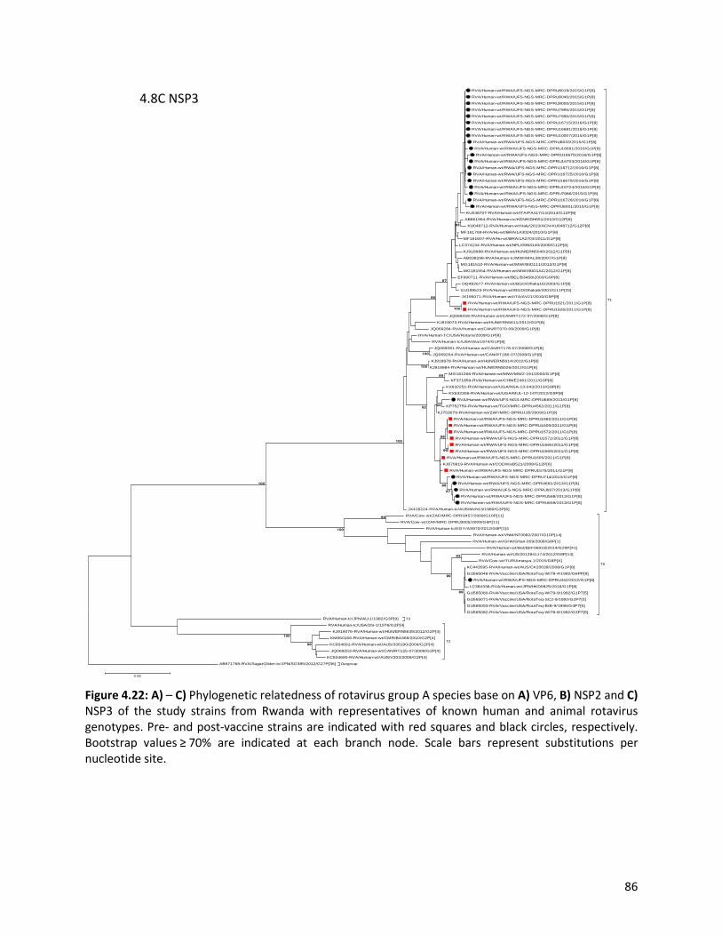
86
Figure 4.22: A) – C) Phylogenetic relatedness of rotavirus group A species base on A) VP6, B) NSP2 and C) NSP3 of the study strains from Rwanda with representatives of known human and animal rotavirus genotypes. Pre- and post-vaccine strains are indicated with red squares and black circles, respectively. Bootstrap values ≥ 70% are indicated at each branch node. Scale bars represent substitutions per nucleotide site.
RVA/Human-wt/RWA/UFS-NGS-MRC-DPRU8019/2015/G1P[8]
RVA/Human-wt/RWA/UFS-NGS-MRC-DPRU8040/2015/G1P[8]
RVA/Human-wt/RWA/UFS-NGS-MRC-DPRU8000/2015/G1P[8]
RVA/Human-wt/RWA/UFS-NGS-MRC-DPRU7995/2015/G1P[8]
RVA/Human-wt/RWA/UFS-NGS-MRC-DPRU7990/2015/G1P[8]
RVA/Human-wt/RWA/UFS-NGS-MRC-DPRU16715/2016/G1P[8]
RVA/Human-wt/RWA/UFS-NGS-MRC-DPRU16681/2016/G1P[8]
RVA/Human-wt/RWA/UFS-NGS-MRC-DPRU10007/2015/G1P[8]
RVA/Human-wt/RWA/UFS-NGS-MRC-DPRU8033/2015/G1P[8]
RVA/Human-wt/RWA/UFS-NGS-MRC-DPRU16691/2016/G1P[8]
RVA/Human-wt/RWA/UFS-NGS-MRC-DPRU16675/2016/G1P[8]
RVA/Human-wt/RWA/UFS-NGS-MRC-DPRU16703/2016/G1P[8]
RVA/Human-wt/RWA/UFS-NGS-MRC-DPRU16712/2016/G1P[8]
RVA/Human-wt/RWA/UFS-NGS-MRC-DPRU16725/2016/G1P[8]
RVA/Human-wt/RWA/UFS-NGS-MRC-DPRU16679/2016/G1P[8]
RVA/Human-wt/RWA/UFS-NGS-MRC-DPRU16724/2016/G1P[8]
RVA/Human-wt/RWA/UFS-NGS-MRC-DPRU7986/2015/G1P[8]
RVA/Human-wt/RWA/UFS-NGS-MRC-DPRU16728/2016/G1P[8]
RVA/Human-wt/RWA/UFS-NGS-MRC-DPRU8001/2015/G1P[8]
KU048707-RVA/Human-wt/ITA/PA417/14/2014/G12P[8]
AB861964-RVA/Human-tc/KEN/KDH651/2010/G12P[8]
KU048712-RVA/Human-wt/Italy/2013/ACN-KU048712/G12P[8]
MF161769-RVA/Hu-wt/BRA/1A3024/2010/G1P[8]
MF161607-RVA/Hu-wt/BRA/1A2703/2011/G1P[8]
LC374134-RVA/Human-wt/NPL/09N3140/2009/G12P[6]
KJ918988-RVA/Human-wt/HUN/ERN5040/2012/G1P[8]
AB938298-RVA/Human-tc/MWI/MAL38/2007/G1P[8]
MG181510-RVA/Human-wt/MWI/BID111/2012/G1P[8]
MG181554-RVA/Human-wt/MWI/BID1AC/2012/G1P[8]
EF990711-RVA/Human-wt/BEL/B3458/2003/G9P[8]
DQ492677-RVA/Human-wt/BGD/Dhaka16/2003/G1P[8]
GU199523-RVA/Human-wt/BGD/Dhaka6/2001/G11P[25]
JX195071-RVA/Human-wt/ITA/AV21/2010/G9P[8]
RVA/Human-wt/RWA/UFS-NGS-MRC-DPRU1621/2011/G1P[8]
RVA/Human-wt/RWA/UFS-NGS-MRC-DPRU1626/2011/G1P[8]
JQ069259-RVA/Human-wt/CAN/RT172-07/2008/G1P[8]
KJ919073-RVA/Human-wt/HUN/ERN5611/2012/G1P[8]
JQ069284-RVA/Human-wt/CAN/RT070-09/2009/G1P[8]
RVA/Human-TC/USA/Rotarix/2009/G1P[8]
RVA/Human-tc/USA/Wa/1974/G1P[8]
JQ069261-RVA/Human-wt/CAN/RT178-07/2008/G1P[8]
JQ069264-RVA/Human-wt/CAN/RT186-07/2008/G1P[8]
KJ918978-RVA/Human-wt/HUN/ERN5014/2012/G1P[8]
KJ918984-RVA/Human-wt/HUN/ERN5026/2012/G1P[8]
MG181268-RVA/Human-wt/MWI/MW2-191/2000/G1P[8]
KF371859-RVA/Human-wt/CHN/E2461/2011/G3P[8]
KX632251-RVA/Human-wt/UGA/NSA-13-043/2013/G9P[8]
KX632306-RVA/Human-wt/UGA/MUL-12-147/2012/G9P[8]
RVA/Human-wt/RWA/UFS-NGS-MRC-DPRU669/2013/G1P[8]
KP752750-RVA/Human-wt/TGO/MRC-DPRU4562/2011/G1P[8]
KJ753079-RVA/Human-wt/ZAF/MRC-DPRU135/2009/G1P[8]
RVA/Human-wt/RWA/UFS-NGS-MRC-DPRU1582/2011/G1P[8]
RVA/Human-wt/RWA/UFS-NGS-MRC-DPRU1609/2011/G1P[8]
RVA/Human-wt/RWA/UFS-NGS-MRC-DPRU1572/2011/G1P[8]
RVA/Human-wt/RWA/UFS-NGS-MRC-DPRU1571/2011/G1P[8]
RVA/Human-wt/RWA/UFS-NGS-MRC-DPRU1565/2011/G1P[8]
RVA/Human-wt/RWA/UFS-NGS-MRC-DPRU1569/2011/G1P[8]
RVA/Human-wt/RWA/UFS-NGS-MRC-DPRU1605/2011/G1P[8]
KJ870919-RVA/Human-wt/COD/KisB521/2008/G12P[6]
RVA/Human-wt/RWA/UFS-NGS-MRC-DPRU1575/2011/G1P[8]
RVA/Human-wt/RWA/UFS-NGS-MRC-DPRU714/2013/G1P[8]
RVA/Human-wt/RWA/UFS-NGS-MRC-DPRU691/2013/G1P[8]
RVA/Human-wt/RWA/UFS-NGS-MRC-DPRU697/2013/G1P[8]
RVA/Human-wt/RWA/UFS-NGS-MRC-DPRU568/2013/G1P[8]
RVA/Human-wt/RWA/UFS-NGS-MRC-DPRU659/2013/G1P[8]
JX416224-RVA/Human-tc/AUS/McN13/1980/G3P[6]
T1
RVA/Cow-wt/ZAF/MRC-DPRU457/2009/G10P[11]
RVA/Cow-wt/ZAF/MRC-DPRU3005/2009/G6P[11]
RVA/Human-tc/EGY/AS970/2012/G8P[1]4
RVA/Human-wt/VNM/NT0082/2007/G10P[14]
RVA/Human-wt/GHA/Ghan-059/2008/G8P[1]
RVA/Human-wt/Bel/BEF06018/2014/G29P[41]
RVA/Human-wt/US/2012841174/2012/G8P[14]
RVA/Cow-wt/TUR/Amasya-1/2015/G8P[X]
KC443595-RVA/Human-wt/AUS/CK20039/2008/G1P[8]
GU565049-RVA/Vaccine/USA/RotaTeq-WI79-4/1992/G6PP[8]
RVA/Human-wt/RWA/UFS-NGS-MRC-DPRU442/2012/G1P[8]
LC384338-RVA/Human-wt/JPN/HKD0825/2016/G1P[8]
GU565060-RVA/Vaccine/USA/RotaTeq-WI79-9/1992/G1P7[5]
GU565071-RVA/Vaccine/USA/RotaTeq-SC2-9/1992/G2P7[5]
GU565093-RVA/Vaccine/USA/RotaTeq-BrB-9/1996/G4P7[5]
GU565082-RVA/Vaccine/USA/RotaTeq-WI78-8/1992/G3P7[5]
T6
T3 RVA/Human-tc/JPN/AU-1/1982/G3P[9]
RVA/Human-tc/USA/DS-1/1976/G2P[4]
KJ919076-RVA/Human-wt/HUN/ERN5635/2012/G2P[4]
KM660166-RVA/Human-wt/CMR/BA368/2010/G2P[4]
KC834691-RVA/Human-wt/AUS/336190/2004/G2P[4]
JQ069253-RVA/Human-wt/CAN/RT125-07/2008/G2P[4]
KC834690-RVA/Human-wt/AUS/V203/2009/G2P[4]
T2
Outgroup AB971768-RVA/SugarGlider-tc/JPN/SG385/2012/G27P[36]
96
100
99
93
99
99
100
93
100
97
99
100
100
96
98
97
96
93
99
92
100
100
0.05
4.8C NSP3
87
4.4. Discussion
The introduction of the rotavirus vaccines has resulted in the reduction of the global burden of RVA
associated diarrhoea diseases and hospitalization (Troeger et al., 2018b). Hence, continuous RVA
surveillance in different settings remains vital to document and characterize post-vaccine licensure era
RVA strains; possible vaccine-derived strains and inter-genogroup reassortant strains as part effort to
document the impact of rotavirus vaccination. Therefore, in the present study, the whole gene analyses
of 35 Rwandan G1P[8] and a single RotaTeq® vdG1P[8] strains collected from children less than five years
of age during pre- and post-vaccination with RotaTeq® showed that all 35 of the strains possessed a typical
Wa-like genotype constellation, while the single RotaTeq® vdG1P[8] strain RVA/Human-wt/RWA/UFS-
NGS-MRC-DPRU442/2012/G1P[8], was apparently produced by a reassortment event between vaccine
strains, RVA/Vaccine/USA/RotaTeq-WI79-4/1992/G6P1A[8] and RVA/Vaccine/USA/Rotateq-W179-
9/1992/G1P7[5]. However, the VP4 and VP6 genes may have originated from other circulating vaccine
strains as their homologies with vaccine strain RVA/Vaccine/USA/RotaTeq-WI79-4/1992/G6P1A[8] and
RVA/Vaccine/USA/Rotateq-W179-9/1992/G1P7[5] were lower than other genes. Whole gene analyses
confirmed that the VP7 and VP4 genes were vaccine-derived and the RotaTeq® vaccine WC3 bovine
genetic backbone was confirmed to be G1-P[8]-I2-R2-C2-M2-A3-N2-T6-E2-H3. Though reassortment
events between RotaTeq® vaccine strains which generated vaccine-derived G1P[8] reassortant have been
reported previously in Australia, South Korea, Finland, and the USA, this is the first reported RotaTeq®
vdG1P[8] from the African continent (Donato et al., 2012; Markkula et al., 2015; Payne et al., 2010; Than
et al., 2015). Previous studies have shown that reassortment may occur more frequently between the VP7
gene of RotaTeq vaccine strain RVA/Vaccine/USA/Rotateq-W179-9/1992/G1P7[5] and VP4 gene of
RotaTeq vaccine strain RVA/Vaccine/USA/RotaTeq-WI79-4/1992/G6P1A[8] than between other
genotypes (Donato et al., 2012; Than et al., 2015). The likelihood that this vdG1P[8] may possess increased
virulence and cause AGE exists (Donato et al., 2012). The combination of the two human outer capsid
proteins could potentially enhance cell binding and entry into enterocytes, hence an increase in virulence
of this vdG1P[8] strain (Donato et al., 2012).
Although inter-genogroup reassortant G1P[8] strains have been reported globally, generally the G1P[8]
strains are usually associated with the Wa-like backbone (Fujii et al., 2014; Jere et al., 2018; Kuzuya et al.,
2014; Luchs et al., 2019; Matthijnssens et al., 2011). Phylogenetically, the 11 gene segments of the
Rwanda pre- and post-vaccine introduction G1P[8] strains, with the exception of the RotaTeq® vdG1P[8],

88
were highly similar and clustered together with cognate gene segments of Wa-like strains (Fig. 1-8) and
further segregated into either typical sub-lineages consisted of only pre-vaccine or post-vaccine
introduction strains or mixtures of pre- and post-vaccine introduction strains. The segregation of pre- and
post-vaccine introduction G1P[8] strains into different lineages and/or sub-lineages have been previously
described in South Africa (Magagula et al., 2015). In each gene, we observed the segregation of strains in
terms of vaccination status and year of detection. With the exception of the NSP1 gene which showed
2013, 2015 and 2016 post-vaccine strains in three different sub-lineages, the post-vaccine introduction
G1P[8] strains detected during the 2015-2016 RVA seasons always clustered together within the post-
vaccine introduction sub-lineages. The VP1-VP3, NSP1, NSP4, and NSP5 gene sequences of the pre-vaccine
introduction strains always clustered together. Analyses of the VP7, VP4, VP6, NSP2, and NSP3 gene
sequences, however, showed mixed sub-lineages consisting of both the pre-vaccine strains detected in
the 2011 RVA season and post-vaccine introduction strains detected in 2013 (VP7, VP4, and NSP3), and
2015-2016 (VP6 and NSP2) seasons. In addition, within the typical pre- and post-vaccine introduction sub-
lineages of all 11 gene segments, the sequence similarity was extremely high ≥ 99.9% and between sub-
lineages the nt (aa) similarities were moderately high in the range of 96-98 % (98.1-99.2%). The high
genetic relationship between gene sequences of the Rwandan pre- and post-vaccine introduction G1P[8]
strains is an indication that the same strains that were circulating before vaccine introduction in Rwanda,
are the same strains or their progeny that are causing RVA-associated diarrheal diseases and
hospitalization after vaccination.
Globally, at least seven lineages have been previously described for G1 strains collected from different
geographical locations (Arista et al., 2006; Bányai et al., 2009; Bucardo et al., 2012; Le et al., 2010). The
emergence of distinct lineages or sub-lineages is attributed to the diverse evolutionary mechanisms such
as mutation, recombination, and reassortments (Kirkwood, 2010). Based on the VP7 gene, the 35
Rwandan G1 strains which consisted of both pre- and post-vaccine introduction strains, clustered in
lineage I and were comparable to the observation reported previously (da Silva et al., 2015; Magagula et
al., 2015; Santos et al., 2019). On the other hand, the VP4 plays a role as an antibody-neutralization protein
(Offit et al., 1986; Ward et al., 1993). Diversity of the P[8] genotypes has been demonstrated through four
described distinct lineages (Arista et al., 2006; Cho et al., 2013). The clustering of the 35 P[8] strains in
lineage III in this study is consistent with previous observations (Arora and Chitambar, 2011; da Silva et
al., 2015; Magagula et al., 2015; Santos et al., 2019). However, despite the clustering of all VP7 and VP4
genes of the study strains in Lineage I (Figure 4.2) and lineage III (Figure 4.5), respectively, antigenic
89
variation seen at the neutralization epitope sites of the VP7 and VP4 proteins of the study strains have
been previously described in Belgium and Australia (Diwakarla and Palombo, 1999; Zeller et al., 2012).
Amino acid substitutions with effect on polarity changes were detected on Rwandan VP7 neutralization
epitopes at positions T91A/V, S190D and M217. Amino acid substitutions on positions 94, 96, 147, 148,
190, 208, 211, 213 and 217 are critical and have been reported to alter rotavirus antigenicity (Ahmed et
al., 2007; Trinh et al., 2007). Therefore, the detected substitutions may be involved in antigenic drift in
Rwandan G1 strains. The resultant alteration in charge and polarity observed at position T91A/V on the
VP7 neutralization epitope may play a role in escaping host immunity as it was distinctively observed post-
vaccine introduction (Zeller et al., 2015, 2012). Furthermore, the change in polarity at position S190D and
M217 could be due to natural fluctuation of RVA as they were observed during the pre-vaccine era and
consistently before and after vaccine introduction, respectively (Jere et al., 2018). The presence of amino
acid differences in CTL epitope positions Y41F/S, V42M, and A46V in Rwandan G1 strains possibly can
result in greater host-immunity escape effects (Wei et al., 2009). In the intestine, trypsin-like proteases
cleave the VP4 spike protein into two structural domains (VP8* and VP5*) (Estes and Greenberg, 2013).
Four surface-exposed antigenic epitopes (8-1 to 8-4) have been described in the VP8* region, while five
antigenic epitopes (5-1 to 5-5) in the VP5* region have been documented (Zeller et al., 2012). The amino
acid changes identified at positions N113D, S131R and N135D in this study, resulted in polarity changes
and played a role in escaping host-immunity (Dormitzer et al., 2004; Monnier et al., 2006).
Overall, full genomic analyses of 35 Rwandan G1P[8] strains revealed the predominance of G1-I and P[8]-
III, which is consistent with what has been reported previously in Belgium (Zeller et al., 2012). The global
prevalence of Wa-like human strains is hypothesized to be due to the ease of disseminating this genetic
backbone in the human host (Ghosh and Kobayashi, 2011; Rahman et al., 2010). Although this study was
insightful in reporting the whole gene composition of the circulating G1P[8] strains in Rwanda, it was
limited to a few years (just one year) pre- and (five years) post-vaccination samples were included.
Another limitation was that, for the sample where RotaTeq® vaccine shedding was observed, other
plausible alternate aetiologies such as adenovirus and/or norovirus that might have led to the diarrhoea
symptoms and possible hospitalization were not evaluated. Cell culture was not used to confirm the
reassortment events as the project was mainly focused on in-silico work.

90
In conclusion, this is the first study to describe full genomic analyses of G1[P8] RVA strains in Rwanda. The
detection of RotaTeq® vdG1P[8] strain from RVA positive child hospitalized with AGE symptoms was
unexpected. Rotavirus group A strain surveillance at the whole gene level will enhance further assessment
of vaccine impact on circulating RVA strains and the frequency along with the epidemiological fitness of
reassortant strains.
4.5. Methods
4.5.1. Sample collection
RVA positive faecal samples (n=158) were obtained from children less than five years old who were
hospitalized with AGE in Rwanda as part of the ongoing WHO/AFRO RVA surveillance program. The
samples were conventionally genotyped into G and P types at the Diarrhoeal Pathogens Research Unit
(DPRU), a WHO Rotavirus Reference Laboratory in South Africa (WHO RRL-SA). The samples were
collected during the pre- (2011-2012) and post- (2012-2016) RVA vaccination periods with RotaTeq®
vaccine in Rwanda. All the samples were stored at the DPRU at -20°C and G1P[8] strains were selected for
whole-genome sequencing at the UFS-NGS Unit.
4.5.2. Double-stranded RNA extraction and purification
The extraction of dsRNA was performed at the DPRU and involved a method previously described by
Nyaga et al. (2018). Briefly, a 100 mg stool sample was added to 200 µL freshly made phosphate-buffered
saline (PBS, Sigma-Aldrich, St. Louis, USA). A 900 µL volume of TRI-reagent (Molecular Research Center,
Inc, Cincinnati, OH, USA) was added to the suspended stool sample to homogenize as well as lyse the cells
and cell components. A volume of 300 µL chloroform (Sigma-Aldrich, St. Louis, USA) was then added and
centrifugation (16 000 x g for 20 minutes at 4°C) was done in a temperature-controlled microcentrifuge
(Eppendorf centrifuge 5427R, Hamburg, Germany). The supernatant containing the total RNA was
precipitated by the addition of 700 µL isopropanol (Sigma-Aldrich, St. Louis, USA) and by centrifugation at
16000 x g for 30 minutes at room temperature. The resulting pellet was re-dissolved by the addition of 90
µL of double-distilled water ,ddH20, (Merck KGaA, Germany). A concentration of 8M LiCl2 (Sigma-Aldrich,
St. Louis, USA) was used to remove ssRNA through precipitation for 16 hours after which further
centrifugation was done for 30 minutes at 16000 x g. The extracted dsRNA was purified by utilizing the

91
MinElute gel extraction kit (Qiagen, Hilden, Germany) and the integrity and enrichment of the dsRNA was
verified via agarose gel electrophoresis.
4.5.3. cDNA synthesis
Complementary DNA was generated from the extracted viral RNA utilizing Maxima H Minus Double-
Stranded cDNA Synthesis Kit (Thermo Fischer Scientific, Waltham, MA, USA) with minor modifications.
Briefly, the extracted total RNA was denatured at 95oC for five minutes and then 1 µL random hexamer
primers (Thermo Fischer Scientific, Waltham, MA, USA) were added. The hexamer primers (Thermo
Fischer Scientific, Waltham, MA, USA) were allowed to anneal at 65oC for five minutes. A volume of 5 µL
of first strand reaction mix (Thermo Fischer Scientific, Waltham, MA, USA) and 1 µL of first strand enzyme
mix (Thermo Fischer Scientific, Waltham, MA, USA) was then added. The solution was then incubated at
25oC, 50oC and 85oC for 10, 120 and five minutes, respectively. The tubes were removed from the
thermocycler (Merck, Darmstadt, Germany) and second-strand synthesis was performed by adding 55 µL
nuclease-free water, followed by the addition of 20 µL of 5x second strand reaction mix (Thermo Fischer
Scientific, Waltham, MA, USA) and 5 µL of second strand enzyme mix (Thermo Fischer Scientific, Waltham,
MA, USA). Subsequently, the solution was incubated at 16oC for one hour and the reaction was stopped
with 6 µL 0.5M EDTA (Thermo Fischer Scientific, Waltham, MA, USA). Residual RNA was removed with 10
µL RNase I (Thermo Fischer Scientific, Waltham, MA, USA) and the synthesized cDNA was incubated at
room temperature for five minutes.
4.5.4. DNA library preparations and whole-genome sequencing
DNA libraries were prepared using the Nextera XT DNA Library Preparation Kit (Illumina, Inc., California,
USA) following the manufacturer’s instructions. Briefly, DNA library preparation entailed tagmentation of
the generated DNA, indexing using unique barcodes and amplification of tagmented DNA and clean-up of
the amplified DNA. The library quality and size were assessed using an Agilent 2100 BioAnalyzer (Agilent
Technologies, California, United States) according to the manufacturer’s specified protocol. The Illumina
custom protocol was utilized to normalize the libraries to 4 nM. All the normalized libraries were then
pooled together into a single tube by combining 5 µL of each barcoded library. The pooled libraries were
subjected to chemical denaturation using 0.2 N NaOH (Sigma Aldrich, Missouri, United States). After
denaturation, 990 µL of pre-chilled HT1 buffer (Illumina, Inc., California, USA) was added to the 10 µL of
the 4 nM denatured DNA library to dilute to 20 pM. A further dilution of the denatured library was

92
performed to get the desired final concentration of 8 pM. A PhiX control (Illumina, Inc., California, USA)
spike-in of 20% was used. Whole-genome sequencing was performed for 600 cycles (301 x 2 paired-end)
on a MiSeq benchtop sequencer (Illumina, Inc., California, USA) using Illumina MiSeq v3 reagent kit
(Illumina, Inc., California, USA) at the UFS-NGS Unit, Bloemfontein, South Africa.
4.5.5. Genome assembly
Illumina sequence reads were analysed using Geneious software v11 (Kearse et al., 2012;
https:www.geneious.com) and CLC Genomics Workbench v11 (CLC Bio, Qiagen, Hilden, Germany) which
entailed genome assembly and mapping the reads to reference-based sequences to obtain the full-length
genomes.
4.5.6. Identification of genotype constellations
Genotyping was performed by utilizing RotaC v 2.0 (Maes et al., 2009), an automated online genotyping
tool for group A RV strains. Genome constellations were generated by assigning genotypes to each
genome segment.
4.5.7. GenBank accession numbers
The sequences were deposited into GenBank under the accession number MN632673-MN633067.
4.5.8. Phylogenetic, sequence analyses and protein modelling
For each gene segment, the ORF were aligned and subjected to sequence comparisons as described
previously (Esona et al., 2018, 2017; Ward et al., 2016). Briefly, multiple alignments were made using the
MUSCLE algorithm implemented in MEGA 6 software (Tamura et al., 2013;
http://www.megasoftware.net/). Once aligned, the DNA Model Test program implemented in MEGA
version 6 was used to identify the optimal evolutionary models that best fit the sequence datasets. Using
the Corrected Akaike Information Criterion (AICc), the model GTR-G-I was found to best fit the sequence
data for each gene segment. With this model, maximum likelihood trees were constructed using MEGA 6
with 1000 bootstrap replicates to estimate branch support. The nucleotide and deduced amino acid
sequence identities among strains were calculated for each gene using distance matrices prepared using
93
the p-distance algorithm in MEGA 6 software (Tamura et al., 2013). Protein modelling was performed on
amino acid sequences for each strain and reference strain using the Swiss-Model protein structure
homology-modelling server (Waterhouse et al., 2018; https://swissmodel.expasy.org). The structures
were modified using UCSF Chimera (Pettersen et al., 2004).

94
CHAPTER FIVE: WHOLE-GENOME ANALYSES IDENTIFIES MULTIPLE
REASSORTANT ROTAVIRUS STRAINS IN RWANDA POST-VACCINE
INTRODUCTION

95
This chapter is under internal review for submission to Frontiers in Microbiology

96
5.1. Abstract
Children in middle and low-income countries, like Rwanda, have a higher prevalence of rotavirus disease
relative to those in developed countries. Following the implementation of the RotaTeq® vaccine in Rwanda
in 2012, a drastic decline in rotavirus-induced diarrhoea was documented during the post-vaccine
introduction impact evaluation. Evolutionary mechanisms leading to multiple reassortant rotavirus strains
influence the diversity and evolutionary dynamics of novel rotaviruses over time. Comprehensive
rotavirus whole-genome analysis was conducted on 158 RVA specimens collected pre- and post-vaccine
introduction in children less than five years in Rwanda. Of these RVA positive specimens, five strains with
the genotype constellations G4P[4]-I1-R2-C2-M2-A2-N2-T1-E1-H2 (n=1), G9P[4]-I1-R2-C2-M2-A1-N1-T1-
E1-H1 (n=1), G12P[8]-I1-R2-C2-M1-A1-N2-T1-E2-H3 (n=2) and G12P[8]-I1-R1-C1-M1-A2-N2-T2-E1-H1
(n=1), with single, double and triple gene reassortant rotavirus strains were identified. Phylogenetic
analysis revealed a close relationship between the Rwandan strains and cognate human RVA strains
circulating on a global scale as well as the RotaTeq® vaccine strains in the VP1, VP2, NSP2, NSP4 and NSP5
gene segments. The VP2, VP3, and NSP1 genes of strain RVA/Human-wt/RWA/UFS-NGS:MRC-
DPRU566/2013/G9P[4] and RVA/Human-wt/RWA/UFS-NGS:MRC-DPRU6235/2014/G4P[4] formed
distinct clusters from similar genes of strains circulating globally. Pairwise analyses revealed considerable
differences in the VP7 and VP4 antigenic regions of the representative Rwandan study strains and the
RotaTeq® vaccine strain. Although the impact of such amino acid changes on the effectiveness remains
unclear, this analysis underlines the potential of rotavirus whole-genome analysis program by enhancing
knowledge on inter-genogroup reassortant strains circulating in Rwanda post-RotaTeq® vaccine
introduction.
Keywords: rotavirus, Rwanda, reassortment, whole-genome based surveillance, epitopes, RotaTeq®

97
5.2. Introduction
Group A rotavirus infections are commonly associated with severe diarrhoea in young children less than
five years of age, accounting for over 128 500 deaths globally in 2016 (Troeger et al., 2018b). Children
living in third-world countries have been documented to experience a higher rotavirus disease burden
attributed to lack of adequate sanitation (Sindhu et al., 2017). Prior to RotaTeq® (RV5, Merck & Co. Inc.,
USA) vaccine introduction in Rwanda, approximately 3500 RVA related mortality was reported in children
annually, accounting for 8.8% of all childhood mortality in Rwanda (GAVI, 2012). RotaTeq® was introduced
into the Rwandan market in May 2012 with a vaccine coverage of 99% within a year after introduction in
2013 (Gatera et al., 2016; WHO, 2020). During the first three years post RotaTeq® introduction, a decrease
of total diarrheal hospitalization (25-44%) among children less than five years in the Eastern Province of
Rwanda was documented (Sibomana et al., 2018).
Structurally, rotaviruses possess an icosahedral symmetry and consist of an 11-segmented dsRNA genome
which encodes for both structural and non-structural proteins (Estes and Greenberg, 2013; Desselberger,
2014). The VP7 and VP4 proteins are generally used to distinguish RVA strains into G- and P-genotypes,
respectively, based on their antigenic properties. Furthermore, the advancements of NGS techniques have
enabled the characterization of all the 11-segments which has turned out to provide essential information
that can be used to determine the evolutionary dynamics of the virus (Matthijnssens et al., 2008;
Matthijnssens et al., 2011; Nyaga et al., 2020).Three human RVA genogroups, the Wa-like and DS-1-like
(major genogroups) and the AU-1 like (minor genogroup) have been identified based on the rotavirus
whole-genome (Ide et al., 2015; Komoto et al., 2016; Matthijnssens et al., 2008; Matthijnssens and Van
Ranst, 2012). The majority of strains that possess the Wa-like (G1-P[8]-I1-R1-C1-M1-A1-N1-T1-E1-H1)
constellation are from porcine origin, while DS-1-like (G2-P[4]-I2-R2-C2-M2-A2-N2-T2-E2-H2) and AU-1-
like (G3-P[9]-I3-R3-C3-M3-A3-N3-T3-E3-H3) strains are from bovine and canine/feline origin, respectively
(Matthijnssens et al., 2008).
Over 107 countries have implemented RVA vaccines as of April 2020 to curb RVA infections in young
children. Based on the safety and efficacy studies, four RVA vaccines, Rotarix® (RV1, GlaxoSmithKline
Biologicals, Belgium), RotaTeq® (RV5, Merck & Co. Inc., USA), Rotavac® (nHRV, Bharat Biotech
International Limited India) and Rotasil® (BRV-PV, Serum Institute of India, India), have been prequalified
by the WHO for global use (ROTA Council, 2016; WHO, 2018). RotaTeq® is a live-attenuated pentavalent

98
vaccine-derived from bovine and human reassortant parent strains (G1 [W179-9], G2 [SC2-9], G3 [W178-
8], G4 [BrB-9] or P[8] [W179-4] of human strains and WC3, G6P[5] of the bovine strain) (Matthijnssens et
al., 2010). In Africa, the RotaTeq® vaccine was introduced in only five countries; Burkina Faso (2013), Libya
(2013), Morocco (2010), Rwanda (2012), and the Gambia (2013) (PATH, 2016). Moreover, Rwanda was
the first low-income country to introduce the RotaTeq® vaccine. Different African countries have reported
the detection of diverse GXP[X] combinations over the years, with G1P[8], G2P[4], G3P[8], G9P[8], G1P[6],
G2P[6], G3P[6], G8P[4] and G9P[6] being the most prevalent combinations in Africa (Mwenda et al., 2010;
Seheri et al., 2014; Steele et al., 2003a; Steele et al., 2003b; Todd et al., 2010). In Rwanda, G1P[8] was the
predominant strain circulating in the pre-vaccine era in 2011. However, there was an immediate shift to
G8P[4] (53% in 2013), G12P[8] (39.4% in 2014) and G4P[8] (36.6% in 2014) and were subsequently
replaced by the re-emergence of G1P[8] (51.6%) in 2015 (Seheri et al., 2018).
Group A rotaviruses are constantly evolving through numerous evolutionary mechanisms influenced by
the segmented nature and error-prone RdRp characteristic of the RNA virus (Bányai et al., 2011; Seheri et
al., 2018; Taniguchi and Urasawa, 1995). Intra-genogroup and inter-genogroup reassortment events are
generally influenced by co-infection with multiple RVA strains (Cowley et al., 2016; Dennis et al., 2014;
Estes and Greenberg, 2013; Heylen et al., 2014; Nakagomi et al., 2013). The RVA strains derived from
inter-genogroup reassortment events both in the pre- and post-vaccine period have been documented
globally (Cowley et al., 2016; Hoa-Tran et al., 2016; Komoto et al., 2017; Mwangi et al., 2020). However,
strains having pure Wa-like or pure DS-1-like genotype constellations are transmitted frequently across
the human population as compared to these reassortant strains (Heiman et al., 2008; McDonald et al.,
2009). The aim of this study is to report on five inter-genogroup reassortant strains observed post-vaccine
introduction in Rwanda between 2013 and 2015 using whole-genome sequencing approach.

99
5.3. Results
5.3.1. Genome genotypes The genetic relatedness and variability of Rwandan strains in comparison to selected reference RVA strains
circulating globally, was determined by sequencing 158 Rwandan samples from the pre- (2011) and post-
(2012 and 2016) vaccination period. From that dataset pool, only five samples from the pool possessed
reassortant constellations (Table 5.1). The length size of contigs and the number of reads post-assembly
are given (Table 5.1). All the five reassortant strains were observed post-vaccine introduction. Strain
RVA/Human-wt/RWA/UFS-NGS:MRC-DPRU6235/2014/G4P[4] and RVA/Human-wt/RWA/UFS-NGS:MRC-
DPRU566/2013/G9P[4] exhibited both the Wa-like and DS-1-like genotype constellation: G4P[4]-I1-R2-C2-
M2-A2-N2-T1-E1-H2 and G9P[4]-I1-R2-C2-M2-A1-N1-T1-E1-H1, respectively. Two G12P[8] strains
(RVA/Human-wt/RWA/UFS-NGS:MRC-DPRU8020/2015/G12P[8] and RVA/Human-wt/RWA/UFS-
NGS:MRC-DPRU9995/2015/G12P[8]) possessed an identical genome constellation across the backbone
(G12P[8]-I1-R2-C2-M1-A1-N2-T1-E2-H3), comprised of typical Wa-like, DS-1-like and AU-1-like genotype
constellation. On the other hand, strain RVA/Human-wt/RWA/UFS-NGS:MRC-DPRU6212/2014/G12P[8]
exhibited both the Wa-like and DS-1-like genotype constellation G12P[8]-I1-R1-C1-M1-A2-N2-T2-E1-H1.

100
Table 5.6: Full genotype constellations of five Rwandan strains detected post-vaccine introduction (2013-2015) along with the contig lengths and number of reads mapped to each contig.
Wa-like genogroups is represented by the green colour, DS-1like is represented by the red colour and AU-1-like is represented by the yellow colour.
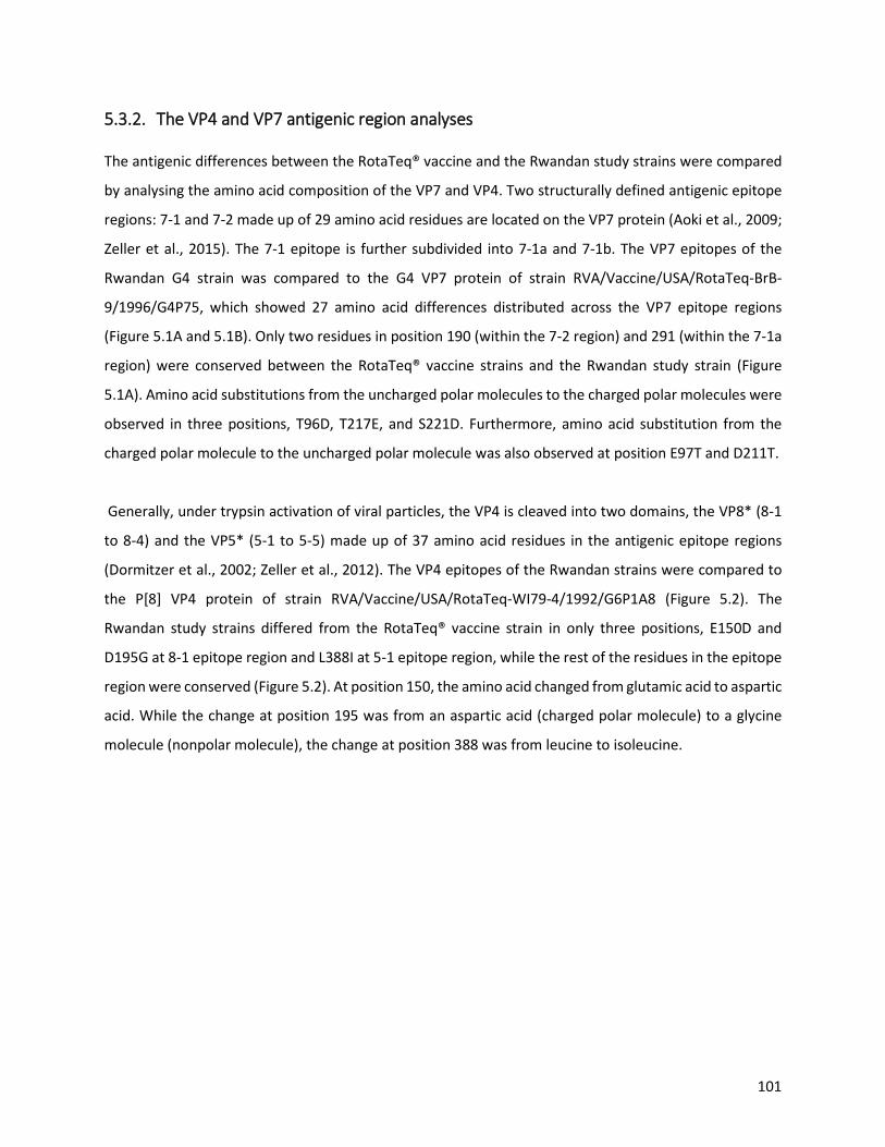
101
5.3.2. The VP4 and VP7 antigenic region analyses The antigenic differences between the RotaTeq® vaccine and the Rwandan study strains were compared
by analysing the amino acid composition of the VP7 and VP4. Two structurally defined antigenic epitope
regions: 7-1 and 7-2 made up of 29 amino acid residues are located on the VP7 protein (Aoki et al., 2009;
Zeller et al., 2015). The 7-1 epitope is further subdivided into 7-1a and 7-1b. The VP7 epitopes of the
Rwandan G4 strain was compared to the G4 VP7 protein of strain RVA/Vaccine/USA/RotaTeq-BrB-
9/1996/G4P75, which showed 27 amino acid differences distributed across the VP7 epitope regions
(Figure 5.1A and 5.1B). Only two residues in position 190 (within the 7-2 region) and 291 (within the 7-1a
region) were conserved between the RotaTeq® vaccine strains and the Rwandan study strain (Figure
5.1A). Amino acid substitutions from the uncharged polar molecules to the charged polar molecules were
observed in three positions, T96D, T217E, and S221D. Furthermore, amino acid substitution from the
charged polar molecule to the uncharged polar molecule was also observed at position E97T and D211T.
Generally, under trypsin activation of viral particles, the VP4 is cleaved into two domains, the VP8* (8-1
to 8-4) and the VP5* (5-1 to 5-5) made up of 37 amino acid residues in the antigenic epitope regions
(Dormitzer et al., 2002; Zeller et al., 2012). The VP4 epitopes of the Rwandan strains were compared to
the P[8] VP4 protein of strain RVA/Vaccine/USA/RotaTeq-WI79-4/1992/G6P1A8 (Figure 5.2). The
Rwandan study strains differed from the RotaTeq® vaccine strain in only three positions, E150D and
D195G at 8-1 epitope region and L388I at 5-1 epitope region, while the rest of the residues in the epitope
region were conserved (Figure 5.2). At position 150, the amino acid changed from glutamic acid to aspartic
acid. While the change at position 195 was from an aspartic acid (charged polar molecule) to a glycine
molecule (nonpolar molecule), the change at position 388 was from leucine to isoleucine.

102
Figure 5.1: A) The alignment of the G4 VP7 component of the RotaTeq® vaccine strain and G4 Rwandan study strain based on the three surface exposed epitope regions (7-1a, 7-1b, and 7-2). The asterisk represents the amino acid position of residues associated with escape neutralization with monoclonal antibodies. B) Surface representation of the VP7 protein. The structure has the root mean square deviation (RMSD) of 0.044 Å. The RotaTeq® vaccine is represented with the red colour, while the Rwandan strain is indicated with the blue colour. The green colour represents the amino acid change observed on the Rwandan study strain as compared to the RotaTeq® vaccine strain (yellow) (Created with PyMoL v2, 15 July 2020)

103
Figure 5.2: The alignment of the P[8] VP4 component of the Rotateq vaccine strain and P[8] Rwandan study strain based on the two VP4 domains, the VP8* (8-1 to 8-4) and VP5 (5-1 to 5-5). The asterisk represents the amino acid position of residues associated with escape neutralization with monoclonal antibodies. 5.3.3. Phylogenetic analysis of the VP7 gene of G4, G9 and G12 Phylogenetic trees were constructed for each of the 11 segments of the five Rwandan RVA reassortant strains in comparison with global reference
strains from GenBank (Figures 5.3-5.6). The three Rwandan G12P[8] strains clustered in G12 lineage III, and shared 87.6-99.3% nt similarity with
other lineage III G12 strains (Figure 5.3). The G12 strains clustered together and shared 99.8-100% nt similarity amongst themselves. On the other
hand, the Rwandan G9 strain clustered in G9 lineage III distantly from globally circulating G9 strains (81.7-92.1% nt similarity). The G4 strain
clustered in G4 lineage I with circulating G4 global strains and exhibited 86% nt similarity with a RotaTeq® vaccine strain.

104
Figure 5.3: Phylogenetic tree of RVA strains based on the full length of the VP7 (G4, G9 and G12) gene displaying the relatedness of the study strains (◆) and reference strains from GenBank. Bootstrap values ≥ 70% are indicated at each branch node.
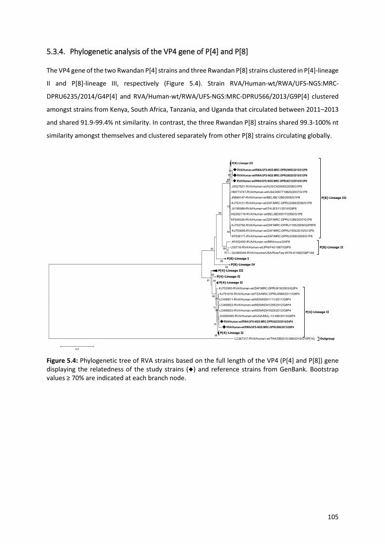
105
5.3.4. Phylogenetic analysis of the VP4 gene of P[4] and P[8] The VP4 gene of the two Rwandan P[4] strains and three Rwandan P[8] strains clustered in P[4]-lineage
II and P[8]-lineage III, respectively (Figure 5.4). Strain RVA/Human-wt/RWA/UFS-NGS:MRC-
DPRU6235/2014/G4P[4] and RVA/Human-wt/RWA/UFS-NGS:MRC-DPRU566/2013/G9P[4] clustered
amongst strains from Kenya, South Africa, Tanzania, and Uganda that circulated between 2011–2013
and shared 91.9-99.4% nt similarity. In contrast, the three Rwandan P[8] strains shared 99.3-100% nt
similarity amongst themselves and clustered separately from other P[8] strains circulating globally.
Figure 5.4: Phylogenetic tree of RVA strains based on the full length of the VP4 (P[4] and P[8]) gene displaying the relatedness of the study strains (◆) and reference strains from GenBank. Bootstrap values ≥ 70% are indicated at each branch node.

106
5.3.5. Phylogenetic analyses of the VP1-VP3 and VP6 genes The VP6 gene of all the five Rwandan study strains clustered separately within lineage I1 and formed
multiple sub-clusters (Figure 5.5A). Strain RVA/Human-wt/RWA/UFS-NGS:MRC-
DPRU8020/2015/G12P[8], RVA/Human-wt/RWA/UFS-NGS:MRC-DPRU9995/2015/G12P[8] and
RVA/Human-wt/RWA/UFS-NGS:MRC-DPRU6235/2014/G4P[4] formed distinct branches that
clustered separately from globally circulating strains. Furthermore, the VP1 gene of the two G12P[8]
(RVA/Human-wt/RWA/UFS-NGS:MRC-DPRU8020/2015/G12P[8] and RVA/Human-wt/RWA/UFS-
NGS:MRC-DPRU9995/2015/G12P[8]), G4P[4] and G9P[4] strains clustered in lineage R2, while strain
RVA/Human-wt/RWA/UFS-NGS:MRC-DPRU6212/2014/G12P[8] clustered in lineage R1 (Figure 5.5B).
The two G12P[8] strains that clustered in lineage R2 clustered amongst human, animal and RotaTeq®
vaccine strains were homologous. The VP2 gene separated into lineage C1 and lineage C2 (Figure
5.5C). RVA/Human-wt/RWA/UFS-NGS:MRC-DPRU8020/2015/G12P[8] and RVA/Human-
wt/RWA/UFS-NGS:MRC-DPRU9995/2015/G12P[8] showed a similar clustering pattern as seen with
the VP1 gene by clustering with both human and RotaTeq® vaccine strains exhibiting homologous
similarity. RVA/Human-wt/RWA/UFS-NGS:MRC-DPRU6212/2014/G12P[8] clustered with strains from
Nepal and Myanmar that shared 99.5-99.6% nt similarity, while the G4P[4] and G9P[4] strains formed
a distinct branch from strains circulating globally. The VP3 gene of the three G12P[8] strains clustered
in lineage M1, while strain G4P[4] and G9P[4] clustered in lineage M2 (Figure 5.5D). RVA/Human-
wt/RWA/UFS-NGS:MRC-DPRU566/2013/G9P[4] and RVA/Human-wt/RWA/UFS-NGS:MRC-
DPRU6235/2014/G4P[4] formed distinct branches separate from globally circulating strains, while
strain RVA/Human-wt/RWA/UFS-NGS:MRC-DPRU6212/2014/G12P[8] clustered in a distinct sub-
cluster to RVA/Human-wt/RWA/UFS-NGS:MRC-DPRU8020/2015/G12P[8] and RVA/Human-
wt/RWA/UFS-NGS:MRC-DPRU9995/2015/G12P[8].

107
5.5.A VP6

108
5.5B VP1

109
5.5C VP2
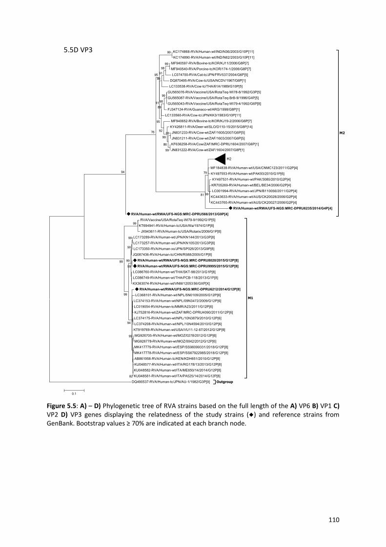
110
Figure 5.5: A) – D) Phylogenetic tree of RVA strains based on the full length of the A) VP6 B) VP1 C) VP2 D) VP3 genes displaying the relatedness of the study strains (◆) and reference strains from GenBank. Bootstrap values ≥ 70% are indicated at each branch node.
5.5D VP3

111
5.3.6. Phylogenetic analyses of the NSP1-NSP5 genes The NSP1-NSP5 gene of study strain RVA/Human-wt/RWA/UFS-NGS:MRC-DPRU8020/2015/G12P[8]
and RVA/Human-wt/RWA/UFS-NGS:MRC-DPRU9995/2015/G12P[8] phylogenetically clustered
closely with reference strains circulating globally in previously established genotypes A1, N2, T1, E2
and H3 (Figure 5.6A-5.6E). The two G12P[8] strains also clustered among human and RotaTeq® vaccine
strains in the NSP2 (homologous), NSP4 (homologous), and NSP5 (99.5-99.8% nt similarity) genes
(Figure 5.6B, 5.6D and 5.6E). Furthermore, the NSP1 gene separated into lineage A1 and lineage A2
(Figure 5.6A). Strains G9P[4] and G4P[4] branched separately from globally circulating strains
observed in lineage A1 and lineage A2, respectively. RVA/Human-wt/RWA/UFS-NGS:MRC-
DPRU6212/2014/G12P[8] clustered amongst strains from Thailand that shared 99.2% nt similarity.
The NSP2 gene of the three G12P[8] strains and the G4P[4] strain clustered in lineage N2, while the
G9P[4] strain clustered in lineage N1 (Figure 5.6B). RVA/Human-wt/RWA/UFS-NGS:MRC-
DPRU6212/2014/G12P[8] and RVA/Human-wt/RWA/UFS-NGS:MRC-DPRU6235/2014/G4P[4] shared
95.9% nt similarity, while strain RVA/Human-wt/RWA/UFS-NGS:MRC-DPRU566/2013/G9P[4] shared
97.1% nt similarity with a strain from the United States. The NSP3 gene of the two G12P[8]
(RVA/Human-wt/RWA/UFS-NGS:MRC-DPRU8020/2015/G12P[8] and RVA/Human-wt/RWA/UFS-
NGS:MRC-DPRU9995/2015/G12P[8]) strains, G4P[4] and G9P[4] clustered in lineage T1, while strain
RVA/Human-wt/RWA/UFS-NGS:MRC-DPRU6212/2014/G12P[8] clustered in lineage T2 (Figure 5.6C).
RVA/Human-wt/RWA/UFS-NGS:MRC-DPRU6212/2014/G12P[8] was homologous to a Ugandan strain.
The NSP4 gene of the G4P[4] strain clustered closely with strains from Denmark, Russia and Japan that
showed 99.4-99.6% nt similarity (Figure 5.6D). The G12P[8] and G9[4] strains clustered in lineage E1
and shared 99% nt similarity with Japan and Indian strains, respectively. The NSP5 gene separated into
lineage H1, H2 and H3 (Figure 5.6E). The G12P[8] strain in lineage H1 clustered closely with strains
from Nepal and Myanmar and shared 99.1-99.3% nt similarity, while the G9P[4] strain clustered
closely with a strain from the United States (99.5% nt similarity). In contrast, the G4P[4] strain shared
99.5% and 99.8% similarity with a South African and Zimbabwean strain, respectively.

112
5.6A NSP1

113
5.6B NSP2

114
5.6C NSP3

115
5.6D NSP4
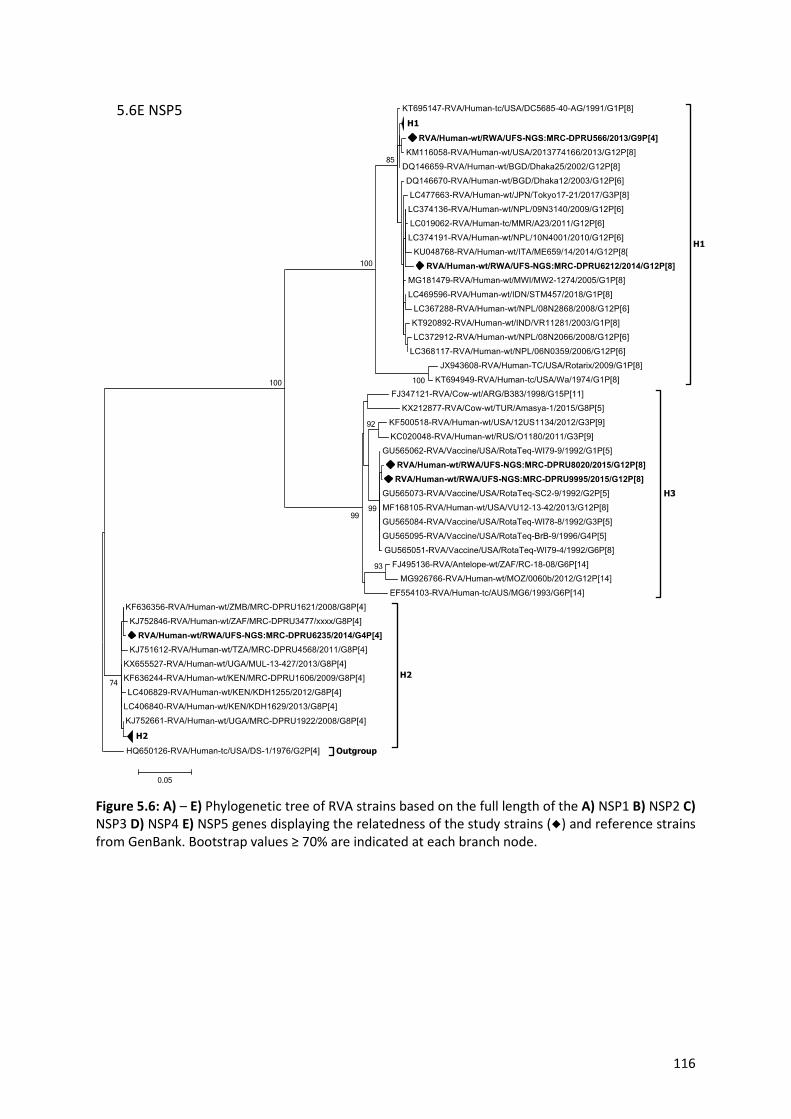
116
Figure 5.6: A) – E) Phylogenetic tree of RVA strains based on the full length of the A) NSP1 B) NSP2 C) NSP3 D) NSP4 E) NSP5 genes displaying the relatedness of the study strains (◆) and reference strains from GenBank. Bootstrap values ≥ 70% are indicated at each branch node.
5.6E NSP5
117
5.4. Discussion
Five Rwandan rotavirus strains that have undergone inter-genogroup reassortment were identified in
this study as part of WHO supported on-going rotavirus whole-genome sentinel surveillance. Strains
RVA/Human-wt/RWA/UFS-NGS:MRC-DPRU6235/2014/G4P[4], RVA/Human-wt/RWA/UFS-NGS:MRC-
DPRU566/2013/G9P[4] and RVA/Human-wt/RWA/UFS-NGS:MRC-DPRU6212/2014/G12P[8] showed
genotype constellations involving the Wa-like and DS-1-like genogroups while RVA/Human-
wt/RWA/UFS-NGS:MRC-DPRU8020/2015/G12P[8] and RVA/Human-wt/RWA/UFS-NGS:MRC-
DPRU9995/2015/G12P[8] exhibited triple-gene reassortment of all three human genogroups, Wa-like,
DS-1-like, and AU-1-like. Bányai and colleagues (2011) stated that most atypical RVA strains are the
result of natural inter-genogroup reassortment between the Wa-like and DS-1-like strains due to the
segmented nature of RVA. The detection of these inter-genogroup reassortant strains in this study
may be attributed to either the lack of RNA polymerase proofreading ability or co-infection of multiple
strains from various human RVA strains (Estes and Greenberg, 2013; Jain et al., 2001; Kirkwood, 2010;
Matthijnssens et al., 2008; Medici et al., 2007). Co-infections have been reported in high frequencies
in several RVA strains across Africa (Dóró et al., 2014; Nyaga et al., 2018; Seheri et al., 2014; Todd et
al., 2010).
The detection of the unusual G9P[4] strain (RVA/Human-wt/RWA/UFS-NGS:MRC-
DPRU566/2013/G9P[4]) in Rwanda is noteworthy as it is more prevalent in South-East Asia, Japan and
Central America and detected in very low frequency (2%) in Africa (Afrad et al., 2013; Doan et al.,
2017; João et al., 2020; Lartey et al., 2018; Pradhan et al., 2016; Quaye et al., 2013; Seheri et al., 2018;
Yamamoto et al., 2015). Phylogenetic analysis revealed that the five Rwandan strains clustered mostly
with strains circulating globally, suggesting a direct importation of these variants from abroad rather
than local emergence through multiple reassortment events between locally circulating strains. It is
evident that the reassortment events of the five Rwandan strains mostly occurred with contemporary
human rotavirus strains as they did not show sufficient evidence of animal/human reassortment.
Furthermore, RVA/Human-wt/RWA/UFS-NGS:MRC-DPRU8020/2015/G12P[8] and RVA/Human-
wt/RWA/UFS-NGS:MRC-DPRU9995/2015/G12P[8] exhibit a high similarity amongst each other across
the genome and both human and RotaTeq® vaccine strains in the VP2, NSP2, NSP4, and NSP5 gene
segment. This finding suggests a reassortment between both human and RotaTeq® vaccine strains
might have also transpired. This observation is consistent with the findings of Rose and co-workers,
who have reported that such reassortment events are expected considering the attenuation of
RotaTeq® vaccines and the segmented nature of the RVA genome (Rose et al., 2013b).

118
In the construction of the RotaTeq® vaccine in the 1980s, the G4 and P[8] components were included
in the composition of this pentavalent vaccine (Matthijnssens et al., 2010). When comparing the G4
and P[8] components of the Rwandan strains and the RotaTeq® vaccine strains, we observed that the
Rwandan G4 strain clustered closely to the RotaTeq® vaccine strain in G4-lineage I while the Rwandan
P[8] strains clustered in P[8] lineage III distantly from the RotaTeq® vaccine strain in P[8]-lineage II.
This phenomenon can be attributed to the constant evolutionary changes that rotaviruses undergo.
Hence the currently circulating RVA strains may cluster in VP7 and VP4 lineages different from the
RVA vaccine strains (Zeller et al., 2012). Such changes may influence a varying selective pressure
against these VP7 and VP4 lineages ultimately reducing vaccine effectiveness over time. The VP1,VP3,
NSP1 and NSP3 genes of strain RVA/Human-wt/RWA/UFS-NGS:MRC-DPRU6235/2014/G4P[4] were
phylogenetically distinct from strains circulating in other places thus suggesting that these gene
segments may be unique.
The amino acid sequence comparison of the G4P[4] and G12P[8] Rwandan strains and the RotaTeq®
vaccine strain showed that none of the Rwandan strains were identical to the RotaTeq® vaccine strain.
Amino acid substitutions were observed throughout the VP7 epitope regions excluding position 291
and 190, while the VP4 exhibited only three amino acid substitutions at position 150, 195 and 388.
The amino acid substitution at position 96, 97, 211, 217 and 221 on the VP7 epitope region suggests
a radical change in polarity. McDonald et. al. (2009) suggested that such substitutions do not change
the genotype specificity of the rotavirus strain. However, they may influence the binding of
neutralizing antibodies thus affecting viral fitness through selection pressure. Despite the distant
clustering between the Rwandan P[8] strains and the vaccine strain, only three changes were observed
in the VP4 neutralizing epitope regions. Antigenic variations between rotavirus strains and the vaccine
strains are usually implicated in the decreased effectiveness of rotavirus vaccines in low-income
countries such as Rwanda (Narang et al., 2009; Taneja and Malik, 2012; Zaman et al., 2010). There is
no evidence that the amino acid changes on the VP4 epitope regions are due to vaccination and could
have occurred as a natural evolutionary process as they were observed in other strains circulating
globally over the years.

119
5.5. Conclusion
The detection of five inter-genogroup reassortant Rwandan rotavirus strains from the whole-genome
analysis further emphasises the ubiquitous nature and diversity of RVA strains in circulation. Whether
vaccine introduction is responsible for the observed reassortment events or not remains enigmatic as
several natural factors can be attributed to the evolution of these RVA strains. Amino acid
substitutions observed in the epitope regions in the neutralizing epitope of the VP7 and VP4 proteins
of the Rwandan strain when compared with the RotaTeq® vaccine strain may be implicated in the
effectiveness of rotavirus vaccines being low in low-income countries. Continuous surveillance at the
whole-genome level is highly recommended to monitor any possible changes due to vaccine pressure
as part efforts to monitor the impact of vaccines on the circulating rotavirus strains especially in
African countries.
5.6. Methods
5.6.1. Ethics statement This study was reviewed and approved by the HSREC of the UFS and assigned an ethics number (UFS-
HSD2019/1601/2810). Patients information were anonymized to ensure confidentiality.
5.6.2. Sample collection One hundred and fifty-eight stool specimens were collected from hospitalized children presenting
with acute gastroenteritis between 2012 and 2016 in Rwanda as part of the WHO/AFRO supported
rotavirus surveillance program. These samples were retrieved from “the African stool repository”
established to archive stool samples as part of the WHO supported African rotavirus surveillance
network maintained at the DPRU, WHO RRL-SA. Only five samples presented the reassortant genome
constellations, which fit the criteria for this study. Two samples were collected from unvaccinated
children aged 14 months (female) and 36 months (male) from the South and East Province of Rwanda,
respectively. The remaining three samples were collected from children (males) who received all three
doses of the RotaTeq® vaccine, one aged 12 months (from the North Province) and two aged 24
months (both from the East Province). The samples were sequenced and whole-genome analysis was
performed at the UFS-NGS Unit. The Rwandan strains described in this study were deposited in the
GenBank database under accession numbers MT163179-MT163266.

120
5.6.3. Sample preparation for rotavirus whole-genome sequencing The dsRNA was extracted using a protocol previously described by Nyaga et al. (2018) and purified
using the Qiagen MinElute gel extraction kit (Qiagen, Hilden, Germany). The quantity of the purified
dsRNA was thereafter verified by 1% agarose gel electrophoresis prior to quantification using a
BioDrop-µLITE spectrophotometer (Biodrop, Cambridge, United Kingdom). For the cDNA synthesis,
the Maxima H Minus Double-Stranded cDNA Synthesis Kit (ThermoFisher Scientific, Waltham MA,
USA) was used according to the manufacturer’s instructions with minor modifications. The
modification included the denaturation of the dsRNA at 95oC for five minutes prior to synthesizing the
first strand for 2 hours at 50 °C in a thermocycler (Merck, Darmstadt, Germany).
5.6.4. DNA library preparations and whole-genome sequencing The Nextera® XT DNA library preparation kit (Illumina Inc., California, United States) was used in
preparation for sequencing on an Illumina Miseq (Illumina Inc., California, United States) platform.
Following the manufacturer’s instructions, indexes were used to barcode the DNA and purified using
AMPure XP magnetic beads (Beckman Coulter, Indiana, United States) while simultaneously selecting
300bp DNA fragments and removing short library fragments. The library was validated using a
Bioanalyzer (Agilent Technologies, California, United States) and Qubit (Life Technologies, California,
United States), and the samples were normalized to 8pM and pooled in a single tube in readiness for
sequencing on an Illumina MiSeq (Illumina Inc., California, United States) platform. An Illumina v3
reagent kit (Illumina Inc., California, United States) was used for 600 cycles (301 x 2 paired ends)
sequencing. A PhiX (20pM; Illumina Inc., California, United States), a spike of 20% was used as a
control.
5.6.5. Computational analysis The raw reads were assembled in Geneious Prime v11.1.5 (Kearse et al., 2012) and CLC Genomics
Workbench v11 (CLC Bio, Qiagen, Hilden, Germany) as complementary tools. A reference-based
assembly was carried out for the samples and the resulting contigs were used as query sequences in
the RotaC v2.0 (Maes et al., 2009, http://rotac.regatools.be/) and the Nucleotide Basic Local
Alignment Search Tool (BLASTn, https://blast.ncbi.nlm.nih.gov/Blast.cgi) to determine the genotype
of each gene and their full-length nucleotide sequence. Phylogenetic trees were constructed using the
MEGA 6 software package (Tamura et al., 2013) with a Maximum Likelihood method-based model
supported by bootstrap analysis with 1000 replicates. The p-distance algorithm was used to calculate
nucleotide distances. The VP7 and VP4 protein structure was constructed using the Swiss-model

121
protein structure server (Waterhouse et al., 2018) and PyMOL v2. The validity of the structures were
confirmed using YASARA (Krieger et al., 2002) and verify 3D
(https://servicesn.mbi.ucla.edu/Verify3D/).

122
CHAPTER SIX: GENERAL DISCUSSION, CONCLUSION AND
RECOMMENDATIONS

123
6.1. Study summary
Over the years, RVA surveillance studies were primarily based on the outer capsid proteins (VP7 and
VP4), which revealed great diversity in circulating strains with significant differences in prevalent
strains, seasonally and geographically (Mwenda et al., 2010; Seheri et al., 2018). African countries are
reported to have the greatest rotavirus diversity according to WHO global surveillance data. Since
rotaviruses consist of 11 segments within the genome, information based solely on the outer capsid
proteins may not be sufficient in providing conclusive data on the correct origin and overall genetic
diversity of the strains (Ghosh and Kobayashi, 2011). The segmented RVA genome is vulnerable to
reassortments, recombination and genomic rearrangements. Moreover, zoonotic transmissions are
also possible and can only be confirmed through whole-genome analysis (Martella et al., 2010; Santos
and Hoshino, 2005). Whole-genome sequencing has thus granted a platform to gain insight into the
whole-genome of rotavirus strains circulating globally. Due to the lack of surveillance data on these
strains with regards to the whole-genome, the WHO in partnership with the UFS-NGS Unit rolled out
a pilot study to contribute to surveillance at whole-genome level, of several African countries, among
them, Rwanda. To our knowledge, this is the first RVA whole-genome study involved in a surveillance
study in Africa. The focus of our study was on G1P[8] and reassortant strains identified in Rwanda,
highlighted in Chapter one and five, from a pool of 158 samples collected from hospitalized children
presenting with gastroenteritis. The samples not analysed here (n=117), will be disseminated
elsewhere.
Rotavirus dsRNA was successfully extracted from stool samples using TRI-reagent (Molecular Research
Center, Inc, Cincinnati, OH, USA). All the dsRNA rotavirus samples collected from children presenting
gastroenteritis were converted to cDNA using the maxima H minus ds cDNA synthesis kit
(ThermoFisher Scientific, Waltham MA, USA) that was modified specifically for rotavirus as per the
UFS-NGS Unit cDNA synthesis SOP. The qualities of the synthesized rotavirus cDNA were good enough
to proceed with subsequent steps required prior to whole-genome sequencing and sufficiently
covered objective one. However, some samples were excluded from sequencing due to low DNA
concentrations which could be attributed the loss of nucleic material during the dsRNA extraction
step, the cDNA purification step, and library preparation. Whole-genome characterization of rotavirus
strains circulating in Rwanda was successfully achieved using an Illumina MiSeq platform.
The second and third objectives were addressed in chapter four and chapter five, respectively. Briefly,
in chapter four, G1P[8] strains were analysed to determine the phylogenetic features pre- and post-
vaccination in Rwanda. Data analysis of these strains revealed that the G1P[8] strains from Rwanda

124
are highly diverse compared to other circulating strains globally. The strains showed clear distinctive
features between pre- and post-vaccine introduction strains by clustering separately in most of the
phylogenetic trees. Furthermore, one of the G1P[8] strains was determined to be a RotaTeq® vaccine-
derived strain as it was homologous to a RotaTeq® vaccine strain and clustered amongst vaccine strains
throughout the entire backbone. The strain was shed from a two month-old infant, four days post-
vaccine introduction. A representative G1P[8] strain was also compared to the RotaTeq® and Rotarix®
vaccine strains, where amino acid changes in the neutralizing epitope regions and the CTL epitope
region were observed. Some of the observed changes did not affect the polarity of the amino acids
while others changed the polarity and were radical in nature. The changes were suggested to have
occurred naturally as they were observed prior to vaccine introduction, while the other changes were
observed post-vaccine introduction and are suspected of playing a role in the escape of host immunity
(Rasebotsa et al., 2020).
In chapter 5, five reassortant strains were analysed to explore the probable evidence of rotavirus
vaccine pressure in Rwanda. These strains were collected from two unvaccinated children (14 months
female and 36 months male) and three children (12 months male and two 24 months old males)
vaccinated with three doses of RotaTeq® vaccine. Two strains exhibited a triple gene reassortment
(Wa-like, DS-1-like, and AU-1-like) while the other three strains showed a double gene reassortment
(Wa-like and DS-1-like). The strains were all detected post-vaccine introduction from hospitalized
children who presented episodes of diarrhoea and vomiting. Despite this observation, the reassortant
strains were suggested to have occurred naturally as there was no evidence of vaccine-induced
pressure. This was justified by observing how these strains were clustering in the phylogenetic trees.
The strains were clustering with globally cognate circulating human RVA strains, with the exception of
a few segments clustering with the RotaTeq® vaccine strain. Furthermore, comparative analysis of the
antigenic region of our study strains and the RotaTeq® vaccine strain revealed the VP7 protein with
multiple changes in the antigenic epitope region while the VP4 protein contained only two changes in
the VP8* domain and only one change in the VP5* domain. These observations are consistent with
changes observed on the antigenic regions of other strains circulating globally, suggesting a non-
isolated event in Rwanda. These changes are however, worth noting as they may pose new challenges
with regards to the effectiveness of currently available vaccines, especially in low-income countries
such as Rwanda. Moreover, the observations in this project highlight that continuous whole-genome
surveillance of circulating RVA strains is important in providing insight on the evolutionary changes
occurring over the years, the impact of vaccines on circulating strains and also providing relevant
information required for a rational design of future RVA vaccines.

125
The use of the Illumina Miseq platform increased throughput compared with less automated
sequencing technologies thus providing quality data with a Q-score value of 30. The platform provided
an opportunity to sequence multiple samples within a single run therefore decreasing the amount of
time and money that would have been required when using the traditional sequencing method. This
surveillance study is highly valuable to the Rwandan community as it successfully deciphered the
whole-genome sequences of circulating rotavirus strains in Rwanda, thus expanding on the
information already available from observing the outer capsid proteins. We established that the
G1P[8] strains circulating in Rwanda have a stable Wa-like genome constellation such information will
aid in understanding the transmission dynamics of G1P[8] strains in Rwanda. Furthermore, the whole-
genome data from this study will contribute in addressing the evolutionary dynamics of rotavirus
strains in Rwanda.
6.2. Limitations and recommendations
The principal limitation to this study was the number of years included in pre- and post-vaccine
introduction thus it is not sufficient enough to conclusively determine the genetic variability of strains
circulating in Rwanda as part of the pilot WHO whole-genome surveillance study. The data presented
was for one year pre-vaccine introduction (2011) and five years post-vaccine introduction (2012-
2016), where the ideal sample set could be in the long-term of at least seven years pre- and seven
years post-vaccination. The study was principally in-silico based, thus cell culture was not attempted
to provide supporting information on our findings. Nevertheless, whole-genome analysis is highly
reliable in providing conclusive data on the evolutionary dynamics of circulating RVA strains.
In this study, we only analysed 41 samples out of 158 samples sequenced thus there is still enough
room to explore the diversity and evolutionary patterns of rotavirus strains circulating in Rwanda. We
also recommend conducting cell cultures to confirm conclusions made based on in-silico analysis. We
highly recommend the use of NGS platforms for surveillance in Africa to decipher novel information
that may be otherwise missed when looking only at the outer capsid proteins. Rwanda has recently
switched from the use of the RotaTeq® vaccine (2012-2017) to the Rotarix® vaccine (2018); thus it will
be interesting to see if the switch will have any effect on the strains circulation in Rwanda in the
coming years.

126
REFERENCES
Abad, F.X., Pinto, R.M., Bosch, A., 1994. Survival of enteric viruses on environmental fomites. Appl.
Environ. Microbiol. 60, 3704–3710. https://doi.org/10.1128/aem.60.10.3704-3710.1994
Abe, M., Yamasaki, A., Ito, N., Mizoguchi, T., Asano, M., Okano, T., Sugiyama, M., 2010. Molecular
characterization of rotaviruses in a Japanese raccoon dog (Nyctereutes procyonoides) and a
masked palm civet (Paguma larvata) in Japan. Vet. Microbiol. 146, 253–259.
https://doi.org/10.1016/j.vetmic.2010.05.019
Afrad, M.H., Rahman, M.Z., Matthijnssens, J., Das, S.K., Faruque, A.S.G., Azim, T., Rahman, M., 2013.
High incidence of reassortant G9P[4] rotavirus strain in Bangladesh: Fully heterotypic from
vaccine strains. J. Clin. Virol. https://doi.org/10.1016/j.jcv.2013.09.024
Agbemabiese, C.A., Nakagomi, T., Nguyen, M.Q., Gauchan, P., Nakagomi, O., 2017. Reassortant DS-1-
like G1P[4] Rotavirus A strains generated from co-circulating strains in Vietnam, 2012/2013.
Microbiol. Immunol. 61, 328–336. https://doi.org/10.1111/1348-0421.12501
Ahmed, K., Nakagomi, T., Nakagomi, O., 2007. Molecular identification of a novel G1 VP7 gene carried
by a human rotavirus with a super-short RNA pattern. Virus Genes 35, 141–145.
https://doi.org/10.1007/s11262-007-0089-9
Aiyegbo, M.S., Sapparapu, G., Spiller, B.W., Eli, I.M., Williams, D.R., Kim, R., Lee, D.E., Liu, T., Li, S.,
Woods, V.L., Nannemann, D.P., Meiler, J., Stewart, P.L., Crowe, J.E., 2013. Human Rotavirus VP6-
Specific Antibodies Mediate Intracellular Neutralization by Binding to a Quaternary Structure in
the Transcriptional Pore. PLoS One 8, e61101. https://doi.org/10.1371/journal.pone.0061101
Alam, M.M., Kobayashi, N., Ishino, M., Nagashima, S., Paul, S.K., Chawla-Sarkar, M., Krishnan, T., Naik,
T.N., 2008. Identical rearrangement of NSP3 genes found in three independently isolated virus
clones derived from mixed infection and multiple passages of Rotaviruses. Arch. Virol. 153, 555–
559. https://doi.org/10.1007/s00705-007-0004-7
Aliabadi, N., Antoni, S., Mwenda, J.M., Weldegebriel, G., Biey, J.N.M., Cheikh, D., Fahmy, K., Teleb, N.,
Ashmony, H.A., Ahmed, H., Daniels, D.S., Videbaek, D., Wasley, A., Singh, S., de Oliveira, L.H.,
Rey-Benito, G., Sanwogou, N.J., Wijesinghe, P.R., Liyanage, J.B.L., Nyambat, B., Grabovac, V.,
Heffelfinger, J.D., Fox, K., Paladin, F.J., Nakamura, T., Agócs, M., Murray, J., Cherian, T., Yen, C.,
Parashar, U.D., Serhan, F., Tate, J.E., Cohen, A.L., 2019. Global impact of rotavirus vaccine
introduction on rotavirus hospitalisations among children under 5 years of age, 2008–16:
findings from the Global Rotavirus Surveillance Network. Lancet Glob. Heal. 7, e893–e903.
https://doi.org/10.1016/S2214-109X(19)30207-4
Anderson, E.J., Weber, S.G., 2004. Rotavirus infection in adults. Lancet Infect. Dis. 4, 91–99.
https://doi.org/10.1016/S1473-3099(04)00928-4
127
Anh, D.D., Van Trang, N., Thiem, V.D., Anh, N.T.H., Mao, N.D., Wang, Y., Jiang, B., Hien, N.D., Luan, L.T.,
2012. A dose-escalation safety and immunogenicity study of a new live attenuated human
rotavirus vaccine (Rotavin-M1) in Vietnamese children. Vaccine 30, A114–A121.
https://doi.org/10.1016/j.vaccine.2011.07.118
Aoki, S.T., Settembre, E.C., Trask, S.D., Greenberg, H.B., Harrison, S.C., Dormitzer, P.R., 2009. Structure
of rotavirus outer-layer protein VP7 bound with a neutralizing Fab. Science (80-. ). 324, 1444–
1447. https://doi.org/10.1126/science.1170481
Arana, A., Montes, M., Jere, K.C., Alkorta, M., Iturriza-Gómara, M., Cilla, G., 2016. Emergence and
spread of G3P[8] rotaviruses possessing an equine-like VP7 and a DS-1-like genetic backbone in
the Basque Country (North of Spain), 2015. Infect. Genet. Evol. 44, 137–144.
https://doi.org/10.1016/j.meegid.2016.06.048
Arista, S., Giammanco, G.M., De Grazia, S., Ramirez, S., Lo Biundo, C., Colomba, C., Cascio, A., Martella,
V., 2006. Heterogeneity and Temporal Dynamics of Evolution of G1 Human Rotaviruses in a
Settled Population. J. Virol. 80, 10724–10733. https://doi.org/10.1128/jvi.00340-06
Armah, G.E., Sow, S.O., Breiman, R.F., Dallas, M.J., Tapia, M.D., Feikin, D.R., Binka, F.N., Steele, A.D.,
Laserson, K.F., Ansah, N.A., Levine, M.M., Lewis, K., Coia, M.L., Attah-Poku, M., Ojwando, J.,
Rivers, S.B., Victor, J.C., Nyambane, G., Hodgson, A., Schödel, F., Ciarlet, M., Neuzil, K.M., 2010.
Efficacy of pentavalent rotavirus vaccine against severe rotavirus gastroenteritis in infants in
developing countries in sub-Saharan Africa: a randomised, double-blind, placebo-controlled trial.
Lancet 376, 606–614. https://doi.org/10.1016/S0140-6736(10)60889-6
Arora, R., Chitambar, S.D., 2011. Full genomic analysis of Indian G1P[8] rotavirus strains. Infect. Genet.
Evol. 11, 504–511. https://doi.org/10.1016/j.meegid.2011.01.005
Atherly, D.E., Lewis, K.D.C., Tate, J., Parashar, U.D., Rheingans, R.D., 2012. Projected health and
economic impact of rotavirus vaccination in GAVI-eligible countries: 2011-2030. Vaccine 30, A7.
https://doi.org/10.1016/j.vaccine.2011.12.096
Bányai, K., Gentsch, J.R., Martella, V., Bogdán, Á., Havasi, V., Kisfali, P., Szabó, A., Mihály, I., Molnár,
P., Melegh, B., Szücs, G., 2009. Trends in the Epidemiology of Human G1P[8] Rotaviruses: A
Hungarian Study. J. Infect. Dis. 200, S222–S227. https://doi.org/10.1086/605052
Bányai, K., Kemenesi, G., Budinski, I., Földes, F., Zana, B., Marton, S., Varga-Kugler, R., Oldal, M.,
Kurucz, K., Jakab, F., 2017. Candidate new rotavirus species in Schreiber’s bats, Serbia. Infect.
Genet. Evol. 48, 19–26. https://doi.org/10.1016/j.meegid.2016.12.002
Bányai, K., László, B., Duque, J., Steele, A.D., Nelson, E.A.S., Gentsch, J.R., Parashar, U.D., 2012.
Systematic review of regional and temporal trends in global rotavirus strain diversity in the pre
rotavirus vaccine era: Insights for understanding the impact of rotavirus vaccination programs.
128
Vaccine 30, A122–A130. https://doi.org/10.1016/j.vaccine.2011.09.111
Bányai, K., Mijatovic-Rustempasic, S., Hull, J.J., Esona, M.D., Freeman, M.M., Frace, A.M., Bowen, M.D.,
Gentsch, J.R., 2011. Sequencing and phylogenetic analysis of the coding region of six common
rotavirus strains: Evidence for intra-genogroup reassortment among co-circulating G1P[8] and
G2P[4] strains from the United States. J. Med. Virol. 83, 532–539.
https://doi.org/10.1002/jmv.21977
Begue, R.E., Perrin, K., 2010. Reduction in Gastroenteritis With the Use of Pentavalent Rotavirus
Vaccine in a Primary Practice. Pediatrics 126, e40–e45. https://doi.org/10.1542/peds.2009-2069
Benhafid, M., Elomari, N., Elqazoui, M., Meryem, A.I., Rguig, A., Filali-Maltouf, A., Elaouad, R., 2013.
Diversity of rotavirus strains circulating in children under 5 years of age admitted to hospital for
acute gastroenteritis in morocco, june 2006 to may 2009. J. Med. Virol. 85, 354–362.
https://doi.org/10.1002/jmv.23445
Benhafid, M., Rguig, A., Trivedi, T., Elqazoui, M., Teleb, N., Mouane, N., Maltouf, A.F., Parashar, U.,
Patel, M., Aouad, R. El, 2012. Monitoring of rotavirus vaccination in Morocco: Establishing the
baseline burden of rotavirus disease. Vaccine 30, 6515–6520.
https://doi.org/10.1016/j.vaccine.2012.08.058
Benson, D.A., Karsch-Mizrachi, I., Lipman, D.J., Ostell, J., Rapp, B.A., Wheeler, D.L., 2000. GenBank.
Nucleic Acids Res. 28, 15–18.
Bentley, D.R., Balasubramanian, S., Swerdlow, H.P., Smith, G.P., Milton, J., Brown, C.G., Hall, K.P.,
Evers, D.J., Barnes, C.L., Bignell, H.R., Boutell, J.M., Bryant, J., Carter, R.J., Keira Cheetham, R.,
Cox, A.J., Ellis, D.J., Flatbush, M.R., Gormley, N.A., Humphray, S.J., Irving, L.J., Karbelashvili, M.S.,
Kirk, S.M., Li, H., Liu, X., Maisinger, K.S., Murray, L.J., Obradovic, B., Ost, T., Parkinson, M.L., Pratt,
M.R., Rasolonjatovo, I.M.J., Reed, M.T., Rigatti, R., Rodighiero, C., Ross, M.T., Sabot, A., Sankar,
S. V., Scally, A., Schroth, G.P., Smith, M.E., Smith, V.P., Spiridou, A., Torrance, P.E., Tzonev, S.S.,
Vermaas, E.H., Walter, K., Wu, X., Zhang, L., Alam, M.D., Anastasi, C., Aniebo, I.C., Bailey, D.M.D.,
Bancarz, I.R., Banerjee, S., Barbour, S.G., Baybayan, P.A., Benoit, V.A., Benson, K.F., Bevis, C.,
Black, P.J., Boodhun, A., Brennan, J.S., Bridgham, J.A., Brown, R.C., Brown, A.A., Buermann, D.H.,
Bundu, A.A., Burrows, J.C., Carter, N.P., Castillo, N., Catenazzi, M.C.E., Chang, S., Neil Cooley, R.,
Crake, N.R., Dada, O.O., Diakoumakos, K.D., Dominguez-Fernandez, B., Earnshaw, D.J., Egbujor,
U.C., Elmore, D.W., Etchin, S.S., Ewan, M.R., Fedurco, M., Fraser, L.J., Fuentes Fajardo, K. V., Scott
Furey, W., George, D., Gietzen, K.J., Goddard, C.P., Golda, G.S., Granieri, P.A., Green, D.E.,
Gustafson, D.L., Hansen, N.F., Harnish, K., Haudenschild, C.D., Heyer, N.I., Hims, M.M., Ho, J.T.,
Horgan, A.M., Hoschler, K., Hurwitz, S., Ivanov, D. V., Johnson, M.Q., James, T., Huw Jones, T.A.,
Kang, G.D., Kerelska, T.H., Kersey, A.D., Khrebtukova, I., Kindwall, A.P., Kingsbury, Z., Kokko-

129
Gonzales, P.I., Kumar, A., Laurent, M.A., Lawley, C.T., Lee, S.E., Lee, X., Liao, A.K., Loch, J.A., Lok,
M., Luo, S., Mammen, R.M., Martin, J.W., McCauley, P.G., McNitt, P., Mehta, P., Moon, K.W.,
Mullens, J.W., Newington, T., Ning, Z., Ling Ng, B., Novo, S.M., O’Neill, M.J., Osborne, M.A.,
Osnowski, A., Ostadan, O., Paraschos, L.L., Pickering, L., Pike, Andrew C., Pike, Alger C., Chris
Pinkard, D., Pliskin, D.P., Podhasky, J., Quijano, V.J., Raczy, C., Rae, V.H., Rawlings, S.R., Chiva
Rodriguez, A., Roe, P.M., Rogers, John, Rogert Bacigalupo, M.C., Romanov, N., Romieu, A., Roth,
R.K., Rourke, N.J., Ruediger, S.T., Rusman, E., Sanches-Kuiper, R.M., Schenker, M.R., Seoane, J.M.,
Shaw, R.J., Shiver, M.K., Short, S.W., Sizto, N.L., Sluis, J.P., Smith, M.A., Ernest Sohna Sohna, J.,
Spence, E.J., Stevens, K., Sutton, N., Szajkowski, L., Tregidgo, C.L., Turcatti, G., Vandevondele, S.,
Verhovsky, Y., Virk, S.M., Wakelin, S., Walcott, G.C., Wang, J., Worsley, G.J., Yan, J., Yau, L.,
Zuerlein, M., Rogers, Jane, Mullikin, J.C., Hurles, M.E., McCooke, N.J., West, J.S., Oaks, F.L.,
Lundberg, P.L., Klenerman, D., Durbin, R., Smith, A.J., 2008. Accurate whole human genome
sequencing using reversible terminator chemistry. Nature 456, 53–59.
https://doi.org/10.1038/nature07517
Berglund, E.C., Kiialainen, A., Syvänen, A.C., 2011. Next-generation sequencing technologies and
applications for human genetic history and forensics. Investig. Genet.
https://doi.org/10.1186/2041-2223-2-23
Bernstein, D.I., 2009. Rotavirus Overview. Pediatr. Infect. Dis. J. 28, S50–S53.
https://doi.org/10.1097/INF.0b013e3181967bee
Bettger, W.J., O’Dell, B.L., 1981. A critical physiological role of zinc in the structure and function of
biomembranes. Life Sci. 28, 1425–1438. https://doi.org/10.1016/0024-3205(81)90374-X
Bharat Biotech, 2019. ROTAVAC|First Rota Virus Vaccine Manufacturer|Bharat Biotech [WWW
Document]. URL https://www.bharatbiotech.com/rotavac.html (accessed 5.12.20).
Bishop, R.F., Barnes, G.L., Cipriani, E., Lund, J.S., 1983. Clinical Immunity after Neonatal Rotavirus
Infection: A Prospective Longitudinal Study in Young Children. N. Engl. J. Med. 309, 72–76.
https://doi.org/10.1056/NEJM198307143090203
Bishop, R.F., Davidson, G.P., Holmes, I.H., Ruck, B.J., 1973. Virus particles in epithelial cells of duodenal
mucosa from children with acute non-bacterial gastroenteritis. Lancet 302, 1281–1283.
https://doi.org/10.1016/S0140-6736(73)92867-5
Blackhall, J., Fuentes, A., Magnusson, G., 1996. Genetic stability of a porcine rotavirus RNA segment
during repeated plaque isolation. Virology 225, 181–190.
https://doi.org/10.1006/viro.1996.0586
Boom, J.A., Sahni, L.C., Payne, D.C., Gautam, R., Lyde, F., Mijatovic-Rustempasic, S., Bowen, M.D., Tate,
J.E., Rench, M.A., Gentsch, J.R., Parashar, U.D., Baker, C.J., 2012. Symptomatic infection and
130
detection of vaccine and vaccine-reassortant rotavirus strains in 5 children: A case series. J.
Infect. Dis. 206, 1275–1279. https://doi.org/10.1093/infdis/jis490
Boschi-Pinto, C., Velebit, L., Shibuya, K., 2008. Estimating child mortality due to diarrhoea in
developing countries. Bull. World Health Organ. 86, 710–717.
https://doi.org/10.2471/BLT.07.050054
Broquet, A.H., Hirata, Y., McAllister, C.S., Kagnoff, M.F., 2011. RIG-I/MDA5/MAVS Are Required To
Signal a Protective IFN Response in Rotavirus-Infected Intestinal Epithelium. J. Immunol. 186,
1618–1626. https://doi.org/10.4049/jimmunol.1002862
Bucardo, F., Rippinger, C.M., Svensson, L., Patton, J.T., 2012. Vaccine-derived NSP2 segment in
rotaviruses from vaccinated children with gastroenteritis in Nicaragua. Infect. Genet. Evol. 12,
1282–1294. https://doi.org/10.1016/J.MEEGID.2012.03.007
Bugarcić, A., Taylor, J.A., 2006. Rotavirus Nonstructural Glycoprotein NSP4 Is Secreted from the Apical
Surfaces of Polarized Epithelial Cells. J. Virol. 80, 12343–12349.
https://doi.org/10.1128/jvi.01378-06
Burke, R.M., Tate, J.E., Kirkwood, C.D., Steele, A.D., Parashar, U.D., 2019. Current and new rotavirus
vaccines. Curr. Opin. Infect. Dis. https://doi.org/10.1097/QCO.0000000000000572
Buttery, J.P., Lambert, S.B., Grimwood, K., Nissen, M.D., Field, E.J., Macartney, K.K., Akikusa, J.D., Kelly,
J.J., Kirkwood, C.D., 2011. Reduction in Rotavirus-associated Acute Gastroenteritis Following
Introduction of Rotavirus Vaccine Into Australiaʼs National Childhood Vaccine Schedule. Pediatr.
Infect. Dis. J. 30, S25–S29. https://doi.org/10.1097/INF.0b013e3181fefdee
Bwogi, J., Jere, K.C., Karamagi, C., Byarugaba, D.K., Namuwulya, P., Baliraine, F.N., Desselberger, U.,
Iturriza-Gomara, M., 2017. Whole-genome analysis of selected human and animal rotaviruses
identified in Uganda from 2012 to 2014 reveals complex genome reassortment events between
human, bovine, caprine and porcine strains. PLoS One 12, e0178855.
https://doi.org/10.1371/journal.pone.0178855
Bwogi, J., Malamba, S., Kigozi, B., Namuwulya, P., Tushabe, P., Kiguli, S., Byarugaba, D.K., Desselberger,
U., Iturriza-Gomara, M., Karamagi, C., 2016. The epidemiology of rotavirus disease in under-five-
year-old children hospitalized with acute diarrhea in central Uganda, 2012-2013. Arch. Virol. 161,
999–1003. https://doi.org/10.1007/s00705-015-2742-2
Capitanio, M.A., Greenberg, S.B., 1991. Pneumatosis intestinalis in two infants with rotavirus
gastroenteritis. Pediatr. Radiol. 21, 361–362. https://doi.org/10.1007/BF02011488
Carvalho-Costa, F.A., de Assis, R.M.S., Fialho, A.M., Araújo, I.T., Silva, M.F., Gómez, M.M., Andrade,
J.S., Rose, T.L., Fumian, T.M., Volotão, E.M., Miagostovich, M.P., Leite, J.P.G., 2019. The evolving
epidemiology of rotavirus A infection in Brazil a decade after the introduction of universal

131
vaccination with Rotarix®. BMC Pediatr. 19, 42. https://doi.org/10.1186/s12887-019-1415-9
CDC, 2011. Vaccines: VPD-VAC/Rotavirus/Rotashield and Intussusception Historical info [WWW
Document]. URL https://www.cdc.gov/vaccines/vpd-vac/rotavirus/vac-rotashield-
historical.htm (accessed 5.12.20).
CDC, 2003. Managing Acute Gastroenteritis Among Children: Oral Rehydration, Maintenance, and
Nutritional Therapy [WWW Document]. URL
https://www.cdc.gov/mmwr/preview/mmwrhtml/rr5216a1.htm (accessed 8.11.20).
CDC, 1999. Withdrawal of Rotavirus Vaccine Recommendation [WWW Document]. URL
https://www.cdc.gov/mmwr/preview/mmwrhtml/mm4843a5.htm (accessed 8.3.20).
Cho, M.-K., Jheong, W.-H., Lee, S.-G., Park, C.J., Jung, K.H., Paik, S.-Y., 2013. Full genomic analysis of a
human rotavirus G1P[8] strain isolated in South Korea. J. Med. Virol. 85, 157–170.
https://doi.org/10.1002/jmv.23366
Clarke, M.F., Davidson, G.P., Gold, M.S., Marshall, H.S., 2011. Direct and indirect impact on rotavirus
positive and all-cause gastroenteritis hospitalisations in South Australian children following the
introduction of rotavirus vaccination. Vaccine 29, 4663–4667.
https://doi.org/10.1016/j.vaccine.2011.04.109
Collins, P.J., Mulherin, E., O’Shea, H., Cashman, O., Lennon, G., Pidgeon, E., Coughlan, S., Hall, W.,
Fanning, S., 2015. Changing patterns of rotavirus strains circulating in Ireland: Re-emergence of
G2P[4] and identification of novel genotypes in Ireland. J. Med. Virol. 87, 764–773.
https://doi.org/10.1002/jmv.24095
Cook, N., Bridger, J., Kendall, K., Gomara, M., El-Attar, L., Gray, J., 2004. The zoonotic potential of
rotavirus. J. Infect. 48, 289–302. https://doi.org/10.1016/j.jinf.2004.01.018
Cowley, D., Donato, C.M., Roczo-Farkas, S., Kirkwood, C.D., 2016. Emergence of a novel equine-like
G3P[8] inter-genogroup reassortant rotavirus strain associated with gastroenteritis in Australian
children. J. Gen. Virol. 97, 403–410. https://doi.org/10.1099/jgv.0.000352
Crawford, S.E., Ramani, S., Tate, J.E., Parashar, U.D., Svensson, L., Hagbom, M., Franco, M.A.,
Greenberg, H.B., O’Ryan, M., Kang, G., Desselberger, U., Estes, M.K., 2017. Rotavirus infection.
Nat. Rev. Dis. Prim. 3, 17083. https://doi.org/10.1038/NRDP.2017.83
Cunliffe, N.A., Ngwira, B.M., Dove, W., Thindwa, B.D.M., Turner, A.M., Broadhead, R.L., Molyneux,
M.E., Hart, C.A., 2010. Epidemiology of Rotavirus Infection in Children in Blantyre, Malawi, 1997–
2007. J. Infect. Dis. 202, S168–S174. https://doi.org/10.1086/653577
da Silva, M.F.M., Rose, T.L., Gómez, M.M., Carvalho-Costa, F.A., Fialho, A.M., de Assis, R.M.S., de
Andrade, J. da S.R., Volotão, E. de M., Leite, J.P.G., 2015. G1P[8] species A rotavirus over 27years
- Pre- and post-vaccination eras - in Brazil: Full genomic constellation analysis and no evidence
132
for selection pressure by Rotarix® vaccine. Infect. Genet. Evol. 30, 206–218.
https://doi.org/10.1016/j.meegid.2014.12.030
Dagan, R., Bar-David, Y., Sarov, B., Katz, M., Kassis, I., Greenberg, D., Glass, R.I., Margolis, C.Z., Sarov,
I., 1990. Rotavirus diarrhea in jewish and bedouin children in the negev region of Israel:
Epidemiology, clinical aspects and possible role of malnutrition in severity of illness. Pediatr.
Infect. Dis. J. 9, 314–321. https://doi.org/10.1097/00006454-199005000-00003
Dennehy, P.H., 2008. Rotavirus vaccines: An overview. Clin. Microbiol. Rev.
https://doi.org/10.1128/CMR.00029-07
Dennis, F.E., Fujii, Y., Haga, K., Damanka, S., Lartey, B., Agbemabiese, C.A., Ohta, N., Armah, G.E.,
Katayama, K., 2014. Identification of Novel Ghanaian G8P[6] Human-Bovine Reassortant
Rotavirus Strain by Next Generation Sequencing. PLoS One 9, e100699.
https://doi.org/10.1371/journal.pone.0100699
Desselberger, U., 2014. Rotaviruses. Virus Res. https://doi.org/10.1016/j.virusres.2014.06.016
Desselberger, U., 1996. Genome rearrangements of rotaviruses. Arch. Virol. Suppl. 1996, 37–51.
https://doi.org/10.1007/978-3-7091-6553-9_5
Desselberger, U., Huppertz, H.-I., 2011. Immune Responses to Rotavirus Infection and Vaccination and
Associated Correlates of Protection. J. Infect. Dis. 203, 188–195.
Diwakarla, C.S., Palombo, E.A., 1999. Genetic and antigenic variation of capsid protein VP7 of serotype
G1 human rotavirus isolates. J. Gen. Virol. 80, 341–344. https://doi.org/10.1099/0022-1317-80-
2-341
Doan, Y.H., Suzuki, Y., Fujii, Y., Haga, K., Fujimoto, A., Takai-Todaka, R., Someya, Y., Nayak, M.K.,
Mukherjee, A., Imamura, D., Shinoda, S., Chawla-Sarkar, M., Katayama, K., 2017. Complex
reassortment events of unusual G9P[4] rotavirus strains in India between 2011 and 2013. Infect.
Genet. Evol. 54, 417–428. https://doi.org/10.1016/j.meegid.2017.07.025
Donato, C.M., Ch’Ng, L.S., Boniface, K.F., Crawford, N.W., Buttery, J.P., Lyon, M., Bishop, R.F.,
Kirkwood, C.D., 2012. Identification of strains of rotateq rotavirus vaccine in infants with
gastroenteritis following routine vaccination. J. Infect. Dis. 206, 377–383.
https://doi.org/10.1093/infdis/jis361
Dormitzer, P.R., Nason, E.B., Prasad, B.V.V., Harrison, S.C., 2004. Structural rearrangements in the
membrane penetration protein of a non-enveloped virus. Nature 430, 1053–1058.
https://doi.org/10.1038/nature02836
Dormitzer, P.R., Sun, Z.-Y.J., Wagner, G., Harrison, S.C., 2002. The rhesus rotavirus VP4 sialic acid
binding domain has a galectin fold with a novel carbohydrate binding site. EMBO J. 21, 885–897.
Dóró, R., Farkas, S.L., Martella, V., Bányai, K., 2015. Zoonotic transmission of rotavirus: surveillance

133
and control. Expert Rev. Anti. Infect. Ther. 13, 1337–1350.
https://doi.org/10.1586/14787210.2015.1089171
Dóró, R., László, B., Martella, V., Leshem, E., Gentsch, J., Parashar, U., Bányai, K., 2014. Review of global
rotavirus strain prevalence data from six years post vaccine licensure surveillance: Is there
evidence of strain selection from vaccine pressure? Infect. Genet. Evol. 28, 446–461.
https://doi.org/10.1016/j.meegid.2014.08.017
Dunn, S.J., Cross, T.L., Greenberg, H.B., 1994. Comparison of the Rotavirus Nonstructural Protein NSP1
(NS53) from Different Species by Sequence Analysis and Northern Blot Hybridization. Virology
203, 178–183. https://doi.org/10.1006/viro.1994.1471
Elliott, E.J., 2007. Acute gastroenteritis in children. Br. Med. J.
https://doi.org/10.1136/bmj.39036.406169.80
Enweronu-Laryea, C.C., Boamah, I., Sifah, E., Diamenu, S.K., Armah, G., 2014. Decline in severe
diarrhea hospitalizations after the introduction of rotavirus vaccination in Ghana: A prevalence
study. BMC Infect. Dis. 14, 431. https://doi.org/10.1186/1471-2334-14-431
Esona, M.D., Roy, S., Rungsrisuriyachai, K., Gautam, R., Hermelijn, S., Rey-Benito, G., Bowen, M.D.,
2018. Molecular characterization of a human G20P[28] rotavirus a strain with multiple genes
related to bat rotaviruses. Infect. Genet. Evol. 57, 166–170.
https://doi.org/10.1016/j.meegid.2017.11.025
Esona, M.D., Roy, S., Rungsrisuriyachai, K., Sanchez, J., Vasquez, L., Gomez, V., Rios, L.A., Bowen, M.D.,
Vazquez, M., 2017. Characterization of a triple-recombinant, reassortant rotavirus strain from
the Dominican republic. J. Gen. Virol. 98, 134–142. https://doi.org/10.1099/jgv.0.000688
Estes, M., Kapikian, A., 2007. Rotaviruses., in: Knipe D, Griffin D, Lamb R, Martin M, Roizman B, Straus
S (Eds.), Fields Virology. Wolters Kluwer Health; Lippincott, Williams and Wilkins; Philadelphia,
PA, USA, pp. 1917–1975.
Estes, M.K., Cohen, J., 1989. Rotavirus gene structure and function. Microbiol. Rev. 53, 410–49.
Estes, M.K., Greenberg, H.B., 2013. Rotaviruses, in: Knipe, D.M. (David M., Howley, P.M. (Eds.), Fields
Virology. Wolters Kluwer Health/Lippincott, Williams and Wilkins: Philadelphia, PA, USA.
Evans, R.H., 1984. Rotavirus-associated diarrhea in young raccoons (Procyon lotor), striped skunks
(Mephitis mephitis) and red foxes (Vulpes vulpes). J. Wildl. Dis. 20, 79–85.
https://doi.org/10.7589/0090-3558-20.2.79
Fischer, T.K., Viboud, C., Parashar, U., Malek, M., Steiner, C., Glass, R., Simonsen, L., 2007.
Hospitalizations and Deaths from Diarrhea and Rotavirus among Children <5 Years of Age in the
United States, 1993–2003. J. Infect. Dis. 195, 1117–1125. https://doi.org/10.1086/512863
Flewett, T.H., 1983. Rotavirus in the home and hospital nursery. Br. Med. J.

134
https://doi.org/10.1136/bmj.287.6392.568
Franco, M.A., Angel, J., Greenberg, H.B., 2006. Immunity and correlates of protection for rotavirus
vaccines. Vaccine. https://doi.org/10.1016/j.vaccine.2005.12.048
Franco, M.A., Tin, C., Greenberg, H.B., 1997. CD8+ T cells can mediate almost complete short-term
and partial long-term immunity to rotavirus in mice. J. Virol. 71, 4165–4170.
https://doi.org/10.1128/jvi.71.5.4165-4170.1997
Fujii, Y., Nakagomi, T., Nishimura, N., Noguchi, A., Miura, S., Ito, H., Doan, Y.H., Takahashi, T., Ozaki,
T., Katayama, K., Nakagomi, O., 2014. Spread and predominance in Japan of novel G1P[8] double-
reassortant rotavirus strains possessing a DS-1-like genotype constellation typical of G2P[4]
strains. Infect. Genet. Evol. 28, 426–433. https://doi.org/10.1016/j.meegid.2014.08.001
Gatera, M., Bhatt, S., Ngabo, F., Utamuliza, M., Sibomana, H., Karema, C., Mugeni, C., Nutt, C.T.,
Nsanzimana, S., Wagner, C.M., Binagwaho, A., 2016. Successive introduction of four new
vaccines in Rwanda: High coverage and rapid scale up of Rwanda’s expanded immunization
program from 2009 to 2013. Vaccine 34, 3420–3426.
https://doi.org/10.1016/j.vaccine.2015.11.076
GAVI, 2012. Rwanda introduces new vaccine against a leading childhood killer [WWW Document]. URL
https://www.gavi.org/rwanda-introduces-new-vaccine-against-a-leading-childhood-killer
(accessed 7.8.20).
Gebhard, R.L., Karouani, R., Prigge, W.F., McClain, C.J., 1983. The effect of severe zinc deficiency on
activity of intestinal disaccharidases and 3-hydroxy-3-methylglutaryl coenzyme A reductase in
the rat. J. Nutr. 113, 855–859. https://doi.org/10.1093/jn/113.4.855
Gentsch, J.R., Glass, R.I., Woods, P., Gouvea, V., Gorziglia, M., Flores, J., Das, B.K., Bhan, M.K., 1992.
Identification of group A rotavirus gene 4 types by polymerase chain reaction. J. Clin. Microbiol.
30, 1365–73.
Gentsch, J.R., Laird, A.R., Bielfelt, B., Griffin, D.D., Bányai, K., Ramachandran, M., Jain, V., Cunliffe, N.A.,
Nakagomi, O., Kirkwood, C.D., Fischer, T.K., Parashar, U.D., Bresee, J.S., Jiang, B., Glass, R.I., 2005.
Serotype Diversity and Reassortment between Human and Animal Rotavirus Strains: Implications
for Rotavirus Vaccine Programs. J. Infect. Dis. 192, S146–S159. https://doi.org/10.1086/431499
Ghosh, S., Adachi, N., Gatheru, Z., Nyangao, J., Yamamoto, D., Ishino, M., Urushibara, N., Kobayashi,
N., 2011. Whole-genome analysis reveals the complex evolutionary dynamics of Kenyan G2P[4]
human rotavirus strains. J. Gen. Virol. 92, 2201–2208. https://doi.org/10.1099/vir.0.033001-0
Ghosh, S., Kobayashi, N., 2011. Whole-genomic analysis of rotavirus strains: Current status and future
prospects. Future Microbiol. https://doi.org/10.2217/fmb.11.90
Giri, S., Nair, N.P., Mathew, A., Manohar, B., Simon, A., Singh, T., Suresh Kumar, S., Mathew, M.A.,

135
Babji, S., Arora, R., Girish Kumar, C.P., Venkatasubramanian, S., Mehendale, S., Gupte, M.D.,
Kang, G., 2019. Rotavirus gastroenteritis in Indian children < 5 years hospitalized for diarrhoea,
2012 to 2016. BMC Public Health 19, 69. https://doi.org/10.1186/s12889-019-6406-0
Glass, R.I., Bresee, J.S., Turcios, R., Fischer, T.K., Parashar, U.D., Steele, A.D., 2005. Rotavirus Vaccines:
Targeting the Developing World. J. Infect. Dis. 192, S160–S166. https://doi.org/10.1086/431504
Glass, R.I., Parashar, U., Patel, M., Gentsch, J., Jiang, B., 2014. Rotavirus vaccines: Successes and
challenges. J. Infect. 68, S9–S18. https://doi.org/10.1016/j.jinf.2013.09.010
Glass, R.I., Parashar, U.D., Bresee, J.S., Turcios, R., Fischer, T.K., Widdowson, M.-A., Jiang, B., Gentsch,
J.R., 2006. Rotavirus vaccines: current prospects and future challenges. Lancet 368, 323–332.
https://doi.org/10.1016/S0140-6736(06)68815-6
Gómez, M.M., Carvalho-Costa, F.A., de Mello Volotão, E., Rose, T.L., da Silva, M.F.M., Fialho, A.M.,
Assis, R.M.S., da Silva Ribeiro de Andrade, J., Sá, A.C.C., Zeller, M., Heylen, E., Matthijnssens, J.,
Leite, J.P.G., 2014. Prevalence and genomic characterization of G2P[4] group A rotavirus strains
during monovalent vaccine introduction in Brazil. Infect. Genet. Evol. 28, 486–494.
https://doi.org/10.1016/j.meegid.2014.09.012
Gower, C.M., Dunning, J., Nawaz, S., Allen, D., Ramsay, M.E., Ladhani, S., 2020. Vaccine-derived
rotavirus strains in infants in England. Arch. Dis. Child. https://doi.org/10.1136/archdischild-
2019-317428
Grada, A., Weinbrecht, K., 2013. Next-Generation Sequencing: Methodology and Application -
ScienceDirect. J. Investig. Dermatology 133, 1–4.
Greenberg, H.B., Estes, M.K., 2009. Rotaviruses: From Pathogenesis to Vaccination. Gastroenterology
136, 1939–1951. https://doi.org/10.1053/j.gastro.2009.02.076
Grunow, J.E., Dunton, S.F., Waner, J.L., 1985. Human rotavirus-like particles in a hepatic abscess. J.
Pediatr. 106, 73–76. https://doi.org/10.1016/S0022-3476(85)80470-4
GSK, 2019. ROTARIX(Rotavirus Vaccine, Live, Oral) [WWW Document]. URL
https://www.gsksource.com/pharma/content/dam/GlaxoSmithKline/US/en/Prescribing_Infor
mation/Rotarix/pdf/ROTARIX-PI-PIL.PDF (accessed 5.12.20).
Hartling, L., Bellemare, S., Wiebe, N., Russell, K.F., Klassen, T.P., Craig, W.R., 2006. Oral versus
intravenous rehydration for treating dehydration due to gastroenteritis in children. Cochrane
Database Syst. Rev. 2006. https://doi.org/10.1002/14651858.cd004390.pub2
Heaton, P.M., Ciarlet, M., 2007. The Pentavalent Rotavirus Vaccine: Discovery to Licensure and
Beyond. Clin. Infect. Dis. 45, 1618–1624. https://doi.org/10.1086/522997
Heiman, E.M., McDonald, S.M., Barro, M., Taraporewala, Z.F., Bar-Magen, T., Patton, J.T., 2008. Group
A human rotavirus genomics: evidence that gene constellations are influenced by viral protein

136
interactions. J. Virol. 82, 11106–16. https://doi.org/10.1128/JVI.01402-08
Hemming, M., Vesikari, T., 2014. Detection of Rotateq Vaccine-Derived, Double-Reassortant Rotavirus
in a 7-Year-Old Child with Acute Gastroenteritis. Pediatr. Infect. Dis. J. 33, 655–656.
https://doi.org/10.1097/INF.0000000000000221
Heylen, E., Likele, B.B., Zeller, M., Stevens, S., De Coster, S., Conceição-Neto, N., Van Geet, C., Jacobs,
J., Ngbonda, D., Van Ranst, M., Matthijnssens, J., 2014. Rotavirus surveillance in Kisangani, the
Democratic Republic of the Congo, reveals a high number of unusual genotypes and gene
segments of animal origin in non-vaccinated symptomatic children. PLoS One 9.
https://doi.org/10.1371/journal.pone.0100953
Heylen, E., Zeller, M., Ciarlet, M., De Coster, S., Van Ranst, M., Matthijnssens, J., 2013. Complete
genetic characterization of human G2P[6] and G3P[6] rotavirus strains. Infect. Genet. Evol. 13,
27–35. https://doi.org/10.1016/j.meegid.2012.08.019
Hoa-Tran, T.N., Nakagomi, T., Vu, H.M., Do, L.P., Gauchan, P., Agbemabiese, C.A., Nguyen, T.T.T.,
Nakagomi, O., Thanh, N.T.H., 2016. Abrupt emergence and predominance in Vietnam of
rotavirus A strains possessing a bovine-like G8 on a DS-1-like background. Arch. Virol. 161, 479–
482. https://doi.org/10.1007/s00705-015-2682-x
Holloway, G., Dang, V.T., Jans, D.A., Coulson, B.S., 2014. Rotavirus inhibits IFN-induced STAT nuclear
translocation by a mechanism that acts after STAT binding to importin-α. J. Gen. Virol. 95, 1723–
1733. https://doi.org/10.1099/vir.0.064063-0
Hoshino, Y., Jones, R.W., Ross, J., Honma, S., Santos, N., Gentsch, J.R., Kapikian, A.Z., 2004. Rotavirus
Serotype G9 Strains Belonging to VP7 Gene Phylogenetic Sequence Lineage 1 May Be More
Suitable for Serotype G9 Vaccine Candidates than Those Belonging to Lineage 2 or 3. J. Virol. 78,
7795–7802. https://doi.org/10.1128/JVI.78.14.7795-7802.2004
Hsieh, Y.C., Wu, F.T., Hsiung, C.A., Wu, H.S., Chang, K.Y., Huang, Y.C., 2014. Comparison of virus
shedding after lived attenuated and pentavalent reassortant rotavirus vaccine. Vaccine 32,
1199–1204. https://doi.org/10.1016/j.vaccine.2013.08.041
Ianiro, G., Heylen, E., Delogu, R., Zeller, M., Matthijnssens, J., Ruggeri, F.M., Van Ranst, M., Fiore, L.,
2013. Genetic diversity of G9P[8] rotavirus strains circulating in Italy in 2007 and 2010 as
determined by whole-genome sequencing. Infect. Genet. Evol. 16, 426–432.
https://doi.org/10.1016/j.meegid.2013.03.031
Ide, T., Komoto, S., Higo-Moriguchi, K., Htun, K.W., Myint, Y.Y., Myat, T.W., Thant, K.Z., Thu, H.M., Win,
M.M., Oo, H.N., Htut, T., Wakuda, M., Dennis, F.E., Haga, K., Fujii, Y., Katayama, K., Rahman, S.,
Nguyen, S. Van, Umeda, K., Oguma, K., Tsuji, T., Taniguchi, K., 2015. Whole Genomic Analysis of
Human G12P[6] and G12P[8] Rotavirus Strains that Have Emerged in Myanmar. PLoS One 10,
137
e0124965. https://doi.org/10.1371/journal.pone.0124965
Illumina, 2020. Sequencing Platforms | Compare NGS platform applications & specifications [WWW
Document]. URL https://emea.illumina.com/systems/sequencing-platforms.html (accessed
8.31.20).
IVAC, 2019. A report on current global access to new childhood vaccines International Vaccine Access
Center (IVAC) VIEW-hub Report: Global Vaccine Introduction and Implementation.
Jain, V., Das, B.K., Bhan, M.K., Glass, R.I., Gentsch, J.R., Bhambal, S.S., Kerari, N., Rawat, H., Bahl, L.,
Thakur, S., 2001. Great diversity of group A rotavirus strains and high prevalence of mixed
rotavirus infections in India. J. Clin. Microbiol. 39, 3524–3529.
https://doi.org/10.1128/JCM.39.10.3524-3529.2001
Jayaram, H., Estes, M.K., Prasad, B.V.V., 2004. Emerging themes in rotavirus cell entry, genome
organization, transcription and replication. Virus Res. 101, 67–81.
https://doi.org/10.1016/j.virusres.2003.12.007
Jere, K.C., Chaguza, C., Bar-Zeev, N., Lowe, J., Peno, C., Kumwenda, B., Nakagomi, O., Tate, J.E.,
Parashar, U.D., Heyderman, R.S., French, N., Cunliffe, N.A., Iturriza-Gomara, M., 2018.
Emergence of Double- and Triple-Gene Reassortant G1P[8] Rotaviruses Possessing a DS-1-Like
Backbone after Rotavirus Vaccine Introduction in Malawi. J. Virol. 92, e01246-17.
https://doi.org/10.1128/JVI.01246-17
Jere, K.C., Esona, M.D., Ali, Y.H., Peenze, I., Roy, S., Bowen, M.D., Saeed, I.K., Khalafalla, A.M.I., Nyaga,
M.M., Mphahlele, J., Steele, D., Seheri, M.L., 2014. Novel NSP1 genotype characterised in an
African camel G8P[11] rotavirus strain. Infect. Genet. Evol. 21, 58–66.
https://doi.org/10.1016/j.meegid.2013.10.002
Jere, K.C., Mlera, L., O’Neill, H.G., Potgieter, A.C., Page, N.A., Seheri, M.L., van Dijk, A.A., 2011. Whole-
genome analyses of African G2, G8, G9, and G12 rotavirus strains using sequence-independent
amplification and 454® pyrosequencing. J. Med. Virol. 83, 2018–2042.
https://doi.org/10.1002/jmv.22207
João, E.D., Munlela, B., Chissaque, A., Chilaúle, J., Langa, J., Augusto, O., Boene, S.S., Anapakala, E.,
Sambo, J., Guimarães, E., Bero, D., Cassocera, M., Cossa-Moiane, I., Mwenda, J.M., Maurício, I.,
O’Neill, H.G., de Deus, N., 2020. Molecular Epidemiology of Rotavirus A Strains Pre- and Post-
Vaccine (Rotarix®) Introduction in Mozambique, 2012–2019: Emergence of Genotypes G3P[4]
and G3P[8]. Pathogens 9, 671. https://doi.org/10.3390/pathogens9090671
Johne, R., Otto, P., Roth, B., Löhren, U., Belnap, D., Reetz, J., Trojnar, E., 2011. Sequence analysis of
the VP6-encoding genome segment of avian group F and G rotaviruses. Virology 412, 384–391.
https://doi.org/10.1016/j.virol.2011.01.031

138
Jonesteller, C.L., Burnett, E., Yen, C.Y., Tate, J.E., Parashar, U.D., 2017. Effectiveness of Rotavirus
Vaccination: A Systematic Review of the First Decade of Global Postlicensure Data, 2006–2016 .
Clin. Infect. Dis. 65, 840–850.
Kalisa, I., Musange, S., Collins, D., Saya, U., Kunda, T., 2015. The development of community-based
health insurance in Rwanda-Experience and Lessons.
https://www.msh.org/sites/default/files/the_development_of_cbhi_in_rwanda_experiences_a
nd_lessons.pdf (accessed 11.23.20)
Kapikian, A.Z., Simonsen, L., Vesikari, T., Hoshino, Y., Morens, D.M., Chanock, R.M., La Montagne, J.R.,
Murphy, B.R., 2005. A hexavalent human rotavirus-bovine rotavirus (UK) reassortant vaccine
designed for use in developing countries and delivered in a schedule with the potential to
eliminate the risk of intussusception, in: Journal of Infectious Diseases. Oxford Academic, pp.
S22–S29. https://doi.org/10.1086/431510
Kearse, M., Moir, R., Wilson, A., Stones-Havas, S., Cheung, M., Sturrock, S., Buxton, S., Cooper, A.,
Markowitz, S., Duran, C., Thierer, T., Ashton, B., Meintjes, P., Drummond, A., 2012. Geneious
Basic: An integrated and extendable desktop software platform for the organization and analysis
of sequence data. Bioinformatics 28, 1647–1649.
Kirkwood, C.D., 2010. Genetic and Antigenic Diversity of Human Rotaviruses: Potential Impact on
Vaccination Programs. J. Infect. Dis. 202, S43–S48. https://doi.org/10.1086/653548
Kirkwood, C.D., Steele, A.D., 2018. Rotavirus Vaccines in China. JAMA Netw. Open 1, e181579.
https://doi.org/10.1001/jamanetworkopen.2018.1579
Komoto, S., Tacharoenmuang, R., Guntapong, R., Ide, T., Haga, K., Katayama, K., Kato, T., Ouchi, Y.,
Kurahashi, H., Tsuji, T., Sangkitporn, S., Taniguchi, K., 2015. Emergence and characterization of
unusual DS-1-like G1P[8] rotavirus strains in children with diarrhea in Thailand. PLoS One 10.
https://doi.org/10.1371/journal.pone.0141739
Komoto, S., Tacharoenmuang, R., Guntapong, R., Ide, T., Sinchai, P., Upachai, S., Fukuda, S., Yoshikawa,
T., Tharmaphornpilas, P., Sangkitporn, S., Taniguchi, K., 2017. Identification and characterization
of a human G9P[23] rotavirus strain from a child with diarrhoea in Thailand: Evidence for porcine-
to-human interspecies transmission. J. Gen. Virol. 98, 532–538.
https://doi.org/10.1099/jgv.0.000722
Komoto, S., Tacharoenmuang, R., Guntapong, R., Ide, T., Tsuji, T., Yoshikawa, T., Tharmaphornpilas,
P., Sangkitporn, S., Taniguchi, K., 2016. Reassortment of Human and Animal Rotavirus Gene
Segments in Emerging DS-1-Like G1P[8] Rotavirus Strains. PLoS One 11, e0148416.
https://doi.org/10.1371/journal.pone.0148416
Komoto, S., Wandera Apondi, E., Shah, M., Odoyo, E., Nyangao, J., Tomita, M., Wakuda, M., Maeno,

139
Y., Shirato, H., Tsuji, T., Ichinose, Y., Taniguchi, K., 2014. Whole genomic analysis of human
G12P[6] and G12P[8] rotavirus strains that have emerged in Kenya: Identification of porcine-like
NSP4 genes. Infect. Genet. Evol. 27, 277–293. https://doi.org/10.1016/j.meegid.2014.08.002
Krieger, E., Koraimann, G., Vriend, G., 2002. Increasing the precision of comparative models with
YASARA NOVA - A self-parameterizing force field. Proteins Struct. Funct. Genet. 47, 393–402.
https://doi.org/10.1002/prot.10104
Kuzuya, M., Fujii, R., Hamano, M., Kida, K., Mizoguchi, Y., Kanadani, T., Nishimura, K., Kishimoto, T.,
2014. Prevalence and molecular characterization of G1P[8] human rotaviruses possessing DS-1-
like VP6, NSP4, and NSP5/6 in Japan. J. Med. Virol. 86, 1056–1064.
https://doi.org/10.1002/jmv.23746
La Frazia, S., Ciucci, A., Arnoldi, F., Coira, M., Gianferretti, P., Angelini, M., Belardo, G., Burrone, O.R.,
Rossignol, J.-F., Santoro, M.G., 2013. Thiazolides, a New Class of Antiviral Agents Effective against
Rotavirus Infection, Target Viral Morphogenesis, Inhibiting Viroplasm Formation. J. Virol. 87,
11096–11106. https://doi.org/10.1128/jvi.01213-13
Lambert, S.B., Faux, C.E., Hall, L., Birrell, F.A., Peterson, K. V, Selvey, C.E., Sloots, T.P., Nissen, M.D.,
Grimwood, K., 2009. Early evidence for direct and indirect effects of the infant rotavirus vaccine
program in Queensland. Med. J. Aust. 191, 157–60.
Lanata, C.F., Fischer-Walker, C.L., Olascoaga, A.C., Torres, C.X., Aryee, M.J., Black, R.E., 2013. Global
Causes of Diarrheal Disease Mortality in Children <5 Years of Age: A Systematic Review. PLoS
One 8, e72788. https://doi.org/10.1371/journal.pone.0072788
Lartey, B.L., Damanka, S., Dennis, F.E., Enweronu-Laryea, C.C., Addo-Yobo, E., Ansong, D., Kwarteng-
Owusu, S., Sagoe, K.W., Mwenda, J.M., Diamenu, S.K., Narh, C., Binka, F., Parashar, U., Lopman,
B., Armah, G.E., 2018. Rotavirus strain distribution in Ghana pre- and post- rotavirus vaccine
introduction. Vaccine 36, 7238–7242. https://doi.org/10.1016/j.vaccine.2018.01.010
Le, V.P., Chung, Y.-C., Kim, K., Chung, S.-I., Lim, I., Kim, W., 2010. Genetic variation of prevalent G1P[8]
human rotaviruses in South Korea. J. Med. Virol. 82, 886–896.
https://doi.org/10.1002/jmv.21653
Lee, S.K., Choi, S., Kim, J.S., Lee, E.J., Hyun, J., Kim, H.S., 2019. Whole-genome analysis of rotavirus
G4P[6] strains isolated from Korean neonates: Association of Korean neonates and rotavirus P[6]
genotypes. Gut Pathog. 11, 1–11. https://doi.org/10.1186/s13099-019-0318-5
Lennon, G., Reidy, N., Cryan, B., Fanning, S., O’Shea, H., 2008. Changing profile of rotavirus in Ireland:
Predominance of P[8] and emergence of P[6] and P[9] in mixed infections. J. Med. Virol. 80, 524–
530. https://doi.org/10.1002/jmv.21084
Leshem, E., Lopman, B., Glass, R., Gentsch, J., Bányai, K., Parashar, U., Patel, M., 2014. Distribution of
140
rotavirus strains and strain-specific effectiveness of the rotavirus vaccine after its introduction:
A systematic review and meta-analysis. Lancet Infect. Dis. 14, 847–856.
https://doi.org/10.1016/S1473-3099(14)70832-1
Leung, A.K.C., Kellner, J.D., Dele Davies, H., 2005. Rotavirus gastroenteritis. Adv. Ther. 22, 476–487.
https://doi.org/10.1007/BF02849868
Leung, A.K.C., Robson, W.L.M., 2007. Acute Gastroenteritis in Children. Pediatr. Drugs 9, 175–184.
https://doi.org/10.2165/00148581-200709030-00006
Levy, K., Hubbard, A.E., Eisenberg, J.N.S., 2009. Seasonality of rotavirus disease in the tropics: a
systematic review and meta-analysis. Int. J. Epidemiol. 38, 1487–96.
https://doi.org/10.1093/ije/dyn260
Li, J., Zhang, Y., Yang, Y., Liang, Z., Tian, Y., Liu, B., Gao, Z., Jia, L., Chen, L., Wang, Q., 2019. Effectiveness
of Lanzhou lamb rotavirus vaccine in preventing gastroenteritis among children younger than
5 years of age. Vaccine 37, 3611–3616. https://doi.org/10.1016/j.vaccine.2019.03.069
Li, J.S., Cao, B., Gao, H.C., Li, D. Di, Lin, L., Li, L.L., Liu, N., Duan, Z.J., 2018. Faecal shedding of rotavirus
vaccine in Chinese children after vaccination with Lanzhou lamb rotavirus vaccine. Sci. Rep. 8,
1001. https://doi.org/10.1038/s41598-018-19469-w
Lin, J. Da, Feng, N., Sen, A., Balan, M., Tseng, H.C., McElrath, C., Smirnov, S. V., Peng, J., Yasukawa, L.L.,
Durbin, R.K., Durbin, J.E., Greenberg, H.B., Kotenko, S. V., 2016. Distinct Roles of Type I and Type
III Interferons in Intestinal Immunity to Homologous and Heterologous Rotavirus Infections. PLoS
Pathog. 12. https://doi.org/10.1371/journal.ppat.1005600
López, S., Sánchez-Tacuba, L., Moreno, J., Arias, C.F., 2016. Rotavirus Strategies Against the Innate
Antiviral System. Annu. Rev. Virol. https://doi.org/10.1146/annurev-virology-110615-042152
Luchs, A., da Costa, A.C., Cilli, A., Komninakis, S.C.V., Carmona, R. de C.C., Morillo, S.G., Sabino, E.C.,
Timenetsky, M. do C.S.T., 2019. First Detection of DS-1-like G1P[8] Double-gene Reassortant
Rotavirus Strains on The American Continent, Brazil, 2013. Sci. Rep. 9.
https://doi.org/10.1038/s41598-019-38703-7
Lynch, M., Lee, B., Azimi, P., Gentsch, J., Glaser, C., Gilliam, S., Chang, H.G.H., Ward, R., Glass, R.I.,
2001. Rotavirus and central nervous system symptoms: Cause or contaminant? Case reports and
review. Clin. Infect. Dis. 33, 932–938. https://doi.org/10.1086/322650
Madhi, S.A., Kirsten, M., Louw, C., Bos, P., Aspinall, S., Bouckenooghe, A., Neuzil, K.M., Steele, A.D.,
2012. Efficacy and immunogenicity of two or three dose rotavirus-vaccine regimen in South
African children over two consecutive rotavirus-seasons: A randomized, double-blind, placebo-
controlled trial. Vaccine 30, A44–A51. https://doi.org/10.1016/j.vaccine.2011.08.080
Maes, P., Matthijnssens, J., Rahman, M., Van Ranst, M., 2009. RotaC: A web-based tool for the
141
complete genome classification of group A rotaviruses. BMC Microbiol. 9, 238.
https://doi.org/10.1186/1471-2180-9-238
Magagula, N.B., Esona, M.D., Nyaga, M.M., Stucker, K.M., Halpin, R.A., Stockwell, T.B., Seheri, M.L.,
Steele, A.D., Wentworth, D.E., Mphahlele, M.J., 2015. Whole-genome analyses of G1P[8]
rotavirus strains from vaccinated and non-vaccinated South African children presenting with
diarrhea. J. Med. Virol. 87, 79–101. https://doi.org/10.1002/jmv.23971
Mahapatro, S., Mahilary, N., Satapathy, A.K., Das, R.R., 2017. Nitazoxanide in Acute Rotavirus
Diarrhea: A Randomized Control Trial from a Developing Country. J. Trop. Med. 2017.
https://doi.org/10.1155/2017/7942515
Maranhão, A.G., Vianez-Júnior, J.L.S.G., Benati, F.J., Bisch, P.M., Santos, N., 2012. Polymorphism of
rotavirus genotype G1 in Brazil: In-silico analysis of variant strains circulating in Rio de Janeiro
from 1996 to 2004. Infect. Genet. Evol. 12, 1397–1404.
https://doi.org/10.1016/j.meegid.2012.04.018
Marine, R.L., Magaña, L.C., Castro, C.J., Zhao, K., Montmayeur, A.M., Schmidt, A., Diez-Valcarce, M.,
Ng, T.F.F., Vinjé, J., Burns, C.C., Nix, W.A., Rota, P.A., Oberste, M.S., 2020. Comparison of Illumina
MiSeq and the Ion Torrent PGM and S5 platforms for whole-genome sequencing of
picornaviruses and caliciviruses. J. Virol. Methods 280, 113865.
https://doi.org/10.1016/j.jviromet.2020.113865
Maringa, W.M., Mwangi, P.N., Simwaka, J., Mpabalwani, E.M., Mwenda, J.M., Peenze, I., Esona, M.D.,
Mphahlele, M.J., Seheri, M.L., Nyaga, M.M., 2020. Molecular Characterisation of a Rare
Reassortant Porcine-Like G5P[6] Rotavirus Strain Detected in an Unvaccinated Child in Kasama,
Zambia. Pathogens 9, 663. https://doi.org/10.3390/pathogens9080663
Markkula, J., Hemming, M., Vesikari, T., 2015. Detection of vaccine-derived rotavirus strains in
nonimmunocompromised children up to 3-6 months after RotaTeq® vaccination. Pediatr. Infect.
Dis. J. 34, 296–298. https://doi.org/10.1097/INF.0000000000000579
Martella, V., Bányai, K., Matthijnssens, J., Buonavoglia, C., Ciarlet, M., 2010. Zoonotic aspects of
rotaviruses. Vet. Microbiol. https://doi.org/10.1016/j.vetmic.2009.08.028
Matthijnssens, J., Ciarlet, M., Heiman, E., Arijs, I., Delbeke, T., McDonald, S.M., Palombo, E.A., Iturriza-
Gomara, M., Maes, P., Patton, J.T., Rahman, M., Van Ranst, M., 2008. Full Genome-Based
Classification of Rotaviruses Reveals a Common Origin between Human Wa-Like and Porcine
Rotavirus Strains and Human DS-1-Like and Bovine Rotavirus Strains. J. Virol. 82, 3204–3219.
https://doi.org/10.1128/jvi.02257-07
Matthijnssens, J., Ciarlet, M., McDonald, S.M., Attoui, H., Bányai, K., Brister, J.R., Buesa, J., Esona, M.D.,
Estes, M.K., Gentsch, J.R., Iturriza-Gómara, M., Johne, R., Kirkwood, C.D., Martella, V., Mertens,

142
P.P.C., Nakagomi, O., Parreño, V., Rahman, M., Ruggeri, F.M., Saif, L.J., Santos, N., Steyer, A.,
Taniguchi, K., Patton, J.T., Desselberger, U., Van Ranst, M., 2011. Uniformity of rotavirus strain
nomenclature proposed by the Rotavirus Classification Working Group (RCWG). Arch. Virol. 156,
1397–1413. https://doi.org/10.1007/s00705-011-1006-z
Matthijnssens, Jelle, Ciarlet, M., Rahman, M., Attoui, H., Bányai, K., Estes, M.K., Gentsch, J.R., Iturriza-
Gómara, M., Kirkwood, C.D., Martella, V., Mertens, P.P.C., Nakagomi, O., Patton, J.T., Ruggeri,
F.M., Saif, L.J., Santos, N., Steyer, A., Taniguchi, K., Desselberger, U., Van Ranst, M., 2008.
Recommendations for the classification of group a rotaviruses using all 11 genomic RNA
segments. Arch. Virol. https://doi.org/10.1007/s00705-008-0155-1
Matthijnssens, J., Joelsson, D.B., Warakomski, D.J., Zhou, T., Mathis, P.K., van Maanen, M.-H.,
Ranheim, T.S., Ciarlet, M., 2010. Molecular and biological characterization of the 5 human-
bovine rotavirus (WC3)-based reassortant strains of the pentavalent rotavirus vaccine,
RotaTeq®. Virology 403, 111–127. https://doi.org/10.1016/j.virol.2010.04.004
Matthijnssens, J., Van Ranst, M., 2012. Genotype constellation and evolution of group A rotaviruses
infecting humans, Current Opinion in Virology.
McDonald, S.M., Matthijnssens, J., McAllen, J.K., Hine, E., Overton, L., Wang, S., Lemey, P., Zeller, M.,
Van Ranst, M., Spiro, D.J., Patton, J.T., 2009. Evolutionary Dynamics of Human Rotaviruses:
Balancing Reassortment with Preferred Genome Constellations. PLoS Pathog. 5, e1000634.
https://doi.org/10.1371/journal.ppat.1000634
Medici, M.C., Abelli, L.A., Martella, V., Martinelli, M., Lorusso, E., Buonavoglia, C., Dettori, G., Chezzi,
C., 2007. Characterization of inter-genogroup reassortant rotavirus strains detected in
hospitalized children in Italy. J. Med. Virol. 79, 1406–1412. https://doi.org/10.1002/jmv.20878
Merck, 2008. RotaTeq (Rotavirus Vaccine, Live, Oral, Pentavalent) [WWW Document]. URL
https://www.merck.com/product/usa/pi_circulars/r/rotateq/rotateq_pi.pdf (accessed 5.12.20).
Metzker, M.L., 2010. Sequencing technologies the next generation. Nat. Rev. Genet.
https://doi.org/10.1038/nrg2626
Mihalov-Kovács, E., Gellért, Á., Marton, S., Farkas, S.L., Fehér, E., Oldal, M., Jakab, F., Martella, V.,
Bányai, K., 2015. Candidate new Rotavirus species in sheltered Dogs, Hungary. Emerg. Infect. Dis.
21, 660–663. https://doi.org/10.3201/eid2104.141370
Mokomane, M., Esona, M.D., Bowen, M.D., Tate, J.E., Steenhoff, A.P., Lechiile, K., Gaseitsiwe, S.,
Seheri, L.M., Magagula, N.B., Weldegebriel, G., Pernica, J.M., Mwenda, J.M., Kasvosve, I.,
Parashar, U.D., Goldfarb, D.M., 2019. Diversity of Rotavirus Strains Circulating in Botswana
before and after introduction of the Monovalent Rotavirus Vaccine. Vaccine 37, 6324–6328.
https://doi.org/10.1016/j.vaccine.2019.09.022

143
Molinari, B.L.D., Lorenzetti, E., Otonel, R.A.A., Alfieri, A.F., Alfieri, A.A., 2014. Species H rotavirus
detected in piglets with Diarrhea, Brazil, 2012. Emerg. Infect. Dis. 20, 1019–1022.
https://doi.org/10.3201/eid2006.130776
Monnier, N., Higo-Moriguchi, K., Sun, Z.-Y.J., Prasad, B.V.V., Taniguchi, K., Dormitzer, P.R., 2006. High-
Resolution Molecular and Antigen Structure of the VP8* Core of a Sialic Acid-Independent
Human Rotavirus Strain. J. Virol. 80, 1513–1523. https://doi.org/10.1128/jvi.80.3.1513-
1523.2006
Moon, S.-S., Groome, M.J., Velasquez, D.E., Parashar, U.D., Jones, S., Koen, A., van Niekerk, N., Jiang,
B., Madhi, S.A., 2016. Prevaccination Rotavirus Serum IgG and IgA Are Associated With Lower
Immunogenicity of Live, Oral Human Rotavirus Vaccine in South African Infants. Clin. Infect. Dis.
62, 157–165. https://doi.org/10.1093/cid/civ828
Moon, S.S., Wang, Y., Shane, A.L., Nguyen, T., Ray, P., Dennehy, P., Baek, L.J., Parashar, U., Glass, R.I.,
Jiang, B., 2010. Inhibitory effect of breast milk on infectivity of live oral rotavirus vaccines.
Pediatr. Infect. Dis. J. 29, 919–923. https://doi.org/10.1097/INF.0b013e3181e232ea
Morozova, O. V., Sashina, T.A., Fomina, S.G., Novikova, N.A., 2015. Comparative characteristics of the
VP7 and VP4 antigenic epitopes of the rotaviruses circulating in Russia (Nizhny Novgorod) and
the Rotarix and RotaTeq vaccines. Arch. Virol. 160, 1693–1703. https://doi.org/10.1007/s00705-
015-2439-6
Mukherjee, A., Ghosh, S., Bagchi, P., Dutta, D., Chattopadhyay, S., Kobayashi, N., Chawla-Sarkar, M.,
2011. Full genomic analyses of human rotavirus G4P[4], G4P[6], G9P[19] and G10P[6] strains
from north-eastern India: Evidence for interspecies transmission and complex reassortment
events. Clin. Microbiol. Infect. 17, 1343–1346. https://doi.org/10.1111/j.1469-
0691.2010.03383.x
Mulherin, E., Bryan, J., Beltman, M., O’grady, L., Pidgeon, E., Garon, L., Lloyd, A., Bainbridge, J., O’shea,
H., Whyte, P., Fanning, S., 2008. Molecular characterisation of a bovine-like rotavirus detected
from a giraffe. BMC Vet. Res. 4, 1–8. https://doi.org/10.1186/1746-6148-4-46
Mwangi, P.N., Mogotsi, M.T., Rasebotsa, S.P., Seheri, M.L., Mphahlele, M.J., Ndze, V.N., Dennis, F.E.,
Jere, K.C., Nyaga, M.M., 2020. Uncovering the first atypical ds-1-like g1p[8] rotavirus strains that
circulated during pre-rotavirus vaccine introduction era in South Africa. Pathogens 9, 391.
https://doi.org/10.3390/pathogens9050391
Mwenda, J.M., Ntoto, K.M., Abebe, A., Enweronu-Laryea, C., Amina, I., Mchomvu, J., Kisakye, A.,
Mpabalwani, E.M., Pazvakavambwa, I., Armah, G.E., Seheri, L.M., Kiulia, N.M., Page, N.,
Widdowson, M., Steele, A.D., 2010. Burden and Epidemiology of Rotavirus Diarrhea in Selected
African Countries: Preliminary Results from the African Rotavirus Surveillance Network. J. Infect.

144
Dis. 202, S5–S11. https://doi.org/10.1086/653557
Nagashima, S., Kobayashi, N., Ishino, M., Alam, M.M., Ahmed, M.U., Paul, S.K., Ganesh, B., Chawla-
Sarkar, M., Krishnan, T., Naik, T.N., Wang, Y.H., 2008. Whole genomic characterization of a
human rotavirus strain B219 belonging to a novel group of the genus rotavirus. J. Med. Virol. 80,
2023–2033. https://doi.org/10.1002/jmv.21286
Naghavi, M., Abajobir, A.A., Abbafati, C., Abbas, K.M., Abd-Allah, F., Abera, S.F., Aboyans, V.,
Adetokunboh, O., Ärnlöv, J., Afshin, A., Agrawal, A., Kiadaliri, A.A., Ahmadi, A., Ahmed, M.B.,
Aichour, A.N., Aichour, I., Aichour, M.T.E., Aiyar, S., Al-Eyadhy, A., Alahdab, F., Al-Aly, Z., Alam,
K., Alam, N., Alam, T., Alene, K.A., Ali, S.D., Alizadeh-Navaei, R., Alkaabi, J.M., Alkerwi, A., Alla, F.,
Allebeck, P., Allen, C., Al-Raddadi, R., Alsharif, U., Altirkawi, K.A., Alvis-Guzman, N., Amare, A.T.,
Amini, E., Ammar, W., Amoako, Y.A., Anber, N., Andersen, H.H., Andrei, C.L., Androudi, S., Ansari,
H., Antonio, C.A.T., Anwari, P., Arora, M., Artaman, A., Aryal, K.K., Asayesh, H., Asgedom, S.W.,
Atey, T.M., Avila-Burgos, L., Avokpaho, E.F.G.A., Awasthi, A., Quintanilla, B.P.A., Béjot, Y.,
Babalola, T.K., Bacha, U., Balakrishnan, K., Barac, A., Barboza, M.A., Barker-Collo, S.L., Barquera,
S., Barregard, L., Barrero, L.H., Baune, B.T., Bedi, N., Beghi, E., Bekele, B.B., Bell, M.L., Bennett,
J.R., Bensenor, I.M., Berhane, A., Bernabé, E., Betsu, B.D., Beuran, M., Bhatt, S., Biadgilign, S.,
Bienhof, K., Bikbov, B., Bisanzio, D., Bourne, R.R.A., Breitborde, N.J.K., Bulto, L.N.B., Bumgarner,
B.R., Butt, Z.A., Cárdenas, R., Cahuana-Hurtado, L., Cameron, E., Campuzano, J.C., Car, J., Carrero,
J.J., Carter, A., Casey, D.C., Castañeda-Orjuela, C.A., Catalá-López, F., Charlson, F.J., Chibueze,
C.E., Chimed-Ochir, O., Chisumpa, V.H., Chitheer, A.A., Christopher, D.J., Ciobanu, L.G., Cirillo,
M., Cohen, A.J., Colombara, D., Cooper, C., Cowie, B.C., Criqui, M.H., Dandona, L., Dandona, R.,
Dargan, P.I., Das Neves, J., Davitoiu, D. V., Davletov, K., De Courten, B., Degenhardt, L., Deiparine,
S., Deribe, K., Deribew, A., Dey, S., Dicker, D., Ding, E.L., Djalalinia, S., Do, H.P., Doku, D.T.,
Douwes-Schultz, D., Driscoll, T.R., Dubey, M., Duncan, B.B., Echko, M., El-Khatib, Z.Z., Ellingsen,
C.L., Enayati, A., Erskine, H.E., Eskandarieh, S., Esteghamati, A., Ermakov, S.P., Estep, K., Sa
Farinha, C.S., Faro, A., Farzadfar, F., Feigin, V.L., Fereshtehnejad, S.M., Fernandes, J.C., Ferrari,
A.J., Feyissa, T.R., Filip, I., Finegold, S., Fischer, F., Fitzmaurice, C., Flaxman, A.D., Foigt, N., Frank,
T., Fraser, M., Fullman, N., Fürst, T., Furtado, J.M., Gakidou, E., Garcia-Basteiro, A.L., Gebre, T.,
Gebregergs, G.B., Gebrehiwot, T.T., Gebremichael, D.Y., Geleijnse, J.M., Genova-Maleras, R.,
Gesesew, H.A., Gething, P.W., Gillum, R.F., Ginawi, I.A.M., Giref, A.Z., Giroud, M., Giussani, G.,
Godwin, W.W., Gold, A.L., Goldberg, E.M., Gona, P.N., Gopalani, S.V., Gouda, H.N., Goulart, A.C.,
Griswold, M., Gupta, P.C., Gupta, R., Gupta, T., Gupta, V., Haagsma, J.A., Hafezi-Nejad, N., Hailu,
A.D., Hailu, G.B., Hamadeh, R.R., Hambisa, M.T., Hamidi, S., Hammami, M., Hancock, J., Handal,
A.J., Hankey, G.J., Hao, Y., Harb, H.L., Hareri, H.A., Hassanvand, M.S., Havmoeller, R., Hay, S.I.,

145
He, F., Hedayati, M.T., Henry, N.J., Heredia-Pi, I.B., Herteliu, C., Hoek, H.W., Horino, M., Horita,
N., Hosgood, H.D., Hostiuc, S., Hotez, P.J., Hoy, D.G., Huynh, C., Iburg, K.M., Ikeda, C., Ileanu, B.V.,
Irenso, A.A., Irvine, C.M.S., Jürisson, M., Jacobsen, K.H., Jahanmehr, N., Jakovljevic, M.B.,
Javanbakht, M., Jayaraman, S.P., Jeemon, P., Jha, V., John, D., Johnson, C.O., Johnson, S.C., Jonas,
J.B., Kabir, Z., Kadel, R., Kahsay, A., Kamal, R., Karch, A., Karimi, S.M., Karimkhani, C., Kasaeian,
A., Kassaw, N.A., Kassebaum, N.J., Katikireddi, S.V., Kawakami, N., Keiyoro, P.N., Kemmer, L.,
Kesavachandran, C.N., Khader, Y.S., Khan, E.A., Khang, Y.H., Khoja, A.T.A., Khosravi, A., Khosravi,
M.H., Khubchandani, J., Kieling, C., Kievlan, D., Kim, D., Kim, Y.J., Kimokoti, R.W., Kinfu, Y.,
Kissoon, N., Kivimaki, M., Knudsen, A.K., Kopec, J.A., Kosen, S., Koul, P.A., Koyanagi, A., Defo, B.K.,
Kulikof, X.R., Kumar, G.A., Kumar, P., Kutz, M., Kyu, H.H., Lal, D.K., Lalloo, R., Lambert, T.L.N., Lan,
Q., Lansingh, V.C., Larsson, A., Lee, P.H., Leigh, J., Leung, J., Levi, M., Li, Y., Kappe, D.L., Liang, X.,
Liben, M.L., Lim, S.S., Liu, A., Liu, P.Y., Liu, Y., Lodha, R., Logroscino, G., Lorkowski, S., Lotufo, P.A.,
Lozano, R., Lucas, T.C.D., Ma, S., Macarayan, E.R.K., Maddison, E.R., Abd El Razek, M.M., Majdan,
M., Majdzadeh, R., Majeed, A., Malekzadeh, R., Malhotra, R., Malta, D.C., Manguerra, H.,
Manyazewal, T., Mapoma, C.C., Marczak, L.B., Markos, D., Martinez-Raga, J., Martins-Melo, F.R.,
Martopullo, I., McAlinden, C., McGaughey, M., McGrath, J.J., Mehata, S., Meier, T., Meles, K.G.,
Memiah, P., Memish, Z.A., Mengesha, M.M., Mengistu, D.T., Menota, B.G., Mensah, G.A.,
Meretoja, A., Meretoja, T.J., Millear, A., Miller, T.R., Minnig, S., Mirarefn, M., Mirrakhimov, E.M.,
Misganaw, A., Mishra, S.R., Mohammad, K.A., Mohammadi, A., Mohammed, S., Mokdad, A.H.,
Mola, G.L.D., Mollenkopf, S.K., Molokhia, M., Monasta, L., Hernandez, J.C.M., Montico, M.,
Mooney, M.D., Moradi-Lakeh, M., Moraga, P., Morawska, L., Morrison, S.D., Morozof, C.,
Mountjoy-Venning, C., Mruts, K.B., Muller, K., Murthy, G.V.S., Musa, K.I., Nachega, J.B., Naheed,
A., Naldi, L., Nangia, V., Nascimento, B.R., Nasher, J.T., Natarajan, G., Negoi, I., Ngunjiri, J.W.,
Nguyen, C.T., Nguyen, G., Nguyen, M., Nguyen, Q. Le, Nguyen, T.H., Nichols, E., Ningrum, D.N.A.,
Nong, V.M., Noubiap, J.J.N., Ogbo, F.A., Oh, I.H., Okoro, A., Olagunju, A.T., Olsen, H.E., Olusanya,
B.O., Olusanya, J.O., Ong, K., Opio, J.N., Oren, E., Ortiz, A., Osman, M., Ota, E., Mahesh, P.A.,
Pacella, R.E., Pakhale, S., Pana, A., Panda, B.K., Jonas, S., Papachristou, C., Park, E.K., Patten, S.B.,
Patton, G.C., Paudel, D., Paulson, K., Pereira, D.M., Perez-Ruiz, F., Perico, N., Pervaiz, A., Petzold,
M., Phillips, M.R., Pigott, D.M., Pinho, C., Plass, D., Pletcher, M.A., Polinder, S., Postma, M.J.,
Pourmalek, F., Purcell, C., Qorbani, M., Radfar, A., Rafay, A., Rahimi-Movaghar, V., Rahman, M.,
Ur Rahman, M.H., Rai, R.K., Ranabhat, C.L., Rankin, Z., Rao, P.C., Rath, G.K., Rawaf, S., Ray, S.E.,
Rehm, J., Reiner, R.C., Reitsma, M.B., Remuzzi, G., Rezaei, S., Rezai, M.S., Rokni, M.B., Ronfani,
L., Roshandel, G., Roth, G.A., Rothenbacher, D., Ruhago, G.M., Rizwan, S.A., Saadat, S., Sachdev,
P.S., Sadat, N., Safdarian, M., Saf, S., Safiri, S., Sagar, R., Sahathevan, R., Salama, J., Salamati, P.,

146
Salomon, J.A., Samy, A.M., Sanabria, J.R., Sanchez-Niño, M.D., Santomauro, D., Santos, I.S.,
Milicevic, M.M.S., Sartorius, B., Satpathy, M., Shahraz, S., Schmidt, M.I., Schneider, I.J.C.,
Schulhofer-Wohl, S., Schutte, A.E., Schwebel, D.C., Schwendicke, F., Sepanlou, S.G., Servan-Mori,
E.E., Shackelford, K.A., Shaikh, M.A., Shamsipour, M., Shamsizadeh, M., Islam, S.M.S., Sharma, J.,
Sharma, R., She, J., Sheikhbahaei, S., Shey, M., Shi, P., Shields, C., Shigematsu, M., Shiri, R.,
Shirude, S., Shiue, I., Shoman, H., Shrime, M.G., Sigfusdottir, I.D., Silpakit, N., Silva, J.P., Singh, A.,
Singh, J.A., Skiadaresi, E., Sligar, A., Smith, A., Smith, D.L., Smith, M., Sobaih, B.H.A., Soneji, S.,
Sorensen, R.J.D., Soriano, J.B., Sreeramareddy, C.T., Srinivasan, V., Stanaway, J.D., Stathopoulou,
V., Steel, N., Stein, D.J., Steiner, C., Steinke, S., Stokes, M.A., Strong, M., Strub, B., Subart, M.,
Sufyan, M.B., Sunguya, B.F., Sur, P.J., Swaminathan, S., Sykes, B.L., Tabarés-Seisdedos, R.,
Tadakamadla, S.K., Takahashi, K., Takala, J.S., Talongwa, R.T., Tarawneh, M.R., Tavakkoli, M.,
Taveira, N., Tegegne, T.K., Tehrani-Banihashemi, A., Temsah, M.H., Terkawi, A.S., Thakur, J.S.,
Thamsuwan, O., Thankappan, K.R., Thomas, K.E., Thompson, A.H., Thomson, A.J., Thrift, A.G.,
Tobe-Gai, R., Topor-Madry, R., Torre, A., Tortajada, M., Towbin, J.A., Tran, B.X., Troeger, C.,
Truelsen, T., Tsoi, D., Tuzcu, E.M., Tyrovolas, S., Ukwaja, K.N., Undurraga, E.A., Updike, R.,
Uthman, O.A., Uzochukwu, B.S.C., Van Boven, J.F.M., Vasankari, T., Venketasubramanian, N.,
Violante, F.S., Vlassov, V.V., Vollset, S.E., Vos, T., Wakayo, T., Wallin, M.T., Wang, Y.P.,
Weiderpass, E., Weintraub, R.G., Weiss, D.J., Werdecker, A., Westerman, R., Whetter, B.,
Whiteford, H.A., Wijeratne, T., Wiysonge, C.S., Woldeyes, B.G., Wolfe, C.D.A., Woodbrook, R.,
Workicho, A., Xavier, D., Xiao, Q., Xu, G., Yaghoubi, M., Yakob, B., Yano, Y., Yaseri, M., Yimam,
H.H., Yonemoto, N., Yoon, S.J., Yotebieng, M., Younis, M.Z., Zaidi, Z., El Sayed Zaki, M., Zegeye,
E.A., Zenebe, Z.M., Zerfu, T.A., Zhang, A.L., Zhang, X., Zipkin, B., Zodpey, S., Lopez, A.D., Murray,
C.J.L., 2017. Global, regional, and national age-sex specifc mortality for 264 causes of death,
1980-2016: A systematic analysis for the Global Burden of Disease Study 2016. Lancet 390, 1151–
1210. https://doi.org/10.1016/S0140-6736(17)32152-9
Naik, S.P., Zade, J.K., Sabale, R.N., Pisal, S.S., Menon, R., Bankar, S.G., Gairola, S., Dhere, R.M., 2017.
Stability of heat stable, live attenuated Rotavirus vaccine (ROTASIIL®). Vaccine 35, 2962–2969.
https://doi.org/10.1016/j.vaccine.2017.04.025
Nakagomi, T., Doan, Y.H., Dove, W., Ngwira, B., Iturriza-Gómara, M., Nakagomi, O., Cunliffe, N.A.,
2013. G8 rotaviruses with conserved genotype constellations detected in Malawi over 10 years
(1997-2007) display frequent gene reassortment among strains co-circulating in humans. J. Gen.
Virol. 94, 1273–1295. https://doi.org/10.1099/vir.0.050625-0
Nakagomi, T., Nguyen, M.Q., Gauchan, P., Agbemabiese, C.A., Kaneko, M., Do, L.P., Vu, T.D.,
Nakagomi, O., 2017. Evolution of DS-1-like G1P[8] double-gene reassortant rotavirus A strains

147
causing gastroenteritis in children in Vietnam in 2012/2013. Arch. Virol. 162, 739–748.
https://doi.org/10.1007/s00705-016-3155-6
Narang, A., Bose, A., Pandit, A.N., Dutta, P., Kang, G., Bhattacharya, S.K., Datta, S.K., Suryakiran, P. V.,
Delem, A., Han, H.H., Bock, H.L., 2009. Immunogenicity, reactogenicity and safety of human
rotavirus vaccine (RIX4414) in Indian infants. Hum. Vaccin. 5, 414–419.
https://doi.org/10.4161/hv.5.6.8176
Naylor, C., Lu, M., Haque, R., Mondal, D., Buonomo, E., Nayak, U., Mychaleckyj, J.C., Kirkpatrick, B.,
Colgate, R., Carmolli, M., Dickson, D., van der Klis, F., Weldon, W., Steven Oberste, M., Ma, J.Z.,
Petri, W.A., 2015. Environmental Enteropathy, Oral Vaccine Failure and Growth Faltering in
Infants in Bangladesh. EBioMedicine 2, 1759–1766.
https://doi.org/10.1016/J.EBIOM.2015.09.036
Ngabo, F., Gatera, M., Karema, C., Donnen, P., Lepage, P., Parashar, U.D., Tate, J.E., Mwenda, J.M.,
Rugambwa, C., Binagwaho, A., 2014. Can routinely collected national data on childhood
morbidity and mortality from diarrhea be used to monitor health impact of rotavirus vaccination
in Africa?: Examination of pre-vaccine baseline data from Rwanda. Pediatr. Infect. Dis. J. 33, 89–
93. https://doi.org/10.1097/INF.0000000000000054
Ngabo, F., Mvundura, M., Gazley, L., Gatera, M., Rugambwa, C., Kayonga, E., Tuyishime, Y., Niyibaho,
J., Mwenda, J.M., Donnen, P., Lepage, P., Binagwaho, A., Atherly, D., 2016a. The Economic
Burden Attributable to a Child’s Inpatient Admission for Diarrheal Disease in Rwanda. PLoS One
11, e0149805. https://doi.org/10.1371/journal.pone.0149805
Ngabo, F., Tate, J.E., Gatera, M., Rugambwa, C., Donnen, P., Lepage, P., Mwenda, J.M., Binagwaho, A.,
Parashar, U.D., 2016b. Effect of pentavalent rotavirus vaccine introduction on hospital
admissions for diarrhoea and rotavirus in children in Rwanda: a time-series analysis, The Lancet
Global Health. https://doi.org/10.1016/S2214-109X(15)00270-3
Nyaga, M.M., Jere, K.C., Esona, M.D., Seheri, M.L., Stucker, K.M., Halpin, R.A., Akopov, A., Stockwell,
T.B., Peenze, I., Diop, A., Ndiaye, K., Boula, A., Maphalala, G., Berejena, C., Mwenda, J.M., Steele,
A.D., Wentworth, D.E., Mphahlele, M.J., 2015. Whole-genome detection of rotavirus mixed
infections in human, porcine and bovine samples co-infected with various rotavirus strains
collected from sub-Saharan Africa. Infect. Genet. Evol. 31, 321–334.
https://doi.org/10.1016/j.meegid.2015.02.011
Nyaga, M.M., Jere, K.C., Peenze, I., Mlera, L., van Dijk, A.A., Seheri, M.L., Mphahlele, M.J., 2013.
Sequence analysis of the whole-genomes of five African human G9 rotavirus strains. Infect.
Genet. Evol. 16, 62–77. https://doi.org/10.1016/j.meegid.2013.01.005
Nyaga, M.M., Peenze, I., Potgieter, C.A., Seheri, L.M., Page, N.A., Yinda, C.K., Steele, A.D.,
148
Matthijnssens, J., Mphahlele, M.J., 2016. Complete genome analyses of the first porcine
rotavirus group H identified from a South African pig does not provide evidence for recent
interspecies transmission events. Infect. Genet. Evol. 38, 1–7.
https://doi.org/10.1016/j.meegid.2015.11.032
Nyaga, M.M., Sabiu, S., Ndze, V.N., Dennis, F.E., Jere, K.C., 2020. Report of the 1st African Enteric
Viruses Genome Initiative (AEVGI) Data and Bioinformatics Workshop on whole-genome analysis
of some African rotavirus strains held in Bloemfontein, South Africa. Vaccine.
https://doi.org/10.1016/j.vaccine.2020.06.010
Nyaga, M.M., Stucker, K.M., Esona, M.D., Jere, K.C., Mwinyi, B., Shonhai, A., Tsolenyanu, E., Mulindwa,
A., Chibumbya, J.N., Adolfine, H., Halpin, R.A., Roy, S., Stockwell, T.B., Berejena, C., Seheri, M.L.,
Mwenda, J.M., Steele, A.D., Wentworth, D.E., Mphahlele, M.J., 2014. Whole-genome analyses of
DS-1-like human G2P[4] and G8P[4] rotavirus strains from Eastern, Western and Southern Africa.
Virus Genes 49, 196–207. https://doi.org/10.1007/s11262-014-1091-7
Nyaga, M.M., Tan, Y., Seheri, M.L., Halpin, R.A., Akopov, A., Stucker, K.M., Fedorova, N.B., Shrivastava,
S., Duncan Steele, A., Mwenda, J.M., Pickett, B.E., Das, S.R., Jeffrey Mphahlele, M., 2018. Whole-
genome sequencing and analyses identify high genetic heterogeneity, diversity and endemicity
of rotavirus genotype P[6] strains circulating in Africa. Infect. Genet. Evol. 63, 79–88.
https://doi.org/10.1016/j.meegid.2018.05.013
Offit, P.A., 1996. Host factors associated with protection against rotavirus disease: the skies are
clearing. - Abstract - Europe PMC. J. Infect. Dis. 174, S59–S64.
Offit, P.A., Clark, H.F., Blavat, G., Greenberg, H.B., 1986. Reassortant rotaviruses containing structural
proteins vp3 and vp7 from different parents induce antibodies protective against each parental
serotype. J. Virol. 60, 491–496. https://doi.org/10.1128/jvi.60.2.491-496.1986
Page, N., 2006. The introduction of rotavirus vaccines into South Africa. South African Fam. Pract.
https://doi.org/10.1080/20786204.2006.10873410
Page, N.A., Seheri, L.M., Groome, M.J., Moyes, J., Walaza, S., Mphahlele, J., Kahn, K., Kapongo, C.N.,
Zar, H.J., Tempia, S., Cohen, C., Madhi, S.A., 2018. Temporal association of rotavirus vaccination
and genotype circulation in South Africa: Observations from 2002 to 2014. Vaccine 36, 7231–
7237. https://doi.org/10.1016/j.vaccine.2017.10.062
Pang, X.L., Lee, B., Boroumand, N., Leblanc, B., Preiksaitis, J.K., Ip, C.C.Y., 2004. Increased Detection of
Rotavirus Using a Real Time Reverse Transcription-Polymerase Chain Reaction (RT-PCR) Assay in
Stool Specimens from Children with Diarrhea. J. Med. Virol. 72, 496–501.
https://doi.org/10.1002/jmv.20009
Parashar, U.D., Burton, A., Lanata, C., Boschi-Pinto, C., Shibuya, K., Steele, D., Birmingham, M., Glass,
149
R.I., 2009. Global Mortality Associated with Rotavirus Disease among Children in 2004. J. Infect.
Dis. 200, S9–S15. https://doi.org/10.1086/605025
Parashar, U.D., Gibson, C.J., Bresee, J.S., Glass, R.I., 2006. Rotavirus and severe childhood diarrhea.
Emerg. Infect. Dis. 12, 304–306. https://doi.org/10.3201/eid1202.050006
Parashar, U.D., Nelson, E.A.S., Kang, G., 2013. Diagnosis, management, and prevention of rotavirus
gastroenteritis in children. BMJ. https://doi.org/10.1136/bmj.f7204
Parra, G.I., Bok, K., Martínez, M., Gomez, J.A., 2004. Evidence of rotavirus intragenic recombination
between two sublineages of the same genotype. J. Gen. Virol. 85, 1713–1716.
https://doi.org/10.1099/vir.0.79851-0
Patel, M., Shane, A.L., Parashar, U.D., Jiang, B., Gentsch, J.R., Glass, R.I., 2009. Oral Rotavirus Vaccines:
How Well Will They Work Where They Are Needed Most? J. Infect. Dis. 200, S39–S48.
https://doi.org/10.1086/605035
PATH, 2016. Rotavirus Vaccine Country Introductions: Maps and List - PATH Vaccine Resource Library
[WWW Document]. URL https://vaccineresources.org/details.php?i=2235 (accessed 12.6.19).
Patton, J.T., Taraporewala, Z., Chen, D., Chizhikov, V., Jones, M., Elhelu, A., Collins, M., Kearney, K.,
Wagner, M., Hoshino, Y., Gouvea, V., 2001. Effect of Intragenic Rearrangement and Changes in
the 3′ Consensus Sequence on NSP1 Expression and Rotavirus Replication. J. Virol. 75, 2076–
2086. https://doi.org/10.1128/jvi.75.5.2076-2086.2001
Patton, J.T., Vasquez-Del Carpio, R., Tortorici, M.A., Taraporewala, Z.F., 2006. Coupling of Rotavirus
Genome Replication and Capsid Assembly. Adv. Virus Res. https://doi.org/10.1016/S0065-
3527(06)69004-0
Payne, D.C., Edwards, K.M., Bowen, M.D., Keckley, E., Peters, J., Esona, M.D., Teel, E.N., Kent, D.,
Parashar, U.D., Gentsch, J.R., 2010. Sibling transmission of vaccine-derived rotavirus (RotaTeq)
associated with rotavirus gastroenteritis. Pediatrics 125, e438–e441.
https://doi.org/10.1542/peds.2009-1901
Pedley, S., Hundley, F., Chrystie, I., McCrae, M.A., Desselberger, U., 1984. The genomes of rotaviruses
isolated from chronically infected immunodeficient children. J. Gen. Virol. 65, 1141–1150.
https://doi.org/10.1099/0022-1317-65-7-1141
Pettersen, E.F., Goddard, T.D., Huang, C.C., Couch, G.S., Greenblatt, D.M., Meng, E.C., Ferrin, T.E.,
2004. UCSF Chimera - A visualization system for exploratory research and analysis. J. Comput.
Chem. 25, 1605–1612. https://doi.org/10.1002/jcc.20084
Phan, T.G., Okitsu, S., Maneekarn, N., Ushijima, H., 2007. Evidence of Intragenic Recombination in G1
Rotavirus VP7 Genes. J. Virol. 81, 10188–10194. https://doi.org/10.1128/jvi.00337-07
Pradhan, G.N., Walimbe, A.M., Chitambar, S.D., 2016. Molecular characterization of emerging G9P[4]
150
rotavirus strains possessing a rare E6 NSP4 or T1 NSP3 genotype on a genogroup-2 backbone
using a refined classification framework. J. Gen. Virol. 97, 3139–3153.
https://doi.org/10.1099/jgv.0.000650
Quail, M.A., Smith, M., Coupland, P., Otto, T.D., Harris, S.R., Connor, T.R., Bertoni, A., Swerdlow, H.P.,
Gu, Y., 2012. A tale of three next generation sequencing platforms: comparison of Ion Torrent,
Pacific Biosciences and Illumina MiSeq sequencers. BMC Genomics 13.
https://doi.org/10.1186/1471-2164-13-341
Quaye, O., Mcdonald, S., Esona, M.D., Lyde, F.C., Mijatovic-Rustempasic, S., Roy, S., Banegas, D.J.C.,
Quiñonez, Y.M., Chinchilla, B.L., Santiago, F.G., Lozano, H.G., Rey-Benito, G., de Oliveira, L.H.,
Gentsch, J.R., Bowen, M.D., 2013. Rotavirus G9P[4] in 3 countries in Latin America, 2009-2010.
Emerg. Infect. Dis. https://doi.org/10.3201/eid1908.130288
Rahman, M., Matthijnssens, J., Saiada, F., Hassan, Z., Heylen, E., Azim, T., Van Ranst, M., 2010.
Complete genomic analysis of a Bangladeshi G1P[8] rotavirus strain detected in 2003 reveals a
close evolutionary relationship with contemporary human Wa-like strains. Infect. Genet. Evol.
10, 746–754. https://doi.org/10.1016/j.meegid.2010.04.011
Rahman, M., Matthijnssens, J., Yang, X., Delbeke, T., Arijs, I., Taniguchi, K., Iturriza-Gómara, M.,
Iftekharuddin, N., Azim, T., Van Ranst, M., 2007. Evolutionary history and global spread of the
emerging g12 human rotaviruses. J. Virol. 81, 2382–90. https://doi.org/10.1128/JVI.01622-06
Ramani, S., Hu, L., Venkataram Prasad, B.V., Estes, M.K., 2016. Diversity in Rotavirus–Host Glycan
Interactions: A “Sweet” Spectrum. Cell. Mol. Gastroenterol. Hepatol. 2, 263–273.
https://doi.org/10.1016/j.jcmgh.2016.03.002
Ramig, R.F., Ward, R.L., 1991. Genomic segment reassortment in rotaviruses and other reoviridae.
Adv. Virus Res. 39, 163–207. https://doi.org/10.1016/S0065-3527(08)60795-2
Randall, R.E., Goodbourn, S., 2008. Interferons and viruses: An interplay between induction, signalling,
antiviral responses and virus countermeasures. J. Gen. Virol.
https://doi.org/10.1099/vir.0.83391-0
Rasebotsa, S., Mwangi, P.N., Mogotsi, M.T., Sabiu, S., Magagula, N.B., Rakau, K., Uwimana, J., Mutesa,
L., Muganga, N., Murenzi, D., Tuyisenge, L., Jaimes, J., Esona, M.D., Bowen, M.D., Mphahlele,
M.J., Seheri, M.L., Mwenda, J.M., Nyaga, M.M., 2020. Whole-genome and in-silico analyses of
G1P[8] rotavirus strains from pre- and post-vaccination periods in Rwanda. Sci. Rep. 10, 1–22.
https://doi.org/10.1038/s41598-020-69973-1
RCWG, 2020. Virus Classification – Laboratory of Viral Metagenomics [WWW Document]. URL
https://rega.kuleuven.be/cev/viralmetagenomics/virus-classification (accessed 9.21.20).
Rose, T.L., da Silva, M.F.M., Goméz, M.M., Resque, H.R., Ichihara, M.Y.T., Volotão, E. de M., Leite,

151
J.P.G., 2013a. Evidence of vaccine-related reassortment of rotavirus, Brazil, 2008-2010. Emerg.
Infect. Dis. 19, 1843–1846. https://doi.org/10.3201/eid1911.121407
Rose, T.L., da Silva, M.F.M., Goméz, M.M., Resque, H.R., Ichihara, M.Y.T., Volotão, E. de M., Leite,
J.P.G., 2013b. Evidence of vaccine-related reassortment of rotavirus, Brazil, 2008-2010. Emerg.
Infect. Dis. 19, 1843–1846. https://doi.org/10.3201/eid1911.121407
Rossignol, J.F., 2014. Nitazoxanide: A first-in-class broad-spectrum antiviral agent. Antiviral Res.
https://doi.org/10.1016/j.antiviral.2014.07.014
ROTA Council, 2020. Global Introduction Status | Rota Council [WWW Document]. URL
https://rotacouncil.org/vaccine-introduction/global-introduction-status/ (accessed 7.8.20).
ROTA Council, 2016. Rotavirus in Africa.
Ansari, A., Springthorpe, S., Sattar, S.A., 1991. Survival and vehicular spread of human rotaviruses:
possible relation to seasonality of outbreaks. Rev. Infect. Dis. 13.
https://doi.org/10.1093/CLINIDS/13.3.448
Saikruang, W., Khamrin, P., Malasao, R., Kumthip, K., Ushijima, H., Maneekarn, N., 2016. Complete
genome analysis of a rare G12P[6] rotavirus isolated in Thailand in 2012 reveals a prototype
strain of DS-1-like constellation. Virus Res. 224, 38–45.
https://doi.org/10.1016/j.virusres.2016.08.002
Sakon, N., Miyamoto, R., Komano, J., 2017. An infant with acute gastroenteritis caused by a secondary
infection with a Rotarix-derived strain. Eur. J. Pediatr. 176, 1275–1278.
https://doi.org/10.1007/s00431-017-2963-3
Sanger, F., Nicklen, S., Coulson, A.R., 1977. DNA sequencing with chain-terminating inhibitors. Proc.
Natl. Acad. Sci. U. S. A. 74, 5463–5467. https://doi.org/10.1073/pnas.74.12.5463
Santos, F.S., Sousa Junior, E.C., Guerra, S.F.S., Lobo, P.S., Penha Junior, E.T., Lima, A.B.F., Vinente,
C.B.G., Chagas, E.H.N., Justino, M.C.A., Linhares, A.C., Matthijnssens, J., Soares, L.S.,
Mascarenhas, J.D.P., 2019. G1P[8] Rotavirus in children with severe diarrhea in the post-vaccine
introduction era in Brazil: Evidence of reassortments and structural modifications of the
antigenic VP7 and VP4 regions. Infect. Genet. Evol. 69, 255–266.
https://doi.org/10.1016/j.meegid.2019.02.009
Santos, N., Hoshino, Y., 2005. Global distribution of rotavirus serotypes/genotypes and its implication
for the development and implementation of an effective rotavirus vaccine. Rev. Med. Virol. 15,
29–56. https://doi.org/10.1002/rmv.448
Santosham, M., Chandran, A., Fitzwater, S., Fischer-Walker, C., Baqui, A.H., Black, R., 2010. Progress
and barriers for the control of diarrhoeal disease. Lancet. https://doi.org/10.1016/S0140-
6736(10)60356-X

152
Saxena, K., Blutt, S.E., Ettayebi, K., Zeng, X.-L., Broughman, J.R., Crawford, S.E., Karandikar, U.C., Sastri,
N.P., Conner, M.E., Opekun, A.R., Graham, D.Y., Qureshi, W., Sherman, V., Foulke-Abel, J., In, J.,
Kovbasnjuk, O., Zachos, N.C., Donowitz, M., Estes, M.K., 2016. Human Intestinal Enteroids: a New
Model To Study Human Rotavirus Infection, Host Restriction, and Pathophysiology. J. Virol. 90,
43–56. https://doi.org/10.1128/JVI.01930-15
Seheri, L.M., Magagula, N.B., Peenze, I., Rakau, K., Ndadza, A., Mwenda, J.M., Weldegebriel, G., Steele,
A.D., Mphahlele, M.J., 2018. Rotavirus strain diversity in Eastern and Southern African countries
before and after vaccine introduction. Vaccine 36, 7222–7230.
https://doi.org/10.1016/J.VACCINE.2017.11.068
Seheri, M., Nemarude, L., Peenze, I., Netshifhefhe, L., Nyaga, M.M., Ngobeni, H.G., Maphalala, G.,
Maake, L.L., Steele, A.D., Mwenda, J.M., Mphahlele, J.M., 2014. Update of Rotavirus Strains
Circulating in Africa From 2007 Through 2011. Pediatr. Infect. Dis. J. 33, S76–S84.
https://doi.org/10.1097/INF.0000000000000053
Shankar, A.H., Prasad, A.S., 1998. Zinc and immune function: The biological basis of altered resistance
to infection, in: American Journal of Clinical Nutrition. American Society for Nutrition.
https://doi.org/10.1093/ajcn/68.2.447S
Shendure, J., Ji, H., 2008. Next-generation DNA sequencing. Nat. Biotechnol. 26, 1135–1145.
https://doi.org/10.1038/nbt1486
Shintani, T., Ghosh, S., Wang, Y.-H., Zhou, X., Zhou, D.-J., Kobayashi, N., 2012. Whole genomic analysis
of human G1P[8] rotavirus strains from different age groups in China. Viruses 4, 1289–304.
https://doi.org/10.3390/v4081289
Sibomana, H., Rugambwa, C., Sayinzoga, F., Iraguha, G., Uwimana, J., 2018. Impact of routine rotavirus
vaccination on all-cause and rotavirus hospitalizations during the first four years following
vaccine introduction in Rwanda. Vaccine 36, 7135–7141.
https://doi.org/10.1016/J.VACCINE.2018.01.072
Sindhu, K.N.C., Babji, S., Ganesan, S.K., 2017. Impact of rotavirus vaccines in low and middle-income
countries. Curr. Opin. Infect. Dis. https://doi.org/10.1097/QCO.0000000000000397
Steele, A. D., Ivanoff, B., Armah, G., Akran, V., Audu, R., Esona, M., Mbida, A., Mngara, J., Mwenda, J.,
Muyanga, J., Nimzing, L., Nyangao, J., Pennap, G., Potgieter, N., Ramsaroop, U., Sawadogo, S.,
Sabunya, T., Trabelsi, A., Tswana, A.S., Yeats, J., 2003. Rotavirus strains circulating in Africa during
1996-1999: Emergence of G9 strains and P[6] strains. Vaccine 21, 361–367.
https://doi.org/10.1016/S0264-410X(02)00616-3
Steele, A D, Peenze, I., de Beer, M.C., Pager, C.T., Yeats, J., Potgieter, N., Ramsaroop, U., Page, N.A.,
Mitchell, J.O., Geyer, A., Bos, P., Alexander, J.J., 2003. Anticipating rotavirus vaccines:

153
epidemiology and surveillance of rotavirus in South Africa. Vaccine 21, 354–60.
https://doi.org/10.1016/s0264-410x(02)00615-1
Strydom, A., João, E.D., Motanyane, L., Nyaga, M.M., Christiaan Potgieter, A., Cuamba, A.,
Mandomando, I., Cassocera, M., de Deus, N., O’Neill, H.G., 2019a. Whole-genome analyses of
DS-1-like Rotavirus A strains detected in children with acute diarrhoea in southern Mozambique
suggest several reassortment events. Infect. Genet. Evol. 69, 68–75.
https://doi.org/10.1016/j.meegid.2019.01.011
Strydom, A., Motanyane, L., Nyaga, M.M., João, E.D., Cuamba, A., Mandomando, I., Cassocera, M., De
Deus, N., O’neill, H., 2019b. Whole-genome characterization of g12 rotavirus strains detected in
mozambique reveals a co-infection with a gxp[14] strain of possible animal origin. J. Gen. Virol.
100, 932–937. https://doi.org/10.1099/jgv.0.001270
Stucker, K.M., Stockwell, T.B., Nyaga, M.M., Halpin, R.A., Fedorova, N., Akopov, A., Ngoveni, H.,
Peenze, I., Seheri, M.L., Mphahlele, M.J., Wentworth, D.E., 2015. Complete genomic sequence
for an avian group G rotavirus from South Africa. Genome Announc. 3.
https://doi.org/10.1128/genomeA.00107-15
Svensson, L., Sheshberadaran, H., Vene, S., Norrby, E., Grandien, M., Wadell, G., 1987. Serum antibody
responses to individual viral polypeptides in human rotavirus infections. J. Gen. Virol. 68, 643–
651. https://doi.org/10.1099/0022-1317-68-3-643
Tacharoenmuang, R., Komoto, S., Guntapong, R., Ide, T., Sinchai, P., Upachai, S., Yoshikawa, T.,
Tharmaphornpilas, P., Sangkitporn, S., Taniguchi, K., 2016. Full Genome Characterization of
Novel DS-1-Like G8P[8] Rotavirus Strains that Have Emerged in Thailand: Reassortment of Bovine
and Human Rotavirus Gene Segments in Emerging DS-1-Like Inter-genogroup Reassortant
Strains. PLoS One 11, e0165826. https://doi.org/10.1371/journal.pone.0165826
Tamura, K., Stecher, G., Peterson, D., Filipski, A., Kumar, S., 2013. MEGA6: Molecular evolutionary
genetics analysis version 6.0. Mol. Biol. Evol. 30, 2725–2729.
https://doi.org/10.1093/molbev/mst197
Taneja, D., Malik, A., 2012. Burden of rotavirus in India - Is rotavirus vaccine an answer to it? Indian J.
Public Health 56, 17. https://doi.org/10.4103/0019-557X.96951
Taniguchi, K., Kojima, K., Urasawa, S., 1996. Nondefective rotavirus mutants with an NSP1 gene which
has a deletion of 500 nucleotides, including a cysteine-rich zinc finger motif-encoding region
(nucleotides 156 to 248), or which has a nonsense codon at nucleotides 153-155. J. Virol. 70,
4125–30. https://doi.org/10.1128/JVI.70.6.4125-4130.1996
Taniguchi, K., Urasawa, S., 1995. Diversity in rotavirus genomes. Semin. Virol.
https://doi.org/10.1006/smvy.1995.0016

154
Tate, J.E., Burton, A.H., Boschi-Pinto, C., Parashar, U.D., Agocs, M., Serhan, F., de Oliveira, L., Mwenda,
J.M., Mihigo, R., Ranjan Wijesinghe, P., Abeysinghe, N., Fox, K., Paladin, F., 2016. Global,
Regional, and National Estimates of Rotavirus Mortality in Children <5 Years of Age, 2000–
2013. Clin. Infect. Dis. 62, S96–S105. https://doi.org/10.1093/cid/civ1013
Tate, J.E., Burton, A.H., Boschi-Pinto, C., Steele, A.D., Duque, J., Parashar, U.D., 2012. 2008 estimate
of worldwide rotavirus-associated mortality in children younger than 5 years before the
introduction of universal rotavirus vaccination programmes: A systematic review and meta-
analysis. Lancet Infect. Dis. 12, 136–141. https://doi.org/10.1016/S1473-3099(11)70253-5
Than, V.T., Jeong, S., Kim, W., 2015. Characterization of RotaTeq® vaccine-derived rotaviruses in South
Korean infants with rotavirus gastroenteritis. J. Med. Virol. 87, 112–116.
https://doi.org/10.1002/jmv.23975Pi
Todd, S., Page, N.A., Duncan Steele, A., Peenze, I., Cunliffe, N.A., 2010. Rotavirus Strain Types
Circulating in Africa: Review of Studies Published during 1997–2006. J. Infect. Dis. 202, S34–S42.
https://doi.org/10.1086/653555
Trask, S.D., McDonald, S.M., Patton, J.T., 2012. Structural insights into the coupling of virion assembly
and rotavirus replication. Nat. Rev. Microbiol. https://doi.org/10.1038/nrmicro2673
Trinh, Q.D., Nguyen, T.A., Phan, T.G., Khamrin, P., Yan, H., Le Hoang, P., Maneekarn, N., Li, Y., Yagyu,
F., Okitsu, S., Ushijima, H., 2007. Sequence analysis of the VP7 gene of human rotavirus G1
isolated in Japan, China, Thailand, and Vietnam in the context of changing distribution of
rotavirus G-types. J. Med. Virol. 79, 1009–1016. https://doi.org/10.1002/jmv.20920
Troeger, C., Blacker, B.F., Khalil, I.A., Rao, P.C., Cao, S., Zimsen, S.R., Albertson, S.B., Stanaway, J.D.,
Deshpande, A., Abebe, Z., Alvis-Guzman, N., Amare, A.T., Asgedom, S.W., Anteneh, Z.A., Antonio,
C.A.T., Aremu, O., Asfaw, E.T., Atey, T.M., Atique, S., Avokpaho, E.F.G.A., Awasthi, A., Ayele, H.T.,
Barac, A., Barreto, M.L., Bassat, Q., Belay, S.A., Bensenor, I.M., Bhutta, Z.A., Bijani, A., Bizuneh,
H., Castañeda-Orjuela, C.A., Dadi, A.F., Dandona, L., Dandona, R., Do, H.P., Dubey, M., Dubljanin,
E., Edessa, D., Endries, A.Y., Eshrati, B., Farag, T., Feyissa, G.T., Foreman, K.J., Forouzanfar, M.H.,
Fullman, N., Gething, P.W., Gishu, M.D., Godwin, W.W., Gugnani, H.C., Gupta, R., Hailu, G.B.,
Hassen, H.Y., Hibstu, D.T., Ilesanmi, O.S., Jonas, J.B., Kahsay, A., Kang, G., Kasaeian, A., Khader,
Y.S., Khan, E.A., Khan, M.A., Khang, Y.H., Kissoon, N., Kochhar, S., Kotloff, K.L., Koyanagi, A.,
Kumar, G.A., Magdy Abd El Razek, H., Malekzadeh, R., Malta, D.C., Mehata, S., Mendoza, W.,
Mengistu, D.T., Menota, B.G., Mezgebe, H.B., Mlashu, F.W., Murthy, S., Naik, G.A., Nguyen, C.T.,
Nguyen, T.H., Ningrum, D.N.A., Ogbo, F.A., Olagunju, A.T., Paudel, D., Platts-Mills, J.A., Qorbani,
M., Rafay, A., Rai, R.K., Rana, S.M., Ranabhat, C.L., Rasella, D., Ray, S.E., Reis, C., Renzaho, A.M.,
Rezai, M.S., Ruhago, G.M., Safiri, S., Salomon, J.A., Sanabria, J.R., Sartorius, B., Sawhney, M.,

155
Sepanlou, S.G., Shigematsu, M., Sisay, M., Somayaji, R., Sreeramareddy, C.T., Sykes, B.L., Taffere,
G.R., Topor-Madry, R., Tran, B.X., Tuem, K.B., Ukwaja, K.N., Vollset, S.E., Walson, J.L., Weaver,
M.R., Weldegwergs, K.G., Werdecker, A., Workicho, A., Yenesew, M., Yirsaw, B.D., Yonemoto, N.,
El Sayed Zaki, M., Vos, T., Lim, S.S., Naghavi, M., Murray, C.J., Mokdad, A.H., Hay, S.I., Reiner,
R.C., 2018a. Estimates of the global, regional, and national morbidity, mortality, and aetiologies
of diarrhoea in 195 countries: a systematic analysis for the Global Burden of Disease Study 2016.
Lancet Infect. Dis. https://doi.org/10.1016/S1473-3099(18)30362-1
Troeger, C., Khalil, I.A., Rao, P.C., Cao, S., Blacker, B.F., Ahmed, T., Armah, G., Bines, J.E., Brewer, T.G.,
Colombara, D. V., Kang, G., Kirkpatrick, B.D., Kirkwood, C.D., Mwenda, J.M., Parashar, U.D., Petri,
W.A., Riddle, M.S., Steele, A.D., Thompson, R.L., Walson, J.L., Sanders, J.W., Mokdad, A.H.,
Murray, C.J.L., Hay, S.I., Reiner, R.C., 2018b. Rotavirus Vaccination and the Global Burden of
Rotavirus Diarrhea Among Children Younger Than 5 Years. JAMA Pediatr. 172, 958.
https://doi.org/10.1001/jamapediatrics.2018.1960
Trojnar, E., Otto, P., Roth, B., Reetz, J., Johne, R., 2010. The Genome Segments of a Group D Rotavirus
Possess Group A-Like Conserved Termini but Encode Group-Specific Proteins. J. Virol. 84, 10254–
10265. https://doi.org/10.1128/jvi.00332-10
Tsolenyanu, E., Mwenda, J.M., Dagnra, A., Leshem, E., Godonou, M., Nassoury, I., Landoh, D., Tate,
J.E., Atakouma, Y., Parashar, U.D., 2016. Early Evidence of Impact of Monovalent Rotavirus
Vaccine in Togo. Clin. Infect. Dis. 62, S196–S199. https://doi.org/10.1093/cid/civ1182
Uwimana, J., Ndishimye, P., Bizimana, E., RMJ, J.B.-, 2015, undefined, 2015. Rotavirus gastroenteritis
surveillance and prevalence assessment among under five children in Rwanda. researchgate.net.
Velasquez, D.E., Parashar, U., Jiang, B., 2017. Decreased performance of live attenuated, oral rotavirus
vaccines in low-income settings: causes and contributing factors. Expert Rev. Vaccines 1–17.
https://doi.org/10.1080/14760584.2018.1418665
Velázquez, F.R., Matson, D.O., Calva, J.J., Guerrero, M.L., Morrow, A.L., Carter-Campbell, S., Glass, R.I.,
Estes, M.K., Pickering, L.K., Ruiz-Palacios, G.M., 1996. Rotavirus infection in infants as protection
against subsequent infections. N. Engl. J. Med. 335, 1022–1028.
https://doi.org/10.1056/NEJM199610033351404
Vesikari, T., 2016. Success of rotavirus vaccination in Africa: Good news and remaining questions.
Lancet Glob. Heal. https://doi.org/10.1016/S2214-109X(15)00318-6
Ward, M.L., Mijatovic-Rustempasic, S., Roy, S., Rungsrisuriyachai, K., Boom, J.A., Sahni, L.C., Baker,
C.J., Rench, M.A., Wikswo, M.E., Payne, D.C., Parashar, U.D., Bowen, M.D., 2016. Molecular
characterization of the first G24P[14] rotavirus strain detected in humans. Infect. Genet. Evol.
43, 338–342. https://doi.org/10.1016/j.meegid.2016.05.033

156
Ward, R.L., 2008. Rotavirus vaccines: how they work or don’t work. Expert Rev. Mol. Med. 10, e5.
https://doi.org/10.1017/S1462399408000574
Ward, R.L., Bernstein, D.I., 2009. Rotarix: A Rotavirus Vaccine for the World. Clin. Infect. Dis. 48, 222–
228. https://doi.org/10.1086/595702
Ward, R.L., Bernstein, D.I., Young, E.C., Sherwood, J.R., Knowlton, D.R., Schiff, G.M., 1986. Human
rotavirus studies in volunteers: determination of infectious dose and serological response to
infection - PubMed. J. Infect. Dis. 154.
Ward, R.L., McNeal, M.M., Sander, D.S., Greenberg, H.B., Bernstein, D.I., 1993. Immunodominance of
the VP4 neutralization protein of rotavirus in protective natural infections of young children. J.
Virol. 67, 464–468. https://doi.org/10.1128/jvi.67.1.464-468.1993
Waterhouse, A., Bertoni, M., Bienert, S., Studer, G., Tauriello, G., Gumienny, R., Heer, F.T., de Beer,
T.A.P., Rempfer, C., Bordoli, L.B., Lepore, R., Schwede, T., 2018. SWISS-MODEL: homology
modelling of protein structures and complexes. Nucleic Acids Res. 46, W296–W303.
Wazny, K., Zipursky, A., Black, R., Curtis, V., Duggan, C., Guerrant, R., Levine, M., Petri, W.A.,
Santosham, M., Scharf, R., Sherman, P.M., Simpson, E., Young, M., Bhutta, Z.A., 2013. Setting
Research Priorities to Reduce Mortality and Morbidity of Childhood Diarrhoeal Disease in the
Next 15 Years. PLoS Med. 10, e1001446. https://doi.org/10.1371/journal.pmed.1001446
Wei, J., Li, J., Zhang, X., Tang, Y., Wang, J., Wu, Y., 2009. A Naturally Processed Epitope on Rotavirus
VP7 Glycoprotein Recognized by HLA-A2.1-Restricted Cytotoxic CD8 + T Cells. Viral Immunol. 22,
189–194. https://doi.org/10.1089/vim.2008.0091
Weldegebriel, G., Mwenda, J.M., Chakauya, J., Daniel, F., Masresha, B., Parashar, U.D., Tate, J.E., 2018.
Impact of rotavirus vaccine on rotavirus diarrhoea in countries of East and Southern Africa.
Vaccine 36, 7124–7130. https://doi.org/10.1016/j.vaccine.2017.10.050
WHO, 2020. WHO | Data, statistics and graphics [WWW Document]. URL
https://www.who.int/immunization/monitoring_surveillance/data/en/ (accessed 9.15.20).
WHO, 2018. WHO prequalifies new rotavirus vaccine [WWW Document]. URL
https://www.who.int/medicines/news/2018/prequalified_new-rotavirus_vaccine/en/
(accessed 5.6.20).
WHO, 2017. Rotavirus Laboratory network [WWW Document]. WHO. URL
https://www.who.int/immunization/monitoring_surveillance/burden/laboratory/Rotavirus/en
/ (accessed 4.15.19).
WHO, 2009. Meeting of the immunization Strategic Advisory Group of Experts, April 2009--conclusions
and recommendations - PubMed. Wkly. Epidemiol. Rec. 84.
Wilde, J., Yolken, R., Willoughby, R., Eiden, J., 1991. Improved detection of rotavirus shedding by

157
polymerase chain reaction. Lancet 337, 323–326. https://doi.org/10.1016/0140-6736(91)90945-
L
Yamamoto, S.P., Kaida, A., Ono, A., Kubo, H., Iritani, N., 2015. Detection and characterization of a
human G9P[4] rotavirus strain in Japan. J. Med. Virol. 87, 1311–1318.
https://doi.org/10.1002/jmv.24121
Zade, J.K., Kulkarni, P.S., Desai, S.A., Sabale, R.N., Naik, S.P., Dhere, R.M., 2014. Bovine rotavirus
pentavalent vaccine development in India. Vaccine 32, A124–A128.
https://doi.org/10.1016/j.vaccine.2014.03.003
Zaman, K., Anh, D.D., Victor, J.C., Shin, S., Yunus, M., Dallas, M.J., Podder, G., Thiem, V.D., Mai, L.T.P.,
Luby, S.P., Tho, L.H., Coia, M.L., Lewis, K., Rivers, S.B., Sack, D.A., Schödel, F., Steele, A.D., Neuzil,
K.M., Ciarlet, M., 2010. Efficacy of pentavalent rotavirus vaccine against severe rotavirus
gastroenteritis in infants in developing countries in Asia: A randomised, double-blind, placebo-
controlled trial. Lancet 376, 615–623. https://doi.org/10.1016/S0140-6736(10)60755-6
Zeller, M., Donato, C., Sequeira Trovão, N., Cowley, D., Heylen, E., Donker, N.C., McAllen, J.K., Akopov,
A., Kirkness, E.F., Lemey, P., Van Ranst, M., Matthijnssens, J., Kirkwood, C.D., 2015. Genome-
Wide Evolutionary Analyses of G1P[8] Strains Isolated Before and After Rotavirus Vaccine
Introduction. Genome Biol Evol. 7, 2473–2483.
Zeller, M., Patton, J.T., Heylen, E., De Coster, S., Ciarlet, M., Van Ranst, M., Matthijnssens, J., 2012.
Genetic Analyses Reveal Differences in the VP7 and VP4 Antigenic Epitopes between Human
Rotaviruses Circulating in Belgium and Rotaviruses in Rotarix and RotaTeq. J. Clin. Microbiol. 50,
966–976. https://doi.org/10.1128/JCM.05590-11
Zhen, S.S., Li, Y., Wang, S.M., Zhang, X.J., Hao, Z.Y., Chen, Y., Wang, D., Zhang, Y.H., Zhang, Z.Y., Ma,
J.C., Zhou, P., Zhang, Z., Jiang, Z.W., Zhao, Y.L., Wang, X.Y., 2015. Effectiveness of the live
attenuated rotavirus vaccine produced by a domestic manufacturer in China studied using a
population-based case-control design. Emerg. Microbes Infect. 4.
https://doi.org/10.1038/emi.2015.64
Zheng, B.J., Chang, R.X., Ma, G.Z., Xie, J.M., Liu, Q., Liang, X.R., Ng, M.H., 1991. Rotavirus infection of
the oropharynx and respiratory tract in young children. J. Med. Virol. 34, 29–37.
https://doi.org/10.1002/jmv.1890340106

158
APPENDICES Appendix A Letter of ethics approval from the Health Sciences Research Ethics Committee

159
Appendix B Permission to use images in figure 2.1 and 2.2 from an author

160
Appendix C Supporting manuscript for chapter four.


![Sequencing and phylogenetic analysis of the coding region of six common rotavirus strains: Evidence for intragenogroup reassortment among co-circulating G1P[8] and G2P[4] strains from](https://img.dokumen.tips/doc/110x75/635756675108319c8703b872/sequencing-and-phylogenetic-analysis-of-the-coding-region-of-six-common-rotavirus.jpg)


![Whole-genome analyses of DS-1-like human G2P[4] and G8P[4] rotavirus strains from Eastern, Western and Southern Africa](https://img.dokumen.tips/doc/110x75/635a32420f31679e830c6e75/whole-genome-analyses-of-ds-1-like-human-g2p4-and-g8p4-rotavirus-strains-from.jpg)


