Embed Size (px)
Citation preview


BIOCHEMISTRYfor
Students

BIOCHEMISTRYfor
Students12th Edition
VK Malhotra PhD (Gold Medalist)
Department of BiochemistryMaulana Azad Medical College (MAMC)
New Delhi, India
Foreword
Nancy Kaul
JAYPEE BROTHERS MEDICAL PUBLISHERS (P) LTDNew Delhi • Panama City • London
®

Published by
Jaypee Brothers Medical Publishers (P) Ltd
Corporate Office4838/24 Ansari Road, Daryaganj, New Delhi - 110002, IndiaPhone: +91-11-43574357, Fax: +91-11-43574314Website: www.jaypeebrothers.com
Offices in India
• Ahmedabad, e-mail: [email protected]
• Bengaluru, e-mail: [email protected]
• Chennai, e-mail: [email protected]
• Delhi, e-mail: [email protected]
• Hyderabad, e-mail: [email protected]
• Kochi, e-mail: [email protected]
• Kolkata, e-mail: [email protected]
• Lucknow, e-mail: [email protected]
• Mumbai, e-mail: [email protected]
• Nagpur, e-mail: [email protected]
Overseas Offices
• Central America Office, Panama City, Panama, Ph: 001-507-317-0160e-mail: [email protected], Website: www.jphmedical.com
• Europe Office, UK, Ph: +44 (0) 2031708910e-mail: [email protected]
Biochemistry for Students
© 2012, Jaypee Brothers Medical Publishers
All rights reserved. No part of this publication should be reproduced, stored in aretrieval system, or transmitted in any form or by any means: electronic, mechanical,photocopying, recording, or otherwise, without the prior written permission of theauthor and the publisher.
This book has been published in good faith that the material provided by the authoris original. Every effort is made to ensure accuracy of material, but the publisher,printer and author will not be held responsible for any inadvertent error (s). In caseof any dispute, all legal matters are to be settled under Delhi jurisdiction only.
Previous Editions: 1978, 1980, 1982, 1984, 1985, 1987, 1989,1991 (Reprint 1993), 1996, 1998, 2003 (Reprint 2006, 2008)
Twelfth Edition: 2012
ISBN 978-93-5025-504-9
Typeset at JPBMP typesetting unit
Printed at

Foreword
Biochemistry has been playing a very important role in day-to-day life of medical students. The book Biochemistry forStudents written by Dr VK Malhotra, Gold Medalist, servesas a quick reading material being purposefully written in clear,lucid and precise manner. This book will certainly serve theneeds of medical students.
Dr (Mrs) Nancy KaulEx-Head, Department of Biochemistry
Lady Hardinge Medical CollegeNew Delhi, India

Preface to the Twelfth Edition
This book is revised keeping in view all categories of studentsand it addresses their needs in a simple and practical manneras biochemistry tries to explain the mystery of life in thelanguage of chemistry.
I hope the book will be received warmly by the studentsas well as teachers for both desire maximum benefits out ofit. All the chapters are revised to gain understanding andclarity. Suggestions to improve the future editions are mostwelcome and will be highly appreciated.
I would like to thank Shri Jitendar P Vij (Chairman andManaging Director) and Mr Tarun Duneja (Director Publishing)of M/s Jaypee Brothers Medical Publishers (P) Ltd, New Delhifor the publication of this book. Mr Subrata Adhikary (AuthorCoordinator) deserves special praise for this venture.
VK Malhotra

Preface to the First Edition
Biochemistry currently occupies an eminent position parti-cularly among medical subjects. However, there are few textsin the market at present which enable the students to acquirea working knowledge of the subject.
Having been connected with the teaching profession forthe past few years, I am well acquainted with the difficultiesencountered by the students while trying to master the subject.
The present book is the result of my humble attempt toovercome these handicaps and present the subject in a simpleand easily comprehensible form.
Attempts have been made to illustrate the subject matterwith diagrams and chemical formulae wherever necessary.
Special thanks to my publisher Shri Jitendar P Vij, withoutwhose help, this book could not have seen the light of theday.
VK Malhotra
Contents
1. Biophysics .........................................................................1• Hydrogen Ion Concentration, pH ................................................. 1• Osmosis and Osmotic Pressure .................................................... 12• Colloids ............................................................................................ 16• Surface Tension ............................................................................... 17• Absorption ....................................................................................... 18• Viscosity ........................................................................................... 18
2. Chemistry of Carbohydrates ..................................... 19• Carbohydrates ................................................................................ 19• Functions of Carbohydrates ......................................................... 19• Classification of Carbohydrates ................................................... 19• Oligosaccharides ............................................................................. 40• Polysaccharides ............................................................................... 45• Heteropolysaccharides .................................................................. 49
3. Chemistry of Lipids .................................................... 53• Simple Lipids ................................................................................... 54• Compound Lipids ........................................................................... 62• Derived Lipid ................................................................................... 68
4. Chemistry of Amino Acids and Proteins ............ 74• Chemistry of Amino Acids ........................................................... 74• Proteins ............................................................................................ 85
5. Hemoglobin .................................................................. 102• Porphins ......................................................................................... 102• Porphyrins ..................................................................................... 103

xii BIOCHEMISTRY FOR STUDENTS
• Hemoglobin .................................................................................. 103• Porphyria ....................................................................................... 114
6. Enzymes ........................................................................ 120• Enzymes ......................................................................................... 120• Factors Influencing the Rate of Enzymatic Reactions ............. 124• Enzyme Activity ........................................................................... 129• Enzyme Inhibitions ...................................................................... 130• Catalytic Site or the Active Sites of the Enzymes .................... 134• Enzyme Induction ........................................................................ 135• Diagnostic Value of Plasma Enzymes ....................................... 137
7. Biological Oxidation ................................................. 140• Biological Oxidation ...................................................................... 140• Mixed Function Oxidases ............................................................. 142• High Energy Compounds ........................................................... 143• Respiratory Chain ........................................................................ 144
8. Metabolism of Carbohydrates ............................... 151• Glycolysis ....................................................................................... 151• Citric Acid Cycle ........................................................................... 155• Energetics ....................................................................................... 158• Glycogenesis .................................................................................. 163• Gluconeogenesis ........................................................................... 168• Galactose Metabolism .................................................................. 169• Fructose Metabolism .................................................................... 172• Lactose Synthesis .......................................................................... 173• Uronic Acid Pathway ................................................................... 174• Regulation of Blood Glucose ....................................................... 175
9. Metabolism of Lipids ............................................... 184• Plasma Lipoproteins ..................................................................... 184

CONTENTS xiii
10. Metabolism of Proteins ........................................... 210• Digestion and Absorption ........................................................... 210• Urea Cycle (Krebs-Henseleit Cycle) .......................................... 214• Glycine ............................................................................................ 221• Methionine ..................................................................................... 226• Cysteine and Cystine ................................................................... 227• Phenylalanine and Tyrosine ........................................................ 229• Tryptophan .................................................................................... 237• Leucine, Isoleucine and Valine .................................................... 240
11. Nucleic Acid—Chemistry and Metabolism ...... 241• Nucleic Acids ................................................................................. 244
12. Vitamins ....................................................................... 259• Fat Soluble Vitamins .................................................................... 261• Vitamin A ....................................................................................... 261• Vitamin D ....................................................................................... 264• Vitamin E ....................................................................................... 265• Vitamin K ....................................................................................... 266• Water Soluble Vitamins ............................................................... 268• Vitamin C ....................................................................................... 268• Thiamine ........................................................................................ 270• Riboflavin ....................................................................................... 272• Niacin .............................................................................................. 273• Pantothenic Acid ........................................................................... 275• Pyridoxine ...................................................................................... 275• Biotin ............................................................................................... 277• Folic Acid ........................................................................................ 279• Cyanocobalamin ........................................................................... 281• Antivitamins .................................................................................. 283
13. Acid-base Balance ..................................................... 284• Acid-base Balance ......................................................................... 284

xiv BIOCHEMISTRY FOR STUDENTS
14. Water and Mineral Metabolism ........................... 292• Biological Importance of Water .................................................. 292• Minerals .......................................................................................... 295
15. Xenobiotics .................................................................... 306
16. Nutrition ....................................................................... 310• Food Values ................................................................................... 319• 1500 Calories Diabetic Diet Chart .............................................. 322
17. Organ Function Tests ............................................... 326• Liver Function Tests ..................................................................... 326• Renal Function Tests .................................................................... 330• Pancreatic Function Test .............................................................. 335• Gastrointestinal (Git) Function Test ........................................... 338
18. Immunology ................................................................. 339• Introduction ................................................................................... 339• Functions of T Cells ...................................................................... 343
19. Cancer ............................................................................ 356
20. Hormones...................................................................... 360• Insulin ............................................................................................. 364• Glucagon ........................................................................................ 367• Triiodothyronine (T3) and Thyroxine (T4) ................................. 367• Calcitonin ....................................................................................... 368• Parathormone ............................................................................... 369• Thyroid Gland ............................................................................... 370

CONTENTS xv
21. Protein Biosynthesis ................................................. 371• Activation Step .............................................................................. 372• Initiation of Polypeptide Chain (In Ribosomes) ...................... 374• Elongation ...................................................................................... 376• Termination ................................................................................... 378• Codon ............................................................................................. 380• Regulation of Gene Expression .................................................. 381
22. Instrumentation .......................................................... 385• Colorimetry ................................................................................... 385• Electrophoresis .............................................................................. 386• Isotopes and their Application .................................................... 387• Electrometric Determination of pH ........................................... 388• Estimation of Nitrogen Content by Micro-Kjeldahl Method ..... 390• Chromatography ......................................................................... 393
Index ........................................................................................ 397

HYDROGEN ION CONCENTRATION, pH
Acids are substances which furnish hydrogen ions (H+) in thesolution, whereas bases are substances that furnish hydroxideions (OH–) in the solution. Substances that dissociate in waterinto a cation (positively charged ion) and an anion (negativelycharged ion) are classified as electrolytes. Whereas sugar oralcohols which dissolve in water but do not carry a chargeor dissociate into species with a positive and negative chargeare classified as nonelectrolytes.
Strong electrolytes are completely ionized in aqueous solut-ions whereas weak electrolytes are partially ionized in aqueoussolutions.
pH of a solution is defined as the negative logarithm ofits hydrogen ion concentration.
pH = – log10 [H+]
= 10
1log [H ]+
Pure water has equal concentration of H+ and OH– ions,the concentrations of each is very small and each being equalto 10–7 moles/liter at room temperature.
Water dissociates into:
H2O H+ + OH–
From the Law of Mass action, the dissociation of watercan be represented as:
Kw = + –
2
[H ] [OH ][H O]
Biophysics
CHAPTER
1

2 BIOCHEMISTRY FOR STUDENTS
The bracket indicates the concentration of each componentin moles per liter.
The concentration of undissociated water is so large ascompared to the concentration of H+ and OH– ions, so thatfor all the practical purposes it is fairly constant. This simplifiesthe above equation to:
[H+] [OH–] = K [H2O][H+] [OH–] = Kw
Where Kw is ionic product of water or the dissociationconstant of water. Electrical conductivity measurements haveshown that dissociation constant of water is constant at a giventemperature and changes with the change in temperature.
Ionic product of water is usually taken as 10–14 at the roomtemperature (25ºC).
Then
[H+] [OH–] = 10–14
Taking logarithm of both sides
log [H+] + log [OH–] = –14
By rearrangement
–log [H+] –log [OH–] = 14
According to the definition of pH, the above equationsimplifies to:
pH + pOH = 14
At neutrality, both hydrogen and hydroxide ions haveequal concentration, i.e.
pH = 7pOH = 7
There exists an inverse relationship between [H+] and[OH–] ions in solution. As hydrogen ion concentration incr-eases, the hydroxide ion concentration decreases and viceversa.
The acidity or alkalinity of a solution is determined bythe amount of [H+] and [OH–] ions present.

BIOPHYSICS 3
A solution having hydrogen ions concentration of one nor-mality (1 N) will have a pH 0, and other having hydroxideconcentration of one normality (1 N) will have pH 14.
It should also be kept in mind that a change of one pHunit brings a ten-fold change in acidity or alkalinity, i.e. asolution of pH 5 has ten times more the hydrogen ionconcentration than that of a solution of pH 6 and a hundredtimes more than that of a solution of pH 7. If hydrogen ionconcentration is doubled, the pH falls by 0.3 units.
The average pH values of some of body fluids are:Gastric juice 1.4Saliva 6.8Urine 6.0Milk 7.1Tears 7.2Blood 7.4Pancreatic juice 8.0
Q. Calculate the pH of a solution of which hydrogen ionconcentration is 4.6 × 10⁻⁹ M.
Ans. pH = –log10 [CH+]= –log10 [4.6 × 10⁻⁹]= –log10 4.6 + 9 log10 10= –0.66 + 9= 8.34.
Q. Calculate the hydrogen ion concentration of a solution,the pH of which is 4.50.
Ans. pH = –log10 [CH+]
4 BIOCHEMISTRY FOR STUDENTS
log10 [CH+] = – pH = – 4.50 = 5.50[CH+] = Antilog 5.50[CH+] = Antilog 0.5 × antilog 5.00
= 3.16 × 10⁻⁵ M.
Buffers
Buffers are the solutions, which resist changes in pH, whensmall amount of acid or alkali is added to them. The bestbuffer is the one which gives the smallest change in pH. Buffersact like shock absorber against the sudden changes of pH.Acetic acid: sodium acetate (CH3COOH; CH3COONa) andcarbonic acid: sodium carbonate (H2CO3; NaHCO3) are exam-ples of buffer systems. Physiologic buffers include bicarbonate,orthophosphate and proteins.
A buffer is a pair of weak acid and its salt with a strongbase or a pair of weak base and its salt with a strong acid.If either free H+ or free OH– are added to a solution of sucha pair they will be partially converted to the unionized form.
Thus
B– + H+ BHor HB + OH– H2O + B–
Where HB denotes a weak acid and B– its conjugate base.The combination of a weak acid and the base that is formed
on dissociation is referred to as a conjugate pair. Ammoniumion NH4
+ is an acid because it dissociates to yield a H+ andNH3 which is conjugate base. Phosphoric acid (H3PO4) is anacid and PO4
–3 is a base.
NH4+ H+ + NH3
(acid) (conjugate base)
H3PO4 3H+ + PO43¯
The ability to buffer hydrogen ions is more important tothe body than the buffering of hydroxyl ions.
The most commonly used buffers in the laboratory are:Acetate buffer (Sodium acetate/acetic acid).Phosphate buffer (Na2HPO4/KH2PO4).Citrate buffer (Sodium citrate/citric acid).

BIOPHYSICS 5
Barbitone buffer (Sodium diethyl barbiturate/diethylbarbituric acid).
The pH of a buffer solution is calculated by the Henderson-Hasselbalch equation.
Suppose the solution is composed of a weak acid [HA] andits salt with a strong base [BA].
The dissociation of weak acid [HA] and salt [BA] can berepresented as follows:
HA H+ + A–
Weak acid Proton + Conjugate baseConjugate base (A–) is the ionized form of a weak acid
[HA] [H+] + [A+] ...(1)
[BA] [B+] + [A–] ...(2)
[HA] dissociates less because it is a weak acid, whereas[BA] dissociates completely because it is a salt of a strongbase.
Larger the ka, the stronger the acid, because most of theHA will be converted into H+ and A–. Conservely, smallerthe ka, less acid will be dissociated and hence weaker theacid.
The dissociation constant of equation (1) is representedas:
Ka = + -[H ] [A ]
[HA]
By rearrangement
[H+] [A–] = Ka [HA]
[H+] = aK [HA][A ]+
As the acid [HA] is weak acid, it will be very slightlyionized, and most of it will be present as [HA], whereas the

6 BIOCHEMISTRY FOR STUDENTS
salt [BA] will be highly ionized, the concentration of [A–] canbe taken as the total concentration of [BA].
[H+] = aK [HA][BA]
Taking logarithm of both sides
log [H+] = log Ka + [HA]
log[BA]
–log [H+] = –log Ka + [BA]
log[HA]
pH = pK + [BA]
log[HA]
pH = pK + [Salt]
log[Acid]
This equation is called Henderson-Hasselbalch equation.If the value of K (the dissociation constant) is known, the
pH of a buffer solution of a given composition can be readilycalculated.
The above equation indicates that the pH of the buffersolution depends on the ratio of the concentrations of the saltand the acid.
The buffering power of a mixture of a weak acid and itssalt is greatest when the two substances are present in equi-valent proportions. Then the buffer has its maximum capacityto absorb either H+ or OH– ions. So that pH is approximatelyequal to the pK of the acid, i.e. when the acid is half neutralized.
[salt] = [acid]
[salt]acid
= 1
[salt]
logacid
= log 1 = 0
Therefore pH = pK
For example

BIOPHYSICS 7
The effective range of a buffer is 1 pH unit higher or lowerthan the pKa. The pKa value of most of the acids producedin the body is well below the physiological pH, hence, theyionizes, immediately and add H+ to the medium.
The effect of dilution on the pH of a buffer mixture andon the apparent pK of the acid is slight. The pH depends uponthe ratio of salt: acid and this ratio is not much affected bydilution.
The pH of the buffer solution is determined by the pKand the ratio of salt to acid concentration. Lower the pK value,lower is the pH of the solution; whereas the ratio of salt toacid concentration may vary with no change in pH as longas the ratio remains the same. When the ratio between thesalt and the acid is 10:1, the pH will one unit higher than thepKa whereas when the ratio between salt and acid is 1:10the pH will be one unit lower than the pKa. Maximum bufferingcapacity occurs ± 1 pH unit on either side of pKa.
Buffers are of main importance in regulating the pH ofthe body fluids and tissues within limits consistent with lifeand normal function. Many biochemical reactions, includingthose catalysed by enzymes, require pH control which isprovided by buffers.
Dissociation constant and pK of acids of importance in bio-chemistry.
Compound Dissociation constant pK
Acetic acid 1.74 × 10⁻⁵ 4.76Citric acid 8.12 × 10⁻⁴ 3.09Lactic acid 1.38 × 10⁻⁴ 3.86Pyruvic acid 3.16 × 10⁻³ 2.50Water 1 × 10–14 14Succinic acid 6.46 × 10⁻⁵ 4.19
Q. A mixture of equal volumes of 0.1 M NaHCO3 and 0.1 MH2CO3 shows a pH of 6.1. Calculate the pKa of H2CO3.Ans. Concentration of H2CO3, i.e. acid = 0.1 M.
Concentration of NaHCO3, i.e. salt = 0.1 M.Applying Henderson-Hasselbalch equation
pH = pK acid + 3
2 3
[NaHCO ]log
[H CO ]

8 BIOCHEMISTRY FOR STUDENTS
6.1 = pK acid + 0.1
log0.1
6.1 = pK acid + log 1[log 1 = 0]
pK acid = 6.1
Q. Phosphate buffers are prepared by mixing together 0.1 MNa2HPO4 and 0.1 M KH2PO4 in different ratios. Calculate theexpected pH of the buffer solution prepared by mixing thesalt and the acid in the above system in the ratio 2:1 (Givenlog 2 = 0.30 and pK2 of phosphoric acid 6.7)
Ans. Concentration of Na2HPO4 (i.e. salt) = 2 × 0.1 M.Concentration of KH2PO4 (i.e. acid) = 1 × 0.1 M.Applying Henderson-Hasselbalch equation
pH = pK phosphoric acid + 2 4
2 4
[Na HPO ]log
[KH PO ]
= 6.7 + 2 0.1
log1 0.1××
= 6.7 + log 2= 6.7 + 0.3= 7
So the e×pected pH of the buffer solution is 7.
Q. You are provided with ample supply of carbonic acid andsodium bicarbonate. How would you prepare a buffer solutionof pH 6.1. Give the theoretical basis of the procedure to befollowed (pKa of carbonic acid = 6.1).Ans. Applying Henderson-Hasselbalch equation:
pH = pK + [Salt]
log[Acid]
pKa of carbonic acid = 6.1The buffer solution to be prepared should have a pH of 6.1.This can be achieved if the concentration of sodium
carbonate and carbonic acid is the same.

BIOPHYSICS 9
So buffer solution of pH 6.1 can be made by mixing equalvolume of sodium carbonate and carbonic acid of sameconcentration.
Q. What would be the pH of 100 cm3 of a 0.2 M acetic acidsolution to which has been added 10 cm3 of 1.5 M sodiumhydroxide. (Given the pK for acetic acid 4.74.).Ans. Before the addition of NaOH,
The number of moles of acetic acid present is:
1000.2
1000× = 0.02 M
Also the number of moles of sodium hydroxide presentin 10 cm3 of 1.5 M NaOH solution are:
1001.5
1000× = 0.015 M
Before the start of reaction, the concentration of acetic acidis 0.02 M and that of sodium hydroxide is 0.015 M.
When the reaction takes place, i.e. 0.015 M NaOH neutralizes0.015 M of CH3COOH to form 0.015 M of sodium acetate.After the reaction is over, the concentration of CH3COOHleft behind 0.02 M – 0.015 M = 0.005 M.
Reaction CH3COOH + NaOH ↔ CH3COONa + H2ONow, applying Henderson-Hasselbalch equation
pH = pK + 10
[Acetate]log
[Acetic acid]
= 4.74 + 10
0.015log
0.005
= 4.74 + log103
= 4.74 + 0.48
= 5.22
Blood Buffers
The buffer systems of blood are:1. Bicarbonate-carbonic acid (BHCO3 : H2CO3)

10 BIOCHEMISTRY FOR STUDENTS
2. Hemoglobinate-hemoglobin (BHb : HHb)3. Oxyhemoglobinate-oxyhemoglobin (BHbO2 : HHbO2)4. Phosphate buffer (B2HPO4 : BH2PO4)5. Protein buffer (B Protein : H Protein).
The most important buffer of plasma is bicarbonate-carbonicacid system. It is present in high concentration. It is of greatimportance in the acid-base balance of the extracellular fluidand in the maintenance of the blood pH within normal limits.The bicarbonate system is of prime physiological importance,and acts cooperatively with other buffers.
The hemoglobinate-hemoglobin and oxyhemoglobinate-oxyhemoglobin buffer, i.e. hemoglobin buffers are of primeimportance in the erythrocytes. Hemoglobin actually absorbs60 percent of the hydrogen ions produced by H2CO3.
Hemoglobin is a better buffer than most proteins at pH7.4 because of relatively high concentration of imidazole group(pKa approximately 7) of the constituent histidine molecules.
Deoxyhemoglobin is a better buffer than oxyhemoglobin.The converse is also true, i.e. the hydrogen ions decrease theaffinity of hemoglobin for oxygen.
Protein and phosphate buffers are of little importance inthe blood, i.e. they are the minor buffering systems in theblood. Proteins are present in much higher concentrations incells than in plasma. They are probably important in bufferingH+ ions before their release from cells. But phosphate bufferis of importance in raising the plasma pH through excretionof H2PO4̄ by kidney. It is an important urinary buffer andworks cooperatively with the bicarbonate system.
Approximate contribution of individual buffers to totalbuffering in whole blood is given below.
Individual buffers Percent buffering in whole blood
Hemoglobin and oxyhemoglobin 35Organic phosphates 3Inorganic phosphates 2Plasma proteins 7Plasma bicarbonate 35Erythrocyte bicarbonate 18

BIOPHYSICS 11
Indicators
Indicators are substances which change in color with changein the pH of the solution in which they are present. Indicatorsare dyes which are weak acids or weak bases and have theproperty of dissociating in solution. Their ionized form haveone color and their unionized form have another color. Thecolor of an indicator solution depends on the relative amountsof its acid and base form present in the solution.
An indicator which is a weak acid, is undissociated in acidsolution and gives the acid color. In the presence of alkali,it forms a salt which dissociates and displays alkali color.
Indicators are used in:1. Determining the end point in acid-base titrations.2. Determining pH of solutions.
Universal Indicator
It is a mixture of a number of indicators which gives a varietyof color changes over a wide-range of pH.
Some common indicators useful for biological pH rangeare:
ColorIndicators pK pH range In acid In alkaline
solution solution
1. Thymol blue 1.65 1.2–2.8 Red Yellow(acid range)
2. Methyl yellow – 2.9–4.0 Red Yellow(Topfer’s reagent)
3. Methyl orange 3.46 3.1–4.4 Red OrangeYellow
4. Methyl red 5.00 4.3–6.1 Red Yellow
5. Phenol red 7.81 6.7–8.3 Yellow Red
6. Thymol blue 8.90 8.0–9.6 Yellow Blue(alkaline range)
7. Phenolphthalein 9.70 8.2–10 Colorless Pink

12 BIOCHEMISTRY FOR STUDENTS
OSMOSIS AND OSMOTIC PRESSURE
Osmotic flow occurs whenever a semipermeable membraneseparates a solution and its pure solvent or between twosolutions differing in concentrations. Water molecules passthrough the membrane until the concentration on both sidesbecomes same. Such a movement of solvent molecules froma pure solvent or dilute solution through a semipermeablemembrane is called osmosis.
Osmotic Pressure
Osmotic pressure is the pressure that must be applied on asolution to keep it in equilibrium with the pure solvent whenthe two are separated by semipermeable membrane or osmoticpressure is the force required to oppose the osmotic flow.
Hypertonic solutions: If the osmotic pressure of the surround-ing solution is high, water passes from the cell to the strongersolution outside, this causes the cell to shrink away.
Isotonic solutions: If external solution has the same osmoticpressure, no flow of water takes place and hence no effectupon the cell protoplasm is observed.
Hypotonic solutions: If the osmotic pressure of the surroundingsolution is low, water passes into the cell from the surrounding,the cells become turgid and rupture.
Van’t Hoff’s law of osmotic pressure:1. The osmotic pressure of a solution is directly proportio-
nal to the concentration of the solute in the solution.2. The osmotic pressure of a solution is directly proportio-
nal to the absolute temperature.Thus indirectly they follow Boyle’s and Charle’s Law.Osmotic pressure is given by the formula.
π = CRTwhere π = Osmotic pressure
C = Concentration in moles per literR = Gas constantT = Absolute temperature

BIOPHYSICS 13
Osmotic pressure is dependent upon the number ofdissolved particles (i.e. on concentration) and is independentof the size or weight of the particle.
According to the law of osmotic pressure, 1 molar solutionexerts an osmotic pressure of 22.4 liters at 0ºC.
The osmotic pressure of substances which ionizes is givenby the formula.
π = i CRTwhere i the isotonic coefficient is given by:
i = 1 + α (n–1)where α = degree of ionization
n = number of ions obtained on ionizationThe value of i, depends upon the degree of dissociation
of the electrolyte, which varies from one electrolyte to another.It increases as the dilution increases and depends upon thenumber of ions formed.
Since osmotic pressure is proportional to the total numberof solute particles in solution, the substances which ionize,will have the higher osmotic pressure as compared to thosesubstances which do not ionize.
The osmotic pressure exerted by colloidal solutions isalways less as compared to that of crystalloids of similarconcentrations in gram per liter because the magnitude ofosmotic pressure depends upon number of particles presentin unit volume of the solution. Solutions that exert the sameosmotic pressure are called isomotic.
The osmotic pressure of 1 M NaCl will be double, ascompared to the osmotic pressure of 1 M sucrose or glucosesolution because each molecule of NaCl on ionization givestwo ions, i.e. Na+ and Cl– ions and each ion will exert therespective osmotic pressure.
The unit of osmotic pressure is osmol or milliosmol. Anosmolar solution is defined as one exerting the osmoticpressure of a molar solution of a nondissociated solute in oneliter of solution. Thus the number of osmoles of a undissociatedsubstance in a liter of solution would be the weight in gramsdivided by its molecular weight. The milliosmolar concen-

14 BIOCHEMISTRY FOR STUDENTS
tration of glucose in a sample of plasma containing 90 mg per100 ml therefore would be:
90 mg per 100 ml 10180 (Mol. wt of glucose)
×= 5 milliosmol per liter
For nonelectrolytes such as glucose or sucrose, 1 millimolis equal to 1 milliosmol. For electrolytes such as NaCl, onemillimol of NaCl is equivalent to 2 milliosmol (Na+ and Cl¯).
Q. 1 Molar solution of glucose has an osmotic pressure of 22.4atmosphere at 0ºC. Calculate the osmotic pressure of 0.1 Msucrose and 0.1 M NaCl at the same temperature. Assume100% dissociation of NaCl.Ans. 1 molar solution exerts an osmotic pressure of 22.4atmosphere.
So 0.1 Molar solution will exert an osmotic pressure of 2.24atmosphere.
So 0.1 M sucrose will have an osmotic pressure of 2.24atmosphere.
In case of sodium chloride, each molecule of NaCl on disso-ciation gives Na+ ions and Cl– ions. Each ion, i.e. Na+ andCl– will exert an independent osmotic pressure. Also thedissociation of sodium chloride is 100%.
So 0.1 M solution of NaCl will exert an osmotic pressureof 2 × 2.24, i.e. 4.48 atmospheres.
Q. Calculate the osmolarities of:i. 0.1 M NaCl solutionii. 0.1 M sucrose solution
Ans. The term milliosmol is used in connection with osmoticpressure.
0.1 M solution of NaCl will have an osmotic pressure of0.1 × 2 = 0.2 milliosmol (because each molecule of sodiumchloride on ionization gives two ions).
Whereas 0.1 M sucrose will have an osmotic pressure of0.1 milliosmol.
Milliequivalent
One milliequivalent is one thousandth of an equivalent andis the same as millimol as long as the valency is one.

BIOPHYSICS 15
For valence 1; 1 millimol = 1 milliequivalentFor valence 2; 1 millimol = 2 milliequivalentFor valence 3; 1 millimol = 3 milliequivalent
How to calculate millimols?
millimol = milligrams per liter
Formula weight
Example:
78 mg of K+ ions per liter = 78/39, i.e. 2 millimols= 2 milliequivalent= 2 milliosmols
Whereas100 mg of Ca++ per liter = 100/40, i.e. 2.5 millimols
= 2.5 milliosmols= 5 milliequivalent
222 mg of CaCl2 per liter = 222/111, i.e. 2 millimolsof CaCl2
= 6 milliosmols.
Ca = 40, 2Cl–
= 2 × 35.5= 71
Gibbs Donnan Equilibrium
Gibbs Donnan equilibrium is concerned with the distributionof electrolytes in systems separated by membranes which areimpermeable to certain components. This resultant unequaldistribution of diffusible ions due to the presence of nondi-ffusible ions on one side of the membrane is called GibbsDonnan Effect.
Example: Consider a semipermeable membrane separatinga solution of NaCl and Protein (NaR). The membrane ispermeable to Na+ and Cl– but not to R–.
Na+ Na+ Na+ Na+
R– Cl– R–
Cl– Cl–
In the beginning (A) At equilibrium (B)

16 BIOCHEMISTRY FOR STUDENTS
When the equilibrium is attained, the product of concentra-tions of diffusible ions (Na+ and Cl–) on one side of membraneis equal to the product of concentrations of same ions on theother side, i.e.
(Na+)(Cl–) > (Na+)(Cl–)
The concentration of diffusible positive ion is greater onthe side of membrane containing nondiffusible ion, i.e.
[Na+]1 > [Na+]2
Donnan effect is of physiological significance in biologicalsystems involving ion exchanges across permeable membraneswhen the fluid on one side of the membrane contains a non-diffusible component. This results in difference of concen-tration of diffusible ions which leads to junction potentialacross the membrane, which is a driving force for most ofthe body reaction. Donnan effect is also involved in absorption,secretion and maintenance of different electrolyte concen-trations between various compartments of the body.
COLLOIDS
Graham classified substances into:1. Crystalloids: Substances which pass through parchment or
animal membrane.2. Colloids: Substances which do not pass through parchment
or animal membrane.But nowadays, the size of the molecule or particle deter-
mines whether they will form crystalloidal or colloidal sol-utions.
According to modern concept.
True solution Colloidal solution Suspension solution
where the size where the size is where the size is(diameter) of the between 1-20 mμ more than 200 mμparticle is lessthan 1 mμ
Properties of Colloidal Solutions
1. Dialysis: The process of separation of crystalloids fromcolloids by diffusion through a membrane by osmotic force
BIOPHYSICS 17
is called dialysis. Dialysis has an important application inmedicine in the artificial kidney. This device is insertedinto the patient’s circulation and diffusible material parti-cularly urea passes out from the blood substituting for theaction of the faulty kidneys.
2. As the size of the colloidal particle is large, few particlesare present in small concentration, the osmotic pressureof the colloidal solution will be very small. This is of primeimportance in driving the passage of water and othersubstances through cell membranes.
3. Precipitation: Colloids possess net charge at the surfacewhich arises from ionisable groups on the particle surfaceand also from absorption of ions and can be precipitatedby neutralizing the charge.
4. Brownian motion.5. Tyndall effect.
SURFACE TENSION
The force with which the surface molecules are held in asolution is called surface tension. Some substances such as bilesalts have the property of lowering the surface tension of themedium in which they are present. This effect is used in theabsorption of fats from the intestine.
Other properties of surface tension are formation of dropsof liquids falling through air; rise of liquid in a capillary tubeand formation of meniscus at the surface of liquids. Surfacetension decreases with increase in temperature.
Role of Surface Tension
Substance which lower the surface tension becomes concen-trated in the surface layer whereas substances whichincrease surface tension are distributed in the interior ofthe liquid.
Soaps, oils, proteins and bile acids reduce the surfacetension of water, while sodium chloride and inorganic saltsincrease the surface tension.
Surface tension leads to better adsorption.

18 BIOCHEMISTRY FOR STUDENTS
ABSORPTION
Certain substances have the power of making water insolublesubstances soluble in water without any apparent chemicalalteration of the dissolved substance.
The substances having such quality are called hydrotropicsubstances.
Among the insoluble substances which are brought intothe solution are fats, phospholipids, sterols, calcium carbonate,magnesium phosphate, etc.
Substance which bring about the solubility are cholic acids,benzoic acid, hippuric acid, soaps of higher fatty acids, etc.
The biological importance of the solution of an insolublesubstance in hydrotropic substances lie in the fact that thesubstances so dissolved are diffusible through membranes.
VISCOSITY
Viscosity of a liquid is the resistance to flow. Viscosity of bloodis 4.5 times more than water. Viscosity of blood is loweredin anemia, nephritis, leukemia, malaria, diabetes mellitus,jaundice, whereas excessive sweating and shock leads toincrease of blood viscocity.

CARBOHYDRATES
Carbohydrates are defined as the aldehydic or ketonic deriva-tives of polyhydroxy alcohols and their polymers havinghemiacetal glycosidic linkages.
The general formula for carbohydrates is Cn(H2O)n.Carbohydrates are the main source of energy in the body.Brain cells and RBCs are exclusively depend on carbo-hydrates (glucose) as the energy source.
The sugar is a carbohydrate and is sweet to taste, solublein water and chars on heating. Glucose (Grape sugar), fructose(fruit sugar), sucrose (cane sugar), lactose (milk sugar), andmaltose (malt sugar) are few examples of sugar. All sugarsare carbohydrates but all carbohydrates are not sugars. Gly-cogen and inulin are carbohydrates but not sugars.
FUNCTIONS OF CARBOHYDRATES1. Provides energy, i.e. as major source of energy to the body.
Their calorific value is 4 kcal per gm.2. As structural components of membranes.3. As structural basis for DNA and RNA (Ribose/Deoxyribose).4. As structural basis for nucleosides and nucleotides.5. As source of carbon skeltons for some amino acids.6. As basis of some intracellular messenger systems.
CLASSIFICATION OF CARBOHYDRATESMonosaccharidesMonosaccharides consists of single polyhydroxy aldehyde orketone unit which cannot be broken down to simpler sub-stances on acid hydrolysis. They are also called simple sugars.
Monosaccharides are further divided into:i. Aldoses, i.e. Aldo sugars
ii. Ketoses, i.e. Keto sugars.
Chemistry ofCarbohydrates
CHAPTER
2

20 BIOCHEMISTRY FOR STUDENTS
Aldoses
Monosaccharides containing aldehydic group as the functionalgroup are called aldoses.
They are classified according to the number of carbon atomspresent. Monosaccharides containing three to seven carbonatoms are called trioses, tetroses, pentoses, hexoses and hepto-ses respectively.
Trioses : D-glyceraldehyde (aldotriose)Dihydroxy acetone (ketotriose)
Tetroses : D-Erythrose (aldotetrose)Pentoses : D-Xylulose (ketopentose)
: D-Ribose (aldopentose): D-Deoxyribose (aldopentose): D-Xylose (aldopentose): D-xylulose (aldopentose)
Hexoses : D-Glucose, D-Galactose,D-Mannose (aldohexose)
: D-Fructose (ketohexose)Structures of Erythrose, Ribose, Glucose, Galactose,
Mannose are:

CHEMISTRY OF CARBOHYDRATES 21
Ketoses
Monosaccharides containing ketonic group as the functionalgroup are called ketoses.Examples: Xylulose, Ribulose, Fructose, etc.
Stereochemistry
The presence of asymmetric carbon atoms (an asymmetriccarbon atom is one to which four different atoms or groupsare attached) in the compound results in the formation ofisomers of that compound. The number of isomers of acompound depends on the number of asymmetric carbon atomsand is given by 2n, where n indicates the number of asymmetriccarbon atoms in that compound.
If the hydroxyl group on the highest asymmetric carbonatom or on the penultimate carbon atom is on the right handside, than the compound will belong to D-Series. If the hydroxylgroup is on the left side, than the compound will belong to L-Series.

22 BIOCHEMISTRY FOR STUDENTS
The D-and L-forms of glucose are given below:
Two compounds that resemble each other but are differentbecause their carbons are asymmetric. The relationship exhibi-ted by each compound is called stereoisomerism and the twocompounds are called stereoisomers or enantiomorphs.
Stereoisomers are those compounds which have the samecomposition but differ in spatial arrangements.
Carbohydrates exhibit the property of optical activity andexist as optical isomers.
Glucose with four asymmetric carbon atom will have 24,i.e., 16 isomers. 8 of these isomers will belong to D-series andother 8 to L-series.
(Where X denotes that particular carbon atom is asym-metric).

CHEMISTRY OF CARBOHYDRATES 23
In the open chain structure of D-glucose, C2, C3, C4, andC5 are the asymmetric carbon atoms. But in nature, D-glucoseexists in 32 stereoisomers, i.e. 32 isomers of D-glucose hasbeen isolated. The 32 isomers can be best explained if thereis one more asymmetric center in the D-glucose. This ispossible if glucose exists in ring or cyclic structure. The cyclicstructure involves the formation of hemiacetal linkagebetween aldehyde group (i.e. C1) and hydroxyl group at C4.In the process, a new asymmetric centre C1 is created atglucose.
In the ring form of D-glucose, C1, C2, C3, C4, and C5 areasymmetric and will have 25, i.e., 32 stereoisomers.
During the process of cyclization a six membered ringconsisting of five carbon atoms and an oxygen atom is formedin case of glucose. This ring structure is also called pyranosestructure.
Similarly a five membered ring consisting of four carbonatoms and an oxygen atom is formed in case of fructose. Thisring structure is also called furanose structure.

24 BIOCHEMISTRY FOR STUDENTS
The planar formula of sugars is also called Fischer formulaand the ring formula is called Haworth formula.
Epimers: Carbohydrates that differ in their configuration abouta specific carbon atom other than the carbonyl carbon atomare called epimers.
Glucose and galactose are epimers as they differ in theirconfiguration at C-4 carbon atom. Similarly, glucose andmannose are epimers as they differ at C-2 carbon atom.
The process of interconversion of glucose and galactoseis known as epimerization.
In glucose, the hydroxyl group at C-4 is on the right handside whereas in galactose, the hydroxyl group at C-4 is onthe left hand side.

CHEMISTRY OF CARBOHYDRATES 25
Anomers: Carbohydrates that differ only in their configu-ration around the carbonyl carbon atom are called anomers.The carbonyl carbon atom is called the anomeric carbonatom.
α-D-glucose and β-D-glucose are the anomeric forms ofD-glucose.
In α-D-glucose, the hydroxyl group at C-1 (i.e. carbonylcarbon atom) is on the right hand side whereas in β-D-glucose,the hydroxyl group at C-1 is on the left hand side.

26 BIOCHEMISTRY FOR STUDENTS
Anomeric form arises as a result of cyclization or ring for-mation. During the process of cyclization, the C-1 carbon atomwhich is symmetrical in the open chain formula of glucoseis converted into asymmetric carbon atom.
The presence of asymmetrical carbon atom give rise to opticalactivity. When a beam of plane polarized light is passed througha solution of carbohydrates, it will rotate the light either toleft or to the right. Depending upon rotation, molecules arecalled dextrorotatory (+) or (d), levorotatory (–) or (l).
A compound that rotates the plane of polarized light ina clockwise direction is said to be dextrorotatory (+), whereasthat which rotates the plane of light in a anticlockwise directionis said to be levorotatory (–).
Amino Sugars
The amino sugars occurring most frequently are glucosamineand galactosamine. They occur as N-acetyl compounds.
Glucosamine is present in chitin, shells of insects and mam-malian polysaccharides whereas galactosamine is present inpolysaccharides of cartilage and chondroitin.
Reactions of Monosaccharides
1. Action of acids2. Mutarotation3. Reducing property4. Osazone formation

CHEMISTRY OF CARBOHYDRATES 27
5. Action of dilute alkali6. Oxidation7. Reduction8. Glycoside formation.
Action of Acids
This is a general test for carbohydrates. Monosaccharides ontreatment with concentrated sulphuric acid undergoes dehy-dration to give furfural or furfural derivatives which oncondensation with α-naphthol yield a violet or purple coloredcomplex. Pentoses yield furfural whereas hexoses yield5-hydroxy furfural.

28 BIOCHEMISTRY FOR STUDENTS
Mutarotation
Mutarotation is defined as the change in specific rotation ofoptically active solution without any change in other properties.
When glucose is dissolved in water, the optical rotationof the solution gradually changes and attains an equilibriumvalue. This change in optical rotation is called mutarotation.
Mutarotation occurs due to the cyclization of open chainform of glucose into α or β form with equal probability. Thisα and β cyclic form of glucose have different optical rotations.This is because, the α and β form are not mirror images ofeach other. They differ in configuration about the anomericcarbon (C1) but have the same configuration at C2, C3, C4,and C5 asymmetric carbons. These cyclic forms are inequilibrium with open chain structure in aqueous solution.Such a change from a single form to an equilibrium mixturethat includes its other form is called mutarotation.
+112o +52-5o +19o
α-D-glucose Equilibrium mixture β-D-glucosecontains α, β andopen chain forms
α-form 36%, β-form 63% and open chain form 1%. Thepredominance of the β-form in aqueous solution is due to itsmore stable conformation relative to the α-form.
Biologically this change is catalyzed by the enzyme, muta-rotase.

CHEMISTRY OF CARBOHYDRATES 29
In aqueous solution, many monosaccharides behave as ifthey have one more asymmetric center than is given by openchain structure.
Ring formation involves the formation of internal hemi-acetal linkage between the aldehyde group, i.e. C-1 and thehydroxyl group at C-5 and a new asymmetric carbon at C-1 is created in glucose. In this cyclic form, there are now fiveasymmetric carbon atoms (i.e. C-1, C-2, C-3, C-4, C-5) whichbest explains about the existence of 25, i.e. 32 isomers of glucose.
Reducing Property
Monosaccharides by virtue of free aldehydic or ketonic groupin their structure, i.e. presence of free anomeric carbon atom,reduces certain heavy metallic cation, e.g. Cu++ ions in alkalinesolution at high temperature.
So all the reducing sugars will give Benedict’s qualitativetest and Fehling test positive.
The reaction is as follows:
The color of the solution or precipitate gives an approximate
(rough) amount of reducing sugars present in the solution.Green color......up to 0.5% (+)Green precipitate.....0.5-1% (++)Green to yellow precipitate.....1.0-1.5% (+++)Yellow to orange precipitate.....1.5-2.0% (++++)Brick red precipitate....more than 2%

30 BIOCHEMISTRY FOR STUDENTS
Benedict’s qualitative reagent contains cupric sulfate, sodiumcarbonate and sodium citrate whereas Fehling solution containscupric sulfate, sodium hydroxide and sodium potassium tartrate(Rochelle salt).
Sodium citrate in Benedict’s reagent and sodium potassiumtartrate (Rochelle’s Salt) in Fehling solution prevent the preci-pitation of cupric hydroxide or cupric carbonate, by forminga deep blue soluble slightly dissociated complexes with thecupric ions. These complexes dissociate sufficiently to providea continuous supply of readily available cupric ions availablefor oxidation.
Benedict’s qualitative reagent is preferred above Fehlingsolution because it is more stable. Also traces of sugar whichis destroyed by the strong alkali of Fehling solution is notdestroyed by Benedict’s reagent.
Osazone Formation
Reducing sugars can be distinguished from one another byphenylhydrazine test when characteristic osazones are formed.These osazones have characteristic crystal structures, meltingpoint, precipitation time and show different crystalline formsunder a microscope and hence, are valuable in the identificationof reducing sugar.
Glucose, fructose and mannose give the same osazones andhence, they cannot be differentiated from each other by thistest.
In the osazone formation only first two carbon atoms, i.e.C-1 and C-2, take part in the reaction. So reducing sugarswhich differ in their configuration at C-1 and C-2 and haverest of the structure same, i.e. C-3, C-4, C-5 and C-6 havethe same configuration, give the same osazones because duringosazone formation, the structural dissimilarity at C-1 andC-2 disappears and the rest of the molecule structure is thesame.
Three molecules of phenylhydrazine are required toproduce one molecule of osazone.

CHEMISTRY OF CARBOHYDRATES 31
The formation of osazones of glucose is explained below.

32 BIOCHEMISTRY FOR STUDENTS
Fructose reacts with phenylhydrazine in a similar manner.

CHEMISTRY OF CARBOHYDRATES 33
Glucose osazone, fructose osazone and mannose osazoneare identical with respect to its crystal structure and chemicalstructure. Glucose, fructose and mannose give the needle shapeosazones whereas maltose gives sunflower and lactose givescotton ball shape osazones.

34 BIOCHEMISTRY FOR STUDENTS
Appearance of yellow crystals takes place. Observe theshape of crystals under microscope.
Lactose (Cotton Ball) Maltose (Sunflower)Osazone of maltose and lactose
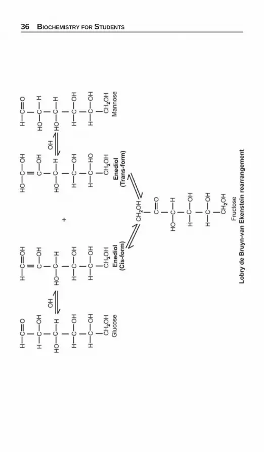
Action of Dilute Alkali
Monosaccharides on treatment with dilute alkali undergo avariety of molecular transformation through enediol for-mation. The enediols of sugars are good reducing agents andform the basis of reducing action of sugars in alkaline medium.
When glucose is treated with dilute alkali for several hours,the resulting mixture obtained contains both fructose andmannose in addition to glucose. A similar mixture of samesugars is obtained with any of the other two sugars. Thisinterconversion of related sugars by the action of dilute alkaliis termed as Lobry de Bruyn-van Ekenstein rearrangement(see page 36 for reaction).
Whereas sugars on boiling with strong alkalis are carame-lized to give yellow to brown resinous product. That is thereason why Benedict’s reagent containing sodium carbonateis preferred to Fehling solution containing sodium hydroxide.
Oxidation
Aldoses are oxidized under variety of conditions to thefollowing:
i. Aldonic acid: Whereby the first carbon atom (C-1) isoxidized to carboxyl group only. The rest of the moleculestructure remains unaffected.
ii. Uronic acid: Whereby the terminal carbon atom is oxidizedto carboxyl group only. The first carbon atom, i.e. alde-hydic group and the rest of the molecular structure

CHEMISTRY OF CARBOHYDRATES 35
remains unaffected. Uronic acid derivatives areparticularly important in detoxification process, i.e.,bilirubin is excreted as bilirubin diglucuronide. Besidesthis, D-glucuronic acid, D-galactouronic acid, D-mannou-ronic acid, L-induronic acid are important componentsof polysaccharides.
iii. Aldaric or saccharic acid: Whereby both the first carbon atom,i.e. aldehydic group and the terminal carbon atom, i.e.primary alcoholic group are oxidized to carboxyl group.
Galactose undergoes oxidation to form a dicarboxylic acid,mucic acid. This reaction is often important in the identificationof galactose.
Example: The oxidation products of glucose under differentconditions are given on Page 37.
Glucose Oxidase: The substrate for glucose oxidase is β-D-glucopyranose. Blood glucose which is an equilibriummixture of α- and β-anomers of D-glucose is qualitativelydetermined by the formation of hydrogen peroxide by thereaction (P-39).
36 BIOCHEMISTRY FOR STUDENTS

CHEMISTRY OF CARBOHYDRATES 37
Two very important uronic acids occuring in carbohydratesare D-glucuronates and L-iduronate (from hexose idose).
The only difference between these two molecule is thatthe carboxyl group is above the ring for D-glucuronate andbelow the ring for L-iduronate.

38 BIOCHEMISTRY FOR STUDENTS
This requires that the α-D-glucopyranose be rapidlyisomerized by mutarotation into the β-D-isomer. This reactionis fast without catalyst.
Q. A reducing carbohydrate gives a positive reaction withBarford’s test and mucic acid crystals on oxidation. Give thestructure of that carbohydrate. Would it exhibit property ofmutarotation. If so, what products are formed at equilibrium.Ans. Since Barford’s test is positive. It indicates that reducingcarbohydrate is monosaccharide.
Also mucic acid crystals are obtained on oxidation sugges-ting that the given reducing carbohydrate is galactose as itis the galactose which on oxidation gives mucic acid crystals.
D-galactose will show mutarotation due to the cyclizationof open chain form of D-galactose into α- and β- form withequal probability. The products at the equilibrium are:
CHEMISTRY OF CARBOHYDRATES 39
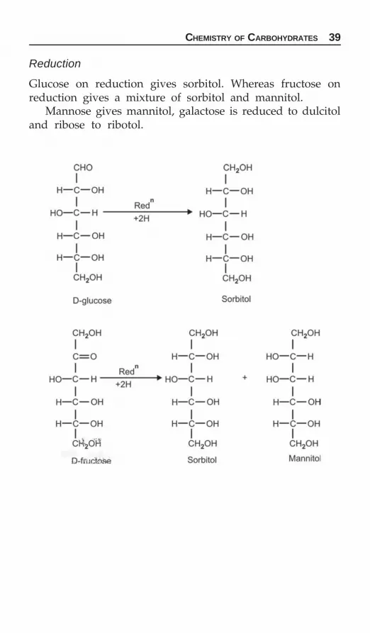
Reduction
Glucose on reduction gives sorbitol. Whereas fructose onreduction gives a mixture of sorbitol and mannitol.
Mannose gives mannitol, galactose is reduced to dulcitoland ribose to ribotol.

40 BIOCHEMISTRY FOR STUDENTS
Fermentation: Fermentation is the process of breakdown ofcomplex organic substances into smaller substances with thehelp of enzymes. Glucose is fermented to ethyl alcohol andcarbon dioxide by yeast. Hence this process is called alcoholicfermentation as alcohol is produced.
Glycosides Formation
Glycosides are sugar derivatives in which hydrogen of thehydroxyl group of hemiacetal or hemiketal form of the sugaris replaced by an organic moiety. A molecule of water iseliminated when this reaction takes place. Glycosides are notreducing sugars and do not show mutarotation.
If the organic moiety is derived from another monosac-charide, the product formed is disaccharide. If the organicmoiety is a noncarbohydrate, then it is called aglycone.
Aglycone: The noncarbohydrate portion of the glycoside iscalled the aglycone or aglucone.
Glycosides do not reduce alkaline copper sulphate becausesugar group is combined, i.e. aldehyde group is convertedto an acetal group.
Glycosides = Carbohydrate + Carbohydrate partor
noncarbohydrate part (aglycone)Examples
Cardiac glycosides = Carbohydrate + Digoxin or digitoxin(aglycone)
Indican = Carbohydrate + Indoxyl (aglycone)Amygdalin = Carbohydrate + Benzaldehyde (aglycone)
OLIGOSACCHARIDES
Oligosaccharides are arbitrarily defined as carbohydrates thatcontains two to ten monosaccharide units per molecule joinedby glycosidic linkages. On hydrolysis they yield monosac-charides.
Depending upon the number of constituent monosaccharideunits, the oligosaccharides are called disaccharides, trisaccha-rides, etc.

CHEMISTRY OF CARBOHYDRATES 41
Oligosaccharides are reducing sugars if one of the carbonylgroup is free (not involved in glycosidic linkage). The reducingpower of carbohydrate decreases as the number of their sugarcomponents increases.
DisaccharidesDisaccharides consist of two monosaccharides joined by aglycosidic linkage. The most common and important disaccha-rides are maltose, Lactose and Sucrose. Maltose and lactoseare reducing disaccharides whereas sucrose is nonreducingdisaccharide.
In general, the properties of disaccharides are similar tothose of monosaccharides. Reducing disaccharide sugars arenot as reducing agents as monosaccharide because of the lowerratio of reducing groups to carbon atoms.
MaltoseMaltose consists of two molecules of D-glucose joined byα (1,4)-glycosidic linkage. The anomeric carbon of one glucosemolecule is joined to the C-4 carbon of the second glucosemolecule. The anomeric carbon of the second glucose moleculeis free. So maltose is a reducing disaccharide.
42 BIOCHEMISTRY FOR STUDENTS
Maltose or malt sugar does not occur in free state but isformed as an important transitory intermediate product ofthe digestion of starch and glycogen.
Maltose reduces heavy metallic ions in alkaline solution(e.g. Benedict’s reagent), undergoes mutarotation andforms sunflower crystals of maltosazone with phenyl-hydrazine.
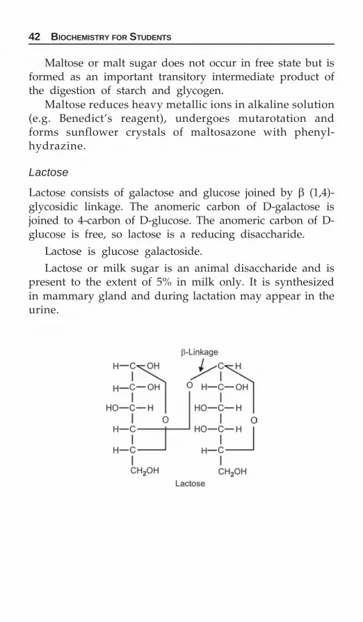
Lactose
Lactose consists of galactose and glucose joined by β (1,4)-glycosidic linkage. The anomeric carbon of D-galactose isjoined to 4-carbon of D-glucose. The anomeric carbon of D-glucose is free, so lactose is a reducing disaccharide.
Lactose is glucose galactoside.Lactose or milk sugar is an animal disaccharide and is
present to the extent of 5% in milk only. It is synthesizedin mammary gland and during lactation may appear in theurine.

CHEMISTRY OF CARBOHYDRATES 43
Lactose on treatment with concentrated nitric acid givesmucic acid crystals.
Lactose reduces Benedict’s reagent, undergoes mutarotationand forms cotton ball lactosazone crystals with phenyl-hydrazine.
Sucrose
Sucrose is a non-reducing disaccharide. Sucrose consists ofglucose and fructose joined by α(1) →β(2) glycosidic linkage.The anomeric carbon (C-1) of glucose molecule in αconfiguration is linked to anomeric carbon (C-2) of fructosein β configuration. So sucrose is a nonreducing disaccharideas both the reducing groups of glucose and fructose are linkedtogether and hence not available for reduction.
Sucrose or sugar cane is a plant disaccharide and is presentin high concentration in sugar cane and sugar beet. Sucroseis used for sweetening purpose.

44 BIOCHEMISTRY FOR STUDENTS
Sucrose does not reduce Benedict’s reagent, does not showmutarotation and does not form osazone with phenyl-hydrazine.
Invert Sugar
Sucrose on hydrolysis yields equimolecular amounts of glucoseand fructose. Since this mixture is levorotatory whereas theoriginal sucrose is dextrorotatory, the process is known asinversion because of the inversion of the sign of rotation, andthe mixture of glucose and fructose obtained is called as invertsugar.
H+
Sucrose Glucose + Fructose(+65.5) (+52.7) (–92)
Honey contains large amount of invert sugar.
Isomaltose
Isomaltose, a disaccharide is derived from the branch pointof starch. Isomaltose has α (1→ 6)-D-glucosidic linkage to asecond D-glucose residue.

CHEMISTRY OF CARBOHYDRATES 45
POLYSACCHARIDES
Polysaccharides are the polymers of monosaccharide unitswhich are joined in linear or branched chain fashion byglycosidic linkages.
Polysaccharides contain a large number of sugar componentsper free carbonyl group. In a branched polysaccharides, thereis only one reducing end and multiple nonreducing ends. Thusthese free carbonyl groups are not sufficiently potential toreduce the Benedict’s Reagent, etc.
By convention polysaccharides are given names ending in—an attached to the particular monosaccharide that make upthe polymer. Thus a name for a polysaccharide in general isglycans from glucose. Examples are mannans, xylans andarabans which are polymers of mannose, galactose, xylose andarabinose.
Polysaccharides have two important biological functions.1. As storage form of fuel (i.e., glycogen of animal origin and
starch of plant origin). Glycogen and starch are both storageform of glucose; glycogen is used by animals to storeglucose and starch is used by plants.
2. As structural components, e.g. Cellulose.The structural polysaccharides have β-linkage and the
storage polysaccharides have an α-linkage. The β-linkagekeeps the molecular linear whereas α-linkage tends to foldthe molecule, forming a gloublar structure then linear one.
Polysaccharides can be divided into two groups:a. Homopolysaccharidesb. Heteropolysaccharides.
Homopolysaccharides
They contain only one type of monosaccharides as the rep-eating unit and on hydrolysis gives only one type of sugar.
Example: Starch, cellulose, glycogen, dextrins, etc.
Starch
Native starch is a mixture of two polysaccharides.a. Amyloseb. Amylopectins.

46 BIOCHEMISTRY FOR STUDENTS
Amylose
Amylose is a linear unbranched molecule in which D-glucoseunits are linked by α–(1→4) glycosidic linkages. It is watersoluble and gives blue color with iodine.
Amylopectin
Amylopectin is a branched chain molecule in which D-glucoseunits in addition to α-(1,4) linkages are branched by α-(1,6)glycosidic linkages. This branching occurs on an average of24 to 30 D-glucose units. It is water insoluble and gives violetcolor with iodine.

CHEMISTRY OF CARBOHYDRATES 47
Starch is a nonreducing polysaccharide, tasteless substanceand gives blue color with iodine. Starch on hydrolysis withdilute mineral acids, i.e. with hydrochloric acid gives glucoseonly.
Action of amylases on starch: Amylases are hydrolytic enzymeswhich hydrolyze polymers of glucose containing α-(1 → 4)glycosidic linkages,
Amylases are of two types:1. α-Amylases.2. β-Amylases.α-amylases are present in saliva and pancreatic juice. They
act on starch, hydrolyzing α-(1,4) glycosidic linkages in arandom manner to yield glucose, free maltose and smallerunits of starch called starch dextrins. These starch dextrinscontain the original α-(1,6) glycosidic linkages. α-amylasecannot hydrolyze the α-(1,6) linkages at the branched pointof amylopectins. The α-amylases are activated by chlorideions.
β-amylases present in barley malt, cleave successive maltoseunits beginning from nonreducing ends of starch to give mal-tose. β-amylase yield only maltose with amylose and smallerbranched polysaccharides, known as limit dextrins, as wellas maltose with amylopectin. β-amylases also cannot hydrolyzeα-(1,6) linkages at the branched point of amylopectin.
Cellulose
Cellulose is a linear polymer of D-glucose units joinedtogether by β–(1,4) glycosidic linkages. On partial hydrolysis,cellulose yields β-1,4 disaccharide cellobiose instead ofmaltose. Cell-ulose is water insoluble, nonreducing and givesno color with iodine.
Unlike starch and glycogen which are readily digested,cellulose cannot be utilized for energy purposes by humanbeings, because the enzyme which cleavage β-(1,4) linkage ismissing in the gastrointenstinal tract and hence, merelyprovide bulk to the diet. Cellulose is present in plant leaves,stems, and outer coverings of fruits and vegetables. Cellulose

48 BIOCHEMISTRY FOR STUDENTS
is a component of fiber (nondigestible carbohydrate) in thediet. Cellulose is present in plant leaves stems and outer cover-ings of fruits and vegetables. Cellulose aids intestinal mobilityand acts as an stool softener and reduces bowel cancer. Thenutrition value of cellulose is nil. Celluloses are the mostabundant organic compound on earth. Celluloses are the majorcomponents of plants comprising 20 to 45% of this cell wall mass.
Glycogen
Glycogen is the carbohydrate reserve of the body. Glycogenis also called animal starch, because it serves as nutritionalreservoir in animal tissues.
Glycogen is a highly branched chain molecule in whichglucose unit in addition to linear α-(1,4) linkages are alsolinked by α-(1,6) at the branched point. This branching repeatsafter every 8-10 glucose units.
Glycogen is water soluble and has no reducing property.It gives red color with iodine.
Glycogen is stored in liver and muscle. About three-fourthof all the glycogen in the body is stored in muscle.
Difference between starch and glycogen.1. Starch is of plant origin whereas glycogen is of animal
origin.2. Glycogen is much more branched than the starch. In starch,
the branching is after every 24 to 30 glucose units, whereasin glycogen, the branching is after every 8 to 10 glucose units.
3. Starch gives blue color with iodine solution whereas glyco-gen gives red color.
Dextrins
They are the partial hydrolytic products of starch by α-amylase,β-amylase and acids. Dextrins formed from amylases have

CHEMISTRY OF CARBOHYDRATES 49
unbranched chains while those formed from amylopectins arebranched. All dextrins have free sugar group and accordinglyreduce alkaline copper sulphate solution.
HETEROPOLYSACCHARIDES
Heteropolysaccharides are made up of mixed disaccharidesrepeating units and on hydrolysis gives a mixture of morethan one product of monosaccharides and their derivativesof amino sugars and sugar acids.
They are the essential components of the tissues wherethey are present in combination with proteins as mucoproteins.
They are also called mucopolysaccharides or glycosaminoglycans (CAG).
The other suitable name for such heteropolysaccharidesis Glycosaminoglycan or CAG. Glycosaminoglycans are un-branched polysaccharides consisting of repeating dissaccharideunits comprising a sugar linked to either N-acetylglucosamineor N-acetylgalactosamine.
They can be divided into:1. Neutral mucopolysaccharides2. Acidic mucopolysaccharides.Acid mucopolysaccharides are present in connective tissues.
They contain hexosamine as the repeating disaccharide unit.The repeating structure of each disaccharide contains alternate1,4 and 1,3 linkages.
The most common CAGs are:
Hyaluronic Acid
Hyaluronic acid is present in the connective tissues, synovialfluid and vitreous fluid in combination with proteins.
It is an unbranched polymer. The repeating disaccharideis made up of D-glucuronic acid and N-acetyl D-glucosamine.The monosaccharide subunits are linked by β-(1,4) and β-(1,3)glycosidic linkages. Glc UA-β(1 → 3) – Glu NAc connectedby β(1 → 4) linkages.

50 BIOCHEMISTRY FOR STUDENTS
On acid hydrolysis it gives an equimolar quantities of glucu-ronic acid, glucosamine and acetic acid.
Hyaluronates form viscous lubricants of joints and gel likesubstance inside the eyes-vitreous humor.
Heparin
Heparin is glucosaminoglycans. Heparin is an acidic mucopoly-saccharide in which both the amino and the hydroxyl groupsare combined with sulphuric acid, which causes it to be slightlyacidic substance.
Heparin is present in liver, lungs, thymus, spleen and blood.Heparin is blood anticoagulant. Heparin contains D-gluco-samine, D-glucuronic acid or L-iduronic acid as the repeatingdisaccharide units. The glucosidic linkage is α(1,4) involvingthe glucuronic acid anomeric carbon hydroxyl with hydroxylgroup at C-4 of glucosamine.

CHEMISTRY OF CARBOHYDRATES 51
Chondroitin Sulfates
They are present in connective tissues and serve as a structuralmaterial such as cartilage, tendons and bones.
Chondroitin sulfates are sulfated polysaccharides. Chond-roitin sulfate is galacto aminoglycans. The acid hydrolysis ofchondroitin sulfate yield D-galactose, D-glucuronic acid, aceticacid and sulfuric acid.
Sialic Acids
Sialic acids are N-acetyl derivatives of neuraminic acid andare widely distributed in tissues such as mucins are presentin blood group substances.

52 BIOCHEMISTRY FOR STUDENTS
Neuraminic acid is a condensation product of pyruvic acidand mannosamine.
Examples Repeating units
Hyaluronic acid Glucuronic acid; N-Acetyl glucosamineChondroitin Glucuronic acid; N-Acetyl galactosamineChondroitin-4- Glucuronic acid;sulfate N-Acetyl galactose-4-sulphate(Chondroitinsulfate A)Heparin Glucosamine-6-SO4; glucuronic
acid-SO4; iduronic acid
Other CAGs
1. Chondroistin sulfate and dermatan sulfate are galactosa-mine glycam.
2. Heparin sulfate, heparin and keratan sulfate are glucosa-mine glycam.
Mucoproteins and Glycoproteins
If the carbohydrate associated with protein is greater than4%, then the complex protein is called mucoprotein. If thecarbohydrate content is less than 4%, then is called glyco-protein.
Plasma α1 and α2 globulins are glycoproteins.
Blood Group Substances
They are water soluble, high molecular weight substances,made up of polysaccharides and proteins. They are presentin saliva, gastric mucin, erythrocyte membranes, etc.
The immunological specificity resides in oligosaccharidepart. The residues present in the oligosaccharides are L-fucose,D-galactose, N-acetyl-D-galactosamine and N-acetyl gluco-samine.

According to Bloor, lipids are defined as a group of naturallyoccurring substances consisting of the higher fatty acids, theirnaturally occurring compounds and substances found naturallyin association with them. It includes a wide variety of subs-tances with different structures. They are insoluble in waterbut are soluble in so-called fat solvents such as ether, acetone,chloroform, benzene, etc. Associated with them are variousfat soluble, non-lipid substances which includes carotenoidpigments and certain vitamins, i.e. vitamins A, D, E and K.Lipids are widely distributed throughout both plant andanimal kingdom and are essential constituents of cell mem-brane.
Fats are said to be protein sparing because their availabilityin the diet reduces the need to burn proteins for energy.
Lipids have several important biological functions.
1. They serve as the reservoir of energy because of their:a. High energy content. The calorific value is 9 kcal/gm
as compared to carbohydrates which have calorific valueof 4 kcal/gm.
b. Storage in concentrated form in water free state (anhy-drous) in the tissues as compared to carbohydrateswhich are highly hydrated and cannot be stored in suchconcentrated form.
2. As structural components of cell membranes.3. As transport forms of various metabolic fuel.4. As protective coating on the surface of many organs such
as kidney, against injury.5. To facilitate the absorption of the fat soluble vitamins A,
D, E and K.
Chemistry of Lipids
CHAPTER
3

54 BIOCHEMISTRY FOR STUDENTS
Dietary fat can be divided into two types:a. Visible fat or fat consumed as such, e.g. butter, oils, ghee.b. Invisible fat or fat present as part of other foods items,
e.g. egg, fish, meat, cereal, nuts, etc.
Classification and Functions of Lipids
Classification Functions1. Fatty lipids Metabolic fuel, building block for
other lipids2. Triglycerides Fatty acid storage, transport3. Phospholipids Membrane structure, storage of
arachidonic acid4. Sphingolipids Membrane structure5. Ketone bodies Fuel
SIMPLE LIPIDS
They are esters of fatty acids with various alcohols.If the alcohol is glycerol, then they are called fats or neutral
fats and are also called triglycerides as all the three hydroxylgroups of the glycerol are esterified.
If the fat is liquid at ordinary temperature it is called an oil.Triglycerides are given by the formula
R = Same or differentAll of the three fatty acids can be same or different.If all the three fatty acids are same, then they are called
simple triglycerides. If the fatty acids are different, then theyare called mixed triglycerides. In nature, mixed triglyceridesare more abundant than the simple triglycerides.

CHEMISTRY OF LIPIDS 55
If the alcohol is high molecular weight instead of glycerolthen they are called waxes.
Comparison of simple and compound lipids is terms of theircomposition.
Lipid Components
Simple lipids 1. Triglycerides Glycerol + Fatty acids2. Waxes Alcohol + Fatty acids
(Both long chain)Compound 1. Phospholipids Glycerol + Fatty acidslipids + Phosphate
2. Sphingomyelins Sphingosine + Fatty acid+ Phosphate + Choline
3. Cerebrosedes (glycolipids) Sphingosine + Fatty acid+ Simple sugar(s)
4. Gangliosides (glycolipids) Sphingosine + Fatty acid+ 2-6 simple sugars one ofwhich is sialic acid
Fatty Acids
Fatty acids in nature as such are not very abundant but arepresent as ester.
Fatty acids are represented by general formula R—COOH.A fatty acid is a long chain aliphatic carboxylic acid.
General points about them.1. They are monocarboxylic acids.2. Number of carbon atoms are even, though odd number
fatty acids exist but are very rare.3. They may be saturated or may be unsaturated.
If unsaturated they can be monounsaturated acid orpoly-unsaturated acid.
Mammals and plants contain both monosaturated and poly-unsaturated fatty acids whereas all the fatty acids containingdouble bonds that are present in bacteria are monounsat-urated. Plant and fish fats contain more polyunsaturated fattyacids than animal fats. The double bonds in a polyunsaturatedfatty acid are neither adjacent nor conjugated since this would

56 BIOCHEMISTRY FOR STUDENTS
make the structure to easily oxidisable when exposed toenvironment oxygen. Rather the double bonds are three carbonapart; this provide somewhat greather protection againstoxidations.
Fats obtained from animals are generally saturated andthose from plants are commonly polyunsaturated. However,these are some exceptions: coconut, palm oils are highlysaturated.
The most common among the saturated fatty acids arepalmitic acid (C16), stearic acid (C18) and among the unsaturatedfatty acid, oleic acid (C18). Unsaturated fatty acids have lowermelting point than saturated fatty acids of same chain length.Fatty acids with odd number of carbon atoms occur in traceamounts in terrestrial and marine animals.
Fatty acids with one to eight carbons are liquids at roomtemperature while those with more carbon atoms are solids.The most common fatty acids in neutral fats are:
No. of atoms Formula
Butyric acid 4 CH3—(CH2)2—COOHCaproic acid 6 CH3—(CH2)4—COOHLauric acid 12 CH3—(CH2)10—COOHPalmitic acid 16 CH3—(CH2)14—COOHStearic acid 18 CH3—(CH2)16—COOHOleic acid 18 CH2—(CH2)7—CH=CH
—(CH2)7 —COOH
Fats as an Energy Source
Fats/oils are tremendous source of energy and 40% of totalcalories are provided by fatty acids that come from trigly-cerides and phospholipids.
Naturally occurring straight chain saturated fatty acid
No. of Common name Type Systematic nameC atoms
2 Acetic acid Short n-Ethanoic acid3 Propionic acid chain n-Propanoic acid4 Butyric acid n-Butanoic acid
⎫⎬⎭
Contd...

CHEMISTRY OF LIPIDS 57
8 Caprylic acid Medium n-Octanoic acid10 Capric acid chain n-Decanoic acid12 Lauric acid n-Dodecanoic acid14 Myristic acid Long n-Tetradecanoic acid16 Palmitic acid chain n-Hexadecanoic acid18 Stearic acid n-Octadecanoic acid20 Arachidic acid n-Eicosanoic acid
The presence of double bond in the molecule gives riseto geometric isomerism. All naturally occurring unsaturatedlong chain fatty acids are found in cis isomer.
Most plant fats are liquid since they contain a largeproportions of unsaturated fatty acids with melting points.Animal fats, on the other hand, contain a high proportion ofpalmitic and stearic acids, and are solid or semi-solid at roomtemperature. Milk fat is unusual in containing a high proportionof shorter chain (C4-C14) fatty acids.
Essential Fatty Acids
They are also called polyunsaturated fatty acids. They are notsynthesized in the body and hence, have to be provided in thediet. Although linolenic acid and arachidonic acid are syn-thesized by the body from linoeic acid, but they are synthesizedin insufficient quantity for our needs.
The deficiency of essential fatty acids in humans gives riseto dry, scaly skin, hair loss, poor wound healing, failure ofgrowth and increase in metabolic rate. These essential fattyacids requirement is about 1% of the caloric intake be in theform of essential fatty acids. Essential fatty acids are neededfor proper cell membrane formation and for synthesis ofprostaglandins prostacyclins, thromboxanes and leukotrienes.
Essential fatty acids are:
No. of No. of Position of Dietarycarbon double double bonds sourceatoms bonds from carboxyl end
1. Linoleic acid 18 2 9, 12 Vegetable oils2. Linolenic acid 18 3 9, 12, 15 Vegetable oils3. Arachidonic acid 20 4 5, 8, 11, 14 Vegetable oils4. Timnodonic acid 20 5 5, 8, 11, 14, 17 Fish oils
⎫⎬⎭
⎫⎬⎭
Contd...

58 BIOCHEMISTRY FOR STUDENTS
Vegetable oils are oils and have many double bonds hencepolyunsaturated appears on the label of must vegetable oils.Butter, on the other hand, is a fat and hence would be expectedto have saturated fatty acids, i.e. no double bonds.
Two of the essential fatty acids, linoleic and linolenic acidsare not synthesized by the mammal but are synthesized byplants. As long as adequate amounts of linoleic acids areavailable mammals can synthesize other essential acids.
Structures
Linoleic acidCH3(CH2)4CH=CHCH2CH=CH=(CH2)7COOHLinolenic acid CH3CH2CH=CHCH2CH=CHCH2CH
=CH(CH2)7COOHArachidonic acid CH3(CH2)4(CH=CHCH2)4(CH2)2COOH
Essential fatty acids are necessary in the biosynthesis ofprostaglandins and for proper cell membrane formation.Prostaglandins are hormone-like compounds which in smallamounts have profound effect.
Important fatty acids in mammalian tissues:
Common name No. of carbon Double Position ofatoms bonds double bonds
Acetic acid 2 0Lauric acid 12 0Myristic acid 14 0Palmitic acid 16 1 9Stearic acid 18 0Oleic acid 18 1 9Linoleic acid 18 2 9, 12Linolenic acid 18 3 9, 12, 15Arachidonic acid 20 4 5, 8, 11, 14
Prostaglandins
Prostaglandins are the derivatives of prostanoic acid whichare the cyclic derivatives of unsaturated fatty acids havingtwenty carbon atoms.

CHEMISTRY OF LIPIDS 59
Prostaglandins are synthesized from essential fatty acidssuch as linoleic acid, linolenic acid and arachidonic acid. Fivetype of rings are found in the naturally occurring prostag-landins giving rise to prostaglandins of A, B, E, F and G orH series. The prostaglandins which are widely distributedin the body are PGE1, PGE2, PGE3, PGF1α, PGF2α and PGF3α.
Linolenic acid is the precursor to PGE3 and PGF1α, Arachi-donic acid is the precursor to PGF2 and PGF2α.
Prostaglandins are synthesized and released by all mamma-lian cells and tissues except RBC. Also prostaglandins are notstored in cells but are synthesized and released immediately.
Biological function of prostaglandins:1. They lower blood pressure (PGE, PGA, PGI2).2. They are used in the induction of labor, termination of
pregnancy and prevention of conception (PGE2).3. They are used in treatment of gastric ulcer (PGE).4. They are used to prevent inflammation.5. They are used in asthma.6. They are used in congenital heart disease.7. They inhibit platelet aggregation (PGI2) whereas PGE2 pro-
mote clotting process.
Eicosanoids: Fatty Acid Derivatives
Eicosanoids are all derived from C-20 carbon arachidonic acid.Prostaglandins, Thromboxanes and Leukotrienes are collecti-vely referred as eicosanoids.They have a variety of extreme potent hormone like actionon various tissues. These compounds are involved in the regu-lation of blood pressure, diuresis, blood platelet aggregation,effects on immune and nervous systems, gastric acid secretionand muscle contraction.

60 BIOCHEMISTRY FOR STUDENTS
Properties of Fats
1. Acrolein formation: When glycerol is heated in the presenceof a dehydrating agent such as potassium bisulphate, acroleinis produced.
Acrolein has a characteristic unpleasant odor and is easilyidentified on the basis of this smell. This reaction occurswhether glycerol is in free or esterified form as occurs in thetriglycerides.2. Hydrogenation: Unsaturated fats can be hydrogenated bythe addition of hydrogen across the double bonds of the fattyacids in the presence of nickel as catalyst to give fully saturatedfats. The above process is called Hardening of oils wherebyvegetable oils are hydrogenated to produce commercial cookingfats.
3. Saponification: Hydrolysis of a fat by alkali is calledSaponification. The products of hydrolysis are glycerol andalkali salts of fatty acids, which are called soaps. Since thecommon fats contain palmitic acid, stearic acid and oleic acidpredominantly, the soaps used for washing consist largely ofsodium salts of these acids. While these fatty acids are insolublein water their sodium and potassium salts are water soluble.
4. Rancidity: Rancidity is a chemical change resulting in unpl-easant odor and taste on storage when fats are exposed to light,heat, air and moisture. Rancidity is more rapid at high temp-

CHEMISTRY OF LIPIDS 61
erature. Rancidity may be due to hydrolytic or oxidative changetaking place at the double bonds of the unsaturated fatty acidsresulting in short chain aldehydes or ketones which haveunpleasant odor.
The addition of certain substances, called antioxidants suchas ascorbic acid and vitamin E prevents rancidity whereasaddition of proxidants like copper, lead and nickel quickensrancidity.
The oxidation of unsaturated bonds in fatty acids whenthe are exposed to oxygen in the environment is referred toas either auto oxidation or peroxidation. Rancid fats are thosethat contain an appreciable amount of peroxidized fatty acid.
Antioxidants are generally added to many food fats toimprove their storage quantities.
Characterization of Fats
Saponification number: Saponification number is defined as the“milligrams of KOH required to saponify 1 gm of fat”. Sincefats are mixtures of triglycerides largely of mixed type so thesaponification number of a fat indicates the average molecularweight (average chain length) of the fatty acids constitutingor comprising the fat.
Saponification number is inversely proportional to theaverage chain length of the fatty acids. Higher the saponificationnumber, the shorter will be the chain lengths of the fatty acidsand vice versa.
The saponification number of some of the fats is given below:
Fat Saponification number
Butter fat 210-230Human fat 195-200Olive oil 185-195Cottonseed oil 194-196Linseed oil 188-195Coconut oil 250-260Castor oil 175-185
Iodine number: Iodine number of a fat is defined as the numberof gm of iodine absorbed by 100 gm of the fat. Halogens,e.g. iodine or bromine are taken up by the fats because ofthe presence of double bonds present in the fatty acid partof the fat.

62 BIOCHEMISTRY FOR STUDENTS
Iodine number is a measure of the degree of unsaturationof fat.
Iodine number of some of the fats is given below:
Fat Iodine number
Butter fat 26 - 28Human fat 65 - 70Olive oil 80 - 90Peanut oil 85 - 100Corn oil 105 - 115Soyabean oil 135 - 145Linseed oil 170 - 200
Acid number: Acid number is defined as the milligrams of KOHrequired to neutralize the free fatty acids present in 1 gm.of fat. This is used in determining the rancidity due to freefatty acids.
Acetyl number: The acetyl number is defined as the milligramsof KOH required to neutralize acetic acid liberated by thesaponification of 1 gm of fat after it has been acetylated.
Since acetylation takes place at the hydroxy groups of thehydroxy fatty acid residues in the fat, so acetyl number isa measure of the hydroxy fatty acids in the fat content.
Polenske number: The ml of N/10 KOH required to neutralizethe insoluble fatty acids from 5 gm. of fat which are not steamvolatile.
Reichert Meissel number: This represents the ml of N/10 KOHrequired to neutralize the volatile acid obtained from 5 gmof fat which has been saponified then acidified to liberate thefatty acids and then steam distilled.
Butter fat, which contains shorter chain fatty acids has aReichert Meissel number of 26 to 30.
COMPOUND LIPIDS
They are the esters of fatty acids containing nitrogen basein addition to an alcohol and fatty acids.

CHEMISTRY OF LIPIDS 63
A molecule which has changed and an unchanged portionis called an amphipathic molecule.
Phospholipids
They are also known as phosphatides. Phospholipids act asa detergent and increase the solubility of other lipids. Theyare present in all cells as well as in the plasma.
Phospholipids include the following groups:
Phosphatidic Acid
The general structure of phosphatidic acid.They are important intermediates in triglyceride synthesis.
Phosphatidic acid on hydrolysis yield glycerol, fatty acid andphosphoric acid.
Lecithins
The structure of lecithins are:
Lecithin contains saturated fatty acid residue at the α-posi-tion and unsaturated fatty acid residue at the β-position ofthe glycerol.
Lecithins on hydrolysis give glycerol, fatty acid, phosphoricacid and choline.

64 BIOCHEMISTRY FOR STUDENTS
Cephalins
The structure of cephalins are:
Cephalins differ from lecithins with respect to base attachedto phosphoric acid.
If the base is ethanol amine then it is called phosphatidylethanolamine or ethanolamine cephalin.
If the base is amino acid serine then it is called phosphatidylserine which is also called serine cephalin.
Cephalins on hydrolysis yield glycerol, fatty acids, phos-phoric acid, ethanol amine or serine.
Phosphatidyl Inositol
The structure of phosphatidyl inositol is:It contains inositol in place of base.

CHEMISTRY OF LIPIDS 65
Cardiolipin
An important phospholipid of mitochondrial membrane iscardiolipin. It is a diphosphatidyl glycerol in which two phos-phatidic acids are joined by a molecule of glycerol.
These phospholipids are particularly rich in the polyunsatu-rated fatty acids especially linoleic acid.
Plasmalogens
These compounds possess fatty aldehyde in place of fatty acidat the α-position, with the result the normal ester linkage isreplaced by the ether linkage on the C1 carbon. In some cases,bases like choline, serine or ethanol amine are also found.They are found in brain and heart.

66 BIOCHEMISTRY FOR STUDENTS
Sphingomyelins
Phospholipids containing sphingosine are called sphingo-myelins. They contain, a complex base sphingosine in additionto phosphoryl choline. A fatty acid is attached to the aminogroup of the sphingosine. No glycerol is present.
Sphingomyelins are present in all tissues especially in brainand other nervous tissues.
Sphingomyelins on hydrolysis yield sphingosine, fatty acid,phosphoric acid and choline.
Increased concentration of sphingomyelins occur in liver,spleen, etc. in a condition known as Niemann-Picks disease.
Cerebrosides or Glycolipids
Glycolipids are carbohydrate-glyceride derivatives containingsugar, sphingosine and a fatty acid. These compounds do notcontain phosphoric acid. If the sugar component is galactose,the lipid is termed galactolipid. The term cerebroside is usedbecause it is found in large quantities in brain tissuesparticularly in white matter.
Structure of Sphingomyelins

CHEMISTRY OF LIPIDS 67
On hydrolysis cerebrosides give sphingosine, a fatty acidand galactose. Cerebrosides are differentiated on the basisof fatty acid present.Examples:
Kerasin: It contains Lignoceric acidCerebron: It contains Hydroxy Lignoceric acidNervon: It contains Nervonic acidOxynervon: It contains Hydroxy Nervonic acidCerebrosides occur in large amounts in the white matter
of brain and in the myelin sheaths of nerves.In Gaucher’s disease, large amount of cerebroside accumu-
lates in the liver and spleen.
Gangliosides
They are found in nerve tissues. They contain carbohydrates,N-acetyl galactosamine and N-acetyl neuraminic acid.
Cerebrosides

68 BIOCHEMISTRY FOR STUDENTS
Sulfatides (Sulpholipids)
They are cerebrosides having a sulfate group attached to thegalactosyl residue.
DERIVED LIPID
Those substances which are derived from the above twogroups by hydrolysis. These include fatty acids of variousseries, steroids, bile acids and substances associated with lipidsin nature such as carotenes, vitamin A, D, E and K.
Lecithins are hydrolyzed by certain enzymes, phospholi-pases or lecithinases. The nature of hydrolysis depends uponthe type of phospholipase used.
Phospholipase A: Present in snake venom (cobra) hydrolyzesfatty acid in α or 1-position of glycerol in the lecithin to formlysolecithins. In the similar manner it acts on cephalin.
Phospholipase B: Hydrolyzes the remaining fatty acid of lyso-lecithin present at β or 2-position to form glyceryl phosphoryl-choline.
Phospholipase C: Hydrolyzes phosphorylcholine from lecithinsto form diglycerides. Phospholipase C catalyses the hydrolysisat the glycerol side of the phosphate group.
Phospholipase D catalyses the hydrolysis on the phosphateside of the phosphate group.

CHEMISTRY OF LIPIDS 69
Phospholipase D: Hydrolyzes choline from phosphatidyl ethano-lamine (cephalin) form phosphatidyl serines.
There are two classes of nonsaponificable lipids.
Terpenes
They are linear or cyclic compounds formed by condensationof two or more isoprene units.
Other important terpenoid compounds are:a. Tocopherol (vitamin E)b. Coenzyme Q (also called ubiquinone)c. Vitamin K (a naphthaquinone)They include vitamins A, E, K and carotenes, etc.
Cyclopentano-perhydro-phenanthrene ring
(Steroid nucleus)
Steroids
The term steroids includes many compounds which havehowever one feature in common, the steroid skeleton. Steroidsare the derivatives of cyclopentano-perhydro-phenanthrenering (consists of four fused rings). This is a saturated (per-hydro) pheranthrene ring with a cyclopentane ring attached.Steroids are steroidal alcohol. The most important memberof this group is cholesterol. The four rings that make up

70 BIOCHEMISTRY FOR STUDENTS
perhydro-cyclopentano-phenanthrene are named alphabetic-ally from left to right.
Despite popular belief, cholesterol is not a poison but a verynecessary part of our cell membranes and the basis of sexualhormones (androgens, estrogens, etc). Cholesterol is only aproblem if it is in excess and in this respect we do not needcholesterol in our diets because body can synthesis it.
Steroids belong to the class of important biological com-pounds with diverse physiological activities.
Some of the biologically important steroids are:
a. Ergosterol: UV radiation causes rupture ofring B to produce vitamin D.
b. Bile acids: In lipid metabolism.c. Adrenal cortex Corticosterone and cortisol.
steroids:d. Female hormones: Progesterone and estrogen.e. Male sex hormones: Testosterone and androsterone.
Cholesterol is an animal fat and it does not occur in plants.
Cholesterol contains hydrogen group at C-3, methyl groupsat C-10 and C-13, a double bond at C-5 and an 8C branchedalkyl group attached to C-17. This marks a total of 27C.This ring structures are lipid soluble and hydroxyl group ofC-3 is hydrophilic.

CHEMISTRY OF LIPIDS 71
Plants have stigmasterol and β-sitosterol which differ onlyin the alkyl group side chain attached at C-17.
The Antioxidant SystemIn healthy individuals, the antioxidant system defends tissuesagainst free radical attack. Antioxidants are known to preventcellular damage and enhance repair. Three classes of antioxi-dants have been identified.a. Primary antioxidants: They prevent the formation of new
free radical species, e.g. superoxide dimutase, glutathioneperoxidase, ceruloplasmin, transferrin, ferritin.
b. Secondary antioxidants: They remove newly formed freeradicals before they can initiate chain reactions. These chainreactions can lead to cell damage and further free radicalformations, e.g. vitamin E, vitamin C, β-carotene, uric acid,bilirubin, albumin.
c. Tertiary antioxidants: They repair cell structures damagedby free radicals attack, e.g. DNA repair enzymes,methionine sulphoxide reductase.Deficiency in the antioxidant system can develop for a
number of reasons:a. Low intake of dietary antioxidantsb. Total parenteral nutritionc. Decreases that reduce the absorption of antioxidant
nutrients from food, e.g. Crohn’s diseased. Renal dialysis
In these situations the antioxidant system struggles toprotect the body from free radical attack and as a result therisk of free radical-mediated disease increases.
Increased antioxidant status by supplementation mayindeed reduce the risk of certain diseases.
i. High intake of vitamin E has been associated with reducedrisk of mortality from ischemic heart disease.
ii. High incidence of vitamin C and β-carotene have beenassociated with a reduced incidence of some cancers.
iii. Dietary supplementation of vitamin E, β-carotene andselenium significantly reduces mortality from esophagealcancer.
iv. Within one week on antioxidant rich, low fat diet reduceslipid peroxide levels and increased aborrhic acid level inpatient in the acute myocardial infarction.

72 BIOCHEMISTRY FOR STUDENTS
Free Radicals
A free radicals is defined as any atom or molecule that possessesan unpaired elactron. It can be anionic, cationic, or neutral.Free radicals are highly reactive molecules generated by thebiochemical redox reactions that occur as part of normal cellmetabolism and by exposure to environmental factors suchas UV light, cigarette smoking, environmental pollutions andgamma radiations.
Human body is constantly under attack from free radicals.Some toxic compounds can result in the production of free
radicals which include anticancer drugs, anaesthetics, anal-gesics, etc.
The free radicals species which occur in the human bodyare:a. Superoxide radical (•O2¯)b. Hydroxyl radical (OH•)c. Nitric oxide radical (NO•)d. Peroxyl radical (ROO•).
Once formed, free radicals attack cell structures within thebody. As a result, free radicals have been implicated innumerous diseases such as atherosclerosis, cancer, AIDS, liverdamage, rheumatoid arthritis, Parkinson’s disease, etc.
Process of Lipid Peroxidation
This process is responsible for randicity of food. This processinvolves:
i. Initiationii. Propagation
iii. Termination.
Initiation
ROOH + Metaln+ → ROO• + Metal(n-1)+ + H+
X• + RH → R• + HX
Propagation
R + O2 → ROO•
ROO• + RH → ROOH + R•

CHEMISTRY OF LIPIDS 73
Termination
2ROO• → ROOR + O2ROO• + R•→ ROORR• + R• → RR
Eicosanoids
Eicosanoids are formed from C20 polyunsaturated fatty acid.Arachidonate and some other C20 fatty acids give rise to eico-sanoids which includes prostaglandins, thromboxanes, leuko-trienes, lipoxins. There are two pathways of their formation:1. Cyclooxygenase pathway2. Lipooxygenase pathway.

74 BIOCHEMISTRY FOR STUDENTS
CHEMISTRY OF AMINO ACIDS
Naturally occurring amino acids are amino acids containingamino group and carboxyl group on the same alpha carbonatom and are represented by the general formula:
Chemistry of AminoAcids and Proteins
CHAPTER
4
All amino acids found in living systems, plant and animalproteins are L-α-amino acids. Glycine is the only amino acid,which is optically inactive and cannot be resolved into D-orL-form because of symmetry on the α-carbon atom. All otheramino acids are optically active.
The configuration of L-α-amino acid is:
A variety of classification of amino acids are possible. Eitherthey can be classified according to the presence of acidic, basicor neutral groups or upon their chemical structures, i.e., pre-sence of polar groups, nonpolar groups, sulphur containinggroups, aromatic groups, heterocyclic ring, branched chain andso on.

CHEMISTRY OF AMINO ACIDS AND PROTEINS 75
Classification of Amino Acids
1. Aliphatic amino acids2. Aromatic amino acids3. Heterocyclic amino acids.

76 BIOCHEMISTRY FOR STUDENTS

CHEMISTRY OF AMINO ACIDS AND PROTEINS 77

78 BIOCHEMISTRY FOR STUDENTS
Tryptophan
Besides these there are number of amino acids which areobtained in free or combined form but do not occur in proteinmolecules, e.g. thyroxine, triiodothyronine, ornithine, citruline,α-aminobutyric acid, β-alanine, etc. Their structures are givenhere.

CHEMISTRY OF AMINO ACIDS AND PROTEINS 79
A dipeptide has two amino acids joined by a single peptidebond; a tripeptide is composed of three amino acids joinedby two peptide bonds: a polypeptide is one in which anynumber (n) of amino acids or (AA)n are linked together by(n-1) peptide bonds.
Examples of relatively smaller peptides that possess biologi-cal activity are glutathione, oxytocin, vasopressin, hypertensin,etc.
Glutathione is a tripeptide consisting of glutamic acid,cystine and glycine and is found in red blood cells.
Oxytocin and vasopressin are produced by the posteriorof the pituitary gland. Each is made up of nine amino acids.Oxytocin causes contraction of smooth muscle and it is usedin obstetrics to initiate labor whereas vasopressin raises bloodpressure and reduces the secretion of urine.
Angiotensin I has 10 amino acids and angiotensin II has8 amino acids. They cause hypertension.
Functions of Amino Acids
Amino acids serve as:1. Building block of proteins2. Precursors of:
a. Hormones. (peptide and thyroid)b. Purinesc. Pyrimidinesd. Porphyrinse. Vitamins
3. Neurotransmitter such as tryptophan (sertonin).4. Transport of nitrogen: Alanine, glutamine.5.Substrates for protein synthesis: Those for which there is
a codon.

80 BIOCHEMISTRY FOR STUDENTS
Essential Amino Acids
Those amino acids which are not synthesized in the body andhence have to be provided in the diet. They are also calledindispensible amino acids. There are eight essential aminoacids.
They are leucine, isoleucine, threonine, tryptophan, pheny-lalanine, valine, methionine and lysine.
Adequate amounts of essential amino acids are requiredto maintain the proper nitrogen balance.
Deficiency of one or more essential amino acids in the dietgives rise to decrease in protein synthesis resulting in failurein growth of the child, negative nitrogen balance in adultsand fall in plasma proteins and hemoglobin levels.
Semiessential Amino Acids
Those amino acids which are synthesized partially by the bodybut not at a rate to meet the requirement of the body arecalled semiessential amino acids.
Semiessential amino acids are arginine and histidine.
Nonessential Amino Acids
Those amino acids which are synthesized by the body. Theseamino acids are derived from carbon skeletons of lipids andcarbohydrates during their metabolism or from the transfor-mation of essential amino acids.
Nonessential amino acids are alanine, arginine, asparagine,aspartic acid, cysteine, glutamic acid, glutamine, glycine, pro-line, serine and tyrosine.
Nitrogen Balance
The ratio of:
Intake NOutput N
=
> 1, i.e. positive nitrogen balance, e.g. duringpregnancy, convulsions and growth.< 1, i.e. negative nitrogen balance, e.g. in mal-nutrituion and in certain wasting diseaseswhere, there is tissue breakdown.
1, i.e. nitrogen equilibrium. Normal adultsare in nitrogen equilibrium.

CHEMISTRY OF AMINO ACIDS AND PROTEINS 81

82 BIOCHEMISTRY FOR STUDENTS
Ninhydrin Reaction
All amino acids (except proline and hydroxyproline), proteinsor protein derivatives containing free amino group and a freecarboxyl group react with ninhydrin to give a blue-violetcolored compound called Rheumann’s purple, whereas aminoacids, proline and hydroxyproline, give a yellow color withninhydrin.
Reaction with Nitrous Acid
α-amino acids are deaminated to the corresponding α-hyd-roxy acids with nitrous acid. Each amino group yields onemolecule of nitrogen which can be measured accurately.
Hence, this reaction is used for the estimation of free aminogroups in amino acids, peptides and proteins.
Formal Titration
Sorensen’s formal titration method is used for the estimationof free carboxyl group in amino acid and mixtures of aminoacids. By this method one can determine the rate of digestionof proteins by determining the increase in carboxyl groupswhich accompanies during enzymatic hydrolysis. Amino acidsby virtue of Zwitter ion formation are neutral in solution. Ifformaldehyde is added to a solution of amino acid, an adductis formed at the amino group, leaving the carboxyl group freeand the molecule acidic in reaction. In other words the presenceof formaldehyde decreases the basicity of the amino group,permitting free carboxyl group to exert its maximum acidity.Free carboxyl group thus can be titrated.

CHEMISTRY OF AMINO ACIDS AND PROTEINS 83
Isoelectric Point of Amino Acids (pl)
pl is defined as that pH at which the amino acid does notmigrate in an electric field. At this pH, the amino acid moleculeexists in the Zwitter ion form, in which the sum of the positivecharges are equal to the sum of the negative charges and thenet charge on the molecule is zero.
pl is calculated as:
where pK1 is the pH at which the carboxyl group is half-titrated and pK2 is the pH at which the N+H3 group is half-titrated.
Amino acids are amphoteric electrolytes, i.e. they exhibitproperties of both an acid and a base. The acidic groups ofamino acids are carboxylic group (–COOH → –COO¯+ H+)and protonated α-amino group (–N+H3 → NH2 + H+).
Basic groups of amino acids are dissociated carboxyl group(–COO¯ + H+ → –COOH) and α-amino group (–NH2 + H+
→ –N+H3).Amino acids in aqueous solutions have been shown to occur
as a dipolar species or zwitter ion (Molecules which have botha negative and a positive change).
As every amino acid has at least two ionizable groups, itcan exist in different ionic forms depending on the pH of themedium.
In aqueous solution a neutral amino acid is in the zwitterion form which is dipolar. It is therefore an amphotericelectrolyte. Ampholytes are those molecules that act as bothan acid and a base.
In strongly acid pH, it is in cationic form while in stronglyalkaline pH, it is in anionic form.
At isoelectric pH, the solubility and buffering capacity isminimum.

84 BIOCHEMISTRY FOR STUDENTS
Similarly, protons exist as cations in the acid media andanion in the alkaline media of the isoelectric pH. Hence, proteinacts as buffers on both sides of isoelectric pH. So a protonis an anion at pH values above the pl and is a cation at pHvalues below the pl.
At the isoelectric pH, glycine exists as Zwitter ion. Additionof acid converts it into cation and addition of alkali convertsit into anion. Therefore amino acids depending on the mediumpH carry net zero, positive or negative charges.
Q. Show the formula of isoelectric glycine. Indicate byformulae what happens on the addition of (a) acid and(b) base to the isoelectric molecule.
Ans. At isoelectric point the glycine exists as:

CHEMISTRY OF AMINO ACIDS AND PROTEINS 85
PROTEINS
Proteins are defined as compounds of high molecular weightmade up of α-amino acids linked to one another by peptidelinkages. Proteins contain 20 odd individual amino acidspresent in characteristic proportions and linked in a specificsequence in each protein.
Proteins are linear polymers consisting of L-α-amino acids.The amino acids are joined together by peptide bonds. Thepeptide bond is formed by the union of carboxyl group ofone amino acids with amino group of other amino acid withan elimination of water molecule.
Classification of Proteins
Proteins are classified on the basis of their composition.
Simple Proteins
Simple proteins are made up of amino acids only and onhydrolysis yield constituent amino acid’s mixture only.
Example:
1. Fibrous proteins:These are animal proteins which are highly resistant todigestion by proteolytic enzymes. They are water insoluble.
a. Collagens It contains high proportion of hydroxyproline and hydroxylysine. It is a majorprotein of connective tissues. On boi-ling with water it forms gelatin.
b. Elastins It is present in tendons and arteries.c. Keratins It contains large amount of sulphur as
cystine. It is present in hair, wool, nails,etc.
2. Globular proteins:a. Albumins Serum albumin and ovalbumin of egg
white. It is water soluble. It is precipi-tated from solution by full saturationof ammonium sulfate. It is coagulatedby heat.

86 BIOCHEMISTRY FOR STUDENTS
b. Globulins Serum globulins, fibrinogens and mus-cle myosin. It is soluble in dilute saltsolutions. It is precipitated from solu-tion by half saturation of ammoniumsulphate. It is coagulated by heat.
c. Glutelins Cereal proteins such as glutelins ofwheat, oxyzenin from rice and zein ofmaize. It is soluble in weak acids orbases but insoluble in neutral aqueoussolutions.
d. Gliadins Gliadin from wheat and zein from(Prolamines) corn. It is water insoluble but soluble
in ethanol.e. Protamines Salmine from salmon sperm cells con-
tains high proportion of arginine.f. Histones Globulin in hemoglobin. It contains
high proportion of basic amino acid. Itis water soluble.
Conjugated Proteins
They are proteins which contain nonprotein group (also calledprosthetic group) attached to the protein part. On hydrolysisthey give nonprotein component and amino acid mixture.
Conjugated Protein = Protein part + Prosthetic group.
Conjugated proteins are classified according to the natureof the nonprotein group attached to the protein part.
Derived Proteins
They are formed from simple and conjugated proteins byphysical and chemical means.
The products of partial hydrolysis of proteins are oftenclassified as derived proteins.1. Primary derived These are formed as a result of slight
protein change in structure with little or nohydrolytic cleavage of peptide bonds.
a. Protein Fibrin from fibrinogen.

CHEMISTRY OF AMINO ACIDS AND PROTEINS 87
b. Metaprotein They are soluble in dilute acids andbases but insoluble in neutral solvents.
c. Conjugated Formed by the action of heat, alcohol,proteins UV light, X-rays. Example, cooked egg
white and egg albumin.2. Secondary derived These are formed by the progressive
protein hydrolytic cleavage of the peptidebonds of protein molecules. They arewater soluble and are not coagulatedby heat.
a. Proteosesb. Peptonesc. Peptides.
These proteins are formed as a result of various deep seatedchanges in the structure or composition of the proteins.
Separations of proteins by ion exchange resins in a chro-matography is also an important technique for the separationand characterization of proteins by change. Ion exchange resins
Proteins Prosthetic group Example
1. Nucleoproteins = Nucleic acid Virus proteins2. Phosphoproteins = Phosphoric acid Casein of milk
(Serine residuesare phosphory-lated), ovovitellinof egg yolk.
3. Glycoproteins = Carbohydrate or Mucin of salivaa derivative ofcarbohydrate
4. Lipoproteins = Lipids (Lecithin, SerumCephalin, lipoproteinscholesterol, etc).
5. Flavoproteins = Riboflavin Biological oxida-tion reductionreactions
6. Metalloproteins = Metals (Zinc, Carbonic anhy-iron and copper) drases, catalase,
cyctochromeoxidase

88 BIOCHEMISTRY FOR STUDENTS
are prepared of insoluble materials such as agarose, polyacry-lamide, cellulose, etc. that contains negatively changed ligands(such as –CH2COO¯, –C3H6SO3¯) or positively charged legandssuch as diethyl amino. The degree of retardation of a proteinor amino acid by a resin will depend on the magnitude ofthe charge on the protein at a particular pH of the experiment.Molecule of the same charge as the resin are eluded first ina single band, followed by proteins with an opposite chargeto that of the resin.
Electrophoresis
If a solution of a mixture of proteins is placed between twoelectrodes, the charged particle will migrate to one electrodeor the other at a rate that depends on the net change and,depending on the supporting medium used, on the molecularweight.
Structure of Proteins
Proteins exhibit four levels of organization:
Primary structure Refers to amino acid sequence.Secondary structure Refers to folding of polypeptide chain
into specific coiled structure whichis repititive in one direction.
Tertiary structure Refers to arrangement and interrela-tionship of twisted chain into a threedimensional structure.
Quaternary structure Refers to the association of differentmonomeric subunit into a compositepolymeric protein.
Primary Structure
It determines the sequence of amino acids in the proteinmolecule. It indicates the number of amino acids, type of aminoacids and in which fashion they are linked up.
The sequence of amino acids in proteins can be found outby Sanger’s and Edman’s degradation method.

CHEMISTRY OF AMINO ACIDS AND PROTEINS 89
Sanger’s Method
This method is used to determine the N-terminal amino acidof proteins. The reagent used is 2, 4-dinitrofluorobenzene (DNFB).DNFB reacts with free amino group of the terminal aminoacid of proteins to give a yellow colored 2, 4-dinitrofluoro-benzene derivative which on hydrolysis, give the terminalamino acid as the yellow 2,4-dinitro derivative and all theother amino acids of protein are obtained as free amino acids.The yellow derivative is separated and identified by paperchromatography, by comparison with known 2,4-DNP aminoacid.
Edman’s Method
N-terminal amino acid residue of proteins can also be identifiedby Edman’s method. The reagent used is Phenylisothiocyanate(PITC). It reacts with free alpha amino group of the N-terminalamino acid of proteins to give the phenylisothiocarbamatederivative of the protein which cyclizes in acid medium givingN-terminal amino acid as phenylthiocarbamyl amino acid(PTCA), leaving the rest of the protein chain intact, but shorter

90 BIOCHEMISTRY FOR STUDENTS
by one amino acid. PTCA then cyclizes to give the correspon-ding phenylthiohydration derivative, which is separated andidentified by chromatography.
The reaction with phenylisothiocyanate is then repeatedon the shortened peptide. The amino acid sequence is thusdetermined from the N-terminal end of the peptide one byone.
Phenylthiohydantoin Derivative
Edman’s method is superior over Sanger’s method. Edman’sdegradation involves the removal of one amino acid at a timefrom the amino end of a peptide or protein chain, leavingthe remaining peptide chain intact. The process can be repeatedand the sequence of amino acid from N-terminal end isobtained.
Whereas in Sanger’s method, only the N-terminal aminoacid is identified because after the removal of N-terminal acidwith DNFB, the remaining peptide chain breaks into aminoacid mixture.
Another reagent often used is Dansyl chloride (Dimethylaminonaphthalene-5-sulphonyl chloride).

CHEMISTRY OF AMINO ACIDS AND PROTEINS 91
The procedure with this reagent is the same as that usedwith DNFB. A covalent bond is formed with the free N-ter-minal amino group. The dansylated protein is hydrolyzed withacid and dansylated amino acid is separated and identifiedby chromatography.
C-terminal residues are usually identified with enzymecarboxypeptidase. This enzyme attack only the peptide bondjoining the last residue with a free α-carbonyl group of thepeptide chain. Amino acids released are identified by chrom-atography.
Also the polypeptide is treated with the anhydrous hyd-razine, which breaks peptide bonds forming hydrazides withthe carbonyl carbons. The C-terminal residue does not forma hydrazide because its carboxyl group is free. After the rem-oval of the hydrazides, this amino acid is then identified chrom-atographically.

92 BIOCHEMISTRY FOR STUDENTS
The repetition of Edman reactions under favorable condi-tions can be carried out for 30 to 40 amino acids into the poly-peptide chain from the NH2-terminal end. Since mostpolypeptide chains in proteins contain more than 30 to 40 aminoacids, they have to be hydrolyzed into smaller fragments andsequenced in sections.
Both enzymatic and chemical methods are used to breakpolypeptide chains into smaller polypeptide fragments.
Trypsin and chymotrypsin are proteolytic enzymes thatare used for partial hydrolysis of polypeptide chains insequencing. Enzyme trypsin catalyse the hydrolysis of peptidebond on the α-COOH side of the basic amino acid residuesof lysine and arginine with the polypeptide chains. Chymo-trypsin hydrolyzes peptide bonds on the α-COOH side ofamino acid residues with larger apolar side chains.
The chemical reagent cyanogen bromide cleaves peptidebonds on the carboxyl side of methionine residue with poly-peptide chains.
R1 Reagent
PhenylalanineTyrosine ChymotrypsinTryptophanAnginine, Lysine TrypsinMethionine Cyanogen bromideTryptophan O-Iodosobenzoic acid
Secondary Structure
The polypeptide back-bone does not assume a random three-dimensional structure, but instead generally forms regulararrangements of amino acids that are located near to eachother in linear sequence. These arrangements are called assecondary structure of proteins. The durameter of helix is 10Å.This is of following types:1. α-helix: This is most common type of secondary structure,
it is spiral structure. α-helix is stabilized by extensivehydrogen bonding and it consists of 3-6 amino acid perturn. Proline disrupts the α-helical structure because it

CHEMISTRY OF AMINO ACIDS AND PROTEINS 93
is imino acid and geometricallly not compatible withhelical structure.
2. β-sheet: In this surface appears pleated. So also known asα-pleated sheet. The two or more chains may be parallelor antiparallel.Amyloid protein deposited in brains of individuals withAlzheimer’s disease is composed of β-pleated sheet.
3. β-bends: β-bends reverse the direction of a polypeptidechain, helping it to form a compact, globular shape. Theseare usually found on the surface of protein molecules.

94 BIOCHEMISTRY FOR STUDENTS
4.Nonrepetitive secondary structure: About half of an averageglobular protein is organized into repetitive structures.These are not random but have a less regular structure.
5.Supersecondary structures: These mainly form core, i.e. interiorto molecule. These are also known as motifs. The commonones are β-α-β unit, greek key and β meander.
Tertiary Structure
Tertiary structure refers to the coiling of several helical portionof single helix into a three-dimensional structure.The tertiary structure of proteins is stabilized by:1. Hydrogen bonding: It is formed by sharing of hydrogen
atom between electronegative oxygen atoms, nitrogenatoms or combination of two.
2. Disulphide bonding: This results from electrostatic attrac-tion between positively and negatively charged spacies.
3. Ionic interactions or salt bridges: These are nonpolar bondsbetween hydrocarbon containing compounds.
4. Ester bonding.5. Hydrophobic interactions: These are the result of mutual
interaction of electron and nuclei of molecules.6. van der Waal’s forces.
Quaternary Structure
Proteins containing more than one polypeptide chain displayfourth level of structural organization called quaternary struc-ture. In quaternary structure of proteins, the individual poly-peptide chains are arranged in relation to each other so asto give a single three dimensional structure of the overallprotein molecule. Each polypeptide chain in such a proteinis called a subunit. Depending upon the number of subunitssuch proteins are called dimers, tetramers or polymers, etc.The various examples are hemoglobin, ferritin, etc.
Reactions of Proteins
1. They give biuret test positive.2. They give blue color with ninhydrin.

CHEMISTRY OF AMINO ACIDS AND PROTEINS 95
Biuret Reaction
The name of the reaction is derived from the organic com-pound, a biuret, obtained by heating urea at high temperaturewhich gives this test positive. The compound biuret containstwo peptide linkage.
Biuret test is given by those compounds which contain twoor more peptide bonds. Since proteins are polypeptides hence,it is a general test for proteins.
When proteins are treated with alkali and minute quantitiesof cupric ions, a pink or purple color is obtained.
Precipitation Reactions
Proteins are precipitated from the solution by a large numberof reagents and the process is called deproteinization. Suchprecipitation reactions are important in the isolation ofproteins, in the deproteinization of blood and other biologicalfluids.a. Effect of salt concentration: Proteins are precipitated from
the solution by the addition of (NH4)2SO4 and Na2SO4.Addition of large amounts of ionic salts results in increasein protein: protein interaction and decrease in protein:water interaction, the process is called salting out.

96 BIOCHEMISTRY FOR STUDENTS
b. Effect of positive ions: The positive ions most commonlyused for protein preci- pitations are heavy metal cationssuch as Cu++, Zn++, Fe+++ etc. These cations precipitateproteins from alkaline solution by combining with thenegatively charged protein to form an insoluble precipitateof metal proteinate.
c. Effect of negative ions: Addition of tungstic acid, phosphot-ungstic acid, trichloroacetic acid, picric acid, sulphosalicylicacid results in precipitation of protein in acidic solution.
Denaturation
Denaturation is the unfolding of the characteristic nativefolded structure of the polypeptide chain of protein. Compara-tively weak forces responsible for maintaining the secondary,tertiary and quaternary structure of proteins are rapidlydisrupted during the denaturation. The primary structure heldby covalent peptide bonds however is not disrupted. Afterdenaturation such proteins acquire the random coil structurewhich may renaturate into native form under favorableconditions. Denaturation of oligomeric protein involves the(i) dissociation of subunits peptide chains from each other withor without (ii) the unfolding of individual chains into randomcoils. Such proteins usually are unable to renaturate or refoldinto the natural form.
There are two conspicuous changes that often result fromdenaturation.
1. Loss of (Partial or Complete) biological activity of theprotein.
2. The solubility decrease drastically, i.e. almost the preci-pitation takes place.
Denaturation results in loss of biological activity caused byheat, pH changes, by organic solvents, effect of radiation, etc.
In electrophoresis, an ampholyte such as protein, peptideor amino acid in a solution buffered at a particular pH is placedin an electric field. Depending on the relationship of the bufferpH to the pI of the molecule, the molecule will either movetoward the cathode (–) or the anode (+) or remain stationary(pH = pI).

CHEMISTRY OF AMINO ACIDS AND PROTEINS 97
For plasma protein separation the solution is buffered atpH 8.6 which is at a pH substantially above the pI of the majorplasma proteins. The proteins are negatively charged and movetoward the positive pole. The peaks are obtained accordingto their rate of migration in order of their pI values are theseof albumin, α1-, α2- and β-globulins, fibrinogen and α1- andα2-globulins.
The different major proteins are designated underneaththe peaks. The direction of migration is from right to left.
Electrophoretic pattern of normal serum
Functions of Proteins in the Body
1. Catalytic proteins: Enzymes 2. Structural proteins: Collagen 3. Contractile proteins: Actin, myosin 4. Natural defence proteins (Immunity): Antibodies 5. Transport proteins: Albumin, Globulin, Hemoglobin, ceru-
loplasmin, apolipoprotein 6. Blood proteins: Fibrinogen 7. Hormonal proteins: Insulin 8. Respiratory proteins: Cytochromes 9. Repressor proteins: Regulate expression of genes of chro-
mosomes10. Rece ptor proteins: Transport information to cell interior
after interacting with proteins on the outside.11. Ribosomal proteins: Associated in the proteins synthesis12. Toxin proteins: Venoms

98 BIOCHEMISTRY FOR STUDENTS
13. Vision proteins: Rhodopsin14. Storage: Ferritin.
Plasma Proteins
Normal value of plasma proteins is 6 to 8 gm per 100 ml ofblood.
Plasma protein include albumin, globulin and fibrinogen.They can be separated.
1. By precipitation method using sodium sulfate, ammo-nium sulfate, etc.
2. By electrophoresis.
In normal human plasma, 6 fractions have been separatedby electrophoresis. They are:
i. Albuminii. α1-globulin
iii. α2-globuliniv. β1-globulinv. γ2-globulin
vi. Fibrinogen.
Functions of Plasma Proteins
1. Osmotic pressure: Plasma proteins are important in regu-lating water between blood and tissues. Small moleculesof plasma and tissue fluid such as glucose, amino acids,urea, electrolytes, freely diffuse back and forth and hence,exert the same osmotic pressure in both fluids, i.e. onboth sides of capillary. However, plasma and lymphprotein do not freely diffuse through the capillary wallsand since the prot ein concentration of plasma is muchhigher than of lymph by difference in the protein osmoticpressure of the two fluids. This difference in the osmoticpressure of lymph and plasma is estimated to averageabout 22 mm Hg and represents effective osmotic pres-sure of plasma.

CHEMISTRY OF AMINO ACIDS AND PROTEINS 99
2. As buffers: Proteins are amphoteric in nature and thus helpin maintaining pH of the body.
3. Reserve proteins: Proteins serve as source of proteins forthe tissues when the need arises.
4. As carrier of certain metabolites: The transport of certaininsoluble substances such as bilirubin, free fatty acids,steroid hormones and lipids is carried out by variousfraction of serum proteins.
5. As immunoglobulins: The property of antibodies formationresides in γ-globulin fraction of the proteins.
Immunoglobulin
Immunoglobulins or antibodies, make up the γ-globulinsfraction of the plasma. These defensive proteins are synthesizedin response to exposure to a foreign material usually a proteinor complex carbohydrate. The foreign material is calledantigen. The formation of antibodies affords immunity againstthe antigen and this response is protective.
Immunoglobulins are composed of four polypeptide chains,two light chains (L-chains) and two heavy chains (H-chains)per molecule. These chains are linked by disulphide bonds.There are two classes of light chains, κ and λ thus creatingtwo series of immunoglobulin molecules. Each class of immuno-globulin contains a unique type of heavy chain. These aredesignated as ρ, α, μ, δ and ε chains. These immunoglobulinsand their chemical formulae are represented as follows:
Immunoglobulins H-chains K-type λ-type
IgG ρ K2r2 λ2r2IgA α K2α2 λ2α2IgM μ K2μ2 λ2μ2IgD δ K2δ2 λ2δ2IgE ε K2ε2 λ2ε2
Immunoglobulins also called Antibodies, comprises thegamma-globulin fraction of the plasma. They are synthesizedin the body in response to the exposure (or administration)
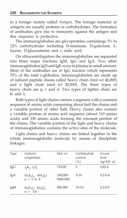
100 BIOCHEMISTRY FOR STUDENTS
to a foreign moiety called Antigen. The foreign material orantigens are usually proteins or carbohydrates. The formationof antibodies give rise to immunity against the antigen andthis response is protective.
The immunoglobulins are glycoproteins containings 3% to12% carbohydrates including D-mannose, D-galactose, L-fucose, D-glucosamine and a sialic acid.
On ultracentrifugation the immunoglobulins are separatedinto three major fractions IgM, IgG and IgA. Two otherimmunoglobulins IgD and IgE occur in plasma in small amount.Most of the antibodies are in IgG fraction which represents70% of the total r-globulins. Immunoglobulins are made upof subunit peptide chains called heavy chain (mol wt 40,000)and a light chain (mol wt 20,000). The three types ofheavy chain are μ, r and α. Two types of lighter chain areK and λ.
Both types of light chains contain a segment with a constantsequence of amino acids comprising about half the chains anda variable portion of other half. Heavy chains also containa variable portion of amino acid sequence (about 110 aminoacids) and 330 amino acids forming the constant portion ofthe chains. The variable portion of the light and heavy chainsof immunoglobulins contains the active sites of the molecule.
Light chains and heavy chains are linked together in thewhole immunoglobulin molecule by means of disulphidelinkages.
Type Subunit Mol wt Carbohydrate Serumcomposition content level
(%) mg/100 ml
IgG γ2k2, γ2λ2 153,00 3 0.81.6
IgA (α2k2)n, n(α2γ2) 180,000- 5-10 0.2-0.4n = 1 to 4 5000,000
lgM (μ2k2)n, (μ2γ2)n 900,000 10-10 0.2-0.5n = 5,6

CHEMISTRY OF AMINO ACIDS AND PROTEINS 101
Each heavy chain has four interchain disulfide bonds; twobetween the pair of μ-chains in the monomer one to the lightchain, and one intersusunit bridge between the monomers.
The light chains are denoted by smaller lines and may beof the κ or γ type.
The solid circles attached to the heavy chains of one ofthe monomers represent complex oligosaccharides.
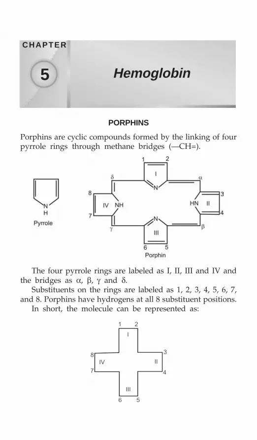
102 BIOCHEMISTRY FOR STUDENTS
PORPHINS
Porphins are cyclic compounds formed by the linking of fourpyrrole rings through methane bridges (—CH=).
Hemoglobin
CHAPTER
5
The four pyrrole rings are labeled as I, II, III and IV andthe bridges as α, β, γ and δ.
Substituents on the rings are labeled as 1, 2, 3, 4, 5, 6, 7,and 8. Porphins have hydrogens at all 8 substituent positions.
In short, the molecule can be represented as:

HEMOGLOBIN 103
PORPHYRINS
Substituted porphins are called porphyrins.Porphyrins are of two types, i.e. type I and type III.A porphyrin with completely symmetrical arrangements
of substituents is called type I porphyrins whereas if thearrangement of substituents is not symmetric then it is calledtype III porphyrins.
In nature both type I and type IlI porphyrins are foundbut type III porphyrins are more abundant.
Porphyrins are colored compounds and show characteristicabsorption spectra in both UV and visible regions.
Some of the important porphyrins are:
Porphyrins Nature of the substituents at thefollowing positions
1,2 3,4 5,6 7,8— — — —
1. Mesoporphyrin ME ME MP PM2. Uroporphyrin AP AP AP PA3. Coproporphyrin MP MP MP PM4. Protoporphyrin MV MV MP PM
where M = Methyl group (—CH3)E = Ethyl group (—C2H5)A = Acetate group (—CH2COOH)P = Propionate group (—CH2CH2COOH)V = Vinyl group (—CH = CH2)
Porphyrins can form complexes with metal ions. Thisproperty is very important in their functioning in biologicalsystem.
Examples: Heme is iron porphyrin, chlorophyll is amagnesium porphyrin, Vitamin B12 is a cobalt porphyrin.
HEMOGLOBIN
The red coloring material of blood is because of hemoglobin.It is present in RBC. Hb is globular in shape.
Hemoglobin belongs to class of conjugated proteinswhereas heme is the prosthetic group and globin, the proteinpart:
Hemoglobin = Heme + Globin

104 BIOCHEMISTRY FOR STUDENTS
Normal adult blood contains 97% HbA1, 2% HbA2 and1% HbF. Both alpha and beta chains have 75 percent alphahelical structure. The α-chains has 7 and β-chains has 8 helicalstructure.
Functions of Hemoglobin
1. In the transport of oxygen from lungs to the tissues and thetransport of carbon dioxide from tissues to the lungs. Hem-oglobin forms a dissociable hemoglobin-oxygen complex.
Hb + O2 ↔ Oxyhemoglobin2. As buffers. The buffering action of hemoglobin is due to
the amino acid histidine present in the globin part of hemo-globin. Histidine comprises 8 percent of the total amino acidmake up of the globin.
3. Hemoglobin is required for both carbon dioxide and oxygentransport because these gases are only sparingly solublein water.
The presence of hemoglobin increases the oxygen trans-porting capacity of a liter of blood from 5 to 250 ml of oxygen.Hemoglobin plays a vital role in the transport of carbon dioxideand hydrogen ion. Myoglobin which is located in muscles,serves as a reserve supply of oxygen and also facilitates themovement of oxygen within muscle.
Significance of 2,3-Diphosphoglycerate (2,3-DPG)
The stability of deoxy conformation is inceased by 2,3-diphosphoglycerate in mammals. It binds electrostatically to143rd histidine and 82th lysine in β-chains of deoxy-Hb andstabilizes T-conformation. During oxygenation 2,3-diphosphoglycerate is released and T form reverts to R-conformation.
Mountain sickness: When an unclemetized subject goes to higherattitudes (Hill areas/mountains) than the level of 2,3-DPGincreases in the blood. This reduces the affinity of oxygen tohemoglobin liberating more and more of oxygen to peripheraltissues.

HEMOGLOBIN 105
Carbon Monoxide Poisoning
Carbon monoxide has the tendency to form coordinationcompounds with metals, in particular with hemoglobin iron.It combines with hemoglobin to form carboxyhemoglobin (HbCO). Hemoglobin in this form does not carry oxygen efficientlysince by competiting specifically and effectively with oxygenfor ferrous site0s of hemoglobin, CO can displace oxygen fromhemoglobin in arterial blood.
The affinity of hemoglobin for CO is approximately 210times greater than for O2. In the lungs, hemoglobin combineswith O2 to form oxyhemoglobin (HbO2) which is carried inthis blood stream. O2 is released at the tissue capillary level.Since there are four heme groups in hemoglobin which cancombine reversibly with 4CO or 4 O2 molecule in anycombination. At the physiological pH and temperature, thecombination of CO with human hemoglobin is about 10 timesslower than O2. However, once formed the dissociation ofcarboxyhemoglobin is 210 times slower than oxyhemoglobin,which explains why the affinity of CO for hemoglobin is210 times more than that of O2.
In lead, poisoning the RBC refer to as Howell’s Jolly bodiesand Cabot ring.
Heme
Ferrous protoporphyrin is called heme. Heme is a chelate offerrous iron with protoporphyrin. Heme is also called proto-heme.
Synthesis of Heme
The starting materials of hemoglobin synthesis are glycineand succinyl CoA.

106 BIOCHEMISTRY FOR STUDENTS
Structure of Hemoglobin
In hemoglobin, iron is in ferrous form.When hemoglobin is converted to oxyhemoglobin, one of
the linkage of iron with imidazole group of histidine in globinis replaced by oxygen. In oxyhemoglobin, iron remains in theferrous form.

HEMOGLOBIN 107

108 BIOCHEMISTRY FOR STUDENTS
About 85 percent of the heme thus formed is used forhemoglobin synthesis. About 10% is used for myoglobinsynthesis and the remaining 5% for cytochromes and otherheme proteins.
There are about 250,000 hemoglobin molecules in a singleRBC.
Hemoglobin molecule contains 4 heme groups combinedwith a globin molecule, i.e. 2 α-chains and 2 β-chains, i.e. globinpart and four heme groups as prosthetic groups (one witheach chain). The total molecular weight is 64, 540.
globin (ferroheme)4 + 4O2 = globin (ferroheme-O2)4Deoxyhemoglobin Oxyhemoglobin
Each chain has one-heme group. One hemoglobin moleculecontains four heme groups as subunits.

HEMOGLOBIN 109
Globin
Globin contains 4 polypeptide chains. Two are α-chains andother two are β-chains. These four chains are arranged intetrahedron configuration.
α-chain contains 141 amino acids, whereas β-chain contains146 amino acids. In all there are 574 amino acids in the globinmolecule.
The globin moiety is formed from amino acid pool inamount of 8 gm per day in the normal adult. Thus, about 14%of the amino acids from the average daily protein intake areused for globin formation.
Each α-chain has 141 amino acids whereas β-chain (alsogamma and delta chains) have 146 amino acids.
There are 38 histidine molecules in hemoglobin molecule.The 58th residue in α-chain is called distal histidine becauseit is far away from the iron atom, whereas 87th residue inalpha chain is called proximal histidine because it lies near toiron atom. The α- and β-subunits of Hb are connected byweak noncovalent bonds like vander Walls forces andhydrogen bonds.
Each of the four polypeptide chains of hemoglobin has itsown heme prosthetic group and iron atom. Iron containedin the heme is coordinately linked with each chain by 2histidine residues at two imidazole nitrogens of histidine atposition 58 and 87 in α-chains and 63 and 97 in β-chain ofglobin.
The structure of oxyhemoglobin is described as R (relaxed)form and that of deoxyhemoglobin is T (tight) form. The T-conformation of deoxy Hb is maintained by electrostatic forcesbetween carboxyl and amine groups.

110 BIOCHEMISTRY FOR STUDENTS
Methemoglobin
Methemoglobin is a hemoglobin derivative in which iron isin the ferric form. It is also called ferrihemoglobin. Methemo-globin is dark brown in color.
Conversion of ferrous to ferric iron in hemoglobin destroysits capacity to combine with oxygen and to transport oxygen.Hence, methemoglobin is useless in the transport of oxygen.
Normally the conversion of hemoglobin to methemoglobintakes place in the blood but reducing substances present inred cells tend to prevent the accumulation of any appreciableamount of methemoglobin. The amount of methemoglobinpresent in blood is 0.3 g per 100 ml of blood.
Increased amount of methemoglobin in blood gives riseto a condition called methemoglobinemia. It is caused by thefailure in the normal reconversion of methemoglobin tohemoglobin or by production of methemoglobin by certaindrugs. The symptoms observed in methemoglobinemia arecyanosis (blue skin) and dyspnea (labored breathing).
Hemoglobin Cooperativity
The oxygenation process of hemoglobin and myoglobin is verypeculiar. This can be understood in terms of a graph offractional saturation of hemoglobin and myoglobin moleculesplotted against the partial pressure of oxygen. As shown infigure, myoglobin oxygenation curve is hyperbolic whereasfor hemoglobin it is sigmoidal. For myoglobin the halfsaturation pressure is quite low which tells us that it is a betteroxygen storage molecule then oxygen carrier. The differencein the oxygenation curves between hemoglobin and myoglobinis related to their structural difference. In hemoglobin thepresence of four subunits alter the nature of the oxygenationcurve. As a consequence of the interplay between foursubunits the binding of oxygen is cooperative. The affinityof a given heme for oxygen increases as the other heme inthe hemoglobin molecule are oxygenated. Consequently thedegree of saturation at first does not respond much to thepressure, then begins to rise abruptly and finally the curvelevels off at high pressure. This phenomenon is called

HEMOGLOBIN 111
cooperative or allosteric effect. There is an advantage of thesigmoidal curve.
The structure of hemoglobin differs in the oxygenated anddeoxygenated states. The quaternary structure of oxygenatedstate is called the R state (for released), and the conformationof the deoxygenated state is called the T state (for tense).
The ability of hemoglobin to bind oxygen decreases withan increase in acidity protons make hemoglobin dump oxygen.
Oxygenation curves for hemoglobin and myoglobin
Hemoglobin Variants
Hemoglobin A1
HbA1 contains two α-chains and two β2-chains. HbA1 constitute-over 98% of the total hemoglobin of the normal adult hemo-globin and is designated as α2A β2-A, or more simply α2β2.
Hemoglobin A2
HbA2 contains two α2-chains and two δ2-chains. HbA2constitute about 2 percent of the total hemoglobin in the normaladult and is designated as α2 δ2.
Hemoglobin F
Human fetal hemoglobin is designated as HbF and is repre-sented as α2 γ2.

112 BIOCHEMISTRY FOR STUDENTS
HbF contains two α-chains and two γ-chains. HbF is pre-dominant form present at birth but is almost totally replacedby HbA1 within few months after birth.
Hemoglobin S (Sickle Cell Hemoglobin)
HbS contains two α-chains and two β-chains in which glutamicacid at 6 position from the N-terminal end of the β-chain isreplaced by valine. HbS is also called sickle hemoglobin dueto the fact the red cells assume the shape of sickle ondeoxygenation. HbS gives rise to sickle cell anemia.
Hemoglobin Gun Hill
It contains only two heme groups instead of four. Five aminoacids are missing from the β-chain and this leads to theinterference with the heme binding.
In short hemoglobin variants are represented as:
Chains
HbA1 2α 2βHbA2 2α 2δHbF 2α 2γHbS 2α 2β glu →val
at position 6
Myoglobin
Myoglobin is a single polypeptide chain. Human myoglobincontains 152 amino acids with a molecular weight of 17,500.The heme is attached to 92nd histidine residue. One moleculeof myoglobin can combine with one molecule of oxygen.Myoglobin has higher affinity to oxygen than that of Hb.Myoglobin has high oxygen affinity while Bohr effect,cooperative effect and 2,3-diphosphoglycerate effect canabsent. The isoelectric point of myoglobin is 6.5.
Bohr Effect
The increase in acidity of hemoglobin as it binds oxygen isknown as Bohr effect; or Bohr effect is the increase in basicityof hemoglobin as it releases oxygen.

HEMOGLOBIN 113
The effect is expressed by the equation.
HHb+ + O2 HbO2 + H+
The above equation indicates that increase in hydrogenion concentration will favour the formation of free oxygenfrom hemoglobin and conversely that oxygenation of hemo-globin will lower the pH of the solution. This reversible uptakeand release of protons is responsible for the isohydric transportof carbon dioxide. The term isohydric refers to a lack ofchange of pH in the process.
Breakdown of Hemoglobin
↔
Bilirubin in combination with albumin reaches the liver,where it undergoes conjugation to form bilirubin diglucuronidewhich passes with the bile into the intestines. In the intestines,bilirubin diglucuronide is hydrolyzed, bilirubin is convertedto urobilinogen. A portion of urobilinogen is absorbed fromthe intestines into the blood and some of it is excreted in theurine (4 mg/day). The remainder is re-excreted in the bile.The unabsorbed urobilinogen is excreted in the stool as fecalurobilinogen which is oxidized to urobilin.
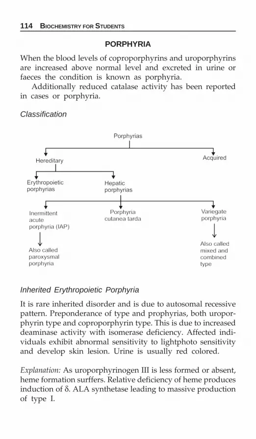
114 BIOCHEMISTRY FOR STUDENTS
PORPHYRIA
When the blood levels of coproporphyrins and uroporphyrinsare increased above normal level and excreted in urine orfaeces the condition is known as porphyria.
Additionally reduced catalase activity has been reportedin cases or porphyria.
Classification
Inherited Erythropoietic Porphyria
It is rare inherited disorder and is due to autosomal recessivepattern. Preponderance of type and prophyrias, both uropor-phyrin type and coproporphyrin type. This is due to increaseddeaminase activity with isomerase deficiency. Affected indi-viduals exhibit abnormal sensitivity to lightphoto sensitivityand develop skin lesion. Urine is usually red colored.
Explanation: As uroporphyrinogen III is less formed or absent,heme formation surffers. Relative deficiency of heme producesinduction of δ. ALA synthetase leading to massive productionof type I.

HEMOGLOBIN 115
Hepatic Porphyria
In this, there occurs abnormal and excessive production ofprophyrins (chiefly type III), their precursors δ ALA andporphobilinogen. There is three types of hepatic porphyrias.
1. Acute intermittent porphyria or paroxysmal porphyria:It is autosomal dominant partial deficiency of uroporphy-rinogen and synthetase. Patients present with acute attacks ofabdominal pain, nausea and vomiting, constipation, CVabnormalities and neuropsychiatric signs. This is due toincreased production of porphyrinogen and d-ALA. Thepatients do not have photosensitivity. Freshly passed urine isoften normal in color but on standing in sunlight turns to redurine color. Both colorless compounds porphobilinogen andd-ALA in sunlight. Polymerases to form two colored redcompounds porphobilin and porphyrin.
Note: Drugs and steroids requiring cyt P-450 can precipitateacute case. Reason is excessive utilisation of cyst P-450 forwhich heme is utilised. This decrease in heme is associatedwith depression of δ-ALA synthetase.
2. Porphyria cutanea tarda: It is autosomal dominant: Thisis due to partial deficiency of uroporphyrinogen decarboxylaseand patients are characterized by photosensitivity. Urinecontains increased quantities of uroporphyrins andcoproporphyrins of both types and also elevated urinaryexcretion of d-ALA and PBG occurs and is associated withuse in serum iron.
3. Varicyate porphyria or mixed (combined) porphyria: Inthis neurological as well as cutaneous symptoms are seen. Thisis autosomal dominant. There is deficiency of portopor-phyrinogen oxidase and ferrochelatase. Clinically there isvomiting, acute attacks of abdominal pain and neuropsychiatricsigns and cutaneous photosensitivity.

116 BIOCHEMISTRY FOR STUDENTS
Bilirubin is of two types:1. Direct bilirubin: Direct bilirubin is bilirubin diglucuronide.
It is water soluble. It is expressed as conjugated bilirubinbecause it can be coupled readily with Diazo Reagent(diazotized sulphanilic acid). This is the direct van denBergh reaction.
2. Indirect bilirubin: Albumin bound bilirubin is called indirectbilirubin. It is water insoluble. It is expressed as unconju-gated bilirubin as it will not react until it is released bythe addition of alcohol. The reaction with Diazo reagentafter the addition of alcohol is called the indirect van denBergh reaction.Normal serum bilirubin level is 0.2-0.6 mg %.
Jaundice
Jaundice is due to increase in the concentration of bilirubinin the blood which imparts yellow color to the skin andconjunctive. Jaundice may be either due to over productionof bilirubin than what the liver can normally excrete or adamage in liver, fails to excrete bilirubin in normal amounts.
Jaundice is of three types:
Hemolytic or Pre-hepatic Jaundice
In hemolytic jaundice, there is an increased breakdown ofhemoglobin, the liver cells are unable to conjugate all theincreased bilirubin formed. Increased production of bilirubinleads to increased production of urobilinogen which appearsin urine in large amounts. Bilirubin will be absent in urine.
Hepatocellular or Hepatic Jaundice
This type of jaundice results from liver damage which cannotconjugate bilirubin. The indirect serum bilirubin level will behigh. Urine will show the presence of bilirubin and increasedamount of urobilinogen. Stool is light in color.

HEMOGLOBIN 117B
ioch
emic
al c
han
ges
in
jau
nd
ice
Bio
chem
ical
Hem
olyt
icH
epat
ocel
lula
rO
bstr
ucti
vein
vest
igat
ion
(pre
-hep
atic
)(h
epat
ic)
(pos
t-he
pati
c)
1.C
ause
sD
ue t
o ex
cess
ivD
isea
se o
fD
ue t
o ob
stru
ctio
nhe
mol
ysis
pare
nchy
mal
cel
lsof
bili
ary
trac
tof
liv
er2.
Seru
m b
iliru
bin
Incr
ease
dIn
crea
sed
Incr
ease
dC
onju
gate
d
frac
tion
Incr
ease
dIn
crea
sed
Incr
ease
d(D
irec
t)U
ncon
juga
ted
fra
ctio
nM
ore
than
dir
ect
Incr
ease
dIn
crea
sed
(Ind
irec
t)fr
acti
on(D
irec
t fo
rm i
s m
ore)
van
den
Ber
gh R
eact
ion
Ind
irec
t po
siti
veD
elay
ed d
irec
t po
siti
veD
irec
t po
siti
veU
rine
bili
rubi
nU
sual
ly a
bsen
tP
rese
ntIn
crea
sed
(but
in
low
am
ount
)3.
Uri
ne u
robi
linog
enM
uch
mor
e th
anN
orm
alA
bsen
tth
e no
rmal
Feca
l st
erco
bilin
ogen
Incr
ease
dU
sual
ly d
imin
ishe
dU
sual
ly d
imin
ishe
dSe
rum
cho
lest
erol
Nor
mal
Dec
reas
edIn
crea
sed
–Fre
e fo
rmN
orm
alD
ecre
ased
Nor
mal
–Est
erri
fied
for
mN
orm
alM
ore
than
nor
mal
Nor
mal
Con
td.

118 BIOCHEMISTRY FOR STUDENTSC
ontd
. Bio
chem
ical
Hem
olyt
icH
epat
ocel
lula
rO
bstr
ucti
vein
vest
igat
ion
(pre
hepa
tic)
(hep
atic
)(p
osth
epat
ic)
4.Se
rum
pro
tein
sN
orm
alD
ecre
ased
Nor
mal
A:G
rat
ioN
orm
alD
ecre
ased
(Rev
ersa
l of
A:G
rat
io)
5.Pr
othr
ombi
n tim
eN
orm
alIn
crea
sed
Nor
mal
aft
er t
hepa
rent
eral
. In
ject
ion
of v
itam
in K
6.Se
rum
alk
alin
eN
orm
alN
orm
al o
r m
oder
atel
yIn
crea
sed
(va
lue
Phos
phat
ase
incr
ease
d (
valu
e be
low
abov
e 5
KA
* un
its
35 K
A u
nits
)7.
Seru
m t
rans
amin
ases
Nor
mal
Ver
y hi
gh i
n fi
rst
wee
kM
oder
atel
y ra
ised
8.Pr
otei
n fl
ocul
atio
n te
stN
orm
al o
r w
eakl
yus
ually
pos
itiv
eN
egat
ive
exce
pt i
n(T
hym
ol t
urbi
dit
y te
st)
seve
re
obst
ruct
ion
9.C
olor
of
stoo
lD
ark
colo
red
Pale
col
ored
Cla
y co
lore
d
*Kin
g A
rmst
rong
uni
ts.

HEMOGLOBIN 119
Obstructive or Post-hepatic Jaundice
This type of jaundice results from the obstruction of commonbile duct. As a result of obstruction, bilirubin does not passinto the intestine, so no urobilinogen is found in the urine.Direct serum bilirubin level will be high, urine will show thepresence of bilirubin. Stool is clay colored.
Physiological Jaundice or Neonatal Jaundice
Usually mild form of jaundice appears in some newbornchildren on the 2nd and 3rd day of life called neonatal jaundice.
Causes
1. Excessive destruction of RBCs after birth causing increasedin serum bilirubin.
2. Due to hepatic immaturityDuring IU life, bilirubin formed is mainly eliminiated by
placenta immediately after birth where has to eliminate allthe bilirubin but it is unable to deal adquately during first10 days.
Note:1. In infants, when serum bilirubin rises beyond 5% clinical
jaundice appears.2. Jaundice is more common and more severe is premature
babies.
Phototherapy
Exposure of skin to white light converts bilirubin to a com-pound which has shorter life than bilirubin called lumirubin.Phototherapy is used to treat infants with hemolysis.

120 BIOCHEMISTRY FOR STUDENTS
ENZYMES
Enzymes are biological catalysts which bring about chemicalreaction in living cells. They are produced by the livingorganism and are usually present in only very small amountsin various cells. They can also exhibit their activity when theyhave been extracted from the source. Enzymes are all organiccompounds and a number of them have been obtained incrystalline form.
General properties of enzymes are:1. All enzymes are proteins with exception of ribosomes.2. Enzymes accelerate the rate of reaction by:
a. Not altering the reaction equilibriumb. Being required in a very small amountc. By being not consumed in the overall reaction.
3. They have the enormous power for catalysis.4. Enzymes are highly specific for their substrate.5. Enzymes possess active sites at which interaction with
substrate takes place.6. Enzymes catalysis involves the transformation of enzyme-
substrate complex as an important intermediate in theiraction.
7. Enzymes lower the activation energy.8. Some enzymes are regulatory in function.
Some enzymes are purely protein in nature and dependfor activity only on their structure while certain enzymesrequire for their function one or more nonprotein component.They are termed as coenzymes, cofactors or prosthetic groups.If such a compound is firmly attached to enzyme proteins then
Enzymes
CHAPTER
6

ENZYMES 121
it is called a prosthetic group. If its attachment to protein isnot very firm then it is called coenzyme. Certain coenzymesexist in free state in solution and contact enzyme protein onlyat the times of reaction.
The term apoenzyme refers to the protein part of theenzyme.The apoenzyme in combination with its prosthetic group (orcoenzyme) constitute a complete enzyme or holoenzymesystem.
Holoenzyme = Apoenzyme + Coenzyme= Protein part + Nonprotein part
Coenzymes
Many enzymes in order to perform their catalytic activityrequire the presence of small nonprotein molecules. Coen-zymes are low molecular weight, organic compounds, non-protein, thermostable and can be separated by dialysis.
Characteristics of Coenzymes
1. They are stable towards heat.2. Generally derived from vitamins.3. Function as cosubstrates.4. They participates in:
a. Hydride (H¯) and electron transfer reactions, e.g.NAD+, NADH, FMN, FAD, etc.
b. Group transfer reactions, e.g. CoA, TPP, pyridoxal phos-phate, tetrahydrofolic acid, etc.
Coenzymes Functions performedNAD+, NADP+ Hydrogen transferFAD, FMN Hydrogen transferThiamine pyrophosphate Acetyl group transferPyridoxal phosphate Amino group transferBiotin Carboxyl group transferCoenzyme A Acyl group transfer
Most of the coenzymes are the members of water solubleB-complex group of vitamins. Coenzymes function as the

122 BIOCHEMISTRY FOR STUDENTS
intermediate carrier of functional groups of specific atoms orof electrons that are transferred in the overall enzymaticreactions.
Classification of Enzymes
According to the International Union of Biochemist, theenzymes are classified into six major classes.1. Oxidoreductases: They catalyze oxidation and reduction
reactions. These enzymes are divided into three groups.a. Oxidases: Those which use oxygen as hydrogen acceptor,
e.g. tyrosinase, uricase.b. Anaerobic dehydrogenases: Those which use some other
substances as hydrogen acceptor, e.g. lactic dehydro-genase, malic dehydrogenase.
c. Hydroperoxidases: Those which use hydrogen peroxideas substrate, e.g. catalase, peroxidase.
2. Transferases: They catalyze the transfer of some group fromone molecule to another molecule. These enzymes areimportant in biological synthesis, e.g. transaminases, hexo-kinases, transacylase, transaldolase, ketolase, phosphomu-tases.
3. Hydrolases: They catalyze the hydrolysis of substrate byaddition of water molecule across the bond which is split,e.g. esterases, peptidases, phosphatases, deamidases.
4. Lyases: They catalyze the addition or removal of groupsfrom the substrate without hydrolysis, oxidation or reduc-tion, e.g. decarboxylases, carboxylase, carbonic anhydrase,aldolase, enolase, etc.
5. Isomerases: They catalyze the conversion of a compound intoan isomer, e.g. racemases, epimerases, isomerases, mutases.
6. Ligases: They catalyze the linking together of moleculescoupled with the breaking of pyrophosphate bound in ATP,e.g. glutamine synthetase, succinic thiokinases.
Enzyme Specificity
Enzyme specificity is determined by how well the reactantfit into the enzyme surface. Some enzymes are very specificand show activity with only one substrate. However, someother enzymes are much less particular and will catalyzereaction with similar compounds.

ENZYMES 123
Generally two types of enzymatic specificities are observedin different reactions.Zymogens: Several proteins are synthesized in inactive forms.These are called zymogens eq. proteins digesting enzymes andblood clotting proteins. To activate zymogens, a small amountof protein is cleared from one end. This causes the protein tochange shape and activate it. These changes are not reversible.
Stereospecificity
Some enzymes show specificities only with a specific groupof a substrate, e.g. Urease catalyzes the hydrolysis of urea.
Alteration in the structure of urea results in the loss ofactivity. For example, N-methyl urea and thiourea are not thesubstrate for enzyme urease.
Also some enzymes show specificity towards D- and L-form of the same substrate, e.g. D-amino acid oxidase actsonly on the D-form of amino acid and not on L-form.
Substrate Specificity
Some enzymes catalyzes similar type of reactions but differin their action due to absolute substrate specificity, e.g. Pepsinhydrolyzes peptide bond involving amino group of aromaticamino acids as phenylalanine or tyrosine.
Similarly trypsin hydrolyzes peptide bond involving thecarboxyl group of basic amino acids such as lysine orarginine.

124 BIOCHEMISTRY FOR STUDENTS
FACTORS INFLUENCING THE RATE OFENZYMATIC REACTIONS
Effect of Substrate Concentration
At a low substrate concentration, the initial velocity of anenzyme catalyzed reaction is proportional to the substrateconcentration. However, as the substrate concentration isincreased, the initial velocity increases less as it is no longerproportional to the substrate concentration. With a furtherincrease in the substrate concentration the reaction rate becomesindependent of the substrate concentration and assumes aconstant rate as a result of enzyme being saturated with itssubstrate.
It was Michaelis and Menten who suggested an explanationof these findings by postulating that at low substrate concen-trations, the enzyme is not saturated with the substrate andthe reaction is not proceeding at maximum velocity whereaswhen the enzyme is saturated with substrate, maximumvelocity is observed. They further visualized the combinationof enzyme with the substrate to form an enzyme-substratecomplex and assumed that the rate of decomposition of thesubstrate being proportional to the concentration of enzyme-substrate complex. The velocity of the reaction at this high

ENZYMES 125
substrate concentration is termed as maximum velocity. Thesubstrate concentration at which the velocity is half of themaximum velocity is called the Michaelis constant and is termedas Km.
Km indicates the affinity of the substrate towards theenzyme and is inversely proportional to the affinity.
m
1K
Affinity∝
Higher the affinity the smaller will be the Km and lowerthe affinity, the higher will be the Km.
The Michaelis-Menten equation is given by the expression
V0 = max
m
V [S]K [S]+
where V0 = Initial velocityVmax = Maximum velocityKm = Michaelis constant[S] = Substrate concentration
The Michaelis-Menten equation relates the initial velocity,the maximum velocity and the initial substrate concentrationthrough Michaelis-Menten constant.
When the initial velocity is exactly half of the maximumvelocity the Michaelis-Menten equation assumes the form
max
1V
2 = max
m
V [S]K [S]+
Km + [S] = 2 [S]i.e. Km = [S]
Thus Michaelis-Menten constant is equal to the substrateconcentration at which the initial velocity is half of themaximum velocity.
Determination of important physical constants of an enzymesuch as V and Km would be difficult from the curve that wouldbe obtained by plotting [V] against [S]. So the Michaelis-Menten equation can be transformed into the form which isuseful in plotting experimental data.
Taking the reciprocals of both the sides of Michaelis-Mentenequation.

126 BIOCHEMISTRY FOR STUDENTS
0
1V
= m
max
K [S]V [S]
+
or0
1V
= m
max max
K [S]V [S] V [S]
+
0
1V
= m
max max
K 1V [S] V
+
This equation is called Line-weaver Burk equation and is theequation for a straight line y = mx + c, where m is the slopeof the straight line, c is the intercept on the y-axis and x isthe intercept on x-axis.
When 1
[ ]0V is plotted against
1[ ]S '
a straight line is obtained,
the slope of which is max
mKV
and has an intercept of max
1V
on
the 1
[ ]0V axis and intercept of
1
mK on the
1[ ]S
axis.

ENZYMES 127
Since, Line-weaver-Burk equation is in the form of astraight line, so it requires few points to define, Km. By usingsmall concentrations of substrate it is possible by this doublereciprocal plot to determine Km.
Significance of Km and Vmax Values
The Michaelis constant [Km] has two meanings:One is that it is equal to that substrate concentration at
which half of the active sites are filled and so once the Kmis shown, the fractions of sites filled (fs) at any substrateconcentration can be calculated by:
fs = max
VV
= m
[S][S] K+
Second, Km is related to rate constant of the individualsteps
Km = 1 2
1
K KK+
Now, when the K1 is much more than K2, the K2 becomes
negligible and Km is then equal to 1
1
−
−K
K, which is the disso-
ciation constant of the ES complex, a reversible reaction, i.e.
1
1
K
KE S ES
−
+ ⇔
R1 = K1 [E][S]R2 = K–1 [ES]
when R1 = R2 (at equilibrium)K1 [E] [S] = K–1 [ES]
or[E] [S][ES]
= 1
1
KK− = KSE
(the equilibrium constant of ES)
Km = 1
1
KK−

128 BIOCHEMISTRY FOR STUDENTS
So when this condition is met, Km indicates the strengthof ES complex and at such conditions a high Km indicates weakbinding and a low Km indicates strong binding. But this istrue only when the K2 is much less then K-1.
Vmax
Vmax indicates the turn over number of the enzyme if the con-centration of active sites, i.e. the total enzyme (Et) is knownsince Vmax = K2[Et].
Here, in this relation K2 is called the turn over numberof an enzyme which is defined as number of substratemolecules converted into product per unit time when theenzyme is fully saturated with the substrate and the timerequired for each round of catalysis is thus given by 1/K2.
Method of Determining Km
Km can be determined by double reciprocal Line-weaver-Burkmethod. In this the velocity of reaction is noted with different
1[ ]S
and 1
[ ]V from the graph, the value of Km is determined.
Another advantage of this equation is that it is used todifferentiate certain type of inhibitors of enzyme system.
Effect of Enzyme Concentration
The rate of an enzyme catalyzed reaction is directly pro-portional to the concentration of the enzyme. The greater theconcentration of enzyme, the faster will be reaction takingplace.
Effect of pH
Most enzymes have a characteristic pH at which their activityis maximum. Above or below that pH, the enzyme activitydecreases. If a curve is drawn between the activity of an en-zyme on a given substrate with the pH of the reaction mixture,it will reveal a maximum activity at a definite pH. This valueis known as optimum pH. See Diagram on Page No. 129.

ENZYMES 129
This is probably due to the changes in the net charge on enzy-mes, (as they are protein in nature) resulting from changes inpH. Excessive changes of pH brought on by the addition ofstrong acids or bases may completely denature and inactivateenzymes.
Effect of Temperature
The rate of an enzyme catalyzed reaction generally increaseswith temperature, within the temperature range in which theenzyme is stable and retains its full or maximum activity.Enzyme catalyzed reactions have an optimum temperature atwhich the reaction is most rapid.
Above this temperature the reaction rate decreases asenzymes being protein in nature are denatured by heat andbecomes inactive.
The increase in rate below optimal temperature results fromincreased kinetic energy of the reacting molecules.
ENZYME ACTIVITY
Activity: Amout of substrante converted to products by theenzyme per unit time (e.g. micromoles/minutes)Specific activity: Activity per quality of protein (e.g. micromoles/minute/mg protein)Catalytic constant: Proportionality constant between the reactionvelocity and the concentration of enzyme catalyzing the reaction.Unit: Activity/mole enzyme.Turnover number: Catalytic constant/number of active sites/mole enzyme.

130 BIOCHEMISTRY FOR STUDENTS
International unit (IU): Quality of enzyme needed to transform1.0 micromole of the substrite to product per minute at 30°Cof optimal pH.
The activity of an enzyme is expressed in standard unitsU = the amount of activity of an enzyme which catalyzes thetransformation of one micromole of substrate per minute. Thespecific activity of an enzyme is the number of units of enzymeactivity per mg of protein. The reason for needing this is thatoften the enzyme is not pure and there is contamination proteinin the sample.
The catalytic constant is units of enzyme activity per molof protein (mmol/min/mol enzyme).
Katab (kat) are the conversion of 1 mol/sec (Internationalunits).
Turnover NumberThe number of molecules of substrate converted to productsper enzyme molecule per minute is called turnover number.
ENZYME INHIBITIONS
Since, enzymes are proteins, any agent which denaturesproteins will inactivate the enzyme. Inhibitors are thesubstances which lower down the rate of enzyme reactions.They exert their effect by acting on the apoenzyme, coenzyme,prosthetic group or activator present in the enzyme systemor by interfering with the binding of the substrate to theenzyme. Reversible inhibitors bind the enzymes through non-covalent bonds and dilution of the enzyme-inhibitor complexresults in dissociation of the reversibly bound inhibitor whereas irreversible inhibitors occurs when an inhibited enzymedoes not regain activity on dilution of the enzyme-inhibitorcomplex.
Substances that inhibit enzymatic reactions are classifiedinto three groups:1. Competitive inhibition2. Noncompetitive inhibition3. Uncompetitive inhibition.
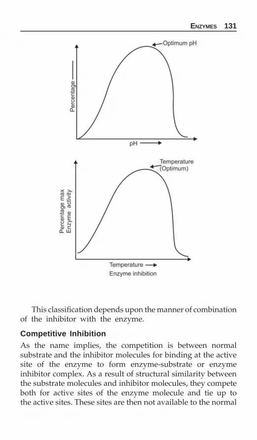
ENZYMES 131
This classification depends upon the manner of combinationof the inhibitor with the enzyme.
Competitive InhibitionAs the name implies, the competition is between normalsubstrate and the inhibitor molecules for binding at the activesite of the enzyme to form enzyme-substrate or enzymeinhibitor complex. As a result of structural similarity betweenthe substrate molecules and inhibitor molecules, they competeboth for active sites of the enzyme molecule and tie up tothe active sites. These sites are then not available to the normal

132 BIOCHEMISTRY FOR STUDENTS
substrate molecules. The overall rate of inhibition is governedby the affinities of inhibitor molecules and normal substratemolecules for the enzyme binding site and by the concentra-tions of the reactants.
The presence of competitive inhibitor thus increases theapparent Km of the enzyme for the substrate, i.e. causes itto require a higher substrate concentration to achieve themaximum velocity. On the other hand, a competitive inhibitordoes not affect the Vmax indicating that it does not interferewith the rate of breakdown of enzyme substrate complex.
Competitive inhibitors are frequently called antagonistsor antimetabolites of the substrate with which they compete.
The example of competitive inhibitions are:i. The inhibition of enzyme succinate dehydrogenase by
malonate for succinate.
Both have similar structural resemblance and hence, bothcompete for the active site of enzyme succinate dehydrogenase.
ii. Sulphanilamide has structural resemblance with para-minobenzoic acid and blocks the folic acid synthesis whichresults in the deficiency of the vitamin to microorganisms.

ENZYMES 133
In case of competitive inhibition.Affinity — DecreasesEfficiency — Remains same1/Km — Decreases as Km increases1/Vmax — Remains same
Noncompetitive Inhibition
As the name implies there is no competition between the substrate and the inhibitor molecules. There is little or nostructural resemblance between the substrate and the inhibitormolecules and hence they bind to the different sites of theenzyme. Inhibitors combine with the allosteric site of theenzyme, this combination results in the distortion of the activesite. In noncompetitive inhibition, the affinity of enzymeremains same but its efficiency decreases. This inhibition isalso known as allosteric inhibition. The inhibitory action cannot
be overcome by increasing the substrate concentration. Thecomplex formation between the inhibitor and enzyme isreversible. Noncompetitive inhibitors lowers the Vmax but doesnot effect the Km.
Examples of noncompetitive inhibitions are:There are many enzymes which require free sulphydryl
group (i.e.—SH group) for activity, are noncompetitively inhi-bited by heavy metal ions such as Pb++, Hg++, etc. Urease isan example of an enzyme which experiences heavy metal

134 BIOCHEMISTRY FOR STUDENTS
inhibition. The action of nerve gas poisons on acetylcho-linesterase, is an example of noncompetitive inhibition.In case of noncompetitive inhibition.
Affinity — Remains sameEfficiency — Decreases1/Km Remains same because the substrate
concentration has no effect on theinhibitory action.
1/Vmax — Increases as V has decreased.
Uncompetitive InhibitionThese inhibitors combine only with the enzyme-substrateforming an irreversible complex. The inhibition is dependentonly on the concentration of the inhibitor.
In case of uncompetitive inhibitionVmax is lowerSlope is same
Apparent Km < Km
CATALYTIC SITE OR THE ACTIVE SITESOF THE ENZYMES
The portion of the enzyme protein molecule which actuallytakes part in catalysis is called active site or the catalytic siteof the enzyme. Although the enzymes differ widely instructure specifically and catalysis, there are certain commonfeatures about the active sites.

ENZYMES 135
1. Normally the active sites makes up a small volume of thetotal portion of an enzyme.
2. The active site is a three dimensional activity.3. It is made up of groups that come from the different parts
of the linear amino acid chain. Indeed the residues are fara part in the linear sequence but may come together tobring about catalysis.
4. The specificity of the substrate binding depends upon theprecisely defined arrangement of the atoms or groups atthe active site.Emil Fisher postulated that substrate and enzyme reacted
in a well defined clear cut lock-key fashion signifying thepredominated structure of the active fit complementary tothe substrate molecule structure with which it will bind. Thismodel implied therefore the rigidity of the catalytic site. Butthis hypothesis was soon found unable to explain the possibilityof such a catalytic site reacting with the product to reformsubstrate in a reversible manner. Then Koshland proposeda more flexible hypothesis called “induced fit model” regardingthe structure of the active site. According to this hypothesis,enzymes in the inactive state in the absence of substrate andthat various groups in the active site are not correctly orientedto interact with the complimentary groups on the substrate.Binding of the specific substrate however, results in a confor-mational change in the enzyme and thus to the active site andshifting of those groups or atoms in the site into the correctposition for proper binding with the substrate and catalysis.
Feedback inhibition: In many multienzyme systems, the endproduct of the reaction sequence may act as a specific inhibitorof an enzyme at or near the beginning of the sequence, withthe result that the rate of entire sequence of reactions is deter-mined by the steady state concentration of the end product.This type of inhibition is called feed back inhibition.
For example, cholesterol synthesis is regulated, by feed-back inhibition.
ENZYME INDUCTION
Enzymes are classified according to the condition underwhich they are present in a cell. They are of two types.

136 BIOCHEMISTRY FOR STUDENTS
a. Constitutive enzymes.b. Inducible enzymes.
Constitutive EnzymesThese enzymes are formed at constant rates and in constantamounts. Their presence in a cell is not related to the presenceor absence of their substrates. They are considered to be partof the permanent enzymatic make of the cell.
For example, enzymes of glycolytic pathway.
Inducible EnzymesAlso called adaptive enzymes. They are always present in traceamounts but their concentrations vary in proportions of theirsubstrates.
Isoenzymes
They are multiple forms of a given enzyme having differentmobilities on electrophoresis, differently depressed by inhi-bitors towards different substrates. Isoenzymes catalyze thesame reaction but differ in Km, Vmax or both. The relativeamounts of the isoenzymes of a particular enzyme differ indifferent organs so that in disease, different isoenzymepatterns are found according to the organs from which theyhave come. These forms are the characteristics of differentorgans and tissues of the human body.
Example1. Lactate dehydrogenase (LDH): This enzyme catalyzes the
dehydrogenation of lactate to pyruvate. This occurs in fivedifferent isoenzymes.
This enzyme is a tetramer having two types of units,i.e. L and M units. Depending upon the various combi-nation, five isoenzymes are known, i.e. thest two subunitscan combine in five different ways.
Test Composition Location
LD-1 HHHH Heart and RBCLD-2 HHHM Heart and RBCLD-3 HHMM Brain and kidneyLD-4 HMMMLD-5 MMMM Liver and skeletal muscle

ENZYMES 137
LD-1 is the predominant form in heart and LD-5 in muscles.LDH is elevated from 12 to 48 hours after initial attack.
2. Alkaline phosphatase: It occurs in two forms.3. Isocitrate dehydrogenase: It occurs in two forms.4. Creatine phosphokinase: It occurs in three forms.
CPK is a dimer consisting of one subunit found in the brain(B) and other in muscle (M). CPK is found in three isoenzymes,as CPK1 (BB), CPK2 (MB) and CPK3 (MM).In normal serum 95% of the CPK activity is in CPK3.
CPK is, found in three isoenzymes as:i. CPK (MM), largely found in skeletal muscle tissue.
ii. CPK (BB), predominately found in brain tissue.iii. CPK (MB), exclusively found in heart tissue.
Blood level of both CPK (total) and CPK (MB) usuallymarkedly increases following acute myocardial infarction. OnlyCPK (MB) elevation is highly specific for the diagnosis of MI.
CPK (MM) increases rapidly following exercise or muscletrauma.
CPK (BB) is heat labile and rarely detected in serum.The activitiy of CPK2 is the cornerstone for the diagnosis
of myocardial infarction because of its abundance in heart andabsence from other cells. It may be elevated after 4 hours andits activity may increase from two to ten folds after 16-24weeks.
DIAGNOSTIC VALUE OF PLASMA ENZYMES
A determination of enzyme levels in the serum is often helpfulin pinpointing which, if any, body tissue or organ has beendamaged or malfunctioning. When a tissue is injured somecells of that tissue are destroyed and their contents, enzymesincluded, are released into the blood stream. Therefore, ifan enzyme is normally found predominately in a tissue otherthan blood, an increase in its level in the blood indicates thattissue has been damaged.
Serum acid phosphatase is increased in Paget’s disease ofbone, hyperparathyroidism, metastases of bone, Gaucher’s dis-eases, chronic renal failure and prostatic carcinoma.

138 BIOCHEMISTRY FOR STUDENTS
Serum alkaline phosphatase is increased in rickets, hyper-parathyroidism, obstructive jaundice, osteomalacia.
Serum glutamate oxaloacetate transaminase SGOT/AST(Aspartate transaminase) is increased in myocardial infarctionand skeletal muscle dystrophies.
Serum glutamate pyruvate transaminase SGPT/ALT (Ala-nine transaminase) is increased in viral hepatitis, toxic hepatitisand other forms of liver diseases associated with some degreeof hepatic necrosis.
Typical profiles of serum enzymes following a myocardialinfarction.
SGOT catalyzes the reversible transfer of the amino groupfrom glutamate to oxaloacetate to form α-ketoglutarate andaspartate. GOT is released from many diseased cells into serumas SGOT. SGOT is elevated in liver disease and following amyocardial infarction. The serum level has diagnostic value.It can be moderately elevated (5-fold) in people with cirrhosisand obstructive liver disease (a stone blocking bile duct). Itcan become very high (25-fold) in viral hepatitis.
Serum lactate dehydrogenase (LDH) is increased in myocar-dial infarction, acute liver disease, pernicious anemia, progres-sive muscular dystrophies.
Serum creatine phosphokinase (CPK) is increased in muscu-lar dystrophy, myocardial infarction.
Serum amylase is increased in various forms of pancreaticdisturbances (Pancreatitis).
Serum isocitrate dehydrogenase is increased in liverdiseases, severe pulmonary infarction.
Serum lipase is increased in acute pancreatitis and carci-noma.
Enzyme Evidence of riseCreatine kinase 3-6 hr(CK-MB)Aspartate transaminase 6-8 hr(AST)Lactate dehydrogenase 12 hr(LDH)

ENZYMES 139
Typical profiles of serum enzymes following a myocardialinfarction.

140 BIOCHEMISTRY FOR STUDENTS
BIOLOGICAL OXIDATION
All body reactions require energy which is obtained fromchemical reactions carried out in the living cells. The stepwiseoxidation of various metabolites is the principal mechanismfor the liberation of energy. The utilization of oxygen andproduction of carbon dioxide by the tissues in the processof cellular respiration is the final phase of biological oxidation.
Transfer of electrons are involved in all oxidation-reductionreactions. Every oxidation must be accompanied by simulta-neous reduction and the energy required for the removal ofelectrons in oxidation is supplied by the reduction.
The electron transport is important for the following reasons:1. It explains how oxygen finally enter the metabolism.2. It provides the mechanism for the regeneration of oxida-
tion-reduction coenzymes.3. It provides the majority of the energy derived from
metabolic processes.The energy transfers involved in the oxidation-reduction
systems are measured by difference in potential of varioussystems.
Oxygen has the highest oxidation potential of the systemsin the living cells and hydrogen atom the lowest.
Biological oxidation is catalyzed by enzymes which func-tions in combination with coenzymes or electron carriers.
Oxidases
These enzymes catalyze the removal of hydrogen from thesubstrate directly to the molecular oxygen, e.g. cytochromea3 (cytochrome oxidase), tyrosinase, uricase, etc.
2H + ½ O2———→ H2O
Biological Oxidation
CHAPTER
7

BIOLOGICAL OXIDATION 141
Dehydrogenases
They are further divided into:a. Aerobic dehydrogenasesb. Anaerobic dehydrogenases.a. Aerobic dehydrogenases: These enzymes remove hydrogenfrom the substrate using either O2 or artificial substance ashydrogen acceptor. These dehydrogenases are flavoproteins,e.g. xanthine oxidase, D-amino acid oxidase, catalase, peroxi-dases.
b. Anaerobic dehydrogenases: These enzymes use substances otherthan oxygen as hydrogen acceptor.
These dehydrogenases are classified as:i. Pyridine nucleotides: Under this group comes nicotinamide
adenine dinucleotide (NAD+) and nicotinamide adeninedinucleotide phosphate (NADP+). The effective partwhich participates in the reaction is nicotinamide.
ii. Flavonucleotides: They are flavin mono nucleotide (FMN)and flavin adenine dinucleotide (FAD). The effective partwhich participates in the reaction is riboflavin.
FAD + 2H+ + 2 e¯ —→ FADH2+
iii. Cytochromes: The cytochromes are iron containing hemo-proteins in which iron oscillates between Fe++ and Fe+++
during oxidation-reduction.Various cytochromes are cytochromes b, c1, c, a.

142 BIOCHEMISTRY FOR STUDENTS
Oxygenases
The enzymes incorporate oxygen into the substrate molecule.They are divided into two groups:
a. Dioxygenases: These enzymes catalyze the incorporation ofboth atoms of oxygen into the substrate.
A + O2 → AO2For example, tryptophan dioxygenase and homogentisic
acid dioxygenase.
b. Monoxygenases: They add only one atom of oxygen into thesubstrate.
MIXED FUNCTION OXIDASES
These oxidases cause reduction of one atom of O2 and utili-zation of other atom for specific oxygenation of hydroxylationof the substrate.
Two catalytic functions are performed by these enzymes:i. Reduction of an atom of oxygen to O¯ii. Transfer of oxygen to the substrate.These enzymes not only require O2 but also a source of
electrons, i.e. reducing agent to reduce an atom of O2 to O¯Mixed function oxidases are metalloproteins with prosthetic
group containing Fe, Cu.Examples of mixed function oxidases are:
i. Phenylalanine hydroxylaseii. p-Hydroxy phenylpyruvate oxidase
iii. Imidazole acetic acid oxidaseiv. Phenolase complex (this enzyme is involved in the
formation of melanins from tyrosine).
Hydroperoxidases or Peroxidases
These peroxidases catalyze the transfer of electrons fromdonars (substrates) to H2O2, reducing it to water. The peroxi-dases are specific in requiring H2O2 as electron acceptor (oxi-dizing agent) but various substrates may act as substrates orelectron donars.

BIOLOGICAL OXIDATION 143
Catalases
Catalases are specific type of hemin-containing hydroperoxi-dases which have the property of rapidly catalyzing thedecomposition of H2O2.
2H2O2 → 2H2O + O2
Catalase enzymes are simply a specific type of peroxidaseenzymes possessing very high activity toward H2O2 as a sub-strate but also capable of catalysing regular peroxidatereactions.
Superoxide anion O2̄ is highly reactive. It is generatedby a number of biological reactions including the antioxidationof quinones, thiols and reactions catalyzed by xanthine oxidaseand flavoprotein dehydrogenases.
Superoxides are very toxic to the cells and consequentlythe enzyme superoxide dismutase, present in all the cells, isresponsible for protecting the cell from the harmful effect ofsuperoxide anion, the cell in turn is protected from H2O2 bycatalase
Superoxide dismutase2H++ 2O2̄ ———————————→ H2O2 + O2
Catalase2H2O2 ————————————→ 2H2O + O2
HIGH ENERGY COMPOUNDS
High energy compounds are:1. Acid phosphates: They are acid anhydrides of an organic
acid (RCOOH) and phosphoric acid. Their general formulais:
For example 1, 3, diphosphoglyceric acid, acetyl phosphate.2. Enol phosphates: For example, 2-phosphoenol pyruvic acid.3. Guanidinophosphates: Two important compounds in this are
creatine phosphate in vertebrate and arginine phosphatein invertebrate.

144 BIOCHEMISTRY FOR STUDENTS
4. Organic pyrophosphates: These compounds are ADP and ATP,GTP, IDP, etc.
5. Coenzyme A: Derivatives of coenzyme A are high energycompounds.
6. Methionione: As S-adenosyl methionine.
RESPIRATORY CHAIN
Transfer of electrons from substrate to molecular oxygenthrough a chain of electronic carriers is called electron transportchain or respiratory chain.
Mitochondria contains series of catalysts called respiratorychain which are involved in the transfer of hydrogen andelectrons and their final reaction with oxygen to form water.
The components of respiratory chain are arranged sequen-tially in the order of increasing redox potential. Electrons flowthrough the chain in a stepwise manner from lower redoxpotential to higher redox potential. The amount of energyliberated in transfering electrons from one system to anotheris determined by difference in redox potential of the twosystems.
The respiratory chain is given as:
A redox potential of 0.2 volt between the components ofrespiratory chain results in the formation of 1 mole of ATP.
The three sites of ATP formation in the respiratory chainare:1. Between NAD+ and flavoprotein2. Between cytochrome b and c3. Between cytochrome a and cytochrome a3.
If the substrate enters the respiratory chain through NAD+
than the ATP yield is 3.If the substrate enters the respiratory chain through flavo-
proteins than the ATP yield is 2.

BIOLOGICAL OXIDATION 145
Coenzyme (Ubiquinone)
It is called ubiquinone because of its ubiquitous occurrencein nature. It is a lipid soluble hydrogen (electron) carrier foundin mitochondrial membranes and is a benzoquinone derivative.It contains an isoprene side chain which varies from sourceto source. Human coenzyme Q contains 10 isoprene units. Itis lipid soluble electron carrying protein and is reversiblyreduced by 2H+ from FADH2. Reduced coenzyme Q is thefinal stage at which oxidation reaction occurs as a process oftransfer of hydrogen atoms. Thereafter it is only the electronsof the hydrogen atoms which are carried down the electrontransport and the 2H+ ions liberated into the medium. Otherhomologous of coenzyme Q contains 6 to 10 isoprene unitsand have been isolated from various microorganisms, e.g.chloroplasts of green plants and mitochondria of beef andother animal tissues.
Cytochromes
Cytochromes are electron carrier proteins containing heme.They contain protein part to which heme is attached asprosthetic group. The cytochromes undergo oxidation andreduction as a result of oscillation of iron atom with Fe++ andFe+++ from which donates the reduced and oxidized formrespectively.
The five different cytochromes that has been identified inthe inner mitochondrial membrane are cytochromes a1, a3, b,c, and c1. It has been found that cytochrome a and cytochromea3 are combined with the same protein molecule to formcytochrome aa3 complex which is also called cytochrome coxidase or respiratory enzyme. It contains 2 atoms of copper.

146 BIOCHEMISTRY FOR STUDENTS
Oxidative Phosphorylation
Oxidative phosphorylation means that oxidation is accom-panied by phosphorylation. The energy released as a resultof biological oxidation is trapped in the form of high energyphosphate bonds in ATP by phosphorylation of ADP.
It is divided into two groups:
1. Substrate level phosphorylation: In the substrate level phos-phorylation the formation of high energy phosphate takes placeon the substrate, without undergoing into the respiratorychain.
The characteristics of substrate level phosphorylation are:i. Formation of ATP does not require oxygen
ii. Respiratory chain does not participateiii. It is dinitrophenol insensitive.
ExamplesSubstrate level phosphorylation is best described by two
examples: NAD+
(1) D-glyceraldehyde-3-PO4 + Pi + ADP ——→ Phospho- ATP glyceric acid
(2) Succinyl CoA + Pi + GDP → Succinic acid + GTP + CoAGTP + ADP → GDP + ATP
2. Respiratory chain phosphorylation: In this phosphorylation theformation of high energy phosphate bonds takes place as aresult of transfer of hydrogen and electrons through therespiratory chain to oxygen.
The characteristics of respiratory chain or electron-oxygentransport chain are:1. It is completely inhibited by trace amounts of dinitrophenol
(DNP) or by antimycin A.2. Oxygen uptake however, is not inhibited by DNP.
When a substrate is oxidized via NAD linked dehydro-genases, 3 moles of inorganic phosphates are incorporatedinto 3 moles of ADP to form 3 moles of ATP per atom ofoxygen consumed.
Similarly when a substrate is oxidized via flavin linkeddehydrogenases only 2 moles of ATP is formed.

BIOLOGICAL OXIDATION 147
Type of phosphorylation Reactions ~P trapped
1. Substrate level D-Glyceraldehyde 3-Phospho-3-PO4 Glyceric acid 1PhosphoenolPyruvate Pyruvate 1Succinyl CoA Succinate 1
2. Respiratory chainPhosphorylation isocitrate Oxalosuccinate 3(electron transport chain)
α-ketoglutarate Succinyl CoA 3Succinate Fumarate 2
P/O Ratio
P/O ratio is defined as the number of inorganic phosphatetaken to phosphorylate ADP per atom of oxygen consumed.
Mechanism of Oxidative Phosphorylation
Three hypothesis for the mechanism of oxidative phospho-rylation has been postulated to account for the transfer ofenergy from the oxidation reductions of reactions involvingelectron transport chain (respiratory chain) to the synthesisof ATP.1. Chemical coupling hypothesis2. Chemiosmotic hypothesis3. Conformational coupling hypothesis.
Chemical Coupling Hypothesis
This is the oldest hypothesis and it postulates that the energyyielding electron transfer process is coupled with energyrequiring oxidative phosphorylation through the formationof high energy intermediate compound which is generatedby the electron transport system and then subsequently uti-lized in the ATP formation from the ADP. In effect, it proposesthe existance of specific carrier proteins called C1, C2 and C3at each of the three ATP producing sites along with an inter-mediate I carried by them. At the site of release of energysufficient to form ATP, intermediate I is combined with thecarrier to form a high energy carrier and intermediate

148 BIOCHEMISTRY FOR STUDENTS
complex. This complex then is again linked to combine withanother intermediate X to form I~X again an high energy inter-mediate. The I component is then finally replaced by inorganicphosphates and ADP is phosphorylated to form ATP usingthe energy contained in I2~X complex, which is used up.
A strong objection to this effect is that no such intermediatehas been found even after intensive research.
This hypothesis takes the help of hypothetical carriers andhypothetical intermediates I and their effects are explainedas:
Effect of inhibitors: Inhibitors arrest respiration by blockingthe respiratory chain at energy site I, II and III.
Inhibitors of site I = Rotenone, amobarbital, piericidinInhibitors of site II = Antimycin, BALInhibitors of site III = H2S, CO, CN¯Uncouplers: Uncouplers are substances which allow elec-
trons to continue but prevent phosphorylation of ADP to ATP.They are dinitrophenol (DNP). Uncouplers causes the hydro-lysis of one of the high energy intermediates (car ~ l) resultingin the release of carrier I and energy as heat.
Chemiosmotic Hypothesis
This hypothesis (Peter Mitchell) assumes two points.i. The outer mitochondrial membrane is impermeable to
hydrogen ions and hydroxide ions.ii. The process goes on within matrix.

BIOLOGICAL OXIDATION 149
During electron transport, protons are released to theoutside of the mitochondria. This results in the establishmentof a proton gradient across the membrane, with a highconcentration of protons (H+) outside the mitochondria andlow concentration of protons inside the mitochondria creatingan electrochemical potential difference. This electrochemicalpotential difference is used to derive a vectorial membranelocated ATP synthetase, or the reversal of a membrane locatedATP synthetase which in the presence of inorganic phosphateand ADP forms ATP.
Conformational Coupling Hypothesis
According to this hypothesis (PD Bayer) the release of energyduring the electron transport induces some conformationalchanges in the carrier protein or the coupling factor. Thesechanges are due to the energy dependent shift in the numberof location of weak bonds such as hydrogen bonds andhydrophilic interactions which normally maintain the threedimensional conformation of the proteins. Then this energyconserved in this energised conformational state is used toderive the phosphorylation of ADP by inorganic phosphorousinto ATP. Simultaneously the carrier protein or the factorreturns back to the original low energy conformation.
This theory gets some support from the fact that inner mito-chondrial membrane undergoes very rapid physical changesas the electron pass along the respiratory chain. Also thismembrane shows some ultrastructural changes that accompanythe addition of ADP to the respiratory mitochondria.
This theory is in a way similar to the chemical couplingtheory except the fact that it postulates the non-covalent bondsas the energy intermediates rather than the postulation of truehigh energy intermediate in the chemical theory.
Shuttle System
NADH is produced in the cytosol but cannot penetrate themitochondria, i.e. the extra mitochondria NADH cannot pene-trate the mitochondrial inner membrane but electron derivedfrom it can enter electron transport chain by an indirect routecalled shuttles.

150 BIOCHEMISTRY FOR STUDENTS
Two important shuttle systems are:1. α-glycerophosphate shuttle2. Malate-aspartate shuttle.α-glycerophosphate shuttle: This shuttle transfers reducing
equivalents from cytosol to the mitochondrial electron trans-port chain by the following route.
Malate-aspartate shuttle: This shuttle transfer NADH from thecytosol to mitochondria by the following route.

METABOLISM OF CARBOHYDRATES 151
The major function of carbohydrate in metabolism is as a fuelto be oxidized and provide energy for other metabolicprocesses. In this role, carbohydrate is utilized by cells mainlyin the form of glucose. It has the advantage of being cheap,easily digested and rapidly metabolized.
Carbohydrate metabolism is basically the metabolism ofglucose and substance related to glucose in their metabolicprocesses. Glucose serves as a ready source of chemical energyfor humans.
The sugar of blood is glucose. The digestion of carbo-hydrates such as starch, sucrose and lactose produces glucose,fructose and galactose which passes into blood circulation.Conversion of fructose and galactose into glucose takes placein the liver.
Carbohydrates supply more than 50 percent of the energyrequirement of the body.
Except for ascorbic acid (vitamin C), carbohydrates are notessential to the diet, through gluconeogenesis, the body cansynthesize necessary carbohydrates from certain amino acids.
GLYCOLYSIS
The breakdown of glucose to pyruvic acid is called glycolysis.Under aerobic condition, pyruvic acid enters mitochondriaand is completely oxidized to CO2 and H2O. Whereas, underanaerobic conditions, pyruvate is converted to lactic acid.
The sequence of reactions from glucose to pyruvic acid isalso called Embden-Meyerhof pathway. Glucose is converted topyruvate in 10 steps by glycolysis.
Glycolysis is an extramitochondrial pathway and is carriedby a group of eleven enzymes.
Metabolism ofCarbohydrates
CHAPTER
8

152 BIOCHEMISTRY FOR STUDENTS
Mutases are enzymes which catalyze the transposition offunctional groups.

METABOLISM OF CARBOHYDRATES 153
Glucokinase is an inducible enzyme and has high km valuefor glucose whereas hexokinase is a constitutive enzyme andhas low km value for glucose.
Pyruvic acid has both a ketone or keto group and an acidgroup and hence it is a keto acid.
Salient Features of Embden-Meyerhof Pathway
1. The rate limiting step in glycolysis is phosphofructokinase(PFK). PFK is stimulated by fructose-6-phosphate, AMPand ADP but is inhibited by ATP and citrate. Since oneof the main object of glycolysis is to produce ATP and sincethe presence of excess AMP, ADP or fructose-6-phosphatemeans that the cell is deficient in ATP. These moleculesare activator of the enzyme (PFK), stimulating it to degrademore glucose and hence more production of ATP. Conse-quently an excess of ATP means that the cell is catabolizingmore glucose than necessary; excess ATP inhibits PFK.
2. All the reactions of glycolysis are reversible except hexo-kinase, phosphofructokinase and pyruvate kinase catalyzedreactions because of energy barriers.
3. Enzyme enolase is inhibited by fluoride. Since erythrocytesdo not have mitochondrial enzymes to oxidize glucoseaerobically, they depend on glycolysis only for their energyrequirement. That is why sodium fluoride (NaF) is usedin the collection of blood sugar sample because it preventsglycolysis by inhibiting the enzyme enolase. Otherwise alow result will be obtained due to glycolysis.

154 BIOCHEMISTRY FOR STUDENTS
4. It is the major pathway by which glucose is metabolizedin erythrocytes.
5. Glycolysis gives rise to certain intermediate compoundswhich are important for other biochemical processes.
i. Glyceraldehyde-3 PO4: For triglycerides and phospho-lipid biosynthesis.
ii. Acetyl CoA: Fatty acid and cholesterol biosynthesis.iii. Pyruvate: Alanine biosynthesis by transamination.
Glycolysis has three principal features:1. It is the degradative pathway whereby D-glucose is
oxidized to pyruvate, which is further metabolized by eitherof the two routes.
i. When the supply of oxygen is inadequate for completeoxidation, the pyruvate is reduced to lactate.
ii. When the supply of oxygen is adequate (aerobic condit-ions) the pyruvate is oxidatively decarboxylated to acetylCoA, which enters the citric acid cycle, where it is oxidizedto carbon dioxide and water.
2. Glycolysis gives rise to certain intermediates which arecommon to other pathway such as pentose phosphate path-way. These intermediate compounds also provide sourcesof starting materials for the biosynthesis of substancessuch as triglycerides from glyceraldehyde-3-phosphate,L-alanine from pyruvate and glycogen from glucose-1-phosphate.
3. Glycolysis is accompanied by the formation of ATP.
Pasteur Effect
Pasteur effect is the inhibition of glycolysis by oxygen. Therate limiting step in glycolysis, the phosphofructokinase, isinhibited by citrate and ATP.
Crabttee Effect
Crabttee effect is the inhibition of cellular respiration by highconcentrations of glucose. This is due to the completion ofglycolytic processes for inorganic phosphate.
METABOLISM OF CARBOHYDRATES 155
CITRIC ACID CYCLE
The complete oxidation of acetyl moiety is effected by meansof a cyclic metabolic mechanism called citric acid, also calledtricarboxylic acid (TCA) cycle and Kreb’s cycle. This cycle takesplace in mitochondria.
The citric acid cycle operates only under aerobic conditionsbecause it requires a supply of NAD+ and FAD which areregenerated when NADH and FADH2 transfer their electronsto O2 through the electron transport chain.
TCA cycle requires the presence of oxygen, i.e. aerobicmetabolism of carbohydrates and is catabolized by enzymesfound in the mitochondrial fraction of the cell.
Before pyruvate gains entry into the TCA cycle, it isoxidatively decarboxylated to acetyl CoA.
Conversion of Pyruvate to Acetyl CoA
Pyruvate is oxidatively decarboxylated to acetyl CoA by amultienzyme complex called pyruvate dehydrogenase com-plex.
This complex enzyme system comprises of three differentenzymes:
i. Pyruvate dehydrogenase (29 molecule)ii. Dihydrolipoate transacetylase (1 molecule)
iii. Dihydrolipoate dehydrogenase (8 molecule).which catalyze the five step reactions involved in conversion
of pyruvate to acetyl CoA.The six cofactors required are (i) Mg++ ions (ii) Thiamine
pyrophosphate (TPP) (iii) Lipoic acid (iv) Coenzyme A (CoA-SH) (v) FAD (vi) NAD+ (see page 156 for reaction).
Salient Features of Citric Acid Cycle
1. Citric acid is the common pathway for the metabolism ofcarbohydrates, fats, and proteins; since it provides thecomplete oxidation of acetyl CoA to carbon dioxide andwater.
2. Citrate synthetase catalyze a direct bond between themethyl carbon of acetyl CoA and carbonyl carbon of oxalo-acetate. It is an irreversible reaction.

156 BIOCHEMISTRY FOR STUDENTS
3. It defines the step by which citric acid, isocitric acid, α-keto-glutaric acid, succinic acid are synthesized and degraded.
The stepwise mechanism of the reactions is explained below.

METABOLISM OF CARBOHYDRATES 157
4. Many amino acids enter the cycle at several levels eitherat acetyl CoA, α-ketoglutarate, oxaloacetate, succinyl CoAand fumarate.
5. The rate limiting step in the TCA cycle is the conversionof isocitrate to α-ketoglutarate. The enzyme is the citratesynthetase. The availability of acetyl CoA and oxaloacetatein plenty stimulates this enzyme while succinyl CoA bycompeting with acetyl CoA inhibits this enzyme.
Similarly α-ketoglutaric acid, an intermediate in citric acidcycle is oxidatively decarboxylated to succinyl CoA. The en-zyme involved is α-ketoglutarate dehydrogenase complexlike the pyruvate dehydrogenase complex. This is anirreversible reaction forming succinyl CoA. Aresenite inhibitsthis reaction causing the accumulation of the α-ketoglutaricacid. Cofactors required are the same as in the conversionof pyruvate to acetyl CoA.

158 BIOCHEMISTRY FOR STUDENTS
The reaction is summed as:Succinyl CoA
Pyruvate can be channelled into TCA cycle as acetyl CoA oras oxaloacetate. This point is a switch point which controlsthe main function of the cycle. If pyruvate is channelled toacetyl CoA then the cycle will generate mainly energy. Ifpyruvate is channelled into oxaloacetate, then its main functionwill be to produce carbon skeletons for amino acid or fatsynthesis, i.e. high levels of acetyl CoA inhibit the activityof pyruvate dehydrogenase, decreasing further synthesis ofacetye CoA and the same time enhance the activity of pyruvatecarboxylase, stimulating the synthesis of oxaloacetate.
ENERGETICS
For each molecule of glucose, 2 pyruvates are formed. Theseare converted to 2 acetyl CoAs, each of which is brokendownto 3 NADH, 1FADH2 and 1 GTP. Hence, for 1 glucose molecule,6 NADH, 2 FADH2 and 2 GTP are produced in the TCA cycle.Reactions Where ATP is ConsumedGlucose to glucose-6-phosphate 1Fructose-6-phosphate to fructose-1, 6-diphosphate 1
Reactions Where ATP is GeneratedGlyceraldehyde-3-PO4 to 1,3 diphosphoglycerate 2 × 3 = 61,3 diphosphoglycerate to 3-diphosphoglycerate 2 × 1 = 2
(Substrate level phosphorylation)

METABOLISM OF CARBOHYDRATES 159
Phosphoenolpyruvate to pyruvate 2 × 1 = 2(Substrate level phosphorylation).
Under Anaerobic ConditionThe ATP yield is 2(Two molecules of ATP are generated in the conversion ofglucose to pyruvate because NADH obtained in the glyceral-dehyde-3-phosphate dehydrogenase reaction is not oxidizedin mitochondria by the respiratory chain).
Under Aerobic ConditionPyruvate to acetyl CoA 2 × 3 = 6Isocitrate to oxalosuccinate 2 × 3 = 6α-ketoglutarate to succinyl CoA 2 × 3 = 6Succinyl CoA to succinate
(The substrate level phosphorylation) 2 × 1 = 2Succinate to fumarate 2 × 2 = 4Malate to oxaloacetate 2 × 3 = 6
——————— Total = 30
———————Total number of ATP molecules formed under aerobic con-
ditions is 38, i.e. 30 from citric acid cycle and 8 from glycolysis.Two important features of Krebs cycle are:
i. Two carbon atoms enter the cycle as acetyl CoA and twocarbons leave as carbon dioxide, so no net gain of carbonatom takes place.
ii. The carbon atoms that leave as CO2 are not the same onesas those taken up as acetyl CoA.
The tricarboxylic acid cycle or Krebs cycle serves five majorfunctions:1. It produces most of the carbon dioxide made in human
tissues.2. It is the source of much of the reduced coenzymes that
drive the respiratory chain to produce ATP.3. It converts excess energy and intermediate to the synthesis
of fatty acids.4. It provides some of the precursors used in the synthesis
of proteins and nucleic acid.5. Its components control directly (product precursor) or indi-
rectly (allosteric) other enzyme system.

160 BIOCHEMISTRY FOR STUDENTS
This cycle is described as biochemical traffic circle, materialcoming to it from carbohydrate source might leave it to formfat whereas material coming to it from amino acid might leaveit to form carbohydrate. The only road closed is that leadingfrom fat to carbohydrate.
Amphibolic Role of Citric Acid or Krebs CycleCitric acid cycle is primarily a catabolic process for the finaloxidation of the carbohydrates, fats and proteins into CO2 andH2O. But this cycle at the same time takes part in the variousanabolic processes such as gluconeogenesis, fatty acid synthesisand amino acid synthesis by providing substrates which arethe normal intermediate products of this cycle. Thus, this cyclehas the dual or amphibolic role of both catalyzing the substancesfor energy and also taking part in synthesis. For example, theoxaloacetate and α-ketoglutarate are utilized for amino acids.Similarly, the malate and oxaloacetate are also utilized as glu-cose precursor in a reaction catalyzed by malate dehydrogenasefirst and subsequently by phosphoenol pyruvate carboxylase.Citrate is also utilized for providing acetyl CoA for fatty acidsynthesis by extra mitochondrial pathway. Further succinyl CoAis utilized in the heme synthesis. Thus, all these examplesestablish amphibolic role of citric cycle.
Hexose Monophosphate Shunt PathwayThis pathway is also known as Warburg-Dickens-Lipmann path-way, pentose phosphate pathway, phosphogluconate pathwayor direct oxidative pathway or reductive pathway.
Though glycolysis is the principal pathway for the con-version of glucose into pyruvate in most tissues but there existsan alternative pathway. Since glucose utilization can proceedwhen certain reactions in the glycolytic pathway are blockedby the addition of inhibitors.
Tissues where this pathway is more prominent are liver,adipose tissue, lactating mammary gland, leukocytes, testes,and adrenal cortex, etc.
The enzymes of this pathway are found in the extramito-chondrial cytoplasm.
Importance of HMP Shunt Pathway1. This pathway generates NADPH, which is required in
the reductive synthesis of fatty acids, triglycerides andsteroids.
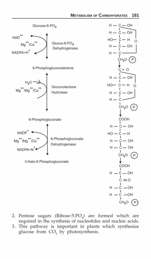
METABOLISM OF CARBOHYDRATES 161
2. Pentose sugars (Ribose-5-PO4) are formed which arerequired in the synthesis of nucleotides and nucleic acids.
3. This pathway is important in plants which synthesizeglucose from CO2 by photosynthesis.

162 BIOCHEMISTRY FOR STUDENTS
EnergeticsEnergy yield of hexose monophosphate shunt pathway.
Although the greatest importance of HMP shunt pathwayis to provide NADPH, for each carbon of glucose oxidized

METABOLISM OF CARBOHYDRATES 163
to CO2. Two molecules of NADPH are reduced or 12 moleculesof NADPH per mole of glucose oxidized is produced.
12 moles of NADPH are equivalent to 36 moles of ATP.Combined aerobic and anaerobic glycolysis of one molecule
of glucose gives 38 ATP, whereas HMP shunt pathway yields36 ATP. So these two pathways of glucose oxidation are almostequivalent in energy yield.
GLYCOGENESIS
The formation of glycogen from glucose is called glycogenesis.Under the combined act of glycogen synthetase and branch-
ing enzyme, glucose units are added to the non-reducing endsof the pre-existing glycogen by α-(1,4) and α-(1,6) linkagesto form glycogen.
Glucose is phosphorylated to glucose-6-PO4, by hexokinasereaction, which is then converted to glucose-6-PO4, a reactioncatalyzed by the enzyme phosphoglucomutase. Glucose-1-PO4reacts with uridine triphosphate (UTP) to form uridine diphos-

164 BIOCHEMISTRY FOR STUDENTS
phate glucose (UDPG). The reaction is catalyzed by the enzymeUDPG pyrophosphorylase. Now in the presence of the enzymeGlycogen synthetase, C-1 of glucose of UDPG forms a glycosidiclinkage α-(1,4) with the C-4 of the preexisting glycogenmolecule. The addition of glucose from UDPG to the existingglycogen molecule takes place from the non-reducing end ofthe glycogen molecule, thus, permitting the origin of newglycogen molecules. When the chain has been lengthened by6 to 11 glucose molecules, a second enzyme called branchingenzyme, transfers 6 glucose molecule in α-(1,4) linkages andattaches to the nearby chain in α-(1,6) linkages, thus creatinga branched point in the molecule.
Branching is important because it increases the solubilityof glycogen and provides a large number of non-reducingsugar terminals which are the sites of activity for glycogenphosphatase, the enzyme that breaks glycogen.
Glycogen Synthetase
a. Glycogen synthetase-D. It is the inactive form of theenzyme.
b. Glycogen synthetase. It is the active form of the enzyme.Glycogen synthetase-D, is the dephosphorylated form. It
is glucose-6-phosphate dependent, i.e. it is stimulated byglucose-6-phosphate.

METABOLISM OF CARBOHYDRATES 165
While glycogen synthetase-I is the dephosphorylated form.It is independent of glucose-6-phosphate.
Glycogen Storage Diseases
These are a group of inborn error of metabolic diseases inwhich there is an accumulation of abnormally large amountof glycogen in the tissue due to the deficiency or absence ofenzymes involved in glycogen metabolism.
Various type of glycogen storage diseases are given below:The classification of these diseases are based on the name
of the patient first diagnosed of that disease.
Type Name of disease Enzyme deficientI. Von Geirke’s disease Glucose-6-phosphatase
II. Pompe’s disease α-(1,4) glucosidaseIII. Cori’s disease Amylo-1, 6-glucosidase, i.e.
debranching enzyme.IV. Andersen’s disease 1,4 → 1,6 transglucosylase,
i.e. branching enzymeV. McArdle’s disease Muscle glycogen phosphorylase
VI. Her’s disease Liver phosphorylase
Glycogenolysis
Breakdown of glycogen to glucose is called glycogenolysis.The breakdown of glycogen takes place in liver and muscle.
In liver, the end product of glycogen breakdown is glucosewhereas in muscle the end product is lactic acid.

166 BIOCHEMISTRY FOR STUDENTS
Under the joint action of phosphorylase [breaks only α-(1,4)linkages] and debranching enzymes [breaks only α-(1,6) linkages]glycogen is broken down to glucose.
The breakdown of glycogen is initiated by the enzyme Phos-phorylase, which cleaves α-(1,4) glycosidic linkages startingfrom non-reducing end of the glycogen molecule to giveglucose 1-PO4 and this process continues until four glucoseresidues remain on either side of the α-(1,6) branched point.
Now another enzyme Glucan transferase, transfer three glu-cose units from one side to another, leaving a single glucoseresidue at the branched point followed by debranching enzymeto break α-(1,6)-linkage.
The breakdown of glycogen takes place in liver and muscle.The action of liver phosphorylase and muscle phosphorylaseare explained as below.
Liver Phosphorylase
It exists in two forms:a. Phosphorylase: It is the active form of phosphorylase.b. Dephosphophosphorylase: It is the inactive form of phosphorylase.
Activation of the inactive form involves phosphorylationof the hydroxyl group of a serine residue by a specific kinasein the presence of ATP. Inactivation of the active form iscatalyzed by a specific phosphatase. The action of kinase isstimulated by c-AMP which itself is formed from ATP in thepresence of adenyl cyclase. Glucagon and adrenaline stimulateglycogenolysis by increasing the activity of adenyl cyclase.
Muscle Phosphorylase
It exists in the following forms:a. Phosphorylase a. It is the active form of phosphorylase.
It is active only in the absence of 5-AMP. It is a tetramercontaining 4 molecules of pyridoxal phosphate.

METABOLISM OF CARBOHYDRATES 167
b. Phosphorylase b. It is the inactive form of phosphorylase.It is active only in the presence of 5-AMP. It is a dimercontaining only 2 molecules of pyridoxal phosphate.Phosphorylase a contains four molecules of pyridoxal phos-
phate. Whereas phosphorylase b contains 2 molecules of pyri-doxal phosphate.
Phosphorylase in muscle is activated by epinephrine, whichactivates adenyl cyclase to form c-AMP, which stimulatephosphorylase kinase, key enzymes of glycolysis and gluconeo-genesis in liver.

168 BIOCHEMISTRY FOR STUDENTS
Cori Cycle
The cyclic process by which lactic acid is converted to glucosein liver and eventually reappears as muscle glycogen is knownas Cori cycle.
The Cori cycle is the body’s way of recycling lactic acidLiver Muscle
During vigorous muscle activity, muscle glycogen isconverted to lactic acid. The lactic acid diffuses from the muscleinto the blood stream and transferred to the liver. In liver,lactic acid is converted to glucose by gluconeogenesis. Glucoseformed in this way returns to the muscle via circulation. Thiscycle continues and is called Cori cycle.
GLUCONEOGENESIS
Gluconeogenesis is the process by which glucose or glycogenis formed from noncarbohydrate substances. The noncarbo-hydrate substances include glycogenic amino acids, intermediatesof TCA cycle, glycerol, pyruvate, lactate, etc. gluconeogenesisis an important source for supplying glucose to various tissueswhen glucose is otherwise not available. Especially during
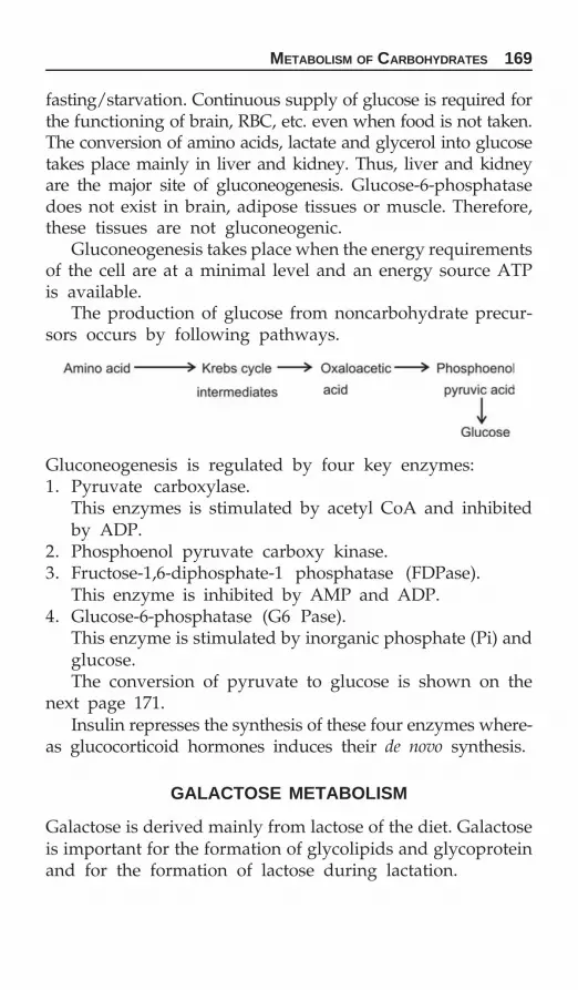
METABOLISM OF CARBOHYDRATES 169
fasting/starvation. Continuous supply of glucose is required forthe functioning of brain, RBC, etc. even when food is not taken.The conversion of amino acids, lactate and glycerol into glucosetakes place mainly in liver and kidney. Thus, liver and kidneyare the major site of gluconeogenesis. Glucose-6-phosphatasedoes not exist in brain, adipose tissues or muscle. Therefore,these tissues are not gluconeogenic.
Gluconeogenesis takes place when the energy requirementsof the cell are at a minimal level and an energy source ATPis available.
The production of glucose from noncarbohydrate precur-sors occurs by following pathways.
Gluconeogenesis is regulated by four key enzymes:1. Pyruvate carboxylase.
This enzymes is stimulated by acetyl CoA and inhibitedby ADP.
2. Phosphoenol pyruvate carboxy kinase.3. Fructose-1,6-diphosphate-1 phosphatase (FDPase).
This enzyme is inhibited by AMP and ADP.4. Glucose-6-phosphatase (G6 Pase).
This enzyme is stimulated by inorganic phosphate (Pi) andglucose.The conversion of pyruvate to glucose is shown on the
next page 171.Insulin represses the synthesis of these four enzymes where-
as glucocorticoid hormones induces their de novo synthesis.
GALACTOSE METABOLISM
Galactose is derived mainly from lactose of the diet. Galactoseis important for the formation of glycolipids and glycoproteinand for the formation of lactose during lactation.

170 BIOCHEMISTRY FOR STUDENTS
Important points of galactose metabolism:1. Conversion of galactose into glucose is the main pathway.
The galactose derived from the milk sugar is readily con-verted into glucose in the liver.
2. Conversion of UDP-galactose into UDP-glucose is a freelyreversible reaction catalyzed by UDP galactose-4-epi-merase. Hence, the glucose can be readily converted intogalactose in the states of galactose lack, which is the wayof treating alatosemia. This does not interfere with thegrowth. So it is not essential in the diet.
3. Galactosemia results from the deficiency of galactose-I- phosphate-uridyl transferase deficiency than galacto-kinase which is normal in the RBC of galactosemicpatients.
4. Galactose is needed as it is a constituent of glycolipids(cerebrosides), chondromucoids and mucoprotein.
5. Galactose is also required for the lactose synthesis in themammary gland by the enzyme lactose synthetase.
Galactosemia
Inability to metabolize galactose is called galactosemia.Galactosemia is an inherited disease, generally encounteredin infants, characterized by inability to metabolize galactoseor lactose. This results in the accumulation of galactose in theblood and ultimately excreted in the urine.
Galactosemia is due to the deficiency of the enzymegalactose-1-phosphate uridyl transferase.
Galactosemia gives rise to loss in weight, mental retardationand development of cataract due to the deposition of galactitol,a reduced product of galactose, in the lens. A galactose freediet avoids these difficulties and galactose that is necessaryfor the synthesis of cell membranes, cerebrosides, glycolipids

METABOLISM OF CARBOHYDRATES 171

172 BIOCHEMISTRY FOR STUDENTS
and mucoproteins can be formed from glucose-1 phosphate.
FRUCTOSE METABOLISM
Fructose is an important source of dietary carbohydrate,accounting for approximately 20% of the total carbo-hydrateintake.
Fructose is present in significant amounts in seminal fluid.It is synthesized in the prostate gland by the following

METABOLISM OF CARBOHYDRATES 173
reaction.In fructosuria, fructose is found in the urine, due to lack
of enzyme fructokinase.
LACTOSE SYNTHESIS
Blood galactose is readily converted into glucose in liver. Hereglucose is first converted into galactose by the pathway asabove and then glucose and galactose combine to form lactoseby the enzyme lactose synthetase.

174 BIOCHEMISTRY FOR STUDENTS
Failure to absorb dietary lactose is common in adults andis due to lactose deficiency and the irriability to hydrolyzelactose. Individuals with lactose deficiency can generally tole-rate yoghurt (curd), a milk product. Yoghurt contains lactasethat catalyzes the degration of lactose to glucose and galactose.
URONIC ACID PATHWAY
Biosynthesis of D-glucuronic acid takes place from glucose-1-phosphate.
UDP-glucuronic acid is required in detoxification reactionsforming glucuronides (e.g. bilirubin diglucuronide) and in the
synthesis of proteoglycans. Also this pathway throughL-uronic acid gives rise to the synthesis of L-Ascorbic acid(Vitamin C) in the animals and other plants.
These reactions occur in animals and higher plants. In man,guinea pigs and other primates, the enzyme which convertsL- glunolactone to 2-keto-L-gluconate is absent, thus makingascorbic acid a vitamin for them.
UDP-glucuronic acid is the active glucuronic acid. It parti-cipates into the incorporation of glucuronic acid into chon-droitin sulphate and other polysaccharides.
Glucuronic acid conjugates with bilirubin, steroids and cer-tain drugs for detoxification.
The compound glucose 6-PO4 is at a pivotal junction toundergo various metabolic fats such as pyruvate/lactate(glycolysis), in HMP shuntpathway, in glycogen synthesis inliver and muscle, give rise to glucuronate, ascorbic acid (uronicacid pathway).

METABOLISM OF CARBOHYDRATES 175
Glucose is converted to glucose-6-PO4 by two possibleenzymes depending upon the tissue. One is glucokinase (foundin liver) which is highly specific for the glucose and other ishexokinase (muscle and fat cells), which catalysis the phos-phorylation of most hexoses, including glucose.
PentosuriaPentosuria is characterized by the increased excretion of oneor more pentoses. The pentoses normally present in urine areL-xylulose, D-ribose and D-ribulose.
Essential pentosuria: It is characterized by increased excre-tion of L-xylulose. This is due to the deficiency of enzymeL-xylulose dehydrogenase.
Other types of pentosuria include alimentary pentosuriaresulting in excretion of L-arabinose and xylose due to intakeof large quantities of fruits and ribosuria (due to increase inexcretion of D-ribose).
The patients with deficiency of glucose-6-phosphate dehy-drogenase when given antimalarials like primaquine thatprecipitates hemolysis because G-6-PD is responsbile for themaintenance of reduced glutathione level and antimalarialsproduces excess of free radicals as free radicals damages theRBC’s cell membrane by oxidative stress mechanism.
REGULATION OF BLOOD GLUCOSE
The concentration of glucose in the blood is the net resultantof two processes.1. Rate of glucose entrance into the bloodstream2. Rate of glucose removal from the bloodstream.
Ways by which sugar is added to the blood
1. By absorption from the intestine2. Breakdown of liver glycogen3. By gluconeogenesis. The sources of gluconeogenesis are:
amino acids, propionate, lactate, glycerol, etc.
Ways by which sugar is removed from the blood1. Conversion to liver glycogen2. Conversion to muscle glycogen3. In the synthesis of fats (i.e. triglycerides)

176 BIOCHEMISTRY FOR STUDENTS
4. In the synthesis of glycoproteins such as nucleic acids (nuc-leoproteins), lactose, etc.
5. Loss in the urine.A balance of the above two processes will keep the blood
sugar level within normal limits.These two processes are influenced by a number of factors
under physiological conditions.
The blood glucose level is most efficiently regulated bya mechanism in which liver, extrahepatic tissues and severalhor-mones play an important part.
Role of Liver
Liver, being the centre of all metabolic activities is mainlyresponsible for the regulation of blood glucose level. In liver,exists the developed mechanism for uptake of glucosefrom the blood, conversion of glucose to glycogen for storage(glycogenesis), release of glucose from glycogen (glycogeno-lysis) and de novo synthesis of glucose from non-carbohydrateprecursors (gluconeogenesis).
Glycogenesis in liver can occur from blood glucose or anysubstance capable of giving rise to pyruvate. Due to the pre-sence of glucose-6-phosphatase, liver glycogen can contributedirectly to blood sugar (gluconeogenesis).

METABOLISM OF CARBOHYDRATES 177
Role of Extra-hepatic Tissues
a. Role of muscle: Muscle glycogen does not contribute directlyto the blood sugar due to the absence of the enzyme,glucose-6-phosphatase. Glycogenolysis in muscle providesglucose to blood only through the formation of lactic acidwhich by Cori cycle is converted to glucose in the liver.
b. Role of kidney: Kidney also exerts a regulatory effect byreabsorbing glucose by the reabsorptive system of the renaltubules. When the blood glucose level rises above the renalthreshold, the excess glucose appears in the urine.
Role of Hormones
Several hormones play an important role in the homeostaticmechanism of blood glucose level. Out of these insulin is theonly hypoglycemic hormone whereas others are hyperglycemichormones.
1. Insulin: Insulin plays an important role in the regulationof blood glucose concentration. It is secreted into the bloodin response to hyperglycemia. Insulin increases the transportof glucose across the cell membranes. Insulin reduces the bloodsugar level by increasing the glucose utilization by glycolysis,decreases hepatic glycogenolysis and increases glycogenesis.
Hormones which keep the blood sugar level high are:1. Epinephrine2. Glucagon3. Glucocorticoids4. Thyroxine5. Growth hormones.Mechanism by which these hormones increase the blood
sugar level are:1. By increasing the absorption of glucose from the intestines2. By decreasing the oxidation of glucose at the tissue level3. By preventing the synthesis of glycogen4. By stimulating glycogenolysis5. By potentiating gluconeogenesis.
Blood sugar level is kept normal by insulin, by opposingthe action of these hormones.

178 BIOCHEMISTRY FOR STUDENTS
2. Glucagon: Glucagon is also called Hyperglycemicglyco-genolytic hormone. Glucagon is secreted by the α-cells of theislets of Langerhans. Glucagon secretion is stimulated by hypo-glycemia. It causes glycogenolysis by activating liver phos-phorylase. Glucagon thus counter balances the action of insulinwhich is secreted into the blood when the blood glucose levelis high.
Glucagon acts primarily on liver and does not affect gly-cogen breakdown in muscles. Glucagon enhances gluconeo-genesis from amino acids and lactate.
3. Epinephrine: Epinephrine stimulates glycogen breakdownin liver and muscle. The stimulation of glycogenolysis is dueto its ability to activate phosphorylase. Epinephrine alsoinhibits muscle glycogen synthesis in liver and thus directsthe production of increased blood glucose.
4. Adrenal cortex hormones: Adrenal cortex secretes glucocor-ticoids, which lead to gluconeogenesis which is the result of increasedprotein breakdown and stimulation of transaminase. It also inhibitsglucose utilization in extra-hepatic tissues.
5. Anterior pituitary hormones: Growth hormones and ACTHelevate the blood glucose level. Growth hormones decreaseglucose uptake by the tissues, whereas ACTH stimulates thesecretion of hormones of the adrenal cortex.
6. Thyroid hormone: Thyroxine has a diabetogenic action. Itincreases blood glucose concentration by increased absorptionof glucose from the intestines.
Glycosuria
The excretion of detectable amounts of reducing sugar in urineis called Glycosuria. If glucose is excreted, then the conditionis called glucosuria. Glucose is filtered by the glomeruli butis completely reabsorbed by the renal tubules. The reabsorp-tion is effected by phosphorylation in the tubular cells. Themaximum rate of reabsorption of glucose by the tubules(TmG—Tubular maximum of glucose) is 350 mg/minute. When

METABOLISM OF CARBOHYDRATES 179
the blood levels of glucose are elevated, the filtrate containmore glucose that can be reabsorbed, the excess passes intothe urine and gives rise to glucosuria.
Renal Glucosuria
The blood glucose level is normal, but as a defect in the reab-sorption system in the tubules, kidney threshold is loweredand glucose appears in the urine.
Renal glucosuria is an example of benign glucosuria.
Diabetes Mellitus
Diabetes mellitus is a metabolic disorder due to the deficiencyof insulin, resulting in high blood glucose level and theexcretion of glucose in the urine.The most important features of diabetes mellitus are:1. The hyperglycemia and glucosuria persist during fasting.2. Liver glycogen falls to a low level.3. Excretion of large quantities of ketone bodies due to increased
fatty acid metabolism giving rise to diabetic coma.Diabetes or diabetes mellitus is a condition where in the bodydoes not produce enough insulin or does not properly respondto the insulin that is produced, there by keeping glucose levelin the blood high.
Diabetes affects nervous digestive circulatory, endocrine,urinary system but all body system are in some way affected.There is no diabetic sure but it can only be cautted.
Classification
1. Type 1 diabetes: Also called childhood onset diabetes, juv-enile diabetes and insulin dependent diabetes mellitus(IDDM).
Type 1 diabetes is a chronic (life long) disease that occurswhen the pancreas does not produce enough insulin toproperly control blood glucose levels. Type 1 diabetes canoccur at any age but it is most after diagnosed in children,adolescents or young adults, In this form of diabetes, thebody cannot make insulin the immune system mistakenly

180 BIOCHEMISTRY FOR STUDENTS
attacks the cells in the pancreas that make and releaseinsulin. As these cells die, blood glucose levels rise. Peoplewith type 1 diabetes need insulin shots.
2. Type 2 diabetes: In characterized by the inability of the bodyto make insulin or properly use insulin as a result, cellscan not take up or utilize glucose resulting in high bloodglucose level.
It is a slow onset process and person having diabetesfor years without knowing.
Typically with type 2 diabetes, the body still make insulin,but the cells cannot use it. This is called insulin resistance.3. Gestational diabetes: It occurs during pregnancy, labor and
delivery, women who got gestational diabetes are morelikely to develop type 2 diabetes.
Prediabetes
That condition when a person have impaired glucose tolerancewhere blood glucose levels are higher than normal but nothigh enough to be classified as diabetes.
Latest autoimmune diabetes of adults (LADA) is a conditionin which Type 1 diabetes develops in adults. Adults with LADAare frequently initially misdiagnosed as having type 2 diabetesbased on age rather than etiology.
Maturity onset diabetes of young (MODY): Conditionbecause of defects in β cell function.
According to the latest WHO guidelines two fasting glucoseblood measurement about 126 mg/dl is considered diagnosticfor diabetes mellitus.
People with testing blood glucose level from 100-125 mg/dlis considered to have impaired fasting glucose.
HbA1c given an idea about average blood glucose controlover the last 120 days.Glycosylated hemoglobin (HbA1c) test is recommended for:a. checking blood glucose control in pre-diabetes.b. Monitoring blood glucose control in diabetes mellitus.
The normal value of HbA1c is 4-6% correlation betweenHbA1c blood glucose level.

METABOLISM OF CARBOHYDRATES 181
HbA1c Average blood glucose level over pastthree months
6% 120 mg%7% 150 mg%8% 180 mg%10% 240 mg%
Higher the value of HbA1c poorer the blood glucose controlHbA1c >6.5 = Diabetes
<6.0 = Not diabetesin between 6-6.5 = May be pre-diabetes or risk of diabetes
The extent of glycosylation of hemoglobin can be convientlymonitored and used to assess the control of hyperglycemia.Analysis of hemoglobin A (the adult form of hemoglobin)reveals the extreme of minor components called hemoglobinA1. Hemoglobin A1 forms by nonenzymatic modification ofhemoglobin A by glucose. Glycosylation has only minor effecton normal hemoglobin function.
Glycosylation occurs continuously within the red cell, andthe extent of glycosylation reflects the average glucoseconcentration to which the cell is exposed during its 120 dayslife-span. Measurement of glycosylated hemoglobin contentprovides a clinically useful means to assess the degree ofhyperglycemia that existed over the previous several weeks.HbA1c in normal individuals (without diabetes mellitus)makes up about 6% of total hemoglobin. Individuals withcontrolled (blood glucose levels <10 mM, or 180 mg/dl) havelevels of hemoglobin A1 of about 9% of total hemoglobin.These with less well controlled diabetes have greater than9%, hemoglobin A1.
Glycosylated hemoglobin is formed in erythrocytes bythe reaction of glucose with hemoglobin. The glycosylationof hemoglobin is nonenzymatic reactions. The reaction isvirtually irreversible. Its removal from the blood proceedsvery slowly.
The prime importance of the glycosylated hemoglobinestimation in diabetes diagnosis lies in the fact that HbA1c
Correlation between HbA1c blood glucose level

182 BIOCHEMISTRY FOR STUDENTS
and blood glucose values of diabetics often present a differentpicture. Glycosylated hemoglobin fraction is not affected bythe metabolic state at a given moment whereas the bloodglucose level can change very rapidly. HbA1c thus makes itpossible to identify hyperglycemic states which would other-wise go unrecognized.
The main characteristic of the glycosylated hemoglobinfraction HBA1 is that it constitutes a kind of long-term retro-spective indicator of blood glucose concentrations. This is dueto the fact that the stable HbA1 is not catabolized throughoutthe erythrocyte lifespan (100-200 days).
HbA1C gives an indication of the averaged blood glucoselevels during the proceeding weeks rather than the metabolicstate of the patient at the time of testing.
HbA1C test estimates the portion of hemoglobin which isglycated. Increased blood glucose levels increase the glycationof hemoglobin. Glycation of hemoglobin is nonenzymatic,irreversible addition of glucose to hemoglobin. In normalindividual 5-6% of the hemoglobin molecule is glycated.However, in uncontrolled diabetic patient the level can goup to more than 20% which indicates that the patient hasuncontrolled hyperglycemia.
Since, the formation of glycated Hb is essentially reversibleand the blood level of HbA1c depend on both the lifespanof red blood cell (average 120 days) and the blood glucoseconcentration. Since the rate of formation of HbA1c is directlyproportional to the concentration of glucose in the blood, theHbA1c concentration represents the integrated values forglucose for the proceeding 6-8 weeks.
HbA1c% Degree of glucose control<6 Nondiabetic level
6-7 Near normal glycemia<7 Good
7-8 Good control>8 Action suggested

METABOLISM OF CARBOHYDRATES 183
Glycosylated hemoglobin (HbA1c) test gives an overviewof diabetic control over proceeding 2-4 months.
HbA1C circulates in the blood for 2-4 months before beingnaturally broken down by the body so the level of HbA1cat any stage can show how high the blood glucose has beenover the proceedings few months.
HbA1c test shows the average amount of glucose in theblood over the last three months.

184 BIOCHEMISTRY FOR STUDENTS
PLASMA LIPOPROTEINS
Lipids are water insoluble and are transported in the bodyin an aqueous medium in combination with various specificproteins. This results in lipid: protein complex called lipopro-teins. These lipoproteins consists of triglycerides or cholesterolesters and the central core surrounded by a coat of unesterifiedcholesterol, phospholipid and protein.
Plasma lipoproteins occur in four major forms which are:1. High or heavy density lipoproteins (HDL)2. Low density lipoproteins (LDL)3. Very low density lipoproteins (VLDL)4. Chylomicrons.Plasma lipoproteins are in dynamic state. They are conti-
nuously being synthesized and degraded with rapid exchangeof both lipid and protein among themselves. Two enzymesLecithin: cholesterol acyl transferase (LCAT) and lipoproteinlipase (also called triglyceride lipase) play a significant rolein the catabolism of lipid fraction of lipoproteins.
The components that are necessary for the synthesis oflipoproteins are triglycerides, cholesterol, cholesterol esters,phospholipids and apoproteins.1. High density lipoproteins: Also called α-lipoproteins. This
fraction is rich in phospholipids.2. Low density lipoproteins: Also called β-lipoproteins. This
fraction is rich in cholesterol.3. Very low density lipoproteins: Also called α2 or pre-β-lipo-
proteins. This fraction is rich in triglycerides.4. Chylomicrons: It consists of central core of triglycerides,
phospholipids, and cholesterol combined with small amountof proteins.
Metabolism of Lipids
CHAPTER
9

METABOLISM OF LIPIDS 185
The density of lipoprotein increases with the protein content.The protein parts of lipoproteins are called apolipoproteins.Each lipoprotein differ in terms of size, density, the relative
proportions of triglycerides and cholesterol esters in the coreand in the nature of apoproteins on the surface.
Chylomicrons VLDL LDL HDL
Density <0.95 0.95–1.006 1.091–1.063 1.063–1.21Protein % 1–2 10 25 45–55TG (%) 80–95 55–65 10 3PL (%) 3–6 15–20 22 30Cholesterol (%) 1–3 10 8 33Cholesteryl ester (%) 2–4 5 37 15
Absorption of Fats
Dietary fat is digested by the action of pancreatic lipase, presentin the intestines. The lipase hydrolyses the triglycerides to40 percent free fatty acids and glycerol, 50-57% mono-anddiglycerides, 3-10% is absorbed unchanged as triglycerides.
The 2-monoglycerides produced as intermediates are con-verted to 1-monoglycerides by an enzyme isomerase whichis then digested by lipase to glycerol and free fatty acids. Ofthe four products of triglycerides hydrolysis, i.e. free fattyacids, glycerol, monoglycerides and diglyceride, free fattyacids and glycerol are easily absorbed as they are water solubleand are then carried away by the blood.
Higher fatty acids, mono and diglycerides are absorbedwith the help of bile salts in the form of water soluble molecularaggregates called mixed micelles.
Inside the intestinal epithelial cell, 1-monotriglycerides arehydrolyzed by intracellular lipases to give free fatty acids andglycerol, whereas 1-monoglycerides are used for triglyceridesynthesis.
Higher fatty acids are largely utilized for the triglyceridessynthesis inside the intestinal epithelial cells and are carried aschylomicrons which are hydrophobic water soluble triglycerides

186 BIOCHEMISTRY FOR STUDENTS

METABOLISM OF LIPIDS 187

188 BIOCHEMISTRY FOR STUDENTS
covered with a layer of hydrophilic phospholipids, free andesterified cholesterol and some proteins.
Fatty acids with odd numbers of carbon atoms andbranched-chain fatty acids make up only a small portion ofour total fatty acid intake. A high fat, low carbohydrate dietresults in metabolic acidosis, whereas high protein, low carbo-hydrate diet results in protein imbalance with a high urinarynitrogen output, increasing carbohydrate in the diet preventsthis lose of nitrogen.
Oxidation of Fatty Acids
The action of hormonally controlled lipase results in the hydro-lysis of neutral fats to glycerol and free fatty acids. Glycerolenters the glycolytic pathway, via formation of glycerol-3-phosphate by the action of ATP and glycerokinase.
The fatty acids tightly bound with albumin is carried viathe blood to the various tissues for oxidation.Fatty acids oxidation takes place in mitochondria.
β-oxidation
Fatty acids are oxidised mainly by β-oxidation. In β-oxidation,the oxidation takes place at the β-carbon atom from thecarboxyl end and the β-carbon atom is oxidized to carboxylgroup resulting in the formation of acetyl CoA and a fattyacid shorter by two carbon atoms.
The first step in β-oxidation pathway is the activation offatty acid to form acyl CoA by combination with coenzyme A.The enzyme acyl CoA synthetase also known as thiokinase.
Acyl CoA synthesis occurs in the outer mitochondrial mem-brane. The acyl CoA so formed cannot penetrate the innermitochondrial membrane to the site of fatty acid β-oxidationenzyme system. In order to cross this barrier the acyl group

METABOLISM OF LIPIDS 189
is transferred to carnitine. The reaction is catalyzed by car-nitine acyl transferase. Acyl carnitine crosses the inner mito-chondrial membrane. Now another enzyme located on theinner surface of the inner mitochondrial membrane catalyzesthe reverse reaction and acyl group is transferred to intra-mitochondrial CoASH (Coenzyme A).
Outside
InsideAcyl carnitine + CoASH Acyl CoA + carnitineOnce the acyl CoA is inside the mitochondria, it is
metabolized by the steps given below.

190 BIOCHEMISTRY FOR STUDENTS
Energy Yield of Palmitic Acid Metabolized
Palmitic acid is C16 acid.Palmitic acid will undergo seven steps of β-oxidation to
give 8 molecules of acetyl CoA.In each sequence of β-oxidation, 5 ATP molecules are gene-
rated, (2 from FADH2 and 3 from NADH + H+, entering therespiratory chain). Since 7 steps of β-oxidation takes place,a total of 7 × 5 = 35 ATPs will be formed.
Each acetyl CoA molecule on complete oxidation by citricacid cycle will give rise to 12 ATPs. So a total of 12 × 8 =96 ATPs will be formed by the complete oxidation of 8molecules of acetyl CoA by citric acid cycle.
In the activation step, i.e. formation of palmitate oxidationis (35 + 96 – 2) = 129.
β-oxidation of odd chain fatty acids yield many moleculesof acetyl CoA and a terminal three carbon residue as propionylCoA. Propionyl CoA enter the TCA cycle by the followingreactions:
In the formation of palmityl CoA from palmitate, 2molecules of ATP are consumed.
The net ATP yield per molecule of palmitate oxidation is(35 + 96 – 2) = 129.

METABOLISM OF LIPIDS 191
In addition to β-oxidation, the other pathways for fattyacid oxidation are:
ααααα-oxidation
α-oxidation is a relatively minor pathway for the productionof energy. Dietary fatty acids which are methylated are meta-bolized by this pathway.
α-oxidation is important in the catabolism of branched chainand odd chain fatty acids.
Phytanic acid, a metabolic product of phytol (occurs as aconstituent of chlorophyll) is metabolized by oxidation. Phy-tanic acid, a significant constituent of milk lipids and animalfat, is metabolized by initial α-hydroxylation followed bydehydrogenation, and decarboxylation.
This pathway is operative in brain and plant tissues.
Refsam’s Disease
Due to the deficiency of enzyme phytate α-hydroxylase,phytanic acid is not metabolized and accumulate in blood andtissues giving rise to neurological problems. This is an inbornerror of metabolism.
ω (ω (ω (ω (ω (Omega)-oxidation
By this pathway, ω terminus carbon is oxidized to carboxylgroup to form dicarboxylic acids. Further metabolism takesplace by β-oxidation.
Fatty Acid Synthesis
Fatty acids are synthesized by three main processes.
Extra-mitochondrial De Novo Fatty Acid Synthesis
This is the de novo pathway for fatty acid synthesis startingfrom acetyl CoA. This pathway is active in liver, kidney, brain,mammary glands, adipose tissues, etc.
The enzyme is fatty acid synthetase complex, which is amultienzyme complex system containing acyl carrier proteinand six enzymes to which the growing fatty acid chain isattached during de novo synthesis. This multienzyme complexmolecule is a dimer consisting of two identical subunits or

192 BIOCHEMISTRY FOR STUDENTS
monomer. The monomer is not active only the dimer is active.The enzyme complex contains two SH group, i.e. central andperipheral.
Malonyl CoA is formed from acetyl CoA by carbon dioxidefixation reaction also called carboxylation reaction. The enzymeis acetyl CoA carboxylase and it requires biotin as cofactor.

METABOLISM OF LIPIDS 193

194 BIOCHEMISTRY FOR STUDENTS
The source of carbon atoms of fatty acid in acetyl CoA.Palmitic acid is the normal end product of fatty acid
synthetase complex which requires one molecule of acetyl CoAand seven molecules of malonyl CoA.
Regulation of De Novo Fatty Acid Synthesis
The rate limiting step in fatty acid synthesis is the carboxylationof acetyl CoA to malonyl CoA and is catalyzed by the enzymeacetyl CoA carboxylase. The enzyme consists of two compo-nents. One contains two proteins, a biotin carboxyl carrierprotein (BCCP) and biotin carboxylase. The second componentis transcarboxylase that catalyses the transfer of carbon dioxidefrom BCCP to acetyl CoA.
The enzyme acetyl CoA carboxylase is activated by con-version from a monomer to a polymer. Citrate activatesenzyme by causing aggregation while long chain acyl CoAinactivates it by causing it to disaggregate.
Mitochondrial Synthesis of Fatty Acids
This is a chain elongation pathway where the additionof acetyl CoA to the existing long chain fatty acids takesplace.
Palmitic acid which is a normal end product of de novo syn-thesis is the precursor of long chain saturated and unsaturatedfatty acids. Elongation of palmitic acid to stearic acid is moreabundant.

METABOLISM OF LIPIDS 195
Microsomal Pathway of Fatty Acid Synthesis
This pathway also provides a means of elongation of bothsaturated and unsaturated fatty acids utilizing malonyl CoAinstead of acetyl CoA.
Main difference between synthesis and degradation of fattyacids:
Synthesis Degradation
1. Occurs in Cytosol Occurs in mitochondria2. Acylcarrier = Acylcarrier protein Acylcarrier = CoA3. Cofactors NADP Cofactor NAD, FAD4. Synthesized in 2C units Degraded in 2C units5. 2C units added to α-C 2C units taken from the α-C end
Triglyceride Synthesis
Biosynthesis of triglycerides takes place in liver, adipose tissue,lactating mammary gland, intestinal mucosa, muscles andkidney. The enzymes are present in endoplasmic reticulum.
In the tissues liver, kidney, lactating mammary glands,glycerol is activated by glycerokinase whereas in adiposetissues and muscles glycerokinase is absent. The formationof α-glycerophosphate comes from dihydroxy acetone phos-phate, an intermediate in glycolysis, by reduction catalyzedby α-glycerophosphate-dehydrogenase.
Activation of Glycerol

196 BIOCHEMISTRY FOR STUDENTS
Activation of Fatty Acid

METABOLISM OF LIPIDS 197
Cholesterol Biosynthesis
Normal adult synthesizes about 1 to 1.5 gm of cholesterol perday. Whereas diet provides only 0.3 gm of cholesterol perday.
In normal human adults cholesterol is found in its largestamounts in the liver (about 0.3%), skin (0.3%), brain andnervous tissues (2%), intestines (0.2%) and certain endocrineglands, with the adrenal glands containing some 10%. Asmuch as 50% of the myelin sheath there surrounds nerves itscholesterol.
The relatively high content of cholesterol in skin may berelated to vitamin D formation and that in the adrenals andcertain other endocrine glands to steroid hormone biosynthesis.
Liver is the main site of cholesterol biosynthesis but intes-tines (intestinal mucosa) is also an important site of synthesisin man. The tissues where cholesterol synthesis also takes placeare skin, adrenal cortex, testes, aorta, etc.
The cholesterol biosynthesis takes place in the extramito-chondrial compartment of the cell.
The source of all the carbon atoms in cholesterol is acetylCoA. Acetyl CoA is the fundamental or building block unitof cholesterol biosynthesis.
M is derived from methyl carbon of acetyl group.C is derived from the carbonyl carbon of acetyl group.

198 BIOCHEMISTRY FOR STUDENTS
Synthesis of Cholesterol Takes Place in Various Stages
1. Formation of mevalonate from acetyl CoA via HMGCoA.
2. Three successive phosphorylation followed by decarboxy-lation to give an isoprene unit which is mainly isopentenylpyrophosphate.
3. Condensation of six isoprene units to give C30 terpene, i.e.Squalene.
4. Cyclization of squalene to lanosterol.5. Conversion of lanosterol to cholesterol.
Cholesterol is a product of animal origin. Cholesterol occursin egg yolk (the richest source of cholesterol), meat, liver,brain, etc. The normal serum cholesterol level is 150-250 mgpercent, about 2/3rd of this is present in the esterified form.The esterification of cholesterol takes place in liver.
Cholesterol is transported in the blood as lipoproteins. Thehighest proportion of cholesterol is found in the low densitylipoprotein fraction, i.e. β-lipoprotein fraction (LDL).
Cholesterol level in the blood is increased in diabetesmellitus, nephrotic syndrome, obstructive jaundice, hypopara-thyroidism, myxodema and xanthomatosis. Low levels ofcholesterol are found in hyperthyroidism, pernicious anemia,hemolytic jaundice, malabsorption syndrome, severe wasting,in acute infections, etc.
Cholesterol cannot be catabolized to straight chain moleculeor to acetyl CoA. Therefore, cholesterol cannot be used asan energy source by the cells.

METABOLISM OF LIPIDS 199

200 BIOCHEMISTRY FOR STUDENTS

METABOLISM OF LIPIDS 201

202 BIOCHEMISTRY FOR STUDENTS
The combined risk factor of coronary heart disease (CHD)can be determined following the estimations of serum choles-terol and HDL-cholesterol. The ratio of serum cholesterol toHDL-cholesterol has predictive value in determining the riskof CHD more accurately. For normal males the ratio of 5:1and for normal females the ratio of 4.5:1 are considered asaverage risk. Lower ratios significantly reduce the risk,whereas ratios of 9.5:1 and 7:1 for males and females respec-tively, are believed to double the risk of CHD. An inverserelationship has been observed between the risk of CHD andthe concentration of HDL-cholesterol in the test serum.
HDL-cholesterol represents approximately 20-25% of thetotal cholesterol in serum. It is also called good cholesterol,friendly cholesterol and healthy cholesterol.
HDL-cholesterol may work as a scavenger of cholesterolfrom the tissues ridding the body of excess cholesterol. HDL-cholesterol takes cholesterol away from tissues (extrahepatic)back to liver for excretion. Low HDL-cholesterol may bepredictive of coronary heart diseases risk whereas high HDL-cholesterol confers protection. People with high HDL levelsare resistant to the development of atherosclerosis. HDLremoves cholesterol from peripheral tissues and thecardiovascular system. HDL takes cholesterol away fromtissues (extrahepatic) back to liver for excretion.
People with high LDL levels on the other hand are proneto development of atherosclerosis. LDL is sometimes calledbad cholesterol (enemy cholesterol) because it may serve asa source for the cholesterol that accumulates in atheroscleroticplagues.
The various factors that influence the turnover of bodycholesterol can be illustrated as follows.
Cholesterol serves as a precursor of several importantclasses of compounds such as bile acids (liver cells), vitamin

METABOLISM OF LIPIDS 203
D and neutral sterols (by enteric bacteria) (coprostanol andcholestanal) steroid hormones, etc. (Progesterone, glucocorti-coids, mineralocorticoids, androgens, estrogens).
Conversion of cholesterol to steroid hormones is essentialfor life. The main steroid hormones include cortisol (a gluco-corticoid produced by adrenal cortex), aldosterone (a mineralo-corticoid produced by adrenal cortex), estrogen (a sex hormoneproduced by the ovary), testosterone (a sex hormone producedby the testes) and progesterone (a progestational hormoneproduced by the ovary).
All steroid hormones are synthesized from pregnenolone,a C21 compound.
Regulation of Cholesterol Biosynthesis
The amount of cholesterol synthesized is, in part, directlyrelated to cholesterol content in the diet. If the diet containslarge amounts of cholesterol, than the organism will synthesizelittle if, however, the diet contains only a small amount ofcholesterol, the organism can produce considerable quantities.
Cholesterol biosynthesis is regulated by negative feedbackmechanism. The rate limiting enzyme in cholesterol biosyn-thesis is HMG CoA-reductase, which catalyze the conversionof HMG CoA to mevalonic acid. Dietary cholesterol inhibitsthe biosynthesis of cholesterol in liver by depressing thesynthesis of HMG CoA reductase in liver. Fasting inhibitscholesterol biosynthesis by diverting HMG-CoA to ketonebodies formation whereas high fat diet accelerate the choles-terol production.
Atherosclerosis
High levels of cholesterol are associated with atherosclerosiswhich is characterized by the deposition of cholesterol esterand other lipids in connective tissues of arterial walls.
Factors which play a leading part in atherosclerosis arehigh blood pressure, obesity, smoking, lack of exercise, etc.
Diet rich in saturated fatty acid, increase the plasma choles-terol concentration. Replacement of saturated fat with a fatthat is rich in polyunsaturated fatty acids such as linoleic acid,decreases the plasma cholesterol concentration.

204 BIOCHEMISTRY FOR STUDENTS
Cornoil, peanut oil and cotton seed oil lower blood choles-terol while butter fat and coconut oil raises it.
Polyunsaturated fatty acid exerts their effects bya. Stimulating cholesterol excretion into the intestinesb. Stimulating the oxidation of cholesterol to bile acidsc. Increasing the metabolic rate of cholesterol esters.
Bile Acids
Bile acids are cholic, deoxycholic and lithocholic acids. Theyare present in the bile in conjugation with glycine and taurineas glycocholic and taurocholic acids. Bile acids are the deri-vatives of cholanic acid.
Deoxycholanic acid is 3,12 dihydroxy cholanic acid. Litho-cholanic acid is 3-hydroxy cholanic acid.
Salts of bile acids lower the surface tension and are goodemulsifying agents and hence play an important role in theabsorption of fats from the intestine.
Bile salts are polar derivatives of cholesterol and are deter-gents as they contain polar and nonpolar negious. they aresynthesized in the liver and stored in the gallbladder. Theysolubilies dietary lipids so that they can be broken down andabsorbed.
Bile salts are divided into two classes: primary and secon-dary. Primary bile salts are synthesized by humans. Secondarybile salts results from the action of intestinal bacteria on theprimary bile salts. The physical and physiological propertisof the bile salts are similar.
Liver synthesizes 500 mg of bile salts daily. About 94%of the intestinal bile salts is reabsorbed and 6 percent is lostin feces.

METABOLISM OF LIPIDS 205
Ketone Bodies
The ketone bodies are:1. Acetoacetic acid2. β-hydroxybutyric acid3. Acetone.The principle ketone body is acetoacetic acid which gives
rise to β-hydroxybutyric acid by reduction and acetone bydecarboxylation.
Ketone bodies are the intermediary breakdown productsof fatty acid metabolism. Under normal conditions fatty acidsare oxidized to carbon dioxide and these intermediary pro-ducts do not appear to any great extent in blood or urine.
Ketosis
Significant accumulation of ketone bodies in the blood (keto-nemia) and their excretion in urine (ketonuria) give rise toa condition known as ketosis.
The total blood concentration of ketone bodies in normalwell fed individuals with a daily urinary excretion of less than1 mg. Higher than normal urinary or blood concentrationsare called ketonuria and ketonemia respectively. The overallcondition is called ketosis.
Under certain metabolic conditions such as starvation, highfat diet, severe diabetes, more fat is metabolized for energypurposes giving rise to increased formation of ketone bodies.

206 BIOCHEMISTRY FOR STUDENTS
Liver is the net producer of ketone bodies. In liver, ketonebodies are formed by two ways.a. HMG-CoA pathwayb. Acetoacetyl CoA pathway.
HMG-CoA Pathway
Condensation of acetoacetyl CoA with another molecule ofacetyl CoA form β-hydoxy methyl glutaryl CoA (HMG-CoA).In the presence of enzyme HMG-CoA lyase. HMG-CoA issplit to acetoacetic acid and acetyl CoA.
Acetoacetyl CoA Pathway
Condensation of two molecules of acetyl CoA give rise toacetoacetyl CoA. In the presence of enzyme acetoacetyl CoAdeacylase, acetoacetic acid is formed.
Though ketone bodies are synthesized in liver but cannotbe utilized by liver because the enzyme required for theactivation of ketone bodies is absent in the liver. Ketone bodiespass from the liver to the blood and are oxidized by peripheraltissues. Cardiac muscle, skeletal muscle and brain prefer ketone

METABOLISM OF LIPIDS 207
bodies for energy utilization. Before the ketone bodies areutilized by these tissues they must be activated.
Acetoacetic acid is activated by two ways.a. Kinase activationb. Transferase reaction.
Kinase Activation
Transferase Reaction (See Below)
Danger of Ketosis
Acetoacetic acid and β-hydroxybutyric acid are fairly strongacids and are buffered when present in blood or tissues. Theirexcretion in the urine results in the loss of buffer cations. Since

208 BIOCHEMISTRY FOR STUDENTS
ketone bodies are negatively charged, their excretion fromthe body is accompanied by excretion of positively chargedions usually sodium as sodium salts, which depletes the alkalireserve of the body leading to fall in plasma bicarbonate level,giving rise to fall in pH. This leads to ketoacidosis, whichmay be cause of danger in uncontrolled diabetes mellitus.
Fatty Livers
Significant accumulation of triglycerides in the liver leads toa condition known as fatty liver. Normally, the liver contains5% of the lipids. Normal liver is rich in glycogen and not infat. But under physiological and pathological conditions thelipid content rises to 25–30%. The increased fat in liver mayresult from:a. Factors associated with increased free fatty acid (FFA)
levels—Such as (i) Starvation, (ii) Diabetes mellitus (severe,uncontrolled), (iii) Ketosis, (iv) Pregnancy (toxicemia, etc.).Here the increased FFA mobilization leads to increasedtriglyceride synthesis and accumulation.
b. Due to the deficiency of lipotropic factors such as choline,methionine, Vitamin E, essential fatty acids, vitamin B6 orpantothenic acid.
c. Other intoxicating agents such as carbon tetrachloride,chloroform, phosphorus, arsenic, lead, alcohol.Fatty liver is associated with uncontrolled diabetics chronic
alcohol intake obesity and protein malnutrition.
Biochemical Basis of Fatty Liver
Fatty liver falls into two groups.1. In one group there is some primary factor causing an
increase in free fatty acid either due to increasedmobilization from adipose tissue or increased hydrolysisof lipoproteins or chylomicrons by lipoprotein lipase. Thisincreased free fatty acid level leads to increased synthesisof triglycerides. The production of lipoproteins fromtriglycerides specifically the chylomicrons and VLDL, doesnot keep pace with the triglyceride synthesis allowing theiraccumulation to result in fatty liver. This is the mechanism

METABOLISM OF LIPIDS 209
in starvation, uncontrolled diabetes, ketosis and toxicemiaof pregnancy.
2. In the other group the defect lies somewhere in theproduction of plasma lipoprotein; such a block could beat any one or more of the following sites:
i. Apoprotein synthesis.ii. Lipoprotein synthesis from lipid and apoproteins.
iii. Synthesis of lipids-specifically the phospholipids.iv. Secretory mechanism of the lipoproteins.
Substances which prevent or relieve such abnormal accumu-lation of lipids in the liver are called lipotropic factors. Theyare choline, betaine, methionine, ethomolanine, inositol.
Role of Liver in Lipid Metabolism
Though liver is not the sole organ of lipid metabolism yetit has the complete enzyme systems to carry out the followingmajor activities.1. Synthesis and degradation of fatty acid (β-oxidation).2. Synthesis of triglycerides.3. Synthesis of cholesterol and its derivatives such as bile acid.4. Phospholipid synthesis.5. Synthesis of plasma lipoproteins such as VLDL and HDL.6. Ketone bodies formation.

210 BIOCHEMISTRY FOR STUDENTS
Twenty amino acids are present in dietary proteins. Theseamino acids are present in L-configuration.
L-form of amino acid is the physiological active form ofthe amino acid. The transport of L-amino acids is energydependent and require ATP, Na+, K+, Mn++ and vitamin B6.
While D-form of amino acid is physiological inactive andis transported by diffusion.
DIGESTION AND ABSORPTION
Two important features of digestion are:1. It breakdown, the nondiffusible bigger molecules into
diffusible smaller molecules (amino acids).2. During digestion, the biological specificity of a protein is
destroyed, i.e. they are no longer antigenic, thus avertingallergic reactions to food.
Digestion of Protein by Various Enzymes
Proteins are hydrolyzed to their constituent amino acids bythe action of variety of enzymes.1. Pepsin: It converts proteins to proteoses and peptones.2. Trypsin: It cleaves peptide bonds involving carboxyl groups
of arginine and lysine.3. Chymotrypsin: It cleaves peptide bonds involving carboxyl
groups of phenylalanine, tyrosine and tryptophan.4. Carboxypeptidases: It cleaves proteins and peptides from the
carboxyl end.5. Aminopeptidases: It cleaves proteins and peptides from the
amino end.6. Dipeptidases: It cleaves dipeptides.
Metabolism of Proteins
CHAPTER
10

METABOLISM OF PROTEINS 211
Absorption
L-form is absorbed at much faster rate than the D-form. Allamino acids are absorbed by active process. Active processrequires ATP, pyridoxal phosphate, Mn++, Na+ and K+.Sources of amino acids in the body pool are:
1. Dietary proteins.2. Intracellular synthesis.3. Tissue protein breakdown.
Dietary proteins serve three broad functions:1. Their consituent amino acids are used for the synthesis
of the body proteins.2. The carbon skeletons of the amino acids are oxidized to
yield energy.3. Their carbon and nitrogen atoms may be used to synthesize
other nitrogen containing cellular constituents as well asmany non-nitrogen containing metabolites.

212 BIOCHEMISTRY FOR STUDENTS
How the Amino Acids are Metabolized within the Cell?
Metabolism
Anabolic Phase Catabolic PhaseIt is a synthetic phase It is a breakdown phase
1. Protein Biosynthesis 1. Transamination.Tissue proteins, blood 2. Oxidative deamination.proteins, enzymes, hormones. 3. Decarboxylation.
2. Synthesis of nonprotein 4. Utilization of nitrogennitrogen substances takes residue.place such as creatine, i. Glutamine synthesispurines, pyrimidines, ii. Urea cycle.glutathione, choline, etc.
Transamination
It involves the transfer of an amino group from an amino acidto the keto group of the keto acid forming a new amino acidand a keto acid.
The reaction is reversible and is catalyzed by transaminaseenzymes. These enzymes are also known as aminotransferases.
The general reaction is represented as:
The transaminases require pyridoxal phosphate as the coen-zyme. The pyridoxal phosphate acts as a carrier of amino groupfrom amino acid to keto acid. The reaction involves theformation of Schiff’s base. The mechanism is represented as:

METABOLISM OF PROTEINS 213
Transaminases are present in practically all the tissues.The most abundant of these are the glutamate-oxaloacetate
transaminase (GOT) or aspartate transaminase (AST) andglutamate-pyruvate transaminase (GPT) or alanine transa-minase (ALT). GPT is predominent in liver whereas, GOT ispredominent in heart. Determination of the concentration ofGOT and GPT in serum is used to assess the degree of cardiacand liver damages.
Examples
Alanine, aspartic acid and glutamic acid participate mostin transamination.
Lysine and threonine do not participate in transamination.Transamination, by converting amino acids to keto acids(which are prominent in TCA cycle), provide an importantlink between the protein and carbohydrate or fat metabolism.By this way amino acids are sources of energy to the bodyby keto acids which undergo complete oxidation.
Also transamination reactions appear to play two majorroles in amino acid metabolism.
i. To serve as a means for the interconversion of numberof amino acids to increase the amount of one that maybe in short supply.
ii. To channel the amino groups of amino acids, ultimatelyto glutamate and aspartate.
Decarboxylation
Amino acids are decarboxylated to give amines. The enzymeis decarboxylase. It requires pyridoxal phosphate as cofactor.

214 BIOCHEMISTRY FOR STUDENTS
Decarboxylation of amino acids give rise to some of thebiologically active amines (biogenic amines such as histamine,serotonin and α-amino butyrate).Histidine HistamineTyrosine TyramineGlutamic acid γ-aminobutyric acid (GABA)Tryptophan Tryptamine5-hydroxy tryptophan 5-hydroxy tryptamine (serotonin)Arginine Agotomin (in bacteria only).
Oxidative Deamination
Oxidative deamination involves the removal of α-amino groupof amino acids to their corresponding keto acids. The enzymeis amino acid oxidase. In the amino acid oxidase reaction, theamino acid is first dehydrogenated by the flavoprotein ofoxidase forming an amino acid. This spontaneously adds waterto decompose the corresponding α-keto acid with the lossof α-amino nitrogen as ammonia.
For L-form of amino acids, the enzyme is L-amino acidoxi- dase. It is FMN dependent.
For D-form of amino acids, the enzyme is D-amino acidoxidase. It is FAD dependent.
UREA CYCLE (KREBS-HENSELEIT CYCLE)
The deamination of amino acids produces ammonia which istoxic. By Krebs cycle it is converted to urea, a nontoxic com-pound, which is transported via the blood to the kidneys and

METABOLISM OF PROTEINS 215
excreted in the urine. Urea formation takes place mainly inthe liver. Two molecules of ammonia and one molecule ofCO2 are converted to urea for each turn of the cycle. Ureais synthesized in the liver. One nitrogen of urea is derivedfrom ammonium ion, and the second is derived from aspartate.The carbonyl group is derived from carbon dioxide (asbicarbonate).
Overall Reaction:
NH3 + CO2 + 3ATP + 3H2O → Urea + 2ADP + AMP + 2Pi + PPiVarious stages of urea cycle are:
1. Synthesis of carbamoyl phosphate2. Synthesis of citrulline3. Synthesis of arginine
This is divided into two parts:a. Synthesis of argininosuccinic acidb. Cleavage of argininosuccinic acid.
4. Synthesis of urea.
Synthesis of Carbamoyl Phosphate
The first step in urea synthesis, is the formation of carbamoylphosphate. The enzyme is carbamoyl phosphate synthetase.The enzyme requires biotin and N-acetyl glutamic acid (AGA)for activity (i.e. cofactors). AGA is the modifier of the enzyme.

216 BIOCHEMISTRY FOR STUDENTS
It acts on the noncatalytic site and keeps the enzyme in activeconfiguration.

METABOLISM OF PROTEINS 217
Synthesis of Citrulline
Carbamoyl phosphate and ornithine combines to form citrulline.The reaction is catalyzed by ornithine transcarbamylase.
Synthesis of Arginine
a. Synthesis of argininosuccinic acid
b. Cleavage of argininosuccinic acid.Argininosuccinic acid is formed from citrulline and aspartic
acid. The reaction is catalyzed by enzyme argininosuccinatesynthetase.
Argininosuccinic acid so formed is cleavaged by enzymeargininosuccinase to arginine and fumaric acid.

218 BIOCHEMISTRY FOR STUDENTS
Synthesis of Urea
Enzyme arginase splits arginine to urea and ornithine.
Ornithine so formed again enters the cycle at the secondstep and hence, the cycle continues.
In urea cycle, everytime a citrulline passes out of themitochondria ornithine passes in. This is what happens andthe protein in the mitochondrial membrane that allows thisto happen is called an ornithine citrulline exchanger.
Urea cycle is a route for biosynthesis of arginine, a semi-essential amino acid.
Link between urea cycle and TCA cycle is shown below:The urea cycle eliminates excess ammonia. Ammonia is
derived mainly dietary amino acids that are not used promptlyfor protein synthesis.
A human consuming 100 g of protein daily excretes about16.5 g of nitrogen per day. Urea is the chief nitrogen metaboliteof humans, accounts for 80-90%, of excreted nitrogen. Urateand ammonium ions are other end products.

METABOLISM OF PROTEINS 219
Hyperammonia
Hyperammonia or hyperammonemic syndrome is because ofincreased level of ammonia in the blood. The urea is formedfrom ammonia by urea cycle. So any deficiency or defect ofurea cycle enzymes give elevated levels of ammonia. Hyper-ammonemia gives rise to mental retardation.
Metabolism of Individual Amino Acids
According to metabolic fates of their skeletons amino acidsare classified in following way:
Glucogenic Amino Acids
Those amino acids which give rise to the intermediates ofcarbohydrate metabolism. The product may be pyruvic acid,fumaric acid, oxaloacetic acid, α-ketoglutaric acid, etc.
The glucogenic amino acids are glycine, alanine, asparaticacid, glutamic acid, arginine, cysteine, histidine, proline, serine,methionine, valine.
All the nonessential amino acids are glucogenic in character.The amount of glucose derived from protein can be calcu-
lated by estimating nitrogen and glucose excreted in the urine.The glucose to nitrogen ratio in urine was found to be 3.65
in diabetic animals which were fed proteins only.Therefore the amount of glucose derived from 100 gm
of protein is 3.65 × 16 = 58.4 gm.The factor 16 is because of the average nitrogen content
of proteins is 16%.This suggests that 58% of protein is glycogenic. The
glycogenic character of proteins is taken into account whilecalculating diet schedule of diabetic patients are made.
Ketogenic Amino Acids
Those amino acids which give rise to the intermediates of fatmetabolism. The product may be acetyl CoA and acetoacetylCoA, i.e. ketone bodies.

220 BIOCHEMISTRY FOR STUDENTS

METABOLISM OF PROTEINS 221
The ketogenic amino acids are leucine, isoleucine, phenyl-alanine and tyrosine lysine.
Phenylalanine and tyrosine tryptophase are both glucogenicand ketogenic amino acids. These amino acids yield aceto-acetic acid and fumaric acid which can be converted to aceticacid and pyruvic acid respectively.
GLYCINE
It is a nonessential amino acid and is synthesized by the livingcells. Glycine contains no asymmetric carbon atom and hencedoes not exist in D or L-form. Glycine is glycogenic aminoacid.
Metabolism
1. Glyoxalate pathway: It is the main pathway by which glycineis metabolized:
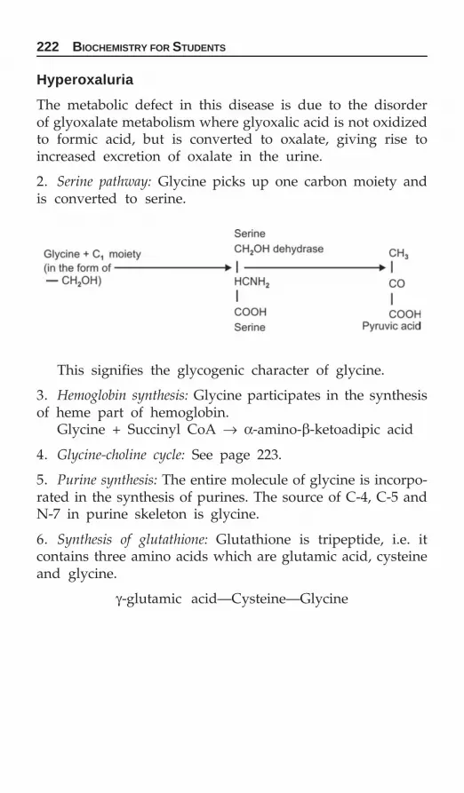
222 BIOCHEMISTRY FOR STUDENTS
Hyperoxaluria
The metabolic defect in this disease is due to the disorderof glyoxalate metabolism where glyoxalic acid is not oxidizedto formic acid, but is converted to oxalate, giving rise toincreased excretion of oxalate in the urine.
2. Serine pathway: Glycine picks up one carbon moiety andis converted to serine.
This signifies the glycogenic character of glycine.
3. Hemoglobin synthesis: Glycine participates in the synthesisof heme part of hemoglobin.
Glycine + Succinyl CoA → α-amino-β-ketoadipic acid
4. Glycine-choline cycle: See page 223.
5. Purine synthesis: The entire molecule of glycine is incorpo-rated in the synthesis of purines. The source of C-4, C-5 andN-7 in purine skeleton is glycine.
6. Synthesis of glutathione: Glutathione is tripeptide, i.e. itcontains three amino acids which are glutamic acid, cysteineand glycine.
γ-glutamic acid—Cysteine—Glycine

METABOLISM OF PROTEINS 223

224 BIOCHEMISTRY FOR STUDENTS
7. Creatine synthesis: Glycine participate in the synthesis ofcreatine. The other two amino acids are arginine andmethionine.
8. Conjugation reaction: For the formation of bile acids, glycineis important.
Glycine + Cholic acid → Glycocholic acid
The following diagram signifies that though glycine is non-essential but it is metabolically most active amino acid.
In detoxification reactions of body.
Glycine + Benzoic acid → Hippuric acid.
Compounds which are toxic detoxified by these reactions.

METABOLISM OF PROTEINS 225

226 BIOCHEMISTRY FOR STUDENTS
Glycinuria
This disease is due to the decreased reabsorption of glycinebecause of defect in renal tubular transport of glycine.Glycinuria is characterized by high excretion of glycine in theurine and tendency to form oxalate renal stones.
Sulfur Containing Amino Acids
The sulfur containing amino acids are methionine, cystine andcysteine. Methionine is an essential amino acid. The deficiencyof it causes negative nitrogen balance and loss in weight inman, whereas, cystine and cysteine are nonessential ordispensible amino acids.
Cysteine is synthesized from methionine and cysteine fromcystine. The presence of cystine in the diet decreases methi-onine requirement in that it relieves the demand for the for-mation of cystine.
The structure of methionine, cysteine and cystine are:
METHIONINE
Methionine is the principal methyl donor in the body. Thetransfer of methyl group of methionine to appropriate donorsis called transmethylation.
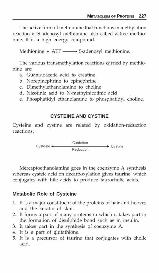
METABOLISM OF PROTEINS 227
The active form of methionine that functions in methylationreaction is S-adenosyl methionine also called active methio-nine. It is a high energy compound.
Methionine + ATP S-adenosyl methionine.
The various transmethylation reactions carried by methio-nine are:
a. Guanidoacetic acid to creatineb. Norepinephrine to epinephrinec. Dimethylethanolamine to cholined. Nicotinic acid to N-methylnicotinic acide. Phosphatidyl ethanolamine to phosphatidyl choline.
CYSTEINE AND CYSTINE
Cysteine and cystine are related by oxidation-reductionreactions.
Mercaptoethanolamine goes in the coenzyme A synthesiswhereas cysteic acid on decarboxylation gives taurine, whichconjugates with bile acids to produce taurocholic acids.
Metabolic Role of Cysteine
1. It is a major constituent of the proteins of hair and hoovesand the keratin of skin.
2. It forms a part of many proteins in which it takes part inthe formation of disulphide bond such as in insulin.
3. It takes part in the synthesis of coenzyme A.4. It is a part of glutathione.5. It is a precursor of taurine that conjugates with cholic
acid.

228 BIOCHEMISTRY FOR STUDENTS
Metabolism of Cystine and Cysteine
Cystinuria
Excessive excreting of cystine in urine takes place. Also thereis defect in renal reabsorption mechanism for lysine, arginineand ornithine. This is an inherited disease.
Cystinosis
Deposition of cystine crystals in many tissues and organs takesplace. Aminoaciduria is also present.

METABOLISM OF PROTEINS 229
Active Sulfate
Coenzyme adenosine-3'-phosphate-5'-phosphosulfate or 3'-phosphoadenosine-5-phosphosulfate (PAPS) is called activesulfate.
The structure of active sulfate is given below.
Active sulfate sulfating agent
Active sulfate is involved in the sulphonation of phenolsand of hexosamine derivatives as in the formation of chon-droitin sulfate and in other sulfanaling transfer reactions, i.e.sulfolipids or sulfatides. They are formed from cerebrosidesby reaction with active sulfates.
Phenol, steroids also react with PAPS giving respective sulfate derivatives which are eliminated in the urine. Thisdetoxification mechanism takes place in liver.
PHENYLALANINE AND TYROSINE
Both phenylalanine and tyrosine are aromatic amino acids.Tyrosine is an hydroxylated phenylalanine.
The structures of phenylalanine and tyrosine are:

230 BIOCHEMISTRY FOR STUDENTS
Phenylalanine is an essential amino acids whereas tyrosineis nonessential amino acid. Phenylalanine can be convertedto tyrosine but tyrosine cannot give rise to phenylalanine.Hence, the requirement of tyrosine can be met by the adequateamount of phenylalanine in the diet.
Phenylalanine and tyrosine are ketogenic amino acids.
Metabolism of Phenylalanine and Tyrosine
Phenylalanine and tyrosine are involved in the synthesis ofa number of important compounds which include thyroxine,melanin, epinephrine and norepinephrine.
Conversion of phenylalanine to tyrosine takes place in theliver. The enzyme is phenylalanine hydroxylase. The enzymerequires molecular oxygen. Fe++, NADPH and biopterin ascofactor.
The reaction is explained below:
Major pathway by which phenylalanine and tyrosine aremetabolized are given as follows:

METABOLISM OF PROTEINS 231

232 BIOCHEMISTRY FOR STUDENTS

METABOLISM OF PROTEINS 233

234 BIOCHEMISTRY FOR STUDENTS
Phenylalanine and tyrosine metabolism give rise to the syn-thesis of the important hormones, thyroxine and triiodotyrosine.
Synthesis of melanine
Formation of Thyroxine (T4) and Triiodotyrosine (T3)
The complete metabolism of phenylalanine and tyrosine issummed as below:
Inborn errors of metabolism associated with phenylalaninemetabolism.

METABOLISM OF PROTEINS 235
Inborn Error of Metabolism
There are a number of metabolic abnormalities which are con-genital, present throughout life and hereditary. Such abnor-malities are represented as in born error of metabolism.
In some of these conditions failure of a metabolic step leadsto the excretion of intermediate products which cannot befurther metabolize along the metabolic path because of specificenzyme deficiency but which normally readily metabolizes.
Inborn errors Enzyme deficitPhenylketonuria Phenylanine hydroxylase,Tyrosinosis p-hydroxy phenylpyruvate-
hydroxylase.Alkaptonuria Homogentisic acid oxidaseAlbinism Tyrosinase.Various blocks in the metabolism of phenylalanine giving
rise to different inborn errors of metabolism are shown below:
Phenylketonuria
Phenylketonuria is an inborn error of metabolism associatedwith phenylalanine metabolism. The enzyme deficit is phenyl-alanine hydroxylase. This enzyme catalyze the conversion of

236 BIOCHEMISTRY FOR STUDENTS
phenylalanine to tyrosine. Due to the deficiency of the enzymephenylalanine hydroxylase, the main pathway for the meta-bolism of phenylalanine via tyrosine is blocked and the minoralternate pathway takes place. The various metabolites thataccumulate in the blood and excreted in the urine by the minorpathway are explained below.
Phenylketonuria results in severe mental deficiency andthe children suffering from this disease are mentally retardedbecause, the metabolites of phenylketonuria, i.e. phenylpyruvicacid, phenyllactic acid and phenylacetic acid, inhibit theformation of serotonin, a brain potent metabolite.
Tyrosinosis
Tyrosinosis is an inborn error of metabolism associated withphenylalanine metabolism. The enzyme deficit is p-hydroxyphenylpyruvic acid hydroxylase. Due to the deficiency of this enzyme,p-hydroxy phenylpyruvic acid is not converted to homogentisicacid, resulting in the accumulation of p-hydroxy phenylpyruvicacid in blood and the excretion of p-hydroxy phenylpyruvic acidand its reduction product, p-hydroxyphenyllactic acid in the urine.
METABOLISM OF PROTEINS 237
Alkaptonuria
Alkaptonuria is an inborn error of metabolism associated withphenylalanine metabolism. The enzyme deficit is homogentisicacid oxidase. The deficiency of homogentisic acid oxidase causesa block in the metabolic pathway at homogentisic acid,resulting in its accumulation in the blood and excretion in theurine. Alkaptonuria is characterized by the excretion of urinewhich upon standing gradually becomes darker in color andfinally turns black.
Accumulation of homogentisic acid in body fluid and itsdeposition in cartilages and other connective tissues give riseto a condition called ochronosis.
Albinism
Albinism is an inborn error of metabolism associated withphenylalanine metabolism. The enzyme deficit is tyrosinase.Due to the deficiency of enzyme tyrosinase, the conversionof tyrosine to melanin formation is blocked.
TRYPTOPHAN
Tryptophan is an essential amino acid. It is the only aminoacid containing indole ring. Tryptophan has the metabolicdistinction of giving rise to nicotinic acid, a number of vitaminB-complex group, during its course of metabolism. It isketogenic in nature.
Metabolism of Tryptophan
Tryptophan is metabolized by the following way. The meta-bolism takes place in liver.
Synthesis of niacin
The major pathway by which tryptophan is metabolized toniacin is given page 238.
This pathway gives rise to the synthesis of niacin. 60 mgof tryptophan gives rise to 1 mg of nicotinic acid in the humanbody. In diet, tryptophan is not in sufficient amount to meetthe requirement of this vitamin.

238 BIOCHEMISTRY FOR STUDENTS
Synthesis of serotonin
This is another important pathway by which tryptophan ismetabolized. Serotonin is also called 5-hydroxytryptamine(5HT), Enteramine or Thrombocytin.

METABOLISM OF PROTEINS 239
Carcinoid Syndrome: Increased excretion of 5-hydroxy indoleacetic acid (5-HIAA) due to increased production of serotoningive rise to carcinoid syndrome. Normally one percent oftryptophan is metabolized by this pathway but in the carcinoidpatients 60 percent of the tryptophan is metabolized by thispathway. Normal excretion of 5-HIAA is 2-10 mg per daybut in carcinoid syndrome patients it may go as high as 50-1000 mg per day. The patient of these syndrome exhibit chronicdiarrhea.
Synthesis of Indole and skatole
This is the minor pathway by which tryptophan is metabolized.The synthesis of indole and skatole takes place in the largeintestines due to certain bacteria. The foul smell of the fecesis due to these.
Synthesis of melatonin
Hartnup Disease
This is an inborn error associated with tryptophan metabolism.The enzyme deficient is tryptophan pyrrolase.
This disease is characterized by three symptoms:a. Pellagra like dermatitisb. Intermittent cerebellar ataxiac. Mental retardation.
Urine of the patient contains large amount of tryptophan,indole acetic acid and its glutamine conjugate. Feces also containlarge amounts of tryptophan. In Hartnup disease there is adeficiency of nicotinic acid because tryptophan is not availablefor the synthesis of nicotinic acid.

240 BIOCHEMISTRY FOR STUDENTS
LEUCINE, ISOLEUCINE AND VALINE
These are the branched amino acids.
Metabolism of Branched Chain Amino Acids
Branched chain amino acids are metabolized as given below:
Maple Syrup Urine Disease
This is an inborn error of metabolism, in infants, resultingin the block in the metabolism of leucine, isoleucine and valine.Due to the deficiency of enzyme oxidative decarboxylase (i.e.step 2), oxidative decarboxylation of α-keto acids does nottake place and hence branched chain keto acid derivatives ofleucine, isoleucine and valine accumulate in blood and appearin urine. The odour of urine of such infants resembles thatof maple syrup.

NUCLEIC ACID—CHEMISTRY AND METABOLISM 241
Nucleoprotein belongs to the category of conjugated proteins,the nucleic acid part is the prosthetic group and the proteinpart consists of protamines and histones, which are basic innature.
The successive degradation of the nucleoproteins is shownbelow:
Nucleic Acid—Chemistryand Metabolism
CHAPTER
11
Bases present in nucleic acids are purines and pyrimidines

242 BIOCHEMISTRY FOR STUDENTS
Pyrimidine Bases
Three main pyrimidine bases are:1. Uracil = It is 2,4-dioxy pyrimidine.2. Thymine = It is 2,4-dioxy-5 methyl pyrimidine.3. Cytosine = It is 2-oxy-4-aminopyrimidine.
The oxypurines and oxyprimidines exist in enol and ketoform. Enol form is called lactim form and keto form is calledlactum form. Lactum form is predominent of the two.
Purine Bases
Two bases are:1. Adenine = It is 6-amino purine2. Guanine = It is 2-amino-6-oxypurine.
Their structures are:
Pentose Sugars
The pentose sugars present are D-ribose and D-2-deoxyribose.Both sugars occur as furanose form in nucleotides.

NUCLEIC ACID—CHEMISTRY AND METABOLISM 243
Nucleoside
Base-sugar unit is called nucleoside.
Nucleoside = Base – Sugar
Purine bases are attached at N-9 position to sugar moietywhereas pyrimidine bases are attached at N-1 position to sugarmoiety. The nature of linkage is α-glycosidic linkage.
Base Sugar Nucleoside
Adenine D-ribose AdenosineGuanine D-ribose GuanosineUracil D-ribose UridineCytosine D-ribose CytidineThymine D-deoxyribose Thymidine
Nucleotides
Nucleotides are phosphorylated nucleosides. They are repre-sented by base-sugar-phosphate unit.
Nucleotides = Base-sugar-phosphoric acid.
Base Sugar Phosphate Nucleotide
Adenine D-ribose Phosphoric acid Adenylic acid.Adenosinemonophosphate (AMP)
Guanine D-ribose Phosphoric acid Guanylic acid.Guanosinemonophosphate (GMP)
Contd...

244 BIOCHEMISTRY FOR STUDENTS
Contd...
Base Sugar Phosphate Nucleotide
Hypoxan- D-ribose Phosphoric acid Inosinic acid.thine Inosine mono-
phosphate (IMP)Uracil D-ribose Phosphoric acid Uridylic acid.
Uridine mono-phosphate (UMP)
Cytosine D-ribose Phosphoric acid Cytidylic acid.Cytidine mono-poshphate (CMP)
Thymine 2-deoxy- Phosphoric acid Thymidylic acid.D-ribose Thymidine
monophosphate (TMP)
NUCLEIC ACIDS
Nucleic acid are polynucleotide and the nucleotides are linkedby means of 3',5'-phosphodiester bonds.
Nucleic acids are of two types:1. Deoxyribose nucleic acid (DNA)2. Ribose nucleic acid (RNA).Hydrolytic products of DNA and RNA
Deoxyribose Nucleic Acid (DNA)
DNA is a polymer of 2-deoxyadenylic acid, 2-deoxyguanylicacid, 2-deoxycytidylic acid and thymidylic acid. It is repre-sented as Base-deoxyribose-phosphate.

NUCLEIC ACID—CHEMISTRY AND METABOLISM 245

246 BIOCHEMISTRY FOR STUDENTS
Structure of DNA
Watson and Crick proposed the helix structure of DNA, inwhich the two polynucleotide chains are wound about a com-mon axis in the form of a double helix. These two polynucleo-tide chains are antiparallel, i.e. they run in opposite directions.
The backbone of DNA, which is a constant throughout themolecule, consists of deoxyriboses linked by 3', 5' phospho-diester bridges. Polynucleotide chains are so oriented that anadenine is always located in the space opposite a thymine anda guanine is opposite a cytosine. This positioning of base is

NUCLEIC ACID—CHEMISTRY AND METABOLISM 247
called base pairing or base complementarity. There exists threehydrogen bonds between guanine and cytosine. The hydrogenbonding involves the keto and amino groups of purine andpyrimidine bases. DNA double helix is stabilized by hydrogenbonding.
In DNA, purine and pyrimidine bases carry genetic infor-mation whereas sugar and phosphate groups perform astructural role.
Ribose Nucleic Acids (RNA)
RNA is a polymer of ribonucleotides. RNA is made up of aribosephosphate backbone to which the various bases areattached. RNA, like DNA, exhibits polarity. The 5’ hydroxylgroup points towards the 5’ end and 3’-hydroxyl group pointsto the 3’ end of the molecule. The sequence of bases alongthe sugar-phosphate backbone determines the primary struc-ture (information content) of RNA, and this is the factor thatdistinguish one RNA from another.
RNA is present in three forms. All the three forms arepresent as single strand, each has characteristic molecularweight and sedimentation coefficient.1. Transfer or soluble RNA2. Messenger RNA3. Ribosomal RNA.
Transfer or Soluble RNA
It comprises 10-20% of the total RNA of the cell.1. It is present in the soluble fraction of the cell.2. It is involved in the transfer of amino acids. Each amino
acid has a specific t-RNA.3. It is a small molecule containing 75 to 90 nucleotides.4. It has a heterogenous base composition.5. It has a clover leaf structure and possess a specific triplet
nucleotide known as anticodon, which is complimentaryto the 3 bases on m-RNA called codon.

248 BIOCHEMISTRY FOR STUDENTS
The structure of transfer RNA is
Messenger RNA
Messenger RNA is synthesized in the nucleus during transcrip-tion in which sequence of bases in one strand of DNA is trans-cribed in the form of a single strand of m-RNA. The sequence

NUCLEIC ACID—CHEMISTRY AND METABOLISM 249
of bases in, m-RNA strand is complimentary to that of DNA.After transcription, m-RNA passes into cytoplasm and thenon to ribosomes where it serves as a template for the sequenceof amino acids during biosynthesis of proteins.
Ribosomal RNA
It comprises 50-80% of the total cellular RNA. Ribosomes carryribosomal RNA.
It has a homogenous base composition.Ribosomal RNA is required to bind m-RNA and specific
enzymes utilized for peptide bond synthesis.Various fractions of ribosomes are 30s and 50s.
Some Biologically Important Nucleotides
1. Adenosine diphosphate (ADP), adenosine triphosphate (ATP).They are sources of high energy phosphate bonds and takepart in oxidative phosphorylation.
2. Inosine diphosphate (IDP) and inosine triphosphate (ITP)participates in phosphorylation reactions.
3. Guanosine triphosphate (GTP) and Guanosine diphosphate(GDP) participates in citric acid cycle.
4. Uridine diphosphate glucose (UDPG) participate in gluco-neogenesis.
5. NAD+ and coenzyme A are of biomedical importance andare synthesized from amphobolic intermediate.
6. Cyclic AMP and cyclic GMP serves the secondarymessenger function.
7. CDP-Acyl glycerol in lipid biosynthesis acts as high energyintermediates.
Purines and Pyrimidines Metabolism
Precursors of Purine Ring
N-1 is derived form amino nitrogen of aspartate.N-3 and N-9 derived from amide nitrogen of glutamine.N-2 and C-8 are derived from format.

250 BIOCHEMISTRY FOR STUDENTS
C-6 is derived from respiratory carbon dioxide.C-4, C-5 and N-7 are derived from glycine.
Precursors of Pyrimidine Ring
N-3 is derived from amide nitrogen of glutamine.C-2 is derived from respiratory carbon dioxide.N-1, C-4, C-5 and C-6 are derived from aspartate.
Biosynthesis of the Purine Ribonucleotides
The starting material for the de novo synthesis of purine ribo-nucleotide is an activated form of D-ribose-5-phosphate onwhich purine ring is built up step-by-step.
D-ribose-5-phosphate, derived from pentose phosphatepathway is converted to 1-pyrophosphate ribose-5-phosphate(PPRP) by the transfer of pyrophosphate group from ATP toC-1 of ribose.

NUCLEIC ACID—CHEMISTRY AND METABOLISM 251
The first ribonucleotide formed by this pathway is inosinicacid which is a precursor of adenylic acid and guanylic acid.
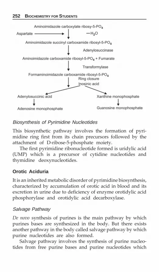
252 BIOCHEMISTRY FOR STUDENTS
Biosynthesis of Pyrimidine Nucleotides
This biosynthetic pathway involves the formation of pyri-midine ring first from its chain precursors followed by theattachment of D-ribose-5-phosphate moiety.
The first pyrimidine ribonucleotide formed is uridylic acid(UMP) which is a precursor of cytidine nucleotides andthymidine deoxynucleotides.
Orotic Aciduria
It is an inherited metabolic disorder of pyrimidine biosynthesis,characterized by accumulation of orotic acid in blood and itsexcretion in urine due to deficiency of enzyme orotidylic acidphosphorylase and orotidylic acid decarboxylase.
Salvage Pathway
De novo synthesis of purines is the main pathway by whichpurines bases are synthesized in the body. But there existsanother pathway in the body called salvage pathway by whichpurine nucleotides are also formed.
Salvage pathway involves the synthesis of purine nucleo-tides from free purine bases and purine nucleotides which

NUCLEIC ACID—CHEMISTRY AND METABOLISM 253

254 BIOCHEMISTRY FOR STUDENTS
are salvaged from dietary sources and tissue breakdown. Thispathway is mainly operative in leukocytes, brain, etc. whichare not capable of de novo synthesis of purine nucleotides. Since90 percent of the free purines formed by man are salvagedand recycled by this pathway and hence this pathway isimportant in purine economy in the vertebrates.
Salvage reactions promoted by the action of two enzymeswhich convert free purines into corresponding purine nucleo-tides for reuse are Adenine phosphoribosyl transferase andHypoxanthine-guanine phosphoribosyl transferase (HGPRTase).
The reactions performed by these enzymes are asfollows:
Lesch-Nyhan Syndrome
Deficiency of enzyme hypoxanthine-guanine phosphyribosyltransferase (HGPR Tase) give rise to Lesch-Nyhan syndrome,a genetic disorder. This enzyme catalyze the transfer of ribosephosphate group of 5 PRPP to either guanine, xanthine or hypo-xanthine. When this enzyme is deficient guanine, xanthine andhypoxanthine are not salvaged and hence degraded to uricacid.
Catabolism of Purines
This first step in the catabolism of purines is their hydrolyticdeamination to hypoxanthine, i.e. adenine is converted to

NUCLEIC ACID—CHEMISTRY AND METABOLISM 255
hypoxanthine and guanine to xanthine. In the second step bothxanthine and hypoxanthine are oxidized to uric acid.
In man, the end product of purine catabolism is uric acid.Whereas in lower primates, the enzyme uricase, hydrolyzeuric acid to allantoin.
Catabolism of Pyrimidines
The catabolism of pyrimidines takes place mainly in the liverand the breakdown pathway is represented as:
The breakdown of pyrimidines gives rise to the formationof β-alanine and β-aminoisobutyric acid. β-alanine is metabolizedto acetate, carbon dioxide and ammonia, whereas β-aminoiso-butyric acid is metabolized to propionic acid, which in turngives rise to succinic acid.
β-alanine is an important constituent of pantothenic acidand therefore, of coenzyme A. Also β-alanine is a componentof carnosine and anserine which are synthesized by the muscleand brain.

256 BIOCHEMISTRY FOR STUDENTS
Regulation of Purine Synthesis
Purine biosynthesis is regulated by feedback inhibition at thefollowing sites:a. Feedback inhibition of 5-phosphoribose pyrophosphate
synthetase by AMP, GMP and IMP.b. Feedback inhibition of amidotransferase by the final
products of the pathway ATP, ADP, AMP, GTP, GDP, GMP,IMP, IDP and IMP.
c. Inosinic acid (IMP) is at a branched point in the synthesisof AMP and GMP. AMP inhibits the conversion of IMPto adenylosuccinate by inhibiting the enzyme adenylosucci-nate dehydrogenase. Similarly, GMP inhibits the conversionof IMP to xanthylic acid by inhibiting the IMP dehydro-genase.
d. ATP is a substrate in the synthesis of GMP whereas GTPis a substrate in the synthesis of AMP. This reciprocalsubstrate relationship regulates the synthesis of AMP andGMP, i.e. GMP synthesis is accelerated when AMP levelsare high.

NUCLEIC ACID—CHEMISTRY AND METABOLISM 257
Regulation by Pyrimidine Biosynthesis
The committed step in pyrimidine biosynthesis is the formationof N-carbamoyl aspartate from aspartate and carbamoyl phos-phate. The enzyme is aspartate transcarbamylase. This enzymeis feedback inhibited by CTP, the final product in the pathway.The second control site is carbamoyl phosphate synthesis whichis feedback inhibited by UMP.
Gout
Gout is a metabolic disease, the salient biochemical featureof which is hyperuricemia. As a result of hyperuricemia, largeamounts of uric acid (as sodium salt) are deposited in thejoints and tissues (tophi).
Gout is of two types:1. Metabolic gout2. Renal gout.
Metabolic Gout
i. Primary metabolic goutii. Secondary metabolic gout.
Primary metabolic gout: This is due to inherited metabolicdefect in purine metabolism leading to increased rate of conver-sion of glycine to uric acid.
Secondary metabolic gout: This is due to increased catabolismof nucleic acids, i.e. polycythemia, leukemia, etc.

258 BIOCHEMISTRY FOR STUDENTS
Renal Gout
i. Primary renal goutii. Secondary renal gout.
Primary renal gout: In this type of gout, the defect lies inthe kidney where there is faulty enzymatic transport of uratesby the renal tubules. The rate of excretion of uric acid islowered.
Secondary renal gout: This is due to glomerulonephritis orsome other destructive process leading to kidney failure.

VITAMINS 259
In addition to oxygen, water, proteins, fats, carbohydratesand inorganic salts, a number of organic compounds are alsonecessary for the life, growth and health of animals includingman. These compounds are known as the accessory dietaryfactors or vitamins and are only necessary in very smallamount. Vitamin cannot be produced by the body and hence,must be supplied.
Vitamins are defined as organic compounds, occurring innatural food either as such or as utilizable precursors whichare required in minute amounts for normal growth,maintenance and reproduction. They differ from other organicfoodstuffs in that they do not enter into the tissue structureand do not undergo degradation for the purpose of providingenergy. The absence of these results in deficiency disease.
Most of the vitamins are supplied by the diet. Very fewvitamins which are synthesized in the intestine belongs to thevitamin B group.
Vitamins which are synthesized in the intestinal flora are:vitamin K, thiamine, riboflavin, pyridoxine, folic acid, niacinand biotin.
But the entire requirement of these vitamins are not metby the endogenous synthesis.
Vitamin deficiencies are must often the result of consumingmonotonous diet, i.e. diet-based on limited number of foodsources. The requirements for vitamins are usually greatestduring the neonatal period.
The vitamins have been classified into:1. Fat soluble vitamins: They are soluble in fat solvents.Vitamins in this group are vitamins A, D, E and K.
Vitamins
CHAPTER
12

260 BIOCHEMISTRY FOR STUDENTS
2. Water soluble vitamins: They are water soluble andincludes vitamin C and vitamins of B-complex.
Most of the vitamins form the integral part of coenzymes.Fat soluble vitamins are stored in our fat deposits (liver
and adipose tissue) and water soluble vitamins are constantlyflushed from our bodies. Therefore, we can do without lipidsoluble vitamins for a resonable amount of time but we mustkeep replacement the water soluble vitamins. Vitamins actsas coenzyme, antioxidants, (free radical quenching agents)as signalling agents in the cells, as regulator of gene expressionand as redox.
Vitamin Like Compounds
Those compounds which are highlighted because of theirknown role as coenzymes in prokaryotes eukaryotes or rolesas a probiotic (growth promoting substance) in higher animalsare defined as vitamin like.
Vitamin like substances are taurine, queuosine, coenzymeQ, pteridines (other then folic acid), such as biopterin and themolybdenum containing pteridine cofactor, pyrroloquinolinequinone (PQQ).
Vitamins as Coenzymes
Vitamins Active form Functions performed
Thiamine Thiamine pyrophosphate Aldehyde grouptransfer
Riboflavin Flavin mononucleotide Hydrogen group(FMN) transferFlavin adenine Hydrogen groupdinucleotide (FAD) transfer
Pantothenic acid Coenzyme A Acyl group transferNicotinamide Nicotinamide adenine Hydrogen transfer
dinucleotide (NAD)Nicotinamide adenine Hydrogen transferdinucleotide phosphate(NADP)
Pyridoxine Pyridoxal phosphate Amino group transfer
Contd...

VITAMINS 261
Biotin Biocytin Carboxyl grouptransfer, i.e. CO2 fixing
Folic acid Tetrahydrofolic acid, Methyl, methylene,i.e. Folacin formyl or formimino
group transferCyanocobalamin Cobamides Alkyl group transferLipoic acid Lipoyl lysine Acyl group transfer
FAT SOLUBLE VITAMINS
VITAMIN A
Retinol, growth promoting vitamin, anti-infective vitamin,antixerophthalmia.
Structure
Vitamin A occurs in two forms, vitamin A1 and vitamin A2.The structure of vitamin A1 is:
Vitamin A2 contains an additional double bond betweenC-3 and C-4.
Vitamin A2 is 40 percent active to vitamin A1.Certain carotenes called provitamins A are converted into
vitamin A in the body. β-carotenes give rise to two moleculesof vitamin A whereas α- and γ-carotenes give rise to onemolecule each of vitamin A.
Functions
1. The most important function of vitamin A is in the visualcycle (page 262).
Retina contains conjugated protein rhodopsin. Rhodop-sin consists of protein ‘opsin’ and vitamin A1 aldehyde.Rhodopsin is the major light receptor of rod cells. Under
Contd...

262 BIOCHEMISTRY FOR STUDENTS
the influence of light rhodopsin is converted to transretinaland opsin. Transretinal is inactive in the resynthesis of rho-dopsin. Transretinal is converted to transretinal by reductasewhich is also inactive in rhodopsin synthesis, is passed intoblood stream. During resynthesis of rhodopsin, which occursin dim light and in the dark, active cis-retinal enters theretina from the blood and is oxidized to cis-retinal by retinalaction of retinal reductase. Now cis-retinal couples withopsin to form rhodopsin. The visual process involves theremoval of active retinal isomer from the blood by the retinawhich returns the inactive isomer to the circulation.
2. In the maintenance of proper health of epithelium tissues.3. For the stability and integrity of cellular and subcellular
membranes.4. Necessary for the synthesis of mucopolysaccharides as it
helps in the incorporation of sulfur in chondroitin sulfate.5. It is also involved in the nucleic acid metabolism.6. It is also involved in the election transport chain and in
oxidative phosphorylation.
Sources
Provitamin sources: Food rich in carotenes such as carrot,papayas, tomatoes, etc.
Readymade or preformed sources: Fish liver oils such as shark,cod, halibut fish, liver oils, egg yolk, butter, milk products.
Visual cycle

VITAMINS 263
Daily Requirement 5000 IU
Deficiency Disease
Deficiency of vitamin A gives rise to night blindness.
Hypervitaminosis A
Excessive intake of vitamin A gives rise to hypervitaminosis A.The symptom of this toxicity include anorexia, irritability,
headache, peeling of skin, vomiting.

264 BIOCHEMISTRY FOR STUDENTS
VITAMIN D
The term vitamin D does not refer to a single dietaryfactor but to a number of chemically related compounds, allof which have the property of preventing or curing rickets.The two most active substances in this respect are vitaminD2 and vitamin D3.
Structure
Vitamin D2 is also known as ergocalciferol and vitamin D3as cholecalciferol.
Irradiation of 7-dehydrocholesterol with ultraviolet radia-tions produces cholecalciferol whereas irradiation of ergosterolproduces ergocalciferol.
Vitamin D2 differs from vitamin D3 with respect to thedouble bond additionally present in the side chain at position20 and 21.
The biologically active form of vitamin D are 25-hydroxycholecalciferol and 1,25-cholecalciferol.
Liver converts cholecalciferol to 25-hydroxy cholecalciferol(25 HCC) whereas kidney converts 25-HCC to 1,25 dihydroxycholecalciferol (1,25-DHCC).
Another important active form formed by the kidney is24,25-dihydroxy cholecalciferol (24,25-DHCC) but very littleis known of biological function of this form.
1,25-dihydroxy cholecalciferol perform the following func-tions:
i. It promotes calcium absorption from the intestineii. It promotes mobilization of calcium from bones.
Functions
1. The basic action of vitamin D is to increase the absorptionof calcium and phosphorus from the intestines.

VITAMINS 265
2. Vitamin D promotes resorption of bone and mobilizationof calcium from bones.
3. Vitamin D increases excretion of phosphate by the kidney.
Sources
Fish liver oils (i.e. cod liver oil, shark liver oil, halibut liveroil), egg yolk, milk.
Daily requirement 400 IU infants and children100 IU adults400 IU pregnancy and lactation
Deficiency Disease
Deficiency of vitamin D gives rise to rickets in children andosteomalacia in adults.
In rickets, there is a fall in intestinal absorption of calciumand phosphate, increased excretion of urinary phosphate andloss of calcium from the bones, leading to softness anddeformities of bones.
In rickets and osteomalacia, there is an increase in serumalkaline phosphatase activity.
Hypervitaminosis D
Doses above 1500 units per day for long period rise to vitaminD toxicity.
Excessive intake of vitamin D causes loss of appetite,nausea, irritability, excessive mobilization of calcium frombones into blood.
VITAMIN E(Antisterility vitamin, fertility factor)
Vitamin E refers to a group of compounds having vitamin Eactivity and are known as tocopherols. Four unsaturatedalcohols, i.e. α, β, γ and δ tocopherols occur in nature. Thesetocopherols differ slightly in structure in their side chain,α-tocopherol is most potent of them.

266 BIOCHEMISTRY FOR STUDENTS
They are thermostable and sensitive to the effects of oxidi-zing agents and ultraviolet rays.
Functions
1. Tocopherols act as powerful antioxidants:a. They prevent the autoxidation of vitamin A and caro-
tenes.b. They prevent the formation of fatty acid peroxidases
in tissues due to the autoxidation of unsaturated fattyacids with oxygen.
c. They protect the lipids of biological membranes againstoxygen by acting as antioxidants (i.e. prevent the pero-xidation of polyunsaturated fatty acids that occur inmembranes throughout the body).
2. Vitamin E prevents rancidity.
Sources
Wheat germ oil, corn oil, peanut oil, soyabean oil, sunfloweroil, egg yolk, leaves of spinach, alfalfa, sweet potatoes.
Daily Requirement 15 IU
Vitamin E intake is related to the intake of polyunsaturatedfatty acids, i.e. 0.4 mg per gm of polyunsaturated fatty acids.
Deficiency Disease
Deficiency of vitamin E gives rise to sterility in rats.
VITAMIN K(Coagulation factor)
Two naturally occurring vitamin K are vitamin K1 and vitaminK2. Both are naphthaquinone derivatives.
Structure
The structure of α-tocopherol is:

VITAMINS 267
Structure
Vitamin K1 is phylloquinone and is chemically known as2-methyl-3-phytyl-1, 4-naphthaquinone.
Vitamin K2 is farnesoquinone and is chemically known as2-methyl-3 difarnesyl-1, 4-naphthaquinone.
Vitamin K is thermostable can withstand reduction andis rapidly oxidized both in acidic and alkaline medium. It iscompletely destroyed by ultraviolet radiations.
Certain compounds having vitamin K activity are calledvitamines. Vitamines are synthetic compounds possessingvitamin K activity (Example: Menadione). Menadione is morepotent than vitamin K1 on weight basis.
Functions1. In the synthesis of prothrombin.2. It is involved in oxidative processes taking place in photo-
synthesis of plant kingdom.3. It is involved in electron transport chain and is involved
in oxidative phosphorylation.
Vitamin K cycle

268 BIOCHEMISTRY FOR STUDENTS
Sources
Vitamin K1, is present in alfalfa, spinach, cabbage, cauliflower,egg yolk, liver.
Vitamin K2 is present in putrifying fish. It is also synthesizedby intestinal flora.
Daily Requirement
Sufficient amounts of vitamin K are synthesized by intestinalbacteria so there is no dietary requirement under physiologicalcondition.
Deficiency Disease
Vitamin K deficiency gives rise to hypoprothrombinemia whichleads to prolongation of prothrombin time.
WATER SOLUBLE VITAMINS
Water soluble vitamins includes vitamin C and members ofvitamin B complex.
Vitamin C(Ascorbic acid, anti-scorbutic vitamin)
Structure

VITAMINS 269
Vitamin C, which is hydrophilic, acts as an antioxidant insolution.
Vitamin C is a powerful reducing agent and is oxidizedto dehydroascorbic acid. Both forms are biologically active.It is stable in acidic solution at low temperatures but undergoesdestruction in alkaline solution when in contact with air.
Vitamin C is not synthesized by man and its entirerequirement is met by diet.
The most rich sources of vitamin C in the body are adrenalcortex, corpus luteum, pituitary, pancreas, liver, etc.
Functions
1. Participation in the hydroxylation of proline and lysinepresent in collagen, an intracellular cementing substance.
2. Participates in the synthesis of steroid hormones both inadrenal cortex and corpus luteum.
3. Participates as cofactor in the following reaction:a. In phenylalanine metabolism.
p-hydroxy phenylpyruvic acid → homogentisic acidb. Dopamine → Norepinephrinec. Folic acid → Folinic acid.
4. Vitamin C is necessary for the synthesis of carnitine in theliver.
5. Necessary for the absorption of iron by reducing ferricform to ferrous form.
6. In tissue respiration, i.e. oxidation-reduction phenomenon.7. In bile acid formation, vitamin C is required at the 7-α-
hydroxylase step.8. Ascorbic acid may act as water soluble antioxidant and
inhibit the formation of nitrosamine.
Sources
Citrus fruits such as lemon, orange, pineapple, etc. Indian goose-bury, green pepper, cauliflower, tomatoes, spinach, potato.Daily requirement: 60-80 mg.
Deficiency Disease
Deficiency of vitamin C gives rise to scurvy. The early mani-festation in man are swelling of joints, hemorrhage in skin,

270 BIOCHEMISTRY FOR STUDENTS
muscle, gastrointestinal tract, inflammation of gums, ulce-ration, etc. In scurvy, vitamin C level in blood falls down.
Vitamin B Complex
The members of this group are:1. Thiamine (B1).2. Riboflavin (B2).3. Pantothenic acid (B3).4. Choline (B4).5. Niacin (B5).6. Pyridoxine (B6).7. Biotin (B7).8. Folic acid (B9).9. Cyanocobalamin (B12).
10. Para amino benzoic acid.11. Inositol.12. Lipoic acid.
The vitamins of this family have been grouped togetherbecause of the following fulfilment:1. Usually present in yeast.2. Present in the outer covering of seeds and cereals.3. Synthesized by the microorganisms in the intestines.4. They are water soluble.5. Required in minute amounts and their deficiencies give
rise to ordinary manifestations.6. They usually serve as a coenzyme of various enzyme
systems. Foods that are poor sources of one of the Bvitamins level to be the poor sources of several B vitamins.
THIAMINE(Vitamin B1, anti-neuritic vitamin, anti-beriberi factor,
aneurin)
Structure

VITAMINS 271
Thiamine consists of a substituted pyrimidine ring joinedby a methylene bridge to substituted thiazole ring.
Thiamine is water soluble. It is thermolabile, destroyedin alkaline medium but thermostable in acidic medium.
Thiamine occurs in the cells largely as its active coenzymeform, i.e. thiamine pyrophosphate, also called cocarboxylase.
Functions
Thiamine pyrophosphate (TPP) is used mainly as a coenzymein the carbohydrate metabolism.
1. In oxidative decarboxylation of α-ketoacids.Pyruvic acid and α-ketoglutaric acid which are inter-
mediates in the carbohydrate metabolism are oxidativelydecarboxylated to acetyl CoA and succinyl CoA respectively.
Pyruvic acid Acetyl CoA
α-ketoglutaric acid Succinyl CoA
2. In transketolation reaction.An intermediate step in hexose monophosphate shunt path-
way where transfer of glycolaldehyde group from D-xylulose-5-phosphate and glyceraldehyde.
D-xylulose-5-PO4 D-sedoheptulose-PO4+ +
D-ribose-5-PO4 D-glyceraldehyde-PO4
3. In yeast TPP acts as coenzyme for nonoxidative decar-boxylation of α-keto-acids, i.e. pyruvate to CO2 and acetaldehyde.
Sources
Yeast, outer coating of seeds, cereals, legumes, wheat, pork,eggs.
Vitamin B1 is also synthesized during germination.
Daily Requirement: 1-1.4 mg.Thiamine requirement rises with a rise in caloric intake
of food, i.e. 0.5 mg of vitamin B1 for every 1000 calories inthe form of carbohydrates.
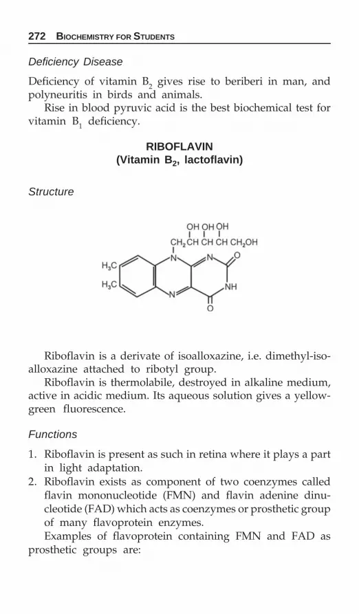
272 BIOCHEMISTRY FOR STUDENTS
Deficiency Disease
Deficiency of vitamin B2 gives rise to beriberi in man, andpolyneuritis in birds and animals.
Rise in blood pyruvic acid is the best biochemical test forvitamin B1 deficiency.
RIBOFLAVIN(Vitamin B2, lactoflavin)
Structure
Riboflavin is a derivate of isoalloxazine, i.e. dimethyl-iso-alloxazine attached to ribotyl group.
Riboflavin is thermolabile, destroyed in alkaline medium,active in acidic medium. Its aqueous solution gives a yellow-green fluorescence.
Functions
1. Riboflavin is present as such in retina where it plays a partin light adaptation.
2. Riboflavin exists as component of two coenzymes calledflavin mononucleotide (FMN) and flavin adenine dinu-cleotide (FAD) which acts as coenzymes or prosthetic groupof many flavoprotein enzymes.Examples of flavoprotein containing FMN and FAD as
prosthetic groups are:

VITAMINS 273
Flavin Mononucleotide (FMN)
a. Warburg yellow enzymeb. L-amino acid oxidasec. Cytochrome C reductase.
Flavin Adenine Dinucleotide (FAD)
a. D-amino acid oxidaseb. Xanthine oxidasec. Glycine oxidased. Thiophorase oxidasee. Acyl CoA dehydrogenase.
Sources
Yeast, milk, egg, meat, fish, liver, kidney, green leafy vegetables.
Daily requirement: 0.4-1.4 mg
Deficiency Disease
Riboflavin deficiency does not give rise to any clear cut diseasebut the important deficiency symptoms are choliosis, fissuresin lips, mouths, inflammation of the tongue, dermatitis.
Riboflavin is excreted mostly in fecal matter. In urine, apigment known as urochrome is excreted which is very muchrelated to riboflavin.
NIACIN(Pellagra preventive factor)
Structure

274 BIOCHEMISTRY FOR STUDENTS
Niacin is a pyridine derivative, thermostable and is notrapidly oxidized.
Functions
Niacinamide is a component of nicotinamide adeninedinucleotide (NAD+) and niacinamide adenine dinucleotidephosphate (NADH4) and acts as coenzyme for many anaerobicdehydrogenases by accepting hydride ions during oxidationof their substrates.
Enzymes requiring NAD+ or NADH coenzymes are:a. Glyceraldehyde-3-PO4 dehydrogenaseb. Lactate dehydrogenasec. Ketose reductased. Malic dehydrogenase.Enzymes requiring NAD+ or NADPH as coenzymes are:a. Isocitrate dehydrogenaseb. Glucose-6-PO4 dehydrogenasec. Aldolase reductase.
Sources
Yeast, meat, liver, kidney, eggs, fish, legumes.Nicotinic acid is also synthesized during tryptophan meta-
bolism, 60 mg of tryptophan gives rise to 1 mg of niacin.
Deficiency Disease
Deficiency of niacinamide gives rise to pellagra in man andblack tongue in dogs.
Pellagra affects the skin, central nervous system and thegastrointestinal tract.
The three important symptoms of pellagra are diarrhea,dermatitis and dermentia.
Diets such as corn maize give rise to niacin deficiency andultimately to pellagra because corn or maize are distinctlydeficient in tryptophan.
The important excretory products of nicotinic acid in humanurine are nicotinic acid as such, nicotinamide, N-methylniacina-mide, nicotinuric acid and 6-pyridone of nicotinic acid.

VITAMINS 275
PANTOTHENIC ACID
Structure
Pantothenic acid is a peptide like compound formed frompantoic acid and β-alanine.
It is thermostable and resistant to oxidation.
Functions
Pantothenic acid is a part of coenzyme, which serves as carrierof acyl group in enzymatic reactions.1. Fatty acid oxidation2. Fatty acid synthesis3. Pyruvic acid oxidation4. Biological acetylations5. Cholesterol biosynthesis6. In acyl carrier proteins.
Sources
Yeast, liver, kidney, egg yolk, molasses.
Daily Requirements
Synthesized by the intestinal flora in sufficient amount to meetthe requirement.
PYRIDOXINE(Antiachrodynia factor)
Structure
Vitamin B6 refers to a group of pyridoxine, pyridoxal andpyridoxamine compounds having similar biological activity.Pyridoxine is most abundant in plants, and pyridoxal andpyridoxamine are most abundant in animal tissues.

276 BIOCHEMISTRY FOR STUDENTS
Pyridoxal and pyridoxamine are thermolabile and photo-labile. Pyridoxine can be converted to pyridoxal and pyridoxa-mine but pyridoxal and pyridoxamine cannot be convertedback to pyridoxine.
All are biologically active but only the phosphate estersof pyridoxal and pyridoxamine can function as coenzymes.
Functions
Pyridoxine containing enzymes is important in amino acid andprotein metabolism.1. Pyridoxine acts as coenzymes for decarboxylation reactions:
a. Histidine → Histamineb. Tyrosine → Tyraminec. 5-hydroxy tryptophan → 5-hydroxy tryptamine (5HT)d. Glutamic acid → γ-amino butyric acid (GABA)e. α-amino-β-keto adipic acid → β-amino levulinic acid
2. As coenzyme in transamination reaction.3. As coenzymes in dehydrases reactions.
Serine → Pyruvic acidThreonine →α-ketobutyric acid
4. As coenzymes in trans-sulfurases reaction.
Homocysteine → Serine5. As coenzymes for desulfuration reactions.
Cysteine → Pyruvic acidHomocysteine → α-ketobutyric acid
6. In tryptophan metabolism, the conversion of kynurenine toanthranilic acid requires pyridoxal phosphate as coenzyme.

VITAMINS 277
In vitamin B6 deficiency, it is converted to xanthurenic acid.So measurement of xanthurenic acid is an indication of vitaminB6 deficiency.
7. Also helps in the transport of amino acids to the cellmembrane and is also necessary for the absorption of aminoacids from the intestinal mucosa.
8. Also involved in the conversion of linolenic acid to arachi-donic acid, hence, it is essential for essential fatty acidsynthesis.
Sources
Yeast, liver, egg yolk, rice polishings.Synthesized by the microflora of the intestines.
Daily requirement: 2-2.5 mg.
BIOTIN
Structure
Biotin contains fused imidazole and thiophene.
Biotin is present in food both in free and in combined formwith proteins. The combined form is liberated by proteolyticenzymes.
Biotin is also synthesized by intestinal flora.

278 BIOCHEMISTRY FOR STUDENTS
Functions
Biotin is required in carbon dioxide fixation reactions. Reactionswhere biotin is involved are:1. Acetyl CoA — Malonyl CoA (Fatty acid synthesis)2. Pyruvic acid — Oxaloacetic acid (Glucongenesis)3. Propionyl CoA — D-methyl malonyl CoA4. CO2 + NH3 — Carbamyl phosphate (Urea cycle)5. In purine ring synthesis, i.e. the C-6 position in purine ring
skeleton.
Structure of coenzyme A
Sources
Yeast, egg yolk, milk, molasses, chocolate, tomato, peanuts.
Daily requirement: 200 μg.
Deficiency Disease
Raw egg induces biotin deficiency because it contains a protein‘avidin’ which tightly binds biotin and prevents its absorptionfrom the intestine.

VITAMINS 279
FOLIC ACID
Structure
Folic acid consists of three components:i. Pteridine nucleus
ii. Para-aminobenzoic acid.
iii. Glutamic acid.Folic acid occurs in nature in three types:a. Pteroyl monoglutamate which contains only one
molecule of glutamic acid.b. Pteroyl triglutamate which contains three molecules of
glutamic acid.c. Pteroyl heptaglutamate which contains seven molecules
of glutamic acid.Before folic acid can function as coenzyme in various meta-
bolic reactions it must be reduced to tetrahydrofolic acid.Folic acid→Dihydrofolic acid→Tetrahydrofolic acid (TH4).The various activated forms of folic acid are:
N5-formyl TH4, N10-formyl TH4N5-10-methenyl TH4, N5-10-methylene TH4.
N5-TH4 contains formyl group at 5-position. It is also calledfolinic acid. TH4 is biologically inactive except that it has onlyone role, i.e. in the formylation of glutamic acid in histidinemetabolism whereas N10-TH4 and N5
10-TH4 are the biologicallymost active forms of tetrahydrofolic acid.
Folic acid coenzymes are collectively known as folacin.
Functions
Folic acid coenzymes are involved in the transfer and incorpo-ration of single or one carbon moiety.

280 BIOCHEMISTRY FOR STUDENTS
One carbon moieties are:i. Formyl group and formate group,
i.e.—CHO and —HCOOHii. Hydroxymethyl group, i.e. —CH2OH
iii. Methyl group, i.e. —CH3iv. Formimino group, i.e. —CH=NH
They are inter convertible:
—CH2OH —CHO —-COOH
Sources of one carbon moieties are:i. α-carbon of glycine gives rise to —CHO group
ii. Histidine gives rise to —CH=NH groupiii. Choline donates —CH3 group via betaine (choline as such
cannot donate methyl group)iv. Biotin gives rise to —CH3 groupv. β-Carbon of serine gives rise to —CHO group.
Utilization of one carbon moiety:1. Conversion of ethanolamine to choline.2. Conversion of glycine to serine.3. Conversion of norepinephrine to epinephrine.4. Conversion of guanidoacetic acid to creatine.5. Conversion of uracil to thymine.6. Conversion of ribonucleotides to deoxyribonucleotides.7. In the formation of N-formylmethionine transfer RNA.8. In purine synthesis, i.e. C-3 and C-8 positions come
through one carbon moiety.FIGLU excretion test (Formiminoglutamic acid excretion test).This is a diagnostic test for finding folic acid deficiency.In the histidine metabolism, the conversion of N-formi-
minoglutamic acid to glutamic acid is a folic acid dependentstep. When folic acid deficient patients are given an increasedload of histidine, there is an increased excretion of formimino-glutamic acid in urine due to the nonconversion of above stepdue to folic acid deficiency. So increased histidine load testis used to find the folic acid deficiency.

VITAMINS 281
Sources
Yeast, liver, kidney, green vegetables.
Daily requirement: 0.4-0.8 mg.
Deficiency Disease
The main deficiency symptoms are anemia, i.e. the reducedability to produce red blood cells.
CYANOCOBALAMIN(Vitamin B12, antipernicious factor,
castle’s extrinsic factor)
Structure
Cyanocobalamin is made up of two components.The larger component is corrin ring system, containing four
pyrrole rings. One of the pair of pyrrole rings is joined directly.Cobalt is coordinated to the four nitrogen of the pyrrole rings,the second component is a ribonucleotide, 5,6 dimethyl benzi-midazole joined to corrin by a nitrogen atom of nucleotideand cobalt atom and by an ester linkage between the 3 phos-phate group of the ribonucleotide and a side chain of the corrinring.
Replacement of cyanide group by hydroxy group, nitrogroup, and methyl group forms hydroxy cobalamin, nitro coba-lamin and methyl cobalamin respectively. All of them possessvitamin B12 activity.
But hydroxycobalamin also called vitamin B12 is more potentbecause it is readily absorbed and its concentration in bloodrises very early.
Vitamin B12 acts in the form of three coenzymes, calledcobamides. The three types of cobamides are:1. Cobamide I (where CN¯ is replaced by dimethyl benzimi-
dazole group).2. Cobamide II (where CN¯ is replaced by benzimidazole
group).3. Cobamide III (where CN¯ is replaced by adenyl group).

282 BIOCHEMISTRY FOR STUDENTS
Function
Cobamide coenzymes are involved in:1. Conversion of L-methylmalonyl CoA to succinyl CoA.2. Conversion of glutamic acid to β-methyl aspartic acid.3. Conversion of ribonucleotides to dexyribonucleotides.
Sources
Liver, kidney, meat, fish, egg yolk.Also synthesized by intestinal bacteria.
Daily Requirement: 3-4 μg.
Deficiency Disease
Deficiency of vitamin B12 gives rise to pernicious anemia. Perni-cious anemia is not simply the result of vitamin B12 deficiencyin the diet but is caused by a lack of specific glycoproteinsin the gastric juice called the intrinsic factor. This protein bindsvitamin B12 and is transported.

VITAMINS 283
Also, deficiency of vitamin B12 gives rise to increased excre-tion of methyl malonic acid in urine.
ANTIVITAMINS
Antivitamins are substances which possess structural similarityto certain vitamins but behave antagonistically to thesevitamins when introduced into the body, thereby preventingthe normal function of these vitamins. Examples of vitaminswith their antivitamins are Thiamine: Pyrithiamine, Riboflavin:Isoflavin, Pyridoxine: Isonicotinic acid hydrazide (INH), Folicacid: Aminopterin, vitamin K: Dicoumarol.

284 BIOCHEMISTRY FOR STUDENTS
ACID-BASE BALANCE
The acid-base balance of the body is basically the metabolism,of hydrogen ions. When these hydrogen ions are producedin the body, there are many ways by which they are handledand excreted by the body so as to maintain the pH of thebody fluids constant.
Electrolyte Composition of Plasma
Cations mEq/L Anions mEq/L
Na+ 142 Cl¯ 103K+ 5 HCO3̄ 27Ca++ 5 HPO4̄ 2Mg++ 3 SO4̄ ¯ 11
Organic acids 6Total 155 Proteinate 16
Total 165
Three mechanisms for the maintenance of acid-base balanceare:
1. Buffer systems of the body fluids.2. Lungs (Respiration).3. Kidneys (Renal mechanism).
Buffer Systems of the Body Fluids
The chief acids produced in the body are H2CO3, HHb,HHbO2, proteins and various organic acids such as lactic acid,
Acid-base Balance
CHAPTER
13

ACID-BASE BALANCE 285
pyruvic acid, citric acid, are taken by the chemical buffersystems of the body.
The buffer systems of the body fluids represent the firstmechanism involved in the regulation of pH.
The buffer systems of the various body fluid compartmentsare:
The important buffers in the plasma are bicarbonate:carbonic acid buffer. In the RBC, the hemoglobin buffer systempredominates whereas proteins and phosphate buffer repre-sent the buffers of intracellular fluids.
When protons are added to the body fluids, they are takenup by the buffer bases (anions) to form buffer acids, and whenthere is a deficit of protons they are given to the body fluidsby the buffer acids.
Bicarbonate Carbonic Acid Buffer
It is the most important buffer system of nonvolatile acidsentering the extracellular fluids because of two reasons.1. It is present in high concentration than the other buffer
systems.2. The production of H2CO3, is effectively buffered and is
disposed by the lungs as CO2.Such acids, e.g. HCl, H2SO4, lactic acid, etc. react with
NaHCO3 as follows:

286 BIOCHEMISTRY FOR STUDENTS
The pH of the buffer system is given by
pH = +
3
2 3
B HCOpk + log
H CO
−⎧ ⎫⎨ ⎬⎩ ⎭
The pH of the body fluids depends on the ratio of BHCO3and H2CO3. As long as the ratio remains 20:1, the pH is normal.
Phosphate Buffer
Phosphate buffer plays a minor part in blood but is moreimportant in the kidney in regulating pH. It is important inraising plasma pH through excretion of H2PO4̄ by the kidney.
4
2 4
HPOH PO
−
− = 8020
At pH 7.4, plasma has four parts dihydrogen phosphateto one part of monohydrogen phosphate.
At pH 5.8, the ratio is ten parts of monohydrogenphosphate to one part of dihydrogen phosphate. This variationin pH is made use of in the removal of hydrogen ions by thekidney.
Protein Buffer
At the pH of the blood, the plasma proteins are anions butact as weak acids.
H¯ Protein ⇔ H+ Protein
In plasma, protein buffer plays a much smaller part thanbicarbonate buffer but in the cells proteins form the mostimportant buffering system.
Lungs
The level of H2CO3 in the plasma is under the control of lungs.The respiratory mechanism, compensates for disturbance ofacid-base balance by regulating H2CO3. Whenever, there isacidosis (i.e. hydrogen ions in blood increase), pH is decreased,with a lowered HCO3̄ : H2CO3 ratio. The lung ventilation isincreased, alveolar pCO2 is decreased and H2CO3 is increased

ACID-BASE BALANCE 287
which increases the HCO3̄ : H2CO3 ratio and the pH returnsto normal.
In alkalosis, when the pH is increased, the ratio HCO3̄ :H2CO3 is high. Lung ventilation is decreased, the pCO2 ofthe alveolar is increased and H2CO3 in the blood and otherfluids is increased which lowers the HCO3̄ : H2CO3 ratio andthus pH returns towards normal.
Kidney
Kidney plays the important role in regulating both theelectrolyte concentration and the acid-base balance of the bodyfluids. The main mechanism by which the kidney maintainsthe pH of the body fluids is by regulating secretion of H+
ions which is linked up with the conservation of base in theform of sodium bicarbonate, the formation of acid phosphatesand generation of NH4
+ ions by the kidney tubules.Various acids such as lactic acids, ketone bodies, sulfuric
acid, phosphoric acid are taken by bicarbonate for neutra-lization as soon as they are formed.
This shows that the bicarbonate is the alkaline reserve ofthe body. These acids buffered with Na+ are first removedby glomerular filtration. The cation is then reabsorbed by therenal tubules in exchange of H+ which are secreted.
Mechanism of H+ excretion.The H+ secreted by the tubular cells are handled in three
principal ways.1. Bicarbonate reabsorption2. Ammonium ion production3. Acidification of the urine.
Bicarbonate Reabsorption
Mobilization of H+ for tubular secretion is accomponished bythe ionization of carbonic acid. In the proximal tubule, the

288 BIOCHEMISTRY FOR STUDENTS
exchange of H+ against sodium bicarbonate takes place. Theformation of carbonic acid is catalyzed by the enzyme carbonicanhydrase.
Ammonium Ion Production
Another mechanism of the conservation of cation is theproduction of NH3. NH3 formation takes place in the distaltubules. NH3 formed enters the tubular filtrate, combines withH+ to form NH4
+ ions. The NH4+ then replace Na+ ions. The
Na+ ions is reabsorbed by the H+-Na+ exchange and re-entersthe plasma as NaHCO3. The NH4
+ ions are excreted in urineas NH4Cl.

ACID-BASE BALANCE 289
Acidification of the Urine
After all the bicarbonate has been absorbed, the H+ ionsecretion then proceeds against NaHPO4. The exchange of Na+
ion for secreted H+ ion changes Na2HPO4 to NaH2PO4.
Na+ and HCO3̄ return to plasma and H+ is excreted in urineto maintain normal acid-base balance and electrolyte concen-tration.
As long as the ratio of bicarbonate to carbonic acid is 20:1in blood, pH of blood is normal. Any variation in the ratio,will disturb the acid-balance of the blood and leads to acidosisor alkalosis.
Disturbances in acid-base balance can be classified broadlyunder two headings:
1. Acidosis2. Alkalosis.
Acidosis Alkalosis(a) Respiratory acidosis (a) Respiratory alkalosis(b) Metabolic acidosis (b) Metabolic alkalosis.
Respiratory Acidosis
Respiratory acidosis is due to increase in H2CO3 in the blood,resulting in lowering of pH of the blood. This is compensated

290 BIOCHEMISTRY FOR STUDENTS
by increase reabsorption of HCO3̄ in the renal tubules. Suchcondition occurs in pneumonia, asthma, etc.
Metabolic Acidosis
This is due to the decrease in HCO3 in the blood with nochange in H2CO3. This is compensated by elimination of moreCO2 (hyperventilation). Such condition occurs in uncontrolleddiabetes with ketosis.
Anion Gap
Anion gap is defined as the difference between the plasmasodium and potassium concentrations and the sum of thechloride and bicarbonate. The anion gap is made up of thedifference between the sum of unmeasured cations such asionized calcium and magnesium and of unmeasured anionssuch as phosphate, urate, organic acids and plasma proteins.
Since the sum of plasma cation and anion concentrationsmust be equal to maintain electrochemical neutrality.
The normal value of anion gap is 10-15 mEq/L (average12 mEq/L).
Anion gap may change because of an alteration in any ofthe quantities on the right hand side of the equation. A risein anion gap is usually due to a rise in the unmeasured anionsin diabetic ketoacidosis or renal failure. Small increase mayoccur in alkalosis due to unmeasured anionic equivalence ofplasma proteins.
A reduced anion gap is caused by a low plasma albuminconcentrations.
Anion Gap
The anion gap is a mathematical approximation of the differencebetween the anions and cations routinely measured in serum.
Routine electrolyte measurement include Na+, K+, Cl¯ andHCO3̄ and unmeasured cations include Ca+2, Mg+2 andunmeasured anions, i.e. PO4̄
3, SO4̄2, protein–1, organic acids.

ACID-BASE BALANCE 291
If the Cl¯ and HCO3̄ conc are summed and substractedfrom total of Na+ and K+ conc the difference comes about10-15 mmol/L (average 23 mmol/L).a. If anion gap exceeds 17 mmol/L, i.e. increased concen-
tration of unmeasured anions• Diabetes mellitus• Alcoholism• Starvation• Salicylate• Uremia.
b. If anion gap less than 10 mmol/L, i.e. increase in unmea-sured cations or a decreases in unmeasured anions• Lithium intoxication• Multiple myeloma• Hypoalbuminemia.
Respiratory Alkalosis
This is due to decrease in H2CO3 in the blood. This is compen-sated by decreased reabsorption of HCO3̄ by the renal tubules.Such condition occurs in hepatic coma.
Metabolic Alkalosis
This is due to increase in HCO3̄ in the blood giving rise toincrease in pH of the blood. This is compensated by retentionof CO2 in the blood. The increased pH of blood leads to retany.It can occur in Cushing’s syndrome.
Urine pH Plasma[HCO3̄ ] [H2CO3] H2CO3
————mEq/L mEq/L[HCO3̄ ]
Normal 6-65 2.5 51.25 1 : 20Respiratory acidosis ↓ ↑ ↑ <1 : 20Respiratory alkalosis ↑ ↓ ↓ >1 : 20Metabolic acidosis ↓ ↓ ↓ <1 : 20Metabolic alkalosis ↑ ↑ ↑ >1 : 20

292 BIOCHEMISTRY FOR STUDENTS
BIOLOGICAL IMPORTANCE OF WATER
1. Water is an essential constituent of cell structures andprovides the media in which the chemical reactions of thebody takes place and substance are transported.
2. It has a high specific heat for which, it can absorb or givesoff heat without any appreciable change in temperature.
3. It has a very high latent heat. Thus, it provides a mechanismfor the regulation of heat loss by sensible or insensibleperspiration on the skin surface.
4. The fluidity of blood is because of water.Water comprises 70% of the lean body mass of the
adult.Lean body mass = weight of the body—fat content of the
body. Water is present in the body both inside and outsidethe cells. Strictly speaking there are two water compartmentsin the body.
i. Intracellular water: Water present inside the cell.ii. Extracellular water: Water present outside the cell.
Extracellular fluid is further divided into:1. Plasma: It comprises 7.5% of the body weight.2. Interstitial fluid: It comprises 20 percent of the body
weight.3. Dense connective tissue, i.e. water content in the bones and
cartilages.It comprises 15% of the body weight.
4. Transcellular fluid (intracellular fluid): It comprises 2.5%of the body weight.
Water and MineralMetabolism
CHAPTER
14

WATER AND MINERAL METABOLISM 293
The volume of fluids in various compartments of the bodycan be found out by various methods such as isotopic dilution,injection of dyes, heavy water, antipyrine, etc.
Total distribution of water in the human body.
percent ml/kg ofbody weight
Intracellular water 55 335Extracellular water 45 270
i. Plasma 7.5 45ii. Interstitial 20 120
iii. Bones and cartilages 15 90iv. Transcellular water 2.5 15
The quantity of water in the body depends upon the bodyweight.
The daily water requirement is about 1 ml/Kcal, i.e. a requi-rement of 2000 Kcal necessitates a water intake of 2000 ml.
Infants have proportionately more water loss and shouldbe allowed about 150 ml water for each 100 Kcal.
Thirst is a good guide for adequate fluid intake.The source of water in the body are as follows (Figures
in bracket indicate the average water intake).1. Water by drinking (1200 ml).2. Water present in food (1000 ml).3. Metabolic water, i.e. water formed in the body by the
oxidation of food stuffs, i.e. oxidation of carbohydrates,fats and proteins (Amino acids). The amount of waterproduced in the body from metabolism is about 200 to450 ml daily.100 gm of carbohydrates, fats and proteins yield 85 ml,
107 ml and 41 ml of water each. Normal diet provides 300ml of metabolic water daily.
Water is lost from the body by the following routes. Figuresin bracket indicate the average quantity of the water loss.1. Water lost through skin both as sensible and insensible
perspiration (600 ml).Insensible perspiration is so called because one is not aware
of it; it evaporates as it is formed.
294 BIOCHEMISTRY FOR STUDENTS
On the other hand, with vigorous activity especially in hotweather, we lose much additional water through visible perspi-ration.
Sensible perspiration is called active sweating. It dependsupon:
i. Habitsii. Type of activity.
If metabolic rate is high, then water loss will be high.Greater the respiration rate, the higher will be metabolic rate,i.e. more loss. Water loss depends upon:1. Metabolic rate2. Climate conditions3. Water lost through lungs in expired water (400 ml).
The loss of water through lungs depends upon:i. Rate of respiration
ii. Temperature and humidity of atmosphereiii. Water lost through kidney in urine.iv. Water lost through intestines in feces (100 ml).
Water loss is proportional to the function of metabolic rate.Kidney is the most important guardian of the water contentof the body. The water loss through skin, lungs and fecesare not controlable but there is an automatic feedbackmechanism by the kidney.
Certain volume of urine has to be lost by the kidney andit is called minimum urine volume or obligatory excretion.Kidney controls the excretion of waste products and todissolve them minimum urine volume of 500 ml is needed.The 500 ml constitutes the 2 percent of body weight whichhas to be lost even when the body does not take any water.
Water content depends upon body weight and water lossdepends upon metabolic rate and both are not correlated.
Minimum water excreted by the kidney depends upon:i. Concentrating power of the kidney
ii. Quantity of water products.
Healthier the kidney, the greater will be concentratingpower of the kidney.

WATER AND MINERAL METABOLISM 295
Concentrating power of the kidney decreases in certaindiseased conditions such as:
i. Obstruction to the flow of kidneyii. Chronic nephritis
iii. Any dehydrationiv. Shock.
Dehydration
Dehydration results:1. When the water intake is less than the body needs. This
occurs in no food of fluid intake.2. When the fluid loss from the body is abnormally high,
e.g. excessive perspiration in hot weather, severe diarrhea,vomiting, fever, with increased loss from the skin, severeburns with accompanying water losses and in uncontrolleddiabetes with frequent urination.
Dehydration is corrected by electrolytes and water.
Edema
Edema is the accumulations of water in the body. It occurswhen the body is unable to excrete sodium in sufficientamounts. This is not unusual in diseases of the heart whenthe circulation is impaired or when the kidneys are unableto excrete waste normally. Edema also occurs followingprolonged protein deficiency because tissues are no longerable to maintain normal water balance.
MINERALS
Minerals are inorganic substances. Minerals are present in allbody tissues and fluids. Unlike carbohydrates, fats andproteins, mineral elements do not furnish energy.
Unlike vitamins, the minerals are not destroyed in foodpreparation. However, they are soluble in water so that someloss will occur if cooking liquids are discarded.
In contrast to the organic substances, which can beconsidered as energy sources, the inorganic substances donot supply any energy. Their presence is necessary for themaintenance of certain physiochemical conditions which areessential for life.

296 BIOCHEMISTRY FOR STUDENTS
Principal minerals required by the body are sodium, potas-sium, calcium, magnesium, phosphorus, sulfur and chlorine.These comprises 70% of the total mineral of the body contents.In addition, copper, zinc, cobalt, manganese, moly- bdenum,iodine, fluorine.
Basic functions performed by the minerals are:i. As structural components of body tissues.
ii. In the maintenance of acid-base balance.iii. In the regulation of body fluids.iv. In transport of gases.v. In muscle contractions.
Iron
Total iron content in the body is 3.5 gm. 70% of this iron ispresent in hemoglobin.
Biologically important compounds of iron are hemoglobin,myoglobin, cytochromes, catalases, peroxidase. In all thesecompounds iron is present as heme form or porphyrin form.In addition to these iron is present in nonheme form callednonheme iron. Nonheme iron is present as ferritin (a storedform of iron) and transferrin (a transport form of iron).
Functions
1. As hemoglobin, in the transport of oxygen.2. In cellular respiration, where it functions as essential compo-
nent of enzymes involved in biological oxidation such ascytochromes c, c1, a1, etc.
Absorption of Iron
The maximum absorption of iron is not more than 10 percentof the iron content of the diet.
In the food, iron is present in ferric form either as ferrichydroxides or in combination with ferric organic compounds.Acidity of gastric juice results in the liberation of ferric form.Ferric form is reduced to ferrous form by the reducingsubstances such as glutathione, vitamin C and cysteine presentin the food absorption.

WATER AND MINERAL METABOLISM 297
The regulation of iron absorption is governed by mucosalblock theory. According to this theory, ferrous ions on enteringthe mucosal epithelial cell are oxidized to ferric ions whichcombines with a protein called apoferritin to form ferritin (alsoknown as siderophilin). Apoferritin is a glycoprotein containingsialic acid, galactose, mannose as the carbohydrate moieties.
Each molecule of apoferritin combines with 2 atoms offerric, iron to form ferritin. This ferritin is a stored form ofiron. The amount of apoferritin present in the mucosal cellsis the controlling factor.
Factors which affect iron absorption are:1. Low phosphate diet increases iron absorption whereas high
phosphate diet decreases iron absorption by forminginsoluble iron phosphates.
2. Iron in ferrous form is more soluble and is readily absorbedthan the ferric form.
3. Phytic acid and oxalates decreases iron absorption byforming iron phytate and iron oxalate.
No absorption of iron takes place under following conditions:1. Any condition of partial or total gastrectomy2. Dissertion of small intestine3. Achlorohydria4. Profuse diarrhea5. Malabsorption syndrome.

298 BIOCHEMISTRY FOR STUDENTS
Iron is transported in the plasma as Fe+++ form in combi-nation with β-globulin called transferrin also known assidoferrin. The iron in this form is called protein bind iron(PBI). The entire iron in the plasma is in the protein boundiron.
The protein bound iron in:Adults : 120-140 μg per 100 ml of bloodFemales : 90-120 μg per 100 ml of blood.
The plasma iron content is the net resultant of the following:i. Rate of RBC destruction
ii. Rate of iron absorption from intestinesiii. Rate of apoferritin synthesisiv. Rate of erythropoiesisv. Extent of blood losses.
Iron is stored in the body as ferritin. Ferritin can bind upto 4000 iron atoms per molecule. If iron is taken in abnormallylarge amounts, the excess is deposited in liver as hemosiderin.
Excessive accumulation of iron in liver, lungs, pancreas,heart are other tissues results in hemosiderosis, when this isaccompanied by bronze pigmentation of the skin, the conditionis called hemochromatosis.
Sources
Meat, heart, kidney, spleen, egg yolk, fish, dates, nuts,legumes, molasses, spinach, cooking of food in iron vessels.
Daily requirement: 10-15 mg.
Calcium
Calcium is present in the body in the largest amount of allthe minerals present in the body. Calcium comprises 2% ofthe body weight. RBC is devoid of calcium. The normal serumlevel is 9-11 mg percent.
Calcium is present in three forms:1. Ionized form: This form is phy siologically active form.
WATER AND MINERAL METABOLISM 299
2. Protein bound fraction: This form is physiologicallyinert.
3. In combination with citrates: Protein bound fraction isnondiffusible whereas other two fractions are diffusible.
Absorption of Calcium
1. Calcium salts are more soluble in acidic media than thealkaline media. Greater the acidity, the more will be theabsorption.
2. Certain foodstuffs contain phytic acid (present in cereals)and oxalates (present in spinach) which inhibit calciumabsorption by forming insoluble calcium salts.
3. When fat absorption is not proper, the free fatty acidspresent react with calcium to form soaps (calcium salts offatty acid) will hinders absorption.
4. On a high protein diet the calcium absorption will be more.5. Vitamin D is necessary in the diet to promote the absor-
ption of calcium.6. The optimum calcium phosphorus ratio in the diet should
be 1:1.
Functions
1. Calcium along with phosphorus is essential for bones andteeth formation.
2. In blood coagulation: Calcium activates the conversion ofprothrombin to thrombin.
3. In milk clotting.4. In enzyme activation: Calcium activates large number of
enzymes such as adenosine triphosphatase (ATPase), suc-cinic dehydrogenase, lipase, etc.
5. In muscle contraction.6. In normal transmission of nerve impulses.7. In neuromuscular excitability.

300 BIOCHEMISTRY FOR STUDENTS
Regulation of Blood Calcium Level
1. Indirect factors: Those factors which have an effect on cal-cium absorption. Under this comes dietary factors whichhave been discussed in the absorption of calcium.
2. Direct factors: Those which have direct effect on bloodcalcium. These are:a. Hormones
i. Parathyroid hormone regulates the concentration ofionized serum calcium.
ii. Calcitonin lowers calcium level by inhibiting boneabsorption and thus decreases the loss of calciumfrom bones.
b. Serum proteinsDecrease in serum proteins will result in decrease intotal calcium level as most of the calcium bound toprotein will be less.
c. A reciprocal relationship exists between calcium andphosphorus in the blood. Increase in serum phosphoruscauses decrease in serum calcium and vice versa.
Sources
Dairy products such as milk, cheese are the best sources.It is also present in lentils, nuts.
Daily Requirement
Adults 800 mgIn females during 2nd and 3rd semester of pregnancy and
lactation 1200 mgInfancy 350-540 mgChildren from 1 to 8 years 0.8-1.2 gm.
Rickets
Deficiency of vitamin D gives rise to rickets in children. Themain symptom of rickets is, insufficient calcification by calciumphosphate of the bones in growing children. The bones,therefore, remain soft and deformed by the body weight.

WATER AND MINERAL METABOLISM 301
Osteoporosis
Osteoporosis is a disease of demineralization or decalcificationof the bones.
It is a condition when calcium is withdrawn from the bones.The bone becomes week and porous and hence breaks. It ismore prevalent in older women than in men.
Sodium
Sodium is the principle cation of the extracellular fluid.
Functions
1. In the regulation of acid-base balance.2. In the maintenance of osmotic pressure of the body fluids.3. In the preservation of normal irritability of muscles and
permeability of the cells.The normal serum sodium level is 133-146 mEq/L.Increased level of sodium in the serum is called hyper-
natremia. Hypernatremia occurs in:i. Cushing disease
ii. Administration of ACTHiii. Administration of sex hormonesiv. Diabetes insipidousv. After active sweating.
Low levels of sodium in serum is called hyponatremia.Hyponatremia occurs in:
i. Acute Addison’s diseaseii. Vomiting, diarrhea
iii. Severe burnsiv. Intestinal obstructionv. Nephrosis
vi. Any situation where there is active sweating and we takeplain water.

302 BIOCHEMISTRY FOR STUDENTS
Potassium
Potassium is the principal cation of the intracellular fluid.
Functions
1. Intracellular cation in acid-base balance2. In muscle contraction, particularly in cardiac muscle3. Conduction of nerve impulse4. Cell membrane function.
The normal concentration of potassium in the serum is3.5-5.5 mEq/L.
Increased level of potassium in serum is called hyperkalemia.Hyperkalemia occurs in:
i. Addison’s diseaseii. Advanced chronic renal failure
iii. Dehydrationiv. Shock.
Low levels of potassium in serum give to hypokalemia.Hypokalemia occurs in:
i. Diarrheaii. Metabolic alkalosis
iii. Familial periodic paralysisPotassium is required during glycogenesis. This potassium
is withdrawn from the extracellular fluid giving rise tohypokalemia.
Phosphorus
Functions
1. Phosphorus along with calcium is essential for bones andteeth.
2. Buffering action, i.e. phosphate buffers.3. In the formation of high energy compounds, i.e. ATP.4. In the synthesis of RNA and DNA.5. In the synthesis of phospholipids.6. In the synthesis of phosphoproteins.
Absorption
The absorption of phosphorus is related to that of calcium.Normally 1/3rd of the ingested phosphorus is passed in thefeces and 2/3rds in the urine. A high calcium diet, diminishes

WATER AND MINERAL METABOLISM 303
the phosphorus absorption by forming insoluble calciumphosphates.
Phosphorus is present in the blood as:i. Inorganic phosphorus
ii. Organic phosphorusiii. Lipid phosphorus.
The normal serum inorganic phosphorus level is 2.5-4 mg%.It is higher in children, the value being 4-6 mg%.Increase in serum phosphorus is found in:
i. Chronic nephritisii. Hypoparathyroidism
Decrease in serum phosphorus is found in:i. Rickets.
ii. Hyperparathyroidismiii. De Toni-Fanconi syndrome.
Sulfur
Sulfur is present in three amino acids. Methionine, cystine andcysteine and thus it is present in all proteins in the body.Connective tissue, skin, hair and nails are especially rich insulfur. Also thiamine and biotin (member of vitamin Bcomplex) and coenzyme A contain sulfur in these molecules.
Diet which is adequate in protein meets the daily require-ment of sulfur.
Copper
Total copper content in the human body is 100-150 mg. It ispresent in almost all the tissues of the body. Liver is the richestsource of copper.
Functions
1. Copper is an important constituent of certain enzymes suchas, cytochromes, cytochrome oxidase, catalase, peroxidase,ascorbic acid oxidase, uricase, tyrosinase, cytosolic super-oxide dimutase, etc.
2. Necessary for growth and bone formation.

304 BIOCHEMISTRY FOR STUDENTS
3. Necessary for formation of myelin sheaths in the nervoussystems.
4. Helps in the incorporation of iron in hemoglobin.5. Helps in the absorption of iron from GI tract.6. Helps in the transfer of iron from tissues to the plasma.
Copper is present in the plasma as ceruloplasmin. Theconcentration of ceruloplasmin in plasma is 23-40 mg %. Thecopper containing protein in RBC is erythrocuperin, in liverit is hepatocuperin and in brain it is cerebrocuperin.
Like iron, copper is conserved and reutilized by the body.
Ceruloplasmin
It is a blue colored copper containing metalloprotein withα2¯ globulin. It contains 8 atoms of copper bound per molecule.The reduced form is colorless. It is glycoprotein containing8-10 units of sialic acid residues per molecule. About 90-95% of the total copper in the plasma is present in theceruloplasmin molecule and remainder is bound to albumin.
Ceruloplasmin has oxidase activity and thereby facilitatesthe incorporation of ferric iron into transferrin. Vitamin C isutilized as hydrogen donor.
Increased levels are seen in acute infections. Chronic condi-tions such as rheumatoid arthritis, cirrhosis and in post-operative stages. Malnutrition also has increased levels.
Wilson disease shows decrease serum levels in both copperand ceruloplasmin.
Sources
Molasses, nuts, legumes, shell fish.
Daily Requirement: 2-5 mg.
Wilson Disease or Hepatolenticular Degeneration
In Wilson’s disease, large amount of copper is deposited inliver, brain, etc. total copper content in the plasma and

WATER AND MINERAL METABOLISM 305
ceruloplasmin bound copper content decrease. There is anincreased excretion of copper in the urine.
Some time copper is also deposited in renal tubules givingrise to renal tubular degeneration. The salient features of whichare glycosuria and amino aciduria.
Zinc
Zinc is an important constituent of pancreas.
Functions
1. Zinc is a constituent of certain enzymes such as carbonicanhydrase, carboxypeptidase, alkaline phosphatase, lactatedehydrogenase, alcohol dehydrogenase, superoxide dimu-tase, retinene reductase, DNA and RNA polymerase.
2. Necessary for taste buds.3. Necessary for fertility of mice.4. Necessary for tissue repair and wound healing.5. Necessary for protein synthesis and digestion.6. Necessary for optimum insulin action as zinc is the integral
constituent of zinc.
Fluoride
Functions
1. It gives strength to enamel tissues.2. It prevents the bacterial action to the teeth.3. Necessary for the health of teeth.
Fluoride ions inhibits all those enzymes which needs Mg++
also, i.e. inhibition of glycolysis reactions. On enolase, it hasthe maximum inhibition activity.
Addition of fluoride salts in water is known as fluoridation.
Fluorosis
Excessive intake of fluorine gives rise to fluorosis. Deficienciesof fluorine seem to increase the incidence of dental carieswhereas excessive concentrations of fluorine in the drinkingwater causes corrosion of the enamel of the teeth, a processknown as mottling.

306 BIOCHEMISTRY FOR STUDENTS
A xenobiotic is a compound that is foreign to the body. Theprinciple classes of xenobiotics includes drugs, chemicalcarcinogens and other pollutants and insecticides.
Metabolism of Xenobiotics
1. Phase I: The major reaction involved is hydroxylation cata-lyzed by cytochrome P-450. In addition to hydroxylation,deamination, dehalogenation, desulphuration are includedin this phase.Cytochrome P-450 is hemoprotein. Highest concentrationof which is present in water.
Xenobiotics
CHAPTER
15
2. Phase II: The hydroxylated compounds produced in phaseI are converted into various polar metabolites by conju-gation with glucuronic acid, sulfate, acitate, glutathione ormethylation.The overall purpose of two phases of metabolism of xeno-
biotics is to increase their water solubility and thus facilitatestheir excretion from the body.
Liver is the main site for detoxification process thoughkidneys also participate to some extent. Detoxified productsare excreted in urine or feces.

XENOBIOTICS 307
Detoxification mechanism is classified under four maintypes:
a. Oxidationb. Hydrolysisc. Reductiond. Conjugation.
Oxidation
A large number of foreign compounds are destroyed in thebody by oxidation.a. Primary alcohols are oxidized through aldehyde to acids
Secondary alcohols are oxidized to ketones.Chloral, used as hypnotic, is oxidized to trichloroacetic acid.
Primary aromatic amines undergo oxidation to correspon-ding acids.
Sulfur of organic sulfur compounds is oxidized to sulfates.
Hydrolysis
Hydrolysis of esters, amide glucosides, etc. brings aboutsignificant changes in the alteration of foreign molecule in thebody.

308 BIOCHEMISTRY FOR STUDENTS
Reduction
Reduction is less common than oxidation in the body.
Picric acid Picramic acidConversion of—S—S—linkages to —SH groups.Conversion of azocompounds to aminesReduction of double bonds.
Conjugation
Conjugation process usually includes oxidation, reduction andhydrolysis of foreign substances although, some compoundsare conjugated without previous alteration.
a. Bilirubin is conjugated and excreted as the glucuronoids.
Bilirubin + Glucuronic acid Bilirubin mono anddiglucuronides
b. Benzoic acid is conjugated with glycine and excreted ashippuric acid.

XENOBIOTICS 309
c. Phenylacetic acid is conjugated with glutamine to formphenylacetyl glutamine. l l
d. Phenolic compounds are conjugated with sulfates.

310 BIOCHEMISTRY FOR STUDENTS
Food is the prime necessity of life. The purpose of food isto provide fuel which when broken down by oxidation givesenergy required for performing vital activities. A balanceddiet must provide for the maintenance of the body as wellas energy requirements and where necessary, for growth andreproduction. Essential elements lost from the body excretionmust be replaced.
Essential nutrients are those that cannot be synthesizedin adequate amounts (if at all) and are required in the diet.
All the calories in the food comes only from the carbohy-drates, fats, proteins and not from the vitamins, minerals andwater though they are also essential components of food.
The unit of energy is kilocalorie, which is 1000 times thesmall calorie. One calorie is defined as the amount of heatrequired to raise the temperature of 1 gm of water by 1°C.
The calorie value of the foodstuffs can be determined bythe Bomb calorimeter.
Foodstuff Heat of combustion (Kcal/gm)In Bomb In the Corrected
calorimeter organism value
Carbohydrates 3.8-4.2 3.8-4.2 4Fats 9.0-9.6 9.0-9.6 9Proteins 5.0-5.3 4.0-4.5 4
Caloric Value of Food
The food we eat is rarely of pure carbohydrate, pure fat orpure protein, but a mixture of these.
Nutrition
CHAPTER
16

NUTRITION 311
The caloric value of a mixed food depends on itscomposition and digestibility.
The appropriate caloric value of a food can be calculatedby simple formula.
Caloric value = 4 (Carbohydrates + Proteins) + 9 Fats(in Kcal/gm).
Respiratory Quotient (RQ)
Respiratory quotient is defined as the ratio of the volume ofcarbon dioxide produced by the oxidation to the volume ofoxygen consumed for the oxidation.
RQ = Volume of carbon dioxide produced
Volume of oxygen consumed
RQ depends upon the type of foodstuffs being metabolized.For carbohydrates:
RQ is 1
For fats:RQ is low, because fats are deficient in oxygen as compared
to carbohydrates, therefore to oxidize fat, more amount ofoxygen is required, e.g. the oxidation of tristearin.
2C57H110O6 + 163 O2 → 114 CO2 + 110 H2O
RQ = 114163
= 0.7
For proteins:
RQ of proteins has been found to be 0.80. For proteins,it is not possible to write equation because the exact structureis not known in many cases.
On a mixed diet, RQ is found to be 0.85.Very low values of RQ are found in diabetes, when large
amount of glucose and ketone bodies are excreted in the urine.High values of RQ are found when carbohydrates are
converted into fats and deposited in the adipose tissue.Hence, it is clear that RQ gives some indication of the type
of food being metabolized.

312 BIOCHEMISTRY FOR STUDENTS
The respiratory quotients for various mixtures of fats andcarbohydrates are given as follows:
RQ Percent fats Percent carbohydrates1 0 1000.89 20 800.83 20 600.77 60 400.74 80 200.71 100 0
Basal Metabolic Rate (BMR)
By basal metabolism, we mean the amount of energy requiredjust to maintain the body processes when a person is atcomplete rest. This lowest amount of energy is called basalmetabolic rate.
BMR is measured in terms of heat production. The higherthe rate of metabolism, the more is the heat production.
To measure the BMR, the following conditions are necessary:1. He should be in the postabsorptive state2. He should be physically relaxed3. He should be awake4. He should be in an environment having the temperature
20-25°C5. His body temperature should be normal.
The following factors which influence BMR are:1. Surface area: Larger the surface area, the higher would
be BMR2. Age: BMR decreases with age3. Sex: Woman has lower BMR than men4. State of nutrition: BMR is lowered in starvation and
undernutrition5. Hormonal action: BMR increases in hyperthyroidism,
decreases in hypothyroidism
Specific Dynamic Action (SDA)
Specific dynamic action of foodstuff is defined as the extraamount of heat produced over and above the caloric valueof the foodstuff when burnt inside the body.

NUTRITION 313
When protein equivalent to 100oC is ingested, on metabo-lization, its production is 130oC. This extra 30oC is due to SDAof protein which is derived at the expense of tissue meta-bolizing the foodstuff. Thus, the SDA of proteins is 30 percent.Similarly, the SDA of carbohydrates is 5 percent and that offat is 13 percent.
On the mixed diet, the SDA is reduced to about 10-12percent.
While calculating the energy requirement for daily activities10 percent of the total calories is added to provide energyfor the expenditure of SDA.
Four basic food values contribute to nutrient essential fora complete and balanced diet.1. Milk group: Provides high quality protein, calcium, phos-
phorus, riboflavin and vitamin D.2. Meat group: Supplies protein of high biologic value, niacin,
thiamine, vitamin B12, heme iron and minerals.3. Vegetable fruit group: Supplies ascorbic acid, carotene, other
water soluble vitamins, minerals and fiber (roughage). Novegetable protein has a high biologic value but whenproperly mixet it is possible for one protein to complementanother.
4. Cereal group: High in carbohydrates for energy needs, alsoinclude vitamins, fiber and iron if the cereals are notrefined. Proteins of this group do not have a high biologicvalue as animal protein.
Biological Value of Proteins
The biological value of a protein is a measure of the degreeto which its nitrogen can be used for growth or maintenanceof total body function.
The biologic value of a dietary protein is a measure of theextent to which it satisfies the amino acid requirement forgrowth or the maintenance of total body function.
In general, animal proteins have a high biologic value, amajor exception is gelatin which lacks the essential amino acid,tryptophan and therefore has no biologic value. Vegetable

314 BIOCHEMISTRY FOR STUDENTS
proteins have a low biologic value because each one has alow level of one or more essential amino acid.
The biological value is expressed as:
= N intake - N loss in feces, sweat, urine
× 100Nintake - N loss in feces
= N retained
100N absorbed
×
Biological Value of Protein
It is the ratio between the amount of N retained and Nabsorbed during specific internal.
BV = Retained N
×100Absorbed N
Net Protein Utilization (NPU)
NPU = Retained N
× 100Intake N
NPU is a better index to assess nutritional quality andavailability of a protein.
Net Dietary Protein Value (NDPV)
NDPV = Intake of N × 6.25 × NPU. This will assess both qualityand quality of protein in the diet.
The biological value of a protein signifies:1. The presence and amounts of various essential amino
acids2. The digestibility of protein3. Availability of digested products.In general, animal proteins have a high biological value
because of its high essential amino acid contents whereas vege-table proteins have a low biological value because of lowcontents of one or more essential amino acids.

NUTRITION 315
Caloric Requirement
The daily caloric requirement of the body is the sum totalof basal energy demands and energy required for theadditional work of the day. During growth, pregnancy,convalescence, caloric demand of the body increases and extra-calories must be provided.
Carbohydrates
Carbohydrates are the main energy source in human nutrition.Carbohydrates supply about 55-70% of the total require- mentof the human body. The widespread use of carbohydrate richfood is due to the fact that they are relatively inexpensive.They are far cheaper than fats or proteins of the same caloricvalue.
Carbohydrates are not strictly essential since all carbo-hydrates can be synthesized from dietary amino acids. Themost important carbohydrate in the food of man is starch.Starch is the main constituent of all cereals. Another importantcarbohydrate of our food is sucrose. Glucose, fructose andother monosaccharides are present in many foods.
Fats
Fats are the most concentrated source of energy because oftheir high caloric values. Fats provide about 20-25% of thetotal caloric requirement of the body. Out of this, at least onepercent of the fat should comprise of essential fatty acids.
The purpose of essential fatty acids is:1. Essential fatty acids lower the serum cholesterol level.2. Essential fatty acids support good growth.In general, animal fats are poorer in essential fatty acids.It is recommended that less than 10% of total calories be
obtained from saturated fatty acids and less than 10 percentbe from polyunsaturated fatty acids.
Proteins
The main purpose of proteins is to provide tissue repair andsynthesis. For energy purpose, its effect is secondary. Thequality of protein depends upon its amino acids make up.

316 BIOCHEMISTRY FOR STUDENTS
Depending on the essential amino acids content they areclassified as:1. Complete proteins: It contains all the essential amino acids
in adequate amounts and support good growth in the youngexperimental animals.
2. Partially complete proteins: Proteins partially lacking in oneor more essential amino acids. They cannot support growthin young experimental animals but can maintain nitrogenbalance.
3. Incomplete proteins: They completely lack in essential aminoacids. They do not support growth and nitrogen balance.All the proteins of animal origin are complete proteins.On the average the nitrogen content of dietary protein is
16% by weight, thus 100
nitrogen content (g)16
× = protein (g).
In other words 6.25 × N (g) = protein (g).
Dietary Fiber
Dietary fiber is defined as those components of food thatcannot be broken down by human digestive enzymes. Vege-tables, wheat and most grain fibres are the best sources ofwater insoluble cellulose, hemicellulose and legnin. Fruits, oats,and legumes are the best source of water soluble fibers pectins,gums, etc.
Another form of carbohydrate in the diet is as dietary fiber.This form of carbohydrate is not digested. They are of twotypes; insoluble and soluble. Soluble fiber foms a gel-likesolution when combined with water. Soluble fiber slows downthe passage of food through digestive tract. Oatbran, driedbeans vegetable and pulp of fruits are rich sources of solublefiber. Some type of soluble fiber appears to decrease cholesterolabsorption.
Insoluble fiber facilitates the movement of food thoughthe digestive tract but it tends to bind with certain mineralsduring digestion and decrease their absorption. The bran that

NUTRITION 317
covers wheat, rice, coss and other plants whole grain is richin insoluble fiber.
Overall view of catabolic pathways of diet.Almost all dietary carbohydrates is of plant origin. Dietary
carbohydrate can be divided into available (absorbable) andunavailable (fiber) varities. Some types of fibers have a highcapacity for water absorption and increases the bulk of thestool.
Protein-Calorie Malnutrition (PCM)
Two forms of protein-calorie malnutrition are kwashiorkorand marasmus. They are seen in infants and young childrenin Africa and Latin America due to severe poverty or as aresult of parental ignorance regarding infant feeding or childneglect.
Kwashiorkor
It is a disease caused by malnutrition specifically by prolongedinsufficient intake of necessary proteins in infants. The infantsobtain enough calories but the high carbohydrate food doesnot supply enough protein. The infant fail to grow. The chiefcharacteristic of kwashiorkor are lack of appropriate cellulardevelopments, edema, diarrhea, poor growth, low plasmaprotein levels, muscle wasting, edema, diarrhea and increased

318 BIOCHEMISTRY FOR STUDENTS
susceptibility to infection.
Marasmus
This occurs in infants who are weaned very early and whoare fed diets which are low in calories as well as protein. Sincesevere malnutrition has occurred very easily in life, brain cellsdevelop less giving rise to mental retardation.
Protein calorie malnutrition can be prevented with proteinrich foods.
Marasmus is defined as inadequate intake of both proteinand energy.
Kwashiorkor is defined as inadequate intake of proteinin the presence of adequate energy intake.
Marasmic Kwashiorkor
This occurs when the clinical features of both marasmus andkwashiorkor are present. Edema is present and body weightis less than 60 percent of standard weight. Skin and hairchanges and fatty liver, characteristics of kwashiorkor arefound.
Vitamins
Vitamins are organic compounds, essential for health andnecessary for the maintenance of proper activity of the body.Vitamins are present in the naturally occurring foods and arerequired in very small amounts. A complete diet provides allthe necessary vitamin requirements of the body. Deficiencyof vitamins gives rise to various deficiency diseases.
Minerals
Inorganic substances do not supply energy but their presenceis necessary for the maintenance of certain physiochemicalconditions which are essential for life. Minerals cannot besynthesized and required minerals are therefore dietaryessentials.

NUTRITION 319
Water as an Essential Nutrient
Humans can live without oxygen for only a matter of minutes.Water is the next most essential requirement for life. Deathfrom dehydration following within several days without fluidintake. Death can occur when there is excessive water lossbecause of diarrhea, etc. The massive diarrhea of cholera cankill in 8 hours. Water balance exists when fluid intake isequivalent to output. Metabolic water is produced whenprotons, electrons and oxygen react during metabolism.
FOOD VALUES
Ingredients Qty Protein Fat CHO Caloriescalories
gm gm gm gm
(1) (2) (3) (4) (5) (6)
Rice 100 6.5 0.4 97.0 345Atta 100 12.1 1.7 69.4 341Dal (Avg) 100 24.1 1.2 60.0 359Vegetables (Avg) 100 2.0 0.2 6.0 34Vegetables (Green) 100 2.0 0.4 3.3 25Potatoes 100 1.6 0.1 22.6 97Orange 100 0.6 0.1 11.4 48Banana 100 1.2 0.4 27.2 116Mutton 100 18.5 1.33 — 194Fish 100 17.0 1.3 1.8 87Liver (Goat) 100 20.0 0.3 — 107Egg 100 13.3 13.3 — 173Milk (Buffalo) 100 ml 4.3 8.8 5.1 117Milk (Cow’s) 100 ml 3.2 4.1 4.4 67Milk (DMS) 100 ml 3.2 3.5 4.7 63Milk (Skimmed fresh) 100 ml 2.5 4.1 4.6 48Cheese (Processed) 100 24.1 25.1 6.3 22Bread 100 7.8 0.7 51.9 248Cornflakes 100 6.8 3.8 88.2 367Soyabeans 100 43.2 195 22.9 432Cream 100 — 36.0 — 324Butter milk 100 0.8 1.1 0.5 15Sugar 100 — — 100.0 400Honey 100 0.3 — 79.5 320Sago 100 0.2 0.2 87.1 351
In order to make the diet schedule of a person, first thetotal caloric requirement is estimated from the table givenon next page. Out of this, one gm per kg body weight should

320 BIOCHEMISTRY FOR STUDENTS
be provided in the form of proteins and fats each approximatelyand rest all are given as carbohydrates.
Caloric Requirement
Activity Calories Protein Calcium Iron(g) (g) (mg)
Normal man Sedentary 2400Moderate 2800 55 .4-.5 20Hard work 3900
Normal woman Sedentary 1900Moderate 2200 45 .4-.5 03Hard work 3000Pregnancy +300 +10 1.0 40
Growing child1 year 1200 172 years 183 years 20 16
.4-.5 to4-6 years 1600 22 807-9 years 1800 2310-12 years 2100 41
The detailed menu can be prepared roughly from the tablegiven below.
Normal Balanced Diet for Adult Man
Foodstuffs Sedentary work Moderate work Hard workVeg Nonveg Veg Nonveg Veg Nonveg
(g) (g) (g) (g) (g) (g)
Cereal 400 400 475 475 650 650Pulse 70 55 80 65 80 65Leafy vegetables 100 100 125 125 125 125Other vegetables 75 75 75 75 100 100Roots and tubers 75 100 75 100 100 100Fruit 30 30 30 30 30 30Milk 200 100 200 100 200 100Fat and oil 35 40 40 40 50 50Meat/fish — 30 — 30 — 30Egg — 30 — 30 — 30Sugar/jaggary 30 30 40 40 55 55Peanuts/fat 50/30 50/30

NUTRITION 321
Normal Balanced Diet for Adult Woman
Foodstuffs Sedentary work Moderate work Hard workVeg Nonveg Veg Nonveg Veg Nonveg
(g) (g) (g) (g) (g) (g)
Cereal 300 200 250 353 475 475Pulse 60 45 70 55 70 55Leafy vegetables 125 125 125 125 125 125Other vegetables 75 75 75 75 100 100Roots and tubers 50 50 75 75 100 100Fruit 30 30 30 30 30 30Milk 200 100 200 100 200 100Fats and oil 30 35 35 45 40 45Meat/fish — 30 — 30 — 30Egg — 30 30 — 30 —Sugar/jaggary 30 30 30 30 40 40Peanuts/fat — — — — 40/25 40/25
For Growing Child
Foodstuffs 1 – 3 yrs 4-6 yrs 7-9 yrs 10-12 yrsVeg Non- Veg Non- Veg Non- Veg Non-
veg veg veg veg(g) (g) (g) (g) (g) (g) (g) (g)
Cereal 150 150 200 200 250 250 420 420Pulse 50 40 60 50 70 60 70 60Green leafy veg 50 50 75 75 75 85 100 100Roots and tubers 30 30 50 50 50 50 75 75Fruit 50 50 50 50 50 50 50 50Milk 300 200 250 200 250 200 250 200Meat/fish/egg — 30 — 30 — 30 — 30Sugar and jaggary 30 30 40 40 50 50 50 50
Balanced Diet for Pregnant Lady
Foodstuffs Sedentary work Moderate work Hard workVeg Nonveg Veg Nonveg Veg Nonveg
(g) (g) (g) (g) (g) (g)
Cereal 350 340 400 400 525 525Pulse 60 45 70 55 70 55Roots and tubers 50 50 75 75 100 100
Contd...

322 BIOCHEMISTRY FOR STUDENTS
Contd...
Foodstuffs Sedentary work Moderate work Hard workVeg Nonveg Veg Nonveg Veg Nonveg
(g) (g) (g) (g) (g) (g)
Green leafy veg 150 150 150 150 150 150Other veg 75 75 75 75 100 100Fruit 30 30 30 30 30 30Milk 325 225 325 225 225 225Fats/oil 30 35 35 50 40 45Sugar/jaggary 40 40 40 50 50 50Meat/fish — 30 — 30 — 30Egg — 30 30 — 30 40Peanuts/fat — — — — 40/25 40/55
1500 CALORIES DIABETIC DIET CHART
Total Food for Day
Foodstuffs Veg Nonveg Householdgm gm measures
(1) (2) (3) (4)
Wheat flour (unsifted) 125 125 5 small chapattisBread 50 50 2 slicesMilk (DMS) 400 400 2 small glassesGreen vegetables 500 500Fruit 125 125 1 smallDal/lean meat, 50 150 2 katori cookedfish, chicken dal 1½ serving of
muttonCottage cheese/egg 35 1Salted biscuits 15 15Ghee/oil 10 10 2 tea spoonsButter 5 5 1 tea spoon
Protein-65 gm(approx)Fiber-11.5 gm
MEAL PLANBreakfastMilk 1 cup Bread : 2 slicesButter 1 tea spoon Cheese/egg : 1
Contd...

NUTRITION 323
Contd...
Foodstuffs Veg Nonveg Householdgm gm measures
(1) (2) (3) (4)
Mid morningFruit 1 Tea: 1 cup
(without sugar)
LunchChapatti 3 (small) Cooked vegetable
and saladDal 1 katori Oil/ghee : 1 tea
spoonTeaTea with-out sugar1-2 Biscuits
DinnerChapattis: 2Cookedveg 1 katoriSaladCurd 1 katoriDal 1 katoriBedtimeMilk 1 cup
2000 Calories Diabetic Diet (Vegetarian/Nonvegetarian)Carbohydrates = 280 gm, Fats = 64 gm, Proteins = 74 gm
Foodstuffs Quantity in gm
Atta 200Vegetables 450Milk 400 mlCurd 200Cheese 25Egg 1 No.Dal 30Meat/fish/chicken 50/120/100Bread 100Ghee 15Butter 15Fruit 1 No.
Contd...

324 BIOCHEMISTRY FOR STUDENTS
Contd...
Foodstuffs Quantity in gm
Condiments 10Salt 15Tea leaves 7
BreakfastMilk 150Bread 50 (2 slices)Butter 8Cheese 25Egg 1 No.
Mid morningTea of coffee milk 50
LunchAtta 100Vegetable 250Dal 30Meat/fish/chicken 55/120/100Curd 100Ghee 5
EveningTea of coffee milk 50Bread 50 (2 slices)Butter 7
DinnerAtta 100Vegetable 200Curd 100Fruit 1 No.Ghee 5Milk 150

NUTRITION 325
1800 Calories Low Cholesterol Diet(Vegetarian/Nonvegetarian)Carbohydrate = 312 gm, Fat = 30.6 gm, Proteins = 70 gm
Foodstuff Quantity in gm
Skimmed milk 600 mlCurd (Skimmed) 200Bread 100Atta or rice 150Dal (for veg) 50(for nonveg) 25Fruit 2 No.Vegetables 500Oil 25Sugar 25Salt 10Tea leaves 7Fish/chicken (for nonveg) 100/80
BreakfastSkimmed milk 250 ml + sugarFruit 1 No.
Mid morningTea or coffee milk 50 ml + sugar
LunchAtta or rice 70Fish/chicken 100/80Vegetables 250Curd 100Fruit 1Oil
EveningTea or coffee milk (50 ml + sugar)Bread 50 gm
DinnerAtta or rice 75Vegetables 250Curd 100Oil 100
BedtimeSkimmed milk 250 ml

326 BIOCHEMISTRY FOR STUDENTS
LIVER FUNCTION TESTS
The various function tests that are in existence depending uponthe array of activities by the liver are enumerated below:
Metabolic Function
A. Carbohydrate metabolism:a. Galactose tolerance tests (oral and intravenous).b. Fructose tolerance tests.
B. Lipid metabolism:a. Serum cholesterol: Free and esterified form of choles-
terol estimations.b. Estimation of fecal fats.
C. Protein metabolism:a. Total proteins, A/G ratio, prothrombin time.b. Flocculation tests: Thymol turbidity test, zinc sulfate
test, colloidal gold test, cephalin cholesterol flocculationtest, formal gap test.
Detoxification and Protective Functions
A. Conversion of ammonia to urea: Estimation of blood ureaand blood ammonia.
B. Formation of bilirubin diglucuronide: Estimation of serumbilirubin (direct and indirect), icteric index, urinary estima-tion of bilirubin and urobilinogen.
C. Hippuric acid test.
Excretory Functions
Bromsulphalein (BSP) test.
Organ Function Tests
CHAPTER
17

ORGAN FUNCTION TESTS 327
Storage Functions
A. Glycogen estimation.B. Lipid estimation.C. Estimation of vitamin A, D, and B12.D. Estimation of serum iron and serum iron binding capacity.
Hematologic Function
Estimation of prothrombin time and factors VII, IX and X.
Cellular Structural Studies
Liver biopsy:The importance of liver function tests are:1. To assess the severity of liver damage.2. To differentiate different types of jaundice.3. To find out the presence of latent liver diseases.
There are hosts of test to evaluate the functions of liverbut those that are commonly employed have got significancefor assessing the conditions of patients.
The first group of tests regarding the secretory, excretory,and enzymatic functions are: Serum bilirubin estimation,bilirubin and urobilinogen in urine, BSP excretion test, serumalkaline phosphatase estimation and SGPT.
The second group meant for assessing the protein syntheticfunctions are: Total proteins estimation, A/G ratio and pro-thrombin time.
The final and the third group that are meant for lipidmetabolic functions are: Estimation of serum cholesterol anddetermination of free and esterified cholesterol ratio.
Estimation of Serum Bilirubin
Bilirubin is estimated by van den Bergh’s reaction involvingDiazo reagent. van den Bergh’s reaction consists of two parts,the direct and indirect reactions.
A. Direct reaction
a. Immediate direct reaction: An immediate developmentof violent color in 10/30 seconds.

328 BIOCHEMISTRY FOR STUDENTS
b. Delayed direct reaction: In which color appears fromfive to thirty minutes and then develops slowly to amaximum.No direct reaction may be observed.
B. Indirect reaction, bilirubin bound to albumin is estimated.
Interpretations
Normal serum bilirubin level is 0.2-0.6 mg %.Immediate direct reaction—in obstructive jaundice.Indirect/Delayed direct reaction—in hepatic jaundice.
Direct reaction—in hepatic jaundice.The three types of jaundice has been discussed in Chapter 5.
Determination of serum bilirubin gives a measure of theintensity of the jaundice. Higher values are found in obstructivejaundice than in hemolytic jaundice.
Estimation of Urine Bilirubin
Urine bilirubin is qualitatively estimated by Fouchet reagentand interpretations.
Estimation of Urine Urobilinogen
Urine urobilinogen is qualitatively estimated by Ehrlich reagentand interpretations.
Bromsulphalein Excretion Test (BSP Test)
A measured amount of dye is injected intravenously. The liverrapidly removes the dye and excretes in the bile. If the liverfunction is impaired, the excretion is delayed and largerproportion of dye remains in the serum. It is very sensitivetest and is most useful in liver cell damage without jaundice,in cirrhosis and chronic hepatitis.
In healthy adults not more than 5% of the dye shouldremain in blood, but the bulk of dye is removed in twentyfive minutes.
In hepatic diseases, cirrhosis, 40–50% of dye retention takesplace. Also abnormal retention of dye in hepatocellular orobstructive jaundice takes place.

ORGAN FUNCTION TESTS 329
Estimation of Serum Alkaline Phosphatase
Normal level is 3-12 King-Armstrong units or 3-4 Bodanskyunits.
Increased level of alkaline phosphatase is found in post-necrotic disease, cirrhosis, carcinoma of liver, obstructivejaundice, hepatocellular jaundice, bone disease and may goup to 200 KA units.
Serum alkaline phosphatase activity is high in obstructivejaundice but remains unchanged in hemolytic jaundice. Soestimation of this activity may be of help to identify the typeof jaundice.
Estimation of Serum Glutamic Pyruvic Transaminase (SGPT):Alanine Transaminase (ALT)
Normal SGPT level is 9-39 IU.Increase of SGPT activity is a more specific indicator of
cell damage than that of SGOT.Increased levels of SGPT are common in hepatocellular
damage and obstructive jaundice.
Determination of Serum Proteins and A/G Ratio
Serum proteins estimation yields most useful information inchronic liver disease. The liver is the site of albumin, fibrinogenand some of α- and β-globulins synthesis.
The normal serum protein level is 6.0-8.0 g%Albumin level is 3.5-5.5 g%Globulin level is 2.0-3.5 g%The normal A/G ratio is 1.2:1.In advanced liver disease, the albumin is decreased and
globulin is increased so that albumin-globulin ratio is reversed.Serum proteins are decreased in malnutrition, liver damage.Low serum albumin is found in severe liver damage due
to the impaired ability of the liver to form albumin.
Prothrombin Time
Prothrombin time is the time required for clotting to take placein citrated plasma to which optimum amounts of thrombo-plastin and calcium has been added.

330 BIOCHEMISTRY FOR STUDENTS
Prothrombin is formed by the liver cells, vitamin K beingrequired. When bile salts are not present in the intestine, theabsorption of vitamin K from the intestine is impaired.
Prothrombin time is used in liver disease and jaundice.In jaundice and liver disease, the prothrombin time is pro-
longed.The normal prothrombin time is 16-18 seconds.
Estimation of Total and Esterified Cholesterol
Liver synthesizes, esterifies and excrete cholesterol into bile.So cholesterol level is affected in liver disease.
Normal cholesterol level is 150-250 mg%. Esterified formis 60-70% of the total.
Cholesterol level is decreased in hepatitis, cirrhosis, hyper-thyroidism, malabsorption syndrome in severe wasting inacute infections, pernicious anemia, etc.
Cholesterol level is increased in obstructive jaundice, intra-hepatic obstruction, myxedema, lipid storage disease, athero-sclerosis, nephrotic syndrome, diabetes mellitus, xanthomatosis.
Liver Biopsy
Histophathological studies of liver biopsy reveals variouspathological states of liver cells.
RENAL FUNCTION TESTS
Kidney plays an important role in the maintenance of acidbase-balance and volume of water in the body. It serves animportant function of excretion of products of metabolism andother harmful substances.
Renal function tests are done to assess the functional capa-city of kidney. The aims of renal function tests in clinical bio-chemistry are to detect impairment of renal function as earlyas possible.
The kidney regulates the chemical composition of bodyfluids by selective filtration of blood through the glomerularbasement membrane.

ORGAN FUNCTION TESTS 331
The movement of molecules through the membrane isdependent upon their size, plasma concentration and electricalchange. In healthy kidneys, most proteins are too large tocross the basement membrane.
Damage to glomerular basement membrane in the kidneycan alter its permeability. This enables large protein moleculessuch as albumin to pass through the membrane into the urineresulting in proteinuria.
The size of protein molecule detected in the urine maygive an indication of underlying kidney dysfunction causingproteinuria.
Following tests are generally done to assess the renalfunction:
1. Concentration test (specific gravity test)2. Dilution test3. Phenolsulfonphthalein (PSP) test4. Urea clearance test5. Inulin clearance test6. Creatinine clearance test7. Renal blood flow.
Microalbumin
Detection of sustained elevations of albumin (>20 μg/min) inthe urine indicates kidney damage and if left untreated canresult in irreversible damage. Microalbuminuria (20-200 μg/min) is an early marker of renal damage and enables preventivemeasures to be taken. Measurement of urinary albumin levelsis an important diagnostic test in conditions associated withincreased risk of renal failures, e.g. diabetes, essential hyper-tension, nondiabetic renal disease and pregnancy.
Concentration Test
This is designed to test the concentrating power of the kidneys.The capacity of the kidney to concentrate urine is one of themost sensitive tests for early loss of function. It is also thesimplest test to perform since it does not require any laboratoryfacilities.

332 BIOCHEMISTRY FOR STUDENTS
When the kidney loses its capacity to do osmotic work,the urinary solids must be excreted in more dilute solution,amount of solids to be excreted, greater volume is requiredto accommodate the same.
The specific gravity of urine, which in the normal individualfluctuates widely in response to fluid intake becomes confinedwithin normal limits until it is fixed at approximately 1010.These changes are usually expressed as polyuria. The day nightratio of urine volume, normally 3-4 to 1 tends towards 1:1ratio.
The advantage of this test is that it is useful for the detectionof renal defect where the blood urea is normal.
Dilution Test
In addition to the loss in the power of the kidneys to produceconcentrated urine, there is also an impairment in its abilityto excrete dilute urines. This later fact has been used in dilutiontest.
In this test no water is taken after midnight, the bladderis emptied at 7 AM and the patient is given 120 ml of waterto drink in 30 minutes. Urine samples are collected hourlyfor next four hours, i.e. at 8, 9, 10 and 11 AM. The volumeand specific gravity of each specimens are measured.
In the normal individual, approximately 1200 ml of urinewill be excreted during this time and the specific gravity ofat least one specimen should fall to 1003 or below. Withimpaired renal function such a low specific gravity is notreached and small volumes may be less than 100 ml and thespecific gravity of urine may not fall below 1010.
Phenolsulfonphthalein (PSP) Test
PSP test indicates a general loss of nephron function. This testconsists of intramuscular injection of solution of PSP, a dyethat is eliminated only by the kidneys, the amount of this dyein urine can be estimated colorimetrically. The time of its firstdisappearance in the urine and the quantity eliminated withina definite period are taken as a measure of the functionalcapacity of the kidneys. The test is harmless, simple and forgeneral purposes the most satisfactory of the functional tests.

ORGAN FUNCTION TESTS 333
In the normal individuals 25% or more of the dye will beexcreted during the first 15 minutes after intravenous injectionand not less than 70% will be excreted during the two-hourperiod. The rate at which the dye appears in the urine dependsboth on the renal blood flow and the action of tubularepithelium in removing the dye from the blood.
Percentage of impairment of PSP excretion is as follows:a. Slight: 52-40% excretion of injected dyeb. Moderate: 39-25% excretion of injected dyec. Marked: 20-11% excretion of injected dye.
Urea Clearance Test
Urea clearance is defined as the number of ml of blood whichcontains the urea excreted in a minute by the kidneys.
Urea clearance = mg urea excreted per minute
mg urea per ml of blood =
UVB
where U = mg urea per ml of urineB = mg urea per ml of bloodV = ml urine excreted per minute.
The clearance is a ratio of the urinary excretion to theaverage blood level determinated simultaneously. Such a ratiois correlated more directly with the progress of kidney diseaseand shows a deviation from normal in early renal damage.Normal urea clearance on average is 75 ml per minute whenrate of excretion of urine is 2 ml or more per minute. Thisis maximum urea clearance.
Maximum urea clearance
= observed urea clearance
100average normal standard urea clearance
×
= UV
100B75
× = 100 UV75 B
If the quantity of urine excreted is less than 2 ml per minutethen the clearance is found to be 54 ml per minute and thisclearance is called standard urea clearance.

334 BIOCHEMISTRY FOR STUDENTS
Standard urea clearance
= observed urea clearance
100average normal standard urea clearance
×
=
UVB 100
54 V× =
100 U V
54 B
Grading renal function on the basis of urea clearance valueis as follows:
Urea clearance Renal functionOver 70 Normal70-40 Mild deficit40-20 ModerateBelow 20 Severe deficitBelow 5 Coma
Inulin Clearance Test
This test is done to find the glomerulus filtration rate. Inulinis filtrated by the glomerulus but it is neither secreted norabsorbed by tubules. Inulin is given subcutaneously or byintravenous infusion. The amount of inulin excreted in eachminute (U in V) is equal to the amount filtered by the glomeruli.The concentration of inulin in the glomerular filtrate is equalto that in the plasma. So the clearance value of inulin is sameas glomerular filtration rate.
Normal rate is 110 to 150 ml per minute.Glomerular filtration rate (ml per minute)
= Inulin clearance C = UV
CB
⎛ ⎞=⎜ ⎟⎝ ⎠
= mg inulin per 100 ml urine
× ml urine passed per minutemg inulin per 100 ml plasma
Creatinine Clearance Test
Creatinine clearance test is also done to find out the glomerularfiltration rate but the situation is complicated in human beings

ORGAN FUNCTION TESTS 335
because a portion of creatinine is secreted by tubules. Thisportion rises unpredictably when failure of filtration occursin renal disease.
Normal value for creatinine clearance is 95-105 ml perminute.
Renal Blood Flow
Renal blood flow can be determined by using para-aminohippuric acid, which at low blood concentration is removedalmost completely by tubular excretion in a single circulationthrough kidney.
Effective renal blood flow as calculated from this type ofclearance procedure is about 1000 to 1150 ml per minute orexpressed as plasma flow about 600 to 700 ml per minute.
PANCREATIC FUNCTION TEST
The important constituents of pancreatic juice are:1. Enzymes
a. Carbohydrates splitting enzymes such as α- and β-amy-lases.
b. Proteolytic enzymes consist of trypsinogen, chymo-trypsinogen and peptidase. They are converted totrypsin, chymotrypsin and carboxypeptidase respec-tively. Other proteolytic enzymes include deoxyri-bonuclease (DNAase) and ribonuclease (RNAase).
c. Fat splitting lipase acts on neutral fats and phospholipidsliberating fatty acids and glycerol.
2. Bicarbonate.3. Water.
Bicarbonate and water are the major components ofpancreatic juice. Daily volume varies from 1500-3000 ml. Thebicarbonate and the fluid is dependent on the hormonesecretion. This hormone is secreted mainly as a result ofstimulation by HCl.
Enzyme secretion in the pancreatic juice is not under thecontrol of secretion but of another hormone pancreozymin.

336 BIOCHEMISTRY FOR STUDENTS
The test most commonly employed in pancreatic dysfunctionare:
1. Determination of enzymes in serum and urine. Theenzyme studies are amylase and lipase.
2. Examination of stool.
Determination of Serum Amylase
Serum amylase is estimated by two methods.
Sacchrometric Method
Serum is incubated with a starch solution and the amount ofreducing substances present is determined before and afterthe incubation. The difference gives a measure of amylolyticactivity of the serum.
Somogyi’s Iodine Test
The time required for complete digestion of a certain amountof starch is determined by periodic testing with iodine.
Normal serum amylase value is 80-200 Somogyi units per100 ml.
It is increased in acute pancreatitis, as high as 1000 Somogyiunits per 1,000 ml and even more.
Low serum amylase has been found in abscess of liver,acute hepatocellular damage, cirrhosis of liver, cholecystitis.
Amylase is usually absent in newborn.
Urinary Amylase
Variation in urinary amylase reflects alteration in serumamylase so long as kidneys are functioning normal. In renaldisease, serum amylase may be increased and urine amylaseis low.
Normal value is 1-3 ml/minutes.It is elevated in acute pancreatitis, obstruction of pancreatic
duct and in cases of pancreatic carcinoma.
Determination of Serum Lipase
Serum lipase hydrolyzes the esters of long chain fatty acidscontaining 8–18 carbon atoms.

ORGAN FUNCTION TESTS 337
Serum lipase parallels change in amylase but rises later andlasts longer. The increase is more pronounced. Serum lipasevalue is elevated in all conditions in which amylase is elevated.Serum lipase value is more informative than amylase inpancreatic cancer.
Urinary Lipase
Lipase is excreted by the kidneys and can be demonstratedin the urine.
It is elevated in:1. Hemorrhagic pancreatitis.2. Some cases of renal impairment.
Stool Examination
i. Fat in stoolii. Nitrogen in stool.
Fat in Stool
Ingested fat is normally split by pancreatic lipase into fattyacids and glycerol and the products of hydrolysis are absorbedby intestinal tract. Therefore, in stool, the neutral fat, freefatty acids and soaps are relatively 6 gm/24 hr.
Increase in neutral fat (steatorrhea) to 11% representsdeficiency of fat splitting enzyme.
Nitrogen (Protein in Stool)
a. Clastro colic fistulab. Obstructive jaundice
Nitrogen (protein in stool)Total fecal nitrogen is 0.25-2 g/day.Increased nitrogen content is found in pancreatic insuffi-
ciency.In addition to these, various other tests such as provocative
test, secretion test. Pancreozymin test, vitamin A tolerancetest and fat absorption test are also helpful in pancreaticdysfunction.

338 BIOCHEMISTRY FOR STUDENTS
GASTROINTESTINAL (GIT) FUNCTION TEST
D-xylose Excretion Test
Function test for: GIT
Condition and Limitations
Gives true results only when kidneys are normal.
Method
The patient is given 5 gm of D-xylose orally, urine samplesare collected for the next 5 hours and a blood sample iscollected after 2 hours.
Xylose is absorbed in the intestine, reaches liver andexcreted by kidneys. The blood level reaches around 35 mg%after excretes arount 1.5 gm of xylose in urine with in fivehours.
Result
Lower values of blood and urine indicate malabsorption dueto mucosal damage.

IMMUNOLOGY 339
INTRODUCTION
The main function of immune system is to prevent or limitinfections by microorganisms such as bacteria, viruses, fungiand parasites.
Protection is provided primarily by cellmediated andantibody mediated (humoral) arms of immune system. Twoother major components of immune system are complementand phagocytosis.
Cell mediated arm consists of T lymphocytes (e.g. helperT cell and cytotoxic T cell) and humoral arm consits of Blymphocytes. B lymphocytes when activated convert intoplasma cells which in turn produce antibodies. The mainfunctions of antibodies are to:1. Opsonize bacteria2. Neutralize toxins and virus cell mediated immunity:
(i) inhibits organisms sich as fungi, parasites and intercel-lular bacteria, it also kills virus infected cells and tumorcells, (ii) regulates antibody response.
Natural and Acquired Immunity
Natural immunue is resistance not acquired through contactwith an antigen. It is nonspecific this immunity does notimprove after exposure to organism in contrast to acquiredimmunity, also natural immune response have no memory incontrast to long-term memory of acquired immunity.
Active and Passive Immunity
Active immunity is resistance induced after contact withforeign antigens, e.g. microorganism. In this, the host actively
Immunology
CHAPTER
18

340 BIOCHEMISTRY FOR STUDENTS
produces immune response consisting of antibodies andactivatedd helper and cytotoxic lymphocytes. Main advantageof acute immunity is that response is long-term but majordisadvantage is its slow onset.
Passive immunity is resistance-based on antibodies per-formed in another host. Performed antibodies against certainviruses (e.g. rabies and hepatitis A and B) can be injected tolimit viral multiplication other forms of passive immunity areIgG passed from mother to fetus during pregnancy and IgApassed from mother to newborn during breastfeeding.
Antigens
Antigens are molecules that react with antibodies whereasimmunogens are molecules that induces an immune response.In most cases, antigens are immunogens, but there are certainexceptions, e.g. haptens.
Haptens are molecules that are not immunogenic but canreach with specific antibody. Haptens are usually smallmolecules and are not protein in nature, e.g. penicillins,catechol.

IMMUNOLOGY 341
Features of molecules that determine immunogenicity areas follows:A. Molecular weightB. Complexity of chemical structureC. ForeignessD. Genetic constitution of host.
Origin of Immune Cells
During postnatal life, stem cells reside in bone marrow anddifferentiate into erythroid, myeloid and lymphoid series. Thelatter give rise to 2 populations in ratio of 3:1 as T and B-lymphocytes.
Thymic Selection
Negative Selection
CD-4+, CD-8+ cells, bearing antigen receptors for self-proteinsare killed by programmed cell death called apoptosis. Thisrefer to clonal deletion.

342 BIOCHEMISTRY FOR STUDENTS
Positive Selection
CD-4+, CD-8+ cells, bearing antigen receptors that do not reactwith self MHC proteins are also killed.
These two processes produce cells that are selected fortheir ability to react both with foreign antigens via antigenreceptors and with self MHC proteins.
T Cells
Within the thymus, T cells differentiates under the influenceof thymic hormones (thymosine) to express surface glyco-proteins, e.g. CD-3, CD-4, CD-8 (CD-cluster of differentiation).CD-3 is present on all T cells. But CD-4 and CD-8 are presenton different populations of T cells.
Activation of T Cells
Activation of helper T cells requires that they recognize acomplex on the surface of antigen presenting cells (APC) likemacrophages. B cells dendritic cells, Langerhans cells withinthe cytoplasm of APC’s let say, macrophage, foreign proteinis cleaved into small peptic. These small peptides are thenassociated wtih MHC II proteins. This complex of smallpeptides and MHC II molecules are transported to cell surfaceof macrophage, where this complex is presented to receptorson CD-4+ helper T cells.
Similar events occur within a virus infected cell, cleavedviral peptide associates with a class I MHC molecule andcomplex is transported to the surface and viral antigen ispresented to the receptor on CD-8+ cytotoxic cell.
Further steps in activation include the following:1. Interaction of antigen with TCR (that is present on T cells)
specific for that antigen.2. IL-1 produces by macrophages is also necessary for
activation.3. For full activation of helper T cells an additional “co-
stimulatory signal” is required, i.e. B protein present onsurface of APC must interact with CD-28 protein on helperT cell.

IMMUNOLOGY 343
Generally speaking class I MHC proteins present endo-genously synthesized antigens, e.g. viral proteins whereasclass II MHC proteins present the antigens of extracellularmicroorganisms that have been phagocytowed, e.g. bacterialproteins.
There are two subpopulations of helper T cells. These arenamed as Th1 and Th2. These two types of cells are orginatedfrom Th-O cells also known as native helper T cell. Thisgives use to different types under influence of differentinterleukins.
FUNCTIONS OF T CELLS
Effector Functions of T Cells
1. Delayed hypersensitivity against intracellular bacteria likemycobacterium tuberculosis. It is shown by TH cells.
2. Cytotoxicity: By it body destroys virus infected cells, tumorcells and allograft rejection. T cells take part in this byreleasing performs.
3. Antibody dependent cellular cytotoxicity (ADCC).
Antibody bound to surface of infected cell is recognizedby IgG receptors on the surface of macrophages and NK cells.

344 BIOCHEMISTRY FOR STUDENTS

IMMUNOLOGY 345
Regulatory Function of T cells
1. Antibody production: Helper T cells secrete IL-4 (B cellgrowth factor) and IL-5 (B cell differentiation factor) whichare responsible for production of antibodies.
2. Cell mediated immunity: Helper T cells also secrete IL-2 (Tcell growth factor) which acts on the same cell at IL-2receptor, thus increasing the cell mediated immunity.
B Cells
B cells constitute about 30% of circulating pool of smalllymphocytes and their life-span is short. B cells precursorsafter originating from fetal cover migrate to bone marrowand they do not require thymus for maturation. There aretwo stages of development of B cells:1. Antigen independent2. Antigen dependent.
Antigen independent phase include: Stem cells → pre Bcells → B cells.Antigen depedent phase includes: Activates B cells →plasma cells.
Activation of B Cells
1. When antigen binds to surface IgM present on B cell surface,endocytosis of that takes place this antigen is processed andappear on the surface again in conjugation with class II MHCproteins. This complex is recognized by helper T cells andthis produces IL-4, IL-5 which are B cell growth factor anddifferentiation factor respectively.
2. Costimulatory signal is necessaryCD-28 on T cell must interact with B-7 on B cell.This signal stimulate T cell to produce IL-2.
3. CD-40 L on T cell must interact with CD-40 on B cell. Thisinteraction is required for class switching from IgM to IgG.
Macrophages
These are derived from bone marrow and exist as both freeand fixed.

346 BIOCHEMISTRY FOR STUDENTS
a. Free macrophages: Wandering macrophages, e.g. mono-cytes.
b. Fixed macrophages, e.g.Kupffer cells (liver)Langerhans cells (skin)Neurological cells (brain)Dust cells (lung).Macrophages migrate to the site of inflammation under
the influence of Csa, Anaphylatoxin.There are three main functions of macrophages:1. Phagocytosis2. Antigen presentation3. Cytokine production, e.g. IL-1, TNF.
Natural Killer Cells
1. These are called so because they are active without priorexposure to virus.
2. These are nonspecific.3. These kill without antibodies but presence of antibodies
enhance its efficiency.4. They kill virus infected cells and tumor cells by secreting
performs and these are similar to cytotoxic T cells.5. IL-12 and gamma interferon are potent activator of NK
cells.6. NK cells are 5-10% of peripheral lymphocytes.7. NK cells have no memory. No T cell receptor (TCR), no
requirement of MHC proteins and no passage throughthymus for maturation.
Differences between T cells and B cells
Characters T cells B cells
1. IgM on surface ✗ ✓2. CD-3 on surface ✓ ✗
3. Immunoglobulin synthesis ✗ ✓
4. Regular of antibody synethsis ✓ ✗
5. IL-2, 4, 5, gamma interferon synthesis ✓ ✗
6. Effector of cell mediated immunity ✓ ✗
7. Maturation in thymus ✓ ✗8. Maturation in bursa equivalent ✗ ✓

IMMUNOLOGY 347
Brief Description about Cytokines
1. Interleukin 1Source: MacrophagesFunction:• activator of Th cells• endogenous pyrogen
2. Interleukin 2Source: Th1 subset of helper T cellFunction: T cell growth factor (TCGF)
3. Interleukin 4Source: Th2 subset of helper T cellsFunction: B cell growth factor (BCGF)
4. Interleukin 5Source: Th2 subset of helper T cellsFunction: B cell differentiation factor (BCDF)
5. Gamma interferonSource: Th1 subset of helper T cellsFunction: Stimulate phagocytosis and killing by macro-phages and NK cells
6. Tumor necrosis factor (TNF)Source: MacrophagesFunction: Causes necrosis of tumors
7. Transforming growth factor β (TGF-β)Source: T cells, B cells, macrophagesFunction: Anticytokine actions.
Antibodies
Antibodies are globulin proteins (immunoglobulins) thatreact specifically with the antigen that stimulated theirproduction.
Antibodies are gamma globulins.
Structure
Immunoglobulins are glycoproteins made up of light (L) andheavy (H) polypeptide chains. Molecular weight of H and Lchains are respectively 50,000 and 25,000.

348 BIOCHEMISTRY FOR STUDENTS
Shape: Y shaped
Consists of:Four polypeptide chains• Two heavy chains• Two light chains
Linked by: Disulphide bond.
Specialties
1. An individual antibody molecule always consists ofidentical H and identical L chains.
2. L and H chains are subdivided into variable and constantregions:
L chain: Constant region — CLVariable region — VL
H chain: Constant region — CHVariable region — VH
3. L chains are of two types kappa (κ) and lambda (λ).4. H chains are of five types gamma (γ), alpha (α), mu (μ),
delta (δ) and epsilon (ε).5. Variable regions are responsible for the binding of antigens
whereas constant regions are responsible for:a. Complement activationb. Various biologic functions.
6. IgG and IgA have three Ch domains and IgM and IgE havefour.
Effect of Proteolytic Enzymes
1. Papain: It breaks the immunoglobulin molecules in hingeregion, above disulphide interchain bond. Thus producingtwo identical Fab fragments and one Fc fragments.
2. Pepsin: It breaks the immunoglobulin molecules at hingeregion below the interchain disulphide bond, thus, produ-cing one Fab fragment and many Fc fragment.

IMMUNOLOGY 349
Isotype, Allotype, Idiotype
Isotype
Isotypes are defined by antigenic differences in the their cons-tant regions like IgG, IgA, IgD, IgM, IgE are different isotype,also IgG1, G2, G3, G4 and IgA1, A2 are different isotypes.
Allotype
Allotypes are additional antigenic features immunoglobulinsthat vary among individuals.e.g. γ H chain contains an
allotype called Gmk L chain contains anallotype called Inv.
Idiotypes
Idiotypes due to variation in variable region of H and L chainindividual CDR which differ with each other are known asidiotypes.

350 BIOCHEMISTRY FOR STUDENTSP
rop
erti
es o
f h
um
an i
mm
un
og
lob
ulin
s
Pro
pert
yIg
GIg
AIg
MIg
DIg
E
1.Pe
rcen
tage
of
tota
lH
ighe
st (
75%
)15
%9%
0.2%
Low
est
(0.0
04%
)
2.Se
rum
con
cH
ighe
st_
__
Low
est
3.M
olec
ular
wei
ght
Low
est
–H
ighe
st–
–
4.St
ruct
ure
Mon
omer
Mon
o or
Pen
tam
erM
onom
erM
onom
erd
imer
5.H
cha
inγ
αμ
δε
6.Pl
acen
tal
pass
age
+–
––
–
7.A
llerg
ic r
espo
nse
––
––
+
8.Fo
und
in
secr
etio
ns–
+–
––
9.O
pson
izat
ion
+–
+–
–
10.
Ant
igen
rec
epto
r on
B c
ell
––
++
–
11.
J ch
ain
–+/
–+
––

IMMUNOLOGY 351
About idiotypes, immunological network are made by Jerne,which consists of idiotype, anti-idiotype.
Antibody Diversity
This is due to:1. Multigene organization2. Combinatorial joining3. Junctional flexibility4. Somatic hypermutation.
Antibody Class Switch (IL-4)
Mature B cells 1st express both IgM and IgD followingstimulation. Other isotypes are also produced so, it is clearthat there is switch from IgM and IgD to other classes.• Eight types of genes are present in definite sequence in
C chain from 5’-3’,
• Each of these C genes, have a switch site towards 5’ endwhich are 1-2 k base pair length but not present at 5’ endof CδSo, this leads to expression of IgM and IgD together rest
are switched from this.
Hybridoma and Monoclonal Antibodies
Hybridoma is a hybrid cell capable of producing monoclonalantibodies. When one clone of antibody producing cells secretea particular type of antibody against a particular antigenicdeterminant. It is called as monoclonal antibodies.• Kohler and Milstein in 1975, first produced monoclonal
antibodies from hybridoma cells. They showed thatcultured splenic cells from mouse immunized with specificantigen can be fused with that of cultured mouse myeloma

352 BIOCHEMISTRY FOR STUDENTS
cells in 5:1 ratio. The hybrid cells so produced will remainimmortal in culture like myeloma cell and producemonoclonal antibodies like immunized splenic cells.
Uses
1. Diagnostic purpose:• Bacterial viral diseases• Blood grouping.
2. Therapeutics:• Anticancer therapy• Immunosuppression in organ transplantation.
MHC Molecules
• Also known as HLA complex• Discovered by Dausset• Present in all nucleated cells• Coded by genes present on chromosome 6• 3 groups
Class I 2000 kb ABCClass II 1000 kb DP, DQ, DRClass III 1000 kb
• Class I codes protein for antigen recognition and bindingby T cells
• Class II codes protein for antigen recognition and bindingby Th cells
• Class III: Do not participate in major histocompatibility butsome other products like tumor necrosis factor, heat shockprotein, complement component, etc.
Class I Molecule
• Long glycoprotein polypeptide chain. 3 regions in chainα1 α2 α3 and associated with other molecule β2 micro-globulin, i.e. non MHC moleculeAg binding site is present between α1 and α2.
• This codes protein for antigen recognition and binding bycytotoxic T cells
• This only expresses endogenous antigen like that of viralantigens.

IMMUNOLOGY 353
Class II Molecule
• It is heterodimer consisting of α1 α2 β1 β2
Ag binding site is present between α1 and β1
• This codes protein for antigen recognition and binding byHelper T cells
• This only expresses exogeneous antigen like that of bacterialantigens.
*Class I includes 3 multiple gene loci A, B, C whileClass II includes DP, DQ, DR.
MHC Polymorphism
• Need: Large variety of antigens to be present so wide rangeof antigens binding sites on these molecules
• This is due to following reasons:• Multiple gene loci• Multiple alleles for a locus• Codominant expression.
Complement System
• The complement system consists of approximately 20proteins that are present in normal human serum. Thecomplement refers to ability to those proteins to comple-ment, i.e. augment, the effecsts of other components ofimmune system. So, this is important component of ourinnate host defences.
• There are three main effects of component of complement:1. Lysis of bacteria, tumors2. Generation of mediators3. Opsonization.
• Complement system is heat labile while antibodies are heatstable.
• This consists of C1 + CaC1 is formed by C1q C1r C1s.

354 BIOCHEMISTRY FOR STUDENTS
Activation of Complement
There are two pathways of activation of complement:1. Classical pathway2. Alternate pathway.
Classical Pathway
This is due to Ag-Ab complex CH2 domain of Fc portion ofAb remains hidden when it is exposed due to binding of Ag.This activate complementAg-Ab complex
↓activates C1q → C1r → C1s
↓↓
Due to active esteraseactiving of C1S
↓Activation of C4 → C4a + C4b
then of C2 → C2a + C2bNow this C4b, 2b acts asC3 convertase
↓Which activate C3 → C3a + C3b
↓ Chemotactic Opsonizationand anaphylactic
C3b attach to C3 convertase, to form C5 convertase↓
which activate C5 → C5a + C5b
Chemotactic↓ and anaphylactic
C5b joins C5 convertasethen sequential attachment of C6 → C9
↓Membrane attach complex → lysis

IMMUNOLOGY 355
Alternate Pathway
• This may include properdin, cobra-venom, IgA, IgE,bacterial endotoxin, yeast cell wall, etc.
• Properdin: protein in serum↓
combines with zymosan↓ Mg2+
properdin-zymosan complex↓
This activates C3 directly rest sequences are repeated toform membrane attack complex (MAC) with the help offactor B and factor D.
Biologic Effects
1. Opsonization — C3b2. Chemotaxis — C5a3. Anaphylatoxin — C3a C4a C5a4. Cytolysis5. Enhancement of antibody production.

356 BIOCHEMISTRY FOR STUDENTS
Cancer cells are characterized by three properties:1. Diminished control of growth, i.e. loss of contact inhibition.2. Invasion of local tissues.3. Spread metastasis to other body parts cancer specially
refers to malignant tumor for benign tumor properties (2)and (3) are absent.Certain genes controlling growth and interactions with
other normal cells are apparently abnormal in structure ofregulation in cancer, e.g. isolation of BRCA-1 gene whichincreases susceptibility to breast and ovarian cancer.
Agents Causing Cancer
Cancer is second most common cause of death in USA aftercardiovascular diseae.
Agents causing cancer fall into three main categories:1. Radiant energy2. Chemical compounds3. Some virus.
Radiant Energy
UV rays, X-rays, γ-rays are mutagenic and carcinogenic. UVrays cause pyridine dimers to form. Also X-rays and γ-rayscauses free radicals to form in tissues. These free radicalsdamage the DNA and macromolecules.
Chemical Compounds
Approximately 80% of human cancers are caused by environ-mental factor, principally chemicals. These include polycyclic
Cancer
CHAPTER
19

CANCER 357
aromatic hydrocarbons, aromatic amines, nitrosam-ines,arsenic, cadmium, chromium, D-actinomycin, aflatoxin B1.
Viruses
Viral oncogenesis is also important aspect to study. Theseviruses include adenovirus. Herpes virus, retrovirus, Epstein-Barr virus (EB-virus).
Changes during Malignant Transformation
1. Alteration of morphology2. Loss of contact inhibition of growth3. Loss of anchorage dependence4. Loss of contact inhibition of movement5. Increased rate of glycolysis6. Diminished requirement for growth factors.
Oncogenes of Rous Sarcoma Virus

358 BIOCHEMISTRY FOR STUDENTS
Various Mechanisms of Activation ofProto-oncogenes to Oncogenes
1. Promotor insertion: Certain retroviruses lack oncogenes butmay cause cancer over a longer period of time than thosecontaining oncogenes. Promotor is inserted upstream tomyc gene. The integrated ds-cDNA is called provirus.
2. Enhancer insertion: In this case, provirus is inserted downstream from the myc gene or upstream from it but orientedin reverse direction.
3. Chromosomal translocation:a. Philadelphia chromosome: Involves translocation inchromosome 9 and 22 and causes chronic myelogenousleukemia.b. Burkitt lymphoma: Involves translocation in chromosome8 and 14. This is fast growing cancer of human B-lymp-hocytes.
4. Gene amplification: Administration of anticancer drug metho-trexate, inhibitor of enzyme dihydrofolate reductase.Resistance to this drug implies the amplification of genefor dihydrofolate reductase.
5. Point mutation: Point mutation is also able to convert proto-oncogene to oncogene.
Tumor Suppressor Genes
RB1
• It is involved in genesis of retinoblastoma• Gene is located on chromosome 13 and 14• pRB is nuclear phosphoprotein• Unphosphorylated species of pRB binds certain viral
proteins, forming complexes that inactive them.
p53
• Acts as “guardian of genome” or “gurdian of tissue”• p53 is nuclear phosphoprotein• p53 binds various viral proteins forming inactive complexes.

CANCER 359
Mechanism of Action of Oncogenes
Cancer Chemotherapy
Compound Treatment use1. Vincristine and vinblastine Kaposi’s sarcoma
(from chrysanthemum)2. Cisplatin Carcinoma of lung
(metallo-organic compound ofplatinum)
3. Alkylating agents Myeloma4. Antimetabolites Leukemia5. Antitumor antibiotics Hodgkin’s disease

360 BIOCHEMISTRY FOR STUDENTS
Hormones are defined as the chemical substances formed inone part of the body, carried in the blood stream to the otherorgans or tissues where they exert the action.
The main function of hormones is to catalyze and controlvarious metabolic reactions.
They resemble enzymes in two ways:1. They are catalyst and needed in very small amount.2. They are not used in the reaction and hence, resemble
enzymes.They differ from enzymes in several aspects.1. Enzymes are utilized at the site where they are
produced, while in case of hormones the site of originis far from the site of action (target organ).
2. Hormones have to be discharged into blood streamwhereas enzymes show their action in the cell.
3. Hormones are not only proteins but also have diversestructure. Hormones can be proteins, small peptides,single amino acids or steroids whereas enzymes are onlyprotein in nature.
Hormone Action
General features of hormone classes:
Group I Group II
Types Steroids, iodothyronines Polypeptides proteinscalcitriol glycoproteins
Solubility Lipophilic HydrophilicReceptor Intracellular Plasma membraneTransport proteins Yes No
Hormones
CHAPTER
20

HORMONES 361
Group I (Steroid) Hormones Action
These hormones diffuse through plasma membrane of all cellsand encounter specific receptors in target cells.
This hormone receptor complex undergo activation process.Now this binds to hormone response element (HRE) of DNAand activate the inactivated specific genes. To the 3’ end ofHRE there is promotor element (PE), which is to the 5’ endof structural gene. In this way, information is transferred andthere is formation of specific proteins to elicit metabolicresponse.
Group II (Peptide) Hormones
These have membrane receptors and information is carriedin cell by secondary messengers.
For example, we are considering adenyl cyclase systemhere the interaction of hormone with its receptor results inactivation/inactivation of adenyl cyclase. This is coupled toa protein which is having intrinsic GTPase activity. By thisactivation of adenylyl cyclase. c-AMP formation takes placewhich activate protein kinase, which in turn phosphorylateproteins to phosphoproteins, which elicit physiological effects.
Cyclic AMP (3',5' Cyclic Adenylic Acid)
Cyclic AMP is formed from ATP by the enzyme adenyl cyclase.
Hormones which activates adenyl cyclase are:1. Epinephrine, (more in muscle than liver)2. Norepinephrine3. Glucagon, (brings about a greater increase of c-AMP
in liver than in muscle)4. Thyroid.Thus, by activating the enzyme adenyl cyclase, they increase
the level of c-AMP.c-AMP is destroyed by the enzyme phosphodiesterase to
5'-AMP.

362 BIOCHEMISTRY FOR STUDENTS
Hormone which activates phosphodiesterase is insulin.Thus the level of c-AMP is the result of these two enzymes.
Functions
1. c-AMP stimulates glycogenolysis and inhibits glycogenesis.2. c-AMP acts like a second messenger.3. To stimulate specific kinase enzymes.
Lipolysis is controlled by the amount of c-AMP presentin the tissues. The process that destroy or preserve c-AMPhas an effect on lipolysis.
The enzyme phosphodiesterase is inhibited by caffeine, xan-thine theophylline, etc. giving rise to accumulation of c-AMPin tissues. This leads to increased lipolysis giving rise to moreFFA in the plasma.
The enzyme adenyl cyclase is inhibited by insulin, nicotinicacid and prostaglandins.Hormones are classified structurally into three groups:1. Amino acid derivatives: Those hormones derived from amino
acid thyrosine such as epinephrine, norepinephrine andthyroid hormones.
2. Peptide: Protein hormones: Those hormones containing largeproteins or medium size peptides such as insulin, glucagon,parathormone, calcitonin, pituitary hormones.
3. Steroid hormones: They are all derived from cholesteroland contain steroid nucleus, i.e. cyclopentanoperhydro-phenenthrene nucleus. Progestrogen, estrogen, androgen,mineralocorticoid and glucocorticoid.

HORMONES 363

364 BIOCHEMISTRY FOR STUDENTS
Glucocorticoids (cortisol), mineralocorticoids (aldosterone), andprogestrogens (progesterone) all contain 21C carbon atoms andare referred as C-21 steroids. The androgens (testosterone) areC-19 steroids while estrogens (estradiot) are C-18 steroids.
Androgens and estrogens contain a potential keto groupat C-17 position and hence are called 17-ketosteroid.Steroids, hormones are divided into three types:
i. Glucocorticoids: They primarily affect metabolism ofcarbohydrates, fats and proteins but are most importantin adaptation to stressful situations, examples: cortisol,cortisone and corticosterone.
ii. Mineralocorticoids: Those hormones which are importantfor the regulation of salt balance, i.e. reabsorption of Na+
and excretion of K+ and water distribution in tissues.Examples: Aldosterone, II-deoxycortisol and II-deoxy-corticosterone (DOC).
iii. The adrenal androgens: Those which are important inthe development of sexual hair and libido in females.
INSULIN
Insulin is an anabolic protein hormone. It is isolated frompancreas. Its crystalization requires traces of zinc.
The major metabolic actions of insulin are centered in liver,muscle and adipose tissue.
Liver
Glucose is freely permeable to liver cells.Insulin induces some of the enzymes of gluconeogenesis.Insulin stimulates glycolysis by stimulating the synthesis
of following enzymes:1. Glucokinase2. Phosphofructokinase3. Pyruvate kinase.
Insulin depresses gluconeogenesis by depressing thesynthesis of the following enzymes:
1. Pyruvate carboxylase2. Phosphoenol pyruvate carboxykinase

HORMONES 365
3. Fructose-1, 6-diphosphatase4. Glucose-6-phosphatase.
Muscle and Adipose Tissue
Insulin stimulates the metabolism giving rise to:1. Increased glycogen deposition2. Increased glycolysis3. HMP shunt pathway is stimulated4. Increased fatty acid synthesis.In adipose tissues, insulin increases, fatty acid synthesis
from Acetyl CoA (glycolysis ↑) and NADPH (HMP Shuntpathway ↑) and triglyceride synthesis from glycerophosphate.
In adipose tissues, insulin inhibits the release of free fattyacids. Since the liberation of fatty acids from adipose tissuesis stimulated by c-AMP, insulin depresses c-AMP level andhence, inhibits fatty acid release giving rise to enhancedlipogenesis and triglyceride synthesis.
Insulin Receptors
Insulin receptors has been studied in great detail using bio-chemical and recombinant DNA techniques. These areglycoprotein in nature. It is heterodimer consisting of twosubunits called as α and β. This is represented as α2β2. Thetwo subunits are linked by disulphide bonds.
α-subunit is extracellular and it binds insulin.β-subunit is transmembrane protein and serves the purpose
of signal transduction cytoplasmic portion of β-subunit hastyrosine kinase activity and an autophosphorylation site.• The human insulin receptor gene is located on chromo-
some 19• Insulin receptor density is 20,000 per cell• Both subunits are extensively glycosylated• Effect of binding of insulin to insulin receptor:
1. There is conformational change in receptor2. The receptors make cross links3. Receptors are internalized4. One or more signals are generated (See on page 366).

366 BIOCHEMISTRY FOR STUDENTS
Diabetes
Insulin deficiency results in diabetes mellitus. As a result ofinsulin lack, glucose transport is impaired and hence, hyper-glycemia occurs.1. Key enzymes of glycolysis are depressed, whereas the
enzymes of gluconeogenesis are activated which contri-butes to hyperglycemia.
2. Uptake of amino acids is depressed, the level of aminoacid in blood increases, glycogenolysis takes place whichadd glucose to the blood.
3. Protein synthesis is decreased. Since, protein synthesisrequires ATP consumption, ATP production is decreasedbecause of glycolysis.
4. Fatty acid synthesis and triglyceride synthesis is depressedbecause of decrease in acetyl CoA, ATP, NADPH andglycerophosphate. Increased lipolysis of stored lipids giverise to free fatty acids which interfere with several stepsof carbohydrate phosphorylation in muscle.

HORMONES 367
5. Glycogen synthetase is depressed because of depressionof glycogen synthetase by the activation of phosphorylase.Fatty acids in high concentrations inhibit fatty acid synthesis
by feedback inhibition of acetyl CoA carboxylase-step. Increa-sed level of acetyl CoA from fatty acids activate pyruvate, andhence, stimulate gluconeogenesis.
Fatty acids also stimulate gluconeogenesis by entering TCAcycle, which produces citrates. Citrates inhibit glycolysis atphosphofructokinase step.
Fatty acids inhibit the citrate synthetase and pyruvatedehydrogenase and hence, inhibit TCA cycle.The level of ketone bodies and cholesterol increases.
GLUCAGON
Glucagon also called hyperglycemic, glycogenolytic horm-one. Glucagon is secreted by the α-cells of the islets of Lang-erhans.1. Glucagon increases blood glucose by accelerating glyco-
genolysis in the liver. This action is mediated throughc-AMP. Glucagon increases c-AMP level, which in turn acti-vates phosphorylase kinase which results in glycogenolysis.
2. Glucagon stimulates gluconeogenesis in the liver via c-AMPwhich stimulates the enzyme pyruvate carboxykinase.
3. Glucagon inhibits synthesis of fatty acids and cholesterolin the liver. It activates lipase in the liver, which resultsin increased free acid liberation from liver triglycerides.
4. Glucagon stimulates the release of glycerol and free fattyacid from adipose tissue.Glucagon does not stimulate glycogen breakdown in the
muscles.
TRIIODOTHYRONINE (T3) AND THYROXINE (T4)
Thyroid gland contains iodized glycoprotein thyroglobulin’,hydrolysis of which gives triiodothyronine and tetraiodo-thyronine (thyroxine). They are also abbreviated as T3and T4.
T3 is 5-10 times biologically active than T4.

368 BIOCHEMISTRY FOR STUDENTS
Metabolic Effects
1. Calorigenic effect: Increases rate of energy exchange and oxy-gen consumption of all tissues. BMR increases.
2. Protein metabolism: Protein metabolism which leads to positivenitrogen balance.
3. Carbohydrate metabolism:a. Increases the rate of intestinal absorption of glucoseb. Hyperglycemia may also be associated with increased
degradation of insulinc. Thyroxine enhances gluconeogenesisd. Glycolysis, Krebs cycle and HMP pathway are enhanced.
4. Lipid metabolism: Stimulates fat breakdown. Release ofunesterified FFA from adiposes tissues with consequentincrease in their concentration in blood.
CALCITONIN
Calcitonin secretion into the blood is regulated by the highlevel of serum calcium.
In plasma, thyroxine (T4) is transported as thyroxine bindingglobulin (TBG) and thyroxine binding prealbumin (TBPA)whereas triiodothyronine (T3) is poorly binded to plasma.
The structures of these hormones are:

HORMONES 369
Calcitonin lowers calcium level.The main effect of calcitonin is to decrease the loss of
Ca++ from the bones and hence, it opposes the action ofparathyroid.
Parathyroid
Parathyroid hormonal secretion maintains the concentrationof ionized calcium in the plasma. Secretion of parathyroid isregulated by the concentration of ionized serum calcium andit varies inversely with the concentration of serum Ca++.
Administration of parathyroid results in:1. Increased serum calcium concentration. This results from:
(a) Increased absorption of Ca++ from the intestine(b) Increased rate of mobilization of Ca++ from the bones(c) Increased renal reabsorption of calcium.
2. Increased phosphorus excretion in the urine. The meta-bolism of Ca and P are interrelated. As the level of onerises the excretion of other is increased.
PARATHORMONE
Parathormone regulates Ca and P metabolism. The control isexerted by negative feedback mechanism whereby hypocalcemiastimulates and hypercalcemia inhibits the release of the hormone.
Metabolic action:1. Increases serum Ca2. Decreases serum Pi3. Increases urinary PO44. Increases citrate content of blood plasma, kidney and bone.Ionized calcium:1. Increased absorption of Ca++ from intestines (in presence
of adequate amount of vitamin D)2. Increased renal tubular absorption of Ca++ from bones3. Increased renal tubular absorption of Ca++
Effect on SkeletonIn the presence excess parathormone, reabsorption of boneoccurs and Ca++ increases in blood.

370 BIOCHEMISTRY FOR STUDENTS
THYROID GLAND
Antithyroid DrugsReduction of hypersecretion of thyroid hormones in hyper-thyroidism can be achieved by drugs which acts in differentways on hormone synthesis and release.1. Drugs which inhibit trapping of iodide by thyroid
By metabolic poisons, e.g. cyanide dinitrateBy monovalent anions, e.g. chlorate, perchlorate, pertech-nitate, thiocyanate, periodate, nitrate.
2. Drugs inhibiting oxidation and organic binding of iodineand formation of T3 and T4, e.g. thiouracil, carbamizole,propyl thiouracil.
3. Iodine/iodide: Acts mainly by reducing release of thyroidhormones.
4. β-adrenergic blocking drugs like propanolol, atenolol,reduced symptoms of hyperthyroidism.
5. Radioactive I131: It is given to destroy overactive thyroid tissue.6. Inhibiting oxidation, e.g. PABA, sulphonamides.

PROTEIN BIOSYNTHESIS 371
It is now established that DNA is the macromolecule thatultimately controls every aspect of cellular function primarilythrough protein synthesis.
DNA → RNA → Protein
The flow of biological information is clearly from one classof nucleic acid to another from DNA to RNA and from theirto protein.
The process of protein biosynthesis is also called translationbecause the language consisting of four base pairing lettersof the nucleic acid is converted into that comprising the twentyletters of amino acids in the proteins. It is a very complexprocess which requires more than 100 macromolecules.Transfer RNA molecules, activating enzymes, soluble factorsand m-RNA are required, in addition to ribosomes.
Proteins are synthesized in the amino to carboxyl directionby the sequential addition of amino acids to the carboxyl endof the growing peptide chain.
Site of protein synthesis is ribosomes.Protein biosynthesis takes place in four major steps. Each
step requires specific enzymes and cofactors.
1. Activation In the cytosol requiring t-RNAs, aminoacids, ATP and Mg++ ions.
2. Initiation In ribosomes m-RNAs, initiation fac-tors. IF1, IF2 and IF3 as well as GTPand Mg++ ions.
3. Elongation Two elongation factors EF-T and EF-Gare required. GTP provides the energy.
Protein Biosynthesis
CHAPTER
21

372 BIOCHEMISTRY FOR STUDENTS
4. Termination Requiring some releasing factors forreleasing the synthesized proteins fromribosomes in the cytoplasm.
ACTIVATION STEP
The formation of a peptide bond between the amino groupof one amino acid and the carboxylic group of the other aminoacid is not favorable thermodynamically as such. This barrieris overcome by the activation of the carboxylic group of theamino acid molecule. The activated intermediates in proteinsynthesis are amino acid esters in which the carboxylic groupof an amino acid is linked to either the 2' or 3'-hydroxyl groupof the ribose moiety at the 3' end of t-RNA. This amino groupcan migrate rapidly between 2' and 3'-hydroxyl group. Thisis called acyl t-RNA.
This activation, besides facilitating the peptide bond forma-tion, is also important because only the t-RNAs can recognizethe codon message carried by the m-RNA. The amino acidthemselves are not able to do such decoding.
The activation is catalyzed by a class of enzyme calledaminoacyl-t-RNA synthetase, each of which is highly specificfor one amino acid and its corresponding t-RNA.

PROTEIN BIOSYNTHESIS 373
In this reaction, pyrophosphate cleavage of ATP takes placeto yield AMP and pyrophosphate. The transfer of an aminoacid to t-RNA takes place in two steps. In the first step, ATPreacts with amino acid to form amino adenylic acid in whichthe 5'-phosphate group is linked in an acid anhydride bondwith the carboxyl group of the amino acid. This high energyanhydride bond activates the carboxyl group. In the next step,the amino acyl group is transferred to the t-RNA to give amino-acyl-t-RNA and adenylic acid (AMP).
The new ester linkage between the t-RNA and the aminoacid formed at the expense of ATP is a high energy bond.The pyrophosphate so formed may undergo subsequenthydrolysis to orthophosphate, thus, utilizing ultimately twohigh energy phosphate bonds. This overall activation reactionis essentially reversible.
The specificity of the aminoacyl t-RNA synthetase indicatesthat this enzyme must possess two different very specific sitesfor binding amino acid and its corresponding t-RNA. Thereis also a third site for binding ATP. These enzymes are sospecific that there is 1 in 10,000 chance of an error underintracellular condition, however these enzymes can still befooled by certain nonbiological amino acid analogs such asp-fluorophenylanine and ethionine which are incorporated inplace of phenylalanine and methionine.
374 BIOCHEMISTRY FOR STUDENTS
INITIATION OF POLYPEPTIDE CHAIN (IN RIBOSOMES)
In E. coli and other prokaryotes, the polypeptide synthesisbegins with the methionine. This enters after its free aminogroup has been formylated (i.e. blocked) and activated asformylmethionine t-RNA. This is enzyme catalyzed reactionin which N10 formyl tetrahydrofolate acts as a formyl groupdonar. The free methionine does not take part in the initiation.The reaction takes place is:
Methionine + t-RNA Methylene t-RNAN10 formyltetrahydrofolate Formyl-met-t-RNA ++ Met-t-RNA TetrahydrofolateThe t-RNA carrying methionine occurs in two forms. Only
one form designated as met-t-RNA, is capable of acceptingN-formyl group. The other one cannot accept formylmethio-nine-t-RNA but only methionyl-t-RNA.
The enzyme catalyzing this reaction—transformylase is alsospecific and does not formylate methionine residue attachedto the other species of t-RNA i.e. t-RNAmet.
The blocking of free amino group has significance, i.e. doesnot allow the amino acid to be inserted into the chain duringthe process of elongation.
So, it can only be used to start the protein synthesis.However, it has been found that the initiating residue in thecytoplasmic protein synthesis of eukaryotic cells is unacety-lated-methionine residue bound to one specific t-RNA-met, theinitiator t-RNA.
As studied and described for the bacteria E. coli the initi-ation process takes place in the following steps (as mentionedon page 375).
The ribosomes separates into 50s and 30s subunits. The30s subunits reacts with the initiation factor-3 (IF-3) to forma complex, the complex then binds with m-RNA to which isthen added a molecule of initiation factor-1 (IF-1).
The f met-t-RNA and GTP binds to initiation factor-2 (IF-2)to form a complex of f-met-t-RNA-GTR-IF-2. This complex

PROTEIN BIOSYNTHESIS 375

376 BIOCHEMISTRY FOR STUDENTS
then binds with the complex formed of 30s subunit IF-3,m-RNA and IF-1 to form what is called the initiation complex.To this is then combined the 50s subunit to form a complex70S functional ribosome. In this process, GTP is hydrolyzedto GDP + Pi and the three initiation factors IF-1, IF-2 and IF-3are dissociated from the ribosome.
The binding of the specific m-RNA molecule on small 30ssubunit is also very accurate. It is believed that each m-RNAcontains one or more ribosomal binding site. Each site containsa specific nucleotide sequence which helps in the correctpositioning of the m-RNA molecule on the 30s subunit. Eachm-RNA has at least one ribosomal site for the production ofone polypeptide chain. A m-RNA coding for 3 polypeptidechain will, therefore, contain 3 sites.
This process of initiation ensures that the initiating amino-acyl m-RNA is correctly placed on the P site and positionedat the initiation codon AUG so that the ribosome startstranslating the correct point on m-RNA.
The P and A sites are the two carriers on the 70s ribosomeinto which t-RNA molecules can be inserted. The sites(P-peptidyl site and A-aminoacyl site) are bounded partiallyby the 50s and 30s subunits and a specific m-RNA codon. Itis this m-RNA codon in relation to these sites which determinesthe correct binding of the specific aminoacyl t-RNA molecule.The sites themselves, however, can allow the attachment ofany aminoacyl t-RNA.
It has been established that translation of codons onm-RNA begins in 5'-3' direction.
The initiation factors are proteins which can be extractedwith strong salt solution. They have a molecular weight ofabout 9,000, 55,00 and 21,000 respectively.
As seen, these factors keep on undergoing attachment andrelease reaction on the 30s subunits.
ELONGATION
It begins when:— The initiating t-RNA is bound on the P site, with its
anticodon pairing with the triplet codon on the m-RNA.— A site is free.This process takes place in steps:

PROTEIN BIOSYNTHESIS 377
Binding of the Incoming Aminoacyl-t-RNA on the A-Site
It occurs on the A-site of the functional 70s ribosomal complex.As studied in prokaryotes, the incoming aminoacyl-t-RNAbinds to a specific protein present in the cytoplasm called aselongation factor T (EF-T). This consists of two subunits, EF-Tu and EF-T3. The combination of EF-T and GTP in the nextstep results in the formation of EF-Ts free. This now combineswith t-RNA carrying the new amino acid to form a ternarycomplex EF-Tu-GTP-aminoacyl-t-RNA.
This complex then binds to the ribosome in such a waythat aminoacyl-t-RNA is positioned correctly on A-site withits anticodon bound to codon on the m-RNA in the site A.The energy required for the positioning of the new aminoacyl-t-RNA is provided by the GTP which undergoes. hydrolysis

378 BIOCHEMISTRY FOR STUDENTS
and then leaves the ribosomes as EF-Tu-GTP complex afterthe aminoacyl-t-RNA has been properly placed on the A-site.
It is stated that the carrier protein EF-T does not bind withthe initiating f-met-t-RNA. The process is blocked by tetra-cylines.
Peptide Bond Formation
With the f-met-t-RNA on the P-site and new aminoacyl-t-RNAon the A-site, peptide bond is formed by the nucleophilicattack of the amino group of the incoming amino acid on thecarboxylic group of the f-met-t-RNA present in the P-site. Thisreaction is catalyzed by the enzyme peptidyl transferaseresulting into formation of dipeptidyl-t-RNA on the A-site,i.e. the amino acid on the initiating t-RNA is transferred onthe A-site of t-RNA leaving an empty t-RNA on the P-site.The energy provided is given by the high energy ester bondbetween f-methionine and t-RNA.
TERMINATION
After the complete synthesis of the polypeptide chain, thetermination of the chain is signalled by one of the three special

PROTEIN BIOSYNTHESIS 379
termination codons on m-RNA. After the attachment or theincorporation of last amino acid into the polypeptide chain,the chain is still attached to t-RNA by its carboxyl terminalon the A-site. The release of chain from here is mediated bythe releasing factors symbolized as R1, R2 and R3. They bindto the ribosome to cause a shift of the polypeptidyl-t-RNAfrom A-site to P-site; the ester bond between the polypeptidechain and the last t-RNA is then hydrolyzed apparently bythe action of peptidyl transferase enzyme. Once, the polypep-tide chain is released the last t-RNA and m-RNA also leavethe ribosome which then dissociates into 50S subunits the newpolypeptide chain is again started.
The exact details of this step are not yet clearly known.The chain probably leaves the ribosome as a folded moleculewith tertiary structure because the ribosome is found to containseveral enzymes, in addition to protein synthesizing agents.
Post-translational Processing of Polypeptide Chain
After the polypeptide chain has been synthesized completelyit undergoes certain changes to yield its biologically activeform.
Most of the proteins do not have a formyl group at theamino terminal. So, it is thought that formyl group is removedby the action of the enzyme deformylase. Some proteins donot have methionine as the amino terminal residue. It isbelieved to be removed afterwards by the enzyme methionineaminopeptidase. Still further some proteins are acylated attheir N-terminal residue after they are synthesized. The disul-phide bonds are similarly formed in reactions catalyzed inmicrosomes to allow them the tertiary structural modifications.
Energy Requirements of Protein Synthesis
2 ATP bonds are required in the activation of amino acid.i. GTP is hydrolyzed in the binding of aminoacyl-t-RNA
to A-site.ii. GTP is hydrolyzed in the translocation ribosomes.
Total of 4 high energy bond = 4 × 7.3 = 29.2 Kcal, for eachpeptide bond synthesized. And each bond gives about 50 Kcalon hydrolysis. Hence, it is highly energy consuming process

380 BIOCHEMISTRY FOR STUDENTS
to generate peptide bond. Infact, it is the most expensiveprocess in cells which is biosynthetic.
Inhibitors of Protein Synthesis
Many inhibitors used in human beings for treating infectionsact by inhibiting the protein synthesis in the prokaryotes.Examples are: tetracycline, chloramphenicol, puromycin, strep-tomycin, neomycin, etc.
Inhibitor Site of actionChloramphenicol Block peptidyl transfer in
70s ribosome.Streptomycin Binds to 30s subunit to
affect initiation.Tetracycline Inhibits binding of incoming
aminoacyl-t-RNA to A-site.Puromycin Reacts with peptidyl-t-RNA
to give puromycin-peptidyl-t-RNA.
Fusidic acid Inhibits translocation.
CODON
Genetic experiments have shown that a group of three basesin fact, codes for one amino acid. This group of bases is calledcodon.
General Characteristic of Genetic Code
1. Each code word consists of three nucleotide bases arrangedin a definite sequence, i.e. it is a triplet.
2. There are in all 64 code words out of which 61 code forvarious amino acids. These are called nonsense codons.The other three codons do not code for any particular aminoacid which are signals of chain termination. They are UAG,UAA and UGA.
3. There are more than one codon for many amino acids suchas 4 codons each for glycine and alanine and 6 codons eachfor arginine, leucine and serine. This property is called asdegeneracy of genetic code. Only tryptophan and methio-nine have only one codon for them.
PROTEIN BIOSYNTHESIS 381
4. Out of the triplet codon, the first two bases are very specificas far as the sequence is concerned. But third one is notthat specific. For example—GCU, GCA and GCG, all arespecific codons for the alanine. Each codon has two samebases at first and second position but the third one isdifferent. Thus the third base tends to be loose and wobblesabout. Third base wobbles.
Further when the two amino acids have two codonseach of which have first two bases common, the thirdbecomes the determining one and in such case the thirdposition is filled by purine base in one and a pyrimidinebase in the other.
For example—for histidine and glutamic acid, there aretwo codons for each.
Histidine CAU, CACGlutamic acid CAA, CAG.In those, the third base is purine base in both the codons
for glutamic acid and third base in both the codons area pyrimidine base for histidine. The nucleotide sequenceis from 5' to 3' end and as the third base lies on the 3'end.
5. In the codon messages, no signals are required to indicatethe end of one codon and the beginning of the other codon,i.e. they are identical in all species of life right from virusto man. This has been tested in more than one way. Thecodon specific for serine, i.e. is identical in virus, bacteria,lower animal and even in man. This refers to the universalityof codon, i.e. many amino acids are designated by morethan one triplet.Only tryptophan and methionine are coded by one tripletonly.
REGULATION OF GENE EXPRESSION
Gene, carrying the genetic information, expresses itself byleading to the formation of a specific protein through theprocess of transcription and translation. Hence, the regulationof gene expression in effect means the regulation of protein

382 BIOCHEMISTRY FOR STUDENTS
synthesis. This regulation can therefore, takes place at thefollowing two levels.
1. Transcription control, i.e. the formation of m-RNA fromthe gene is the point of regulation.
2. Translation control, i.e. the synthesis of the proteinsfrom m-RNA is the point of regulation.
However, in most bacteria and prokaryotes transcriptionalcontrol is the main regulatory method.
Transcription Control
The basic process of protein synthesis is regulated by inductionand or repression. Depending upon the metabolic state of theorganism, certain enzymes, i.e. the proteins are either inducedor repressed.
Jacob and Monard proposed the concept of operon to explainthe phenomenon of induction and repression as the meansof transcription control of protein synthesis. Initially, thishypothesis named as operon model was postulated withparticular reference to lactose metabolism regulation in E. coliby the genetic pathway.
This hypothesis postulates the existance of an operon as thegroup of functionally related structural genes lying contigiousto each other in the chromosomes which can be turned offand on coordinately by the same regulatory gene. Since theyexplained the protein synthesis with particular reference tolactose. They proposed the lactose operon (Lac operon) asthe model for regulatory mechanism.
This explained the induction of three protein brought aboutby the lactose and the repression of these proteins by thepresence of glucose in the following way.
There are present structural genes which are responsiblefor transcription m-RNA for three proteins, i.e. β-galacto-sidase, permease and transacetylase. These three functionalgenes are under the regulation of an inhibitory locus—nowcalled as regulatory gene. This is then the operator locus onthe chromosome of DNA adjacent to the structural genes. Theregulatory gene normally excerts an inhibitory influence onthe structural genes preventing them from transcripting the

PROTEIN BIOSYNTHESIS 383
various m-RNAs by means of a protein molecule called repressor.This represser then binds with the operator to inhibit the trans-cription under normal circumstances.
The binding of the repressor, which is reversible to theoperator interferes with the bindings of RNA polymerase onto the promotor, another locus present in the vicinity ofoperator. There appears to be some overlappings in the limitsof the promotor and operator. To initiate the protein synthesisand when the repressor molecule is not bound to the operator,the transcription is carried out uninterrupted by the structuralgene. Thus according to this operon model, the repressorscontrol the rate of transcription of DNA into RNA.
DNA Repair
The maintainance of integrity of the information in DNAmolecules is of great importance. The mechanisms responsiblefor this monitoring mechanism in E. coli includes 3’ to 5’ exo-nuclease activity. Repair is of four types:1. Mismatch repair2. Base excision repair3. Nucleotide excision repair4. Double strand break repair.
1. Mismatch repairProblem: Mismatching refers to copying errors.Solution: 1. Methyl directed strand cutting
2. Exonuclease digestion.2. Base excision repair
Problem: Chemical/radiation damage to a single baseSolution: Base removal by N-glycosylase, abasic sugarremoval replacement.
3. Nucleotide excision repairProblem: Chemical/radiation damage to DNA segmentSolution: Removal of an approximate 30-nucleotide basepairs and replacement.
4. Double strand break repairProblem: Chemotherapy, oxidative free radicals, ionizingradiationSolution: Synapses, unwinding alignment, ligation.

384 BIOCHEMISTRY FOR STUDENTS
Applied
Pyrimidine dimers can be formed in the skin cells of humanexposed to unfiltered ultraviolet sunlight in the rare geneticdisease xeroderma pigmentosum, the cells cannot repair thedamaged DNA resulting in extensive accumulation ofmutations that leads to skin cancers.
The most common form of this disease is caused by theabsence of UV-specific endonuclease.
DNA replication
The biosynthesis of a duplicate copy of DNA prior to celldivision (DNA → DNA).
DNA repair
The removal and synthesis of short segments of DNA damagedby chemical or physical agents or of DNA synthesized witherrors during replication.
DNA recombination
The exchange of gene segments between different DNA mole-cules.
DNA transposition
A nonclassical type of genetic recombination involving themovement of a gene from one location to another on the samechromosome or to a different chromosome.

INSTRUMENTATION 385
COLORIMETRY
Colorimetry depends upon the measurement of the amountof color, i.e. intensity of color, produced during a chemicalreaction in which the substance being estimated takes partquantitatively. The intensity of color produced is proportionalto the concentration of the reacting substances and it is possibleto measure the concentration of the substance by determiningthe depth of the color.
Laws governing the absorption of light are governed byLambert’s and Beer’s which are as follows:
Lambert’s Law
Proportion of light absorbed by the substance is independentof the intensity of the incident light.
Beer’s Law
Absorption depends only on the number of absorbing moleculesthrough which the light passes.
Mathematical derivation is given as:
0
Ilog
I = KCL
where C = ConcentrationK = ConstantL = Thickness
Optical density = KCL I0 = Incident lightI = Emergent light
Instrumentation
CHAPTER
22

386 BIOCHEMISTRY FOR STUDENTS
Optical Density
It is defined as the logarithmic ratio of the incident light tothat of emergent light.
OD = 0Ilog
I
Transmission
It is defined as the ratio of the intensity of transmitted lightto that of incident light.
T = 0II
Relationship between optical density and transmission:
OD = 0Ilog
I
= I
logT
= 100
log%T
= 2 – log T
When transmission is 100%, the optical density is 0.Colorimeter comes under the visible range. Actually what
we are measuring in the colorimeter is the maximum absorp-tion of the light. We select a particular filter for a particularcolor, so that the maximum absorption of the light should betheir.
ELECTROPHORESIS
Electrophoresis is defined as the migration of charged particlesin the solution under the influence of electric field. The rateof migration is directly proportional to the number of chargespresent on the component. Proteins are colloidal particles andcharged either positive or negative which depends on the pHof the solution. In acidic medium, it acts as cation and in alka-line medium as anions. If uncharged particles are charged,then they can be separated. If a potential difference is applied

INSTRUMENTATION 387
across them, current will flow and cations move towardscathode and anion towards anode.
Migration depends upon:i. pH of buffer
ii. Net charge of amphoteric particleiii. Temperatureiv. Voltage and current.
ISOTOPES AND THEIR APPLICATION
Isotopes are atomic species having similar atomic numbersbut different atomic weights due to the difference in the nucleusof atom. As the atomic number is same, isotopes have thesame chemical properties but different physical properties.
Isotopes are of two types:1. Stable or nonradioactive isotopes2. Unstable or radioactive isotopesStable or nonradioactive isotopes: They do not emit any
radioactive radiations.Unstable or radioactive isotopes: They emit radioactive
radiations, i.e. α, β or γ-rays.
Radioactivity
It is the phenomenon where radioactive substances emit α,β or γ-rays on disintegration.
α-rays: They consists of doubly charged helium atoms.They have least penetration power of all the three particleswith greatest ionization power, because they have heavyparticles they cannot penetrate much.
β-rays: They are fast moving electrons having ionizationpower less than α-particles and less than γ-rays.
γ-rays: They are electromagnetic waves with maximum pene-tration power but less ionization power of all the three particles.
Measurement of Radioactivity
1. Geiger-Müller counter: The radiations entering the gas mixtureproduce ions. When α, β or γ-rays collide with gas atoms ormolecules the cation and anion move to cathode and anode

388 BIOCHEMISTRY FOR STUDENTS
respectively and produce electric impulse which is proportionalto the activity of the radioactive substance.
2. Proportional counter: It is same as Geiger-Müller counterexcept the gas is not a mixture but a single monoatomic gas.It can differentiate between α, β and γ-rays. However, it isless sensitive.
3. Scintillation counter: In this method, no gas or mixture ofgas is used. The radiations are allowed to full over fluorescentsubstance, e.g. crystals or on a liquid organic solvent (liquidscintillation counter). The photons emitted are allowed to passthrough a photo multiple tube that converts this light intoelectricity and amplified using 10 to 15 diodes whose strengthis proportional to the radioactivity of the substance.
Units of Radioactivity
Radiation absorption dose: Since the radioactive rays can produceions inside the tissues also, long exposure to these rays canbe dangerous.
1 rd = 100 ergs of energy/gm of tissue.1 roentgen = 1 rd (practically).
Application of Isotopes
a. In biochemistry, isotopes are used in working out metabolicpathways, i.e. cholesterol synthesis from acetic acid, purinesynthesis from glycine. Also some commonly used stableisotopes are used in the determination of turnover ofdifferent metabolic activities in the body.
b. Determination of total plasma volume and total bloodvolume in the body.
c. Determination of average life of RBC.d. In iodine metabolism.
ELECTROMETRIC DETERMINATION OF pH
The pH of solutions can be determined more accurately bypotential measurements of certain electrodes than by the useof indicators. They give rapid and accurate results. Common

INSTRUMENTATION 389
electrical methods for pH determination depend upon the useof hydrogen or glass electrode.
Hydrogen Electrode
It consists of a small platinum strip coated with platinum blockand absorbs hydrogen gas. A platinum wire welded to theelectrode makes contact with the outer circuit, the Pt strip issurrounding by glass tube with inlets and outlets for H2 whichis admitted at 1 atmosphere.
H2 2H 2e¯ + 2H+
(On Pt surface) (Remain on Pt) (Pass into solutionfrom electrode)
Since H2 is admitted at constant pressure, the solutiontension of hydrogen atoms has a constant value. If electrode1 is maintained constant by immersion in 1 N H+ ions, thenpotential will vary depending on the H+ ions concentrationaround electrode 2.
The electrode with H2 at 1 atmosphere in a 1 N H+ solutionis called hydrogen electrode and is arbitrarily assigned apotential, Eno of Zero under all conditions and is used as astandard reference for other electrodes.
EMF = En + Eno
If potential difference between the normal H2 electrodeand the electrode in the unknown solution is known for agiven temperature, pH of the solution can be calculated fromthe formula:
pH = EMF,
0.00019837T where T is absolute temperature.
Calomel Electrode
It consists of metallic mercury in contact with Hg2Cl2 in KClsolution. Potential varies with the saturation of KCl solutionbut for a given temperature and KCl concentration, thepotential of the calomel electrode against the normal hydrogenelectrode is constant, i.e. at 25°C, the potential of saturated

390 BIOCHEMISTRY FOR STUDENTS
calomel electrode is 0.2458 Volts. Now, if a hydrogen electrodeis placed in the solution of unknown pH and connected witha saturated calomel electrode, the potential difference regis-tered will be more than that would have been obtained againsta normal hydrogen electrode by 0.2458 Volts. The pH of un-known solution can, therefore, be calculated as:
pH = EMF Encalomel
0.00019837T−
Glass Electrode
This method of determining pH is rapidly replacing thehydrogen electrode procedure. It is not affected by oxidizingor reducing agent. It is based on the principle that when aglass membrane separates two different solutions differingin pH, a potential difference is found to exist between thetwo surfaces of the glass. It consists of a thin walled glassbulb made out of a special type of low melting point glass.It is filled with normal HCl solution in contact with Ag/AgClelectrode. The platinum wire dipping in the electrolyte passesout of the glass tube and the bulb is placed in the solution,whose pH is to be measured. The potential is measured againsta standard calomel electrodes.
pH Meter
A glass electrode is made up of a bulb containing solutionof known pH into which is dipping an Ag/AgCl electrode.The bulb is fragile as it is made up of a thin layer of glass.The glass electrode and calomel electrode both are dippedin a solution of unknown pH. The electrodes are connectedby potentiometer.
ESTIMATION OF NITROGEN CONTENT BY MICRO-KJELDAHL METHOD
Any nitrogen containing substance on digestion with concen-trated H2SO4 is converted into ammonium sulfate. From theammonium sulfate so formed, the ammonia is distilled off,by treating with strong alkali. The evolved NH3 is trapped

INSTRUMENTATION 391
in a suitable indicator, which on titration with standard H2SO4gives the amount of ammonia trapped and thus by backcalculation, the nitrogen content is found out.
Reaction
The whole procedure for nitrogen estimation is dividedinto following three parts:a. Digestionb. Distillationc. Estimation.
Digestion
A known weight or the volume of the organic compound istaken in a small micro-kjeldahl flask, followed by 2 ml ofconcentrated H2SO4. To this, a pinch of CuSO4 (acts as catalyst)and K2SO4 (raises boiling point and prevents bumping) areadded. The mixture now looks black. The flask is placed ona microburner and is heated slowly over a small flame. Inthe initial stage, a low flame is used and later on a strongflame is used till the solution is completely digested andacquires a slight blue tinge. The time of digestion dependsupon the nature of nitrogen in the unknown substance (i.e.complexity of the nitrogen). Under similar conditions, run ablank which contains all the reagents except test material.

392 BIOCHEMISTRY FOR STUDENTS
Distillation
The contents of the digested material are transferred quanti-tatively into a distillation jacket. The digested flask is washedwith distilled water and the contents poured into thedistillation jacket followed by 10 ml of 40 percent NaOH. Asteady stream of steam is bubbled into the distillation jacket.The ammonia evolved is trapped in boric acid: Tashiro’sindicator. In acidic pH, the indicator is violet in color. In alkalinesolution, it is green in color.
Estimation
The trapped ammonia is estimated by titrating against N/70H2SO4.
The color of the indicator becomes green after the ammoniahas been absorbed into it. This is titrated against N/70 H2SO4until the solution becomes violet again.
Reaction
Indicator
Calculation
Proteins contain 16% nitrogen, i.e. 16 mg of nitrogen is presentin 100 mg of protein.
1 mg of nitrogen is present in 6.25 mg of protein1 liter of 1N H2SO4 = 1 liter of 1N-NH31 liter of 1N H2SO4 = 14 gm of nitrogen1 ml of 1N H2SO4 = 14 mg of nitrogen1 ml of N/70 H2SO4 = 0.2 mg of nitrogen.If x be the difference between the titre value for test
solution and blank.

INSTRUMENTATION 393
If 0.2 ml of serum is digested initially than 0.2 ml of serumproduces 0.2 × x mg of nitrogen.
1 ml of serum produces x0.2 mg
0.2× of nitrogen.
100 ml of serum produces x
0.2 mg2
× of nitrogen.
The results can be converted into proteins by multiplyingby factor 6.25.
Chromatography is defined as the analytical technique forseparating compounds on the basis of differences in affinityfor a stationary and a mobile phase. The difference in affinityinvolves the process of either adsorption or partition. Inadsorption chromatography, the binding of a compound tothe surface of the solid phase takes place whereas in partitionchromatography, relative solubility of a compound in twophases results in the partition of the compound in two phases.Thus, all types of chromatography known so far have beengrouped in either of the two mentioned form.

394 BIOCHEMISTRY FOR STUDENTS
As all the experimental techniques have got their own wayof representations, chromatographic method has also entirelydifferent notation by which results are represented: They areknown as Rf.
Rf expresses the relative rate of movement of solutes andsolvents. Rf is defined as the ratio of the distance travelledby the compound at its point of maximum concentration tothe distance travelled by the solvent. Both the distances aremeasured from the point of application of the sample:
Rf = Distance travelled by the solute
Distance travelled by the solvent
Rf value has no unit.Rf is always less than one.Rf value of different compounds are entirely different. Just
as melting point, boiling point and other physical constantsare different for different compounds, so is the case with Rfvalues. The Rf values vary with the solvent used, i.e. twosolvents will give two values. Thus, Rf value is always quotedwith reference to the solvents used.
In paper chromatography, the analysis of an unknown sub-stance is mainly done by the flow of solvents on speciallydesigned filter paper. One of the two solvents is imiscible orpartially miscible in the other solvent. The solvent rises upby the capillary action and by adsorption on the paper, theseparation is effected by differential migration of the mixtureof substances. This occurs due to difference in partition co-efficients.
When the solvent moves over the spot, two type of forcesare involved. They are:1. Propelling force: This assists in the propagation of the
substances in the direction of the flow of the solvent.2. Retarding force: This tries to drag the substances behind
towards their point of application.The Rf value, the distance through which the substances
move on the paper under the influence of the solvent is dueto the net resultant of these two types of forces.

INSTRUMENTATION 395
In biological mixture separation:1. The mixture is available in very small quantity.2. Mixture is usually made up of proteins, cannot be sub-
jected to high temperature, high or low pH because theyget denatured.
3. Substances having melting points or boiling points veryclose to each other. Their solubilities are also very closelyrelated.
All these difficulties can be overcome by using the chroma-tographic techniques.


INDEX 397
Index
A
Absorption 18calcium 299fats 185iron 296
Acetoacetyl CoA pathway 206Acetyl number 62Acid
phosphates 143base balance 284
Acidification of urine 289Acrolein formation 60Action of
acids 27amylases starch 47dilute alkali 34
Activation ofB cells 345fatty acid 196glycerol 195T cells 342
Activeand passive immunity 339sulfate 229sulfate sulfating agent 229
Adenosinediphosphate 249triphosphate 249
Adenylic acid 373Adrenal
androgens 364cortex hormones 178
Aerobic dehydrogenases 141Agents causing cancer 356α-glycerophosphate shuttle 150Alanine transaminase 213, 329Albinism 237Aldaric or saccharic acid 35Aldonic acid 34Aldoses 20Alkaline phosphatase 137
Alkaptonuria 237Amino
acidderivatives 362molecule 372
sugars 26Ammonium ion production 288Amphibolic role citric acid or Krebs
cycle 160Amylopectin 46Amylose 46Anaerobic dehydrogenases 122,141Andersen’s disease 165Anion gap 290Anterior pituitary hormones 178Antiachrodynia factor 275Antibody
class switch 351diversity 351
Antigens 100, 340Antioxidant system 71Antipernicious factor 281Antisterility vitamin 265Antithyroid drugs 370Antivitamins 283α-oxidation 191Application of isotopes 388Arachidonic acid 57α-rays 387Aspartate transaminase 213Atherosclerosis 203
B
B cells 345Balanced diet pregnant lady 321Banana 319Barford’s test 38Basal metabolic rate 312Base excision repair 383Beer’s law 385

398 BIOCHEMISTRY FOR STUDENTS
Benedict’squalitative reagent 30reagent 34
Bicarbonate 335carbonic acid buffer 285reabsorption 287
Bile acids 204Binding of incoming aminoacyl-
t-RNA 377Biochemical
basis of fatty liver 208changes in jaundice 117
Biologicalimportance of water 292oxidation 140value of proteins 313, 314
Biophysics 1Biosynthesis of
purine ribonucleotides 250pyrimidine nucleotides 252
Biotin 277Biuret reaction 95Blood
buffers 9group substances 52
Bohr effect 112β-oxidation 188β-rays 387Bread 322Breakdown of hemoglobin 113Brief description cytokines 347Bromsulphalein
excretion test 328Brownian motion 17
Buffer 4systems of body fluids284
Burkitt lymphoma 358Butter 322
C
Calcitonin 368Calcium 298Calomel electrode 389Caloric
requirement 315, 320value of food 310
Calories diabetic diet 323chart 322
Calories low cholesterol diet 325Calorigenic effect 368Cancer 356
cells 356chemotherapy 359
Carbohydrate 19, 315metabolism 326, 368
Carbon monoxide poisoning 105Carcinoid syndrome 239Carcinoma of lung 359Cardiolipin 65Castle’s extrinsic factor 281Catabolism of
purines 254pyrimidines 255
Catalases 143Catalytic site oractive sites of
enzymes 134Cell mediated immunity 345Cellular structural studies 327Cellulose 47Cephalins 64Cereal group 313Cerebrosides or glycolipids 66Ceruloplasmin 304Characteristics of
coenzymes 121Characterization of fats 61Chemical
compounds 356coupling hypothesis 147
Chemiosmotic hypothesis 148Chemistry of
amino acids 74and proteins 74
carbohydrates 19lipids 53
Cholesterol biosynthesis 197Chondroitin sulfates 51Chylomicrons 184Citric acid cycle 155Classification and functions of
lipids 54Classification of
amino acids 75carbohydrates 19enzymes 122proteins 85
Coagulation factor 266Codon 380

INDEX 399
Coenzymeubiquinone 145
Coenzymes 121Colloids 16Colorimetry 385Competitive inhibition 131Complete proteins 316Compound lipids 62Concentration test 331Conformational coupling hypothesis
149Conjugated proteins 86Conjugation reaction 224Constitutive enzymes 136Conversion of pyruvate to acetyl
CoA 155Copper 303Cori cycle 168Cori’s disease 165Cottage cheese/egg 322Crabttee effect 154Creatine
phosphokinase 137synthesis 224
Creatinine clearance test 334Crystalloids 16Curd 323Cushing’s syndrome 291Cyanocobalamin 281Cyclic amp 361Cysteine ®
pyruvic acid 276and cystine 227
Cystinosis 228Cystinuria 228Cytochromes 145
D
Daily requirement 300Dal 323Dal/lean meat 322Danger of ketosis 207De Toni-Fanconi syndrome 303Decarboxylation 213Deficiency disease 265Degree of glucose control 182Dehydration 295
results 295Dehydrogenases 141Denaturation 96
Dense connective tissue 292Deoxyribose nucleic acid 244Derived
lipid 68proteins 86
Determination of serumamylase 336lipase 336
Detoxification and protectivefunctions 326
Dextrins 48Diabetes 366
mellitus 179Diagnostic value of plasma
enzymes 137Dialysis 16Dietary fiber 316Differences between T cells and B
cells 346Digestion
and absorption 210of protein various enzymes 210
Dihydrofolic acid 279Dilution test 332Distillation 392Disulphide bonding 94DNA ®
RNA ® protein 371recombination 384repair 383replication 384transposition 384
Double strand break repair 383Dust cells 346D-xylose excretion test 338
E
E. coli 382Edema 295Effect of
enzyme concentration 128negative ions 96pH 128positive ions 96proteolytic enzymes 348salt concentration 95skeleton 369substrate concentration 124temperature 129
Effector functions of T cells 343

400 BIOCHEMISTRY FOR STUDENTS
Egg 319, 322Eicosanoids 73
fatty acid derivatives 59Electrolyte composition of plasma
284Electrometric determination of pH
388Electrophoresis 88, 386
pattern of normal serum 97Embden-Meyerhof pathway 153Energetics 158Energy
requirements of protein synthesis379yield of palmitic acidmetabolized 190
Enhancer insertion 358Enolphosphates 143Enzyme
activity 129induction 135inhibitions 130specificity 122
Enzymes 120, 335Epinephrine 178Essentialamino acids 80
fatty acids 57pentosuria 175
Esterified cholesterol 330Estimation of
Serumalkaline phosphatase 329bilirubin 327glutamic pyruvic
transaminase 329urine
bilirubin 328urobilinogen 328
Excretory functions 326Extra-mitochondrial de novo fatty
acid synthesis 191
F
Factors influencing rate ofenzymatic reaction 124
Fatin stool 337soluble vitamins 259, 261
Fats 315energy source 56oil 322
Fattyacid 55
synthesis 191livers 208
Ferrous protoporphyrin 105Fertility factor 265Fibrous proteins 85Figlu excretion test 280Fish 319
chicken 322Flavin
adenine dinucleotide 273mononucleotide 273
Flavonucleotides 141Fluoride 305Fluorosis 305Folic acid 279Food values 319Formal titration 82Formiminoglutamic acid excretion
test 280Free radicals 72Fructose metabolism 172Fruit 322, 323Functions of
amino acids 79carbohydrates 19hemoglobin 104iron 296plasma proteins 98proteins in body 97T cells 343
G
Galactose metabolism 169Galactosemia 170Gamma interferon 347Gangliosides 67Gastrointestinal function test 338Geiger-Müller counter 387Gene amplification 358General characteristic of genetic
code 380Gestational diabetes 180Ghee/oil 322Gibbs donnan equilibrium 15

INDEX 401
Glass electrode 390Globin 109Globular proteins 85Glucagon 178,361, 367Glucocorticoids 364Glucogenic amino acids 219Gluconeogenesis 168, 176Glucose oxidase 35Glutamate
oxaloacetate transaminase 213pyruvate transaminase 213
Glutamic acid 279Glycine 221
choline cycle 222Glycinuria 226Glycogen 48
storage diseases 165synthetase 164
Glycogenesis 163Glycogenolysis 165Glycolysis 151Glycosides formation 40Glycosuria 178Glyoxalate pathway 221Gout 257γ-rays 387Green vegetables 322Growing child 321Guanidinophosphates 143Guanosinediphosphate 249Guardian ofgenome 358
tissue 358
H
Hartnup disease 239H-chains 99Hematologic function 327Heme 105Hemochromatosis 298Hemoglobin 102, 103
cooperativity 110gun hill 112synthesis 222variants 111
Hemolytic or pre-hepaticjaundice 116
Henderson-Hasselbalch equation5, 6
Heparin 50Hepatic porphyria 115Hepatocellular or hepatic jaundice
116Her’s disease 165Heteropolysaccharides 49Hexose monophosphate shunt
pathway 160High
density lipoproteins 184energy compounds 143or heavy density lipoproteins184
HMG-CoA pathway 206Hodgkin’s disease 359Homogentisic acid oxidase 237Homopolysaccharides 45Hormone
action 360classes 360response element 361
Hormones 360Hyaluronic acid 49Hybridoma and monoclonal
antibodies 351Hydrogen
bonding 94electrode 389ion concentration 1
Hydrogenation 60Hydrolysis 307Hydroperoxidases or peroxidases
142Hydrophobic interactions 94Hyperammonia 219Hyperoxaluria 222Hyperparathyroidism 303Hypertonic solutions 12Hypervitaminosis
A 263D 265
Hypotonic solutions 12Hypoxanthine-guanine
phosphoribosyl transferase 254
I
Immunoglobulin 99Immunology 339

402 BIOCHEMISTRY FOR STUDENTS
Importance of HMP shunt pathway160
Inborn error of metabolism 235Incomplete proteins 316Inducible enzymes 136Inherited erythropoietic porphyria
114Inhibitors of protein synthesis
380Initiation of polypeptide chain
374Inosine
diphosphate 249triphosphate 249
Instrumentation 385Insulin 364
receptors 365Intensity of color 385Interstitial fluid 292Intracellular fluid 292Inulin clearance test 334Invert sugar 44Iodine number 61Ionized calcium 369Iron 296Isocitrate dehydrogenase 137Isoelectric point of amino acids 83Isoenzymes 136Isotonic solutions 12Isotopes and application 387Isotype, allotype, idiotype 349
J
Jaundice 116
K
Kaposi’s sarcoma 359Ketogenic amino acids 219Ketone bodies 205Ketoses 21Ketosis 205Kidney 284, 287Kinase activation 207Krebs-Henseleit cycle 214Kupffercells 346Kwashiorkor 317
L
Lactate dehydrogenase 136Lactose 42
synthesis 173Lambert’s law 385Langerhans cells 346Latest autoimmune diabetes of
adults 180Lecithins 63Lesch-Nyhansyndrome 254Leucine, isoleucine and valine
240Line-weaver burk equation 126Linoleic acid 57Lipid metabolism 326, 368Liver 364
biopsy 327, 330function tests 326goat 319phosphorylase 166
Low density lipoproteins 184Lungs 286
respiration 284
M
Macrophages 345Malate-aspartate shuttle 150Malignant transformation 357Maltose 41Maple syrup urine disease 240Marasmickwashiorkor 318Marasmus 318Maturation in
bursa equivalent 346thymus 346
Maximum urea clearance 333Mcardle’s disease 165Meal plan 322Meat
group 313fish 322
Mechanism ofaction oncogenes 359H+ excretion 287oxidative phosphorylation 147
Messenger RNA 248

INDEX 403
Metabolicacidosis 290, 291action 369alkalosis 291effects 368function 326gout 257role of cysteine 227
Metabolism ofbranched chain amino acids 240carbohydrates 151cystine and cysteine 228individual amino acids 219lipids 184phenylalanine and tyrosine 230proteins 210tryptophan 237xenobiotics 306
Methemoglobin 110Methionine 226Methionione 144Method of determining km 128MHC
molecules 352polymorphism 353
Microalbumin 331Microsomal pathway of fatty acid
synthesis 195Mid morning 323Milk 322
buffalo 319cow’s 319DMS 322group 313
Milliequivalent 14Mineralocorticoids 364Minerals 295, 318Mismatch repair 383Mitochondrial synthesis of fatty
acids 194Mixed function oxidases 142Monosaccharides 19Mountain sickness 104Mucoproteins and glycoproteins 52Muscle
adipose tissue 365fat cells 175phosphorylase 166
Mutarotation 28Mutton 319Myoglobin 112
N
Naturaland acquired immunity 339killer cells 346
Netdietary protein value 314protein utilization 314
Neurological cells 346Niacin 273Ninhydrin reaction 82Nitrogen 337
balance 80Noncompetitive inhibition 133Nonessential amino acids 80Nonrepetitive secondary structure
94Normal balanced diet for adult
man 320woman 321
Nucleic acid 244chemistry and metabolism 241
Nucleoside 243Nucleotide excision repair 383Nucleotides 243Nutrition 310
O
Obstructive or post-hepaticjaundice 119
Oligosaccharides 40Opsonize bacteria 339Optical density 386Orange 319Organ function tests 326Organic pyrophosphates 144Origin of immune cells 341Orotic aciduria 252Osazone formation 30Osmosis and osmotic pressure 12Osmotic pressure 12, 98Osteoporosis 301Oxidases 140Oxidation 307
of fatty acids 188Oxidative
deamination 214phosphorylation 146
Oxidoreductases 122Oxygenases 142

404 BIOCHEMISTRY FOR STUDENTS
Oxygenation curves for hemoglobinand myoglobin 111
P
Pancreatic function test 335Pantothenic acid 275Para-aminobenzoic acid 279Parathormone 369Parathyroid 369Partially complete proteins 316Pasteur effect 154Peanuts/fat 322Pellagra preventive factor 273Pentose sugars 242Pentosuria 175Peptide 361
bond formation 378pHmeter 390
of buffer 387Phenolsulfonphthalein test 332Phenylalanine
and tyrosine 229hydroxylase 235
Phenylketonuria 235Phenylthiohydantoin derivative 90Philadelphia chromosome 358Phosphate buffer 286Phosphatidic acid 63Phosphatidyl inositol 64Phospholipids 63Phosphorus 302Physiological jaundice or neonatal
jaundice 119Plasma 292
lipoproteins 184proteins 98
Plasmalogens 65Polenske number 62Polysaccharides 45Pompe’s disease 165Porphins 102Porphyria 114Porphyrins 103Post-translational processing of
polypeptide chain 379Potassium 302Potatoes 319Precipitation reactions 95
Precursorsof purine ring 249pyrimidine ring 250
Prediabetes 180Primary
antioxidants 71metabolic gout 257renal gout 258
Process of lipid peroxidation72
Promotorelement 361insertion 358
Propelling force 394Properties of
colloidal solutions 16fats 60
Proportional counter 388Prostaglandins 58Protein
biosynthesis 212, 371buffer 286in stool 337metabolism 326, 368calorie malnutrition 317
Proteins 85, 315Prothrombin time 329Pteridine nucleus 279Purine
bases 242synthesis 222
Purines and pyrimidinesmetabolism 249
Pyridine nucleotides 141Pyridoxine 275Pyrimidine
bases 242dimers 384
Pyrroloquinoline quinone 260
R
Radiant energy 356Radiation absorption dose 388Rancidity 60Reactions
monosaccharides 26proteins 94with nitrous acid 82
Refsam’s disease 191

INDEX 405
RegulationBlood
calcium level 300glucose 175
by pyrimidine biosynthesis 257cholesterol biosynthesis 203de novo fatty acid synthesis
194gene expression 381purine synthesis 256
Regulatory function of T cells 345Reichert meissel number 62Renal
blood flow 335function tests 330glucosuria 179gout 258mechanism 284
Repressor 383Respiratory
acidosis 289, 291alkalosis 291chain 144
phosphorylation 146quotient 311
Retarding force 394Riboflavin 272Ribose nucleic acids 247Ribosomal RNA 249Rickets 300,303Rochelle salt 30Role of
extra-hepatic tissues 177hormones 177kidney 177liver 176liver lipid metabolism 209muscle 177surface tension 17
S
Sacchrometric method 336Salad 323Salted biscuits 322Salvage pathway 252Sanger’s method 89, 90Saponification 60Schiff’s base 212Scintillation counter 388
Secondaryantioxidants 71metabolic gout 257renal gout 258
Semiessential amino acids 80Serine pathway 222Serum
creatine phosphokinase 138glutamate
oxaloacetate transaminase138
pyruvate transaminase 138lactate dehydrogenase 138
Shuttle system 149Sialicacids 51Sickle cell hemoglobin 112Simple
lipids 54proteins 85
Sodium 301Somogyi’s iodine test 336Specific dynamic action 312Sphingomyelins 66Starch 45Stereochemistry 21Stereospecificity 123Steroids 69, 361Stool examination 337Storage functions 327Structure of
coenzyme 278DNA 246hemoglobin 106proteins 88
Substratelevel phosphorylation 146specificity 123
Sugar/jaggary 322Sulfatides (sulpholipids) 68Sulfur 303
containing amino acids 226Supersecondary structures 94Surface tension 17Synthesis of
arginine 217carbamoyl phosphate 215citrulline 217glutathione 222heme 105indole and skatole 239

406 BIOCHEMISTRY FOR STUDENTS
melanine 234melatonin 239niacin 237nonprotein 212serotonin 238urea 218
T
T cells 342Tea 323Terpenes 69Tertiary antioxidants 71Tetrahydrofolic acid 279Thiamine 270
pyrophosphate 271Thymic selection 341Thyroid 361
gland 369hormone 178
Timnodonic acid 57Total food for day 322Transamination 212Transcellular fluid 292Transcription control 382Transfer or soluble RNA 247Transferase reaction 207Triglyceride synthesis 195Tryptophan 237Tumor suppressor genes 358Turnover number 130Tyndall effect 17Tyrosinosis 236
U
Uncompetitive inhibition 134Under aerobic condition 159Units of radioactivity 388Urea
clearance test 333cycle 214
Uridine diphosphate glucose 249Urinary
amylase 336lipase 337
Uronic acid 34pathway 174
V
van Der Waal’s forces 94Van’thoff’s law 12Varicyate porphyria or mixed 115Vegetable fruit group 313Vegetables 319Vegetarian/nonvegetarian 323Very low density lipoproteins 184Viscosity 18Vitamin
A 261B complex 270B12 281B2, lactoflavin 272C 268D 264E 265K 266like compounds 260
Vitamins 259, 318coenzymes 260
Voltage and current 387vonGeirke’s disease 165
W
Water 335and mineral metabolism 292essential nutrient 319soluble vitamins 260, 268
Wheat flour 322Wilson disease or
hepatolenticular degeneration304
X
Xenobiotics 306
Z
Zinc 305


![vf[ky Hkkjrh; vk;qfoZKkuLakLFkku] jk;iqj ¼NRrhlx](https://img.dokumen.tips/doc/110x75/631c5a277051d371800f6fbb/vfky-hkkjrh-vkqfozkkulaklfkku-jkiqj-nrrhlx.jpg)


![ksx] mÙkjk[kM yksd lsok vk](https://img.dokumen.tips/doc/110x75/632717e85c2c3bbfa8040199/ksx-mukjkkm-yksd-lsok-vk.jpg)


