Embed Size (px)
Citation preview
�����������������
Citation: PelvEx Collaborative.
Contemporary Management of
Locally Advanced and Recurrent
Rectal Cancer: Views from the PelvEx
Collaborative. Cancers 2022, 14, 1161.
https://doi.org/10.3390/
cancers14051161
Academic Editor: Jean-Jacques
Tuech
Received: 28 January 2022
Accepted: 21 February 2022
Published: 24 February 2022
Publisher’s Note: MDPI stays neutral
with regard to jurisdictional claims in
published maps and institutional affil-
iations.
Copyright: © 2022 by the author.
Licensee MDPI, Basel, Switzerland.
This article is an open access article
distributed under the terms and
conditions of the Creative Commons
Attribution (CC BY) license (https://
creativecommons.org/licenses/by/
4.0/).
cancers
Review
Contemporary Management of Locally Advanced and RecurrentRectal Cancer: Views from the PelvEx CollaborativePelvEx Collaborative †,‡
† Members of the PelvEx Collaborative could be found in the Appendix A.‡ Correspondence to Michael Eamon Kelly, Department of Colorectal Surgery, St James Hospital,
D8 Dublin, Ireland; [email protected].
Simple Summary: Pelvic exenteration is a complex procedure performed for the managementof advanced pelvic cancers. It often involves the resection of several pelvic organs and can beassociated with a high morbidity and impact on the patient’s quality of life. The development ofbetter diagnostics, improved chemotherapy and radiotherapy regimens, combined with advancedsurgical strategies have improved surgical and survival outcomes. This article highlights current andfuture management options.
Abstract: Pelvic exenteration is a complex operation performed for locally advanced and recurrentpelvic cancers. The goal of surgery is to achieve clear margins, therefore identifying adjacent orinvolved organs, bone, muscle, nerves and/or vascular structures that may need resection. Whilethese extensive resections are potentially curative, they can be associated with substantial morbidity.Recently, there has been a move to centralize care to specialized units, as this facilitates better multi-disciplinary care input. Advancements in pelvic oncology and surgical innovation have redefined theboundaries of pelvic exenterative surgery. Combined with improved neoadjuvant therapies, advancesin diagnostics, and better reconstructive techniques have provided quicker recovery and better qualityof life outcomes, with improved survival This article provides highlights of the current managementof advanced pelvic cancers in terms of surgical strategy and potential future developments.
Keywords: rectal cancer; locally advanced rectal cancer; recurrent rectal cancer; diagnostics; surgicalmanagement; surgical outcomes; perioperative care; quality of life
1. Introduction
Pelvic exenteration is a complex operation performed for locally advanced and re-current pelvic cancers [1]. The procedure involves an the en bloc resection of at least twopelvic organs with subsequent reconstruction and/or diversion of bowel/urinary/sexualfunctions. The goal of surgery is to achieve clear margins; therefore, adjacent or involvedorgans, bone, muscle, nerves and vascular structures may be resected [2,3]. While theseextensive resections are potentially curative, they are associated with significant morbid-ity [4]. A multidisciplinary team approach is essential to optimise patients pre-operativelyand during their recovery process [5]. More recently, there has been a move to centralizecare to specialized units, as this facilitates a better integration of prehabilitation proto-cols, subspecialty involvement and a greater emphasis on research and quality of lifeassessments [5–8].
The COVID-19 pandemic has disrupted patient access to healthcare worldwide, de-spite efforts to maintain essential cancer care during this time [9]. Early studies have showna concerning delay in cancer diagnosis and treatment during the initial ‘waves’ of the pan-demic [10,11]. This is predicted to cause an increase in the proportion of patients with moreadvanced stages of cancer [9]. This may challenge our already strained healthcare services,with protected surgical beds and the availability of intensive care facilities essential formaintaining complex surgical care [12].
Cancers 2022, 14, 1161. https://doi.org/10.3390/cancers14051161 https://www.mdpi.com/journal/cancers
Cancers 2022, 14, 1161 2 of 19
Advancements in pelvic oncology and surgical innovation have redefined the bound-aries of pelvic operations [13]. Aggressive surgical techniques including extended pelvicexenteration (bony/vascular resection) and cytoreductive surgery with or without hyper-thermic intraperitoneal chemotherapy (CRS-HIPEC) reported improved five-year overallsurvival rates [14–18]. Improved neoadjuvant therapies and advances in imaging tech-niques, navigational technology and artificial intelligence facilitate the increased down-staging of advanced neoplasms, more judicious patient selection and/or greater surgicalprecision [19,20]. Reconstructive techniques provided quicker recovery and better quality oflife outcomes [21]. This article aims to provide insight into the contemporary managementof advanced pelvic cancers in terms of surgical strategy and future developments.
2. Contemporary Management Strategies2.1. Pre-Operative Strategies2.1.1. Tumour Staging and Predicting Resectability
Precise tumour staging is essential to determine prognosis and enables a more focusedassessment of the available management options [13]. Local staging for rectal cancer ismost accurately established with magnetic resonance imaging (MRI), with a sensitivityand specificity of up to 100% and 98%, respectively, for T3 and T4 disease. Staging fordistant metastases is routinely performed with computerised tomography (CT) of thethorax, abdomen and pelvis [14]. Recently, the concept of whole-body MRI (WB-MRI) hasbeen postulated, as a radiation-free alternative for surveying cancer patients. Furthermore,WB-MRI is shown to be highly accurate in the detection of bone metastases. Despite this,WB-MRI has not been widely utilized [15,22]. Ongoing studies are comparing it to positronemission tomography (PET-CT), especially in the setting of mucinous adenocarcinoma,peritoneal malignancy and other PET-insensitive neoplasms [15,23].
The use of MRI of the chest/thorax is not widely available and is subject to signif-icant operator variation [16]. However, the role of MRI of the abdomen/pelvis is well-established [24]. MRI liver is used to characterise equivocal lesions on CT [25]. Therefore,the role of WB-MRI may evolve over time as a viable alternative to CT.
Regardless of modality, good communication and feedback between surgical and radi-ological departments is critical in the assessment of patient operability and for predictingresectability. Expertise is required to devise a radiological roadmap that incorporates surgi-cal planes, highlighting potential issues and ensuring negative margins. A roadmap for R0excision should be tailored to the maximum possible disease extent previously identifiedon sequential MRI imaging, regardless of down-staging post-neoadjuvant treatment. Therationale behind this is based on the knowledge that radiologically occult microscopicfoci of viable tumour cells can occasionally persist beyond down-staged tumour margins(peri-tumoral scar tissue). For this reason, areas of fibrosis in contact with tumours onpost-treatment imaging should be considered to have malignant potential and included inthe extended resection [26–30]. The radiological roadmap should be tailored individuallyto each patient, accounting for their anatomy, tumour extent and co-morbidities. ‘BON-VUE’ is a helpful acronym used to ensure the radiology team includes a description ofbones, organs, nerves, vessels, ureters and extra (tumour sites) in the development of aroadmap [31].
2.1.2. Neoadjuvant Therapy
Total neoadjuvant therapy (TNT) is increasingly utilized for the downstaging of locallyadvanced rectal cancer (LARC) [32]. Recent studies highlight that TNT may improve out-comes by increasing patient compliance to therapy, reducing tumour stage, and exposingpatients to earlier chemotherapy [19]. TNT strategies vary between centres, with someadvocating chemo-induction prior to long-course chemoradiotherapy (induction), whileothers favour consolidation chemotherapy (long-course chemoradiotherapy followed bychemotherapy) [33]. Two recent meta-analyses demonstrated an improved pathologicalcomplete response (pCR) in patients who underwent TNT compared to conventional treat-
Cancers 2022, 14, 1161 3 of 19
ment [19,32]. Recently, the PelvEx Collaborative has become involved in two randomisedcontrolled trials examining the role of TNT in the setting of recurrent rectal cancer. PelvEx IItrial (NCT 04389088) and GRECCAR 15 have started to recruit patients, and their outcomeswill influence the future management of patients with recurrent rectal cancer. The PelvExII trial is a multi-centre, open-label, randomised, controlled, parallel arms clinical trial ofinduction chemotherapy followed by chemoradiotherapy versus chemoradiotherapy aloneas neoadjuvant treatments for locally recurrent rectal cancer (LRRC). Similarly, the GREC-CAR 15 trial is a phase III randomised trial aiming to evaluate chemotherapy followed bypelvic re-irradiation versus chemotherapy alone as neoadjuvant treatment in LRRC [34].
The treatment response to neoadjuvant chemoradiotherapy is highly variable, with upto 20% of rectal tumours exhibiting a complete resistance [35]. The activation of mutationsin genes in the phosphatidylinositol 3-kinase (PI3K) and MAP kinase (MAPK) signallingpathways is shown to modulate treatment response and clinical outcomes in locally ad-vanced rectal cancers [36]. Gene mutations associated with an increased response to TNTinclude ARID1A, PMS2 and JAK1, whereas those associated with resisting treatment in-clude KDM6A, ABL1 and DNMT3A [35]. In addition, work on the relationship betweentreatment resistance and the microbiome is emerging. One study found greater depositsof fusobacteria in an RNA analysis of pre-treatment tumours in intermediate and poorresponders to neoadjuvant therapies [36]. This opens several promising avenues that canbe investigated, especially as genomic sequencing increasingly influences the preferredneoadjuvant regimen or agent.
Programmed cell death-1 (PD-1) is a cell receptor found on the surface of activatedT-cells, pro-B cells and macrophages that contains at least two ligands; programmed death-ligand 1 (PD-L1) and programmed death-ligand 2 (PD-L2). The binding of these twoligands to the PD-1 receptor results in T-cell deactivation and subsequent tumour cellevasion, preventing a host attack on its own immune system [37]. PD pathway blockade,in order to prevent immune evasion, is a novel method that can also be considered [37,38].A recent meta-analysis revealed prolonged survival rates in patients with dMMR/MSI-HmCRC receiving anti-PD-1 inhibitor monotherapy [37]. Despite these promising results,many questions remain. dMMR/MSI-H tumours only account for 5% of mCRC and furtherresearch is required to extend the benefit of immunotherapy into a broader, microsatellitestable population [39].
2.1.3. Prehabilitation
Perioperative strategies to optimise the outcomes of patients undergoing pelvic ex-enteration or extended resection for pelvic cancers is critical to maximising treatmentsuccess [40]. Pre-existing co-morbidities are associated with poorer outcomes [25]. Preha-bilitation can be defined as the process of ‘optimising physical functionality preoperativelyto enable the individual to maintain a normal level of function during and after surgery’. Itencompasses a combination of exercise, nutrition and psychosocial interventions [41,42]. Ameta-analysis of fifteen studies revealed a significantly lower hospital length of stay in pa-tients undergoing cancer surgery, demonstrating an accelerated post-operative recovery inpatients exposed to prehabilitation in the pre-operative period [42]. To optimise patient out-comes, multidisciplinary collaboration is essential, incorporating opinions from members ofthe anaesthetic and surgical teams, nursing staff and other allied health professionals [40].
Surgical patients with a poor functional capacity, determined by oxygen consumptionat anaerobic threshold (AT) during cardiopulmonary exercise testing (CPET), experiencepoorer post-operative outcomes. The identification of high-risk surgical patients allowsfor the appropriate planning of their perioperative care, subsequently reducing the risk ofmortality or severe complications in the post-operative period [43]. Pre-operative cardiopul-monary exercise testing (PCPET) allows us to assess exercise capacity whilst identifyingcauses of exercise limitations. Information acquired from PCPET can be invaluable forestimating the risk of perioperative events [44]. Anaerobic threshold (AT) indicates thestatus of the patient’s aerobic fitness and is predictive of perioperative outcomes [43]. The
Cancers 2022, 14, 1161 4 of 19
PelvEx Collaborative previously outlined five consensus recommendations to optimisepreoperative assessment and preparation in patients undergoing pelvic exenteration [40]:
• Where possible, the anaesthetist undertaking the case should personally pre-assessthe high-risk patient undergoing exenteration;
• When available, CPET should be utilised to assess functional capacity pre-operatively;• Pelvic exenteration can be undertaken in patients who have demonstrated an adequate
CPET result and have been deemed low risk for severe perioperative morbidity;• Patients with more than two cardiac risk factors and poor functional capacity should
undergo imaging stress-testing prior to surgery;• Formal cardiology assessment is not routinely required in patients undergoing pelvic
exenteration.
Cancer-related malnutrition occurs secondary to anorexia, nausea, vomiting, metabolicdisorders and psychological factors in patients undergoing major oncological surgery.Two cohort studies identified that 32.5% and 24% of their populations, respectively, weremalnourished before exenteration when assessed by the subjective global assessment (SGA)tool [25]. The optimisation of nutritional and metabolic state prior to surgery contributesto improved perioperative outcomes and is being increasingly employed as part of pre-operative MDT disease management [45]. Malnutrition during neoadjuvant therapy wasalso associated with adverse perioperative outcomes, including reduced tumour response,poor treatment tolerance and increased morbidity [46,47]. Early identification and treatmentof malnutrition was shown to improve outcomes, lower infection rates, shorten hospitalstay and improve wound healing [48].
2.2. Operative Strategies2.2.1. Pushing the Boundaries of Exenterative Surgery
Negative resection margins (R0) are the single most important prognostic factor inpredicting long-term survival in patients undergoing pelvic exenteration [1,5,7,8,13,49–55].The goal of exenterative surgery is to resect all involved organs/structures whilst balancingthis radicality with an acceptable risk profile and postoperative quality of life. In recentdecades, more extensive procedures are being performed, with better patient education andcounselling regarding the risks [14,17]. Various surgical techniques have been developedto facilitate en bloc resection of ‘higher and wider’ pelvic tumours beyond traditionalmesorectal planes. These include high sacrectomy, pubic bone resection and lateral com-partment excision, often involving major neurovascular structures [17]. Low sacrectomy(below S3) is performed routinely by exenterative surgeons, demonstrating relatively lowcomplication rates regardless of patient positioning [13]. Similarly, high sacrectomy isshown to be safe and efficacious without compromising R0 rates and is no longer consid-ered a contraindication to surgery [56]. Several studies showed that an R0 resection canbe achieved in 55–80% of patients with recurrent rectal cancer undergoing exenterativesurgery, translating to a 5-year overall survival of 28–50% [57–59]. Several specialised unitshave adopted novel techniques for en bloc sacral resection that minimise morbidity byavoiding complete sacrectomy. Proposed methods include anterior sacrectomy (resection ofthe anterior cortex to preserve nerve roots), segmental sacrectomy or high subcortical sacrec-tomy (HiSS) [60,61]. In the past, the involvement of the pelvic sidewall was considered anabsolute contraindication for surgery due to bony limitations and the presence of majorneurovascular structures [54]. However, better reconstructive methods have allowed forthese more radical resections [62]. Increasingly, the selective en bloc resection of the pelvicside-wall structures, including the internal iliac vessels, piriformis and obturator internusmuscles, ischium, and sacrotuberous/sacropspinous ligaments is being performed [63].
Historically, the presence of hydronephrosis, gross lower limb oedema, and invasionof the sciatic notch or involvement of the aortoiliac axis suggested inoperable disease.Various studies have demonstrated the safety and efficacy of major extra-anatomic resec-tions involving these structures in selective patients [64–66]. En bloc sciatic nerve and/orlumbosacral trunk resections for tumours extending laterally into the piriformis muscle
Cancers 2022, 14, 1161 5 of 19
have demonstrated similar R0 rates to central pelvic tumours [13]. Functional outcomes arealways a concern when undertaking these radical resections; however, almost all patientsundergoing complete sciatic nerve resection regain mobility post-operatively followingintensive physiotherapy and orthotics input [64]. En bloc major vascular resections of theaortoiliac axis are also shown to be feasible in select patients in specialised centres, with anR0 rate of 81.8% reported in one study [65].
While the extent of resection is theoretically limitless, it is imperative that morbidityis minimized and, therefore, operations are tailored to each patient. As advancements inreconstructive methods and rehabilitative systems are made, the indications and contraindi-cations for surgery are constantly changing [14]. Currently, absolute contraindications toresection include: poor performance status, bilateral sciatic nerve involvement and/or cir-cumferential bone involvement. Relative contraindications include: encasement of externaliliac vasculature, high sacral involvement (above S2), extension through the sciatic notch,and/or unresectable extra-pelvic metastases [30].
2.2.2. Intra-Operative Radiation Therapy (IORT)
IORT delivers a single high-fraction dose (10–20 Gy) of radiation directly to anatomicaltargets deemed as having the potential for high recurrence risk [67]. IORT is usuallyadministrated to patients with either no or a limited volume of metastatic disease [68].The addition of IORT to conventional multi-modal treatment strategies has been shown toachieve excellent local control outcomes in ‘select’ patient cohorts [67,69]. The utilization ofIORT in the setting of gross residual (R2) disease is of limited value.
IORT can be delivered via two methods: high-dose-rate brachytherapy or intraop-erative electron beam radiotherapy (IOERT). The evidence for IORT stems from positiveprospective long-term data from several international units who administer it when thereis concern for threatened margins [70]. A recent systematic review suggested that IORTmay improve oncological outcomes in advanced and recurrent colorectal cancers, offeringbetter local control, disease-free survival and overall survival with no associated increasein severe complications [67].
2.2.3. Pelvic Exenteration in the Setting of Peritoneal Disease
Current treatment options for patients with peritoneal metastases include supportivecare, palliative systemic chemotherapy, pressurised intraperitoneal aerosol chemotherapy(PIPAC), and cytoreductive surgery (CRS) combined with heated intraperitoneal chemother-apy (CRS-HIPEC). When treated with modern systemic chemotherapy, these patients havea median survival of thirteen months [71]. Patients with low-volume peritoneal diseasemay be considered for CRS-HIPEC with curative intent, whereas those with more extensivedisease may benefit more from PIPAC [72].
CRS-HIPEC is a well-established treatment modality for patients with synchronousor metachronous colorectal peritoneal metastases [73]. While it is associated with a long-term survival benefit, high rates of morbidity were observed to range from 12 to 65% [74].Traditionally, CRS-HIPEC is not recommended in those needing a pelvic exenteration [75].Recent studies suggest the feasibility and safety of these two procedures being performedsimultaneously, with an acceptable level of morbidity [74,75]. The PRODIGE-7 trial com-pared CRS and CRS-HIPEC in patients with peritoneal metastases. All patients received atleast six months of oxaliplatin-based systemic chemotherapy, with the CRS-HIPEC armreceiving additional HIPEC with oxaliplatin for 30 min. Median survival was 41.7 and41.2 months with and without HIPEC, respectively [76]. This study demonstrated thatoxaliplatin-based HIPEC did not improve survival in this cohort of patients. Tuech et al.recently demonstrated a complete cytoreduction in all patients undergoing CRS-HIPECand TPE, with R0 margins achieved in 81.2%. Despite these promising results, severecomplications occurred in 56.2% of patients and post-operative mortality was 12.5% [74].
Further research is required via a multi-centre approach to determine the optimalcandidates for this approach [77]. The PRODIGE 7 trial demonstrated no clear benefit in
Cancers 2022, 14, 1161 6 of 19
the use of oxaliplatin-based HIPEC in addition to standard chemotherapy. However, moredata are required to evaluate if other HIPEC chemotherapeutic regimens are better.
2.2.4. Oligometastatic Disease
For patients presenting with rectal cancer, 15–20% will have synchronous liver metas-tases [52]. The optimal management of these patients is subject to debate and often depen-dent on local resources and expertise [51]. Historically, surgical resection in patients withLARC or LRRC was confined to patients without metastatic disease. However, simultane-ous hepatic resection was reported to be technically feasible with an acceptable morbidityand mortality rate when performed on select patients [52,78]. The median cancer-specificsurvival after liver resection for colorectal cancer with liver metastases was reported as42.5 months, with disease tending to recur in patients with poor differentiation of theprimary tumour, positive lymph nodes and higher amounts of liver metastases [79].
The PelvEx Collaborative observed an R0 resection in 73.5% of pelvic exenterationsand 66.4% of liver resections among 128 patients with synchronous liver metastases. The5-year overall survival for patients in whom an R0 resection was achieved was 54.6% incomparison to 20% for those with an R1/R2 resection. This was the first multi-centre studythat demonstrated the safety and feasibility of simultaneous liver resection, with acceptablemorbidity and mortality rate [52].
Debate around the optimal treatment of colorectal lung metastases also remains. [80].The majority of these metastases are suitable for surgical resection, with reasonable 5-yearoverall survival rates [81]. A recent meta-analysis demonstrated comparable survivalrates between both the surgical and non-surgical management of colorectal pulmonarymetastases, contrary to earlier evidence suggesting a benefit of resection [80]. The recentlypublished LaIT-SABR study aimed to identify predictive factors of sterotactic ablativeradiotherapy (SABR) response in patients with colorectal lung metastases and investigatethe rates of progression to polymetastatic disease [82]. Their results support the use of SBRTin this cohort of patients as it was associated with a delay in progression to polymetastaticdisease. Further prospective studies are necessary to obtain a better understanding of thelong-term effect of lung metastasectomy in metastatic colorectal cancer [83].
2.2.5. Minimally Invasive Surgery (MIS)
Minimally invasive surgical modalities have evolved considerably in recent years,particularly regarding pelvic procedures [84]. A recent meta-analysis published by thePelvEx Collaborative investigated the current evidence regarding the use of MIS techniquessuch as laparoscopy and robotic surgery in pelvic exenteration. It was concluded that MISexenteration was associated with reduced intra-operative blood loss and hospital length ofstay while having no adverse effect on resectability [1]. Since robotic-assisted pelvic exen-teration was first described in 2013, there are increasing numbers of case reports and seriesdemonstrating its safety and feasibility [85]. The current evidence in the literature suggestsan acceptable operative time, blood loss and a range of R0 rates [84,86–91]. Laparoscopicpelvic exenteration has been associated with reduced blood loss, faster recovery and anacceptable length of stay; on the contrary, in well-selected patients, the learning curve issteep [92]. Robotic-assisted surgery facilitates a more ergonomic and visually enhancedplatform [93].
In the PelvEx Collaborative meta-analysis comparing MIS techniques to the openapproach, 78.1% underwent open exenteration while 21.8% had an MIS exenterationanalysis among 170 patients. MIS exenteration was associated with a longer operating timebut substantially less blood loss. MIS exenteration was also associated with a significantlyreduced overall morbidity rate (56.7% versus 88.5%) and a short post-operative length ofstay (6 days less). This study demonstrated the safety and feasibility of MIS exenterationin patients with favourable anatomy and tumour characteristics [1]. Moving forward,novel robotic technology such as fluorescence-guided surgery, 3-dimensional modellingand stereotactic navigation will significantly improve surgical dissection and resection
Cancers 2022, 14, 1161 7 of 19
margins [93]. Fluorescence-guided surgery was established in several different specialities.Indocyanine green (ICG) can be used to map lymph nodes in various cancers, detect tumourmargins and evaluating bowel perfusion at anastomotic sites [94]. Three-dimensionalmodelling with a virtual reality viewing system is shown to augment the surgical planningprocess and result in improved patient outcomes. This involves the conversion of CTand MRI images to 3-dimensional virtual reality models helping the surgeon plan theoperation [95]. Stereotactic navigation paired with robotics is a novel concept but remainstechnically challenging. The addition of navigation to robotics will undoubtedly improvesurgical precision [96].
2.2.6. Reconstruction
Radical resections incur a greater need for reconstruction. The ability to performcomplex soft tissue, vascular and bone reconstruction/stabilization has improved thefunctional outcomes of patients undergoing pelvic exenteration.
Soft Tissue Reconstruction
Many patients will require flap reconstruction after exenterative surgery due to exten-sive tissue loss. However, some co-existing factors will determine the feasibility of eachspecific reconstruction. Previous chemoradiotherapy, increased pelvic dead space, poortissue vascular supply, accumulation of fluid and bacterial contamination all play a rolein the development of flap complications, which occur in 25–60% of reconstructions [97].The following soft tissue reconstruction methods can be considered, based on certainregions [98,99]:
• Abdominal: Vertical or oblique rectus abdominus myocutaneous (VRAM/ORAM).• Gluteal: Myocutaneous or fascio-cutaneous VY-plasty, inferior gluteal artery perforator
(IGAP) flap.• Upper thigh: Anterolateral thigh +/− vastus lateralis flap, bilateral pedicled gracilis flap.• Gluteal fold/perineal: Internal pudendal artery perforator or perineal turnover perfo-
rator flap.
Vaginal defects resulting from radical oncologic resection are challenging to recon-struct. These defects may range from simple mucosal defects to full circumferential lossdue to posterior vaginal wall resection. Anatomy and function can be restored using arectus abdominis myofascial flap, deep inferior epigastric perforator (DIEP) flap, bilateralgracilis flats or gluteus maximus special flaps [25].
Empty pelvis syndrome is a major contributor to morbidity following pelvic exen-teration as dead space allows for the accumulation of fluid and small bowel migration(obstruction) into the pelvis. To alleviate the risk of these complications, the dead spacemust be “filled” using either synthetic mesh or tissue. Reconstructive methods include my-ocutaneous flap reconstruction, omental flaps and mesh reconstruction. There is currentlyinsufficient evidence in the literature to support the use of one reconstructive method overanother [100].
Bony Reconstruction
En bloc sacrectomy is performed in cases where tumours infiltrate the presacral fasciaand may require further reconstruction [17]. Sacrectomy results in a large cavity which canresult in infection as well as neurological or sexual deficits. Reconstruction aims to restorethe pelvic ring and spinopelvic junction [18]. While several fixation methods exist, such asspinopelvic fixation (SPF), posterior pelvic ring fixation (PPRF) and anterior spinal columnfixation (ASCF), there is a lack of evidence to suggest the superiority of one method overanother [97].
Vascular Reconstruction
The involvement of aortoiliac vessels, the sciatic nerve or its associated roots sub-stantially increases the difficulty of achieving an R0 resection in advanced pelvic malig-
Cancers 2022, 14, 1161 8 of 19
nancies [17]. To achieve a clear lateral margin, iliac vessels may be resected en bloc andsubsequently reconstructed with an autologous or synthetic graft [17]. A pre-emptivefemoral–femoral arterial and venous crossover graft reconstruction method has also beenstudied and demonstrated a decreased risk of graft infection secondary to avoidance ofcontamination with gastrointestinal or genitourinary organisms [101,102].
2.2.7. Palliative Surgery
Palliative procedures, such as a defunctioning stoma or urinary diversion, can beperformed in patients who are suffering from disabling symptoms where an R0 resec-tion is unlikely [49]. The decision to undergo major palliative surgery in the setting ofan advanced/recurrent rectal must be considered on a case-by-case basis [103]. Locallyadvanced disease can have a profound impact on a patient’s QoL. Relentless growth cancause intractable symptoms, including pain, bleeding, fistulisation to bladder/abdominalwall/bone and/or intestinal obstruction [104]. Increasingly, palliative exenteration is beingconsidered. This remains a controversial topic but should be discussed with patients sothat they understand all risks and benefits [49]. Clear counselling, with the involvement ofthe multidisciplinary team, is vital for establishing treatment goals and expectations [105].Quyn et al. observed a 62% response rate to their QoL questionnaire using the FunctionalAssessment of Cancer Therapy—Colorectal (FACT-C). The average FACT-C score returnedto pre-operative quality of life two months post-operatively and quality of life continued toimprove slowly over the following twelve months [3].
A recent meta-analysis performed by the PelvEx Collaborative demonstrated symptomrelief in ‘select patients’ undergoing palliative exenteration. Symptom relief was reportedin a median of 79% of patients, although the magnitude of the effect was poorly measured.Though available data are limited (23 studies comprising 509 patients), the results suggestthat there may be some improvement in symptom control in selective patients. However,palliative exenteration is an extremely morbid procedure with insufficient evidence ofsustained quality of life [49]. As the median overall survival was only 14 months in thiscohort of patients, it is essential to consider safer procedures such as stoma formation,bypass, nephrostomy, radiotherapy and multimodal analgesia before resection.
2.3. Post-Operative Strategies
Enhanced recovery after surgery (ERAS) protocols are well-established and havedemonstrated improvements in morbidity rates, length of stay and quality of life [106].There is emerging evidence to suggest the feasibility of ERAS in complex cytoreductivesurgery with an improvement in early clinical outcomes [107–109]. The PelvEx Collab-orative has offered guidance on the perioperative management of patients undergoingexenterative surgery and acknowledges the need for individualised tailored post-operativetreatment plans [40].
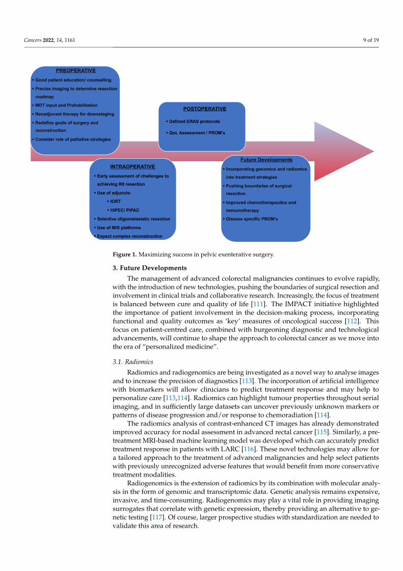
Harji et al. enrolled 145 patients into a dedicated pelvic exenteration ERAS programmeto assess its feasibility and short-term impact on this cohort of patients. They demonstratedan overall compliance rate of 70%. Patients with higher compliance to the program tendedto have a shorter hospital length of stay, reduced rate and severity of post-operative mor-bidity, as well as fewer readmissions. ERAS appears feasible and efficacious in patientsundergoing pelvic exenteration, displaying a high compliance and improved clinical out-comes [110]. See Figure 1 for the highlights of contemporary management strategies.
Cancers 2022, 14, 1161 9 of 19
Cancers 2022, 14, x FOR PEER REVIEW 9 of 20
Collaborative has offered guidance on the perioperative management of patients under-going exenterative surgery and acknowledges the need for individualised tailored post-operative treatment plans [40].
Harji et al. enrolled 145 patients into a dedicated pelvic exenteration ERAS pro-gramme to assess its feasibility and short-term impact on this cohort of patients. They demonstrated an overall compliance rate of 70%. Patients with higher compliance to the program tended to have a shorter hospital length of stay, reduced rate and severity of post-operative morbidity, as well as fewer readmissions. ERAS appears feasible and effi-cacious in patients undergoing pelvic exenteration, displaying a high compliance and im-proved clinical outcomes [110]. See Figure 1 for the highlights of contemporary manage-ment strategies.
Figure 1. Maximizing success in pelvic exenterative surgery.
3. Future Developments The management of advanced colorectal malignancies continues to evolve rapidly,
with the introduction of new technologies, pushing the boundaries of surgical resection and involvement in clinical trials and collaborative research. Increasingly, the focus of treatment is balanced between cure and quality of life [111]. The IMPACT initiative high-lighted the importance of patient involvement in the decision-making process, incorpo-rating functional and quality outcomes as ‘key’ measures of oncological success [112]. This focus on patient-centred care, combined with burgeoning diagnostic and technological advancements, will continue to shape the approach to colorectal cancer as we move into the era of “personalized medicine”.
3.1. Radiomics Radiomics and radiogenomics are being investigated as a novel way to analyse im-
ages and to increase the precision of diagnostics [113]. The incorporation of artificial in-telligence with biomarkers will allow clinicians to predict treatment response and may help to personalize care [113,114]. Radiomics can highlight tumour properties throughout serial imaging, and in sufficiently large datasets can uncover previously unknown mark-ers or patterns of disease progression and/or response to chemoradiation [114].
Figure 1. Maximizing success in pelvic exenterative surgery.
3. Future Developments
The management of advanced colorectal malignancies continues to evolve rapidly,with the introduction of new technologies, pushing the boundaries of surgical resection andinvolvement in clinical trials and collaborative research. Increasingly, the focus of treatmentis balanced between cure and quality of life [111]. The IMPACT initiative highlightedthe importance of patient involvement in the decision-making process, incorporatingfunctional and quality outcomes as ‘key’ measures of oncological success [112]. Thisfocus on patient-centred care, combined with burgeoning diagnostic and technologicaladvancements, will continue to shape the approach to colorectal cancer as we move intothe era of “personalized medicine”.
3.1. Radiomics
Radiomics and radiogenomics are being investigated as a novel way to analyse imagesand to increase the precision of diagnostics [113]. The incorporation of artificial intelligencewith biomarkers will allow clinicians to predict treatment response and may help topersonalize care [113,114]. Radiomics can highlight tumour properties throughout serialimaging, and in sufficiently large datasets can uncover previously unknown markers orpatterns of disease progression and/or response to chemoradiation [114].
The radiomics analysis of contrast-enhanced CT images has already demonstratedimproved accuracy for nodal assessment in advanced rectal cancer [115]. Similarly, a pre-treatment MRI-based machine learning model was developed which can accurately predicttreatment response in patients with LARC [116]. These novel technologies may allow fora tailored approach to the treatment of advanced malignancies and help select patientswith previously unrecognized adverse features that would benefit from more conservativetreatment modalities.
Radiogenomics is the extension of radiomics by its combination with molecular analy-sis in the form of genomic and transcriptomic data. Genetic analysis remains expensive,invasive, and time-consuming. Radiogenomics may play a vital role in providing imagingsurrogates that correlate with genetic expression, thereby providing an alternative to ge-netic testing [117]. Of course, larger prospective studies with standardization are needed tovalidate this area of research.
Cancers 2022, 14, 1161 10 of 19
3.2. Genomics
The adenoma-carcinoma sequence was first described in 1990 by Vogelstein and Fearonand provided the foundation for our understanding of colorectal cancer as a disease consist-ing of complex genomic changes [118]. Comparative genomic hybridization (CGH) arrays,single-nucleotide polymorphism (SNP) arrays and novel next-generation sequencing (NGS)approaches have provided us with insights into the complex colorectal cancer genome [119].Subsequent research has refined and expanded our knowledge of this area, allowing us toincorporate genomics into our treatment choices. The impact of the 100,000 genome projectand the integration of genomic and translational medicine into cancer care pathways hasprovided a unique opportunity for tailoring and personalizing oncological treatment [120].Research suggests that, in the future, we will be able to predict responses to chemotherapeu-tics, which will undoubtedly guide decision making, particularly in borderline cases [121].While the effect of RAS and BRAF mutations is well-established in current clinical practice,new genomic markers are showing promising results in clinical trials [118].
BRAF V600E is the most common potentially targetable mutation in metastatic col-orectal cancer; however, RAF inhibitors have limited efficacy as single agents in treatingpatients with this alteration [118]. The BEACON trial is currently comparing doublet ortriplet targeted therapy with standard therapy in patients with BRAF V600E metastaticcolorectal cancer [122]. Preliminary results are promising, with a 48% overall response ratein patients receiving triplet therapy consisting of encorafenib (RAF inhibitor), binimetinib(MEK inhibitor) and cetuximab [122].
HER2 amplification occurs in 2–6% of metastatic colorectal cancers and is associatedwith a poor response to EGFR antibody treatment [118]. While anti-HER2 drugs, such astrastuzumab as monotherapy, have not demonstrated tumour regression in clinical trials, acombination therapy with an EGFR inhibitor achieved tumour shrinkage [123]. The phase2 HERACLES trial reported a 30% response rate in patients with HER2-positive metastaticcolorectal cancer receiving this combination therapy [124]. Cohort B of the HERACLEStrial is ongoing and aims to compare the efficacy of the antibody-drug conjugate TDM-1monotherapy with combination therapy of TDM-1 and pertuzumab (anti-HER2) in thesecond line setting [118].
Our understanding of tumour biology and molecular subtypes is constantly expand-ing, thanks to advancements in microarray and NGS technologies which allow for theidentification of new cancer genes and pathways. While translational genomic studieshave already provided clinically relevant biomarkers for predicting prognosis and therapyresponse, future research will identify new drug targets and reveal novel therapeutic op-portunities. Innovations in current applications, coupled with novel emerging technologies,will lead to further advancements in translational cancer genomics which will hopefullycontribute to improved patient outcomes in the future [119].
3.3. Pushing the Surgical Boundaries
Over the last two decades, experienced exenterative surgeons have redefined whatconstitutes resectable disease. The development of regional/national specialized unitshas allowed funding and structural/service supports to enable these centres to establishspecialist pelvic oncology units. As a result, extended bony and/or neurovascular resectionsare pursued more frequently, with an acceptable morbidity reported [18,65,102].
‘Higher and wider’ resections at the periphery of the pelvis are now commonplacein pelvic exenteration [13]. Certain centres are performing en bloc sciatic nerve and/orlumbosacral trunk excision for tumours that extend laterally into the piriformis. Highsacrectomy (above the junction of S2/S3) can be performed without compromising margins,and functional outcomes are acceptable [125]. Alternative techniques such as anteriorcortical sacrectomy and abdominolithotomy sacrectomy have become more standardized.
Improved reconstructive techniques have facilitated these more radicalresections [21,65,98,99]. However, the repair of large bony defects remains a challenge [126].The current methods of reconstruction of these defects include autologous iliac grafting,
Cancers 2022, 14, 1161 11 of 19
autologous vascularized fibula transplantation, Masquelet’s induced membrane or massiveallografts. Autologous grafts account for approximately 50% of cases [126,127]. 3D bioprint-ing is a state-of-the-art technology used to build constructs from a single-cell type using alayer-by-layer deposition of a specific bioink [127]. Bioprinting uses cell-laden hydrogensto print structures following a period of maturation which can be developed into a varietyof complex tissues [126]. Its use in bone reconstruction is still evolving and the clinicalapplication of this technology remains in its infancy. Despite this, bio-printed bone hasbeen successfully implanted in pre-clinical models and other 3D-printed materials havebeen successfully transplanted into human subjects [128]. This ground-breaking technologywill allow us to develop tailored bone grafts that incorporate real cells, growth factorsand vasculature, which may revolutionize the way we reconstruct bony defects in thefuture [126].
Anatomical reconstruction of the sacrum using 3-dimensional printing technology hasbeen sparsely reported in the literature [129,130]. Kim et al. successfully reconstructed thesacrum with a 3D-printed implant in a patient who had undergone hemispherectomy forsacral osteosarcoma. One-year follow up revealed excellent bony union without compli-cation, demonstrating the feasibility of this novel method [129]. Similarly, Chatain et al.described a case of custom 3D-printed sacral implant for revision of failing sacrectomy ina patient who previously underwent en bloc sacrectomy and standard spinopelvic recon-struction for sacral chordoma [130]. In this case, the prosthesis was fashioned from titaniumalloy using a 3D-printing technique, tailored to the patient using a CT 3-dimensional recon-struction model. The surgical implantation of the device proved challenging but long-termoutcomes were satisfactory [130].
Current options for tissue reconstruction rely heavily on autologous donor tissueto repair defects. 3D bioprinting offers the potential to avoid autologous grafts and thecomplications associated with them [131]. Its use has now been reported in a variety of sur-gical disciplines, including plastics, breast, orthopaedic, craniomaxillofacial and head/neckoncology [132]. While significant advancements are being made in the production of simple,single tissue types, composite tissue engineering consisting of multilaminar constructsadds an additional layer of complexity [133]. Ultimately, 3D bioprinting has the potentialto produce patient-specific body components, including organs and limbs, which willundoubtedly revolutionize surgery [131].
3.4. Histopathology
Magnetic resonance-guided histopathology has been shown to increase the accuracyof staging in LARC [134]. The technique is performed by carefully matching multilevelhistologic sections, using previous MR images as guidance, to examine for evidence ofresidual tumour, while paying particular attention to areas with MRI signals consistentwith fibrosis or mucin. Another advancement in the field of histopathology is the use ofwhole-slide imaging, which produces digital histologic images from glass slides [135]. Thistechnology will allow for the precise evaluation of tumour dimensions, stage and margins,and has the potential to improve both diagnostic accuracy and workflow efficiency inthe future.
Biobanking is the process of collecting and storing various human specimens for thepurpose of clinical research and provides a platform for the development of translationaland personalised “precision” medicine [136]. Translational research with specimens fromtissue biobanks enables the discovery of molecular biomarkers that have the potential toguide therapy and individualize treatment [137,138]. Many research programmes havebenefited from biobank specimens, including the development of trastuzumab [139]. Morerecently, biobanks played a crucial role in the creation of The Cancer Genome Atlas (TCGA),a comprehensive catalogue of cancer genomic profiles [140]. This atlas has allowed for thediscovery of molecular aberrations at DNA, RNA, protein and epigenetic levels, providinga detailed analysis of commonalities and differences across tumour lineages.
Cancers 2022, 14, 1161 12 of 19
Biobanks provide researchers with human specimens and associated clinical data,which allow for large cohorts of over 30 specimens to be analysed using large-scale genomesequencing. This facilitates the discovery of novel molecular alterations as well as theclassification of tumour subtypes according to distinct genomic alterations, providing apersonalised, precision medicine approach in cancer care [141].
3.5. Quality of Life/PROMs
Aggressive multi-visceral resections are performed more often to manage patientswith advanced pelvic cancers [111]. Currently, surgery remains the only long-term curativeoption in the majority of cases [142]. Despite the radicality of pelvic exenteration, previousstudies have demonstrated an acceptable survival rate with reasonable quality of lifeoutcomes [143]. It appears that quality of life scores rapidly deteriorate in the immediatepost-operative period; however, they begin to rise slowly again from three months post-operation [144]. The ultimate goal of therapy is to balance patient quality of life withsurvival and complication rates, and this should be an integral component of the patientcounselling process.
Patient counselling with shared decision-making is crucial in the consideration andplanning of extensive pelvic surgery. A growing volume of research has shown thatwhen patients are actively involved in decision-making and prehabilitation (nutritional,physiotherapy/conditioning and/or psychological input) that they experience better out-comes [144,145]. The routine use of patient-reported outcome measures (PROMs) willinform us as to how surgery impacts a patient’s lifestyle and quality of life [146,147].Standardized questionnaires that collect data on patients post-operatively, particularlyregarding symptoms, health-related quality of life and functional status are vital [148].While there is ample support for the use of PROMs in the literature, there has been limiteduptake amongst surgeons [149,150]. PROMs not only inform practitioners of the nature,frequency and impact of adverse events following treatment but may also be used toidentify and treat these effects on an individual level in the post-operative period. ThePelvEx Collaborative has supported the development of a specific PROM QoL tool via theEuropean Organisation for Research and Treatment of Cancer network. Survivorship is atthe forefront of this project, in the hope that it will give greater insight into the most impor-tant factors affecting patients and strategies. The morbidity of pelvic exenterative surgerymay extend long into the months after discharge from hospital, and it is imperative that wehave the necessary supports in place to manage these complications [151]. Post-operativespecialist multi-disciplinary care is essential to assist patients with pain, wound and stomamanagement as well as for psychological support.
4. Conclusions
The role of radical surgery in the setting of locally advanced and recurrent rectalcancer has evolved substantially. Novel strategies including TNT, cytoreductive and/orbony/vascular resection and enhanced reconstructive techniques have enabled surgeons topursue what was once considered a terminal disease. Advancements in surgical technology,in particular the incorporation of artificial intelligence and three-dimensional bioprinting,will undoubtedly enhance our ability to move the limits of what is reasonable and possibleto resect.
Author Contributions: All authors have been involved in the preparation, reviewed and approvalof this manuscript by the PelvEx Collaborative. All authors have read and agreed to the publishedversion of the manuscript.
Funding: This research received no external funding.
Conflicts of Interest: The authors declare no conflict of interest.
Cancers 2022, 14, 1161 13 of 19
Appendix A
PelvEx Collaborative: Kelly M.E., O’Sullivan N.J., Fahy M.R., Aalbers A.G.J., AbdulAziz N., Abecasis N., Abraham-Nordling M., Abu Saadeh F., Akiyoshi T., Alberda W.,Albert M., Andric M., Angeles M.A., Angenete E., Antoniou A., Auer R., Austin K.K.,Aytac E., Aziz O., Bacalbasa N., Baker R.P., Bali M., Baransi S., Baseckas G., Bebington B.,Bedford M., Bednarski B.K., Beets G.L., Berg P.L., Bergzoll C., Beynon J., Biondo S., Boyle K.,Bordeianou L., Brecelj E., Bremers A.B., Brunner M., Buchwald P., Bui A., Burgess A., BurgerJ.W.A., Burling D., Burns E., Campain N., Carvalhal S., Castro L., Caycedo-Marulanda A.,Ceelen W., Chan K.K.L., Chang G.J., Chang M., Chew M.H., Chok A.Y., Chong P., CloustonH., Codd M., Collins D., Colquhoun A.J., Constantinides J., Corr A., Coscia M., CosimelliM., Cotsoglou C., Coyne P.E., Croner R.S., Damjanovich L., Daniels I.R., Davies M., DelaneyC.P., de Wilt J.H.W., Denost Q., Deutsch C., Dietz D., Domingo S., Dozois E.J., Drozdov E.,Duff M., Eglinton T., Enriquez-Navascues J.M., Espín-Basany E., Evans M.D., EyjólfsdóttirB., Fearnhead N.S., Ferron G., Fichtner-Feigl S., Flatmark K., Fleming F.J., Flor B., FolkessonJ., Frizelle F.A., Funder J., Gallego M.A., Gargiulo M., García-Granero E., García-SabridoJ.L., Gargiulo M., Gava V.G., Gentilini L., George M.L., George V., Georgiou P., Ghosh A.,Ghouti L., Gil-Moreno A., Giner F., Ginther D.N., Glyn T., Glynn R., Golda T., GriffithsB., Harris D.A., Hanchanale V., Harji D.P., Harris C., Helewa R.M., Hellawell G., HeriotA.G., Hochman D., Hohenberger W., Holm T., Hompes R., Hornung B., Hurton S., Hyun E.,Ito M., Iversen L.H., Jenkins J.T., Jourand K., Kaffenberger S., Kandaswamy G.V., KapurS., Kanemitsu Y., Kazi M., Kelley S.R., Keller D.S., Ketelaers S.H.J., Khan M.S., Kiran R.P.,Kim H., Kim H.J., Koh C.E., Kok N.F.M., Kokelaar R., Kontovounisios C., Kose F., KoutraM., Kristensen H.Ø., Kroon H.M., Kumar S., Kusters M., Lago V., Lampe B., Lakkis Z.,Larach J.T., Larkin J.O., Larsen S.G., Larson D.W., Law W.L., Lee P.J., Limbert M., Loria A.,Lydrup ML., Lyons A., Lynch A.C., Maciel J., Manfredelli S., Mann C., Mantyh C., MathisK.L., Marques C.F.S., Martinez A., Martling A., Mehigan B.J., Meijerink W.J.H.J., MercheaA., Merkel S., Mehta A.M., Mikalauskas S., McArthur D.R., McCormick J.J., McCormickP., McDermott F.D., McGrath J.S., Malde S., Mirnezami A., Monson J.R.T., Navarro A.S.,Neeff H., Negoi I., Neto J.W.M., Ng J.L., Nguyen B., Nielsen M.B., Nieuwenhuijzen G.A.P.,Nilsson P.J., Nordkamp S., Nugent T., Oliver A., O’Dwyer S.T., Paarnio K., Palmer G.,Pappou E., Park J., Patsouras D., Peacock O., Pellino G., Peterson A.C., Pfeffer F., PinsonJ., Poggioli G., Proud D., Quinn M., Quyn A., Rajendran N., Radwan R.W., Rajendran N.,Rao C., Rasheed S., Rausa E., Regenbogen S.E., Reims H.M., Renehan A., Rintala J., RochaR., Rochester M., Rohila J., Rothbarth J., Rottoli M., Roxburgh C., Rutten H.J.T., Safar B.,Sagar P.M., Sahai A., Saklani A., Sammour T., Sayyed R., Schizas A.M.P., Schwarzkopf E.,Scripcariu D., Scripcariu V., Selvasekar C., Shaikh I., Simpson A., Skeie-Jensen T., SmartN.J., Smart P., Smith J.J., Solbakken A.M., Solomon M.J., Sørensen M.M., Sorrentino L.,Steele S.R., Steffens D., Stitzenberg K., Stocchi L., Stylianides N.A., Swartling T., SpasojevicM., Sumrien H.., Sutton PA., Swartking T., Takala H., Tan E.J., Taylor C., Taylor D., TekinA., Tekkis P.P., Teras J., Thaysen H.V., Thurairaja R., Thorgersen E.B., Tiernan J., Toh E.L.,Tolenaar J., Tsarkov P., Tsukada Y., Tsukamoto S., Tuech J.J., Turner W.H., Tuynman J.B.,Valente M., van Ramshorst G.H., van Rees J., van Zoggel D., Vasquez-Jimenez W., VatherR., Verhoef C., Vierimaa M., Vizzielli G., Voogt E.L.K., Uehara K., Urrejola G., Wakeman C.,Warrier S.K., Wasmuth H.H., Waters P.S., Weber K., Weiser M.R., Wheeler J.M.D., Wild J.,Williams A., Wilson M., Wolthuis A., Yano H., Yip B., Yoo R.N., Zappa M.A., Winter D.C.
References1. PelvEx Collaborative. Minimally invasive surgery techniques in pelvic exenteration: A systematic and meta-analysis review.
Surg. Endosc. 2018, 32, 4707–4715. [CrossRef] [PubMed]2. Waters, P.S.; Peacock, O.; Warrier, S.K.; Wakeman, C.; Eglinton, T.; Lynch, A.C.; Frizelle, F.A.; Heriot, A.G.; McCormick, J.
Evolution of pelvic exenteration surgery–resectional trends and survival outcomes over three decades. Eur. J. Surg. Oncol. 2019,45, 2325–2333. [CrossRef] [PubMed]
3. Quyn, A.J.; Solomon, M.; Lee, P.M.; Badgery-Parker, T.; Masya, L.M.; Young, J.M. Palliative Pelvic Exenteration: Clinical Outcomesand Quality of Life. Dis. Colon Rectum 2016, 59, 1005–1010. [CrossRef] [PubMed]
Cancers 2022, 14, 1161 14 of 19
4. Steffens, D.; Solomon, M.J.; Young, J.M.; Koh, C.; Venchiarutti, R.L.; Lee, P.; Austin, K. Cohort study of long-term survival andquality of life following pelvic exenteration. BJS Open 2018, 2, 328–335. [CrossRef]
5. PelvEx Collaborative. Changing outcomes following pelvic exenteration for locally advanced and recurrent rectal cancer. BJSOpen 2019, 3, 516–520. [CrossRef]
6. PelvEx Collaborative. The global cost of pelvic exenteration: In-hospital perioperative costs. Br. J. Surg. 2020, 107, e470–e471.[CrossRef]
7. PelvEx Collaborative. Surgical and Survival Outcomes Following Pelvic Exenteration for Locally Advanced Primary RectalCancer: Results from an International Collaboration. Ann. Surg. 2019, 269, 315–321. [CrossRef]
8. PelvEx Collaborative. Factors affecting outcomes following pelvic exenteration for locally recurrent rectal cancer. Br. J. Surg. 2018,105, 650–657. [CrossRef]
9. Burns, E.M.; Boyle, K.; Mirnezami, A.; Jenkins, J.T.; UK PEN (United Kingdom Pelvic Exenteration Network). The impact ofCOVID-19 on advanced colorectal cancer. Colorectal Dis. 2020, 22, 737–738. [CrossRef]
10. Zadnik, V.; Mihor, A.; Tomsic, S.; Zagar, T.; Bric, N.; Lokar, K.; Oblak, I. Impact of COVID-19 on cancer diagnosis and managementin Slovenia—Preliminary results. Radiol. Oncol. 2020, 54, 329–334. [CrossRef]
11. Englum, B.R.; Prasad, N.K.; Lake, R.E.; Mayorga-Carlin, M.; Turner, D.J.; Siddiqui, T.; Sorkin, J.D.; Lal, B.K. Impact of theCOVID-19 pandemic on diagnosis of new cancers: A national multicenter study of the Veterans Affairs Healthcare System. Cancer2021, 128, 1048–1056. [CrossRef] [PubMed]
12. McCabe, R.; Schmit, N.; Christen, P.; D’Aeth, J.C.; Løchen, A.; Rizmie, D.; Nayagam, S.; Miraldo, M.; Aylin, P.; Bottle, A.; et al.Adapting hospital capacity to meet changing demands during the COVID-19 pandemic. BMC Med. 2020, 18, 329. [CrossRef][PubMed]
13. Solomon, M.J. Redefining the boundaries of advanced pelvic oncology surgery. Br. J. Surg. 2021, 108, 453–455. [CrossRef][PubMed]
14. Kazi, M.; Sukumar, V.; Desouza, A.; Saklani, A. State-of-the-art surgery for recurrent and locally advanced rectal cancers.Langenbeck’s Arch. Surg. 2021, 406, 1763–1774. [CrossRef]
15. Antoch, G.; Vogt, F.M.; Freudenberg, L.S.; Nazaradeh, F.; Goehde, S.C.; Barkhausen, J.; Dahmen, G.; Bockisch, A.; Debatin,J.F.; Ruehm, S.G. Whole-Body Dual-Modality PET/CT and Whole-Body MRI for Tumor Staging in Oncology. JAMA 2003, 290,3199–3206. [CrossRef]
16. Lee, C.U.; White, D.B.; Sykes, A.-M.G. Establishing a Chest MRI Practice and its Clinical Applications: Our Insight and Protocols.J. Clin. Imaging Sci. 2014, 4, 17. [CrossRef]
17. Lau, Y.C.; Brown, K.G.M.; Lee, P. Pelvic exenteration for locally advanced and recurrent rectal cancer-how much more?J. Gastrointest. Oncol. 2019, 10, 1207–1214. [CrossRef]
18. Wuisman, P.; Lieshout, O.; Sugihara, S.; van Dijk, M. Total Sacrectomy and Reconstruction. Clin. Orthop. Relat. Res. 2000, 381,192–203. [CrossRef]
19. Kasi, A.; Abbasi, S.; Handa, S.; Al-Rajabi, R.; Saeed, A.; Baranda, J.; Sun, W. Total Neoadjuvant Therapy vs Standard Therapy inLocally Advanced Rectal Cancer: A Systematic Review and Meta-analysis. JAMA Netw. Open 2020, 3, e2030097. [CrossRef]
20. Xu, Q.; Sun, Z.; Li, X.; Ye, C.; Zhou, C.; Zhang, L.; Lu, G. Advanced gastric cancer: CT radiomics prediction and early detection ofdownstaging with neoadjuvant chemotherapy. Eur. Radiol. 2021, 31, 8765–8774. [CrossRef]
21. Pusic, A.L.; Mehrara, B.J. Vaginal reconstruction: An algorithm approach to defect classification and flap reconstruction. J. Surg.Oncol. 2006, 94, 515–521. [CrossRef] [PubMed]
22. Schmidt, G.P.; Reiser, M.F.; Baur-Melnyk, A. Whole-body MRI for the staging and follow-up of patients with metastasis. Eur. J.Radiol. 2009, 70, 393–400. [CrossRef] [PubMed]
23. Ciliberto, M.; Maggi, F.; Treglia, G.; Padovano, F.; Calandriello, L.; Giordano, A.; Bonomo, L. Comparison between whole-bodyMRI and Fluorine-18-Fluorodeoxyglucose PET or PET/CT in oncology: A systematic review. Radiol. Oncol. 2013, 47, 206–218.[CrossRef] [PubMed]
24. Tatli, S.; Mortele, K.J.; Breen, E.L.; Bleday, R.; Silverman, S.G. Local staging of rectal cancer using combined pelvic phased-arrayand endorectal coil MRI. J. Magn. Reson. Imaging 2006, 23, 534–540. [CrossRef] [PubMed]
25. Drami, I.; Lord, A.; Sarmah, P.; Baker, R.; Daniels, I.; Boyle, K.; Griffiths, B.; Mohan, H.; Jenkins, J. Preoperative assessment andoptimisation for pelvic exenteration in locally advanced and recurrent rectal cancer: A review. Eur. J. Surg. Oncol. 2021, in press.[CrossRef]
26. Kim, Y.H.; Kim, D.Y.; Kim, T.H.; Jung, K.H.; Chang, H.J.; Jeong, S.-Y.; Sohn, D.K.; Choi, H.S.; Ahn, J.B.; Lim, S.-B.; et al. Usefulnessof magnetic resonance volumetric evaluation in predicting response to preoperative concurrent chemoradiotherapy in patientswith resectable rectal cancer. Int. J. Radiat. Oncol. 2005, 62, 761–768. [CrossRef]
27. Hartley, A.; Ho, K.F.; McConkey, C.; Geh, J.I. Pathological complete response following pre-operative chemoradiotherapy in rectalcancer: Analysis of phase II/III trials. Br. J. Radiol. 2005, 78, 934–938. [CrossRef]
28. Ryan, J.E.; Warrier, S.K.; Lynch, A.C.; Heriot, A.G. Assessing pathological complete response to neoadjuvant chemoradiotherapyin locally advanced rectal cancer: A systematic review. Colorectal Dis. 2015, 17, 849–861. [CrossRef]
29. Hoffmann, K.-T.; Rau, B.; Wust, P.; Stroszczynski, C.; Hünerbein, M.; Schneider, U.; Felix, R. Restaging of locally advancedcarcinoma of the rectum with MR imaging after preoperative radio-chemotherapy plus regional hyperthermia. Strahlenther. Onkol.2002, 178, 386–392. [CrossRef]
Cancers 2022, 14, 1161 15 of 19
30. Beyond TME Collaborative. Consensus statement on the multidisciplinary management of patients with recurrent and primaryrectal cancer beyond total mesorectal excision planes. Br. J. Surg. 2013, 100, 1009–1014. [CrossRef]
31. Akash, M.; Mehta, D.B.; Jenkins, J.T. Preoperative Assessment of Tumor Anatomy and Surgical Resectability. Surg. Manag. Adv.Pelvic Cancer 2021, 17–31. [CrossRef]
32. Liu, S.; Jiang, T.; Xiao, L.; Yang, S.; Liu, Q.; Gao, Y.; Chen, G.; Xiao, W. Total Neoadjuvant Therapy (TNT) versus StandardNeoadjuvant Chemoradiotherapy for Locally Advanced Rectal Cancer: A Systematic Review and Meta-Analysis. Oncologist 2021,26, e1555–e1566. [CrossRef] [PubMed]
33. Petrelli, F.; Trevisan, F.; Cabiddu, M.; Sgroi, G.; Bruschieri, L.; Rausa, E.; Ghidini, M.; Turati, L. Total Neoadjuvant Therapy inRectal Cancer: A Systematic Review and Meta-analysis of Treatment Outcomes. Ann. Surg. 2020, 271, 440–448. [CrossRef][PubMed]
34. Denost, Q.; Frison, E.; Salut, C.; Sitta, R.; Rullier, A.; Harji, D.; Maillou-Martinaud, H.; Rullier, E.; Smith, D.; Vendrely, V.; et al. Aphase III randomized trial evaluating chemotherapy followed by pelvic reirradiation versus chemotherapy alone as preoperativetreatment for locally recurrent rectal cancer—GRECCAR 15 trial protocol. Colorectal Dis. 2021, 23, 1909–1918. [CrossRef]
35. Douglas, J.K.; Callahan, R.E.; Hothem, Z.A.; Cousineau, C.S.; Kawak, S.; Thibodeau, B.J.; Bergeron, S.; Li, W.; Peeples, C.E.;Wasvary, H.J. Genomic variation as a marker of response to neoadjuvant therapy in locally advanced rectal cancer. Mol. Cell.Oncol. 2020, 7, 1716618. [CrossRef]
36. Toomey, S.; Gunther, J.; Carr, A.; Weksberg, D.C.; Thomas, V.; Salvucci, M.; Bacon, O.; Sherif, E.-M.; Fay, J.; Kay, E.W.; et al.Genomic and Transcriptomic Characterisation of Response to Neoadjuvant Chemoradiotherapy in Locally Advanced RectalCancer. Cancers 2020, 12, 1808. [CrossRef]
37. He, S.; Hu, D.; Feng, H.; Xue, Y.; Jin, J.; Wang, X. Efficacy of immunotherapy with PD-1 inhibitor in colorectal cancer: Ameta-analysis. J. Comp. Eff. Res. 2020, 9, 1285–1292. [CrossRef]
38. Chalabi, M.; Fanchi, L.F.; Dijkstra, K.K.; Berg, J.G.V.D.; Aalbers, A.G.; Sikorska, K.; Lopez-Yurda, M.; Grootscholten, C.; Beets, G.L.;Snaebjornsson, P.; et al. Neoadjuvant immunotherapy leads to pathological responses in MMR-proficient and MMR-deficientearly-stage colon cancers. Nat. Med. 2020, 26, 566–576. [CrossRef]
39. Oliveira, A.F.; Bretes, L.; Furtado, I. Review of PD-1/PD-L1 Inhibitors in Metastatic dMMR/MSI-H Colorectal Cancer. Front.Oncol. 2019, 9, 396. [CrossRef]
40. PelvEx Collaborative. Perioperative management and anaesthetic considerations in pelvic exenterations using Delphi methodol-ogy: Results from the PelvEx Collaborative. BJS Open 2021, 5, zraa055. [CrossRef]
41. Cabilan, C.; Hines, S.; Munday, J. The effectiveness of prehabilitation or preoperative exercise for surgical patients: A systematicreview. JBI Database Syst. Rev. Implement. Rep. 2015, 13, 146–187. [CrossRef]
42. Lambert, J.; Hayes, L.; Keegan, T.; Subar, D.; Gaffney, C. Response to the Comment on “The Impact of Prehabilitation on PatientOutcomes in Hepatobiliary, Colorectal and Upper Gastrointestinal Cancer Surgery: A PRISMA-Accordant Meta-analysis”. Ann.Surg. 2021, 274, e932–e933. [CrossRef]
43. Lai, C.W.; Minto, G.; Challand, C.P.; Hosie, K.B.; Sneyd, J.R.; Creanor, S.; Struthers, R.A. Patients’ inability to perform apreoperative cardiopulmonary exercise test or demonstrate an anaerobic threshold is associated with inferior outcomes aftermajor colorectal surgery. Br. J. Anaesth. 2013, 111, 607–611. [CrossRef]
44. Levett, D.; Jack, S.; Swart, M.; Carlisle, J.; Wilson, J.; Snowden, C.; Riley, M.; Danjoux, G.; Ward, S.; Older, P.; et al. Perioperativecardiopulmonary exercise testing (CPET): Consensus clinical guidelines on indications, organization, conduct, and physiologicalinterpretation. Br. J. Anaesth. 2018, 120, 484–500. [CrossRef]
45. Steenhagen, E. Preoperative nutritional optimization of esophageal cancer patients. J. Thorac. Dis. 2019, 11, S645–S653. [CrossRef]46. Daly, J.M.; Lieberman, M.D.; Goldfine, J.; Shou, J.; Weintraub, F.; Rosato, E.F.; Lavin, P. Enteral nutrition with supplemental
arginine, RNA, and omega-3 fatty acids in patients after operation: Immunologic, metabolic, and clinical outcome. Surgery 1992,112, 56–67.
47. Andreyev, H.J.; Norman, A.R.; Oates, J.; Cunningham, D. Why do patients with weight loss have a worse outcome whenundergoing chemotherapy for gastrointestinal malignancies? Eur. J. Cancer 1998, 34, 503–509. [CrossRef]
48. Huddy, J.R.; Huddy, F.M.S.; Markar, S.R.; Tucker, O. Nutritional optimization during neoadjuvant therapy prior to surgicalresection of esophageal cancer-a narrative review. Dis. Esophagus 2018, 31, dox110. [CrossRef]
49. PelvEx Collaborative. Palliative pelvic exenteration: A systematic review of patient-centered outcomes. Eur. J. Surg. Oncol. 2019,45, 1787–1795. [CrossRef]
50. PelvEx Collaborative. Predicting outcomes of pelvic exenteration using machine learning. Colorectal Dis. 2020, 22, 1933–1940.[CrossRef]
51. PelvEx Collaborative. Management strategies for patients with advanced rectal cancer and liver metastases using modifiedDelphi methodology: Results from the PelvEx Collaborative. Colorectal Dis. 2020, 22, 1184–1188. [CrossRef]
52. PelvEx Collaborative. Simultaneous pelvic exenteration and liver resection for primary rectal cancer with synchronous livermetastases: Results from the PelvEx Collaborative. Colorectal Dis. 2020, 22, 1258–1262. [CrossRef]
53. PelvExCollaborative. Pelvic Exenteration for Advanced Nonrectal Pelvic Malignancy. Ann. Surg. 2019, 270, 899–905. [CrossRef]54. Solomon, M.J.; Brown, K.G.M.; Koh, C.E.; Lee, P.; Austin, K.K.S.; Masya, L. Lateral pelvic compartment excision during pelvic
exenteration. Br. J. Surg. 2015, 102, 1710–1717. [CrossRef]
Cancers 2022, 14, 1161 16 of 19
55. Solomon, M.J.; Brown, K.G.M. Extended Radical Resection: The Standard of Care for Patients with Advanced Pelvic Malignancy.Ann. Surg. Oncol. 2019, 27, 323–324. [CrossRef]
56. Lau, Y.C.; Jongerius, K.; Wakeman, C.; Heriot, A.G.; Solomon, M.J.; Sagar, P.M.; Tekkis, P.P.; Frizelle, F.A. Influence of the level ofsacrectomy on survival in patients with locally advanced and recurrent rectal cancer. Br. J. Surg. 2019, 106, 484–490. [CrossRef]
57. You, Y.N.; Skibber, J.M.; Hu, C.; Crane, C.H.; Das, P.; Kopetz, S.; Eng, C.; Feig, B.W.; Rodriguez-Bigas, M.A.; Chang, G.J. Impact ofmultimodal therapy in locally recurrent rectal cancer. Br. J. Surg. 2016, 103, 753–762. [CrossRef]
58. Yang, T.X.; Morris, D.L.; Chua, T.C. Pelvic exenteration for rectal cancer: A systematic review. Dis Colon Rectum. 2013, 56, 519–531.[CrossRef]
59. Harris, C.; Solomon, M.; Heriot, A.G.; Sagar, P.M.; Tekkis, P.P.; Dixon, L.; Pascoe, R.; Dobbs, B.R.; Frampton, C.M.; Harji, D.P.; et al.The Outcomes and Patterns of Treatment Failure After Surgery for Locally Recurrent Rectal Cancer. Ann. Surg. 2016, 264, 323–329.[CrossRef]
60. Brown, K.G.; Solomon, M.; Koh, C.E. Pelvic Exenteration Surgery: The Evolution of Radical Surgical Techniques for Advancedand Recurrent Pelvic Malignancy. Dis. Colon Rectum 2017, 60, 745–754. [CrossRef]
61. Shaikh, I.; Holloway, I.; Aston, W.; Littler, S.; Burling, D.; Antoniou, A.; Jenkins, J.T.; Complex Cancer Clinic St Mark’s HospitalLondon. High subcortical sacrectomy: A novel approach to facilitate complete resection of locally advanced and recurrent rectalcancer with high (S1–S2) sacral extension. Colorectal Dis. 2015, 18, 386–392. [CrossRef]
62. Lee, P.; Tan, W.J.; Brown, K.G.M.; Solomon, M.J. Addressing the empty pelvic syndrome following total pelvic exenteration: Doesmesh reconstruction help? Colorectal Dis. 2019, 21, 365–369. [CrossRef]
63. Austin, K.K.S.; Solomon, M. Pelvic Exenteration with En Bloc Iliac Vessel Resection for Lateral Pelvic Wall Involvement. Dis.Colon Rectum 2009, 52, 1223–1233. [CrossRef]
64. Brown, K.G.M.; Solomon, M.J.; Lau, Y.C.; Steffens, D.; Austin, K.K.S.; Lee, P.J. Sciatic and Femoral Nerve Resection DuringExtended Radical Surgery for Advanced Pelvic Tumours: Long-term Survival, Functional, and Quality-of-life Outcomes. Ann.Surg. 2021, 273, 982–988. [CrossRef]
65. Peacock, O.; Smith, N.; Waters, P.S.; Cheung, F.; McCormick, J.J.; Warrier, S.K.; Wagner, T.; Heriot, A.G. Outcomes of extendedradical resections for locally advanced and recurrent pelvic malignancy involving the aortoiliac axis. Colorectal Dis. 2020, 22,818–823. [CrossRef]
66. Grimes, W.R.; Stratton, M. Pelvic Exenteration; StatPearls: Treasure Island, FL, USA, 2021.67. Mirnezami, R.; Chang, G.J.; Das, P.; Chandrakumaran, K.; Tekkis, P.; Darzi, A.; Mirnezami, A.H. Intraoperative radiotherapy in
colorectal cancer: Systematic review and meta-analysis of techniques, long-term outcomes, and complications. Surg. Oncol. 2012,22, 22–35. [CrossRef]
68. Haddock, M.G. Intraoperative radiation therapy for colon and rectal cancers: A clinical review. Radiat. Oncol. 2017, 12, 11.[CrossRef]
69. Hyngstrom, J.R.; Tzeng, C.W.; Beddar, S.; Das, P.; Krishnan, S.; Delclos, M.E.; Crane, C.H.; Chang, G.J.; You, Y.N.; Feig, B.W.; et al.Intraoperative radiation therapy for locally advanced primary and recurrent colorectal cancer: Ten-year institutional experience.J. Surg. Oncol. 2014, 109, 652–658. [CrossRef]
70. Mirnezami, R. Multivisceral Resection of Advanced Pelvic Tumors: From Planning to Implementation. Clin. Colon Rectal Surg.2020, 33, 268–278. [CrossRef]
71. Franko, J.; Shi, Q.; Goldman, C.D.; Pockaj, B.A.; Nelson, G.D.; Goldberg, R.M.; Pitot, H.C.; Grothey, A.; Alberts, S.R.; Sargent, D.Treatment of Colorectal Peritoneal Carcinomatosis with Systemic Chemotherapy: A Pooled Analysis of North Central CancerTreatment Group Phase III Trials N9741 and N9841. J. Clin. Oncol. 2012, 30, 263–267. [CrossRef]
72. Tavernier, C.; Passot, G.; Vassal, O.; Allaouchiche, B.; Decullier, E.; Bakrin, N.; Alyami, M.; Davigo, A.; Bonnet, J.-M.; Louzier, V.;et al. Pressurized intraperitoneal aerosol chemotherapy (PIPAC) might increase the risk of anastomotic leakage compared toHIPEC: An experimental study. Surg. Endosc. 2019, 34, 2939–2946. [CrossRef] [PubMed]
73. Brown, K.G.M.; Ansari, N.; Solomon, M.J.; Austin, K.K.S.; Hamilton, A.E.R.; Young, C.J. Pelvic exenteration combined withcytoreductive surgery and hyperthermic intraperitoneal chemotherapy for advanced primary or recurrent colorectal cancer withperitoneal metastases. Colorectal Dis. 2020, 23, 186–191. [CrossRef]
74. Tuech, J.-J.; Pinson, J.; Nouhaud, F.-X.; Wood, G.; Clavier, T.; Sabourin, J.-C.; Di Fiore, F.; Monge, M.; Papet, E.; Coget, J. TotalPelvic Exenteration, Cytoreductive Surgery, and Hyperthermic Intraperitoneal Chemotherapy for Rectal Cancer with AssociatePeritoneal Metastases: Surgical Strategies to Optimize Safety. Cancers 2020, 12, 3478. [CrossRef] [PubMed]
75. Shinde, R.S.; Acharya, R.; Kumar, N.A.; Solanki, S.; DeSouza, A.; Saklani, A. Pelvic Exenteration with Cytoreductive Surgery andHyperthermic Intraperitoneal Chemotherapy (CRS + HIPEC) for Rectal Cancer-Case Series with Review of Literature. Indian J.Surg. Oncol. 2019, 10, 80–83. [CrossRef] [PubMed]
76. Quénet, F.; Elias, D.; Roca, L.; Goéré, D.; Ghouti, L.; Pocard, M.; Facy, O.; Arvieux, C.; Lorimier, G.; Pezet, D.; et al. Cytoreductivesurgery plus hyperthermic intraperitoneal chemotherapy versus cytoreductive surgery alone for colorectal peritoneal metastases(PRODIGE 7): A multicentre, randomised, open-label, phase 3 trial. Lancet Oncol. 2021, 22, 256–266. [CrossRef]
77. Flood, M.P.; Waters, P.S.; Soucisse, M.; Ramsay, R.; Michael, M.; McCormick, J.J.; Warrier, S.; Heriot, A. Pelvic exenteration,cytoreductive surgery, and hyperthermic intraperitoneal chemotherapy for peritoneal surface malignancy: Experience andoutcomes from an exenterative and peritonectomy unit. Langenbeck’s Arch. Surg. 2021, 406, 2807–2815. [CrossRef]
Cancers 2022, 14, 1161 17 of 19
78. Kelly, M.; Spolverato, G.; Le, G.; Mavros, M.; Doyle, F.; Pawlik, T.; Winter, D. Synchronous colorectal liver metastasis: A networkmeta-analysis review comparing classical, combined, and liver-first surgical strategies. J. Surg. Oncol. 2014, 111, 341–351.[CrossRef]
79. Heinrich, S.; Lang, H. Liver metastases from colorectal cancer: Technique of liver resection. J. Surg. Oncol. 2012, 107, 579–584.[CrossRef]
80. Ratnayake, C.B.B.; Wells, C.I.; Atherton, P.; Hammond, J.S.; White, S.; French, J.J.; Manas, D.; Pandanaboyana, S. Meta-analysis ofsurvival outcomes following surgical and non surgical treatments for colorectal cancer metastasis to the lung. ANZ J. Surg. 2020,91, 255–263. [CrossRef]
81. Lumachi, F.; Chiara, G.B.; Tozzoli, R.; Del Contea, A.; Basso, S.M.M. Factors Affecting Survival in Patients with Lung Metastasesfrom Colorectal Cancer. A Short Meta-analysis. Anticancer Res. 2016, 36, 13–19.
82. Nicosia, L.; Franceschini, D.; Perrone-Congedi, F.; Casamassima, F.; Gerardi, M.A.; Rigo, M.; Mazzola, R.; Perna, M.; Scotti, V.;Fodor, A.; et al. A multicenter LArge retrospectIve daTabase on the personalization of stereotactic ABlative radiotherapy use inlung metastases from colon-rectal cancer: The LaIT-SABR study. Radiother. Oncol. 2021, 166, 92–99. [CrossRef] [PubMed]
83. Lee, K.-Y.; Lau, J.; Siew, B.-E.; Chua, Y.-K.; Lim, Y.-X.; Lim, X.-Y.; Chong, C.-S.; Tan, K.-K. Does pulmonary metastasectomy ofcolorectal metastases translate to better survival? A systematic review. Ann. Acad. Med. Singap. 2021, 50, 773–781. [CrossRef]
84. Williams, M.; Perera, M.; Nouhaud, F.-X.; Coughlin, G. Robotic pelvic exenteration and extended pelvic resections for locallyadvanced or synchronous rectal and urological malignancy. Investig. Clin. Urol. 2021, 62, 111–120. [CrossRef] [PubMed]
85. Lawande, A.; Kenawadekar, R.; Desai, R.; Malireddy, C.; Nallapothula, K.; Puntambekar, S.P. Robotic total pelvic exenteration.J. Robot. Surg. 2013, 8, 93–96. [CrossRef] [PubMed]
86. Smith, N.; Murphy, D.G.; Lawrentschuk, N.; McCormick, J.; Heriot, A.; Warrier, S.; Lynch, A.C. Robotic multivisceral pelvicresection: Experience from an exenteration unit. Tech. Coloproctol. 2020, 24, 1145–1153. [CrossRef] [PubMed]
87. Heah, N.H.; Wong, K.Y. Feasibility of robotic assisted bladder sparing pelvic exenteration for locally advanced rectal cancer: Asingle institution case series. World J. Gastrointest. Surg. 2020, 12, 190–196. [CrossRef] [PubMed]
88. Raj Kumar, B.; Bankar, S.; Pandey, D.; Rohila, J.; Prakash, G.; Bakshi, G.; de Souza, A.; Saklani, A. Abdominoperineal excisionwith prostatectomy in T4 rectal cancer-bladder-sparing robotic pelvic exenteration—A video vignette. Colorectal Dis. 2020, 22,1786–1787. [CrossRef]
89. Shin, U.S.; You, Y.N.; Nguyen, A.T.; Bednarski, B.K.; Messick, C.; Maru, D.M.; Dean, E.M.; Hu, C.-Y.; Chang, G.J. OncologicOutcomes of Extended Robotic Resection for Rectal Cancer. Ann. Surg. Oncol. 2016, 23, 2249–2257. [CrossRef]
90. Winters, B.R.; Mann, G.N.; Louie, O.; Wright, J.L. Robotic Total Pelvic Exenteration with Laparoscopic Rectus Flap: InitialExperience. Case Rep. Surg. 2015, 2015, 835425. [CrossRef]
91. Shin, J.W.; Kim, J.; Kwak, J.M.; Hara, M.; Cheon, J.; Kang, S.H.; Kang, S.G.; Stevenson, A.R.L.; Coughlin, G.; Kim, S.H. First report:Robotic pelvic exenteration for locally advanced rectal cancer. Colorectal Dis. 2013, 16, O9–O14. [CrossRef]
92. Pokharkar, A.; Kammar, P.; D’Souza, A.; Bhamre, R.; Sugoor, P.; Saklani, A. Laparoscopic Pelvic Exenteration for Locally AdvancedRectal Cancer, Technique and Short-Term Outcomes. J. Laparoendosc. Adv. Surg. Tech. 2018, 28, 1489–1494. [CrossRef] [PubMed]
93. Chang, T.; Chok, A.; Tan, D.; Rogers, A.; Rasheed, S.; Tekkis, P.; Kontovounisios, C. The Emerging Role of Robotics in PelvicExenteration Surgery for Locally Advanced Rectal Cancer: A Narrative Review. J. Clin. Med. 2021, 10, 1518. [CrossRef] [PubMed]
94. Lee, Y.-J.; Berg, N.S.V.D.; Orosco, R.K.; Rosenthal, E.L.; Sorger, J.M. A narrative review of fluorescence imaging in robotic-assistedsurgery. Laparosc. Surg. 2021, 5, 31. [CrossRef]
95. Shirk, J.D.; Kwan, L.; Saigal, C. The Use of 3-Dimensional, Virtual Reality Models for Surgical Planning of Robotic PartialNephrectomy. Urology 2019, 125, 92–97. [CrossRef] [PubMed]
96. Atallah, S.; Parra-Davila, E.; Melani, A.G.F.; Romagnolo, L.G.; Larach, S.W.; Marescaux, J. Robotic-assisted stereotactic real-timenavigation: Initial clinical experience and feasibility for rectal cancer surgery. Tech. Coloproctol. 2019, 23, 53–63. [CrossRef][PubMed]
97. Griffin, N.; Rabouhans, J.; Grant, L.A.; Ng, R.L.H.; Ross, D.; Roblin, P.; George, M.L. Pelvi-perineal flap reconstruction: Normalimaging appearances and post-operative complications on cross-sectional imaging. Insights Imaging 2011, 2, 215–223. [CrossRef]
98. Sagebiel, T.L.; Faria, S.C.; Balachandran, A.; Butler, C.E.; Garvey, P.B.; Bhosale, P.R. Pelvic Reconstruction with Pedicled ThighFlaps: Indications, Surgical Techniques, and Postoperative Imaging. Am. J. Roentgenol. 2014, 202, 593–601. [CrossRef]
99. Küntscher, M.V.; Mansouri, S.; Noack, N.; Hartmann, B. Versatility of vertical rectus abdominis musculocutaneous flaps.Microsurgery 2006, 26, 363–369. [CrossRef]
100. Johnson, Y.Y.L.; West, M.M.A.; Gould, L.E.; Drami, I.I.; Behrenbruch, C.C.; Burns, E.E.M.; Mirnezami, A.A.H.; Jenkins, J.J.T. Emptypelvis syndrome: A systematic review of reconstruction techniques and their associated complications. Colorectal Dis. 2021, 24,16–26. [CrossRef]
101. Bederman, S.S.; Shah, K.N.; Hassan, J.M.; Hoang, B.H.; Kiester, P.D.; Bhatia, N.N. Surgical techniques for spinopelvic reconstruc-tion following total sacrectomy: A systematic review. Eur. Spine J. 2013, 23, 305–319. [CrossRef]
102. Peacock, O.; Smith, N.; Waters, P.S.; Cheung, F.P.-Y.; McCormick, J.J.; Warrier, S.K.; Wagner, T.; Heriot, A.G. Preemptive Femoral-Femoral Crossover Grafting of Artery and Vein Before Pelvic Exenterative Surgery for Locally Advanced and Recurrent PelvicMalignancy Involving the Aortoiliac Axis. Dis. Colon Rectum 2020, 64, e2–e5. [CrossRef]
103. Hope, J.M.; Pothuri, B. The Role of Palliative Surgery in Gynecologic Cancer Cases. Oncologist 2013, 18, 73–79. [CrossRef]
Cancers 2022, 14, 1161 18 of 19
104. Mori, M.; Elsayem, A.; Reddy, S.K.; Bruera, E.; Fadul, N.A. Unrelieved Pain and Suffering in Patients with Advanced Cancer. Am.J. Hosp. Palliat. Med. 2011, 29, 236–240. [CrossRef]
105. Simeonsson, R.J.; Edmondson, R.; Smith, T.; Carnahan, S.; Bucy, J.E. Family involvement in multidisciplinary team evaluation:Professional and parent perspectives. Chil Care Health Dev. 1995, 21, 199–214. [CrossRef]
106. Ni, X.; Jia, D.; Chen, Y.; Wang, L.; Suo, J. Is the Enhanced Recovery After Surgery (ERAS) Program Effective and Safe inLaparoscopic Colorectal Cancer Surgery? A Meta-Analysis of Randomized Controlled Trials. J. Gastrointest. Surg. 2019, 23,1502–1512. [CrossRef]
107. Dhiman, A.; Fenton, E.; Whitridge, J.; Belanski, J.; Petersen, W.; Macaraeg, S.; Rangrass, G.; Shergill, A.; Micic, D.; Eng, O.S.; et al.Guide to Enhanced Recovery for Cancer Patients Undergoing Surgery: ERAS for Patients Undergoing Cytoreductive Surgerywith or without HIPEC. Ann. Surg. Oncol. 2021, 28, 6955–6964. [CrossRef]
108. Lu, P.W.; Fields, A.C.; Shabat, G.; Bleday, R.; Goldberg, J.E.; Irani, J.; Stopfkuchen-Evans, M.; Melnitchouk, N. CytoreductiveSurgery and HIPEC in an Enhanced Recovery After Surgery Program: A Feasibility Study. J. Surg. Res. 2019, 247, 59–65.[CrossRef]
109. Webb, C.; Day, R.; Velazco, C.S.; Pockaj, B.A.; Gray, R.J.; Stucky, C.-C.; Young-Fadok, T.; Wasif, N. Implementation of an EnhancedRecovery After Surgery (ERAS) Program is Associated with Improved Outcomes in Patients Undergoing Cytoreductive Surgeryand Hyperthermic Intraperitoneal Chemotherapy. Ann. Surg. Oncol. 2019, 27, 303–312. [CrossRef]
110. Harji, D.; Mauriac, P.; Bouyer, B.; Berard, X.; Gille, O.; Salut, C.; Rullier, E.; Celerier, B.; Robert, G.; Denost, Q. The feasibilityof implementing an enhanced recovery programme in patients undergoing pelvic exenteration. Eur. J. Surg. Oncol. 2021, 47,3194–3201. [CrossRef]
111. Boland, P.A.; Kelly, M.E. Advanced colorectal cancer: Redefining the outcome paradigm; balancing cure with quality of life.Colorectal Dis. 2021. [CrossRef]
112. Pelican IMPACT Program 2021. Available online: https://www.pelicancancer.org/workshops/impact/?doing_wp_cron=1645535752.6175010204315185546875 (accessed on 10 October 2021).
113. Horvat, N.; Bates, D.D.B.; Petkovska, I. Novel imaging techniques of rectal cancer: What do radiomics and radiogenomics have tooffer? A literature review. Abdom. Radiol. 2019, 44, 3764–3774. [CrossRef] [PubMed]
114. Mayerhoefer, M.E.; Materka, A.; Langs, G.; Häggström, I.; Szczypinski, P.; Gibbs, P.; Cook, G. Introduction to Radiomics. J. Nucl.Med. 2020, 61, 488–495. [CrossRef] [PubMed]
115. Sammour, T.; Bedrikovetski, S. Radiomics for Diagnosing Lateral Pelvic Lymph Nodes in Rectal Cancer: Artificial IntelligenceEnabling Precision Medicine? Ann. Surg. Oncol. 2020, 27, 4082–4083. [CrossRef] [PubMed]
116. Delli Pizzi, A.; Chiarelli, A.M.; Chiacchiaretta, P.; d’Annibale, M.; Croce, P.; Rosa, C.; Mastrodicasa, D.; Trebeschi, S.; Lambregts,D.M.J.; Caposiena, D.; et al. MRI-based clinical-radiomics model predicts tumor response before treatment in locally advancedrectal cancer. Sci. Rep. 2021, 11, 5379. [CrossRef]
117. Lo Gullo, R.; Daimiel, I.; Morris, E.A.; Pinker, K. Combining molecular and imaging metrics in cancer: Radiogenomics. InsightsImaging 2020, 11, 1. [CrossRef]
118. Mondaca, S.; Yaeger, R. Colorectal cancer genomics and designing rational trials. Ann. Transl. Med. 2018, 6, 159. [CrossRef]119. Cheasley, D.; Jorissen, R.N.; Liu, S.; Tan, C.W.; Love, C.; Palmieri, M.; Siber, O.M. Genomic approach to translational studies in
colorectal cancer. Transl. Cancer Res. 2015, 4, 235–255.120. Samuel, G.N.; Farsides, B. The UK’s 100,000 Genomes Project: Manifesting policymakers’ expectations. New Genet. Soc. 2017, 36,
336–353. [CrossRef]121. McDermott, F.D.; Newton, K.; Beggs, A.D.; Clark, S.K. Implications for the colorectal surgeon following the 100,000 Genomes
Project. Colorectal Dis. 2021, 23, 1049–1058. [CrossRef]122. Van Cutsem, E.; Cuyle, P.-J.; Huijberts, S.; Yaeger, R.; Schellens, J.H.M.; Elez, E.; Tabernero, J.; Fakih, M.; Montagut, C.; Peeters, M.;
et al. BEACON CRC study safety lead-in (SLI) in patients with BRAFV600E metastatic colorectal cancer (mCRC): Efficacy andtumor markers. J. Clin. Oncol. 2018, 36, 627. [CrossRef]
123. Bertotti, A.; Migliardi, G.; Galimi, F.; Sassi, F.; Torti, D.; Isella, C.; Corà, D.; Di Nicolantonio, F.; Buscarino, M.; Petti, C.; et al. AMolecularly Annotated Platform of Patient-Derived Xenografts (“Xenopatients”) Identifies HER2 as an Effective TherapeuticTarget in Cetuximab-Resistant Colorectal Cancer. Cancer Discov. 2011, 1, 508–523. [CrossRef] [PubMed]
124. Sartore-Bianchi, A.; Trusolino, L.; Martino, C.; Bencardino, K.; Lonardi, S.; Bergamo, F.; Zagonel, V.; Leone, F.; Depetris, I.;Martinelli, E.; et al. Dual-targeted therapy with trastuzumab and lapatinib in treatment-refractory, KRAS codon 12/13 wild-type,HER2-positive metastatic colorectal cancer (HERACLES): A proof-of-concept, multicentre, open-label, phase 2 trial. Lancet Oncol.2016, 17, 738–746. [CrossRef]
125. Khaled, F.; Smith, M.J.; Moises, C.; Smith, A.J.; Yee, A.J.M. Single-Stage Anterior High Sacrectomy for Locally Recurrent RectalCancer. Spine 2014, 39, 443–452. [CrossRef] [PubMed]
126. Vidal, L.; Kampleitner, C.; Brennan, M.; Hoornaert, A.; Layrolle, P. Reconstruction of Large Skeletal Defects: Current ClinicalTherapeutic Strategies and Future Directions Using 3D Printing. Front. Bioeng. Biotechnol. 2020, 8, 61. [CrossRef] [PubMed]
127. Genova, T.; Roato, I.; Carossa, M.; Motta, C.; Cavagnetto, D.; Mussano, F. Advances on Bone Substitutes through 3D Bioprinting.Int. J. Mol. Sci. 2020, 21, 7012. [CrossRef]
128. Keriquel, V.; Guillemot, F.; Arnault, I.; Guillotin, B.; Miraux, S.; Amédée, J.; Fricain, J.-C.; Catros, S. In vivo bioprinting forcomputer- and robotic-assisted medical intervention: Preliminary study in mice. Biofabrication 2010, 2, 014101. [CrossRef]
Cancers 2022, 14, 1161 19 of 19
129. Kim, D.; Lim, J.-Y.; Shim, K.; Han, J.W.; Yi, S.; Yoon, D.H.; Kim, K.N.; Ha, Y.; Ji, G.Y.; Shin, D.A. Sacral Reconstruction with a3D-Printed Implant after Hemisacrectomy in a Patient with Sacral Osteosarcoma: 1-Year Follow-Up Result. Yonsei Med. J. 2017,58, 453–457. [CrossRef]
130. Chatain, G.P.; Finn, M. Compassionate use of a custom 3D-printed sacral implant for revision of failing sacrectomy: Case report.J. Neurosurg. Spine 2020, 33, 513–518. [CrossRef]
131. Jovic, T.H.; Combellack, E.J.; Jessop, Z.M.; Whitaker, I.S. 3D Bioprinting and the Future of Surgery. Front. Surg. 2020, 7, 129.[CrossRef]
132. Bauermeister, A.J.; Zuriarrain, A.; Newman, M.I. Three-Dimensional Printing in Plastic and Reconstructive Surgery: A SystematicReview. Ann. Plast. Surg. 2016, 77, 569–576. [CrossRef]
133. Lee, N.; Robinson, J.; Lu, H. Biomimetic strategies for engineering composite tissues. Curr. Opin. Biotechnol. 2016, 40, 64–74.[CrossRef] [PubMed]
134. Hole, K.H.; Larsen, S.G.; Grøholt, K.K.; Giercksky, K.-E.; Ree, A.H. Magnetic resonance-guided histopathology for improvedaccuracy of tumor response evaluation of neoadjuvant treatment in organ-infiltrating rectal cancer. Radiother. Oncol. 2013, 107,178–183. [CrossRef] [PubMed]
135. Hanna, M.G.; Parwani, A.; Sirintrapun, S.J. Whole Slide Imaging: Technology and Applications. Adv. Anat. Pathol. 2020, 27,251–259. [CrossRef] [PubMed]
136. Patil, S.; Majumdar, B.; Awan, K.; Sarode, G.; Sarode, S.; Gadbail, A.; Gondivkar, S. Cancer oriented biobanks: A comprehensivereview. Oncol. Rev. 2018, 12, 357. [CrossRef] [PubMed]
137. Castillo-Pelayo, T.; Babinszky, S.; LeBlanc, J.; Watson, P.H. The Importance of Biobanking in Cancer Research. Biopreserv. Biobank.2015, 13, 172–177. [CrossRef] [PubMed]
138. Ryan, J.E.; Warrier, S.K.; Lynch, A.C.; Ramsay, R.; Phillips, W.; Heriot, A.G. Predicting pathological complete response toneoadjuvant chemoradiotherapy in locally advanced rectal cancer: A systematic review. Colorectal Dis. 2016, 18, 234–246.[CrossRef]
139. Slamon, D.J.; Clark, G.M.; Wong, S.G.; Levin, W.J.; Ullrich, A.; McGuire, W.L. Human breast cancer: Correlation of relapse andsurvival with amplification of the HER-2/neu oncogene. Science 1987, 235, 177–182. [CrossRef]
140. Cancer Genome Atlas Research Network; Weinstein, J.N.; Collisson, E.A.; Mills, G.B.; Shaw, K.R.; Ozenberger, B.A.; Ellrott, K.;Shmulevich, I.; Sander, C.; Stuart, J.M. The Cancer Genome Atlas Pan-Cancer analysis project. Nat. Genet. 2013, 45, 1113–1120.[CrossRef]
141. Coppola, L.; Cianflone, A.; Grimaldi, A.M.; Incoronato, M.; Bevilacqua, P.; Messina, F.; Baselice, S.; Soricelli, A.; Mirabelli, P.;Salvatore, M. Biobanking in health care: Evolution and future directions. J. Transl. Med. 2019, 17, 172. [CrossRef]
142. Lim, J.S.-Y.; Koh, C.E.; Liu, H.; Solomon, M.J.; Johnstone, C.S. The Price We Pay for Radical Curative Pelvic Exenterations:Prevalence and Management of Pain. Dis. Colon Rectum 2018, 61, 314–319. [CrossRef]
143. Austin, K.K.S.; Young, J.M.; Solomon, M. Quality of Life of Survivors After Pelvic Exenteration for Rectal Cancer. Dis. ColonRectum 2010, 53, 1121–1126. [CrossRef] [PubMed]
144. Hamilton, D.W.; Heaven, B.; Thomson, R.G.; Wilson, J.; Exley, C. Multidisciplinary team decision-making in cancer and the absentpatient: A qualitative study. BMJ Open 2016, 6, e012559. [CrossRef] [PubMed]
145. Ziegelmann, M.; Köhler, T.S.; Bailey, G.C.; Miest, T.; Alom, M.M.; Trost, L. Surgical patient selection and counseling. Transl. Androl.Urol. 2017, 6, 609–619. [CrossRef] [PubMed]
146. Meadows, K. Patient-reported Outcome Measures—A Call for More Narrative Evidence. J. Patient Exp. 2021, 8. [CrossRef][PubMed]
147. Mason, S.J.; Catto, J.; Downing, A.; Bottomley, S.E.; Glaser, A.W.; Wright, P. Evaluating patient-reported outcome measures(PROMs) for bladder cancer: A systematic review using the COnsensus-based Standards for the selection of health MeasurementINstruments (COSMIN) checklist. Br. J. Urol. 2018, 122, 760–773. [CrossRef] [PubMed]
148. Churruca, K.; Pomare, C.; Ellis, L.A.; Long, J.C.; Henderson, S.B.; Murphy, L.E.D.; Leahy, C.J.; Braithwaite, J. Patient-reportedoutcome measures (PROMs): A review of generic and condition-specific measures and a discussion of trends and issues. HealthExpect. 2021, 24, 1015–1024. [CrossRef]
149. Mou, D.; Sisodia, R.C.; Castillo-Angeles, M.; Ladin, K.; Bergmark, R.W.; Pusic, A.L.; Del Carmen, M.G.; Heng, M. The Surgeon’sPerceived Value of Patient-reported Outcome Measures (PROMs): An Exploratory Qualitative Study of 5 Different SurgicalSubspecialties. Ann. Surg. 2020, 275, 500–505. [CrossRef]
150. Sokas, C.; Hu, F.; Edelen, M.; Sisodia, R.; Pusic, A.; Cooper, Z. A Review of PROM Implementation in Surgical Practice. Ann. Surg.2022, 275, 85–90. [CrossRef]
151. Burch, J.; Taylor, C. Nursing care for patients undergoing pelvic exenteration for rectal cancers. Cancer Nurs. Pract. 2016, 15, 20–25.[CrossRef]


