Embed Size (px)
Citation preview

Vibrio biofilms: so much the same yetso differentFitnat H. Yildiz1 and Karen L. Visick2
1 Department of Microbiology and Environmental Toxicology, University of California, Santa Cruz, Santa Cruz, CA 95064, USA2 Department of Microbiology and Immunology, Loyola University Chicago, Maywood, IL 60153, USA
Review
Vibrios are natural inhabitants of aquatic environmentsand form symbiotic or pathogenic relationships witheukaryotic hosts. Recent studies reveal that the abilityof vibrios to form biofilms (i.e. matrix-enclosed, surface-associated communities) depends upon specific struc-tural genes (flagella, pili and exopolysaccharide biosyn-thesis) and regulatory processes (two-componentregulators, quorum sensing and c-di-GMP signaling).Here, we compare and contrast mechanisms and regu-lation of biofilm formation by Vibrio species, with a focuson Vibrio cholerae, Vibrio parahaemolyticus, Vibrio vul-nificus and Vibrio fischeri. Although many aspects arethe same, others differ dramatically. Crucial questionsthat remain to be answered regarding the molecularunderpinnings of Vibrio biofilm formation are also dis-cussed.
Vibrios and biofilmsVibrio species are ubiquitous in aquatic ecosystems.AlthoughmanyVibrio species are free living, a small groupcan form pathogenic or symbiotic interactions with eukar-yotic hosts. Indeed, these Vibrio species alternate betweengrowth within their hosts and prolonged survival inaquatic habitats. Vibrio cholerae, for example, causes per-iodic occurrences of the severe diarrheal disease cholera.These epidemics typically result from consumption ofdrinking water contaminated with the pathogen. In be-tween epidemics, V. cholerae survives within brackishwater [1].
Like V. cholerae, the pathogens Vibrio parahaemolyti-cus and Vibrio vulnificus are most often delivered tohuman hosts through the consumption of contaminatedwater or food, particularly raw seafood. V. parahaemoly-ticus is responsible for themost commonVibrio-associated,seafood-borne gastroenteritis [2]. V. vulnificus causes gas-troenteritis, severe wound infections and septicemia insusceptible hosts [3]. The symbiontVibrio fischeri similarlyalternates between free-living and host-associated life-styles [4].
Adaptation of Vibrio species to changing parameters ofthe aquatic ecosystem, in addition to those of their respect-ive hosts, is crucial to their survival and colonizationsuccess. One key factor for environmental survival andtransmission is the ability to form biofilms (i.e. matrix-enclosed, surface-associated communities). The biofilmmode of growth is the preferred lifestyle in the microbialworld as it enhances growth and survival by providing
Corresponding author: Yildiz, F.H. ([email protected]).
0966-842X/$ – see front matter � 2008 Elsevier Ltd. All rights reserved. doi:10.1016/j.tim.2008.
access to nutrients and protection from predators andantimicrobial compounds (reviewed in Ref. [5]).
The biofilm forming capacity of V. cholerae is welldocumented, both in natural habitats and under laboratoryconditions (Figure 1a) [1,6,7]. Stool samples from cholerapatients, for example, contain not only planktonic V. cho-lerae but also biofilm-like aggregates that are more infec-tious [8]. Furthermore, removal from water of particles>20 mm in diameter can reduce cholera incidence by 48%[9]. Taken together, these studies highlight the importanceof the biofilm growth mode in both the intestinal andaquatic phases of V. cholerae’s life cycle. Biofilm formationalso has a key role in host colonization by V. fischeri(Figure 1b) [10,11]. It is likely to also be important forthe ecology, transmission and/or virulence of V. vulnificusand V. parahaemolyticus, which are found on surfaces ofplankton and colonize shellfish [2,3], however, this area ofresearch remains underdeveloped.
In recent years, numerous studies have investigatedbiofilm formation in Vibrio species. Many of these studiesrely on colony morphology as an indicator of biofilm for-mation, including translucent (TR), opaque (OP) andrugose or wrinkled colonies; these and other methods forinvestigating Vibrio biofilms are described in Box 1. Thesestudies have identified many key proteins, including thoseinvolved in the biosynthesis of flagella, pili and polysac-charides, and in the regulators that control their expres-sion, predominantly two-component and quorum sensingregulators and the small signaling molecule c-di-GMP.Here, we compare and contrast mechanisms and regula-tion of biofilm formation in Vibrio species, focusing on V.cholerae, V. parahaemolyticus, V. vulnificus and V. fischerias they are the species most intensively studied for biofilmformation.
Flagella are involved in initial stages of biofilmformation by Vibrio spp.Biofilm formation begins when a bacterium reaches andattaches to a surface. After the initial attachment, sub-sequent formation of microcolonies and 3D structures ismediated bymovement and growth of attached bacteria. Inmany bacteria, flagella-mediated motility promotes theinitial stages of biofilm formation, usually by enhancingmovement towards and along the surface [12]. In vibrios,the impact of motility seems to extend beyond attachment.
In V. cholerae, loss of flagellar genes usually results indecreased attachment, although the contribution of theflagellum varies between strains [6,13]. In V. cholerae
12.004 Available online 21 February 2009 109
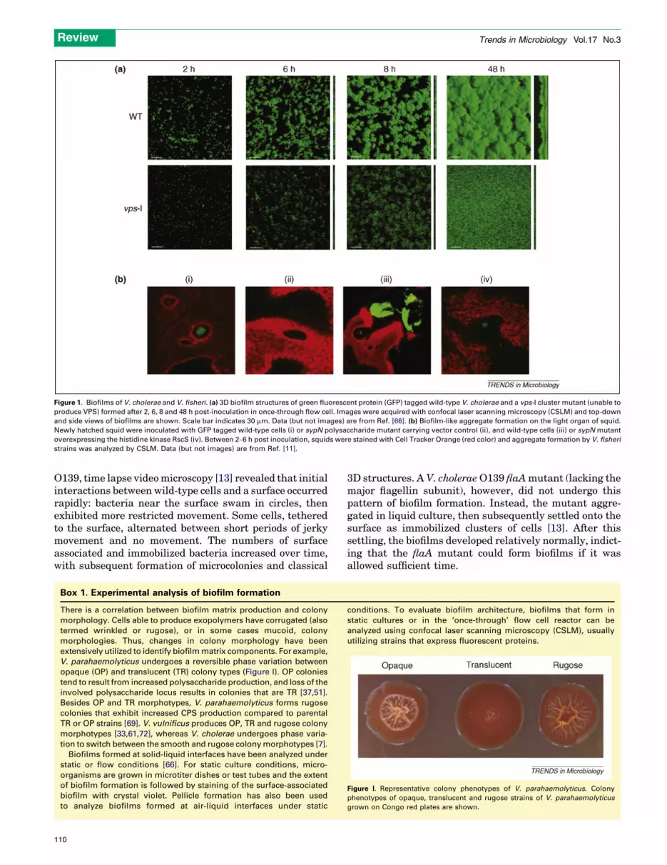
Figure 1. Biofilms of V. cholerae and V. fisheri. (a) 3D biofilm structures of green fluorescent protein (GFP) tagged wild-type V. cholerae and a vps-I cluster mutant (unable to
produce VPS) formed after 2, 6, 8 and 48 h post-inoculation in once-through flow cell. Images were acquired with confocal laser scanning microscopy (CSLM) and top-down
and side views of biofilms are shown. Scale bar indicates 30 mm. Data (but not images) are from Ref. [66]. (b) Biofilm-like aggregate formation on the light organ of squid.
Newly hatched squid were inoculated with GFP tagged wild-type cells (i) or sypN polysaccharide mutant carrying vector control (ii), and wild-type cells (iii) or sypN mutant
overexpressing the histidine kinase RscS (iv). Between 2–6 h post inoculation, squids were stained with Cell Tracker Orange (red color) and aggregate formation by V. fisheri
strains was analyzed by CSLM. Data (but not images) are from Ref. [11].
Review Trends in Microbiology Vol.17 No.3
O139, time lapse videomicroscopy [13] revealed that initialinteractions between wild-type cells and a surface occurredrapidly: bacteria near the surface swam in circles, thenexhibited more restricted movement. Some cells, tetheredto the surface, alternated between short periods of jerkymovement and no movement. The numbers of surfaceassociated and immobilized bacteria increased over time,with subsequent formation of microcolonies and classical
Box 1. Experimental analysis of biofilm formation
There is a correlation between biofilm matrix production and colony
morphology. Cells able to produce exopolymers have corrugated (also
termed wrinkled or rugose), or in some cases mucoid, colony
morphologies. Thus, changes in colony morphology have been
extensively utilized to identify biofilm matrix components. For example,
V. parahaemolyticus undergoes a reversible phase variation between
opaque (OP) and translucent (TR) colony types (Figure I). OP colonies
tend to result from increased polysaccharide production, and loss of the
involved polysaccharide locus results in colonies that are TR [37,51].
Besides OP and TR morphotypes, V. parahaemolyticus forms rugose
colonies that exhibit increased CPS production compared to parental
TR or OP strains [69]. V. vulnificus produces OP, TR and rugose colony
morphotypes [33,61,72], whereas V. cholerae undergoes phase varia-
tion to switch between the smooth and rugose colony morphotypes [7].
Biofilms formed at solid-liquid interfaces have been analyzed under
static or flow conditions [66]. For static culture conditions, micro-
organisms are grown in microtiter dishes or test tubes and the extent
of biofilm formation is followed by staining of the surface-associated
biofilm with crystal violet. Pellicle formation has also been used
to analyze biofilms formed at air-liquid interfaces under static
110
3D structures. A V. choleraeO139 flaAmutant (lacking themajor flagellin subunit), however, did not undergo thispattern of biofilm formation. Instead, the mutant aggre-gated in liquid culture, then subsequently settled onto thesurface as immobilized clusters of cells [13]. After thissettling, the biofilms developed relatively normally, indict-ing that the flaA mutant could form biofilms if it wasallowed sufficient time.
conditions. To evaluate biofilm architecture, biofilms that form in
static cultures or in the ‘once-through’ flow cell reactor can be
analyzed using confocal laser scanning microscopy (CSLM), usually
utilizing strains that express fluorescent proteins.
Figure I. Representative colony phenotypes of V. parahaemolyticus. Colony
phenotypes of opaque, translucent and rugose strains of V. parahaemolyticus
grown on Congo red plates are shown.

Review Trends in Microbiology Vol.17 No.3
The planktonic aggregation of flaA mutants was sub-sequently attributed to increased production of an exopoly-saccharide termed vibrio polysaccharide (VPS). Consistentwith this result, these mutants formed rugose colonies.Surprisingly, any of several paralyzed (mot)mutants, whichproduce a flagellum but cannot rotate it, formed smoothcolonies and poor biofilms [13,14]. A flaA motX doublemutant also produced smooth colonies, indicating that dis-ruptionofmot caneliminate theVPS-inducing signal causedby loss of the flagellum [14]. Thus, V. cholerae could use theflagellar motor not only to promote motility but also totransmit a signal indicating interaction with a surface [14].
Such a possibility would not be unprecedented: V. para-haemolyticus uses itsmotor to decidewhen to switch from aswimming cell (polarly flagellated) to a swarmer cell (withlateral flagella) capable of moving on surfaces or in highlyviscous media [15]. V. parahaemolyticus also uses its polarflagellum to promote biofilm formation [16]. flgE and flgD(hook) mutants are defective in attachment, pellicle for-mation (forming ‘speckled’ pellicles) and biofilm formation[16]. The severity of the defect depended on the strainbackground. For example, whereas TR fla mutants insubmerged cultures could not adhere, OP flamutants couldadhere, but could not form complex 3D structures, evenwith prolonged incubation. These data indicate a role forthe polar flagellum beyond biofilm initiation and/or incontrolling other factors [16].
In V. vulnificus and V. fischeri, flagellum-mediatedmotility also promotes biofilm formation. For example, aV. vulnificus flgE (hook) mutant is defective for attachmentboth to polystyrene and to glass wool [17]. InV. fischeri, theflrC regulator is also required for biofilm formation [18];prolonged incubation could overcome some, but not theentire defect.
Thus, motility has a key role in biofilm formation in thevibrios, as has been seen for other biofilm models. Inter-estingly, the role of motility seems to extend beyond simplyallowing the cell to reach the right location. Understandingother role(s) for motility and/or the motility apparatusduring biofilm development remains an important areaof investigation.
Pili promote cell-surface and/or cell–cell interactionsIn V. cholerae, at least three types of pili contribute tobiofilm formation: mannose-sensitive haemagglutinin typeIV pili (MSHA), toxin co-regulated pili (TCP) and chitin-regulated pili (ChiRP; formerly termed PilA) [19–22]. Therelative importance of these pili varies under differentconditions, and from strain to strain. MSHA, for example,is crucial for early attachment to abiotic surfaces in V.cholerae O1 El Tor strains, yet initial studies revealed norole in strain O139 [13]. Subsequent work revealed a rolefor the O139 MSHA pilus structural gene mshA in mono-layer formation, and demonstrated that the mshA mutantcould bypass this stage by aggregating as planktonic cellsand subsequently settling and forming 3D biofilms [23].Similarly, conflicting results for the importance of MSHApili have been obtained for biofilm formation on variouschitin substrates [19,20,22]. Thus, the contribution ofMSHA to biofilm formation is impacted by both environ-mental and genetic factors.
The classic, virulence-associated pilus of V. cholerae,TCP, is involved in microcolony formation on an environ-mentally relevant substrate, chitin. A TCP pili mutant ofEl Tor N16961 formed monolayers, but not microcolonies,on a chitin substrate [21]. Recent work revealed thatMSHA and TCP pili are inversely controlled at multiplelevels [24], indicating the possibility that the two pilustypes sequentially promote monolayer and 3D biofilmformation.
A role for ChiRP is less clear. ChiRP was required forcompetitive attachment to a chitin surface, crab shell, butlargely unnecessary for individual attachment to chitinparticles [20]. It is speculated that ChiRPmight have a roleother than adherence, such as orienting the cell optimallyfor chitin degradation [20].
V. parahaemolyticus also employs MSHA and ChiRP forbiofilm formation. MSHA mutants form substantiallyreduced biofilms – a defect that could be overcome byprolonged incubation – and defective, speckled pellicles[16]. Like V. cholerae, strain background influenced theseverity of the defect. ChiRP mutants fail to progress pastmonolayer formation [25]. In addition, both MSHA andChiRP contribute to attachment to chitin particles [25].
InV. vulnificus, the type IV pilus structural protein PilAand, to a greater degree, the pre-pilin peptidase PilD,contribute to binding both to abiotic surfaces and to humanepithelial cells [26,27]. The difference in relative import-ance of the two genes could be attributed to the retention bythe pilA mutant of other types of pili, and/or to the role ofPilD in processing other secreted proteins [26,27]. PilA andPilD are also necessary for prolonged attachment tooysters [28].
A role for pili in biofilm formation in V. fischeri has notbeen determined but is expected, given that the genomeencodes eight putative pili loci, two of which contribute toefficient symbiotic colonization [29,30].
In general, although similar pili are used by vibrios forattachment, it seems that the genetic context of the cell andthe type of surface it encounters (and/or other clues fromthe environment) can substantially influence the relativeimportance and thus usage of a particular type of pili forattachment.
Polysaccharides are the most prevalent component ofVibrio biofilmsProduction of mature biofilms requires extracellularmatrix components that hold the cells together and keepthe biofilm attached to the surface. The capsular polysac-charide (CPS) or exopolysaccharide (EPS, or VPS in V.cholerae) loci involved in biofilm formation have beenidentified from numerous Vibrio spp. Expression of theseloci is frequently correlated with biofilm-associatedchanges in colony morphology, in particular OP, rugoseor wrinkled colonies (Box 1).
vps and vps-like loci
In V. cholerae O1 El Tor A1552, biofilm formation dependsupon two linked loci, vps-I and vps-II (collectively termedvps), which encode structural proteins responsible for EPSproduction [7] (Figure 2a). vps-I and vps-II are separatedby six genes (rbmA-F) that also are involved in biofilm
111
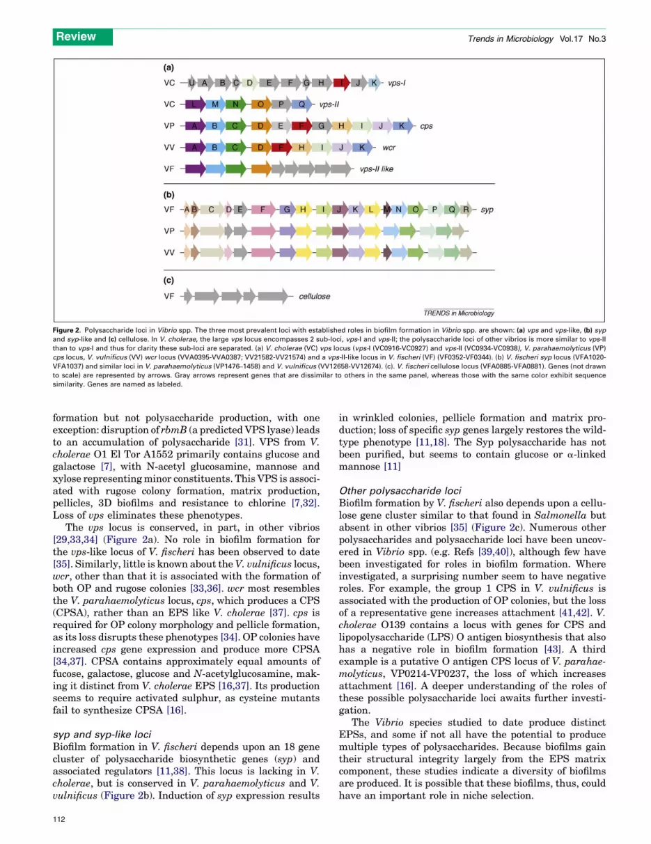
Figure 2. Polysaccharide loci in Vibrio spp. The three most prevalent loci with established roles in biofilm formation in Vibrio spp. are shown: (a) vps and vps-like, (b) syp
and syp-like and (c) cellulose. In V. cholerae, the large vps locus encompasses 2 sub-loci, vps-I and vps-II; the polysaccharide loci of other vibrios is more similar to vps-II
than to vps-I and thus for clarity these sub-loci are separated. (a) V. cholerae (VC) vps locus (vps-I (VC0916-VC0927) and vps-II (VC0934-VC0938), V. parahaemolyticus (VP)
cps locus, V. vulnificus (VV) wcr locus (VVA0395-VVA0387; VV21582-VV21574) and a vps-II-like locus in V. fischeri (VF) (VF0352-VF0344). (b) V. fischeri syp locus (VFA1020-
VFA1037) and similar loci in V. parahaemolyticus (VP1476–1458) and V. vulnificus (VV12658-VV12674). (c). V. fischeri cellulose locus (VFA0885-VFA0881). Genes (not drawn
to scale) are represented by arrows. Gray arrows represent genes that are dissimilar to others in the same panel, whereas those with the same color exhibit sequence
similarity. Genes are named as labeled.
Review Trends in Microbiology Vol.17 No.3
formation but not polysaccharide production, with oneexception: disruption of rbmB (a predicted VPS lyase) leadsto an accumulation of polysaccharide [31]. VPS from V.cholerae O1 El Tor A1552 primarily contains glucose andgalactose [7], with N-acetyl glucosamine, mannose andxylose representingminor constituents. This VPS is associ-ated with rugose colony formation, matrix production,pellicles, 3D biofilms and resistance to chlorine [7,32].Loss of vps eliminates these phenotypes.
The vps locus is conserved, in part, in other vibrios[29,33,34] (Figure 2a). No role in biofilm formation forthe vps-like locus of V. fischeri has been observed to date[35]. Similarly, little is known about theV. vulnificus locus,wcr, other than that it is associated with the formation ofboth OP and rugose colonies [33,36]. wcr most resemblesthe V. parahaemolyticus locus, cps, which produces a CPS(CPSA), rather than an EPS like V. cholerae [37]. cps isrequired for OP colony morphology and pellicle formation,as its loss disrupts these phenotypes [34]. OP colonies haveincreased cps gene expression and produce more CPSA[34,37]. CPSA contains approximately equal amounts offucose, galactose, glucose and N-acetylglucosamine, mak-ing it distinct from V. cholerae EPS [16,37]. Its productionseems to require activated sulphur, as cysteine mutantsfail to synthesize CPSA [16].
syp and syp-like loci
Biofilm formation in V. fischeri depends upon an 18 genecluster of polysaccharide biosynthetic genes (syp) andassociated regulators [11,38]. This locus is lacking in V.cholerae, but is conserved in V. parahaemolyticus and V.vulnificus (Figure 2b). Induction of syp expression results
112
in wrinkled colonies, pellicle formation and matrix pro-duction; loss of specific syp genes largely restores the wild-type phenotype [11,18]. The Syp polysaccharide has notbeen purified, but seems to contain glucose or a-linkedmannose [11]
Other polysaccharide loci
Biofilm formation by V. fischeri also depends upon a cellu-lose gene cluster similar to that found in Salmonella butabsent in other vibrios [35] (Figure 2c). Numerous otherpolysaccharides and polysaccharide loci have been uncov-ered in Vibrio spp. (e.g. Refs [39,40]), although few havebeen investigated for roles in biofilm formation. Whereinvestigated, a surprising number seem to have negativeroles. For example, the group 1 CPS in V. vulnificus isassociated with the production of OP colonies, but the lossof a representative gene increases attachment [41,42]. V.cholerae O139 contains a locus with genes for CPS andlipopolysaccharide (LPS) O antigen biosynthesis that alsohas a negative role in biofilm formation [43]. A thirdexample is a putative O antigen CPS locus of V. parahae-molyticus, VP0214-VP0237, the loss of which increasesattachment [16]. A deeper understanding of the roles ofthese possible polysaccharide loci awaits further investi-gation.
The Vibrio species studied to date produce distinctEPSs, and some if not all have the potential to producemultiple types of polysaccharides. Because biofilms gaintheir structural integrity largely from the EPS matrixcomponent, these studies indicate a diversity of biofilmsare produced. It is possible that these biofilms, thus, couldhave an important role in niche selection.

Figure 3. Regulation of biofilm formation in Vibrio spp. Biofilm formation in V. cholerae is positively regulated by the regulators VpsR and VpsT. The magnitude of
transcriptional control of vps genes by VpsR is greater than that of VpsT. Expression of vps genes and the regulators VpsR and VpsT are negatively controlled by the HapR
regulator. In V. parahaemolyticus expression of cps genes is negatively regulated by a homolog of VpsT, CpsS. In the absence of CpsS, OpaR and CpsR (HapR and VpsR
homologs, respectively) positively control cps gene expression and biofilm formation. In V. fischeri transcription of syp genes and biofilm formation are positively
controlled by the histidine kinases RscS and/or SypF, which act through response regulator SypG. SypF also positively regulates production of cellulose (cel), acting
although the VpsR homolog of V. fischeri. The roles of VpsR and SypE as inhibitors in V. fischeri are poorly understood and thus these are omitted from this figure.
Review Trends in Microbiology Vol.17 No.3
Transcriptional control of EPS production genesIn Vibrio species, regulation of exopolysaccharide pro-duction and biofilm formation is complex, and involvesnumerous transcriptional regulators, particularly two-component signal transduction and quorum sensing reg-ulators (Figure 3). In a typical two-component system, astimulus detected by a sensor histidine kinase (HK) istransformed into a cellular signal by a phosphorelay eventthat involves autophosphorylation of the HK at a con-served histidine residue. The phosphoryl group is thenpassed to a response regulator (RR) at a conserved aspar-tate residue, leading to activation and altered gene expres-sion or protein function. Quorum sensing involves sensingand responding to population density (Box 2). Global reg-ulators, such as CRP [44,45] and sigma factors RpoS andRpoN [32], have a role in biofilm formation in the vibrios,but they are not discussed here.
Two-component regulators
VpsR and VpsR-like regulators. In V. cholerae, the RRVpsR is a key regulator of biofilm formation. VpsR pro-motes transcription of the vps genes and formation oftypical 3D biofilm structures [32,46]. Mutations that alterthe residue predicted to be phosphorylated (D59) eitherinactivate this protein (D59A) or increase its activity(D59E) [14], thus supporting that phosphorylation on thisresidue is required for activation. However, theHK respon-sible for phosphorylation of VpsR has not been identified,as it is not physically linked to vpsR. Identifying thecognate HK will facilitate understanding the regulatorynetwork controlling vps gene expression and how environ-mental signals regulate VPS production.
Under specific genetic conditions, V. parahaemolyticusalso produces high levels of CPS and thus rugose colonies.Disruption of the vpsR homolog, cpsR, yields smoothcolonies and decreases transcriptional activity of cpsAfused to a lacZ reporter in the rugose background [34].However, CpsR is not required for basal levels of cpsexpression in TR or OP strains, as disruption of cpsR in
these strains had no effect on cps transcription [34]. Thiscontrasts with the role of VpsR, which is essential fortranscription of vps genes in all forms of V. cholerae. Itremains to be determined whether CpsR directlyregulates transcription of cps genes.
V. fischeri also encodes a VpsR homolog. Disruption ofvpsR leads to formation of mucoid colonies, which areindicative of enhanced polysaccharide production, indicat-ing that VpsR might be a repressor [35]. When overex-pressed, however, VpsR induces biofilm formation,indicating that it could also be an activator. Surprisingly,VpsR-induced biofilms depended not on a vps-like locus buton a putative cellulose biosynthesis cluster not found inother characterized vibrios (Figure 2c) [35]. Thus, VpsRseems to have a novel role in V. fischeri.
VpsT and VpsT-like. In V. cholerae, a second positiveregulator is VpsT, a member of the UhpA (FixJ) family oftranscriptional regulators [47]. VpsT shows homology toCsgD, which positively controls curli and cellulose pro-duction in Salmonella enterica. Disruption of vpsT yieldssmooth colonies and reduces vps expression, and biofilmformation [47]. Although the putative phosphorylation sitein the receiver domain of VpsT is conserved, it is notnecessary for the in vivo function of VpsT (J. Meir andF. Yildiz, unpublished). Thus, whether VpsT functions as acanonical RR remains unclear.
Characterization of vpsT and vpsR mutants in V. cho-lerae revealed different roles for these regulators in deter-mining biofilm architecture. The vpsT mutant is still ableto form biofilms (albeit distinct from its rugose parent),whereas vpsR and vpsT vpsR mutants produce only singlecells or small microcolonies attached to the substratum[48]. The VpsR and VpsT regulons are largely identical,although VpsR exerts a larger impact on expression [48].Thus, VpsR is essential for VPS production and biofilmformation, whereas VpsT seems to have an accessory role,possibly by increasing the level or activity of VpsR.Whether VpsT and VpsR serve as direct regulators ofvps remains unknown.
113
Box 2. Quorum sensing
Quorum sensing is a mechanism that cells use to determine their
population density. Signal synthases produce a diffusible molecule
(autoinducer [AI]) that can accumulate in the extracellular environ-
ment. When the cell population is sufficiently high, and enough
signal accumulates, this molecule binds to and activates cellular
receptors. In Vibrio species, one consequence of this signaling is a
phosphorelay that ultimately controls the synthesis of a global
regulator and phenotypes that are more useful to a group of cells,
such as luminescence, rather than to individuals [50]. Because
biofilm formation is a behavior that depends upon a group of
cells, it makes sense for bacteria to rely on quorum sensing
regulators to control biofilm formation, and indeed they do. In
V. cholerae, however, the sense of this control is not as might be
expected: V. cholerae mutants, which are ‘locked’ into a regulatory
state mimicking high cell density, are impaired in biofilm formation
[54].
V. cholerae produces two AIs [50]. CAI-1 [(S)-3-hydroxytridecan-4-
one] [73] is synthesized by CqsA and detected by CqsS. AI-2 [the
furanosyl borate diester (2S,4S)-2-methyl-2,3,3,4-tetrahydroxytetrahy-
drofuran borate] is synthesized by LuxS and detected by the LuxQP
receptor [50]. Information from the sensors is transduced through a
phosphorelay, first to the LuxU phosphotransfer protein and then to
LuxO, which is an RpoN dependent response regulator. At low cell
density, when the concentrations of AIs are low, LuxO is phosphory-
lated and activates expression of a set of small regulatory RNAs
(Figure Ia) [74]. The result is inhibition of expression of the major
quorum sensing regulator, HapR. At high cell density, LuxO is de-
phosphorylated and expression of HapR is increased. Under these
conditions, the vps genes and virulence-associated genes (aphA) are
repressed (Figure Ib).
This regulation seems to be both direct and indirect as the
empirically defined HapR binding motif was found in the promoter
regions of both the vps locus and its regulator vpsT [32]. Mutants in
cqsA that cannot produce the CAI-1 signal, a condition that should
mimic low cell density, form thicker biofilms [55]. This indicates that
CAI-1 signals are crucial for repression of biofilm formation via the
quorum sensing regulatory circuitry. It is thought that quorum
sensing ensures development of ‘normal’ biofilm structures that
permit rapid dispersion of bacteria from the biofilm, thereby
facilitating transmission.
Figure I. V. cholerae quorum sensing. V. cholerae synthesizes two AIs, CAI-1 (stars) made by CqsA and AI-2 (circles) made by LuxS, that are detected by sensor kinases
CqsS and LuxQ (working with periplasmic protein LuxP), respectively, which control a phosphorelay system. Low (a) and high (b) levels of autoinducers alter the signal
transduction cascade, ultimately impacting biofilm formation and virulence gene expression. Abbreviations: Hfq, RNA binding protein; sRNAs, small regulatory RNAs.
Review Trends in Microbiology Vol.17 No.3
A VpsT homolog, CpsS, also controls biofilm formationin V. parahaemolyticus, but as a negative regulator [34].Introduction of a cpsS mutation into TR or OP strainsderepresses cpsA transcription, resulting in rugose colo-nies. In TR strains, derepression is mediated throughCpsR, whereas in OP strains, CpsR has an accessory role[34]. Thus,V. cholerae andV. parahaemolyticus use similarproteins, but they function in the opposite sense and todifferent degrees: CpsS is the dominant negative regulatorin V. parahaemolyticus, whereas VpsT is a positive co-regulator in V. cholerae.
Other two-component regulators. InV. vulnificus, the RRNtrC contributes to biofilm formation through its tran-scriptional control of gmhD [49]. The GmhD protein, anADP-l-glycero-D-manno-heptose-6-epimerase, is requiredfor LPS and EPS production and biofilm formation [49].The relative roles of LPS and EPS in biofilm formation, inaddition to the identity of the EPS involved in this process,have yet to be determined.
114
In V. fischeri, two-component regulators have crucialroles in the control of biofilm formation, primarily throughtheir activation of the syp polysaccharide locus. Forexample, the orphan HK RscS, when overexpressed,induces syp transcription, wrinkled colony and pellicleformation, and symbiotic biofilm formation [11]. RscS actsupstream of SypG, a syp-encoded RR that is proposed to bethe direct activator of syp transcription [18].
Surprisingly, overexpression of SypG induced syp tran-scriptionbutnot the formationofwrinkled coloniesor strongpellicles. However, overexpression of SypG in a sypEmutant, permitted wrinkled colony and pellicle formation[18]. Thus, SypE, a putative RR that is not predicted to bindDNA, seems tohave an inhibitory role.RscSmight thereforeboth activate SypG and inactivate SypE. Homologs of SypGare present in V. vulnificus and V. parahaemolyticus, butRscS and SypE are unique to V. fischeri.
Overexpression of a third syp-encoded regulator, SypF,also induces biofilm formation in V. fischeri [35]. This

Figure 4. C-di-GMP signaling proteins in Vibrio spp. Cyclic di-guanosine-
monophosphate (c-di-GMP) controls cell surface structures and biofilm
formation in a diverse group of microorganisms. C-di-GMP is created from GTP
(guanosine-50-triphosphate) by diguanylate cyclase proteins that bear a GGDEF
amino acid motif and degraded to the dinucleotide pGpG by phosphodiesterase
proteins with EAL domains. C-di-GMP can be sensed by proteins with a PilZ
domain. Numbers of genes encoding GGDEF, EAL, dual GGDEF and EAL or PilZ
domain proteins in different Vibrio species are shown.
Review Trends in Microbiology Vol.17 No.3
regulator seems to function upstream of both SypG andVpsR: mutation of both regulators was necessary to elim-inate all the biofilm phenotypes induced by SypF over-expression. The SypF-VpsR path seems to promote cell-surface attachment, whereas the SypF-SypG path seems tobe responsible for cell–cell attachment [35].
Quorum sensing regulators
Vibrio species use quorum sensing (Box 2) to controlnumerous traits, including luminescence, virulence andbiofilm formation. Best studied in Vibrio harveyi, in whichthe endpoint regulator is termed LuxR (reviewed in Ref.[50]), similar pathway components are found in all othervibrios studied to date.
The V. parahaemolyticus LuxR homolog, OpaR, posi-tively regulates opacity, cps gene expression and biofilmformation [51]. Disruption of opaR in an OP strain yieldedTR colonies, and overexpression of opaR in a TR strainincreased cps expression and colony opacity [37]. A similarphenomenon is seen in both V. vulnificus and V. fischeri:mutants defective for the LuxR homologs, SmcR [52] andLitR [53], respectively, form TR colonies instead of theparental OP colonies; in the case of V. vulnificus, thisdisruption is associated with decreased biofilm formation.However, the molecular mechanisms causing decreases incolony opacity and biofilm formation and their connectionsto CPS or EPS production by V. vulnificus and V. fischeriare unknown.
In V. cholerae, the story is different (Figure 3). Mutantsof the LuxR homolog, HapR, exhibit increased rugosity andincreased vps expression [32,54,55]. These data indicatethat HapR is a negative regulator of biofilm formation.HapR can directly bind DNA and repress expression ofvpsT [56]. It can also control vpsR expression in somestrains [32]. Thus, quorum sensing control of cell surfaceproperties and biofilm formation is opposite in V. choleraerelative to the other vibrios. The ecological importance ofthis regulation is yet to be determined.
C-di-GMP signaling and biofilm formationC-di-GMP is a ubiquitous second messenger that controlsthe transition from a free-living, motile lifestyle to a biofilmlifestyle in many bacteria (reviewed in Ref. [57]), includingvibrios [58–62]. Increased c-di-GMP levels tend to promotebiofilm formation and/or inhibit flagellar motility. C-di-GMP production and degradation is controlled by digua-nylate cyclases (DGCs) and phosphodiesterases (PDEs),respectively [57] (Figure 4). Overexpression of these reg-ulators tends to cause global effects. Intriguingly, vibrioscontain much larger numbers of DGCs and PDEs thanother bacteria [63]. The abundance of enzymes controllingsynthesis and degradation of c-di-GMP in vibrios indicatesthe importance of c-di-GMP signaling to the biology ofvibrios. Because different types of sensory domains arefound in proteins predicted to function as DGCs or PDEs[63], one possibility is that cells adjust their c-di-GMPlevels in response to environmental and intracellular sig-nals and that c-di-GMP signaling has an important role inadaptation of vibrios to different environments.
In V. cholerae, c-di-GMP increases biofilm formationby stimulating transcription of vps genes and the
positive transcriptional regulators vpsR and vpsT[60,64]. Mutants of the known or putative PDE genes,mbaA, rocS, cdgC, cdpA and vieA, exhibit enhancedbiofilm formation, presumably because of increased c-di-GMP levels [65,66].
Recently, c-di-GMP also has been linked to the naturalcapacity of V. cholerae to generate rugose variants. Forexample, the prototype rugose strain A1552 expresseselevated c-di-GMP levels caused by a single amino acidchange in a DGC protein, VpvC, relative to the smoothvariant [67]. Disruption of vpvC in this rugose variantreduces overall c-di-GMP levels and causes cells to becomesimilar to the smooth variant with respect to biofilmformation and vps transcription. Rugosity can also begenerated by deletion of the master quorum sensing reg-ulator hapR. This effect occurs through CdgA, a DGCwhose mRNA abundance is increased in the hapRmutant;this increased CdgA presumably increases cellular c-di-GMP. Deletion of cdgA decreased vps transcription andrestored smooth colony formation to the rugose hapRmutant [48]. Subsequent studies revealed that HapRserves as a direct regulator of cdgA [56].
Increased c-di-GMP level leads to a decrease in motility.In V. cholerae, as expected, mutations in DGC genes cdgD[60], cdgH [68] and vpvC [67] lead to an increase inmotility, whereasmutations in PDE genes vieA, rocS, cdgCand mbaA lead to a decrease in motility relative to wild-type, when tested on Luria-Bertani medium (LB) soft agarmotility plates [66].
In V. parahaemolyticus, increases in cellular c-di-GMPlevels prevent swarming motility and promote biofilmformation. Two genes involved in c-di-GMP control, scrGand scrC, have been extensively characterized [58,59,69].ScrG functions as a PDE: null mutants increase c-di-GMPand exhibit increased cps and decreased lateral flagellar
115

Box 3. Questions for future research
� What combination(s) of polysaccharides are being produced
under various laboratory and environmental conditions?
� What are the constituents (protein or DNA) of biofilm matrices
under laboratory, environmental and/or disease conditions?
� What other structural and regulatory factors are involved in
biofilm formation?
� What are the environmental conditions that promote formation
and dissolution of biofilms?
� What are the stimuli sensed by two-component systems regulat-
ing biofilms formation?
� What is the mechanism of c-di-GMP signaling and how is it
connected to the regulatory network controlling biofilm forma-
tion?
� Do differences in regulation of biofilm formation reflect the
importance of the biofilm lifestyle to each Vibrio spp. during their
in vivo and ex vivo life cycles?
Review Trends in Microbiology Vol.17 No.3
gene (laf) expression and, thus, enhanced biofilm for-mation and reduced swarming motility [59]. Null mutantsdefective for the scrABC operon behave similarly, whereasoverexpression of scrABC yields the opposite results [58].Interestingly, however, overexpression of scrC in theabsence of scrAB (encoding putative pyridoxal-phos-phate-dependent and extracellular solute-bindingproteins, respectively) induces cps, not laf , expression[58]. Subsequent work revealed that ScrC is a bifunctionalprotein that functions as a DGC to synthesize c-di-GMP,but when co-produced with ScrAB, functions as a PDE anddegrades c-di-GMP. Epistasis analysis indicates that ScrGand ScrABC act in the same regulatory circuitry and thatscrG and scrABC double mutants show a cumulative effectat the level of laf and cps gene expression [58].
Relatively little is known about c-di-GMP and biofilmformation in V. vulnificus and V. fischeri. In V. vulnificus,expression of the DGC DcpA converted TR colonies of anacapsular mutant into OP colonies, but did not impactmotility [61]. Overexpression of dcpA induced productionof an EPS that was structurally distinct from the CPS,rugose colony formation and biofilm formation [61]. In V.fischeri, overexpression of the putative DGC MifA pro-motes cellulose biosynthesis and biofilm formation, indi-cating that c-di-GMP is a player in biofilm formation in thismicrobe as well [70].
How are c-di-GMP levels sensed by the cell? One proteindomain that binds c-di-GMP is the PilZ domain. Of the fivePilZ domain proteins in V. cholerae, two of these, PlzC andPlzD, have been recently shown to bind c-di-GMP and areknown to regulate biofilm formation and/or motility [71].Thus, PilZ domain proteins can function as c-diGMP recep-tors and regulate c-di-GMP-dependent processes in V.cholerae and likely in other vibrios.
It is becoming clear that, although Vibrios share com-mon regulatory proteins and signaling systems, the biofilmregulatory circuitry is unique to each Vibrio spp. Differ-ences in regulation might reflect the importance of thebiofilm life style to eachVibrio spp. during their in vivo andex vivo life cycles, differences in niche occupation, differ-ences in environmental parameters that they respond toand/or parameters driving evolution of the pathogens andsymbionts.
116
Concluding remarksBiofilm formation, particularly on a biotic, possibly nutri-tional surface, seems likely to provide a substantial survi-val advantage to aquatic organisms such as Vibrio species.That these organisms use similar traits and regulators tosolve the problem of biofilm formation is not unexpected.That they use such diversity in approaches – the relativeimportance of the traits and regulators, and even the sense(positive or negative) of control – is surprising and thus hasthe potential to provide great insights into the peculiarlifestyles of these microbes. Some outstanding researchquestions are listed in Box 3. Because biofilm formation isalso part of the pathogenic lifestyles of Vibrio spp., elucida-tion of the molecular mechanisms and regulation of biofilmformation will provide the foundation for developing noveltreatments and prevention strategies against Vibrio-associated illnesses.
AcknowledgementsWe thank Emily Yip and Cindy DeLoney-Marino for photos of V. fischeriaggregates, Linda McCarter for V. parahaemolyticus pictures, and KivancBilecen, Sinem Beyhan and Ates Gurcan for figure preparation. We alsothank members of our laboratories, Karen Ottemann and Alan Wolfe forcritical reading of the manuscript. Work in our laboratories investigatingbiofilm formation in Vibrio species was supported by NIH grantsAI055987 to F.H.Y. and GM59690 to K.L.V.
References1 Faruque, S.M. et al. (1998) Epidemiology, genetics, and ecology of
toxigenic Vibrio cholerae. Microbiol. Mol. Biol. Rev. 62, 1301–13142 Nair, G.B. et al. (2007) Global dissemination of Vibrio
parahaemolyticus serotype O3:K6 and its serovariants. Clin.Microbiol. Rev. 20, 39–48
3 Gulig, P.A. et al. (2005) Molecular Pathogenesis of Vibrio vulnificus. J.Microbiol. 43 (Spec No), 118–131
4 Visick, K.L. and Ruby, E.G. (2006) Vibrio fischeri and its host: it takestwo to tango. Curr. Opin. Microbiol. 9, 632–638
5 Donlan, R.M. and Costerton, J.W. (2002) Biofilms: survivalmechanisms of clinically relevant microorganisms. Clin. Microbiol.Rev. 15, 167–193
6 Watnick, P.I. and Kolter, R. (1999) Steps in the development of aVibriocholerae El Tor biofilm. Mol. Microbiol. 34, 586–595
7 Yildiz, F.H. and Schoolnik, G.K. (1999) Vibrio cholerae O1 El Tor:identification of a gene cluster required for the rugose colony type,exopolysaccharide production, chlorine resistance, and biofilmformation. Proc. Natl. Acad. Sci. U. S. A. 96, 4028–4033
8 Faruque, S.M. et al. (2006) Transmissibility of cholera: in vivo-formedbiofilms and their relationship to infectivity and persistence in theenvironment. Proc. Natl. Acad. Sci. U. S. A. 103, 6350–6355
9 Colwell, R.R. et al. (2003) Reduction of cholera in Bangladeshivillages by simple filtration. Proc. Natl. Acad. Sci. U. S. A. 100,1051–1055
10 Nyholm, S.V. et al. (2000) Establishment of an animal-bacterialassociation: recruiting symbiotic vibrios from the environment. Proc.Natl. Acad. Sci. U. S. A. 97, 10231–10235
11 Yip, E.S. et al. (2006) The symbiosis regulator rscS controls the sypgene locus, biofilm formation and symbiotic aggregation by Vibriofischeri. Mol. Microbiol. 62, 1586–1600
12 O’Toole, G. et al. (2000) Biofilm formation as microbial development.Annu. Rev. Microbiol. 54, 49–79
13 Watnick, P.I. et al. (2001) The absence of a flagellum leads to alteredcolony morphology, biofilm development and virulence in Vibriocholerae O139. Mol. Microbiol. 39, 223–235
14 Lauriano, C.M. et al. (2004) The sodium-driven flagellar motor controlsexopolysaccharide expression in Vibrio cholerae. J. Bacteriol. 186,4864–4874
15 Kawagishi, I. et al. (1996) The sodium-driven polar flagellar motor ofmarine Vibrio as the mechanosensor that regulates lateral flagellarexpression. Mol. Microbiol. 20, 693–699

Review Trends in Microbiology Vol.17 No.3
16 Enos-Berlage, J.L. et al. (2005) Genetic determinants of biofilmdevelopment of opaque and translucent Vibrio parahaemolyticus.Mol. Microbiol. 55, 1160–1182
17 Lee, J.H. et al. (2004) Role of flagellum and motility in pathogenesis ofVibrio vulnificus. Infect. Immun. 72, 4905–4910
18 Hussa, E.A. et al. (2008) RscS functions upstream of SypG to control thesyp locus and biofilm formation in Vibrio fischeri. J. Bacteriol. 190,4576–4583
19 Chiavelli, D.A. et al. (2001) The mannose-sensitive hemagglutinin ofVibrio cholerae promotes adherence to zooplankton. Appl. Environ.Microbiol. 67, 3220–3225
20 Meibom, K.L. et al. (2004) The Vibrio cholerae chitin utilizationprogram. Proc. Natl. Acad. Sci. U. S. A. 101, 2524–2529
21 Reguera, G. and Kolter, R. (2005) Virulence and the environment: anovel role for Vibrio cholerae toxin-coregulated pili in biofilm formationon chitin. J. Bacteriol. 187, 3551–3555
22 Watnick, P.I. et al. (1999) A role for the mannose-sensitivehemagglutinin in biofilm formation by Vibrio cholerae El Tor. J.Bacteriol. 181, 3606–3609
23 Moorthy, S. and Watnick, P.I. (2004) Genetic evidence that the Vibriocholerae monolayer is a distinct stage in biofilm development. Mol.Microbiol. 52, 573–587
24 Hsiao, A. et al. (2008) Post-transcriptional cross-talk between pro- andanti-colonization pili biosynthesis systems in Vibrio cholerae. Mol.Microbiol. 67, 849–860
25 Shime-Hattori, A. et al. (2006) Two type IV pili of Vibrioparahaemolyticus play different roles in biofilm formation. FEMSMicrobiol. Lett. 264, 89–97
26 Paranjpye, R.N. et al. (1998) The type IV leader peptidase/N-methyltransferase of Vibrio vulnificus controls factors required foradherence to HEp-2 cells and virulence in iron-overloaded mice.Infect. Immun. 66, 5659–5668
27 Paranjpye, R.N. and Strom, M.S. (2005) A Vibrio vulnificus type IVpilin contributes to biofilm formation, adherence to epithelial cells, andvirulence. Infect. Immun. 73, 1411–1422
28 Paranjpye, R.N. et al. (2007) Role of type IV pilins in persistence ofVibrio vulnificus in Crassostrea virginica oysters. Appl. Environ.Microbiol. 73, 5041–5044
29 Ruby, E.G. et al. (2005) Complete genome sequence of Vibrio fischeri: asymbiotic bacterium with pathogenic congeners. Proc. Natl. Acad. Sci.U. S. A. 102, 3004–3009
30 Stabb, E.V. and Ruby, E.G. (2003) Contribution of pilA to competitivecolonization of the squid Euprymna scolopes by Vibrio fischeri. Appl.Environ. Microbiol. 69, 820–826
31 Fong, J.C. and Yildiz, F.H. (2007) The rbmBCDEF gene clustermodulates development of rugose colony morphology and biofilmformation in Vibrio cholerae. J. Bacteriol. 189, 2319–2330
32 Yildiz, F.H. et al. (2004) Molecular analysis of rugosity in a Vibriocholerae O1 El Tor phase variant. Mol. Microbiol. 53, 497–515
33 Grau, B.L. et al. (2008) Further characterization of Vibrio vulnificusrugose variants and identification of a capsular and rugoseexopolysaccharide gene cluster. Infect. Immun. 76, 1485–1497
34 Guvener, Z.T. and McCarter, L.L. (2003) Multiple regulators controlcapsular polysaccharide production in Vibrio parahaemolyticus. J.Bacteriol. 185, 5431–5441
35 Darnell, C.L. et al. (2008) The putative hybrid sensor kinaseSypF coordinates biofilm formation in Vibrio fischeri by actingupstream of two response regulators. SypG and VpsR. J. Bacteriol.190, 4941–4950
36 Smith, A.B. and Siebeling, R.J. (2003) Identification of genetic locirequired for capsular expression inVibrio vulnificus. Infect. Immun. 71,1091–1097
37 Enos-Berlage, J.L. and McCarter, L.L. (2000) Relation of capsularpolysaccharide production and colonial cell organization to colonymorphology in Vibrio parahaemolyticus. J. Bacteriol. 182, 5513–5520
38 Yip, E.S. et al. (2005) A novel, conserved cluster of genes promotessymbiotic colonization and sigma-dependent biofilm formation byVibrio fischeri. Mol. Microbiol. 57, 1485–1498
39 Bush, C.A. et al. (1997) Classification of Vibrio vulnificus strains by thecarbohydrate composition of their capsular polysaccharides. Anal.Biochem. 250, 186–195
40 Nakhamchik, A. et al. (2007) Identification of a Wzy polymeraserequired for group IV capsular polysaccharide and
lipopolysaccharide biosynthesis in Vibrio vulnificus. Infect. Immun.75, 5550–5558
41 Chatzidaki-Livanis, M. et al. (2006) Genetic variation in the Vibriovulnificus group 1 capsular polysaccharide operon. J. Bacteriol. 188,1987–1998
42 Joseph, L.A. and Wright, A.C. (2004) Expression of Vibrio vulnificuscapsular polysaccharide inhibits biofilm formation. J. Bacteriol. 186,889–893
43 Kierek, K. andWatnick, P.I. (2003) TheVibrio choleraeO139O-antigenpolysaccharide is essential for Ca2+-dependent biofilm development insea water. Proc. Natl. Acad. Sci. U. S. A. 100, 14357–14362
44 Fong, J.C. and Yildiz, F.H. (2008) Interplay between cyclic AMP-cyclicAMP receptor protein and cyclic di-GMP signaling in Vibrio choleraebiofilm formation. J. Bacteriol. 190, 6646–6659
45 Liang, W. et al. (2007) The cyclic AMP receptor protein modulatescolonial morphology in Vibrio cholerae. Appl. Environ. Microbiol. 73,7482–7487
46 Yildiz, F.H. et al. (2001) VpsR, a Member of the Response Regulators ofthe Two-Component Regulatory Systems, Is Required for Expression ofvps Biosynthesis Genes and EPS(ETr)-Associated Phenotypes inVibrio cholerae O1 El Tor. J. Bacteriol. 183, 1716–1726
47 Casper-Lindley, C. and Yildiz, F.H. (2004) VpsT is a transcriptionalregulator required for expression of vps biosynthesis genes and thedevelopment of rugose colonial morphology in Vibrio cholerae O1 ElTor. J. Bacteriol. 186, 1574–1578
48 Beyhan, S. et al. (2007) Regulation of rugosity and biofilm formation inVibrio cholerae: comparison of VpsT and VpsR regulons and epistasisanalysis of vpsT, vpsR, and hapR. J. Bacteriol. 189, 388–402
49 Kim, H.S. et al. (2007) Role of NtrC in biofilm formation via controllingexpression of the gene encoding an ADP-glycero-manno-heptose-6-epimerase in the pathogenic bacterium, Vibrio vulnificus. Mol.Microbiol. 63, 559–574
50 Waters, C.M. and Bassler, B.L. (2005) Quorum sensing: cell-to-cellcommunication in bacteria. Annu. Rev. Cell Dev. Biol. 21, 319–346
51 McCarter, L.L. (1998) OpaR, a homolog of Vibrio harveyi LuxR,controls opacity of Vibrio parahaemolyticus. J. Bacteriol. 180, 3166–
317352 Lee, J.H. et al. (2007) Identification and functional analysis of Vibrio
vulnificus SmcR, a novel global regulator. J. Microbiol. Biotechnol. 17,325–334
53 Fidopiastis, P.M. et al. (2002) LitR, a new transcriptional activator inVibrio fischeri, regulates luminescence and symbiotic light organcolonization. Mol. Microbiol. 45, 131–143
54 Hammer, B.K. and Bassler, B.L. (2003) Quorum sensingcontrols biofilm formation in Vibrio cholerae. Mol. Microbiol. 50,101–104
55 Zhu, J. and Mekalanos, J.J. (2003) Quorum sensing-dependentbiofilms enhance colonization in Vibrio cholerae. Dev. Cell 5, 647–
65656 Waters, C.M. et al. (2008) Quorum sensing controls biofilm formation in
Vibrio cholerae through modulation of cyclic di-GMP levels andrepression of vpsT. J. Bacteriol. 190, 2527–2536
57 Romling, U. and Amikam, D. (2006) Cyclic di-GMP as a secondmessenger. Curr. Opin. Microbiol. 9, 218–228
58 Ferreira, R.B. et al. (2008) Vibrio parahaemolyticus ScrC modulatescyclic dimeric GMP regulation of gene expression relevant to growth onsurfaces. J. Bacteriol. 190, 851–860
59 Kim, Y.K. and McCarter, L.L. (2007) ScrG, a GGDEF-EAL protein,participates in regulating swarming and sticking in Vibrioparahaemolyticus. J. Bacteriol. 189, 4094–4107
60 Lim, B. et al. (2006) Cyclic-diGMP signal transduction systems inVibrio cholerae: modulation of rugosity and biofilm formation. Mol.Microbiol. 60, 331–348
61 Nakhamchik, A. et al. (2008) Cyclic-di-GMP regulates extracellularpolysaccharide production, biofilm formation, and rugose colonydevelopment by Vibrio vulnificus. Appl. Environ. Microbiol. 74,4199–4209
62 Tischler, A.D. and Camilli, A. (2004) Cyclic diguanylate (c-di-GMP)regulates Vibrio cholerae biofilm formation. Mol. Microbiol. 53, 857–
86963 Galperin, M.Y. et al. (2001) Novel domains of the prokaryotic two-
component signal transduction systems. FEMS Microbiol. Lett. 203,11–21
117
Review Trends in Microbiology Vol.17 No.3
64 Beyhan, S. et al. (2006) Transcriptome and phenotypic responses ofVibrio cholerae to increased cyclic di-GMP level. J. Bacteriol. 188,3600–3613
65 Tamayo, R. et al. (2008) Role of cyclic Di-GMP during El tor biotypeVibrio cholerae infection: characterization of the in vivo-induced cyclicDi-GMP phosphodiesterase CdpA. Infect. Immun. 76, 1617–1627
66 Yildiz, F.H. and Kolter, R. (2008) Genetics and Microbiology of BiofilmFormation by Vibrio cholerae. In Vibrio cholerae: Genomics andMolecular Biology (Faruque, S.M. and Nair, G.B., eds), pp. 123–139,Caister Academic Press
67 Beyhan, S. and Yildiz, F.H. (2007) Smooth to rugose phase variation inVibrio cholerae can be mediated by a single nucleotide change thattargets c-di-GMP signalling pathway. Mol. Microbiol. 63, 995–1007
68 Beyhan, S. et al. (2008) Identification and characterization of cyclicdiguanylate signaling systems controlling rugosity in Vibrio cholerae.J. Bacteriol. 190, 7392–7405
Have you contributed to aDid you know that you are en
book
A 30% discount is available to all Elsevier book an
stand-alone CD-ROMs directly from us.
To take advantage of your discount:
1. Choose your book(s) from www.elsevier.com or w
2. Place your order
Americas:
Phone: +1 800 782 4927 for US customers
Phone: +1 800 460 3110 for Canada, South a
Fax: +1 314 453 4898
All other countries:
Phone: +44 (0)1865 474 010
Fax: +44 (0)1865 474 011
You’ll need to provide the name of the Elsev
contributed. Shipping is free on prepaid ord
If you are faxing your order, please enclose
3. Make your payment
This discount is only available on prepaid or
apply to multi-volume reference works or El
For more information, visit
118
69 Boles, B.R. and McCarter, L.L. (2002) Vibrio parahaemolyticusscrABC, a novel operon affecting swarming and capsularpolysaccharide regulation. J. Bacteriol. 184, 5946–5954
70 O’Shea, T.M. et al. (2006) Diguanylate cyclases control magnesium-dependent motility of Vibrio fischeri. J. Bacteriol. 188, 8196–8205
71 Pratt, J.T. et al. (2007) PilZ domain proteins bind cyclic diguanylateand regulate diverse processes in Vibrio cholerae. J. Biol. Chem. 282,12860–12870
72 Grau, B.L. et al. (2005) High-frequency phase variation of Vibriovulnificus 1003: isolation and characterization of a rugosephenotypic variant. J. Bacteriol. 187, 2519–2525
73 Higgins, D.A. et al. (2007) The major Vibrio cholerae autoinducer andits role in virulence factor production. Nature 450, 883–886
74 Lenz, D.H. et al. (2005) CsrA and three redundant small RNAsregulate quorum sensing in Vibrio cholerae. Mol. Microbiol. 58,1186–1202
n Elsevier publication?titled to a 30% discount ons?
d journal contributors when ordering books or
ww.books.elsevier.com
nd Central America customers
ier book or journal to which you have
ers within the US.
a copy of this page.
ders. Please note that this offer does not
sevier Health Sciences products.
www.books.elsevier.com


