Embed Size (px)
Citation preview
Vesicular Egress of Non-Enveloped Lytic ParvovirusesDepends on Gelsolin FunctioningSeverine Bar, Laurent Daeffler, Jean Rommelaere, Jurg P. F. Nuesch*
Program ‘‘Infection and Cancer,’’ Abteilung F010 and Institut National de la Sante et de la Recherche Medicale U701, Deutsches Krebsforschungszentrum, Heidelberg,
Germany
Abstract
The autonomous parvovirus Minute Virus of Mice (MVM) induces specific changes in the cytoskeleton filaments of infectedpermissive cells, causing in particular the degradation of actin fibers and the generation of ‘‘actin patches.’’ This is attributedto a virus-induced imbalance between the polymerization factor N-WASP (Wiscott-Aldrich syndrome protein) and gelsolin, amultifunctional protein cleaving actin filaments. Here, the focus is on the involvement of gelsolin in parvovirus propagationand virus-induced actin processing. Gelsolin activity was knocked-down, and consequences thereof were determined forvirus replication and egress and for actin network integrity. Though not required for virus replication or progeny particleassembly, gelsolin was found to control MVM (and related H1-PV) transport from the nucleus to the cell periphery andrelease into the culture medium. Gelsolin-dependent actin degradation and progeny virus release were both controlled by(NS1)/CKIIa, a recently identified complex between a cellular protein kinase and a MVM non-structural protein. Furthermore,the export of newly synthesized virions through the cytoplasm appeared to be mediated by (virus-modified) lysomal/lateendosomal vesicles. By showing that MVM release, like entry, is guided by the cytoskeleton and mediated by vesicles, theseresults challenge the current view that egress of non-enveloped lytic viruses is a passive process.
Citation: Bar S, Daeffler L, Rommelaere J, Nuesch JPF (2008) Vesicular Egress of Non-Enveloped Lytic Parvoviruses Depends on Gelsolin Functioning. PLoSPathog 4(8): e1000126. doi:10.1371/journal.ppat.1000126
Editor: Susan Cotmore, Yale University, United States of America
Received April 9, 2008; Accepted July 16, 2008; Published August 15, 2008
Copyright: � 2008 Bar et al. This is an open-access article distributed under the terms of the Creative Commons Attribution License, which permits unrestricteduse, distribution, and reproduction in any medium, provided the original author and source are credited.
Funding: This work was supported by public fundings by the DKFZ (German Cancer Research Center) and the French Institute National de la Sante et de laRecherche Medicale (INSERM). SB was a recipient of von Humboldt Foundation and EMBO. Confocal data were obtained at the Nikon Imaging Center inHeidelberg (Bioquant BQ 0004, INF 267).
Competing Interests: The authors have declared that no competing interests exist.
* E-mail: [email protected]
Introduction
The genus parvovirus (PV) consists of small icosahedral non-
enveloped particles with a 5.1-kb linear single-stranded DNA
genome. During productive infection, PVs induce dramatic
morphological and physiological changes in their host cells,
culminating in cell death and lysis. PV cytotoxicity is attributed
mainly to the large non-structural viral protein NS1, an 83-kDa
multifunctional protein endowed with enzymatic and non-
enzymatic properties enabling it to control various processes
necessary for progeny particle production and spread (reviewed in
[1]). To function in a concerted way, NS1 is regulated by specific
phosphorylations driven mainly by members of the PKC family
[2,3]. In addition to its direct involvement in particle production,
NS1 acts specifically to jeopardize the integrity and survival of
infected cells [4,5,6]. It has been shown to control the activity and
properties of selected cell components through physical interaction
[7,8] and/or induction of post-translational modifications [9,10].
Such targets might be modified either directly by NS1/CKIIa, a
recently described complex formed by NS1 with the catalytic
domain of cellular CKII [8], or indirectly through activation/
modulation of the PDK-1/PKC signaling cascade [11].
PV infection leads to characteristic alterations of host-cell
morphology that might facilitate virus replication or the release of
progeny particles. Subnuclear APAR-bodies acting as replication
centers for parvoviral DNA amplification are formed early in
infection [12,13]. Later, PVs induce cytoskeletal changes evi-
denced by rounding-up and detachment from the culture dish
prior to cytolysis [14,15]. In MVM-infected mouse A9 cells, these
morphological alterations have been attributed to the activity of
NS1 [4] and shown to result from changes in micro- and
intermediate filaments [10]. While tropomyosin is a direct target of
NS1/CKIIa, MVM-induced actin-filament alterations appear to
result from an imbalance between the polymerizing factor N-
WASP (Wiscott-Aldrich syndrome protein) and gelsolin [10], a
multifunctional protein known mainly for its actin-filament-
severing and capping activities and its participation in processes
requiring rapid actin remodeling [16]. Roles in apoptosis and lipid
signaling are also reported [17]. By altering the availability of
PIP2, gelsolin activity might interfere with PIP2-dependent
signaling cascades affecting phospholipase C [16].
Little is known about the impact of cytoskeletal rearrangements
on virus replication and spread. Cytoplasmic collapse is thought to
be part of a process leading to virus release upon cytolysis [7], but
there is also indirect evidence of PV release in the absence of cell
disruption [18]. The aim of the present study was to assess the role
of gelsolin activity and actin reorganization in PV replication and
spread. We show that gelsolin-induced modulation of actin
filaments is essential to virus egress and provides strong evidence
that progeny virions move to the cell periphery through vesicular
transport and start to be released into the medium before cell
collapse at the end of infection.
PLoS Pathogens | www.plospathogens.org 1 August 2008 | Volume 4 | Issue 8 | e1000126
Results
MVM-induced remodeling of gelsolin and actin filamentsMVM-induced cytopathic effects include actin-fiber degrada-
tion and subsequent formation of actin patches at late stages of
infection. The proposed cause is a virus-induced imbalance
between actin polymerization and severing [10]. To investigate
the impact of gelsolin on actin modulation and parvovirus
replication we used confocal laser scanning microscopy. A9 cells
infected (or not) with MVM were examined for gelsolin’s
subcellular distribution and its association with actin structures.
Gelsolin was found to colocalize with phalloidin-stained actin
structures (Figure 1A) whatever the infection status and time. In
non-infected cells it accumulated abundantly along the rigid actin
network and in the actin-rich region beneath the plasma
membrane. Upon infection, concomitantly with destruction of
the actin network, it became redistributed to the plasma
membrane and perinuclear regions, later becoming associated
with the above-mentioned cytoplasmic patches. The identical
distribution of gelsolin and disorganized actin in infected cells
suggests a link between the former and the state of the latter.
Cell fractionation experiments confirmed the above findings
(Figure 1B). In non-infected cells, actin was found predominantly
in the scaffold-containing fractions (iS, sS), but after infection it
was found in all subcellular fractions, including the cytosol (C) and
the membrane-associated fractions (nM, pM). In agreement with
its association with remodeled actin structures, MVM-induced
gelsolin was similarly found in all actin-positive fractions.
As additional proof of gelsolin/actin interaction during MVM-
induced actin reorganization, we used affinity chromatography to
study actin-gelsolin binding. This method was previously used to
determine NS1 association with tropomyosin [8] and proved
successful in detecting specific protein interactions with partially
insoluble cytoskeleton components in the cellular context. A9 cell
lines expressing GST-coupled actin under the control of the
parvoviral P38 promoter were MVM or mock infected. Twenty-
four hours post-infection, cell extracts were prepared, matched for
the GST-actin content due to viral induction of recombinant
protein expression, and passed through Glutathione Sepharose
columns to trap the fusion protein and associated polypeptides.
After extensive washing steps, proteins bound to the trapped GST-
actin were then eluted with high salt and cell matched volumes of
input and eluates were then tested by Western blotting for the
presence of gelsolin. As shown in Figure 1C (actin lanes), gelsolin
was invariably recovered from the GST-actin-loaded columns,
whether the actin-bound proteins were from infected or non-
infected cells. The specificity of the actin-gelsolin interaction was
demonstrated by failure to detect gelsolin in eluates from columns
loaded with extracts of A9 cells expressing only GST-free actin (A9
lanes) or GST-coupled tropomyosin (TM5 lanes). Altogether,
these results strongly suggest that gelsolin can interact with both
filamentous actin and virus-processed actin structures such as the
cytoplasmic patches.
Gelsolin involvement in MVM-induced actin-networkremodeling and release of progeny virions
Since gelsolin is induced by MVM and associates with actin, we
hypothesized that it might play a role in MVM-induced alteration
of the actin network and in MVM propagation. To test this
hypothesis, we transfected A9 cells with control serum/IgG or
with antibodies (aGln) known to specifically inhibit gelsolin activity
[19], infected them with MVM, and placed them in culture. At
different times, cells and their medium were collected separately
(Infection 1). The collected cells were tested by immunofluores-
cence (IF) staining for expression of the viral protein NS1, taken as
indicator of successful infection (Figure 2A). Southern blots were
also produced from the cells, showing the different forms of DNA
typically encountered in infected cells: the double-stranded
replicative forms (RF) and the single-stranded DNA (ssDNA) of
progeny virions (Figure 2B, Inf1). Neither the efficiency of
infection nor the levels of the different viral DNA forms appeared
to be altered by the presence of gelsolin-neutralizing antibodies.
In a second phase of the experiment, the release and infectivity
of progeny virions was determined by inoculating cultures of naive
A9 cells with supernatant medium from infection-1 cultures
(Infection 2). In this case, gelsolin activity appeared essential to
either the formation or the release of infectious progeny virions,
since cells exposed to infection-1 culture supernatants showed
markedly lower levels of MVM DNA when the infection-1 cells
had been treated with aGln (Figure 2B, Inf2). This result was
confirmed by measuring production and release of infectious
virions at 24 h p.i. by standard plaque assay. While cell-associated
titers between aGFP-IgG and aGln-treated samples varied only
marginally 1.5 fold (GFP: 2.966108, aGln: 2.126108), inactivation
of endogenous gelsolin blocked release of infectious virions leading
to a 30 fold reduction of medium-associated titers in aGln-treated
samples (26107 vs. 66105). Similar results were obtained with
human glioblastoma cells (NCH149) infected with H1-PV,
indicating that dependence on gelsolin is a general, late feature
of PV infection (Figure 2C).
Is gelsolin required for the generation or for the release of
infectious progeny virions? Does its effect on progeny virion
formation or release correlate with an active involvement of
gelsolin in MVM-induced remodeling of the actin network? To
address these questions, we generated two cell lines, each stably
transfected with a plasmid, pP38-MycGlnY438A or pP38-
MycGlnD565N, driving MVM-inducible expression of a domi-
nant-negative mutant gelsolin gene, so as to block endogenous
gelsolin activity. The studied mutations were respectively: (i) a
tyrosine-to-alanine substitution at position 438, disrupting a
phosphorylation site for Src kinases [20] regulating the PIP2
interaction and actin-severing activity [21] and (ii) a glutamic acid-
to-glutamine substitution at position 565, disrupting a conserved
Ca2+-binding site regulating the activity of gelsolin through
Author Summary
Rodent parvoviruses are non-enveloped lytic viruses thatare thought excellent tools for a virotherapy of cancerbecause of their strong natural oncolytic potential and lowpathogenicity in humans. Egress of non-enveloped lyticviruses is commonly thought to occur as a virus burst aftercell disintegration. Indeed, we showed in the past thatautonomous parvoviruses induce severe cytopathic effectsto the host cell, manifested in restructuring and degrada-tion of cytoskeletal filaments, thereby supporting suchmode of virus spread. Here, we focus on the impact ofvirus-induced actin degradation, and particularly thefunctioning of the actin-severing protein gelsolin. Al-though not required for DNA replication or progenyparticle production, gelsolin appears to facilitate aregulated virus egress from the nucleus to the cellperiphery via (virus modified) lysosomal/late endosomalvesicles. These results challenge the current view that lyticvirus egress is just a passive process at the end of infectionand suggests that these pathogens are endowed with theability to efficiently spread from cell to cell potentially insolid (tumor) tissue.
Gelsolin-Mediated Parvovirus Egress
PLoS Pathogens | www.plospathogens.org 2 August 2008 | Volume 4 | Issue 8 | e1000126

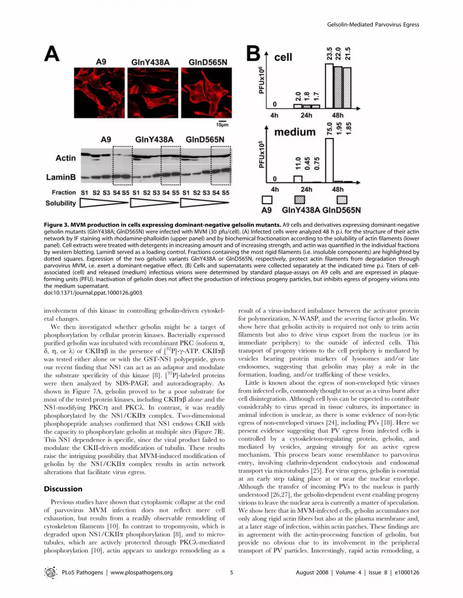
conformational alterations [22]. In IF microscopy and fraction-
ation experiments, both mutant gelsolins (GlnY438A and
GlnD565N) were found to accumulate in the perinuclear region
and being associated with large, insoluble scaffold structures
(Figure S1), i.e., were able to interfere with the actin-processing
activity of endogenous gelsolin. Furthermore, both mutant
gelsolins were found to protect actin fibers from PV-induced
remodeling, notably preventing the formation of patches
(Figure 3A, upper panel), and to impair the degradation of rigid
actin filaments (Figure 3A, lower panel). These results both
confirm the dominant-negative character of the mutations
introduced and demonstrate that gelsolin is instrumental in
altering the actin network after PV infection.
The same stable transfectants were then used to assess the role
of gelsolin in infectious virion production and release. As in the
case of cells treated with gelsolin-neutralizing antibodies (Figure 2B
and 2C), transfectants expressing GlnY438A or GlnD565N
retained the ability to amplify MVM DNA. In contrast, the
release of infectious viruses into the medium was drastically
impaired (Figure S2). This lack of ‘‘free’’ viruses in the
corresponding culture supernatants was not attributable to
efficient readsorption onto neighboring cells, since it was also
observed in cultures treated with neuraminidase to prevent MVM
recapture. The question was thus: does gelsolin inactivation affect
the formation or the shedding of infectious progeny viruses? To
address this question, we determined the infectious titers of both
cell-associated and released virions (Figure 3B). Cultures of
transfectants expressing a mutant gelsolin displayed similar cell-
associated infectious virion titers as A9 cultures, but a drastically
(20- to 40-fold) reduced titer of infectious virions shed into the
medium. This strongly suggests that gelsolin is essential for efficient
egress of progeny virions during MVM infection.
We next investigated the subcellular location at which progeny
particles get stuck in the absence of functional gelsolin. To this end,
A9 cells and cells expressing GlnY438A or GlnD565N were infected
with purified MVM, fixed at the indicated time p.i., and examined
by confocal microscopy for the presence of capsids. As shown for
representative cells in Figure 4A and as quantified in Figure 4B,
newly synthesized capsids were rapidly exported from the nucleus
and transported to the periphery of A9 cells, so that capsid staining
was distributed from the nucleus through the cytoplasm to the
plasma membrane (Nuc+Cytoplasmic capsids). In contrast, cells
expressing either of the mutant gelsolins displayed trapping of a
considerable proportion of the progeny virions in or around the
nucleus at least until 48 h p.i. ([Peri]nuclear capsids only).
Pre-lytic egress of parvoviruses is mediated by vesiculartransport
Capsid staining of infected A9 cells was noticeably spotty
(Figure 4A), suggesting that progeny viruses might be transported
by vesicular structures. This possibility was tested by confocal
microscopy of infected cells after double IF labeling of assembled
capsids and either vesicular markers or proteins known to be
Figure 1. Gelsolin modulation during MVM infection of A9cells. (A) A9 cells grown on spot slides were infected (or not) with MVM(30 pfu/cell), fixed with paraformaldehyde at the indicated time p.i., andanalyzed by confocal laser scanning microscopy after double-labelingfor actin with rhodamine-coupled phalloidin (red), and gelsolin, withCy2-conjugated IgGs (green). Colocalization areas appear yellow in themerge. Scale bar 15 mm. Gelsolin is found associated to phalloidin-stained actin in both non-infected and MVM-infected cells. Arrows pointto a slight but distinct accumulation of the actin-processing polypep-tide in the perinuclear region. (B) A9 cells were infected with MVM(30 pfu/cell) and harvested at the indicated time p.i. Association ofgelsolin and actin with cellular scaffold and membrane structures wasdetermined by fractionating cell extracts by a combined sedimentationand Triton X-100 extraction procedure. The distribution of each proteinamong the various fractions was determined by western blotting. UponMVM-infection, actin and gelsolin become associated with membranestructures. iS, insoluble scaffold proteins; nM, mainly nuclear (mem-brane) constituents; sS, soluble scaffold proteins; pM, mainly vesicularand plasma (membrane) constituents; C, soluble cytosolic proteins. (C)
Binding of gelsolin to cytoskeletal proteins was determined by affinitychromatography using GST-tagged bactin or tropomyosin 5 (TM) asbaits in stably transfected cells. Extracts of mock (2) and MVM-infected(+) cells (parental A9 [lanes 1 and 2] or derivatives expressing GST-tagged bactin [lanes 3 and 4] or TM [lanes 5 and 6]) were run throughGlutathione Sepharose columns specifically retaining GST-taggedproteins and partners thereof. 700 mM NaCl eluates containing thepartner proteins were analyzed by western blotting for the presence ofgelsolin, in comparison with the cell-matched original extract (Input).doi:10.1371/journal.ppat.1000126.g001
Gelsolin-Mediated Parvovirus Egress
PLoS Pathogens | www.plospathogens.org 3 August 2008 | Volume 4 | Issue 8 | e1000126

involved in vesicle formation. We took several measures to make
sure we were observing virus release and not virus entry: (i) we
checked that no incoming capsids were detected under the
conditions used (Figure 4A); (ii) we prevented re-infection by
neuraminidase treatment of the cells after infection; (iii) we
checked that the results were similar when the cells were
transfected with viral DNA rather than exposed to virus particles.
In the parental A9 cells, newly synthesized capsids were found, 24
and 48 h p.i., to colocalize with Lamp2 (Figure 5A), cathepsin B,
and Rab6, but not with the mitochondria (Figure S3). Cells
expressing GlnY438A or GlnD565N showed no colocalization
with Lamp2, cathepsin B, or Rab6. These data strongly support a
role of (virus-modified) lysosomes or late endosomes in gelsolin-
dependent export of progeny particles. In agreement with its
involvement in (endosomal) vesicle formation [23], MVM
infection caused dynamin to accumulate in the perinuclear region,
where it was found to colocalize with newly synthesized capsids
(Figure 5B).
To further substantiate the association of newly synthesized
infectious virions with cellular vesicles, extracts prepared from
infected A9 cells or A9 derivatives expressing GlnY438A were
treated to separate nuclei, large organelles (HMF), a light
mitochondrial fraction (LMF), and a soluble cytosolic fraction,
the LMF being further fractionated in a self-forming iodixanol
gradient. In A9 extracts, as shown in Figure 5C, ssDNA was found
not only in the nuclei along with RF DNA, but also in the HMF
and LMF, co-migrating with Lamp2, a profile suggestive of a
vesicular localization. Very little virion DNA was found in the
cytosolic fraction. Interestingly, only minute amounts were
detected in the LMF fractions derived from cells expressing a
mutant gelsolin. This suggests a possible involvement of this actin-
processing protein in the formation of capsid-containing vesicles or
their release from larger compartments.
To further examine the role of actin in this process we determined
whether infectious virions can physically bind to (virus-modified)
actin. Protein complexes formed with GST-b-actin or GST-a-
tubulin were extracted from MVM- or mock-infected cells and
trapped on Glutathione Sepharose columns. The partners of b-actin
or a-tubulin were then recovered and the eluates tested for the
presence of virion DNA (by Southern blotting) and capsid proteins
(by Western blotting). Parental A9 cells served as negative controls.
In contrast to free replicative form DNA which appeared to interact
nonspecifically with the column material, MVM progeny virions
were found to bind specifically to GST-actin and to elute from the
column at high salt concentration, as evidenced by the presence of
VP2 and ssDNA in the eluates from MVM-infected cells expressing
GST-actin (Figure 5D). In agreement, with our findings of virus-
induced actin association with (cellular) membranes (Figure 1B) and
the requirement for gelsolin to egress virions from the nucleus, this
supports a hypothesis that rapid actin remodeling might be required
for the formation and/or motility of virion-containing vesicles.
Regulation of gelsolin activity during MVM infectionPrevious investigations with an MVM-inducible cell line
expressing a dominant-negative mutant of CKII (A9-P38:CKII-
E81A) have shown that functional CKII is essential to the release
of progeny virions into the culture medium [7]. As illustrated in
Figure 6A and quantified in Figure 6B, these cells are
distinguishable from the parental line A9 by a striking retention
of progeny viruses in the nucleus and perinuclear region. This
defect, similar to that observed after functional inactivation of
gelsolin, suggests that CKII might take part in regulating gelsolin
in MVM-infected cells. This hypothesis was first tested by
determining whether expression of the dominant-negative form
of CKIIa can interfere with gelsolin-dependent remodeling of
actin filaments in infected cells. A9 and A9-P38:CKII-E81A cells
were infected with purified MVM and examined by confocal
microscopy. As shown in Figure 6C, inhibition of CKIIa was
found to correlate with prolonged persistence of rigid actin
filaments and delayed formation of actin patches. Furthermore,
gelsolin/actin colocalization was strongly reduced upon MVM
infection. All of these observations are in agreement with the
Figure 2. Impact of gelsolin on parvovirus replication andspreading. Cells were transfected with control serum (aCon) orneutralizing anti-gelsolin antibodies (aGln) at 7 mg/cell. Twenty-fourhours post-transfection, they were infected with the indicatedparvovirus, washed extensively after 2 h to remove the inoculum, andfurther incubated for the indicated time (h). Cells and supernatantswere collected separately at the indicated time p.i. (Inf 1). To estimatethe amount of infectious virions released into the medium, naivecultures were incubated with Inf-1 supernatants and harvested 24 hlater (Inf 2). Although dispensible during virus entry, DNA amplificationand formation of DNA-containing capsids, gelsolin appears to play arole either in the generation of infectious progeny virions and/or therelease of infectious particles into the medium. (A) The efficiencies oftransfection (with antibodies) and infection (with MVM) were measuredin parallel by IF staining of cultured cells treated with control antiserum(aCon) or neutralizing anti-gelsolin antibodies (aGln). (B, C) Productionof replicative intermediates (dRF, mRF) and single-stranded progeny-virion DNA (ssDNA) was measured by Southern blotting in (B) MVM-infected A9 cells (10 pfu/cell) and (C) H-1-PV-infected NCH149 cells(10 pfu/cell).doi:10.1371/journal.ppat.1000126.g002
Gelsolin-Mediated Parvovirus Egress
PLoS Pathogens | www.plospathogens.org 4 August 2008 | Volume 4 | Issue 8 | e1000126
involvement of this kinase in controlling gelsolin-driven cytoskel-
etal changes.
We then investigated whether gelsolin might be a target of
phosphorylation by cellular protein kinases. Bacterially expressed
purified gelsolin was incubated with recombinant PKC (isoform a,
d, g, or l) or CKIIab in the presence of [32P]-c-ATP. CKIIabwas tested either alone or with the GST-NS1 polypeptide, given
our recent finding that NS1 can act as an adaptor and modulate
the substrate specificity of this kinase [8]. [32P]-labeled proteins
were then analyzed by SDS-PAGE and autoradiography. As
shown in Figure 7A, gelsolin proved to be a poor substrate for
most of the tested protein kinases, including CKIIab alone and the
NS1-modifying PKCg and PKCl. In contrast, it was readily
phosphorylated by the NS1/CKIIa complex. Two-dimensional
phosphopeptide analyses confirmed that NS1 endows CKII with
the capacity to phosphorylate gelsolin at multiple sites (Figure 7B).
This NS1 dependence is specific, since the viral product failed to
modulate the CKII-driven modification of tubulin. These results
raise the intriguing possibility that MVM-induced modification of
gelsolin by the NS1/CKIIa complex results in actin network
alterations that facilitate virus egress.
Discussion
Previous studies have shown that cytoplasmic collapse at the end
of parvovirus MVM infection does not reflect mere cell
exhaustion, but results from a readily observable remodeling of
cytoskeleton filaments [10]. In contrast to tropomyosin, which is
degraded upon NS1/CKIIa phosphorylation [8], and to micro-
tubules, which are actively protected through PKCl-mediated
phosphorylation [10], actin appears to undergo remodeling as a
result of a virus-induced imbalance between the activator protein
for polymerization, N-WASP, and the severing factor gelsolin. We
show here that gelsolin activity is required not only to trim actin
filaments but also to drive virus export from the nucleus (or its
immediate periphery) to the outside of infected cells. This
transport of progeny virions to the cell periphery is mediated by
vesicles bearing protein markers of lysosomes and/or late
endosomes, suggesting that gelsolin may play a role in the
formation, loading, and/or trafficking of these vesicles.
Little is known about the egress of non-enveloped lytic viruses
from infected cells, commonly thought to occur as a virus burst after
cell disintegration. Although cell lysis can be expected to contribute
considerably to virus spread in tissue cultures, its importance in
animal infection is unclear, as there is some evidence of non-lytic
egress of non-enveloped viruses [24], including PVs [18]. Here we
present evidence suggesting that PV egress from infected cells is
controlled by a cytoskeleton-regulating protein, gelsolin, and
mediated by vesicles, arguing strongly for an active egress
mechanism. This process bears some resemblance to parvovirus
entry, involving clathrin-dependent endocytosis and endosomal
transport via microtubules [25]. For virus egress, gelsolin is essential
at an early step taking place at or near the nuclear envelope.
Although the transfer of incoming PVs to the nucleus is partly
understood [26,27], the gelsolin-dependent event enabling progeny
virions to leave the nuclear area is currently a matter of speculation.
We show here that in MVM-infected cells, gelsolin accumulates not
only along rigid actin fibers but also at the plasma membrane and,
at a later stage of infection, within actin patches. These findings are
in agreement with the actin-processing function of gelsolin, but
provide no obvious clue to its involvement in the peripheral
transport of PV particles. Interestingly, rapid actin remodeling, a
Figure 3. MVM production in cells expressing dominant-negative gelsolin mutants. A9 cells and derivatives expressing dominant-negativegelsolin mutants (GlnY438A; GlnD565N) were infected with MVM (30 pfu/cell). (A) Infected cells were analyzed 48 h p.i. for the structure of their actinnetwork by IF staining with rhodamine-phalloidin (upper panel) and by biochemical fractionation according to the solubility of actin filaments (lowerpanel). Cell extracts were treated with detergents in increasing amount and of increasing strength, and actin was quantified in the individual fractionsby western blotting. LaminB served as a loading control. Fractions containing the most rigid filaments (i.e. insoluble components) are highlighted bydotted squares. Expression of the two gelsolin variants GlnY438A or GlnD565N, respectively, protect actin filaments from degradation throughparvovirus MVM, i.e. exert a dominant-negative effect. (B) Cells and supernatants were collected separately at the indicated time p.i. Titers of cell-associated (cell) and released (medium) infectious virions were determined by standard plaque-assays on A9 cells and are expressed in plaque-forming units (PFU). Inactivation of gelsolin does not affect the production of infectious progeny particles, but inhibits egress of progeny virions intothe medium supernatant.doi:10.1371/journal.ppat.1000126.g003
Gelsolin-Mediated Parvovirus Egress
PLoS Pathogens | www.plospathogens.org 5 August 2008 | Volume 4 | Issue 8 | e1000126

known gelsolin activity, is reported to be associated with the
formation of vesicles [23,28]. Our present finding that progeny
virions are detectable in vesicular structures only if gelsolin is
functional leads us to suggest that gelsolin may drive the assembly,
loading, or mobilization of vesicles involved in transferring viral
particles from the perinuclear region to the cell periphery.
There are multiple reports demonstrating actin-dependent
transport of intracellular pathogens (including viruses) and cellular
vesicles [24,28]. While entry and egress of vaccinia virus involves
movement along the microtubules, propulsion of this virus during
dissemination from infected cells to adjacent tissue requires rapid
actin polymerization induced by a WASP-like viral protein
constitutively activating the Arp2/3 complex and leading to the
appearance of ‘‘actin-tails’’ [29]. The movement of vesicles, on the
other hand, has been shown to be driven by myosins along intact
actin filaments [30]. Our data do not support such a role of the
actin scaffold in guiding (vesicle-contained) MVM towards the
plasma membrane, since (i) there is no homology between PV-
encoded proteins and ENA/WASP-family proteins, (ii) recruit-
ment of endogenous N-WASP is unlikely, as this protein is strongly
down-regulated at late stages of infection [10], and (iii) infection
induces actin filament degradation, known to inhibit this transport
system [30]. Interestingly, there is recent evidence of cross-talk
between actin- and microtubule-dependent transport [23,31,32].
This raises the intriguing possibility that gelsolin-dependent actin
processing might trigger the picking-up of virions or virus-loaded
vesicles by the microtubule network for their transport from the
nucleus to the periphery. This would be in agreement with the
maintenance of microtubules until late in infection [10] and with
the capsid-dynamin colocalization reported here.
Besides inducing the accumulation of gelsolin, MVM infection
alters its subcellular distribution and membrane-binding affinity.
This suggests that gelsolin may be subject to virus-induced post-
translational modifications. This possibility is in keeping with the
observation of multiple gelsolin species after SDS-PAGE.
Although we were unable to immunoprecipitate enough endog-
enous gelsolin to allow characterization of its in vivo phosphory-
lation pattern, we present strong in vitro evidence of its regulation
through phosphorylation by the NS1/CKIIa complex: NS1 can
retarget CKII to gelsolin, leading the kinase to phosphorylate this
protein at multiple sites. These modifications may be relevant to
the role of gelsolin in virus egress, since CKII inhibition and
gelsolin inactivation similarly impair the outward transfer of
progeny virions from the nucleus.
Investigations in progress aim to pinpoint gelsolin functions
involved in MVM infection, and particularly virus egress. As stated
above, there may be a direct connection between gelsolin-
dependent actin processing and virus transport systems. On the
other hand, we show here that gelsolin also localizes to the
cytoplasmic actin patches appearing late in infection and which
are not associated with capsids. Although the role of these patches
remains elusive, one might speculate that they fulfill a signaling
function. Actin structures can indeed serve as scaffolds for
signaling cascades, and because of its high affinity for PIP2,
gelsolin is thought to affect cell pathways, notably ones involving
PLC and PLD. Altogether, these observations point to lipid-
dependent signaling as a potential alternative gelsolin target to be
studied for its impact on the outcome of parvovirus infection.
Materials and Methods
Antibodies and reagentsPrimary antibodies. Antibodies against actin (MP
Biomedicals: C-4, 691002), gelsolin (BD Biosciences # 610413),
a-tubulin, and myc-tag (Sigma: T6074; C3956), lamin B, and
Lamp2 (Sta. Cruz Biotechnologies: M-20, sc 6217; C-20, sc8100),
cathepsin B (Upstate Biotechnology: 06-480); neutralizing
monoclonal anti-gelsolin antibody and control serum (Sigma:
GS-2C4, 104K4781), rhodamine-coupled phalloidin (Invitrogen,
R415). Rabbit antiserum recognizing the MVM NS1 protein
(aNS1C) was raised with a peptide consisting of the 15 most C-
terminal amino acids of NS1. Antiserum recognizing VP2 (aVP2)
was raised with two peptides, MSDGTSQPDSGNAVH+C and
SGNAVHSAARVERAA+C (Eurogentech). Monoclonal anti-
capsid antibody B7 has been described [33].
Secondary IgGs. Horseradish-peroxidase-conjugated (HRP-
conjugated) anti-rabbit and anti-mouse IgGs (Promega), HRP-
conjugated anti-goat IgGs (Sta. Cruz), fluorescent-dye-labeled IgG
(Dianova and Invitrogen).
Others. Glutathione Sepharose beads and columns, [32P]-
labeled a-dCTP, c-ATP (Pharmacia Amersham), [32P]-
orthophosphate (MP Biomedicals).
Cells and virusesA9 mouse fibroblasts, derivatives thereof, and NCH149 human
glioma cells [15] were maintained as monolayers in Dulbecco’s
Figure 4. Dependence of virus egress on gelsolin. A9 cells and A9derivatives expressing the indicated gelsolin mutants were grown onspot slides, infected (or not) with CsCl-gradient-purified MVM (30 pfu/cell), fixed at 4, 24 or 48 h p.i., and analyzed by confocal microscopy.Capsids were detected with aB7 monoclonal antibodies and cells werevisualized by Nomarski staining. Inhibition of gelsolin leads toaccumulation of progeny virions in the nucleus of infected cells. (A) IFstaining patterns of representative cells. Scale bar 15 mm. (B)Intracellular capsid distribution, as determined by IF microscopy on atleast 300 infected cells.doi:10.1371/journal.ppat.1000126.g004
Gelsolin-Mediated Parvovirus Egress
PLoS Pathogens | www.plospathogens.org 6 August 2008 | Volume 4 | Issue 8 | e1000126

Modified Eagle Medium (DMEM) containing 10% fetal calf serum
(FCS). MVMp (MVM) and H1-PV were propagated respectively
in adherent A9 and NCH149 cells. Virus stocks were prepared by
freezing and thawing in TE pH 8.3. When indicated, full (DNA-
containing) MVM particles were separated from empty capsids, on
the basis of their buoyant density, by CsCl-gradient centrifugation.
Plasmid constructsIsolation of mouse gelsolin cDNA. A9 cDNA libraries were
generated from mRNA preparations with the SMARTTM PCR
cDNA synthesis kit (BD Biosciences). Full-length gelsolin cDNA
was isolated in a single PCR reaction as described [3], using the N-
terminal primer 59-TGGTGGTGGAGCACCCCGAATTCCT-
GAAGGCAGGGAAGG-39) and the C-terminal primer 59-
TCAGGCAGCCAGCTCAGCCAAGGCCCGGTCCAAAGGA-
TCC-3 corresponding to the published mouse gelsolin sequence
(NCBI NM 010354). PCR fragments were gel purified, cloned into
pCR2.1 (Invitrogen), and sequenced (Microsynth GmbH, CH). This
revealed the following differences with respect to the published
amino-acid sequence: G36E, E213G, T214E, K245R, P274A,
G374A, A566G, S599A, and N714D.
Production of the Y438A and D565N gelsolin
mutants. Site-directed mutagenesis of gelsolin was performed
by chimeric PCR [8] with an N-terminal primer (consisting of a
unique Eco47III restriction site followed by the myc-tag sequence
Figure 5. Vesicular transport of MVM progeny virions. (A, B) A9cells and derivatives expressing a mutant gelsolin were infected (or not)with CsCl-gradient-purified MVM (30 pfu/cell), treated with neuramin-idase to avoid second-round infection, and fixed at 24 h p.i. Capsids(detected with aB7 [green]) were analyzed by confocal laser scanningand spinning-disk microscopy for their subcellular localization, ascompared to that of Lamp2-positive vesicles. Progeny virions areassociated with cellular lysosomes and/or late endosomes as apparentfrom colocalization with the vesicular marker Lamp2 in the cytosol. (A)a, mock-treated A9 cells; Lamp2 (main panel) and the negative controlfor capsid staining (insert); b, Lamp2 staining of MVM-infected A9 cells;c, MVM capsids in infected A9 cells; bc, Lamp2/capsid merge; bc9,enlarged area; d, Lamp2/capsid merge applied to MVM-infected A9 cellsexpressing a dominant-negative gelsolin variant. Scale bars: 8 mm. (A9)Capsid/Lamp2 colocalization determined with imageJ, expressed as themean value of a whole stack. Capsid/mitotracker (suppl. 3) served as anegative control (Contr.). Cap in Ves, percentage of green pixelsmerging with red. Values in parentheses are derived from cytoplasmicareas; Ves with Cap, red pixels merging with green. (B) a, dynamin inmock-treated A9; b, dynamin in MVM-infected A9; c, MVM capsids; bc,dynamin/capsid merge. Small squares represent enlarged areas ofcapsid/dynamin colocalization. Scale bars 12 mm. (C) A9 cells and cellsof the derivative expressing GlnY435A were infected with MVM (30 pfu/cell), harvested at 24 h p.i., and fractionated by differential (density)centrifugation to separate different organelles. The presence ofprogeny particles was determined by Southern blotting (revealing theirsingle-stranded DNA). DNA-containing progeny virions co-purify withcellular vesicles during biochemical fractionations of cellular organelles.Nuc, purified nuclei; HMF, large organelles; Cyt, cytosol. Cellular vesicleswere further purified from the light mitochondrial fraction (LMF) bycentrifugation through an iodixanol gradient. The migration of Lamp2 isindicated by arrows. (D) A9 cells (lanes 1&2) and derivatives expressingeither GST-tagged a-tubulin (lanes 3 and 4) or b-actin (lanes 5–10) weremock-treated (2) or infected with MVM for the indicated time (h). Cellextracts were prepared and run through Glutathione Sepharosecolumns specifically retaining the GST-tagged proteins and theirassociated partners. The partners were recovered (700 mM NaCl Eluate)and tested for the presence of full (virion-containing) capsids bySouthern blotting (ssDNA) and Western blotting (capsid proteins), bycomparison with the corresponding total extracts (Input). Black arrowsindicate the migration of ssDNA, grey arrows of free replicative formviral DNAs. DNA-containing progeny particles specifically interact withactin.doi:10.1371/journal.ppat.1000126.g005
Gelsolin-Mediated Parvovirus Egress
PLoS Pathogens | www.plospathogens.org 7 August 2008 | Volume 4 | Issue 8 | e1000126

and then the first 40 nts of gelsolin) and a C-terminal reverse
primer (consisting of a unique NotI site followed by 32 nts of
coding sequence) together with two overlapping internal primers
harboring the mutation (tyrosine 438 to alanine: 59-CAG TTC
GCT GGA GGC GAC AG-39 and 59-CT GTC GCC TCC AGC
GAA CTG-3 or aspartic acid 565 to asparagine: 59-G AAC TCC
AAC AAC GCC TTT GTG-39 and 59-CAC AAA GGC GTT
GTT GGA GTT C-39). The Myc-tagged GlnY438A and
GlnD565N mutants were subcloned into pCR2.1, sequenced,
and transferred as Eco47III/Not1 restriction fragments into
HpaI/NotI-cleaved pP38 [3], yielding pP38-MycGlnY438A and
pP38-MycGlnD565N. pP38:GST-Tuba was made as described
for pP38:GST-bactin [8] using the two overlapping internal
primers (59-ATCCTCCAAAATCGGATCTGATGCGTGAGT-
GCATCTCCAT-39 and 59-ATGGAGATGCACTCACGCAT-
CAGATCCGATTTTGGAGGAT-39) encompassing the fusion
region. pEYFP-Tub (BD Biosciences) served as a template for a-
tubulin sequences.
Generation of stably transfected A9 cell linesStable transfectants were generated with pP38-MycGlnY438A,
pP38-MycGlnD565N or pP38:GST-Tuba and the selection
plasmid pSV2neo at the molar ratio of 25:1 [3]. Colonies were
pooled after growth under selection and frozen stocks prepared.
Experiments were performed in absence of G418. Transfectants
were kept in culture for less than 25 passages. Previously
established cell lines contained pP38-GST-bactin, pP38-GST-
TM5 [8], pP38-CKII:E81A [7], or pP38-PKCgT512A [3].
MVM DNA replication in infected cellsAccumulation of MVM DNA was determined by Southern
blotting [34]. When indicated, transfection with gelsolin-neutral-
izing antibodies was performed 4 h prior to virus infection, with
7 mg IgG and 15 ml Provectin (Imgenex). To prevent secondary
rounds of infection, the cells were treated with 100 ng/ml
neuraminidase (Sigma) 4 h p.i. They were harvested in TE buffer,
digested with proteinase K, and total DNA was sheared by passage
Figure 6. Impact of CKII on gelsolin-dependent actin processing and parvovirus egress. CKII activity is essential to promote vesicularegress of parvovirus progeny particles. A9 cells and their P38:CKII-E81A-expressing derivatives (dnCKII) were infected with CsCl-gradient-purifiedMVM (30 pfu/cell), fixed at the indicated time p.i., and analyzed by confocal laser scanning microscopy. (A) Capsid staining (green) within the cell(Nomarski). (B) The intracellular distribution of newly synthesized capsids was determined on at least 200 infected cells. Gray columns: percentages ofcells showing a purely (peri)nuclear capsid distribution. Hatched columns: percentages of cells showing both a (peri)nuclear and a cytoplasmic capsiddistribution. (C) Actin filaments stained with rhodamine-phalloidin (red) and gelsolin (green). Colocalization areas appear yellow in the merge andwere quantified using ImageJ. White arrows indicate large actin fibers. CKII-activity is involved in parvovirus-induced actin processing. Scale bars:15 mm (A) 10 mm (C).doi:10.1371/journal.ppat.1000126.g006
Gelsolin-Mediated Parvovirus Egress
PLoS Pathogens | www.plospathogens.org 8 August 2008 | Volume 4 | Issue 8 | e1000126

through a syringe. Viral DNA was analyzed by agarose gel
electrophoresis and detected, after blotting onto nitrocellulose
membranes, with a 32P-labeled probe corresponding to nts 385–
1885 of the NS1-encoding region of MVM DNA.
Production of infectious progeny virusesTo measure the formation and release of progeny virions,
cultures were infected with MVM as described above. At the
indicated times p.i., medium was removed and kept separately.
Adherent cells were washed, harvested in DMEM without serum
by scraping from the dish, and collected by centrifugation.
Medium- and cell-associated virions were quantified, after
repeated freezing and thawing, in standard plaque assays [5].
Western blot analysesProtein extracts were fractionated by discontinuous SDS-PAGE
and blotted onto nitrocellulose membranes. Proteins of interest
were detected by incubation for 18 h with appropriate primary
antibodies in 10% dry milk/PBS and staining with HRP-
conjugated secondary antibodies for 1 h followed by chemilumi-
nescence detection (Amersham).
Immunofluorescence microscopyCells were grown on spot slides (Roth), mock- or MVM-
infected, and further incubated for the appropriate time. Cultures
were fixed with 3% paraformaldehyde and permeabilized with
0.1% Triton X-100. Specimens were preadsorbed with 20% FCS,
incubated with primary antibodies, and stained with specific Alexa
Fluo 594-, CY2-, CY3-, or rhodamine-conjugated anti-species
antibodies. After mounting with Elvanol, cells were analyzed by
laser scanning microscopy with a Leica DMIRBE apparatus (636lens, laser: red 543 nm, green 488 nm) and Powerscan software or
by spinning disk confocal microscopy with a Perkin Elmer ERS
6Line microscope (1006 lens, laser: red 568 nm, green 488)
presenting a single slice of a stack. Quantitative analyses were
performed on all slides of a stack, mean colocalization being
calculated with ImageJ software.
Biochemical fractionation of cell extractsAssociation of proteins with cellular scaffolds and
membrane structures [10]. Extracts were prepared and the
insoluble material was separated from the soluble fraction by low-
speed centrifugation. The pellet was extracted with 1% Triton X-
100, after which the insoluble scaffold proteins (the iS fraction)
were separated from the membrane-associated proteins by
centrifugation (the supernatant was called the nM fraction).
After low-speed centrifugation, the soluble components were
further fractionated by high-speed centrifugation, yielding the
cytosolic constituents (C) in the supernatant and a pellet that was
extracted with Triton X-100, separating the soluble scaffold (sS-
fraction) from the post-nuclear membrane fraction (pM). All
volumes were adjusted to equivalent original cell numbers, making
it possible to compare the relative amounts and distributions of
selected proteins in differently treated cells.
Fractionation according to solubility [10]. Extracts were
obtained by freezing and thawing (S1) and insoluble components
were treated successively with equal amounts of CHAPS-buffer
(S2), CHAPS-DOC-buffer (S3), and CHAPS-DOC-SDS-buffer
(S4). The insoluble pellet (S5) was heated at 100uC in loading
buffer. All fractions were analyzed by SDS-PAGE and western
blotting.
Separation of nuclear, mitochondrial, and vesicular
fractions from the soluble cytosol [6]. Nuclear components
were obtained by pelleting at 900 g and further purification
through 1 M sucrose. The 900-g supernatant was centrifuged at
2500 g to pellet large organelles like mitochondria in a ‘‘heavy
mitochondrial fraction’’ (HMF), and the supernatant was
centrifuged at 17000 g to pellet smaller organelles like vesicles in
a ‘‘light mitochondrial fraction’’ (LMF). The final supernatant was
considered to be the soluble cytosolic fraction. To determine
association of newly synthesized virions with vesicles, the LMF
suspended in hypotonic buffer was added to 50% iodixanol/
142 mM sucrose (1:2 v/v) and the components were separated
according to their density by centrifugation for 4 h at 4uC in a self-
forming gradient in a vertical rotor at 380,000 g. Fractions were
collected from the top, volume-matched with the nuclear, HMF,
and cytosolic fractions, and analyzed individually by Southern and
western blotting.
Isolation/quantitation of viral and cell componentsinteracting with cytoskeletal proteins
Proteins and viral components interacting with cytoskeletal
proteins were identified by affinity chromatography [8]. A9 cells or
derivatives thereof (A9-P38:GST-bactin, A9-P38:GST-TM5, A9-
P38:GST-Tuba) were infected (or not) with MVM (30 pfu/cell).
Extracts were prepared as nuclear squeezes into the cytoplasm,
loaded onto Glutathione Sepharose columns under isotonic
conditions, and washed extensively. Components binding to the
glutathione-S-transferase-tagged (GST-tagged) baits were then
specifically eluted with 700 mM NaCl. Viral and cellular proteins
were detected by western blotting. The single-stranded DNA
genomes of infectious virions were detected by Southern blotting
after extensive treatment with proteinase K.
Figure 7. Sensitivity of gelsolin to in vitro phosphorylation bycellular protein kinases. Gelsolin is subject to phosphorylationthrough NS1/CKIIa at least in vitro. (A) Bacterially expressed gelsolin(Gln) was used as a substrate for the various indicated recombinantprotein kinases in the presence of [32P]-c-ATP. CKIIab was tested aloneor in combination with wild-type (wt) or mutant (m:S473A) GST-taggedNS1 protein. [32P]-labeled gelsolin was immunoprecipitated andanalyzed by 10% SDS-PAGE and autoradiography. (B) Gelsolin (Gln)and tubulin (Tub) were subjected to in vitro phosphorylation by CKII/NS1wt or CKII/mNS1, purified, and processed for 2-dimensional trypticphosphopeptide analysis.doi:10.1371/journal.ppat.1000126.g007
Gelsolin-Mediated Parvovirus Egress
PLoS Pathogens | www.plospathogens.org 9 August 2008 | Volume 4 | Issue 8 | e1000126

ImmunoprecipitationsProtein extracts or fractions thereof were diluted in 700 ml Co-
Ip-buffer (20 mM Hepes-KOH pH 7.5, 300 mM NaCl, 1 mM
EDTA, 0.2 % NP-40) and pre-cleared by addition of FCS (5 ml)
and protein G-Sepharose (40 ml) for 2 h at room temperature.
After centrifugation, soluble proteins were incubated with specific
antibodies/antiserum for 18 h at 4uC before addition of protein
G-Sepharose for 2 h at room temperature. After extensive washes
with Co-Ip buffer, immune complexes were collected and
analyzed.
In vitro kinase reactionsIn vitro kinase reactions and tryptic phosphopeptide analyses
were performed as described [35] with recombinant CKIIab(Roche) and the various PKC isoforms (Sigma). When indicated,
purified GST-tagged wild-type or mutant (S473A) NS1 protein
was added [8]. Gelsolin and tubulin used as substrates were
produced in bacteria and purified as described for tropomyosin
[8]. Assays were performed for 40 min at 37uC with 30 mCi c-
[32P]ATP in 50 ml of 20 mM HEPES-KOH [pH 7.5], 7 mM
MgCl2, 150 mM NaCl, 1 mM DTT, in the presence of the
appropriate cofactors. The reactions were stopped and the
reaction products analyzed either directly or after immunoprecip-
itation, by 10% SDS-PAGE and semi-dry transfer onto poly-
vinyldifluoride (PVDF) membranes (Millipore). Phospholabeled
proteins were then digested with trypsin and analyzed by two-
dimensional thin-layer electrophoresis/chromatography (electro-
phoresis at pH 1.9/phosphochromatography).
Supporting Information
Figure S1
Found at: doi:10.1371/journal.ppat.1000126.s001 (0.06 MB PDF)
Figure S2
Found at: doi:10.1371/journal.ppat.1000126.s002 (0.12 MB PDF)
Figure S3
Found at: doi:10.1371/journal.ppat.1000126.s003 (0.26 MB PDF)
Acknowledgments
Special thanks are due to Claudia Plotzky for excellent technical assistance.
We are also grateful to Michele Vogel and Drs Nathalie Salome, Peter
Tattersall, Susan Cotmore, Colin Parrish, and Jose Almendral providing
plasmid constructs and antisera. We also thank Dr. Maija Vihinen-Ranta
for critical reading of the manuscript and helpful comments.
Author Contributions
Conceived and designed the experiments: SB LD JR JPFN. Performed the
experiments: SB JPFN. Analyzed the data: SB LD JR JPFN. Contributed
reagents/materials/analysis tools: SB JPFN. Wrote the paper: JR JPFN.
References
1. Nuesch JP (2006) Regulation of non-structural protein functions by differential
synthesis, modifications and trafficking. In: Kerr JRCS, Bloom ME, Linden RM,
Parrish CR, eds (2006) Parvoviruses. London: Edward Arnold Ltd. pp 275–290.
2. Nuesch JP, Lachmann S, Corbau R, Rommelaere J (2003) Regulation of minute
virus of mice NS1 replicative functions by atypical PKClambda in vivo. J Virol
77: 433–442.
3. Lachmann S, Rommeleare J, Nuesch JP (2003) Novel PKCeta is required to
activate replicative functions of the major nonstructural protein NS1 of minute
virus of mice. J Virol 77: 8048–8060.
4. Corbau R, Duverger V, Rommelaere J, Nuesch JP (2000) Regulation of MVM
NS1 by protein kinase C: Impact of mutagenesis at consensus phosphorylation
sites on replicative functions and cytopathic effects. Virology 278: 151–167.
5. Daeffler L, Horlein R, Rommelaere J, Nuesch JP (2003) Modulation of minute
virus of mice cytotoxic activities through site-directed mutagenesis within the NS
coding region. J Virol 77: 12466–12478.
6. Di Piazza M, Mader C, Geletneky K, Herrero YCM, Weber E, et al. (2007)
Cytosolic activation of cathepsins mediates parvovirus H-1-induced killing of
cisplatin and TRAIL-resistant glioma cells. J Virol 81: 4186–4198.
7. Nuesch JP, Rommelaere J (2006) NS1 interaction with CKII alpha: Novel
protein complex mediating parvovirus-induced cytotoxicity. J Virol 80:
4729–4739.
8. Nuesch JP, Rommelaere J (2007) A viral adaptor protein modulating casein
kinase II activity induces cytopathic effects in permissive cells. Proc Natl Acad
Sci U S A 104: 12482–12487.
9. Anouja F, Wattiez R, Mousset S, Caillet-Fauquet P (1997) The cytotoxicity of
the parvovirus minute virus of mice nonstructural protein NS1 is related to
changes in the synthesis and phosphorylation of cell proteins. J Virol 71:
4671–4678.
10. Nuesch JP, Lachmann S, Rommelaere J (2005) Selective alterations of the host
cell architecture upon infection with parvovirus minute virus of mice. Virology
331: 159–174.
11. Lachmann S, Bar S, Rommelaere J, Nuesch JP (2008) Parvovirus interference
with intracellular signalling: mechanism of PKCeta activation in MVM-infected
A9 fibroblasts. Cell Microbiol 10: 755–769.
12. Bashir T, Rommelaere J, Cziepluch C (2001) In vivo accumulation of cyclin A
and cellular replication factors in autonomous parvovirus minute virus of mice-
associated replication bodies. J Virol 75: 4394–4398.
13. Ihalainen TO, Niskanen EA, Jylhava J, Turpeinen T, Rinne J, et al. (2007)
Dynamics and interactions of parvoviral NS1 protein in the nucleus. Cell
Microbiol 9: 1946–1959.
14. Caillet-Fauquet P, Perros M, Brandenburger A, Spegelaere P, Rommelaere J
(1990) Programmed killing of human cells by means of an inducible clone of
parvoviral genes encoding non-structural proteins. EMBO J 9: 2989–2995.
15. Herrero YCM, Cornelis JJ, Herold-Mende C, Rommelaere J, Schlehofer JR, et
al. (2004) Parvovirus H-1 infection of human glioma cells leads to complete viral
replication and efficient cell killing. Int J Cancer 109: 76–84.
16. Sun HQ, Yamamoto M, Mejillano M, Yin HL (1999) Gelsolin, a multifunctional
actin regulatory protein. J Biol Chem 274: 33179–33182.
17. Burtnick LD, Urosev D, Irobi E, Narayan K, Robinson RC (2004) Structure of
the N-terminal half of gelsolin bound to actin: roles in severing, apoptosis and
FAF. EMBO J 23: 2713–2722.
18. Salome N, van Hille B, Geuskens M, Rommelaere J (1989) Partial reversion of
conditional transformation correlates with a decrease in the sensitivity of rat cells
to killing by the parvovirus minute virus of mice but not in their capacity for
virus production: Effect of a temperature-sensitive v-src oncogene. J Virol 63:
4797–4807.
19. Arora PD, McCulloch CA (1996) Dependence of fibroblast migration on actin
severing activity of gelsolin. J Biol Chem 271: 20516–20523.
20. De Corte V, Demol H, Goethals M, Van Damme J, Gettemans J, et al. (1999)
Identification of Tyr438 as the major in vitro c-Src phosphorylation site in
human gelsolin: a mass spectrometric approach. Protein Sci 8: 234–241.
21. Chellaiah MA, Biswas RS, Yuen D, Alvarez UM, Hruska KA (2001)
Phosphatidylinositol 3,4,5-trisphosphate directs association of Src homology 2-
containing signaling proteins with gelsolin. J Biol Chem 276: 47434–47444.
22. Choe H, Burtnick LD, Mejillano M, Yin HL, Robinson RC, et al. (2002) The
calcium activation of gelsolin: Insights from the 3A structure of the G4-G6/actin
complex. J Mol Biol 324: 691–702.
23. Praefcke GJ, McMahon HT (2004) The dynamin superfamily: Universal
membrane tubulation and fission molecules? Nat Rev Mol Cell Biol 5: 133–
147.
24. Smith GA, Enquist LW (2002) Break ins and break outs: Viral interactions with
the cytoskeleton of Mammalian cells. Annu Rev Cell Dev Biol 18: 135–161.
25. Suikkanen S, Saajarvi K, Hirsimaki J, Valilehto O, Reunanen H, et al. (2002)
Role of recycling endosomes and lysosomes in dynein-dependent entry of canine
parvovirus. J Virol 76: 4401–4411.
26. Ros C, Burckhardt CJ, Kempf C (2002) Cytoplasmic trafficking of minute virus
of mice: Low-pH requirement, routing to late endosomes, and proteasome
interaction. J Virol 76: 12634–12645.
27. Silacci P, Mazzolai L, Gauci C, Stergiopulos N, Yin HL, et al. (2004) Gelsolin
superfamily proteins: Key regulators of cellular functions. Cell Mol Life Sci 61:
2614–2623.
28. Radtke K, Dohner K, Sodeik B (2006) Viral interactions with the cytoskeleton: A
hitchhiker’s guide to the cell. Cell Microbiol 8: 387–400.
29. Smith GL, Murphy BJ, Law M (2003) Vaccinia virus motility. Annu Rev
Microbiol 57: 323–342.
30. Buss F, Luzio JP, Kendrick-Jones J (2002) Myosin VI, an actin motor for
membrane traffic and cell migration. Traffic 3: 851–858.
31. Stamnes M (2002) Regulating the actin cytoskeleton during vesicular transport.
Curr Opin Cell Biol 14: 428–433.
32. Slepchenko BM, Semenova I, Zaliapin I, Rodionov V (2007) Switching of
membrane organelles between cytoskeletal transport systems is determined by
regulation of the microtubule-based transport. J Cell Biol 179: 635–641.
Gelsolin-Mediated Parvovirus Egress
PLoS Pathogens | www.plospathogens.org 10 August 2008 | Volume 4 | Issue 8 | e1000126
33. Eichwald V, Daeffler L, Klein M, Rommelaere J, Salome N (2002) The NS2
proteins of parvovirus minute virus of mice are required for efficient nuclearegress of progeny virions in mouse cells. J Virol 76: 10307–10319.
34. Corbau R, Salom N, Rommelaere J, Nuesch JP (1999) Phosphorylation of the
viral nonstructural protein NS1 during MVMp infection of A9 cells. Virology259: 402–415.
35. Nuesch JP, Corbau R, Tattersall P, Rommelaere J (1998) Biochemical activities
of minute virus of mice nonstructural protein NS1 are modulated In vitro by the
phosphorylation state of the polypeptide. J Virol 72: 8002–8012.
Gelsolin-Mediated Parvovirus Egress
PLoS Pathogens | www.plospathogens.org 11 August 2008 | Volume 4 | Issue 8 | e1000126


