Embed Size (px)
Citation preview

Unexpectedly High Copy Number of Random Integrationbut Low Frequency of Persistent Expression
of the Sleeping Beauty Transposase After Trans Deliveryin Primary Human T Cells
Xin Huang,1–4,* Kari Haley,5,* Marianna Wong,1–4 Hongfeng Guo,1–4 Changming Lu,1–4
Andrew Wilber,6 and Xianzheng Zhou1–4
Abstract
We have shown that the Sleeping Beauty (SB) transposon system can mediate stable expression of both reporterand therapeutic genes in human primary T cells and that trans delivery (i.e., transposon and transposase are onseparate plasmids) is at least 3-fold more efficient than cis delivery. One concern about trans delivery is thepotential for integration of the transposase-encoding sequence into the cell genome with the possibility ofcontinued expression, transposon remobilization, and insertional mutagenesis. To address this concern, humanperipheral blood lymphocytes were nucleofected with transposase plasmid and a DsRed transposon. Eighty-eight stable DsRedþ T cell clones were generated and found to be negative for the transposase-encoding se-quence by PCR analysis of genomic DNA. Genomic PCR was positive for transposase in 5 of 15 bulk T cellpopulations that were similarly transfected and selected for transgene expression where copy numbers wereunexpectedly high (0.007–0.047 per cell) by quantitative PCR. Transposase-positive bulk T cells lacked trans-posase plasmid demonstrated by Hirt (episomal) extracted DNA and showed no detectable transposase bySouthern hybridization, Western blot, and quantitative RT-PCR analyses. Cytogenetic and array comparativegenomic hybridization analyses of the only identified transposase-positive clone (O56; 0.867 copies per cell)showed no chromosomal abnormality or tumor formation in nude mice although transposon remobilization wasdetected. Our data suggest that SB delivery via plasmid in T cells should be carried out with caution because ofunexpectedly high copy numbers of randomly integrated SB transposase.
Introduction
The Sleeping Beauty (SB) transposon system providesa nonviral method of mediating stable transgene ex-
pression in mammalian cells and tissues (Ivics et al., 1997;Izsvak and Ivics, 2004). SB, a reconstructed member of theTc1/mariner superfamily, consists of two components: thecatalytic transposase and a transposon encoding any DNAcargo. Gene transfer, achieved by transposition, occurs by a‘‘cut-and-paste’’ mechanism (Ivics et al., 1997). On codeliveryto a target cell, the transposase recognizes and binds to the
inverted/direct repeat (IR/DR) sequences designating thetransposon ends, excises this sequence from the donor mole-cule, and inserts it into the cellular genome (Ivics et al., 1997).SB has been used successfully to mediate integration andpersistent gene expression in a variety of somatic cell types,including human primary T cells (Huang et al., 2006, 2008,2009; Singh et al., 2008; Peng et al., 2009) and CD34þ hema-topoietic progenitor cells isolated from umbilical cord blood(Mates et al., 2009; Sumiyoshi et al., 2009; Xue et al., 2009).These studies suggest the potential use of this nonviral vectorsystem for ex vivo gene transfer applications, where one goal
1Pediatric Blood and Marrow Transplantation, University of Minnesota Medical School, Minneapolis, MN 55455.2Masonic Cancer Center, University of Minnesota Medical School, Minneapolis, MN 55455.3Center for Immunology, University of Minnesota Medical School, Minneapolis, MN 55455.4Center for Genome Engineering, University of Minnesota Medical School, Minneapolis, MN 55455.5College of Biological Sciences, University of Minnesota, Minneapolis, MN 55455.6Surgery and Simmons Cooper Cancer Institute, Southern Illinois University School of Medicine, Springfield, IL 62794.*X.H. and K.H. contributed equally.
HUMAN GENE THERAPY 21:1577–1590 (November 2010)ª Mary Ann Liebert, Inc.DOI: 10.1089/hum.2009.138
1577

is to achieve life-long expression of the introduced genesequences.
The primary expectation of gene delivery studies using theSB transposon system is to achieve genomic integration oftransposon-encoded sequences. This is generally achieved bysupplying an SB-encoding plasmid as the source of thetransposase-encoding component of the system. Our previ-ous work demonstrated that a trans delivery strategy ap-peared to be 3-fold more efficient than the SB cis vector formediating stable gene transfer (Huang et al., 2006). This transdelivery method has proven to be effective in mediating bothin vitro and in vivo transposition (reviewed by Iszvak andIvics, 2004; Hackett et al., 2005). However, provision of thetransposase-encoding sequence as a codeliverable DNAmolecule may be problematic in that there exists the poten-tial for undesired integration and persistent expression of theenzyme. Continued expression of transposase after the initialtransposition event raises the possibility of subsequenttransposon excision and reintegration, with associated risk ofgenotoxicity, a result previously observed for Cre and Fok-1recombinases in mammalian cells (Loonstra et al., 2001;Pfeifer et al., 2001; Silver et al., 2001; Alwin et al., 2005).
To address this problem, modified viral vectors have beendeveloped to limit the duration and intensity of nucleaseexpression (Pfeifer et al., 2001; Silver et al., 2001; Galla et al.,2004). Nonviral strategies have employed RNA as an alter-native to DNA for short-term expression of Cre or FLP re-combinases in cultured human cells (Van den Plas et al.,2003), single-cell mouse embryos (de Wit et al., 1998), andmouse embryonic stem cells (Ponsaerts et al., 2004), dem-onstrating precisely targeted sequence modification whilelimiting the potential for integration of the nuclease. In vitro-transcribed RNA as a source of SB transposase has been usedsuccessfully to mediate transposition in cultured cells andadult mice (Wilber et al., 2006, 2007).
Successful use of the SB transposon system in transfor stable genetic modification of human T cells andCD34þ hematopoietic progenitor cells prompted the studiesreported here. These studies are intended to examine thefrequency with which random integration of the transposase-encoding sequence may occur when the transposase andtransposon are codelivered in trans to human peripheralblood T cells.
Materials and Methods
Human T cell gene transfer
Human T cell gene transfer with the SB transposon/transposase has been previously described (Huang et al.,2006, 2008, 2009). Peripheral blood was either purchasedfrom Memorial Blood Centers (St. Paul, MN) or obtainedfrom healthy donors with informed consent approved by theUniversity of Minnesota Institutional Review Board. Mono-nuclear cells from peripheral blood (PBMNCs) were isolatedwith lymphocyte separation medium (Mediatech Cellgro,Herndon, VA) and washed with phosphate-buffered saline(PBS) (Invitrogen, Carlsbad, CA) containing 0.5% bovineserum albumin (BSA) (Sigma, St. Louis, MO). Viable cells(5�106), transposon plasmid (5mg), and 0, 5, 10, 15, or 20mgof transposase plasmid were resuspended in human T cellnucleofection solution and subsequently transferred into thesupplied cuvette, and transfected (setting U-14; Lonza,
Walkersville, MD). The cells were immediately resuspendedin prewarmed growth medium and seeded into 24-well platescontaining human T cell medium (RPMI 1640 [Invitrogen],10% FCS [Hyclone, Logan, UT], 2 mM l-glutamine, penicillin[50 U/ml], streptomycin [50mg/ml], 25mM 2-mercaptoethanol)supplemented with recombinant human interleukin (rhIL)-2(50 IU/ml; Chiron, Emeryville, CA) and rhIL-7 (10 ng/ml;National Cancer Institute Biological Resources Branch, Rock-ville, MD) and incubated at 378C/5% CO2 overnight or for2–4 hr.
Cells were activated with anti-CD3/CD28 beads (pro-vided by B.L. Levine, University of Pennsylvania, Philadel-phia, PA) at a target-to-bead ratio of 1:3 (Levine et al., 2002).Five days later, beads were removed and activated T cellswere maintained in human T cell medium supplementedwith rhIL-2 (50 IU/ml) and rhIL-7 (10 ng/ml) and restimu-lated every 10 to 14 days with OKT3, allogeneic PBMNCs,and Epstein–Barr virus (EBV)-transformed lymphoblastoidcell lines (LCLs) (Ortho Biotech, Raritan, NJ) (Riddell andGreenberg, 1990). Cells were maintained in culture over aperiod of 2–3 months and periodically harvested for deter-mination of transgene expression by flow cytometry analy-sis, using a FACSCalibur (BD Biosciences, San Jose, CA).
Single cell-derived T cell clones were generated by FACSat 1 cell per well in 96-well plates from SB transposon-transfected T cells 3 weeks after nucleofection as previouslydescribed (Riddell and Greenberg, 1990; Huang et al., 2006,2009). Previously described DsRedþ T cell clones (L6, L22,O9, O21, O56, and O69) and DsRed– clones (NO11 andNO71) were generated by limiting dilution 3 weeks afternucleofection of 5�106 PBMNCs with 5mg of SB DsRedtransposon and 5–10 mg of SB10 DNA (Huang et al., 2006). Tcell clones were expanded in 24-well plates and flasks withOKT3 (Riddell and Greenberg, 1990).
Generation of SB-transfected bulk T cells
Eighteen bulk T cell populations (Table 1) were gener-ated by nucleofection of PBLs or umbilical cord blood (UCB)-derived mononuclear cells with 8 different SB transposons(Table 1) plus either SB10 or SB11 DNA or SB11 mRNA,and were enriched for expression of the transposon-encodedtransgene. The plasmid template for in vitro transcription ofSB11 transposase was previously described (Wilber et al.,2006, 2007). Capped RNA transcripts (SB11 or enhanced greenfluorescent protein [eGFP]) were generated with an mMES-SAGE mMACHINE kit (Ambion, Austin, TX) and the RNAproducts were treated with DNase I to remove DNA tem-plate. RNA was purified with a MEGAclear kit (Ambion) andwashed twice with 70% ethanol. PBMNCs were nucleofectedwith SB transposon pKT2/Cag-DsRed (5mg) and SB11 oreGFP mRNA (10 or 15mg) in the presence of RNasin RNaseinhibitor (40 U; Promega, Madison, WI) using human T cellNucleofector solution (Lonza). About 3 weeks after transfec-tion, T cells were either sorted by FACS for DsRed, selected onthe basis of drug resistance (e.g., Zeocin, 0.2 mg/ml; Invitro-gen), or isolated by immunomagnetic selection with Miltenyibeads (e.g., anti-NGFR; Miltenyi Biotec, Bergisch Gladbach,Germany), and further cultured for 2–3 months. Transgeneexpression was confirmed by flow cytometric and functionalanalyses (e.g., CD19 chimeric antigen receptor [CAR], cytosinedeaminase) as previously described (Huang et al., 2006, 2008).
1578 HUANG ET AL.

Genomic DNA polymerase chain reaction analysis
Genomic DNA (gDNA) was isolated from T cell clonesand cell lines with a Gentra Systems Puregene DNA purifi-cation kit (Qiagen, Valencia, CA). Genomic DNA (250 ng)was used as template for a 25-ml PCR containing 1�PCRbuffer, 1.5 mM MgCl2, 0.2 mM dNTPs, 0.2 mM primers, and0.5 U of GoTaq Flexi DNA polymerase (Promega). SB10- andSB11-specific primer sequences were as follows: forwardprimer (50-AGC CGT CAT ACC GCT CAG GA-30) and re-verse primer (50-CCC ATG TGC AGT TGC AAA CC-30). Theb-actin gene-specific forward primer (50-CGC CCT TTC TCACTG GTT CT-30) and reverse primer (50-GTC ACA CTGGGG AAG CCA CT-30) were used as a PCR internal control.The PCR was carried out by an initial denaturation at 948Cfor 10 min, followed by 30 cycles of 948C for 30 sec, 608C for30 sec, and 728C for 45 sec, and followed by extension at 728Cfor 5 min. PCR products were separated by 1.7–2% agarosegel electrophoresis.
Analysis of transposase copy numbers by quantitativeTaqMan PCR
The copy numbers of the transposase-encoding sequencesin bulk T cell lines that were positive by genomic DNA PCRwere further analyzed by TaqMan qPCR. One SB10/11– bulkT cell line (PBL1m), one mock T cell line, and clone O56 wereused as negative and positive controls. The standard curve ofSB10 copy numbers was generated by dilution of the SB10plasmid ranging from 0, 0.004, 0.012, 0.037, 0.11, 0.33, 1, 3, to9 copies according to the web site at http://www.uri.edu/research/gsc/resources/cndna.html. Genomic DNA isolated
from nine pooled mock T cells was used to dilute SB10plasmid when producing the standard curve. The sequencesof primer pairs and probe specific for both SB10 and SB11were designed with Primer Express software (Applied Bio-systems) as follows: 50-CAA AGC CCT GAC CTC AAT CCTA-30 (forward primer), 50-CTT GCT CGC ACA CGC TTT T-30
(reverse primer), and 50-AAA ATT TGT GGG CAG AAC T-30
(FAM) (probe sequence). TaqMan copy number assays wereperformed according to the manufacturer’s instructions(Applied Biosystems). Briefly, 10 ml of 2�TaqMan universalPCR master mix, 2ml of 20�SB10/SB11 primer/probe set,and 2ml of 20�RNase P (VIC) TaqMan copy number refer-ence were mixed with 20 ng of gDNA in a total 20-ml reac-tion. PCR conditions were 10 min at 958C, 40 cycles of 15 secat 958C, and 1 min at 608C. Experiments were performed induplicate or triplicate for each sample. The copy numbers ofclone O56 and bulk T cell lines were calculated from thestandard curve of SB10 plasmid and further confirmed withCopyCaller v1.0 software (Applied Biosystems).
RT-PCR
Total RNA was purified with an RNeasy mini kit (Qiagen)and treated with DNase. Reverse transcription reactionswere carried out with SuperScript III and random hexamerprimers; a reaction without reverse transcription (no Super-Script III) was used to control for potential contaminatinggenomic DNA. First-strand cDNA was used as templatefor SB10 and glyceraldehyde-3-phosphate dehydrogenase(GAPDH) PCR with SB10-specific primers and GAPDH-F(50-GAA GGT GAA GGT CGG AGTC-30) and GAPDH-R
Table 1. Transposon and Transposase Combinations Used for Generation
of Transgenic Bulk T Cell Populations
Bulk T cells T cell source Transposon Transgene(s) Transposase
SB21 PBLs pT2/Cag-DsReda DsRed SB10 DNASB81 PBLs pT2/Cag-NGCDa NGCD SB10 DNASB82 PBLs pT2/Cag-NGCDa NGCD SB10 DNAAP1 PBLs pT2/EF1-fLuc_PGK-GFP:Zeo GFP-Zeo fusion/fLuc SB10 DNAAP2 PBLs pT2/ EF1- fLuc_PGK-GFP:Bsd GFP-Bsd fusion/fLuc SB10 DNAD2-NGCD PBLs pT2/Cag-NGCDa NGCD SB10 DNAPBL15 PBLs pKT2/Ubc-19BB_mCMV-QBINGCD CD19 CAR/NGCD SB10 DNAUCB15 UCB pKT2/Ubc-19BB_mCMV-QBINGCD CD19 CAR/NGCD SB10 DNAUCB32 UCB pT2/PGK-19BB_mCMV-QBICD20b CD19 CAR/CD20 SB10 DNAPBL32-1 PBLs pT2/PGK-19BB_mCMV-QBICD20b CD19 CAR/CD20 SB10 DNAPBL32-2 PBLs pT2/PGK-19BB_mCMV-QBICD20b CD19 CAR/CD20 SB10 DNANGCD-gLuc PBLs pT2/EF1-NGCD_mCMV-gLuc NGCD-gLuc SB10 DNAPBL9 PBLs pKT2/Cag-DsRedc DsRed SB11 DNAUCB1 UCB pKT2/Cag-DsRedc DsRed SB11 DNAPBL1m PBLs pKT2/Cag-DsRedc DsRed SB11 mRNAd
PBL1d PBLs pKT2/Cag-DsRedc DsRed SB11 DNAPBL2m PBLs pKT2/Cag-DsRedc DsRed SB11 mRNAd
PBL2d PBLs pKT2/Cag-DsRedc DsRed SB11 DNA
PBL, peripheral blood lymphocytes; UCB, umbilical cord blood; NGCD, truncated nerve growth factor receptor–cytosine deaminasefusion; EF1, human elongation factor-1a promoter; Cag, chimeric CMV-enhancer chicken b-actin promoter; PGK, human phosphoglyceratekinase promoter; GFP, green fluorescent protein; Zeo, Zeocin resistance gene; Bsd, blasticidin resistance gene; 19BB, CD19 CAR with 4-1BBsignaling domain; QBINGCD, QBI translational enhancer element proceeding NGCD; QBICD20, QBI proceeding CD20; fLuc, fireflyluciferase; gLuc, gaussia luciferase; CD19 CAR, chimeric antigen receptor for CD19; CD20, CD20 antigen.
aHuang et al. (2006).bHuang et al. (2008).cHuang et al. (2010).dWilber et al. (2006).
RANDOM INTEGRATION OF SB TRANSPOSASE 1579

(50-GAA GAT GGT GAT GGG ATT TC-30). PCRs were car-ried out under the same conditions as described for genomicPCR and separated by 2% agarose gel electrophoresis.
Quantitative RT-PCR
Total RNA was isolated from T cell clones and bulksamples with TRIzol reagent, treated with DNase, and re-verse transcribed into cDNA (Invitrogen). The sequences ofprimer pairs and probe specific for both SB10 and SB11 aredescribed in the section Analysis of Transposase CopyNumbers by Quantitative TaqMan PCR. One microliter ofcDNA was used as the template; 12.5 ml of 2�TaqMan geneexpression PCR master mix was mixed with the template,primers, and probe (Invitrogen). The total reaction volumewas 25 ml. PCR conditions were 10 min at 958C, 40 cycles of15 sec at 958C, and 1 min at 608C. Experiments were per-formed in triplicate for each sample. The mRNA levels ofSB10 and SB11 were normalized to the mRNA level of en-dogenous 18S mRNA control by subtracting the cyclethreshold (Ct) value of 18S mRNA from the Ct value of thegene. The fold difference was calculated compared with thecontrol (2–DDCt).
PCR cloning of transposition sites
Linker-mediated PCR used to recover the genomic DNAsequences flanking transposon inserts has been describedpreviously (Huang et al., 2006). Nested PCR products weresequenced directly and/or cloned into pCR2.1-TOPO vector(Invitrogen). DNA sequencing was performed at the Bio-Medical Genomics Center at the University of Minnesota(Minneapolis, MN). The sequence results were subjectedto BlastN analysis against the human genome, using theUniversity of California at Santa Cruz (UCSC) database.Cancer-related genes were identified using the Cancer Genesdatabase at Memorial Sloan-Kettering Cancer Center (NewYork, NY; http://cbio.mskcc.org/CancerGenes/). Transcrip-tional start site (TSS) data were extracted from the UCSC da-tabase, originally from SwitchGear Genomics. The closest TSSon the same strand as the mapped region was used in theanalysis.
Western blot
Two million cells from T cell clones, cell lines, and HeLacells (used as a positive control after transfection with SB10transposase plasmid) were harvested and lysed in 200ml of ice-cold lysis buffer, which contained 1�PBS, 1% sodium dodecylsulfate (SDS), and 10% glycerol. The lysates were immediatelyboiled for 5 min. Total cell lysate supernatants were collectedby centrifugation at 16,000�g for 10 min at 48C. Samples weredenatured under reducing conditions and electrophoresed bysodium dodecyl sulfate–10% polyacrylamide gel electropho-resis (10% SDS–PAGE). The samples were then transferred topolyvinylidene difluoride (PVDF) membrane (Bio-Rad, Her-cules, CA) and immunoblotted with mouse anti-SB10 (R&DSystems, Minneapolis, MN) or rabbit polyclonal anti-SB anti-body (kindly provided by P. Hackett, University of Minnesota,Minneapolis, MN) and secondary horseradish peroxidase(HRP)-conjugated goat anti-mouse or anti-rabbit IgG. Thesignals were visualized with enhanced chemiluminescence(ECL) reagent (GE Healthcare, Piscataway, NJ).
Southern blot analysis
Southern hybridization was performed as previously de-scribed (Huang et al., 2006). Briefly, 10mg of genomic DNAextracted from each T cell clone was digested with EcoRI andBamHI overnight at 378C. Ten-microgram amounts of geno-mic DNA from T cells with or without 6 pg of transposaseplasmid were used as positive and negative controls, respec-tively. Genomic DNA fragments were separated by electro-phoresis through 0.9% agarose gels and transferred to aHybond membrane (GE Healthcare). A 260-bp SB10-specificPCR fragment was generated, using SB10 plasmid as templateand SB10-specific primers as described previously, isolated,purified, and 32P radiolabeled with a Prime-It II random pri-mer labeling kit (Stratagene, La Jolla, CA). Probe hybridiza-tion was performed in 0.2 M NaHPO4 (pH 7.2)/1 mM EDTA/1% bovine serum albumin (BSA)/7% SDS at 658C overnight.Blots were subsequently washed three times and exposed toKodak X-ray film (Carestream Health, Rochester, NY).
Flow cytometric analysis
Flow cytometric analysis was carried out on a FACSCali-bur, and the data were analyzed with FlowJo software (TreeStar, Portland, OR).
Hirt DNA isolation and analysis
Unintegrated low molecular weight DNA (Hirt DNA) ex-tracted from human T cell populations was previously de-scribed (Hirt, 1967; Huang et al., 2006). Hirt DNA (500 ng) wasused to transform 25ml of ElectroMAX DH10B bacterial cells(Invitrogen), using a Bio-Rad electroporator (Bio-Rad). Coloniesresulting from each bacterial transformation were quantified,and if present, plasmid DNA was extracted from 20 colonies ofeach group and further evaluated by restriction digest for thepresence of pT2/DsRed- and pUb-SB10-encoding plasmids.
Cytogenetic analysis
O56 and O69 clones were maintained according to anOKT3 expansion protocol (Riddell and Greenberg, 1990). Onday 7 after OKT3 stimulation, dead cells were removed withFicoll-Hypaque (Mediatech Cellgro) and 10�106 cells wereresuspended in 10 ml of human T cell culture medium. Cellswere sent to the University of Minnesota Cytogenetics CoreLaboratory for analysis. Briefly, cells were treated with col-cemid for 3 hr and then harvested according to a standardcytogenetic protocol. Twenty metaphases were evaluated byG-banding at a 400- to 425-band level resolution.
Array comparative genomic hybridization analysis
Genomic DNA from the T cell control clone NO71 (DsRedtransposon– and SB10–) (Huang et al., 2006) was isolated,restriction digested, and labeled with fluorochrome Cyanine5, using random primers and exo-Klenow fragment DNApolymerase. For the control experiment, genomic DNA fromperipheral blood of pooled sex-matched controls was labeledconcurrently with fluorochrome Cyanine 3. For the analysisof T cell clones O56 and O69 derived from the same blooddonor as clone NO71, their DNA was labeled with Cyanine 5and run against DNA from the T cell control NO71, labeledwith Cyanine 3. The Cyanine 3 and Cyanine 5 samples were
1580 HUANG ET AL.
combined and array comparative genomic hybridization(CGH) was performed with a microarray constructed byAgilent Technologies (Palo Alto, CA) that contains approxi-mately 170,000 distinct biological oligonucleotides spaced atan average interval of 13 kb. The ratio of the samples (O56and O69) to control DNA (NO71) for each oligo was calcu-lated with Feature Extraction software 10.5 (Agilent Tech-nologies). The abnormal threshold was applied using DNAAnalytics 4.0.85. A minimum of three oligos that had aminimal absolute ratio value of 0.3 (based on a log2 ratio)was used as a criterion for a gain or loss of gene copy.
Determination of tumorigenicity of SB10þ O56 clonein nude mice
Eight-week-old female nude mice (BALB/cBy-Hfh11nu,cat. no. 000711) were purchased from Jackson Laboratory(Bar Harbor, ME). On day �1, mice were irradiated (4.0 Gy,cesium-137). On day 0, mice were subcutaneously injectedvia the right flank with either 2�107 SB10þ O56 clone cells,2�107 SB10– O69 clone cells, or 1�106 Raji cells. Tumorgrowth was measured with calipers and computed aslength�width (mm2). Growth was monitored up to 73 days.Mice were killed when tumors reached a size that causedthem to be moribund. Digital images of mice were takenwith a digital camera on days 25 and 73. These studies wereconducted at the University of Minnesota and conformed toinstitutional guidelines and approval.
Results
Persistent expression of SB transposasecan be observed in T cells after trans delivery
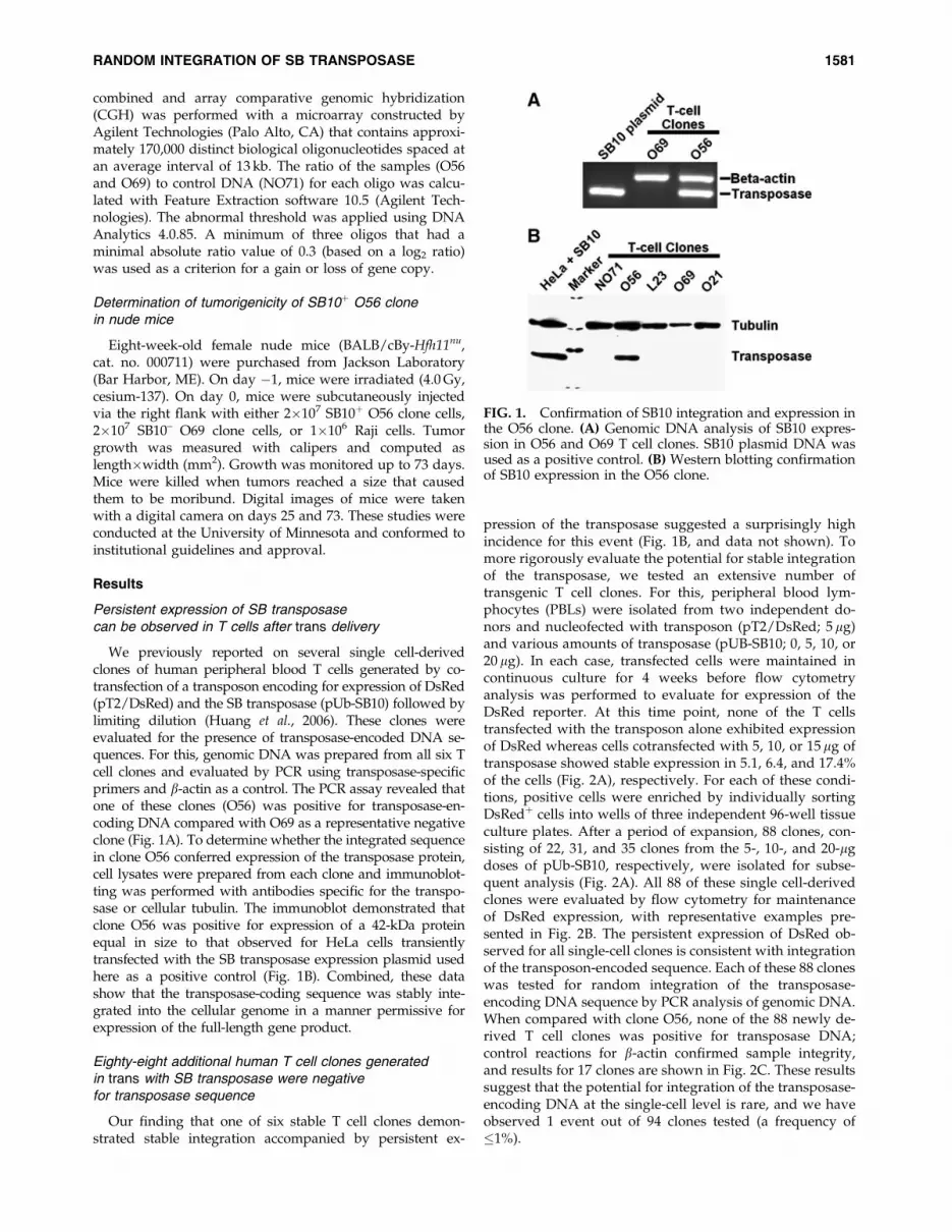
We previously reported on several single cell-derivedclones of human peripheral blood T cells generated by co-transfection of a transposon encoding for expression of DsRed(pT2/DsRed) and the SB transposase (pUb-SB10) followed bylimiting dilution (Huang et al., 2006). These clones wereevaluated for the presence of transposase-encoded DNA se-quences. For this, genomic DNA was prepared from all six Tcell clones and evaluated by PCR using transposase-specificprimers and b-actin as a control. The PCR assay revealed thatone of these clones (O56) was positive for transposase-en-coding DNA compared with O69 as a representative negativeclone (Fig. 1A). To determine whether the integrated sequencein clone O56 conferred expression of the transposase protein,cell lysates were prepared from each clone and immunoblot-ting was performed with antibodies specific for the transpo-sase or cellular tubulin. The immunoblot demonstrated thatclone O56 was positive for expression of a 42-kDa proteinequal in size to that observed for HeLa cells transientlytransfected with the SB transposase expression plasmid usedhere as a positive control (Fig. 1B). Combined, these datashow that the transposase-coding sequence was stably inte-grated into the cellular genome in a manner permissive forexpression of the full-length gene product.
Eighty-eight additional human T cell clones generatedin trans with SB transposase were negativefor transposase sequence
Our finding that one of six stable T cell clones demon-strated stable integration accompanied by persistent ex-
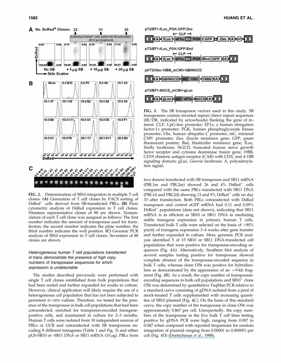
pression of the transposase suggested a surprisingly highincidence for this event (Fig. 1B, and data not shown). Tomore rigorously evaluate the potential for stable integrationof the transposase, we tested an extensive number oftransgenic T cell clones. For this, peripheral blood lym-phocytes (PBLs) were isolated from two independent do-nors and nucleofected with transposon (pT2/DsRed; 5 mg)and various amounts of transposase (pUB-SB10; 0, 5, 10, or20 mg). In each case, transfected cells were maintained incontinuous culture for 4 weeks before flow cytometryanalysis was performed to evaluate for expression of theDsRed reporter. At this time point, none of the T cellstransfected with the transposon alone exhibited expressionof DsRed whereas cells cotransfected with 5, 10, or 15 mg oftransposase showed stable expression in 5.1, 6.4, and 17.4%of the cells (Fig. 2A), respectively. For each of these condi-tions, positive cells were enriched by individually sortingDsRedþ cells into wells of three independent 96-well tissueculture plates. After a period of expansion, 88 clones, con-sisting of 22, 31, and 35 clones from the 5-, 10-, and 20-mgdoses of pUb-SB10, respectively, were isolated for subse-quent analysis (Fig. 2A). All 88 of these single cell-derivedclones were evaluated by flow cytometry for maintenanceof DsRed expression, with representative examples pre-sented in Fig. 2B. The persistent expression of DsRed ob-served for all single-cell clones is consistent with integrationof the transposon-encoded sequence. Each of these 88 cloneswas tested for random integration of the transposase-encoding DNA sequence by PCR analysis of genomic DNA.When compared with clone O56, none of the 88 newly de-rived T cell clones was positive for transposase DNA;control reactions for b-actin confirmed sample integrity,and results for 17 clones are shown in Fig. 2C. These resultssuggest that the potential for integration of the transposase-encoding DNA at the single-cell level is rare, and we haveobserved 1 event out of 94 clones tested (a frequency of�1%).
FIG. 1. Confirmation of SB10 integration and expression inthe O56 clone. (A) Genomic DNA analysis of SB10 expres-sion in O56 and O69 T cell clones. SB10 plasmid DNA wasused as a positive control. (B) Western blotting confirmationof SB10 expression in the O56 clone.
RANDOM INTEGRATION OF SB TRANSPOSASE 1581
Heterogeneous human T cell populations transfectedin trans demonstrate the presence of high copynumbers of transposase sequences for whichexpression is undetectable
The studies described previously were performed withsingle T cell clones established from bulk populations thathad been sorted and further expanded for weeks in culture.However, clinical application will likely require the use of aheterogeneous cell population that has not been subjected topersistent in vitro culture. Therefore, we tested for the pres-ence of the transposase in bulk cell populations that had beencotransfected, enriched for transposon-encoded transgene-positive cells, and maintained in culture for 2–3 months.Human T cells were isolated from 18 independent sources ofPBLs or UCB and cotransfected with SB transposons en-coding 8 different transgenes (Table 1 and Fig. 3) and eitherpUb-SB10 or -SB11 DNA or SB11 mRNA (10 mg). PBLs from
two donors transfected with SB transposon and SB11 mRNA(PBL1m and PBL2m) showed 26 and 4% DsRedþ cellscompared with the same PBLs transfected with SB11 DNA(PBL1d and PBL2d) showing 13 and 9% DsRedþ cells on day33 after transfection. Both PBLs cotransfected with DsRedtransposon and control eGFP mRNA had 0.11 and 0.09%DsRedþ populations (data not shown), indicating that SB11mRNA is as efficient as SB10 or SB11 DNA in mediatingstable transgene expression in primary human T cells.Transfected bulk T cells were selected on the basis of >90%purity of transgene expression 3–4 weeks after gene transferand further expanded in culture. Here, genomic PCR anal-ysis identified 5 of 15 SB10 or SB11 DNA-transfected cellpopulations that were positive for transposase-encoding se-quences (Fig. 4A). Alternatively, Southern blot analysis ofseveral samples testing positive for transposase showedcomplete absence of the transposase-encoded sequence inbulk T cells, whereas clone O56 was positive for hybridiza-tion as demonstrated by the appearance of an *9-kb frag-ment (Fig. 4B). As a result, the copy number of transposase-encoding sequences in bulk cell populations and SB10þ cloneO56 was determined by quantitative TaqMan PCR relative toa standard curve consisting of gDNA isolated from a pool ofmock-treated T cells supplemented with increasing quanti-ties of SB10 plasmid (Fig. 4C). On the basis of this standardcurve, the copy number of the transposase in clone O56 wasapproximately 0.867 per cell. Unexpectedly, the copy num-bers of the transposase in the five bulk T cell lines testingpositive by gDNA PCR were high, ranging from 0.007 to0.047 when compared with reported frequencies for randomintegration of plasmid ranging from 0.00001 to 0.000001 percell (Fig. 4D) (Doetschman et al., 1988).
FIG. 2. Determination of SB10 integration in multiple T cellclones. (A) Generation of T cell clones by FACS sorting ofDsRedþ cells derived from SB-transfected PBLs. (B) Flowcytometric analysis of DsRed expression in T cell clones.Nineteen representative clones of 88 are shown. Nomen-clature of each T cell clone was assigned as follows: The firstnumber indicates the amount of transposase used for trans-fection; the second number indicates the plate number; thethird number indicates the well position. (C) Genomic PCRanalysis of SB10 expression in T cell clones. Seventeen of 88clones are shown.
FIG. 3. The SB transposon vectors used in this study. SBtransposons contain inverted repeat/direct repeat sequences(IR/DR, indicated by arrowheads) flanking the gene of in-terest. CLP, CpG-less promoter; EF1a, a human elongationfactor-1a promoter; PGK, human phosphoglycerate kinasepromoter; Ubc, human ubiquitin C promoter; mC, minimalCMV promoter; Zeo, Zeocin resistance gene; GFP, greenfluorescent protein; Bsd, blasticidin resistance gene; fLuc,firefly luciferase; NGCD, truncated human nerve growthfactor receptor and cytosine deaminase fusion gene; 19BB,CD19 chimeric antigen receptor (CAR) with CD3z and 4-1BBsignaling domain; gLuc, Gaussia luciferase; A, polyadenyla-tion signal.
1582 HUANG ET AL.
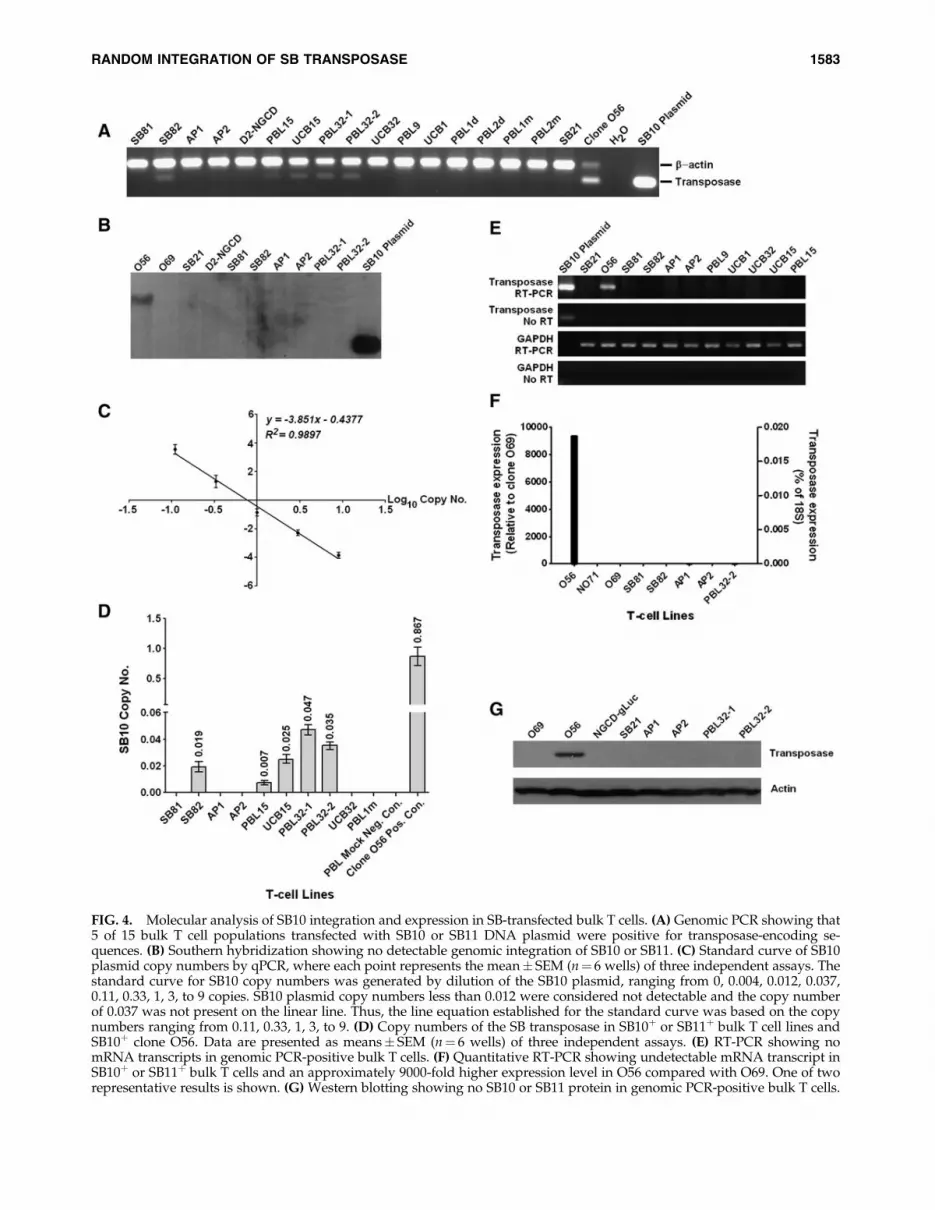
FIG. 4. Molecular analysis of SB10 integration and expression in SB-transfected bulk T cells. (A) Genomic PCR showing that5 of 15 bulk T cell populations transfected with SB10 or SB11 DNA plasmid were positive for transposase-encoding se-quences. (B) Southern hybridization showing no detectable genomic integration of SB10 or SB11. (C) Standard curve of SB10plasmid copy numbers by qPCR, where each point represents the mean� SEM (n¼ 6 wells) of three independent assays. Thestandard curve for SB10 copy numbers was generated by dilution of the SB10 plasmid, ranging from 0, 0.004, 0.012, 0.037,0.11, 0.33, 1, 3, to 9 copies. SB10 plasmid copy numbers less than 0.012 were considered not detectable and the copy numberof 0.037 was not present on the linear line. Thus, the line equation established for the standard curve was based on the copynumbers ranging from 0.11, 0.33, 1, 3, to 9. (D) Copy numbers of the SB transposase in SB10þ or SB11þ bulk T cell lines andSB10þ clone O56. Data are presented as means� SEM (n¼ 6 wells) of three independent assays. (E) RT-PCR showing nomRNA transcripts in genomic PCR-positive bulk T cells. (F) Quantitative RT-PCR showing undetectable mRNA transcript inSB10þ or SB11þ bulk T cells and an approximately 9000-fold higher expression level in O56 compared with O69. One of tworepresentative results is shown. (G) Western blotting showing no SB10 or SB11 protein in genomic PCR-positive bulk T cells.
RANDOM INTEGRATION OF SB TRANSPOSASE 1583
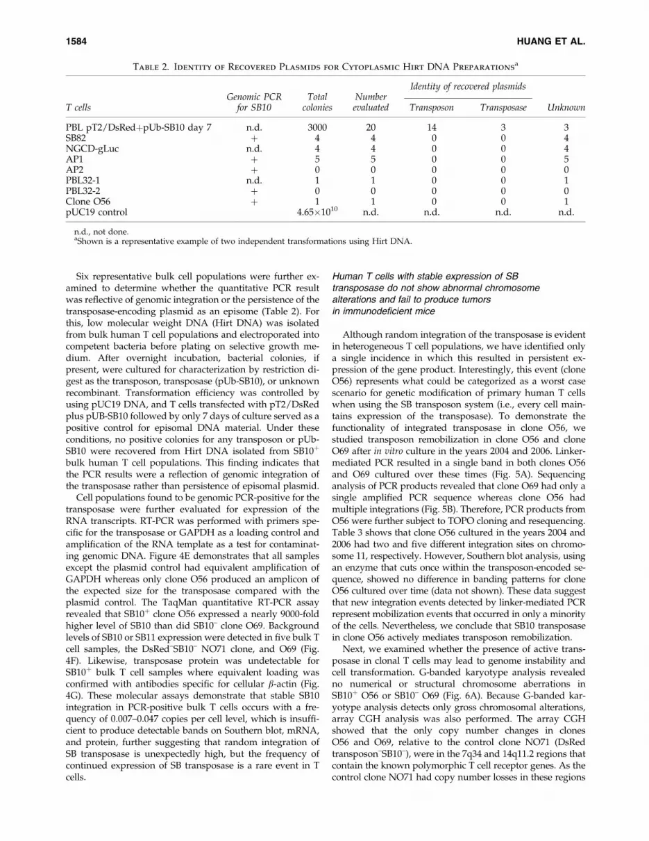
Six representative bulk cell populations were further ex-amined to determine whether the quantitative PCR resultwas reflective of genomic integration or the persistence of thetransposase-encoding plasmid as an episome (Table 2). Forthis, low molecular weight DNA (Hirt DNA) was isolatedfrom bulk human T cell populations and electroporated intocompetent bacteria before plating on selective growth me-dium. After overnight incubation, bacterial colonies, ifpresent, were cultured for characterization by restriction di-gest as the transposon, transposase (pUb-SB10), or unknownrecombinant. Transformation efficiency was controlled byusing pUC19 DNA, and T cells transfected with pT2/DsRedplus pUB-SB10 followed by only 7 days of culture served as apositive control for episomal DNA material. Under theseconditions, no positive colonies for any transposon or pUb-SB10 were recovered from Hirt DNA isolated from SB10þ
bulk human T cell populations. This finding indicates thatthe PCR results were a reflection of genomic integration ofthe transposase rather than persistence of episomal plasmid.
Cell populations found to be genomic PCR-positive for thetransposase were further evaluated for expression of theRNA transcripts. RT-PCR was performed with primers spe-cific for the transposase or GAPDH as a loading control andamplification of the RNA template as a test for contaminat-ing genomic DNA. Figure 4E demonstrates that all samplesexcept the plasmid control had equivalent amplification ofGAPDH whereas only clone O56 produced an amplicon ofthe expected size for the transposase compared with theplasmid control. The TaqMan quantitative RT-PCR assayrevealed that SB10þ clone O56 expressed a nearly 9000-foldhigher level of SB10 than did SB10– clone O69. Backgroundlevels of SB10 or SB11 expression were detected in five bulk Tcell samples, the DsRed–SB10– NO71 clone, and O69 (Fig.4F). Likewise, transposase protein was undetectable forSB10þ bulk T cell samples where equivalent loading wasconfirmed with antibodies specific for cellular b-actin (Fig.4G). These molecular assays demonstrate that stable SB10integration in PCR-positive bulk T cells occurs with a fre-quency of 0.007–0.047 copies per cell level, which is insuffi-cient to produce detectable bands on Southern blot, mRNA,and protein, further suggesting that random integration ofSB transposase is unexpectedly high, but the frequency ofcontinued expression of SB transposase is a rare event in Tcells.
Human T cells with stable expression of SBtransposase do not show abnormal chromosomealterations and fail to produce tumorsin immunodeficient mice
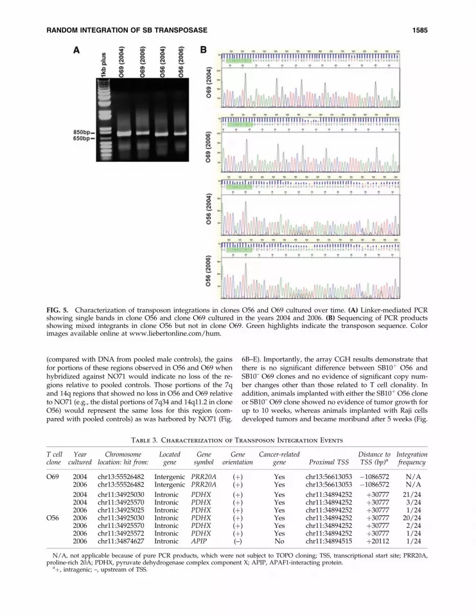
Although random integration of the transposase is evidentin heterogeneous T cell populations, we have identified onlya single incidence in which this resulted in persistent ex-pression of the gene product. Interestingly, this event (cloneO56) represents what could be categorized as a worst casescenario for genetic modification of primary human T cellswhen using the SB transposon system (i.e., every cell main-tains expression of the transposase). To demonstrate thefunctionality of integrated transposase in clone O56, westudied transposon remobilization in clone O56 and cloneO69 after in vitro culture in the years 2004 and 2006. Linker-mediated PCR resulted in a single band in both clones O56and O69 cultured over these times (Fig. 5A). Sequencinganalysis of PCR products revealed that clone O69 had only asingle amplified PCR sequence whereas clone O56 hadmultiple integrations (Fig. 5B). Therefore, PCR products fromO56 were further subject to TOPO cloning and resequencing.Table 3 shows that clone O56 cultured in the years 2004 and2006 had two and five different integration sites on chromo-some 11, respectively. However, Southern blot analysis, usingan enzyme that cuts once within the transposon-encoded se-quence, showed no difference in banding patterns for cloneO56 cultured over time (data not shown). These data suggestthat new integration events detected by linker-mediated PCRrepresent mobilization events that occurred in only a minorityof the cells. Nevertheless, we conclude that SB10 transposasein clone O56 actively mediates transposon remobilization.
Next, we examined whether the presence of active trans-posase in clonal T cells may lead to genome instability andcell transformation. G-banded karyotype analysis revealedno numerical or structural chromosome aberrations inSB10þ O56 or SB10– O69 (Fig. 6A). Because G-banded kar-yotype analysis detects only gross chromosomal alterations,array CGH analysis was also performed. The array CGHshowed that the only copy number changes in clonesO56 and O69, relative to the control clone NO71 (DsRedtransposon–SB10–), were in the 7q34 and 14q11.2 regions thatcontain the known polymorphic T cell receptor genes. As thecontrol clone NO71 had copy number losses in these regions
Table 2. Identity of Recovered Plasmids for Cytoplasmic Hirt DNA Preparationsa
T cellsGenomic PCR
for SB10Total
coloniesNumberevaluated
Identity of recovered plasmids
Transposon Transposase Unknown
PBL pT2/DsRedþpUb-SB10 day 7 n.d. 3000 20 14 3 3SB82 þ 4 4 0 0 4NGCD-gLuc n.d. 4 4 0 0 4AP1 þ 5 5 0 0 5AP2 þ 0 0 0 0 0PBL32-1 n.d. 1 1 0 0 1PBL32-2 þ 0 0 0 0 0Clone O56 þ 1 1 0 0 1pUC19 control 4.65�1010 n.d. n.d. n.d. n.d.
n.d., not done.aShown is a representative example of two independent transformations using Hirt DNA.
1584 HUANG ET AL.
(compared with DNA from pooled male controls), the gainsfor portions of these regions observed in O56 and O69 whenhybridized against NO71 would indicate no loss of the re-gions relative to pooled controls. Those portions of the 7qand 14q regions that showed no loss in O56 and O69 relativeto NO71 (e.g., the distal portions of 7q34 and 14q11.2 in cloneO56) would represent the same loss for this region (com-pared with pooled controls) as was harbored by NO71 (Fig.
6B–E). Importantly, the array CGH results demonstrate thatthere is no significant difference between SB10þ O56 andSB10– O69 clones and no evidence of significant copy num-ber changes other than those related to T cell clonality. Inaddition, animals implanted with either the SB10þ O56 cloneor SB10– O69 clone showed no evidence of tumor growth forup to 10 weeks, whereas animals implanted with Raji cellsdeveloped tumors and became moribund after 5 weeks (Fig.
FIG. 5. Characterization of transposon integrations in clones O56 and O69 cultured over time. (A) Linker-mediated PCRshowing single bands in clone O56 and clone O69 cultured in the years 2004 and 2006. (B) Sequencing of PCR productsshowing mixed integrants in clone O56 but not in clone O69. Green highlights indicate the transposon sequence. Colorimages available online at www.liebertonline.com/hum.
Table 3. Characterization of Transposon Integration Events
T cellclone
Yearcultured
Chromosomelocation: hit from:
Locatedgene
Genesymbol
Geneorientation
Cancer-relatedgene Proximal TSS
Distance toTSS (bp)a
Integrationfrequency
O69 2004 chr13:55526482 Intergenic PRR20A (þ) Yes chr13:56613053 �1086572 N/A2006 chr13:55526482 Intergenic PRR20A (þ) Yes chr13:56613053 �1086572 N/A
2004 chr11:34925030 Intronic PDHX (þ) Yes chr11:34894252 þ30777 21/242004 chr11:34925570 Intronic PDHX (þ) Yes chr11:34894252 þ30777 3/242006 chr11:34925025 Intronic PDHX (þ) Yes chr11:34894252 þ30777 1/24
O56 2006 chr11:34925030 Intronic PDHX (þ) Yes chr11:34894252 þ30777 20/242006 chr11:34925570 Intronic PDHX (þ) Yes chr11:34894252 þ30777 2/242006 chr11:34925572 Intronic PDHX (þ) Yes chr11:34894252 þ30777 1/242006 chr11:34874627 Intronic APIP (–) No chr11:34894515 þ20112 1/24
N/A, not applicable because of pure PCR products, which were not subject to TOPO cloning; TSS, transcriptional start site; PRR20A,proline-rich 20A; PDHX, pyruvate dehydrogenase complex component X; APIP, APAF1-interacting protein.
aþ, intragenic; –, upstream of TSS.
RANDOM INTEGRATION OF SB TRANSPOSASE 1585

FIG. 6. Cytogenetic and array CGH analyses of O56 clone.(A) G-band analysis of SB10þ O56 and SB10– O69 clonesshowing a normal 46,XY male karyotype. (B) Array CGHshowing losses at 7q34 and 14q11.2 in the control cloneNO71 (compared with pooled male control specimens) andgains in the same regions in clones O56 and O69 (comparedwith NO71). (C) Array CGH showing copy number gainsagainst control clone NO71 at 7q34 and 14q11.2 regions inclone O56. (D) Array CGH showing copy number gainsagainst NO71 at 7q34 and 14q11.2 regions in clone O69. (E)The control NO71, compared with the pooled control DNA,showed two regions of loss encompassing approximately 346and 155 kb within 7q34, respectively. The start point for theloss of the 346-kb region was estimated at bp 141663456 andthe stop point at bp 142009059. The ratio value of �1 wasconsistent with a deletion of this region on one chromosome7 allele. This region contains TCRBV genes. The start pointfor the loss of the 155-kb region was estimated at 142021348and the stop point at 142176133. The ratio for this region fellbetween �2 and �4, consistent with a homozygous loss.Mapped to this region are other TCRB-related genes. Tworegions of loss within 14q11.2, encompassing 128 and 633 kb,respectively, were noted. The start point for the 128-kb re-gion was estimated at 21285126 and the stop point at2142207. Mapped to this region are TCRAV-related genes.The start point for the 633-kb region was estimated at21420105 and the stop point at 22052858. Mapped to thisregion are also TCRAV-related genes. Clone O56 shows aratio of 1.0 for the proximal part of 7q34 and a ratio of 0 forthe remaining portion of this 7q34 region. As the controlclone NO71 had deletion in these regions, this would indi-cate that O56 had no loss for the proximal region, and hadthe same loss as the control NO71 for the distal portion ofthis region. Clone O69 had a small gain for a portion of the7q34 region, likely indicating a mixture of cells that had thesame loss as the control NO71 and those that had no loss.Clone O69 had a ratio of approximately 2.0 for the distalportion of 7q34, indicating no loss for this region. For the14q11.2 region, the ratios for O56 and O69 were generatedusing the control T cell clone NO71, which showed a ratio of�3 for this region. Thus, the þ0.3 ratio for O56 indicates thatthere is no loss for the proximal portion of 14q11.2 region inthese cells; the remainder of this region shows a profileconsistent with the presence of some cells with loss, andothers not. The findings are similar for O69, which shows noloss for the very proximal region, and some cells with loss forthe more distal portion of the region. Of note, for NO71compared with pooled controls, additional gains were seenin the near centromeric regions of chromosomes 7, 8, and 15,and in the pseudoautosomal regions of Xp and Yp. Thesegains are among the benign copy number variants that arewell documented in published databases to occur in healthycontrols, and are interpreted as germline variants of no rel-evance to the present experiments. Color images availableonline at www.liebertonline.com/hum.
1586 HUANG ET AL.

7A and B). These data demonstrate that human T cells withSB10 stably integrated remain karyotypically normal and donot form tumors in mice.
Discussion
Three important observations concerning random inte-gration of SB transposase on trans delivery in human pri-mary T cells appear in this paper. First, the frequency ofrandom integration of SB transposase resulting in expressionof SB10 or SB11 protein accounts for about 1% (1 of 94 T cellclones analyzed). Second, although random integration ofthe SB transposase was observed in approximately 33% ofbulk T cells and the copy numbers were unexpectedly high (5of 15 bulk cells tested with 0.007–0.047 copies of the SBtransposase), no transposase mRNA transcript or proteincould be detected. Third, an SB10þ T cell clone (O56; 0.867copies) with stable transposon encoded transgene expressionshows active transposon remobilization but no apparentnumeric or structural chromosome alterations and no tumorgrowth in nude mice. Thus, our results suggest that randomintegration of SB transposase is high but that stable inte-gration of functional SB transposase-encoding sequences islow in human T cells, warranting caution in the use of SBtransposase DNA for human T cell gene therapy.
Transient expression of the transposase is a requirement ofthe transposition process. However, persistent expressionsuggests a situation in which the transposon could becomeremobilized after the initial insertion event. This method hasproven quite effective for discovering novel cancer-causinggenes in both normal and tumor-prone mice (Carlson et al.,2005; Collier et al., 2005; Dupuy et al., 2005; Su et al., 2008;Keng et al., 2009; Rahrmann et al., 2009; Starr et al., 2009). Inthese studies, mice were engineered for constitutive or tissue-
restricted expression of the transposase to mobilize transpo-sons from a resident concatemer. Some of the new integrationevents induce expression of an oncogene or inactivation of atumor suppressor gene. Common to several of these studies isthe use of animals demonstrating constitutive or induced ex-pression of the transposase as a control cohort for transposase-mediated mutagenesis (Carlson et al., 2005; Collier et al., 2005;Dupuy et al., 2005; Su et al., 2008; Keng et al., 2009; Rahrmannet al., 2009; Starr et al., 2009). Only when animals are alsopredisposed to cancer, that is, deficient in the tumor sup-pressor genes encoding p19Arf or p53, has there been dem-onstrated morbidity (Collier et al., 2005; Keng et al., 2009). Thiswas not observed for normal mice, where the most extensivestudies were carried out for more than 1 year (Collier et al.,2005; Keng et al., 2009).
Additional studies intending to model defined somatictumorigenesis have coinfused normal mice (Wiesner et al.,2009) or tumor-predisposed mice (Carlson et al., 2005) withtransposons harboring an activated oncogene and transpo-sase under delivery conditions that can target a specific tis-sue. After SB-mediated gene transfer of an activated versionof the N-RAS oncogene to the liver of Arf-deficient mice,Carlson and colleagues identified three independent tumorswhere the codelivered transposase had become randomlyintegrated. Western blot confirmed that one of these tumorsdemonstrated constitutive expression of the transposase.
Using nucleofection as a method for achieving ex vivo genetransfer into human T cells, we detected SB10 functionalintegration in one of the first 6 clones but not in the addi-tional 88 clones. In each case, we used similar ratios oftransposon- and transposase-encoding plasmids. The onlydifference between these two experiments is the method usedto generate T cell clones, with limiting dilution and FACS-based cell sorting used in the first and second experiments,
FIG. 7. The O56 clone did not induce tumor formation in nude mice. (A) Tumor growth in nude mice; mice injected with theSB10þ O56 clone showed no tumor growth on day 73. In Raji cell-injected mice, one mouse (#1) showed initial tumor growthand then the tumor regressed spontaneously. (B) Tumor volume versus days after injection. Color images available online atwww.liebertonline.com/hum.
RANDOM INTEGRATION OF SB TRANSPOSASE 1587

respectively. It is possible but highly unlikely that cell sortingdisfavors the growth of clones with integrated transposasecompared with limiting dilution.
We also detected SB transposase integration in 5 of 15 bulkT cell populations stably expressing transposon-encodedtransgenes, using genomic PCR. Unexpectedly, five genomicPCR-positive bulk T cell populations demonstrated approx-imately 700–4700 times higher frequencies of random trans-posase integration than previously documented levels forplasmid random integration (0.7–4.7 vs. 0.001–0.0001%)(Doetschman et al., 1988). This higher than expected level ofrandom integration of SB transposase could be attributed tothe following: First, all bulk T cells were enriched for trans-poson-encoded transgenes by FACS, immunomagnetic iso-lation, or drug selection (Table 1). Thus, transposaseexpression in every cell is required to achieve transposition.Second, the amount of SB transposase for trans delivery (10–15 mg of transposase and 5 mg of transposon per 5�106 Tcells) was high. Third, one of the intrinsic properties of the SBtransposase is to ‘‘cut’’ DNA, suggesting a potential for in-creased frequency of random integration. Fourth, the methodof nucleofection used here to achieve T cell gene transfer mayhave an increased potential compared with alternative genetransfer methods (e.g., calcium phosphate coprecipitation orliposome-mediated transfection) for promoting random in-tegration. Fifth, the frequency of random integration is celltype specific. For example, McIvor and colleagues reportedthat NIH3T3 cells had a six times higher stable baselinetransfection frequency relative to HeLa cells (0.15 vs. 0.025%,respectively) as defined by transfection with pT2/neo (SBtransposon encoding the neomycin resistance gene) alone(Converse et al., 2005). In similar studies using an unrelatednonviral integrating vector system, the Calos group alsodemonstrated an *31-fold higher integration frequency inNIH3T3 cells than in 293 cells (0.25 vs. 0.008%) as defined bytransfection with bacteriophage FC31 cargo-containing vec-tor without integrase (Thyagarajan et al., 2010). By compar-ison, the frequency of SB transposase random integration inhuman T cells appears similar (0.7–0.47 vs. 0.15–0.25%) tothese results reported for NIH3T3 cells.
Even with these unexpectedly high levels of random in-tegration of transposase-encoding sequences, all bulk T cellsfailed to express detectable levels of SB10 or SB11 mRNAand protein. This result can be explained in that randomintegration of the codelivered transposase occurs in only asmall percentage of cells that can be detected only by sen-sitive PCR and TaqMan qPCR amplification, not by South-ern hybridization. Random integration of the transposase inthis manner may not favor the production of detectabletranscripts and protein. Our data clearly demonstrate thatSB10 or SB11 functional integration is a low-frequencyevent. With respect to the tumorigenic potential of trans-posase-positive T cells demonstrating constitutive expres-sion of a transposon-encoded transgene (e.g., the O56 clone),we observed no tumor formation in nude mice 73 days aftercell implantation. However, we cannot exclude the possi-bility that nude mice may not be the best model for asses-sing human T cell transformation. In addition, it may benecessary to monitor the potential for tumor developmentfor a longer period of time to ensure tumor negativity be-cause it is possible that tumor formation may occur at amuch later time point.
From our results, it can be derived that the frequency offunctional SB transposase integration is a low but viable pos-sibility. However, this does not exclude the potential use ofDNA as a source for transposase. Although we were able toidentify a single clone that was positive for SB10 transposaseintegration with relatively low copy number (0.867 copies),protein production, and active transposon remobilization, ad-ditional studies confirmed that there was no resultant tumorgrowth in mouse models or alterations to the chromosomalstructure of the genomic DNA, even by array CGH analysis. Itis evident that integration of the transposase can lead to localtransposon ‘‘hopping’’ as shown in only a minority of cloneO56 populations. The transposase used in previous studies foreliciting insertional mutagenesis was engineered for inducedactivity and may not represent a risk when using the SBtransposon system to mediate gene transfer.
The advantages of a DNA transposase system are plentifulwhen compared with mRNA as a source of SB transposase inhuman T cell engineering: low cost, simple production,similar efficiency, and easy handling in transfection. How-ever, it may be better to use mRNA as a source of SBtransposase or DNA with a coexpressed suicide gene forhematopoietic stem cell gene transfer as these cells are highlysubject to insertional mutagenesis (Hacein-Bey-Abina et al.,2003; Mates et al., 2009; Sumiyoshi et al., 2009; Xue et al.,2009). Supporting this alternative, we observed similar effi-ciencies of stable gene transfer for two independent donorT cell populations when in vitro-transcribed transposase-encoding mRNA was compared with plasmid. Ultimately,our results warn of potential risks involved in the mediationof gene transfer by the SB transposon system, which wasapproved in 2008 for the ‘‘first-in-human’’ gene therapy trialby the Recombinant DNA Advisory Committee (RAC)(VandenDriessche and Chuah, 2009). In this trial, T cellsgenetically engineered by the SB transposon system will beadoptively transferred into patients with CD19þ B cell ma-lignancies (Singh et al., 2008). Because of the high frequencyof transposase random integration in human T cells asdemonstrated in this study, use of SB transposase mRNA orDNA along with a suicide gene should be considered.
Acknowledgments
The authors thank Dr. David Largaespada (University ofMinnesota, Minneapolis, MN) for helpful discussion aboutarray CGH data and local ‘‘hopping,’’ Dr. R. Scott McIvor(University of Minnesota) for critical reading of this man-uscript, Dr. Perry Hackett (University of Minnesota) forindependent confirmation of copy number calculations,Dr. Betsy Hirsch (University of Minnesota) for interpretationof the G-banding and array CGH data, Dr. Mingqiang Ren(Medical College of Georgia, Augusta, GA) for helpful dis-cussion about copy number determination by qPCR andstandard curve, and Dr. Nikunj Somia (University of Min-nesota, Minneapolis, MN) for providing the blasticidin–GFPfusion sequence. The authors also thank Dr. San Ming Wangand Dr. Yeong C. Kim (Northwestern University, Evanston,IL), and Dr. Zheng Jin Tu (University of Minnesota, Min-neapolis, MN) for help in bioinformatic analyses of integra-tion sites. K.H. was a recipient of an Undergraduate ResearchOpportunity Program (UROP) Award, University of Min-nesota, and of an American Society of Hematology Research
1588 HUANG ET AL.

Trainee Award. This work was supported by grants from theAlliance for Cancer Gene Therapy, the Gabrielle’s AngelFoundation for Cancer Research, the Sidney Kimmel Foun-dation for Cancer Research Kimmel Scholar Program, theUniversity of Minnesota (an AHC Translational Researchgrant), the University of Minnesota Medical School Dean’sCommitment, and in part by the Children’s Cancer ResearchFund in Minneapolis (X.Z.). The cytogenetic and array CGHanalyses were performed in the Cytogenetics Core Labora-tory at the University of Minnesota with support from theComprehensive Cancer Center (NIH Grant P30 CA077598-09). K.H.’s current address: Medical College of Wisconsin,8701 Watertown Park Road, Milwaukee, WI 53226.
Contribution Statement
X.H. designed, performed, and supervised the research,analyzed the data, and wrote the paper. K.H. performed theresearch, analyzed the data, and wrote the paper. M.W.performed the research, analyzed the data, and wrote thepaper. H.G. and C.L. performed the research and analyzedthe data. A.W. provided critical reagents and wrote the pa-per. X.Z. oversaw the research and wrote the paper.
Author Disclosure Statement
The authors declare no competitive financial interests.
References
Alwin, S., Gere, M.B., Guhl, E., Effertz, K., Barbas, C.F., III, Segal,D.J., Weitzman, M.D., and Cathomen, T. (2005). Custom zinc-finger nucleases for use in human cells. Mol. Ther. 12, 610–617.
Carlson, C.M., Frandsen, J.L., Kirchhof, N., McIvor, R.S., andLargaespada, D.A. (2005). Somatic integration of an oncogene-harboring Sleeping Beauty transposon models liver tumor de-velopment in the mouse. Proc. Natl. Acad. Sci. U.S.A. 102,17059–17064.
Collier, L.S., Carlson, C.M., Ravimohan, S., Dupuy, A.J., andLargaespada, D.A. (2005). Cancer gene discovery in solid tu-mors using transposon-based somatic mutagenesis in themouse. Nature 436, 272–276.
Converse, A.D., Belur, L.R., Gori, J., Liu, G., Amaya, F., Aguilar-Cordova, E., Hackett, P.B., and McIvor, R.S. (2005). Counter-selection and co-delivery of transposon and transposasefunctions for Sleeping Beauty-mediated transposition in cul-tured mammalian cells. Biosci. Rep. 24, 577–594.
de Wit, T., Drabek, D., and Grosveld, F. (1998). Microinjection ofCre recombinase RNA induces site-specific recombination of atransgene in mouse oocytes. Nucleic Acids Res. 26, 676–678.
Doetschman, T., Maeda, N., and Smithies, O. (1988). Targetedmutation of the Hprt gene in mouse embryonic stem cells.Proc. Natl. Acad. Sci. U.S.A. 85, 8583–8587.
Dupuy, A,J,, Akagi, K., Largaespada, D.A., Copeland, N.G., andJenkins, N.A. (2005). Mammalian mutagenesis using a highlymobile somatic Sleeping Beauty transposon system. Nature 436,221–226.
Galla, M., Will, E., Kraunus, J., Chen, L., and Baum, C. (2004).Retroviral pseudotransduction for targeted cell manipulation.Mol. Cell 16, 309–315.
Hacein-Bey-Abina, S., Von Kalle, C., Schmidt, M., McCormack,M.P., Wulffraat, N., Leboulch, P., Lim, A., Osborne, C.S.,Pawliuk, R., Morillon, E., Sorensen, R., Forster, A., Fraser, P.,Cohen, J.I., de Saint Basile, G., Alexander, I., Wintergerst, U.,
Frebourg, T., Aurias, A., Stoppa-Lyonnet, D., Romana, S.,Radford-Weiss, I., Gross, F., Valensi, F., Delabesse, E., Ma-cintyre, E., Sigaux, F., Soulier, J., Leiva, L.E., Wissler, M., Prinz,C., Rabbitts, T.H., Le Deist, F., Fischer, A., and Cavazzana-Calvo, M. (2003). LMO2-associated clonal T cell proliferationin two patients after gene therapy for SCID-X1. Science 302,415–419.
Hackett, P.B., Ekker, S.C., Largaespada, D.A., and McIvor, R.S.(2005). Sleeping Beauty transposon-mediated gene therapy forprolonged expression. Adv. Genet. 54, 189–232.
Hirt, B (1967). Selective extraction of polyoma DNA from in-fected mouse cell cultures. J. Mol. Biol. 26, 365–369.
Huang, X., Wilber, A.C., Bao, L., Tuong, D., Tolar, J., Orchard,P.J., Levine, B.L., June, C.H., McIvor, R.S., Blazar, B.R., andZhou, X. (2006). Stable gene transfer and expression in humanprimary T cells by the Sleeping Beauty transposon system.Blood 107, 483–491.
Huang, X., Guo, H., Kang, J., Choi, S., Zhou, T.C., Tammana, S.,Lees, C.J., Li, Z.Z., Milone, M., Levine, B.L., Tolar, J., June,C.H., McIvor, R.S., Wagner, J.E., Blazar, B.R., and Zhou, X.(2008). Sleeping Beauty transposon-mediated engineering ofhuman primary T cells for therapy of CD19þ lymphoid ma-lignancies. Mol. Ther. 16, 580–589.
Huang, X., Wilber, A, McIvor, R.S., and Zhou, X. (2009). DNAtransposons for modification of human primary T lympho-cytes. Methods Mol. Biol. 506, 115–126.
Huang, X., Guo, H., Tammana, S., Jung, Y.C., Mellgren, E., Bassi,P., Cao, Q., Tu, Z.J., Kim, Y.C., Ekker, S.C., Wu, X., Wang,S.M., and Zhou, X. (2010). Gene transfer efficiency and genome-wide integration profiling of Sleeping Beauty, Tol2, and PiggyBactransposons in human primary T cells. Mol. Ther. 18, 1803–1813.
Ivics, Z., Hackett, P.B., Plsterk, R.H., and Izsvak, Z. (1997).Molecular reconstruction of Sleeping Beauty, a Tc1-like trans-poson from fish, and its transposition in human cells. Cell 91,501–510.
Izsvak, Z., and Ivics, Z. (2004). Sleeping Beauty transposon:Biology and applications for molecular therapy. Mol. Ther. 9,147–156.
Keng, V.W., Villanueva, A., Chiang, D.Y., Dupuy, A.J., Ryan,B.J., Matise, I, Silverstein, K.A.T., Sarver, A., Starr, T.K., Akagi,E., Tessarollo, L., Collier, L.S., Powers, S., Lowe, S.W., Jenkins,N.A., Copeland, N.G., Llovet, J.M., and Largaespada, D.A.(2009). A conditional transposon-based insertional mutagen-esis screen for genes associated with mouse hepatocellularcarcinoma. Nat. Biotechnol. 27, 264–274.
Levine, B.L., Bernstein, W.B., Aronson, N.E., Schlienger, K.,Cotte, J., Perfetto, S., Humphries, M.J., Ratto-Kim, S., Birx,D.L., Steffens, C., Landay, A., Carroll, R.G., and June C.H.(2002). Adoptive transfer of costimulated CD4þ T cells inducesexpansion of peripheral T cells and decreased CCR5 expres-sion in HIV infection. Nat. Med. 8, 47–53.
Loonstra, A., Vooijs, M., Beverloo, H.B., Allak, B.A., van Drunen,E., Kanaar, R., Berns, A., and Jonkers, J. (2001). Growthinhibition and DNA damage induced by Cre recombinasein mammalian cells. Proc. Natl. Acad. Sci. U.S.A. 98, 9209–9214.
Mates, L., Chuah, M.K., Belay, E., Jerchow, B., Manoj, N.,Acosta-Sanchez, A., Grzela, D.P., Schmitt, A., Becker, K.,Matrai, J., Ma, L., Samara-Kuko, E., Gysemans, C., Pry-putniewicz, D., Miskey, C., Fletcher, B., Vandendriessche, T.,Ivics, Z., and Izsvak, Z. (2009). Molecular evolution of a novelhyperactive Sleeping Beauty transposase enables robust stablegene transfer in vertebrates. Nat. Genet. 41, 753–761.
RANDOM INTEGRATION OF SB TRANSPOSASE 1589
Pfeifer, A., Brandon, E.P., Kootstra, N., Gage, F.H., and Verma,I.M. (2001). Delivery of the Cre recombinase by a self-deletinglentiviral vector: Efficient gene targeting in vivo. Proc. Natl.Acad. Sci. U.S.A. 98, 11450–11455.
Ponsaerts, P., Brown, J.P., Van den Plas, D., Van den Eeden, L.,Van Bockstaele, D.R., Jorens, P.G., Van Tendeloo, V.F., Mer-regaert, J., Singh, P.B., and Berneman, Z.N. (2004). MessengerRNA electroporation is highly efficient in mouse embryonicstem cells: Successful FLPe- and Cre-mediated recombination.Gene Ther. 11, 1606–1610.
Rahrmann, E.P., Collier, L.S., Knutson, T.P., Doyal, M.E., Kuslak,S.L., Green, L.E., Malinowski, R.L., Roethe, L., Akagi, K.,Waknitz, M., Huang, W., Largaespada, D.A., and Marker,P.C. (2009). Identification of PDE4D as a proliferationpromoting factor in prostate cancer using a Sleeping Beautytransposon-based somatic mutagenesis screen. Cancer Res. 69,4388–4397.
Silver, D.P., and Livingston, D.M. (2001). Self-excising retroviralvectors encoding the Cre recombinase overcome Cre-mediatedcellular toxicity. Mol. Cell 8, 233–243.
Starr, T.K., Allaei, R., Silverstein, K.A., Staggs, R.A., Sarver, A.L.,Bergemann, T.L., Gupta, M., O’Sullivan, M.G., Matise, I.,Dupuy, A.J., Collier, L.S., Powers, S., Oberg, A.L., Asmann,Y.W., Thibodeau, S.N., Tessarollo, L., Copeland, N.G., Jenkins,N.A., Cormier, R.T., and Largaespada, D.A. (2009). A transposon-based genetic screen in mice identifies genes altered in colo-rectal cancer. Science 323, 1747–1750.
Su, Q., Prosser, H.M., Campos, L.S., Ortiz, M., Nakamura, T.,Warren, M., Dupuy, A.J., Jenkins, N.A., Copeland, N.G.,Bradley, A., and Liu P. (2008). A DNA transposon-based ap-proach to validate oncogenic mutations in the mouse. Proc.Natl., Acad. Sci. U.S.A. 105, 19904–19909.
Sumiyoshi, T., Holt, N.G., Hollis, R.P., Ge, S., Cannon, P.M.,Crooks, G.M., and Kohn, D.B. (2009). Stable transgene ex-pression in primitive human CD34þ hematopoietic stem/progenitor cells, using the Sleeping Beauty transposon system.Hum. Gene Ther. 20, 1607–1626.
Thyagarajan, B., Olivares, E.C., Hollis, P., Ginsburg, D.S., andCalos, M.P. (2001). Site-specific genomic integration in mam-malian cells mediated by phage FC31 integrase. Mol. Cell.Biol. 21, 3926–3934.
VandenDriessche, T., and Chuah, M.K.L. (2009). Moving genetherapy forward with mobile DNA. Hum. Gene Ther. 20,1559–1561.
Van den Plas, D., Ponsaerts, P., Van Tendeloo, V., Van Bockstaele,D.R., Berneman, Z.N., and Merregaert, J. (2003). Efficient removalof LoxP-flanked genes by electroporation of Cre-recombinasemRNA. Biochem. Biophys. Res. Commun. 305, 10–15.
Wiesner, S.M., Decker, S.A., Larson, J.D., Erickson, K., Forster,C., Gallardo, J.L., Long, C., Demorest, Z.L., Zamora, E.A.,Low, W.C., SantaCruz, K., Largaespada, D.A., and Ohlfest,J.R. (2009). De novo induction of genetically engineered braintumors in mice using plasmid DNA. Cancer Res. 15, 431–439.
Wilber, A., Frandsen, J.L., Geurts, J.L., Largaespada, D.A.,Hackett, P.B., and McIvor, R.S. (2006). RNA as a source oftransposase for Sleeping Beauty-mediated gene insertion andexpression in somatic cells and tissues. Mol. Ther. 13, 625–630.
Wilber, A., Wangensteen, K.J., Chen, Y., Zhuo, L., Frandsen, J.L.,Bell, J.B., Chen, Z.J., Ekker, S.C., McIvor, R.S., and Wang, X.(2007). Messenger RNA as a source of transposase for SleepingBeauty transposon-mediated correction of hereditary tyro-sinemia type I. Mol. Ther. 15, 1280–1287.
Xue, X., Huang, X., Nodland, S.E., Mates, L., Ma, L., Ivics, Z.,Izsvak, Z., Lebien T.W., McIvor, R.S., Wagner, J.E., and Zhou,X. (2009). Stable gene transfer and expression in cord blood-derived CD34þ hematopoietic stem and progenitor cells by ahyperactive Sleeping Beauty transposon system. Blood 114,1319–1330.
Address correspondence to:Dr. Xianzheng Zhou
Masonic Cancer CenterUniversity of Minnesota
MMC 366, 420 Delaware StreetMinneapolis, MN 55455
E-mail: [email protected].
Received for publication July 28, 2009;accepted after revision June 8, 2010.
Published online: October 12, 2010.
1590 HUANG ET AL.


