Embed Size (px)
Citation preview
Georgia State University Georgia State University
ScholarWorks @ Georgia State University ScholarWorks @ Georgia State University
Biology Dissertations Department of Biology
Summer 6-18-2012
Tyrosine Phosphorylation of p68 RNA Helicase Promotes Tyrosine Phosphorylation of p68 RNA Helicase Promotes
Metastasis in Colon Cancer Progression Metastasis in Colon Cancer Progression
Chia Yi Liu Georgia State University
Follow this and additional works at: https://scholarworks.gsu.edu/biology_diss
Recommended Citation Recommended Citation Liu, Chia Yi, "Tyrosine Phosphorylation of p68 RNA Helicase Promotes Metastasis in Colon Cancer Progression." Dissertation, Georgia State University, 2012. doi: https://doi.org/10.57709/3002852
This Dissertation is brought to you for free and open access by the Department of Biology at ScholarWorks @ Georgia State University. It has been accepted for inclusion in Biology Dissertations by an authorized administrator of ScholarWorks @ Georgia State University. For more information, please contact [email protected].
TYROSINE PHOSPHORYLATION OF p68 RNA HELICASE PROMOTES METASTASIS
IN COLON CANCER PROGRESSION
by
CHIA YI LIU
Under the Direction of Zhi-Ren Liu
ABSTRACT
The initiation of cancer metastasis usually requires Epithelial-Mesenchymal Transition (EMT),
by which tumor cells lose cell-cell interactions and gain the ability of migration and invasion.
Previous study demonstrated that p68 RNA helicase, a prototypical member of the DEAD-box
RNA helicases, functions as a mediator to promote platelet-derived growth factor (PDGF)-
induced EMT through facilitating nuclear translocation of β-catenin in colon cancer cells. In this
context, p68 RNA helicase was found to be phosphorylated at the tyrosine 593 residue (referred
as phosphor-p68) by c-Abl kinase, and this phosphorylation is required for the activation of β-
catenin signaling and the consequent EMT. The phosphor-p68 RNA helicase-mediated EMT was
characterized by the repression of an epithelial marker, E-cadherin, and the upregulation of a
mesenchymal marker, Vimentin. E-cadherin, a major cell-cell adhesion molecule that is involved
in the formation of adherens junctions, has been shown to sequester β-catenin at the cell
membrane and thus inhibit its transcriptional activity. The functional loss of E-cadherin is the
fundamental event of EMT. Despite the role of phosphor-p68 RNA helicase in regulating nuclear
translocation of β-catenin, whether phosphor-p68 is involved in the regulation of E-cadherin
remains unknown. Here, our data indicated that phosphor-p68 RNA helicase initiated EMT by
transcriptional upregulation of Snail1, a master transcriptional repressor of E-cadherin. The data
suggest that phosphor-p68 RNA helicase displaced HDAC1 from the chromatin remodeling
MBD3:Mi-2/NuRD complex at the Snail1 promoter, thereby activating the transcription of
Snail1. In the xenograft tumor model, abolishing the phosphorylation of p68 RNA helicase by
the expression of Y593F mutant resulted in a significant reduction of metastatic potential in
human colon cancer cells. Analyses in the colon cancer tissues also revealed that the tyrosine 593
phosphorylation level of p68 RNA helicase is substantially enhanced in the tumor tissues
comparing to that in the corresponding normal counterparts, suggesting a correlation of
phosphor-p68 and tumor progression. In conclusion, we showed that tyrosine phosphorylation of
p68 RNA helicase positively correlated to the malignant status of colon cancer progression. The
molecular basis behind this correlation could be partly through the transcriptional regulation of
Snail1.
INDEX WORDS: p68 RNA helicase, DEAD box helicase, Transcription regulation, Snail1,
NuRD, HDAC1, EMT, Phosphorylation, Cancer metastasis
TYROSINE PHOSPHORYLATION OF p68 RNA HELICASE PROMOTES METASTASIS
IN COLON CANCER PROGRESSION
by
CHIA YI LIU
A Dissertation Submitted in Partial Fulfillment of the Requirements for the Degree of
Doctor of Philosophy
in the College of Arts and Sciences
Georgia State University
2012

Copyright by
Chia Yi Liu
2012
TYROSINE PHOSPHORYLATION OF p68 RNA HELICASE PROMOTES METASTASIS
IN COLON CANCER PROGRESSION
by
CHIA YI LIU
Committee Chair: Zhi-Ren Liu
Committee: Ritu Aneja
Susanna F. Greer
Electronic Version Approved:
Office of Graduate Studies
College of Arts and Sciences
Georgia State University
August 2012

iv
ACKNOWLEDGEMENTS
I would like to thank Dr. Zhi-Ren Liu for accepting me into his laboratory and having the
patience to guide me throughout my graduate research. Thank you for your support and
encouragement. I would like to acknowledge my exceptional doctoral committee, Dr. Ritu Aneja
and Dr. Susanna Greer, for their continuous support and helpful suggestions on the improvement
of this dissertation work. I would like to especially thank Dr. Jenny Yang for always being there
for me when I needed help. Debby Walthall, thank you for your technical support and
willingness to help me when I needed it. LaTesha Warren, I could not have done this without
you, thank you for providing all the assistance in my graduate career. Special thanks to the
members in Dr. Liu’s lab. William and Yinwei, you have been good friends and thank you for all
the help in my graduate career. And thanks to all of you for making my time in the Liu lab.
I would like to especially thank Dr. Hui-Wen Lue for the advice in both my research and
life and for being a great friend. Thanks to Vaishali Pannu for giving me the advice on improving
the organization of my defense slides. Special thanks to all my friends who have supported me
along the way.
Finally, I would like to thank my family: Mom, brother, and sister in law, for their
encouragement throughout my graduate career. Thanks to my boyfriend, John, for giving me the
advice on my dissertation and supporting me to achieve my goal of becoming a molecular
biologist.
v
TABLE OF CONTENTS
ACKNOWLEDGEMENTS ........................................................................................................ iv
LIST OF TABLES ....................................................................................................................... ix
LIST OF FIGURES ...................................................................................................................... x
CHAPTER 1: GENERAL INTRODUCTION ........................................................................... 1
1.1 Helicases and Nucleic Acid Translocases .......................................................... 1
1.2 RNA Remodeling Proteins/ RNA Helicases ...................................................... 2
1.3 DEAD Box RNA Helicases ................................................................................. 2
1.4 DEAD Box Protein-p68 RNA Helicase .............................................................. 5
1.4.1 Gene and Structure of p68 RNA Helicase ....................................................... 5
1.4.2 Expression of p68 RNA Helicase .................................................................... 6
1.4.3 Posttranslational Modification of p68 RNA Helicase ..................................... 7
1.5 Transcriptional Regulation .............................................................................. 12
1.5.1 General Transcriptional Regulation ............................................................... 12
1.5.2 p68 RNA Helicase in Transcriptional Regulation ......................................... 15
1.5.3 p68 RNA Helicase in Pre-mRNA Splicing .................................................... 17
1.5.4 p68 RNA Helicase in Ribosome Biogenesis ................................................. 20
1.6 p68 RNA Helicase in Cancer Development..................................................... 21
1.7 Cancer Metastasis ............................................................................................. 24
1.7.1 Epithelial-Mesenchymal Transition ............................................................... 26
vi
1.7.2 Downregulation of E-cadherin in Epithelial-Mesenchymal Transition ......... 28
1.7.3 Signaling Pathways toward Epithelial-Mesenchymal Transition .................. 31
1.7.4 Convergence of Signaling Pathways toward Snail1 ...................................... 34
1.7.5 Snail1 Expression and Cancer Metastasis...................................................... 35
1.7.6 Regulation of Snail1 ...................................................................................... 36
1.8 Rationale and Aims of the Dissertation ........................................................... 40
1.9 References .......................................................................................................... 43
CHAPTER 2: PHOSPHORYLATED p68 RNA HELICASE ACTIVATES SNAIL1
TRANSCRIPTION BY PROMOTING HDAC1 DISSOCIATION FROM THE SNAIL1
PROMOTER1 .............................................................................................................................. 72
2.1 Abstract .............................................................................................................. 72
2.2 Introduction ....................................................................................................... 72
2.3 Results ................................................................................................................ 74
2.3.1 The Phosphor-p68 Repressed E-cadherin by Upregulating Transcription of
Snail1 ............................................................................................................. 74
2.3.2 p68 Associates with the MBD3:Mi2/NuRD Complex .................................. 77
2.3.3 HDAC1 Dissociated from the Snail1 Promoter in the Presence of Phosphor-
p68 ................................................................................................................. 78
2.3.4 p68 Phosphorylation Correlates with Snail1 Expression in Metastatic Cancer
Cells ............................................................................................................... 80
2.4 Discussion ........................................................................................................... 81
2.5 Materials and Methods ..................................................................................... 84
vii
2.6 References .......................................................................................................... 88
CHAPTER 3: PHOSPHORYLATION OF p68 RNA HELICASE PROMOTES COLON
CANCER METASTASIS ......................................................................................................... 102
3.1 Abstract ............................................................................................................ 102
3.2 Introduction ..................................................................................................... 102
3.3 Results .............................................................................................................. 104
3.3.1 Characterization of Sublines with Endogenous p68 Knockdown and
Exogenous p68 wt and Y593F Mutant Expressions .................................... 104
3.3.2 Knockdown of p68 and Expression of Y593F Mutant Significantly Reduced
Cancer Metastasis ........................................................................................ 106
3.3.3 p68 is Phosphorylated at Y593 in the Colon Cancer Tissue Samples ......... 107
3.4 Discussion ......................................................................................................... 108
3.5 Materials and Methods ................................................................................... 110
3.6 References ........................................................................................................ 116
CHAPTER 4: MATERIALS AND METHODS .................................................................... 134
4.1 Materials .......................................................................................................... 134
4.1.1 Chemicals ..................................................................................................... 134
4.1.2 Kits ............................................................................................................... 135
4.1.3 Laboratory Equipment ................................................................................. 136
4.1.4 Enzymes and Recombinant Proteins ............................................................ 136
4.1.5 Antibodies .................................................................................................... 136
viii
4.1.6 Vectors and siRNA/shRNA Sequence ......................................................... 137
4.1.7 Bacteria Stains ............................................................................................. 137
4.1.8 The Mammalian Cell Lines ......................................................................... 137
4.1.9 Buffers.......................................................................................................... 138
4.2 General Techniques......................................................................................... 138
4.2.1 Bacterial Culture .......................................................................................... 138
4.2.2 Transformation ............................................................................................. 139
4.2.3 Deoxyribonucleic Acid Techniques ............................................................. 139
4.2.4 RNA Isolation .............................................................................................. 143
4.2.5 Protein Techniques....................................................................................... 144
4.2.6 Antibody Generation and Purification ......................................................... 150
4.2.7 Cell Proliferation Assay ............................................................................... 151
4.3 References ........................................................................................................ 152
CHAPTER 5: CONCLUSIONS .............................................................................................. 153
5.1 Transcriptional Regulation of Snail1 by p68 RNA Helicase ....................... 154
5.2 The Role of p68 RNA Helicase in Cancer Metastasis .................................. 160
5.3 References ........................................................................................................ 165

ix
LIST OF TABLES
Table 1 Summary of the tumor xenograft experiment ............................................ 133
x
LIST OF FIGURES
Figure 1.1 Classification of helicases and translocases ....................................................... 66
Figure 1.2 The conserved motifs of DEAD-box proteins and their interaction with ATP .... 67
Figure 1.3 DEAD-box posttranslational modifications and protein-protein interactions ..... 68
Figure 1.4 The crystal structure of conserved helicase core of DEAD box RNA helicase ... 69
Figure 1.5 Epithelial-mesenchymal transition (EMT) ......................................................... 70
Figure 1.6 The molecular model describes the structural change of chromosome between
transcription repression and activation. ............................................................................... 71
Figure 2.1 The phosphor-p68 down-regulated E-cadherin by upregulation of transcription of
Snail1. ................................................................................................................................ 92
Figure 2.2 p68 interacted with Snail1 promoter. ................................................................. 94
Figure 2.3 P68 interacted with the MBD3:Mi-2/NuRD complex. ....................................... 96
Figure 2.4 Phosphor-p68 dissociated HDAC1 from the Snail1 promoter. ........................... 98
Figure 2.5 P68 phosphorylation correlates with Snail1 expression in metastatic and non-
metastatic cancer cells ...................................................................................................... 100
Figure 3.1 P68/HA-p68 levels in sublines of SW620 ........................................................ 118
Figure 3.2 Growth conditions of established SW620 sublines .......................................... 120
Figure 3.3 Cell migration and epithelial/mesenchymal marker characterization in SW620
sublines ............................................................................................................................. 122
Figure 3.4 Morphology of SW620 sublines and SW480 cells ........................................... 124
Figure 3.5 The effects of p68 mutations on metastasis of xenograft of SW620 tumors. .... 125
Figure 3.6 mRNA levels of p68 in SW620 tumors ............................................................ 127
Figure 3.7 Phosphorylation level of p68 at Y593 in colon cancer tissues .......................... 129
Figure 3.8 Comparison of immunoblot of p68 between tissue extracts and cultured cell
extracts. ............................................................................................................................ 131
1
CHAPTER 1: GENERAL INTRODUCTION
1.1 Helicases and Nucleic Acid Translocases
Helicases are a highly diverse group of proteins that perform a variety of functions in
cells. These proteins are capable of unwinding deoxyribonucleic acid (DNA) or ribonucleic acid
(RNA) duplex substrates by hydrolyzing nucleotide triphosphate (NTP), mainly adenosine
triphosphate (ATP), to provide energy. The helicases exist in every living organism and virus,
and they play a role in almost every process that involves nucleic acids, including DNA
replication and repair, transcription, translation, ribosome synthesis, RNA maturation and
splicing, and nuclear export processes. Originally, the helicases were identified by a series of
conserved motifs that form a core domain to exert their ATP hydrolysis and unwinding activity
(Koonin 1993b, Koonin and Deutscher 1993). With accumulative evidence over years, it has
become clear that these motifs are actually characteristics of nucleic acid translocases that use
ATP hydrolysis to move directionally along nucleic acid strands. Therefore, helicases are now
classified as a subgroup of translocases (Berger 2008, Singleton et al 2007).
Based on the fundamental works by Gorbalenya, Koonin, and others, this large group of
helicases/translocases is classified into six superfamilies (Erzberger et al 2006, Gorbalenya et al
1989, Koonin 1993a, Koonin 1993b, Korolev et al 1998, Mahdi et al 2003, Pause and Sonenberg
1992, Tanner et al 2003). For the largest two superfamilies (SF1 and SF2), their helicase/ATPase
core domain shares several common motifs, summarized in Fig.1.1a. The available structural
evidence suggests that the active interface (cleft) of the helicase/ATPase core domain in SF1 and
SF2 is shaped by the amino-terminal and carboxyl-terminal recombinase A (RecA)-like domains
shown in Fig.1.1b (Singleton et al 2007). The RecA-like domain is a universal structural feature
of a large class of ATPases, including helicases and proteinases, and it is responsible for the

2
nucleotide binding and hydrolysis that provides energy in mechanical work, such as moving
polypeptides or nucleic acids (Ye et al 2004).
1.2 RNA Remodeling Proteins/ RNA Helicases
RNA helicases are ubiquitous, highly conserved enzymes involved in most aspects of
RNA metabolism, including transcription, pre-mRNA splicing, translation, mRNA decay, and
microRNA biogenesis (Aratani et al 2001, Hilliker et al 2011, Jia et al 2011, Moore et al 2011).
It is generally believed that RNA helicases modulate dynamic RNA structures and RNA-protein
complexes in an ATP-dependent manner, by which the hydrolysis of the ATP drives the
dissociation of RNA-RNA and/or RNA-protein interactions (Chen et al 2002, Jankowsky et al
2001, Linder et al 2001). With their important role in RNA metabolism, RNA helicases are
found in all kingdoms of life, and many viruses express one or more of these proteins to execute
their biological functions, for example, to replicate in the host (Jin and Peterson 1995, Wen et al
2009).
1.3 DEAD Box RNA Helicases
The family of DEAD box RNA helicases, one of the largest subdivisions of SF2
helicases, is characterized by the presence of an Asp-Glu-Ala-Asp (DEAD) motif and plays an
essential role in almost every cellular event, including cell survival, growth, proliferation, and
differentiation (Abdelhaleem 2005, Jankowsky 2011). Similar to all SF2 helicases, the DEAD
box proteins share nine conserved sequence motifs (Fig.1.2) that build the helicase core of two
nearly identical RecA-like domains. These sequence motifs encompass an approximate 300-400
amino acid region involved in ATP binding/hydrolysis and RNA binding/unwinding (Caruthers
and McKay 2002, Cordin et al 2006, Fairman-Williams et al 2010, Singleton et al 2007).
3
The crystal structures of a number of DEAD-box proteins or domains have been solved,
including eIF4A (Benz et al 1999, Caruthers et al 2000, Johnson and McKay 1999); the DEAD-
box protein of Methanococcus jannaschii (Story et al 2001) and Bacillus stearothermophilus
(Carmel and Matthews 2004); Vasa in Drosophila melanogaster (Sengoku et al 2006); DDX3X,
the closely related form of the DDX3 protein (Hogbom et al 2007); yeast DEAD box protein
Mss116p (Del Campo and Lambowitz 2009); as well as some human DEAD box proteins
(Schutz et al 2010). According to the comparative structural analysis of DEAD-box RNA
helicases, motifs I, II (DEAD box), VI, and the Q-motif participate in nucleotide binding,
whereas motifs Ia, Ib, IV, and V contribute to the RNA binding process (Linder 2006, Schutz et
al 2010). Moreover, the helicase core of the DEAD box proteins is flanked by variable N-
terminal and C-terminal auxiliary domains, which are thought to be critical for the diverse
functions of these enzymes by posttranslational modification or interacting with cofactors, as
Fig.1.3 summarizes (Gustafson and Wessel 2010, Linder 2006).
The biological functions of DEAD box RNA helicases in eukaryotic cells are very
diverse and range from transcription to the degradation of RNA (Fuller-Pace 2006, Linder 2006).
How the single-folded enzymes perform such diverse functions in the cells remains elusive. In
vitro, the vast majority of RNA helicases do not display sequence or structural preferences
except for the polarity requirements of some enzymes for unwinding RNA duplexes. However,
in vivo studies have revealed that different RNA helicases, even with a high structural similarity,
perform distinct functions on various substrates (Fairman-Williams et al 2010, Franca et al 2007,
Strohmeier et al 2011). For example, the eukaryotic translation initiation factor 4A (eIF4A, also
known as DDX2A/B), a prototypic member of the DEAD box helicase family, has been shown
to be recruited to the mammalian translation initiation complex 4F (eIF4F) to stimulate the
4
translation process (Hilbert et al 2011, Imataka and Sonenberg 1997). During translation
initiation, eIF4A is capable of unwinding the secondary structure in the 5’-untranslated region of
eukaryotic mRNAs, thus promoting ribosome binding (Hilbert et al 2011, Imataka and
Sonenberg 1997, Ozes et al 2011). However, eIF4AIII (DDX48), which is structurally similar to
eIF4A, has a distinct function in the exon junction complex (EJC). The EJC is a multi-protein
complex deposited roughly 20 nucleotides upstream of exon-exon junctions during pre-mRNA
splicing in higher eukaryotes (Le Hir and Andersen 2008, Tange et al 2004). Crosslinking and
antibody inhibition studies suggested that eIF4AIII constitutes at least part of the platform that
anchors other EJC components, such as nuclear-cytoplasmic shuttling proteins, Y14 and Magoh,
to the spliced mRNAs. In mammalian cells, it is also known that eIF4AIII is essential for
nonsense-mediated mRNA decay. Therefore, eIF4AIII may represent a new functional class of
DEAD box proteins that act as RNA clamps for the sequence-independent attachment of
additional factors to RNAs (Shibuya et al 2004).
Many DEAD box RNA helicases appear to participate solely in a single step of one
cellular process. A multi-step process, such as ribosome biogenesis, requires a large number of
DEAD box RNA helicases, suggesting a functional specificity for the proteins in each step
(Burger et al 2000, Daugeron and Linder 2001, Hilliker et al 2011). Interestingly, several DEAD
box proteins, including Ded1p (DDX3) and p68 (DDX5), were found to play a role in various
multi-component complexes, such as chromatin remodeling, transcription, and splicing
complexes (Carter et al 2010, Fuller-Pace and Moore 2011, Hilliker et al 2011, Mooney et al
2010a, Rosner and Rinkevich 2007). Therefore, understanding the role(s) of DEAD box RNA
helicases in the formation and modulation of these complexes will help to elucidate possible
mechanisms by which these proteins participate in different cellular processes in the cells.
5
1.4 DEAD Box Protein-p68 RNA Helicase
1.4.1 Gene and Structure of p68 RNA Helicase
Human p68 RNA helicase is encoded in DDX5 gene located at chromosome 17q21 (Iggo
et al 1989). The cDNA sequence of human p68 shows an open reading frame encoding a
polypeptide of 614 amino acids (Hloch et al 1990). The p68 RNA helicase was first identified
because of its immunological cross-reaction with the anti-SV40 large T monoclonal antibody
DL3C4 (PAb204) (Crawford et al 1982a, Lane and Hoeffler 1980a). Sequence analysis of human
p68 cDNA revealed the molecular basis for its cross-reaction with the SV40 large T antigen and
its extensive homology to the eIF4A protein, both of which have nucleic acid unwinding activity
(Pause and Sonenberg 1992, Scheffner et al 1989). Purified p68 protein has been shown to
exhibit ATP binding, RNA-dependent ATPase, and RNA helicases activities in vitro (Hirling et
al 1989, Iggo and Lane 1989b).
The p68 protein is evolutionarily conserved. The similarity of the amino acid sequence of
mouse and human p68 is as high as 98% (Lemaire and Heinlein 1993). The amino acid sequence
of the p68 protein contains multiple conserved motifs that place it in the family of DEAD box
RNA helicases. Although the crystal structure of p68 has not yet been solved, the structure of
eIF4A has been rendered (Benz et al 1999, Caruthers et al 2000, Johnson and McKay 1999). The
crystal structure of the eIF4A helicase core resembles a dumbbell-like shape that consists of two
RecA-like globular domains connected by a flexible linker (Fig.1.4) (Caruthers et al 2000). The
structure of the helicase core reveals that the motifs conserved in the DEAD box family form a
pocket within the two globular domains, providing the binding sites for ATP and RNA. The
latest identified Q motif has been reported to regulate ATP binding and hydrolysis (Tanner et al
2003). Motif I (AxxGxGKT) and Motif II (DEAD) are Walker A and B motifs, respectively,
6
which are essential for ATP binding/hydrolysis in the family of DEAD box RNA helicases
(Carmel and Matthews 2004, Lorsch and Herschlag 1998, Rocak et al 2005, Schneider and
Hunke 1998). Mutations in the motif I affect ATP binding, whereas mutations in the motif II
affect ATP hydrolysis that couples with the RNA helicase activity. Furthermore, the first residue
in the motif VI (HRxGRxGR) is involved in ATP hydrolysis, and mutations in the motif III
(SAT) abolish RNA unwinding while the binding of RNA and ATP is unaffected (Pause and
Sonenberg 1992). The motif V (RGID) has been shown to contribute to the RNA binding process
(de la Cruz et al 1999). Outside the helicase core, an IQ motif, identified in the C-terminal of the
p68 protein, has been shown to contain the conserved protein kinase C phosphorylation site and
the calmodulin binding site, both of which have negative regulatory effects on p68 ATPase
activity (Buelt et al 1994, Yang et al 2004). An RGG (Arg-Gly-Gly) repeat region following the
helicase core of the p68 protein has been suggested to regulate the protein function via
posttranslational modification (Yang and Liu 2004b, Yang et al 2005d).
1.4.2 Expression of p68 RNA Helicase
The expression of the p68 RNA helicase has been shown in a wide range of vertebrate
species and is essential for normal development and cell growth (Lane and Hoeffler 1980a,
Lemaire and Heinlein 1993). The chicken homologue of p68 was found to be tightly associated
with the 5-methylcytosine-DNA glycosylase complex in developing embryos. This glycosylase
complex is involved in the active de-methylation of DNA on the CpG repeats, which is vital for
the normal development of vertebrates (Jost et al 2001). In the Swiss 3T3 cells, the expression of
p68 is detectable in quiescent cells, but is enhanced greatly by serum stimulation, suggesting a
growth-related role of this protein (Stevenson et al 1998b). In adult rats and the human tissues,
p68 mRNA and/or protein are reported to be expressed differentially in different cell/tissue
7
types; however, the expression levels do not correlate completely with the proliferation status,
suggesting that the regulation of expression of this protein is more complicated in the tissue than
in the cultured cells (Rossler et al 2000, Stevenson et al 1998b).
In the mouse brain, it has been shown that the expression level of the p68 RNA helicase
is significantly lower in old males compared with adult males. Furthermore, the p68 RNA
helicase in the mouse brain was found to interact with the transactivation domain of the estrogen
receptor α (ERα), and this interaction is relatively lower in old mice of both sexes and even
lower in male mice of the same ages compared with their counterparts. Such age- and sex-
dependency of the p68-ERα interaction suggests a role for p68 in ERα-mediated brain function
during aging (Ghosh and Thakur 2009).
In the screening of the differential cDNA library, nitric oxide (NO) stimulation in HaCaT
human keratinocytes was found to induce the p68 RNA helicase. NO is known to pivotally drive
epithelial movement and gene expression during skin repair. Wound healing experiments on
mouse skin demonstrated that the p68 RNA helicase is essential for developing epithelium,
possibly through stimulating keratinocyte proliferation and the expression of the vascular
endothelial growth factor to accelerate the healing process (Kahlina et al 2004).
1.4.3 Posttranslational Modification of p68 RNA Helicase
Posttranslational modification is a process, in which proteins are added with other
biochemical functional groups, such as acetates, phosphates, lipids, carbohydrates, and small
polypeptides, to specific amino acid residue(s). The posttranslational modification changes the
chemical nature of an amino acid and thus extends the range of the protein functions.
8
In the cells, phosphorylation is accomplished by transferring the γ-phosphoryl group of
ATP to serine, threonine, or tyrosine residues of a protein, which is catalyzed by protein kinases
(Berg et al 2007). Phosphorylation modification grants the host protein two extra negative
charges, which are likely to induce a conformational change of the protein and thus alter its
enzymatic activity, binding affinity to interacting factors, and/or cellular localization (Berg et al
2007). Also, a protein can be phosphorylated simultaneously or sequentially at multiple residues
to perform its function precisely in a spatiotemporal manner. In addition, protein phosphorylation
can be reversed by phosphatases, which confers a more flexible regulatory mechanism onto the
cells in response to different stimulations. Therefore, kinases and phosphatases play a critical
role in most, if not all, signaling pathways that are vital for regulating cellular behaviors, such as
cell growth and survival. Imbalanced regulation of protein phosphorylation may cause severe
diseases, such as cancer (Brognard and Hunter 2011).
A previous study from our laboratory demonstrated that the recombinant p68 RNA
helicase purified from bacteria, Escherichia coli, has the phosphorylation modifications on
serine, threonine and tyrosine residues (Yang and Liu 2004b). Biochemical analysis showed that
the phosphorylation modification on the recombinant p68 RNA helicase affects its ATPase and
RNA binding/unwinding activities, which may suggest an important mechanism in controlling
the function of this protein at the posttranslational level (Yang and Liu 2004b). Further studies
showed that the recombinant C-terminal domain of p68 RNA helicase is responsible for the
binding of single stranded RNA, which is negatively regulated by protein kinase C (PKC).
Phosphorylation of the C-terminal domain of p68 RNA helicase by PKC abolishes its RNA
binding activity in vitro (Yang et al 2004). These data suggested that the C-terminal domain of
p68 RNA helicase is the RNA binding site and that the enzymatic activities of this protein might
9
be regulated through phosphorylation by PKC in the cells. Another study also showed that the
phosphorylation on tyrosine residue(s) of p68 RNA helicase is enhanced in tumor cell lines
compared with their normal counterparts, suggesting a role of p68 in abnormal cell proliferation
and cancer development (Yang et al 2005c). Furthermore, phosphorylation of p68 at threonine or
tyrosine residues was found to respond differently to tumor necrosis factor α (TNF-α) treatment
in HeLa cells (Yang et al 2005d). In the absence of TNF-α, p68 RNA helicase is phosphorylated
on tyrosine but not on serine or threonine residue(s) in HeLa cells. In the presence of TNF-α,
tyrosine phosphorylation of p68 RNA helicase is first enhanced and then replaced by threonine
phosphorylation due to the activation of a serine/threonine kinase, p38 MAP kinase. The data
also revealed that the threonine phosphorylation of p68 is essential for its ATPase and RNA
unwinding activities, whereas the tyrosine phosphorylation attenuates its enzymatic activities and
the functional role in the pre-mRNA splicing, suggesting that phosphorylation pattern of p68
RNA helicase is associated with different cellular functions (Yang et al 2005d).
In glioblastoma cells, a site-directed mutagenesis study specified that double
phosphorylation of tyrosine 593 and 595 residues on p68 RNA helicase confers the cells with the
resistance to apoptosis upon the treatment of tumor necrosis factor-related apoptosis-inducing
ligand (TRAIL) (Yang et al 2007a). In colon cancer cells, p68 is phosphorylated at the tyrosine
593 residue by c-Abl kinase upon the stimulation of platelet-derived growth factor (PDGF)-BB.
The tyrosyl (593)-phosphorylated p68 protects a transcription activator, β-catenin, from GSK3β-
medicated protein degradation in cytoplasm, enriches the nuclear portion of β-catenin, and thus
promotes epithelial to mesenchymal transition (EMT) (Yang et al 2006). The tyrosine
phosphorylation of p68 also mediates the effects of PDGF on promoting cell proliferation by
activating the transcription of cyclin D1 and c-Myc genes (Yang et al 2007b). Taken together,
10
phosphorylation of p68 RNA helicase on different residues may play an important role in
regulating various cellular functions.
Ubiquitination is a posttranslational modification in which a small conserved peptide,
ubiquitin or ubiquitin chains, are added to the target proteins, giving rise to mono or
polyubiquitination (Busch and Goldknopf 1981, Schlesinger and Goldstein 1975). The process of
ubiquitination involves a series of steps. First, an E1 ubiquitin-activating enzyme driven
by ATP hydrolysis activates an ubiquitin. Second, the ubiquitin is transferred from E1 to
the active cysteine of an E2 ubiquitin-conjugating enzyme. Finally, an isopeptide bond is created
between a lysine of the target protein and the C-terminal glycine of ubiquitin, which is catalyzed
by E3 ubiquitin ligase with substrate specificity (Scheffner et al 1995). The effect of the
ubiquitination depends on the number and the position of ubiquitin appended to the protein.
Polyubiquitination marks proteins for proteasome-mediated degradation to maintain the protein
homeostasis in the cells (Busch and Goldknopf 1981), whereas monoubiquitination regulates
proteins in multiple ways, including changing the cellular localization, stability, and activity. The
monoubiquitination modification of proteins affects many cellular functions, such as membrane
trafficking, chromatin remodeling, transcription regulation, DNA repair, and DNA replication
(Sigismund et al 2004).
Sumoylation is another posttranslational modification, in which a small ubiquitin-like
modifier (SUMO) is added to a protein (Hay 2005). Similar to ubiquitination, posttranslational
modification of proteins by the SUMO-1 regulates their cellular localization, stability, and
activity, with implications in many cellular processes, such as cell cycle control, DNA repair,
and transcription activation/repression (Boyer-Guittaut et al 2005, Gill 2003, Johnson and
Hochstrasser 1997, Pastushok and Xiao 2004).
11
It has been shown that the expression of p68 protein is correlated with the development
of colorectal adenocarcinomas (Causevic et al 2001b). Interestingly, the increased level of p68
protein is accompanied by polyubiquitination modification, suggesting a possible defect in
proteasome-mediated degradation and/or an unknown mechanism by which the
polyubiquitinated p68 regulates the cellular proliferation in tumorigenesis.
The diverse effects on the transcriptional regulators by SUMO modification are often
through altering their localization or interactions with other factors. The p68 RNA helicase has
been shown to function as a transcriptional coactivator or corepressor in a context-dependent
manner (Rossow and Janknecht 2003, Wilson et al 2004a). A recent review indicated that SUMO
modification is a key in switching the transcriptional regulatory role of p68 between activation
and repression (Fuller-Pace et al 2007). It has been shown in vivo that p68 is modified on its
lysine 53 by the small ubiquitin-like modifier-2 (SUMO-2). This SUMO-2 modification of p68,
catalyzed by the SUMO E3 ligase, PIAS1, enhances its transcriptional repression activity
through altering chromatin structure by the recruitment of histone deacetylase 1 (HDAC1)
(Jacobs et al 2007). The SUMO modification has been shown to increase protein stability by
reducing polyubiquitination and the consequent proteasome-mediated protein degradation
(Geiss-Friedlander and Melchior 2007). Interestingly, a bioinformatics study revealed that p68
contains one consensus sumoylation motif, ψKxE, where ψ is an aliphatic-branched amino acid
and x is any amino acid. The SUMO modification of p68 increases its stability, whereas
mutation of the consensus sumoylation site enhances its polyubiquitination and degradation
(Mooney et al 2010b). Dysfunctional regulation of both ER and p53 signaling pathways has been
correlated with breast cancer development (Pelosi et al 1994, Shirley et al 2009). A recent study
showed that SUMO modification of p68 enhances its transcriptional coactivation activity on the
12
promoters containing ER response elements, whereas the p68 sumoylation status does not affect
p53-dependent MDM2 transcription (Mooney et al 2010b). Therefore, this study suggests that
the pleiotropic effects of SUMO modification of p68 may be a critical mechanism by which ER
and p53 activities are regulated in breast cancer cells.
According to previous studies, it is obvious that p68 plays multiple roles in cells through
different posttranslational modifications. Emerging evidence also suggests that the
posttranslational regulation gains more weight in maintaining the normal function of cells and is
associated with pathogenesis, such as tumorigenesis. Therefore, understanding the underlying
mechanisms and the effects of posttranslational modification may shed light on its therapeutic
purpose in cancer treatment.
1.5 Transcriptional Regulation
1.5.1 General Transcriptional Regulation
Transcriptional regulation is very important in living cells, as it responds to various
cellular signals, and it involves a large number of proteins, including sequence-specific DNA
binding factors, chromatin regulators, general transcription factors, RNA polymerase II (Pol II),
and the coactivators or repressors (Li et al 2007, Orphanides and Reinberg 2002, Pugh 2000,
Struhl et al 1998). The general view of transcriptional activation is initiated by the interaction of
sequence-specific regulators and their cognate DNA motifs. The sequence-specific regulators
further recruit coactivators to alter and expose the local chromatin structure, facilitate the
assembly of the pre-initiation complex (PIC) consisting of general transcription factors (GTFs)
and RNA polymerase II (Pol II), and eventually lead to gene transcription (Venters and Pugh
2009). In contrast to the transcriptional activation, genes may be repressed through the steric
13
hindrance mechanism by which chromatin is compacted with histone proteins into a dense
nucleosome structure that limits its accessibility to transcription activators. Two groups of
enzymes with antagonistic activities, called histone acetyltransferases (HATs) and histone
deacetylases (HDACs), play a crucial role in activating or repressing gene transcription
(Johnsson et al 2009). HATs, such as CBP/p300, acetylate histone and make the chromatin more
accessible to transcription factors, whereas HDACs, such as HDAC1, function in a reverse way
to repress gene expression. Another group of enzymes, called DNA methylases, also plays a role
in transcriptional regulation, mainly in gene repression (Bhakat and Mitra 2003, Kang et al
1999). These regulatory enzymes can be recruited to the promoters by sequence-specific factors
to provide additional active or repressive effects on gene transcription (Lande-Diner et al 2007,
Li et al 2007).
Many sequence-specific regulators have been determined and granted with a crucial role
in regulating gene transcription. Growing evidence indicates that the sequence-specific
regulators orchestrate gene transcription in multiple aspects, including the recruitment of
chromatin remodeling complexes, general transcription factors, chromatin modifying complexes,
and RNA Pol II (Cosma et al 1999, Garbett et al 2007, Goldmark et al 2000, Green 2005,
Larschan and Winston 2001, Neely et al 2002, Nourani et al 2004, Ranish et al 1999, Yudkovsky
et al 1999).
It is evident that transcriptional activation involves the movement of nucleosome, which
is driven by ATP hydrolysis catalyzed in chromatin remodeling complexes (Li and Reese 2001,
Lohr 1997, Ravindra et al 1999). Based on sequence conservation, many identified chromatin
remodeling complexes are classified into four families, SWI/SNF, INO80/SWR1, ISWI, and
CHD, each of which plays a distinct role in regulating chromatin structure (Bao and Shen 2007).
14
The SWI/SNF family of chromatin remodeling complexes generally functions as a
positive regulator through sliding or ejecting the nucleosomes, exposing the DNA biding sites to
transcription factors, and thus activating gene expression (Liu et al 2011). Unlike the SWI/SNF
family, the INO80/SWR1 family of chromatin remodeling complexes regulates transcription
positively by affecting the composition of nucleosomes rather than their position. For instance,
the SWR1 complex replaces the H2A with H2A.Z in an ATP-dependent manner in the promoter
region, where the substitute histone variant destabilizes the nucleosome structure and increases
its accessibility (Guillemette et al 2005, Raisner et al 2005, Zhang et al 2005). In contrast to the
positive regulatory roles of SWI/SNF and INO80/SWR1 families, the ISWI (Initiation SWItch)
family of chromatin remodeling complexes tends to repress gene transcription. Since the ISWI
chromatin remodeling complexes lack a proven DNA binding domain, it has been suggested that
they are recruited by sequence-specific repressors and may function in concert with the HDACs
to repress gene transcription (Fazzio et al 2001, Goldmark et al 2000). The intracellular role of
the CHD (Chromatin organization modifier, Helicase, and DNA-binding domains) family of
chromatin remodeling complexes is less understood. The Chd1 protein is a component of the
SAGA (Spt-Ada-Gcn5 Acetyltransferase) chromatin remodeling complex, and it interacts in
vitro with methylated H3K4, a gene activating marker, suggesting that the CHD family may
cooperate with other chromatin modifying complexes to regulate gene transcription (Pray-Grant
et al 2005). Taken together, chromatin remodeling and modifying complexes play important
roles in regulating gene transcription, and their functions rely largely on the sequence-specific
factors.
While chromatin remodeling complexes keep the promoter region dynamic, the
components of the PIC, including GTFs and Pol II, are assembled in a highly regulated fashion
15
mediated by the mediator complex. The mediator complex is composed of a large number of
proteins involved in many aspects of transcription (Biddick and Young 2005). Step-wise
assembly of PIC indicates that TBP (TATA-Binding Protein) and TAFs (TBP-Associated
Factors) in the TFIID (Transcription Factor II D) complex first associate with the specific
promoter region (Meisterernst et al 1990). The TFIIA and TFIIB join afterward and recruit Pol
II, which is then stabilized by TFIIF (Chung et al 2003, Lagrange et al 1996). The further
association of TFIIE, TFIIH, and other regulators controls Pol II activity and mediates the
transition from transcription initiation to elongation, which is mainly through regulating the
phosphorylation state of a subunit of Pol II, CTD (Carboxyl-Terminal Domain). It is now evident
that Pol II CTD phosphorylation and isomerization regulate all stages in a transcription cycle,
including initiation, elongation, and termination (Egloff and Murphy 2008, Meinhart et al 2005,
Phatnani and Greenleaf 2006). As a result, controlling the activity and the configuration of Pol II
plays a critical role in efficient and accurate regulation of gene transcription.
1.5.2 p68 RNA Helicase in Transcriptional Regulation
The first evidence indicating that p68 RNA helicase acts as a transcription coactivator is
finding its interaction with nuclear estrogen receptor α (ERα) (Endoh et al 1999a). ERα is a
ligand-activated transcription factor that contains two transactivation domains, AF1 (activating
factor 1 containing N-terminal A/B domain) and AF2 (activating factor 2 containing C-terminal
E (ligand binding) domain). The activity of AF1 is ligand-independent, whereas the binding of
estrogen mediates AF2 activation (Nilsson et al 2001). It has been shown that p68 interacts with
AF1 but not AF2 of human ERα. Moreover, mitogen-activated protein kinase (MAPK)-induced
phosphorylation of ERα on Ser118 potentiates the interaction of p68 and AF1, which enhances
the activity of AF1 and the estrogen-mediated transcriptional activation. Interestingly, the
16
helicase activity of p68 is dispensable as a coactivator of ERα-AF1. Specificity tests also showed
that p68 has no effect on ERβ- AF1 and AF2. These findings suggest that p68 functions as a
coactivator that is specific for ERα-AF1, and MAPK signaling regulates the interaction between
p68 and ERα-AF1 (Endoh et al 1999a).
Other than ERα, p68 was found to interact with other ERα coactivators, such as steroid
receptor coactivator 1 (SRC1) and steroid receptor RNA activator (SRA), suggesting that p68
might serve as a bridge to facilitate the interaction between ERα and its coactivators (Watanabe
et al 2001). Indeed, some reports have strengthened this idea of p68 acting as a bridge. First, p68
was found to enhance CBP/p300-mediated transcriptional activation by interacting with
CBP/p300 and RNA Pol II (Rossow and Janknecht 2003). Second, it was found that p68
interacts with Smad3, a member of the Smad family of transcriptional activators, and facilitates
the formation of a transcriptionally active complex comprising smad3, p68, and CBP to enhance
TGF-β mediated cellular responses (Warner et al 2004). Third, p68 was shown to serve as an
adapter between the CCCTC-binding factor (CTCF), which is a DNA-binding protein, and its
interacting factors (Yao et al 2010). Therefore, these findings suggest that p68 may serve either
as a bona fide coactivator through its ATPase/helicase activities or as an adaptor to regulate gene
transcription in the cells.
A recent report demonstrated that p68 functions as a transcription coactivator of p53, a
tumor suppressor that transcriptionally activates certain genes involved in cell cycle arrest, DNA
repair, and apoptosis in response to stresses and DNA damage. p68 was found to be recruited to
the p53-responsive promoters to stimulate transcription in a p53-dependent manner. Depletion of
p68 inhibits the expression of p53 target genes, such as p21 (a cell cycle inhibitory protein), as
well as p53-induced apoptosis, suggesting an essential role of p68 in p53-mediated response
17
(Bates et al 2005a). The coactivator activity of p68 in p53 signaling echoes the finding for p68
and ERα in response to estrogen, suggesting that p68 may regulate transcription through the
recruitment of other transcription factors or coactivators, such as CBP, p300, SRC1, and RNA
Pol II (Endoh et al 1999a, Rossow and Janknecht 2003, Watanabe et al 2001).
In contrast to functioning as a transcription coactivator, p68 has been suggested as acting
as a transcription corepressor by interacting with HDAC1 (Wilson et al 2004a). A recent study
showed that Drosophila homolog of human p68 RNA helicase, Dmp68, plays a role in RNA
export and gene deactivation. Termination of gene transcription requires the maturation or
destruction of pre-existing transcripts and the return of the chromatin structure to an inactive
state. Mutation of Dmp68 revealed an accumulation of transcripts in the nucleus and a delayed
gene shutdown (Buszczak and Spradling 2006a). Therefore, these findings suggest that p68 may
regulate transcription at multiple stages, including early initiation and termination.
1.5.3 p68 RNA Helicase in Pre-mRNA Splicing
The precursor messenger RNAs (pre-mRNAs) in higher eukaryotes usually comprise
multiple non-coding intervening sequences, so-called introns, which must be removed by the
splicing process. The process of splicing pre-mRNAs involves the precise excision of the introns
and the ligation of the flanking exons. Alternative splicing is a mechanism by which the introns
in the pre-mRNAs are selectively excised to produce variants of mature mRNAs, thus increasing
the complexity of the entire proteome. In the pre-mRNA splicing process, RNAs and proteins
cooperate extensively as ribonucleoproteins (RNPs) to form a MegaDalton machine, called the
spliceosome, where the pre-mRNAs are spliced to yield the mature mRNAs.
18
Extensive studies revealed that the spliceosome is composed of several major building
blocks, including U1, U2, U4/U6, and U5 small nuclear (sn)RNPs (Jurica et al 2002). In addition
to snRNPs, a core set of over 100 proteins with splicing activities is identified in this complex in
a stage-specific fashion (Agafonov et al 2011). Due to the spliceosome’s size and the dynamics
of its assembly, analyses of the splicing kinetics and the conformational rearrangements are
challenging. Recently, the basic molecular mechanism underlying the splicing process was
proposed. During the splicing process, the 5’splicing site (5’ss) in the intron is recognized by the
U1 snRNP complex that subsequently recruits the U2 snRNP to the 3’ splicing site (3’ss) and the
branching point sequence (BPS) in order to form a pre-spliceosome complex. The U4/U6 and U5
snRNPs are further recruited to the pre-spliceosome complex to form pre-catalytic spliceosome.
In order to activate the splicing activity of the spliceosome, the U1 and U4 snRNPs have to be
released from the complex with the assistance of some RNA-dependent ATPases/helicases to
unwind RNA-RNA and RNA-protein interactions. At the end of the splicing process, the intron
is removed from the pre-mRNA along with the rest of the snRNPs (Brow 2002, Wahl et al
2009). Within a single splicing cycle, numerous DEAD box RNA helicases have been identified
to catalyze RNA-RNA rearrangements and RNP remodeling events (Forch et al 2003, Tseng et
al 2011, Xu et al 2004).
The first clue linking the p68 RNA helicase to the pre-mRNA splicing process is
supported by the comprehensive analysis of mass spectrometry that showed the existence of p68
in the spliceosome complex (Neubauer et al 1998). Using methylene blue-mediated crosslinking
also showed that p68 interacts with the U1-5’ss duplex during spliceosome assembly. Depletion
of the p68 RNA helicase does not affect assembly of the pre-spliceosome complex; however, it
inhibits the dissociation of the U1 snRNP from the 5’ss, suggesting that p68 plays a role in
19
destabilizing the U1-5’ss interaction, which is essential in the transition from pre-spliceosome to
spliceosome (Liu 2002). Abolishing the ATPase/helicase activities of the p68 RNA helicase does
not affect the association of p68 to the splice sites, whereas it abrogates the dissociation of the
U1 snRNP from the 5’ss. Also, it was found that p68 participates in the addition of U4.U6/U5
snRNPs, and this does not require the ATPase and helicase activities (Lin et al 2005). These data
suggest that p68 may play multiple roles in the pre-mRNA splicing process and that some of
them may require the ATPase/helicase activities.
Human Ras (H-Ras) plays a crucial role in controlling proliferation, differentiation, and
apoptosis via coupling extracellular signals to intracellular networks. Alternative splicing has
been found to process H-Ras pre-mRNA to render two proteins, p19 and p21 H-Ras, by either
excluding or including the alternative intron D exon (IDX). This alternative splicing process is
regulated by some cis- and trans-acting factors (Camats et al 2008). An intronic silencer
sequence (rasISS1), acting along with IDX, has been characterized to regulate its upstream intron
negatively. The p68 RNA helicase was shown to associate with both IDX and rasISS1. Depletion
of p68 expression in HeLa cells results in an increase of IDX inclusion in the endogenous
mRNA, indicating that p68 plays a role in regulating H-Ras alternative splicing (Guil et al 2003).
Further studies revealed that the p68 RNA helicase promotes the exclusion of IDX from H-Ras
pre-mRNA by unwinding the IDX-rasISS1 structure, preventing the binding of hnRNP H to
IDX-rasISS1, and altering the dynamic localization of SC35, a splicing factor that facilitates IDX
inclusion (Camats et al 2008).
Another example of the p68 RNA helicase involving in alternative splicing is the
regulation of tau exon 10 splicing, which plays a crucial role in neurodegenerative diseases, such
as Alzheimer’s disease. The p68 RNA helicase was identified as an activator of tau exon 10
20
splicing. During the splicing process, p68 induces a conformational change at the 5’ss, thereby
increasing the access of the U1snRNP to the 5’ss of tau exon 10. In concert with p68, RBM4, an
intronic splicing activator, interacts with the splicing region to enhance the spliceosome activity
(Kar et al 2011). Taken together, p68 was shown to play a role in the regulation of pre-mRNA
splicing, as well as alternative splicing, by interacting with different splicing regulators.
1.5.4 p68 RNA Helicase in Ribosome Biogenesis
Ribosome is an essential component for protein biogenesis in all living organisms. It has
been shown that some DEAD box RNA helicases are involved in the ribosome biogenesis
(Burger et al 2000, Daugeron and Linder 2001, Jalal et al 2007, Lavoie et al 1993, Saporita et al
2011). The p68 RNA helicase and its homologue, p72, play a role in ribosome biogenesis and
cell proliferation (Jalal et al 2007). In the case where the p68 gene is suppressed, p72
overexpression can rescue the cell proliferation, whereas co-silencing of both genes causes the
disorder of nucleolar structure and cell death. Furthermore, suppression of both p68 and p72
reduces the nucleolytic cleavage of 32S pre-rRNA, which causes failure of pre-rRNA maturation
and ribosome biogenesis. The p19ARF tumor suppressor has been shown to regulate ribosome
biogenesis negatively in response to hyper-proliferative signals by interfering nucleolar protein
or RNA polymerase I transcription factor, TTF-1 (Itahana et al 2003, Lessard et al 2010).
Recently, the p68 RNA helicase was shown to play a role in ARF-mediated inhibition of
ribosome biogenesis (Saporita et al 2011). This study demonstrated that ARF targets p68 and
prevents its interaction with nucleophosmin (NPM) protein, rDNA promoter, and nuclear pre-
ribosomes, ultimately reducing the ribosome output. Additionally, in ARF-deficient cells,
suppression of p68 is sufficient to impair oncogene RasV12-driven colony formation in vitro and
21
tumor growth in mice. Taken together, these findings indicate that p68 RNA helicase is a target
to regulate cell proliferation at least through participating in ribosome biogenesis.
1.6 p68 RNA Helicase in Cancer Development
Cancer, also known as a malignant neoplasm, involves unregulated cell growth. The
unrestricted growth observed in neoplasms is generally due to a stepwise acquisition of genetic
alterations, including the function gain of oncogenes and repression of tumor-suppressor genes
(Hanahan and Weinberg 2000). In cancer, cells divide uncontrollably to form malignant tumors,
which invade nearby tissues, and eventually spread throughout the body via the lymphatic
system or bloodstream (Hanahan and Weinberg 2000). Not all tumors are malignant. Benign
tumors do not grow unlimitedly, invade nearby tissues, or spread throughout the body.
Therefore, understanding the mechanisms causing uncontrolled cell division and growth, as well
as their dissemination ability, is a key to treating patients with cancers.
The p68 RNA helicase has been demonstrated to play an essential role in normal
development involving cell growth and differentiation (Stevenson et al 1998b). Recently, a
growing body of evidence links p68 to cancer development (Fuller-Pace and Moore 2011, Shin
et al 2007, Wang et al 2011, Yang et al 2005c). It has been shown that the tyrosine
phosphorylation of p68 is present in six different cancer cell lines and tumor tissues, but not in
the corresponding normal cells/tissues. Induction of the apoptosis in cancer cells by some
anticancer drugs, such as tumor necrosis factor α (TNF-α), tumor necrosis factor-related
apoptosis-inducer ligand (TRAIL), and STI-571, diminishes the tyrosine phosphorylation of p68,
suggesting that the tyrosine phosphorylation status of p68 is associated with abnormal cell
proliferation and cancer development (Yang et al 2005c).
22
Another DEAD box RNA helicase, p72, highly related to p68, was discovered in 1996
(Lamm et al 1996). It was shown that p68 and p72 share a 90% identity across the conserved
helicase core and can form heterodimers in cells to function in mRNA processing. Moreover,
p68 and p72 can be found in a variety of protein complexes. The potential of p68 and/or p72 to
coexist with other factors in different complexes may imply a wide range of functions for p68
and p72 (Ogilvie et al 2003). A recent study reported that the expression of p68 and p72
increases during the transition of polyp-->adenoma-->adenocarcinoma in the progression of
colon cancer (Shin et al 2007). In this study, p68 and p72 form complexes with β-catenin to
activate the expression of some proto-oncogenes, including c-Myc, Cyclin D1, c-jun, and fra-1.
Moreover, double knockdown of p68 and p72 reduces the expression of these proto-oncogenes
and enhances the transcription of a cell cycle inhibitor, p21 (WAF1/CIP1), whose expression is
suppressed by c-Myc. Knockdown of p68 and p72 in colon cancer cells diminishes their ability
to form tumors in vivo. Thus, p68 and p72 may participate in colon cancer development by
activating proto-oncogenes directly and suppressing the cell cycle inhibitors indirectly.
The androgen receptor (AR) has been shown to play an important role in prostatic cancer
(PCa) development (Jenster 1999, Lonergan and Tindall 2011). Recently, p68 RNA helicase was
found to function as a coactivator in AR-mediated transcription activation in prostate cancer
(Clark et al 2008a). The p68 protein was identified to interact with AR in a yeast two-hybrid
screening, and coimmunoprecipitation in the PCa LNCaP cell line confirmed this interaction
further. Chromatin immunoprecipitation (ChIP) analysis showed that AR and p68 coexist in the
promoter region of the androgen responsive prostate-specific antigen (PSA) gene. Knockdown of
p68 reduces AR-mediated PSA expression. Luciferase reporter assay revealed that tyrosine
phosphorylation of p68 is required for the transcription activity of AR-regulated promoter,
23
wherein the ATP-binding is dispensable. Tissue microarray screening also showed an increased
frequency and expression of p68 in prostate cancer compared with benign tissues. Therefore, the
study suggests that the p68 RNA helicase may control gene expression in an AR-dependent
manner to regulate prostate cancer development (Clark et al 2008b).
Although abundant evidence supports the role of the p68 RNA helicase in promoting
cancer progression, recent studies in breast cancer revealed an opposite role of p68 as a tumor
co-suppressor in a p53-dependent manner (Bates et al 2005a, Moore et al 2010). As mentioned
earlier, p68 has been shown to interact with p53 to activate the expression of cell cycle inhibitor,
p21, and other apoptosis-related genes in response to DNA damage (Bates et al 2005a). Also, a
recent study showed that the expression of p68 and the ∆133p53 isoform(s), negative regulators
of full-length p53, is inversely correlated in primary breast cancer (Moore et al 2010).
Consistently, depletion of p68 in culture cells results in an increase of the ∆133p53 isoform(s) in
response to DNA damage, and the increased ∆133p53α inhibits the p53-dependent transcription
of p21. The data suggest that p68, p53, and ∆133p53α may form part of a complex to regulate
p53-mediated transcription and may modulate the function of p53 in breast and other cancers that
harbor wild-type p53.
Novel mechanisms of microRNAs (miRNA)-mediated gene regulation at the
posttranscriptional level have emerged recently. MiRNAs regulate gene expression by targeting
mRNAs for translational repression or mRNA degradation (Chen 2005). Growing evidence
shows that many miRNAs are regulated in the normal development, and abnormal expression of
specific miRNAs may contribute to cancer progression (Chen 2005). Studies in cultured cells
and human tissues revealed that the expression of the p68 RNA helicase increases progressively
during breast cancer development (Moore et al 2010, Wang et al 2011). In breast cancer cells,
24
p68 was found to upregulate a subset of miRNAs, including miR-21 and miR-182, which
downregulate a tumor suppressor, PDCD4, and cytoskeleton regulators, cofilin and profilin,
respectively (Wang et al 2011). Depletion of p68 leads to cytoskeleton reorganization and
reduced cell proliferation (Wang et al 2011). These findings reveal a novel functional role of p68
via regulating microRNAs, and suggest a potential target, including p68 and its downstream
microRNAs in the treatment of breast cancer.
The compound (-)-epigallocatechin-3-gallate (EGCG), a major catechin found in green
tea, induces apoptosis and suppresses tumor growth by regulating a wide range of signaling
pathways (Nagle et al 2006, Singh et al 2011). It has been found that EGCG can bind covalently
to cysteine residues in proteins and thus regulate protein function. Recently, the p68 RNA
helicase was identified as a novel EGCG-binding target in AZ521 human gastric cancer cells
(Tanaka et al 2011). Treatment of AZ521 cells with EGCG reduces the protein level of p68 in
dose and time-dependent manners. Furthermore, EGCG inhibits AZ521 cell proliferation by
promoting protein degradation of β-catenin (Tanaka et al 2011), a transcription activator that was
shown to be upregulated by p68 in tumor progression (Yang et al 2006). Taken together, the p68
RNA helicase seems to play an essential role in the progression of various types of cancers,
which may strengthen the idea that targeting this protein could be an effective way to treat
cancers.
1.7 Cancer Metastasis
Cancer metastasis is the spreading of primary tumor cells through the lymphatic system
or bloodstream to distant organs. Metastasis causes most cancer deaths, yet the underlying
mechanism controlling it remains one of the most enigmatic aspects of this disease.
Accumulating evidence suggests that metastasis can be viewed as a two-phase process. The first
25
phase involves the escape of cancer cells from the original site to a distant organ. The second
phase, known as colonization, involves the adaptation of the cancer cells and the development of
micrometastasis at the new place. To start the metastatic cascade, cancer cells within the primary
tumors need to gain an invasive phenotype. The invasive cancer cells then invade the
surrounding tissues and migrate toward blood vessels, where they intravasate to enter the
circulation system. The cancer cells travel through the circulation and exhibit anchorage-
independent survival. Then, the circulating cancer cells leave the circulation via extravasation
and invade the foreign tissue. At the foreign site, the cancer cells have to escape the innate
immune response, adapt to the microenvironment, and initiate proliferation to form an active
macrometastatic colony (Chaffer and Weinberg 2011). Whether the acquisition of malignant
traits occurs as an inevitable consequence of primary tumor progression or as an accidental
outcome remains a question. Therefore, much work is needed to uncover the mechanisms behind
the entire metastatic cascade.
Critical thought from recent studies suggests that the cell population within individual
tumors is heterogeneous. The intra-tumoral heterogeneity is uncovered in many types of
carcinomas. The populations of cells within a tumor are arranged hierarchically like those in the
normal tissue, with the scheme of self-renewing stem cells (SCs), partially differentiated
progenitor cells, and fully differentiated end-stage cells (Ailles and Weissman 2007, Al-Hajj et al
2003). These cancer stem cells (CSCs), although not yet proved completely equal to the normal
tissue SCs, are defined with enhanced tumor-initiating potential relative to other cancer cells
within a tumor and should reveal the ability of self-renewal and producing non-CSC progeny
(Ailles and Weissman 2007). Many of the biological traits, such as motility, invasiveness, and
self-renewal, are central to the high-grade malignancy and have been traced specifically to the
26
CSCs’ subpopulation in a tumor with larger populations of neoplastic cells. The traits that are
ascribed to CSCs, self-renewal and tumor-initiating ability, seem to be the inextricable trigger of
successful metastasis. Other traits in CSCs, such as notable motility, invasiveness, and the
resistance to apoptosis, also contribute to metastasis (Charafe-Jauffret et al 2009, Marcato et al
2011, Pang et al 2010). This implies a multi-faceted biological program that empowers cancer
cells within primary tumors to accomplish multiple steps of the metastasis process.
1.7.1 Epithelial-Mesenchymal Transition
Epithelial-mesenchymal transition (EMT) is a biological program characterized by the
loss of cell-cell adhesion, the loss of cell polarity, and the acquisition of migratory and invasive
properties. The well-organized epithelial cells attach to their neighbors and establish an apical-
basolateral polarity through various types of cell junctions, including tight junctions, adherens
junctions, desmosomes, and gap junctions. In contrast, mesenchymal cells are organized loosely
and have the abilities of migration and invasion (Alberts et al 2002). EMT and its reverse
program, MET, (mesenchymal-epithelial transition) are involved in many processes of
embryogenesis, including gastrulation, mesoderm formation, and neural crest maturation (Thiery
et al 2009). Recently, migratory tumor cells that lose E-cadherin-mediated cell-cell adhesion and
detach from the tumor mass and the surrounding stroma, have been observed, providing evidence
of EMT at the invasive front of tumors (Brabletz et al 2001, Prall 2007). The phenotype and
molecular changes during EMT are briefly illustrated in Fig.1.5.
Recent studies have shown that EMT stimulates non-CSCs to enter into a CSC-like state
(Mani et al 2008, Reiman et al 2010). Activation of EMT endows non-CSCs with the set of CSC
traits that empowers them to spread from primary tumors to distant sites (Thiery et al 2009).
Moreover, EMT confers the carcinoma cells with the increased resistance to apoptosis, the
27
property that is critical to survive the rigors of the journey from primary tumors to sites of
dissemination (Gal et al 2008). EMT may also contribute to the launch of new colonies during
metastasis.
How EMT is activated during tumorigenesis is not fully understood. Activation of EMT
during embryonic development and tumorigenesis requires signals from the neighboring stromal
cells (Yang and Weinberg 2008). It has been suggested that a variety of cell types, including
fibroblasts, myofibroblasts, granulocytes, macrophages, mesenchymal stem cells, and
lymphocytes, are recruited to the surrounding stroma of advanced primary tumors to create an
inflammatory microenvironment that releases EMT-inducing signals (Lopez-Novoa and Nieto
2009). The signals released from the stromal cells activate the expression of EMT-related
transcription factors (EMT-TFs) that orchestrate EMT programs within certain cancer cells
(Thiery et al 2009). Taken together, it suggests that two elements are required to activate the
EMT program. First, certain cancer cells have to undergo genetic and epigenetic alterations in
order to respond to EMT induction. Second, the reactive stroma has to release the inductive
signals to cause the expression of EMT-TFs and thus activate the EMT program within these
responsive carcinoma cells.
It seems that the EMT program is being induced in certain responsive carcinoma cells
during tumorigenesis. However, there is still a possibility that some carcinoma cells have
intrinsic metastatic ability (Chaffer and Weinberg 2011). Whether the metastatic ability is
intrinsic or induced through the EMT program within these carcinoma cells remains largely
unproved, due partly to the difficulties of capturing the transitory process in human cancer
patients or distinguishing the cancer cells after EMT from stromal cells or tumor-associated
fibroblasts. Moreover, although EMT is usually described as a bi-stable switch that causes cells
28
to flip from one state into the other, in many tumors, epithelial carcinoma cells seem to advance
only partly toward the mesenchymal state, which yields cancer cells with concomitant expression
of epithelial and mesenchymal markers and thus brings more complexity to understand the EMT
program.
1.7.2 Downregulation of E-cadherin in Epithelial-Mesenchymal Transition
Loss of functional E-cadherin (encoded by CDH1 gene) is a key step in the transition of
epithelial to mesencymal phenotypes and is also considered a fundamental event in the
progression of adenoma to invasive carcinoma (Perl et al 1998). E-cadherin, a member of the
cadherin family of transmembrane proteins, serves as a major component in the formation of
adherens junctions (Takeichi 1993) that facilitate cell-cell adhesion and establish cell polarity in
epithelial cells (Shapiro et al 1995). The adherens junctions are stabilized by the interaction of
the intracellular domains of E-cadherin with α, β, and γ catenins, which link E-cadherin to the
actin filament cytoskeleton (Takeichi 1995). In epithelial cells, E-cadherin regulates not only
cell-cell adhesion, but also cell growth by interacting with β-catenin, a multifunctional
transcription factor that responds to Wnt signaling to promote cell proliferation (Nelson and
Nusse 2004).
The function of E-cadherin can be regulated in multiple aspects, including genetic,
epigenetic, transcriptional, and posttranslational levels (Guilford et al 1998). The loss-functional
mutation of CDH1 gene encoding E-cadherin protein is found less frequently in the majority of
E-cadherin-negative carcinomas. On the other hand, epigenetic silencing of the CDH1 promoter
via hypermethylation of CpG islands has been found in various types of cancers (Chang et al
2002, Nass et al 2000, Yeh et al 2002, Yoshiura et al 1995). Moreover, evidence in breast cancer
cells showed that the methylation of the CDH1 promoter is dynamic and not stable, which may
29
contribute to the heterogeneous phenotype of tumor cells that drives metastasis (Graff et al 2000).
Since downregulation of E-cadherin is a hallmark of epithelial-mesenchymal transition, it is
reasonable that the transcriptional repressors that regulate E-cadherin expression during
embryonic development also contribute to the tumor progression. Indeed, several CDH1
transcription repressors, including the Snail family, the ZEB family, and some basic helix-loop-
helix (bHLH) factors, are now thought of as potent EMT inducers during tumor progression
(Peinado et al 2007).
The Snail family share some common features, including a C-terminal domain containing
four to six zinc fingers for sequence-specific DNA binding, an N-terminal SNAG domain for
transcriptional repression, and a central divergent region that is considered a regulatory domain
for their activities and subcellular localization (Peinado et al 2007). Two representatives of the
Snail family, Snail1 (aka Snail) (Batlle et al 2000, Bolos et al 2003b, Cano et al 2000) and Snail2
(aka Slug) (Bolos et al 2003b, Hajra et al 2002), are described as key regulators of EMT via
repressing E-cadherin expression during both embryonic development and tumor progression
(Nieto 2002). They bind to the E-box elements C/A(CAGGTG) of the E-cadherin promoter and
recruit corepressors, such as Sin3A and HDAC1/2, to form a repressor complex, which causes
chromatin remodeling and the consequent transcriptional repression of E-cadherin (Peinado et al
2004a). In some cases, the function of Snail1 and Snail2 is interchangeable (Barrallo-Gimeno
and Nieto 2005). However, a distinct role for each of them was revealed in knockout mice
experiments, where Snail1 knockout causes early embryonic death (Carver et al 2001) and Snail2
knockout has no apparent effect on early development (Jiang et al 1998). Previous studies in
breast cancer and Xenopus embryos also revealed that Snail1 precedes Snail2 in the genetic
expression cascade, supporting their distinct roles in both developmental and tumor-associated
30
EMT (Aybar et al 2003, Come et al 2006). Moreover, investigation on the role of Snail1 and
Snail2 in tumorigenesis of mouse skin carcinoma cell lines in nude mice showed that Snail1 and
Snail2 collaborate on tumor growth, while Snail1 is more determinant on local invasion and
tumor dissemination (Olmeda et al 2008).
The ZEB family in vertebrates, including ZEB1 (aka δEF1) and ZEB2 (aka SMAD
interacting protein 1 (SIP1)), are featured by the presence of two zinc finger clusters, one at each
end, and a central homeodomain (Vandewalle et al 2009). ZEB1 and ZEB2 have been shown to
play a crucial role in multiple EMT-associated processes that are essential for embryonic
development (Vandewalle et al 2009, Zwijsen et al 2001). Emerging evidence also showed that
the function of ZEB1 and ZEB2 in transcriptional repression of E-cadherin correlates to tumor
malignancy (Comijn et al 2001, Eger et al 2005). Further insight into the molecular mechanism
revealed that ZEB1 and ZEB2 bind to the E box-like sequence CACCT(G) in the E-cadherin
promoter and recruit corepressors, including C-terminal binding protein (CtBP) and HDACs, to
silence E-cadherin expression (Gheldof et al 2012).
The common feature for all bHLH families involves two parallel α-helices connected by
a loop structure. This common feature is required for the hetero- or homo-dimerization of bHLH
proteins, which is essential for their function in transcriptional regulation. In some cases, bHLH
proteins can act as transcriptional activators or repressors by the recruitment of either HAT
proteins, such as p300, or corepressors, such as SIN3A (Massari and Murre 2000). E12/47 and
Twist belong to class I and class II bHLH factors, respectively. Class I bHLH factors are widely
expressed and can function as homodimers or heterodimers with class II proteins. Class II bHLH
factors are tissue-specific and function as heterodimers with class I factors. Despite different
tissue specificity, all bHLH factors bind to consensus E-box (CANNTG) sites in the promoter
31
region to regulate gene transcription (Massari and Murre 2000). Recent studies showed that
abnormal expression of E12/47 and Twist plays a role in cancer progression through repressing
E-cadherin expression (Hwang-Verslues et al 2011, Thiery and Morgan 2004, Yuen et al 2007);
however, the molecular basis behind this regulation remains largely unknown.
The evidence that Snail1, Snail2, ZEB1, and ZEB2 function as transcription repressors by
recruiting specific chromatin modifying complexes provides a link between the transcriptional
repression and the epigenetic modification on the gene silencing of CDH1 during developmental
and tumor-associated EMT (Peinado et al 2004b). Whether these repressors affect other genes by
this mechanism remains to be studied.
1.7.3 Signaling Pathways toward Epithelial-Mesenchymal Transition
Many signaling pathways, including the transforming growth factor β (TGFβ)
superfamily, Wnt, Notch, epidermal growth factor (EGF), hepatocyte growth factor/scatter
factor (HGF/SF), fibroblast growth factor (FGF), and hypoxia-inducible factor (HIF), have been
shown to contribute to the induction of EMT in both embryogenesis and cancer progression
(Yang and Weinberg 2008). Most studies also suggested that the collaboration of different
signaling pathways is essential to generate the specificities of EMT in various morphogenetic
steps. For example, in cultured Madin-Darby canine kidney (MDCK) epithelial cells, activation
of both TGFβ/Smad and Ras signaling pathways is required to promote a full EMT (Janda et al
2002).
TGFβ signaling is mediated by ligand-induced activation of type 1 and type 2 receptors,
and it is transduced mainly by the Smad family of proteins (Ikushima and Miyazono 2010).
TGFβ signals also regulate non-Smad pathways, including Erk, p38, JUN N-terminal kinase
32
(JNK), PI3K-Akt, and small GTPases (Moustakas and Heldin 2005, Zhang 2009). Various
studies have implicated that TGFβ signals function as primary inducers of EMT during
embryonic morphogenesis and cancer progression, although individual family members may
induce different cellular responses through diverse mechanisms (Ikushima and Miyazono 2010,
Yang and Weinberg 2008). TGFβ receptors are serine/threonine kinases that can be homo
or heterodimers and are found at tight junctions, where they associate with regulators of
epithelial cell polarity and tight-junction assembly, Par6 and Occludin (Barrios-Rodiles et al
2005, Dore et al 1998, Ozdamar et al 2005). Ligand-mediated activation of TGFβ type II
receptors leads to formation of a hetero-tetrameric complex that phosphorylates Par6 and causes
the loss of tight junctions and apical-basal polarity, thus promoting EMT (Ozdamar et al 2005).
Induction of EMT by activating TGFβ signaling has been shown to be essential in mesoderm
formation, neural crest maturation, and cardiac development in various vertebrate embryos
(Correia et al 2007, Kimelman 2006, Mercado-Pimentel and Runyan 2007).
Interestingly, TGFβ signaling has been endowed with two opposite functions, tumor
promotion and tumor suppression, which are performed through the context-dependent effects on
cellular behaviors and tumor-stroma interactions (Blobe et al 2000). Loss of function mutations
on type II TGFβ receptors and SMAD4 transducers are commonly found in several types of
cancers, which provides evidence that TGFβ signaling functions as a tumor suppressor (Levy and
Hill 2006). However, TGFβ signaling also plays a role in tumor promotion, with the evidence
that the excessive expression of TGFβ1 is positively correlated with the progression and the
metastasis of various cancer types, including breast, colorectal, and prostate cancers (Tsushima
et al 1996, Walker and Dearing 1992, Wikstrom et al 1998). TGFβ1-induced activation of
SMAD proteins mediates the transcription of several EMT promoting factors, such as the
33
repressors of cell-cell adhesion molecules, Snail1 and Snail2, and the actin filament remodeling
proteins, thus increasing the metastatic potential of tumor cells (Medjkane et al 2009, Morita et
al 2007).
The NFκB pathway is emerging as a regulatory mechanism of EMT in carcinoma cells
through the induction of Snail1 (Julien et al 2007, Strippoli et al 2008). Inhibition of NFκB
signals by introducing non-destructible IκB blocks the induction of EMT, confirming the
essential role of this pathway in the regulation of EMT (Huber et al 2004). In colorectal cancer
cells, PDGF has been shown to induce EMT through promoting nuclear translocation of β-
catenin in a Wnt-independent manner (Yang et al 2006). PDGF signals may also function as an
essential mediator of TGFβ-induced EMT (Gotzmann et al 2006). EGF signaling is known to
promote EMT by enhancing E-cadherin endocytosis (Lu et al 2003) or by inducing the
expression of Snail1 and Twist to repress E-cadherin transcription (Lee et al 2008, Lo et al
2007). Continuous activation of β-catenin-mediated Wnt signaling is viewed as an essential
element in the initiation of the vast majority of colorectal cancers (Clevers 2006).
Many genes associated with the induction of EMT are β-catenin targets that are
transcriptionally activated during cancer progression (Kim et al 2002). Inhibition of the Wnt-
Frizzled (FZD) interaction suppresses tumor invasion and metastasis, further supporting the
essential role of Wnt signaling in cancer malignancy (Vincan and Barker 2008). Notch signaling
has been shown to function as an EMT promoter during heart development and neural crest
formation, as well as tumor progression, partly through the induction of Snail1 and Snail2 to
repress E-cadherin transcription (Cornell and Eisen 2005, Niessen et al 2008, Timmerman et al
2004). Studies of various types of cancers revealed that HGF/SF signaling stimulates EMT and
promotes metastasis (Argast et al 2011, Copple 2010, Elliott et al 2002). In many cellular and
34
tumoral systems, hypoxia-induced activation of HIF1 transcription factor has been shown to
promote EMT by inducing Snail1, and the concomitant expression of E12/47, ZEBs, and Twist
is also observed, suggesting a correlation between hypoxia and the tumor metastasis (Lopez-
Novoa and Nieto 2009, Yang and Weinberg 2008). During embryonic development, it is evident
that FGF signaling is necessary for maintaining the expression of two Snail genes, Snail1 and
Snail2, both of which are critical inducers of EMT (Barrallo-Gimeno and Nieto 2005, Billottet et
al 2008).
Taken together, although many signaling pathways contribute to EMT, accumulating
evidence indicates that theses diverse signals interplay with each other and converge to the
induction of several EMT-associated transcription factors (EMT-TFs), such as Snail1/2, ZEB1/2,
E12/47, and Twist (Thiery et al 2009). The EMT-TFs cause the loss of cell-cell adhesion and cell
polarity in epithelial cells by downregulating epithelial markers, such as E-cadherin, and enhance
the ability of cell migration and invasion by upregulating mesenchymal markers, such as
vimentin.
1.7.4 Convergence of Signaling Pathways toward Snail1
The induction of Snail1 expression has been demonstrated as the convergence of several
signaling pathways that promote EMT during embryonic development and cancer progression
(Grunert et al 2003, Seton-Rogers and Brugge 2004, Seton-Rogers et al 2004, Siegel et al 2003,
Ueda et al 2004, Xie et al 2004). As the functional loss of E-cadherin is a key event in the
process of EMT, the transcriptional repressors mentioned above, including Snail1, Snail2,
ZEB1/δEF1, ZEB2/SIP1, E47, and Twist, are implicated in tumor progression. Studies in vitro
and in model organisms supported further that Snail1 acts as a master downstream effector in
TGFβ-induced EMT, as knockdown of Snail1 reverts the loss of epithelial phenotypes (Peinado
35
et al 2007). Moreover, growing evidence indicated that Snail1 enhances the resistance to
apoptosis in cancer cells (Franco et al 2010, Roy et al 2004), implicating its additional role in
promoting cancer metastasis by increasing the survival of cancer cells in the circulating system.
Also, studies in breast cancer suggested that Snail1 is involved in tumor recurrence by
maintaining the self-renewal potential in cancer stem-cell-like cells (De Craene and Berx 2006).
Therefore, the induction of Snail1 expression has been considered a key step in the initiation of
EMT, as well as the following metastatic process in cancer progression.
1.7.5 Snail1 Expression and Cancer Metastasis
During the process of tumor metastasis, the increased migratory and invasive abilities of
cancer cells are reminiscent of EMT that is associated with the downregulation of E-cadherin.
Snail1, as a potent transcriptional repressor of E-cadherin, has been implicated to play a critical
role in tumor metastasis. In the studies of various types of tumors, it has been found that the
expression of Snail1 in the tumor cells correlates with poor prognosis in cancer patients. The
expression of Snail1 is present at a substantial frequency in the breast carcinomas that have the
appearance of lymph node metastasis (Blanco et al 2002). A comprehensive study in
hepatocellular carcinomas (HCCs) revealed that over 50% of the primary HCCs have the
expression of Snail1 (Yang et al 2009). A recent study demonstrated further the related
molecular mechanism that Snail1 promotes HCC metastasis by downregulation of E-cadherin
and upregulation of matrix metalloproteinase-2 (MMP2) (Chen et al 2011).
Pancreatic cancer is usually viewed as a deadly malignancy because of its tendency to
metastasis and the resistance to chemotherapy. The expression of Snail1 has been found to
correlate closely with lymph node invasion and distant metastasis in pancreatic cancers (Yin et al
2007). Studies using two human pancreatic cancer cell lines, Panc1 and BxPC3, revealed that the
36
expression of Snail1 endows the tumor cells with a highly malignant potential by triggering EMT
(Nishioka et al 2010, Yin et al 2007). Moreover, Snail1 expression confers pancreatic cancer
cells the chemoresistance to 5-fluorouracil and gemcitabine (Yin et al 2007).
Recently, it was proposed that Snai1l might serve as a tumor marker to predict tumor
metastasis. A study conducted lately in head and neck squamous cell carcinomas (HNSCCs)
revealed that Snail1-positive tumors are associated strongly with poor differentiation, lymph
node invasion, and distant metastasis (Mendelsohn et al 2012). In addition to the epithelial tumor
cells, the expression of Snail1 was also found in tumor endothelial cells and stromal
myofibroblasts of pharyngeal squamous cell carcinomas (PSCC) (Jouppila-Matto et al 2011).
The presence of Snail1 protein in the tumor microenvironment correlates positively with the
advanced stage of tumors (stage III, IV) and the poor survival in PSCC patients, suggesting that
Snail1 may play an additional role in remodeling the tissues that surround tumor mess, thus
promoting metastasis. Taken together, these findings suggest that the expression of Snail1 is
involved in tumor metastasis through the effects on the tumors and the surrounding tissues in
various types of cancers. Targeting Snail1 could be a promising therapeutic strategy to prevent
tumor metastasis.
1.7.6 Regulation of Snail1
Posttranslational Regulation
Emerging evidence has shown that subcellular localization and activity of Snail1 are
regulated at the posttranslational levels. Although the reverse correlation between Snail1 and E-
cadherin expression has been well established, in some cases, the mRNA of both genes co-exists
in the same tissues or cancer cell lines (Dominguez et al 2003, Hajra et al 2002), suggesting that
37
Snail1 is not only regulated at the transcriptional level, but also at the translational and
posttranslational levels.
Recently, a proposed model suggested that Snail1 is regulated by glycogen synthase
kinase 3 β (GSK3β)-mediated phosphorylation. The model implicates a two-stage
phosphorylation of Snail1 by GSK3β at two consensus motifs identified between residues 92 to
121 (Zhou et al 2004b). According to this model, nuclear GSK3β phosphorylates Snail1 at
regulatory motif 2, thus inducing its nuclear export. In the second step, cytoplasmic GSK3β
phosphorylates motif 1, which results in the association of Snail1 with β-Trcp E3 ubiquitin ligase
and the subsequent polyubiquitination and protein degradation. This model provides an
explanation that controls the protein stability of Snail1 and explains its short half-life (around 30
min) in some cell lines.
A recent study showed that two lysine residues (K98 and K137) are essential for the
stability and activity of Snail1 protein, as double mutation of these two residues abolishes its
function as a transcription repressor of E-cadherin (Peinado et al 2005). These two lysine
residues are essential for Snail1 to interact with lysyl-oxidase homolog 2 (LOXL2), by which it
attenuates GSK3β-mediated Snail1 degradation. Another study also showed that p21-activated
kinase 1 (Pak1) modulates the repressor activity of Snail1 by phosphorylating its serine 246
residue. Phosphorylation of Snail1 by Pak1 facilitates its nuclear accumulation and thus
enhances its repressor activity (Yang et al 2005e). Taken together, different posttranslational
modifications play a role in determining the cellular localization and the activity of Snail1,
suggesting a context-dependent regulatory mechanism in the cells.
38
Transcriptional Regulation of Snail1
Transcriptional regulation is considered a major mechanism to regulate Snail1 expression
during the embryonic development and tumor progression. Many signaling pathways, as
mentioned above, can stimulate the expression of Snail1. In contrast, studies in breast cancer
cells revealed that activation of the ER pathway suppresses Snail1 expression via the
involvement of the metastasis-associated gene 3 (MTA3), a subunit of the nucleosome
remodeling and histone deacetylation (NuRD) repressor complex (Fujita et al 2003). The
promoter study in Snail1 identified several conserved response elements, including two E-boxes,
AP1, AP4, SMAD, and LEF1 binding sites (Barbera et al 2004, Peinado et al 2003). Snail1 was
found to limit its own expression by binding to the E-box of the promoter, suggesting a delicate
feedback mechanism of controlling Snail1 transcription. Although abundant studies
demonstrated that Snail1 can be regulated at transcriptional level (Batlle et al 2000, Cano et al
2000, Cheng et al 2001, Jiao et al 2002), the molecular basis behind this mechanism remains
elusive and needs to be explored.
Mi-2/Nucleosome Remodeling and Deacetylase (NuRD) Complex
The Mi-2/nucleosome remodeling and deacetylase (NuRD) complex is identified as a
multi-subunit protein complex with the unique property of combining both histone deacetylase
and chromatin remodeling ATPase activities (Tong et al 1998, Wade et al 1998, Xue et al 1998b,
Zhang et al 1998). The ATPase activity of Mi-2/NuRD complex resides in Mi-2 α and/or β
proteins, both of which belong to the SWI/SNF chromatin remodeling family that functions as
transcription regulators through sliding or ejecting the nucleosome structure (Eisen et al 1995).
The deacetylase subunits of Mi-2/NuRD complex comprise HDAC1 and/or HDAC2. Other
subunits, such as the methyl CpG-binding domain (MBD) family of proteins, Rbbp4 and Rbbp7,
39
p66α and p66β, and the metastasis associated (MTA) protein family, are also identified in this
complex (Wade et al 1999, Zhang et al 1999a). Two representatives of the MBD family of
proteins, MBD2 and MBD3, have displayed a role in coupling chromatin remodeling and DNA
methylation in Mi-2/NuRD-mediated gene regulation (Wade et al 1999, Zhang et al 1999a). The
mutation analyses in mouse models and embryonic stem cells have provided evidence that
MBD2 and MBD3 are critical for gene regulation during development (Hendrich et al 2001, Kaji
et al 2006).
The MTA family is composed of three members: MTA1, MTA2, and MTA3. All three
have been demonstrated to be components of the Mi-2/NuRD complex (Fujita et al 2003, Xue et
al 1998b, Zhang et al 1999a). The function of MTA proteins is correlated intimately with the ER
signaling in the development of both normal mammary gland and breast cancer (Manavathi et al
2007). MTA1 was originally identified in breast cancer cell lines with high metastatic potential
(Toh et al 1994). A study at molecular level revealed that MTA3 gene is activated through the
direct binding of ERα on its promoter. Furthermore, MTA3 protein serves as a corepressor in the
Mi-2/NuRD complex to repress the transcription of Snail1, which in turn, suppresses the
invasive growth in breast cancer cells (Fujita et al 2003). Interestingly, studies in transgenic mice
demonstrated that MTA3 is implicated in suppression of ductal branching in the mammary gland
(Zhang et al 2006), whereas MTA1 exhibits an opposite effect with more branching phenotypes
(Bagheri-Yarmand et al 2004). These findings suggest that the involvement of different members
of MTA proteins can direct the Mi-2/NuRD complex to achieve a unique function in a given
cellular or tissue context. Based on the current understanding, a model was proposed to
demonstrate a mechanism of transcription repression that involves Mi-2/NuRD complex, as
Fig.1.6 shows. Although it is clear that the incorporation of different subunits is required for the
40
Mi-2/NuRD complex to perform its unique function, to what extent these molecules participate
in transcription repression remains a question and needs further effort to uncover the mechanistic
links.
1.8 Rationale and Aims of the Dissertation
Previous studies from our laboratory demonstrated that tyrosine phosphorylation of p68
on the Y593 residue is required for PDGF-mediated EMT in the colorectal cancer cell line, HT29
(Yang et al 2006). The tyrosyl-phosphorylated p68 facilitates the nuclear translocation of β-
catenin in a Wnt-independent manner, subsequently activating the EMT-related genes. Insight
into the molecular basis revealed that p68 is tyrosyl-phosphorylated by c-Abl kinase upon the
stimulation of PDGF. The tyrosyl-phosphorylated p68 displaces Axin protein from the APC-
Axin-GSK3β destructive complex and thus stabilizes β-catenin. The study also showed that the
tyrosyl-phosphorylated p68 associates with β-catenin and promotes its nuclear translocation. In
the epithelial cells, the transactivation function of β-catenin is also restricted by adherens
junctions through interacting with E-cadherin. Whether tyrosyl-phosphorylated p68 plays a role
in regulating E-cadherin or not remains a question.
In our preliminary experiment, we found that tyrosyl-phosphorylated p68 downregulated
E-cadherin expression at both protein and mRNA levels. However, overexpression or
knockdown of E-cadherin did not significantly affect the nuclear translocation of β-catenin,
suggesting that tyrosyl-phosphorylated p68 may activate a more comprehensive signaling to
trigger EMT. Given the fact that p68 has been identified to associate with several critical
transcriptional regulators, such as RNA Pol II, CBP/p300, HDAC1, p53, ERα, and Smad3, it is
very likely that the functional role of phosphorylated p68 is involved largely in transcriptional
regulation.
41
Metastasis is the major cause of death in cancer patients. Despite intensive studies over
decades, the molecular mechanisms that control this death-leading process are poorly defined.
Growing evidence suggests that EMT is the process that confers carcinoma cells with metastatic
potentials, such as increased migration and invasion abilities. It has been well established that
downregulation of E-cadherin is the hallmark of EMT and that this phenomenon correlates to
various types of high-grade tumors. In our preliminary experiments, we observed that
phosphorylation of p68 at Y593 downregulates the expression of E-cadherin at both mRNA and
protein levels, which is consistent with our previous report that phosphor-p68 promotes EMT.
We also observed that the level of tyrosine-phosphorylated p68 is higher in metastatic cancer cell
lines than in the corresponding non-metastatic counterparts. Therefore, we speculate that
phosphorylation of p68 at Y593 may contribute to the process of cancer metastasis via regulating
EMT.
In this dissertation, the aims can be divided into two parts. First, I study the molecular
basis by which tyrosyl-phosphorylated p68 is involved in the regulation of E-cadherin expression
in colon cancer cells. Second, I examine the correlation between tyrosyl-phosphorylated p68 and
the metastasis potential in a xenograft tumor model and colorectal tumor tissues. Here, I
demonstrate that the central role of tyrosyl-phosphorylated p68 in downregulation of E-cadherin
is through upregulating Snail1 transcription in colorectal tumor cells. The tyrosyl-phosphorylated
p68 displaces HDAC1 from the Mi-2/NuRD corepressor complex, which leads to the activation
of Snail1 transcription. The Snail1 protein is a potent EMT inducer and is positively correlated
with the metastatic potential in various types of cancers. Abolishing the phosphorylation by
expressing Y593F mutant of p68 reduces the metastatic potential of a colorectal cancer cell line,
SW620, in the xenograft tumor model. I also demonstrate that phosphorylation of p68 at the
42
Y593 residue is higher in human colorectal tumors than in their corresponding normal tissues.
Additionally, the expression of tyrosyl-phosphorylated p68 is positively correlated with the
expression of Snail1. Taken together, I discover a molecular mechanism by which tyrosyl-
phosphorylated p68 regulates Snail1 transcription through modulating the protein composition in
the Mi-2/NuRD corepressor complex. Regarding the expression of Snail1 as an indicator of
cancer malignancy, I also link the tyrosyl-phosphorylated p68 to the metastatic potential in
colorectal tumors, which may provide a new route as a therapeutic target in future cancer
treatment.
43
1.9 References
Abdelhaleem M (2005). RNA helicases: regulators of differentiation. Clin Biochem 38: 499-503.
Agafonov DE, Deckert J, Wolf E, Odenwalder P, Bessonov S, Will CL et al (2011).
Semiquantitative proteomic analysis of the human spliceosome via a novel two-dimensional gel
electrophoresis method. Mol Cell Biol 31: 2667-2682.
Ailles LE, Weissman IL (2007). Cancer stem cells in solid tumors. Curr Opin Biotechnol 18:
460-466.
Al-Hajj M, Wicha MS, Benito-Hernandez A, Morrison SJ, Clarke MF (2003). Prospective
identification of tumorigenic breast cancer cells. Proc Natl Acad Sci U S A 100: 3983-3988.
Alberts B, Johnson A, Lewis J, Raff M, Roberts K, Walter P (2002). Molecular biology of the
cell. Garland Science Taylor & Francis Group.
Aratani S, Fujii R, Oishi T, Fujita H, Amano T, Ohshima T et al (2001). Dual roles of RNA
helicase A in CREB-dependent transcription. Mol Cell Biol 21: 4460-4469.
Argast GM, Mercado P, Mulford IJ, O'Connor M, Keane DM, Shaaban S et al (2011).
Cooperative signaling between oncostatin M, hepatocyte growth factor and transforming growth
factor-beta enhances epithelial to mesenchymal transition in lung and pancreatic tumor models.
Cells Tissues Organs 193: 114-132.
Aybar MJ, Nieto MA, Mayor R (2003). Snail precedes slug in the genetic cascade required for
the specification and migration of the Xenopus neural crest. Development 130: 483-494.
Bagheri-Yarmand R, Talukder AH, Wang RA, Vadlamudi RK, Kumar R (2004). Metastasis-
associated protein 1 deregulation causes inappropriate mammary gland development and
tumorigenesis. Development 131: 3469-3479.
Bao Y, Shen X (2007). SnapShot: chromatin remodeling complexes. Cell 129: 632.
Barbera MJ, Puig I, Dominguez D, Julien-Grille S, Guaita-Esteruelas S, Peiro S et al (2004).
Regulation of Snail transcription during epithelial to mesenchymal transition of tumor cells.
Oncogene 23: 7345-7354.
Barrallo-Gimeno A, Nieto MA (2005). The Snail genes as inducers of cell movement and
survival: implications in development and cancer. Development 132: 3151-3161.
Barrios-Rodiles M, Brown KR, Ozdamar B, Bose R, Liu Z, Donovan RS et al (2005). High-
throughput mapping of a dynamic signaling network in mammalian cells. Science 307: 1621-
1625.
44
Bates GJ, Nicol SM, Wilson BJ, Jacobs AM, Bourdon JC, Wardrop J et al (2005). The DEAD
box protein p68: a novel transcriptional coactivator of the p53 tumour suppressor. Embo J 24:
543-553.
Batlle E, Sancho E, Franci C, Dominguez D, Monfar M, Baulida J et al (2000). The transcription
factor snail is a repressor of E-cadherin gene expression in epithelial tumour cells. Nat Cell Biol
2: 84-89.
Benz J, Trachsel H, Baumann U (1999). Crystal structure of the ATPase domain of translation
initiation factor 4A from Saccharomyces cerevisiae--the prototype of the DEAD box protein
family. Structure 7: 671-679.
Berg JM, Tymoczko JL, Stryer L (2007). Biochemistry. W. H. Freeman & Co Ltd.
Berger JM (2008). SnapShot: nucleic acid helicases and translocases. Cell 134: 888-888 e881.
Bhakat KK, Mitra S (2003). CpG methylation-dependent repression of the human O6-
methylguanine-DNA methyltransferase gene linked to chromatin structure alteration.
Carcinogenesis 24: 1337-1345.
Biddick R, Young ET (2005). Yeast mediator and its role in transcriptional regulation. C R Biol
328: 773-782.
Billottet C, Tuefferd M, Gentien D, Rapinat A, Thiery JP, Broet P et al (2008). Modulation of
several waves of gene expression during FGF-1 induced epithelial-mesenchymal transition of
carcinoma cells. J Cell Biochem 104: 826-839.
Blanco MJ, Moreno-Bueno G, Sarrio D, Locascio A, Cano A, Palacios J et al (2002). Correlation
of Snail expression with histological grade and lymph node status in breast carcinomas.
Oncogene 21: 3241-3246.
Blobe GC, Schiemann WP, Lodish HF (2000). Role of transforming growth factor beta in human
disease. N Engl J Med 342: 1350-1358.
Bolos V, Peinado H, Perez-Moreno MA, Fraga MF, Esteller M, Cano A (2003). The
transcription factor Slug represses E-cadherin expression and induces epithelial to mesenchymal
transitions: a comparison with Snail and E47 repressors. J Cell Sci 116: 499-511.
Boyer-Guittaut M, Birsoy K, Potel C, Elliott G, Jaffray E, Desterro JM et al (2005). SUMO-1
modification of human transcription factor (TF) IID complex subunits: inhibition of TFIID
promoter-binding activity through SUMO-1 modification of hsTAF5. J Biol Chem 280: 9937-
9945.
Brabletz T, Jung A, Reu S, Porzner M, Hlubek F, Kunz-Schughart LA et al (2001). Variable
beta-catenin expression in colorectal cancers indicates tumor progression driven by the tumor
environment. Proc Natl Acad Sci U S A 98: 10356-10361.
45
Brognard J, Hunter T (2011). Protein kinase signaling networks in cancer. Curr Opin Genet Dev
21: 4-11.
Brow DA (2002). Allosteric cascade of spliceosome activation. Annu Rev Genet 36: 333-360.
Buelt MK, Glidden BJ, Storm DR (1994). Regulation of p68 RNA helicase by calmodulin and
protein kinase C. J Biol Chem 269: 29367-29370.
Burger F, Daugeron MC, Linder P (2000). Dbp10p, a putative RNA helicase from
Saccharomyces cerevisiae, is required for ribosome biogenesis. Nucleic Acids Res 28: 2315-
2323.
Busch H, Goldknopf IL (1981). Ubiquitin - protein conjugates. Mol Cell Biochem 40: 173-187.
Buszczak M, Spradling AC (2006). The Drosophila P68 RNA helicase regulates transcriptional
deactivation by promoting RNA release from chromatin. Genes Dev 20: 977-989.
Camats M, Guil S, Kokolo M, Bach-Elias M (2008). P68 RNA helicase (DDX5) alters activity
of cis- and trans-acting factors of the alternative splicing of H-Ras. PLoS One 3: e2926.
Cano A, Perez-Moreno MA, Rodrigo I, Locascio A, Blanco MJ, del Barrio MG et al (2000). The
transcription factor snail controls epithelial-mesenchymal transitions by repressing E-cadherin
expression. Nat Cell Biol 2: 76-83.
Carmel AB, Matthews BW (2004). Crystal structure of the BstDEAD N-terminal domain: a
novel DEAD protein from Bacillus stearothermophilus. Rna 10: 66-74.
Carter CL, Lin C, Liu CY, Yang L, Liu ZR (2010). Phosphorylated p68 RNA helicase activates
Snail1 transcription by promoting HDAC1 dissociation from the Snail1 promoter. Oncogene 29:
5427-5436.
Caruthers JM, Johnson ER, McKay DB (2000). Crystal structure of yeast initiation factor 4A, a
DEAD-box RNA helicase. Proc Natl Acad Sci U S A 97: 13080-13085.
Caruthers JM, McKay DB (2002). Helicase structure and mechanism. Curr Opin Struct Biol 12:
123-133.
Carver EA, Jiang R, Lan Y, Oram KF, Gridley T (2001). The mouse snail gene encodes a key
regulator of the epithelial-mesenchymal transition. Mol Cell Biol 21: 8184-8188.
Causevic M, Hislop RG, Kernohan NM, Carey FA, Kay RA, Steele RJ et al (2001).
Overexpression and poly-ubiquitylation of the DEAD-box RNA helicase p68 in colorectal
tumours. Oncogene 20: 7734-7743.

46
Chaffer CL, Weinberg RA (2011). A perspective on cancer cell metastasis. Science 331: 1559-
1564.
Chang HW, Chow V, Lam KY, Wei WI, Yuen A (2002). Loss of E-cadherin expression
resulting from promoter hypermethylation in oral tongue carcinoma and its prognostic
significance. Cancer 94: 386-392.
Charafe-Jauffret E, Ginestier C, Iovino F, Wicinski J, Cervera N, Finetti P et al (2009). Breast
cancer cell lines contain functional cancer stem cells with metastatic capacity and a distinct
molecular signature. Cancer Res 69: 1302-1313.
Chen CZ (2005). MicroRNAs as oncogenes and tumor suppressors. N Engl J Med 353: 1768-
1771.
Chen D, Zheng X, Jiao X, Gao Y, Zhang K, Liang J (2011). Transcriptional Repressor Snail and
Metastasis in Hepatocellular Carcinoma. Hepatogastroenterology 59.
Chen HC, Lin WC, Tsay YG, Lee SC, Chang CJ (2002). An RNA helicase, DDX1, interacting
with poly(A) RNA and heterogeneous nuclear ribonucleoprotein K. J Biol Chem 277: 40403-
40409.
Cheng CW, Wu PE, Yu JC, Huang CS, Yue CT, Wu CW et al (2001). Mechanisms of
inactivation of E-cadherin in breast carcinoma: modification of the two-hit hypothesis of tumor
suppressor gene. Oncogene 20: 3814-3823.
Chung WH, Craighead JL, Chang WH, Ezeokonkwo C, Bareket-Samish A, Kornberg RD et al
(2003). RNA polymerase II/TFIIF structure and conserved organization of the initiation
complex. Mol Cell 12: 1003-1013.
Clark EL, Coulson A, Dalgliesh C, Rajan P, Nicol SM, Fleming S et al (2008a). The RNA
helicase p68 is a novel androgen receptor coactivator involved in splicing and is overexpressed
in prostate cancer. Cancer Res 68: 7938-7946.
Clark EL, Fuller-Pace FV, Elliott DJ, Robson CN (2008b). Coupling transcription to RNA
processing via the p68 DEAD box RNA helicase androgen receptor co-activator in prostate
cancer. Biochem Soc Trans 36: 546-547.
Clevers H (2006). Wnt/beta-catenin signaling in development and disease. Cell 127: 469-480.
Come C, Magnino F, Bibeau F, De Santa Barbara P, Becker KF, Theillet C et al (2006). Snail
and slug play distinct roles during breast carcinoma progression. Clin Cancer Res 12: 5395-
5402.
Comijn J, Berx G, Vermassen P, Verschueren K, van Grunsven L, Bruyneel E et al (2001). The
two-handed E box binding zinc finger protein SIP1 downregulates E-cadherin and induces
invasion. Mol Cell 7: 1267-1278.
47
Copple BL (2010). Hypoxia stimulates hepatocyte epithelial to mesenchymal transition by
hypoxia-inducible factor and transforming growth factor-beta-dependent mechanisms. Liver Int
30: 669-682.
Cordin O, Banroques J, Tanner NK, Linder P (2006). The DEAD-box protein family of RNA
helicases. Gene 367: 17-37.
Cornell RA, Eisen JS (2005). Notch in the pathway: the roles of Notch signaling in neural crest
development. Semin Cell Dev Biol 16: 663-672.
Correia AC, Costa M, Moraes F, Bom J, Novoa A, Mallo M (2007). Bmp2 is required for
migration but not for induction of neural crest cells in the mouse. Dev Dyn 236: 2493-2501.
Cosma MP, Tanaka T, Nasmyth K (1999). Ordered recruitment of transcription and chromatin
remodeling factors to a cell cycle- and developmentally regulated promoter. Cell 97: 299-311.
Crawford L, Leppard K, Lane D, Harlow E (1982). Cellular proteins reactive with monoclonal
antibodies directed against simian virus 40 T-antigen. J Virol 42: 612-620.
Daugeron MC, Linder P (2001). Characterization and mutational analysis of yeast Dbp8p, a
putative RNA helicase involved in ribosome biogenesis. Nucleic Acids Res 29: 1144-1155.
De Craene B, Berx G (2006). Snail in the frame of malignant tumor recurrence. Breast Cancer
Res 8: 105.
de la Cruz J, Kressler D, Linder P (1999). Unwinding RNA in Saccharomyces cerevisiae:
DEAD-box proteins and related families. Trends Biochem Sci 24: 192-198.
Del Campo M, Lambowitz AM (2009). Structure of the Yeast DEAD box protein Mss116p
reveals two wedges that crimp RNA. Mol Cell 35: 598-609.
Denslow SA, Wade PA (2007). The human Mi-2/NuRD complex and gene regulation. Oncogene
26: 5433-5438.
Dominguez D, Montserrat-Sentis B, Virgos-Soler A, Guaita S, Grueso J, Porta M et al (2003).
Phosphorylation regulates the subcellular location and activity of the snail transcriptional
repressor. Mol Cell Biol 23: 5078-5089.
Dore JJ, Jr., Edens M, Garamszegi N, Leof EB (1998). Heteromeric and homomeric
transforming growth factor-beta receptors show distinct signaling and endocytic responses in
epithelial cells. J Biol Chem 273: 31770-31777.
Eger A, Aigner K, Sonderegger S, Dampier B, Oehler S, Schreiber M et al (2005). DeltaEF1 is a
transcriptional repressor of E-cadherin and regulates epithelial plasticity in breast cancer cells.
Oncogene 24: 2375-2385.
48
Egloff S, Murphy S (2008). Cracking the RNA polymerase II CTD code. Trends Genet 24: 280-
288.
Eisen JA, Sweder KS, Hanawalt PC (1995). Evolution of the SNF2 family of proteins:
subfamilies with distinct sequences and functions. Nucleic Acids Res 23: 2715-2723.
Elliott BE, Hung WL, Boag AH, Tuck AB (2002). The role of hepatocyte growth factor (scatter
factor) in epithelial-mesenchymal transition and breast cancer. Can J Physiol Pharmacol 80: 91-
102.
Endoh H, Maruyama K, Masuhiro Y, Kobayashi Y, Goto M, Tai H et al (1999). Purification and
identification of p68 RNA helicase acting as a transcriptional coactivator specific for the
activation function 1 of human estrogen receptor alpha. Mol Cell Biol 19: 5363-5372.
Erzberger JP, Mott ML, Berger JM (2006). Structural basis for ATP-dependent DnaA assembly
and replication-origin remodeling. Nat Struct Mol Biol 13: 676-683.
Fairman-Williams ME, Guenther UP, Jankowsky E (2010). SF1 and SF2 helicases: family
matters. Curr Opin Struct Biol 20: 313-324.
Fazzio TG, Kooperberg C, Goldmark JP, Neal C, Basom R, Delrow J et al (2001). Widespread
collaboration of Isw2 and Sin3-Rpd3 chromatin remodeling complexes in transcriptional
repression. Mol Cell Biol 21: 6450-6460.
Forch P, Merendino L, Martinez C, Valcarcel J (2003). U2 small nuclear ribonucleoprotein
particle (snRNP) auxiliary factor of 65 kDa, U2AF65, can promote U1 snRNP recruitment to 5'
splice sites. Biochem J 372: 235-240.
Franca R, Belfiore A, Spadari S, Maga G (2007). Human DEAD-box ATPase DDX3 shows a
relaxed nucleoside substrate specificity. Proteins 67: 1128-1137.
Franco DL, Mainez J, Vega S, Sancho P, Murillo MM, de Frutos CA et al (2010). Snail1
suppresses TGF-beta-induced apoptosis and is sufficient to trigger EMT in hepatocytes. J Cell
Sci 123: 3467-3477.
Fujita N, Jaye DL, Kajita M, Geigerman C, Moreno CS, Wade PA (2003). MTA3, a Mi-2/NuRD
complex subunit, regulates an invasive growth pathway in breast cancer. Cell 113: 207-219.
Fuller-Pace FV (2006). DExD/H box RNA helicases: multifunctional proteins with important
roles in transcriptional regulation. Nucleic Acids Res 34: 4206-4215.
Fuller-Pace FV, Jacobs AM, Nicol SM (2007). Modulation of transcriptional activity of the
DEAD-box family of RNA helicases, p68 (Ddx5) and DP103 (Ddx20), by SUMO modification.
Biochem Soc Trans 35: 1427-1429.

49
Fuller-Pace FV, Moore HC (2011). RNA helicases p68 and p72: multifunctional proteins with
important implications for cancer development. Future Oncol 7: 239-251.
Gal A, Sjoblom T, Fedorova L, Imreh S, Beug H, Moustakas A (2008). Sustained TGF beta
exposure suppresses Smad and non-Smad signalling in mammary epithelial cells, leading to
EMT and inhibition of growth arrest and apoptosis. Oncogene 27: 1218-1230.
Garbett KA, Tripathi MK, Cencki B, Layer JH, Weil PA (2007). Yeast TFIID serves as a
coactivator for Rap1p by direct protein-protein interaction. Mol Cell Biol 27: 297-311.
Geiss-Friedlander R, Melchior F (2007). Concepts in sumoylation: a decade on. Nat Rev Mol
Cell Biol 8: 947-956.
Gheldof A, Hulpiau P, van Roy F, De Craene B, Berx G (2012). Evolutionary functional analysis
and molecular regulation of the ZEB transcription factors. Cell Mol Life Sci.
Ghosh S, Thakur MK (2009). Interaction of estrogen receptor-alpha transactivation domain with
nuclear proteins of mouse brain: p68 RNA helicase shows age- and sex-specific change. J
Neurosci Res 87: 1323-1328.
Gill G (2003). Post-translational modification by the small ubiquitin-related modifier SUMO has
big effects on transcription factor activity. Curr Opin Genet Dev 13: 108-113.
Goldmark JP, Fazzio TG, Estep PW, Church GM, Tsukiyama T (2000). The Isw2 chromatin
remodeling complex represses early meiotic genes upon recruitment by Ume6p. Cell 103: 423-
433.
Gorbalenya AE, Koonin EV, Donchenko AP, Blinov VM (1989). Two related superfamilies of
putative helicases involved in replication, recombination, repair and expression of DNA and
RNA genomes. Nucleic Acids Res 17: 4713-4730.
Gotzmann J, Fischer AN, Zojer M, Mikula M, Proell V, Huber H et al (2006). A crucial function
of PDGF in TGF-beta-mediated cancer progression of hepatocytes. Oncogene 25: 3170-3185.
Graff JR, Gabrielson E, Fujii H, Baylin SB, Herman JG (2000). Methylation patterns of the E-
cadherin 5' CpG island are unstable and reflect the dynamic, heterogeneous loss of E-cadherin
expression during metastatic progression. J Biol Chem 275: 2727-2732.
Green MR (2005). Eukaryotic transcription activation: right on target. Mol Cell 18: 399-402.
Grunert S, Jechlinger M, Beug H (2003). Diverse cellular and molecular mechanisms contribute
to epithelial plasticity and metastasis. Nat Rev Mol Cell Biol 4: 657-665.
Guil S, Gattoni R, Carrascal M, Abian J, Stevenin J, Bach-Elias M (2003). Roles of hnRNP A1,
SR proteins, and p68 helicase in c-H-ras alternative splicing regulation. Mol Cell Biol 23: 2927-
2941.
50
Guilford P, Hopkins J, Harraway J, McLeod M, McLeod N, Harawira P et al (1998). E-cadherin
germline mutations in familial gastric cancer. Nature 392: 402-405.
Guillemette B, Bataille AR, Gevry N, Adam M, Blanchette M, Robert F et al (2005). Variant
histone H2A.Z is globally localized to the promoters of inactive yeast genes and regulates
nucleosome positioning. PLoS Biol 3: e384.
Gustafson EA, Wessel GM (2010). DEAD-box helicases: posttranslational regulation and
function. Biochem Biophys Res Commun 395: 1-6.
Hajra KM, Chen DY, Fearon ER (2002). The SLUG zinc-finger protein represses E-cadherin in
breast cancer. Cancer Res 62: 1613-1618.
Hanahan D, Weinberg RA (2000). The hallmarks of cancer. Cell 100: 57-70.
Hay RT (2005). SUMO: a history of modification. Mol Cell 18: 1-12.
Hendrich B, Guy J, Ramsahoye B, Wilson VA, Bird A (2001). Closely related proteins MBD2
and MBD3 play distinctive but interacting roles in mouse development. Genes Dev 15: 710-723.
Hilbert M, Kebbel F, Gubaev A, Klostermeier D (2011). eIF4G stimulates the activity of the
DEAD box protein eIF4A by a conformational guidance mechanism. Nucleic Acids Res 39:
2260-2270.
Hilliker A, Gao Z, Jankowsky E, Parker R (2011). The DEAD-Box Protein Ded1 Modulates
Translation by the Formation and Resolution of an eIF4F-mRNA Complex. Mol Cell 43: 962-
972.
Hirling H, Scheffner M, Restle T, Stahl H (1989). RNA helicase activity associated with the
human p68 protein. Nature 339: 562-564.
Hloch P, Schiedner G, Stahl H (1990). Complete cDNA sequence of the human p68 protein.
Nucleic Acids Res 18: 3045.
Hogbom M, Collins R, van den Berg S, Jenvert RM, Karlberg T, Kotenyova T et al (2007).
Crystal structure of conserved domains 1 and 2 of the human DEAD-box helicase DDX3X in
complex with the mononucleotide AMP. J Mol Biol 372: 150-159.
Huber MA, Azoitei N, Baumann B, Grunert S, Sommer A, Pehamberger H et al (2004). NF-
kappaB is essential for epithelial-mesenchymal transition and metastasis in a model of breast
cancer progression. J Clin Invest 114: 569-581.
Hwang-Verslues WW, Chang PH, Wei PC, Yang CY, Huang CK, Kuo WH et al (2011). miR-
495 is upregulated by E12/E47 in breast cancer stem cells, and promotes oncogenesis and
hypoxia resistance via downregulation of E-cadherin and REDD1. Oncogene 30: 2463-2474.
51
Iggo R, Gough A, Xu W, Lane DP, Spurr NK (1989). Chromosome mapping of the human gene
encoding the 68-kDa nuclear antigen (p68) by using the polymerase chain reaction. Proc Natl
Acad Sci U S A 86: 6211-6214.
Iggo RD, Lane DP (1989). Nuclear protein p68 is an RNA-dependent ATPase. Embo J 8: 1827-
1831.
Ikushima H, Miyazono K (2010). TGFbeta signalling: a complex web in cancer progression. Nat
Rev Cancer 10: 415-424.
Imataka H, Sonenberg N (1997). Human eukaryotic translation initiation factor 4G (eIF4G)
possesses two separate and independent binding sites for eIF4A. Mol Cell Biol 17: 6940-6947.
Itahana K, Bhat KP, Jin A, Itahana Y, Hawke D, Kobayashi R et al (2003). Tumor suppressor
ARF degrades B23, a nucleolar protein involved in ribosome biogenesis and cell proliferation.
Mol Cell 12: 1151-1164.
Jacobs AM, Nicol SM, Hislop RG, Jaffray EG, Hay RT, Fuller-Pace FV (2007). SUMO
modification of the DEAD box protein p68 modulates its transcriptional activity and promotes its
interaction with HDAC1. Oncogene 26: 5866-5876.
Jalal C, Uhlmann-Schiffler H, Stahl H (2007). Redundant role of DEAD box proteins p68
(Ddx5) and p72/p82 (Ddx17) in ribosome biogenesis and cell proliferation. Nucleic Acids Res
35: 3590-3601.
Janda E, Lehmann K, Killisch I, Jechlinger M, Herzig M, Downward J et al (2002). Ras and
TGF[beta] cooperatively regulate epithelial cell plasticity and metastasis: dissection of Ras
signaling pathways. J Cell Biol 156: 299-313.
Jankowsky E (2011). RNA helicases at work: binding and rearranging. Trends Biochem Sci 36:
19-29.
Jankowsky JL, Slunt HH, Ratovitski T, Jenkins NA, Copeland NG, Borchelt DR (2001). Co-
expression of multiple transgenes in mouse CNS: a comparison of strategies. Biomol Eng 17:
157-165.
Jenster G (1999). The role of the androgen receptor in the development and progression of
prostate cancer. Semin Oncol 26: 407-421.
Jia H, Wang X, Liu F, Guenther UP, Srinivasan S, Anderson JT et al (2011). The RNA helicase
Mtr4p modulates polyadenylation in the TRAMP complex. Cell 145: 890-901.
Jiang R, Lan Y, Norton CR, Sundberg JP, Gridley T (1998). The Slug gene is not essential for
mesoderm or neural crest development in mice. Dev Biol 198: 277-285.
52
Jiao W, Miyazaki K, Kitajima Y (2002). Inverse correlation between E-cadherin and Snail
expression in hepatocellular carcinoma cell lines in vitro and in vivo. Br J Cancer 86: 98-101.
Jin L, Peterson DL (1995). Expression, isolation, and characterization of the hepatitis C virus
ATPase/RNA helicase. Arch Biochem Biophys 323: 47-53.
Johnson ER, McKay DB (1999). Crystallographic structure of the amino terminal domain of
yeast initiation factor 4A, a representative DEAD-box RNA helicase. Rna 5: 1526-1534.
Johnson PR, Hochstrasser M (1997). SUMO-1: Ubiquitin gains weight. Trends Cell Biol 7: 408-
413.
Johnsson A, Durand-Dubief M, Xue-Franzen Y, Ronnerblad M, Ekwall K, Wright A (2009).
HAT-HDAC interplay modulates global histone H3K14 acetylation in gene-coding regions
during stress. EMBO Rep 10: 1009-1014.
Jost JP, Oakeley EJ, Zhu B, Benjamin D, Thiry S, Siegmann M et al (2001). 5-Methylcytosine
DNA glycosylase participates in the genome-wide loss of DNA methylation occurring during
mouse myoblast differentiation. Nucleic Acids Res 29: 4452-4461.
Jouppila-Matto A, Tuhkanen H, Soini Y, Pukkila M, Narkio-Makela M, Sironen R et al (2011).
Transcription factor snail1 expression and poor survival in pharyngeal squamous cell carcinoma.
Histol Histopathol 26: 443-449.
Julien S, Puig I, Caretti E, Bonaventure J, Nelles L, van Roy F et al (2007). Activation of NF-
kappaB by Akt upregulates Snail expression and induces epithelium mesenchyme transition.
Oncogene 26: 7445-7456.
Jurica MS, Licklider LJ, Gygi SR, Grigorieff N, Moore MJ (2002). Purification and
characterization of native spliceosomes suitable for three-dimensional structural analysis. Rna 8:
426-439.
Kahlina K, Goren I, Pfeilschifter J, Frank S (2004). p68 DEAD box RNA helicase expression in
keratinocytes. Regulation, nucleolar localization, and functional connection to proliferation and
vascular endothelial growth factor gene expression. J Biol Chem 279: 44872-44882.
Kaji K, Caballero IM, MacLeod R, Nichols J, Wilson VA, Hendrich B (2006). The NuRD
component Mbd3 is required for pluripotency of embryonic stem cells. Nat Cell Biol 8: 285-292.
Kang SH, Bang YJ, Im YH, Yang HK, Lee DA, Lee HY et al (1999). Transcriptional repression
of the transforming growth factor-beta type I receptor gene by DNA methylation results in the
development of TGF-beta resistance in human gastric cancer. Oncogene 18: 7280-7286.
Kar A, Fushimi K, Zhou X, Ray P, Shi C, Chen X et al (2011). RNA helicase p68 (DDX5)
regulates tau exon 10 splicing by modulating a stem-loop structure at the 5' splice site. Mol Cell
Biol 31: 1812-1821.
53
Kim K, Lu Z, Hay ED (2002). Direct evidence for a role of beta-catenin/LEF-1 signaling
pathway in induction of EMT. Cell Biol Int 26: 463-476.
Kimelman D (2006). Mesoderm induction: from caps to chips. Nat Rev Genet 7: 360-372.
Koonin EV (1993a). A superfamily of ATPases with diverse functions containing either classical
or deviant ATP-binding motif. J Mol Biol 229: 1165-1174.
Koonin EV (1993b). A common set of conserved motifs in a vast variety of putative nucleic
acid-dependent ATPases including MCM proteins involved in the initiation of eukaryotic DNA
replication. Nucleic Acids Res 21: 2541-2547.
Koonin EV, Deutscher MP (1993). RNase T shares conserved sequence motifs with DNA
proofreading exonucleases. Nucleic Acids Res 21: 2521-2522.
Korolev S, Yao N, Lohman TM, Weber PC, Waksman G (1998). Comparisons between the
structures of HCV and Rep helicases reveal structural similarities between SF1 and SF2 super-
families of helicases. Protein Sci 7: 605-610.
Lagrange T, Kim TK, Orphanides G, Ebright YW, Ebright RH, Reinberg D (1996). High-
resolution mapping of nucleoprotein complexes by site-specific protein-DNA photocrosslinking:
organization of the human TBP-TFIIA-TFIIB-DNA quaternary complex. Proc Natl Acad Sci U S
A 93: 10620-10625.
Lamm GM, Nicol SM, Fuller-Pace FV, Lamond AI (1996). p72: a human nuclear DEAD box
protein highly related to p68. Nucleic Acids Res 24: 3739-3747.
Lande-Diner L, Zhang J, Ben-Porath I, Amariglio N, Keshet I, Hecht M et al (2007). Role of
DNA methylation in stable gene repression. J Biol Chem 282: 12194-12200.
Lane DP, Hoeffler WK (1980). SV40 large T shares an antigenic determinant with a cellular
protein of molecular weight 68,000. Nature 288: 167-170.
Larschan E, Winston F (2001). The S. cerevisiae SAGA complex functions in vivo as a
coactivator for transcriptional activation by Gal4. Genes Dev 15: 1946-1956.
Lavoie CA, Harvey M, Lasko PF (1993). Dbp45A encodes a Drosophila DEAD box protein with
similarity to a putative yeast helicase involved in ribosome assembly. Biochim Biophys Acta
1216: 140-144.
Le Hir H, Andersen GR (2008). Structural insights into the exon junction complex. Curr Opin
Struct Biol 18: 112-119.
54
Lee MY, Chou CY, Tang MJ, Shen MR (2008). Epithelial-mesenchymal transition in cervical
cancer: correlation with tumor progression, epidermal growth factor receptor overexpression, and
snail up-regulation. Clin Cancer Res 14: 4743-4750.
Lemaire L, Heinlein UA (1993). High-level expression in male germ cells of murine P68 RNA
helicase mRNA. Life Sci 52: 917-926.
Lessard F, Morin F, Ivanchuk S, Langlois F, Stefanovsky V, Rutka J et al (2010). The ARF
tumor suppressor controls ribosome biogenesis by regulating the RNA polymerase I transcription
factor TTF-I. Mol Cell 38: 539-550.
Levy L, Hill CS (2006). Alterations in components of the TGF-beta superfamily signaling
pathways in human cancer. Cytokine Growth Factor Rev 17: 41-58.
Li B, Reese JC (2001). Ssn6-Tup1 regulates RNR3 by positioning nucleosomes and affecting the
chromatin structure at the upstream repression sequence. J Biol Chem 276: 33788-33797.
Li B, Carey M, Workman JL (2007). The role of chromatin during transcription. Cell 128: 707-
719.
Lin C, Yang L, Yang JJ, Huang Y, Liu ZR (2005). ATPase/helicase activities of p68 RNA
helicase are required for pre-mRNA splicing but not for assembly of the spliceosome. Mol Cell
Biol 25: 7484-7493.
Linder P, Tanner NK, Banroques J (2001). From RNA helicases to RNPases. Trends Biochem
Sci 26: 339-341.
Linder P (2006). Dead-box proteins: a family affair--active and passive players in RNP-
remodeling. Nucleic Acids Res 34: 4168-4180.
Liu N, Balliano A, Hayes JJ (2011). Mechanism(s) of SWI/SNF-induced nucleosome
mobilization. Chembiochem 12: 196-204.
Liu ZR (2002). p68 RNA helicase is an essential human splicing factor that acts at the U1
snRNA-5' splice site duplex. Mol Cell Biol 22: 5443-5450.
Lo HW, Hsu SC, Xia W, Cao X, Shih JY, Wei Y et al (2007). Epidermal growth factor receptor
cooperates with signal transducer and activator of transcription 3 to induce epithelial-
mesenchymal transition in cancer cells via up-regulation of TWIST gene expression. Cancer Res
67: 9066-9076.
Lohr D (1997). Nucleosome transactions on the promoters of the yeast GAL and PHO genes. J
Biol Chem 272: 26795-26798.
Lonergan PE, Tindall DJ (2011). Androgen receptor signaling in prostate cancer development
and progression. J Carcinog 10: 20.
55
Lopez-Novoa JM, Nieto MA (2009). Inflammation and EMT: an alliance towards organ fibrosis
and cancer progression. EMBO Mol Med 1: 303-314.
Lorsch JR, Herschlag D (1998). The DEAD box protein eIF4A. 2. A cycle of nucleotide and
RNA-dependent conformational changes. Biochemistry 37: 2194-2206.
Lu Z, Ghosh S, Wang Z, Hunter T (2003). Downregulation of caveolin-1 function by EGF leads
to the loss of E-cadherin, increased transcriptional activity of beta-catenin, and enhanced tumor
cell invasion. Cancer Cell 4: 499-515.
Mahdi AA, Briggs GS, Sharples GJ, Wen Q, Lloyd RG (2003). A model for dsDNA
translocation revealed by a structural motif common to RecG and Mfd proteins. Embo J 22: 724-
734.
Manavathi B, Singh K, Kumar R (2007). MTA family of coregulators in nuclear receptor
biology and pathology. Nucl Recept Signal 5: e010.
Mani SA, Guo W, Liao MJ, Eaton EN, Ayyanan A, Zhou AY et al (2008). The epithelial-
mesenchymal transition generates cells with properties of stem cells. Cell 133: 704-715.
Marcato P, Dean CA, Pan D, Araslanova R, Gillis M, Joshi M et al (2011). Aldehyde
dehydrogenase activity of breast cancer stem cells is primarily due to isoform ALDH1A3 and its
expression is predictive of metastasis. Stem Cells 29: 32-45.
Massari ME, Murre C (2000). Helix-loop-helix proteins: regulators of transcription in eucaryotic
organisms. Mol Cell Biol 20: 429-440.
Medjkane S, Perez-Sanchez C, Gaggioli C, Sahai E, Treisman R (2009). Myocardin-related
transcription factors and SRF are required for cytoskeletal dynamics and experimental
metastasis. Nat Cell Biol 11: 257-268.
Meinhart A, Kamenski T, Hoeppner S, Baumli S, Cramer P (2005). A structural perspective of
CTD function. Genes Dev 19: 1401-1415.
Meisterernst M, Horikoshi M, Roeder RG (1990). Recombinant yeast TFIID, a general
transcription factor, mediates activation by the gene-specific factor USF in a chromatin assembly
assay. Proc Natl Acad Sci U S A 87: 9153-9157.
Mendelsohn AH, Lai CK, Shintaku IP, Fishbein MC, Brugman K, Elashoff DA et al (2012).
Snail as a novel marker for regional metastasis in head and neck squamous cell carcinoma. Am J
Otolaryngol 33: 6-13.
Mercado-Pimentel ME, Runyan RB (2007). Multiple transforming growth factor-beta isoforms
and receptors function during epithelial-mesenchymal cell transformation in the embryonic heart.
Cells Tissues Organs 185: 146-156.
56
Mooney SM, Goel A, D'Assoro AB, Salisbury JL, Janknecht R (2010a). Pleiotropic effects of
p300-mediated acetylation on p68 and p72 RNA helicase. J Biol Chem 285: 30443-30452.
Mooney SM, Grande JP, Salisbury JL, Janknecht R (2010b). Sumoylation of p68 and p72 RNA
helicases affects protein stability and transactivation potential. Biochemistry 49: 1-10.
Moore HC, Jordan LB, Bray SE, Baker L, Quinlan PR, Purdie CA et al (2010). The RNA
helicase p68 modulates expression and function of the Delta133 isoform(s) of p53, and is
inversely associated with Delta133p53 expression in breast cancer. Oncogene 29: 6475-6484.
Moore HC, Johnston M, Nicol SM, Bourdon JC, Thompson AM, Hutvagner G et al (2011). An
evolutionarily conserved, alternatively spliced, intron in the p68/DDX5 DEAD-box RNA
helicase gene encodes a novel miRNA. Rna 17: 555-562.
Morita T, Mayanagi T, Sobue K (2007). Dual roles of myocardin-related transcription factors in
epithelial mesenchymal transition via slug induction and actin remodeling. J Cell Biol 179:
1027-1042.
Moustakas A, Heldin CH (2005). Non-Smad TGF-beta signals. J Cell Sci 118: 3573-3584.
Nagle DG, Ferreira D, Zhou YD (2006). Epigallocatechin-3-gallate (EGCG): chemical and
biomedical perspectives. Phytochemistry 67: 1849-1855.
Nass SJ, Herman JG, Gabrielson E, Iversen PW, Parl FF, Davidson NE et al (2000). Aberrant
methylation of the estrogen receptor and E-cadherin 5' CpG islands increases with malignant
progression in human breast cancer. Cancer Res 60: 4346-4348.
Neely KE, Hassan AH, Brown CE, Howe L, Workman JL (2002). Transcription activator
interactions with multiple SWI/SNF subunits. Mol Cell Biol 22: 1615-1625.
Nelson WJ, Nusse R (2004). Convergence of Wnt, beta-catenin, and cadherin pathways. Science
303: 1483-1487.
Neubauer G, King A, Rappsilber J, Calvio C, Watson M, Ajuh P et al (1998). Mass spectrometry
and EST-database searching allows characterization of the multi-protein spliceosome complex.
Nat Genet 20: 46-50.
Niessen K, Fu Y, Chang L, Hoodless PA, McFadden D, Karsan A (2008). Slug is a direct Notch
target required for initiation of cardiac cushion cellularization. J Cell Biol 182: 315-325.
Nieto MA (2002). The snail superfamily of zinc-finger transcription factors. Nat Rev Mol Cell
Biol 3: 155-166.
Nilsson S, Makela S, Treuter E, Tujague M, Thomsen J, Andersson G et al (2001). Mechanisms
of estrogen action. Physiol Rev 81: 1535-1565.
57
Nishioka R, Itoh S, Gui T, Gai Z, Oikawa K, Kawai M et al (2010). SNAIL induces epithelial-
to-mesenchymal transition in a human pancreatic cancer cell line (BxPC3) and promotes distant
metastasis and invasiveness in vivo. Exp Mol Pathol 89: 149-157.
Nourani A, Utley RT, Allard S, Cote J (2004). Recruitment of the NuA4 complex poises the
PHO5 promoter for chromatin remodeling and activation. Embo J 23: 2597-2607.
Ogilvie VC, Wilson BJ, Nicol SM, Morrice NA, Saunders LR, Barber GN et al (2003). The
highly related DEAD box RNA helicases p68 and p72 exist as heterodimers in cells. Nucleic
Acids Res 31: 1470-1480.
Olmeda D, Montes A, Moreno-Bueno G, Flores JM, Portillo F, Cano A (2008). Snai1 and Snai2
collaborate on tumor growth and metastasis properties of mouse skin carcinoma cell lines.
Oncogene 27: 4690-4701.
Orphanides G, Reinberg D (2002). A unified theory of gene expression. Cell 108: 439-451.
Ozdamar B, Bose R, Barrios-Rodiles M, Wang HR, Zhang Y, Wrana JL (2005). Regulation of
the polarity protein Par6 by TGFbeta receptors controls epithelial cell plasticity. Science 307:
1603-1609.
Ozes AR, Feoktistova K, Avanzino BC, Fraser CS (2011). Duplex unwinding and ATPase
activities of the DEAD-box helicase eIF4A are coupled by eIF4G and eIF4B. J Mol Biol 412:
674-687.
Pang R, Law WL, Chu AC, Poon JT, Lam CS, Chow AK et al (2010). A subpopulation of
CD26+ cancer stem cells with metastatic capacity in human colorectal cancer. Cell Stem Cell 6:
603-615.
Pastushok L, Xiao W (2004). DNA postreplication repair modulated by ubiquitination and
sumoylation. Adv Protein Chem 69: 279-306.
Pause A, Sonenberg N (1992). Mutational analysis of a DEAD box RNA helicase: the
mammalian translation initiation factor eIF-4A. Embo J 11: 2643-2654.
Peinado H, Quintanilla M, Cano A (2003). Transforming growth factor beta-1 induces snail
transcription factor in epithelial cell lines: mechanisms for epithelial mesenchymal transitions. J
Biol Chem 278: 21113-21123.
Peinado H, Ballestar E, Esteller M, Cano A (2004a). Snail mediates E-cadherin repression by the
recruitment of the Sin3A/histone deacetylase 1 (HDAC1)/HDAC2 complex. Mol Cell Biol 24:
306-319.
58
Peinado H, Ballestar E, Esteller M, Cano A (2004b). Snail mediates E-cadherin repression by the
recruitment of the Sin3A/histone deacetylase 1 (HDAC1)/HDAC2 complex. Mol Cell Biol 24:
306-319.
Peinado H, Del Carmen Iglesias-de la Cruz M, Olmeda D, Csiszar K, Fong KS, Vega S et al
(2005). A molecular role for lysyl oxidase-like 2 enzyme in snail regulation and tumor
progression. Embo J 24: 3446-3458.
Peinado H, Olmeda D, Cano A (2007). Snail, Zeb and bHLH factors in tumour progression: an
alliance against the epithelial phenotype? Nat Rev Cancer 7: 415-428.
Pelosi G, Bresaola E, Rodella S, Manfrin E, Piubello Q, Schiavon I et al (1994). Expression of
proliferating cell nuclear antigen, Ki-67 antigen, estrogen receptor protein, and tumor suppressor
p53 gene in cytologic samples of breast cancer: an immunochemical study with clinical,
pathobiological, and histologic correlations. Diagn Cytopathol 11: 131-140.
Perl AK, Wilgenbus P, Dahl U, Semb H, Christofori G (1998). A causal role for E-cadherin in
the transition from adenoma to carcinoma. Nature 392: 190-193.
Phatnani HP, Greenleaf AL (2006). Phosphorylation and functions of the RNA polymerase II
CTD. Genes Dev 20: 2922-2936.
Prall F (2007). Tumour budding in colorectal carcinoma. Histopathology 50: 151-162.
Pray-Grant MG, Daniel JA, Schieltz D, Yates JR, 3rd, Grant PA (2005). Chd1 chromodomain
links histone H3 methylation with SAGA- and SLIK-dependent acetylation. Nature 433: 434-
438.
Pugh BF (2000). Control of gene expression through regulation of the TATA-binding protein.
Gene 255: 1-14.
Raisner RM, Hartley PD, Meneghini MD, Bao MZ, Liu CL, Schreiber SL et al (2005). Histone
variant H2A.Z marks the 5' ends of both active and inactive genes in euchromatin. Cell 123:
233-248.
Ranish JA, Yudkovsky N, Hahn S (1999). Intermediates in formation and activity of the RNA
polymerase II preinitiation complex: holoenzyme recruitment and a postrecruitment role for the
TATA box and TFIIB. Genes Dev 13: 49-63.
Ravindra A, Weiss K, Simpson RT (1999). High-resolution structural analysis of chromatin at
specific loci: Saccharomyces cerevisiae silent mating-type locus HMRa. Mol Cell Biol 19: 7944-
7950.
Reiman JM, Knutson KL, Radisky DC (2010). Immune promotion of epithelial-mesenchymal
transition and generation of breast cancer stem cells. Cancer Res 70: 3005-3008.
59
Rocak S, Linder P (2004). DEAD-box proteins: the driving forces behind RNA metabolism. Nat
Rev Mol Cell Biol 5: 232-241.
Rocak S, Emery B, Tanner NK, Linder P (2005). Characterization of the ATPase and unwinding
activities of the yeast DEAD-box protein Has1p and the analysis of the roles of the conserved
motifs. Nucleic Acids Res 33: 999-1009.
Rosner A, Rinkevich B (2007). The DDX3 subfamily of the DEAD box helicases: divergent
roles as unveiled by studying different organisms and in vitro assays. Curr Med Chem 14: 2517-
2525.
Rossler OG, Hloch P, Schutz N, Weitzenegger T, Stahl H (2000). Structure and expression of the
human p68 RNA helicase gene. Nucleic Acids Res 28: 932-939.
Rossow KL, Janknecht R (2003). Synergism between p68 RNA helicase and the transcriptional
coactivators CBP and p300. Oncogene 22: 151-156.
Roy HK, Iversen P, Hart J, Liu Y, Koetsier JL, Kim Y et al (2004). Down-regulation of SNAIL
suppresses MIN mouse tumorigenesis: modulation of apoptosis, proliferation, and fractal
dimension. Mol Cancer Ther 3: 1159-1165.
Saporita AJ, Chang HC, Winkeler CL, Apicelli AJ, Kladney RD, Wang J et al (2011). RNA
helicase DDX5 is a p53-independent target of ARF that participates in ribosome biogenesis.
Cancer Res 71: 6708-6717.
Scheffner M, Knippers R, Stahl H (1989). RNA unwinding activity of SV40 large T antigen.
Cell 57: 955-963.
Scheffner M, Nuber U, Huibregtse JM (1995). Protein ubiquitination involving an E1-E2-E3
enzyme ubiquitin thioester cascade. Nature 373: 81-83.
Schlesinger DH, Goldstein G (1975). Molecular conservation of 74 amino acid sequence of
ubiquitin between cattle and man. Nature 255: 42304.
Schneider E, Hunke S (1998). ATP-binding-cassette (ABC) transport systems: functional and
structural aspects of the ATP-hydrolyzing subunits/domains. FEMS Microbiol Rev 22: 1-20.
Schutz P, Karlberg T, van den Berg S, Collins R, Lehtio L, Hogbom M et al (2010).
Comparative structural analysis of human DEAD-box RNA helicases. PLoS One 5.
Sengoku T, Nureki O, Nakamura A, Kobayashi S, Yokoyama S (2006). Structural basis for RNA
unwinding by the DEAD-box protein Drosophila Vasa. Cell 125: 287-300.
Seton-Rogers SE, Brugge JS (2004). ErbB2 and TGF-beta: a cooperative role in mammary
tumor progression? Cell Cycle 3: 597-600.
60
Seton-Rogers SE, Lu Y, Hines LM, Koundinya M, LaBaer J, Muthuswamy SK et al (2004).
Cooperation of the ErbB2 receptor and transforming growth factor beta in induction of migration
and invasion in mammary epithelial cells. Proc Natl Acad Sci U S A 101: 1257-1262.
Shapiro L, Fannon AM, Kwong PD, Thompson A, Lehmann MS, Grubel G et al (1995).
Structural basis of cell-cell adhesion by cadherins. Nature 374: 327-337.
Shibuya T, Tange TO, Sonenberg N, Moore MJ (2004). eIF4AIII binds spliced mRNA in the
exon junction complex and is essential for nonsense-mediated decay. Nat Struct Mol Biol 11:
346-351.
Shin S, Rossow KL, Grande JP, Janknecht R (2007). Involvement of RNA helicases p68 and p72
in colon cancer. Cancer Res 67: 7572-7578.
Shirley SH, Rundhaug JE, Tian J, Cullinan-Ammann N, Lambertz I, Conti CJ et al (2009).
Transcriptional regulation of estrogen receptor-alpha by p53 in human breast cancer cells.
Cancer Res 69: 3405-3414.
Siegel PM, Shu W, Cardiff RD, Muller WJ, Massague J (2003). Transforming growth factor beta
signaling impairs Neu-induced mammary tumorigenesis while promoting pulmonary metastasis.
Proc Natl Acad Sci U S A 100: 8430-8435.
Sigismund S, Polo S, Di Fiore PP (2004). Signaling through monoubiquitination. Curr Top
Microbiol Immunol 286: 149-185.
Singh BN, Shankar S, Srivastava RK (2011). Green tea catechin, epigallocatechin-3-gallate
(EGCG): mechanisms, perspectives and clinical applications. Biochem Pharmacol 82: 1807-
1821.
Singleton MR, Dillingham MS, Wigley DB (2007). Structure and mechanism of helicases and
nucleic acid translocases. Annu Rev Biochem 76: 23-50.
Stevenson RJ, Hamilton SJ, MacCallum DE, Hall PA, Fuller-Pace FV (1998). Expression of the
'dead box' RNA helicase p68 is developmentally and growth regulated and correlates with organ
differentiation/maturation in the fetus. J Pathol 184: 351-359.
Story RM, Li H, Abelson JN (2001). Crystal structure of a DEAD box protein from the
hyperthermophile Methanococcus jannaschii. Proc Natl Acad Sci U S A 98: 1465-1470.
Strippoli R, Benedicto I, Perez Lozano ML, Cerezo A, Lopez-Cabrera M, del Pozo MA (2008).
Epithelial-to-mesenchymal transition of peritoneal mesothelial cells is regulated by an ERK/NF-
kappaB/Snail1 pathway. Dis Model Mech 1: 264-274.
Strohmeier J, Hertel I, Diederichsen U, Rudolph MG, Klostermeier D (2011). Changing
nucleotide specificity of the DEAD-box helicase Hera abrogates communication between the Q-
motif and the P-loop. Biol Chem 392: 357-369.
61
Struhl K, Kadosh D, Keaveney M, Kuras L, Moqtaderi Z (1998). Activation and repression
mechanisms in yeast. Cold Spring Harb Symp Quant Biol 63: 413-421.
Takeichi M (1993). Cadherins in cancer: implications for invasion and metastasis. Curr Opin
Cell Biol 5: 806-811.
Takeichi M (1995). Morphogenetic roles of classic cadherins. Curr Opin Cell Biol 7: 619-627.
Tanaka T, Ishii T, Mizuno D, Mori T, Yamaji R, Nakamura Y et al (2011). (-)-Epigallocatechin-
3-gallate suppresses growth of AZ521 human gastric cancer cells by targeting the DEAD-box
RNA helicase p68. Free Radic Biol Med 50: 1324-1335.
Tange TO, Nott A, Moore MJ (2004). The ever-increasing complexities of the exon junction
complex. Curr Opin Cell Biol 16: 279-284.
Tanner NK, Cordin O, Banroques J, Doere M, Linder P (2003). The Q motif: a newly identified
motif in DEAD box helicases may regulate ATP binding and hydrolysis. Mol Cell 11: 127-138.
Thiery JP, Morgan M (2004). Breast cancer progression with a Twist. Nat Med 10: 777-778.
Thiery JP, Acloque H, Huang RY, Nieto MA (2009). Epithelial-mesenchymal transitions in
development and disease. Cell 139: 871-890.
Timmerman LA, Grego-Bessa J, Raya A, Bertran E, Perez-Pomares JM, Diez J et al (2004).
Notch promotes epithelial-mesenchymal transition during cardiac development and oncogenic
transformation. Genes Dev 18: 99-115.
Toh Y, Pencil SD, Nicolson GL (1994). A novel candidate metastasis-associated gene, mta1,
differentially expressed in highly metastatic mammary adenocarcinoma cell lines. cDNA
cloning, expression, and protein analyses. J Biol Chem 269: 22958-22963.
Tong JK, Hassig CA, Schnitzler GR, Kingston RE, Schreiber SL (1998). Chromatin
deacetylation by an ATP-dependent nucleosome remodelling complex. Nature 395: 917-921.
Tseng CK, Liu HL, Cheng SC (2011). DEAH-box ATPase Prp16 has dual roles in remodeling of
the spliceosome in catalytic steps. Rna 17: 145-154.
Tsushima H, Kawata S, Tamura S, Ito N, Shirai Y, Kiso S et al (1996). High levels of
transforming growth factor beta 1 in patients with colorectal cancer: association with disease
progression. Gastroenterology 110: 375-382.
Ueda Y, Wang S, Dumont N, Yi JY, Koh Y, Arteaga CL (2004). Overexpression of HER2
(erbB2) in human breast epithelial cells unmasks transforming growth factor beta-induced cell
motility. J Biol Chem 279: 24505-24513.
62
Vandewalle C, Van Roy F, Berx G (2009). The role of the ZEB family of transcription factors in
development and disease. Cell Mol Life Sci 66: 773-787.
Venters BJ, Pugh BF (2009). How eukaryotic genes are transcribed. Crit Rev Biochem Mol Biol
44: 117-141.
Vincan E, Barker N (2008). The upstream components of the Wnt signalling pathway in the
dynamic EMT and MET associated with colorectal cancer progression. Clin Exp Metastasis 25:
657-663.
Wade PA, Jones PL, Vermaak D, Wolffe AP (1998). A multiple subunit Mi-2 histone
deacetylase from Xenopus laevis cofractionates with an associated Snf2 superfamily ATPase.
Curr Biol 8: 843-846.
Wade PA, Gegonne A, Jones PL, Ballestar E, Aubry F, Wolffe AP (1999). Mi-2 complex
couples DNA methylation to chromatin remodelling and histone deacetylation. Nat Genet 23:
62-66.
Wahl MC, Will CL, Luhrmann R (2009). The spliceosome: design principles of a dynamic RNP
machine. Cell 136: 701-718.
Walker RA, Dearing SJ (1992). Transforming growth factor beta 1 in ductal carcinoma in situ
and invasive carcinomas of the breast. Eur J Cancer 28: 641-644.
Wang D, Huang J, Hu Z (2011). RNA helicase DDX5 regulates MicroRNA expression and
contributes to cytoskeletal reorganization in basal breast cancer cells. Mol Cell Proteomics.
Warner DR, Bhattacherjee V, Yin X, Singh S, Mukhopadhyay P, Pisano MM et al (2004).
Functional interaction between Smad, CREB binding protein, and p68 RNA helicase. Biochem
Biophys Res Commun 324: 70-76.
Watanabe M, Yanagisawa J, Kitagawa H, Takeyama K, Ogawa S, Arao Y et al (2001). A
subfamily of RNA-binding DEAD-box proteins acts as an estrogen receptor alpha coactivator
through the N-terminal activation domain (AF-1) with an RNA coactivator, SRA. Embo J 20:
1341-1352.
Wen G, Xue J, Shen Y, Zhang C, Pan Z (2009). Characterization of classical swine fever virus
(CSFV) nonstructural protein 3 (NS3) helicase activity and its modulation by CSFV RNA-
dependent RNA polymerase. Virus Res 141: 63-70.
Wikstrom P, Stattin P, Franck-Lissbrant I, Damber JE, Bergh A (1998). Transforming growth
factor beta1 is associated with angiogenesis, metastasis, and poor clinical outcome in prostate
cancer. Prostate 37: 19-29.
63
Wilson BJ, Bates GJ, Nicol SM, Gregory DJ, Perkins ND, Fuller-Pace FV (2004). The p68 and
p72 DEAD box RNA helicases interact with HDAC1 and repress transcription in a promoter-
specific manner. BMC Mol Biol 5: 11.
Xie L, Law BK, Chytil AM, Brown KA, Aakre ME, Moses HL (2004). Activation of the Erk
pathway is required for TGF-beta1-induced EMT in vitro. Neoplasia 6: 603-610.
Xu J, Lamouille S, Derynck R (2009). TGF-beta-induced epithelial to mesenchymal transition.
Cell Res 19: 156-172.
Xu YZ, Newnham CM, Kameoka S, Huang T, Konarska MM, Query CC (2004). Prp5 bridges
U1 and U2 snRNPs and enables stable U2 snRNP association with intron RNA. Embo J 23: 376-
385.
Xue Y, Wong J, Moreno GT, Young MK, Cote J, Wang W (1998). NURD, a novel complex
with both ATP-dependent chromatin-remodeling and histone deacetylase activities. Mol Cell 2:
851-861.
Yang J, Weinberg RA (2008). Epithelial-mesenchymal transition: at the crossroads of
development and tumor metastasis. Dev Cell 14: 818-829.
Yang L, Liu ZR (2004). Bacterially expressed recombinant p68 RNA helicase is phosphorylated
on serine, threonine, and tyrosine residues. Protein Expr Purif 35: 327-333.
Yang L, Yang J, Huang Y, Liu ZR (2004). Phosphorylation of p68 RNA helicase regulates RNA
binding by the C-terminal domain of the protein. Biochem Biophys Res Commun 314: 622-630.
Yang L, Lin C, Liu ZR (2005a). Signaling to the DEAD box--regulation of DEAD-box p68 RNA
helicase by protein phosphorylations. Cell Signal 17: 1495-1504.
Yang L, Lin C, Liu ZR (2005b). Phosphorylations of DEAD box p68 RNA helicase are
associated with cancer development and cell proliferation. Mol Cancer Res 3: 355-363.
Yang L, Lin C, Liu ZR (2006). P68 RNA Helicase Mediates PDGF-Induced Epithelial
Mesenchymal Transition by Displacing Axin from beta-Catenin. Cell 127: 139-155.
Yang L, Lin C, Sun SY, Zhao S, Liu ZR (2007a). A double tyrosine phosphorylation of P68
RNA helicase confers resistance to TRAIL-induced apoptosis. Oncogene 26: 6082-6092.
Yang L, Lin C, Zhao S, Wang H, Liu ZR (2007b). Phosphorylation of p68 RNA helicase plays a
role in platelet-derived growth factor-induced cell proliferation by up-regulating cyclin D1 and c-
Myc expression. J Biol Chem 282: 16811-16819.
Yang MH, Chen CL, Chau GY, Chiou SH, Su CW, Chou TY et al (2009). Comprehensive
analysis of the independent effect of twist and snail in promoting metastasis of hepatocellular
carcinoma. Hepatology 50: 1464-1474.
64
Yang Z, Rayala S, Nguyen D, Vadlamudi RK, Chen S, Kumar R (2005c). Pak1 phosphorylation
of snail, a master regulator of epithelial-to-mesenchyme transition, modulates snail's subcellular
localization and functions. Cancer Res 65: 3179-3184.
Yao H, Brick K, Evrard Y, Xiao T, Camerini-Otero RD, Felsenfeld G (2010). Mediation of
CTCF transcriptional insulation by DEAD-box RNA-binding protein p68 and steroid receptor
RNA activator SRA. Genes Dev 24: 2543-2555.
Ye J, Osborne AR, Groll M, Rapoport TA (2004). RecA-like motor ATPases--lessons from
structures. Biochim Biophys Acta 1659: 1-18.
Yeh KT, Shih MC, Lin TH, Chen JC, Chang JY, Kao CF et al (2002). The correlation between
CpG methylation on promoter and protein expression of E-cadherin in oral squamous cell
carcinoma. Anticancer Res 22: 3971-3975.
Yin T, Wang C, Liu T, Zhao G, Zha Y, Yang M (2007). Expression of snail in pancreatic cancer
promotes metastasis and chemoresistance. J Surg Res 141: 196-203.
Yoshiura K, Kanai Y, Ochiai A, Shimoyama Y, Sugimura T, Hirohashi S (1995). Silencing of
the E-cadherin invasion-suppressor gene by CpG methylation in human carcinomas. Proc Natl
Acad Sci U S A 92: 7416-7419.
Yudkovsky N, Logie C, Hahn S, Peterson CL (1999). Recruitment of the SWI/SNF chromatin
remodeling complex by transcriptional activators. Genes Dev 13: 2369-2374.
Yuen HF, Chua CW, Chan YP, Wong YC, Wang X, Chan KW (2007). Significance of TWIST
and E-cadherin expression in the metastatic progression of prostatic cancer. Histopathology 50:
648-658.
Zhang H, Roberts DN, Cairns BR (2005). Genome-wide dynamics of Htz1, a histone H2A
variant that poises repressed/basal promoters for activation through histone loss. Cell 123: 219-
231.
Zhang H, Singh RR, Talukder AH, Kumar R (2006). Metastatic tumor antigen 3 is a direct
corepressor of the Wnt4 pathway. Genes Dev 20: 2943-2948.
Zhang Y, LeRoy G, Seelig HP, Lane WS, Reinberg D (1998). The dermatomyositis-specific
autoantigen Mi2 is a component of a complex containing histone deacetylase and nucleosome
remodeling activities. Cell 95: 279-289.
Zhang Y, Ng HH, Erdjument-Bromage H, Tempst P, Bird A, Reinberg D (1999). Analysis of the
NuRD subunits reveals a histone deacetylase core complex and a connection with DNA
methylation. Genes Dev 13: 1924-1935.
Zhang YE (2009). Non-Smad pathways in TGF-beta signaling. Cell Res 19: 128-139.
65
Zhou BP, Deng J, Xia W, Xu J, Li YM, Gunduz M et al (2004). Dual regulation of Snail by
GSK-3beta-mediated phosphorylation in control of epithelial-mesenchymal transition. Nat Cell
Biol 6: 931-940.
Zwijsen A, van Grunsven LA, Bosman EA, Collart C, Nelles L, Umans L et al (2001).
Transforming growth factor beta signalling in vitro and in vivo: activin ligand-receptor
interaction, Smad5 in vasculogenesis, and repression of target genes by the deltaEF1/ZEB-
related SIP1 in the vertebrate embryo. Mol Cell Endocrinol 180: 13-24.
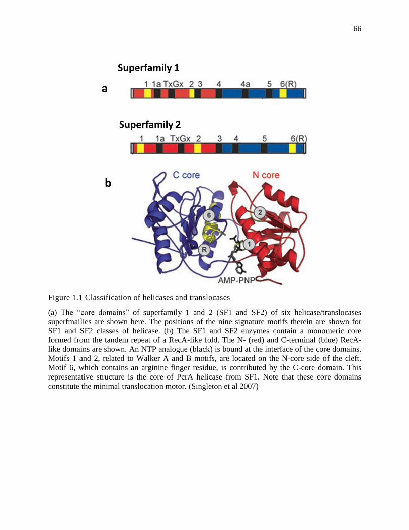
66
Figure 1.1 Classification of helicases and translocases
(a) The “core domains” of superfamily 1 and 2 (SF1 and SF2) of six helicase/translocases
superfmailies are shown here. The positions of the nine signature motifs therein are shown for
SF1 and SF2 classes of helicase. (b) The SF1 and SF2 enzymes contain a monomeric core
formed from the tandem repeat of a RecA-like fold. The N- (red) and C-terminal (blue) RecA-
like domains are shown. An NTP analogue (black) is bound at the interface of the core domains.
Motifs 1 and 2, related to Walker A and B motifs, are located on the N-core side of the cleft.
Motif 6, which contains an arginine finger residue, is contributed by the C-core domain. This
representative structure is the core of PcrA helicase from SF1. Note that these core domains
constitute the minimal translocation motor. (Singleton et al 2007)
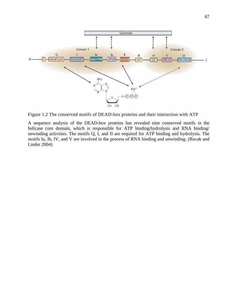
67
Figure 1.2 The conserved motifs of DEAD-box proteins and their interaction with ATP
A sequence analysis of the DEAD-box proteins has revealed nine conserved motifs in the
helicase core domain, which is responsible for ATP binding/hydrolysis and RNA binding/
unwinding activities. The motifs Q, I, and II are required for ATP binding and hydrolysis. The
motifs Ia, Ib, IV, and V are involved in the process of RNA binding and unwinding. (Rocak and
Linder 2004)
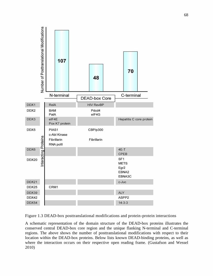
68
Figure 1.3 DEAD-box posttranslational modifications and protein-protein interactions
A schematic representation of the domain structure of the DEAD-box proteins illustrates the
conserved central DEAD-box core region and the unique flanking N-terminal and C-terminal
regions. The above shows the number of posttranslational modifications with respect to their
location within the DEAD-box proteins. Below lists known DEAD-binding proteins, as well as
where the interaction occurs on their respective open reading frame. (Gustafson and Wessel
2010)
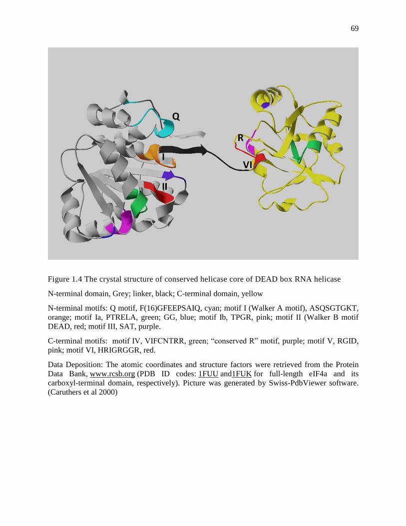
69
Figure 1.4 The crystal structure of conserved helicase core of DEAD box RNA helicase
N-terminal domain, Grey; linker, black; C-terminal domain, yellow
N-terminal motifs: Q motif, F(16)GFEEPSAIQ, cyan; motif I (Walker A motif), ASQSGTGKT,
orange; motif Ia, PTRELA, green; GG, blue; motif Ib, TPGR, pink; motif II (Walker B motif
DEAD, red; motif III, SAT, purple.
C-terminal motifs: motif IV, VIFCNTRR, green; “conserved R” motif, purple; motif V, RGID,
pink; motif VI, HRIGRGGR, red.
Data Deposition: The atomic coordinates and structure factors were retrieved from the Protein
Data Bank, www.rcsb.org (PDB ID codes: 1FUU and1FUK for full-length eIF4a and its
carboxyl-terminal domain, respectively). Picture was generated by Swiss-PdbViewer software.
(Caruthers et al 2000)
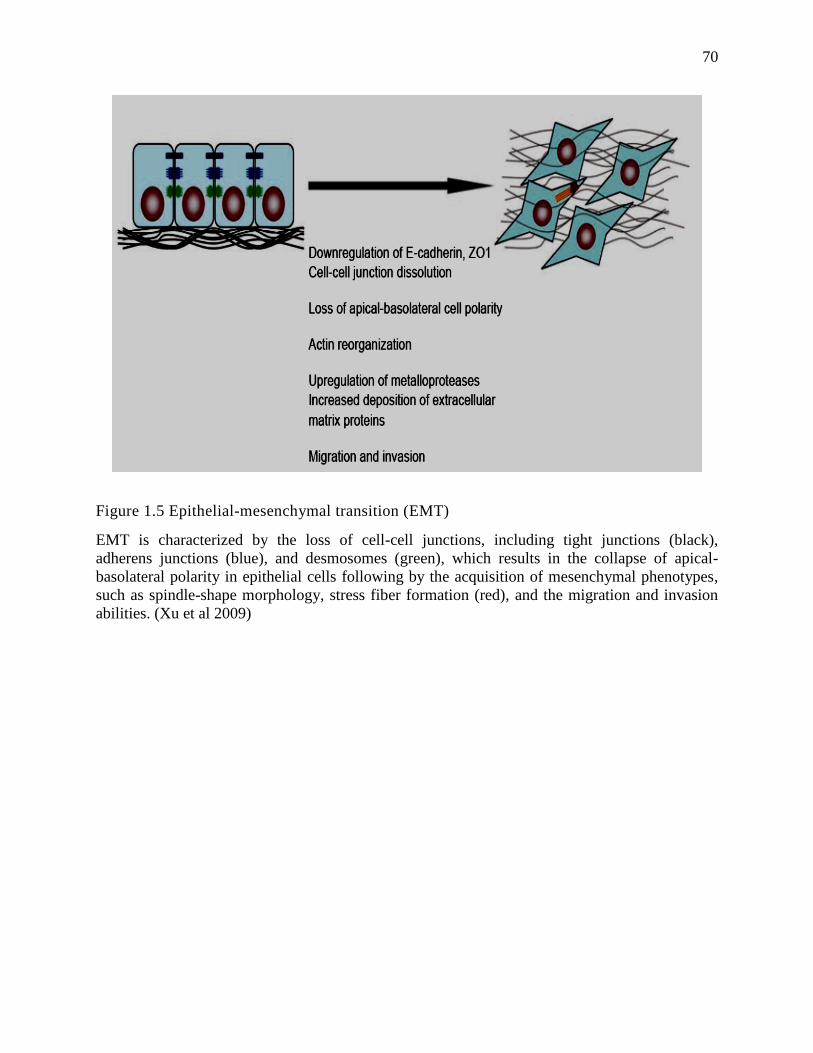
70
Figure 1.5 Epithelial-mesenchymal transition (EMT)
EMT is characterized by the loss of cell-cell junctions, including tight junctions (black),
adherens junctions (blue), and desmosomes (green), which results in the collapse of apical-
basolateral polarity in epithelial cells following by the acquisition of mesenchymal phenotypes,
such as spindle-shape morphology, stress fiber formation (red), and the migration and invasion
abilities. (Xu et al 2009)
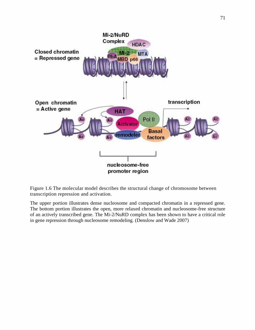
71
Figure 1.6 The molecular model describes the structural change of chromosome between
transcription repression and activation.
The upper portion illustrates dense nucleosome and compacted chromatin in a repressed gene.
The bottom portion illustrates the open, more relaxed chromatin and nucleosome-free structure
of an actively transcribed gene. The Mi-2/NuRD complex has been shown to have a critical role
in gene repression through nucleosome remodeling. (Denslow and Wade 2007)
72
CHAPTER 2: PHOSPHORYLATED p68 RNA HELICASE ACTIVATES SNAIL1
TRANSCRIPTION BY PROMOTING HDAC1 DISSOCIATION FROM THE SNAIL1
PROMOTER1
1 This chapter has been published in Oncogene (2010) 29, 5427–5436. I contributed equally to this work.
2.1 Abstract
The nuclear p68 RNA helicase is a prototypical member of the DEAD box family of
RNA helicases. P68 RNA helicase has been implicated in cell proliferation and early organ
development and maturation. However, the functional role of p68 RNA helicase in these
biological processes at the molecular level is not well understood. We previously reported that
tyrosine phosphorylation of p68 RNA helicase mediates the effects of PDGF in induction of
EMT by promoting β-catenin nuclear translocation. Here we report that phosphorylation of p68
RNA helicase at Y593 up-regulates transcription of the Snail1 gene. The phosphorylated p68
activates transcription of the Snail1 gene by promoting HDAC1 dissociation from the Snail1
promoter. Our results showed that p68 interacted with the nuclear remodeling and deacetylation
complex MBD3:Mi-2/NuRD. Thus, our data suggested that a DEAD box RNA unwindase can
potentially regulate gene expression by functioning as a protein ‘displacer’ to modulate protein-
protein interactions at the chromatin remodeling complex.
2.2 Introduction
E-cadherin, a prototypical member of the cadherin family, is the key component of the
epithelial cell-cell adhesion junction. During embryonic development and tissue remodeling, the
expression of E-cadherin is repressed. As a consequence, the strong adhesions of the epithelial
cells are weakened. The cells adopt a fibroblast-like morphology. This is the so-called Epithelial-
73
Mesenchymal-Transition (EMT) process (Kang and Massague 2004). Loss of E-cadherin
expression or function constitutes one main reason for epithelial carcinoma progression to an
invasive metastatic status (Kang and Massague 2004, Rodrigo et al 1999). Both expression and
function of E-cadherin are regulated at multiple levels (Bryant and Stow 2004, Davis et al 2003).
A zinc-finger transcription factor, Snail1, and its closely related family have been demonstrated
to play a key role in downregulation of E-cadherin gene transcription (De Craene et al 2005,
Peinado et al 2004b). It was revealed that Snail1 mediates E-cadherin repression by recruiting
histone deacetylase (HDAC) to the E-cadherin promoter (Peinado et al 2004b). Repression of E-
cadherin by Snail1 leads to Epithelial-Mesenchymal Transition (EMT). As a master regulator for
EMT, expression of Snail1 is stimulated by signaling pathways of a number of growth factors
(De Craene et al 2005), such as, EGF, FGF and TGF-β (Ciruna and Rossant 2001, Lu et al 2003,
Zavadil and Bottinger 2005). Cellular levels of Snail1 are regulated via a number of different
mechanisms, including gene transcription and protein turn-over in cells (Barbera et al 2004,
Zhou et al 2004a). Most recently, Fujita and co-workers demonstrated that MTA3, a member of
the metastasis associated gene family, regulates Snail1 expression by targeting the nuclear
remodeling and deacetylation complex MBD3:Mi-2/NuRD-HDAC1 to the Snail1 promoter in
breast cancer cells (Fujita et al 2003).
The nuclear p68 RNA helicase (ref to as p68) is a prototypical member of the DEAD box
family of RNA helicases (Crawford et al 1982b, Lane and Hoeffler 1980a). As an early example
of a cellular RNA helicase, the ATPase and the RNA unwinding activities of p68 RNA helicase
were documented (Ford et al 1988, Hirling et al 1989, Iggo and Lane 1989a). Expression of p68
correlates with cell proliferation and early organ maturation (Stevenson et al 1998a). The protein
was also shown to potentially play a critical role in the tumorigenesis process (Causevic et al
74
2001a, Dubey et al 1997, Wei and Hu 2001). It has been demonstrated by numerous laboratories
that p68 has a functional role in transcriptional regulation of a number of genes, including
Estrogen Receptor alpha (ERα) (Endoh et al 1999b) and several p53-dependent genes (Bates et
al 2005b). The protein was also shown to interact with p300/CBP and the RNA polymerase II
holoenzyme (Rossow and Janknecht 2003). The molecular mechanism by which p68 is involved
in transcriptional regulation is not clear. Interestingly, p68 was detected to interact with histone
deacetylase 1 (HDAC1), indicating that the protein may have a functional role in regulation of
gene expression by chromatin remodeling (Wilson et al 2004b). We previously reported that p68
is phosphorylated at multiple amino acid residues, including serine/threonine and tyrosine (Yang
and Liu 2004a, Yang et al 2005a) Tyrosine phosphorylation of p68 correlates with tumor
progression (Yang et al 2005b). In the present study, we present evidence to show that the
phosphor-p68 represses E-cadherin expression by regulating transcription of the Snail1 gene.
Phosphorylation of p68 at Y593 promoted dissociation of HDAC1 from Snail1 promoter. P68
RNA helicase interacted with the nuclear remodeling and deacetylation MBD3:Mi-2/NuRD
complex, suggesting a potential role of phosphor-p68 in dissociating HDAC1 from the
MBD3:Mi-2/NuRD-HDAC1 complex at the Snail1 promoter. Our study revealed a close
correlation between the Snail1 expression levels and the phosphorylation levels of p68, both
correlated closely with cancer metastasis.
2.3 Results
2.3.1 The Phosphor-p68 Repressed E-cadherin by Upregulating Transcription of Snail1
We previously reported that phosphorylation of p68 at Y593 mediates growth factor
PDGF stimulated EMT by promoting β-catenin nuclear translocation (Yang et al 2006). The
75
phosphor-p68 represses expression of epithelial marker, E-cadherin. We sought to understand the
molecular mechanism by which the phosphor-p68 regulates E-cadherin expression. We first
tested whether the effects of p68 phosphorylation on the changes in E-cadherin expression was
due to transcription of the E-cadherin gene. The transcription activity of the E-cadherin promoter
in a metastatic colon cancer cell line SW620 was examined. We used a luciferase reporter fused
to the E-boxes of the E-cadherin promoter (Fearon 2003) in cells in which p68 was knocked
down and HA-p68s wild type (WT) or Y593F mutant was exogenously expressed in p68
knockdown cells (SW620-p68/+wt
and SW620-p68/+Y593F
). E-cadherin transcription activity was not
markedly affected by p68 knockdown but was substantially down-regulated in SW620-p68/+wt
cells (Fig. 2.1A). On the other hand, E-cadherin transcription was significantly upregulated in
SW620-p68/+Y593F
cells (Fig. 2.1A). We tested whether phosphor-p68 regulated E-cadherin
transcription directly or if the regulatory effects were mediated through other cellular factor(s).
To this end, we examined whether phosphor-p68 interacted with the promoter of the E-cadherin
gene by chromatin immunoprecipitation (ChIP). It was certain that neither p68 (WT) nor the
Y593F mutant interacted with the E-cadherin promoter (Fig. 2.1B). Thus, it was likely that
phosphor-p68 regulated E-cadherin expression indirectly.
Snail1/Slug and SIP1 are the master regulators that regulate E-cadherin transcription
(Bolos et al 2003a, Fearon 2003, Thiery and Chopin 1999). In a ChIP-on-chip assay, we detected
that p68 interacted with the Snail1 promoter (Data not shown). Thus, we reasoned whether the
phosphor-p68 regulated E-cadherin through regulation of Snail1 expression. We examined the
effects of p68 phosphorylation on the expression of Snail1. Immunoblotting showed that cellular
levels of Snail1 were increased in the SW620-p68/+wt
cells and decreased in the SW620-p68/+Y593F
cells (Fig. 2.1C). The Snail1 upregulation correlated with the p68 expression and
76
phosphorylation at Y593 (Fig. 2.1C). Our results indicated that phosphorylation of p68 at Y593
may regulate the transcriptional activity of the Snail1 gene. Regulation of transcription of the
Snail1 gene by phosphor-p68 was also confirmed by RT-PCR detection of the Snail1 mRNA in
SW620, SW620-p68/+wt
, or SW620-p68/+Y593F
cells (Fig. 2.1D). To test whether phosphor-p68
regulated Snail1 transcription directly, we conducted ChIP experiments with the Snail1 promoter
using the antibody Pabp68. Clearly, p68 precipitated with the Snail1 promoter (Fig. 2.2A). The
similar experiments were also carried out with exogenously expressed HA-p68 in SW620 cells
using anti-HA antibody. P68 precipitated with the Snail1 promoter (Fig. 2.2B), indicating that
p68 was directly involved in the transcriptional regulation of the Snail1 gene.
Whether the phosphor-p68 repressed E-cadherin through transcriptional regulation of the
Snail1 gene, we reasoned that Snail1 expression must be required for the effects of the Y593
phosphorylation on the E-cadherin repression. To test this conjecture, we examined the effects of
the p68 phosphorylation on the cellular level of E-cadherin in SW620 cells with/without Snail1
knockdown by RNAi. We found that Snail1 was upregulated, E-cadherin was repressed, and
vimentin was upregulated by expression of wild-type p68 in SW620 cells. In contrast, expression
of p68 Y593F mutant in SW620 cells led to Snail1 repression, E-cadherin upregulation, and
vimentin repression (Fig. 2.2C). However, knockdown of Snail1 by RNAi abolished the effects
of exogenous expression of HA-p68s (wild-type and Y593F mutant) on cellular levels of E-
cadherin and vimentin (Fig. 2.2C). The results supported the conclusion that regulating the
transcription of Snail1 mediated the regulatory effects of the phosphorylated p68 in repressing E-
cadherin expression. Interestingly, we repeatedly observed that there was a very low level of
expression of E-cadherin in cells where Snail1 was knocked down (Fig. 2.2C), suggesting that
77
there is an alternative mechanism which represses E-cadherin expression in Snail1 knockdown
cells. This alternative mechanism is not regulated by phosphor-p68.
2.3.2 p68 Associates with the MBD3:Mi2/NuRD Complex
The preceding experiments suggested the role of phosphor-p68 in repression of E-
cadherin through the regulation of transcription of the Snail1 gene. To further understand the
molecular mechanism by which phosphor-p68 regulated transcription of the Snail1 gene, we
attempted to probe the protein or protein complex that interacted with phosphor-p68 at the Snail1
promoter. Recently, Fujita and co-workers demonstrated that MTA3 targeted the nuclear
remodeling and deacetylation complex Mi-2/NuRD to the Snail1 promoter, and directly
regulated Snail1 gene transcription (Fujita et al 2003). Thus, we asked whether phosphor-p68
interacted with the MBD3:Mi-2/NuRD complex in SW620 cells. To this end, we carried out co-
immunoprecipitation with the nuclear extracts made from SW620 cells using PAbp68. As a
comparison, the co-immunoprecipitation experiments were also conducted with nuclear extracts
made from SW480, a cell line derived from the tissue of the same patient from whom SW620
was derived. SW480, however, was derived from tissue of non-metastatic adenocarcinoma. The
tyrosine phosphorylation of p68 was almost undetectable in SW480 cells (data not shown, also
see Fig. 2.5A). It was certain that the antibody against p68 precipitated MBD3 and Mi-2 in the
extracts made from both SW480 and SW620 cells (Fig. 2.3A). To confirm the co-
immunoprecipitation results, we performed co-immunoprecipitation using antibodies against
MBD3 and Mi-2. The co-precipitation of p68 with MBD3 and Mi-2 in the extracts made from
SW480 and SW620 cells was clearly evident (Fig. 2.3B). These coimuunoprecipitation
experiments suggested that p68 interacted with Mi-2/NuRD complex.
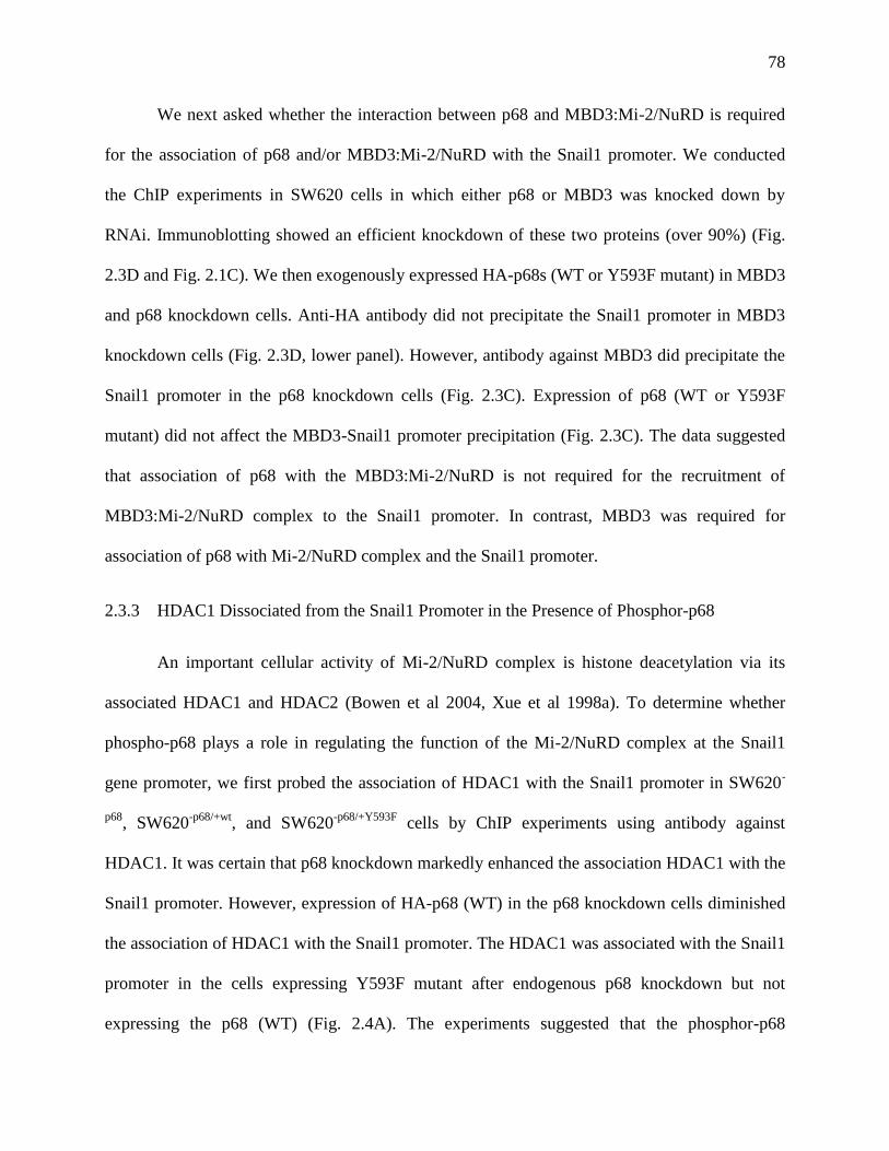
78
We next asked whether the interaction between p68 and MBD3:Mi-2/NuRD is required
for the association of p68 and/or MBD3:Mi-2/NuRD with the Snail1 promoter. We conducted
the ChIP experiments in SW620 cells in which either p68 or MBD3 was knocked down by
RNAi. Immunoblotting showed an efficient knockdown of these two proteins (over 90%) (Fig.
2.3D and Fig. 2.1C). We then exogenously expressed HA-p68s (WT or Y593F mutant) in MBD3
and p68 knockdown cells. Anti-HA antibody did not precipitate the Snail1 promoter in MBD3
knockdown cells (Fig. 2.3D, lower panel). However, antibody against MBD3 did precipitate the
Snail1 promoter in the p68 knockdown cells (Fig. 2.3C). Expression of p68 (WT or Y593F
mutant) did not affect the MBD3-Snail1 promoter precipitation (Fig. 2.3C). The data suggested
that association of p68 with the MBD3:Mi-2/NuRD is not required for the recruitment of
MBD3:Mi-2/NuRD complex to the Snail1 promoter. In contrast, MBD3 was required for
association of p68 with Mi-2/NuRD complex and the Snail1 promoter.
2.3.3 HDAC1 Dissociated from the Snail1 Promoter in the Presence of Phosphor-p68
An important cellular activity of Mi-2/NuRD complex is histone deacetylation via its
associated HDAC1 and HDAC2 (Bowen et al 2004, Xue et al 1998a). To determine whether
phospho-p68 plays a role in regulating the function of the Mi-2/NuRD complex at the Snail1
gene promoter, we first probed the association of HDAC1 with the Snail1 promoter in SW620-
p68, SW620
-p68/+wt, and SW620
-p68/+Y593F cells by ChIP experiments using antibody against
HDAC1. It was certain that p68 knockdown markedly enhanced the association HDAC1 with the
Snail1 promoter. However, expression of HA-p68 (WT) in the p68 knockdown cells diminished
the association of HDAC1 with the Snail1 promoter. The HDAC1 was associated with the Snail1
promoter in the cells expressing Y593F mutant after endogenous p68 knockdown but not
expressing the p68 (WT) (Fig. 2.4A). The experiments suggested that the phosphor-p68
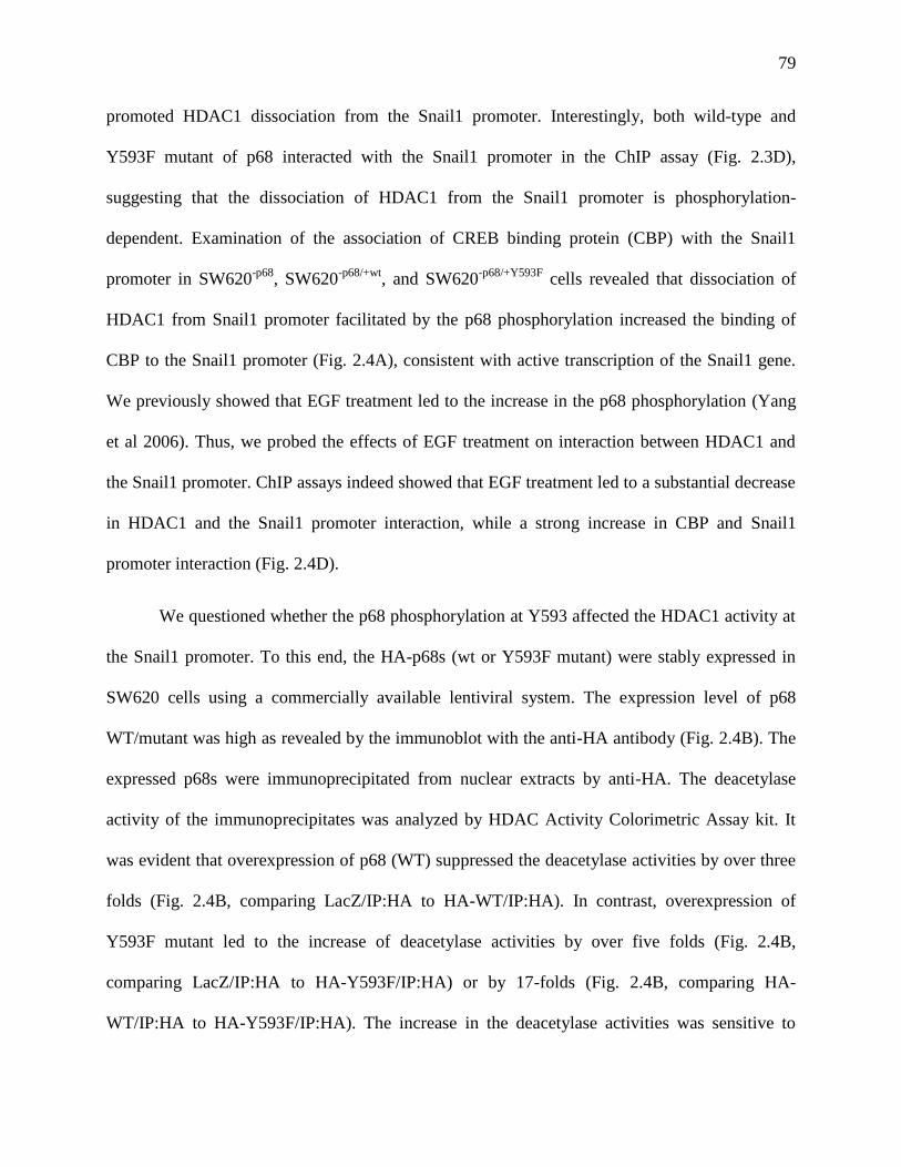
79
promoted HDAC1 dissociation from the Snail1 promoter. Interestingly, both wild-type and
Y593F mutant of p68 interacted with the Snail1 promoter in the ChIP assay (Fig. 2.3D),
suggesting that the dissociation of HDAC1 from the Snail1 promoter is phosphorylation-
dependent. Examination of the association of CREB binding protein (CBP) with the Snail1
promoter in SW620-p68
, SW620-p68/+wt
, and SW620-p68/+Y593F
cells revealed that dissociation of
HDAC1 from Snail1 promoter facilitated by the p68 phosphorylation increased the binding of
CBP to the Snail1 promoter (Fig. 2.4A), consistent with active transcription of the Snail1 gene.
We previously showed that EGF treatment led to the increase in the p68 phosphorylation (Yang
et al 2006). Thus, we probed the effects of EGF treatment on interaction between HDAC1 and
the Snail1 promoter. ChIP assays indeed showed that EGF treatment led to a substantial decrease
in HDAC1 and the Snail1 promoter interaction, while a strong increase in CBP and Snail1
promoter interaction (Fig. 2.4D).
We questioned whether the p68 phosphorylation at Y593 affected the HDAC1 activity at
the Snail1 promoter. To this end, the HA-p68s (wt or Y593F mutant) were stably expressed in
SW620 cells using a commercially available lentiviral system. The expression level of p68
WT/mutant was high as revealed by the immunoblot with the anti-HA antibody (Fig. 2.4B). The
expressed p68s were immunoprecipitated from nuclear extracts by anti-HA. The deacetylase
activity of the immunoprecipitates was analyzed by HDAC Activity Colorimetric Assay kit. It
was evident that overexpression of p68 (WT) suppressed the deacetylase activities by over three
folds (Fig. 2.4B, comparing LacZ/IP:HA to HA-WT/IP:HA). In contrast, overexpression of
Y593F mutant led to the increase of deacetylase activities by over five folds (Fig. 2.4B,
comparing LacZ/IP:HA to HA-Y593F/IP:HA) or by 17-folds (Fig. 2.4B, comparing HA-
WT/IP:HA to HA-Y593F/IP:HA). The increase in the deacetylase activities was sensitive to
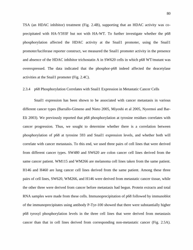
80
TSA (an HDAC inhibitor) treatment (Fig. 2.4B), supporting that an HDAC activity was co-
precipitated with HA-Y593F but not with HA-WT. To further investigate whether the p68
phosphorylation affected the HDAC activity at the Snail1 promoter, using the Snail1
promoter/luciferase reporter construct, we measured the Snail1 promoter activity in the presence
and absence of the HDAC inhibitor trichostatin A in SW620 cells in which p68 WT/mutant was
overexpressed. The data indicated that the phosphor-p68 indeed affected the deacetylase
activities at the Snail1 promoter (Fig. 2.4C).
2.3.4 p68 Phosphorylation Correlates with Snail1 Expression in Metastatic Cancer Cells
Snail1 expression has been shown to be associated with cancer metastasis in various
different cancer types (Barrallo-Gimeno and Nieto 2005, Miyoshi et al 2005, Nyormoi and Bar-
Eli 2003). We previously reported that p68 phosphorylation at tyrosine residues correlates with
cancer progression. Thus, we sought to determine whether there is a correlation between
phosphorylation of p68 at tyrosine 593 and Snail1 expression levels, and whether both will
correlate with cancer metastasis. To this end, we used three pairs of cell lines that were derived
from different cancer types. SW480 and SW620 are colon cancer cell lines derived from the
same cancer patient. WM115 and WM266 are melanoma cell lines taken from the same patient.
H146 and H460 are lung cancer cell lines derived from the same patient. Among these three
pairs of cell lines, SW620, WM266, and H146 were derived from metastatic cancer tissue, while
the other three were derived from cancer before metastasis had begun. Protein extracts and total
RNA samples were made from these cells. Immunoprecipitation of p68 followed by immunoblot
of the immunoprecipitates using antibody P-Tyr-100 showed that there were substantially higher
p68 tyrosyl phosphorylation levels in the three cell lines that were derived from metastasis
cancer than that in cell lines derived from corresponding non-metastatic cancer (Fig. 2.5A).
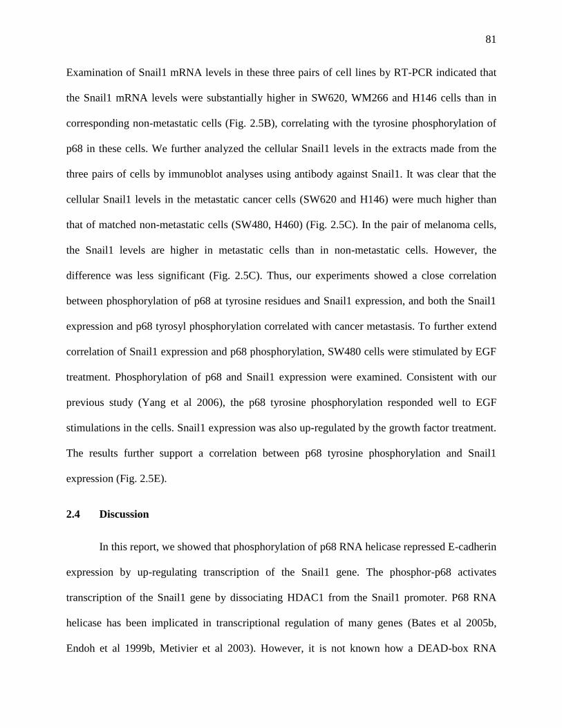
81
Examination of Snail1 mRNA levels in these three pairs of cell lines by RT-PCR indicated that
the Snail1 mRNA levels were substantially higher in SW620, WM266 and H146 cells than in
corresponding non-metastatic cells (Fig. 2.5B), correlating with the tyrosine phosphorylation of
p68 in these cells. We further analyzed the cellular Snail1 levels in the extracts made from the
three pairs of cells by immunoblot analyses using antibody against Snail1. It was clear that the
cellular Snail1 levels in the metastatic cancer cells (SW620 and H146) were much higher than
that of matched non-metastatic cells (SW480, H460) (Fig. 2.5C). In the pair of melanoma cells,
the Snail1 levels are higher in metastatic cells than in non-metastatic cells. However, the
difference was less significant (Fig. 2.5C). Thus, our experiments showed a close correlation
between phosphorylation of p68 at tyrosine residues and Snail1 expression, and both the Snail1
expression and p68 tyrosyl phosphorylation correlated with cancer metastasis. To further extend
correlation of Snail1 expression and p68 phosphorylation, SW480 cells were stimulated by EGF
treatment. Phosphorylation of p68 and Snail1 expression were examined. Consistent with our
previous study (Yang et al 2006), the p68 tyrosine phosphorylation responded well to EGF
stimulations in the cells. Snail1 expression was also up-regulated by the growth factor treatment.
The results further support a correlation between p68 tyrosine phosphorylation and Snail1
expression (Fig. 2.5E).
2.4 Discussion
In this report, we showed that phosphorylation of p68 RNA helicase repressed E-cadherin
expression by up-regulating transcription of the Snail1 gene. The phosphor-p68 activates
transcription of the Snail1 gene by dissociating HDAC1 from the Snail1 promoter. P68 RNA
helicase has been implicated in transcriptional regulation of many genes (Bates et al 2005b,
Endoh et al 1999b, Metivier et al 2003). However, it is not known how a DEAD-box RNA
82
helicase functions in transcriptional regulation. A recent study done by Allen C. Spradling’s
laboratory reveals that Drosophila’s p68 may have a role in unwinding the RNA transcripts from
its DNA template, facilitating a quick reset of the nucleosome structure, which is
transcriptionally inactive (Buszczak and Spradling 2006b). Our observations may reveal another
model for the functional role of p68 in transcriptional regulation. P68 RNA helicase may have a
role in remodeling or re-arranging the protein complex that assembles at the gene promoter.
There were two possible explanations for the observed dissociation of HDAC1 from the
Snail1 promoter in SW620 cells. (1) The unphosphorylated p68 recruited HDAC1 to the
promoter. The phosphor-p68 could not function as a recruiter. (2) The phosphor-p68 may
‘displace’ HDAC1 from the Snail1 promoter. The unphosphorylated p68 could not function as a
protein ‘displacer’. Although we observed that the phosphor-p68 acquired β-catenin binding-
dependent ATPase activity (data not shown), indicating a possibility that phosphor-p68 can use
protein-binding as substrate to stimulate its ATPase activity for the protein ‘displacement’,
additional experiments are required to prove that the phosphor-p68 can indeed displace HDAC1
from Mi-2/NuRD complex. Histone deacetylases (HDACs) are enzymes that modify chromatin
structure and subsequently regulate gene expression. HDACs are usually recruited to a particular
regulatory site with their associated multi-protein complexes, such as NuRD or Sin3 complex
(Knoepfler and Eisenman 1999, Narlikar et al 2002). While most studies concentrated on the
mechanism by which the HDAC activity and its associated complex are recruited to a specific
gene promoter (Forsberg and Bresnick 2001, Kurdistani and Grunstein 2003, Neely and
Workman 2002), our studies suggested a possible mechanism of action by which HDACs can be
displaced from their associated complex by a DEAD-box helicase. Given that tyrosine
phosphorylations of p68 were closely associated with cancer development (Yang et al 2005b), it
83
is tempting to speculate that displacement of HDACs by the phosphor-p68 is a dysregulated
route for tumor progression through activation of specific genes. Whether p68 is a constitutive
member of the NuRD complex is an open question. P68 was not identified in the originally
isolated NuRD complex (Bowen et al 2004, Tong et al 1998, Xue et al 1998a, Zhang et al
1999b). Our experiments showed co-immunoprecipitation of p68 with the Mi-2 and MBD3, the
components of NuRD complex. This seems to argue that p68 is a part of protein components of
the NuRD complex. In contrast, knockdown of p68 did not affect the association of the
MBD3:Mi-2/NuRD complex with Snail1 promoter, suggesting that p68 is not required for the
NuRD complex to be recruited to the Snail1 promoter. This observation disagrees that p68 may
not be a constitutive component of the NuRD complex. The best explanation is that p68 only
associates with a subset of NuRD complexes. Most recently, p68 was detected to interact with
HDAC1 in a promoter-specific manner (Wilson et al 2004b), which also seems to support the
concept. It was not known whether the detected interaction of p68 with HDAC1 was direct or
mediated by other protein factors. Knockdown of MBD3 abolished the association of p68 at the
Snail1 promoter, suggesting that p68 was recruited to the Snail1 promoter by a component of the
NuRD complex. Taken together, we speculated that p68 RNA helicase is recruited to the
MBD3:Mi-2/NuRD complex in a promoter specific manner. An alternative explanation is that
phosphorylated p68 RNA helicase may compete with HDAC1 to assemble to the Mi-2/NuRD
complex. The unphosphorylated p68 and Y593F mutant are unable to compete. Thus, there may
be two populations of Mi-2/NuRD complexes in SW620 cells. One contains phosphor-p68 and
the other contains HDAC1.
84
2.5 Materials and Methods
Reagents and Antibodies
TSA was purchased from PeproTech. Both polyclonal antibody PAbp68 and monoclonal
antibody p68-rgg against human p68 were raised against bacterially expressed His-tagged C-
terminal domain of p68 (Invitrogen, Auburn University Hybridoma Facility). Commercial
-actin,
HDAC1, GAPDH against human GAPDH, ChIP grade monoclonal against HDAC1, Mi-2,
SNAI 1 against Snail1), Cell Signaling Technology (p-Tyr-100, against phosphor-tyrosine,
HDAC1, polyclonal against HDAC1, H2A, against human histone 2A), Imgenex (MBD3, ChIP
grade), BD Bioscience (E-cadherin and Vimentin), Roche (12CA5, against HA-tag ChIP grade),
Abcam (KAT3A/CBP, against CBP) and Upstate (HA, against HA-tag).
Cell Culture and RNA Interference
SW620 and SW480 cells were purchased from ATCC and grown by following the
vendor’s instructions. All DNAs or RNAs transfections were performed using Lipofectamine
2000 by following the manufacturer's instructions (Invitrogen). For the siRNA experiments, cells
were grown to 50% confluence and transfected with siRNA (100 pM). The duplex RNA
oligonucleotides for RNAi were purchased from Dharmacon siGENOME™ and SMARTpool®.
A duplex RNA oligonucleotides with random sequence (non-targeting, NT) provided by the
vendor was included in all siRNA knockdown experiments as negative controls. For transient
expression of wild-type p68 or mutants in p68 knockdown cells, the cells were transfected with
the indicated plasmid DNA 24 hours after the cells were transfected with siRNA and harvested
after additional 48 hours incubation.
85
Subcellular Extracts, Immunoprecipitation, Immunoblot
All subcellular extracts or whole cell extracts were made freshly after appropriate
treatments (indicated in figures). Subcellular extracts were prepared using commercially
available cell extracting kit and by following the vendor’s instructions (Active motif). The
protein concentration of the extracts was determined using the Bradford assay (Bio-Rad).
Immunoprecipitation experiments and immunoblot analyses were performed as described in
previous studies . The blotting signals were detected using SuperSignal West Dura Extended
Duration Substrate (Pierce).
Luciferase Reporter Assays
Before cells were appropriately treated (indicated in figures), cells were transfected with
1 μg of the indicated reporter plasmid and 0.01 μg of pRL null, which expresses Renilla
luciferase from Renilla reniformis as an internal control. The total amount of plasmid DNA was
adjusted with pcDNA3-β-Galactosidase. Firefly and Renilla luciferase activities present in
cellular lysates were assayed using the Dual-Luciferase Reporter System (Promega, Madison,
WI). Data were represented as Firefly luciferase activity normalized by Renilla luciferase
activity.
Expression of p68s by Lentiviral System
Stable overexpression of HA-tagged p68 wild-type or Y593F mutant was carried out
using the ViralPower lentiviral expression system (Invitrogen) by following the manufacturer's
instructions. The ORFs of p68 wild type or Y593F mutant with N-terminal HA-tag were cloned
into pLenti6/TOPO (Invitrogen). The infections of cells with the lentiviruses that carry pLenti6-
86
p68 were carried out in the presence of 6 µg/mL of polybrene and 10 mM HEPES. Following
transduction, the cells were selected by 8 µg/mL of Blasticidin (Invitrogen).
RT-PCR
Cells were appropriately treated, and then total RNA was extracted using the total RNA
extraction kit (Qiagen, Valencia, CA). The RNA was quantified and then converted to cDNA
using the Improm II reverse transcription system (Promega, Madison, WI) following the
manufacturer’s protocol. The cDNA was then used in the final PCR reaction. The cycles were
an initial denaturing of 94 ºC for 2 minutes followed by 25 cycles of 94 ºC for 15s, 55 ºC for 30s,
and 72 ºC for 1m with an additional extension time of 5m added after the last cycle.
Densitometry was performed using the ImageJ program. Primers used were: Snail1 (sense 5’-
TCTAGGCCCTGGCTGCTAC-3’ antisense 5’-GCCTGGCACTGGTACTTCTT-3’); B2M
(sense 5’-TGCTGTCTCCATGTTTGATGTATCT-3’) antisense 5’-
TCTCTGCTCCCCACCTCTAAGT-3’). Primers for B2M were used as PCR and loading
controls. Controls for the RT reaction (not shown) contained no template or no reverse
transcriptase.
Chromatin Immunoprecipitation (ChIP)
The ChIP experiments were performed using ChIP-ITTM
Kit (Activemotif). The
precipitation of Snail1 or E-cadherin promoters was determined by PCR using primers spanning
nt -680 – -541 of the Snail1 promoter (sense 5’-GGGTGCTCTTGGCTAGCTG-3’antisense 5’-
CTGGAGAGCGTGGCATTG-3’) or nt -164 – +49 of the E-cadherin promoter (sense 5’-
GTCACCGCGTCTATGCGAGGCCG-3’, Antisense 5’-
GGACACTCGAACGCCTTCAGTCAAGT-3’). TFIIB/Pol II antibodies and mouse IgG were
87
used as control antibodies (included in the kit). ChIP-IT’s negative control primers flank a region
of genomic DNA between the GAPDH gene and CNAP1 gene. The primers were provided by
the vendor (sense5’-ATGGTTGCCACTGGGGATCT-3’antisense 5’-
TGCCAAAGCCTAGGGGAAGA-3’).
HDAC Activity Assay
SW620 cells were lysed in RIPA buffer (Upstate) 48 hr after transfection as described
above. The lysate was then diluted in RIPA buffer and HA-tagged proteins immunoprecipitated
with anti-HA polyclonal antibody (Upstate). HDAC activities were determined by HDAC
Activity Colorimetric Assay Kit (BioVision) according to manufacturer’s instructions. Antibody-
bound beads were washed in HDAC assay buffer prior to being added to the 96-well plate, to
remove immunoprecipitation buffer. Reactions were incubated for 30 min at 37°C with or
without the addition of 1 μM TSA. Samples were read in a VICTOR3™ plate reader
(PerkinElmer) at 405 nm. Typically each assay was performed 3 times.
88
2.6 References
Barbera MJ, Puig I, Dominguez D, Julien-Grille S, Guaita-Esteruelas S, Peiro S et al (2004).
Regulation of Snail transcription during epithelial to mesenchymal transition of tumor cells.
Oncogene 23: 7345-7354.
Barrallo-Gimeno A, Nieto MA (2005). The Snail genes as inducers of cell movement and
survival: implications in development and cancer. Development 132: 3151-3161.
Bates GJ, Nicol SM, Wilson BJ, Jacobs AM, Bourdon JC, Wardrop J et al (2005). The DEAD
box protein p68: a novel transcriptional coactivator of the p53 tumour suppressor. Embo J 24:
543-553.
Bolos V, Peinado H, Perez-Moreno MA, Fraga MF, Esteller M, Cano A (2003). The
transcription factor Slug represses E-cadherin expression and induces epithelial to mesenchymal
transitions: a comparison with Snail and E47 repressors. J Cell Sci 116: 499-511.
Bowen NJ, Fujita N, Kajita M, Wade PA (2004). Mi-2/NuRD: multiple complexes for many
purposes. Biochim Biophys Acta 1677: 52-57.
Bryant DM, Stow JL (2004). The ins and outs of E-cadherin trafficking. Trends Cell Biol 14:
427-434.
Buszczak M, Spradling AC (2006). The Drosophila P68 RNA helicase regulates transcriptional
deactivation by promoting RNA release from chromatin. Genes Dev 20: 977-989.
Causevic M, Hislop RG, Kernohan NM, Carey FA, Kay RA, Steele RJ et al (2001).
Overexpression and poly-ubiquitylation of the DEAD-box RNA helicase p68 in colorectal
tumours. Oncogene 20: 7734-7743.
Ciruna B, Rossant J (2001). FGF signaling regulates mesoderm cell fate specification and
morphogenetic movement at the primitive streak. Dev Cell 1: 37-49.
Crawford L, Leppard K, Lane D, Harlow E (1982). Cellular proteins reactive with monoclonal
antibodies directed against simian virus 40 T-antigen. J Virol 42: 612-620.
Davis MA, Ireton RC, Reynolds AB (2003). A core function for p120-catenin in cadherin
turnover. J Cell Biol 163: 525-534.
De Craene B, van Roy F, Berx G (2005). Unraveling signalling cascades for the Snail family of
transcription factors. Cell Signal 17: 535-547.
Dubey P, Hendrickson RC, Meredith SC, Siegel CT, Shabanowitz J, Skipper JC et al (1997).
The immunodominant antigen of an ultraviolet-induced regressor tumor is generated by a
somatic point mutation in the DEAD box helicase p68. J Exp Med 185: 695-705.
89
Endoh H, Maruyama K, Masuhiro Y, Kobayashi Y, Goto M, Tai H et al (1999). Purification and
identification of p68 RNA helicase acting as a transcriptional coactivator specific for the
activation function 1 of human estrogen receptor alpha. Mol Cell Biol 19: 5363-5372.
Fearon ER (2003). Connecting estrogen receptor function, transcriptional repression, and E-
cadherin expression in breast cancer. Cancer Cell 3: 307-310.
Ford MJ, Anton IA, Lane DP (1988). Nuclear protein with sequence homology to translation
initiation factor eIF-4A. Nature 332: 736-738.
Forsberg EC, Bresnick EH (2001). Histone acetylation beyond promoters: long-range acetylation
patterns in the chromatin world. Bioessays 23: 820-830.
Fujita N, Jaye DL, Kajita M, Geigerman C, Moreno CS, Wade PA (2003). MTA3, a Mi-2/NuRD
complex subunit, regulates an invasive growth pathway in breast cancer. Cell 113: 207-219.
Hirling H, Scheffner M, Restle T, Stahl H (1989). RNA helicase activity associated with the
human p68 protein. Nature 339: 562-564.
Iggo RD, Lane DP (1989). Nuclear protein p68 is an RNA-dependent ATPase. Embo J 8: 1827-
1831.
Kang Y, Massague J (2004). Epithelial-mesenchymal transitions: twist in development and
metastasis. Cell 118: 277-279.
Knoepfler PS, Eisenman RN (1999). Sin meets NuRD and other tails of repression. Cell 99: 447-
450.
Kurdistani SK, Grunstein M (2003). Histone acetylation and deacetylation in yeast. Nat Rev Mol
Cell Biol 4: 276-284.
Lane DP, Hoeffler WK (1980). SV40 large T shares an antigenic determinant with a cellular
protein of molecular weight 68,000. Nature 288: 167-170.
Liu ZR, Sargueil B, Smith CW (1998). Detection of a novel ATP-dependent cross-linked protein
at the 5' splice site-U1 small nuclear RNA duplex by methylene blue-mediated photo-cross-
linking. Mol Cell Biol 18: 6910-6920.
Lu Z, Ghosh S, Wang Z, Hunter T (2003). Downregulation of caveolin-1 function by EGF leads
to the loss of E-cadherin, increased transcriptional activity of beta-catenin, and enhanced tumor
cell invasion. Cancer Cell 4: 499-515.
Metivier R, Penot G, Hubner MR, Reid G, Brand H, Kos M et al (2003). Estrogen receptor-alpha
directs ordered, cyclical, and combinatorial recruitment of cofactors on a natural target promoter.
Cell 115: 751-763.
90
Miyoshi A, Kitajima Y, Kido S, Shimonishi T, Matsuyama S, Kitahara K et al (2005). Snail
accelerates cancer invasion by upregulating MMP expression and is associated with poor
prognosis of hepatocellular carcinoma. Br J Cancer.
Narlikar GJ, Fan HY, Kingston RE (2002). Cooperation between complexes that regulate
chromatin structure and transcription. Cell 108: 475-487.
Neely KE, Workman JL (2002). The complexity of chromatin remodeling and its links to cancer.
Biochim Biophys Acta 1603: 19-29.
Nyormoi O, Bar-Eli M (2003). Transcriptional regulation of metastasis-related genes in human
melanoma. Clin Exp Metastasis 20: 251-263.
Peinado H, Ballestar E, Esteller M, Cano A (2004). Snail mediates E-cadherin repression by the
recruitment of the Sin3A/histone deacetylase 1 (HDAC1)/HDAC2 complex. Mol Cell Biol 24:
306-319.
Rodrigo I, Cato AC, Cano A (1999). Regulation of E-cadherin gene expression during tumor
progression: the role of a new Ets-binding site and the E-pal element. Exp Cell Res 248: 358-371.
Rossow KL, Janknecht R (2003). Synergism between p68 RNA helicase and the transcriptional
coactivators CBP and p300. Oncogene 22: 151-156.
Stevenson RJ, Hamilton SJ, MacCallum DE, Hall PA, Fuller-Pace FV (1998). Expression of the
'dead box' RNA helicase p68 is developmentally and growth regulated and correlates with organ
differentiation/maturation in the fetus. J Pathol 184: 351-359.
Thiery JP, Chopin D (1999). Epithelial cell plasticity in development and tumor progression.
Cancer Metastasis Rev 18: 31-42.
Tong JK, Hassig CA, Schnitzler GR, Kingston RE, Schreiber SL (1998). Chromatin
deacetylation by an ATP-dependent nucleosome remodelling complex. Nature 395: 917-921.
Wei Y, Hu MH (2001). [The study of P68 RNA helicase on cell transformation]. Yi Chuan Xue
Bao 28: 991-996.
Wilson BJ, Bates GJ, Nicol SM, Gregory DJ, Perkins ND, Fuller-Pace FV (2004). The p68 and
p72 DEAD box RNA helicases interact with HDAC1 and repress transcription in a promoter-
specific manner. BMC Mol Biol 5: 11.
Xue Y, Wong J, Moreno GT, Young MK, Cote J, Wang W (1998). NURD, a novel complex
with both ATP-dependent chromatin-remodeling and histone deacetylase activities. Mol Cell 2:
851-861.
Yang L, Liu ZR (2004). Bacterially expressed recombinant p68 RNA helicase is phosphorylated
on serine, threonine, and tyrosine residues. Protein Expr Purif 35: 327-333.
91
Yang L, Lin C, Liu ZR (2005a). Signaling to the DEAD box-Regulation of DEAD-box p68 RNA
helicase by protein phosphorylations. Cell Signal.
Yang L, Lin C, Liu ZR (2005b). Phosphorylations of DEAD box p68 RNA helicase are
associated with cancer development and cell proliferation. Mol Cancer Res 3: 355-363.
Yang L, Lin C, Liu ZR (2006). P68 RNA Helicase Mediates PDGF-Induced Epithelial
Mesenchymal Transition by Displacing Axin from beta-Catenin. Cell 127: 139-155.
Zavadil J, Bottinger EP (2005). TGF-beta and epithelial-to-mesenchymal transitions. Oncogene
24: 5764-5774.
Zhang Y, Ng HH, Erdjument-Bromage H, Tempst P, Bird A, Reinberg D (1999). Analysis of the
NuRD subunits reveals a histone deacetylase core complex and a connection with DNA
methylation. Genes Dev 13: 1924-1935.
Zhou BP, Deng J, Xia W, Xu J, Li YM, Gunduz M et al (2004). Dual regulation of Snail by
GSK-3beta-mediated phosphorylation in control of epithelial-mesenchymal transition. Nat Cell
Biol 6: 931-940.
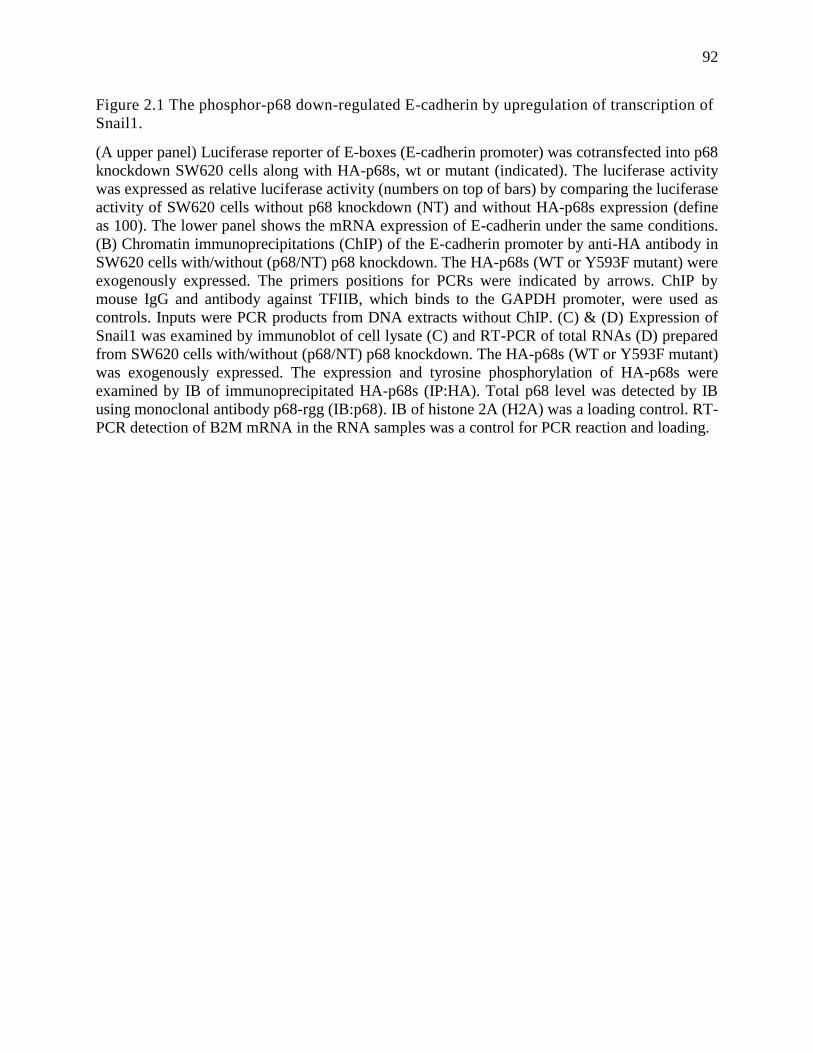
92
Figure 2.1 The phosphor-p68 down-regulated E-cadherin by upregulation of transcription of
Snail1.
(A upper panel) Luciferase reporter of E-boxes (E-cadherin promoter) was cotransfected into p68
knockdown SW620 cells along with HA-p68s, wt or mutant (indicated). The luciferase activity
was expressed as relative luciferase activity (numbers on top of bars) by comparing the luciferase
activity of SW620 cells without p68 knockdown (NT) and without HA-p68s expression (define
as 100). The lower panel shows the mRNA expression of E-cadherin under the same conditions.
(B) Chromatin immunoprecipitations (ChIP) of the E-cadherin promoter by anti-HA antibody in
SW620 cells with/without (p68/NT) p68 knockdown. The HA-p68s (WT or Y593F mutant) were
exogenously expressed. The primers positions for PCRs were indicated by arrows. ChIP by
mouse IgG and antibody against TFIIB, which binds to the GAPDH promoter, were used as
controls. Inputs were PCR products from DNA extracts without ChIP. (C) & (D) Expression of
Snail1 was examined by immunoblot of cell lysate (C) and RT-PCR of total RNAs (D) prepared
from SW620 cells with/without (p68/NT) p68 knockdown. The HA-p68s (WT or Y593F mutant)
was exogenously expressed. The expression and tyrosine phosphorylation of HA-p68s were
examined by IB of immunoprecipitated HA-p68s (IP:HA). Total p68 level was detected by IB
using monoclonal antibody p68-rgg (IB:p68). IB of histone 2A (H2A) was a loading control. RT-
PCR detection of B2M mRNA in the RNA samples was a control for PCR reaction and loading.
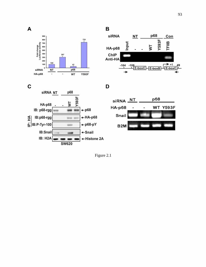
93
Figure 2.1
94
Figure 2.2 p68 interacted with Snail1 promoter.
(A) & (B) Chromatin immunoprecipitations (ChIP) of the Snail1 promoter by antibody Pabp68
(A, Anti-p68) and by anti-HA antibody (B, Anti-HA) in SW620 cells with/without (p68/NT) p68
knockdown. The HA-p68s (WT or Y593F mutant) were exogenously expressed. The primer
positions for Snail1 PCR were indicated by arrows. ChIP by mouse IgG and antibody against
RNA polymerase II (POL II), which binds to the GAPDH promoter, were used as controls.
Inputs were PCR products from DNA extracts without ChIP (use 10% of sample). The lower
panel shows the quantization ChIP results by real-time PCR. The ChIP quantization is expressed
as a percentage of the input by defining the input as 100. The error bars represent standard
deviation of results from three independent measurements. (C) Cellular levels of E-cadherin
(second panel from top) and vimentin (third panel from top) were detected by IBs of cellular
extracts made from SW620 cells with/without (Snail1/NT) Snail1 siRNA knockdown and
exogenous expression of HA-p68s (WT or Y593F mutant). Tyrosine phosphorylation of HA-
p68s was analyzed by IB of anti-HA IPs via antibody p-Tyr-100 (fifth panel from top). IB of
histone 2A (H2A) was loading control.
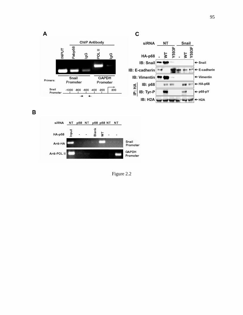
95
Figure 2.2
96
Figure 2.3 P68 interacted with the MBD3:Mi-2/NuRD complex.
(A) Co-IPs of MBD3 and Mi-2 with p68 in SW480 and SW620 cells were detected by IB of p68
co-immunoprecipitates using appropriate antibodies (indicated). P68 was precipitated by
polyclonal antibody Pab-p68. Rabbit IgG was used as a negative control IP antibody. Inputs were
the IBs of extracts without IP. (B) Co-IPs of p68 with MBD3 and Mi-2 in SW620 (620) and
SW480 (480) cells were detected by IBs of co-immunoprecipitates of antibodies (anti-MBD3
and anti-Mi-2) using monoclonal antibody p68-rgg. Mouse IgG was used as control IP antibody.
The inputs were the IBs of extracts without IP. The tyrosine phosphorylation of p68 was detected
by IB of PAbp68 immunoprecipitated p68 using antibody Tyr-100. (C) Upper panel, cellular
levels of MBD3, HDAC1, and exogenously expressed HA-p68s (WT or Y593F mutant) were
analyzed via IBs using appropriate antibodies (indicated). The IBs were performed with cellular
extracts made from SW620 cells that were treated with p68 siRNA (p68) or non-targeting siRNA
(NT). Lower panel, Interactions of MBD3 with Snail1 promoter in the cells treated as described
in upper panels were detected by ChIP assays using anti-MBD3 antibody (Anti-MBD3). (D)
Upper panel, cellular levels of MBD3 and exogenously expressed HA-p68s (WT or Y593F
mutant) (without endogenous p68 knockdown) were analyzed via IBs using appropriate
antibodies (indicated). The IBs were performed with cellular extracts made from SW620 cells
that were treated with MBD3 siRNA (MBD3) or non-targeting siRNA (NT). IB of Actin was a
loading control. Lower panel, Interactions of HA-p68 with Snail1 promoter in the cells treated as
described in upper panels were detected by ChIP assays using anti-HA antibody (Anti-HA).
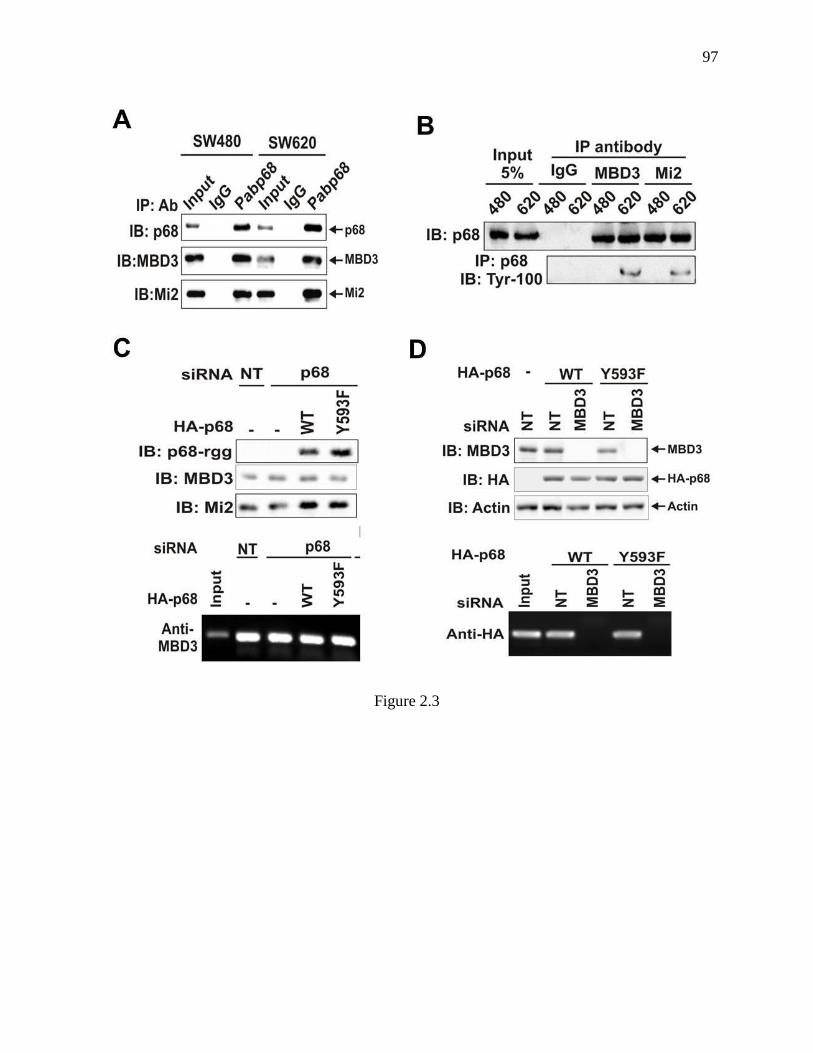
97
Figure 2.3
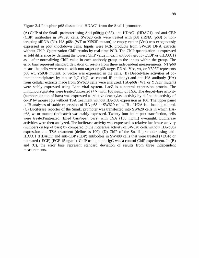
98
Figure 2.4 Phosphor-p68 dissociated HDAC1 from the Snail1 promoter.
(A) ChIP of the Snail1 promoter using Anti-p68rgg (p68), anti-HDAC1 (HDAC1), and anti-CBP
(CBP) antibodies in SW620 cells. SW620 cells were treated with p68 siRNA (p68) or non-
targeting siRNA (Nt). HA-p68s (WT or Y593F mutant) or empty vector (Vec) was exogenously
expressed in p68 knockdown cells. Inputs were PCR products from SW620 DNA extracts
without ChIP. Quantization ChIP results by real-time PCR. The ChIP quantization is expressed
as fold difference by defining the lowest ChIP value in each antibody group (αCBP or αHDAC1)
as 1 after normalizing ChIP value in each antibody group to the inputs within the group. The
error bars represent standard deviation of results from three independent measurements. NT/p68
means the cells were treated with non-target or p68 target RNAi. Vec, wt, or Y593F represents
p68 wt, Y593F mutant, or vector was expressed in the cells. (B) Deacetylase activities of co-
immunoprecipitates by mouse IgG (IgG, as control IP antibody) and anti-HA antibody (HA)
from cellular extracts made from SW620 cells were analyzed. HA-p68s (WT or Y593F mutant)
were stably expressed using Lenti-viral system. LacZ is a control expression protein. The
immunoprecipitates were treated/untreated (+/-) with 100 ng/ml of TSA. The deacetylase activity
(numbers on top of bars) was expressed as relative deacetylase activity by define the activity of
co-IP by mouse IgG without TSA treatment without HA-p68 expression as 100. The upper panel
is IB analyses of stable expression of HA-p68 in SW620 cells. IB of H2A is a loading control.
(C) Luciferase reporter of the Snail1 promoter was transfected into SW620 cells in which HA-
p68, wt or mutant (indicated) was stably expressed. Twenty four hours post transfection, cells
were treated/untreated (filled bars/open bars) with TSA (100 ng/ml) overnight. Luciferase
activities were then analyzed. The luciferase activity was expressed as relative luciferase activity
(numbers on top of bars) by compared to the luciferase activity of SW620 cells without HA-p68s
expression and TSA treatment (define as 100). (D) ChIP of the Snail1 promoter using anti-
HDAC1 (HDAC1) and anti-CBP (CBP) antibodies in SW480 cells that were treated (+EGF) or
untreated (-EGF) (EGF 15 ng/ml). ChIP using rabbit IgG was a control ChIP experiment. In (B)
and (C), the error bars represent standard deviation of results from three independent
measurements.
99
Figure 2.4
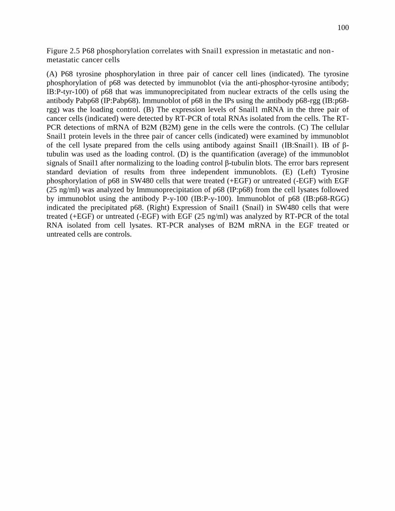
100
Figure 2.5 P68 phosphorylation correlates with Snail1 expression in metastatic and non-
metastatic cancer cells
(A) P68 tyrosine phosphorylation in three pair of cancer cell lines (indicated). The tyrosine
phosphorylation of p68 was detected by immunoblot (via the anti-phosphor-tyrosine antibody;
IB:P-tyr-100) of p68 that was immunoprecipitated from nuclear extracts of the cells using the
antibody Pabp68 (IP:Pabp68). Immunoblot of p68 in the IPs using the antibody p68-rgg (IB:p68-
rgg) was the loading control. (B) The expression levels of Snail1 mRNA in the three pair of
cancer cells (indicated) were detected by RT-PCR of total RNAs isolated from the cells. The RT-
PCR detections of mRNA of B2M (B2M) gene in the cells were the controls. (C) The cellular
Snail1 protein levels in the three pair of cancer cells (indicated) were examined by immunoblot
of the cell lysate prepared from the cells using antibody against Snail1 (IB:Snail1). IB of β-
tubulin was used as the loading control. (D) is the quantification (average) of the immunoblot
signals of Snail1 after normalizing to the loading control β-tubulin blots. The error bars represent
standard deviation of results from three independent immunoblots. (E) (Left) Tyrosine
phosphorylation of p68 in SW480 cells that were treated (+EGF) or untreated (-EGF) with EGF
(25 ng/ml) was analyzed by Immunoprecipitation of p68 (IP:p68) from the cell lysates followed
by immunoblot using the antibody P-y-100 (IB:P-y-100). Immunoblot of p68 (IB:p68-RGG)
indicated the precipitated p68. (Right) Expression of Snail1 (Snail) in SW480 cells that were
treated (+EGF) or untreated (-EGF) with EGF (25 ng/ml) was analyzed by RT-PCR of the total
RNA isolated from cell lysates. RT-PCR analyses of B2M mRNA in the EGF treated or
untreated cells are controls.
101
Figure 2.5
102
CHAPTER 3: PHOSPHORYLATION OF p68 RNA HELICASE PROMOTES COLON
CANCER METASTASIS
3.1 Abstract
Most cancer-related deaths are the consequence of cancer metastasis. Despite intensive
studies over decades, the molecular mechanisms that govern this death-causing process are still
poorly defined. We previously reported that phosphorylation of p68 RNA helicase at Y593
mediates the effects of PDGF in promoting EMT (Yang et al 2006). Here, we show that
phosphorylation of p68 RNA helicase at tyrosine Y593 promotes cancer metastasis in nude mice
xenograft model of human colon cancer cells. Analyses of tissue samples from colon cancer
patients reveal a close correlation between phosphorylation of p68 at Y593 and cancer
progression, supporting the notion that phosphorylation of p68 at Y593 plays an important role
in tumorigenesis and cancer progression.
3.2 Introduction
The nuclear p68 RNA helicase is a prototypical DEAD box family of RNA helicase
(Crawford et al 1982, Lane and Hoeffler 1980). Knock out experiments showed that p68
knockout is embryonic lethal indicating the critical function of the protein in the developmental
programs. The protein is essential to maintain cell normal growth and differentiation (Janknecht
2010, Stevenson et al 1998). However, the cellular functions of the protein at the molecular
level are not well understood (Schmid and Linder 1992). A number of independent studies have
revealed that expression and/or posttranslational modifications of p68 are closely associated with
cancer development and progression. It was first noted that overexpression and
polyubiquitylation of p68 is associated with colon cancer development (Causevic et al 2001).
103
Association of p68 with colon cancer development and progression was further confirmed by
observation from an independent study showing that expression of p68 was strongly increased
during the polyp-->adenoma-->adenocarcinoma transition in the colon. It was believed that
overexpression of p68 upregulated expression of a number of -catenin-regulated genes (Shin et
al 2007, Yang et al 2006). Our laboratory demonstrated that p68 is phosphorylated at multiple
amino acid residues (Yang and Liu 2004). Phosphorylation of p68 at tyrosine closely correlated
with cancer progression and metastasis (Yang et al 2005). Phosphorylation of p68 RNA helicase
at Y593 mediates the effects of several growth factors in promoting EMT in colon cancer cells
by promoting nuclear translocation of -catenin (Yang et al 2006).
Epithelial-mesenchymal-transition (EMT) is a process by which epithelial cells undergo
dramatic morphology changes marked by loss of cell polarity and cell-cell contacts. During
tumor progression, substantial evidence suggests that EMT plays a critical role in tumor cells
leaving organized epithelial layers and gaining property of invasiveness. Such changes often
initiate cancer metastasis (Chaffer and Weinberg 2011, Rhim et al 2012). Phosphorylation of p68
at Y593 closely correlates with tumor progression (Yang et al 2005). Thus, it is speculated that
the phosphorylation of p68 at Y593 may play an important role in cancer progression and
metastasis. In this report, we provide evidence to demonstrate that phosphorylation of p68 RNA
helicase at tyrosine Y593 promotes cancer metastasis in nude mice xenograft model of human
colon cancer cells. Analyses of tissue samples from colon cancer patients reveal a close
correlation between phosphorylation of p68 at Y593 and cancer development and progression.
104
3.3 Results
3.3.1 Characterization of Sublines with Endogenous p68 Knockdown and Exogenous p68 wt
and Y593F Mutant Expressions
Phosphorylation of p68 at Y593 mediates the effects of PDGF on promoting EMT (Yang
et al 2006). We speculated that the p68 phosphorylation may play a functional role in cancer
metastasis. To test the potential role of the p68 phosphorylation at Y593 in promoting cancer
metastasis, we examined whether expression of a non-phosphorylable p68 mutant Y593F would
inhibit tumor metastasis of metastatic cancer cells in nude mice xenograft model. We chose a
human colon cancer cell line SW620. This cell line was derived from colon cancer tissue of the
patient with cancer metastasized to lymph nodes (the line was derived from lymph node
metastatic cancer) (Leibovitz et al 1976). We first sought to establish a stable subline with
endogenous p68 knockdown and expression of the p68 mutant Y593F. P68 was knocked down
using shRNA by a retro-viral system. Immunoblot and RT-PCR analyses demonstrated that the
endogenous p68 was largely knocked down (Fig. 3.1, A & B). P68 wild type and non-
phosphorylable Y593F mutant were subsequently stably expressed in the p68 knockdown
SW620 cells by plasmid DNA transfection followed with appropriate antibiotic selection. RT-
PCR analyses showed that the exogenous HA-p68s, wt or Y593F mutant, were expressed and the
expression levels were comparable with that of endogenous p68 in the cells (Fig. 3.1, C & A).
Several clones of sublines with p68 knockdown and HA-p68, wt and Y593F mutant,
were selected. We carried out extensive characterizations of the obtained sublines. Firstly, to test
whether knockdown of endogenous p68 and exogenous expression of HA-p68s, wt and Y593F
mutant, led to significant changes in growth behaviors and other properties, cell proliferation of
the established sublines were tested by a commercially available cell proliferation kit. It was
105
evident that knockdown of p68 resulted in a reduction in cell growth rate both in short-term
(24hr) and long-term (6 days) testing periods (Fig. 3.2, A & B). Although the reduction in
growth rate was not recovered by expression of HA-p68, wt and Y593F mutant in a 24-hour
testing time (Fig. 3.2C), a gradual recovery of growth rate was observed on these cells in a 6-day
measurement (Fig. 3.2D). We next analyzed the cell migration property by Boyden chamber
assay. Evidently, p68 knockdown strongly reduced the cell migration (Fig. 3.3A). Expression of
p68 wild-type completely recovered the migration property of the cells, whereas the expression
of non-phosphorylable mutant p68 did not have a significant effect on cell migration (Fig. 3.3B).
Since phosphorylation of p68 at Y593 promotes EMT (Yang et al 2006), we therefore examined
the expression of epithelial and mesenchymal markers, E-cadherin and vimentin, in the p68
knockdown and wild-type/mutant expression cells, by RT-PCR. As expected, knockdown of p68
up-regulated E-cadherin and down-regulated vimentin expression (Fig. 3.3C). The pattern of E-
cadherin expression could be restored by exogenous expression of wild-type p68, but not by
expression of the Y593F mutant (Fig. 3.3D). We showed in our previous studies that
phosphorylation of p68 at Y593 affected Snail1 expression (Carter et al 2010). Thus, we
examined whether the Snail expression was affected in the selected clones of sublines. Clearly,
Snail1 was down-regulated by p68 knockdown (Fig. 3.3C).
Examination of the selected clones of different sublines by phase contrast microscope
revealed that expression of wild-type p68 in p68 knockdown cells led to increase in number of
cells with spindle shaped morphology (Fig. 3.4B), while expression of the Y593F mutant in p68
knockdown cells led to strong increase in cell adhesion as demonstrated by substantial more cells
clustered together in cultures (Fig. 3.4C). The cell-cell adhesion morphology observed in p68-
Y593F expressing SW620 cells is comparable to that in its paired non-metastatic cell line,

106
SW480 (Fig. 3.4D), which is characterized with less or no invasive phenotypes. From these in
vitro characterizations, we concluded that the established clones of sublines demonstrated that
p68 phosphorylation at Y593 indeed support an invasive phenotype of SW620 colon cancer
cells.
3.3.2 Knockdown of p68 and Expression of Y593F Mutant Significantly Reduced Cancer
Metastasis
Next, we tested the metastasis of these established sublines in xenograft model. It is well
established that the xenograft of SW620 will metastasize to lymph nodes, and the metastasis can
be analyzed by examination of SW620 cells in the spleen (Liu et al 2003a). The established
sublines of the SW620 cells were implanted at the right flank of nude mouse. The tumors were
grown for 30 days. The growth of tumor was monitored by measuring the tumor volume and the
tumor weight at end of experiments. In consistent with our observations with cultured cells, the
growth rates of xenograft tumors were not significantly affected by expression of wild-type p68
or the Y593F mutant (Fig. 3.5A & B). We then analyzed the cancer metastasis of the xenograft
tumors by examining the mRNA of human cytokeratin-18 (ref to as hCK-18) in the spleen of the
tumor bearing mice. It was clear that knockdown of p68 led to no detection of hCK-18 mRNA in
the spleen of any mouse within the test group. As comparison, we observed hCK-18 mRNA in
three out of six test mice that were implanted SW620 cells without p68 knockdown (Fig. 3.5C,
top-right panel). Expression of wild-type p68 resulted in observation of hCK-18 mRNA in three
or four mice in the group of six mice (Fig. 3.5C, mid-panel). Expression of Y593F mutant led to
detection of hCK-18 mRNA in only one mouse in each group of six mice (Fig. 3.5C, bottom-
panel). Table 1 summarized the results of growth and metastasis tests of the xenograft of
established sublines of SW620 cells. To ensure that the knockdown of p68 and expression of
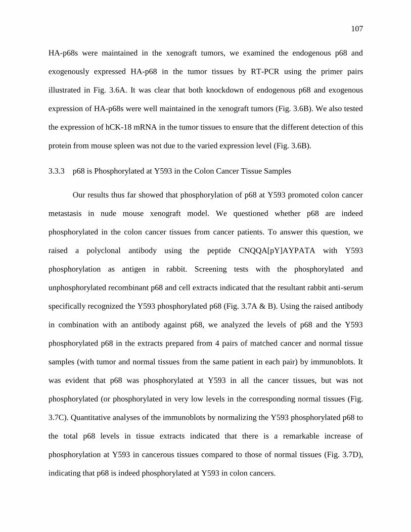
107
HA-p68s were maintained in the xenograft tumors, we examined the endogenous p68 and
exogenously expressed HA-p68 in the tumor tissues by RT-PCR using the primer pairs
illustrated in Fig. 3.6A. It was clear that both knockdown of endogenous p68 and exogenous
expression of HA-p68s were well maintained in the xenograft tumors (Fig. 3.6B). We also tested
the expression of hCK-18 mRNA in the tumor tissues to ensure that the different detection of this
protein from mouse spleen was not due to the varied expression level (Fig. 3.6B).
3.3.3 p68 is Phosphorylated at Y593 in the Colon Cancer Tissue Samples
Our results thus far showed that phosphorylation of p68 at Y593 promoted colon cancer
metastasis in nude mouse xenograft model. We questioned whether p68 are indeed
phosphorylated in the colon cancer tissues from cancer patients. To answer this question, we
raised a polyclonal antibody using the peptide CNQQA[pY]AYPATA with Y593
phosphorylation as antigen in rabbit. Screening tests with the phosphorylated and
unphosphorylated recombinant p68 and cell extracts indicated that the resultant rabbit anti-serum
specifically recognized the Y593 phosphorylated p68 (Fig. 3.7A & B). Using the raised antibody
in combination with an antibody against p68, we analyzed the levels of p68 and the Y593
phosphorylated p68 in the extracts prepared from 4 pairs of matched cancer and normal tissue
samples (with tumor and normal tissues from the same patient in each pair) by immunoblots. It
was evident that p68 was phosphorylated at Y593 in all the cancer tissues, but was not
phosphorylated (or phosphorylated in very low levels in the corresponding normal tissues (Fig.
3.7C). Quantitative analyses of the immunoblots by normalizing the Y593 phosphorylated p68 to
the total p68 levels in tissue extracts indicated that there is a remarkable increase of
phosphorylation at Y593 in cancerous tissues compared to those of normal tissues (Fig. 3.7D),
indicating that p68 is indeed phosphorylated at Y593 in colon cancers.
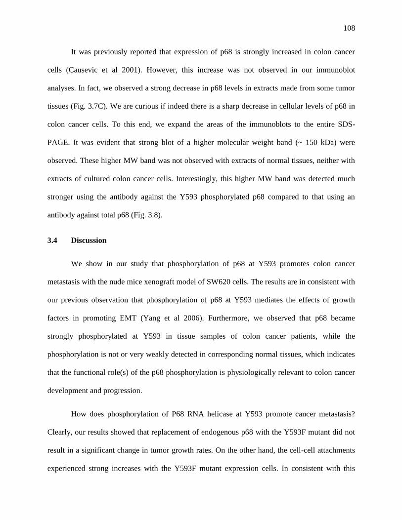
108
It was previously reported that expression of p68 is strongly increased in colon cancer
cells (Causevic et al 2001). However, this increase was not observed in our immunoblot
analyses. In fact, we observed a strong decrease in p68 levels in extracts made from some tumor
tissues (Fig. 3.7C). We are curious if indeed there is a sharp decrease in cellular levels of p68 in
colon cancer cells. To this end, we expand the areas of the immunoblots to the entire SDS-
PAGE. It was evident that strong blot of a higher molecular weight band (~ 150 kDa) were
observed. These higher MW band was not observed with extracts of normal tissues, neither with
extracts of cultured colon cancer cells. Interestingly, this higher MW band was detected much
stronger using the antibody against the Y593 phosphorylated p68 compared to that using an
antibody against total p68 (Fig. 3.8).
3.4 Discussion
We show in our study that phosphorylation of p68 at Y593 promotes colon cancer
metastasis with the nude mice xenograft model of SW620 cells. The results are in consistent with
our previous observation that phosphorylation of p68 at Y593 mediates the effects of growth
factors in promoting EMT (Yang et al 2006). Furthermore, we observed that p68 became
strongly phosphorylated at Y593 in tissue samples of colon cancer patients, while the
phosphorylation is not or very weakly detected in corresponding normal tissues, which indicates
that the functional role(s) of the p68 phosphorylation is physiologically relevant to colon cancer
development and progression.
How does phosphorylation of P68 RNA helicase at Y593 promote cancer metastasis?
Clearly, our results showed that replacement of endogenous p68 with the Y593F mutant did not
result in a significant change in tumor growth rates. On the other hand, the cell-cell attachments
experienced strong increases with the Y593F mutant expression cells. In consistent with this
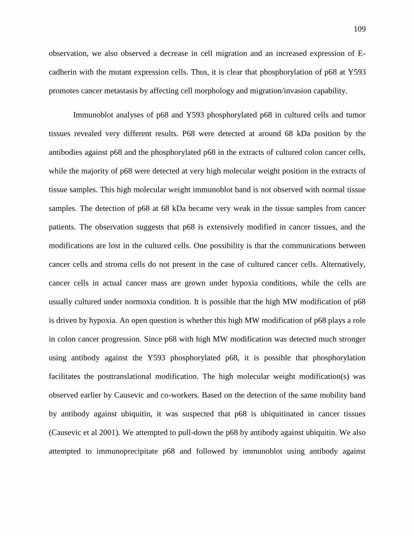
109
observation, we also observed a decrease in cell migration and an increased expression of E-
cadherin with the mutant expression cells. Thus, it is clear that phosphorylation of p68 at Y593
promotes cancer metastasis by affecting cell morphology and migration/invasion capability.
Immunoblot analyses of p68 and Y593 phosphorylated p68 in cultured cells and tumor
tissues revealed very different results. P68 were detected at around 68 kDa position by the
antibodies against p68 and the phosphorylated p68 in the extracts of cultured colon cancer cells,
while the majority of p68 were detected at very high molecular weight position in the extracts of
tissue samples. This high molecular weight immunoblot band is not observed with normal tissue
samples. The detection of p68 at 68 kDa became very weak in the tissue samples from cancer
patients. The observation suggests that p68 is extensively modified in cancer tissues, and the
modifications are lost in the cultured cells. One possibility is that the communications between
cancer cells and stroma cells do not present in the case of cultured cancer cells. Alternatively,
cancer cells in actual cancer mass are grown under hypoxia conditions, while the cells are
usually cultured under normoxia condition. It is possible that the high MW modification of p68
is driven by hypoxia. An open question is whether this high MW modification of p68 plays a role
in colon cancer progression. Since p68 with high MW modification was detected much stronger
using antibody against the Y593 phosphorylated p68, it is possible that phosphorylation
facilitates the posttranslational modification. The high molecular weight modification(s) was
observed earlier by Causevic and co-workers. Based on the detection of the same mobility band
by antibody against ubiquitin, it was suspected that p68 is ubiquitinated in cancer tissues
(Causevic et al 2001). We attempted to pull-down the p68 by antibody against ubiquitin. We also
attempted to immunoprecipitate p68 and followed by immunoblot using antibody against
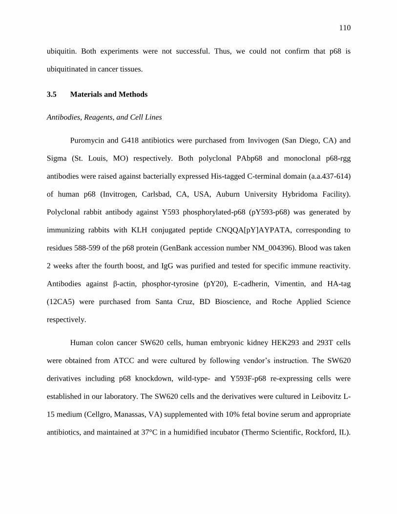
110
ubiquitin. Both experiments were not successful. Thus, we could not confirm that p68 is
ubiquitinated in cancer tissues.
3.5 Materials and Methods
Antibodies, Reagents, and Cell Lines
Puromycin and G418 antibiotics were purchased from Invivogen (San Diego, CA) and
Sigma (St. Louis, MO) respectively. Both polyclonal PAbp68 and monoclonal p68-rgg
antibodies were raised against bacterially expressed His-tagged C-terminal domain (a.a.437-614)
of human p68 (Invitrogen, Carlsbad, CA, USA, Auburn University Hybridoma Facility).
Polyclonal rabbit antibody against Y593 phosphorylated-p68 (pY593-p68) was generated by
immunizing rabbits with KLH conjugated peptide CNQQA[pY]AYPATA, corresponding to
residues 588-599 of the p68 protein (GenBank accession number NM_004396). Blood was taken
2 weeks after the fourth boost, and IgG was purified and tested for specific immune reactivity.
Antibodies against β-actin, phosphor-tyrosine (pY20), E-cadherin, Vimentin, and HA-tag
(12CA5) were purchased from Santa Cruz, BD Bioscience, and Roche Applied Science
respectively.
Human colon cancer SW620 cells, human embryonic kidney HEK293 and 293T cells
were obtained from ATCC and were cultured by following vendor’s instruction. The SW620
derivatives including p68 knockdown, wild-type- and Y593F-p68 re-expressing cells were
established in our laboratory. The SW620 cells and the derivatives were cultured in Leibovitz L-
15 medium (Cellgro, Manassas, VA) supplemented with 10% fetal bovine serum and appropriate
antibiotics, and maintained at 37°C in a humidified incubator (Thermo Scientific, Rockford, IL).
111
HEK293 and 293T cells were cultured in DMEM (Cellgro, Manassas, VA) supplemented with
10% FBS, and maintained at 37°C in a humidified incubator supplemented with 5% CO2.
Stable Knockdown of p68 by Retroviral System
The pSIREN-RetroQ retroviral vector (Clontech, Mountain View, CA) containing short
hairpin RNA (shRNA) targeting the sequence GAACTGCTCGCAGTACCAA of p68 (shRNA-
p68) and the control vector (shRNA-conl) were gifted from Dr. Janknecht (Shin et al 2007).
293T cells were co-transfected with 15µg of ShRNA-p68 or shRNA-conl plasmids and two
packing plasmids including 5µg of VSVG and 10µg of gag-pol using Lipofectamine 2000 (Life
Technologies, Grand Island, NY) per manufacturer’s instruction. The retroviruses were
harvested 48-72 hours after transfection and used to infect SW620 cells according to standard
procedures. The infected SW620 cells were selected in 10µg/ml puromycin (Invivogen, San
Diego, CA) and the individual clones were isolated after 2-week selection. The knockdown
efficiency of p68 in each clone was examined by Western blot (not shown) and RT-PCR. The
SW620 clones stably expressing p68 shRNA (SW620-p68KD) were then used in this study.
Transfection and Isolation of p68-expressing Stable Cell Lines
The pHM6 vectors containing wild-type or Y593F human p68 cDNA (Yang et al 2006)
were first undergone the site-directed mutagenesis on the p68 shRNA target site using
QuikChange II Site-Directed Mutagenesis Kit (Stratagene, La Jolla, CA), which resulted in the
silent mutations and allow these constructs to express p68 proteins in p68 shRNA-expressing
SW620 cells. The p68-knockdown SW620 cells were seeded in 6-well plates with 50 %
confluence and transfected with 4µg of each construct using Lipofectamine 2000 (Life
Technologies, Grand Island, NY) according to manufacturer’s instruction. After 48 hours of
112
transfection, the cells were plated in a 15-cm tissue culture dish and selected in 1mg/ml of G418
antibiotic for 4-6 weeks. The growing colonies were isolated individually and tested for the
expression of p68 by Western blot (not shown) and RT-PCR. The G418 concentration used on
the positive clones was gradually reduced to 200µg/ml during the routine culture, and the
stability of p68 expression was tested. The positive clones were then used in this study. The
empty pHM6 vector-transfected cells were undergone the same procedure and used as a control.
RT-PCR Analysis
Cells were properly treated, and then total RNA was extracted using RNeasy Mini Kit
(Qiagen, Valencia, CA). The RNA was quantified and then converted to cDNA using the
GoScript reverse transcription system (Promega, Madison, WI) following the manufacturer’s
instruction. The cDNA was then used in the final PCR reaction. PCR products were visualized
after electrophoresis in a 1% agarose gel and stained by ethidium bromide (0.5 mg/ml).
Densitometry was performed using ImageJ program. Primers used were: E-cadherin (sense 5’-
AGAAGACAGAAGAGAGACTGGG-3’ antisense 5’-TCCTCAACTGCATTCCCGTT-3’);
HA-tagged p68 (HA-sense 5’-TACCCATAC GACGTCCCAGACTA-3’ P68RV301-antisense
5’-ACATCCATGACATTTGCAGGGAA-3’); p68(endogenous) (sense 5’-
TGGAAGAACTGCTCGCAGTACCAA-3’ antisense 5’-
TGGAACGACCTGAACCTCTGTCTT-3’); Snail1 (sense 5’-TCTAGGCCCTGGCTGCTAC-3’
antisense 5’-GCCTGGCACTGGTACTTCTT-3’); Vimentin (sense 5’-
AATGCGTCTCTGGCACGT-3’ antisense 5’-ATTTCACGCATCTGGCGTTC-3’); GAPDH
(sense 5’-GAGTCAACGGATTTGGTCGT-3’ antisense 5’-TTGATTTTGGAGGGATCTCG-
3’). Primers for GAPDH were used as PCR and loading controls. Controls for the RT reaction
(not shown) contained no template or no reverse transcriptase.
113
The PCR primers (Liu et al 2003b) used in the metastasis study in xenograft tumor model
included: human keratin-18 (sense 5’-GCCTACAAGCCCAGATTGCC-3’ antisense 5’-
GGTGGTCTTTTGGATGGTTTGC-3’); human β-actin (sense 5’-
ATGGATGATGATATCGCCGCG-3’ antisense 5’-CTAGAAGCATTTGCGGTGGAC-3’);
mouse β-actin (sense 5’-ATTGTTACCAACTGGGACGACATG-3’ antisense 5’-
CTTCATGAGGTAGTCTGTCAGGTC-3’). The PCR parameters used were as following: 30 s
at 94°C, 30 s at 60°C, and 1 min at 72°C for 30 cycles. Three primer sets were used for detecting
exogenous expression of p68 including: 1. HA-tag+ P68RV301 (sequence listed above); 2. p68
601FW (sense 5’-ATCTACGGTGGTGCTCCTAA-3’)+p68MutRTRV (antisense 5’-
TTGGTGCTCCGCGCCGTT-3’) with~700bp product; 3. p68 MutRTFW (sense 5’-
AACGGCGCGGAGCACCAA-3’)+ p68 (antisense 5’-TGGAACGACCTGAACCTCTGTCTT-
3’) with ~150bp product.
Proliferation Assay
The MTT (3-(4,5-Dimethylthiazol-2-yl)-2,5-diphenyltetrazolium bromide, Sigma, St.
Louis, MO) method was used to determine the cell proliferation. Cells with the proper treatment
were seeded in a 96-well tissue culture plate with an amount of 104 cells per well. On the second
day, the culture cells were replaced with 100µl of MTT-containing media (10µl of MTT stock
solution (5mg/ml) in 90µl of the culture medium without phenol red). The cells were returned to
the incubator for 2 hours. After the incubation, the resulting formazan crystals were dissolved by
adding 100µl of MTT solubilization solution (10% Triton X-100 plus 0.1 N HCl in anhydrous
isopropanol) and mixed well by pipetting up and down. The absorbance at a wavelength of
570nm was measured by a spectrophotometer. The background absorbance was eliminated by
measuring the absorbance at a wavelength of 690nm and subtracted from the 570nm
114
measurement. A blank containing complete medium without cells was included, and each group
of cells was repeated in 8 wells for each independent experiment. The statistical result was based
on three independent experiments.
In Vitro Trans-well Assay
The cell migration assays using trans-well system (BD Biosciences, Franklin Lakes, NJ)
were performed on SW620 cells with proper treatments per manufacturer's instructions. The cells
were serum-starved overnight, and 5x10⁴ of cells in 100µl were plated onto the top chamber of
24-well transwell plates with 8 µm pore size of trans-membrane in serum-free condition. The
surface of the trans-membrane facing the bottom chamber was coated with 2µg/cm2 of human
fibronectin (BD biosciences, Franklin Lakes, NJ), and the bottom chamber was filled with 400µl
of complete culture medium. The trans-well plates were returned to the incubator and incubated
for 48 hours. The cells trans-migrating to the bottom side of the membrane were fixed with 3.7%
formaldehyde solution and stained with 0.5% crystal violet. The non-migrated cells inside the
top chamber were removed by swabbing. The migrating cells with crystal violet staining were
quantitated by solubilizing the dye into Sorenson’s buffer (0.1 M sodium citrate and 50% ethanol,
pH 4.2) and measuring the absorbance at 590nm wavelength.
Analyses of Tissue Samples
Frozen patient tissue specimens were obtained from University of Massachusetts Medical
School Cancer Center (Worcester, MA). The specimens (200 ~300mg) were weighted and diced
into small pieces using a clean razor blade. Collected pieces were immersed in liquid nitrogen
and disrupted in a pre-chilled 1.5ml micro-centrifuge tube on ice using an electric pestle (VWR,
Radnor, PA). Each disrupted specimen was lysed in 500µl of 1X RIPA buffer and further
115
homologized by using the pestle manually for 10 strokes. The lysate was rotated for 60 min at
4°C and then centrifuged at 1,2000rpm for 15 min at 4°C to remove debris. The supernatant was
stored at –80°C until use.
Preparation of Cell Lysates, Nuclear Extracts, Immunoprecipitation, and Immunoblot
The experimental procedures for cell lysates and nuclear extracts, and for
immunoprecipitation and immunoblot were similar to those described in our previous reports
(Carter et al 2010, Yang et al 2007).
Tumor Growth in Nude Mice
All animal experiments were carried out in accordance with the guidelines of IACUC of
Georgia State University. SW620 cells or its derivatives were inoculated subcutaneously into
athymic nude mice (Harlan Laboratories, Indianapolis, IN) in the right flank with 2 X 10⁶ cells
per mouse. Tumor growth was monitored by digital caliper in two dimensions, and the volume
was calculated based on the formula of (width squared X length)/2. Four weeks after the tumor
cell inoculation, spleens and tumors were isolated from each mouse and immersed in RNAlater
RNA Stabilization Reagent (Qiagen, Valencia, CA) for later use of total RNA preparation. The
RT-PCR based micrometastasis detection in mouse spleen was following the procedures reported
previously and the primers were listed in RT-PCR analysis section (Liu et al 2003b).
Statistical Analysis
All statistical analyses were performed by using Analysis ToolPack in Microsoft Excel
2010. Two groups of samples were first analyzed by F-test to determine equal or unequal
variances, and then analyzed by appropriate type of t-Test to determine the p value.
116
3.6 References
Carter CL, Lin C, Liu CY, Yang L, Liu ZR (2010). Phosphorylated p68 RNA helicase activates
Snail1 transcription by promoting HDAC1 dissociation from the Snail1 promoter. Oncogene 29:
5427-5436.
Causevic M, Hislop RG, Kernohan NM, Carey FA, Kay RA, Steele RJ et al (2001).
Overexpression and poly-ubiquitylation of the DEAD-box RNA helicase p68 in colorectal
tumours. Oncogene 20: 7734-7743.
Chaffer CL, Weinberg RA (2011). A perspective on cancer cell metastasis. Science 331: 1559-
1564.
Crawford L, Leppard K, Lane D, Harlow E (1982). Cellular proteins reactive with monoclonal
antibodies directed against simian virus 40 T-antigen. J Virol 42: 612-620.
Janknecht R (2010). Multi-talented DEAD-box proteins and potential tumor promoters: p68
RNA helicase (DDX5) and its paralog, p72 RNA helicase (DDX17). Am J Transl Res 2: 223-
234.
Lane DP, Hoeffler WK (1980). SV40 large T shares an antigenic determinant with a cellular
protein of molecular weight 68,000. Nature 288: 167-170.
Leibovitz A, Stinson JC, McCombs WB, 3rd, McCoy CE, Mazur KC, Mabry ND (1976).
Classification of human colorectal adenocarcinoma cell lines. Cancer Res 36: 4562-4569.
Liu K, McDuffie E, Abrams SI (2003a). Exposure of human primary colon carcinoma cells to
anti-Fas interactions influences the emergence of pre-existing Fas-resistant metastatic
subpopulations. J Immunol 171: 4164-4174.
Liu K, McDuffie E, Abrams SI (2003b). Exposure of human primary colon carcinoma cells to
anti-Fas interactions influences the emergence of pre-existing Fas-resistant metastatic
subpopulations. J Immunol 171: 4164-4174.
Rhim AD, Mirek ET, Aiello NM, Maitra A, Bailey JM, McAllister F et al (2012). EMT and
Dissemination Precede Pancreatic Tumor Formation. Cell 148: 349-361.
Schmid SR, Linder P (1992). D-E-A-D protein family of putative RNA helicases. Mol Microbiol
6: 283-291.
Shin S, Rossow KL, Grande JP, Janknecht R (2007). Involvement of RNA helicases p68 and p72
in colon cancer. Cancer Res 67: 7572-7578.
Stevenson RJ, Hamilton SJ, MacCallum DE, Hall PA, Fuller-Pace FV (1998). Expression of the
'dead box' RNA helicase p68 is developmentally and growth regulated and correlates with organ
differentiation/maturation in the fetus. J Pathol 184: 351-359.
117
Yang L, Liu ZR (2004). Bacterially expressed recombinant p68 RNA helicase is phosphorylated
on serine, threonine, and tyrosine residues. Protein Expr Purif 35: 327-333.
Yang L, Lin C, Liu ZR (2005). Phosphorylations of DEAD box p68 RNA helicase are associated
with cancer development and cell proliferation. Mol Cancer Res 3: 355-363.
Yang L, Lin C, Liu ZR (2006). P68 RNA helicase mediates PDGF-induced epithelial
mesenchymal transition by displacing Axin from beta-catenin. Cell 127: 139-155.
Yang L, Lin C, Zhao S, Wang H, Liu ZR (2007). Phosphorylation of p68 RNA helicase plays a
role in platelet-derived growth factor-induced cell proliferation by up-regulating cyclin D1 and c-
Myc expression. J Biol Chem 282: 16811-16819.
118
Figure 3.1 P68/HA-p68 levels in sublines of SW620
Cellular levels of mRNA (A) and protein (B) of p68 in SW620 cells were analyzed by RT-PCR
(A) and immunoblot (B) using antibody against p68 (IB:p68). The cells were infected with virus
that carries non-targeting shRNA (NT) or shRNA target p68 (p68). The Clone# represents the
selected sublines. RT-PCR (A) and immunoblot (B) analyses of GAPDH (GAPDH and
IB:GAPDH) are loading controls. (C) Cellular levels of mRNA of endogenous p68 (En-p68) and
exogenous p68 (HA-p68) in sublines of SW620 by RT-PCR using the primer pairs indicated in
bottom panel. Endogenous p68 was knocked down and HA-p68s, wild-type (WT) and Y593F
mutant (Y593F) were exogenously expressed. Vec means the cells were infected with virus
carries empty vector. The Clone# represents the selected sublines. RT-PCR analyses of mRNA
of GAPDH (GAPDH) are loading control.
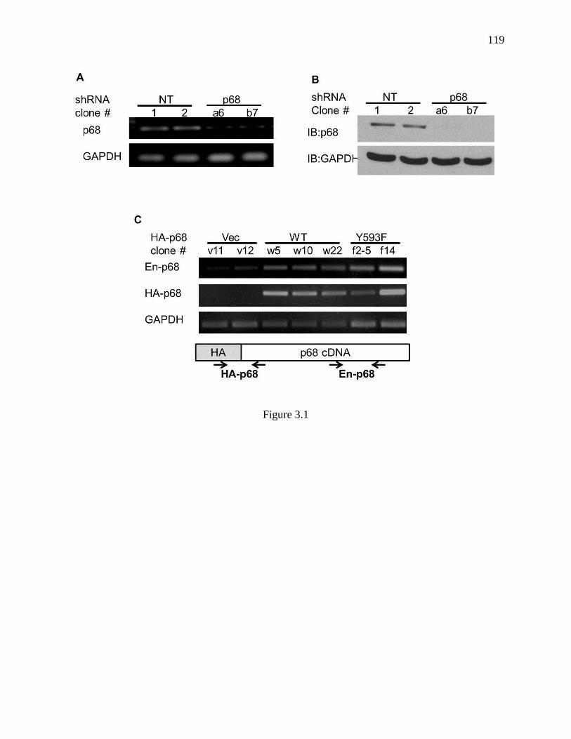
119
Figure 3.1
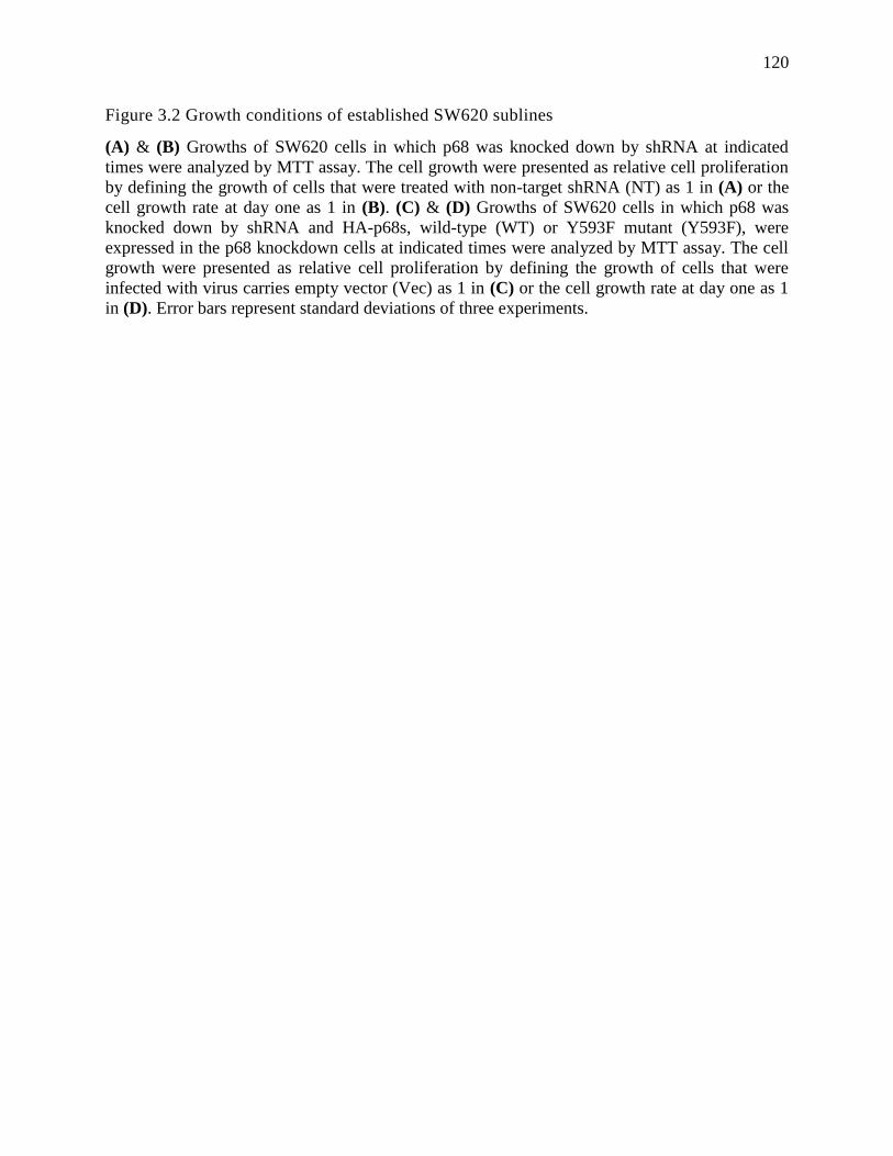
120
Figure 3.2 Growth conditions of established SW620 sublines
(A) & (B) Growths of SW620 cells in which p68 was knocked down by shRNA at indicated
times were analyzed by MTT assay. The cell growth were presented as relative cell proliferation
by defining the growth of cells that were treated with non-target shRNA (NT) as 1 in (A) or the
cell growth rate at day one as 1 in (B). (C) & (D) Growths of SW620 cells in which p68 was
knocked down by shRNA and HA-p68s, wild-type (WT) or Y593F mutant (Y593F), were
expressed in the p68 knockdown cells at indicated times were analyzed by MTT assay. The cell
growth were presented as relative cell proliferation by defining the growth of cells that were
infected with virus carries empty vector (Vec) as 1 in (C) or the cell growth rate at day one as 1
in (D). Error bars represent standard deviations of three experiments.
121
Figure 3.2
122
Figure 3.3 Cell migration and epithelial/mesenchymal marker characterization in SW620
sublines
(A) & (B) Cell migration of SW620 cells in which p68 was knocked down by shRNA (A) and
HA-p68s, wild-type (WT) or Y593F mutant (Y593F), were expressed in the p68 knockdown
cells (B) was measured by Boyden chamber assay. The cell migrations were presented as relative
cell migration by defining the migration rate of the cells that were treated with non-target shRNA
(NT) as 1 in (A) or of the cells that were infected with virus that carries empty vectors after p68
knockdown (Vec) as 1 in (B). Error bars represent standard deviations of three experiments. (C)
& (D) Cellular mRNA levels of E-cadherin (E-cadherin), Snail, and Vimentin (Vimentin) in
SW620 cells in which p68 was knocked down by shRNA (C) and HA-p68s, wild-type (WT) or
Y593F mutant (Y593F), were expressed in the p68 knockdown cells (D) were measured by RT-
PCR. NT in (C) means the cells were treated with non-target shRNA. Vec in (D) means the cells
were infected with virus that carries empty vectors after p68 knockdown. Analyses of mRNA
levels of GAPDH are loading controls.
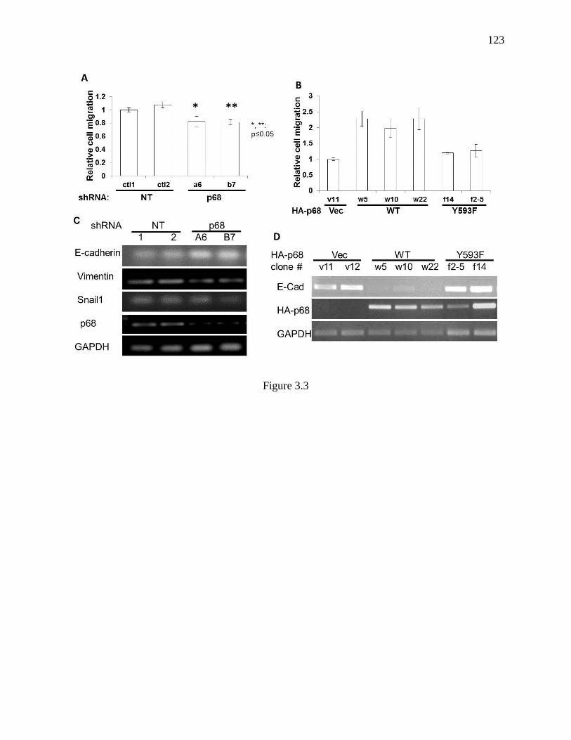
123
Figure 3.3
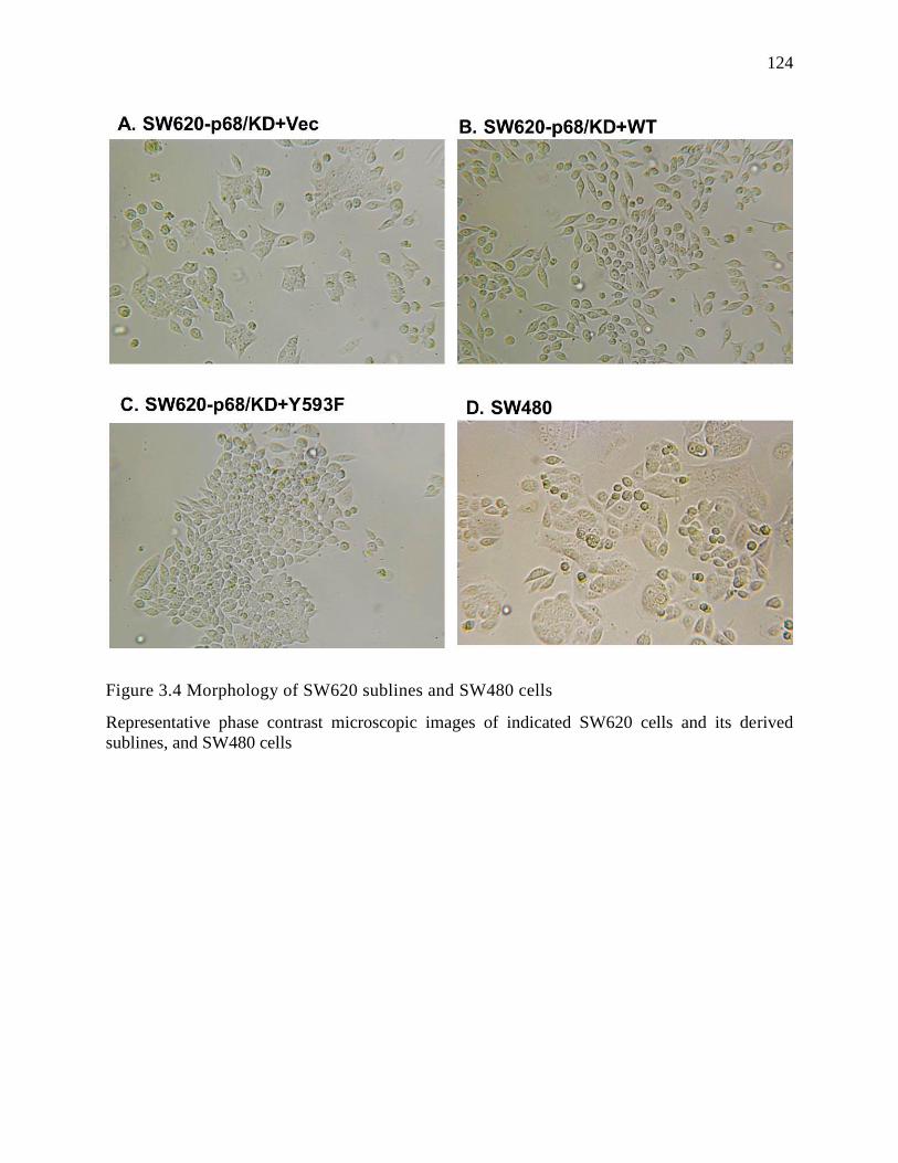
124
Figure 3.4 Morphology of SW620 sublines and SW480 cells
Representative phase contrast microscopic images of indicated SW620 cells and its derived
sublines, and SW480 cells
125
Figure 3.5 The effects of p68 mutations on metastasis of xenograft of SW620 tumors.
(A) Tumor growth was monitored by measuring tumor volumes every three days and the tumor
volumes were calculated by formula; Tumor volume = 1/2 x (width) 2 x length. (B) The end
point weight of harvested tumors of SW620 and its derived sublines after 28 days growth. (C)
The mRNA levels of human cytokarotine-18 (hCK-18) in the spleen extracts of tumor-bearing
mice that carry tumors of SW620 or its derived sublines (indicated) were analyzed by RT-PCR.
The mRNA levels of mouse -actin (mActin) in the spleen extracts are loading controls. Error
bars in (A) & (B) represent standard deviations of three experiments.
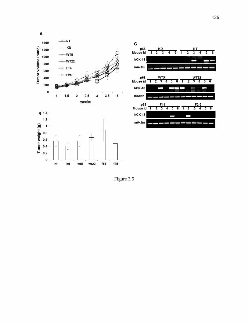
126
Figure 3.5

127
Figure 3.6 mRNA levels of p68 in SW620 tumors
The mRNA levels of endogenous and exogenous p68 in the implanted tumors of SW620 and its
derived sublines were examined by RT-PCR using primer pairs to cover different regions of
ORF of the p68 (illustrated in Upper penal). RT-PCR examination of mRNA levels of human
cytokarotine-18 (hCK-18) and human -actin (hActin) in the tumor tissue extracts are loading
controls.
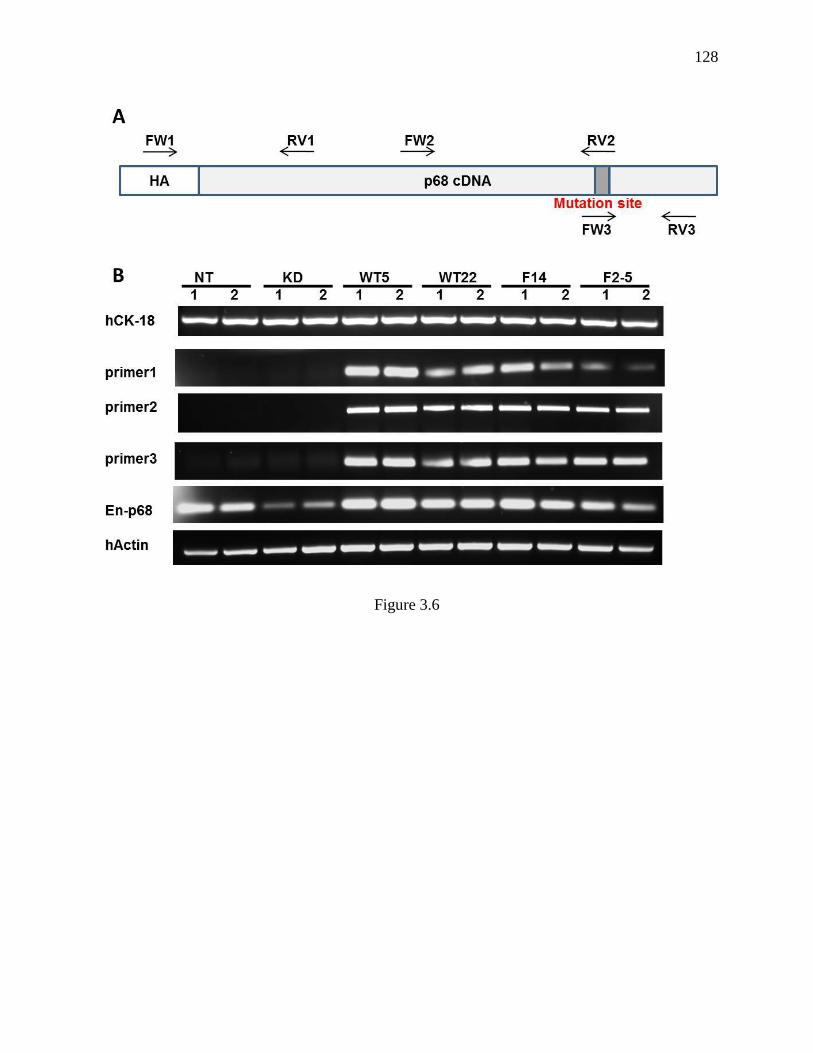
128
Figure 3.6
129
Figure 3.7 Phosphorylation level of p68 at Y593 in colon cancer tissues
(A) & (B) Tests of specificity of antibody raised against Y593 phosphorylated p68 (A)
Immunoblot analyses of bacterially expressed recombinant p68 that was treated/untreated by c-
Abl (+/-) and YOP (+/-) using different antibodies: Antibody against the Y593 phosphorylated
p68 (IB:p-p68), antibody against tyrosine phosphor-protein (IB:pY20), and antibody against p68
(IB:p68). (B) Immunoblot analyses of exogenous HA-p68s (WT and indicated mutant) that were
immunoprecipitated by anti-HA antibody (IP:HA) using antibody against the Y593
phosphorylated p68 (IB:p-p68). Immunoblot of total p68 in the IPs indicate amount of IPed HA-
p68. The input (IB:HA and IB:GAPDH) are the immunoblots of cell extracts without IP and are
loading controls. (C) Immunoblot analyses of p68 in colon cancer and corresponding normal
tissue samples (indicated by the patent ID numbers) using antibodies against the Y593
phosphorylated p68 (IB:p-p68) and p68 (IB:p68). Immunoblot of GAPDH (IB:GAPDH) is a
loading control. (D) Quantitation of the immunoblots in (C) by normalizing the IB signals of
Y593 phosphorylated p68 to the total p68 IB signals (relative intensity of Y-593 phosphor-p68).
Error bars represent standard deviations of three experiments.
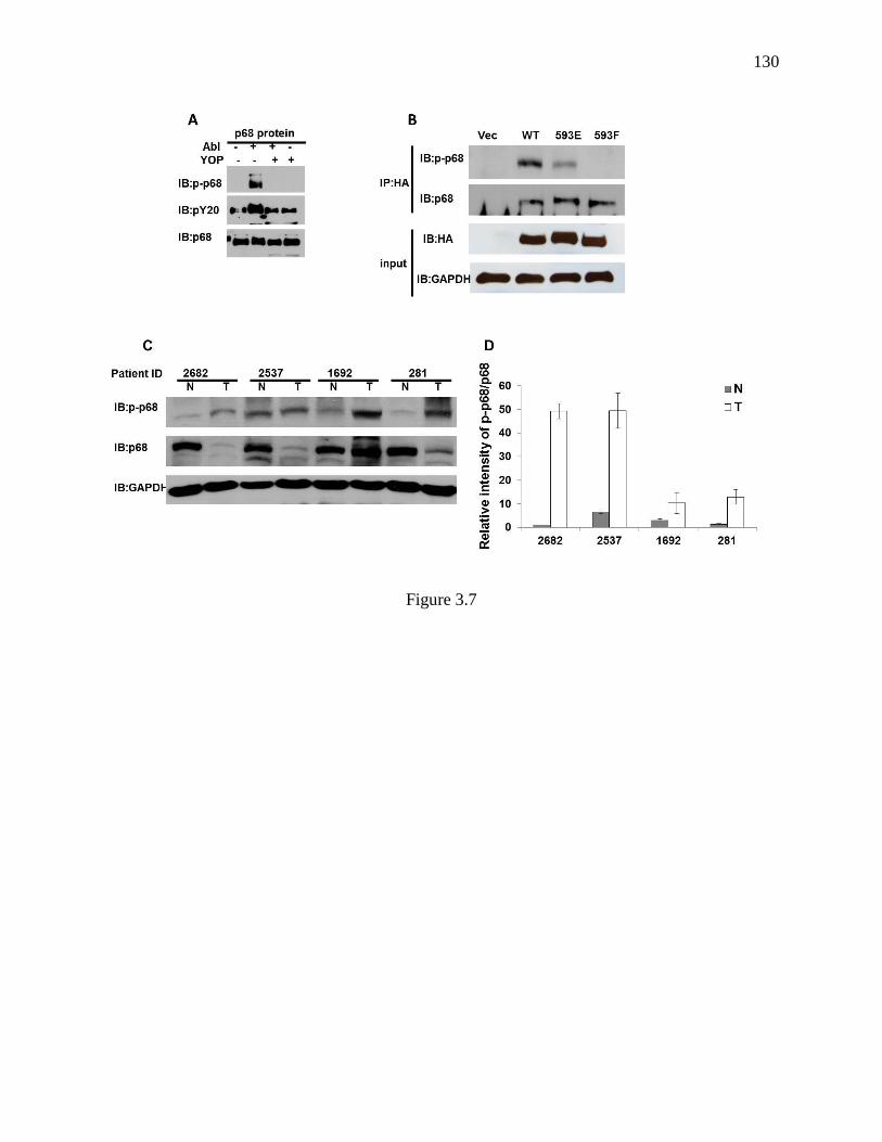
130
Figure 3.7
131
Figure 3.8 Comparison of immunoblot of p68 between tissue extracts and cultured cell
extracts.
Immunoblot analyses of p68 in the tissue extracts from colon cancer tissue samples (T) and
corresponding normal tissue samples (N) using antibody against the Y593 phosphorylated p68
(IB:p-p68, Upper panel) and antibody against p68 (IB:p68, middle panel). M means the tissue
extracts prepared from metastatic tumors in small bowl. The panels on the right are the same
immunoblots using extracts of SW620, HCT116, and HT29 cultured colon cancer cells. The
numbers on the left are the molecular weight markers. Immunoblot of GAPDH (IB:GAPDH) is a
loading control.
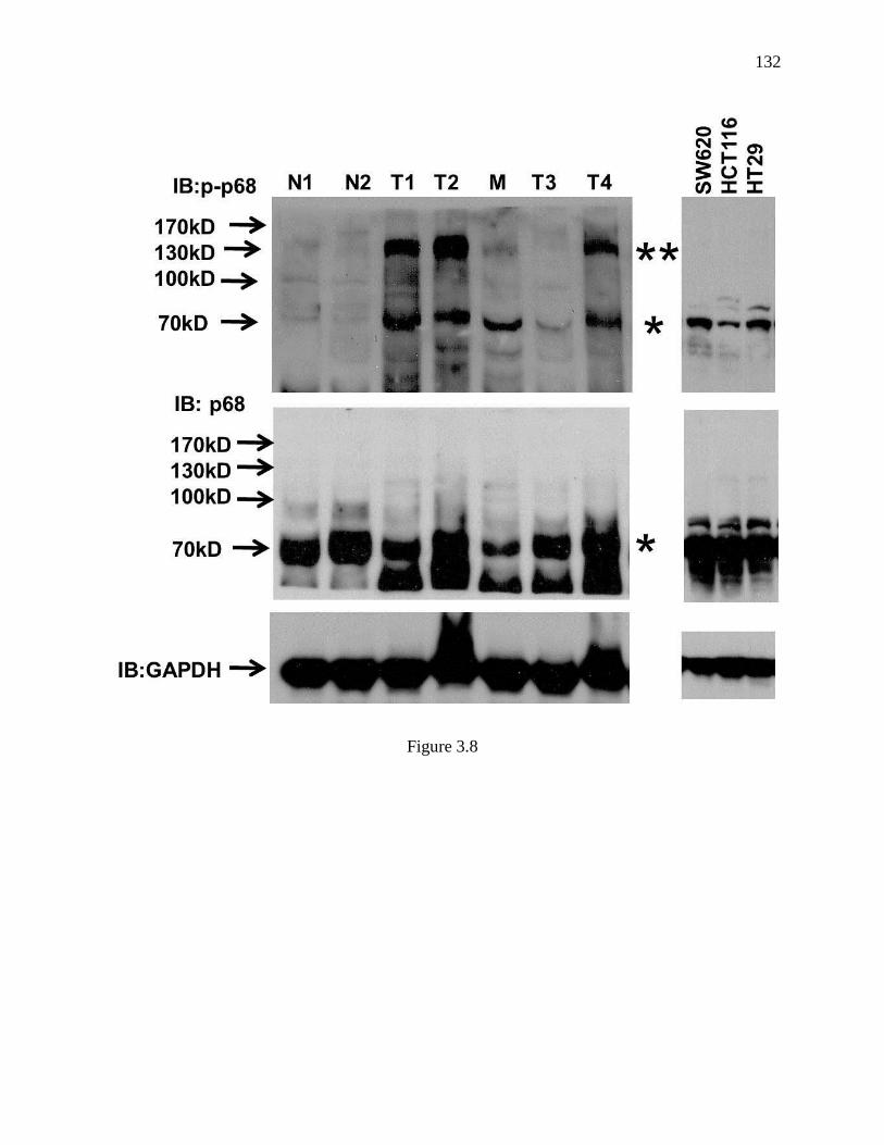
132
Figure 3.8
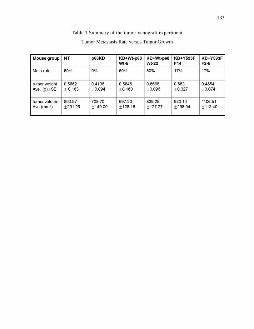
133
Table 1 Summary of the tumor xenograft experiment
Tumor Metastasis Rate versus Tumor Growth
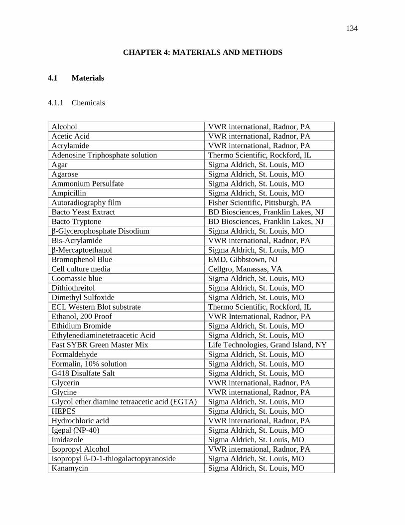
134
CHAPTER 4: MATERIALS AND METHODS
4.1 Materials
4.1.1 Chemicals
Alcohol VWR international, Radnor, PA
Acetic Acid VWR international, Radnor, PA
Acrylamide VWR international, Radnor, PA
Adenosine Triphosphate solution Thermo Scientific, Rockford, IL
Agar Sigma Aldrich, St. Louis, MO
Agarose Sigma Aldrich, St. Louis, MO
Ammonium Persulfate Sigma Aldrich, St. Louis, MO
Ampicillin Sigma Aldrich, St. Louis, MO
Autoradiography film Fisher Scientific, Pittsburgh, PA
Bacto Yeast Extract BD Biosciences, Franklin Lakes, NJ
Bacto Tryptone BD Biosciences, Franklin Lakes, NJ
β-Glycerophosphate Disodium Sigma Aldrich, St. Louis, MO
Bis-Acrylamide VWR international, Radnor, PA
β-Mercaptoethanol Sigma Aldrich, St. Louis, MO
Bromophenol Blue EMD, Gibbstown, NJ
Cell culture media Cellgro, Manassas, VA
Coomassie blue Sigma Aldrich, St. Louis, MO
Dithiothreitol Sigma Aldrich, St. Louis, MO
Dimethyl Sulfoxide Sigma Aldrich, St. Louis, MO
ECL Western Blot substrate Thermo Scientific, Rockford, IL
Ethanol, 200 Proof VWR International, Radnor, PA
Ethidium Bromide Sigma Aldrich, St. Louis, MO
Ethylenediaminetetraacetic Acid Sigma Aldrich, St. Louis, MO
Fast SYBR Green Master Mix Life Technologies, Grand Island, NY
Formaldehyde Sigma Aldrich, St. Louis, MO
Formalin, 10% solution Sigma Aldrich, St. Louis, MO
G418 Disulfate Salt Sigma Aldrich, St. Louis, MO
Glycerin VWR international, Radnor, PA
Glycine VWR international, Radnor, PA
Glycol ether diamine tetraacetic acid (EGTA) Sigma Aldrich, St. Louis, MO
HEPES Sigma Aldrich, St. Louis, MO
Hydrochloric acid VWR international, Radnor, PA
Igepal (NP-40) Sigma Aldrich, St. Louis, MO
Imidazole Sigma Aldrich, St. Louis, MO
Isopropyl Alcohol VWR international, Radnor, PA
Isopropyl ß-D-1-thiogalactopyranoside Sigma Aldrich, St. Louis, MO
Kanamycin Sigma Aldrich, St. Louis, MO
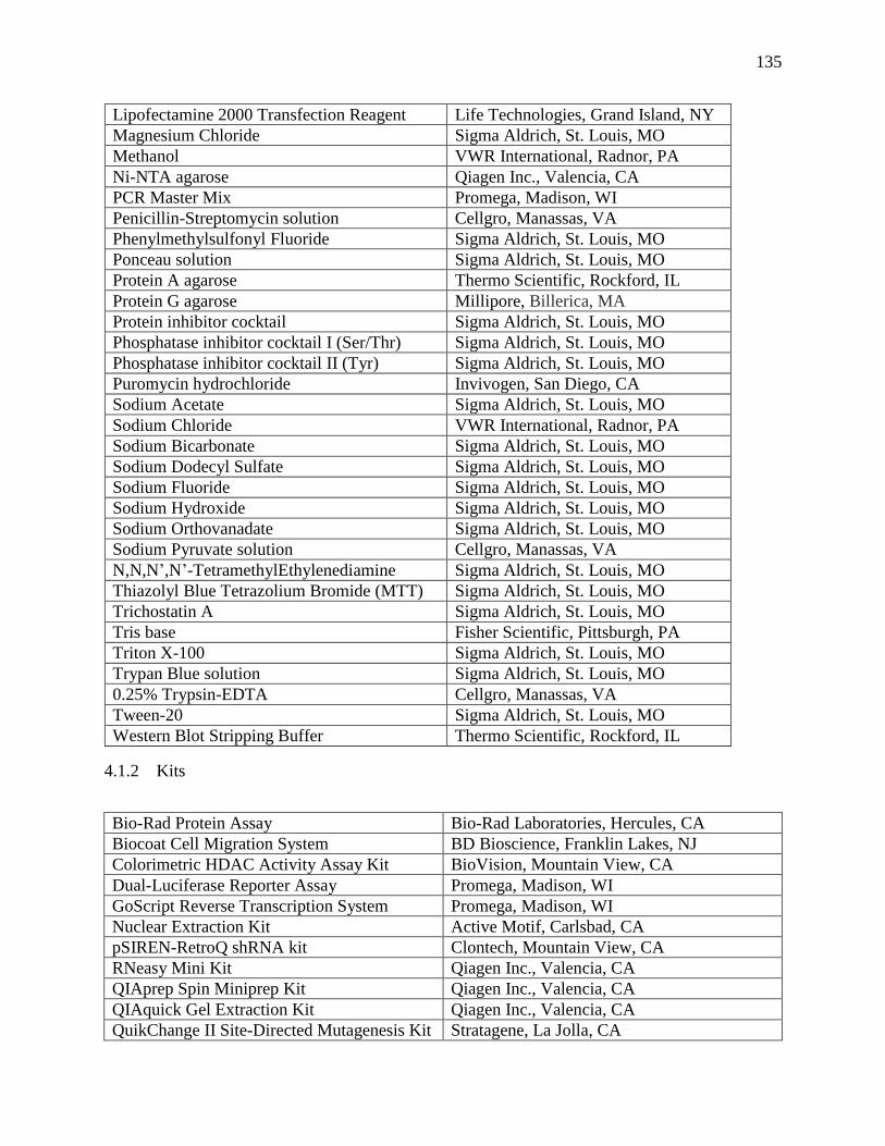
135
Lipofectamine 2000 Transfection Reagent Life Technologies, Grand Island, NY
Magnesium Chloride Sigma Aldrich, St. Louis, MO
Methanol VWR International, Radnor, PA
Ni-NTA agarose Qiagen Inc., Valencia, CA
PCR Master Mix Promega, Madison, WI
Penicillin-Streptomycin solution Cellgro, Manassas, VA
Phenylmethylsulfonyl Fluoride Sigma Aldrich, St. Louis, MO
Ponceau solution Sigma Aldrich, St. Louis, MO
Protein A agarose Thermo Scientific, Rockford, IL
Protein G agarose Millipore, Billerica, MA
Protein inhibitor cocktail Sigma Aldrich, St. Louis, MO
Phosphatase inhibitor cocktail I (Ser/Thr) Sigma Aldrich, St. Louis, MO
Phosphatase inhibitor cocktail II (Tyr) Sigma Aldrich, St. Louis, MO
Puromycin hydrochloride Invivogen, San Diego, CA
Sodium Acetate Sigma Aldrich, St. Louis, MO
Sodium Chloride VWR International, Radnor, PA
Sodium Bicarbonate Sigma Aldrich, St. Louis, MO
Sodium Dodecyl Sulfate Sigma Aldrich, St. Louis, MO
Sodium Fluoride Sigma Aldrich, St. Louis, MO
Sodium Hydroxide Sigma Aldrich, St. Louis, MO
Sodium Orthovanadate Sigma Aldrich, St. Louis, MO
Sodium Pyruvate solution Cellgro, Manassas, VA
N,N,N’,N’-TetramethylEthylenediamine Sigma Aldrich, St. Louis, MO
Thiazolyl Blue Tetrazolium Bromide (MTT) Sigma Aldrich, St. Louis, MO
Trichostatin A Sigma Aldrich, St. Louis, MO
Tris base Fisher Scientific, Pittsburgh, PA
Triton X-100 Sigma Aldrich, St. Louis, MO
Trypan Blue solution Sigma Aldrich, St. Louis, MO
0.25% Trypsin-EDTA Cellgro, Manassas, VA
Tween-20 Sigma Aldrich, St. Louis, MO
Western Blot Stripping Buffer Thermo Scientific, Rockford, IL
4.1.2 Kits
Bio-Rad Protein Assay Bio-Rad Laboratories, Hercules, CA
Biocoat Cell Migration System BD Bioscience, Franklin Lakes, NJ
Colorimetric HDAC Activity Assay Kit BioVision, Mountain View, CA
Dual-Luciferase Reporter Assay Promega, Madison, WI
GoScript Reverse Transcription System Promega, Madison, WI
Nuclear Extraction Kit Active Motif, Carlsbad, CA
pSIREN-RetroQ shRNA kit Clontech, Mountain View, CA
RNeasy Mini Kit Qiagen Inc., Valencia, CA
QIAprep Spin Miniprep Kit Qiagen Inc., Valencia, CA
QIAquick Gel Extraction Kit Qiagen Inc., Valencia, CA
QuikChange II Site-Directed Mutagenesis Kit Stratagene, La Jolla, CA
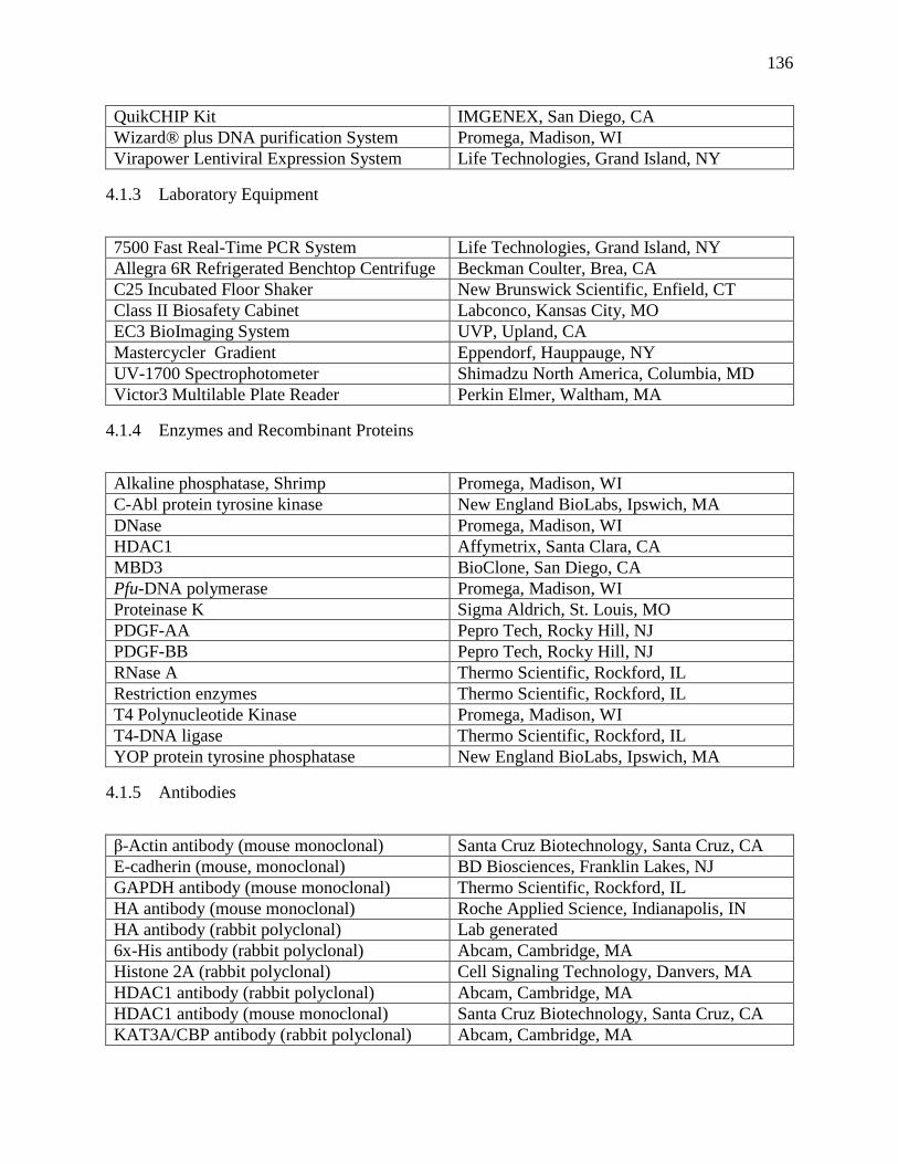
136
QuikCHIP Kit IMGENEX, San Diego, CA
Wizard® plus DNA purification System Promega, Madison, WI
Virapower Lentiviral Expression System Life Technologies, Grand Island, NY
4.1.3 Laboratory Equipment
7500 Fast Real-Time PCR System Life Technologies, Grand Island, NY
Allegra 6R Refrigerated Benchtop Centrifuge Beckman Coulter, Brea, CA
C25 Incubated Floor Shaker New Brunswick Scientific, Enfield, CT
Class II Biosafety Cabinet Labconco, Kansas City, MO
EC3 BioImaging System UVP, Upland, CA
Mastercycler Gradient Eppendorf, Hauppauge, NY
UV-1700 Spectrophotometer Shimadzu North America, Columbia, MD
Victor3 Multilable Plate Reader Perkin Elmer, Waltham, MA
4.1.4 Enzymes and Recombinant Proteins
Alkaline phosphatase, Shrimp Promega, Madison, WI
C-Abl protein tyrosine kinase New England BioLabs, Ipswich, MA
DNase Promega, Madison, WI
HDAC1 Affymetrix, Santa Clara, CA
MBD3 BioClone, San Diego, CA
Pfu-DNA polymerase Promega, Madison, WI
Proteinase K Sigma Aldrich, St. Louis, MO
PDGF-AA Pepro Tech, Rocky Hill, NJ
PDGF-BB Pepro Tech, Rocky Hill, NJ
RNase A Thermo Scientific, Rockford, IL
Restriction enzymes Thermo Scientific, Rockford, IL
T4 Polynucleotide Kinase Promega, Madison, WI
T4-DNA ligase Thermo Scientific, Rockford, IL
YOP protein tyrosine phosphatase New England BioLabs, Ipswich, MA
4.1.5 Antibodies
β-Actin antibody (mouse monoclonal) Santa Cruz Biotechnology, Santa Cruz, CA
E-cadherin (mouse, monoclonal) BD Biosciences, Franklin Lakes, NJ
GAPDH antibody (mouse monoclonal) Thermo Scientific, Rockford, IL
HA antibody (mouse monoclonal) Roche Applied Science, Indianapolis, IN
HA antibody (rabbit polyclonal) Lab generated
6x-His antibody (rabbit polyclonal) Abcam, Cambridge, MA
Histone 2A (rabbit polyclonal) Cell Signaling Technology, Danvers, MA
HDAC1 antibody (rabbit polyclonal) Abcam, Cambridge, MA
HDAC1 antibody (mouse monoclonal) Santa Cruz Biotechnology, Santa Cruz, CA
KAT3A/CBP antibody (rabbit polyclonal) Abcam, Cambridge, MA
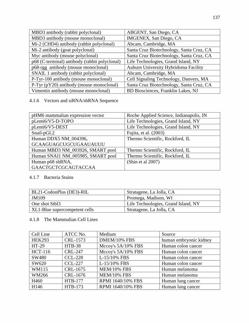
137
MBD3 antibody (rabbit polyclonal) ABGENT, San Diego, CA
MBD3 antibody (mouse monoclonal) IMGENEX, San Diego, CA
Mi-2 (CHD4) antibody (rabbit polyclonal) Abcam, Cambridge, MA
Mi-2 antibody (goat polyclonal) Santa Cruz Biotechnology, Santa Cruz, CA
Myc antibody (mouse polyclonal) Santa Cruz Biotechnology, Santa Cruz, CA
p68 (C-terminal) antibody (rabbit polyclonal) Life Technologies, Grand Island, NY
p68-rgg antibody (mouse monoclonal) Auburn University Hybridoma Facility
SNAIL 1 antibody (rabbit polyclonal) Abcam, Cambridge, MA
P-Tyr-100 antibody (mouse monoclonal) Cell Signaling Technology, Danvers, MA
P-Tyr (pY20) antibody (mouse monoclonal) Santa Cruz Biotechnology, Santa Cruz, CA
Vimentin antibody (mouse monoclonal) BD Biosciences, Franklin Lakes, NJ
4.1.6 Vectors and siRNA/shRNA Sequence
pHM6 mammalian expression vector Roche Applied Science, Indianapolis, IN
pLenti6/V5-D-TOPO Life Technologies, Grand Island, NY
pLenti6/V5-DEST Life Technologies, Grand Island, NY
Snail-pGL2 Fujita, et al. (2003)
Human DDX5 NM_004396,
GCAAGUAGCUGCUGAAUAUUU
Thermo Scientific, Rockford, IL
Human MBD3 NM_003926, SMART pool Thermo Scientific, Rockford, IL
Human SNAI1 NM_005985, SMART pool Thermo Scientific, Rockford, IL
Human p68 shRNA,
GAACTGCTCGCAGTACCAA
(Shin et al 2007)
4.1.7 Bacteria Stains
BL21-CodonPlus (DE3)-RIL Stratagene, La Jolla, CA
JM109 Promega, Madison, WI
One shot Stbl3 Life Technologies, Grand Island, NY
XL1-Blue supercompetent cells Stratagene, La Jolla, CA
4.1.8 The Mammalian Cell Lines
Cell Line ATCC No. Medium Source
HEK293 CRL-1573 DMEM/10% FBS human embryonic kidney
HT-29 HTB-38 Mccoy's 5A/10% FBS Human colon cancer
HCT-116 CRL-247 Mccoy's 5A/10% FBS Human colon cancer
SW480 CCL-228 L-15/10% FBS Human colon cancer
SW620 CCL-227 L-15/10% FBS Human colon cancer
WM115 CRL-1675 MEM/10% FBS Human melanoma
WM266 CRL-1676 MEM/10% FBS Human melanoma
H460 HTB-177 RPMI 1640/10% FBS Human lung cancer
H146 HTB-173 RPMI 1640/10% FBS Human lung cancer
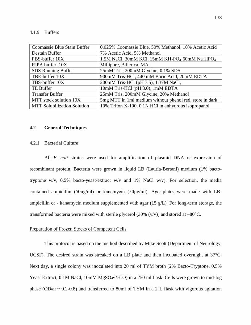
138
4.1.9 Buffers
Coomassie Blue Stain Buffer 0.025% Coomassie Blue, 50% Methanol, 10% Acetic Acid
Destain Buffer 7% Acetic Acid, 5% Methanol
PBS-buffer 10X 1.5M NaCl, 30mM KCl, 15mM KH2PO4, 60mM Na2HPO4
RIPA buffer, 10X Millipore, Billerica, MA
SDS Running Buffer 25mM Tris, 200mM Glycine, 0.1% SDS
TBE-buffer 10X 900mM Tris-HCl, 440 mM Boric Acid, 20mM EDTA
TBS-buffer 10X 200mM Tris-HCl (pH 7.5), 1.37M NaCl,
TE Buffer 10mM Tris-HCl (pH 8.0), 1mM EDTA
Transfer Buffer 25mM Tris, 200mM Glycine, 20% Methanol
MTT stock solution 10X 5mg MTT in 1ml medium without phenol red, store in dark
MTT Solubilization Solution 10% Triton X-100, 0.1N HCl in anhydrous isopropanol
4.2 General Techniques
4.2.1 Bacterial Culture
All E. coli strains were used for amplification of plasmid DNA or expression of
recombinant protein. Bacteria were grown in liquid LB (Lauria-Bertani) medium (1% bacto-
tryptone w/v, 0.5% bacto-yeast-extract w/v and 1% NaCl w/v). For selection, the media
contained ampicillin (50μg/ml) or kanamycin (50μg/ml). Agar-plates were made with LB-
ampicillin or - kanamycin medium supplemented with agar (15 g/L). For long-term storage, the
transformed bacteria were mixed with sterile glycerol (30% (v/v)) and stored at –80°C.
Preparation of Frozen Stocks of Competent Cells
This protocol is based on the method described by Mike Scott (Department of Neurology,
UCSF). The desired strain was streaked on a LB plate and then incubated overnight at 37°C.
Next day, a single colony was inoculated into 20 ml of TYM broth (2% Bacto-Tryptone, 0.5%
Yeast Extract, 0.1M NaCl, 10mM MgSO4•7H2O) in a 250 ml flask. Cells were grown to mid-log
phase (OD600 ~ 0.2-0.8) and transferred to 80ml of TYM in a 2 L flask with vigorous agitation
139
until reaching 0.5-0.9 OD600. After which the culture was further diluted to 500 ml in the same
vessel. When the cells were grown to OD600 0.6, the flask was cooled in ice-water with gentle
shaking and centrifuged at 4,000 rpm for 15 minutes at 4°C. Cell pellet was then suspended in
100ml of cold TfBⅠ (30mM KOAc, 50mM MnCl2, 100mM KCl, 10mM CaCl2, 15% (v/v)
glycerol) and re-spun at 4,000 rpm, 8 minutes at 4°C. Pellet was resuspended in 20ml of cold
TfBⅡ (10mM Na•MOPS, pH7.0, 75mM CaCl2, 10mM KCl, 15% glycerol) by gentle shaking on
ice. Aliquot 0.1-0.5ml aliquots in pre-chilled microfuge tubes, store at -70°C.
4.2.2 Transformation
An aliquot of competent cells was removed from -70°C and thawed at room temperature
until just melting. The cells were placed on ice, added with desired DNA and stood on ice for 30
minutes. After which, cells were incubated at 37°C for 5 minutes, diluted 1:10 in LB, and then
grown for 90 minutes at 37°C. The culture was spread on a LB-agar plate with appropriate
antibiotic(s) selection.
4.2.3 Deoxyribonucleic Acid Techniques
Preparation of Plasmid DNA
Small scale purification of DNA plasmid was prepared with QIAprep Spin Miniprep Kit
by following manufacturer’s instruction. Generally, 2 ml of medium supplemented with
appropriate antibiotic(s) were inoculated with a single bacterial colony from a selective agar
plate and then incubated for 16-18 hours by vigorous shaking at 37ºC. The cell suspension was
centrifuged for 5 min at 2,500 rpm and the supernatant was removed by aspiration. The bacterial
pellets were suspended completely in 250 µl of suspension buffer supplemented with RNase A.
The bacterial cells were lysed by adding 250 µl of alkaline lysis buffer and the lysate was then
140
neutralized and adjusted to high-salt binding conditions. The protein precipitate in the lysate was
removed by centrifugation and the DNA-containing sample was applied to the QIAprep column.
Endonuclease and salts were efficiently removed by several washing steps. The plasmid DNA
was then eluted from the column with 50µl of elution buffer or water.
Large scale purification of plasmid DNA (up to 100 μg) was prepared using the Wizard-
Plus DNA isolation System. A small amount (100µl) of bacterial culture were amplified to
100ml with appropriate antibiotic(s) and incubated for 16-18 hours by vigorous shaking at 37ºC.
The culture was centrifuged at 5,000 rpm for 10 min at 4ºC and the pellet was proceeded by
following the manufacturer’s instruction.
Quantification of Nucleic Acid Concentration
Concentration of nucleic acids was determined by a spectrophotometer in the wavelength
of 260nm. One unit of optical density at 260nm (OD260) equates approximately 50μg/ml of
double-stranded DNA or 40μg/ml of single-stranded DNA and RNA. The ratio of OD260 to
OD280 provides an estimation of the purity of the purified nucleic acid. Highly pure DNA or
RNA is characterized by the ratio of 1.8 to 2.0.
Gel Electrophoresis of Nucleic Acids
Nucleic acids that differ in size were separated and analyzed by electrophoresis in
agarose gel. Nucleic acids have negative charges from their phosphate backbone and thus tend to
migrate toward the anode in an electric field. DNA fragments from 0.5K to 2K base pairs (bp)
are usually well separated in 1% agarose gels (w/v). Smaller DNA fragments (100-500 bp) were
separated in 1.5-2% agarose gels. Agarose was dissolved in 1X TBE buffer by heating up to
65°C. Ethidium bromide (10mg/ml) was then added to the gel solution with a final concentration
141
of 500ng/ml. The DNA fragments separated in the gel were intercalated by ethidium bromide
and illuminated under the ultraviolet light. The size of each fragment was estimated using
molecular weight markers.
DNA Extraction from Agarose Gel
QIAquick Gel Extraction Kit was used for extraction and purification of DNA fragments
from agarose gel after electrophoresis. The silica column in the kit is designed to isolate DNA
from unwanted impurities. In principle, QG buffer solubilizes a particular slice of agarose gel
and optimizes the condition of DNA to bind to the silica membrane. The ethanol-containing
washing buffer is then use to remove salts, agarose and dyes. Elution of DNA is more efficient
under a basic and low-salt condition with 30-50μl of EB buffer or water.
Polymerase Chain Reaction (PCR)
PCR are used to characterize, analyze, and synthesize a specific piece of DNA, by
exploiting the polymerases. Three major steps are involved in PCR. Firstly, the double helix
strands of the target genetic material must be denatured and separated by heating to 90-96°C.
Secondly, the primers designed for targeting a specific gene anneal to their complementary bases
on the single-stranded DNA. Thirdly, the targeted DNA fragment is amplified by the polymerase.
The major components in PCR reaction mixture include polymerase buffer, dNTP (0.2µM),
DNA template (0.1-1µM), forward and reverse primers (0.1-1µM), polymerase and water. The
ration of the each component was based on different manufacturer’s instruction. The reaction
was carried out in a thermocycler with repeated 28-32 cycles of three steps (e.g. 30 sec 94ºC, 30
sec 55ºC and 2 min 72ºC) and a final step of 10 min incubation at 72ºC to complete the partly
amplified DNA fragments.
142
Restricted Endonuclease Digestion
Restriction endonucleases are found in bacteria and responsible for protecting host DNA
by recognizing and cleaving a specific sequence of the foreign DNA invading into bacterial cells.
The nature of sequence specificity of restriction enzymes makes them a potent tool in the
recombinant DNA technology. In principle, small scale of DNA digestion was performed at 37ºC
for 2 hours of incubation. The products were analyzed by gel electrophoresis or purified by gel
extraction. For ligation purpose, large scaled digestion of up to 10 µg DNA was incubated
overnight at 37ºC. The formula for the digestion was based on each manufacturer’s instruction.
Ligation
Ligation is used to create a covalent bond between the 5´-phosphate and the 3´-OH of
two fragments of DNA or two ends in single DNA. Generally, ligation is used to insert a gene
into a vector to generate a plasmid expressing that particular gene. The enzyme used in ligation
is T4 DNA ligase. This enzyme ligates DNA fragments with overhanging, cohesive ends that are
complementary to each other. In the cloning process, the linearized vector was treated with
phosphatase to eliminate the 5´-phosphate group in order to avoid its self-ligation. The reaction
mixture generally contained 3 folds molar excess of insert to vector DNA. The ligation reaction
was incubated at room temperature for 4 hours or 4°C for overnight, followed by transformation
or storage at 4°C.
Cloning of pHM6-p68 with Mutations on p68 shRNA Targeting Sequence
The original clone of pHM6-p68 was accomplished by a previous member, Dr. Chunru
Lin. In principle, p68 cDNA were subcloned into the mammalian expression vector, pHM6, by
Hind III site with HA epitope at the N-terminus. The subcloned p68 was subjected to site-
143
directed mutagenesis on the p68 shRNA targeted site in order to express in the p68 knockdown
cells. The sequence of mutagenesis primers is listed in the table along with all primer sequences
used in this dissertation.
Site-directed Mutagenesis
QuikChange II site-directed mutagenesis kit was used to create mutations on DNA
sequence which in turn to replace, delete or insert single or multiple amino acids. The general
procedure utilizes a plasmid with a DNA fragment of interest and two synthesized
complementary oligonucleotide primers containing desired mutations. The oligonucleotide
primers were extended by PfuUltra high-fidelity (HF) DNA polymerase in a PCR reaction.
Extension of the oligonucleotide primers generated a mutated plasmid containing staggered nicks.
The PCR product was then treated with Dpn I endonuclease. Dpn I (target sequence: 5´-
Gm6ATC-3´) was used to specifically digest the parental DNA template which has methylated
modification and to retain the synthesized DNA with desired mutations. (DNA isolated from
almost all E. coli strains is methylated and therefore susceptible to Dpn I digestion.) The DNA
products containing the desired mutations were then transformed into XL1-Blue supercompetent
cells. The correct mutations on the plasmids were verified by DNA sequencing.
4.2.4 RNA Isolation
Total RNA in mammalian cells or tumor and spleen tissues from xenograft tumor model
was isolated by using RNeasy Mini kit (Qiagen, Valencia, CA) according to manufacturer’s
instruction. To isolate RNA, the samples were directly lysed and homogenized in the presence of
a highly denaturing guanidine isothiocyanate (GITC)-containing buffer, which immediately
inactivates RNases to ensure isolation of intact RNA. The tissue lysates were passing through the
144
shredder column for further homogenization and cleared by centrifugation. Both tissue and cell
lysates were applied to affinity column for RNA binding. RNAs were selectively bound to a
silica-gel-based membrane. Contaminants were efficiently washed away by high-salt buffer.
After remove extra ethanol, high-quality RNA is then eluted in 30-50 µl of DEPC-treated water.
The quality of the isolated RNAs was determined by examining the 18S and 28S rRNAs via
agarose gel electrophoresis. The integrity and 2:1 intensity ratio of the 28S and 18S rRNAs
suggested high quality of the isolated RNAs.
4.2.5 Protein Techniques
Recombinant p68 Protein Purification
The method used for expression and purification of p68 RNA helicase is described in a
previous report (Yang and Liu 2004b). The p68 expressing plasmid (pET-30a-p68) with 6x HIS
tag at N-terminus of the open reading frame was transformed to BL21-Codon-Plus (DE3)
competent cells. An individual colony was cultured in 2ml of LB media with 50µg/ml of
kanamycin at 37°C overnight with vigorous shaking. On the second day, 100µl of bacterial
culture was transferred to 2ml of fresh media for further incubation of 1.5hr. The protein
expression was induced by adding 1mM of IPTG to culture media followed by incubation for 4
hours at 37°C. A small fraction of bacteria culture before and after IPTG induction was collected
and analyzed by SDS-PAGE followed by coomassie blue staining to evaluate the induction ratio
of the interested protein.
The colony with best induction ratio was selected for large scale protein purification. The
bacteria culture was amplified from 200µl to 100ml of fresh LB media with appropriate
antibiotic(s) and incubated overnight at 37°C with vigorous shaking. On the second day, the
145
bacteria culture was further amplified to 1 liter LB media with antibiotic(s) and continued to
incubate until the absorbance at 600nm reached 0.8. The bacterial culture was added with 1mM
of IPTG and incubated for another 4hrs. The culture was centrifuged at 5,000 rpm at 4°C
for10min. After washing with PBS, bacterial pellet was either stored at -80°C or continued for
protein purification.
Bacterial pellet was lysed by 10ml of lysis buffer (50mM Tris-HCl pH 7.5, 300mM NaCl,
10mM imidazole) supplemented with 1mg/ml of lysozyme and incubated for 30 min at 4°C. The
lysate was sonicated for 6 x 20 s with 30 s pause to break down DNA. The lysate was
centrifuged at 10,000 x g for 30 min at 4°C. The recombinant proteins carrying the 6x His tag
were purified under native condition by using a gradient concentration of imidazole. The
imidazole ring is part of the structure of histidine in the 6x His tag which binds to the nickel-
NTA agarose resin. The clear lysate was applied to the Ni-NTA column which was pre-
equilibrated by lysis buffer. The binding of endogenous proteins with histidine residues to Ni-
NTA resin was prevented by adding 10-50mM of imidazole in the lysis buffer. The lysate passed
through the column by gravity. The column was then washed by 4ml of wash buffer (50mM
Tris-HCl pH 7.5, 300mM NaCl, 20mM imidazole) for three times. The 6x His tagged proteins
were eluted by 500µl of elution buffer (50mM Tris-HCl pH 7.5, 300mM NaCl, 250mM
imidazole) and analyzed by SDS-PAGE.
Quantitation of Protein Concentration
The protein amount in cellular and tissue lysates was quantitated using Bio-Rad protein
assay. The solution being measured contains 2μl of the protein lysate, 798μl of distilled water
and 200μl of Bio-Rad dye. Different BSA concentrations (2.0-10.0 µg/ml) were used to generate
146
a standard curve. The absorption at wavelength 595nm was measured using a spectrophotometer.
The absorption values were converted to the concentration according to the standard curve.
Preparation of Whole Cell Lysates
Cells under the desired condition were washed twice with ice-cold PBS. The cells were
lysed with RIPA buffer supplemented with protease inhibitors and phosphatase inhibitors and
scraped off from the culture dishes using a rubber policeman. The lysate was vortexed
vigorously and incubated for 1 hour at 4°C. The lysate was then centrifuged at 1,2000rpm for 15
min at 4°C to remove cellular debris. The supernatant was stored at –80°C until use.
Preparation of Tissue Lysates
Frozen patient tissue specimens were obtained from University of Massachusetts Medical
School Cancer Center (Worcester, MA). Tissue specimens (200 ~300mg) were weighted and
diced into small pieces using a clean razor blade. Collected pieces were immersed in liquid
nitrogen and disrupted in a pre-chilled 1.5ml micro-centrifuge tube on ice using an electric pestle.
The disrupted tissue was lysed in 500µl of 1X RIPA buffer and further homologized by using the
pestle manually for 10 strokes. The lysate was rotated for 60 min at 4°C and then centrifuged at
1,2000rpm for 15 min at 4°C to remove cellular debris. The supernatant was stored at –80°C
until use.
Preparation of Nucleic Extract
Nucleic Extraction Kit was used to prepare nucleic extract according to manufacturer’s
instruction. Cells grown in a 100mm culture dish were collected in ice-cold PBS in the presence
of phosphatase inhibitors. Then, the cells were re-suspended and incubated in hypotonic buffer
for 15 min on ice to swell the cell membrane and make it fragile. Addition of the Detergent
147
caused leakage of the cytoplasmic proteins into the supernatant. The cytoplasmic supernatant
was collected by centrifuging at 10,000 g for 30 s at 4°C. The nucleus pellet was lysed by
Complete Lysis Buffer (added with 1mM DTT, protease inhibitor, and phosphatase inhibitor)
and the nuclear proteins were collected and stored at -80°C until use.
SDS-PAGE
The gel apparatus was set up and the separating gel monomer (12% acrylamide, 0.375M
Tris-HCl pH8.9, 0.1% SDS, 0.396M sucrose, 0.042% APS, 0.083% TEMED) was prepared.
TEMED was added prior to pouring gel. The separating gel was allowed to polymerize before
adding stacking gel by overlaying gently with absolute ethanol. After the separating gel has
polymerized, the overlay was decanted and the stacking gel monomer (5% acrylamide, 0.125M
Tris-HCl pH6.7, 0.1% SDS, 0.07% APS, 0.15% TEMED) was prepared. As the separating gel,
the stacking gel was added with TEMED just before pouring. The stacking gel was inserted with
comb immediately and allowed to polymerize completely before running. Samples were diluted
with 5X sample buffer (175mM Tris-HCl pH7.0, 5mM EDTA, 10% SDS, 20% sucrose, 0.01%
bromophenol blue, 28.8mM β-mercaptoethanol) and heated at 95 C for 5 minutes prior to
loading. Then samples were loaded in gel and run at constant voltage, 50-100 V, in SDS-PAGE
running buffer (50mM Tris, 380mM glycine, 0.1% SDS) for ~3 hours.
Western Blot
Protein samples were loaded and separated in a SDS polyacrylamide gel. The
electrophoresed proteins in gel were transferred onto a nitrocellulose membrane (Millipore) and
the membrane was incubated in blocking solution (5% skim milk/TBST (20mM Tris-HCl pH7.4,
150mM NaCl, 0.1% Tween-20)) for 1 hour at room temperature or overnight at 2-8°C. The
148
membrane was washed with washing buffer (TBST) for 15 minutes with 3 changes of buffer at
room temperature and then incubated with primary antibodies which were diluted in blocking
buffer for 2 hours at room temperature or overnight at 2-8°C. The probed membrane was washed
3 times with washing buffer for 15 minutes, 5 min for each, at room temperature and incubated
with an appropriate secondary reagent such as goat anti-mouse IgG-HRP at 1:5000 dilution in
blocking buffer for 1 hour at room temperature. The membrane was then washed with washing
buffer for 15 minutes with 3 changes of buffer at room temperature and the signal was detected
with chemiluminescence reagents.
Immunoprecipitation
Immunoprecipitation (IP) is used in a variety of applications, such as to study protein-
protein interactions, to determine specific enzymatic activity, and to analyze protein post-
translational modifications. An interested protein or protein complex in the cell or tissue lysate
was precipitated in an appropriate IP lysis buffer to form an immune complex, which includes
the antigen (protein(s)), primary antibody and Protein A- or G- or a secondary antibody-
conjugated agarose beads. In principle, the lysate was diluted by 1x RIPA buffer supplemented
with protease and phosphatase inhibitors to 500 µl. 1-2µg of primary antibody were added to
capture specific protein. After gentle rotation overnight at 4°C, 20 µl of 50 % agarose slurry was
added to the mixture for further incubation of one hour. After extensive wash, proteins captured
by agarose were eluted and analyzed by SDS-PAGE and western blot.
Chromatin Immunoprecipitation (ChIP)
Chromatin Immunoprecipitation (ChIP) is a tool developed to study protein-DNA
interactions (Solomon et al 1988Solomon et al 1988). In principle, the living cells are fixed using
149
formaldehyde, which cross-links and stabilizes protein-DNA interactions. The fixed cells are
lysed and the lysate is subjected to sonication or enzymatic digestion to acquire small fragments
of DNA mostly ranging from 200-1000bp. Then, the specific protein/DNA complexes in the
lysate are immunoprecipitated using an antibody against the protein of interest. The
immunoprecipitates are treated with high salt buffer for at least four hours at 65°C to reverse the
cross-link. After reversing cross-link, the solution is treated with RNase A and then proteinase K
to remove RNA and protein. The precipitated DNA is then amplified by a specific pair of
primers to determine whether the interested protein binds to the particular DNA region.
The protocol used in here is mainly based on the manual supplied with the QuikCHIP kit
(IMGENEX). SW620 cells with the proper treatment described in chapter 2 were fixed with
3.7% formaldehyde in complete culture media for 10 min at room temperature. The fixation
process was stopped by 1x Glycine buffer for 5 min at room temperature. The cells were then
washed with ice-cold PBS and collected by scrapping followed by centrifugation for 5 min at
4°C. The cell pellets were either frozen at -80°C or resuspended in 1ml SDS lysis buffer
supplemented with 1X protease inhibitor cocktail and 1X PMSF. After incubate on ice for 10
min, the lysate was sonicated on ice at 50% amplitude with 15 cycles of 20 seconds on 30
seconds off. After sonication, the samples were centrifuged and the shearing efficiency was
checked as per the manufacturer’s instructions. Samples were pre-cleared with 75μl of Protein
G/Salmon Sperm DNA for 1 hour. The samples were then immunoprecipitated overnight with
1-5μg of appropriate antibody or Rabbit IgG antibody in 500μl of ChIP dilution buffer
(Imgenex). On the following day, 75μl of Protein G/Salmon Sperm DNA was added and the
samples were incubated at 4 ºC on a rotating platform for 2 hours. The beads were washed
extensively using the wash buffers contained in the kits. The protein/DNA was eluted from the
150
beads using 1% SDS containing 1M NaHCO3. Next, the samples were treated with RNAse A
and decrosslinked at 65 ºC overnight. The following day, the samples were treated with
Proteinase K at 45 ºC for 1 hour. Finally, the samples were purified by Phenol/Chloroform
extraction/Ethanol Purification. The purified samples were then used in PCR reactions using
primers directed toward the DNA sequences of interest and the products were run on a 2%
agarose gel.
4.2.6 Antibody Generation and Purification
Polyclonal rabbit antibody against Y593 phosphorylated-p68 (pY593-p68) was
generated by immunizing rabbits with KLH conjugated peptide CNQQA[pY]AYPATA,
corresponding to residues 588-599 of the p68 protein (GenBank accession number NM_004396).
Blood sample was taken 2 weeks after the fourth boost, and the anti-serum was tested for
specific immune reactivity followed with IgG purification.
The procedures of IgG purification are described as follows. The column was packed
with protein G beads (Millipore, Billerica, MA) and washed with 10 column volumes of 100 mM
Tris HCl buffer, pH8.0 (0.135M NaCl, 100 mM Tris HCl). The serum was run through the
column. The column was washed with 10 column volumes of 100mM Tris HCl buffer, pH8.0,
and then 10 column volumes of 10mM Tris HCl, pH 8.0. The antibody was eluted off the protein
G beads with 100mM glycine, pH 2.7, and the eluted was collected into eppendorfs containing a
15% final fraction volume of 2M Tris-HCl, pH8.0. The fractions were tested by SDS-PAGE
followed by Commassie blue staining. The IgG positive fractions were concentrated by Amicon
columns (Millipore, Billerica, MA) and tested by Western Blot for specific immune reactivity.
151
4.2.7 Cell Proliferation Assay
Counting viable cells stained with a vital dye is a traditional method to determine cell
growth. Trypan blue staining is used to determine the integrity of cell membrane and thus
assume cell proliferation or death. However, this staining method is not sensitive and practical to
use for high throughput screening. Radioactive labeling, usually the tritium-labeled thymidine, is
accurate but time-consuming and risky of handling radioactive substances. The MTT (3-(4,5-
Dimethylthiazol-2-yl)-2,5-diphenyltetrazolium bromide, a tetrazole) method is simple, accurate,
and yields reproducible results. Yellow MTT, dissolving in medium or balanced salt solutions, is
reduced to insoluble purple formazan in the mitochondria of living cells. The purple crystals are
dissolved in acidified isopropanol, which can be quantified by measuring at a certain wavelength
(usually between 500 and 600nm) by using a spectrophotometer. This reduction can take place
only when the mitochondrial dehydrogenases are active, which therefore is directly proportional
to the number of viable cells. An increase in cell number results in an increase in the amount of
MTT formazan formed and an increase in absorbance. The application of the MTT method does
have limitations that are affected by: (1) the physiological state of cells and (2) variance in
mitochondrial dehydrogenase activity in different cell types. However, it is useful in the
measurement of cell growth in response to various stimuli, such as mitogens, growth factors, and
cytotoxicity reagents. The procedures used here are based on the manufacturer’s protocol.
Cells with the proper treatment were seeded to a 96-well tissue culture plate with an
amount of 104 cells per well. On the second day, the culture cells were replaced with 100µl of
MTT-containing media (MTT stock (5mg/ml) in an amount equal to 10% of the culture medium
volume) without phenol red. The cells were returned to the incubator for 2 hours. After the
incubation period, the resulting formazan crystals were dissolved by adding 100µl of MTT
152
solubilization solution (10% Triton X-100 plus 0.1 N HCl in anhydrous isopropanol) and mixed
well by pipetting up and down. The absorbance at a wavelength of 570 nm was measured by
using a spectrophotometer. The background absorbance was eliminated by measuring the
absorbance at a wavelength of 690 nm and subtracted from the 570 nm measurement. A blank
containing complete medium without cells was included. Each group of cells was repeated in 8
wells for every independent experiment. The statistical result was based on three independent
experiments.
4.3 References
Shin S, Rossow KL, Grande JP, Janknecht R (2007). Involvement of RNA helicases p68 and p72
in colon cancer. Cancer Res 67: 7572-7578.
Solomon MJ, Larsen PL, Varshavsky A (1988). Mapping protein-DNA interactions in vivo with
formaldehyde: evidence that histone H4 is retained on a highly transcribed gene. Cell 53: 937-
947.
Yang L, Liu ZR (2004). Bacterially expressed recombinant p68 RNA helicase is phosphorylated
on serine, threonine, and tyrosine residues. Protein Expr Purif 35: 327-333.
Yang L, Lin C, Liu ZR (2006). P68 RNA helicase mediates PDGF-induced epithelial
mesenchymal transition by displacing Axin from beta-catenin. Cell 127: 139-155.
153
CHAPTER 5: CONCLUSIONS
Over the past decade, p68 RNA helicase has been implicated in multiple cellular
functions. It has been shown that p68 is involved in pre-mRNA splicing, alternative splicing, and
ribosome biogenesis (Guil et al 2003, Jalal et al 2007, Lin et al 2005). Apart from these vital
cellular processes, p68 was shown to function as a transcriptional co-regulator through
interacting with various transcription factors, such as p53, HDAC1, RNA Pol II, CBP/p300, and
SMAD3 (Fuller-Pace 2006). However, the molecular basis that p68 is involved in the
transcriptional regulation remains largely unknown. Recently, it has been suggested that
posttranslational modification of p68 by sumoylation switches this protein from a transcriptional
coactivator to a corepressor, where the sumoylated p68 favors recruiting HDAC1 to the specific
responsive promoter (Fuller-Pace et al 2007). Therefore, it is likely that posttranslational
modification plays a key role in determining the cellular function of p68 in different contexts.
A previous study in our laboratory demonstrated that tyrosine phosphorylations of p68
are associated with cancer development and cell proliferation (Yang et al 2005a).
Phosphorylation of p68 at Y593 residue was shown to promote PDGF-induced epithelial-
mesenchymal transition (EMT) through increasing the stability and nuclear translocation of β-
catenin (Yang et al 2006). Given that EMT has been implicated in cancer metastasis (Chaffer and
Weinberg 2011) and our observation that tyrosine phosphorylation of p68 is relatively higher in
metastatic cancer cell lines than their non-metastatic counterparts, we speculated that Y593-
phosphoryalted p68 may promote cancer metastasis. Interestingly, in the ChIP-on-chip assay, my
colleague identified that p68 interacted with the Snail1 promoter (Carter 2009). Snail1 has been
implicated to induce EMT through transcriptional repression of E-cadherin in both embryonic
development and cancer progression (Batlle et al 2000, Cano et al 2000). The expression of
154
Snail1 is also associated with cancer metastasis (Kudo-Saito et al 2009). Thus, we reasoned that
phosphorylation of p68 at Y593 may promote EMT through regulating the expression of Snail1
and the consequent cancer metastasis. In this dissertation, we showed that phosphorylation of
p68 at Y593 represses E-cadherin expression through transcriptional upregulation of Snail1.
Insight into the molecular basis revealed that Y593-phosphorylated p68 upregulates Snail1 by
displacing HDAC1 from the Snail1 promoter. We also observed a positive correlation between
tyrosyl-phosphorylated p68 and colon cancer metastasis in a xenograft model. These findings
may provide a new route for therapeutic purposes in cancer treatment.
5.1 Transcriptional Regulation of Snail1 by p68 RNA Helicase
In our study, we showed that Y593 phosphorylation of p68 RNA helicase repressed E-
cadherin expression by upregulating transcription of the Snail1 gene. Transcriptional regulation
is considered a major mechanism for controlling Snail1 expression during the embryonic
development and tumor progression. Studies in breast cancer cells revealed that activation of
estrogen receptor (ER) pathway suppresses Snail1 expression via the involvement of the
nucleosome remodeling and histone deacetylation (NuRD) repressor complex. In accordance
with this finding, we observed that p68 regulates Snail1 transcription through association with
the MBD3:Mi-2/NuRD repressor complex. The phosphor-p68 activates transcription of the
Snail1 gene by dissociating HDAC1 from the Snail1 promoter. In contrast, the expression of the
non-phosphorylable p68 Y593F mutant leads to more association of HDAC1 to MBD3:Mi-
2/NuRD complex on the Snail1 promoter, which results in transcriptional repression.
Knockdown of p68 did not affect the assembly of the MBD3:Mi-2/NuRD complex on the Snail1
promoter; whereas, knockdown of MBD3 impaired the recruitment of p68 to the Snail1
promoter, suggesting that p68 is recruited indirectly to the Snai1l promoter by interacting with
155
MBD3 or other components in the Mi-2/NuRD complex. The p68 RNA helicase has been
implicated in transcriptional regulation of many genes (Bates et al 2005, Endoh et al 1999a,
Metivier et al 2003). However, the role of the p68 RNA helicase in transcriptional regulation is
not fully understood. A recent study revealed that Drosophila’s p68 functions as a transcriptional
repressor by resetting nucleosome structure via unwinding the RNA transcripts from its DNA
template (Buszczak and Spradling 2006). Therefore, it is likely that p68 can target different
chromatin remodeling and/or modification complexes to fulfill its role as a transcription
coactivator or corepressor in a context-dependent manner. Our studies may reveal another model
for the functional role of p68 in transcriptional regulation through remodeling or rearranging the
protein complex that assembles at a specific gene promoter.
How p68 regulates the dynamic composition of MBD3:Mi-2/NuRD complex on the
Snail1 promoter in SW620 cells remains a question. The observation that HDAC1 dissociated
from the Snail1 promoter could be due to two possibilities. (1) The unphosphorylated p68
recruited HDAC1 to the promoter. The phosphor-p68 could not function as a recruiter. (2) The
phosphor-p68 may ‘displace’ HDAC1 from the Snail1 promoter. The unphosphorylated p68
could not function as a protein ‘displacer’. However, the underlying mechanics is not known.
The ATPase activity of p68 may play a role in this regulation. Firstly, previous studies in our
laboratory have shown that the phosphor-p68 displays β-catenin binding-dependent ATPase
activity, suggesting a possibility that phosphor-p68 may utilize protein-binding as a substrate to
drive its ATPase activity and displace HDAC1 from MBD3:Mi-2/NuRD complex. However,
more effort is required to verify that the phosphor-p68 can indeed displace HDAC1 from
MBD3:Mi-2/NuRD complex. Secondly, p68 may use its ATPase activity to execute a function
similar to other nucleosome remodeling complex such as SWI/SNF ATPases to rearrange the
156
chromatin structure on a specific promoter, which subsequently affects gene transcription.
Another possibility is that p68 may directly affect the composition of MBD3:Mi-2/NuRD
complex to regulate gene transcription. The association of phosphor- or non-phosphor-p68 may
induce a different conformational change in the MBD3:Mi-2/NuRD complex, which affects the
affinity of HDAC1 to this complex and leads to two opposite outcomes of gene transcription.
The MTA family, as a component of MBD3:Mi-2/NuRD complex, was shown to play a
critical role in regulating the function of this complex (Fujita et al 2003, Xue et al 1998b, Zhang
et al 1999a). The expression of MTA proteins is closely correlated with the ER signaling in the
development of normal mammary gland and breast cancer (Manavathi et al 2007). MTA1 was
originally identified in metastatic breast cancer cells and has a role in promoting invasive
phenotypes in cells (Toh et al 1994). On the other hand, MTA3 protein serves as a corepressor in
Mi-2/NuRD complex to repress the transcription of Snail1, which in turn suppresses the invasive
growth in breast cancer cells (Fujita et al 2003). Studies in transgenic mice demonstrated that
MTA3 is implicated in suppression of ductal branching in the mammary gland (Zhang et al
2006), whereas MTA1 exhibits an opposite effect with more branching phenotypes (Bagheri-
Yarmand et al 2004). Given that p68 was shown to function as a transcriptional coactivator in the
ER signaling (Endoh et al 1999b, Watanabe et al 2001), it is likely that phosphorylated and
unphosphorylated p68 may have a role in inducing the expression of different MTA proteins,
which leads to distinct effects on the function of Mi-2/NuRD complex. Although the detailed
molecular mechanism remains unknown, our data provide evidence that p68 regulates gene
expression by modulating the association of components in the transcriptional regulatory
complex. It is not clear how p68 is recruited to this complex, yet the action of p68 on regulating
157
gene transcription may depend on different cellular environment and the context of specific
promoters.
Whether p68 is a constitutive member of the MBD3:Mi-2/NuRD complex is unknown.
P68 was not identified in the originally isolated NuRD complex (Bowen et al 2004, Tong et al
1998, Xue et al 1998a, Zhang et al 1999b). Our data showed co-immunoprecipitation of p68 with
the Mi-2 and MBD3, implicating that p68 is a part of protein components of the NuRD complex.
In contrast, knockdown of p68 did not affect the association of the MBD3:Mi-2/NuRD complex
with Snail1 promoter, suggesting that p68 is not required for the NuRD complex to be recruited
to the Snail1 promoter. This observation seems to disagree with the idea that p68 is a constitutive
component of the MBD3:Mi-2/NuRD complex. A possible explanation is that p68 only interacts
with a sub-population of MBD3:Mi-2/NuRD complexes. Alternatively, the association of p68
and MBD3:Mi-2NuRD complex may be context-dependent. Recently, p68 was found to interact
with HDAC1 in a promoter-specific manner (Wilson et al 2004), which seems to support the
concept of context dependency. Another explanation is that phosphorylated p68 RNA helicase
may compete with HDAC1 to assemble to the MBD3:Mi-2/NuRD complex. The
unphosphorylated p68 and Y593F mutant are unable to compete. Thus, there may be two
populations of Mi-2/NuRD complexes in SW620 cells. One contains phosphor-p68 and the other
contains HDAC1.
It was originally demonstrated that p68 exhibits RNA-dependent ATPase activity (Iggo
and Lane 1989). Whether the ATPase activity of p68 is required for its function as a transcription
co-regulator may vary in different cellular context. P68 has been shown to be involved in
multiple signaling pathways and act as a transcriptional coactivator (Endoh et al 1999a, Rossow
and Janknecht 2003) or corepressor (Bates et al 2005, Wilson et al 2004). A study in estrogen
158
receptor signaling reported that the ATPase activity of p68 is not required for its function as a
transcriptional coactivator (Endoh, Maruyama et al. 1999). It was shown that p68 interacts with
transcription coactivators, such as CBP/p300, along with the RNA Pol II to activate gene
transcription (Rossow and Janknecht 2003). In the study of skeletal muscle differentiation,
reducing the level of p68 impaired the assembly of TBP, RNA Pol II, and the ATPase SWI/SNF
chromatin remodeling complex on the promoter region of MyoD gene (Caretti et al 2006). These
findings suggest that p68 may serve as an adaptor to facilitate the assembly of the transcription
regulatory complex on the specific promoter. In such cases, the ATPase activity of p68 may be
dispensable. However, we did not observe that p68 affects the assembly of MBD3:Mi-2/NuRD
complex on the Snail1 promoter. Therefore, we cannot rule out a possibility that ATPase activity
of p68 plays a role in regulating the composition of MBD3:Mi-2/NuRD on the Snail1 promoter.
It is not clear whether more genes are regulated by p68 through modulating the
components of the Mi-2/NuRD complex. The Mi-2/NuRD complex has been shown to play a
vital role in embryonic development. It was evident that MBD3 knockout mice die during early
embryogenesis due to the malfunction of the Mi-2/NuRD complex (Hendrich et al 2001).
Accordingly, p68 was shown to play a vital role in early embryonic development (Stevenson et
al 1998). Therefore, it is likely that p68 cooperates with MBD3:Mi-2/NuRD complex to regulate
transcription of many genes that are essential for embryonic development. Our findings may
extend the functional role of p68 RNA helicase to global gene regulation by altering the protein-
protein interactions within a multi-protein complex.
Histone deacetylases (HDACs) are enzymes that modify chromatin structure and
subsequently repress gene expression. HDACs are usually associated with a particular promoter
region along with other multi-protein complexes, such as NuRD or Sin3 complex (Knoepfler and
159
Eisenman 1999, Narlikar et al 2002). While most studies concentrated on the mechanism by
which the HDAC activity and its associated complex are recruited to a specific gene promoter
(Forsberg and Bresnick 2001, Kurdistani and Grunstein 2003, Neely and Workman 2002), our
studies provided a possible mechanism by which HDACs can be displaced from their associated
complex with the assistance of a DEAD-box RNA helicase. Furthermore, our findings suggested
a reversible mechanism that controls gene transcription by recruiting or dissociating HDACs
from a specific promoter. Whether this reversible mechanism also contributes to the
transcriptional regulation of other genes remains elusive. Given that tyrosine phosphorylations of
p68 are closely associated with cancer development (Yang et al 2005b), we speculate that
displacement of HDACs by the phosphor-p68 is a dysregulated route for tumor progression. It is
likely that phosphor-p68 may regulate the transcription of a set of genes through a similar
mechanism to promote cancer development.
In this dissertation, we showed that phosphor- and non-phosphor-p68 affect the
association of HDAC1 to the Mi-2/NuRD complex differently on the Snail1 promoter, where
phosphor-p68 forces the dissociation of HDAC1 from the Snail1 promoter, and non-phosphor-
p68 recruits more HDAC1 to the Snail1 promoter. However, how different phosphorylation
status of p68 leads to two opposite outcomes of HDAC1 association to the Mi-2/NuRD complex
remains a question. A previous study showed that p68 can associate directly with HDAC1 to
function as a transcriptional corepressor in a promoter-specific manner (Wilson et al 2004b). A
later study further demonstrated that the SUMO modification of p68 on the lysine 53 (K53) is
required for the interaction of p68 and HDAC1 and the subsequent transcriptional repression
activity (Jacobs et al 2007). The K53R mutation of p68 (non-SUMO-modified p68) abolishes
p68-HDAC1 interaction and the transcriptional repression activity. On the other hand, the
160
SUMO modification of p68 inhibits its function as a transcriptional coactivator. Therefore, it is
evident that the posttranslational modification of p68 is important to determine the function of
p68 as either a transcriptional coactivator or corepressor in cells. As we observed that the
transcriptional regulation of Snail1 by p68 via the involvement of HDAC1 is p68 tyrosine
phosphorylation-dependent, we speculate that tyrosine phosphorylation and sumoylation may
have crosstalk to precisely regulate the function of p68 as a transcriptional coregulator. Here we
provide two possibilities to explain the molecular mechanisms behind this regulation. First, the
tyrosine phosphorylation of p68 may cause a conformational change within the protein structure,
which inhibits the SUMO-modification of p68 and its interaction with HDAC1. The phosphor-
p68 without the SUMO modification thus dissociates HDAC1 from the Mi-2/NuRD complex,
suppressing the transcriptional repression activity of this complex on the Snail1 promoter.
Second, the non-phosphorylated p68 may serve as a preferred substrate for SUMO E3 ligase,
which generates more SUMO-modified p68. The SUMO-modified p68 associates and recruits
more HDAC1 to the Mi-2/NuRD complex, thus enhancing the transcription repression on the
Snail1 promoter. However, whether the phosphorylation of p68 affects its interaction with
HDAC1 directly and has a role in facilitating other posttranslational modifications remain a
question. Given that p68 is involved in the transcriptional regulation of many genes, it is likely
that posttranslational modification is critical to determine the function of p68 as a transcriptional
coregulator and explains the versatile role of p68 in cells.
5.2 The Role of p68 RNA Helicase in Cancer Metastasis
Most cancer related deaths are the consequence of cancer metastasis. Despite intensive
studies over decades, the molecular mechanisms that control this death-leading process are still
poorly defined. Previous studies in our laboratory reported that phosphorylation of p68 RNA
161
helicase at Y593 mediates the effects of growth factors in promoting EMT (Yang et al 2006).
Also, in the first part of this dissertation, we demonstrated that phosphorylation of p68 at Y593
plays a role in upregulating the transcription of Snail1 gene, which was shown to play a key role
in the induction of EMT during both embryonic development and cancer progression. Given by
the recently established concept that EMT may be a critical step to initiate metastasis in
epithelial carcinoma cells, we reasoned that phosphorylation of p68 at Y593 may play a role in
determining the metastatic potential of cancer cells. Here, we showed that phosphorylation of
p68 RNA helicase at Y593 promotes cancer metastasis in the nude mice xenograft model of
SW620 human colon cancer cells. In our experiments, we found that knockdown of p68
completely abolished the metastatic potential of SW620 cells. Re-expression of wild-type p68
completely recovered the metastatic potential of the cells, whereas the re-expression of the non-
phosphorylable Y593F mutant p68 did not have a significant effect on the recovery of
metastasis. Furthermore, we observed that p68 became strongly phosphorylated at Y593 in tissue
samples of colon cancer patients, while the phosphorylation is not or weakly detected in
corresponding normal tissues, indicating that the functional role(s) of the p68 phosphorylation is
physiologically relevant to colon cancer development.
In this study, our results showed that replacement of endogenous p68 with the non-
phosphorylable Y593F mutant did not result in a significant difference in tumor growth rates. On
the other hand, it showed a strong increase of the cell-cell attachments in the cells with the
expression of p68 Y593F mutant. Consistent with this observation, we observed a decrease in
cell migration and an increased expression of E-cadherin in the p68 Y593F mutant expressing
cells. Thus, it is clear that phosphorylation of p68 at Y593 promotes cancer metastasis by
affecting cell morphology and migration capability.
162
Why phosphorylated and unphosphorylated p68 play different functional roles in the cells
is an interesting issue. Although we observed that p68 Y593F mutant failed to promote cancer
metastasis in the xenograft tumor model, the expression of Y593F mutant successfully recovered
the tumor growth compared with that of wild-type expressing tumors. Interestingly, we did not
observe a significant difference of tumor growth rate between p68 Y593F mutant and wild-type
expressing tumors, suggesting that the regulation of tumor growth is Y593 phosphorylation-
independent. One explanation is that unphosphorylated p68 may play a dominant role in
maintaining cellular processes, such as pre-mRNA splicing and ribosome biogenesis, which are
critical for cell growth. Therefore, overexpression of non-phosphorylable p68 has the ability to
recover cell growth. However, upon certain environmental stimulation, the tyrosine
phosphorylation of p68 is enhanced and leads the function of this protein in promoting EMT.
The consequence is that the phosphorylated p68 promotes cancer metastasis. Another possibility
is that phosphorylated and unphosphorylated p68 may have different substrate specificities and
enzymatic activities, which may target distinct sets of genes. Therefore, switching between a
phosphorylated and unphosphorylated status may be a key to determine a particular role of p68
in the cells.
Immunoblot analyses of p68 and Y593 phosphorylated p68 in cultured cells and tumor
tissues revealed very different results. P68 was detected at mobility around 68 kDa in SDS-
PAGE by the antibodies against p68 and the phosphorylated p68 in the extracts of cultured colon
cancer cells, while the majority of p68 was detected at higher molecular weight (MW) (~150
kDa) in the extracts of the tumor tissues but not the normal tissues. This high MW immunoblot
band was strongly detected by using the antibody against Y593 phosphorylated p68. The
observation suggests that p68 is extensively modified in tumor tissues, and the modifications are
163
lost in the cultured cells. One possibility is that the communications between tumor cells and
stroma cells do not present in the case of cultured tumor cells. Alternatively, tumor cells in the
tumor mass are grown under hypoxia conditions, while the cells are usually cultured under
normoxia condition. Therefore, it is possible that the high MW modification of p68 is driven by
hypoxia. Since p68 with high MW modification was detected much stronger using antibody
against the Y593 phosphorylated p68, it is possible that phosphorylation facilitates the
posttranslational modification. The high molecular weight modification(s) was observed earlier
by Causevic and co-workers. Based on the detection of the same mobility band by antibody
against ubiquitin, it was suspected that p68 is ubiquitinated in cancer tissues (Causevic et al
2001). Since this band is only observed in tumor tissues but not in cultured tumor cells, it is quite
challenging to study the functional role of this high MW modification(s) of p68. Therefore,
whether this high MW modification(s) of p68 plays a role in colon cancer progression remains
an open question.
Cancer metastasis is a complicated process that involves multiple steps. Although it is
very challenging to observe the EMT process during tumor metastasis, accumulating evidence
supports that EMT is an essential step for carcinoma cells to acquire migration and invasion
phenotypes. Combining the previous studies and recent findings in our laboratory, we found that
phosphorylation of p68 at Y593 plays a role in mediating colon cancer metastasis. Firstly, the
phosphorylation of p68 at Y593 can facilitate the nuclear translocation of β-catenin and thus
promote EMT in response to growth factor stimulation (Yang et al 2006). Secondly, our data
showed that phosphorylation of p68 at Y593 promotes the transcriptional activation of Snail1
through dissociating HDAC1 from Snail1 promoter. Finally, in the xenograft tumor model of
human colon cancer cells, we showed that phosphorylation of p68 at Y593 is essential for caner
164
metastasis. Furthermore, in the analyses of tumor tissues from colon cancer patients, we
observed a positive correlation between Y593 phosphorylated p68 and cancer development.
These findings suggest that p68 may be used as a potential marker for diagnosis and prognosis.
In the process of treating cancer patients, several major methods are used in current
clinic, including chemotherapy, radiation, and surgery. The treatment of cancer usually combines
more than one method to increase the effectiveness. One of the major reasons causing the failure
of cancer treatment is the metastasis during cancer development. Therefore, finding a drug that
specifically targets the metastasis process will make the cancer treatment more promising. Since
it has been shown that treatment of some anti-cancer drugs, including TNFα and TRAIL, reduces
the tyrosine phosphorylation of p68 in cancer cells (Yang et al 2005a), it is possible that
designing a drug, such as phosphorylated Y593-containing short peptides, to compete the effect
of phosphor-p68 may be a way to inhibit cancer metastasis. Therefore, additional efforts are
required for testing the application of the tyrosine phosphorylation of p68 as a treatment target.
165
5.3 References
Bagheri-Yarmand R, Talukder AH, Wang RA, Vadlamudi RK, Kumar R (2004). Metastasis-
associated protein 1 deregulation causes inappropriate mammary gland development and
tumorigenesis. Development 131: 3469-3479.
Bates GJ, Nicol SM, Wilson BJ, Jacobs AM, Bourdon JC, Wardrop J et al (2005). The DEAD
box protein p68: a novel transcriptional coactivator of the p53 tumour suppressor. Embo J 24:
543-553.
Batlle E, Sancho E, Franci C, Dominguez D, Monfar M, Baulida J et al (2000). The transcription
factor snail is a repressor of E-cadherin gene expression in epithelial tumour cells. Nat Cell Biol
2: 84-89.
Bowen NJ, Fujita N, Kajita M, Wade PA (2004). Mi-2/NuRD: multiple complexes for many
purposes. Biochim Biophys Acta 1677: 52-57.
Buszczak M, Spradling AC (2006). The Drosophila P68 RNA helicase regulates transcriptional
deactivation by promoting RNA release from chromatin. Genes Dev 20: 977-989.
Cano A, Perez-Moreno MA, Rodrigo I, Locascio A, Blanco MJ, del Barrio MG et al (2000). The
transcription factor snail controls epithelial-mesenchymal transitions by repressing E-cadherin
expression. Nat Cell Biol 2: 76-83.
Caretti G, Schiltz RL, Dilworth FJ, Di Padova M, Zhao P, Ogryzko V et al (2006). The RNA
helicases p68/p72 and the noncoding RNA SRA are coregulators of MyoD and skeletal muscle
differentiation. Dev Cell 11: 547-560.
Carter C (2009). The RNA Helicase p68 Regulates Transcription by Facilitating Chromatin
Remodeling. Biology Dissertations Paper 60.
Causevic M, Hislop RG, Kernohan NM, Carey FA, Kay RA, Steele RJ et al (2001).
Overexpression and poly-ubiquitylation of the DEAD-box RNA helicase p68 in colorectal
tumours. Oncogene 20: 7734-7743.
Chaffer CL, Weinberg RA (2011). A perspective on cancer cell metastasis. Science 331: 1559-
1564.
Endoh H, Maruyama K, Masuhiro Y, Kobayashi Y, Goto M, Tai H et al (1999a). Purification
and identification of p68 RNA helicase acting as a transcriptional coactivator specific for the
activation function 1 of human estrogen receptor alpha. Mol Cell Biol 19: 5363-5372.
Endoh H, Maruyama K, Masuhiro Y, Kobayashi Y, Goto M, Tai H et al (1999b). Purification
and identification of p68 RNA helicase acting as a transcriptional coactivator specific for the
activation function 1 of human estrogen receptor alpha. Mol Cell Biol 19: 5363-5372.
166
Forsberg EC, Bresnick EH (2001). Histone acetylation beyond promoters: long-range acetylation
patterns in the chromatin world. Bioessays 23: 820-830.
Fujita N, Jaye DL, Kajita M, Geigerman C, Moreno CS, Wade PA (2003). MTA3, a Mi-2/NuRD
complex subunit, regulates an invasive growth pathway in breast cancer. Cell 113: 207-219.
Fuller-Pace FV (2006). DExD/H box RNA helicases: multifunctional proteins with important
roles in transcriptional regulation. Nucleic Acids Res 34: 4206-4215.
Fuller-Pace FV, Jacobs AM, Nicol SM (2007). Modulation of transcriptional activity of the
DEAD-box family of RNA helicases, p68 (Ddx5) and DP103 (Ddx20), by SUMO modification.
Biochem Soc Trans 35: 1427-1429.
Guil S, Gattoni R, Carrascal M, Abian J, Stevenin J, Bach-Elias M (2003). Roles of hnRNP A1,
SR proteins, and p68 helicase in c-H-ras alternative splicing regulation. Mol Cell Biol 23: 2927-
2941.
Hendrich B, Guy J, Ramsahoye B, Wilson VA, Bird A (2001). Closely related proteins MBD2
and MBD3 play distinctive but interacting roles in mouse development. Genes Dev 15: 710-723.
Iggo RD, Lane DP (1989). Nuclear protein p68 is an RNA-dependent ATPase. Embo J 8: 1827-
1831.
Jacobs AM, Nicol SM, Hislop RG, Jaffray EG, Hay RT, Fuller-Pace FV (2007). SUMO
modification of the DEAD box protein p68 modulates its transcriptional activity and promotes its
interaction with HDAC1. Oncogene 26: 5866-5876.
Jalal C, Uhlmann-Schiffler H, Stahl H (2007). Redundant role of DEAD box proteins p68
(Ddx5) and p72/p82 (Ddx17) in ribosome biogenesis and cell proliferation. Nucleic Acids Res
35: 3590-3601.
Knoepfler PS, Eisenman RN (1999). Sin meets NuRD and other tails of repression. Cell 99: 447-
450.
Kudo-Saito C, Shirako H, Takeuchi T, Kawakami Y (2009). Cancer Metastasis Is Accelerated
through Immunosuppression during Snail-Induced EMT of Cancer Cells. Cancer Cell 15: 195-
206.
Kurdistani SK, Grunstein M (2003). Histone acetylation and deacetylation in yeast. Nat Rev Mol
Cell Biol 4: 276-284.
Lin C, Yang L, Yang JJ, Huang Y, Liu ZR (2005). ATPase/helicase activities of p68 RNA
helicase are required for pre-mRNA splicing but not for assembly of the spliceosome. Mol Cell
Biol 25: 7484-7493.
167
Manavathi B, Singh K, Kumar R (2007). MTA family of coregulators in nuclear receptor
biology and pathology. Nucl Recept Signal 5: e010.
Metivier R, Penot G, Hubner MR, Reid G, Brand H, Kos M et al (2003). Estrogen receptor-alpha
directs ordered, cyclical, and combinatorial recruitment of cofactors on a natural target promoter.
Cell 115: 751-763.
Narlikar GJ, Fan HY, Kingston RE (2002). Cooperation between complexes that regulate
chromatin structure and transcription. Cell 108: 475-487.
Neely KE, Workman JL (2002). The complexity of chromatin remodeling and its links to cancer.
Biochim Biophys Acta 1603: 19-29.
Rossow KL, Janknecht R (2003). Synergism between p68 RNA helicase and the transcriptional
coactivators CBP and p300. Oncogene 22: 151-156.
Stevenson RJ, Hamilton SJ, MacCallum DE, Hall PA, Fuller-Pace FV (1998). Expression of the
'dead box' RNA helicase p68 is developmentally and growth regulated and correlates with organ
differentiation/maturation in the fetus. J Pathol 184: 351-359.
Toh Y, Pencil SD, Nicolson GL (1994). A novel candidate metastasis-associated gene, mta1,
differentially expressed in highly metastatic mammary adenocarcinoma cell lines. cDNA
cloning, expression, and protein analyses. J Biol Chem 269: 22958-22963.
Tong JK, Hassig CA, Schnitzler GR, Kingston RE, Schreiber SL (1998). Chromatin
deacetylation by an ATP-dependent nucleosome remodelling complex. Nature 395: 917-921.
Watanabe M, Yanagisawa J, Kitagawa H, Takeyama K, Ogawa S, Arao Y et al (2001). A
subfamily of RNA-binding DEAD-box proteins acts as an estrogen receptor alpha coactivator
through the N-terminal activation domain (AF-1) with an RNA coactivator, SRA. Embo J 20:
1341-1352.
Wilson BJ, Bates GJ, Nicol SM, Gregory DJ, Perkins ND, Fuller-Pace FV (2004). The p68 and
p72 DEAD box RNA helicases interact with HDAC1 and repress transcription in a promoter-
specific manner. BMC Mol Biol 5: 11.
Xue Y, Wong J, Moreno GT, Young MK, Cote J, Wang W (1998a). NURD, a novel complex
with both ATP-dependent chromatin-remodeling and histone deacetylase activities. Mol Cell 2:
851-861.
Xue Y, Wong J, Moreno GT, Young MK, Cote J, Wang W (1998b). NURD, a novel complex
with both ATP-dependent chromatin-remodeling and histone deacetylase activities. Mol Cell 2:
851-861.
Yang L, Lin C, Liu ZR (2005a). Phosphorylations of DEAD box p68 RNA helicase are
associated with cancer development and cell proliferation. Mol Cancer Res 3: 355-363.
168
Yang L, Lin C, Liu ZR (2005b). Phosphorylations of DEAD box p68 RNA helicase are
associated with cancer development and cell proliferation. Mol Cancer Res 3: 355-363.
Yang L, Lin C, Liu ZR (2006). P68 RNA helicase mediates PDGF-induced epithelial
mesenchymal transition by displacing Axin from beta-catenin. Cell 127: 139-155.
Zhang H, Singh RR, Talukder AH, Kumar R (2006). Metastatic tumor antigen 3 is a direct
corepressor of the Wnt4 pathway. Genes Dev 20: 2943-2948.
Zhang Y, Ng HH, Erdjument-Bromage H, Tempst P, Bird A, Reinberg D (1999a). Analysis of
the NuRD subunits reveals a histone deacetylase core complex and a connection with DNA
methylation. Genes Dev 13: 1924-1935.
Zhang Y, Ng HH, Erdjument-Bromage H, Tempst P, Bird A, Reinberg D (1999b). Analysis of
the NuRD subunits reveals a histone deacetylase core complex and a connection with DNA
methylation. Genes Dev 13: 1924-1935.


