Embed Size (px)
Citation preview

Review ArticleTumor Suppression and Promotion by Autophagy
Yenniffer Ávalos,1,2 Jimena Canales,1 Roberto Bravo-Sagua,1,2 Alfredo Criollo,3
Sergio Lavandero,2,4 and Andrew F. G. Quest1
1 Laboratory of Cellular Communication, Advanced Center for Chronic Diseases (ACCDiS) and Center forMolecular Studies of the Cell, Program in Cell and Molecular Biology, Biomedical Sciences Institute (ICBM), Faculty of Medicine,University of Chile, 8380492 Santiago, Chile
2 Laboratory of Molecular Signal Transduction, Advanced Center for Chronic Diseases (ACCDiS) and Center forMolecular Studies of the Cell, Faculty of Chemical and Pharmaceutical Sciences & Faculty of Medicine, University of Chile,8380492 Santiago, Chile
3 Research Institute of Dental Science, Faculty of Dentistry, University of Chile, 8380492 Santiago, Chile4Department of Internal Medicine, Cardiology Division, University of Texas Southwestern Medical Center, Dallas, TX 75235, USA
Correspondence should be addressed to Sergio Lavandero; [email protected] and Andrew F. G. Quest; [email protected]
Received 18 July 2014; Accepted 3 September 2014; Published 18 September 2014
Academic Editor: Arkadiusz Orzechowski
Copyright © 2014 Yenniffer Avalos et al. This is an open access article distributed under the Creative Commons AttributionLicense, which permits unrestricted use, distribution, and reproduction in any medium, provided the original work is properlycited.
Autophagy is a highly regulated catabolic process that involves lysosomal degradation of proteins and organelles, mostlymitochondria, for the maintenance of cellular homeostasis and reduction of metabolic stress. Problems in the execution of thisprocess are linked to different pathological conditions, such as neurodegeneration, aging, and cancer. Many of the proteins thatregulate autophagy are either oncogenes or tumor suppressor proteins. Specifically, tumor suppressor genes that negatively regulatemTOR, such as PTEN, AMPK, LKB1, and TSC1/2 stimulate autophagy while, conversely, oncogenes that activate mTOR, suchas class I PI3K, Ras, Rheb, and AKT, inhibit autophagy, suggesting that autophagy is a tumor suppressor mechanism. Consistentwith this hypothesis, the inhibition of autophagy promotes oxidative stress, genomic instability, and tumorigenesis. Nevertheless,autophagy also functions as a cytoprotective mechanism under stress conditions, including hypoxia and nutrient starvation, thatpromotes tumor growth and resistance to chemotherapy in established tumors.Here, in this brief review,wewill focus the discussionon this ambiguous role of autophagy in the development and progression of cancer.
1. Introduction
According to the World Health Organization (WHO), non-communicable diseases (NCDs) or chronic diseases (CDs),such as cardiovascular diseases, cancer, diabetes, and chronicrespiratory diseases, are the leading causes of globalmortality.Moreover, because average life-expectation is increasing,their incidence is on the rise and approaching epidemic pro-portions.The resulting public health burden is spiraling out ofcontrol and doing so at an accelerated rate particularly amonglower income countries [1]. Despite this rather bleak outlook,the good news is that the impact of these diseases couldbe significantly reduced and considerable suffering avoidedby changes in lifestyle to reduce associated risk factors andby the implementation of easy measures for early detection
and timely treatment. Specifically, NCDs could be avoidedto a considerable extent by reducing four main behavioralrisk factors: tobacco use, physical inactivity, harmful use ofalcohol, and unhealthy diet. Of interest, particularly in thecontext of this review series, the latter three risk factorsresult in a chronic systemic imbalance between caloric intakeand consumption, thereby positioning metabolic alterationsat the core of chronic disease development. Importantly,although perhaps not as immediately obvious as for diabetesand obesity, cancer is no exception in this respect.
Cancer, a group of diseases generally characterized byabnormal and uncontrolled growth of a population of cells(tumor cells), which eventually invade tissues and formmetastases, is one of the leading causes of death worldwide.The latest cancer statistics according to GLOBOCAN 2012
Hindawi Publishing CorporationBioMed Research InternationalVolume 2014, Article ID 603980, 15 pageshttp://dx.doi.org/10.1155/2014/603980

2 BioMed Research International
(http://globocan.iarc.fr/Default.aspx) reveal that the globalburden of cancer increased in 2012 to 14.1 million new casesand 8.2 million deaths, up from 12.7 million and 7.6 million,respectively, in 2008. Furthermore, these figures are expectedto continue increasing to a worrisome 26.4 million new casesand 17 million cancer-related deaths by 2030. In the moredevelopedworld (MDW) cancers of the lung, breast, prostate,and colon are the most prevalent types encountered. Incontrast, in less developed world (LDW), stomach, liver, oralcavity, and cervical cancers are a more significant concern.These notable differences can be attributed to variations inlifestyles and habits. However, patterns are gradually chang-ing in the LDWand beginning to resemble those of theMDWdue to the aging of the population, as well as the acquisitionof similar lifestyles and associated risk factors [1, 2].
Thus, despite the many scientific and technologicaladvances that have been developed since the “War onCancer”was declared by Richard Nixon in 1971, cancer not onlyremains one of the leading causes of morbidity and mortalityworldwide, but it is in fact predicted to become the leadingcause of human demise in the coming 20–30 years. In largepart, the complexity associated with successful treatment isdirectly linked to the incredible variety of molecular changesimplicated in disease development. The cancer hallmarksdefined by Hanahan and Weinberg [3, 4] helped enormouslyin identifying the general nature of the changes that arerequired to convert normal cells into tumor cells (transfor-mation). Amongst these, metabolic changes, including thefamous Warburg effect, are now recognized as crucial tothe development of the transformed phenotype. Bearing thisin mind, it should come as no surprise that processes thatfacilitate cell survival under conditions ofmetabolic stress arelikely to be important in the development of tumors. In thiscontext, we will focus our discussion here on how an evolu-tionarily ancient response to cellular stress, coined autophagy,may contribute to the pathogenesis of a wide range of can-cers. A better understanding of the role of autophagy in tumo-rigenesis may open up opportunities for more successfultreatment of the disease.
2. Autophagy: General Aspects and Regulation
Autophagy is a crucial biological process for the survivalof unicellular and multicellular eukaryotic organisms underconditions of nutrient deprivation that participates in themaintenance of cellular homeostasis by controlling thequality of proteins and cytoplasmic organelles. The termautophagy (“self-eating”) was introduced by Christian DeDuve in the decade of the sixties, based on the observation,by transmission electron microscopy, of double membranevacuoles containing cytoplasmic material [5]. Nowadays,autophagy is defined as a cellular pathway by which cyto-plasmic macromolecules and organelles are delivered to thelysosomes for degradation [6].
At least three different forms of autophagy have beenidentified to date [6], macroautophagy, microautophagy, andchaperone-mediated autophagy (CMA). These differ withrespect to their function and themode of delivery of the cargoto the lysosomes. In this review, we will focus the discussion
on macroautophagy (hereafter referred to as autophagy)and its role in cancer. During macroautophagy, the cargois sequestered within a de novo formed double membranevesicle, the autophagosome, which fuses with the lysosometo generate autolysosomes, in which lysosomal enzymesdegrade the vesicle content. Not surprisingly, autophagyrepresents an important catabolic mechanism that cancercells activate in response to cellular stress and/or increasedmetabolic demands imposed by rapid cell proliferation. Inthis scenario, autophagy should favor tumor cell survival.Interestingly, however, autophagy also acts as a tumor sup-pressor mechanism by preventing the accumulation of dam-aged organelles and proteins. Here, we will discuss our cur-rent understanding of the apparently contradictory role thatautophagy plays in cancer development and progression.
The autophagosome is the double membrane vesicle thatrepresents the morphological hallmark of autophagy. Auto-phagosomes originate from the phagophore, an isolationmembrane that most likely derives from the endoplasmicreticulum (ER) [7, 8]. However, the source of the membranestill remains a matter of debate and recent findings indicatethat both the ER and mitochondria may provide the mem-branes required [9, 10]. The phagophore then expands andsurrounds the material destined for degradation and finallyforms the characteristic double membrane vesicle, known asautophagosome. The mature autophagosome then fuses withthe lysosome generating the autolysosomes, where the inter-nal membrane andmaterial enclosed in the autolysosome aredegraded by the activity of the lysosomal hydrolases and acid-ification of the luminal microenvironment. The degradationproducts generated by autophagy are then transferred backto the cytosol by permeases in the autolysosomal membraneand recycled into different metabolic pathways.
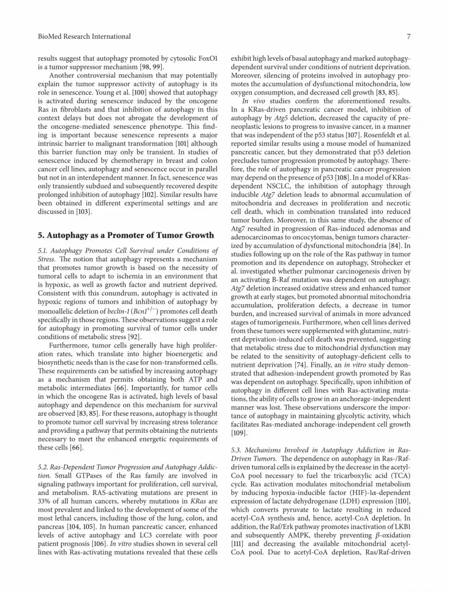
The molecular execution of the autophagic pathway—generation, maturation and degradation of the autophago-somes—requires the participation of specific autophagy-related (ATG) proteins [11] that were first described in yeastbefore orthologs in higher eukaryotes were identified. TheATG proteins organize into multiprotein complexes thatfunction in a nonredundant manner in the different steps ofthe process. Thus, although many ATGs exist, inhibition ofjust one ATG suffices to block execution of the autophagiccascade.
In mammalian cells, nucleation of the phagophore isregulated by a protein serine/threonine kinase complex thatresponds to the mammalian target of rapamycin (mTOR), akey regulator of the autophagic pathway, which shuts downautophagy in the presence of nutrients and growth factors[12]. Phagophore nucleation [7, 13] is regulated by the balancebetween class I and class III phosphatidylinositol 3-kinase(PI3K) enzymatic activities [14]. The active enzyme VPS34,a class III PI3K, together with the counterparts of yeast Vps15and Vps30/Atg6, identified inmammals as p150 and Beclin-1,and ATG14 form a PI3K complex that catalyzes the produc-tion of phosphatidylinositol-3-phosphate, thereby generatinga signal to initiate the recruitment of effectors proteins, suchas double FYVE-containing protein 1 (DFCP1) and WD-repeat domain phosphoinositide-interacting (WIPI) familyproteins [15–18]. The elongation of the isolation membrane

BioMed Research International 3
and subsequent closure of the autophagosome require theformation of two ubiquitin-like conjugates. First, ATG12is conjugated to ATG5 by the sequential activity of ATG7and ATG10. The resulting ATG5-ATG12 complex interactswith ATG16L, which then oligomerizes to form the ATG16Lcomplex [19]. Second, LC3 (the mammalian homologueof yeast Atg8) is cleaved by the protease ATG4 and thenconjugated to the lipid phosphatidylethanolamine via theactivity of ATG7 and ATG3 [19, 20]. While the unprocessedform of LC3 (LC3I) is diffusely distributed throughout thecytoplasm, the lipidated form of LC3 (LC3II) specificallyaccumulates on nascent autophagosomes and thus representsa marker to monitor autophagy [21]. The autophagosomeeventually seals off and fuses with lysosomes through mech-anisms that remain poorly characterized in mammalian cells[11]. Some regulators of the autophagosome-lysosome fusionprocess include LC3, the lysosomal proteins LAMP-1 andLAMP-2, the small GTP-binding protein RAB7, and theAAA-type ATPase SKD1 [22–24]. Autophagosome-lysosomefusion then results in the activation of the hydrolases whichcompletely degrade the autophagosomal cargo.
Different signaling mechanisms are known to modulateautophagy in mammalian cells [25]. The best characterizedpathways are those that modulate autophagy in response tonutritional changes and, as previously mentioned, mTOR iscritical for sensing the nutritional status of the cell and regu-lating the initiation of autophagy [12]. In higher eukaryotes,mTOR can be found in at least two distinctmultiprotein com-plexes, referred to asmTOR complex 1 (mTORC1) andmTORcomplex 2 (mTORC2) [26, 27]. The former is considered theprincipal regulator of autophagy [28]. When nutrients andgrowth factors are available, mTORC1 inhibits autophagy byphosphorylating and maintaining in an inactive state ULK1,which is required for the formation of the phagophore [29,30]. As indicated (see Figure 1), mTOR activity is controlledby different signaling pathways triggered via cues from theextracellular and intracellular microenvironment.
AMP-activated protein kinase (AMPK), another sen-sor of the cellular energy status, responds to decreases inATP/AMP ratios [31]. In conditions of nutrient deprivation,AMPK directly phosphorylates and inhibits mTOR (notshown in Figure 1). The ensuing reduction in mTOR activitydecreases ULK1 phosphorylation and promotes autophago-some formation [31, 32]. Moreover, AMPK also directlyactivates autophagy by phosphorylation of TSC2 [33, 34].In summary, autophagy is a highly regulated process thatinvolves a large number of modulators and the complexityof these events will increase as novel components continueto be identified in both mTOR dependent and independentpathways [35].
3. Control of Autophagy by Oncogenes andTumor Suppressors
Most of the proteins that participate in the regulation of auto-phagy are either tumor suppressor proteins or oncogenes.Perhaps not surprisingly, mechanisms involved in the regu-lation of autophagy largely overlap with signaling pathwaysimplicated in the control of cancer. Thus, tumor suppressor
genes that negatively regulate mTOR, such as PTEN, AMPK,LKB1, and TSC1/2 stimulate autophagy while, conversely,oncogenes that activate mTOR, such as class I PI3K, Ras,RHEB, and AKT, inhibit autophagy [51] (see Table 1). In thefollowing paragraphs, the role of Beclin-1, DAPK, Bcl2/Bcl-XL, and mTOR will be discussed briefly (see Figure 1).
Consistent with this view, Beclin-1, which is part of theclass III PI3K complex that promotes autophagy, functionsas a tumor suppressor in mammalian cells. Interestingly,monoallelic mutations in the beclin-1 gene are frequentlyobserved in prostate, ovarian, and breast cancers in humans.In addition, studies in mice have demonstrated that theanimals are more sensitive to spontaneous tumor devel-opment when beclin-1 is monoallelically disrupted. Theseobservations provide direct evidence for a role of beclin-1 as a haploinsufficient tumour suppressor gene implicatedin the pathogenesis of several human cancers [41, 52–54].Additionally, the death-associated protein kinase, DAPK,a protein that phosphorylates Beclin-1 thereby disruptingBeclin-1/BCL-2 complex and favoring autophagy, is anotherinducer of autophagy that is commonly silenced in differenttypes of human cancers by methylation [55].
BCL-2 and BCL-XL are antiapoptotic members of theBCL-2 family that modulate cell death in an autophagy-independent manner and are overexpressed in several hema-tological malignancies [56]. There, BCL-2 and BCL-XL sup-press cell death and promote survival and growth of cancercells by suppression of BAK/BAX-dependent pore forma-tion duringmitochondrial outermembrane permeabilization(MOMP) [57]. In addition to the role of BCL-2 and BCL-XLin the inhibition of apoptosis, they have also been implicatedin oncogenesis as negative regulators of autophagy. Althoughthese proteins do not directly participate in mTOR signaling,they can interact with the Beclin-1 BH3 domain and sequesterBeclin-1 into an inactive complex in the ER [58, 59].
The protein kinase mTOR is the major negative regulatorof autophagy [60].This kinase participates inmultiple signal-ing pathways that regulate cell growth, especially downstreamof growth factor receptors with tyrosine kinase activity. Inter-estingly, both the constitutive activation of these receptors, aswell as activating mutations of downstream elements in thesepathways (Ras, PI3-K, AKT, and PDK-1) or mutations thatinactivate negative regulators (TSC1/2, LKB1, and PTEN) arecommon in the development of cancer [36, 38, 47, 48, 50],suggesting that inhibition of autophagy likely contributes tothe onset to tumor development.
4. Autophagy as a TumorSuppressor Mechanism
Thefirst data pointing towards the possible tumor suppressorrole of autophagy were obtained in studies of Beclin-1. Mono-allelic loss of the beclin-1 gene on chromosome 17q21 hasbeen reported in 40% to 75% of human breast, ovary,and prostate tumors, suggesting that autophagy representsa tumor suppressor mechanism [41]. Also, a reduction inBeclin-1 protein levels has been observed in various braincancers [61]. Accordingly, Beclin-1+/− mice have a highincidence of spontaneous tumors, especially lymphoma and

4 BioMed Research International
Auto
phag
y
Nucleation Elongation Closing andmaturation
Dockingand fusion Degradation
PhagophoreAutophagosome
LysosomeAutophagosome
(a)
Dec
ision
-mak
ing
Receptortyrosine kinase PI3K
P
ERK
Ras
PP
TSC1 TSC2P
AMPK
Rheb
ARHI
PTENNF1P
LKB1
P
mTOR
Amino acids
P
ULK1
P
PDK1P
AKT1
Growthfactors
BH3-only Bcl-2/XL
Life/death equilibrium
Autophagy
PIP3
AMPATP
(b)
Maturation
RAB7A
Exec
utio
n
Nucleation
Beclin-1AMBRA1
p150hVPS34
ATG14L
UVRAG
BIF1Rubicon
BCL-2/XLP
ULK1
ATG5/ATG12ATG16
Elongation
LC3 II
LC3 I
Pro LC3
ATG4
ATG3ATG7
ATG5/ATG12
ATG12ATG5
ATG10
ATG16
4
OncogenesTumor suppressors
(c)
Figure 1: Phases of autophagy and its regulation by oncogenes and tumor suppressors. In (a), the five stages of autophagy are summarized.In (b), inhibition of autophagy by oncogenes (in red) and activation by tumor suppressors (in blue) is shown. Finally, (c) summarizes detailsof the complex regulation and interplay between different proteins in each stage of autophagy (see text for more details).

BioMed Research International 5
Table 1: Summary of oncogenes and tumor suppressors involved in autophagy regulation.
Oncogenes Role in autophagy Evidences of oncogenesis Reference
AKT1 Upstream inhibitor of autophagy via mTORactivation
Gain-of-function mutations in several cancertypes [36]
BCL-2, BCL-XL Sequester Beclin-1 into inactive complexes Overexpressed in several cancer types [37]
PI3K Upstream inhibitors of autophagy via AKT1activation Gain-of-function mutations in many cancer types [36, 38]
Ras Upstream inhibitors of autophagy via mTORactivation Hyperactivated in several cancer types [37]
Tumor suppressors Role in autophagy Evidences of tumor suppression
ATG4 Converts LC3 into LC3 I during stress conditions Mutations in ATG4C increase susceptibility tocarcinogens [39]
ARHI/DIRAS3,PTEN
Relieve autophagy inhibition mediated byPI3K-AKT1 Downregulated in ovarian cancer [36, 40]
Beclin-1, p150 Required in the nucleation complex for autophagyinitiation Deleted in breast, ovarian, and prostate cancer [41]
BH3-only proteins Relieve autophagy inhibition mediated byBCL-2/BCL-XL Mutated or silenced in many cancer types [42–44]
UVRAG, BIF1 Positive regulator of the nucleation complex Deleted or downregulated in colorectal cancer [45]
DAPK1 Relieve autophagy inhibition mediated byBCL-2/BCL-XL Silenced in many tumor types [46]
LKB1/STK11 Promotes autophagy via AMPK activation Mutated in Peutz-Jeghers syndrome andnon-small cell lung carcinomas [47, 48]
NF1 Relieve autophagy inhibition mediated by Ras Mutated in neurofibromatosis, juvenilemyelomonocytic leukemia [37]
RAB7A Modulates endosomal trafficking involved inautophagosome maturation Rearranged in leukemia, deleted in solid tumors [49]
TSC1, TSC2 Stimulate Rheb GTPase, thus inhibiting thePI3K-AKT1-mTOR pathway Mutated in TSC [50]
hepatocellular carcinoma. Furthermore, the evidence pro-vided suggests that Beclin-1 functions as a haploinsufficienttumor suppressor gene, given that the tumors continuedto express Beclin-1 [53, 54]. Moreover, immortalized breastepithelial cells with a monoallelic deletion of Beclin-1 formtumors more rapidly after inoculation into nude mice [62].More recently, phosphorylation of Beclin-1 on multiple tyro-sine residues in an EGFR-dependent manner was foundto decrease the activity of the Beclin-1/PI3KC3 complexand therefore decreased autophagy in non-small-cell lungcarcinoma cells (NSCLC) and that this effect was reducedin the presence of an inhibitor of EGFR kinase activity.Alternatively, the expression of a tyrosine phosphomimeticmutant of Beclin-1 reduces autophagy and increases tumorgrowth [63]. Similarly, several proteins that interact withBeclin-1 and positively regulate autophagy, such as AMBRA1 [64], BIF-1 [45], and UVRAG [65], have been shown todisplay antiproliferative or tumor suppressor effects. How-ever, a complication here is that all these proteins have otherfunctions that are independent of their role in autophagy, forexample in regulating the endocytic pathway [66]. Moreover,the Beclin-1/PI3KC3 complex also controls the ubiquitinationand degradation of p53 by regulating the stability and activityof the deubiquitinating enzymes USP13 and USP10 [67].
Given these additional functions, the contribution of suchautophagy-independent mechanisms to the observed tumorsuppressor phenotype cannot be excluded.
In agreement with the tumor suppressor hypothesis, thegeneration of knockout mice for specific genes involved inautophagy (ATGs) has shown that defects in specific regu-lators of this process are associated with the development ofa tumorigenic phenotype. Because systemic deletion of Atg3,Atg5, Atg7, Atg9, or Atg16L1 causes neonatal death [68–72],long-term effects of the inhibition of autophagy could not beassessed until mice with systemic mosaic Atg5 deletion weregenerated. In this background, systemic mosaicAtg5 deletionor liver-specific deletion of Atg7 results in mice that sponta-neously develop benign liver adenomas [73].While these datasuggest that defects in autophagy promote the developmentof benign tumors in this tissue, they also indicate that, in theabsence of autophagy, progression to a malignant phenotypeis prevented. Similarly, Strohecker et al. showed that thedeletion of Atg7 in mice expressing an activating mutationof B-Raf (Braf V600E/+) promotes early tumor development inthe lung but also inhibits the progression to amoremalignantphenotype and increases mouse survival [74]. Additionalautophagy-promoting factors that have tumor suppressorfunctions are Atg4C and RAB7A. For animals deficient in

6 BioMed Research International
Atg4C, increased susceptibility to the development of fibro-sarcomas induced by chemical carcinogens was detected[39]. RAB7A has been shown to prevent growth factor-inde-pendent survival by inhibiting cell-autonomous nutrienttransporter expression and the RAB7A gene is frequentlyrearranged in different types of leukemia [49, 75].
Despite this evidence that favors a role for autophagy intumor suppression, some more recent findings concerningBeclin-1 contrast with the previous interpretation of data.The beclin-1 gene lies close to BRCA1 on chromosome 17q21raising the specter that the relevance of the loss of Beclin-1 inovarian, breast, and prostate cancer may have been overinter-preted. Indeed, deletions encompassing both genes (BRCA1and beclin-1) and deletions of only BRCA1 but not beclin-1were found in breast and ovarian cancers, which is consistentwith BRCA1 loss representing the primary driver mutationin these cancers. Furthermore, no evidence for beclin-1mutations or loss have been detected in any other cancer,which questions whether beclin-1 is indeed a tumor suppres-sor in various human cancers [76]. Taken together, the evi-dence presented supports the hypothesis that autophagy mayplay an important role in tumor suppression at early stages.However, the findings discussed also reveal the potentiallydual nature of this process in tumor development and pro-gression.
4.1. Mechanisms Involved in Tumor Suppression by Autophagy
4.1.1. Oxidative Stress and Genomic Instability. One of themost important connections between autophagy and tumorsuppression is via the regulation of reactive oxygen species(ROS). Increased ROS production accelerates mutagenesis,increasing the activation of oncogenes, thus stimulating car-cinogenesis [77, 78]. Mitochondria are considered the mainsource of intracellular ROS and their production increases asthese organelles age or become damaged [79]. In this context,autophagy helps to avoid damage through selective degra-dation of defective mitochondria, a process known as mito-phagy. Consequently, inhibition of autophagy facilitatesgenomic instability by promoting the activation of onco-genes [62, 80] and genotoxic effects observed in autophagy-defective cells seem to be dependent on ROS generation [81].Thus, the selective removal of potentially damagedmitochon-dria (mitophagy) reduces excessive ROS production andthereby limits tumor-promoting effects dependent on theproduction of such species [82]. Accordingly, inhibition ofautophagy in different models leads to accumulation ofdefective mitochondria [69, 73, 74, 83–85].
Autophagy also permits the degradation of protein aggre-gates. Defects in the autophagic process have been associ-ated with the accumulation of protein aggregates and theautophagy substrate p62/SQSTM1. Such events are associatedwith increased production of ROS, ER stress, and activationof the DNA damage response [81]. The p62 protein is a selec-tive autophagy substrate that accumulates when autophagy isreduced. This scaffolding protein contains a PB1 domain thatpermits protein oligomerization, an UBA domain requiredfor binding to polyubiquitinated proteins and an LIR domain(LC3-interacting region) necessary for association with LC3.
For these reasons, p62 favors selective degradation of bothpolyubiquitinated proteins and organelles (i.e., mitochon-dria) [86, 87]. Interestingly, p62 levels are commonly elevatedin human tumors. In addition, tumorigenic developmentobserved in autophagy-deficient cells is reversed by geneticinactivation of p62 in various models, suggesting that theaccumulation of p62 promotes tumor formation in this con-text [73, 81, 83, 88]. Moreover, p62 accumulation stabilizesand activates the transcription via NRF-2, by binding toKeap-1, the main negative regulator of NRF-2. In doing so,antioxidant defense is upregulated and may contribute totumor development [88–90]. Specifically, overexpression ofp62 and activation of NRF-2 are critical for anchorage-inde-pendent growth observed in hepatocellular carcinoma cells[88].
4.1.2. Inflammation and Necrosis. The tumor microenviron-ment is defined by complex interactions between variouscell types that coexist within tumors (tumor and stromalcells) and crosstalk between these cells regulates both tumorgrowth and progression. In this context, it is important tonote that both inflammatory cells and cytokines are extremelyrelevant because a proinflammatory environment promotesproliferation and survival of malignant cells, stimulates angi-ogenesis, metastasis, and modifies the response to drugs [91].In different models, autophagy inhibition in apoptosis-defi-cient tumor cells has been shown to promote necrotic celldeath, local inflammation, and tumor growth [92]. Theseresults suggest that autophagy may contribute to tumor sup-pression by restricting tumor necrosis and local inflammation[60].The anti-inflammatory effect of autophagy has been sug-gested to be linked to the removal of cell corpses [93] becauseof findings in Atg5−/− embryonic stem cells, where defects inthe clearance of apoptotic bodies during embryonic devel-opment are observed [94]. Moreover, a complex connectionbetween autophagy and different aspects of the immuneresponse has been noted, which could contribute to the tumorsuppressor role of autophagy, as has been reviewed elsewhere[95].
4.1.3. Autophagic Cell Death and Senescence. Although auto-phagy is primarily considered a mechanism that permits sur-vival under stress conditions, some reports indicate that,under specific conditions, an increase in autophagic flux maycause cell death due to autophagy and explain in part thetumor suppressor effects [96]. The findings of Pattingre et al.revealed that the expression of a mutant Beclin-1, unable tointeract with BCL-2, induced autophagy to a greater extentthan wild-type Beclin-1, and unlike the latter, it promotedcell death [58]. More recently, studies in an ovarian cancercell line showed that ectopic expression of Ras induces auto-phagic cell death through the upregulation of Beclin-1 andNoxa, a BH3-only protein, which ultimately limits the onco-genic potential of Ras [97]. Similarly, Zhao and colleaguesdemonstrated that the transcription factor FoxO1 promotesautophagy in a manner independent of its transcriptionalactivity and induces autophagic cell death in tumor cells,suppressing tumor growth of xenografts in nude mice. These
BioMed Research International 7
results suggest that autophagy promoted by cytosolic FoxO1is a tumor suppressor mechanism [98, 99].
Another controversial mechanism that may potentiallyexplain the tumor suppressor activity of autophagy is itsrole in senescence. Young et al. [100] showed that autophagyis activated during senescence induced by the oncogeneRas in fibroblasts and that inhibition of autophagy in thiscontext delays but does not abrogate the development ofthe oncogene-mediated senescence phenotype. This find-ing is important because senescence represents a majorintrinsic barrier to malignant transformation [101] althoughthis barrier function may only be transient. In studies ofsenescence induced by chemotherapy in breast and coloncancer cell lines, autophagy and senescence occur in parallelbut not in an interdependent manner. In fact, senescence wasonly transiently subdued and subsequently recovered despiteprolonged inhibition of autophagy [102]. Similar results havebeen obtained in different experimental settings and arediscussed in [103].
5. Autophagy as a Promoter of Tumor Growth
5.1. Autophagy Promotes Cell Survival under Conditions ofStress. The notion that autophagy represents a mechanismthat promotes tumor growth is based on the necessity oftumoral cells to adapt to ischemia in an environment thatis hypoxic, as well as growth factor and nutrient deprived.Consistent with this conundrum, autophagy is activated inhypoxic regions of tumors and inhibition of autophagy bymonoallelic deletion of beclin-1 (Bcn1+/−) promotes cell deathspecifically in those regions.These observations suggest a rolefor autophagy in promoting survival of tumor cells underconditions of metabolic stress [92].
Furthermore, tumor cells generally have high prolifer-ation rates, which translate into higher bioenergetic andbiosynthetic needs than is the case for non-transformed cells.These requirements can be satisfied by increasing autophagyas a mechanism that permits obtaining both ATP andmetabolic intermediates [66]. Importantly, for tumor cellsin which the oncogene Ras is activated, high levels of basalautophagy and dependence on this mechanism for survivalare observed [83, 85]. For these reasons, autophagy is thoughtto promote tumor cell survival by increasing stress toleranceand providing a pathway that permits obtaining the nutrientsnecessary to meet the enhanced energetic requirements ofthese cells [66].
5.2. Ras-Dependent Tumor Progression and Autophagy Addic-tion. Small GTPases of the Ras family are involved insignaling pathways important for proliferation, cell survival,and metabolism. RAS-activating mutations are present in33% of all human cancers, whereby mutations in KRas aremost prevalent and linked to the development of some of themost lethal cancers, including those of the lung, colon, andpancreas [104, 105]. In human pancreatic cancer, enhancedlevels of active autophagy and LC3 correlate with poorpatient prognosis [106]. In vitro studies shown in several celllines with Ras-activating mutations revealed that these cells
exhibit high levels of basal autophagy andmarked autophagy-dependent survival under conditions of nutrient deprivation.Moreover, silencing of proteins involved in autophagy pro-motes the accumulation of dysfunctional mitochondria, lowoxygen consumption, and decreased cell growth [83, 85].
In vivo studies confirm the aforementioned results.In a KRas-driven pancreatic cancer model, inhibition ofautophagy by Atg5 deletion, decreased the capacity of pre-neoplastic lesions to progress to invasive cancer, in a mannerthat was independent of the p53 status [107]. Rosenfeldt et al.reported similar results using a mouse model of humanizedpancreatic cancer, but they demonstrated that p53 deletionprecludes tumor progression promoted by autophagy.There-fore, the role of autophagy in pancreatic cancer progressionmay depend on the presence of p53 [108]. In amodel of KRas-dependent NSCLC, the inhibition of autophagy throughinducible Atg7 deletion leads to abnormal accumulation ofmitochondria and decreases in proliferation and necroticcell death, which in combination translated into reducedtumor burden. Moreover, in this same study, the absence ofAtg7 resulted in progression of Ras-induced adenomas andadenocarcinomas to oncocytomas, benign tumors character-ized by accumulation of dysfunctional mitochondria [84]. Instudies following up on the role of the Ras pathway in tumorpromotion and its dependence on autophagy, Strohecker etal. investigated whether pulmonar carcinogenesis driven byan activating B-Raf mutation was dependent on autophagy.Atg7 deletion increased oxidative stress and enhanced tumorgrowth at early stages, but promoted abnormal mitochondriaaccumulation, proliferation defects, a decrease in tumorburden, and increased survival of animals in more advancedstages of tumorigenesis. Furthermore, when cell lines derivedfrom these tumors were supplemented with glutamine, nutri-ent deprivation-induced cell death was prevented, suggestingthat metabolic stress due to mitochondrial dysfunction maybe related to the sensitivity of autophagy-deficient cells tonutrient deprivation [74]. Finally, an in vitro study demon-strated that adhesion-independent growth promoted by Raswas dependent on autophagy. Specifically, upon inhibition ofautophagy in different cell lines with Ras-activating muta-tions, the ability of cells to grow in an anchorage-independentmanner was lost. These observations underscore the impor-tance of autophagy in maintaining glycolytic activity, whichfacilitates Ras-mediated anchorage-independent cell growth[109].
5.3. Mechanisms Involved in Autophagy Addiction in Ras-Driven Tumors. The dependence on autophagy in Ras-/Raf-driven tumoral cells is explained by the decrease in the acetyl-CoA pool necessary to fuel the tricarboxylic acid (TCA)cycle. Ras activation modulates mitochondrial metabolismby inducing hypoxia-inducible factor (HIF)-1𝛼-dependentexpression of lactate dehydrogenase (LDH) expression [110],which converts pyruvate to lactate resulting in reducedacetyl-CoA synthesis and, hence, acetyl-CoA depletion. Inaddition, the Raf/Erk pathway promotes inactivation of LKB1and subsequently AMPK, thereby preventing 𝛽-oxidation[111] and decreasing the available mitochondrial acetyl-CoA pool. Due to acetyl-CoA depletion, Ras/Raf-driven

8 BioMed Research International
tumors require autophagy in order to obtain TCA cycleintermediates. These in turn promote mitochondrial activity,which provides reductive equivalents necessary for oxidativephosphorylation and mitochondrial respiration. Moreover,in Ras-driven tumors, autophagy inhibition promotes theaccumulation of dysfunctional mitochondria. This kind ofcancer requires autophagy to maintain a pool of functionalmitochondria necessary for enhanced energetic requirementsof tumoral cells [83, 85].
Beyond the requirement of autophagy for survival ofRas-driven cancer cells, Ras activation also promotes cellsignaling events involved in the induction of autophagyby the upregulation of Noxa and Beclin-1 expression [97].Furthermore, Ras can directly stimulate BNIP3 expressionthrough activation of the Ras/Raf/Erk pathway or indirectlythrough HIF-1𝛼 induction [112–114].
5.4. Autophagy in Ras-Independent Tumor Progression. Therole of autophagy also has been studied in different contextsthat are independent of Ras. For instance, in a model ofbreast cancer driven by the PyMT oncogene, the inhibition ofautophagy by FIP200 deletion suppresses mammary tumorinitiation and progression. Here, FIP200 ablation increasedthe number of mitochondria with abnormal morphology intumor cells and reduced significantly proliferation, but it didnot affect apoptosis of mammary tumor cells [115]. AlthoughPyMT requires Ras activation to initiate cell transformation,PI3-kinase and Src activation are also involved [116]. Thus,it would be interesting to determine whether these kinasescontribute to dependence on autophagy for cell proliferation.Another study employed a Palb2 knockout model specific toepithelial breast cells to determine the role of autophagy inbreast cancer progression. PALB2 is a protein that cooper-ates with BRCA1 and BRCA2 in DNA repair via homologrecombination and helps maintain genomic stability. Palb2knockout mice develop breast adenocarcinoma when p53 ismutated. Partial inhibition of autophagy by monoallelic lossof Beclin-1 (Bcn1+/−) increased apoptosis and delayed tumorgrowth in amanner dependent on p53.The authors proposedthat autophagy promotes tumor growth by p53 suppressionwhen DNA is damaged [117]. These studies indicate thatautophagy can promote tumor progression in a mannerindependent of Ras activation and that autophagy could bea more general mechanism involved in cancer cell survivaland tumor progression.
In summary, current evidence points towards autophagyas a mechanism that ensures adequate mitochondrial meta-bolism in Ras-driven cancers by supplying mitochondrialintermediates via the degradation of macromolecules underbasal and starvation conditions [118]. Particularly, Ras-driventumorigenesis appears to be “addicted to autophagy” formetabolic support and maintenance of rapid tumor growth.All these data explain why autophagy is required in Ras-driven cancers to promote tumor cell survival and tumor pro-gression. Interestingly, however, some more recent evidenceindicates that autophagy is also important for tumoral cellsurvival of other cancers, independent of the Ras activationstatus.
6. Caveolin-1, a Connection to Autophagy?
Caveolin-1 (CAV1) is a scaffolding protein that is essentialfor caveolae formation, is expressed in a wide variety oftissues, and is involved in many biological processes, includ-ing cholesterol homeostasis, vesicular transport, and signaltransduction. Moreover, similar to autophagy, CAV1 plays adual role in cancer, functioning both as a tumor suppressorand promoter of tumor metastasis [119–121]. Although, E-cadherin has been identified as important in determiningCAV1 function in this context [122–125], the molecularmechanisms explaining such ambiguous behavior remainlargely undefined.
Given the parallels between the roles of CAV1 andautophagy in cancer, it is intriguing to speculate that theremight be a connection between the two. Indeed, Martinez-Outschoorn et al. demonstrated, using a coculture system,that CAV1 is degraded via lysosomes in stromal fibroblastssubjected to hypoxia and that this correlated with increasedlevels of autophagic markers such as LC3, ATG16L, BNIP3,BNIP3L, HIF-1𝛼, andNF-kB.Moreover, knockdown of CAV1in stromal fibroblasts was sufficient to induce the upregula-tion of lysosomal and autophagicmarkers, suggesting that theloss of CAV1 in the stromal compartment induces autophagy[126]. Also, loss of CAV1 leads to metabolic reprogrammingof stromal cells to support the growth of adjacent tumor cellsby delivering energy-rich metabolites and essential buildingblocks [127]. Consistent with the notion that CAV1 is anegative regulator of autophagy, CAV1 depletion in HCT116colorectal cancer cells was shown to reduce glucose uptakeand ATP production, which then triggered autophagy viaactivation of AMPK-p53 signaling [128]. Moreover, both invitro cell growth and in vivo xenograft tumor growth wereattenuated to a greater extent by CAV1 depletion in p53+/+
than in p53−/− cells [128].An inverse relationship between autophagy and CAV1
has also been observed in models of nontransformed cells.For instance, metabolomic profiling of endothelial cell lysatesfollowing transfection with si-CAV1 or si-control resulted inmarked increases in dipeptide levels for the CAV1 knock-down cells, which was attributed to an increase in autophagy[129]. To corroborate these results, the authors evaluatedthe processing of LC3 I to LC3 II by western blotting andshowed that siRNA-mediated CAV1 knockdown led to anincrease in the presence of the autophagy marker LC3-II.Also, treatment with the lysosomal inhibitor bafilomycin A1markedly increased LC3-II levels, indicating that reducedCAV1 expression leads to an increase in autophagy flux [129].Recently, CAV1 was also shown to regulate autophagy incigarette smoking-induced injury of lung epithelium [130].Specifically, CAV1 depletion increased basal and starvation-induced levels of ATG12-ATG5 and autophagy. Biochemicalanalysis revealed that CAV1 interacted with ATG5, ATG12,and the active ATG12-ATG5 complex to suppress autophagyin lung epithelial cells, thereby providing new insights asto how CAV1 modulates autophagy in this model [130].However, details of the molecular mechanisms by whichCAV1 regulates autophagy in cancer cells remain to bedetermined. A rather speculative idea is that the dual role

BioMed Research International 9
Early stages Late stages
Tumor suppression Tumor growth
Survival understress conditions
Mitochondrialquality control Cell death
mechanism
Chemoresistance
Resistance toMitochondrialmetabolism
Intermediarymolecules
Anabolism
Autophagic degradation
↓ Genomic instability
↓ ROS↓ Necrosis
↓ Inflammation
↓ O2 and starvation
Figure 2: The two facets of autophagy in cancer. At early stages, autophagy acts as a tumor suppressor mechanism by enhancing thedegradation of damaged proteins and organelles, mostly mitochondria. In doing so, autophagy acts as a quality control system that decreasesROS production and genomic instability. Moreover, autophagy prevents necrotic cell death in apoptosis-defective cells, thereby reducinglocal inflammation and tumor growth. Also, autophagy may serve (in some cases) as a mechanism that leads to cell death. On the otherhand, at later stages of tumor development, activation of autophagy supplies tumor cells under metabolic stress conditions with nutrientsand also maintains mitochondrial metabolism by providing metabolic intermediates, which promote cell survival and tumor growth. Finally,autophagy acts as a mechanism that promotes resistance to cancer therapy.
of CAV1 in cancer may be linked to its participation in thecontrol of autophagy. However, further experimentation isrequired to corroborate this intriguing hypothesis.
In summary, CAV1, a membrane protein typically impli-cated in the formation of cell surface structures like caveolaeand regulation of signalling, also plays a dual role in cancer,functioning as a tumor suppressor at early stages and a tumorpromoter later on. The future will reveal how the seeminglyopposing roles of autophagy in tumor development andprogression are controlled, and to what extent the ambiguousrole of CAV1 in cancer may be linked to the control ofautophagy.
7. Conclusions
Autophagy is an evolutionarily conserved mechanism thatdeveloped in eukaryotes to ensure protein and organellehomeostasis. A hallmark of cancer cells is their increasedproliferation and as a consequence their demand for energyequivalents and specific metabolites, which can be providedby autophagy. In this context, autophagy favors tumor celldevelopment, adaptation, and progression, and particularlysome oncogene-driven tumors are “addicted” to autophagyin this respect. However, autophagy also appears to havea tumor suppressor function early in cancer developmentby eliminating damaged mitochondria and reducing ROS-mediated genotoxic damage (see Figure 2). Accordingly,pharmacological modulation of autophagy in establishedtumors may represent an important anticancer therapy, as issupported by the use of autophagy inhibitors (chloroquine orhydroxychloroquine) in a large number of clinical trials andcurrently as a treatment for various kinds of cancers that aregenerally very aggressive or resistant to therapy (see Table 2).
Alternatively, considering the potential tumor suppressorrole of autophagy in early stages of cancer development,one may speculate that stimulation of this process could beuseful as a preventive mechanism against the development
of cancer. Consistent with this notion, caloric restriction hasbeen shown to prolong life span and reduce cancer incidencein several animal models [131]. Also, treatments with met-formin, an activator of the AMPK pathway that stimulatesautophagy, are associated with lower risk of different kindsof cancers [132].
Clearly, the role of autophagy in cancer depends onmanyfactors like tissue type, tumor stage, and the type of oncogenicmutation involved. Because of these dramatic differences,more research is required to understand the role of autophagyin cancer biology and how we may harness such knowledgeto improve cancer therapies and patient survival.
Abbreviations
AMBRA1: Autophagy/Beclin-1 regulator 1AMPK: AMP-activated protein kinaseATG: Autophagy-relatedBCL-2: B-cell lymphoma 2BCL-XL: B-cell lymphoma-extra largeBIF-1: BAX-interacting factor 1BNIP3: BCL2/adenovirus E1B 19 kDa interacting
protein 3BNIP3L: BCL2/adenovirus E1B 19 kDa interacting
protein 3-likeBRCA1: Breast cancer 1, early onsetBRCA2: Breast cancer 2, early onsetER: Endoplasmic reticulumFIP200: FAK family-interacting protein of 200 kDaFoxO: Forkhead box OLAMP: Lysosomal-associated membrane proteinLC3: Microtubule-associated protein 1 light
chain 3 (homolog of yeast Atg8)LKB1: Liver kinase B1mTOR: Mammalian target of rapamycinNF-𝜅B: Nuclear factor kappa-light-chain-
enhancer of activated B cells

10 BioMed Research International
Table 2: Summary of clinical trials involving autophagy inhibitors (chloroquine or hydroxychloroquine) for cancer treatment (data obtainedfrom http://www.cancer.gov/clinicaltrials).
Cancer type Therapy Phase Status Protocol IDRelapsed and refractory multiplemyeloma
Cyclophosphamide and pulse dexamethasonewith hydroxychloroquine or rapamycin 0 Completed NCT01396200
Glioblastoma multiforme Hydroxychloroquine, radiation, andtemozolimide I, II Closed NCT00486603
Pancreas adenocarcinoma Hydroxychloroquine, gemcitabine I, II Closed NCT01128296
Prostate cancer Hydroxychloroquine after prostate cancertreatment II Closed NCT00726596
Non-small cell lung cancer Erlotinib with or without hydroxychloroquine II Closed NCT00977470
Metastatic pancreatic cancer Hydroxychloroquine after prostate cancertreatment II Closed NCT01273805
Relapsed and refractory multiplemyeloma
Chloroquine, bortezomib, andcyclophosphamide II Closed NCT01438177
Advanced solid tumorsirresponsive to chemotherapy Hydroxychloroquine, sunitinib I Closed NCT00813423
B-cell chronic lymphocyticleukemia Hydroxychloroquine II Temporarily
Closed NCT00771056
Surgery removable Stage III orStage IV melanoma Hydroxychloroquine 0 Temporarily
Closed NCT00962845
Relapsed and refractory multiplemyeloma Hydroxychloroquine, bortezomib I, II Active NCT00568880
Lung cancer Hydroxychloroquine, gefitinib I, II Active NCT00809237Ductal carcinoma in situ Chloroquine I, II Active NCT01023477
Colorectal cancer Hydroxychloroquine, folinic acid, 5-fluorouracil,oxaliplatin, and bevacizumab I, II Active NCT01206530
Pancreatic cancer Hydroxychloroquine, protein-bound paclitaxel,and gemcitabine I, II Active NCT01506973
Previously treated renal cellcarcinoma Hydroxychloroquine, everolimus I, II Active NCT01510119
Renal cell carcinoma Hydroxychloroquine, aldesleukin I, II Active NCT01550367Unresectable hepatocellularcarcinoma
Hydroxychloroquine, transarterialchemoembolization (TACE) I, II Active NCT02013778
Metastatic colorectal cancer Hydroxychloroquine, capecitabine, oxaliplatin,and bevacizumab II Active NCT01006369
Chronic myeloid leukemia Imatinib mesylate with or withouthydroxychloroquine II Active NCT01227135
Breast cancer Hydroxychloroquine II Active NCT01292408Advanced or metastatic breastcancer Chloroquine, taxane II Active NCT01446016
Resectable pancreatic cancer Hydroxychloroquine, capecitabine, and radiation II Active NCT01494155High grade gliomas Hydroxychloroquine, radiation II Active NCT01602588Advanced/recurrent non-smallcell lung cancer
Hydroxychloroquine, paclitaxel, carboplatin, andbevacizumab II Active NCT01649947
Progressive metastatic castraterefractory prostate cancer
Navitoclax, abiraterone acetate with or withouthydroxychloroquine II Active NCT01828476
Soft tissue sarcoma Hydroxychloroquine, rapamycin II Active NCT01842594Potentially resectable pancreaticcancer
Protein-bound paclitaxel, gemcitabine with orwithout hydroxychloroquine II Active NCT01978184
Metastatic or unresectable solidtumors Hydroxychloroquine, temozolomide I Active NCT00714181
Irresponsive metastatic solidtumors Hydroxychloroquine, temsirolimus I Active NCT00909831

BioMed Research International 11
Table 2: Continued.
Cancer type Therapy Phase Status Protocol IDStage IV small cell lung cancer Chloroquine I Active NCT00969306Advanced solid tumors Hydroxychloroquine, vorinostat I Active NCT01023737Primary renal cell carcinoma Hydroxychloroquine before surgery I Active NCT01144169Advanced cancer Hydroxychloroquine sirolimus, or vorinostat I Active NCT01266057Solid tumors Hydroxychloroquine, radiation I Active NCT01417403Melanoma Chloroquine, radiation, DT01 I Active NCT01469455Advanced solid tumors,melanoma, prostate, or kidneycancer
Hydroxychloroquine, Akt inhibitor MK2206 I Active NCT01480154
Stages I–III small cell lung cancer Chloroquine, radiation I Active NCT01575782Refractory or relapsed solidtumors Hydroxychloroquine, sorafenib I Active NCT01634893
Lymphangioleiomyomatosis inwomen Hydroxychloroquine sirolimus I Active NCT01687179
Relapsed or refractory multiplemyeloma
Hydroxychloroquine, cyclophosphamide,dexamethasone, and sirolimus I Active NCT01689987
Nonresectable pancreaticadenocarcinoma Chloroquine, gemcitabine I Active NCT01777477
BRAF mutant metastaticmelanoma Hydroxychloroquine, vemurafenib I Active NCT01897116
Advanced solid tumors Chloroquine, carboplatin, and gemcitabine I Active NCT02071537Brain metastasis Chloroquine, radiation 0 Active NCT01727531
NRF-2: Nuclear factor erythroid 2-related factor 2PALB2: Partner and localizer of BRCA2PI3KC3/VSP34: Phosphatidylinositol 3-kinase, catalytic
subunit type 3PI3K: Phosphatidylinositol 3-kinasePTEN: Phosphatase and tensin homologPyMT: Polyoma middle T-antigenULK: Unc-51 like autophagy activating kinaseUVRAG: Ultraviolet radiation resistance-associated
gene.
Conflict of Interests
The authors declare that there is no conflict of interestsregarding the publication of this paper.
Acknowledgments
This work was supported by CONICYT-FONDAP 15130011(Andrew F. G. Quest and Sergio Lavandero), Anillo ProjectACT1111 (Andrew F. G. Quest and Sergio Lavandero),FONDECYT 1130250 (Andrew F. G. Quest), FONDECYT1140908 (Alfredo Criollo), and CONICYT PhD scholarships(Yenniffer Avalos, Jimena Canales, and Roberto Bravo-Sagua).
References
[1] P. Vineis and C. P. Wild, “Global cancer patterns: causes andprevention,”The Lancet, vol. 383, no. 9916, pp. 549–557, 2014.
[2] C. Are, S. Rajaram, M. Are et al., “A review of global cancerburden: trends, challenges, strategies, and a role for surgeons,”Journal of Surgical Oncology, vol. 107, no. 2, pp. 221–226, 2013.
[3] D.Hanahan andR.A.Weinberg, “Thehallmarks of cancer,”Cell,vol. 100, no. 1, pp. 57–70, 2000.
[4] D. Hanahan and R. A.Weinberg, “Hallmarks of cancer: the nextgeneration,” Cell, vol. 144, no. 5, pp. 646–674, 2011.
[5] D. J. Klionsky, “Autophagy revisited: a conversation with Chris-tian de Duve,” Autophagy, vol. 4, no. 6, pp. 740–743, 2008.
[6] M. A. Jardon, K. Rothe, S. Bortnik et al., “Autophagy: fromstructure to metabolism to therapeutic regulation,” Autophagy,vol. 9, no. 12, pp. 2180–2182, 2013.
[7] M.Hayashi-Nishino,N. Fujita, T.Noda,A. Yamaguchi, T. Yoshi-mori, andA. Yamamoto, “A subdomain of the endoplasmic reti-culum forms a cradle for autophagosome formation,” NatureCell Biology, vol. 11, no. 12, pp. 1433–1437, 2009.
[8] M.Hayashi-Nishino,N. Fujita, T.Noda,A. Yamaguchi, T. Yoshi-mori, and A. Yamamoto, “Electron tomography reveals theendoplasmic reticulum as a membrane source for autophago-some formation,” Autophagy, vol. 6, no. 2, pp. 301–303, 2010.
[9] M. Hamasaki, N. Furuta, A. Matsuda et al., “Autophagosomesform at ER-mitochondria contact sites,” Nature, vol. 495, no.7441, pp. 389–393, 2013.
[10] S. A. Tooze and T. Yoshimori, “The origin of the autophagoso-mal membrane,” Nature Cell Biology, vol. 12, no. 9, pp. 831–835,2010.
[11] K. R. Parzych and D. J. Klionsky, “An overview of autophagy:morphology, mechanism, and regulation,” Antioxidants andRedox Signaling, vol. 20, no. 3, pp. 460–473, 2014.
[12] A. Efeyan, R. Zoncu, and D. M. Sabatini, “Amino acids andmTORC1: from lysosomes to disease,”Trends inMolecularMed-icine, vol. 18, no. 9, pp. 524–533, 2012.

12 BioMed Research International
[13] G. Kroemer, G.Marino, and B. Levine, “Autophagy and the inte-grated stress response,” Molecular Cell, vol. 40, no. 2, pp. 280–293, 2010.
[14] F. O’Farrell, T. E. Rusten, and H. Stenmark, “Phosphoinositide3-kinases as accelerators and brakes of autophagy,” FEBS Jour-nal, vol. 280, no. 24, pp. 6322–6337, 2013.
[15] S. F. Funderburk, Q. J. Wang, and Z. Yue, “The Beclin 1-VPS34complex—at the crossroads of autophagy and beyond,” Trendsin Cell Biology, vol. 20, no. 6, pp. 355–362, 2010.
[16] Y. Zhong, Q. J. Wang, X. Li et al., “Distinct regulation of auto-phagic activity by Atg14L and Rubicon associated with Beclin1-phosphatidylinositol-3-kinase complex,” Nature Cell Biology,vol. 11, no. 4, pp. 468–476, 2009.
[17] H. E. J. Polson, J. De Lartigue, D. J. Rigden et al., “MammalianAtg18 (WIPI2) localizes to omegasome-anchored phagophoresand positively regulates LC3 lipidation,” Autophagy, vol. 6, no.4, pp. 506–522, 2010.
[18] T. Proikas-Cezanne and H. Robenek, “Freeze-fracture replicaimmunolabelling reveals human WIPI-1 and WIPI-2 as mem-brane proteins of autophagosomes,” Journal of Cellular andMolecular Medicine, vol. 15, no. 9, pp. 2007–2010, 2011.
[19] D. J. Klionsky and B. A. Schulman, “Dynamic regulation ofmacroautophagy by distinctive ubiquitin-like proteins,” NatureStructural and Molecular Biology, vol. 21, no. 4, pp. 336–345,2014.
[20] H. Nakatogawa, “Two ubiquitin-like conjugation systems thatmediate membrane formation during autophagy,” Essays in Bio-chemistry, vol. 55, no. 1, pp. 39–50, 2013.
[21] E. Tasdemir, L. Galluzzi, M. C. Maiuri et al., “Methods forassessing autophagy and autophagic cell death,” Methods inMolecular Biology, vol. 445, pp. 29–76, 2008.
[22] E.-L. Eskelinen, A. L. Illert, Y. Tanaka et al., “Role of LAMP-2 inlysosome biogenesis and autophagy,” Molecular Biology of theCell, vol. 13, no. 9, pp. 3355–3368, 2002.
[23] X. Ao, L. Zou, and Y. Wu, “Regulation of autophagy by the RabGTPase network,” Cell Death and Differentiation, vol. 21, no. 3,pp. 348–358, 2014.
[24] A. Nara, N. Mizushima, A. Yamamoto, Y. Kabeya, Y. Ohsumi,and T. Yoshimori, “SKD1 AAA ATPase-dependent endosomaltransport is involved in autolysosome formation,”Cell Structureand Function, vol. 27, no. 1, pp. 29–37, 2002.
[25] M.Mehrpour, A. Esclatine, I. Beau, and P. Codogno, “Overviewof macroautophagy regulation in mammalian cells,” CellResearch, vol. 20, no. 7, pp. 748–762, 2010.
[26] R. Zoncu, A. Efeyan, and D. M. Sabatini, “MTOR: from growthsignal integration to cancer, diabetes and ageing,” NatureReviews Molecular Cell Biology, vol. 12, no. 1, pp. 21–35, 2011.
[27] P. P. Hsu, S. A. Kang, J. Rameseder et al., “The mTOR-regulatedphosphoproteome reveals a mechanism of mTORC1-mediatedinhibition of growth factor signaling,” Science, vol. 332, no. 6035,pp. 1317–1322, 2011.
[28] A. Efeyan, R. Zoncu, S. Chang et al., “Regulation of mTORC1by the Rag GTPases is necessary for neonatal autophagy andsurvival,” Nature, vol. 493, no. 7434, pp. 679–683, 2013.
[29] E. Y. Chan, “MTORC1 phosphorylates the ULK1-mAtg13-FIP200 autophagy regulatory complex,” Science Signaling, vol.2, no. 84, article pe51, 2009.
[30] N. Hosokawa, T. Hara, T. Kaizuka et al., “Nutrient-dependentmTORCl association with the ULK1-Atg13-FIP200 complexrequired for autophagy,” Molecular Biology of the Cell, vol. 20,no. 7, pp. 1981–1991, 2009.
[31] L. Shang, S. Chen, F. Du, S. Li, L. Zhao, and X. Wang, “Nutrientstarvation elicits an acute autophagic response mediated byUlk1 dephosphorylation and its subsequent dissociation fromAMPK,” Proceedings of the National Academy of Sciences of theUnited States of America, vol. 108, no. 12, pp. 4788–4793, 2011.
[32] J. Kim, M. Kundu, B. Viollet, and K.-L. Guan, “AMPK andmTOR regulate autophagy through direct phosphorylation ofUlk1,” Nature Cell Biology, vol. 13, no. 2, pp. 132–141, 2011.
[33] J. W. Lee, S. Park, Y. Takahashi, and H.-G. Wang, “The asso-ciation of AMPK with ULK1 regulates autophagy,” PLoS ONE,vol. 5, no. 11, Article ID e15394, 2010.
[34] D. N. Tripathi, R. Chowdhury, L. J. Trudel et al., “Reactivenitrogen species regulate autophagy through ATM-AMPK-TSC2-mediated suppression of mTORC1,” Proceedings of theNational Academy of Sciences of the United States of America,vol. 110, no. 32, pp. E2950–E2957, 2013.
[35] S. Sarkar, “Regulation of autophagy by mTOR-dependent andmTOR-independent pathways: autophagy dysfunction in neu-rodegenerative diseases and therapeutic application of auto-phagy enhancers,” Biochemical Society Transactions, vol. 41, no.5, pp. 1103–1130, 2013.
[36] M. Cully, H. You, A. J. Levine, and T. W. Mak, “Beyond PTENmutations: the PI3K pathway as an integrator of multiple inputsduring tumorigenesis,” Nature Reviews Cancer, vol. 6, no. 3, pp.184–192, 2006.
[37] B. Levine, S. Sinha, and G. Kroemer, “Bcl-2 family members:dual regulators of apoptosis and autophagy,” Autophagy, vol. 4,no. 5, pp. 600–606, 2008.
[38] R. J. Shaw and L. C. Cantley, “Ras, PI(3)K and mTOR signallingcontrols tumour cell growth,” Nature, vol. 441, no. 7092, pp.424–430, 2006.
[39] G. Marino, N. Salvador-Montoliu, A. Fueyo, E. Knecht, N.Mizushima, and C. Lopez-Otın, “Tissue-specific autophagyalterations and increased tumorigenesis in mice deficient inAtg4C/autophagin-3,” Journal of Biological Chemistry, vol. 282,no. 25, pp. 18573–18583, 2007.
[40] Z. Lu, R. Z. Luo, Y. Lu et al., “The tumor suppressor gene ARHIregulates autophagy and tumor dormancy in human ovariancancer cells,” The Journal of Clinical Investigation, vol. 118, no.12, pp. 3917–3929, 2008.
[41] X. H. Liang, S. Jackson, M. Seaman et al., “Induction of auto-phagy and inhibition of tumorigenesis by beclin 1,” Nature, vol.402, no. 6762, pp. 672–676, 1999.
[42] V. Labi, M. Erlacher, S. Kiessling, and A. Villunger, “BH3-onlyproteins in cell death initiation, malignant disease and anti-cancer therapy,”Cell Death and Differentiation, vol. 13, no. 8, pp.1325–1338, 2006.
[43] E. Lomonosova and G. Chinnadurai, “BH3-only proteins inapoptosis and beyond: an overview,”Oncogene, vol. 27, no. 1, pp.S2–S19, 2008.
[44] M. C. Maiuri, A. Criollo, E. Tasdemir et al., “BH3-only proteinsand BH3 mimetics induce autophagy by competitively disrupt-ing the interaction between Beclin 1 and Bcl-2/Bcl-XL,” Auto-phagy, vol. 3, no. 4, pp. 374–376, 2007.
[45] Y. Takahashi, D. Coppola, N. Matsushita et al., “Bif-1 interactswith Beclin 1 through UVRAG and regulates autophagy andtumorigenesis,” Nature Cell Biology, vol. 9, no. 10, pp. 1142–1151,2007.
[46] B. Harrison, M. Kraus, L. Burch et al., “DAPK-1 binding toa linear peptide motif in MAP1B stimulates autophagy andmembrane blebbing,” The Journal of Biological Chemistry, vol.283, no. 15, pp. 9999–10014, 2008.

BioMed Research International 13
[47] J. Liang, S. H. Shao, Z.-X. Xu et al., “The energy sensing LKB1-AMPK pathway regulates p27kip1 phosphorylation mediatingthe decision to enter autophagy or apoptosis,” Nature Cell Bio-logy, vol. 9, no. 2, pp. 218–224, 2007.
[48] H. Ji, M. R. Ramsey, D. N. Hayes et al., “LKB1 modulates lungcancer differentiation andmetastasis,”Nature, vol. 448, no. 7155,pp. 807–810, 2007.
[49] A. L. Edinger, R. M. Cinalli, and C. B. Thompson, “Rab7 pre-vents growth factor-independent survival by inhibiting cell-autonomous nutrient transporter expression,” DevelopmentalCell, vol. 5, no. 4, pp. 571–582, 2003.
[50] R. A. Schwartz, G. Fernandez, K. Kotulska, and S. Jozwiak,“Tuberous sclerosis complex: advances in diagnosis, genetics,and management,” Journal of the American Academy of Derma-tology, vol. 57, no. 2, pp. 189–202, 2007.
[51] A. M. K. Choi, S. W. Ryter, and B. Levine, “Mechanisms of dis-ease: autophagy in human health and disease,”TheNewEnglandJournal of Medicine, vol. 368, no. 7, pp. 651–662, 2013.
[52] V. M. Aita, X. H. Liang, V. V. V. S. Murty et al., “Cloning andgenomic organization of beclin 1, a candidate tumor suppressorgene on chromosome 17q21,”Genomics, vol. 59, no. 1, pp. 59–65,1999.
[53] X. Qu, J. Yu, G. Bhagat et al., “Promotion of tumorigenesis byheterozygous disruption of the beclin 1 autophagy gene,” Journalof Clinical Investigation, vol. 112, no. 12, pp. 1809–1820, 2003.
[54] Z. Yue, S. Jin, C. Yang, A. J. Levine, and N. Heintz, “Beclin 1, anautophagy gene essential for early embryonic development, is ahaploinsufficient tumor suppressor,” Proceedings of the NationalAcademy of Sciences of the United States of America, vol. 100, no.25, pp. 15077–15082, 2003.
[55] Y. Huang, L. Chen, L. Guo, T. R. Hupp, and Y. Lin, “EvaluatingDAPK as a therapeutic target,” Apoptosis, vol. 19, no. 2, pp. 371–386, 2014.
[56] K. H. Khan, M. Blanco-Codesido, and L. R. Molife, “Cancertherapeutics: targeting the apoptotic pathway,” Critical Reviewsin Oncology/Hematology, vol. 90, no. 3, pp. 200–219, 2014.
[57] L. Galluzzi, O. Kepp, and G. Kroemer, “Mitochondria: masterregulators of danger signalling,” Nature Reviews Molecular CellBiology, vol. 13, no. 12, pp. 780–788, 2012.
[58] S. Pattingre, A. Tassa, X. Qu et al., “Bcl-2 antiapoptotic proteinsinhibit Beclin 1-dependent autophagy,” Cell, vol. 122, no. 6, pp.927–939, 2005.
[59] M. C. Maiuri, G. le Toumelin, A. Criollo et al., “Functional andphysical interaction between Bcl-XL and a BH3-like domain inBeclin-1,” EMBO Journal, vol. 26, no. 10, pp. 2527–2539, 2007.
[60] E. Morselli, L. Galluzzi, O. Kepp et al., “Anti- and pro-tumorfunctions of autophagy,” Biochimica et Biophysica Acta: Molecu-lar Cell Research, vol. 1793, no. 9, pp. 1524–1532, 2009.
[61] C. Miracco, E. Cosci, G. Oliveri et al., “Protein and mRNAexpression of autophagy gene Beclin 1 in human brain tumours,”International Journal of Oncology, vol. 30, no. 2, pp. 429–436,2007.
[62] V. Karantza-Wadsworth, S. Patel, O. Kravchuk et al., “Autophagymitigates metabolic stress and genome damage in mammarytumorigenesis,” Genes & Development, vol. 21, no. 13, pp. 1621–1635, 2007.
[63] Y. Wei, Z. Zou, N. Becker et al., “EGFR-mediated beclin 1 phos-phorylation in autophagy suppression, tumor progression, andtumor chemoresistance,” Cell, vol. 154, no. 6, pp. 1269–1284,2013.
[64] G.Maria Fimia, A. Stoykova, A. Romagnoli et al., “Ambra1 regu-lates autophagy and development of the nervous system,”Nature, vol. 447, no. 7148, pp. 1121–1125, 2007.
[65] C. Liang, P. Feng, B. Ku et al., “Autophagic and tumour sup-pressor activity of a novel Beclin1-binding protein UVRAG,”Nature Cell Biology, vol. 8, no. 7, pp. 688–698, 2006.
[66] E. White, “Deconvoluting the context-dependent role forautophagy in cancer,” Nature Reviews Cancer, vol. 12, no. 6, pp.401–410, 2012.
[67] J. Liu, H. Xia,M. Kim et al., “Beclin1 controls the levels of p53 byregulating the deubiquitination activity of USP10 and USP13,”Cell, vol. 147, no. 1, pp. 223–234, 2011.
[68] A. Kuma, M. Hatano, M. Matsui et al., “The role of autophagyduring the early neonatal starvation period,” Nature, vol. 432,no. 7020, pp. 1032–1036, 2004.
[69] M. Komatsu, S. Waguri, T. Ueno et al., “Impairment of starva-tion-induced and constitutive autophagy in Atg7-deficientmice,” Journal of Cell Biology, vol. 169, no. 3, pp. 425–434, 2005.
[70] T. Saitoh, N. Fujita,M.H. Jang et al., “Loss of the autophagy pro-tein Atg16L1 enhances endotoxin-induced IL-1𝛽 production,”Nature, vol. 456, no. 7219, pp. 264–268, 2008.
[71] T. Saitoh, F.Naonobu, T. Yoshimori, and S. Akira, “Regulation ofinflammation by autophagy,” Tanpakushitsu kakusan koso, vol.54, supplement 8, pp. 1119–1124, 2009.
[72] Y.-S. Sou, S. Waguri, J.-I. Iwata et al., “The Atg8 conjugationsystem is indispensable for proper development of autophagicisolationmembranes inmice,”Molecular Biology of the Cell, vol.19, no. 11, pp. 4762–4775, 2008.
[73] A. Takamura, M. Komatsu, T. Hara et al., “Autophagy-deficientmice develop multiple liver tumors,” Genes and Development,vol. 25, no. 8, pp. 795–800, 2011.
[74] A.M. Strohecker, J. Y. Guo, G. Karsli-Uzunbas et al., “Autophagysustains mitochondrial glutamine metabolism and growth ofBrafV600E -driven lung tumors,” Cancer Discovery, vol. 3, no.11, pp. 1272–1285, 2013.
[75] V. I. Kashuba, R. Z.Gizatullin, A. I. Protopopov et al., “NotI link-ing/jumping clones of human chromosome 3: mapping of theTFRC, RAB7 and HAUSP genes to regions rearranged in leu-kemia and deleted in solid tumors,” FEBS Letters, vol. 419, no.2-3, pp. 181–185, 1997.
[76] S. V. Laddha, S. Ganesan, C. S. Chan, and E.White, “Mutationallandscape of the essential autophagy gene BECN1 in humancancers,”Molecular Cancer Research, vol. 12, no. 4, pp. 485–490,2014.
[77] L. J. Marnett, “Oxyradicals and DNA damage,” Carcinogenesis,vol. 21, no. 3, pp. 361–370, 2000.
[78] M. E. Goetz andA. Luch, “Reactive species: a cell damaging routassisting to chemical carcinogens,” Cancer Letters, vol. 266, no.1, pp. 73–83, 2008.
[79] L. Galluzzi, E. Morselli, O. Kepp et al., “Mitochondrial gatewaysto cancer,”Molecular Aspects of Medicine, vol. 31, no. 1, pp. 1–20,2010.
[80] R. Mathew, S. Kongara, B. Beaudoin et al., “Autophagy sup-presses tumor progression by limiting chromosomal instability,”Genes and Development, vol. 21, no. 11, pp. 1367–1381, 2007.
[81] R. Mathew, C. M. Karp, B. Beaudoin et al., “Autophagy sup-presses tumorigenesis through elimination of p62,”Cell, vol. 137,no. 6, pp. 1062–1075, 2009.
[82] E. Morselli, L. Galluzzi, O. Kepp et al., “Oncosuppressive func-tions of autophagy,” Antioxidants and Redox Signaling, vol. 14,no. 11, pp. 2251–2269, 2011.

14 BioMed Research International
[83] J. Y. Guo, H.-Y. Chen, R. Mathew et al., “Activated Ras requiresautophagy to maintain oxidative metabolism and tumorigene-sis,” Genes and Development, vol. 25, no. 5, pp. 460–470, 2011.
[84] J. Y. Guo, G. Karsli-Uzunbas, R. Mathew et al., “Autophagysuppresses progression of K-ras-induced lung tumors to onco-cytomas and maintains lipid homeostasis,” Genes and Develop-ment, vol. 27, no. 13, pp. 1447–1461, 2013.
[85] S. H. Yang, X. Wang, G. Contino et al., “Pancreatic cancersrequire autophagy for tumor growth,” Genes and Development,vol. 25, no. 7, pp. 717–729, 2011.
[86] S. Geisler, K. M. Holmstrom, D. Skujat et al., “PINK1/Parkin-mediated mitophagy is dependent on VDAC1 and p62/SQSTM1,” Nature Cell Biology, vol. 12, no. 2, pp. 119–131, 2010.
[87] J. Moscat and M. T. Diaz-Meco, “p62 at the crossroads of auto-phagy, apoptosis, and cancer,” Cell, vol. 137, no. 6, pp. 1001–1004,2009.
[88] Y. Inami, S. Waguri, A. Sakamoto et al., “Persistent activation ofNrf2 through p62 in hepatocellular carcinoma cells,” Journal ofCell Biology, vol. 193, no. 2, pp. 275–284, 2011.
[89] M. Komatsu, H. Kurokawa, S. Waguri et al., “The selectiveautophagy substrate p62 activates the stress responsive tran-scription factor Nrf2 through inactivation of Keap1,”Nature CellBiology, vol. 12, no. 3, pp. 213–223, 2010.
[90] A. Lau, X.-J. Wang, F. Zhao et al., “A noncanonical mechanismof Nrf2 activation by autophagy deficiency: direct interactionbetween keap1 and p62,”Molecular and Cellular Biology, vol. 30,no. 13, pp. 3275–3285, 2010.
[91] J. Candido and T. Hagemann, “Cancer-related inflammation,”Journal of Clinical Immunology, vol. 33, supplement 1, pp. S79–S84, 2013.
[92] K. Degenhardt, R. Mathew, B. Beaudoin et al., “Autophagy pro-motes tumor cell survival and restricts necrosis, inflammation,and tumorigenesis,” Cancer Cell, vol. 10, no. 1, pp. 51–64, 2006.
[93] E. White and R. S. DiPaola, “The double-edged sword of auto-phagy modulation in cancer,” Clinical Cancer Research, vol. 15,no. 17, pp. 5308–5316, 2009.
[94] X. Qu, Z. Zou, Q. Sun et al., “Autophagy gene-dependent clear-ance of apoptotic cells during embryonic development,” Cell,vol. 128, no. 5, pp. 931–946, 2007.
[95] B. Levine, N. Mizushima, and H. W. Virgin, “Autophagy inimmunity and inflammation,” Nature, vol. 469, no. 7330, pp.323–335, 2011.
[96] G. Kroemer and B. Levine, “Autophagic cell death: the story ofa misnomer,” Nature Reviews Molecular Cell Biology, vol. 9, no.12, pp. 1004–1010, 2008.
[97] M. Elgendy, C. Sheridan, G. Brumatti, and S. J. Martin, “Onco-genic Ras-induced expression of Noxa and Beclin-1 promotesautophagic cell death and limits clonogenic survival,”MolecularCell, vol. 42, no. 1, pp. 23–35, 2011.
[98] Y. Zhao, J. Yang, W. Liao et al., “Cytosolic FoxO1 is essentialfor the induction of autophagy and tumour suppressor activity,”Nature Cell Biology, vol. 12, no. 7, pp. 665–675, 2010.
[99] Y. Zhao, L. Wang, J. Yang et al., “Anti-neoplastic activity of thecytosolic FoxO1 results from autophagic cell death,” Autophagy,vol. 6, no. 7, pp. 988–990, 2010.
[100] A. R. J. Young, M. Narita, M. Ferreira et al., “Autophagy medi-ates the mitotic senescence transition,”Genes and Development,vol. 23, no. 7, pp. 798–803, 2009.
[101] A. C. Kimmelman, “The dynamic nature of autophagy in can-cer,”Genes and Development, vol. 25, no. 19, pp. 1999–2010, 2011.
[102] R. W. Goehe, X. Di, K. Sharma et al., “The autophagy-sen-escence connection in chemotherapy: must tumor cells (self)eat before they sleep,” Journal of Pharmacology and Experimen-tal Therapeutics, vol. 343, no. 3, pp. 763–778, 2012.
[103] D. A. Gewirtz, “Autophagy and senescence in cancer therapy.,”Journal of cellular physiology, vol. 229, no. 1, pp. 6–9, 2014.
[104] A. E. Karnoub and R. A. Weinberg, “Ras oncogenes: splitpersonalities,”Nature Reviews Molecular Cell Biology, vol. 9, no.7, pp. 517–531, 2008.
[105] A. Jemal, R. Siegel, J. Xu, and E. Ward, “Cancer statistics, 2010,”CA Cancer Journal for Clinicians, vol. 60, no. 5, pp. 277–300,2010.
[106] S. Fujii, S. Mitsunaga, M. Yamazaki et al., “Autophagy is acti-vated in pancreatic cancer cells and correlates with poor patientoutcome,” Cancer Science, vol. 99, no. 9, pp. 1813–1819, 2008.
[107] A. Yang, N. V. Rajeshkumar, X. Wang et al., “Autophagy is cri-tical for pancreatic tumor growth and progression in tumorswith p53 alterations,” Cancer Discovery, vol. 4, no. 8, pp. 905–913, 2014.
[108] M. T. Rosenfeldt, J. O'Prey, J. P. Morton et al., “P53 status deter-mines the role of autophagy in pancreatic tumour develop-ment,” Nature, vol. 504, no. 7479, pp. 296–300, 2013.
[109] R. Lock, S. Roy, C. M. Kenific et al., “Autophagy facilitates gly-colysis during Ras-mediated oncogenic transformation,”Molec-ular Biology of the Cell, vol. 22, no. 2, pp. 165–178, 2011.
[110] S. Y. Chun, C. Johnson, J. G. Washburn, M. R. Cruz-Correa, D.T. Dang, and L. H. Dang, “Oncogenic KRAS modulates mito-chondrial metabolism in human colon cancer cells by inducingHIF-1𝛼 and HIF-2𝛼 target genes,” Molecular Cancer, vol. 9,article 293, 2010.
[111] B. Zheng, J. H. Jeong, J. M. Asara et al., “Oncogenic B-RAF neg-atively regulates the tumor suppressor LKB1 to promote mela-noma cell proliferation,” Molecular Cell, vol. 33, no. 2, pp. 237–247, 2009.
[112] W. Kalas, E. Swiderek, A. Rapak, M. Kopij, J. Rak, and L.Strzadala, “H-ras Up-regulates expression of BNIP3,” Anti-cancer Research, vol. 31, no. 9, pp. 2869–2875, 2011.
[113] S.-Y.Wu, S.-H. Lan, D.-E. Cheng et al., “Ras-related tumorigen-esis is suppressed by BNIP3-mediated autophagy through inhi-bition of cell proliferation,” Neoplasia, vol. 13, no. 12, pp. 1171–1182, 2011.
[114] H.-J. An, O. Maeng, K.-H. Kang et al., “Activation of Rasup-regulates pro-apoptotic BNIP3 in nitric oxide-induced celldeath,” Journal of Biological Chemistry, vol. 281, no. 45, pp.33939–33948, 2006.
[115] H. Wei, S. Wei, B. Gan, X. Peng, W. Zou, and J.-L. Guan, “Sup-pression of autophagy by FIP200 deletion inhibits mammarytumorigenesis,”Genes andDevelopment, vol. 25, no. 14, pp. 1510–1527, 2011.
[116] B. A. Smith, D. N. Shelton, C. Kieffer et al., “Targeting the PyMToncogene to diversemammary cell populations enhances tumorheterogeneity and generates rare breast cancer subtypes,”Genesand Cancer, vol. 3, no. 9-10, pp. 550–563, 2012.
[117] Y. Huo, H. Cai, I. Teplova et al., “Autophagy opposes p53-mediated tumor barrier to facilitate tumorigenesis in amodel ofPALB2 -associated hereditary breast cancer,” Cancer Discovery,vol. 3, no. 8, pp. 894–907, 2013.
[118] R. Mathew and E. White, “Autophagy, stress, and cancer meta-bolism: what doesn’t kill you makes you stronger,” Cold SpringHarbor Symposia on Quantitative Biology, vol. 76, pp. 389–396,2011.

BioMed Research International 15
[119] A. F. G. Quest, J. L. Gutierrez-Pajares, and V. A. Torres, “Caveo-lin-1: an ambiguous partner in cell signalling and cancer,” Jour-nal of Cellular and Molecular Medicine, vol. 12, no. 4, pp. 1130–1150, 2008.
[120] A. F. G. Quest, L. Leyton, and M. Parraga, “Caveolins, caveolae,and lipid rafts in cellular transport, signaling, and disease,”Biochemistry and Cell Biology, vol. 82, no. 1, pp. 129–144, 2004.
[121] A. F. G. Quest, L. Lobos-Gonzalez, S. Nunez et al., “The Cav-eolin-1 connection to cell death and survival,”CurrentMolecularMedicine, vol. 13, no. 2, pp. 266–281, 2013.
[122] L. Lobos-Gonzalez, L. Aguilar, J. Diaz et al., “E-cadherin deter-mines Caveolin-1 tumor suppression or metastasis enhancingfunction in melanoma cells,” Pigment Cell and MelanomaResearch, vol. 26, no. 4, pp. 555–570, 2013.
[123] D. A. Rodriguez, J. C. Tapia, J. G. Fernandez et al., “Caveolin-1-mediated suppression of cyclooxygenase-2 via a 𝛽-catenin-Tcf/Lef-dependent transcriptional mechanism reduced prost-aglandin E
2production and survivin expression,” Molecular
Biology of the Cell, vol. 20, no. 8, pp. 2297–2310, 2009.[124] V. A. Torres, J. C. Tapia, D. A. Rodrıguez et al., “Caveolin-1 con-
trols cell proliferation and cell death by suppressing expressionof the inhibitor of apoptosis protein survivin,” Journal of CellScience, vol. 119, part 9, pp. 1812–1823, 2006.
[125] V. A. Torres, J. C. Tapia, D. A. Rodriguez et al., “E-cadherin isrequired for caveolin-1-mediated down-regulation of the inhi-bitor of apoptosis protein survivin via reduced 𝛽-catenin-Tcf/Lef- dependent transcription,” Molecular and Cellular Biology,vol. 27, no. 21, pp. 7703–7717, 2007.
[126] U. E. Martinez-Outschoorn, C. Trimmer, Z. Lin et al., “Auto-phagy in cancer associated fibroblasts promotes tumor cell sur-vival: role of hypoxia, HIF1 induction and NF𝜅B activation inthe tumor stromal microenvironment,” Cell Cycle, vol. 9, no. 17,pp. 3515–3533, 2010.
[127] F. Sotgia, U. E.Martinez-Outschoorn, A.Howell, R. G. Pestell, S.Pavlides, and M. P. Lisanti, “Caveolin-1 and cancer metabolismin the tumor microenvironment: markers, models, and mecha-nisms,”Annual Review of Pathology:Mechanisms of Disease, vol.7, pp. 423–467, 2012.
[128] T.-K.Ha,N.-G.Her,M.-G. Lee et al., “Caveolin-1 increases aero-bic glycolysis in colorectal cancers by stimulating HMGA1-mediated GLUT3 transcription,” Cancer Research, vol. 72, no.16, pp. 4097–4109, 2012.
[129] T. Shiroto, N. Romero, T. Sugiyama et al., “Caveolin-1 is a criticaldeterminant of autophagy, metabolic switching, and oxidativestress in vascular endothelium,” PLoS ONE, vol. 9, no. 2, ArticleID e87871, 2014.
[130] Z.-H. Chen, J.-F. Cao, J.-S. Zhou et al., “Interaction of caveolin-1 with ATG12-ATG5 system suppresses autophagy in lungepithelial cells,” The American Journal of Physiology: LungCellular and Molecular Physiology, vol. 306, no. 11, pp. L1016–L1025, 2014.
[131] O.Meynet and J.-E. Ricci, “Caloric restriction and cancer:mole-cular mechanisms and clinical implications,” Trends in Molecu-lar Medicine, vol. 20, no. 8, pp. 419–427, 2014.
[132] M. Malek, R. Aghili, Z. Emami, and M. E. Khamseh, “Risk ofcancer in diabetes: the effect of metformin,” ISRN Endocrinol-ogy, vol. 2013, Article ID 636927, 9 pages, 2013.


