Embed Size (px)
Citation preview
Tubulite, �Ag2Pb22Sb20S53, a new Pb–Ag–Sb sulfosalt from Le Rivet quarry,
Peyrebrune ore field (Tarn, France) and Bio, Borgofranco mines, Borgofranco
d’Ivrea (Piedmont, Italy)
YVES MOELO1,*, ROBERT PECORINI2, MARCO E. CIRIOTTI3, NICOLAS MEISSER4, MARIA TERESA CALDES1,
PAOLO ORLANDI5, PIERRE-EMMANUEL PETIT1, BRUNO MARTINI6 and ADRIO SALVETTI7
1 Institut des Materiaux Jean Rouxel, UMR 6502, CNRS, Universite de Nantes, 2, rue de la Houssiniere, F-44322 NantesCedex 3, France
*Corresponding author, e-mail: [email protected] Association Francaise de Micromineralogie, 9, allee des Chenes Verts, F-13620 Carry le Rouet, France
3 Associazione Micromineralogica Italiana - via San Pietro, 55, I-10073 Devesi-Cirie, Italy4 Musee de geologie & Institut des sciences de la Terre, Universite de Lausanne, CH-1015 Lausanne, Switzerland
5 Dipartimento di Scienze della Terra, Universita di Pisa, via S. Maria 53, I-56126 Pisa, Italy6 Associazione Micromineralogica Italiana - via Roma, 26, I-13812 Campiglia Cervo, Italy
7 Associazione Micromineralogica Italiana - strada Regione Croce, 30–I-13900 Chiavazza, Italy
Abstract: Tubulite, � Ag2Pb22Sb20S53, is a new Pb–Ag–Sb sulfosalt discovered at Le Rivet quarry, Peyrebrune ore field (Tarn,France) and Bio, Borgofranco mines, Borgofranco d’Ivrea (Piedmont, Italy). As indicated by the name, it forms very thin perfectmicro-tubes, 100 to 600 mm in length, 40 to 100 mm in diameter, and only 1 to 2 mm in thickness; a hair-like variety is also present atBio. At Le Rivet, it is associated with galena, pyrite, pyrrhotite, arsenopyrite, stibnite, and various Sb sulfosalts of Pb, Ag and Cu, in agangue of quartz, baryte and carbonates. At Bio, it is associated with galena, sphalerite, chalcopyrite, pyrite, marcasite, and various Sbsulfosalts of Pb, Ag and Cu, within the same type of gangue. Tubulite is metallic black; optical properties could not be observed underthe microscope, due to the crystal morphology. Electron microprobe analysis gave (wt.% - average of 8 anal.): Ag 2.7(2), Pb 46.6(9),Sb 26.1(8), S 17.8(5), Total 93.2(2.1). The low total is due to the thinness of the tube wall. According to crystallographic study(electron and X-ray diffraction), tubulite is monoclinic (space group Pc, P2/c or P21/c) with unit-cell parameters a 4.132(2), b 43.1(2),c 27.4(1) A, b 93.2�, V 4872(40) A3, with Z¼ 2. Weak reflections in the [010] electron diffraction pattern indicate a 2a superstructure.Main diffraction lines are [d (A), (I)]: 3.99 (35), 3.69 (60), 3.36 (100), 3.28 (55), 2.99 (55), 2.912 (55), 2.063 (75). The unit cell oftubulite is very close to those of sterryite and parasterryite; like these sulfosalts, tubulite is probably an expanded derivative ofowyheeite. Its peculiar tubular morphology is discussed within the general framework of crystals whose habit presents a circularsymmetry. It is proposed that micro-tubes were initiated by capillary forces acting on very thin lamellar crystallites around gasbubbles or liquid droplets.
Key-words: tubulite, new mineral, sulfosalt, lead, silver, tubular morphology, Le Rivet, Peyrebrune, France, Bio, Borgofranco, Italy.
Introduction
In 1998, during the examination, in search of micro-crystalsfor the preparation of micromounts, of a sampling from LeRivet quarry (in the area of Peyrebrune Pb–Zn–Ag mine,Tarn department, France), one of us (R. P.) discovered an oremineral with a very unusual tubular habit, associated withgalena and various antimony sulfosalts. Although the miner-alogical characterization of this tubular mineral appearedvery difficult, due to its peculiar habit, a first scanningelectron microscope (SEM) chemical analysis proved it tobe a Pb–Ag–Sb sulfosalt with an original composition, dis-tinct from any known mineral species of this group.
In 2002, a new occurrence of what appeared to bethe same sulfosalt (first SEM analyses in 2006), with thesame tubular habit, was discovered at Bio, in the uppergalleries (Veneziana and Torinese) of the Pb–Ag–As–SbBorgofranco mines (Borgofranco d’Ivrea, Turin, Piedmont,Italy) (‘‘unknown mineral UKMBB 703’’ – Ambrino et al.,2008; Ciriotti et al., 2009 – see cover photo of the latterbook). Its study allowed confirming and completing the dataobtained on the first, French occurrence. On this basis, it waspossible to define this new mineral for which the name‘tubulite’ was proposed, due to its original main crystal habit.
The mineral and its name have been accepted by theCNMNC of the IMA, under the number 2011–109.
0935-1221/13/0025-2334 $ 6.30DOI: 10.1127/0935-1221/2013/0025-2334 # 2013 E. Schweizerbart’sche Verlagsbuchhandlung, D-70176 Stuttgart
Eur. J. Mineral.
2013, 25, 1017–1030
Published online December 2013
Holotype material from Le Rivet quarry is deposited in thecollections of the Museum of Mineralogy of the EcoleNationale Superieure des Mines de Paris (now MINESParisTech), France, catalogue number M 82939. Holotypematerial from Bio is deposited in the mineralogical collec-tions of the Museo Regionale di Scienze Naturali, ViaGiovanni Giolitti 36, Turin, Italy, catalogue number15905. Cotype specimens are deposited in the mineralogicalcollections of the Museo di Storia Naturale, Universita diPisa, Via Roma 79, Calci, Pisa, Italy, catalogue number19630, and in the collections of the Musee Cantonal deGeologie, Universite-Anthropole, 1015 Lausanne,Switzerland, catalogue number MGL#92635.
Recently, the definition of a new sulfosalt, parasterryite(Moelo et al., 2011), and the resolution of the crystalstructures of parasterryite and sterryite (Moelo et al.,2012) revealed the close crystal chemical relationshipbetween tubulite and these two sulfosalts, which areexpanded derivatives of owyheeite.
1. The deposits and their parageneses
1.1. The French deposit of Le Rivet quarry
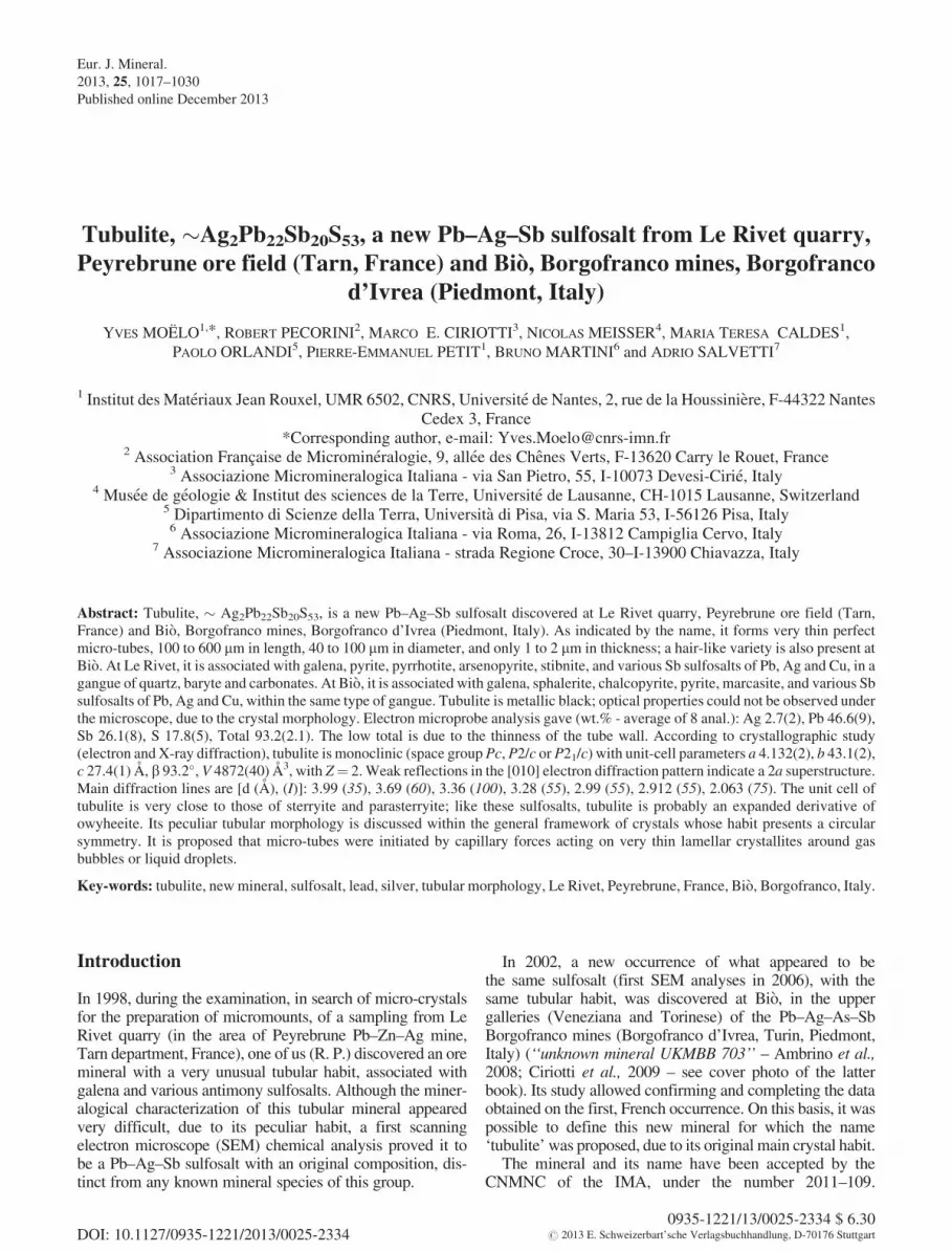
The quarry of Le Rivet belongs to the PeyrebrunePb–Zn–Ag ore field, located along the Dadou river, about6 km E-SE of the town of Realmont, on the commune ofMontredon-Labessonie, Tarn department, France (Figs. 1and 2). Geographic coordinates of Le Rivet quarryare x ¼ 43� 45’ 42’’N, y ¼ 02� 14’ 16’’ E and z � 240 m.A general review of the nearby Peyrebrune mine (historyand exploitation) has been presented by Di Cato (1996).According to Contri (1959), Durand (1966), and Pierrotet al. (1976), the ore deposit consists of quartz veins orientedEW; these veins intersect a metamorphosed volcano-sedi-mentary complex (Ordovician black schists with meta-dolerites and meta-basalts) with some granitic and micro-granitic sills (Peyrebrune granite), indicative of a Hercynianshallow batholith (Durand, 1966; Guerange-Lozes et al.,
1996). This geologic formation occurs at the western marginof Massif Central, in direct contact with the Tertiary coverof the Aquitaine Basin.
The vein system of Peyrebrune was studied in detail byContri (1959) and Durand (1966). Le Rivet quarry is situatedwithin meta-dolerites and meta-basalts, and is operated forgravel extraction. Three sub-vertical quartz-dolomitic veins(named A to C) were observed in the quarry. Tubulitesamples were collected in vein C, oriented NNE-SSW.
The mineral inventory of Peyrebrune ores has been doneby Pierrot et al. (1976). Main sulfides are galena, sphaler-ite, pyrite and chalcopyrite, in a gangue of quartz, siderite,fluorite, calcite and dolomite. Various sulfosalts have been
Fig. 1. Geographic location of the Peyrebrune Pb–Zn–Ag ore field. Left: Tarn department in France; right: Peyrebrune in Tarn department.
Fig. 2. Geological location of Le Rivet quarry (simplified accordingto Guerange-Lozes et al., 1996). 1. Sedimentary cover (Tertiary toPresent); 2. Ordovician black schists; 3. Interstratified metadoleritesand metabasalts; 4. Hercynian granitic and micro-granitic dykes; 5.Main Pb–Zn mineralized quartz veins of Peyrebrune ore deposit(crossed circle: mine buildings); 6. Quarry.
1018 Y. Moelo et al.
observed through the metallographic study, as well as laterby mineral collectors (Bernadi, 1992; Hubert & Hubert,1992) (Table 1).
A detailed sequence of ore deposition has been estab-lished by Durand (1966). It can be simplified as follows:
– Stage I: dolomite;– Stage II (main): galena and sphalerite, in a gangue of
siderite;– Stage III: chalcopyrite with quartz and fluorite;– Stage IV (discrete): stibnite;– Stage V: baryte.
Stage II is the main silver carrier. Bournonite is the mostabundant sulfosalt, then tetrahedrite. Although they arespatially subordinated to galena and chalcopyrite, it isprobable that these minerals and other sulfosalts (amongwhich tubulite) may result for a main part from the remo-bilization of sulfides of stages II and III by Sb-rich solu-tions of stage IV (superimposition process).
1.2. The Italian deposit of Bio, Borgofranco mines
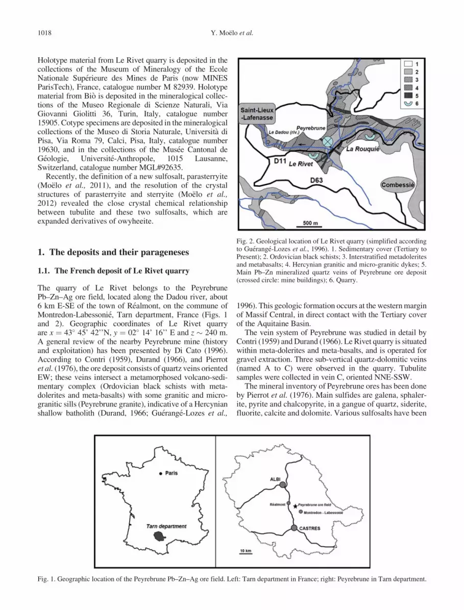
The Borgofranco Pb–Ag–As–Sb ore district is composedof 14 small mines, worked in the past for silver, lead,arsenic and antimony (Lincio, 1916). This ore district islocated in Piedmont, around the town of Borgofrancod’Ivrea, situated at about 50 km north of Turin, along theDora Baltea river, on the road to Val d’Aosta (Fig. 3).
The ore district is located at the foot of Mount Vesino,within the geo-tectonic ‘‘Canavese Zone’’ which, accordingto Baggio & Friz (1969), represents a ‘‘post-Hercynian coverof the Ivrea-Verbano area’’. The Canavese Zone is actuallycomposed of fault-bounded tectonic slices which includeboth pre-Alpine basement and Permo-Mesozoic cover(Borghi et al., 1996); it is a narrow tectonic unit boundedby the western end of the Insubric Line and separating theSesia Zone (Austroalpine Domain) from the Ivrea Zone(Southalpine Domain) (e.g., Borghi et al., 1996). There, thetwo domains are separated by the ‘‘tectonic suture’’ of the‘‘Schists of Bio’’, known with the name of ‘‘Bio suture’’.
Mineralized veins intersect the ‘‘schists of Bio’’, a non-fossiliferous sedimentary sequence of mudstones, sand-stones, arkoses and impure limestones (Biino &Compagnoni, 1989), probably Upper Jurassic in age (R.Compagnoni, pers. comm. 2012).
The mineralogy of these ores has been detailed by Piccoliet al. (2007) and Ambrino et al. (2008). The latter authorsespecially described the sulfosalt association. Main gangueminerals are quartz and baryte, together with carbonates(calcite, dolomite and siderite). Simple sulfides are galena,sphalerite, chalcopyrite, pyrite, marcasite and orpiment;numerous Sb sulfosalts were identified (Table 1).
Tubulite samples have been discovered in 2002 bytwo of us (B.M. and A.S.) in the two mine tunnels ofVeneziana and Torinese, just above the farm of Bio(Fig. 3). Geographic coordinates are x ¼ 45� 31’ 07’’N, y ¼ 7� 51’ 53’’ E, and z � 300–350 m.
Table 1. Ore mineral associations at Peyrebrune and Bio.
Peyrebrune Ref.: Pierrot et al. (1976), this study (*: Le Rivet quarry)Gangue A: quartz*, baryte*, siderite. F: fluorite, calcite*, dolomite*. t: ankeriteSulfides A: galena*. F: pyrite, sphalerite, chalcopyrite. t: marcasite, arsenopyrite, pyrrhotite, stibniteSulfosalts F: bournonite. t: tetrahedrite-freibergite, semseyite, boulangerite*, freieslebenite, miargyite, polybasite,
proustite, pyrargyrite, pyrostilpnite, polybasite, tubulite*, witticheniteBio Ref.: Ambrino et al. (2008)
Gangue A: quartz, baryte, calcite, dolomite, sideriteSulfides A: galena, sphalerite. t: orpiment, marcasite, chalcopyriteSulfosalts F: tetrahedrite-freibergite, bournonite, boulangerite
t: semseyite, geocronite, fizelyite, diaphorite, miargyrite, pyrargyrite-proustite, pyrostilpnite, tubulite
A, abundant; F, frequent; t, traces.
Fig. 3. Location of the Bio occurrences from Borgofranco minedistrict (reproduced from Ambrino et al., 2008). Veneziana andTorinese tunnels ¼ 1.
Tubulite, a new Pb–Ag–Sb sulfosalt 1019
2. Descriptive study
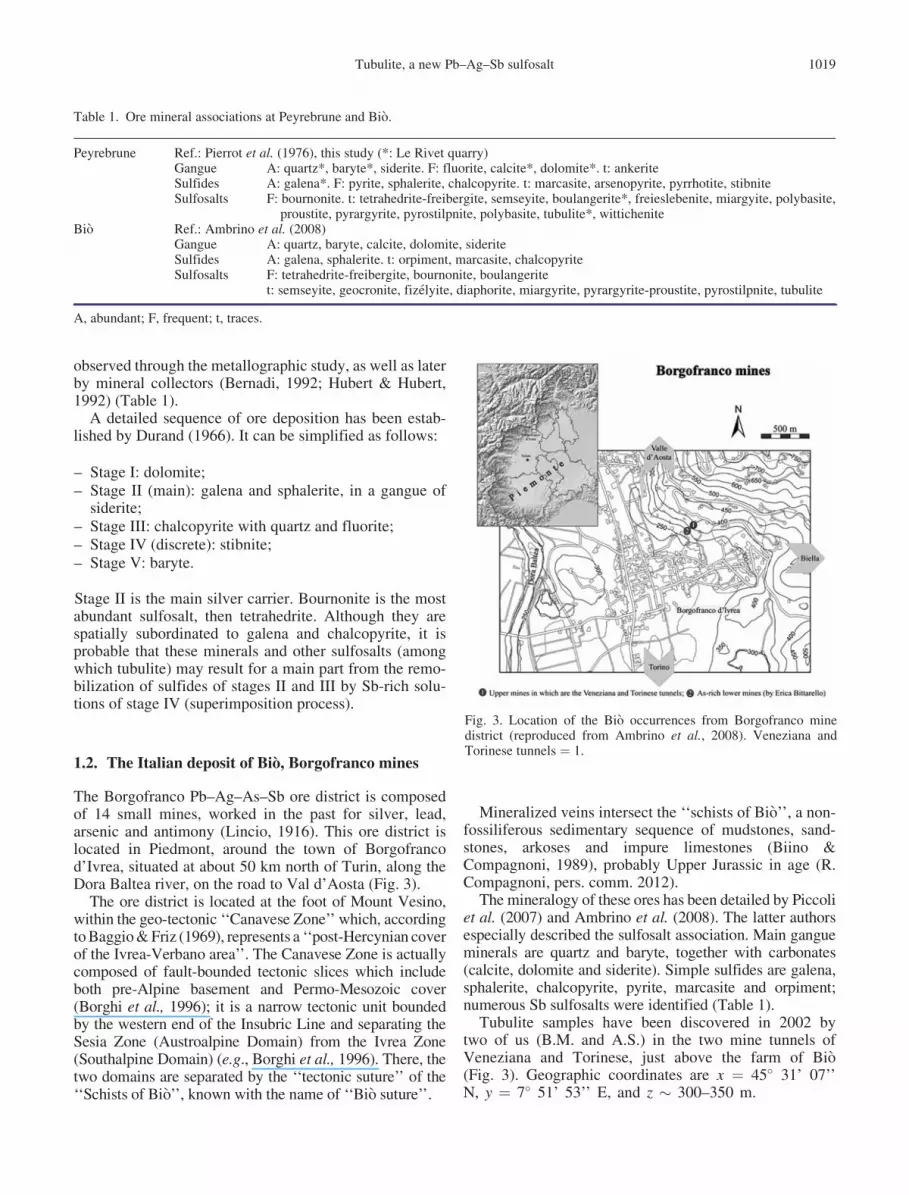
Under the binocular microscope, typical tubulite crystalsappear in vugs as rare metallic black cylinders (Fig. 4 aand b), associated with galena, and also boulangerite at LeRivet. Contrary to natural cylindrite crystals, which arecompletely filled cylinders (Makovicky, 1971), tubulitecrystals, when observed along their elongation axis, appearas perfect tubes with a very thin wall. The SEM images ofsuch a tube (e.g., Fig. 5a) indicate a wall thickness of about 2mm, for a diameter of several tens of mm, and a length up to600 mm. The external surface of the tube shows two steps
that would correspond to a spiral growth around the tube axis(also visible in Fig. 4a – tube at left). Rarely were observedunrolled, more or less undulated elongated lamellae, forinstance on a crystal face of calcite and baryte at Bio.
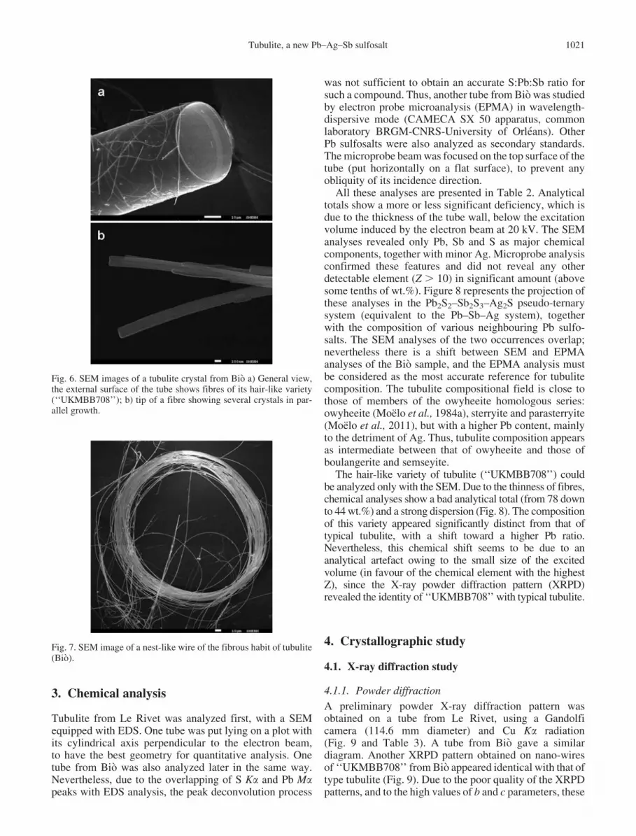
Tubulite from Bio (Fig. 4b and 6) is closely associatedwith geocronite, bournonite, fizelyite, and a hair-like sul-fosalt (‘‘unknown mineral UKMBB708’’of Ambrino et al.,2008), which appeared to be another habit of tubulite (seebelow). This tubulite variety is constituted by very finemicro-fibres (‘‘nano-wires’’ – Fig. 6a), about 0.2 mm thick.Details of a needle-like tip are given in Fig. 6b. Whenforming a scroll (Fig. 7), with a diameter of about 400mm, and at least about fifty turns, the length of such amicro-fibre may reach 6 cm, i.e. a length/width ratio of�3 � 105.
2.1. Physical properties
Optical properties as well as hardness could not be studied,due to the crystal morphology. Tubes show no plasticity,contrary to cylindrite crystals, and appear very resistantto crushing. For instance, one tube sandwiched betweentwo glass lamellae (45 � 30 � 1.5 mm) was not flattenedunder the lamella weight. Under stronger pressure, the tubebroke into rings, then rings into arched fragments. Suchfragments were used for TEM study.
Fig. 4. Photomicrographs of typical tubulite crystals. a) Le Rivetquarry. Top: long tube, 0.4 mm in length (Sample: R. P.; photocourtesy by J.-M. Johannet). Bottom (sample and photo: R. P.): thetube at left (L 0.3 mm, Ø 0.2 mm) shows on its upper part a linear step(parallel to the tube axis - arrow) indicating a spiral growth. Tube atright: L 0.5 mm, Ø 0.05 mm. b) Bio. Several perfect tubes associatedwith nano-wires of the same sulfosalt (forming a scroll at right) (fieldsize: 2.6 � 3.3 mm; photo by E. Bonacina, reproduced with hisauthorization).
Fig. 5. Scanning electron microscope (SEM) images of a tubulitecrystal from Le Rivet quarry: a) general view. Two growth steps(curved lines) are visible on the external surface of the tube; b) detailof one edge.
1020 Y. Moelo et al.
3. Chemical analysis
Tubulite from Le Rivet was analyzed first, with a SEMequipped with EDS. One tube was put lying on a plot withits cylindrical axis perpendicular to the electron beam,to have the best geometry for quantitative analysis. Onetube from Bio was also analyzed later in the same way.Nevertheless, due to the overlapping of S Ka and Pb Mapeaks with EDS analysis, the peak deconvolution process
was not sufficient to obtain an accurate S:Pb:Sb ratio forsuch a compound. Thus, another tube from Bio was studiedby electron probe microanalysis (EPMA) in wavelength-dispersive mode (CAMECA SX 50 apparatus, commonlaboratory BRGM-CNRS-University of Orleans). OtherPb sulfosalts were also analyzed as secondary standards.The microprobe beam was focused on the top surface of thetube (put horizontally on a flat surface), to prevent anyobliquity of its incidence direction.
All these analyses are presented in Table 2. Analyticaltotals show a more or less significant deficiency, which isdue to the thickness of the tube wall, below the excitationvolume induced by the electron beam at 20 kV. The SEManalyses revealed only Pb, Sb and S as major chemicalcomponents, together with minor Ag. Microprobe analysisconfirmed these features and did not reveal any otherdetectable element (Z . 10) in significant amount (abovesome tenths of wt.%). Figure 8 represents the projection ofthese analyses in the Pb2S2–Sb2S3–Ag2S pseudo-ternarysystem (equivalent to the Pb–Sb–Ag system), togetherwith the composition of various neighbouring Pb sulfo-salts. The SEM analyses of the two occurrences overlap;nevertheless there is a shift between SEM and EPMAanalyses of the Bio sample, and the EPMA analysis mustbe considered as the most accurate reference for tubulitecomposition. The tubulite compositional field is close tothose of members of the owyheeite homologous series:owyheeite (Moelo et al., 1984a), sterryite and parasterryite(Moelo et al., 2011), but with a higher Pb content, mainlyto the detriment of Ag. Thus, tubulite composition appearsas intermediate between that of owyheeite and those ofboulangerite and semseyite.
The hair-like variety of tubulite (‘‘UKMBB708’’) couldbe analyzed only with the SEM. Due to the thinness of fibres,chemical analyses show a bad analytical total (from 78 downto 44 wt.%) and a strong dispersion (Fig. 8). The compositionof this variety appeared significantly distinct from that oftypical tubulite, with a shift toward a higher Pb ratio.Nevertheless, this chemical shift seems to be due to ananalytical artefact owing to the small size of the excitedvolume (in favour of the chemical element with the highestZ), since the X-ray powder diffraction pattern (XRPD)revealed the identity of ‘‘UKMBB708’’ with typical tubulite.
4. Crystallographic study
4.1. X-ray diffraction study
4.1.1. Powder diffraction
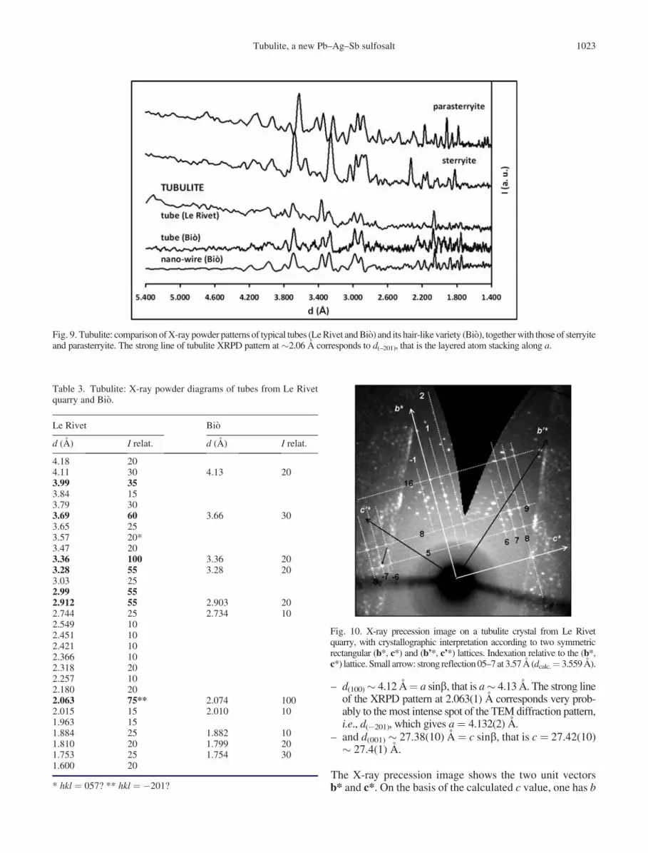
A preliminary powder X-ray diffraction pattern wasobtained on a tube from Le Rivet, using a Gandolficamera (114.6 mm diameter) and Cu Ka radiation(Fig. 9 and Table 3). A tube from Bio gave a similardiagram. Another XRPD pattern obtained on nano-wiresof ‘‘UKMBB708’’ from Bio appeared identical with that oftype tubulite (Fig. 9). Due to the poor quality of the XRPDpatterns, and to the high values of b and c parameters, these
Fig. 6. SEM images of a tubulite crystal from Bio a) General view,the external surface of the tube shows fibres of its hair-like variety(‘‘UKMBB708’’); b) tip of a fibre showing several crystals in par-allel growth.
Fig. 7. SEM image of a nest-like wire of the fibrous habit of tubulite(Bio).
Tubulite, a new Pb–Ag–Sb sulfosalt 1021

patterns could not be indexed to refine the unit cell. Onlythe strong lines at d ¼ 3.57 and 2.063 A probably corre-spond to reflections 057 and –201, respectively, accordingto X-ray single-crystal and TEM studies. In the samefigure, comparison with the XRPD patterns obtained forsterryite and parasterryite using the same apparatus (Moeloet al., 2011) proves the specificity of tubulite diagram.
4.1.2. Single-crystal study
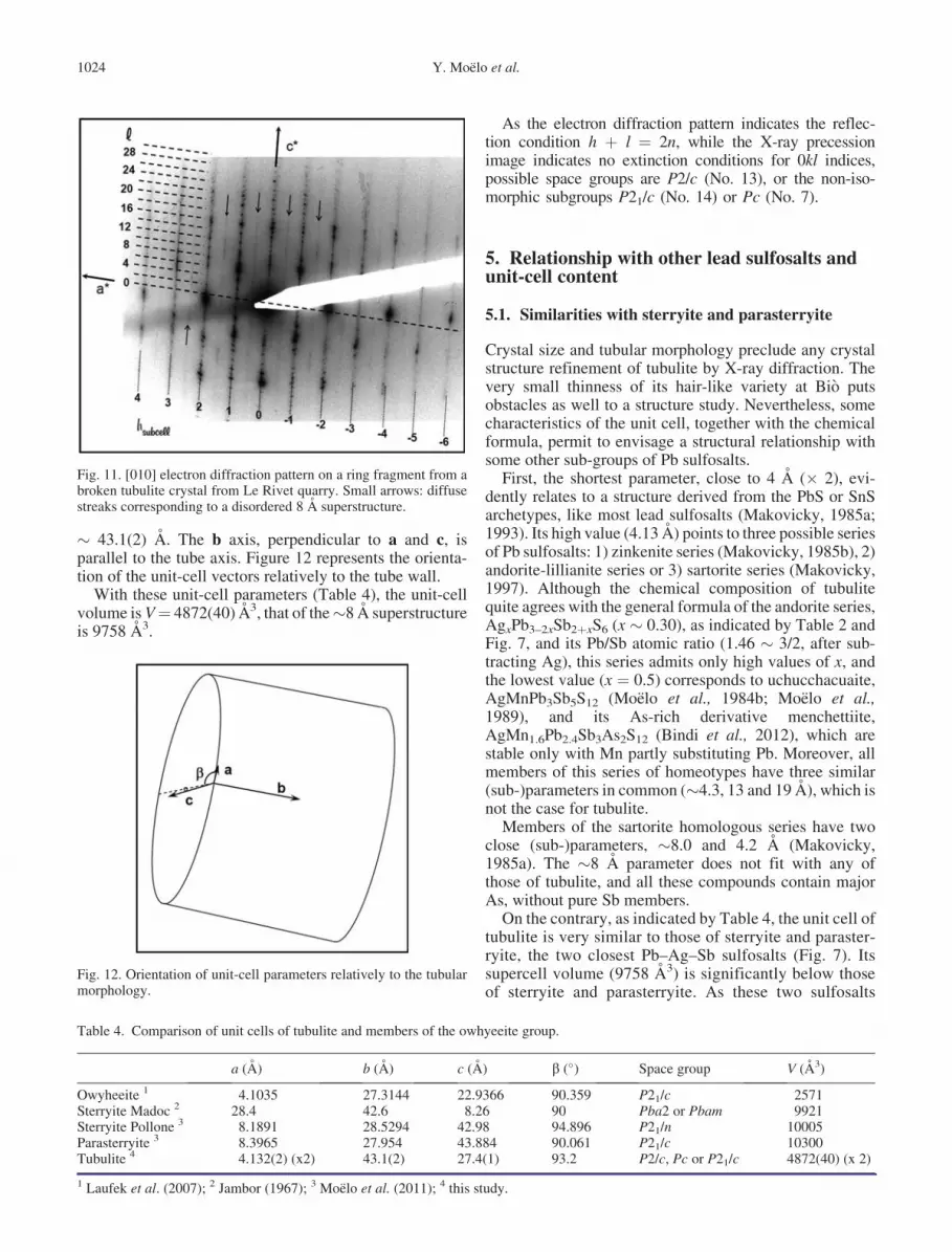
One tubular crystal from Le Rivet was put on a glass capillaryusing a Nonius Kappa CCD detector. X-ray diffraction datawere treated in order to obtain precession images. Figure 10presents the best image obtained. Two symmetric rectangularnets have been visualized. This symmetry is the result of thecylindrical shape of the single crystal. These patterns corre-spond to the b*� c* plane, without any 0kl extinction rules.
4.2. TEM study
After crushing, ring fragments of a tube were put on a coppergrid coated with holey carbon film, and examined with aPhilips CM30 electron microscope operating at 300 kV. Dueto the curvature of these fragments, they were lying withtheir cylinder axis sub-perpendicular to the grid plane (i.e.sub-parallel to the electron beam). This permitted to obtain adiffraction pattern (Fig. 11) corresponding to the reciprocalplane perpendicular to the cylinder axis, combining the unitvector a* sub-tangential to the tube, and the second vector c*parallel to the cylinder radius (radial axis).
Due to the short value of c* and the thinness of the tube(c* is perpendicular to the tube wall), both the zero- andfirst-order Laue zones (ZOLZ and FOLZ) overlap, corre-sponding to h0l and h1l reflection sets. The b angle is closeto 93.2�. For h0l reflections, those with odd l are absent.
Weak but distinct streaks (arrows on Fig. 11) indicateclearly a disordered 2a superstructure (� 8 A), commonlyobserved in acicular lead sulfosalts.
4.3. Uni-cell parameters
X-ray precession and TEM studies indicate a monoclinicsymmetry, with b¼ 93.2�. According to the [010] electrondiffraction pattern, one has:
Table 2. Electron probe microanalysis of tubulite from Le Rivet quarry and Bio.
wt.% Ag Pb Sb S Total
Le Rivet SEM 1 3.7 40.0 23.4 17.1 84.22 3.5 39.9 23.7 17.0 84.1
BioSEM (EDS) Mean 12 3.2 41.3 24.1 17.9 86.4
s 0.2 1.2 0.9 0.5 1.2P¼ 100 % 3.7 47.8 27.9 20.7 100.0
s 0.2 1.4 1.1 0.5 1.3EPMA (WDS) Mean 8 2.7 46.6 26.1 17.8 93.2
s 0.2 0.9 0.8 0.5 2.1P¼ 100 % 2.9 50.0 28.0 19.1 100.0
s 0.2 0.9 0.8 0.5 2.2Unit formula (WDS) Ag Pb Sb S Ev (%)P
cat. ¼ 5 at. Mean 0.27 2.42 2.31 5.97 0.8s 0.01 0.03 0.03 0.03P
cat. ¼ 44 at. Mean 2.4 21.3 20.3 52.5s 0.1 0.3 0.3 0.3P
cat. ¼ 45 at. Mean 2.4 21.8 20.8 53.7s 0.1 0.3 0.3 0.3
Ev: relative error on the valence balance.
Fig. 8. Projection in the Ag-poor sub-part of the Pb2S2–Sb2S3–Ag2Spseudo-ternary system of the analyses of tubulite (tubes and nano-wires). For tubes, the two circles correspond to areas of SEM analyses(Le Rivet and Bio), and EPMA (Bio; black triangle is the meancomposition). For nano-wires (Bio), the large ellipse encloses SEManalyses. Solid-solution field of owhyeeite (ellipse) and ideal stoichio-metric composition (square – Ag3Pb10Sb11S28) according to Moeloet al., (1984a). Small grey ellipse: compositions of sterryite and para-sterryite. U: uchucchacuaite; MK: unknown mineral of Moelo et al.(1989). Lozenges a, b and c: possible ideal compositions of tubulite,Ag2Pb22Sb20S53, Ag3Pb21Sb21S54 and Ag3Pb20Sb21S53, respectively,close to the segments between owyheeite, on the one hand, and, on theother hand, boulangerite, semseyite or synthetic Pb2Sb2S5.
1022 Y. Moelo et al.
– d(100)� 4.12 A¼ a sinb, that is a� 4.13 A. The strong lineof the XRPD pattern at 2.063(1) A corresponds very prob-ably to the most intense spot of the TEM diffraction pattern,i.e., d(�201), which gives a¼ 4.132(2) A.
– and d(001) � 27.38(10) A¼ c sinb, that is c ¼ 27.42(10)� 27.4(1) A.
The X-ray precession image shows the two unit vectorsb* and c*. On the basis of the calculated c value, one has b
Fig. 9. Tubulite: comparison of X-ray powder patterns of typical tubes (Le Rivet and Bio) and its hair-like variety (Bio), together with those of sterryiteand parasterryite. The strong line of tubulite XRPD pattern at�2.06 A corresponds to d(–201), that is the layered atom stacking along a.
Table 3. Tubulite: X-ray powder diagrams of tubes from Le Rivetquarry and Bio.
Le Rivet Bio
d (A) I relat. d (A) I relat.
4.18 204.11 30 4.13 203.99 353.84 153.79 303.69 60 3.66 303.65 253.57 20*3.47 203.36 100 3.36 203.28 55 3.28 203.03 252.99 552.912 55 2.903 202.744 25 2.734 102.549 102.451 102.421 102.366 102.318 202.257 102.180 202.063 75** 2.074 1002.015 15 2.010 101.963 151.884 25 1.882 101.810 20 1.799 201.753 25 1.754 301.600 20
* hkl ¼ 057? ** hkl ¼ �201?
Fig. 10. X-ray precession image on a tubulite crystal from Le Rivetquarry, with crystallographic interpretation according to two symmetricrectangular (b*, c*) and (b’*, c’*) lattices. Indexation relative to the (b*,c*) lattice. Small arrow: strong reflection 05–7 at 3.57 A (dcalc.¼ 3.559 A).
Tubulite, a new Pb–Ag–Sb sulfosalt 1023
� 43.1(2) A. The b axis, perpendicular to a and c, isparallel to the tube axis. Figure 12 represents the orienta-tion of the unit-cell vectors relatively to the tube wall.
With these unit-cell parameters (Table 4), the unit-cellvolume is V¼ 4872(40) A3, that of the�8 A superstructureis 9758 A3.
As the electron diffraction pattern indicates the reflec-tion condition h þ l ¼ 2n, while the X-ray precessionimage indicates no extinction conditions for 0kl indices,possible space groups are P2/c (No. 13), or the non-iso-morphic subgroups P21/c (No. 14) or Pc (No. 7).
5. Relationship with other lead sulfosalts andunit-cell content
5.1. Similarities with sterryite and parasterryite
Crystal size and tubular morphology preclude any crystalstructure refinement of tubulite by X-ray diffraction. Thevery small thinness of its hair-like variety at Bio putsobstacles as well to a structure study. Nevertheless, somecharacteristics of the unit cell, together with the chemicalformula, permit to envisage a structural relationship withsome other sub-groups of Pb sulfosalts.
First, the shortest parameter, close to 4 A (� 2), evi-dently relates to a structure derived from the PbS or SnSarchetypes, like most lead sulfosalts (Makovicky, 1985a;1993). Its high value (4.13 A) points to three possible seriesof Pb sulfosalts: 1) zinkenite series (Makovicky, 1985b), 2)andorite-lillianite series or 3) sartorite series (Makovicky,1997). Although the chemical composition of tubulitequite agrees with the general formula of the andorite series,AgxPb3–2xSb2þxS6 (x � 0.30), as indicated by Table 2 andFig. 7, and its Pb/Sb atomic ratio (1.46 � 3/2, after sub-tracting Ag), this series admits only high values of x, andthe lowest value (x ¼ 0.5) corresponds to uchucchacuaite,AgMnPb3Sb5S12 (Moelo et al., 1984b; Moelo et al.,1989), and its As-rich derivative menchettiite,AgMn1.6Pb2.4Sb3As2S12 (Bindi et al., 2012), which arestable only with Mn partly substituting Pb. Moreover, allmembers of this series of homeotypes have three similar(sub-)parameters in common (�4.3, 13 and 19 A), which isnot the case for tubulite.
Members of the sartorite homologous series have twoclose (sub-)parameters, �8.0 and 4.2 A (Makovicky,1985a). The �8 A parameter does not fit with any ofthose of tubulite, and all these compounds contain majorAs, without pure Sb members.
On the contrary, as indicated by Table 4, the unit cell oftubulite is very similar to those of sterryite and paraster-ryite, the two closest Pb–Ag–Sb sulfosalts (Fig. 7). Itssupercell volume (9758 A3) is significantly below thoseof sterryite and parasterryite. As these two sulfosalts
Fig. 11. [010] electron diffraction pattern on a ring fragment from abroken tubulite crystal from Le Rivet quarry. Small arrows: diffusestreaks corresponding to a disordered 8 A superstructure.
Fig. 12. Orientation of unit-cell parameters relatively to the tubularmorphology.
Table 4. Comparison of unit cells of tubulite and members of the owhyeeite group.
a (A) b (A) c (A) b (�) Space group V (A3)
Owyheeite 1 4.1035 27.3144 22.9366 90.359 P21/c 2571Sterryite Madoc 2 28.4 42.6 8.26 90 Pba2 or Pbam 9921Sterryite Pollone 3 8.1891 28.5294 42.98 94.896 P21/n 10005Parasterryite 3 8.3965 27.954 43.884 90.061 P21/c 10300Tubulite 4 4.132(2) (x2) 43.1(2) 27.4(1) 93.2 P2/c, Pc or P21/c 4872(40) (x 2)
1 Laufek et al. (2007); 2 Jambor (1967); 3 Moelo et al. (2011); 4 this study.
1024 Y. Moelo et al.
contain a significant amount of As substituting Sb,their hypothetical Sb-pure derivatives would have ahigher unit-cell volume. Thus tubulite cannot be sucha Sb-pure derivative, and its unit-cell atom contentmust be lower than those of sterryite and parasterryite.This is also confirmed by the specificity of the XRPDpattern of tubulite (Fig. 8).
Sterryite and parasterryite belong to the same spacegroup P21/c (No. 14), that would favour the samechoice for tubulite among the three possible ones. But,contrary to sterryite and parasterryite, where the b angleis between the shortest (�8.2–8.4 A) and the longestparameters (�43–44 A), that of tubulite is between theshortest and the intermediate (�27 A) parameters. Allthree possible space groups for tubulite imply Z ¼ 1, 2,or also Z ¼ 4 for No. 13 and 14.
5.2. Structural formula
The unit-cell content of tubulite can be approximatedby comparison with the unit-cell contents of sterryiteand parasterryite. One knows that the volume Vex of acomplex ‘‘molecule’’, here the unit formula of a com-plex sulfosalt, is close to the addition of the volumesVSulf of the unit formulas for the simple sulfides thatconstitute this ‘‘molecule’’, according to their ratios inthe structural formula (Moelo, 1983). For instance, letus consider semseyite, Pb9Sb8S21, whose unit formulacorresponds to the sum of 9 PbS plus 4 Sb2S3 (Table 5):
– the unit-cell volume of PbS (galena) is 208.95 A3,with Z ¼ 4; thus the volume VPbS of a single ‘‘mole-cule’’ PbS is 52.24 A3;
– the unit cell volume of Sb2S3 (stibnite) is 487.72 A3,with Z ¼ 4; thus the volume VSb2S3 of one Sb2S3 is121.93 A3.
– therefore one has 9 VPbS þ 4 VSb2S3 ¼ 957.88A3 ¼ Vmix, the calculated volume of the mixture ofgalena and stibnite with the semseyite formula,
– while the effective unit-cell volume of semseyite is3816.49 A3, with Z ¼ 4. The exact volume Vex ofPb9Sb8S21 is 954.12 A3.
Thus one has Vmix/Vex ¼ 1.0039: the relative error isonly þ0.39 %, which shows the accuracy of thisapproximation. The same holds for boulangerite,Pb5Sb4S11, for which Vmix/Vex ¼ 0.9998. Table 5 alsoapplies this approach to owyheeite (Moelo et al., 1984a;Laufek et al., 2007), as well as to sterryite and para-sterryite from Pollone (Italy - Moelo et al., 2011), forwhich Vmix/Vex ¼ 0.994, 1.008 and 1.009, respectively.For tubulite, on the basis of the microprobe analysis(and proposed Z ¼ 2, as in sterryite and parasterryite),the two unit formulas with an integer number of cationsthat approximate the ideal value of Vex correspond to 44and 45 cations (Vmix/Vex ¼ 0.993 and 1.017, respec-tively). To 44 cations correspond 52.95 S atoms (ideally53), to 45 cations, 54.15 S atoms (ideally 54). The two T
able
5.
Cal
cula
ted
un
it-f
orm
ula
vo
lum
es(V
mix
)o
ftu
bu
lite
and
clo
seP
b–
Sb
–(A
g)
sulf
osa
lts,
com
par
edto
thei
rex
per
imen
tal
vo
lum
es(V
ex).
Sim
ple
sulf
ides
Pb
SS
b2S
3A
s 2S
3A
sSA
g2S
Cu
2S
Vm
ixV
ex
Vm
ix/V
ex
Mo
lar
vo
lum
e(A
3)
52
.24
12
1.9
31
17
.12
49
.97
56
.80
45
.65
Su
lfo
salt
sU
nit
form
ula
Sem
sey
ite
Pb
9S
b8S
21
x9¼
47
0.2
x4¼
48
7.7
95
7.9
95
4.1
1.0
04
Bo
ula
ng
erit
eP
b5S
b4S
11
x5¼
26
1.2
x2¼
24
3.9
50
5.1
50
5.2
0.9
99
8O
wy
hee
ite
Ag
3P
b1
0S
b1
1S
28
x1
0¼
52
2.4
x1
1/2¼
67
0.6
x3
/2¼
85
.21
27
8.2
12
85
.50
.99
4S
terr
yit
eC
u(A
g,C
u) 3
Pb
19-
x1
9¼
99
2.6
x8¼
97
5.4
x3¼
35
1.4
x2¼
10
0.0
x1¼
56
.8x
1¼
45
.72
52
1.9
25
01
.31
.00
8(S
b,A
s)2
2(A
s�A
s)S
56
Par
aste
rry
ite
Ag
4P
b2
0-
(Sb
14
.5A
s 9.5
)P2
4S
58
x2
0¼
10
44
.8x
14
.5/2¼
88
4.0
x9
.5/2¼
55
6.3
x2¼
11
3.6
25
98
.72
57
5.0
1.0
09
Tu
bu
lite
Ag
2.4
Pb
21
.8S
b2
0.8
S5
3.7
x2
1.8¼
11
40
.4x
20
.8/2¼
12
66
.6x
1.2¼
70
.72
47
7.7
24
36
(20
)1
.01
7A
g2
.4P
b2
1.3
Sb
20
.3S
52
.5x
21
.3¼
11
12
.7x
20
.3/2¼
12
37
.5x
1.2¼
68
.22
41
8.4
24
36
(20
)0
.99
3
Fo
rea
chsu
lfo
salt
:fi
rst
lin
e¼
nu
mb
ero
fsi
mp
le-s
ulf
ide
mo
lecu
les
init
su
nit
form
ula
;se
con
dli
ne¼
calc
ula
ted
par
tial
vo
lum
e.F
or
tub
uli
te,
two
form
ula
sar
eco
nsi
der
ed.
Tubulite, a new Pb–Ag–Sb sulfosalt 1025

formula units calculated with 44 and 45 cations on the basisof the microprobe analysis are Ag2.4Pb21.3Sb20.3S52.5 andAg2.4Pb21.8Sb20.8S53.7, respectively (Table 2). Accordingto the substitution rule Agþ Sb$ 2 Pb, the first formula isintermediate between the stoichiometries Ag2Pb22Sb20S53
(1) and Ag3Pb20Sb21S53 (2). The only stoichiometric for-mula close to the second one is Ag3Pb21Sb21S54 (3). Thesethree ideal formulas have been indicated in the triangulardiagram of Fig. 7. Without the knowledge of the crystalstructure, the formula (1), i.e. Ag2Pb22Sb20S53, the closestto the microprobe analysis, will be considered as the idealformula of tubulite for the time being.
On the basis of this unit formula, Ag2Pb22Sb20S53, andZ ¼ 2, the calculated density of tubulite is D ¼ 6.06(5) g/cm3, between those of owyheeite, semseyite and boulan-gerite (6.03, 6.15 and 6.20 g/cm3, respectively).
5.3. What structural type for tubulite?
The crystal chemical peculiarities of tubulite point to acrystal structure probably derived by expansion from thatof owyheeite, as demonstrated for sterryite and parasterryite(Moelo et al., 2011, 2012). It would signify an organizationaround complex rods with pseudo-triangular symmetry, con-stituting the basic building block of owyheeite (Laufek et al.,2007). It has also been indicated that the crystal structure ofparasterryite can be interpreted as a unit-cell intergrowth oftwo types of building blocks, one of the owyheeite type, thesecond of the sartorite type, their chemical mixture giving thechemical composition of parasterryite.
Similarly, it is possible to view tubulite as a chemicalmixture between owyheeite and a Pb–Sb sulfosalt (seeFig. 7), i.e. boulangerite for formula (1), semseyite forformula (3), or synthetic Pb2Sb2S5 (Skowron & Brown,1990) for formula (2). It is thus probable that the tubulitestructure contains building sub-units of one among thesethree Pb–Sb sulfosalts.
6. Tubular morphology and genesis of crystalhabits with circular symmetry: discussion
6.1. Crystal habits with circular symmetry
The formation of the same new compound in two distinctdeposits with such an original tubular morphology cannotbe explained for the time being, especially because itscrystal structure is unknown. For comparison and discus-sion, one can review schematically the different types ofcrystal habits presenting strong regular curvature, i.e. cir-cular symmetry. A first inventory of such habits in the fieldof mineralogy was done by Bideaux (1970). These habitsare rings, disks, massive cylinders, tubes, helix andspheres.
– Rings are well known in the group of acicular Pb–Sbsulfosalts, particularly jamesonite and boulangerite(Bideaux, 1970; Mielke, 1977; Ghiurca, 1985; Stalder
et al., 1998). In Apuan Alps, boulangerite scrolls havebeen observed in the Seravezza marble (Orlandi et al.,1996), and in the Monte Rocca stope, Bottino mine(Biagioni, 2009). Other scrolls of acicular lead sulfosaltshave been observed in the cavities of the Carrara andMassa marble quarries, and in the fractures of the dolos-tones of the Monte Arsiccio mine (C. Biagioni, writ.com.). A ring of a synthetic Pb–Sb sulfosalt wasobtained by Heuer et al. (2004). Axial flat rings (i.e.short sections of a tube) give circular ribbons, or strips(in synthetic NbSe3–Tsuneta et al., 2003), and constitutean intermediate with thin tubes, while radial flat ringsevolve towards disks.
– The most famous example of massive cylindrical crys-tals is that of cylindrite, described since the end of theXIXth century at Poopo, Bolivia (Frenzel, 1893;Makovicky, 1971). This mineral belongs to the groupof composite structures of the 2D misfit type(Makovicky & Hyde, 1992). Numerous synthetic com-pounds of the same family have also been obtained,sometimes with a tubular habit (Guemas et al., 1988).The oxy–sulfide tochilinite, of the misfit type, alsoforms cylinders (Zolensky & Mackinnon, 1986).Among pure oxide compounds, chrysotile is wellknown to form cylindrical growth with polygonal orga-nisation (Baronnet & Devouard, 2005).
– A spiral string of boulangerite (?) in fluorite fromMadoc (Canada) was presented by Bideaux (1970). AtWannenkopfe (Eifel massif, Germany), fibrous hema-tite was also observed with such a morphology(Valverde, 2002).
– Radial growth of curved lamellas gives spheruleswith onion structure. Such a habit is well knownfor muscovite in pegmatites, and has also beenobserved in hisingerite, a ferric derivative of kaolin(Eggleton & Tilley, 1998).
– Other minerals sometimes present a tubular morphologyat the micrometric or nanometric scale, for instance chry-sotile (Maser et al., 1960; Yada, 1971; Veblen & Buseck,1979), hollandite (Belkin & Libelo, 1987), halloysite(Bates et al., 1950; Kogure et al., 2011), and tochilinite(Zolensky & Mackinnon, 1986). Such tubes have beenalso described in boulangerite (at Madoc – Bideaux,1970), and in synthetic chalcogenides of the 2D-misfittype, i.e. � PbNb2S5 (Guemas et al., 1988), as well as� BiNbS3 or � BiNb2S5 (Gomez-Herrero et al., 2000).
– Various curved micro- and nanostructures of graphitederivatives have been observed in nature (Jaszczaket al., 2007, and references therein).
Table 6 summarizes these different crystal habits withcircular symmetry.
6.2. Genesis of crystals with circular symmetry:discussion
Bideaux (1970) has reviewed the various possible mechan-isms generating curved crystals. In many cases, surface
1026 Y. Moelo et al.
curvature appears clearly as the consequence of an asym-metric interface at the crystal structure level (curvature ofintrinsic origin). For instance, in halloysite structure, theconnection within the constitutive layer between a SiO4
tetrahedral sub-layer and an AlO4 octahedral sub-layerinduces a tubular curvature, the ideal (regular) periodicityof the octahedral sub-layer being smaller than that of thetetrahedral sub-layer (Bates et al., 1950). As the layerstacking is obtained through weak hydrogen bonds, ifcrystal nucleation permits to generate a nanotube, its con-tinuous crystal growth up to a higher, micro- or macro-scopic level to form a cylinder is possible due to theflexibility of the layer interface through such hydrogenbonds.
More generally, nanotubes are governed by intrinsicparameters at the unit-cell level (crystallographic mis-match; energetic constraints), as indicated byKrivovichev (2008). In non-commensurate sulfides of the2D-misfit type, structures consist of the regular alternationof two types of layers (pseudotetragonal Q, and pseudo-hexagonal H), with an asymmetric interface (like a unit-cell syntactic intergrowth), as exemplified by the crystalstructures of cylindrite (Makovicky et al., 2008) and its Cuderivative, levyclaudite (Evain et al., 2006). In naturalBolivian cylindrite, the tangent of the cylinders is parallelto the b parameter pair with bQ/bH ¼ 5.790/3.670 A(Makovicky, 1976). The non-commensurate Q/H interfaceis permitted owing to the plasticity of the coordination ofPb atoms (N¼ 6 or 7) of the Q layer with the S atoms of theH layer (of the CdI2 type). The strongest constraint in theinterface bonding is the interlayer charge transfer, as 2Sb3þ substituting 2 Pb2þ in the Q layer are coupled to 1Fe2þ substituting 1 Sn4þ in the H layer, forming rowsparallel to the cylinder axis (Makovicky, 1974 – see hisFig. 10). A last important characteristic is the small long-range misfit along the b direction, as 4.5 bQ ¼ 26.055 A�7 bH ¼ 25.690 A, i.e. a relative difference of only 1.4 %(against �2 % between tetrahedral and octahedral sub-layers of serpentine). In synthetic Sn–Se cylindrite(Makovicky et al., 2008), this difference is smaller: 4.5bQ ¼ 26.861 A � 7 bH ¼ 26.816 A, and db ¼ 0.2 %.
According to these crystal-chemical characteristics, thenucleation of a cylindrite crystal, starting from a single pair
of the two constitutive H and Q layers, may induce acurvature along b of intrinsic origin, like in serpentine, toform a nanotube, whose growth will permit the formationof a cylinder at the macroscopic scale. In layered sulfides,another possible mechanism of formation of a tube orcylinder may result from the syntactic nucleation of twoclose species, with very close intra-layer parameters. Forinstance, cylindrical intergrowths of homologues withinthe family of Pb/Nb/S 2D misfits, together with pureNbS2, have been described by Moelo et al. (1995).Comparison of intralayer parameters of the NbS2 H layer(ortho-hexagonal sub-cell) of two homologues (Wiegers &Meerschaut, 1992) with those of NbS2 (rhombohedralpolytype) gives: NbS2 (orthohexagonal choice): a3.330, b 5.768 A; (PbS)1.14NbS2: a 3.313, b 5.801 A;(PbS)1.14(NbS2)2: a 3.326, b 5.775 A. The relative differ-ence d between a or b values is always below 1 %.
On the other side, contrary to these cases where theasymmetry of the crystal structure appears to be a prere-quisite for the formation of cylindrical morphology, thedirect formation, at the macroscopic scale, of tubes or ringswould have an extrinsic origin. For instance, in the exam-ple of ribbon rings observed by Tsuneta et al. (2003), thecurvature of an NbSe3 synthetic crystal is the consequenceof its growth at the surface of a selenium droplet. Thisexample confirms the possible role of spherical droplets ina crystallization medium hypothesized initially by J. Arem(cited by Bideaux, 1970) for the formation of single crys-tals with circular morphology. Similarly, boulangeriterings at Madoc (Ontario) have been interpreted as theconsequence of growth by capillarity around oil droplets(Mielke, 1977).
For jamesonite and boulangerite, which do not have alayered structure, and do not belong to any homologousseries, an intrinsic origin of rings is unlikely, all the morebecause the ring diameter is variable even in a givensample, and the hypothesis of extrinsic formation at atwo-liquid interface (water/oil) proposed at Madoc byMielke (1977) appears very attractive. For tubulite, astubes are observed in carbonate cavities, one may alsoconsider such a hypothesis; another similar alternativecould be the formation of bubbles of carbonic gas throughweak dissolution of these carbonates by acid solutions
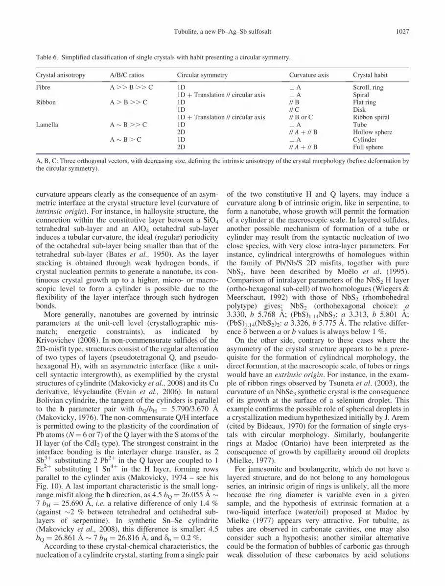
Table 6. Simplified classification of single crystals with habit presenting a circular symmetry.
Crystal anisotropy A/B/C ratios Circular symmetry Curvature axis Crystal habit
Fibre A .. B .. C 1D ? A Scroll, ring1D þ Translation // circular axis ? A Spiral
Ribbon A . B .. C 1D // B Flat ring1D // C Disk1D þ Translation // circular axis // B or C Ribbon spiral
Lamella A � B .. C 1D ? A Tube2D // A þ // B Hollow sphere
A � B . C 1D ? A Cylinder2D // A þ // B Full sphere
A, B, C: Three orthogonal vectors, with decreasing size, defining the intrinsic anisotropy of the crystal morphology (before deformation bythe circular symmetry).
Tubulite, a new Pb–Ag–Sb sulfosalt 1027
during sulfosalt deposition. Like selenium or oil droplets,such bubbles may have attracted by capillarity very thinlamellar crystallites of tubulite, inducing a circular growth.The variable diameter of the tubes would thus correspondto a change in the bubble radius.
Crystallites of tubulite with a needle-like morphologymay have grown also around a droplet or bubble, but suchmorphology induces a higher degree of freedom (curva-ture) during the growth, giving the final scroll observedunder the binocular microscope (Fig. 4b), and not perfectrings.
The difference between typical tubular and hair-likemorphologies of tubulite is the consequence of distinctratios of growth speeds along unit-cell vectors (va, vb andvc). Tubular crystals are derived from ribbon-like crystal-lites, with va . vb .. vc. Due to mechanical constraintsand capillarity strengths, the ribbon would have grownflat along the equatorial circle of an oil droplet or gasbubble, to give a tube through spiral rolling-up. In hair-like crystals, one has va .. vb � vc. Such a difference inmorphology may be the consequence of twinning, as,according to Grigoriev (1965), a twin contact acts like adefect and favours the growth of twinned crystals alongtheir common twin boundary. In tubulite, (001) as a twinplane, giving a pseudo-orthorhombic symmetry, wouldfavour the growth along a and b [(001) as twin contact]relatively to c, and thus the initiation of ribbon-like crys-tallite. Twinning would be a structural pre-requisite forthe formation of tubular crystals, relatively to untwinnedfibrous crystals.
A very similar morphological duality between tubes andfibres was described by Belkin & Libelo (1987). Here itcorresponds to crystals of cryptomelane-hollandite grow-ing within Permian bedded salt. According to theseauthors, the cylinders ‘‘always occur in or very near fluidinclusions’’. Individual fibres are commonly ‘‘curvedwhen in a fluid inclusion’’. It is important to cite themechanism invoked by these authors: ‘‘Only one cylinderper fluid inclusion is found in the chevron salt whereasmultiple cylinders are commonly found in inclusions inclear salt. The common multiple cylinders in the clear saltmay reflect a concentration process that occurred duringdissolution and recrystallization of the primary chevronsalt. Cylinders trapped in chevron salt would perhapsremain as a residue after salt dissolution and might betrapped in the less common but much larger fluid inclu-sions characteristic of the clear salt ( . . . ) Two importantfeatures that are observed are (1) these salt-hosted cylin-ders are usually near larger inclusions (. 10 mm3) and (2)they occur above the inclusion, in the stratigraphic sense,suggesting that the inclusions may have moved downward,leaving the cylinders behind’’.
It is curious that Belkin & Libelo (1987) exclude the roleof gas bubbles, when their description strongly suggestssuch a mechanism: only one bubble within each primaryfluid inclusion will permit only one tubular crystal to grow,at the top of this inclusion, while later the coalescence ofinclusions through salt recrystallization will permit toobserve several tubes within one inclusion.
7. Conclusion
Tubulite appears as a new sulfosalt species of the pseudo-ternary Ag2S–PbS–Sb2S3 system, with the lowest Ag con-tent within the group of Ag–Pb–Sb sulfosalts. Its unit cell isclose to those of sterryite and parasterryite, the nearestcompounds in the (Ag,Cu)2S–PbS–(Sb,As)2S3 system. Insulfosalt systematics (Moelo et al., 2008), this crystal-chemical relationship relates tubulite with the owyheeitesub-group in the zinkenite family. Its crystal structure maycorrespond to the unit-cell intergrowth of two buildingblocks, an owyheeite-type complex column and anotherone derived from a Pb–Sb sulfosalt. A high-resolutiontransmission electron microscopy (HRTEM) study of frag-ments of tubular crystals would appear as the best approachin the future to confirm such a structural organization, asexemplified by Kryukova et al. (2005) for the structuralcharacterization of the synthetic iodine derivative of pil-laite, belonging also to the zinkenite family. On the otherhand, one may hope to find flat crystals of sufficient sizefor an X-ray single-crystal structure study.
The perfect tubular morphology of typical tubulite crys-tals seems to be related to extrinsic conditions of formation(capillary forces) acting on very thin lath-like crystallites atthe beginning of crystal growth. The hypothesis of growtharound gas bubbles, or oil droplets, in hydrothermal solu-tions would need fluid inclusion studies in the two depositsto be confirmed. Tubulite may be also present in otherdeposits; for instance, sulfosalt tubes (stacked rings) weredescribed by Woodside et al. (2000) in the hydrothermalPb–Zn–Ag–Sb vein system of Van Silver mine(Brandywine Creek, British Columbia, Canada). TheSEM analysis of one tube (Y.M.) indicated owyheeite,but the close association in this ore of variousPb–Ag–Cu–Sb sulfosalts, among which boulangerite, sem-seyite, owyheeite and also fizelyite (Yang et al., 2009),whose compositions surround that of tubulite, may havealso favoured the formation of tubulite here. Tubular bou-langerite from Madoc observed by Bideaux (1970) withSEM, but apparently without precise chemical analysis,may also correspond to tubulite, close (but without arsenic)to the pair boulangerite-sterryite, present at Madoc(Jambor, 1968; Jambor et al., 1982).
Acknowledgements: We sincerely thank Georges Favreau(Association Francaise de Micromineralogie - AFM), whoactively promoted the study of the French occurrence oftubulite, then the Franco-Italian collaboration, andGiovanni Dalla Fontana for his contribution to the find ofthe first specimens of tubulite from Bio. The kind help ofCristian Biagioni (Pisa University) and Matteo Boscardin(Museo di Archeologia e Scienze Naturali G. Zannato ofMontecchio Maggiore), who provided the preliminary ana-lysis of Italian samples, was also greatly appreciated.Michel Evain (Institut des Materiaux J. Rouxel) helpedus in the X-ray precession study. Jean-Marc Johannet(AFM) provided a beautiful photograph of tubulite. Manythanks to Silvio Maccagno who provided the first samples
1028 Y. Moelo et al.
from Bio studied in Lausanne in 2007, and to EricaBittarello (Dipartimento di Scienze della Terra – TurinUniversity) for the map of the Italian deposit. We finallythank Professor E. Makovicky (University of Copenhagen)and Dr. A. J. Locock (University of Alberta) for theirconstructive comments, as well as S. Krivovichev, ChiefEditor of the European Journal of Mineralogy, for hiscareful monitoring of the manuscript.
References
Ambrino, P., Blass, G., Boscardin, M., Ciriotti, M.E., Dalla Fontana,
G., Kolitsch, U., Martini, B., Milli, L., Salvetti, A., Bonacina, E.
(2008): Borgofranco d’Ivrea. La paragenesi a solfuri e solfosali
delle miniere argentifere. Riv. Mineral. Ital., 3/2008, 144–162.
Baggio, P. & Friz, C. (1969): Fenomeni tettonico-metamorfici di eta
alpina lungo la linea Insubrica auct. Mem. Mus. Trid. Sci. Nat.,
17, 183–206.
Baronnet, A. & Devouard, B. (2005): Microstructures of common
polygonal serpentines from axial HRTEM imaging, electron dif-
fraction, and lattice-simulation data. Can. Mineral., 43, 513–542.
Bates, T.F., Hildebrand, F.A., Swineford, A. (1950): Morphology
and structure of endellite and halloysite. Am. Mineral., 35,
463–484.
Belkin, H.E. & Libelo, E.L. (1987): Fibers and cylinders of crypto-
melane-hollandite in Permian bedded salt, Palo Duro Basin,
Texas. Am. Mineral., 72, 1211–1224.
Bernadi, G. (1992): Peyrebrune: Carriere du Rivet. Cahier Des
Micromonteurs, 1, 3–10.
Biagioni, C. (2009): Minerali della Provincia di Lucca. Associazione
Micro-mineralogica Italiana ed., Cremona, 352 p.
Bideaux, R.A. (1970): Mineral rings and cylinders. Mineral. Rec., 1,
105–112.
Biino, G. & Compagnoni, R. (1989): The Canavese Zone between
the Serra d’Ivrea and the Dora Baltea river (Western Alps).
Eclog. Geol. Helvet., 82 (2), 413–427.
Bindi, L., Keutsch, F.N., Bonazzi, P. (2012): Menchettiite,
AgPb2.40Mn1.60Sb3As2S12, a new sulfosalt belonging to the lil-
lianite series from the Uchucchacua polymetallic deposit, Lima
Department, Peru. Am. Mineral., 97, 440–446.
Borghi, A., Compagnoni, R., Naldi, M. (1996): The crystalline base-
ment of the Canavese Zone (Internal Western Alps): new data
from the area west of Ivrea (Northern Italy). Geologie Alpine,
72, 23–34.
Ciriotti, M.E., Fascio, L., Pasero, M. (2009): Italian type minerals.
Plus-Pisa university press ed., Pisa, 357 p.
Contri, J.P. (1959): Etude geologique et metallogenique des environs
de Peyrebrune et de Montroc pres Realmont (Tarn). Thesis, Paris
University, 127 p.
Di Cato, P. (1996): Peyrebrune: Une mine dans le Tarn. Graphitarn
ed., Albi, 190 p.
Durand, B. (1966): Le gisement plombo-zincifere de Peyrebrune,
Tarn (France). Thesis, Nancy University, 251 p.
Eggleton, R.A. & Tilley, D.B. (1998): Hisingerite: A ferric kaolin
mineral with curved morphology. Clays Clay Minerals, 46,
400–413.
Evain, M., Petricek, V., Moelo, Y., Maurel, C. (2006): First (3þ 2)-
dimensional superspace approach to the structure of
levyclaudite-(Sb), a member of the cylindrite-type minerals.
Acta Cryst. B, 62, 775–789.
Frenzel, A. (1893): Uber den Kylindrit. N. Jb. Miner. Geol. Palaont.,
2, 125–128.
Ghiurca, V. (1985): Les habitus en rondelles. Dans le monde curieux
des microcristaux de jamesonite. Miner. & Fos., 120, 22–30.
Gomez-Herrero, A., Landa-Casanovas, A.R., Hansen, S., Otero-
Dıaz, L.C. (2000): Electron microscopy study of tubular crystals
(BiS)1þd(NbS2)n. Micron., 31, 587–595.
Grigoriev, D.P. (1965): Ontogeny of minerals. Israel Program for
Scientific Translation ed., Jerusalem, 250 p.
Guemas, L., Rabu, P., Meerschaut, A., Rouxel, J. (1988):
Characterization of new ‘‘SnNbS3, PbNbS3, PbNb2S5, SnTiS3
and SnTi2S5’’ compounds. Mater. Res. Bull., 23, 1061–1069.
Guerange-Lozes, J., Mouline, M.P., Delsahut, B. (1996):
Geological map of France (1/50000): Realmont (959). BRGM
ed., Orleans.
Heuer, M., Wagner, G., Doring, T., Bente, K., Kryukova, G. (2004):
Nanowire arrangements of PbS–Sb2S3 compounds. J. Crystal
Growth., 267, 745–750.
Hubert, M.-N. & Hubert, M. (1992): A propos du gisement de
Peyrebrune: Histoire d’une de nos decouvertes. Cahier Des
Micromonteurs, 2, 27–33.
Jambor, J.L. (1967): New lead sulfoantimonides from Madoc,
Ontario. Part 2 – Mineral descriptions. Can. Mineral., 9,
191–213.
— (1968): New lead sulfoantimonides from Madoc, Ontario. Part 3 –
Syntheses, paragenesis, origin. Can. Mineral., 9, 505–521.
Jambor, J.L., Laflamme, J.H.G., Walker, D.A. (1982): A re-exam-
ination of the Madoc sulfosalts. Mineral. Rec.., 13, 93–100.
Jaszczak, J.A., Dimovski, S., Hackney, S.A., Robinson, G.W.,
Bosio, P., Gogotsi, Y. (2007): Micro- and nanoscale graphite
cones and tubes from Hackman valley, Kola Peninsula, Russia.
Can. Mineral., 45, 379–389.
Kogure, T., Mori, K., Kimura, Y., Takai, Y. (2011): Unraveling the
stacking structure in tubular halloysite using a new TEM with
computer-assisted minimal-dose system. Am. Mineral., 96,
1776–1780.
Krivovichev, S.V. (2008) Nanotubes in minerals and mineral-related
systems. in‘‘Minerals as advanced materials I’’, S. Krivovichev,
ed., Springer, Berlin/Heidelberg, 179–191.
Kryukova, G.N., Heuer, M., Wagner, G., Doering, T., Bente, K.
(2005): Synthetic Cu0.507Pb8.73Sb8.15I1.6S20.0 nanowires. J.
Solid State Chem., 178, 376–381.
Laufek, F., Pazout, R., Makovicky, E. (2007): Crystal structure
of owyheeite, Ag1.5Pb4.43Sb6.07S14: refinement from
powder synchrotron X-ray diffraction. Eur. J. Mineral., 19,
557–566.
Lincio, G. (1916): Note preliminari su alcuni minerali del giaci-
mento metallifero di Borgofranco d’Ivrea. Rend. R. Accad.
Naz. Lincei., 25, 227–230.
Makovicky, E. (1971): Microstructure of cylindrite. N. Jb. Mineral.
Mh., 1971, 403–413.
— (1976): Crystallography of cylindrite. N. Jb. Mineral. Abh., 126,
304–306.
— (1985a): The building principles and classification of sulfosalts
based on the SnS archetype. Fortschr. Miner., 63, 45–89.
— (1985b): Cyclically twinned sulphosalt structures and their
approximate analogues. Z. Kristallogr., 173, 1–23.
— (1993): Rod-based sulphosalt structures derived from the SnS and
PbS archetypes. Eur. J. Mineral., 5, 545–591.
Tubulite, a new Pb–Ag–Sb sulfosalt 1029
— (1997): Modular crystal chemistry of sulphosalts and other com-
plex sulfides. in ‘‘Modular aspects of Minerals’’. EMU Notes in
Mineralogy, 1, 237–271.
Makovicky, E. (1974): Mineralogical data on cylindrite and incaite.
N. Jb. Miner. Mh., 1974, 235–256.
Makovicky, E. & Hyde, B.G. (1992): Incommensurate, two-layer
structures with complex crystal chemistry: minerals and related
synthetics. Mat. Sci. Forum, 100 & 101, 1–100.
Makovicky, E., Petrıcek, V., Dusek, M., Topa, D. (2008): Crystal
structure of a synthetic tin-selenium representative of the cylin-
drite structure type. Am. Mineral., 93, 1797–1798.
Maser, M., Rice, R.V., Klug, H.P. (1960): Chrysotile morphology.
Am. Mineral., 45, 680–688.
Mielke, R. (1977): Boulangerite and associated minerals of the
Rogers mine, Madoc, Ontario. Unpub. B. Sc. thesis, University
of Waterloo, Waterloo, Ontario.
Moelo, Y. (1983): Contribution a l’etude des conditions naturelles
de formation des sulfures complexes d’antimoine et plomb.
Thesis, Series Documents du BRGM, 57, BRGM ed.
(Orleans), 624 p.
Moelo, Y., Mozgova, N., Picot, P., Bortnikov, N., Vrublevskaya, Z.
(1984a): Cristallochimie de l’owyheeite: nouvelles donnees.
Tschermaks Min. Petr. Mitt., 32, 271–284.
Moelo, Y., Oudin, E., Picot, P., Caye, R. (1984b): L’uchucchacuaite,
AgMnPb3Sb5S12, une nouvelle espece minerale de la serie de
l’andorite. Bull. Mineral., 107, 597–604.
Moelo, Y., Makovicky, E., Karup-Møller, S. (1989): Sulfures com-
plexes plombo-argentiferes: mineralogie et cristallochimie de la
serie andorite-fizelyite. Series Documents du BRGM, 167.
BRGM ed. (Orleans), 107 p.
Moelo, Y., Meerschaut, A., Rouxel, J., Auriel, C. (1995): Precise
analytical characterization of incommensurate sandwiched
layered compounds [(Pb,Sn)S]1þx[(Nb,Ti)S2]m (0.08 �� 0.28, m ¼ 1–3). Role of cationic coupling on the proper-
ties and the structural modulation. Chem. Mater., 7,
1759–1771.
Moelo, Y., Makovicky, E., Mozgova, N.N., Jambor, J.L., Cook, N.,
Pring, A., Paar, W.H., Nickel, E.H., Graeser, S., Karup-Møller,
S., Balic-Zunic, T., Mumme, W.G., Vurro, F., Topa, D., Bindi,
L., Bente, K., Shimizu, M. (2008): Sulfosalt systematics: a
review. Report of the sulfosalt sub-committee of the IMA
Commission on Ore Mineralogy. Eur. J. Mineral., 20, 7–46.
Moelo, Y., Orlandi, P., Guillot-Deudon, C., Biagioni, C., Paar, W.,
Evain, M. (2011): Lead-antimony sulfosalts from Tuscany (Italy).
XI. The new mineral parasterryite, Ag4Pb20(Sb14.5As9.5)P¼24S58,
and associated sterryite, Cu(Ag,Cu)3Pb19(Sb18–15As4–7)P¼22
(As–As)S56, from Pollone mine (Tuscany, Italy). Can. Mineral.,
49, 623–638.
Moelo, Y., Guillot-Deudon, C., Evain, M., Orlandi, P., Biagioni, C.
(2012): Comparative modular analysis of two complex sulfosalt
structures: sterryite, �Cu(Ag,Cu)3Pb19(Sb,As)22(As–As)S56,
and parasterryite, Ag4Pb20(Sb,As)24S58. Acta Cryst. B, 68 (5),
480–492.
Orlandi, P., Del Chiaro, L., Pagano, R. (1996): Minerals of the
Seravezza Marble, Tuscany, Italy. Mineral. Rec., 27 (1),
47–58.
Piccoli, G.C., Maletto, G., Bosio, P., Lombardo, B. (2007): Minerali
del Piemonte e della Valle d’Aosta. Associazione Amici del
Museo ‘‘F. Eusebio’’ Alba ed., Alba (Cuneo), 607 p.
Pierrot, R., Picot, P., Fortune, J.-P., Tollon, F. (1976): Inventaire
mineralogique de la France No 6: Tarn (81). Service Geologique
National (BRGM) ed., Orleans, 147 p.
Skowron, A. & Brown, I.D. (1990): Structure of Pb2Sb2S5. Acta
Cryst. C, 46, 534–536.
Stalder, H.A., Wagner, A., Graeser, S., Stuker, P., Offerman, E.,
Meisser, N. (1998): Mineralienlexikon der Schweiz. Wepf &
Co. ed., Basel, 579 p.
Tsuneta, T., Tanda, S., Inagaki, K., Okajima, Y., Yamaya, K. (2003):
New crystal topologies and the charge-density-wave in NbSe3.
Physica B, 329–333, 1544–1545.
Valverde, J. (2002): Photo p. 32. Cahier des Micromonteurs, 78–4.
Veblen, D.R. & Buseck, P.R. (1979): Serpentine minerals:
Intergrowths and new combination structures. Science, 206,
1398–1400.
Wiegers, G.A. & Meerschaut, A. (1992): Misfit layer compounds
(MS)nTS2 (M ¼ Sn, Pb, Bi, Rare Earth metals; T ¼ Nb, Ta, Ti,
V, Cr; 1.08,n ,1.23): Structures and physical properties.
Mater. Sci. Forum, 100 & 101, 101–172.
Woodside, R.W.M., Soregaroli, A.E., Ansell, H.G., Twaites, B.L.,
Balacko, T.W. (2000): Rare sulfosalts from the Van Silver Mine,
British Columbia. Mineral. Rec., 31 (3), 219–229.
Yada, K. (1971): Study of microstructure of chrysotile asbestos by
high resolution electron microscopy. Acta Cryst. A, 27, 659–664.
Yang, H., Downs, R.T., Burt, J.B., Costin, G. (2009): Structure
refinement of an untwinned single crystal of Ag-excess fizelyite,
Ag5.94Pb13.74Sb20.84S48. Can. Mineral., 47, 1257–1264.
Zolensky, M.E. & Mackinnon, I.D.R. (1986): Microstructures of
cylindrical tochilinites. Am. Mineral., 71, 1201–1209.
Received 23 May 2013
Modified version received 1 July 2013
Accepted 22 July 2013
1030 Y. Moelo et al.


![Mayenite supergroup, part III: Fluormayenite, Ca\u003cSUB\u003e12\u003c/SUB\u003eAl\u003cSUB\u003e14\u003c/SUB\u003eO\u003cSUB\u003e32\u003c/SUB\u003e[〈\u003cSUB\u003e4\u003c/SUB\u003eF\u003cSUB\u003e2\u003c/SUB\u003e],](https://img.dokumen.tips/doc/110x75/634059325328aff1b103eb6f/mayenite-supergroup-part-iii-fluormayenite-cau003csubu003e12u003csubu003ealu003csubu003e14u003csubu003eou003csubu003e32u003csubu003eu003csubu003e4u003csubu003efu003csubu003e2u003csubu003e.jpg)


