Embed Size (px)
Citation preview

JOURNAL OF VIROLOGY, Aug. 2006, p. 8100–8113 Vol. 80, No. 160022-538X/06/$08.00�0 doi:10.1128/JVI.00687-06Copyright © 2006, American Society for Microbiology. All Rights Reserved.
Transposon-Assisted Cloning and Traceless Mutagenesis of Adenoviruses:Development of a Novel Vector Based on Species D†
Zsolt Ruzsics,2 Markus Wagner,2‡ Andrea Osterlehner,2§ Jonathan Cook,1Ulrich Koszinowski,2 and Hans-Gerhard Burgert1,2*
University of Warwick, Department of Biological Sciences, Coventry CV4 7AL, United Kingdom,1 andMax-von-Pettenkofer Institute, Gene Center, Ludwig-Maximilians-University, 81377 Munich, Germany2
Received 5 April 2005/Accepted 31 May 2006
Until recently, adenovirus (Ad)-mediated gene therapy was almost exclusively based on human Ad type 5(Ad5). Preexisting immunity and the limited, coxsackievirus and adenovirus receptor-dependent tropism ofAd5 stimulated attempts to exploit the natural diversity in tropism of the other 50 known human Ad serotypes.Aiming in particular at immunotherapy and vaccination, we have screened representative serotypes fromdifferent Ad species for their ability to infect dendritic cells. Ad19a, an Ad from species D, was selected fordevelopment as a new vector for vaccination and cancer gene therapy. To clone and manipulate its genome, wehave developed a novel methodology, coined “exposon mutagenesis,” that allows the rapid and precise intro-duction of virtually any genetic alteration (deletions, point mutations, or insertions) into recombinant Adbacterial artificial chromosomes. The versatility of the system was exemplified by deleting the E3 region ofAd19a, by specifically knocking out expression of a species-specific E3 gene, E3/49K, and by reinserting E3/49Kinto an E3 null Ad19a mutant. The technology requires only limited sequence information and is applicable toother Ad species. Therefore, it should be extremely valuable for the analysis of gene functions from any Adspecies. In addition, a basic, replication-defective E1- and E3-deleted Ad19a vector expressing GFP(Ad19aGFP) was generated. This new vector based on species D Ads exhibits a very promising tropism forlymphoid and muscle cells and shows great potential as an alternative vector for transduction of cell types thatare resistant to or only poorly transduced by conventional Ad5-based vectors.
Adenoviruses (Ads) are nonenveloped viruses with a dou-ble-stranded DNA genome ranging in size from 29 to 45 kb.All human Ads have the potential to induce acute and persis-tent infections and are generally associated with mild patho-genesis. However, Ads can also induce severe symptoms oreven life-threatening diseases, particularly in immunocompro-mised patients (29). To date, 51 different human serotypeshave been described; these serotypes are classified into the sixspecies A to F (57). Members of species B, C, and E usuallycause respiratory infections, those of species A and F areassociated with infections of the gastrointestinal tract, andthose of species D, harboring the vast majority of serotypes(32), show a tropism for the eye. Three serotypes of this species(Ad8, Ad19a, and Ad37) cause a distinct eye disease calledepidemic keratoconjunctivitis (EKC). Interestingly, a promi-nent subset of species D was isolated from AIDS patientspreviously (17).
Ads of species C, in particular, the best-characterized sero-types Ad5 and Ad2, have played a crucial role in the discoveryand characterization of many important molecular processes,e.g., splicing, replication, and tumor suppression (52, 57). More
recently, these viruses have emerged as extremely useful mod-els for the study of the interaction of viruses with the hostimmune system, unraveling many viral immunomodulatoryfunctions that appear to contribute to immune evasion andpersistence (10, 37, 41, 71). Moreover, the species C Ad Ad5has gained widespread use as a vector for gene therapy andvaccination (16, 30, 32, 52, 63).
In the past few years there has been growing interest instudying Ads from species other than C. The reasons are sev-eralfold. First, the tissue tropism and receptor usage of thevarious species differ significantly. While species C Ads pri-marily utilize the coxsackievirus and adenovirus receptor(CAR) as an attachment receptor (4, 51), most species B Adsuse CD46 (23, 39, 60) and some possibly use CD80/86 (59; foran up-to-date review, see reference 73). For species D, theidentity of the primary attachment receptor remains contro-versial. Initial evidence suggested that fibers of species D Adscan bind to CAR (51); however, subsequent studies clearlyshowed that infection by members of this species does not relyon CAR. Some authors have suggested CD46 as primary re-ceptor for the EKC-causing Ad37 (72, 73), whereas othersprovided strong evidence for �(2-3)-linked sialic acid as a li-gand for attachment of EKC-causing Ads (2, 3, 12, 64). Sec-ondly, the composition of genes in the immunomodulatory E3region differs remarkably between Ad species (10, 11). Thus, itis likely that species-specific E3 genes, together with the dif-ferential receptor usages, contribute to the distinct pathogen-esis and disease association of the different species (8, 11, 36,70). Thirdly, it became apparent that Ad5-based gene therapyvectors have significant limitations. Efficient Ad5 transduction
* Corresponding author. Mailing address: Department of BiologicalSciences, University of Warwick, Gibbet Hill Road, Coventry CV47AL, United Kingdom. Phone: 44-2476-524744. Fax: 44-2476-523568.E-mail: [email protected].
† Supplemental material for this article may be found at http://jvi.asm.org.
‡ Present address: Bavaria Nordic GmbH, Fraunhoferstr. 13, 82152Martinsried, Germany.
§ Present address: Roche Diagnostics GmbH, Nonnenwald 2, 82377Penzberg, Germany.
8100
on July 8, 2015 by guesthttp://jvi.asm
.org/D
ownloaded from
on July 8, 2015 by guest
http://jvi.asm.org/
Dow
nloaded from
on July 8, 2015 by guesthttp://jvi.asm
.org/D
ownloaded from

requires CAR, and some normal tissues and advanced tumorcells, e.g., hematopoietic cells such as dendritic cells (DCs) (34,35), exhibit low CAR expression or lack CAR altogether. DCsare key antigen-presenting cells and are exploited for immu-notherapy and vaccination (55). Moreover, preexisting immu-nity, with �80% of the human population being seropositivefor Ad5, considerably limits the efficacy of Ad5 vector treat-ments (63, 66). Therefore, exploitation of the natural diversityof Ads may help to overcome some of the obstacles faced ingene therapy and vaccination.
Investigation and exploitation of Ads other than species Cwas hampered at least in part by the lack of convenient cloningand mutagenesis systems. Traditionally, two approaches havebeen used to generate recombinant Ads. In both cases, geneticchanges are introduced into a subcloned fragment of the Adgenome. Subsequently, this modified fragment is introducedinto the Ad genome by in vitro ligation to restriction endonu-clease-digested virus DNA or by homologous recombinationbetween the modified fragments and digested viral DNAs inpermissive host cells (5). Alternatively, homologous recombi-nation between cotransfected plasmids carrying overlappingand complementary parts of the Ad genome was used to gen-erate recombinant species C Ads (6). These methods are lim-ited by the low efficiency of virus reconstitution and by theneed for plaque purification of the recombinant viruses, sincewild-type (wt) progenies are frequently generated during virusreconstitution (16, 45).
To overcome the problems associated with homologous re-combination in permissive cells and to improve the efficiency ofgenetic manipulation, full-length genomes of species C Adshave been cloned in Saccharomyces cerevisiae (33) and Esche-richia coli (13), allowing mutagenesis under noncomplement-ing conditions. However, these approaches require subcloningof the modified DNA fragment in special shuttle plasmids,which is labor intensive (13, 15, 26, 33, 42). In addition, most ofthese mutagenesis procedures are greatly restricted by the lim-ited availability of appropriate restriction sites close to thetargeted sequence (13, 33) or by the requirement for a specifichost for genetic manipulation (13, 15, 26, 33). A range ofmethods exists for generating Ad2 and Ad5 vectors containingexpression cassettes (see references 1, 16, 45, and 47 and ref-erences therein). Genetic manipulation of these vectors is rel-atively fast and simple but is restricted to a defined, predeter-mined site. Recombinant Ads based on other human serotypes(e.g., Ad4, Ad7, or Ad35) or animal Ads have been generatedby traditional homologous recombination (40) in cells or in E.coli or by classical cloning techniques (21, 22, 31, 48, 62, 66).However, fast, generally applicable, and efficient methods forcloning and precise manipulation of Ad genomes for detailedstudies of the various functional activities of different Ad spe-cies or for exploration of their potential as vectors are not yetavailable.
Recently, a novel recombination system, called ET recom-bination, has been exploited for genetic engineering of recom-binant DNA in E. coli (46, 74). ET recombination uses �phage-derived recombination proteins that mediate effectiverecombination of linear DNA fragments into the target se-quences, requiring only very short (usually 35- to 50-bp) ho-mologous sequences. This allows the introduction of virtuallyany selectable mutation in a single step. Likewise, large genomic
fragments can be cloned by ET recombination into a PCR-derived linear vector carrying short homology arms (75). How-ever, in the latter case, degradation of linear DNA in E. colimust be prevented, and short terminal repeats within the ho-mology arms of the vector may lead to a substantial back-ground due to a high frequency of vector circularization (75).Mutations generated by PCR or even fully synthetic DNA canbe introduced by ET recombination, which, unlike other meth-ods based on homologous recombination, does not requirespecial E. coli strains.
Here we demonstrate that ET recombination can be suc-cessfully applied to construct recombinant Ads. To ensurecorrect recombinatorial construction, purified Ad19a DNAwas marked prior to ET recombination with an antibiotic re-sistance gene using a transposon 7 (Tn7)-derived in vitro trans-position system (7). Convenient and complete removal of theselection marker makes the resulting recombinant Ad genome-bacterial artificial chromosome (BAC) ready for reverse ge-netic approaches. In addition, we report on a novel, two-stepmutagenesis technique in which the mutation coupled with theTn7-derived selection marker is first introduced by ET re-combination. Subsequently, the operational sequences arecompletely removed by a simple transposase cleavage-ligasereaction in vitro. The potency of these methodologies isdemonstrated by introducing deletions, insertions, and pointmutations in a recombinant Ad19a genome. Moreover, anovel Ad19a-derived first-generation gene therapy vectorwas established that seems to have an interesting tropism forlymphoid cells.
MATERIALS AND METHODS
Cell lines, viruses, and preparation of viral genomic DNA. The human epi-thelial lung carcinoma cell line A549 (ATCC CCL-185) and the Ad5-trans-formed human epithelial kidney cell line 293 (ATCC CRL-1573) were culturedin Dulbecco’s minimal essential medium supplemented with 10% fetal calf serum(FCS), penicillin (100 U/ml), streptomycin (100 �g/ml), and 2 mM glutamine(27). Jurkat E6-1 (ATCC TIB-152), LCL, and T2 (ATCC CRL-1992) cells weremaintained in RPMI medium supplemented with 10% FCS and antibiotics. TheME strain of Ad19a (67), a kind gift of G. Wadell (Department of Virology,Umea University, Sweden), was plaque purified and amplified by infecting sub-confluent A549 cells at 1 to 3 PFU/cell. We refer to the plaque-purified stock ofthe ME strain as Ad19aT3 in this report. Virus stocks were prepared usingstandard protocols (44). The human Ad5-derived E1- and E3-deleted greenfluorescent protein (GFP)-expressing vector Ad5GFP was purchased from Qbio-gene. Like the Ad19aGFP vector described in this report, Ad5GFP expressesenhanced GFP under the control of the cytomegalovirus (CMV) promoter-enhancer in the deleted E1 region. In addition, both vectors have large deletionswithin the E3 region; Ad19aGFP lacks 4.5 kb and Ad5GFP lacks 1.88 kb,preventing expression of all E3 genes and of all except E3/12.5K, respectively.
For preparation of genomic Ad19a DNA, A549 cells were infected at a mul-tiplicity of infection of 1 to 3. After the cytopathic effect was complete, theinfected cells were washed once in phosphate-buffered saline (PBS), scrapedfrom the plates, and resuspended in PBS. Cells (�4 � 106 cells/ml) were lysed byadding an equal volume of TST buffer (2% Triton X-100, 400 mM NaCl, 20 mMTris-HCl, pH 8.0) to the cell suspension followed by incubation on ice for 30 min.Cell debris were removed by centrifugation at 14,000 � g for 10 min at 4°C, andthe supernatant was treated with 50 �g/ml proteinase K (Roche) in the presenceof 0.5% sodium dodecyl sulfate for 60 min at 56°C. After extraction of the nucleicacids by phenol-chloroform and ethanol precipitation, the extract was treatedwith RNase A (Sigma). RNA-free viral DNA was again phenol-chloroformextracted and precipitated with ethanol.
Tn labeling of viral DNA and recombinatorial cloning of the wt Ad19a ge-nome. The BAC entry vector was generated by direct cloning of an assembledPCR product consisting of the two Ad19a inverted terminal repeats (ITRs)linked by a short unique E4 sequence. The left and right Ad19a ITRs wereamplified by PCR using oligonucleotides 19aITR-PacI and 19aLrev and 19aRfor,
VOL. 80, 2006 MUTAGENESIS OF SPECIES D Ads AND VECTOR DEVELOPMENT 8101
on July 8, 2015 by guesthttp://jvi.asm
.org/D
ownloaded from

respectively (for primer sequences, see Table S1 in the supplemental material).To amplify the left end of the genome, the forward primer specific to theterminal virus sequence was flanked by a 5� PacI site (19aITR-PacI) and thereverse primer specific to the 3� end of the ITR (19aLrev) was tagged with a shortsequence homologous to 19aRfor. For amplification of the right Ad19a end, thesame terminal primer (19aITR-PacI) and a primer specific to a conserved E4sequence (19aRfor) were used. The products of the “left” and “right” PCRswere combined and reamplified by use of the terminal primer 19aITR-PacI.The BAC entry vector p19aLR was generated by inserting the assembled PCRproducts into the PacI site of the pKSO BAC vector (43).
pGPS1.1 (New England Biolabs [NEB]) containing a mini-Tn cassette(Transprimer-1) was used as Tn donor in the Tn-assisted cloning experiments.Purified viral DNA (200 or 300 ng) was labeled with Transprimer-1 in vitro byuse of TnsABC* according to the Genome Priming System (GPS) protocol (NewEngland Biolabs). Recombination-proficient electrocompetent E. coli DH10B(Invitrogen) carrying p19aLR and pBAD�� (74, 75) was prepared as describedpreviously (68) and subsequently transformed with Tn-labeled Ad19a DNA byuse of a Bio-Rad GenePulser with the following settings: 2,500 V, 200 , and 25�F. Upon induction with 0.1% L-arabinose (46, 74), the transformants wereincubated for 2 h at 37°C in LB medium and plated onto LB agar platescontaining 25 �g/ml chloramphenicol (Cm) and 20 �g/ml kanamycin (Kn). Dou-bly resistant colonies were isolated, boiled, and screened by PCR using primersspecific to the Ad hexon to identify BACs containing Ad19a DNA. To roughlylocate the Tn insertion site, recombinant BACs were analyzed by restrictiondigestion.
ET recombination. Synthetic oligonucleotide primers used for the generationof the ET recombination fragments were designed as follows. At the 5� ends allprimers carried 40-nucleotide up- or downstream homology arms for ET recom-bination. These 40 nucleotides were Ad sequences in the vicinity of the locationin which the desired mutation should occur. At their 3� end, all ET primerscontained the priming sequences complementary to either the left end (5�-TGTGGG CGG ACA AAA TAG TTG G-3�) or right end (5�-TGT GGG CGG ACAATA AAG TCT TAA ACT GAA-3�) of the Transprimer-1 cassette of pGPS1.1.Depending on the application, different insertion sequences (e.g., three-nucle-otide direct repeats) were included between the homology arms and the Tnpriming regions (for the sequences of the primers used in this study, see Table S1in the supplemental material). The linear recombination fragments were ampli-fied by use of an Expand High Fidelity PCR system (Roche) and 2 ng pGPS1.1as the template. The PCR and the ET recombination procedure were performedas described previously (68). Briefly, PCR products were purified with a PCRpurification kit (QIAGEN). For ET recombination, arabinose-induced electro-competent E. coli DH10B cells carrying the target BACs and pBAD-�� (46)were transformed with 300- to 400-ng purified recombination fragment. After1.5 h growth in 1 ml LB medium at 37°C the transformants were plated on LBagar plates containing 25 �g/ml Cm and 20 �g/ml Kn.
Transposon excision and exposon mutagenesis. Purified Tn-containing BACs(140 ng) were treated with 1 �l TnsABC* (NEB) in 1� GPS buffer (250 mMTris-HCl [pH 8.0], 20 mM dithiothreitol, 20 mM ATP) in the presence of 90 ngof temperature-sensitive plasmid pST76T (50), which serves as a dead-end target.After 10 min incubation at 37°C, a 1/20 vol of 0.3 M MgCl2 was added to initiateTn end cleavage. Following 60 min incubation at 37°C the reaction was stoppedby heat treatment (15 min, 75°C). A total of 400 cohesive end units of T4 ligasewere added, and the reaction mixture was incubated overnight at 16°C forrecircularization. After heat inactivation of the T4 ligase, the reaction mixturewas phenol-chloroform extracted and the BAC DNA was ethanol precipitated.Electrocompetent E. coli DH10B or pUC19RP12-transformed DH10B (49) ex-pressing meganuclease I-SceI was transformed with the purified DNA. Thetransformants were incubated at 37°C in LB medium and plated on LB agarplates containing 25 �g/ml of chloramphenicol. BACs prepared from isolatedcolonies according to the standard alkaline lysis procedure were analyzed byrestriction digestions.
In the insertion reaction of “exposon mutagenesis,” 200 ng of purified SapI-treated PCR amplified inserts was added to the heat-inactivated TnsABC* re-action prior to T4 ligase treatment. Subsequent treatments were as describedabove. For construction of the E3-deleted, E3/49K-expressing recombinant virusAd19a�E3�49K, the E3/49K open reading frame (ORF), including the 5� simianvirus 40 (SV40)-derived intron sequence of pSG5, was amplified by PCR usingSap49Kfor and Sap49Krev primers and pSG5-E3/49K (70) as the template. TheGFP-expressing Ad19a vector was generated in two steps. First, the GFP ORFof pEGFP-N2 (BD Clontech) was cloned into the pBK-CMV expression vector(Stratagene). Subsequently, the GFP transcription unit was amplified withSapGFPfor and SapGFPrev primers (see Table S1 in the supplemental ma-
terial) and inserted in the E1- and E3-deleted Ad19a genome via ET recom-bination.
Reconstitution of recombinant viruses. Recombinant viruses were reconsti-tuted by transfection of approximately 50% confluent 293 cell culture dishes (6cm) with PacI-linearized Ad19a-BACs by use of a standard calcium phosphateprecipitation method (54). Cells were incubated with the transfection mixtureovernight and split 48 h posttransfection onto 10 cm dishes. After developmentof a complete cytopathic effect, the recombinant viruses containing supernatantswere further amplified either on A549 cells (E3 mutants) or 293 cells (E1mutants). Recombinant virus stocks were prepared by standard protocols (44).
Flow cytometry. Fluorescence-activated cell sorting (FACS) was carried outessentially as described previously (20, 56) except that 3 to 5 � 105 cells/samplewere used. Adherent cells (A549) were washed once with PBS and detached withtrypsin-EDTA or EDTA alone. DCs were either floating or detached from theplate by vigorous pipetting. Cells were resuspended in Dulbecco’s minimal es-sential medium containing 10% FCS, centrifuged (300 � g, 5 min), and washedin PBS before they were fixed with formaldehyde (CellFIX; BD Biosciences).After quenching with NH4Cl and further washes in PBS, cells were resuspendedin ice-cold FACS buffer (FB; PBS, 2.5% FCS, 0.07% sodium azide) or FBsupplemented with 0.1% saponin (FB�SAP; Calbiochem). FB�SAP was usedfor detection of intracellular antigens such as the Ad capsid antigen hexon.Monoclonal antibody (MAb) 2Hx-2 (ATCC HB-8117) against hexon (�1 �gpurified or undiluted hybridoma supernatant supplemented with 0.1% saponin)was added. To detect E3/49K on the cell surface of infected cells, a rat MAbgenerated by immunization with recombinant Ad19a E3/49K (M. Windheim, E.Kremmer, and H.-G. Burgert, unpublished data) was used in the absence ofsaponin. Fas expression was monitored with MAb B-G27 (Chemicon) as de-scribed previously (27). After incubation for 45 min at 4°C, cells were washedthree times with FB or FB�SAP followed by incubation with fluorescein iso-thiocyanate-labeled goat anti-mouse antibodies (Sigma) or phycoerythrin-cou-pled goat anti-rat antibodies (Dianova). After 45 min incubation at 4°C in thedark, cells were washed three more times with FB or FB�SAP. Fluorescenceprofiles were obtained by analyzing 5,000 viable cells by use of a FACSCaliburflow cytometer and CellQuest software (BD Biosciences). Background stainingobtained with the secondary antibody alone or an unrelated isotype control forthe hexon antibody (34-1-2S directed against the murine MHC Kd molecule)(ATCC HB79) was deducted from the mean value of fluorescence. GFP expres-sion was monitored through its endogenous fluorescence. The percentage ofGFP-expressing cells was determined by selecting a region of fluorescence abovethe background of autofluorescence from uninfected cells.
Culture of human DCs. DCs were derived from buffy coats (Red Cross bloodbank, Munich) by use of standard methods. Briefly, peripheral blood mononu-clear cells were isolated by sedimentation by the Ficoll-Hypaque technique andplated in RPMI medium supplemented with 5% human serum and antibiotics.After 1 h adsorption, the floating cells were removed and the adherent cells wereincubated for 6 or 7 days with granulocyte-macrophage colony-stimulating factor(GM-CSF) (Sando) (100 IU/ml) and IL-4 (1,000 U/ml). At day 7, most cells werenonadherent, immature DCs (CD14�, CD1�, CD86�). For infection, cells werewashed in OptiMEM (Invitrogen) and transferred into OptiMEM. After 1 h,Ad19a or Ad2 (5 to 50 PFU/cell) was added to the cells and incubated for 1 h.Then the medium was removed and replaced by RPMI 1640, 10% heat-inacti-vated FCS, GM-CSF (Sando) (100 IU/ml or 50 ng/ml), and IL-4 (1,000 U/ml). Inparallel, A549 cells or the primary fibroblasts SeBu (20) were infected with thesame PFU/cell ratio of virus. Thirty-eight to 44 h later the cells were processedfor FACS analysis.
RESULTS
Cloning of the Ad19a genome as BAC. We have screenedvarious serotypes from different Ad species for their infectionefficiency of DCs and have identified serotype Ad19a as beingparticularly efficient for DC infection. Monocyte-derived DCswere infected with Ad19a and, as a control, with the standardspecies C Ad, Ad2. At 44 h postinfection (pi) the extent ofinfection was quantitatively assessed by FACS analysis moni-toring Ad hexon expression. While 70% of Ad19a-infectedcells stained positive for hexon, less than 10% were infected byAd2. However, the two viruses infect the lung epithelial cellline A549 with similar efficiencies (Fig. 1). This is to be ex-pected, as the viruses were titrated on A549 cells. Thus, the two
8102 RUZSICS ET AL. J. VIROL.
on July 8, 2015 by guesthttp://jvi.asm
.org/D
ownloaded from

viruses are similarly effective for infection of lung epithelialcells or primary fibroblasts (data not shown) whereas theydiffer dramatically in their efficiency of infection of DCs. Asthe DCs lacked CAR (data not shown), efficient DC infectionby Ad19a is CAR independent. This promising feature ofAd19a might thus be exploited to design an alternative vectorfor efficient transduction of DCs and other CAR-negative cells.
Another aspect that makes Ad19a particular interesting is itsspecific disease association. Therefore, we decided to clone theAd19a genome. Our initial attempts to clone subgenomic frag-ments of Ad19a into conventional cloning vectors revealed thatthe HindIII B fragment containing the E3 region (Fig. 2A) isunstable in E. coli. However, we were able to maintain thisfragment in a BAC vector (data not shown). This prompted usto utilize the BAC vector also for cloning of the full-lengthAd19a genome. Rather than using the previously introducedRecABCD recombination system of E. coli (13), we used theET recombination system (74, 75) for construction of recom-binant Ad genomes. The rationale was that ET recombination,in contrast to RecABCD (69), requires only very short homol-ogous sequences (35 to 50 bp). As the Ad19a genome had notbeen sequenced and even subgenomic fragments were unstablein E. coli, the ET system seemed to be particularly suitable.The entry vector pB19aLR was constructed by inserting thePCR-amplified Ad19a ITRs, separated by a short spacer ofviral E4 sequence, into the BAC vector pKSO (43). Two PacI
sites were introduced at each vector-ITR boundary, enablingsubsequent release of the linear Ad genome for virus recon-stitution. E. coli DH10B was cotransformed with the pB19aLRentry vector and the plasmid pBAD�� encoding the recom-bination genes red�, red�, and gam of phage � (75). Followingthe procedure for introducing markerless PCR-derived linearDNA fragments by ET recombination (74), we initially usedlinear Ad19a DNA and a counter-selectable Ad19a ITR-con-taining entry vector but failed to obtain recombinants contain-ing genomic Ad19a DNA. Even recombination with the morestable Ad2 genome was extremely inefficient (1 recombinantout of 400 clones screened; data not shown). To overcome thisproblem we established a system that allows the positive selec-tion of recombinant Ad19a genomes. Genomic Ad19a DNAprepared from infected cells was labeled in vitro usingTransprimer-1 and the purified gain-of-function mutant trans-posase complex TnsABC* (NEB). Transprimer-1 is a Tn7-derived mobile DNA element that carries the Kn resistancegene flanked by I-CeuI and I-SceI meganuclease recognitionsites and is contained in the Tn donor plasmid pGPS1.1(NEB). Originally, the Transprimer-1 system was developedfor sequencing larger segments of DNA (7), but it can also besuccessfully utilized to randomly label linear genomic DNA(25). When recombination-competent bacteria carrying thepB19aLR ITR entry plasmid together with pBAD�� weretransformed with the Tn-labeled Ad19a genomes, recombinantBACs could be positively selected by using Cm that selects forthe vector and Kn selecting for the labeled Ad genome (Fig.2B). Using 200 to 300 ng of labeled Ad19a genome we repro-ducibly obtained 15 to 22 doubly resistant colonies, of which50% were positive for Ad DNA, as analyzed by Ad hexon-specific PCR (data not shown). Thus, labeling of the linearAd19a genome with a positive selection marker appeared to beessential for its successful cloning.
BACs containing the hexon gene were isolated and digestedwith HindIII. We focused on clones containing the Tn inser-tion in the HindIII B fragment (Fig. 2A), because a large partof it represents the E3 region that is dispensable for Ad growthin cell culture. Consequently, clones with Tn insertions in theE3 region should yield infectious virus upon transfection of 293cells. HindIII digestion of BAC DNA from four independentclones indicated that three of them (B19aT13, B19aT51, andB19aT50) indeed had Tn insertions in the HindIII B fragment,since the typical 7.8-kb HindIII B fragment was missing andinstead two smaller fragments were visible (Fig. 2C, lanes 2 to4). The appearance of these new fragments can be explained bythe presence of a known HindIII site approximately in themiddle of the introduced Tn. In B19aT23 the Tn-marked frag-ment migrates slightly slower than the original HindIII B frag-ment, indicating that the Tn had inserted close to one of theends of the original fragment. When the four BAC clones werelinearized by PacI and transfected into 293 cells, the threeclones with two HindIII B-derived fragments (B19aT13,B19aT50, and B19aT51) resulted in viable progeny. Sequenc-ing of the Tn insertion sites of the viable clones revealed thatB19aT51 has the Tn insertion at the end of the E3/19K ORF.A comparison of the HindIII restriction patterns of viral DNAderived from reconstituted Ad19aT51 and wt Ad19a showedno difference except that the normal 7.8-kb HindIII B frag-
FIG. 1. Highly efficient infection of dendritic cells by Ad19a com-pared to Ad2. Immature DCs were generated from peripheral bloodmonocytes by incubation with GM-CSF and IL-4 for 7 days. DCs wereharvested, washed, and infected with 50 PFU/cell of Ad2 or Ad19a. At44 h later cells were processed for flow cytometry by intracellularstaining for Ad hexon by use of MAb 2Hx-2. In parallel, the lungepitheloid cell line A549 was infected for 24 h and subsequentlystained for FACS analysis. The bar diagram represents the means ofresults for hexon-positive cells for Ad19a (black bars) and Ad2(hatched bars) from two experiments.
VOL. 80, 2006 MUTAGENESIS OF SPECIES D Ads AND VECTOR DEVELOPMENT 8103
on July 8, 2015 by guesthttp://jvi.asm
.org/D
ownloaded from
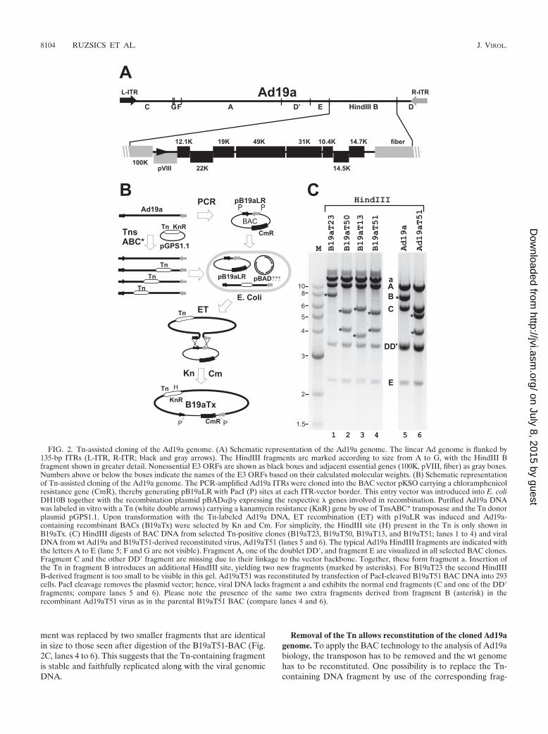
ment was replaced by two smaller fragments that are identicalin size to those seen after digestion of the B19aT51-BAC (Fig.2C, lanes 4 to 6). This suggests that the Tn-containing fragmentis stable and faithfully replicated along with the viral genomicDNA.
Removal of the Tn allows reconstitution of the cloned Ad19agenome. To apply the BAC technology to the analysis of Ad19abiology, the transposon has to be removed and the wt genomehas to be reconstituted. One possibility is to replace the Tn-containing DNA fragment by use of the corresponding frag-
FIG. 2. Tn-assisted cloning of the Ad19a genome. (A) Schematic representation of the Ad19a genome. The linear Ad genome is flanked by135-bp ITRs (L-ITR, R-ITR; black and gray arrows). The HindIII fragments are marked according to size from A to G, with the HindIII Bfragment shown in greater detail. Nonessential E3 ORFs are shown as black boxes and adjacent essential genes (100K, pVIII, fiber) as gray boxes.Numbers above or below the boxes indicate the names of the E3 ORFs based on their calculated molecular weights. (B) Schematic representationof Tn-assisted cloning of the Ad19a genome. The PCR-amplified Ad19a ITRs were cloned into the BAC vector pKSO carrying a chloramphenicolresistance gene (CmR), thereby generating pB19aLR with PacI (P) sites at each ITR-vector border. This entry vector was introduced into E. coliDH10B together with the recombination plasmid pBAD�� expressing the respective � genes involved in recombination. Purified Ad19a DNAwas labeled in vitro with a Tn (white double arrows) carrying a kanamycin resistance (KnR) gene by use of TnsABC* transposase and the Tn donorplasmid pGPS1.1. Upon transformation with the Tn-labeled Ad19a DNA, ET recombination (ET) with p19aLR was induced and Ad19a-containing recombinant BACs (B19aTx) were selected by Kn and Cm. For simplicity, the HindIII site (H) present in the Tn is only shown inB19aTx. (C) HindIII digests of BAC DNA from selected Tn-positive clones (B19aT23, B19aT50, B19aT13, and B19aT51; lanes 1 to 4) and viralDNA from wt Ad19a and B19aT51-derived reconstituted virus, Ad19aT51 (lanes 5 and 6). The typical Ad19a HindIII fragments are indicated withthe letters A to E (lane 5; F and G are not visible). Fragment A, one of the doublet DD�, and fragment E are visualized in all selected BAC clones.Fragment C and the other DD� fragment are missing due to their linkage to the vector backbone. Together, these form fragment a. Insertion ofthe Tn in fragment B introduces an additional HindIII site, yielding two new fragments (marked by asterisks). For B19aT23 the second HindIIIB-derived fragment is too small to be visible in this gel. Ad19aT51 was reconstituted by transfection of PacI-cleaved B19aT51 BAC DNA into 293cells. PacI cleavage removes the plasmid vector; hence, viral DNA lacks fragment a and exhibits the normal end fragments (C and one of the DD�fragments; compare lanes 5 and 6). Please note the presence of the same two extra fragments derived from fragment B (asterisk) in therecombinant Ad19aT51 virus as in the parental B19aT51 BAC (compare lanes 4 and 6).
8104 RUZSICS ET AL. J. VIROL.
on July 8, 2015 by guesthttp://jvi.asm
.org/D
ownloaded from

ment from wt Ad DNA and traditional cloning techniques. Wefollowed this strategy initially and reconstructed the wt Ad19agenome by partial digestion with AvrII and subsequent inser-tion of the corresponding Ad19a AvrII fragment (data notshown). However, this method is not generally applicable andrequires exact mapping with the restriction enzymes to be usedand therefore is laborious. To make the Tn-removal step in-dependent of any further mapping work the nuclease activity ofthe TnsABC* complex was exploited. Upon insertion of the Tnby use of the Tn7 transposase the inserted Tn was flanked by5-bp target repeats (12345 in Fig. 3A) 12345 the five nucleo-tides in the Ad genome directly flanking the Tn insertion site.TnsABC* cleaved one strand of the double-stranded donorDNA exactly at the 3� ends of the Tn and the other strand 3 bpapart within the target duplications, creating 3-base 5� over-hangs within the target duplications (345 in Fig. 3B) (14).Thus, the wt donor sequence is not restored after transposaseexcision and simple end joining, since the resulting ends areusually not compatible and the cleavage removes only 3 bp ofthe 5-bp duplications, leaving a residual 2-bp insertion (14).
To overcome this problem we first removed the Kn cassettefrom the Tn in B19aT51, one of the BACs with the Tn in theE3/19K gene, by double digestion with the meganucleases I-CeuI and I-SceI. Subsequent blunting and religation resultedin an intermediate BAC clone that retained the Tn ends butnot the selectable marker (Fig. 3A, B19aT51�Kn). A new ETrecombination fragment was generated by PCR using the Tndonor plasmid as the template and primers specific to the endsof the Tn flanked by homologies to the viral target sequencesup- and downstream of the Tn insertion site. The upstreaminsertion site was left intact, but the first 2 bp of the down-stream target duplication were deleted by primer design (Fig.3A). ET recombination between the redesigned Tn-positiverecombination fragment and B19aT51�Kn as the target shouldyield BAC B19aT51T, with only 3-bp target repeats at eitherend of the Tn. Subsequent treatment of B19aT51T withTnsABC* in vitro removes the Tn sequences, creating now-compatible 3-nucleotide 5� overhangs (345 in Fig. 3B) at bothends of the BAC. These represent the last 3 bp of the originaltarget repeats (Fig. 3B). TnsABC* cleaves the donor site only
FIG. 3. Generation of the wt Ad19a genome upon Tn removalfrom B19aT51. (A) Schematic representation of the precise removal ofthe Tn. The KnR gene (arrow) of the Tn (open double arrows) wasremoved in vitro by I-SceI/I-CeuI meganuclease double digestion(meganuclease sites are indicated by gray lines) followed by end fillingand ligation, generating B19aT51�Kn. In parallel, a PCR was per-formed using primers specific to the Tn ends (open arrows) flanked by40-bp homologies to the target sites in Ad (black and gray boxes). Inthe forward primer the entire target repeat (12345) was incorporated,
whereas only the last 3 bp of the right target repeat were included intothe homology region of the reverse primer. Target repeats are indi-cated on either side of the Tn by black and gray numbers. This Tn-containing PCR fragment was introduced into the B19aT51�Kn by ETrecombination, whereby the orientation of the Tn in the newly gener-ated BAC B19aT51T becomes reversed. (B) Tn removal fromB19aT51T. B19aT51T was treated with TnsABC* transposase, whichexcises the Tn, leaving compatible 3-base-long 5� overhangs on theBAC ends. Simple ligation reconstitutes the 5-bp wt Ad target se-quence, thereby generating a BAC containing the wt Ad19a genome(B19a). (C) Restriction analysis of BAC clones and their derived Ads.The XhoI patterns of B19aT51, B19aT51�Kn, and B19aT51T areshown in lanes 1 to 3, respectively, with Tn-containing fragments in-dicated by asterisks. The HindIII pattern of BACs (lanes 5 to 7) andreconstituted viruses (lanes 8 and 9) is also shown. Fragment B-derivedbands are indicated by asterisks. A HindIII-PacI double digest of B19aDNA releases the end fragments (C and one of the DD� fragments)from the vector backbone (black arrowhead), eliminating fragment a(lane 7). BAC-derived Ad19a, Ad19aB; wt Ad19a, Ad19a. M indicatesthe lane of DNA markers (NEB) with numbers in kilobases.
VOL. 80, 2006 MUTAGENESIS OF SPECIES D Ads AND VECTOR DEVELOPMENT 8105
on July 8, 2015 by guesthttp://jvi.asm
.org/D
ownloaded from

in the presence of target DNA. Therefore, to provide nonim-mune target sites for the excising Tn to transpose into, aplasmid with a temperature-sensitive origin of replication wasincluded in the cleavage reaction. This dead-end plasmid waslost, since it is not replicated at 37°C. After simple ligation, thereleased BAC ends were joined and the wt sequence was re-stored (Fig. 3B; B19a). As TnsABC* cleavage and ligation arenot 100% efficient, the reaction mixture was transformed intoE. coli DH10B carrying an I-SceI expression plasmid (49),thereby selecting against Tn-positive BACs which contain anI-SceI site. Pretesting the resulting colonies for Kn sensitivityidentified those that had lost the Tn, since removal of the Tnis accompanied by a loss of the Kn resistance gene. To removethe I-SceI expression plasmid, E. coli DH10B was retrans-formed with the BAC DNA preparation from the Kn-sensitivecolonies. Restoration of the wt sequence can be confirmed byrestriction analysis (Fig. 3C) and sequencing.
Restriction digests of BAC intermediates confirmed the suc-cess of the individual manipulations. The presence of the Tnwithin the E3 region in B19aT51 is indicated by an additionalXhoI site within the E3 region, producing two fragments of 4.4and 3.5 kb (Fig. 3C, lane 1). Cleavage and ligation removes theTn, along with the XhoI site, yielding only one XhoI fragmentof �7 kb (Fig. 3C, lane 2, asterisk). Insertion of the modifiedPCR-derived Tn oriented in the opposite direction reintro-duces the XhoI site. Hence, after XhoI digestion of B19aT51Ttwo Tn-containing fragments are generated (Fig. 3C, lane 3)that differ in size from those seen in B19aT51 due to thealtered orientation of the Tn. Also, comparison of the HindIIIpatterns of B19aT51T (lane 5) and B19a (lane 6) clearly indi-cates the removal of the Tn sequence from fragment B afterTnsABC* and ligase treatment. The Tn-encoded HindIII siteresulting in the two Tn-containing fragments of B19aT51T islost in B19a, and instead a wt-like 7.8-kb B fragment appears(lane 6). The same 7.8-kb HindIII B fragment is visible afterrelease of the vector backbone by PacI cleavage, lending fur-ther support to the idea of B19a being a BAC containing thewild-type Ad19a genome (Fig. 3C, lanes 5 to 7). Finally, trans-fection of PacI-cleaved B19a DNA into 293 cells yielded viablevirus (Ad19aB) whose HindIII restriction pattern was indistin-guishable from that obtained for wt Ad19a DNA. Moreover,when tested, all biological features of this BAC-derivedAd19aB were identical to those of the original Ad19a strain(see Fig. 6A and B).
Tn-assisted mutagenesis of the Ad19a BAC (exposon mu-tagenesis). The data presented above indicate that the Tn7ends can be used successfully as a cleavage-determining ciselement and the Tn7 transposase-derived TnsABC* as a cleav-ing enzyme for in vitro DNA manipulation. Accordingly, itshould be possible to utilize this system for the introduction ofany mutant sequence flanking the Tns into any site of a re-combinant DNA. Subsequent Tn excision by TnsABC* fol-lowed by DNA ligation should only leave the intended mu-tation in the Ad genome without retaining any unwantedoperational sequence. Thus, precise and traceless introduc-tion of mutations into Ad genomes should be possible. Totest this idea, we introduced a mutation into the subgenusD-specific ORF E3/49K. This ORF codes for a highly gly-cosylated transmembrane protein that can be detected inthe Golgi, early endosomes, and plasma membrane (8, 18,
70). On the basis of the location of the 49K gene in the E3region and the prediction that at least one of the domainshas an immunoglobulin-like fold we speculate that it mightsubvert the host immune system. However, to date its func-tion remains unknown.
With the aim of eliminating 49K expression, 4 bp (TAAG)were inserted after the 11th codon (see Fig. S1 and Table S1 inthe supplemental material), thereby generating a stop codonand a 1-base frame shift into the 49K ORF. First, a linearrecombination fragment was produced by PCR using the Tn asa template (Fig. 4A). The upstream homology arm followed bythe 5-base insertion (including the four additional bases plusthe first base of the downstream homology) and the 22-basepriming site specific to the left end of the Tn was incorporatedinto the forward primer. The reverse primer consists of thepriming site on the Tn (30 bp) followed by the 2-base insertionand the downstream homology. The 2-bp insertion, togetherwith the first base of the downstream homology, serves as adownstream copy of the 3-bp target duplication (Fig. 4A). Theparticular bases were chosen to provide a new HindIII site ifthe mutation was successful. ET recombination proficient-competent bacteria carrying B19a were transformed by thisPCR product, and recombinants were plated out in the presenceof Kn. Correct recombination is predicted to yield HindIII frag-ments of 4.4 kb, 4.2 kb, and 0.8 kb instead of the original 7.8-kbHindIII B fragment. These are generated by the HindIII sitesflanking the E3 region, the one in the Tn and the one at theright end of the Tn newly created by the mutation. Indeed,recombinants with the expected restriction pattern (Fig. 4B,lanes 1 and 2) were isolated and the respective mutations wereconfirmed by sequencing (B19a49K*T).
Subsequent treatment in vitro with TnsABC* and T4 DNAligase should eliminate the Tn and yield B19a49K* containingthe intended 4-bp insertion that creates a new HindIII site atthe site of the mutation and therefore predicts 3.5-kb and4.2-kb HindIII fragments. This pattern was confirmed by therestriction cut (Fig. 4B, lane 3). In addition, correct construc-tion of both the Tn-containing intermediate B19a49K*T andthe B19a49K* has been verified by DNA sequencing (see Fig.S1 in the supplemental material). On transfection of PacI-linearized B19a49K* DNA into 293 cells the mutant virusAd19a49K* was successfully reconstituted, as shown by thepresence of the same fragments in the viral DNA as in theB19a49K* DNA (Fig. 4B; compare lane 3 and 5). FACS anal-ysis of A549 cells infected with the mutant virus Ad19a49K*(see Fig. 6) corroborated the selective loss of 49K expression,while all other E3 functions tested remained intact. The selectiveelimination of 49K expression was also confirmed by Westernblotting (data not shown).
To assess the effect of the additional selection step onTn-containing BACs, both normal DH10B cells and I-SceI-expressing DH10B cells were transformed with the reactionmixture and grown on Cm plates. Resulting colonies wereanalyzed by replica plating on Kn plates and by PCR using49K-specific primers. The results show that the transposase-mediated cleavage was very efficient (see Table S2 in the sup-plemental material). More than 95% of the input DNA wascleaved, resulting in a dramatic reduction of Kn-resistant col-ony numbers after transformation of the TnsABC*-treatedB19a49K*T compared to the results seen with untreated BAC.
8106 RUZSICS ET AL. J. VIROL.
on July 8, 2015 by guesthttp://jvi.asm
.org/D
ownloaded from

Of these colonies, 54% become Kn sensitive, and most of theKn-sensitive clones carried the correct mutation, as analyzedby PCR (data not shown). Upon transformation of I-SceI-expressing bacteria with TnsABC*-T4 ligase-treated BACDNA, 100% of the resulting colonies carried the correct ge-netic changes. I-CeuI treatment in vitro after ligation resultedin 96% efficiency but drastically reduced numbers of colonies(5.3%).
By modifying the above-described method it should be pos-sible to introduce precise deletions into Ad BAC clones. In thiscase, the primers are designed such that the Tn element isflanked by 3-bp direct repeats (123 in Fig. 5) and the desiredhomologous sequences required for ET recombination. In thefirst step, the resulting PCR fragment is introduced into theBAC by ET recombination, replacing the region to be deleted.
Subsequent transposase cleavage and ligation mediated by the3-bp overhangs should allow the precise deletion of genes ornucleotides (Fig. 5A, process 1). This strategy was applied todelete the entire E3 coding region of Ad19a (4.5 kb), beginningwith the start codon of the 12.1 ORF and extending to the lastE3 ORF, 14.7K at the 3� end (Fig. 2A). The observed HindIIIrestriction pattern of the DNA is consistent with the successfulgeneration of intermediates and end products. The wt 7.8-kbfragment in B19a is replaced by a 3.2-kb fragment (and smallerfragments) in B19aE3T and by 2.1-kb and 1.0-kb (not visible)fragments in the B19a�E3 end product (Fig. 5B, lanes 1 to 3).This same 2.1-kb fragment is visualized in HindIII-digestedDNA of the reconstituted Ad19a�E3 virus that otherwise ex-hibits a pattern identical to that seen with the wt except thatthe 7.8-kb HindIII B fragment is lost. Further analysis shows
FIG. 4. Exposon mutagenesis. (A) Schematic drawing indicating the steps involved in “exposon mutagenesis.” As an example, a 4-bp insertion(TAAG) into the E3/49K gene is shown which was aimed at abrogating 49K expression by insertion of a stop codon and introduction of a readingframe shift. Simultaneously, a new HindIII site is introduced to conveniently monitor the success of the mutation. PCR was performed using a Tntemplate and ET primers 49KKOfor and 49KKOrev specific for the Tn ends (open arrows) and containing additional 40-base arms homologousto the upstream (black boxes) and downstream (gray boxes) viral target sequences (see Fig. S1 and Table S1 in the supplemental material for theexact sequences). In between these elements, the 5-base target repeat sequence (capital letters) is shown. For the upstream (left) primer thissequence includes the 4-base mutation (TAAG) and the first base (C) of the downstream homology (underlined gray capital letters). In the rightprimer, the complementary bases for the last two bases of the mutation were repeated (TC3AG), followed by the first three bases of thedownstream homology arm (GAA). After ET recombination (ET) with wt B19a (the Ad19a BAC construct), Tn-containing recombinants(B19a49K*T) were selected based on kanamycin resistance. Treatment of the purified B19a49K*T BAC with TnsABC* yields compatible 5�overhangs in the BAC backbone that can be recircularized by ligation. The resulting mutant B19a49K* contains only the designed 4-bp insertionand no operational sequences. HindIII sites (H) and the sizes (in kilobases) of the generated HindIII fragments are given. (B) Comparison of theHindIII restriction patterns of wt and mutant BACs (lanes 1 to 3) as well as wt Ad19a (lane 4) and reconstituted, BAC-derived mutant virusAd19a49K* (lane 5). DNA from the bacteria and viruses indicated on the top was analyzed by HindIII digestion. Bands derived from fragmentB are indicated (asterisks). Please note that the 0.8-kb fragment in lane 2 is not visible. After removal of the Tn sequences, only one additionalHindIII site (created by the mutagenesis) is left and the 4.4-kb fragment is converted to 3.5 kb due to the loss of the Tn sequence (lane 3). Uponmutagenesis the normal B fragment of Ad19a is converted into two fragments (asterisks) of identical sizes, as seen in B19a49K* (lanes 4 and 5).
VOL. 80, 2006 MUTAGENESIS OF SPECIES D Ads AND VECTOR DEVELOPMENT 8107
on July 8, 2015 by guesthttp://jvi.asm
.org/D
ownloaded from

that Ad19a�E3 is viable and does not express any of the E3functions tested (Fig. 6).
Tn-assisted gene insertion. The flexible design of Tn entryfragments also allows the insertion of larger DNA fragments orgenes. The first steps are identical to those in the proceduredescribed above, but during the ligation step the gene to beinserted is added. A prerequisite for successful insertion is thatthe gene contains sticky ends compatible with those created bytransposase. This can easily be achieved by PCR using primerpairs with recognition sequences for restriction endonucleasesthat generate overhangs compatible with those of transposase,e.g., SapI. The TnsABC*-cleaved target BAC and the insertwith compatible ends are ligated, and after I-SceI selection,mutants lacking the Tn are greatly enriched. This is illustratedhere with two examples. In the first, we introduced an E3/49Kexpression cassette from pSG5-E3/49K in which E3/49K ex-pression is under the control of the SV40 promoter (70). Theentire 49K expression cassette of pSG5-E3/49K was amplifiedusing primers that introduce two SapI cleavage sites at eachend of the amplicon. Cleavage by SapI generates the desired3-base overhangs compatible for insertion into the TnsABC*-treated B19aE3T (Fig. 5A, process 2). The resulting BACclones were tested as described above. As shown in Fig. 5B,replacement of the Tn in B19aE3T by the 49K expressioncassette (2.3 kb) eliminates two HindIII sites, resulting in aBAC clone with the 49K insert in a left-to-right orientation ona 5.3-kb fragment (lane 4; B19a�E3�49K). The same charac-teristic 5.3-kb fragment is detected after cleavage of DNA fromthe corresponding, reconstituted recombinant Ad (Ad19a�E3�49K), demonstrating that the inserted gene is stable afterseveral rounds of virus replication (Fig. 5B; compare lane 4with lane 7).
Functional analysis of Ad19a mutants generated by exposonmutagenesis. To assess the success and specificity of the intro-duced genetic alterations, we analyzed the phenotypes of thevarious Ad19a mutant viruses generated with the proceduredescribed above. As the E3 region exhibits very complex splic-
FIG. 5. Exposon mutagenesis for precise deletion and insertion ofgenes. (A) Schematic representation of Tn-assisted deletion (process1, left) and gene insertion (process 2, right). The Tn-containing PCR-derived recombination fragment was designed to contain 40-bp homol-ogy arms (black and gray boxes) targeted to the beginning (5� part) andend (3� part) of the Ad19a E3 region marking the borders of thedeletion and 3-bp (123) direct repeats introduced by the ET primershomologous to either Tn end. After ET recombination with wt B19athe coding region of Ad19a E3 (open box) was replaced by the Tn-
containing PCR fragment, generating the Tn-containing intermediateB19aE3T that, on cleavage with TnsABC* and circularization via itscompatible ends (process 1), yields the deletion mutant B19a�E3. In amodification of the procedure, a new gene (E3/49K) was inserted(process 2) after treatment of a PCR-derived 49K protein-encodinginsert (gray box) with SapI, generating compatible sticky ends withTnsABC*-treated B19aE3T. From the B19a�E3�49K BAC clone anAd19a mutant virus was reconstituted expressing only 49K as a singleE3 gene. (B) BAC DNA extracted from B19a, B19aE3T, B19a�E3,and B19a�E3�49K was analyzed by HindIII digestion. Bands derivedfrom fragment B are indicated by asterisks. Introduction of the Tn intothe E3 region inserts two additional HindIII sites, one by the mutationand the other by the Tn. On HindIII digestion of B19aE3T a 3.2-kbfragment (asterisk in lane 2) and 1-kb and 0.8-kb fragments (notvisible) are produced. Tn removal from B19aE3T results in an addi-tional deletion (asterisk in lane 3; B19a�E3). The 5.3-kb fragment(lane 4; B19a�E3�49K) is consistent with the elimination of twoHindIII sites and with the residual fragment B sequences being linkedto the 49K expression cassette. HindIII-digested DNA extracted fromwt Ad19a (lane 5) and the reconstituted recombinant viruses Ad19a�E3 (lane 6) and Ad19a�E3�49K (lane 7) exhibit B-derived fragmentsidentical in size to those seen in the corresponding BAC DNA (com-pare lanes 5 to 7 with lanes 1, 3, and 4).
8108 RUZSICS ET AL. J. VIROL.
on July 8, 2015 by guesthttp://jvi.asm
.org/D
ownloaded from

ing and previous mutations resulted in unintended secondaryeffects (19), it is crucial to analyze specific E3 functions in E3mutant Ads. Ads remove several apoptosis receptors, includingFas (CD95), from the cell surface of infected cells to protectthem from premature apoptosis (20, 41, 65, 71). Down-regu-lation of Fas from the cell surface requires the E3/10.4–14.5Kproteins, also called RID (20, 65; for a review, see references10 and 41). We have investigated the capacity of BAC-derivedwt and mutant Ad19a to down-regulate CD95 (Fas) by FACSanalysis (Fig. 6A). In wt Ad19a such as the plaque-purifiedAd19aT3, the RID genes are expressed and, consequently, Fascell surface levels are reduced to �20% compared to mock-infected cells (100%). A similar down-regulation is observed incells infected with BAC-derived wt Ad19a (Ad19aB) or theAd19a49K* mutant, in which expression of E3/49K (an E3gene in the close vicinity of the RID genes) is specificallyeliminated upon insertion of the 4-bp mutation (Fig. 4). Thisdemonstrates that the intended elimination of E3/49K (70)expression did not affect the function of E3/RID and thusappears to be specific. By contrast, Ad19a viruses lacking all E3genes (Ad19a� E3) or all but E3/49K (Ad19a�E3�49K) areunable to modulate Fas from the cell surface, confirming forthe first time that species D RID exhibits a functional activitysimilar to that seen with species C.
The generation of a 49K-specific MAb (M. Windheim., E.Kremmer, and H.-G. Burgert,, unpublished data) allowed usalso to directly monitor the expression of 49K in wt and mutantAd19a viruses, as measured by FACS analysis. A549 cells in-fected with plaque-purified wt Ad19a and BAC-derived wtAd19a (Ad19aB) as well as mutant Ad19a solely expressing49K in the E3 region (Ad19a�E3�49K) synthesize 49K,whereas those infected with mutant viruses selectively lacking49K expression (Ad19a49K*) or lacking E3 genes altogether(Ad19a�E3) exhibit only background staining (Fig. 6B).
The above-described technology should also prove ex-tremely useful for rapid exploration of the vector potential ofvarious Ad serotypes for which only limited sequence data areavailable, as was the case with Ad19a. The genome could becloned into BAC vectors by utilizing the ITR homologies, asdemonstrated for Ad19a. To this end, we have generated anAd19a vector expressing enhanced GFP. In analogy to thedeletion of E3 (Fig. 5A), we have deleted the E1 region ofAd19a by introducing the Tn in this region by using B19a�E3as a target. In a second step, the Tn was replaced by anexpression cassette encoding GFP under the control of theCMV immediate-early promoter and the SV40 enhancer (seeMaterials and Methods). A recombinant E1-negative Ad19amutant virus expressing GFP, Ad19a�E1GFP�E3 (referred tohere as Ad19aGFP), was viable in 293 cells, stably expressing
FIG. 6. Phenotypes of various Ad19a mutant viruses generatedwith the procedure described in the text. (A) Down-regulation ofCD95 (Fas) from the cell surface of infected A549 cells upon infectionwith plaque-purified wt Ad19a (Ad19aT3) or BAC-derived wt Ad19a(Ad19aB) as well as mutant Ad19a lacking E3 expression (Ad19a�E3)or 49K expression (Ad19a49K*) or solely expressing 49K in the E3region (Ad19a�E3�49K), as determined by FACS analysis 21 h pi.The names of the corresponding viruses are indicated below the bardiagram. The mean value of fluorescence (MvF) deducted by examin-ing that obtained after background staining with the secondary anti-body alone was related to the MvF in mock-infected cells. The lattervalue was arbitrarily set to 100%. Bars depict the means compiled fromfour independent experiments. Error bars represent the means �standard errors. (B) E3/49K cell surface expression as measured byFACS analysis with a MAb specific for 49K in A549 cells infected withthe same viruses as described for panel A. The MvF deducted by thatobtained after background staining with the secondary antibody alonewas related to the MvF in wt Ad19aT3-infected cells. The latter was
arbitrarily set to 100%. Bars depict the means compiled from at leastthree independent experiments. Error bars represent the means �standard errors. (C) Comparison of the transduction capacity of GFP-expressing Ad19a and Ad5 vectors. Several lymphoid cell lines (Jurkat,T2, LCL) and, as a control, 293 cells were transduced with 25 PFU/cellAd19aGFP (black bars) and Ad5GFP (gray bars). At 36 h pi thefraction of GFP-expressing cells was determined by FACS analysis.With the exception of the LCL data, which represent a single experi-ment, the bars represent the means of three experiments.
VOL. 80, 2006 MUTAGENESIS OF SPECIES D Ads AND VECTOR DEVELOPMENT 8109
on July 8, 2015 by guesthttp://jvi.asm
.org/D
ownloaded from

Ad5 E1 genes. Interestingly, the Ad19a-derived GFP vectorexhibited a transduction pattern remarkably different from thatseen with the commonly used Ad5-derived gene therapy vector(Fig. 6C). This was revealed when different lymphoid cell lineswere infected with Ad19aGFP or the corresponding Ad5GFPvector. At 36 h pi the fraction of GFP-expressing cells wasquantitatively determined by FACS analysis. In contrast to thestandard Ad5 vector (Fig. 6C), the Ad19a-derived vector effi-ciently transduced all lymphoid cell lines tested (Jurkat; T2;LCL) and C1R, Bristol8 (data not shown). Upon Ad19a trans-duction, 90% of Jurkat, 65% of T2, and 70% of LCL cellsexpressed GFP, whereas only 15%, 7%, and 2%, respectively,of the cells exhibited GFP expression after transduction withAd5GFP. Correct titration of the vectors was confirmed bythe equivalent susceptibility of 293 cells, which were usedfor titration of both types of vectors. Thus, Ad19aGFPseems to be superior to the Ad5GFP vector for transductionof lymphoid cells.
DISCUSSION
Although some species D Ads appear to have unique celltropism and receptor usage characteristics and the entire se-quence of one serotype (Ad17) from this species has beenknown for several years, no detailed description of a species Dvector has been reported yet (58). We think that this might bedue to the instability of their genome in E. coli. In our hands,attempts to maintain even a subgenomic fragment containingthe Ad19a E3 region as standard high- and low-copy-numbervectors in E. coli failed (data not shown). Since a number ofunstable viral and cellular genomes have been successfullycloned as BACs in E. coli (9), we applied the BAC technologyto construct and manipulate the Ad19a genome. We usedhomologous recombination, mediated by lambda phage redalpha, beta, and gam (ET recombination), to insert full-lengthAd19a genomic DNA into a BAC vector that carries only theAd19a ITRs. Although previous data showed that ET recom-bination is inefficient when repeat regions are targeted (75), wedemonstrated in this study that a reasonable efficiency can beachieved by inserting a selectable Tn into the Ad genome invitro prior to recombination. In contrast to the published re-combinatorial construction of recombinant Ads (13), subclon-ing of large homology regions is not required for ITR-medi-ated ET cloning. Consequently, very small amounts ofsequence information suffice to initiate cloning projects; hence,less-well-characterized serotypes and strains become amenableto genetic studies. Moreover, since small amounts of viralDNA are sufficient and even preparations from infected cellsare suitable for Tn labeling, clinical isolates could be conservedby the presented approach without extensive tissue culturepassage prior to cloning. After establishment of infectiousAd19a clones another round of ET recombination was appliedto completely remove the cloning marker from the Ad19a-BAC. The resulting wt Ad19a genome was maintained in E.coli without any detectable changes, and the resulting recon-stituted viruses obtained by transfection of 293 cells exhibitedwt properties (Fig. 2, 3 and 6).
We also describe a novel, versatile method combining ETrecombination and transposase manipulation to mutate BAC-cloned Ad genomes. This method is independent of restriction
sites and leaves no operational sequences behind, thereby al-lowing traceless introduction of any genetic change into Adgenomes. In the first step, the genetic changes are introducedby ET recombination in the context of a Tn carrying a positiveselection marker. In the second step, the Tn sequences areentirely removed by a transposase reaction in vitro. We coinedthis method “exposon mutagenesis” to emphasize the utiliza-tion of the transposase reaction for excision of the Tn-embed-ded operational sequences. Two-step recombinatorial mutagen-esis methods have been previously described for manipulatingmammalian genomic BACs (46, 74). In these approaches, thegenetic changes are introduced along with a positive and anegative selection marker. The positive selection marker is firstused to identify the intermediate recombinants, which is fol-lowed by a second round of recombination and negative selec-tion to gain markerless mutants. However, viral BACs areparticularly challenging objects for recombinatorial mu-tagenesis because the presence of repetitive sequences canbe deleterious, especially when counter-selection is applied(69). Alternatively, the second step might be carried outwithout counterselection, but in this case extensive libraryscreening is required to identify the right mutant in thenonselected pool.
Another disadvantage of most traceless recombinatorial mu-tagenesis approaches is that the manipulated genomes areexposed twice to an active recombination system, thereforedoubling the risk of unwanted genetic events. By contrast, inour method one recombination event is sufficient to introducethe intended genetic change along with a marker that allowspositive selection to preserve the genetic arrangement. In thesecond step, the marker is removed by in vitro treatment of theintermediate BAC with Tn7 transposase and T4 ligase, thuseliminating the need for a second recombination step.TnsABC*-mediated transposon removal is very efficient, and apotential background can easily be subtracted by simple replicaplating to detect the loss of the Tn resistance marker. Anadditional advantage is that the same intermediate BAC can beused for the rapid generation of a large number of mutants inthe same region or for the insertion of different transgenes ata predefined site. Therefore, exposon mutagenesis serves as afast and simpler alternative to shuttle plasmid mutagenesis(43) and other techniques described so far. This is demon-strated here by the generation of several mutants in the Ad19aE3 region, the deletion of the entire E3 region, and the rein-sertion of a single E3 gene, E3/49K, or its selective inactivation(Fig. 5 and 6). Thus, exposon mutagenesis is extremely usefulfor precise mutagenesis of E3 genes of Ad19a and for mu-tagenesis of Ad genomes in general. The latter was confirmedby successfully applying the technique for manipulation of thegenomes of Ad2 and Ad5, both Ads of species C.
We have introduced point mutations and deletions in theE3/10.4K, E3/14.5K, E1B/19K, and E1B/55K genes (S. Ober-meier, Z. Ruzsics, A. Hilgendorf, and H.-G. Burgert, data notshown) or have replaced the E1 region with a GFP expressioncassette. Complex splicing is a hallmark of Ad transcriptionunits, and deletion mutants often turned out to exhibit unin-tended secondary effects, e.g., affecting splicing of other E3genes. Therefore, a method for introduction of precise andsubtle changes is highly desirable. Principally, any Ad mutantcan be generated by this method as long as the corresponding
8110 RUZSICS ET AL. J. VIROL.
on July 8, 2015 by guesthttp://jvi.asm
.org/D
ownloaded from

complementation is provided. Thus, exposon mutagenesis ap-pears to be generally applicable and particularly useful for thefunctional analysis of genes, e.g., those involved in the EKCphenotype of species D Ads.
Using the above-described methodology, we have also gen-erated a new basic Ad19a vector expressing GFP. This repli-cation-deficient vector exhibits significantly higher transduc-tion efficiency for human lymphoid cells (Fig. 6C) and primaryhuman muscle cells (64) compared to conventional Ad5 vec-tors. This superior property appears to be due to two effects: (i)a higher uptake of Ad19a versus Ad5 particles (64) and (ii)enhanced expression of the GFP transgene. The latter effectcan be demonstrated in those cell types (melanoma cells,breast cancer cells, etc.) in which the fractions of cells trans-duced by Ad5 and Ad19a vectors were similar or identical andyet GFP expression was three- to sixfold higher in Ad19a-transduced cells (data not shown). At present, the reasons forthis increased transgene expression remain unclear. We spec-ulate that the potency of either the E1 enhancers or other ciselements in the vicinity of the inserted transgene cassette maydiffer between the two Ad species. Alternatively, functions intrans that might act in a tissue- or cell type-specific manner maystimulate GFP expression in Ad19a vectors. This notion issupported by earlier data for Ad5 vectors indicating that E4products can influence transgene expression (38). Whateverthe underlying cause, the experiments point to unexpecteddifferences between species C and D Ads. Exploitation of thesedifferences may turn out to be advantageous for gene therapyor vaccination. For example, lower amounts of Ad19a vectormight suffice to express similar levels of transgenes; hence,vector-mediated toxicity is likely to be reduced.
In contrast to the recently developed species B-based Advectors (24, 28, 53, 61, 62, 66), which propagate very poorly in293 cells and typically require specific trans-complementingcell lines expressing at least the serotype-specific E1B/55Kprotein for preparation of high-titer E1-deleted vectors, theE1- and E3-deleted Ad19a-based vector can be propagatedwith essentially wt-like productivity in 293 cells. It has beenshown that both the transactivation ability of the E1A geneproducts and the antiapoptotic activities of the E1B region arewell conserved among human Ads (52). One possible explana-tion for the species-specific complementation is that other viralfunctions associated with E1 (and E4), such as late mRNAexport, may be substantially different in species B compared toC and D. Alternatively, the relevant E1 function might beencoded by another gene locus in species D and, therefore,complementation might only be necessary for the conservedE1 functions. In any case, since propagation of the first-gen-eration recombinant Ad19aGFP shown here can be performedby the same methods that are in use for Ad5-based vectors,establishment of new protocols is not required.
The recombinant Ad19aGFP vector transduced all humancell lines tested (this study) as well as primary muscle cells (64)and fibroblasts in vitro with high efficiency. This is consistentwith the observation that EKC-associated Ads may use �(2-3)sialic acid-containing receptors present on the surface of mostcell types (2, 3). In support of this notion, sialidase treatmentof human myoblasts dramatically reduced transduction byAd19aGFP but not that of Ad5GFP (64). However, furtherexperiments are necessary to investigate the role of other re-
ceptor entities proposed for EKC-associated Ads (72). Takingthese results together with our DC data (our DCs did notexpress CAR; data not shown), it is clear that in contrast tosubgenus C Ads, Ad19a does not require CAR for infection.Thus, Ad19a, and possibly other Ads of species D, might be avaluable alternative to Ad5-based vectors that exhibit a limitedtargeting spectrum in different gene transfer applications (30,32, 63). In particular, their ability to efficiently transduce DCsmakes Ad19a vectors highly attractive for vaccination and im-munotherapy. Further in vitro and in vivo experiments will benecessary to examine whether the increased efficacy of DCgene transfer also translates into a higher capacity of trans-duced DCs to stimulate a corresponding T-cell response. Ourdata obtained with lymphoid cell lines originating from B andT cells (Fig. 6 and data not shown) suggest that the increasedtransduction by Ad19aGFP compared to a corresponding Ad5vector may extend to leukocytes in general (Fig. 6). Therefore,it is anticipated that vectors based on Ad19a should also beuseful for cytotoxic therapy of leukemias and other lymphoidtumors.
ACKNOWLEDGMENTS
This work was supported by a Mercia Spinner Pathfinder grant andgrants from the Deutsche Forschungsgemeinschaft (BU 642-1 andSFB455) and the BBSRC (BB/D002877/1) to H.-G.B.
We thank M. Windheim for generously providing the pSG5-E3/49Kplasmid and J. Bergelson for kindly providing the CAR-specific MAb.We are grateful to R. Magerstaedt for the initial help and advice togenerate DCs. For critical reading of the manuscript, we thank J. Coxand S. Eldershaw.
REFERENCES
1. Anderson, R. D., R. E. Haskell, H. Xia, B. J. Roessler, and B. L. Davidson.2000. A simple method for the rapid generation of recombinant adenovirusvectors. Gene Ther. 7:1034–1038.
2. Arnberg, N., K. Edlund, A. H. Kidd, and G. Wadell. 2000. Adenovirus type37 uses sialic acid as a cellular receptor. J. Virol. 74:42–48.
3. Arnberg, N., A. H. Kidd, K. Edlund, F. Olfat, and G. Wadell. 2000. Initialinteractions of subgenus D adenoviruses with A549 cellular receptors: sialicacid versus �V integrins. J. Virol. 74:7691–7693.
4. Bergelson, J. M., J. A. Cunningham, G. Droguett, E. A. Kurt-Jones, A.Krithivas, J. S. Hong, M. S. Horwitz, R. L. Crowell, and R. W. Finberg. 1997.Isolation of a common receptor for coxsackie B viruses and adenoviruses 2and 5. Science 275:1320–1323.
5. Berkner, K. L. 1988. Development of adenovirus vectors for the expressionof heterologous genes. BioTechniques 6:616–629.
6. Bett, A. J., W. Haddara, L. Prevec, and F. L. Graham. 1994. An efficient andflexible system for construction of adenovirus vectors with insertions ordeletions in early regions 1 and 3. Proc. Natl. Acad. Sci. USA 91:8802–8806.
7. Biery, M. C., F. J. Stewart, A. E. Stellwagen, E. A. Raleigh, and N. L. Craig.2000. A simple in vitro Tn7-based transposition system with low target siteselectivity for genome and gene analysis. Nucleic Acids Res. 28:1067–1077.
8. Blusch, J. H., F. Deryckere, M. Windheim, Z. Ruzsics, N. Arnberg, T.Adrian, and H. G. Burgert. 2002. The novel early region 3 protein E3/49K isspecifically expressed by adenoviruses of subgenus D: implications for epi-demic keratoconjunctivitis and adenovirus evolution. Virology 296:94–106.
9. Brune, W., M. Messerle, and U. H. Koszinowski. 2000. Forward with BACs:new tools for herpesvirus genomics. Trends Genet. 16:254–259.
10. Burgert, H.-G., Z. Ruzsics, S. Obermeier, A. Hilgendorf, M. Windheim, andA. Elsing. 2002. Subversion of host defense mechanisms by adenoviruses.Curr. Top. Microbiol. Immunol. 269:274–319.
11. Burgert, H. G., and J. H. Blusch. 2000. Immunomodulatory functions en-coded by the E3 transcription unit of adenoviruses. Virus Genes 21:13–25.
12. Burmeister, W. P., D. Guilligay, S. Cusack, G. Wadell, and N. Arnberg. 2004.Crystal structure of species D adenovirus fiber knobs and their sialic acidbinding sites. J. Virol. 78:7727–7736.
13. Chartier, C., E. Degryse, M. Gantzer, A. Dieterle, A. Pavirani, and M.Mehtali. 1996. Efficient generation of recombinant adenovirus vectors byhomologous recombination in Escherichia coli. J. Virol. 70:4805–4810.
14. Craig, N. L. 1996. Transposon Tn7. Curr. Top. Microbiol. Immunol. 204:27–48.
15. Crouzet, J., L. Naudin, C. Orsini, E. Vigne, L. Ferrero, A. Le Roux, P. Benoit,
VOL. 80, 2006 MUTAGENESIS OF SPECIES D Ads AND VECTOR DEVELOPMENT 8111
on July 8, 2015 by guesthttp://jvi.asm
.org/D
ownloaded from

M. Latta, C. Torrent, D. Branellec, P. Denefle, J. F. Mayaux, M. Perricaudet,and P. Yeh. 1997. Recombinational construction in Escherichia coli of infec-tious adenoviral genomes. Proc. Natl. Acad. Sci. USA 94:1414–1419.
16. Danthinne, X., and M. J. Imperiale. 2000. Production of first generationadenovirus vectors: a review. Gene Ther. 7:1707–1714.
17. De Jong, J. C., A. G. Wermenbol, M. W. Verweij-Uijterwaal, K. W. Slaterus, P.Wertheim-Van Dillen, G. J. Van Doornum, S. H. Khoo, and J. C. Hierholzer.1999. Adenoviruses from human immunodeficiency virus-infected individu-als, including two strains that represent new candidate serotypes Ad50 andAd51 of species B1 and D, respectively. J. Clin. Microbiol. 37:3940–3945.
18. Deryckere, F., and H. G. Burgert. 1996. Early region 3 of adenovirus type 19(subgroup D) encodes an HLA-binding protein distinct from that of sub-groups B and C. J. Virol. 70:2832–2841.
19. Dimitrov, T., P. Krajcsi, T. W. Hermiston, A. E. Tollefson, M. Hannink, andW. S. Wold. 1997. Adenovirus E3-10.4K/14.5K protein complex inhibitstumor necrosis factor-induced translocation of cytosolic phospholipase A2 tomembranes. J. Virol. 71:2830–2837.
20. Elsing, A., and H.-G. Burgert. 1998. The adenovirus E3/10.4K-14.5K pro-teins down-modulate the apoptosis receptor Fas/Apo-1 by inducing its in-ternalization. Proc. Natl. Acad. Sci. USA 95:10072–10077.
21. Farina, S. F., G. P. Gao, Z. Q. Xiang, J. J. Rux, R. M. Burnett, M. R. Alvira,J. Marsh, H. C. Ertl, and J. M. Wilson. 2001. Replication-defective vectorbased on a chimpanzee adenovirus. J. Virol. 75:11603–11613.
22. Francois, A., N. Eterradossi, B. Delmas, V. Payet, and P. Langlois. 2001.Construction of avian adenovirus CELO recombinants in cosmids. J. Virol.75:5288–5301.
23. Gaggar, A., D. M. Shayakhmetov, and A. Lieber. 2003. CD46 is a cellularreceptor for group B adenoviruses. Nat. Med. 9:1408–1412.
24. Gao, W., P. D. Robbins, and A. Gambotto. 2003. Human adenovirus type 35:nucleotide sequence and vector development. Gene Ther. 10:1941–1949.
25. Gwinn, M., A. Stellwagen, N. Craig, J. Tomb, and H. Smith. 1997. In vitroTn7 mutagenesis of Haemophilus influenzae Rd and characterization of therole of atpA in transformation. J. Bacteriol. 179:7315–7320.
26. He, T. C., S. Zhou, L. T. da Costa, J. Yu, K. W. Kinzler, and B. Vogelstein.1998. A simplified system for generating recombinant adenoviruses. Proc.Natl. Acad. Sci. USA 95:2509–2514.
27. Hilgendorf, A., J. Lindberg, Z. Ruzsics, S. Honing, A. Elsing, M. Lofqvist, H.Engelmann, and H.-G. Burgert. 2003. Two distinct transport motifs in theadenovirus E3/10.4–14.5 proteins act in concert to down-modulate apoptosisreceptors and the epidermal growth factor receptor. J. Biol. Chem. 278:51872–51884.
28. Holterman, L., R. Vogels, R. van der Vlugt, M. Sieuwerts, J. Grimbergen, J.Kaspers, E. Geelen, E. van der Helm, A. Lemckert, G. Gillissen, S. Verhaagh, J.Custers, D. Zuijdgeest, B. Berkhout, M. Bakker, P. Quax, J. Goudsmit, andM. Havenga. 2004. Novel replication-incompetent vector derived from ade-novirus type 11 (Ad11) for vaccination and gene therapy: low seroprevalenceand non-cross-reactivity with Ad5. J. Virol. 78:13207–13215.
29. Horwitz, M. S. 2001. Adenoviruses, p. 2301–2326. In D. M. Knipe andP. M. Howley (ed.), Fields virology, 4th ed. Lippincott Williams &Wilkins, Philadelphia, Pa.
30. Imperiale, M., and S. Kochanek. 2004. Adenovirus vectors: biology, design,and production. Curr. Top. Microbiol. Immunol. 273:335–357.
31. Jacobs, S. C., A. J. Davison, S. Carr, A. M. Bennett, R. Phillpotts, andG. W. G. Wilkinson. 2004. Characterization and manipulation of the humanadenovirus 4 genome. J. Gen. Virol. 85:3361–3366.
32. Kanerva, A., and A. Hemminki. 2004. Modified adenoviruses for cancer genetherapy. Int. J. Cancer 110:475–480.
33. Ketner, G., F. Spencer, S. Tugendreich, C. Connelly, and P. Hieter. 1994.Efficient manipulation of the human adenovirus genome as an infectiousyeast artificial chromosome clone. Proc. Natl. Acad. Sci. USA 91:6186–6190.
34. Kim, M., K. R. Zinn, B. G. Barnett, L. A. Sumerel, V. Krasnykh, D. T. Curiel,and J. T. Douglas. 2002. The therapeutic efficacy of adenoviral vectors forcancer gene therapy is limited by a low level of primary adenovirus receptorson tumour cells. Eur. J. Cancer 38:1917–1926.
35. Li, Y., R.-C. Pong, J. M. Bergelson, M. C. Hall, A. I. Sagalowsky, C.-P. Tseng,Z. Wang, and J.-T. Hsieh. 1999. Loss of adenoviral receptor expression inhuman bladder cancer cells: a potential impact on the efficacy of genetherapy. Cancer Res. 59:325–330.
36. Li, Y., and W. S. Wold. 2000. Identification and characterization of a 30Kprotein (Ad4E3-30K) encoded by the E3 region of human adenovirus type 4.Virology 273:127–138.
37. Lichtenstein, D., T. K., K. Doronin, A. Tollefson, and W. Wold. 2004. Func-tions and mechanisms of action of the adenovirus E3 proteins. Int. Rev.Immunol. 23:75–111.
38. Lusky, M., L. Grave, A. Dieterle, D. Dreyer, M. Christ, C. Ziller, P. Furstenberger,J. Kintz, D. Ali Hadji, A. Pavirani, and M. Mehtali. 1999. Regulation of ade-novirus-mediated transgene expression by the viral E4 gene products: require-ment for E4 ORF3. J. Virol. 73:8308–8319.
39. Marttila, M., D. Persson, D. Gustafsson, M. K. Liszewski, J. P. Atkinson, G.Wadell, and N. Arnberg. 2005. CD46 is a cellular receptor for all species Badenoviruses except types 3 and 7. J. Virol. 79:14429–14436.
40. Mason, B. B., A. R. Davis, B. M. Brat, M. Chengalvala, M. D. Lubeck, G.
Zandle, B. Kostek, S. Cholodofsky, S. Dheer, K. Molnar-Kimber, M. Satoshi,and P. P. Hung. 1990. Adenovirus vaccine vectors expressing hepatitis Bsurface antigen: importance of regulatory elements in the adenovirus majorlate intron. Virology 177:452–461.
41. McNees, A. L., and L. R. Gooding. 2002. Adenoviral inhibitors of apoptoticcell death. Virus Res. 88:87–101.
42. McVey, D., M. Zuber, D. Ettyreddy, D. E. Brough, and I. Kovesdi. 2002.Rapid construction of adenoviral vectors by lambda phage genetics. J. Virol.76:3670–3677.
43. Messerle, M., I. Crnkovic, W. Hammerschmidt, H. Ziegler, and U. H.Koszinowski. 1997. Cloning and mutagenesis of a herpesvirus genome as aninfectious bacterial artificial chromosome. Proc. Natl. Acad. Sci. USA 94:14759–14763.
44. Mittereder, N., K. L. March, and B. C. Trapnell. 1996. Evaluation of theconcentration and bioactivity of adenovirus vectors for gene therapy. J. Vi-rol. 70:7498–7509.
45. Mizuguchi, H., M. A. Kay, and T. Hayakawa. 2001. Approaches for gener-ating recombinant adenovirus vectors. Adv. Drug Deliv. Rev. 52:165–176.
46. Muyrers, J. P., Y. Zhang, V. Benes, G. Testa, W. Ansorge, and A. F. Stewart.2000. Point mutation of bacterial artificial chromosomes by ET recombina-tion. EMBO Rep. 1:239–243.
47. Ng, P., R. J. Parks, D. T. Cummings, C. M. Evelegh, and F. L. Graham. 2000.An enhanced system for construction of adenoviral vectors by the two-plasmid rescue method. Hum. Gene Ther. 11:693–699.
48. Nguyen, T., J. Nery, S. Joseph, C. Rocha, G. Carney, K. Spindler, and L.Villarreal. 1999. Mouse adenovirus (MAV-1) expression in primary humanendothelial cells and generation of a full-length infectious plasmid. GeneTher. 6:1291–1297.
49. Posfai, G., V. Kolisnychenko, Z. Bereczki, and F. R. Blattner. 1999. Marker-less gene replacement in Escherichia coli stimulated by a double-strand breakin the chromosome. Nucleic Acids Res. 27:4409–4415.
50. Posfai, G., M. Koob, H. Kirkpatrick, and F. Blattner. 1997. Versatile inser-tion plasmids for targeted genome manipulations in bacteria: isolation, de-letion, and rescue of the pathogenicity island LEE of the Escherichia coliO157:H7 genome. J. Bacteriol. 179:4426–4428.
51. Roelvink, P. W., A. Lizonova, J. G. Lee, Y. Li, J. M. Bergelson, R. W. Finberg,D. E. Brough, I. Kovesdi, and T. J. Wickham. 1998. The coxsackievirus-adenovirus receptor protein can function as a cellular attachment protein foradenovirus serotypes from subgroups A, C, D, E, and F. J. Virol. 72:7909–7915.
52. Russell, W. C. 2000. Update on adenovirus and its vectors. J. Gen. Virol.81:2573–2604.
53. Sakurai, F., H. Mizuguchi, T. Yamaguchi, and T. Hayakawa. 2003. Charac-terization of in vitro and in vivo gene transfer properties of adenovirusserotype 35 vector. Mol. Ther. 8:813–821.
54. Sambrook, J., and D. W. Russell. 2001. Molecular cloning: a laboratorymanual, 3nd ed. Cold Spring Harbor Laboratory Press, Cold Spring Harbor,N.Y.
55. Schuler, G., B. Schuler-Thurner, and R. M. Steinman. 2003. The use ofdendritic cells in cancer immunotherapy. Curr. Opin. Immunol. 15:138–147.
56. Sester, M., and H.-G. Burgert. 1994. Conserved cysteine residues within theE3/19K protein of adenovirus type 2 are essential for binding to majorhistocompatibility complex antigens. J. Virol. 68:5423–5432.
57. Shenk, T. E. 2001. Adenoviridae: the viruses and their replication, p. 2265–2300. In D. M. Knipe and P. M. Howley (ed.), Fields virology, 4th ed.Lippincott Williams & Wilkins, Philadelphia, Pa.
58. Shiver, J. W., and E. A. Emini. 2004. Recent advances in the development ofHIV-1 vaccines using replication-incompetent adenovirus vectors. Annu.Rev. Med. 55:355–372.
59. Short, J. J., A. V. Pereboev, Y. Kawakami, C. Vasu, M. J. Holterman, andD. T. Curiel. 2004. Adenovirus serotype 3 utilizes CD80 (B7.1) and CD86(B7.2) as cellular attachment receptors. Virology 322:349–359.
60. Sirena, D., B. Lilienfeld, M. Eisenhut, S. Kalin, K. Boucke, R. R. Beerli, L.Vogt, C. Ruedl, M. F. Bachmann, U. F. Greber, and S. Hemmi. 2004. Thehuman membrane cofactor CD46 is a receptor for species B adenovirusserotype 3. J. Virol. 78:4454–4462.
61. Sirena, D., Z. Ruzsics, W. Schaffner, U. F. Greber, and S. Hemmi. 2005. Thenucleotide sequence and a first generation gene transfer vector of species Bhuman adenovirus serotype 3. Virology 343:283–298.
62. Stone, D., S. Ni, Z.-Y. Li, A. Gaggar, N. DiPaolo, Q. Feng, V. Sandig, and A.Lieber. 2005. Development and assessment of human adenovirus type 11 asa gene transfer vector. J. Virol. 79:5090–5104.
63. Tatsis, N., and H. C. J. Ertl. 2004. Adenoviruses as vaccine vectors. Mol.Ther. 10:616–629.
64. Thirion, C., H. Lochmuller, Z. Ruzsics, M. Boelhauve, C. Konig, C.Thedieck, S. Kutik, C. Geiger, S. Kochanek, C. Volpers, and H.-G. Burgert.2006. Adenovirus vectors based on human adenovirus type 19a have highpotential for human muscle-directed gene therapy. Hum. Gene Ther. 17:193–205.
65. Tollefson, A. E., T. W. Hermiston, D. L. Lichtenstein, C. F. Colle, R. A.Tripp, T. Dimitrov, K. Toth, C. E. Wells, P. C. Doherty, and W. S. Wold.
8112 RUZSICS ET AL. J. VIROL.
on July 8, 2015 by guesthttp://jvi.asm
.org/D
ownloaded from

1998. Forced degradation of Fas inhibits apoptosis in adenovirus-infectedcells. Nature 392:726–730.
66. Vogels, R., D. Zuijdgeest, R. van Rijnsoever, E. Hartkoorn, I. Damen, M.-P.de Bethune, S. Kostense, G. Penders, N. Helmus, W. Koudstaal, M. Cecchini,A. Wetterwald, M. Sprangers, A. Lemckert, O. Ophorst, B. Koel, M. vanMeerendonk, P. Quax, L. Panitti, J. Grimbergen, A. Bout, J. Goudsmit, andM. Havenga. 2003. Replication-deficient human adenovirus type 35 vectorsfor gene transfer and vaccination: efficient human cell infection and bypassof preexisting adenovirus immunity. J. Virol. 77:8263–8271.
67. Wadell, G., and J. C. de Jong. 1980. Restriction endonucleases in identifi-cation of a genome type of adenovirus 19 associated with keratoconjuncti-vitis. Infect. Immun. 27:292–296.
68. Wagner, M., A. Gutermann, J. Podlech, M. J. Reddehase, and U. H. Koszinowski.2002. Major histocompatibility complex class I allele-specific cooperative andcompetitive interactions between immune evasion proteins of cytomegalovirus.J. Exp. Med. 196:805–816.
69. Wagner, M., and U. H. Koszinowski. 2004. Mutagenesis of viral BACs withlinear PCR fragments (ET recombination). Methods Mol. Biol. 256:257–268.
70. Windheim, M., and H. G. Burgert. 2002. Characterization of E3/49K, anovel, highly glycosylated E3 protein of the epidemic keratoconjunctivitis-causing adenovirus type 19a. J. Virol. 76:755–766.
71. Windheim, M., A. Hilgendorf, and H.-G. Burgert. 2004. Immune evasion byadenovirus E3 proteins: exploitation of intracellular trafficking pathways.Curr. Top. Microbiol. Immunol. 273:29–85.
72. Wu, E., S. A. Trauger, L. Pache, T.-M. Mullen, D. J. Von Seggern, G.Siuzdak, and G. R. Nemerow. 2004. Membrane cofactor protein is a receptorfor adenoviruses associated with epidemic keratoconjunctivitis. J. Virol. 78:3897–3905.
73. Zhang, Y., and J. M. Bergelson. 2005. Adenovirus receptors. J. Virol. 79:12125–12131.
74. Zhang, Y., F. Buchholz, J. P. Muyrers, and A. F. Stewart. 1998. A new logicfor DNA engineering using recombination in Escherichia coli. Nat. Genet.20:123–128.
75. Zhang, Y., J. P. Muyrers, G. Testa, and A. F. Stewart. 2000. DNA cloning byhomologous recombination in Escherichia coli. Nat. Biotechnol. 18:1314–1317.
VOL. 80, 2006 MUTAGENESIS OF SPECIES D Ads AND VECTOR DEVELOPMENT 8113
on July 8, 2015 by guesthttp://jvi.asm
.org/D
ownloaded from

JOURNAL OF VIROLOGY, Oct. 2006, p. 10285 Vol. 80, No. 200022-538X/06/$08.00�0 doi:10.1128/JVI.01646-06
ERRATUM
Transposon-Assisted Cloning and Traceless Mutagenesisof Adenoviruses: Development of a Novel Vector Based on
Species DZsolt Ruzsics, Markus Wagner, Andrea Osterlehner, Jonathan Cook,
Ulrich Koszinowski, and Hans-Gerhard BurgertUniversity of Warwick, Department of Biological Sciences, Coventry CV4 7AL, United Kingdom, andMax-von-Pettenkofer Institute, Gene Center, Ludwig-Maximilians-University, 81377 Munich, Germany
Volume 80, no. 16, p. 8100–8113, 2006. Page 8104, Fig. 2: Figure 2 should appear as shown below.
10285


