Embed Size (px)
Citation preview
Toxicokinetics of Waterborne Trivalent Arsenic in the FreshwaterBivalve Corbicula fluminea
Pedro M. Costa Æ Hugo M. Santos ÆIsabel Peres Æ Maria H. Costa Æ Sheila Alves ÆJose Luıs Capelo-Martinez Æ Mario S. Diniz
Received: 30 May 2008 / Accepted: 12 November 2008 / Published online: 23 November 2008
� Springer Science+Business Media, LLC 2008
Abstract Arsenite (AsIII) uptake and elimination kinetics
were studied in a freshwater bivalve, Corbicula fluminea,
exposed to several nominal concentrations of AsIII (0, 100,
300, 500, and 1000 lg L-1) in a static 28-day assay, fol-
lowed by a depuration stage of 14 days. At the end of each
sampling time (days 0, 7, 28, and 42) whole-body portions
were surveyed for total As concentrations and, compli-
mentarily, surveyed for whole-body metallothionein (MT)
induction to assess its role as a defense mechanism against
exposure to AsIII. Histochemical evaluation of the digestive
gland was performed to verify As deposition and elimina-
tion in the tissue. Results show a significant increase in
whole-body total As after 28 days of exposure for all
treatments, followed by a decrease at the end of the depu-
ration phase. Biodynamic kinetic models for As uptake and
elimination were obtained from bioaccumulation data dur-
ing the exposure phase, for all As treatments, by estimating
uptake and elimination rate constants. Bioconcentration
factors (BCFs) were estimated by the ratio of these
constants. Results revealed that exposure to higher con-
centrations of AsIII causes a decrease in BCFs, suggesting
that C. fluminea triggers effective regulatory mechanisms
when exposed to higher concentrations of the metalloid.
Significant induction of MT was detected during the
exposure phase, followed by a decrease in MT concentra-
tion to control levels after depuration for all treatments. No
significant differences in MT concentrations were observed
between treatments. This finding may confirm the role of
MT as part of the As regulation process, but its indepen-
dence relative to concentrations of AsIII in water suggests
that MT induction is not dose dependent. The histochemical
evaluation provided clear evidence that As was effectively
accumulated in the digestive gland during exposure and
eliminated during depuration. The present work demon-
strated that C. fluminea is capable of regulating As, even at
exposures as high as 1000 lg L-1 of waterborne AsIII.
Arsenic (As) is a nonessential abundant metalloid that may
occur in the environment as a consequence of natural
mobilization and mineralization processes ranging from
erosion to activity of micro-organisms (Duker et al. 2005).
The presence of As in the biota can also result from
anthropogenic contamination since it has been used in
multiple industrial applications, from pesticides to syn-
thetic pigments, and, also, it may be released to the
environment as a consequence of the combustion of fossil
fuels and mining and smelting of metal ores (Mandal and
Suzuki 2002). As a consequence of these processes, As
may be present in trace amounts in organisms, soil, water,
and air, at potentially toxic concentrations (Mandal and
Suzuki 2002).
Arsenic-contaminated water is a serious human health
issue, far from restricted to developing countries. In the
Pedro M. Costa, Hugo M. Santos and Mario S. Diniz contributed
equally to this work.
P. M. Costa (&) � I. Peres � M. H. Costa � M. S. Diniz
IMAR-Instituto do Mar, Departamento de Ciencias e Engenharia
do Ambiente, Faculdade de Ciencias e Tecnologia, Universidade
Nova de Lisboa, Quinta da Torre, 2829-516 Caparica, Portugal
e-mail: [email protected]
H. M. Santos � J. L. Capelo-Martinez
REQUIMTE, Departamento de Quımica, Faculdade de Ciencias
e Tecnologia, Universidade Nova de Lisboa, Quinta da Torre,
2829-516 Caparica, Portugal
S. Alves
Centro de Quımica Estrutural, Instituto Superior Tecnico de
Lisboa, Avenida Rovisco Pais, 1049-001 Lisboa, Portugal
123
Arch Environ Contam Toxicol (2009) 57:338–347
DOI 10.1007/s00244-008-9267-6
United States, for instance, 13 million people are estimated
to drink water with more than 10 lg As L-1 (Bissen and
Frimmel 2003b). In some regions of Asia (e.g., Bangladesh
and India), As contamination of freshwater bodies is a
long-known major public health problem, and it has been
found to be the cause of severe pathologies, including lung,
liver, and skin cancers as well as a variety of cardiovas-
cular disorders (Duker et al. 2005; Mandal and Suzuki
2002). Human health concerns are, however, not limited to
direct exposure to waterborne As but also include con-
sumption resources from polluted areas, such as fish and
mollusks. Hazard quotient models for the consumption of
fish grown in black-foot disease regions (an As-derived
vascular pathology in humans exposed to contaminated
water) have recently been developed, relating carcinogen-
esis risks to As bioaccumulation in meal fish and
reinforcing the importance of understanding toxicokinetics
for such purposes (Liao and Ling 2003).
Arsenic toxicity and carcinogenicity have been found to
be dependent on its chemical and physical forms (species),
as well as exposure concentration and duration (Hossain
and Sivakumar 2006). Trivalent or arsenite (AsIII), and
pentavalent or arsenate (AsV), forms of As are the most
common in natural waters (Feng et al. 2001); however,
trivalent As is in general more toxic than pentavalent
(Cervantes et al. 1994; Duker et al. 2005). Arsenite can
inhibit various enzymatic pathways including glycolysis
and the tricarboxylic acid cycle by binding to sulfydryl
groups of enzymes and arsenate is known to uncouple
mitochondrial oxidative phosphorylation (Hindmarsh
2000). Organic As compounds such as arsenobetaine and
arsenocholine are also less toxic than inorganic forms and
have been found to be rapidly eliminated by mammals
(Kaise et al. 1992; Bissen and Frimmel 2003a).
It is believed that metallothioneins (MTs) play an
important role in the homeostasis of essential metals such
as copper (Cu) and zinc (Zn) and in the detoxification of
nonessential metals such as cadmium (Cd) and mercury
(Hg) (Romero-Isart and Vasak 2002). Bivalve MTs share
biochemical properties with their mammalian counterparts:
low molecular weight (6–7 kDa), high cysteine content,
lack of aromatic amino acids, and heat and acid tolerance
(Romero-Isart and Vasak 2002). As a result of the protein’s
role in metal detoxification, MT induction has been pro-
posed to be a biomarker for the environmental monitoring
of metal contamination (Viarengo et al. 1999). According
to Aposhian and Aposhian (2006) a number of reports have
appeared dealing with the binding of As species to proteins
as a protective mechanism, without, however, providing a
clear relationship between As metabolism and MTs. Still,
other authors have demonstrated that arsenite does bind to
mammalian MTs, including humans’ (Toyama et al. 2002;
Jiang et al. 2003; Ngu and Stillman 2006).
It is recognized that mollusks are capable of concen-
trating metals from their environment and have a high
tolerance to metallic burden (Couillard et al. 1993). Pres-
ently having a worldwide distribution, Corbicula fluminea
is a freshwater bivalve from Asia that was introduced in
Europe in the 1980 s, presenting invasive dynamics of
rivers, channels, and lakes (Mouthon 1981). It has been
considered of great relevance to develop environmental
toxicology studies with C. fluminea due to its distribution,
competitiveness, and tolerance against pollution (Sebesvari
et al. 2005). Still, regardless of the aforesaid characteris-
tics, C. fluminea has been subjected to only a few
ecotoxicological studies regarding As contamination, even
though it is has already been suggested that this species is a
valid biological indicator of contamination by trace metals
and metalloids (Inza et al. 1997; Baudrimont et al. 2003;
Santos et al. 2007).
Although some studies were carried out with respect to
Cd, Cu, and Zn (Baudrimont et al. 1997a; Fraysse et al.
2002), there is still a lack of data on As toxicokinetics and
toxicodynamics of C. fluminea, hence the relevance of the
processes of accumulation and elimination of this metal-
loid. The main objective of the present work was to study
As contamination kinetics by developing biodynamic two-
compartment models and estimating bioconcentration fac-
tors and, secondarily, to study the induction of MT-like
proteins in C. fluminea as a possible defense mechanism
against exposure to trivalent As. A histochemichal
approach is also intended in order to confirm As accumu-
lation in the digestive gland.
Materials and Methods
Experimental Design
The freshwater bivalves, Corbicula fluminea, collected
from the river Minho’s hydrographic basin (northwestern
Portugal) were acclimated in a closed continuous-flow
system with filtered and aerated tap water. Animals were
fed daily with a suspension of minced fish pellets (DibaQ,
Segovia, Spain). The assay was carried out in static mode
and in duplicate, at 20 ± 1�C and pH 7.06 ± 0.07, with a
12:12-h light:dark photoperiod. Bivalves (soft body mass,
1.6 ± 0.3 g; shell length, 2.0 ± 0.3 cm) were randomly
distributed by 20-L-capacity polyvinyl tanks (15 individ-
uals per replica), with constant aeration, and exposed to
different nominal concentrations of AsIII: 0 (control), 100,
300, 500, and 1000 lg L-1. AsIII stock solutions were
prepared from standard inorganic trivalent arsenic (Sigma,
St. Louis, MO, USA). The control test was performed with
clean, dechlorinated, tap water. The assay was divided into
exposure and depuration phases. The exposure phase lasted
Arch Environ Contam Toxicol (2009) 57:338–347 339
123
for 28 days, with three sampling times, at days 0 (T0), 7
(T7), and 28 (T28). After T28 animals were allowed to de-
purate in clean water for a further 14 days (depuration
phase), the assay was terminated at day 42 (T42). At T0 10
animals from acclimation tanks were sacrificed for analy-
sis, and at subsequent sampling times five individuals per
replica were processed for whole-body As and MT con-
centrations and histochemical evaluation. At each sampling
time water samples were collected to determine the real As
concentrations.
Arsenic Determination
Total As in tissue was determined in homogenized whole
soft body samples, dried, and digested in pressurized TFE
cells with HNO3 and H2O2 (Bryan et al. 1985). Total As
concentrations were measured by electrothermal atomic
absorption spectrometry (ET-AAS), with Zeeman correc-
tion and a matrix modifier (Pd[NO3]2 � 2H2O from Fluka,
Buchs, Switzerland), using a graphite tube atomizer
(M6Solaar AAS; Thermo Electron Corp., Waltham, MA,
USA). Analysis of DOLT 3 reference material (National
Research Council, Ottawa, Canada) and blanks followed
the same analytical procedure used for the samples to
validate the procedure and the measured values were found
within the certified range. The calibration curve was
obtained from standard inorganic As for atomic absorption
spectrometry (N206962 standard; Aldrich, Milwaukee, WI,
USA). Concentrations are given as micrograms total As per
gram tissue dry weight (dw). Total As in water was
determined using the same procedure, from water samples
acidified with 1 lL mL-1 Suprapur grade 65% HNO3
(Merck, Darmstadt, Germany).
Uptake and Elimination Kinetics and Bioaccumulation
Factors
First-order two-compartment kinetic models were obtained
to estimate the As uptake and elimination (excretion) rate
constants k. According to the biodynamic models detailed by
De Kock and Kramer (1994) and Luoma and Rainbow
(2005), the bioaccumulation of a contaminant results from
the balance between uptake from water and elimination from
tissues, which depends on the concentration of the bio-
available contaminant in the surrounding medium. Arsenic
bioaccumulation may therefore be given by the model
CAs ¼k1CAs waterð Þ
k2
1� e�k2t� �
þ CAs init:ð Þe�k2t ð1Þ
where CAs is the concentration of total As in tissue at time
t, k1 is the uptake constant, k2 is the elimination constant,
CAs(init.) is the baseline As concentration in tissue (i.e., at
the beginning of the exposure period), and CAs(water) is the
As concentration in the water, assuming that it remains
constant and no decay parameter is necessary in the model.
Since tissue As concentrations are given as micrograms per
gram tissue dw, water As as micrograms per liter, and time
as days, k1 is given as micrograms As per gram tissue dw
per day per microgram As per liter water, which, simpli-
fied, becomes liters per gram per day (L g-1 day-1). The
constant k2 is expressed as days-1, since the elimination
rate over time may be obtained by multiplying this constant
by the As concentration in tissue (Luoma and Rainbow
2005). The constants k1 and k2 are thus random coefficients
related to the removal of the element from the water, as is
its incorporated in the tissues and its elimination from the
organism, respectively.
Under steady-state conditions, the bioconcentration
factor (BCF) can be estimated as the ratio of k constants
(e.g., Bailer et al. 2000):
BCF ¼ k1
k2
ð2Þ
Bioconcentration factors are thus given as liters per
gram (L g-1). Standard errors (SE) for BCFs were
estimated as described by Bailer et al. (2000), according
to the formula
SEðBCFÞ �
ffiffiffiffiffiffiffiffiffiffiffiffiffiffiffiffiffiffiffiffiffiffiffiffiffiffiffiffiffiffiffiffiffiffiffiffiffiffiffir2
1
k22
� 2k1r1;2
k32
þ k21r
22
k42
s
ð3Þ
where r2 is the variance of each k estimation and r1,2 is the
covariance between both estimations since they were
obtained by the same model. Estimation was done using
bioaccumulation data from the exposure phase. Since water
As concentrations were kept constant during the exposure
phase in each experimental treatment, four different mod-
els (corresponding to the nominal exposures to 100, 300,
500, and 100 lg L-1) were obtained. Rate constants k1 and
k2 were estimated by iterative least squares. Model good-
ness of fit was assessed by the r2 statistic.
Metallothionein Determination
MT was determined by quantification of thiolic (–SH)
groups in heat-treated cytosols from whole-body samples
according to the Brdicka reaction, as described by Diniz
et al. (2007). Whole-body tissue samples were homoge-
nized in the cold (&4�C), in 3 ml of 0.02 M Tris-HCl
buffer (pH 8.6) and centrifuged at 30,000g for 1 h at 4�C.
The supernatant (cytosol) was then separated from the
pellet, heat-treated at 80�C for 10 min, and subsequently
centrifuged at 30,000g for 1 h at 4�C to precipitate residual
high molecular weight and non-heat-stable proteins.
Quantification of MT equivalents in heat-treated cytosols
was performed by means of differential-pulse polarography
with a static mercury drop electrode (DPP-SMDE) using a
340 Arch Environ Contam Toxicol (2009) 57:338–347
123
693 VA processor and a 694 VA stand (Metrohm, Herisau,
Switzerland). The three-electrode system consisted of a
mercury capillary working electrode, a platinum auxiliary
electrode, and an Ag/AgCl reference electrode. The
Brdicka supporting electrolyte was prepared according to
Palecek and Pechan (1971) and contained 1 M NH4Cl, 1 M
NH4OH, and 2 mM (CoCNH3)6Cl3 (hexaminecobaltIII
chloride; Sigma). Triton X-100 (Sigma), 2.5 9 10-2 % (v/
v), was added to the electrolyte prior to use to suppress
baseline noise. The quantification procedure by DPP-
SMDE was adapted from Thompson and Cosson (1984).
MT concentrations were determined using the standard
addition method. In the absence of a standard mollusk MT,
determination of C. fluminea MT was performed using a
rabbit liver MT (forms I and II) standard (Sigma), as val-
idated by Diniz et al. (2007). Results are expressed as
milligrams MT per gram tissue dw.
Histochemistry
Fresh samples of whole-body C. fluminea were placed in
Castel’s fixative, containing 10% (v/v) formaldehyde and
2.5% (m/v) copper sulfate, for 5 days. After washing for
24 h with distilled water, samples were embedded in par-
affin for histological sectioning as described by Martoja
and Martoja (1967). After inclusion in paraffin, 5- to 7-lm
sections were counterstained with Nuclear Fast Red
(Sigma). Castel’s method for arsenic histochemistry was
employed as described by Lillie (1965). Histochemical
evaluation was performed using a DMLB optical micro-
scope (Leica Microsystems, Wetzal, Germany).
Statistical Analysis
The nonparametric statistics Mann–Whitney U and
Kruskall–Wallis H were used to identify differences
between exposure and control treatments and within
groups, respectively, after failure to demonstrate homoge-
neity of variances by Levene’s test. Correlation analysis
was obtained with Spearman rank-order correlations R or
Kendall’s s statistics. Inference on BCF estimates was
obtained with the z-test. The significance level was set at
5%. Statistical analysis and model parameter estimation
were done using the Statistica 6.0 software (StatSoft Inc.,
Tulsa, OK, USA), after Sheskin (2000).
Results
Arsenic Concentrations in Water
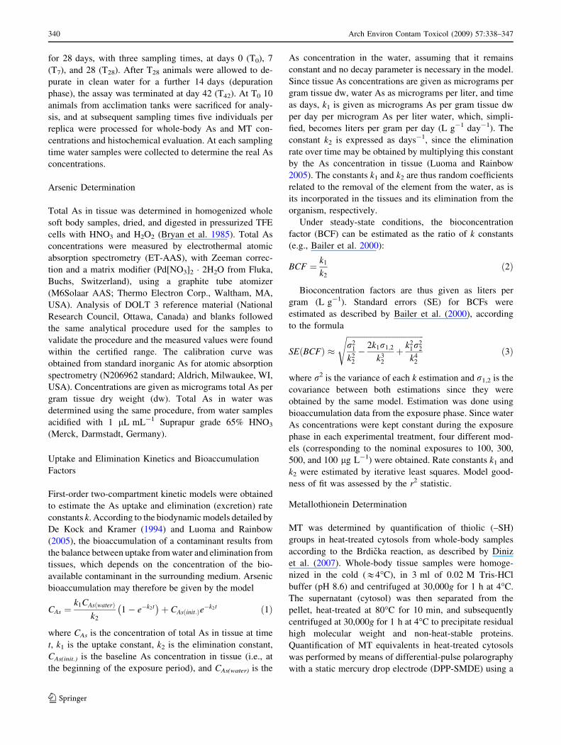
During the experimental period no mortality was observed
in any treatment. Total As concentrations determined in
water samples collected from the different treatments were
found to be very close to the nominal concentrations of
AsIII exposure (Table 1). No significant differences were
detected between the beginning and the end of the exper-
imental period (Mann–Whitney U, p [ 0.05), which
confirms that CAs(water) (1) can be regarded as constant.
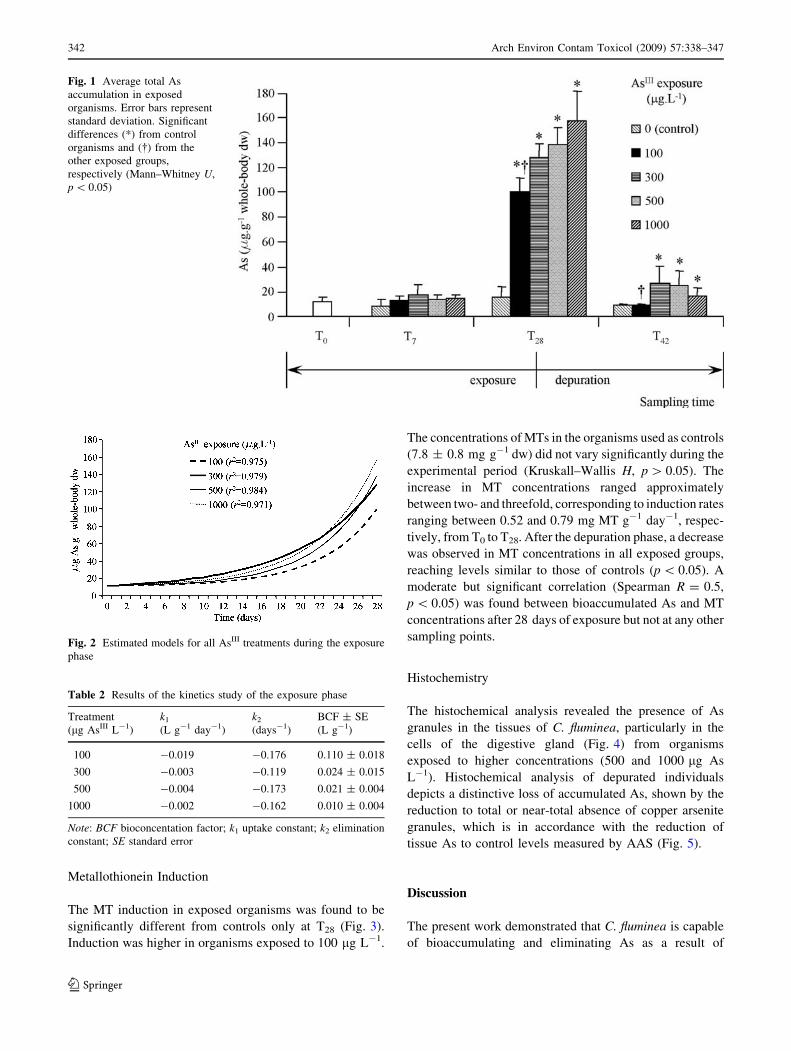
Arsenic Bioaccumulation and Elimination
The data obtained from clam tissue samples collected at T7
and T28 regarding the different exposures to AsIII (100,
300, 500, and 1000 lg As L-1) are shown in Fig. 1, which
also reports total As concentrations for depurated organ-
isms. Total As concentration in control clams was
10.8 ± 6.4 lg As g-1 dw and did not vary significantly
along the experiment time (p [ 0.05). The levels of As in
organisms sampled at T0 (11.7 ± 4.4 lg g-1 dw) were not
significantly different from controls (p [ 0.05). During the
exposure phase, a very significant increase in accumulated
As was observed for all treatments at T28 (p \ 0.05) but
not at T7. At the end of the exposure term a strong corre-
lation between As in water and As in mollusks’ whole soft
body (Spearman R = 0.75, p \ 0.05) was observed. After
14 days of depuration a significant decrease was observed
(p \ 0.05) in the contents of total As in the whole soft
tissues of the clams in comparison with levels measured at
T28, for all treatments. The decrease in whole-body total As
ranged between 79% and 91%.
Biodynamic Accumulation Model
and Bioconcentration Factors
Bioaccumulation data from the exposure phase were suc-
cessfully fitted to model (1), yielding four different models
that explain 95.5%–98.4% of the total variance (Fig. 2).
Estimated k constants and respective BCFs are presented in
Table 2. The BCF estimated for the 100 lg L-1 exposure
was found to be significantly higher than the other treat-
ments (p \ 0.01). Although no other significant differences
were found between BCFs, these were found to be nega-
tively correlated with As exposures (Kendall’s s = - 1,
p \ 0.05), decreasing with increasing exposures to water-
borne AsIII.
Table 1 Average ± standard deviation values of effective total As
concentrations (lg L-1) measured in water samples in the exposure
assay
Treatment (lg AsIII L-1)
Control 100 300 500 1000
T0 \d.l. 96 ± 2 306 ± 5 540 ± 8 1010 ± 3
T28 \d.l. 92 ± 3 299 ± 3 530 ± 5 991 ± 4
Note: d.l., detection limit
Arch Environ Contam Toxicol (2009) 57:338–347 341
123
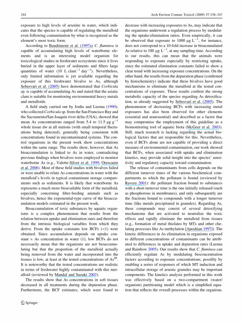
Metallothionein Induction
The MT induction in exposed organisms was found to be
significantly different from controls only at T28 (Fig. 3).
Induction was higher in organisms exposed to 100 lg L-1.
The concentrations of MTs in the organisms used as controls
(7.8 ± 0.8 mg g-1 dw) did not vary significantly during the
experimental period (Kruskall–Wallis H, p [ 0.05). The
increase in MT concentrations ranged approximately
between two- and threefold, corresponding to induction rates
ranging between 0.52 and 0.79 mg MT g-1 day-1, respec-
tively, from T0 to T28. After the depuration phase, a decrease
was observed in MT concentrations in all exposed groups,
reaching levels similar to those of controls (p \ 0.05). A
moderate but significant correlation (Spearman R = 0.5,
p \ 0.05) was found between bioaccumulated As and MT
concentrations after 28 days of exposure but not at any other
sampling points.
Histochemistry
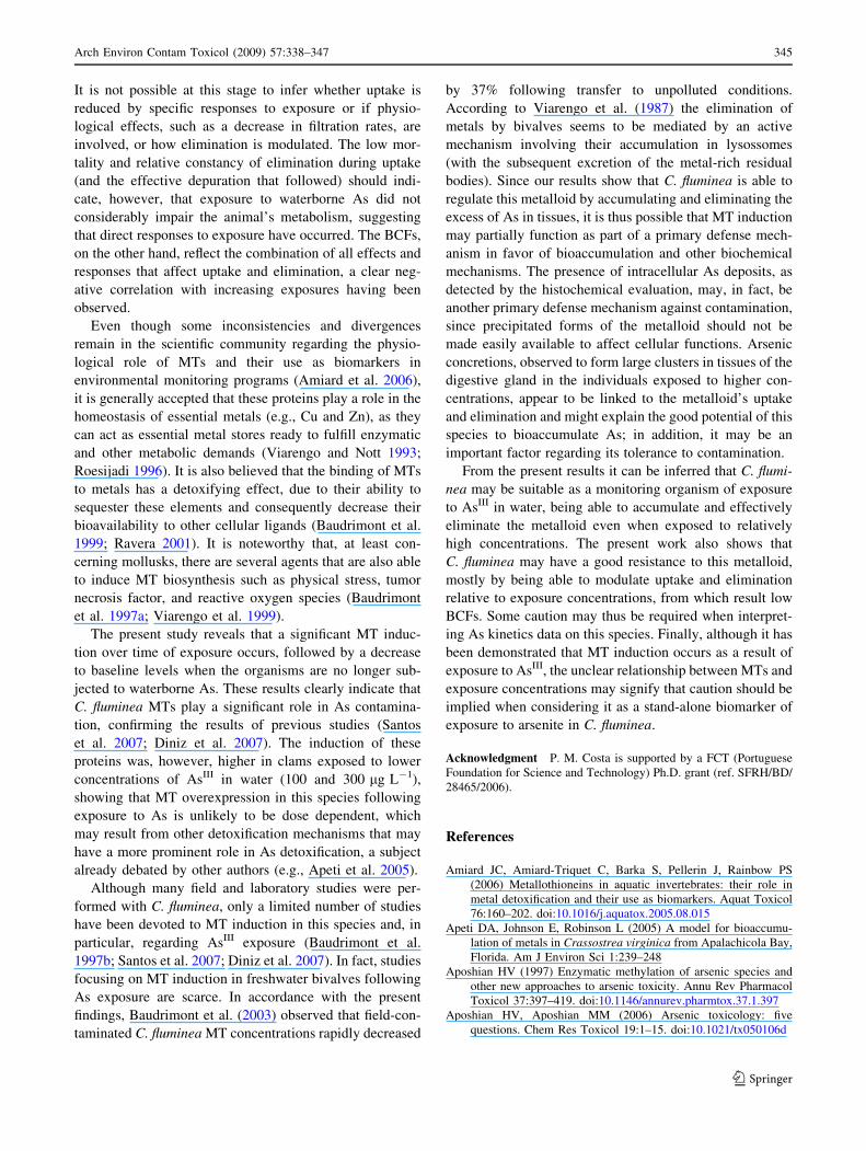
The histochemical analysis revealed the presence of As
granules in the tissues of C. fluminea, particularly in the
cells of the digestive gland (Fig. 4) from organisms
exposed to higher concentrations (500 and 1000 lg As
L-1). Histochemical analysis of depurated individuals
depicts a distinctive loss of accumulated As, shown by the
reduction to total or near-total absence of copper arsenite
granules, which is in accordance with the reduction of
tissue As to control levels measured by AAS (Fig. 5).
Discussion
The present work demonstrated that C. fluminea is capable
of bioaccumulating and eliminating As as a result of
Fig. 1 Average total As
accumulation in exposed
organisms. Error bars represent
standard deviation. Significant
differences (*) from control
organisms and (�) from the
other exposed groups,
respectively (Mann–Whitney U,
p \ 0.05)
Fig. 2 Estimated models for all AsIII treatments during the exposure
phase
Table 2 Results of the kinetics study of the exposure phase
Treatment
(lg AsIII L-1)
k1
(L g-1 day-1)
k2
(days-1)
BCF ± SE
(L g-1)
100 -0.019 -0.176 0.110 ± 0.018
300 -0.003 -0.119 0.024 ± 0.015
500 -0.004 -0.173 0.021 ± 0.004
1000 -0.002 -0.162 0.010 ± 0.004
Note: BCF bioconcentation factor; k1 uptake constant; k2 elimination
constant; SE standard error
342 Arch Environ Contam Toxicol (2009) 57:338–347
123
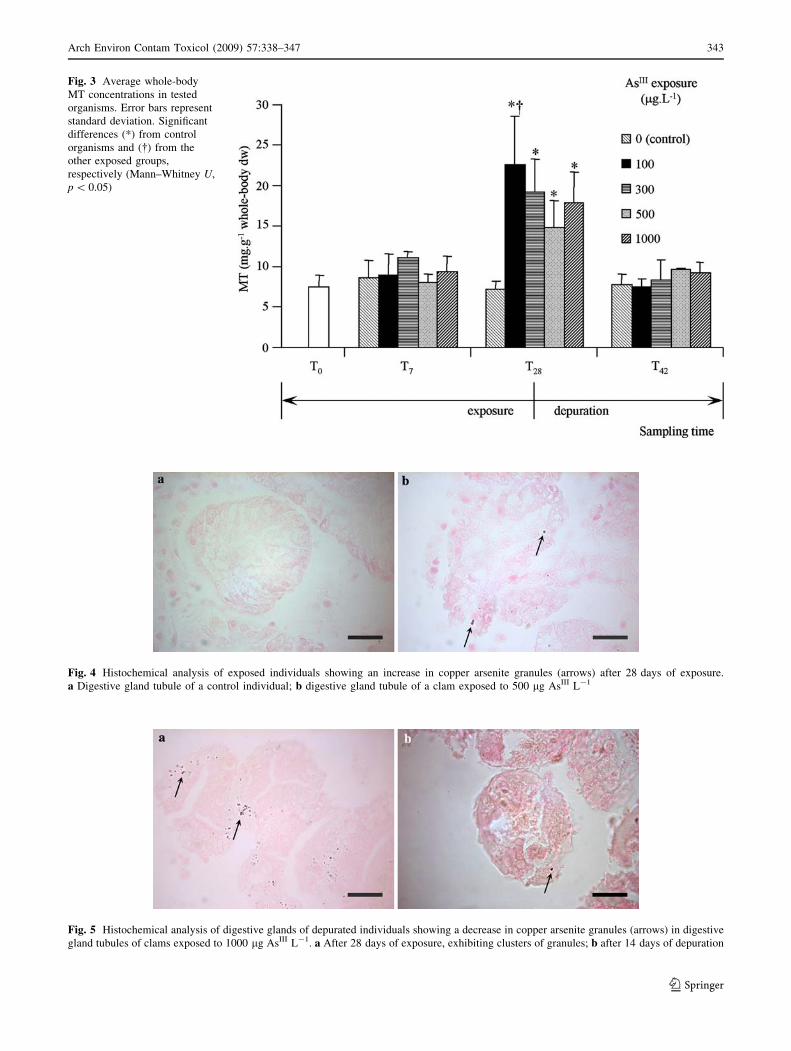
Fig. 3 Average whole-body
MT concentrations in tested
organisms. Error bars represent
standard deviation. Significant
differences (*) from control
organisms and (�) from the
other exposed groups,
respectively (Mann–Whitney U,
p \ 0.05)
Fig. 4 Histochemical analysis of exposed individuals showing an increase in copper arsenite granules (arrows) after 28 days of exposure.
a Digestive gland tubule of a control individual; b digestive gland tubule of a clam exposed to 500 lg AsIII L-1
Fig. 5 Histochemical analysis of digestive glands of depurated individuals showing a decrease in copper arsenite granules (arrows) in digestive
gland tubules of clams exposed to 1000 lg AsIII L-1. a After 28 days of exposure, exhibiting clusters of granules; b after 14 days of depuration
Arch Environ Contam Toxicol (2009) 57:338–347 343
123
exposure to high levels of arsenite in water, which indi-
cates that the species is capable of regulating the metalloid
even following contamination by what is recognized as the
element’s most toxic form.
According to Baudrimont et al. (1997a) C. fluminea is
capable of accumulating high levels of waterborne ele-
ments and is an interesting model organism for
toxicological studies in freshwater ecosystems since it lives
buried in the upper layer of sediments and filters large
quantities of water (10 L clam-1 day-1). Nevertheless,
only limited information is yet available regarding the
exposure of this freshwater bivalve to As, although
Sebesvari et al. (2005) have demonstrated that Corbicula
sp. is capable of accumulating As and stated that the asiatic
clam is suitable for monitoring environmental heavy metals
and metalloids.
A field study carried out by Jonhs and Luoma (1990),
who collected Corbicula sp. from the San Francisco Bay and
the Sacramento/San Joaquin river delta (USA), showed that
mean As concentrations ranged from 5.4 to 11.5 lg g-1
whole tissue dw at all stations (with small temporal fluctu-
ations being detected), generally being consistent with
concentrations found in uncontaminated systems. The con-
trol organisms in the present work show concentrations
within the same range. The results show, however, that As
bioaccumulation is linked to As in water, contradicting
previous findings when bivalves were employed to monitor
waterborne As (e.g., Valette-Silver et al. 1999; Orescanin
et al. 2006). Most of these field studies with bivalves failed
or were unable to relate As concentrations in water with the
metalloid’s levels in typical contaminant storage compart-
ments such as sediments. It is likely that waterborne As
represents a much more bioavailable form of the metalloid,
especially concerning filter-feeding animals such as
bivalves, hence the exponential-type curve of the bioaccu-
mulation models estimated in the present work.
Bioaccumulation of toxic substances by aquatic organ-
isms is a complex phenomenon that results from the
relation between uptake and elimination rates and therefore
from the intrinsic biological variables from which they
derive. From the uptake constants low BCFs (\1) were
obtained. Since accumulation depends on uptake con-
stant 9 As concentration in water (1), low BCFs do not
necessarily mean that the organisms are not bioaccumu-
lating but that the proportion of the metalloid actually
being removed from the water and incorporated into the
tissues is low, at least at the tested concentrations of AsIII.
It is noteworthy that the tested concentrations are realistic
in terms of freshwater highly contaminated with this met-
alloid (reviewed by Mandal and Suzuki 2002).
The results show that As concentrations in soft tissues
decreased in all treatments during the depuration phase.
Furthermore, the BCF estimates, which were found to
decrease with increasing exposures to As, may indicate that
the organisms underwent a regulation process by modulat-
ing the uptake:elimination ratios. Even empirically, it can
be observed that exposure to 1000 lg L-1, for instance,
does not correspond to a 10-fold increase in bioacumulated
As relative to 100 lg L-1, at any sampling time. According
to our results, this can mean that the animals were
responding to exposure especially by restricting uptake,
since the estimated elimination constants failed to show a
clear trend with increasing exposure concentrations. On the
other hand, the results from the depuration phase (confirmed
by histochemistry) indicate that these bivalves have good
mechanisms to eliminate the metalloid at the tested con-
centrations of exposure. These results confirm the strong
metabolic capacity of the species regarding As detoxifica-
tion, as already suggested by Sebesvari et al. (2005). The
phenomenon of decreasing BCFs with increasing metal
exposures has also been observed for other elements
(essential and nonessential) and described as a factor that
may compromise the employment of this guideline as a
biomonitoring tool of aquatic biota (McGeer et al. 2003).
Still, much research is lacking regarding the actual bio-
logical factors that are responsible for this. Nevertheless,
even if BCFs alone are not capable of providing a direct
measure of environmental contamination, our work showed
that BCFs, when associated with uptake and elimination
kinetics, may provide solid insight into the species’ sensi-
tivity and regulatory capacity toward contamination.
The release of contaminants from cells depends on the
different turnover times of the various biochemical com-
partments to which the pollutant is bound (reviewed by
Ravera 2001): the pollutant fraction bound to substances
with a short turnover time is the one initially released (such
as phosphorous in membranes), and only subsequently are
the fractions bound to compounds with a longer turnover
time (like metals precipitated in granules). Regarding As,
these compounds may consist of several detoxifying
mechanisms that are activated to neutralize the toxic
effects and rapidly eliminate the metalloid from tissues
(e.g., formation of metal thiolates by MTs and other che-
lating processes like As methylation [Aposhian 1997]). The
kinetic differences in As elimination in organisms exposed
to different concentrations of contaminants can be attrib-
uted to differences in uptake and depuration rates (Luoma
and Rainbow 2005). Our results show that C. fluminea can
efficiently regulate As by modulating bioconcentration
factors according to exposure concentrations, possibly by
enabling a series of responses of which MT induction and
intracellular storage of arsenic granules may be important
components. The kinetics analysis performed in this work
was effectively based on a two-compartment (water/
organism) partitioning model which is a simplified equa-
tion that reflects the overall processes within the organism.
344 Arch Environ Contam Toxicol (2009) 57:338–347
123
It is not possible at this stage to infer whether uptake is
reduced by specific responses to exposure or if physio-
logical effects, such as a decrease in filtration rates, are
involved, or how elimination is modulated. The low mor-
tality and relative constancy of elimination during uptake
(and the effective depuration that followed) should indi-
cate, however, that exposure to waterborne As did not
considerably impair the animal’s metabolism, suggesting
that direct responses to exposure have occurred. The BCFs,
on the other hand, reflect the combination of all effects and
responses that affect uptake and elimination, a clear neg-
ative correlation with increasing exposures having been
observed.
Even though some inconsistencies and divergences
remain in the scientific community regarding the physio-
logical role of MTs and their use as biomarkers in
environmental monitoring programs (Amiard et al. 2006),
it is generally accepted that these proteins play a role in the
homeostasis of essential metals (e.g., Cu and Zn), as they
can act as essential metal stores ready to fulfill enzymatic
and other metabolic demands (Viarengo and Nott 1993;
Roesijadi 1996). It is also believed that the binding of MTs
to metals has a detoxifying effect, due to their ability to
sequester these elements and consequently decrease their
bioavailability to other cellular ligands (Baudrimont et al.
1999; Ravera 2001). It is noteworthy that, at least con-
cerning mollusks, there are several agents that are also able
to induce MT biosynthesis such as physical stress, tumor
necrosis factor, and reactive oxygen species (Baudrimont
et al. 1997a; Viarengo et al. 1999).
The present study reveals that a significant MT induc-
tion over time of exposure occurs, followed by a decrease
to baseline levels when the organisms are no longer sub-
jected to waterborne As. These results clearly indicate that
C. fluminea MTs play a significant role in As contamina-
tion, confirming the results of previous studies (Santos
et al. 2007; Diniz et al. 2007). The induction of these
proteins was, however, higher in clams exposed to lower
concentrations of AsIII in water (100 and 300 lg L-1),
showing that MT overexpression in this species following
exposure to As is unlikely to be dose dependent, which
may result from other detoxification mechanisms that may
have a more prominent role in As detoxification, a subject
already debated by other authors (e.g., Apeti et al. 2005).
Although many field and laboratory studies were per-
formed with C. fluminea, only a limited number of studies
have been devoted to MT induction in this species and, in
particular, regarding AsIII exposure (Baudrimont et al.
1997b; Santos et al. 2007; Diniz et al. 2007). In fact, studies
focusing on MT induction in freshwater bivalves following
As exposure are scarce. In accordance with the present
findings, Baudrimont et al. (2003) observed that field-con-
taminated C. fluminea MT concentrations rapidly decreased
by 37% following transfer to unpolluted conditions.
According to Viarengo et al. (1987) the elimination of
metals by bivalves seems to be mediated by an active
mechanism involving their accumulation in lysossomes
(with the subsequent excretion of the metal-rich residual
bodies). Since our results show that C. fluminea is able to
regulate this metalloid by accumulating and eliminating the
excess of As in tissues, it is thus possible that MT induction
may partially function as part of a primary defense mech-
anism in favor of bioaccumulation and other biochemical
mechanisms. The presence of intracellular As deposits, as
detected by the histochemical evaluation, may, in fact, be
another primary defense mechanism against contamination,
since precipitated forms of the metalloid should not be
made easily available to affect cellular functions. Arsenic
concretions, observed to form large clusters in tissues of the
digestive gland in the individuals exposed to higher con-
centrations, appear to be linked to the metalloid’s uptake
and elimination and might explain the good potential of this
species to bioaccumulate As; in addition, it may be an
important factor regarding its tolerance to contamination.
From the present results it can be inferred that C. flumi-
nea may be suitable as a monitoring organism of exposure
to AsIII in water, being able to accumulate and effectively
eliminate the metalloid even when exposed to relatively
high concentrations. The present work also shows that
C. fluminea may have a good resistance to this metalloid,
mostly by being able to modulate uptake and elimination
relative to exposure concentrations, from which result low
BCFs. Some caution may thus be required when interpret-
ing As kinetics data on this species. Finally, although it has
been demonstrated that MT induction occurs as a result of
exposure to AsIII, the unclear relationship between MTs and
exposure concentrations may signify that caution should be
implied when considering it as a stand-alone biomarker of
exposure to arsenite in C. fluminea.
Acknowledgment P. M. Costa is supported by a FCT (Portuguese
Foundation for Science and Technology) Ph.D. grant (ref. SFRH/BD/
28465/2006).
References
Amiard JC, Amiard-Triquet C, Barka S, Pellerin J, Rainbow PS
(2006) Metallothioneins in aquatic invertebrates: their role in
metal detoxification and their use as biomarkers. Aquat Toxicol
76:160–202. doi:10.1016/j.aquatox.2005.08.015
Apeti DA, Johnson E, Robinson L (2005) A model for bioaccumu-
lation of metals in Crassostrea virginica from Apalachicola Bay,
Florida. Am J Environ Sci 1:239–248
Aposhian HV (1997) Enzymatic methylation of arsenic species and
other new approaches to arsenic toxicity. Annu Rev Pharmacol
Toxicol 37:397–419. doi:10.1146/annurev.pharmtox.37.1.397
Aposhian HV, Aposhian MM (2006) Arsenic toxicology: five
questions. Chem Res Toxicol 19:1–15. doi:10.1021/tx050106d
Arch Environ Contam Toxicol (2009) 57:338–347 345
123

Bailer AJ, Walker SE, Venis K (2000) Estimating and testing
bioconcentration factors. Environ Toxicol Chem 19:2338–2340.
doi:10.1897/1551-5028(2000)019\2338:EATBF[2.3.CO;2
Baudrimont M, Metivaud J, Maury-Brachet R, Ribeyre F, Boudu A
(1997a) Bioaccumulation and metallothionein response in the
asiatic clam Corbicula fluminea after experimental exposure to
cadmium and inorganic mercury. Environ Toxicol Chem
16:2096–2105. doi:10.1897/1551-5028(1997)016\2096:BAMRIT[2.3.CO;2
Baudrimont M, Lemaire-Gony S, Ribeyre F, Metivaud J, Boudou A
(1997b) Seasonal variations of metallothionein concentrations in
the Asiatic clam (Corbicula fluminea). Comp Biochem Physiol
118C:361–367
Baudrimont M, Andres S, Metivaud J, Lapaquellerie Y, Ribeyre F,
Maillet N, Latouche C, Boudou A (1999) Field transplantation of
the freshwater bivalve Corbicula fluminea along a polymetallic
contamination gradient (river Lot, France): II. Metallothionein
response to metal exposure. Environ Toxicol Chem 18:2472–2477.
doi:10.1897/1551-5028(1999)018\2472:FTOTFB[2.3.CO;2
Baudrimont M, Andres S, Durrieu G, Boudou A (2003) The key role
of metallothioneins in the bivalve Corbicula fluminea during the
depuration phase, after in situ exposure to Cd and Zn. Aquat
Toxicol 63:89–102. doi:10.1016/S0166-445X(02)00134-0
Bissen M, Frimmel FH (2003a) Arsenic—a review. Part I. Occur-
rence, toxicity, speciation, mobility in the aqueous environment.
Acta Hydrochim Hydrobiol 31:9–18. doi:10.1002/aheh.
200390025
Bissen M, Frimmel FH (2003b) Arsenic—a review. Part II. Oxidation
of arsenic and its removal in water treatment. Acta Hydrochim
Hydrobiol 3:97–107. doi:10.1002/aheh.200300485
Bryan GW, Langston WJ, Hummerstone LG, Burt GR (1985) A
Guide to the assessment of heavy-metal contamination in
estuaries using biological indicators. Mar Biol Assoc UK,
Occasional Publication No. 4
Cervantes C, Ji G, Ramirez JL, Silver S (1994) Resistance to arsenic
compounds in microorganisms. FEMS Microbiol Rev 15:355–
367. doi:10.1111/j.1574-6976.1994.tb00145.x
Couillard Y, Campbell PGC, Tessier A (1993) Response of metal-
lothionein concentrations in a freshwater bivalve (Anodontagrandis) along an environmental cadmium gradient. Mol Mar
Biol Biotechnol 2:299–313
De Kock WC, Kramer KJM (1994) Active biomonitoring (ABM) by
translocation of bivalve molluscs. In: Kramer KJM (ed)
Biomonitoring of coastal waters and estuaries. CRC Press, Boca
Raton, FL, pp 51–84
Diniz M, Santos HM, Costa PM, Peres I, Costa MH, Capelo JL (2007)
Metallothionein responses in the Asiatic clam (Corbiculafluminea) after exposure to trivalent arsenic. Biomarkers
12:589–598. doi:10.1080/13547500701507701
Duker AA, Carranza EJM, Hale M (2005) Arsenic geochemistry and
health. Environ Int 31:631–641. doi:10.1016/j.envint.2004.10.
020
Feng Z, Xia Y, Tian D, Wu K, Schmitt M, Kwok RK (2001) DNA
damage in buccal epithelial cells from individuals chronically
exposed to arsenic via drinking water in Inner Mongolia, China.
Anticancer Res 21:51–58
Fraysse B, Baudin JP, Garnier-Laplace J, Adam C, Boudou A (2002)
Effects of Cd and Zn waterborne exposure on the uptake and
depuration of 57Co, 110mAg and 134Cs by the Asiatic clam
(Corbicula fluminea) and the zebra mussel (Dreissena polymor-pha)—whole organism study. Environ Poll 118:297–306. doi:
10.1016/S0269-7491(01)00305-0
Hindmarsh JT (2000) Arsenic, its clinical and environmental signif-
icance. J Trace Elem Exp Med 13:165–172. doi:10.1002/
(SICI)1520-670X(2000)13:1\165::AID-JTRA17[3.0.CO;2-R
Hossain F, Sivakumar B (2006) Spatial pattern of arsenic contam-
ination in shallow tubewells of Bangladesh: regional geology
and non-linear dynamics stochastic. Environ Res Risk Assess
20:66–76. doi:10.1007/s00477-005-0012-7
Inza B, Ribeyre F, Maury-Brachet R, Boudou A (1997) Tissue
distribution of inorganic mercury, methylmercury and cadmium
in the asiatic clam (Corbicula fluminea) in relation to the
contamination levels of the water column and sediment. Chemo-sphere 35:2817–2836. doi:10.1016/S0045-6535(97)00342-1
Jiang G, Gong Z, Li X-F, Cullen WR, Le XC (2003) Interaction of
trivalent arsenicals with metallothionein. Chem Res Toxicol
16:873–880. doi:10.1021/tx034053g
Jonhs C, Luoma SN (1990) Arsenic in benthic bivalves of San
Francisco Bay and the Sacramento/San Joaquin River. Sci Tot
Environ 97(98):673–684. doi:10.1016/0048-9697(90)90268-Y
Kaise T, Horiguchi Y, Fukui S, Shiomi K, Chino M, Kikuchi T (1992)
Acute toxicity and metabolism of arsenocholine in mice. Appl
Organomet Chem 6:369–373. doi:10.1002/aoc.590060410
Liao CM, Ling MP (2003) Assessment of human health risks for
arsenic bioaccumulation in tilapia (Oreochromis mossambicus)
and large-scale-mullet (Liza macrolepis) from blackfoot disease
area in Taiwan. Arch Environ Contam Toxicol 45:264–272. doi:
10.1007/s00244-003-0107-4
Lillie RD (1965) Histopathologic technic and practical histochemis-
try. McGraw-Hill, New York
Luoma S, Rainbow P (2005) Why is metal bioaccumulation so
variable? Biodynamics as a unifying concept. Environ Sci
Technol 39:1921–1931. doi:10.1021/es048947e
Mandal BK, Suzuki KT (2002) Arsenic around the world: a review.
Talanta 58:201–235. doi:10.1016/S0039-9140(02)00268-0
Martoja R, Martoja M (1967) Initiation aux tecniques de l’histologie
animal. Masson, Paris
McGeer JC, Brix KB, Skeaff JM, DeForest DK, Brigham SI, Adams
WJ, Green A (2003) Inverse relationship between bioconcentra-
tion factor and exposure concentration for metals: implications
for hazard assessment of metals in the aquatic environment.
Environ Toxicol Chem 22:1017–1037. doi:10.1897/1551-
5028(2003)022\1017:IRBBFA[2.0.CO;2
Mouthon J (1981) Sur la presence en France et au Portugal de
Corbicula (Bivalvia, Corbiculidae) originaire d’Asie. Basteria
45:109–116
Ngu TT, Stillman MJ (2006) Arsenic binding to human metallothio-
nein. J Am Chem Soc 128:12473–12483. doi:10.1021/ja062914c
Orescanin V, Lovrencic I, Mikelic L, Barisic D, Matasin Z, Lulic S,
Pezelj D (2006) Biomonitoring of heavy metals and arsenic on
the east coast of the Middle Adriatic Sea using Mytilusgalloprovincialis. Nuc Instr Meth Phys Res B 245:495–500. doi:
10.1016/j.nimb.2005.11.050
Palecek E, Pechan Z (1971) Estimation of nanogram quantities of
proteins by pulse polarographic techniques. Anal Biochem
42:59–71. doi:10.1016/0003-2697(71)90010-8
Ravera O (2001) Monitoring of the aquatic environment by species
accumulator of pollutants: a review. J Limnol 60S1:63–78
Roesijadi G (1996) Metallothionein and its role in toxic metal
regulation. Comp Biochem Physiol 113C:117–123
Romero-Isart N, Vasak M (2002) Advances in the structure and
chemistry of metallothioneins. J Inorg Biochem 88:388–396.
doi:10.1016/S0162-0134(01)00347-6
Santos HM, Diniz M, Costa PM, Peres I, Costa MH, Alves S, Capelo
JL (2007) Toxicological effects and bioaccumulation in the
freshwater clam (Corbicula fluminea) following exposure to
trivalent arsenic. Environ Toxicol 22:502–509. doi:10.1002/
tox.20303
Sebesvari Z, Ettwig F, Emons H (2005) Biomonitoring of tin and
arsenic in different compartments of a limnic ecosystem with
346 Arch Environ Contam Toxicol (2009) 57:338–347
123
emphasis on Corbicula fluminea and Dikerogammarus villosus.
J Environ Monit 7:203–207. doi:10.1039/b410717a
Sheskin FJ (2000) Handbook of parametric and nonparametric
statistical procedures, 2nd edn. Chapman & Hall, Boca Raton,
FL
Thompson JAJ, Cosson RP (1984) An improved electrochemical
method for the quantification of metallothioneins in marine
organisms. Mar Environ Res 11:137–152. doi:10.1016/0141-
1136(84)90027-8
Toyama M, Yamashita M, Hirayama N, Murooka Y (2002) Interac-
tions of arsenic with human metallothionein-2. J Biochem Tokyo
132:217–221
Valette-Silver NJ, Riedel GF, Crecelius EA, Windom H, Smith RG,
Dolvin SS (1999) Elevated arsenic concentrations in bivalves
from the southeast coasts of the USA. Mar Environ Res 48:311–
333. doi:10.1016/S0141-1136(99)00057-4
Viarengo A, Nott JA (1993) Mechanisms of heavy metal cation
homeostasis in marine invertebrates. Comp Biochem Phys
104C:355–372
Viarengo A, Moore MN, Mancinelli G, Mazzucotelli A, Pipe RK,
Farrar SV (1987) Metallothioneins and lysosomes in metal
toxicity and accumulation in marine mussels: the effect of
cadmium in the presence and absence of phenanthrene. Mar Biol
94:251–257. doi:10.1007/BF00392937
Viarengo A, Burlando B, Dondero F, Marro A, Fabbri R (1999)
Metallothionein as a tool in biomonitoring programmes.
Biomarkers 4:455–466. doi:10.1080/135475099230615
Arch Environ Contam Toxicol (2009) 57:338–347 347
123


![Toxicokinetics of [3H]-dihydroazadirachtin in the variegated cutwormPeridroma saucia](https://img.dokumen.tips/doc/110x75/635d3ca4a3fa66b45c0e4c3a/toxicokinetics-of-3h-dihydroazadirachtin-in-the-variegated-cutwormperidroma-saucia.jpg)

