Embed Size (px)
Citation preview
BASIC AND EXPERIMENTAL RESEARCH
Tissue Specificity of Cross-Reactive AllogeneicResponses by EBV EBNA3A-Specific Memory T Cells
Lloyd J. A. D’Orsogna,1,5 Dave L. Roelen,1 Ellen M. W. van der Meer-Prins,1 Pieter van der Pol,2
Marry E. Franke-van Dijk,1 Michael Eikmans,1 Jacqy Anholts,1 Jamie Rossjohn,3 James McCluskey,4
Arend Mulder,1 Cees van Kooten,2 Ilias I. N. Doxiadis,1 and Frans H. J. Claas1
Background. The crossreactivity of Epstein-Barr virus (EBV Epstein-Barr virus nuclear antigen 3A [EBNA3A])-specific CD8 T cells against allogeneic human leukocyte antigen (HLA)-B*44:02 has been shown to be dependent onpresentation of self-peptide EEYLQAFTY by the target antigen. In this study, we report that allogeneic HLA-B*44:02�
proximal tubular epithelial cells (PTECs) and human umbilical vein endothelial cells (HUVECs) are poor targets forEBV EBNA3A-specific T cells.Methods. The EEY peptide was exogenously loaded onto HLA-B*44:02 and HLA-B*44:03-expressing PTECs andHUVECs. EEY-peptide-loaded, and unloaded, PTECs and HUVECs were then incubated with serial dilutions of ourEBNA3A T-cell clone, in a cytotoxicity assay.Results. Although HLA-B*44:02-expressing PTECs were specifically lysed in proportion to the effector/target ratio bythe EBNA3A T-cell clone, without peptide loading, lysis was greatly increased by exogenous EEY peptide loading (15%vs. 75%; P�0.0001). HLA-B*44:02-expressing HUVECs were only lysed when loaded with exogenous EEY peptide (0%vs. 64%; P�0.0001). Lack of HLA expression and lack of ABCD3 gene expression were excluded as a cause for theseresults. PTECs and HUVECs were specifically targeted by another alloreactive T-cell clone without exogenous peptideloading, suggesting that the lack of recognition of HLA-B*44:02� epithelial and endothelial cells by the EBV EBNA3AT-cell clone was due to lack of EEYLQAFTY peptide presentation.Conclusions. Tissue-specific (peptide dependent) alloreactivity may have important implications for transplantationmonitoring and rejection.
Keywords: Heterologous immunity, Memory T cells, Alloreactivity, EBV, Tissue specificity.
(Transplantation 2011;91: 494–500)
V iral infection is associated with solid organ transplantrejection and is a potent barrier to transplantation toler-
ance (1–10). It has recently been shown that allohuman leu-kocyte antigen (HLA) crossreactivity from viral-specificmemory T cells is far more common than predicted, usingEpstein-Barr virus (EBV)-transformed B cells (EBV LCLs)
and HLA-transfected target cells (11). This allo-HLA crossre-activity from viral-specific memory T cells has been shown tobe dependent on endogenous self-peptide presentation by thedonor cell (11–13), and therefore, alloreactivity could be tis-sue cell-type specific if the recognized peptide is differentiallyexpressed by target tissues.
Earlier work suggested that the explanation for thepresence of alloreactive memory T cells in nonsensitized in-dividuals could be crossreactivity from viral-specific memoryT cells against allo-HLA molecules (14, 15). Burrows et al.(15) demonstrated the dual specificity of EBV EBNA3A-specific T-cell clones for the immunodominant EBV peptideFLRGRAYGL presented on HLA-B*08:01 and the alloantigenHLA-B*44:02, to which the individual had never been ex-posed. The clinical relevance of this finding is reinforced bythe fact that EBV EBNA3A-specific CD8 T cells are capable ofspecifically lysing HLA-B*44:02� target cells in cytotoxicityassays (15, 16) and that HLA-B44 is an immunogenic mis-match in HLA-B8 kidney recipients (17).
The authors declare no conflict of interest.1 Department of Immunohematology and Blood Transfusion, Leiden Uni-
versity Medical Centre, The Netherlands.2 Department of Nephrology, Leiden University Medical Centre, The
Netherlands.3 Department of Biochemistry and Molecular Biology, Monash University,
Australia.4 Department of Microbiology and Immunology, University of Melbourne,
Australia.5 Address correspondence to: Lloyd J. D’Orsogna, Ph.D., M.B.B.S., Depart-
ment of Immunohematology and Blood Transfusion, Leiden UniversityMedical Centre, PO Box 9600, Leiden 2300RC, The Netherlands.
E-mail: [email protected]. participated in research design, performed research, and wrote
manuscript; D.L.R. participated in research design and project supervi-sor; E.M.W.v.d.M.-P. performed research; P.v.d.P. and M.E.F.-v.D. con-tributed tissue-specific cell lines; J.A. and M.E. designed and performedresearch relating to ABCD3 gene expression; J.R. and J.M. participated inresearch design; A.M. and C.v.K. participated in research design andcontributed tissue-specific cell lines; and I.I.N.D. and F.H.J.C. partici-pated in research design and project supervisors.
Received 12 August 2010. Revision requested 26 September 2010.Accepted 18 November 2010.Copyright © 2011 by Lippincott Williams & WilkinsISSN 0041-1337/11/9105-494DOI: 10.1097/TP.0b013e318207944c
494 | www.transplantjournal.com Transplantation • Volume 91, Number 5, March 15, 2011
This EBV EBNA3A T-cell allo-HLA-B*44:02 crossreactiv-ity is dependent on presentation of the EEYLQAFTY self-peptide derived from the ABCD3 gene by molecular mimicry(12). Despite extensive polymorphism between HLA-B*08:01and HLA-B*44:02 and the disparate sequences of their boundviral and self-peptides, respectively, the HLA-B8/FLR-restrictedT-cell receptor (TCR) engages these different peptide/HLAcomplexes identically.
In our laboratory, various cell lines have been generatedfor in vitro testing in kidney transplantation research. Proximaltubular epithelial cells (PTECs) are derived from proximal tu-bule cells taken from kidney transplant biopsy specimens (18–20) and are a useful model to examine alloreactivity from graftinfiltrating lymphocytes (21, 22). Human umbilical vein endo-thelial cells (HUVECs) are derived from healthy human post-partum umbilical tissue (23–25) but are also useful as a modelof kidney vascular endothelial cell transplantation (26). Inthis study, we investigate tissue specificity of the cross-reactive alloresponse by EBV EBNA3A-specific memory Tcells, using PTECs and HUVECs as a model system for hu-man kidney transplantation.
We report that in contrast to other allogeneic HLA-B*44:02� cell lines, allogeneic HLA-B*44:02� PTECs are poor targetsfor EBV EBNA3A-specific CD8 T cells, and HLA-B*44:02HUVECs are not targeted at all. We hypothesized that this dif-ferential lysis of HLA-B*44:02-expressing PTEC and HUVECcell lines could be explained by differential tissue presentation ofthe EEYLQAFTY self-peptide. Our work with the EBV EBNA3AT-cell clone, presented in this study, confirms that (peptidedependent) tissue specificity of allo-HLA responses from viral-specific memory T cells may indeed be relevant in the kidneytransplantation setting. Essentially, alloreactivity can be tissuespecific.
RESULTS
HLA-B*44:02-Expressing Epithelial andEndothelial Cell Lines Are Poor Targets for EBVEBNA3A-Specific CD8 Memory T Cells
We and others have previously shown that EBVEBNA3A-specific T-cell clones exert cytolytic activity against
allogeneic HLA-B*44:02� EBV LCLs, phytohemagglutininblasts, and HLA-B*44:02-transfected K562 cells (11, 15, 16).We, therefore, performed cytolytic assays using HLA-B*44:02� PTEC and HUVEC targets to determine whether allo-HLA crossreactivity from viral-specific memory T cells, asdetermined by hematological target cells types, correspondswith solid organ alloreactivity (Fig. 1). In contrast to otherallogeneic HLA-B*44:02� cell lines, allogeneic HLA-B*44:02� PTECs are poor targets for EBV EBNA3A-specific CD8T cells (specific lysis 12%). HLA-B*44:02�-expressingHUVECs are not targeted by an EBV EBNA3A-specific T-cellclone (specific lysis 0%).
The Lack of Recognition of Epithelial andEndothelial Cell Lines Is Not Due To Lack ofHLA-B*44:02 Expression
To exclude lack of HLA expression as the cause forthese results, we performed cytotoxicity assays before and af-ter interferon (IFN)-� stimulation of the PTEC and HUVECtargets. PTECs demonstrated higher baseline HLA-B*44:02expression, when compared with HUVECs (data not shown).IFN-� prestimulation was associated with increased HLA-B*44:02 surface expression in both PTECs and HUVECs(data not shown). Regardless, the increased HLA expressionafter IFN-� stimulation did not result in increased lysis of thePTEC or HUVEC target cells (data not shown).
The Lack of Recognition of HLA-B*44:02Epithelial and Endothelial Cells Is Not Due ToLack of ABCD3 Gene Expression
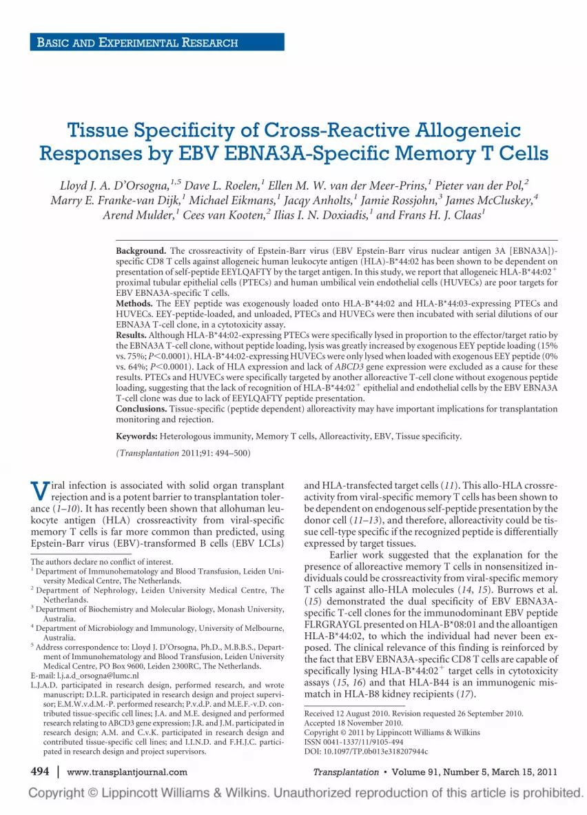
Alloreactivity of the EBV EBNA3A T-cell clone againstHLA-B*44:02� cell lines is dependent on presentation of theEEYLQAFTY peptide derived from the ABCD3 gene (12). Toexclude the lack of ABCD3 gene expression in the epithelialand endothelial target cells, we performed quantitative poly-merase chain reaction (qPCR) for ABCD3-gene-specific mes-senger RNA (mRNA). ABCD3 mRNA was detectable in bothPTEC and HUVEC target cell types (Fig. 2). Furthermore, ananti-ABCD3 monoclonal antibody demonstrated cytoplas-mic presence of the protein product (data not shown).
FIGURE 1. HLA-B*44:02-expressingPTECs and HUVECs are poor targetsfor EBV EBNA3A-specific T cells. Incontrast to allogeneic HLA-B*44:02�
phytohemagglutinin (PHA) blasts, allo-geneic HLA-B*44:02� PTECs are poortargets for EBV EBNA3A-specific CD8T cells in a 4-hr cytotoxicity assay(specific lysis, 12%). HLA-B*44:02�-expressing HUVECs are not targetedby an EBV EBNA3A T-cell clone (spe-cific lysis, 0%). Experiments are per-formed in triplicate, and mean valuesare shown with SD. E:T ratio 30:1, tar-gets�5000. HLA, human leukocyteantigen; PTEC, proximal tubular epi-thelial cell; HUVEC, human umbilicalvein endothelial cell; EBV, Epstein-Barr virus; EBNA3A, Epstein-Barr virusnuclear antigen 3A; SD, standarddeviation.
© 2011 Lippincott Williams & Wilkins 495D’Orsogna et al.

The Lack of Recognition of HLA-B*44:02-Expressing Epithelial and Endothelial Cells IsLikely Due To Quantitative Lack of EEYLQAFTYPeptide Presentation on the Cell Surface
To confirm that the lack of allorecognition of the HLA-B*44:02� epithelial and endothelial cell lines was due to lackof peptide presentation, we performed cytolytic assays usingEEYLQAFTY-peptide-loaded and -unloaded PTEC andHUVEC targets. HLA-B*44:02-expressing PTECs werepoorly targeted by an EBV EBNA3A T-cell clone without pep-tide loading (specific lysis 15%; P�0.004 vs. HLA-B*44:03PTEC) and then only at high effector/target ratio (Fig. 3a).The specific lysis of HLA-B*44:02-expressing PTECs wasgreatly increased by exogenous EEY peptide loading (15% vs.75%; P�0.0001; Fig. 3a). HLA-B*44:02-expressing HUVECswere only targeted by an EBV EBNA3A-specific clone whenloaded with exogenous EEY peptide (0% vs. 64%; P�0.0001)(Fig. 3b). Stimulation of the EBV EBNA3A-specific T-cellclone with HLA-B*44:02� HUVECs did not elicit cytokineproduction in a 24-hr luminex assay (data not shown). TheEBV EBNA3A-specific T-cell clone was able to target EEY-peptide-loaded HLA-B*44:02 PTECs and HUVECs evenwithout IFN-� prestimulation, suggesting that the baselineHLA-B*44:02 expression in these cell lines is sufficient forcytotoxic T lymphocyte (CTL) targeting if sufficient allopep-tide is presented. HLA-B*44:03 PTECs and HUVECs werenot targeted irrespective of peptide loading, as predicted (12).Thus, organ (kidney) specificity of the allo-HLA crossreactiv-ity from the EBV EBNA3A-specific T cell is dependent onendogenous self-peptide (EEY) processing and presentation.
FIGURE 2. ABCD3 mRNA expression in PTEC andHUVEC cell lines. ABCD3 mRNA expression, relative tohousehold gene expression, was measured in HLA-transfected and nontransfected K562 cells, EBV LCLs,PTECs, and HUVECs. ABCD3 mRNA expression in PTECsand HUVECs was comparable with EBV LCLs, indicatingthat the lack of allorecognition of PTECs and HUVECs bythe EBV EBNA3A T-cell clone was not due to absence ofexpression of the ABCD3 protein, from which the EEYLQAFTYself-peptide is derived. SAL, single HLA-transfected K562cell; EBV, Epstein-Barr virus-transformed B cell. HLA, humanleukocyte antigen; PTEC, proximal tubular epithelial cell;HUVEC, human umbilical vein endothelial cell; EBV, Epstein-Barr virus; EBNA3A, Epstein-Barr virus nuclear antigen 3A;SD, standard deviation; EBV LCL, EBV-transformed B cells;mRNA, messenger RNA.
FIGURE 3. The lack of recognition of HLA-B*44:02� PTECs and HUVECs is likely due to lack of EEYLQAFTY peptidepresentation on the cell surface. (a) HLA-B*44:02� PTECs were poorly targeted by the EBV EBNA3A-specific T-cell clone(specific lysis 15%; **P�0.004 vs. HLA-B*44:03 PTEC). The specific lysis of HLA-B*44:02� PTECs was greatly increased byexogenous EEY peptide loading (specific lysis 15% vs. 75%; ***P�0.0001). (b) HLA-B*44:02� HUVECs were only targetedby an EBV EBNA3A-specific T-cell clone when loaded with exogenous EEY peptide (specific lysis 0% vs. 64%;***P�0.0001). The EBV EBNA3A T-cell clone did not recognize HLA-B*44:03� PTECs or HUVECs irrespective of peptideloading. Experiments shown in this study were performed without IFN-� prestimulation further demonstrating that thebaseline HLA-B*44:02 expression is sufficient to elicit cytotoxicity if the EEY peptide is present. Experiments are performedin triplicate, and mean values are shown with SD. Targets�5000. HLA, human leukocyte antigen; PTEC, proximal tubularepithelial cell; HUVEC, human umbilical vein endothelial cell; EBV, Epstein-Barr virus; EBNA3A, Epstein-Barr virus nuclearantigen 3A; SD, standard deviation; IFN, interferon.
496 | www.transplantjournal.com Transplantation • Volume 91, Number 5, March 15, 2011

Cognate Antigen Recognition andAllorecognition Increase in Proportion to theConcentration of Exogenously Added Peptide
To determine the concentration of specific peptide re-quired to elicit cytolytic effector function by the EBVEBNA3A-specific T cells, FLR or EEY peptide were loadedonto HLA-B*08:01� or HLA-B*44:02� target cells, respec-tively, in a peptide dose-response experiment (Fig. 4). Cog-nate viral antigen recognition and allorecognition increase inproportion to the concentration of exogenously added cog-nate or allopeptide (Fig. 4). Equivalent concentrations of theFLR cognate peptide on HLA-B*08:01� target cells and EEYallopeptide on HLA-B*44:02� target cells were required toelicit cytolytic effector function by the EBV EBNA3A-specificT cells. For both cognate and allopeptides, 50% of the maxi-mum specific lysis occurred between 10 and 50 �g/mL ofexogenously added peptide.
Epithelial and Endothelial Cells Are NotResistant to Lysis by CTL Clones
Finally, to exclude the possibility that the EEYLQAFTYpeptide is presented on the target HLA molecule, but the cellsare not targeted due to lytic resistance or CTL suppression, weperformed cytolytic assays using the EBV EBNA3A clone anda HLA-A2 alloreactive T-cell clone (JS132) in parallel. TheJS132 clone specifically lysed HLA-A2� B*44:02� heterozy-gote PTECs and HUVECs, without any exogenous peptideaddition (Fig. 5). The EBV EBNA3A CD8 T-cell clone wasunable to efficiently target the identical epithelial or endothe-lial cell lines without exogenous addition of EEYLQAFTYpeptide (Fig. 5). Thus, the epithelial and endothelial cells canbe suitable targets for CTL clones without addition of exoge-nous peptide.
DISCUSSIONIn this report, we demonstrate that allo-HLA crossre-
activity by viral-specific memory T cells can be tissue cell-typespecific because of differential tissue-specific self-peptidepresentation. We have confirmed that not only is the HLA-
B*44:02 alloreactivity from the EBV EBNA3A-specific T-cellclone self-peptide dependent but also that normal allogeneickidney cells may not be targeted unless sufficient EEY self-peptide is processed and presented. Alloreactivity is mediatedby cytotoxicity, when the peptide is presented, indicating thepotential clinical relevance of cross-reactive alloresponsesagainst cell types present in kidney transplant tissue.
Our results do not suggest that allo-HLA crossreactivityfrom the EBV EBNA3A T cell is irrelevant to kidney trans-plantation. The EBV EBNA3A-specific T cell does have cyto-lytic activity against HLA-B*44:02 kidney epithelial cells in a4-hr assay. Memory T cells persist and, therefore, could per-form effector functions over a prolonged period or at timeswhen immunosuppression is tapered (2). Furthermore, Tcells mediate effector functions through a variety of mecha-nisms, including cytokine production, not just cytotoxicity(27). The EBV EBNA3A-specific immune response is a publicTCR response present in all HLA-B8� B44� individuals (28),and HLA-B44 mismatching has been identified as high risk inHLA-B8 kidney recipients (17).
However, results presented in this study suggest it isunlikely that EBV EBNA3A-specific T cells exhibit effectorfunctions against HLA-B*44:02� endothelial cells present insolid organ tissue. Conversely, a viral-specific T cell that tar-gets a kidney cell-specific peptide presented on an allogeneicHLA molecule may not recognize peripheral blood monocytecells (PBMCs) or spleen cells from the same allogeneic donor.
In light of our findings, it is worth considering some ofthe possible mechanisms by which organ-specific alloreactiv-ity could occur. Quantitative differences in HLA expressioncould explain organ-specific alloreactivity but has been ex-cluded in this study. Differences in costimulation and acces-sory molecule expression are also feasible, but there is littleevidence for this as memory CD8 T cells have reducedrequirements for costimulation and do not require CD4T-cell help (29, 30). Furthermore, the EBV EBNA3A cloneused in this study is clearly capable of targeting HLA-B*44:02-transfected K562 cells, which have absent costimula-tory molecules (16).
FIGURE 4. Cognate antigen recognition and allorecognition increase in proportion to the concentration of exogenouslyadded peptide, for both PTEC (a) and HUVEC (b) target cell types. To determine the concentration of specific peptiderequired to elicit cytolytic effector function by the EBV EBNA3A-specific T cells, FLR or EEY peptide was loaded ontoHLA-B*08:01� or HLA-B*44:02� target cells, respectively, in a peptide dose-response experiment. Cytolytic effector func-tion of the EBV EBNA3A-specific T cells increases in proportion to exogenously added peptide concentration for both thecognate viral peptide and the allopeptide. Equivalent concentrations of the FLR cognate peptide on HLA-B*08:01� targetcells and EEY allopeptide on HLA-B*44:02� target cells is required to elicit cytolytic effector function by the EBV EBNA3A-specific T cells. Assays are performed with a HLA-B*08:01� and two different HLA-B*44:02� PTEC and HUVEC target cells.Experiments are performed in triplicate, and mean values are shown with SD. Effector:target ratio 20:1, targets�5000. HLA,human leukocyte antigen; PTEC, proximal tubular epithelial cell; HUVEC, human umbilical vein endothelial cell; EBV, Epstein-Barr virus; EBNA3A, Epstein-Barr virus nuclear antigen 3A; SD, standard deviation; EBV LCL, EBV-transformed B cells.
© 2011 Lippincott Williams & Wilkins 497D’Orsogna et al.

Tissue-specific expression of a protein that is the sourceof the self-peptide recognized on the allo-HLA moleculewould be extremely likely to result in organ-specific alloreac-tivity. For example, a peptide derived from a renal-specificion transporter will only be presented on renal tubular cells.Furthermore, alloreactivity might only be induced when thegene expression is upregulated.
Results presented in this study are of particular interestbecause we have demonstrated expression of the ABCD3 pro-tein product in the target epithelial and endothelial cells. TheHLA-B*44:02� PTECs were targeted albeit to a lower level oflysis (15%), therefore there must be naturally some EEY pep-tide presented on the cell surface but not enough to trigger ahigh percentage of specific lysis. The HLA-B*44:02�
HUVECs are not targeted, and therefore, it is likely that in-sufficient EEY peptide is presented on the surface.
Differences in antigen processing and presentationcould account for tissue-specific alloreactivity, even if similarlevels of the ABCD3 gene product are expressed within theepithelial and endothelial cells. For example, EBV LCLs, phy-tohemagglutinin blasts, and K562 cells constitutively expressthe immunoproteosome, which may generate novel antigenicpeptides. Furthermore, the study of Macdonald et al. (12)defines the EEYLQAFTY peptide as an antigenic target of theEBV EBNA3A T cell presented by allogeneic HLA-B*44:02;however, this study does not exclude the possibility that sev-eral different peptides presented on HLA-B*44:02 are capableof activating the EBV EBNA3A-specific T cell. Theoretically,these additional peptides may not be presented by epithelialor endothelial cell types.
Alternatively, differences in expression of a proteinthat contains a peptide capable of competing with anantigenic peptide for the peptide-binding groove of the allo-geneic molecule could also cause organ-specific alloreactivity,
as also suggested by others (31). A tissue-specific competingpeptide may reduce the amount of the target self-peptide/allo-HLA complex available for recognition by the alloreac-tive CTL.
HLA-B*44:02 is a tapasin-dependent HLA molecule(32, 33), and therefore, limited tapasin expression in PTECsor HUVECs could decrease EEY peptide presentation in thesecell lines. However, tapasin mRNA is strongly induced in en-dothelial cells after IFN-� treatment (34), and IFN-� treat-ment did not increase the targeting of HUVECs in our assaysdespite inducing elevated HLA-B44 expression. The HLA-B*44:05 molecule is also a target of the EBV EBNA3A T cell(12) and can load peptides independently of tapasin, unfor-tunately no HLA-B*44:05-expressing PTECs or HUVECs areavailable.
Our assays using the JS132 clone exclude the possibilitythat the EEY peptide is presented but that epithelial and en-dothelial cells are resistant to lysis or are tolerogenic. TheHLA-A2 alloreactive JS132 clone was generated by stimulat-ing PBMCs with HLA-A2 mismatched irradiated EBV LCLsin vitro. The JS132 allo-A2 reactivity is likely peptide depen-dent, and therefore, we conclude that the antigenic peptiderecognized in the context of HLA-A2 is constitutively pre-sented by the epithelial and endothelial cell lines.
The ultimate proof that our results are attributable tolower/absent EEYLQAFTY peptide presentation could beprovided by peptide elution studies. However, elution of pep-tides from HLA-B*44:02� PTECs and HUVECs is not feasi-ble due to the large number of cells required for peptideelution and mass spectrometry analysis. Nonetheless, we fa-vor the conclusion that the differential allorecognition ofHLA-B*44:02� PTECs and HUVECs by the EBV EBNA3A-specific T-cell clone is the result of differential quantitative pre-sentation of the EEYLQAFTY self-peptide by the target cells.
FIGURE 5. PTECs (a) and HUVECs (b) are suitable targets for CTL-mediated killing without exogenous peptide addition. TheJS132 HLA-A2 alloreactive T-cell clone specifically lysed HLA-A*02� B*44:02� heterozygous PTECs and HUVECs irrespective ofexogenous peptide loading (specific lysis �85%). The EBV EBNA3A CD8 T-cell clone was unable to efficiently target the identicalepithelial or endothelial cell lines without exogenous addition of EEYLQAFTY peptide. Furthermore, the EBV EBNA3A clonelysed both HLA-B*08:01� and HLA-B*44:02� epithelial and endothelial cell lines when loaded with FLR peptide or EEY peptide,respectively, but neither RAK (HLA-B*08:01 control) nor EEK (HLA-B*44:02 control) peptides, confirming that the viral specificityand alloreactivity are peptide dependent and mediated by the same T cell. Thus, it is likely that the lack of recognition ofHLA-B*44:02� epithelial and endothelial cells is due to a quantitative lack of EEY peptide presentation. Experiments are per-formed in triplicate, and mean values are shown with SD. Effector:target ratio 30:1, targets�5000. HLA, human leukocyte antigen;PTEC, proximal tubular epithelial cell; HUVEC, human umbilical vein endothelial cell; EBV, Epstein-Barr virus; EBNA3A, Epstein-Barr virus nuclear antigen 3A; SD, standard deviation; CTL, cytotoxic T lymphocyte.
498 | www.transplantjournal.com Transplantation • Volume 91, Number 5, March 15, 2011

The finding of organ-specific allorecognition is exten-sively described in mice (31, 35–38). For example, priming ofmice with normal allogeneic spleen cells generated peptide-dependent Kb-specific alloreactive CTL clones that exhibitedcell-type-specific allorecognition (31). Human tissue-specificalloreactivity has been suggested by studies using graft-infiltrating lymphocytes obtained from renal allograftsundergoing rejection (21, 39 – 43). Graft infiltrating lympho-cytes were shown to exhibit T-cell functional activity againstPTEC grown from the corresponding biopsy but neitherdonor-derived splenocytes nor PTEC from biopsies obtainedfrom other patients. For example, van der Woude et al. (41)found that 13 of 40 (33%) of graft infiltrating cell lines reacted ina donor-specific fashion to PTEC but not to donor splenocytes.
Results presented in this study may have importantclinical implications for renal transplantation monitoring, re-jection, and tolerance. Monitoring of alloreactive T cells mayallow individualization of immunosuppression (44), butsuch assays routinely use donor PBMCs or spleen cells asstimulator. Allo-HLA crossreactivity by viral-specific mem-ory T cells as defined against hematological target cell typeswill not correspond with solid organ alloreactivity unless thetargeted self-peptide is ubiquitously and equally presented. Ifalloreactive CTL recognize allo-HLA presenting a specificpeptide, then it is possible that competitive peptides could bedesigned to inhibit allorecognition, as has also been sug-gested by others (31, 45). We have confirmed that the ab-sence of a single tissue-specific self-peptide is enough toabrogate alloreactivity. Also, long-term immunosuppres-sive-free graft survival is the ultimate aim of much trans-plantation research, but our work suggests induction oftolerance by using pretransplant blood transfusion maynot delete organ-specific CTLs.
Finally, we acknowledge that this study uses umbilicalvein endothelial cells as a model for kidney vascular endothe-lial cell transplantation; however, gene expression and func-tional differences have not been reported between kidney andumbilical endothelial cells. Others have also found thatdonor-derived gonadal vein endothelial cells can be specifi-cally targeted by graft-infiltrating alloreactive T cells (39).
In conclusion, we show that the EBV EBNA3A CD8 Tcell exhibits tissue cell-type-specific alloreactivity because ofquantitative differences in presentation of the recognizedself-peptide antigen. Tissue-specific allorecognition mayhave important clinical consequences, especially for monitor-ing, rejection, and tolerance induction of solid organ grafts.Future work should determine whether tissue-specific al-lorecognition is a common characteristic of human alloreac-tive CTL.
MATERIALS AND METHODS
Generation of EBV EBNA3A Viral-Specific CD8Memory T-Cell Clone
The generation and allo-HLA-B*44:02 crossreactivity of the EBVEBNA3A CD8 memory T-cell clone used in this study has been describedpreviously (16). Briefly, EBV EBNA3A-specific CD8 T-cell clones (HLA-B8/FLRGRAYGL restricted) were derived from a healthy donor with HLA typingHLA-A*0101,0201; B*0801,-; DRB1*0301,-; using single cell sorting basedon viral peptide/tetramer complex staining. Clonality of the T-cell clone wasconfirmed using reverse-transcriptase PCR to determine TCR AV and BVusage (16).
Generation of JS132 CloneThe generation and the allo-HLA-A2 alloreactivity of the JS132 CD8 T-
cell clone have been described previously (46, 47). Briefly, PBMCs fromhealthy donor JS (HLA-A3,3; B7,7; DR2,2; and DQ1,1) were stimulated withirradiated EBV transformed B-cell line JY (HLA-A2,2; B7,7; and DR4,6).After several rounds of stimulation and enrichment, the HLA-A2 alloreactivepopulation was cloned by limiting dilution at 0.5 cell/well.
Generation and Culture of PTECs and HUVECsGeneration of PTECs (18, 19) and HUVECs (23, 24) has been described
previously. PTECs were cultured from cortical tissue of human kidneys notsuitable for transplantation because of anatomical reasons or from pretrans-plant biopsies, and HUVECs from umbilical vein of human umbilical cord.Morphologic appearance and immunofluorescence staining confirmed spe-cific outgrowth of PTECs and HUVECs.
HLA Typing and Fluorescence-Activated CellSorter Staining for HLA Expression of Epithelialand Endothelial Cells
Molecular typing for class I and class II was performed in the tissue typinglaboratory Leiden University Medical Centre, the Netherlands. The relativeamount of HLA surface expression was determined using human monoclo-nal antibodies specific for the HLA molecule expressed. Epithelial and endo-thelial cell lines were treated with trypsin, harvested, and then washed twotimes. Cells were incubated with the human HLA-specific monoclonal anti-body for 30 min and then washed twice. Cells were then labeled with a rabbit-anti-human-fluorescein isothiocyanate secondary detection antibody for afurther 30 min and then washed three times. HLA expression was determinedbefore and after IFN-� treatment, 500 units/mL for 24 hr.
ABCD3 Gene Expression in PTEC and HUVECCell Lines
For detection of ABCD3 mRNA expression, cells were harvested and pre-served in RNAlater solution (Qiagen, Chatsworth, CA). RNA was extractedusing the RNeasy mini kit (Qiagen) following the manufacturer’s instruc-tions. RNA was treated with DNase (Qiagen) on the spin columns. RNAquantity was assessed with a spectrophotometer (Nanodrop technologies,Wilmington, DE), and all samples showed A260/A280 ratios between 1.9 and2.1. qPCR was performed as per standard protocols. The forward and reverseprimer sequences used in the qPCR for ABCD3 mRNA were CCTGGTGCT-GGAGAAATCAT and CCCCAGATCGAACTTCAAAA, respectively, givingan amplicon of 118 bp. The PCR was performed using an iCycler MyiQ(Bio-Rad). The PCR program was finalized with a melting curve analysis. Thesignal of the stably expressed household genes �-actin and glyceraldehyde3-phosphate dehydrogenase served as normalization factors.
Cytotoxicity AssaysThe EBV EBNA-3A-specific T-cell clone and the JS132 CD8 T-cell
clone were evaluated for cytotoxicity by incubating 5000 PTEC orHUVEC target cells with serial dilutions of the T-cell clone(s) for 4 hr ina 51Cr release assays. HLA-B*44:02� target cells were loaded with eitherthe EEYLQAFTY allopeptide or EEKLIVVLF control peptide, or no pep-tide. HLA-B*08:01� target cells were loaded with either FLRGRAYGLcognate peptide or RAKFKQLL control peptide, or no peptide. In thepeptide dose-response assays, HLA-B*08:01� target cells or HLA-B*44:02� target cells were incubated with different concentrations of theFLRGRAYGL cognate peptide or the EEYLQAFTY allopeptide, respec-tively, for 1 hr and then washed twice. The peptide-dose response assayswere performed with an effector:target ratio of 20:1 only. Target cellswere incubated with chromium for 60 min. Supernatants were harvestedfor gamma counting: percent specific lysis�(experimental release�spontaneous release)/(max release�spontaneous release)�100%. Re-sults are expressed as the mean of triplicate samples.
© 2011 Lippincott Williams & Wilkins 499D’Orsogna et al.

StatisticsValues for specific lysis are presented as the mean of triplicate wells, with
standard deviation. Comparative analyses are nonparametric (unpaired) ttests, P less than 0.05 is considered to be significant. Statistics are derivedusing Graph Pad Prism 4 for Windows (version 4.02, 2004; La Jolla, CA).
REFERENCES1. Adams A, Williams M, Jones T, et al. Heterologous immunity provides
a potent barrier to transplantation tolerance. J Clin Invest 2003; 111:1887.
2. Brook M, Wood K, Jones N. The impact of memory T-cells on rejectionand the induction of tolerance. Transplantation 2006; 82: 1.
3. Selin L, Brehm M. Frontiers in nephrology: Heterologous immunity, Tcell cross reactivity and alloreactivity. J Am Soc Nephrol 2007; 18: 2268.
4. Welsh R, Selin L. No one is naïve: The significance of heterologousT-cell immunity. Nat Rev Immunol 2002; 2: 417.
5. Burrows S, Khanna R, Silins S, et al. The influence of antiviral T-cellresponses on the alloreactive repertoire. Immunol Today 1999; 20: 203.
6. Yang H, Welsh R. Induction of alloreactive cytotoxic T cells by acutevirus infection of mice. J Immunol 1986; 136: 1186.
7. Wang T, Chen L, Ahmed E, et al. Prevention of allograft tolerance bybacterial infection with listeria monocytogenes. J Immunol 2008; 180:5991.
8. Welsh R, Markees T, Woda B, et al. Virus-induced abrogation of trans-plantation tolerance induced by donor-specific transfusion and anti-CD154 antibody. J Virol 2000; 74: 2210.
9. Webb S, Sprent J. T-cells with multiple specificities. Int Rev Immunol1986; 1: 151.
10. Wang T, Ahmed E, Chen L, et al. Infection with the intracellular bac-terium, listeria monocytogenes, overrides established tolerance in amouse cardiac allograft model. Am J Transplant 2010; 10: 1524.
11. Amir A, D’Orsogna L, Roelen D, et al. Allo-HLA reactivity of viralspecific memory T-cells is common. Blood 2010; 115: 3146.
12. Macdonald W, Chen Z, Gras S, et al. T cell recognition via molecularmimicry. Immunity 2009; 31: 897.
13. Archbold J, Macdonald W, Miles J, et al. Alloreactivity between dispar-ate cognate and allogeneic pMHC-I complexes is the result of highlyfocused, peptide-dependent structural mimicry. J Biol Chem 2006; 281:34324.
14. Gaston J, Rickinson A, Epstein M. Crossreactivity of self-HLA-restricted Epstein-Barr virus-specific cytotoxic T lymphocytes for allo-HLA determinants. J Exp Med 1983; 158: 1804.
15. Burrows S, Khanna R, Burrows J, et al. An alloresponse in humans isdominated by cytotoxic T-lymphocytes (CTL) cross-reactive with asingle Epstein-Barr virus CTL epitope: Implications for graft-vs-hostdisease. J Exp Med 1994; 179: 1155.
16. D’Orsogna L, Amir A, Zoet Y, et al. New tools to monitor the impact ofviral infection on the alloreactive T-cell repertoire. Tissue Antigens2009; 74: 290.
17. Maruya E, Takemoto S, Terasaki P. HLA matching: Identification ofpermissible HLA mismatches. Clin Transplant 1993; 9: 511.
18. Woltman A, De Haij S, Boonstra J, et al. Interleukin-17 and CD40-ligand synergistically enhance cytokine and chemokine production byrenal epithelial cells. J Am Soc Nephrol 2000; 11: 2044.
19. van Kooten C, Gerritsma J, Paape M, et al. Possible role for the CD40-CD40L in the regulation of interstitial infiltration in the kidney. KidneyInt 1997; 51: 711.
20. Stobbe I, van der Meer-Prins E, Smits J, et al. In vitro reactivity ofallo-specific cytotoxic T lymphocytes does not explain the taboo phe-nomenon. Transplant Immunol 1999; 7: 215.
21. Miltenburg A, Meijer-Paape M, Daha M, et al. Donor-specific lysis ofhuman proximal tubular epithelial cells by renal allograft-infiltratinglymphocytes. Transplantation 1989; 48: 296.
22. van der Woude F, Daha M, Miltenburg A, et al. Renal allograft-infiltratedlymphocytes and proximal tubular cells: Further analysis of donor-specificlysis. Hum Immunol 1990; 28: 186.
23. Maciag T, Hoover G, Stemerman M, et al. Serial propagation of humanendothelial cells in vitro. J Cell Biol 1981; 91: 420.
24. Gordon P, Sussman I, Hatcher V. Long-term culture of human endo-thelial cells. In Vitro 1983; 19: 661.
25. Lewis LJ, Hoak JC, Maca RD, et al. Replication of human endothelialcells in culture. Science 1973; 181: 453.
26. Jin Y, Jindra P, Gong K, et al. Anti-HLA class I antibodies activateendothelial cells and promote chronic rejection. Transplantation 2005;79; S19.
27. Seder R, Darrah P, Roederer M. T-cell quality in memory and protec-tion: Implications for vaccine design. Nat Rev Immunol 2008; 8: 247.
28. Venturi V, Price D, Douek D, et al. The molecular basis for public T-cellresponses? Nat Rev Immunol 2008; 8: 231.
29. Byrne J, Butler J, Cooper M. Differential activation requirements forvirgin and memory T cells. J Immunol 1988; 141: 3249.
30. Hamann D, Baars P, Rep M, et al. Phenotypic and Functional Separa-tion of memory and effector human CD8� T cells. J Exp Med 1997;186: 1407.
31. Heath W, Sherman L. Cell-type-specific recognition of allogeneic cellsby alloreactive cytotoxic T cells: A consequence of peptide-dependentallorecognition. Eur J Immunol 1991; 21: 153.
32. Peh C, Burrows S, Barnden M, et al. HLA-B27-restricted antigen pre-sentation in the absence of tapasin reveals polymorphism in mecha-nisms of HLA class I peptide loading. Immunity 1998; 8: 531.
33. Williams A, Peh C, Purcell A, et al. Optimization of the MHC class Ipeptide cargo is dependent on tapasin. Immunity 2002: 16; 509.
34. Johnson D, Mook-Kanamori B. Dependence of elevated human leuko-cyte antigen class I molecule expression on increased heavy chain, lightchain (�2-Microglobulin), transporter associated with antigen process-ing, tapasin and peptide. J Biol Chem 2000: 275; 16643.
35. Schild H, Rotzschke O, Kalbacher H, et al. Limit of T cell tolerance toself-proteins by peptide presentation. Science 1990; 247: 1587.
36. Minami M, Kawasaki H, Taira S, et al. Alloantigen presentation byB-cells; two types of alloreactive T cell hybridomas, B cell reactive and Bcell-nonreactive. J Immunol 1985; 135: 111.
37. Molina I, Huber B. The expression of tissue-specific self-peptide isrequired for allorecognition. J Immunol 1990; 144: 2082.
38. Marrack P, Kappler J. T cells can distinguish between allogeneic majorhistocompatibility complex products on different cell types. Nature1988; 332: 840.
39. Deckers J, Daha M, van der Kooij S, et al. Epithelial and endothelial-cellspecificity of renal graft infiltrating T cells. Clin Transplant 1998; 12:285.
40. Yard B, Claas F, Paape M, et al. Recognition of a tissue-specific poly-morphism by graft infiltrating T-cell clones isolated from a renal allo-graft with acute rejection. Nephrol Dial Transplant 1994; 9: 805.
41. van der Woude F, Deckers J, Mallat M, et al. Tissue antigens in tubu-lointerstitial and vascular rejection. Kidney Int Suppl 1995; 52: S11.
42. Boonstra J, Deckers J, Laterveer J, et al. Pancreas and kidney allograft-infiltrating cells in simultaneous pancreas-kidney transplantation.Transplantation 1997; 63: 1470.
43. Deckers J, Boonstra J, van der Kooij S, et al. Tissue-specific character-istics of cytotoxic graft-infiltrating T cells during renal allograft rejec-tion. Transplantation 1997; 64: 178.
44. Bestard O, Nickel P, Cuzado J, et al. Circulating alloreactive T cellscorrelate with graft function in longstanding renal transplant recipi-ents. J Am Soc Nephrol 2008; 19: 1419.
45. Burrows S, Khanna R, Moss D. Direct alloreactivity by human T lym-phocytes can be inhibited by altered peptide ligand antagonism. Blood1999; 93: 1020.
46. Borst J, de Vries E, Spits H, et al. Complexity of T-cell receptor recog-nition sites for defined alloantigens. J Immunol 1987; 139: 1952.
47. Spits H, Paliard X, Engelhard V, et al. Cytotoxic activity and lympho-kine production of T cell receptor (TCR)-alpha beta� and TCR-gamma delta� cytotoxic T lymphocyte (CTL) clones recognizingHLA-A2 and HLA-A2 mutants. Recognition of TCR-gamma delta�CTL clones is affected by mutations at positions 152 and 156. J Immu-nol 1990; 144: 4156.
500 | www.transplantjournal.com Transplantation • Volume 91, Number 5, March 15, 2011