Embed Size (px)
Citation preview
Anastasi et al. Journal of Ovarian Research 2013, 6:44http://www.ovarianresearch.com/content/6/1/44
RESEARCH Open Access
The use of HE4, CA125 and CA72-4 biomarkersfor differential diagnosis between ovarianendometrioma and epithelial ovarian cancerEmanuela Anastasi1, Teresa Granato2, Renato Falzarano1, Paola Storelli3, Adele Ticino3, Luigi Frati1,Pierluigi Benedetti Panici3 and Maria Grazia Porpora3*
Abstract
Background: Endometriosis is frequently associated with high levels of CA125. This marker is therefore not usefulfor discriminating ovarian endometrioma from ovarian malignancy. The aim of this study was to establish a panel ofcomplementary biomarkers that could be helpful in the differential diagnosis between ovarian endometriosis orother ovarian benign masses and ovarian cancer.
Methods: Blood samples from 50 healthy women, 17 patients with benign ovarian tumors, 57 patients with ovarianendometrioma and 39 patients with ovarian cancer were analyzed and serum values were measured for thefollowing biomarkers: CA125, HE4 and CA72-4.
Results: Serum CA125 concentration was elevated in both patients with ovarian endometriosis and ovarian cancerbut not in patients with other benign ovarian masses. HE4 was never increased in patients with endometriosis orbenign masses whereas it was significantly higher in all patients with ovarian cancer (p < 0.05). A marked differencein CA72-4 values was observed between women with ovarian cancer (67%) and those with endometriosis(p < 0.05).
Conclusions: The results of the study suggest that HE4 and CA72-4 determination is the best approach to confirmthe benign nature of ovarian endometrioma in women with high CA125 levels.
Keywords: CA125, HE4, CA72-4, Tumor markers, Ovarian endometrioma, Adnexal mass, Ovarian cancer
BackgroundEndometriosis is a common chronic disease, present in5-10% of women in reproductive age [1]. The disease,characterized by the presence and growth of endometrialtissue outside the uterine cavity, is often associated withinfertility and pelvic pain and it tends to recur [2,5].Endometriosis can be diagnosed by clinical and ultra-sound examinations (US) but the most accurate proced-ure to confirm the diagnosis is laparoscopy that allowsvisualization of lesions and histological confirmation [6].Endometriosis is a benign disease but it shares several
characteristics with invasive cancer. Cancer antigen 125(CA125) measurement is an important component in
* Correspondence: [email protected] of Gynaecology, Obstetrics and Urology, ”Sapienza” Universityof Rome, Policlinico Umberto I, Viale del Policlinico 155, Rome 00161, ItalyFull list of author information is available at the end of the article
© 2013 Anastasi et al.; licensee BioMed CentraCommons Attribution License (http://creativecreproduction in any medium, provided the or
the work-up of a woman with an adnexal mass [7]. How-ever, CA125 is characterized by a low diagnostic specifi-city, as abnormally high concentrations can be found inmalignancies of different origin including non-ovariangynecological cancer, such as endometrial, pancreatic,lung, breast and colorectal cancer [8]. In patients withendometriosis CA125 levels can be high. In fact, CA125is the most extensively investigated and used peripheralbiomarker of endometriosis [9]. In addition, elevatedserum levels of CA125 are associated with non-gynecological diseases such as tuberculosis, liver cirrhosisand also in physiological conditions such as pregnancyor different phases of the menstrual cycle [10,11]. Thus,CA125 has a limited role in the differential diagnosisbetween endometriosis and ovarian cancer due to thelack of specificity [12]. Recently, the role of surgery forthe treatment of ovarian endometriosis in women with
l Ltd. This is an Open Access article distributed under the terms of the Creativeommons.org/licenses/by/2.0), which permits unrestricted use, distribution, andiginal work is properly cited.
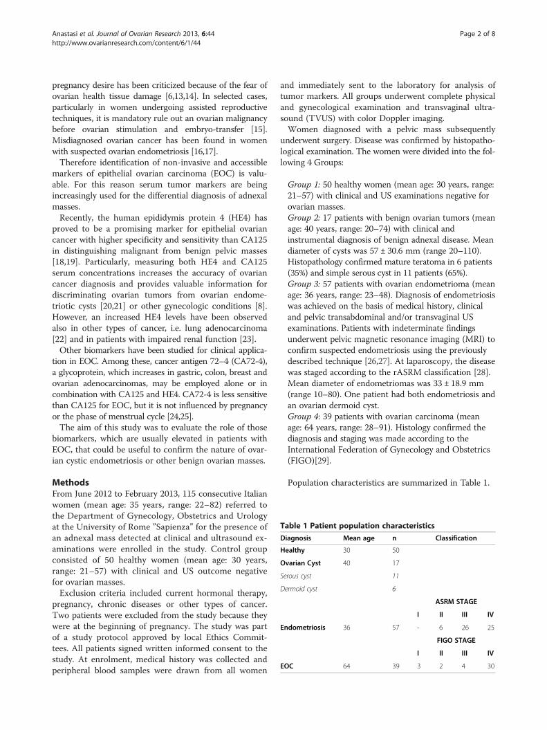
Table 1 Patient population characteristics
Diagnosis Mean age n Classification
Healthy 30 50
Ovarian Cyst 40 17
Serous cyst 11
Dermoid cyst 6
ASRM STAGE
I II III IV
Endometriosis 36 57 - 6 26 25
FIGO STAGE
I II III IV
EOC 64 39 3 2 4 30
Anastasi et al. Journal of Ovarian Research 2013, 6:44 Page 2 of 8http://www.ovarianresearch.com/content/6/1/44
pregnancy desire has been criticized because of the fear ofovarian health tissue damage [6,13,14]. In selected cases,particularly in women undergoing assisted reproductivetechniques, it is mandatory rule out an ovarian malignancybefore ovarian stimulation and embryo-transfer [15].Misdiagnosed ovarian cancer has been found in womenwith suspected ovarian endometriosis [16,17].Therefore identification of non-invasive and accessible
markers of epithelial ovarian carcinoma (EOC) is valu-able. For this reason serum tumor markers are beingincreasingly used for the differential diagnosis of adnexalmasses.Recently, the human epididymis protein 4 (HE4) has
proved to be a promising marker for epithelial ovariancancer with higher specificity and sensitivity than CA125in distinguishing malignant from benign pelvic masses[18,19]. Particularly, measuring both HE4 and CA125serum concentrations increases the accuracy of ovariancancer diagnosis and provides valuable information fordiscriminating ovarian tumors from ovarian endome-triotic cysts [20,21] or other gynecologic conditions [8].However, an increased HE4 levels have been observedalso in other types of cancer, i.e. lung adenocarcinoma[22] and in patients with impaired renal function [23].Other biomarkers have been studied for clinical applica-
tion in EOC. Among these, cancer antigen 72–4 (CA72-4),a glycoprotein, which increases in gastric, colon, breast andovarian adenocarcinomas, may be employed alone or incombination with CA125 and HE4. CA72-4 is less sensitivethan CA125 for EOC, but it is not influenced by pregnancyor the phase of menstrual cycle [24,25].The aim of this study was to evaluate the role of those
biomarkers, which are usually elevated in patients withEOC, that could be useful to confirm the nature of ovar-ian cystic endometriosis or other benign ovarian masses.
MethodsFrom June 2012 to February 2013, 115 consecutive Italianwomen (mean age: 35 years, range: 22–82) referred tothe Department of Gynecology, Obstetrics and Urologyat the University of Rome ”Sapienza” for the presence ofan adnexal mass detected at clinical and ultrasound ex-aminations were enrolled in the study. Control groupconsisted of 50 healthy women (mean age: 30 years,range: 21–57) with clinical and US outcome negativefor ovarian masses.Exclusion criteria included current hormonal therapy,
pregnancy, chronic diseases or other types of cancer.Two patients were excluded from the study because theywere at the beginning of pregnancy. The study was partof a study protocol approved by local Ethics Commit-tees. All patients signed written informed consent to thestudy. At enrolment, medical history was collected andperipheral blood samples were drawn from all women
and immediately sent to the laboratory for analysis oftumor markers. All groups underwent complete physicaland gynecological examination and transvaginal ultra-sound (TVUS) with color Doppler imaging.Women diagnosed with a pelvic mass subsequently
underwent surgery. Disease was confirmed by histopatho-logical examination. The women were divided into the fol-lowing 4 Groups:
Group 1: 50 healthy women (mean age: 30 years, range:21–57) with clinical and US examinations negative forovarian masses.Group 2: 17 patients with benign ovarian tumors (meanage: 40 years, range: 20–74) with clinical andinstrumental diagnosis of benign adnexal disease. Meandiameter of cysts was 57 ± 30.6 mm (range 20–110).Histopathology confirmed mature teratoma in 6 patients(35%) and simple serous cyst in 11 patients (65%).Group 3: 57 patients with ovarian endometrioma (meanage: 36 years, range: 23–48). Diagnosis of endometriosiswas achieved on the basis of medical history, clinicaland pelvic transabdominal and/or transvaginal USexaminations. Patients with indeterminate findingsunderwent pelvic magnetic resonance imaging (MRI) toconfirm suspected endometriosis using the previouslydescribed technique [26,27]. At laparoscopy, the diseasewas staged according to the rASRM classification [28].Mean diameter of endometriomas was 33 ± 18.9 mm(range 10–80). One patient had both endometriosis andan ovarian dermoid cyst.Group 4: 39 patients with ovarian carcinoma (meanage: 64 years, range: 28–91). Histology confirmed thediagnosis and staging was made according to theInternational Federation of Gynecology and Obstetrics(FIGO)[29].
Population characteristics are summarized in Table 1.

Anastasi et al. Journal of Ovarian Research 2013, 6:44 Page 3 of 8http://www.ovarianresearch.com/content/6/1/44
Sample preparationAll sera were acquired following a standard collectionprotocol. Briefly, samples were collected in a Red TopVacutainer, clotted 60–90 min and centrifuged for10 min at 1300 × g. The serum fractions were aliquotedand stored at −80°C until analysis.
CA125 determinationLumipulse® G1200 CA125II is an assay system for thequantitative measurement of CA125 in specimens basedon chemiluminescent enzyme immunoassay technology(CLEIA) by a two-step sandwich method (Innogenetics-Fujirebio, Belgium-Japan). This assay makes use of solidphase and ALP-labeled monoclonal antibodies (OC125and M11 respectively).CA125 in specimens specifically binds to anti-CA125
monoclonal antibody immobilized on the particlesforming antigen-antibody immunocomplexes. The parti-cles are then washed and rinsed in order to remove un-bound materials. Alkaline phosphatase (ALP)-labeledanti-CA125 monoclonal antibody specifically binds toCA125 of the immunocomplexes. After a second wash,substrate solution is added. AMPPD contained in thesubstrate solution is dephosporylated by the catalysis ofALP indirectly conjugated to the particles. A lumines-cent signal is generated by the cleavage reaction ofdephosphorylated 3-(2′-spiroadamantyl)-4-methoxy-4-(3″-phosphoryloxy)-phenyl-1,2-dioxetane (AMPPD) andreflects the amount of CA125 in the sample. Normallevels of CA125 were considered less than 35 U/mL.
HE4 determinationHE4 levels were determined using the HE4 enzymaticimmunoassay (EIA)(Fujirebio Diagnostics). The HE4EIA is a solid phase, non competitive immunoassaybased upon the direct ”sandwich” technique using twomonoclonal antibodies, 2H5 and 3D8, directed againsttwo epitopes in the C-WFDC domain of HE4. Controls
Table 2 Serum markers for each group
n Group 1
50
CA125 Mean 16.8
U/mL SD 8.6
Median(range) 14 (9–47)
HE4 Mean 48.6
pmol/L SD 13.6
Median(range) 47 (24–103)
CA72-4 Mean 2.8
U/mL SD 0.45
Median(range) 2.7 (2.1 - 4)ap-value < 0.05 vs Group 1 (healthy); bp-value < 0.05 vs Group 2 (Benign) and Group
or patient serum samples and standards were incubatedwith biotinylated anti-HE4 monoclonal antibody 2H5aliquots in streptavidin coatedmicrostrips. HE4 presentin standards or serum samples was adsorbed to thestreptavidin coatedmicrostrips by the biotinylated anti-HE4 monoclonal antibody during the incubation period.The strips were then washed and incubated with horse-radish peroxidase (HRP) labeled anti-HE4 monoclonalantibody 3D8. After washing, buffered substrate/chromo-gen reagent was added to each well and the enzyme reac-tion was allowed to proceed. During the enzyme reactiona blue color developed if the antigen was present. The in-tensity of the color was directly proportional to theamount of HE4 present in the samples. According to themanufacturer’s indications, normal values of HE4 wereconsidered less than 150 pmol/L.
CA72-4 assayCA72-4 was detected utilizing a solid phase two-siteimmunoradiometric ELSA- CA72-4 assay (Cisbio Bioassays,France). Two monoclonal antibodies were preparedagainst sterically remote antigenic sites on the TAG 72molecule: the first was coated on the ELSA solid phase,the second, radiolabeled with iodine 125, was used astracer. TAG 72 molecules present in the standards or thesamples to be tested were “sandwiched” between the twoantibodies. Following the formation of the coated anti-body/antigen/ antibody sandwich, the unbound tracer waseasily removed by a washing step. The radioactivity boundto the Elsa was proportional to the concentration of TAG72 present in the sample. Normal levels of CA72-4 wereconsidered to be less than 3.8 U/mL.
Statistical analysisWomen were stratified by disease in four groups. In eachgroup, the median, range, mean, SD for serum CA125,HE4and CA72-4 levels were determined. Mann–Whitneytest was used to assess difference in distributions of tumor
Group 2 Group 3 Group 4
17 57 39
19.9 46.1 1976.3
20.5 34 7390.4
13 (9–97) 38 (8–167)a 480 (8–46210)a,b
60.6 53.8 508.3
26.5 15.3 301.5
58 (30–125) 53 (26–98) 426 (48–850)a,b
2.7 3 39.8
0.32 0.98 45.1
2.8 (2.1 - 3.3) 2.7 (1.8 -6.2) 7 (1–112)a,b
3 (endometriosis); Group 4 (ovarian cancer).

Figure 1 Box and whisker plots representing median levels andthe interquartile range (box) of (A) CA125, (B) HE4 and (C) CA72-4for each studied group. The dashed horizontal line represents thecut-off level for each marker (CA125 = 35 U/mL; HE4 = 150 pmol/L;CA72-4 = 3.8 U/mL). The y axis is a logarithmic scale. Group 1 = Healthywomen; Group 2 = Ovarian cyst; Group 3 = Endometriosis;Group 4 = Epithelial Ovarian Cancer.
Anastasi et al. Journal of Ovarian Research 2013, 6:44 Page 4 of 8http://www.ovarianresearch.com/content/6/1/44
markers between different patient populations. Log base10–transformed whisker-box plots were generated foreach marker by disease group. The diagnostic perfor-mance of the markers was also expressed as sensitivity,specificity, positive predictive values (PPV) and negativepredictive values (NPV) using the following cut-off values:35 U/mL for CA125, 150 pmol/L for HE4, 3.8 U/mL forCA72-4. Receiver operator characteristic (ROC) curveswere constructed and the areas under the curve (AUC)with binomial exact 95% confidence intervals (95% CI)were calculated. The method described by DeLong et al.was used to calculate the difference between two AUCs[30]. For all statistical comparisons, a level of P < 0.05 wasaccepted as statistically significant. All statistical analyseswere performed using MedCalc v.12.2.1.0.
ResultsBiomarker distributionCA125, HE4 and CA72-4 serum marker levels wereevaluated in all groups (163 women). Results expressedas mean, median and ranges are shown in Table 2.All markers showed significant difference between
Group 1 and Group 2 and Group 4 (p < 0.05). HE4 andCA72-4 were significantly higher in Group 4 than in allthe other groups (p < 0.05). CA125 was significantlyhigher in Group 3 and Group 4 than in Group 1 andGroup 2 (p < 0.05). The distribution of marker levels foreach studied group is shown in Figure 1.
Tumor marker sensitivity and specificity in malignant andbenign diseaseIn Group 4, HE4 was increased in 89.7% (35/39) of cases,in this group CA72-4 was elevated in 67% (26/39) of cases,while CA125 was positive in 92% (36/39) of patients.In Group 3, CA125 was elevated in 56.1% (32/57) of
cases and a slight but not statistically significant increaseof CA72-4 was observed in 7% (4/57) of patients.HE4 correctly discriminated malignant from benign dis-
ease (Group 2 and Group 3 vs Group 4) with a sensitivityand specificity of 87% and 100%, respectively. PPV andNPV of HE4 were 100% and 96% respectively.In patients with malignancy, CA125 showed a sig-
nificantly higher sensitivity than CA72-4 (90% vs 67%,p < 0.001), but a lower specificity than CA72-4 (70% vs
Table 3 Sensitivity, specificity, PPV and NPV of controlsand malignant vs benign cases for each marker
CA125 HE4 CA72-4
Sensitivity % 90 87 67
Specificity % 70 100 96
PPV 51 100 84
NPV 95 96 89
Cut-off levels: CA125 = 35 U/mL; HE4 = 150 pmol/L; CA72-4 =3.8 U/mL.
Anastasi et al. Journal of Ovarian Research 2013, 6:44 Page 5 of 8http://www.ovarianresearch.com/content/6/1/44
96%, p < 0.001). PPV was 51% and 84%, and NPV was 95%and 89% for CA125 and CA72-4, respectively (Table 3).
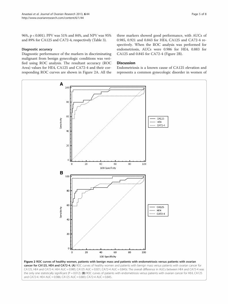
Diagnostic accuracyDiagnostic performance of the markers in discriminatingmalignant from benign gynecologic conditions was veri-fied using ROC analysis. The resultant accuracy (ROCArea) values for HE4, CA125 and CA72-4 and their cor-responding ROC curves are shown in Figure 2A. All the
Figure 2 ROC curves of healthy women, patients with benign mass acancer for CA125, HE4 and CA72-4. (A) ROC curves of healthy women aCA125, HE4 and CA72-4. HE4 AUC = 0.985; CA125 AUC = 0.921; CA72-4 AUCthe only one statistically significant (P = 0.012). (B) ROC curves of patients wand CA72-4. HE4 AUC = 0.986; CA125 AUC = 0.883; CA72-4 AUC = 0.845.
three markers showed good performance, with AUCs of0.985, 0.921 and 0.843 for HE4, CA125 and CA72-4 re-spectively. When the ROC analysis was performed forendometriosis, AUCs were 0.986 for HE4, 0.883 forCA125 and 0.845 for CA72-4 (Figure 2B).
DiscussionEndometriosis is a known cause of CA125 elevation andrepresents a common gynecologic disorder in women of
nd patients with endometriosis versus patients with ovariannd patients with benign mass versus patients with ovarian cancer for= 0.843c The overall difference in AUCs between HE4 and CA72-4 wasith endometriosis versus patients with ovarian cancer for HE4, CA125
Anastasi et al. Journal of Ovarian Research 2013, 6:44 Page 6 of 8http://www.ovarianresearch.com/content/6/1/44
reproductive age [2]. Generally the diagnosis of ovarianendometriosis is made by clinical and imaging techniqueexaminations [31] and confirmed by surgery with histo-logical examination [6]. Recently surgical treatment ofovarian endometriosis in women desiring pregnancy hasbeen criticized because of the risk of ovarian healthytissue damage [13,14]. Therefore, in selected cases withovarian endometrioma treated by medical therapy orundergoing assisted reproductive techniques (ART)without prior surgery, a correct diagnosis is mandatory.In these cases, the use of tumor markers with high sensi-tivity and specificity could help to reduce the risk, evensmall, of undetected ovarian cancer. In fact, there is arecognized association between endometriosis and clearcell, low-grade serous and endometrioid ovarian cancer[32]. So far, very little is known about the underlying fac-tors involved in the malignant progression of endometri-osis. For more than two decades CA125 has been theonly marker employed in the diagnosis of EOC, but,although overexpressed in more than 80% of ovariancancers, it lacks of specificity [33].In the present study, we investigated the role of
serum CA125, HE4 and CA72-4 in the diagnosticevaluation of ovarian endometrioma and adnexal mass.In agreement with data reported in literature [19],more than 50% of women with endometriosis expressedhigh levels of CA125, confirming the low specificity ofthis marker.It was recently observed that HE4 rarely increases in
benign gynecologic conditions suggesting its comple-mentary role to CA125 [18,19,25,34,35]. In women withendometriosis, Moore et al. observed a marked differ-ence between HE4 levels, which was increased only in3% of cases, compared to CA125, which was elevated in67% of cases [19]. Hamed et al. showed that serum HE4and CA125 concentrations were significantly higher inpatients with ovarian cancer compared with levels ob-served in patients with benign disease or healthy con-trols. In their study CA125 and HE4 had high sensitivity(90% vs 83.3%) and combining the two markers EOCwere correctly detected in 97% of cases [36]. MoreoverHE4 measurement, in healthy premenopausal women aswell as in women with endometriosis, can be carried outat any phase of the menstrual cycle, and irrespective ofhormonal therapy, extending the benefits of HE4 use inclinical practice [37,38]. However, HE4 overexpressionhas been also observed in non-oncologic conditions suchas chronic kidney disease, which represents the most im-portant known source of false-positive HE4 results [23].In addition, recent studies have reported high HE4 levelsalso in some benign gynecological conditions such asuterine fibroma and pelvic inflammatory disease [8,19].Moreover, HE4 levels in healthy women are affected byage, BMI and smoking [39].
In our study CA125 and HE4 yielded a sensitivity of90% and 87% and a specificity of 70% and 100% respect-ively in the diagnosis of epithelial ovarian cancer. HE4never increased in women with endometriosis and it wasable to correctly discriminate malignant from all benignovarian masses. Therefore we agree with previous re-ports that HE4 is the most useful marker for the differ-ential diagnosis between EOC and ovarian endometriosis[19]. However, since some benign conditions can be asso-ciated with high HE4 levels, in selected cases CA72-4 maybe useful for the differential diagnosis. Nevertheless therole of CA72-4 in the differential diagnosis between benignform malignant ovarian mass is still controversial [18].In our study a slight and not statistically significant in-
crease of CA72-4 was found in a small number of patientswith endometriosis, with the highest observed value of 6.2U/mL, which is indeed a borderline value, found only inone woman. Our data confirm the results reported byLenhard et al. who showed that CA125 but not CA72-4tends to be increased in the presence of endometriosis[24]. A multimarker approach, consisting of HE4, CA125and CA72-4, can provide a more accurate tool for a differ-ential diagnosis of patients with ovarian endometrioticcysts, other benign ovarian masses and ovarian cancer.
ConclusionsIn conclusion our results suggest that the use of serumHE4, CA125 and CA72-4 may be a valuable approachfor distinguishing patients with ovarian endometriomaor other benign adnexal masses from those with ovarianmalignancy. This approach could reduce medical costsrelated to more expensive diagnostic procedures and itmay have a reassuring effect on the patient. Furtherstudies are needed to confirm these results.
AbbreviationsUS: Ultrasound; CA125: Cancer antigen 125; EOC: Epithelial ovariancarcinoma; HE4: Human epididymis protein 4; CA72-4: Cancer antigen 72–4;TVUS: Transvaginal ultrasound; MRI: Magnetic resonance imaging;rASRM: revised American Society of Reproductive Medicine;FIGO: International Federation of Gynecology and Obstetrics;CLEIA: Chemiluminescent enzyme immunoassay; ALP: Alkaline phosphatase;AMPPD: 3-(2IGO: International Fedthoxy-4- (3(3zyme immunoassay;EIA: Immunoenzymatic assay; HRP: Horseradish peroxidase; PPV: Positivepredictive values; NPV: Negative predictive values; ROC: Receiver operatorcharacteristic; AUC: Areas under the curve; ART: Assisted reproductivetechniques.
Competing interestsAll authors declare that they have no competing interest.
Authors’ contributionsEA and MGP conceived and design the experiments. EA, MGP, RF, PS and ATperformed the experiments. EA, MGP, TG, LF and PBP analyzed the data. TGcontributed reagents/material/analysis tools. EA, MGP, RF, PS and AT wrotethe paper. All authors read and approved the final manuscript.
AcknowledgementWe are thankful Barbara Colaprisca and Valentina Viggiani for their technicalassistance.

Anastasi et al. Journal of Ovarian Research 2013, 6:44 Page 7 of 8http://www.ovarianresearch.com/content/6/1/44
Author details1Department of Molecular Medicine, “Sapienza” University of Rome,Policlinico Umberto I, Viale Regina Elena 324, Rome 00161, Italy. 2CNR-IBPM,National Research Council, Piazzale Aldo Moro 7, Rome 00185, Italy.3Department of Gynaecology, Obstetrics and Urology, ”Sapienza” Universityof Rome, Policlinico Umberto I, Viale del Policlinico 155, Rome 00161, Italy.
Received: 5 April 2013 Accepted: 18 June 2013Published: 1 July 2013
References1. Eskenazi B, Warner ML: Epidemiology of endometriosis. Obstet Gynecol Clin
North Am 1997, 24(2):235–258.2. Giudice LC: Clinical practice. Endometriosis. N Engl J Med 2010,
362(25):2389–2398.3. Porpora MG, Koninckx PR, Piazze J, Natili M, Colagrande S, Cosmi EV:
Correlation between endometriosis and pelvic pain. J Am Assoc GynecolLaparosc 1999, 6(4):429–434.
4. Porpora MG, Pallante D, Ferro A, Crisafi B, Bellati F, Benedetti Panici P: Painand ovarian endometrioma recurrence after laparoscopic treatment ofendometriosis: a long-term prospective study. Fertil Steril 2010,93(3):716–721.
5. Coccia ME, Rizzello F, Palagiano A, Scarselli G: Long-term follow-up afterlaparoscopic treatment for endometriosis: multivariate analysis ofpredictive factors for recurrence of endometriotic lesions and pain. Eur JObstet Gynecol Reprod Biol 2011, 157(1):78–83.
6. Kennedy S, Bergqvist A, Chapron C, D’Hooghe T, Dunselman G, Greb R,Hummelshoj L, Prentice A, Saridogan E: On behalf of the ESHRE specialinterest group for endometriosis endometrium guideline developmentgroup. ESHRE guideline for the diagnosis and treatment ofendometriosis. Hum Reprod 2005, 20:2698–2704.
7. Bordin L, Fiore C, Donà G, Andrisani A, Ambrosini G, Faggian D, Plebani M,Clari G, Armanini D: Evaluation of erythrocyte band 3 phosphotyrosinelevel, glutathione content, CA-125, and human epididymal secretoryprotein E4 as combined parameters in endometriosis. Fertil Steril 2010,94(5):1616–1621.
8. Escudero JM, Auge JM, Filella X, Torne A, Pahisa J, Molina R: Comparison ofserum human epididymis protein 4 with cancer antigen 125 as a tumormarker in patients with malignant and nonmalignant diseases. Clin Chem2011, 57(11):1534–1544.
9. Gupta S, Agarwal A, Sekhon L, Krajcir N, Cocuzza M, Falcone T: Serum andperitoneal abnormalities in endometriosis: potential use as diagnosticmarkers. Minerva Ginecol 2006, 58:527–551.
10. Buamah P: Benign conditions associated with raised serum CA-125concentration. J Surg Oncol 2000, 75(4):264–265.
11. McLemore MR, Aouizerat BE, Lee KA, Chen LM, Cooper B, Tozzi M,Miaskowski C: A comparison of the cyclic variation in serum levels ofCA125 across the menstrual cycle using two commercial assays. Biol ResNurs 2012, 14(3):250–256.
12. Bast RC Jr, Klug TL, St John E, Jenison E, Niloff JM, Lazarus H, Berkowitz RS,Leavitt T, Griffiths CT, Parker L, Zurawski VR Jr, Knapp RC: Aradioimmunoassay using a monoclonal antibody to monitor the courseof epithelial ovarian cancer. N Engl J Med 1983, 309(15):883–887.
13. Tsoumpou I, Kyrgiou M, Gelbaya TA, Nardo LG: The effect of surgicaltreatment for endometrioma on in vitro fertilization outcomes: asystematic review and meta-analysis. Fertil Steril 2009, 92:75–87.
14. Raffi F, Metwally M, Amer S: The impact of excision of ovarian endometriomaon ovarian reserve: a systematic review and meta-analysis. J Clin EndocrinolMetab 2012, 97(9):3146–3154.
15. Vlahos NF, Economopoulos KP, Fotiou S: Endometriosis, in vitrofertilisation and the risk of gynaecological malignancies, includingovarian and breast cancer. Best Pract Res Clin Obstet Gynaecol 2010,24:39–50.
16. Zygouris D, Leontara V, Makris GM, Chrelias C, Trakakis E, Christodoulaki C,Panagopoulos P: Endometrioid ovarian cancer arising from anendometriotic cyst in a young patient. Eur J Gynaecol Oncol 2012,33(3):324–325.
17. Vasilakaki T, Skafida E, Arkoumani E, Grammatoglou X, Firfiris N, ManoloudakiK: Borderline clear cell adenofibroma of the ovary associated withovarian endometriosis: a case report. Eur J GynaecolOncol 2012,33(2):230–232.
18. Moore RG, Brown AK, Miller MC, Skates S, Allard WJ, Verch T, SteinhoffM, Messerlian G, Di Silvestro P, Granai CO, Bast RC Jr: The use ofmultiple novel tumor biomarkers for the detection of ovariancarcinoma in patients with a pelvic mass. Gynecol Oncol 2008,108(2):402–408.
19. Moore RG, Miller MC, Steinhoff MM, Skates SJ, Lu KH, Lambert-Messerlian G,Bast RC Jr: Serum HE4 levels are less frequently elevated than CA125 inwomen with benign gynecologic disorders. Am J Obstet Gynecol 2012,206(4):351.
20. Huhtinen K, Suvitie P, Hiissa J, Junnila J, Huvila J, Kujari H, Setälä M, Härkki P,Jalkanen J, Fraser J, Mäkinen J, Auranen A, Poutanen M, Perheentupa A:Serum HE4 concentration differentiates malignant ovarian tumours fromovarian endometriotic cysts. Br J Cancer 2009, 100(8):1315–1319.
21. Nolen B, Velikokhatnaya L, Marrangoni A, De Geest K, Lomakin A, Bast RC Jr,Lokshin A: Serum biomarker panels for the discrimination of benign frommalignant cases in patients with an adnexal mass. Gynecol Oncol 2010,117(3):440–445.
22. Galgano MT, Hampton GM, Frierson HF Jr: Comprehensive analysis of HE4expression in normal and malignant human tissues. Mod Pathol 2006,19(6):847–853.
23. Nagy B Jr, Krasznai ZT, Balla H, Csobán M, Antal-Szalmás P, Hernádi Z,Kappelmayer J: Elevated human epididymis protein 4 concentrations inchronic kidney disease. Ann Clin Biochem 2012, 49(4):377–380.
24. Lenhard MS, Nehring S, Nagel D, Mayr D, Kirschenhofer A, Hertlein L, FrieseK, Stieber P, Burges A: Predictive value of CA125 and Ca72-4 in borderlineovarian tumors. Clin Chem Lab Med 2009, 47(5):537–542.
25. Granato T, Midulla C, Longo F, Colaprisca B, Frati L, Anastasi E: Role of HE4,CA72-4, and CA125 in monitoring ovarian cancer. Tumor Biol 2012,33(5):1335–1339.
26. Manganaro L, Vittori G, Vinci V, Fierro F, Tomei A, Lodise P, Sollazzo P, Sergi ME,Bernardo S, Ballesio L, Marini M, Porpora MG: Beyond laparoscopy: 3-Tmagnetic resonance imaging in the evaluation of posteriorcul-de-sacobliteration. Magn Reson Imaging 2012, 30(10):1432–1438.
27. Manganaro L, Fierro F, Tomei A, Irimia D, Lodise P, Sergi ME, Vinci V,Sollazzo P, Porpora MG, Delfini R, Vittori G, Marini M: Feasibility of 3.0Tpelvic MR imaging in the evaluation of endometriosis. Eur J Radiol 2012,81(6):1381–1387.
28. American Society for Reproductive Medicine: Revised American society forreproductive medicine classification of endometriosis: 1996. Fertil Steril1997, 67(5):817–821.
29. Heintz AP, Odicino F, Maisonneuve P, Quinn MA, Benedet JL, Creasman WT,Ngan HY, Pecorelli S, Beller U: Carcinoma of the ovary. FIGO 26th AnnualReport on the Results of Treatment in Gynecological Cancer. Int JGynaecol Obstet 2006, 95(Suppl 1):S161-92.
30. DeLong ER, DeLong DM, Clarke-Pearson DL: Comparing the areas undertwo or more correlated receiver operating characteristic curves: anonparametric approach. Biometrics 1988, 44(3):837–845.
31. Van Holsbeke C, Van Calster B, Guerriero S, Savelli L, Paladini D, Lissoni AA,Czekierdowski A, Fischerova D, Zhang J, Mestdagh G, Testa AC, Bourne T,Valentin L, Timmerman D: Endometriomas: their ultrasoundcharacteristics. Ultrasound Obstet Gynecol 2010, 35(6):730–740.
32. Pearce CL, Templeman C, Rossing MA, Lee A, Near AM, Webb PM, NagleCM, Doherty JA, Cushing-Haugen KL, Wicklund KG, et al: Associationbetween endometriosis and risk of histological subtypes of ovariancancer: a pooled analysis of case–control studies. Lancet Oncol 2012,13:385–394.
33. Bast RC Jr, Xu FJ, Yu YH, Barnhill S, Zhang Z, Mills GB: CA 125: the past andthe future. Int J Biol Markers 1998, 13(4):179–187.
34. Anastasi E, Granato T, Coppa A, Manganaro L, Giannini G, Comploj S, Frati L,Midulla C: HE4 in the differential diagnosis of a pelvic mass: a casereport. Int J Mol Sci 2011, 12(1):627–632.
35. Yamashita S, Tokuishi K, Hashimoto T, Moroga T, Kamei M, Ono K, MiyawakiM, Takeno S, Chujo M, Yamamoto S, Kawahara K: Prognostic significance ofHE4 expression in pulmonary adenocarcinoma. Tumour Biol 2011,32(2):265–271.
36. Hamed EO, Ahmed H, Sedeek OB, Mohammed AM, Abd-Alla AA, AbdelGhaffar HM: Significance of HE4 estimation in comparison with CA125 indiagnosis of ovarian cancer and assessment of treatment response.Diagn Pathol 2013, 8:11.
37. Anastasi E, Granato T, Marchei G, Viggiani V, Colaprisca B, Comploj S, RealeM, Frati L, Midulla C: Ovarian tumor marker HE4 is differently expressed
Anastasi et al. Journal of Ovarian Research 2013, 6:44 Page 8 of 8http://www.ovarianresearch.com/content/6/1/44
during the phases of the menstrual cycle in healthy young women.Tumour Biol 2010, 31(5):411.
38. Hallamaa M, Suvitie P, Huhtinen K, Matomäki J, Poutanen M, Perheentupa A:Serum HE4 concentration is not dependent on menstrual cycle orhormonal treatment among endometriosis patients and healthypremenopausal women. Gynecol Oncol 2012, 125:667–672.
39. Bolstad N, Øijordsbakken M, Nustad K, Bjerne J: Human epididymis proteinreference limits and natural variation in a Nordic reference population.Tumor Biol 2012, 33:141–148.
doi:10.1186/1757-2215-6-44Cite this article as: Anastasi et al.: The use of HE4, CA125 and CA72-4biomarkers for differential diagnosis between ovarian endometriomaand epithelial ovarian cancer. Journal of Ovarian Research 2013 6:44.
Submit your next manuscript to BioMed Centraland take full advantage of:
• Convenient online submission
• Thorough peer review
• No space constraints or color figure charges
• Immediate publication on acceptance
• Inclusion in PubMed, CAS, Scopus and Google Scholar
• Research which is freely available for redistribution
Submit your manuscript at www.biomedcentral.com/submit


