Embed Size (px)
Citation preview
RESEARCH Open Access
Intra-ovarian injection of platelet-richplasma into ovarian tissue promotedrejuvenation in the rat model of prematureovarian insufficiency and restored ovulationrate via angiogenesis modulationShahin Ahmadian1,2†, Sepideh Sheshpari3†, Mohammad Pazhang2, Alberto Miranda Bedate4, Rahim Beheshti5,Mehran Mesgari Abbasi6, Mohammad Nouri7,8, Reza Rahbarghazi7,9 and Mahdi Mahdipour7,8*
Abstract
Premature Ovarian Insufficiency (POI) is viewed as a type of infertility in which the menopausal status occurs beforethe physiological age. Several therapeutic strategies have been introduced in clinic for POI treatment, although theoutputs are not fully convincing. Platelet-rich plasma (PRP) is a unique blood product widely applied in regenerativemedicine, which is based on the releasing of the growth factors present in platelets α-granules. In the currentinvestigation, we examined the effectiveness of PRP as a therapeutic alternative for POI animals. POI in Wistar albinorats was induced by daily intraperitoneal (IP) administration of gonadotoxic chemical agent, 4-vinylcyclohexenedioxide (VCD) (160 mg/ kg) for 15 consecutive days. After POI induction, the PRP solution was directly injected intra-ovarian in two concentrations via a surgical intervention. Every two weeks post-injection, pathological changeswere monitored in the ovaries using Hematoxylin-Eosin staining method, until eight weeks. Follicle StimulatingHormone (FSH) content in serum was measured, together with the expression of the angiogenic-related transcriptsANGPT2 and KDR by real-time qPCR. Furthermore the fertility status of the treated rats was evaluated by matingtrials. Histopathological examination revealed successful POI induction via the depletion of morphologically normalfollicles in rats following VCD treatment compared to the control rats. The injection of PRP at two concentrationsreduced the number and extent of the follicular atresia and inflammatory responses (p < 0.05). The expression ofboth ANGPT2 and KDR transcripts were significantly increased in POI rats due to enhanced inflammation, whilethese values were modulated after PRP administration (p < 0.05) compared to POI rats. FSH showed a decreasedtrend in concentration eight weeks after PRP treatment, but not statistically significant (p > 0.05). Nevertheless, aclear improvement in litter counts was found in POI rats receiving PRP compared to the non-treated POI group,(Continued on next page)
© The Author(s). 2020 Open Access This article is licensed under a Creative Commons Attribution 4.0 International License,which permits use, sharing, adaptation, distribution and reproduction in any medium or format, as long as you giveappropriate credit to the original author(s) and the source, provide a link to the Creative Commons licence, and indicate ifchanges were made. The images or other third party material in this article are included in the article's Creative Commonslicence, unless indicated otherwise in a credit line to the material. If material is not included in the article's Creative Commonslicence and your intended use is not permitted by statutory regulation or exceeds the permitted use, you will need to obtainpermission directly from the copyright holder. To view a copy of this licence, visit http://creativecommons.org/licenses/by/4.0/.The Creative Commons Public Domain Dedication waiver (http://creativecommons.org/publicdomain/zero/1.0/) applies to thedata made available in this article, unless otherwise stated in a credit line to the data.
* Correspondence: [email protected]†Shahin Ahmadian and Sepideh Sheshpari contributed equally to this work.7Stem Cell Research Center, Tabriz University of Medical Sciences, Tabriz5165665811, Iran8Department of Reproductive Biology, Faculty of Advanced Medical Sciences,Tabriz University of Medical Sciences, Tabriz 5166653431, IranFull list of author information is available at the end of the article
Ahmadian et al. Reproductive Biology and Endocrinology (2020) 18:78 https://doi.org/10.1186/s12958-020-00638-4
(Continued from previous page)
being able to consider PRP as a facile, quick, accessible, safe and relatively cheap alternative therapeutic strategy torevert POI-related pathologies.
Keywords: Platelet-rich plasma, Ovarian rejuvenation, Premature ovarian insufficiency, Angiogenesis, Fertility
IntroductionWomen’s reproduction disability, mentioned also as infertil-ity, is defined as the failure achieving pregnancy in a durationof 1 year with unprotected sexual intercourses [1]. Both maleand female patients almost equally suffer from infertilitycomplications [2], which are caused by different factors, suchas genetic background, obstructive disorders, and environ-mental elements. It has also been well documented that che-micals or anti-cancer gonadotoxic drugs are major inducersof sub-fertility in females before the physiological age, knownas premature ovarian insufficiency (POI). In this regard, mostPOI patients derived to the fertility clinics have previously re-ceived chemotherapy [3, 4]. Moreover, several complicationsare associated with POI such as osteoporosis, depression,cardiovascular diseases, etc. [3, 5, 6]. The routine treatmentstrategies are in general gonadotropin-releasing hormoneagonist (GnRHa), hormonal replacement therapy (HRT), andassisted reproductive technologies (ART). These procedures,however, are not providing satisfactory recovery to the pa-tients [7, 8]. For example, HRT-based treatments have beenlinked to undesired secondary effects, like breast and ovariancancers, venous thromboembolism [9, 10], and incapabilityof reforming uterine volume and/or endometrial thickness[3, 11]. In the case of ART, which considered as the primarystrategy applied for patients dealing with POI [12], severalunwanted effects were usually observed, such as inconsistentclinical outcomes, invasive surgical procedures, or prolongedovulation induction protocols [7, 13]. In consequence, a newgeneration of therapeutics based on certain cell and cellproducts are being developed to palliate these deficiencies.Platelet-rich plasma (PRP) is a remarkable example. It
is composed by a high density platelet concentrate, andhas been typically used for the treatment of variousproblems, such as surgeries or diabetic wounds, osteo-arthritis, skin, and soft tissue damages [14–18]. Plateletsderived from megakaryocytes in the bone marrow areanucleated blood cells with a life span of 7 to 10 days inhumans, and a bit shorter in mice [19]. They are easy toisolate and have an active role in wound healing and tis-sue repair due to their α-granules content [20, 21]. Theα-granules contain more than 800 different proteins thathave a paracrine effect on surrounding cells, especiallyon local mesenchymal stem cells (MSCs), promoting arapid tissue regeneration [21].Angiogenesis is a sophisticated biological phenomenon
participating in physiological and pathological condi-tions, refers to the formation of de novo vascular units
from the pre-existing vascular network. Importantly forPOI reversal, the increase of vascularization in ovaries toprovide an increase of blood nourishment, contributingto the acceleration of healing procedure and scavengingtissue debris [22]. The critical role of different ligands,notably angiopoietins (e.g. ANGPT1 or ANGPT2), andreceptor tyrosine kinases (e.g. VEGFR-2 or KDR) hasbeen well documented in relation to angiogenesis, andtherefore we hypothesize that the measure of their ex-pression after PRP administration in POI patients pro-vides a good picture of the regeneration status [23].The current investigation aimed to assess the effective-
ness of PRP on ovarian rejuvenation in an experimentally4-vinylcyclohexene dioxide (VCD) induced POI model inrats [24]. VCD, a side product of 4-Vinylcyclohexene(VCH), is usually employed as an adjuvant in epoxy resins;however it has been shown as well to selectively destructboth primordial and primary follicles [24–26], similarly towhat occurs in POI. Therefore, after POI induction in ratswith VCD, PRP solution was injected at two different con-centrations into ovarian tissue. Subsequently, fertilitystatus, follicular developments, and angiogenesis-relatedtranscript expressions of ANGPT2 and KDR were assessedusing histological examination and transcriptomic assays.This study could help in addressing therapeutic alterna-tives in the restoration of ovarian tissue in POI patients.
Materials and methodsAnimalsEighty-six female Wistar albino rats aged between six toseven weeks were obtained from Med-Zist Co. (Tehran)and housed in a standard environment set for thetemperature of 22 ± 2 °C and twelve hours of dark/lightcycling with unlimited access to food and water. Ratswere housed in polyester cages for 1 week for acclima-tion. All the experimental protocols were confirmed bythe local committee of the ethics at Tabriz University ofMedical Sciences (IR.TBZMED.REC.1396.876).
Development of premature ovarian insufficiency (POI)model in ratsAfter a week of acclimation, rats were divided into twomajor groups of VCD and the control. The VCD group(n = 63) was intraperitoneally (IP) injected with 160mg/kg VCD (Catalog number 94956; Sigma Aldrich) usingan appropriate solvent for 15 consecutive days [27]. Therats in the control group (n = 18) received similar
Ahmadian et al. Reproductive Biology and Endocrinology (2020) 18:78 Page 2 of 13
normal saline volume. After 15 days, three rats fromeach group were randomly sacrificed and serum andovarian tissue were sampled to confirm the developmentof POI.
PRP enrichment protocolFor this purpose, PRP kits (Catalog number 1019;Rooyagene Fertilize-Lympho) were used according tothe instructions provided by the company. This kit wasdesignated for isolation of PRP in different enrichmentlevels. In short, five healthy female rats (9–10 weeks old)were selected for blood sampling. To this end, rats weredeeply anesthetized, and blood harvested directly fromthe heart using a syringe. The mixture of blood and anti-coagulant were centrifuged at 2100 RPM for 10 mins at22 °C. After supernatant transfer into the new tubes, thesecond centrifugation was performed at 4000 RPM for 6mins at 22 °C. Next, the supernatant was discarded, andPRP retained. The total number of platelets in bloodsamples before enrichment was 3.3 × 10⁵/μL and, follow-ing enrichment, both low and high concentrated plateletdensities raised to 8.5 × 10⁵/μL (PRP-a: 3-fold increase;3X) and 21.6 × 10⁵/μL (PRP-b: 7-fold increase; 7X), re-spectively. PRPs were freshly injected into the ovaries.
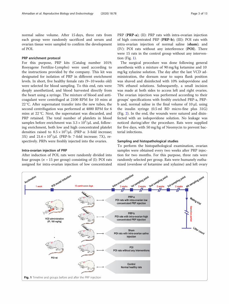
Intra-ovarian injection of PRPAfter induction of POI, rats were randomly divided intofour groups (n = 15 per group) consisting of (I): POI ratsassigned for intra-ovarian injection of low concentrated
PRP (PRP-a); (II): PRP rats with intra-ovarian injectionof high concentrated PRP (PRP-b); (III): POI rats withintra-ovarian injection of normal saline (sham); and(IV): POI rats without any interference (POI). Therewere 15 rats in the control group without any interven-tion (Fig. 1).The surgical procedure was done following general
anesthesia with a mixture of 90 mg/kg ketamine and 10mg/kg xylazine solution. The day after the last VCD ad-ministration, the dorsum near to supra flank positionwas shaved and disinfected with 10% iodopovidone and70% ethanol solutions. Subsequently, a small incisionwas made at both sides to access left and right ovaries.The ovarian injection was performed according to theirgroups’ specifications with freshly enriched PRP-a, PRP-b and, normal saline in the final volume of 10 μL usingthe insulin syringe (0.5 ml BD micro-fine plus 31G)(Fig. 2). In the end, the wounds were sutured and disin-fected with an iodopovidone solution. No leakage wasnoticed during/after the procedure. Rats were suppliedfor five days, with 50mg/kg of Neomycin to prevent bac-terial infections.
Sampling and histopathological studiesTo perform the histopathological examination, ovariansamples were obtained every two weeks after PRP injec-tion for two months. For this purpose, three rats wererandomly selected per group. Rats were humanely eutha-nized (overdose of ketamine and xylazine) and left ovary
Fig. 1 Timeline and groups before and after the PRP injection
Ahmadian et al. Reproductive Biology and Endocrinology (2020) 18:78 Page 3 of 13
was removed, rinsed in phosphate-buffered saline (PBS)solution, and fixed in 10% formalin (Merck). Right ovar-ies were sampled for gene expression evaluations andstored in liquid nitrogen until use.For histopathological examination, the specimens were
embedded in paraffin, and three consecutive sections of5 μm thickness prepared using a microtome instrument(Leica). Then, the sections were stained with hematoxylinand eosin (H&E) staining solution. Any pathological re-sponse and structural changes were monitored in the ovariansections. The numbers of primary, secondary, and antral fol-licles were recorded and compared with the control rats.
RNA isolation, synthesis of cDNA, and quantitative real-time PCRRNA extraction was performed using a Total RNA isola-tion Kit (YT9080, Yekta Tajhiz Azma; Tehran, Iran) ac-cording to the manufacture’s protocol. Extracted RNAswere reverse-transcribed by a cDNA synthesis kit(YT4500, Yekta Tajhiz Azma). Primer sets for ANGPT2and KDR transcripts (Table 1) were designed usingNCBI online program (www.ncbi.nlm.nih.gov/tools/pri-mer-blast/). Quantitative real-time PCR (qRT-PCR) re-actions were carried out using synthesized cDNAsamples and SYBR Green 2X (YT2551, Yekta Tajhiz
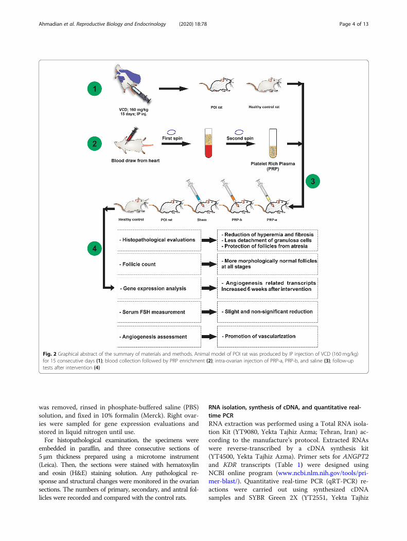
Fig. 2 Graphical abstract of the summary of materials and methods. Animal model of POI rat was produced by IP injection of VCD (160 mg/kg)for 15 consecutive days (1); blood collection followed by PRP enrichment (2); intra-ovarian injection of PRP-a, PRP-b, and saline (3); follow-uptests after intervention (4)
Ahmadian et al. Reproductive Biology and Endocrinology (2020) 18:78 Page 4 of 13
Azma) in the “Roche LightCycler 96” instrument. Spe-cific annealing temperatures were identified using gradi-ent RT-PCR. PCR reactions were run on duplicates inthree steps, basically consisting of denaturation, anneal-ing and extension all for 10 s at 95, 60, and 72 °C, re-spectively over 40 cycles. The specificity of the PCRreactions was assessed by analyzing the melting curves.
Examination of vascular density usingimmunohistochemistry stainingTo assess the angiogenic potential of PRP in the ovariantissue post-VCD injection, we performed IHC stainingof anti α-SMA (BIOCARE, CM 001 A) antibody. To thisend, paraffin-embedded sections were used according tothe previously published protocol [28]. In this study, wecounted the number of α-SMA+ small arteries in 12 ser-ial high-power field using light microscopy.
Measuring serum levels of FSH using ELISATo evaluate the possible correlations and feedback be-tween serum levels of FSH and POI conditions beforeand after PRP treatment, we measured systemic FSHlevels using an appropriate ELISA kit. For this purpose,blood samples were collected eight weeks after PRP in-jection. As mentioned above, blood samples wereattained directly from the heart of the animals after adeep anesthetization using a mixture of 90 mg/kg keta-mine and 10mg/kg xylazine solution. Subsequently,serum samples were collected after centrifugation at400×g for 20 min. All serum samples were stored at −80 °C until use. ELISA was performed using a commer-cial kit (425–300 FSH AccuBind, CA, USA) according tothe standard protocol provided by the company. Thisassay was performed in triplicate.
Mating trialTo examine the fertility status of the rats, threeremaining rats in all the groups were caged with fertilityproved male rats in a ratio of 1:2 for five days. After-ward, every female rat was caged individually until theparturition, and the number of healthy litters per birthwas registered.
Statistical analysisAll the acquired results were provided in mean ± SD,and evaluated by GraphPad Prism 8. Data for each time-point after PRP injection were analyzed using One-WayANOVA. To identify the significant difference betweenthe groups, a post-hoc test (Fisher’s least significant dif-ference, LSD) was applied. Statistical significance was setto p < 0.05.
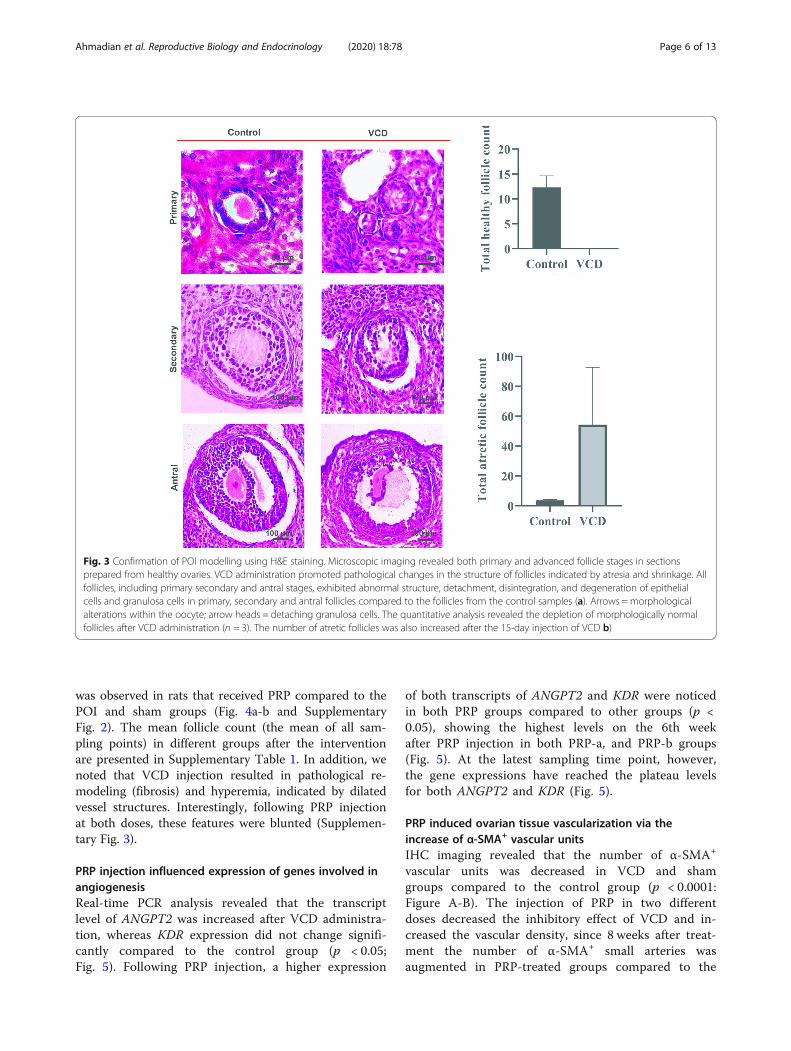
ResultsRat model of POI was established after VCDadministrationFollowing VCD administration for POI induction, histo-pathological evaluations revealed detachment of granulosacells from oocytes, and general follicular atresia in allstages of follicular development including primary, sec-ondary, and antral follicles (Fig. 3a). No morphologicallynormal follicles were noticed in the VCD rats in all stagesof follicular development (average follicle number: Con-trol = 12.33 ± 2.309; VCD = 0 ± 0) (Fig. 3b and Supplemen-tary Fig. 1A). Accordingly, a larger number of atreticfollicles were observed in the VCD group compared to thecontrol rats (average follicle number: Control = 3.67 ±0.58; VCD = 54.33 ± 38.40) (Fig. 3b and SupplementaryFigs. 1B). These data confirmed that VCD can inducepathological changes in the ovarian stroma which leads tofollicular atresia and depletion.
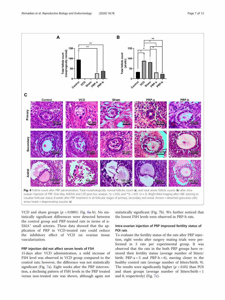
Improved ovarian function was observed after PRPinjectionHistopathological studies demonstrated that rats whichreceived PRP treatments in both concentrations exhib-ited general improvement in follicular quality, and statis-tically significant increases in total morphologicallynormal follicular counts when compared to the POI rats(Control = 94.33 ± 44.46; VCD = 0 ± 0; sham = 0 ± 0;PRP-a = 26.67 ± 2.517; PRP-b = 37.67 ± 5.132) (Fig. 4a-c).In the POI and sham groups however, no morphologic-ally normal follicles of any stages were noticed whereas,a significantly higher ratio (p < 0.05) of total atretic folli-cles were detected in these groups (Control = 33.00 ±19.00; VCD = 88.00 ± 34.77; sham = 81.33 ± 27.02; PRP-a = 30.33 ± 14.36; PRP-b = 23.00 ± 7.00). Furthermore, asignificant (p < 0.05) decrease in the atretic follicle count
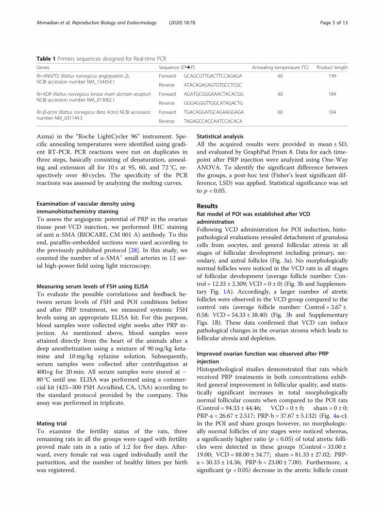
Table 1 Primers sequences designed for Real-time PCR
Genes Sequence (5′➔3′) Annealing temperature (°C) Product length
Rn-ANGPT2 (Rattus norvegicus angiopoietin 2),NCBI accession number NM_134454.1
Forward GCAGCGTTGACTTCCAGAGA 60 199
Reverse ATACAGAGAGTGTGCCTCGC
Rn-KDR (Rattus norvegicus kinase insert domain receptor)NCBI accession number NM_013062.1
Forward AGATGCGGGAAACTACACGG 60 184
Reverse GGGAGGGTTGGCATAGACTG
Rn-β-actin (Rattus norvegicus Beta Actin) NCBI accessionnumber NM_031144.3
Forward TGACAGGATGCAGAAGGAGA 60 104
Reverse TAGAGCCACCAATCCACACA
Ahmadian et al. Reproductive Biology and Endocrinology (2020) 18:78 Page 5 of 13
was observed in rats that received PRP compared to thePOI and sham groups (Fig. 4a-b and SupplementaryFig. 2). The mean follicle count (the mean of all sam-pling points) in different groups after the interventionare presented in Supplementary Table 1. In addition, wenoted that VCD injection resulted in pathological re-modeling (fibrosis) and hyperemia, indicated by dilatedvessel structures. Interestingly, following PRP injectionat both doses, these features were blunted (Supplemen-tary Fig. 3).
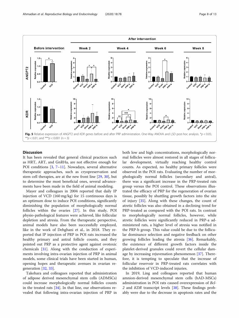
PRP injection influenced expression of genes involved inangiogenesisReal-time PCR analysis revealed that the transcriptlevel of ANGPT2 was increased after VCD administra-tion, whereas KDR expression did not change signifi-cantly compared to the control group (p < 0.05;Fig. 5). Following PRP injection, a higher expression
of both transcripts of ANGPT2 and KDR were noticedin both PRP groups compared to other groups (p <0.05), showing the highest levels on the 6th weekafter PRP injection in both PRP-a, and PRP-b groups(Fig. 5). At the latest sampling time point, however,the gene expressions have reached the plateau levelsfor both ANGPT2 and KDR (Fig. 5).
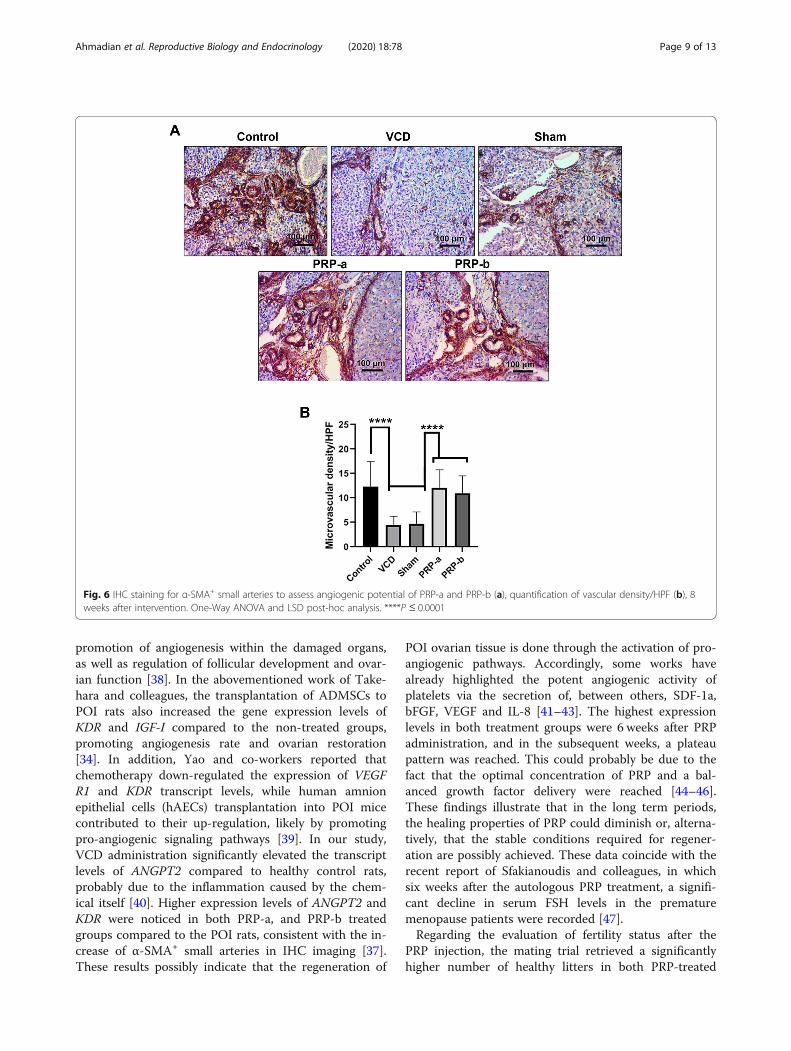
PRP induced ovarian tissue vascularization via theincrease of α-SMA+ vascular unitsIHC imaging revealed that the number of α-SMA+
vascular units was decreased in VCD and shamgroups compared to the control group (p < 0.0001:Figure A-B). The injection of PRP in two differentdoses decreased the inhibitory effect of VCD and in-creased the vascular density, since 8 weeks after treat-ment the number of α-SMA+ small arteries wasaugmented in PRP-treated groups compared to the
Fig. 3 Confirmation of POI modelling using H&E staining. Microscopic imaging revealed both primary and advanced follicle stages in sectionsprepared from healthy ovaries. VCD administration promoted pathological changes in the structure of follicles indicated by atresia and shrinkage. Allfollicles, including primary secondary and antral stages, exhibited abnormal structure, detachment, disintegration, and degeneration of epithelialcells and granulosa cells in primary, secondary and antral follicles compared to the follicles from the control samples (a). Arrows =morphologicalalterations within the oocyte; arrow heads = detaching granulosa cells. The quantitative analysis revealed the depletion of morphologically normalfollicles after VCD administration (n = 3). The number of atretic follicles was also increased after the 15-day injection of VCD b)
Ahmadian et al. Reproductive Biology and Endocrinology (2020) 18:78 Page 6 of 13
VCD and sham groups (p < 0.0001: Fig. 6a-b). No sta-tistically significant differences were detected betweenthe control group and PRP-treated rats in terms of α-SMA+ small arteries. These data showed that the ap-plication of PRP in VCD-treated rats could reducethe inhibitory effect of VCD on ovarian tissuevascularization.
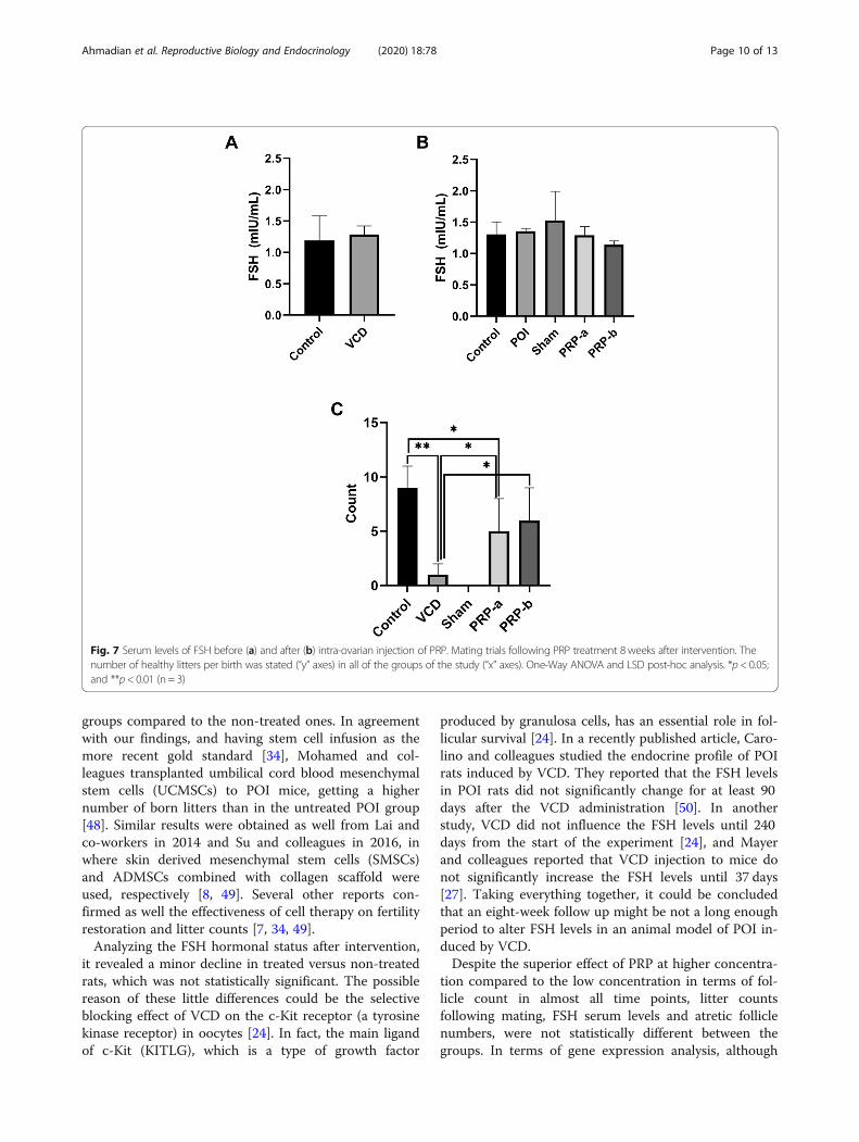
PRP injection did not affect serum levels of FSH15 days after VCD administration, a mild increase ofFSH level was observed in VCD group compared to thecontrol rats; however, the difference was not statisticallysignificant (Fig. 7a). Eight weeks after the PRP interven-tion, a declining pattern of FSH levels in the PRP treatedversus non-treated rats was shown, although again not
statistically significant (Fig. 7b). We further noticed thatthe lowest FSH levels were observed in PRP-b rats.
Intra-ovarian injection of PRP improved fertility status ofPOI ratsTo evaluate the fertility status of the rats after PRP injec-tion, eight weeks after surgery mating trials were per-formed in 3 rats per experimental group. It wasobserved that the rats in the both PRP groups have re-stored their fertility status (average number of litters/birth: PRP-a = 5 and PRP-b = 6), moving closer to thehealthy control rats (average number of litters/birth: 9).The results were significantly higher (p < 0.05) than POIand sham groups (average number of litters/birth = 1and 0, respectively) (Fig. 7c).
Fig. 4 Follicle count after PRP administration. Total morphologically normal follicles count (a), and total atretic follicle counts (b) after intra-ovarian injection of PRP. One-Way ANOVA and LSD post-hoc analysis. *p < 0.05; and **p < 0.01 (n = 3). Bright-field imaging after H&E staining tovisualize follicular status; 8 weeks after PRP treatment in all follicular stages of primary, secondary and antral. Arrows = detached granulosa cells;arrow heads = degenerating oocytes (c)
Ahmadian et al. Reproductive Biology and Endocrinology (2020) 18:78 Page 7 of 13
DiscussionIt has been revealed that general clinical practices suchas HRT, ART, and GnRHa, are not effective enough forPOI conditions [3, 7–11]. Nowadays, several alternativetherapeutic approaches, such as cryopreservation andstem cell therapies, are at the new front line [29, 30], butto determine the most beneficial ones, several advance-ments have been made in the field of animal modeling.Mayer and colleagues in 2004 reported that daily IP
injection of VCD (160mg/kg) for 15 continuous days isan optimum dose to induce POI conditions, significantlydiminishing the population of morphologically normalfollicles within the ovaries [27]. In this study, POIphysio-pathological features were achieved, like folliculardepletion and atresia. From the therapeutic perspective,animal models have also been successfully employed,like in the work of Dehghani et al., in 2018. They re-ported that IP injection of PRP in POI rats increased thehealthy primary and antral follicle counts, and theypointed out PRP as a protective agent against ovotoxicchemicals [31]. Along with the conduction of experi-ments involving intra-ovarian injection of PRP in animalmodels, some clinical trials have been started in human,opening hopes and therapeutic avenues in ovarian re-generation [32, 33].Takehara and colleagues reported that administration
of adipose derived mesenchymal stem cells (ADMSCs)could increase morphologically normal follicles countsin the treated rats [34]. In that line, our observations re-vealed that following intra-ovarian injection of PRP in
both low and high concentrations, morphologically nor-mal follicles were almost restored in all stages of follicu-lar development, virtually reaching healthy controlcounts. As expected, no healthy primary follicles wereobserved in the POI rats. Evaluating the number of mor-phologically normal follicles (secondary and antral),there was a significant increase in the PRP-treated ratsgroup versus the POI control. These observations illus-trated the efficacy of PRP for the regeneration of ovariantissue, possibly by shuttling growth factors into the siteof injury [35]. Along with these changes, the count ofatretic follicles was also obtained in a declining trend forPRP-treated as compared with the POI rats. In contrastto morphologically normal follicles, however, whileatretic follicles were significantly reduced in PRP-a ad-ministered rats, a higher level of atresia was notified inthe PRP-b group. This value could be due to the follicu-lar dominance selection and negative feedback on othergrowing follicles leading the atresia [36]. Remarkably,the existence of different growth factors inside theplatelet-derived granules could revert the cellular dam-age by increasing rejuvenation phenomenon [37]. There-fore, it is tempting to speculate that the increase offollicular reservoir in PRP-treated rats correlates withthe inhibition of VCD-induced injuries.In 2019, Ling and colleagues reported that human
amnion-derived mesenchymal stem cells (hAD-MSCs)administration in POI rats caused overexpression of Bcl-2 and KDR transcript levels [38]. These findings prob-ably were due to the decrease in apoptosis rates and the
Fig. 5 Relative expression of ANGPT2 and KDR genes before and after PRP administration. One-Way ANOVA and LSD post-hoc analysis. *p < 0.05;**p < 0.01; and ***p < 0.001 (n = 3)
Ahmadian et al. Reproductive Biology and Endocrinology (2020) 18:78 Page 8 of 13
promotion of angiogenesis within the damaged organs,as well as regulation of follicular development and ovar-ian function [38]. In the abovementioned work of Take-hara and colleagues, the transplantation of ADMSCs toPOI rats also increased the gene expression levels ofKDR and IGF-I compared to the non-treated groups,promoting angiogenesis rate and ovarian restoration[34]. In addition, Yao and co-workers reported thatchemotherapy down-regulated the expression of VEGFR1 and KDR transcript levels, while human amnionepithelial cells (hAECs) transplantation into POI micecontributed to their up-regulation, likely by promotingpro-angiogenic signaling pathways [39]. In our study,VCD administration significantly elevated the transcriptlevels of ANGPT2 compared to healthy control rats,probably due to the inflammation caused by the chem-ical itself [40]. Higher expression levels of ANGPT2 andKDR were noticed in both PRP-a, and PRP-b treatedgroups compared to the POI rats, consistent with the in-crease of α-SMA+ small arteries in IHC imaging [37].These results possibly indicate that the regeneration of
POI ovarian tissue is done through the activation of pro-angiogenic pathways. Accordingly, some works havealready highlighted the potent angiogenic activity ofplatelets via the secretion of, between others, SDF-1a,bFGF, VEGF and IL-8 [41–43]. The highest expressionlevels in both treatment groups were 6 weeks after PRPadministration, and in the subsequent weeks, a plateaupattern was reached. This could probably be due to thefact that the optimal concentration of PRP and a bal-anced growth factor delivery were reached [44–46].These findings illustrate that in the long term periods,the healing properties of PRP could diminish or, alterna-tively, that the stable conditions required for regener-ation are possibly achieved. These data coincide with therecent report of Sfakianoudis and colleagues, in whichsix weeks after the autologous PRP treatment, a signifi-cant decline in serum FSH levels in the prematuremenopause patients were recorded [47].Regarding the evaluation of fertility status after the
PRP injection, the mating trial retrieved a significantlyhigher number of healthy litters in both PRP-treated
Fig. 6 IHC staining for α-SMA+ small arteries to assess angiogenic potential of PRP-a and PRP-b (a), quantification of vascular density/HPF (b), 8weeks after intervention. One-Way ANOVA and LSD post-hoc analysis. ****P ≤ 0.0001
Ahmadian et al. Reproductive Biology and Endocrinology (2020) 18:78 Page 9 of 13
groups compared to the non-treated ones. In agreementwith our findings, and having stem cell infusion as themore recent gold standard [34], Mohamed and col-leagues transplanted umbilical cord blood mesenchymalstem cells (UCMSCs) to POI mice, getting a highernumber of born litters than in the untreated POI group[48]. Similar results were obtained as well from Lai andco-workers in 2014 and Su and colleagues in 2016, inwhere skin derived mesenchymal stem cells (SMSCs)and ADMSCs combined with collagen scaffold wereused, respectively [8, 49]. Several other reports con-firmed as well the effectiveness of cell therapy on fertilityrestoration and litter counts [7, 34, 49].Analyzing the FSH hormonal status after intervention,
it revealed a minor decline in treated versus non-treatedrats, which was not statistically significant. The possiblereason of these little differences could be the selectiveblocking effect of VCD on the c-Kit receptor (a tyrosinekinase receptor) in oocytes [24]. In fact, the main ligandof c-Kit (KITLG), which is a type of growth factor
produced by granulosa cells, has an essential role in fol-licular survival [24]. In a recently published article, Caro-lino and colleagues studied the endocrine profile of POIrats induced by VCD. They reported that the FSH levelsin POI rats did not significantly change for at least 90days after the VCD administration [50]. In anotherstudy, VCD did not influence the FSH levels until 240days from the start of the experiment [24], and Mayerand colleagues reported that VCD injection to mice donot significantly increase the FSH levels until 37 days[27]. Taking everything together, it could be concludedthat an eight-week follow up might be not a long enoughperiod to alter FSH levels in an animal model of POI in-duced by VCD.Despite the superior effect of PRP at higher concentra-
tion compared to the low concentration in terms of fol-licle count in almost all time points, litter countsfollowing mating, FSH serum levels and atretic folliclenumbers, were not statistically different between thegroups. In terms of gene expression analysis, although
Fig. 7 Serum levels of FSH before (a) and after (b) intra-ovarian injection of PRP. Mating trials following PRP treatment 8 weeks after intervention. Thenumber of healthy litters per birth was stated (“y” axes) in all of the groups of the study (“x” axes). One-Way ANOVA and LSD post-hoc analysis. *p < 0.05;and **p < 0.01 (n = 3)
Ahmadian et al. Reproductive Biology and Endocrinology (2020) 18:78 Page 10 of 13
not statistically significant, PRP-a treated rats exhibitedbetter results, probably due to the negative feedbackfrom a high concentration of platelets in the PRP-btreated subjects. These findings highlight the critical roleof PRP concentration in the modulation of angiogenesissignaling pathway within the injured ovaries.Nowadays, several clinical centers have initiated PRP
therapies for various reproductive-related complications,including POI and poor responding patients. As a clearadvantage, the PRP-therapy can easily be used as co-adjuvant in patients undergoing chemotherapy, whichare already being treated with other fertility procedures,such as ovarian tissue preservation. Additionally, PRPtherapy possesses some interesting improvements thatsurpasses the benefits of other current cell sources fortransplants. For instance, derived from its autologousorigin, PRP administration does not induce auto-immune response after transplant, a highly prevalentand detrimental secondary effect of the actual therapies.Another example could be that, due to the existence ofnumerous granules, the introduction of PRP provides ahigh concentration of growth factors and regenerativemodulators directly into the injured sites [18]. Conse-quently, it is noteworthy to highlight this cell source as apowerful healing product, which can potentially be usedin a wide range of pathological processes like, betweenothers, the POI.Talking about suitable clinical settings to administrate
PRP, transvaginal ultrasound- guided injection is a goodcandidate, since it is a nonsurgical technique that allowsthe clinicians to inject biological products like cells/PRPdirectly into the ovarian tissue. This technique is per-formed under sedation and is basically the same methodused for an egg aspiration/retrieval during IVF/ICSI op-eration [32, 51]. As described above, stem cell trans-plantation is another putative therapeutic option for POIpatients and potentially more powerful in combinationwith PRP; however, stem cell therapy is sometimes asso-ciated with tumorigenesis, making this approach less safethan others [52]. Therefore, PRP treatment could belisted as one of the least invasive procedures, eventhough, all the possible complications should be evalu-ated carefully in the future works.
ConclusionWe have demonstrated that PRP can partially restorethe function of POI ovaries. It can protect morphologic-ally normal follicles from degeneration and atresiacaused by ovotoxic chemicals and stimulate angiogen-esis. Moreover, PRP showed a good capacity for tissuerejuvenation, inflammation regulation, minimizing thehistopathological damages, and promoting the littercounts. Importantly, the PRP concentration has an im-portant effect on the therapeutic outcome of PRP
administration. If excessive, PRP will reduce its efficacydue to possibly a negative feedback generated by itscomponents. It is therefore needed to find an optimizeddose for PRP in any specific application to gain the bestoutcomes. Besides, PRP therapy is relatively cheap, safe,and does not require complicated procedures for its ad-ministration thus, overall, PRP could be considered as aputative alternative strategy for a variety of complica-tions linked with female infertility, including POI.
Supplementary informationSupplementary information accompanies this paper at https://doi.org/10.1186/s12958-020-00638-4.
Additional file 1: Supplementary Figure 1. Separate follicle countsbefore intra-ovarian RPR injection (N = 3), morphologically normal follicles(A), and atretic follicles (B).
Additional file 2: Supplementary Figure 2. Detailed morphologicallynormal and atretic primary, secondary and antral follicle count; 2, 4, 6 and8 weeks after intra-ovarian PRP injection. One-Way ANOVA and LSD post-hoc analysis. *p < 0.05; **p < 0.01; and ***p < 0.001 (n = 3).
Additional file 3: Supplementary Figure 3. Bright-field imaging afterH&E staining to visualize hyperemia and vessel dilation (A), and fibroticchanges (B), following the administration of PRP in VCD-treated rats on8th week.
Additional file 4: Supplementary Table 1. Mean follicle count indifferent groups after the intervention (mean ± SD).
AcknowledgmentsThe authors would like to thank the Stem Cell Research Center, andAdvanced Drug Research Center, Tabriz University of Medical Sciences, forsupporting this work.
Authors’ contributionsS.A., S.S., and M.P., performed the experiments and wrote the initial draft ofthe manuscript. R.B. and M.M.A., participated in the surgical procedures.A.M.B., M.N., and R.R. reviewed and revised the initial draft of the manuscript.M.M. designed and conceptualized the project and the manuscript. Theauthors read and approved the final manuscript.
FundingThis study was supported by a grant from Stem Cell Research Center fromTabriz university of medical sciences (grant number: 58881).
Availability of data and materialsAll data generated or analyzed during this study are included in thispublished article and its supplementary information files.
Ethics approval and consent to participateAll the experimental protocols were confirmed by the local committee ofthe ethics at Tabriz University of Medical Sciences (IR.TBZMED.REC.1396.876).
Consent for publicationNot applicable.
Competing interestsThe authors declare that they have no competing interests.
Author details1Women’s Reproductive Health Research Center, Tabriz University of MedicalSciences, Tabriz 5138663134, Iran. 2Department of Biology, Faculty of BasicSciences, Azarbaijan Shahid Madani University, Tabriz 537517169, Iran.3Department of Midwifery, Faculty of Nursing and Midwifery, TabrizUniversity of Medical Sciences, Tabriz 5138947977, Iran. 4Laboratory forTranslational Immunology (LTI), Universitair Medisch Centrum Utrecht,(UMCU), UtrechtHeidelberglaan 100, 3584, CX, The Netherlands. 5Department
Ahmadian et al. Reproductive Biology and Endocrinology (2020) 18:78 Page 11 of 13
of Veterinary Science, Islamic Azad University Shabestar Branch, Shabestar5381637181, Iran. 6Drug Applied Research Center, Tabriz University ofMedical Sciences, Tabriz, Iran. 7Stem Cell Research Center, Tabriz University ofMedical Sciences, Tabriz 5165665811, Iran. 8Department of ReproductiveBiology, Faculty of Advanced Medical Sciences, Tabriz University of MedicalSciences, Tabriz 5166653431, Iran. 9Department of Applied Cell Sciences,Faculty of Advanced Medical Sciences, Tabriz University of Medical Sciences,Tabriz 5166653431, Iran.
Received: 18 March 2020 Accepted: 29 July 2020
References1. Olooto WE, Amballi AA, Banjo TA. A review of female infertility; important
etiological factors and management. J Microbiol Biotech Res. 2012;2:379–85.2. Anwar S, Anwar A. Infertility: a review on causes, treatment and
management. Women’s Health Gynecol. 2016;5:2.3. Bao R, Xu P, Wang Y, Wang J, Xiao L, Li G, et al. Bone marrow derived
mesenchymal stem cells transplantation rescues premature ovarianinsufficiency induced by chemotherapy. Gynecol Endocrinol. 2018;34:320–6.
4. Blumenfeld Z, Shapiro D, Shteinberg M, Avivi I, Nahir M. Preservation offertility and ovarian function and minimizing gonadotoxicity in youngwomen with systemic lupus erythematosus treated by chemotherapy.Lupus. 2000;9:401–5.
5. Li J, Mao Q, He J, She H, Zhang Z, Yin C. Human umbilical cordmesenchymal stem cells improve the reserve function of perimenopausalovary via a paracrine mechanism. Stem Cell Res Ther. 2017;8:55.
6. Liu J, Zhang H, Zhang Y, Li N, Wen Y, Cao F, et al. Homing and restorativeeffects of bone marrow-derived mesenchymal stem cells on cisplatininjured ovaries in rats. Mol Cells. 2014;37:865–72.
7. Zhu SF, Hu HB, Xu HY, Fu XF, Peng DX, Su WY, et al. Human umbilical cordmesenchymal stem cell transplantation restores damaged ovaries. J Cell MolMed. 2015;19:2108–17.
8. Su J, Ding L, Cheng J, Yang J, Li X, Yan G, et al. Transplantation of adipose-derived stem cells combined with collagen scaffolds restores ovarianfunction in a rat model of premature ovarian insufficiency. Hum Reprod.2016;31:1075–86.
9. Omar F, Amin N, Elsherif H, Mohamed D. Role of adipose-derived stem cellsin restoring ovarian structure of adult albino rats with chemotherapy-induced ovarian failure: a histological and immunohistochemical study. JCarcinog Mutagen. 2016;7:2.
10. Wang S, Yu L, Sun M, Mu S, Wang C, Wang D, et al. The therapeuticpotential of umbilical cord mesenchymal stem cells in mice prematureovarian failure. Biomed Res Int. 2013;2013:690491. https://pubmed.ncbi.nlm.nih.gov/23998127/.
11. Kozub M, Prokopiuk V, Skibina K, Prokopiuk O, Kozub N. Comparison ofvarious tissue and cell therapy approaches when restoring ovarian, hepaticand kidney’s function after chemotherapy-induced ovarian failure. ExpOncol. 2017;39:181–5. https://pubmed.ncbi.nlm.nih.gov/28967642/.
12. Zegers-Hochschild F, Adamson GD, de Mouzon J, Ishihara O, Mansour R,Nygren K, et al. The International Committee for Monitoring AssistedReproductive Technology (ICMART) and the World Health Organization(WHO) revised glossary on ART terminology, 2009†. Hum Reprod. 2009;24:2683–7.
13. Mohamed SA, Shalaby SM, Abdelaziz M, Brakta S, Hill WD, Ismail N, et al.Human mesenchymal stem cells partially reverse infertility inchemotherapy-induced ovarian failure. Reprod Sci. 2018;25:51–63.
14. Lacci KM, Dardik A. Platelet-rich plasma: support for its use in woundhealing. Yale J Biol Med. 2010;83:1.
15. Marx RE, Carlson ER, Eichstaedt RM, Schimmele SR, Strauss JE, Georgeff KR.Platelet-rich plasma: growth factor enhancement for bone grafts. Oral SurgOral Med Oral Pathol Oral Radiol Endod. 1998;85:638–46.
16. Nikolidakis D, Jansen JA. The biology of platelet-rich plasma and itsapplication in oral surgery: literature review. Tissue Eng Part B Rev. 2008;14:249–58.
17. Kavadar G, Demircioglu DT, Celik MY, Emre TY. Effectiveness of platelet-richplasma in the treatment of moderate knee osteoarthritis: a randomizedprospective study. J Phys Ther Sci. 2015;27:3863–7.
18. Lubkowska A, Dolegowska B, Banfi G. Growth factor content in PRP andtheir applicability in medicine. J Biol Regul Homeost Agents. 2012;26:3S–22S.
19. van der Meijden PE, Heemskerk JW. Platelet biology and functions: newconcepts and clinical perspectives. Nat Rev Cardiol. 2019;16:166–79.
20. Messora MR, Nagata MJH, Furlaneto FAC, Dornelles RCM, Bomfim SRM,Deliberador TM, et al. A standardized research protocol for platelet-richplasma (PRP) preparation in rats. RSBO Revista Sul-Brasileira de Odontologia.2011;8:299–304.
21. Amable PR, Carias RBV, Teixeira MVT, da Cruz Pacheco Í, do Amaral RJFC,Granjeiro JM, et al. Platelet-rich plasma preparation for regenerativemedicine: optimization and quantification of cytokines and growth factors.Stem Cell Res Ther. 2013;4:67.
22. Liang N, Li Y, Chung HY. Two natural eudesmane-type sesquiterpenes fromLaggera alata inhibit angiogenesis and suppress breast cancer cellmigration through VEGF-and angiopoietin 2-mediated signaling pathways.Int J Oncol. 2017;51:213–22.
23. Bavil FM, Alipour MR, Keyhanmanesh R, Alihemmati A, Ghiyasi R, Mohaddes G.Ghrelin decreases angiogenesis, HIF-1α and VEGF protein levels in chronichypoxia in lung tissue of male rats. Adv Pharmaceutical Bull. 2015;5:315.
24. Kappeler CJ, Hoyer PB. 4-vinylcyclohexene diepoxide: a model chemical forovotoxicity. Syst Biol Reprod Med. 2012;58:57–62.
25. Brooks HL, Pollow D, Hoyer PB. The VCD mouse model of menopause andperimenopause for the study of sex differences in cardiovascular diseaseand the metabolic syndrome. Physiology. 2016;31:250–7.
26. Romero-Aleshire MJ, Diamond-Stanic MK, Hasty AH, Hoyer PB, Brooks HL.Loss of ovarian function in the VCD mouse-model of menopause leads toinsulin resistance and a rapid progression into the metabolic syndrome. AmJ Physiol Regul Integr Comp Physiol. 2009;297:R587–R92.
27. Mayer LP, Devine PJ, Dyer CA, Hoyer PB. The follicle-deplete mouse ovaryproduces androgen. Biol Reprod. 2004;71:130–8.
28. Rahbarghazi R, Nassiri SM, Ahmadi SH, Mohammadi E, Rabbani S, Araghi A,et al. Dynamic induction of pro-angiogenic milieu after transplantation ofmarrow-derived mesenchymal stem cells in experimental myocardialinfarction. Int J Cardiol. 2014;173:453–66.
29. Ahmadian S, Mahdipour M, Pazhang M, Sheshpari S, Mobarak H, Bedate AM,et al. Effectiveness of stem cell therapy in the treatment of ovarian disordersand female infertility: a systematic review. Curr Stem Cell Res Ther. 2020;15:173–86.
30. Sheshpari S, Shahnazi M, Mobarak H, Ahmadian S, Bedate AM, Nariman-Saleh-Fam Z, et al. Ovarian function and reproductive outcome after ovariantissue transplantation: a systematic review. J Transl Med. 2019;17:396.
31. Dehghani F, Aboutalebi H, Esmaeilpour T, Panjehshahin MR, Bordbar H.Effect of platelet-rich plasma (PRP) on ovarian structures incyclophosphamide-induced ovarian failure in female rats: a stereologicalstudy. Toxicol Mech Methods. 2018;28:653–9.
32. Stojkovska S, Dimitrov G, Stamenkovska N, Hadzi-Lega M, Petanovski Z. Livebirth rates in poor responders’ group after previous treatment withautologous platelet-rich plasma and low dose ovarian stimulationcompared with poor responders used only low dose ovarian stimulationbefore in vitro fertilization. Open Access Macedonian J Med Sci. 2019;7:3184.
33. Callejo J, Salvador C, González-Nuñez S, Almeida L, Rodriguez L, Marqués L,et al. Live birth in a woman without ovaries after autograft of frozen-thawedovarian tissue combined with growth factors. J Ovarian Res. 2013;6:33.
34. Takehara Y, Yabuuchi A, Ezoe K, Kuroda T, Yamadera R, Sano C, et al. Therestorative effects of adipose-derived mesenchymal stem cells on damagedovarian function. Lab Invest. 2013;93:181–93.
35. Guevara-Alvarez A, Schmitt A, Russell RP, Imhoff AB, Buchmann S. Growthfactor delivery vehicles for tendon injuries: mesenchymal stem cells andplatelet rich plasma. Muscles Ligaments Tendons J. 2014;4:378.
36. Fortune J, Rivera G, Yang M. Follicular development: the role of the follicularmicroenvironment in selection of the dominant follicle. Anim Reprod Sci.2004;82:109–26.
37. Reddy SHR, Reddy R, Babu NC, Ashok G. Stem-cell therapy and platelet-richplasma in regenerative medicines: a review on pros and cons of thetechnologies. J Oral Maxillofacial Pathol. 2018;22:367.
38. Ling L, Feng X, Wei T, Wang Y, Wang Y, Wang Z, et al. Human amnion-derived mesenchymal stem cell (hAD-MSC) transplantation improvesovarian function in rats with premature ovarian insufficiency (POI) at leastpartly through a paracrine mechanism. Stem Cell Res Ther. 2019;10:46.
39. Yao X, Guo Y, Wang Q, Xu M, Zhang Q, Li T, et al. The paracrine effect oftransplanted human amniotic epithelial cells on ovarian functionimprovement in a mouse model of chemotherapy-induced primary ovarianinsufficiency. Stem Cells Int. 2016;2016.
Ahmadian et al. Reproductive Biology and Endocrinology (2020) 18:78 Page 12 of 13
40. Fiedler U, Reiss Y, Scharpfenecker M, Grunow V, Koidl S, Thurston G, et al.Angiopoietin-2 sensitizes endothelial cells to TNF-α and has a crucial role inthe induction of inflammation. Nat Med. 2006;12:235–9.
41. Sabrkhany S, Griffioen AW, Oude Egbrink MG. The role of blood platelets intumor angiogenesis. Biochim Biophys Acta. 1815;2011:189–96.
42. Stellos K, Gawaz M. Platelets and stromal cell-derived factor-1 in progenitorcell recruitment. Semin Thromb Hemost. 2007;33:159–64. https://pubmed.ncbi.nlm.nih.gov/17340464/.
43. Rofstad EK, Halsør EF. Vascular endothelial growth factor, interleukin 8,platelet-derived endothelial cell growth factor, and basic fibroblast growthfactor promote angiogenesis and metastasis in human melanomaxenografts. Cancer Res. 2000;60:4932–8.
44. Yamaguchi R, Terashima H, Yoneyama S, Tadano S, Ohkohchi N. Effects ofplatelet-rich plasma on intestinal anastomotic healing in rats: PRPconcentration is a key factor. J Surg Res. 2012;173:258–66.
45. Yoshida R, Cheng M, Murray MM. Increasing platelet concentration inplatelet-rich plasma inhibits anterior cruciate ligament cell function in three-dimensional culture. J Orthop Res. 2014;32:291–5.
46. Boswell SG, Schnabel LV, Mohammed HO, Sundman EA, Minas T, Fortier LA.Increasing platelet concentrations in leukocyte-reduced platelet-rich plasmadecrease collagen gene synthesis in tendons. Am J Sports Med. 2014;42:42–9.
47. Sfakianoudis K, Simopoulou M, Nitsos N, Rapani A, Pappas A, Pantou A, et al.Autologous platelet-rich plasma treatment enables pregnancy for a womanin premature menopause. J Clin Med. 2019;8:1.
48. Mohamed SA, Shalaby S, Brakta S, Elam L, Elsharoud A, Al-Hendy A. Umbilicalcord blood mesenchymal stem cells as an infertility treatment for chemotherapyinduced premature ovarian insufficiency. Biomedicines. 2019;7:7.
49. Lai D, Wang F, Dong Z, Zhang Q. Skin-derived mesenchymal stem cells helprestore function to ovaries in a premature ovarian failure mouse model.PLoS One. 2014;9.
50. Carolino ROG, Barros PT, Kalil B, Anselmo-Franci J. Endocrine profile of theVCD-induced perimenopausal model rat. PLoS One. 2020;14:e0226874.
51. Fisch B, Abir R. Female fertility preservation: past, present and future.Reproduction (Cambridge, England). 2018;156:F11–f27.
52. Barkholt L, Flory E, Jekerle V, Lucas-Samuel S, Ahnert P, Bisset L, et al. Risk oftumorigenicity in mesenchymal stromal cell–based therapies—bridgingscientific observations and regulatory viewpoints. Cytotherapy. 2013;15:753–9.
Publisher’s NoteSpringer Nature remains neutral with regard to jurisdictional claims inpublished maps and institutional affiliations.
Ahmadian et al. Reproductive Biology and Endocrinology (2020) 18:78 Page 13 of 13


