Embed Size (px)
Citation preview
�����������������
Citation: Zhang, L.; Ismail, M.M.;
Rocchetti, G.; Fayek, N.M.; Lucini, L.;
Saber, F.R. The Untargeted
Phytochemical Profile of Three
Meliaceae Species Related to In Vitro
Cytotoxicity and Anti-Virulence
Activity against MRSA Isolates.
Molecules 2022, 27, 435. https://
doi.org/10.3390/molecules27020435
Academic Editor: Carlos
H. G. Martins
Received: 18 November 2021
Accepted: 8 January 2022
Published: 10 January 2022
Publisher’s Note: MDPI stays neutral
with regard to jurisdictional claims in
published maps and institutional affil-
iations.
Copyright: © 2022 by the authors.
Licensee MDPI, Basel, Switzerland.
This article is an open access article
distributed under the terms and
conditions of the Creative Commons
Attribution (CC BY) license (https://
creativecommons.org/licenses/by/
4.0/).
molecules
Article
The Untargeted Phytochemical Profile of Three MeliaceaeSpecies Related to In Vitro Cytotoxicity and Anti-VirulenceActivity against MRSA IsolatesLeilei Zhang 1 , Maha M. Ismail 2,* , Gabriele Rocchetti 1,* , Nesrin M. Fayek 3 , Luigi Lucini 1
and Fatema R. Saber 3
1 Department for Sustainable Food Process, Università Cattolica del Sacro Cuore, Via Emilia Parmense 84,29122 Piacenza, Italy; [email protected] (L.Z.); [email protected] (L.L.)
2 Microbiology and Immunology Department, Faculty of Pharmacy, Cairo University, Kasr el Aini St.,Cairo 11562, Egypt
3 Pharmacognosy Department, Faculty of Pharmacy, Cairo University, Kasr el Aini st., Cairo 11562, Egypt;[email protected] (N.M.F.); [email protected] (F.R.S.)
* Correspondence: [email protected] (M.M.I.); [email protected] (G.R.);Tel.: +20-10-2885-2770 (M.M.I.); +39-05-2359-9369 (G.R.)
Abstract: Background: A high mortality rate is associated with about 80% of all infections worldwide,mainly due to antimicrobial resistance. Various antimicrobial and cytotoxic activities have beenproposed for Meliaceae species. This study aimed to evaluate the in vitro anti-virulence and cytotoxiceffect of the leaf extracts of Aphanamixis polystachya, Toona ciliata and Melia azedarach against fiveMRSA strains and on three cancer cell lines, followed by biological correlation to their encompassedphytoconstituents. Material and Methods: We explored three plants of this family against a panel ofMethicillin-resistant Staphylococcus aureus (MRSA) strains and several cancer cell lines to select themost promising candidates for further in vivo and preclinical studies. The phytochemical compositionwas evaluated by UHPLC–QTOF–MS untargeted profiling. Cell viability was assessed by SRB assay.Minimum Inhibitory Concentration was carried out by using the agar micro-dilution technique. Inhi-bition of biofilm formation and preformed biofilm disruption were assessed spectrophotomertically,according to the Sultan and Nabil method (2019). Results: A total of 279 compounds were putativelyannotated to include different phytochemical classes, such as flavonoids (108), limonoids/terpenoids(59), phenolic acids (49) and lower-molecular-weight phenolics (39). A. polystachya extract showedthe most potent cytotoxic activity against Huh-7, DU-145 and MCF-7 cell lines (IC50 = 3, 3.5 and13.4 µg mL−1, respectively), followed by M. azedarach, with no effect recorded for T. ciliata extract.Furthermore, both A. polystachya and M. azedarach extracts showed promising anti-virulence andantimicrobial activities, with A. polystachya being particularly active against MRSA. These two latterextracts could inhibit and disrupt the biofilm, formed by MRSA, at sub-lethal concentrations. In-terestingly, the extracts inhibited hemolysin-α enzyme, thus protecting rabbit RBCs from lysis. A.polystachya extract reduced the pigmentation and catalase enzyme activity of tested pigmented strainsbetter than M. azedarach at both tested sub-MICs. Consequently, susceptibility of the extract-treatedcells to oxidant killing by 200 mM H2O2 increased, leading to faster killing of the cells within 120 minas compared to the extract-non-treated cells, likely due to the lower antioxidant-scavenging activityof cells exhibiting less staphyloxanthin production. Conclusion: These findings suggested that bothA. polystachya and M. azedarach natural extracts are rich in bioactive compounds, mainly limonoids,phenolics and oxygenated triterpenoids, which can combat MRSA biofilm infections and could beconsidered as promising sources of therapeutic cytotoxic, antibiofilm and anti-virulence agents.
Keywords: metabolomic profiling; Meliaceae; cytotoxicity; biofilm; MDR; MRSA; staphyloxanthin
Molecules 2022, 27, 435. https://doi.org/10.3390/molecules27020435 https://www.mdpi.com/journal/molecules
Molecules 2022, 27, 435 2 of 20
1. Introduction
The Meliaceae family, commonly known as the mahogany family, comprises more than50 genera and 600 species [1,2], and it is distributed in tropical and subtropical regions [3].Traditionally, it is used to control pests and produce highly valued timber for constructionpurposes [1,3]. The three genera Aphanamixis, Toona and Melia, belonging to Meliaceae, havebeen recently recognized for their cytotoxic activity and other biological activities [4–6].For example, Aphanamixis extract was reported to possess anti-inflammatory, antibacterialand antifungal activities [4]. Meanwhile, Toona demonstrated antioxidant, anti-infective,antidiarrheal, antidiabetic and leishmanicidal activities [6]. Recently, Melia extract wasdescribed to have antihemorrhoidal, anthelmintic, antipyretic and antidiabetic activities,with its bark being particularly useful in leprosy, leucoderma and skin diseases [7].
The extracts of various parts (leaves, flowers and seeds) of the Aphanamixis polystachya(syn. Amoora rohituka), Toona ciliata (syn. Cedrela toona) and Melia azedarach showed antimi-crobial activities against Staphylococcus aureus, Staphylococcus epidermidis, Pseudomonas areo-genes, E. coli, Bacillus subtilis, Salmonella typhi, Klebsiella pneumoniae and Proteus spp. [4,8–10].Recently, a Melia azedarach leaf extract showed strong inhibition against a panel of dental-biofilm-forming bacteria [11]. Meanwhile, no recent data were found in the scientific litera-ture for the inhibitory potential of the other two species, namely Aphanamixis polystachyaand Toona ciliata, on bacterial biofilms. Overall, a high mortality rate is associated with about80% of all infections worldwide, mainly due to antimicrobial resistance [12]. Methicillin-resistant Staphylococcus aureus (MRSA) is considered one of the MDR bacteria causinglife-threatening conditions, including sepsis. In the antibiotic-resistance threat report pub-lished in 2019 by the Centre for Disease Control and Prevention (CDC), this bacteriumwas listed as a severe threat [13]. In addition, this pathogen gained “high-priority” in theglobal priority antibiotic-resistant-pathogens list released by WHO, for which researchand development of novel antibiotics are urgently required [14]. Microbial resistance isassociated with bacterial biofilms of the three bacteria most commonly causing woundinfections, i.e., P. aeruginosa, Staphylococcus spp. and Enterococcus spp. [15]. These bacterialbiofilms, especially Staphylococcus, are characterized by their rapid modification of geneexpression, thus consequently leading to the alternation of its surface antigens [16,17]. Inaddition, biofilms increase antibiotic resistance by reducing antibiotic penetration intothe cells and altering the growth rate. Moreover, various virulence factors produced byS. aureus (hemolysins, proteases, nucleases, the antioxidant staphyloxanthin pigment andothers) play an important role in bacterial pathogenesis and resistance [18]. On the otherhand, malignant tumors are considered the second leading cause of death globally, afterischemic heart disease and stroke, accounting for an estimated 9.6 million deaths, or one insix deaths, in 2018 [19]. The most common types of cancer in men are prostate, liver, lung,colorectal and stomach cancer. Meanwhile, among women, breast, colorectal, cervical andthyroid cancer are the most predominant [19].
Starting from this scenario, the quest for efficient alternatives to classical control agentshas gained primary attention. In this regard, various cytotoxic activities have been pro-posed for Meliaceae, both in vitro and in vivo. Among others, promising properties havebeen reported for A. polystachya [20], T. ciliata [21,22] and M. azedarach [5]. The cytotoxiccompounds of A. polystachya and M. azedarach are supposed to be flavonoids, steroids, triter-penoids (such as tirucallane), diterpenoids, limonoids and organic acids [20,23]. Therefore,this study aimed to explore the phytochemical profile and evaluate the in vitro antibacterialand anti-virulence potential of the leaf extracts of the three meliaceous plants against apanel of five MRSA strains. A second aim was to evaluate the cytotoxic activity of the se-lected leaf extracts and further to correlate the encompassed phytoconstituents, as revealedby UHPLC–QTOF–MS untargeted analysis, with biological activities.
Molecules 2022, 27, 435 3 of 20
2. Results and Discussion2.1. Phytochemical Profiling of Meliaceae Extracts Using UHPLC–QTOF–MS
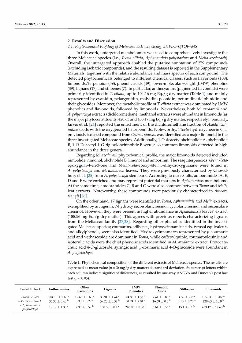
In this work, untargeted metabolomics was used to comprehensively investigate thethree Meliaceae species (i.e., Toona ciliata, Aphanamixis polystachya and Melia azedarach).Overall, the untargeted approach enabled the putative annotation of 279 compounds(excluding isobaric compounds), and the resulting dataset is reported in the SupplementaryMaterials, together with the relative abundance and mass spectra of each compound. Thedetected phytochemicals belonged to different chemical classes, such as flavonoids (108),limonoids/terpenoids (59), phenolic acids (49), lower-molecular-weight (LMW) phenolics(39), lignans (17) and stilbenes (7). In particular, anthocyanins (pigmented flavonoids) wereprimarily identified in T. ciliata, up to 104.16 mg Eq./g dry matter (Table 1) and mainlyrepresented by cyanidin, pelargonidin, malvidin, peonidin, petunidin, delphinidin andtheir glycosides. Moreover, the metabolic profile of T. ciliata extract was dominated by LMWphenolics and flavonoids, followed by limonoids. Nevertheless, both M. azedarach andA. polystachya extracts (dichloromethane: methanol extracts) were abundant in limonoids (asthe major phytoconstituents; 420.63 and 433.17 mg Eq./g dry matter, respectively). Similarly,Jarvis et al. [24] reported the enrichment of the dichloromethane fraction of Azadirachtaindica seeds with the oxygenated triterpenoids. Noteworthy, 11beta-hydroxycneorin G, apreviously isolated compound from Cedrela sinesis, was identified as a major limonoid in thethree investigated Meliaceae species. Additionally, 1-O-deacetylohchinolide A, ohchnolideB, 1-O-Deacetyl-1-O-tigloylohchinolide B were also common limonoids detected in highabundance in the three genera.
Regarding M. azedarach phytochemical profile, the major limonoids detected includednimbolide, nimonol, ohchnolide B, limonol and amoorinin. The sesquiterpenoids, 6beta,7beta-epoxyguai-4-en-3-one and 6beta,7beta-epoxy-4beta,5-dihydroxyguaiane were found inA. polystachya and M. azedarach leaves. They were previously characterized by Chowd-hury et al. [25] from A. polystachya stem bark. According to our results, amooramides A, E,D and F were enriched and may represent potential markers in Aphanamixis metabolome.At the same time, amooramides C, B and G were also common between Toona and Melialeaf extracts. Noteworthy, these compounds were previously characterized in Amooratsangii [26].
On the other hand, 17 lignans were identified in Toona, Aphanamixis and Melia extracts,exemplified by arctigenin, 7-hydroxy secoisolariciresinol, cyclolariciresinol and secoisolari-ciresinol. However, they were present in higher abundance in Aphanamixis leaves’ extract(188.56 mg Eq./g dry matter). This agrees with previous reports characterizing lignansfrom the Meliaceae family [27,28]. Regarding other phenolics identified in the investi-gated Meliaceae species; coumarins, stilbenes, hydroxycinnamic acids, tyrosol equivalentsand alkylphenols, were also identified. Hydroxycinnamates represented by p-coumaricacid and verbascoside are dominant in Toona, while caffeoylquinic, coumaroylquinic andisoferulic acids were the chief phenolic acids identified in M. azedarach extract. Protocate-chuic acid 4-O-glucoside, syringic acid, p-coumaric acid 4-O-glucoside were abundant inA. polystachya.
Table 1. Phytochemical composition of the different extracts of Meliaceae species. The results areexpressed as mean value (n = 3; mg/g dry matter) ± standard deviation. Superscript letters withineach column indicate significant differences, as resulted by one-way ANOVA and Duncan’s post hoctest (p < 0.05).
Tested Extract Anthocyanins OtherFlavonoids Lignans LMW
PhenolicsPhenolic
Acids Stilbenes Limonoids
- Toona ciliata 104.16 ± 2.63 c 12.65 ± 0.65 c 33.91 ± 1.44 a 74.85 ± 1.53 b 7.41 ± 0.85 a 4.59 ± 2.7 a 135.93 ± 13.07 a
- Melia azedarach 36.35 ± 5.45 b 3.33 ± 0.29 a 59.25 ± 0.32 b 31.74 ± 2.81 a 16.68 ± 0.5 b 3.15 ± 0.25 a 420.63 ± 10.8 b
- Aphanamixispolystachya 19.19 ± 1.35 a 7.35 ± 0.59 b 188.56 ± 8.1 c 248.05 ± 8.32 c 6.61 ± 0.56 a 15.1 ± 0.1 b 433.17 ± 12.63 b
Molecules 2022, 27, 435 4 of 20
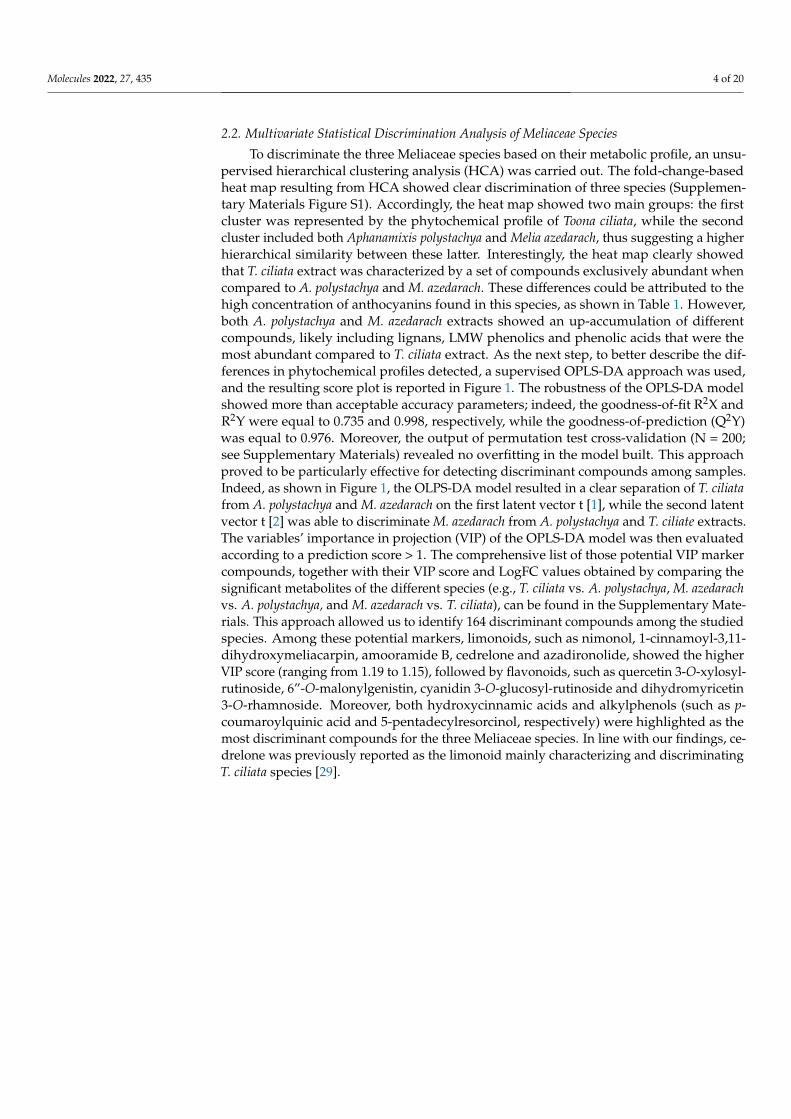
2.2. Multivariate Statistical Discrimination Analysis of Meliaceae Species
To discriminate the three Meliaceae species based on their metabolic profile, an unsu-pervised hierarchical clustering analysis (HCA) was carried out. The fold-change-basedheat map resulting from HCA showed clear discrimination of three species (Supplemen-tary Materials Figure S1). Accordingly, the heat map showed two main groups: the firstcluster was represented by the phytochemical profile of Toona ciliata, while the secondcluster included both Aphanamixis polystachya and Melia azedarach, thus suggesting a higherhierarchical similarity between these latter. Interestingly, the heat map clearly showedthat T. ciliata extract was characterized by a set of compounds exclusively abundant whencompared to A. polystachya and M. azedarach. These differences could be attributed to thehigh concentration of anthocyanins found in this species, as shown in Table 1. However,both A. polystachya and M. azedarach extracts showed an up-accumulation of differentcompounds, likely including lignans, LMW phenolics and phenolic acids that were themost abundant compared to T. ciliata extract. As the next step, to better describe the dif-ferences in phytochemical profiles detected, a supervised OPLS-DA approach was used,and the resulting score plot is reported in Figure 1. The robustness of the OPLS-DA modelshowed more than acceptable accuracy parameters; indeed, the goodness-of-fit R2X andR2Y were equal to 0.735 and 0.998, respectively, while the goodness-of-prediction (Q2Y)was equal to 0.976. Moreover, the output of permutation test cross-validation (N = 200;see Supplementary Materials) revealed no overfitting in the model built. This approachproved to be particularly effective for detecting discriminant compounds among samples.Indeed, as shown in Figure 1, the OLPS-DA model resulted in a clear separation of T. ciliatafrom A. polystachya and M. azedarach on the first latent vector t [1], while the second latentvector t [2] was able to discriminate M. azedarach from A. polystachya and T. ciliate extracts.The variables’ importance in projection (VIP) of the OPLS-DA model was then evaluatedaccording to a prediction score > 1. The comprehensive list of those potential VIP markercompounds, together with their VIP score and LogFC values obtained by comparing thesignificant metabolites of the different species (e.g., T. ciliata vs. A. polystachya, M. azedarachvs. A. polystachya, and M. azedarach vs. T. ciliata), can be found in the Supplementary Mate-rials. This approach allowed us to identify 164 discriminant compounds among the studiedspecies. Among these potential markers, limonoids, such as nimonol, 1-cinnamoyl-3,11-dihydroxymeliacarpin, amooramide B, cedrelone and azadironolide, showed the higherVIP score (ranging from 1.19 to 1.15), followed by flavonoids, such as quercetin 3-O-xylosyl-rutinoside, 6”-O-malonylgenistin, cyanidin 3-O-glucosyl-rutinoside and dihydromyricetin3-O-rhamnoside. Moreover, both hydroxycinnamic acids and alkylphenols (such as p-coumaroylquinic acid and 5-pentadecylresorcinol, respectively) were highlighted as themost discriminant compounds for the three Meliaceae species. In line with our findings, ce-drelone was previously reported as the limonoid mainly characterizing and discriminatingT. ciliata species [29].
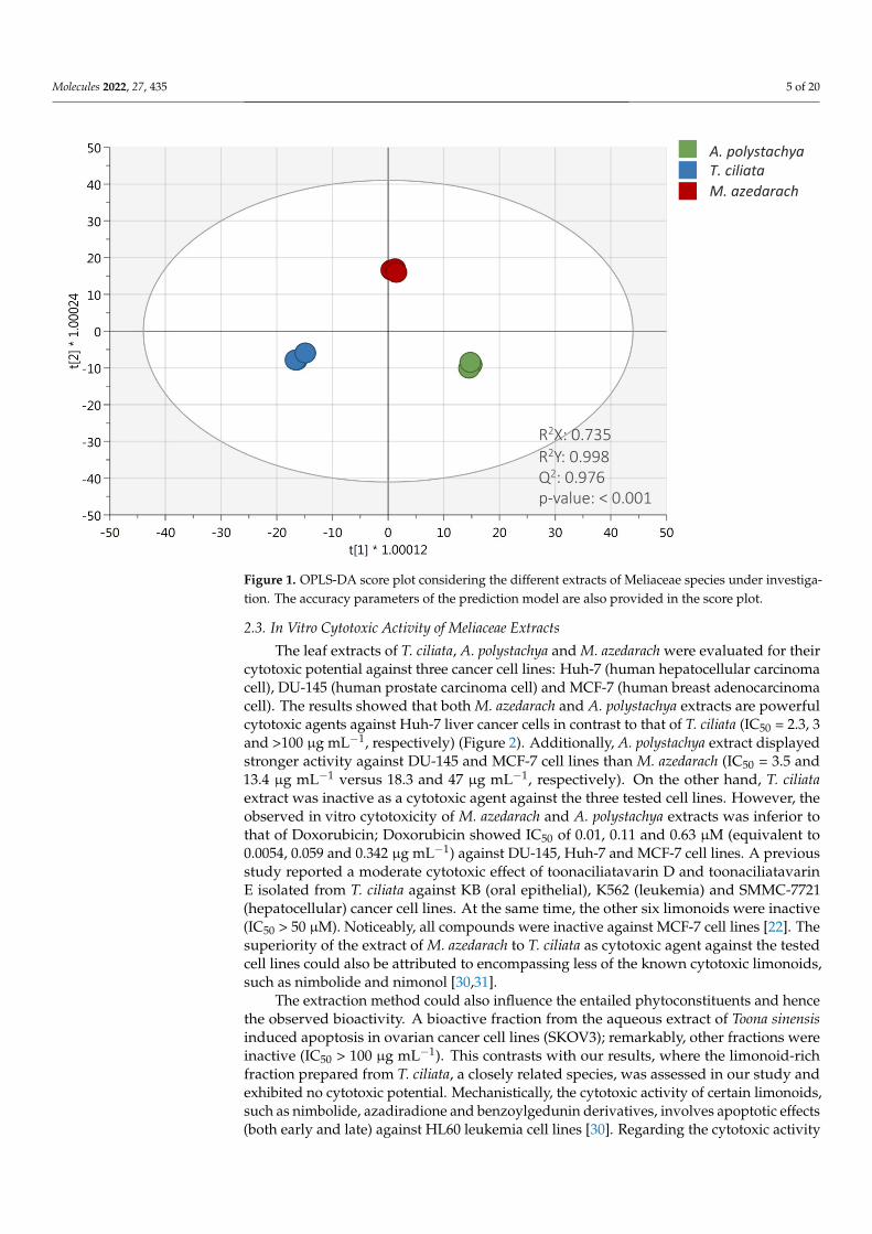
Molecules 2022, 27, 435 5 of 20Molecules 2022, 27, x 5 of 20
Figure 1. OPLS-DA score plot considering the different extracts of Meliaceae species under investi-
gation. The accuracy parameters of the prediction model are also provided in the score plot.
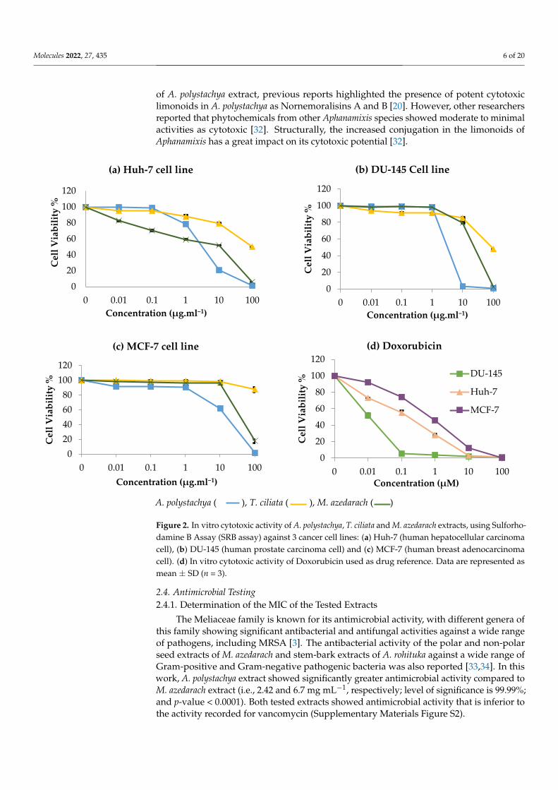
2.3. In Vitro Cytotoxic Activity of Meliaceae Extracts
The leaf extracts of T. ciliata, A. polystachya and M. azedarach were evaluated for their
cytotoxic potential against three cancer cell lines: Huh-7 (human hepatocellular carcinoma
cell), DU-145 (human prostate carcinoma cell) and MCF-7 (human breast adenocarcinoma
cell). The results showed that both M. azedarach and A. polystachya extracts are powerful
cytotoxic agents against Huh-7 liver cancer cells in contrast to that of T. ciliata (IC50 = 2.3, 3
and >100 µg·mL−1, respectively) (Figure 2). Additionally, A. polystachya extract displayed
stronger activity against DU-145 and MCF-7 cell lines than M. azedarach (IC50 = 3.5 and 13.4
µg·mL−1 versus 18.3 and 47 µg·mL−1, respectively). On the other hand, T. ciliata extract was
inactive as a cytotoxic agent against the three tested cell lines. However, the observed in
vitro cytotoxicity of M. azedarach and A. polystachya extracts was inferior to that of Doxo-
rubicin; Doxorubicin showed IC50 of 0.01, 0.11 and 0.63 µM (equivalent to 0.0054, 0.059
and 0.342 µg·mL−1) against DU-145, Huh-7 and MCF-7 cell lines. A previous study re-
ported a moderate cytotoxic effect of toonaciliatavarin D and toonaciliatavarin E isolated
from T. ciliata against KB (oral epithelial), K562 (leukemia) and SMMC-7721 (hepatocellu-
lar) cancer cell lines. At the same time, the other six limonoids were inactive (IC50 > 50 µM).
Noticeably, all compounds were inactive against MCF-7 cell lines [22]. The superiority of
the extract of M. azedarach to T. ciliata as cytotoxic agent against the tested cell lines could
also be attributed to encompassing less of the known cytotoxic limonoids, such as nimbo-
lide and nimonol [30,31].
The extraction method could also influence the entailed phytoconstituents and hence
the observed bioactivity. A bioactive fraction from the aqueous extract of Toona sinensis
induced apoptosis in ovarian cancer cell lines (SKOV3); remarkably, other fractions were
inactive (IC50 > 100 µg mL−1). This contrasts with our results, where the limonoid-rich frac-
tion prepared from T. ciliata, a closely related species, was assessed in our study and ex-
hibited no cytotoxic potential. Mechanistically, the cytotoxic activity of certain limonoids,
R2X: 0.735R2Y: 0.998Q2: 0.976p-value: < 0.001
A. polystachyaT. ciliata
M. azedarach
Figure 1. OPLS-DA score plot considering the different extracts of Meliaceae species under investiga-tion. The accuracy parameters of the prediction model are also provided in the score plot.
2.3. In Vitro Cytotoxic Activity of Meliaceae Extracts
The leaf extracts of T. ciliata, A. polystachya and M. azedarach were evaluated for theircytotoxic potential against three cancer cell lines: Huh-7 (human hepatocellular carcinomacell), DU-145 (human prostate carcinoma cell) and MCF-7 (human breast adenocarcinomacell). The results showed that both M. azedarach and A. polystachya extracts are powerfulcytotoxic agents against Huh-7 liver cancer cells in contrast to that of T. ciliata (IC50 = 2.3, 3and >100 µg mL−1, respectively) (Figure 2). Additionally, A. polystachya extract displayedstronger activity against DU-145 and MCF-7 cell lines than M. azedarach (IC50 = 3.5 and13.4 µg mL−1 versus 18.3 and 47 µg mL−1, respectively). On the other hand, T. ciliataextract was inactive as a cytotoxic agent against the three tested cell lines. However, theobserved in vitro cytotoxicity of M. azedarach and A. polystachya extracts was inferior tothat of Doxorubicin; Doxorubicin showed IC50 of 0.01, 0.11 and 0.63 µM (equivalent to0.0054, 0.059 and 0.342 µg mL−1) against DU-145, Huh-7 and MCF-7 cell lines. A previousstudy reported a moderate cytotoxic effect of toonaciliatavarin D and toonaciliatavarinE isolated from T. ciliata against KB (oral epithelial), K562 (leukemia) and SMMC-7721(hepatocellular) cancer cell lines. At the same time, the other six limonoids were inactive(IC50 > 50 µM). Noticeably, all compounds were inactive against MCF-7 cell lines [22]. Thesuperiority of the extract of M. azedarach to T. ciliata as cytotoxic agent against the testedcell lines could also be attributed to encompassing less of the known cytotoxic limonoids,such as nimbolide and nimonol [30,31].
The extraction method could also influence the entailed phytoconstituents and hencethe observed bioactivity. A bioactive fraction from the aqueous extract of Toona sinensisinduced apoptosis in ovarian cancer cell lines (SKOV3); remarkably, other fractions wereinactive (IC50 > 100 µg mL−1). This contrasts with our results, where the limonoid-richfraction prepared from T. ciliata, a closely related species, was assessed in our study andexhibited no cytotoxic potential. Mechanistically, the cytotoxic activity of certain limonoids,such as nimbolide, azadiradione and benzoylgedunin derivatives, involves apoptotic effects(both early and late) against HL60 leukemia cell lines [30]. Regarding the cytotoxic activity
Molecules 2022, 27, 435 6 of 20
of A. polystachya extract, previous reports highlighted the presence of potent cytotoxiclimonoids in A. polystachya as Nornemoralisins A and B [20]. However, other researchersreported that phytochemicals from other Aphanamixis species showed moderate to minimalactivities as cytotoxic [32]. Structurally, the increased conjugation in the limonoids ofAphanamixis has a great impact on its cytotoxic potential [32].
Molecules 2022, 27, x 6 of 20
such as nimbolide, azadiradione and benzoylgedunin derivatives, involves apoptotic ef-fects (both early and late) against HL60 leukemia cell lines [30]. Regarding the cytotoxic activity of A. polystachya extract, previous reports highlighted the presence of potent cyto-toxic limonoids in A. polystachya as Nornemoralisins A and B [20]. However, other re-searchers reported that phytochemicals from other Aphanamixis species showed moderate to minimal activities as cytotoxic [32]. Structurally, the increased conjugation in the limo-noids of Aphanamixis has a great impact on its cytotoxic potential [32].
A. polystachya ( ), T. ciliata ( ), M. azedarach ( )
Figure 2. In vitro cytotoxic activity of A. polystachya, T. ciliata and M. azedarach extracts, using Sul-forhodamine B Assay (SRB assay) against 3 cancer cell lines: (a) Huh-7 (human hepatocellular car-cinoma cell), (b) DU-145 (human prostate carcinoma cell) and (c) MCF-7 (human breast adenocarci-noma cell). (d) In vitro cytotoxic activity of Doxorubicin used as drug reference. Data are repre-sented as mean ± SD (n = 3).
2.4. Antimicrobial Testing 2.4.1. Determination of the MIC of the Tested Extracts
The Meliaceae family is known for its antimicrobial activity, with different genera of this family showing significant antibacterial and antifungal activities against a wide range of pathogens, including MRSA [3]. The antibacterial activity of the polar and non-polar seed extracts of M. azedarach and stem-bark extracts of A. rohituka against a wide range of Gram-positive and Gram-negative pathogenic bacteria was also reported [33,34]. In this work, A. polystachya extract showed significantly greater antimicrobial activity compared to M. azedarach extract (i.e., 2.42 and 6.7 mg mL−1, respectively; level of significance is
0
20
40
60
80
100
120
0 0.01 0.1 1 10 100
Cel
l Via
bilit
y %
Concentration (µg.ml−1)
(a) Huh-7 cell line
0
20
40
60
80
100
120
0 0.01 0.1 1 10 100C
ell V
iabi
lity
%Concentration (µg.ml−1)
(b) DU-145 Cell line
020406080
100120
0 0.01 0.1 1 10 100
Cel
l Via
bilit
y %
Concentration (µg.ml−1)
(c) MCF-7 cell line
0
20
40
60
80
100
120
0 0.01 0.1 1 10 100
Cel
l Via
bilit
y %
Concentration (µM)
(d) Doxorubicin
DU-145
Huh-7
MCF-7
Figure 2. In vitro cytotoxic activity of A. polystachya, T. ciliata and M. azedarach extracts, using Sulforho-damine B Assay (SRB assay) against 3 cancer cell lines: (a) Huh-7 (human hepatocellular carcinomacell), (b) DU-145 (human prostate carcinoma cell) and (c) MCF-7 (human breast adenocarcinomacell). (d) In vitro cytotoxic activity of Doxorubicin used as drug reference. Data are represented asmean ± SD (n = 3).
2.4. Antimicrobial Testing2.4.1. Determination of the MIC of the Tested Extracts
The Meliaceae family is known for its antimicrobial activity, with different genera ofthis family showing significant antibacterial and antifungal activities against a wide rangeof pathogens, including MRSA [3]. The antibacterial activity of the polar and non-polarseed extracts of M. azedarach and stem-bark extracts of A. rohituka against a wide range ofGram-positive and Gram-negative pathogenic bacteria was also reported [33,34]. In thiswork, A. polystachya extract showed significantly greater antimicrobial activity compared toM. azedarach extract (i.e., 2.42 and 6.7 mg mL−1, respectively; level of significance is 99.99%;and p-value < 0.0001). Both tested extracts showed antimicrobial activity that is inferior tothe activity recorded for vancomycin (Supplementary Materials Figure S2).
Molecules 2022, 27, 435 7 of 20
In this study, in both A. polystachya and M. azedarach extracts, limonoids were detectedas the major phytoconstituents; this is in agreement with Lu et al. [35], who reported theisolation of 15 limonoids from the ethanolic extract of the dry seeds of neem. Among theseisolated compounds, the antibacterial activity was observed against both Gram-positiveand Gram-negative isolates. Another study conducted by Rahman [36] reported theisolation of swietenolide and 2-hydroxy-3-O-tigloylswietenolide from Swietenia mahagoni(family Meliaceae), showing activity against eight MDR clinical isolates, including S. aureus.However, their antibacterial activity was inferior to that of vancomycin, which was includedas an antibiotic standard that agrees with our study. The observed antimicrobial activity ismainly attributable to their enrichment with limonoids, in addition to other phenolics whichact via disruption of the bacterial membrane, leading to the loss of its selective permeabilityand subsequent killing of pathogens. It is noteworthy that nimbolide, a major constituentdetected in the LC–MS profile of M. azedarach, exhibited cell membrane disruption andbiofilm inhibition of multidrug-resistant MRSA, and this effect was dose-dependent [37].
2.4.2. Anti-Virulent Activity of the Tested Extracts
Virulence factors produced by MRSA allow its survival inside the host and evasionof the immune system; these factors include hemolysins (α, β, γ and δ) nuclease, proteaseand other enzymes [38]. Anti-virulence therapy is an attractive alternative strategy to limitmicrobial resistance rather than killing a pathogen. This includes inhibition of biofilmformation, disruption of preformed biofilm, inhibition of certain bacterial enzymes and areduction of bacterial pigmentation [39].
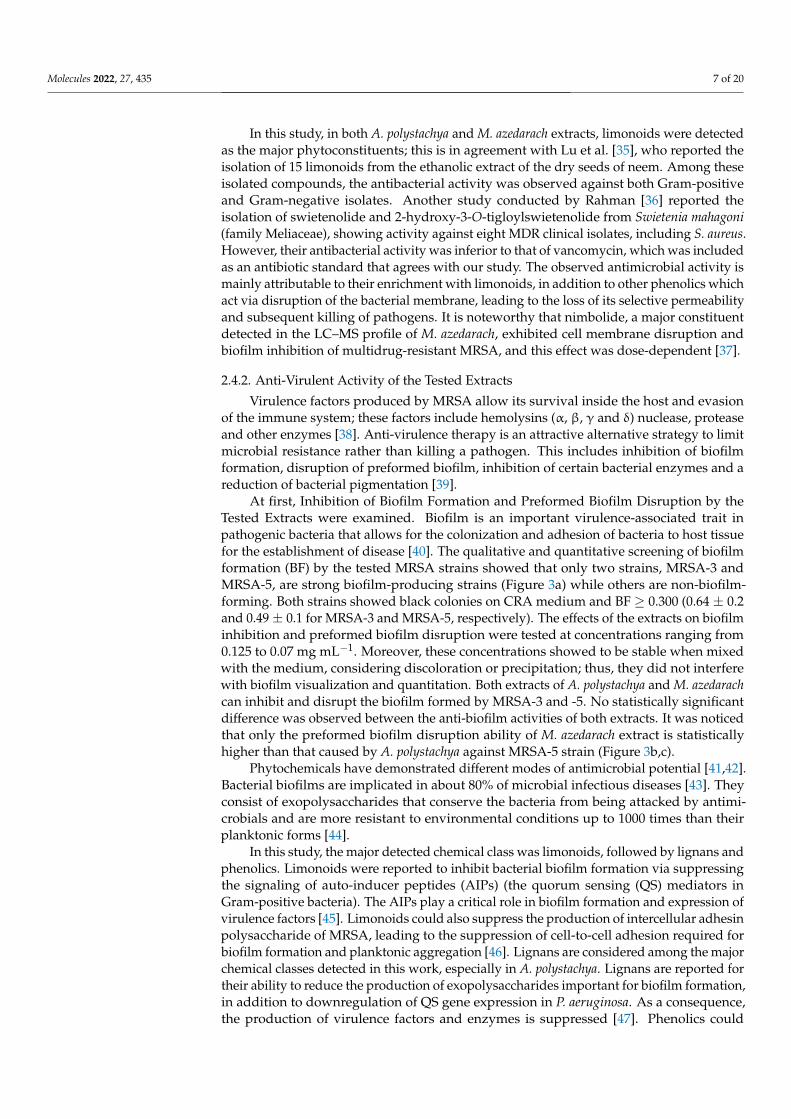
At first, Inhibition of Biofilm Formation and Preformed Biofilm Disruption by theTested Extracts were examined. Biofilm is an important virulence-associated trait inpathogenic bacteria that allows for the colonization and adhesion of bacteria to host tissuefor the establishment of disease [40]. The qualitative and quantitative screening of biofilmformation (BF) by the tested MRSA strains showed that only two strains, MRSA-3 andMRSA-5, are strong biofilm-producing strains (Figure 3a) while others are non-biofilm-forming. Both strains showed black colonies on CRA medium and BF ≥ 0.300 (0.64 ± 0.2and 0.49 ± 0.1 for MRSA-3 and MRSA-5, respectively). The effects of the extracts on biofilminhibition and preformed biofilm disruption were tested at concentrations ranging from0.125 to 0.07 mg mL−1. Moreover, these concentrations showed to be stable when mixedwith the medium, considering discoloration or precipitation; thus, they did not interferewith biofilm visualization and quantitation. Both extracts of A. polystachya and M. azedarachcan inhibit and disrupt the biofilm formed by MRSA-3 and -5. No statistically significantdifference was observed between the anti-biofilm activities of both extracts. It was noticedthat only the preformed biofilm disruption ability of M. azedarach extract is statisticallyhigher than that caused by A. polystachya against MRSA-5 strain (Figure 3b,c).
Phytochemicals have demonstrated different modes of antimicrobial potential [41,42].Bacterial biofilms are implicated in about 80% of microbial infectious diseases [43]. Theyconsist of exopolysaccharides that conserve the bacteria from being attacked by antimi-crobials and are more resistant to environmental conditions up to 1000 times than theirplanktonic forms [44].
In this study, the major detected chemical class was limonoids, followed by lignans andphenolics. Limonoids were reported to inhibit bacterial biofilm formation via suppressingthe signaling of auto-inducer peptides (AIPs) (the quorum sensing (QS) mediators inGram-positive bacteria). The AIPs play a critical role in biofilm formation and expression ofvirulence factors [45]. Limonoids could also suppress the production of intercellular adhesinpolysaccharide of MRSA, leading to the suppression of cell-to-cell adhesion required forbiofilm formation and planktonic aggregation [46]. Lignans are considered among the majorchemical classes detected in this work, especially in A. polystachya. Lignans are reported fortheir ability to reduce the production of exopolysaccharides important for biofilm formation,in addition to downregulation of QS gene expression in P. aeruginosa. As a consequence,the production of virulence factors and enzymes is suppressed [47]. Phenolics could

Molecules 2022, 27, 435 8 of 20
kill pathogens and inhibit biofilm via different mechanisms: disruption of the bacterialmembrane (with subsequent leakage of cellular content) and inhibition of cell–cell adhesion,inhibition of extracellular matrix formation and inhibition of Staphylococcal protein A,thus preventing bacterial adhesion and attachment required for biofilm production [45]. Apossible interaction between compounds of these chemical classes has led to the observedanti-virulence effect of the extracts.
Nevertheless, a further in vivo study is recommended for the major isolated com-pounds from the three Meliaceae species for further characterization and confirmation ofthe suggested activity.
Molecules 2022, 27, x 8 of 20
Lignans are reported for their ability to reduce the production of exopolysaccharides im-
portant for biofilm formation, in addition to downregulation of QS gene expression in P.
aeruginosa. As a consequence, the production of virulence factors and enzymes is sup-
pressed [47]. Phenolics could kill pathogens and inhibit biofilm via different mechanisms:
disruption of the bacterial membrane (with subsequent leakage of cellular content) and
inhibition of cell–cell adhesion, inhibition of extracellular matrix formation and inhibition
of Staphylococcal protein A, thus preventing bacterial adhesion and attachment required
for biofilm production [45]. A possible interaction between compounds of these chemical
classes has led to the observed anti-virulence effect of the extracts.
Nevertheless, a further in vivo study is recommended for the major isolated com-
pounds from the three Meliaceae species for further characterization and confirmation of
the suggested activity.
(a)
Aphanamixis polystachya Melia azedarach
(*) p-value = 0.018
MRSA-3 MRSA-5 MRSA-3 MRSA-5
(b) (c)
Figure 3. (a) Congo red agar medium showing black colonies production with strains MRSA-3 and
MRSA-5 only, indicating positive slime production. Pink colonies indicate negative slime produc-
tion. (b) Percentage inhibition of biofilm formation and (c) percentage disruption of preformed bio-
film formed by MRSA-3 and -5 strains by Aphanamixis polystachya and Melia azedarach extracts at
concentration of 0.125 mg.mL−1. Data represent the means of percentage biofilm inhibition/disrup-
tion ± SD, n = 3 (one-way ANOVA, followed by Tukey’s multiple comparisons, p-value < 0.05). (*)
Indicates presence of a statistically significant difference between columns.
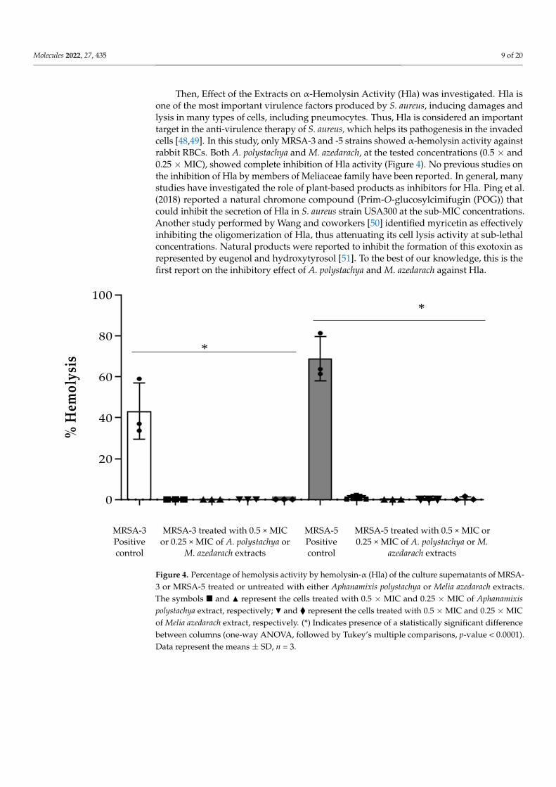
Then, Effect of the Extracts on α-Hemolysin Activity (Hla) was investigated. Hla is
one of the most important virulence factors produced by S. aureus, inducing damages and
lysis in many types of cells, including pneumocytes. Thus, Hla is considered an important
Figure 3. (a) Congo red agar medium showing black colonies production with strains MRSA-3 andMRSA-5 only, indicating positive slime production. Pink colonies indicate negative slime production.(b) Percentage inhibition of biofilm formation and (c) percentage disruption of preformed biofilmformed by MRSA-3 and -5 strains by Aphanamixis polystachya and Melia azedarach extracts at concentra-tion of 0.125 mg mL−1. Data represent the means of percentage biofilm inhibition/disruption ± SD,n = 3 (one-way ANOVA, followed by Tukey’s multiple comparisons, p-value < 0.05). (*) Indicatespresence of a statistically significant difference between columns.
Molecules 2022, 27, 435 9 of 20
Then, Effect of the Extracts on α-Hemolysin Activity (Hla) was investigated. Hla isone of the most important virulence factors produced by S. aureus, inducing damages andlysis in many types of cells, including pneumocytes. Thus, Hla is considered an importanttarget in the anti-virulence therapy of S. aureus, which helps its pathogenesis in the invadedcells [48,49]. In this study, only MRSA-3 and -5 strains showed α-hemolysin activity againstrabbit RBCs. Both A. polystachya and M. azedarach, at the tested concentrations (0.5 × and0.25 ×MIC), showed complete inhibition of Hla activity (Figure 4). No previous studies onthe inhibition of Hla by members of Meliaceae family have been reported. In general, manystudies have investigated the role of plant-based products as inhibitors for Hla. Ping et al.(2018) reported a natural chromone compound (Prim-O-glucosylcimifugin (POG)) thatcould inhibit the secretion of Hla in S. aureus strain USA300 at the sub-MIC concentrations.Another study performed by Wang and coworkers [50] identified myricetin as effectivelyinhibiting the oligomerization of Hla, thus attenuating its cell lysis activity at sub-lethalconcentrations. Natural products were reported to inhibit the formation of this exotoxin asrepresented by eugenol and hydroxytyrosol [51]. To the best of our knowledge, this is thefirst report on the inhibitory effect of A. polystachya and M. azedarach against Hla.
Molecules 2022, 27, x 9 of 20
target in the anti-virulence therapy of S. aureus, which helps its pathogenesis in the in-
vaded cells [48,49]. In this study, only MRSA-3 and -5 strains showed α-hemolysin activity
against rabbit RBCs. Both A. polystachya and M. azedarach, at the tested concentrations (0.5
× and 0.25 × MIC), showed complete inhibition of Hla activity (Figure 4). No previous
studies on the inhibition of Hla by members of Meliaceae family have been reported. In
general, many studies have investigated the role of plant-based products as inhibitors for
Hla. Ping et al. (2018) reported a natural chromone compound (Prim-O-glucosylcimifugin
(POG)) that could inhibit the secretion of Hla in S. aureus strain USA300 at the sub-MIC
concentrations. Another study performed by Wang and coworkers [50] identified myrice-
tin as effectively inhibiting the oligomerization of Hla, thus attenuating its cell lysis activ-
ity at sub-lethal concentrations. Natural products were reported to inhibit the formation
of this exotoxin as represented by eugenol and hydroxytyrosol [51]. To the best of our
knowledge, this is the first report on the inhibitory effect of A. polystachya and M. azedarach
against Hla.
% H
emo
lysi
s
0
20
40
60
80
100
MRSA-3 Positive control
MRSA-3 treated with 0.5 × MIC or 0.25 × MIC of A. polystachya or
M. azedarach extracts
MRSA-5 Positive control
MRSA-5 treated with 0.5 × MIC or 0.25 × MIC of A. polystachya or M.
azedarach extracts
Figure 4. Percentage of hemolysis activity by hemolysin-α (Hla) of the culture supernatants of
MRSA-3 or MRSA-5 treated or untreated with either Aphanamixis polystachya or Melia azedarach ex-
tracts. The symbols ◼ and ▲ represent the cells treated with 0.5 × MIC and 0.25 × MIC of Aphanamixis
polystachya extract, respectively; and ◆ represent the cells treated with 0.5 × MIC and 0.25 × MIC
of Melia azedarach extract, respectively. (*) Indicates presence of a statistically significant difference
between columns (one-way ANOVA, followed by Tukey’s multiple comparisons, p-value < 0.0001).
Data represent the means ± SD, n = 3.
Staphyloxanthin, a golden-yellow pigment that possesses antioxidant activity, is an-
other virulence factor of S. aureus that is involved in protecting bacteria from reactive ox-
ygen species (ROS) and enhancing the resistance of innate immune response represented
by the host neutrophils [52]. Among the tested MRSA strains, only two (MRSA-3 and -5)
showed golden pigmentation, whereas other MRSA strains were white. The effect on pig-
ment production by the action of A. polystachya was more noticeable than that of M. azed-
arach for both tested concentrations (Supplementary Materials Figure S2). As mentioned
above, the observed reduction of pigmentation and hemolysis activity could be a result of
suppression of QS signaling molecules in S. aureus (AIPs) and a possible downregulation
of virulence-associated genes reported for the major detected phytoconstituents in this
study.
*
*
Figure 4. Percentage of hemolysis activity by hemolysin-α (Hla) of the culture supernatants of MRSA-3 or MRSA-5 treated or untreated with either Aphanamixis polystachya or Melia azedarach extracts.The symbols � and N represent the cells treated with 0.5 × MIC and 0.25 × MIC of Aphanamixispolystachya extract, respectively; H and � represent the cells treated with 0.5 ×MIC and 0.25 ×MICof Melia azedarach extract, respectively. (*) Indicates presence of a statistically significant differencebetween columns (one-way ANOVA, followed by Tukey’s multiple comparisons, p-value < 0.0001).Data represent the means ± SD, n = 3.
Molecules 2022, 27, 435 10 of 20
Staphyloxanthin, a golden-yellow pigment that possesses antioxidant activity, is an-other virulence factor of S. aureus that is involved in protecting bacteria from reactiveoxygen species (ROS) and enhancing the resistance of innate immune response representedby the host neutrophils [52]. Among the tested MRSA strains, only two (MRSA-3 and-5) showed golden pigmentation, whereas other MRSA strains were white. The effecton pigment production by the action of A. polystachya was more noticeable than that ofM. azedarach for both tested concentrations (Supplementary Materials Figure S2). Asmentioned above, the observed reduction of pigmentation and hemolysis activity could bea result of suppression of QS signaling molecules in S. aureus (AIPs) and a possible down-regulation of virulence-associated genes reported for the major detected phytoconstituentsin this study.
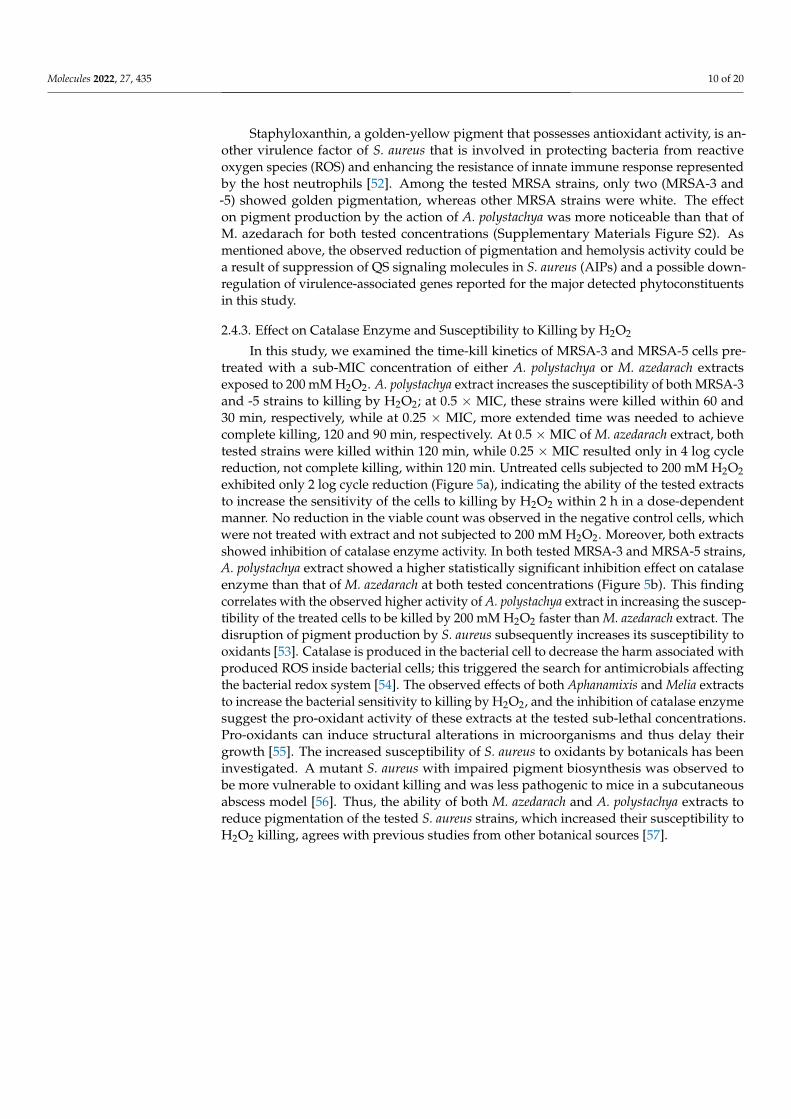
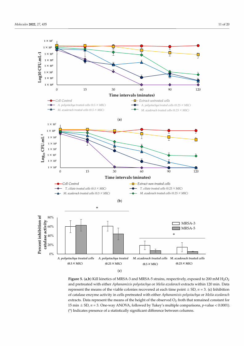
2.4.3. Effect on Catalase Enzyme and Susceptibility to Killing by H2O2
In this study, we examined the time-kill kinetics of MRSA-3 and MRSA-5 cells pre-treated with a sub-MIC concentration of either A. polystachya or M. azedarach extractsexposed to 200 mM H2O2. A. polystachya extract increases the susceptibility of both MRSA-3and -5 strains to killing by H2O2; at 0.5 × MIC, these strains were killed within 60 and30 min, respectively, while at 0.25 × MIC, more extended time was needed to achievecomplete killing, 120 and 90 min, respectively. At 0.5 ×MIC of M. azedarach extract, bothtested strains were killed within 120 min, while 0.25 × MIC resulted only in 4 log cyclereduction, not complete killing, within 120 min. Untreated cells subjected to 200 mM H2O2exhibited only 2 log cycle reduction (Figure 5a), indicating the ability of the tested extractsto increase the sensitivity of the cells to killing by H2O2 within 2 h in a dose-dependentmanner. No reduction in the viable count was observed in the negative control cells, whichwere not treated with extract and not subjected to 200 mM H2O2. Moreover, both extractsshowed inhibition of catalase enzyme activity. In both tested MRSA-3 and MRSA-5 strains,A. polystachya extract showed a higher statistically significant inhibition effect on catalaseenzyme than that of M. azedarach at both tested concentrations (Figure 5b). This findingcorrelates with the observed higher activity of A. polystachya extract in increasing the suscep-tibility of the treated cells to be killed by 200 mM H2O2 faster than M. azedarach extract. Thedisruption of pigment production by S. aureus subsequently increases its susceptibility tooxidants [53]. Catalase is produced in the bacterial cell to decrease the harm associated withproduced ROS inside bacterial cells; this triggered the search for antimicrobials affectingthe bacterial redox system [54]. The observed effects of both Aphanamixis and Melia extractsto increase the bacterial sensitivity to killing by H2O2, and the inhibition of catalase enzymesuggest the pro-oxidant activity of these extracts at the tested sub-lethal concentrations.Pro-oxidants can induce structural alterations in microorganisms and thus delay theirgrowth [55]. The increased susceptibility of S. aureus to oxidants by botanicals has beeninvestigated. A mutant S. aureus with impaired pigment biosynthesis was observed tobe more vulnerable to oxidant killing and was less pathogenic to mice in a subcutaneousabscess model [56]. Thus, the ability of both M. azedarach and A. polystachya extracts toreduce pigmentation of the tested S. aureus strains, which increased their susceptibility toH2O2 killing, agrees with previous studies from other botanical sources [57].
Molecules 2022, 27, 435 11 of 20Molecules 2022, 27, x 11 of 20
Figure 5. (a,b) Kill kinetics of MRSA-3 and MRSA-5 strains, respectively, exposed to 200 mM H2O2
and pretreated with either Aphanamixis polystachya or Melia azedarach extracts within 120 min. Data
represent the means of the viable colonies recovered at each time point ± SD, n = 3. (c) Inhibition of
catalase enzyme activity in cells pretreated with either Aphanamixis polystachya or Melia azedarach
(a)
(b)
(c)
1.E+00
1.E+01
1.E+02
1.E+03
1.E+04
1.E+05
1.E+06
1.E+07
0 15 30 60 90 120
Lo
g10
CF
U.m
L-1
Time intervals (minutes)
Cell Control Extract-untreated cells
A. polystachya treated cells (0.5xMIC) A. polystachya treated cells (0.25xMIC)
M. azaderach treated cells (0.5xMIC) M. azaderach treated cells (0.25xMIC)
1 × 10²
1 × 101
1 × 100
1 × 10³
1 × 10⁴
1 × 10⁵
1 × 10⁶
1 × 10⁷
A. polystachya treated cells (0.25×MIC)A. polystachya treated cells (0.5×MIC)
M. azaderach treated cells (0.5×MIC) M. azaderach treated cells (0.25×MIC)
1.E+00
1.E+01
1.E+02
1.E+03
1.E+04
1.E+05
1.E+06
1.E+07
0 15 30 60 90 120
Lo
g1
0C
FU
.mL
-1
Time intervals (minutes)
Cell Control Extract non-treated cells
T. ciliate treated cells (0.5xMIC) T. ciliate treated cells (0.25xMIC)
M. azaderach treated cells (0.5xMIC) M. azaderach treated cells (0.25xMIC)
1 × 10³
1 × 10⁴
1 × 10⁵
1 × 10⁶
1 × 10⁷
1 × 10²
1 × 101
1 × 100
T. ciliate treated cells (0.25×MIC)T. ciliate treated cells (0.5×MIC)
M. azaderach treated cells (0.5×MIC) M. azaderach treated cells (0.25×MIC)
0%
20%
40%
60%
80%
A. polystachya treated cells
(0.5xMIC)
A. polystachya treated
cells (0.25xMIC)
M. azaderach treated cells
(0.5xMIC)
M. azaderach treated cells
(0.25xMIC)
Per
cen
t in
hib
itio
n o
f
cata
lase
act
ivit
y
MRSA-5
MRSA-3
(0.5×MIC) (0.5×MIC)(0.25×MIC)(0.25×MIC) (0.25×MIC)
*
*
Figure 5. (a,b) Kill kinetics of MRSA-3 and MRSA-5 strains, respectively, exposed to 200 mM H2O2
and pretreated with either Aphanamixis polystachya or Melia azedarach extracts within 120 min. Datarepresent the means of the viable colonies recovered at each time point ± SD, n = 3. (c) Inhibitionof catalase enzyme activity in cells pretreated with either Aphanamixis polystachya or Melia azedarachextracts. Data represent the means of the height of the observed O2 froth that remained constant for15 min ± SD, n = 3. One-way ANOVA, followed by Tukey’s multiple comparisons, p-value < 0.0001).(*) Indicates presence of a statistically significant difference between columns.
Molecules 2022, 27, 435 12 of 20
2.5. Pearson’s Correlations between Phytochemical Profile and Biological Activity
Pearson’s correlation coefficients were finally evaluated to investigate different degreesof correlations between the main classes of phytochemicals and the various biologicalactivities under investigation, such as cytotoxic activity against Huh-7, MCF-7 and DU-145, as well as antimicrobial or antibiofilm formation of MRSA strains. A summarizingcorrelation table is provided in the Supplementary Materials.
Overall, lignans and limonoids were the classes of compounds that were most relatedto the biological activities, and this was true mainly for the extracts of both M. azedarachand A. polystachya. Indeed, the cytotoxic activity against three cancerous cell lines exhibiteda significant negative correlation with these phytochemical classes. In detail, plant extractscontaining high amounts of lignans were negatively correlated with the viability of MCF-7(r = −0.87; p < 0.01) and DU-145 (r = −0.73; p < 0.05) cell lines, proving to be the classprimarily responsible for the negative correlation. Furthermore, plant extracts that wereenriched in limonoids resulted in being the greater inhibitor of the three cancer cell lines,showing strong negative correlations ranging between −0.93 and −0.99 (p < 0.001). Recentstudies have proved that both market drugs containing lignans and lignan-rich fractionshave an anticancer effect [2]. For example, the Egyptian flaxseed, especially Giza-9, andits dietary formulations are enriched with lignans and exerted an in vitro and in vivoanticancer effect on a human breast cancer cell line and in mice bearing tumors [58]. Nano-formulated nimbolide, is characterized by its sustained release for more than 6 days in PBS(pH 7.4) and also exerted enhanced cytotoxicity about two- to three-fold in both breast andpancreatic cancer cell lines compared with free nimbolide [59].
Breast cancer is common among the global women population, and the conventionalprocedure for its treatments includes surgical removal of the malignant tissue, ionizingradiation and chemotherapy, causing several side effects. Compounds derived from plantextracts showed to be the next alternative medicine against cancer, with reduced sideeffects and the ability to boost up the immune system to fight against metastatic cells. Incompliance, the cytotoxicity capacity of A. rohituka leaves extract against human breastadenocarcinoma (MCF-7), triple-negative human breast cancer (MDA-MB-231), mice undif-ferentiated carcinoma (EAC) and mice fibroblast (L929) cell lines was reported by differentauthors owed to the presence of alkaloids, flavonoids, steroids, tannins, saponins andterpenoids [60–62].
The same result was observed for the antimicrobial activity against the five MRSAstrains. In this regard, lignans and limonoids showed a strong negative correlation withantimicrobial activity (on average r = −0.66; p < 0.05 and r = −0.99; p < 0.01, respectively).These findings suggested that an increased quantity of these compounds resulted in alow extract concentration needed to inhibit the growth of cancer cells or MRSA strains.Moreover, we observed a dose–response effect between these classes of compounds andthe time needed to kill MRSA strains, as demonstrated by the time-kill assay. Chowdhuryet al. [33] studied the antiviral and antibacterial activity of the limonoid rohitukine, whichis found in A. rohituka extracts. In addition, Yuan et al. [63] showed the same activityof four limonoids (i.e., 3-deacetylkhivorin, 1-deacetylkhivorin, swietmanin B and 3-beta-acetoxy-1-oxo-methylmeliacate) against Staphylococcus aureus, Pseudomonas aeruginosa andtwo other clinically isolated bacterial (MRSA) strains. In the current study, the antibiofilmactivity was evaluated on the strains MRSA-3 and -5, expressed in terms of inhibitioncapacity of biofilm formation and disruption of preformed biofilm. Interestingly, the strongpositive correlation reported by limonoids in inhibiting biofilm formation on MRSA-3and -5 strains (r = 0.98 and 0.87; p < 0.01, respectively). The wide spectrum of action oflimonoids was extended also in the disruption capacity of preformed biofilm (r = 0.86 and0.68, respectively). Moreover, phenolic acids identified in our Meliaceae species provided asignificant capacity to regulate the biofilm formation both in early and preformed phasesfor MRSA 5. In accordance with our findings, several authors reported the efficacy ofphenolic acids in antibiofilm formation [64,65]. In particular, Walker et al. [66] reportedthe capacity of rosmarinic acid, a natural phenolic acid, to prevent Pseudomonas aeruginosa
Molecules 2022, 27, 435 13 of 20
biofilm formation. Other phenolic compounds were also found to be capable of inhibitingQS by using different approaches, such as catechin [67] and eugenol [68].
3. Materials and Methods3.1. Plant Material
Leaves of Toona ciliata M. Roem., Aphanamixis polystachya (Wall.) R. Parker and Meliaazedarach L. were collected at the flowering stage on April/May 2019 from Orman BotanicalGarden, Dokki, Giza, Egypt. The identity of the plant material was confirmed by staffmembers of the herbarium at Orman Botanical Garden. Voucher specimens (12.04.2019I,13.05.2019I and 13.05.2019II, respectively) were deposited at Pharmacognosy Department,Faculty of Pharmacy, Cairo University, Egypt.
3.2. Preparation of Plant Extracts
The powdered leaves of the Meliaceae species under investigation (300 g each) wereextracted in a Soxhlet apparatus (Glassco, India), using dichloromethane–methanol (80:20,v/v). The extracts were then evaporated till dryness at 50 ◦C, under reduced pressure,using a rotary evaporator. The dried extracts were subsequently kept in a dry and darkplace at 10 ◦C until further analyses.
3.3. In Vitro Cytotoxic Assay
Three cell lines, namely Huh-7 (human hepatocellular carcinoma cell), DU-145 (humanprostate carcinoma cell) and MCF-7 (human breast adenocarcinoma cell), were obtainedfrom Nawah Scientific Inc. (Almokattam, Cairo, Egypt). Cells were maintained in DMEMmedium supplemented with 100 mg mL−1 of streptomycin, 100 units mL−1 of penicillinand 10% of heat-inactivated fetal bovine serum in humidified 5% (v/v) CO2 atmosphere at37 ◦C. Cell viability was assessed by Sulforhodamine B Assay (SRB) assay [69]. Aliquotsof 100 µL cell suspension (5 × 10³ cells) were placed in 96-well flat-bottom tissue-cultureplates and incubated in complete media for 24 h. Cells were treated with another aliquotof 100 µL media containing the 3 plant extracts at various concentrations (0.01, 0.1, 1, 10and 100 µg/mL) and Doxorubicin (0.01, 0.1, 1, 10 and 100 µM). After 72 h of drug exposure,cells were fixed by replacing media with 150 µL of 10% TCA and incubated at 4 ◦C for 1 h.The TCA solution was removed, and the cells were washed 5 times with distilled water.Aliquots of 70 µL SRB solution (0.4% w/v) were added and incubated in a dark place atroom temperature for 10 min. Plates were washed 3 times with 1% acetic acid and allowedto air-dry overnight. Then 150 µL of 10 mM tris was added to dissolve the protein-boundSRB stain and the absorbance measured at 540 nm, using a BMG LABTECH®- FLUOstarOmega microplate reader (Ortenberg, Germany). Results are provided in Figure 2.
3.4. Microbiological Testing3.4.1. Bacterial Isolates and Culture Conditions
Five strains of methicillin-resistant Staphylococcus aureus (MRSA) isolated from intensive-care-unit patients of two Egyptian hospitals were used in this study [70]. The strains weregrown aerobically on Trypticase soy broth/agar (TSB/TSA) (Oxoid, UK), either undershaking at 180 rpm or static conditions, at 37 ◦C for 24 h. The strains were preservedand maintained by cryopreservation. All microbiological-related testing and assays wereperformed in triplicate, otherwise specified.
3.4.2. Determination of the Minimum Inhibitory Concentration (MIC) of the Plant Extractsby Agar Dilution Technique
MIC was determined according to the method described by (Jenkins and Schuetz,2012). In brief, the dry extracts were dissolved in absolute alcohol to a final concentrationof 250 mg mL−1. Different concentrations of each extract solution were prepared in moltenMuller Hinton agar [71] medium at 55–60 ◦C, and then MHA–extract mixture was pouredinto 6 cm Petri dishes and left to solidify. Then 10 µL of 107 CFU mL−1 bacterial suspension
Molecules 2022, 27, 435 14 of 20
was spotted on the surface of the solidified MHA–extract mixture. Negative and positivecontrols were included (MHA–absolute alcohol without bacteria and MHA–absolute al-cohol surface inoculated with bacteria, respectively). Plates were incubated at 37 ◦C for24 h. MHA plates with different concentrations of vancomycin, as a reference antibiotic,were included. MIC was recorded as the lowest concentration of extract showing no visiblegrowth.
3.5. Screening of Biofilm Formation by the Tested Strains3.5.1. Qualitative Screening Using Congo Red Agar (CRA) Medium
Slime production is an important phenotype associated with biofilm formation [72].A total of 10 µL of 107 CFU mL−1 bacterial suspension was spotted on the surface of thesolidified CRA. Plates were incubated at 37 ◦C for 24 h. The bacterial isolate was consideredas slime producing if black-colored bacterial growth was observed and non-slime producingif red- or pink-colored growth appeared.
3.5.2. Quantitative Screening Using Flat-Bottom 96-Well Microplates
The screening was performed according to the method of Sultan and Nabiel [73].Overnight culture of each MRSA strain was prepared in TSB supplemented with 1% glucose(TSBG); then 1:100 dilution of these cultures in fresh TSBG was performed. Then 200 µLof each 1:100 diluted culture was dispensed in the wells of microplates. Wells containing200 µL of TSBG only served as a negative control. Plates were incubated statically at 37 ◦Cfor 24 h. Turbidity measurement, biofilm staining and visualization were performed asfollows: Turbidity was measured at 600 nm, and then planktonic cells and spent mediumwere discarded. Plates were washed carefully with phosphate-buffered saline (PBS) threetimes and then left to dry completely in a laminar air-flow cabinet. The attached biofilmin wells was stained with 200 µL of 0.5% crystal violet solution. Plates were left static for30 min and then washed with distilled water three times and left to dry completely. Forbiofilm visualization, 150 µL of absolute ethanol was added to extract the violet color andleft for 15 min with shaking; violet color was then measured at 570 nm, using a microplatereader (Biotech, Synergy 2, Winooski, VT, USA). Isolates were classified as strong, moderateor weak at biofilm-forming according to the formula reported by Naves et al. [74]:
BF = AB−CW (1)
where BF is biofilm formation, AB is the OD570 of stained bacteria cells attached to thewells and CW is the OD570 of stained negative control wells.
If BF≥ 0.300, the isolate is considered a strong biofilm-forming isolate; BF = 0.200–0.299means the isolate is moderate at BF-forming, BF of 0.100–0.199 is deemed to be weak,and <0.100 is considered a negative biofilm-forming isolate. Then strong or moderatebiofilm-forming strains were selected to perform biofilm-inhibition/disruption assays.
3.5.3. The Anti-Virulence Activity of the Extracts
Inhibition of biofilm formation was examined as follows: the relevant bacterial isolateswere allowed to form biofilm under the same conditions described in the biofilm screeningsection (using TSB medium supplemented with 1% glucose (TSBG) and incubated staticallyfor 24 h at 37 ◦C), in the presence of sub-MIC of the extracts (proven to be colorlessconcentrations, to avoid color interference with the assay results) ranging from 0.125 to0.007 mg mL−1. Negative control (TSBG and the same concentrations of the extracts) andpositive control (1:100 diluted overnight cultures in TSBG) were included. Plates wereincubated at 37 ◦C for 24 h statically. Biofilm was then stained and visualized, as statedbefore, using 0.5% crystal violet solution.
Disruption of preformed biofilm was examined as follows: relevant bacterial isolateswere allowed to form preformed biofilm under the same stated conditions prior to additionof extracts. After that, spent culture medium and planktonic cells were discarded, followedby the addition of fresh TSBG medium containing the same sub-MIC concentrations of
Molecules 2022, 27, 435 15 of 20
each of the extracts (that were used in the assay of inhibition of biofilm formation). Plateswere further incubated at 37 ◦C for 24 h statically. Negative and positive controls wereincluded, as mentioned before. Biofilm was then stained and visualized as described above,using 0.5% crystal violet solution.
Effect on α-hemolysin activity assay was performed according to Ping et al. [49], withminor modification. In brief, 25 µL of defibrinated rabbit RBCs was added to 875 µLof sterile PBS. Eighteen-hour-old TSB cultures of MRSA strains untreated/treated withsub-MIC of the extracts (0.5 ×MIC and 0.25 ×MIC) were centrifuged at 1000× g for 5 min.Then 100 µL of supernatant was added to the rabbit RBCs suspension, gently mixed andincubated at 37 ◦C for 15 min. The mixtures were centrifuged at 10,000× g for 2 min at20 ◦C. The degree of hemolysis was measured at 543 nm, using a microplate reader (Biotech,Synergy 2, Winooski, VT, USA). RBCs treated with 1% Triton-X 100 solution served aspositive control and considered 100% hemolysis, while PBS-treated RBCs served as thenegative control.
Percent hemolysis was calculated according to the following equation:
% Hemolysis =OD543 of sample − OD543 of negative control
OD543 of positive control× 100 (2)
The effect of the extracts on the pigmentation of relevant MRSA strains was determinedqualitatively [53]. Overnight cultures of these isolates untreated/treated with sub-MIC(0.5×MIC and 0.25×MIC) of the extracts in TSB were prepared. Extract-treated/untreatedcultures were centrifuged at 10,000× g for 5 min, and the pellets were washed twice withPBS and were observed for pigmentation by the naked eye. Overnight cultures of extract-untreated bacteria served as the positive control.
In oxidant susceptibility assays, the bacterial cells used in these assays were preparedas follows: 18-h-old overnight cultures of relevant MRSA strains in TSB untreated/treatedwith sub-MIC (0.5 ×MIC and 0.25 ×MIC) of the extracts were centrifuged at 1000× g for5 min. Pellets were washed twice with PBS and then re-suspended in PBS; inoculum wasadjusted according to the assay requirement.
In susceptibility to killing by H2O2: turbidity of PBS bacterial suspension was adjustedto 0.5 McFarland turbidity standard (≈108 CFU mL−1). Fresh H2O2 solution in PBS wasadded to the bacterial suspension to give a final concentration of 200 mM. The mixturewas incubated at 37 ◦C for 2 h, with shaking at 180 rpm. Viable colonies were enumeratedon MHA for both extract-treated and untreated cells at time intervals 0, 30, 60, 90 and120 min [53].
The quantitative catalase enzyme activity assay was performed according to themethod described by Iwase et al. [75], but with some modifications. OD600 of bacterialsuspension in PBS was adjusted to 5. Then 100 µL of the bacterial suspension was added toglass Wasserman tubes (1 cm diameter, 7.5 cm height) containing 100 µL of fresh 30% H2O2and 100 µL of 1% Triton-X 100, mixed well and incubated at room temperature for 15 min.The height of the observed O2 froth that remained constant for 15 min was measured byusing a ruler. The inhibition of enzyme activity was calculated according to the followingequation:
% Inhibition =Height of froth of untreated sample −Height of froth of treated sample
Height of froth of untreated sample× 100 (3)
3.6. Untargeted Profiling of Different Meliaceae Species by UHPLC-QTOF Mass Spectrometry
The three Meliaceae extracts were re-suspended in dichloromethane solution andcentrifuged at 8000× g for 10 min. The supernatants were then collected in the LC–MSvials, and then 6 µL of each sample was injected in an ultrahigh-performance liquidchromatography–quadrupole time-of-flight–mass spectrometry (UHPLC–QTOF–MS). A1290 liquid chromatography coupled with a G6550 mass spectrometer detector via a DualElectrospray Jet Stream ionization system (Agilent Technologies, Santa Clara, CA, USA)

Molecules 2022, 27, 435 16 of 20
was used. The instrumental conditions for analyzing plant extracts were optimized in previ-ous works [76,77]. The mass spectrometer acquisition was made in the m/z range 100–1200,using a positive full-scan mode with a nominal resolution at 30,000 FWHM. The Agilentsoftware Profinder B.06 was used to align and annotate the raw mass features accord-ing to the “find-by-formula” algorithm, using the combination of monoisotopic accuratemass and the entire isotopic pattern. To this aim, a custom database built by consideringboth phenolic compounds (Phenol-Explorer 3.6; http://phenol-explorer.eu/, accessed on30 September 2021) and limonoids reported in the literature for Meliaceae species wasused. The MS data were then subjected to a post-acquisition process, retaining only thosecompounds putatively annotated within 100% of replications in at least one condition.The approach adopted allowed a Level 2 of compound identification [78]. The annotatedcompounds were then ascribed into classes and quantified by using single pure-standardcompounds analyzed by using the same UHPLC–MS method. The standards prepared(purity > 98%) were representative of the following phenolic subclasses: anthocyanins(cyanidin), flavanols and flavonols (catechin), flavones (luteolin), phenolic acids (ferulicacid), lignans (sesamin), stilbenes (resveratrol), lower-molecular-weight phenolics (tyrosols)and limonoids (azadirachtin B). The results were finally expressed as mg equivalents/gdry matter (DM).
3.7. Statistical Analysis
In this work, a one-way analysis of the variance (ANOVA), followed by Tukey’smultiple comparisons and unpaired Student’s t-tests (p-value < 0.05), was performedwhen considering the antimicrobial, anticancer and antibiofilm assays, using the softwareGraphPad Prism 6.01 (GraphPad Software, Inc., San Diego, CA, USA). Moreover, the resultsof each in vitro assay and semi-quantitative analysis of phytochemicals were analyzedby software PASW Statistics 25.0 (SPSS Inc., Chicago, IL, USA) to investigate significantdifferences (p-value < 0.05, Duncan’s post hoc test). Pearson’s correlation coefficients(p-value = 0.01, two-tailed) were also calculated by using the same statistical software. TheAgilent software Mass Profiler Professional B.12.06 (from Agilent Technologies, Santa Clara,CA, USA; version B.05.00) was then used to elaborate the untargeted UHPLC–MS data. Theraw MS dataset was analyzed according to an unsupervised hierarchical clustering analysis(HCA), based on the fold-change (FC) heat map and supervised orthogonal projectionsto latent structures discriminant analysis (OPLS-DA) through the software SIMCA 13(Umetrics, Malmo, Sweden), as previously reported [76]. The software also allowed us torecord the goodness-of-fit and goodness-of-prediction parameters of the OPLS-DA model(i.e., R2Y and Q2Y, respectively). Finally, the variables’ importance in projection (VIPscore cutoff > 1) was coupled to a fold-change analysis (FC cutoff > 1.2) to find out themost discriminant marker compounds between the three different Meliaceae species underinvestigation.
4. Conclusions
There is growing evidence that infections mediated by biofilm facilitate the develop-ment of chronic infectious diseases caused by inappropriate antibiotic use. A promisingsolution is to use natural extracts that are rich in bioactive compounds, able to eliminateand able to prevent infections. Accordingly, in this work, we studied some limonoidsand phenolics-enriched Meliaceae species as promising sources of therapeutic cytotoxic,antibiofilm and anti-virulence agents. Our findings suggest a possible implication ofA. rohituka and M. azedarach extracts as potential candidates to combat MRSA biofilm infec-tions. However, selective isolation of the major constituents and in-depth in vivo study arestill missed in this study. The future scope of our project would be covering an optimizationof nano-based formulation of the main constituents followed by preclinical studies inanimal models. Notwithstanding, toxicological studies are still needed to further guaranteethe cytotoxic potential of A. rohituka and M. azedarach, including selectivity index towardsnormal cells.
Molecules 2022, 27, 435 17 of 20
Supplementary Materials: The following supporting information can be downloaded online,Figure S1: Hierarchical unsupervised clustering analysis heat map built by considering the phyto-chemical profile of the three different extracts of Meliaceae species under investigation. Figure S2:Average MIC values recorded for the tested extracts of Aphanamixis polystachya and Melia azedarachin mg mL−1 against five clinical isolates of Methicillin-resistant Staphylococcus aureus (MRSA); theresults represent the mean of three replicates ± SD. Vancomycin showed average MIC values of0.002 ± 0.0 mg.mL−1 with the 5 tested MRSA strains. Unpaired t-test, p-value < 0.0001. Figure S3:Inhibition of pigmentation of MRSA-3 and MRSA-5 by extracts of Aphanamixis polystachya was morepronounced than the effect of Melia azedarach at concentration of either 0.5 ×MIC or 0.25 ×MIC: (A)0.5×MIC treated A. polystachya cells, (B) 0.25×MIC treated A. polystachya cells, (C) Extract-untreatedcells, (D) 0.5 ×MIC treated M. azedarach cells and (E) 0.25 ×MIC treated M. azedarach cells. Table S1:Metabolomic dataset containing the annotated compounds from UHPLC–QTOF–mass spectrometry.Each compound is provided with its relative abundance and composite mass spectrum. Table S2:A comprehensive list of potential VIP marker compounds, together with their VIP score (>1) andLogFC values obtained by comparing the significant metabolites of the 3 different Meliaceae species.Table S3: Pearson’s correlation coefficients regarding the different in vitro biological assays and theannotated phytochemical classes by untargeted metabolomics.
Author Contributions: Conceptualization, M.M.I. and F.R.S.; data curation, L.Z. and G.R.; formalanalysis, L.Z. and G.R.; funding acquisition, M.M.I. and F.R.S.; investigation, L.Z., M.M.I., G.R., N.M.F.and F.R.S.; methodology, M.M.I., G.R. and L.L.; project administration, F.R.S.; resources, M.M.I.,L.L. and F.R.S.; software, L.Z. and G.R.; supervision, L.L.; validation, L.Z., M.M.I., G.R. and L.L.;visualization, L.Z., M.M.I., G.R., N.M.F. and F.R.S.; writing—original draft, L.Z., M.M.I., G.R., N.M.F.,L.L. and F.R.S.; writing—review and editing, L.Z., M.M.I., G.R., N.M.F., L.L. and F.R.S. All authorshave read and agreed to the published version of the manuscript.
Funding: This research is funded for the extensive microbiological studies and partially for the phyto-chemical study by Faculty of Pharmacy, Cairo University, Cairo, Egypt (IRG project 967:2019–2021).
Institutional Review Board Statement: Not applicable.
Informed Consent Statement: Not applicable.
Data Availability Statement: Not applicable.
Acknowledgments: The authors wish to thank Hanzada Tawfik Nour El-Din, Microbiology andImmunology department, Faculty of Pharmacy, Cairo University, for providing the clinical isolatesused in this study. The authors wish also to thank the “Romeo ed Enrica Invernizzi” Foundation(Milan, Italy) for supporting the metabolomic facility at Università Cattolica del Sacro Cuore.
Conflicts of Interest: The authors declare no conflict of interest.
Sample Availability: Not applicable.
References1. Christenhusz, M.J.; Byng, J.W. The number of known plants species in the world and its annual increase. Phytotaxa 2016, 261,
201–217. [CrossRef]2. De Silva, S.F.; Alcorn, J. Flaxseed lignans as important dietary polyphenols for cancer prevention and treatment: Chemistry,
pharmacokinetics, and molecular targets. Pharmaceuticals 2019, 12, 68. [CrossRef] [PubMed]3. Paritala, V.; Chiruvella, K.K.; Thammineni, C.; Ghanta, R.G.; Mohammed, A. Phytochemicals and antimicrobial potentials of
mahogany family. Rev. Bras. Farm. 2015, 25, 61–83. [CrossRef]4. Xu, W.-H.; Su, X.-M.; Wang, C.; Du, F.; Liang, Q. The genus Amoora: A phytochemical and pharmacological review. Fitoterapia
2019, 137, 104269. [CrossRef] [PubMed]5. Nerome, K.; Ito-Kureha, T.; Paganini, T.; Fukuda, T.; Igarashi, Y.; Ashitomi, H.; Ikematsu, S.; Yamamoto, T. Potent and broad
anticancer activities of leaf extracts from Melia azedarach L. of the subtropical Okinawa islands. Am. J. Cancer Res. 2020, 10, 581.6. Almubayedh, H.; Ahmad, R. Ethnopharmacology, phytochemistry, biological activities, and therapeutic applications of Cedrela
serrata Royle: A mini review. J. Ethnopharmacol 2020, 246, 112206. [CrossRef] [PubMed]7. Akbar, S. Melia azedarach L. (Meliaceae). In Handbook of 200 Medicinal Plants: A Comprehensive Review of Their Traditional Medical
Uses and Scientific Justifications; Springer International Publishing: Cham, Switzerland, 2020; pp. 1161–1170.8. Meziane, M.; Goumri, H. The antimicrobial effect of extracts of Melia azedarach on some pathogenic microorganisms. Int. J. Appl.
Nat. Sci. 2014, 3, 173–180.

Molecules 2022, 27, 435 18 of 20
9. Al-Khafaji, N.J.; Al-Zubaedi, R.M.; Al-Azawi, S.J. Evaluation of antibacterial effects of Melia azedarach fruit extracts against someisolated pathogenic bacteria. Vet. Sci. Dev. 2016, 6, 6080. [CrossRef]
10. Khan, A.S. Trees with Antimicrobial Activities. In Medicinally Important Trees; Springer International Publishing: Cham, Switzer-land, 2017; pp. 85–108.
11. Khalid, M.; Hassani, D.; Bilal, M.; Butt, Z.A.; Hamayun, M.; Ahmad, A.; Huang, D.; Hussain, A. Identification of oral cavitybiofilm forming bacteria and determination of their growth inhibition by Acacia arabica, Tamarix aphylla L. and Melia azedarach L.medicinal plants. Arch. Oral Biol. 2017, 81, 175–185. [CrossRef] [PubMed]
12. Frieri, M.; Kumar, K.; Boutin, A. Antibiotic resistance. J. Infect. Public Health 2017, 10, 369–378. [CrossRef] [PubMed]13. Frieden, T. Antibiotic Resistance Threats in the United States, 2013; Centers for Disease Control Prevention, US Department of Health
and Human Services: Atlanta, GA, USA, 2013; Volume 23, pp. 11–28.14. Tacconelli, E.; Magrini, N.; Kahlmeter, G.; Singh, N. Global Priority List of Antibiotic-Resistant Bacteria to Guide Research, Discovery,
and Development of New Antibiotics; World Health Organization: Geneva, Switzerland, 2017; Volume 27, pp. 318–327. Availableonline: https://www.who.int/medicines/publications/global-priority-list-antibiotic-resistant-bacteria/en/ (accessed on 1 June2020).
15. Römling, U.; Kjelleberg, S.; Normark, S.; Nyman, L.; Uhlin, B.E.; Åkerlund, B. Microbial biofilm formation: A need to act. J. Int.Med. 2014, 276, 98–110. [CrossRef] [PubMed]
16. Thoendel, M.; Kavanaugh, J.S.; Flack, C.E.; Horswill, A.R. Peptide signaling in the Staphylococci. Chem. Rev. 2011, 111, 117–151.[CrossRef] [PubMed]
17. Banerjee, D.; Shivapriya, P.; Gautam, P.K.; Misra, K.; Sahoo, A.K.; Samanta, S.K. A review on basic biology of bacterial biofilminfections and their treatments by nanotechnology-based approaches. Proc. Natl. Acad. Sci. India Sect. B Biol. Sci. 2019, 90, 243–259.[CrossRef]
18. Holá, V.; Ružicka, F.; Votava, M. The dynamics of Staphylococcus epidermis biofilm formation in relation to nutrition, temperature,and time. Scr. Med. 2006, 79, 169–174.
19. WHO. Latest global cancer data: Cancer burden rises to 18.1 million new cases and 9.6 million cancer deaths in 2018. InInternational Agency for Research on Cancer; World Health Organization: Geneva, Switzerland, 2018.
20. Wu, X.-Z.; Fang, F.-H.; Huang, W.-J.; Shi, Y.-Y.; Pan, H.-Q.; Ning, L.; Yuan, C.-S. Two novel nornemoralisin-type diterpenoids fromAphanamixis polystachya (Wall.) R. Parker. Fitoterapia 2020, 140, 104431. [CrossRef]
21. Zhang, P.; Cui, Z.; Wei, S.; Li, Y.; Yin, Y.; Wang, X.; Luo, J.; Kong, L. Diverse limonoids from barks of Toona ciliata var. yunnanensisand their biological activities. Ind. Crop. Prod. 2020, 148, 112275. [CrossRef]
22. Zhang, F.; Wang, J.-S.; Gu, Y.-C.; Kong, L.-Y. Cytotoxic and anti-inflammatory triterpenoids from Toona ciliata. J. Nat. Prod. 2012,75, 538–546. [CrossRef] [PubMed]
23. Ervina, M.; Poerwono, H.; Widyowati, R.; Matsunami, K. Bio-selective hormonal breast cancer cytotoxic and antioxidant potenciesof Melia azedarach L. wild type leaves. Biotechnol. Rep. 2020, 25, e00437. [CrossRef]
24. Jarvis, A.P.; Morgan, E.D.; Edwards, C. Rapid separation of triterpenoids from Neem seed extracts. Phytochem. Anal. 1999, 10,39–43. [CrossRef]
25. Chowdhury, R.; Hasan, C.M.; Rashid, M.A. Guaiane sesquiterpenes from Amoora rohituka. Phytochemistry 2003, 62, 1213–1216.[CrossRef]
26. Zhu, G.-Y.; Chen, G.; Liu, L.; Bai, L.-P.; Jiang, Z.-H. C-17 lactam-bearing limonoids from the twigs and leaves of Amoora tsangii. J.Nat. Prod. 2014, 77, 983–989. [CrossRef] [PubMed]
27. Sianturi, J.; Harneti, D.; Darwati; Mayanti, T.; Supratman, U.; Awang, K. A New (–)-5′,6-dimethoxyisolariciresinol-(3”,4”-dimethoxy)-3α-O-β-D-glucopyranoside from the bark of Aglaia eximia (Meliaceae). Nat. Prod. Res. 2016, 30, 2204–2208. [CrossRef][PubMed]
28. Wu, S.F.; Lin, C.K.; Chuang, Y.S.; Chang, F.R.; Tseng, C.K.; Wu, Y.C.; Lee, J.C. Anti-hepatitis C virus activity of 3-hydroxycaruilignan C from Swietenia macrophylla stems. J. Viral Hepat. 2012, 19, 364–370. [CrossRef]
29. De Leo, M.; Milella, L.; Braca, A.; De Tommasi, N. Cedrela and Toona genera: A rich source of bioactive limonoids and triterpenoids.Phytochem. Rev. 2018, 17, 751–783. [CrossRef]
30. Kikuchi, T.; Ishii, K.; Noto, T.; Takahashi, A.; Tabata, K.; Suzuki, T.; Akihisa, T. Cytotoxic and apoptosis-inducing activities oflimonoids from the seeds of Azadirachta indica (neem). J. Nat. Prod. 2011, 74, 866–870. [CrossRef] [PubMed]
31. Supratman, U.; Salam, S.; Naibaho, W.; Fajar, M.; Nurlelasari; Katja, D.G.; Harneti, D.; Maharani, R.; Hidayat, A.T.; Lesmana,R.; et al. New cytotoxic limonoids from the stem bark of Chisocheton pentandrus (Blanco) Merr. Phytochem. Lett. 2020, 35, 63–67.[CrossRef]
32. Zhang, Y.; Wang, J.-S.; Wei, D.-D.; Gu, Y.-C.; Wang, X.-B.; Kong, L.-Y. Bioactive terpenoids from the fruits of Aphanamixis grandifolia.J. Nat. Prod. 2013, 76, 1191–1195. [CrossRef]
33. Chowdhury, R.; Hasan, C.M.; Rashid, M.A. Antimicrobial activity of Toona ciliata and Amoora rohituka. Fitoterapia 2003, 74, 155–158.[CrossRef]
34. Khan, A.V.; Ahmed, Q.U.; Mir, M.R.; Shukla, I.; Khan, A.A. Antibacterial efficacy of the seed extracts of Melia azedarach againstsome hospital isolated human pathogenic bacterial strains. Asian Pac. J. Trop. Biomed. 2011, 1, 452–455. [CrossRef]
35. Lu, X.-F.; Lin, P.-C.; Zi, J.-C.; Fan, X.-N. Limonoids from seeds of Azadirachta indica and their antibacterial activity. ZhongguoZhongyao Zazhi=China J. Chin. Mater. Med. 2019, 44, 4864–4873.
Molecules 2022, 27, 435 19 of 20
36. Rahman, A.S.; Chowdhury, A.A.; Ali, H.-A.; Raihan, S.Z.; Ali, M.S.; Nahar, L.; Sarker, S.D. Antibacterial activity of two limonoidsfrom Swietenia mahagoni against multiple-drug-resistant (MDR) bacterial strains. J. Nat. Med. 2009, 63, 41–45. [CrossRef] [PubMed]
37. Sarkar, P.; Acharyya, S.; Banerjee, A.; Patra, A.; Thankamani, K.; Koley, H.; Bag, P.K. Intracellular, biofilm-inhibitory andmembrane-damaging activities of nimbolide isolated from Azadirachta indica A. Juss (Meliaceae) against meticillin-resistantStaphylococcus aureus. J. Med. Microbiol. 2016, 65, 1205–1214. [CrossRef] [PubMed]
38. Shettigar, K.; Murali, T.S. Virulence factors and clonal diversity of Staphylococcus aureus in colonization and wound infection withemphasis on diabetic foot infection. Eur. J. Clin. Microbiol. Infect. Dis. 2020, 39, 2235–2246. [CrossRef] [PubMed]
39. Fleitas Martínez, O.; Cardoso, M.H.; Ribeiro, S.M.; Franco, O.L. Recent advances in anti-virulence therapeutic strategies with afocus on dismantling bacterial membrane microdomains, toxin neutralization, quorum-sensing interference and biofilm inhibition.Front. Cell. Infect. Microbiol. 2019, 9, 74. [CrossRef]
40. Mohamed, J.A.; Huang, D.B. Biofilm formation by Enterococci. J. Med. Microbiol. 2007, 56, 1581–1588. [CrossRef] [PubMed]41. Ismail, M.M.; Hassan, M.; Moawad, S.S.; Okba, M.M.; Ashour, R.M.; Fayek, N.M.; Saber, F.R. Exploring the Antivirulence
Activity of Pulverulentone A, a Phloroglucinol-Derivative from Callistemon citrinus Leaf Extract, against Multi-Drug ResistantPseudomonas aeruginosa. Antibiotics 2021, 10, 907. [CrossRef]
42. Franco, C.M.; Vázquez, B.I. Natural Compounds as Antimicrobial Agents. Antibiotics 2020, 9, 217. [CrossRef]43. Davies, D. Understanding biofilm resistance to antibacterial agents. Nat. Rev. Drug Discov. 2003, 2, 114–122. [CrossRef]44. Rabin, N.; Zheng, Y.; Opoku-Temeng, C.; Du, Y.; Bonsu, E.; Sintim, H.O. Biofilm formation mechanisms and targets for developing
antibiofilm agents. Fut. Med. Chem. 2015, 7, 493–512. [CrossRef] [PubMed]45. Lu, L.; Hu, W.; Tian, Z.; Yuan, D.; Yi, G.; Zhou, Y.; Cheng, Q.; Zhu, J.; Li, M. Developing natural products as potential anti-biofilm
agents. Chin. Med. 2019, 14, 1–17. [CrossRef] [PubMed]46. Quelemes, P.V.; Perfeito, M.L.; Guimarães, M.A.; dos Santos, R.C.; Lima, D.F.; Nascimento, C.; Silva, M.P.; Soares, M.J.d.S.; Ropke,
C.D.; Eaton, P. Effect of neem (Azadirachta indica A. Juss) leaf extract on resistant Staphylococcus aureus biofilm formation andSchistosoma mansoni worms. J. Ethnopharmacol. 2015, 175, 287–294. [CrossRef] [PubMed]
47. Anju, V.; Busi, S.; Ranganathan, S.; Ampasala, D.R.; Kumar, S.; Suchiang, K.; Kumavath, R.; Dyavaiah, M. Sesamin and sesamolinrescues Caenorhabditis elegans from Pseudomonas aeruginosa infection through the attenuation of quorum sensing regulatedvirulence factors. Microb. Pathog. 2021, 155, 104912. [CrossRef] [PubMed]
48. Dinges, M.M.; Orwin, P.M.; Schlievert, P.M. Exotoxins of Staphylococcus aureus. Clin. Microbiol. Rev. 2000, 13, 16–34. [CrossRef][PubMed]
49. Ping, O.; Ruixue, Y.; Jiaqiang, D.; Kaiyu, W.; Jing, F.; Yi, G.; Xiaoli, H.; Defang, C.; Weimin, L.; Li, T. Subinhibitory concentrations ofprim-O-glucosylcimifugin decrease the expression of alpha-hemolysin in Staphylococcus aureus (USA300). Evid. Based Complement.Altern. Med. 2018, 2018, 7579808. [CrossRef]
50. Wang, T.; Zhang, P.; Lv, H.; Deng, X.; Wang, J. A natural dietary flavone myricetin as an α-hemolysin inhibitor for controllingStaphylococcus aureus infection. Front. Cell. Infect. Microbiol. 2020, 10, 330. [CrossRef]
51. Upadhyay, A.; Mooyottu, S.; Yin, H.; Nair, M.S.; Bhattaram, V.; Venkitanarayanan, K. Inhibiting microbial toxins using plant-derived compounds and plant extracts. Medicines 2015, 2, 186–211. [CrossRef] [PubMed]
52. Song, Y.; Liu, C.-I.; Lin, F.-Y.; No, J.H.; Hensler, M.; Liu, Y.-L.; Jeng, W.-Y.; Low, J.; Liu, G.Y.; Nizet, V. Inhibition of staphyloxanthinvirulence factor biosynthesis in Staphylococcus aureus: In vitro, in vivo, and crystallographic results. J. Med. Chem. 2009, 52,3869–3880. [CrossRef] [PubMed]
53. Leejae, S.; Hasap, L.; Voravuthikunchai, S.P. Inhibition of staphyloxanthin biosynthesis in Staphylococcus aureus by rhodomyrtone,a novel antibiotic candidate. J. Med. Microbiol. 2013, 62, 421–428. [CrossRef] [PubMed]
54. Kim, S.Y.; Park, C.; Jang, H.J.; Kim, B.O.; Bae, H.W.; Chung, I.Y.; Kim, E.S.; Cho, Y.H. Antibacterial strategies inspired by theoxidative stress and response networks. J. Microbiol. 2019, 57, 203–212. [CrossRef]
55. Halliwell, B. Are polyphenols antioxidants or pro-oxidants? What do we learn from cell culture and in vivo studies? Arch.Biochem. Biophys. 2008, 476, 107–112. [CrossRef] [PubMed]
56. Liu, G.Y.; Essex, A.; Buchanan, J.T.; Datta, V.; Hoffman, H.M.; Bastian, J.F.; Fierer, J.; Nizet, V. Staphylococcus aureus golden pigmentimpairs neutrophil killing and promotes virulence through its antioxidant activity. J. Exp. Med. 2005, 202, 209–215. [CrossRef]
57. Lee, J.-H.; Park, J.-H.; Cho, M.H.; Lee, J. Flavone reduces the production of virulence factors, staphyloxanthin and α-hemolysin,in Staphylococcus aureus. Curr. Microbiol. 2012, 65, 726–732. [CrossRef] [PubMed]
58. Ezzat, S.M.; Shouman, S.A.; Elkhoely, A.; Attia, Y.M.; Elsesy, M.S.; El Senousy, A.S.; Choucry, M.A.; El Gayed, S.H.; El Sayed, A.A.;Sattar, E.A. Anticancer potentiality of lignan rich fraction of six Flaxseed cultivars. Sci. Rep. 2018, 8, 1–16. [CrossRef] [PubMed]
59. Patra, A.; Satpathy, S.; Hussain, M.D. Nanodelivery and anticancer effect of a limonoid, nimbolide, in breast and pancreaticcancer cells. Int. J. Nanomed. 2019, 14, 8095. [CrossRef]
60. Chan, L.L.; George, S.; Ahmad, I.; Gosangari, S.L.; Abbasi, A.; Cunningham, B.T.; Watkin, K.L. Cytotoxicity Effects of Amoorarohituka and chittagonga on Breast and Pancreatic Cancer Cells. Evid-Based Complement. Altern. Med. 2011, 2011, 860605. [CrossRef][PubMed]
61. Rabi, T.; Ramachandran, C.; Fonseca, H.B.; Nair, R.P.; Alamo, A.; Melnick, S.J.; Escalon, E. Novel drug amooranin inducesapoptosis through caspase activity in human breast carcinoma cell lines. Breast Cancer Res. Treat. 2003, 80, 321–330. [CrossRef][PubMed]

Molecules 2022, 27, 435 20 of 20
62. Singh, R.K.; Ranjan, A.; Srivastava, A.K.; Singh, M.; Shukla, A.K.; Atri, N.; Mishra, A.; Singh, A.K.; Singh, S.K. Cytotoxic andapoptotic inducing activity of Amoora rohituka leaf extracts in human breast cancer cells. J. Ayurveda Integr. Med. 2020, 11, 383–390.[CrossRef]
63. Yuan, C.-M.; Zhang, Y.; Tang, G.-H.; Di, Y.-T.; Cao, M.-M.; Wang, X.-Y.; Zuo, G.-Y.; Li, S.-L.; Hua, H.-M.; He, H.-P. KhayseneganinsA–H, limonoids from Khaya senegalensis. J. Nat. Prod. 2013, 76, 327–333. [CrossRef] [PubMed]
64. Silva, S.; Costa, E.M.; Horta, B.; Calhau, C.; Morais, R.M.; Pintado, M.M. Anti-biofilm potential of phenolic acids: The influence ofenvironmental pH and intrinsic physico-chemical properties. Biofouling 2016, 32, 853–860. [CrossRef]
65. Luís, Â.; Silva, F.; Sousa, S.; Duarte, A.P.; Domingues, F. Antistaphylococcal and biofilm inhibitory activities of gallic, caffeic, andchlorogenic acids. Biofouling 2014, 30, 69–79. [CrossRef] [PubMed]
66. Walker, T.S.; Bais, H.P.; Déziel, E.; Schweizer, H.P.; Rahme, L.G.; Fall, R.; Vivanco, J.M. Pseudomonas aeruginosa-plant rootinteractions. Pathogenicity, biofilm formation, and root exudation. Plant. Physiol. 2004, 134, 320–331. [CrossRef]
67. Vandeputte, O.M.; Kiendrebeogo, M.; Rajaonson, S.; Diallo, B.; Mol, A.; El Jaziri, M.; Baucher, M. Identification of catechin as oneof the flavonoids from Combretum albiflorum bark extract that reduces the production of quorum-sensing-controlled virulencefactors in Pseudomonas aeruginosa PAO1. Appl. Environ. Microbiol. 2010, 76, 243–253. [CrossRef] [PubMed]
68. Husain, F.M.; Ahmad, I.; Asif, M.; Tahseen, Q. Influence of clove oil on certain quorum-sensing-regulated functions and biofilmof Pseudomonas aeruginosa and Aeromonas hydrophila. J. Biosci. 2013, 38, 835–844. [CrossRef] [PubMed]
69. Skehan, P.; Storeng, R.; Scudiero, D.; Monks, A.; McMahon, J.; Vistica, D.; Warren, J.T.; Bokesch, H.; Kenney, S.; Boyd, M.R. Newcolorimetric cytotoxicity assay for anticancer-drug screening. J. Natl. Cancer Inst. 1990, 82, 1107–1112. [CrossRef]
70. Nour El-Din, H.T.; Yassin, A.S.; Ragab, Y.M.; Hashem, A. Phenotype-Genotype Characterization and Antibiotic-ResistanceCorrelations Among Colonizing and Infectious Methicillin-Resistant Staphylococcus aureus Recovered from Intensive Care Units.Infect. Drug Resist. 2021, 14, 1557. [CrossRef] [PubMed]
71. Pavón-Pérez, J.; Oviedo, C.A.; Elso-Freudenberg, M.; Henríquez-Aedo, K.; Aranda, M. LC-MS/MS Method For L-Dopa Quantifi-cation In Different Tissues of Vicia faba. J. Chil. Chem. Soc. 2019, 64, 4651–4653. [CrossRef]
72. Kannappan, A.; Sivaranjani, M.; Srinivasan, R.; Rathna, J.; Pandian, S.K.; Ravi, A.V. Inhibitory efficacy of geraniol on biofilmformation and development of adaptive resistance in Staphylococcus epidermidis RP62A. J. Med. Microbiol. 2017, 66, 1506–1515.[CrossRef]
73. Sultan, A.; Nabiel, Y. Tube method and Congo red agar versus tissue culture plate method for detection of biofilm production byuropathogens isolated from midstream urine: Which one could be better? Afr. J. Clin. Exp. Microbiol. 2019, 20, 60–66. [CrossRef]
74. Naves, P.; Del Prado, G.; Huelves, L.; Gracia, M.; Ruiz, V.; Blanco, J.; Rodríguez-Cerrato, V.; Ponte, M.; Soriano, F. Measurement ofbiofilm formation by clinical isolates of Escherichia coli is method-dependent. J. Appl. Microbiol. 2008, 105, 585–590. [CrossRef]
75. Iwase, T.; Tajima, A.; Sugimoto, S.; Okuda, K.-I.; Hironaka, I.; Kamata, Y.; Takada, K.; Mizunoe, Y. A simple assay for measuringcatalase activity: A visual approach. Sci. Rep. 2013, 3, 1–4. [CrossRef]
76. Zhang, L.; Rocchetti, G.; Zengin, G.; Ak, G.; Yıldıztugay, E.; Mahomoodally, M.F.; Picot-Allain, M.C.N.; Lucini, L. Profiling ofpolyphenols and sesquiterpenoids using different extraction methods in Muscari turcicum, an endemic plant from Turkey. Ind.Crop. Prod. 2020, 154, 112626. [CrossRef]
77. Zhang, L.; Rocchetti, G.; Zengin, G.; Ak, G.; Saber, F.R.; Montesano, D.; Lucini, L. The UHPLC-QTOF-MS Phenolic Profiling andActivity of Cydonia oblonga Mill. Reveals a Promising Nutraceutical Potential. Foods 2021, 10, 1230. [CrossRef] [PubMed]
78. Salek, R.M.; Steinbeck, C.; Viant, M.R.; Goodacre, R.; Dunn, W.B. The role of reporting standards for metabolite annotation andidentification in metabolomic studies. GigaScience 2013, 2, 2047–217X. [CrossRef] [PubMed]


