Embed Size (px)
Citation preview
Author(s) agree that this article remains permanently open access under the terms of the
Creative Commons Attribution License 4.0 International License
Research Paper
PHYTOCHEMICAL ANALYSIS, ANTIMICROBIAL AND ANTIOXIDANT ACTIVITY ASSESSMENT OF ORANGE PEELS
Kaur M, Mehta A, Bhardwaj KK and Gupta R
Department of Biotechnology, Himachal Pradesh University, Summerhill, Shimla, 171 005,
India.
Abstract In the present-day scenario perishable fruit peels are considered as a new era of pharmaceutical products as they are rich in phytochemicals and act as antioxidant agents. The present study was undertaken to analyze the phytochemical constituents qualitatively in the orange peel extracts. The 1,1-diphenyl-1-picryl-hydrazyl (DPPH) radical scavenging activity of methanolic extract and distilled water extract was found to be better than other extracts used in the study. Silver nanoparticles were also synthesized with the aid of orange peel extract. Characterization of silver nanoparticles was carried out using UV-Visible spectroscopy and X-Ray diffraction. Synthesized sliver nanoparticles showed significant antibacterial activity against Bacillus sp. followed by Salmonella, Proteus, Pseudomonas, Escherichia, Shigella and Staphylococcus sp. Key words: Orange peel, Phytochemical constituents, Silver nanoparticles, Antibacterial activity, Antioxidant activity.
INTRODUCTION Sweet orange originated from East Asia but is consumed all over world as an
excellent source of vitamin C, a powerful natural antioxidant that builds the body
immune system. The peel of citrus fruit is abundant source of flavanones and many
polyethoxylated flavones which are very rare in other plants [1]. The phytochemicals
are also known as secondary metabolites that are derived from primary metabolites
and are used as drugs. They contribute to flavor, color and other characteristics of plant
parts [2]. Phytochemical analysis is very useful in the evaluation of some active
biological components of some vegetables and medicinal plants. Medically, the presence
of phytochemicals explains the use of vegetables in ethno medicine for the management
of various ailments [3]. These secondary plant metabolites are extractable by various
solvents that exhibit varied biochemical and pharmacological actions in animals [4]. The
antioxidant nature of the extract was assessed by using 1,1-diphenyl-1-picryl-hydrazyl
Journal of Global Biosciences ISSN 2320-1355 Volume 8, Number 3, 2019, pp. 6062-6072 Website: www.mutagens.co.in
Journal of Global Biosciences Vol. 8(3), 2019 pp. 6062-6072
ISSN 2320-1355
www.mutagens.co.in 6063
(DPPH) method. The biological synthesis of silver nanoparticles emerges as an eco-
friendly and exciting approach in the field of nanotechnology. Silver nanoparticles
(AgNPs) have been known to have inhibitory and bactericidal effects [5, 6].
Nanoparticles exhibit completely new or improved properties based on specific features
such as size, distribution and morphology. Although chemical and physical methods
may successfully make pure, well defined nanoparticles; these methods are rather
expensive and potentially hazardous to the environment. Use of biological organisms,
plant extract or plant biomass could be alternative to chemical and physical methods for
production of nanoparticles in an eco-friendly manner. In small concentrations, silver
nanoparticles (AgNps) are non-toxic to humans and are effective against microbes at
low concentrations and have no side effects [7, 8].
MATERIALS AND METHODS
Plant material and preparation of extract
Orange peels were collected from fruit juice shops of Nalagarh, Distt. Solan of
H.P. After collection, the peels were dried in natural sunlight for 3-4 days. The dried
peels were pulverized using an electric blender and stored in airtight containers for
further use. Four different solvents namely acetone, methanol, DMSO:methanol and
distilled water were used for extraction. The extract was prepared by using the methods
of Omm et al. and John et al. [9, 8].
Qualitative phytochemical screening
Phytochemical screening was carried out to analyze the presence of various
phytoconstituents such as alkaloids, flavonoids, phenolics, amino acids, tannins
anthraquinones, saponins, proteins, terpenoids and cardiac glycosides according to
standard method described by Arora and Kaur [3] and Gayathri and Kiruba [6]. General
reactions in this analysis revealed the presence or absence of these compounds.
Thin Layer Chromatography
The thin layer chromatography was used to determine the Rf value of separated
compounds. Aliquots of the extract and standard (quercetin, 1 mg/ml in methanol)
were applied on the TLC plate. The plate was then placed in a glass beaker containing n-
butanol: acetic acid: water (2:2:6) as mobile phase. Appearance of bright yellow colored
bands after spraying with ferric chloride indicates the presence of flavonoids [2, 8].
Anti-oxidant assay
DPPH free radical scavenging activity assay was performed by following the
methodology of Senthamil et al. [10].
Assessment of antibacterial activity
Antibacterial activity of the extract was tested against Escherichia, Salmonella,
Staphylococcus, Bacillus, Pseudomonas, Proteus and Shigella by using well diffusion
method.
Journal of Global Biosciences Vol. 8(3), 2019 pp. 6062-6072
ISSN 2320-1355
www.mutagens.co.in 6064
Minimum Inhibitory Concentration (MIC) against selected pathogenic strains
MIC was performed by using the method of Ashok et al. [4].
Biosynthesis of silver nanoparticles
Silver nanoparticles were synthesized by following the methods of Reenaa and
Menon, [11].
Characterization of silver nanoparticles
Synthesized silver nanoparticles were characterized by UV-Visible
spectrophotometer (GENESYS 10S UV-Vis v4.005 2L9S073217). Further, structure and
size of synthesized silver nanoparticles was investigated by XRD on PAN analytical
X’Pert PRO X-ray diffractometer [12].
Antibacterial activity of synthesized silver nanoparticles
Antibacterial activity of synthesized silver nanoparticles was tested by using well
diffusion method [5].
RESULTS AND DISCUSSIONS
Phytochemical screening of different solvent extracts
Present study of phytochemical screening of orange peels revealed the presence
of alkaloids, flavonoids etc. Besides the secondary metabolites, the presence of saponins
was noticed in methanol, DMSO:methanol (1:1 v/v) and distilled water extracts. By
contrast, the acetone extract exhibited the absence of saponins but showed the presence
of other common metabolites as reported in other extracts. Methanol extract of orange
peels showed the presence of secondary metabolites such as tannins and saponins. The
results of phytochemical analysis are shown in Table 1.
Gayathri and Kiruba, reported the presence of alkaloids, flavonoids, terpenoids
and phenols from dry leaf powder extracts of Citrus aurantium [6]. Another study on
phytochemical profile of Citrus limonum peel extract showed the presence of flavonoids,
phenols and cardiac glycosides [13].
TLC analysis of different solvent extracts
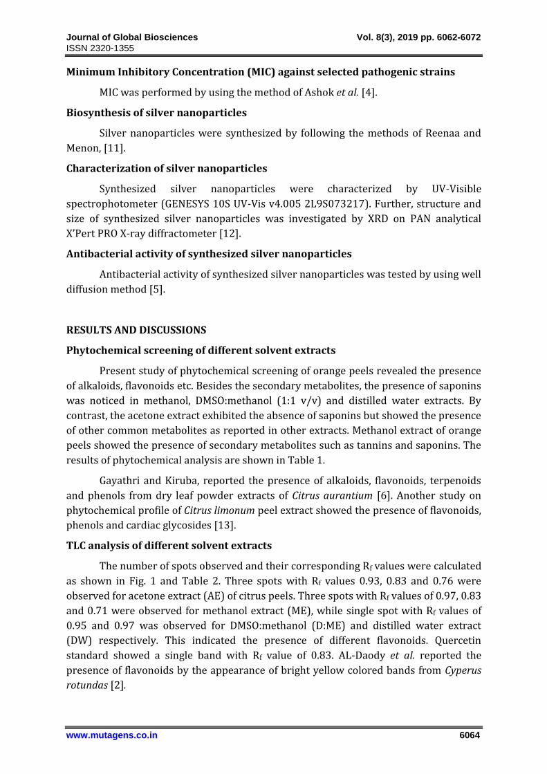
The number of spots observed and their corresponding Rf values were calculated
as shown in Fig. 1 and Table 2. Three spots with Rf values 0.93, 0.83 and 0.76 were
observed for acetone extract (AE) of citrus peels. Three spots with Rf values of 0.97, 0.83
and 0.71 were observed for methanol extract (ME), while single spot with Rf values of
0.95 and 0.97 was observed for DMSO:methanol (D:ME) and distilled water extract
(DW) respectively. This indicated the presence of different flavonoids. Quercetin
standard showed a single band with Rf value of 0.83. AL-Daody et al. reported the
presence of flavonoids by the appearance of bright yellow colored bands from Cyperus
rotundas [2].
Journal of Global Biosciences Vol. 8(3), 2019 pp. 6062-6072
ISSN 2320-1355
www.mutagens.co.in 6065
Fig. 1 TLC analysis of flavonoids.
Antioxidant assay of peel extracts
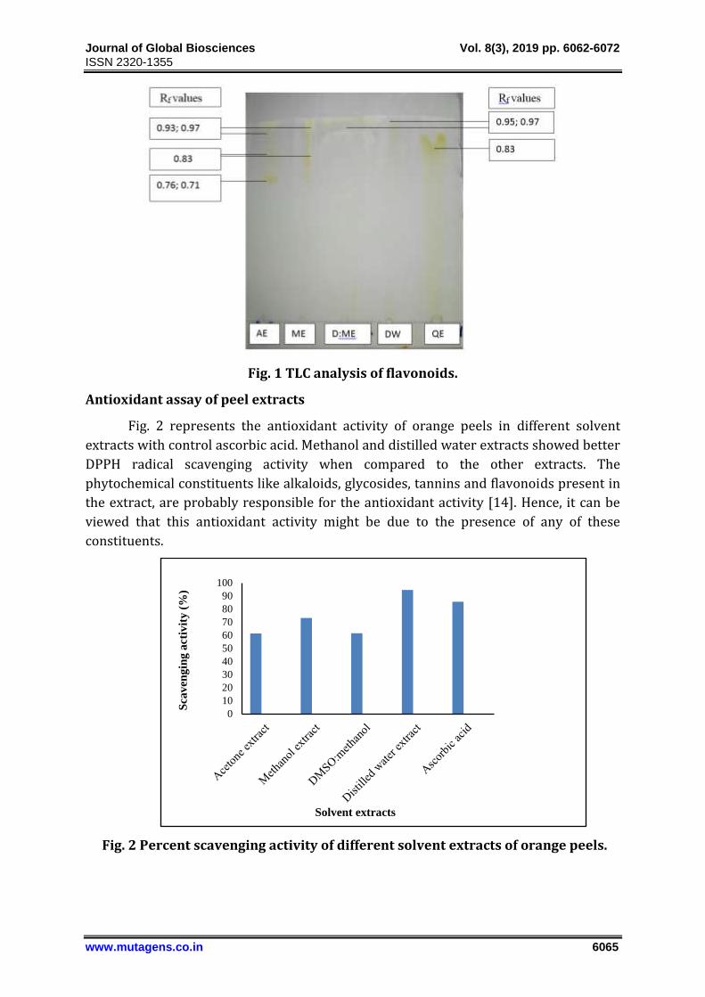
Fig. 2 represents the antioxidant activity of orange peels in different solvent
extracts with control ascorbic acid. Methanol and distilled water extracts showed better
DPPH radical scavenging activity when compared to the other extracts. The
phytochemical constituents like alkaloids, glycosides, tannins and flavonoids present in
the extract, are probably responsible for the antioxidant activity [14]. Hence, it can be
viewed that this antioxidant activity might be due to the presence of any of these
constituents.
Fig. 2 Percent scavenging activity of different solvent extracts of orange peels.
0
10
20
30
40
50
60
70
80
90
100
Sca
ven
gin
g a
ctiv
ity
(%
)
Solvent extracts
Journal of Global Biosciences Vol. 8(3), 2019 pp. 6062-6072
ISSN 2320-1355
www.mutagens.co.in 6066
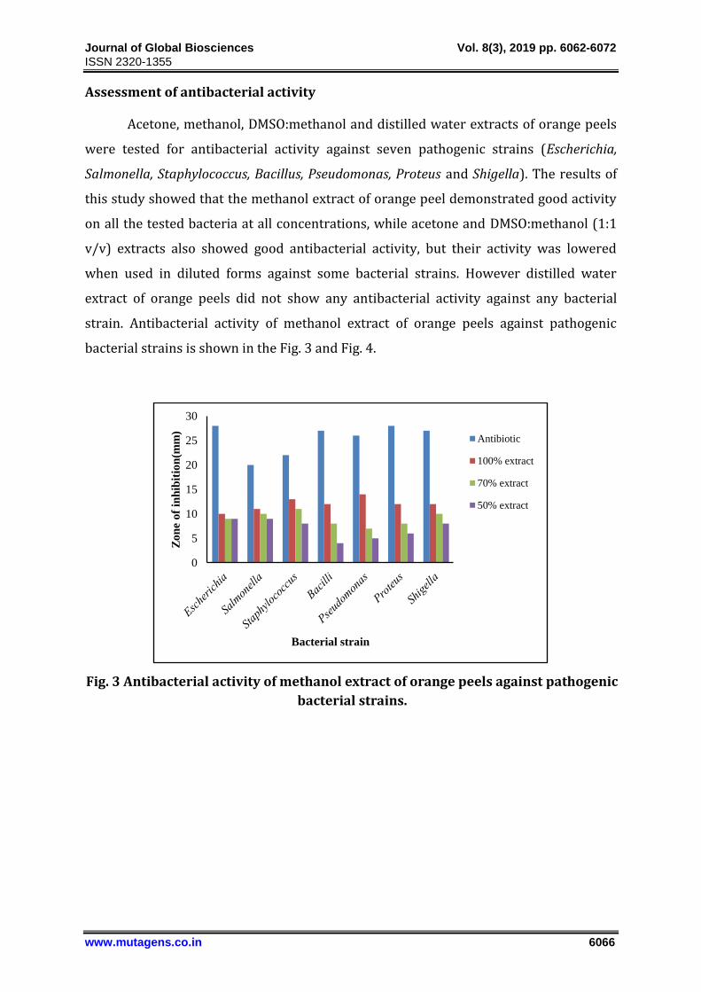
Assessment of antibacterial activity
Acetone, methanol, DMSO:methanol and distilled water extracts of orange peels
were tested for antibacterial activity against seven pathogenic strains (Escherichia,
Salmonella, Staphylococcus, Bacillus, Pseudomonas, Proteus and Shigella). The results of
this study showed that the methanol extract of orange peel demonstrated good activity
on all the tested bacteria at all concentrations, while acetone and DMSO:methanol (1:1
v/v) extracts also showed good antibacterial activity, but their activity was lowered
when used in diluted forms against some bacterial strains. However distilled water
extract of orange peels did not show any antibacterial activity against any bacterial
strain. Antibacterial activity of methanol extract of orange peels against pathogenic
bacterial strains is shown in the Fig. 3 and Fig. 4.
Fig. 3 Antibacterial activity of methanol extract of orange peels against pathogenic
bacterial strains.
0
5
10
15
20
25
30
Zo
ne
of
inh
ibit
ion
(mm
)
Bacterial strain
Antibiotic
100% extract
70% extract
50% extract
Journal of Global Biosciences Vol. 8(3), 2019 pp. 6062-6072
ISSN 2320-1355
www.mutagens.co.in 6067
Escherichia
Staphylococcus
Bacillus
Salmonella
Pseudomonas
Shigella
Proteus
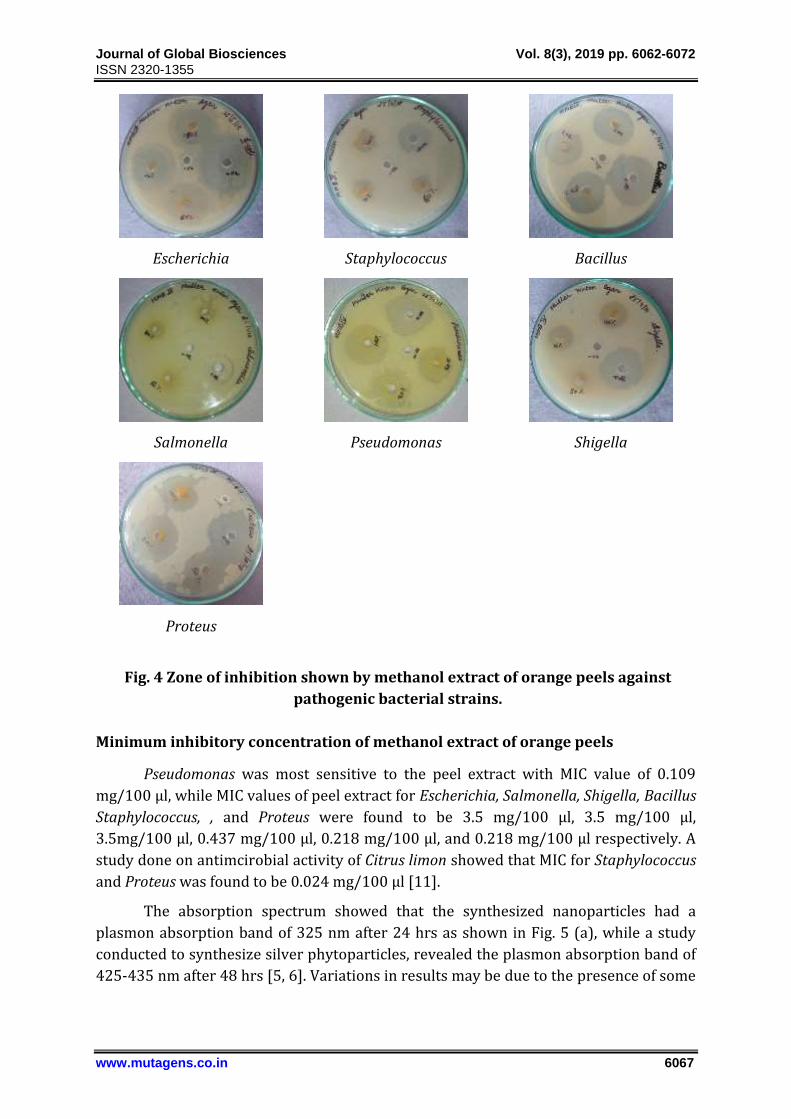
Fig. 4 Zone of inhibition shown by methanol extract of orange peels against
pathogenic bacterial strains.
Minimum inhibitory concentration of methanol extract of orange peels
Pseudomonas was most sensitive to the peel extract with MIC value of 0.109
mg/100 µl, while MIC values of peel extract for Escherichia, Salmonella, Shigella, Bacillus
Staphylococcus, , and Proteus were found to be 3.5 mg/100 µl, 3.5 mg/100 µl,
3.5mg/100 µl, 0.437 mg/100 µl, 0.218 mg/100 µl, and 0.218 mg/100 µl respectively. A
study done on antimcirobial activity of Citrus limon showed that MIC for Staphylococcus
and Proteus was found to be 0.024 mg/100 µl [11].
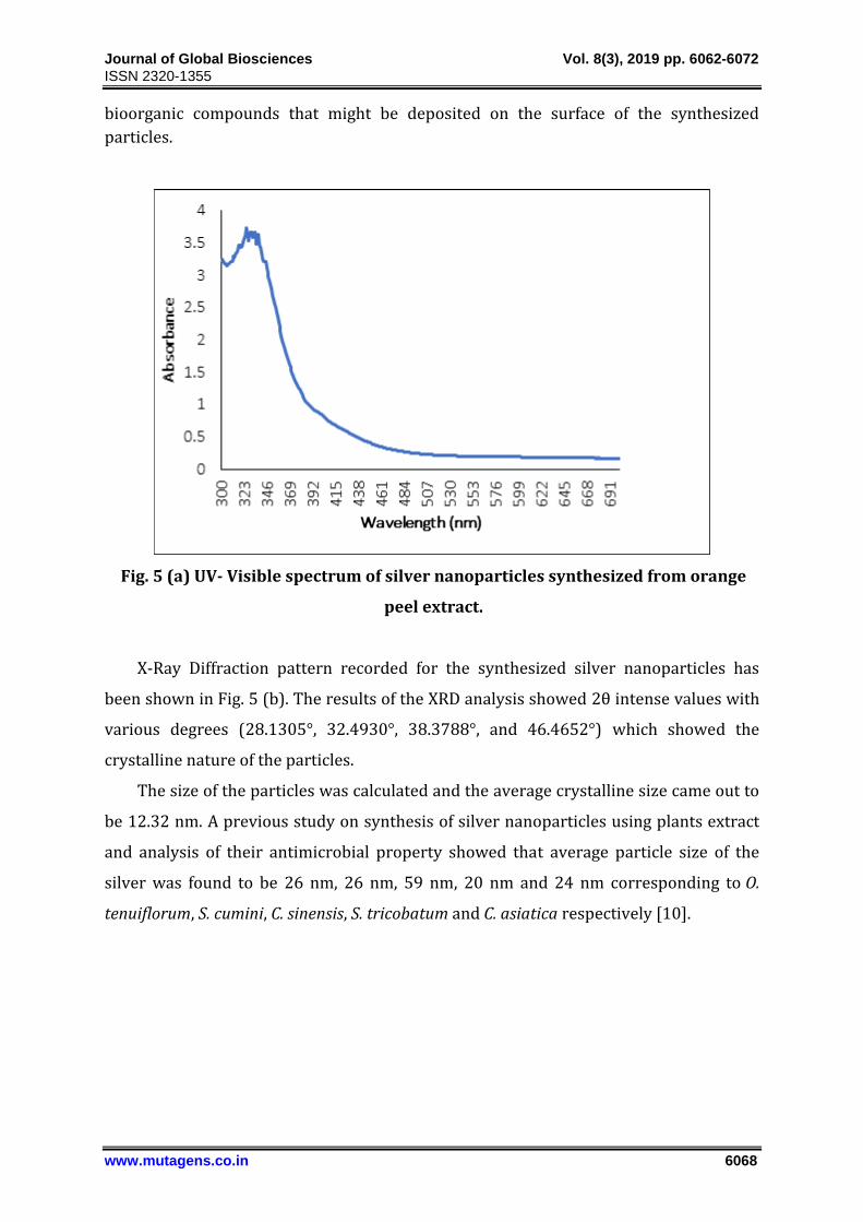
The absorption spectrum showed that the synthesized nanoparticles had a
plasmon absorption band of 325 nm after 24 hrs as shown in Fig. 5 (a), while a study
conducted to synthesize silver phytoparticles, revealed the plasmon absorption band of
425-435 nm after 48 hrs [5, 6]. Variations in results may be due to the presence of some
Journal of Global Biosciences Vol. 8(3), 2019 pp. 6062-6072
ISSN 2320-1355
www.mutagens.co.in 6068
bioorganic compounds that might be deposited on the surface of the synthesized
particles.
Fig. 5 (a) UV- Visible spectrum of silver nanoparticles synthesized from orange
peel extract.
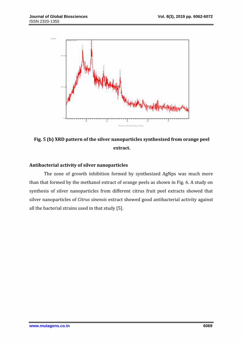
X-Ray Diffraction pattern recorded for the synthesized silver nanoparticles has
been shown in Fig. 5 (b). The results of the XRD analysis showed 2θ intense values with
various degrees (28.1305°, 32.4930°, 38.3788°, and 46.4652°) which showed the
crystalline nature of the particles.
The size of the particles was calculated and the average crystalline size came out to
be 12.32 nm. A previous study on synthesis of silver nanoparticles using plants extract
and analysis of their antimicrobial property showed that average particle size of the
silver was found to be 26 nm, 26 nm, 59 nm, 20 nm and 24 nm corresponding to O.
tenuiflorum, S. cumini, C. sinensis, S. tricobatum and C. asiatica respectively [10].
Journal of Global Biosciences Vol. 8(3), 2019 pp. 6062-6072
ISSN 2320-1355
www.mutagens.co.in 6069
Fig. 5 (b) XRD pattern of the silver nanoparticles synthesized from orange peel
extract.
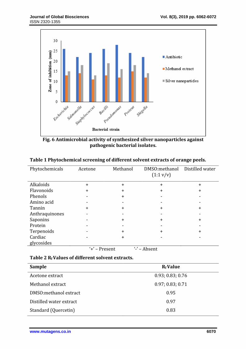
Antibacterial activity of silver nanoparticles
The zone of growth inhibition formed by synthesized AgNps was much more
than that formed by the methanol extract of orange peels as shown in Fig. 6. A study on
synthesis of silver nanoparticles from different citrus fruit peel extracts showed that
silver nanoparticles of Citrus sinensis extract showed good antibacterial activity against
all the bacterial strains used in that study [5].
Journal of Global Biosciences Vol. 8(3), 2019 pp. 6062-6072
ISSN 2320-1355
www.mutagens.co.in 6070
Fig. 6 Antimicrobial activity of synthesized silver nanoparticles against
pathogenic bacterial isolates.
Table 1 Phytochemical screening of different solvent extracts of orange peels.
Phytochemicals Acetone Methanol DMSO:methanol (1:1 v/v)
Distilled water
Alkaloids + + + + Flavonoids + + + + Phenols - + - - Amino acid - - - - Tannin + + + + Anthraquinones - - - - Saponins - + + + Protein - - - - Terpenoids - + + + Cardiac glycosides
- + - -
‘+’ – Present ‘-’ – Absent
Table 2 Rf Values of different solvent extracts.
Sample Rf Value
Acetone extract 0.93; 0.83; 0.76
Methanol extract 0.97; 0.83; 0.71
DMSO:methanol extract 0.95
Distilled water extract 0.97
Standard (Quercetin) 0.83
Journal of Global Biosciences Vol. 8(3), 2019 pp. 6062-6072
ISSN 2320-1355
www.mutagens.co.in 6071
CONCLUSION
The study reveals that orange peel is a rich source of active compounds like alkaloids,
flavonoids with various medicinal and pharmacological properties making them to be
utilized as an attractive, alternate and cheap source of functional ingredients for the
formulation of functional foods and nutraceuticals. Further studies are also required to
unravel and characterize active components present in orange peel.
ACKNOWLEDGEMENTS
The financial support from Department of Biotechnology, Ministry of Science and
Technology, Govt. of India, to Department of Biotechnology, Himachal Pradesh
University, Shimla (India), is thankfully acknowledged. The financial assistance from
DEST (Department of Environment, Science and Technology). Himachal Pradesh, in the
form of a Minor Research Project is thankfully acknowledged.
CONFLICTS OF INTERESTS
The author(s) declare(s) that there is no conflict of interests regarding the publication
of this article.
REFERENCES
[1] Aja PM, Okaka ANC, Onu PN, Ibiam U, Urako AJ. Phytochemical composition of
Talinum triangulare (water leaf) leaves. Pakistan Journal of Nutrition. 2010;6:527-
530.
[2] AL-Daody AC, AL-Hyaly AM, AL-Soultany AA. Chromatographic identification of
some flavonoids compounds from Cyperus rotundas growing in Iraq. Tikrit Journal
of Pure Science. 2010;15:812-816.
[3] Arora M, Kaur P. Phytochemical screening of orange peel and pulp. International
Journal of Research in Engineering and Technology. 2013;2:517-522.
[4] Ashok K, Narayani Subanthini, Jayakumar. Antimicrobial activity and phytochemical
analysis of citrus fruit peels -utilization of fruit waste. International Journal of
Engineering Science and Technology. 2011;3:5414-5421.
[5] Iniaghe OM, Malomo SO, Adebayo JO. Proximate composition and phytochemical
constituents of leaves of some Acalypha species. Pakistan Journal of Nutrition.
2009;8:256-258.
Journal of Global Biosciences Vol. 8(3), 2019 pp. 6062-6072
ISSN 2320-1355
www.mutagens.co.in 6072
[6] Gayathri V, Kiruba D. Preliminary phytochemical analysis of dry leaf powder
extracts of Citrus aurantium. International Journal of Science and Nature.
2014;5:294-296.
[7] Magudapathy P, Dhara S. Electrical transport studies of Ag nanoparticles in glass
matrix. Matter. 2001;299:142-146.
[8] John S, Monica SJ, Priyadarshini S, Arumugam P. Investigation on phytochemical
profile of Citrus limonum peel extract. International Journal of Food Science and
Nutrition. 2017;2:65-67.
[9] Omm-e-H, Asia N, Aamir A. Screening of phytochemical constituted, antimicrobial
and antioxidant activities of orange peel (Citrus sinensis) extract. Bulletin of
Environment, Pharmacology and Life Sciences. 2015;4:102-108
[10] Senthamil SR, Rane ZAK, Anusha B. Phytochemical investigation and in vitro
antioxidant activity of Citrus sinensis pel extract. Der Pharmacia Lettre.
2016;8:159-165.
[11] Reenaa M, Menon AS. Synthesis of silver nanoparticles from different citrus fruit
peel extracts and a comparative analysis on its antibacterial activity. International
Journal of Current Microbiology and Applied Science. 2017;6:2358-2365.
[12] Peter L, Sivagnanam S, Jayanthi A. Synthesis of silver nanoparticles using plants
extract and analysis of their antimicrobial property. Journal of Saudi Chemical
Society. 2015;19:311-317.
[13] Mohanpuria P, Rana NK, Yadav SK. Biosynthesis of nanoparticles: Technology
concepts and future applications. Journal of Nanoparticle Research. 2008;10:507.
[14] Prabhu N, Divya TR, Yamuna G. Synthesis of silver phyto nanoparticles and their
antibacterial efficacy. Digest Journal of Nanomaterials and Biostructures.
2010;5:185-189.