Embed Size (px)
Citation preview
1555Development 122, 1555-1565 (1996)Printed in Great Britain © The Company of Biologists Limited 1996DEV5046
The TGF-β signaling pathway is essential for Drosophila oogenesis
Vern Twombly1, Ronald K. Blackman2, Hui Jin2, Jonathan M. Graff1, Richard W. Padgett3and William. M. Gelbart1
1Department of Molecular and Cellular Biology, Harvard University, Cambridge, MA 02138, USA 2Department of Cell and Structural Biology, 505 S. Goodwin Avenue, University of Illinois, Urbana, IL 61801, USA3Waksman Institute and Department of Molecular Biology and Biochemistry, Rutgers University, Piscataway, NJ 08855, USA
We examine roles of signaling by secreted ligands of theTGF-β family during Drosophila oogenesis. One familymember, the DPP ligand encoded by the decapentaplegic(dpp) gene, is required for patterning of anterior eggshellstructures. This requirement presumably reflects theexpression pattern of dpp in an anterior subset of somaticfollicle cells: the centripetally migrating and the nurse cell-associated follicle cells. Similar requirements are alsorevealed by mutations in the saxophone (sax)-encodedreceptor, consistent with the idea that DPP signaling is, atleast in part, mediated by the SAX receptor. A loss of
germline sax function results in a block in oogenesis asso-ciated with egg chamber degeneration and a failure of thetransfer of nurse cell contents to the oocyte, indicating thatTGF-β signaling is required for these events. Some pheno-types of sax mutations during oogenesis suggest that SAXresponds to at least one other TGF-β ligand as well in theposterior follicle cells.
Key words: TGF-β, signaling, oogenesis, decapentaplegic,Drosophila
SUMMARY
INTRODUCTION
Cell-cell communication is crucial for cell migration and forspecifying pattern during development. During Drosophilamelanogaster oogenesis, extensive cell migrations occur assomatic and germline cells cooperate to generate a viable egg(reviewed by Spradling, 1993). The female ovary consists of15-20 ovariole strands, each representing an independent andprogressive chain of maturing egg chambers (King 1970;reviewed by Spradling, 1993). At the apical end of eachovariole is the germarium, where somatic and germline stemcells proliferate. Within the germarium, germline cellsundergo four incomplete cell divisions forming a 16-cell cystinterconnected by cytoplasmic bridges, termed ring canals.Descendants of somatic stem cells migrate cortically andenter the germarium to enclose each cyst with a monolayerof follicle cells. The enclosed cyst, the egg chamber, exitsthe germarium. The somatic follicle cells continue dividingup to stage 6 (where they number ~1100) while the nursecells enlarge and transfer small amounts of specific RNAsand proteins to the oocyte where yolk becomes visible bystage 8. By stage 9 nearly all the follicle cells migrate pos-teriorly over the oocyte. At this time, the anteriormost 6-10follicle cells (termed border cells) delaminate and migrateposteriorly between the nurse cells toward the anterior poleof the oocyte.
By stage 10A, the oocyte spans the posterior half of theegg chamber while the anterior half is composed of the nursecells. The chamber remains covered by a layer of follicle cellsthat have reorganized so that ~95% are a columnar epithe-
lium over the oocyte and ~5% are thinly stretched over thenurse cells. The columnar centripetally migrating folliclecells (CMFC) at the oocyte-nurse cell boundary begin amigration inward between the oocyte and nurse cells andpause as they approach the border cells, which are centeredimmediately anterior of the oocyte. At stage 10B, cells fromthe dorsal anterior columnar epithelium overlying the oocyteare recruited to form respiratory filaments, the dorsalappendages. Later in stage 10B, the nurse cells begin rapidlytransferring their cytoplasmic contents, causing an anteriorretreat of the diminishing nurse cells and a concomitantenlargement of the oocyte. This transfer is completed inapproximately 30 minutes. The CMFC and border cells arethen thought to direct the production of anterior eggshellstructures: the micropyle (the sperm entry point), theoperculum (the hatch from which the embryo exits) and theventral collar (a structure circumscribing the operculum). Bystage 14, the nurse cell nuclei degrade and anterior eggshellstructures are completed.
Considerable cell communication and migration is requiredfor these events. The signals that orchestrate follicle cell migra-tions and the transfer of nurse cell contents to the oocyte havenot been determined. Here we investigate roles of the Trans-forming Growth Factor-β (TGF-β) signaling in potentiatingsuch activities (reviewed by Attisano et al., 1994; Kingsley,1994; Miyazono et al., 1994). In Drosophila melanogaster,three members of the TGF-β superfamily are encoded by thegenes decapentaplegic (dpp; Padgett et al., 1987), 60A(Wharton et al., 1991; Doctor et al., 1992) and screw (scw;Arora et al., 1994). dpp has been shown to be capable of
1556 V. Twombly and others
signaling across germ layers. During early embryogenesis,dorsal ectoderm expression of dpp induces determination ofdorsal versus ventral mesoderm (Staehling-Hampton et al.,1994; Frasch 1995). Later, mesoderm expression of dpp directscell fate changes in the underlying endoderm (Panganiban etal., 1990; Immerglück et al., 1990).
TGF-β-related factors are thought to signal through type Iand type II serine/threonine receptor kinases, distinguished bytheir sequence and ligand-binding characteristics (reviewed byDerynck 1994; Massagué and Polyak 1995). Recent charac-terization of DPP receptors has also shown a requirement forboth type I and type II receptor subunits. Two DPP type Ireceptors are encoded by the saxophone (sax) and thick veins(tkv) genes (Brummel et al. 1994; Nellen et al., 1994; Pentonet al., 1994; Xie et al., 1994) and a DPP type II receptor isencoded by the punt gene (Letsou et al., 1995; Ruberte et al.,1995). All three receptors are thought to contribute to DPPsignaling, though the requisite level of each receptor and thenature of the in vivo heteromeric receptor complex are not fullyunderstood. Further, it is not known if these receptors partici-pate in the signaling of the other known Drosophila ligands60A and SCW.
Previous work has shown that dpp is not required in thegermline during oogenesis (Irish and Gelbart, 1987). In thisreport, we show dpp function is required in the somaticcomponent of Drosophila ovaries. We find that dppexpression is restricted to a subset of migrating anteriorfollicle cells. We demonstrate an essential germline require-ment for sax, suggesting that a TGF-β signaling event maybe transmitted from the soma to the germline duringoogenesis. Our analysis of oogenesis in females mutant forDPP signaling elements demonstrate that this TGF-β pathwayis necessary to maintain egg chamber integrity, generateanterior eggshell structures and transfer nurse cell contents tothe oocyte. In addition, there is likely to be a contribution ofan additional TGF-β signal for proper patterning within theposterior egg chamber.
MATERIALS AND METHODS
Drosophila strains Canton-S or y w67c23 strains were used as wild type. The mutants usedin this study are referenced in FlyBase (1996) unless otherwise noted.sax1rv5 is a loss-of-function sax allele recovered by Marnie Gelbart asan EMS-induced dominant suppressor of Df(2L)Mad sax1 / Mad+sax+
female sterility. Loss-of-function alleles sax4 and sax5 were EMS-induced on a nub b pr isogenic chromosome as recessive lethalsexposed by Df(2R)H23 (and all other available deletions of the43E18-43F2 region) and are rescued by sax transgenes (V. T., unpub-lished results). Strains were cultured on standard cornmeal, yeastextract, dextrose medium at 25°C, unless stated otherwise.
Characterization of dpp10638
dpp10638 is a P element insertion initially characterized by theBerkeley Drosophila Genome Project. Plasmid rescue was employedto recover the left half of the dpp10638 transposon and adjoininggenomic sequences. Three clones were recovered extending from anXbaI site in the middle of the PZ transposon (Jacobs et al., 1989) toan XbaI or NheI site in the dpp gene at molecular map coordinate 83(St. Johnston et al., 1990). Clone pPR11 was sequenced outward fromthe P element into the dpp genomic DNA. The transcription units ofdpp and the lacZ gene in PZ are divergently oriented.
hs-tsax and hs-dpp plasmid and strain constructionsP{w+mC sax∆IC.1=hs-tsax.T-G}1.1, (P{hs-tsax})The truncated SAX receptor was generated as in Graff et al. (1994).This modified cDNA was cloned into the pHsp70 vector (Pirotta,1988) and transformed into y w67c23 embryos.
P{w+mC dpphs.P-BP=hs-dpp.B-P}, (8xP{hs-dpp})Genomic dpp sequences extending beyond the polyadenylation sitewere substituted for 3′ sequences of the dpp cDNA, E55 (Padgett et al.,1987). These sequences were cloned into the pHsp70 vector andinjected into y w67c23 embryos (Pirotta, 1988). A strain homozygous forfour distinct insertions (at 2B, 16F-17A, 61E and 97) was generated.
Histochemical staining and ovariole phenotypes 1- to 2-day-old females were conditioned for 3 days at 25°C onyeasted media and ovaries were then dissected in Drosophila Ringer’ssolution + 0.05% BSA (DR+BSA), unless stated differently in figurelegends. Ovarioles were dissociated, fixed for 5 minutes in 4%formaldehyde and washed 3× with DR+BSA. To assay β-galactosi-dase activity, ovaries were incubated in X-gal-staining solution(Bellen et al., 1989) saturated with X-gal at 37°C for 12-16 hours.Ovaries were mounted in 50% glycerol and examined under Nomarskioptics. Rhodamine-conjugated phalloidin staining was performed asin Cooley et al. (1992). Phalloidin-labelled ovaries were mounted inVectashield and photographed with a Zeiss Axiophot.
Characterization of eggshell phenotypesdppe87 / dpphr56 females were maintained at 29°C and egg collectionsspanned days 4 through 7. 8xP{hs-dpp} females were maintained at25°C and heat-shocked for 45 minutes at 37°C on both day 3 and day4. Eggs were harvested 8 hours after the second heat-shock. P{hs-sax}/ + ; sax− / Df(2R)sax-H9 females were maintained at 18°C and col-lections were performed from day 2 to 6. tkv7sax+ / tkv+sax1 femaleswere maintained at 25°C and eggs were harvested on days 4 through7. Eggs were harvested from yeasted grape juice agar plates, mountedin 3:1 Hoyer’s : lactic acid and photographed under dark-field optics.
Eggshell measurementsEggshells and an optical micrometer were photographed at the samemagnification and the resulting images were used for measurements.Egg lengths were measured from the collar to the posterior aeropyle.Opercula were measured from the ventral collar to the anterior dorsalappendage attachment point. Dorsal appendage measurements span thedistal tip to the posterior attachment point. For wild type at 25°C, themean length of the egg, dorsal appendage and operculum were0.58±0.03 mm, 0.32±0.01 mm and 0.11±0.01 mm, respectively (n=25).
Germline clonal analysis of saxFemales containing FRT constructs, P{>whs>} sax+sca bw,P{>whs>} sax4 sha1 / CyO and P{>whs>} sax5 sha1 / CyO (kindlyprovided by Dr Matthew Singer) were crossed to P{hs-FLP} / Y ;P{>whs>} P{ovoD1=18}32X9 / CyO males. These adults were trans-ferred every 48 hours to new culture bottles. The progeny of this crosswere heat-shocked at 24 hours after the onset of pupation, (±24 hours),as in Chou et al. (1993). Within single vials, 1- to 2-day-old individ-ual females of the genotype P{hs-FLP} / + ; P{>whs>}## / P{>whs>}P{ovoD1=18}32X9 (where ##=sax+ sca bw, sax4 sha1, or sax5 sha1)were continuously provided three males and egg production wasmonitored every other day as the culture vial was transferred.
RESULTS
The expression of decapentaplegic duringoogenesisA P-element enhancer trap insertion in dpp accurately
1557DPP signaling in Drosophila oogenesis
60 70 80 90 100 110AB D/CE
3'5'
10638
shv disk
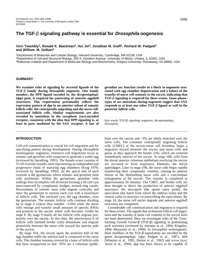
Fig. 1. Location of the P element in dpp10638. Schematicrepresentation of the insertion site (triangle) of P{PZ}l(2)10638 inthe decapentaplegic genomic region (St. Johnston et al., 1990). Thecoding exons and the four known 5′ untranslated exons are denotedbelow the genomic walk as black and white rectangles, respectively.The shaded rectangles above the genomic walk represent the cis-regulatory shv (shortvein) and disk (imaginal disk) domains.
reflects dpp expression during oogenesis. This insertion,originally isolated by Karpen and Spradling (1992) is arecessive embryonic lethal dpp allele and is now nameddpp10638. The insertion site is 218 bp 5′ of the dpp transcriptA start site (Fig. 1). In addition to recapitulating the dppembryonic and imaginal disk expression patterns (data notshown), dpp10638 also exhibits ovarian lacZ expression, alocation for which dpp expression has not been previouslydescribed. RNA in situ hybridization to whole-mount ovarieswere performed to ensure that the lacZ expression seen withdpp10638 represented the endogenous dpp ovary expressionpattern. dpp RNA was detected in the same temporal andspatial pattern in the ovary visualized by lacZ expression ofdpp10638 (Fig. 2A,H). In the following section, we will refer
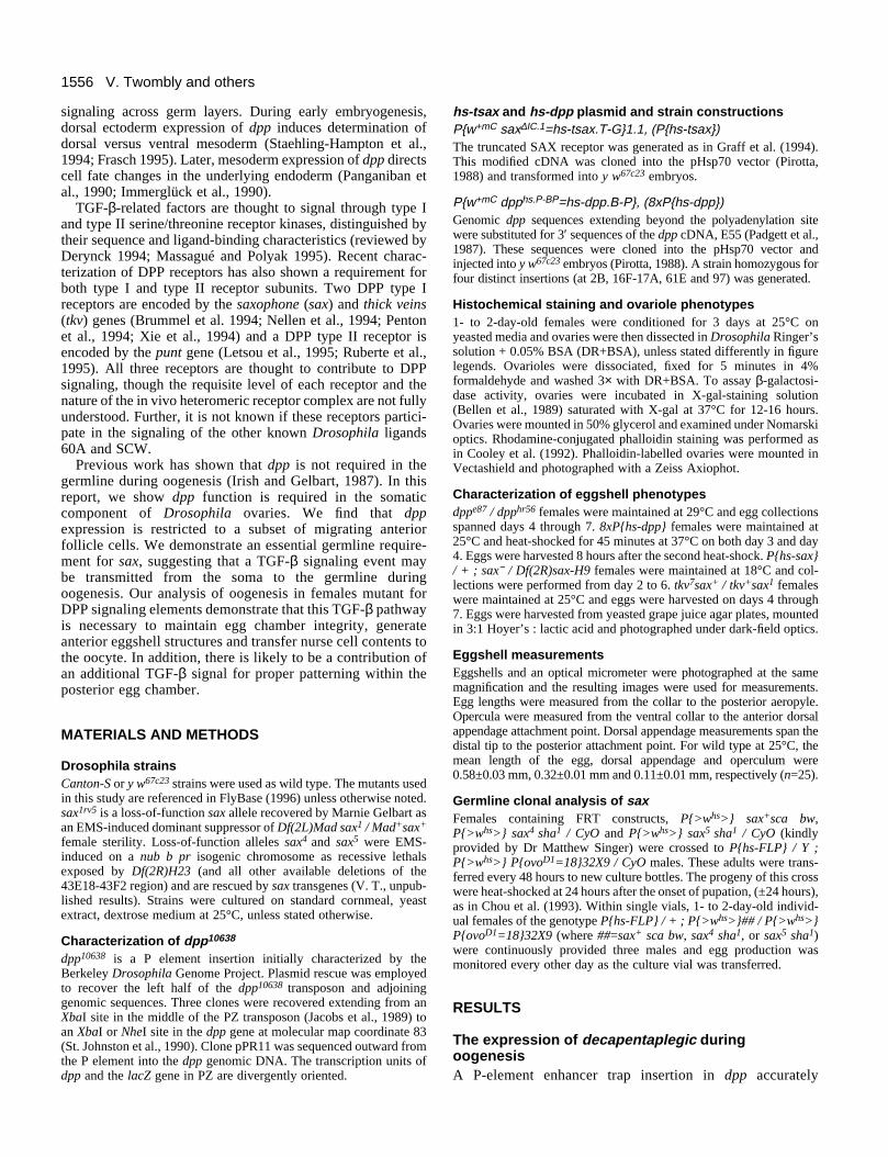
Fig. 2. dpp expression during oogenesis. Localization of dpp transcript acof the dpp10638 enhancer trap (B-G). Anterior is to the left in this and subRNA is absent from the germarium (g) and early stage egg chambers, bu(CMFC, triangle). dpp RNA was also detected in the nurse cell follicle celacZ expression begins in late stage 8 chambers. The domain of lacZ expview of stage 10A expression in the nurse cell follicle cells (NFC) and a chamber shows lacZ expression in the leading edge of the CMFC, whichthe nurse cells (NC) and the oocyte (O). (E,F) By the end of stage 10B, thmigration (E) and remain immediately anterior of the expanding oocyte (visible in this focal plane) and the CMFC encircling the developing micr(H) Endogenous dpp transcripts are clearly visible in follicle cells around
to this lacZ expression pattern in the ovary as the dppexpression pattern.
dpp expression is first detectable at the end of stage 8 inapproximately 20 to 30 somatic follicle cells at the anterior tipof the egg chamber (Fig. 2B). The number of cells expressingdpp increases as they migrate cortically and posteriorly untilexpression is seen over the anterior third of the egg chamberat the end of stage 9 (Fig. 2B). The progression of the mostposterior transverse plane of dpp-expressing cells is roughlythe same plane as that of the border cells, which themselves donot express dpp (data not shown). By stage 10A, all folliclecells have completed their posterior movement. Here, thenumber of dpp-expressing cells has increased and includes ~50thinly stretched nurse cell follicle cells (NFC) and approxi-mately 20 columnar follicle cells overlying the nurse cell-oocyte boundary (Fig. 2C,D). The increase in the number ofdpp-expressing cells during stages 8 through 10A may arisefrom maintenance of expression in the daughter cells of cellspreviously expressing dpp or through newly inducedexpression. At stage 10B, the cells at the nurse cell-oocyteboundary begin a centripetal migration inward between theoocyte and the nurse cells. Here, dpp is expressed in the leadingedge (~20 cells) of the approximately 150 columnar cen-tripetally migrating follicle cells (CMFC), which moveadjacent to the border cells (Fig. 2D,E). From stage 11 onward,dpp expression continues in the NFC and CMFC populationsas they migrate or are displaced anteriorly over the expandingoocyte (Fig. 2F). At stages 13-14, dpp expression is limited tothe NFC and a two cell tall cylinder encircling the nearlycomplete micropyle (Fig. 2G).
In summary, dpp is expressed in the outer somatic layer offollicle cells in the anterior portion of the developing eggchamber. This expression almost completely surrounds the
cumulation by RNA in situ hybridization (A,H) and by lacZ expressionsequent figures, unless stated otherwise. Dorsal is up in E-H. (A) dppt is expressed at later stages in the centripetally migrating follicle cellslls (not visible in this focal plane). (B) Superficial views of dpp10638
ression extends posteriorly during stages 9 through 10A. (C) Superficialsubset of the CMFC. (D) An optical cross-section of a stage 10B have begun to migrate inward (in the direction of the arrows), betweene lacZ-expressing CMFC (arrow) have completed their centripetal
F). (G) From stage 12 to 14, expression remains on in the NFC (notopyle (mp). The collar (c), demarks the edge of the operculum. the micropyle at stage 14.
1558 V. Twombly and others
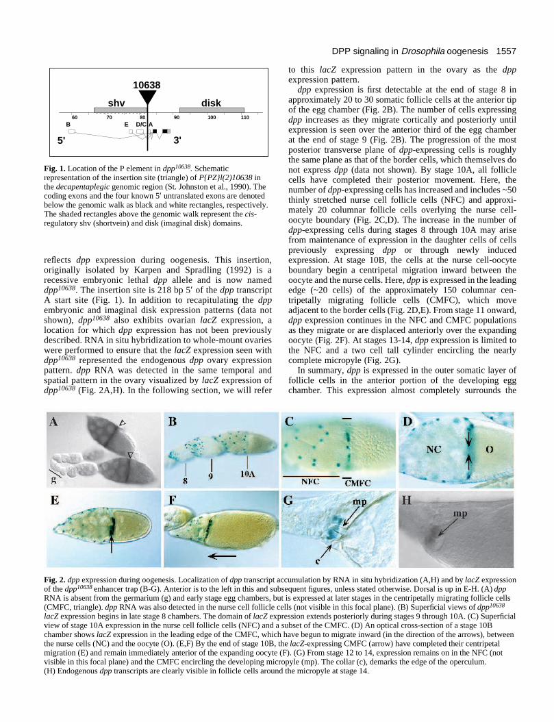
Fig. 3. Eggshell phenotypes associatedwith alterations in DPP signaling. Theeggs in B, E and H are side views, withdorsal up. The other eggs are top views.(A-C) Morphologically wild-type eggsproduced by control females. (A) Thedorsal appendages (da) are broader atthe distal end than they are proximally.The operculum (op) comprises theregion between the vertical bars.(B) Lateral view; (C) highmagnification of the anterior eggshellstructures. Note the micropyle (mp) andthe thinner appearance of the eggshellof the operculum. (D-F) Eggs resultingfrom defects in DPP signaling. Theseeggs are derived from dppe87 / dpphr56
females reared at 18°C and thencultured at restrictive temperature(29°C). (D) The egg length, operculumand dorsal appendages are all reduced.Small anterior protrusions of ectopiceggshell (arrow) were commonlyobserved. (E) Lateral view; (F) high
magnification reveals a complete loss of the operculum and a malformed micropyle. (G-I) Mutant phenotypes from heat-shocked 8xP{hs-dpp} females. (G,H) Eggs display enlarged opercula and deranged or absent dorsal appendages (arrow in G, missing in H). (I) The enlargedoperculum retains some elements of the wild-type follicle cell imprint pattern (see Fig. 3C).
A B
D E F
IHG
op
mpop
mp
C
da
op
germline nurse cells and includes cell types implicated in theproduction of anterior eggshell structures.
Altered levels of dpp affect anterior eggshellstructuresBecause of the zygotic lethality associated with loss-of-function dpp alleles, we had to rely on conditional partial loss-of-function alleles to examine the contribution of dpp tooogenesis. At 25°C, dppe87 / dpphr56 animals die while, at18°C, many survive to adulthood (Wharton et al., 1996). Toassess maternal effects of dpp on oogenesis, such survivingdppe87 / dpphr56 females were mated to wild-type males andcultured at permissive (18°C) and restrictive (29°C) tempera-tures. Unlike the case for control females (wild type culturedat 29°C and dppe87 / dpphr56 at 18°C; Fig. 3A-C), a subset ofeggs generated by mutant females reared at 29°C displayanterior eggshell defects. Some abnormal eggshells exhibitextremely reduced opercula (Fig. 3F) and slightly reduceddorsal appendages while the other class displays a moderatelyshortened egg length (Table 1; Fig. 3D,E). In many cases, theoperculum defects are accompanied by small protrusions ofectopic eggshell extending off the anterior pole and, morerarely, malformations of the micropyle (Fig. 3F). Thus, dimin-ished levels of DPP are associated with a loss of anterioreggshell structures. Since dpp is completely dispensible in thegermline (Irish and Gelbart, 1987), we infer that these partialloss-of-function phenotypes are due to reduced expression ofDPP in the anterior follicle cells.
If dpp plays a role in the fating or activity of anterior folliclecells, then ubiquitous follicle cell expression of dpp might beexpected to increase the extent of eggshell structures producedby the anterior follicle cell populations. Indeed, eggs producedby females ubiquitously expressing dpp (through an Hsp70-dpp transgene) display expanded anterior eggshell structures
(Fig. 3G-I). 8x P{hs-dpp} females received a 37°C heat-shockand were then maintained at 29°C. Nearly all resulting eggshave abnormal dorsal appendages and 58% display abnormal-ities that we interpret as vastly enlarged opercula (Table 1).The opercula are minimally 150% larger than those of wild-type eggs and can span 50% of the egg length (Fig. 3G,H).Nearly a third of these eggs completely lack dorsal appendageswhile one-sixth display multiple spurs of dorsal appendagematerial or branching ‘antler-like’ dorsal appendages along thecircumference of the operculum (Fig. 3G). Eggs from controlfemales (wild type at 29°C and 8xP{hs-dpp} maintained at18°C) show none of these abnormalities. Thus, ubiquitousexpression of dpp has the opposite effect on eggshell develop-ment - expansion of anterior eggshell domains - to thatgenerated by partial loss of dpp expression.
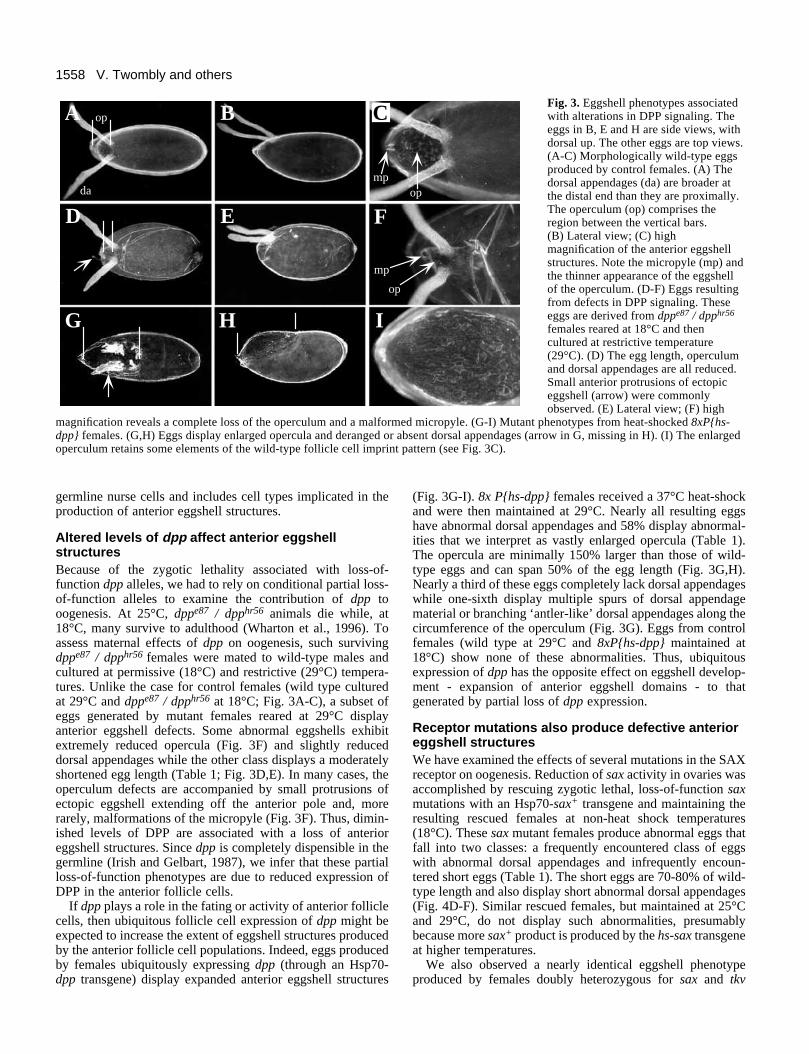
Receptor mutations also produce defective anterioreggshell structuresWe have examined the effects of several mutations in the SAXreceptor on oogenesis. Reduction of sax activity in ovaries wasaccomplished by rescuing zygotic lethal, loss-of-function saxmutations with an Hsp70-sax+ transgene and maintaining theresulting rescued females at non-heat shock temperatures(18°C). These sax mutant females produce abnormal eggs thatfall into two classes: a frequently encountered class of eggswith abnormal dorsal appendages and infrequently encoun-tered short eggs (Table 1). The short eggs are 70-80% of wild-type length and also display short abnormal dorsal appendages(Fig. 4D-F). Similar rescued females, but maintained at 25°Cand 29°C, do not display such abnormalities, presumablybecause more sax+ product is produced by the hs-sax transgeneat higher temperatures.
We also observed a nearly identical eggshell phenotypeproduced by females doubly heterozygous for sax and tkv
1559DPP signaling in Drosophila oogenesis
Table 1. Eggshell phenotypes associated with disruptions in the TGF-β signaling pathwayDorsal appendages (DA) Operculum (OP) Egg shape
Genotype n Wild-type Abnormala Fused Absent Reducedb Enlargedc Wild-type Shortd Ventralized
Canton S 408 89% 10% 0 0 0 0 99% 1% 0@ 29°C
dppe87/dpphr56 374 2% 98% 0 0 93% 0 41% 59% 0@ 29°C
Canton S 387 86% 4% 0 0 0 0 97% 3% 0@ 29°C with heat shock
8 x hs-dpp 307 0 71%e 0 29% 0 58% 100% 0 0@ 29°C with heat shock
Canton S 1655 92% 7% 0.3% 0 na na 97% 0.5% 2.1%@ 18°Cf
hs-sax/+ ; sax1rv1/Df(2R)sax-H9 858 69% 29% 2% 0 na na 94% 4.3% 1.4%@ 18°Cf
hs-sax/+ ; sax5/Df(2R)sax-H9 1247 81% 15% 4% 0 na na 96% 1.5% 2.5%@ 18°Cf
hs-sax/+ ; sax5/sax1rv1 927 55% 33% 12% 0 na na 89% 10.7 0.2%@ 18°Cf
tkv7 +/+ sax1rv5 189 61% 39% 0 0 na na 99% 0.5% 0@ 25°C
tkv7 +/+ sax1 437 7% 5% 48% 0 na na 69% 31% 0@ 25°C
na = not applicable. This is because the CMFC may not migrate to a position that would engender the phenotypes seen from dpp mutants.aDA had one or more of the following phenotypes; short, thin, broad, or the two DA of a single egg were substantially different lengths.bOP were 0-50% of wild-type length.cOP were greater than 150% of wild-type length.dEgg lengths were approximately 70-85% of wild-type length.eDA reductions were different than in the above three genotypes and were associated with enlarged operculums. In this case 42% of the total had moderately
broadened DA, 14% displayed DA 3-4 times the wildtype width, and 21% exhibited spurs of DA material along the edge of the operculum or they ‘antler-like’DA branches.
fVirgin females were crossed at 18o for 3 days at which point egg collections began. Eggs were collected daily and scored for 8 days.
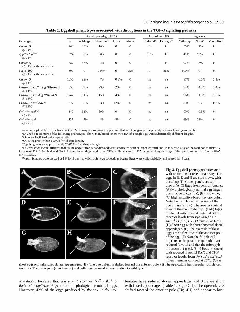
Fig. 4. Eggshell phenotypes associatedwith reductions in receptor activity. Theeggs in B, E and H are side views, withdorsal up. The other panels are topviews. (A-C) Eggs from control females.(A) Morphologically normal egg length;dorsal appendages (da); (B) side view;(C) high magnification of the operculum.Note the follicle cell patterning of theoperculum (arrow). The inset is a lateralview of the micropyle (mp). (D-F) Eggsproduced with reduced maternal SAXreceptor levels from P{hs-sax} / + ;sax1rv1 / Df(2L)sax-H9 females at 18°C.(D) Short egg with short abnormal dorsalappendages. (E) The opercula of theseeggs are shifted toward the anterior poleof the egg. (F) Note the follicle cellimprints in the posterior operculum arereduced (arrow) and that the micropyleis abnormal (inset). (G-I) Eggs producedwith reduced maternal SAX and TKVreceptor levels, from tkv7sax+ / tkv+sax1
mutant females cultured at 25°C. (G) Ashort eggshell with fused dorsal appendages. (H). The operculum is shifted toward the anterior pole. (I) The operculum has irregular follicle cellimprints. The micropyle (small arrow) and collar are reduced in size relative to wild type.
A B C
D E F
G H I
da
da
mutations. Females that are sax1 / sax+ or tkv7 / tkv+ ortkv7sax+ / tkv+sax1rv5 generate morphologically normal eggs.However, 42% of the eggs produced by tkv7sax+ / tkv+sax1
females have reduced dorsal appendages and 31% are shortwith fused appendages (Table 1; Fig. 4G-I). The opercula areshifted toward the anterior pole (Fig. 4H) and appear to lack
1560 V. Twombly and others
normal follicle cell imprint patterns (Fig. 4I). In summary,mutations in what are thought to be DPP receptors produceshort eggs with reduced anterior structures. While the sameanterior structures are affected in eggs derived from either dppor receptor mutant females, the latter produce more severedefects.
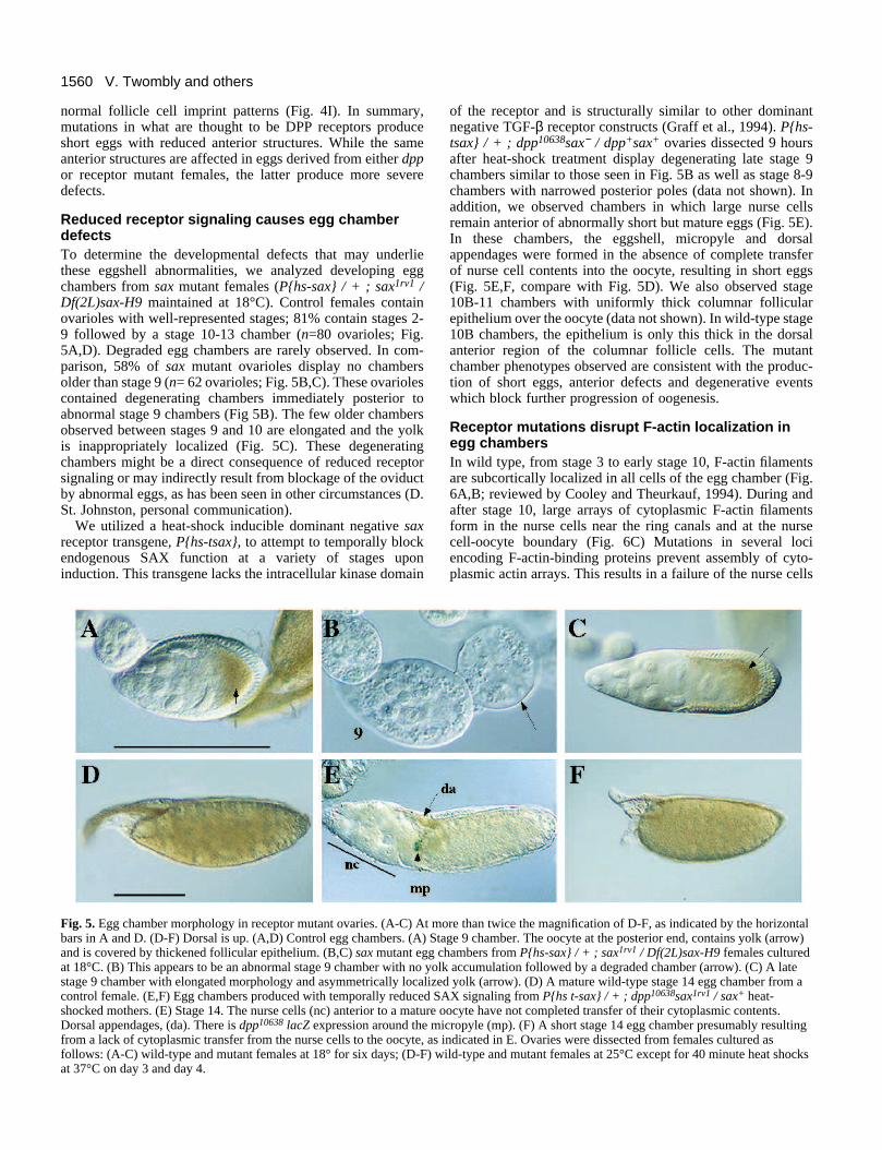
Reduced receptor signaling causes egg chamberdefectsTo determine the developmental defects that may underliethese eggshell abnormalities, we analyzed developing eggchambers from sax mutant females (P{hs-sax} / + ; sax1rv1 /Df(2L)sax-H9 maintained at 18°C). Control females containovarioles with well-represented stages; 81% contain stages 2-9 followed by a stage 10-13 chamber (n=80 ovarioles; Fig.5A,D). Degraded egg chambers are rarely observed. In com-parison, 58% of sax mutant ovarioles display no chambersolder than stage 9 (n= 62 ovarioles; Fig. 5B,C). These ovariolescontained degenerating chambers immediately posterior toabnormal stage 9 chambers (Fig 5B). The few older chambersobserved between stages 9 and 10 are elongated and the yolkis inappropriately localized (Fig. 5C). These degeneratingchambers might be a direct consequence of reduced receptorsignaling or may indirectly result from blockage of the oviductby abnormal eggs, as has been seen in other circumstances (D.St. Johnston, personal communication).
We utilized a heat-shock inducible dominant negative saxreceptor transgene, P{hs-tsax}, to attempt to temporally blockendogenous SAX function at a variety of stages uponinduction. This transgene lacks the intracellular kinase domain
Fig. 5. Egg chamber morphology in receptor mutant ovaries. (A-C) At mbars in A and D. (D-F) Dorsal is up. (A,D) Control egg chambers. (A) Sand is covered by thickened follicular epithelium. (B,C) sax mutant egg at 18°C. (B) This appears to be an abnormal stage 9 chamber with no yostage 9 chamber with elongated morphology and asymmetrically localizcontrol female. (E,F) Egg chambers produced with temporally reduced Sshocked mothers. (E) Stage 14. The nurse cells (nc) anterior to a matureDorsal appendages, (da). There is dpp10638 lacZ expression around the mfrom a lack of cytoplasmic transfer from the nurse cells to the oocyte, asfollows: (A-C) wild-type and mutant females at 18° for six days; (D-F) wat 37°C on day 3 and day 4.
of the receptor and is structurally similar to other dominantnegative TGF-β receptor constructs (Graff et al., 1994). P{hs-tsax} / + ; dpp10638sax− / dpp+sax+ ovaries dissected 9 hoursafter heat-shock treatment display degenerating late stage 9chambers similar to those seen in Fig. 5B as well as stage 8-9chambers with narrowed posterior poles (data not shown). Inaddition, we observed chambers in which large nurse cellsremain anterior of abnormally short but mature eggs (Fig. 5E).In these chambers, the eggshell, micropyle and dorsalappendages were formed in the absence of complete transferof nurse cell contents into the oocyte, resulting in short eggs(Fig. 5E,F, compare with Fig. 5D). We also observed stage10B-11 chambers with uniformly thick columnar follicularepithelium over the oocyte (data not shown). In wild-type stage10B chambers, the epithelium is only this thick in the dorsalanterior region of the columnar follicle cells. The mutantchamber phenotypes observed are consistent with the produc-tion of short eggs, anterior defects and degenerative eventswhich block further progression of oogenesis.
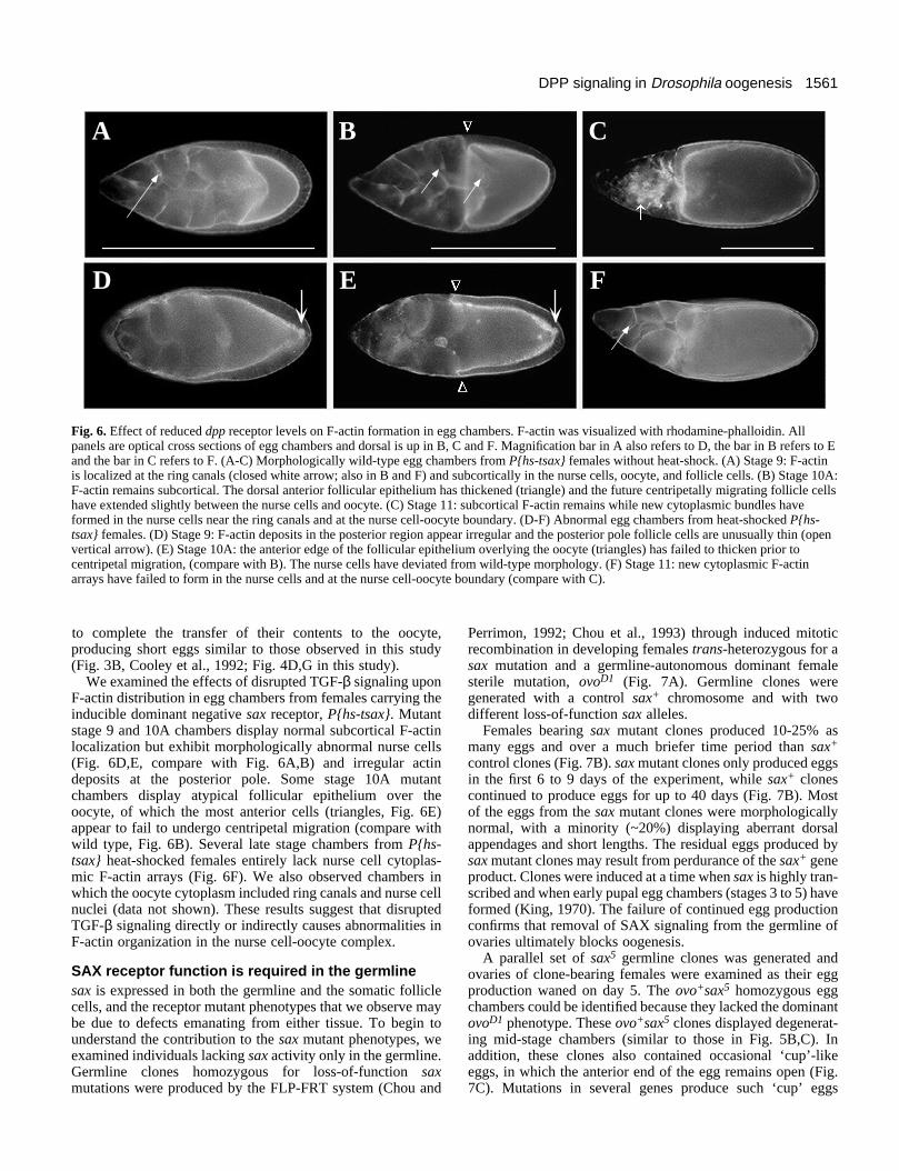
Receptor mutations disrupt F-actin localization inegg chambers In wild type, from stage 3 to early stage 10, F-actin filamentsare subcortically localized in all cells of the egg chamber (Fig.6A,B; reviewed by Cooley and Theurkauf, 1994). During andafter stage 10, large arrays of cytoplasmic F-actin filamentsform in the nurse cells near the ring canals and at the nursecell-oocyte boundary (Fig. 6C) Mutations in several lociencoding F-actin-binding proteins prevent assembly of cyto-plasmic actin arrays. This results in a failure of the nurse cells
ore than twice the magnification of D-F, as indicated by the horizontaltage 9 chamber. The oocyte at the posterior end, contains yolk (arrow)chambers from P{hs-sax} / + ; sax1rv1 / Df(2L)sax-H9 females culturedlk accumulation followed by a degraded chamber (arrow). (C) A lateed yolk (arrow). (D) A mature wild-type stage 14 egg chamber from aAX signaling from P{hs t-sax} / + ; dpp10638sax1rv1 / sax+ heat-
oocyte have not completed transfer of their cytoplasmic contents.icropyle (mp). (F) A short stage 14 egg chamber presumably resulting indicated in E. Ovaries were dissected from females cultured asild-type and mutant females at 25°C except for 40 minute heat shocks
1561DPP signaling in Drosophila oogenesis
A B C
D E F
Fig. 6. Effect of reduced dpp receptor levels on F-actin formation in egg chambers. F-actin was visualized with rhodamine-phalloidin. Allpanels are optical cross sections of egg chambers and dorsal is up in B, C and F. Magnification bar in A also refers to D, the bar in B refers to Eand the bar in C refers to F. (A-C) Morphologically wild-type egg chambers from P{hs-tsax} females without heat-shock. (A) Stage 9: F-actinis localized at the ring canals (closed white arrow; also in B and F) and subcortically in the nurse cells, oocyte, and follicle cells. (B) Stage 10A:F-actin remains subcortical. The dorsal anterior follicular epithelium has thickened (triangle) and the future centripetally migrating follicle cellshave extended slightly between the nurse cells and oocyte. (C) Stage 11: subcortical F-actin remains while new cytoplasmic bundles haveformed in the nurse cells near the ring canals and at the nurse cell-oocyte boundary. (D-F) Abnormal egg chambers from heat-shocked P{hs-tsax} females. (D) Stage 9: F-actin deposits in the posterior region appear irregular and the posterior pole follicle cells are unusually thin (openvertical arrow). (E) Stage 10A: the anterior edge of the follicular epithelium overlying the oocyte (triangles) has failed to thicken prior tocentripetal migration, (compare with B). The nurse cells have deviated from wild-type morphology. (F) Stage 11: new cytoplasmic F-actinarrays have failed to form in the nurse cells and at the nurse cell-oocyte boundary (compare with C).
to complete the transfer of their contents to the oocyte,producing short eggs similar to those observed in this study(Fig. 3B, Cooley et al., 1992; Fig. 4D,G in this study).
We examined the effects of disrupted TGF-β signaling uponF-actin distribution in egg chambers from females carrying theinducible dominant negative sax receptor, P{hs-tsax}. Mutantstage 9 and 10A chambers display normal subcortical F-actinlocalization but exhibit morphologically abnormal nurse cells(Fig. 6D,E, compare with Fig. 6A,B) and irregular actindeposits at the posterior pole. Some stage 10A mutantchambers display atypical follicular epithelium over theoocyte, of which the most anterior cells (triangles, Fig. 6E)appear to fail to undergo centripetal migration (compare withwild type, Fig. 6B). Several late stage chambers from P{hs-tsax} heat-shocked females entirely lack nurse cell cytoplas-mic F-actin arrays (Fig. 6F). We also observed chambers inwhich the oocyte cytoplasm included ring canals and nurse cellnuclei (data not shown). These results suggest that disruptedTGF-β signaling directly or indirectly causes abnormalities inF-actin organization in the nurse cell-oocyte complex.
SAX receptor function is required in the germlinesax is expressed in both the germline and the somatic folliclecells, and the receptor mutant phenotypes that we observe maybe due to defects emanating from either tissue. To begin tounderstand the contribution to the sax mutant phenotypes, weexamined individuals lacking sax activity only in the germline.Germline clones homozygous for loss-of-function saxmutations were produced by the FLP-FRT system (Chou and
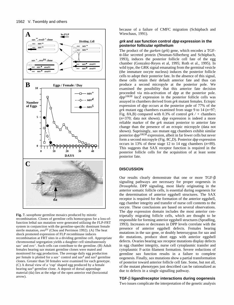
Perrimon, 1992; Chou et al., 1993) through induced mitoticrecombination in developing females trans-heterozygous for asax mutation and a germline-autonomous dominant femalesterile mutation, ovoD1 (Fig. 7A). Germline clones weregenerated with a control sax+ chromosome and with twodifferent loss-of-function sax alleles.
Females bearing sax mutant clones produced 10-25% asmany eggs and over a much briefer time period than sax+
control clones (Fig. 7B). sax mutant clones only produced eggsin the first 6 to 9 days of the experiment, while sax+ clonescontinued to produce eggs for up to 40 days (Fig. 7B). Mostof the eggs from the sax mutant clones were morphologicallynormal, with a minority (~20%) displaying aberrant dorsalappendages and short lengths. The residual eggs produced bysax mutant clones may result from perdurance of the sax+ geneproduct. Clones were induced at a time when sax is highly tran-scribed and when early pupal egg chambers (stages 3 to 5) haveformed (King, 1970). The failure of continued egg productionconfirms that removal of SAX signaling from the germline ofovaries ultimately blocks oogenesis.
A parallel set of sax5 germline clones was generated andovaries of clone-bearing females were examined as their eggproduction waned on day 5. The ovo+sax5 homozygous eggchambers could be identified because they lacked the dominantovoD1 phenotype. These ovo+sax5 clones displayed degenerat-ing mid-stage chambers (similar to those in Fig. 5B,C). Inaddition, these clones also contained occasional ‘cup’-likeeggs, in which the anterior end of the egg remains open (Fig.7C). Mutations in several genes produce such ‘cup’ eggs
1562 V. Twombly and others
0
5
10
DAYS
Eggs / Female / Day
10 20 30 40
sax5
sax4
sax+
0
Numberof Eggs
B
hs-FLP
Dividing Cell
sax
sax
sax , ovoD + clone
sax ovoD1
sax ovoD1
sax ovoD1
sax ovoD1
sax
sax
A
C
da
sax + , ovoD1 clone
FRT
FRT
FRT
FRT
FRT
FRT
FRT
FRT
Fig. 7. saxophone germline mosaics produced by mitoticrecombination. Clones of germline cells homozygous for a loss-of-function lethal sax mutation were generated utilizing the FLP-FRTsystem in conjunction with the germline-specific dominant femalesterile mutation, ovoD1 (Chou and Perrimon 1992). (A) The heatshock promoted expression of FLP recombinase inducesrecombination at FRT sites in a dividing germline cell. Appropriatechromosomal segregation yields a daughter cell simultaneouslysax− and ovo+. Such cells can contribute to the germline. (B) Adultfemales bearing sax mutant germline clones were mated andmonitored for egg production. The average daily egg productionper female is plotted for a sax+ control and sax4 and sax5 germlineclones. Greater than 50 females were examined for each genotype.(C) A dorsal view of a ‘cup’ shaped egg produced by a femalebearing sax5 germline clone. A deposit of dorsal appendagematerial (da) lies at the edge of the open anterior end (horizontalarrow).
because of a failure of CMFC migration (Schüpbach andWieschaus, 1991).
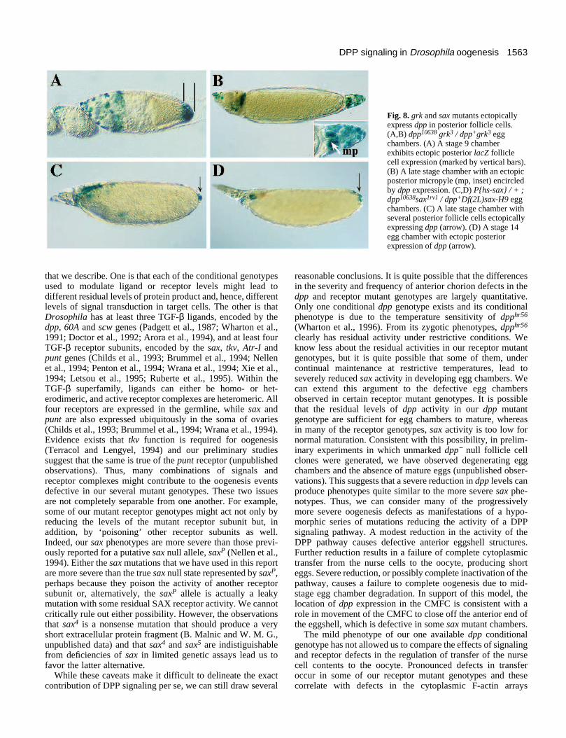
grk and sax function control dpp expression in theposterior follicular epitheliumThe product of the gurken (grk) gene, which encodes a TGF-α-like secreted protein (Neuman-Silberberg and Schüpbach,1993), induces the posterior follicle cell fate of the eggchamber (Gonzalez-Reyes et al, 1995; Roth et al., 1995). Inwild type, the GRK signal emanating from the germinal vesicle(the immature oocyte nucleus) induces the posterior folliclecells to adopt their posterior fate. In the absence of this signal,these cells retain their default anterior fate and thus canproduce a second micropyle at the posterior pole. Weexamined the possibility that this anterior fate decisionproceeded via mis-activation of dpp at the posterior pole.dpp10638 lacZ expression in the posterior follicle cells wasassayed in chambers derived from grk mutant females. Ectopicexpression of dpp occurs at the posterior pole of 77% of thegrk mutant egg chambers examined from stage 9 to 14 (n=97;Fig. 8A,B) compared with 0.3% of control grk / + chambers(n=370; data not shown). dpp expression is indeed a morereliable marker of the grk mutant posterior to anterior fatechange than the presence of an ectopic micropyle (data notshown). Suprisingly, sax mutant egg chambers exhibit similarposterior dpp10638 expression, albeit in far fewer cells but neverform a second micropyle (Fig. 8C,D). Posterior dpp expressionoccurs in 13% of these stage 12 to 14 egg chambers (n=89).This suggests that SAX receptor function is required in theposterior follicle cells for the acquisition of at least someposterior fate.
DISCUSSION
Our results clearly demonstrate that one or more TGF-βsignaling pathways are necessary for proper oogenesis inDrosophila. DPP signaling, most likely originating in theanterior somatic follicle cells, is essential during oogenesis forthe determination of anterior eggshell structures. The SAXreceptor is required for the formation of the anterior eggshell,egg chamber integrity and transfer of nurse cell contents to theoocyte. These conclusions are based on several observations.The dpp expression domain includes the most anterior cen-tripetally migrating follicle cells, which are thought to beresponsible for forming anterior eggshell structures (Spradling,1993). Increases or decreases in DPP levels correlate with thepresence of anterior eggshell defects. Females bearingmutations in the sax gene, or doubly heterozygous for sax andtkv mutations, produce short eggs with anterior eggshelldefects. Ovaries bearing sax receptor mutations display defectsin egg chamber integrity, nurse cell cytoplasmic transfer andcytoplasmic F-actin filament formation. Severe reductions ofgermline sax function results in a failure to completeoogenesis. Finally, sax mutations show a partial transformationof posterior toward anterior follicle cell fate. Some, but not all,of the mutant phenotypes just described can be rationalized asdue to defects in a single signalling pathway.
TGF-β ligand/receptor interactions during oogenesisTwo issues complicate the interpretation of the genetic analysis
1563DPP signaling in Drosophila oogenesis
Fig. 8. grk and sax mutants ectopicallyexpress dpp in posterior follicle cells.(A,B) dpp10638 grk3 / dpp+grk3 eggchambers. (A) A stage 9 chamberexhibits ectopic posterior lacZ folliclecell expression (marked by vertical bars).(B) A late stage chamber with an ectopicposterior micropyle (mp, inset) encircledby dpp expression. (C,D) P{hs-sax} / + ;dpp10638sax1rv1 / dpp+Df(2L)sax-H9 eggchambers. (C) A late stage chamber withseveral posterior follicle cells ectopicallyexpressing dpp (arrow). (D) A stage 14egg chamber with ectopic posteriorexpression of dpp (arrow).
that we describe. One is that each of the conditional genotypesused to modulate ligand or receptor levels might lead todifferent residual levels of protein product and, hence, differentlevels of signal transduction in target cells. The other is thatDrosophila has at least three TGF-β ligands, encoded by thedpp, 60A and scw genes (Padgett et al., 1987; Wharton et al.,1991; Doctor et al., 1992; Arora et al., 1994), and at least fourTGF-β receptor subunits, encoded by the sax, tkv, Atr-I andpunt genes (Childs et al., 1993; Brummel et al., 1994; Nellenet al., 1994; Penton et al., 1994; Wrana et al., 1994; Xie et al.,1994; Letsou et al., 1995; Ruberte et al., 1995). Within theTGF-β superfamily, ligands can either be homo- or het-erodimeric, and active receptor complexes are heteromeric. Allfour receptors are expressed in the germline, while sax andpunt are also expressed ubiquitously in the soma of ovaries(Childs et al., 1993; Brummel et al., 1994; Wrana et al., 1994).Evidence exists that tkv function is required for oogenesis(Terracol and Lengyel, 1994) and our preliminary studiessuggest that the same is true of the punt receptor (unpublishedobservations). Thus, many combinations of signals andreceptor complexes might contribute to the oogenesis eventsdefective in our several mutant genotypes. These two issuesare not completely separable from one another. For example,some of our mutant receptor genotypes might act not only byreducing the levels of the mutant receptor subunit but, inaddition, by ‘poisoning’ other receptor subunits as well.Indeed, our sax phenotypes are more severe than those previ-ously reported for a putative sax null allele, saxP (Nellen et al.,1994). Either the sax mutations that we have used in this reportare more severe than the true sax null state represented by saxP,perhaps because they poison the activity of another receptorsubunit or, alternatively, the saxP allele is actually a leakymutation with some residual SAX receptor activity. We cannotcritically rule out either possibility. However, the observationsthat sax4 is a nonsense mutation that should produce a veryshort extracellular protein fragment (B. Malnic and W. M. G.,unpublished data) and that sax4 and sax5 are indistiguishablefrom deficiencies of sax in limited genetic assays lead us tofavor the latter alternative.
While these caveats make it difficult to delineate the exactcontribution of DPP signaling per se, we can still draw several
reasonable conclusions. It is quite possible that the differencesin the severity and frequency of anterior chorion defects in thedpp and receptor mutant genotypes are largely quantitative.Only one conditional dpp genotype exists and its conditionalphenotype is due to the temperature sensitivity of dpphr56
(Wharton et al., 1996). From its zygotic phenotypes, dpphr56
clearly has residual activity under restrictive conditions. Weknow less about the residual activities in our receptor mutantgenotypes, but it is quite possible that some of them, undercontinual maintenance at restrictive temperatures, lead toseverely reduced sax activity in developing egg chambers. Wecan extend this argument to the defective egg chambersobserved in certain receptor mutant genotypes. It is possiblethat the residual levels of dpp activity in our dpp mutantgenotype are sufficient for egg chambers to mature, whereasin many of the receptor genotypes, sax activity is too low fornormal maturation. Consistent with this possibility, in prelim-inary experiments in which unmarked dpp− null follicle cellclones were generated, we have observed degenerating eggchambers and the absence of mature eggs (unpublished obser-vations). This suggests that a severe reduction in dpp levels canproduce phenotypes quite similar to the more severe sax phe-notypes. Thus, we can consider many of the progressivelymore severe oogenesis defects as manifestations of a hypo-morphic series of mutations reducing the activity of a DPPsignaling pathway. A modest reduction in the activity of theDPP pathway causes defective anterior eggshell structures.Further reduction results in a failure of complete cytoplasmictransfer from the nurse cells to the oocyte, producing shorteggs. Severe reduction, or possibly complete inactivation of thepathway, causes a failure to complete oogenesis due to mid-stage egg chamber degradation. In support of this model, thelocation of dpp expression in the CMFC is consistent with arole in movement of the CMFC to close off the anterior end ofthe eggshell, which is defective in some sax mutant chambers.
The mild phenotype of our one available dpp conditionalgenotype has not allowed us to compare the effects of signalingand receptor defects in the regulation of transfer of the nursecell contents to the oocyte. Pronounced defects in transferoccur in some of our receptor mutant genotypes and thesecorrelate with defects in the cytoplasmic F-actin arrays
1564 V. Twombly and others
required for this event (Gutzeit, 1986). We can only note thatthe expression pattern of dpp, essentially enwrapping the nursecell complex in the anterior half of the mid-stage egg chamber,is consistent with a role for dpp in initiating the receptor acti-vation necessary for normal cytoplasmic transfer from nursecells to oocyte. The observations that BMP2, a functional DPPhomolog, binds the SAX receptor in COS cell assays(Brummel et al., 1994) and that dpp and sax mutants shareseveral developmental defects (Brummel et al., 1994; Nellenet al., 1994; Xie et al., 1994), are also consistent with thisnotion.
Some phenotypes that we observed suggest that other TGF-β signaling pathways play roles during oogenesis. One relatesto the phenotype elicited by ubiquitous expression of dpp byheat shock induction of the P{hs-dpp} transgenes. Profoundchanges in anterior follicle cell structures and in the expressionof at least one downstream marker of dorsal anterior folliclecells (shortsighted: L. Dobens et al., unpublished data) mightwell reflect the ectopic activation of receptors that are ordi-narily activated by another TGF-β ligand. The second obser-vation is even more difficult to rationalize in terms of the siteof expression of dpp during oogenesis. Certain receptor mutantgenotypes lead to mis-expression of the dpp10638 enhancer trapat the posterior pole of the embryo, as well as a morphologi-cally mutant follicular epithelium in the posterior oocyte.These changes are suggestive of a partial transformation ofposterior follicle cell fate toward anterior. It is not apparenthow the anteriorly expressed dpp follicle cell signal couldmediate these phenotypes. It is much more likely that anotherTGF-β ligand activates the SAX receptor in this posteriordomain. Indeed, the 60A gene is expressed in these cells (K.Wharton, personal communication) and hence is a candidate toencode this ligand. Only the further analysis of somatic folliclecell and germline clones lacking various elements of theDrosophila TGF-β pathway will resolve the roles played bythese signaling molecules.
We thank Fotis Kafatos, Len Dobens, Joe Duffy, Tien Hsu andDoug Harrison for local expertise and discussions. Excellent technicalsupport was provided by Diane Duplissa and Genya Kraytsberg. Wewould like to express our gratitude for the extraordinary efforts andyears of friendship with our ‘Drosophila curator’ Lorraine Lukas. Fordiscussions, information and materials, we thank Celeste Berge,Laurel Raftery, Lynn Cooley, Ruth Lehmann, Daniel St. Johnston,Mike O’Connor, Norbert Perrimon, Matt Singer, Allan Spradling andKristi Wharton. This work was supported by NSF grants (DCB 90-18618 and MCB 93-17701) to R. K. B., NIH grants to W. M. G. anda Council for Tobacco Research grant to R. W. P.; J. M. G. is a HHMIPhysician Research Fellow; V. T. was a National Research ServiceAward genetics trainee.
REFERENCES
Arora, K., Levine, M. S. and O’Connor, M. B. (1994). The screw geneencodes a ubiquitously expressed member of the TGF-β family required forspecification of dorsal cell fates in the Drosophila embryo. Genes Dev. 8,2588-2601.
Attisano, L., Wrana, J. L., Lopez-Casillas, F. and Massagué, J. (1994).TGF-β receptors and actions. Bioch Bioph Acta, 1222, 71-80.
Bellen, H. J., O’Kane, C. J., Wilson, C., Grossniklaus, U., Pearson, R. K.and Gehring, W. J. (1989). P-element-mediated enhancer detection: aversatile method to study development in Drosophila. Genes Dev. 3, 1288-1300.
Brummel, T. J., Twombly, V., Marqués, G., Wrana, J. L., Newfeld, S. J.,Attisano, L., Massagué, J., O’Connor, M. B. and Gelbart, W. M. (1994).Characterization and relationship of DPP receptors encoded by thesaxophone and thick veins genes in Drosophila. Cell 78, 251-261.
Childs, S. R., Wrana, J. L., Attisano, L., Arora, K., Massagué J. andO’Connor, M.B. (1993). Identification of a Drosophila activin receptor.Proc. Natl. Acad. Sci. USA 90, 9475-9479.
Chou, T-B. and Perrimon, N. (1992). Use of a yeast site-specific recombinaseto produce female germline chimeras in Drosophila. Genetics 131, 643-653.
Chou, T-B., Noll, E. and Perrimon, N. (1993). Autosomal P[ovoD1] dominantfemale-sterile insertions in Drosophila and their use in generating germ-linechimeras. Development 119, 1359-1369.
Cooley, L., Verheyen, E. and Ayers, K. (1992). chickadee encodes a profilinrequired for intercellular cytoplasm transport during Drosophila oogenesis.Cell 69, 173-184.
Cooley, L. and Theurkauf, W. E. (1994). Cytoskeletal functions duringDrosophila oogenesis. Science 266, 590-595.
Derynck, R. (1994). TGF-β-receptor mediated signaling. Trends Biochem. 19,548-553.
Doctor, J. S., Jackson, P. D., Rashka, K. E., Visalli, M. and Hoffmann, F.M. (1992). Sequence, biochemical characterization, and developmentalexpression of a new member of the TGF-β superfamily in Drosophilamelanogaster. Dev. Biol. 151, 491-505.
The FlyBase Consortium. (1996). FlyBase: the Drosophila database. Nucl.Acids Res. 24, 53-56.
Frasch, M. (1995). Induction of visceral and cardiac mesoderm by ectodermalDPP in the early Drosophila embryo. Nature 374, 464-467.
Gonzalez-Reyes, A., Elliot, H. and St. Johnston, D. (1995). Polarization ofboth major body axes in Drosophila by gurken-Egfr signaling. Nature 375,654-658.
Graff, J. M., Thies, R. S., Song, J., Celeste, A. J. and Melton, D. A. (1994).Studies with a Xenopus BMP receptor suggest that ventral mesoderm-inducing signals override dorsal signals in vivo. Cell 79, 169-179.
Gutzeit, H. O. (1986). The role of microfilaments in cytoplasmic streaming inDrosophila follicles. J. Cell Sci. 80, 159-169.
Immerglück, K., Lawrence, P. A. and Bienz, M. (1990). Induction acrossgerm layers in Drosophila mediated by a genetic cascade. Cell 62, 261-268.
Irish, V. F. and Gelbart, W. M. (1987). The decapentaplegic gene is requiredfor dorsal-ventral patterning of the Drosophila embryo. Genes Dev. 1,868-879.
Jacobs, J. R., Hiromi, Y., Patel, N. H. and Goodman, C. S. (1989). Lineagemigration, and morphogenesis of longitudinal glia in the Drosophila CNS asrevealed by a molecular lineage marker. Neuron 2, 1625-31.
Karpen, G. H. and Spradling, A. C. (1992). Analysis of subtelomericheterochromatin in the Drosophila minichromosome Dp1187 by single Pelement insertional mutagenesis. Genetics 132, 737-753.
King, R. C. (1970). Ovarian Development in Drosophila melanogaster. NewYork and London: Academic Press.
Kingsley, D. M. (1994). The TGF-β superfamily: new members, newreceptors, and new genetic tests of function in different organisms. GenesDev. 8, 133-146.
Letsou, A., Arora, K., Wrana, J. L., Simin, K., Twombly, V., Jamal, J.,Staehling-Hampton, K., Hoffmann, F. M., Gelbart, W. M., Massagué, J.and O’Connor, M. O. (1995). Drosophila DPP signaling is mediated by thepunt gene product: A dual-ligand binding type II receptor of the TGFβreceptor family. Cell 80, 899-908.
Massagué, J. and Polyak, K. (1995). Mammalian antiproliferative signals andtheir targets. Curr. Op. Genet. Dev. 5, 91-96.
Miyazano, K., Ten-Dijke, P., Ichilo, H. and Heldin, C-H. (1994). Receptorsfor the Transforming Growth Factor-β. Adv. Immunol. 55, 181-220.
Nellen, D., Affolter, M. and Basler, K. (1994). Receptor serine/threoninekinases implicated in the control of Drosophila body pattern bydecapentaplegic. Cell 78, 225-237.
Neuman-Silberberg, F. S. and Schüpbach, T. (1993). The Drosophiladorsoventral patterning gene gurken produces a dorsally localized RNA andencodes a TGFα-like protein. Cell 75, 165-174.
Padgett, R. W., St. Johnston, R. D. and Gelbart, W. M. (1987). A transcriptfrom a Drosophila pattern gene predicts a protein homologous to thetransforming growth factor-β family. Nature 325, 81-84.
Panganiban, G. F., Reuter, R., Scott, M. P. and Hoffmann, F. M. (1990). ADrosophila growth factor homolog, decapentaplegic, regulates homeoticgene expression within and across germ layers during midgutmorphogenesis. Development 110, 1041-1050.
Penton, A., Chen, Y., Staehling-Hampton, K., Wrana, J. L., Attisano, L.,
1565DPP signaling in Drosophila oogenesis
Szidonya, J., Cassill, A., Massagué, J. and Hoffmann, F. M. (1994)Identification of two bone morphogenetic protein type I receptors, Brk25Dand Brk43E, in Drosophila and evidence that Brk25D is a functional receptorfor decapentaplegic. Cell 78, 239-250.
Pirotta, V. (1988). Vectors for P-element transformation in Drosophila. InVectors: A Survey of Molecular Cloning Vectors and Their Uses (eds. R. L.Rodriquez and D. T. Denhart), pp. 437-456. Boston: Butterworths.
Roth, S., Neuman-Silberberg, F. S., Barcelo, G. and Schüpbach, T. (1995).cornichon and the EGF receptor signaling process are necessary for bothanterior-posterior and dorsal-ventral pattern formation in Drosophila. Cell81, 967-978.
Ruberte, E., Marty, T., Nellen, D., Affolter, A. and Basler, K. (1995). Anabsolute requirement for both the type II and type I receptors, Punt and ThickVeins for DPP signaling in vivo. Cell 80, 889-897.
Schüpbach, T. and Wieschaus, E. (1991). Female sterile mutations on thesecond chromosome of Drosophila melanogaster. II. Mutations blockingoogenesis or altering egg morphology. Genetics 129, 1119-1136.
Spradling, A. C. (1993). Developmental genetics of oogenesis. In TheDevelopment of Drosophila melanogaster. pp. 1-70. Cold Spring HarborPress.
St. Johnston, R. D., Hoffmann, F. M., Blackman, R. K., Segal, D., Grimalia,R., Padgett, R. W., Irick, H. A. and Gelbart, W. M. (1990). Molecularorganization of the decapentaplegic gene in Drosophila melanogaster.Development 4, 1114-1127.
Staehling-Hampton, K., Hoffmann, F. M., Baylies, M. K., Rushton, E. andBate, M. (1994). dpp induces mesodermal gene expression in Drosophila.Nature 372, 783-786.
Terracol, R. and Lengyel, J. A. (1994). The thick veins gene of Drosophila isrequired for dorsoventral polarity of the embryo. Genetics 138, 165-178.
Wharton, K. A., Thomsen, G. H. and Gelbart, W. M. (1991). Drosophila60A gene, a new transforming growth factor β family member, is closelyrelated to human bone morphogenetic proteins. Proc. Natl. Acad. Sci. USA88, 9214-9218
Wharton, K., Ray, R. P., Findley, S. D., Duncan, H. E. and Gelbart, W. M.(1996). Molecular lesions associated with alleles of decapentaplegic identifyresidues necessary for TGF-β/BMP cell signaling in Drosophilamelanogaster. Genetics 142, 493-505.
Wrana, J. L., Tran, H., Attisano, L., Arora, K., Childs, S. R., Massagué, J.and O’Connor, M. B. (1994). Two distinct transmembrane serine/threoninekinases from Drosophila melanogaster form an activin receptor complex.Molec. Cell. Biol. 14, 944-950.
Xie, T., Finelli, A. L. and Padgett, R. W. (1994). The Drosophila saxophonegene: a serine-threonine kinase receptor of the TGF-β superfamily. Science263, 1756-1759.
(Accepted 5 February 1996)
Note added in proofDuring the proof stage it was brought to our attention thatmutations in the nonmuscle myosin gene, sqh, cause phenotypeswhich are strikingly similar to DPP signaling mutant pheno-types. See Wheatley et al., Development 121, 1937-1946.


