Embed Size (px)
Citation preview
Psychopharmacology (2006) 186: 487–503DOI 10.1007/s00213-006-0387-2
REVIEW
Daphna Joel
The signal attenuation rat model of obsessive–compulsivedisorder: a review
Received: 14 July 2005 / Accepted: 17 March 2006 / Published online: 23 May 2006# Springer-Verlag 2006
Abstract During the last 30 years, there have been manyattempts to develop animal models of obsessive–compulsivedisorder (OCD), in the hope that they may provide a routefor furthering our understanding and treatment of thisdisorder. The present paper reviews a recently developedrat model of OCD, namely, signal attenuation. Results ofpharmacological and lesion studies are presented andevaluated with respect to the pharmacology and patho-physiology of OCD. It is argued that signal attenuation is arat model of OCD with construct (derived from similarityin the underlying mechanisms), predictive (derived fromsimilarity in response to treatment), and face (derived fromphenomenological similarity between “compulsive”behavior in the model and compulsions in OCD patients)validity.
Keywords Animal model . Compulsive lever-pressing
Obsessive–compulsive disorder (OCD) is a psychiatricaffliction with a lifetime prevalence of 1–3% (Rasmussenand Eisen 1992; Sasson et al. 1997). According to theDiagnostic and Statistical Manual of Mental Disorders(4th edition; DSM IV) (American Psychiatric Association1994), the essential features of OCD are recurrentobsessions or compulsions (e.g., doubting, checking,washing).
For obvious reasons, the understanding and treatment ofdiseases such as OCD must rely heavily on appropriateanimal models that closely mimic their behavioral and, ifpossible, their neural manifestations. During the last 3decades, several animal models of OCD have beendeveloped (for comprehensive reviews of these modelsand an assessment of their validity, see Insel et al. 1994;
Joel, in press; Man et al. 2004; Pitman 1989; Ricciardi andHurley 1990; Stein et al. 1994; Winslow and Insel 1991).These models can be divided into three classes: ethological,pharmacological, and genetic.
Ethological models include naturally occurring repetitiveor stereotypic behaviors, such as tail chasing, fur chewingand weaving (for review, see Insel et al. 1994; Stein et al.1994; Winslow and Insel 1991); innate motor behaviorsthat occur during periods of conflict, frustration, or stress(displacement behaviors), such as grooming, cleaning, andpecking (for review, see Insel et al. 1994; Pitman 1991;Ricciardi and Hurley 1990; Winslow and Insel 1991); andnatural behaviors that occur following some behavioralmanipulation (adjunctive behaviors; for review, see Insel etal. 1994), such as schedule-induced polydipsia (Woods etal. 1993) and food-restriction-induced hyperactivity(Altemus et al. 1996). These models rest primarily onbehavioral similarity between the behavior in the modeland the clinical condition. The similarity may be evident attwo levels: at the level of the specific behavior, e.g.,grooming in animals and cleaning in patients; and at a moreabstract level, e.g., the behavior induced in the model isrepetitive as are compulsions. The effects of serotoninreuptake inhibitors (SRIs), currently the only efficientmonotherapy for OCD, have been tested in some of thesemodels (Altemus et al. 1996; Nurnberg et al. 1997;Rapoport et al. 1992; Szechtman et al. 1998; Winslowand Insel 1991; Woods et al. 1993), and in some models,the effects of SRIs have also been compared to the effectsof drugs known not to be effective in OCD (Altemus et al.1996—fluoxetine vs imipramine; Rapoport et al. 1992—clomipramine, sertraline, and fluoxetine vs desipramineand fenfluramine; Winslow and Insel 1991—clomipraminevs desipramine; Woods et al. 1993—fluvoxamine, fluox-etine, and clomipramine vs desipramine, haloperidol, anddiazepam). Although some of these models have goodpredictive validity in addition to face validity, many havenot been used since the original publications. To date, onlythree behavioral models of OCD are in use, namely,the barbering (Garner et al. 2004a,b), marble burying(Broekkamp et al. 1986; Broekkamp and Jenck 1989;
D. Joel (*)Department of Psychology, Tel Aviv University,Ramat-Aviv, Tel Aviv 69978, Israele-mail: [email protected].: +972-3-6408996Fax: +972-3-6409547

Gyertyan 1995; Londei et al. 1998; Njung’e and Handley1991), and signal attenuation models. Similar to earlierbehavioral models, barbering and marble burying havebeen suggested as potential models of OCD on the basis ofbehavioral similarity. In contrast, the signal attenuationmodel, the focus of the present review, is a theory-drivenmodel of OCD, in which a “compulsive”-like behavior isinduced by simulating a deficient psychological mecha-nism hypothesized to underlie compulsive behaviors inOCD.
Pharmacological models are based on drug-inducedbehavioral alterations which bear similarity to somespecific characteristics of the behavior of humans diag-nosed with OCD, such as perseveration and indecision(Yadin et al. 1991), or compulsive checking (Eilam andSzechtman 1995; Szechtman et al. 1998, 2001). In additionto behavioral similarity, the relevant behavior in thesemodels is induced by manipulations of a neurotransmittersystem whose dysfunction has been implicated in OCD.Thus, in the model of Yadin et al. (1991), perseveration isinduced by manipulations of the serotonergic system,
whereas in the model of Szechtman and colleagues,compulsive checking is induced by manipulations of thedopaminergic system. Finally, in both models, drug-induced compulsive-like behavior has been shown to bereduced by administration of an SRI (fluoxetine andclomipramine in the model of Yadin et al. 1991 [Yadin et al.1991 and Fernandez-Guasti et al. 2003, respectively], andclomipramine in the model of Szechtman et al. 1998).Recently, meta-chlorophenylpiperazine (mCPP)-inducedposition preference in a T-maze has been suggested toprovide a model of compulsive behavior, as it has beendemonstrated that mCPP-induced position preference isblocked by chronic treatment with fluoxetine but not withdiazepam or desipramine (Tsaltas et al. 2005).
In recent years, four genetic mice models of OCD havebeen presented: the D1CT-7 transgenic mouse model ofcomorbid Tourette’s syndrome and OCD (Campbell et al.1999a–c; McGrath et al. 1999a,b; Nordstrom and Burton2002), the Hoxb8 mutants as a model of the OC-spectrumdisorder trichotillomania (Greer and Capecchi 2002), the5-HT2c receptor knockout mouse as a model of compulsive
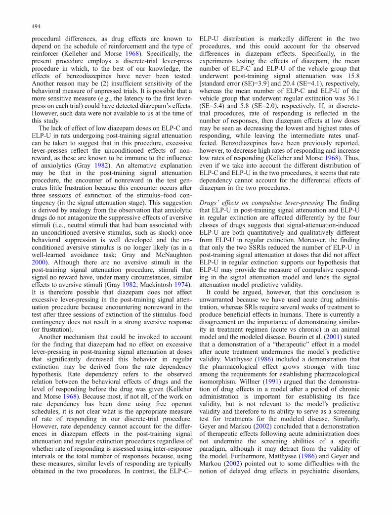
Stimulus ONFood Pellet
Magazine entry
15-s elapsed
Stimulus OFFHL OFF
HLON
Leversinserted
RI5-s
Lever press
15-s elapsed
Stimulus ONFood Pellet
Magazine entry
10-s elapsed*
Stimulus OFFHL OFF
Levers retracted
HLON
RI5-s
Stimulus ONMagazine entry
10-s elapsed
Stimulus OFFHL OFF
HLON
Leversinserted
RI5-s
Lever press
15-s elapsed
Stimulus ONMagazine entry
10-s elapsed
Stimulus OFFHL OFF
Levers retracted
HLON
RI5-s
Magazinetraining
Lever-press training
Signalattenuation
Test
Fig. 1 A schematic diagram of the organization of a trial in each ofthe different training stages of the post-training signal attenuationprocedure. In the magazine training stage (days 1–3), rats are trainedto collect food pellets from the food magazine in the operantchamber, with the levers retracted. On each trial, a single food pelletis dropped into the food magazine, simultaneous with the onset of acompound stimulus (an 80-dB, 2.8-kHz tone and the magazinelight). The stimulus is turned off after the rat’s head enters the foodmagazine or after 15 s has elapsed, and a 30-s intertrial intervalbegins. In the lever-press training stage (days 4–6), rats are trainedto lever-press in a discrete-trial procedure. On each trial, both leversare inserted into the chamber. Responding on one of the levers(reinforced lever; RL) results in the delivery of a single food pelletinto the magazine, accompanied by the presentation of thecompound stimulus. The levers are retracted and the compoundstimulus is turned off after the rat’s head enters the food magazine orafter 10 s from the rat’s first lever-press has elapsed. Further lever-presses on the RL as well as responding on the other lever(nonreinforced lever) are recorded, but have no programmedconsequences. In the signal attenuation stage (days 7–9), with the
levers retracted, rats are exposed to the presentation of thecompound stimulus as on days 1–3, but no food is delivered tothe food magazine (it should be noted that the pellet dispenser isactivated as in previous stages, producing its typical noise, but nopellet is delivered because the pellet dispenser is empty at thisstage). In the test (day 10), rats are trained as in the lever-presstraining stage, except that no food is delivered to the food magazine,i.e., pressing the lever results in the presentation of the compoundstimulus only (again, the pellet dispenser is activated, but is empty).To assess rats’ tendency for excessive lever-pressing, the number oflever-presses on the nonreinforced lever and the number of lever-presses on the reinforced lever after the first response (extra lever-presses; ELP) are recorded. The latter measure is further subdividedinto ELP that are not followed by insertion of the head into the foodmagazine during stimulus presentation (ELP-U) and ELP that arefollowed by insertion of the head into the food magazine duringstimulus presentation (ELP-C). HL houselight, RI random interval,Lever press a press on the reinforced lever. *On the first day oflever-press training, this time limit is 15 s
488

behavior in OCD (Chou-Green et al. 2003), and thedopamine transporter knockdown mouse as a model ofOCD and Tourette’s syndrome (Berridge et al. 2004). It isimportant to note that the above models were not created onthe basis of a known mutation in humans that was found tobe related to OCD. Rather, these models are based onbehavioral similarity, i.e., the behavior of geneticallymodified mice was found to be similar in specific respectsto that of OCD patients (e.g., excessive), and this is the mainbasis for the claim that they may serve as animal models ofthis disorder. Regretfully, to date there are no reports on theeffects of different pharmacological treatments in thesemodels, which could have strengthened their relevance toOCD.
The signal attenuation rat model of OCD
The signal attenuation model has been developed on thebasis of the theoretical proposition that compulsivebehaviors result from a deficit in the feedback associatedwith the performance of normal goal-directed responses(e.g., Baxter 1999; Gray 1982; Malloy 1987; Pitman 1991;Pitman et al. 1987, Reed 1977; Szechtman and Woody2004; for review, see Otto 1992). In the model, the normalbehavior is lever-pressing for food, and the feedback for theresponse is an external stimulus, which follows the lever-press response and is accompanied by the presentation ofthe food reward. Thus, the stimulus signals that the lever-press response was effective in producing food. Thedeficiency in response feedback is simulated by extinguish-ing the contingency between the stimulus and the reward(“signal attenuation”). The procedure used to establish astimulus as a response feedback and to test the effects ofsignal attenuation on the performance of this response hasbeen termed post-training signal attenuation.
The post-training signal attenuation procedure
The post-training signal attenuation procedure includesfour stages. First, a stimulus (typically the magazine lightand a tone) is established as a signal for the delivery of foodby repeatedly pairing it with food (magazine training).Next, rats are trained to lever-press for food, whosedelivery is accompanied by the presentation of the stimulus(lever-press training). Because rats experience the stimulusbefore they find the food, the stimulus comes to signal thatthe lever-press response was effective in producing food. Inthe third stage (signal attenuation), the stimulus isrepeatedly presented without food (the levers are notpresent at this stage). It is hypothesized that the extinctionof the stimulus–food contingency in this stage attenuatesthe “signaling” property of the stimulus. In the last stage(test), the effects of signal attenuation on rats’ lever-pressbehavior are assessed under extinction conditions, i.e.,pressing the lever results in the presentation of the stimulusbut no food is delivered (see Fig. 1).
The assessment of the effects of signal attenuation onlever-press responding is performed under extinctionconditions to prevent the fast relearning of the stimulus–food association, which would occur if the test is carriedout under rewarded conditions. The fact that the test iscarried out under extinction conditions may, however,confound the assessment of the effects of signal attenuationbecause an encounter of nonreward produces an increase inoperant responding (i.e., an extinction burst). To betterdifferentiate between the effects of signal attenuation andof extinction per se, the behavior of rats undergoing anextinction test preceded by a signal attenuation stage hasbeen compared to that of rats in an extinction session thatwas not preceded by signal attenuation (we refer to thebehavioral procedure that is identical to the post-trainingsignal attenuation procedure but does not include a signalattenuation stage as “regular extinction”).
The behavior
Figure 2 presents the results of an experiment comparingthe behavior of rats that underwent the post-training signalattenuation procedure to that of rats that underwent theregular extinction procedure. As can be seen, the effect ofnonreward is clearly seen in the regular extinctionprocedure in the form of a high number of excessivelever-presses that are followed by magazine entry (extralever-presses in completed trials; ELP-C). Such a behavioris also exhibited by rats that underwent signal attenuationprior to the extinction test, but these rats show in additionan equally high number of lever-presses that are notfollowed by magazine entry (extra lever-presses in un-completed trials; ELP-U). It should be noted that althoughthe mean number of ELP-C and ELP-U may vary
0
10
20
30
40
ELP-C ELP-U
SARE
Mea
n N
umbe
r of
ELP
Type of ELP
*
Fig. 2 The mean and standard error of the mean number of extralever-presses that were followed by an attempt to collect a reward(ELP-C) and extra lever-presses that were not followed by anattempt to collect a reward (ELP-U) exhibited by intact rats (Wistar)undergoing the test of the post-training signal attenuation procedure(SA, n=12) or of the regular extinction procedure (RE, n=10). MixedANOVA with a main factor of procedure (SA, RE) and a repeatedmeasurements factor of type of ELP (ELP-C, ELP-U) yielded asignificant procedure × type of ELP interaction, F(1,20)=6.93,p<0.02 (the effect of procedure and of type of ELP was notsignificant, F(1,20)=1.75, p=0.20 and F(1,20)=1.35, p=0.26,respectively); post hoc least significant difference comparisonscomparing the number of ELP-C and ELP-U between the twoprocedures yielded a significant difference between the number ofELP-U in the post-training signal attenuation and regular extinctionprocedures (p<0.05), but not in the number of ELP-C
489
considerably across experiments, the distribution of ELP-Cand ELP-U in the two procedures is consistent. That is, inrats undergoing signal attenuation, the number of ELP-U issimilar to or higher than the number of ELP-C, whereas inrats undergoing regular extinction, the number of ELP-U ismuch lower than the number of ELP-C (e.g., Fig. 2; Joeland Doljansky 2003; Joel et al. 2004).
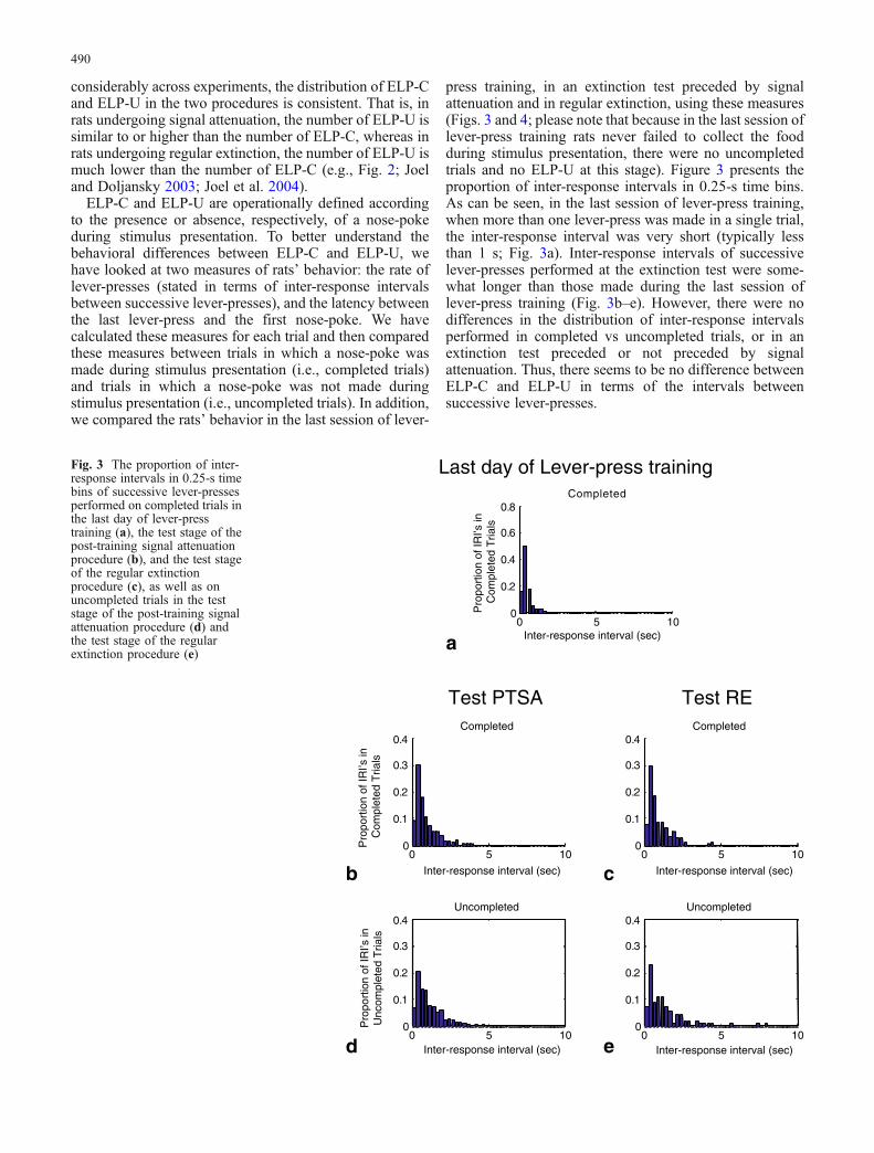
ELP-C and ELP-U are operationally defined accordingto the presence or absence, respectively, of a nose-pokeduring stimulus presentation. To better understand thebehavioral differences between ELP-C and ELP-U, wehave looked at two measures of rats’ behavior: the rate oflever-presses (stated in terms of inter-response intervalsbetween successive lever-presses), and the latency betweenthe last lever-press and the first nose-poke. We havecalculated these measures for each trial and then comparedthese measures between trials in which a nose-poke wasmade during stimulus presentation (i.e., completed trials)and trials in which a nose-poke was not made duringstimulus presentation (i.e., uncompleted trials). In addition,we compared the rats’ behavior in the last session of lever-
press training, in an extinction test preceded by signalattenuation and in regular extinction, using these measures(Figs. 3 and 4; please note that because in the last session oflever-press training rats never failed to collect the foodduring stimulus presentation, there were no uncompletedtrials and no ELP-U at this stage). Figure 3 presents theproportion of inter-response intervals in 0.25-s time bins.As can be seen, in the last session of lever-press training,when more than one lever-press was made in a single trial,the inter-response interval was very short (typically lessthan 1 s; Fig. 3a). Inter-response intervals of successivelever-presses performed at the extinction test were some-what longer than those made during the last session oflever-press training (Fig. 3b–e). However, there were nodifferences in the distribution of inter-response intervalsperformed in completed vs uncompleted trials, or in anextinction test preceded or not preceded by signalattenuation. Thus, there seems to be no difference betweenELP-C and ELP-U in terms of the intervals betweensuccessive lever-presses.
0 5 100
0.1
0.2
0.3
0.4Completed
0 5 100
0.1
0.2
0.3
0.4Uncompleted
0 5 100
0.2
0.4
0.6
0.8
Inter-response interval (sec)
Inter-response interval (sec) Inter-response interval (sec)
Inter-response interval (sec)Inter-response interval (sec)
Completed
Last day of Lever-press training
Test PTSA Test RE
Pro
port
ion
of IR
I’sin
C
ompl
eted
Tria
ls
Pro
port
ion
of IR
I’sin
Unc
ompl
eted
Tria
ls
a
b c
d e
Pro
port
ion
of IR
I’sin
C
ompl
eted
Tria
ls
0 5 100
0.1
0.2
0.3
0.4Completed
0 5 100
0.1
0.2
0.3
0.4Uncompleted
Fig. 3 The proportion of inter-response intervals in 0.25-s timebins of successive lever-pressesperformed on completed trials inthe last day of lever-presstraining (a), the test stage of thepost-training signal attenuationprocedure (b), and the test stageof the regular extinctionprocedure (c), as well as onuncompleted trials in the teststage of the post-training signalattenuation procedure (d) andthe test stage of the regularextinction procedure (e)
490
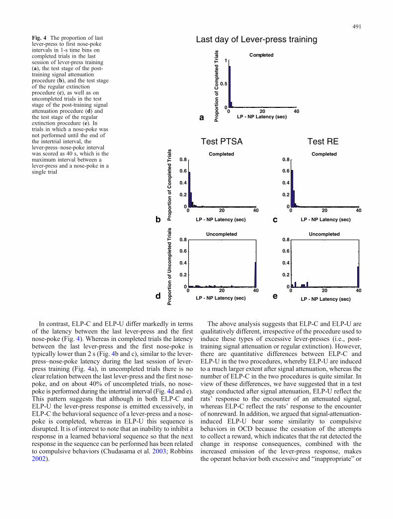
In contrast, ELP-C and ELP-U differ markedly in termsof the latency between the last lever-press and the firstnose-poke (Fig. 4). Whereas in completed trials the latencybetween the last lever-press and the first nose-poke istypically lower than 2 s (Fig. 4b and c), similar to the lever-press–nose-poke latency during the last session of lever-press training (Fig. 4a), in uncompleted trials there is noclear relation between the last lever-press and the first nose-poke, and on about 40% of uncompleted trials, no nose-poke is performed during the intertrial interval (Fig. 4d and e).This pattern suggests that although in both ELP-C andELP-U the lever-press response is emitted excessively, inELP-C the behavioral sequence of a lever-press and a nose-poke is completed, whereas in ELP-U this sequence isdisrupted. It is of interest to note that an inability to inhibit aresponse in a learned behavioral sequence so that the nextresponse in the sequence can be performed has been relatedto compulsive behaviors (Chudasama et al. 2003; Robbins2002).
The above analysis suggests that ELP-C and ELP-U arequalitatively different, irrespective of the procedure used toinduce these types of excessive lever-presses (i.e., post-training signal attenuation or regular extinction). However,there are quantitative differences between ELP-C andELP-U in the two procedures, whereby ELP-U are inducedto a much larger extent after signal attenuation, whereas thenumber of ELP-C in the two procedures is quite similar. Inview of these differences, we have suggested that in a teststage conducted after signal attenuation, ELP-U reflect therats’ response to the encounter of an attenuated signal,whereas ELP-C reflect the rats’ response to the encounterof nonreward. In addition, we argued that signal-attenuation-induced ELP-U bear some similarity to compulsivebehaviors in OCD because the cessation of the attemptsto collect a reward, which indicates that the rat detected thechange in response consequences, combined with theincreased emission of the lever-press response, makesthe operant behavior both excessive and “inappropriate” or
0 20 400
0.2
0.4
0.6
0.8Completed
0 20 400
0.2
0.4
0.6
0.8Uncompleted
0 20 400
0.5
1
LP - NP Latency (sec)
LP - NP Latency (sec)
LP - NP Latency (sec)
LP - NP Latency (sec)
LP - NP Latency (sec)
Completed
Last day of Lever-press training
Test PTSA Test RE
Pro
po
rtio
n o
f C
om
ple
ted
Tri
als
Pro
po
rtio
n o
f C
om
ple
ted
Tri
als
Pro
po
rtio
n o
f U
nco
mp
lete
d T
rial
s
a
b c
d e
0 20 400
0.2
0.4
0.6
0.8Completed
0 20 400
0.2
0.4
0.6
0.8Uncompleted
Fig. 4 The proportion of lastlever-press to first nose-pokeintervals in 1-s time bins oncompleted trials in the lastsession of lever-press training(a), the test stage of the post-training signal attenuationprocedure (b), and the test stageof the regular extinctionprocedure (c), as well as onuncompleted trials in the teststage of the post-training signalattenuation procedure (d) andthe test stage of the regularextinction procedure (e). Intrials in which a nose-poke wasnot performed until the end ofthe intertrial interval, thelever-press–nose-poke intervalwas scored as 40 s, which is themaximum interval between alever-press and a nose-poke in asingle trial
491
“unreasonable,” thus fulfilling two important criteria ofcompulsive behavior (DSM-IV; Rapoport 1989; Reed1985). These hypotheses, derived at the behavioral level,are supported by the different patterns of drug and lesioneffects on ELP-C and on ELP-U in the two procedures(see succeeding sections).
Pharmacology of compulsive lever-pressing(i.e., signal-attenuation-induced ELP-U)
Since one of the most salient features of OCD is itsselective response to treatment with SRIs (Masand andGupta 1999; Piccinelli et al. 1995; Pigott and Seay 1999;Stein et al. 1995; Zohar et al. 1992), we have tested whethercompulsive lever-pressing shows a similar pharmacologicalselectivity. The effects of drugs that are known to beeffective in OCD as well as drugs that were found not to beeffective in this disorder were assessed (Table 1). First, theeffects of each drug were tested in the post-training signalattenuation procedure using several doses, ranging from adose that had no effect on rats’ behavior to a dose thatabolished responding altogether. Next, to better differen-tiate between the drug’s effects on the behavioral responseto signal attenuation and on extinction per se, drug dosesthat were effective in the post-training signal attenuationprocedure without completely abolishing responding weretested in an extinction test not preceded by signalattenuation (i.e., regular extinction of the lever-pressresponse). All the experiments described below employedacute administration of drugs prior to the test stage.
The effects of selective serotonin reuptake inhibitors
The effects of acute administration of three selectiveserotonin reuptake inhibitors (SSRIs), paroxetine, fluvox-amine (Joel et al. 2004), and fluoxetine (Joel and Avisar2001), were assessed in the post-training signal attenuationprocedure. We refer here only to the results obtained withthe first two drugs because the effects of fluoxetine wereassessed using an older version of the software which didnot enable the separate recording of ELP-C and ELP-U(Joel and Avisar 2001). Paroxetine and fluvoxamineexerted very similar effects. When administered prior to
an extinction session of lever-press responding that waspreceded by signal attenuation, paroxetine (1, 3, 5, 7, and10 mg/kg, administered i.p. 30 min before the test) andfluvoxamine (10, 15, and 20 mg/kg, administered i.p.30 min before the test) dose-dependently decreased thenumber of ELP-U and the number of ELP-C. Whenadministered prior to an extinction session not preceded bysignal attenuation (i.e., regular extinction of lever-pressresponding), paroxetine (7 mg/kg) and fluvoxamine(15 mg/kg) decreased the number of ELP-C withoutaffecting the number of ELP-U (Joel et al. 2004).
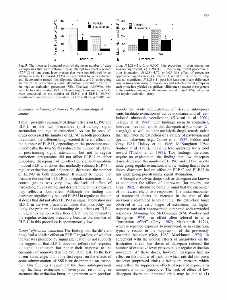
Figure 5 presents the results of an experiment whichassessed the effects of a single dose of fluvoxamine(15 mg/kg) in both the post-training signal attenuation andthe regular extinction procedures. It is clearly seen thatfluvoxamine decreased ELP-C in the two procedures(Fig. 5a) but decreased ELP-U only in the post-trainingsignal attenuation procedure (Fig. 5b; although fluvox-amine tended to increase the number of ELP-U in ratsundergoing regular extinction, this increase, which wasnot observed in a previous experiment [Joel et al. 2004],was not significant).
The effects of drugs that are not effectivein the treatment of OCD
We also tested the effects of three drugs that are known notto be effective in the treatment of OCD when given asmonotherapy, namely, the anxiolytic drug diazepam(Cassano et al. 1975; Waxman 1977, see also Argyropouloset al. 2000; Kim et al. 1997; Montgomery 1993; Stein2002), the tricyclic antidepressant desipramine (Goodmanet al. 1990; Hoehn-Saric et al. 2000; Leonard and Rapoport1989; Leonard et al. 1989; Piccinelli et al. 1995), and theantipsychotic haloperidol (e.g., McDougle et al. 1990,1994).
Desipramine (5–15 mg/kg, administered i.p. 60 minbefore the test) and haloperidol (0.005–0.05 mg/kg,administered i.p. 60 min before the test) had a similareffect on rats’ lever-press responding regardless of whetherlever-press extinction was preceded by a signal attenuationstage or not. Desipramine decreased the number of ELP-C,while having no effect on the number of ELP-U in bothprocedures (Joel et al. 2004), whereas haloperidoldecreased both ELP-C and ELP-U in the two procedures(Joel and Doljansky 2003).
Diazepam (2–10 mg/kg, administered i.p. 30 min beforethe test) affected rats’ behavior in the post-training signalattenuation procedure only at the highest doses tested, with8 mg/kg tending to decrease the number of ELP-C and ofELP-U and 10 mg/kg almost completely abolishing lever-press responding (doses between 2 and 6 mg/kg had noeffect on ELP-C and ELP-U). In contrast, when adminis-tered prior to an extinction session not preceded by signalattenuation, diazepam significantly decreased the numberof ELP-C already at a dose of 4 mg/kg and almostcompletely abolished ELP-U at all doses tested (2, 4, 6, and8 mg/kg) (Joel et al. 2004).
Table 1 Summary of drugs’ effects on ELP-C and ELP-U in thepost-training signal attenuation and regular extinction procedures
ELP-C ELP-U
SA RE SA RE
Paroxetine ↓ ↓ ↓ –Fluvoxamine ↓ ↓ ↓ –Desipramine ↓ ↓ – –Diazepam (↓) ↓ (↓) ↓Haloperidol ↓ ↓ ↓ ↓
SA post-training signal attenuation, RE regular extinction
492
Summary and interpretation of the pharmacologicalstudies
Table 1 presents a summary of drugs’ effects on ELP-C andELP-U in the two procedures (post-training signalattenuation and regular extinction). As can be seen, alldrugs decreased the number of ELP-C in both procedures.In contrast, the different drugs exerted different effects onthe number of ELP-U, depending on the procedure used.Specifically, the two SSRIs reduced the number of ELP-Uin post-training signal attenuation but not in regularextinction; desipramine did not affect ELP-U in eitherprocedure; diazepam had no effect on signal-attenuation-induced ELP-U at doses that markedly reduced ELP-U inregular extinction; and haloperidol decreased the numberof ELP-U in both procedures. It should be noted thatbecause the number of ELP-U in regular extinction in thecontrol groups was very low, the lack of effect ofparoxetine, fluvoxamine, and desipramine on this measuremay reflect a floor effect. Although the finding thatdiazepam significantly reduced ELP-U in regular extinctionat doses that did not affect ELP-U in signal attenuation norELP-C in the two procedures makes this possibility lesslikely, the problem of confounding drug effects on ELP-Uin regular extinction with a floor effect may be inherent tothe regular extinction procedure because the number ofELP-U in this procedure is spontaneously low.
Drugs’ effects on extinction The finding that the differentdrugs had a similar effect on ELP-C regardless of whetherthe test was preceded by signal attenuation or not supportsthe suggestion that ELP-C does not reflect rats’ responseto signal attenuation but rather their response to theencounter of nonreward in the extinction test. To the bestof our knowledge, this is the first report on the effects ofacute administration of SSRIs or desipramine on extinc-tion. Our findings suggest that the three antidepressantsmay facilitate extinction of lever-press responding orattenuate the extinction burst, in agreement with previous
reports that acute administration of tricyclic antidepres-sants facilitate extinction of active avoidance and of fear-induced ultrasonic vocalization (Kikusui et al. 2001;Telegdy et al. 1983). Our findings seem to contradict,however, previous reports that diazepam at low doses (2–4 mg/kg), as well as other anxiolytic drugs, retards ratherthan facilitates the extinction of a variety of pavlovian andoperant behaviors (e.g., Cowie et al. 1987; Feldon andGray 1981; Halevy et al. 1986; McNaughton 1984;Soubrie et al. 1978), including lever-pressing for a foodreward (Thiebot et al. 1983). Two findings, therefore,require an explanation: the finding that low diazepamdoses decreased the number of ELP-C and ELP-U in ratsundergoing regular extinction, and the finding that at thesedoses, diazepam had no effect on ELP-C and ELP-U inrats undergoing post-training signal attenuation.
Although anxiolytic drugs such as diazepam are knownto counteract the effects of nonreward (for review, seeGray 1982), it should be borne in mind that the encounterof nonreward elicits two responses: The initial encounterof nonreward elicits an increase in the rate of thepreviously reinforced behavior [e.g., the extinction burstobserved at the early stages of extinction; the higherresponse rate after nonrewarded compared with rewardedresponses (Manning and McDonough 1974; Wookey andStrongman 1974)], an effect often referred to as a“frustration effect” (Gray 1982; Mackintosh 1974),whereas repeated exposure to nonreward, as in extinction,typically results in the suppression of the previouslyrewarded behavior (Gray 1982; Mackintosh 1974). Inagreement with the known effects of anxiolytics on thefrustration effect, low doses of diazepam reduced thenumber of excessive lever-presses in our regular extinctionprocedure. At these doses, however, diazepam had noeffect on the number of trials on which rats did not pressthe lever (unpressed trials), a behavioral measure whichmay reflect the suppressive effects of repeated exposure tononreward in our procedure. The lack of effect of lowdiazepam doses on unpressed trials may be due to (1)
Fig. 5 The mean and standard error of the mean number of extralever-presses that were followed by an attempt to collect a reward(ELP-C) (a) and extra lever-presses that were not followed by anattempt to collect a reward (ELP-U) (b) exhibited by vehicle-treatedand fluvoxamine-treated rats (Sprague Dawley, n=32) undergoingthe test of the post-training signal attenuation procedure (SA) or ofthe regular extinction procedure (RE). Two-way ANOVAs withmain factors of procedure (SA, RE) and drug (fluvoxamine, vehicle)were conducted on the number of ELP-C and ELP-U. ELP-C:significant main effects of procedure, F(1,28)=10.87, p<0.005, and
drug, F(1,28)=21.40, p<0.0001 [the procedure × drug interactionwas not significant, F(11,28)<1]. ELP-U: a significant procedure ×drug interaction, F(1,28)=4.77, p<0.05 [the effect of procedureapproached significance, F(1,28)=3.33, p=0.078; the effect of drugwas not significant, F(1,28)<1]; post hoc least significant differencecomparisons comparing fluvoxamine- and vehicle-treated groups ineach procedure yielded a significant difference between these groupsin the post-training signal attenuation procedure (p<0.05), but not inthe regular extinction group
493
procedural differences, as drug effects are known todepend on the schedule of reinforcement and the type ofreinforcer (Kelleher and Morse 1968). Specifically, thepresent procedure employs a discrete-trial lever-pressprocedure in which, to the best of our knowledge, theeffects of benzodiazepines have never been tested.Another reason may be (2) insufficient sensitivity of thebehavioral measure of unpressed trials. It is possible that amore sensitive measure (e.g., the latency to the first lever-press on each trial) could have detected diazepam’s effects.However, such data were not available to us at the time ofthis study.
The lack of effect of low diazepam doses on ELP-C andELP-U in rats undergoing post-training signal attenuationcan be taken to suggest that in this procedure, excessivelever-presses reflect the unconditioned effects of non-reward, as these are known to be immune to the influenceof anxiolytics (Gray 1982). An alternative explanationmay be that in the post-training signal attenuationprocedure, the encounter of nonreward in the test gen-erates little frustration because this encounter occurs afterthree sessions of extinction of the stimulus–food con-tingency (in the signal attenuation stage). This suggestionis derived by analogy from the observation that anxiolyticdrugs do not antagonize the suppressive effects of aversivestimuli (i.e., neutral stimuli that had been associated withan unconditioned aversive stimulus, such as shock) oncebehavioral suppression is well developed and the un-conditioned aversive stimulus is no longer likely (as in awell-learned avoidance task; Gray and McNaughton2000). Although there are no aversive stimuli in thepost-training signal attenuation procedure, stimuli thatsignal no reward have, under many circumstances, similareffects to aversive stimuli (Gray 1982; Mackintosh 1974).It is therefore possible that diazepam does not affectexcessive lever-pressing in the post-training signal atten-uation procedure because encountering nonreward in thetest after three sessions of extinction of the stimulus–foodcontingency does not result in a strong aversive response(or frustration).
Another mechanism that could be invoked to accountfor the finding that diazepam had no effect on excessivelever-pressing in post-training signal attenuation at dosesthat significantly decreased this behavior in regularextinction may be derived from the rate dependencyhypothesis. Rate dependency refers to the observedrelation between the behavioral effects of drugs and thelevel of responding before the drug was given (Kelleherand Morse 1968). Because most, if not all, of the work onrate dependency has been done using free operantschedules, it is not clear what is the appropriate measureof rate of responding in our discrete-trial procedure.However, rate dependency cannot account for the differ-ences in diazepam effects in the post-training signalattenuation and regular extinction procedures regardless ofwhether rate of responding is assessed using inter-responseintervals or the total number of responses because, usingthese measures, similar levels of responding are typicallyobtained in the two procedures. In contrast, the ELP-C–
ELP-U distribution is markedly different in the twoprocedures, and this could account for the observeddifferences in diazepam effects. Specifically, in theexperiments testing the effects of diazepam, the meannumber of ELP-C and ELP-U of the vehicle group thatunderwent post-training signal attenuation was 15.8[standard error (SE)=3.9] and 20.4 (SE=4.1), respectively,whereas the mean number of ELP-C and ELP-U of thevehicle group that underwent regular extinction was 36.1(SE=5.4) and 5.8 (SE=2.0), respectively. If, in discrete-trial procedures, rate of responding is reflected in thenumber of responses, then diazepam effects at low dosesmay be seen as decreasing the lowest and highest rates ofresponding, while leaving the intermediate rates unaf-fected. Benzodiazepines have been previously reported,however, to decrease high rates of responding and increaselow rates of responding (Kelleher and Morse 1968). Thus,even if we take into account the different distribution ofELP-C and ELP-U in the two procedures, it seems that ratedependency cannot account for the differential effects ofdiazepam in the two procedures.
Drugs’ effects on compulsive lever-pressing The findingthat ELP-U in post-training signal attenuation and ELP-Uin regular extinction are affected differently by the fourclasses of drugs suggests that signal-attenuation-inducedELP-U are both quantitatively and qualitatively differentfrom ELP-U in regular extinction. Moreover, the findingthat only the two SSRIs reduced the number of ELP-U inpost-training signal attenuation at doses that did not affectELP-U in regular extinction supports our hypothesis thatELP-U may provide the measure of compulsive respond-ing in the signal attenuation model and lends the signalattenuation model predictive validity.
It could be argued, however, that this conclusion isunwarranted because we have used acute drug adminis-tration, whereas SRIs require several weeks of treatment toproduce beneficial effects in humans. There is currently adisagreement on the importance of demonstrating similar-ity in treatment regimen (acute vs chronic) in an animalmodel and the modeled disease. Bourin et al. (2001) statedthat a demonstration of a “therapeutic” effect in a modelafter acute treatment undermines the model’s predictivevalidity. Matthysse (1986) included a demonstration thatthe pharmacological effect grows stronger with timeamong the requirements for establishing pharmacologicalisomorphism. Willner (1991) argued that the demonstra-tion of drug effects in a model after a period of chronicadministration is important for establishing its facevalidity, but is not relevant to the model’s predictivevalidity and therefore to its ability to serve as a screeningtest for treatments for the modeled disease. Similarly,Geyer and Markou (2002) concluded that a demonstrationof therapeutic effects following acute administration doesnot undermine the screening abilities of a specificparadigm, although it may detract from the validity ofthe model. Furthermore, Matthysse (1986) and Geyer andMarkou (2002) pointed out to some difficulties with thenotion of delayed drug effects in psychiatric disorders,
494
such as the fact that in most animal studies acute effectsare obtained with much higher doses than would betolerated by humans and the possibility that drugs mayalso have acute effects in humans, but that this effect maybe hard to detect statistically (for a recent criticism of thenotion of delayed-onset action, see Agid et al. 2003).These difficulties may also be relevant to OCD, especiallybecause to prevent side effects, SSRI treatment is typicallystarted with low doses which are gradually increased, andthe difference between the initial dose and the therapeuticdose may be very large (e.g., 50 vs 200–300 mg/day,respectively, for fluvoxamine, Masand and Gupta 1999).With regard to the signal attenuation model, it may thus beargued that the extant results with acute drug administra-tion support its predictive validity and therefore its abilityto serve as a screening test for anticompulsive drugs.Clearly, a demonstration that therapeutic effects in themodel are obtained following chronic administration ofSSRIs at doses that do not produce a significant acuteeffect is needed before the model can be used to addresstemporal patterns characteristic of OCD pharmacology.It should be noted however that in its present form, thepost-training signal attenuation procedure is not wellsuited for chronic drug administration studies becauserepeated drug administration may affect behavior in theearly stages of the procedure (e.g., lever-press training,signal attenuation).
Blockade of D1 receptors: a potentialtreatment of OCD?
When studying the effects of dopaminergic manipulationson compulsive lever-pressing (Joel et al. 2001; Joel andDoljansky 2003), we found that administration of the D1antagonist SCH 23390 (0.005, 0.01, and 0.03 mg/kg,administered i.p. 60 min before the test) prior to anextinction session of lever-press responding that waspreceded by signal attenuation decreased the number ofELP-U without affecting the number of ELP-C. Whenadministered prior to an extinction session not preceded bysignal attenuation, SCH 23390 (0.01 mg/kg) had no effecton either measure (Joel and Doljansky 2003).
If indeed a decrease in ELP-U in the post-training signalattenuation procedure but not in a regular extinctionprocedure is indicative of an anticompulsive effect, thepattern of results obtained with SCH 23390 suggests that anew approach to the treatment of OCD may be theblockade of D1 receptors. Interestingly, on the basis of atheoretical model of the pathophysiology of OCD, Saxenaet al. (1998) have suggested that selective D1 blockadeshould reduce OCD symptoms. A potential hazard in usingD1 blockers to alleviate compulsions, however, is thatchronic administration of such drugs leads to increaseddensity of D1 receptors in the striatum (Creese and Chen1985; Giorgi et al. 1993; Hess et al. 1986, 1988;Lappalainen et al. 1992; Memo et al. 1987; O’Boyle etal. 1993; Porceddu et al. 1985), and therefore, discontinuationof pharmacotherapy may lead to worsening of symptoms.Indeed, repeated administration of SCH 23390 in rats led toan increase in compulsive behavior following the terminationof drug administration (Joel et al. 2001). Such a risk may beprevented by using D1 agonists, rather than antagonists, forthe treatment of OCD (for a detailed discussion of therational for this suggestion, see Joel and Doljansky 2003).
Neural substrates of compulsive lever-pressing
Functional imaging data from patients with idiopathicOCD and evidence from patients with acquired OCDimplicate most consistently the orbitofrontal cortex in thepathophysiology of this disorder (e.g., Baxter et al. 1987,1988; Benkelfat et al. 1990; Berthier et al. 1996; Breiterand Rauch 1996; Breiter et al. 1996; Cottraux et al. 1996;Hugo et al. 1999; Insel 1992; McGuire et al. 1994; Rauchet al. 1994; Saxena et al. 1999; Stein et al. 1999; Swedo etal. 1992; for review, see Saxena et al. 1998). Althoughthere are significant differences in the details and in thecomplexity of the organization of the cortex of rats andprimates, hodological, electrophysiological, and behavioraldata suggest that the rat orbital cortex may be analog to theprimate orbitofrontal cortex (for recent reviews, seeGroenewegen and Uylings 2000; Ongur and Price 2000;Schoenbaum and Setlow 2001; Uylings et al. 2003). In aseries of studies, we have found that bilateral excitotoxiclesions of the rat orbital cortex result in an increase in the
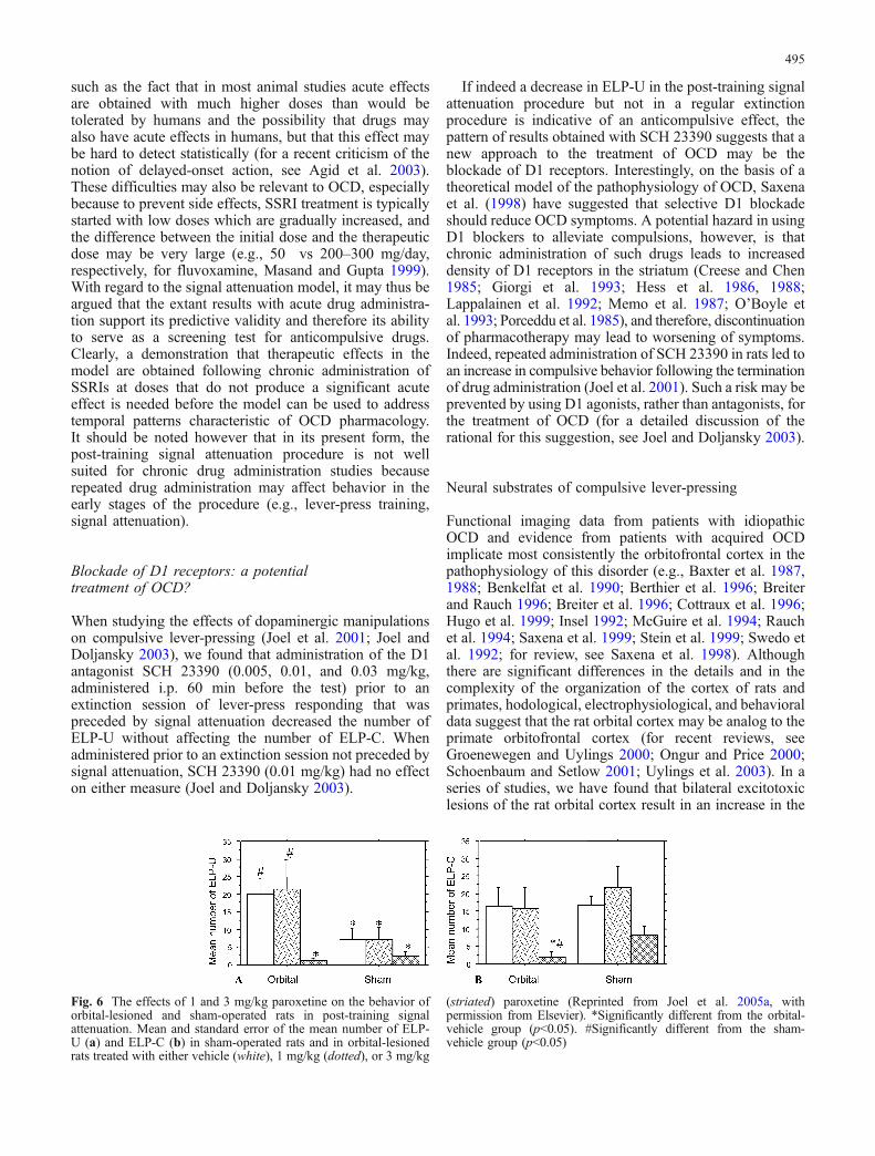
Fig. 6 The effects of 1 and 3 mg/kg paroxetine on the behavior oforbital-lesioned and sham-operated rats in post-training signalattenuation. Mean and standard error of the mean number of ELP-U (a) and ELP-C (b) in sham-operated rats and in orbital-lesionedrats treated with either vehicle (white), 1 mg/kg (dotted), or 3 mg/kg
(striated) paroxetine (Reprinted from Joel et al. 2005a, withpermission from Elsevier). *Significantly different from the orbital-vehicle group (p<0.05). #Significantly different from the sham-vehicle group (p<0.05)
495
number of signal-attenuation-induced ELP-U (Joel et al.2005a,b; Fig. 6a), while having no effect on the number ofELP-U in regular extinction (Joel et al. 2005a). The orbital-lesion-induced effect on ELP-U seems to be quite selectivebecause although orbital-lesioned rats were found toexhibit a higher number of ELP-C compared to sham ratsin one experiment (Joel et al. 2005a), such an increase wasnot exhibited by vehicle-treated orbital-lesioned ratsundergoing post-training signal attenuation (Joel et al.2005a,b; Fig. 6b) nor by orbital-lesioned rats undergoingregular extinction (Joel et al. 2005a). Orbital-lesioned ratswere also not different from their controls in the number ofELP-C during lever-press training (Joel et al. 2005a,b) norin the number of lever-presses on the nonreinforced lever inthe lever-press training and test stages (Joel et al. 2005a,b).Taken together, these results suggest that lesions to the ratorbital cortex lead to a selective increase in compulsivelever-pressing that cannot be attributed to a general lesion-induced failure of response inhibition (e.g., Brutkowski1964; Kolb et al. 1974; Konorski 1972).
The increase in compulsive lever-pressing followingorbital lesion was prevented by the SSRI paroxetine(Fig. 6a) and was paralleled by an increase in the densityof the striatal serotonin transporter, suggesting that orbital-lesion-induced compulsivity is mediated by alterations ofthe serotonergic system, possibly of the striatal serotonergicsystem (Joel et al. 2005a). These findings are of particularimportance given that the orbitofrontal cortex and thestriatum function abnormally in OCD and that drugs thatblock the serotonin transporter act in OCD patients toreduce symptoms as well as to reduce the increasedmetabolism of the orbitofrontal cortex and the striatum(Baxter et al. 1992; Benkelfat et al. 1990; Cottraux et al.1996; McGuire et al. 1994; Rauch et al. 1994; Saxena et al.1999; Swedo et al. 1992). Although the extrapolation froman animal model to the clinical condition is problematic,these findings raise the possibility that in some OCDpatients a primary orbitofrontal dysfunction leads to striatalserotonergic malfunction and to compulsive behavior, andthat antiobsessional/anticompulsive drugs act by normal-izing the dysfunctional striatal serotonergic system (for acomprehensive discussion, see Joel et al. 2005a). Interest-ingly, several imaging studies have reported that patientswith lower pretreatment orbitofrontal cortex metabolismresponded better to SRI treatment (Brody et al. 1998;Rauch et al. 2002; Saxena et al. 1999; Swedo et al. 1989),and there is some evidence that orbitofrontal cortex volumeis reduced in OCD patients (Choi et al. 2004; Pujol et al.2004; Szeszko et al. 1999).
We have also studied the effects of lesions to thebasolateral nucleus of the amygdala (BLA) and to themedial prefrontal cortex, which are anatomically connectedand functionally related to the orbital cortex. Lesions to thebasolateral amygdala or to the medial prefrontal cortex didnot have any effect on compulsive lever-pressing (Joel etal. 2005b). Given that the rat medial prefrontal cortex maycorrespond to regions in the dorsal and lateral subdivisionsof the primate prefrontal cortex (for recent reviews, seeGroenewegen and Uylings 2000; Kesner 2000; Ongur and
Price 2000; Uylings et al. 2003), the finding that compul-sive lever-pressing is enhanced following lesions to theorbital cortex, but not to the medial prefrontal cortex or tothe basolateral amygdala, is consistent with functionalimaging findings in OCD patients which consistentlyimplicate the orbitofrontal cortex in this disorder (seeabove), but rarely report evidence for an involvement of thedorsal and lateral prefrontal cortex (but see Kwon et al.2003) or of the amygdala (but see Breiter et al. 1996;Horwitz et al. 1991; Szeszko et al. 1999).
In summary, the finding that orbital lesions affect ELP-Uin post-training signal attenuation but not in regularextinction provides further support to our suggestion thatthese two types of lever-presses are qualitatively different.Moreover, the finding that lesions to the orbital cortex, butnot to the medial prefrontal cortex or to the basolateralamygdala, selectively increase signal-attenuation-inducedELP-U supports our hypothesis that ELP-U may providethe measure of compulsive responding in the signalattenuation model.
What is the mechanism by which signal attenuationinduces compulsive lever-pressing?
The above findings suggest that signal attenuation prior toan extinction test may lead to the emergence of compulsivebehavior. In the following, we analyze the post-trainingsignal attenuation procedure in terms of the processes thatmay be involved at each stage and use current knowledgeof the neural substrates of these processes in an attempt toidentify the mechanism that may underlie the induction ofcompulsive lever-presses.
The post-training signal attenuation procedure includesan early stage (i.e., magazine training) of classical condi-tioning between a neutral stimulus and a primary reinforcer(i.e., food). Stimuli that have been paired with a primaryreinforcer can influence behavior in diverse ways. Suchstimuli can elicit responses, can act as discriminativestimuli for responding, and can serve as conditionedreinforcers (e.g., Mackintosh 1974; Robbins 1978). Thelatter refers to the ability of a stimulus to serve as areinforcer for the acquisition of a new response and tomaintain responding in extinction (Mackintosh 1974).These abilities may depend on the “motivational” propertiesof the stimulus, i.e., its conditioned value, acquired throughthe pairing of the stimulus with a primary reinforcer, and/oron the “informational” properties of the stimulus, i.e., itsability to “highlight that a response has registered, in muchthe same sense as response feedback is commonly used”(Williams 1994, p. 458) and the ability to “signal that areinforcer is about to occur, thus serving to bridge the gapbetween the response and the subsequent reinforcer”(Williams 1994, p. 458).
The different modes by which a stimulus can influencebehavior are subserved by the different associations thatmay be formed during pavlovian conditioning (for recentreviews, see Cardinal et al. 2002; Dickinson and Balleine2002). Thus, the pairing of a conditioned stimulus (CS)
496
with an unconditioned stimulus (US) may lead to theformation of a direct link between the CS and the responseelicited by the US. As a result, the CS comes to elicitconditioned responses; the CS may be associated with theaffect elicited by the US. Through this association, thestimulus acquires conditioned value; the CS may becomeassociated with the specific sensory properties of the US.This latter association may serve the informational proper-ties of a conditioned stimulus described above.
In the second stage of the post-training signal attenuationprocedure, the lever-press training stage, the conditionedstimulus accompanies reward delivery following a lever-press on the reinforced lever. The different associationsacquired at the magazine training stage are expected toremain intact because the CS–US contingency is preserved.[It may be noted, however, that the acquisition of the lever-press response at this stage most likely does not depend onthe presentation of the stimulus because conditionedreinforcers are reported not to have a significant contribu-tion to learning when the response is also followed by aprimary reinforcer (Mackintosh 1974).]
In the subsequent signal attenuation stage, the classicalcontingency between the stimulus and food is extin-guished. This procedure is expected to alter the differentproperties/associations of the stimulus, including itsconditioned reinforcement properties (Mackintosh 1974).At the last stage, the extinction test, a lever-press is fol-lowed only by the now-extinguished conditioned stimulus.
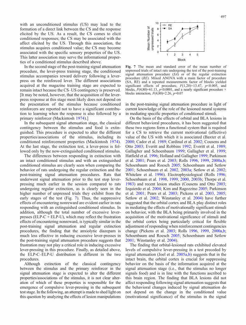
The differences between responding in extinction withan intact conditioned stimulus and with an extinguishedconditioned stimulus are clearly seen when comparing thebehavior of rats undergoing the regular extinction and thepost-training signal attenuation procedures. Rats thatunderwent signal attenuation prior to the test stop lever-pressing much earlier in the session compared to ratsundergoing regular extinction, as is clearly seen in thehigher number of unpressed trials they exhibit from theearly stages of the test (Fig. 7). Thus, the suppressiveeffects of encountering nonreward are evident earlier in ratsresponding with an extinguished conditioned stimulus. Inaddition, although the total number of excessive lever-presses (ELP-C + ELP-U), which may reflect the frustrationeffects of encountering nonreward, is typically similar in thepost-training signal attenuation and regular extinctionprocedures, the finding that the anxiolytic diazepam ismuch less effective in reducing excessive lever-presses inthe post-training signal attenuation procedure suggests thatfrustration may not play a critical role in inducing excessivelever-pressing in this procedure. Finally, as detailed above,the ELP-C–ELP-U distribution is different in the twoprocedures.
Although extinction of the classical contingencybetween the stimulus and the primary reinforcer in thesignal attenuation stage is expected to alter the differentproperties/associations of the stimulus, it is not clear alter-ation of which of these properties is responsible for theemergence of compulsive lever-pressing in the subsequenttest stage. In the following, an attempt ismade to shed light onthis question by analyzing the effects of lesion manipulations
in the post-training signal attenuation procedure in light ofcurrent knowledge of the role of the lesioned neural systemsin mediating specific properties of conditioned stimuli.
On the basis of the effects of orbital and BLA lesions indifferent behavioral procedures, it has been suggested thatthese two regions form a functional system that is requiredfor a CS to retrieve the current motivational (affective)value of the US with which it is associated (Baxter et al.2000; Cador et al. 1989; Cardinal et al. 2002; Cousens andOtto 2003; Everitt and Robbins 1992; Everitt et al. 1989;Gallagher and Schoenbaum 1999; Gallagher et al. 1999;Hatfield et al. 1996; Holland and Gallagher 1999; Parkinsonet al. 2001; Pears et al. 2003; Rolls 1996, 1999, 2000a,b;Schoenbaum and Roesch 2005; Schoenbaum and Setlow2001; Schoenbaum et al. 2002, 2003a; Setlow et al. 2002;Whitelaw et al. 1996). Electrophysiological (Rolls 1996;Schoenbaum et al. 1998, 1999, 2000, 2003b; Thorpe et al.1983) and recent lesion studies (Cousens and Otto 2003;Izquierdo et al. 2004; Kim and Ragozzino 2005; Parkinsonet al. 2001; Pears et al. 2003; Pickens et al. 2003, 2005;Setlow et al. 2002; Winstanley et al. 2004) have furthersuggested that the orbital cortex and BLA play distinct rolesin mediating the effects of motivationally significant stimulion behavior, with the BLA being primarily involved in theacquisition of the motivational significance of stimuli andthe orbital cortex being particularly critical for flexibleadjustment of responding when reinforcement contingencieschange (Pickens et al. 2003; Rolls 1996, 1999, 2000a,b;Schoenbaum and Roesch 2005; Schoenbaum and Setlow2001; Winstanley et al. 2004).
The finding that orbital-lesioned rats exhibited elevatedlevels of compulsive lever-pressing in a test preceded bysignal attenuation (Joel et al. 2005a,b) suggests that in theintact brain, the orbital cortex is crucial for suppressingbehavior on the basis of the information acquired at thesignal attenuation stage (i.e., that the stimulus no longersignals food) and is in line with the functions ascribed tothis brain region. The finding that BLA lesions did notaffect responding following signal attenuation suggests thatthe behavioral changes induced by signal attenuation donot depend on the change in the conditioned value(motivational significance) of the stimulus in the signal
Mea
n N
umbe
r of
unpr
esse
d tr
ials
Block
0
2
4
6
8
10
1 2 3 4 5
SARE
Fig. 7 The mean and standard error of the mean number ofunpressed trials of intact rats undergoing the test of the post-trainingsignal attenuation procedure (SA) or of the regular extinctionprocedure (RE). Mixed ANOVA with a main factor of procedure(SA, RE) and a repeated measurements factor of blocks yieldedsignificant effects of procedure, F(1,20)=13.47, p<0.005, andblocks, F(4,80)=61.13, p<0.0001, and a nearly significant procedure ×blocks interaction, F(4,80)=2.26, p=0.07
497
attenuation stage, but rather on a change in some otherproperty of the stimulus.
We have recently obtained evidence suggesting indi-rectly that alteration of the association between thestimulus and the specific sensory properties of the USmay be the critical factor in inducing compulsive lever-pressing. Specifically, we have found that inactivation ofthe orbital cortex in rats just prior to an extinction session inthe regular extinction procedure induces the same ELP-C–ELP-U distribution that is seen in sham-operated ratsundergoing the post-training signal attenuation procedure(Joel and Klavir, in press). These results suggest that orbitalinactivation has the same effect on behavior as undergoingsignal attenuation prior to the extinction test. A recentstudy by Ostlund and Balleine (2005) suggests that orbitalinactivation may specifically disrupt the associationbetween a CS and the specific sensory properties of theUS. It may therefore be speculated that alteration of thisassociation in the signal attenuation stage is the criticalfactor in inducing compulsive lever-pressing in thesubsequent extinction test.
As detailed above, the association between a CS andthe specific sensory properties of the US may subserve theinformational properties of conditioned stimuli. Thus, thedegradation of this association in the signal attenuationstage may alter the ability of the stimulus to highlight thatthe response has registered or to signal that the responsewas effective in producing food. Although the possibilitythat alteration of this association is the critical factor ininducing compulsive lever-pressing is highly speculative, itis of interest given theories of OCD which postulate adeficient response feedback in the production of obsessionsand compulsions (Baxter 1999; Gray 1982; Malloy 1987;Pitman 1991; Pitman et al. 1987; Reed 1977; Szechtmanand Woody 2004; for review, see Otto 1992).
Summary
On the basis of the results reviewed, we suggest that signalattenuation may provide an animal model of OCD withconstruct validity, which derives from similarities in theunderlying inducing mechanism (i.e., attenuation of anexternal feedback and a deficient response feedback mecha-nism, respectively) and in the neural systems involved(the orbital cortex and the serotonergic and dopaminergicsystems); face validity, i.e., the behavior induced by signalattenuation (compulsive lever-pressing) and compulsionsare both excessive and unreasonable; and predictive validity,i.e., selectivity for antiobsessional/anticompulsive drugs.(The application of the terms construct, face, and predictivevalidity to animal models of psychopathology is afterMcKinney 1988 and Willner 1991.)
We would like to note that the model is the inducingmanipulation, namely, signal attenuation, which ishypothesized to simulate an abnormal psychologicalprocess that may underlie obsessions and compulsions inOCD patients. However, whereas OCD patients areassumed to suffer from this deficiency at all times, in the
model, this state is induced by a behavioral manipulationand is temporary (i.e., compulsive lever-pressing isexhibited only for a short duration). In this sense, theeffects of signal attenuation on the behavior of normal ratssuggest that obsessions and compulsions in patients may beviewed as a normal reaction to an abnormal situation (i.e., adeficient response feedback). In the model, ELP-U are thebehavioral measure of compulsive behavior (in contrast toother behaviors exhibited during the test of the post-training signal attenuation procedures, such as ELP-C).Importantly, it is the combination of ELP-U and the induc-ing mechanism (i.e., signal attenuation) that provides ameasure of compulsive behavior, and not the behavior alone,as lever-presses not followed by magazine entry have beenreported following additional behavioral manipulations (e.g.,regular extinction, reinforcer devaluation without incentivelearning), but only signal-attenuation-induced ELP-U havebeen shown to have pharmacological and neurobiologicalsimilarities to compulsive behaviors in OCD.
The signal attenuation model has strengths and weak-nesses as an animal model of OCD. These are summarizedbelow with regard to specific aims animal models mayserve (for a comprehensive review of the strengths andweaknesses of other animal models of OCD, see Joel,in press). In the context of screening for anti-compulsiveactivity, the most critical features of a model are itspredictive validity and its cost-effectiveness. The signalattenuation model has good predictive validity, as it candifferentiate between the effects of SSRI’s and of drugs noteffective in the treatment of OCD. It requires, however,special equipment (operant boxes) and about 2 weeks ofbehavioral training. In addition, the post-training signalattenuation procedure is not well suited for chronic drugadministration studies because repeated drug administrationmay affect behavior in the early stages of the procedure. Anadditional use of animal models is the elucidation of theneurobiological mechanisms of the modeled condition. Inthis context, similarity in the inducing mechanism seems tobe the critical feature, although it cannot be evaluateddirectly, as the etiology of OCD is currently unknown.The signal attenuation model attempts to simulate apsychological process that is assumed to underlie compul-sive behaviors. Although there are clear differences betweena deficient internal response feedback mechanism and anattenuated external feedback, the finding that compulsivebehavior in the model has similarities to compulsivebehaviors in patients in terms of response to treatment andneural systems involved suggests that this model may beuseful in the study of the neurobiological mechanisms ofcompulsive behaviors. As detailed above, this model hasalready yielded findings which may shed light on theobserved association between a dysfunction of the orbito-frontal cortex and of the serotonergic system in OCD.
In summary, although the signal attenuation model doesnot provide an ideal animal model of OCD, it is currentlyone of the most validated animal models of OCD. It is nowcrucial that this model is tested by other groups. It is hopedthat future studies using this model will help the elucidationof the pathological mechanisms underlying OCD as well as
498
the development of new treatment approaches to thisdisorder.
References
Agid O, Kapur S, Arenovich T, Zipursky RB (2003) Delayed-onsethypothesis of antipsychotic action: a hypothesis tested andrejected. Arch Gen Psychiatry 60:1228–1235
Altemus M, Glowa JR, Galliven E, Leong YM, Murphy DL (1996)Effects of serotonergic agents on food-restriction-inducedhyperactivity. Pharmacol Biochem Behav 53:123–131
American Psychiatric Association (1994) Diagnostic and statisticalmanual of mental disorders, 4th edn. American Psychiatric,Washington, DC
Argyropoulos SV, Sandford JJ, Nutt DJ (2000) The psychobiologyof anxiolytic drug. Part 2: pharmacological treatments ofanxiety. Pharmacol Ther 88:213–227
Baxter LR (1999) Functional imaging of brain systems mediatingobsessive–compulsive disorder. In: Nestler CE, Bunney W(eds) Neurobiology of mental illness. Oxford University Press,New York, pp 534–547
Baxter LR Jr, Phelps ME, Mazziotta JC, Guze BH, Schwartz JM,Selin CE (1987) Local cerebral glucose metabolic rates inobsessive–compulsive disorder. A comparison with rates inunipolar depression and in normal controls. Arch Gen Psychiatry44:211–218
Baxter LR Jr, Schwartz JM, Mazziotta JC, Phelps ME, Pahl JJ, GuzeBH, Fairbanks L (1988) Cerebral glucose metabolic rates innondepressed patients with obsessive–compulsive disorder. AmJ Psychiatry 145:1560–1563
Baxter LR Jr., Schwartz JM, Bergman KS et al (1992) Caudateglucose metabolic rate changes with both drug and behaviortherapy for obsessive-compulsive disorder. Arch Gen Psychiatry49:681–689
Baxter MG, Parker A, Lindner CC, Izquierdo AD, Murray EA(2000) Control of response selection by reinforcer valuerequires interaction of amygdala and orbital prefrontal cortex.J Neurosci 20:4311–4319
Benkelfat C, Nordahl TE, Semple WE, King AC, Murphy DL, CohenRM (1990) Local cerebral glucose metabolic rates in obsessive–compulsive disorder. Patients treated with clomipramine. ArchGen Psychiatry 47:840–848
Berridge KC, Aldridge JW, Houchard KR, Zhuang X (2004)Sequential super-stereotypy of an instinctive fixed action patternin hyper-dopaminergic mutant mice: a model of obsessivecompulsive disorder and Tourette’s. BMC Biol 3:1–16
Berthier ML, Kulisevsky J, Gironell A, Heras JA (1996) Obsessive–compulsive disorder associated with brain lesions: clinicalphenomenology, cognitive function, and anatomic correlates.Neurology 47:353–361
Bourin M, Fiocco AJ, Clenet F (2001) How valuable are animalmodels in defining antidepressant activity? Hum Psychopharmacol16:9–21
Breiter HC, Rauch SL (1996) Functional MRI and the study of OCD:from symptom provocation to cognitive–behavioral probes ofcortico-striatal systems and the amygdala. Neuroimage4:S127–S138
Breiter HC, Rauch SL, Kwong KK, Baker JR, Weisskoff RM,Kennedy DN, Kendrick AD, Davis TL, Jiang A, Cohen MS,Stern CE, Belliveau JW, Baer L, O’Sullivan RL, Savage CR,Jenike MA, Rosen BR (1996) Functional magnetic resonanceimaging of symptom provocation in obsessive–compulsivedisorder. Arch Gen Psychiatry 53:595–606
Brody AL, Saxena S, Schwartz JM, Stoessel PW, Maidment K,Phelps ME, Baxter LR Jr (1998) FDG-PET predictors ofresponse to behavioral therapy and pharmacotherapy in obses-sive compulsive disorder. Psychiatry Res 84:1–6
Broekkamp CL, Jenck F (1989) The relationship between variousanimal models of anxiety, fear-related psychiatric symptomsand response to serotonergic drugs. In: Bevan P, Cools R,Archer T (eds) Behavioural pharmacology of 5–HT. Erlbaum,Hillsdale, pp 321–335
Broekkamp CL, Rijk HW, Joly-Gelouin D, Lloyd KL (1986) Majortranquillizers can be distinguished from minor tranquillizers onthe basis of effects on marble burying and swim-inducedgrooming in mice. Eur J Pharmacol 126:223–229
Brutkowski S (1964) Prefrontal cortex and drive inhibition. In:Warren JM, Akert K (eds) The frontal granular cortex andbehavior. McGraw-Hill, New York, pp 242–294
Cador M, Robbins TW, Everitt BJ (1989) Involvement of theamygdala in stimulus–reward associations: interaction with theventral striatum. Neuroscience 30:77–86
Campbell KM, de Lecea L, Severynse DM, Caron MG, McGrathMJ, Sparber SB, Sun LY, Burton FH (1999a) OCD-likebehaviors caused by a neuropotentiating transgene targeted tocortical and limbic D1+ neurons. J Neurosci 19:5044–5053
Campbell KM, McGrath MJ, Burton FH (1999b) Differentialresponse of cortical–limbic neuropotentiated compulsive miceto dopamine D1 and D2 receptor antagonists. Eur J Pharmacol371:103–111
Campbell KM, McGrath MJ, Burton FH (1999c) Behavioral effectsof cocaine on a transgenic mouse model of cortical–limbiccompulsion. Brain Res 833:216–224
Cardinal RN, Parkinson JA, Hall J, Everitt BJ (2002) Emotion andmotivation: the role of the amygdala, ventral striatum, andprefrontal cortex. Neurosci Biobehav Rev 26:321–352
Cassano GB, Carrara S, Castrogiovanni P (1975) Bromazepam versusdiazepam in psychoneurotic inpatients. PharmakopsychiatrNeuropsychopharmakol 8:1–7
Chudasama Y, Passetti F, Rhodes SE, Lopian D, Desai A, RobbinsTW (2003) Dissociable aspects of performance on the 5-choiceserial reaction time task following lesions of the dorsal anteriorcingulate, infralimbic and orbitofrontal cortex in the rat:differential effects on selectivity, impulsivity and compulsivity.Behav Brain Res 146:105–119
Choi JS, Kang DH, Kim JJ, Ha TH, Lee JM, Youn T, Kim IY, KimSI, Kwon JS (2004) Left anterior subregion of orbitofrontalcortex volume reduction and impaired organizational strategiesin obsessive–compulsive disorder. J Psychiatr Res 38:193–199
Chou-Green JM, Holscher TD, Dallman MF, Akana SF (2003)Compulsive behavior in the 5-HT2C receptor knockout mouse.Physiol Behav 78:641–649
Cottraux J, Gerard D, Cinotti L, Froment JC, Deiber MP, Le Bars D,Galy G, Millet P, Labbe C, Lavenne F, Bouvard M, Mauguiere F(1996) A controlled positron emission tomography study ofobsessive and neutral auditory stimulation in obsessive–compul-sive disorder with checking rituals. Psychiatry Res 60:101–112
Cousens GA, Otto T (2003) Neural substrates of olfactorydiscrimination learning with auditory secondary reinforcement.I. Contributions of the basolateral amygdaloid complex andorbitofrontal cortex. Integr Physiol Behav Sci 38:272–294
Cowie S, Quintero S, McNaughton N (1987) Home cage and testapparatus artefacts in assessing behavioral effects of diazepamin rats. Psychopharmacology (Berl) 91:257–259
Creese I, Chen A (1985) Selective D-1 dopamine receptor increasefollowing chronic treatment with SCH 23390. Eur J Pharmacol109:127–128
Dickinson A, Balleine B (2002) The role of learning in motivation.In: Gallistel CR (ed) Learning, motivation & emotion. Wiley,New York, pp 497–533
Eilam D, Szechtman H (1995) Towards an animal model ofobsessive–compulsive disorder (OCD): sensitization to dopamineagonist quinpirole. Soc Neurosci Abstr 21:192
Everitt B, Robbins TW (1992) Amygdala-ventral striatal interactionsand reward-related processes. In: Aggleton J (ed) The amygdala.Neurobiological aspects of emotion, memory and mentaldysfunction. Wiley, Oxford, pp 401–429
499
Everitt BJ, Cador M, Robbins TW (1989) Interactions between theamygdala and ventral striatum in stimulus–reward associations:studies using a second-order schedule of sexual reinforcement.Neuroscience 30:63–75
Feldon J, Gray JA (1981) The partial reinforcement extinction effectafter treatment with chlordiazepoxide. Psychopharmacology(Berl) 73:269–275
Fernandez-Guasti A, Ulloa RE, Nicolini H (2003) Age differencesin the sensitivity to clomipramine in an animal model ofobsessive–compulsive disorder. Psychopharmacology (Berl)166:195–201
Gallagher M, Schoenbaum G (1999) Functions of the amygdala andrelated forebrain areas in attention and cognition. Ann N YAcad Sci 877:397–411
Gallagher M, McMahan RW, Schoenbaum G (1999) Orbitofrontalcortex and representation of incentive value in associativelearning. J Neurosci 19:6610–6614
Garner JP, Weisker SM, Dufour B, Mench JA (2004a) Barbering(fur and whisker trimming) by laboratory mice as a model ofhuman trichotillomania and obsessive–compulsive spectrumdisorders. Comp Med 54:216–224
Garner JP, Dufour B, Gregg LE, Weisker SM, Mench JA (2004b)Social and husbandry factors affecting the prevalence andseverity of barbering (‘whisker trimming’) by laboratory mice.Appl Anim Behav Sci 89:263–282
Geyer MA, Markou A (2002) The role of preclinical models in thedevelopment of psychotropic drugs. In: Davis KL, Charney D,Coyle JT, Nemeroff C (eds) Neuropsychopharmacology: thefifth generation of progress. Lippincott Williams & Wilkins,Philadelphia, pp 445–455
Giorgi O, Pibiri MG, Loi R, Corda MG (1993) Chronic treatmentwith SCH 23390 increases the production rate of dopamine D1receptors in the nigro-striatal system of the rat. Eur J Pharmacol245:139–145
Goodman WK, Price LH, Delgado PL, Palumbo J, Krystal JH, NagyLM, Rasmussen SA, Heninger GR, Charney DS (1990)Specificity of serotonin reuptake inhibitors in the treatment ofobsessive–compulsive disorder. Comparison of fluvoxamineand desipramine. Arch Gen Psychiatry 47:577–585
Gray JA (1982) The neuropsychology of anxiety: an enquiry into thefunctions of the septo-hippocampal system. Oxford UniversityPress, New York
Gray JA, McNaughton N (2000) The neuropsychology of anxiety:an enquiry into the functions of the septo-hippocampal system.Oxford University Press, New York
Greer JM, Capecchi MR (2002) Hoxb8 is required for normalgrooming behavior in mice. Neuron 33:23–34
Groenewegen HJ, Uylings HB (2000) The prefrontal cortex and theintegration of sensory, limbic and autonomic information. ProgBrain Res 126:3–28
Gyertyan I (1995) Analysis of the marble burying response: marblesserve to measure digging rather than evoke burying. BehavPharmacol 6:24–31
Halevy G, Feldon J, Weiner I (1986) The effects of clonidine on thepartial reinforcement extinction effect (PREE). Psychopharma-cology (Berl) 90:95–100
Hatfield T, Han JS, Conley M, Gallagher M, Holland P (1996)Neurotoxic lesions of basolateral, but not central, amygdalainterfere with Pavlovian second-order conditioning andreinforcer devaluation effects. J Neurosci 16:5256–5265
Hess EJ, Albers LJ, Le H, Creese I (1986) Effects of chronicSCH23390 treatment on the biochemical and behavioralproperties of D1 and D2 dopamine receptors: potentiatedbehavioral responses to a D2 dopamine agonist after selectiveD1 dopamine receptor upregulation. J Pharmacol Exp Ther238:846–854
Hess EJ, Norman AB, Creese I (1988) Chronic treatment withdopamine receptor antagonists: behavioral and pharmacologiceffects on D1 and D2 dopamine receptors. J Neurosci 8:2361–2370
Hoehn-Saric R, Ninan P, Black DW, Stahl S, Greist JH, Lydiard B,McElroy S, Zajecka J, Chapman D, Clary C, Harrison W(2000) Multicenter double-blind comparison of sertraline anddesipramine for concurrent obsessive–compulsive and majordepressive disorders. Arch Gen Psychiatry 57:76–82
Holland PC, Gallagher M (1999) Amygdala circuitry in attentionaland representational processes. Trends Cogn Sci 3:65–73
Horwitz B, Swedo SE, Grady CL, Pietrini P, Schapiro MB,Rapoport JL, Rapoport SI (1991) Cerebral metabolic patternin obsessive–compulsive disorder: altered intercorrelationsbetween regional rates of glucose utilization. Psychiatry Res40:221–237
Hugo F, van Heerden B, Zungu-Dirwayi N, Stein DJ (1999)Functional brain imaging in obsessive–compulsive disordersecondary to neurological lesions. Depress Anxiety 10:129–136
Insel TR (1992) Toward a neuroanatomy of obsessive–compulsivedisorder. Arch Gen Psychiatry 49:739–744
Insel TR, Mos J, Olivier B (1994) Animal models of obsessivecompulsive disorder: A review. In: Hollander E, Zohar J,Marazzitti D, Olivier B (eds) Current insights in obsessivecompulsive disorder. JohnWiley& Sons, Chichester pp. 117–135
Izquierdo A, Suda RK, Murray EA (2004) Bilateral orbitalprefrontal cortex lesions in rhesus monkeys disrupt choicesguided by both reward value and reward contingency.J Neurosci 24:7540–7548
Joel D (2006) Current animal models of obsessive compulsivedisorder: a critical review. Prog Neuropsychopharmacol BiolPsychiatry (in press)
Joel D, Avisar A (2001) Excessive lever pressing following post-training signal attenuation in rats: a possible animal model ofobsessive compulsive disorder? Behav Brain Res 123:77–87
Joel D, Doljansky J (2003) Selective alleviation of ‘compulsive’lever-pressing in rats by D1, but not D2, blockade: possibleimplications for the involvement of D1 receptors in obsessivecompulsive disorder. Neuropsychopharmacology 28:77–85
Joel D, Avisar A, Doljansky J (2001) Enhancement of excessivelever-pressing after post-training signal attenuation in rats byrepeated administration of the D1 antagonist SCH 23390 or theD2 agonist quinpirole but not of the D1 agonist SKF 38393 orthe D2 antagonist haloperidol. Behav Neurosci 115:1291–1300
Joel D, Ben-Amir E, Doljansky J, Flaisher S (2004) ‘Compulsive’lever-pressing in rats is attenuated by the serotonin re-uptakeinhibitors paroxetine and fluvoxamine but not by the tricyclicantidepressant desipramine or the anxiolytic diazepam. BehavPharmacol 15:241–252
Joel D, Doljansky J, Roz N, Rehavi M (2005a) Role of the orbitalcortex and the serotonergic system in a rat model of obsessivecompulsive disorder. Neuroscience 130:25–36
Joel D, Doljansky J, Schiller D (2005b) ‘Compulsive’ lever pressingin rats is enhanced following lesions to the orbital cortex, butnot to the basolateral nucleus of the amygdala or to the dorsalmedial prefrontal cortex. Eur J Neurosci 21:2252–2262
Joel D, Klavir O (in press) The effects of temporary inactivation ofthe orbital cortex in the signal attenuation rat model ofobsessive compulsive disorder. Behav Neurosci
Kelleher RT, Morse WH (1968) Determinants of the specificity ofbehavioral effects of drugs. Ergeb Physiol 60:1–56
Kesner P (2000) Subregional analysis of mnemonic functions of theprefrontal cortex in the rat. Psychobiology 28:219–228
Kikusui T, Takeuchi Y, Mori Y (2001) Pharmacological manipulationsof the extinction process of fear-induced ultrasonic vocalizationin rats. J Vet Med Sci 63:591–595
500
Kim J, Ragozzino ME (2005) The involvement of the orbitofrontalcortex in learning under changing task contingencies. NeurobiolLearn Mem 83:125–133
Kim SW, Dysken MW, Kushner MG, Kuskowski MA, Hoover KM,Klein KW, Faris PL, Hartman BK (1997) Phenomenologicaland pharmacological study of provoked obsessive/anxietysymptoms in obsessive–compulsive disorder: a preliminarystudy. Biol Psychiatry 42:969–975
Kolb B, Nonneman AJ, Singh RK (1974) Double dissociation ofspatial impairments and perseveration following selectiveprefrontal lesions in rats. J Comp Physiol Psychol 87:772–780
Konorski J (1972) Some hypotheses concerning the functionalorganization of the prefrontal cortex. Acta Neurobiol Exp(Wars) 32:595–613
Kwon JS, Kim JJ, Lee DW, Lee JS, Lee DS, Kim MS, Lyoo IK, ChoMJ, Lee MC (2003) Neural correlates of clinical symptoms andcognitive dysfunctions in obsessive–compulsive disorder.Psychiatry Res 122:37–47
Lappalainen J, Hietala J, Pohjalainen T, Syvalahti E (1992)Regulation of dopamine D1 receptors by chronic administrationof structurally different D1 receptor antagonists: a quantitativeautoradiographic study. Eur J Pharmacol 210:195–200
Leonard HL, Rapoport JL (1989) Pharmacotherapy of childhoodobsessive–compulsive disorder. Psychiatr Clin North Am12:963–970
Leonard HL, Swedo SE, Rapoport JL, Koby EV, Lenane MC,Cheslow DL, Hamburger SD (1989) Treatment of obsessive–compulsive disorder with clomipramine and desipramine inchildren and adolescents. A double-blind crossover comparison.Arch Gen Psychiatry 46:1088–1092
Londei T, Valentini AM, Leone VG (1998) Investigative burying bylaboratory mice may involve non-functional, compulsivebehaviour. Behav Brain Res 94:249–254
Mackintosh NJ (1974) The psychology of animal learning.Academic, New York
Malloy P (1987) Frontal lobe dysfunction in obsessive compulsivedisorder. In: Perecman E (ed) The frontal lobes revisited. IRBN,New York
Man J, Hudson AL, Ashton D, Nutt DJ (2004) Animal models ofobsessive compulsive disorder. Curr Neuropharmacol 2:1–7
Manning FJ, McDonough JH Jr (1974) Reinforcement omission,non-contingent reinforcement, and limbic lesions in rats. BehavBiol 11:327–338
Masand PS, Gupta S (1999) Selective serotonin-reuptake inhibitors:an update. Harv Rev Psychiatry 7:69–84
Matthysse S (1986) Animal models in psychiatric research. ProgBrain Res 65:259–270
McDougle CJ, Goodman WK, Price LH, Delgado PL, Krystal JH,Charney DS, Heninger GR (1990) Neuroleptic addition influvoxamine-refractory obsessive–compulsive disorder. Am JPsychiatry 147:652–654
McDougle CJ, Goodman WK, Leckman JF, Lee NC, Heninger GR,Price LH (1994) Haloperidol addition in fluvoxamine-refractoryobsessive–compulsive disorder. A double-blind, placebo-con-trolled study in patients with and without tics. Arch GenPsychiatry 51:302–308
McGrath MJ, Campbell KM, Veldman MB, Burton FH (1999a)Anxiety in a transgenic mouse model of cortical–limbic neuro-potentiated compulsive behavior. Behav Pharmacol 10:435–443
McGrath MJ, Campbell KM, Burton FH (1999b) The role ofcognitive and affective processing in a transgenic mouse modelof cortical–limbic neuropotentiated compulsive behavior.Behav Neurosci 113:1249–1256
McGuire PK, Bench CJ, Frith CD, Marks IM, Frackowiak RS,Dolan RJ (1994) Functional anatomy of obsessive–compulsivephenomena. Br J Psychiatry 164:459–468
McKinney WT Jr (1988) Models of mental disorders. a newcomparative psychiatry. Plenum, New York
McNaughton N (1984) Effects of anxiolytic drugs on the partialreinforcement extinction effect in runway and Skinner box. Q JExp Psychol B 36:319–330
Memo M, Pizzi M, Nisoli E, Missale C, Carruba MO, Spano P(1987) Repeated administration of (−)sulpiride and SCH 23390differentially up-regulate D-1 and D-2 dopamine receptorfunction in rat mesostriatal areas but not in cortical–limbicbrain regions. Eur J Pharmacol 138:45–51
Montgomery SA (1993) Obsessive compulsive disorder is not ananxiety disorder. Int Clin Psychopharmacol 8(Suppl 1):57–62
Njung’e K, Handley SL (1991) Evaluation of marble-buryingbehavior as a model of anxiety. Pharmacol Biochem Behav38:63–67
Nordstrom EJ, Burton FH (2002) A transgenic model of comorbidTourette’s syndrome and obsessive–compulsive disordercircuitry. Mol Psychiatry 7:617–625, 524
Nurnberg HG, Keith SJ, Paxton DM (1997) Consideration of therelevance of ethological animal models for human repetitivebehavioral spectrum disorders. Biol Psychiatry 41:226–229
O’Boyle KM, Gavin KT, Harrison N (1993) Chronic antagonisttreatment does not alter the mode of interaction of dopaminewith rat striatal dopamine receptors. J Recept Res 13:329–339
Ongur D, Price JL (2000) The organization of networks within theorbital and medial prefrontal cortex of rats, monkeys andhumans. Cereb Cortex 10:206–219
Ostlund SB, Balleine BB (2005) Lesions of the orbitofrontal cortexdisrupt pavlovian, but not instrumental, outcome-encoding.Society for Neuroscience: Program No. 71.20
Otto MW (1992) Normal and abnormal information processing:a neuropsychological perspective on obsessive–compulsivedisorder. In: Jenike MA (ed) The psychiatric clinics of NorthAmerica. Obsessional disorders. Saunders, Harcourt BraceJovanovich, Chicago, pp 825–848
Parkinson JA, Crofts HS, McGuigan M, Tomic DL, Everitt BJ,Roberts AC (2001) The role of the primate amygdala inconditioned reinforcement. J Neurosci 21:7770–7780
Pears A, Parkinson JA, Hopewell L, Everitt BJ, Roberts AC (2003)Lesions of the orbitofrontal but not medial prefrontal cortexdisrupt conditioned reinforcement in primates. J Neurosci23:11189–11201
Piccinelli M, Pini S, Bellantuono C, Wilkinson G (1995) Efficacy ofdrug treatment in obsessive–compulsive disorder. A meta-analytic review. Br J Psychiatry 166:424–443
Pickens CL, Saddoris MP, Setlow B, Gallagher M, Holland PC,Schoenbaum G (2003) Different roles for orbitofrontal cortexand basolateral amygdala in a reinforcer devaluation task.J Neurosci 23:11078–11084
Pickens CL, Saddoris MP, Gallagher M, Holland PC (2005)Orbitofrontal lesions impair use of cue–outcome associationsin a devaluation task. Behav Neurosci 119:317–322
Pigott TA, Seay SM (1999) A review of the efficacy of selectiveserotonin reuptake inhibitors in obsessive–compulsive disorder.J Clin Psychiatry 60:101–106
Pitman RK (1989) Animal models of compulsive behavior. BiolPsychiatry 26:189–198
Pitman R (1991) Historical considerations. In: Zohar J, Insel T,Rasmussen S (eds) The psychobiology of obsessive–compulsivedisorder. Springer, Berlin Heidelberg New York, pp 1–12
Pitman RK, Green RC, Jenike MA, Mesulam MM (1987) Clinicalcomparison of Tourette’s disorder and obsessive–compulsivedisorder. Am J Psychiatry 144:1166–1171
Porceddu ML, Ongini E, Biggio G (1985) [3H]SCH 23390 bindingsites increase after chronic blockade of D-1 dopaminereceptors. Eur J Pharmacol 118:367–370
Pujol J, Soriano-Mas C, Alonso P, Cardoner N, Menchon JM, Deus J,Vallejo J (2004) Mapping structural brain alterations inobsessive–compulsive disorder. Arch Gen Psychiatry 61:720–730
Rapoport JL (1989) The biology of obsessions and compulsions(see comments). Sci Am 260:82–89
Rapoport JL, Ryland DH, Kriete M (1992) Drug treatment of canineacral lick. An animal model of obsessive–compulsive disorder.Arch Gen Psychiatry 49:517–521
501
Rasmussen SA, Eisen JL (1992) The epidemiological and clinicalfeatures of obsessive–compulsive disorder. In: Jenike MA (ed)The psychiatric clinics of North America. Obsessionaldisorders. Saunders, Harcourt Brace Jovanovich, Chicago,pp 743–758
Rauch SL, Jenike MA, Alpert NM, Baer L, Breiter HC, Savage CR,Fischman AJ (1994) Regional cerebral blood flow measuredduring symptom provocation in obsessive–compulsive disorderusing oxygen 15-labeled carbon dioxide and positron emissiontomography. Arch Gen Psychiatry 51:62–70
Rauch SL, Shin LM, Dougherty DD, Alpert NM, Fischman AJ,Jenike MA (2002) Predictors of fluvoxamine response incontamination-related obsessive compulsive disorder: a PETsymptom provocation study. Neuropsychopharmacology27:782–791
Reed GF (1977) Obsessional personality disorder and remembering.Br J Psychiatry 130:177–183
Reed GF (1985) Obsessional experience and compulsive behaviour:a cognitive–structural approach. Academic, New York
Ricciardi JN, Hurley J (1990) Development of animal models ofobsessive–compulsive disorders. In: Jenike MA, Baer L,Minichiello WE (eds) Obsessive–compulsive disorders: theoryand management. Year Book Medical Publishers, Chicago,pp 189–199
Robbins TW (1978) The acquisition of responding with conditionedreinforcement: effects of pipradrol, methylphenidate, d-amphet-amine, and nomifensine. Psychopharmacology (Berl) 58:79–87
Robbins TW (2002) The 5-choice serial reaction time task:behavioural pharmacology and functional neurochemistry.Psychopharmacology (Berl) 163:362–380
Rolls ET (1996) The orbitofrontal cortex. Philos Trans R Soc LondB Biol Sci 351:1433–1443; discussion 1443–1444
Rolls ET (1999) The brain and emotion. Oxford University Press,New York
Rolls ET (2000a) Neurophysiology and functions of the primateamygdala, and the neural basis of emotion. In: Aggleton J (ed)The amygdala: a functinal analysis. Oxford University Press,New York, pp 447–478
Rolls ET (2000b) The orbitofrontal cortex and reward. Cereb Cortex10:284–294
Sasson Y, Zohar J, Chopra M, Lustig M, Iancu I, Hendler T (1997)Epidemiology of obsessive–compulsive disorder: a world view.J Clin Psychiatry 58:7–10
Saxena S, Brody AL, Schwartz JM, Baxter LR (1998) Neuro-imaging and frontal–subcortical circuitry in obsessive–compulsivedisorder. Br J Psychiatry Suppl 35:26–37
Saxena S, Brody AL, Maidment KM, Dunkin JJ, Colgan M,Alborzian S, Phelps ME, Baxter LR Jr (1999) Localizedorbitofrontal and subcortical metabolic changes and predictorsof response to paroxetine treatment in obsessive–compulsivedisorder. Neuropsychopharmacology 21:683–693
Schoenbaum G, Roesch M (2005) Orbitofrontal cortex, associativelearning, and expectancies. Neuron 47:633–636
Schoenbaum G, Setlow B (2001) Integrating orbitofrontal cortexinto prefrontal theory: common processing themes acrossspecies and subdivisions. Learn Mem 8:134–147
Schoenbaum G, Chiba AA, Gallagher M (1998) Orbitofrontal cortexand basolateral amygdala encode expected outcomes duringlearning. Nat Neurosci 1:155–159
Schoenbaum G, Chiba AA, Gallagher M (1999) Neural encoding inorbitofrontal cortex and basolateral amygdala during olfactorydiscrimination learning. J Neurosci 19:1876–1884
Schoenbaum G, Chiba AA, Gallagher M (2000) Changes infunctional connectivity in orbitofrontal cortex and basolateralamygdala during learning and reversal training. J Neurosci20:5179–5189
Schoenbaum G, Nugent SL, Saddoris MP, Setlow B (2002)Orbitofrontal lesions in rats impair reversal but not acquisitionof go, no-go odor discriminations. Neuroreport 13:885–890
Schoenbaum G, Setlow B, Nugent SL, Saddoris MP, Gallagher M(2003a) Lesions of orbitofrontal cortex and basolateral amygdalacomplex disrupt acquisition of odor-guided discriminations andreversals. Learn Mem 10:129–140
Schoenbaum G, Setlow B, Saddoris MP, Gallagher M (2003b)Encoding predicted outcome and acquired value in orbitofrontalcortex during cue sampling depends upon input from basolateralamygdala. Neuron 39:855–867
Setlow B, Gallagher M, Holland PC (2002) The basolateral complexof the amygdala is necessary for acquisition but not expressionof CS motivational value in appetitive Pavlovian second-orderconditioning. Eur J Neurosci 15:1841–1853
Soubrie P, Thiebot MH, Simon P, Boissier JR (1978) Benzodiazepinesand behavioral effects of reward (water) omission in the rat.Psychopharmacology (Berl) 59:95–100
Stein DJ (2002) Obsessive–compulsive disorder. Lancet 360:397–405Stein DJ, Dodman NH, Borchelt P, Hollander E (1994) Behavioral
disorders in veterinary practice: relevance to psychiatry. ComprPsychiatry 35:275–285
Stein DJ, Spadaccini E, Hollander E (1995) Meta-analysis ofpharmacotherapy trials for obsessive–compulsive disorder. IntClin Psychopharmacol 10:11–18
Stein DJ, Van Heerden B, Wessels CJ, Van Kradenburg J, WarwickJ, Wasserman HJ (1999) Single photon emission computedtomography of the brain with Tc-99m HMPAO duringsumatriptan challenge in obsessive–compulsive disorder: in-vestigating the functional role of the serotonin auto-receptor.Prog Neuropsychopharmacol Biol Psychiatry 23:1079–1099
Swedo SE, Schapiro MB, Grady CL, Cheslow DL, Leonard HL,Kumar A, Friedland R, Rapoport SI, Rapoport JL (1989)Cerebral glucose metabolism in childhood-onset obsessive–compulsive disorder. Arch Gen Psychiatry 46:518–523
Swedo SE, Pietrini P, Leonard HL, Schapiro MB, Rettew DC,Goldberger EL, Rapoport SI, Rapoport JL, Grady CL (1992)Cerebral glucose metabolism in childhood-onset obsessive–compulsive disorder. Revisualization during pharmacotherapy.Arch Gen Psychiatry 49:690–694
Szechtman H, Woody E (2004) Obsessive–compulsive disorder as adisturbance of security motivation. Psychol Rev 111:111–127
Szechtman H, Sulis W, Eilam D (1998) Quinpirole inducescompulsive checking behavior in rats: a potential animalmodel of obsessive–compulsive disorder (OCD). BehavNeurosci 112:1475–1485
Szechtman H, Eckert MJ, Tse WS, Boersma JT, Bonura CA,McClelland JZ, Culver KE, Eilam D (2001) Compulsivechecking behavior of quinpirole-sensitized rats as an animalmodel of obsessive–compulsive disorder(OCD): form andcontrol. BMC Neurosci 2:4
Szeszko PR, Robinson D, Alvir JM, Bilder RM, Lencz T, AshtariM, Wu H, Bogerts B (1999) Orbital frontal and amygdalavolume reductions in obsessive–compulsive disorder. Arch GenPsychiatry 56:913–919
Telegdy G, Fekete M, Balazs M, Kadar T (1983) Effects of a newantidepressant drug on active avoidance behavior in rats.Comparative study with tricyclic antidepressants. Arch IntPharmacodyn Ther 266:50–59
Thiebot MH, Childs M, Soubrie P, Simon P (1983) Diazepam-induced release of behavior in an extinction procedure: itsreversal by Ro 15-1788. Eur J Pharmacol 88:111–116
Thorpe SJ, Rolls ET, Maddison S (1983) The orbitofrontal cortex:neuronal activity in the behaving monkey. Exp Brain Res49:93–115
502
Tsaltas E, Kontis D, Chrysikakou S, Giannou H, Biba A, Pallidi S,Christodoulou A, Maillis A, Rabavilas A (2005) Reinforcedspatial alternation as an animal model of obsessive–compulsivedisorder (OCD): investigation of 5-HT2C and 5-HT1D receptorinvolvement in OCD pathophysiology. Biol Psychiatry57:1176–1185
Uylings HB, Groenewegen HJ, Kolb B (2003) Do rats have aprefrontal cortex? Behav Brain Res 146:3–17
Waxman D (1977) A clinical trial of clomipramine and diazepam inthe treatment of phobic and obsessional illness. J Int Med Res 5Suppl 5:99–110
Whitelaw RB, Markou A, Robbins TW, Everitt BJ (1996)Excitotoxic lesions of the basolateral amygdala impair theacquisition of cocaine-seeking behaviour under a second-orderschedule of reinforcement. Psychopharmacology (Berl)127:213–224
Williams B (1994) Conditioned reinforcement: neglect or outmodedexplanatory construct? Psychon Bull Rev 1:457–475
Willner P (1991) Behavioural models in psychopharmacology. In:Willner P (ed) Behavioural models in psychopharmacology:theoretical, industrial and clinical perspectives. CambridgeUniversity Press, Cambridge, pp 3–18
Winslow JT, Insel TR (1991) Neuroethological models of obsessive–compulsive disorder. In: Zohar J, Insel T, Rasmussen S (eds) Thepsychobiology of obsessive–compulsive disorder. Springer,Berlin Heidelberg New York, pp 208–226
Winstanley CA, Theobald DE, Cardinal RN, Robbins TW (2004)Contrasting roles of basolateral amygdala and orbitofrontalcortex in impulsive choice. J Neurosci 24:4718–4722
Woods A, Smith C, Szewczak M, Dunn RW, Cornfeldt M, Corbett R(1993) Selective serotonin re-uptake inhibitors decreaseschedule-induced polydipsia in rats: a potential model forobsessive compulsive disorder. Psychopharmacology (Berl)112:195–198
Wookey PE, Strongman KT (1974) Frustration and elation effects inoperant analogues of the double runway. Br J Psychol 65:305–313
Yadin E, Friedman E, Bridger WH (1991) Spontaneous alternationbehavior: an animal model for obsessive–compulsive disorder?Pharmacol Biochem Behav 40:311–315
Zohar J, Zohar-Kadouch RC, Kindler S (1992) Current concepts inthe pharmacological treatment of obsessive–compulsive disorder.Drugs 43:210–218
503


