Embed Size (px)
Citation preview

JOBNAME: PSY XML PAGE: 1 SESS: 16 OUTPUT: Thu Sep 24 10:06:55 2009DATE: 09/21/09 TIME: 14:12 USER: mkuranda DRAFT/archives/09jobs/psy/nov09/yoa90039
ORIGINAL ARTICLE
Altered Corticostriatal Functional Connectivityin Obsessive-Compulsive DisorderBen J. Harrison, PhD; Carles Soriano-Mas, PhD; Jesus Pujol, MD; Hector Ortiz, MS;Marina Lopez-Solà, BSc; Rosa Hernandez-Ribas, MD; Joan Deus, PhD; Pino Alonso, MD;Murat Yücel, PhD; Christos Pantelis, MD; Jose M. Menchon, MD; Narcıs Cardoner, MD
Context: Neurobiological models of obsessive-com-pulsive disorder (OCD) emphasize disturbances in thefunction and connectivity of brain corticostriatal net-works, or “loops.” Although neuroimaging studies of pa-tients have supported this network model of OCD, veryfew have applied measurements that are sensitive to brainconnectivity features.
Objective: Using resting-state functional magnetic reso-nance imaging, we tested the hypothesis that OCD is as-sociated with disturbances in the functional connectiv-ity of primarily ventral corticostriatal regions, measuredfrom coherent spontaneous fluctuations in the blood oxy-gen level–dependent (BOLD) signal.
Design: Case-control cross-sectional study.
Setting: Hospital referral OCD unit and magnetic reso-nance imaging facility.
Participants: A total of 21patients with OCD (10 men,11 women) and 21 healthy control subjects matched forage, sex, and estimated intelligence.
Main Outcome Measures: Voxelwise statistical para-metric maps testing the strength of functional connectiv-ity of 4 striatal seed regions of interest (dorsal caudate
nucleus, ventral caudate/nucleus accumbens, dorsal puta-men, and ventral putamen) with remaining brain areas.
Results: For both groups, there was a clear distinctionin the pattern of cortical connectivity of dorsal and ven-tral striatal regions, consistent with the notion of segre-gated motor, associative, and limbic corticostriatal net-works. Between groups, patients with OCD hadsignificantly increased functional connectivity along aventral corticostriatal axis, implicating the orbitofrontalcortex and surrounding areas. The specific strength of con-nectivity between the ventral caudate/nucleus accumbensand the anterior orbitofrontal cortex predicted patients’overall symptom severity (r2=0.57; P� .001). Addition-ally, patients with OCD showed evidence of reduced func-tional connectivity of the dorsal striatum and lateral pre-frontal cortex, and of the ventral striatum with the regionof the midbrain ventral tegmental area.
Conclusions: This study directly supports the hypoth-esis that OCD is associated with functional alterations ofbrain corticostriatal networks. Specifically, our findings em-phasize abnormal and heightened functional connectivityof ventrolimbic corticostriatal regions in patients with OCD.
Arch Gen Psychiatry. 2009;66(0):1-12
T HE BASAL GANGLIA HAVE
long been implicated in thepathophysiology of obses-sive-compulsive disorder(OCD)1-3 and remain cen-
tral to its contemporary neurobiologicalmodels.4 Existing ideas of their dysfunc-tion in OCD have emerged, in particular,through knowledge of basal ganglia–thalamocortical circuits, or “loops5,” re-ferring to segregated sensorimotor, asso-ciative, and limbic territories of the basalganglia implicated in motor, cognitive, andemotional aspects of behavior, respec-tively.6 In OCD, it has been hypothesizedthat alterations occurring mostly along aventral corticostriatal axis may underlie its
core symptomatology and even its re-sponse to treatments.7 However, despitethe broad appeal of this hypothesis, a de-finitive account of such alterations has notbeen reached.
A major source of empirical support forneurobiological models of OCD has comefrom in vivo imaging studies with posi-tron emission tomography (PET) and fromstructural and functional magnetic reso-nance imaging (fMRI). Evidence of brainstructural alterations implicate the orbito-frontal, anterior cingulate, and temporo-limbic cortices as well as striatal and tha-lamic subregions, although results havevaried across studies.8 By comparison,heightened activity in the orbitofrontal cor-
Author Affiliations are listed atthe end of this article.
(REPRINTED)
Confidential. Do not distribute. Pre-embargo material. Confidential. Do not distribute. Pre-embargo material. Confidential. Do not distribute. Pre-embargo material. Confidential. Do not distribute. Pre-embargo material. Confidential. Do not distribute. Pre-embargo material.ARCH GEN PSYCHIATRY/ VOL 66 (NO. 0), 2009 WWW.ARCHGENPSYCHIATRY.COM
1
©2009 American Medical Association. All rights reserved.
JOBNAME: PSY XML PAGE: 2 SESS: 16 OUTPUT: Thu Sep 24 10:06:55 2009/archives/09jobs/psy/nov09/yoa90039
texandcaudatenucleihasbeenwell replicated inPETstud-ies of patients at rest7,9 and, in several instances, predictedsymptom severity and normalized following successfultreatment.10-14 A similar pattern has been observed in PETand fMRI studies of symptom provocation, either in symp-tomatically mixed groups of patients with OCD15-20 oramong specific subtypes.21-25 Finally, reduced functionalresponsiveness of ventral corticostriatal regions, includ-ing theorbitofrontal cortex,hasbeenreported in fMRIstud-ies of reversal learning and inhibitory control,26-29 whereasheightened activation of the dorsal anterior cingulate cor-tex is evident during response conflict tasks.30-32
While existing imaging study has converged in sup-port of a corticostriatal involvement in this disorder, itis important to consider the methodological constraintsof such approaches, particularly regarding their spatio-temporal resolution and measurement. In the latter case,this refers to the predominant use of statistical tools toresolve imaging differences (eg, patient vs control) at avoxel-by-voxel or intraregional level, thereby ignoringinterrelationships or interactions between brain re-gions. However, recent advances make it possible to con-sider the use of alternative mapping techniques33,34 in-cluding those that provide measurements sensitive to brainconnectivity features.35-39 This is relevant both to the studyof OCD and to the basal ganglia in general.
Central to this study are recent observations of cor-ticostriatal networks made with resting-state fMRI,40 a rap-idly growing technique that involves the assessment ofcoherent spontaneous fluctuations of the blood oxygenlevel–dependent (BOLD) signal.41 Compared with con-ventional task-based studies, resting-state fMRI pro-vides a more sensitive measurement of functional con-nectivity in large-scale brain networks in humans.42,43 Forinstance, as in anatomical models,5,6 this recent study ofthe basal ganglia40 confirmed several predictions aboutthe cortical connectivity of striatal structures includingstrong evidence of postulated cognitive and affective di-visions between areas of the dorsal and ventral stria-tum. The aim of our study was to add to this work bytesting the “corticostriatal loop” hypothesis of OCD.4,7,44
Based on such theoretical models, in which it is sus-pected that elevated neuronal excitability may emerge inventrolimbic corticostriatal networks, we predicted thatpatients with OCD would show increased functional con-nectivity between such regions, implicating, in particu-lar, the orbitofrontal cortex. Further, based on early PETstudies and other recent fMRI findings,26 we expected thatpatients’ illness severity would correlate directly with evi-dence of brain functional alteration.
METHODS
PARTICIPANTS
Twenty-four outpatients with OCD were recruited for this studythrough their ongoing contact with the OCD service at the De-partment of Psychiatry, University Hospital of Bellvitge, Bar-celona, Spain. All patients were required to satisfy DSM-IV di-agnostic criteria for OCD in the absence of relevant medical,neurological, or other major psychiatric illness.45 A primary di-agnosis of OCD was given if (1) OCD symptoms were the pri-
mary reason patients were seeking medical intervention, and(2) OCD symptoms were persistent and constituted the pri-mary cause of distress and interference in the patient’s life. Nopatient met criteria for Tourette’s syndrome or had a historyof psychoactive drug use and/or abuse. Comorbid anxious anddepressive symptoms were not considered as an exclusion cri-terion, provided that OCD was the primary clinical diagnosis.
The Yale-Brown Obsessive-Compulsive Scale (YBOCS)46 anda clinician-rated Yale-Brown Obsessive-Compulsive Scale symp-tom checklist46 were used to assess illness severity and to char-acterize the OCD phenomena47 (Table 1). Comorbid symp-toms of depression and anxiety were measured by the HamiltonDepression48 and Anxiety49 Inventories. All patients were tak-ing stable doses of medication during at least a 3-month pe-riod coinciding with the time of the scan, except for 1 patientwho was free of medication for at least 1 month (Table 1).
Of the original sample, 3 patients were excluded from thefinal analysis: 1 male patient owing to an incidental finding onMRI (medial wall hyperintensity) and 2 female patients be-cause of excessive movement during scanning (�2 mm in z-axis translation). The remaining 21 patients were matched forage, sex, handedness, and estimated intelligence quotient to asample of 21 healthy control subjects (case-matched prior toanalyses from a larger cohort identified through an ongoing re-search program) such that there were no significant group dif-ferences on any of these measures (Table 1). General intelli-gence was estimated using the vocabulary subtest of the WechslerAdult Intelligence Scale.50 These sample characteristics werecompared between the groups using univariate analyses of vari-ance in Statistical Package for the Social Sciences (SPSS) ver-sion 11.0 (SPSS Inc, Carey, North Carolina). Each control sub-ject took the Structured Clinical Interview for DSM-IV nonpatientversion51 to exclude any axis I or II psychiatric disorders. Nopatient in this cohort had a personal history of neurological orpsychiatric illness. All participants had normal or corrected-to-normal vision and gave written informed consent to partici-pate following a complete description of the protocol, whichwas approved by the institutional review board of the Univer-sity Hospital of Bellvitge, Barcelona.
IMAGE ACQUISITION AND PREPROCESSING
Images were acquired with a 1.5-T Signa Excite system (Gen-eral Electric, Milwaukee, Wisconsin) equipped with an 8-chan-nel phased-array head coil and single-shot echoplanar imagingsoftware. Functional sequences consisted of gradient-recalled ac-quisition in the steady state (time of repetition, 2000 millisec-onds; time of echo, 50 milliseconds; pulse angle, 90°) within afield of view of 24 cm, with a 64�64 pixel matrix and a slicethickness of 4 mm (interslice gap, 1 mm). Twenty-two inter-leaved slices parallel to the anterior-posterior commissure linewere acquired to cover the whole brain. The first 4 (additional)images were discarded to allow the magnetization to reach equi-librium. For each subject, a single 4-minute continuous func-tional sequence was acquired, generating 120 whole-brain echo-planar imaging volumes. Subjects were instructed to relax, stayawake, and lie still without moving while keeping their eyes closedthroughout. We also acquired a high-resolution T1-weighted ana-tomical image for each subject using a 3-dimensional fast spoiledgradient inversion-recovery prepared sequence with 130 con-tiguous slices (time of repetition, 11.8 milliseconds; time of echo,4.2 milliseconds; flip angle, 15°; field of view, 30 cm; 256�256pixel matrix; slice thickness, 1.2 mm).
Imaging data were transferred and processed on a Micro-soft Windows platform running MATLAB version 7 (The Math-Works Inc, Natick, Massachusetts). Image preprocessing wasperformed in Statistical Parametric Mapping 5 (SPM5) (http://www.fil.ion.ucl.ac.uk/spm/). Motion correction was per-
(REPRINTED) ARCH GEN PSYCHIATRY/ VOL 66 (NO. 0), 2009 WWW.ARCHGENPSYCHIATRY.COM2
©2009 American Medical Association. All rights reserved.

JOBNAME: PSY XML PAGE: 3 SESS: 16 OUTPUT: Thu Sep 24 10:06:55 2009/archives/09jobs/psy/nov09/yoa90039
formed by aligning (within subject) each time series to the firstimage volume using a least-squares minimization and a 6-pa-rameter (rigid body) spatial transformation. These realignedfunctional sequences were then coregistered to each subject’srespective anatomical scan that had been previously coregis-tered to the SPM-T1 template. Anatomical scans were seg-mented and normalized to the SPM-T1 template by the uni-fied segmentation approach.52 Normalization parameters wereapplied to the coregistered functional images and resliced to 2mm isotropic resolution. Functional images were smoothed withan 8 mm (full-width, half-maximum) Gaussian filter. With thispreprocessing strategy, we ensured that functional scans werein identical stereotaxic (Montreal Neurological Institute) spaceas the anatomical segments of gray matter, white matter, andcerebrospinal fluid (CSF; see below). All image sequences wereroutinely inspected for potential normalization artifacts.
FUNCTIONAL CONNECTIVITY ANALYSES
Seed and Nuisance Signals
To assess potential differences in the pattern of cortical and sub-cortical functional connectivity of specific striatal subdivi-sions of the basal ganglia (caudate nucleus and putamen) be-tween patients with OCD and control subjects, we performeda detailed seed-based cross-correlation analysis of subjects’ rest-ing-state imaging sequences. Our approach was based on themethod of Di Martino et al40 and focused on the segregation offunctional connectivity maps between the dorsal and ventralstriatum. Dorsal and ventral striatal subregions were distin-guished using z�7 mm as a marker for the ventral caudate/nucleus accumbens, z�7 mm as a marker for dorsal caudate,and z=2 as the boundary between the dorsal and ventral pu-
Table 1. Sample Characteristics of Healthy Controls and Patients With OCD
Characteristic Mean (SD) Range
Controls (n=21)Age, y 26.2 (3.4) 21-33Sex, M:F, No. 10:11Handedness, right:left, No. 19:2WAIS vocabulary, scaled score 11.71 (1.9) 10-14Age at onset of OCD, y 2.8 (3.7) 0-13Duration of illness, y 4.8 (5.2) 0-17
Patients (n=21)a
Age, y 28.52 (5.9) 19-39Sex, M:F, No. 10:11Handedness, right:left, No. 19:2WAIS vocabulary, scaled score 12.43 (1.8) 9-16Age at onset of OCD, y 20.4 (6.7) 9-34Duration of illness, y 8.7 (5.7) 2-28Y-BOCS, total 20.7 (6.3) 11-36Y-BOCS, obsessions 10.5 (3.2) 5-18Y-BOCS, compulsions 10.2 (3.6) 2-18HAM-Db 7.6 (4.7) 0-19HAM-Ab 11.2 (5.7) 2-21
No. (%)
OCD symptom dimensionsc 0 (Absent) 1 (Mild) 2 (Prominent)Symmetry, ordering 14 (66.7) 3 (14.3) 4 (19)Hoarding 15 (71.4) 6 (28.6) 0 (0)Contamination, cleaning 11 (52.4) 6 (28.6) 4 (19)Aggressive, checking 5 (23.8) 3 (14.3) 13 (61.9)Sexual, religious obsessions 16 (76.2) 1 (4.8) 4 (19)
Treatment statusNever treated with an SSRI 7 (33.3)1 Previous SSRI trial 5 (23.8)2 Previous SSRI trials 6 (28.6)�3 previous SSRI trials 3 (14.3)Previous low-dose antipsychotic use 5 (23.8)Cumulative SSRI treatments, mean (SD) 1.33 (1.28)
Medication at study timeMedication-free (�4 wk) 1 (4.8)Fluoxetine 4 (19)Fluvoxamine 2 (9.5)Citalopram 1 (4.8)Clomipramine 2 (9.5)Clomipramine with SSRI 11 (52.4)
Abbreviations: HAM-A, Hamilton Rating Scale for Anxiety; HAM-D, Hamilton Rating Scale for Depression; OCD, obsessive-compulsive disorder; SSRI, selectiveserotonin reuptake inhibitor; WAIS, Wechsler Adult Intelligence Scale; Y-BOCS, Yale-Brown Obsessive-Compulsive Scale.
aThe single unmedicated patient with OCD recorded a total Y-BOCS score of 15 and was unremarkable across the other clinical domains.bP� .001.cThe Yale-Brown Obsessive-Compulsive Scale symptom checklist was used to derive scores on 5 previously identified obsessive-compulsive symptom
dimensions: symmetry/ordering, hoarding, contamination/cleaning, aggression/checking, and sexual/religious obsessions, classified as absent, present (mild), orprominent.47
(REPRINTED) ARCH GEN PSYCHIATRY/ VOL 66 (NO. 0), 2009 WWW.ARCHGENPSYCHIATRY.COM3
©2009 American Medical Association. All rights reserved.

JOBNAME: PSY XML PAGE: 4 SESS: 16 OUTPUT: Thu Sep 24 10:06:55 2009/archives/09jobs/psy/nov09/yoa90039
tamen. These dorsal/ventral borders were initially assigned byPostuma and Dagher53 from the human stereotaxic atlas of Maiet al54 and have shown good face validity in human functionalconnectivity mapping studies.40,53,55
Respective seed placements of interest corresponded to thefollowing locations: (1) the dorsal caudate (x=±13, y=15, z=9);(2) the ventral caudate (inferior), corresponding approxi-mately to the nucleus accumbens (x=±9, y=9, z=−8); (3) thedorsal caudal putamen (x=±28, y=1, z =3); and (4) the ven-tral rostral putamen (x=±20, y=12, z=−3). To reproduce thefinding of segregated dorsal and ventral striatal functionalconnectivity maps,40 we included in the model 2 intermedi-ate seeds (of no interest) located in the ventral caudate supe-rior (x = ±10, y = 15, z = 0) and dorsal rostral putamen(x=±25, y=8, z=6). This replicated the striatal parcellationmethod of Di Martino et al40 and satisfied our aim to con-trast the groups as maximally as possible between the dorsaland ventral striatum. Considering the spatial resolution andsmoothing of the fMRI data, no seed placements were madein the globus pallidus, substantia nigra, or subthalamicnucleus, as previously discussed.40 Figure 1 illustrates the 4sets of striatal seed regions of interest on 3- and 2-dimensionalanatomical images.
For each of the striatal locations, seeds were defined in bothhemispheres as 3.5-mm radial spheres (sampling approxi-mately 25 voxels in 2 mm of isotropic resolution) with a mini-mum Euclidean distance requirement of 8 mm between any 2regions.40 This was performed using MarsBaR region-of-interest toolbox in Montreal Neurological Institute stereo-taxic space.56 Signals were then extracted for each seed (10 intotal) by calculating the mean region-of-interest value acrossthe time series. This process was performed for each subject.
In addition to our signals of interest, we derived estimatesof white matter, CSF, and global brain signal fluctuations toinclude in the regression analyses. Subjects’ segmented whitematter and CSF images were thresholded at 50% tissue prob-ability type and binarized to create nuisance variable masks,together with a binary mask of the global brain volume (summedfrom the gray matter, white matter, and CSF segments). Nui-sance signals were then extracted for each mask by calculatingthe mean region-of-interest value across the time series. Thesenuisance signals are typically adjusted for in resting-state func-tional connectivity studies because they reflect global signal fluc-tuations of nonneuronal origin (eg, physiological artifacts as-sociated with variables such as cardiac and respiratory cycles,CSF motion, and scanner drift,).41
Statistical Analysis
Functional connectivity maps were estimated for each striatalregion by including the seed and nuisance signals as predic-tors of interest or no interest in whole-brain, linear regressionanalyses in SPM5. These subjectwise (first-level) analyses werecarried out separately for each hemisphere. A high-pass filterset at 128 seconds was used to remove low-frequency drifts ofless thanapproximately0.008 Hz. Prior to model estimation,each of the 3 nuisance covariates were orthogonalized (usingan iterative Gram-Schmidt method) and then removed from eachseed’s time series by linear regression, resulting in a general lin-ear model that comprised the 6 “noise-cleaned” seeds and 3 or-thogonal nuisance variables. Contrast images were generatedfor each subject by estimating the regression coefficient be-tween all brain voxels and each seed’s time series, respec-tively. These images were then included in group (second-level) random-effects analyses, adopting a 2�2 mixed design,factorial model (group [control, patient] by hemisphere [rightseed, left seed]).
To assess the magnitude and extent of functional connec-tivity for each striatal seed within groups, resulting z-trans-formed (Gaussianized) SPMs were thresholded using a falsediscovery rate correction57 of PFDR < .05 for the whole-brainvolume with a minimum cluster extent of 8 contiguous vox-els. Between-group analyses (main effects of group and group�hemisphere interactions) were performed by implicitlymasking T contrasts (1-tailed) with a global conjunction ofthese within-group SPMs for both patients and controls.Between-group contrasts were thresholded at P� .001 (uncor-rected; minimum cluster extent, 8 contiguous voxels) tomore fully characterize the anatomy of functional connectiv-ity differences.
RESULTS
Overall, both groups exhibited robust and significant pat-terns of functional connectivity with the 4 striatal seed re-
A
B
Dorsal caudateVentral caudate/nucleus accumbensDorsal putamenVentral putamen
Figure 1. Placement of the striatal seed regions of interest on 3-dimensional(A) and 2-dimensional (B) anatomical images. Seeds are presented on ahigh-resolution single-subject magnetic resonance image in standardneuroanatomical space (Montreal Neurological Institute, Colin-27 template).
(REPRINTED) ARCH GEN PSYCHIATRY/ VOL 66 (NO. 0), 2009 WWW.ARCHGENPSYCHIATRY.COM4
©2009 American Medical Association. All rights reserved.

JOBNAME: PSY XML PAGE: 5 SESS: 16 OUTPUT: Thu Sep 24 10:06:55 2009/archives/09jobs/psy/nov09/yoa90039
gions of interest that reproduced the spatial topographyof these networks, described by Di Martino and col-leagues.40 This included clear evidence of segregation inthe cortical connectivity of the dorsal and ventral cau-
date, and of the dorsal and ventral putamen as quantifiedin their study (Figure 2; Table 2). The specific within-and between-group findings for each of the striatal seedregions are described below.
A
C
B
D
R L R L
R L R L
R R R R
L L L L
R L R L
R L R L
R R R R
L L L L
Figure 2. Significant within-group (main effect) corticostriatal functional connectivity maps of the dorsal caudate (A),ventral caudate/nucleus accumbens(B),dorsal putamen (C), andventral putamen (D) seeds. Green indicatescontrol subjects; red, patients with obsessive-compulsive disorder; yellow, the relativespatial overlap of functional connectivity maps between the groups; R,right hemisphere; and L, left hemisphere. Sagittal slices are displayed at x=±5 (A); x=±5(B); x=±7 (C); and x=±18 (D). Axial slices are displayed at z=45 (A); z=−3 (B);z=3 (C); and z=−7 (D). Results are displayed at PFDR� .05, corrected.
(REPRINTED) ARCH GEN PSYCHIATRY/ VOL 66 (NO. 0), 2009 WWW.ARCHGENPSYCHIATRY.COM5
©2009 American Medical Association. All rights reserved.

JOBNAME: PSY XML PAGE: 6 SESS: 16 OUTPUT: Thu Sep 24 10:06:55 2009/archives/09jobs/psy/nov09/yoa90039
DORSAL CAUDATE REGION
The dorsal caudate seed region in both groups showedsignificant functional connectivity with the dorsal me-dial frontal, dorsal premotor (including presupplemen-tary motor and frontal-eye field areas), lateral and infe-
rior frontal cortex, dorsal anterior cingulate, and superiorparietal cortices (Figure 2A). No significant differencesin the main effect of striatal seed region were seen be-tween control subjects and patients with OCD for thedorsal caudate seed, although a significant group �hemi-sphere interaction was observed for the right dorsolat-
Table 2. Regions Demonstrating Significant Functional Connectivity With the 4 Striatal Seeds of Interest
Seed Connected Regiona
Controls Patients Differenced
Anatomyb Statisticc Anatomy Statistic
Direction
Anatomy Statistic
x, y, z z Score BA x, y, z z Score BA x, y, z z Score BA
DC Medial frontal gyrus 0, 65, 19 6.02 10 −8, 62, 3 5.47 10−12, 53, 5 5.60 10−6, 49, 40 5.53 8
Superior frontal gyrus −8, 38, 48 5.51 8 −16, 30, 48 5.15 8−8, 32, 57 5.22 6 20, 22, 54 3.04 6
Pre-SMA 12, 23, 38 4.35 32Inferior frontal gyrus −30, 21, −3 4.79 47Thalamus (dorsomedial) −4, −15, 8 5.44Middle frontal gyrus −40, 14, 51 4.66 6 −36, 50, −7 4.59 10
46, 27, 41 3.73 8 −40, 14, 44 4.39 6Medial temporal gyrus −58, −30, −3 4.44 21Superior parietal lobe −51, −64, 38 3.94 39 −48, −67, 27 3.71 39Occipital lobe 14, −101, 1 4.06 18Globus pallidus −12, 2, 5 7.52 −12, 2, 5 7.37
14, 4, 5 6.27 14, 4, 5 7.15VC Orbital frontal gyrus −16, 48, −10 6.47 10 −18, 40, −7 6.94 10 OCD�HC −19, 25, −4 3.24 47
16, 60, −6 5.42 10 −6, 47, −2 5.32 10 OCD�HC −10, 60, 24 3.65 10−30, 31, −8 6.29 47 14, 64, −4 4.05 10 OCD�HC −29, 49, 3 3.45 4720, 34, −10 5.38 47 −10, 67, 10 3.87 10
−18, 50, −11 7.03 11−20, 36, −10 6.81 1124, 50, −11 4.95 1118, 58, −4 3.82 11
Anterior cingulate gyrus −8, 33, −1 6.35 24 4, 35, 4 5.44 24 OCD�HC −7, 27, 34 3.20 3214, 40, −8 6.17 32 8, 46, 27 3.75 32 OCD�HC −8, 24, −6 3.09 24
−8, 42, 16 4.29 32Brainstem (~ VTA) 2, −12, −4 4.92 HC�OCD −5, −16, −5 3.02Thalamus (pulvinar) −4, −29, 0 4.29Superior temporal gyrus −49, −11, 4 3.92 22Middle temporal gyrus 59, −33, 2 3.88 22 HC�OCD 51, −36, 4 3.47 22Globus pallidus −12, 7, −5 �8 −12, 7, −5 �8
12, 7, −5 �8 12, 7, −5 �8SN/STN 8, −12, −3 3.71
−8, 11, −4 3.37DP Superior frontal gyrus/SMA 4, 5, 51 5.34 6 −10, −3, 48 3.15 6
8, 0, 60 5.09 −4, −17, 46 3.47 6Precentral/postcentral gyrus 50, −1, 52 4.69 6 −12, −1, 64 3.19 6
−49, −11, 47 3.91 4−23, −30, 59 4.13 320, −28, 62 3.82 3
Insula cortex 32, 17, −2 6.72 36, 17, 5 6.38 13−38, 15, 7 6.01 13−49, −4, 7 5.41 649, 0, 5 5.36 44
Superior temporal gyrus 53, 8, −4 7.08 22 −51, −25, 8 4.76 41−53, 0, −2 7.00 22 58, −39, 12 4.51 22
Middle temporal gyrus −55, −47, 6 3.55 21Thalamus (ventrolateral) −14, −19, 3 7.74 −18, −26, 3 5.32 HC�OCD −22, −21, 13 3.62
14, −15, 3 5.81 8, −13, 1 4.04 HC�OCD 10, −14, 12 3.09Brainstem (PAG) −8, −22, −4 6.85 HC�OCD −6, −27, −11 4.32Inferior frontal gyrus 44, 11, −6 7.42 47 −38, 24, 10 4.12 13 HC�OCD −43, 32, 7 3.14 46
−46, 13, −2 6.27 47 49, 28, 1 3.63 45 HC�OCD 51, 33, 8 3.86 46Globus pallidus 24, −6, 2 �8 20, −2, −2 �8
−22, −4, 2 �8 −22, −4, −4 �8SN/STN −12, −16, −3 6.03 12, −10, −1 3.99
10, −16, −3 5.82 −12, −16, −3 3.05
(continued)
(REPRINTED) ARCH GEN PSYCHIATRY/ VOL 66 (NO. 0), 2009 WWW.ARCHGENPSYCHIATRY.COM6
©2009 American Medical Association. All rights reserved.

JOBNAME: PSY XML PAGE: 7 SESS: 16 OUTPUT: Thu Sep 24 10:06:55 2009/archives/09jobs/psy/nov09/yoa90039
eral prefrontal cortex (controls� patients; x, y, z=36,41, 35; z score, 3.49; Brodmann area,9; eFigure 1A; http://www.archophthalmol.com).
VENTRAL CAUDATE/NUCLEUSACCUMBENS REGION
The ventral caudate/accumbens seed region in both groupsdemonstrated significant functional connectivity, primar-ily with the medial and lateral orbitofrontal cortex and theanterior prefrontal and perigenual (subgenual and ros-tral) anterior cingulate cortex (Figure 2B). As a main effectof striatal seed region, control subjects demonstrated rela-tively greater functional connectivity of the ventral caudate/accumbens to the region of the brainstem ventral tegmen-tal area (Figure 3A) and right medial temporal lobe. Bycomparison, patients demonstrated relatively greater func-tional connectivity of the ventral caudate/accumbens to themedial orbitofrontal cortex (anterior and posterior clus-ters) and anterior prefrontal and perigenual (subgenual anddorsal rostral) anterior cingulate cortex. A significant group� hemisphere interaction was observed for the left para-hippocampal gyrus (patients � controls; x, y, z=−36, −6,−10; z score,3.91; eFigure 1B).
DORSAL PUTAMEN REGION
The dorsal putamen seed region in both groups showedsignificant functional connectivity with the primary and
secondary motor areas (supplementary motor cortex),thalamus, anterior insula-operculum, inferior frontal cor-
Table 2. Regions Demonstrating Significant Functional Connectivity With the 4 Striatal Seeds of Interest (cont.)
Seed Connected Regiona
Controls Patients Differenced
Anatomyb Statisticc Anatomy Statistic
Direction
Anatomy Statistic
x, y, z z Score BA x, y, z z Score BA x, y, z z Score BA
VP Superior frontal gyrus/pre-SMA 0, 5, 53 4.66 6 22, 48, −5 5.38 1022, 53, 18 3.16 10 −4, 15, 56 4.21 6
Middle/inferior frontal gyrus −32, 46, 20 4.43 10 −18, 38, −12 5.80 11 HC�OCD −34, 27, −1 3.87 47−40, 4, 40 3.44 6 30, 46, 20 3.02 10 HC�OCD −35, 30, 30 3.53 951, 20, 41 3.32 84, −17, 52 3.63 6
Inferior frontal gyrus −16, 34, −12 5.63 47 OCD�HC −16, 27, −4 3.89 47Precental gyrus 48, −7, 43 4.66 4 48, −11, 43 3.25 4Medial frontal gyrus 18, 48, −9 5.47 10 OCD�HC 16, 40, −2 3.39 32
−20, 50, −7 5.40 10Superior temporal gyrus −49, −27, 0 4.26 22 −55, 10, 3 3.89 22
59, −19, 10 3.55 42Inferior temporal gyrus 59, −32, −22 3.18 20Hippocampus 34, −20, −8 3.42Thalamus (posterior medial) −16, −21, 1 3.52Brainstem (~ VTA) 4, −14, −14 4.31 HC�OCD −1, −13, −11 4.20Globus pallidus −14, 4, 4 �8 −14, 4, 4 �8 HC�OCD 25, −17, 5 3.65
12, 4, 1 7.66 12, 4, 1 �8SN/STN 6, −13, 0 4.05 8, −15, −1 3.05
−8, −14, −6 4.05
Abbreviations: BA, Brodmann area; DC, dorsal caudate; DP, dorsal putamen; HC, healthy controls; OCD, obsessive-compulsive disorder; PAG, periaqueductal gray;SMA, supplementary motor area; SN/STN, substantia nigra/subthalamic nucleus; VC, ventral caudate; VP, ventral putamen; ~ VTA, region of the ventral tegmental area.
aThe strength of effects for the globus pallidus and STN/SN were estimated by performing a small volume search for these structures using masks generated fromthe Wake Forest University WFU PickAtlas (http://www.fmri.wfubmc.edu/cms/software#PickAtlas).
bActivity coordinates (x, y, z ) are given in Talairach & Tournoux Atlas space. Imaging coordinates were transformed from Statistical Parametric Mapping MontrealNeurological Institute to Talairach space using the Brett transform implemented in GingerALE (http://www.brainmap.org). The same conversion applies for allconnectivity results reported in text.
cMagnitude and extent statistics correspond to a minimum (whole-brain) corrected threshold of PFDR� .05.dResults correspond to between-group main effect differences thresholded at P� .001, uncorrected.
L LR
R
LR
L L
L L R
A
B
C
Figure 3. Significant between-group (main effect) differences incorticostriatal functional connectivity in the ventral caudate/nucleusaccumbens (A),dorsal putamen (B),and ventral putamen (C) seeds. Redoverlay corresponds to regions demonstrating greater relative functionalconnectivity with the respective seed region in patients versus controls;green, regions demonstrating greater relative functional connectivity with therespective seed region in controls versus patients; R,right hemisphere; L, lefthemisphere. Slice coordinates are displayed at left x=−27, center x=−8, rightx=3 (A); center z=2, right x=−5 (B); and left z=−10; center x=−37, x=−5(C). Results are displayed at P� .001, uncorrected.
(REPRINTED) ARCH GEN PSYCHIATRY/ VOL 66 (NO. 0), 2009 WWW.ARCHGENPSYCHIATRY.COM7
©2009 American Medical Association. All rights reserved.
JOBNAME: PSY XML PAGE: 8 SESS: 16 OUTPUT: Thu Sep 24 10:06:55 2009/archives/09jobs/psy/nov09/yoa90039
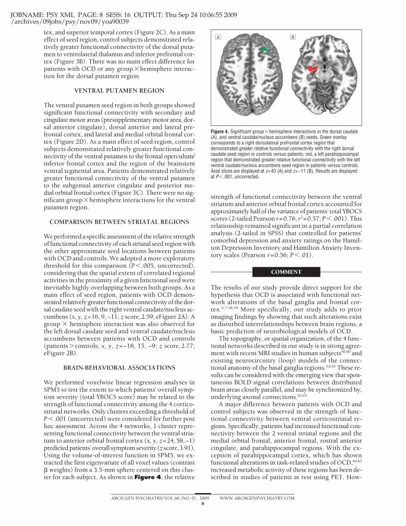
tex, and superior temporal cortex (Figure 2C). As a maineffect of seed region, control subjects demonstrated rela-tively greater functional connectivity of the dorsal puta-men to ventrolateral thalamus and inferior prefrontal cor-tex (Figure 3B). There was no main effect difference forpatients with OCD or any group�hemisphere interac-tion for the dorsal putamen region.
VENTRAL PUTAMEN REGION
The ventral putamen seed region in both groups showedsignificant functional connectivity with secondary andcingulate motor areas (presupplementary motor area, dor-sal anterior cingulate), dorsal anterior and lateral pre-frontal cortex, and lateral and medial orbital frontal cor-tex (Figure 2D). As a main effect of seed region, controlsubjects demonstrated relatively greater functional con-nectivity of the ventral putamen to the frontal operculum/inferior frontal cortex and the region of the brainstemventral tegmental area. Patients demonstrated relativelygreater functional connectivity of the ventral putamento the subgenual anterior cingulate and posterior me-dial orbital frontal cortex (Figure 3C). There were no sig-nificant group�hemisphere interactions for the ventralputamen region.
COMPARISON BETWEEN STRIATAL REGIONS
We performed a specific assessment of the relative strengthof functional connectivity of each striatal seed region withthe other approximate seed locations between patientswith OCD and controls. We adopted a more exploratorythreshold for this comparison (P� .005, uncorrected),considering that the spatial extent of correlated regionalactivities in the proximity of a given functional seed wereinevitably highly overlapping between both groups. As amain effect of seed region, patients with OCD demon-strated relatively greater functional connectivity of the dor-sal caudate seed with the right ventral caudate/nucleus ac-cumbens (x, y, z=16, 9, −11; z score,2.59; eFigure 2A). Agroup � hemisphere interaction was also observed forthe left dorsal caudate seed and ventral caudate/nucleusaccumbens between patients with OCD and controls(patients�controls; x, y, z=−18, 15, −9; z score,2.77;eFigure 2B).
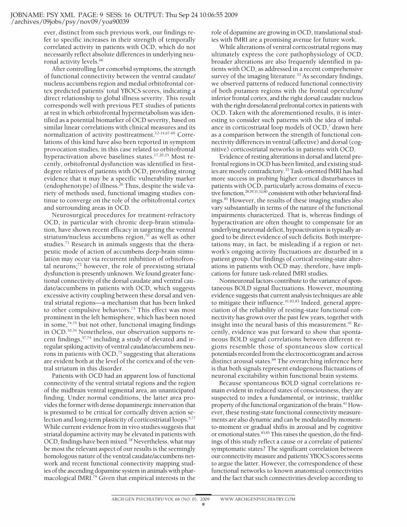
BRAIN-BEHAVIORAL ASSOCIATIONS
We performed voxelwise linear regression analyses inSPM5 to test the extent to which patients’ overall symp-tom severity (total YBOCS score) may be related to thestrength of functional connectivity among the 4 cortico-striatal networks. Only clusters exceeding a threshold ofP� .001 (uncorrected) were considered for further posthoc assessment. Across the 4 networks, 1 cluster repre-senting functional connectivity between the ventral stria-tum to anterior orbital frontal cortex (x, y, z=24,58,−1)predictedpatients’ overall symptomseverity (z score,3.91).Using the volume-of-interest function in SPM5, we ex-tracted the first eigenvariate of all voxel values (contrast� weights) from a 3.5-mm sphere centered on this clus-ter for each subject. As shown in Figure 4, the relative
strength of functional connectivity between the ventralstriatum and anterior orbital frontal cortex accounted forapproximately half of the variance of patients’ total YBOCSscores (2-tailed Pearson r=0.76; r2=0.57; P� .001). Thisrelationship remained significant in a partial correlationanalysis (2-tailed in SPSS) that controlled for patients’comorbid depression and anxiety ratings on the Hamil-ton Depression Inventory and Hamilton Anxiety Inven-tory scales (Pearson r=0.56; P� .01).
COMMENT
The results of our study provide direct support for thehypothesis that OCD is associated with functional net-work alterations of the basal ganglia and frontal cor-tex.4,7,58,59 More specifically, our study adds to priorimaging findings by showing that such alterations existas disturbed interrelationships between brain regions, abasic prediction of neurobiological models of OCD.
The topography, or spatial organization, of the 4 func-tional networks described in our study is in strong agree-ment with recent MRI studies in human subjects40,60 andexisting neurocircuitry (loop) models of the connec-tional anatomy of the basal ganglia regions.5,6,61 These re-sults can be considered with the emerging view that spon-taneous BOLD signal correlations between distributedbrain areas closely parallel, and may be synchronized by,underlying axonal connections.62,63
A major difference between patients with OCD andcontrol subjects was observed in the strength of func-tional connectivity between ventral corticostriatal re-gions. Specifically, patients had increased functional con-nectivity between the 2 ventral striatal regions and themedial orbital frontal, anterior frontal, rostral anteriorcingulate, and parahippocampal regions. With the ex-ception of parahippocampal cortex, which has shownfunctional alterations in task-related studies of OCD,64,65
increased metabolic activity of these regions has been de-scribed in studies of patients at rest using PET. How-
A B
Figure 4. Significant group�hemisphere interactions in the dorsal caudate(A), and ventral caudate/nucleus accumbens (B) seeds. Green overlaycorresponds to a right dorsolateral prefrontal cortex region thatdemonstrated greater relative functional connectivity with the right dorsalcaudate seed region in controls versus patients; red, a left parahippocampalregion that demonstrated greater relative functional connectivity with the leftventral caudate/nucleus accumbens seed region in patients versus controls.Axial slices are displayed at z=43 (A) and z=−11 (B). Results are displayedat P� .001, uncorrected.
(REPRINTED) ARCH GEN PSYCHIATRY/ VOL 66 (NO. 0), 2009 WWW.ARCHGENPSYCHIATRY.COM8
©2009 American Medical Association. All rights reserved.
JOBNAME: PSY XML PAGE: 9 SESS: 16 OUTPUT: Thu Sep 24 10:06:55 2009/archives/09jobs/psy/nov09/yoa90039
ever, distinct from such previous work, our findings re-fer to specific increases in their strength of temporallycorrelated activity in patients with OCD, which do notnecessarily reflect absolute differences in underlying neu-ronal activity levels.66
After controlling for comorbid symptoms, the strengthof functional connectivity between the ventral caudate/nucleus accumbens region and medial orbitofrontal cor-tex predicted patients’ total YBOCS scores, indicating adirect relationship to global illness severity. This resultcorresponds well with previous PET studies of patientsat rest in which orbitofrontal hypermetabolism was iden-tified as a potential biomarker of OCD severity, based onsimilar linear correlations with clinical measures and itsnormalization of activity posttreatment.12-14,67-69 Corre-lations of this kind have also been reported in symptomprovocation studies, in this case related to orbitofrontalhyperactivation above baselines states.17,20,25 Most re-cently, orbitofrontal dysfunction was identified in first-degree relatives of patients with OCD, providing strongevidence that it may be a specific vulnerability marker(endophenotype) of illness.26 Thus, despite the wide va-riety of methods used, functional imaging studies con-tinue to converge on the role of the orbitofrontal cortexand surrounding areas in OCD.
Neurosurgical procedures for treatment-refractoryOCD, in particular with chronic deep-brain stimula-tion, have shown recent efficacy in targeting the ventralstriatum/nucleus accumbens region,70 as well as otherstudies.71 Research in animals suggests that the thera-peutic mode of action of accumbens deep-brain stimu-lation may occur via recurrent inhibition of orbitofron-tal neurons;72 however, the role of preexisting striataldysfunction is presently unknown. We found greater func-tional connectivity of the dorsal caudate and ventral cau-date/accumbens in patients with OCD, which suggestsexcessive activity coupling between these dorsal and ven-tral striatal regions—a mechanism that has been linkedto other compulsive behaviors.73 This effect was mostprominent in the left hemisphere, which has been notedin some,74,75 but not other, functional imaging findingsin OCD.10,76 Nonetheless, our observation supports re-cent findings,37,74 including a study of elevated and ir-regular spiking activity of ventral caudate/accumbens neu-rons in patients with OCD,75 suggesting that alterationsare evident both at the level of the cortex and of the ven-tral striatum in this disorder.
Patients with OCD had an apparent loss of functionalconnectivity of the ventral striatal regions and the regionof the midbrain ventral tegmental area, an unanticipatedfinding. Under normal conditions, the latter area pro-vides the former with dense dopaminergic innervation thatis presumed to be critical for cortically driven action se-lection and long-term plasticity of corticostriatal loops.5,77
While current evidence from in vivo studies suggests thatstriatal dopamine activity may be elevated in patients withOCD, findings have been mixed.78 Nevertheless, what maybe most the relevant aspect of our results is the seeminglyhomologous nature of the ventral caudate/accumbens net-work and recent functional connectivity mapping stud-ies of the ascending dopamine system in animals with phar-macological fMRI.79 Given that empirical interests in the
role of dopamine are growing in OCD, translational stud-ies with fMRI are a promising avenue for future work.
While alterations of ventral corticostriatal regions mayultimately express the core pathophysiology of OCD,broader alterations are also frequently identified in pa-tients with OCD, as addressed in a recent comprehensivesurvey of the imaging literature.33 As secondary findings,we observed patterns of reduced functional connectivityof both putamen regions with the frontal operculum/inferior frontal cortex, and the right dorsal caudate nucleuswith the right dorsolateral prefrontal cortex in patients withOCD. Taken with the aforementioned results, it is inter-esting to consider such patterns with the idea of imbal-ance in corticostriatal loop models of OCD,7 drawn hereas a comparison between the strength of functional con-nectivity differences in ventral (affective) and dorsal (cog-nitive) corticostriatal networks in patients with OCD.
Evidence of resting alterations in dorsal and lateral pre-frontal regions in OCD has been limited, and existing stud-ies are mostly contradictory.33 Task-oriented fMRI has hadmore success in probing higher cortical disturbances inpatients with OCD, particularly across domains of execu-tive function,28,29,31,32,80 consistent with other behavioral find-ings.81 However, the results of these imaging studies alsovary substantially in terms of the nature of the functionalimpairments characterized. That is, whereas findings ofhyperactivation are often thought to compensate for anunderlying neuronal deficit, hypoactivation is typically ar-gued to be direct evidence of such deficits. Both interpre-tations may, in fact, be misleading if a region or net-work’s ongoing activity fluctuations are disturbed in apatient group. Our findings of cortical resting-state alter-ations in patients with OCD may, therefore, have impli-cations for future task-related fMRI studies.
Nonneuronal factors contribute to the variance of spon-taneous BOLD signal fluctuations. However, mountingevidence suggests that current analysis techniques are ableto mitigate their influence.41,82,83 Indeed, general appre-ciation of the reliability of resting-state functional con-nectivity has grown over the past few years, together withinsight into the neural basis of this measurement.41 Re-cently, evidence was put forward to show that sponta-neous BOLD signal correlations between different re-gions resemble those of spontaneous slow corticalpotentials recorded from the electrocorticogram and acrossdistinct arousal states.84 The overarching inference hereis that both signals represent endogenous fluctuations ofneuronal excitability within functional brain systems.
Because spontaneous BOLD signal correlations re-main evident in reduced states of consciousness, they aresuspected to index a fundamental, or intrinsic, traitlikeproperty of the functional organization of the brain.41 How-ever, these resting-state functional connectivity measure-ments are also dynamic and can be modulated by moment-to-moment or gradual shifts in arousal and by cognitiveor emotional states.83,85 This raises the question, do the find-ings of this study reflect a cause or a correlate of patients’symptomatic states? The significant correlation betweenour connectivity measure and patients’ YBOCS scores seemsto argue the latter. However, the correspondence of thesefunctional networks to known anatomical connectivitiesand the fact that such connectivities develop according to
(REPRINTED) ARCH GEN PSYCHIATRY/ VOL 66 (NO. 0), 2009 WWW.ARCHGENPSYCHIATRY.COM9
©2009 American Medical Association. All rights reserved.
JOBNAME: PSY XML PAGE: 10 SESS: 16 OUTPUT: Thu Sep 24 10:06:55 2009/archives/09jobs/psy/nov09/yoa90039
distinct maturation rates that may be compromised inOCD86 suggest that both possibilities may be valid, or atleast that this question remains open.
We consider this study a useful starting point for futureinvestigations of resting-state functional connectivity inOCD, in relation to both corticostriatal networks and otherlarge-scale brain systems. In both cases, it will be impor-tant for such studies to address the multisymptomatic na-ture of OCD populations in specific detail, as performed inother larger-scale MRI studies.37,87 While our patients werecharacterizedbymoreprominentaggressive/checkingsymp-toms, consistentwithprior studies,34 wewereunable toper-form the appropriate statistical tests (ie, because of samplesize) to determine whether our results can be related pref-erentially to one or another major symptom dimension orgeneralized to all patients with this disorder. This is a ca-veat that should be addressed in future work. Addition-ally, the assessment of patients both taking and not takingmedication will also be relevant in further studies of cor-ticostriatal functional networks in OCD. Most patients inour study had completed 1 or more trials of selective sero-tonin reuptake inhibitor antidepressant treatment. Al-though based on previous work, chronic selective seroto-nin reuptake inhibitor treatment in OCD may be expectedtonormalizeheightenedresting-stateactivity inventral cor-ticostriatal regions;12 its influenceonspecific functionalcon-nectivity measurements requires investigation.
Resting-state fMRI has obvious practical benefits whenstudying psychiatric populations,82 including its ease ofuse and the fact that reliable brain mapping results canbe obtained from relatively brief scanning sessions (�10minutes).88 However, despite such benefits, resting-state fMRI should be seen as complementary to task-based imaging studies, as both are likely to be mutuallyinformative. For example, the integration of both ap-proaches may be useful in reconciling the apparently di-vergent findings of reduced task-related activation26-29 andheightened resting-state activity and striatal functionalconnectivity of the orbitofrontal cortex in patients withOCD. Lastly, although we were able to perform whole-brain echoplanar imaging by scanning at 1.5 T, higher-field fMRI with increased sensitivity is likely to improvethe level of anatomical description achieved in this study.
While our findings support the prevailing hypoth-esis that corticalstriatal networks are dysfunctional inOCD, the specific neural mechanisms and vulnerabilityfactors that give rise to this impairment remain to be un-derstood. There is strong potential for neuroimaging stud-ies to generate further insight to this end, especially ifcombined with current advances in clinical, develop-mental, and molecular neuroscience studies of OCD.
Submitted for Publication: November 12, 2008; final re-vision received January 26, 2009; accepted March 16,2009.Author Affiliations: Institut d’Alta Tecnologia–Parc deRecerca Biomèdica de Barcelona, Centro RadiologicoComputerizado Corporacio Sanitària, Barcelona, Spain(Drs Harrison, Soriano-Mas, Pujol, Hernandez-Ribas,Deus, Alonso, and Cardoner, Ms Lopez-Solà, and Mr Or-tiz); Melbourne Neuropsychiatry Centre, Department ofPsychiatry, The University of Melbourne & Melbourne
Health, Australia (Drs Harrison, Yücel, and Pantelis); De-partment of Electronic Engineering, Technical Univer-sity of Catalonia, Barcelona, Spain (Mr Ortiz); Depart-ment of Clinical Sciences, Faculty of Medicine, Universityof Barcelona, Spain (Ms Lopez-Solà); Department of Psy-chiatry, Bellvitge University Hospital, Centro de Inves-tigacion Biomedica en Red de Salud Mental, Barcelona,Spain (Drs Hernandez-Ribas, Alonso, Menchon, and Car-doner); Department of Clinical and Health Psychology,Autonomous University of Barcelona, Barcelona, Spain(Dr Deus); Orygen Research Centre, Melbourne, Aus-tralia (Dr Yücel); Networking Research Center on Bio-engineering, Biomaterials and Nanomedicine, Centro deInvestigacion Biomedica en Red en Bioenginierıa, Bio-materials y Nanomedicina, Barcelona, Spain (Dr Pujol).Correspondence: Ben J. Harrison, PhD, Melbourne Neu-ropsychiatry Centre, National Neuroscience Facility, 161Barry St, Carlton, 3053, Melbourne, Australia ([email protected]).Author Contributions: Dr Harrison had full access to allof the data in the study and takes responsibility for the in-tegrity of the data and the accuracy of the data analysis.Financial Disclosure: None reported.Funding/Support: This study was supported in part bythe Instituto de Salud Carlos III, Centro de Investiga-cion en Red de Salud Mental, Fondo de Investigacion Sani-taria grants PI050884 and PI071029; National Health andMedical Research Council of Australia (NHMRC) Train-ing Award 400420 (Dr Harrison); NHMRC Clinical Ca-reer Development Award 509345 (Dr Yücel); the Net-working Research Center on Bioengineering, Biomaterialsand Nanomedicine, Barcelona, Spain (Dr Pujol); and For-macion de Personal Universitario scholarships AP2005-0408 and AP2006-02869 from the Spanish Ministry ofEducation (Ms Lopez-Solà and Mr Ortiz).Additional Information: The eFigures are available at http://www.archgenpsychiatry.com.
REFERENCES
1. Laplane D, Levasseur M, Pillon B, Dubois B, Baulac M, Mazoyer B, Tran Dinh S,Sette G, Danze F, Baron JC. Obsessive-compulsive and other behavioural changeswith bilateral basal ganglia lesions: a neuropsychological, magnetic resonanceimaging and positron tomography study. Brain. 1989;112(pt 3):699-725.
2. Modell JG, Mountz JM, Curtis GC, Greden JF. Neurophysiologic dysfunction inbasal ganglia/limbic striatal and thalamocortical circuits as a pathogenetic mecha-nism of obsessive-compulsive disorder. J Neuropsychiatry Clin Neurosci. 1989;1(1):27-36.
3. Rapoport JL, Wise SP. Obsessive-compulsive disorder: evidence for basal gan-glia dysfunction. Psychopharmacol Bull. 1988;24(3):380-384.
4. Graybiel AM, Rauch SL. Toward a neurobiology of obsessive-compulsive disorder.Neuron. 2000;28(2):343-347.
5. Alexander GE, DeLong MR, Strick PL. Parallel organization of functionally seg-regated circuits linking basal ganglia and cortex. Annu Rev Neurosci. 1986;9:357-381.
6. Haber SN. The primate basal ganglia: parallel and integrative networks. J ChemNeuroanat. 2003;26(4):317-330.
7. Saxena S. Neuroimaging and the pathophysiology of obsessive compulsivedisorder. In: Fu C, Senior C, Russell TA, Weinberger DR, Murray R, eds. Neuro-imaging in Psychiatry. London, England: Martin Dunitz; 2003:191-224.
8. Menzies L, Chamberlain SR, Laird AR, Thelen SM, Sahakian BJ, Bullmore ET.Integrating evidence from neuroimaging and neuropsychological studies of ob-sessive-compulsive disorder: the orbitofronto-striatal model revisited. Neuro-sci Biobehav Rev. 2008;32(3):525-549.
9. Whiteside SP, Port JD, Abramowitz JS. A meta-analysis of functional neuroim-aging in obsessive-compulsive disorder. Psychiatry Res. 2004;132(1):69-79.
(REPRINTED) ARCH GEN PSYCHIATRY/ VOL 66 (NO. 0), 2009 WWW.ARCHGENPSYCHIATRY.COM10
©2009 American Medical Association. All rights reserved.

JOBNAME: PSY XML PAGE: 11 SESS: 16 OUTPUT: Thu Sep 24 10:06:55 2009/archives/09jobs/psy/nov09/yoa90039
10. Baxter LR Jr, Schwartz JM, Bergman KS, Szuba MP, Guze BH, Mazziotta JC, Ala-zraki A, Selin CE, Ferng HK, Munford P, Phelps ME. Caudate glucose metabolicrate changes with both drug and behavior therapy for obsessive-compulsivedisorder. Arch Gen Psychiatry. 1992;49(9):681-689.
11. Baxter LR Jr, Schwartz JM, Mazziotta JC, Phelps ME, Pahl JJ, Guze BH, Fair-banks L. Cerebral glucose metabolic rates in nondepressed patients with obsessive-compulsive disorder. Am J Psychiatry. 1988;145(12):1560-1563.
12. Saxena S, Brody AL, Maidment KM, Dunkin JJ, Colgan M, Alborzian S, PhelpsME, Baxter LR Jr. Localized orbitofrontal and subcortical metabolic changes andpredictors of response to paroxetine treatment in obsessive-compulsive disorder.Neuropsychopharmacology. 1999;21(6):683-693.
13. Schwartz JM, Stoessel PW, Baxter LR Jr, Martin KM, Phelps ME. Systematic changesin cerebral glucose metabolic rate after successful behavior modification treatmentof obsessive-compulsive disorder. Arch Gen Psychiatry. 1996;53(2):109-113.
14. Swedo SE, Pietrini P, Leonard HL, Schapiro MB, Rettew DC, Goldberger EL, RapoportSI, Rapoport JL, Grady CL. Cerebral glucose metabolism in childhood-onset ob-sessive-compulsive disorder: revisualization during pharmacotherapy. Arch GenPsychiatry. 1992;49(9):690-694.
15. Adler CM, McDonough-Ryan P, Sax KW, Holland SK, Arndt S, Strakowski SM.fMRI of neuronal activation with symptom provocation in unmedicated patientswith obsessive compulsive disorder. J Psychiatr Res. 2000;34(4-5):317-324.
16. Breiter HC, Rauch SL, Kwong KK, Baker JR, Weisskoff RM, Kennedy DN, Ken-drick AD, Davis TL, Jiang A, Cohen MS, Stern CE, Belliveau JW, Baer L, O’SullivanRL, Savage CR, Jenike MA, Rosen BR. Functional magnetic resonance imagingof symptom provocation in obsessive-compulsive disorder. Arch Gen Psychiatry.1996;53(7):595-606.
17. Mataix-Cols D, Wooderson S, Lawrence N, Brammer MJ, Speckens A, PhillipsML. Distinct neural correlates of washing, checking, and hoarding symptom di-mensions in obsessive-compulsive disorder. Arch Gen Psychiatry. 2004;61(6):564-576.
18. Schienle A, Schafer A, Stark R, Walter B, Vaitl D. Neural responses of OCD pa-tients towards disorder-relevant, generally disgust-inducing and fear-inducingpictures. Int J Psychophysiol. 2005;57(1):69-77.
19. McGuire PK, Bench CJ, Frith CD, Marks IM, Frackowiak RS, Dolan RJ. Func-tional anatomy of obsessive-compulsive phenomena. Br J Psychiatry. 1994;164(4):459-468.
20. Rauch SL, Jenike MA, Alpert NM, Baer L, Breiter HC, Savage CR, Fischman AJ.Regional cerebral blood flow measured during symptom provocation in obsessive-compulsive disorder using oxygen 15-labeled carbon dioxide and positron emis-sion tomography. Arch Gen Psychiatry. 1994;51(1):62-70.
21. Phillips ML, Marks IM, Senior C, Lythgoe D, O’Dwyer AM, Meehan O, WilliamsSC, Brammer MJ, Bullmore ET, McGuire PK. A differential neural response inobsessive-compulsive disorder patients with washing compared with checkingsymptoms to disgust. Psychol Med. 2000;30(5):1037-1050.
22. Shapira NA, Liu Y, He AG, Bradley MM, Lessig MC, James GA, Stein DJ, LangPJ, Goodman WK. Brain activation by disgust-inducing pictures in obsessive-compulsive disorder. Biol Psychiatry. 2003;54(7):751-756.
23. Lawrence NS, An SK, Mataix-Cols D, Ruths F, Speckens A, Phillips ML. Neuralresponses to facial expressions of disgust but not fear are modulated by wash-ing symptoms in OCD. Biol Psychiatry. 2007;61(9):1072-1080.
24. van den Heuvel OA, Veltman DJ, Groenewegen HJ, Dolan RJ, Cath DC, BoellaardR, Mesina CT, van Balkom AJ, van Oppen P, Witter MP, Lammertsma AA, vanDyck R. Amygdala activity in obsessive-compulsive disorder with contamina-tion fear: a study with oxygen-15 water positron emission tomography. Psychia-try Res. 2004;132(3):225-237.
25. Rauch SL, Shin LM, Dougherty DD, Alpert NM, Fischman AJ, Jenike MA. Pre-dictors of fluvoxamine response in contamination-related obsessive compul-sive disorder: a PET symptom provocation study. Neuropsychopharmacology.2002;27(5):782-791.
26. Chamberlain SR, Menzies L, Hampshire A, Suckling J, Fineberg NA, del CampoN, Aitken M, Craig K, Owen AM, Bullmore ET, Robbins TW, Sahakian BJ.Orbitofrontal dysfunction in patients with obsessive-compulsive disorder and theirunaffected relatives. Science. 2008;321(5887):421-422.
27. Remijnse PL, Nielen MM, van Balkom AJ, Cath DC, van Oppen P, Uylings HB,Veltman DJ. Reduced orbitofrontal-striatal activity on a reversal learning task inobsessive-compulsive disorder. Arch Gen Psychiatry. 2006;63(11):1225-1236.
28. Roth RM, Saykin AJ, Flashman LA, Pixley HS, West JD, Mamourian AC. Event-related functional magnetic resonance imaging of response inhibition in obsessive-compulsive disorder. Biol Psychiatry. 2007;62(8):901-909.
29. van den Heuvel OA, Veltman DJ, Groenewegen HJ, Cath DC, van Balkom AJ, vanHartskamp J, Barkhof F, van Dyck R. Frontal-striatal dysfunction during planningin obsessive-compulsive disorder. Arch Gen Psychiatry. 2005;62(3):301-309.
30. Maltby N, Tolin DF, Worhunsky P, O’Keefe TM, Kiehl KA. Dysfunctional action moni-toring hyperactivates frontal-striatal circuits in obsessive-compulsive disorder: anevent-related fMRI study. Neuroimage. 2005;24(2):495-503.
31. van der Wee NJ, Ramsey NF, Jansma JM, Denys DA, van Megen HJ, Westen-berg HM, Kahn RS. Spatial working memory deficits in obsessive compulsivedisorder are associated with excessive engagement of the medial frontal cortex.Neuroimage. 2003;20(4):2271-2280.
32. Yücel M, Harrison BJ, Wood SJ, Fornito A, Wellard RM, Pujol J, Clarke K, Phill-ips ML, Kyrios M, Velakoulis D, Pantelis C. Functional and biochemical alter-ations of the medial frontal cortex in obsessive-compulsive disorder. Arch GenPsychiatry. 2007;64(8):946-955.
33. Menzies L, Achard S, Chamberlain SR, Fineberg N, Chen CH, del Campo N, Sa-hakian BJ, Robbins TW, Bullmore E. Neurocognitive endophenotypes of obsessive-compulsive disorder. Brain. 2007;130(pt 12):3223-3236.
34. Soriano-Mas C, Pujol J, Alonso P, Cardoner N, Menchon JM, Harrison BJ, DeusJ, Vallejo J, Gaser C. Identifying patients with obsessive-compulsive disorderusing whole-brain anatomy. Neuroimage. 2007;35(3):1028-1037.
35. Cardoner N, Soriano-Mas C, Pujol J, Alonso P, Harrison BJ, Deus J, Hernandez-Ribas R, Menchon JM, Vallejo J. Brain structural correlates of depressive comor-bidity in obsessive-compulsive disorder. Neuroimage. 2007;38(3):413-421.
36. Harrison BJ, Yucel M, Shaw M, Kyrios M, Maruff P, Brewer WJ, Purcell R, Vela-koulis D, Strother SC, Scott AM, Nathan PJ, Pantelis C. Evaluating brain activityin obsessive-compulsive disorder: preliminary insights from a multivariate analysis.Psychiatry Res. 2006;147(2-3):227-231.
37. Pujol J, Soriano-Mas C, Alonso P, Cardoner N, Menchon JM, Deus J, Vallejo J.Mapping structural brain alterations in obsessive-compulsive disorder. Arch GenPsychiatry. 2004;61(7):720-730.
38. Menzies L, Williams GB, Chamberlain SR, Ooi C, Fineberg N, Suckling J, Sa-hakian BJ, Robbins TW, Bullmore ET. White matter abnormalities in patients withobsessive-compulsive disorder and their first-degree relatives [published on-line ahead of print June 2, 2008]. Am J Psychiatry. 2008;165(10):1308-1315.
39. Szeszko PR, Ardekani BA, Ashtari M, Malhotra AK, Robinson DG, Bilder RM, LimKO. White matter abnormalities in obsessive-compulsive disorder: a diffusiontensor imaging study. Arch Gen Psychiatry. 2005;62(7):782-790.
40. Di Martino A, Scheres A, Margulies DS, Kelly AM, Uddin LQ, Shehzad Z, BiswalB, Walters JR, Castellanos FX, Milham MP. Functional connectivity of human stria-tum: a resting state fMRI study [published online ahead of print April 9, 2008].Cereb Cortex. 2008;18(12):2735-2747.
41. Fox MD, Raichle ME. Spontaneous fluctuations in brain activity observed with func-tional magnetic resonance imaging. Nat Rev Neurosci. 2007;8(9):700-711.
42. Damoiseaux JS, Rombouts SA, Barkhof F, Scheltens P, Stam CJ, Smith SM, Beck-mann CF. Consistent resting-state networks across healthy subjects. Proc NatlAcad Sci U S A. 2006;103(37):13848-13853.
43. Hampson M, Peterson BS, Skudlarski P, Gatenby JC, Gore JC. Detection of func-tional connectivity using temporal correlations in MR images. Hum Brain Mapp.2002;15(4):247-262.
44. Aouizerate B, Guehl D, Cuny E, Rougier A, Bioulac B, Tignol J, Burbaud P. Patho-physiology of obsessive-compulsive disorder: a necessary link between phe-nomenology, neuropsychology, imagery and physiology. Prog Neurobiol. 2004;72(3):195-221.
45. First MB, Spitzer RL, Gibbon M, Williams JB. Structured Clinical Interview forDSM-IV Axis 1 Disorders. Washington, DC: American Psychiatric Press; 1998.
46. Goodman WK, Price LH, Rasmussen SA, Mazure C, Fleischmann RL, Hill CL,Heninger GR, Charney DS. The Yale-Brown Obsessive Compulsive Scale I: de-velopment, use, and reliability. Arch Gen Psychiatry. 1989;46(11):1006-1011.
47. Mataix-Cols D, Rauch SL, Manzo PA, Jenike MA, Baer L. Use of factor-analyzedsymptom dimensions to predict outcome with serotonin reuptake inhibitors andplacebo in the treatment of obsessive-compulsive disorder. Am J Psychiatry. 1999;156(9):1409-1416.
48. Hamilton M. A rating scale for depression. J Neurol Neurosurg Psychiatry. 1960;23:56-62.
49. Hamilton M. The assessment of anxiety states by rating. Br J Med Psychol. 1959;32(1):50-55.
50. Wechsler D. Wechsler Abbreviated Scale of Intelligence Manual. San Antonio,TX: The Psychological Corporation; 1999.
51. First MB, Spitzer RL, Gibbon M, Williams JB. Structured Clinical Interview forDSM-IV-RS Axis 1 Disorders: Non-Patient Edition (SCID-I/NP). New York, NY:Biometrics Research, New York State Psychiatric Institute; 2007.
52. Ashburner J, Friston KJ. Unified segmentation. Neuroimage. 2005;26(3):839-851.53. Postuma RB, Dagher A. Basal ganglia functional connectivity based on a meta-
analysis of 126 positron emission tomography and functional magnetic reso-nance imaging publications. Cereb Cortex. 2006;16(10):1508-1521.
54. Mai J, Assheuer J, Paxinos G. Atlas of the Human Brain. San Diego, CA: AcademicPress; 1997.
55. Toro R, Fox PT, Paus T. Functional coactivation map of the human brain. CerebCortex. 2008;18(11):2553-2559.
56. Region of interest analysis using an SPM toolbox [abstract]. Brett M, Anton JL,Valabregue R, Poline JB. Presented at: The 8th International conference on Func-
(REPRINTED) ARCH GEN PSYCHIATRY/ VOL 66 (NO. 0), 2009 WWW.ARCHGENPSYCHIATRY.COM11
©2009 American Medical Association. All rights reserved.
JOBNAME: PSY XML PAGE: 12 SESS: 16 OUTPUT: Thu Sep 24 10:06:55 2009/archives/09jobs/psy/nov09/yoa90039
tional Mapping of the Human Brain; June 2-6, 2002; Sendai, Japan. Available onCD-ROM in Neuroimage 16(2).
57. Genovese CR, Lazar NA, Nichols T. Thresholding of statistical maps in functionalneuroimaging using the false discovery rate. Neuroimage. 2002;15(4):870-878.
58. Baxter LR Jr. Basal ganglia systems in ritualistic social displays: reptiles and hu-mans; function and illness. Physiol Behav. 2003;79(3):451-460.
59. Schwartz JM. Neuroanatomical aspects of cognitive-behavioural therapy re-sponse in obsessive-compulsive disorder: an evolving perspective on brain andbehaviour. Br J Psychiatry Suppl. 1998;(35):38-44.
60. Draganski B, Kherif F, Kloppel S, Cook PA, Alexander DC, Parker GJ, DeichmannR, Ashburner J, Frackowiak RS. Evidence for segregated and integrative connec-tivity patterns in the human basal ganglia. J Neurosci. 2008;28(28):7143-7152.
61. Groenewegen HJ, Trimble M. The ventral striatum as an interface between thelimbic and motor systems. CNS Spectr. 2007;12(12):887-892.
62. Greicius MD, Supekar K, Menon V, Dougherty RF. Resting-state functional con-nectivity reflects structural connectivity in the default mode network [publishedonline ahead of print April 9, 2008]. Cereb Cortex. 2009;19(1):72-78.
63. Vincent JL, Patel GH, Fox MD, Snyder AZ, Baker JT, Van Essen DC, Zempel JM,Snyder LH, Corbetta M, Raichle ME. Intrinsic functional architecture in the anes-thetized monkey brain. Nature. 2007;447(7140):83-86.
64. Rauch SL, Savage CR, Alpert NM, Dougherty D, Kendrick A, Curran T, Brown HD,Manzo P, Fischman AJ, Jenike MA. Probing striatal function in obsessive-compulsive disorder: a PET study of implicit sequence learning. J Neuropsychia-try Clin Neurosci. 1997;9(4):568-573.
65. Rauch SL, Wedig MM, Wright CI, Martis B, McMullin KG, Shin LM, CannistraroPA, Wilhelm S. Functional magnetic resonance imaging study of regional brain ac-tivation during implicit sequence learning in obsessive-compulsive disorder. BiolPsychiatry. 2007;61(3):330-336.
66. Raichle ME, Mintun MA. Brain work and brain imaging. Annu Rev Neurosci. 2006;29:449-476.
67. Benkelfat C, Nordahl TE, Semple WE, King AC, Murphy DL, Cohen RM. Localcerebral glucose metabolic rates in obsessive-compulsive disorder: patients treatedwith clomipramine. Arch Gen Psychiatry. 1990;47(9):840-848.
68. Biver F, Goldman S, Francois A, De La Porte C, Luxen A, Gribomont B, LotstraF. Changes in metabolism of cerebral glucose after stereotactic leukotomy forrefractory obsessive-compulsive disorder: a case report. J Neurol NeurosurgPsychiatry. 1995;58(4):502-505.
69. Matsumoto R, Nakamae T, Yoshida T, Kitabayashi Y, Ushijima Y, Narumoto J, ItoH, Suhara T, Fukui K. Recurrent hyperperfusion in the right orbitofrontal cortex inobsessive-compulsive disorder. Prog Neuropsychopharmacol Biol Psychiatry. 2008;32(4):1082-1084.
70. Greenberg BD, Gabriels LA, Malone DA Jr, Rezai AR, Friehs GM, Okun MS, Sha-pira NA, Foote KD, Cosyns PR, Kubu CS, Malloy PF, Salloway SP, Giftakis JE,Rise MT, Machado AG, Baker KB, Stypulkowski PH, Goodman WK, RasmussenSA, Nuttin BJ. Deep brain stimulation of the ventral internal capsule/ventral stria-tum for obsessive-compulsive disorder: worldwide experience [published on-line May 20, 2008]. Mol Psychiatry. doi:10.1038/mp.2008.55.
71. Mallet L, Polosan M, Jaafari N, Baup N, Welter ML, Fontaine D, du Montcel ST,Yelnik J, Chereau I, Arbus C, Raoul S, Aouizerate B, Damier P, Chabardes S, Cz-ernecki V, Ardouin C, Krebs MO, Bardinet E, Chaynes P, Burbaud P, Cornu P, Der-ost P, Bougerol T, Bataille B, Mattei V, Dormont D, Devaux B, Verin M, Houeto JL,Pollak P, Benabid AL, Agid Y, Krack P, Millet B, Pelissolo A; STOC Study Group.Subthalamic nucleus stimulation in severe obsessive-compulsive disorder. N EnglJ Med. 2008;359(20):2121-2134.
72. McCracken CB, Grace AA. High-frequency deep brain stimulation of the nucleusaccumbens region suppresses neuronal activity and selectively modulates affer-ent drive in rat orbitofrontal cortex in vivo. J Neurosci. 2007;27(46):12601-12610.
73. Everitt BJ, Belin D, Economidou D, Pelloux Y, Dalley JW, Robbins TW. Neural mecha-nisms underlying the vulnerability to develop compulsive drug-seeking habits andaddiction [review]. Philos Trans R Soc Lond B Biol Sci. 2008;363(1507):3125-3135.
74. Van Laere K, Nuttin B, Gabriels L, Dupont P, Rasmussen S, Greenberg BD, Co-syns P. Metabolic imaging of anterior capsular stimulation in refractory obsessive-compulsive disorder: a key role for the subgenual anterior cingulate and ventralstriatum. J Nucl Med. 2006;47(5):740-747.
75. Guehl D, Benazzouz A, Aouizerate B, Cuny E, Rotge JY, Rougier A, Tignol J, Bi-oulac B, Burbaud P. Neuronal correlates of obsessions in the caudate nucleus.Biol Psychiatry. 2008;63(6):557-562.
76. Saxena S, Brody AL, Ho ML, Zohrabi N, Maidment KM, Baxter LR Jr. Differentialbrain metabolic predictors of response to paroxetine in obsessive-compulsivedisorder versus major depression. Am J Psychiatry. 2003;160(3):522-532.
77. Surmeier DJ, Ding J, Day M, Wang Z, Shen W. D1 and D2 dopamine-receptormodulation of striatal glutamatergic signaling in striatal medium spiny neurons.Trends Neurosci. 2007;30(5):228-235.
78. Denys D, Zohar J, Westenberg HG. The role of dopamine in obsessive-compul-sive disorder: preclinical and clinical evidence. J Clin Psychiatry. 2004;65(suppl14):11-17.
79. Schwarz AJ, Gozzi A, Reese T, Bifone A. In vivo mapping of functional connec-tivity in neurotransmitter systems using pharmacological MRI. Neuroimage. 2007;34(4):1627-1636.
80. Pujol J, Torres L, Deus J, Cardoner N, Pifarre J, Capdevila A, Vallejo J. Functionalmagnetic resonance imaging study of frontal lobe activation during word genera-tion in obsessive-compulsive disorder. Biol Psychiatry. 1999;45(7):891-897.
81. Purcell R, Maruff P, Kyrios M, Pantelis C. Neuropsychological deficits in obsessive-compulsive disorder: a comparison with unipolar depression, panic disorder, andnormal controls. Arch Gen Psychiatry. 1998;55(5):415-423.
82. Greicius M. Resting-state functional connectivity in neuropsychiatric disorders.Curr Opin Neurol. 2008;21(4):424-430.
83. Harrison BJ, Pujol J, Lopez-Sola M, Hernandez-Ribas R, Deus J, Ortiz H, Soriano-Mas C, Yucel M, Pantelis C, Cardoner N. Consistency and functional specializationin the default mode brain network. Proc Natl Acad Sci U S A. 2008;105(28):9781-9786.
84. He BJ, Snyder AZ, Zempel JM, Smyth MD, Raichle ME. Electrophysiological cor-relates of the brain’s intrinsic large-scale functional architecture. Proc Natl AcadSci U S A. 2008;105(41):16039-16044.
85. Harrison BJ, Pujol J, Ortiz H, Fornito A, Pantelis C, Yucel M. Modulation of brainresting-state networks by sad mood induction. PLoS One. 2008;3(3):e1794.
86. Maia TV, Cooney RE, Peterson BS. The neural bases of obsessive-compulsivedisorder in children and adults. Dev Psychopathol. 2008;20(4):1251-1283.
87. van den Heuvel OA, Remijnse PL, Mataix-Cols D, Vrenken H, Groenewegen HJ,Uylings HB, van Balkom AJ, Veltman DJ. The major symptom dimensions of ob-sessive-compulsive disorder are mediated by partially distinct neural systems [pub-lished online ahead of print October 24, 2008]. Brain. 2009;132(pt 4):853-868.
88. Shehzad ZE, Clare Kelly AM, Reiss PT, Gee DG, Gotimer K, Uddin LQ, Lee SH,Margulies DS, Krain Roy A, Biswal BB, Petkova E, Castellanos FX, Milham MP.The resting-brain: unconstrained yet reliable [published online February 16, 2009].Cereb Cortex. doi:10.1093/cercor/bhn256.
(REPRINTED) ARCH GEN PSYCHIATRY/ VOL 66 (NO. 0), 2009 WWW.ARCHGENPSYCHIATRY.COM12
©2009 American Medical Association. All rights reserved.


