Embed Size (px)
Citation preview

The Rockefeller University Press, 0021-9525/2001/12/1065/15 $5.00The Journal of Cell Biology, Volume 155, Number 6, December 10, 2001 1065–1079http://www.jcb.org/cgi/doi/10.1083/jcb.200104005
JCB
Article
1065
The SH2-containing inositol polyphosphate5-phosphatase, SHIP-2, binds filamin andregulates submembraneous actin
Jennifer M. Dyson,
1
Cindy J. O’Malley,
1
Jelena Becanovic,
1
Adam D. Munday,
1
Michael C. Berndt,
2
Imogen D. Coghill,
1
Harshal H. Nandurkar,
1
Lisa M. Ooms,
1
and Christina A. Mitchell
1
1
Department of Biochemistry and Molecular Biology, Monash University, Clayton, Victoria, 3800 Australia
2
Baker Medical Research Institute, Prahran, Victoria, 3181 Australia
HIP-2 is a phosphoinositidylinositol 3,4,5 trisphos-
phate (PtdIns[3,4,5]P
3
) 5-phosphatase that contains anNH
2
-terminal SH2 domain, a central 5-phosphatasedomain, and a COOH-terminal proline-rich domain. SHIP-2negatively regulates insulin signaling. In unstimulated cells,SHIP-2 localized in a perinuclear cytosolic distribution andat the leading edge of the cell. Endogenous and recombinantSHIP-2 localized to membrane ruffles, which were mediatedby the COOH-terminal proline–rich domain. To identifyproteins that bind to the SHIP-2 proline–rich domain, yeasttwo-hybrid screening was performed, which isolated actin-binding protein filamin C. In addition, both filamin A and Bspecifically interacted with SHIP-2 in this assay. SHIP-2coimmunoprecipitated with filamin from COS-7 cells, and
S
association between these species did not change afterepidermal growth factor stimulation. SHIP-2 colocalized withfilamin at Z-lines and the sarcolemma in striated musclesections and at membrane ruffles in COS-7 cells, althoughthe membrane ruffling response was reduced in cellsoverexpressing SHIP-2. SHIP-2 membrane ruffle local-ization was dependent on filamin binding, as SHIP-2 wasexpressed exclusively in the cytosol of filamin-deficient cells.Recombinant SHIP-2 regulated PtdIns(3,4,5)P
3
levels andsubmembraneous actin at membrane ruffles after growthfactor stimulation, dependent on SHIP-2 catalytic activity.Collectively these studies demonstrate that filamin-dependentSHIP-2 localization critically regulates phosphatidylinositol3 kinase signaling to the actin cytoskeleton.
Introduction
Phosphoinositides are ubiquitous membrane componentswhich regulate proliferation, differentiation, inhibition ofapoptosis, secretion, cell movement, and the actin cytoskeleton.Receptor-regulated phosphoinositide 3 (PI-3)* kinasesphosphorylate phosphatidylinositol 4,5 bisphosphate (Ptd-
Ins[4,5]P
2
) forming phosphatidylinositol 3,4,5 trisphosphate
(PtdIns[3,4,5]P
3
) that is dephosphorylated by the inosi-tol polyphosphate 5-phosphatases (5-phosphatase) to Ptd-Ins(3,4)P
2
(Majerus, 1996; Martin, 1997). Both PtdIns
(3,4,5)P
3
and PtdIns(3,4)P
2
localize signaling proteins to theinner wall of the plasma membrane and allosterically regulatethese target proteins. PtdIns(3,4,5)P
3
and PtdIns(3,4)P
2
bind-ing proteins include the serine/threonine kinase, Akt, whichinhibits apoptosis, and proteins such as cytohesins and cen-taurins that regulate ADP ribosylation factor (ARF), andthereby vesicular trafficking and the peripheral actin cyto-skeleton (Corvera et al., 1999; Datta et al., 1999). Ptd-Ins(3,4,5)P
3
regulates growth factor–induced actin-depen-dent extension of lamellipodia, membrane ruffle formation,and cell migration.
PtdIns(3,4,5)P
3
is metabolized by the removal of eitherthe 5- or 3-position phosphate by specific 5- or 3-lipidphosphatases, respectively. The product of the tumorsuppressor gene phosphatase and tensin homologue deletedon chromosome 10 (PTEN) is a PtdIns(3,4,5)P
3
3-phosphatasewhich hydrolyses PtdIns(3,4,5)P
3
, forming PtdIns(4,5)P
2
. Thelipid 3-phosphatase activity of PTEN is critical for its tumorsuppressor function (for review see Cantley and Neel, 1999).
The 5-phosphatases hydrolyze the 5-position phosphate fromboth inositol phosphates and phosphoinositides and share the
Address correspondence to Christina A. Mitchell, Dept. of Biochemistryand Molecular Biology, Monash University, Clayton, Victoria, 3800Australia. Tel.: (61) 3990-53790. Fax: (61) 3990-54699. E-mail:[email protected]
*Abbreviations used in this paper: aa, amino acid; 5-phosphatase, inositolpolyphosphate 5-phosphatase; FLNC, filamin C; GFP, green fluorescentprotein; HA, hemagglutinin; PH, pleckstrin homology; PI-3, phosphati-dylinositol 3; PtdIns, phosphatidylinositol; PTEN, phosphatase andtensin homologue deleted on chromosome 10; SHIP, SH2 domain–containing inositol polyphosphate 5-phosphatase.Key words: SHIP-2; inositol polyphosphate 5-phosphatase; filamin;cytoskeleton; phosphatidylinositol 3,4,5-trisphosphate
on Novem
ber 23, 2015jcb.rupress.org
Dow
nloaded from
Published December 10, 2001

1066 The Journal of Cell Biology
|
Volume 155, Number 6, 2001
same catalytic mechanism to the apurinic/apyrimidinicendonucleases (Majerus, 1996; Whisstock et al., 2000;Tsujishita et al., 2001). SHIP-2 is a widely expressed 5-phos-phatase which plays a significant role in negatively regulat-ing insulin signaling (Ishihara et al., 1999; Clement et al.,2001). SHIP-2 contains an NH
2
-terminal SH2 domain, a
central 5-phosphatase domain, and a COOH-terminal pro-line–rich domain and bears significant sequence identitywith the 5-phosphatase, SHIP-1, except in the proline-richdomain. SHIP-2 hydrolyses the 5-position phosphate fromPtdIns(3,4,5)P
3
and PtdIns(4,5)P
2
, and in some, but not all,studies has been shown to hydrolyze the soluble inositol
Figure 1. SHIP-2 localizes to actin-rich regions in COS-7 cells. (A) COS-7 cells were either left resting or serum-starved and thenstimulated with EGF (100 ng/ml) for the indicated times, costained with the SHIP-2 antibody and phalloidin, and then visualized by confocal microscopy. Arrows indicate membrane ruffle localization. (B) COS-7 cells were transiently transfected with GFP-SHIP-2 and either left unstimulated or serum-starved, then stimulated with EGF (100 ng/ml) for the indicated times, and fixed and phalloidin stained. Arrows indicate SHIP-2 membrane localization. (C) COS-7 cells were transiently transfected with the indicated HA–SHIP-2 constructs (see Table I) and where indicated, stimulated with EGF (100 ng/ml), fixed and stained with anti-HA antibodies, andvisualized by confocal microscopy. Arrows indicate SHIP-2 membrane localization. (D) Immunoblot analysis of recombinant GFP–SHIP-2 or HA–SHIP-2 wild-type and mutant proteins. 100 �g of Triton X-100–soluble lysate from transfected cells (as indicated) was analyzed by immunoblot analysis using antibodies to GFP, or HA. The migration of molecular weight markers is shown on the left. Bars, 20 �m.
on Novem
ber 23, 2015jcb.rupress.org
Dow
nloaded from
Published December 10, 2001
SHIP-2 complexes with filamin at membrane ruffles |
Dyson et al. 1067
phosphate Ins(1,3,4,5)P
4
(Pesesse et al., 1997; Wisniewski etal., 1999; Taylor et al., 2000). In contrast to SHIP-1, whichhas a restricted hematopoietic expression, SHIP-2 is widelyexpressed. SHIP-2 undergoes cytokine-, growth factor–, andinsulin-stimulated phosphorylation in a number of cell lines(Habib et al., 1998; Wisniewski et al., 1999). In addition,SHIP-2 is constitutively tyrosine phosphorylated and associ-ated with Shc in chronic myeloid leukemic progenitor cells,suggesting a role for SHIP-2 in 210
bcr/abl
-mediated myeloidexpansion (Wisniewski et al., 1999). SHIP-2, like PTEN,regulates both PtdIns(3,4,5)P
3
-mediated Akt activation andthe induction of cell cycle arrest associated with increasedstability of expression of the cell cycle inhibitor p27
KIPI
(Tay-lor et al., 2000). Recent studies have demonstrated SHIP-2negatively regulates insulin signaling. Homozygous micelacking SHIP-2 develop severe neonatal hypoglycemia andprenatal death. Adult SHIP-2 heterozygous mutant micedemonstrate insulin sensitivity associated with increasedtranslocation of GLUT4 to the plasma membrane in re-sponse to insulin treatment (Clement et al., 2001).
In this study we examine the intracellular location of the5-phosphatase SHIP-2 and demonstrate the enzyme is locatedat membrane ruffles mediated via its proline-rich domain.SHIP-2 forms a complex with the actin binding protein, fil-amin, and thereby regulates PtdIns(3,4,5)P
3
and actin at theleading edge of the cell. This may represent a mechanism forthe tight spatial regulation of PtdIns(3,4,5)P
3
at specific sitesafter growth factor or insulin stimulation.
Results
Intracellular localization of SHIP-2 in unstimulated and EGF-stimulated cells
We localized endogenous SHIP-2 in both COS-7 cells andNIH3T3 cells (unpublished data) by indirect immunofluo-rescence using affinity-purified antiserum raised to its uniqueCOOH-terminal sequence. Similar results were found inboth cell types. In unstimulated COS-7 cells, SHIP-2 wasdetected diffusely in the cytosol and concentrated at theplasma membrane at the leading edge of the cell (Fig. 1 A).SHIP-2 colocalized with markers of submembraneous actin,including
�
-actin (unpublished data) and phalloidin stain-ing. After EGF stimulation, conditions under which mem-brane ruffles and actin are actively formed and remodeled,SHIP-2 localized initially at membrane ruffles i.e., areas atthe edges of lamellipodia where the plasma membrane de-taches from the support and rolls up. After 5–10 min EGFstimulation, SHIP-2 was diffusely expressed at the mem-brane and was detected in areas of active cytoskeletal rear-
rangement. However, SHIP-2 did not at any time associatewith stress fibers, although the enzyme was detected at focaladhesions (unpublished data) as recently reported (Prasad etal., 2001). Preimmune sera were nonreactive (unpublisheddata). Localization of recombinant SHIP-2 tagged with ei-ther green fluorescent protein (GFP) (Fig. 1 B) or hemagglu-tinin (HA) (Fig. 1 C), matched that of the endogenous pro-tein. In nonstimulated cells, GFP–SHIP-2 was expresseddiffusely in the cytosol and in many cells concentrated in aperinuclear distribution. It was also present at the plasmamembrane at the leading edge of the cell, colocalizing at thissite with phalloidin staining. After EGF stimulation at 1min, GFP–SHIP-2 concentrated at membrane ruffles, andby 5 min localized diffusely at the plasma membrane, colo-calizing as shown in the merged images with phalloidin-staining of actin (Fig. 1 B). We noted membrane ruffling wassignificantly reduced in many cells overexpressing SHIP-2(see Fig. 9). Expression of HA-tagged SHIP-2 demonstrateda similar intracellular location to GFP–SHIP-2 (Fig. 1 C).GFP or HA alone were detected diffusely through the cytosoland nucleus and localization did not change after EGF stim-ulation (unpublished data). Recombinant GFP–SHIP-2 wasexpressed as a 166-kD protein on SDS-PAGE, with littleproteolysis detected (Fig. 1 D).
Identification of peptide sequences mediating SHIP-2 localization to membrane ruffles
SHIP-2 contains an NH
2
-terminal SH2 domain, a central cata-lytic 300 amino acid 5-phosphatase domain, and an extensiveCOOH-terminal proline–rich domain. To investigate thestructural domains mediating SHIP-2 intracellular location,specifically to membrane ruffles, a series of wild-type and mu-tant SHIP-2 recombinants tagged with HA were expressed inCOS-7 cells (Fig. 1 C and Table I). SHIP-2 that lacked theSH2 domain (HA–SHIP-2
�
SH2) localized to membrane ruf-fles similarly to wild-type SHIP-2 (Fig. 1 C). SHIP-2 thatlacked the proline-rich domain (HA–SHIP-2
�
PRD) showedno membrane association in the resting cell and failed to dem-onstrate membrane ruffle localization after 1 min of EGF stim-ulation. However, after 5 min of stimulation, faint membrane-ous localization of HA–SHIP-2
�
PRD was detected, althoughthis was much less intense than the wild-type protein, suggest-ing that sequences independent of the proline-rich domain maycontribute to plasma membrane localization after prolongedgrowth factor stimulation (Fig. 1 C). This result is consistentwith recent studies which have demonstrated SHIP-2 forms acomplex with the adaptor protein p130
cas
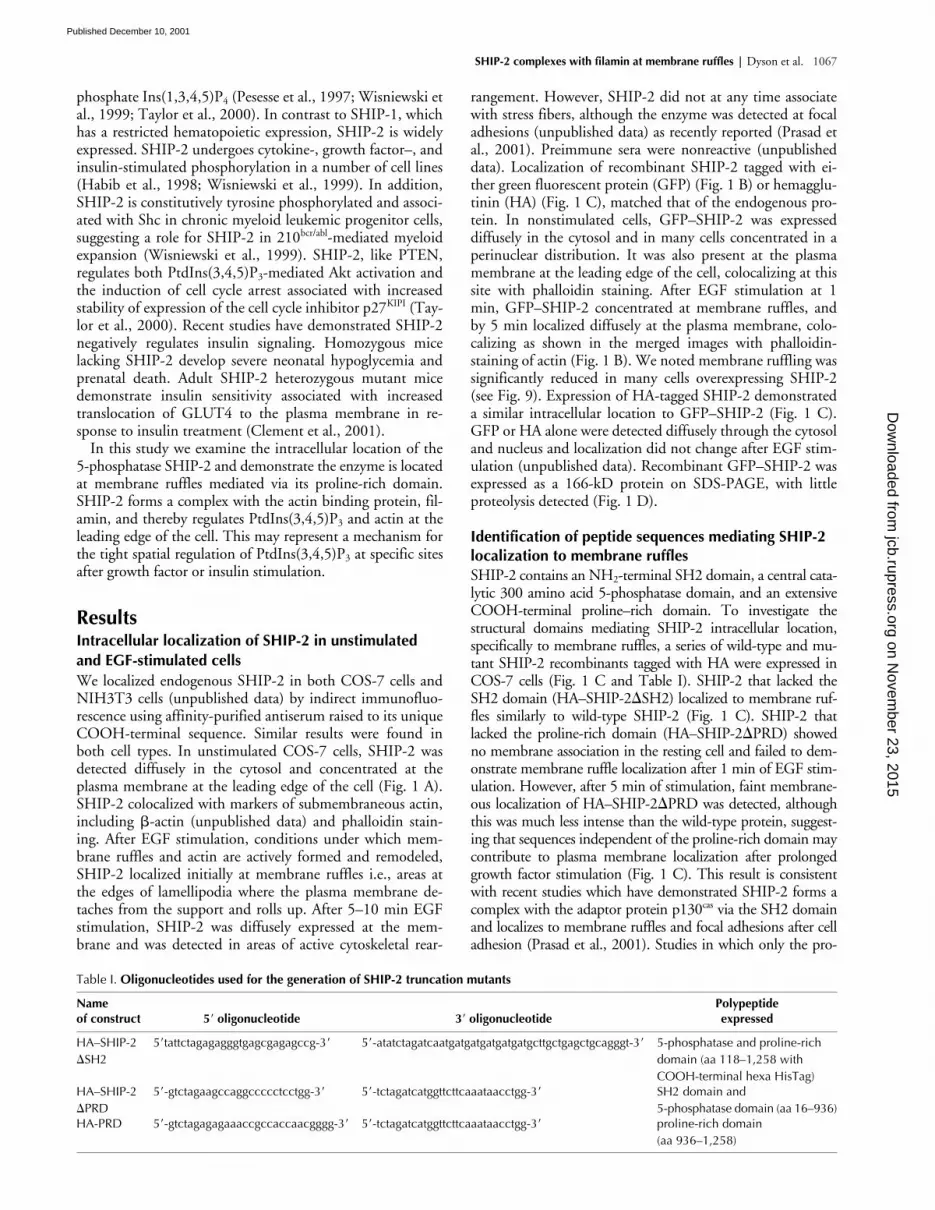
via the SH2 domainand localizes to membrane ruffles and focal adhesions after celladhesion (Prasad et al., 2001). Studies in which only the pro-
Table I.
Oligonucleotides used for the generation of SHIP-2 truncation mutants
Nameof construct 5
�
oligonucleotide 3
�
oligonucleotidePolypeptideexpressed
HA–SHIP-2
�
SH25
�
tattctagagagggtgagcgagagccg-3
�
5
�
-atatctagatcaatgatgatgatgatgatgcttgctgagctgcagggt-3
�
5-phosphatase and proline-richdomain (aa 118–1,258 withCOOH-terminal hexa HisTag)
HA–SHIP-2
�
PRD5
�
-gtctagaagccaggccccctcctgg-3
�
5
�
-tctagatcatggttcttcaaataacctgg-3
�
SH2 domain and5-phosphatase domain (aa 16–936)
HA-PRD 5
�
-gtctagagagaaaccgccaccaacgggg-3
�
5
�
-tctagatcatggttcttcaaataacctgg-3
�
proline-rich domain(aa 936–1,258)
on Novem
ber 23, 2015jcb.rupress.org
Dow
nloaded from
Published December 10, 2001
1068 The Journal of Cell Biology
|
Volume 155, Number 6, 2001
line-rich domain was expressed (HA-PRD) demonstrated thisdomain localized to the leading edge of the cell in resting cells,as shown for the wild-type enzyme, and concentrated at mem-brane ruffles and submembraneous actin–rich structures imme-diately after EGF stimulation. After 5-min stimulation, how-ever, expression at the membrane was less intense than thewild-type enzyme (Fig. 1 C). Wild-type and mutant HA–SHIP-2proteins were expressed intact with little proteolysis and mi-grated at their predicted molecular mass (Fig. 1 D). Collec-tively, these studies demonstrate that the proline-rich domainmediates SHIP-2 membrane ruffle localization in the restingand in EGF-stimulated cells. In addition, sequences such as the
SH2 domain may contribute to membrane localization afterprolonged growth factor stimulation.
Identification of SHIP-2 binding partners using yeast two-hybrid analysis
The SHIP-2 COOH-terminal proline–rich domain containsnumerous “PXXP” motifs which conform to consensus se-quences for SH3 binding domains, 1 WW binding domainmotif (PPLP) which may bind to WW domain-containingproteins, and one EVH1 binding domain motif (E/DFPPP-PXD/E) which may link the cytoskeletal network to signaltransduction pathways (Fedorov et al., 1999). The SHIP-2
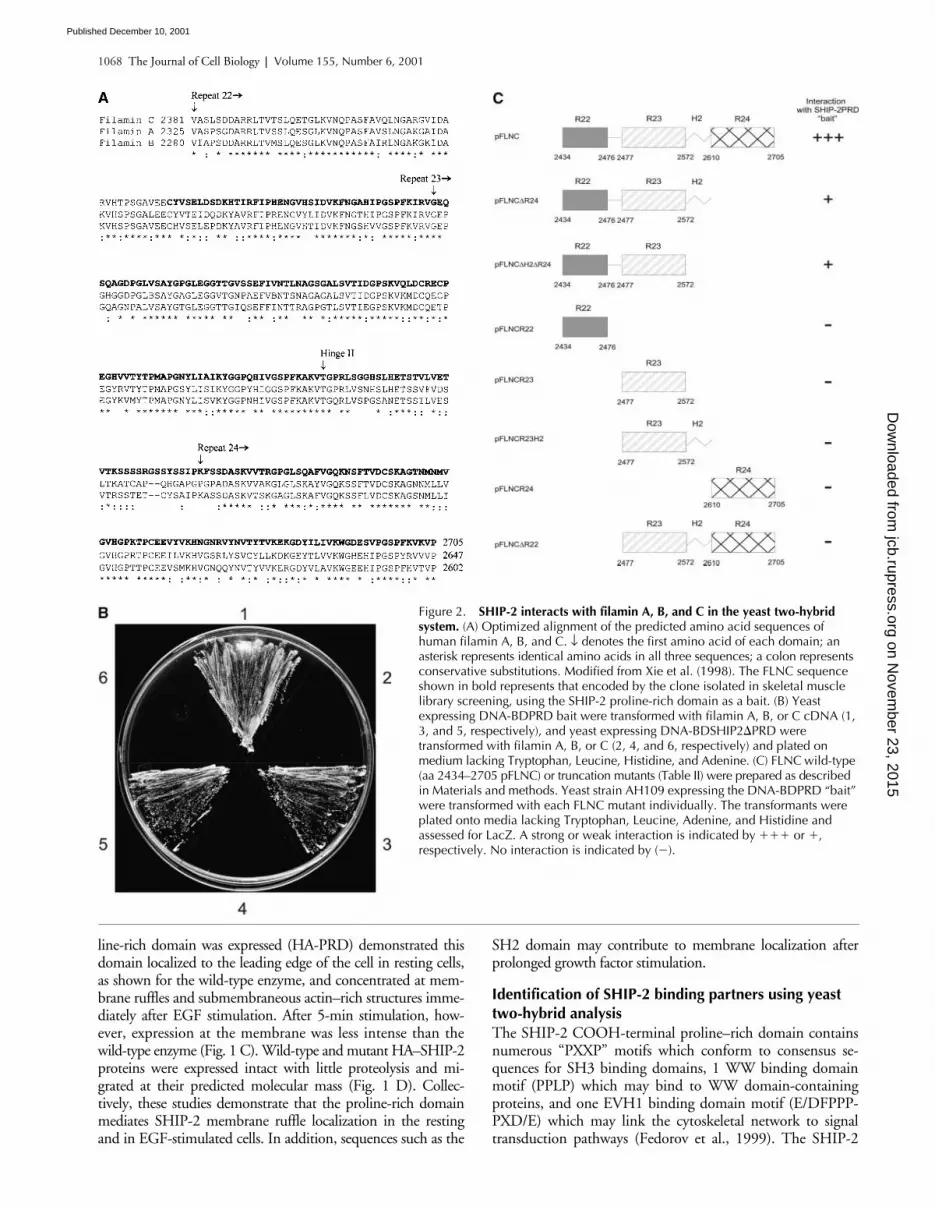
Figure 2. SHIP-2 interacts with filamin A, B, and C in the yeast two-hybrid system. (A) Optimized alignment of the predicted amino acid sequences of human filamin A, B, and C. ↓ denotes the first amino acid of each domain; anasterisk represents identical amino acids in all three sequences; a colon represents conservative substitutions. Modified from Xie et al. (1998). The FLNC sequence shown in bold represents that encoded by the clone isolated in skeletal musclelibrary screening, using the SHIP-2 proline-rich domain as a bait. (B) Yeastexpressing DNA-BDPRD bait were transformed with filamin A, B, or C cDNA (1, 3, and 5, respectively), and yeast expressing DNA-BDSHIP2�PRD were transformed with filamin A, B, or C (2, 4, and 6, respectively) and plated onmedium lacking Tryptophan, Leucine, Histidine, and Adenine. (C) FLNC wild-type (aa 2434–2705 pFLNC) or truncation mutants (Table II) were prepared as described in Materials and methods. Yeast strain AH109 expressing the DNA-BDPRD “bait” were transformed with each FLNC mutant individually. The transformants were plated onto media lacking Tryptophan, Leucine, Adenine, and Histidine andassessed for LacZ. A strong or weak interaction is indicated by ��� or �,respectively. No interaction is indicated by (�).
on Novem
ber 23, 2015jcb.rupress.org
Dow
nloaded from
Published December 10, 2001

SHIP-2 complexes with filamin at membrane ruffles |
Dyson et al. 1069
proline–rich domain sequence demonstrates no significant se-quence homology over the extreme COOH-terminal 322amino acids with SHIP-1. We searched for proteins that spe-cifically interact with the proline-rich domain using yeast two-hybrid analysis. The entire SHIP-2 proline-rich domain(amino acids [aa] 936–1258) was expressed in yeast cells witha library of proteins expressed as fusions with the GAL4 tran-scription activation domain. Several rounds of screening a hu-man skeletal muscle library (4
�
10
6
clones) identified a num-ber of interacting clones in which growth on selective mediasuggested the presence of bona fide interactors for the proline-rich domain. Sequence analysis demonstrated that one clone,an 818 bp fragment, encoded aa 2434–2705 of the lastCOOH-terminal two and a half immunoglobulin repeats ofthe cytoskeletal actin-binding protein filamin C (FLNC),which were in frame with the GAL4 activation domain (Fig. 2A). Filamin is located in the cortical cytoplasm subjacent tothe plasma membrane, and binds actin, promoting orthogo-nal branching of actin filaments and thereby cell migrationand membrane stability (reviewed by Stossel et al., 2001). Fil-amin forms a complex with a variety of cell surface receptorsincluding Fc
RI, the platelet von Willebrand factor receptor,glycoprotein Ib-IX-V,
�
1
and
�
2
integrin receptors, and intra-cellular proteins involved in various signaling cascades includ-ing Traf 2, granzyme B, caveolin-1, and the stress-activatedprotein kinase (reviewed by Stossel et al., 2001).
Three human gene filamin paralogues have been identified.Filamin A encodes
-filamin (also called ABP-280) (Gorlin etal., 1990), filamin B codes for
�
-filamin (also called ABP-278)(Takafuta et al., 1998; Xu et al., 1998), and FLNC encodes for
-filamin (also called ABPL or FLN2), which is highly ex-pressed in skeletal muscle (Xie et al., 1998). In addition, differ-ential splicing has been demonstrated for each gene (Maestriniet al., 1993; Xie et al., 1998; Xu et al., 1998). All three filaminisoforms demonstrate a similar structure comprising an NH
2
-terminal actin binding domain followed by 24 immunoglobu-lin-like repeat domains creating an extended rod like structure.The extreme COOH-terminal repeat 24 contains a ho-modimerization domain which is linked to other repeats by acalpain-sensitive “hinge II region” (reviewed by Stossel et al.,2001). As each of the three filamin isoforms shows significantsequence identity in the COOH-terminal region (Fig. 2 A), weinvestigated if each isoform interacted in cotransformation as-says with the proline-rich domain expressed in frame with theDNA binding domain of GAL4 (DNA-BDPRD), or with re-combinant mutant SHIP-2 which lacked the proline-rich do-main (DNA-BDSHIP-2
�
PRD), versus control heterologousbaits. The proline-rich domain “bait” (DNA-BDPRD), but notthe SHIP-2 “bait” lacking the proline-rich domain (DNA-BD-SHIP-2
�
PRD), interacted with the COOH-terminal four im-munoglobulin repeats of filamin A and B, and the COOH-ter-minal two and a half immunoglobulin repeats of FLNC,indicated by expression of all three reporter genes
HIS3
and
ADE2
(Fig. 2 B) and
LacZ
(unpublished data).
Identification of FLNC sequences mediating interaction with SHIP-2
To determine the region of FLNC specifically interactingwith SHIP-2, a series of wild-type and mutant FLNC con-structs comprising the COOH-terminal immunoglobulin re-
peat regions R22–R24 (aa 2434–2705), which include a“hinge II region” between R23 and R24, were cloned into theactivation domain and cotransformed with the DNA-BD-PRD bait and interactions scored as strong (
���
), or weak(
�
) (Fig. 2 C). The fragment containing FLNC repeats 22–24 demonstrated the strongest binding to the proline-rich do-main bait. FLNC repeats 23 and 24, either alone or in combi-nation, did not interact with SHIP-2. Repeats 22 and 23 incombination, with or without the hinge II region, interactedwith SHIP-2; however, this was weaker than repeats 22–24(Fig. 2 C). All FLNC truncation mutants were expressed inthe yeast strain AH109 and were soluble (unpublished data).
We took several approaches to verify whether SHIP-2 in-teracted with filamin in vivo and thereby regulated the
Figure 3. SHIP-2 associates with filamin in resting andEGF-stimulated COS-7 cells. (A) COS-7 cells were transiently cotransfected with FLAG–SHIP-2 and HA empty vector, orFLAG–SHIP-2 and HA-filamin (encoding aa 2,434 to 2,705). Cells were harvested and the Triton X-100–soluble lysate was immunoprecipitated with HA antibodies and immunoblotted with FLAG antibodies. (B) COS-7 cells were transiently cotransfected with FLAG empty vector and Myc-filamin (encoding aa 2,434–2,705), or FLAG–SHIP-2 and Myc-filamin. Where indicated, COS-7 cells were EGF treated (100 ng/ml) for 5 min. Cells were harvested and the Triton X-100–soluble lysate was immunoprecipitated with FLAG antibodies and immunoblotted with Myc antibodies.
on Novem
ber 23, 2015jcb.rupress.org
Dow
nloaded from
Published December 10, 2001
1070 The Journal of Cell Biology
|
Volume 155, Number 6, 2001
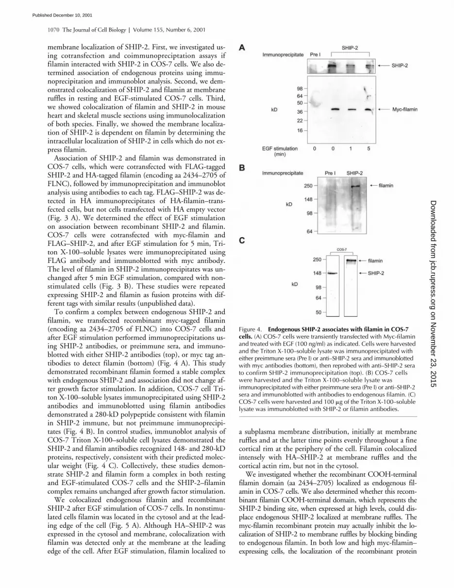
membrane localization of SHIP-2. First, we investigated us-ing cotransfection and coimmunopreciptation assays iffilamin interacted with SHIP-2 in COS-7 cells. We also de-termined association of endogenous proteins using immu-noprecipitation and immunoblot analysis. Second, we dem-onstrated colocalization of SHIP-2 and filamin at membraneruffles in resting and EGF-stimulated COS-7 cells. Third,we showed colocalization of filamin and SHIP-2 in mouseheart and skeletal muscle sections using immunolocalizationof both species. Finally, we showed the membrane localiza-tion of SHIP-2 is dependent on filamin by determining theintracellular localization of SHIP-2 in cells which do not ex-press filamin.
Association of SHIP-2 and filamin was demonstrated inCOS-7 cells, which were cotransfected with FLAG-taggedSHIP-2 and HA-tagged filamin (encoding aa 2434–2705 ofFLNC), followed by immunoprecipitation and immunoblotanalysis using antibodies to each tag. FLAG–SHIP-2 was de-tected in HA immunoprecipitates of HA-filamin–trans-fected cells, but not cells transfected with HA empty vector(Fig. 3 A). We determined the effect of EGF stimulationon association between recombinant SHIP-2 and filamin.COS-7 cells were cotransfected with myc-filamin andFLAG–SHIP-2, and after EGF stimulation for 5 min, Tri-ton X-100–soluble lysates were immunoprecipitated usingFLAG antibody and immunoblotted with myc antibody.The level of filamin in SHIP-2 immunoprecipitates was un-changed after 5 min EGF stimulation, compared with non-stimulated cells (Fig. 3 B). These studies were repeatedexpressing SHIP-2 and filamin as fusion proteins with dif-ferent tags with similar results (unpublished data).
To confirm a complex between endogenous SHIP-2 andfilamin, we transfected recombinant myc-tagged filamin(encoding aa 2434–2705 of FLNC) into COS-7 cells andafter EGF stimulation performed immunoprecipitations us-ing SHIP-2 antibodies, or preimmune sera, and immuno-blotted with either SHIP-2 antibodies (top), or myc tag an-tibodies to detect filamin (bottom) (Fig. 4 A). This studydemonstrated recombinant filamin formed a stable complexwith endogenous SHIP-2 and association did not change af-ter growth factor stimulation. In addition, COS-7 cell Tri-ton X-100–soluble lysates immunoprecipitated using SHIP-2antibodies and immunoblotted using filamin antibodiesdemonstrated a 280-kD polypeptide consistent with filaminin SHIP-2 immune, but not preimmune immunoprecipi-tates (Fig. 4 B). In control studies, immunoblot analysis ofCOS-7 Triton X-100–soluble cell lysates demonstrated theSHIP-2 and filamin antibodies recognized 148- and 280-kDproteins, respectively, consistent with their predicted molec-ular weight (Fig. 4 C). Collectively, these studies demon-strate SHIP-2 and filamin form a complex in both restingand EGF-stimulated COS-7 cells and the SHIP-2–filamincomplex remains unchanged after growth factor stimulation.
We colocalized endogenous filamin and recombinantSHIP-2 after EGF stimulation of COS-7 cells. In nonstimu-lated cells filamin was located in the cytosol and at the lead-ing edge of the cell (Fig. 5 A). Although HA–SHIP-2 wasexpressed in the cytosol and membrane, colocalization withfilamin was detected only at the membrane at the leadingedge of the cell. After EGF stimulation, filamin localized to
a subplasma membrane distribution, initially at membraneruffles and at the latter time points evenly throughout a finecortical rim at the periphery of the cell. Filamin colocalizedintensely with HA–SHIP-2 at membrane ruffles and thecortical actin rim, but not in the cytosol.
We investigated whether the recombinant COOH-terminalfilamin domain (aa 2434–2705) localized as endogenous fil-amin in COS-7 cells. We also determined whether this recom-binant filamin COOH-terminal domain, which represents theSHIP-2 binding site, when expressed at high levels, could dis-place endogenous SHIP-2 localized at membrane ruffles. Themyc-filamin recombinant protein may actually inhibit the lo-calization of SHIP-2 to membrane ruffles by blocking bindingto endogenous filamin. In both low and high myc-filamin–expressing cells, the localization of the recombinant protein
Figure 4. Endogenous SHIP-2 associates with filamin in COS-7 cells. (A) COS-7 cells were transiently transfected with Myc-filamin and treated with EGF (100 ng/ml) as indicated. Cells were harvested and the Triton X-100–soluble lysate was immunoprecipitated with either preimmune sera (Pre I) or anti–SHIP-2 sera and immunoblotted with myc antibodies (bottom), then reprobed with anti–SHIP-2 sera to confirm SHIP-2 immunoprecipitation (top). (B) COS-7 cells were harvested and the Triton X-100–soluble lysate wasimmunoprecipitated with either preimmune sera (Pre I) or anti–SHIP-2 sera and immunoblotted with antibodies to endogenous filamin. (C) COS-7 cells were harvested and 100 �g of the Triton X-100–soluble lysate was immunoblotted with SHIP-2 or filamin antibodies.
on Novem
ber 23, 2015jcb.rupress.org
Dow
nloaded from
Published December 10, 2001

SHIP-2 complexes with filamin at membrane ruffles |
Dyson et al. 1071
matched that of endogenous filamin at membrane ruffles in un-stimulated (unpublished data) and more prominently in EGF-stimulated cells (Fig. 5 B). However, in cells expressing myc-fil-amin at very high levels we noted increased expression offilamin in the cytosol in addition to membrane staining. Local-ization of endogenous SHIP-2 in myc-filamin–expressing cellswas determined by indirect immunofluorescence. In cells ex-pressing low levels of myc-filamin, endogenous SHIP-2 local-ized to membrane ruffles in both resting (unpublished data)and more prominently in EGF-stimulated cells (Fig. 5 B). Incontrast, in cells expressing high levels of myc-filamin, SHIP-2localized in a perinuclear distribution in the cytosol and stainingwas less intense at the plasma membrane (Fig. 5 B).
Colocalization of filamin and SHIP-2 in heart and skeletal muscle
FLNC is highly expressed in striated muscle, where it ispredominantly localized in myofibrillar Z-discs, with aminor fraction of the protein showing subsarcolemma lo-calization (van der Ven et al., 2000). SHIP-2 is also ex-pressed in skeletal muscle, although its intracellular lo-cation in this tissue has not been reported. SHIP-2homozygous null mice demonstrate increased sensitivityto insulin (Clement et al., 2001). One of the major sitesof insulin action is skeletal muscle, where insulin stimu-lates the translocation of the glucose transporter GLUT4in a PI-3 kinase–dependent manner to the sarcolemma(Khan et al., 2000). We investigated whether SHIP-2 and
filamin colocalized in striated muscle. Mouse heart stri-ated muscle sections were isolated, fixed, and probed withspecific affinity purified SHIP-2 antibodies (Fig. 6 A).Soleus muscle showed similar localization (unpublisheddata). In longitudinal sections SHIP-2 antibodies stainedintensively in an alternate banding pattern at areas thatresembled Z-lines. No staining of any structure was ob-served using preimmune serum (Fig. 6 B). Counter-stain-ing sections using antibodies to filamin and the Z-line–specific protein
-actinin demonstrated both colocalizedwith SHIP-2 (Fig. 6, C–H). Cross-sectional analysis ofskeletal muscle demonstrated both filamin and SHIP-2localized to the sarcolemma, the site of insulin-stimulatedGLUT4 translocation (Fig. 6, I–L).
Intracellular localization of SHIP-2 infilamin-deficient cells
To investigate if SHIP-2 membrane ruffle localization wasdependent on an interaction with filamin, we examined theintracellular localization of SHIP-2 in a cell line (M2) de-rived from a human malignant melanoma that does not ex-press detectable filamin messenger RNA or protein (Cun-ningham et al., 1992). M2 cells demonstrate a distinctphenotype characterized by extensive membrane blebbingand defective locomotion, which is reversed by the stabletransfection of filamin A cDNA (A7 subline). EquivalentSHIP-2 expression was demonstrated in A7 and M2 cells asshown by immunoblot analysis using SHIP-2 antipeptide
Figure 5. SHIP-2 colocalizes with filamin in resting and EGF-stimulated COS-7 cells. (A) COS-7 cells were transiently transfected with HA–SHIP-2. Cells were EGF (100 ng/ml) stimulated where indicated and fixed and costained with HA and filamin B antibodies. Cells were visualized by
confocal microscopy. (B) COS-7 cells were transiently transfected with myc-filamin (encoding aa 2,434–2,705) and EGF stimulated for 5 min. Cells were scored as expressing high or low levels of this recombinant protein, as determined by intensity of staining using antibodies to the myc tag by indirect immunofluorescence. The localization of endogenous SHIP-2 was determined using specific SHIP-2 antibodies in cells expressing either low or high levels of myc-filamin. Arrows indicate the localization of SHIP-2 to membrane ruffles. Bars, 20 �M.
on Novem
ber 23, 2015jcb.rupress.org
Dow
nloaded from
Published December 10, 2001

1072 The Journal of Cell Biology
|
Volume 155, Number 6, 2001
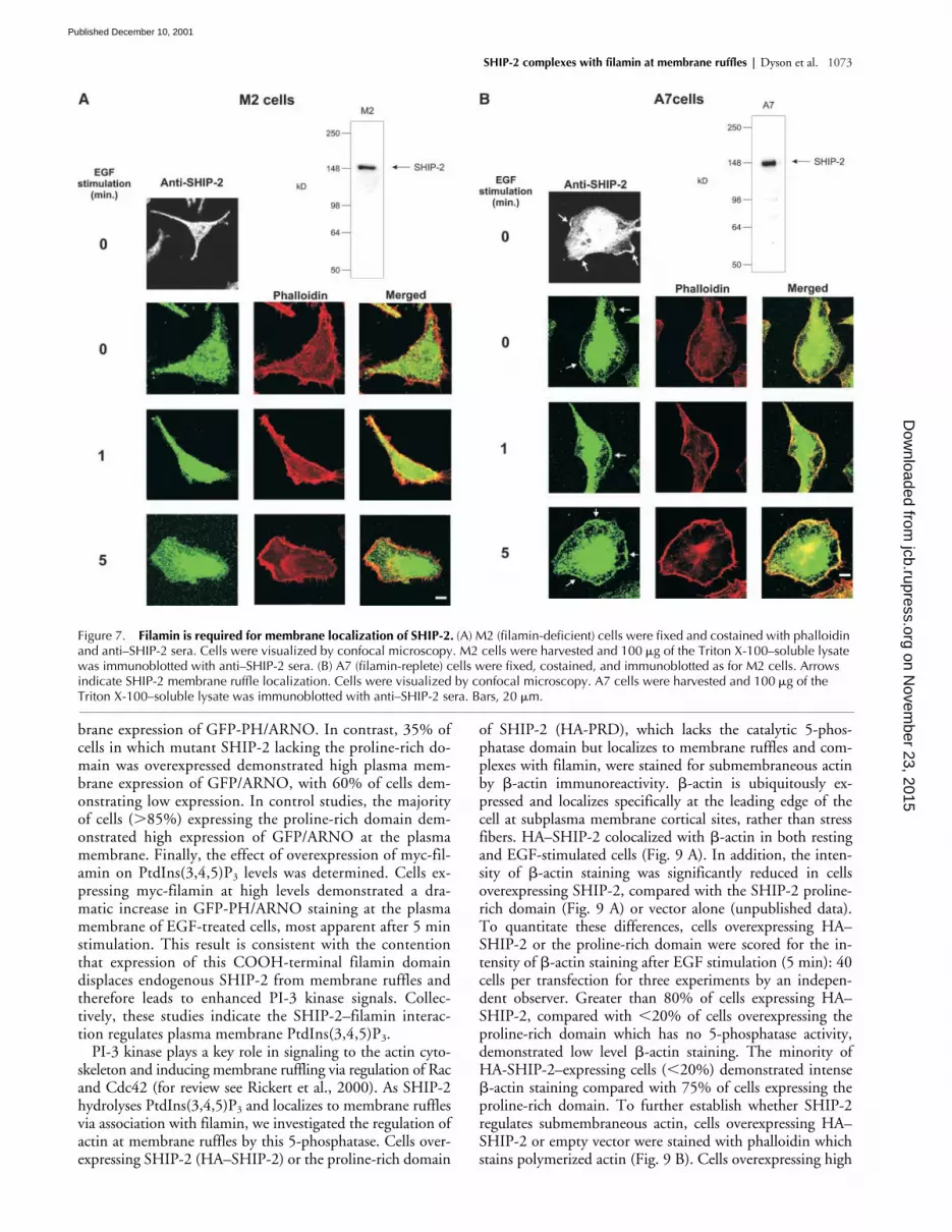
antibodies of 100
�
g of cell lysate (Fig. 7, A and B). The in-tracellular localization of SHIP-2 was determined by in-direct immunofluorescence of endogenous enzyme usingSHIP-2 antipeptide antibodies, or cells transfected with HA-tagged SHIP-2 with detection by staining with antibodies tothe tag (unpublished data) with the same result. In nonstim-ulated M2 cells, SHIP-2 was expressed diffusely in the cyto-sol and did not colocalize with markers of submembraneousactin such as phalloidin (Fig. 7 A). Upon EGF stimulation,M2 cells transiently form membrane ruffles, or membrane“blebs,” which can be detected by phalloidin staining (for re-view see Stossel et al., 2001). SHIP-2 remained in the cyto-sol in stimulated cells and did not colocalize with filamin atmembrane ruffles. In contrast, in A7 cells, which have beenstably transfected with filamin, SHIP-2 localized to mem-
brane ruffles upon EGF stimulation (Fig. 7 B) and colocal-ized with phalloidin staining. Collectively, these studiesdemonstrate SHIP-2 localization at membrane ruffles is de-pendent on filamin expression.
SHIP-2 regulates PtdIns(3,4,5)P
3
and
�
-actin at membrane ruffles
To establish that recombinant SHIP-2 localized at mem-brane ruffles was active and to determine the functionalconsequences on PtdIns(3,4,5)P
3
levels, we employed thepleckstrin homology (PH) domain of ARNO, which hashigh affinity for PtdIns(3,4,5)P
3
to assess local plasmamembrane concentrations of this phosphoinositide (Ballaet al., 2000). Several studies have demonstrated that GFP-tagged PH domains with specificity for PtdIns(3,4,5)P
3
can be used to accurately detect PtdIns(3,4,5)P
3
at theleading edge of the cell (for review see Rickert et al., 2000).Although it has been established that SHIP-2 regulates to-tal cellular PtdIns(3,4,5)P
3
levels, the membrane ruffle lo-calized regulation of PtdIns(3,4,5)P
3 has not been reported(Blero et al., 2001; Pesesse et al., 2001). In addition, thefunctional role of SHIP-2 membrane location in regulatingPtdIns(3,4,5)P3 degradation has not been determined.GFP-fused with the PH domain of ARNO (GFP-PH/ARNO) was coexpressed with either empty vector (HA), orHA–SHIP-2, or the proline-rich domain (HA-PRD), ormutant SHIP-2 which lacked the proline-rich domain(HA–SHIP-2�PRD) (Fig. 8). In addition, we determinedthe effect of overexpressing the COOH-terminal filaminfragment (aa 2434–2705) which binds SHIP-2 in COS-7cells on PtdIns(3,4,5)P3 levels, as we have shown overex-pression of recombinant myc-filamin displaces endogenousSHIP-2 from membrane ruffles (Fig. 5 B). In nonstimu-lated cells transfected with HA empty vector and GFP-PH/ARNO, plasma membrane staining of GFP-PH/ARNOwas not detected. Upon EGF activation GFP-PH/ARNOtranslocated rapidly to membrane ruffles after 1 min ofstimulation and by 5 min intense plasma membrane stain-ing was detected (Fig. 8). In contrast, cells overexpressingSHIP-2 demonstrated no or low expression of GFP-PH/ARNO at the plasma membrane of EGF-stimulated cells.However, cells expressing the SHIP-2 proline-rich do-main (HA-PRD), which lacks the SH2 and catalytic do-main, demonstrated strong plasma membrane expressionof GFP-PH/ARNO, comparable to HA empty vector ex-pressing cells after EGF stimulation. In cells expressingSHIP-2, which lacks the proline-rich domain (HA-SHIP-2�PRD) but contains the SH2 and 5-phosphatase catalyticdomain, GFP-PH/ARNO demonstrated growth factor–dependent relocalization to the plasma membrane. However,staining was less intense at 5 min compared with emptyvector–expressing cells, but was greater than intact SHIP-2expressing cells. To confirm these observations, transfectedcells were scored as showing high or low GFP-PH/ARNOplasma membrane expression after 5 min of EGF stimula-tion. Over 40 cells were scored per transfection for threeindependent experiments by an independent observer. Theexpression of GFP-PH/ARNO at the plasma membranewas low or not detected in �90% of cells overexpressingSHIP-2, with �10% of cells showing high plasma mem-
Figure 6. SHIP-2 colocalizes with filamin at Z-lines in mouse heart and skeletal muscle sections. Mouse adult heart longitudinal cryosections were cut 7 �m thick and stained with SHIP-2 antibodies (a, c, and f) or preimmune sera (b). SHIP-2 was colocalized with-actinin (d) at the Z-line and also with antifilamin (g) at the Z-line. Merged images are indicated between SHIP-2 and filamin in panels e and h, respectively. Cross-sectional sections of skeletal muscle were stained with SHIP-2 antibodies (i and j) and counterstained with filamin antibodies (k). Merged images are shown in panel l. Sections were visualized by confocal microscopy. Bars, 20 �m.
on Novem
ber 23, 2015jcb.rupress.org
Dow
nloaded from
Published December 10, 2001
SHIP-2 complexes with filamin at membrane ruffles | Dyson et al. 1073
brane expression of GFP-PH/ARNO. In contrast, 35% ofcells in which mutant SHIP-2 lacking the proline-rich do-main was overexpressed demonstrated high plasma mem-brane expression of GFP/ARNO, with 60% of cells dem-onstrating low expression. In control studies, the majorityof cells (�85%) expressing the proline-rich domain dem-onstrated high expression of GFP/ARNO at the plasmamembrane. Finally, the effect of overexpression of myc-fil-amin on PtdIns(3,4,5)P3 levels was determined. Cells ex-pressing myc-filamin at high levels demonstrated a dra-matic increase in GFP-PH/ARNO staining at the plasmamembrane of EGF-treated cells, most apparent after 5 minstimulation. This result is consistent with the contentionthat expression of this COOH-terminal filamin domaindisplaces endogenous SHIP-2 from membrane ruffles andtherefore leads to enhanced PI-3 kinase signals. Collec-tively, these studies indicate the SHIP-2–filamin interac-tion regulates plasma membrane PtdIns(3,4,5)P3.
PI-3 kinase plays a key role in signaling to the actin cyto-skeleton and inducing membrane ruffling via regulation of Racand Cdc42 (for review see Rickert et al., 2000). As SHIP-2hydrolyses PtdIns(3,4,5)P3 and localizes to membrane rufflesvia association with filamin, we investigated the regulation ofactin at membrane ruffles by this 5-phosphatase. Cells over-expressing SHIP-2 (HA–SHIP-2) or the proline-rich domain
of SHIP-2 (HA-PRD), which lacks the catalytic 5-phos-phatase domain but localizes to membrane ruffles and com-plexes with filamin, were stained for submembraneous actinby �-actin immunoreactivity. �-actin is ubiquitously ex-pressed and localizes specifically at the leading edge of thecell at subplasma membrane cortical sites, rather than stressfibers. HA–SHIP-2 colocalized with �-actin in both restingand EGF-stimulated cells (Fig. 9 A). In addition, the inten-sity of �-actin staining was significantly reduced in cellsoverexpressing SHIP-2, compared with the SHIP-2 proline-rich domain (Fig. 9 A) or vector alone (unpublished data).To quantitate these differences, cells overexpressing HA–SHIP-2 or the proline-rich domain were scored for the in-tensity of �-actin staining after EGF stimulation (5 min): 40cells per transfection for three experiments by an indepen-dent observer. Greater than 80% of cells expressing HA–SHIP-2, compared with �20% of cells overexpressing theproline-rich domain which has no 5-phosphatase activity,demonstrated low level �-actin staining. The minority ofHA-SHIP-2–expressing cells (�20%) demonstrated intense�-actin staining compared with 75% of cells expressing theproline-rich domain. To further establish whether SHIP-2regulates submembraneous actin, cells overexpressing HA–SHIP-2 or empty vector were stained with phalloidin whichstains polymerized actin (Fig. 9 B). Cells overexpressing high
Figure 7. Filamin is required for membrane localization of SHIP-2. (A) M2 (filamin-deficient) cells were fixed and costained with phalloidin and anti–SHIP-2 sera. Cells were visualized by confocal microscopy. M2 cells were harvested and 100 �g of the Triton X-100–soluble lysate was immunoblotted with anti–SHIP-2 sera. (B) A7 (filamin-replete) cells were fixed, costained, and immunoblotted as for M2 cells. Arrowsindicate SHIP-2 membrane ruffle localization. Cells were visualized by confocal microscopy. A7 cells were harvested and 100 �g of theTriton X-100–soluble lysate was immunoblotted with anti–SHIP-2 sera. Bars, 20 �m.
on Novem
ber 23, 2015jcb.rupress.org
Dow
nloaded from
Published December 10, 2001

1074 The Journal of Cell Biology | Volume 155, Number 6, 2001
levels of HA–SHIP-2 demonstrated significantly decreasedstaining of actin at membrane ruffles and decreased mem-brane ruffling. In many HA–SHIP-2 overexpressing cells,only a fine cortical rim of actin was stained by phalloidin af-ter 5 min EGF stimulation. The intensity of phalloidin stain-ing of actin at the membrane was scored as high or low after5 min EGF stimulation in HA–SHIP-2 versus HA emptyvector expressing cells for 40 cells per transfection for threeindependent experiments by an independent observer.Greater than 70% of HA-SHIP-2–expressing cells, com-pared with 30% of cells expressing the HA empty vector,demonstrated low intensity phalloidin staining at membraneruffles. In addition, only 25% of SHIP-2 versus 75% of HAempty vector–expressing cells demonstrated high intensityphalloidin staining at membrane ruffles. Collectively, thesestudies demonstrate SHIP-2 localizes to membrane rufflesvia association with filamin and regulates PtdIns(3,4,5)P3,�-actin, and membrane ruffling.
DiscussionThe results of this study demonstrate the 5-phosphataseSHIP-2 forms a functionally significant complex with the ac-tin-binding protein, filamin. SHIP-2 interacts via its proline-rich domain, specifically with filamin A, B, and C isoforms inyeast two hybrid assays. Filamin and SHIP-2 colocalized toZ-lines and the sarcolemma of skeletal muscle, and in mamma-lian cell lines to membrane ruffles. We demonstrated by recip-rocal coimmunoprecipitation studies that SHIP-2 and filaminform a stable complex in COS-7 cells and the level of associa-tion between these species does not change after EGF stimula-tion. SHIP-2 membrane localization is dependent on filaminexpression, as membrane association was not detected in awell-characterized melanoma cell line, which does not expressfilamin. The association of SHIP-2 with filamin serves to reg-ulate PtdIns(3,4,5)P3 and �-actin at membrane ruffles.
Inositol polyphosphate 5-phosphatases andthe cytoskeletonIncreasing evidence indicates that both mammalian andyeast 5-phosphatase isoforms, via hydrolysis of PtdIns(4,5)P2
and or PtdIns(3,4,5)P3, play a significant role in regulatingcytoskeletal reorganization. The 5-phosphatases comprise 10mammalian and 4 yeast isoforms with many spliced variantsdescribed. Null mutation of any two yeast Sac-1 domaincontaining 5-phosphatases results in a phenotype which in-cludes disorganization of polymerized actin and delocaliza-tion of actin patches from the growing yeast bud to themother cell (for review see Hughes et al., 2000). The yeast5-phosphatases, Inp52p and Inp53p, translocate to actinpatches upon osmotic stress, the site of plasma membrane in-vaginations. In addition, overexpression of Inp52p andInp53p, but not catalytically inactive Inp52p, results in asignificant reduction in the repolarization time of actinpatches after osmotic stress (Ooms et al., 2000). Themammalian 5-phosphatase, synaptojanin-1, hydrolyses Ptd-Ins(4,5)P2 bound to the actin regulatory proteins, -actinin,vinculin, gelsolin, and profilin, and decreases the number ofstress fibers in the cell (Sakisaka et al., 1997). Synaptojanin-2directly interacts with Rac1 in a GTP-dependent manner, re-sulting in translocation of the 5-phosphatase to membraneruffles and inhibition of endocytosis (Malecz et al., 2000).Overexpression of SKIP (skeletal muscle and kidney-enriched inositol phosphatase) results in loss of actin stressfibers in areas of SKIP expression (Ijuin et al., 2000). Therecently identified proline-rich inositol polyphosphate5-phosphatase (PIPP) localizes to membrane ruffles, but un-like SHIP-2 does not appear to regulate the actin cytoskele-ton (Mochizuki and Takenawa, 1999). SHIP-2 regulation ofsubmembraneous actin levels is most probably mediatedvia localized regulation of PtdIns(3,4,5)P3. However, bothSHIP-1 and SHIP-2 also hydrolyze PtdIns(4,5)P2, formingPtdIns(4)P (Kisseleva et al., 2000; Taylor et al., 2000).
Figure 8. SHIP-2 regulatesPtdIns(3,4,5)P3 at membrane ruffles. COS-7 cells were transiently cotransfected with the GFP-PH/ARNO and with either empty vector (HA), HA–SHIP-2, theproline-rich domain (HA-PRD), mutant SHIP-2 which lacked the proline-rich domain (HA–SHIP-2�PRD), orMyc-filamin. Cells were EGF treated (100 ng/ml) for the indicated times and stained with HA or Myc antibodies to identify cotransfected cells (unpublished data). Cells were visualized by confocal microscopy for GFP-PH/ARNO expression which is shown. Arrows indicate areas of high GFP-PH/ARNO expression. Bar equals 20 �m.
on Novem
ber 23, 2015jcb.rupress.org
Dow
nloaded from
Published December 10, 2001

SHIP-2 complexes with filamin at membrane ruffles | Dyson et al. 1075
Role of SHIP-2 association with FLNC in insulin signalingThe recent characterization of SHIP-2 homozygous null micehas demonstrated that this 5-phosphatase plays a significantrole in regulating insulin sensitivity (Clement et al., 2001). Al-though the signaling pathways mediating the phenotype of in-sulin hypersensitivity have yet to be fully determined, insulin-stimulated GLUT4 translocation to the plasma membraneappears to be enhanced in mice lacking SHIP-2. In addition,
overexpression of SHIP-2, but not catalytically inactive SHIP-2,in 3T3-L1 adipocytes results in negative regulation of insu-lin-induced signaling (Wada et al., 2001). The submembra-neous actin microfilament network links various signalingproteins, including IRS-1 and PI-3 kinase, that regulate GLUT4translocation to the plasma membrane. Insulin-induced reor-ganization of the subplasma membrane actin filaments mayallow exocytic GLUT4 vesicles to fuse with the plasma mem-
Figure 9. SHIP-2 regulates �-actin expression at membraneruffles. (A) COS-7 cells were transiently transfected with eitherHA–SHIP-2 or HA-PRD. Where indicated, cells were treated with EGF (100 ng/ml) for 1, 5, or 10 min. Cells were fixed and costained with HA and �-actin antibodies and visualized by confocalmicroscopy. Arrows indicate areas of high �-actin expression. (B) COS-7 cells were transiently transfected with either HA–SHIP-2 or HA empty vector. Where indicated, cells were treated with EGF (100 ng/ml) for 5 min. Cells were fixed and stained with HA antibodies and actin stained with phalloidin. Cells were visualized by confocal microscopy. Arrows indicate areas of high phalloidin staining. Bars, 20 �m.
on Novem
ber 23, 2015jcb.rupress.org
Dow
nloaded from
Published December 10, 2001

1076 The Journal of Cell Biology | Volume 155, Number 6, 2001
brane during stimulation by insulin (Khayat et al., 2000).GLUT4 expression is restricted to muscle and adipose tissue.Insulin-stimulated glucose disposal in skeletal muscle ac-counts for 80% of glucose uptake postprandial (Khayat et al.,2000), localizing GLUT4 to the sarcolemma. Filamin mayprovide a scaffold for the juxtaposition of SHIP-2 to the sar-colemma, localizing the enzyme to PtdIns(3,4,5)P3 after insu-lin treatment and thereby regulating GLUT4 translocation.
Filamin forms a scaffold for the binding of Rho GTPases,including Rac1, Cdc42, Rho A, and RalA, and their activa-tors such as Trio, a Rho guanine nucleotide exchange factor(GEF) (Ohta et al., 1999). The localization of both RhoGTPases and their activators on filamin may allow spatialcoordination of actin nucleation at sites where newly assem-bled actin filaments are cross-linked. It is noteworthy thatmany filamin-binding proteins, including Trio and SHIP-2,bind to the extreme COOH-terminal repeats 21–24 of fil-amin, by a variety of interacting modules, including proline-rich domains, as is the case for SHIP-2 and PH domains asshown for Trio. This would therefore provide close proxim-ity between all these signaling proteins, including SHIP-2,that regulate actin polymerization on a filamin scaffold.
Several recent studies in fibroblasts and the neutrophilcell line HL60 have demonstrated using the PH domain ofPtdIns(3,4,5)P3-binding proteins fused to GFP, that PI-3kinase signals are generated at the leading edge of the cell(Blomberg et al., 1999; Watton and Downward, 1999;Balla et al., 2000; Servant et al., 2000). These PH domainsfunction as an accurate probe for the localized agonist-dependent accumulation of PtdIns(3,4,5)P3. It has beenproposed, but not previously shown, that this asymmetricdistribution of PtdIns(3,4,5)P3 may result from its localizedsynthesis or degradation (Rickert et al., 2000). The resultsof the study reported here are consistent with this conten-tion. We have shown the spatially controlled synthesis ofPtdIns(3,4,5)P3 at membrane ruffles is regulated by the Ptd-Ins(3,4,5)P3 5-phosphatase SHIP-2, which also localizes tomembrane ruffles.
We have demonstrated SHIP-2 localization to membraneruffles is mediated by its COOH-terminal proline-rich do-main binding to the actin binding protein filamin. The ex-pression of SHIP-2 in filamin deficient cells is exclusively cy-tosolic. In addition, SHIP-2 membrane localization appearsto contribute to localized PtdIns(3,4,5)P3 hydrolysis. SHIP-2COOH-terminal truncation mutants were not as efficientat regulating PtdIns(3,4,5)P3 at membrane ruffles as intactSHIP-2. Furthermore, displacement of endogenous SHIP-2by overexpression of the SHIP-2 filamin binding domain(myc filamin aa 2434–2705) lead to the marked enhance-ment of the PI-3 kinase signal PtdIns(3,4,5)P3. Several re-cent reports have shown the membrane localization of thehighly related SHIP-2 homologue SHIP-1 is also importantfor PtdIns(3,4,5)P3 hydrolysis (Phee et al., 2000). Enforcedplasma membrane localization of SHIP-1, mediated by over-expression of a human CD8–SHIP-1 chimera, decreased thetotal cellular levels of PtdIns(3,4,5)P3. Membrane targetingof SHIP-1 mediated by the COOH-terminal proline-richdomain appears to be important in B cell inhibitory func-tion (Aman et al., 2000). In addition, the SHIP-1 COOHterminus is essential for PtdIns(3,4,5)P3 hydrolysis and inhi-bition of mast cell degranulation (Damen et al., 2001). Col-
lectively, these studies show membrane targeting of thesetwo 5-phosphatases mediated by their respective COOH-terminal proline-rich domains plays an important functionalrole in regulating PI-3 kinase signals.
SHIP-2 associates with the p130Cas adaptor protein at focaladhesions and regulates cell spreading, which is dependent onthe SHIP-2 SH2 domain and is enhanced by tyrosine phos-phorylation and cell adhesion (Prasad et al., 2001). We haveshown SHIP-2 and filamin also form a functionally signifi-cant complex both in the resting cells and after cellular activa-tion at membrane ruffles. We therefore propose the bindingof SHIP-2 to filamin provides a mechanism for exquisite lo-calized hydrolysis of PtdIns(3,4,5)P3 in resting, growth fac-tor– and insulin–stimulated cells at the leading edge of cell.
Materials and methodsRestriction and DNA-modifying enzymes were obtained from New En-gland Biolabs, Inc., Fermentas, or Promega. Big dye terminator cycle se-quencing was from PE Applied Systems. Synthetic peptides were from Chi-ron Mimotopes. COS-7 cells were purchased from the American TypeCulture Collection. M2 and A7 cell lines were a gift from Dr. T. Stossel(Brigham and Women’s Hospital, Boston, MA). Oligonucleotides were ob-tained from Bresatec and the Department of Microbiology, Monash Uni-versity, Melbourne, Australia. Partially purified rabbit polyclonal antibod-ies to human filamin B were a gift from Dr. Dominic Chung (Departmentof Biochemistry, University of Washington School of Medicine, WA). Goatpolyclonal antibodies to chicken gizzard filamin and monoclonal antibod-ies to -actinin (-actinin) and FLAG were obtained from Sigma-Aldrich.Monoclonal antibodies to HA were obtained from Silenus, GFP antibodieswere from Boehringer, �-actin antibodies were from Sigma-Aldrich, andantifilamin antibodies (raised to platelet filamin) were from Chemicon.Phalloidin stain was obtained from Molecular Probes. The GFP-PH/ARNOconstruct was a gift from Dr. Tamas Balla (National Institutes of Health,Bethesda, MD). The yeast two-hybrid system Matchmaker 3 and thepEGFP-C2 vector were obtained from CLONTECH Laboratories, Inc. andthe pCGN vector was a gift from Dr. Tony Tiganis (Monash University,Melbourne, Australia). pEFBOS-Myc and pEFBOS-FLAG vectors were a giftfrom Dr. Tracey Willson (Walter and Eliza Hall Institute of Medical Re-search, Melbourne, Australia). Filamin A and B cDNAs were a gift from Dr.Joe Trapani and Kylie Browne (Peter MacCallum Cancer Institute, Mel-bourne, Australia). All other reagents were from Sigma-Aldrich unless oth-erwise stated.
Production of antipeptide antibodiesSHIP-2 antipeptide antibodies were generated to a fusion peptide compris-ing the NH2-terminal seven amino acids of SHIP-2 fused to the COOH-ter-minal seven amino acids of SHIP-2 (MASACGADTLQLSK) (SHIP-2NC), orto the amino acid sequence, 1019–1030 (ITVPAPQLGHHRH) (SHIP-2P).SHIP-2NC antibodies were used for all experiments except indirect immu-nofluorescence of COS-7, M2, and A7 cells in which the SHIP-2P antibodywas used. Peptide conjugated to diphtheria toxoid was injected subcutane-ously into female New Zealand white rabbits. Affinity-purified antipeptideantibodies were obtained by chromatography of immune sera on the spe-cific peptide coupled to thiopropyl-Sepharose 6B resin. After extensivewashing, specific antibodies were eluted from the column with 0.1 M gly-cine-HCl, pH 2.5.
Generation of full-length SHIP-2 and SHIP-2 truncation mutantsHuman SHIP-2 cDNA was generated by ligation of EST aa 279072 to thecDNA encoding INPPL-1, obtained from Dr. James Hejna (Oregon HealthSciences University, Portland, OR) (Hejna et al., 1995). Several rounds ofPCR amplification enabled prolongation of the 5�-end of the clone to en-compass SHIP-2 nucleotides 257–3988, which correspond to aa 16–1258plus a COOH-terminal hexa-His-tag. SHIP-2 cDNA was cloned in-frameinto pCGN (XbaI site), pEGFP-C2 (EcoRI site) and pEFBOS (MluI site) vec-tors, that encode for HA, GFP, and FLAG-tagged SHIP-2 fusion proteins,respectively, using PCR amplification with the introduction of specific re-striction sites. Truncation mutants of SHIP-2 were also generated and weresubsequently cloned into the XbaI site of pCGN. The oligonucleotide se-quences and a description of the constructs generated are listed in Table I.Fidelity of all PCR products and the final constructs was confirmed bydideoxy sequencing.
on Novem
ber 23, 2015jcb.rupress.org
Dow
nloaded from
Published December 10, 2001

SHIP-2 complexes with filamin at membrane ruffles | Dyson et al. 1077
Yeast two-hybrid analysisThe yeast two-hybrid Matchmaker III GAL4-based system was used for allyeast two-hybrid studies. The proline-rich domain of human SHIP-2 com-prising nucleotides 3017–3989 (aa 936–1258), was cloned into the EcoRIsite of pGBKT7, creating a GAL4 fusion protein, the “bait.” “Bait” protein-expressing yeast (AH109) were transformed with human skeletal musclecDNA library (CLONTECH Laboratories, Inc.) according to the manufac-turer’s guidelines. Yeast plasmid was extracted from positive clones as de-scribed (Ausubel et al., 1991).
SHIP-2 proline-rich domain–expressing yeast were transformed with fil-amin A and B isoforms cDNA (aa 2171–2647 and 2130–2602, respec-tively) to investigate an interaction. Specificity of the interactions with theSHIP-2 proline-rich domain was confirmed using a p53 “bait.” In addition,a “bait” lacking the proline-rich domain of SHIP-2, but containing the SH2domain and 5-phosphatase domain (comprising nucleotides 210–3016)was constructed using a PCR based strategy and was also used as a nega-tive control “bait.”
Immunoblot of endogenous SHIP-2, HA, and GFP-taggedSHIP-2 constructsM2 and A7 cells were maintained as described (Cunningham et al., 1992;Ohta et al., 1999). COS-7 cells were maintained in DME, 10% (vol/vol) fetalcalf serum containing 2 mM glutamine, and transfected using the DEAE-dex-tran procedure and allowed to grow for 2 d (Sambrook and Russel, 2001).Cells were washed briefly with PBS and treated with lysis buffer (50 mM Tris,pH 8.0, 150 mM NaCl, 1% Triton X-100, 2 mM EDTA (ethylenediaminetetra-acetic acid DI-sodium salt), 1 mM benzamidine, 2 mM phenylmethylsulfo-nylfluoride, 2 �g/ml leupeptin, and 2 �g/ml aprotinin) for 2 h at 4 C. Lysateswere centrifuged at 15,400 g for 10 min to obtain the Triton X-100–solublesupernatant which was analyzed by immunoblot analysis using antibodies tothe specific tag, affinity-purified SHIP-NC sera, or antifilamin antibodies.
Intracellular localization of SHIP-2 in COS-7 cellsCOS-7 cells were transiently transfected with GFP–SHIP-2, Myc-filamin,HA–SHIP-2, HA-SHIP-2�PRD, HA–SHIP-2�SH2, or HA-PRD truncationmutants (Table I), fixed/permeabilized, and stained. Alternatively, in somestudies, 24 h after transfection, cells were placed in DME containing 0.1%FCS and 2 mM glutamate for a period of �15 h and then stimulated withEGF (100 ng/ml). Cells expressing GFP-tagged proteins were gentlywashed with PBS and fixed with 3.7% formaldehyde. Cells expressing Mycand HA-tagged proteins were gently washed with PBS and then fixed andpermeabilized for 10 min in PBS with 3.7% formaldehyde and 0.2% TritonX-100. Expression of Myc and HA-tagged proteins was localized usingMyc and HA monoclonal antibodies and detected using tetramethyl-rhodamine isothiocyanate–conjugated TRITC anti–mouse IgG and fluo-rescein isothiocyanate–conjugated FITC anti–mouse IgG, respectively.Endogenous SHIP-2 was detected in nontransfected and Myc-filamin–transfected cells using the SHIP-2P antibody and FITC anti–rabbit IgG.Colocalization was performed using antibodies to filamin B detected withtetramethylrhodamine isothiocyanate–conjugated TRITC anti–rabbit IgGand specific actin markers, either phalloidin staining and/or antibodies to�-actin detected using TRITC anti–mouse IgG. Coverslips were mountedusing SlowFade and visualized by confocal microscopy.
ImmunoprecipitationsCOS-7 cells were transfected via electroporation (Sambrook and Russel,2001) and either harvested in lysis buffer 48 h posttransfection or EGF
stimulated for 1 or 5 min as outlined above and harvested. Transfected andnontransfected cells were harvested and Triton X-100 extracted as outlinedabove. Triton X-100–soluble lysates were immunoprecipitated with either10 �g of monoclonal FLAG or HA antibody, 5 �g of polyclonal anti–SHIP-2NC sera or preimmune sera and 60 �l of 50% slurry of protein A–Seph-arose. Immunoprecipitates were immunoblotted with either FLAG, Myc, orfilamin monoclonal antibodies.
Intracellular localization of SHIP-2 in A7 and M2 cellsHuman filamin–deficient melanoma cell line (M2) and full-length filaminreplete cell line (A7) were maintained as described (Cunningham et al.,1992; Ohta et al., 1999). Endogenous SHIP-2 was localized in resting andEGF-treated A7 and M2 cells as described above for COS-7 cells.
Assessment of �-actin, phalloidin, or GFP-PH/ARNO stainingCOS-7 cells were transiently transfected via DEAE dextran-chloroquinewith HA–SHIP-2 or HA-PRD, stimulated for 5 min with EGF (100 ng/ml),and costained with HA- and �-actin–specific antibodies, as outlinedabove. Cells were assessed for �-actin staining at the plasma membrane asa percentage of the total transfected cells. COS-7 cells were transientlytransfected with HA–SHIP-2 or empty vector HA, stimulated for 5 min withEGF (100 ng/ml), and costained with HA antibodies and phalloidin. Cellswere scored for phalloidin staining. COS-7 cells were transiently cotrans-fected with HA–SHIP-2, HA-SHIP2�PRD, or HA-PRD, or myc-filamin andGFP fused to the PH domain of ARNO, (GFP-PH/ARNO), or empty vectorHA and GFP-PH/ARNO, stimulated for 5 min with EGF (100 ng/ml), andstained with HA antibody as outlined above. Cells were assessed for GFP-PH/ARNO expression at the plasma membrane. Approximately 40 cellswere scored by an independent observer for each experiment.
Generation of FLNC truncation mutantsA PCR-based strategy was employed to generate FLNC truncation mutantswhich were subcloned into the EcoRI site of pGADT7, creating HA-taggedGAL4 recombinant proteins. The oligonucleotide and construct descrip-tions are given in Table II. Nucleotides 2,434–3,252 of FLNC was sub-cloned into the XbaI site and MluI site of pCGN and pEFBOS-Myc tagged,respectively. Fidelity of all PCR products and the final constructs were con-firmed by dideoxy sequencing.
Localization of SHIP-2 and filamin in murine heart andsoleus muscleMice were killed humanely following National Health and Medical ResearchCouncil guidelines, Monash University animal ethics number BAM/B/2000/17. Murine heart and soleus were dissected from 12-wk-old male mice,C57B/6. Organs were snap frozen in isopentane chilled with liquid nitrogenand blocked in OCT (10.24% wt/wt polyvinyl alcohol, 4.26% wt/wt polyeth-ylene glycol, and 85.5% wt/wt nonreactive ingredients) compound andstored at �70 C until used. Blocks were equilibrated to �20 C before sec-tioning. Cross-sections and longitudinal sections were cut 7-�m thick andplaced on superfrost plus slides before staining. They were then fixed in PBS/4% paraformaldehyde for 5 min at room temperature, washed with PBS, thenblocked and permeablized with PBS, 10% horse serum, and 0.1% TritonX-100 for 15 min at room temperature. Slides were washed and stained withanti–SHIP-2NC sera, and detected with FITC anti–rabbit IgG. Antifilamin wasdetected with TRITC anti–goat IgG and anti–-actinin was detected withTRITC anti–rabbit IgG; overnight incubation at 4 C. Sections were washedwith PBS, mounted using SlowFade, and visualized by confocal microscopy.
Table II. Oligonucleotides used for the generation of FLNC truncation mutants in pGADT7
Name of construct 5� oligonucleotide 3� oligonucleotide FLNC polypeptide expressed
pFLNC�R24 5�-cgaattctgctacgtctctgagctg-3� 5�-cgaattctcaggcatctgaggagaactt-3� Nucleotides 2,434–2,476 of repeat 22 and 23and Hinge II region; aa 2,654–2,705
pFLNC�H2�R24 5�-cgaattctgctacgtctctgagctg-3� 5�-cgaattctcacagcctcggaccagtgac-3� Nucleotides 2,434–2,476 of repeat 22 and 23;aa 2,434–2,578
pFLNC�R22 5�-cgaattcatgcccttcaagatccgcgttg-3� 5�-ggaattctcaagggaccttgactttg-3� Repeat 23 and 24 and Hinge II region;aa 2,471–2,705
pFLNCR23H2 5�-cgaattcatgcccttcaagatccgcgttg-3� 5�-cgaattctcaggcatctgaggagaactt-3�' Repeat 23 and Hinge II region;aa 2,471–2614
pFLNCR22 alone 5�-cgaattctgctacgtctctgagctg-3� 5�-gaattctcaagcctggctctgctcccc-3� Nucleotides 2,434–2,476 of repeat 22;aa 2,434–2,481
pFLNCR23 alone 5�-cgaattcatgcccttcaagatccgcgttg-3� 5�-cgaattctcacagcctcggaccagtgac-3� Repeat 23; aa 2,471– to 2,577pFLNC24 alone 5�-cgaattcatgagctacagctccatcccc-3� 5�-cgaattctcaagggaccttgactttg-3� Repeat 24; aa 2,602–to 2,705
on Novem
ber 23, 2015jcb.rupress.org
Dow
nloaded from
Published December 10, 2001
1078 The Journal of Cell Biology | Volume 155, Number 6, 2001
We thank Drs. Tony Tiganis, Raju Gurung, and Susan Brown (MonashUniversity, Clayton, Australia) for advice and helpful discussions; andMeagan McGrath and all members of the Mitchell laboratory for technicaladvice. Confocal images were obtained at the Biomedical Confocal Imag-ing Facility of Monash University.
Cindy O’Malley was funded by an Anti-Cancer Council of Victoriascholarship. This work was funded by the National Health and MedicalResearch Council of Australia.
Submitted: 2 April 2001Revised: 28 September 2001Accepted: 19 October 2001
ReferencesAman, M.J., S.F. Walk, M.E. March, H.P. Su, D.J. Carver, and K.S. Ravichan-
dran. 2000. Essential role for the C-terminal noncatalytic region of SHIP inFcRIIB1-mediated inhibitory signaling. Mol. Cell. Biol. 20:3576–3589.
Ausubel, F.M., R. Brent, R.E. Kingston, D.D. Moore, J.G. Seidman, J.A. Smith,and K. Struhl. 1991. Current protocols in molecular biology. John Wileyand Sons, Inc., New York.
Balla, T., T. Bondeva, and P. Varnai. 2000. How accurately can we image inositollipids in living cells? Trends Pharmacol. Sci. 21:238–241.
Blero, D., F. De Smedt, X. Pesesse, N. Paternotte, C. Moreau, B. Payrastre, and C.Erneux. 2001. The SH2 domain containing inositol 5-phosphatase SHIP2controls phosphatidylinositol 3,4,5-trisphosphate levels in CHO-IR cellsstimulated by insulin. Biochem. Biophys. Res. Commun. 282:839–843.
Blomberg, N., E. Baraldi, M. Nilges, and M. Saraste. 1999. The PH superfold: astructural scaffold for multiple functions. Trends Biochem Sci. 24:441–445.
Cantley, L.C., and B.G. Neel. 1999. New insights into tumor suppression: PTENsuppresses tumor formation by restraining the phosphoinositide 3-kinase/AKT pathway. Proc. Natl. Acad. Sci. USA. 96:4240–4245.
Clement, S., U. Krause, F. Desmedt, J.-F. Tanti, J. Behrends, X. Pesesse, T. Sasaki,J. Penninger, M. Doherty, W. Malaisse, et al. 2001. The lipid phosphataseSHIP2 controls insulin sensitivity. Nature. 409:92–97.
Corvera, S., A. D’Arrigo, and H. Stenmark. 1999. Phosphoinositides in membranetraffic. Curr. Opin. Cell Biol. 11:460–465.
Cunningham, C.C., J.B. Gorlin, D.J. Kwiatkowski, J.H. Hartwig, P.A. Janmey,H.R. Byers, and T.P. Stossel. 1992. Actin-binding protein requirement forcortical stability and efficient locomotion. Science. 255:325–327.
Damen, J.E., M.D. Ware, J. Kalesnikoff, M.R. Hughes, and G. Krystal. 2001.SHIP’s C-terminus is essential for its hydrolysis of PIP3 and inhibition ofmast cell degranulation. Blood. 97:1343–1351.
Datta, S.R., A. Brunet, and M.E. Greenberg. 1999. Cellular survival: a play inthree Akts. Genes Dev. 13:2905–2927.
Fedorov, A.A., E. Fedorov, F. Gertler, and S.C. Almo. 1999. Structure of EVH1, anovel proline-rich ligand-binding module involved in cytoskeletal dynamicsand neural function. Nat. Struct. Biol. 6:661–665.
Gorlin, J.B., R. Yamin, S. Egan, M. Stewart, T.P. Stossel, D.J. Kwiatkowski, andJ.H. Hartwig. 1990. Human endothelial actin-binding protein (ABP-280,nonmuscle filamin): a molecular leaf spring. J. Cell Biol. 111:1089–1105.
Habib, T., J.A. Hejna, R.E. Moses, and S.J. Decker. 1998. Growth factors and in-sulin stimulate tyrosine phosphorylation of the 51C/SHIP2 protein. J. BiolChem. 273:18605–18609.
Hejna, J.A., H. Saito, L.S. Merkens, T.V. Tittle, P.M. Jakobs, M.A. Whitney, M.Grompe, A.S. Friedberg, and R.E. Moses. 1995. Cloning and characteriza-tion of a human cDNA (INPPL1) sharing homology with inositol polyphos-phate phosphatases. Genomics. 29:285–287.
Hughes, W.E., F.T. Cooke, and P.J. Parker. 2000. Sac phosphatase domain pro-teins. Biochem. J. 350:337–352.
Ijuin, T., Y. Mochizuki, K. Fukami, M. Funaki, T. Asano, and T. Takenawa.2000. Identification and characterization of a novel inositol polyphosphate5-phosphatase. J. Biol. Chem. 275:10870–10875.
Ishihara, H., T. Sasaoka, H. Hori, T. Wada, H. Hirai, T. Haruta, W.J. Langlois,and M. Kobayashi. 1999. Molecular cloning of rat SH2-containing inositolphosphatase 2 (SHIP2) and its role in the regulation of insulin signaling.Biochem. Biophys. Res. Commun. 260:265–272.
Khan, A.H., D.C. Thurmond, C. Yang, B.P. Ceresa, and J.E. Pessin. 2000.Munc18c regulates insulin-stimulated GLUT4 translocation to the trans-verse tubules in skeletal muscle. J. Biol. Chem. 38:12.
Khayat, Z.A., P. Tong, K. Yaworsky, R.J. Bloch, and A. Klip. 2000. Insulin-induced actin filament remodeling colocalizes actin with phosphatidylinosi-
tol 3-kinase and GLUT4 in L6 myotubes. J. Cell Sci. 113:279–290.Kisseleva, M.V., M.P. Wilson, and P.W. Majerus. 2000. The isolation and charac-
terization of a cDNA encoding phospholipid-specific inositol polyphosphate5-phosphatase. J. Biol. Chem. 275:20110–20116.
Maestrini, E., C. Patrosso, M. Mancini, S. Rivella, M. Rocchi, M. Repetto, A.Villa, A. Frattini, M. Zoppe, P. Vezzoni, et al. 1993. Mapping of two genesencoding isoforms of the actin binding protein ABP-280, a dystrophin likeprotein, to Xq28 and to chromosome 7. Hum. Mol. Genet. 2:761–766.
Majerus, P.W. 1996. Inositols do it all. Genes Dev. 10:1051–1053.Malecz, N., P.C. McCabe, C. Spaargaren, R. Qiu, Y. Chuang, and M. Symons.
2000. Synaptojanin 2, a novel rac1 effector that regulates clathrin-mediatedendocytosis. Curr. Biol. 10:1383–1386.
Martin, T.F. 1997. Phosphoinositides as spatial regulators of membrane traffic.Curr. Opin. Neurobiol. 7:331–338.
Mochizuki, Y., and T. Takenawa. 1999. Novel inositol polyphosphate 5-phos-phatase localizes at membrane ruffles. J. Biol. Chem. 274:36790–36795.
Ohta, Y., N. Suzuki, S. Nakamura, J.H. Hartwig, and T.P. Stossel. 1999. Thesmall GTPase RalA targets filamin to induce filopodia. Proc. Natl. Acad. Sci.USA. 96:2122–2128.
Ooms, L.M., B.K. McColl, F. Wiradjaja, A.P. Wijayaratnam, P. Gleeson, M.J. Geth-ing, J. Sambrook, and C.A. Mitchell. 2000. The yeast inositol polyphosphate5-phosphatases np52p and np53p translocate to actin patches following hyper-osmotic stress: mechanism for regulating phosphatidylinositol 4,5-bisphos-phate at plasma membrane invaginations. Mol. Cell. Biol. 20:9376–9390.
Pesesse, X., S. Deleu, F. De Smedt, L. Drayer, and C. Erneux. 1997. Identificationof a second SH2-domain-containing protein closely related to the phos-phatidylinositol polyphosphate 5-phosphatase SHIP. Biochem. Biophys. Res.Commun. 239:697–700.
Pesesse, X., V. Dewaste, F. De Smedt, M. Laffargue, S. Giuriato, C. Moreau, B.Payrastre, and C. Erneux. 2001. The SH2 domain containing inositol5-phosphatase SHIP2 is recruited to the EGF receptor and dephosphory-lates phosphatidylinositol 3,4,5-trisphosphate in EGF stimulated COS-7cells. J. Biol. Chem. 10:10.
Phee, H., A. Jacob, and K.M. Coggeshall. 2000. Enzymatic activity of the Src ho-mology 2 domain-containing inositol phosphatase is regulated by a plasmamembrane location. J. Biol. Chem. 275:19090–19097.
Prasad, N., R.S. Topping, and S.J. Decker. 2001. SH2-containing inositol5�-phosphatase SHIP2 associates with the p130(Cas) adapter protein andregulates cellular adhesion and spreading. Mol. Cell. Biol. 21:1416–1428.
Rickert, P., O.D. Weiner, F. Wang, H.R. Bourne, and G. Servant. 2000. Leuko-cytes navigate by compass: roles of PI3Kgamma and its lipid products.Trends Cell Biol. 10:466–473.
Sakisaka, T., T. Itoh, K. Miura, and T. Takenawa. 1997. Phosphatidylinositol 4,5-bisphosphate phosphatase regulates the rearrangement of actin filaments.Mol. Cell. Biol. 17:3841–3849.
Sambrook, J., and D.W. Russel. 2001. Molecular Cloning: A Laboratory Manual.Cold Spring Harbor Laboratory Press, Cold Spring Harbor, NY.
Servant, G., O.D. Weiner, P. Herzmark, T. Balla, J.W. Sedat, and H.R. Bourne.2000. Polarization of chemoattractant receptor signaling during neutrophilchemotaxis. Science. 287:1037–1040.
Stossel, T.P., J. Condeelis, L. Cooley, J.H. Hartwig, A. Noegel, M. Schleicher, S.Shapiro. 2001. Filamin as integrators of cell mechanics and signalling. Nat.Rev. 2:138–145.
Takafuta, T., G. Wu, G.F. Murphy, and S.S. Shapiro. 1998. Human beta-filaminis a new protein that interacts with the cytoplasmic tail of glycoprotein Ibal-pha. J. Biol. Chem. 273:17531–17538.
Taylor, V., M. Wong, C. Brandts, L. Reilly, N.M. Dean, L.M. Cowsert, S.Moodie, and D. Stokoe. 2000. 5� phospholipid phosphatase SHIP-2 causesprotein kinase B inactivation and cell cycle arrest in glioblastoma cells. Mol.Cell. Biol. 20:6860–6871.
Tsujishita, Y., S. Guo, L.E. Stolz, J.D. York, and J.H. Hurley. 2001. Specificity de-terminants in phosphoinositide dephosphorylation: crystal structure of anarchetypal inositol polyphosphate 5-phosphatase. Cell. 105:379–389.
van der Ven, P.F., W.M. Obermann, B. Lemke, M. Gautel, K. Weber, and D.O.Furst. 2000. Characterization of muscle filamin isoforms suggests a possiblerole of gamma-filamin/ABP-L in sarcomeric Z-disc formation. Cell Motil.Cytoskeleton. 45:149–162.
Wada, T., T. Sasaoka, M. Funaki, H. Hori, S. Murakami, M. Ishiki, T. Haruta, T.Asano, W. Ogawa, H. Ishihara, and M. Kobayashi. 2001. Overexpression ofSH2-containing inositol phosphatase 2 results in negative regulation of insu-lin-induced metabolic actions in 3T3-L1 adipocytes via its 5�-phosphatasecatalytic activity. Mol. Cell. Biol. 21:1633–1646.
on Novem
ber 23, 2015jcb.rupress.org
Dow
nloaded from
Published December 10, 2001

SHIP-2 complexes with filamin at membrane ruffles | Dyson et al. 1079
Watton, S.J., and J. Downward. 1999. Akt/PKB localisation and 3� phosphoinosi-tide generation at sites of epithelial cell-matrix and cell-cell interaction. Curr.Biol. 9:433–436.
Whisstock, J.C., S. Romero, R. Gurung, H. Nandurkar, L.M. Ooms, S.P. Bottom-ley, and C.A. Mitchell. 2000. The inositol polyphosphate 5-phosphatasesand the Apurinic/Apyrimidinic base excision repair endonucleases share acommon mechanism for catalysis. J. Biol. Chem. 275:37055–37061.
Wisniewski, D., A. Strife, S. Swendeman, H. Erdjument-Bromage, S. Geromanos,W.M. Kavanaugh, P. Tempst, and B. Clarkson. 1999. A novel SH2-con-taining phosphatidylinositol 3,4,5-trisphosphate 5-phosphatase (SHIP2) is
constitutively tyrosine phosphorylated and associated with src homologousand collagen gene (SHC) in chronic myelogenous leukemia progenitor cells.Blood. 93:2707–2720.
Xie, Z., W. Xu, E.W. Davie, and D.W. Chung. 1998. Molecular cloning of humanABPL, an actin-binding protein homologue. Biochem Biophys. Res. Commun.251:914–919.
Xu, W., Z. Xie, D.W. Chung, and E.W. Davie. 1998. A novel human actin-bind-ing protein homologue that binds to platelet glycoprotein Ibalpha. Blood.92:1268–1276.
on Novem
ber 23, 2015jcb.rupress.org
Dow
nloaded from
Published December 10, 2001


