Embed Size (px)
Citation preview

The S proteins of human coronavirus NL63 and severe acuterespiratory syndrome coronavirus bind overlapping regions ofACE2
Wenhui Li1, Jianhua Sui2, I-Chueh Huang1, Jens H. Kuhn1,3, Sheli R. Radoshitzky1, WayneA. Marasco2, Hyeryun Choe4, and Michael Farzan1,*
1 Department of Microbiology and Molecular Genetics, Harvard Medical School, New England PrimateResearch Center, Southborough, Massachusetts
2 Department of Medicine, Harvard Medical School and Dana-Farber Cancer Institute
3 Department of Biology, Chemistry, Pharmacy, Freie Universität Berlin, Berlin, Germany
4 Department of Pediatrics, Harvard Medical School and Perlmutter Laboratory, Children’s Hospital,Boston, Massachusetts
AbstractThe cellular receptor for human coronavirus NL63 (HCoV-NL63), a group I coronavirus, isangiotensin-converting enzyme2 (ACE2). ACE2 is also the receptor for the SARS coronavirus(SARS-CoV), a group II coronavirus. Here we describe the ability of HCoV-NL63 to utilize a numberof ACE2 variants previously characterized as SARS-CoV receptors. Several ACE2 variants thatreduced SARS-CoV S-protein association similarly reduced that of HCoV-NL63, whereas alterationof a number of solvent-exposed ACE2 residues did not interfere with binding by either S protein.One notable exception is ACE2 residue 354, at the boundary of the SARS-CoV binding site, whosealteration markedly inhibited utilization by the HCoV-NL63 but not SARS-CoV S proteins. Inaddition, the SARS-CoV S-protein receptor-binding domain inhibited entry mediated by the HCoV-NL63 S protein. These studies indicate that HCoV-NL63, like SARS-CoV, associates region ofhuman ACE2 that includes a key loop formed by β-strands 4 and 5.
Keywordshuman coronavirus NL63; SARS coronavirus; angiotensin-converting enzyme 2; viral entry
IntroductonCoronaviruses infect a wide range of hosts and cause enteric, respiratory, and neurotropicdisorders in humans and animals (Bastien et al., 2005; Holmes et al., 1993; McIntosh, 2005).To date, five human coronaviruses from two phylogenetic groups, have been characterized.Human coronaviruses HCoV-229E (group I), HCoV-OC43, and CoV-HKU1 (both group II)typically cause mild disease and are estimated to be responsible for 15–30% of common colds
*Corresponding author. Mailing address: 1 Pine Hill Drive, Southborough, MA 01772-9102. Phone: 508 624 8019. Fax: 508 786 3317.E-mail: E-mail: [email protected]'s Disclaimer: This is a PDF file of an unedited manuscript that has been accepted for publication. As a service to our customerswe are providing this early version of the manuscript. The manuscript will undergo copyediting, typesetting, and review of the resultingproof before it is published in its final citable form. Please note that during the production process errors may be discovered which couldaffect the content, and all legal disclaimers that apply to the journal pertain.
NIH Public AccessAuthor ManuscriptVirology. Author manuscript; available in PMC 2009 June 8.
Published in final edited form as:Virology. 2007 October 25; 367(2): 367–374. doi:10.1016/j.virol.2007.04.035.
NIH
-PA Author Manuscript
NIH
-PA Author Manuscript
NIH
-PA Author Manuscript

and can contribute to more serious lower respiratory tract infections(Woo et al., 2005). Incontrast, the severe acute respiratory syndrome coronavirus (SARS-CoV) which emerged inthe winter of 2002–2003 was fatal in approximately 10% of infections (Bastien et al., 2005;Lee et al., 2003; Peiris et al., 2003; Zhong et al., 2003). Human coronavirus NL63 (HCoV-NL63, also described as HCoV-NL, HCoV-NH), was described in 2004 (Esper et al., 2005;Fouchier et al., 2004; van der Hoek et al., 2004). HCoV-NL63 infection appears to be commonin childhood, and most adult sera contain antibodies that neutralize the virus (Hofmann et al.,2005; van der Hoek et al., 2004). In some cases, the virus can cause severe lower respiratorytract infections requiring hospitalization, especially of young children, the elderly, andimmunocompromised adults (Arden et al., 2005; Bastien et al., 2005; Chiu et al., 2005; Ebiharaet al., 2005; Gerna et al., 2006; Kaiser et al., 2005; Vabret et al., 2005). HCoV-NL63 has beenalso reported to be associated with croup (van der Hoek et al., 2005). Among coronaviruses,HCoV-NL63 is most similar to HCoV-229E, another group I coronavirus (Pyrc et al., 2004).However these viruses utilize distinct cellular receptors: HCoV-229E utilizes CD13, whereasHCoV-NL63 utilizes angiotensin-converting enzyme 2 (ACE2), the receptor for SARS-CoV,a group II coronavirus (Hofmann et al., 2005; Li et al., 2003; Yeager et al., 1992). The receptorsfor the other human group II coronviruses HCoV-HKU1 and HCoV-OC43 have not beenidentified.
Spike (S) proteins of coronaviruses are large transmembrane glycoproteins that mediatereceptor association, membrane fusion, and viral entry (Bosch et al., 2003; Colman andLawrence, 2003; Gallagher and Buchmeier, 2001). The S proteins of some coronaviruses arecleaved into two subunits by cellular proteases (Jackwood et al., 2001; Sturman and Holmes,1984; Sturman, Ricard, and Holmes, 1985). HCoV-NL63, HCoV-229E, and SARS-CoV Sproteins are not cleaved in producer cells, but two functionally distinct domains, correspondingto the subunits of cleaved S proteins (S1 and S2), have been described (Huang et al., 2006; Liet al., 2003; Xiao et al., 2003). The S1 domain mediates association with a cell-surface receptor,whereas the S2 domain contains the hydrophobic fusion peptide and coiled-coil regions thatorchestrate the process of membrane fusion following receptor association (Bosch et al.,2003; Li et al., 2003; Xiao and Dimitrov, 2004).
Discrete, independently folded receptor-binding domains (RBDs) have been identified in theS1 domains of a number of coronaviruses (Bonavia et al., 2003; Breslin et al., 2003; Kubo,Yamada, and Taguchi, 1994; Wong et al., 2004). The first 330 amino acids of the spike proteinof mouse hepatitis virus (MHV) efficiently binds the MHV receptor, CEACAM1 (Dveksler etal., 1993; Dveksler et al., 1991; Kubo, Yamada, and Taguchi, 1994); the RBD of theHCoV-229E S protein is located between residues 417 and 547 (Bonavia et al., 2003; Breslinet al., 2003); and a 193 amino-acid fragment of the SARS-CoV S1 domain, residues 318-510,binds human ACE2 with nanomolar affinity (Wong et al., 2004). The structure of the SARS-CoV RBD complexed to human ACE2 reveals an extended tyrosine-enriched loop of the RBD,residues 424-494, that are in direct contact with ACE2 (Li et al., 2005a). This region has beendescribed as the receptor-binding motif (RBM) (Li et al., 2005a).
Here we show that alteration of residues in a region of the HCoV-NL63 S protein similar tothe RBM region of SARS-CoV interfered with, and in one case, enhanced, association withACE2. We also show that the S proteins of SARS-CoV and HCoV-NL63 bind overlappingregions of ACE2 that include a critical loop between its fourth and fifth β-strands. Our studiesunderscore some commonalities between the entry proteins of these two divergentcoronaviruses, and imply that some entry inhibitors of SARS-CoV may also be effective againstHCoV-NL63.
Li et al. Page 2
Virology. Author manuscript; available in PMC 2009 June 8.
NIH
-PA Author Manuscript
NIH
-PA Author Manuscript
NIH
-PA Author Manuscript

ResultsHCoV-NL63 S1 truncation variants bind ACE2
The S protein of HCoV-NL63 is composed of 1,356 amino acids, including a 179-residue N-terminal fragment not present in other coronaviruses (Fouchier et al., 2004; van der Hoek etal., 2004). This fragment has been suggested to contribute to ACE2 recognition (Hofmann etal., 2005). To test this possibility, four truncation variants of the HCoV-NL63 spike protein,encoding residues 16-200, 16-334, 16-749, 198-749, were generated. These S1 variants wereexpressed as fusion proteins containing the Fc-region of human IgG1 (S1-Ig), as previouslydescribed (Li et al., 2005c; Wong et al., 2004). Plasmids encoding these fusion proteins weretransfected into HEK293T cells, which were subsequently incubated in 293SFM II mediumfor 48 hours. Proteins were then purified with protein A-Sepharose 4B beads (Fig. 1A), and 1μg of each protein was incubated with HEK293T cells transfected with a plasmid expressinghuman ACE2. Transfected cells were then analyzed by flow cytometry. As shown in Fig. 1B,S1-Ig variants encoding residues 16-749 and 198-749 efficiently bound ACE2-expressingcells, whereas variants encoding 16-200 and 16-334 did not. Thus, important determinants ofACE2 association are located between residues 198 and 749, and the unique N-terminal regionof HCoV-NL63 S protein does not contribute to receptor binding. These data are consistentwith those previously reported by Hofmann et al. (Hofmann et al., 2006). Fig. 1B alsounderscores the substantially higher efficiency with which SARS-CoV S-protein binds ACE2,compared with that of HCoV-NL63.
To further define the receptor-binding region of the spike protein, additional truncation variantsof S1-Ig were similarly made and characterized (Fig. 1C). Variants that included residues198-701, 301-749, and 301-643 efficiently bound ACE2-expressing cells, whereas twelveadditional smaller variants could not detectably bind these cells. The 301-643 variantconsistently bound cells less efficiently than the 301-749 S1-Ig variant. Therefore residuesbetween amino acids 301 and 643 make substantial contributions to ACE2 recognition, andthose between amino acids 643 and 701 may also contribute, directly or indirectly, to receptorbinding. Our subsequent assays utilized the 301-749 S1-Ig variant, described below as S1-Ig(301-749).
Alteration of asparagine 578 to tyrosine increases the affinity of HCoV-NL63 S1-Ig(301-749)for ACE2
The structure of the SARS-CoV RBD complexed with human ACE2 has been determined (Liet al., 2005a). The RBD contains two subdomains, a five-stranded anti-parallel β-sheet core,and an extended loop including residues 424-494, described as the receptor-binding motif(RBM). Fourteen residues of the RBM, six of which are tyrosines, directly contact ACE2. Aregion of HCoV-NL63 S1, between residues 577 and 597, aligns with the SARS-CoV RBM(Fig. 2A). To determine the contribution of this region to ACE2 association, we convertedseveral residues of S1-Ig(301-749) to their putative SARS-CoV equivalents. In most cases,these changes reduced ACE2 association. However, alteration of S1-Ig(301-749) asparagine578 to tyrosine (N578Y) resulted in substantially more efficient binding to ACE2 (Fig. 2B).The consistent ability of changes in this region to alter the efficiency of ACE2 associationsuggests that this region of the HCoV-NL63 S protein contributes to ACE2 binding. Fig. 2Chighlights SARS-CoV tyrosine 475, proposed here to be analogous to the N578Y alteration ofHCoV-NL63 S1-Ig(301-749).
Some ACE2 residues contribute to both HCoV-NL63 and SARS-CoV S-protein associationThe ability of rat, palm civet, and human ACE2 to bind the SARS-CoV S protein has beencompared previously (Li et al., 2004; Li et al., 2005c). The SARS-CoV S protein binds ratACE2 very inefficiently, whereas it binds palm civet ACE2 more efficiently than human ACE2
Li et al. Page 3
Virology. Author manuscript; available in PMC 2009 June 8.
NIH
-PA Author Manuscript
NIH
-PA Author Manuscript
NIH
-PA Author Manuscript

(Li et al., 2005c). We compared the ability of the HCoV-NL63 S protein to bind rat and palmcivet ACE2 molecules (Fig. 3A). We also characterized two forms of palm civet ACE2, varyingat residue 354. Wild-type palm civet ACE2 bears an aspartic acid at this position, whereas theACE2 of most animals, including human, rat, mouse, cat, and dog express a glycine (Li et al.,2004; Li et al., 2005c). Similar to what we have reported for SARS-CoV, HCoV-NL63 S1-Ig(301-749) did not bind rat ACE2 efficiently and bound the D354G form of palm civet ACE2more efficiently than human ACE2. However, in striking contrast to the SARS-CoV S1, HCoV-NL63 S1-Ig(301-749) did not bind palm civet ACE2 with the native aspartic acid at residue354. Therefore, residues in the immediate vicinity of glycine 354 likely contribute to HCoV-NL63 association.
We further compared the ability of human ACE2 variants to bind the S1 domains of HCoV-NL63 and SARS-CoV. As we have previously reported, introduction of a glycosylated regionof rat ACE2 (residues 82-84; denoted MYP/NFS) into the human protein modestly decreasedbinding of the SARS-CoV S1 (Li et al., 2005c). This modification similarly decreased HCoV-NL63 S1 binding (Fig. 3B). We also introduced into human ACE2 four residues from the palmcivet receptor that resulted in loss of a glycosylation site in the human receptor (residues 90-93,denoted NLTV/DAKI). As we previously reported, this alteration enhanced SARS-CoV S1association (Li et al., 2005c). Again, the HCoV-NL63 S protein followed the same pattern.However, introduction of an aspartic acid at human ACE2 residue 354 had only a modest effecton SARS-CoV S1 association, whereas it completely abolished association with HCoV-NL63S1 (Fig. 3B). Thus residue 354 modulates HCoV-NL63 S-protein association with both humanand palm civet ACE2 (Figs. 3A and B). As shown in Fig. 3C, the ability of these human ACE2variants to bind HCoV-NL63 and SARS-CoV S1 was reflected in their ability to supportinfection mediated by the S proteins of these viruses. We also characterized a series of humanACE2 variants previously described (Li et al., 2005c) for their ability to bind SARS-CoV S1and support SARS-CoV infection (Fig. 3D). Each variant expressed efficiently, as indicatedby antibody recognition of an N-terminal tag. Several variants that less efficiently bound theSARS-CoV S1 domain also less efficiently bound HCoV-NL63 S1-Ig(301-749). Specifically,alteration of tyrosine 41, lysine 353, aspartic acid 355, and arginine 393 interfered with theability of S1-Ig(301-749) to bind, and more modestly, to support HCoV-NL63 S-protein-mediated infection.
We also characterized a number of alterations in solvent-exposed ACE2 residues previouslyshown to not interfere with SARS-CoV S1-Ig association. None of these alterations interferedwith HCoV-NL63 S1-Ig(301-749) binding or entry mediated by the HCoV-NL63 S protein(Supplemental Table 1). Collectively, these data indicate that the SARS-CoV and HCoV-NL63S1 domains bind regions of ACE2 that largely overlap. The observation that the G354Dalteration strongly interferes with binding of the HCoV-NL63, but not the SARS-CoV, S1domain also supports this assertion: residue 354 is immediately adjacent to lysine 353, a residuecritical for the association of the SARS-CoV S protein to ACE2.
The SARS-CoV RBD inhibits infection mediated by the HCoV-NL63 S proteinFinally, we assayed the ability of the SARS-CoV RBD to inhibit infection mediated by theSARS-CoV and HCoV-NL63 S proteins (Fig. 4). The SARS-CoV RBD specifically inhibitedHCoV-NL63 S-protein-mediated infection. As previously reported, this RBD also blockedentry mediated by the SARS-CoV S protein (Wong et al., 2004). Consistent with data fromFig. 3, this observation indicates that the HCoV-NL63 S protein utilizes ACE2 residuesoccluded by the SARS-CoV RBD. The reduced ability of the SARS-CoV RBD to inhibit entrymediated by the HCoV-NL63 S protein, relative to the SARS-CoV S protein, may reflect otherHCoV-NL63 binding sites on ACE2 or a greater role for other cellular proteins. Of note, neitherthe lower affinity HCoV-NL63 S1(301-749)-Ig nor a N578Y variant inhibited entry mediated
Li et al. Page 4
Virology. Author manuscript; available in PMC 2009 June 8.
NIH
-PA Author Manuscript
NIH
-PA Author Manuscript
NIH
-PA Author Manuscript

by the S proteins of HCoV-NL63 or SARS-CoV at the concentrations used in Fig. 4 (notshown). Fig. 5 summarizes our observations with a “virus-eye” view of human ACE2,highlighting residues critical to the S proteins of both viruses.
DiscussionHCoV-NL63 is closely related to HCoV-229E and a number of other group I viruses found inanimals (Fouchier et al., 2004; Hofmann et al., 2005; Pyrc et al., 2004; van der Hoek et al.,2004). Most group I coronaviruses utilize the cellular receptor CD13 (Holmes et al., 1993).Surprisingly, HCoV-NL63 utilizes ACE2, the same receptor used by SARS-CoV (Hofmannand Pohlmann, 2004; Hofmann et al., 2005; Smith et al., 2006). Although the S1 domains ofHCoV-NL63 and SARS-CoV are quite different, the region of SARS-CoV that directlycontacts ACE2, the RBM, bears some similarity to a region present in most group Icoronavirues including HCoV-NL63 (Li et al., 2006). This region is not present in group IIcoronaviruses other than SARS-CoV. Interestingly, the S1 proteins of SARS-CoV-like virusesfound in bats lack much of this region (Lau et al., 2005; Li et al., 2005b; Li et al., 2006). It isnot established whether this region has been deleted in these bat viruses, or whether SARS-CoV acquired this region, perhaps through convergent evolution or recombination with a groupI relative of HCoV-NL63. Data here is somewhat consistent with acquisition of the RBM duringSARS-CoV evolution. We have shown that perturbations of the RBM-like region in HCoV-NL63 S protein interfere with its ability to mediate viral entry. Interestingly, when a residuefound in the RBM of SARS-CoV was introduced into a weakly homologous region of theHCoV-NL63 S protein, the affinity of this S protein for ACE2 was substantially enhanced,although not to the level observed for the SARS-CoV S protein.
We have also shown that HCoV-NL63 entry is sensitive to some changes in ACE2 that interferewith or are adjacent to residues critical for SARS-CoV entry. In addition, the SARS-CoV RBDcan inhibit HCoV-NL63 S-protein-mediated infection. These data indicate that the SARS-CoVand HCoV-NL63 binding sites on ACE2 overlap. In contrast, Hoffman et al., using a differentpanel of ACE2 variants, tentatively concluded that these viruses have distinct binding sites,because they did not identify variants that interfered both with SARS-CoV and HCoV-NL63entry (Hofmann et al., 2006). Here we shown that Y41A and K353D ACE2 variants, previouslyshown to limit SARS-CoV entry (Li et al., 2005c) and contact the SARS-CoV RBD, alsolimited HCoV-NL63 infection. In addition, two variants in a loop formed by ACE2 β4 andβ5 strands, D355A and especially G354D, adjacent to lysine 353, substantially interfered withHCoV-NL63 infection. Use of a similar region on ACE2 may be a result of functionalconstraints on viral entry, or it may reflect a common origin of the receptor-binding motifs ofthese divergent viruses.
The observation that HCoV-NL63 and SARS-CoV bind a similar region on ACE2 also has apractical implication: small molecules that bind ACE2 and inhibit SARS-CoV infection willalso likely inhibit replication of HCoV-NL63. Although the HCoV-NL63 virus circulating inthe human population is only modestly pathogenic in most cases, an inhibitor may be usefulin treating the subset of serious cases or more pathogenic forms of the virus.
MethodsHCoV-NL63 and SARS-CoV S-protein and ACE2 variants
Plasmids encoding a codon-optimized form of the SARS-CoV S-protein, TOR2 isolate(accession number AY274119), and of the HCoV-NL63 S-protein (AY567487) have beenpreviously described (Huang et al., 2006; Li et al., 2003; Moore et al., 2004). Plasmids encodingthe S1 domain (residues 12-672) and the receptor-binding domain (residues 318-510) of theTOR2 S protein, fused to the Fc domain of human IgG1 (S1-Ig and RBD-Ig, respectively),
Li et al. Page 5
Virology. Author manuscript; available in PMC 2009 June 8.
NIH
-PA Author Manuscript
NIH
-PA Author Manuscript
NIH
-PA Author Manuscript

have been previously described (Li et al., 2003; Wong et al., 2004). Plasmids encoding the S1domain of HCoV-NL63 were generated by inserting codon-optimized sequence encodingresidues 16-749 into the identical vector used to express SARS-CoV S1-Ig between the uniqueNheI and BamHI sites. Sequences encoding truncation variants of HCoV-NL63 S1-Ig weregenerated by PCR and ligated at the same sites of the same vector. Variants of HCoV-NL63S1-Ig and S1-Ig(301-749) were generated by mutagenesis using the QuikChange method(Invitrogen). Human, rat, and palm-civet ACE2 molecules, and variants thereof, encoding anamino-terminal tag from c-myc have been previously described (Li et al., 2004; Li et al.,2005c).
Binding assaysAssociation of HCoV-NL63 S1-Ig or variants with ACE2 variants was determined by flowcytometry. Flow cytometry using ACE2-expressing cells has been previously described (Li etal., 2003; Wong et al., 2004). Briefly, HEK293T cells were transfected with a plasmid encodingACE2 variants, or with vector alone. Two days post-transfection, cells were detached in PBS/5 mM EDTA and washed with PBS/0.5% BSA. S1-Ig, or variants thereof, or the anti-tagantibody 9E10, were added to 5 × 10⁵ cells, and the mixture was incubated on ice for one hour.Cells were washed three times with PBS/0.5% BSA, then incubated for 30 minutes on ice withanti-human IgG FITC conjugate (Sigma). Cells were again washed with PBS/0.5% BSA, andanalyzed.
Infection with S-protein-pseudotyped lentivirusesInfection was assayed with a lentivirus expressing a luciferase reporter gene and pseudotypedwith SARS-CoV or HCoV-NL63 S protein, as previously described (Sui et al., 2005a). Briefly,HEK293T cells were cotransfected with plasmid encoding full-length S proteins, a plasmid(pCMVΔR8.2) encoding HIV-1 Gag-Pol, and a plasmid (pHIV-Luc) encoding the fireflyluciferase reporter gene under control of the HIV-1 long terminal repeat. Two daysposttransfection, viral supernatants were harvested and 3 μl of S-protein-pseudotyped viruswas used for infection of 6,000 ACE2-expressing HEK293T cells in a 96-well plate, withvarying concentrations of SARS-CoV S1-Ig(318-510), human IgG or an affinity-purified anti-ACE2 polyclonal antibody (R&D Systems). Infection efficiency was quantified by measuringluciferase activity in target cells with an EG&G Berthold Microplate Luminometer LB 96V.
Supplementary MaterialRefer to Web version on PubMed Central for supplementary material.
ReferencesArden KE, Nissen MD, Sloots TP, Mackay IM. New human coronavirus, HCoV-NL63, associated with
severe lower respiratory tract disease in Australia. J Med Virol 2005;75(3):455–62. [PubMed:15648064]
Bastien N, Anderson K, Hart L, Van Caeseele P, Brandt K, Milley D, Hatchette T, Weiss EC, Li Y.Human coronavirus NL63 infection in Canada. J Infect Dis 2005;191(4):503–6. [PubMed: 15655772]Epub 2005 Jan 4
Bonavia A, Zelus BD, Wentworth DE, Talbot PJ, Holmes KV. Identification of a receptor-binding domainof the spike glycoprotein of human coronavirus HCoV-229E. J Virol 2003;77(4):2530–8. [PubMed:12551991]
Bosch BJ, van der Zee R, de Haan CA, Rottier PJ. The coronavirus spike protein is a class I virus fusionprotein: structural and functional characterization of the fusion core complex. J Virol 2003;77(16):8801–11. [PubMed: 12885899]
Li et al. Page 6
Virology. Author manuscript; available in PMC 2009 June 8.
NIH
-PA Author Manuscript
NIH
-PA Author Manuscript
NIH
-PA Author Manuscript

Breslin JJ, Mork I, Smith MK, Vogel LK, Hemmila EM, Bonavia A, Talbot PJ, Sjostrom H, Noren O,Holmes KV. Human coronavirus 229E: receptor binding domain and neutralization by soluble receptorat 37 degrees C. J Virol 2003;77(7):4435–8. [PubMed: 12634402]
Chiu SS, Chan KH, Chu KW, Kwan SW, Guan Y, Poon LL, Peiris JS. Human coronavirus NL63 infectionand other coronavirus infections in children hospitalized with acute respiratory disease in Hong Kong,China. Clin Infect Dis 2005;40(12):1721–9. [PubMed: 15909257]Epub 2005 May 10
Colman PM, Lawrence MC. The structural biology of type I viral membrane fusion. Nat Rev Mol CellBiol 2003;4(4):309–19. [PubMed: 12671653]
Dveksler GS, Dieffenbach CW, Cardellichio CB, McCuaig K, Pensiero MN, Jiang GS, Beauchemin N,Holmes KV. Several members of the mouse carcinoembryonic antigen-related glycoprotein familyare functional receptors for the coronavirus mouse hepatitis virus-A59. J Virol 1993;67(1):1–8.[PubMed: 8380065]
Dveksler GS, Pensiero MN, Cardellichio CB, Williams RK, Jiang GS, Holmes KV, Dieffenbach CW.Cloning of the mouse hepatitis virus (MHV) receptor: expression in human and hamster cell linesconfers susceptibility to MHV. J Virol 1991;65(12):6881–91. [PubMed: 1719235]
Ebihara T, Endo R, Ma X, Ishiguro N, Kikuta H. Detection of human coronavirus NL63 in young childrenwith bronchiolitis. J Med Virol 2005;75(3):463–5. [PubMed: 15648061]
Esper F, Weibel C, Ferguson D, Landry ML, Kahn JS. Evidence of a novel human coronavirus that isassociated with respiratory tract disease in infants and young children. J Infect Dis 2005;191(4):492–8. [PubMed: 15655770]Epub 2005 Jan 14
Fouchier RA, Hartwig NG, Bestebroer TM, Niemeyer B, de Jong JC, Simon JH, Osterhaus AD. Apreviously undescribed coronavirus associated with respiratory disease in humans. Proc Natl AcadSci U S A 2004;101(16):6212–6. [PubMed: 15073334]Epub 2004 Apr 8
Gallagher TM, Buchmeier MJ. Coronavirus spike proteins in viral entry and pathogenesis. Virology2001;279(2):371–4. [PubMed: 11162792]
Gerna G, Campanini G, Rovida F, Percivalle E, Sarasini A, Marchi A, Baldanti F. Genetic variability ofhuman coronavirus OC43-, 229E-, and NL63-like strains and their association with lower respiratorytract infections of hospitalized infants and immunocompromised patients. J Med Virol 2006;78(7):938–49. [PubMed: 16721849]
Hofmann H, Pohlmann S. Cellular entry of the SARS coronavirus. Trends Microbiol 2004;12(10):466–72. [PubMed: 15381196]
Hofmann H, Pyrc K, van der Hoek L, Geier M, Berkhout B, Pohlmann S. Human coronavirus NL63employs the severe acute respiratory syndrome coronavirus receptor for cellular entry. Proc NatlAcad Sci U S A 2005;16:16.
Hofmann H, Simmons G, Rennekamp AJ, Chaipan C, Gramberg T, Heck E, Geier M, Wegele A, MarziA, Bates P, Pohlmann S. Highly conserved regions within the spike proteins of human coronaviruses229E and NL63 determine recognition of their respective cellular receptors. J Virol 2006;80(17):8639–52. [PubMed: 16912312]
Holmes KV, Dveksler G, Gagneten S, Yeager C, Lin SH, Beauchemin N, Look AT, Ashmun R,Dieffenbach C. Coronavirus receptor specificity. Adv Exp Med Biol 1993;342:261–6. [PubMed:8209740]
Huang IC, Bosch BJ, Li F, Li W, Lee KH, Ghiran S, Vasilieva N, Dermody TS, Harrison SC, DormitzerPR, Farzan M, Rottier PJ, Choe H. SARS coronavirus, but not human coronavirus NL63, utilizescathepsin L to infect ACE2-expressing cells. J Biol Chem 2006;281(6):3198–203. [PubMed:16339146]
Jackwood MW, Hilt DA, Callison SA, Lee CW, Plaza H, Wade E. Spike glycoprotein cleavagerecognition site analysis of infectious bronchitis virus. Avian Dis 2001;45(2):366–72. [PubMed:11417816]
Kaiser L, Regamey N, Roiha H, Deffernez C, Frey U. Human coronavirus NL63 associated with lowerrespiratory tract symptoms in early life. Pediatr Infect Dis J 2005;24(11):1015–7. [PubMed:16282944]
Kubo H, Yamada YK, Taguchi F. Localization of neutralizing epitopes and the receptor-binding sitewithin the amino-terminal 330 amino acids of the murine coronavirus spike protein. J Virol 1994;68(9):5403–10. [PubMed: 7520090]
Li et al. Page 7
Virology. Author manuscript; available in PMC 2009 June 8.
NIH
-PA Author Manuscript
NIH
-PA Author Manuscript
NIH
-PA Author Manuscript

Lau SK, Woo PC, Li KS, Huang Y, Tsoi HW, Wong BH, Wong SS, Leung SY, Chan KH, Yuen KY.Severe acute respiratory syndrome coronavirus-like virus in Chinese horseshoe bats. Proc Natl AcadSci U S A 2005;102(39):14040–5. [PubMed: 16169905]
Lee N, Hui D, Wu A, Chan P, Cameron P, Joynt GM, Ahuja A, Yung MY, Leung CB, To KF, Lui SF,Szeto CC, Chung S, Sung JJ. A major outbreak of severe acute respiratory syndrome in Hong Kong.N Engl J Med 2003;348(20):1986–94. [PubMed: 12682352]
Li F, Li W, Farzan M, Harrison SC. Structure of SARS coronavirus spike receptor-binding domaincomplexed with receptor. Science 2005a;309(5742):1864–8. [PubMed: 16166518]
Li W, Greenough TC, Moore MJ, Vasilieva N, Somasundaran M, Sullivan JL, Farzan M, Choe H.Efficient replication of severe acute respiratory syndrome coronavirus in mouse cells is limited bymurine Angiotensin-converting enzyme 2. J Virol 2004;78(20):11429–33. [PubMed: 15452268]
Li W, Moore MJ, Vasilieva N, Sui J, Wong SK, Berne MA, Somasundaran M, Sullivan JL, LuzeriagaC, Greenough TC, Choe H, Farzan M. Angiotensin-converting enzyme 2 is a functional receptor forthe SARS coronavirus. Nature 2003;426:450–454. [PubMed: 14647384]
Li W, Shi Z, Yu M, Ren W, Smith C, Epstein JH, Wang H, Crameri G, Hu Z, Zhang H, Zhang J,McEachern J, Field H, Daszak P, Eaton BT, Zhang S, Wang LF. Bats are natural reservoirs of SARS-like coronaviruses. Science 2005b;310(5748):676–9. [PubMed: 16195424]
Li W, Wong SK, Li F, Kuhn JH, Huang IC, Choe H, Farzan M. Animal origins of the severe acuterespiratory syndrome coronavirus: insight from ACE2-S-protein interactions. J Virol 2006;80(9):4211–9. [PubMed: 16611880]
Li W, Zhang C, Sui J, Kuhn JH, Moore MJ, Luo S, Wong SK, Huang IC, Xu K, Vasilieva N, MurakamiA, He Y, Marasco WA, Guan Y, Choe H, Farzan M. Receptor and viral determinants of SARS-coronavirus adaptation to human ACE2. Embo J 2005c;24:24.
McIntosh K. Coronaviruses in the limelight. J Infect Dis 2005;191(4):489–91. [PubMed: 15655769]Epub2005 Jan 14
Moore MJ, Dorfman T, Li W, Wong SK, Li Y, Kuhn JH, Coderre J, Vasilieva N, Han Z, Greenough TC,Farzan M, Choe H. Retroviruses pseudotyped with the severe acute respiratory syndrome coronavirusspike protein efficiently infect cells expressing angiotensin-converting enzyme 2. J Virol 2004;78(19):10628–35. [PubMed: 15367630]
Peiris JS, Lai ST, Poon LL, Guan Y, Yam LY, Lim W, Nicholls J, Yee WK, Yan WW, Cheung MT,Cheng VC, Chan KH, Tsang DN, Yung RW, Ng TK, Yuen KY. Coronavirus as a possible cause ofsevere acute respiratory syndrome. Lancet 2003;361(9366):1319–25. [PubMed: 12711465]
Pyrc K, Jebbink MF, Berkhout B, van der Hoek L. Genome structure and transcriptional regulation ofhuman coronavirus NL63. Virol J 2004;1(1):7. [PubMed: 15548333]
Smith MK, Tusell S, Travanty EA, Berkhout B, van der Hoek L, Holmes KV. Human angiotensin-converting enzyme 2 (ACE2) is a receptor for human respiratory coronavirus NL63. Adv Exp MedBiol 2006;581:285–8. [PubMed: 17037544]
Sturman LS, Holmes KV. Proteolytic cleavage of peplomeric glycoprotein E2 of MHV yields two 90Ksubunits and activates cell fusion. Adv Exp Med Biol 1984;173:25–35. [PubMed: 6331116]
Sturman LS, Ricard CS, Holmes KV. Proteolytic cleavage of the E2 glycoprotein of murine coronavirus:activation of cell-fusing activity of virions by trypsin and separation of two different 90K cleavagefragments. J Virol 1985;56(3):904–11. [PubMed: 2999443]
Sui J, Li W, Roberts A, Matthews LJ, Murakami A, Vogel L, Wong SK, Subbarao K, Farzan M, MarascoWA. Evaluation of Human mAb 80R in Immunoprophylaxis of SARS by an Animal Study, EpitopeMapping and Analysis of Spike Variants. J Virol. 2005a in press
Sui J, Li W, Roberts A, Matthews LJ, Murakami A, Vogel L, Wong SK, Subbarao K, Farzan M, MarascoWA. Evaluation of Human Monoclonal Antibody 80R for Immunoprophylaxis of Severe AcuteRespiratory Syndrome by an Animal Study, Epitope Mapping, and Analysis of Spike Variants. JVirol 2005b;79(10):5900–5906. [PubMed: 15857975]
Vabret A, Mourez T, Dina J, van der Hoek L, Gouarin S, Petitjean J, Brouard J, Freymuth F. Humancoronavirus NL63, France. Emerg Infect Dis 2005;11(8):1225–9. [PubMed: 16102311]
van der Hoek L, Pyrc K, Jebbink MF, Vermeulen-Oost W, Berkhout RJ, Wolthers KC, Wertheim-vanDillen PM, Kaandorp J, Spaargaren J, Berkhout B. Identification of a new human coronavirus. NatMed 2004;10(4):368–73. [PubMed: 15034574]
Li et al. Page 8
Virology. Author manuscript; available in PMC 2009 June 8.
NIH
-PA Author Manuscript
NIH
-PA Author Manuscript
NIH
-PA Author Manuscript

van der Hoek L, Sure K, Ihorst G, Stang A, Pyrc K, Jebbink MF, Petersen G, Forster J, Berkhout B,Uberla K. Croup is associated with the novel coronavirus NL63. PLoS Med 2005;2(8):e240.[PubMed: 16104827]
Wong SK, Li W, Moore MJ, Choe H, Farzan M. A 193-amino acid fragment of the SARS coronavirusS protein efficiently binds angiotensin-converting enzyme 2. J Biol Chem 2004;279(5):3197–201.[PubMed: 14670965]Epub 2003 Dec 11
Woo PC, Lau SK, Tsoi HW, Huang Y, Poon RW, Chu CM, Lee RA, Luk WK, Wong GK, Wong BH,Cheng VC, Tang BS, Wu AK, Yung RW, Chen H, Guan Y, Chan KH, Yuen KY. Clinical andmolecular epidemiological features of coronavirus HKU1-associated community-acquiredpneumonia. J Infect Dis 2005;192(11):1898–907. [PubMed: 16267760]
Xiao X, Chakraborti S, Dimitrov AS, Gramatikoff K, Dimitrov DS. The SARS-CoV S glycoprotein:expression and functional characterization. Biochem Biophys Res Commun 2003;312(4):1159–64.[PubMed: 14651994]
Xiao X, Dimitrov DS. The SARS-CoV S glycoprotein. Cell Mol Life Sci 2004;61(19–20):2428–30.[PubMed: 15526150]
Yeager CL, Ashmun RA, Williams RK, Cardellichio CB, Shapiro LH, Look AT, Holmes KV. Humanaminopeptidase N is a receptor for human coronavirus 229E. Nature 1992;357(6377):420–2.[PubMed: 1350662]
Zhong N, Ding Y, Mao Y, Wang Q, Wang G, Wang D, Cong Y, Li Q, Liu Y, Ruan L, Chen B, Du X,Yang Y, Zhang Z, Zhang X, Lin J, Zheng J, Zhu Q, Ni D, Xi X, Zeng G, Ma D, Wang C, Wang W,Wang B, Wang J, Liu D, Li X, Liu X, Chen J, Chen R, Min F, Yang P, Zhang Y, Luo H, Lang Z, HuY, Ni A, Cao W, Lei J, Wang S, Wang Y, Tong X, Liu W, Zhu M, Chen W, Xhen X, Lin L, Luo Y,Zhong J, Weng W, Peng S, Pan Z, Wang R, Zuo J, Liu B, Zhang N, Zhang J, Zhang B, Chen L, ZhouP, Jiang L, Chao E, Guo L, Tan X, Pan J. Consensus for the management of severe acute respiratorysyndrome. Chin Med J (Engl) 2003;116(11):1603–35. [PubMed: 14642124]
Li et al. Page 9
Virology. Author manuscript; available in PMC 2009 June 8.
NIH
-PA Author Manuscript
NIH
-PA Author Manuscript
NIH
-PA Author Manuscript

Fig. 1. Residues 301 through 643 of HCoV-NL63 spike protein contribute to ACE2 association(A) Fusion proteins expressing the indicated fragments of the HCoV-NL63 S protein and theFc region of human IgG1 were purified from transfected HEK293T cells. Expression of S1-Ig(residues 16-749) and smaller variants were normalized for expression by Coomassie staining.(B) 5 μg/ml of human IgG, HCoV-NL63 S1-Ig (residues 16-749), or the indicated truncationvariants thereof, or SARS-CoV S1-Ig was incubated with human-ACE2 expressing HEK293Tcells. Cells were washed and ACE2 association analzyzed by flow cytometry. Meanfluorescence intensity: IgG, 3.4; 16-200, 3.7; 16-334, 4.4; SARS-CoV S1, 79.3; 16-749, 18.2;198-749, 20.5. (C) Experiment similar to that in (B) except that a wider range of truncation
Li et al. Page 10
Virology. Author manuscript; available in PMC 2009 June 8.
NIH
-PA Author Manuscript
NIH
-PA Author Manuscript
NIH
-PA Author Manuscript

variants of HCoV-NL63 S1-Ig were assayed. Binding, measured as mean fluorescenceintensity, is shown as percentage of binding observed with HCoV-NL63 S1-Ig.
Li et al. Page 11
Virology. Author manuscript; available in PMC 2009 June 8.
NIH
-PA Author Manuscript
NIH
-PA Author Manuscript
NIH
-PA Author Manuscript
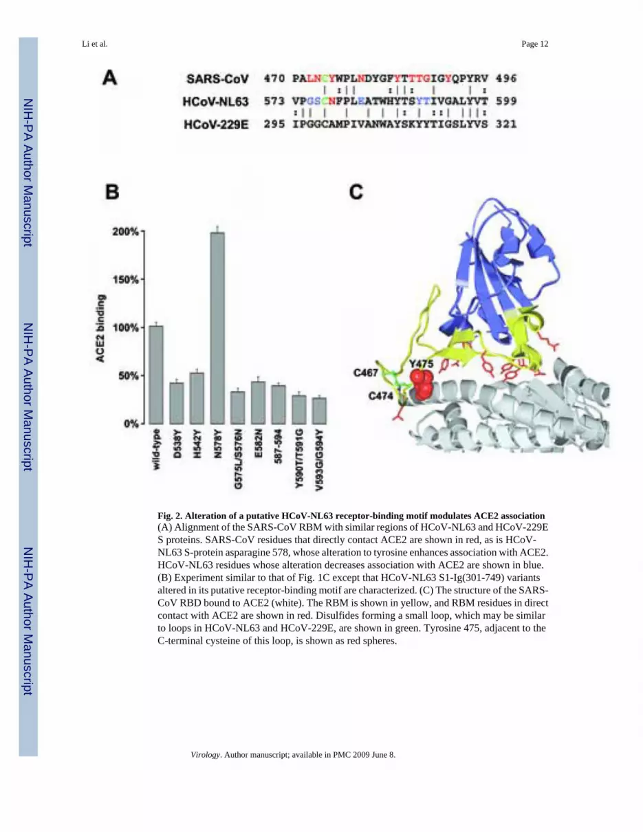
Fig. 2. Alteration of a putative HCoV-NL63 receptor-binding motif modulates ACE2 association(A) Alignment of the SARS-CoV RBM with similar regions of HCoV-NL63 and HCoV-229ES proteins. SARS-CoV residues that directly contact ACE2 are shown in red, as is HCoV-NL63 S-protein asparagine 578, whose alteration to tyrosine enhances association with ACE2.HCoV-NL63 residues whose alteration decreases association with ACE2 are shown in blue.(B) Experiment similar to that of Fig. 1C except that HCoV-NL63 S1-Ig(301-749) variantsaltered in its putative receptor-binding motif are characterized. (C) The structure of the SARS-CoV RBD bound to ACE2 (white). The RBM is shown in yellow, and RBM residues in directcontact with ACE2 are shown in red. Disulfides forming a small loop, which may be similarto loops in HCoV-NL63 and HCoV-229E, are shown in green. Tyrosine 475, adjacent to theC-terminal cysteine of this loop, is shown as red spheres.
Li et al. Page 12
Virology. Author manuscript; available in PMC 2009 June 8.
NIH
-PA Author Manuscript
NIH
-PA Author Manuscript
NIH
-PA Author Manuscript
Fig. 3. HCoV-NL63 and SARS-CoV S proteins bind similar sites on human ACE2(A) HEK293T cells expressing N-terminally tagged forms of human, rat, or two variants ofpalm civet ACE2 proteins incubated with anti-tag antibody, or HCoV-NL63 S1-Ig(16-749) orS1-Ig(301-749). Cells were washed and analyzed by flow cytometry. Results are indicated asthe percentage of mean fluorescence intensity observed with cells expressing human ACE2and incubated with anti-tag antibody. (B) An experiment similar to (A) except that humanACE2, and three indicated variants thereof, were analyzed for binding to anti-tag antibody,HCoV-NL63 S1-Ig(301-749), and SARS-CoV S1-Ig. (C) Lentiviruses expressing luciferaseand pseudotyped with the S proteins of HCoV-NL63 or SARS-CoV were incubated with cellstransfected with human ACE2 or the indicated human ACE2 variants, as previously described(Li et al., 2005c; Sui et al., 2005b). Entry, as measured by luciferase activity, is shown aspercentage observed for wild-type human ACE2. (D) Experiments similar to those of (B) and(C) except that human ACE2 variants proximal to the SARS-CoV binding site are characterizedfor expression, HCoV-NL63 S1-Ig(301-749) binding, and HCoV-NL63 S-protein-mediatedinfection. See supplemental data for results of experiments characterizing variants that do notcontact the SARS-CoV RBD.
Li et al. Page 13
Virology. Author manuscript; available in PMC 2009 June 8.
NIH
-PA Author Manuscript
NIH
-PA Author Manuscript
NIH
-PA Author Manuscript

Fig 4. The SARS-CoV RBD inhibits HCoV-NL63 S-protein-mediated infectionLentiviruses expressing luciferase and pseudotyped with the S proteins of SARS-CoV orHCoV-NL63 were incubated with ACE2-expressing HEK293T cells in the presence of theindicated concentrations of affinity purified goat anti-ACE2 antibody(Li et al., 2003), SARS-CoV RBD-Ig, or human IgG. Infection is expressed as a percentage of luciferase activityobserved in the absence of inhibitor.
Li et al. Page 14
Virology. Author manuscript; available in PMC 2009 June 8.
NIH
-PA Author Manuscript
NIH
-PA Author Manuscript
NIH
-PA Author Manuscript

Fig. 5. ACE2 residues critical to HCoV-NL63 S1-Ig associationA “virus-eye” view of the human ACE2 surface is shown. Residues in contact with the SARS-CoV RBD are indicated in yellow and orange. Residues whose alteration affects HCoV-CoVS1 binding were colored orange. Glycine 354, whose alteration to aspartic acid abolishesHCoV-NL63 binding, but not that of SARS-CoV, is shown in purple. Residues whose alterationdoes not alter ACE2 association with either S protein are shown in green. Some green residueslisted in Table 1 are not visible in this view.
Li et al. Page 15
Virology. Author manuscript; available in PMC 2009 June 8.
NIH
-PA Author Manuscript
NIH
-PA Author Manuscript
NIH
-PA Author Manuscript


![Coronavirus [COVID-19] Pathogenesis - Indian Journal of](https://img.dokumen.tips/doc/110x75/631778e9f68b807f8803a695/coronavirus-covid-19-pathogenesis-indian-journal-of-.jpg)