Embed Size (px)
Citation preview
The role and regulation of Frizzled receptors in synapse formation
Alessandro Bossio
A dissertation submitted in partial fulfillment of the
requirements for the degree of
Doctor of Philosophy
University College London
LMCB - Laboratory for Molecular Cell Biology
Department of Cell and Developmental Biology
July 2019

2

3
I, Alessandro Bossio, declare that the work presented here is my own. Where
information has been derived from other sources, I confirm that this has been
indicated in the thesis.

4
5
Abstract
The formation of synapses is crucial for brain function. Secreted Wnt proteins
signal through Frizzled and other receptors to regulate synaptogenesis. In
particular, Wnt7a promotes synaptogenesis in the hippocampus. The
receptor Fz5 mediates Wnt7a-induced presynaptic assembly, but the
mechanisms underlying Fz5 regulation are not well understood. How Wnt7a
signals at postsynaptic sites is also unknown. Fz7, another receptor binding
Wnt7a, is hypothesised to have a role in this process.
To address these questions, I used biochemical and cell biology techniques
combining in vitro and in vivo approaches. My findings demonstrate that Fz5
and Fz7 have distinct synaptic localisation. Fz5 is absent from dendritic
spines - excitatory postsynaptic structures - and is not required for spine
development. In contrast, Fz7 localises in spines and is required for Wnt7a-
induced spine formation.
Our preliminary data suggested that Fz5 is palmitoylated, a post-translational
lipid modification that affects protein distribution and function. I demonstrated
that all Frizzled receptors can be palmitoylated. Using a palmitoylation-
deficient Fz5 receptor, I showed that palmitoylation is required for Fz5
interaction with the scaffold protein Dishevelled, a key component of the Wnt
signalosome, but has no impact on Fz5 degradation rate and lateral mobility
at the plasma membrane. Palmitoylation-deficient Fz5 exhibits impaired
axonal distribution, increased endocytosis and decreased surface levels.
Expression of wild-type Fz5 in the hippocampus promotes presynaptic
assembly, whereas palmitoylation-deficient Fz5 lacks synaptogenic activity.
Palmitoylation is therefore a critical molecular mechanism that underpins Fz5
regulation and function in vivo.
These findings demonstrate that two distinct Frizzled receptors act pre- and
postsynaptically to promote synaptogenesis, and reveal a previously
uncharacterised lipid modification of Frizzled receptors, which is of critical
functional importance. This work opens up new avenues to study the role of
Frizzled palmitoylation in different biological contexts, from cell fate decisions
to neuronal circuit formation and plasticity.

6
7
Impact Statement
During my PhD I studied the mechanisms that regulate Wnt signalling in the
context of synapse formation in the brain. Wnts are secreted proteins that
signal through Frizzled and other receptors to activate a variety of
downstream signalling pathways. Wnt signalling plays a role in a wide range
of physiological processes, from tissue patterning to stem cell biology and
synaptogenesis. Aberrant Wnt signalling is associated with a number of
pathological conditions, including several types of cancers and
neurodegenerative disorders such as Alzheimer’s and Parkinson’s disease.
Understanding how Wnt signalling is regulated is therefore crucial to dissect
the mechanisms underlying this complex signalling cascade in health and
disease.
The precise molecular mechanisms that regulate the main receptors of Wnt
ligands, Frizzled receptors, remain poorly understood. In demonstrating that
possibly all Frizzled receptors undergo palmitoylation, my work characterises
a previously unidentified post-translational lipid modification of this class of
receptors. Focussing on Fz5, one of the most studied Frizzled receptors
expressed in mammals, I showed that palmitoylation is critical for the
trafficking and functional activity of this Wnt receptor in promoting
synaptogenesis in the developing hippocampus. These findings are novel
and identify an important molecular mechanism that directly underpins
Frizzled function in vivo.
In elucidating a novel mechanism of Frizzled regulation in the brain, my work
lays the foundation to further study how Wnt signalling is regulated at the
receptor level across a multitude of biological processes. As surface
receptors are a classical drug target, investigating how palmitoylation can be
modulated to tune Frizzled surface levels and activity has substantial
therapeutic potential. My findings are therefore likely to have a significant
impact on basic as well as clinical research.
We are planning to publish this study in a high impact scientific journal, and
present this work at prestigious international meetings such as the Wnt
Signalling Gordon Research Conference. My findings are of direct and
8
significant interest to the entire field of Wnt signalling research, which is a
large community spread across the world and includes academic research
groups as well as pharmaceutical companies with interests in this important
signalling pathway. For instance, the Dementia Research Institute, which is
the biggest initiative ever launched in UK to defeat dementia, has invested to
study the role of Wnt signalling in synapse degeneration. In addition, on-
going clinical trials in US are testing Wnt signalling inhibitors to cure different
forms of cancer.
The impact of my findings can already be appreciated in the academic
environment, as our laboratory has recently been awarded a grant to further
study the impact of Fz5 palmitoylation in neuronal circuit assembly and
function. Post doctoral fellows and students will benefit from taking part in
this project. In the future, other research grants and fellowships could be
proposed to study the role of palmitoylation in the regulation of other Frizzled
receptors. Therefore, the work produced during my PhD has the required
potential to influence future academic and private studies in the Wnt
signalling research field.
9
Contents Abstract…………………………………………………………………………. 5
Impact statement………………………………………………………………. 7
List of figures…………………………………………………………………… 13
List of tables……………………………………………………………………. 14
Abbreviations…………………………………………………………………… 15
CHAPTER 1: INTRODUCTION ..................................................................... 19
1.1 General overview ............................................................................... 19 1.2 Structure and function of synapses .................................................... 20
1.2.1 The presynaptic side .................................................................... 20 1.2.2 The postsynaptic side ................................................................... 24
1.3 Synaptogenesis .................................................................................. 29 1.3.1 Molecular mechanisms of synapse formation ............................... 29
Synthesis and trafficking of synaptic proteins prior to axo-dendritic contacts ............................................................................................. 30 The formation of axo-dendritic contacts in synapse assembly .......... 31 Synapse maturation .......................................................................... 32
1.3.2 Synaptogenic molecules ............................................................... 34 Cell adhesion molecules (CAMs) ...................................................... 34 Secreted synaptogenic factors .......................................................... 37 Glial synaptogenic factors ................................................................. 41
1.3.3 Synaptic plasticity and activity-mediated synapse formation ........ 42 1.4 Wnt signalling ..................................................................................... 47
1.4.1 Overview ...................................................................................... 47 1.4.2 Wnt signalling components ........................................................... 47
Wnt ligands ....................................................................................... 48 Secreted Wnt inhibitors ..................................................................... 50 Frizzled receptors .............................................................................. 51 The co-receptor LRP6 and other Wnt receptors ................................ 55
1.4.3 Wnt signalling pathways ............................................................... 57 Canonical or β-catenin Wnt signalling ............................................... 57 The divergent canonical pathway ...................................................... 60 The PCP pathway ............................................................................. 60 The Wnt/Ca2+ pathway ...................................................................... 61 Frizzled nuclear import pathway ........................................................ 62
10
1.5 Wnt signalling in neural development.................................................. 64 1.6 Wnt signalling at the synapse ............................................................. 66
1.6.1 Wnt signalling in synaptogenesis .................................................. 66 1.6.2 Wnts in synaptic transmission, plasticity and maintenance ........... 71
1.7 Receptor localisation and signalling, a role for post-translational modifications (PTMs) ................................................................................ 75
1.7.1 Lipid PTMs .................................................................................... 76 1.7.2 Palmitoylating and de-palmitoylating enzymes ............................. 80 1.7.3 Palmitoylation at synapses ............................................................ 82
1.8 Regulation of Wnt7a-Frizzled signalling at synapses, a role for palmitoylation. ........................................................................................... 86 1.9 Thesis aims ......................................................................................... 88
CHAPTER 2: MATERIAL AND METHODS .................................................. 89
2.1 DNA constructs, cloning and viruses................................................... 89 2.2 qPCR analyses ................................................................................... 92 2.3 Animal use and intracerebroventricular (ICV) injections of AAV9 ....... 93 2.4 Brain dissection, fixation and freezing ................................................. 93 2.5 Cryosectioning and Immunofluorescence analyses of brain slices ..... 94 2.6 Cell cultures, transfection methods and pharmacological treatments . 94
Primary rat hippocampal cultures ...................................................... 94 HEK293, NRK and NB2A cultures ..................................................... 95 Lipofectamine transfection ................................................................. 96 AMAXA nucleofection ........................................................................ 96 Pharmacological treatments .............................................................. 96
2.7 Antibody feeding experiments ............................................................. 97 2.8 Fluorescent Recovery After Photobleaching (FRAP) .......................... 97 2.9 Immunofluorescence analyses of cultured cells .................................. 98 2.10 Confocal imaging .............................................................................. 98 2.11 Image analyses ................................................................................. 99 2.12 Preparation of protein samples and western blotting (WB) ............... 99 2.13 Surface biotinylation ........................................................................ 100 2.14 Co-IP (co-immunoprecipitation) experiments .................................. 101 2.15 Acyl-Biotin exchange (ABE) assay .................................................. 102
Chloroform-methanol (CM) protein precipitation .............................. 102 Day1: blockage of free Cys residues ............................................... 103 Day 2: NEM wash out - acyl-biotin exchange .................................. 103
11
Day 3: immunoprecipitation of biotinylated (=palmitoyated) proteins ........................................................................................................ 104
2.16 Statistical analyses ......................................................................... 105 CHAPTER 3: THE ROLE OF Fz5 AND Fz7 IN PRE- AND POSTSYNAPTIC DEVELOPMENT .......................................................................................... 114
3.1 Introduction ....................................................................................... 114 3.2 Results ............................................................................................. 116
3.2.1 Fz5 and Fz7 localisation at dendritic spines ............................... 116 3.2.2 Fz5 expression is neither sufficient nor required for dendritic spine formation ............................................................................................. 118 3.2.3 Fz7 is required for postsynaptic development and Wnt7a-mediated spine formation .................................................................................... 120
3.3 Discussion ........................................................................................ 123 3.3.1 The distribution of Fz5 and Fz7 at dendritic spines .................... 123 3.3.2 Fz5 is not required for dendritic spine development ................... 123 3.3.3 The role of Fz7 at pre- and postsynaptic sites ............................ 124
CHAPTER 4: PALMITOYLATION OF FRIZZLED RECEPTORS ............... 127
4.1 Introduction ....................................................................................... 127 4.2 Results ............................................................................................. 130
4.2.1 Some Frizzled receptors are palmitoylated ................................ 130 4.2.2 Fz5 is palmitoylated on each of the 3 Cys residues at the C-term ............................................................................................................ 132 4.2.3 Investigating which DHHC enzymes palmitoylate Fz5 ............... 134
4.3 Discussion ........................................................................................ 137 4.3.1 Frizzled receptors are palmitoylated – Fz5 palmitoylation at the C-term ..................................................................................................... 137 4.3.2 Palmitoylation of Fz5 by members of the DHHC family .............. 138
CHAPTER 5: THE IMPACT OF PALMITOYLATION ON Fz5 SIGNALLING AND TRAFFICKING .................................................................................... 141
5.1 Introduction ....................................................................................... 141 5.2 Results ............................................................................................. 143
5.2.1 Fz5 turnover is independent of protein palmitoylation ................ 143 5.2.2 Palmitoylation is required for Fz5 interaction with Dvl1 but dispensable for Fz5/Fz5 interaction ..................................................... 143 5.2.3 Palmitoylation regulates Fz5 distribution along axons but not dendrites and soma of hippocampal neurons ...................................... 146 5.2.4 The role of palmitoylation in Fz5 stability at the PM ................... 148
12
5.3 Discussion ......................................................................................... 153 5.3.1 Palmitoylation regulates Fz5 localisation in hippocampal neurons ............................................................................................................. 153 5.3.2 The role of palmitoylation in Fz5 membrane trafficking ............... 154
CHAPTER 6: THE ROLE OF Fz5 AND ITS PALMITOYLATION IN THE FORMATION OF SYNAPSES IN VIVO ...................................................... 156
6.1 Introduction ....................................................................................... 156 6.2 Results .............................................................................................. 158
6.2.1 Effects of ICV injections on hippocampal anatomy and proliferation of glial cells .......................................................................................... 158 6.2.2 Fz5 promotes synaptogenesis in vivo in a palmitoylation-dependent manner ................................................................................................. 161
6.3 Discussion ......................................................................................... 164 6.3.1 Fz5 expression induces presynaptic assembly in vivo ................ 164 6.3.2 Palmitoylation is required for Fz5-induced synapse formation in vivo....................................................................................................... 165
CHAPTER 7: DISCUSSION AND FUTURE DIRECTIONS......................... 167
7.1 Summary of project aims and findings .............................................. 167 7.2 Fz5 and Fz7 exhibit different synaptic distribution and mediate pre- and postsynaptic development respectively ................................................... 169 7.3 Frizzled receptors are palmitoylated ................................................. 172 7.4 Palmitoylation is essential for Fz5 function ....................................... 174 7.5 Palmitoylation regulates Fz5 trafficking and membrane stability ....... 177 7.6 Multiple DHHC enzymes can palmitoylate Fz5 ................................. 182 7.7 Conclusions ...................................................................................... 184
Acknowledgments.………………………………………………….…………186 Bibliography ............................................................................................... 187
13
List of Figures 1.1: Schematic of pre- and postsynaptic structure……………………….. 22
1.2: Schematic of an inhibitory synapse…………………………………… 28
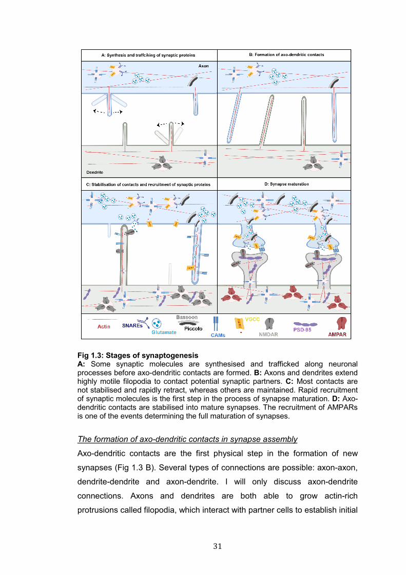
1.3: Stages of synaptogenesis……………………………………………… 31
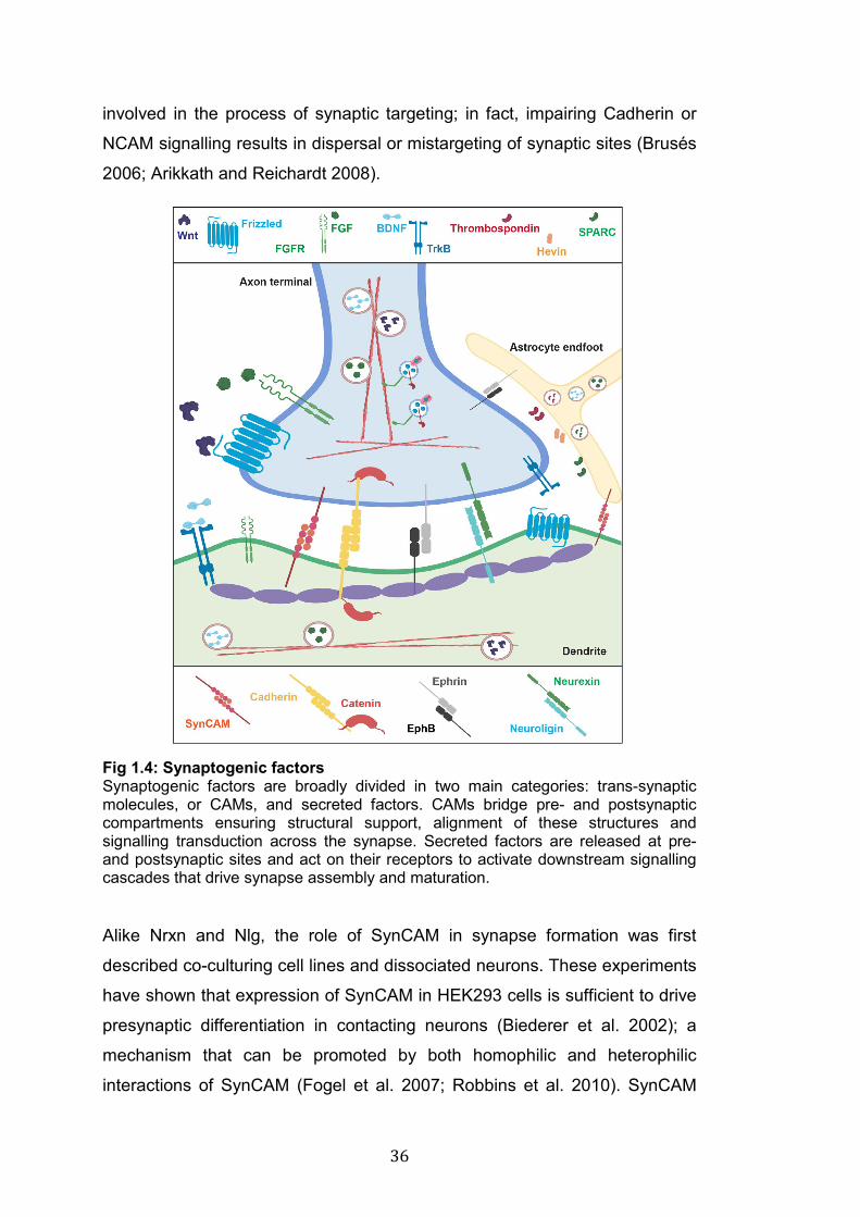
1.4: Synaptogenic factors…………………………………………………… 36
1.5: Frizzled receptors………………………………………………………. 53
1.6: β-catenin and divergent canonical Wnt pathways…………………... 59
1.7: PCP and Wnt/Ca2+ pathways………………………..………………… 61
1.8: Frizzled nuclear import pathway………………………………………. 63
1.9: The role of Fz5 in presynaptic assembly.……………………………. 67
1.10: Different types of palmitoylation……………………………………... 77
1.11: The effects of protein palmitoylation………………………………… 79
1.12: S-palmitoylation by DHHC protein acyl transferases……………… 81
1.13. Fz5 is palmitoylated and this modification is important for its synaptogenic activity………………………..………………………………..
87
3.1: Gallery of neurons selected for in vitro studies……………………… 117
3.2: Fz5 gain-of-function does not affect dendritic spines……………….. 118
3.3: Fz5 loss-of-function does not affect dendritic spines……………….. 119
3.4: Fz7 is required for spine formation and Wnt7a-induced structural plasticity………………………………………………………………………..
121
3.5: Fz7 is not required for presynaptic assembly………………………... 122
4.1: Prediction of palmitoylation of Frizzled receptors…………………… 131
4.2: Alignment of the C-term of Frizzled receptors……………………….. 132
4.3: Frizzled receptors palmitoylation - Fz5 palmitoylation at the C-term 133
4.4: Gain-of-function screen for the DHHC enzymes that palmitoylate Fz5……………………………………………………………………………..
135
4.5 The effect of DHHC3 and DHHC7 loss-of-function on Fz5 palmitoylation………………………………………………………………….
136
5.1: Palmitoylation does not regulate Fz5 turnover………………………. 144
5.2: Palmitoylation is dispensable for Fz5/Fz5 interaction but required for binding to Dvl1…………………………………………………………….
145
5.3: Palmitoylation controls Fz5 distribution in axons but not dendrites or soma………………………………………………………………………..
147
5.4: Palmitoylation regulates Fz5 membrane stability…………………… 149
14
5.5: Palmitoylation does not affect Fz5 lateral mobility………………….. 152
6.1: ICV injections in new born mice ……………………………………… 158
6.2 Spread of AAV9 viruses………………………………………………… 160
6.3: ICV injections did not affect hippocampal morphology or proliferation of glial cells.…………………………………………………….
161
6.4 Expression of WT Fz5, but not the mutant 3CS Fz5, promotes presynaptic assembly in the developing hippocampus…………………..
163
7.1: Fz5 and Fz7 regulate pre- and postsynaptic development respectively……………………………………………………………………
169
7.2: Frizzled receptors are palmitoylated………………………………….. 172
7.3: Fz5 expression promotes presynaptic assembly in vivo in a palmitoylation dependent manner…………………………………………..
175
7.4: The role of palmitoylation in Fz5 trafficking………………………….. 179
List of Tables 2.1: Primers used for Fz5 cloning………………………………………….. 89
2.2: Nomenclature of DHHC enzymes…………………………………….. 90
2.3: shRNA target sequences used for loss-of-function studies………... 91
2.4: Primers used for qPCR analyses……………………………………... 92
2.5: List of primary antibodies………………………………………………. 106
List of solutions……………………………………………………………... 106
15
Abbreviations AAV (Adeno Associated Virus)
ABE (acyl biotin exchange)
Ach (achetylcholine)
AD (Alzheimer’s disease)
AZ (active zone)
AMPAR (α-amino-3-hydroxyl-5-methyl-4-isoxazolepropionate receptor)
APs (action potentials)
APC (Adenomatus polyposis coli)
APT (Acyl-proteinthioesterase 1)
BBB (blood brain barrier)
BDNF (Brain derived neurotrophic factor)
β-TrCP (Beta-Transducin Repeat Containing E3 Ubiquitin Protein Ligase)
CaMKII (Ca2+/calmodulin-dependent protein kinase II)
CAMs (Cell adhesion molecules)
CNS (central nervous system)
CK1 (Casein kinase 1)
cKO (conditional knock-out)
CRD (Cys rich domain)
C-term (carboxyl terminus)
Cys (cysteine)
DG (dentate gyrus)
DHHC (Aspartate, Hystidine, Hystidine, Cystein)
Dkk (Dickkopf)
Dishevelled (Dvl)
DMSO (dimethyl sulfoxide)
EAAT (excitatory amino acid transporter)
EE (enriched environment)
EphB (erythropoietin-producing human hepatocellular receptors)
ER (endoplasmic reticulum)
Erb (Receptor tyrosine-protein kinase erbB-4)
ERAD (endoplasmic reticulum-associated degradation)
ERK (extracellular-signal-regulated kinase)
Evi (Eveness interrupted in vertebrates)
FGF (Fibroblast Growth Factor)
16
FRAP (fluorescence recovery after photobleaching)
GABA (γ-aminobutyric acid)
Gli (glioma-associated oncogene homologue)
GPCRs (G-protein Coupled receptors)
Gpr124 (probable G-protein coupled receptor 124)
GRIP1 (glutamate receptor interacting protein 1)
Gsk3β (glycogen synthase kinase 3)
HFS/LFS (high/low frequency stimulation)
ICV (intracerebroventricular)
IP (immunoprecipitation)
IP3 (inositol triphosphate)
JNK (Jun N-terminal kinase)
KD (knock-down)
KI (knock-in)
KO (knock-out)
LEF/TCF (lymphoid enhancer binding factor and transcription factor)
LRGs (leucine-rich repeat-containing G-protein coupled receptor
LRP6 (low density lipoprotein related receptor 6)
LTP/LTD (long-term potentiation/depression)
MAPK (mitogen-activated protein kinase)
MBOAT (membrane bound O-acyl transferase)
MuSK (Muscle-specific kinase)
NCAM (neural cell adeshion molecules)
NGF (nerve growth factor)
Nlg (Neuroligin)
NMDAR (N-methyl-d-aspartate receptor)
NMJ (neuromuscular junction)
Nrg (Neuregulin)
Nrxn (Neurexin)
NTs (neurotrasnmitters)
N-term (amino terminus)
O/N (overnight)
PATs (protein acyl transferases)
PCP (planar cell polarity)
PLC (Phospholipase C)
PKA/PKC (protein kinase A, C)
PM (plasma membrane)
17
Porc (Porcupine)
PPT1 (Palmitoyl-protein thioesterase 1)
PSD (postsynaptic densities)
Ptch (Patched)
qPCR (quantitative polymerase chain reaction)
RNF43 (Ring finger protein 43)
RT (room temperature)
RUSH (retention using selective hooks)
Ser (serine)
SFRPs (Secreted Frizzled related protiens)
Shh (Sonic Hedgehog)
SM (Sec1/Munc18)
Smo (Smoothened)
SNARE (soluble N-ethyl maleimide sensitive-factor attachment protein
receptors)
SPARC (secreted protein acidic enriched in cysteine)
SVs (synaptic vesicles)
Syts (Synaptotagmins)
SynGAP (Ras-GTPase-activating protein)
SWIM (secreted Wg-interacting molecule)
TNF (Tumor Necrosis Factor)
TrkB (Tropomysin receptor kinase B)
TSP (Thrombospondin)
vGat (vesicular GABA transporter)
VGCCs (voltage gated Ca2+ channels)
vGlut (vesicular Glutamate transporter)
WB (western blot)
WIF-1 (Wnt inhibitory factor 1)
Wingless (Wg)
Wls (Wntless in Drosophila)
ZNRF3 (Zinc and ring finger 3)

18
19
CHAPTER 1: INTRODUCTION
1.1 General overview
What we do, from breathing and moving to more complex actions like
performing an experiment or discussing Brexit, depends on the correct
function of our brain. To perform these actions the brain relies on the
transmission of information between nerve cells, or neurons. Neurons are
organised in circuits formed by countless connections between cells. These
connections, called synapses, are the hubs of communication between
neurons and are crucial for the transmission of nerve impulses. Synapses
are asymmetric connections between nerve cells characterised by the
presence of highly specialised proteins that allow the transit of information
from a pre- to a postsynaptic cell, thus enabling signal transmission along
neuronal circuits. The correct development of synapses is crucial to sustain
the function of our brain. In fact, aberrant synapse formation is associated
with the onset of several neurological disorders. One of the key questions in
developmental neurobiology is what are the molecular mechanisms that
regulate the assembly and maturation of synapses?
The formation and maturation of synapses require the orchestrated
recruitment and stabilisation of thousands of synaptic proteins at sites where
new connections are formed. Synaptogenic factors are a broad group of
molecules which regulate the formation of synapses. Remarkable progress
has been made in understanding the role and regulation of synaptogenic
factors during synapse formation, but in spite of such advancement we still
do not fully understand how this complex phenomenon is regulated. In my
PhD thesis I have studied the molecular mechanisms that regulate the
trafficking and function of Frizzled receptors, which are the main receptors for
Wnt ligands, a family of conserved secreted glycolipoproteins that act as
potent synaptogenic factors in the central and peripheral nervous systems.
20
1.2 Structure and function of synapses
Synapses are small but highly dynamic structures specialised to transmit
electro-chemical signalling between a presynaptic cell (a neuron) and a
postsynaptic cell, which can be another neuron, a muscle fibre or a gland.
Pre- and postsynaptic compartments exhibit a highly asymmetric and
polarised architecture that is optimal for the transmission of electro-chemical
signals from one cell to another. Glial cells, particularly astrocytes, are also
important cellular components of the synapse, as they offer structural and
functional support. The crucial role of astrocytes at synapses has given rise
to the concept of tripartite synapse (Fig 1.1) (Araque et al. 1999; Perea et al.
2009).
1.2.1 The presynaptic side
The presynaptic terminal, or bouton, is where electrical stimuli in the form of
action potentials (APs) arrive and are converted into chemical signals in the
form of neurotransmitters (NTs). NTs are released to activate receptors on
postsynaptic cells (Fig 1.1). Once an AP reaches a presynaptic terminal, it
induces an influx of Ca2+ ions through voltage gated Ca2+ channels (VGCCs)
(Takahashi and Momiyama 1993; Catterall 2011). This activates the Ca2+-
sensitive components of the NT release machinery and triggers the fusion of
NTs-loaded synaptic vesicles (SVs) with the plasma membrane (PM), thus
enabling the release of NTs in the synaptic cleft, a very small space (roughly
20 nm) that separates the pre- and postsynaptic side (Südhof 2012a;
Midorikawa and Sakaba 2015) (Fig 1.1). The process of NT release occurs in
less than 1ms and involves a vast number of proteins including all the
components of the release machinery (Südhof 2013).
The release machinery is composed by a multitude of molecules; the best
characterised are Synaptotagmins (Syts), members of the SNARE (soluble
N-ethyl maleimide sensitive-factor attachment protein receptors) family and
the SM (Sec1/Munc18) protein family (Rizo and Xu 2015). In response to
elevated Ca2+ concentrations at presynaptic terminals, Syts interact with
SNARE proteins enabling the exocytosis of SVs (Chapman 2008; Chapman
21
2018). SNARE proteins are the core components of the release machinery.
This family includes over 60 members, of which SNAP25, Synaptobrevin1/2
and Syntaxin1 are the most studied. SNARE proteins do not only regulate
SVs fusion but they are also involved in all types of vesicle exocytosis (Wu et
al. 2014). The interaction between different SNAREs leads to the docking of
vesicles to the PM. If sufficient Ca2+ enters the terminal, SVs fuse with the
PM allowing the release of NTs (Chen and Scheller 2001; Duman and Forte
2003; Ungar and Hughson 2003; Han et al. 2017). SM proteins are
fundamental regulators of exocytosis: they act as “clasps” to spatially and
temporally coordinate the interaction between SNAREs (Dulubova et al.
2007; Südhof and Rothman 2009; Südhof and Rizo 2011).
At presynaptic terminals SVs loaded with NTs fuse with the PM at a specific
area called the active zone (AZ) (Fig 1.1). The main function of the AZ is to
favour the docking and fusion of SVs, to recruit VGCCs and to spatially
coordinate the apposition of pre- and postsynaptic sides (Zhai and Bellen
2004; Südhof 2012b). The AZ is characterised by a complex cytoskeletal
organisation, which is required to hold in place different components of the
AZ (Cingolani and Goda 2008). Some of the key components of the AZ
zones are Munc-13 (Protein Unc 13), RIM (regulating synaptic membrane
exocytosis 1), Piccolo and Bassoon. Munc-13 binds directly to some
SNAREs and is required for both spontaneous and evoked transmitter
release (Südhof 2012b). RIM and RIM-BPs (RIM binding proteins) are key
organizers of presynaptic terminals as they control SV docking and tether
Ca2+ channels to the AZ. Piccolo and Bassoon have crucial functions at the
AZ as they move SVs towards the PM, regulate the localisation of VGCCs
and are involved in activity-induced remodelling of the actin cytoskeleton
(Piccolo only) (Hallermann et al. 2010; Mukherjee et al. 2010; Gundelfinger
et al. 2015).
NTs released into the synaptic cleft bind to postsynaptic receptors causing
the influx of ions and affecting the electrical activity of postsynaptic cells
(Snyder 2009; Smart and Paoletti 2012). At postsynaptic sites, NTs can
induce depolarisation or hyperpolarisation, which are changes in the
electrophysiological balance of neuronal cells caused by the influx of
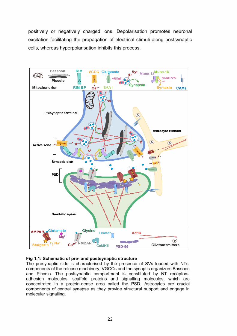
22
positively or negatively charged ions. Depolarisation promotes neuronal
excitation facilitating the propagation of electrical stimuli along postsynaptic
cells, whereas hyperpolarisation inhibits this process.
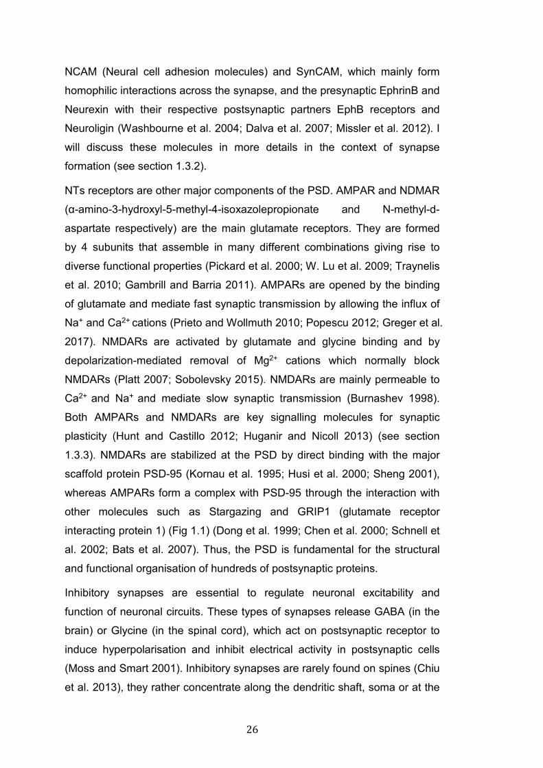
Fig 1.1: Schematic of pre- and postsynaptic structure The presynaptic side is characterised by the presence of SVs loaded with NTs, components of the release machinery, VGCCs and the synaptic organizers Bassoon and Piccolo. The postsynaptic compartment is constituted by NT receptors, adhesion molecules, scaffold proteins and signalling molecules, which are concentrated in a protein-dense area called the PSD. Astrocytes are crucial components of central synapse as they provide structural support and engage in molecular signalling.
23
Hundreds of NTs exist, but the amino acid glutamate and GABA (γ-
aminobutyric acid) are the most common excitatory and inhibitory NTs in the
CNS (Meldrum 2000; Valenzuela et al. 2011; Snyder 2017). However,
whether a NT induces excitation or inhibition depends on the
electrophysiological environment of the receiving cells; for instance, early in
development GABA induces depolarisation due to the high concentration of
Cl- ions found in young neurons (Ben-Ari 2002; Spitzer 2010). During
development changes in the expression of the ion transporter NKCC1 (Na+-
K+-2Cl- co-transporter 1) and KCC2 (K+-Cl- cotransporter 2) affect the
electrical properties of neurons, causing a shift from excitation to inhibition
upon activation of GABA receptors (Rivera et al. 1999; Tyzio et al. 2006;
Leonzino et al. 2016). Dopamine, serotonin, acetylcholine (Ach) and glycine
are also common NTs released at central and peripheral synapses
(Valenzuela et al. 2011; Snyder 2017).
After NT release, SV recycling and NT re-uptake are absolutely required to
replenish the SV pool and terminate signalling by clearing NTs from the
synaptic cleft, a function that is largely dependent on astrocytes (Rizzoli
2014; Soykan et al. 2016). Two main classes of NT transporters exist: a)
those localised on SVs, such as vGlut (vesicular Glutamate transporter) and
vGAT (vesicular GABA transporter), which control the loading of NTs; b)
those distributed at the PM, which terminate signalling by mediating the re-
uptake of NTs from the synaptic cleft, like EAAT (excitatory amino acid
transporter) and GAT (Danbolt 2001; Shigeri et al. 2004; Blakely and
Edwards 2012; Scimemi 2014). Given the crucial role of NT transporters at
synapses and the progress made in understanding their structure and
function, these molecules have become one of the most important drug
targets in the CNS (Gether et al. 2006; Iversen 2006).
In summary, presynaptic terminals are specialised structures to sense the
arrival of electrical stimuli, induce the release of NTs and ensure the re-
uptake of these molecules from the synaptic cleft to terminate signalling and
undergo further cycles of transmission (Fig 1.1). Presynaptic terminals are
made by hundreds of proteins that work together to ensure transmission of
signals from a pre- to a postsynaptic cell. VGCCs, SVs loaded with NTs, the
24
release machinery and the AZ are the most important components of
presynaptic terminals.
1.2.2 The postsynaptic side
The postsynaptic side is the structure of the synapse specialised for the
reception of NTs. Binding of NTs to their postsynaptic receptors induces
changes in the electrical properties of postsynaptic cells and results in the
activation of downstream signalling (Fig 1.1) (Sheng and Kim 2011). For the
purpose of this PhD thesis, I will focus mainly on the structure of excitatory
postsynaptic sites and I will only briefly touch upon the organisation of
inhibitory synapses.
Dendritic spines are the main postsynaptic structures for excitatory synapses.
They were first described over a century ago by Ramon y Cajal in his
astonishing drawings, following his observation of Golgi-stained brain tissues
(Ramón y Cajal 1909). Since then, dendritic spines have been one of the
most studied synaptic structures. Spines are small actin-rich (max width
<1µm) protrusions extending from dendrites; they are extremely dynamic and
diverse in their morphology (Häusser et al. 2000; Sorra and Harris 2000;
Nimchinsky et al. 2002; Rochefort and Konnerth 2012). Dendritic spines are
distributed along the entirety of dendritic branches at a density that varies
greatly according to age and brain areas, ranging from 1-10 spines/10 µm
(Huttenlocher 1990; Woolley et al. 1990; Benavides-Piccione et al. 2013;
Jammalamadaka et al. 2013; Morales et al. 2014). In most cases dendritic
spines are innervated by one presynaptic terminal, but multi-innervated
spines also exist (Fiala et al. 1998; Giese et al. 2015). Spines, particularly
mature ones, exhibit a defined head that comprises one or more postsynaptic
densities (PSD), which are protein-dense areas formed by NT receptors,
trans-synaptic proteins, scaffold proteins and signalling molecules (Walikonis
et al. 2000; Boeckers 2006; Arellano et al. 2007; Kim and Sheng 2009).
Based on their morphology dendritic spines are classified into three groups:
a) mushroom spines, which exhibit a relatively big head that is connected to
the dendritic branch by a very thin neck; b) thin spines, which have small
25
head and thin neck; and c) stubby spines, which do not present a clear
separation between head and neck (Arellano et al. 2007; Berry and Nedivi
2017). There is a correlation between morphology, molecular composition
and function of dendritic spines (Yuste and Bonhoeffer 2001; Arellano et al.
2007; Dent et al. 2011; Bosch and Hayashi 2012). Changes in dendritic spine
size and morphology are some of the mechanisms underlying synaptic
plasticity, which is the ability of synapses to dynamically adapt their structure
and function in response to external stimuli (see section 1.3.3).
The PSD of dendritic spines is probably the most complex substructure at
synapses. It is estimated that several hundreds of proteins are concentrated
at the PSD (Walikonis et al. 2000; Collins et al. 2006), an astonishing number
given the small size of this structure: roughly 200-800 nm long and 30-50 nm
thick (Boeckers 2006). Among the molecules in the PSD, we find NTs
receptors, ion channels, adhesion molecules, signalling enzymes and
scaffold proteins. The most abundant proteins are CaMKII (Ca2+/Calmodulin-
dependent protein kinase II), SynGAP (Ras-GTPase-activating protein) and
the scaffold protein PSD-95 (Kim and Sheng 2009). The PSD is localised at
the tip of dendritic spine heads, where it exerts structural and functional roles.
The PSD supports adhesion molecules and NT receptors and, through
scaffold proteins, couples these membrane proteins to cytoplasmic signalling
molecules (Fig 1.1) (Sheng and Kim 2011). The PSD itself is a very dynamic
structure, and modifications of the molecular composition of the PSD are
reflected in changes in spine structure and function (Hering and Sheng 2001).
Cell adhesion molecules (CAMs) of the PSD have critical synaptic functions
as they bind presynaptic partners bridging the two sides of the synapse
(Yamagata et al. 2003; Dalva et al. 2007). In addition, they mediate the
interaction with other cell types like astrocytes (Togashi et al. 2009). CAMs
also play a role in the recognition of synaptic targets, a process particularly
important during the initial stages of synapse formation (Washbourne,
Dityatev, et al. 2004). Moreover, by acting as trans-synaptic signalling
molecules and binding intracellular scaffold proteins, they can modulate
synapse structure and function activating downstream effectors (Dalva et al.
2007). Among the members of this family of proteins we find: Cadherin,
26
NCAM (Neural cell adhesion molecules) and SynCAM, which mainly form
homophilic interactions across the synapse, and the presynaptic EphrinB and
Neurexin with their respective postsynaptic partners EphB receptors and
Neuroligin (Washbourne et al. 2004; Dalva et al. 2007; Missler et al. 2012). I
will discuss these molecules in more details in the context of synapse
formation (see section 1.3.2).
NTs receptors are other major components of the PSD. AMPAR and NDMAR
(α-amino-3-hydroxyl-5-methyl-4-isoxazolepropionate and N-methyl-d-
aspartate respectively) are the main glutamate receptors. They are formed
by 4 subunits that assemble in many different combinations giving rise to
diverse functional properties (Pickard et al. 2000; W. Lu et al. 2009; Traynelis
et al. 2010; Gambrill and Barria 2011). AMPARs are opened by the binding
of glutamate and mediate fast synaptic transmission by allowing the influx of
Na+ and Ca2+ cations (Prieto and Wollmuth 2010; Popescu 2012; Greger et al.
2017). NMDARs are activated by glutamate and glycine binding and by
depolarization-mediated removal of Mg2+ cations which normally block
NMDARs (Platt 2007; Sobolevsky 2015). NMDARs are mainly permeable to
Ca2+ and Na+ and mediate slow synaptic transmission (Burnashev 1998).
Both AMPARs and NMDARs are key signalling molecules for synaptic
plasticity (Hunt and Castillo 2012; Huganir and Nicoll 2013) (see section
1.3.3). NMDARs are stabilized at the PSD by direct binding with the major
scaffold protein PSD-95 (Kornau et al. 1995; Husi et al. 2000; Sheng 2001),
whereas AMPARs form a complex with PSD-95 through the interaction with
other molecules such as Stargazing and GRIP1 (glutamate receptor
interacting protein 1) (Fig 1.1) (Dong et al. 1999; Chen et al. 2000; Schnell et
al. 2002; Bats et al. 2007). Thus, the PSD is fundamental for the structural
and functional organisation of hundreds of postsynaptic proteins.
Inhibitory synapses are essential to regulate neuronal excitability and
function of neuronal circuits. These types of synapses release GABA (in the
brain) or Glycine (in the spinal cord), which act on postsynaptic receptor to
induce hyperpolarisation and inhibit electrical activity in postsynaptic cells
(Moss and Smart 2001). Inhibitory synapses are rarely found on spines (Chiu
et al. 2013), they rather concentrate along the dendritic shaft, soma or at the
27
initial segment of axons. A recent paper proposed that specific temporally-
restricted expression profiles characterise different populations of GABAergic
neurons and establish the distribution of inhibitory synapses at different
cellular locations (dendrites vs soma vs axons) (Favuzzi et al. 2019). GABA
receptors (GABARs) are the main NT receptors at inhibitory synapses (Fig
1.2). GABAARs are pentameric ionotropic receptors that assemble in many
different combinations of α, β and γ subunits, and upon GABA binding they
quickly inhibit electrical activity by allowing the influx of Cl- (Olsen and
Sieghart 2009; Miller and Aricescu 2014). GABABRs are metabotropic
receptors that assemble in heterodimers and act through a second
messenger system to regulate slow and prolonged inhibitory
neurotransmission (Chebib and Johnston 1999; Bettler et al. 2004). Gephyrin
is a self-assembling scaffold protein and a key organizer molecule at
inhibitory postsynaptic sites; its main function is to stabilize GABAARs at
synapses by facilitating the clustering of these receptors (Choii and Ko 2015)
(Fig 1.2). Like PSD-95 and glutamate receptors at excitatory synapses,
GABARs and Gephyrin are involved in mechanisms of synaptic plasticity at
inhibitory synapses.
In summary, the postsynaptic compartment is a very specialized structure
where binding of NTs to their highly concentrated receptors activate
downstream cascades to propagate signalling. In contrast to presynaptic
sites, excitatory and inhibitory postsynaptic compartments exhibit different
structural organisation. Excitatory synapses are mainly formed on dendritic
spines, which are highly specialised and dynamic structures extending from
dendritic branches. Inhibitory synapses are mainly found at the soma and
along the dendritic shaft. The PSD is the central component of postsynaptic
sites; it incorporates hundreds of molecules including NT receptors, CAMs,
scaffold proteins and signalling molecules to orchestrate signalling in
response to NT release. In the following section I will introduce the molecular
mechanisms that regulate the assembly of synapses.

28
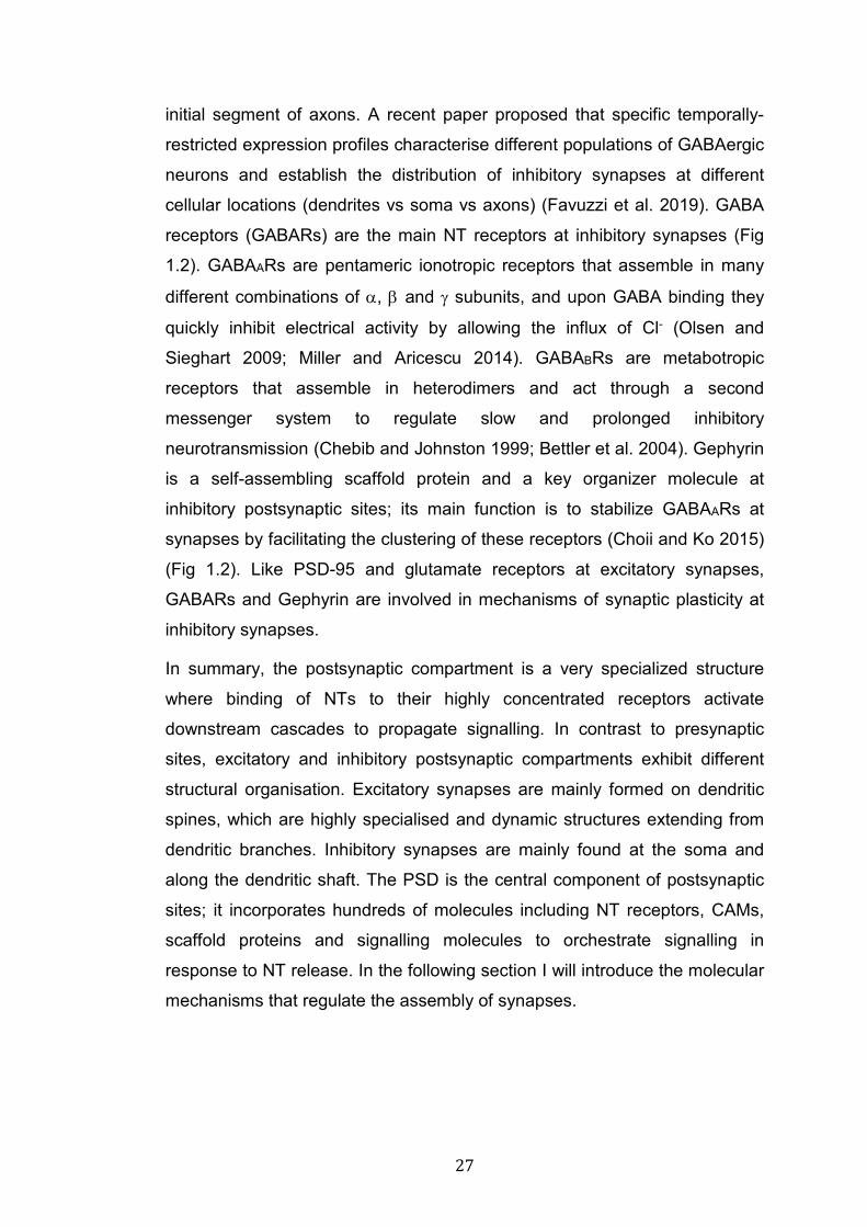
Fig 1.2: Schematic of an inhibitory synapse Inhibitory synapses are usually found along the dendritic shaft or in proximity of the soma and rarely on dendritic spines. GABA is the main NT at inhibitory synapses and vGAT and GAT are vesicular and membrane transporters for this NT. GABA release activates the postsynaptic receptors GABAA allowing the influx of Cl- ions in the postsynaptic compartment and inhibiting neuronal excitation. Gephyrin is the major component of the PSD at inhibitory synapses and is fundamental to cluster GABAARs.
29
1.3 Synaptogenesis
The assembly of synapses is a key step for the formation of functional
neuronal circuits, and is therefore essential for the proper function of the
brain. The timing of synapse formation varies greatly between different brain
areas (Huttenlocher and Dabholkar 1997; Dehorter et al. 2012). In rodents
synaptogenesis peaks around the second week of life (P10-P15) (Semple et
al. 2013), and it is followed by synaptic pruning, a period of selective
elimination of unnecessary connections that results in roughly 50% reduction
in synapse number by postnatal week 4-6 (Pressler and Auvin 2013). The
rate of synapse formation during early postnatal life is astonishing, it has
been estimated that synaptic density in the cortex on newborn rats increases
from 200 million/mm3 to 4 billion/mm3 in just five weeks (DeFelipe et al. 1999).
Highly regulated molecular mechanisms are in place to ensure such fast and
precise expansion in synaptic connections. Moreover, synapses continue to
form throughout the entire lifespan, in a balance with synapse elimination.
Unlike synapse formation during postnatal development, synaptogenesis in
adults is mainly experience-dependent and is one of the key mechanisms
underlying synaptic plasticity (Zito and Svoboda 2002; Markham and
Greenough 2004; Song et al. 2005; Holtmaat and Svoboda 2009), which is
considered the cellular correlate of learning and memory.
1.3.1 Molecular mechanisms of synapse formation
The formation of new synapses requires the coordinated recruitment and
stabilisation of thousands of proteins at both sides of the synapse; thus,
synaptogenesis is an extremely complex process. The correct formation of
synaptic connections is essential to sustain the cognitive performances of our
brain; in fact, aberrant formation of synapses has been linked to onset of
several neurodevelopmental disorders (Lepeta et al. 2016; Del Pino et al.
2018; Batool et al. 2019). Enormous progresses have been made in
describing the sequence of events as well as the molecules involved in the
formation of synapses; however, we still have much to learn about the
mechanisms underlying this complex process. The steps towards the
30
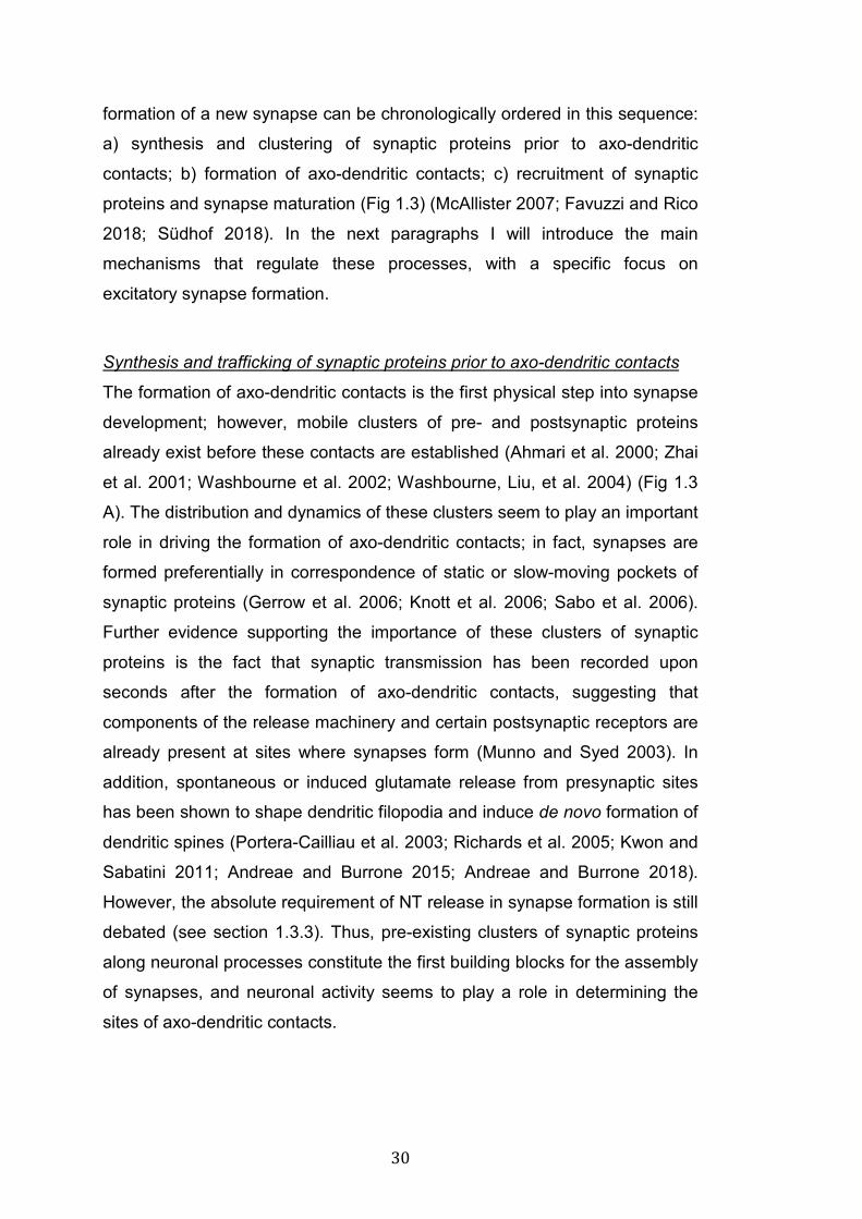
formation of a new synapse can be chronologically ordered in this sequence:
a) synthesis and clustering of synaptic proteins prior to axo-dendritic
contacts; b) formation of axo-dendritic contacts; c) recruitment of synaptic
proteins and synapse maturation (Fig 1.3) (McAllister 2007; Favuzzi and Rico
2018; Südhof 2018). In the next paragraphs I will introduce the main
mechanisms that regulate these processes, with a specific focus on
excitatory synapse formation.
Synthesis and trafficking of synaptic proteins prior to axo-dendritic contacts
The formation of axo-dendritic contacts is the first physical step into synapse
development; however, mobile clusters of pre- and postsynaptic proteins
already exist before these contacts are established (Ahmari et al. 2000; Zhai
et al. 2001; Washbourne et al. 2002; Washbourne, Liu, et al. 2004) (Fig 1.3
A). The distribution and dynamics of these clusters seem to play an important
role in driving the formation of axo-dendritic contacts; in fact, synapses are
formed preferentially in correspondence of static or slow-moving pockets of
synaptic proteins (Gerrow et al. 2006; Knott et al. 2006; Sabo et al. 2006).
Further evidence supporting the importance of these clusters of synaptic
proteins is the fact that synaptic transmission has been recorded upon
seconds after the formation of axo-dendritic contacts, suggesting that
components of the release machinery and certain postsynaptic receptors are
already present at sites where synapses form (Munno and Syed 2003). In
addition, spontaneous or induced glutamate release from presynaptic sites
has been shown to shape dendritic filopodia and induce de novo formation of
dendritic spines (Portera-Cailliau et al. 2003; Richards et al. 2005; Kwon and
Sabatini 2011; Andreae and Burrone 2015; Andreae and Burrone 2018).
However, the absolute requirement of NT release in synapse formation is still
debated (see section 1.3.3). Thus, pre-existing clusters of synaptic proteins
along neuronal processes constitute the first building blocks for the assembly
of synapses, and neuronal activity seems to play a role in determining the
sites of axo-dendritic contacts.
31
Fig 1.3: Stages of synaptogenesis A: Some synaptic molecules are synthesised and trafficked along neuronal processes before axo-dendritic contacts are formed. B: Axons and dendrites extend highly motile filopodia to contact potential synaptic partners. C: Most contacts are not stabilised and rapidly retract, whereas others are maintained. Rapid recruitment of synaptic molecules is the first step in the process of synapse maturation. D: Axo-dendritic contacts are stabilised into mature synapses. The recruitment of AMPARs is one of the events determining the full maturation of synapses.
The formation of axo-dendritic contacts in synapse assembly
Axo-dendritic contacts are the first physical step in the formation of new
synapses (Fig 1.3 B). Several types of connections are possible: axon-axon,
dendrite-dendrite and axon-dendrite. I will only discuss axon-dendrite
connections. Axons and dendrites are both able to grow actin-rich
protrusions called filopodia, which interact with partner cells to establish initial
32
contacts (Ziv and Smith 1996; Fiala et al. 1998). These interactions occur
very rapidly and the vast majority retract within seconds to minutes before
maturing into functional synapses (Wong and Wong 2000; Bonhoeffer and
Yuste 2002). It has been proposed that filopodia are stabilised in apposition
of clusters of both pre- (Ruthazer et al. 2006; Sabo et al. 2006) and
postsynaptic proteins (Niell et al. 2004; Gerrow et al. 2006; Knott et al. 2006).
Several synaptogenic factors promote the growth of axonal and dendritic
filopodia. For instance, NGF (nerve growth factor), BDNF (brain derived
neurotrophic factor) and Wnt molecules promote the growth of filopodia
(Menna et al. 2009; Schlessinger et al. 2009; Ketschek et al. 2011;
Stamatakou et al. 2015). Dendritic filopodia are very numerous and dynamic
during early development but decrease in density and motility in a directly
proportional manner to synapse maturation (Jontes and Smith 2000; Portera-
Cailliau et al. 2003). Neuronal activity rapidly increases the density and
motility of dendritic filopodia (Maletic-Savatic et al. 1999; Portera-Cailliau et
al. 2003). In addition, glutamate-independent local oscillations of Ca2+ at
dendritic filopodia have been proposed to stabilise axo-dendritic contacts
(Lohmann and Bonhoeffer 2008). Thus, axonal and dendritic filopodia are
fundamental structures for the initial contacts between pre- and postsynaptic
cells. These contacts can be stabilised into mature synapses in response to
neuronal activity and certain signalling molecules (see below).
Synapse maturation
Synapse differentiation, or maturation, is the process of stabilisation of axo-
dendritic contacts and recruitment of synaptic proteins to form a functional
synapse (Fig 1.3 C-D). Although some pockets of synaptic proteins are pre-
formed before synapse formation, the vast majority of synaptic proteins is
coordinately transported to newly formed synapses after the formation of
axo-dendritic contacts. Live-imaging studies have shown that, within 15-30
mins from the formation of axo-dendritic contacts, core presynaptic proteins
including Piccolo, Bassoon and members of the release machinery are
transported together to the newly formed synapses (Ahmari et al. 2000;
Friedman et al. 2000; Zhai et al. 2001). CAMs play a fundamental role in
stabilising the initial contacts between axon and dendrites by allowing the
33
recruitment of other synaptic components. Specific examples of the role of
CAMs in synapse formation are discussed below (see section 1.3.2)
On the postsynaptic side, Neuroligin1 accumulates at synapses within a few
minutes from the formation of axo-dendritic contacts, and recruits PSD-95
and NMDARs within 45 mins (Barrow et al. 2009). AMPARs are recruited
after NMDARs, consistently with the fact that NMDAR transmission exceeds
AMPAR transmission in early development (Hall and Ghosh 2008). The
observation that newly formed synapses lack AMPARs gave rise to the
concept of silent synapses, which have been proposed to acquire AMPARs
in an activity-dependent manner (Isaac et al. 1995; Liao et al. 1995; Wu et al.
1996; Zhu et al. 2000). Activation of NMDARs trigger rapid and substantial
incorporation of AMPARs at silent synapses, thus driving synapse maturation.
However, these observations have been challenged by studies showing that
NMDARs inhibit AMPAR insertion at developing synapses (Ju et al. 2004;
Sutton et al. 2006; Hall et al. 2007; Hall and Ghosh 2008). It is now accepted
that a molecular switch of the subunit composition of NMDARs is responsible
for synaptic insertion of AMPARs. Until early postnatal life, NMDARs are
formed primarily by NR1 and NR2B subunits, whereas at later stages NR2A
outnumber NR2B subunits (Monyer et al. 1994; Sheng et al. 1994;
Stephenson 2001; Liu et al. 2004; Elias et al. 2008). This switch allows
AMPAR recruitment and synapse maturation (Massey et al. 2004; Kim et al.
2005; Hall et al. 2007; Gambrill and Barria 2011). Dendritic spine maturation
occurs simultaneously to these events and is regulated by multiple factors
including CAMs and secreted synaptogenic molecules (see section 1.3.2)
(Sorra and Harris 2000; Tada and Sheng 2006; Hiester et al. 2013; Poon et
al. 2013). The development of dendritic spines is characterised by a
progressive reduction in the number of filopodia in favour of the formation of
thin, stubby and mushroom spines (Cline 2001; Grutzendler et al. 2002;
García-López et al. 2010).
In summary, synapse formation is a complex process that starts in late
embryonic development, peaks in early postnatal life and continues
throughout the entire life span. The assembly of excitatory synapses requires
a series of events that include the synthesis of synaptic proteins, the
34
formation of physical contacts between a pre- and a postsynaptic cell and the
recruitment of key synaptic molecules. Hundreds of proteins are presents at
mature synapses; therefore, the orchestrated recruitment and interaction of
these proteins requires a fine regulation by synaptic-organiser molecules
(Siddiqui and Craig 2011). In the next section, I will introduce some of the
key synaptogenic molecules that regulate synapse formation.
1.3.2 Synaptogenic molecules
Cell adhesion molecules (CAMs)
CAMs regulate the formation of physical contacts between axons and
dendrites (Washbourne, Dityatev, et al. 2004; Togashi et al. 2009; Südhof
2018). The interaction between presynaptic Neurexin (Nrxn) and
postsynaptic Neuroligin (Nlg) is one of the most extensively studied
mechanisms of CAM-mediated synapse formation (Fig 1.4). In mammals
three Nrxn genes exist (Nrx1-2-3) and each encodes different isoforms: α-, β-
and the recently identified γ. Nrxns are further modified by splicing events
generating hundreds of different isoforms (Yan et al. 2015). The
synaptogenic role of Nrxn and Nlg was first described in cell line/neuron co-
cultures, where the expression of Nlg in heterologous cells was sufficient to
drive presynaptic differentiation in contacting neurons (Scheiffele et al. 2000;
Dean et al. 2003; Graf et al. 2004). In vivo studies of Nrxn and Nlg KO
(knock-out) mice have elucidated the roles of these molecules in regulating
synapse maturation and synaptic transmission (Missler et al. 2003;
Varoqueaux et al. 2006; Li et al. 2007; Banovic et al. 2010; Liang et al. 2015;
L. Y. Chen et al. 2017). However, whether Nrxn and Nlg are required for the
initial stages of synapse formation is still a matter of debate. In fact, triple α-
Nrx (1-2-3) KO mice, which die postnatally, exhibit severe defects in Ca2+
channels localisation and function, but still form structurally normal synapses
(Missler et al. 2003). Similarly, triple Nlg KO mice exhibit severe defects in
synaptic transmission but unchanged synaptic density (Varoqueaux et al.
2006). Given the numerous Nrxn isoforms, two major challenges of KO
studies are to establish the specificity of each Nrxns and to avoid
compensatory effects. Recently, α and β pan-Nrxn cKO (conditional KO)

35
have been generated (Chen et al. 2017). Cre-mediated deletion of these
genes at P0, to avoid lethality, showed complex and diverse phenotypes in
different brain areas and cell types, supporting the hypothesis that Nrxn exert
very distinct functions depending on the cellular context (L. Y. Chen et al.
2017). However, it should be noted that these mice still expressed the
recently identified γ isoforms of Nrxn, complicating the interpretation of the
data. A recent paper examined the synaptogenic role of γ-Nrxn in C.elegans
and found that γ isoforms are required for synapse formation (Kurshan et al.
2018). Interestingly, it was proposed that γ-Nrxn is regulated by Wnt
signalling, a major regulator of synapse formation (Kurshan et al. 2018). The
authors showed that Wnt inhibits synaptogenesis in the posterior motor
neurons of C. elegans by downregulating γ-Nrxn expression and by
promoting endocytosis of Frizzled receptors (Kurshan et al. 2018). Thus,
Nrxn and Nlg are crucial regulators of synapse maturation but their
requirement for early stages of synapse assembly remains to be clarified.
Cadherins, NCAM (neural cell adhesion molecule) and SynCAM are
expressed on both sides of the synapse and form mainly homophilic
interactions across the synaptic cleft (Fig 1.4). Cadherins, as all the other
CAMs at synapses, stabilise the physical interaction between pre-and
postsynaptic sites (Arikkath and Reichardt 2008). In addition, through the
interaction with Catenins, Cadherins act as signalling molecules to induce
modifications of the actin cytoskeleton (Brusés 2006; Arikkath and Reichardt
2008; Friedman et al. 2015). Cadherins and Catenins, particularly N-cadherin
and β-catenin, are involved in the assembly of SV clusters at early stages of
synapse formation (Bamji et al. 2003), and are required for dendritic spine
morphogenesis (Togashi et al. 2002).
NCAM is a single pass transmembrane protein highly expressed in the CNS
and is recruited at newly formed synaptic sites within minutes from axo-
dendritic contacts (Washbourne, Dityatev, et al. 2004). Experiments in
dissociate hippocampal cultures have shown that NCAM promotes the
assembly of synapses and modulates synaptic transmission through
NMDARs; however, NCAM does not seem to be strictly required for
synaptogenesis (Dityatev et al. 2000). Cadherins and NCAMs, are both
36
involved in the process of synaptic targeting; in fact, impairing Cadherin or
NCAM signalling results in dispersal or mistargeting of synaptic sites (Brusés
2006; Arikkath and Reichardt 2008).
Fig 1.4: Synaptogenic factors Synaptogenic factors are broadly divided in two main categories: trans-synaptic molecules, or CAMs, and secreted factors. CAMs bridge pre- and postsynaptic compartments ensuring structural support, alignment of these structures and signalling transduction across the synapse. Secreted factors are released at pre- and postsynaptic sites and act on their receptors to activate downstream signalling cascades that drive synapse assembly and maturation.
Alike Nrxn and Nlg, the role of SynCAM in synapse formation was first
described co-culturing cell lines and dissociated neurons. These experiments
have shown that expression of SynCAM in HEK293 cells is sufficient to drive
presynaptic differentiation in contacting neurons (Biederer et al. 2002); a
mechanism that can be promoted by both homophilic and heterophilic
interactions of SynCAM (Fogel et al. 2007; Robbins et al. 2010). SynCAM
37
localises at growth cones where it shapes their morphology and stabilises
axo-dendritic contacts (Stagi et al. 2010). In addition, in vivo gain and loss of
function of SynCAM respectively increases and decreases excitatory
synapse density, respectively, and it also affects synaptic plasticity in mature
neurons (Robbins et al. 2010).
Presynaptic EphrinB and its postsynaptic tyrosine kinase receptor EphB are
key modulators of synapse formation and function (Fig 1.4) (Hruska and
Dalva 2012; Sloniowski and Ethell 2012). For instance, EphrinB-EphB
signalling is required for filopodia motility (Kayser et al. 2008), and mice
lacking different isoforms of EphB (EphB1-2-3) exhibit fewer presynaptic
terminals, decreased content of NMDARs and defects in the formation of
dendritic spines (Henkemeyer et al. 2003). In contrast, gain of function of
EphB receptors increases the assembly of presynaptic sites and promotes
the clustering of AMPA and NMDA receptors, the latter through direct
interaction between EphB and NMDARs (Dalva et al. 2000). In summary,
CAMs are key synaptogenic factors involved in different aspects of
synaptogenesis, including stabilisation of axo-dendritic contacts, recruitment
of synaptic proteins and maturation of functional synapses.
Secreted synaptogenic factors
Another class of synaptogenic factors comprises secreted molecules such as
BDNF, FGF (Fibroblast Growth Factor), Neuregulin, Shh (Sonic Hedgehog)
and Wnts (Fig 1.3). The fact that secreted molecules are expressed prior to
the formation of axo-dendritic contacts suggests that these molecules may
act upstream of CAMs in regulating synapse formation (Shen and Cowan
2010; Johnson-Venkatesh and Umemori 2010). I will focus on the
aforementioned secreted factors, with the exception of Wnt molcules, whose
role in synapse formation will be described in details in a separate section
(see section 1.6.1).
BDNF is the best-characterised secreted synaptogenic factor. BDNF belongs
to the neurotrophin family and is involved in several aspects of the CNS
development, including stem cell proliferation, neuronal survival, axon-
dendrite polarisation and guidance, synapse formation and synaptic plasticity
38
(Binder and Scharfman 2004; Park and Poo 2013). The action of BDNF at
synapses was first described at neuromuscular connections where BNDF
potentiates synaptic strength (Lohof et al. 1993; Wang et al. 1995; Stoop and
Poo 1996). Similarly, BDNF was observed to modulate synaptic function in
the CNS (Kang and Schuman 1995; Patterson et al. 1996; Levine et al.
1995), but the first evidence showing the synaptogenic activity of BDNF
came in 1998, when this molecule was shown to induce the formation of
excitatory and inhibitory synapses in cultured hippocampal neurons (Vicario-
Abejón et al. 1998). Subsequently, a number of studies elucidated some of
the mechanisms though which BDNF induces excitatory and inhibitory
synapse formation, and described a role for this molecule in regulating
synaptic plasticity and activity-mediated synaptogenesis (McAllister et al.
1999; Bramham and Messaoudi 2005; Leal et al. 2017). BDNF is secreted by
neurons at pre- and postsynaptic sites, as well as by microglia and
astrocytes (Lessmann and Brigadski 2009; Song et al. 2017). BDNF levels
are elevated in response to increased neuronal activity (Hong et al. 2008;
Sakata et al. 2013; Sleiman et al. 2016), and autocrine BDNF signalling
within single spines modulates synaptic plasticity (Harward et al. 2016).
How does BDNF regulate synapse formation? BDNF binds, on both sides of
the synapse, to TrkB receptors (Tropomyosin receptor kinase B), allowing
local activation of downstream signalling pathways (Zhang and Poo 2002).
During synaptogenesis clusters of TrkB receptors are present at axons and
dendrites as well as in filopodia, indicating an ideal distribution to influence
synapse formation (Gomes et al. 2006). TrkB KO mice exhibit defects in
axonal branching, decreased density of SVs, reduced levels of SNARE
proteins, and severe reduction in synapse number (Martínez et al. 1998).
BDNF-TrkB signalling is involved in the formation of both excitatory and
inhibitory synapses (Gottmann et al. 2009; Lessmann and Brigadski 2009). A
variety of signalling molecules are activated downstream of TrkB. These
include Ras GTPases, PI3K (phosphatidyl inositol-3 kinase), MAPK
(mitogen-activated protein kinase), ERK (extracellular-signal-regulated
kinase), CREB (cAMP response element-binding protein), PKA/PKC (protein
kinase A, C) and CaMKII (Reichardt 2006; Kowiański et al. 2018). Thus,
39
acting through a multitude of downstream effectors BDNF-TrkB signalling
affects different aspects of synapse formation, such as remodelling of the
actin cytoskeleton, recruitment and organisation of key synaptic proteins,
modulation of synaptic transmission and regulation of gene expression.
Neuregulin (Nrg) is another member of the neurotrophin family involved in
synapse formation. The human genome contains four NRG genes, of which
NRG1 is the most extensively studied. In the CNS, Nrg1 signals through the
tyrosine kinase receptors Erb. During embryonic development Nrg1-ErbB4
signalling regulates the migration and differentiation of inhibitory interneurons
(Mei and Xiong 2008). In early postnatal life Nrg1-Erb signalling is required
for the development of excitatory and inhibitory synapses (Rico and Marín
2011). ErbB4 receptors, which are expressed by inhibitory interneurons
(Fazzari et al. 2010), promote the formation and function of inhibitory
synapses on axons of pyramidal cells and on the dendrites of GABAergic
neurons, where these receptors are expressed (Fazzari et al. 2010, Del Pino
et al.2013). Nrg1-Erb signalling has also been proposed to regulate the
development and maturation of dendritic spines (Barros et al. 2009). Nestin-
Cre-mediated cKO of ErbB2 and ErbB4 diminishes dendritic spine number
and size in pyramidal cells of the cortex and hippocampus (Barros et al.
2009). However, given that ErbB4 expression is largely restricted to inhibitory
interneurons, ErbB4 loss in pyramidal cells does not affect excitatory
synapses number and function, suggesting that the decreased spine density
in ErbB2-ErbB4 cKO mice is likely due to Nrg1-ErbB2 signalling or to effects
that are not cell-autonomous (Fazzari et al. 2010). The crucial role of Nrg1
and Erb4 signalling in inhibitory synapse formation is reflected by the fact
that mutations in these genes have been associated with the onset of
schizophrenia (Stefansson et al. 2002; Mei and Xiong 2008; Walsh et al.
2008) a neurological disorders characterised by the disruption in the balance
between excitatory and inhibitory connections (Jaaro-Peled et al. 2010).
Fibroblast growth factors (FGFs) are another important group of secreted
synaptogenic molecules. FGFs are a family of 22 secreted growth factors
that signal through 4 FGF receptors (FGFRs) and regulate a wide range of
biological processes during development and adulthood (Ornitz and Itoh
40
2001). FGF signalling is a master regulator of cell proliferation, survival,
migration and differentiation in several areas of the body including the CNS
(Ornitz and Itoh 2015). Furthermore, FGFs have a synaptogenic role in the
CNS and at the Drosophila NMJ (Umemori et al. 2004; Fox et al. 2007).
FGF7 signals through FGFR1b and FGFR2b to promote inhibitory synapse
formation in the hippocampus, whereas FGF22-FGFR1b signalling is
required for the formation excitatory synapses (Terauchi et al. 2010;
Dabrowski et al. 2015). Thus, beside their crucial role in early development,
FGFs are fundamental regulators of synapse formation.
The Hedgehog signalling pathway is a major regulator of tissue
morphogenesis in several organs including the brain (Varjosalo and Taipale
2008; Briscoe 2009; Garcia et al. 2018). Sonic Hedgehog (Shh) is the best
characterised of three Hedgehog ligands expressed in mammals. In the
canonical Shh signalling, the secreted glycolipoprotein Shh binds at the PM
to Patched (Ptch) receptors, inactivating Ptch-mediated inhibition of the 7
transmembrane Smoothened (Smo) (Choudhry et al. 2014; Lee et al. 2016).
Thus, in the presence of Shh, Smo activates downstream signalling. This
results in the nuclear translocation of the transcription factors Gli (glioma-
associated oncogene homologue), with consequent expression of Gli-target
genes, which include Ptch and Gli1, several Wnt signalling components and
many others (Choudhry et al. 2014; Lee et al. 2016). Like other morphogens,
such as FGF, BMP (Bone morphogenic factor) and Wnts, Shh is highly
expressed in the postnatal brain (Ahn and Joyner 2005; Palma et al. 2005),
suggesting a role beyond tissue morphogenesis (Álvarez-Buylla and Ihrie
2014). Indeed, in the postnatal brain Shh regulates stem cell proliferation,
axon guindance and was recently been shown to have a role in synapse
formation (Belgacem et al. 2016; Garcia et al. 2018). Treatment of
hippocampal neurons with Shh increases the number of presynaptic
terminals (Mitchell et al. 2012), and cKO of Shh in a subset of layer V cortical
neurons results in diminished formation of dendritic spines (Harwell et al.
2012). The non-canonical receptor Boc (brother of Cdo), which mediates Gli-
independent signalling during axon guidance (Charron et al. 2003; Okada et
al. 2006), is expressed in layers II-III of the cortex, which project to neurons

41
in layer V, where Shh is expressed (Harwell et al. 2012). Layer V neurons of
Boc KO mice phenocopy the synaptic defect of Shh KO in layer II-III,
indicating that this receptor is required to mediate Shh signalling at these
specific synapses (Harwell et al. 2012). Thus, Shh, one of the master
regulators of tissue morphogenesis, is also required postnatally to control
synapse formation in the cortex.
Glial synaptogenic factors
Glial cells are other important regulators of synapse formation and function
(Eroglu and Barres 2010; Allen 2013; Bosworth and Allen 2017). First,
astrocytes, which are the most abundant glial cell type in the brain, are
physical and functional constituents of synapses (Araque et al. 1999; Perea
et al. 2009). Indeed, the maturation of glial cells occurs simultaneously to the
formation of synapses, suggesting a role in this process (Eroglu and Barres
2010). Neuron-astrocyte co-cultures provided key information about the role
of glia in synapse maturation. Pure neuronal populations of retinal ganglion
cells develop fewer synapse than neurons cultured with astrocyte-
conditioned medium (Meyer-Franke et al. 1995). Thrombospondin (TSP), an
extracellular glycoprotein that mediates cell-cell and cell-matrix interactions
(Fig 1.4), was identified as one of the factors present in the conditioned
medium, and it was demonstrated that immunodepletion of TPS from the
medium prevents the formation of synapses (Christopherson et al. 2005). In
the rodent brain TPS1 and TPS2 are highly expressed during development
and Tps1 and Tps2 null mice exhibit a marked reduction in the number of
excitatory synapses (Christopherson et al. 2005). TPSs induce
synaptogenesis by binding to the α-2 δ-1 subunit of neuronal Ca2+ channels
(Eroglu et al. 2009). This subunit is also the target of the potent anti-epileptic
drug Gabapentin, which inhibits synapse formation by competing with TPSs
(Eroglu et al. 2009). Hevin and SPARC (Secreted Protein Acidic enRiched in
Cysteine) are other proteins secreted by astrocytes that respectively promote
and inhibit synapse formation (Fig 1.3) (Kucukdereli et al. 2011). Hevin
stabilises synaptic connections by bridging α-Nrx1 and Nlg-1B (Singh et al.
2016), whereas SPARC specifically inhibits Hevin-mediated synaptogenesis
(Kucukdereli et al. 2011). After synaptogenesis, astrocytes continue to
42
regulate synaptic function. At mature synapses astrocyte end-feet engulf
synaptic terminals providing spatial limitations to the synaptic cleft, and
consistently with the fact that they express NT transporters, astrocytes play a
crucial role in NT re-uptake to terminate synaptic transmission (Araque et al.
1999; Perea et al. 2009). Moreover, astrocytes’ end-feet contain NT
receptors and are able to release gliotransmitters, such as Glutamate, GABA,
ATP (Adenosine triphosphate) and TNF (Tumor Necrosis Factor), which act
on pre- and postsynaptic sites affecting synaptic transmission. Therefore,
these cells are capable of responding and regulating synaptic signals
(Kimelberg 1995; Porter and McCarthy 1997; Araque et al. 2014).
In summary, a multitude of synaptogenic factors is involved in building a
synapse and the effects of these molecules are diverse, ranging from
promoting SVs clustering to modifying the actin cytoskeleton and regulating
the distribution of NT receptors. CAMs bridge synaptic connections, provide
structural support, and engage in signalling activation. In addition, secreted
factors are key organizers of synaptic connections. Their receptors are
localised at pre- and postsynaptic compartments to locally activate
downstream signalling and regulate synapse assembly and maturation. Glial
cells engulf synapses and provide spatial and functional support. Glial-
derived factors are fundamental signalling molecules for synaptogenesis and
synaptic function in development and adulthood. In the next section I will
introduce some of the mechanisms of synaptic plasticity, including the
process of activity-mediated synapse formation.
1.3.3 Synaptic plasticity and activity-mediated synapse formation
Beside the aforementioned synaptogenic factors, neuronal activity is another
driving force of synapse formation. Neuronal activity is also required for
synapse maturation and it regulates synthesis, trafficking and function of a
plethora of synaptic molecules. Neuronal activity modulates synapse
formation in two different manners: first, at very early stages of development,
spontaneous release of NT promotes synaptogenesis (Andreae and Burrone
2018); second, experience-dependent patterns of neuronal activity induce
43
the formation of synapses in the developing and adult brain (Zito and
Svoboda 2002; Holtmaat and Svoboda 2009; Fu and Zuo 2011).
During early development spontaneous NT release occurs along axons and
at growth cones (Hume et al. 1983; Young and Poo 1983; Xie and Poo 1986;
Gao and van den Pol 2000). Several pieces of evidence support a role for
spontaneous release in the development of both pre- and postsynaptic sites.
A series of Drosophila mutants, including null flies for the SNARE binding
protein Complexin, exhibit enhanced spontaneous release that correlates
with increased number of presynaptic boutons in motor neurons (Huntwork
and Littleton 2007; Cho et al. 2015). Moreover, blockage of spontaneous
release results in the development of smaller boutons (Choi et al. 2014). At
the postsynaptic side, dendritic filopodia extend towards boutons that
undergo spontaneous release, and pharmacological blockage of NMDARs at
very early developmental stages, when only spontaneous release occurs,
negatively affects dendritic arborisation and synapse formation (Andreae and
Burrone 2015). In addition, spatially and temporally controlled release of
glutamate or GABA can promote the formation of dendritic spines in the
mouse developing cortex (Kwon and Sabatini 2011; Oh et al. 2016).
However, contrasting pieces of evidence question the role of NT release in
the early stages of synapse formation. In fact, dissociated hippocampal
cultures and organotypic slices from Munc-13 or Munc-18 KO mice, which
lack spontaneous and evoked transmission, still form structurally normal
synapses (Varoqueaux et al. 2002; Sigler et al. 2017). However, analyses of
embryonic brains (E16-E18), before the onset of neurodegeneration that
causes the death of the animals, showed extensive reduction of synapse
number, even if the remaining synapses are morphologically normal
(Bouwman et al. 2004; Verhage et al. 2000). Thus, the precise role of NT
release in synapse formation is still not fully understood. If synaptic
transmission appears dispensable for the very early stages of synapse
assembly, it is required for synapse maturation and maintenance.
Back in 1963 Wiesel and Hubel introduced the concept of experience-
dependent plasticity by studying the effects of monocular deprivation during
the development of the visual system in kittens. The authors found atrophy in
44
areas of the brain connected to the deprived eye, whereas those connected
to the open eye were expanded (Wiesel and Hubel 1963). This suggested
that experience-induced neuronal activity shapes the connectivity of the brain.
It is now established that synaptic connections rapidly respond to external
stimuli, or patterns of neuronal activity, by long-term structural and functional
changes, which are thought to be the cellular and molecular bases of
learning and memory formation (Ho et al. 2011; Stuchlik 2014; Batool et al.
2019). This process is generally referred to as synaptic plasticity.
In response to experience-induced patterns of neuronal activity, synapses
can be formed or eliminated, and their molecular composition can be altered
to modulate synaptic strength. For instance, whisker stimulation/deprivation
shapes the development and structural organisation of the barrel cortex in
rodents (Erzurumlu and Gaspar 2012). Specifically, the activation of sensory
neurons by whisker stimulation enhances motility and turn-over rate of
dendritic spines and filopodia, resulting in an overall increase of excitatory
and inhibitory inputs (Lendvai et al. 2000; Knott et al. 2002; Trachtenberg et
al. 2002). Consistently, whisker deprivation reduces the survival rate of newly
formed spines and rapidly induces rearrangements of synaptic connectivity in
the barrel cortex (Trachtenberg et al. 2002; Knott et al. 2006). Exposure to a
socially and physically enriched environment (EE) is another extensively
used paradigm to study experience-dependent neuronal development.
Rodents kept in EE exhibit increased gliogenesis and neurogenesis,
enhanced complexity of neuronal morphology, higher synaptic density and
improved learning (van Praag et al. 2000; Nithianantharajah and Hannan
2006). Indeed, learning and memory formation are complex cognitive
processes that require the coordination of synaptogenic factors and synaptic
molecules to establish functional and long-lasting connections (Tronson and
Taylor 2007; Xu et al. 2009; Johansen et al. 2011; Holtmaat and Caroni
2016). Thus, experience drives profound changes in the connectivity of the
brain, but how are these processes regulated at the cellular and molecular
level?
Neuronal activity acts on synapses in multiple ways. Activity controls
cytoskeletal dynamics, regulates the trafficking, recruitment, and stabilisation
45
of synaptic proteins, as well as changes in gene expression (Hua and Smith
2004; Ho et al. 2011). The most extensively studied forms of synaptic
plasticity are the NMDAR-dependent long-term potentiation and depression
(LTP/LTD) of hippocampal synaptic connections. Activation of NMDARs,
through specific patterns of presynaptic stimulation like high or low frequency
stimulation (HFS/LFS), allows Ca2+ influx into the postsynaptic compartment
with consequent activation of downstream effectors (Lüscher and Malenka
2012). For instance, HFS-mediated opening of NMDARs activates CaMKII
and other signalling molecules, which in turn recruit more AMPARs at the
surface of dendritic spines (Lisman et al. 2012; Herring and Nicoll 2016), thus
increasing the size and strength of these structure (Matsuzaki et al. 2001;
Hanley 2008). Long-term effects of LTP and LTD include the
formation/disassembly of synaptic connections and the modulation of gene
expression (Malenka and Bear 2004).
The synaptic expression and localisation of a number of synaptogenic factors,
including BDNF and Wnt signalling components, is regulated by neuronal
activity, pointing to a role for these molecules in synaptic plasticity (Sahores
and Salinas 2011; Poon et al. 2013; Kowiański et al. 2018). For instance,
neuronal activity regulates the transcription, local translation and secretion of
BDNF, which in turn controls several aspects of synaptic potentiation, such
as NT release (Jovanovic et al. 2000; Lu 2003; Yano et al. 2006), AMPAR
trafficking (Narisawa-Saito et al. 2002; Caldeira et al. 2007), gene expression
(Messaoudi et al. 2002) and dendritic spine morphology and density (Alonso
et al. 2004; Rex et al. 2007). Interestingly, striking similarities are shared
between BDNF and Wnt signalling in mechanisms of synaptic plasticity
(McLeod and Salinas 2018). In the following sections, I will cover the role of
Wnt signalling in synapse formation and plasticity (see section 1.6.2).
In summary, synapse formation does not only occur during early
developmental phases but continues throughout the entire life. Neuronal
activity is a fundamental regulator of synapse assembly and function.
Synaptic transmission, either spontaneous or evoked, can induce the
formation of new synaptic connections. Moreover, experience-induced
neuronal activity from motor, sensorial or emotional inputs, shapes the

46
connectivity of our brain. Neuronal activity drives synapse formation by
affecting the expression, localisation and function of many synaptic
molecules and synaptogenic factors, including BNDF and Wnt signalling
molecules. In the next section, I will introduce the components of the Wnt
pathways and I will discuss the regulation and functions of these prominent
cascades.
47
1.4 Wnt signalling
1.4.1 Overview
With the expression “Wnt signalling” we refer to a group of signalling
pathways activated by secreted glyco-lipoproteins called Wnts. The
discovery of Wnt molecules dates back to 1976, when it was observed that a
mutation in a Drosophila gene caused loss of wing tissue, hence the name
Wingless (Wg) (Sharma and Chopra 1976). Later in 1982 Roel Nusse and
Harold Varmus discovered a new proto-oncogene named Int-1 (Nusse and
Varmus 1982) with homology to Wg (Rijsewijk et al. 1987). The fusion of the
names of these two genes gave birth to the nomenclature Wnt. In the
following decades an incredible amount of work has elucidated the function
of Wnt signalling in a number of key biological processes. Wnt signalling is
crucial in early development, as it regulates body axis patterning, cell fate,
cell proliferation and cell migration (Logan and Nusse 2004; Nusse 2012;
Nusse and Clevers 2017). In adults, Wnt signalling is involved in tissue
homeostasis by regulating maintenance and differentiation of stem cells in
different organs including the skin, the intestines and the brain (Reya and
Clevers 2005; Katoh and Katoh 2007; Clevers et al. 2014). Wnt signalling
also plays a fundamental role in the nervous system: it regulates patterning
of the nervous system, formation and function of the blood brain barrier, axon
guidance, dendritic development, synaptogenesis, synaptic plasticity and
maintenance (Liebner et al. 2008; Salinas 2012; Park and Shen 2012;
Dickins and Salinas 2013). Given the variety of biological processes in which
Wnt signalling is involved, it is not surprising that aberrant Wnt signalling is
linked to several pathologies including cancer, autoimmune diseases and
neurological disorders such as schizophrenia and Alzheimer’s disease (AD)
(Nusse 2005; Johnson and Rajamannan 2006; Harvey and Marchetti 2014;
Nusse and Clevers 2017).
1.4.2 Wnt signalling components
Wnt signalling is extremely complex: a multitude of extracellular, cytoplasmic
and nuclear components, as well a plethora of membrane receptors and
48
modulators, interact in multiple ways through at least 5 different signalling
cascades (Komiya and Habas 2008).
Wnt ligands
Wnt ligands are secreted glyco-lipoproteins and are the main activators of
Wnt signalling. Wnts are highly conserved across all metazoans; 19 Wnts are
expressed in mammals, and with 10 Frizzled receptors and many other Wnt
signalling components they signal through complex molecular pathways
(Komiya and Habas 2008; MacDonald et al. 2009). Considerable amount of
work, especially in Drosophila and C. elegans, has described the key steps
and modulators of Wnt biogenesis and secretion. In the ER (endoplasmic
reticulum) Wnts are subjected to two major posttranslational modifications
(PTMs) N-glycosylation and acylation. Glycosylation occurs at two
asparagine residues on the N-term of Wnt ligands and has been proposed to
regulate Wnt secretion and signalling (Komekado et al. 2007; Kurayoshi et al.
2007). However, the requirement of glycosylation for these functions has
been debated for some Wnt ligands like Wnt1 (Mason et al. 1992).
Wnt ligands are also lipid modified. First, Wnts have been proposed to be
palmitoylated on a cysteine (Cys) residue at the N-term (Cys77 in Wnt3a)
(Willert et al. 2003). This modification was originally proposed to be
necessary for Wnt signalling activation but dispensable for protein secretion
(Willert et al. 2003). However, later reports questioned the role of this
modification by showing the involvement of Cys77 in the formation of a intra-
protein disulfite bond rather than in binding palmitate moieties (Janda et al.
2012; Janda and Garcia 2015). In the ER Wnt ligands are further modified by
palmitoleoylation, which differs from palmitoylation (Fig 1.10 page 77), at a
conserved serine (Ser) residue (Ser209 in Wnt3a) by Porcupine (Porc) multi-
pass membrane O-acyl transferase (MBOAT) (Takada 2006). Wnt
palmitoleoylation is considered absolutely required for Wnt secretion, as
palmitoleoylation-deficient Wnt mutants are retained in the ER (Takada et al.
2006; Hausmann et al. 2007). In addition, palmitoleoylation regulates Wnt
binding to Frizzled receptors (Janda et al. 2012; Janda and Garcia 2015;
DeBruine et al. 2017; Nile et al. 2017). However, a very recent report just
questioned this claim by showing that in Xenopus laevis several Wnts, in
49
which palmitoleoylation sites had been mutated, retained binding to Frizzled
receptors and signalling capability (Speer et al. 2019). Thus, despite
extensive studies the precise role of Wnt’s PTMs remains to be fully
elucidated.
The trafficking of Wnt ligands from the ER to the plasma membrane is
regulated by another multi-pass membrane protein, Wls (Wntless in
Drosophila) or Evi (Eveness interrupted in vertebrates) (Ching and Nusse
2006; Bartscherer and Boutros 2008; Das et al. 2012). Evi is distributed
along the entire secretion pathway, interacts directly with Wnts and is
required for the progression of these ligands along the secretion route
(Bänziger et al. 2006; Bartscherer et al. 2006; Goodman et al. 2006). In Wls
mutant flies Wg accumulates in the ER and these animals phenocopy Wg
mutants (Bänziger et al. 2006; Bartscherer et al. 2006; Goodman et al. 2006).
Therefore, this multi-pass transmembrane protein is an essential regulator of
Wnt trafficking and function.
The retromer, a complex of molecules that shuttles proteins from endosomes
to the Golgi apparatus and back to the PM, is a fundamental regulator of
protein trafficking, including Wnt ligands (Seaman 2012). The retromer
complex plays a crucial role in Wnt secretion by regulating the recycling of
Evi from endosomes to the Golgi apparatus and enabling further cycles of
Evi-mediated Wnt transport and secretion at the PM (Belenkaya et al. 2008;
Port et al. 2008; Yang et al. 2008; Harterink et al. 2011). In addition, the
ERAD (endoplasmic reticulum-associated degradation) system controls Evi
degradation in response to Wnt activation, providing a mechanism of
feedback loop inhibition (Glaeser et al. 2018). Thus, the secretion of Wnt
ligands, and therefore their activity, is highly dependent on the correct
function of these two protein-trafficking regulators.
However, caution should be taken when discussing Wnt secretion. In fact,
the secretion of these molecules is a matter of vivid debate in the Wnt field.
Is Wnt secretion required for signalling? It is commonly assumed that Wnt
ligands must be secreted and diffuse in the extracellular space to signal.
Several models for Wnt secretion and propagation have been proposed
(Takada et al. 2017), including extracellular shuttling mediated by the Wnt-
50
binding proteins SWIM (secreted Wg-interacting molecule) (Mulligan et al.
2012); restricted diffusion along neighbouring cells through proteoglycans
(Yan and Lin 2009); movements across filopodia-like protrusion, or
cytonemes (Stanganello and Scholpp 2016); and release on extracellular
vesicles like exosomes (Gross et al. 2012; Koles et al. 2012). Each model
presents limitations and seems restricted to specific cellular-contexts. Recent
work from the Vincent lab questioned the assumption that Wnt must diffuse
in the extracellar space to signal. Flies in which Wg was replaced with a
membrane-bound form of this protein, are viable, fertile, and exhibit only a
minor delay in tissue morphogenesis, suggesting that Wnt diffusion is
dispensable for signalling in these conditions (Alexandre et al. 2014). Thus,
despite the huge progress made in understanding Wnt biogenesis,
modification and secretion, there is still much to learn about their trafficking
and signalling regulation at the PM and in the extracellular space.
Secreted Wnt inhibitors
SFRPs (secreted Frizzled related proteins) constitute the largest family of
Wnt inhibitors (Cruciat and Niehrs 2013). In humans five SFRPS genes have
been identified. These secreted molecules are characterised by the presence
of a N-term CRD (Cys rich domain) domain that resembles the CRD region
of Frizzled receptors. SFRPs inhibit Wnt signalling in two ways: a) by binding
and sequestering Wnts in the extracellular space (Leyns et al. 1997; Lin et al.
1997; Wang et al. 1997); b) by interacting with the CRD domain of Frizzled
receptors, thus competing with Wnts for binding (Bafico et al. 1999;
Rodriguez et al. 2005). By chelating Wnts prior to Frizzled binding, SFRPs
exert a global inhibition on Wnt signalling not directed to specific cascades
(see section 1.4.3). However, by binding to Frizzled receptors, SFRPs have
been shown to silence specific Wnt signalling pathways (Bovolenta et al.
2008). Interestingly, in addition to the classical inhibitory function of SFRPS,
a role in Wnt signalling activation has also been proposed (Rodriguez et al.
2005). In fact, by interacting with both Frizzled and Wnt, SFRPs can favour
the interaction between ligand and receptor; moreover, by bridging Frizzled-
Frizzled interactions, SFRPs can also promote the formation of receptor
dimers which are sufficient to initiate signalling even in the absence of Wnts

51
(Carron et al. 2003; Rodriguez et al. 2005). Moreover, SFRPs interact with
molecule unrelated to Wnt signalling; therefore, independently of Wnt they
are involved in biological processes such as cancer, axon guidance and BMP
signalling (Bovolenta et al. 2008). Thus, the versatile mode of action of these
molecules makes them an interesting target for Wnt signalling regulation.
Another group of Wnt antagonists is the Dkk (Dickkopf) protein family, which
consists of four members of secreted glycoproteins. Dkk1, -2 and -4 exhibit
high homology with each other and inhibit Wnt signalling by binding to the
Wnt co-receptor LRP6 (Zorn 2001). Dkk binding to LRP6 blocks canonical
Wnt signalling (see section 1.4.3) in two ways: a) Dkk1 competes with Wnt
ligands for binding to LRP6; b) Dkk1 inhibits the assembly of the LRP6-
Frizzled complex preventing the formation of the Wnt signalosome (Bafico et
al. 2001; Niehrs 2006). Dkk1 and Dkk2 can also bind to other receptors,
specifically Kremen1 and 2. Dkk1 binding to Kremen stabilises the formation
of a complex with LRP6 and induces the endocytosis of this complex, thus
decreasing surface levels of LRP6 and inhibiting canonical Wnt signalling
(Mao et al. 2002). Dkk3 is the most divergent member of the family and its
role in Wnt signalling is still poorly understood (Niehrs 2006). It has been
proposed that Dkk3 binds to Kremen 1 and 2 but does not interact with LRP6
(Nakamura and Hackam 2010); therefore, it might not act as a Wnt
antagonist.
Other secreted Wnt inhibitors are the de-acylase Notum, an enzyme that
removes palmitoleic acid from Wnt ligands inhibiting their interaction with
Frizzled receptors (Kakugawa et al. 2015), and WIF-1 (Wnt inhibitory factor
1), which similarly to SFRPs binds to Wnt ligands preventing their interaction
with Frizzled receptors (Kawano and Kypta 2003). Therefore, Wnts can be
modulated intracellularly and extracellularly by a variety of different proteins
that affect the function of these ligands by adding/removing PTMs, by
regulating Wnt trafficking, or by competing for binding with Wnt receptors.
Frizzled receptors
Frizzled are 7 transmembrane G-protein Coupled receptors (GPCRs) and
are the main receptors for Wnt ligands. Like many of the Wnt signalling
52
components, Frizzled receptors were first identified in Drosophila, where
Frizzled deficient flies revealed tissue polarity defects, such as
disorganisation of wing hair, hence the name Frizzled (Gubb and García-
Bellido 1982). However, only in 1996 it became clear that Frizzled are
receptors for Wnt ligands (Bhanot et al. 1996). 10 Frizzled receptors are
expressed in mammals, and they play critical roles in all Wnt signalling
cascades. Frizzled receptors are essential for embryonic development,
establishment of tissue and cell polarity, CNS formation and function, as well
as regulation of cell proliferation in adults (Huang and Klein 2004; Wang et al.
2016; Zeng et al. 2018).
Frizzled receptors exhibit some typical features of GPCRs: they are formed
by 7 transmembrane domains, they are glycosylated at the extracellular N-
term region, they have a cytoplasmic C-term tail that can be modified by
PTMs and interact with G protein subunits (Schulte and Bryja 2007). Frizzled
can indeed signal as GPCRs by interacting with G protein subunits and by
activating intracellular Ca2+ signalling typical of this class of receptors
(Slusarski et al. 1997; Katanaev and Buestorf 2009; Koval and Katanaev
2011; Wright et al. 2018). However whether GPCRs-like activation of Frizzled
is required in all Wnt cascades remains unknown (see section 1.4.3).
Frizzled exhibits a fairly conserved N-term CRD (cysteine rich domain),
which is required for Wnt binding (Fig 1.5) (Huang and Klein 2004; Wang et
al. 2006). The CRD of these receptors contains a hydrophobic cavity that
allows the interaction with the palmitoleate moiety of Wnt ligands (Janda et al.
2012; DeBruine et al. 2017; Nile et al. 2017). Recent works have shown that
the lipid moiety of Wnt ligand bridges two or more CRD domains of Frizzled
receptors (DeBruine et al. 2017; Nile et al. 2017), consistent with the notion
that these receptors can form homo- and heterodimers at the cell surface
(Dann et al. 2001; Carron et al. 2003). Dimerisation of Frizzled receptors is
sufficient to initiate signalling (Dann et al. 2001; Carron et al. 2003), but
whether Wnt binding induces Frizzled dimerisation has not been
demonstrated yet. Despite the progresses made in understanding the
interaction between Wnt and Frizzled at the cell surface, there is still much to
learn about the molecular mechanisms that regulate this process.
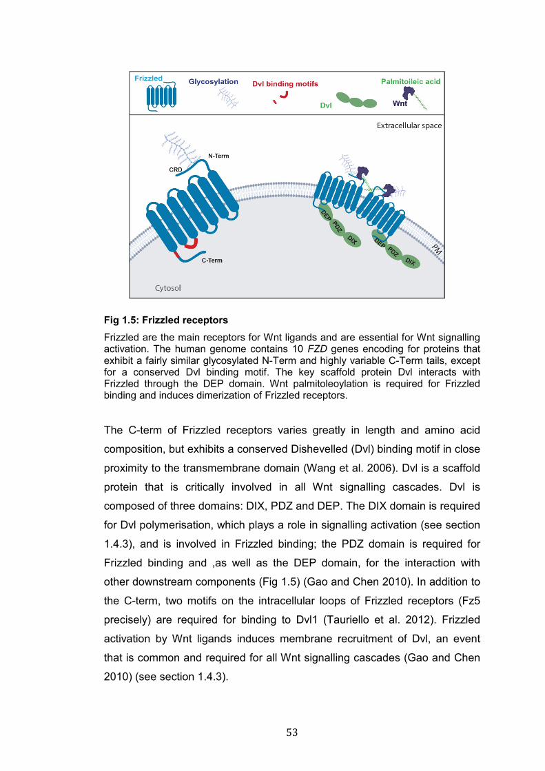
53
Fig 1.5: Frizzled receptors Frizzled are the main receptors for Wnt ligands and are essential for Wnt signalling activation. The human genome contains 10 FZD genes encoding for proteins that exhibit a fairly similar glycosylated N-Term and highly variable C-Term tails, except for a conserved Dvl binding motif. The key scaffold protein Dvl interacts with Frizzled through the DEP domain. Wnt palmitoleoylation is required for Frizzled binding and induces dimerization of Frizzled receptors.
The C-term of Frizzled receptors varies greatly in length and amino acid
composition, but exhibits a conserved Dishevelled (Dvl) binding motif in close
proximity to the transmembrane domain (Wang et al. 2006). Dvl is a scaffold
protein that is critically involved in all Wnt signalling cascades. Dvl is
composed of three domains: DIX, PDZ and DEP. The DIX domain is required
for Dvl polymerisation, which plays a role in signalling activation (see section
1.4.3), and is involved in Frizzled binding; the PDZ domain is required for
Frizzled binding and ,as well as the DEP domain, for the interaction with
other downstream components (Fig 1.5) (Gao and Chen 2010). In addition to
the C-term, two motifs on the intracellular loops of Frizzled receptors (Fz5
precisely) are required for binding to Dvl1 (Tauriello et al. 2012). Frizzled
activation by Wnt ligands induces membrane recruitment of Dvl, an event
that is common and required for all Wnt signalling cascades (Gao and Chen
2010) (see section 1.4.3).
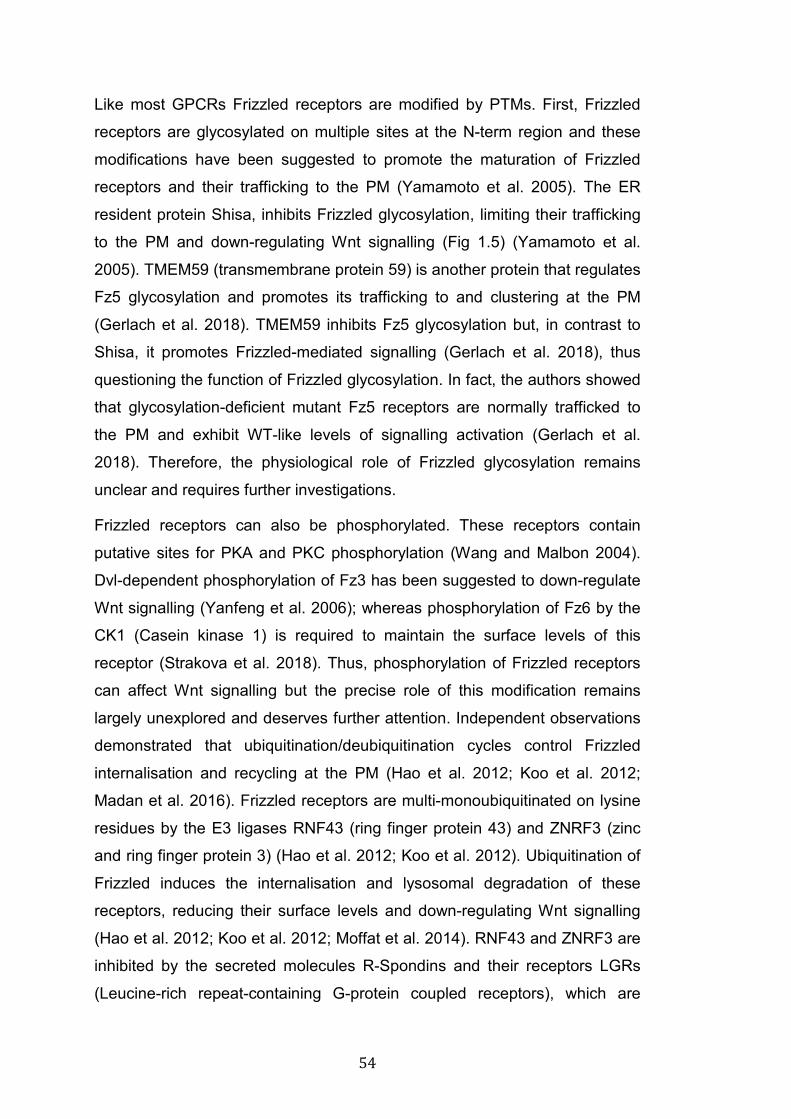
54
Like most GPCRs Frizzled receptors are modified by PTMs. First, Frizzled
receptors are glycosylated on multiple sites at the N-term region and these
modifications have been suggested to promote the maturation of Frizzled
receptors and their trafficking to the PM (Yamamoto et al. 2005). The ER
resident protein Shisa, inhibits Frizzled glycosylation, limiting their trafficking
to the PM and down-regulating Wnt signalling (Fig 1.5) (Yamamoto et al.
2005). TMEM59 (transmembrane protein 59) is another protein that regulates
Fz5 glycosylation and promotes its trafficking to and clustering at the PM
(Gerlach et al. 2018). TMEM59 inhibits Fz5 glycosylation but, in contrast to
Shisa, it promotes Frizzled-mediated signalling (Gerlach et al. 2018), thus
questioning the function of Frizzled glycosylation. In fact, the authors showed
that glycosylation-deficient mutant Fz5 receptors are normally trafficked to
the PM and exhibit WT-like levels of signalling activation (Gerlach et al.
2018). Therefore, the physiological role of Frizzled glycosylation remains
unclear and requires further investigations.
Frizzled receptors can also be phosphorylated. These receptors contain
putative sites for PKA and PKC phosphorylation (Wang and Malbon 2004).
Dvl-dependent phosphorylation of Fz3 has been suggested to down-regulate
Wnt signalling (Yanfeng et al. 2006); whereas phosphorylation of Fz6 by the
CK1 (Casein kinase 1) is required to maintain the surface levels of this
receptor (Strakova et al. 2018). Thus, phosphorylation of Frizzled receptors
can affect Wnt signalling but the precise role of this modification remains
largely unexplored and deserves further attention. Independent observations
demonstrated that ubiquitination/deubiquitination cycles control Frizzled
internalisation and recycling at the PM (Hao et al. 2012; Koo et al. 2012;
Madan et al. 2016). Frizzled receptors are multi-monoubiquitinated on lysine
residues by the E3 ligases RNF43 (ring finger protein 43) and ZNRF3 (zinc
and ring finger protein 3) (Hao et al. 2012; Koo et al. 2012). Ubiquitination of
Frizzled induces the internalisation and lysosomal degradation of these
receptors, reducing their surface levels and down-regulating Wnt signalling
(Hao et al. 2012; Koo et al. 2012; Moffat et al. 2014). RNF43 and ZNRF3 are
inhibited by the secreted molecules R-Spondins and their receptors LGRs
(Leucine-rich repeat-containing G-protein coupled receptors), which are
55
potent activators of Wnt signalling (de Lau et al. 2014; Hao et al. 2016). R-
Spondins bind to both RNF43/ZNRF3 and LGRs forming a trimeric complex
(Hao et al. 2012). The binding of R-spondin causes ZNRF3 internalisation,
inhibiting Frizzled ubiquitination and increasing the surface levels of these
receptors (Hao et al. 2012). Consistently, loss of function mutations of
RNF43 and ZNRF3 increase surface levels of Frizzled receptors resulting in
aberrant activation of Wnt signalling, which is associated with cancer
development (Hao et al. 2016; Katoh and Katoh 2017; Zeng et al. 2018).
Thus, mounting evidence indicates that ubiquitination of Frizzled receptors
plays a crucial role in Wnt signalling modulation.
The role of receptor endocytosis is a controversial topic in the Wnt signalling
field (Gagliardi et al. 2008). Wnt-induced Clathrin and Caveolin-mediated
endocytosis of Wnt receptors (Frizzled and LRP6) are required for signalling
activation (Seto and Bellen 2006; Yamamoto et al. 2006; Gagliardi et al.
2014; Hagemann et al. 2014). However, endocytosis is also a mechanism to
reduce surface levels of Wnt receptors and dampen Wnt signalling (Hao et al.
2012; Koo et al. 2012; Moffat et al. 2014; Madan et al. 2016; Agajanian et al.
2019). Therefore, whether endocytosis activates or inhibits Wnt signalling
seems to depend on the cellular context, and the molecular mechanisms that
regulate Frizzled membrane trafficking remain poorly undersood.
In summary, Frizzled are the main receptors for Wnt ligands and are
essential for Wnt signalling activation. The function of these receptors is
modulated by PTMs; in particular, glycosylation, phosphorylation and
ubiquitination have been proposed to regulate the trafficking and signalling of
these receptors. However, the molecular mechanisms that regulate Frizzled
trafficking and their activity are still poorly understood and require further
investigation. During my PhD, I have studied a novel mechanism of
regulation of Frizzled trafficking and function.
The co-receptor LRP6 and other Wnt receptors
At the cell surface, Wnts bind to a variety of receptors beside the
aforementioned Frizzled. LRP6 (Low-density lipoprotein receptor-related
56
protein 6) is a single pass transmembrane protein that acts as a co-receptor
for Wnts in the canonical β-catenin pathway (see section 1.4.3). LRP6 has a
long N-term extracellular domain which can bind multiple Wnt ligands, some
of which simultaneously (Bourhis et al. 2010), and the Wnt inhibitors Dkk1
and Dkk2 (B T MacDonald and He 2012). Given the ability of binding Wnt
ligands and Wnt antagonists, LRP6 is a fundamental regulator of Wnt
signalling activation. In addition, the C-term of LRP6, which contains multiple
phosphorylation sites, is essential for signalling transduction (Tamai et al.
2004; MacDonald et al. 2009). Wnt binding to LRP6 and Frizzled receptors
induces the formation of a multimeric complex, which induces the PM
recruitment of Dvl and other components of the Wnt cascade, including the
scaffold protein Axin, the kinases CK1 and Gsk3β (glycogen synthase kinase
3 (see section 1.4.3) (Cong et al. 2004; He et al. 2004; Gao and Chen 2010).
The role of LRP6 in Wnt signalling will be examined in more details in the
context of the β-catenin pathway (see section 1.4.3). Beside Wnt signalling
activation, LRP6 plays a fundamental role in lipid metabolism mediating
cholesterol homeostasis (Go and Mani 2012).
Wnts can also bind to the tyrosine kinase receptors Ror and Ryk. Ror
receptors contain a N-term Frizzled-like CRD domain that is required for Wnt
binding, whereas Ryk receptors bind Wnt ligands through an extracellular
domain similar to the Wnt inhibitor WIF (Green et al. 2014). Wnt binding to
these receptors primes the formation of a complex between ligand, receptor
(Frizzled) and co-receptor (Ror or Ryk), similarly to the interaction with LRP6.
Signalling through Ror or Ryk has been shown to activate the planar cell
polarity (PCP) pathway through the membrane recruitment of Dvl (see
section 1.4.3) (Komiya and Habas 2008; Gao and Chen 2010; Minami et al.
2010). By signalling through this cascade, Ror and Ryk are fundamental
regulators of tissue morphogenesis and play important functions in CNS
development (Minami et al. 2010; Green et al. 2014). Moreover, the GPCR
Gpr124 (Probable G-protein coupled receptor 124) and the metallo-protease
inhibitor Reck (reversion inducing cysteine rich protein with kazal motifs) are
important co-receptors for Wnt7a/Wnt7b and are essential for the
development of the BBB (blood brain barrier) (Cho et al. 2017). Reck binds to
57
Gpr124, which in turn binds to Frizzled receptors facilitating the formation of
a signalling complex (Cho et al. 2017). In addition, recent work has shown
that Wnt7a/b can directly bind Reck, which then associates with other
receptors including Frizzled and Gpr124 (Eubelen et al. 2018; Vallon et al.
2018). Thus, Wnt binding to a variety of different plasma membrane
receptors and co-receptors results in a vast signalling diversification. In the
next session, I will introduce the Wnt signalling cascades activated
downstream of surface receptors.
1.4.3 Wnt signalling pathways
Canonical or β-catenin Wnt signalling
Canonical Wnt signalling is the most characterised Wnt signalling pathway.
The key features of this cascade are the stabilisation of cytoplasmic β-
catenin by inhibition of the β-catenin destruction complex, and the
consequent activation of transcription factors that regulate the expression of
Wnt target genes (Clevers 2006; Nusse and Clevers 2017).
In the canonical Wnt cascade, a series of signalling events determine
whether β-catenin is degraded, down-regulating Wnt signalling, or whether it
accumulates in the cell promoting signalling transduction. Gsk3β and CK1,
together with the scaffold protein Axin, the cytoplasmic protein APC
(Adenomatus polyposis coli), PP2A (protein phosphatase 2A) and the E3
ubiquitin ligase β-TrCP (Beta-Transducin Repeat Containing E3 Ubiquitin
Protein Ligase) are part of the β-catenin destruction complex. In the
abscence of Wnts, the kinases of this complex phosphorylate β-catenin
targeting it for ubiquitination by β-TrCP and consequent proteasomal
degradation (Kimelman and Xu 2006; Stamos and Weis 2013).
The canonical pathway is initiated when Wnt binds to Frizzled and LRP6, an
event that triggers the membrane recruitment of Dvl, promoting the formation
of a signalling complex and the disassembly of the destruction complex
(Komiya and Habas 2008; Nusse and Clevers 2017). Next, LRP6 is
phosphorylated by Gsk3β and CK1, and Axin is recruited to the PM where it
interacts with the phosphorylated C-term tail of LRP6 (Davidson et al. 2005;
58
Zeng et al. 2005). Upon ligand binding and phosphorylation of LRP6 the
destruction complex is inhibited and cytoplasmic β-catenin is stabilised.
Several models have proposed different sequences of events downstream of
LRP6 activation that lead to β-catenin stabilisation: 1) in the initiation-
amplification model is a feed-forward loop in which the formation of the
LRP6-Frizzled-Dvl complex at the PM recruits Axin, which promotes GSK3β-
mediated phosphorylation of LRP6, resulting in the recruitment of more Axin
to the PM (MacDonald et al. 2008; Wolf et al. 2008). 2) In the signalosome
model, DIX-domain-dependent polymerisation of Dvl promotes the clustering
of multiple Frizzled-LRP6 complexes, which results in Axin recruitment and
LRP6 phosphorylation by Gsk3β and CK1 (Schwarz-Romond, Fiedler, et al.
2007). Axin also contain a DIX domain, proposed to polymerise with Dvl
(Schwarz-Romond, Metcalfe, et al. 2007). 3) The endosomal signalling
model shares similarity with the signalosome model, but it proposes that
assembly of the signalosome and signalling transduction occurs upon
Caveolin-mediated internalisation of LRP6 and Frizzled receptors
(Yamamoto et al. 2006; Bryan T MacDonald and He 2012b). However,
canonical Wnt signalling is up-regulated in intestinal stem cells of Caveolin-\-
mice, raising questions as to whether this model is restricted to specific
cellular contexts (Li et al. 2005; Sotgia et al. 2005; Gagliardi et al. 2008;
Bryan T MacDonald and He 2012b). 4) Another model suggests that Dvl
activates PI4KIIα (phosphatidylinositol 4-kinase type II) and
PIP5KI (phosphatidylinositol-4-phosphate 5-kinase type I) leading to the
production of PIP2 (phosphatidylinositol 4,5-bisphosphate). PIP2, through
unknown molecular mechanisms, promotes the clustering and
phosphorylation of LRP6 by Gsk3β and CK1 (Pan et al. 2008). Thus, in spite
of huge progress, the precise molecular mechanisms by which the canonical
Wnt signalling is activated remain to be fully elucidated.
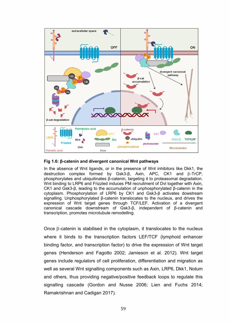
59
Fig 1.6: β-catenin and divergent canonical Wnt pathways In the absence of Wnt ligands, or in the presence of Wnt inhibitors like Dkk1, the destruction complex formed by Gsk3-β, Axin, APC, CK1 and β-TrCP, phosphorylates and ubiquitinates β-catenin, targeting it to proteasomal degradation. Wnt binding to LRP6 and Frizzled induces PM recruitment of Dvl together with Axin, CK1 and Gsk3-β, leading to the accumulation of unphosphorylated β-catenin in the cytoplasm. Phosphorylation of LRP6 by CK1 and Gsk3-β activates dowstream signalling. Unphosphorylated β-catenin translocates to the nucleus, and drives the expression of Wnt target genes through TCF/LEF. Activation of a divergent canonical cascade downstream of Gsk3-β, independent of β-catenin and transcription, promotes microtubule remodelling.
Once β-catenin is stabilised in the cytoplasm, it translocates to the nucleus
where it binds to the transcription factors LEF/TCF (lymphoid enhancer
binding factor, and transcription factor) to drive the expression of Wnt target
genes (Henderson and Fagotto 2002; Jamieson et al. 2012). Wnt target
genes include regulators of cell proliferation, differentiation and migration as
well as several Wnt signalling components such as Axin, LRP6, Dkk1, Notum
and others, thus providing negative/positive feedback loops to regulate this
signalling cascade (Gordon and Nusse 2006; Lien and Fuchs 2014;
Ramakrishnan and Cadigan 2017).
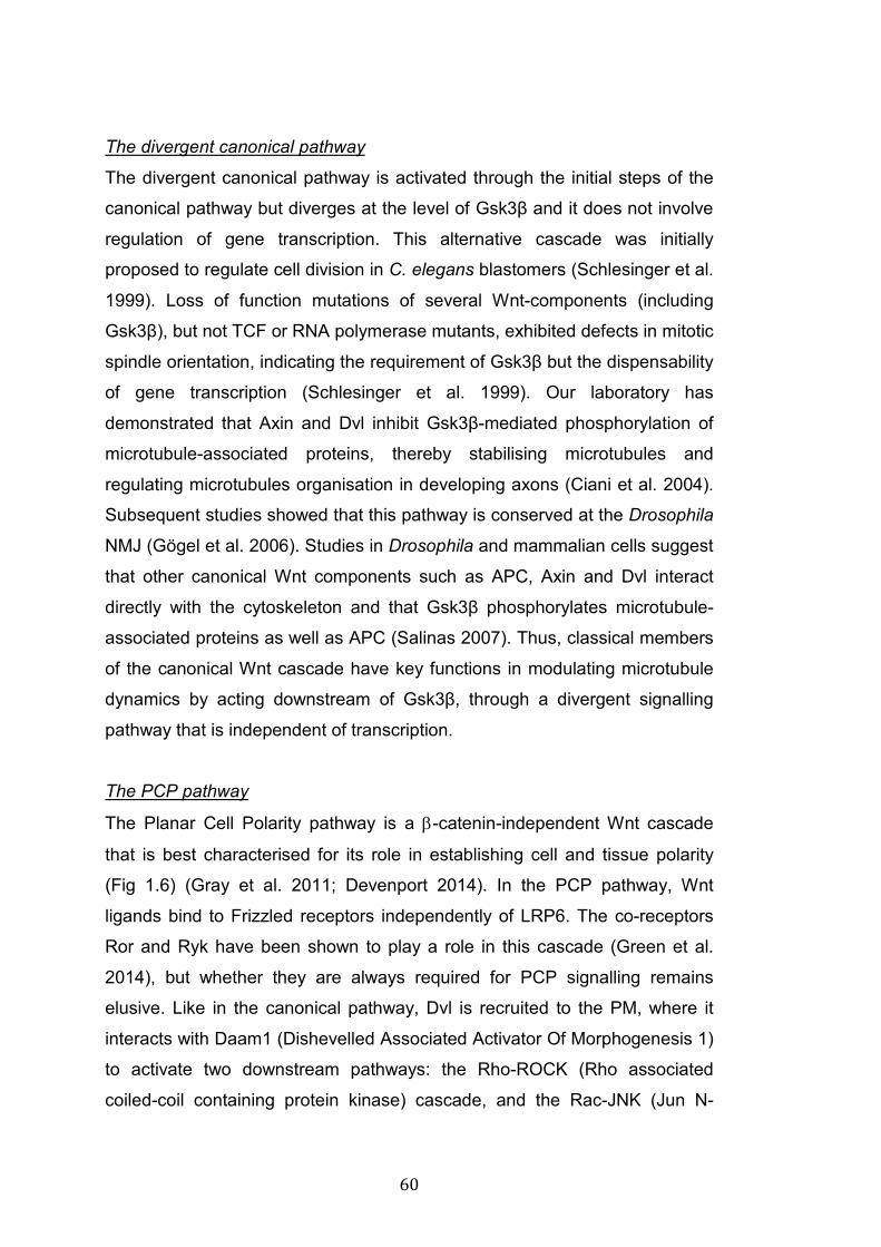
60
The divergent canonical pathway
The divergent canonical pathway is activated through the initial steps of the
canonical pathway but diverges at the level of Gsk3β and it does not involve
regulation of gene transcription. This alternative cascade was initially
proposed to regulate cell division in C. elegans blastomers (Schlesinger et al.
1999). Loss of function mutations of several Wnt-components (including
Gsk3β), but not TCF or RNA polymerase mutants, exhibited defects in mitotic
spindle orientation, indicating the requirement of Gsk3β but the dispensability
of gene transcription (Schlesinger et al. 1999). Our laboratory has
demonstrated that Axin and Dvl inhibit Gsk3β-mediated phosphorylation of
microtubule-associated proteins, thereby stabilising microtubules and
regulating microtubules organisation in developing axons (Ciani et al. 2004).
Subsequent studies showed that this pathway is conserved at the Drosophila
NMJ (Gögel et al. 2006). Studies in Drosophila and mammalian cells suggest
that other canonical Wnt components such as APC, Axin and Dvl interact
directly with the cytoskeleton and that Gsk3β phosphorylates microtubule-
associated proteins as well as APC (Salinas 2007). Thus, classical members
of the canonical Wnt cascade have key functions in modulating microtubule
dynamics by acting downstream of Gsk3β, through a divergent signalling
pathway that is independent of transcription.
The PCP pathway
The Planar Cell Polarity pathway is a β-catenin-independent Wnt cascade
that is best characterised for its role in establishing cell and tissue polarity
(Fig 1.6) (Gray et al. 2011; Devenport 2014). In the PCP pathway, Wnt
ligands bind to Frizzled receptors independently of LRP6. The co-receptors
Ror and Ryk have been shown to play a role in this cascade (Green et al.
2014), but whether they are always required for PCP signalling remains
elusive. Like in the canonical pathway, Dvl is recruited to the PM, where it
interacts with Daam1 (Dishevelled Associated Activator Of Morphogenesis 1)
to activate two downstream pathways: the Rho-ROCK (Rho associated
coiled-coil containing protein kinase) cascade, and the Rac-JNK (Jun N-
61
terminal kinase) pathway. These kinases phosphorylate a plethora of
downstream targets; therefore, the precise molecular interactions
downstream of ROCK and JNK depend on the specific cellular context
(Amano et al. 2010; Zeke et al. 2016). The functional outcome of Rho-ROCK
and Rac-JNK signalling is the remodelling of cytoskeletal structures
(Schlessinger et al. 2009).
Fig 1.7: PCP and Wnt/Ca2+ pathways A: In the PCP pathway, Wnt binding to Frizzled and the co-receptors Ryk and Ror activates the small GTPases Rho and Rac, which in turn activate ROCK and JNK driving remodelling of the actin cytoskeleton. B: In the Wnt/Ca2+ pathway, Wnt binding to Frizzled activates G proteins and PLC, which acts on PIP2 and DAG to produce IP3. IP3 induces Ca2+ release from the ER activating the kinases PKC and CaMKII. Downstream signalling events include NFkB and CREB activation in the nucleus to drive gene transcription.
The Wnt/Ca2+ pathway
The Wnt Ca2+ pathway is another β-catenin-independent Wnt cascade (Fig
1.6). In this signalling pathway, Wnt ligands bind to Frizzled and in some
circumstances to co-receptors, such as Knypek and Ror2, inducing
recruitment of Dvl in a G protein dependent manner and activating
Phospholipase C (PLC). PLC acts on PIP2 (Phosphatidylinositol 4,5-
62
bisphosphate) increasing the levels of the second messengers IP3 (Inositol
trisphosphate) and DAG (diacylglycerol). IP3 induces the rapid release of
Ca2+ from the ER activating Ca2+-dependent kinases such as PKC and
CaMKII (Kühl et al. 2000; Sheldahl et al. 2003), which can signal to a number
of downstream effectors including the transcription factors CREB (cAMP
response element-binding protein) and NFkB (nuclear factor kappa-light-
chain-enhancer of activated B cells). Ca2+ signalling plays an important role
during embryogenesis, CNS development and cancer (Komiya and Habas
2008; De 2011).
Frizzled nuclear import pathway
This Wnt pathway is distinguished from all the other Wnt cascades for the
mechanism of Frizzled signalling (Fig 1.7). This cascade was described at
the Drosophila NMJ to regulate synapse formation and function (Koles and
Budnik 2012). Wg binding to DFz2, which is the homolog of human Fz5 and
Fz8, causes receptor internalisation and cleavage (Mathew et al. 2005).
DFz2 is cleaved at the C-term domain and this portion of protein trafficks to
the nucleus in a GRIP1 (glutamate receptor interacting protein 1) dependent
manner, whereas the rest of the DFz2 accumulates outside the nucleus
(Ataman et al. 2006). Wg, DFz2 or GRIP1 null flies exhibit the same defects
in synapse formation and function (Ataman 2006). The molecular
mechanisms that control DFz2 nuclear function are unclear, but its nuclear
import is followed by nuclear export of large complexes of transcripts for
synaptic proteins (Speese et al. 2012). This mechanism of cleavage and
nuclear import is unusual for Frizzled receptors but it is a quite common
mode of action for other types of molecules such as tyrosine kinase
receptors (Carpenter and Liao 2013; Chen and Hung 2015). For instance,
Ryk is cleaved at the C-term and this portion of protein is transferred to the
nucleus to regulate the differentiation of neuronal progenitor cells in the
cortex (Lyu et al. 2008). Thus, Wnt-induced receptor cleavage and nuclear
import seem to be additional Wnt signalling routes; however, further
experiments are needed to address the role of this pathway in other
biological contexts and in other organisms.
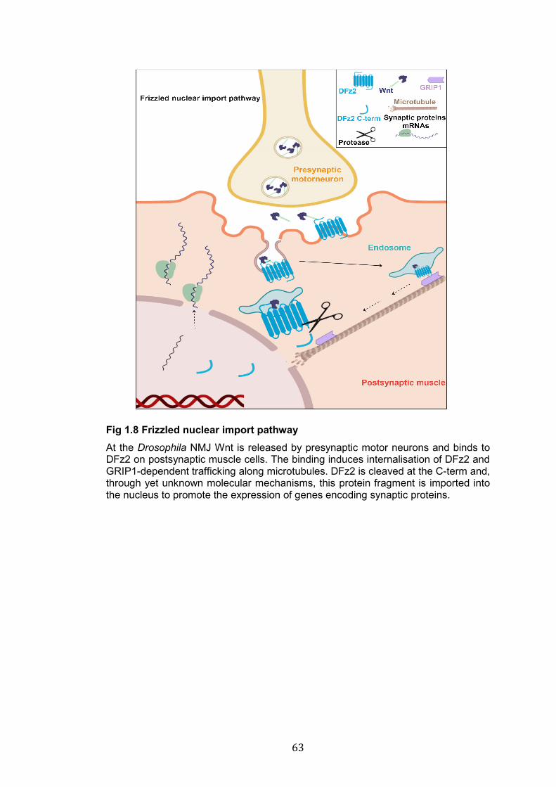
63
Fig 1.8 Frizzled nuclear import pathway At the Drosophila NMJ Wnt is released by presynaptic motor neurons and binds to DFz2 on postsynaptic muscle cells. The binding induces internalisation of DFz2 and GRIP1-dependent trafficking along microtubules. DFz2 is cleaved at the C-term and, through yet unknown molecular mechanisms, this protein fragment is imported into the nucleus to promote the expression of genes encoding synaptic proteins.
64
1.5 Wnt signalling in neural development
Wnt signalling is involved in many key aspects of neural development and
these functions are highly conserved across different species. From very
early embryonic stages, Wnts promote the development of the neural tube,
which is the precursor structure of the brain, and trigger site-directed
migration of multipotent stem cells from the dorsal part of the neural tube,
which is the neural crest (Ikeya et al. 1997; Dorsky et al. 1998; Pinson et al.
2000; Kiecker and Niehrs 2001; De Calisto et al. 2005; Bocchi et al. 2017).
Once these multipotent stem cells have migrated and reached the
appropriate location, Wnt signalling is required for their differentiation into
mature neurons (Dickinson et al. 1994; Megason and McMahon 2002;
Chesnutt et al. 2004; Hirabayashi et al. 2004). In differentiated neurons Wnts
play a fundamental role in establishing neuronal polarity and promoting the
growth of neuronal processes (Ciani and Salinas 2005; Salinas and Zou
2008; C.-W. He et al. 2018). Numerous pieces of evidence show that Wnt
signalling plays a role in axon growth, guidance and branching in different
model systems and in different areas of the CNS and PNS (Lucas and
Salinas 1997; Krylova et al. 2002; Lyuksyutova et al. 2003; Yoshikawa et al.
2003; Rodriguez et al. 2005; Keeble et al. 2006; Hua et al. 2014). In addition,
Wnts are also required for activity-mediated dendritic growth and branching
(Yu and Malenka 2003; Rosso et al. 2005; Wayman et al. 2006; Kirszenblat
et al. 2011; Lanoue et al. 2017; Ferrari et al. 2018).
In late embryonic development and early postnatal life, Wnt signalling plays a
crucial role in synapse formation by modulating the assembly of pre- and
postsynaptic sites (Budnik and Salinas 2011; Salinas 2012). Wnt signalling is
also implicated in basal synaptic transmission as well as short- and long-term
synaptic plasticity (Oliva et al. 2013a; McLeod and Salinas 2018). In the adult,
Wnts control neurogenesis (Lie et al. 2005; Varela-Nallar and Inestrosa
2013) and mechanisms of synaptic maintenance (Inestrosa and Arenas
2010; Dickins and Salinas 2013; Purro et al. 2014; Buechler and Salinas
2018). Thus, Wnt signalling plays different roles at synapses, regulating their
assembly during development to controlling their function and maintenance
during adulthood.
65
Wnt signalling not only promotes the development and function of neuronal
cells, but it is crucial for several other cell types in the brain. In particular, Wnt
signalling is fundamental for the formation of the BBB (Liebner et al. 2008;
Polakis 2008; Zhou and Nathans 2014; Cho et al. 2017), and regulates the
maturation and function of oligodendrocytes, astrocytes and microglia
(Halleskog et al. 2011; Yang et al. 2012; Tang 2014; Yao et al. 2016; Zheng
et al. 2017; Soomro et al. 2018). In light of such a profound involvement at
different levels of neural development, maturation, and function, it is not
surprising that aberrant Wnt signalling is linked to several neurological
disorders including brain patterning abnormalities, psychiatric conditions
such as schizophrenia and bipolar disorders, and neurodegenerative
pathologies like Alzheimer’s (AD) and Parkinson’s disease (Berwick and
Harvey 2012; Panaccione et al. 2013; Inestrosa and Varela-Nallar 2014;
Harvey and Marchetti 2014; Purro et al. 2014; Mulligan and Cheyette 2017;
Hoseth et al. 2018).
66
1.6 Wnt signalling at the synapse
The role of Wnts at synapses was first described in the context of
synaptogenesis, but studies in the last 20 years have also revealed a role for
Wnt signalling in synaptic transmission, synaptic plasticity and synapse
maintenance in adulthood. These key roles of Wnt signalling are directly
translated into functional outcomes, such as the regulation of cognitive
functions and complex behaviours. In the following sections, I will introduce
evidence supporting the role of Wnt signalling in these processes.
1.6.1 Wnt signalling in synaptogenesis
Wnt molecules are potent synaptogenic factors able to induce pre and
postsynaptic assembly. Our laboratory first described that exposure to Wnt7a
increases the clustering of presynaptic proteins in cultured cerebellar granule
cells (Lucas and Salinas 1997). These findings were soon corroborated by in
vivo studies demonstrating that Wnt7a null mice exhibit defects in
presynaptic organisation in the cerebellum, precisely at the mossy fibre-
granule cell synapse (Hall et al. 2000). In primary hippocampal cultures,
Wnt7a/Wnt7b promote the assembly of excitatory presynaptic terminals
without affecting inhibitory synapse formation (Davis et al. 2008; Ciani et al.
2011). Expression of Fz5 in hippocampal neurons is sufficient to promote
presynaptic assembly, whereas loss of function of this receptor reduces the
number of presynaptic sites (Fig 1.9) (Sahores et al. 2010). Moreover, Fz5
receptors, which are present along axons, are required for Wnt7a-mediated
presynaptic assembly (Fig 1.9) (Sahores et al. 2010). Furthermore, Wnt7a
signalling, through removal of APC from the β-catenin destruction complex,
has been suggested to promote clustering of nicotinic acetylcholine receptors
at presynaptic site of hippocampal neurons (Farías et al. 2007).
Other Wnt ligands signal through different Wnt receptors to promote
presynaptic assembly. For instance, the co-receptor LRP6 is a key regulator
of synapse formation. Lrp6 KO mice are embryonically lethal, but in vivo KD
of LRP6 results in a reduction of excitatory pre- and postsynaptic terminals
(Sharma et al. 2013). In addition, Wnt3a through LRP6 and Fz1 receptors
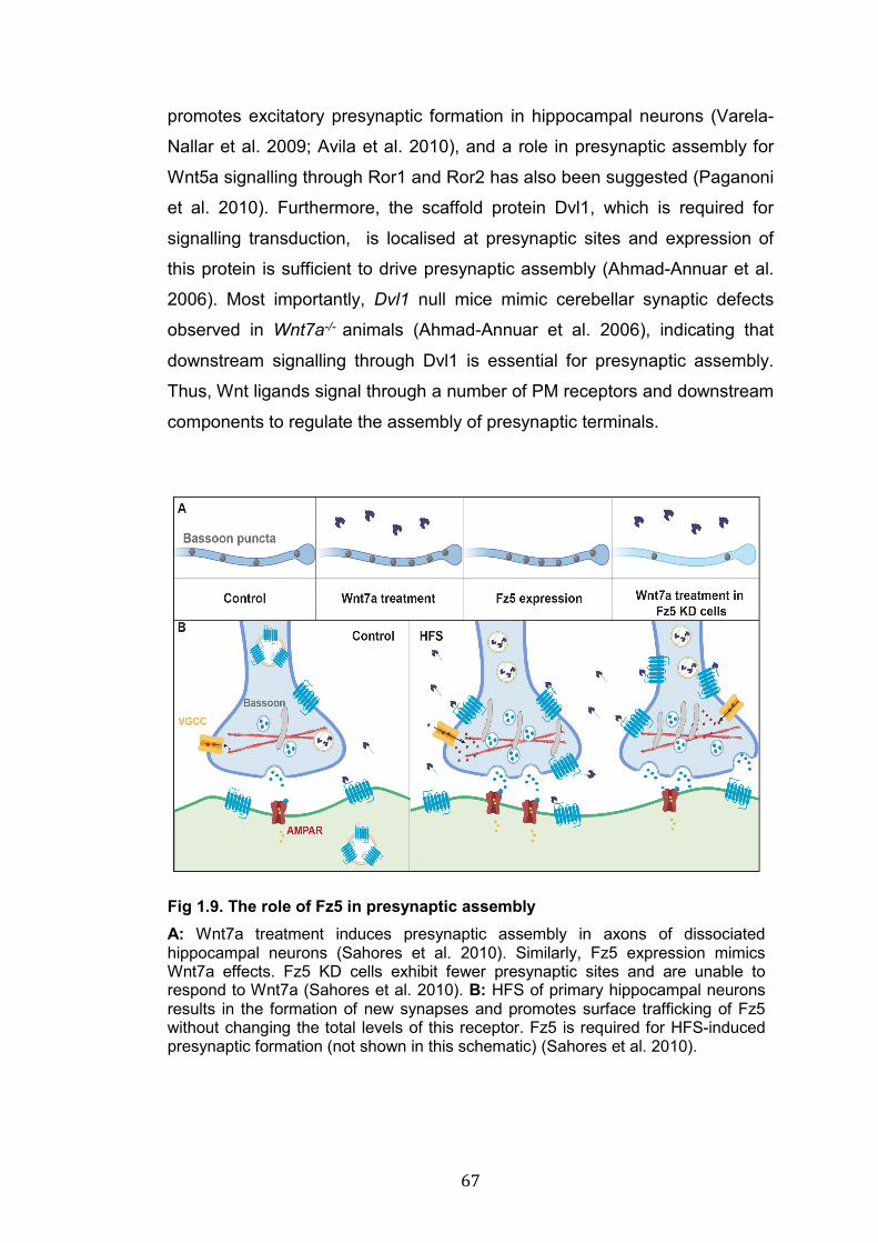
67
promotes excitatory presynaptic formation in hippocampal neurons (Varela-
Nallar et al. 2009; Avila et al. 2010), and a role in presynaptic assembly for
Wnt5a signalling through Ror1 and Ror2 has also been suggested (Paganoni
et al. 2010). Furthermore, the scaffold protein Dvl1, which is required for
signalling transduction, is localised at presynaptic sites and expression of
this protein is sufficient to drive presynaptic assembly (Ahmad-Annuar et al.
2006). Most importantly, Dvl1 null mice mimic cerebellar synaptic defects
observed in Wnt7a-/- animals (Ahmad-Annuar et al. 2006), indicating that
downstream signalling through Dvl1 is essential for presynaptic assembly.
Thus, Wnt ligands signal through a number of PM receptors and downstream
components to regulate the assembly of presynaptic terminals.
Fig 1.9. The role of Fz5 in presynaptic assembly A: Wnt7a treatment induces presynaptic assembly in axons of dissociated hippocampal neurons (Sahores et al. 2010). Similarly, Fz5 expression mimics Wnt7a effects. Fz5 KD cells exhibit fewer presynaptic sites and are unable to respond to Wnt7a (Sahores et al. 2010). B: HFS of primary hippocampal neurons results in the formation of new synapses and promotes surface trafficking of Fz5 without changing the total levels of this receptor. Fz5 is required for HFS-induced presynaptic formation (not shown in this schematic) (Sahores et al. 2010).
68
Wnt signalling can also promote the formation of postsynaptic sites. For
example, Wnt7a signals to the postsynaptic compartment to increase the
number and size of dendritic spines, where excitatory synapses are mainly
formed (Ciani et al. 2011). In contrast, Wnt7a does not affect the formation of
inhibitory postsynaptic sites (Ciani et al. 2011) – a function that involves
another Wnt ligand. Namely, Wnt5a ptomotes GABAAR clustering at
inhibitory synapses (Cuitino et al. 2010). What is the downstream signalling
involved in Wnt7a-mediated spine formation? The receptor that regulates
Wnt7a signalling at dendritic spines site was unknown when I started my
PhD. Data describing the role of two Wnt7a receptors (Fz5 and Fz7) at
postsynaptic compartments will be presented in chapter 3 of this thesis.
Downstream of Wnt receptors, Dvl1 is enriched at dendritic spines and its
expression is sufficient to promote synapse growth in number and size (Ciani
et al. 2011). Furthermore, Dvl1 is required for Wnt7a to induce postsynaptic
development, and Wnt7a-/-;Dvl1-/- double KO mice exhibit reduced spine
number and size in pyramidal neurons of the hippocampus (Ciani et al. 2011).
Wnt7a-Dvl1 signalling requires CaMKII activation to increase dendritic spine
number and size (Ciani et al. 2011). Wnt7a-induced activation of CaMKII not
only affects spine development but it also regulates the synaptic and surface
recruitment of AMPARs (Ciani et al. 2011; McLeod et al. 2018). The
activation of CaMKII, the delivery of AMPAR at the cell surface, and the
increase in spine size occur very rapidly (within 10 mins of exposure to
Wnt7a), suggesting that these are transcription independent effects (Ciani et
al. 2011; McLeod et al. 2018). Thus, Wnt7a is a potent synaptogenic factor
that signals bidirectionally to promote the assembly of pre- and postsynaptic
sites.
Other Wnts and Wnt receptors have been proposed to regulate the
development of excitatory postsynaptic sites but the data is controversial. In
primary hippocampal neurons Wnt5a rapidly induces PSD-95 clustering
through activation of JNK (Farías et al. 2009), and it has been suggested to
increase dendritic spine number and size (Varela-Nallar et al. 2010).
However, Wnt5a effects on spines were not observed in the previous report
from the same group (Farías et al. 2009). Importantly, analyses of Wnt5a
69
cKO revealed no changes in the formation of dendritic spines (Chen et al.
2017), raising questions on the actual role of Wnt5a at spines. The co-
receptor LRP6, which is required for presynaptic formation, is localised at
dendritic spines and is necessary for PSD-95 clustering and spine formation,
but it does not affect the assembly of inhibitory synapses (Sharma et al.
2013). Thus, Wnt signalling is a key regulator of postsynaptic development
as it promotes the formation of dendritic spines and controls the localisation
and function of several key postsynaptic proteins.
The study of the development of the NMJ has considerably enriched our
understanding of the molecular mechanisms of Wnt-induced synapse
formation. At the Drosophila NMJ, Wg is released by presynaptic motor
neurons and is essential for the formation of proper synaptic boutons
(Packard et al. 2002). Wg-/- flies exhibit abnormal presynaptic boutons, called
“ghost boutons”, which lack key presynaptic components such as
mitochondria and the active zone (Packard et al. 2002; Ataman et al. 2006).
At the NMJ, Wg is secreted through exosomes together with Wls and
subsequently binds to the postsynaptic receptor DFz2 (Korkut et al. 2009).
Wg binding to DFz2 induces the internalisation and cleavage of this receptor,
resulting in the nuclear import of DFz2 C-term domain (Mathew et al. 2005;
Ataman et al. 2006), which is then followed by nuclear export of large
complexes of synaptic protein transcripts (Fig 1.8) (Speese et al. 2012).
In vertebrates, Wnt signalling is also required at the NMJ, where it acts
through an interplay with the potent secreted synaptogenic factor Agrin and
its receptor MuSK (Muscle-specific kinase) to regulate the assembly of
acetylcholine receptors (AChRs) (Wu et al. 2010; Budnik and Salinas 2011).
Agrin-/- and Musk-/- mice exhibit severe defects in NMJ development and
AChRs clustering (DeChiara et al. 1996; Gautam et al. 1996). MuSK
receptors, which bind to Dvl1 (Luo et al. 2002), contain a Frizzled-like CRD
domain that allows their direct interaction with certain Wnt ligands (Jing et al.
2009). Wnt3, which is expressed by presynaptic motor neurons (Krylova et al.
2002), activates MuSK-Dvl1-Rac on muscle cells to induce the initial
clustering of AChRs on the postsynaptic side; Agrin is then required to
stabilise these AChRs clusters (Henriquez et al. 2008). Other molecular
70
interplays between Agrin and Wnt signalling occur at the level of APC and
LRP4, a single pass receptor belonging to the same family of LRP5/6
receptors. APC interacts with AChRs receptors and is necessary for their
clustering, a function that is conserved in central neurons (Temburni et al.
2004; Farías et al. 2007; Rosenberg et al. 2008). Lrp4-/- mice mimic the NMJ
developmental defects of Musk-/- animals (Weatherbee et al. 2006); in fact,
LRP4 is a co-receptor for Agrin and forms a complex with MuSK receptors
that is necessary for the development of the NMJ (Kim et al. 2008; Zhang et
al. 2008). Thus, Wnt signalling is required for the development of central and
peripheral synapses in different model systems.
The findings presented until now support a role for Wnt signalling as positive
regulator of synapse formation; however, Wnt signalling has also been
described as inhibitory signalling for the development of the NMJ in C.
elegans (Park and Shen 2012). In C. elegans, presynaptic sites of the DA9
motorneuron are not present in the tail of the worm, where a gradient of Lin-
44 (C. elegans Wnt) is found (Klassen and Shen 2007). Lin-44 restricts Lin-
17 (C. elegans Frizzled) expression to the tail of the worm, and loss of
function of Lin-44, Lin-17 or Dsh1 (C. elegans Dvl) allows the ectopic
formation of presynaptic sites in the posterior region of the worm, suggesting
that active Wnt signalling is required to inhibit synapse formation (Klassen
and Shen 2007). However, a recent report suggested that Lin-17 has actually
a pro-synaptogenic function that is inhibited by Lin-44 through the induction
of receptor endocytosis and down-regulation of γ-Neurexin, which was
proposed to act in parallel to the Wnt signalling pathway through yet
unspecified molecular mechanisms (Kurshan et al. 2018). The interplay
between Frizzled receptors and Neurexins during synapse formation is a
novel and exciting finding that deserves further investigation in vertebrate
model systems.
In summary, Wnt signalling is a key regulator of central and peripheral
synapses. Wnt ligands promote the formation of both excitatory and inhibitory
synapses, organising the assembly of pre- and postsynaptic compartments.
Several Wnt ligands, in particular Wnt7a, Wnt3a and Wnt5a, are potent
synaptic regulators. However, the receptors and intracellular mechanisms
71
through which Wnt signalling drives synapse assembly remain to be fully
elucidated.
1.6.2 Wnts in synaptic transmission, plasticity and maintenance
In addition to a crucial function in synapse formation, Wnts play a
fundamental role in regulating synaptic transmission, synaptic plasticity and
synaptic maintenance. Wnt signalling at the synapse controls the levels and
dynamics of SVs, the localisation and functions of members of the NT
release machinery, and the localisation of postsynaptic NT receptors
(McLeod and Salinas 2018). Application of exogenous Wnts to hippocampal
neurons increases the rate of SVs recycling, indicating an increment in NT
release (Ahmad-Annuar et al. 2006; Cerpa et al. 2008; Varela-Nallar et al.
2009). Consistently, blockage of endogenous Wnts with SFRPs, or by
genetic suppression of Wnt7a and Dvl1, impairs NT release (Ahmad-Annuar
et al. 2006; Varela-Nallar et al. 2010; Cerpa et al. 2011; Ciani et al. 2011;
Ciani et al. 2015). At presynaptic terminals, Dvl1 interacts with Syt1 (direct)
and other SNAREs (indirect) to regulate NT release (Ciani et al. 2015). At
postsynaptic sites, Wnt ligands increase postsynaptic currents by modulating
the distribution and function of glutamate receptors (Ciani et al. 2011; Mcleod
et al. 2018). Wnt7a increases AMPAR surface levels, enhancing AMPAR-
mediated currents (McLeod et al. 2018), whereas Wnt5a signalling through
Ror2 receptors strengthens NMDAR distribution and transmission and
inhibits the activity of voltage gated K+ channels (Cerpa et al. 2011; Cerpa et
al. 2015; Parodi et al. 2015; McQuate et al. 2017). In addition, at inhibitory
synapses Wnt5a controls GABAAR surface levels thereby increasing
GABAergic transmission (Cuitino et al. 2010). Thus, Wnt ligands shape
excitatory and inhibitory synaptic transmission by acting on both sides of the
synapse.
An interesting aspect of Wnt signalling is the modulation of Wnt components
by neuronal activity. Neuronal activity induces the expression and release of
Wnt components, which are in turn required for activity-induced neuronal and
synaptic plasticity (Budnik and Salinas 2011; Sahores and Salinas 2011;
McLeod and Salinas 2018). For instance, neuronal activity promotes
72
dendritogenesis by inducing the transcription and release of Wnt ligands and
by activating cytoplasmic β-catenin (independently of its nuclear function),
CaMKII and CREB signalling (Yu and Malenka 2003; Wayman et al. 2006).
Moreover, the surface levels of Fz5 are also regulated by neuronal activity. In
cultured neurons, HFS (high frequency stimulation) increases, whereas LFS
(low frequency stimulation) decreases, the levels of surface Fz5, and this
receptor is required for activity-mediated synapse formation (Fig 1.9)
(Sahores et al. 2010). Moreover, induction of LTP by electrical or chemical
stimulation increases Wnt3a and Wnt7a/Wnt7b protein levels at synapses
(Chen et al. 2006; McLeod et al. 2018). Wnt ligands facilitate LTP induction
and blockage of Wnt signalling through SFRPs or Dkk1 inhibits the
expression of LTP (Chen et al. 2006; Cerpa et al. 2011; Marzo et al. 2016;
McLeod et al. 2018). In particular, Wnt5a increases NMDAR surface levels
and increases NMDAR-mediated currents, facilitating LTP induction (Cerpa
et al. 2011; Cerpa et al. 2015; McQuate et al. 2017). Wnt7a regulates the
early signalling events that allow LTP expression by activating CaMKII and
PKA, promoting phosphorylation and surface recruitment of AMPARs and
growth of dendritic spines (McLeod et al. 2018). Furthermore, Wnt signalling
is involved in activity-mediated synapse formation at the NMJ. Patterned
stimulation of neuronal activity induces the formation of presynaptic ghost
boutons in Drosophila motor neurons (Ataman et al. 2008). These events are
respectively facilitated and abolished by the expression or suppression of Wg
(Ataman et al. 2008). In addition, the expression of Wnt7a/Wnt7b ligands is
increased in mice exposed to EE (enriched environment) – a behavioral
paradigm that stimulates neuronal activity and increases synapse formation
(van Praag et al. 2000; Nithianantharajah and Hannan 2006). Importantly,
intracranial application of the Wnt inhibitors SFRPs suppresses EE-induced
synapse formation in the hippocampus, strongly suggesting that Wnt
signalling is required for experience-mediated synapse formation (Gogolla et
al. 2009). Therefore, neuronal activity modulates the levels of Wnt
components, which in turn are fundamental regulators of activity-induced
synaptic plasticity.
73
The central functions played by Wnt signalling at synapses are further
highlighted by the fact that positive or negative modulation of these cascades
result in behavioural and cognitive changes. For instance, inhibition of Wnt
signalling by Dkk1 infusion impairs fear memory consolidation in the
amygdala (Maguschak and Ressler 2011). Similarly, hippocampal-mediated
fear-conditioned learning is inhibited by local application of SFRPs or Dkk1,
and enhanced by exposure to Wnt3a (Xu et al. 2015). In the striatum and
hippocampus of adult mice, induced expression of Dkk1 triggers synapse
disassembly, affects synaptic transmission, plasticity and memory without
affecting cell viability (Galli et al. 2014; Marzo et al. 2016). Thus, Wnt
signalling is a fundamental regulator of synaptic plasticity. By acting on
cellular and molecular mechanisms of synaptic function and plasticity, Wnt
signalling directly impacts on cognitive functions and complex behaviours.
In the adult brain, synapse formation and elimination are in a constant
balance to maintain normal levels of synaptic density but at the same time
allow changes in structural and functional plasticity of synapses during
processes such as learning and memory. A disruption of this balance in
favour of synapse elimination, for example, is presented at early stages of
several neurodegenerative disorders (Selkoe 2002; Palop et al. 2006; Palop
and Mucke 2010; Bellucci et al. 2016). Mounting evidence strongly suggests
a link between deficient canonical Wnt signalling and AD (Inestrosa and
Arenas 2010; Dickins and Salinas 2013; Purro et al. 2014). DKK1 levels are
increased in post-mortem brain of AD patients and Dkk1 expression is
induced by exposure of hippocampal neurons to Amyloid β (Aβ) oligomers
(Caricasole et al. 2004; Purro et al. 2012). Aβ is the main component of the
characteristic extracellular plaques found AD patients’ brains, and Aβ
oligomers are the most toxic form of Aβ, initiating synaptotoxicity in the
process of neurodegeneration (Benilova et al. 2012; Kayed and Lasagna-
Reeves 2013). Dkk1 inhibition with neutralising antibodies prevents Aβ-
mediated synapse disassembly in brain slices, strongly suggesting that Dkk1
acts downstream of Aβ (Purro et al. 2012). In addition, a genetic variant of
the Wnt co-receptor LRP6 has been linked to late onset development of AD
(De Ferrari et al. 2007). Moreover, conditional KO of Lrp6 results in age-

74
dependent synapse loss and exacerbates neurodegeneration in a mouse
model of AD (Liu et al. 2014). Furthermore, the microglia receptor TREM2
(Triggering Receptor Expressed On Myeloid Cells) has been identified as a
risk factor in AD. TREM2 is required to stabilise β-catenin and promote
survival of microglia cells (Zheng et al. 2017), which are key regulators of
synapse function in health and disease (Hong et al. 2016; Salter and Stevens
2017). Thus, Wnt signalling play an important role in synaptic maintenance,
and aberrant Wnt signalling is linked to synapse degeneration.
75
1.7 Receptor localisation and signalling, a role for post-translational modifications
Surface receptors are essential molecules for signalling in every cell of our
organism. Their distribution at the PM determines the spatio-temporal
dynamic of signalling activation. Therefore, studying the molecular
mechanisms that regulate the trafficking and stabilisation of receptors at the
PM is crucial to understand how signalling is modulated. Post-translational
modifications (PTMs) are biochemical changes occurring on one or more
amino acids after protein synthesis. PTMs are master regulator of protein
trafficking and function and they target virtually every protein. The
biochemical nature of these modifications varies greatly, ranging from the
formation of intra-protein disulfide bonds to the covalent attachment of low or
very high molecular weight groups. In spite of profound biochemical
differences, all PTMs share one common feature: they increase the
biochemical properties of proteins beyond those conferred by the amino
acids that constitute these molecules. Dozens of PTMs exist but some, like
phosphorylation and N-glycosylation, are far more common than others
(Khoury et al. 2011). PTMs addition and removal are usually catalyzed by
specific enzymes, which are localised essentially in all subcellular
compartments, even in the extracellular space. The functions of PTMs are
very diverse and reflect the great biochemical variety of these modifications.
Wnt signalling is a good example to illustrate the variety of functions of PTMs.
Palmitoleoylation of Wnt ligands on Cys residues, which is catalyzed in the
ER by Porc, regulates protein secretion and interaction with Frizzled
receptors (Hausmann et al. 2007; Nile and Hannoush 2016). In the
extracellular space the de-acylase enzyme Notum removes palmitoleate
moiety from Wnt ligands thus inhibiting Wnt signalling activation (Kakugawa
et al. 2015). β-catenin phosphorylation induces its ubiquitination and
consequent degradation through the proteasome system (Stamos and Weis
2013). Palmitoylation controls protein folding and exit from the ER of the Wnt
co-receptor LRP6; once at the PM, the turnover rate of LRP6 is controlled by
an interplay between palmitoylation and ubiquitination (Abrami et al. 2008;
Perrody et al. 2016). Frizzled receptors are glycosylated in the ER but the
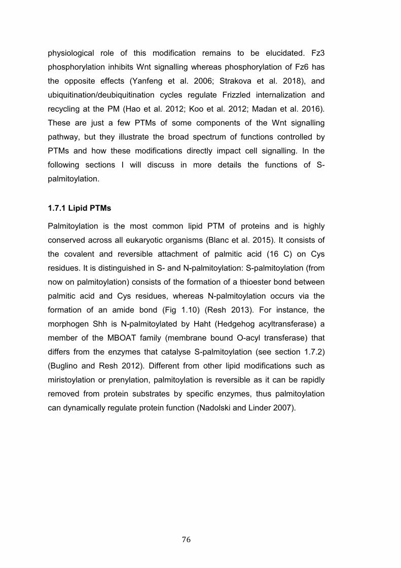
76
physiological role of this modification remains to be elucidated. Fz3
phosphorylation inhibits Wnt signalling whereas phosphorylation of Fz6 has
the opposite effects (Yanfeng et al. 2006; Strakova et al. 2018), and
ubiquitination/deubiquitination cycles regulate Frizzled internalization and
recycling at the PM (Hao et al. 2012; Koo et al. 2012; Madan et al. 2016).
These are just a few PTMs of some components of the Wnt signalling
pathway, but they illustrate the broad spectrum of functions controlled by
PTMs and how these modifications directly impact cell signalling. In the
following sections I will discuss in more details the functions of S-
palmitoylation.
1.7.1 Lipid PTMs
Palmitoylation is the most common lipid PTM of proteins and is highly
conserved across all eukaryotic organisms (Blanc et al. 2015). It consists of
the covalent and reversible attachment of palmitic acid (16 C) on Cys
residues. It is distinguished in S- and N-palmitoylation: S-palmitoylation (from
now on palmitoylation) consists of the formation of a thioester bond between
palmitic acid and Cys residues, whereas N-palmitoylation occurs via the
formation of an amide bond (Fig 1.10) (Resh 2013). For instance, the
morphogen Shh is N-palmitoylated by Haht (Hedgehog acyltransferase) a
member of the MBOAT family (membrane bound O-acyl transferase) that
differs from the enzymes that catalyse S-palmitoylation (see section 1.7.2)
(Buglino and Resh 2012). Different from other lipid modifications such as
miristoylation or prenylation, palmitoylation is reversible as it can be rapidly
removed from protein substrates by specific enzymes, thus palmitoylation
can dynamically regulate protein function (Nadolski and Linder 2007).
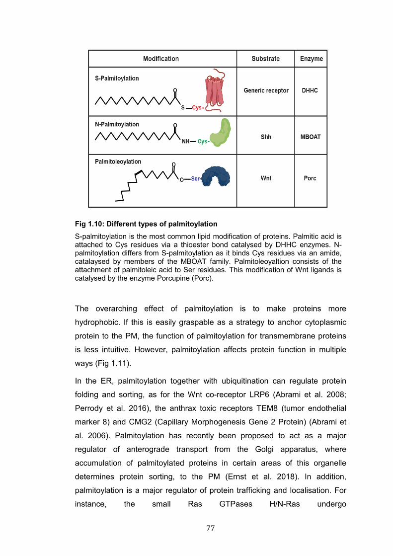
77
Fig 1.10: Different types of palmitoylation S-palmitoylation is the most common lipid modification of proteins. Palmitic acid is attached to Cys residues via a thioester bond catalysed by DHHC enzymes. N-palmitoylation differs from S-palmitoylation as it binds Cys residues via an amide, catalaysed by members of the MBOAT family. Palmitoleoyaltion consists of the attachment of palmitoleic acid to Ser residues. This modification of Wnt ligands is catalysed by the enzyme Porcupine (Porc).
The overarching effect of palmitoylation is to make proteins more
hydrophobic. If this is easily graspable as a strategy to anchor cytoplasmic
protein to the PM, the function of palmitoylation for transmembrane proteins
is less intuitive. However, palmitoylation affects protein function in multiple
ways (Fig 1.11).
In the ER, palmitoylation together with ubiquitination can regulate protein
folding and sorting, as for the Wnt co-receptor LRP6 (Abrami et al. 2008;
Perrody et al. 2016), the anthrax toxic receptors TEM8 (tumor endothelial
marker 8) and CMG2 (Capillary Morphogenesis Gene 2 Protein) (Abrami et
al. 2006). Palmitoylation has recently been proposed to act as a major
regulator of anterograde transport from the Golgi apparatus, where
accumulation of palmitoylated proteins in certain areas of this organelle
determines protein sorting, to the PM (Ernst et al. 2018). In addition,
palmitoylation is a major regulator of protein trafficking and localisation. For
instance, the small Ras GTPases H/N-Ras undergo
78
palmitoylation/depalmitoylation cycles that dynamically shuffle these proteins
between the Golgi and the PM (Prior and Hancock 2001; Baekkeskov and
Kanaani 2009). Similar observations have been made for transmembrane
protein such as NMDARs and AMPARs, which are moved between the PM
and endosomal compartments in a palmitoylation dependent manner
(Baekkeskov and Kanaani 2009; Greaves et al. 2010; Salaun et al. 2010;
Gladding and Raymond 2011; Sohn and Park 2019). Moreover,
palmitoylation can target cytoplasmic as well as transmembrane proteins,
particularly GPCRs, to specific membrane domains (Barnett-Norris et al.
2005; Goddard and Watts 2012). Indeed, palmitoylated proteins are often
found in cholesterol-rich membrane fractions suggesting direct targeting to
lipid rafts (Melkonian et al. 1999; Levental et al. 2010). In addition, protein-
protein interactions are also regulated by palmitoylation. For instance, the
clustering of PSD-95 or Gephyrin depends on their palmitoylation (El-
Husseini et al. 2000; Dejanovic et al. 2014). By affecting several aspects of
basic cell biology, palmitoylation has a profound impact on several
physiological processes including cell metabolism, cell death, and formation
of immune and neuronal synapses. Aberrant regulation of palmitoylation
results in the development of pathological conditions such as infection
diseases, cancer and neurological disorders (Resh 2012; Cho and Park
2016; Sobocińska et al. 2017; Chen et al. 2018).
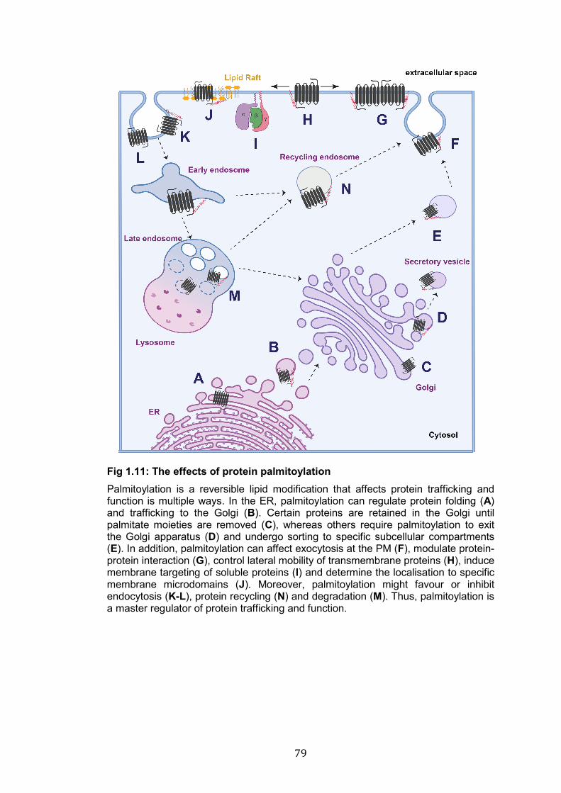
79
Fig 1.11: The effects of protein palmitoylation Palmitoylation is a reversible lipid modification that affects protein trafficking and function is multiple ways. In the ER, palmitoylation can regulate protein folding (A) and trafficking to the Golgi (B). Certain proteins are retained in the Golgi until palmitate moieties are removed (C), whereas others require palmitoylation to exit the Golgi apparatus (D) and undergo sorting to specific subcellular compartments (E). In addition, palmitoylation can affect exocytosis at the PM (F), modulate protein-protein interaction (G), control lateral mobility of transmembrane proteins (H), induce membrane targeting of soluble proteins (I) and determine the localisation to specific membrane microdomains (J). Moreover, palmitoylation might favour or inhibit endocytosis (K-L), protein recycling (N) and degradation (M). Thus, palmitoylation is a master regulator of protein trafficking and function.
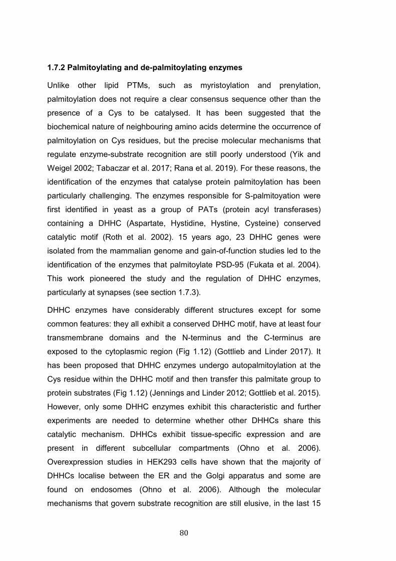
80
1.7.2 Palmitoylating and de-palmitoylating enzymes
Unlike other lipid PTMs, such as myristoylation and prenylation,
palmitoylation does not require a clear consensus sequence other than the
presence of a Cys to be catalysed. It has been suggested that the
biochemical nature of neighbouring amino acids determine the occurrence of
palmitoylation on Cys residues, but the precise molecular mechanisms that
regulate enzyme-substrate recognition are still poorly understood (Yik and
Weigel 2002; Tabaczar et al. 2017; Rana et al. 2019). For these reasons, the
identification of the enzymes that catalyse protein palmitoylation has been
particularly challenging. The enzymes responsible for S-palmitoyation were
first identified in yeast as a group of PATs (protein acyl transferases)
containing a DHHC (Aspartate, Hystidine, Hystine, Cysteine) conserved
catalytic motif (Roth et al. 2002). 15 years ago, 23 DHHC genes were
isolated from the mammalian genome and gain-of-function studies led to the
identification of the enzymes that palmitoylate PSD-95 (Fukata et al. 2004).
This work pioneered the study and the regulation of DHHC enzymes,
particularly at synapses (see section 1.7.3).
DHHC enzymes have considerably different structures except for some
common features: they all exhibit a conserved DHHC motif, have at least four
transmembrane domains and the N-terminus and the C-terminus are
exposed to the cytoplasmic region (Fig 1.12) (Gottlieb and Linder 2017). It
has been proposed that DHHC enzymes undergo autopalmitoylation at the
Cys residue within the DHHC motif and then transfer this palmitate group to
protein substrates (Fig 1.12) (Jennings and Linder 2012; Gottlieb et al. 2015).
However, only some DHHC enzymes exhibit this characteristic and further
experiments are needed to determine whether other DHHCs share this
catalytic mechanism. DHHCs exhibit tissue-specific expression and are
present in different subcellular compartments (Ohno et al. 2006).
Overexpression studies in HEK293 cells have shown that the majority of
DHHCs localise between the ER and the Golgi apparatus and some are
found on endosomes (Ohno et al. 2006). Although the molecular
mechanisms that govern substrate recognition are still elusive, in the last 15
81
years numerous enzymes/substrate pairs have been identified (Tabaczar et
al. 2017). Interestingly, several proteins like PSD95, SNAP25, AMPARs and
others can be palmitoylated by several enzymes (Fukata et al. 2004; Hayashi
et al. 2005; Greaves et al. 2010), increasing the challenge of understanding
enzymes/substrate specificity.
Fig 1.12: S-palmitoylation by DHHC protein acyl transferases A: It has been proposed that several DHHCs undergo CoA-mediated autopalmitoylation of the Cys residue in the DHHC motif before transferring the palmitate moiety to a specific substrate. B: Most DHHCs have 4 transmembrane domains with cytoplasmic N- and C-term regions. The DHHC catalytic motif is conserved across all 23 DHHC genes. C: Phylogenetic tree of human DHHC genes (adapted from Ohno et al. 2006).
One of the key features of palmitoylation is its reversibility. The removal of
palmitate is catalysed by a group of enzymes called protein acyl
thioesterases. APT1 (acyl protein thioesterase) and PPT1 (palmitoyl protein
thioesterase), and their homologs APT2 and PPT2, were first described to
act as thioesterase on Ras GTPases and G proteins (Won et al. 2018). PPT1
is found exclusively on lysosomes and late endosomes, excluding its
involvement in de-palmitoylation events occurring in the cytoplasm or at the
82
PM (Hernandez et al. 2013). PPT1 mutations are linked to the development
of infantile forms of neuronal ceroid lipofuscinoses (NCL), aslo known as
Batten disease (Zeidman et al. 2009; Kollmann et al. 2013), a severe
neurodegenerative disorders characterised by the lysosomal accumulation of
autofluorescent lipoparticles that leads eventually to cell death, particularly of
neurons (Kyttälä et al. 2006; Kollmann et al. 2013). APT1 and 2 are
ubiquitously expressed and are primarily cytoplasmic, but they are also
associated with some internal membranes and with the PM (Vartak et al.
2014; Adachi et al. 2016; Won et al. 2016), most likely through the interaction
with their own palmitate groups (Yang et al. 2010; Vartak et al. 2014).
However, the distribution of APT1 and 2 has only been investigated by
overexpression studies due to the lack of antibodies against endogenous
APTs; thus, the precise localisation of endogenous APT1 and 2 remains to
be established. Loss of function studies clearly showed that APTs KD affects
palmitoylation of some substrates, like Huntingtin, but not others, like N-Ras
and PSD-95, suggesting the presence of other thioesterases for cytosolic
proteins (Lin and Conibear 2015). Indeed, ABHD proteins (α/β-hydrolase
domain-containing protein) were recently screened for thioesterase activity
and ABHD17s (isoforms A, B and C) were found to act as de-palmitoylating
enzymes of N-Ras, PSD-95 and MAP6 (microtubule associated protein 6)
(Lin and Conibear 2015; Yokoi et al. 2016; Tortosa et al. 2017). ABHD17
enzymes exhibit plasma membrane localisation (Yokoi et al. 2016), but the
mechanisms that regulate their enzymatic function are still unknown. Thus,
palmitoylation is a very dynamic PTM that controls multiple aspects of protein
trafficking and functions, and the enzymes that catalyse or remove
palmitoylation have started to be identified, significantly contributing to our
understanding of the regulation of this PTM.
1.7.3 Palmitoylation at synapses
Hundreds of neuronal proteins are palmitoylated and this modification is
crucial for different processes including axon and dendrite polarisation and
also synapse growth and synaptic function (Fukata and Fukata 2010). Key
synaptic proteins are palmitoylated, these include components of the release
83
machinery, scaffold proteins and NT receptors (Prescott et al. 2009; Thomas
and Huganir 2013; Montersino and Thomas 2015; Matt et al. 2019). The list
of synaptic proteins that are palmitoylated is long and ever growing. Here I
will only touch upon some key examples.
Palmitoylation of SNAP23-25, Synaptotagmins, Syntaxin1 and VAMP2 is
fundamental for their function at axonal terminals (Prescott et al. 2009).
Palmitoylation of SNAP25, catalysed by DHHC3, DHHC7 and DHHC17,
regulates stable membrane binding and ensures SNAP25 accumulation at
exocytic sites (Koticha et al. 2002; Salaün et al. 2005). In addition, the
majority of the 17 Synaptotagmin (Syt) isoforms contain at least one C-term
Cys residue that is palmitoylated (Kang et al. 2004). Mutation of the Cys
residue of SytI affects this protein in multiple ways: palmitoylation-mutant SytI
has a more diffuse distribution and is less abundant at synapses, consistently
with a role for palmitoylation in regulating SytI clustering at presynaptic sites
(Kang et al. 2004). On postsynaptic sites, key scaffold proteins, as well as
CAMs, signalling molecules and NT receptors are palmitoylated. For
example, PSD-95 is dually palmitoylated at two Cys residues at the N-
terminus, and this PTM is required for microtubule-dependent targeting of
PSD-95 at synapses, and for PSD-95-mediated clustering of NT receptors
(El-Husseini et al. 2000). Palmitoylation of PSD-95 is mediated by multiple
DHHCs enzymes, such as DHHC2, DHHC3, DHHC7 and DHHC15 (Fukata
et al. 2004). Furthermore, palmitoylation of certain kinases such as the
cytoskeleton regulator LIMK1 (LIM domain kinase 1) and the non-receptor
tyrosine kinase Fyn is required for their activation of NMDARs and their
ability to signal at synapses (Montersino and Thomas 2015). Moreover,
glutamate and GABA receptors are also palmitoylated. Dual palmitoylation of
AMPARs and NMDARs has very distinct roles on their trafficking and
function (Naumenko and Ponimaskin 2018). At the Golgi, palmitoylation is
catalysed by DHHC3 and is required for retaining AMPARs and NMDARs on
this organelle (Hayashi et al. 2005; Hayashi et al. 2009; Lin et al. 2009). At
the PM, palmitoylation controls AMPAR and NMDAR internalisation in
different manners: palmitoylation of AMPARs at the C-terminus facilitates
activity-induced internalisation of these receptors by regulating its interaction
84
with 4.1N protein (Hayashi et al. 2005; Lin et al. 2009). Palmitoylation of
NMDARs, also at the C-terminus, increases Fyn-mediated phosphorylation
and stabilises this receptor at the PM by inhibiting constitutive internalisation
(Hayashi et al. 2009). Thus, palmitoylation of many pre- and postsynaptic
proteins is crucial for their correct localisation and function.
The reversibility of protein palmitoylation is fundamental at synapses, where
activity-mediated regulation of protein function and localisation is one of the
underlying mechanisms of synaptic plasticity. Several DHHC enzymes
including DHHC2, DHHC5, DHHC8, DHHC9, and DHHC12 are regulated by
neuronal activity (Noritake et al. 2009; Thomas et al. 2012; Brigidi et al. 2014;
Dejanovic et al. 2014; Brigidi et al. 2015). For instance, induction of LTP
increases DHHC5 catalytic activity and mobilises DHHC5 from dendritic
spines to endosomes, where it palmitoylates δ-catenin (Brigidi et al. 2014;
Brigidi et al. 2015). Palmitoylated δ-catenin is targeted to spines where it
interacts with N-cadherin to modulate synaptic strength (Brigidi et al. 2014;
Brigidi et al. 2015). Similarly, neuronal activity enhances DHHC8 catalytic
activity, which increases GRIP1B palmitoylation stabilising AMPARs at the
cell surface (Thomas et al. 2012). Moreover, long-term blockage of neuronal
activity, which is associated with homeostatic plasticity (Turrigiano 2012),
increases DHHC2-mediated palmitoylation of PSD-95, therefore enhancing
its interaction with AMPARs and strengthening synaptic transmission
(Noritake et al. 2009). Thus, DHHC enzymes affect the localisation and
function of key synaptic proteins in an activity dependent manner, suggesting
that they may directly regulate neural circuits and affect cognitive functions.
Indeed, Dhhc5 KO mice exhibit severe deficits in hippocampal-dependent
behaviors (Li et al. 2010). Mice lacking Dhhc17, which palmitoylates PSD-95,
AMPARs, SNAP25 and Huntingtin, have severe defects in synaptic
transmission, synaptic plasticity and hippocampal-mediated memory
(Milnerwood et al. 2013). Therefore, growing efforts have focused on
studying the role of palmitoylating and depalmitoylating enzymes in
physiological and pathological processes that affect synapses. For instance,
aberrant activity of DHHCs or depalmitoylating enzymes is linked to disorders

85
characterised by synaptic failure, including AD, Huntington disease,
schizophrenia and autism (Cho and Park 2016; Zaręba-Kozioł et al. 2018).
86
1.8 Regulation of Wnt7a-Frizzled signalling at synapses, a role for palmitoylation.
Wnt7a is a potent synaptogenic factor that promotes the assembly of
excitatory synapses by signalling to both sides of the synapse. Wnt7a also
regulates synaptic transmission and synaptic plasticity (Sahores et al. 2010;
Ciani et al. 2011). Our laboratory identified Fz5 as the receptor for Wnt7a at
presynaptic sites (Sahores et al. 2010). Moreover, Fz5 surface levels are
regulated by neuronal activity (Sahores et al. 2010), suggesting that dynamic
mechanisms are in place to regulate the trafficking of this receptor at the
synapse.
Given the dynamic modulation of Fz5 surface levels in response to neuronal
activity, our laboratory started to investigate the molecular mechanisms that
could regulate this process. First, Dr Eleanna Stamatakou, a previous PhD in
our laboratory, identified possible sites for PTMs in the C-term of Fz5. The C-
terminus of Fz5 contains three Cys residues that could be targeted by S-
palmitoylation (Fig 4.2). These three Cys are indeed predicted to be
palmitoylated by the CCS-palm 4.0 software (Stamatakou and Salinas
unpublished data). Using different techniques including the acyl-biotin
exchange (ABE) assay (Wan et al. 2007), incorporation of radioactive
palmitate, and site-directed mutagenesis of the three Cys to Ser (Fz5 3CS),
Dr Stamatakou confirmed that Fz5 is indeed palmitoylated (Fig 1.13)
(Stamatakou and Salinas unpublished results). Moreover, Fz5 palmitoylation
is regulated by activity as the levels of this modification are increased in
cultured neurons stimulated by HFS and in the hippocampus of mice
exposed to EE (Fig 1.13) (Stamatakou and Salinas unpublished results).
Importantly, preliminary studies using the 3CS Fz5 mutant showed that
palmitoylation is required for Fz5 synaptogenic activity (Fig 1.13). To our
knowledge, this is the first demonstration that Frizzled receptors are
palmitoylated. These are novel and exciting findings for the Wnt field, as they
uncover a previously uncharacterised PTM of Frizzled receptors, unravelling
a new mechanism for Wnt signalling regulation. In my PhD project, I
extended this investigation by examining whether other Frizzled receptors
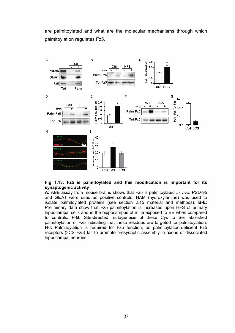
87
are palmitoylated and what are the molecular mechanisms through which
palmitoylation regulates Fz5.
Fig 1.13. Fz5 is palmitoylated and this modification is important for its synaptogenic activity A: ABE assay from mouse brains shows that Fz5 is palmitoylated in vivo. PSD-95 and GluA1 were used as positive controls. HAM (hydroxylamine) was used to isolate palmitoylated proteins (see section 2.15 material and methods). B-E: Preliminary data show that Fz5 palmitoylation is increased upon HFS of primary hippocampal cells and in the hippocampus of mice exposed to EE when compared to controls. F-G: Site-directed mutagenesis of these Cys to Ser abolished palmitoylation of Fz5 indicating that these residues are targeted for palmitoylation. H-I: Palmitoylation is required for Fz5 function, as palmitoylation-deficient Fz5 receptors (3CS Fz5) fail to promote presynaptic assembly in axons of dissociated hippocampal neurons.
88
1.9 Thesis aims
Wnt signalling plays a critical role at the synapse by regulating synapse
assembly during development and by modulating synaptic function and
maintenance in the adult brain. In particular, Wnt7a is a potent synaptogenic
factor that promotes the assembly of pre- and postsynaptic sites, but the
molecular mechanism underlying these processes remain elusive.
Specifically, although Fz5 is a receptor required for Wnt7a-induced
presynaptic assembly, the receptor mediating Wnt7a-dependent was
unknown. Furthermore, palmitoylation, a previously uncharacterised PTM of
Frizzled receptors, seems to be required for Fz5 function at synapses.
However, whether other Frizzled receptors are palmitoylated, and the
molecular mechanisms through which palmitoylation controls Fz5 function,
remain completely unknown. Finally, although the role of Fz5 in synapse
formation has been shown in vitro (Sahores et al. 2010), its role in synapse
formation and the role of Fz5 palmitoylation in vivo are unknown. During my
PhD, I used a combination of biochemical, molecular and cell biology
techniques to study the function of two Wnt7a receptors (Fz5 and Fz7) at the
synapse. In particular, I focused my attention on the molecular mechanism
by which palmitoylation affects Fz5 trafficking and function. The experiments
presented in the next chapters were aimed to address the following specific
questions:
1) Which is the receptor for Wnt7a that is required for postsynaptic
development?
2) Are other Frizzled receptors palmitoylated?
3) Which are the enzymes that palmitoylate Fz5?
4) What are the molecular mechanisms by which palmitoylation controls
Fz5?
5) What is the role of Fz5 and its palmitoylation for synapse formation in
vivo?
89
CHAPTER 2: MATERIAL AND METHODS 2.1 DNA constructs, cloning and viruses
WT Fz5-HA was a kind gift from professor Xi He (Harvard University, US).
Dvl1-HA and WT Fz5-SEP constructs were previously generated in house,
following standard restriction-enzymes-based cloning techniques. The
plasmids for the expression of all Frizzled receptors (mouse) with C-term
1D4 tag were purchased from Addgene (references: Fz1 42263; Fz2 42264;
Fz3 42265; Fz4 42266; Fz5 42267; Fz6 42268; Fz7 42269; Fz8 42270; Fz9
42271; Fz10 42272). 3CS and double Cys mutant Fz5 plasmids were
generated by Dr Eleanna Stamatakou and Dr Laura-Nadine Schuhmacher
respectively. QuikChangeLightning Site-Directed Mutagenesis kit
(Stratagene) was used to clone 3CS Fz5, whereas the Phusion PCR kit was
used to clone Double Cys mutant Fz5 according to manufacturer’s
instructions. The following primers were used to generate these constructs:
Table 2.1: Primers used for Fz5 cloning Constructs Forward and reverse primers 3CS Fz5 Fw 5′-CACCAGCCGCAGCAGCAGCCGCCCGCG-3′
Rv 5′-CGCGGGCGGCTGCTGCTGCGGCTGGTG-3′
C537-538S Fw 5’-GTTTCACCAGCCGCAGCAGCTGCCG-3’
Rv 5’-CGGCTGGTGAAACGCCGCCACGACT-3’
C537-539S Fw 5’-TGGCGGCGTTTCACCAGCCGCAGCTG-3’
Rv 5’-GTGAAACGCCGCCACGACTCCACCGT-3’
C538-539S Fw 5’-CACCAGCCGCTGCAGCAGCCGCCCGC-3’
Rv 5’-TGCAGCGGCTGGTGAAACGCCGCCACGA-3’
All DHHCs plasmids were a kind gift from Prof Akio Kihara and Dr Yusuke
Ohno from the University of Hokkaido, Japan. These plasmids contain
human DHHCs coding sequences tagged with Myc or Flag at the N-term or
C-term, depending on the effect of the tag on each protein. The terminology
of DHHC enzymes needs clarification, as the names commonly used by
research groups do not always correspond to the nomenclature reported on
gene databases:
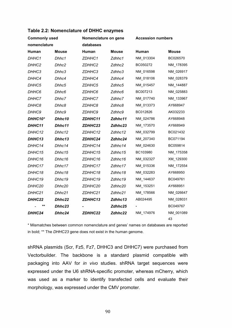
90
Table 2.2: Nomenclature of DHHC enzymes
Commonly used nomenclature
Nomenclature on gene databases
Accession numbers
Human Mouse Human Mouse Human Mouse DHHC1 Dhhc1 ZDHHC1 Zdhhc1 NM_013304 BC026570
DHHC2 Dhhc2 ZDHHC2 Zdhhc2 BC050272 NM_178395
DHHC3 Dhhc3 ZDHHC3 Zdhhc3 NM_016598 NM_026917
DHHC4 Dhhc4 ZDHHC4 Zdhhc4 NM_018106 NM_028379
DHHC5 Dhhc5 ZDHHC5 Zdhhc5 NM_015457 NM_144887
DHHC6 Dhhc6 ZDHHC6 Zdhhc6 BC007213 NM_025883
DHHC7 Dhhc7 ZDHHC7 Zdhhc7 NM_017740 NM_133967
DHHC8 Dhhc8 ZDHHC8 Zdhhc8 NM_013373 AY668947
DHHC9 Dhhc9 ZDHHC9 Zdhhc9 BC012826 AK032233
DHHC10* Dhhc10 ZDHHC11 Zdhhc11 NM_024786 AY668948
DHHC11 Dhhc11 ZDHHC23 Zdhhc23 NM_173570 AY668949
DHHC12 Dhhc12 ZDHHC12 Zdhhc12 NM_032799 BC021432
DHHC13 Dhhc13 ZDHHC24 Zdhhc24 NM_207340 BC071194
DHHC14 Dhhc14 ZDHHC14 Zdhhc14 NM_024630 BC059814
DHHC15 Dhhc15 ZDHHC15 Zdhhc15 BC103980 NM_175358
DHHC16 Dhhc16 ZDHHC16 Zdhhc16 NM_032327 XM_129300
DHHC17 Dhhc17 ZDHHC17 Zdhhc17 NM_015336 NM_172554
DHHC18 Dhhc18 ZDHHC18 Zdhhc18 NM_032283 AY668950
DHHC19 Dhhc19 ZDHHC19 Zdhhc19 NM_144637 BC049761
DHHC20 Dhhc20 ZDHHC20 Zdhhc20 NM_153251 AY668951
DHHC21 Dhhc21 ZDHHC21 Zdhhc21 NM_178566 NM_026647
DHHC22 Dhhc22 ZDHHC13 Zdhhc13 AB024495 NM_028031
- ** Dhhc23 - Zdhhc25 - BC049767
DHHC24 Dhhc24 ZDHHC22 Zdhhc22 NM_174976 NM_001089
43
* Mismatches between common nomenclature and genes’ names on databases are reported
in bold; ** The DHHC23 gene does not exist in the human genome.
shRNA plasmids (Scr, Fz5, Fz7, DHHC3 and DHHC7) were purchased from
Vectorbuilder. The backbone is a standard plasmid compatible with
packaging into AAV for in vivo studies. shRNA target sequences were
expressed under the U6 shRNA-specific promoter, whereas mCherry, which
was used as a marker to identify transfected cells and evaluate their
morphology, was expressed under the CMV promoter.
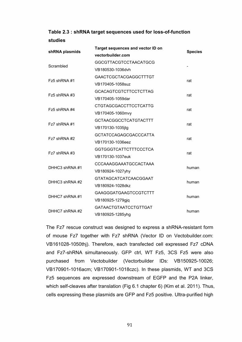
91
Table 2.3 : shRNA target sequences used for loss-of-function studies
shRNA plasmids Target sequences and vector ID on vectorbuilder.com
Species
Scrambled GGCGTTACGTCCTAACATGCG
- VB180530-1036dvh
Fz5 shRNA #1 GAACTCGCTACGAGGCTTTGT
rat VB170405-1058suz
Fz5 shRNA #3 GCACAGTCGTCTTCCTCTTAG
rat VB170405-1059dar
Fz5 shRNA #4 CTGTAGCGACCTTCCTCATTG
rat VB170405-1060mvy
Fz7 shRNA #1 GCTAACGGCCTCATGTACTTT
rat VB170130-1035jtg
Fz7 shRNA #2 GCTATCCAGAGCGACCCATTA
rat VB170130-1036eez
Fz7 shRNA #3 GGTGGGTCATTCTTTCCCTCA
rat VB170130-1037euk
DHHC3 shRNA #1 CCCAAAGGAAATGCCACTAAA
human VB180924-1027yhy
DHHC3 shRNA #2 GTATAGCATCATCAACGGAAT
human VB180924-1028dkz
DHHC7 shRNA #1 GAAGGGATGAAGTCCGTCTTT
human VB180925-1279gjq
DHHC7 shRNA #2 GATAACTGTAATCCTGTTGAT
human VB180925-1285yhg
The Fz7 rescue construct was designed to express a shRNA-resistant form
of mouse Fz7 together with Fz7 shRNA (Vector ID on Vectobuilder.com:
VB161028-1050thj). Therefore, each transfected cell expressed Fz7 cDNA
and Fz7-shRNA simultaneously. GFP ctrl, WT Fz5, 3CS Fz5 were also
purchased from Vectobuilder (Vectorbuilder IDs: VB150925-10026;
VB170901-1016acm; VB170901-1018czc). In these plasmids, WT and 3CS
Fz5 sequences are expressed downstream of EGFP and the P2A linker,
which self-cleaves after translation (Fig 6.1 chapter 6) (Kim et al. 2011). Thus,
cells expressing these plasmids are GFP and Fz5 positive. Ultra-purified high
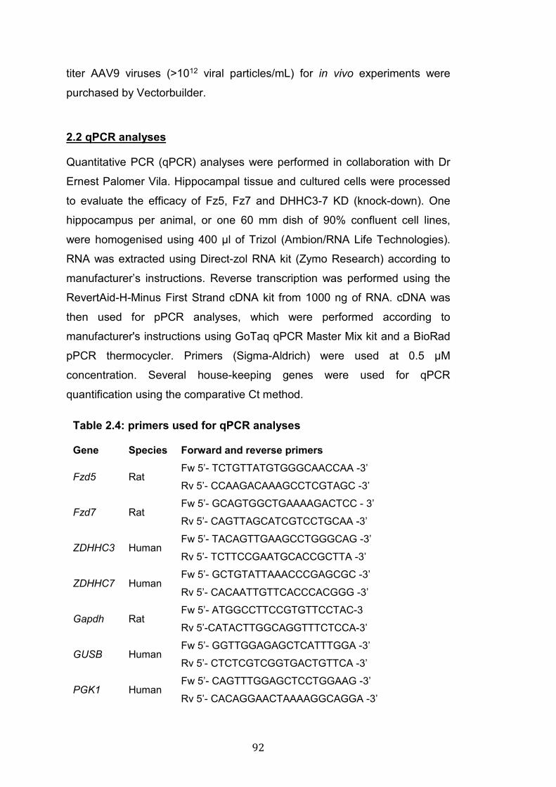
92
titer AAV9 viruses (>1012 viral particles/mL) for in vivo experiments were
purchased by Vectorbuilder.
2.2 qPCR analyses
Quantitative PCR (qPCR) analyses were performed in collaboration with Dr
Ernest Palomer Vila. Hippocampal tissue and cultured cells were processed
to evaluate the efficacy of Fz5, Fz7 and DHHC3-7 KD (knock-down). One
hippocampus per animal, or one 60 mm dish of 90% confluent cell lines,
were homogenised using 400 μl of Trizol (Ambion/RNA Life Technologies).
RNA was extracted using Direct-zol RNA kit (Zymo Research) according to
manufacturer’s instructions. Reverse transcription was performed using the
RevertAid-H-Minus First Strand cDNA kit from 1000 ng of RNA. cDNA was
then used for pPCR analyses, which were performed according to
manufacturer's instructions using GoTaq qPCR Master Mix kit and a BioRad
pPCR thermocycler. Primers (Sigma-Aldrich) were used at 0.5 μM
concentration. Several house-keeping genes were used for qPCR
quantification using the comparative Ct method.
Table 2.4: primers used for qPCR analyses
Gene Species Forward and reverse primers
Fzd5 Rat Fw 5’- TCTGTTATGTGGGCAACCAA -3’
Rv 5’- CCAAGACAAAGCCTCGTAGC -3’
Fzd7 Rat Fw 5’- GCAGTGGCTGAAAAGACTCC - 3’
Rv 5’- CAGTTAGCATCGTCCTGCAA -3’
ZDHHC3 Human Fw 5’- TACAGTTGAAGCCTGGGCAG -3’
Rv 5’- TCTTCCGAATGCACCGCTTA -3’
ZDHHC7 Human Fw 5’- GCTGTATTAAACCCGAGCGC -3’
Rv 5’- CACAATTGTTCACCCACGGG -3’
Gapdh Rat Fw 5’- ATGGCCTTCCGTGTTCCTAC-3
Rv 5’-CATACTTGGCAGGTTTCTCCA-3’
GUSB Human Fw 5’- GGTTGGAGAGCTCATTTGGA -3’
Rv 5’- CTCTCGTCGGTGACTGTTCA -3’
PGK1 Human Fw 5’- CAGTTTGGAGCTCCTGGAAG -3’
Rv 5’- CACAGGAACTAAAAGGCAGGA -3’
93
2.3 Animal use and intracerebroventricular (ICV) injections of AAV9
Experiments carried out on rats and mice were performed as stated under
personal and project licences granted by the UK Home Office in compliance
of the Animals (Scientific Procedures) Act 1986. C57BL/6 wild type mice
were used for ICV injections. P0-P1 new-born pups were injected in the
lateral ventricles with adeno-associated virus serotype 9 (AAV9) with minimal
variations to what previously described (J.-Y. Kim et al. 2014).
Before collecting the pups for injections the mother was moved to a different
cage and kept isolated for the entire length of the procedure (10-30 mins
depending on the number of pups to be injected). Manipulation of neonatal
mice can result in rejection and cannibalism by the mother. To cover up
external smells that could induce pups rejection, I thoroughly rubbed my
hands with beddings from the original cage before touching the animals. Next,
pups were gently collected and placed in tissue pockets on a pre-warmed
pad to avoid hypothermia. One pup at the time was processed for injection:
the head was held flat with two fingers and a Hamilton syringe with a 32G
needle was mounted on a stereotaxic frame to freely press the plunger
without risking of displacing the needle and used to dispense the virus.
Injection sites were located halfway between the Lambda and Bregma
sutures, approximately 1 mm laterally of the sagittal midline (see Fig 6.1) (J.-
Y. Kim et al. 2014). The needle was inserted to a depth of 2.5 µm from the
surface of the mouse’s head. Up to 2.5 µL of virus were injected and the
needle slowly retracted to repeat the procedure on the other hemisphere. For
identification purposes, front and/or back paws were poked using a small
needle covered in tattoo paste. Before returning to the cage, the pups were
gently rubbed with beddings to cover external smells that could potentially
induce rejection by the mother. Finally, the mother was re-introduced to the
cage. Pups were collected 14-15 days post-injection.
2.4 Brain dissection, fixation and freezing
Animals were sacrificed according to Schedule 1 protocol (culled with an
overdose of CO2 and decapitated), brains were rapidly dissected and either
94
snap frozen in liquid nitrogen for WB (western blot) and qPCR (quantitative
polymerase chain reaction) analyses or fixed with 4% PFA/PBS O/N at 4 ˚C
(Table 2.13). 24 hrs later, fixed brains were rinsed twice with PBS and placed
for 2-3 days at 4 ˚C in a 30% sucrose/PBS solution for cryopreservation. The
brains were then frozen in dry-ice-cold isopentane (2-methylbutane, Sigma)
and stored at -80 ˚C until cryosectioning.
2.5 Cryosectioning and Immunofluorescence analyses of brain slices
Frozen brains were cut using a Leica CM1850 cryostat. The brains were
introduced in the cryostat, equilibrated at -20 ˚C for 15-20 min and then
embedded in OCT (VWR Chemicals). 30 µm thick sections were cut
sagittally, collected in cryo-freezing solution (2.14) and placed at -20 ˚C for
long term storage. Sections were rinsed from cryo-freezing solution with PBS
(3x10 mins washes) before being immersed in blocking solution (10%
donkey serum, 0.5 % Triton X-100 in PBS) with gentle shaking for 1-2 hrs at
RT. Primary antibodies (Table 2.5) were diluted in blocking solution and
incubated O/N at 4 ˚C with vigorous shaking. The following day the slices
were washed 3 times for 10 min with PBS and incubated in the dark for 3-4
hrs with secondary antibodies diluted 1:600 in blocking solution. After 3x10
mins washes with PBS, slices were mounted on glass microscope slides
using Fluoromount-G (Southern Biotech) and let dry in the dark for at least
48 hrs before being imaged.
2.6 Cell cultures, transfection methods and pharmacological treatments
Primary rat hippocampal cultures
Primary hippocampal cultures were prepared from E18 Sprague-Dawley rat
embryos as previously described (Dotti et al. 1988). After removing the brain
from the skull, the tissue was kept in ice cold Hank’s balanced salt solution
(Gibco) for the duration of the hippocampal dissection, which was performed
using a Leica dissecting microscope. After completion of the dissection, the
hippocampi were placed at 37 ˚C for 18 mins in Hank’s solution
supplemented with 0.5% trypsin (Invitrogen) for chemical digestion. Next, the
95
hippocampi were rinsed three times with Hank's solution and transferred to
DMEM plating medium (Table 2.8) for mechanical dissociation, which was
carried out with three glass Pasteur pipettes of different size. Cells were
counted using a haemocytometer and plated at required density: 250
cells/mm2 for high-density cultures, or 125 cells/mm2 for low-density cultures.
Cells were plated onto 12mm wide glass coverslips pre-treated with nitric
acid and coated O/N (over-night) with 1 mg/mL poly-D-lysine in boric buffer
(Sigma). After two hrs at 37 ˚C with 95% O2 and 5% CO2, DMEM plating
media was replaced with Neurobasal maintenance medium supplemented
with nutrition factors (Gibco, Table 2.9). Cells were kept in culture for up to 2
weeks and fresh medium was added when necessary to compensate for
reduction in volume due to evaporation.
Hippocampal neurons were transfected using the Ca2+ phosphate method
(Dudek et al. 2001). Depending on the aim of the experiment, transfection
was performed at DIV (days in vitro) 5-9. Briefly, 20 mins before transfection
Neurobasal maintenance medium of a P60 dish was replaced with 3 mL of
transfection medium (Table 2.10). For each 60mm dish, 5 µg of plasmid DNA,
10 µL of 2.5 M CaCl2 and H2O were mixed to reach a final volume of 100 µL,
which was then added drop-wise to 100 µL of 2x HEPES-buffer saline
solution (Table 2.11) and incubated in the dark for 20 mins at RT. Next, the
mix was added to the cells for 5-10 mins at 37 ˚C. The cells were then
washed 3 times with pre-equilibrated transfection medium and returned to
the incubator with 4 mL of original maintenance medium supplemented with
Pen/Strep.
HEK293, NRK and NB2A cultures
HEK293 (human embryonic kidney 293) and NRK (normal rat kidney) were
cultured in DMEM medium (Gibco) containing Glutamax, D-glucose (4.5 g/L),
pyruvate, 10 % FBS (Gibco) and 1 % v/v Pen/Strep (Gibco). Cells were kept
in a humidifier incubator at 37 ˚C with 95% O2 and 5% CO2. Cells were
transfected using Lipofectamine or AMAXA nucleofection methods.
96
Lipofectamine transfection
Lipofectamine transfection was performed according to manufacturer’s
instructions (Thermo Fisher). Briefly, 3.5-5x10⁵ cell were plated in one well of
6-well plate with 2 mL of DMEM maintenance medium. The following day the
cells were transfected with up to 2.5 µg of plasmid DNA. For each well, 0.2-
2.5 µg of DNA and 5 µL of reagent 3000 were added to 125 µL of OPTIMEM
medium (Gibco). In parallel, 5 µL of Lipofectamine were added to 125 µL of
OPTIMEM, immediately vortexed and added to the tube containing DNA and
reagent 3000. This mix was incubated at RT for 10-15 mins and then added
to the cells in a drop-wise manner. Cells were harvested 24-48 hrs post
transfection.
AMAXA nucleofection
NRK cells were transfected by AMAXA nucleofection (Lonza) following
manufacturer’s instruction. Briefly, after trypsinisation cells were pelleted and
resuspended in 100 µL (per condition) of WT-EM transfection solution (Table
2.12). 1x10⁶ cells/per condition were mixed gently with up to 5 µg of plasmid
DNA. The cells were immediately transferred to glass cuvettes and
nucleofected using the appropriate program in a Lonza nucleofector device
(NRK cells were nucleofected using the X-01 program). 10 mins after
nucleofection, 500 µL of pre-equilibrate DMEM medium were added to each
cuvette, the cells were collected using a thin Pasteur pipette and plated at
the desired density.
Pharmacological treatments
To examine Fz5 degradation rate NRK cells were treated with the protein
translation blocker Cycloheximide (Cell Signalling Technology, 50 μg/ml) or
with DMSO (dimethyl sulfoxide, control vehicle) for different time points (0
mins - 2 hrs). To inhibit palmitoylation and examine Fz5 surface levels,
HEK293 cells were treated with 2BP (2-Bromohexadecanoic acid, Sigma) at
a final concentration of 100 µM or with DMSO.
97
2.7 Antibody feeding experiments
NRK cells expressing WT or 3CS Fz5 were placed on an ice block in the cold
room for 15-20 mins to stop membrane trafficking. Primary HA antibodies
were diluted 1:500 in ice cold DMEM maintenance media. The mix was
added to the cells, still on ice, for 30 mins to allow antibody binding to the
extracellular HA tag of surface Fz5 receptors. The cells were then washed 3
times with ice cold PBS and a subset, used as control (t=0), was immediately
fixed with ice cold PFA. Another subset of cells was placed in pre-
equilibrated warm DMEM and returned to the incubator for 15 mins to allow
for receptor endocytosis. These cells were then fixed with warm 4% PFA in
PBS and processed for immunofluorescence analyses (see section 2.9).
2.8 Fluorescent Recovery After Photobleaching (FRAP)
For FRAP experiments NRK cells were imaged using a Leica TCS SP8
inverted confocal microscope with a 60x objective (NA 1.35). The external
chamber of the microscope was equilibrated at 37 ˚C. NRK cells expressed
WT or 3CS Fz5 tagged at the N-term with SEP (super ecliptic pHluorin), a
GFP variant that does not fluoresce at acid pH, normally found in intracellular
vesicles, but emits fluorescence at neutral pH, normally found extracellularly
(Miesenböck et al. 1998). Therefore, the fluorescence signal detected came
from Fz5 receptors at the cell surface. To reduce background fluorescence
and stabilise the pH, NRK cells were cultured with DMEM without phenol red
and supplemented with HEPES buffer (Gibco). A baseline of 15 single stack
images (256x256 pixels = 82x82 µm; 0.32 µm voxel) was taken every 0.25
sec prior to bleaching. Samples were flashed 10 times with 100% laser
power every 0.25 sec to bleach the fluorescence signal in three small areas
of the field of view: a) outside the cell (blank); b) at the edge of the cell; c) in
the middle of the cell. The “blank” area at the edge of the cell was used for
quantification of fluorescence recovery, which was measured by imaging at
the following intervals and frequencies: 1 image / 0.25 sec for the first 12.5
sec; 1 image / sec for the next 30 sec; 1 image / 5 sec for the next 60 sec
(total = 102,5 sec).
98
2.9 Immunofluorescence analyses of cultured cells
Hippocampal and cell line cultures were stained using a standard
immunofluorescence protocol. Briefly, cells were rinsed with warm PBS and
fixed in 4% PFA/PBS for 20 mins at RT. When fixing hippocampal neurons,
4% PFA was supplemented with 4% sucrose (Table 2.13). After fixation, cells
were rinsed 3 times with PBS and either stored at 4 ˚C or processed for
immunostaining. Cells were permeabilised with 0.05 % Triton X-100 in PBS
for 5 min, washed once with PBS, and placed for at least 45 mins at RT in
blocking buffer (5% BSA/PBS) in a humidified chamber. Primary antibodies
were diluted in 1% BSA/PBS and added onto the cells O/N at 4 ˚C in a
humidified chamber. The following day, after 3 washes with PBS, secondary
antibodies were diluted in 1% BSA/PBS and added onto the cells for 1 hr in
the dark at RT in a humidified chamber. Cells were then washed 3 more
times with PBS and mounted on glass microscope slides with a drop of
FluorSave (Calbiochem) mounting medium. Dry samples were placed at 4 ˚C
or at -20 ˚C for short or long term storage.
2.10 Confocal imaging
Hippocampal neurons and cell lines were imaged on an Olympus FV1000
inverted confocal microscope. An oil-immersion 60x (NA 1.35) objective was
used to acquire images at 207 nm/pixel. 6-12 z-step of 0.25 µm were imaged
to analyse dendritic spines, synaptic puncta, endocytosed receptors and
other trafficking markers. Brain slices were imaged on a Leica TCS SP8
inverted confocal microscope using an oil-immersion 60x (NA 1.35) objective
to acquire images at 207 nm/pixel. 8 z-steps of 0.25 µm or 15-25 z-steps of 1
µm were acquired to image synaptic puncta or entire cells/brain areas
respectively. For experiments on hippocampal neurons or cell lines 7-15
images from at least 3 independent biological samples were acquired. For
brain slices, a minimum of 6 images from 3 slices was acquired for each
animal.
99
2.11 Image analyses
Volocity software (PerkinElmer) was used to design protocols for
quantification of all confocal images. When possible, analyses were carried
out blind to the experimental condition/genotype. The number and size of
dendritic spines of hippocampal neurons were measured manually. Briefly, 3-
4 secondary dendrites were cropped from each cell and used for
quantification. Morphological features commonly accepted in the field
(Arellano et al. 2007; Basu et al. 2018) were used to identify stubby,
mushroom and thin spines and exclude filopodia-like structures. The number
of spines was normalised to the length of dendrites, which was calculated
manually using a measuring tools of the Volocity software. Using the same
tool, the maximum width of the head of each spine, here called spine size,
was calculated. The average value of all the spines of each cell was used for
quantification.
Similar criteria were used to identify synaptic puncta and vesicles of
endocytosed receptors. To identify objects of interests, thresholds were
designed according to two main features: fluorescence intensity and
minimum/maximum size of the object. The software automatically measured
number and volume of the identified objects. When analysing presynaptic
assembly in isolated axons, the number of synaptic puncta was normalised
to the length of the axon. To analyse images of brain slices, three different
squares were cropped from each image to avoid areas of non-puncta
staining, such as cell bodies or blood vessels, which would skew the number
of puncta when normalising for total volume. For antibody-feeding
experiments, the number of puncta of endocytosed receptors was normalised
per cell. Only cells with similar size were imaged.
2.12 Preparation of protein samples and western blotting (WB)
Cultured cells were first rinsed with ice cold PBS and then lysed in
appropriate amount of lysis buffer (Table 2.15) supplemented with PMSF
(Sigma-Aldrich, final concentration 1 mM), 1x phosphatase and protease
inhibitors (Roche). Unless stated otherwise, lysates were transferred to
100
Eppendorf tubes and incubated for 10 mins on ice before max speed
centrifugation (10 mins at 13000 rpm at 4 ˚C). The pellet was discarded and
the supernatant collected for protein quantification, which was performed
using the BCA kit following manufacturer's instructions (Thermo Scientific).
After adjusting protein concentrations to equal amounts, 5x loading buffer
containing DTT (Table 2.27) was added to a final dilution of 1x, and the
samples were incubated at 65 ˚C for 10 mins to denature protein structures.
5 to 30 µg of protein were loaded on SDS PAGE gels following standard
Western Blot (WB) guide lines. Nitrocellulose or PVDF membranes were
blocked for at least 1 hr at RT in 5% milk or BSA/TBS-t (Table 2.26) and then
probed O/N at 4˚C with primary antibodies (Table 2.5). The following day,
after 3x10 mins washes with TBS-t, the membranes were incubated for 1hr
at RT with fluorescent or HRP-conjugated secondary antibodies and then
washed again 3x10-30 mins before detection of fluorescence or
chemiluminescence signals. When HRP-conjugated secondaries were used,
ECL substrate (Amersham) was added to the membranes according to
manufacturer’s instructions to visualise chemiluminescence signals. A
ChemiDoc (Bio-Rad) and ImageLab software (Bio-Rad) were used to
imaging WB membranes. ImageJ software was used for quantification of
signal intensity.
2.13 Surface biotinylation
Surface biotinylation experiments were performed as previously described
(Sahores et al. 2010). One 60mm dish of HEK293 cells at 80-90%
confluency was used for each condition. Cells were first placed on an ice
block for 10 mins to stop membrane trafficking. All the following incubations
and centrifugations were performed at 4 ˚C unless stated otherwise. Cells
were washed twice with PBS containing 1 mM CaCl2 and 0.5 mM MgCl2, and
then treated for 30 mins with 0.25 mg/mL of membrane-impermeable
biotin/PBS (EZ-Link Sulfo-NHS-Biotin, Thermo Fisher Scientific) to biotinylate
surface proteins. 10 mM glycine in PBS was used for 3x5 min washes to
neutralise excess biotin and prevent biotinylation of intracellular proteins at
the moment of lysis. Each dish was lysed with 500 µL of RIPA buffer (Table
101
2.16) supplemented with protease and phosphatase inhibitors. The samples
were then passed 3 times through a 25G needle to maximise lysis efficacy
and shear DNA and large portions of membranes. 10 mins after
centrifugation at 13000 rpm, supernatants were collected and pellets
discarded. Protein concentration was determined by BCA assay as described
before (section 2.12). 10-50 µL were kept as total lysate (input), whereas the
rest of the lysate (200-400 µg of protein) was added to 50 µL of streptavidin
agarose beads (Pierce Streptavidin Agarose, Thermo Fisher Scientific) pre-
equilibrated by three washes with RIPA buffer. Samples were placed on a
wheel for end-over-end rotation for 2 hrs. Next, they were washed 3-4 times
with RIPA buffer containing protease and phosphatase inhibitors. Each wash
consisted of spinning down the samples at 1000 rpm for 1 min to precipitate
streptavidin beads, discarding the supernatant, and adding 500 µL of fresh
RIPA buffer to the beads. After the final wash, residual RIPA buffer was
removed using a 30G needle, whose gauge is too narrow to aspirate beads.
50-60 µL of 2.5x loading buffer were added to elute biotinylated proteins from
beads (IP or surface fraction). IP and input samples were then incubated at
65 ˚C for 10 mins to complete protein denaturation.
2.14 Co-IP (co-immunoprecipitation) experiments
Co-IP experiments were performed in HEK293 cells using GFP-trap beads
(Chromotek). Importantly, these GFP beads recognise numerous variants of
GFP proteins (Chromotek), including SEP, a pH-sensitive derivate of EGFP
(Miesenböck et al. 1998). WT and/or 3CS Fz5-SEP were transfected in
HEK293 in order to use GFP-trap and examine the interaction with Dvl1-HA
and Fz5-HA. 24h hrs after transfection, one 60 mm dish of 80-90% confluent
HEK293 cells was rinsed with PBS and lysed in 500 µL Triton Lysis Buffer
(Table 2.22) supplemented with protease and phosphatase inhibitors. The
samples were then incubated at 4 ˚C for 20 mins on an end-over-end rotating
wheel and then spun at 13000 rpm for 10 mins. Protein quantification was
determined, concentrations adjusted, and 20-50 µL of total lysate kept as
input. The rest (200-400 µg of proteins) was added to 15-20 µL of GFP-trap
beads pre-equilibrated by 3 washes with lysis buffer. The specimens were
102
incubated at 4 ˚C for 2-3 hrs on an end-over-end rotating wheel before being
washed and processed for SDS PAGE analysis (same procedure as for
surface biotinylation samples, section 2.13). Proteins co-immunoprecipitated
together with WT or 3CS Fz5 were detected by WB, probing the IP fraction
for proteins of interest (Dvl-HA or Fz5-HA). Relative levels of the co-
immunoprecipitated proteins were calculated as a ratio over the total protein
in the lysate.
2.15 Acyl-Biotin exchange (ABE) assay
ABE assay was performed from brain, hippocampal cultures and HEK293
lysates to identify palmitoylated proteins. The procedure was carried out with
minimal variations to what previously reported (Wan et al. 2007). This
protocol is based on the chemical exchange of palmitate groups with biotin,
followed by standard streptavidin-mediated immunoprecipitation of
biotinylated proteins. The protocol is divided in three consecutive days,
followed by standard WB analyses on SDS PAGE gels.
Chloroform-methanol (CM) protein precipitation
This technique was used to wash protein samples from different components
in the various buffers used throughout the protocol. All the steps of CM
precipitation were performed at RT. Briefly, 1.2 mL of lysate were transferred
to 15 mL plastic Falcons and 4.8 mL of methanol, 1.5 mL of chloroform and
3.6 mL of water were added. The mix was immediately vortexed and spun in
a J2-MI Beckman centrifuge with a JS-7.5 swinging bucket rotor at 4000 rpm
for 20 mins. After this centrifugation three phases are formed: top aqueous
phase (water and methanol), a solid pancake-like interphase (proteins) and a
bottom phase (chloroform). The top aqueous phase was carefully aspirated
without disturbing the protein interphase. 3.6 mL of methanol were gently
added to the tube and the solution slowly inverted 2-3 times to mix
chloroform and methanol. The samples were centrifuged at 4000 rpm for 5
mins to pellet the solid protein interphase. The CM mix was discarded and
the tubes air-dried for 2-3 min to facilitate the resuspension of the pellet. This
is a critical step: left over traces of CM from insufficient drying, as well as
103
over-drying the samples, result in a slow and incomplete resuspension of the
pellets. Pellets were resuspened in 300 µL of 4SB buffer (Table 2.19) unless
stated otherwise. After adding 4SB, samples were placed in a water bath at
37 ˚C and frequently tapped and vortexed to resuspend pellets. This step can
take from 10- up to 30 mins depending on the amount of protein present in
the lysate. After complete resuspension, unless stated otherwise, 900 µL of
ABE lysis buffer with 0.2% triton were added and the procedure was
repeated.
Day1: blockage of free Cys residues
Flash-frozen brain tissue was homogenised with a pestel in 1 mL of ABE
lysis buffer (Table 2.17) supplemented with 10 mM NEM (N-Ethylmaleimide)
(Sigma), 1 mM PMSF and 1x protease inhibitors cocktail (PI, Table 2.18).
When working with cells, two 60mm dishes of hippocampal neurons or
HEK293 cells were washed twice with ice cold PBS and then lysed in 1 mL
(500 µL/dish) of ABE lysis buffer with the same supplements mentioned
above. In this protocol NEM is used to block all the free Cys residues, thus
avoiding biotinylation in the following steps. Poor efficacy of NEM blockage
can result in background signals in negative control samples (-HAM, see day
2). After lysis, cells were passed 3-5 times through a 25G needle and 85 µL
of Triton X-100 were added (final concentration 1.7%). Lysates were then
passed 3 more times through a 25G needle to ensure complete
homogenisation. Samples were placed at 4 ˚C for 1hr on an end-over-end
rotating wheel (10 rpm) before centrifugation at low speed (0.3 rcf at 5 mins
at 4 ˚C) to remove big particulates. Next, lysates were washed by chloroform-
methanol (CM) precipitation (see above) and resuspended in 300 µL of 4SB
buffer supplemented with 10mM NEM. 900 µL of ABE lysis buffer containing
10 mM NEM, 1mM PMSF, 1x PI and 0.2% triton were added and samples
were incubated O/N at 4 ˚C on a rotating wheel (10 rpm).
Day 2: NEM wash out - acyl-biotin exchange
In the second day of the protocol NEM is washed out, palmitate groups
removed by treatment with Hydroxilamine (HAM) and newly exposed Cys
residues biotinylated. Excessive NEM was washed by 3 sequential CM
104
precipitations. The protocol can be stopped after each CM precipitation and
lysates can be stored at -20 ˚C in 4SB buffer. At the end on the 3rd CM the
pellet was resuspended in 600 µL of 4SB and each sample was divided into
two tubes. Half of the samples were treated with 900 µL of +HAM buffer
(Table 2.20) and half of the samples were treated with 900 µL of -HAM buffer
(Table 2.20). The samples were incubated in the dark at RT for 1hr on a
rotating wheel at 10 rpm. +HAM buffer contained 0.7 M HAM (Sigma), which
breaks the thioester bonds between Cys residues and palmitate groups. -
HAM buffer contained water; therefore, thioester bonds between Cys
residues and palmitate groups were unaffected. During this incubation with
HAM buffers, free Cys residues, which were originally palmitoylated, were
biotinylated with HPDP-biotin that was added to both + and - HAM buffers.
HAM was then washed out by one CM precipitation and protein pellets
resuspended in 300 µL 4SB. To ensure complete biotinylation of free Cys
residues, 900 µL of low HPDP-biotin buffer (Table 2.21) were added and the
samples incubated in the dark at RT for 1hr on a rotating wheel at 10 rpm. To
remove excessive HPDP-biotin before streptavidin pull-down (day 3), the
samples were subjected to three consecutive CM precipitations. The protocol
was stopped by freezing the samples after the 1st or 2nd CM precipitation.
Day 3: immunoprecipitation of biotinylated (=palmitoyated) proteins
Samples were thawed at RT and the remaining CM precipitation (1 or 2,
depending on the stopping point at the end of day 2) were carried out. After
the last CM, pellets were resuspended in 50 µL of 2SB buffer to dilute SDS
concentration and ensure efficient streptavidin pull-down. SDS was further
diluted to 0.1% by adding 950 µL of ABE lysis buffer supplemented with 1
mM PMSF and 1x PI. The samples were incubated in the dark at RT for 30
mins on a wheel rotating at max speed (40 rpm) to ensure complete
dissolution of protein pellet and washing from residual SDS. Next, lysates
were spun at max speed for 1 min in a bench-top centrifuge at RT to remove
large particulates before the pull-down. Protein concentration was
determined with BCA kit and adjusted for each sample. 100 µL of total lysate
were kept as input and the rest of the lysates were added to 80 µL of
streptavidin agarose beads pre-equilibrated with ABE lysis buffer containing
105
PMSF and PI. The samples were incubated for 2-3 hrs in the dark, at RT, on
a wheel (10 rpm). Next, they were washed 3-4 times with ABE lysis buffer
and, as previously described for surface biotinylation and Co-IP experiments,
left over solution was aspirated with thin needles. Proteins were eluted from
beads by adding 50-60 µL of 2.5x loading buffer and by heating samples for
10 min at 65 ˚C. The specimens were then used for standard WB procedures.
2.16 Statistical analyses
Statistical analyses were carried out using GraphPad Prism. First, normality
was tested using either D’agostino-Pearson, Saphiro-Wilk or Kolmogorov-
Smirnov tests. For all the experiments each animal/culture was considered
as independent experimental unit, with the exception of the analyses of Fz5
and Fz7 KD in hippocampal neurons (chapter 3) and FRAP live-imaging
experiments, in which each cell was treated as an independent experimental
unit. One sample t-test and two-tailed unpaired Student’s t-test were used,
where appropriate, to calculate statistical significance between two
conditions. Mann-Whitney test was used when comparing two data-sets not
normally distributed. One-way and Two-way ANOVA were performed on data
sets composed of 3 or more conditions. One Way ANOVA followed by
Tukey’s post hoc test was used on normally distributed samples whereas
Kruskall-Wallis and Dunn’s post hoc test were run on not-normally distributed
data sets.
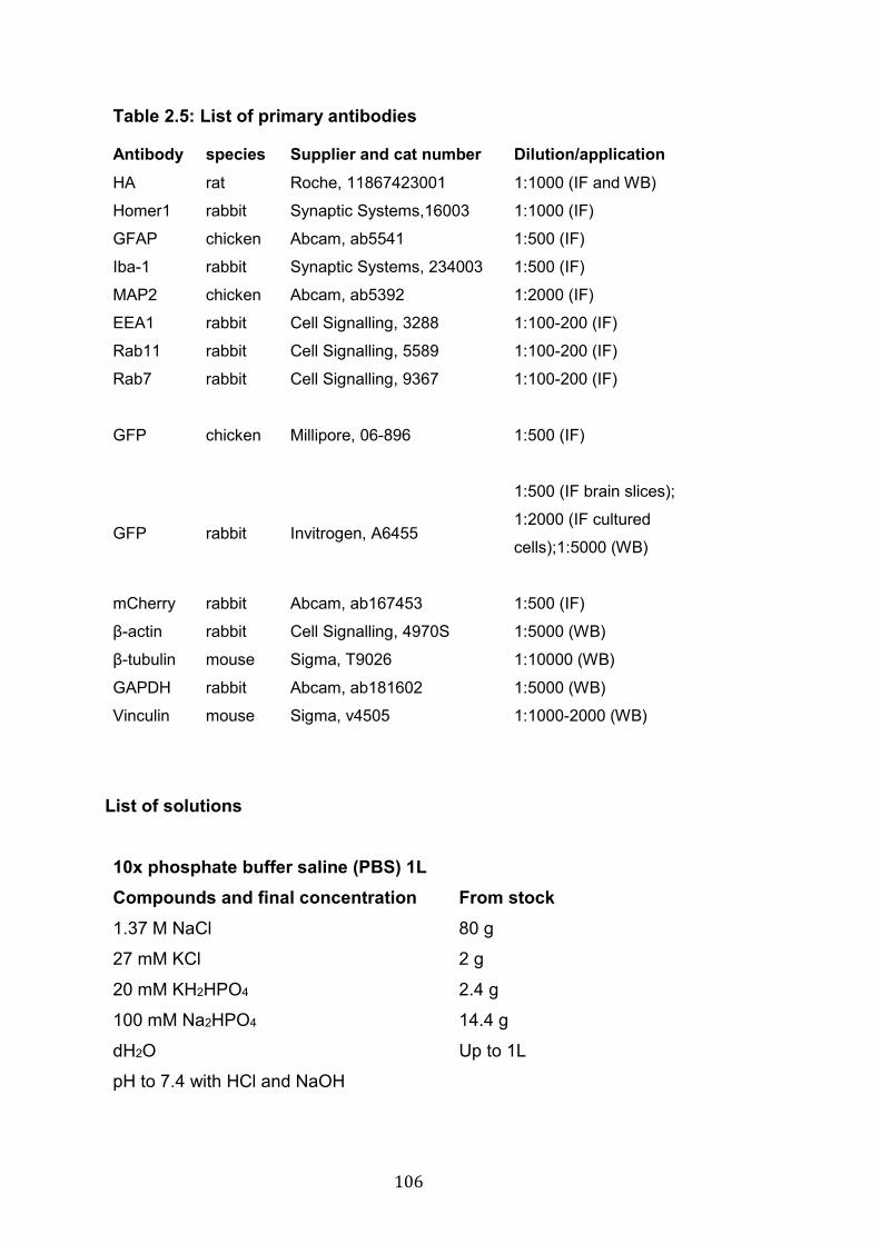
106
Table 2.5: List of primary antibodies
Antibody species Supplier and cat number Dilution/application
HA rat Roche, 11867423001 1:1000 (IF and WB)
Homer1 rabbit Synaptic Systems,16003 1:1000 (IF)
GFAP chicken Abcam, ab5541 1:500 (IF)
Iba-1 rabbit Synaptic Systems, 234003 1:500 (IF)
MAP2 chicken Abcam, ab5392 1:2000 (IF)
EEA1 rabbit Cell Signalling, 3288 1:100-200 (IF)
Rab11 rabbit Cell Signalling, 5589 1:100-200 (IF)
Rab7 rabbit Cell Signalling, 9367 1:100-200 (IF)
GFP chicken Millipore, 06-896
1:500 (IF)
GFP rabbit Invitrogen, A6455
1:500 (IF brain slices);
1:2000 (IF cultured
cells);1:5000 (WB)
mCherry rabbit Abcam, ab167453 1:500 (IF)
β-actin rabbit Cell Signalling, 4970S 1:5000 (WB)
β-tubulin mouse Sigma, T9026 1:10000 (WB)
GAPDH rabbit Abcam, ab181602 1:5000 (WB)
Vinculin mouse Sigma, v4505 1:1000-2000 (WB)
List of solutions 10x phosphate buffer saline (PBS) 1L Compounds and final concentration From stock 1.37 M NaCl 80 g
27 mM KCl 2 g
20 mM KH2HPO4 2.4 g
100 mM Na2HPO4 14.4 g
dH2O Up to 1L
pH to 7.4 with HCl and NaOH
107
Borate buffer (400 mL) Compounds and final concentration From stock 50 mM boric acid 1.24 g
25 mM borax 2 g
dH2O Up to 400 mL
pH to 8.5 and filter. Store at 4 ˚C
Hippocampal cultures plating medium (50 mL) Compounds and final concentration From stock Horse serum (Gibco) 5 mL
Penicillin / Streptomycin (Gibco) 200 μL
DMEM, GlutaMAX, D-Gluocse (4.5 g/L),
pyruvate
45 mL
Hippocampal cultures maintenance medium (50 mL) Compounds and final concentration From stock B27 (Gibco) 1 mL
N2 (Gibco) 500 μL
1 mM Glutamine (Gibco) 250 μL
Glucose (Sigma) 500 μL
Neurobasal medium (Gibco) up to 50 mL
Neuronal transfection medium (50 mL) Compounds and final concentration From stock Glucose (Sigma) 666 μL
Neurobasal medium (Gibco) up to 50 mL
2x HEPES-buffered saline (HeBBS) (200 mL) Compounds and final concentration From stock
274 mM NaCl 3.2 g
10 mM KCl 142 mg
1.4 mM Na2HPO4 40 mg
15 mM D-glucose 540 mg
42 mM HEPES 2 g
108
mQH2O up to 200 mL
pH to 7.06 - 7.14 with NaOH and filter
WT-EM nucleofection solution for cell lines (200mL) Compounds and final concentration From stock
15 mM NaH2PO4 3 mL (from 1M solution)
35 mM Na2HPO4 7 mL (from 1M solution)
5 mM KCl 1 mL (from 1M solution)
10 mM MgCl2 2 mL (from 1M solution)
11 mM Glucose 2.2 mL (from 1M solution)
100 mM NaCl 4 mL (from 5M solution)
20 mM HEPES 4 ml (from 1M solution)
mQ H2O 176.8 mL
pH to 7.2 with NaOH
4% PFA (50 mL) Compounds and final concentration From stock 4 % paraformaldehyde 2 g
4 % sucrose 2 mg
0.4 mM NaOH 200 μL (from 0.1M stock)
PBS 5 mL of PBS 10x
Check pH (7-7.5)
Cryo-freezing solution (800 mL) Compounds and final concentration From stock
11 mM NaH2PO4 - H2O 1.25 g
30 mM Na2HPO4 - 2H2O 4.36 g
30 % ethylene glycol 240 mL
30 % glycerol 240 mL
dH2O up to 800 mL
Standard WB lysis buffer (50 mL) Compounds and final concentration From stock
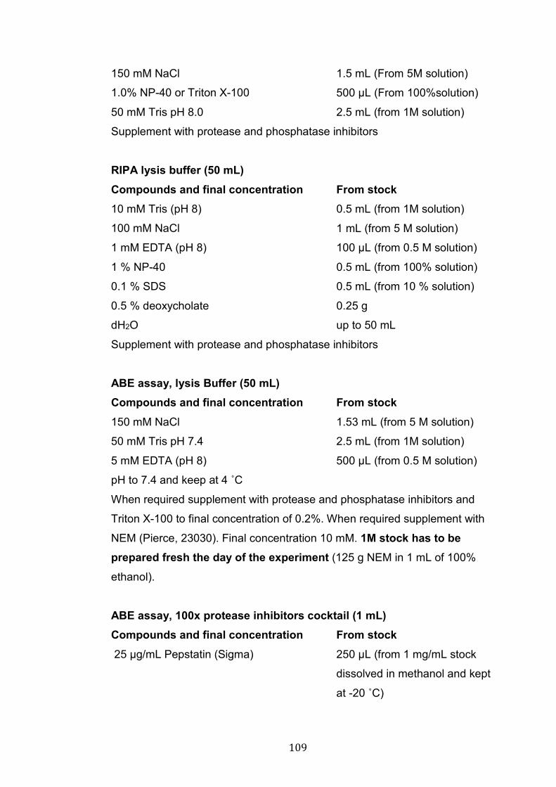
109
150 mM NaCl 1.5 mL (From 5M solution)
1.0% NP-40 or Triton X-100 500 μL (From 100%solution)
50 mM Tris pH 8.0 2.5 mL (from 1M solution)
Supplement with protease and phosphatase inhibitors
RIPA lysis buffer (50 mL) Compounds and final concentration From stock
10 mM Tris (pH 8) 0.5 mL (from 1M solution)
100 mM NaCl 1 mL (from 5 M solution)
1 mM EDTA (pH 8) 100 μL (from 0.5 M solution)
1 % NP-40 0.5 mL (from 100% solution)
0.1 % SDS 0.5 mL (from 10 % solution)
0.5 % deoxycholate 0.25 g
dH2O up to 50 mL
Supplement with protease and phosphatase inhibitors
ABE assay, lysis Buffer (50 mL) Compounds and final concentration From stock 150 mM NaCl 1.53 mL (from 5 M solution)
50 mM Tris pH 7.4 2.5 mL (from 1M solution)
5 mM EDTA (pH 8) 500 μL (from 0.5 M solution)
pH to 7.4 and keep at 4 ˚C
When required supplement with protease and phosphatase inhibitors and
Triton X-100 to final concentration of 0.2%. When required supplement with
NEM (Pierce, 23030). Final concentration 10 mM. 1M stock has to be prepared fresh the day of the experiment (125 g NEM in 1 mL of 100%
ethanol).
ABE assay, 100x protease inhibitors cocktail (1 mL) Compounds and final concentration From stock 25 μg/mL Pepstatin (Sigma) 250 μL (from 1 mg/mL stock
dissolved in methanol and kept
at -20 ˚C)
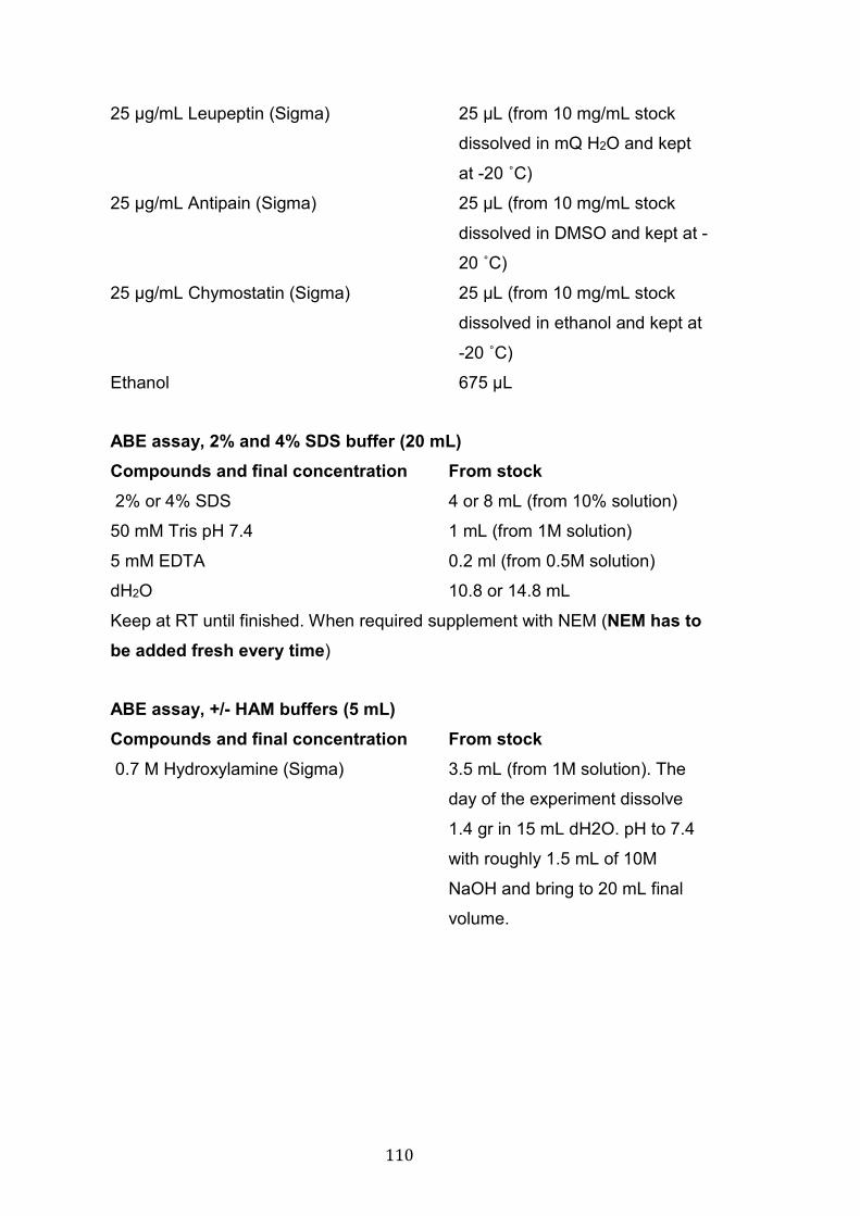
110
25 μg/mL Leupeptin (Sigma) 25 μL (from 10 mg/mL stock
dissolved in mQ H2O and kept
at -20 ˚C)
25 μg/mL Antipain (Sigma) 25 μL (from 10 mg/mL stock
dissolved in DMSO and kept at -
20 ˚C)
25 μg/mL Chymostatin (Sigma) 25 μL (from 10 mg/mL stock
dissolved in ethanol and kept at
-20 ˚C)
Ethanol 675 μL
ABE assay, 2% and 4% SDS buffer (20 mL) Compounds and final concentration From stock 2% or 4% SDS 4 or 8 mL (from 10% solution)
50 mM Tris pH 7.4 1 mL (from 1M solution)
5 mM EDTA 0.2 ml (from 0.5M solution)
dH2O 10.8 or 14.8 mL
Keep at RT until finished. When required supplement with NEM (NEM has to be added fresh every time)
ABE assay, +/- HAM buffers (5 mL) Compounds and final concentration From stock 0.7 M Hydroxylamine (Sigma) 3.5 mL (from 1M solution). The
day of the experiment dissolve
1.4 gr in 15 mL dH2O. pH to 7.4
with roughly 1.5 mL of 10M
NaOH and bring to 20 mL final
volume.

111
1 mM HPDP–biotin ( Pierce, 21341) 1.25 ml from 4 mM HPDP–biotin.
HPDP- biotin is dissolved at 50
mM in DMSO and stored at -20
˚C. The day of the experiment dilute the 50 mM stock to 4mM in
N,N-dimethyl formamide (Sigma)
0.2% Triton X-100 50 μL (from 20% solution)
1x protease inhibitors 50 μL (from 100x solution)
1 mM PMSF 50 μL (from 100mM solution)
dH2O 50 μL
-HAM solution has the same components, but HAM is substituted by 250 μL
of 1M Tris pH 7.4 and 3.25 mL dH2O.
ABE assay, Low HPDP–biotin buffer (10 mL) Compounds and final concentration From stock 0.2 mM HPDP–biotin 0.5 mL ml from 4 mM HPDP–
biotin.
150 mM NaCl 300 μL (from 5M solution)
50 mM Tris pH 7.4 500 μL (from 1M solution)
5 mM EDTA 100 μL (from 0.5M solution)
0.2% Triton X-100 100 μL (from 20% solution)
1x protease inhibitors 100 μL (from 100x solution)
1 mM PMSF 100 μL (from 100mM solution)
dH2O 8.2 mL
Co-IP lysis buffer (50mL) Compounds and final concentration From stock 50 mM Tris pH 7.5 or 8 2.5 mL (from 1M solution)
150 mM NaCl 1.5 mL (from 5M solution)
1 mM EDTA 100 μL (from 0.5M solution)
0.5% Triton X-100 1 ml (from 25% solution)
dH2O up to 50 mL
Supplement with protease and phosphate inhibitors
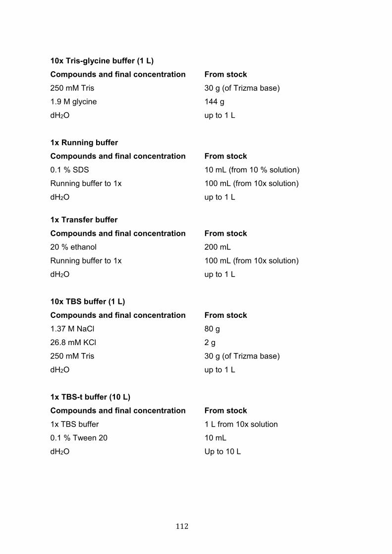
112
10x Tris-glycine buffer (1 L) Compounds and final concentration From stock 250 mM Tris 30 g (of Trizma base)
1.9 M glycine 144 g
dH2O up to 1 L
1x Running buffer Compounds and final concentration From stock 0.1 % SDS 10 mL (from 10 % solution)
Running buffer to 1x 100 mL (from 10x solution)
dH2O up to 1 L
1x Transfer buffer Compounds and final concentration From stock 20 % ethanol 200 mL
Running buffer to 1x 100 mL (from 10x solution)
dH2O up to 1 L
10x TBS buffer (1 L) Compounds and final concentration From stock 1.37 M NaCl 80 g
26.8 mM KCl 2 g
250 mM Tris 30 g (of Trizma base)
dH2O up to 1 L
1x TBS-t buffer (10 L) Compounds and final concentration From stock 1x TBS buffer 1 L from 10x solution
0.1 % Tween 20 10 mL
dH2O Up to 10 L
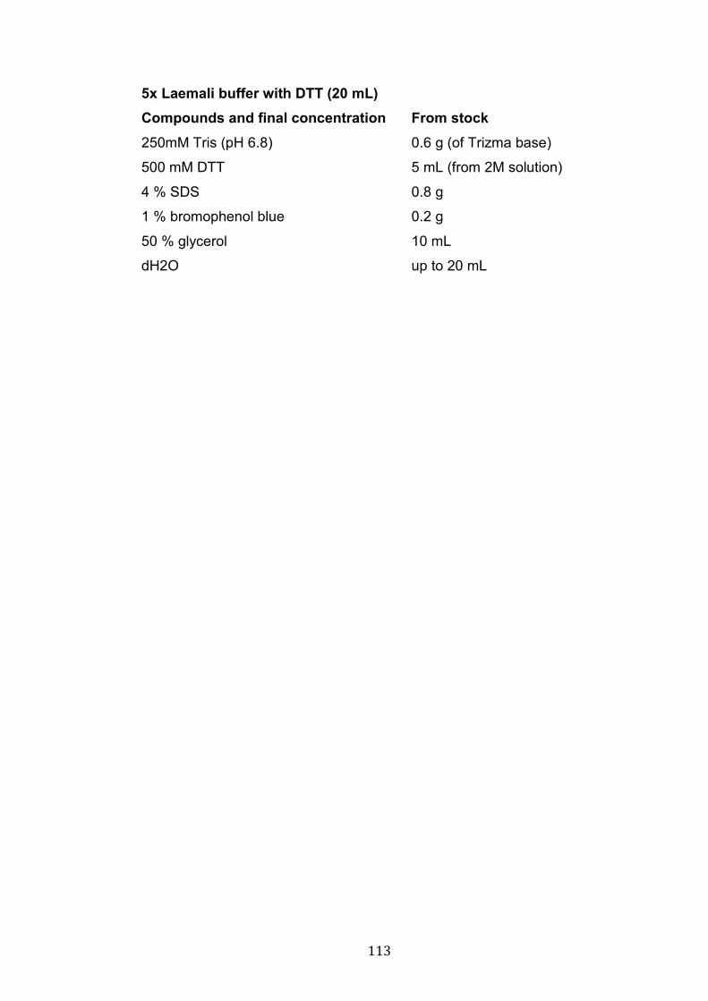
113
5x Laemali buffer with DTT (20 mL) Compounds and final concentration From stock 250mM Tris (pH 6.8) 0.6 g (of Trizma base)
500 mM DTT 5 mL (from 2M solution)
4 % SDS 0.8 g
1 % bromophenol blue 0.2 g
50 % glycerol 10 mL
dH2O up to 20 mL
114
CHAPTER 3: THE ROLE OF Fz5 AND Fz7 IN PRE- AND POSTSYNAPTIC DEVELOPMENT 3.1 Introduction
Wnt molecules are key organizers of embryonic development (MacDonald et
al. 2009; Nusse and Clevers 2017; Wiese et al. 2018). In addition, numerous
research demonstrates that Wnt signalling plays a pivotal role in controlling
several aspects of CNS development and function, such as BBB formation,
dendritogenesis, axon guidance, synapse formation, synaptic plasticity and
maintenance (Liebner et al. 2008; Budnik and Salinas 2011; Salinas 2012;
McLeod and Salinas 2018; Oliva et al. 2013b).
The synaptogenic activity of Wnt molecules was first described over 20 years
ago. Our laboratory demonstrated that exogenous Wnt7a induces axonal
remodelling and the assembly of presynaptic sites in cerebellar granule cells
(Lucas and Salinas 1997). Importantly, the effects of Wnt7a were confirmed
in vivo at granule cell/mossy fibre synapses using a Wnt7a-/- mouse model
(Hall et al. 2000). Since then, mounting evidence has unravelled the role of
Wnt7a in orchestrating the formation and function of both sides of the
synapse. On the presynaptic side, Wnt7a promotes the assembly of key
presynaptic structures (Lucas and Salinas 1997; Hall et al. 2000; Davis et al.
2008; Sahores et al. 2010) and regulates synaptic transmission by controlling
vesicle recycling and neurotransmitter release (Ahmad-Annuar et al. 2006;
Cerpa et al. 2008; Ciani et al. 2015). On the postsynaptic side, Wnt7a is
required for the formation and growth of dendritic spines (Ciani et al. 2011),
which are the postsynaptic structures for the vast majority of excitatory inputs.
Moreover, on the postsynaptic side Wnt7a regulates the trafficking of α-
amino-3-hydroxy-5-methyl-4-isoxazolepropionic acid receptors (AMPARs),
strengthening synaptic plasticity (McLeod et al. 2018). Therefore, Wnt7a
promotes synapse assembly by acting on both sides of the synapse.
However, a key question still remains: how do Wnts drive synaptogenesis?
115
The action of different Wnts on their receptors, and the activation of
downstream cascades, determines the function of these molecules. At the
cell surface Wnt ligands can bind to a number of different receptors, including
the canonical co-receptor LRP6, the non-canonical co-receptors Ror and Ryk
and the main Wnt receptors Frizzled (van Amerongen 2012). The role of
Frizzled receptors in Wnt-mediated synaptogenesis is poorly understood.
Work from our lab has shown that the synaptogenic factor Wnt7a can bind to
Fz5 and Fz7, which are both found in synaptosome fractions (Sahores et al.
2010; McLeod et al. 2018). Fz5 is enriched at presynaptic sites and
overexpression of this receptor in hippocampal dissociated neurons is
sufficient to induce presynaptic differentiation, mimicking the effect of Wnt7a
(Sahores et al. 2010). Importantly, loss of function studies demonstrated that
Fz5 is required for Wnt7a-mediated presynaptic assembly in cultured
neurons (Sahores et al. 2010). However, how Wnt7a signalling is mediated
at postsynaptic sites remained unknown. In this chapter, I will present gain
and loss of function studies performed on dissociated hippocampal neurons
where I compared the role of Fz5 and Fz7 in the development of both sides
of the synapse. I have addressed the following specific questions:
1) Are Fz5 and Fz7 similarly localised at dendritic spines?
2) Does Fz5, a presynaptic receptor for Wnt7a, have a role in
postsynaptic development?
3) Is Fz7 required for postsynaptic development?
4) Does Wnt7a-Fz7 signalling drive dendritic spine formation?
5) Is Fz7 also required for presynaptic differentiation?
116
3.2 Results
3.2.1 Fz5 and Fz7 localisation at dendritic spines
Wnt7a signals on axons to induce presynaptic assembly (Lucas and Salinas
1997; Hall et al. 2000; Davis et al. 2008), a function mediated by Fz5
receptors (Fig 1.9) (Sahores et al. 2010). On the postsynaptic side, Wnt7a
ligands regulate dendritic spine formation and morphology (Ciani et al. 2011).
However, the receptor required for Wnt7a to induce postsynaptic
development was unknown. A binding assay screen revealed that Wnt7a can
also bind Fz7, which is expressed in the brain and found in synaptosome
fractions (McLeod et al. 2018). In a work recently published (McLeod et al.
2018), we compared the localisation and function of Fz5 and Fz7 to identify
the receptor required for Wnt7a to induce postsynaptic development.
Using immunofluorescence and confocaI microscopy, I first examined the
localisation of these two Wnt receptors in dissociated hippocampal neurons.
We have previously used antibodies against endogenous Fz5 to examine its
distribution along axons and dendrites, but not dendritic spines (Sahores et al.
2010). Unfortunately, these antibodies are no longer available; therefore, the
distribution of Fz5 was analysed by expressing Fz5-HA in cultured neurons.
EGFP-actin was co-expressed with Fz5-HA to visualise the morphology of
the cells in particular dendritic spines, where actin is enriched (Fig 3.1). The
cells were examined at 12-14 DIV (days in vitro), within the peak of
synaptogenesis (12-16 DIV), unless otherwise stated. As previously
observed (Sahores et al. 2010), I found that Fz5 receptors were present in
the soma and along axons and dendrites. Although Fz5 was clearly present
along dendrites, it was almost never observed within dendritic spines (Fig 3.2
A), suggesting that this receptor might not be involved in mechanisms of
spine development and function. In contrast, primary antibodies against
endogenous Fz7 revealed the presence of this receptor in dendritic spines
(Fig 3.3 A). Although these data were obtained using different approaches
(overexpression vs endogenous), these findings indicate that these two Wnt
receptors exhibit different localisation: Fz5 is not localised at dendritic spines
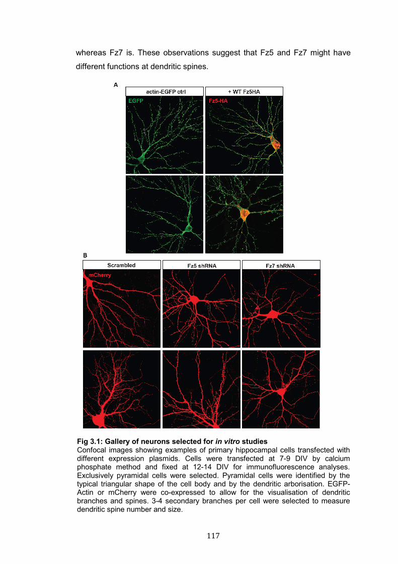
117
whereas Fz7 is. These observations suggest that Fz5 and Fz7 might have
different functions at dendritic spines.
Fig 3.1: Gallery of neurons selected for in vitro studies Confocal images showing examples of primary hippocampal cells transfected with different expression plasmids. Cells were transfected at 7-9 DIV by calcium phosphate method and fixed at 12-14 DIV for immunofluorescence analyses. Exclusively pyramidal cells were selected. Pyramidal cells were identified by the typical triangular shape of the cell body and by the dendritic arborisation. EGFP-Actin or mCherry were co-expressed to allow for the visualisation of dendritic branches and spines. 3-4 secondary branches per cell were selected to measure dendritic spine number and size.
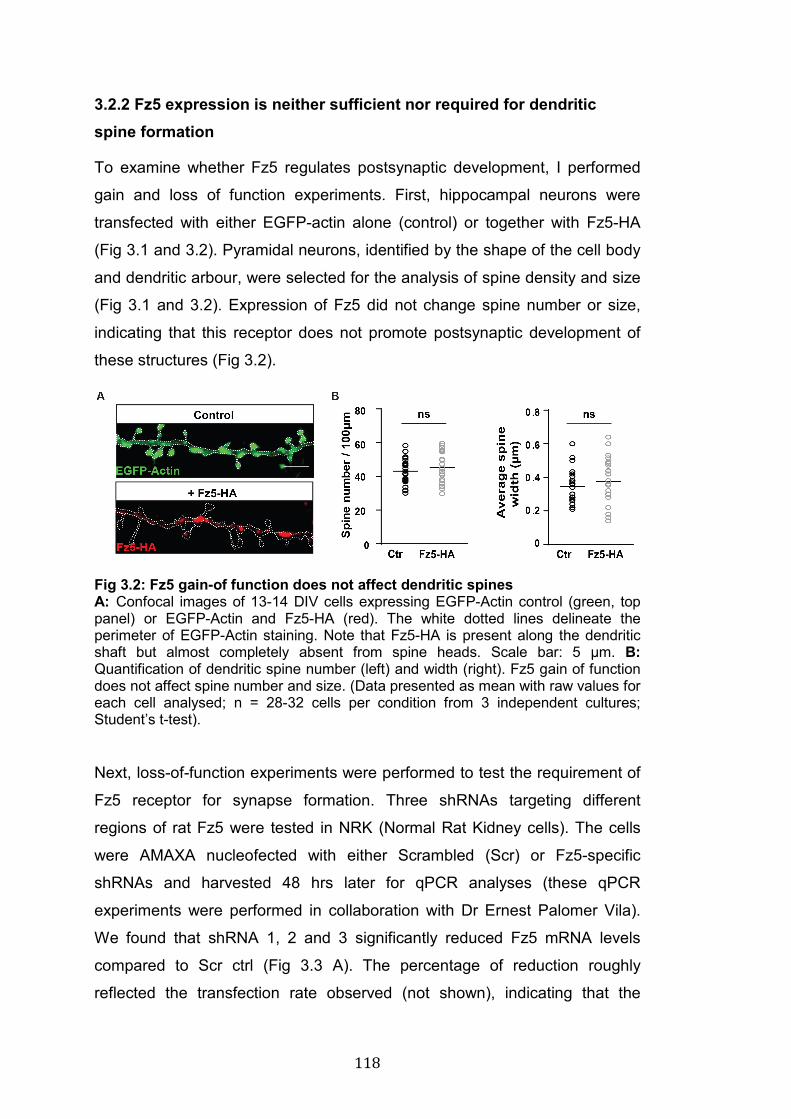
118
3.2.2 Fz5 expression is neither sufficient nor required for dendritic spine formation
To examine whether Fz5 regulates postsynaptic development, I performed
gain and loss of function experiments. First, hippocampal neurons were
transfected with either EGFP-actin alone (control) or together with Fz5-HA
(Fig 3.1 and 3.2). Pyramidal neurons, identified by the shape of the cell body
and dendritic arbour, were selected for the analysis of spine density and size
(Fig 3.1 and 3.2). Expression of Fz5 did not change spine number or size,
indicating that this receptor does not promote postsynaptic development of
these structures (Fig 3.2).
Fig 3.2: Fz5 gain-of function does not affect dendritic spines A: Confocal images of 13-14 DIV cells expressing EGFP-Actin control (green, top panel) or EGFP-Actin and Fz5-HA (red). The white dotted lines delineate the perimeter of EGFP-Actin staining. Note that Fz5-HA is present along the dendritic shaft but almost completely absent from spine heads. Scale bar: 5 µm. B: Quantification of dendritic spine number (left) and width (right). Fz5 gain of function does not affect spine number and size. (Data presented as mean with raw values for each cell analysed; n = 28-32 cells per condition from 3 independent cultures; Student’s t-test).
Next, loss-of-function experiments were performed to test the requirement of
Fz5 receptor for synapse formation. Three shRNAs targeting different
regions of rat Fz5 were tested in NRK (Normal Rat Kidney cells). The cells
were AMAXA nucleofected with either Scrambled (Scr) or Fz5-specific
shRNAs and harvested 48 hrs later for qPCR analyses (these qPCR
experiments were performed in collaboration with Dr Ernest Palomer Vila).
We found that shRNA 1, 2 and 3 significantly reduced Fz5 mRNA levels
compared to Scr ctrl (Fig 3.3 A). The percentage of reduction roughly
reflected the transfection rate observed (not shown), indicating that the
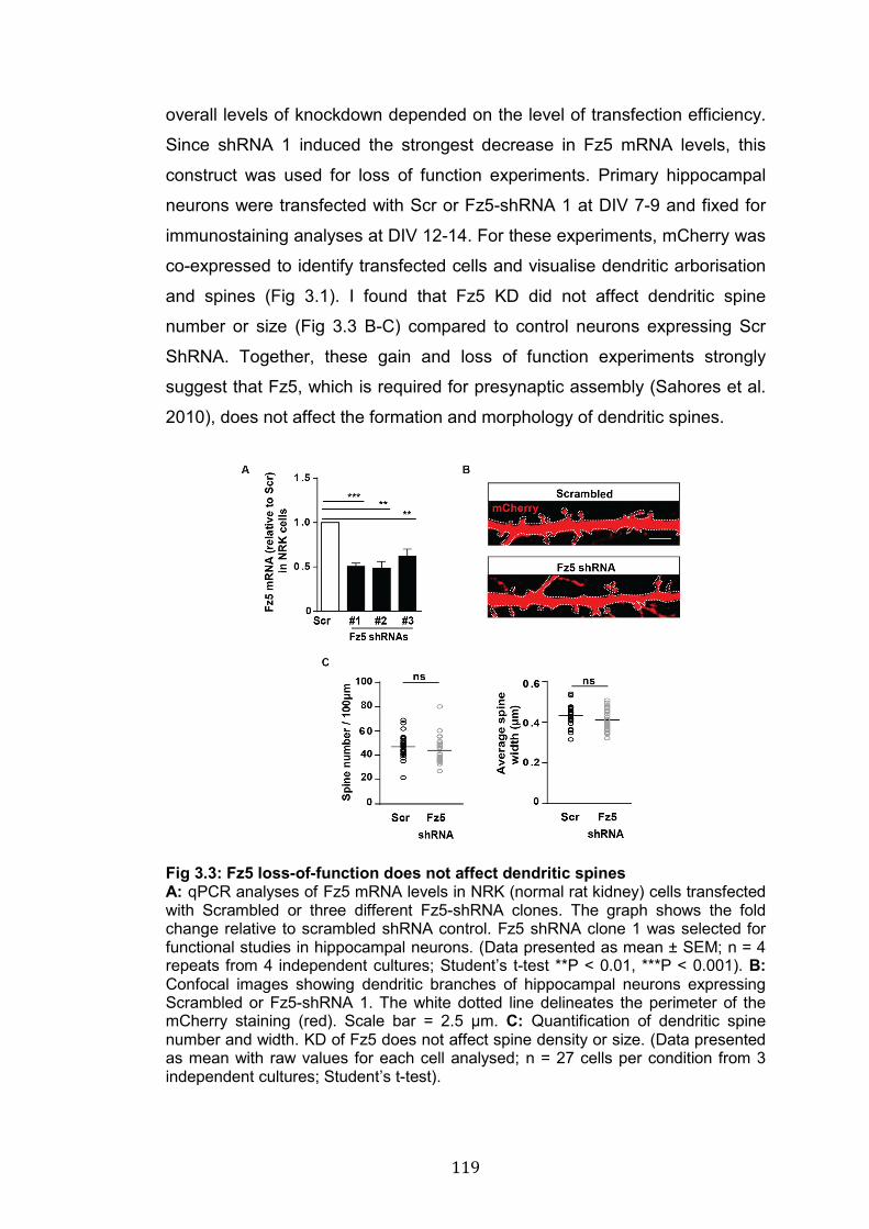
119
overall levels of knockdown depended on the level of transfection efficiency.
Since shRNA 1 induced the strongest decrease in Fz5 mRNA levels, this
construct was used for loss of function experiments. Primary hippocampal
neurons were transfected with Scr or Fz5-shRNA 1 at DIV 7-9 and fixed for
immunostaining analyses at DIV 12-14. For these experiments, mCherry was
co-expressed to identify transfected cells and visualise dendritic arborisation
and spines (Fig 3.1). I found that Fz5 KD did not affect dendritic spine
number or size (Fig 3.3 B-C) compared to control neurons expressing Scr
ShRNA. Together, these gain and loss of function experiments strongly
suggest that Fz5, which is required for presynaptic assembly (Sahores et al.
2010), does not affect the formation and morphology of dendritic spines.
Fig 3.3: Fz5 loss-of-function does not affect dendritic spines A: qPCR analyses of Fz5 mRNA levels in NRK (normal rat kidney) cells transfected with Scrambled or three different Fz5-shRNA clones. The graph shows the fold change relative to scrambled shRNA control. Fz5 shRNA clone 1 was selected for functional studies in hippocampal neurons. (Data presented as mean ± SEM; n = 4 repeats from 4 independent cultures; Student’s t-test **P < 0.01, ***P < 0.001). B: Confocal images showing dendritic branches of hippocampal neurons expressing Scrambled or Fz5-shRNA 1. The white dotted line delineates the perimeter of the mCherry staining (red). Scale bar = 2.5 µm. C: Quantification of dendritic spine number and width. KD of Fz5 does not affect spine density or size. (Data presented as mean with raw values for each cell analysed; n = 27 cells per condition from 3 independent cultures; Student’s t-test).
120
3.2.3 Fz7 is required for postsynaptic development and Wnt7a-mediated spine formation
The role of Fz7 in postsynaptic development was tested by loss of function
experiments in dissociated hippocampal neurons. First, KD (knock-down) of
Fz7 was tested in NRK cells as described for Fz5. NRK cells transfected with
Fz7-shRNA 1, 2 and 3 showed a significant reduction of Fz7 mRNA levels
compared to Scr ctrl (Fig 3.4 B). I then tested the impact of the KD in primary
neurons. Scr and shRNA 3 were transfected into primary hippocampal
neurons at 7-9 DIV and the dendritic phenotype was examined at 12-14 DIV
when spines are formed. I found that KD of Fz7 resulted in a significant
decrease in the number of dendritic spines when compared to neurons
expressing Scr shRNA (Fig 3.4 C-D). However, spine size was unchanged
between Scr and KD cells (Fig 3.4 E-F). To rule out off-target effects, I
performed rescue experiments by co-expressing Fz7-ShRNA together with
shRNA-resistant Fz7 cDNA (rescue). Expression of the shRNA-resistant Fz7
fully rescued the number of dendritic spines to Scr levels (Fig 3.4 C-D),
demonstrating that the spine defect was not due to off target effects.
Next, I tested whether Fz7 is required for Wnt7a to signal on the postsynaptic
side. For this, neurons (12-14 DIV) were treated for 3 hrs with recombinant
Wnt7a and the effects on spines were analysed. As expected, spine size and
number were significantly increased in Scr expressing cells exposed to
Wnt7a compared to controls (BSA treated) (Fig 3.4 E-F) (Ciani et al. 2011).
However, the increase in spine size and number was completely abolished in
Fz7 KD cells, suggesting that these cells were unable to respond to Wnt7a
(Fig 3.4 E-F). Altogether, these data indicate that Fz7 is necessary for the
development of dendritic spines and is required for Wnt7a-mediated spine
growth.
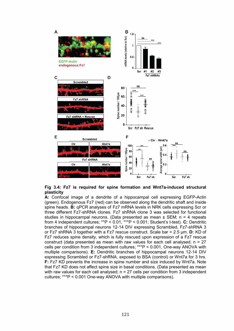
121
Fig 3.4: Fz7 is required for spine formation and Wnt7a-induced structural plasticity A: Confocal image of a dendrite of a hippocampal cell expressing EGFP-Actin (green). Endogenous Fz7 (red) can be observed along the dendritic shaft and inside spine heads. B: qPCR analyses of Fz7 mRNA levels in NRK cells expressing Scr or three different Fz7-shRNA clones. Fz7 shRNA clone 3 was selected for functional studies in hippocampal neurons. (Data presented as mean ± SEM; n = 4 repeats from 4 independent cultures; **P < 0.01, ***P < 0.001; Student’s t-test). C: Dendritic branches of hippocampal neurons 12-14 DIV expressing Scrambled, Fz7-shRNA 3 or Fz7 shRNA 3 together with a Fz7 rescue construct. Scale bar = 2.5 µm. D: KD of Fz7 reduces spine density, which is fully rescued upon expression of a Fz7 rescue construct (data presented as mean with raw values for each cell analysed; n = 27 cells per condition from 3 independent cultures; ***P < 0.001, One-way ANOVA with multiple comparisons). E: Dendritic branches of hippocampal neurons 12-14 DIV expressing Scrambled or Fz7-shRNA, exposed to BSA (control) or Wnt7a for 3 hrs. F: Fz7 KD prevents the increase in spine number and size induced by Wnt7a. Note that Fz7 KD does not affect spine size in basal conditions. (Data presented as mean with raw values for each cell analysed; n = 27 cells per condition from 3 independent cultures; ***P < 0.001 One-way ANOVA with multiple comparisons).
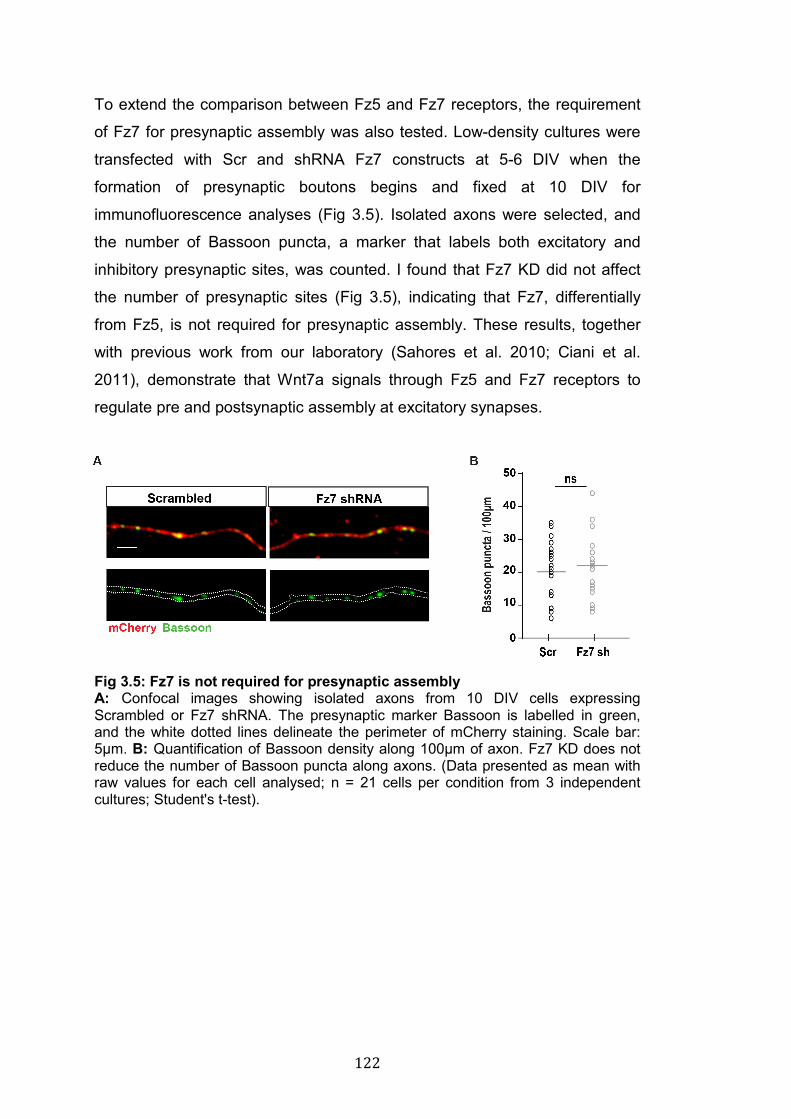
122
To extend the comparison between Fz5 and Fz7 receptors, the requirement
of Fz7 for presynaptic assembly was also tested. Low-density cultures were
transfected with Scr and shRNA Fz7 constructs at 5-6 DIV when the
formation of presynaptic boutons begins and fixed at 10 DIV for
immunofluorescence analyses (Fig 3.5). Isolated axons were selected, and
the number of Bassoon puncta, a marker that labels both excitatory and
inhibitory presynaptic sites, was counted. I found that Fz7 KD did not affect
the number of presynaptic sites (Fig 3.5), indicating that Fz7, differentially
from Fz5, is not required for presynaptic assembly. These results, together
with previous work from our laboratory (Sahores et al. 2010; Ciani et al.
2011), demonstrate that Wnt7a signals through Fz5 and Fz7 receptors to
regulate pre and postsynaptic assembly at excitatory synapses.
Fig 3.5: Fz7 is not required for presynaptic assembly A: Confocal images showing isolated axons from 10 DIV cells expressing Scrambled or Fz7 shRNA. The presynaptic marker Bassoon is labelled in green, and the white dotted lines delineate the perimeter of mCherry staining. Scale bar: 5µm. B: Quantification of Bassoon density along 100µm of axon. Fz7 KD does not reduce the number of Bassoon puncta along axons. (Data presented as mean with raw values for each cell analysed; n = 21 cells per condition from 3 independent cultures; Student's t-test).
123
3.3 Discussion
Some of the Wnts (4 of the 19 Wnts) expressed in mammals have been
shown to play a crucial role in synapse formation (Salinas 2012). Particularly,
the Wnt7a ligand promotes the formation of both pre and postsynaptic sides
at excitatory synapses (Hall et al. 2000; Sahores et al. 2010; Ciani et al.
2011). Wnt7a signals through Fz5 on axons to promote presynaptic
assembly (Sahores et al. 2010). However, the postsynaptic receptor for
Wnt7a was unknown until now. Here, I have characterised the localisation of
two Wnt7a receptors, Fz5 and Fz7, at dendritic spines and I have examined
their function at both sides of the synapse.
3.3.1 The distribution of Fz5 and Fz7 at dendritic spines
Biochemical analyses reported that both Fz5 and Fz7 are present at
synapses (Sahores et al. 2010; McLeod et al. 2018). Using
immunofluorescence analyses, I found that these receptors exhibit distinct
localisation at dendritic spines, where Fz7, but not Fz5, is detected (Fig 3.2
A; Fig 3.4 A). It should be noted that the distribution of Fz5 was studied using
overexpression techniques due to the lack of suitable primary antibodies.
Although this approach can sometimes create artefacts in protein localisation,
gain and loss of function experiments presented here confirmed that Fz5 is
not involved in postsynaptic development. In the general discussion (see
chapter 7), I will consider hypotheses on what molecular mechanisms may
establish the different localisation and function of Fz5 and Fz7.
3.3.2 Fz5 is not required for dendritic spine development
Our laboratory demonstrated that Fz5 is required and is sufficient to drive
presynaptic assembly in cultured neurons (Sahores et al. 2010). Moreover,
Fz5 is required for Wnt7a- and activity-induced formation of presynaptic
boutons (Sahores et al. 2010). However, the role of this receptor in the
development of dendritic spines had not been explored before. Here, I
showed that neither gain nor loss of function of Fz5 affected spine size and
number. Altogether these data demonstrate that Fz5, whose function at the
124
presynaptic side was previously reported, is not required for spine
development.
One question still remains, what is the function of Fz5 in dendrites? Given
the distribution of this receptor along the dendritic shaft, we could
hypothesise a role for Fz5 in the formation of inhibitory postsynaptic sites,
which are mostly found on the shaft rather than on dendritic spines (Moss
and Smart 2001). In chapter 6 I will present data showing the role of Fz5
expression in excitatory synapse formation in vivo. We are currently using
the same samples to examine inhibitory synapse formation. Very preliminary
data suggest that Fz5 is not involved in the formation of inhibitory synapses
in the developing hippocampus; consistently, other preliminary data from
cultured neurons suggest that Fz5 puncta in the shaft do not seem to
colocalise with inhibitory postsynaptic structures (Bossio and Salinas
unpublished results). Therefore, the function of Fz5 receptors in dendrites
remains to be elucidated.
3.3.3 The role of Fz7 at pre- and postsynaptic sites
The data presented here strongly suggest that Fz7 is not required for
presynaptic assembly. Whether Fz7 plays a role in axons still needs to be
addressed. Beside presynaptic assembly, Wnt signalling plays an important
role in regulating axon growth cone dynamics, SVs recycling and
neurotransmitter release (Ciani et al. 2004; Purro et al. 2008; Stamatakou et
al. 2015; Ciani et al. 2015). Thus, although Fz7 does not affect presynaptic
assembly its role in these Wnt-mediated presynaptic functions should be
investigated.
Data from our lab showed that the synaptogenic factor Wnt7a binds to Fz7
(McLeod et al. 2018), which localises at dendritic spines (Fig 3.4 A). I found
that in basal conditions loss of Fz7 down-regulates the density of dendritic
spines but not their size. In addition, loss of function of Fz7 prevented Wnt7a-
induced increase in spine number and size, strongly suggesting that Fz7
mediates Wnt7a effects at dendritic spines. An interesting question arises
from these data. If Fz7 does not regulate spine size, how does it block
Wnt7a-mediated enlargement of these structures?
125
The size of dendritic spine is determined by the area of the postsynaptic
density, the amount of postsynaptic receptors and the cytoskeleton within the
spine head and neck (Arellano et al. 2007). Molecular mechanisms
regulating these factors are in place to continuously maintain the size of
dendritic spines. My data indicate that in basal conditions KD of Fz7 does not
affect dendritic spines, suggesting that this receptor is dispensable for steady
state maintenance of spines size. However, it is important to examine these
data in the context of structural plasticity of dendritic spines. Spines rapidly
increase or decrease their size in response to external stimuli, such as
experience-dependent or induced patterns of neuronal activity (Hering and
Sheng 2001; Rochefort and Konnerth 2012; Bosch and Hayashi 2012). How
does neuronal activity control spine size? It is established that neuronal
activity promotes the expression and secretion of several synaptogenic
molecules, including BDNF and Wnts, which play a role in shaping spine
morphology (Thoenen 1991; Harward et al. 2016; McLeod and Salinas 2018).
Our lab has recently demonstrated that during the very early stages of LTP
induction (within 10 mins) Wnt7a levels are increased at synapse (McLeod et
al. 2018). In addition, we showed that Fz7 receptors are required for LTP-
induced spine enlargement and, consistently, we found that Fz7 is required
for the induction of LTP. In light of these findings, the fact that Wnt7a does
not increase spine size in Fz7 KD cells indicates that this receptor is required
for structural plasticity of dendritic spines.
The work presented in this chapter enriches our understanding of the role of
Wnt receptors in synapse formation. Several Wnt receptors, including LRP6,
Ryk, Ror and Frizzled have been shown to be required for pre and/or
postsynaptic assembly (Mathew et al. 2005; Varela-Nallar et al. 2009;
Paganoni et al. 2010; Sahores et al. 2010; Sharma et al. 2013). Wnt7a is the
best-characterised synaptogenic Wnt ligand; however, the receptor required
to mediate its function at dendritic spines was unknown until now. My data,
alongside previous work from our lab (Sahores et al. 2010), describe distinct
localisation and function of Fz5 and Fz7 at both sides of the synapse,
shedding light on the molecular mechanisms of Wnt7a-mediated synapse
formation.

126
127
CHAPTER 4: PALMITOYLATION OF FRIZZLED RECEPTORS 4.1 Introduction
The cell-surface recruitment and localisation of receptors is crucial for proper
signalling in every cell. This is often achieved by regulating protein trafficking
and function through post-translational modifications (PTMs). The
biochemistry of these modifications can be very diverse, ranging from the
attachment of a single phosphate group to large carbohydrate or lipid chains.
Thus, PTMs have a direct impact on the structure, localisation and function of
proteins.
Extensive efforts have been made to understand how key synaptic proteins
are trafficked to synapses, and how their function is regulated in response to
different stimuli (Lau and Zukin 2007; Anggono and Huganir 2012; Bassani et
al. 2013; Choquet and Triller 2013; Diering and Huganir 2018). However,
much less is known about the synaptic recruitment and regulation of
synaptogenic factors and their receptors, particularly Frizzled receptors. The
surface levels of the synaptogenic receptor Fz5 are up and downregulated by
HFS and LFS (high/low frequency stimulation) respectively, and Fz5 is
required for Wnt7a- and activity-mediated synapse formation in hippocampal
neurons (Sahores et al. 2010). In light of these results, we started to
investigate potential molecular mechanisms that could control Fz5 trafficking
in a dynamic manner.
We decided to examine for specific features at the C-terminal domain of the
receptor, which is crucial for signalling (Tauriello et al. 2012) and could
determine Fz5 trafficking and retention at the PM and at synapses.
Observations made by a former member of our lab, Dr E. Stamatakou,
revealed the presence of three cysteine (Cys) residues at the C-term of Fz5;
these Cys could be sites for PTMs such as palmitoylation (Stamatakou and
Salinas unpublished). Indeed, by mutagenesis of the three Cys to Ser, which
cannot be palmitoylayed, and radiolabelling, Dr. Stamatakou confirmed that
Fz5 is palmitoylated (Fig 1.13). In addition, preliminary observations suggest
that palmitoylation is required for Fz5 synaptogenic activity in cultured
neurons (Fig 1.13) (Stamatakou and Salinas, unpublished). These results
128
were exciting because they revealed for the first time that Frizzled receptors
can be palmitoylated and that this modification might be crucial for their
function. Therefore, the study of this modification is novel and of extreme
interest for the Wnt signalling field.
We focused on palmitoylation because it is one of the most common lipid
modifications of synaptic proteins and it regulates their trafficking and
function at synapses (Fukata and Fukata 2010; Globa and Bamji 2017). S-
palmitoylation, distinguished from N-palmitoylation (Fig 1.10), consists of the
reversible attachment of a long saturated chain of palmitic acid on Cys
residues, and is the most common lipid modification of proteins (Resh 2016;
Linder and Deschenes 2007). As well as increasing protein hydrophobicity,
palmitoylation can alter protein conformation, as in the case of the Wnt co-
receptor LRP6 (Abrami et al. 2008), arrest or allow the progression of
proteins along the trafficking route, and target molecules to specific
membrane domains (Charollais and Van Der Goot 2009; Aicart-Ramos et al.
2011). In addition, protein-protein interactions and the interplay with other
PTMs can be affected by palmitoylation (Salaun et al. 2010; Blaskovic et al.
2013).
S-palmitoylation is catalysed by a specific family of protein acyl transferases
(PATs) named DHHC enzymes (Fukata et al. 2006; Guan and Fierke 2011).
DHHC enzymes are specifically localised withtin the cell (ER, Golgi,
endosomes, plasma membrane) and between tissues (Ohno et al. 2006).
Many of these enzymes are expressed in the brain and their substrates have
only just started to be identified (Fukata and Fukata 2010). The identification
of DHHC-substrates has been instrumental in elucidating the molecular
mechanisms that control protein trafficking and function at the synapse
(Fukata and Fukata 2010; Globa and Bamji 2017).
Although our studies demonstrate that Fz5 is palmitoylated (Fig 1.13), we did
not know whether other Frizzled receptors are also palmitoylated and
whether all the Cys at the C-term of Fz5 can be palmitoylated. Furthermore, I
have asked which are the enzymes that palmitoylate Fz5, and performed
gain and loss of function studies on members of the DHHC family of protein
129
acyl transferases. The novelty of the data presented in this and the next
chapter opens new doors to unravel the mechanisms of regulation of Wnt
signalling at the level of Frizzled receptors. Understanding how Wnt
receptors are regulated will help us to comprehend and, where necessary,
manipulate Frizzled function in different biological processes in which Wnt
signalling is involved. In this chapter I will present data that address these
specific questions:
1. Which Fz receptors are palmitoylated?
2. Which Cys residues are palmitoylated in Fz5?
3. Which DHHC enzymes can palmitoylate Fz5?
4. Which DHHC enzymes are required for Fz5 palmitoylation?
130
4.2 Results
4.2.1 Some Frizzled receptors are palmitoylated
Data from our laboratory indicate that Fz5 is palmitoylated in vivo and in vitro
(Fig 1.13, Stamatakou and Salinas unpublished results). However, whether
other Frizzled receptors are palmitoylated had not been investigated before.
To test this, I first used the CSS-Palm 4.0 software (Ren et al. 2008) to
predict putative sites of palmitoylation in the entire protein sequence of all
Frizzled receptors. Each of the 10 Frizzled receptors (human sequences)
was predicted to be palmitoylated on at least one Cys residue (Fig 4.1).
These Cys residues are spread across different protein domains including
the N-term, transmembrane (TM) regions and the C-term. Although the C-
terms of Fz3, Fz4, Fz5, Fz6, Fz8, Fz9 and Fz10 contain at least one Cys (Fig
4.2), only those of Fz5 and its homolog Fz8 were predicted to be
palmitoylated (Fig 4.1). Taken together, these computational analyses
suggest that all Frizzled receptors might be palmitoylated on several Cys
residues. However, given the heterogeneous distribution of these Cys
residues (e.g N-term vs TM vs C-term), we could speculate that
palmitoylation, if present, might have very distinct functions for different
Frizzled receptors.
Frizzled receptors were screened for palmitoylation using the ABE
biochemical assay (Wan et al. 2007). All the receptors were expressed in
HEK293 cells. Palmitoylation was clearly detectable for Fz3, Fz4, Fz5, Fz6,
and Fz9 (Fig 4.3 A). Fz1, Fz7 and Fz8 exhibited lower levels of palmitoylation
and only very weak signals were observed for Fz2 and Fz10 after long
exposure of the WB membrane (Fig 4.3 A). These results indicate that
several Frizzled receptors are palmitoylated, most likely on the Cys residues
identified with CSS-Palm-4.0. However, to rule out false positives, especially
for those Frizzled that only exhibited very weak palmitoylation levels, it would
be important to confirm these results by different techniques such as mass
spectrometry, incorporation of radioactive palmitate and mutagenesis of
putative palmitoylation sites, as we have done for Fz5 (Stamatakou and
Salinas, unpublished results). Taken together, these bioinformatic and
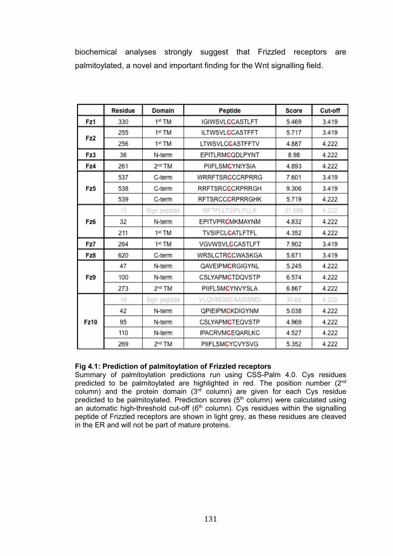
131
biochemical analyses strongly suggest that Frizzled receptors are
palmitoylated, a novel and important finding for the Wnt signalling field.
Fig 4.1: Prediction of palmitoylation of Frizzled receptors Summary of palmitoylation predictions run using CSS-Palm 4.0. Cys residues predicted to be palmitoylated are highlighted in red. The position number (2nd column) and the protein domain (3rd column) are given for each Cys residue predicted to be palmitoylated. Prediction scores (5th column) were calculated using an automatic high-threshold cut-off (6th column). Cys residues within the signalling peptide of Frizzled receptors are shown in light grey, as these residues are cleaved in the ER and will not be part of mature proteins.
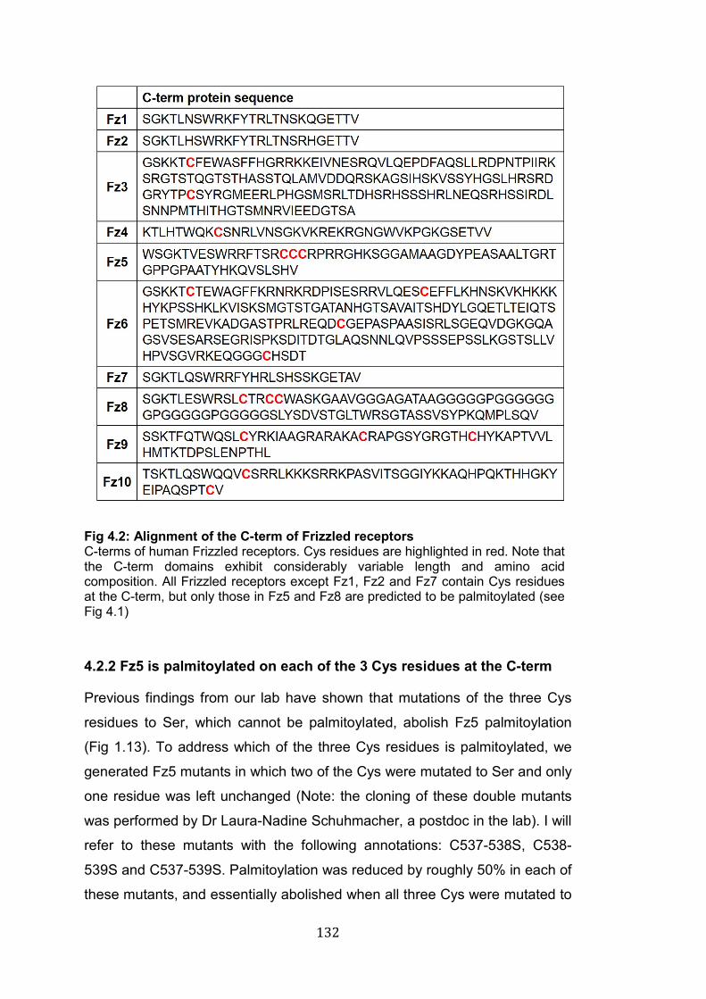
132
Fig 4.2: Alignment of the C-term of Frizzled receptors C-terms of human Frizzled receptors. Cys residues are highlighted in red. Note that the C-term domains exhibit considerably variable length and amino acid composition. All Frizzled receptors except Fz1, Fz2 and Fz7 contain Cys residues at the C-term, but only those in Fz5 and Fz8 are predicted to be palmitoylated (see Fig 4.1)
4.2.2 Fz5 is palmitoylated on each of the 3 Cys residues at the C-term
Previous findings from our lab have shown that mutations of the three Cys
residues to Ser, which cannot be palmitoylated, abolish Fz5 palmitoylation
(Fig 1.13). To address which of the three Cys residues is palmitoylated, we
generated Fz5 mutants in which two of the Cys were mutated to Ser and only
one residue was left unchanged (Note: the cloning of these double mutants
was performed by Dr Laura-Nadine Schuhmacher, a postdoc in the lab). I will
refer to these mutants with the following annotations: C537-538S, C538-
539S and C537-539S. Palmitoylation was reduced by roughly 50% in each of
these mutants, and essentially abolished when all three Cys were mutated to
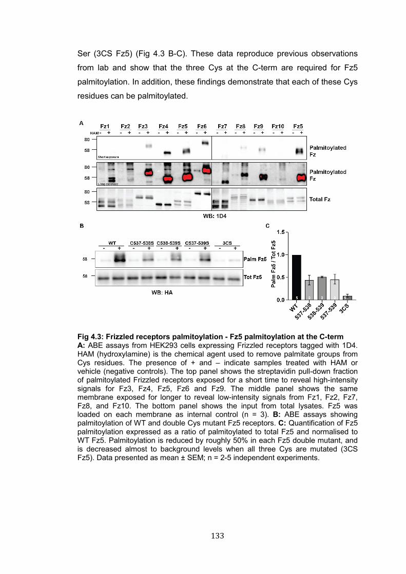
133
Ser (3CS Fz5) (Fig 4.3 B-C). These data reproduce previous observations
from lab and show that the three Cys at the C-term are required for Fz5
palmitoylation. In addition, these findings demonstrate that each of these Cys
residues can be palmitoylated.
Fig 4.3: Frizzled receptors palmitoylation - Fz5 palmitoylation at the C-term A: ABE assays from HEK293 cells expressing Frizzled receptors tagged with 1D4. HAM (hydroxylamine) is the chemical agent used to remove palmitate groups from Cys residues. The presence of + and – indicate samples treated with HAM or vehicle (negative controls). The top panel shows the streptavidin pull-down fraction of palmitoylated Frizzled receptors exposed for a short time to reveal high-intensity signals for Fz3, Fz4, Fz5, Fz6 and Fz9. The middle panel shows the same membrane exposed for longer to reveal low-intensity signals from Fz1, Fz2, Fz7, Fz8, and Fz10. The bottom panel shows the input from total lysates. Fz5 was loaded on each membrane as internal control (n = 3). B: ABE assays showing palmitoylation of WT and double Cys mutant Fz5 receptors. C: Quantification of Fz5 palmitoylation expressed as a ratio of palmitoylated to total Fz5 and normalised to WT Fz5. Palmitoylation is reduced by roughly 50% in each Fz5 double mutant, and is decreased almost to background levels when all three Cys are mutated (3CS Fz5). Data presented as mean ± SEM; n = 2-5 independent experiments.
134
4.2.3 Investigating which DHHC enzymes palmitoylate Fz5
To understand how Fz5 palmitoylation is regulated, it is crucial to identify the
enzymes responsible for its palmitoylation. Therefore, I have performed gain
and loss of function experiments to address this question. First, cDNAs of all
23 DHHC enzymes (a kind gift from Professor Akio Kihara and Dr Yusuke
Ohno), were expressed together with Fz5-HA in HEK293 cells, and
palmitoylation levels of Fz5 were examined by ABE assay (see section 2.15
chapter 2). Fz5 palmitoylation was increased over 2-folds by six enzymes:
DHHC3, DHHC6, DHHC7, DHHC9, DHHC14 and DHHC15. In particular,
expression of DHHC3 and DHHC7, which belong to the same subfamily (Fig
1.12), increased Fz5 palmitoylation over 6 folds. In contrast, palmitoylation of
Fz5 was reduced by more than 30% in the presence of DHHC12 (-32%),
DHHC13 (-48%) and DHHC24 (-41%) (Fig 4.4), suggesting that the presence
of these enzymes activates some signalling pathways that inhibit Fz5
palmitoylation.
To address which enzymes are required for Fz5 palmitoylation, I performed
loss of function experiments on the two most promising candidates: DHHC3
and DHHC7. Two shRNAs for each enzyme were validated in HEK293 cells,
as shown by the decrease of DHHC3 and DHHC7 mRNA levels 48h after
transfection of each shRNA clone (Fig 4.5 A-B). These shRNAs were then
used to KD endogenous DHHC3 or DHHC7 while expressing Fz5-HA. To
avoid potential compensatory effects, I decided to also KD both enzymes
simultaneously. However, neither single nor double KD of DHHC3 and
DHHC7 reduced Fz5-HA palmitoylation significantly (Fig 4.5 C-D). These
data suggest that either these enzymes are not required for Fz5
palmitoylation or that the presence of other DHHCs might compensate for the
loss of DHHC3 and DHHC7. Further experiments, which are discussed
below, are needed to clarify the discrepancy between gain and loss of
function analyses.
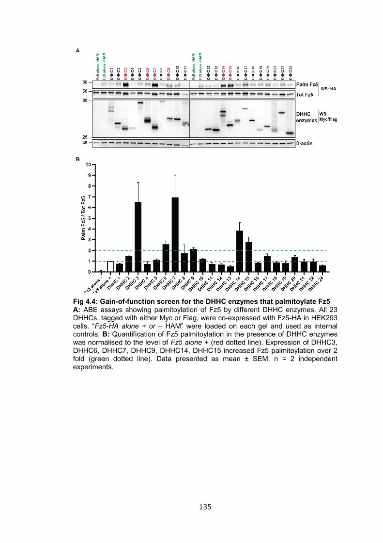
135
Fig 4.4: Gain-of-function screen for the DHHC enzymes that palmitoylate Fz5 A: ABE assays showing palmitoylation of Fz5 by different DHHC enzymes. All 23 DHHCs, tagged with either Myc or Flag, were co-expressed with Fz5-HA in HEK293 cells. “Fz5-HA alone + or – HAM” were loaded on each gel and used as internal controls. B: Quantification of Fz5 palmitoylation in the presence of DHHC enzymes was normalised to the level of Fz5 alone + (red dotted line). Expression of DHHC3, DHHC6, DHHC7, DHHC9, DHHC14, DHHC15 increased Fz5 palmitoylation over 2 fold (green dotted line). Data presented as mean ± SEM; n = 2 independent experiments.
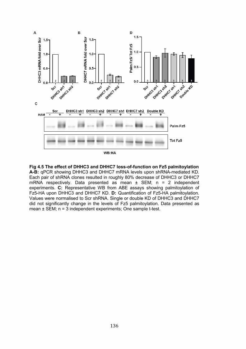
136
Fig 4.5 The effect of DHHC3 and DHHC7 loss-of-function on Fz5 palmitoylation A-B: qPCR showing DHHC3 and DHHC7 mRNA levels upon shRNA-mediated KD. Each pair of shRNA clones resulted in roughly 80% decrease of DHHC3 or DHHC7 mRNA respectively. Data presented as mean ± SEM; n = 2 independent experiments. C: Representative WB from ABE assays showing palmitoylation of Fz5-HA upon DHHC3 and DHHC7 KD. D: Quantification of Fz5-HA palmitoylation. Values were normalised to Scr shRNA. Single or double KD of DHHC3 and DHHC7 did not significantly change in the levels of Fz5 palmitoylation. Data presented as mean ± SEM; n = 3 independent experiments; One sample t-test.
137
4.3 Discussion
Wnt signalling through Frizzled receptors is central in many biological
processes. Studying how Frizzled receptors are regulated at the molecular
level is crucial to understand the function of these molecules. Here I have
shown that potentially all Frizzled receptors are palmitoylated. I have also
determined the sites where Fz5 is palmitoylated and I have identified six
enzymes that can catalyse this PTM. The results presented in this chapter
uncover a novel PTM of Frizzled receptors and open new avenues to study
the role of palmitoylation on these family of proteins.
4.3.1 Frizzled receptors are palmitoylated – Fz5 palmitoylation at the C-term
Previous observations from our lab showed that Fz5 is palmitoylated (Fig
1.13), but whether other Frizzled receptors are subjected to the same
modification was unknown. By analysing the structure of Frizzled receptors I
found that each of these proteins contain at least one Cys residue predicted
to be palmitoylated. The Cys residues are located across diverse domains in
different Frizzled receptors. My biochemistry experiments strongly suggest
that Fz3, Fz4, Fz5, Fz6 and Fz9 are palmitoylated, whereas further analyses
are required to address palmitoylation of Fz1, Fz2, Fz7, Fz8 and Fz10.
Although the biological role of Fz5 palmitoylation will be addressed in the
next chapter, some general considerations can be made for the whole
Frizzled family.
The fact that Cys residues are located across different protein domains (N-
term vs TM vs C-term) implies that palmitoylation may have different roles on
Frizzled receptors, and it also suggests that this modification might be
regulated by different mechanisms. For instance, palmitoylation of Frizzled
receptors may occur in different subcellular compartments and might be
catalysed by different DHHC enzymes. In addition, bioinformatic analyses
revealed that Cys residues are present at the C-term of most Frizzled
receptors (Fz3, Fz4, Fz5, Fz6, Fz8, Fz9, Fz10), but only those of Fz5 and its
homologous, Fz8, were predicted to be palmitoylated. Thus, it is possible that
138
palmitoylation at the C-term of Frizzled receptors might have evolved to
regulate specific function of Fz5 and its homolog Fz8.
Preliminary data from our lab showed that the 3 Cys residues at the C-term
of Fz5 are required for palmitoylation, but whether palmitoylation can occur
on each of these Cys was not addressed before. Here I found that each of
the three Cys can be palmitoylated. This observation raises the question of
whether palmitoylation on different residues has diverse biological effects.
Although it is unknown whether palmitoylation on adjacent Cys can regulate
distinct biological processes, several examples exist for proteins in which
Cys residues are not adjacent. For instance AMPARs and NMDARs are
palmitoylated on distinct TM or C-term Cys residues, which respectively
control the retention of these receptors at the Golgi or regulate their
internalisation from the plasma membrane (Hayashi et al. 2005; Hayashi et al.
2009). In my PhD I have investigated the role of palmitoyIation using the
triple Cys Fz5 mutant (Fz5 3CS, see chapter 5 and 6); therefore, the role of
palmitoylation on each specific Cys residues remains unknown. Altogether,
these results uncover a novel PTM of Frizzled receptors and open a whole
new areas of research in the Wnt signalling field.
4.3.2 Palmitoylation of Fz5 by members of the DHHC family
Identifying enzyme-substrate pairs is fundamental to understand how
palmitoylation is regulated, but it is still a major challenge in the field of
protein palmitoylation. To identify the enzymes that palmitoylate Fz5, I
performed gain- and loss-of-function experiments of members of the DHHC
family of protein acyl transferases (PAT) (Fukata et al. 2006; Ohno et al.
2006). The gain-of-function screen presented here shows that DHHC3 and
DHHC7, but also DHHC6, 9, 14, and 15, can palmitoylate Fz5 in HEK293
cells. Particular attention was given to DHHC3 and DHHC7, which increased
Fz5 palmitoylation around 6 to 7fold. These enzymes, which belong to the
same DHHC subfamily (Fig 1.12), are expressed in the CNS and many other
tissues and localise at the Golgi apparatus (Ohno et al. 2006). Many
substrates, including several pre- and postsynaptic proteins, are

139
palmitoylated by both of these enzymes (Globa and Bamji 2017; Matt et al.
2019), suggesting either redundancy or a coordinated mechanism that
requires both enzymes. However, the requirement of DHHC3 and DHHC7
was not confirmed by shRNA mediated KD of these enzymes. The most
likely explanation is that other DHHC enzymes compensate for the loss of
DHHC3 and DHHC7.
How can we avoid compensatory effects? The first question that needs to be
addressed is whether palmitoylation by six different DHHC6 might be an
artefact induced by overexpression of these enzymes. This same question
has recently been addressed in the context of palmitoylation of γ subunits of
GABAARs (Kilpatrick et al. 2016). In HEK293 cells both DHHC3 and DHHC7
palmitoylate GABAARs (Fang et al. 2006); however, only Dhhc3-/- mice, and
not Dhhc7-/- animals, exhibit defects in GABAARs trafficking and function
(Kilpatrick et al. 2016). The authors elegantly demonstrated that this
phenotype is due to a subtle difference in localisation of these enzymes at
the Golgi apparatus: DHHC3 and DHHC7 localise at the cis- and trans-Golgi
respectively (Kilpatrick et al. 2016). In HEK293 cells, overexpression causes
aberrant localisation of DHHC7 to the cis-Golgi, enabling palmitoylation of
GABAARs (Kilpatrick et al. 2016). Thus, palmitoylation of GABAARs by
DHHC7 seems to be an artefact of protein overexpression (Kilpatrick et al.
2016). Therefore, examining Fz5 palmitoylation in a more physiological
context, such as using DHHCs KO mouse models, could unravel the role of
these enzymes for Fz5 palmitoylation in vivo.
Another approach could be to extend the analysis of the effects of DHHC KD
to Fz5-specific functions. Indeed, several proteins that are palmitoylated on
more than one Cys residue exhibit residue-specific function of palmitoylation.
For instance, NMDARs are palmitoylated at the C-term on two distinct
clusters of Cys residues (Hayashi et al. 2009). Palmitoylation of a first cluster
of Cys mediates the retention of NDMARs in the Golgi, whereas
palmitoylation of a second group of Cys is important for the membrane
stability of the receptor (Hayashi et al. 2009). Although we do not know
whether palmitoylation of the three Cys residues at the C-term of Fz5 results
in residues-specific effects, it would be important to examine the effects of
140
DHHCs KD on Fz5-specific function. In fact, while overall palmitoylation
levels might be compensated by other DHHCs, specific functional outcomes
might be impaired by the loss of one or more enzymes. In this respect, it
would be interesting to examine the impact of DHHCs KD on Fz5-induced
synapse formation. In collaboration with other members of the lab we are
currently working on this hypothesis.
Despite the inconclusive data on the requirement of DHHC enzymes, what
can we learn from the gain-of-function screen? DHHC3, DHHC6, DHHC7
DHHC9, DHH14 and DHHC15 increase Fz5 palmitoylation. In HEK293 cells
these enzymes localise between the ER and the Golgi (Ohno et al. 2006).
Therefore, it seems reasonable to assume that palmitoylation of Fz5 occurs
before the receptor reaches the cell surface, and palmitoylation could
regulate the trafficking of this receptor to different subcellular locations.
Knowing where palmitoylation is attached to Fz5 could help us to dissect the
role of this modification.
Taken together my data strongly suggest that several if not all Frizzled
receptors are palmitoylated. I also demonstrated that each of the Cys
residues at the C-term of Fz5 is palmitoylated, and that at least 6 different
DHHC enzymes can palmitoylate Fz5 in cell lines. Further experiments are
needed to address the requirement of these enzymes for Fz5 palmitoylation.
Thus, my findings uncover a previously uncharacterised PTM of Frizzled
receptors. Studying the molecular mechanisms and biological functions of
Frizzled palmitoylation will unravel novel mechanisms of regulation of Wnt
signalling.
141
CHAPTER 5: THE IMPACT OF PALMITOYLATION ON Fz5 SIGNALLING AND TRAFFICKING 5.1 Introduction
Frizzled are the main receptors for Wnt ligands and are essential for
signalling. However, surprisingly little is known about the molecular
mechanisms that control the function and distribution of these receptors. In
light of the crucial roles that Frizzled receptors play in a variety of biological
processes, including cancer, stem cell biology and neuroscience, it is
fundamental to study how their function is regulated. In the previous chapter,
I have introduced palmitoylation, a previously uncharacterised modification of
Frizzled receptors, with special attention to Fz5, a receptor for the
synaptogenic factor Wnt7a. Preliminary data from our lab show that
palmitoylation-deficient Fz5 (3CS Fz5) is unable to promote synapse
formation in vitro (Fig 1.13), suggesting that palmitoylation is crucial for Fz5
function. Here, I have investigated the role of this modification further, and
used biochemical and cell biology techniques to elucidate how palmitoylation
affects Fz5.
Palmitoylation can affect proteins in a number of different ways. First,
palmitoylation can positively or negatively control protein turnover
(Percherancier et al. 2001; Roberts et al. 2016). Second, palmitoylation can
regulate the exit of proteins from the ER and Golgi, determining progression
along the trafficking route and sorting to specific subcellular compartments
(Greaves and Chamberlain 2007; Salaun et al. 2010; Ernst et al. 2018). This
is particularly important in neurons, where selective trafficking to axon or
dendrites is crucial for cell polarity and for synapse formation and function
(Fukata and Fukata 2010; Yokoi et al. 2012; Montersino and Thomas 2015;
Holland and Thomas 2017; Yogev and Shen 2017). Third, the attachment of
lipid chains onto proteins can affect their lateral mobility across the PM,
thereby affecting signalling (Delint-Ramirez et al. 2011; S. Kim et al. 2014).
Fourth, palmitoylation is a major regulator of exocytosis, endocytosis and
recycling of membrane proteins, thus controlling their surface levels and
determining signalling strength and duration (Resh 2006; Nadolski and
142
Linder 2007; Sugita 2008; Doherty and McMahon 2009; Grant and
Donaldson 2009). Fifth, palmitoylation can control protein-protein interactions
such as ligand-receptor binding, oligomerisation, and the interaction with
scaffold proteins (Simons and Toomre 2000; Resh 2006; Charollais and Van
Der Goot 2009). By affecting one or more of these cell biology mechanisms,
palmitoylation has direct consequences on several aspects of protein
function.
The fact that the palmitoylation deficient mutant 3CS Fz5 is unable to
promote synapse formation suggests that this post-translational modification
might regulate several of the cell biology processes listed above. Therefore, I
have performed a series of experiments, in cell lines and primary
hippocampal neurons, to establish the role of Fz5 palmitoylation in each of
these processes. The data presented here shed new light into the role of this
previously uncharacterised PTM of Frizzled receptors, hence enriching our
understanding of the regulation of these molecules. The following questions
will be addressed:
1. Does palmitoylation affect Fz5 protein turnover?
2. Does palmitoylation regulate Fz5 interaction with key components of
the Wnt signalosome?
3. Does palmitoylation regulate Fz5 localisation in hippocampal
neurons?
4. Does palmitoylation control Fz5 membrane trafficking?
143
5.2 Results
5.2.1 Fz5 turnover is independent of protein palmitoylation
Palmitoylation controls the turnover rate of many proteins including the A1
adenosine receptor (Gao et al. 1999) and the chemokine and HIV receptor
CCR5 (Percherancier et al. 2001). In some cases, like for anthrax toxin
receptors and the Wnt co-receptor LRP6, it does so by preventing protein
ubiquitination (Abrami et al. 2006; Abrami et al. 2008). To test whether Fz5
turnover is regulated by palmitoylation, I blocked protein translation in NRK
(normal rat kidney) cells and monitored the degradation of WT and 3CS Fz5
over time by WB (western blot). Both WT and 3CS Fz5 exhibited a half-life of
roughly 60 mins, strongly suggesting that palmitoylation does not regulate
the turnover rate of Fz5 (Fig 5.1). My data are consistent with previous
reports on the half-life of other Frizzled receptors (Mukai et al. 2010; Hao et
al. 2012). In addition, the relatively rapid turnover rate of Fz5 receptors is
comparable to the half-life of Smoothened (Smo) (Milenkovic et al. 2009), a
seven transmembrane protein which belongs to the Frizzled family of GPCRs.
Smo is a key component of the Hedgehog signalling, and has an half-life of
roughly 60-120 mins (Milenkovic et al. 2009). In contrast, other GPCRs like
adenosine, adrenergic, cannabinoid and glutamate metabotropic receptors
exhibit considerably longer half-life (10-48 hrs) (McIntosh et al. 1998; Drake
et al. 2006; Hazell et al. 2012), suggesting regulation by different molecular
mechanisms than the Frizzled/Smo family. Thus, these experiments indicate
that palmitoylation does not affect Fz5 turnover.
5.2.2 Palmitoylation is required for Fz5 interaction with Dvl1 but dispensable for Fz5/Fz5 interaction
Palmitoylation deficient Fz5 lacks synaptogenic activity (Fig 1.13), suggesting
deficiency in Wnt signalling. The binding of Wnt ligands to their receptors
triggers the formation of a signalosome comprising ligand/receptor/co-
receptors and intracelullar molecules including the scaffold protein
Dishevelled (Dvl), which is essential for all Wnt signalling cascades (Gao and
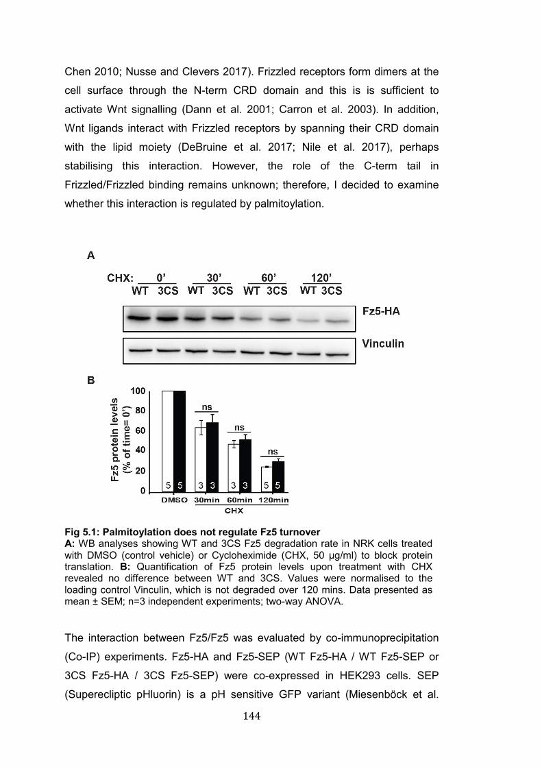
144
Chen 2010; Nusse and Clevers 2017). Frizzled receptors form dimers at the
cell surface through the N-term CRD domain and this is is sufficient to
activate Wnt signalling (Dann et al. 2001; Carron et al. 2003). In addition,
Wnt ligands interact with Frizzled receptors by spanning their CRD domain
with the lipid moiety (DeBruine et al. 2017; Nile et al. 2017), perhaps
stabilising this interaction. However, the role of the C-term tail in
Frizzled/Frizzled binding remains unknown; therefore, I decided to examine
whether this interaction is regulated by palmitoylation.
Fig 5.1: Palmitoylation does not regulate Fz5 turnover A: WB analyses showing WT and 3CS Fz5 degradation rate in NRK cells treated with DMSO (control vehicle) or Cycloheximide (CHX, 50 µg/ml) to block protein translation. B: Quantification of Fz5 protein levels upon treatment with CHX revealed no difference between WT and 3CS. Values were normalised to the loading control Vinculin, which is not degraded over 120 mins. Data presented as mean ± SEM; n=3 independent experiments; two-way ANOVA.
The interaction between Fz5/Fz5 was evaluated by co-immunoprecipitation
(Co-IP) experiments. Fz5-HA and Fz5-SEP (WT Fz5-HA / WT Fz5-SEP or
3CS Fz5-HA / 3CS Fz5-SEP) were co-expressed in HEK293 cells. SEP
(Superecliptic pHluorin) is a pH sensitive GFP variant (Miesenböck et al.
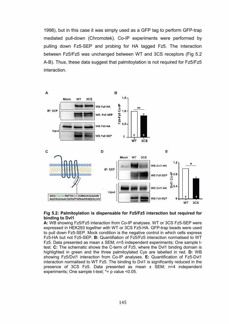
145
1998), but in this case it was simply used as a GFP tag to perform GFP-trap
mediated pull-down (Chromotek). Co-IP experiments were performed by
pulling down Fz5-SEP and probing for HA tagged Fz5. The interaction
between Fz5/Fz5 was unchanged between WT and 3CS receptors (Fig 5.2
A-B). Thus, these data suggest that palmitoylation is not required for Fz5/Fz5
interaction.
Fig 5.2: Palmitoylation is dispensable for Fz5/Fz5 interaction but required for binding to Dvl1 A: WB showing Fz5/Fz5 interaction from Co-IP analyses. WT or 3CS Fz5-SEP were expressed in HEK293 together with WT or 3CS Fz5-HA. GFP-trap beads were used to pull down Fz5-SEP. Mock condition is the negative control in which cells express Fz5-HA but not Fz5-SEP. B: Quantifiation of Fz5/Fz5 interaction normalised to WT Fz5. Data presented as mean ± SEM; n=5 independent experiments; One sample t-test. C: The schematic shows the C-term of Fz5, where the Dvl1 binding domain is highlighted in green and the three palmitoylated Cys are labelled in red. D: WB showing Fz5/Dvl1 interaction from Co-IP analyses. E: Quantification of Fz5-Dvl1 interaction normalised to WT Fz5. The binding to Dvl1 is significantly reduced in the presence of 3CS Fz5. Data presented as mean ± SEM; n=4 independent experiments; One sample t-test; *= p value <0.05.
146
Another fundamental event for the formation of the Wnt signalosome and
signalling transduction is the interaction between Frizzled and Dvl. At the C-
term Fz5 is palmitoylated on three Cys residues (Fig 4.3 B-C), which are
located in close proximity (6 amino acids away) to one of the Dvl1 binding
motifs (Tauriello et al. 2012). Given such proximity, it is possible that
palmitoylation could regulate binding to Dvl1. Therefore, I tested this
hypothesis by expressing WT or 3CS Fz5-SEP and Dvl1-HA in HEK293 cells
and measuring their interaction as previously described for Fz5-SEP/Fz5-HA.
The interaction with Dvl1 was reduced by roughly 40% in the presence of
3CS Fz5 compared to WT receptors (Fig 5.2 D-E), indicating that
palmitoylation is involved in Fz5 binding to Dvl1. Taken together, these data
suggest that palmitoylation is dispensable for Fz5/Fz5 interaction but is
important for Dvl1 binding.
5.2.3 Palmitoylation regulates Fz5 distribution along axons but not dendrites and soma of hippocampal neurons
Palmitoylation is a major regulator of protein trafficking and sorting. This
function is particularly important in highly polarised cells like neurons; in fact,
several neuronal proteins exhibit palmitoylation-dependent sorting (Fukata
and Fukata 2010). For instance, palmitoylation is required to target GAP-43
to growth cones (Skene and Virág 1989; Liu et al. 1991), to direct the growth
factor DLK1 to axonal trafficking vesicles (Holland et al. 2016) and to sort
Synaptotagmin 1 to presynaptic compartments (Kang et al. 2004).
Preliminary data indicate that 3CS Fz5 fails to induce synapse formation (Fig
1.13), suggesting that Fz5 function in axons is compromised. Therefore, I
hypothesised that WT and 3CS Fz5 might exhibit different localisation
patterns, which could explain the lack of synaptogenic activity of
palmitoylation deficient Fz5-receptors.
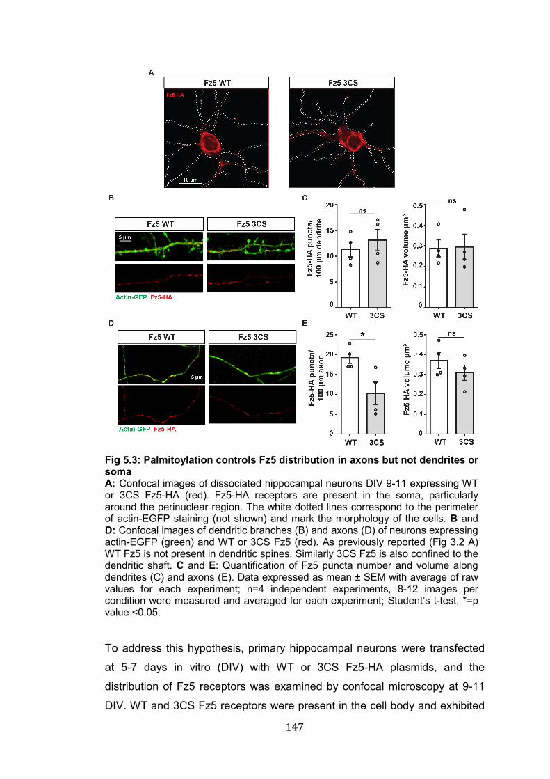
147
Fig 5.3: Palmitoylation controls Fz5 distribution in axons but not dendrites or soma A: Confocal images of dissociated hippocampal neurons DIV 9-11 expressing WT or 3CS Fz5-HA (red). Fz5-HA receptors are present in the soma, particularly around the perinuclear region. The white dotted lines correspond to the perimeter of actin-EGFP staining (not shown) and mark the morphology of the cells. B and D: Confocal images of dendritic branches (B) and axons (D) of neurons expressing actin-EGFP (green) and WT or 3CS Fz5 (red). As previously reported (Fig 3.2 A) WT Fz5 is not present in dendritic spines. Similarly 3CS Fz5 is also confined to the dendritic shaft. C and E: Quantification of Fz5 puncta number and volume along dendrites (C) and axons (E). Data expressed as mean ± SEM with average of raw values for each experiment; n=4 independent experiments, 8-12 images per condition were measured and averaged for each experiment; Student’s t-test, *=p value <0.05. To address this hypothesis, primary hippocampal neurons were transfected
at 5-7 days in vitro (DIV) with WT or 3CS Fz5-HA plasmids, and the
distribution of Fz5 receptors was examined by confocal microscopy at 9-11
DIV. WT and 3CS Fz5 receptors were present in the cell body and exhibited
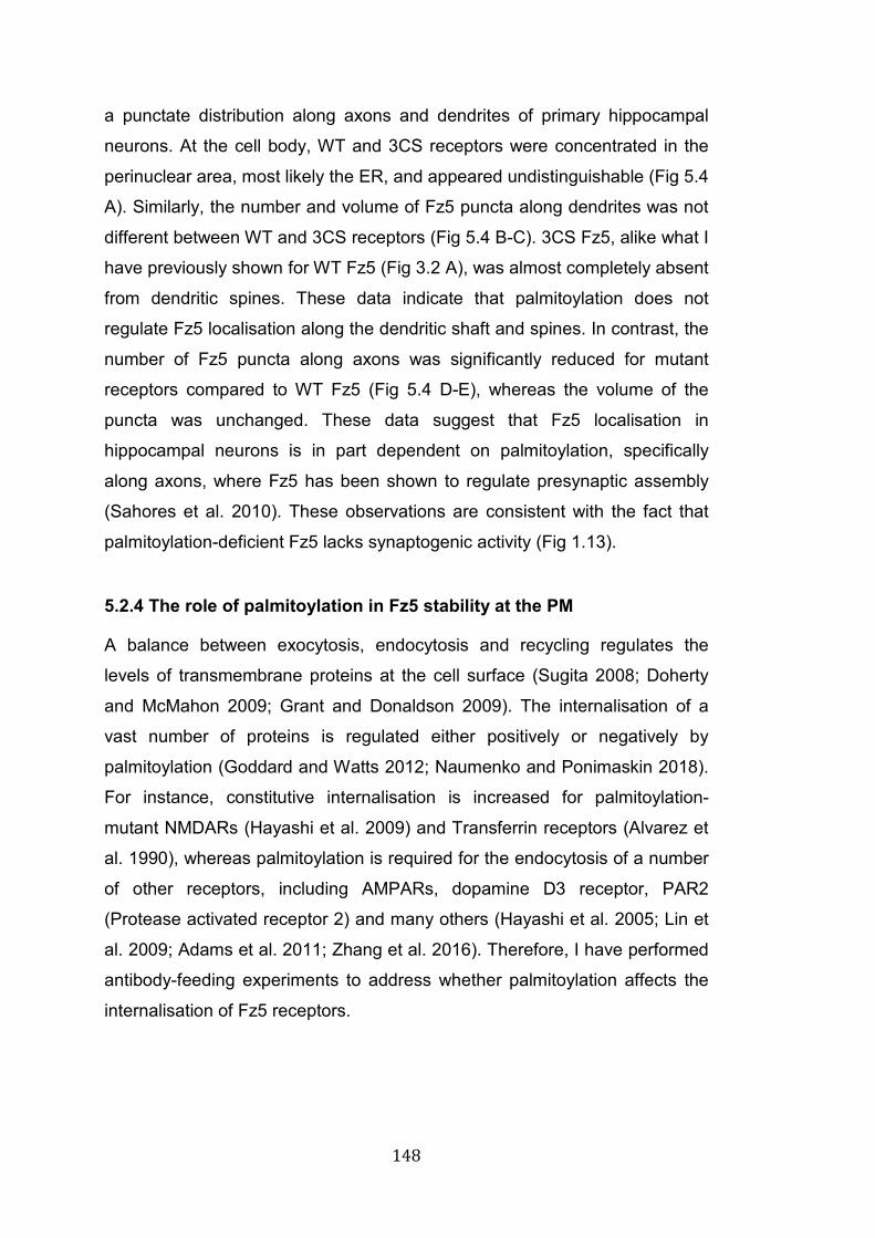
148
a punctate distribution along axons and dendrites of primary hippocampal
neurons. At the cell body, WT and 3CS receptors were concentrated in the
perinuclear area, most likely the ER, and appeared undistinguishable (Fig 5.4
A). Similarly, the number and volume of Fz5 puncta along dendrites was not
different between WT and 3CS receptors (Fig 5.4 B-C). 3CS Fz5, alike what I
have previously shown for WT Fz5 (Fig 3.2 A), was almost completely absent
from dendritic spines. These data indicate that palmitoylation does not
regulate Fz5 localisation along the dendritic shaft and spines. In contrast, the
number of Fz5 puncta along axons was significantly reduced for mutant
receptors compared to WT Fz5 (Fig 5.4 D-E), whereas the volume of the
puncta was unchanged. These data suggest that Fz5 localisation in
hippocampal neurons is in part dependent on palmitoylation, specifically
along axons, where Fz5 has been shown to regulate presynaptic assembly
(Sahores et al. 2010). These observations are consistent with the fact that
palmitoylation-deficient Fz5 lacks synaptogenic activity (Fig 1.13).
5.2.4 The role of palmitoylation in Fz5 stability at the PM
A balance between exocytosis, endocytosis and recycling regulates the
levels of transmembrane proteins at the cell surface (Sugita 2008; Doherty
and McMahon 2009; Grant and Donaldson 2009). The internalisation of a
vast number of proteins is regulated either positively or negatively by
palmitoylation (Goddard and Watts 2012; Naumenko and Ponimaskin 2018).
For instance, constitutive internalisation is increased for palmitoylation-
mutant NMDARs (Hayashi et al. 2009) and Transferrin receptors (Alvarez et
al. 1990), whereas palmitoylation is required for the endocytosis of a number
of other receptors, including AMPARs, dopamine D3 receptor, PAR2
(Protease activated receptor 2) and many others (Hayashi et al. 2005; Lin et
al. 2009; Adams et al. 2011; Zhang et al. 2016). Therefore, I have performed
antibody-feeding experiments to address whether palmitoylation affects the
internalisation of Fz5 receptors.
149
Fig 5.4: Palmitoylation regulates Fz5 membrane stability A: The schematic illustrates the principle of antibody feeding experiments. Cells were placed on ice to block trafficking and label surface Fz5 receptors with HA antibodies. Cells were then returned to 37 ˚C to allow for internalisation of surface proteins. B: Confocal images of NRK cells expressing WT or 3CS Fz5-HA. Insets on the right show clear formation of endocytic clustes. C: Quantification of Fz5 endocytic puncta. At t=15 mins (after being retunerd to 37 ˚C) 3CS Fz5 puncta were double than WT Fz5. Data presented as mean ± SEM; n=5 independent experiments, 10-12 cells per condition were measured and averaged for each experiment; Student’s t-test, ***=p value < 0.0005. D: Confocal images of Fz5 puncta (green) colocalising with endosomal markers (red, EAA1, Rab11, Rab7). E: Surface biotinylation analyses of HEK293 cells expressing WT or 3CS Fz5-HA. Cells expressing WT Fz5 were treated for 90 mins with 2BP (100 µM), to inhibit protein palmitoylation. F: Quantification of surface levels of Fz5 normalised to total protein levels and to WT Fz5 treated with vehicle ctrl. Data presented as mean ± SEM; n=6 independent experiments; one-way ANOVA, */***=p value < 0,05 and 0.0005.
150
NRK cells expressing either WT or 3CS Fz5-HA were placed on ice to block
trafficking, and then incubated with primary antibodies against the HA tag at
the N-term extracellular region of Fz5 receptors. Next, cells were either fixed
(time=0) or returned at 37˚C to allow receptor internalisation (Fig 5.4 A). The
formation of typical endocytic clusters of Fz5-HA was analysed by confocal
microscopy. 15 mins after returning the cells to 37˚C, WT and 3CS Fz5
showed remarkable differences in the levels of internalisation (Fig 5.5 A-B).
The number of endocytic clusters was double for 3CS Fz5 compared to WT
Fz5 (Fig 5.5 B). To confirm that these puncta were indeed clusters of
internalised receptors, I examined their colocalisation with common markers
of early, late and recycling endosomes (EEA1, Rab7 and Rab11
respectively). I found that Fz5 clusters co-localised with each one of these
markers (Fig 5.5 C). Altogether these data strongly suggest that
internalisation of palmitoylation-deficient Fz5 is accelerated, suggesting that
palmitoylation inhibits constitutive endocytosis of this receptor.
Given that 3CS is internalised faster than WT Fz5, I examined whether the
surface levels of the mutant receptor were reduced. Surface biotinylation
experiments in HEK239 cells showed a small (20%) but statistically
significant reduction of surface 3CS Fz5 compared to WT receptors (Fig 5.5),
consistently with an increase in the rate of steady-state internalisation.
Importantly, this result was confirmed by pharmacological treatment of WT
Fz5 with 2-BP (Fig 5.5), a drug that blocks palmitoylation by preventing the
addition of new palmitate groups to Cys residues (Davda et al. 2013). Overall
these data strongly suggest that genetic or pharmacological inhibition of
palmitoylation decrease the surface levels of Fz5.
Another important aspect of receptor stability at the PM is their lateral
mobility, a phenomenon that can be regulated by palmitoylation. For instance,
palmitoylation and phosphorylation regulate the synaptic recruitment by
lateral diffusion of extrasynaptic AMPARs, a process that is fundamental for
induction of synaptic plasticity (Hayashi et al. 2005; Lin et al. 2009; Makino
and Malinow 2009). In addition, palmitoylation-deficient mutants of large
conductance Ca2+ channels move faster across the PM (S. Kim et al. 2014).
Except for one study where several Wnts have been shown to increase
151
lateral mobility of Fz6 (Kilander et al. 2014), the lateral mobility of Frizzled
receptors has not been investigated. Therefore, I asked whether lateral
diffusion of Fz5 might be affected by palmitoylation.
To address this question, I performed FRAP (Fluorescence Recovery After
Photobleaching) experiments on NRK cells expressing WT or 3CS Fz5 .
These receptors were tagged at the N-term with SEP, a pH sensitive GFP
variant that does not fluoresce at acidic pH, normally found in intracellular
vesicles, but is excited at neutral pH, normally found in the extracellular
space (Miesenböck et al. 1998). The use of SEP tagged-receptors, combined
with FRAP, allowed us to monitor the lateral movement of Fz5 at the cell
surface (Georgiou et al. 2002; Ashby et al. 2004; Hildick et al. 2012). Three
small regions per field of view were bleached: one located outside the cell to
be used as blank, one around the perinuclear area and one at the edge of
the cell. The blank region and the one at the edge of the cell were used for
quantification of fluorescence recovery. After bleaching, the recovery of
fluorescence intensity of SEP tagged proteins is achieved by lateral diffusion
of nearby receptors and/or insertion of new ones. Given the small size of the
bleached area and the short time of the recording, it is extremely unlikely that
exocytosis occurred in the regions of interest. Therefore, it is reasonable to
assume that the recovery of fluorescence intensity is due to lateral diffusion
of nearby receptors (Martin et al. 2009; Hildick et al. 2012). I found that the
recovery of fluorescence intensity was unchanged between WT and 3CS Fz5
(Fig 5.6 A-B). Moreover, I extrapolated the pools of mobile and immobile
receptors from the maximal percentage of fluorescence recovery. The reason
why the recovery of fluorescence signal does not reach 100% is because a
portion of receptors are immobile at the PM and therefore they are not
replaced by lateral diffusion of nearby receptors (Zheng et al. 2011; Hildick et
al. 2012). I found that 80% of receptors appeared to be mobile and 20%
immobile for both WT and 3CS Fz5 (Fig 5.6 C), suggesting that lateral
mobility of Fz5 does not depend on palmitoylation. Altogether the data
presented in this chapter strongly suggest that palmitoylation affects Dvl1
interaction, Fz5 distribution along axons, Fz5 internalisation and surface
levels of this receptor. In contrast, Fz5 turnover rate, Fz5/Fz5 interaction, Fz5
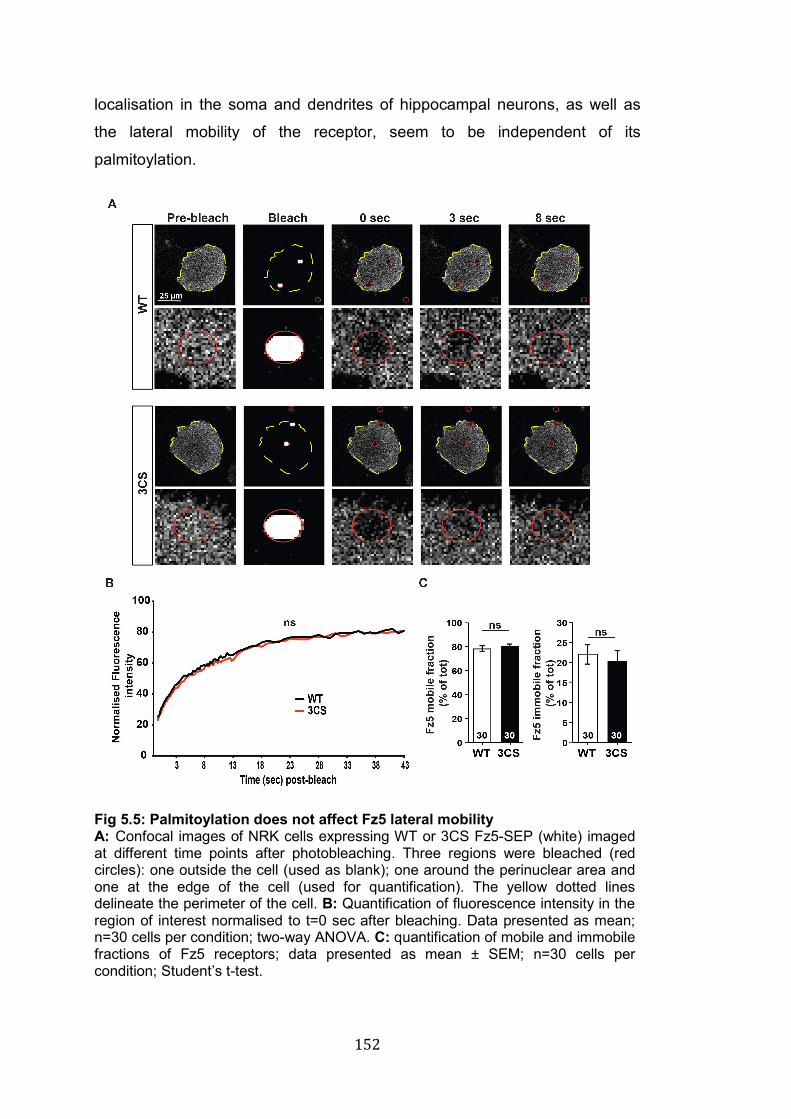
152
localisation in the soma and dendrites of hippocampal neurons, as well as
the lateral mobility of the receptor, seem to be independent of its
palmitoylation.
Fig 5.5: Palmitoylation does not affect Fz5 lateral mobility A: Confocal images of NRK cells expressing WT or 3CS Fz5-SEP (white) imaged at different time points after photobleaching. Three regions were bleached (red circles): one outside the cell (used as blank); one around the perinuclear area and one at the edge of the cell (used for quantification). The yellow dotted lines delineate the perimeter of the cell. B: Quantification of fluorescence intensity in the region of interest normalised to t=0 sec after bleaching. Data presented as mean; n=30 cells per condition; two-way ANOVA. C: quantification of mobile and immobile fractions of Fz5 receptors; data presented as mean ± SEM; n=30 cells per condition; Student’s t-test.
153
5.3 Discussion
Findings from our lab suggest that palmitoylation is required for Fz5 to induce
presynaptic assembly in vitro (Fig 1.13) (Stamatakou and Salinas,
unpublished results), implying that this modification is required for Fz5
function. Here I have asked which aspects of Fz5 function are regulated by
palmitoylation. Using biochemical and cell biology approaches I found that
palmitoylation is required for Fz5-Dvl1 interaction, for the stability of the
receptor at the PM and for its distribution along axons, where Fz5 is required
for presynaptic assembly (Sahores et al. 2010). In contrast, Fz5
palmitoylation seems to be dispensable for Fz5/Fz5 interaction, Fz5 turnover
rate, its lateral mobility at the PM and for the distribution of this receptor in
the soma or along dendritic processes. These findings represent the first
characterisation of the molecular mechanisms through which palmitoylation
affects a Frizzled receptor.
5.3.1 Palmitoylation regulates Fz5 localisation in hippocampal neurons
The precise localisation of protein at specific subcellular compartments is
fundamental in every cell, but particularly in neurons because of their high
degree of polarisation. Palmitoylation can regulate the sorting and clustering
of proteins into neuronal process and at synapses. For instance, Paralemmin
and GAP-43 require the attachment of palmitate moieties to be directed to
dendritic spines and axonal growth cones (El-Husseini et al. 2001; Greaves
and Chamberlain 2007). Moreover, PSD-95 and Gephyrin exhibit
palmitoylation dependent-clustering in dendrites (El-Husseini et al. 2000;
Dejanovic et al. 2014). Given that palmitoylation-mutant Fz5 receptors lack
synaptogenic activity, I hypothesised that the localisation of Fz5 in axonal
processes might be impaired. The data presented here indicate that Fz5 is
less abundant in axons of dissociated hippocampal neurons, as observed by
the decreased number of axonal puncta compared to WT Fz5. In contrast,
the localisation at the cell body and along dendrites was unchanged between
WT and 3CS Fz5, suggesting the existence of axon-specific mechanisms
that regulate Fz5 sorting and/or clustering.
154
Two non-mutually exclusive mechanisms could underlie the impaired
distribution of mutant Fz5 in axons. First, Fz5 sorting into axons might be
compromised. At present we do not have evidence supporting a role for
palmitoylation in Fz5 sorting, but we are planning to address this question
combining live-imaging of GFP-tagged Fz5 with the RUSH (retention using
selective hooks) technology, which allows synchronised protein trafficking
from the ER to the PM (Boncompain et al. 2012). Thus, comparing WT and
3CS trafficking into axons could elucidate the role of palmitoylation in this
process. A second possibility is that Fz5 clustering in axons might be
dependent on palmitoylation. Given that Fz5/Fz5 interaction is unchanged
between WT and 3CS receptors we could speculate that palmitoylation is
dispensable for Fz5 clustering. However, my data also indicate that the
interaction with the scaffold protein Dvl1 is dependent on palmitoylation;
suggesting that Dvl-mediated clustering might be impaired in 3CS Fz5
receptors. Interestingly, Dvl1 is involved in several aspects of presynaptic
function including synapse formation, vesicle recycling and neurotransmitter
release (Ahmad-Annuar et al. 2006; Ciani et al. 2015). Therefore, it would be
important to address whether Fz5/Dvl1 interaction is necessary for Fz5
clustering and function along neuronal processes and at presynaptic sites.
5.3.2 The role of palmitoylation in Fz5 membrane trafficking
Given the prominent role of palmitoylation in regulating receptor endocytosis,
I examined its impact on Fz5 internalisation. The data presented here
strongly suggest that palmitoylation decreases the endocytosis rate of Fz5
stabilising it at the cell surface, but does not affect the lateral mobility of this
receptor at the PM. Together with the observation that palmitoylation-mutant
Fz5 exhibits impaired interaction with Dvl1 (Fig 5.2) and lacks synaptogenic
activity (Fig 1.13), these data strongly suggest that, in this context,
endocytosis of Fz5 receptors has a negative impact on signalling activation.
The role of receptor endocytosis in signalling activation is a matter of debate
in the Wnt field. On one hand, endocytosis of Frizzled and the co-receptors
LRP5-6 has been shown to be required for β-catenin stabilisation in different
155
model systems (Yamamoto et al. 2006; Gagliardi et al. 2014; Hagemann et al.
2014; Seto and Bellen 2006). On the other hand, the opposite has also been
proposed: clathrin-mediated endocytosis of LRP6 provides a negative
feedback on Wnt signalling (Agajanian et al. 2019), and ubiquitination-
induced internalisation of Frizzled receptors is a mechanism to down-
regulate signalling (Hao et al. 2012; Koo et al. 2012; de Lau et al. 2014;
Moffat et al. 2014; Zebisch and Jones 2015; Madan et al. 2016). Thus, the
role endocytosis for signalling activation/down-regulation seems to be
dependet on the cellular context and the molecular mechanisms are not fully
understood.
Given the role of ubiquitination in Frizzled endocytosis (Hao et al. 2016), it
would be interesting to investigate whether this modification is co-regulated
together with palmitoylation to control Fz5 surface levels, as it has been
shown for the Wnt-co-receptor LRP6 (a separate function from the role of
palmitoylation/ubiquitination in protein folding) (Abrami et al. 2008). In
addition, it would be important to address the impact of palmitoylation in
ligand-induced endocytosis of Frizzled receptors, which has been shown to
be required for signalling activation (Yamamoto et al. 2006). These questions
represent some of the next challenges to unravel the role of palmitoylation in
Frizzled endocytosis, and will be discussed in more details in the general
discussion (see chapter 7). Overall my data describe a role for palmitoylation
in regulating Fz5 membrane levels and endocytosis. Since aberrant Wnt
signalling, which is often a consequence of deregulation of Wnt receptors, is
linked to the development of several pathological conditions including cancer
and neurodegenerative disorders (Johnson and Rajamannan 2006; Anastas
and Moon 2013; Wang et al. 2016), these data shed light on a novel
mechanism of regulation of Fz5, opening new avenues to examine the
trafficking and function of Frizzled receptors in health and disease.
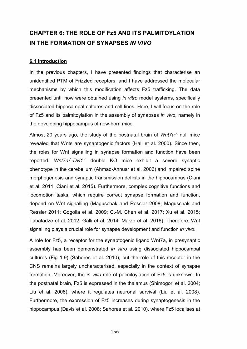
156
CHAPTER 6: THE ROLE OF Fz5 AND ITS PALMITOYLATION IN THE FORMATION OF SYNAPSES IN VIVO 6.1 Introduction
In the previous chapters, I have presented findings that characterise an
unidentified PTM of Frizzled receptors, and I have addressed the molecular
mechanisms by which this modification affects Fz5 trafficking. The data
presented until now were obtained using in vitro model systems, specifically
dissociated hippocampal cultures and cell lines. Here, I will focus on the role
of Fz5 and its palmitoylation in the assembly of synapses in vivo, namely in
the developing hippocampus of new-born mice.
Almost 20 years ago, the study of the postnatal brain of Wnt7a-/- null mice
revealed that Wnts are synaptogenic factors (Hall et al. 2000). Since then,
the roles for Wnt signalling in synapse formation and function have been
reported. Wnt7a-/--Dvl1-/- double KO mice exhibit a severe synaptic
phenotype in the cerebellum (Ahmad-Annuar et al. 2006) and impaired spine
morphogenesis and synaptic transmission deficits in the hippocampus (Ciani
et al. 2011; Ciani et al. 2015). Furthermore, complex cognitive functions and
locomotion tasks, which require correct synapse formation and function,
depend on Wnt signalling (Maguschak and Ressler 2008; Maguschak and
Ressler 2011; Gogolla et al. 2009; C.-M. Chen et al. 2017; Xu et al. 2015;
Tabatadze et al. 2012; Galli et al. 2014; Marzo et al. 2016). Therefore, Wnt
signalling plays a crucial role for synapse development and function in vivo.
A role for Fz5, a receptor for the synaptogenic ligand Wnt7a, in presynaptic
assembly has been demonstrated in vitro using dissociated hippocampal
cultures (Fig 1.9) (Sahores et al. 2010), but the role of this receptor in the
CNS remains largely uncharacterised, especially in the context of synapse
formation. Moreover, the in vivo role of palmitoylation of Fz5 is unknown. In
the postnatal brain, Fz5 is expressed in the thalamus (Shimogori et al. 2004;
Liu et al. 2008), where it regulates neuronal survival (Liu et al. 2008).
Furthermore, the expression of Fz5 increases during synaptogenesis in the
hippocampus (Davis et al. 2008; Sahores et al. 2010), where Fz5 localises at
157
synapses (Sahores et al. 2010). Importantly, Wnt7a is also expressed in the
hippocampus (Shimogori et al. 2004; Davis et al. 2008; Gogolla et al. 2009;
Ciani et al. 2011), where it has been shown to regulate synapse formation
and plasticity (Gogolla et al. 2009; Ciani et al. 2011; McLeod et al. 2018).
Thus, I asked whether Fz5 promotes synapse formation in the hippocampus
and whether palmitoylation is required for this process in vivo.
To address the role of this receptor and its palmitoylation in the formation of
synapses in vivo, I have performed intracerebroventricular (ICV) injections of
Adeno Associated Virus (AAV) to deliver WT and 3CS Fz5 cDNA in the
developing brain of new-born mice. This approach has been used before to
study central synapses (Lu et al. 2013; C. X. He et al. 2018; Passini and
Wolfe 2001; Kim et al. 2013; Ge et al. 2018). Upon expression of WT or 3CS
Fz5, I evaluated synaptic density by immunofluorescence and confocal
microscopy, as routinely performed in our laboratory (McLeod et al. 2017).
The results presented in the following section are the first in vivo
investigation of the role of Fz5 and its palmitoylation in the developing
hippocampus. Thus, they contribute to a better understanding of the role of
this receptor at synapses, and provide novel and valuable knowledge on the
function of a previously unidentified PTM of Fz5 receptors. Here I will
address the following specific questions:
1) Does WT Fz5 promote synapse formation in the developing
hippocampus?
2) Is palmitoylation required for Fz5-induced synapse formation in the
developing hippocampus?
158
6.2 Results
6.2.1 Effects of ICV injections on hippocampal anatomy and proliferation of glial cells
To express Fz5 in the brain of new-born mice, AAV9 viruses carrying WT or
3CS Fz5 cDNA were injected in the lateral ventricles of each hemisphere at
P0-P1 and animals sacrificed at P14, within the peak of synaptogenesis
(Semple et al. 2013). To identify infected cells Fz5 was expressed together
with EGFP. Fz5 and EGFP were translated from the same transcript but
immediately separated by autocleavage of the P2A linker (Fig 6.1 A-C) (Kim
et al. 2011). Importantly, cellular localisation was undistinguishable between
Fz5 expressed from a standard PCS2+ vector or from the construct
packaged into AAV9 (Fig 6.1 A-C).
Fig 6.1: ICV injections in new born mice A: Map of AAV plasmid used to express WT or 3CS Fz5 in the brain of new-born mice. The expression is driven by the CMV promoter. B: The P2A linker between GFP (green) and Fz5-HA does not affect Fz5 localisation. As shown in these epifluorescence images of HEK293 cells, Fz5 (red) is still localised at the PM in an identical manner to Fz5 receptors expressed using a standard PCS2+ plasmid. C: WB from HEK293 lysate showing complete P2A cleavage. GFP and Fz5 run at the expected molecular weight. D: 2.5 µL of AAV9 viruses were injected in the lateral ventricle of each hemisphere of P0-P1 new-born mice. The injection site was located in between the Lambda and Bregma sutures, approximately 1mm laterally of the midline suture.
159
Several reports have demonstrated that the serotype of viral capsids and
injection timing determine the number and type of cells that get infected.
Specifically, ICV injections of AAV8 or AAV9 within the first 24 hrs of life
results in maximal infection rate and neuronal specificity compared to other
AAV serotypes like AAV1 or AAV2 (Chakrabarty et al. 2013; Gholizadeh et
al. 2013). Therefore, 2.5 µL of AAV9 (>1012 viral particle/mL) were injected in
each hemisphere of C57BL/6 mice at P0, with minimal modifications from
what has been previously reported (Fig 6.1 A) (see section 2.3 of material
and methods) (J.-Y. Kim et al. 2014).
The injection of AAV9 (GFP ctrl; WT Fz5; 3CS FZ5) resulted in widespread
infection of several areas of the brain including the hippocampus, cortex and
olfactory bulb (Fig 6.2). The axons projecting to the stratum lacunosum
moleculare (SLM), which make synapses onto dendrites of CA1 cells (Fig 6.2
B), consistently exhibited very high infection rate (Fig 6.2 A). These axons
originate in part from cells in the CA3 region of the hippocampus and partially
from cells in the upper layers of the entorhinal cortex (EC), which were both
highly infected by AAV9 (Fig 6.2 A). Therefore, regions of interest in the SLM
were selected for quantification of synapse density (Fig 6.2 C). As expected,
neurons were the most infected cell type (Fig 6.2 A bottom left panel).
However, several astrocytes were also infected (Fig 6.2 A bottom left panel).
Before examining the impact on synapses, I ruled out whether ICV injections
caused abnormalities in hippocampal anatomy or aberrant activation and
proliferation of glial cells. DAPI staining did not reveal obvious differences in
gross hippocampal anatomy between animals injected with PBS or AAV
(GFP ctrl, WT Fz5, 3CS Fz5) (Fig 6.3 A). Similarly, the number and
morphology of astrocytes (GFAP positive) and microglia (Iba1 positive) did
not reveal any obvious differences (Fig 6.3 B). In addition, no obvious
abnormalities were observed in the size of injected animals (data not shown).
Taken together, these qualitative analyses strongly suggest that ICV
injections of AAV do not affect the overall development of the hippocampus,
consistent with previous work where the same approach has been used
(Broekman et al. 2006; Chakrabarty et al. 2013; Gholizadeh et al. 2013;
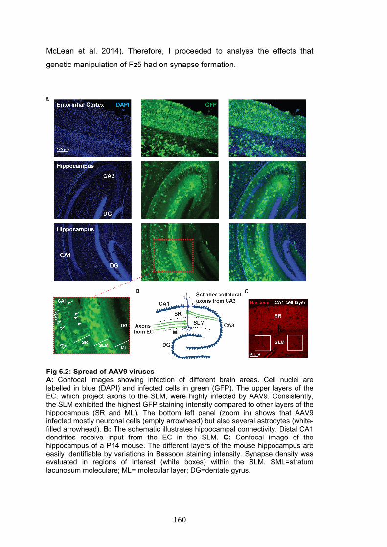
160
McLean et al. 2014). Therefore, I proceeded to analyse the effects that
genetic manipulation of Fz5 had on synapse formation.
Fig 6.2: Spread of AAV9 viruses A: Confocal images showing infection of different brain areas. Cell nuclei are labelled in blue (DAPI) and infected cells in green (GFP). The upper layers of the EC, which project axons to the SLM, were highly infected by AAV9. Consistently, the SLM exhibited the highest GFP staining intensity compared to other layers of the hippocampus (SR and ML). The bottom left panel (zoom in) shows that AAV9 infected mostly neuronal cells (empty arrowhead) but also several astrocytes (white-filled arrowhead). B: The schematic illustrates hippocampal connectivity. Distal CA1 dendrites receive input from the EC in the SLM. C: Confocal image of the hippocampus of a P14 mouse. The different layers of the mouse hippocampus are easily identifiable by variations in Bassoon staining intensity. Synapse density was evaluated in regions of interest (white boxes) within the SLM. SML=stratum lacunosum moleculare; ML= molecular layer; DG=dentate gyrus.
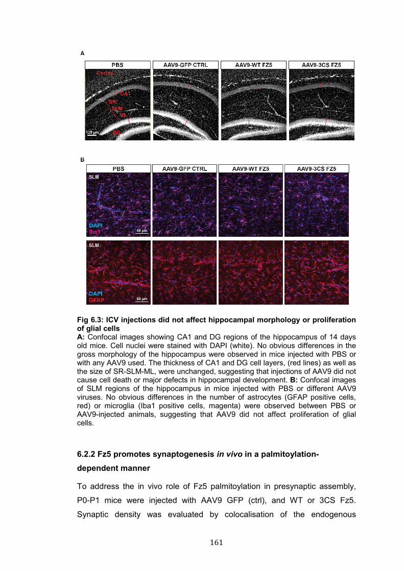
161
Fig 6.3: ICV injections did not affect hippocampal morphology or proliferation of glial cells A: Confocal images showing CA1 and DG regions of the hippocampus of 14 days old mice. Cell nuclei were stained with DAPI (white). No obvious differences in the gross morphology of the hippocampus were observed in mice injected with PBS or with any AAV9 used. The thickness of CA1 and DG cell layers, (red lines) as well as the size of SR-SLM-ML, were unchanged, suggesting that injections of AAV9 did not cause cell death or major defects in hippocampal development. B: Confocal images of SLM regions of the hippocampus in mice injected with PBS or different AAV9 viruses. No obvious differences in the number of astrocytes (GFAP positive cells, red) or microglia (Iba1 positive cells, magenta) were observed between PBS or AAV9-injected animals, suggesting that AAV9 did not affect proliferation of glial cells.
6.2.2 Fz5 promotes synaptogenesis in vivo in a palmitoylation-dependent manner
To address the in vivo role of Fz5 palmitoylation in presynaptic assembly,
P0-P1 mice were injected with AAV9 GFP (ctrl), and WT or 3CS Fz5.
Synaptic density was evaluated by colocalisation of the endogenous
162
presynaptic marker Bassoon and the postsynaptic marker Homer1.
Expression of WT Fz5 induced a statistically significant increase in the
number of Bassoon puncta in the SLM compared to GFP ctrl animals (Fig
6.4). In contrast, the number and volume of Homer1 puncta were unchanged
in animals expressing WT Fz5 compared to GFP ctrl. Importantly, these
results are consistent with our obervations in cultured hippocampal neurons
(Fig 3.2). Expression of Fz5 3CS mutant completely failed to induce
presynaptic assembly, and, albeit not statistically significant, the levels of the
postsynaptic marker Homer1 exhibited a marked trend towards a decrease
(Fig 6.4 B). The volume of Homer1 was unchanged between mice
expressing GFP ctrl and WT or 3CS Fz5 (Fig 6.4 B). The overall number of
excitatory synapses, measured by the colocalisation between Bassoon and
Homer1, was not statistically different between GFP ctrl and WT Fz5, nor
between GFP ctrl and 3CS Fz5, most likely because of the high variability
observed for Homer1 staining (Fig 6.4 B). However, a statistically significant
difference in synapse density was observed when comparing WT and 3CS
Fz5 (Fig 6.4 B), indicating that palmitoylation is required for the correct
function of this receptor.
In summary, the data presented here strongly suggest that WT Fz5 boosts
presynaptic assembly in vivo and that Fz5 palmitoylation is required for this
function. My data also suggest that expression of palmitoylation-deficient Fz5
might have a negative impact on postsynaptic development. However, this
observation requires further investigation. Altogether, these findings
represent the first in vivo investigation of the role of WT and 3CS Fz5 in
synapse assembly in the developing hippocampus.

163
Fig 6.4: Expression of WT Fz5, but not the mutant 3CS Fz5, promotes presynaptic assembly in the developing hippocampus A: Confocal images of the SLM of mice injected with AAV9 GFP ctrl, WT Fz5 and 3CS Fz5. Bassoon-positive presynaptic sites are shown in red and Homer1 positive-excitatory postsynaptic sites in green. Scale bar: 2.5µm (big panels) and 1µm (small panels). B: Quantification of the number and volume of pre- and postsynaptic puncta. Expression of WT Fz5 induced a significant increase in the number and volume of Bassoon puncta whereas no significant differences were observed for Homer1 number and volume. Synapse number was statistically different in WT Fz5 compared to 3CS Fz5 but unchanged compared GFP ctrl. (Data presented as mean with raw values for each animal analysed; n = 9 GFP, 10 WT Fz5, 11 3CS Fz5 animals; * p-value < 0.05, <0.005; One-way ANOVA with multiple comparisons).
164
6.3 Discussion
In this last chapter, I demonstrated a role for Fz5 in the formation of
synapses in vivo. Moreover, I have asked whether palmitoylation is required
for Fz5 function in vivo. My findings strongly suggest that expression of WT
Fz5 increases presynaptic development without affecting excitatory
postsynaptic differentiation. In contrast, palmitoylation-deficient Fz5 fails to
induce presynaptic assembly and might have a negative impact of the
development of excitatory postsynaptic structures. Importantly, the findings
are consistent with our previous results obtained in vitro (Fig 1.13 and Fig
3.2). These results indicate that palmitoylation is essential for Fz5 function,
and represent the first in vivo study addressing the importance of this
modification for Frizzled receptors signalling. Therefore, this study paves the
way for future investigations into the impact of palmitoylation on Frizzled
function in different cellular processes.
6.3.1 Fz5 expression induces presynaptic assembly in vivo
Several members of the Frizzled family are expressed in the CNS (Wang et
al. 2016), and have been implicated in the formation of synapses using in
vitro model systems (Varela-Nallar et al. 2009; Sahores et al. 2010; Ramírez
et al. 2016; McLeod et al. 2018). Although KO models exist for all Frizzled
receptors (Wang et al. 2016), the role of these proteins at the synapse in vivo
remains largely unknown, with the exception of studies of Frizzled receptors
in other organisms such as Drosophila and C.elegans. DFz2 receptors are
required for NMJ formation in Drosophila (Mathew et al. 2005) and Win-17
(C.elegans Frizzled) inhibits synapse formation in posterior motor neurons of
C.elegans (Klassen and Shen 2007). Therefore, the in vivo role of Frizzled
receptors in synaptogenesis in rodents remained uncharacterised until now.
My studies indicate that expression of Fz5 is sufficient to boost presynaptic
assembly in the developing hippocampus, where its ligand, Wnt7a, is
expressed (Shimogori et al. 2004; Davis et al. 2008; Gogolla et al. 2009;
Ciani et al. 2011; McLeod et al. 2018). Expression of WT Fz5 not only
increases the number of puncta for the presynaptic marker Bassoon
compared to 3CS Fz5, but it also increases the volume of these structures,
165
suggesting a possible increase in synaptic strength. Further functional
investigations using electrophysiology could address whether Fz5 has an
impact in presynaptic function. In contrast, expression of WT Fz5 did not
affect the number and size of Homer1-positive postsynaptic compartments,
consistent with data presented in chapter 3 (Fig 3.2), where I have shown
that gain and loss of function of Fz5 do not affect dendritic spine size and
number in hippocampal cultures. In spite of a significant increase in
presynaptic terminals, the number of excitatory synapses is not statistically
changed between ctrl GFP and WT Fz5 expressing mice. A possible
explanation for this result lies in the variability of Homer1 staining, which
could have inevitably affected the number of Bassoon puncta colocalising
with Homer1. Using different post-synaptic markers, such as PSD-95, and
increasing the size of the data set might reveal differences in the total
number of synapses between mice expressing GFP ctrl and WT Fz5.
Another possible explanation is that the increase in Bassoon puncta, which
include excitatory and inhibitory presynaptic terminals, might reflect an
increase in inhibitory synapses. However, as I mentioned in section 3.3.2,
very preliminary data (not shown) suggest that expression of WT Fz5 in the
hippocampus does not affect inhibitory synapse formation, consistently with
the fact that Wnt7a, a ligand for Fz5 receptors, does not affect inhibitory
synapse formation in cultured neurons (Ciani et al. 2011). In summary, the
data presented here represent novel and important findings that increase our
understanding of the function Frizzled receptors have at synapses.
6.3.2 Palmitoylation is required for Fz5-induced synapse formation in vivo
The experiments presented here indicate that Fz5 requires palmitoylation to
promote the assembly of presynaptic sites in vivo, consistently with previous
observation from cultured neurons (Fig 1.13). In addition, my data revealed a
trend towards a decrease in postsynaptic development (Homer1 count) upon
expression of 3CS Fz5 compared to ctrl or WT Fz5. However, further
experiments are needed to reach statistical significance. If Fz5 is not
required for postsynaptic development (Fig 3.2), how does 3CS Fz5 affect
166
the postsynaptic compartment? One possibility is that 3CS Fz5 impairs the
function of presynaptic terminals and therefore, indirectly, it affects the
formation or stability of postsynaptic sites. Consistently with the fact that 3CS
Fz5 does not increase presynaptic assembly and might have negative effect
on postsynaptic development, I detected a significant difference in the
numbers of excitatory synapses between mice expressing WT and 3CS Fz5.
Therefore, these data indicate that palmitoylation is required for Fz5-
mediated excitatory synapse assembly in the developing hippocampus.
Given the novelty of Frizzled palmitoylation, our studies represent the first
experimental pieces of evidence addressing the importance of this lipid
modification for Frizzled function in any model systems. Together with the
results presented in chapter 5, these data shed light into novel mechanisms
by which Frizzled receptors could be regulated in a number of biological
processes where Wnt signalling is involved, from cell fate decisions to the
formation and function of neuronal circuits.
167
CHAPTER 7: DISCUSSION AND FUTURE DIRECTIONS
7.1 Summary of project aims and findings
During my PhD, I studied the molecular mechanisms by which Wnt signalling
regulates synapse formation. In particular, I focused my attention on the role
and regulation of Frizzled receptors, the main receptors for Wnt ligands. The
aim of this PhD project was to study two aspects of Frizzled function in
synaptogenesis: first, I investigated the molecular mechanisms by which the
synpatogenic factor Wnt7a promotes both pre- and postsynaptic
development through Fz5 and Fz7 receptors; second, I have characterised
the role of palmitoylation, a novel post-translational modification (PMT) of
Frizzled receptors, in regulating Fz5 trafficking and function in the context of
synapse formation.
To address how Wnt7a affects pre- and postsynaptic development, I
performed gain and loss of function experiments for two Wnt7a receptors,
Fz5 and Fz7, and compared their localisation and function at both sides of
the synapse. Fz5, which is required for Wnt7a-mediate presynaptic assembly
(Sahores et al. 2010), does not localise to dendritic spines. I have shown that
gain and loss of function of this receptor do not affect dendritic spine
development in primary hippocampal neurons (McLeod et al. 2018). In
contrast, I found that Fz7 localises at dendritic spines and is required for
Wnt7a-induced spine formation and growth (McLeod et al. 2018). In addition,
I examined the role of Fz5 in the developing hippocampus of new-born mice.
Consistent with in vitro experiments, my data show that expression of Fz5
promotes presynaptic assembly without affecting postsynaptic development
in the hippocampus. Importantly, I found that palmitoylation, a previously
uncharacterised PTM of Frizzled receptors, is required for Fz5 function in
vivo, as palmitoylation-deficient Fz5 (3CS Fz5) receptors fail to promote
presynaptic assembly in the developing hippocampus. I also investigated the
molecular mechanisms underlying palmitoylation-dependent regulation of
Fz5. First, I established that each of the Cys residues in the C-term of Fz5
can be palmitoylated. Second, I examined which enzymes palmitoylate Fz5
and found that DHHC3 and DHHC7 strongly increase Fz5 palmitoylation
168
when expressed in HEK293 cells. However, loss of function of these two
enzymes did not seem to affect palmitoylation levels of Fz5, likely because of
compensatory effects by other enzymes. In fact, DHHC6, DHHC9, DHHC14
and DHHC15 were also found to increase Fz5 palmitoylation albeit less than
DHHC3 and DHHC7. Third, I examined whether palmitoylation affects Fz5
stability and found no differences between WT and 3CS Fz5 degradation rate.
Next, I investigated whether palmitoylation affects key interactions required
for the formation of the Wnt signalosome and found that this modification
does not affect Fz5-Fz5 interaction, but it is important for the binding to the
scaffold protein Dvl1. In light of the crucial role played by palmitoylation in
regulating protein trafficking, I examined whether 3CS Fz5 exhibits trafficking
defects compared to WT receptors. Consistently with deficient presynaptic
function, 3CS Fz5 is less abundant than WT Fz5 along the axons of primary
hippocampal neurons. Furthermore, palmitoylation is required for Fz5
trafficking as my data strongly suggest that this modification inhibits
constitutive internalization of Fz5 and is required to maintain the surface
levels of this receptor; however, FRAP experiments suggest that
palmitoylation does not affect Fz5 lateral mobility at the PM. Finally, I found
that other Frizzled receptors, potentially the entire Frizzled receptor family,
are palmitoylated - a finding that opens up new avenues to study how these
fundamental receptors are regulated.
My findings contribute to our understanding of the molecular mechanisms
underlying the role of Frizzled receptors in synapse formation. In particular,
my work sheds new light on the role and regulation of the Frizzled receptors
required for Wnt7a-induced pre- and postsynaptic development and
demonstrates for the first time that palmitoylation of Frizzled proteins is a
critical molecular mechanism underpinning localisation and function of these
receptors. These findings represent a novel and important discovery for the
Wnt signalling field, as they uncover a previously uncharacterised
mechanism of regulation of Frizzled receptors, which are crucial in all Wnt
signalling cascades.
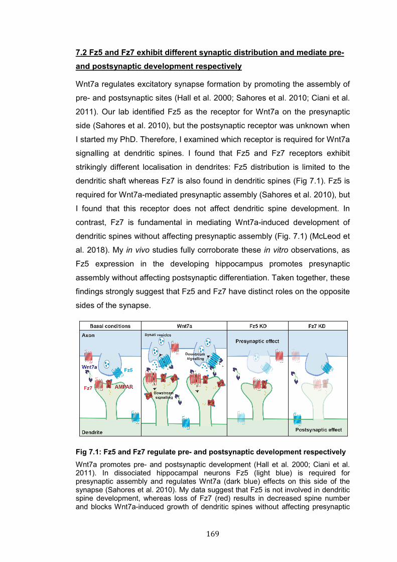
169
7.2 Fz5 and Fz7 exhibit different synaptic distribution and mediate pre- and postsynaptic development respectively
Wnt7a regulates excitatory synapse formation by promoting the assembly of
pre- and postsynaptic sites (Hall et al. 2000; Sahores et al. 2010; Ciani et al.
2011). Our lab identified Fz5 as the receptor for Wnt7a on the presynaptic
side (Sahores et al. 2010), but the postsynaptic receptor was unknown when
I started my PhD. Therefore, I examined which receptor is required for Wnt7a
signalling at dendritic spines. I found that Fz5 and Fz7 receptors exhibit
strikingly different localisation in dendrites: Fz5 distribution is limited to the
dendritic shaft whereas Fz7 is also found in dendritic spines (Fig 7.1). Fz5 is
required for Wnt7a-mediated presynaptic assembly (Sahores et al. 2010), but
I found that this receptor does not affect dendritic spine development. In
contrast, Fz7 is fundamental in mediating Wnt7a-induced development of
dendritic spines without affecting presynaptic assembly (Fig. 7.1) (McLeod et
al. 2018). My in vivo studies fully corroborate these in vitro observations, as
Fz5 expression in the developing hippocampus promotes presynaptic
assembly without affecting postsynaptic differentiation. Taken together, these
findings strongly suggest that Fz5 and Fz7 have distinct roles on the opposite
sides of the synapse.
Fig 7.1: Fz5 and Fz7 regulate pre- and postsynaptic development respectively Wnt7a promotes pre- and postsynaptic development (Hall et al. 2000; Ciani et al. 2011). In dissociated hippocampal neurons Fz5 (light blue) is required for presynaptic assembly and regulates Wnt7a (dark blue) effects on this side of the synapse (Sahores et al. 2010). My data suggest that Fz5 is not involved in dendritic spine development, whereas loss of Fz7 (red) results in decreased spine number and blocks Wnt7a-induced growth of dendritic spines without affecting presynaptic
170
assembly. Wnt7a-Fz7 signalling is also important for synaptic plasticity and AMPARs (dark red) trafficking (McLeod et al. 2018).
What are the mechanisms that control the localisation of Fz5 and Fz7 at
different synaptic compartments? What are the signalling events downstream
of these receptors that lead to synapse formation? One potential explanation
lies in the different structures of these two receptors, in particular at the C-
terminal domain. While both receptors can bind Wnt7a through their N-term
domain, their C-term domains are strikingly different. Fz7 has a short tail (24
amino acids), whereas Fz5 has a longer one (63 amino acids), which can be
multiply palmitoylated. These structural differences suggest that the
interactome of Fz7 might be very different from Fz5. Therefore, it is
concievable that the C-term of Frizzled receptors, perhaps through
palmitoylation or other PTMs, determines Frizzled localisation, interactome
and function. Could such differences in the C-term influence signalling
downstream of Frizzled receptors? These questions could be addressed by
generating Frizzled receptors chimeras and analysing how their function is
affected. Using this approach, it has been demonstrated that the specificity of
Wnt9-Fz9 signalling during stem cell development in Zebrafish is determined
by just two intracellular domains of Fz9, which cannot be substituted with
domains of other Frizzled receptors (Grainger et al. 2018). To address
whether Frizzled receptor C-term domains determine synaptic localisation
and function, future experiments should examine whether (1) Fz5 receptors
with the Fz7 C-term domain localise at dendritic spines and promote spine
development; (2) whether Fz7 with the Fz5 C-term promotes presynaptic
gain-of-function, and whether this is palmitoylation-dependent.
Many other molecular mechanisms, which are not mutually exclusive, could
be underpinning the distinct functions of Fz5 and Fz7. Previous findings
indicate that - except for the involvement of Dvl1, which is required for both
pre- and postsynaptic development (Ahmad-Annuar et al. 2006; Ciani et al.
2011) - Wnt7a induces different downstream signalling cascades at pre- and
postsynaptic sites. At presynaptic sites of cultured hippocampal neurons,
pharmacological inhibition of Gsk3β blocks the effects of Wnt7a; however,
171
Wnt7a-induced presynaptic assembly does not require transcription,
suggesting that the divergent canonical pathway is involved in Wnt7a-
mediated presynaptic assembly (E. Dickens PhD thesis, unpublished data).
Whether Fz5 and its palmitoylation are involved in the activation of the
divergent canonical pathway remains to be determined. At dendritic spines,
Wnt7a-Fz7 signalling activates the Ca2+ cascade by activating CaMKII and
PKA, which in turn phosphorylate AMPARs resulting in changes in their
trafficking (McLeod et al. 2018). Thus, the divergent canonical pathway
seems to be involved in Wnt7a-induced presynaptic formation, whereas
Wnt7a-Fz7 signalling activates the Ca2+ signalling cascade at dendritic
spines (McLeod et al. 2018). The next step will be to understand how the
distinct signalling pathways are activated downstream of Fz5 and Fz7 at
synapses.
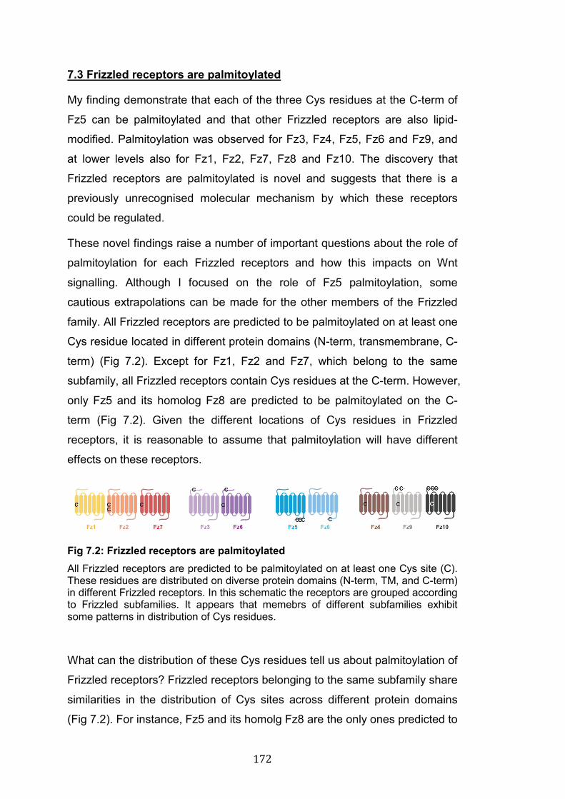
172
7.3 Frizzled receptors are palmitoylated
My finding demonstrate that each of the three Cys residues at the C-term of
Fz5 can be palmitoylated and that other Frizzled receptors are also lipid-
modified. Palmitoylation was observed for Fz3, Fz4, Fz5, Fz6 and Fz9, and
at lower levels also for Fz1, Fz2, Fz7, Fz8 and Fz10. The discovery that
Frizzled receptors are palmitoylated is novel and suggests that there is a
previously unrecognised molecular mechanism by which these receptors
could be regulated.
These novel findings raise a number of important questions about the role of
palmitoylation for each Frizzled receptors and how this impacts on Wnt
signalling. Although I focused on the role of Fz5 palmitoylation, some
cautious extrapolations can be made for the other members of the Frizzled
family. All Frizzled receptors are predicted to be palmitoylated on at least one
Cys residue located in different protein domains (N-term, transmembrane, C-
term) (Fig 7.2). Except for Fz1, Fz2 and Fz7, which belong to the same
subfamily, all Frizzled receptors contain Cys residues at the C-term. However,
only Fz5 and its homolog Fz8 are predicted to be palmitoylated on the C-
term (Fig 7.2). Given the different locations of Cys residues in Frizzled
receptors, it is reasonable to assume that palmitoylation will have different
effects on these receptors.
Fig 7.2: Frizzled receptors are palmitoylated All Frizzled receptors are predicted to be palmitoylated on at least one Cys site (C). These residues are distributed on diverse protein domains (N-term, TM, and C-term) in different Frizzled receptors. In this schematic the receptors are grouped according to Frizzled subfamilies. It appears that memebrs of different subfamilies exhibit some patterns in distribution of Cys residues.
What can the distribution of these Cys residues tell us about palmitoylation of
Frizzled receptors? Frizzled receptors belonging to the same subfamily share
similarities in the distribution of Cys sites across different protein domains
(Fig 7.2). For instance, Fz5 and its homolg Fz8 are the only ones predicted to
173
be palmitoylated on the C-term. The Fz1, Fz2, Fz7 subfamily is predicted to
be palmitoylated on the 1st TM domain. The Fz4, Fz9, Fz10 subfamily might
be palmitoylated on the 2nd TM domain and at the N-term. Similarly, Fz3 and
Fz6 could also be palmitoylated in the extracellular region. Therefore, it
seems that the localisation of these residues might correlate with the
evolution of Frizzled receptors. This interesting observation should be taken
into account when planning to address the function of palmitoylation for each
Frizzled receptor, as similar molecular mechanisms might be shared by
members of the same subfamily. However, caution should be taken
regarding the prediction of Frizzled palmitoylation at the N-term, which was
suggested by the CSS 4.0 software for Fz3, Fz6, Fz9 and Fz10 (Fig 4.1).
First, extremely little is known about palmitoylation on extracellular domains
of membrane proteins. In contrast, palmitoylation of Cys within TM or
cytoplasmic domains has been observed in a number of TM proteins and has
been widely shown to regulate multiple cell biology functions (Greaves and
Chamberlain 2007; Linder and Deschenes 2007; Salaun et al. 2010;
Blaskovic et al. 2013; Chamberlain and Shipston 2015). Second, the CRD
domain of Frizzled receptors contains 10 conserved Cys residues, and in Fz3
and Fz8 these Cys all engage in disulfide bonds (Dann et al. 2001),
suggesting that they might not be available sites for palmitoylation. It will be
important to address whether this is true for all Frizzled receptors, especially
those predicted to be palmitoylated at the N-term. However, if palmitoylation
does occur at the N-term, its impact on ligand binding should be examined,
as this interaction occurs between the CRD domain of Frizzled receptors and
the lipid moiety on Wnt ligands (Janda et al. 2012; DeBruine et al. 2017; Nile
et al. 2017; Nile and Hannoush 2018). Therefore, the position of Cys
residues within Frizzled receptors needs to be taken into account when
further studying the role of this modification for the function of these
receptors.
174
7.4 Palmitoylation is essential for Fz5 function
My findings demonstrate that palmitoylation is required in vivo for Fz5-
induced synapse formation. Consistent with preliminary observations in
hippocampal cultures (Fig 1.13), palmitoylation-deficient Fz5 receptors do not
promote the assembly of presynaptic sites in the developing hippocampus
(Fig 7.3). In addition, expression of mutant Fz5 receptors (3CS Fz5) might
have a negative impact on postsynaptic differentiation (Fig 7.3), although
further experiments are required to support this conclusion. These novel and
exciting findings open up new avenues to the study of Frizzled receptor
regulation and Wnt signallig-mediated cellular functions.
How does palmitoylation regulate Fz5 function? I performed a series of
experiments to address this question, focusing specifically on the role of
palmitoylation for key interactions of Fz5 with the Wnt signalosome
components and on the impact of this modification on Fz5 trafficking (see
section 7.6). The correct assembly of the Wnt signalosome is crucial for
activation of the pathway. A key event in this process is the recruitment of the
scaffold protein Dvl to the PM, where Dvl interacts with the C-term domain of
Frizzled receptors (Gao and Chen 2010). Frizzled dimerisation is another key
event for Wnt signalling activation (Carron et al. 2003). My findings suggest
that palmitoylation does not affect Fz5/Fz5 interaction, which is unsuprising
given that Fz-Fz interactions occur via the extracellular CRD domain of these
receptors (DeBruine et al. 2017; Nile et al. 2017). However, palmitoylation is
important for Fz5 binding to Dvl1. The interaction between Dvl1 and Fz
occurs between the DEP domain of Dvl1 and a discontinuous Dvl-binding
motif over the C-term and the 3rd intracellular loop of Fz5 (Tauriello et al.
2012). At the C-term of Fz5, the Dvl1-binding motif is located in close
proximity (6 amino acids apart) to the three Cys residues that are
palmitoylated, suggesting that palmitoylation could affect Dvl1 interaction
with the C-term of Fz5. However, it should be noted that this Dvl-binding
domain is conserved across all Frizzled receptors (Wang et al. 2006), which,
except for Fz8 (the Fz5 homolog), either do not contain Cys residues at the
C-term or have Cys sites that are not predicted to be palmitoylated. Thus, if
palmitoylation affects the interaction with Dvl1 through this motif, it is likely to
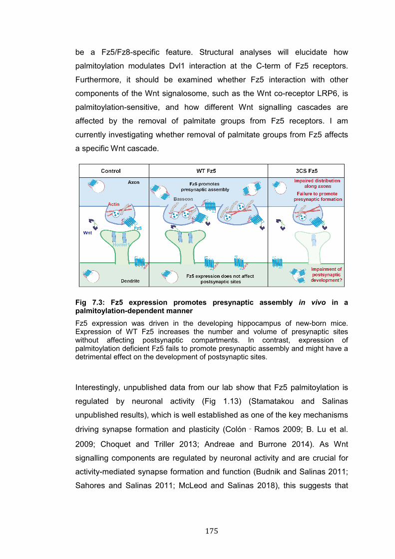
175
be a Fz5/Fz8-specific feature. Structural analyses will elucidate how
palmitoylation modulates Dvl1 interaction at the C-term of Fz5 receptors.
Furthermore, it should be examined whether Fz5 interaction with other
components of the Wnt signalosome, such as the Wnt co-receptor LRP6, is
palmitoylation-sensitive, and how different Wnt signalling cascades are
affected by the removal of palmitate groups from Fz5 receptors. I am
currently investigating whether removal of palmitate groups from Fz5 affects
a specific Wnt cascade.
Fig 7.3: Fz5 expression promotes presynaptic assembly in vivo in a palmitoylation-dependent manner Fz5 expression was driven in the developing hippocampus of new-born mice. Expression of WT Fz5 increases the number and volume of presynaptic sites without affecting postsynaptic compartments. In contrast, expression of palmitoylation deficient Fz5 fails to promote presynaptic assembly and might have a detrimental effect on the development of postsynaptic sites.
Interestingly, unpublished data from our lab show that Fz5 palmitoylation is
regulated by neuronal activity (Fig 1.13) (Stamatakou and Salinas
unpublished results), which is well established as one of the key mechanisms
driving synapse formation and plasticity (Colón‐Ramos 2009; B. Lu et al.
2009; Choquet and Triller 2013; Andreae and Burrone 2014). As Wnt
signalling components are regulated by neuronal activity and are crucial for
activity-mediated synapse formation and function (Budnik and Salinas 2011;
Sahores and Salinas 2011; McLeod and Salinas 2018), this suggests that
176
Fz5 palmitoylation could be a key molecular step in translating neuronal
activity into enhanced synapse formation and function.
It is of particular interest to understand how neuronal activity affects
Wnt7a/Wnt7b and Fz5 expression (at the protein level) and function. In the
hippocampus, Wnt7a/Wnt7b protein levels are elevated upon induction of
HFS (high frequqncy stimulation) (McLeod et al. 2018), and are increased in
mice exposed to an EE (enriched environment) (Gogolla et al. 2009), a
paradigm used to study activity-mediated synapse formation and remodelling
(van Praag et al. 2000; Nithianantharajah and Hannan 2006). Fz5, whose
surface levels are up- and down-regulated in response to HFS and LFS
respectively (Sahores et al. 2010), is required for HFS-induced synapse
formation in primary hippocampal neurons (Sahores et al. 2010).
Unpublished data from our lab show that Fz5 palmitoylation is elevated upon
HFS in primary neurons and by EE in the hippocampus of young mice (Fig
1.13). Therefore, it is intriguing to speculate that Fz5 and its palmitoylation
are required for synapse formation downstream of activity-induced
Wnt7a/Wnt7b. It is possible that Fz5 palmitoylation is necessary for
Wnt7a/Wnt7b to induce synapse formation in mice exposed to EE.
Furthermore, given that Fz5 palmitoylation is modulated by neuronal activity,
the regulation of palmitoylating and de-palmitoylating enzymes in this context
should be addressed. Several DHHC enzymes are positively regulated by
neuronal activity, and their function is required for mechanisms of synaptic
plasticity (Noritake et al. 2009; Thomas et al. 2012; Brigidi et al. 2014;
Dejanovic et al. 2014; Brigidi et al. 2015). Studying the molecular
mechanisms underlying activity-induced Fz5 palmitoylation and how this
affects synapse formation will provide further insight into the complex
interactions and dependencies that govern the process of synaptogenesis.
177
7.5 Palmitoylation regulates Fz5 trafficking and membrane stability
Palmitoylation is a master regulator of protein trafficking and function (Linder
and Deschenes 2007). While palmitoyaltion does not affect Fz5 turnover and
does not regulate the mobility of this receptor across the PM, palmitoylation
is required to maintain Fz5 surface levels by inhibiting constitutive
internalisation of this receptor (Fig 7.4). Moreover, palmitoylation-deficient
Fz5 receptors are less abundant along axons of hippocampal neurons (Fig
5.3), suggesting defects in polarised sorting. These findings strongly suggest
that palmitoylation is an important regulator of Fz5 trafficking.
Palmitoylation plays a fundamental role in regulating protein sorting and
localisation (Greaves and Chamberlain 2007), especially in highly polarised
cells like neurons (Fukata and Fukata 2010; Globa and Bamji 2017). The fact
that palmitoylation-deficient Fz5 receptors fail to induce synapse formation
suggested that the localisation of 3CS Fz5 receptors could be affected in
hippocampal neurons. Indeed, although palmitoylation does not affect Fz5
distribution at the soma and along dendrites, it is required for Fz5 localisation
along axons (Fig 5.3). Several neuronal proteins exhibit palmitoylation-
dependent sorting into axons or dendrites (El-Husseini et al. 2001; Fukata
and Fukata 2010; Globa and Bamji 2017). For instance, palmitoylation of
PSD-95 regulates both trafficking into dendrites as well as its clustering at
synapses (Craven et al. 1999; El-Husseini et al. 2000). Similarly, DLK (Dual
leucine-zipper kinase), which is an essential kinase for axon-soma retrograde
signalling after nerve injury, is targeted to axonal trafficking vesicles in a
palmitoylation-dependent manner, and this modification is required for the
assembly of DLK signalling complexes (Holland et al. 2016). Thus,
palmitoylation can control both polarised sorting and protein clustering in
neurons.
In section 5.3 I have discussed how palmitoylation might regulate Fz5
localisation in axons through these two non-mutually exclusive mechanisms.
Recent findings suggest that palmitoylation at the Golgi is an essential signal
for anterograde transport to the PM (Ernst et al. 2018). Given that I did not
detect obvious accumulation of 3CS Fz5 at the ER or Golgi apparatus (Fig
5.3), major changes along the exocytic route are not expected. Nevertheless,
178
palmitoylation might be involved in polarised sorting into axons, thus
explaining the decreased distribution of Fz5 3CS along these processes.To
answer this question, we are planning to perform time-course and time-lapse
experiments to compare the trafficking of WT and 3CS into axons and to
presynaptic sites. To perform these analyses, we will take advantage of the
RUSH (retention using selective hooks) system technology, which allows
synchronised trafficking of proteins of interest from a specific organelle to
other subcellular compartments (Boncompain et al. 2012; Boncompain and
Perez 2014). This experiment will answer two specific questions: does
palmitoylation regulate the delivery of Fz5 to the PM? Is this modification
involved in Fz5 polarised trafficking to axons and presynaptic terminals?
These are fundamental questions to fully elucidate the role of palmitoylation
in Fz5 trafficking.
In addition to sorting and exocytosis, palmitoylation is a major regulator of
protein stability at the PM, suggesting that surface levels of Fz5 could be
affected by this modification. Indeed, palmitoylation inhibits Fz5 constitutive
internalisation and is required to maintain Fz5 surface levels, but does not
affect the lateral mobility of this receptor (Fig 5.4 and Fig 5.5). Another PTM,
ubiquitination, has previously been shown to regulate Fz5 surface levels
(Koo et al. 2012; Hao et al. 2012), raising the question whether palmitoylation
and ubiquitination are co-regulated to control the levels of Fz5 at the PM. The
transmembrane E3 ubiquitin ligases RNF43 and ZNRF3 interact directly with
Frizzled receptors and ubiquitinate them, triggering their internalisation and,
in some cases, lysosomal degradation (Hao et al. 2012; Koo et al. 2012;
Bryan T MacDonald and He 2012a; de Lau et al. 2014). In endosomes,
Frizzled receptors can be de-ubiquitinated by USP enzymes and recycled to
the PM, or they can be targeted to degradation as a mechanism to down-
regulate Wnt signalling (Mukai et al. 2010; Madan et al. 2016). My findings
demonstrate that internalisation of 3CS Fz5 is increased compared to WT
receptors, suggesting that ubiquitination might be increased in the absence
of palmitate groups. What could be the molecular mechanisms of this co-
regulation? One possibility is that palmitoylation may reduce the interaction
of Fz5 with RNF43 and/or ZNRF3, perhaps by restricting the distribution of
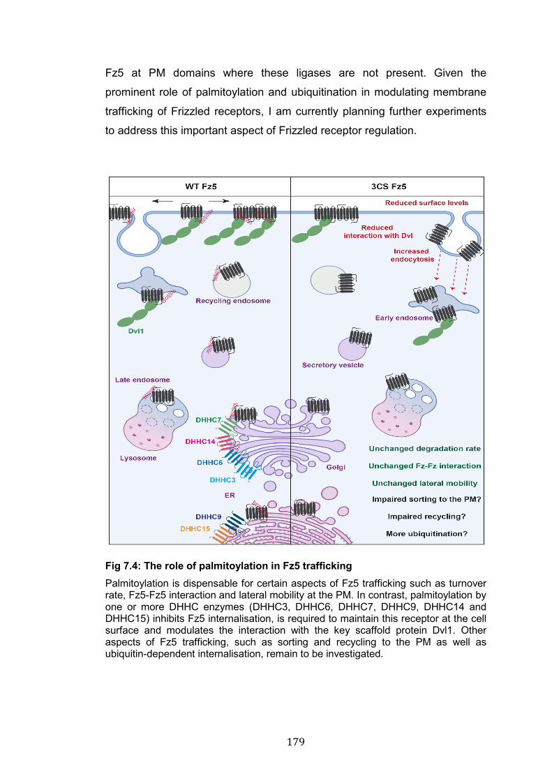
179
Fz5 at PM domains where these ligases are not present. Given the
prominent role of palmitoylation and ubiquitination in modulating membrane
trafficking of Frizzled receptors, I am currently planning further experiments
to address this important aspect of Frizzled receptor regulation.
Fig 7.4: The role of palmitoylation in Fz5 trafficking Palmitoylation is dispensable for certain aspects of Fz5 trafficking such as turnover rate, Fz5-Fz5 interaction and lateral mobility at the PM. In contrast, palmitoylation by one or more DHHC enzymes (DHHC3, DHHC6, DHHC7, DHHC9, DHHC14 and DHHC15) inhibits Fz5 internalisation, is required to maintain this receptor at the cell surface and modulates the interaction with the key scaffold protein Dvl1. Other aspects of Fz5 trafficking, such as sorting and recycling to the PM as well as ubiquitin-dependent internalisation, remain to be investigated.

180
Aside from its major role in protein endocytosis, palmitoylation is fundamental
for the PM recycling of a number of surface receptors (McCormick et al.
2008; Charollais and Van Der Goot 2009; Naumenko and Ponimaskin 2018),
suggesting that palmitoylation could also determine Fz5 recycling to the PM
after endocytosis. The fact that protein turnover is unchanged, but surface
levels of 3CS Fz5 are reduced, suggests that palmitoylation-deficient
receptors accumulate on some endosomal compartments. My qualitative
experiments show colocalisation of 3CS Fz5 with common endosome
markers (EEA1, Rab7 and Rab11), but the recycling rate of WT vs 3CS Fz5
remains to be examined. A functional recycling assay, ideally coupled with
the use of super-resolution microscopy, could reveal whether 3CS Fz5 fails
to recycle to the PM.
Another interesting question is whether palmitoylation affects ligand-induced
endocytosis of Fz5. Although the role of Wnt receptors internalisation in
downstream signalling is still debated, ligand-induced endocytosis has been
proposed as a model of Wnt signalling activation. Both Clathrin and Caveolin
control the endocytosis of the Wnt signalosome (Gagliardi et al. 2008; Brunt
and Scholpp 2018; Yamamoto et al. 2006; Blitzer and Nusse 2006). Wnt3a
induces the internalisation of Fz5 through Clathrin in the absence of LRP6,
whereas the Caveolin route is preferred when LRP6 is present (Yamamoto et
al. 2006). As my antibody-feeding experiments (Fig 5.4) were performed
without treating cells with exogenous Wnt ligands, they serve mainly as a
read-out of constitutive internalisation rather than ligand-induced endocytosis,
although cell lines do express some endogenous Wnts. Are WT and 3CS
Fz5 equally internalised together with functional signalosomes upon Wnt
binding? The reduced interaction between 3CS Fz5 and Dvl1 (Fig 5.2)
suggests that the internalisation of the mutant receptor together with the Wnt
signalosome might be impaired. TIRF (total internal reflection fluorescence)
imaging coupled with super-resolution microscopy (Guo et al. 2018) will
further elucidate the role of Fz5 palmitoylation in ligand-induced endocytosis.
The study of Frizzled lateral mobility is an understudied topic in the Frizzled
receptor field. Given that palmitoylation-deficient Fz5 exhibits impaired
membrane stability and reduced interaction with the scaffold protein Dvl1, the
181
lateral mobility at the PM could also be regulated by this modification.
However, FRAP experiments in NRK cells suprisisingly revealed no
differences in the lateral mobility of WT and 3CS Fz5, suggesting that
palmitoylation is not involved in this process. However, further experiments
should test how the binding of Wnt ligands affects the movements of WT and
3CS Fz5 across the PM. It has been proposed that several, but not all, Wnt
ligands significantly accelerate Fz6 mobility at the PM (Kilander et al. 2014).
Does palmitoylation regulate this phenomenon for Fz5 receptors? This
question is particularly interesting in the context of synapses, where Wnt7a is
rapidly increased upon induction of neuronal activity (McLeod et al. 2018).
Furthermore, lateral mobility of surface receptors has profound effects on
synapse strength and function (Groc et al. 2004; Lau and Zukin 2007;
Delgado and Selvin 2018; Park 2018). For instance, changes in the lateral
mobility of NMDARs and AMPARs, which are modulated by several factors
including Wnt ligands (McQuate et al. 2017; McLeod et al. 2018), affect
synaptic transmission and plasticity (Groc et al. 2004; Lau and Zukin 2007;
Heine et al. 2008). However, nothing is known about the membrane
dynamics of Frizzled receptors at the synapse. While my findings from cell
line experiments suggest that palmitoylation is not involved in this process, it
would be important to study further how the lateral mobility of Frizzled
receptors is regulated to modulate synapse formation and function.

182
7.6 Multiple DHHC enzymes can palmitoylate Fz5
Identifying the enzymes that catalyse Fz5 palmitoylation and de-
palmitoylation is crucial to understand the molecular mechanisms that
regulate Fz5 function, and I conducted gain and loss of function experiments
aimed at identifying these enzymes. Similarly to what has been reported for
other palmitoylated substrates like PSD-95, Gα subunit, GRIP1B, δ-catenin,
SNAP25 and others (Fukata et al. 2004; Hayashi et al. 2005; Hayashi et al.
2009; Thomas et al. 2012; Brigidi et al. 2014; Tsutsumi et al. 2009), Fz5 can
be palmitoylated by multiple enzymes. Expression of the Golgi-resident
DHHC3 or DHHC7 increased Fz5 palmitoylation by roughly 6-7 folds (Fig
4.4), but double KD of these two enzymes did not affect Fz5 palmitoylation.
Other enzymes, including DHHC6, DHHC9, DHHC14 and DHHC15
(localised between ER and Golgi) are also able to palmitoylate Fz5 and could
therefore compensate for the loss of DHHC3 and DHHC7 (Fig 7.4).
Therefore, the requirement of specific DHHC enzymes for Fz5 palmitoylation
remains to be fully elucidated.
How can it be determined which DHHC enzymes are essential for Fz5
palmitoylation? Understanding the recognition of Cys sites by specific
palmitoylating enzymes is a major challenge in the field of protein
palmitoylation. Some studies have proposed mechanisms of recognition
based on the amino acid microenvironment around palmitoylation sites, and
based on the molecular structure of DHHC enzymes (Resh 2006; Huang et
al. 2009; Ohno et al. 2012). However, these observations are either very
general or restricted to a small group of substrates. In the absence of
computational tools for the prediction of enzyme/substrate pairs, researchers
resort to gain and loss of function screens to tackle this question.
In the case of Fz5, potential compensatory effects by 4 different DHHCs may
occur when down-regulating DHHC3 and DHHC7 expplastuicression. As
discussed in section 4.3, the ability of 6 DHHCs to palmitoylate Fz5 might be
the result of protein overexpression in cell lines. Therefore, a first crucial step
is to understand whether each of these enzymes can palmitoylate Fz5 in a
more physiological context. Besides biochemical analyses of palmitoylation
levels, the functional outcomes of DHHCs KD should be examined, as this
183
strategy could reveal the requirement of certain enzymes in Fz5-specific
functions, such as for the assembly of presynaptic sites. Another strategy to
examine DHHC activity in a more specific manner is to examine whether
each of these enzymes can palmitoylate all three Cys residues, or whether
they exhibit residue-specific activity. If the latter is true, palmitoylation by
different DHHCs couls have different effects on Fz5. This molecular
mechanism has been demonstrated for certain synaptic proteins. For
instance, PSD-95 is dually palmitoylated at the N-term by DHHC3, which
mediates constitutive palmitoylation in the Golgi (El-Husseini et al. 2000;
Fukata et al. 2004), but in dendrites, DHHC2 catalyses activity-induced
palmitoylation of PSD-95, which is required for mechanisms of synaptic
plasticity (Noritake et al. 2009; Fukata et al. 2013; Jeyifous et al. 2016).
Using the double Cys mutants of Fz5 (see chapter 4), it is possible to
examine whether DHHC enzymes can palmitoylate all Cys residues or
whether their activity is restricted to a specific site. It can also be investigated
whether palmitoylation of each Cys is involved in a specific function, such as
constitutive versus activity-mediated palmitoylation. However, it is unknown
whether palmitoylation events on adjacent Cys sites have diverse effects on
proteins, and whether these modifications can be catalysed by different
DHHC enzymes.
Another important aspect of protein palmitoylation is the study of the de-
palmitoylation process. Different from other lipid modifications, palmitoylation
is reversible, and the de-attachment of palmitate chains is catalysed by acyl
protein thioesterases (Yokoi et al. 2016; Hornemann 2015; Lin and Conibear
2015; Won et al. 2018). Mutations of de-palmitoylating enzymes have been
linked to several disorders, including AD and Batten disease (Kollmann et al.
2013; Cho and Park 2016). Examining the regulation of Frizzled de-
palmitoylation will be of interest for future investigations. More
comprehensive characterisations of palmitoylating and de-palmitoylationg
enzymes will contribute enormously to our understanding of how Frizzled
receptors are trafficked and regulated, and will shed light on potential targets
that modulate Frizzled function. Given the prominent role that Wnt receptors
play in several biological processes, including neuroscience, stem cell
184
biology and cancer, studying the role and regulation of Frizzled palmitoylating
and de-palmitoylating enzymes is of considerable importance to basic as well
as clinical research.
7.7 Conclusions
In spite of the prominent role of Wnt signalling in many and diverse biological
processes, surprisingly little is known about the mechanisms that regulate
the main receptors for Wnt ligands, Frizzleds. In my PhD project, I have
studied the role and regulation of Frizzled receptors in synapse formation,
with a particular focus on a previously uncharacterised PTM of Fz5. My
results indicate that Fz7 and Fz5, two receptors for the synaptogenic ligand
Wnt7a, have distinct roles in pre- and postsynaptic development, respectively.
My findings also show for the first time that palmitoylation is required for the
synaptogenic function of Fz5 in vivo. This previously uncharacterised PTM is
essential for Fz5 membrane stability and for the interaction with the key
scaffold protein Dvl1. Aside from Fz5, others and potentially all Frizzled
receptors are palmitoylated. Together, my findings elucidate the molecular
mechanisms that regulate Fz5 trafficking and function, opening up new
avenues to study the mechanisms that regulate Frizzled receptors in health
and disease.

185

186
187
Acknowledgments
I want to thank my supervisor, Prof Patricia Salinas, for her great guidance
and support throughout my PhD. I feel grateful for being given the possibility
to work on such an exciting and interesting project. I will make treasure of the
valuable teachings received during my PhD.
I also wish to thank the MRC for funding my studies.
Many thanks to my thesis committee, Dr Julie Pitcher, Prof Trevor Smart and
Prof Frank Pichaud; their inputs have helped me in multiple occasions.
I would like to thank the UCL and LMCB staff for making this place a great
working environment. Many thanks to Dr Eleanna Stamatakou, who started
this project, and Dr Faye McLeod, Dr Ernest Palomer Vila and Dr Laura
Nadine Schuhmacher, who were directly involved in some aspects of these
studies. Many thanks also to the Brodsky lab for their scientific opinions and
for being great lab neighbours.
Thanks to previous and present members of the Salinas lab. You have been
fantastic colleagues and friends. You all represent an essential part of my
journey. I have incredible memories of these long years spent with you, too
many to be listed. I have learned a lot from all of you, and this is a rare luck.
Thanks to all my dear friends in Italy, UK and in the rest of the world. With
your jokes, laughters and dramas you are, and always will be, the best
company I could wish for.
I would like to thank my family for their unconditioned, and tangible love.
Without them I would not have had the possibility to study here. And finally
my Cheeky, who somehow managed to blur the 994 miles that separate us,
making me feel like we were holding hands every step of the way.
188
Bibliography Abrami, L., Kunz, B., Iacovache, I. and van der Goot, F.G. 2008. Palmitoylation and ubiquitination regulate exit of the Wnt signalling protein LRP6 from the endoplasmic reticulum. Proceedings of the National Academy of Sciences of the United States of America 105(14), pp. 5384–5389. Abrami, L., Leppla, S.H. and van der Goot, F.G. 2006. Receptor palmitoylation and ubiquitination regulate anthrax toxin endocytosis. The Journal of Cell Biology 172(2), pp. 309–320. Adachi, N., Hess, D.T., McLaughlin, P. and Stamler, J.S. 2016. S-Palmitoylation of a Novel Site in the β2-Adrenergic Receptor Associated with a Novel Intracellular Itinerary. The Journal of Biological Chemistry 291(38), pp. 20232–20246. Adams, M.N., Christensen, M.E., He, Y., Waterhouse, N.J. and Hooper, J.D. 2011. The role of palmitoylation in signalling, cellular trafficking and plasma membrane localisation of protease-activated receptor-2. Plos One 6(11), p. e28018. Agajanian, M.J., Walker, M.P., Axtman, A.D., Ruela-de-Sousa, R.R., Serafin, D.S., Rabinowitz, A.D., Graham, D.M., Ryan, M.B., Tamir, T., Nakamichi, Y., Gammons, M.V., Bennett, J.M., Couñago, R.M., Drewry, D.H., Elkins, J.M., Gileadi, C., Gileadi, O., Godoi, P.H., Kapadia, N., Müller, S., Santiago, A.S., Sorrell, F.J., Wells, C.I., Fedorov, O., Willson, T.M., Zuercher, W.J. and Major, M.B. 2019. WNT Activates the AAK1 Kinase to Promote Clathrin-Mediated Endocytosis of LRP6 and Establish a Negative Feedback Loop. Cell reports 26(1), pp. 79–93.e8. Ahmad-Annuar, A., Ciani, L., Simeonidis, I., Herreros, J., Fredj, N.B., Rosso, S.B., Hall, A., Brickley, S. and Salinas, P.C. 2006. Signalling across the synapse: a role for Wnt and Dishevelled in presynaptic assembly and neurotransmitter release. The Journal of Cell Biology 174(1), pp. 127–139. Ahmari, S.E., Buchanan, J. and Smith, S.J. 2000. Assembly of presynaptic active zones from cytoplasmic transport packets. Nature Neuroscience 3(5), pp. 445–451. Ahn, S. and Joyner, A.L. 2005. In vivo analysis of quiescent adult neural stem cells responding to Sonic hedgehog. Nature 437(7060), pp. 894–897. Aicart-Ramos, C., Valero, R.A. and Rodriguez-Crespo, I. 2011. Protein palmitoylation and subcellular trafficking. Biochimica et Biophysica Acta 1808(12), pp. 2981–2994. Alexandre, C., Baena-Lopez, A. and Vincent, J.-P. 2014. Patterning and growth control by membrane-tethered Wingless. Nature 505(7482), pp. 180–185. Allen, N.J. 2013. Role of glia in developmental synapse formation. Current Opinion in Neurobiology 23(6), pp. 1027–1033. Alonso, M., Medina, J.H. and Pozzo-Miller, L. 2004. ERK1/2 activation is necessary for BDNF to increase dendritic spine density in hippocampal CA1 pyramidal neurons. Learning & Memory 11(2), pp. 172–178. Alvarez, E., Gironès, N. and Davis, R.J. 1990. Inhibition of the receptor-mediated endocytosis of diferric transferrin is associated with the covalent modification of the transferrin receptor with palmitic acid. The Journal of Biological Chemistry 265(27), pp. 16644–16655.
189
Álvarez-Buylla, A. and Ihrie, R.A. 2014. Sonic hedgehog signalling in the postnatal brain. Seminars in Cell & Developmental Biology 33, pp. 105–111. Amano, M., Nakayama, M. and Kaibuchi, K. 2010. Rho-kinase/ROCK: A key regulator of the cytoskeleton and cell polarity. Cytoskeleton 67(9), pp. 545–554. Van Amerongen, R. 2012. Alternative Wnt pathways and receptors. Cold Spring Harbor Perspectives in Biology 4(10). Anastas, J.N. and Moon, R.T. 2013. WNT signalling pathways as therapeutic targets in cancer. Nature Reviews. Cancer 13(1), pp. 11–26. Andreae, L.C. and Burrone, J. 2015. Spontaneous Neurotransmitter Release Shapes Dendritic Arbors via Long-Range Activation of NMDA Receptors. Cell reports 10(6), pp. 873–882. Andreae, L.C. and Burrone, J. 2014. The role of neuronal activity and transmitter release on synapse formation. Current Opinion in Neurobiology 27, pp. 47–52. Andreae, L.C. and Burrone, J. 2018. The role of spontaneous neurotransmission in synapse and circuit development. Journal of Neuroscience Research 96(3), pp. 354–359. Anggono, V. and Huganir, R.L. 2012. Regulation of AMPA receptor trafficking and synaptic plasticity. Current Opinion in Neurobiology 22(3), pp. 461–469. Araque, A., Carmignoto, G., Haydon, P.G., Oliet, S.H.R., Robitaille, R. and Volterra, A. 2014. Gliotransmitters travel in time and space. Neuron 81(4), pp. 728–739. Araque, A., Parpura, V., Sanzgiri, R.P. and Haydon, P.G. 1999. Tripartite synapses: glia, the unacknowledged partner. Trends in Neurosciences 22(5), pp. 208–215. Arellano, J.I., Benavides-Piccione, R., Defelipe, J. and Yuste, R. 2007. Ultrastructure of dendritic spines: correlation between synaptic and spine morphologies. Frontiers in Neuroscience 1(1), pp. 131–143. Arikkath, J. and Reichardt, L.F. 2008. Cadherins and catenins at synapses: roles in synaptogenesis and synaptic plasticity. Trends in Neurosciences 31(9), pp. 487–494. Ashby, M.C., Ibaraki, K. and Henley, J.M. 2004. It’s green outside: tracking cell surface proteins with pH-sensitive GFP. Trends in Neurosciences 27(5), pp. 257–261. Ataman, B., Ashley, J., Gorczyca, D., Gorczyca, M., Mathew, D., Wichmann, C., Sigrist, S.J. and Budnik, V. 2006. Nuclear trafficking of Drosophila Frizzled-2 during synapse development requires the PDZ protein dGRIP. Proceedings of the National Academy of Sciences of the United States of America 103(20), pp. 7841–7846. Ataman, B., Ashley, J., Gorczyca, M., Ramachandran, P., Fouquet, W., Sigrist, S.J. and Budnik, V. 2008. Rapid activity-dependent modifications in synaptic structure and function require bidirectional Wnt signalling. Neuron 57(5), pp. 705–718. Avila, M.E., Sepúlveda, F.J., Burgos, C.F., Moraga-Cid, G., Parodi, J., Moon, R.T., Aguayo, L.G., Opazo, C. and De Ferrari, G.V. 2010. Canonical Wnt3a modulates intracellular calcium and enhances excitatory neurotransmission in hippocampal neurons. The Journal of Biological Chemistry 285(24), pp. 18939–18947.
190
Azzolin, L., Panciera, T., Soligo, S., Enzo, E., Bicciato, S., Dupont, S., Bresolin, S., Frasson, C., Basso, G., Guzzardo, V., Fassina, A., Cordenonsi, M. and Piccolo, S. 2014. YAP/TAZ incorporation in the β-catenin destruction complex orchestrates the Wnt response. Cell 158(1), pp. 157–170. Baekkeskov, S. and Kanaani, J. 2009. Palmitoylation cycles and regulation of protein function (Review). Molecular membrane biology 26(1), pp. 42–54. Bafico, A., Gazit, A., Pramila, T., Finch, P.W., Yaniv, A. and Aaronson, S.A. 1999. Interaction of frizzled related protein (FRP) with Wnt ligands and the frizzled receptor suggests alternative mechanisms for FRP inhibition of Wnt signalling. The Journal of Biological Chemistry 274(23), pp. 16180–16187. Bafico, A., Liu, G., Yaniv, A., Gazit, A. and Aaronson, S.A. 2001. Novel mechanism of Wnt signalling inhibition mediated by Dickkopf-1 interaction with LRP6/Arrow. Nature Cell Biology 3(7), pp. 683–686. Bamji, S.X., Shimazu, K., Kimes, N., Huelsken, J., Birchmeier, W., Lu, B. and Reichardt, L.F. 2003. Role of beta-catenin in synaptic vesicle localisation and presynaptic assembly. Neuron 40(4), pp. 719–731. Banovic, D., Khorramshahi, O., Owald, D., Wichmann, C., Riedt, T., Fouquet, W., Tian, R., Sigrist, S.J. and Aberle, H. 2010. Drosophila neuroligin 1 promotes growth and postsynaptic differentiation at glutamatergic neuromuscular junctions. Neuron 66(5), pp. 724–738. Bänziger, C., Soldini, D., Schütt, C., Zipperlen, P., Hausmann, G. and Basler, K. 2006. Wntless, a conserved membrane protein dedicated to the secretion of Wnt proteins from signalling cells. Cell 125(3), pp. 509–522. Barnett-Norris, J., Lynch, D. and Reggio, P.H. 2005. Lipids, lipid rafts and caveolae: their importance for GPCR signalling and their centrality to the endocannabinoid system. Life Sciences 77(14), pp. 1625–1639. Barros, C.S., Calabrese, B., Chamero, P., Roberts, A.J., Korzus, E., Lloyd, K., Stowers, L., Mayford, M., Halpain, S. and Müller, U. 2009. Impaired maturation of dendritic spines without disorganisation of cortical cell layers in mice lacking NRG1/ErbB signalling in the central nervous system. Proceedings of the National Academy of Sciences of the United States of America 106(11), pp. 4507–4512. Barrow, S.L., Constable, J.R., Clark, E., El-Sabeawy, F., McAllister, A.K. and Washbourne, P. 2009. Neuroligin1: a cell adhesion molecule that recruits PSD-95 and NMDA receptors by distinct mechanisms during synaptogenesis. Neural Development 4, p. 17. Bartscherer, K. and Boutros, M. 2008. Regulation of Wnt protein secretion and its role in gradient formation. EMBO Reports 9(10), pp. 977–982. Bartscherer, K., Pelte, N., Ingelfinger, D. and Boutros, M. 2006. Secretion of Wnt ligands requires Evi, a conserved transmembrane protein. Cell 125(3), pp. 523–533. Bassani, S., Folci, A., Zapata, J. and Passafaro, M. 2013. AMPAR trafficking in synapse maturation and plasticity. Cellular and Molecular Life Sciences 70(23), pp. 4411–4430. Basu, S., Saha, P.K., Roszkowska, M., Magnowska, M., Baczynska, E., Das, N., Plewczynski, D. and Wlodarczyk, J. 2018. Quantitative 3-D morphometric analysis of individual dendritic spines. Scientific reports 8(1), p. 3545. Batool, S., Raza, H., Zaidi, J., Riaz, S., Hasan, S. and Syed, N.I. 2019. Synapse formation: from cellular and molecular mechanisms to
191
neurodevelopmental and neurodegenerative disorders. Journal of Neurophysiology 121(4), pp. 1381–1397. Bats, C., Groc, L. and Choquet, D. 2007. The interaction between Stargazin and PSD-95 regulates AMPA receptor surface trafficking. Neuron 53(5), pp. 719–734. Belenkaya, T.Y., Wu, Y., Tang, X., Zhou, B., Cheng, L., Sharma, Y.V., Yan, D., Selva, E.M. and Lin, X. 2008. The retromer complex influences Wnt secretion by recycling wntless from endosomes to the trans-Golgi network. Developmental Cell 14(1), pp. 120–131. Belgacem, Y.H., Hamilton, A.M., Shim, S., Spencer, K.A. and Borodinsky, L.N. 2016. The many hats of sonic hedgehog signalling in nervous system development and disease. Journal of developmental biology 4(4). Bellucci, A., Mercuri, N.B., Venneri, A., Faustini, G., Longhena, F., Pizzi, M., Missale, C. and Spano, P. 2016. Review: Parkinson’s disease: from synaptic loss to connectome dysfunction. Neuropathology and Applied Neurobiology 42(1), pp. 77–94. Ben-Ari, Y. 2002. Excitatory actions of gaba during development: the nature of the nurture. Nature Reviews. Neuroscience 3(9), pp. 728–739. Benavides-Piccione, R., Fernaud-Espinosa, I., Robles, V., Yuste, R. and DeFelipe, J. 2013. Age-based comparison of human dendritic spine structure using complete three-dimensional reconstructions. Cerebral Cortex 23(8), pp. 1798–1810. Benilova, I., Karran, E. and De Strooper, B. 2012. The toxic Aβ oligomer and Alzheimer’s disease: an emperor in need of clothes. Nature Neuroscience 15(3), pp. 349–357. Berry, K.P. and Nedivi, E. 2017. Spine dynamics: are they all the same? Neuron 96(1), pp. 43–55. Berwick, D.C. and Harvey, K. 2012. The importance of Wnt signalling for neurodegeneration in Parkinson’s disease. Biochemical Society Transactions 40(5), pp. 1123–1128. Bettler, B., Kaupmann, K., Mosbacher, J. and Gassmann, M. 2004. Molecular structure and physiological functions of GABA(B) receptors. Physiological Reviews 84(3), pp. 835–867. Bhanot, P., Brink, M., Samos, C.H., Hsieh, J.C., Wang, Y., Macke, J.P., Andrew, D., Nathans, J. and Nusse, R. 1996. A new member of the frizzled family from Drosophila functions as a Wingless receptor. Nature 382(6588), pp. 225–230. Biederer, T., Sara, Y., Mozhayeva, M., Atasoy, D., Liu, X., Kavalali, E.T. and Südhof, T.C. 2002. SynCAM, a synaptic adhesion molecule that drives synapse assembly. Science 297(5586), pp. 1525–1531. Binder, D.K. and Scharfman, H.E. 2004. Brain-derived neurotrophic factor. Growth Factors 22(3), pp. 123–131. Blakely, R.D. and Edwards, R.H. 2012. Vesicular and plasma membrane transporters for neurotransmitters. Cold Spring Harbor Perspectives in Biology 4(2). Blanc, M., David, F., Abrami, L., Migliozzi, D., Armand, F., Bürgi, J. and van der Goot, F.G. 2015. SwissPalm: Protein Palmitoylation database. [version 1; peer review: 3 approved]. F1000Research 4, p. 261.
192
Blaskovic, S., Blanc, M. and van der Goot, F.G. 2013. What does S-palmitoylation do to membrane proteins? The FEBS Journal 280(12), pp. 2766–2774. Blitzer, J.T. and Nusse, R. 2006. A critical role for endocytosis in Wnt signalling. BMC Cell Biology 7, p. 28. Bocchi, R., Egervari, K., Carol-Perdiguer, L., Viale, B., Quairiaux, C., De Roo, M., Boitard, M., Oskouie, S., Salmon, P. and Kiss, J.Z. 2017. Perturbed Wnt signalling leads to neuronal migration delay, altered interhemispheric connections and impaired social behavior. Nature Communications 8(1), p. 1158. Boeckers, T.M. 2006. The postsynaptic density. Cell and Tissue Research 326(2), pp. 409–422. Boncompain, G., Divoux, S., Gareil, N., de Forges, H., Lescure, A., Latreche, L., Mercanti, V., Jollivet, F., Raposo, G. and Perez, F. 2012. Synchronization of secretory protein traffic in populations of cells. Nature Methods 9(5), pp. 493–498. Boncompain, G. and Perez, F. 2014. Synchronization of secretory cargos trafficking in populations of cells. Methods in Molecular Biology 1174, pp. 211–223. Bonhoeffer, T. and Yuste, R. 2002. Spine motility. Phenomenology, mechanisms, and function. Neuron 35(6), pp. 1019–1027. Bosch, M. and Hayashi, Y. 2012. Structural plasticity of dendritic spines. Current Opinion in Neurobiology 22(3), pp. 383–388. Bosworth, A.P. and Allen, N.J. 2017. The diverse actions of astrocytes during synaptic development. Current Opinion in Neurobiology 47, pp. 38–43. Bourhis, E., Tam, C., Franke, Y., Bazan, J.F., Ernst, J., Hwang, J., Costa, M., Cochran, A.G. and Hannoush, R.N. 2010. Reconstitution of a frizzled8.Wnt3a.LRP6 signalling complex reveals multiple Wnt and Dkk1 binding sites on LRP6. The Journal of Biological Chemistry 285(12), pp. 9172–9179. Bouwman, J., Maia, A.S., Camoletto, P.G., Posthuma, G., Roubos, E.W., Oorschot, V.M.J., Klumperman, J. and Verhage, M. 2004. Quantification of synapse formation and maintenance in vivo in the absence of synaptic release. Neuroscience 126(1), pp. 115–126. Bovolenta, P., Esteve, P., Ruiz, J.M., Cisneros, E. and Lopez-Rios, J. 2008. Beyond Wnt inhibition: new functions of secreted Frizzled-related proteins in development and disease. Journal of Cell Science 121(Pt 6), pp. 737–746. Bramham, C.R. and Messaoudi, E. 2005. BDNF function in adult synaptic plasticity: the synaptic consolidation hypothesis. Progress in Neurobiology 76(2), pp. 99–125. Brigidi, G.S., Santyr, B., Shimell, J., Jovellar, B. and Bamji, S.X. 2015. Activity-regulated trafficking of the palmitoyl-acyl transferase DHHC5. Nature Communications 6, p. 8200. Brigidi, G.S., Sun, Y., Beccano-Kelly, D., Pitman, K., Mobasser, M., Borgland, S.L., Milnerwood, A.J. and Bamji, S.X. 2014. Palmitoylation of δ-catenin by DHHC5 mediates activity-induced synapse plasticity. Nature Neuroscience 17(4), pp. 522–532. Briscoe, J. 2009. Making a grade: Sonic Hedgehog signalling and the control of neural cell fate. The EMBO Journal 28(5), pp. 457–465.
193
Broekman, M.L.D., Comer, L.A., Hyman, B.T. and Sena-Esteves, M. 2006. Adeno-associated virus vectors serotyped with AAV8 capsid are more efficient than AAV-1 or -2 serotypes for widespread gene delivery to the neonatal mouse brain. Neuroscience 138(2), pp. 501–510. Brunt, L. and Scholpp, S. 2018. The function of endocytosis in Wnt signalling. Cellular and Molecular Life Sciences 75(5), pp. 785–795. Brusés, J.L. 2006. N-cadherin signalling in synapse formation and neuronal physiology. Molecular Neurobiology 33(3), pp. 237–252. Budnik, V. and Salinas, P.C. 2011. Wnt signalling during synaptic development and plasticity. Current Opinion in Neurobiology 21(1), pp. 151–159. Buechler, J. and Salinas, P.C. 2018. Deficient wnt signalling and synaptic vulnerability in alzheimer’s disease: emerging roles for the LRP6 receptor. Frontiers in synaptic neuroscience 10, p. 38. Buglino, J.A. and Resh, M.D. 2012. Palmitoylation of Hedgehog proteins. Vitamins and Hormones 88, pp. 229–252. Burnashev, N. 1998. Calcium permeability of ligand-gated channels. Cell Calcium 24(5-6), pp. 325–332. Caldeira, M.V., Melo, C.V., Pereira, D.B., Carvalho, R., Correia, S.S., Backos, D.S., Carvalho, A.L., Esteban, J.A. and Duarte, C.B. 2007. Brain-derived neurotrophic factor regulates the expression and synaptic delivery of alpha-amino-3-hydroxy-5-methyl-4-isoxazole propionic acid receptor subunits in hippocampal neurons. The Journal of Biological Chemistry 282(17), pp. 12619–12628. De Calisto, J., Araya, C., Marchant, L., Riaz, C.F. and Mayor, R. 2005. Essential role of non-canonical Wnt signalling in neural crest migration. Development 132(11), pp. 2587–2597. Caricasole, A., Copani, A., Caraci, F., Aronica, E., Rozemuller, A.J., Caruso, A., Storto, M., Gaviraghi, G., Terstappen, G.C. and Nicoletti, F. 2004. Induction of Dickkopf-1, a negative modulator of the Wnt pathway, is associated with neuronal degeneration in Alzheimer’s brain. The Journal of Neuroscience 24(26), pp. 6021–6027. Carpenter, G. and Liao, H.-J. 2013. Receptor tyrosine kinases in the nucleus. Cold Spring Harbor Perspectives in Biology 5(10), p. a008979. Carron, C., Pascal, A., Djiane, A., Boucaut, J.-C., Shi, D.-L. and Umbhauer, M. 2003. Frizzled receptor dimerization is sufficient to activate the Wnt/beta-catenin pathway. Journal of Cell Science 116(Pt 12), pp. 2541–2550. Catterall, W.A. 2011. Voltage-gated calcium channels. Cold Spring Harbor Perspectives in Biology 3(8), p. a003947. Cerpa, W., Gambrill, A., Inestrosa, N.C. and Barria, A. 2011. Regulation of NMDA-receptor synaptic transmission by Wnt signalling. The Journal of Neuroscience 31(26), pp. 9466–9471. Cerpa, W., Godoy, J.A., Alfaro, I., Farías, G.G., Metcalfe, M.J., Fuentealba, R., Bonansco, C. and Inestrosa, N.C. 2008. Wnt-7a modulates the synaptic vesicle cycle and synaptic transmission in hippocampal neurons. The Journal of Biological Chemistry 283(9), pp. 5918–5927. Cerpa, W., Latorre-Esteves, E. and Barria, A. 2015. RoR2 functions as a noncanonical Wnt receptor that regulates NMDAR-mediated synaptic transmission. Proceedings of the National Academy of Sciences of the United States of America 112(15), pp. 4797–4802.
194
Chakrabarty, P., Rosario, A., Cruz, P., Siemienski, Z., Ceballos-Diaz, C., Crosby, K., Jansen, K., Borchelt, D.R., Kim, J.-Y., Jankowsky, J.L., Golde, T.E. and Levites, Y. 2013. Capsid serotype and timing of injection determines AAV transduction in the neonatal mice brain. Plos One 8(6), p. e67680. Chamberlain, L.H. and Shipston, M.J. 2015. The physiology of protein S-acylation. Physiological Reviews 95(2), pp. 341–376. Chapman, E.R. 2018. A ca2+ sensor for exocytosis. Trends in Neurosciences 41(6), pp. 327–330. Chapman, E.R. 2008. How does synaptotagmin trigger neurotransmitter release? Annual Review of Biochemistry 77, pp. 615–641. Charollais, J. and Van Der Goot, F.G. 2009. Palmitoylation of membrane proteins (Review). Molecular membrane biology 26(1), pp. 55–66. Charron, F., Stein, E., Jeong, J., McMahon, A.P. and Tessier-Lavigne, M. 2003. The morphogen sonic hedgehog is an axonal chemoattractant that collaborates with netrin-1 in midline axon guidance. Cell 113(1), pp. 11–23. Chebib, M. and Johnston, G.A. 1999. The “ABC” of GABA receptors: a brief review. Clinical and Experimental Pharmacology & Physiology 26(11), pp. 937–940. Chen, B., Sun, Y., Niu, J., Jarugumilli, G.K. and Wu, X. 2018. Protein lipidation in cell signalling and diseases: function, regulation, and therapeutic opportunities. Cell chemical biology 25(7), pp. 817–831. Chen, C.-M., Orefice, L.L., Chiu, S.-L., LeGates, T.A., Hattar, S., Huganir, R.L., Zhao, H., Xu, B. and Kuruvilla, R. 2017. Wnt5a is essential for hippocampal dendritic maintenance and spatial learning and memory in adult mice. Proceedings of the National Academy of Sciences of the United States of America 114(4), pp. E619–E628. Chen, J., Park, C.S. and Tang, S.-J. 2006. Activity-dependent synaptic Wnt release regulates hippocampal long term potentiation. The Journal of Biological Chemistry 281(17), pp. 11910–11916. Chen, L., Chetkovich, D.M., Petralia, R.S., Sweeney, N.T., Kawasaki, Y., Wenthold, R.J., Bredt, D.S. and Nicoll, R.A. 2000. Stargazin regulates synaptic targeting of AMPA receptors by two distinct mechanisms. Nature 408(6815), pp. 936–943. Chen, L.Y., Jiang, M., Zhang, B., Gokce, O. and Südhof, T.C. 2017. Conditional deletion of all neurexins defines diversity of essential synaptic organizer functions for neurexins. Neuron 94(3), pp. 611–625.e4. Chen, M.-K. and Hung, M.-C. 2015. Proteolytic cleavage, trafficking, and functions of nuclear receptor tyrosine kinases. The FEBS Journal 282(19), pp. 3693–3721. Chen, Y.A. and Scheller, R.H. 2001. SNARE-mediated membrane fusion. Nature Reviews. Molecular Cell Biology 2(2), pp. 98–106. Chesnutt, C., Burrus, L.W., Brown, A.M.C. and Niswander, L. 2004. Coordinate regulation of neural tube patterning and proliferation by TGFbeta and WNT activity. Developmental Biology 274(2), pp. 334–347. Ching, W. and Nusse, R. 2006. A dedicated Wnt secretion factor. Cell 125(3), pp. 432–433. Chiu, C.Q., Lur, G., Morse, T.M., Carnevale, N.T., Ellis-Davies, G.C.R. and Higley, M.J. 2013. Compartmentalization of GABAergic inhibition by dendritic spines. Science 340(6133), pp. 759–762.
195
Cho, C., Smallwood, P.M. and Nathans, J. 2017. Reck and Gpr124 Are Essential Receptor Cofactors for Wnt7a/Wnt7b-Specific Signalling in Mammalian CNS Angiogenesis and Blood-Brain Barrier Regulation. Neuron 95(5), pp. 1056–1073.e5. Cho, E. and Park, M. 2016. Palmitoylation in Alzheimer’s disease and other neurodegenerative diseases. Pharmacological Research 111, pp. 133–151. Cho, R.W., Buhl, L.K., Volfson, D., Tran, A., Li, F., Akbergenova, Y. and Littleton, J.T. 2015. Phosphorylation of Complexin by PKA Regulates Activity-Dependent Spontaneous Neurotransmitter Release and Structural Synaptic Plasticity. Neuron 88(4), pp. 749–761. Choi, B.J., Imlach, W.L., Jiao, W., Wolfram, V., Wu, Y., Grbic, M., Cela, C., Baines, R.A., Nitabach, M.N. and McCabe, B.D. 2014. Miniature neurotransmission regulates Drosophila synaptic structural maturation. Neuron 82(3), pp. 618–634. Choii, G. and Ko, J. 2015. Gephyrin: a central GABAergic synapse organizer. Experimental & Molecular Medicine 47, p. e158. Choquet, D. and Triller, A. 2013. The dynamic synapse. Neuron 80(3), pp. 691–703. Choudhry, Z., Rikani, A.A., Choudhry, A.M., Tariq, S., Zakaria, F., Asghar, M.W., Sarfraz, M.K., Haider, K., Shafiq, A.A. and Mobassarah, N.J. 2014. Sonic hedgehog signalling pathway: a complex network. Annals of neurosciences 21(1), pp. 28–31. Christopherson, K.S., Ullian, E.M., Stokes, C.C.A., Mullowney, C.E., Hell, J.W., Agah, A., Lawler, J., Mosher, D.F., Bornstein, P. and Barres, B.A. 2005. Thrombospondins are astrocyte-secreted proteins that promote CNS synaptogenesis. Cell 120(3), pp. 421–433. Ciani, L., Boyle, K.A., Dickins, E., Sahores, M., Anane, D., Lopes, D.M., Gibb, A.J. and Salinas, P.C. 2011. Wnt7a signalling promotes dendritic spine growth and synaptic strength through Ca2+/Calmodulin-dependent protein kinase II. Proceedings of the National Academy of Sciences of the United States of America 108(26), pp. 10732–10737. Ciani, L., Krylova, O., Smalley, M.J., Dale, T.C. and Salinas, P.C. 2004. A divergent canonical WNT-signalling pathway regulates microtubule dynamics: dishevelled signals locally to stabilize microtubules. The Journal of Cell Biology 164(2), pp. 243–253. Ciani, L., Marzo, A., Boyle, K., Stamatakou, E., Lopes, D.M., Anane, D., McLeod, F., Rosso, S.B., Gibb, A. and Salinas, P.C. 2015. Wnt signalling tunes neurotransmitter release by directly targeting Synaptotagmin-1. Nature Communications 6, p. 8302. Ciani, L. and Salinas, P.C. 2005. WNTs in the vertebrate nervous system: from patterning to neuronal connectivity. Nature Reviews. Neuroscience 6(5), pp. 351–362. Cingolani, L.A. and Goda, Y. 2008. Actin in action: the interplay between the actin cytoskeleton and synaptic efficacy. Nature Reviews. Neuroscience 9(5), pp. 344–356. Clevers, H. 2006. Wnt/beta-catenin signalling in development and disease. Cell 127(3), pp. 469–480. Clevers, H., Loh, K.M. and Nusse, R. 2014. Stem cell signalling. An integral program for tissue renewal and regeneration: Wnt signalling and stem cell control. Science 346(6205), p. 1248012.
196
Cline, H.T. 2001. Dendritic arbor development and synaptogenesis. Current Opinion in Neurobiology 11(1), pp. 118–126. Collins, M.O., Husi, H., Yu, L., Brandon, J.M., Anderson, C.N.G., Blackstock, W.P., Choudhary, J.S. and Grant, S.G.N. 2006. Molecular characterization and comparison of the components and multiprotein complexes in the postsynaptic proteome. Journal of Neurochemistry 97 Suppl 1, pp. 16–23. Colón‐Ramos, D.A. 2009. Chapter 2 Synapse Formation in Developing Neural Circuits. In: Development of Neural Circuitry. Current Topics in Developmental Biology. Elsevier, pp. 53–79. Cong, F., Schweizer, L. and Varmus, H. 2004. Wnt signals across the plasma membrane to activate the beta-catenin pathway by forming oligomers containing its receptors, Frizzled and LRP. Development 131(20), pp. 5103–5115. Craven, S.E., El-Husseini, A.E. and Bredt, D.S. 1999. Synaptic targeting of the postsynaptic density protein PSD-95 mediated by lipid and protein motifs. Neuron 22(3), pp. 497–509. Cruciat, C.-M. and Niehrs, C. 2013. Secreted and transmembrane wnt inhibitors and activators. Cold Spring Harbor Perspectives in Biology 5(3), p. a015081. Cuitino, L., Godoy, J.A., Farías, G.G., Couve, A., Bonansco, C., Fuenzalida, M. and Inestrosa, N.C. 2010. Wnt-5a modulates recycling of functional GABAA receptors on hippocampal neurons. The Journal of Neuroscience 30(25), pp. 8411–8420. Dabrowski, A., Terauchi, A., Strong, C. and Umemori, H. 2015. Distinct sets of FGF receptors sculpt excitatory and inhibitory synaptogenesis. Development 142(10), pp. 1818–1830. Dalva, M.B., McClelland, A.C. and Kayser, M.S. 2007. Cell adhesion molecules: signalling functions at the synapse. Nature Reviews. Neuroscience 8(3), pp. 206–220. Dalva, M.B., Takasu, M.A., Lin, M.Z., Shamah, S.M., Hu, L., Gale, N.W. and Greenberg, M.E. 2000. EphB receptors interact with NMDA receptors and regulate excitatory synapse formation. Cell 103(6), pp. 945–956. Danbolt, N.C. 2001. Glutamate uptake. Progress in Neurobiology 65(1), pp. 1–105. Dann, C.E., Hsieh, J.C., Rattner, A., Sharma, D., Nathans, J. and Leahy, D.J. 2001. Insights into Wnt binding and signalling from the structures of two Frizzled cysteine-rich domains. Nature 412(6842), pp. 86–90. Das, S., Yu, S., Sakamori, R., Stypulkowski, E. and Gao, N. 2012. Wntless in Wnt secretion: molecular, cellular and genetic aspects. Frontiers in biology 7(6), pp. 587–593. Davda, D., El Azzouny, M.A., Tom, C.T.M.B., Hernandez, J.L., Majmudar, J.D., Kennedy, R.T. and Martin, B.R. 2013. Profiling targets of the irreversible palmitoylation inhibitor 2-bromopalmitate. ACS Chemical Biology 8(9), pp. 1912–1917. Davidson, G., Wu, W., Shen, J., Bilic, J., Fenger, U., Stannek, P., Glinka, A. and Niehrs, C. 2005. Casein kinase 1 gamma couples Wnt receptor activation to cytoplasmic signal transduction. Nature 438(7069), pp. 867–872. Davis, E.K., Zou, Y. and Ghosh, A. 2008. Wnts acting through canonical and noncanonical signalling pathways exert opposite effects on hippocampal synapse formation. Neural Development 3, p. 32.
197
De, A. 2011. Wnt/Ca2+ signalling pathway: a brief overview. Acta biochimica et biophysica Sinica 43(10), pp. 745–756. Dean, C., Scholl, F.G., Choih, J., DeMaria, S., Berger, J., Isacoff, E. and Scheiffele, P. 2003. Neurexin mediates the assembly of presynaptic terminals. Nature Neuroscience 6(7), pp. 708–716. DeBruine, Z.J., Ke, J., Harikumar, K.G., Gu, X., Borowsky, P., Williams, B.O., Xu, W., Miller, L.J., Xu, H.E. and Melcher, K. 2017. Wnt5a promotes Frizzled-4 signalosome assembly by stabilizing cysteine-rich domain dimerization. Genes & Development 31(9), pp. 916–926. DeChiara, T.M., Bowen, D.C., Valenzuela, D.M., Simmons, M.V., Poueymirou, W.T., Thomas, S., Kinetz, E., Compton, D.L., Rojas, E., Park, J.S., Smith, C., DiStefano, P.S., Glass, D.J., Burden, S.J. and Yancopoulos, G.D. 1996. The receptor tyrosine kinase MuSK is required for neuromuscular junction formation in vivo. Cell 85(4), pp. 501–512. DeFelipe, J., Marco, P., Busturia, I. and Merchán-Pérez, A. 1999. Estimation of the number of synapses in the cerebral cortex: methodological considerations. Cerebral Cortex 9(7), pp. 722–732. Dehorter, N., Vinay, L., Hammond, C. and Ben-Ari, Y. 2012. Timing of developmental sequences in different brain structures: physiological and pathological implications. The European Journal of Neuroscience 35(12), pp. 1846–1856. Dejanovic, B., Semtner, M., Ebert, S., Lamkemeyer, T., Neuser, F., Lüscher, B., Meier, J.C. and Schwarz, G. 2014. Palmitoylation of gephyrin controls receptor clustering and plasticity of GABAergic synapses. PLoS Biology 12(7), p. e1001908. Delgado, J.Y. and Selvin, P.R. 2018. A revised view on the role of surface AMPAR mobility in tuning synaptic transmission: limitations, tools, and alternative views. Frontiers in synaptic neuroscience 10, p. 21. Delint-Ramirez, I., Willoughby, D., Hammond, G.R.V., Ayling, L.J. and Cooper, D.M.F. 2011. Palmitoylation targets AKAP79 protein to lipid rafts and promotes its regulation of calcium-sensitive adenylyl cyclase type 8. The Journal of Biological Chemistry 286(38), pp. 32962–32975. Dent, E.W., Merriam, E.B. and Hu, X. 2011. The dynamic cytoskeleton: backbone of dendritic spine plasticity. Current Opinion in Neurobiology 21(1), pp. 175–181. Devenport, D. 2014. The cell biology of planar cell polarity. The Journal of Cell Biology 207(2), pp. 171–179. Dickins, E.M. and Salinas, P.C. 2013. Wnts in action: from synapse formation to synaptic maintenance. Frontiers in Cellular Neuroscience 7, p. 162. Dickinson, M.E., Krumlauf, R. and McMahon, A.P. 1994. Evidence for a mitogenic effect of Wnt-1 in the developing mammalian central nervous system. Development 120(6), pp. 1453–1471. Diering, G.H. and Huganir, R.L. 2018. The AMPA receptor code of synaptic plasticity. Neuron 100(2), pp. 314–329. Dityatev, A., Dityateva, G. and Schachner, M. 2000. Synaptic strength as a function of post- versus presynaptic expression of the neural cell adhesion molecule NCAM. Neuron 26(1), pp. 207–217. Doherty, G.J. and McMahon, H.T. 2009. Mechanisms of endocytosis. Annual Review of Biochemistry 78, pp. 857–902.
198
Dong, H., Zhang, P., Song, I., Petralia, R.S., Liao, D. and Huganir, R.L. 1999. Characterization of the glutamate receptor-interacting proteins GRIP1 and GRIP2. The Journal of Neuroscience 19(16), pp. 6930–6941. Dorsky, R.I., Moon, R.T. and Raible, D.W. 1998. Control of neural crest cell fate by the Wnt signalling pathway. Nature 396(6709), pp. 370–373. Dotti, C.G., Sullivan, C.A. and Banker, G.A. 1988. The establishment of polarity by hippocampal neurons in culture. The Journal of Neuroscience 8(4), pp. 1454–1468. Drake, M.T., Shenoy, S.K. and Lefkowitz, R.J. 2006. Trafficking of G protein-coupled receptors. Circulation Research 99(6), pp. 570–582. Dudek, H., Ghosh, A. and Greenberg, M.E. 2001. Calcium phosphate transfection of DNA into neurons in primary culture. Current Protocols in Neuroscience Chapter 3, p. Unit 3.11. Dulubova, I., Khvotchev, M., Liu, S., Huryeva, I., Südhof, T.C. and Rizo, J. 2007. Munc18-1 binds directly to the neuronal SNARE complex. Proceedings of the National Academy of Sciences of the United States of America 104(8), pp. 2697–2702. Duman, J.G. and Forte, J.G. 2003. What is the role of SNARE proteins in membrane fusion? American Journal of Physiology. Cell Physiology 285(2), pp. C237–49. El-Husseini, A. el-D., Craven, S.E., Brock, S.C. and Bredt, D.S. 2001. Polarized targeting of peripheral membrane proteins in neurons. The Journal of Biological Chemistry 276(48), pp. 44984–44992. El-Husseini, A.E., Craven, S.E., Chetkovich, D.M., Firestein, B.L., Schnell, E., Aoki, C. and Bredt, D.S. 2000. Dual palmitoylation of PSD-95 mediates its vesiculotubular sorting, postsynaptic targeting, and ion channel clustering. The Journal of Cell Biology 148(1), pp. 159–172. Elias, G.M., Elias, L.A.B., Apostolides, P.F., Kriegstein, A.R. and Nicoll, R.A. 2008. Differential trafficking of AMPA and NMDA receptors by SAP102 and PSD-95 underlies synapse development. Proceedings of the National Academy of Sciences of the United States of America 105(52), pp. 20953–20958. Ernst, A.M., Syed, S.A., Zaki, O., Bottanelli, F., Zheng, H., Hacke, M., Xi, Z., Rivera-Molina, F., Graham, M., Rebane, A.A., Björkholm, P., Baddeley, D., Toomre, D., Pincet, F. and Rothman, J.E. 2018. S-Palmitoylation Sorts Membrane Cargo for Anterograde Transport in the Golgi. Developmental Cell 47(4), pp. 479–493.e7. Eroglu, C., Allen, N.J., Susman, M.W., O’Rourke, N.A., Park, C.Y., Ozkan, E., Chakraborty, C., Mulinyawe, S.B., Annis, D.S., Huberman, A.D., Green, E.M., Lawler, J., Dolmetsch, R., Garcia, K.C., Smith, S.J., Luo, Z.D., Rosenthal, A., Mosher, D.F. and Barres, B.A. 2009. Gabapentin receptor alpha2delta-1 is a neuronal thrombospondin receptor responsible for excitatory CNS synaptogenesis. Cell 139(2), pp. 380–392. Eroglu, C. and Barres, B.A. 2010. Regulation of synaptic connectivity by glia. Nature 468(7321), pp. 223–231. Erzurumlu, R.S. and Gaspar, P. 2012. Development and critical period plasticity of the barrel cortex. The European Journal of Neuroscience 35(10), pp. 1540–1553. Eubelen, M., Bostaille, N., Cabochette, P., Gauquier, A., Tebabi, P., Dumitru, A.C., Koehler, M., Gut, P., Alsteens, D., Stainier, D.Y.R., Garcia-Pino, A. and
199
Vanhollebeke, B. 2018. A molecular mechanism for Wnt ligand-specific signalling. Science 361(6403). Fang, C., Deng, L., Keller, C.A., Fukata, M., Fukata, Y., Chen, G. and Lüscher, B. 2006. GODZ-mediated palmitoylation of GABA(A) receptors is required for normal assembly and function of GABAergic inhibitory synapses. The Journal of Neuroscience 26(49), pp. 12758–12768. Farías, G.G., Alfaro, I.E., Cerpa, W., Grabowski, C.P., Godoy, J.A., Bonansco, C. and Inestrosa, N.C. 2009. Wnt-5a/JNK signalling promotes the clustering of PSD-95 in hippocampal neurons. The Journal of Biological Chemistry 284(23), pp. 15857–15866. Farías, G.G., Vallés, A.S., Colombres, M., Godoy, J.A., Toledo, E.M., Lukas, R.J., Barrantes, F.J. and Inestrosa, N.C. 2007. Wnt-7a induces presynaptic colocalisation of alpha 7-nicotinic acetylcholine receptors and adenomatous polyposis coli in hippocampal neurons. The Journal of Neuroscience 27(20), pp. 5313–5325. Favuzzi, E., Deogracias, R., Marques-Smith, A., Maeso, P., Jezequel, J., Exposito-Alonso, D., Balia, M., Kroon, T., Hinojosa, A.J., F Maraver, E. and Rico, B. 2019. Distinct molecular programs regulate synapse specificity in cortical inhibitory circuits. Science 363(6425), pp. 413–417. Favuzzi, E. and Rico, B. 2018. Molecular diversity underlying cortical excitatory and inhibitory synapse development. Current Opinion in Neurobiology 53, pp. 8–15. Fazzari, P., Paternain, A.V., Valiente, M., Pla, R., Luján, R., Lloyd, K., Lerma, J., Marín, O. and Rico, B. 2010. Control of cortical GABA circuitry development by Nrg1 and ErbB4 signalling. Nature 464(7293), pp. 1376–1380. De Ferrari, G.V., Papassotiropoulos, A., Biechele, T., Wavrant De-Vrieze, F., Avila, M.E., Major, M.B., Myers, A., Sáez, K., Henríquez, J.P., Zhao, A., Wollmer, M.A., Nitsch, R.M., Hock, C., Morris, C.M., Hardy, J. and Moon, R.T. 2007. Common genetic variation within the low-density lipoprotein receptor-related protein 6 and late-onset Alzheimer’s disease. Proceedings of the National Academy of Sciences of the United States of America 104(22), pp. 9434–9439. Ferrari, M.E., Bernis, M.E., McLeod, F., Podpolny, M., Coullery, R.P., Casadei, I.M., Salinas, P.C. and Rosso, S.B. 2018. Wnt7b signalling through Frizzled-7 receptor promotes dendrite development by coactivating CaMKII and JNK. Journal of Cell Science 131(13). Fiala, J.C., Feinberg, M., Popov, V. and Harris, K.M. 1998. Synaptogenesis via dendritic filopodia in developing hippocampal area CA1. The Journal of Neuroscience 18(21), pp. 8900–8911. Fox, M.A., Sanes, J.R., Borza, D.-B., Eswarakumar, V.P., Fässler, R., Hudson, B.G., John, S.W.M., Ninomiya, Y., Pedchenko, V., Pfaff, S.L., Rheault, M.N., Sado, Y., Segal, Y., Werle, M.J. and Umemori, H. 2007. Distinct target-derived signals organize formation, maturation, and maintenance of motor nerve terminals. Cell 129(1), pp. 179–193. Friedman, H.V., Bresler, T., Garner, C.C. and Ziv, N.E. 2000. Assembly of new individual excitatory synapses: time course and temporal order of synaptic molecule recruitment. Neuron 27(1), pp. 57–69.
200
Friedman, L.G., Benson, D.L. and Huntley, G.W. 2015. Cadherin-based transsynaptic networks in establishing and modifying neural connectivity. Current Topics in Developmental Biology 112, pp. 415–465. Fu, M. and Zuo, Y. 2011. Experience-dependent structural plasticity in the cortex. Trends in Neurosciences 34(4), pp. 177–187. Fukata, M., Fukata, Y., Adesnik, H., Nicoll, R.A. and Bredt, D.S. 2004. Identification of PSD-95 palmitoylating enzymes. Neuron 44(6), pp. 987–996. Fukata, Y., Bredt, D.S. and Fukata, M. 2006. Protein palmitoylation by DHHC protein family. In: Kittler, J. T. and Moss, S. J. eds. The dynamic synapse: molecular methods in ionotropic receptor biology. Frontiers in Neuroscience. Boca Raton (FL): CRC Press/Taylor & Francis. Fukata, Y., Dimitrov, A., Boncompain, G., Vielemeyer, O., Perez, F. and Fukata, M. 2013. Local palmitoylation cycles define activity-regulated postsynaptic subdomains. The Journal of Cell Biology 202(1), pp. 145–161. Fukata, Y. and Fukata, M. 2010. Protein palmitoylation in neuronal development and synaptic plasticity. Nature Reviews. Neuroscience 11(3), pp. 161–175. Gagliardi, M., Hernandez, A., McGough, I.J. and Vincent, J.-P. 2014. Inhibitors of endocytosis prevent Wnt/Wingless signalling by reducing the level of basal β-catenin/Armadillo. Journal of Cell Science 127(Pt 22), pp. 4918–4926. Gagliardi, M., Piddini, E. and Vincent, J.-P. 2008. Endocytosis: a positive or a negative influence on Wnt signalling? Traffic 9(1), pp. 1–9. Galli, S., Lopes, D.M., Ammari, R., Kopra, J., Millar, S.E., Gibb, A. and Salinas, P.C. 2014. Deficient Wnt signalling triggers striatal synaptic degeneration and impaired motor behaviour in adult mice. Nature Communications 5, p. 4992. Gambrill, A.C. and Barria, A. 2011. NMDA receptor subunit composition controls synaptogenesis and synapse stabilization. Proceedings of the National Academy of Sciences of the United States of America 108(14), pp. 5855–5860. Gao, C. and Chen, Y.-G. 2010. Dishevelled: The hub of Wnt signalling. Cellular Signalling 22(5), pp. 717–727. Gao, X.B. and van den Pol, A.N. 2000. GABA release from mouse axonal growth cones. The Journal of Physiology 523 Pt 3, pp. 629–637. Gao, Z., Ni, Y., Szabo, G. and Linden, J. 1999. Palmitoylation of the recombinant human A1 adenosine receptor: enhanced proteolysis of palmitoylation-deficient mutant receptors. The Biochemical Journal 342 ( Pt 2), pp. 387–395. Garcia, A.D.R., Han, Y.-G., Triplett, J.W., Farmer, W.T., Harwell, C.C. and Ihrie, R.A. 2018. The elegance of sonic hedgehog: emerging novel functions for a classic morphogen. The Journal of Neuroscience 38(44), pp. 9338–9345. García-López, P., García-Marín, V. and Freire, M. 2010. Dendritic spines and development: towards a unifying model of spinogenesis--a present day review of Cajal’s histological slides and drawings. Neural plasticity 2010, p. 769207. Gautam, M., Noakes, P.G., Moscoso, L., Rupp, F., Scheller, R.H., Merlie, J.P. and Sanes, J.R. 1996. Defective neuromuscular synaptogenesis in agrin-deficient mutant mice. Cell 85(4), pp. 525–535.
201
Ge, Y., Kang, Y., Cassidy, R.M., Moon, K.-M., Lewis, R., Wong, R.O.L., Foster, L.J. and Craig, A.M. 2018. Clptm1 limits forward trafficking of GABAA receptors to scale inhibitory synaptic strength. Neuron 97(3), pp. 596–610.e8. Georgiou, G., Bahra, S.S., Mackie, A.R., Wolfe, C.A., O’Shea, P., Ladha, S., Fernandez, N. and Cherry, R.J. 2002. Measurement of the lateral diffusion of human MHC class I molecules on HeLa cells by fluorescence recovery after photobleaching using a phycoerythrin probe. Biophysical Journal 82(4), pp. 1828–1834. Gerlach, J.P., Jordens, I., Tauriello, D.V.F., van' ' ’t Land-Kuper, I., Bugter, J.M., Noordstra, I., van der Kooij, J., Low, T.Y., Pimentel-Muiños, F.X., Xanthakis, D., Fenderico, N., Rabouille, C., Heck, A.J.R., Egan, D.A. and Maurice, M.M. 2018. TMEM59 potentiates Wnt signalling by promoting signalosome formation. Proceedings of the National Academy of Sciences of the United States of America 115(17), pp. E3996–E4005. Gerrow, K., Romorini, S., Nabi, S.M., Colicos, M.A., Sala, C. and El-Husseini, A. 2006. A preformed complex of postsynaptic proteins is involved in excitatory synapse development. Neuron 49(4), pp. 547–562. Gholizadeh, S., Tharmalingam, S., Macaldaz, M.E. and Hampson, D.R. 2013. Transduction of the central nervous system after intracerebroventricular injection of adeno-associated viral vectors in neonatal and juvenile mice. Human gene therapy methods 24(4), pp. 205–213. Giese, K.P., Aziz, W., Kraev, I. and Stewart, M.G. 2015. Generation of multi-innervated dendritic spines as a novel mechanism of long-term memory formation. Neurobiology of Learning and Memory 124, pp. 48–51. Gladding, C.M. and Raymond, L.A. 2011. Mechanisms underlying NMDA receptor synaptic/extrasynaptic distribution and function. Molecular and Cellular Neurosciences 48(4), pp. 308–320. Glaeser, K., Urban, M., Fenech, E., Voloshanenko, O., Kranz, D., Lari, F., Christianson, J.C. and Boutros, M. 2018. ERAD-dependent control of the Wnt secretory factor Evi. The EMBO Journal 37(4). Globa, A.K. and Bamji, S.X. 2017. Protein palmitoylation in the development and plasticity of neuronal connections. Current Opinion in Neurobiology 45, pp. 210–220. Go, G.-W. and Mani, A. 2012. Low-density lipoprotein receptor (LDLR) family orchestrates cholesterol homeostasis. The Yale journal of biology and medicine 85(1), pp. 19–28. Goddard, A.D. and Watts, A. 2012. Regulation of G protein-coupled receptors by palmitoylation and cholesterol. BMC Biology 10, p. 27. Gögel, S., Wakefield, S., Tear, G., Klämbt, C. and Gordon-Weeks, P.R. 2006. The Drosophila microtubule associated protein Futsch is phosphorylated by Shaggy/Zeste-white 3 at an homologous GSK3beta phosphorylation site in MAP1B. Molecular and Cellular Neurosciences 33(2), pp. 188–199. Gogolla, N., Galimberti, I., Deguchi, Y. and Caroni, P. 2009. Wnt signalling mediates experience-related regulation of synapse numbers and mossy fiber connectivities in the adult hippocampus. Neuron 62(4), pp. 510–525. Gomes, R.A., Hampton, C., El-Sabeawy, F., Sabo, S.L. and McAllister, A.K. 2006. The dynamic distribution of TrkB receptors before, during, and after synapse formation between cortical neurons. The Journal of Neuroscience 26(44), pp. 11487–11500.
202
Goodman, R.M., Thombre, S., Firtina, Z., Gray, D., Betts, D., Roebuck, J., Spana, E.P. and Selva, E.M. 2006. Sprinter: a novel transmembrane protein required for Wg secretion and signalling. Development 133(24), pp. 4901–4911. Gordon, M.D. and Nusse, R. 2006. Wnt signalling: multiple pathways, multiple receptors, and multiple transcription factors. The Journal of Biological Chemistry 281(32), pp. 22429–22433. Gottlieb, C.D. and Linder, M.E. 2017. Structure and function of DHHC protein S-acyltransferases. Biochemical Society Transactions 45(4), pp. 923–928. Gottlieb, C.D., Zhang, S. and Linder, M.E. 2015. The Cysteine-rich Domain of the DHHC3 Palmitoyltransferase Is Palmitoylated and Contains Tightly Bound Zinc. The Journal of Biological Chemistry 290(49), pp. 29259–29269. Gottmann, K., Mittmann, T. and Lessmann, V. 2009. BDNF signalling in the formation, maturation and plasticity of glutamatergic and GABAergic synapses. Experimental Brain Research 199(3-4), pp. 203–234. Graf, E.R., Zhang, X., Jin, S.-X., Linhoff, M.W. and Craig, A.M. 2004. Neurexins induce differentiation of GABA and glutamate postsynaptic specializations via neuroligins. Cell 119(7), pp. 1013–1026. Grainger, S., Nguyen, N., Richter, J., Setayesh, J., Lonquich, B., Oon, C.H., Wozniak, J.M., Barahona, R., Kamei, C.N., Houston, J., Carrillo-Terrazas, M., Drummond, I.A., Gonzalez, D., Willert, K. and Traver, D. 2018. EGFR confers exquisite specificity of Wnt9a-Fzd9b signalling in hematopoietic stem cell development. BioRxiv. Grant, B.D. and Donaldson, J.G. 2009. Pathways and mechanisms of endocytic recycling. Nature Reviews. Molecular Cell Biology 10(9), pp. 597–608. Gray, R.S., Roszko, I. and Solnica-Krezel, L. 2011. Planar cell polarity: coordinating morphogenetic cell behaviors with embryonic polarity. Developmental Cell 21(1), pp. 120–133. Greaves, J. and Chamberlain, L.H. 2007. Palmitoylation-dependent protein sorting. The Journal of Cell Biology 176(3), pp. 249–254. Greaves, J., Gorleku, O.A., Salaun, C. and Chamberlain, L.H. 2010. Palmitoylation of the SNAP25 protein family: specificity and regulation by DHHC palmitoyl transferases. The Journal of Biological Chemistry 285(32), pp. 24629–24638. Green, J., Nusse, R. and van Amerongen, R. 2014. The role of Ryk and Ror receptor tyrosine kinases in Wnt signal transduction. Cold Spring Harbor Perspectives in Biology 6(2). Greger, I.H., Watson, J.F. and Cull-Candy, S.G. 2017. Structural and Functional Architecture of AMPA-Type Glutamate Receptors and Their Auxiliary Proteins. Neuron 94(4), pp. 713–730. Groc, L., Heine, M., Cognet, L., Brickley, K., Stephenson, F.A., Lounis, B. and Choquet, D. 2004. Differential activity-dependent regulation of the lateral mobilities of AMPA and NMDA receptors. Nature Neuroscience 7(7), pp. 695–696. Gross, J.C., Chaudhary, V., Bartscherer, K. and Boutros, M. 2012. Active Wnt proteins are secreted on exosomes. Nature Cell Biology 14(10), pp. 1036–1045. Grutzendler, J., Kasthuri, N. and Gan, W.-B. 2002. Long-term dendritic spine stability in the adult cortex. Nature 420(6917), pp. 812–816.
203
Guan, X. and Fierke, C.A. 2011. Understanding protein palmitoylation: biological significance and enzymology. Science China. Chemistry 54(12), pp. 1888–1897. Gubb, D. and García-Bellido, A. 1982. A genetic analysis of the determination of cuticular polarity during development in Drosophila melanogaster. Journal of embryology and experimental morphology 68, pp. 37–57. Gundelfinger, E.D., Reissner, C. and Garner, C.C. 2015. Role of Bassoon and Piccolo in Assembly and Molecular Organisation of the Active Zone. Frontiers in synaptic neuroscience 7, p. 19. Guo, M., Chandris, P., Giannini, J.P., Trexler, A.J., Fischer, R., Chen, J., Vishwasrao, H.D., Rey-Suarez, I., Wu, Y., Wu, X., Waterman, C.M., Patterson, G.H., Upadhyaya, A., Taraska, J.W. and Shroff, H. 2018. Single-shot super-resolution total internal reflection fluorescence microscopy. Nature Methods 15(6), pp. 425–428. Hagemann, A.I.H., Kurz, J., Kauffeld, S., Chen, Q., Reeves, P.M., Weber, S., Schindler, S., Davidson, G., Kirchhausen, T. and Scholpp, S. 2014. In vivo analysis of formation and endocytosis of the Wnt/β-Catenin signalling complex in zebrafish embryos. Journal of Cell Science 127(24), p. 5331. Hall, A.C., Lucas, F.R. and Salinas, P.C. 2000. Axonal remodeling and synaptic differentiation in the cerebellum is regulated by WNT-7a signalling. Cell 100(5), pp. 525–535. Hall, B.J. and Ghosh, A. 2008. Regulation of AMPA receptor recruitment at developing synapses. Trends in Neurosciences 31(2), pp. 82–89. Hall, B.J., Ripley, B. and Ghosh, A. 2007. NR2B signalling regulates the development of synaptic AMPA receptor current. The Journal of Neuroscience 27(49), pp. 13446–13456. Hallermann, S., Fejtova, A., Schmidt, H., Weyhersmüller, A., Silver, R.A., Gundelfinger, E.D. and Eilers, J. 2010. Bassoon speeds vesicle reloading at a central excitatory synapse. Neuron 68(4), pp. 710–723. Halleskog, C., Mulder, J., Dahlström, J., Mackie, K., Hortobágyi, T., Tanila, H., Kumar Puli, L., Färber, K., Harkany, T. and Schulte, G. 2011. WNT signalling in activated microglia is proinflammatory. Glia 59(1), pp. 119–131. Han, J., Pluhackova, K. and Böckmann, R.A. 2017. The multifaceted role of SNARE proteins in membrane fusion. Frontiers in physiology 8, p. 5. Hanley, J.G. 2008. AMPA receptor trafficking pathways and links to dendritic spine morphogenesis. Cell Adhesion & Migration 2(4), pp. 276–282. Hao, H.-X., Jiang, X. and Cong, F. 2016. Control of Wnt Receptor Turnover by R-spondin-ZNRF3/RNF43 Signalling Module and Its Dysregulation in Cancer. Cancers 8(6). Hao, H.-X., Xie, Y., Zhang, Y., Charlat, O., Oster, E., Avello, M., Lei, H., Mickanin, C., Liu, D., Ruffner, H., Mao, X., Ma, Q., Zamponi, R., Bouwmeester, T., Finan, P.M., Kirschner, M.W., Porter, J.A., Serluca, F.C. and Cong, F. 2012. ZNRF3 promotes Wnt receptor turnover in an R-spondin-sensitive manner. Nature 485(7397), pp. 195–200. Harterink, M., Port, F., Lorenowicz, M.J., McGough, I.J., Silhankova, M., Betist, M.C., van Weering, J.R.T., van Heesbeen, R.G.H.P., Middelkoop, T.C., Basler, K., Cullen, P.J. and Korswagen, H.C. 2011. A SNX3-dependent retromer pathway mediates retrograde transport of the Wnt sorting receptor
204
Wntless and is required for Wnt secretion. Nature Cell Biology 13(8), pp. 914–923. Harvey, K. and Marchetti, B. 2014. Regulating Wnt signalling: a strategy to prevent neurodegeneration and induce regeneration. Journal of Molecular Cell Biology 6(1), pp. 1–2. Harward, S.C., Hedrick, N.G., Hall, C.E., Parra-Bueno, P., Milner, T.A., Pan, E., Laviv, T., Hempstead, B.L., Yasuda, R. and McNamara, J.O. 2016. Autocrine BDNF-TrkB signalling within a single dendritic spine. Nature 538(7623), pp. 99–103. Harwell, C.C., Parker, P.R.L., Gee, S.M., Okada, A., McConnell, S.K., Kreitzer, A.C. and Kriegstein, A.R. 2012. Sonic hedgehog expression in corticofugal projection neurons directs cortical microcircuit formation. Neuron 73(6), pp. 1116–1126. Hausmann, G., Bänziger, C. and Basler, K. 2007. Helping Wingless take flight: how WNT proteins are secreted. Nature Reviews. Molecular Cell Biology 8(4), pp. 331–336. Häusser, M., Spruston, N. and Stuart, G.J. 2000. Diversity and dynamics of dendritic signalling. Science 290(5492), pp. 739–744. Hayashi, T., Rumbaugh, G. and Huganir, R.L. 2005. Differential regulation of AMPA receptor subunit trafficking by palmitoylation of two distinct sites. Neuron 47(5), pp. 709–723. Hayashi, T., Thomas, G.M. and Huganir, R.L. 2009. Dual palmitoylation of NR2 subunits regulates NMDA receptor trafficking. Neuron 64(2), pp. 213–226. Hazell, G.G.J., Hindmarch, C.C., Pope, G.R., Roper, J.A., Lightman, S.L., Murphy, D., O’Carroll, A.-M. and Lolait, S.J. 2012. G protein-coupled receptors in the hypothalamic paraventricular and supraoptic nuclei--serpentine gateways to neuroendocrine homeostasis. Frontiers in Neuroendocrinology 33(1), pp. 45–66. He, C.-W., Liao, C.-P. and Pan, C.-L. 2018. Wnt signalling in the development of axon, dendrites and synapses. Open biology 8(10). He, C.X., Arroyo, E.D., Cantu, D.A., Goel, A. and Portera-Cailliau, C. 2018. A versatile method for viral transfection of calcium indicators in the neonatal mouse brain. Frontiers in Neural Circuits 12, p. 56. He, X., Semenov, M., Tamai, K. and Zeng, X. 2004. LDL receptor-related proteins 5 and 6 in Wnt/beta-catenin signalling: arrows point the way. Development 131(8), pp. 1663–1677. Heine, M., Groc, L., Frischknecht, R., Béïque, J.-C., Lounis, B., Rumbaugh, G., Huganir, R.L., Cognet, L. and Choquet, D. 2008. Surface mobility of postsynaptic AMPARs tunes synaptic transmission. Science 320(5873), pp. 201–205. Henderson, B.R. and Fagotto, F. 2002. The ins and outs of APC and beta-catenin nuclear transport. EMBO Reports 3(9), pp. 834–839. Henkemeyer, M., Itkis, O.S., Ngo, M., Hickmott, P.W. and Ethell, I.M. 2003. Multiple EphB receptor tyrosine kinases shape dendritic spines in the hippocampus. The Journal of Cell Biology 163(6), pp. 1313–1326. Henriquez, J.P., Webb, A., Bence, M., Bildsoe, H., Sahores, M., Hughes, S.M. and Salinas, P.C. 2008. Wnt signalling promotes AChR aggregation at the neuromuscular synapse in collaboration with agrin. Proceedings of the
205
National Academy of Sciences of the United States of America 105(48), pp. 18812–18817. Hering, H. and Sheng, M. 2001. Dendritic spines: structure, dynamics and regulation. Nature Reviews. Neuroscience 2(12), pp. 880–888. Hernández, A.R., Klein, A.M. and Kirschner, M.W. 2012. Kinetic responses of β-catenin specify the sites of Wnt control. Science 338(6112), pp. 1337–1340. Hernandez, J.L., Majmudar, J.D. and Martin, B.R. 2013. Profiling and inhibiting reversible palmitoylation. Current Opinion in Chemical Biology 17(1), pp. 20–26. Herring, B.E. and Nicoll, R.A. 2016. Long-Term Potentiation: From CaMKII to AMPA Receptor Trafficking. Annual Review of Physiology 78, pp. 351–365. Hiester, B.G., Galati, D.F., Salinas, P.C. and Jones, K.R. 2013. Neurotrophin and Wnt signalling cooperatively regulate dendritic spine formation. Molecular and Cellular Neurosciences 56, pp. 115–127. Hildick, K.L., González-González, I.M., Jaskolski, F. and Henley, J.M. 2012. Lateral diffusion and exocytosis of membrane proteins in cultured neurons assessed using fluorescence recovery and fluorescence-loss photobleaching. Journal of Visualized Experiments (60). Hirabayashi, Y., Itoh, Y., Tabata, H., Nakajima, K., Akiyama, T., Masuyama, N. and Gotoh, Y. 2004. The Wnt/beta-catenin pathway directs neuronal differentiation of cortical neural precursor cells. Development 131(12), pp. 2791–2801. Ho, V.M., Lee, J.-A. and Martin, K.C. 2011. The cell biology of synaptic plasticity. Science 334(6056), pp. 623–628. Holland, S.M., Collura, K.M., Ketschek, A., Noma, K., Ferguson, T.A., Jin, Y., Gallo, G. and Thomas, G.M. 2016. Palmitoylation controls DLK localisation, interactions and activity to ensure effective axonal injury signalling. Proceedings of the National Academy of Sciences of the United States of America 113(3), pp. 763–768. Holland, S.M. and Thomas, G.M. 2017. Roles of palmitoylation in axon growth, degeneration and regeneration. Journal of Neuroscience Research 95(8), pp. 1528–1539. Holtmaat, A. and Caroni, P. 2016. Functional and structural underpinnings of neuronal assembly formation in learning. Nature Neuroscience 19(12), pp. 1553–1562. Holtmaat, A. and Svoboda, K. 2009. Experience-dependent structural synaptic plasticity in the mammalian brain. Nature Reviews. Neuroscience 10(9), pp. 647–658. Hong, E.J., McCord, A.E. and Greenberg, M.E. 2008. A biological function for the neuronal activity-dependent component of Bdnf transcription in the development of cortical inhibition. Neuron 60(4), pp. 610–624. Hong, S., Dissing-Olesen, L. and Stevens, B. 2016. New insights on the role of microglia in synaptic pruning in health and disease. Current Opinion in Neurobiology 36, pp. 128–134. Hornemann, T. 2015. Palmitoylation and depalmitoylation defects. Journal of Inherited Metabolic Disease 38(1), pp. 179–186. Hoseth, E.Z., Krull, F., Dieset, I., Mørch, R.H., Hope, S., Gardsjord, E.S., Steen, N.E., Melle, I., Brattbakk, H.-R., Steen, V.M., Aukrust, P., Djurovic, S., Andreassen, O.A. and Ueland, T. 2018. Exploring the Wnt signalling pathway in schizophrenia and bipolar disorder. Translational psychiatry 8(1), p. 55.
206
Hruska, M. and Dalva, M.B. 2012. Ephrin regulation of synapse formation, function and plasticity. Molecular and Cellular Neurosciences 50(1), pp. 35–44. Hua, J.Y. and Smith, S.J. 2004. Neural activity and the dynamics of central nervous system development. Nature Neuroscience 7(4), pp. 327–332. Hua, Z.L., Jeon, S., Caterina, M.J. and Nathans, J. 2014. Frizzled3 is required for the development of multiple axon tracts in the mouse central nervous system. Proceedings of the National Academy of Sciences of the United States of America 111(29), pp. E3005–14. Huang, H.-C. and Klein, P.S. 2004. The Frizzled family: receptors for multiple signal transduction pathways. Genome Biology 5(7), p. 234. Huang, K., Sanders, S., Singaraja, R., Orban, P., Cijsouw, T., Arstikaitis, P., Yanai, A., Hayden, M.R. and El-Husseini, A. 2009. Neuronal palmitoyl acyl transferases exhibit distinct substrate specificity. The FASEB Journal 23(8), pp. 2605–2615. Huganir, R.L. and Nicoll, R.A. 2013. AMPARs and synaptic plasticity: the last 25 years. Neuron 80(3), pp. 704–717. Hume, R.I., Role, L.W. and Fischbach, G.D. 1983. Acetylcholine release from growth cones detected with patches of acetylcholine receptor-rich membranes. Nature 305(5935), pp. 632–634. Hunt, D.L. and Castillo, P.E. 2012. Synaptic plasticity of NMDA receptors: mechanisms and functional implications. Current Opinion in Neurobiology 22(3), pp. 496–508. Huntwork, S. and Littleton, J.T. 2007. A complexin fusion clamp regulates spontaneous neurotransmitter release and synaptic growth. Nature Neuroscience 10(10), pp. 1235–1237. Husi, H., Ward, M.A., Choudhary, J.S., Blackstock, W.P. and Grant, S.G. 2000. Proteomic analysis of NMDA receptor-adhesion protein signalling complexes. Nature Neuroscience 3(7), pp. 661–669. Huttenlocher, P.R. 1990. Morphometric study of human cerebral cortex development. Neuropsychologia 28(6), pp. 517–527. Huttenlocher, P.R. and Dabholkar, A.S. 1997. Regional differences in synaptogenesis in human cerebral cortex. The Journal of Comparative Neurology 387(2), pp. 167–178. Ikeya, M., Lee, S.M., Johnson, J.E., McMahon, A.P. and Takada, S. 1997. Wnt signalling required for expansion of neural crest and CNS progenitors. Nature 389(6654), pp. 966–970. Inestrosa, N.C. and Arenas, E. 2010. Emerging roles of Wnts in the adult nervous system. Nature Reviews. Neuroscience 11(2), pp. 77–86. Inestrosa, N.C. and Varela-Nallar, L. 2014. Wnt signalling in the nervous system and in Alzheimer’s disease. Journal of Molecular Cell Biology 6(1), pp. 64–74. Isaac, J.T., Nicoll, R.A. and Malenka, R.C. 1995. Evidence for silent synapses: implications for the expression of LTP. Neuron 15(2), pp. 427–434. Jaaro-Peled, H., Ayhan, Y., Pletnikov, M.V. and Sawa, A. 2010. Review of pathological hallmarks of schizophrenia: comparison of genetic models with patients and nongenetic models. Schizophrenia Bulletin 36(2), pp. 301–313. Jamieson, C., Sharma, M. and Henderson, B.R. 2012. Wnt signalling from membrane to nucleus: β-catenin caught in a loop. The International Journal of Biochemistry & Cell Biology 44(6), pp. 847–850.
207
Jammalamadaka, A., Banerjee, S., Manjunath, B.S. and Kosik, K.S. 2013. Statistical analysis of dendritic spine distributions in rat hippocampal cultures. BMC Bioinformatics 14, p. 287. Janda, C.Y. and Garcia, K.C. 2015. Wnt acylation and its functional implication in Wnt signalling regulation. Biochemical Society Transactions 43(2), pp. 211–216. Janda, C.Y., Waghray, D., Levin, A.M., Thomas, C. and Garcia, K.C. 2012. Structural basis of Wnt recognition by Frizzled. Science 337(6090), pp. 59–64. Jennings, B.C. and Linder, M.E. 2012. DHHC protein S-acyltransferases use similar ping-pong kinetic mechanisms but display different acyl-CoA specificities. The Journal of Biological Chemistry 287(10), pp. 7236–7245. Jeyifous, O., Lin, E.I., Chen, X., Antinone, S.E., Mastro, R., Drisdel, R., Reese, T.S. and Green, W.N. 2016. Palmitoylation regulates glutamate receptor distributions in postsynaptic densities through control of PSD95 conformation and orientation. Proceedings of the National Academy of Sciences of the United States of America 113(52), pp. E8482–E8491. Jing, L., Lefebvre, J.L., Gordon, L.R. and Granato, M. 2009. Wnt signals organize synaptic prepattern and axon guidance through the zebrafish unplugged/MuSK receptor. Neuron 61(5), pp. 721–733. Johansen, J.P., Cain, C.K., Ostroff, L.E. and LeDoux, J.E. 2011. Molecular mechanisms of fear learning and memory. Cell 147(3), pp. 509–524. Johnson, M.L. and Rajamannan, N. 2006. Diseases of Wnt signalling. Reviews in Endocrine & Metabolic Disorders 7(1-2), pp. 41–49. Jontes, J.D. and Smith, S.J. 2000. Filopodia, spines, and the generation of synaptic diversity. Neuron 27(1), pp. 11–14. Jovanovic, J.N., Czernik, A.J., Fienberg, A.A., Greengard, P. and Sihra, T.S. 2000. Synapsins as mediators of BDNF-enhanced neurotransmitter release. Nature Neuroscience 3(4), pp. 323–329. Ju, W., Morishita, W., Tsui, J., Gaietta, G., Deerinck, T.J., Adams, S.R., Garner, C.C., Tsien, R.Y., Ellisman, M.H. and Malenka, R.C. 2004. Activity-dependent regulation of dendritic synthesis and trafficking of AMPA receptors. Nature Neuroscience 7(3), pp. 244–253. Kakugawa, S., Langton, P.F., Zebisch, M., Howell, S., Chang, T.-H., Liu, Y., Feizi, T., Bineva, G., O’Reilly, N., Snijders, A.P., Jones, E.Y. and Vincent, J.-P. 2015. Notum deacylates Wnt proteins to suppress signalling activity. Nature 519(7542), pp. 187–192. Kang, H. and Schuman, E.M. 1995. Long-lasting neurotrophin-induced enhancement of synaptic transmission in the adult hippocampus. Science 267(5204), pp. 1658–1662. Kang, R., Swayze, R., Lise, M.F., Gerrow, K., Mullard, A., Honer, W.G. and El-Husseini, A. 2004. Presynaptic trafficking of synaptotagmin I is regulated by protein palmitoylation. The Journal of Biological Chemistry 279(48), pp. 50524–50536. Katanaev, V.L. and Buestorf, S. 2009. The G protein-coupled receptor identity of the frizzled proteins. Cell Communication and Signalling 7(Suppl 1), p. A19. Katoh, Masuko and Katoh, Masaru 2017. Molecular genetics and targeted therapy of WNT-related human diseases (Review). International Journal of Molecular Medicine 40(3), pp. 587–606.
208
Katoh, Masuko and Katoh, Masaru 2007. WNT signalling pathway and stem cell signalling network. Clinical Cancer Research 13(14), pp. 4042–4045. Kawano, Y. and Kypta, R. 2003. Secreted antagonists of the Wnt signalling pathway. Journal of Cell Science 116(Pt 13), pp. 2627–2634. Kayed, R. and Lasagna-Reeves, C.A. 2013. Molecular mechanisms of amyloid oligomers toxicity. Journal of Alzheimer’s Disease 33 Suppl 1, pp. S67–78. Kayser, M.S., Nolt, M.J. and Dalva, M.B. 2008. EphB receptors couple dendritic filopodia motility to synapse formation. Neuron 59(1), pp. 56–69. Keeble, T.R., Halford, M.M., Seaman, C., Kee, N., Macheda, M., Anderson, R.B., Stacker, S.A. and Cooper, H.M. 2006. The Wnt receptor Ryk is required for Wnt5a-mediated axon guidance on the contralateral side of the corpus callosum. The Journal of Neuroscience 26(21), pp. 5840–5848. Khoury, G.A., Baliban, R.C. and Floudas, C.A. 2011. Proteome-wide post-translational modification statistics: frequency analysis and curation of the swiss-prot database. Scientific reports 1. Kiecker, C. and Niehrs, C. 2001. A morphogen gradient of Wnt/beta-catenin signalling regulates anteroposterior neural patterning in Xenopus. Development 128(21), pp. 4189–4201. Kilander, M.B.C., Dahlström, J. and Schulte, G. 2014. Assessment of Frizzled 6 membrane mobility by FRAP supports G protein coupling and reveals WNT-Frizzled selectivity. Cellular Signalling 26(9), pp. 1943–1949. Kilpatrick, C.L., Murakami, S., Feng, M., Wu, X., Lal, R., Chen, G., Du, K. and Luscher, B. 2016. Dissociation of Golgi-associated DHHC-type Zinc Finger Protein (GODZ)- and Sertoli Cell Gene with a Zinc Finger Domain-β (SERZ-β)-mediated Palmitoylation by Loss of Function Analyses in Knock-out Mice. The Journal of Biological Chemistry 291(53), pp. 27371–27386. Kim, E. and Sheng, M. 2009. The postsynaptic density. Current Biology 19(17), pp. R723–4. Kim, J.H., Lee, S.-R., Li, L.-H., Park, H.-J., Park, J.-H., Lee, K.Y., Kim, M.-K., Shin, B.A. and Choi, S.-Y. 2011. High cleavage efficiency of a 2A peptide derived from porcine teschovirus-1 in human cell lines, zebrafish and mice. Plos One 6(4), p. e18556. Kim, J.-Y., Ash, R.T., Ceballos-Diaz, C., Levites, Y., Golde, T.E., Smirnakis, S.M. and Jankowsky, J.L. 2013. Viral transduction of the neonatal brain delivers controllable genetic mosaicism for visualising and manipulating neuronal circuits in vivo. The European Journal of Neuroscience 37(8), pp. 1203–1220. Kim, J.-Y., Grunke, S.D., Levites, Y., Golde, T.E. and Jankowsky, J.L. 2014. Intracerebroventricular viral injection of the neonatal mouse brain for persistent and widespread neuronal transduction. Journal of Visualized Experiments (91), p. 51863. Kim, M.J., Dunah, A.W., Wang, Y.T. and Sheng, M. 2005. Differential roles of NR2A- and NR2B-containing NMDA receptors in Ras-ERK signalling and AMPA receptor trafficking. Neuron 46(5), pp. 745–760. Kim, N., Stiegler, A.L., Cameron, T.O., Hallock, P.T., Gomez, A.M., Huang, J.H., Hubbard, S.R., Dustin, M.L. and Burden, S.J. 2008. Lrp4 is a receptor for Agrin and forms a complex with MuSK. Cell 135(2), pp. 334–342.
209
Kim, S., Kim, H. and Um, J.W. 2018. Synapse development organized by neuronal activity-regulated immediate-early genes. Experimental & Molecular Medicine 50(4), p. 11. Kim, S., Lee, B.-C., Lee, A.-R., Won, S. and Park, C.-S. 2014. Effects of palmitoylation on the diffusional movement of BKCa channels in live cells. FEBS Letters 588(5), pp. 713–719. Kimelberg, H.K. 1995. Receptors on astrocytes--what possible functions? Neurochemistry International 26(1), pp. 27–40. Kimelman, D. and Xu, W. 2006. beta-catenin destruction complex: insights and questions from a structural perspective. Oncogene 25(57), pp. 7482–7491. Kirszenblat, L., Pattabiraman, D. and Hilliard, M.A. 2011. LIN-44/Wnt directs dendrite outgrowth through LIN-17/Frizzled in C. elegans Neurons. PLoS Biology 9(9), p. e1001157. Klassen, M.P. and Shen, K. 2007. Wnt signalling positions neuromuscular connectivity by inhibiting synapse formation in C. elegans. Cell 130(4), pp. 704–716. Knott, G.W., Holtmaat, A., Wilbrecht, L., Welker, E. and Svoboda, K. 2006. Spine growth precedes synapse formation in the adult neocortex in vivo. Nature Neuroscience 9(9), pp. 1117–1124. Knott, G.W., Quairiaux, C., Genoud, C. and Welker, E. 2002. Formation of dendritic spines with GABAergic synapses induced by whisker stimulation in adult mice. Neuron 34(2), pp. 265–273. Koles, K. and Budnik, V. 2012. Wnt signalling in neuromuscular junction development. Cold Spring Harbor Perspectives in Biology 4(6). Koles, K., Nunnari, J., Korkut, C., Barria, R., Brewer, C., Li, Y., Leszyk, J., Zhang, B. and Budnik, V. 2012. Mechanism of evenness interrupted (Evi)-exosome release at synaptic boutons. The Journal of Biological Chemistry 287(20), pp. 16820–16834. Kollmann, K., Uusi-Rauva, K., Scifo, E., Tyynelä, J., Jalanko, A. and Braulke, T. 2013. Cell biology and function of neuronal ceroid lipofuscinosis-related proteins. Biochimica et Biophysica Acta 1832(11), pp. 1866–1881. Komekado, H., Yamamoto, H., Chiba, T. and Kikuchi, A. 2007. Glycosylation and palmitoylation of Wnt-3a are coupled to produce an active form of Wnt-3a. Genes To Cells 12(4), pp. 521–534. Komiya, Y. and Habas, R. 2008. Wnt signal transduction pathways. Organogenesis 4(2), pp. 68–75. Koo, B.-K., Spit, M., Jordens, I., Low, T.Y., Stange, D.E., van de Wetering, M., van Es, J.H., Mohammed, S., Heck, A.J.R., Maurice, M.M. and Clevers, H. 2012. Tumour suppressor RNF43 is a stem-cell E3 ligase that induces endocytosis of Wnt receptors. Nature 488(7413), pp. 665–669. Korkut, C., Ataman, B., Ramachandran, P., Ashley, J., Barria, R., Gherbesi, N. and Budnik, V. 2009. Trans-synaptic transmission of vesicular Wnt signals through Evi/Wntless. Cell 139(2), pp. 393–404. Kornau, H.C., Schenker, L.T., Kennedy, M.B. and Seeburg, P.H. 1995. Domain interaction between NMDA receptor subunits and the postsynaptic density protein PSD-95. Science 269(5231), pp. 1737–1740. Koticha, D.K., McCarthy, E.E. and Baldini, G. 2002. Plasma membrane targeting of SNAP-25 increases its local concentration and is necessary for
210
SNARE complex formation and regulated exocytosis. Journal of Cell Science 115(Pt 16), pp. 3341–3351. Koval, A. and Katanaev, V.L. 2011. Wnt3a stimulation elicits G-protein-coupled receptor properties of mammalian Frizzled proteins. The Biochemical Journal 433(3), pp. 435–440. Kowiański, P., Lietzau, G., Czuba, E., Waśkow, M., Steliga, A. and Moryś, J. 2018. BDNF: A Key Factor with Multipotent Impact on Brain Signalling and Synaptic Plasticity. Cellular and Molecular Neurobiology 38(3), pp. 579–593. Krylova, O., Herreros, J., Cleverley, K.E., Ehler, E., Henriquez, J.P., Hughes, S.M. and Salinas, P.C. 2002. WNT-3, expressed by motoneurons, regulates terminal arborization of neurotrophin-3-responsive spinal sensory neurons. Neuron 35(6), pp. 1043–1056. Kucukdereli, H., Allen, N.J., Lee, A.T., Feng, A., Ozlu, M.I., Conatser, L.M., Chakraborty, C., Workman, G., Weaver, M., Sage, E.H., Barres, B.A. and Eroglu, C. 2011. Control of excitatory CNS synaptogenesis by astrocyte-secreted proteins Hevin and SPARC. Proceedings of the National Academy of Sciences of the United States of America 108(32), pp. E440–9. Kühl, M., Sheldahl, L.C., Malbon, C.C. and Moon, R.T. 2000. Ca(2+)/calmodulin-dependent protein kinase II is stimulated by Wnt and Frizzled homologs and promotes ventral cell fates in Xenopus. The Journal of Biological Chemistry 275(17), pp. 12701–12711. Kurayoshi, M., Yamamoto, H., Izumi, S. and Kikuchi, A. 2007. Post-translational palmitoylation and glycosylation of Wnt-5a are necessary for its signalling. The Biochemical Journal 402(3), pp. 515–523. Kurshan, P.T., Merrill, S.A., Dong, Y., Ding, C., Hammarlund, M., Bai, J., Jorgensen, E.M. and Shen, K. 2018. γ-Neurexin and Frizzled Mediate Parallel Synapse Assembly Pathways Antagonized by Receptor Endocytosis. Neuron 100(1). Kwon, H.-B. and Sabatini, B.L. 2011. Glutamate induces de novo growth of functional spines in developing cortex. Nature 474(7349), pp. 100–104. Kyttälä, A., Lahtinen, U., Braulke, T. and Hofmann, S.L. 2006. Functional biology of the neuronal ceroid lipofuscinoses (NCL) proteins. Biochimica et Biophysica Acta 1762(10), pp. 920–933. Lanoue, V., Langford, M., White, A., Sempert, K., Fogg, L. and Cooper, H.M. 2017. The Wnt receptor Ryk is a negative regulator of mammalian dendrite morphogenesis. Scientific reports 7(1), p. 5965. Lau, C.G. and Zukin, R.S. 2007. NMDA receptor trafficking in synaptic plasticity and neuropsychiatric disorders. Nature Reviews. Neuroscience 8(6), pp. 413–426. De Lau, W., Peng, W.C., Gros, P. and Clevers, H. 2014. The R-spondin/Lgr5/Rnf43 module: regulator of Wnt signal strength. Genes & Development 28(4), pp. 305–316. Leal, G., Bramham, C.R. and Duarte, C.B. 2017. BDNF and hippocampal synaptic plasticity. Vitamins and Hormones 104, pp. 153–195. Lee, R.T.H., Zhao, Z. and Ingham, P.W. 2016. Hedgehog signalling. Development 143(3), pp. 367–372. Lendvai, B., Stern, E.A., Chen, B. and Svoboda, K. 2000. Experience-dependent plasticity of dendritic spines in the developing rat barrel cortex in vivo. Nature 404(6780), pp. 876–881.
211
Leonzino, M., Busnelli, M., Antonucci, F., Verderio, C., Mazzanti, M. and Chini, B. 2016. The Timing of the Excitatory-to-Inhibitory GABA Switch Is Regulated by the Oxytocin Receptor via KCC2. Cell reports 15(1), pp. 96–103. Lepeta, K., Lourenco, M.V., Schweitzer, B.C., Martino Adami, P.V., Banerjee, P., Catuara-Solarz, S., de La Fuente Revenga, M., Guillem, A.M., Haidar, M., Ijomone, O.M., Nadorp, B., Qi, L., Perera, N.D., Refsgaard, L.K., Reid, K.M., Sabbar, M., Sahoo, A., Schaefer, N., Sheean, R.K., Suska, A., Verma, R., Vicidomini, C., Wright, D., Zhang, X.-D. and Seidenbecher, C. 2016. Synaptopathies: synaptic dysfunction in neurological disorders - A review from students to students. Journal of Neurochemistry 138(6), pp. 785–805. Lessmann, V. and Brigadski, T. 2009. Mechanisms, locations, and kinetics of synaptic BDNF secretion: an update. Neuroscience Research 65(1), pp. 11–22. Levental, I., Lingwood, D., Grzybek, M., Coskun, U. and Simons, K. 2010. Palmitoylation regulates raft affinity for the majority of integral raft proteins. Proceedings of the National Academy of Sciences of the United States of America 107(51), pp. 22050–22054. Levine, E.S., Dreyfus, C.F., Black, I.B. and Plummer, M.R. 1995. Brain-derived neurotrophic factor rapidly enhances synaptic transmission in hippocampal neurons via postsynaptic tyrosine kinase receptors. Proceedings of the National Academy of Sciences of the United States of America 92(17), pp. 8074–8077. Leyns, L., Bouwmeester, T., Kim, S.H., Piccolo, S. and De Robertis, E.M. 1997. Frzb-1 is a secreted antagonist of Wnt signalling expressed in the Spemann organizer. Cell 88(6), pp. 747–756. Li, J., Ashley, J., Budnik, V. and Bhat, M.A. 2007. Crucial role of Drosophila neurexin in proper active zone apposition to postsynaptic densities, synaptic growth, and synaptic transmission. Neuron 55(5), pp. 741–755. Li, J., Hassan, G.S., Williams, T.M., Minetti, C., Pestell, R.G., Tanowitz, H.B., Frank, P.G., Sotgia, F. and Lisanti, M.P. 2005. Loss of caveolin-1 causes the hyper-proliferation of intestinal crypt stem cells, with increased sensitivity to whole body gamma-radiation. Cell Cycle 4(12), pp. 1817–1825. Li, V.S.W., Ng, S.S., Boersema, P.J., Low, T.Y., Karthaus, W.R., Gerlach, J.P., Mohammed, S., Heck, A.J.R., Maurice, M.M., Mahmoudi, T. and Clevers, H. 2012. Wnt signalling through inhibition of β-catenin degradation in an intact Axin1 complex. Cell 149(6), pp. 1245–1256. Li, Y., Hu, J., Höfer, K., Wong, A.M.S., Cooper, J.D., Birnbaum, S.G., Hammer, R.E. and Hofmann, S.L. 2010. DHHC5 interacts with PDZ domain 3 of post-synaptic density-95 (PSD-95) protein and plays a role in learning and memory. The Journal of Biological Chemistry 285(17), pp. 13022–13031. Liang, J., Xu, W., Hsu, Y.T., Yee, A.X., Chen, L. and Südhof, T.C. 2015. Conditional neuroligin-2 knockout in adult medial prefrontal cortex links chronic changes in synaptic inhibition to cognitive impairments. Molecular Psychiatry 20(7), pp. 850–859. Liao, D., Hessler, N.A. and Malinow, R. 1995. Activation of postsynaptically silent synapses during pairing-induced LTP in CA1 region of hippocampal slice. Nature 375(6530), pp. 400–404. Lie, D.-C., Colamarino, S.A., Song, H.-J., Désiré, L., Mira, H., Consiglio, A., Lein, E.S., Jessberger, S., Lansford, H., Dearie, A.R. and Gage, F.H. 2005.
212
Wnt signalling regulates adult hippocampal neurogenesis. Nature 437(7063), pp. 1370–1375. Liebner, S., Corada, M., Bangsow, T., Babbage, J., Taddei, A., Czupalla, C.J., Reis, M., Felici, A., Wolburg, H., Fruttiger, M., Taketo, M.M., von Melchner, H., Plate, K.H., Gerhardt, H. and Dejana, E. 2008. Wnt/beta-catenin signalling controls development of the blood-brain barrier. The Journal of Cell Biology 183(3), pp. 409–417. Lien, W.-H. and Fuchs, E. 2014. Wnt some lose some: transcriptional governance of stem cells by Wnt/β-catenin signalling. Genes & Development 28(14), pp. 1517–1532. Lin, D.-T., Makino, Y., Sharma, K., Hayashi, T., Neve, R., Takamiya, K. and Huganir, R.L. 2009. Regulation of AMPA receptor extrasynaptic insertion by 4.1N, phosphorylation and palmitoylation. Nature Neuroscience 12(7), pp. 879–887. Lin, D.T.S. and Conibear, E. 2015. ABHD17 proteins are novel protein depalmitoylases that regulate N-Ras palmitate turnover and subcellular localisation. eLife 4, p. e11306. Lin, K., Wang, S., Julius, M.A., Kitajewski, J., Moos, M. and Luyten, F.P. 1997. The cysteine-rich frizzled domain of Frzb-1 is required and sufficient for modulation of Wnt signalling. Proceedings of the National Academy of Sciences of the United States of America 94(21), pp. 11196–11200. Linder, M.E. and Deschenes, R.J. 2007. Palmitoylation: policing protein stability and traffic. Nature Reviews. Molecular Cell Biology 8(1), pp. 74–84. Lisman, J., Yasuda, R. and Raghavachari, S. 2012. Mechanisms of CaMKII action in long-term potentiation. Nature Reviews. Neuroscience 13(3), pp. 169–182. Liu, C., Wang, Y., Smallwood, P.M. and Nathans, J. 2008. An essential role for Frizzled5 in neuronal survival in the parafascicular nucleus of the thalamus. The Journal of Neuroscience 28(22), pp. 5641–5653. Liu, C.-C., Tsai, C.-W., Deak, F., Rogers, J., Penuliar, M., Sung, Y.M., Maher, J.N., Fu, Y., Li, X., Xu, H., Estus, S., Hoe, H.-S., Fryer, J.D., Kanekiyo, T. and Bu, G. 2014. Deficiency in LRP6-mediated Wnt signalling contributes to synaptic abnormalities and amyloid pathology in Alzheimer’s disease. Neuron 84(1), pp. 63–77. Liu, X.-B., Murray, K.D. and Jones, E.G. 2004. Switching of NMDA receptor 2A and 2B subunits at thalamic and cortical synapses during early postnatal development. The Journal of Neuroscience 24(40), pp. 8885–8895. Liu, Y.C., Chapman, E.R. and Storm, D.R. 1991. Targeting of neuromodulin (GAP-43) fusion proteins to growth cones in cultured rat embryonic neurons. Neuron 6(3), pp. 411–420. Logan, C.Y. and Nusse, R. 2004. The Wnt signalling pathway in development and disease. Annual Review of Cell and Developmental Biology 20, pp. 781–810. Lohmann, C. and Bonhoeffer, T. 2008. A role for local calcium signalling in rapid synaptic partner selection by dendritic filopodia. Neuron 59(2), pp. 253–260. Lohof, A.M., Ip, N.Y. and Poo, M.M. 1993. Potentiation of developing neuromuscular synapses by the neurotrophins NT-3 and BDNF. Nature 363(6427), pp. 350–353.
213
Lu, B. 2003. BDNF and activity-dependent synaptic modulation. Learning & Memory 10(2), pp. 86–98. Lu, B., Wang, K.H. and Nose, A. 2009. Molecular mechanisms underlying neural circuit formation. Current Opinion in Neurobiology 19(2), pp. 162–167. Lu, W., Bushong, E.A., Shih, T.P., Ellisman, M.H. and Nicoll, R.A. 2013. The cell-autonomous role of excitatory synaptic transmission in the regulation of neuronal structure and function. Neuron 78(3), pp. 433–439. Lu, W., Shi, Y., Jackson, A.C., Bjorgan, K., During, M.J., Sprengel, R., Seeburg, P.H. and Nicoll, R.A. 2009. Subunit composition of synaptic AMPA receptors revealed by a single-cell genetic approach. Neuron 62(2), pp. 254–268. Lucas, F.R. and Salinas, P.C. 1997. WNT-7a induces axonal remodeling and increases synapsin I levels in cerebellar neurons. Developmental Biology 192(1), pp. 31–44. Luo, Z.G., Wang, Q., Zhou, J.Z., Wang, J., Luo, Z., Liu, M., He, X., Wynshaw-Boris, A., Xiong, W.C., Lu, B. and Mei, L. 2002. Regulation of AChR clustering by Dishevelled interacting with MuSK and PAK1. Neuron 35(3), pp. 489–505. Lüscher, C. and Malenka, R.C. 2012. NMDA receptor-dependent long-term potentiation and long-term depression (LTP/LTD). Cold Spring Harbor Perspectives in Biology 4(6). Lyu, J., Yamamoto, V. and Lu, W. 2008. Cleavage of the Wnt receptor Ryk regulates neuronal differentiation during cortical neurogenesis. Developmental Cell 15(5), pp. 773–780. Lyuksyutova, A.I., Lu, C.-C., Milanesio, N., King, L.A., Guo, N., Wang, Y., Nathans, J., Tessier-Lavigne, M. and Zou, Y. 2003. Anterior-posterior guidance of commissural axons by Wnt-frizzled signalling. Science 302(5652), pp. 1984–1988. MacDonald, Bryan T and He, X. 2012a. A finger on the pulse of Wnt receptor signalling. Cell Research 22(10), pp. 1410–1412. MacDonald, B T and He, X. 2012. Frizzled and LRP5/6 receptors for Wnt/beta-catenin signalling. Cold Spring Harb Perspect Biol 4(12). MacDonald, Bryan T and He, X. 2012b. Frizzled and LRP5/6 receptors for Wnt/β-catenin signalling. Cold Spring Harbor Perspectives in Biology 4(12). MacDonald, B.T., Tamai, K. and He, X. 2009. Wnt/beta-catenin signalling: components, mechanisms, and diseases. Developmental Cell 17(1), pp. 9–26. MacDonald, B.T., Yokota, C., Tamai, K., Zeng, X. and He, X. 2008. Wnt signal amplification via activity, cooperativity, and regulation of multiple intracellular PPPSP motifs in the Wnt co-receptor LRP6. The Journal of Biological Chemistry 283(23), pp. 16115–16123. Madan, B., Walker, M.P., Young, R., Quick, L., Orgel, K.A., Ryan, M., Gupta, P., Henrich, I.C., Ferrer, M., Marine, S., Roberts, B.S., Arthur, W.T., Berndt, J.D., Oliveira, A.M., Moon, R.T., Virshup, D.M., Chou, M.M. and Major, M.B. 2016. USP6 oncogene promotes Wnt signalling by deubiquitylating Frizzleds. Proceedings of the National Academy of Sciences of the United States of America 113(21), pp. E2945–54. Maguschak, K.A. and Ressler, K.J. 2008. Beta-catenin is required for memory consolidation. Nature Neuroscience 11(11), pp. 1319–1326.
214
Maguschak, K.A. and Ressler, K.J. 2011. Wnt signalling in amygdala-dependent learning and memory. The Journal of Neuroscience 31(37), pp. 13057–13067. Makino, H. and Malinow, R. 2009. AMPA receptor incorporation into synapses during LTP: the role of lateral movement and exocytosis. Neuron 64(3), pp. 381–390. Malenka, R.C. and Bear, M.F. 2004. LTP and LTD: an embarrassment of riches. Neuron 44(1), pp. 5–21. Maletic-Savatic, M., Malinow, R. and Svoboda, K. 1999. Rapid dendritic morphogenesis in CA1 hippocampal dendrites induced by synaptic activity. Science 283(5409), pp. 1923–1927. Mao, B., Wu, W., Davidson, G., Marhold, J., Li, M., Mechler, B.M., Delius, H., Hoppe, D., Stannek, P., Walter, C., Glinka, A. and Niehrs, C. 2002. Kremen proteins are Dickkopf receptors that regulate Wnt/beta-catenin signalling. Nature 417(6889), pp. 664–667. Markham, J.A. and Greenough, W.T. 2004. Experience-driven brain plasticity: beyond the synapse. Neuron Glia Biology 1(4), pp. 351–363. Martin, S., Henley, J.M., Holman, D., Zhou, M., Wiegert, O., van Spronsen, M., Joëls, M., Hoogenraad, C.C. and Krugers, H.J. 2009. Corticosterone alters AMPAR mobility and facilitates bidirectional synaptic plasticity. Plos One 4(3), p. e4714. Martínez, A., Alcántara, S., Borrell, V., Del Río, J.A., Blasi, J., Otal, R., Campos, N., Boronat, A., Barbacid, M., Silos-Santiago, I. and Soriano, E. 1998. TrkB and TrkC signalling are required for maturation and synaptogenesis of hippocampal connections. The Journal of Neuroscience 18(18), pp. 7336–7350. Marzo, A., Galli, S., Lopes, D., McLeod, F., Podpolny, M., Segovia-Roldan, M., Ciani, L., Purro, S., Cacucci, F., Gibb, A. and Salinas, P.C. 2016. Reversal of synapse degeneration by restoring wnt signalling in the adult hippocampus. Current Biology 26(19), pp. 2551–2561. Mason, J.O., Kitajewski, J. and Varmus, H.E. 1992. Mutational analysis of mouse Wnt-1 identifies two temperature-sensitive alleles and attributes of Wnt-1 protein essential for transformation of a mammary cell line. Molecular Biology of the Cell 3(5), pp. 521–533. Massey, P.V., Johnson, B.E., Moult, P.R., Auberson, Y.P., Brown, M.W., Molnar, E., Collingridge, G.L. and Bashir, Z.I. 2004. Differential roles of NR2A and NR2B-containing NMDA receptors in cortical long-term potentiation and long-term depression. The Journal of Neuroscience 24(36), pp. 7821–7828. Mathew, D., Ataman, B., Chen, J., Zhang, Y., Cumberledge, S. and Budnik, V. 2005. Wingless signalling at synapses is through cleavage and nuclear import of receptor DFrizzled2. Science 310(5752), pp. 1344–1347. Matsuzaki, M., Ellis-Davies, G.C., Nemoto, T., Miyashita, Y., Iino, M. and Kasai, H. 2001. Dendritic spine geometry is critical for AMPA receptor expression in hippocampal CA1 pyramidal neurons. Nature Neuroscience 4(11), pp. 1086–1092. Matt, L., Kim, K., Chowdhury, D. and Hell, J.W. 2019. Role of palmitoylation of postsynaptic proteins in promoting synaptic plasticity. Frontiers in Molecular Neuroscience 12, p. 8.
215
McAllister, A.K. 2007. Dynamic aspects of CNS synapse formation. Annual Review of Neuroscience 30, pp. 425–450. McAllister, A.K., Katz, L.C. and Lo, D.C. 1999. Neurotrophins and synaptic plasticity. Annual Review of Neuroscience 22, pp. 295–318. McCormick, P.J., Dumaresq-Doiron, K., Pluviose, A.-S., Pichette, V., Tosato, G. and Lefrancois, S. 2008. Palmitoylation controls recycling in lysosomal sorting and trafficking. Traffic 9(11), pp. 1984–1997. McIntosh, H.H., Song, C. and Howlett, A.C. 1998. CB1 cannabinoid receptor: cellular regulation and distribution in N18TG2 neuroblastoma cells. Brain research. Molecular brain research 53(1-2), pp. 163–173. McLean, J.R., Smith, G.A., Rocha, E.M., Hayes, M.A., Beagan, J.A., Hallett, P.J. and Isacson, O. 2014. Widespread neuron-specific transgene expression in brain and spinal cord following synapsin promoter-driven AAV9 neonatal intracerebroventricular injection. Neuroscience Letters 576, pp. 73–78. McLeod, F., Bossio, A., Marzo, A., Ciani, L., Sibilla, S., Hannan, S., Wilson, G.A., Palomer, E., Smart, T.G., Gibb, A. and Salinas, P.C. 2018. Wnt Signalling Mediates LTP-Dependent Spine Plasticity and AMPAR Localisation through Frizzled-7 Receptors. Cell reports 23(4), pp. 1060–1071. McLeod, F., Marzo, A., Podpolny, M., Galli, S. and Salinas, P. 2017. Evaluation of synapse density in hippocampal rodent brain slices. Journal of Visualized Experiments (128). McLeod, F. and Salinas, P.C. 2018. Wnt proteins as modulators of synaptic plasticity. Current Opinion in Neurobiology 53, pp. 90–95. McQuate, A., Latorre-Esteves, E. and Barria, A. 2017. A wnt/calcium signalling cascade regulates neuronal excitability and trafficking of nmdars. Cell reports 21(1), pp. 60–69. Megason, S.G. and McMahon, A.P. 2002. A mitogen gradient of dorsal midline Wnts organizes growth in the CNS. Development 129(9), pp. 2087–2098. Mei, L. and Xiong, W.-C. 2008. Neuregulin 1 in neural development, synaptic plasticity and schizophrenia. Nature Reviews. Neuroscience 9(6), pp. 437–452. Meldrum, B.S. 2000. Glutamate as a neurotransmitter in the brain: review of physiology and pathology. The Journal of Nutrition 130(4S Suppl), p. 1007S–15S. Melkonian, K.A., Ostermeyer, A.G., Chen, J.Z., Roth, M.G. and Brown, D.A. 1999. Role of lipid modifications in targeting proteins to detergent-resistant membrane rafts. Many raft proteins are acylated, while few are prenylated. The Journal of Biological Chemistry 274(6), pp. 3910–3917. Messaoudi, E., Ying, S.-W., Kanhema, T., Croll, S.D. and Bramham, C.R. 2002. Brain-derived neurotrophic factor triggers transcription-dependent, late phase long-term potentiation in vivo. The Journal of Neuroscience 22(17), pp. 7453–7461. Meyer-Franke, A., Kaplan, M.R., Pfrieger, F.W. and Barres, B.A. 1995. Characterization of the signalling interactions that promote the survival and growth of developing retinal ganglion cells in culture. Neuron 15(4), pp. 805–819. Midorikawa, M. and Sakaba, T. 2015. Imaging exocytosis of single synaptic vesicles at a fast CNS presynaptic terminal. Neuron 88(3), pp. 492–498.
216
Miesenböck, G., De Angelis, D.A. and Rothman, J.E. 1998. Visualizing secretion and synaptic transmission with pH-sensitive green fluorescent proteins. Nature 394(6689), pp. 192–195. Milenkovic, L., Scott, M.P. and Rohatgi, R. 2009. Lateral transport of Smoothened from the plasma membrane to the membrane of the cilium. The Journal of Cell Biology 187(3), pp. 365–374. Miller, P.S. and Aricescu, A.R. 2014. Crystal structure of a human GABAA receptor. Nature 512(7514), pp. 270–275. Milnerwood, A.J., Parsons, M.P., Young, F.B., Singaraja, R.R., Franciosi, S., Volta, M., Bergeron, S., Hayden, M.R. and Raymond, L.A. 2013. Memory and synaptic deficits in Hip14/DHHC17 knockout mice. Proceedings of the National Academy of Sciences of the United States of America 110(50), pp. 20296–20301. Minami, Y., Oishi, I., Endo, M. and Nishita, M. 2010. Ror-family receptor tyrosine kinases in noncanonical Wnt signalling: their implications in developmental morphogenesis and human diseases. Developmental Dynamics 239(1), pp. 1–15. Missler, M., Zhang, W., Rohlmann, A., Kattenstroth, G., Hammer, R.E., Gottmann, K. and Südhof, T.C. 2003. Alpha-neurexins couple Ca2+ channels to synaptic vesicle exocytosis. Nature 423(6943), pp. 939–948. Mitchell, N., Petralia, R.S., Currier, D.G., Wang, Y.-X., Kim, A., Mattson, M.P. and Yao, P.J. 2012. Sonic hedgehog regulates presynaptic terminal size, ultrastructure and function in hippocampal neurons. Journal of Cell Science 125(Pt 18), pp. 4207–4213. Moffat, L.L., Robinson, R.E., Bakoulis, A. and Clark, S.G. 2014. The conserved transmembrane RING finger protein PLR-1 downregulates Wnt signalling by reducing Frizzled, Ror and Ryk cell-surface levels in C. elegans. Development 141(3), pp. 617–628. Montersino, A. and Thomas, G.M. 2015. Slippery signalling: Palmitoylation-dependent control of neuronal kinase localisation and activity. Molecular membrane biology 32(5-8), pp. 179–188. Monyer, H., Burnashev, N., Laurie, D.J., Sakmann, B. and Seeburg, P.H. 1994. Developmental and regional expression in the rat brain and functional properties of four NMDA receptors. Neuron 12(3), pp. 529–540. Morales, J., Benavides-Piccione, R., Dar, M., Fernaud, I., Rodríguez, A., Anton-Sanchez, L., Bielza, C., Larrañaga, P., DeFelipe, J. and Yuste, R. 2014. Random positions of dendritic spines in human cerebral cortex. The Journal of Neuroscience 34(30), pp. 10078–10084. Moss, S.J. and Smart, T.G. 2001. Constructing inhibitory synapses. Nature Reviews. Neuroscience 2(4), pp. 240–250. Mukai, A., Yamamoto-Hino, M., Awano, W., Watanabe, W., Komada, M. and Goto, S. 2010. Balanced ubiquitylation and deubiquitylation of Frizzled regulate cellular responsiveness to Wg/Wnt. The EMBO Journal 29(13), pp. 2114–2125. Mukherjee, K., Yang, X., Gerber, S.H., Kwon, H.-B., Ho, A., Castillo, P.E., Liu, X. and Südhof, T.C. 2010. Piccolo and bassoon maintain synaptic vesicle clustering without directly participating in vesicle exocytosis. Proceedings of the National Academy of Sciences of the United States of America 107(14), pp. 6504–6509.
217
Mulligan, K.A. and Cheyette, B.N.R. 2017. Neurodevelopmental perspectives on wnt signalling in psychiatry. Molecular neuropsychiatry 2(4), pp. 219–246. Mulligan, K.A., Fuerer, C., Ching, W., Fish, M., Willert, K. and Nusse, R. 2012. Secreted Wingless-interacting molecule (Swim) promotes long-range signalling by maintaining Wingless solubility. Proceedings of the National Academy of Sciences of the United States of America 109(2), pp. 370–377. Munno, D.W. and Syed, N.I. 2003. Synaptogenesis in the CNS: an odyssey from wiring together to firing together. The Journal of Physiology 552(Pt 1), pp. 1–11. Nadolski, M.J. and Linder, M.E. 2007. Protein lipidation. The FEBS Journal 274(20), pp. 5202–5210. Nakamura, R.E.I. and Hackam, A.S. 2010. Analysis of Dickkopf3 interactions with Wnt signalling receptors. Growth Factors 28(4), pp. 232–242. Narisawa-Saito, M., Iwakura, Y., Kawamura, M., Araki, K., Kozaki, S., Takei, N. and Nawa, H. 2002. Brain-derived neurotrophic factor regulates surface expression of alpha-amino-3-hydroxy-5-methyl-4-isoxazoleproprionic acid receptors by enhancing the N-ethylmaleimide-sensitive factor/GluR2 interaction in developing neocortical neurons. The Journal of Biological Chemistry 277(43), pp. 40901–40910. Naumenko, V.S. and Ponimaskin, E. 2018. Palmitoylation as a functional regulator of neurotransmitter receptors. Neural plasticity 2018, p. 5701348. Niehrs, C. 2006. Function and biological roles of the Dickkopf family of Wnt modulators. Oncogene 25(57), pp. 7469–7481. Niell, C.M., Meyer, M.P. and Smith, S.J. 2004. In vivo imaging of synapse formation on a growing dendritic arbor. Nature Neuroscience 7(3), pp. 254–260. Nile, A.H. and Hannoush, R.N. 2018. Fatty acid recognition in the Frizzled receptor family. The Journal of Biological Chemistry. Nile, A.H. and Hannoush, R.N. 2016. Fatty acylation of Wnt proteins. Nature Chemical Biology 12(2), pp. 60–69. Nile, A.H., Mukund, S., Stanger, K., Wang, W. and Hannoush, R.N. 2017. Unsaturated fatty acyl recognition by Frizzled receptors mediates dimerization upon Wnt ligand binding. Proceedings of the National Academy of Sciences of the United States of America 114(16), pp. 4147–4152. Nimchinsky, E.A., Sabatini, B.L. and Svoboda, K. 2002. Structure and function of dendritic spines. Annual Review of Physiology 64, pp. 313–353. Nithianantharajah, J. and Hannan, A.J. 2006. Enriched environments, experience-dependent plasticity and disorders of the nervous system. Nature Reviews. Neuroscience 7(9), pp. 697–709. Noritake, J., Fukata, Y., Iwanaga, T., Hosomi, N., Tsutsumi, R., Matsuda, N., Tani, H., Iwanari, H., Mochizuki, Y., Kodama, T., Matsuura, Y., Bredt, D.S., Hamakubo, T. and Fukata, M. 2009. Mobile DHHC palmitoylating enzyme mediates activity-sensitive synaptic targeting of PSD-95. The Journal of Cell Biology 186(1), pp. 147–160. Nusse, R. 2005. Wnt signalling in disease and in development. Cell Research 15(1), pp. 28–32. Nusse, R. 2012. Wnt signalling. Cold Spring Harbor Perspectives in Biology 4(5). Nusse, R. and Clevers, H. 2017. Wnt/β-Catenin Signalling, Disease, and Emerging Therapeutic Modalities. Cell 169(6), pp. 985–999.
218
Nusse, R. and Varmus, H.E. 1982. Many tumors induced by the mouse mammary tumor virus contain a provirus integrated in the same region of the host genome. Cell 31(1), pp. 99–109. Oh, W.C., Lutzu, S., Castillo, P.E. and Kwon, H.-B. 2016. De novo synaptogenesis induced by GABA in the developing mouse cortex. Science 353(6303), pp. 1037–1040. Ohno, Y., Kashio, A., Ogata, R., Ishitomi, A., Yamazaki, Y. and Kihara, A. 2012. Analysis of substrate specificity of human DHHC protein acyltransferases using a yeast expression system. Molecular Biology of the Cell 23(23), pp. 4543–4551. Ohno, Y., Kihara, A., Sano, T. and Igarashi, Y. 2006. Intracellular localisation and tissue-specific distribution of human and yeast DHHC cysteine-rich domain-containing proteins. Biochimica et Biophysica Acta 1761(4), pp. 474–483. Okada, A., Charron, F., Morin, S., Shin, D.S., Wong, K., Fabre, P.J., Tessier-Lavigne, M. and McConnell, S.K. 2006. Boc is a receptor for sonic hedgehog in the guidance of commissural axons. Nature 444(7117), pp. 369–373. Oliva, C.A., Vargas, J.Y. and Inestrosa, N.C. 2013a. Wnt signalling: role in LTP, neural networks and memory. Ageing Research Reviews 12(3), pp. 786–800. Oliva, C.A., Vargas, J.Y. and Inestrosa, N.C. 2013b. Wnts in adult brain: from synaptic plasticity to cognitive deficiencies. Frontiers in Cellular Neuroscience 7, p. 224. Olsen, R.W. and Sieghart, W. 2009. GABA A receptors: subtypes provide diversity of function and pharmacology. Neuropharmacology 56(1), pp. 141–148. Ornitz, D.M. and Itoh, N. 2001. Fibroblast growth factors. Genome Biology 2(3), p. REVIEWS3005. Ornitz, D.M. and Itoh, N. 2015. The Fibroblast Growth Factor signalling pathway. Wiley interdisciplinary reviews. Developmental biology 4(3), pp. 215–266. Packard, M., Koo, E.S., Gorczyca, M., Sharpe, J., Cumberledge, S. and Budnik, V. 2002. The Drosophila Wnt, wingless, provides an essential signal for pre- and postsynaptic differentiation. Cell 111(3), pp. 319–330. Paganoni, S., Bernstein, J. and Ferreira, A. 2010. Ror1-Ror2 complexes modulate synapse formation in hippocampal neurons. Neuroscience 165(4), pp. 1261–1274. Palma, V., Lim, D.A., Dahmane, N., Sánchez, P., Brionne, T.C., Herzberg, C.D., Gitton, Y., Carleton, A., Alvarez-Buylla, A. and Ruiz i Altaba, A. 2005. Sonic hedgehog controls stem cell behavior in the postnatal and adult brain. Development 132(2), pp. 335–344. Palop, J.J., Chin, J. and Mucke, L. 2006. A network dysfunction perspective on neurodegenerative diseases. Nature 443(7113), pp. 768–773. Palop, J.J. and Mucke, L. 2010. Amyloid-beta-induced neuronal dysfunction in Alzheimer’s disease: from synapses toward neural networks. Nature Neuroscience 13(7), pp. 812–818. Pan, W., Choi, S.-C., Wang, H., Qin, Y., Volpicelli-Daley, L., Swan, L., Lucast, L., Khoo, C., Zhang, X., Li, L., Abrams, C.S., Sokol, S.Y. and Wu, D. 2008. Wnt3a-mediated formation of phosphatidylinositol 4,5-bisphosphate regulates LRP6 phosphorylation. Science 321(5894), pp. 1350–1353.
219
Panaccione, I., Napoletano, F., Forte, A.M., Kotzalidis, G.D., Del Casale, A., Rapinesi, C., Brugnoli, C., Serata, D., Caccia, F., Cuomo, I., Ambrosi, E., Simonetti, A., Savoja, V., De Chiara, L., Danese, E., Manfredi, G., Janiri, D., Motolese, M., Nicoletti, F., Girardi, P. and Sani, G. 2013. Neurodevelopment in schizophrenia: the role of the wnt pathways. Current neuropharmacology 11(5), pp. 535–558. Park, H. and Poo, M. 2013. Neurotrophin regulation of neural circuit development and function. Nature Reviews. Neuroscience 14(1), pp. 7–23. Park, M. 2018. AMPA receptor trafficking for postsynaptic potentiation. Frontiers in Cellular Neuroscience 12, p. 361. Park, M. and Shen, K. 2012. WNTs in synapse formation and neuronal circuitry. The EMBO Journal 31(12), pp. 2697–2704. Parodi, J., Montecinos-Oliva, C., Varas, R., Alfaro, I.E., Serrano, F.G., Varas-Godoy, M., Muñoz, F.J., Cerpa, W., Godoy, J.A. and Inestrosa, N.C. 2015. Wnt5a inhibits K(+) currents in hippocampal synapses through nitric oxide production. Molecular and Cellular Neurosciences 68, pp. 314–322. Passini, M.A. and Wolfe, J.H. 2001. Widespread gene delivery and structure-specific patterns of expression in the brain after intraventricular injections of neonatal mice with an adeno-associated virus vector. Journal of Virology 75(24), pp. 12382–12392. Patterson, S.L., Abel, T., Deuel, T.A., Martin, K.C., Rose, J.C. and Kandel, E.R. 1996. Recombinant BDNF rescues deficits in basal synaptic transmission and hippocampal LTP in BDNF knockout mice. Neuron 16(6), pp. 1137–1145. Percherancier, Y., Planchenault, T., Valenzuela-Fernandez, A., Virelizier, J.L., Arenzana-Seisdedos, F. and Bachelerie, F. 2001. Palmitoylation-dependent control of degradation, life span, and membrane expression of the CCR5 receptor. The Journal of Biological Chemistry 276(34), pp. 31936–31944. Perea, G., Navarrete, M. and Araque, A. 2009. Tripartite synapses: astrocytes process and control synaptic information. Trends in Neurosciences 32(8), pp. 421–431. Perrody, E., Abrami, L., Feldman, M., Kunz, B., Urbé, S. and van der Goot, F.G. 2016. Ubiquitin-dependent folding of the Wnt signalling coreceptor LRP6. eLife 5. Pickard, L., Noël, J., Henley, J.M., Collingridge, G.L. and Molnar, E. 2000. Developmental changes in synaptic AMPA and NMDA receptor distribution and AMPA receptor subunit composition in living hippocampal neurons. The Journal of Neuroscience 20(21), pp. 7922–7931. Del Pino, I., Rico, B. and Marín, O. 2018. Neural circuit dysfunction in mouse models of neurodevelopmental disorders. Current Opinion in Neurobiology 48, pp. 174–182. Pinson, K.I., Brennan, J., Monkley, S., Avery, B.J. and Skarnes, W.C. 2000. An LDL-receptor-related protein mediates Wnt signalling in mice. Nature 407(6803), pp. 535–538. Platt, S.R. 2007. The role of glutamate in central nervous system health and disease--a review. Veterinary Journal 173(2), pp. 278–286. Polakis, P. 2008. Formation of the blood-brain barrier: Wnt signalling seals the deal. The Journal of Cell Biology 183(3), pp. 371–373. Poon, V.Y., Choi, S. and Park, M. 2013. Growth factors in synaptic function. Frontiers in synaptic neuroscience 5, p. 6.

220
Popescu, G.K. 2012. Modes of glutamate receptor gating. The Journal of Physiology 590(1), pp. 73–91. Port, F., Kuster, M., Herr, P., Furger, E., Bänziger, C., Hausmann, G. and Basler, K. 2008. Wingless secretion promotes and requires retromer-dependent cycling of Wntless. Nature Cell Biology 10(2), pp. 178–185. Porter, J.T. and McCarthy, K.D. 1997. Astrocytic neurotransmitter receptors in situ and in vivo. Progress in Neurobiology 51(4), pp. 439–455. Portera-Cailliau, C., Pan, D.T. and Yuste, R. 2003. Activity-regulated dynamic behavior of early dendritic protrusions: evidence for different types of dendritic filopodia. The Journal of Neuroscience 23(18), pp. 7129–7142. Van Praag, H., Kempermann, G. and Gage, F.H. 2000. Neural consequences of environmental enrichment. Nature Reviews. Neuroscience 1(3), pp. 191–198. Prescott, G.R., Gorleku, O.A., Greaves, J. and Chamberlain, L.H. 2009. Palmitoylation of the synaptic vesicle fusion machinery. Journal of Neurochemistry 110(4), pp. 1135–1149. Pressler, R. and Auvin, S. 2013. Comparison of Brain Maturation among Species: An Example in Translational Research Suggesting the Possible Use of Bumetanide in Newborn. Frontiers in neurology 4, p. 36. Prieto, M.L. and Wollmuth, L.P. 2010. Gating modes in AMPA receptors. The Journal of Neuroscience 30(12), pp. 4449–4459. Prior, I.A. and Hancock, J.F. 2001. Compartmentalization of Ras proteins. Journal of Cell Science 114(Pt 9), pp. 1603–1608. Purro, S.A., Ciani, L., Hoyos-Flight, M., Stamatakou, E., Siomou, E. and Salinas, P.C. 2008. Wnt regulates axon behavior through changes in microtubule growth directionality: a new role for adenomatous polyposis coli. The Journal of Neuroscience 28(34), pp. 8644–8654. Purro, S.A., Dickins, E.M. and Salinas, P.C. 2012. The secreted Wnt antagonist Dickkopf-1 is required for amyloid β-mediated synaptic loss. The Journal of Neuroscience 32(10), pp. 3492–3498. Purro, S.A., Galli, S. and Salinas, P.C. 2014. Dysfunction of Wnt signalling and synaptic disassembly in neurodegenerative diseases. Journal of Molecular Cell Biology 6(1), pp. 75–80. Ramakrishnan, A.-B. and Cadigan, K.M. 2017. Wnt target genes and where to find them. [version 1; peer review: 3 approved]. F1000Research 6, p. 746. Ramírez, V.T., Ramos-Fernández, E., Henríquez, J.P., Lorenzo, A. and Inestrosa, N.C. 2016. Wnt-5a/Frizzled9 Receptor Signalling through the Gαo-Gβγ Complex Regulates Dendritic Spine Formation. The Journal of Biological Chemistry 291(36), pp. 19092–19107. Ramón y Cajal, S. 1909. Histologie du système nerveux de l’homme & des vertébrés. Paris :: Maloine,. Rana, M.S., Lee, C.-J. and Banerjee, A. 2019. The molecular mechanism of DHHC protein acyltransferases. Biochemical Society Transactions 47(1), pp. 157–167. Reichardt, L.F. 2006. Neurotrophin-regulated signalling pathways. Philosophical Transactions of the Royal Society of London. Series B, Biological Sciences 361(1473), pp. 1545–1564. Ren, J., Wen, L., Gao, X., Jin, C., Xue, Y. and Yao, X. 2008. CSS-Palm 2.0: an updated software for palmitoylation sites prediction. Protein Engineering, Design & Selection 21(11), pp. 639–644.
221
Resh, M.D. 2013. Covalent lipid modifications of proteins. Current Biology 23(10), pp. R431–5. Resh, M.D. 2016. Fatty acylation of proteins: The long and the short of it. Progress in lipid research 63, pp. 120–131. Resh, M.D. 2006. Palmitoylation of ligands, receptors, and intracellular signalling molecules. Science’s STKE: Signal Transduction Knowledge Environment 2006(359), p. re14. Resh, M.D. 2012. Targeting protein lipidation in disease. Trends in Molecular Medicine 18(4), pp. 206–214. Rex, C.S., Lin, C.-Y., Kramár, E.A., Chen, L.Y., Gall, C.M. and Lynch, G. 2007. Brain-derived neurotrophic factor promotes long-term potentiation-related cytoskeletal changes in adult hippocampus. The Journal of Neuroscience 27(11), pp. 3017–3029. Reya, T. and Clevers, H. 2005. Wnt signalling in stem cells and cancer. Nature 434(7035), pp. 843–850. Richards, D.A., Mateos, J.M., Hugel, S., de Paola, V., Caroni, P., Gähwiler, B.H. and McKinney, R.A. 2005. Glutamate induces the rapid formation of spine head protrusions in hippocampal slice cultures. Proceedings of the National Academy of Sciences of the United States of America 102(17), pp. 6166–6171. Rico, B. and Marín, O. 2011. Neuregulin signalling, cortical circuitry development and schizophrenia. Current Opinion in Genetics & Development 21(3), pp. 262–270. Rijsewijk, F., Schuermann, M., Wagenaar, E., Parren, P., Weigel, D. and Nusse, R. 1987. The Drosophila homolog of the mouse mammary oncogene int-1 is identical to the segment polarity gene wingless. Cell 50(4), pp. 649–657. Rivera, C., Voipio, J., Payne, J.A., Ruusuvuori, E., Lahtinen, H., Lamsa, K., Pirvola, U., Saarma, M. and Kaila, K. 1999. The K+/Cl- co-transporter KCC2 renders GABA hyperpolarizing during neuronal maturation. Nature 397(6716), pp. 251–255. Rizo, J. and Xu, J. 2015. The synaptic vesicle release machinery. Annual review of biophysics 44, pp. 339–367. Rizzoli, S.O. 2014. Synaptic vesicle recycling: steps and principles. The EMBO Journal 33(8), pp. 788–822. Robbins, E.M., Krupp, A.J., Perez de Arce, K., Ghosh, A.K., Fogel, A.I., Boucard, A., Südhof, T.C., Stein, V. and Biederer, T. 2010. SynCAM 1 adhesion dynamically regulates synapse number and impacts plasticity and learning. Neuron 68(5), pp. 894–906. Roberts, B.J., Svoboda, R.A., Overmiller, A.M., Lewis, J.D., Kowalczyk, A.P., Mahoney, M.G., Johnson, K.R. and Wahl, J.K. 2016. Palmitoylation of desmoglein 2 is a regulator of assembly dynamics and protein turnover. The Journal of Biological Chemistry 291(48), pp. 24857–24865. Rochefort, N.L. and Konnerth, A. 2012. Dendritic spines: from structure to in vivo function. EMBO Reports 13(8), pp. 699–708. Rodriguez, J., Esteve, P., Weinl, C., Ruiz, J.M., Fermin, Y., Trousse, F., Dwivedy, A., Holt, C. and Bovolenta, P. 2005. SFRP1 regulates the growth of retinal ganglion cell axons through the Fz2 receptor. Nature Neuroscience 8(10), pp. 1301–1309.
222
Rosenberg, M.M., Yang, F., Giovanni, M., Mohn, J.L., Temburni, M.K. and Jacob, M.H. 2008. Adenomatous polyposis coli plays a key role, in vivo, in coordinating assembly of the neuronal nicotinic postsynaptic complex. Molecular and Cellular Neurosciences 38(2), pp. 138–152. Rosso, S.B., Sussman, D., Wynshaw-Boris, A. and Salinas, P.C. 2005. Wnt signalling through Dishevelled, Rac and JNK regulates dendritic development. Nature Neuroscience 8(1), pp. 34–42. Roth, A.F., Feng, Y., Chen, L. and Davis, N.G. 2002. The yeast DHHC cysteine-rich domain protein Akr1p is a palmitoyl transferase. The Journal of Cell Biology 159(1), pp. 23–28. Ruthazer, E.S., Li, J. and Cline, H.T. 2006. Stabilization of axon branch dynamics by synaptic maturation. The Journal of Neuroscience 26(13), pp. 3594–3603. Sabo, S.L., Gomes, R.A. and McAllister, A.K. 2006. Formation of presynaptic terminals at predefined sites along axons. The Journal of Neuroscience 26(42), pp. 10813–10825. Sahores, M., Gibb, A. and Salinas, P.C. 2010. Frizzled-5, a receptor for the synaptic organizer Wnt7a, regulates activity-mediated synaptogenesis. Development 137(13), pp. 2215–2225. Sahores, M. and Salinas, P.C. 2011. Activity-mediated synapse formation a role for Wnt-Fz signalling. Current Topics in Developmental Biology 97, pp. 119–136. Sakata, K., Martinowich, K., Woo, N.H., Schloesser, R.J., Jimenez, D.V., Ji, Y., Shen, L. and Lu, B. 2013. Role of activity-dependent BDNF expression in hippocampal-prefrontal cortical regulation of behavioral perseverance. Proceedings of the National Academy of Sciences of the United States of America 110(37), pp. 15103–15108. Salaün, C., Gould, G.W. and Chamberlain, L.H. 2005. The SNARE proteins SNAP-25 and SNAP-23 display different affinities for lipid rafts in PC12 cells. Regulation by distinct cysteine-rich domains. The Journal of Biological Chemistry 280(2), pp. 1236–1240. Salaun, C., Greaves, J. and Chamberlain, L.H. 2010. The intracellular dynamic of protein palmitoylation. The Journal of Cell Biology 191(7), pp. 1229–1238. Salinas, P.C. 2007. Modulation of the microtubule cytoskeleton: a role for a divergent canonical Wnt pathway. Trends in Cell Biology 17(7), pp. 333–342. Salinas, P.C. 2012. Wnt signalling in the vertebrate central nervous system: from axon guidance to synaptic function. Cold Spring Harbor Perspectives in Biology 4(2). Salinas, P.C. and Zou, Y. 2008. Wnt signalling in neural circuit assembly. Annual Review of Neuroscience 31, pp. 339–358. Salter, M.W. and Stevens, B. 2017. Microglia emerge as central players in brain disease. Nature Medicine 23(9), pp. 1018–1027. Scheiffele, P., Fan, J., Choih, J., Fetter, R. and Serafini, T. 2000. Neuroligin expressed in nonneuronal cells triggers presynaptic development in contacting axons. Cell 101(6), pp. 657–669. Schlesinger, A., Shelton, C.A., Maloof, J.N., Meneghini, M. and Bowerman, B. 1999. Wnt pathway components orient a mitotic spindle in the early Caenorhabditis elegans embryo without requiring gene transcription in the responding cell. Genes & Development 13(15), pp. 2028–2038.
223
Schlessinger, K., Hall, A. and Tolwinski, N. 2009. Wnt signalling pathways meet Rho GTPases. Genes & Development 23(3), pp. 265–277. Schnell, E., Sizemore, M., Karimzadegan, S., Chen, L., Bredt, D.S. and Nicoll, R.A. 2002. Direct interactions between PSD-95 and stargazin control synaptic AMPA receptor number. Proceedings of the National Academy of Sciences of the United States of America 99(21), pp. 13902–13907. Schulte, G. and Bryja, V. 2007. The Frizzled family of unconventional G-protein-coupled receptors. Trends in Pharmacological Sciences 28(10), pp. 518–525. Schwarz-Romond, T., Fiedler, M., Shibata, N., Butler, P.J.G., Kikuchi, A., Higuchi, Y. and Bienz, M. 2007. The DIX domain of Dishevelled confers Wnt signalling by dynamic polymerization. Nature Structural & Molecular Biology 14(6), pp. 484–492. Schwarz-Romond, T., Metcalfe, C. and Bienz, M. 2007. Dynamic recruitment of axin by Dishevelled protein assemblies. Journal of Cell Science 120(Pt 14), pp. 2402–2412. Scimemi, A. 2014. Structure, function, and plasticity of GABA transporters. Frontiers in Cellular Neuroscience 8, p. 161. Seaman, M.N.J. 2012. The retromer complex - endosomal protein recycling and beyond. Journal of Cell Science 125(Pt 20), pp. 4693–4702. Selkoe, D.J. 2002. Alzheimer’s disease is a synaptic failure. Science 298(5594), pp. 789–791. Semple, B.D., Blomgren, K., Gimlin, K., Ferriero, D.M. and Noble-Haeusslein, L.J. 2013. Brain development in rodents and humans: Identifying benchmarks of maturation and vulnerability to injury across species. Progress in Neurobiology 106-107, pp. 1–16. Seto, E.S. and Bellen, H.J. 2006. Internalization is required for proper Wingless signalling in Drosophila melanogaster. The Journal of Cell Biology 173(1), pp. 95–106. Sharma, K., Choi, S.-Y., Zhang, Y., Nieland, T.J.F., Long, S., Li, M. and Huganir, R.L. 2013. High-throughput genetic screen for synaptogenic factors: identification of LRP6 as critical for excitatory synapse development. Cell reports 5(5), pp. 1330–1341. Sharma, R.P. and Chopra, V.L. 1976. Effect of the Wingless (wg1) mutation on wing and haltere development in Drosophila melanogaster. Developmental Biology 48(2), pp. 461–465. Sheldahl, L.C., Slusarski, D.C., Pandur, P., Miller, J.R., Kühl, M. and Moon, R.T. 2003. Dishevelled activates Ca2+ flux, PKC, and CamKII in vertebrate embryos. The Journal of Cell Biology 161(4), pp. 769–777. Sheng, M. 2001. The postsynaptic NMDA-receptor--PSD-95 signalling complex in excitatory synapses of the brain. Journal of Cell Science 114(Pt 7), p. 1251. Sheng, M., Cummings, J., Roldan, L.A., Jan, Y.N. and Jan, L.Y. 1994. Changing subunit composition of heteromeric NMDA receptors during development of rat cortex. Nature 368(6467), pp. 144–147. Sheng, M. and Kim, E. 2011. The postsynaptic organisation of synapses. Cold Spring Harbor Perspectives in Biology 3(12). Shigeri, Y., Seal, R.P. and Shimamoto, K. 2004. Molecular pharmacology of glutamate transporters, EAATs and VGLUTs. Brain Research. Brain Research Reviews 45(3), pp. 250–265.
224
Shimogori, T., VanSant, J., Paik, E. and Grove, E.A. 2004. Members of the Wnt, Fz, and Frp gene families expressed in postnatal mouse cerebral cortex. The Journal of Comparative Neurology 473(4), pp. 496–510. Siddiqui, T.J. and Craig, A.M. 2011. Synaptic organizing complexes. Current Opinion in Neurobiology 21(1), pp. 132–143. Sigler, A., Oh, W.C., Imig, C., Altas, B., Kawabe, H., Cooper, B.H., Kwon, H.-B., Rhee, J.-S. and Brose, N. 2017. Formation and maintenance of functional spines in the absence of presynaptic glutamate release. Neuron 94(2), pp. 304–311.e4. Simons, K. and Toomre, D. 2000. Lipid rafts and signal transduction. Nature Reviews. Molecular Cell Biology 1(1), pp. 31–39. Singh, S.K., Stogsdill, J.A., Pulimood, N.S., Dingsdale, H., Kim, Y.H., Pilaz, L.-J., Kim, I.H., Manhaes, A.C., Rodrigues, W.S., Pamukcu, A., Enustun, E., Ertuz, Z., Scheiffele, P., Soderling, S.H., Silver, D.L., Ji, R.-R., Medina, A.E. and Eroglu, C. 2016. Astrocytes assemble thalamocortical synapses by bridging nrx1α and NL1 via hevin. Cell 164(1-2), pp. 183–196. Skene, J.H. and Virág, I. 1989. Posttranslational membrane attachment and dynamic fatty acylation of a neuronal growth cone protein, GAP-43. The Journal of Cell Biology 108(2), pp. 613–624. Sleiman, S.F., Henry, J., Al-Haddad, R., El Hayek, L., Abou Haidar, E., Stringer, T., Ulja, D., Karuppagounder, S.S., Holson, E.B., Ratan, R.R., Ninan, I. and Chao, M.V. 2016. Exercise promotes the expression of brain derived neurotrophic factor (BDNF) through the action of the ketone body β-hydroxybutyrate. eLife 5. Sloniowski, S. and Ethell, I.M. 2012. Looking forward to EphB signalling in synapses. Seminars in Cell & Developmental Biology 23(1), pp. 75–82. Slusarski, D.C., Corces, V.G. and Moon, R.T. 1997. Interaction of Wnt and a Frizzled homologue triggers G-protein-linked phosphatidylinositol signalling. Nature 390(6658), pp. 410–413. Smart, T.G. and Paoletti, P. 2012. Synaptic neurotransmitter-gated receptors. Cold Spring Harbor Perspectives in Biology 4(3). Snyder, S.H. 2017. A life of neurotransmitters. Annual Review of Pharmacology and Toxicology 57, pp. 1–11. Snyder, S.H. 2009. Neurotransmitters, receptors, and second messengers galore in 40 years. The Journal of Neuroscience 29(41), pp. 12717–12721. Sobocińska, J., Roszczenko-Jasińska, P., Ciesielska, A. and Kwiatkowska, K. 2017. Protein palmitoylation and its role in bacterial and viral infections. Frontiers in immunology 8, p. 2003. Sobolevsky, A.I. 2015. Structure and gating of tetrameric glutamate receptors. The Journal of Physiology 593(1), pp. 29–38. Sohn, H. and Park, M. 2019. Palmitoylation-mediated synaptic regulation of AMPA receptor trafficking and function. Archives of Pharmacal Research. Song, H., Kempermann, G., Overstreet Wadiche, L., Zhao, C., Schinder, A.F. and Bischofberger, J. 2005. New neurons in the adult mammalian brain: synaptogenesis and functional integration. The Journal of Neuroscience 25(45), pp. 10366–10368. Song, M., Martinowich, K. and Lee, F.S. 2017. BDNF at the synapse: why location matters. Molecular Psychiatry 22(10), pp. 1370–1375.
225
Soomro, S.H., Jie, J. and Fu, H. 2018. Oligodendrocytes development and wnt signalling pathway. International Journal of Human Anatomy 1(3), pp. 17–35. Sorra, K.E. and Harris, K.M. 2000. Overview on the structure, composition, function, development, and plasticity of hippocampal dendritic spines. Hippocampus 10(5), pp. 501–511. Sotgia, F., Williams, T.M., Cohen, A.W., Minetti, C., Pestell, R.G. and Lisanti, M.P. 2005. Caveolin-1-deficient mice have an increased mammary stem cell population with upregulation of Wnt/beta-catenin signalling. Cell Cycle 4(12), pp. 1808–1816. Soykan, T., Maritzen, T. and Haucke, V. 2016. Modes and mechanisms of synaptic vesicle recycling. Current Opinion in Neurobiology 39, pp. 17–23. Speer, K.F., Sommer, A., Tajer, B., Mullins, M.C., Klein, P.S. and Lemmon, M.A. 2019. Non-acylated Wnts Can Promote Signalling. Cell reports 26(4), pp. 875–883.e5. Speese, S.D., Ashley, J., Jokhi, V., Nunnari, J., Barria, R., Li, Y., Ataman, B., Koon, A., Chang, Y.-T., Li, Q., Moore, M.J. and Budnik, V. 2012. Nuclear envelope budding enables large ribonucleoprotein particle export during synaptic Wnt signalling. Cell 149(4), pp. 832–846. Spitzer, N.C. 2010. How GABA generates depolarization. The Journal of Physiology 588(Pt 5), pp. 757–758. Stagi, M., Fogel, A.I. and Biederer, T. 2010. SynCAM 1 participates in axo-dendritic contact assembly and shapes neuronal growth cones. Proceedings of the National Academy of Sciences of the United States of America 107(16), pp. 7568–7573. Stamatakou, E., Hoyos-Flight, M. and Salinas, P.C. 2015. Wnt Signalling Promotes Actin Dynamics during Axon Remodelling through the Actin-Binding Protein Eps8. Plos One 10(8), p. e0134976. Stamos, J.L. and Weis, W.I. 2013. The β-catenin destruction complex. Cold Spring Harbor Perspectives in Biology 5(1), p. a007898. Stanganello, E. and Scholpp, S. 2016. Role of cytonemes in Wnt transport. Journal of Cell Science 129(4), pp. 665–672. Stefansson, H., Sigurdsson, E., Steinthorsdottir, V., Bjornsdottir, S., Sigmundsson, T., Ghosh, S., Brynjolfsson, J., Gunnarsdottir, S., Ivarsson, O., Chou, T.T., Hjaltason, O., Birgisdottir, B., Jonsson, H., Gudnadottir, V.G., Gudmundsdottir, E., Bjornsson, A., Ingvarsson, B., Ingason, A., Sigfusson, S., Hardardottir, H., Harvey, R.P., Lai, D., Zhou, M., Brunner, D., Mutel, V., Gonzalo, A., Lemke, G., Sainz, J., Johannesson, G., Andresson, T., Gudbjartsson, D., Manolescu, A., Frigge, M.L., Gurney, M.E., Kong, A., Gulcher, J.R., Petursson, H. and Stefansson, K. 2002. Neuregulin 1 and susceptibility to schizophrenia. American Journal of Human Genetics 71(4), pp. 877–892. Stephenson, F. 2001. Subunit characterization of NMDA receptors. Current Drug Targets 2(3), pp. 233–239. Stoop, R. and Poo, M.M. 1996. Synaptic modulation by neurotrophic factors: differential and synergistic effects of brain-derived neurotrophic factor and ciliary neurotrophic factor. The Journal of Neuroscience 16(10), pp. 3256–3264. Strakova, K., Kowalski-Jahn, M., Gybel, T., Valnohova, J., Dhople, V.M., Harnos, J., Bernatik, O., Ganji, R.S., Zdrahal, Z., Mulder, J., Lindskog, C.,
226
Bryja, V. and Schulte, G. 2018. Dishevelled enables casein kinase 1-mediated phosphorylation of Frizzled 6 required for cell membrane localisation. The Journal of Biological Chemistry 293(48), pp. 18477–18493. Südhof, T.C. 2012a. Calcium control of neurotransmitter release. Cold Spring Harbor Perspectives in Biology 4(1), p. a011353. Südhof, T.C. 2013. Neurotransmitter release: the last millisecond in the life of a synaptic vesicle. Neuron 80(3), pp. 675–690. Südhof, T.C. 2012b. The presynaptic active zone. Neuron 75(1), pp. 11–25. Südhof, T.C. 2018. Towards an understanding of synapse formation. Neuron 100(2), pp. 276–293. Südhof, T.C. and Rizo, J. 2011. Synaptic vesicle exocytosis. Cold Spring Harbor Perspectives in Biology 3(12). Südhof, T.C. and Rothman, J.E. 2009. Membrane fusion: grappling with SNARE and SM proteins. Science 323(5913), pp. 474–477. Sugita, S. 2008. Mechanisms of exocytosis. Acta Physiologica 192(2), pp. 185–193. Sutton, M.A., Ito, H.T., Cressy, P., Kempf, C., Woo, J.C. and Schuman, E.M. 2006. Miniature neurotransmission stabilizes synaptic function via tonic suppression of local dendritic protein synthesis. Cell 125(4), pp. 785–799. Tabaczar, S., Czogalla, A., Podkalicka, J., Biernatowska, A. and Sikorski, A.F. 2017. Protein palmitoylation: Palmitoyltransferases and their specificity. Experimental Biology and Medicine, p. 1535370217707732. Tabatadze, N., Tomas, C., McGonigal, R., Lin, B., Schook, A. and Routtenberg, A. 2012. Wnt transmembrane signalling and long-term spatial memory. Hippocampus 22(6), pp. 1228–1241. Tada, T. and Sheng, M. 2006. Molecular mechanisms of dendritic spine morphogenesis. Current Opinion in Neurobiology 16(1), pp. 95–101. Takada, R., Satomi, Y., Kurata, T., Ueno, N., Norioka, S., Kondoh, H., Takao, T. and Takada, S. 2006. Monounsaturated fatty acid modification of Wnt protein: its role in Wnt secretion. Developmental Cell 11(6), pp. 791–801. Takada, S., Fujimori, S., Shinozuka, T., Takada, R. and Mii, Y. 2017. Differences in the secretion and transport of Wnt proteins. Journal of Biochemistry 161(1), pp. 1–7. Takahashi, T. and Momiyama, A. 1993. Different types of calcium channels mediate central synaptic transmission. Nature 366(6451), pp. 156–158. Tamai, K., Zeng, X., Liu, C., Zhang, X., Harada, Y., Chang, Z. and He, X. 2004. A mechanism for Wnt coreceptor activation. Molecular Cell 13(1), pp. 149–156. Tang, S.-J. 2014. Synaptic activity-regulated Wnt signalling in synaptic plasticity, glial function and chronic pain. CNS & Neurological Disorders Drug Targets 13(5), pp. 737–744. Tauriello, D.V.F., Jordens, I., Kirchner, K., Slootstra, J.W., Kruitwagen, T., Bouwman, B.A.M., Noutsou, M., Rüdiger, S.G.D., Schwamborn, K., Schambony, A. and Maurice, M.M. 2012. Wnt/β-catenin signalling requires interaction of the Dishevelled DEP domain and C terminus with a discontinuous motif in Frizzled. Proceedings of the National Academy of Sciences of the United States of America 109(14), pp. E812–20. Temburni, M.K., Rosenberg, M.M., Pathak, N., McConnell, R. and Jacob, M.H. 2004. Neuronal nicotinic synapse assembly requires the adenomatous
227
polyposis coli tumor suppressor protein. The Journal of Neuroscience 24(30), pp. 6776–6784. Terauchi, A., Johnson-Venkatesh, E.M., Toth, A.B., Javed, D., Sutton, M.A. and Umemori, H. 2010. Distinct FGFs promote differentiation of excitatory and inhibitory synapses. Nature 465(7299), pp. 783–787. Thomas, G.M., Hayashi, T., Chiu, S.-L., Chen, C.-M. and Huganir, R.L. 2012. Palmitoylation by DHHC5/8 targets GRIP1 to dendritic endosomes to regulate AMPA-R trafficking. Neuron 73(3), pp. 482–496. Thomas, G.M. and Huganir, R.L. 2013. Palmitoylation-dependent regulation of glutamate receptors and their PDZ domain-containing partners. Biochemical Society Transactions 41(1), pp. 72–78. Togashi, H., Abe, K., Mizoguchi, A., Takaoka, K., Chisaka, O. and Takeichi, M. 2002. Cadherin regulates dendritic spine morphogenesis. Neuron 35(1), pp. 77–89. Togashi, H., Sakisaka, T. and Takai, Y. 2009. Cell adhesion molecules in the central nervous system. Cell Adhesion & Migration 3(1), pp. 29–35. Tortosa, E., Adolfs, Y., Fukata, M., Pasterkamp, R.J., Kapitein, L.C. and Hoogenraad, C.C. 2017. Dynamic Palmitoylation Targets MAP6 to the Axon to Promote Microtubule Stabilization during Neuronal Polarization. Neuron 94(4), pp. 809–825.e7. Trachtenberg, J.T., Chen, B.E., Knott, G.W., Feng, G., Sanes, J.R., Welker, E. and Svoboda, K. 2002. Long-term in vivo imaging of experience-dependent synaptic plasticity in adult cortex. Nature 420(6917), pp. 788–794. Traynelis, S.F., Wollmuth, L.P., McBain, C.J., Menniti, F.S., Vance, K.M., Ogden, K.K., Hansen, K.B., Yuan, H., Myers, S.J. and Dingledine, R. 2010. Glutamate receptor ion channels: structure, regulation, and function. Pharmacological Reviews 62(3), pp. 405–496. Tronson, N.C. and Taylor, J.R. 2007. Molecular mechanisms of memory reconsolidation. Nature Reviews. Neuroscience 8(4), pp. 262–275. Tsutsumi, R., Fukata, Y., Noritake, J., Iwanaga, T., Perez, F. and Fukata, M. 2009. Identification of G protein alpha subunit-palmitoylating enzyme. Molecular and Cellular Biology 29(2), pp. 435–447. Turrigiano, G. 2012. Homeostatic synaptic plasticity: local and global mechanisms for stabilizing neuronal function. Cold Spring Harbor Perspectives in Biology 4(1), p. a005736. Tyzio, R., Cossart, R., Khalilov, I., Minlebaev, M., Hübner, C.A., Represa, A., Ben-Ari, Y. and Khazipov, R. 2006. Maternal oxytocin triggers a transient inhibitory switch in GABA signalling in the fetal brain during delivery. Science 314(5806), pp. 1788–1792. Umemori, H., Linhoff, M.W., Ornitz, D.M. and Sanes, J.R. 2004. FGF22 and its close relatives are presynaptic organizing molecules in the mammalian brain. Cell 118(2), pp. 257–270. Ungar, D. and Hughson, F.M. 2003. SNARE protein structure and function. Annual Review of Cell and Developmental Biology 19, pp. 493–517. Valenzuela, C.F., Puglia, M.P. and Zucca, S. 2011. Focus on: neurotransmitter systems. Alcohol research & health : the journal of the National Institute on Alcohol Abuse and Alcoholism 34(1), pp. 106–120. Vallon, M., Yuki, K., Nguyen, T.D., Chang, J., Yuan, J., Siepe, D., Miao, Y., Essler, M., Noda, M., Garcia, K.C. and Kuo, C.J. 2018. A RECK-WNT7
228
Receptor-Ligand Interaction Enables Isoform-Specific Regulation of Wnt Bioavailability. Cell reports 25(2), pp. 339–349.e9. Varela-Nallar, L., Alfaro, I.E., Serrano, F.G., Parodi, J. and Inestrosa, N.C. 2010. Wingless-type family member 5A (Wnt-5a) stimulates synaptic differentiation and function of glutamatergic synapses. Proceedings of the National Academy of Sciences of the United States of America 107(49), pp. 21164–21169. Varela-Nallar, L., Grabowski, C.P., Alfaro, I.E., Alvarez, A.R. and Inestrosa, N.C. 2009. Role of the Wnt receptor Frizzled-1 in presynaptic differentiation and function. Neural Development 4, p. 41. Varela-Nallar, L. and Inestrosa, N.C. 2013. Wnt signalling in the regulation of adult hippocampal neurogenesis. Frontiers in Cellular Neuroscience 7, p. 100. Varjosalo, M. and Taipale, J. 2008. Hedgehog: functions and mechanisms. Genes & Development 22(18), pp. 2454–2472. Varoqueaux, F., Aramuni, G., Rawson, R.L., Mohrmann, R., Missler, M., Gottmann, K., Zhang, W., Südhof, T.C. and Brose, N. 2006. Neuroligins determine synapse maturation and function. Neuron 51(6), pp. 741–754. Varoqueaux, F., Sigler, A., Rhee, J.-S., Brose, N., Enk, C., Reim, K. and Rosenmund, C. 2002. Total arrest of spontaneous and evoked synaptic transmission but normal synaptogenesis in the absence of Munc13-mediated vesicle priming. Proceedings of the National Academy of Sciences of the United States of America 99(13), pp. 9037–9042. Vartak, N., Papke, B., Grecco, H.E., Rossmannek, L., Waldmann, H., Hedberg, C. and Bastiaens, P.I.H. 2014. The autodepalmitoylating activity of APT maintains the spatial organisation of palmitoylated membrane proteins. Biophysical Journal 106(1), pp. 93–105. Verhage, M., Maia, A.S., Plomp, J.J., Brussaard, A.B., Heeroma, J.H., Vermeer, H., Toonen, R.F., Hammer, R.E., van den Berg, T.K., Missler, M., Geuze, H.J. and Südhof, T.C. 2000. Synaptic assembly of the brain in the absence of neurotransmitter secretion. Science 287(5454), pp. 864–869. Vicario-Abejón, C., Collin, C., McKay, R.D. and Segal, M. 1998. Neurotrophins induce formation of functional excitatory and inhibitory synapses between cultured hippocampal neurons. The Journal of Neuroscience 18(18), pp. 7256–7271. Walikonis, R.S., Jensen, O.N., Mann, M., Provance, D.W., Mercer, J.A. and Kennedy, M.B. 2000. Identification of proteins in the postsynaptic density fraction by mass spectrometry. The Journal of Neuroscience 20(11), pp. 4069–4080. Walsh, T., McClellan, J.M., McCarthy, S.E., Addington, A.M., Pierce, S.B., Cooper, G.M., Nord, A.S., Kusenda, M., Malhotra, D., Bhandari, A., Stray, S.M., Rippey, C.F., Roccanova, P., Makarov, V., Lakshmi, B., Findling, R.L., Sikich, L., Stromberg, T., Merriman, B., Gogtay, N., Butler, P., Eckstrand, K., Noory, L., Gochman, P., Long, R., Chen, Z., Davis, S., Baker, C., Eichler, E.E., Meltzer, P.S., Nelson, S.F., Singleton, A.B., Lee, M.K., Rapoport, J.L., King, M.-C. and Sebat, J. 2008. Rare structural variants disrupt multiple genes in neurodevelopmental pathways in schizophrenia. Science 320(5875), pp. 539–543. Wan, J., Roth, A.F., Bailey, A.O. and Davis, N.G. 2007. Palmitoylated proteins: purification and identification. Nature Protocols 2(7), pp. 1573–1584.
229
Wang, H., Liu, T. and Malbon, C.C. 2006. Structure-function analysis of Frizzleds. Cellular Signalling 18(7), pp. 934–941. Wang, H.Y. and Malbon, C.C. 2004. Wnt-frizzled signalling to G-protein-coupled effectors. Cellular and Molecular Life Sciences 61(1), pp. 69–75. Wang, S., Krinks, M., Lin, K., Luyten, F.P. and Moos, M. 1997. Frzb, a secreted protein expressed in the Spemann organizer, binds and inhibits Wnt-8. Cell 88(6), pp. 757–766. Wang, T., Xie, K. and Lu, B. 1995. Neurotrophins promote maturation of developing neuromuscular synapses. The Journal of Neuroscience 15(7 Pt 1), pp. 4796–4805. Wang, Y., Chang, H., Rattner, A. and Nathans, J. 2016. Frizzled receptors in development and disease. Current Topics in Developmental Biology 117, pp. 113–139. Washbourne, P., Bennett, J.E. and McAllister, A.K. 2002. Rapid recruitment of NMDA receptor transport packets to nascent synapses. Nature Neuroscience 5(8), pp. 751–759. Washbourne, P., Dityatev, A., Scheiffele, P., Biederer, T., Weiner, J.A., Christopherson, K.S. and El-Husseini, A. 2004. Cell adhesion molecules in synapse formation. The Journal of Neuroscience 24(42), pp. 9244–9249. Washbourne, P., Liu, X.-B., Jones, E.G. and McAllister, A.K. 2004. Cycling of NMDA receptors during trafficking in neurons before synapse formation. The Journal of Neuroscience 24(38), pp. 8253–8264. Wayman, G.A., Impey, S., Marks, D., Saneyoshi, T., Grant, W.F., Derkach, V. and Soderling, T.R. 2006. Activity-dependent dendritic arborization mediated by CaM-kinase I activation and enhanced CREB-dependent transcription of Wnt-2. Neuron 50(6), pp. 897–909. Weatherbee, S.D., Anderson, K.V. and Niswander, L.A. 2006. LDL-receptor-related protein 4 is crucial for formation of the neuromuscular junction. Development 133(24), pp. 4993–5000. Wen, L., Lu, Y.-S., Zhu, X.-H., Li, X.-M., Woo, R.-S., Chen, Y.-J., Yin, D.-M., Lai, C., Terry, A.V., Vazdarjanova, A., Xiong, W.-C. and Mei, L. 2010. Neuregulin 1 regulates pyramidal neuron activity via ErbB4 in parvalbumin-positive interneurons. Proceedings of the National Academy of Sciences of the United States of America 107(3), pp. 1211–1216. Wiese, K.E., Nusse, R. and van Amerongen, R. 2018. Wnt signalling: conquering complexity. Development 145(12). Wiesel, T.N. and Hubel, D.H. 1963. SINGLE-CELL RESPONSES IN STRIATE CORTEX OF KITTENS DEPRIVED OF VISION IN ONE EYE. Journal of Neurophysiology 26, pp. 1003–1017. Willert, K., Brown, J.D., Danenberg, E., Duncan, A.W., Weissman, I.L., Reya, T., Yates, J.R. and Nusse, R. 2003. Wnt proteins are lipid-modified and can act as stem cell growth factors. Nature 423(6938), pp. 448–452. Wolf, J., Palmby, T.R., Gavard, J., Williams, B.O. and Gutkind, J.S. 2008. Multiple PPPS/TP motifs act in a combinatorial fashion to transduce Wnt signalling through LRP6. FEBS Letters 582(2), pp. 255–261. Won, S.J., Cheung See Kit, M. and Martin, B.R. 2018. Protein depalmitoylases. Critical Reviews in Biochemistry and Molecular Biology 53(1), pp. 83–98. Won, S.J., Davda, D., Labby, K.J., Hwang, S.Y., Pricer, R., Majmudar, J.D., Armacost, K.A., Rodriguez, L.A., Rodriguez, C.L., Chong, F.S., Torossian,
230
K.A., Palakurthi, J., Hur, E.S., Meagher, J.L., Brooks, C.L., Stuckey, J.A. and Martin, B.R. 2016. Molecular Mechanism for Isoform-Selective Inhibition of Acyl Protein Thioesterases 1 and 2 (APT1 and APT2). ACS Chemical Biology 11(12), pp. 3374–3382. Wong, W.T. and Wong, R.O. 2000. Rapid dendritic movements during synapse formation and rearrangement. Current Opinion in Neurobiology 10(1), pp. 118–124. Woolley, C.S., Gould, E., Frankfurt, M. and McEwen, B.S. 1990. Naturally occurring fluctuation in dendritic spine density on adult hippocampal pyramidal neurons. The Journal of Neuroscience 10(12), pp. 4035–4039. Wright, S.C., Cañizal, M.C.A., Benkel, T., Simon, K., Le Gouill, C., Matricon, P., Namkung, Y., Lukasheva, V., König, G.M., Laporte, S.A., Carlsson, J., Kostenis, E., Bouvier, M., Schulte, G. and Hoffmann, C. 2018. FZD5 is a Gαq-coupled receptor that exhibits the functional hallmarks of prototypical GPCRs. Science Signalling 11(559). Wu, G., Malinow, R. and Cline, H.T. 1996. Maturation of a central glutamatergic synapse. Science 274(5289), pp. 972–976. Wu, H., Xiong, W.C. and Mei, L. 2010. To build a synapse: signalling pathways in neuromuscular junction assembly. Development 137(7), pp. 1017–1033. Wu, L.-G., Hamid, E., Shin, W. and Chiang, H.-C. 2014. Exocytosis and endocytosis: modes, functions, and coupling mechanisms. Annual Review of Physiology 76(1), pp. 301–331. Xie, Z.P. and Poo, M.M. 1986. Initial events in the formation of neuromuscular synapse: rapid induction of acetylcholine release from embryonic neuron. Proceedings of the National Academy of Sciences of the United States of America 83(18), pp. 7069–7073. Xu, N., Zhou, W.-J., Wang, Y., Huang, S.-H., Li, X. and Chen, Z.-Y. 2015. Hippocampal wnt3a is necessary and sufficient for contextual fear memory acquisition and consolidation. Cerebral Cortex 25(11), pp. 4062–4075. Xu, T., Yu, X., Perlik, A.J., Tobin, W.F., Zweig, J.A., Tennant, K., Jones, T. and Zuo, Y. 2009. Rapid formation and selective stabilization of synapses for enduring motor memories. Nature 462(7275), pp. 915–919. Yamagata, M., Sanes, J.R. and Weiner, J.A. 2003. Synaptic adhesion molecules. Current Opinion in Cell Biology 15(5), pp. 621–632. Yamamoto, A., Nagano, T., Takehara, S., Hibi, M. and Aizawa, S. 2005. Shisa promotes head formation through the inhibition of receptor protein maturation for the caudalizing factors, Wnt and FGF. Cell 120(2), pp. 223–235. Yamamoto, H., Komekado, H. and Kikuchi, A. 2006. Caveolin is necessary for Wnt-3a-dependent internalization of LRP6 and accumulation of beta-catenin. Developmental Cell 11(2), pp. 213–223. Yan, D. and Lin, X. 2009. Shaping morphogen gradients by proteoglycans. Cold Spring Harbor Perspectives in Biology 1(3), p. a002493. Yan, Q., Weyn-Vanhentenryck, S.M., Wu, J., Sloan, S.A., Zhang, Y., Chen, K., Wu, J.Q., Barres, B.A. and Zhang, C. 2015. Systematic discovery of regulated and conserved alternative exons in the mammalian brain reveals NMD modulating chromatin regulators. Proceedings of the National Academy of Sciences of the United States of America 112(11), pp. 3445–3450.
231
Yanfeng, W.A., Tan, C., Fagan, R.J. and Klein, P.S. 2006. Phosphorylation of frizzled-3. The Journal of Biological Chemistry 281(17), pp. 11603–11609. Yang, C., Iyer, R.R., Yu, A.C.H., Yong, R.L., Park, D.M., Weil, R.J., Ikejiri, B., Brady, R.O., Lonser, R.R. and Zhuang, Z. 2012. β-Catenin signalling initiates the activation of astrocytes and its dysregulation contributes to the pathogenesis of astrocytomas. Proceedings of the National Academy of Sciences of the United States of America 109(18), pp. 6963–6968. Yang, P.-T., Lorenowicz, M.J., Silhankova, M., Coudreuse, D.Y.M., Betist, M.C. and Korswagen, H.C. 2008. Wnt signalling requires retromer-dependent recycling of MIG-14/Wntless in Wnt-producing cells. Developmental Cell 14(1), pp. 140–147. Yang, W., Di Vizio, D., Kirchner, M., Steen, H. and Freeman, M.R. 2010. Proteome scale characterization of human S-acylated proteins in lipid raft-enriched and non-raft membranes. Molecular & Cellular Proteomics 9(1), pp. 54–70. Yano, H., Ninan, I., Zhang, H., Milner, T.A., Arancio, O. and Chao, M.V. 2006. BDNF-mediated neurotransmission relies upon a myosin VI motor complex. Nature Neuroscience 9(8), pp. 1009–1018. Yao, K., Qiu, S., Tian, L., Snider, W.D., Flannery, J.G., Schaffer, D.V. and Chen, B. 2016. Wnt Regulates Proliferation and Neurogenic Potential of Müller Glial Cells via a Lin28/let-7 miRNA-Dependent Pathway in Adult Mammalian Retinas. Cell reports 17(1), pp. 165–178. Yik, J.H.N. and Weigel, P.H. 2002. The position of cysteine relative to the transmembrane domain is critical for palmitoylation of H1, the major subunit of the human asialoglycoprotein receptor. The Journal of Biological Chemistry 277(49), pp. 47305–47312. Yogev, S. and Shen, K. 2017. Establishing Neuronal Polarity with Environmental and Intrinsic Mechanisms. Neuron 96(3), pp. 638–650. Yokoi, N., Fukata, M. and Fukata, Y. 2012. Synaptic plasticity regulated by protein-protein interactions and posttranslational modifications. International review of cell and molecular biology 297, pp. 1–43. Yokoi, N., Fukata, Y., Sekiya, A., Murakami, T., Kobayashi, K. and Fukata, M. 2016. Identification of PSD-95 Depalmitoylating Enzymes. The Journal of Neuroscience 36(24), pp. 6431–6444. Yoshikawa, S., McKinnon, R.D., Kokel, M. and Thomas, J.B. 2003. Wnt-mediated axon guidance via the Drosophila Derailed receptor. Nature 422(6932), pp. 583–588. Young, S.H. and Poo, M.M. 1983. Spontaneous release of transmitter from growth cones of embryonic neurones. Nature 305(5935), pp. 634–637. Yu, X. and Malenka, R.C. 2003. Beta-catenin is critical for dendritic morphogenesis. Nature Neuroscience 6(11), pp. 1169–1177. Yuste, R. and Bonhoeffer, T. 2001. Morphological changes in dendritic spines associated with long-term synaptic plasticity. Annual Review of Neuroscience 24, pp. 1071–1089. Zaręba-Kozioł, M., Figiel, I., Bartkowiak-Kaczmarek, A. and Włodarczyk, J. 2018. Insights Into Protein S-Palmitoylation in Synaptic Plasticity and Neurological Disorders: Potential and Limitations of Methods for Detection and Analysis. Frontiers in Molecular Neuroscience 11, p. 175. Zebisch, M. and Jones, E.Y. 2015. ZNRF3/RNF43--A direct linkage of extracellular recognition and E3 ligase activity to modulate cell surface
232
signalling. Progress in Biophysics and Molecular Biology 118(3), pp. 112–118. Zeidman, R., Jackson, C.S. and Magee, A.I. 2009. Protein acyl thioesterases (Review). Molecular membrane biology 26(1), pp. 32–41. Zeke, A., Misheva, M., Reményi, A. and Bogoyevitch, M.A. 2016. JNK Signalling: Regulation and Functions Based on Complex Protein-Protein Partnerships. Microbiology and Molecular Biology Reviews 80(3), pp. 793–835. Zeng, C.-M., Chen, Z. and Fu, L. 2018. Frizzled receptors as potential therapeutic targets in human cancers. International Journal of Molecular Sciences 19(5). Zeng, X., Tamai, K., Doble, B., Li, S., Huang, H., Habas, R., Okamura, H., Woodgett, J. and He, X. 2005. A dual-kinase mechanism for Wnt co-receptor phosphorylation and activation. Nature 438(7069), pp. 873–877. Zhai, R.G. and Bellen, H.J. 2004. The architecture of the active zone in the presynaptic nerve terminal. Physiology 19, pp. 262–270. Zhai, R.G., Vardinon-Friedman, H., Cases-Langhoff, C., Becker, B., Gundelfinger, E.D., Ziv, N.E. and Garner, C.C. 2001. Assembling the presynaptic active zone: a characterization of an active one precursor vesicle. Neuron 29(1), pp. 131–143. Zhang, B., Luo, S., Wang, Q., Suzuki, T., Xiong, W.C. and Mei, L. 2008. LRP4 serves as a coreceptor of agrin. Neuron 60(2), pp. 285–297. Zhang, X. hui and Poo, M. 2002. Localized synaptic potentiation by BDNF requires local protein synthesis in the developing axon. Neuron 36(4), pp. 675–688. Zhang, Xiaowei, Le, H.T., Zhang, Xiaohan, Zheng, M., Choi, B.-G. and Kim, K.-M. 2016. Palmitoylation on the carboxyl terminus tail is required for the selective regulation of dopamine D2 versus D3 receptors. Biochimica et Biophysica Acta 1858(9), pp. 2152–2162. Zheng, C.-Y., Petralia, R.S., Wang, Y.-X. and Kachar, B. 2011. Fluorescence recovery after photobleaching (FRAP) of fluorescence tagged proteins in dendritic spines of cultured hippocampal neurons. Journal of Visualized Experiments (50). Zheng, H., Jia, L., Liu, C.-C., Rong, Z., Zhong, L., Yang, L., Chen, X.-F., Fryer, J.D., Wang, X., Zhang, Y.-W., Xu, H. and Bu, G. 2017. TREM2 Promotes Microglial Survival by Activating Wnt/β-Catenin Pathway. The Journal of Neuroscience 37(7), pp. 1772–1784. Zhou, Y. and Nathans, J. 2014. Gpr124 controls CNS angiogenesis and blood-brain barrier integrity by promoting ligand-specific canonical wnt signalling. Developmental Cell 31(2), pp. 248–256. Zhu, J.J., Esteban, J.A., Hayashi, Y. and Malinow, R. 2000. Postnatal synaptic potentiation: delivery of GluR4-containing AMPA receptors by spontaneous activity. Nature Neuroscience 3(11), pp. 1098–1106. Zito, K. and Svoboda, K. 2002. Activity-dependent synaptogenesis in the adult Mammalian cortex. Neuron 35(6), pp. 1015–1017. Ziv, N.E. and Smith, S.J. 1996. Evidence for a role of dendritic filopodia in synaptogenesis and spine formation. Neuron 17(1), pp. 91–102. Zorn, A.M. 2001. Wnt signalling: antagonistic Dickkopfs. Current Biology 11(15), pp. R592–5.


