Embed Size (px)
Citation preview
1070 Rev Esp Cardiol. 2010;63(9):1070-86
The right heart and pulmonary circulation (IX)
The Right Heart in Adults With Congenital Heart DiseaseRafael Alonso-González,a Konstantinos Dimopoulos,a,b SiewYen Ho,a,b José M. Oliver,c and Michael A. Gatzoulisa,b
aAdult Congenital Heart Centre and Centre for Pulmonary Hypertension, Royal Brompton Hospital, London,
United Kingdom bNational Heart Lung Institute, Imperial College London, London, United Kingdom cUnidad de Cardiopatías Congénitas del Adulto, Hospital Universitario La Paz, Madrid, Spain
UPDATE
Disclosure: Dr. Rafael Alonso-González, has received a research grant from Fundación Alfonso Martín Escudero, Madrid, Spain. Professor Gatzoulis and the Royal Brompton Adult Congenital Heart Disease and Centre for Pulmonary Hypertension have received support from the British Heart Foundation. Correspondence: Prof M. A. Gatzoulis, MD, PhD, FACC, Adult Congenital Heart Centre and Centre for Pulmonary Hypertension. Royal Brompton Hospital, Sydney Street. SW3 6NP, London. United Kingdom E-mail: [email protected]
Right ventricular dysfunction is not uncommon in
adults with congenital heart disease. In congenital
heart disease, unlike acquired heart disease, the right
ventricle is not always the subpulmonary ventricle: it may
support the systemic circulation as it does in patients
with transposition complexes. The result is chronic right
ventricular pressure overload. In contrast, pulmonary valve
regurgitation —a frequent problem after surgical repair of
the tetralogy of Fallot— imposes a volume overload on
the right ventricle. Over time, both conditions may lead
to right ventricular dysfunction and often this becomes a
major clinical concern. Clearly, a thorough understanding
of right ventricular anatomy and physiology is a necessity
for those caring for patients with congenital heart disease.
This article provides an overview of right ventricular
morphology and the adverse effects of right ventricular
dysfunction in adults with congenital heart disease.
Key words: Right ventricle. Congenital heart disease.
Ebstein’s anomaly. Tetralogy of Fallot. Systemic right
ventricle.
Ventrículo derecho y cardiopatías congénitas en el adulto
La disfunción ventricular derecha no es infrecuente en
adultos con cardiopatías congénitas. A diferencia de las
cardiopatías adquiridas, en estos pacientes el ventrícu-
lo derecho no es siempre el ventrículo subpulmonar y
puede soportar la circulación sistémica, como ocurre en
la transposición de grandes arterias, lo que lleva a una
sobrecarga de presión crónica del ventrículo derecho.
Por el contrario, la insuficiencia pulmonar —un problema
frecuente después de la reparación quirúrgica de la te-
tralogía de Fallot— implica una sobrecarga de volumen
del ventrículo derecho. Con el tiempo, ambas situaciones
pueden generar disfunción ventricular derecha, lo que a
menudo supone un problema clínico importante. Por ello
es crucial que todo aquel que trate a pacientes con car-
diopatías congénitas conozca exhaustivamente tanto la
anatomía como la fisiología ventricular derecha. Este artí-
culo revisa la anatomía del ventrículo derecho y los efec-
tos adversos de su disfunción en pacientes adultos con
cardiopatías congénitas.
Palabras clave: Ventrículo derecho. Cardiopatía congé-
nita. Anomalía de Ebstein. Tetralogía de Fallot. Ventrículo
derecho sistémico.
INTRODUCTION
Compared to the left ventricle (LV), until recently the right ventricle (RV) has received very little attention in patients with acquired heart disease and
only slightly more in those with congenital heart disease (CHD). Whether RV dysfunction affects prognosis in acquired heart disease is largely unclear. In patients with CHD, however, emerging data is linking RV dysfunction and fibrosis to quality of life, symptoms and prognosis. The RV in CHD patients is often, but not always, the subpulmonary ventricle, as for example in patients with transposition of the great arteries, where it supports the systemic circulation. As a consequence, RV function may be impaired either as a result of pressure or volume overload or a combination of the two. Accurate understanding of RV anatomy and assessment of RV volumes, and function, is thus paramount for clinicians involved in the care of CHD patients. This article provides an overview of RV anatomy, and reviews the central role of the RV in CHD.
Document downloaded from https://www.revespcardiol.org/, day 25/01/2022. This copy is for personal use. Any transmission of this document by any media or format is strictly prohibited.Document downloaded from https://www.revespcardiol.org/, day 25/01/2022. This copy is for personal use. Any transmission of this document by any media or format is strictly prohibited.
Alonso-González R et al. The Right Heart in Adults With Congenital Heart Disease
Rev Esp Cardiol. 2010;63(9):1070-86 1071
Another useful marker for identifying the morphological RV is the moderator band, a broad muscular strap that crosses the ventricular cavity.
The apical trabecular component involves the RV body and apex and has characteristically coarse trabeculations. The outlet portion is muscular and elongated, extending to the pulmonary valve, which does not have a real valvar annulus. This more triangular shape of the morphological RV, distinguishes it from the more conical morphological LV.
The 2 ventricles also differ significantly in myofiber orientation. While the RV myofibers are circumferential and longitudinal, the LV wall contains obliquely arranged myofibers on the surface, and longitudinal myofibers in the subendocardium, with circumferential myofibres in between, in a sandwich fashion.1 The free wall of the normal RV is usually 3 to 5 mm in thickness, but in conditions of pressure overload the RV wall thickness may even exceed that of the LV, as does its myoarchitecture.2
THE PRESSURE LOADED RIGHT VENTRICLE
The 2 most common models of pressure loaded RV are RV outflow tract (RVOT) obstruction and RV as a systemic ventricle.
RIGHT VENTRICULAR OUTFLOW TRACT OBSTRUCTION
Congenital obstruction of RVOT is most commonly due to pulmonary valve stenosis (PS), but may also be the result of abnormalities at the mid-RV, the infundibulum, the supravalvular region, or the branch/peripheral pulmonary arteries3 (Table).
Pulmonary Valve Stenosis
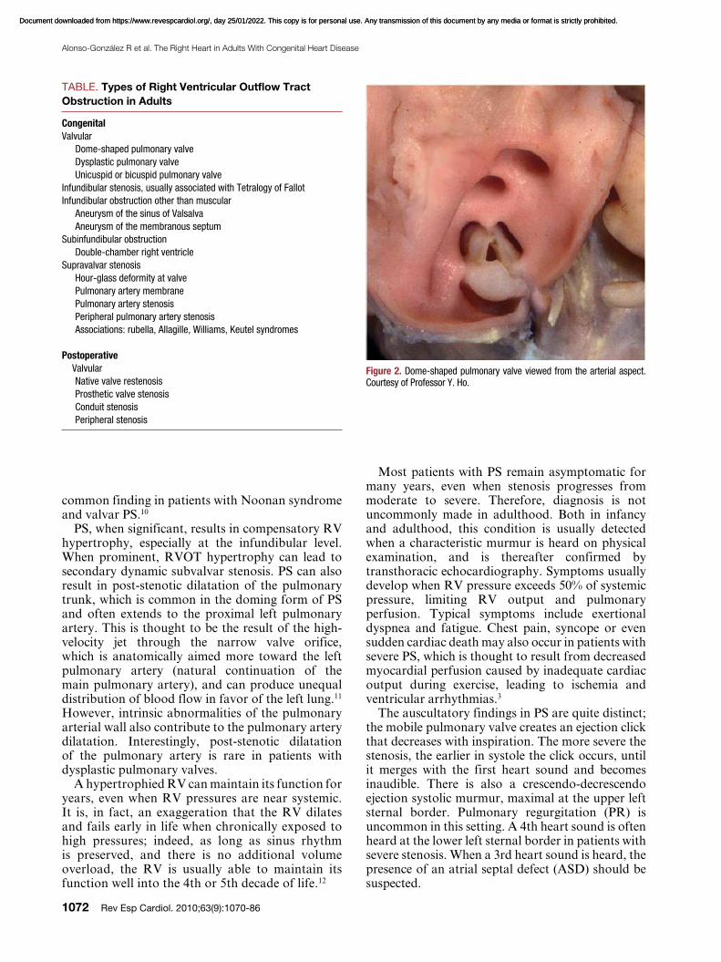
Isolated PS is found in 80%-90% of all patients with RVOT obstruction, and is almost always congenital. It is possible to identify 3 morphological types of stenotic pulmonary valve: a) dome-shaped; b) dysplastic; and c) bicuspid or unicuspid.4 The latter type is uncommon. The classic form of PS is the dome-shaped pulmonary valve, which may have 2-4 raphes, but there is no separation into valve cusps5 (Figure 2). It is characterized by a mobile valve with narrow central opening.6 Its familial inheritance is low, varying between 1.7%-3.6%.7,8
A dysplastic pulmonary valve is present in 10% to 20% of patients with PS.9,10 Dysplastic valves are trileaflet with markedly thickened leaflets composed of disorganized myxomatous tissue and little, if any, fusion. This is usually associated with a hypoplastic ventriculo-arterial junction. This entity is the most
ANATOMY
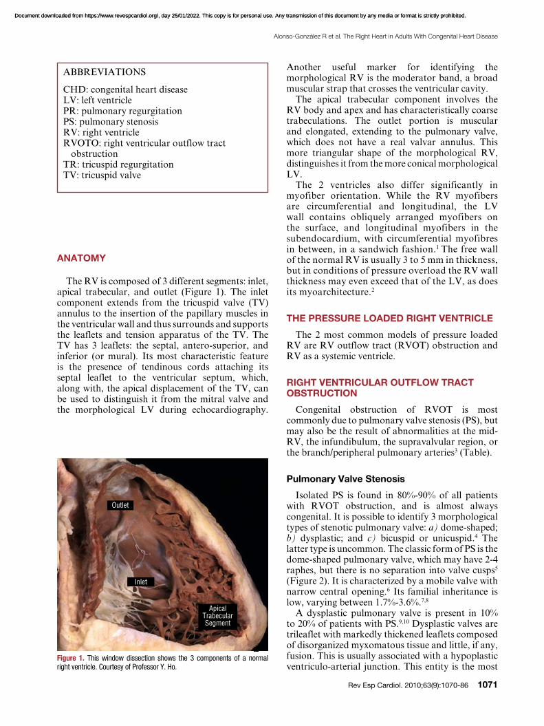
The RV is composed of 3 different segments: inlet, apical trabecular, and outlet (Figure 1). The inlet component extends from the tricuspid valve (TV) annulus to the insertion of the papillary muscles in the ventricular wall and thus surrounds and supports the leaflets and tension apparatus of the TV. The TV has 3 leaflets: the septal, antero-superior, and inferior (or mural). Its most characteristic feature is the presence of tendinous cords attaching its septal leaflet to the ventricular septum, which, along with, the apical displacement of the TV, can be used to distinguish it from the mitral valve and the morphological LV during echocardiography.
ABBREVIATIONS
CHD: congenital heart diseaseLV: left ventriclePR: pulmonary regurgitationPS: pulmonary stenosisRV: right ventricleRVOTO: right ventricular outflow tract
obstructionTR: tricuspid regurgitationTV: tricuspid valve
Figure 1. This window dissection shows the 3 components of a normal right ventricle. Courtesy of Professor Y. Ho.
Apical Trabecular Segment
Inlet
Outlet
Document downloaded from https://www.revespcardiol.org/, day 25/01/2022. This copy is for personal use. Any transmission of this document by any media or format is strictly prohibited.Document downloaded from https://www.revespcardiol.org/, day 25/01/2022. This copy is for personal use. Any transmission of this document by any media or format is strictly prohibited.
1072 Rev Esp Cardiol. 2010;63(9):1070-86
Alonso-González R et al. The Right Heart in Adults With Congenital Heart Disease
Most patients with PS remain asymptomatic for many years, even when stenosis progresses from moderate to severe. Therefore, diagnosis is not uncommonly made in adulthood. Both in infancy and adulthood, this condition is usually detected when a characteristic murmur is heard on physical examination, and is thereafter confirmed by transthoracic echocardiography. Symptoms usually develop when RV pressure exceeds 50% of systemic pressure, limiting RV output and pulmonary perfusion. Typical symptoms include exertional dyspnea and fatigue. Chest pain, syncope or even sudden cardiac death may also occur in patients with severe PS, which is thought to result from decreased myocardial perfusion caused by inadequate cardiac output during exercise, leading to ischemia and ventricular arrhythmias.3
The auscultatory findings in PS are quite distinct; the mobile pulmonary valve creates an ejection click that decreases with inspiration. The more severe the stenosis, the earlier in systole the click occurs, until it merges with the first heart sound and becomes inaudible. There is also a crescendo-decrescendo ejection systolic murmur, maximal at the upper left sternal border. Pulmonary regurgitation (PR) is uncommon in this setting. A 4th heart sound is often heard at the lower left sternal border in patients with severe stenosis. When a 3rd heart sound is heard, the presence of an atrial septal defect (ASD) should be suspected.
common finding in patients with Noonan syndrome and valvar PS.10
PS, when significant, results in compensatory RV hypertrophy, especially at the infundibular level. When prominent, RVOT hypertrophy can lead to secondary dynamic subvalvar stenosis. PS can also result in post-stenotic dilatation of the pulmonary trunk, which is common in the doming form of PS and often extends to the proximal left pulmonary artery. This is thought to be the result of the high-velocity jet through the narrow valve orifice, which is anatomically aimed more toward the left pulmonary artery (natural continuation of the main pulmonary artery), and can produce unequal distribution of blood flow in favor of the left lung.11 However, intrinsic abnormalities of the pulmonary arterial wall also contribute to the pulmonary artery dilatation. Interestingly, post-stenotic dilatation of the pulmonary artery is rare in patients with dysplastic pulmonary valves.
A hypertrophied RV can maintain its function for years, even when RV pressures are near systemic. It is, in fact, an exaggeration that the RV dilates and fails early in life when chronically exposed to high pressures; indeed, as long as sinus rhythm is preserved, and there is no additional volume overload, the RV is usually able to maintain its function well into the 4th or 5th decade of life.12
TABLE. Types of Right Ventricular Outflow Tract
Obstruction in Adults
Congenital
Valvular
Dome-shaped pulmonary valve
Dysplastic pulmonary valve
Unicuspid or bicuspid pulmonary valve
Infundibular stenosis, usually associated with Tetralogy of Fallot
Infundibular obstruction other than muscular
Aneurysm of the sinus of Valsalva
Aneurysm of the membranous septum
Subinfundibular obstruction
Double-chamber right ventricle
Supravalvar stenosis
Hour-glass deformity at valve
Pulmonary artery membrane
Pulmonary artery stenosis
Peripheral pulmonary artery stenosis
Associations: rubella, Allagille, Williams, Keutel syndromes
Postoperative
Valvular
Native valve restenosis
Prosthetic valve stenosis
Conduit stenosis
Peripheral stenosis
Figure 2. Dome-shaped pulmonary valve viewed from the arterial aspect. Courtesy of Professor Y. Ho.
Document downloaded from https://www.revespcardiol.org/, day 25/01/2022. This copy is for personal use. Any transmission of this document by any media or format is strictly prohibited.Document downloaded from https://www.revespcardiol.org/, day 25/01/2022. This copy is for personal use. Any transmission of this document by any media or format is strictly prohibited.
Alonso-González R et al. The Right Heart in Adults With Congenital Heart Disease
Rev Esp Cardiol. 2010;63(9):1070-86 1073
the gold standard method for quantifying RV size and function.
The first operation for PS was performed in 1948 and consisted of pulmonary valve commissurotomy. Subsequent procedures in the early 1950s involved closed valvotomy with transcatheter methods (Brock procedure). Later, open valvotomy yielded significantly better results. If the valve is dysplastic, partial or total valvotomy may be required; if there is annular or pulmonary trunk hypoplasia, a transannular patch may become necessary. All of these procedures invariably result in various degrees of PR, which is well tolerated for many years. Initially, the RV compensates by dilatation, maintaining contractility and stroke volume. Eventually RV dysfunction ensues and patients develop symptoms, such as dyspnea, fatigue and substantial arrhythmia.
The long-term outcome of patients with PS is excellent. In the Second Natural History Study of Congenital Heart Defects, there was no significant disease progression in patients with a peak gradient <20 mm Hg, while there was a 20% chance of requiring repair in those with a peak gradient between 25 and 49 mm Hg. The majority of patients with peak gradients >50 mm Hg required intervention.14 Studies of patients who received surgical valvotomy in childhood have shown that outcome is excellent in this cohort, with a mortality rate <5% after 25 years of follow-up.14 However, one-third of the patients will develop significant PR and will require re-intervention in later years (re-intervention rate 9%-40%; mean time to pulmonary valve replacement [PVR], 33 years).15-17 Atrial or ventricular arrhythmia, exercise intolerance or cardiomegaly on chest x-ray in patients with
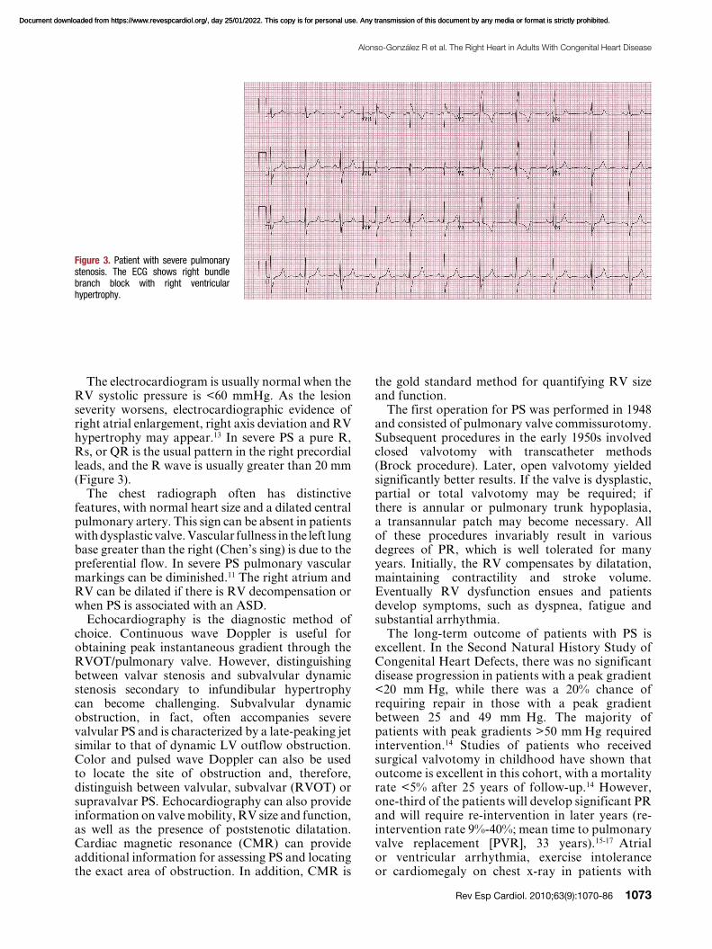
The electrocardiogram is usually normal when the RV systolic pressure is <60 mmHg. As the lesion severity worsens, electrocardiographic evidence of right atrial enlargement, right axis deviation and RV hypertrophy may appear.13 In severe PS a pure R, Rs, or QR is the usual pattern in the right precordial leads, and the R wave is usually greater than 20 mm (Figure 3).
The chest radiograph often has distinctive features, with normal heart size and a dilated central pulmonary artery. This sign can be absent in patients with dysplastic valve. Vascular fullness in the left lung base greater than the right (Chen’s sing) is due to the preferential flow. In severe PS pulmonary vascular markings can be diminished.11 The right atrium and RV can be dilated if there is RV decompensation or when PS is associated with an ASD.
Echocardiography is the diagnostic method of choice. Continuous wave Doppler is useful for obtaining peak instantaneous gradient through the RVOT/pulmonary valve. However, distinguishing between valvar stenosis and subvalvular dynamic stenosis secondary to infundibular hypertrophy can become challenging. Subvalvular dynamic obstruction, in fact, often accompanies severe valvular PS and is characterized by a late-peaking jet similar to that of dynamic LV outflow obstruction. Color and pulsed wave Doppler can also be used to locate the site of obstruction and, therefore, distinguish between valvular, subvalvar (RVOT) or supravalvar PS. Echocardiography can also provide information on valve mobility, RV size and function, as well as the presence of poststenotic dilatation. Cardiac magnetic resonance (CMR) can provide additional information for assessing PS and locating the exact area of obstruction. In addition, CMR is
Figure 3. Patient with severe pulmonary stenosis. The ECG shows right bundle branch block with right ventricular hypertrophy.
Document downloaded from https://www.revespcardiol.org/, day 25/01/2022. This copy is for personal use. Any transmission of this document by any media or format is strictly prohibited.Document downloaded from https://www.revespcardiol.org/, day 25/01/2022. This copy is for personal use. Any transmission of this document by any media or format is strictly prohibited.
1074 Rev Esp Cardiol. 2010;63(9):1070-86
Alonso-González R et al. The Right Heart in Adults With Congenital Heart Disease
was associated with a lower peak VO2.22 Whether
balloon valvotomy will be associated with similar rates of re-intervention for PR in the long term remains speculative.
Double-Chambered Right Ventricle
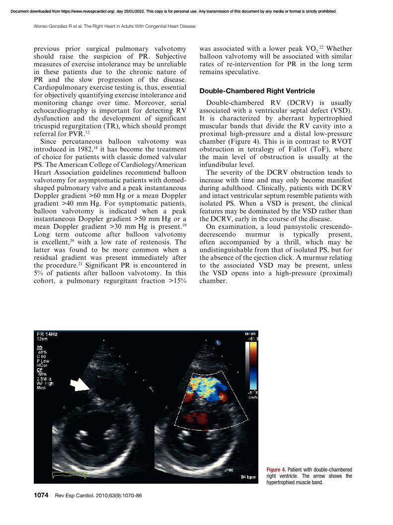
Double-chambered RV (DCRV) is usually associated with a ventricular septal defect (VSD). It is characterized by aberrant hypertrophied muscular bands that divide the RV cavity into a proximal high-pressure and a distal low-pressure chamber (Figure 4). This is in contrast to RVOT obstruction in tetralogy of Fallot (ToF), where the main level of obstruction is usually at the infundibular level.
The severity of the DCRV obstruction tends to increase with time and may only become manifest during adulthood. Clinically, patients with DCRV and intact ventricular septum resemble patients with isolated PS. When a VSD is present, the clinical features may be dominated by the VSD rather than the DCRV, early in the course of the disease.
On examination, a loud pansystolic crescendo-decrescendo murmur is typically present, often accompanied by a thrill, which may be undistinguishable from that of isolated PS, but for the absence of the ejection click. A murmur relating to the associated VSD may be present, unless the VSD opens into a high-pressure (proximal) chamber.
previous prior surgical pulmonary valvotomy should raise the suspicion of PR. Subjective measures of exercise intolerance may be unreliable in these patients due to the chronic nature of PR and the slow progression of the disease. Cardiopulmonary exercise testing is, thus, essential for objectively quantifying exercise intolerance and monitoring change over time. Moreover, serial echocardiography is important for detecting RV dysfunction and the development of significant tricuspid regurgitation (TR), which should prompt referral for PVR.12
Since percutaneous balloon valvotomy was introduced in 1982,18 it has become the treatment of choice for patients with classic domed valvular PS. The American College of Cardiology/American Heart Association guidelines recommend balloon valvotomy for asymptomatic patients with domed-shaped pulmonary valve and a peak instantaneous Doppler gradient >60 mm Hg or a mean Doppler gradient >40 mm Hg. For symptomatic patients, balloon valvotomy is indicated when a peak instantaneous Doppler gradient >50 mm Hg or a mean Doppler gradient >30 mm Hg is present.19 Long term outcome after balloon valvotomy is excellent,20 with a low rate of restenosis. The latter was found to be more common when a residual gradient was present immediately after the procedure.21 Significant PR is encountered in 5% of patients after balloon valvotomy. In this cohort, a pulmonary regurgitant fraction >15%
Figure 4. Patient with double-chambered right ventricle. The arrow shows the hypertrophied muscle band.
Document downloaded from https://www.revespcardiol.org/, day 25/01/2022. This copy is for personal use. Any transmission of this document by any media or format is strictly prohibited.Document downloaded from https://www.revespcardiol.org/, day 25/01/2022. This copy is for personal use. Any transmission of this document by any media or format is strictly prohibited.
Alonso-González R et al. The Right Heart in Adults With Congenital Heart Disease
Rev Esp Cardiol. 2010;63(9):1070-86 1075
should be suspected when the latter is associated with abdominal situs solitus. Additional cardiac lesions are present in 95% of cases with ccTGA and include, Ebstein-like anomaly of the TV (90%),VSD (70%), PS (40%), and complete heart block (2% risk per year).24
Patients with ccTGA typically remain asymptomatic until the 3rd or 4th decade of life but may become symptomatic earlier when additional haemodynamic lesions are present.25 The most common symptoms include dyspnea on exertion and palpitations or syncope secondary to atrial arrhythmias or complete heart block. Patients with VSD and PS may present with progressive cyanosis.
Echocardiography is the diagnostic method of choice. The apical four-chamber and subcostal views are most helpful in determining the situs and AV–VA connections. Ventricular morphology is best assessed by examining the AV valves: the TV is always closer to the apex compared to the mitral valve and its septal leaflet has chordal attachments to the inlet septum. The relation of the two ventricles and the great arteries are more side by side than the usual anteroposterior. It is important to identify associated lesions such as VSD, which is usually perimembranous, as well as the presence of LV outflow tract obstruction, which may be due to either an aneurysm of the membranous septum, a fibrous membrane, or mobile subpulmonary tissue “tags.” Moreover, accurate assessment of systemic RV function and the degree of systemic AV valve regurgitation is also fundamental in the assessment of ccTGA. CMR provides complementary information on the anatomy and a more accurate estimate of RV size and function.
The prognosis of ccTGA greatly depends on the presence and severity of associated lesions. In the absence of the above, patients with ccTGA may survive until the 7th or 8th decade of life.26 However, the incidence of systemic ventricular dysfunction and congestive heart failure increases with age, even in the absence of associated lesions (more than 1 in 3 will develop congestive heart failure by the 5th decade of life).27 The presence of significant TR and/or RV dysfunction is associated with a significantly higher mortality27,28 and a higher risk of developing decompensated heart failure. The rate of deterioration of systemic RV function in the presence of significant TR is much more rapid than that of systemic LV in the presence of mitral regurgitation. The factors responsible for this remain unclear. Ventricular geometry is likely to play a role in this, as RV dilatation is more likely to cause severe annular dilatation and rapidly aggravates TR and in turn RV dysfunction.29 In addition, myocardial perfusion may not be
The electrocardiogram typically demonstrates RV hypertrophy, which is not what is expected in cases of isolated restrictive VSD. Two- dimensional echocardiography is usually diagnostic, identifying the degree and location of the obstruction and the presence of a VSD. However, the VSD may be difficult to visualize when it opens into the high-pressure chamber. CMR provides complementary information on the anatomy and physiology.
The indications for surgery in DCRV are similar to those for valvar PS.19 Muscular resection and outflow-enlarging procedures have been very effective, with excellent long-term results and very low rates of recurrence.23
Pulmonary Stenosis and Pregnancy
When RV function is preserved, isolated RVOT obstruction is usually well tolerated during pregnancy, even when the lesion is severe. Nevertheless, when women with severe PS develop recurrent atrial arrhythmia and/or early right heart failure during pregnancy, balloon valvotomy should be considered.
SYSTEMIC RIGHT VENTRICLE
A morphological RV in the systemic position in adulthood is most commonly encountered in patients with congenitally corrected transposition (ccTGA) and those with transposition of the great arteries (TGA) following atrial switch procedures (Mustard or Senning). In the systemic position, the RV changes its myofiber architecture to resemble the “sandwich” pattern encountered in the normal LV.4 Moreover, in the systemic RV there is predominant circumferential rather than longitudinal free wall shortening.5 These changes allow the RV to adapt and function in the systemic position to a large extent and for a number of decades.
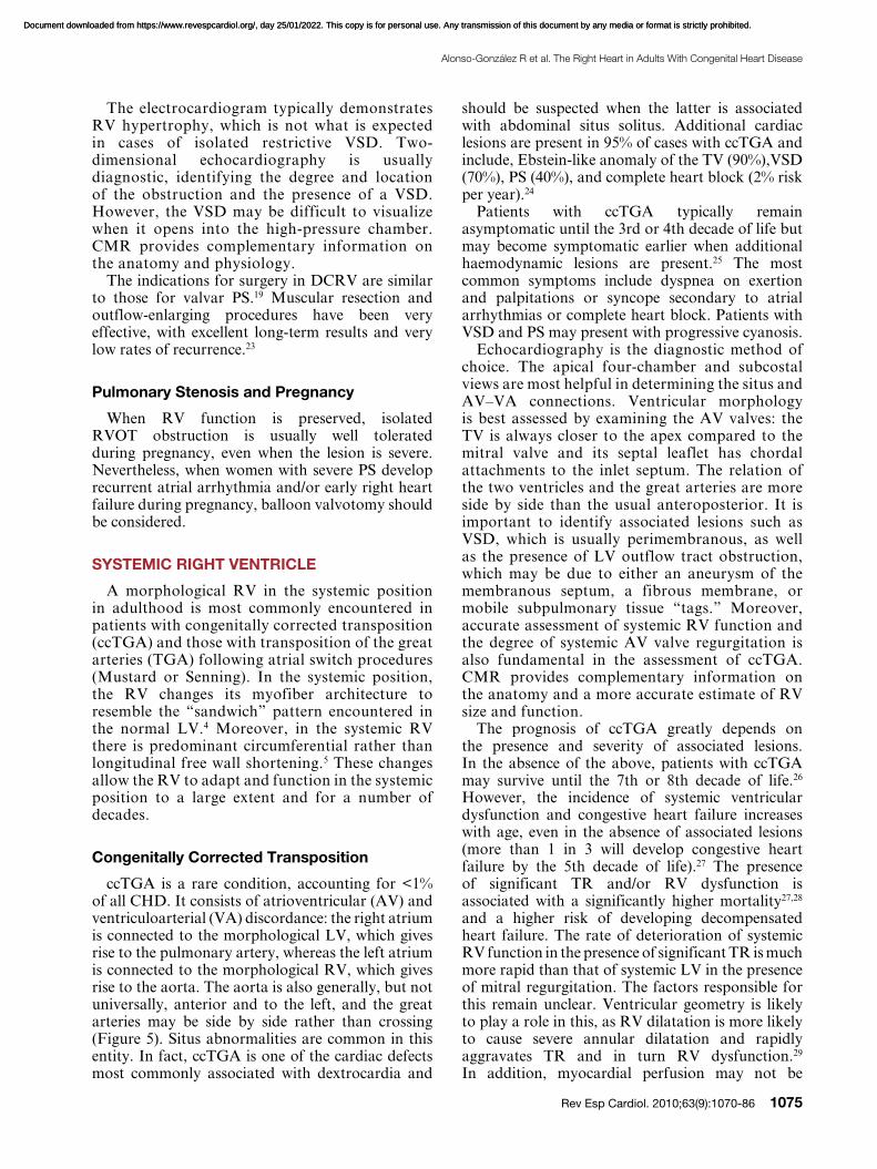
Congenitally Corrected Transposition
ccTGA is a rare condition, accounting for <1% of all CHD. It consists of atrioventricular (AV) and ventriculoarterial (VA) discordance: the right atrium is connected to the morphological LV, which gives rise to the pulmonary artery, whereas the left atrium is connected to the morphological RV, which gives rise to the aorta. The aorta is also generally, but not universally, anterior and to the left, and the great arteries may be side by side rather than crossing (Figure 5). Situs abnormalities are common in this entity. In fact, ccTGA is one of the cardiac defects most commonly associated with dextrocardia and
Document downloaded from https://www.revespcardiol.org/, day 25/01/2022. This copy is for personal use. Any transmission of this document by any media or format is strictly prohibited.Document downloaded from https://www.revespcardiol.org/, day 25/01/2022. This copy is for personal use. Any transmission of this document by any media or format is strictly prohibited.
1076 Rev Esp Cardiol. 2010;63(9):1070-86
Alonso-González R et al. The Right Heart in Adults With Congenital Heart Disease
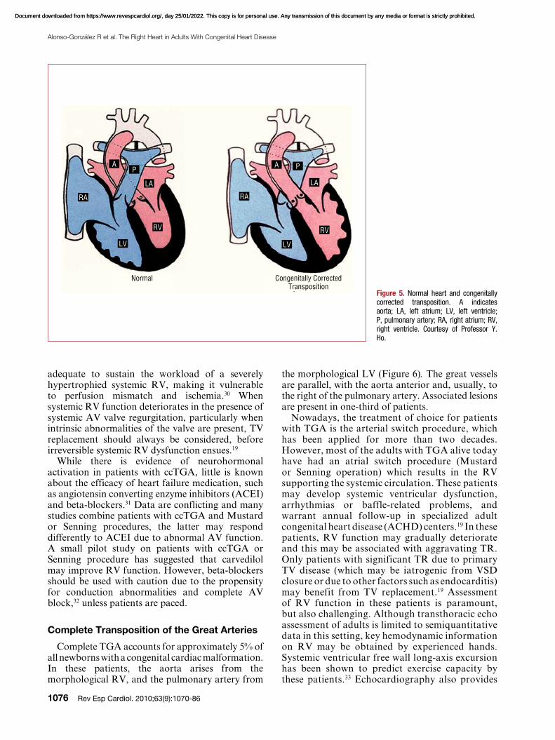
the morphological LV (Figure 6). The great vessels are parallel, with the aorta anterior and, usually, to the right of the pulmonary artery. Associated lesions are present in one-third of patients.
Nowadays, the treatment of choice for patients with TGA is the arterial switch procedure, which has been applied for more than two decades. However, most of the adults with TGA alive today have had an atrial switch procedure (Mustard or Senning operation) which results in the RV supporting the systemic circulation. These patients may develop systemic ventricular dysfunction, arrhythmias or baffle-related problems, and warrant annual follow-up in specialized adult congenital heart disease (ACHD) centers.19 In these patients, RV function may gradually deteriorate and this may be associated with aggravating TR. Only patients with significant TR due to primary TV disease (which may be iatrogenic from VSD closure or due to other factors such as endocarditis) may benefit from TV replacement.19 Assessment of RV function in these patients is paramount, but also challenging. Although transthoracic echo assessment of adults is limited to semiquantitative data in this setting, key hemodynamic information on RV may be obtained by experienced hands. Systemic ventricular free wall long-axis excursion has been shown to predict exercise capacity by these patients.33 Echocardiography also provides
adequate to sustain the workload of a severely hypertrophied systemic RV, making it vulnerable to perfusion mismatch and ischemia.30 When systemic RV function deteriorates in the presence of systemic AV valve regurgitation, particularly when intrinsic abnormalities of the valve are present, TV replacement should always be considered, before irreversible systemic RV dysfunction ensues.19
While there is evidence of neurohormonal activation in patients with ccTGA, little is known about the efficacy of heart failure medication, such as angiotensin converting enzyme inhibitors (ACEI) and beta-blockers.31 Data are conflicting and many studies combine patients with ccTGA and Mustard or Senning procedures, the latter may respond differently to ACEI due to abnormal AV function. A small pilot study on patients with ccTGA or Senning procedure has suggested that carvedilol may improve RV function. However, beta-blockers should be used with caution due to the propensity for conduction abnormalities and complete AV block,32 unless patients are paced.
Complete Transposition of the Great Arteries
Complete TGA accounts for approximately 5% of all newborns with a congenital cardiac malformation. In these patients, the aorta arises from the morphological RV, and the pulmonary artery from
Figure 5. Normal heart and congenitally corrected transposition. A indicates aorta; LA, left atrium; LV, left ventricle; P, pulmonary artery; RA, right atrium; RV, right ventricle. Courtesy of Professor Y. Ho.
Normal Congenitally Corrected Transposition
AP
RA
LV LV
RV RV
LA LA
RA
PA
Document downloaded from https://www.revespcardiol.org/, day 25/01/2022. This copy is for personal use. Any transmission of this document by any media or format is strictly prohibited.Document downloaded from https://www.revespcardiol.org/, day 25/01/2022. This copy is for personal use. Any transmission of this document by any media or format is strictly prohibited.
Alonso-González R et al. The Right Heart in Adults With Congenital Heart Disease
Rev Esp Cardiol. 2010;63(9):1070-86 1077
syndrome,” although this is uncommon because the azygos system usually compensates for it. Patency of the superior systemic venous pathway should always be assessed before implantation of a transvenous pacing system. Small baffle leaks are relatively more common. They are usually small and haemodynamically insignificant but may allow paradoxical emboli and cause cyanosis at rest or during exercise. When the LV is enlarged, contrast echocardiography with agitated saline should be performed to rule out a significant pathway leak with predominant systemic to pulmonary shunt (equivalent of RV enlargement in concordant heart with an ASD).
Pulmonary arterial hypertension occurs in about 7% of patients after atrial switch procedures for TGA.37,38 The pathogenesis is unknown, but risks factors include operation at >2 years of age39 and shunting at the ventricular or great artery level prior to repair.40 Pulmonary venous pathway obstruction should always be investigated as a cause of pulmonary hypertension in this setting.
Systemic Right Ventricle and Sudden Cardiac Death
The majority of patients with systemic RV die suddenly.41,42 Age, systemic ventricular dysfunction,
information on baffle patency, baffle leaks and valvar regurgitation.
CMR is considered the gold standard for the study of RV size and function in this population. Moreover, the systemic and pulmonary venous pathways can be evaluated more accurately. When CMR is contraindicated, gated CT-angiography may be an alternative.
Both bradyarrhythmias and tachyarrythmias are common long after atrial switch for TGA. Sinus node dysfunction is frequent and 11% of patients will need pacemaker implantation at some point.34 Almost a quarter of patients develop atrial flutter or fibrillation 23 years after the atrial switch procedure.35 The extensive atrial scarring following atrial surgery creates the perfect substrate for atrial arrhythmias, which may become life threatening when fast or in the presence of significant pathway obstruction. Antiarrhythmic drugs with negative inotropic effect are rarely used when systemic RV dysfunction is present. Radiofrequency ablation for atrial arrhythmias can be challenging, but has a success rate of approximately 70% in experienced hands.36
Atrial pathway obstruction and leaks, while infrequent, should always be excluded in the presence of symptoms. Superior vena cava (SVC) obstruction is more common and may cause “SVC
Figure 6. Normal heart and transposition of the great arteries. A indicates aorta; LA, left atrium; LV, left ventricle; P, pulmonary artery; RA, right atrium; RV, right ventricle. Courtesy of Professor Y. Ho.
Normal Great arteries completed transposition
LV LV
RV RV
LA LA
RA RA
P PA A
Document downloaded from https://www.revespcardiol.org/, day 25/01/2022. This copy is for personal use. Any transmission of this document by any media or format is strictly prohibited.Document downloaded from https://www.revespcardiol.org/, day 25/01/2022. This copy is for personal use. Any transmission of this document by any media or format is strictly prohibited.
1078 Rev Esp Cardiol. 2010;63(9):1070-86
Alonso-González R et al. The Right Heart in Adults With Congenital Heart Disease
right coronary sinus and crosses the RVOT (3% of cases).49
Most patients with ToF, nowadays, undergo total repair early in life. This involves closure of the VSD and relief of the RVOT obstruction. The latter may require RVOT/transannular patch, which disrupts the integrity of the pulmonary valve “annulus,” or pulmonary valvotomy/valvectomy if the pulmonary valve is abnormal. An extracardiac conduit between the RVOT and main pulmonary artery may be necessary in the presence of pulmonary atresia or an anomalous left coronary artery crossing the RVOT.
The most common sequela after RVOT patch augmentation is significant PR. Severe chronic PR may lead to RV dilatation and RV systolic dysfunction,50 with a propensity to clinical arrhythmia and sudden cardiac death.51,52 It is known that after 2 decades of exposure to significant volume overload, RV systolic function deteriorates, resulting in progressive exercise intolerance and a high risk of arrhythmias, both supraventricular and ventricular. Patients should, thus, be considered for PVR when significant RV dilatation is present, before the onset of irreversible RV dysfunction. Reliance on symptoms only for deciding the timing of PVR may be misleading, as patients often become symptomatic when RV function becomes severely impaired. Objective assessment of exercise capacity may be more reliable, and serial exercise testing may help identify changes in exercise capacity, which may not be perceived by the patient. Surgical intervention is also indicated when patients with severe PR and enlarged RVs develop moderate to severe TR, or symptomatic atrial or ventricular arrhythmias.19 Indeed, no single parameter, but several variables, should be considered when deciding the correct timing of PVR.
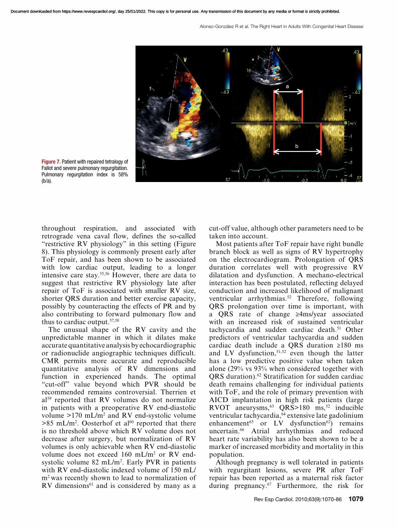
Echocardiography remains the most widely employed imaging modality for assessing patients with ToF. However, CMR is the gold standard for both PR quantification and RV volumetric analysis. Patients with repaired ToF, especially those with progressive TR or other sequelae, should have an echocardiogram annually and a CMR every 2-3 years.19 Quantification of PR with echocardiography can be challenging for inexperienced operators. Early equalization of RV and PA pressures can be a sign of significant PR in the absence of a restrictive RV. A pressure half-time <100 ms53 and a PR index (ratio between PR duration and total diastole) lower than 0.77 has 100% sensitivity and 85% specificity for identifying patients with pulmonary regurgitant fraction greater than 25% in CMR54 (Figure 7). RV size and function should also be evaluated.
Pulsed Doppler late-diastolic forward pulmonary flow, coincident with atrial systole, present
New York Heart Association functional class,43
supraventricular arrhythmia35 and QT dispersion44 have all been associated. Recently, Schwerzmann et al43 reported that a QRS duration ≥140 ms is associated with lower functional class, worse systemic RV function and a higher mortality. Automated implanted cardioverter defibrillators (AICD) have been utilized recently in this setting, although their role remains unknown.
Systemic Ventricle and Pregnancy
The risk of pregnancy in patients with systemic RV depends on systemic ventricular function, the presence of significant hemodynamic lesions and functional capacity.45 Pregnancy may cause deterioration in systemic RV function, even though the long-term impact of pregnancy on systemic RV function remains unclear. A mortality rate of up to 4% has been reported in a small series of women with systemic RV, and this should be discussed during preconception counseling.46 Patients with good or mildly impaired RV systolic function and no pathway obstruction are at low risk.47,48 During pregnancy, clinical assessment should focus on early signs of heart failure and arrhythmias. If atrial tachycardia occurs, direct current cardioversion can be performed safely to restore sinus rhythm. Aspirin throughout pregnancy should be considered in patients with previous history of atrial arrhythmias.
THE VOLUME LOADED RIGHT VENTRICLE
The 3 most common lesions associated with RV volume overload are ASD, pulmonary regurgitation in the setting of ToF and TR in the setting of Ebstein anomaly. In this review, we will focus on the latter 2 conditions.
TETRALOGY OF FALLOT
ToF is the most common form of cyanotic CHD after the first year of life. It consists of a large malaligned subaortic VSD, the aorta riding up over the septal defect (when more than 50% patients fall into double outlet RV subgroup), RVOT obstruction and RV hypertrophy. Pulmonary branch stenosis or pulmonary branch hypoplasia may be present, with some patients presenting with pulmonary atresia. Associated anomalies include ASD, AV septal defect (more common in patients with Down syndrome) and a right-sided aortic arch, which can be found in up to 25% of patients. Coronary artery anomalies may also be present, commonly involving the left anterior descending coronary artery, which arises from the
Document downloaded from https://www.revespcardiol.org/, day 25/01/2022. This copy is for personal use. Any transmission of this document by any media or format is strictly prohibited.Document downloaded from https://www.revespcardiol.org/, day 25/01/2022. This copy is for personal use. Any transmission of this document by any media or format is strictly prohibited.
Alonso-González R et al. The Right Heart in Adults With Congenital Heart Disease
Rev Esp Cardiol. 2010;63(9):1070-86 1079
cut-off value, although other parameters need to be taken into account.
Most patients after ToF repair have right bundle branch block as well as signs of RV hypertrophy on the electrocardiogram. Prolongation of QRS duration correlates well with progressive RV dilatation and dysfunction. A mechano-electrical interaction has been postulated, reflecting delayed conduction and increased likelihood of malignant ventricular arrhythmias.52 Therefore, following QRS prolongation over time is important, with a QRS rate of change ≥4ms/year associated with an increased risk of sustained ventricular tachycardia and sudden cardiac death.51 Other predictors of ventricular tachycardia and sudden cardiac death include a QRS duration ≥180 ms and LV dysfunction,51,52 even though the latter has a low predictive positive value when taken alone (29% vs 93% when considered together with QRS duration).62 Stratification for sudden cardiac death remains challenging for individual patients with ToF, and the role of primary prevention with AICD implantation in high risk patients (large RVOT aneurysms,63 QRS>180 ms,52 inducible ventricular tachycardia,64 extensive late gadolinium enhancement65 or LV dysfunction62) remains uncertain.66 Atrial arrhythmias and reduced heart rate variability has also been shown to be a marker of increased morbidity and mortality in this population.
Although pregnancy is well tolerated in patients with regurgitant lesions, severe PR after ToF repair has been reported as a maternal risk factor during pregnancy.67 Furthermore, the risk for
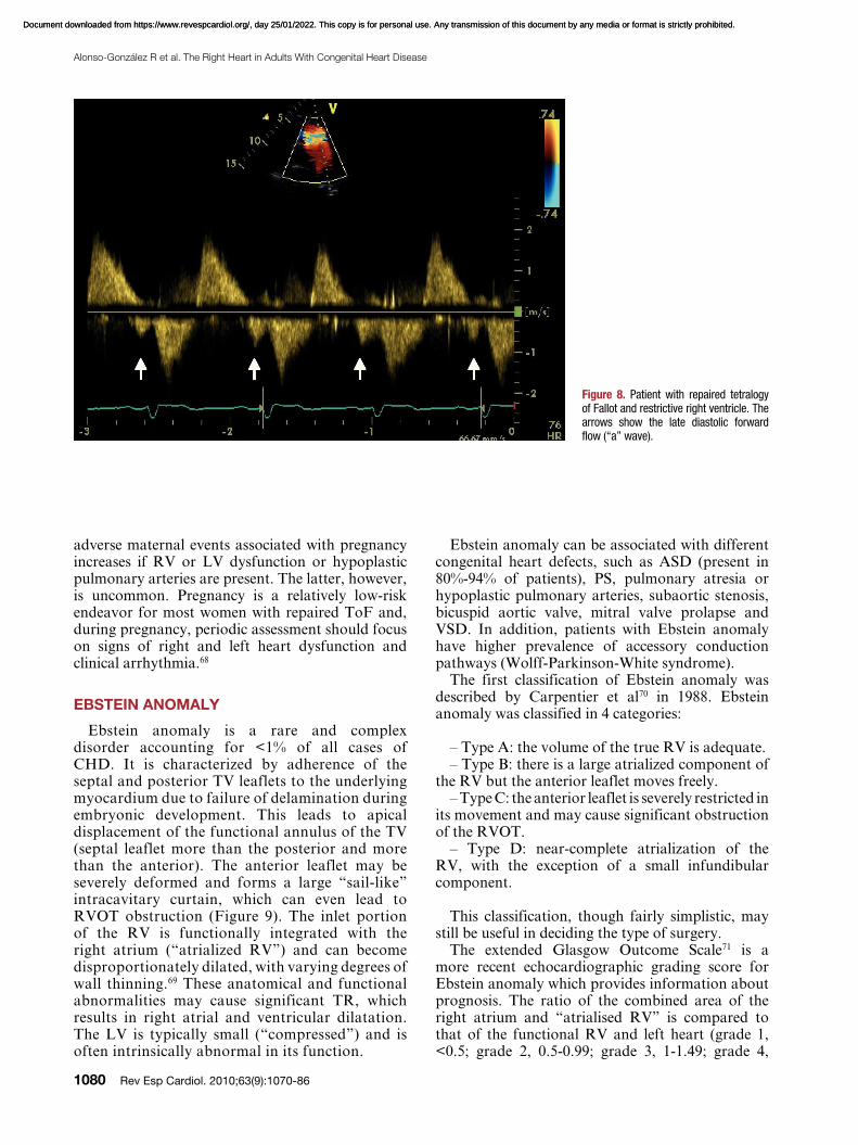
throughout respiration, and associated with retrograde vena caval flow, defines the so-called “restrictive RV physiology” in this setting (Figure 8). This physiology is commonly present early after ToF repair, and has been shown to be associated with low cardiac output, leading to a longer intensive care stay.55,56 However, there are data to suggest that restrictive RV physiology late after repair of ToF is associated with smaller RV size, shorter QRS duration and better exercise capacity, possibly by counteracting the effects of PR and by also contributing to forward pulmonary flow and thus to cardiac output.57,58
The unusual shape of the RV cavity and the unpredictable manner in which it dilates make accurate quantitative analysis by echocardiographic or radionuclide angiographic techniques difficult. CMR permits more accurate and reproducible quantitative analysis of RV dimensions and function in experienced hands. The optimal “cut-off” value beyond which PVR should be recommended remains controversial. Therrien et al59 reported that RV volumes do not normalize in patients with a preoperative RV end-diastolic volume >170 mL/m2 and RV end-systolic volume >85 mL/m2. Oosterhof et al60 reported that there is no threshold above which RV volume does not decrease after surgery, but normalization of RV volumes is only achievable when RV end-diastolic volume does not exceed 160 mL/m2 or RV end-systolic volume 82 mL/m2. Early PVR in patients with RV end-diastolic indexed volume of 150 mL/m2 was recently shown to lead to normalization of RV dimensions61 and is considered by many as a
Figure 7. Patient with repaired tetralogy of Fallot and severe pulmonary regurgitation. Pulmonary regurgitation index is 58% (b/a).
Document downloaded from https://www.revespcardiol.org/, day 25/01/2022. This copy is for personal use. Any transmission of this document by any media or format is strictly prohibited.Document downloaded from https://www.revespcardiol.org/, day 25/01/2022. This copy is for personal use. Any transmission of this document by any media or format is strictly prohibited.
1080 Rev Esp Cardiol. 2010;63(9):1070-86
Alonso-González R et al. The Right Heart in Adults With Congenital Heart Disease
Ebstein anomaly can be associated with different congenital heart defects, such as ASD (present in 80%-94% of patients), PS, pulmonary atresia or hypoplastic pulmonary arteries, subaortic stenosis, bicuspid aortic valve, mitral valve prolapse and VSD. In addition, patients with Ebstein anomaly have higher prevalence of accessory conduction pathways (Wolff-Parkinson-White syndrome).
The first classification of Ebstein anomaly was described by Carpentier et al70 in 1988. Ebstein anomaly was classified in 4 categories:
– Type A: the volume of the true RV is adequate.– Type B: there is a large atrialized component of
the RV but the anterior leaflet moves freely.– Type C: the anterior leaflet is severely restricted in
its movement and may cause significant obstruction of the RVOT.
– Type D: near-complete atrialization of the RV, with the exception of a small infundibular component.
This classification, though fairly simplistic, may still be useful in deciding the type of surgery.
The extended Glasgow Outcome Scale71 is a more recent echocardiographic grading score for Ebstein anomaly which provides information about prognosis. The ratio of the combined area of the right atrium and “atrialised RV” is compared to that of the functional RV and left heart (grade 1, <0.5; grade 2, 0.5-0.99; grade 3, 1-1.49; grade 4,
adverse maternal events associated with pregnancy increases if RV or LV dysfunction or hypoplastic pulmonary arteries are present. The latter, however, is uncommon. Pregnancy is a relatively low-risk endeavor for most women with repaired ToF and, during pregnancy, periodic assessment should focus on signs of right and left heart dysfunction and clinical arrhythmia.68
EBSTEIN ANOMALY
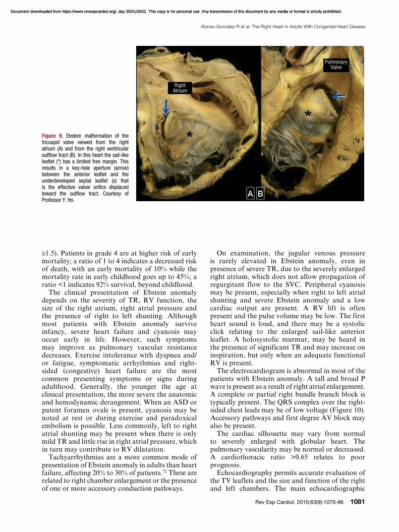
Ebstein anomaly is a rare and complex disorder accounting for <1% of all cases of CHD. It is characterized by adherence of the septal and posterior TV leaflets to the underlying myocardium due to failure of delamination during embryonic development. This leads to apical displacement of the functional annulus of the TV (septal leaflet more than the posterior and more than the anterior). The anterior leaflet may be severely deformed and forms a large “sail-like” intracavitary curtain, which can even lead to RVOT obstruction (Figure 9). The inlet portion of the RV is functionally integrated with the right atrium (“atrialized RV”) and can become disproportionately dilated, with varying degrees of wall thinning.69 These anatomical and functional abnormalities may cause significant TR, which results in right atrial and ventricular dilatation. The LV is typically small (“compressed”) and is often intrinsically abnormal in its function.
Figure 8. Patient with repaired tetralogy of Fallot and restrictive right ventricle. The arrows show the late diastolic forward flow (“a” wave).
Document downloaded from https://www.revespcardiol.org/, day 25/01/2022. This copy is for personal use. Any transmission of this document by any media or format is strictly prohibited.Document downloaded from https://www.revespcardiol.org/, day 25/01/2022. This copy is for personal use. Any transmission of this document by any media or format is strictly prohibited.
Alonso-González R et al. The Right Heart in Adults With Congenital Heart Disease
Rev Esp Cardiol. 2010;63(9):1070-86 1081
On examination, the jugular venous pressure is rarely elevated in Ebstein anomaly, even in presence of severe TR, due to the severely enlarged right atrium, which does not allow propagation of regurgitant flow to the SVC. Peripheral cyanosis may be present, especially when right to left atrial shunting and severe Ebstein anomaly and a low cardiac output are present. A RV lift is often present and the pulse volume may be low. The first heart sound is loud, and there may be a systolic click relating to the enlarged sail-like anterior leaflet. A holosystolic murmur, may be heard in the presence of significant TR and may increase on inspiration, but only when an adequate functional RV is present.
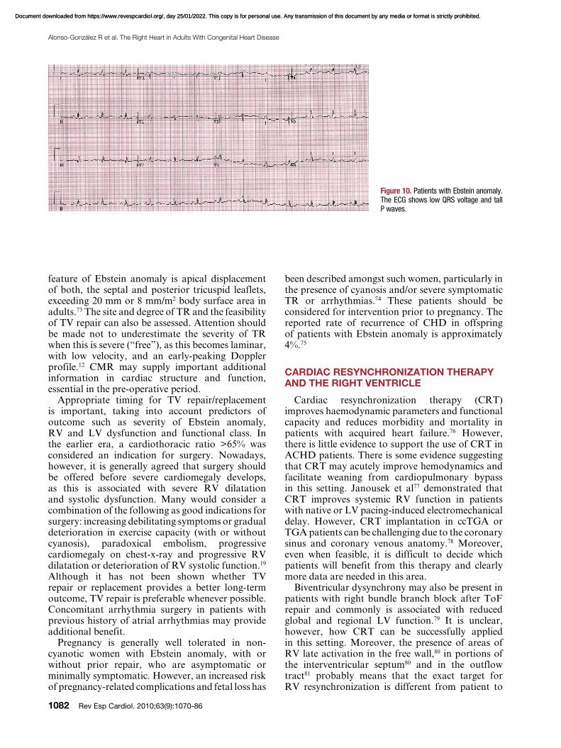
The electrocardiogram is abnormal in most of the patients with Ebstein anomaly. A tall and broad P wave is present as a result of right atrial enlargement. A complete or partial right bundle branch block is typically present. The QRS complex over the right-sided chest leads may be of low voltage (Figure 10). Accessory pathways and first degree AV block may also be present.
The cardiac silhouette may vary from normal to severely enlarged with globular heart. The pulmonary vascularity may be normal or decreased. A cardiothoracic ratio >0.65 relates to poor prognosis.
Echocardiography permits accurate evaluation of the TV leaflets and the size and function of the right and left chambers. The main echocardiographic
≥1.5). Patients in grade 4 are at higher risk of early mortality; a ratio of 1 to 4 indicates a decreased risk of death, with an early mortality of 10% while the mortality rate in early childhood goes up to 45%; a ratio <1 indicates 92% survival, beyond childhood.
The clinical presentation of Ebstein anomaly depends on the severity of TR, RV function, the size of the right atrium, right atrial pressure and the presence of right to left shunting. Although most patients with Ebstein anomaly survive infancy, severe heart failure and cyanosis may occur early in life. However, such symptoms may improve as pulmonary vascular resistance decreases. Exercise intolerance with dyspnea and/or fatigue, symptomatic arrhythmias and right-sided (congestive) heart failure are the most common presenting symptoms or signs during adulthood. Generally, the younger the age at clinical presentation, the more severe the anatomic and hemodynamic derangement. When an ASD or patent foramen ovale is present, cyanosis may be noted at rest or during exercise and paradoxical embolism is possible. Less commonly, left to right atrial shunting may be present when there is only mild TR and little rise in right atrial pressure, which in turn may contribute to RV dilatation.
Tachyarrhythmias are a more common mode of presentation of Ebstein anomaly in adults than heart failure, affecting 20% to 30% of patients.72 These are related to right chamber enlargement or the presence of one or more accessory conduction pathways.
Figure 9. Ebstein malformation of the tricuspid valve viewed from the right atrium (A) and from the right ventricular outflow tract (B). In this heart the sail-like leaflet (*) has a limited free margin. This results in a key-hole aperture (arrow) between the anterior leaflet and the underdeveloped septal leaflet (s) that is the effective valvar orifice displaced toward the outflow tract. Courtesy of Professor Y. Ho.
Right Atrium
Pulmonary Valve
Document downloaded from https://www.revespcardiol.org/, day 25/01/2022. This copy is for personal use. Any transmission of this document by any media or format is strictly prohibited.Document downloaded from https://www.revespcardiol.org/, day 25/01/2022. This copy is for personal use. Any transmission of this document by any media or format is strictly prohibited.
1082 Rev Esp Cardiol. 2010;63(9):1070-86
Alonso-González R et al. The Right Heart in Adults With Congenital Heart Disease
been described amongst such women, particularly in the presence of cyanosis and/or severe symptomatic TR or arrhythmias.74 These patients should be considered for intervention prior to pregnancy. The reported rate of recurrence of CHD in offspring of patients with Ebstein anomaly is approximately 4%.75
CARDIAC RESYNCHRONIZATION THERAPY AND THE RIGHT VENTRICLE
Cardiac resynchronization therapy (CRT) improves haemodynamic parameters and functional capacity and reduces morbidity and mortality in patients with acquired heart failure.76 However, there is little evidence to support the use of CRT in ACHD patients. There is some evidence suggesting that CRT may acutely improve hemodynamics and facilitate weaning from cardiopulmonary bypass in this setting. Janousek et al77 demonstrated that CRT improves systemic RV function in patients with native or LV pacing-induced electromechanical delay. However, CRT implantation in ccTGA or TGA patients can be challenging due to the coronary sinus and coronary venous anatomy.78 Moreover, even when feasible, it is difficult to decide which patients will benefit from this therapy and clearly more data are needed in this area.
Biventricular dysynchrony may also be present in patients with right bundle branch block after ToF repair and commonly is associated with reduced global and regional LV function.79 It is unclear, however, how CRT can be successfully applied in this setting. Moreover, the presence of areas of RV late activation in the free wall,80 in portions of the interventricular septum80 and in the outflow tract81 probably means that the exact target for RV resynchronization is different from patient to
feature of Ebstein anomaly is apical displacement of both, the septal and posterior tricuspid leaflets, exceeding 20 mm or 8 mm/m2 body surface area in adults.73 The site and degree of TR and the feasibility of TV repair can also be assessed. Attention should be made not to underestimate the severity of TR when this is severe (“free”), as this becomes laminar, with low velocity, and an early-peaking Doppler profile.12 CMR may supply important additional information in cardiac structure and function, essential in the pre-operative period.
Appropriate timing for TV repair/replacement is important, taking into account predictors of outcome such as severity of Ebstein anomaly, RV and LV dysfunction and functional class. In the earlier era, a cardiothoracic ratio >65% was considered an indication for surgery. Nowadays, however, it is generally agreed that surgery should be offered before severe cardiomegaly develops, as this is associated with severe RV dilatation and systolic dysfunction. Many would consider a combination of the following as good indications for surgery: increasing debilitating symptoms or gradual deterioration in exercise capacity (with or without cyanosis), paradoxical embolism, progressive cardiomegaly on chest-x-ray and progressive RV dilatation or deterioration of RV systolic function.19 Although it has not been shown whether TV repair or replacement provides a better long-term outcome, TV repair is preferable whenever possible. Concomitant arrhythmia surgery in patients with previous history of atrial arrhythmias may provide additional benefit.
Pregnancy is generally well tolerated in non-cyanotic women with Ebstein anomaly, with or without prior repair, who are asymptomatic or minimally symptomatic. However, an increased risk of pregnancy-related complications and fetal loss has
Figure 10. Patients with Ebstein anomaly. The ECG shows low QRS voltage and tall P waves.
Document downloaded from https://www.revespcardiol.org/, day 25/01/2022. This copy is for personal use. Any transmission of this document by any media or format is strictly prohibited.Document downloaded from https://www.revespcardiol.org/, day 25/01/2022. This copy is for personal use. Any transmission of this document by any media or format is strictly prohibited.
Alonso-González R et al. The Right Heart in Adults With Congenital Heart Disease
Rev Esp Cardiol. 2010;63(9):1070-86 1083
patient. Currently there is not enough evidence to recommend routine CRT in this population and again more data are required.
CONCLUSION
The RV, with its complex geometry and unique adaptive mechanisms in CHD, remains a challenge in adult cardiology. Maintaining an adequate RV function and avoiding excessive RV dilatation is essential both in the subpulmonary
and systemic position and will influence exercise capacity as well as short and long term morbility and mortality. While hemodynamic interventions to relieve volume overload from TR or PR seem to improve hemodynamics and functional class, timing of valve surgery is evolving. These patients are best assessed and followed by in tertiary centres by cardiologists expert in ACHD. Novel therapies such as AICD and CRT hold promise for the failing systemic RV, even though further studies are required.
Document downloaded from https://www.revespcardiol.org/, day 25/01/2022. This copy is for personal use. Any transmission of this document by any media or format is strictly prohibited.Document downloaded from https://www.revespcardiol.org/, day 25/01/2022. This copy is for personal use. Any transmission of this document by any media or format is strictly prohibited.
1084 Rev Esp Cardiol. 2010;63(9):1070-86
Alonso-González R et al. The Right Heart in Adults With Congenital Heart Disease
21. Sadr-Ameli MA, Sheikholeslami F, Firoozi I, Azarnik H. Late
results of balloon pulmonary valvuloplasty in adults. Am J
Cardiol. 1998;82:398-400.
22. Harrild DM, Powell AJ, Trang TX, Geva T, Lock JE,
Rhodes J, et al. Long-term pulmonary regurgitation following
balloon valvuloplasty for pulmonary stenosis risk factors and
relationship to exercise capacity and ventricular volume and
function. J Am Coll Cardiol. 2010;55:1041-7.
23. Hachiro Y, Takagi N, Koyanagi T, Morikawa M, Abe T.
Repair of double-chambered right ventricle: surgical results
and long-term follow-up. Ann Thorac Surg. 2001;72:1520-2.
24. Thorne SA. Congenitally corrected transposition of the great
arteries. In: Warnes CA, ed. Adult Congenital Heart Disease.
Oxford: Wiley-Blackwell; 2009:76-86.
25. Prieto LR, Hordof AJ, Secic M, Rosenbaum MS, Gersony
WM. Progressive tricuspid valve disease in patients with
congenitally corrected transposition of the great arteries.
Circulation. 1998;98:997-1005.
26. Pezard P, Banus Y, Laporte J, Geslin P, Garnier H, Tadei A.
[Corrected transposition of the great vessels in aged adults.
Apropos of 2 patients aged 72 and 80]. Arch Mal Coeur Vaiss.
1986;79:1637-42.
27. Graham TP, Jr., Bernard YD, Mellen BG, Celermajer
D, Baumgartner H, Cetta F, et al. Long-term outcome in
congenitally corrected transposition of the great arteries: a
multi-institutional study. J Am Coll Cardiol. 2000;36:255-61.
28. Rutledge JM, Nihill MR, Fraser CD, Smith OE, McMahon
CJ, Bezold LI. Outcome of 121 patients with congenitally
corrected transposition of the great arteries. Pediatr Cardiol.
2002;23:137-45.
29. Fogel MA, Weinberg PM, Fellows KE, Hoffman EA. A study
in ventricular-ventricular interaction. Single right ventricles
compared with systemic right ventricles in a dual-chamber
circulation. Circulation. 1995;92:219-30.
30. Hornung TS, Bernard EJ, Jaeggi ET, Howman-Giles RB,
Celermajer DS, Hawker RE. Myocardial perfusion defects and
associated systemic ventricular dysfunction in congenitally
corrected transposition of the great arteries. Heart. 1998;80:
322-6.
31. Dore A, Houde C, Chan KL, Ducharme A, Khairy P, Juneau
M, et al. Angiotensin receptor blockade and exercise capacity in
adults with systemic right ventricles: a multicenter, randomized,
placebo-controlled clinical trial. Circulation 2005;112:2411-6.
32. Giardini A, Lovato L, Donti A, Formigari R, Gargiulo G,
Picchio FM, et al. A pilot study on the effects of carvedilol on
right ventricular remodelling and exercise tolerance in patients
with systemic right ventricle. Int J Cardiol 2006. Jun 10. [Epub
ahead of print]
33. Li W, Hornung TS, Francis DP, O’Sullivan C, Duncan A,
Gatzoulis M, et al. Relation of biventricular function quantified
by stress echocardiography to cardiopulmonary exercise
capacity in adults with Mustard (atrial switch) procedure
for transposition of the great arteries. Circulation 2004;110:
1380-6.
34. Gelatt M, Hamilton RM, McCrindle BW, Connelly M, Davis
A, Harris L, et al. Arrhythmia and mortality after the Mustard
procedure: a 30-year single-center experience. J Am Coll
Cardiol. 1997;29:194-201.
35. Gatzoulis MA, Walters J, McLaughlin PR, Merchant N,
Webb GD, Liu P. Late arrhythmia in adults with the mustard
procedure for transposition of great arteries: a surrogate
marker for right ventricular dysfunction? Heart. 2000;84:
409-15.
36. Kanter RJ, Papagiannis J, Carboni MP, Ungerleider RM,
Sanders WE, Wharton JM. Radiofrequency catheter ablation
of supraventricular tachycardia substrates after mustard and
senning operations for d-transposition of the great arteries. J
Am Coll Cardiol. 2000;35:428-41.
37. Berman W, Jr., Whitman V, Pierce WS, Waldhausen JA. The
development of pulmonary vascular obstructive disease after
REFERENCES
1. Ho SY, Nihoyannopoulos P. Anatomy, echocardiography,
and normal right ventricular dimensions. Heart. 2006;92 Suppl
1:i2-13.
2. Sánchez-Quintana D, Anderson RH, Ho SY. Ventricular
myoarchitecture in tetralogy of Fallot. Heart. 1996;76:280-6.
3. Latson L, Prieto L. Pulmonary stenosis. In: Allen HD,
Gutgeselll HP, Clark E, Driscoll D, eds. Moss and Adams’
Heart disease in infants, children and adults. Sixth ed.
Philadelphia: Lipppincott Williams & Wilkins; 2001:820-44.
4. Perloff JK. Congenital pulmonary stenosis. In: Perloff JK, ed.
Clinical recognition of congenital heart disease. Philladelphia:
Saunders; 2003:163-86.
5. Edwards JE. Congenital malformation of the heart and great
vessels. In: Gould SE, ed. Pathology of the heart. Springfield,
IL: Charles C Thomas; 1953.
6. Bashore TM. Adult congenital heart disease: right ventricular
outflow tract lesions. Circulation. 2007;115:1933-47.
7. Driscoll DJ, Michels VV, Gersony WM, Hayes CJ, Keane JF,
Kidd L, et al. Occurrence risk for congenital heart defects in
relatives of patients with aortic stenosis, pulmonary stenosis,
or ventricular septal defect. Circulation. 1993;87:I114-20.
8. Nora JJ, Nora AH. Recurrence risks in children having one
parent with a congenital heart disease. Circulation. 1976;53:701-
2.
9. Koretzky ED, Moller JH, Korns ME, Schwartz CJ, Edwards
JE. Congenital pulmonary stenosis resulting from dysplasia of
valve. Circulation. 1969;40:43-53.
10. Noonan J. Noonan syndrome--then and now. Cardiol Young.
1999;9:545-6.
11. Chen JT, Robinson AE, Goodrich JK, Lester RG. Uneven
distribution of pulmonary blood flow between left and right
lungs in isolated valvular pulmonary stenosis. Am J Roentgenol
Radium Ther Nucl Med. 1969;107:343-50.
12. Warnes CA. Adult congenital heart disease importance of the
right ventricle. J Am Coll Cardiol. 2009;54:1903-10.
13. Chou TC, Knilans TK. Congenital heart disease in adults. In:
Chou TC, Knilans TK, eds. Electrocardiography in in clinical
practice. Philadelphia: WB Saunders Co; 1996:296-318.
14. Hayes CJ, Gersony WM, Driscoll DJ, Keane JF, Kidd L,
O‘Fallon WM, et al. Second natural history study of congenital
heart defects. Results of treatment of patients with pulmonary
valvar stenosis. Circulation. 1993;87:I28-37.
15. Earing MG, Connolly HM, Dearani JA, Ammash NM,
Grogan M, Warnes CA. Long-term follow-up of patients after
surgical treatment for isolated pulmonary valve stenosis. Mayo
Clin Proc. 2005;80:871-6.
16. Roos-Hesselink JW, Meijboom FJ, Spitaels SE, vanDomburg
RT, vanRijen EH, Utens EM, et al. Long-term outcome after
surgery for pulmonary stenosis (a longitudinal study of 22-33
years). Eur Heart J. 2006;27:482-8.
17. Shimazaki Y, Blackstone EH, Kirklin JW. The natural history
of isolated congenital pulmonary valve incompetence: surgical
implications. Thorac Cardiovasc Surg. 1984;32:257-9.
18. Kan JS, White RI, Jr., Mitchell SE, Gardner TJ. Percutaneous
balloon valvuloplasty: a new method for treating congenital
pulmonary-valve stenosis. N Engl J Med. 1982;307:540-2.
19. Warnes CA, Williams RG, Bashore TM, Child JS, Connolly
HM, Dearani JA, et al. ACC/AHA 2008 Guidelines for the
Management of Adults with Congenital Heart Disease: a
report of the American College of Cardiology/American
Heart Association Task Force on Practice Guidelines (writing
committee to develop guidelines on the management of adults
with congenital heart disease). Circulation. 2008;118:e714-
833.
20. Jarrar M, Betbout F, Farhat MB, Maatouk F, Gamra H,
Addad F, et al. Long-term invasive and noninvasive results
of percutaneous balloon pulmonary valvuloplasty in children,
adolescents, and adults. Am Heart J. 1999;138:950-4.
Document downloaded from https://www.revespcardiol.org/, day 25/01/2022. This copy is for personal use. Any transmission of this document by any media or format is strictly prohibited.Document downloaded from https://www.revespcardiol.org/, day 25/01/2022. This copy is for personal use. Any transmission of this document by any media or format is strictly prohibited.
Alonso-González R et al. The Right Heart in Adults With Congenital Heart Disease
Rev Esp Cardiol. 2010;63(9):1070-86 1085
pulmonary regurgitation in adults with repaired tetralogy of
Fallot: comparison with cardiovascular magnetic resonance
imaging. Am Heart J. 2004;147:165-72.
55. Cullen S, Shore D, Redington A. Characterization of right
ventricular diastolic performance after complete repair of
tetralogy of Fallot. Restrictive physiology predicts slow
postoperative recovery. Circulation. 1995;91:1782-9.
56. Rathore KS, Gupta N, Kapoor A, Modi N, Singh PK, Tewari
P, et al. Assessment of right ventricular diastolic function: does
it predict post-operative course in tetralogy of Fallot. Indian
Heart J. 2004;56:220-4.
57. Gatzoulis MA, Clark AL, Cullen S, Newman CG, Redington
AN. Right ventricular diastolic function 15 to 35 years after
repair of tetralogy of Fallot. Restrictive physiology predicts
superior exercise performance. Circulation. 1995;91:
1775-81.
58. Helbing WA, Niezen RA, Le Cessie S, van der Geest RJ,
Ottenkamp J, de Roos A. Right ventricular diastolic function in
children with pulmonary regurgitation after repair of tetralogy
of Fallot: volumetric evaluation by magnetic resonance velocity
mapping. J Am Coll Cardiol. 1996;28:1827-35.
59. Therrien J, Provost Y, Merchant N, Williams W, Colman J,
Webb G. Optimal timing for pulmonary valve replacement
in adults after tetralogy of Fallot repair. Am J Cardiol.
2005;95:779-82.
60. Oosterhof T, van Straten A, Vliegen HW, Meijboom FJ,
van Dijk AP, Spijkerboer AM, et al. Preoperative thresholds
for pulmonary valve replacement in patients with corrected
tetralogy of Fallot using cardiovascular magnetic resonance.
Circulation. 2007;116:545-51.
61. Dave HH, Buechel ERV, Dodge-Khatami A, Kadner A,
Rousson V, Bauersfeld U, et al. Early Insertion of a Pulmonary
Valve for Chronic Regurgitation Helps Restoration of
Ventricular Dimensions. The Annals of Thoracic Surgery.
2005;80:1615-21.
62. Ghai A, Silversides C, Harris L, Webb GD, Siu SC, Therrien J.
Left ventricular dysfunction is a risk factor for sudden cardiac
death in adults late after repair of tetralogy of Fallot. J Am
Coll Cardiol. 2002;40:1675-80.
63. Nollert GD, Dabritz SH, Schmoeckel M, Vicol C, Reichart
B. Risk factors for sudden death after repair of tetralogy of
Fallot. Ann Thorac Surg. 2003;76:1901-5.
64. Khairy P, Landzberg MJ, Gatzoulis MA, Lucron H, Lambert J,
Marcon F, et al. Value of programmed ventricular stimulation
after tetralogy of fallot repair: a multicenter study. Circulation.
2004;109:1994-2000.
65. Babu-Narayan SV, Kilner PJ, Li W, Moon JC, Goktekin
O, Davlouros PA, et al. Ventricular fibrosis suggested by
cardiovascular magnetic resonance in adults with repaired
tetralogy of fallot and its relationship to adverse markers of
clinical outcome. Circulation. 2006;113:405-13.
66. Khairy P, Harris L, Landzberg MJ, Viswanathan S, Barlow A,
Gatzoulis MA, et al. Implantable cardioverter-defibrillators in
tetralogy of Fallot. Circulation. 2008;117:363-70.
67. Khairy P, Ouyang DW, Fernandes SM, Lee-Parritz A,
Economy KE, Landzberg MJ. Pregnancy outcomes in women
with congenital heart disease. Circulation. 2006;113:517-24.
68. Veldtman GR, Connolly HM, Grogan M, Ammash NM,
Warnes CA. Outcomes of pregnancy in women with tetralogy
of Fallot. J Am Coll Cardiol. 2004;44:174-80.
69. Dearani JA, Danielson G. Ebstein‘s anomaly of the tricuspid
valve. In: Mavroudis C, Barcker C, eds. Pediatric Cardiac
Surgery. 3rd ed. Philadelphia: Mosby; 2003:524-36.
70. Carpentier A, Chauvaud S, Mace L, Relland J, Mihaileanu S,
Marino JP, et al. A new reconstructive operation for Ebstein‘s
anomaly of the tricuspid valve. J Thorac Cardiovasc Surg.
1988;96:92-101.
71. Celermajer DS, Bull C, Till JA, Cullen S, Vassillikos VP,
Sullivan ID, et al. Ebstein‘s anomaly: presentation and outcome
from fetus to adult. J Am Coll Cardiol. 1994;23:170-6.
successful Mustard operation in early infancy. Circulation.
1978;58:181-5.
38. Ebenroth ES, Hurwitz RA, Cordes TM. Late onset of
pulmonary hypertension after successful Mustard surgery for
d-transposition of the great arteries. Am J Cardiol. 2000;85:127-
30, A10.
39. Clarkson PM, Neutze JM, Wardill JC, Barratt-Boyes BG. The
pulmonary vascular bed in patients with complete transposition
of the great arteries. Circulation. 1976;53:539-43.
40. Newfeld EA, Paul MM, Muster AJ, Idriss FS. Pulmonary
vascular disease in complete transposition of the great arteries:
a study of 200 patients. Am J Cardiol. 1974;34:75-82.
41. Oechslin E, Jenni R. 40 years after the first atrial switch
procedure in patients with transposition of the great arteries:
long-term results in Toronto and Zurich. Thorac Cardiovasc
Surg. 2000;48:233-7.
42. Dos L, Teruel L, Ferreira IJ, Rodriguez-Larrea J, Miro
L, Girona J, et al. Late outcome of Senning and Mustard
procedures for correction of transposition of the great arteries.
Heart. 2005;91:652-6.
43. Schwerzmann M, Salehian O, Harris L, Siu SC, Williams WG,
Webb GD, et al. Ventricular arrhythmias and sudden death in
adults after a Mustard operation for transposition of the great
arteries. Eur Heart J. 2009;30:1873-9.
44. Sun ZH, Happonen JM, Bennhagen R, Sairanen H, Pesonen
E, Toivonen L, et al. Increased QT dispersion and loss of sinus
rhythm as risk factors for late sudden death after Mustard or
Senning procedures for transposition of the great arteries. Am
J Cardiol. 2004;94:138-41.
45. Uebing A, Gatzoulis MA. Right heart lesions. In: Steer PJ,
Gatzoulis MA, eds. Heart Disease and Pregnancy. London:
RCOG Press; 2006:191-209.
46. Siu SC, Sermer M, Colman JM, Alvarez AN, Mercier
LA, Morton BC, et al. Prospective multicenter study of
pregnancy outcomes in women with heart disease. Circulation.
2001;104:515-21.
47. Clarkson PM, Wilson NJ, Neutze JM, North RA, Calder AL,
Barratt-Boyes BG. Outcome of pregnancy after the Mustard
operation for transposition of the great arteries with intact
ventricular septum. J Am Coll Cardiol. 1994;24:190-3.
48. Drenthen W, Pieper PG, Ploeg M, Voors AA, Roos-Hesselink
JW, Mulder BJ, et al. Risk of complications during pregnancy
after Senning or Mustard (atrial) repair of complete transposition
of the great arteries. Eur Heart J. 2005;26:2588-95.
49. Siwik ES, Patel CR, Zahka K, Goldmuntz E. Tetralogy of
Fallot. In: Allen HD, Gutgeselll HP, Clark E, Driscoll D,
eds. Moss and Adams‘ Heart disease in infants, children, and
adolescents. Philadelphia: Lipppincott Williams & Wilkins;
2001:880-902.
50. Redington AN, Oldershaw PJ, Shinebourne EA, Rigby ML. A
new technique for the assessment of pulmonary regurgitation
and its application to the assessment of right ventricular
function before and after repair of tetralogy of Fallot. Br Heart
J. 1988;60:57-65.
51. Gatzoulis MA, Balaji S, Webber SA, Siu SC, Hokanson JS,
Poile C, et al. Risk factors for arrhythmia and sudden cardiac
death late after repair of tetralogy of Fallot: a multicentre
study. Lancet. 2000;356:975-81.
52. Gatzoulis MA, Till JA, Somerville J, Redington AN.
Mechanoelectrical interaction in tetralogy of Fallot. QRS
prolongation relates to right ventricular size and predicts
malignant ventricular arrhythmias and sudden death.
Circulation. 1995;92:231-7.
53. Silversides CK, Veldtman GR, Crossin J, Merchant N,
Webb GD, McCrindle BW, et al. Pressure half-time predicts
hemodynamically significant pulmonary regurgitation in
adult patients with repaired tetralogy of fallot. J Am Soc
Echocardiogr. 2003;16:1057-62.
54. Li W, Davlouros PA, Kilner PJ, Pennell DJ, Gibson D,
Henein MY, et al. Doppler-echocardiographic assessment of
Document downloaded from https://www.revespcardiol.org/, day 25/01/2022. This copy is for personal use. Any transmission of this document by any media or format is strictly prohibited.Document downloaded from https://www.revespcardiol.org/, day 25/01/2022. This copy is for personal use. Any transmission of this document by any media or format is strictly prohibited.
1086 Rev Esp Cardiol. 2010;63(9):1070-86
Alonso-González R et al. The Right Heart in Adults With Congenital Heart Disease
78. Diller GP, Okonko D, Uebing A, Ho SY, Gatzoulis MA. Cardiac
resynchronization therapy for adult congenital heart disease
patients with a systemic right ventricle: analysis of feasibility and
review of early experience. Europace. 2006;8:267-72.
79. Abd El Rahman MY, Hui W, Yigitbasi M, Dsebissowa F,
Schubert S, Hetzer R, et al. Detection of left ventricular
asynchrony in patients with right bundle branch block after
repair of tetralogy of Fallot using tissue-Doppler imaging-
derived strain. J Am Coll Cardiol. 2005;45:915-21.
80. Vogel M, Sponring J, Cullen S, Deanfield JE, Redington
AN. Regional wall motion and abnormalities of electrical
depolarization and repolarization in patients after surgical
repair of tetralogy of Fallot. Circulation. 2001;103:1669-73.
81. Uebing A, Gibson DG, Babu-Narayan SV, Diller GP,
Dimopoulos K, Goktekin O, et al. Right ventricular mechanics
and QRS duration in patients with repaired tetralogy of
Fallot: implications of infundibular disease. Circulation.
2007;116:1532-9.
72. Kumar AE, Fyler DC, Miettinen OS, Nadas AS. Ebstein’s
anomaly. Clinical profile and natural history. Am J Cardiol.
1971;28:84-95.
73. Oechslin E, Buchholz S, Jenni R. Ebstein’s anomaly in adults:
Doppler-echocardiographic evaluation. Thorac Cardiovasc
Surg. 2000;48:209-13.
74. Connolly HM, Warnes CA. Ebstein’s anomaly: outcome of
pregnancy. J Am Coll Cardiol. 1994;23:1194-8.
75. Drenthen W, Pieper PG, Roos-Hesselink JW, van Lottum
WA, Voors AA, Mulder BJ, et al. Outcome of pregnancy in
women with congenital heart disease: a literature review. J Am
Coll Cardiol. 2007;49:2303-11.
76. Hare JM. Cardiac-resynchronization therapy for heart failure.
N Engl J Med. 2002;346:1902-5.
77. Janousek J, Tomek V, Chaloupecky VA, Reich O, Gebauer
RA, Kautzner J, et al. Cardiac resynchronization therapy: a
novel adjunct to the treatment and prevention of systemic right
ventricular failure. J Am Coll Cardiol. 2004;44:1927-31.
Document downloaded from https://www.revespcardiol.org/, day 25/01/2022. This copy is for personal use. Any transmission of this document by any media or format is strictly prohibited.Document downloaded from https://www.revespcardiol.org/, day 25/01/2022. This copy is for personal use. Any transmission of this document by any media or format is strictly prohibited.