Embed Size (px)
Citation preview
Journal of the American College of Cardiology Vol. 58, No. 2, 2011© 2011 by the American College of Cardiology Foundation ISSN 0735-1097/$36.00
CLINICAL RESEARCH Interventional Cardiology
Percutaneous Tricuspid Valve Replacementin Congenital and Acquired Heart Disease
Philip A. Roberts, MBCHB,*† Younes Boudjemline, MD, PHD,‡ John P. Cheatham, MD,§Andreas Eicken, MD, PHD,� Peter Ewert, MD,¶ Doff B. McElhinney, MD,**Sharon L. Hill, MSN, ACNP, PHD,§ Felix Berger, MD, PHD,¶ Danyal Khan, MD,††Dietmar Schranz, MD,# John Hess, MD, PHD,� Michael D. Ezekowitz, MBCHB, DPHIL,‡‡David Celermajer, MBBS, PHD,*† Evan Zahn, MD††
Sydney, Australia; Paris, France; Columbus, Ohio; Munich, Berlin, and Giessen, Germany;Boston, Massachusetts; Miami, Florida; and Wynnewood, Pennsylvania
Objectives This study sought to describe the first human series of percutaneous tricuspid valve replacements in patientswith congenital or acquired tricuspid valve (TV) disease.
Background Percutaneous transcatheter heart valve replacement of the ventriculoarterial (aortic, pulmonary) valves is estab-lished. Although there are isolated reports of transcatheter atrioventricular heart valve replacement (hybrid andpercutaneous), this procedure has been less frequently described; we are aware of no series describing this pro-cedure for TV disease.
Methods We approached institutions with significant experience with the Melody percutaneous pulmonary valve(Medtronic, Inc., Minneapolis, Minnesota) to collect data where this valve had been implanted in the tricuspidposition. Clinical and procedural data were gathered for 15 patients. Indications for intervention included severehemodynamic compromise and perceived high surgical risk; all had prior TV surgery and significant stenosisand/or regurgitation of a bioprosthetic TV or a right atrium–to–right ventricle conduit.
Results Procedural success was achieved in all 15 patients. In patients with predominantly stenosis, mean tricuspid gra-dient was reduced from 12.9 to 3.9 mm Hg (p � 0.01). In all patients, tricuspid regurgitation was reduced tomild or none. New York Heart Association functional class improved in 12 patients. The only major proceduralcomplication was of third-degree heart block requiring pacemaker insertion in 1 patient. One patient developedendocarditis 2 months after implant, and 1 patient with pre-procedural multiorgan failure did not improve anddied 20 days after the procedure. The remaining patients have well-functioning Melody valves in the TV positiona median of 4 months after implantation.
Conclusions In selected cases, patients with prior TV surgery may be candidates for percutaneous TV replacement. (J AmColl Cardiol 2011;58:117–22) © 2011 by the American College of Cardiology Foundation
Published by Elsevier Inc. doi:10.1016/j.jacc.2011.01.044
Primary tricuspid valve (TV) disease is a rare entity, theetiology of which can be either congenital (e.g., Ebstein’sanomaly or primary TV dysplasia) or acquired (e.g., rheu-matic, endocarditis, or carcinoid disease). Tricuspid valvereplacement (TVR), therefore, is not a common operation
From the *Royal Prince Alfred Hospital, Sydney, Australia; †Children’s Hospital atWestmead, Sydney, Australia; ‡Hopital Necker Enfants Malades, Paris, France;§Nationwide Children’s Hospital, Columbus, Ohio; �German Heart Centre, Mu-nich, Germany; ¶German Heart Institute Berlin, Berlin Germany; #University ClinicGiessen, Giessen, Germany; **Children’s Hospital Boston, Boston, Massachusetts;††Miami Children’s Hospital, Miami, Florida; and the ‡‡Lankenau Institute forMedical Research, Wynnewood, Pennsylvania. Drs. Roberts, Cheatham, Eicken, Ewert,McElhinney, Berger, Hess, and Zahn are proctors and/or consultants for Medtronic, Inc. The
other authors have reported that they have no relationships to disclose.Manuscript received December 6, 2010; accepted January 4, 2011.
and, in most series, is associated with high post-operativemortality, despite advances in perioperative care (1–4).
See page 123
There continues to be debate in the surgical literatureregarding whether bioprosthetic or mechanical valves arepreferable in the TV position, with many centers preferringbioprosthetic valves due to the high failure rates and theanticoagulation and thrombotic complications associatedwith mechanical valves in this setting (3,5,6). Althoughbioprosthetic valves appear to have improved performanceearly after surgery, these valves will inevitably experience
wear and degeneration, requiring a second implant.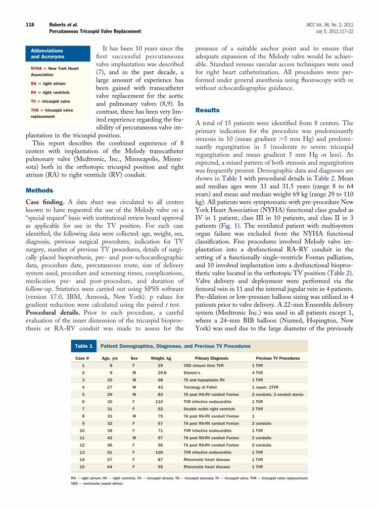
aykYIpocpsatVfPpswY
118 Roberts et al. JACC Vol. 58, No. 2, 2011Percutaneous Tricuspid Valve Replacement July 5, 2011:117–22
It has been 10 years since thefirst successful percutaneousvalve implantation was described(7), and in the past decade, alarge amount of experience hasbeen gained with transcathetervalve replacement for the aorticand pulmonary valves (8,9). Incontrast, there has been very lim-ited experience regarding the fea-sibility of percutaneous valve im-
plantation in the tricuspid position.This report describes the combined experience of 8
centers with implantation of the Melody transcatheterpulmonary valve (Medtronic, Inc., Minneapolis, Minne-sota) both in the orthotopic tricuspid position and rightatrium (RA) to right ventricle (RV) conduit.
Methods
Case finding. A data sheet was circulated to all centersknown to have requested the use of the Melody valve on a“special request” basis with institutional review board approvalas applicable for use in the TV position. For each caseidentified, the following data were collected: age, weight, sex,diagnosis, previous surgical procedures, indication for TVsurgery, number of previous TV procedures, details of surgi-cally placed bioprosthesis, pre- and post-echocardiographicdata, procedure date, percutaneous route, size of deliverysystem used, procedure and screening times, complications,medication pre- and post-procedure, and duration offollow-up. Statistics were carried out using SPSS software(version 17.0, IBM, Armonk, New York). p values forgradient reduction were calculated using the paired t test.Procedural details. Prior to each procedure, a carefulevaluation of the inner dimension of the tricuspid biopros-thesis or RA-RV conduit was made to assess for the
Abbreviationsand Acronyms
NYHA � New York HeartAssociation
RA � right atrium
RV � right ventricle
TV � tricuspid valve
TVR � tricuspid valvereplacement
Patient Demographics, Diagnoses, and PreviousTable 1 Patient Demographics, Diagnoses,
Case # Age, yrs Sex Weight, kg
1 8 F 29
2 9 M 29.8
3 25 M 68
4 27 M 43
5 29 M 83
6 30 F 110
7 31 F 52
8 31 M 75
9 32 F 67
10 39 F 71
11 42 M 97
12 45 F 56
13 51 F 100
14 57 F 87
15 64 F 55
RA � right atrium; RV � right ventricle; TA � tricuspid atresia; TS � tricuspVSD � ventricular septal defect.
presence of a suitable anchor point and to ensure thatadequate expansion of the Melody valve would be achiev-able. Standard venous vascular access techniques were usedfor right heart catheterization. All procedures were per-formed under general anesthesia using fluoroscopy with orwithout echocardiographic guidance.
Results
A total of 15 patients were identified from 8 centers. Theprimary indication for the procedure was predominantlystenosis in 10 (mean gradient �5 mm Hg) and predomi-nantly regurgitation in 5 (moderate to severe tricuspidregurgitation and mean gradient 5 mm Hg or less). Asexpected, a mixed pattern of both stenosis and regurgitationwas frequently present. Demographic data and diagnoses areshown in Table 1 with procedural details in Table 2. Meannd median ages were 33 and 31.5 years (range 8 to 64ears) and mean and median weight 69 kg (range 29 to 110g). All patients were symptomatic with pre-procedure Nework Heart Association (NYHA) functional class graded as
V in 1 patient, class III in 10 patients, and class II in 3atients (Fig. 1). The ventilated patient with multisystemrgan failure was excluded from the NYHA functionallassification. Five procedures involved Melody valve im-lantation into a dysfunctional RA-RV conduit in theetting of a functionally single-ventricle Fontan palliation,nd 10 involved implantation into a dysfunctional biopros-hetic valve located in the orthotopic TV position (Table 2).alve delivery and deployment were performed via the
emoral vein in 11 and the internal jugular vein in 4 patients.re-dilation or low-pressure balloon sizing was utilized in 4atients prior to valve delivery. A 22-mm Ensemble deliveryystem (Medtronic Inc.) was used in all patients except 1,here a 24-mm BIB balloon (Numed, Hopington, Nework) was used due to the large diameter of the previously
roceduresrevious TV Procedures
rimary Diagnosis Previous TV Procedures
osure then TVR 1 TVR
’s 4 TVR
hypoplastic RV 1 TVR
gy of Fallot 1 repair, 1TVR
t RA-RV conduit Fontan 2 conduits, 3 conduit stents
fective endocarditis 1 TVR
outlet right ventricle 2 TVR
t RA-RV conduit Fontan 1
t RA-RV conduit Fontan 2 conduits
fective endocarditis 1 TVR
t RA-RV conduit Fontan 2 conduits
t RA-RV conduit Fontan 2 conduits
fective endocarditis 1 TVR
atic heart disease 1 TVR
atic heart disease 1 TVR
TV Pand P
P
VSD cl
Ebstein
TS and
Tetralo
TA pos
TVR in
Double
TA pos
TA pos
TVR in
TA pos
TA pos
TVR in
Rheum
Rheum
id stenosis; TV � tricuspid valve; TVR � tricuspid valve replacement;
interna
119JACC Vol. 58, No. 2, 2011 Roberts et al.July 5, 2011:117–22 Percutaneous Tricuspid Valve Replacement
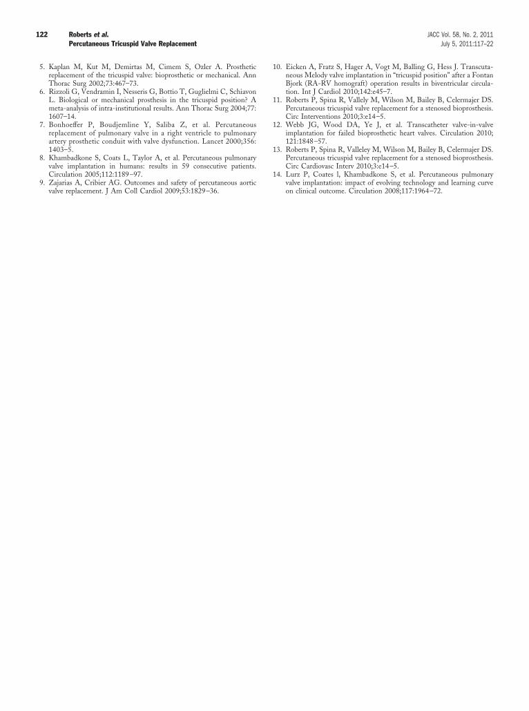
placed surgical valve and concern regarding achieving ade-quate apposition of the Melody valve. Post-implantationdilation of the Melody valve using higher pressure valvulo-plasty balloons was performed in 7 patients. Average pro-cedure and screening times were 120 � 64 min (range 54 to251 min) and 29 � 18 min (range 8.3 to 74 min),respectively. In 1 patient (Case #15), a Melody valve wasimplanted in both the tricuspid and pulmonary valve posi-tions at the same procedure. Figures 2 to 4 show pre- andpost-echocardiographic and fluoroscopic images, and Figure5 shows balloon interrogation of a bioprosthesis.
In those patients being treated primarily for stenosis, themedian valve gradient was reduced from 12.5 to 3.5 mm Hg(mean 12.9 to 3.9 mm Hg) (p � 0.01). The medianpost-procedure TV gradient for all patients was 2 mm Hg(mean 2.9 mm Hg, range 0 to 7 mm Hg) (p � 0.01). No
Procedural DetailsTable 2 Procedural Details
Case # Approach Bioprosthesis Type
Gradient
Pre Post
1 Femoral 23 mm CE 5 2
2 LIJ 23 mm MM 8 5
3 Femoral CE No 14 conduit 17 7
4 Femoral 27 mm MM 10 3
5 Femoral Patched Dacron tube 4 1
6 RIJ 27 mm MM 13 3
7 Femoral 25 mm MM 3 2
8 Femoral 26 mm Homograft 12 2
9 Femoral 22 mm CE 12 2
10 RIJ 27 mm CE 14 5
11 Femoral 30 mm Hancock 5 0
12 Femoral 31 mm homograft — 0
13 RIJ 31 mm CE 14 6
14 Femoral Sorin Bicarbon 33 20 4
15 Femoral 29 mm CE 9 2
Mean Doppler gradients are in mm Hg.CE � Carpentier Edwards; LIJ � left internal jugular vein; MM � Medtronic Mosaic; RIJ � right
Figure 1 Before and After NYHA Functional Classfor the 14 Surviving Patients
An improvement in functional class was noted in 12 of 14 surviving patients.NYHA � New York Heart Association.
patient had more than mild tricuspid regurgitation at thecompletion of the procedure, with 10 having no regurgitationas judged by echocardiography. One patient, in whom thevalve was implanted with a 24-mm balloon, developed earlyprogressive (from mild to moderate) tricuspid regurgitation.This patient, who was comatose with multisystem organ failureprior to Melody implantation, experienced initial improvementwith a reduction in mean valve gradient from 14 to 6 mm Hgassociated with a transient improvement in mental state, butultimately died 2 days after surgical removal and replacementof the valve, 20 days after the initial implant.
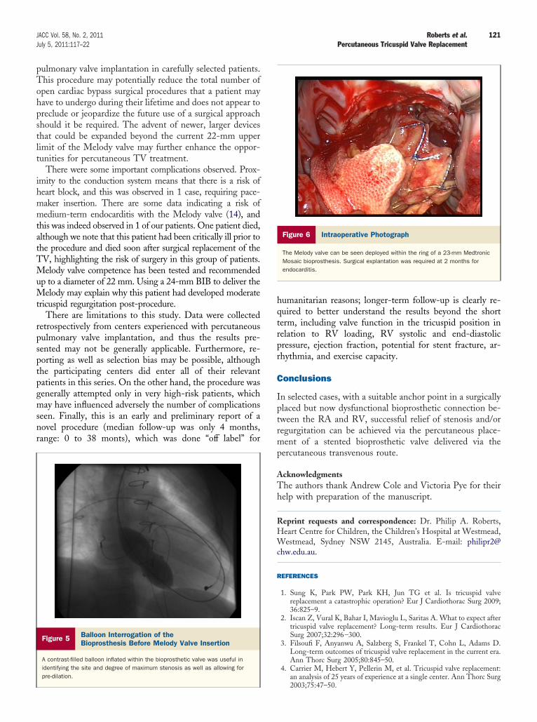
Other complications included 1 case of third-degree heartblock necessitating pacemaker implantation and 1 case ofMelody valve endocarditis requiring valve removal 2 monthspost-implant (Fig. 6).
At latest follow-up (mean 9 months, and median of 4months, range 0 to 38 months), 14 of 15 patients whounderwent the procedure are alive and well with 13 of 14retaining the Melody valve in the TV position. NYHAfunctional class improved acutely in all but 2 of thesurviving patients (Fig. 1).
Discussion
Major advances in the surgical management of congenitaland acquired right-sided valvular heart disease have allowedthe survival of increasing numbers of affected patients withcongenital or acquired disease. The right side of the heartprovides its own special set of anatomical and functionalconsiderations, often necessitating the use of a bioprostheticvalve or valved conduit, all of which have a variable butlimited longevity.
A considerable body of literature supports the use of theMelody valve in a stenotic and/or regurgitant RV to
Tricuspid Regurgitation
Screening Time (min) ComplicationsPre Post
Severe None 8.6 None
Severe Mild 25 Endocarditis
None None 33 None
Moderate None 10 None
Severe None 37 None
Mild Mild 10.6 None
Severe Mild 32.8 None
Mild None 57 None
Moderate None 22 None
Moderate Mild 74 None
Moderate None 21 None
Severe None 8.3 None
Mild Mild 35 Died
None None 15 Heart block
Mild None 30 None
l jugular vein.
pulmonary artery conduit with particular morphologic fea-
120 Roberts et al. JACC Vol. 58, No. 2, 2011Percutaneous Tricuspid Valve Replacement July 5, 2011:117–22
tures (8). An even greater number of percutaneous aorticvalve replacements have been performed worldwide (9).Following development of transcatheter heart valve replace-ment in the ventriculoarterial valve position for high-risksurgical patients, the natural extension was to look foroptions for nonsurgical treatment of the stenotic and/orregurgitant RA-RV connection in patients considered highsurgical risk. Transcatheter heart valve use has been reportedin a prosthetic RA to RV conduit (Bjork-type connection ina complex univentricular-type heart) (10), and preliminaryanimal work has reported percutaneous TVR in the nativevalve annulus (11). Webb et al. (12) recently reportedimplantation of an Edwards Sapien valve (Edwards Life-
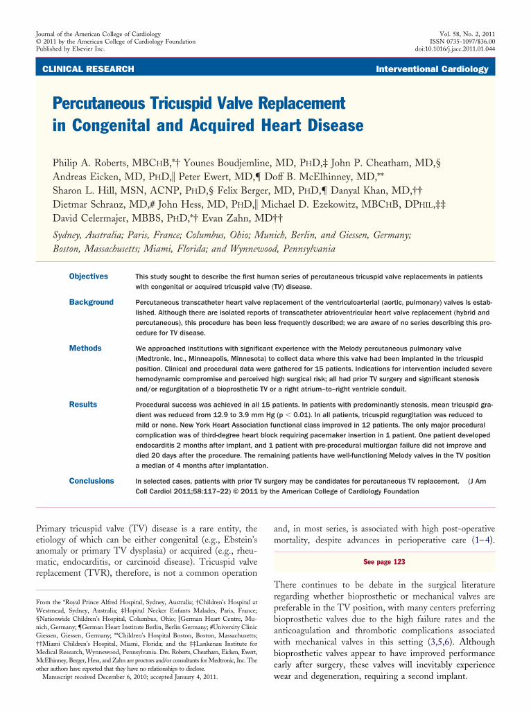
Figure 2 Before and After 2D and Color Doppler Transthoracic
(Top) The severe regurgitation of the bioprosthesis in the orthotopic TV position (timage shows a narrow stenotic jet of color flow that is relieved after Melody valveventricle.
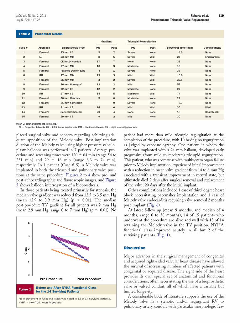
Figure 3 3 Lateral Angiograms TakenBefore and After Melody Deployment
The left-hand image shows significant regurgitation of contrast from the right ven-tricle back into the right atrium. The right-hand image is after Melody valve deploy-ment and shows a competent RA-RV connection. Abbreviations as in Figure 2.
sciences, Irvine, California) in a patient with a TV biopros-thesis using a hybrid approach with transcatheter delivery ofthe valve through thoracotomy access. There is a case reportof a percutaneous valve placed inside a bioprosthetic valvefor tricuspid stenosis (13); this patient is included in thecurrent series (Case #6 in Tables 1 and 2).
We now report a series of 15 percutaneous TVRs using abioprosthetic valve placed inside a previously implantedbioprosthetic valve or valved conduit, with important he-modynamic compromise. This series shows that percutane-ous TVR is achievable with a high procedural success rate,when undertaken by groups experienced with transcatheter
cardiograms
t) is abolished after Melody valve insertion (top right). (Bottom) The bottom leftment (bottom right). 2D � 2-dimensional; RA � right atrium; RV � right
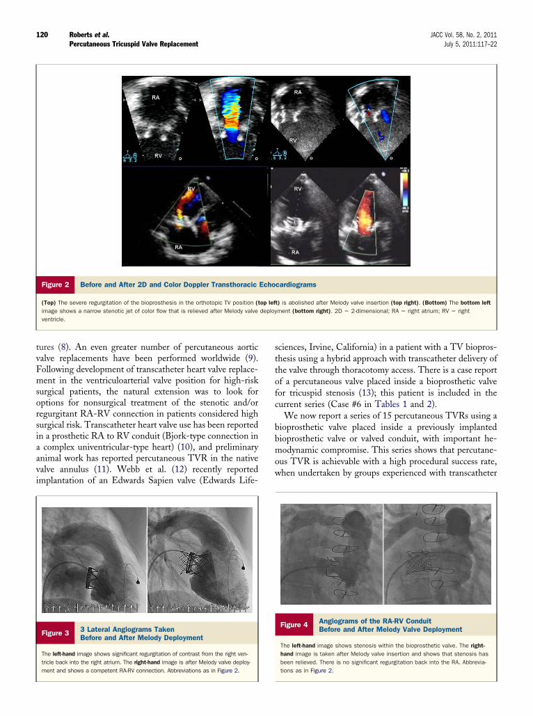
Figure 4 Angiograms of the RA-RV ConduitBefore and After Melody Valve Deployment
The left-hand image shows stenosis within the bioprosthetic valve. The right-hand image is taken after Melody valve insertion and shows that stenosis hasbeen relieved. There is no significant regurgitation back into the RA. Abbrevia-tions as in Figure 2.
Echo
op lefdeploy
121JACC Vol. 58, No. 2, 2011 Roberts et al.July 5, 2011:117–22 Percutaneous Tricuspid Valve Replacement
pulmonary valve implantation in carefully selected patients.This procedure may potentially reduce the total number ofopen cardiac bypass surgical procedures that a patient mayhave to undergo during their lifetime and does not appear topreclude or jeopardize the future use of a surgical approachshould it be required. The advent of newer, larger devicesthat could be expanded beyond the current 22-mm upperlimit of the Melody valve may further enhance the oppor-tunities for percutaneous TV treatment.
There were some important complications observed. Prox-imity to the conduction system means that there is a risk ofheart block, and this was observed in 1 case, requiring pace-maker insertion. There are some data indicating a risk ofmedium-term endocarditis with the Melody valve (14), andthis was indeed observed in 1 of our patients. One patient died,although we note that this patient had been critically ill prior tothe procedure and died soon after surgical replacement of theTV, highlighting the risk of surgery in this group of patients.Melody valve competence has been tested and recommendedup to a diameter of 22 mm. Using a 24-mm BIB to deliver theMelody may explain why this patient had developed moderatetricuspid regurgitation post-procedure.
There are limitations to this study. Data were collectedretrospectively from centers experienced with percutaneouspulmonary valve implantation, and thus the results pre-sented may not be generally applicable. Furthermore, re-porting as well as selection bias may be possible, althoughthe participating centers did enter all of their relevantpatients in this series. On the other hand, the procedure wasgenerally attempted only in very high-risk patients, whichmay have influenced adversely the number of complicationsseen. Finally, this is an early and preliminary report of anovel procedure (median follow-up was only 4 months,range: 0 to 38 monts), which was done “off label” for
Figure 5 Balloon Interrogation of theBioprosthesis Before Melody Valve Insertion
A contrast-filled balloon inflated within the bioprosthetic valve was useful inidentifying the site and degree of maximum stenosis as well as allowing forpre-dilation.
humanitarian reasons; longer-term follow-up is clearly re-quired to better understand the results beyond the shortterm, including valve function in the tricuspid position inrelation to RV loading, RV systolic and end-diastolicpressure, ejection fraction, potential for stent fracture, ar-rhythmia, and exercise capacity.
Conclusions
In selected cases, with a suitable anchor point in a surgicallyplaced but now dysfunctional bioprosthetic connection be-tween the RA and RV, successful relief of stenosis and/orregurgitation can be achieved via the percutaneous place-ment of a stented bioprosthetic valve delivered via thepercutaneous transvenous route.
AcknowledgmentsThe authors thank Andrew Cole and Victoria Pye for theirhelp with preparation of the manuscript.
Reprint requests and correspondence: Dr. Philip A. Roberts,Heart Centre for Children, the Children’s Hospital at Westmead,Westmead, Sydney NSW 2145, Australia. E-mail: [email protected].
REFERENCES
1. Sung K, Park PW, Park KH, Jun TG et al. Is tricuspid valvereplacement a catastrophic operation? Eur J Cardiothorac Surg 2009;36:825–9.
2. Iscan Z, Vural K, Bahar I, Mavioglu L, Saritas A. What to expect aftertricuspid valve replacement? Long-term results. Eur J CardiothoracSurg 2007;32:296–300.
3. Filsoufi F, Anyanwu A, Salzberg S, Frankel T, Cohn L, Adams D.Long-term outcomes of tricuspid valve replacement in the current era.Ann Thorc Surg 2005;80:845–50.
4. Carrier M, Hebert Y, Pellerin M, et al. Tricuspid valve replacement:an analysis of 25 years of experience at a single center. Ann Thorc Surg
Figure 6 Intraoperative Photograph
The Melody valve can be seen deployed within the ring of a 23-mm MedtronicMosaic bioprosthesis. Surgical explantation was required at 2 months forendocarditis.
2003;75:47–50.
122 Roberts et al. JACC Vol. 58, No. 2, 2011Percutaneous Tricuspid Valve Replacement July 5, 2011:117–22
5. Kaplan M, Kut M, Demirtas M, Cimem S, Ozler A. Prostheticreplacement of the tricuspid valve: bioprosthetic or mechanical. AnnThorac Surg 2002;73:467–73.
6. Rizzoli G, Vendramin I, Nesseris G, Bottio T, Guglielmi C, SchiavonL. Biological or mechanical prosthesis in the tricuspid position? Ameta-analysis of intra-institutional results. Ann Thorac Surg 2004;77:1607–14.
7. Bonhoeffer P, Boudjemline Y, Saliba Z, et al. Percutaneousreplacement of pulmonary valve in a right ventricle to pulmonaryartery prosthetic conduit with valve dysfunction. Lancet 2000;356:1403–5.
8. Khambadkone S, Coats L, Taylor A, et al. Percutaneous pulmonaryvalve implantation in humans: results in 59 consecutive patients.Circulation 2005;112:1189–97.
9. Zajarias A, Cribier AG. Outcomes and safety of percutaneous aorticvalve replacement. J Am Coll Cardiol 2009;53:1829–36.
10. Eicken A, Fratz S, Hager A, Vogt M, Balling G, Hess J. Transcuta-neous Melody valve implantation in “tricuspid position” after a FontanBjork (RA-RV homograft) operation results in biventricular circula-tion. Int J Cardiol 2010;142:e45–7.
11. Roberts P, Spina R, Vallely M, Wilson M, Bailey B, Celermajer DS.Percutaneous tricuspid valve replacement for a stenosed bioprosthesis.Circ Interventions 2010;3:e14–5.
12. Webb JG, Wood DA, Ye J, et al. Transcatheter valve-in-valveimplantation for failed bioprosthetic heart valves. Circulation 2010;121:1848–57.
13. Roberts P, Spina R, Valleley M, Wilson M, Bailey B, Celermajer DS.Percutaneous tricuspid valve replacement for a stenosed bioprosthesis.Circ Cardiovasc Interv 2010;3:e14–5.
14. Lurz P, Coates l, Khambadkone S, et al. Percutaneous pulmonary
valve implantation: impact of evolving technology and learning curveon clinical outcome. Circulation 2008;117:1964–72.


