Embed Size (px)
Citation preview

[CANCER RESEARCH 64, 6041–6049, September 1, 2004]
The Rgr Oncogene Induces Tumorigenesis in Transgenic Mice
Marıa Jimenez,1,2 Ignacio Perez de Castro,1,2 Marta Benet,1 Juan F. Garcıa,2 Giorgio Inghirami,1,3 andAngel Pellicer1
1Department of Pathology and New York University Cancer Institute, New York University School of Medicine, New York, New York; 2Molecular Pathology and MolecularOncology Program, Centro Nacional de Investigaciones Oncologicas, Madrid, Spain; and 3Center for Experimental Research and Medical Studies, University of Turin, Turin,Italy
ABSTRACT
To study the oncogenic potential of Rgr in vivo, we have generatedseveral transgenic Rgr mouse lines, which express the oncogene under thecontrol of different promoters. These studies revealed that Rgr expressionleads to the generation of various pathological alterations, including fi-brosarcomas, when its transgenic expression is restricted to nonlymphoidtissues. Moreover, the overall incidence and latency of fibrosarcomas weresubstantially increased and shortened, respectively, in a p15INK4b-defectivebackground. More importantly, we also have demonstrated that Rgrexpression in thymocytes of transgenic mice induces severe alterations inthe development of the thymocytes, which eventually lead to a highincidence of thymic lymphomas. This study demonstrates that oncogenicRgr can induce expression of p15INK4b and, more importantly, that bothRgr and p15INK4b cooperate in the malignant phenotype in vivo. Thesefindings provide new insights into the tumorigenic role of Rgr as a potentoncogene and show that p15INK4b can act as a tumor suppressor gene.
INTRODUCTION
Rgr is an oncogene that was isolated in our laboratory by its abilityto produce tumors in the nude mice assay (1). This oncogene wasdetected and isolated by gene transfer, using DNA from a 7,12-dimethylbenz(a)anthracene-induced rabbit squamous cell carcinomaas the starting material. Rgr (truncated at its 5� end) was fused to therHHR23A gene, the rabbit orthologue of Rad23, a Saccharomycescerevisiae nucleotide excision repair gene. Rgr belongs to the guaninenucleotide exchange factor (GEF) family, also known as GDP disso-ciation stimulator (GDS), and has a significant homology to Ralguanine dissociation stimulator (Ral-GDS). Ral-GDS is an effector ofRas and may functionally link Ras with other Ras-related proteins (2).Similar to Ral-GDS, Rgr has been shown to have specific exchangeactivity for Ral (1). Rgr lacks the Ras-interacting domain within theCOOH-terminal end present in other family members. Interestingly,its introduction into cells resulted in increased levels of GTP-boundRas (3). Moreover, Rgr is the first member of the Ral-mediatedpathway to display tumorigenic properties as demonstrated by thepotent tumorigenic activity of Rgr in the nude mice assay (1) and theformation of transformed foci and abnormal cellular morphologies incultured cells (3). In addition, Rgr also enhanced the phosphorylationof extracellular signal-regulated kinases, p38, and c-Jun-NH2-terminalkinases. The biological significance of these activities was confirmedusing dominant-negative forms of Ras, Ral, and Rho that blocked thetranscriptional activation induced by Rgr (3). Interestingly, only thedominant-negative form of Ras inhibited Rgr transformation, indicat-ing that Ras activation is crucial for the oncogenic activity of Rgr (3).
It is important to note that Ras has been previously shown to inducethe expression of several cell cycle regulators, including p15INK4b (4),and it is important to determine the ability of Rgr to produce thoseeffects. In addition, it has been previously shown that there is Rasactivation and inactivation of p15INK4b in some tumors (5). Thisseemingly contradictory observation is interpreted as if the increase inp15INK4b expression produced by Ras is a defense mechanism of thecell to block the transforming effects of Ras. Therefore, it is also ofinterest to investigate whether Rgr would cooperate with p15INK4b
inactivation as well.Recently, we have shown that at least part of the transforming
activity of Rgr is a consequence of its overexpression, which in turnis due to the elimination of translational regulatory elements in the 5�leader region of Rgr mRNA. This overexpression resulted in subse-quent Ras activation (6). The human orthologue of the rabbit Rgrgene, hrgr, has been recently isolated from lymphoid malignant celllines that express several hrgr mutant transcripts (7). Similar to theactivated rabbit truncated form (Rgr oncogene), the human mutanttranscripts found in T-cell lines and some T-cell lymphomas aretruncated forms of the hrgr gene (7).
This report summarizes the analysis of Rgr transgenic mouse linesin an effort to study the in vivo oncogenic potential of Rgr. In theseexperiments, Rgr has demonstrated the ability to induce pathologicalalterations, including tumors, in tissues in which it is expressed. Someof the neoplasic alterations were substantially increased in a p15INK4b-defective background, indicating cooperation between these two ge-netic alterations in some tumor types for Rgr-induced tumorigenesis.More importantly, the expression of Rgr in T cells was able to causethe formation of lymphomas with a high penetrance, consistent withthe alteration found in human lymphomas and confirming the impor-tant role of this oncogene in this type of tumor.
MATERIALS AND METHODS
Plasmids and DNA/RNA Manipulation. Rgr was subcloned into theEcoRI site of pcDNA3 vector (Invitrogen, Carlsbad, CA), in which the mCC10promoter was put in place of the cytomegalovirus (CMV) promoter, giving riseto the pmCC10-RGR plasmid; or into pCX vector (8), giving rise to thepCX-RGR plasmid. Rgr also was subcloned into pMexNeo expression vector(9), producing pNM11 plasmid, which was described previously (3). ThepCD4-RGR-Flag vector, in which the CD4 promoter drives RGR-Flag expres-sion, was prepared by inserting the CD4 promoter-enhancer into the cloningsite of pMexNeo vector and then subcloning the RGR-Flag sequence in the 3�end of the promoter. The CD4 promoter-enhancer was obtained from a plasmid(CD4-hCD2; a generous gift from Dr. D. R. Littman; Skirball Institute, NewYork University Medical Center, New York, NY) containing the minimal CD4enhancer (339 bp), the minimal murine CD4 promoter (487 bp lacking the CD4silencer region to drive the expression in CD4- and CD8-positive T cells, bothsingle and double positive), the transcription initiation site, and 70 bp of theuntranslated first exon and part of the first intron of the murine CD4 gene (10).The RGR-Flag fragment was described previously as Flag-tagged RSC-Rgr(6).
The expression of oncogenic Rgr driven by the majority of the promoterslisted above was analyzed in vitro before obtaining the transgenic mice.pNM11 and pCX-RGR vectors were transfected into NIH3T3 cells by thecalcium phosphate precipitation method (11), and the expression of Rgr was
Received 10/28/03; revised 5/16/04; accepted 7/6/04.Grant support: Supported by National Institutes of Health grants CA50434 (A.
Pellicer) and CA90773 (G. Inghirami).The costs of publication of this article were defrayed in part by the payment of page
charges. This article must therefore be hereby marked advertisement in accordance with18 U.S.C. Section 1734 solely to indicate this fact.
Requests for reprints: Angel Pellicer, Department of Pathology and New YorkUniversity Cancer Institute, New York University School of Medicine, 550 First Avenue,New York, NY 10016. Phone: 212-263-5342; Fax: 212-263-8211; E-mail: [email protected].
©2004 American Association for Cancer Research.
6041
Research. on February 12, 2016. © 2004 American Association for Cancercancerres.aacrjournals.org Downloaded from

tested using a focus formation assay and Northern blot. The expression of RGRin pmCC10-RGR vector was confirmed by transfecting H441 lung cells by thecalcium phosphate precipitation method and performing a Northern blot. Inaddition, the luciferase reporter plasmids (p15INK4b promoter) used in thiswork were described previously (4).
For hybridizations, digested DNA or RNA was separated on agarose gelsand transferred onto nitrocellulose membranes (Schleicher & Schuell, Keene,NH). DNA probes were labeled with [32P]dCTP (3,000 Ci/mmol; Dupont-NewEngland Nuclear, Boston, MA) using the Random Prime DNA LabelingSystem Rediprime II (Amersham Biosciences, Piscataway, NJ) in accordancewith the manufacturer’s protocol. Hybridizations were visualized by the use ofa PhosphorImager (Molecular Dynamics, Sunnyvale, CA) or by exposure toX-ray film.
Generation and Genotyping of Transgenic Mice. Transgenic mice weregenerated as described previously (12, 13). Briefly, each of the transgeneconstructs was injected into the pronucleus of fertilized eggs from femaledonors and subsequently transferred to pseudopregnant mice. All of the trans-genic lines were produced in the inbred strain FVB/N (14). Screening ofpositive animals for the transgene was performed by polymerase chain reaction(PCR) of DNA extracted as described previously (15), using forward primersspecific for each of the promoters and the common reverse primer 907–884(5�-GTGCCTGGCTGCAGGCTCCGCAGG-3�), which is specific for thetransgene. The specific primers for each of the promoters were as follows:MSV-F (5�-ACCTGAAAATGACCCTGTGC-3�) for the MSV-RGR lines;mCC10-147 (5�-GGTCCTCCACTGCCTGAATA-3�) for the mCC10-RGRlines; PCX-F1 (5�-CAGCCATTGCCTTTTATGGT-3�) for the CMV-RGRlines; and CD4-RGR-F1 (5�-GCCCACTTTTGGGTATCAGA-3�) for theCD4-RGR-Flag lines. Founder animals were confirmed by Southern blot afterdigestion of 20 �g of genomic DNA with SacI and hybridization with a cDNAprobe containing the entire sequence of oncogenic rabbit Rgr. Screening of theoffspring was performed by PCR amplification of DNA with the primersdescribed above. The animals were maintained in accordance with NationalInstitutes of Health and New York University institutional guidelines in apathogen-free facility.
KO-p15INK4b mice were generously provided by M. Barbacid (SpanishNational Cancer Institute, Madrid, Spain) in a 129/Sv � C57BL6 geneticbackground. MSV-RGR/p15�/� offspring were generated as littermates fromcommon matings and genotyped by PCR using specific primers for the RGRtransgene and both the wild-type and KO p15INK4b sequences (15). TheMSV-RGR/p15INK4b�/� line was generated by crossing MSV-RGR/p15INK4b�/� mice and then maintained by mating the double mutants, MSV-RGRp15�/�.
Cell Culture and Luciferase Assays. NIH3T3 cells were maintained inDulbecco’s modified Eagle’s medium (Invitrogen) supplemented with 10%calf serum (Invitrogen), penicillin G (50 units/ml), streptomycin (50 �g/ml;Gemini Bio-Products, Woodland, CA), and 500 �g/ml fungizone (Invitrogen)and incubated in standard conditions of humidity (95%), CO2 atmosphere(5%), and temperature (37°C).
For the luciferase assays, approximately 75 ng of the reporter plasmids, 25ng of the pRL-null (as an internal control of transfection efficiency), and 400ng of the inducer (pNM11) or empty vector (pMEXneo) were used to cotrans-fect NIH3T3 cells. Transient transfections were performed by lipofectionfollowing the manufacturer’s recommendations. NIH3T3 cells were platedonto 6-well plates (NIH3T3) at a density of 100,000 cells/well, grown for 24hours, and transfected. Forty-eight hours after transfection, cells were col-lected, and the luciferase assay was performed in accordance with the manu-facturer’s recommendations (Dual-Luciferase Reporter Assay System; Pro-mega, Madison, WI).
Real-Time Reverse Transcription-PCR Expression Analysis. TotalRNA was isolated from tissues and cells using Trizol (Invitrogen). To generatecDNA, 1 �g of total RNA was reverse transcribed using 0.4 �mol/L of anoligo(dT)-adapter primer and SuperScript II RNase H reverse transcriptase(Invitrogen) as described by manufacturer. To determine p15INK4b mRNAexpression in thymic lymphomas and wild-type thymi, real-time reverse tran-scription (RT)-PCR was performed using the following primers for mousep15INK4b: E2mp15F74, 5�-CTGCCACCCTTACCAGACCTGTGC-3�; andE2mp15R257, 5�-TCTCCAGTGGCAGCGTGCAGATAC-3�. To normalizethe expression levels of p15, �-actin expression was also determinedusing primers QmBactin-F (5�-TGTTACCAACTGGGACGACA-3�) and
QmBactin-R (5�-CTTTTCACGGTTGGCCTTAG-3�). Quantitative PCR wasperformed with the iCycler iQ System from Bio-Rad (Hercules, CA) usingSYBR Green as DNA intercalator.
Histological and Immunohistochemical Analyses. Tissues were fixed in10% buffered formalin at 4°C overnight, transferred to 70% ethanol, embeddedin paraffin, and sectioned at 5-�m thickness. Sections were stained withhematoxylin and eosin for histological analysis.
The immunohistochemical expression of lineage-specific markers for Bcells and IgM was performed on paraffin-embedded tissue sections fromMSV-RGR/KO-p15INK4b and CD4-RGR lymphomas (spleen and thymus, re-spectively). Antibodies used were CD45R (goat antimouse B220; clone RA3-6B2; PharMingen, San Diego, CA) and rabbit antimouse IgM (DAKO,Glostrup, Denmark), respectively. A previous heat-induced epitope retrievalstep was performed in a 0.1 mol/L trisodium citrate solution for 2 minutes ina conventional pressure cooker. After incubation, immunodetection was per-formed with the proper biotinylated secondary immunoglobulins, followed byperoxidase-labeled streptavidin (DAKO). Diaminobenzidine was used as sub-strate chromogen.
Immunostaining techniques were also performed in paraffin-embedded tis-sue sections for the detection of p53, p21, and p16 as described above.Antibodies used were as follows: rabbit antimouse p16 (M-156; 1:35 dilution;Santa Cruz Biotechnology, Santa Cruz, CA), rabbit antimouse p21 (C-19; 1:25dilution; Santa Cruz Biotechnology), and rabbit antimouse p53 (CM5p; 1:100dilution; Novocastra, Newcastle upon Tyne, United Kingdom).
Flow Cytometric Analysis. Primary thymocytes (0.5 � 106 per point)were isolated from 6-week-old wild-type and CD4-RGR mice. To characterizethe different thymocyte subpopulations as well as their activation status, CD3,CD4, CD8, CD25, CD44, and CD69 markers were detected in both groups ofthymocytes. Briefly, isolated thymocytes were washed twice with PBS andincubated at 4°C for 1 hour with two different sets of antibodies fromCALTAG: PE-TexasRed-antiCD8, FITC-antiCD4, PE-antiCD44, and APC-antiCD25; or FITC-antiCD3 plus PE-antiCD69. Cells were washed three timeswith PBS and fixed with 2% paraformaldehyde. Finally, cells were analyzedby flow cytometry in a FACscan cytometer, and data were interpreted using theCellQuest program (Becton Dickinson, San Jose, CA).
RESULTS
Study of the Effect of Rgr Transgene Expression in Nonlym-phoid Mouse Tissues. To determine whether the oncogenic form ofRgr is able to elicit the formation of tumors in vivo, we have insertedits cDNA into transgene cassettes to generate different transgenic
Fig. 1. Transgenic constructs generated with the Rgr oncogene. Four Rgr constructswere generated as described in Material and Methods. All of them include Rgr cDNA(white boxes) and differ in their promoter (hatched and checkered boxes) and poly(A)(gray boxes) sequences. Depending on the promoter sequence, the different constructs andmouse transgenic lines were termed MSV-RGR (A), mCC10-RGR (B), CMV-RGR (C),and CD4-RGR (D). In two of the cases (C and D), an intronic sequence was insertedbetween the promoter region and Rgr. Finally, Rgr was tagged with a Flag sequence(black box) in the CD4-RGR construct.
6042
Rgr-INDUCED TUMORIGENESIS
Research. on February 12, 2016. © 2004 American Association for Cancercancerres.aacrjournals.org Downloaded from
mouse lines as described previously (12, 13). The expression of Rgrwas controlled by different promoters (Fig. 1): the Moloney murinesarcoma virus (MSV), which is expressed at significant levels in thebrain (16), eye (16–18) and skeletal muscle (19) and at lower levelsin the kidney, pancreas (16), and testis; the mCC10 promoter (mouseClara Cell 10 KD protein), which is specific for the lung (20, 21),although it also drives a low level of expression in the uterus, ovaries,and epididymus of normal adults (21–23); and the CMV-IE enhancerplus the chicken �-actin promoter, which is ubiquitously expressedand starting from 4-cell stage (8).
Different results were obtained in the three cases in which theexpression of the Rgr transgene was driven by nonlymphoid promot-ers. For the mCC10-RGR construct, after 2 years of study, grossmorphological or behavioral abnormalities have not been detected inany of the transgenic mice. On the other hand, after three differentexperiments in which 244 fertilized eggs were injected with theCMV-RGR construct and a total of 50 mice were born, transgenicmice have not been obtained with this construct, which suggests thata high and ubiquitous expression of this oncogene at early embryonicstage could be lethal.
In the case of the MSV-RGR construct, a total of 11 transgenic lineswere obtained that transmitted the transgene to their progeny. Five ofthese lines, with varying levels of expression in several tissues, weremaintained as stable lines (L3, L21 L40, L43, and L58). Two of them,L21 and L43, were characterized and studied in more detail. Fig. 2Asummarizes the RGR expression pattern as well as the major abnor-
malities found in the MSV-RGR lines. More than 95% of the micebearing the transgene developed visible lens opacity at 3–6 weeksafter birth. This opacity led to cataracts with swollen disoriented lensfibers and vacuolation of the lens (Fig. 2B i and ii). Associated withthe cataracts, the presence of the oncogene elicited Harderian glandadenomas in all transgenic animals. MSV-RGR mice also exhibitedinguinal hernias (Fig. 2B iii) that affected only males (90% of L43males and 5% of L21 males), compromising male reproductive func-tion and leading to sterility. More importantly, fibrosarcomas in thelimbs and tail were found in 10% of the transgenic mice. Tumors werecomposed of atypical spindle-shaped cells that had a variable mitoticactivity and appeared fibroblastic, arranged in intersecting fascicles,with collagenous stroma (Fig. 2B iv). These fibrosarcomas, whichdeveloped with a latency of 6–8 months, caused paralysis of the limbsin aged mice. These results indicated that Rgr acts as an oncogene andcan produce tumors when overexpressed in transgenic mice.
Rgr Cooperates with the Lack of p15INK4b in the TumoralPhenotype In vivo. Tumoral processes are the result of an accumu-lation of alterations in genes regulating cellular homeostasis, such asoncogenes, tumor suppressor genes, apoptotic-regulating genes, andDNA repair genes. Because the MSV-RGR transgenic line showed amoderate tumor phenotype (10% of sarcomas with a 6- to 8-monthlatency period), we analyzed whether Rgr, as an oncogene, couldcooperate in vivo with a tumor suppressor gene in tumor progressionand development. Among all of the genes that have been associatedwith a tumor suppressor activity, we have previously demonstratedthat the lack of one of them, p15INK4b, cooperates with RGR in fociformation assays performed in mouse embryonic fibroblasts (10).Because this is the same collaboration type that we have foundbetween Ras and p15INK4b (10), we studied whether or not Rgr alsoshares with Ras the mechanisms to activate the expression ofp15INK4b. NIH3T3 cells were cotransfected with Rgr and luciferasereporter plasmids containing either a wild-type form or differentdeletion mutants of the p15INK4b promoter. Luciferase activity wasmeasured 48 hours after transfections. As shown in Fig. 3, Rgr isindeed able to induce p15INK4b expression in a fashion similar to thatof Ras.
Given all these results suggesting that the cell cycle inhibitorp15INK4b is involved in tumor suppressor activity triggered by an
Fig. 2. Different phenotypes are observed in MSV-RGR transgenic mice. A, incidenceof the different alterations detected in MSV-RGR transgenic mice. Note that with theexception of inguinal hernias, the incidence of the remaining alterations is the same inboth lines. B. Pictures show different pathological alterations developed in transgenicmice. The histopathological analysis of MSV-RGR eyes showed lens cataract withdisoriented lens fibers, vacuolation, and other malformations (i) compared with normaleye structure from a wild-type mouse (ii). In both i and ii, magnification is �20. iii,surgical dissection showing an inguinal hernia developed in a male MSV-RGR transgenicmouse. iv, section of a fibrosarcoma from a MSV-RGR transgenic mouse, stained withhematoxylin and eosin; magnification, �60.
Fig. 3. Oncogenic Rgr induces p15INK4b expression through activation of its promoter.Representative experiment showing the induction of p15INK4b promoter activity by Rgr.NIH3T3 cells were cotransfected with Rgr and the pGL2b(�751/�160)-luc constructcarrying a 751-bp sequence upstream of the p15INK4b ATG, the pGL2b(�35/�160)-lucconstruct carrying a sequence lacking Sp1 binding sites, or the tk81-luc construct as anegative control. Forty-eight hours after transfection, luciferase activity was measured asdescribed in Material and Methods.
6043
Rgr-INDUCED TUMORIGENESIS
Research. on February 12, 2016. © 2004 American Association for Cancercancerres.aacrjournals.org Downloaded from
inappropriate oncogenic Rgr activation, we decided to investigate theeffect of Rgr oncogenic activation in a p15INK4b-deficient backgroundusing a new transgenic/knockout mouse line that was generated bycrossing the MSV-RGR and KO-p15INK4b mouse lines (24). With thismouse model, we were able to demonstrate that lack of p15INK4b
could collaborate in the MSV-RGR-induced tumorigenesis inducing amore pronounced phenotype (Fig. 4). Indeed, the severity and inci-dence of the tumors induced by Rgr were substantially increased whenthe MSV-RGR transgenic mice were crossed with the KO-p15INK4b
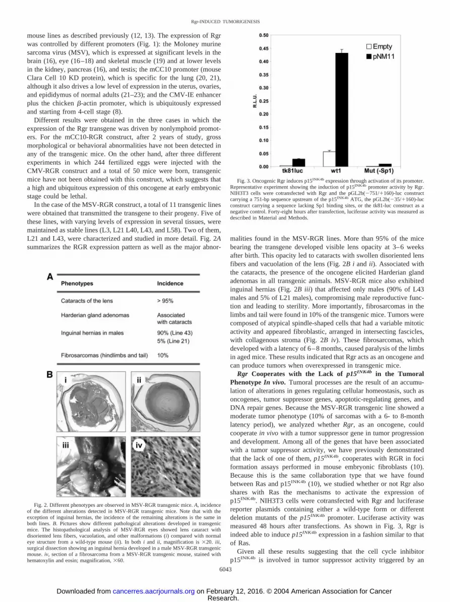
mouse line. In fact, all of the MSV-RGR/KO-p15INK4b mice devel-oped fibrosarcomas in the limbs, tail, and/or ears, compared with only10% of animals in the case of the MSV-RGR/WT-p15INK4b. Further-more, MSV-RGR/KO-p15INK4b limb tumors were significantly largerthan those found in the MSV-RGR/WT-p15INK4b mice (Fig. 4A).Also, the latency of the fibrosarcomas diminished substantially from24 to 32 weeks for the MSV-RGR mice in a wild-type p15INK4b
background to 3 to 6 weeks in a p15INK4b-deficient one.
The histopathological analysis of the fibrosarcomas obtained fromMSV-RGR/KO-p15INK4b exhibited a more aggressive phenotype thanthose from MSV-RGR/WT-p15INK4b mice, usually showing highergrade tumors with increased cellularity, mitotic activity, necrotic foci,and tumoral areas, with frequent multinucleated pleomorphic cells(undifferentiated pleomorphic sarcomas; Figs. 2F and 4B). In addi-tion, all of the MSV-RGR/KO-p15INK4b mice displayed lymphoidhyperplasia in the spleen (Fig. 4C). In 50% of these cases, thisphenotype was a consequence of extramedullary hematopoiesis (fociof myeloid metaplasia in the red pulp composed of granulocytic cells,erythroblasts, and megakaryocytes in different stages of maturation),an abnormal phenotype that is also found in the p15INK4b-null mice(24). More importantly, the remaining enlarged spleens were associ-ated with lymphomas, a tumor type rarely found in KO-p15INK4b mice(�1%; Ref. 24). These tumors were predominantly well-differentiatedlymphomas, composed of small- to medium-sized cleaved centrocytesthat usually expressed CD45R (B220) and IgM (data not shown),
Fig. 4. Rgr cooperates with the lack of p15INK4b in MSV-RGR-induced tumorigenesis. A. The severity of the fibrosarcomas induced bythe MSV-RGR construct was substantially increased in a KO-p15INK4b
genetic background, as observed by the size of the tumors that devel-oped in the limbs of MSV-RGR mice (i) compared with those inMSV-RGR/KO-p15INK4b mice (ii). The right bar graph shows thequantification of ankle diameter in wild-type (n � 7), KO-p15INK4b
(n � 9), MSV-RGR (n � 17), and MSV-RGR/KO-p15INK4b (n � 20)mice. �, P � 0.05; ���, P � 0.0001. B, sections of fibrosarcomas fromthe limb (i) and tail (ii) observed in MSV-RGR/KO-p15INK4b mice. C.The normal cellular organization found in wild-type spleens (i) also wasaltered in MSV-RGR/KO-p15INK4b mice. In 50% of the cases, ex-tramedullary hematopoiesis (ii) was detected, whereas the rest of thecases were associated with lymphomas (iii). D. Bar graph shows thepercentage of fibrosarcomas and lymphomas that are observed in KO-p15INK4b, MSV-RGR, and MSV-RGR/KO-p15INK4b mice. B and C,hematoxylin and eosin staining; magnification: �60 (B) and �40 (C).
6044
Rgr-INDUCED TUMORIGENESIS
Research. on February 12, 2016. © 2004 American Association for Cancercancerres.aacrjournals.org Downloaded from

whereas infiltration of the white and red pulp was variable. Thus, theconcurrent presence of both genetic alterations, expression of onco-genic Rgr together with the loss of p15INK4b, has a synergistic effectin the tumoral phenotype (Fig. 4D).
To demonstrate the specificity of p15INK4b in cooperation with Rgrin this mouse transgenic model, we studied the expression of othertumor suppressor genes by immunohistochemistry. Specifically, wechose the following for this analysis: p16INK4a (like p15INK4b, anINK4 family member that has been found to be down-regulated inmany tumor types); p21kip1 (another RB function inhibitor; in thiscase, a member of the CIP/KIP family); and, finally, p53, the keymolecule of the other critical pathway most frequently mutated intumorigenesis. Interestingly, none of these three genes was down-regulated in any of the seven tumors analyzed (Fig. 5). Moreover, wedid not find overexpression of p53, suggesting that this gene is notmutated in any of the analyzed tumors. Therefore, these results appearto indicate a specific role of the constitutive lack of p15INK4b inRGR-induced tumorigenesis.
CD4-RGR Transgenic Mice: A Key Role of Rgr in T-CellLymphoma Development and Progression. Mutations in the Rgrhuman orthologue, hrgr, have been associated with human T-cellmalignancies (7). In contrast, MSV-RGR transgenic mice express-ing low levels of Rgr in lymphoid tissues (data not shown) areprone to splenic lymphomas in a p15INK4b-deficient background(Fig. 4C and D). To further analyze in vivo the role of Rgr inlymphomagenesis, new transgenic mouse lines were generated inwhich Rgr expression was under the control of the murine CD4minimal promoter (lacking the CD4 silencer region) and the CD4enhancer (Fig. 1D) to drive the expression in CD4- and CD8-positive T cells (both single and double positive; Ref. 10). Amongthe 16 founders obtained from microinjection of the CD4-RGR-Flag transgene, three independent CD4-RGR transgenic lines
(lines 19, 37, and 42), were expanded and studied. The thymocytesof the animals in all three lines exhibited Rgr expression (data notshown). As shown in Fig. 6A, many animals (83% in L19, 38% inL37, and 68% in L42) carrying the CD4-RGR construct developedthymic lymphomas. In fact, the progression of this tumor was thecause of death in all cases. Lines 19 and 42 showed similar thymiclymphoma incidence curves in the first year of life (median latencyof 22 and 20 weeks and tumor-free ratios of 6.7% and 13.2%,respectively). A weaker phenotype was displayed by line 37 (me-dian latency of 35 weeks and tumor-free ratio at 1 year of age of37%). The neoplasms caused complete effacement of the normalthymic architecture and frequently infiltrated adjacent soft tissuesand lung (Fig. 6B).
To further characterize the in vivo oncogenic effect of Rgr expres-sion in mouse thymocytes, the expression profile of different T-cellsurface markers was determined. A series of thymic lymphomasshowed that the majority of these tumors were, CD4�, CD8�/�, andCD25� (Fig. 6C). To characterize the putative effects resulting fromthe expression of Rgr in preneoplastic thymocytes, the morphologicaland phenotypic features of thymocyte populations in line 42 and in theless clinically aggressive line 37 were studied. Microscopic evaluationdemonstrated a normal thymic architecture and no significant differ-ences between the transgenic mice and normal control littermates(data not shown). However, a reproducible but minimal reduction inthe total number of thymocytes was observed in lines 37 and 42 (Fig.7), although this decrease in the number of cells of the CD4-RGRthymi was not statistically significant. Moreover, when 6-week-oldmice were used to isolate and study the frequency of different T-cellsubpopulations by flow cytometric analysis, significant differenceswere detected between CD4-RGR and wild-type thymocytes (Fig. 7).These differences were more pronounced in the case of the CD4-RGRmice line with shorter latency and higher incidence of thymic lym-phomas (line 42). Indeed, a significant reduction was found in thethymocyte subpopulation expressing high levels of T-cell receptor(CD3high) and the single-positive CD4 and CD8 thymocytes, togetherwith an increase in the number of CD4/CD8 double-positive cells. Nosignificant differences were found for the percentages of CD4/CD8double-negative thymocytes. Higher levels of CD25-, CD44-, andCD69-positive thymocytes among CD4-RGR line 42 mice were alsodetected. However, only the percentages of CD25- and CD69-positivethymocytes were significantly altered in the CD4-RGR line withlonger latency and lower incidence (line 37).
All these results indicated that Rgr expression in the thymocytes ofimmature mice induced a severe alteration on the patterns of thymo-cyte development and maturation consisting of a substantial increasein the number of activated and undifferentiated cells. These alterationspreceded the development of lymphomas.
Finally, because lack of p15INK4b in mice carrying a MSV-RGRconstruct induces a more pronounced malignant phenotype, includingthe onset of spleen lymphomas, we investigated whether or notp15INK4b down-regulation also plays a critical role in CD4-RGR-induced lymphomagenesis. The expression levels of p15INK4b mRNAwere determined in a set of CD4-RGR thymic lymphomas by real-time RT-PCR. As shown in Fig. 8, 50% of the tumors analyzedshowed a significant down-regulation of p15INK4b expression. Also, itis interesting to note that in the other 50% of the samples, p15INK4b
was overexpressed, which confirmed our in vitro results on the ca-pacity of Rgr to induce p15INK4b expression (Fig. 3). These resultssupport the functional interplay between Rgr and p15INK4b and sug-gest that oncogenic activation of Rgr and lack of p15INK4b expressioncooperate in different tumor types.
Fig. 5. Specific role of the lack of p15INK4b in MSV-RGR/KO-p15INK4b fibrosarco-mas. A. Table shows p21, p16, and p53 protein expression levels detected by immuno-histochemistry in seven MSV-RGR/KO-p15INK4b fibrosarcomas. Protein expression: �,5–50% positive cells; ��, �50% positive cells; and, ���, �50% positive cells withhigh levels of expression per cell. B. Representative pictures show the expression of p16,p21, and p53 in two different MSV-RGR/KO-p15INK4b fibrosarcomas. Sample 6609Tshows normal expression of both p16 and p53 and moderated overexpression of p21.Sample 6866TA shows overexpression of the three analyzed proteins.
6045
Rgr-INDUCED TUMORIGENESIS
Research. on February 12, 2016. © 2004 American Association for Cancercancerres.aacrjournals.org Downloaded from

DISCUSSION
Rgr Is the First Ral Guanine Nucleotide Exchange FactorFamily Member with Transforming Activity In vivo. Rgr is a GEFprotein that was isolated in our laboratory by its ability to producetumors in nude mice (1). The role of GEFs in activating pathwaysmediated by small GTPases indicates that they could play a patho-genic role in some tumors in which these pathways are abnormallyinduced. There are several GEFs, such as dbl, vav, and CALDAG-GEF1, previously identified as oncogenes that have been found to bemutated or rearranged in human (25, 26) and animal malignancies(27). However, it is interesting to note that among all of the consti-tutively activated variants of the RalGEFs, only Rgr is transformingby itself in mouse cells (3). The remaining GEFs, although they arenot transforming, cooperate with activated Raf in focus formationprocesses (2, 28). Unlike in mouse cells, the RalGEF pathway hasbeen reported to be important for Ras-mediated transformation inhuman cells (7, 29). In vivo studies have shown previously thatactivation of RalGEF is sufficient to initiate an invasive phenotype innude mice (30). The present study demonstrates that Rgr displaystumorigenic properties in a mouse transgenic model because it is ableto produce tumors in some of the tissues in which it is overexpressed.
The only known substrates for the RalGEFs are the monomericG-proteins RalA and RalB. However, oncogenic potential has notbeen shown for activated Ral proteins alone, although they facilitateRas transformation, participate in cell motility, and are required formetastatic evolution of Ras-transformed cells and for Ras-inducedstimulation of cyclin D1 expression (28–34). Interestingly, Rgr also isable to activate Ras (3), which could explain its potency as anoncogene and its ability to transform cells and promote tumor forma-tion in transgenic mice.
Rgr and p15INK4b Cooperate in the Development and Progres-sion of Different Tumor Types. Tumor development and progres-sion are associated with multiple genetic lesions. Although it is notknown which altered genes are cooperating with Rgr in tumorigenic
processes, one of the candidates is the tumor suppressor genep15INK4b. In a previous study, p15INK4b produced G1 arrest in Ras-transformed cells and decreased the tumorigenic potential of Ras andRgr in focus formation assays in mouse fibroblasts (4). In addition, wehave shown that, like oncogenic Ras, activation of oncogenic Rgrtriggers p15INK4b expression (Fig. 3), which could be part of a moregeneral antioncogenic response. Therefore, similar to oncogenic Ras,Rgr and p15INK4b interact in vitro in such a way that alteration of bothgenes cooperates in the malignant phenotype. These results are con-sistent with the fact that although Rgr is able to activate both Ral andRas, its transforming activity is dependent on Ras and less so on Ral(3).
The role of p15INK4b in tumorigenesis is not well defined. Althoughthe INK4b locus is often deleted in human tumors, its loss is concom-itant with that of the INK4a/ARF locus (35). In some cases, however,deletion of p15INK4b occurs independently of p16INK4a status (36, 37).More recently, it has been described that a small fraction of theKO-p15INK4b mice develop different types of tumors, mainly sarco-mas (24). The high incidence of tumors observed in the double-mutantMSV-Rgr/KO-p15INK4b mice (100%), compared with that obtained inboth the MSV-Rgr (10%) and KO-p15INK4b (8.2%) mouse lines,clearly demonstrates the synergistic cooperation between p15INK4b
and the Rgr oncogene in vivo.It has been described that inhibition of p15INK4b expression appears
to be a common event in human lymphoid tumors and mouse T-celllymphomas (37–41). Here, the important role of p15INK4b in lym-phomagenesis is confirmed. Furthermore, the results indicate that therole of p15INK4b in tumorigenesis is dependent on the concurrentoncogenic activation of various components of the Ras pathway.Indeed, splenic lymphomas are found in 50% of MSV-RGR/KO-p15INK4b mice, whereas this tumor type is found in �1% of KO-p15INK4b mice, which only show extramedullary hematopoiesis. Inaccordance with these observations, similar results have been obtainedwhen crossing transgenic mice expressing an N-Ras oncogenic variant
Fig. 6. CD4-RGR transgenic mice de-velop thymic lymphomas. A, thymic lym-phoma incidence in the three CD4-RGRlines studied in this work. The number ofmice used per line was as follows: 30 (line19), 23 (line 42), 27 (line 42), and 30(wild-type). B, representative sections of(i) a malignant thymic lymphoma in line19 showing a monomorphic population oflarge, noncleaved lymphoid cells, withhigh mitotic index; (ii) a normal thymusfrom a wild-type mouse of the same age;and (iii) an infiltration by a CD4-RGRlymphoma in the lung showing patchysmall infiltrates by monomorphic large- tomedium-sized lymphocytes. Magnifica-tion: �630 (i and ii) and �400X (iii). C,typical phenotype of CD4-RGR thymiclymphomas. Tumor cells obtained fromneoplastic thymus were stained with theindicated antibodies and analyzed by fluo-rescence-activated cell sorting.
6046
Rgr-INDUCED TUMORIGENESIS
Research. on February 12, 2016. © 2004 American Association for Cancercancerres.aacrjournals.org Downloaded from

with p15INK4b-KO mice.4 Finally, the results derived from the studyof the CD4-RGR tumors constitute not only additional evidence infavor of the role of alterations in both genes in lymphomagenesis butalso a new and significant observation about the collaboration of thetwo of them in this kind of tumor.
The INK4b gene and the closely related gene, INK4a, are located atthe same chromosomal region in both the human and mouse genome,a hot spot for cancer-associated deletions (37, 42). Although theinactivation of p16INK4a has been clearly associated with cancerdevelopment and progression (43), the role of p15INK4b as a tumorsuppressor gene is still controversial. Therefore, our data constitutenew and strong evidence in favor of the critical role of p15INK4b
inactivation in the development and progression of some tumor types.Critical Role of Rgr Oncogenic Activation in T-Cell Tumori-
genesis. An important conclusion derived from this work is the roleof Rgr in lymphomagenesis. As it has been described above, MSV-RGR mice lacking p15INK4b showed a remarkable increase in splenic
B-cell lymphoma incidence. These results are remarkable, consideringthat Rgr expression in the spleen of these transgenic mice is modest(data not shown). CD4-RGR transgenic mice provided additionalconfirmation of the role of Rgr in lymphomagenesis. The expressionof Rgr under control of the CD4 promoter induces thymic lym-phomagenesis (Fig. 6). The fact that NrasT transgenic mice developthymic lymphomas (44) is consistent with our previous results invitro, in which the transforming activity of Rgr is mediated predom-inantly by Ras.
The study of preneoplastic CD4-RGR thymi produced importantinsights regarding the Rgr-induced alterations in T cells. In the CD4-RGR transgenic line with higher incidence of tumors and shorterlatency, 6-week-old mice showed a significantly altered pattern oftheir thymocyte subpopulations (Fig. 7). The reduction in the numberof CD3�high, CD4�CD8�, and CD4�CD8� cells indicated an in-crease in the number of undifferentiated thymocytes. Moreover, thesecells became more activated, as demonstrated by the levels of expres-sion of specific activation markers (CD25 and CD69). The mostinteresting results derived from the analysis of the CD44 marker.CD44 is a multistructural and multifunctional cell surface moleculethat has been involved in many cellular functions from cell prolifer-ation to cell migration and angiogenesis. In this case, both CD4-RGRtransgenic mice lines (lines 42 and 37) showed a similar and substan-tial increase in the number of CD44� thymocytes. Interestingly,CD44 has been associated with cancer (45), and it has been proposedas a good candidate to predict prognosis in patients with lymphoma(46). Our data suggest that CD44 overexpression could be a candidatemarker for early detection of Rgr-induced lymphomagenesis.
Recently, the human orthologue of Rgr, hrgr, has been associ-ated with human T-cell malignancies (7). Similar to rabbit Rgr,which was found as one of the components of a fusion proteinisolated from a rabbit squamous cell carcinoma, truncation of hrgrconfers transforming properties to its cDNA. DNA rearrange-ments, which are frequent events in T-cell lymphomagenesis (47),could have resulted in hrgr truncation. Indeed, a DNA rearrange-ment has been found within the hrgr gene in a human anaplasticlarge cell lymphoma cell line (DHL; Ref. 7). It is interesting topoint out that chromosomal translocations frequently have beenassociated with sarcomagenesis (48), and in many cases, these
4 I. Perez de Castro, M. Malumbres, M. Benet, M. Jimenez, J. F. García, M. Barbacid,A. Pellicer. Effect of p15INK4b deficiency on N-ras mediated oncogenesis, manuscript inpreparation.
Fig. 7. Characterization of CD4-RGR preneoplasic thymi. Phenotypic features ofwild-type and CD4-RGR thymi. Thymocytes were isolated from a total of twelve6-week-old mice (6 wild-type and 6 CD4-RGR mice). Half of the CD4-RGR mice camefrom line 37 (L37), whereas the remaining mice corresponded to the most aggressive ofthe lines (L42). Bar graphs show the total number of cells (top left panel) or percentagesof CD3�high, CD69�, CD4�/CD25�, CD8�/CD25�, and CD44� thymocytes (remain-ing panels) detected by flow cytometric analysis. Wild-type and CD4-RGR populationswere compared using unpaired t tests. �, P � 0.05; ��, P � 0.001; ���, P � 0.0001.
Fig. 8. Expression of p15INK4b in CD4-RGR thymic lymphomas. Total RNA wasisolated from two wild-type (WT) thymi and six CD4-RGR thymic lymphomas. Real-timeRT-PCR was performed in triplicate as described in Material and Methods to determinemRNA expression levels of p15INK4b in all of the samples. Normal and lymphoma valueswere calculated relative to one of the wild-type samples (3222 TH).
6047
Rgr-INDUCED TUMORIGENESIS
Research. on February 12, 2016. © 2004 American Association for Cancercancerres.aacrjournals.org Downloaded from

genetic translocations affect a chromosomal region close to the onein which hrgr is located (22q11.2). Therefore, there is a need foradditional studies on the role of hrgr in human sarcomagenesis.According to our preliminary results, DNA rearrangements involv-ing Rgr provoke changes in its levels of expression. In normal cellsand tissues, the levels of expression of both Rgr and hrgr are low,whereas in transformed cells and tumor samples, a significant levelof expression of these genes is detected (3, 7). In this work,overexpression of oncogenic Rgr is sufficient to induce the devel-opment of fibrosarcomas and lymphomas. Thus, our previous invitro model is corroborated, in which a mutation affecting theregulation of gene expression, which increases its expression fromnegligible levels, is the basis for Rgr acting as an oncogene (6).However, neoplastic alterations have not been detected in some ofthe tissues in which Rgr has been overexpressed. For example, theMSV-RGR mice express substantial levels of Rgr in liver, brain,and kidney (data not shown), and pathological alterations have notbeen detected in those tissues after analysis of these transgenicmice at age � 2 years. Therefore, these results suggest that theRgr-induced transforming phenotype is dependent not only on theexpression levels of this gene but also on certain tissue-specificfactors. These still uncharacterized elements might be the reasonfor oncogenic Rgr involvement in lymphomagenesis and, to alesser extent, in the development of fibrosarcomas.
Taken together, these results reinforce the role of Rgr as an onco-gene in different tumor types. In some of the tumors (fibrosarcomas),Rgr-induced tumorigenesis is substantially potentiated by the effect ofother genetic alterations, such as a constitutive lack of p15INK4b
expression. However, in other cases (lymphomas), expression of Rgrin T cells induced a severe alteration of thymocyte differentiation andstrengthens the notion that Rgr plays an important role in lym-phomagenesis. Therefore, the high incidence of lymphomas and fi-brosarcomas in the transgenic lines and the fact that hrgr also isassociated with human lymphomas support this as a model to studysarcomagenesis and lymphomagenesis.
ACKNOWLEDGMENTS
We are especially indebted to E. Latres and M. Barbacid for providing uswith the p15INK4b-deficient mice. We also want to thank John Hirst for his helpin flow cytometric analyses and Laura Martello-Roonie for critical reading ofthe manuscript.
REFERENCES
1. D’Adamo DR, Novick S, Kahn JM, Leonardi P, Pellicer A. rsc: a novel oncogenewith structural and functional homology with the gene family of exchange factors forRal. Oncogene 1997;14:1295–305.
2. White MA, Vale T, Camonis JH, Schaefer E, Wigler MH. A role for the Ral guaninenucleotide dissociation stimulator in mediating Ras-induced transformation. J BiolChem 1996;271:16439–42.
3. Hernandez-Munoz I, Malumbres M, Leonardi P, Pellicer A. The Rgr oncogene(homologous to RalGDS) induces transformation and gene expression by activatingRas, Ral and Rho mediated pathways. Oncogene 2000;19:2745–57.
4. Malumbres M, Perez De Castro I, Hernandez MI, et al. Cellular response to oncogenicras involves induction of the Cdk4 and Cdk6 inhibitor p15INK4b. Mol Cell Biol2000;20:2915–25.
5. Perez de Castro IP, Malumbres M, Santos J, Pellicer A, Fernandez-Piqueras J.Cooperative alterations of Rb pathway regulators in mouse primary T cell lympho-mas. Carcinogenesis (Lond) 1999;20:1675–82.
6. Hernandez-Munoz I, Benet M, Calero M, et al. rgr oncogene: activation byelimination of translational controls and mislocalization. Cancer Res 2003;63:4188 –95.
7. Leonardi P, Kassin E, Hernandez-Munoz I, et al. Human rgr: transforming activityand alteration in T-cell malignancies. Oncogene 2002;21:5108–16.
8. Okabe M, Ikawa M, Kominami K, Nakanishi T, Nishimune Y. “Green mice” as asource of ubiquitous green cells. FEBS Lett 1997;407:313–9.
9. Martin-Zanca D, Oskam R, Mitra G, Copeland T, Barbacid M. Molecular andbiochemical characterization of the human trk proto-oncogene. Mol Cell Biol 1989;9:24–33.
10. Sawada S, Scarborough JD, Killeen N, Littman DR. A lineage-specific transcriptionalsilencer regulates CD4 gene expression during T lymphocyte development. Cell1994;77:917–29.
11. Wigler M, Pellicer A, Silverstein S, Axel R. Biochemical transfer of single-copyeucaryotic genes using total cellular DNA as donor. Cell 1978;14:725–31.
12. Gordon JW, Scangos GA, Plotkin DJ, Barbosa JA, Ruddle FH. Genetic transforma-tion of mouse embryos by microinjection of purified DNA. Proc Natl Acad Sci USA1980;77:7380–4.
13. Gordon JW, Ruddle FH. Gene transfer into mouse embryos: production of transgenicmice by pronuclear injection. Methods Enzymol 1983;101:411–33.
14. Taketo M, Schroeder AC, Mobraaten LE, et al. FVB/N: an inbred mouse strainpreferable for transgenic analyses. Proc Natl Acad Sci USA 1991;88:2065–9.
15. Malumbres M, Mangues R, Ferrer N, Lu S, Pellicer A. Isolation of high molecularweight DNA for reliable genotyping of transgenic mice. Biotechniques 1997;22:1114–9.
16. Theuring F, Gotz W, Balling R, et al. Tumorigenesis and eye abnormalities intransgenic mice expressing MSV-SV40 large T-antigen. Oncogene 1990;5:225–32.
17. Khillan JS, Oskarsson MK, Propst F, et al. Defects in lens fiber differentiation arelinked to c-mos overexpression in transgenic mice. Genes Dev 1987;1:1327–35.
18. Gotz W, Theuring F, Favor J, Herken R. Eye pathology in transgenic mice carryinga MSV-SV 40 large T-construct. Exp Eye Res 1991;52:41–9.
19. Sutrave P, Copeland TD, Showalter SD, Hughes SH. Characterization of chickenc-ski oncogene products expressed by retrovirus vectors. Mol Cell Biol 1990;10:3137–44.
20. DeMayo FJ, Damak S, Hansen TN, Bullock DW. Expression and regulation of therabbit uteroglobin gene in transgenic mice. Mol Endocrinol 1991;5:311–8.
21. Sandmoller A, Voss AK, Hahn J, et al. Cell-specific, developmentally and hormon-ally regulated expression of the rabbit uteroglobin transgene and the endogenousmouse uteroglobin gene in transgenic mice. Mech Dev 1991;34:57–67.
22. Gomez Lahoz E, Lopez de Haro MS, Esponda P, Nieto A. Tissue-specific andhormonally regulated expression of the puromycin N-acetyltransferase-encoding geneunder control of the rabbit uteroglobin promoter in transgenic mice. Gene (Amst)1992;117:255–8.
23. Margraf LR, Finegold MJ, Stanley LA, et al. Cloning and tissue-specific expressionof the cDNA for the mouse Clara cell 10 kD protein: comparison of endogenousexpression to rabbit uteroglobin promoter-driven transgene expression. Am J RespirCell Mol Biol 1993;9:231–8.
24. Latres E, Malumbres M, Sotillo R, et al. Limited overlapping roles of P15INK4b andP18INK4c cell cycle inhibitors in proliferation and tumorigenesis. EMBO J 2000;19:3496–506.
25. Zhu K, Debreceni B, Bi F, Zheng Y. Oligomerization of DH domain is essential forDbl-induced transformation. Mol Cell Biol 2001;21:425–37.
26. Katzav S, Martin-Zanca D, Barbacid M. vav, a novel human oncogene derived froma locus ubiquitously expressed in hematopoietic cells. EMBO J 1989;8:2283–90.
27. Dupuy AJ, Morgan K, von Lintig FC, et al. Activation of the Rap1 guanine nucleotideexchange gene, CalDAG-GEF I, in BXH-2 murine myeloid leukemia. J Biol Chem2001;276:11804–11.
28. Urano T, Emkey R, Feig LA. Ral-GTPases mediate a distinct downstream signalingpathway from Ras that facilitates cellular transformation. EMBO J 1996;15:810–6.
29. Hamad NM, Elconin JH, Karnoub AE, et al. Distinct requirements for Ras oncogen-esis in human versus mouse cells. Genes Dev 2002;16:2045–57.
30. Ward Y, Wang W, Woodhouse E, et al. Signal pathways which promote invasionand metastasis: critical and distinct contributions of extracellular signal-regulatedkinase and Ral-specific guanine exchange factor pathways. Mol Cell Biol 2001;21:5958 – 69.
31. Gildea JJ, Harding MA, Seraj MJ, Gulding KM, Theodorescu D. The role of Ral Ain epidermal growth factor receptor-regulated cell motility. Cancer Res 2002;62:982–5.
32. Henry DO, Moskalenko SA, Kaur KJ, et al. Ral GTPases contribute to regulation ofcyclin D1 through activation of NF-kappaB. Mol Cell Biol 2000;20:8084–92.
33. Lee T, Feig L, Montell DJ. Two distinct roles for Ras in a developmentally regulatedcell migration. Development (Camb) 1996;122:409–18.
34. Suzuki J, Yamazaki Y, Li G, Kaziro Y, Koide H. Involvement of Ras and Ral inchemotactic migration of skeletal myoblasts. Mol Cell Biol 2000;20:4658–65. Erra-tum in: Mol Cell Biol 2000;20:7049.
35. Stone S, Dayananth P, Jiang P, et al. Genomic structure, expression and mutationalanalysis of the P15 (MTS2) gene. Oncogene 1995;11:987–91.
36. Glendening JM, Flores JF, Walker GJ, et al. Homozygous loss of the p15INK4B gene(and not the p16INK4 gene) during tumor progression in a sporadic melanomapatient. Cancer Res 1995;55:5531–5.
37. Malumbres M, Perez de Castro I, Santos J, et al. Inactivation of the cyclin-dependentkinase inhibitor p15INK4b by deletion and de novo methylation with independenceof p16INK4a alterations in murine primary T-cell lymphomas. Oncogene 1997;14:1361–70.
38. Herman JG, Jen J, Merlo A, Baylin SB. Hypermethylation-associated inactivationindicates a tumor suppressor role for p15INK4B. Cancer Res 1996;56:722–7.
39. Herman JG, Civin CI, Issa JP, et al. Distinct patterns of inactivation of p15INK4B andp16INK4A characterize the major types of hematological malignancies. Cancer Res1997;57:837–41.
6048
Rgr-INDUCED TUMORIGENESIS
Research. on February 12, 2016. © 2004 American Association for Cancercancerres.aacrjournals.org Downloaded from
40. Batova A, Diccianni MB, Yu JC, et al. Frequent and selective methylation of p15 anddeletion of both p15 and p16 in T-cell acute lymphoblastic leukemia. Cancer Res1997;57:832–6.
41. Malumbres M, Perez de Castro I, Santos J, Fernandez Piqueras J, Pellicer A.Hypermethylation of the cell cycle inhibitor p15INK4b 3�-untranslated region inter-feres with its transcriptional regulation in primary lymphomas. Oncogene 1999;18:385–96.
42. Kamb A, Shattuck-Eidens D, Eeles R, et al. Analysis of the p16 gene (CDKN2) as acandidate for the chromosome 9p melanoma susceptibility locus. Nat Genet 1994;8:23–6.
43. Ruas M, Peters G. The p16INK4a/CDKN2A tumor suppressor and its relatives.Biochim Biophys Acta 1998;1378:F115–77.
44. Mangues R, Symmans WF, Lu S, Schwartz S, Pellicer A. Activated N-ras oncogeneand N-ras proto-oncogene act through the same pathway for in vivo tumorigenesis.Oncogene 1996;13:1053–63.
45. Naor D, Nedvetzki S, Golan I, Melnik L, Faitelson Y. CD44 in cancer. Crit Rev ClinLab Sci 2002;39:527–79.
46. Pals ST, Drillenburg P. CD44 expression predicts disease outcome in localized largeB cell lymphoma. Blood 2000;95:1900–10.
47. Downing JR, Shurtleff SA, Zielenska M, et al. Molecular detection of the (2;5)translocation of non-Hodgkin’s lymphoma by reverse transcriptase-polymerase chainreaction. Blood 1995;85:3416–22.
48. Helman LJ, Meltzer P. Mechanisms of sarcoma development. Nat Rev Cancer2003;3:685–94.
6049
Rgr-INDUCED TUMORIGENESIS
Research. on February 12, 2016. © 2004 American Association for Cancercancerres.aacrjournals.org Downloaded from

2004;64:6041-6049. Cancer Res María Jiménez, Ignacio Pérez de Castro, Marta Benet, et al. Mice
Oncogene Induces Tumorigenesis in TransgenicRgrThe
Updated version
http://cancerres.aacrjournals.org/content/64/17/6041
Access the most recent version of this article at:
Cited articles
http://cancerres.aacrjournals.org/content/64/17/6041.full.html#ref-list-1
This article cites 47 articles, 24 of which you can access for free at:
Citing articles
http://cancerres.aacrjournals.org/content/64/17/6041.full.html#related-urls
This article has been cited by 5 HighWire-hosted articles. Access the articles at:
E-mail alerts related to this article or journal.Sign up to receive free email-alerts
SubscriptionsReprints and
To order reprints of this article or to subscribe to the journal, contact the AACR Publications
Permissions
To request permission to re-use all or part of this article, contact the AACR Publications
Research. on February 12, 2016. © 2004 American Association for Cancercancerres.aacrjournals.org Downloaded from