Embed Size (px)
Citation preview

Available online at www.sciencedirect.com
ental xx (2009) xxx–xxxwww.metabolismjournal.com
ARTICLE IN PRESS
Metabolism Clinical and Experim
The relationship of visfatin/pre–B-cell colony-enhancingfactor/nicotinamide phosphoribosyltransferase in adipose tissue with
inflammation, insulin resistance, and plasma lipidsYi-Cheng Changa, Tien-Jyun Changb, Wei-Jei Leec, Lee-Ming Chuangb,d,⁎
aDepartment of Internal Medicine, National Taiwan University Hospital Yun-Lin Branch, Yunlin 640, TaiwanbDepartment of Internal Medicine, National Taiwan University Hospital, Taipei 100, Taiwan
cDepartment of Surgery, Ming-Sheng General Hospital, Taoyuan 330, TaiwandGraduate Institute of Clinical Medicine, National Taiwan University College of Medicine, Taipei 100, Taiwan
Received 6 January 2009; accepted 14 July 2009
Abstract
Visfatin/pre–B-cell colony-enhancing factor (PBEF)/nicotinamide phosphoribosyltransferase (Nampt) has been proposed as an insulin-mimicking adipocytokine predominantly secreted from visceral adipose tissue (VAT) and correlated with obesity. However, recent evidencechallenged this proposal and instead suggested visfatin/PBEF/Nampt as a proinflammatory cytokine. The study aimed to examine whethervisfatin/PBEF/Nampt was predominantly expressed in VAT and was correlated with obesity. The relationship of visfatin/PBEF/Nampt geneexpression in adipose tissues with proinflammatory gene expression and metabolic phenotypes was also examined. The relative messengerRNA (mRNA) levels of visfatin/PBEF/Nampt, macrophage-specific marker CD68, and proinflammatory genes were measured in pairedabdominal VAT and subcutaneous adipose tissues (SAT) and from 53 nondiabetic adults using quantitative real-time polymerase chainreaction. Fasting glucose, insulin, triglyceride, cholesterol, and uric acid levels were measured; and systemic insulin sensitivity wasquantified with modified insulin suppression tests. There was no difference in visfatin/PBEF/Nampt mRNA levels between VAT and SAT,and neither was associated with measures of obesity. Visfatin/PBEF/Nampt mRNA levels were strongly correlated with proinflammatorygene expression including CD68 and tumor necrosis factor–α gene in both VAT and SAT. The VAT and SAT visfatin/PBEF/Nampt mRNAexpressions were positively correlated with steady-state plasma glucose concentrations measured with modified insulin suppression tests, adirect measurement of systemic insulin resistance (r = 0.42, P = .03 and r = 0.44, P = .03, respectively). The VAT visfatin/PBEF/NamptmRNA expression was also positively correlated with fasting triglyceride (r = 0.42, P = .002) and total cholesterol levels (r = 0.37, P = .009).Visfatin/PBEF/Nampt is not predominantly secreted from VAT and is not correlated with obesity. Our findings suggest that visfatin/PBEF/Nampt is a proinflammatory marker of adipose tissue associated with systemic insulin resistance and hyperlipidemia.© 2009 Elsevier Inc. All rights reserved.
1. Introduction
Recent research in adipocyte biology has revealed thatadipose tissue functions as an endocrine organ capable ofproducing and secreting a variety of factors including freefatty acids (FFAs), leptin, adiponectin, tumor necrosisfactor–α (TNF-α), interleukin-6 (IL-6), and plasminogenactivator inhibitor–1 [1]. These factors profoundly influence
⁎ Corresponding author. Department of Internal Medicine, NationalTaiwan University Hospital, Taipei, Taiwan. Tel.: +886 2 23123456x56038;fax: +886 2 23938859.
E-mail address: [email protected] (L.-M. Chuang).
0026-0495/$ – see front matter © 2009 Elsevier Inc. All rights reserved.doi:10.1016/j.metabol.2009.07.011
whole-body metabolic homeostasis and participate in thepathogenesis of insulin resistance and atherosclerosis [1].Fukuhara et al [2] recently identified visfatin/pre–B-cellcolony-enhancing factor (PBEF)/nicotinamide phosphoribo-syltransferase (Nampt) as a novel adipokine predominantlyexpressed and released by visceral adipose tissue (VAT).Plasma visfatin/PBEF/Nampt concentration and gene ex-pression in VAT correlated strongly with obesity [2].Visfatin/PBEF/Nampt binds and activates insulin receptors,exerts insulin-mimetic effects, and lowers plasma glucoseconcentrations in mice [2]. Authors have claimed thatvisfatin/PBEF/Nampt is an adipocyte-derived cytokinesecreted preferentially from VAT, correlated with obesity,and exerts insulin-mimicking effects in peripheral tissue.
2 Y.-C. Chang et al. / Metabolism Clinical and Experimental xx (2009) xxx–xxx
ARTICLE IN PRESS
However, subsequent studies failed to confirm thatvisfatin/PBEF/Nampt was expressed predominantly inVAT [3-7]. Studies investigating the association of visfa-tin/PBEF/Nampt gene expression in adipose tissue withobesity also yielded inconsistent results [3-5,8]. Further-more, methodological concerns have been raised aboutexperiments demonstrating its insulin-mimicking action [9].Instead, a growing body of evidence now suggests thatvisfatin/PBEF/Nampt is involved in inflammation and innateimmunity [10-13]. Visfatin/PBEF/Nampt was found to bereleased predominantly from macrophages rather than fromadipocytes in VAT [14]. The expression of visfatin/PBEF/Nampt was increased in the macrophages of unstableatherosclerotic plaques [10], in the synovial tissue of patientswith rheumatoid arthritis [11], and in the neutrophils ofseptic patients [12]. Such conflicting data challenge theproposed role of visfatin/PBEF/Nampt and raise questionsregarding the source of visfatin/PBEF/Nampt, its associationwith obesity, and its physiologic function.
This study aimed to examine whether visfatin/PBEF/Nampt is predominantly expressed in VAT and correlateswith obesity. We next examined the association of visfatin/PBEF/Nampt gene expression with macrophage-specificmarker CD68 [15] and proinflammatory gene expression inhuman adipose tissue. The relationships of visfatin/PBEF/Nampt gene expression in adipose tissues with glucosehomeostasis and lipid metabolism were also investigated.
2. Methods and subjects
2.1. Subjects
Fifty-three nondiabetic Taiwanese subjects (44 womenand 9 men) aged 19 to 59 years were recruited. Biopsies wereperformed on abdominal VAT and subcutaneous adiposetissue (SAT) in fasting state and during an electiveabdominal operation either for benign uterine myoma orfor bariatric surgery. All medications were discontinuedbefore the study. Fasting plasma glucose, total cholesterol,high-density lipoprotein cholesterol (HDL-C), low-densitylipoprotein cholesterol (LDL-C), triglyceride, and uric acidlevels were analyzed by an automatic analyzer (Hitachi 7250special; Hitachi, Tokyo, Japan). Serum insulin levels weredetermined by a microparticle enzyme immunoassay usingAxSYM system from Abbott Diagnostics (Abbott Labora-tories, Dainabot, Tokyo, Japan). The homeostasis modelassessment of insulin resistance (HOMA-IR) was calculatedfrom the product of the fasting insulin concentration (inmicrounits per liter) and plasma glucose (in millimoles perliter) divided by 22.5 [16]. The homeostasis modelassessment of β-cell function (HOMA-β) was calculated as20 × fasting insulin (in microunits per milliliter)/[fastingplasma glucose (in millimoles per liter) − 3.5] [16]. Obesitywas defined as body mass index (BMI) of at least 30 kg/m2.Informed consent was obtained from each patient. This study
was approved by the ethical board of the National TaiwanUniversity Hospital.
2.2. Modified insulin suppression test
We quantified insulin sensitivity in a subset of 24 subjects(20 women and 4 men) from the 55 study subjects bymodified insulin suppression tests [17]. In brief, afterovernight fasting, a venous catheter was placed in each ofthe subjects' arms. One arm was used for the 180-minuteinfusion of octreotide (0.5 μg min−1 preceded by a 25-μgbolus), insulin (25 mU m2 min−1), and glucose (240 mg m2
min−1). Steady-state plasma glucose concentrations (SSPGs)were measured 150 to 180 minutes after the infusion. Steady-state plasma glucose concentration provides a direct measureof the ability of insulin to mediate disposal of infusedglucose [17].
2.3. Adipose tissue RNA extraction andreverse transcription
Adipose tissue was immediately placed in liquid nitrogenafter resection and stored at −80°C until processed. TotalRNA was extracted using REzol (Promega, Madison, WI)according to the manufacturer's instructions. Reversetranscription was performed using a reverse transcriptionkit (Promega) with 1 μg of total RNA and 0.5 μg of randomhexamers in a final volume of 25 μL containing 200 U ofMaloney murine leukemia virus reverse transcriptase, 20nmol/L of dNTP, and 25 U of rRNasin (Promega, Medison,WI) for 1 hour at 37°C. The reaction mixture was diluted to100 μL with double-distilled water before polymerase chainreaction (PCR) amplification.
2.4. Measurement of messenger RNA levels byreal-time PCR
A 5-μL sample of diluted complementary DNA wasadded to a mixture of 12.55 μL 2× TaqMan Master MixBuffer and 1.25 μL 20× probe/primer assay mix containingthe predeveloped gene-specific primers and probes in a finalvolume of 25 μL (Applied Biosystems, Foster City, CA).The primers and probes used were Hs00154355_m1 forCD68, Hs00237184_m1 for visfatin/PBEF/Nampt,Hs00174128_m1 for TNF-α, Hs00174097_m1 for interleu-kin-1 β (IL-1 β) , Hs00174131_m1 for IL-6, andHs0018168_m1 for β-actin (Applied Biosystems).
Real-time quantitative PCR was analyzed by the ABIPRISM 7000 Sequence Detection System (TaqMan, Perkin-Elmer Applied Biosystems). The fluorescent signal fromeach PCR reaction was collected as a peak-normalized valueplotted against the cycle number. Reactions were character-ized by comparing the threshold cycle (Ct) value, which is aunitless number defined as the cycle number at which thenormalized sample fluorescence signal passes a fixedthreshold above baseline. Samples with a high startingcopy number of complementary DNA showed an increase influorescence earlier in the PCR process, resulting in a lower
3Y.-C. Chang et al. / Metabolism Clinical and Experimental xx (2009) xxx–xxx
ARTICLE IN PRESS
Ct number. The comparative Ct method eliminating the needfor standard curves was used and was calculated as the Ctvalue of the target gene minus the Ct value of β-actin.Relative gene expression in relation to β-actin was calculatedusing the formula 2−ΔCt.
2.5. Statistical analysis
Results are expressed as mean ± standard deviation (SD).Data that were not distributed normally including BMI, waistcircumference, fasting plasma triglyceride, glucose, insulinconcentrations, HOMA-IR, HOMA-β, and the relativemessenger RNA (mRNA) levels were logarithmicallytransformed to approximate normal distribution beforeanalysis. Differences between VAT and SAT were assessedusing paired t test. Pearson correlations were used toexamine the correlations between gene expression andmetabolic phenotypic variables. Statistical analyses wereperformed using STATA 10 (Stata, College Station, TX) andGraphPad Prism 5 (GraphPad Software, La Jolla, CA). Anull hypothesis was rejected if the P value was b .05.
3. Results
3.1. Difference in visfatin/PBEF/Nampt mRNA levelsbetween VAT and SAT
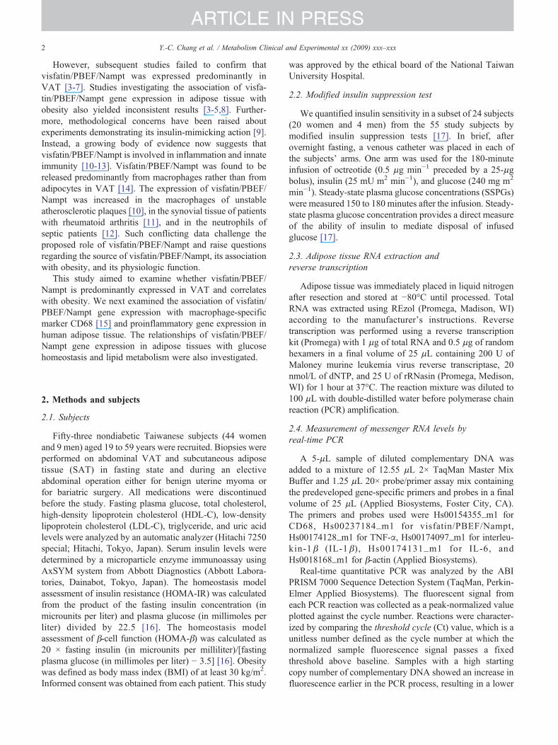
The characteristics of study participants according to theobesity status and sex are summarized in Table 1. There wasno difference in visfatin/PBEF/Nampt mRNA levels be-tween VAT and SAT (P = .25, Fig. 1A). The VAT or SATvisfatin/PBEF/Nampt mRNA levels were not differentbetween nonobese and obese subjects (P = .74 and .84,
Table 1Characteristics of the study participants stratified by obesity status and sex
Characteristics Nonobesesubjects
Obese subjects
Age (y) 47.74 ± 5.96 27.2 ± 5.90 31.02 ± 8.31n, sex (male/female) 15, female 9, male 29, femaleBMI (kg/m2) 23.42 ± 2.69 42.01 ± 10.6 38.89 ± 4.90Waist circumference (cm) 91.5 ± 4.75 121.9 ± 13.16 114.8 ± 13.03Triglyceride (mmol/L) 1.39 ± 1.98 2.65 ± 0.79 1.73 ± 0.88Total cholesterol(mmol/L)
4.47 ± 1.49 5.09 ± 0.78 5.06 ± 0.75
LDL-C (mmol/L) 2.09 ± 0.33 2.77 ± 1.16 3.06 ± 0.69HDL-C (mmol/L) 1.27 ± 0.28 1.03 ± 0.22 1.25 ± 0.34Uric acid (μmol/L) 425.3 ± 197.7 532.0 ± 117.4 414.2 ± 86.11Fasting insulin (pmol/L) 96.32 ± 99.70 236.3 ± 81.87 148.5 ± 102.7Fasting glucose (mmol/L) 5.82 ± 1.44 6.09 ± 1.16 5.50 ± 0.90HOMA-IR 3.27 ± 3.56 7.40 ± 2.89 5.05 ± 3.70HOMA-β 141.1 ± 145.1 387.4 ± 93.73 235.2 ± 164.4SSPG (mmol/L) 25.03 16.69 ± 1.85 15.71 ± 2.66log VAT visfatinmRNA levels
−3.51 ± 2.66 −2.60 ± 1.94 −3.48 ± 2.32
log SAT visfatinmRNA levels
−2.67 ± 4.91 −3.11 ± 1.64 −2.93 ± 2.25
Data are presented as means± SD.
Fig. 1B). These results were similar in the female subgroup(data not shown). We found no difference in visfatin/PBEF/Nampt mRNA levels between male and female subjects ineither VAT (P = .31) or SAT (P = .72) (Fig. 1C).
3.2. Correlations between visfatin/PBEF/Nampt andproinflammatory gene expression in human adipose tissue
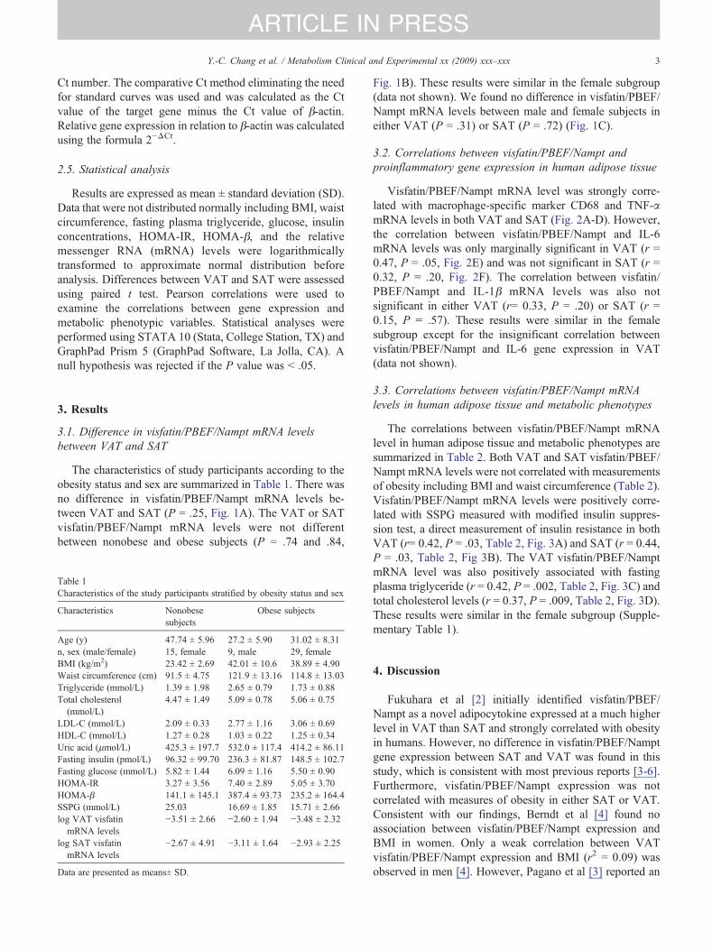
Visfatin/PBEF/Nampt mRNA level was strongly corre-lated with macrophage-specific marker CD68 and TNF-αmRNA levels in both VAT and SAT (Fig. 2A-D). However,the correlation between visfatin/PBEF/Nampt and IL-6mRNA levels was only marginally significant in VAT (r =0.47, P = .05, Fig. 2E) and was not significant in SAT (r =0.32, P = .20, Fig. 2F). The correlation between visfatin/PBEF/Nampt and IL-1β mRNA levels was also notsignificant in either VAT (r= 0.33, P = .20) or SAT (r =0.15, P = .57). These results were similar in the femalesubgroup except for the insignificant correlation betweenvisfatin/PBEF/Nampt and IL-6 gene expression in VAT(data not shown).
3.3. Correlations between visfatin/PBEF/Nampt mRNAlevels in human adipose tissue and metabolic phenotypes
The correlations between visfatin/PBEF/Nampt mRNAlevel in human adipose tissue and metabolic phenotypes aresummarized in Table 2. Both VAT and SAT visfatin/PBEF/Nampt mRNA levels were not correlated with measurementsof obesity including BMI and waist circumference (Table 2).Visfatin/PBEF/Nampt mRNA levels were positively corre-lated with SSPG measured with modified insulin suppres-sion test, a direct measurement of insulin resistance in bothVAT (r= 0.42, P = .03, Table 2, Fig. 3A) and SAT (r = 0.44,P = .03, Table 2, Fig 3B). The VAT visfatin/PBEF/NamptmRNA level was also positively associated with fastingplasma triglyceride (r = 0.42, P = .002, Table 2, Fig. 3C) andtotal cholesterol levels (r = 0.37, P = .009, Table 2, Fig. 3D).These results were similar in the female subgroup (Supple-mentary Table 1).
4. Discussion
Fukuhara et al [2] initially identified visfatin/PBEF/Nampt as a novel adipocytokine expressed at a much higherlevel in VAT than SAT and strongly correlated with obesityin humans. However, no difference in visfatin/PBEF/Namptgene expression between SAT and VAT was found in thisstudy, which is consistent with most previous reports [3-6].Furthermore, visfatin/PBEF/Nampt expression was notcorrelated with measures of obesity in either SAT or VAT.Consistent with our findings, Berndt et al [4] found noassociation between visfatin/PBEF/Nampt expression andBMI in women. Only a weak correlation between VATvisfatin/PBEF/Nampt expression and BMI (r2 = 0.09) wasobserved in men [4]. However, Pagano et al [3] reported an
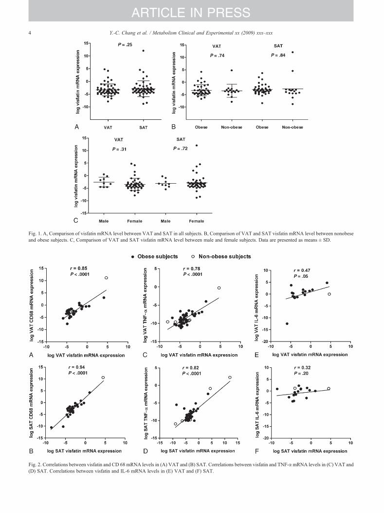
Fig. 1. A, Comparison of visfatin mRNA level between VAT and SAT in all subjects. B, Comparison of VAT and SAT visfatin mRNA level between nonobeseand obese subjects. C, Comparison of VAT and SAT visfatin mRNA level between male and female subjects. Data are presented as means ± SD.
Fig. 2. Correlations between visfatin and CD 68 mRNA levels in (A) VAT and (B) SAT. Correlations between visfatin and TNF-αmRNA levels in (C) VAT and(D) SAT. Correlations between visfatin and IL-6 mRNA levels in (E) VAT and (F) SAT.
4 Y.-C. Chang et al. / Metabolism Clinical and Experimental xx (2009) xxx–xxx
ARTICLE IN PRESS
Table 2Correlation of VAT and SAT visfatin mRNA expression with metabolicphenotypes
Metabolicphenotypes
n VAT visfatinmRNA
SAT visfatinmRNA
R P r P
Age 53 −0.14 .32 0.05 .75BMI 53 0.09 .51 −0.06 .66Waist circumference 36 −0.16 .34 −0.23 .17Triglyceride 51 0.42 .002⁎ 0.007 .96Total cholesterol 51 0.37 .009⁎ 0.18 .22LDL-C 36 0.18 .31 0.13 .46HDL-C 36 −0.31 .07 −0.15 .40Uric acid 38 0.15 .38 0.14 .40Fasting glucose 51 0.21 .14 0.15 .29Fasting insulin 38 0.02 .89 0.32 .05HOMA-IR 36 0.09 .59 0.37 .02⁎
HOMA-β 36 −0.15 .38 0.14 .42SSPG 24 0.42 .03⁎ 0.44 .03⁎
Correlation coefficients (r) and P values are shown. Data are presented asmeans ± SD.
⁎ P b .05.
5Y.-C. Chang et al. / Metabolism Clinical and Experimental xx (2009) xxx–xxx
ARTICLE IN PRESS
inverse relationship between gluteal SAT visfatin/PBEF/Nampt expression and BMI, no correlation betweenabdominal SAT visfatin/PBEF/Nampt expression and BMI,and a positive correlation between abdominal VAT visfatin/PBEF/Nampt expression and BMI. Varma et al [5] reportedthat VAT visfatin/PBEF/Nampt expression was positively
Fig. 3. Correlation between visfatin mRNA levels with SSPG during modified invisfatin mRNA levels with fasting triglyceride levels (C) and total cholesterol lev
associated with BMI, whereas SAT visfatin/PBEF/Namptexpression was negatively associated with BMI. Thesecontroversies might be partly explained by the relativelysmall study populations and heterogeneous sample sources.Taken together, current evidence does not support the notionthat visfatin/PBEF/Nampt is predominantly expressed inVAT. The association of visfatin/PBEF/Nampt gene expres-sion with obesity remains controversial.
A major finding of this study is the strong correlationbetween visfatin/PBEF/Nampt and macrophage-specificCD68 and TNF-α gene expressions in human adipose tissues.Supporting these findings, Varma et al [5] demonstrated thatvisfatin/PBEF/Nampt expression in the stromal vascularfraction was higher than that in the adipocyte fraction ofadipose tissue. A recent work also demonstrated that visfatin/PBEF/Nampt was released predominantly frommacrophagesrather than from adipocytes in VATs [14]. Visfatin/PBEF/Nampt increased inflammatory gene (TNF-α, interleukin-8,IL-6) expression in monocytes [10,13]. Pharmacologicinhibition of visfatin/PBEF/Nampt reduced inflammatorycytokine secretion such as TNF-α, IL-6, and IL-1β both invitro and in vivo [18]. These findings suggest that visfatin/PBEF/Nampt is a proinflammatory marker of residentmacrophages in human adipose tissue.
We found significant association of visfatin/PBEF/NamptmRNA levels in SAT and VAT with systemic insulinresistance. Consistent with our findings, circulating visfatin/
sulin suppression test in VAT (A) and SAT (B). Correlation between VATels (D).
6 Y.-C. Chang et al. / Metabolism Clinical and Experimental xx (2009) xxx–xxx
ARTICLE IN PRESS
PBEF/Nampt levels were found to be elevated in patientswith type 2 diabetes mellitus [19-21], women withpolycystic ovary syndrome [22-24] or gestational diabetes[25], and patients with metabolic syndrome [26]. Insulinresistance has been recognized as a state of low-gradeinflammation associated with macrophage infiltration inadipose tissue [27,28]. Therefore, the positive associations ofvisfatin/PBEF/Nampt with insulin resistance might bemediated through increased adipose tissue inflammation.
Furthermore, we found a positive association betweenVAT visfatin/PBEF/Nampt mRNA level and plasma triglyc-eride and total cholesterol levels. Consistent with this finding,circulating visfatin/PBEF/Nampt concentration was found tobe positively associated with plasma triglyceride level inobese children [29] and young healthy men [30] and withLDL-C levels in patients with metabolic syndrome [26]. Themolecular mechanism underlying these associations iscurrently not known. It is possible that increased visfatin/PBEF/Nampt expression induces adipose tissues inflamma-tion, which increases lipolysis in adipose tissue and FFA fluxto the liver. In the liver, increased FFA flux drives very low-density lipoprotein production, leading to elevated plasmatriglyceride and total cholesterol. The reason why theassociation between visfatin/PBEF/Nampt mRNA level andelevated lipids levels was observed only in VAT is not clear.However, VAT has been demonstrated to be more lipolyti-cally active than SAT; and the FFA released from VAT isdirectly delivered to the portal vein [31]. In epidemiologicstudies, VAT was more strongly correlated with abnormallipoprotein metabolism than SAT [32]. These data provide apossible explanation for the closer association of VATvisfatin/PBEF/Nampt mRNA levels with hyperlipidemia.
This study has several limitations. First, this is a cross-sectional observational study. Further functional studies arerequired to clarify the underlying mechanism. Second, thestudy population was recruited from patients undergoing anoperation for benign uterine myoma or bariatric surgery andwas composed of mainly obese subjects. Thus, the resultcould not be directly extrapolated to the general population.Third, we did not measure the concentrations of plasmavisfatin/PBEF/Nampt or other proinflammatory cytokines;and their relationship with visfatin/PBEF/Nampt geneexpression in adipose tissue and metabolic phenotypescould not be further explored. Lastly, we did not directlymeasure visfatin/PBEF/Nampt gene expression in theadipocellular and stromal vascular fractions of humanadipose tissue. Therefore, direct evidence regarding thesource of visfatin/PBEF/Nampt expression in adipose tissuewas not available.
In summary, this study demonstrated that visfatin/PBEF/Nampt was not predominantly expressed in VAT and wasnot correlated with obesity. Visfatin/PBEF/Nampt geneexpression was strongly and positively correlated with theexpression of CD68, a macrophage-specific marker andproinflammatory gene expression in human adipose tissue.Visfatin/PBEF/Nampt gene expression in adipose tissue was
associated with increased systemic insulin resistance andelevated plasma triglyceride and total cholesterol levels.These findings suggest visfatin/PBEF/Nampt as a proin-flammatory marker of adipose tissue associated with insulinresistance and hyperlipidemia.
Acknowledgment
We thank all participants in this study. This work wassupported by grants (NSC 93-2752-B-002-009-PAE, 94-2752-B-002-008-PAE, 95-2752-B-002-008-PAE) from theNational Science Council of the ROC and a grant from theNational Taiwan University Hospital Yunlin branch, Taiwan(NTHYL 98 X008).
Appendix A. Supplementary data
Supplementary data associatedwith this article canbe found,in the online version, at doi:10.1016/j.metabol.2009.07.011.
References
[1] Frayn KN, Karpe F, Fielding BA, Macdonald IA, Coppack SW.Integrative physiology of human adipose tissue. Int J Obes 2005;27:875-88.
[2] Fukuhara A, Matsuda M, Nishizawa M, Segawa K, Tanaka M,Kishimoto K, et al. Visfatin: a protein secreted by visceral fat thatmimics the effects of insulin. Science 2005;307:426-30.
[3] Pagano C, Pilon C, Olivieri M, Mason P, Fabris R, Serra R, et al.Reduced plasma visfatin/pre B-cell colony-enhancing factor in obesityis not related to insulin resistance in humans. J Clin Endocrinol Metab2006;91:3165-70.
[4] Berndt J, Klöting N, Kralisch S, Kovacs P, Fasshauer M, SchönMR, etal. Plasma visfatin concentrations and fat-depot specific mRNAexpression in humans. Diabetes 2005;54:2911-6.
[5] Varma V, Yao-Borengasser A, Rasouli N, Bodles AM, Phanavanh B,Lee MJ, et al. Human visfatin expression: relationship to insulinsensitivity, intramyocellular lipids, and inflammation. J Clin Endocri-nol Metab 2007;92:666-72.
[6] Böttcher Y, Teupser D, Enigk B, Berndt J, Klöting N, Schön MR.Genetic variation in the visfatin gene (PBEF1) and its relation toglucose metabolism and fat-depot–specific messenger ribonucleic acidexpression in humans. J Clin Endocrinol Metab 2006;91:2725-31.
[7] Fain JN, Sacks HS, Buehrer B, Bahouth SW, Garrett E, Wolf RY, et al.Identification of omentin mRNA in human epicardial adipose tissue:comparison to omentin in subcutaneous, internal mammary arteryperiadventitial and visceral abdominal depots. Int J Obes 2008;32:810.
[8] Hammarstedt A, Pihlajamäki J, Rotter Sopasakis V, Gogg S, JanssonPA, Laakso M. Visfatin is an adipokine, but it is not regulated bythiazolidinediones. J Clin Endocrinol Metab 2006;91:1181-4.
[9] Fukuhara A, Matsuda M, Nishizawa M, Segawa K, Tanaka M,Kishimoto K, et al. Retraction of “Visfatin: a protein secreted byvisceral fat that mimics the effects of insulin”. Science 2007;318:565.
[10] Dahl TB, Yndestad A, Skjelland M, Øie E, Dahl A, Michelsen A, et al.Increased expression of visfatin in macrophages of human unstablecarotid and coronary atherosclerosis: possible role in inflammation andplaque destabilization. Circulation 2007;115:972-80.
[11] Brentano F, Schorr O, Ospelt C, Stanczyk J, Gay RE, Gay S, et al. Pre–B cell colony-enhancing factor/visfatin, a new marker of inflammationin rheumatoid arthritis with proinflammatory and matrix-degradingactivities. Arthritis Rheum 2007;56:2829-39.
7Y.-C. Chang et al. / Metabolism Clinical and Experimental xx (2009) xxx–xxx
ARTICLE IN PRESS
[12] Jia SH, Li Y, Parodo J, Kapus A, Fan L, Rotstein OD, et al. Pre–Bcell colony enhancing factor inhibits neutrophil apoptosis inexperimental inflammation and clinical sepsis. J Clin Invest 2004;113:1318-27.
[13] Moschen AR, Kaser A, Enrich B, Mosheimer B, Theurl M,Niederegger H. Visfatin, an adipocytokine with proinflammatory andimmunomodulating properties. J Immunol 2007;178:1748-58.
[14] Curat CA, Wegner V, Sengenès C, Miranville A, Tonus C, Busse R.Macrophages in human visceral adipose tissue: increased accumulationin obesity and a source of resistin and visfatin. Diabetologia 2006;49:744-7.
[15] Greaves DR, Gordon S. Macrophage-specific gene expression: currentparadigms and future challenges. Int J Hematol 2002;76:6-15.
[16] Matthews DR, Hosker JP, Rudenski AS, Naylor BA, Treacher DF,Turner RC. Homeostasis model assessment: insulin resistance and β-cell function from fasting plasma glucose and insulin concentrations inman. Diabetologia 1985;28:412-9.
[17] Pei D, Jones CN, Bhargava R, Chen YD, Reaven GM. Evaluation ofoctreotide to assess insulin mediated glucose disposal by the insulinsuppression test. Diabetologia 1994;37:843-5.
[18] Busso N, Karababa M, Nobile M, Rolaz A, Van Gool F, Galli M, et al.Pharmacological inhibition of nicotinamide phosphoribosyltransfer-ase/visfatin enzymatic activity identifies a new inflammatory pathwaylinked to NAD. PLoS ONE 2008;3:e2267.
[19] Sandeep S, Velmurugan K, Deepa R, Mohan V. Serum visfatin inrelation to visceral fat, obesity, and type 2 diabetes mellitus in AsianIndians. Metabolism 2007;56:565-70.
[20] Chen MP, Chung FM, Chang DM, Tsai JC, Huang HF, Shin SJ, et al.Elevated plasma level of visfatin/pre–B cell colony-enhancing factorin patients with type 2 diabetes mellitus. J Clin Endocrinol Metab2006;91:295-9.
[21] Retnakaran R, Youn BS, Liu Y, Hanley AJ, Lee NS, Park JW, et al.Correlation of circulating full-length visfatin (PBEF/Nampt) withmetabolic parameters in subjects with and without diabetes: a cross-sectional study. Clin Endocrinol 2008;69:885-93.
[22] Tan BK, Chen J, Digby JE, Keay SD, Kennedy CR, Randeva HS.Increased visfatin mRNA and protein levels in adipose tissue andadipocytes in women with polycystic ovary syndrome: parallelincrease in plasma visfatin. J Clin Endocrinol Metab 2006;91:5022-8.
[23] Kowalska I, Straczkowski M, Nikolajuk A, Adamska A, Karczewska-Kupczewska M, Otziomek E, et al. Serum visfatin in relation to insulinresistance and markers of hyperandrogenism in lean and obese womenwith polycystic ovary syndrome. Hum Reprod 2007;22:1824-9.
[24] Chan TF, Chen YL, Chen HH, Lee CH, Jong SB, Tsai EM. Increasedplasma visfatin concentrations in women with polycystic ovarysyndrome. Fertil Steril 2007;88:401-5.
[25] Krzyzanowska K, Krugluger W, Mittermayer F, Rahman R, Haider D,Shnawa N, et al. Increased visfatin concentrations in women withgestational diabetes mellitus. Clin Sci 2006;110:605-9.
[26] Zhong M, Tan HW, Gong HP, Wang SF, Zhang Y, Zhang W.Increased serum visfatin in patients with metabolic syndrome andcarotid atherosclerosis. Clin Endocrinol 2008;69:878-84.
[27] Weisberg SP, McCann D, Desai M, Rosenbaum M, Leibel RL,Ferrante Jr AW. Obesity is associated with macrophage accumulationin adipose tissue. J Clin Invest 2008;112:1796-808.
[28] Gustafson B, Hammarstedt A, Andersson CX, Smith U. Inflamedadipose tissue: a culprit underlying the metabolic syndrome andatherosclerosis. Arterioscler Thromb Vasc Biol 2007;27:2276-83.
[29] Araki S, Dobashi K, Kubo K, Kawagoe R, Yamamoto Y, Kawada Y, etal. Plasma visfatin concentration as a surrogate marker for visceral fataccumulation in obese children. Obesity 2008;16:384-8.
[30] Sun G, Bishop J, Khalili S, Vasdev S, Gill V, Pace D, et al. Serumvisfatin concentrations are positively correlated with serum triacylgly-cerols and down-regulated by overfeeding in healthy young men. Am JClin Nutr 2007;85:399-404.
[31] Wajchernberg BL. Subcutaneous and visceral adipose tissue: theirrelation to the metabolic syndrome. Endocr Rev 2000;21:697-738.
[32] Porter SA, Massaro JM, Hoffmann U, Vasan RS, O'Donnell CJ, FoxCS. Subcutaneous abdominal adipose tissue: a protective fat depot?Diabetes Care 2009 [Epub ahead of print].


