Embed Size (px)
Citation preview
Endocrine pharmacology
Anti-diabetic properties of a non-conventional radicalscavenger, as compared to pioglitazone and exendin-4,in streptozotocin-nicotinamide diabetic mice
Michela Novelli a, Donatella Canistro b, Manuela Martano c, Niccola Funel d,Andrea Sapone b, Simone Melega b, Matilde Masini a, Vincenzo De Tata a, Anna Pippa e,Cecilia Vecoli e, Daniela Campani d, Rocco De Siena c, Antonio Soleti c, Moreno Paolini b,Pellegrino Masiello a,nQ1
a Department of Translational Research and New Technologies in Medicine and Surgery, University of Pisa, Via Roma 55, 56126 Pisa, Italyb Department of Pharmacy and Biotechnology, Alma Mater Studiorum, University of Bologna, Via Irnerio 48, 40126 Bologna, Italyc Medestea Research, Via Cernaia 31, 10121 Torino, Italyd Department of Surgical, Medical, Molecular, and Critical Area Pathology, University of Pisa, Via Savi 10, 56126 Pisa, Italye Institute of Clinical Physiology, CNR, Via Moruzzi 1, 56124 Pisa, Italy
a r t i c l e i n f o
Article history:Received 16 October 2013Received in revised form22 January 2014Accepted 24 January 2014
Chemical compounds studied in this article:Doxorubicin hydrochloride (PubChem CID:443939)
Keywords:Type 2 diabetes mellitusOxidative stressRadical scavengersBeta-cell massPDX-1PioglitazoneExendin-4Q2
a b s t r a c t
We previously showed that the innovative radical scavenger bis(1-hydroxy-2,2,6,6-tetramethyl-4-piperidinyl)-decandioate (IAC) improves metabolic dysfunctions in a diabetic mouse model. Here, we compared the in vivoeffects of IAC with those of the anti-diabetic drugs pioglitazone (PIO) and exendin-4 (EX-4). Diabetes wasinduced in C57Bl/6J mice by streptozotocin and nicotinamide administration. Paralleled by healthy controls,diabetic animals (D) were randomly assigned to four groups and treated daily for 7 consecutive weeks:Dþsaline, ip; Dþ IAC 30 mg/kg b.w., ip; DþPIO 10 mg/kg b.w. per os; and DþEX-4, 50 μg/kg b.w., ip. Ourresults show that IAC reduced basal hyperglycemia and improved glucose tolerance better than PIO or EX-4.Interestingly, in the heart of diabetic mice, IAC treatment normalized the increased levels of GSSG/GSH ratioand thiobarbituric acid reactive substances, indexes of oxidative stress and damage, while PIO and EX-4 wereless effective. As supported by immunohistochemical data, IACmarkedly prevented diabetic islet β-cell reduceddensity, differently from PIO and EX-4 that had only a moderate effect. Interestingly, in diabetic animals, IACtreatment enhanced the activity of pancreatic-duodenal homeobox 1 (PDX-1), an oxidative stress-sensitivetranscription factor essential for maintenance of β-cell function, as evaluated by quantification of its nuclearimmunostaining, whereas PIO or EX-4 treatments did not. Altogether, these observations support theimprovement of the general redox balance and β-cell function induced by IAC treatment in streptozotocin-nicotinamide diabetic mice. Furthermore, in this model, the correction of diabetic alterations was betterobtained by treatment with the radical scavenger IAC than with pioglitazone or exendin-4.
& 2014 Published by Elsevier B.V.
1. Introduction
Type 2 diabetes mellitus (T2D) is characterized by peripheralinsulin resistance associated with inadequate insulin secretion, due
to the progressive inability of pancreatic β-cells to adapt to the highmetabolic demand, finally leading to hyperglycemia. Chronic hyper-glycemia induces deleterious effects on pancreatic islets, a phenom-enon known as glucotoxicity. This includes loss of β-cell differen-tiation, alteration of the stimulus-secretion coupling, increase of β-cellapoptosis, and changes in gene expression (Gupta et al., 2012; Laybuttet al., 2002). These events create a vicious circle that contributes to theworsening of the disease over time (Bensellam et al., 2012). Recentevidence confirms that oxidative stress plays a central role in both thepathogenesis and the progression of T2D (Goodarzi et al., 2010 Q3; Styskalet al., 2012). The pro-oxidant condition in T2D may result from theoverproduction of intracellular reactive oxygen species (ROS), derivedfrom various sources, such as the glycation reaction, the enhancedglycolytic flux and pyruvate feeding to the tricarboxylic acid cycle
123456789
101112131415161718192021222324252627282930313233343536373839404142434445464748495051525354555657585960616263646566
676869707172737475767778798081828384
Contents lists available at ScienceDirect
journal homepage: www.elsevier.com/locate/ejphar
European Journal of Pharmacology
http://dx.doi.org/10.1016/j.ejphar.2014.01.0710014-2999 & 2014 Published by Elsevier B.V.
Abbreviations: T2D, Type 2 diabetes mellitus; ROS, Reactive oxygen species; IAC,bis(1-hydroxy-2,2,6,6-tetramethyl-4-piperidinyl)-decandioate; STZ, Streptozotocin;NA, Nicotinamide; PPARγ, Peroxisome-proliferator activated receptor gamma;GLP-1, Glucagon-like peptide 1; PDX-1, Pancreatic and duodenal homeobox 1;PIO, Pioglitazone; EX-4, Exendin-4; RIA, Radioimmunoassay; GSH, Glutathione;GSSG, Glutathione disulfide
n Correspondence to: Dip. di Ricerca Traslazionale e delle Nuove Tecnologie inMedicina e Chirurgia Via Roma, 55 -Scuola Medica-56126 PISA, Italy.Tel.: þ39 050 2218571.
Please cite this article as: Novelli, M., et al., Anti-diabetic properties of a non-conventional radical scavenger, as compared topioglitazone and exendin-4, in streptozotocin-nicotinamide.... Eur J Pharmacol (2014), http://dx.doi.org/10.1016/j.ejphar.2014.01.071i
European Journal of Pharmacology ∎ (∎∎∎∎) ∎∎∎–∎∎∎
(Rota et al., 2004; Kaneto et al., 2010), the accelerated rate ofalternative metabolic pathways (e.g., polyols and hexosamine)(Robertson, 2006), and the mitochondrial fragmentation (Men et al.,2009). ROS excess can cause protein oxidation, membrane lipidperoxidation and DNA damage (Styskal et al., 2012; Giacco andBrownlee, 2010), as well as impairment of fundamental β-cell genestranscription (Kaneto et al., 2007a). Moreover, ROS may trigger stress-sensitive signaling pathways, such as transcription of NF-kB, that inturn mediates the expression of pro-inflammatory factors (Reuteret al., 2010). It is noteworthy that reduced levels of antioxidantdefenses make β-cells particularly prone to severe injury due to pro-oxidant conditions (Karunakara and Park, 2013). Thus, an attenuationof oxidative stress through a pharmacological approach, based oneffective antioxidant scavenging agents, able to counteract the viciouscircle of inflammatory responses and tissue damage, appears highlyadvisable (Noh et al., 2011).
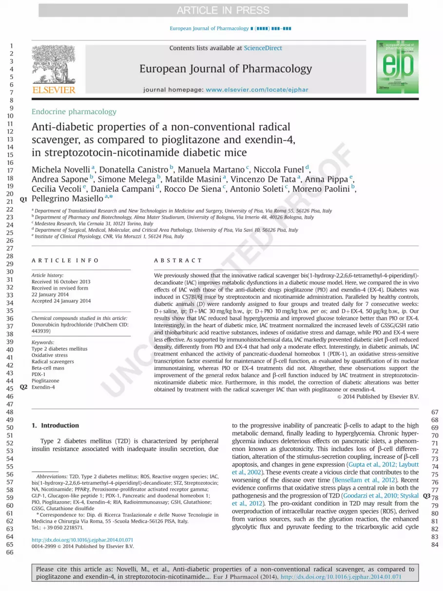
In our laboratory, we synthesized an innovative cyclic hydro-xylamine derivative, namely bis(1-hydroxy-2,2,6,6-tetramethyl-4-piperidinyl)-decandioate (IAC, Fig. 1), capable of rapidly reactingwith most free radicals of biological interest, favored by itsdistinctive cell membrane permeability (Valgimigli et al., 2001).We previously showed that in the mouse model of T2D obtainedby the combined administration of streptozotocin (STZ) andnicotinamide (NA) (Masiello et al., 2006; Nakamura et al., 2006),IAC administration reduced hyperglycemia, improved glucosetolerance and partially preserved pancreatic insulin content(Novelli et al., 2007). These benefits, likely dependent on preven-tion of oxidative stress-induced progressive β-cell damage, per-sisted over time, long after suspension of IAC treatment (Novelliet al., 2010).
On the basis of such observations, the present work aimed atdirectly comparing, in the STZ-NA mouse diabetic model, themetabolic effects of IAC with those of two reference anti-diabeticcompounds acting by different mechanisms: pioglitazone (PIO)and exendin-4 (EX-4). Additionally, this study intended to providefurther insights into the mechanisms underlying the protectiveaction of IAC against diabetes-induced oxidative damage in tissues,with particular regard to the reactivation of pancreatic andduodenal homeobox 1 (PDX-1), an oxidative stress-sensitive tran-scription factor, required for maintenance of differentiated β-cellfunction (Fu et al., 2013).
2. Materials and methods
2.1. Animals
Experiments were performed in male C57Bl/6J mice, of the ageof 8–9 weeks, weighing 22–24 g, purchased from Harlan Labora-tories s.r.l (S. Pietro al Natisone, Udine, Italy). Mice were kept at aconstant temperature of 24–25 1C and were subjected to a con-trolled 12 h light-dark cycle; they had free access to water anddiet. The experimental protocol followed the Principles of Labora-tory Animal Care (EU Directive 2010/63/EU ) and was approved bythe Ethical Committee of the University of Pisa, Italy.
2.2. Induction of diabetes
Nicotinamide (210 mg/kg b.w., Sigma, Saint Louis, MO, USA),dissolved in saline, was injected intraperitoneally (i.p.) 20 minbefore STZ administration (Sigma, 180 mg/kg b.w., i.p.), and dis-solved in buffer citrate (pH 4.5) immediately before use. Controlsreceived the vehicles of both substances.
2.3. Pharmacological treatments
Glycemia was measured twice after STZ-NA treatment (at the5th and 10th day) to assess stability of glycemic values, and on the11th day, diabetic mice were homogeneously distributed into foursubgroups of 7 animals each. Pharmacological treatment wasrandomly assigned to the four diabetic groups, according to thefollowing scheme: D, STZ-NA diabetic mice, daily treated withsaline without pharmacological treatment, and fed with pelletfood; Dþ IAC, diabetic mice treated i.p. with IAC at 30 mg/kg b.w.per day dissolved in saline, and fed with pellet food; DþPIO,diabetic mice treated per os with pioglitazone at 10 mg/kg b.w. perday mixed with powder food; and DþEX-4, diabetic mice treated i.p.with exendin-4 at 50 μg/kg b.w. per day, and fed with pellet food.Non-diabetic healthy mice, receiving daily saline i.p., were dividedinto two sub-groups (n¼7 each): C, PELLET, fed with food in pelletand C, POWDER, fed with powder food, in order to check thatfood intake would not change between the two subgroups (nochange was indeed observed, so that data obtained in these twocontrol groups could be pooled). During the experimental period,the animals’ food intake and body weight were monitored oncea week.
2.4. Assays in whole blood or plasma
Blood sampling was performed every week in conscious micethat had food removed 3 h before. Whole-blood glucose levelswere determined in 5 μl of blood obtained from the tail vein, usingOneTouch Ultra glucometer apparatus (Life Scan, Inc., Milpitas, CA,USA). After 4 and 6 weeks of anti-diabetic drugs treatment, somedrops of blood (80–100 μl) were collected by the same method insmall tubes containing 2 μl of 250 mM EDTA. Plasma was sepa-rated by centrifugation and stored at �20 1C for subsequentinsulin determination by radioimmunoassay (RIA), according toHerbert et al. (1965), using anti-insulin antibody and 125I-labeledinsulin obtained from Linco (Linco Research, INC. St. Charles,MO, USA).
2.5. Intraperitoneal glucose and insulin tolerance test
After 5 weeks of pharmacological treatment, glucose (1.5 g/kg b.w. as 16.5% solution) was given i.p. to 3-h fasting consciousanimals, randomly chosen from each group. Drops of blood weresequentially collected from the tail vein at 0, 15, 60, 90, and120 min after the glucose injection, and immediately used forglucose determination by a glucometer. Some blood samples (at 0,15 and 60 min) were also taken in EDTA-treated tubes, centrifugedat 4 1C, and plasma was stored at �20 1C for subsequent insulinmeasurement. The daily pharmacological doses were administeredat the end of the test. After 6 weeks of treatment, human insulin(0.75 U/kg b.w., Humulin R, Eli Lilly, Indianapolis, IN, USA) wasadministered to conscious animals by an intraperitoneal route.Blood glucose was measured by a glucometer in drops of bloodtaken from the tail vein at 0, 30, 60 and 120 min after the insulininjection.
123456789
101112131415161718192021222324252627282930313233343536373839404142434445464748495051525354555657585960616263646566
676869707172737475767778798081828384858687888990919293949596979899
100101102103104105106107108109110111112113114115116117118119120121122123124125126127128129130131132
Fig. 1. Chemical structure of bis(1-hydroxy-2,2,6,6-tetramethyl-4-piperidinyl)decandioate (IAC), showing the two opposite cyclic hydroxylamine moieties linkedby a long aliphatic chain.
M. Novelli et al. / European Journal of Pharmacology ∎ (∎∎∎∎) ∎∎∎–∎∎∎2
Please cite this article as: Novelli, M., et al., Anti-diabetic properties of a non-conventional radical scavenger, as compared topioglitazone and exendin-4, in streptozotocin-nicotinamide.... Eur J Pharmacol (2014), http://dx.doi.org/10.1016/j.ejphar.2014.01.071i
2.6. Sacrifice
After 7 weeks of treatment, 3-h fasting animals were anesthe-tized using pentobarbital (50 mg/kg b.w., i.p.); the pancreas wasrapidly removed, dissected free of fat and processed for immuno-histochemical and electron microscope analyses. Liver, heart andkidney were also removed, weighed and frozen at �80 1C.
2.7. GSH and GSSG determination
Total GSH was analyzed by the enzymatic method of Tietze(1969), as modified by Baker et al. (1990) to adapt it to a microtiterplate reader. Glutathione disulfide (GSSG) was determined by thevinylpiridine method (Baker et al., 1990; Griffith, 1980).
2.8. Determination of thiobarbituric acid reactive substances(TBARS)
Frozen hearts and liver fragments were pulverized under liquidnitrogen and homogenized in an acid solution (0.6 M perchloric-aceticacid). One volume of supernatant was mixed with 1 volume of 0.66%(wt/vol) thiobarbituric acid, and the mixture was boiled for 15 min.The mixture was read at 535 nm, and the TBARS were calculated byusing a molar absorption coefficient of 1.56�10�5 M�1/cm as pre-viously described (L’Abbate et al., 2007).
2.9. Immunohistochemistry
2.9.1. Tissue preparationFor histological and immunohistochemical studies, a suitable
fragment of pancreas taken from the pancreatic tail was fixed in10% buffered formalin and routinely processed to obtain aparaffin-embedded tissue block.
2.9.2. Immunohistochemical stainingParaffin section (2-μm thickness) were mounted on treated
slides (two to three sections per slide) and dried in an oven at56 1C for 20 min. After progressive hydration, the sections wereincubated with a guinea pig either anti-insulin or anti-PDX-1polyclonal antibody (obtained from Zymed Laboratories, SanFrancisco, CA, USA and Abcam, Cambridge, UK, respectively).Immunoreactivity was detected by the kit Histomouse (ZymedLaboratories), containing diaminobenzidine chromogen: insulinwas visualized as a light brown cytoplasmic staining, while PDX-1 intracellular localization appeared dark brown. Negative controlswere incubated in phosphate-buffered saline without primaryantibody. Finally, the nuclei were stained in blue by haematoxylin,and the sections were submitted to morphometric analysis.
2.9.3. Morphometric analysisMorphometric analysis was performed by a BX-51 Olympus
microscope connected to a computer by a color CCD camera. TheanalySISB software (Olympus) was used to acquire images atdifferent magnifications. The colorimetric property of the systemwas able to recognize and quantify the total section area, the totalislet surface after selection of the perimeter of each islet present inthe specimen, the insulin-positive area (light brown color), thePDX-1-positive nuclei (dark brown color), and the PDX-1-negativenuclei (bluish color). The set-up of colors was done using thestaining of the corresponding control groups.
2.10. Statistical analysis
All data are presented as means7S.E.M. Statistical significancewas evaluated by analysis of variance (ANOVA) followed by Fisher
post-hoc test for multiple comparisons. A Po0.05, at least, wasconsidered as significant.
3. Results
3.1. Food intake and body weight
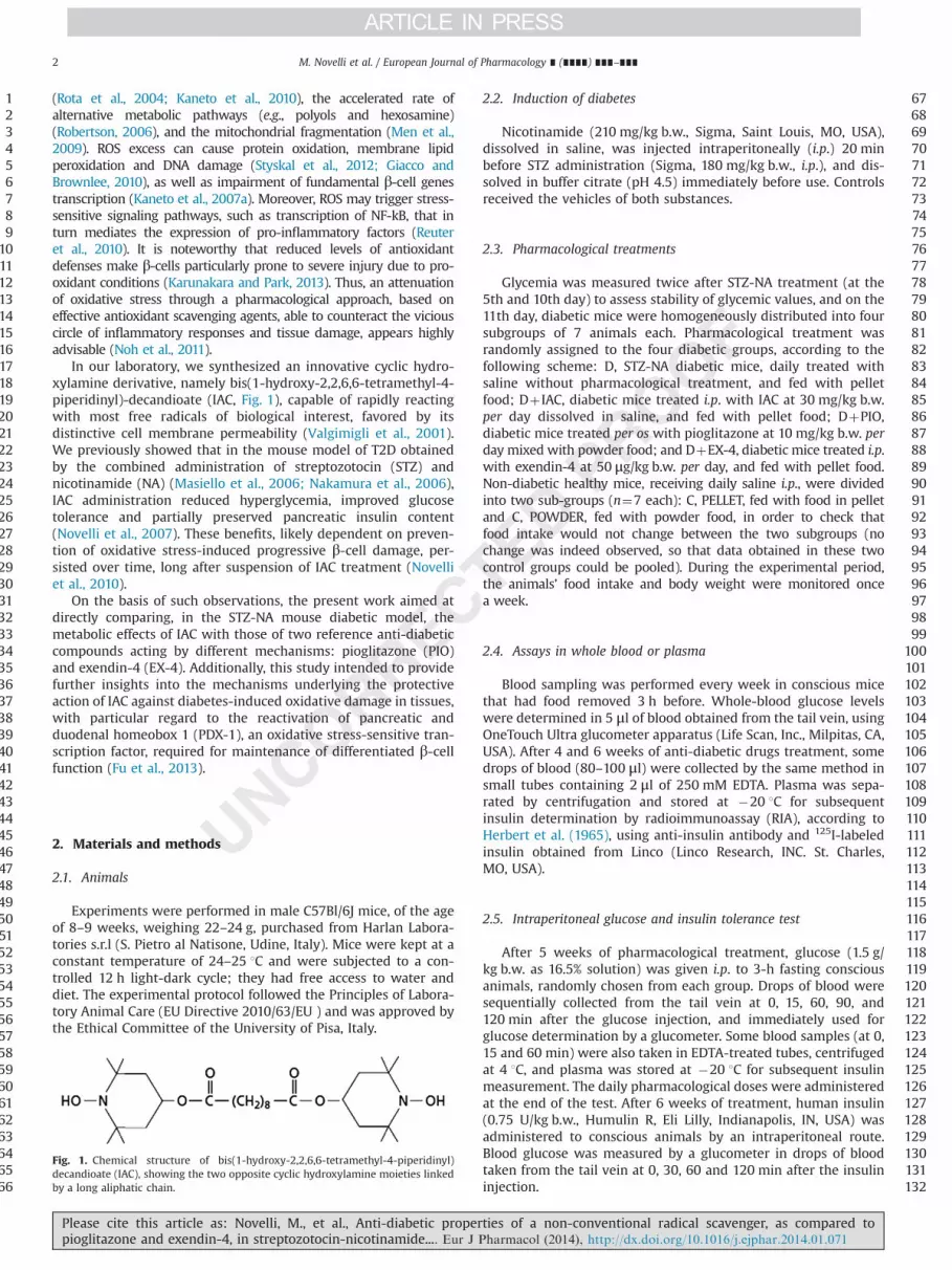
No change in either food intake or body weight occurred inhealthy controls fed with pellet food or powder food. At the end ofthe pharmacological treatment, untreated diabetic mice showed asignificant reduction in body weight with respect to controls,despite a similar food intake (Fig. 2). In IAC- and EX-4 treateddiabetic mice, food intake and body weight were not significantlyimproved, while in pioglitazone-treated diabetic animals bodyweight approached that of healthy controls, likely as a conse-quence of the significant increase in food intake observed in theserodents (Fig. 2).
3.2. Time course of basal blood glucose and insulin levels
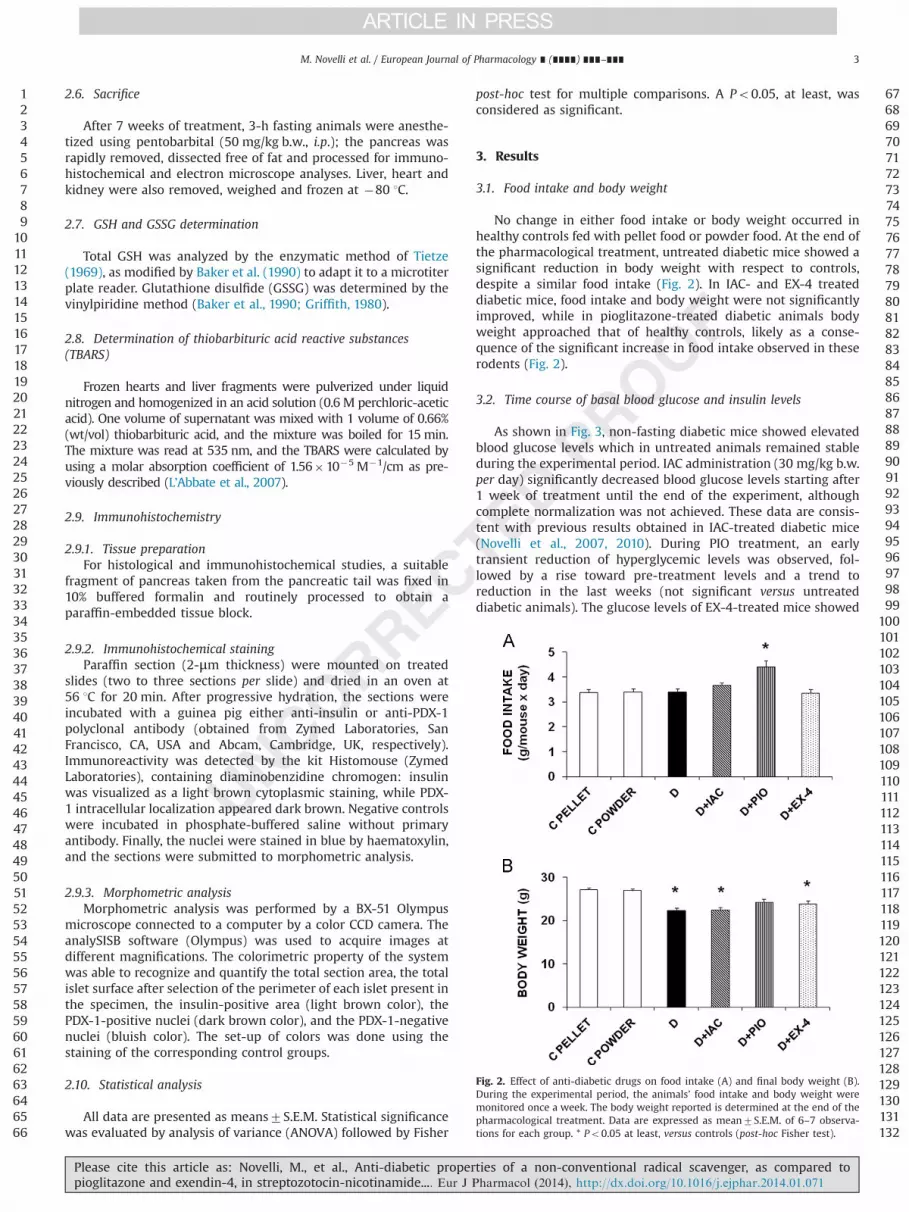
As shown in Fig. 3, non-fasting diabetic mice showed elevatedblood glucose levels which in untreated animals remained stableduring the experimental period. IAC administration (30 mg/kg b.w.per day) significantly decreased blood glucose levels starting after1 week of treatment until the end of the experiment, althoughcomplete normalization was not achieved. These data are consis-tent with previous results obtained in IAC-treated diabetic mice(Novelli et al., 2007, 2010). During PIO treatment, an earlytransient reduction of hyperglycemic levels was observed, fol-lowed by a rise toward pre-treatment levels and a trend toreduction in the last weeks (not significant versus untreateddiabetic animals). The glucose levels of EX-4-treated mice showed
123456789
101112131415161718192021222324252627282930313233343536373839404142434445464748495051525354555657585960616263646566
676869707172737475767778798081828384858687888990919293949596979899
100101102103104105106107108109110111112113114115116117118119120121122123124125126127128129130131132
Fig. 2. Effect of anti-diabetic drugs on food intake (A) and final body weight (B).During the experimental period, the animals’ food intake and body weight weremonitored once a week. The body weight reported is determined at the end of thepharmacological treatment. Data are expressed as mean7S.E.M. of 6–7 observa-tions for each group. * Po0.05 at least, versus controls (post-hoc Fisher test).
M. Novelli et al. / European Journal of Pharmacology ∎ (∎∎∎∎) ∎∎∎–∎∎∎ 3
Please cite this article as: Novelli, M., et al., Anti-diabetic properties of a non-conventional radical scavenger, as compared topioglitazone and exendin-4, in streptozotocin-nicotinamide.... Eur J Pharmacol (2014), http://dx.doi.org/10.1016/j.ejphar.2014.01.071i
some reduction after four weeks of pharmacological treatmentthat persisted in the last weeks but did not achieve statisticalsignificance versus the untreated diabetic group.
As measured at 4 and 6 weeks after pharmacological treatment,basal plasma insulin levels, which were significantly decreased inuntreated diabetic mice (approximately 50% reduction versuscontrols), were not normalized by any compound, although atrend toward control values could be observed during IAC andEX-4 treatments (at 4 weeks, plasma insulin concentrations wereC, 0.9370.07; D, 0.5070.04; Dþ IAC, 0.6570.05; DþPIO 0.5670.08; and DþEX, 0.6970.06 ng/ml (n¼8).
3.3. Glucose and insulin tolerance test
Fig. 4 shows the results of the intraperitoneal glucose tolerancetest, performed in diabetic mice 5 weeks after the pharmacologicaltreatment. The post-loading blood glucose profile showed that thesevere glucose intolerance observed in untreated diabetic animalswas significantly improved in the diabetic groups treated with IACand EX-4, and much less in the group treated with PIO, as confirmedby the area under blood glucose curve (AUC) (Fig. 4A and corre-sponding inset). The post-loading rise in circulating insulin over basalvalues, that was lacking in untreated diabetic mice, occurred to avariable extent in all pharmacologically treated groups (the best wasin the IAC group that achieved statistical significance versus theuntreated diabetic group). Nevertheless, the rise in circulating insulinobserved in the treated diabetic groups remained much lower thanin healthy controls (Fig. 4B and corresponding inset).
No significant change in insulin sensitivity was observed in anyexperimental group, including the untreated diabetic group, dur-ing an i.p. insulin tolerance test performed after 6 weeks ofpharmacological treatment (data not shown).
3.4. Effects of pharmacological treatments on heart oxidative stress
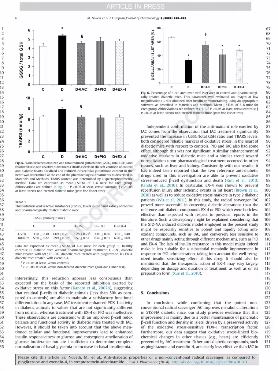
As the tissue GSSG/total GSH ratio and thiobarbituric acid reactivesubstances (TBARS) are considered reliable markers of oxidative stressand injury, wewere interested to verify whether such indices could bemodulated in the heart by our experimental treatments. The GSSG/total GSH ratio and TBARS were indeed increased in the heart ofdiabetic mice as compared to controls (Fig. 5), and both were
significantly modified by the 7-week IAC treatment, as their valuesreturned to normal. PIO and EX-4 also exerted an antioxidant actionthat approached, but did not achieve, statistical significance withrespect to the untreated diabetic group. This pattern of enhancedTBARS and a trend toward normalization upon pharmacologicaltreatment was also observed in the liver and kidney of diabeticanimals (Table 1).
3.5. Effects of pharmacological treatments on pancreatic β-cells, asassessed by immunohistochemical analysis
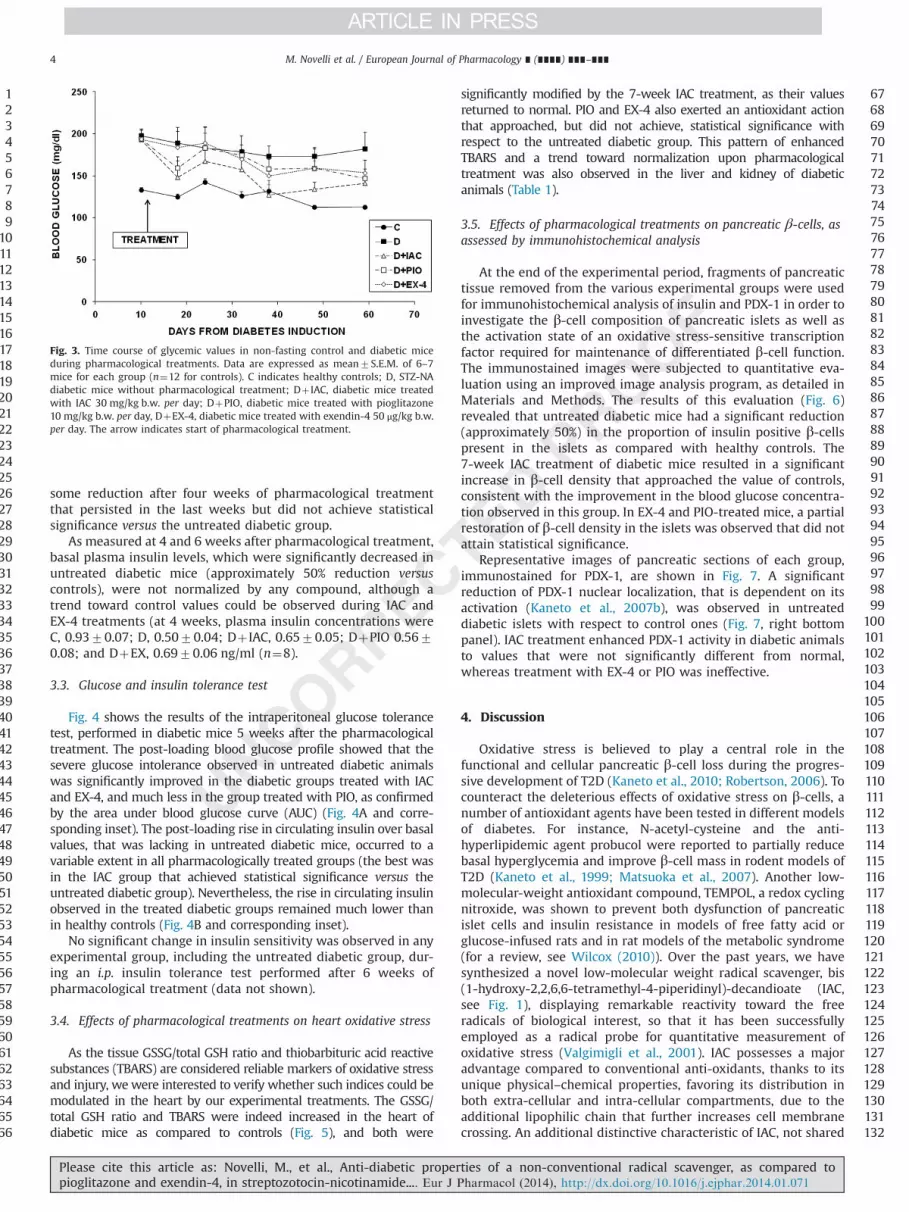
At the end of the experimental period, fragments of pancreatictissue removed from the various experimental groups were usedfor immunohistochemical analysis of insulin and PDX-1 in order toinvestigate the β-cell composition of pancreatic islets as well asthe activation state of an oxidative stress-sensitive transcriptionfactor required for maintenance of differentiated β-cell function.The immunostained images were subjected to quantitative eva-luation using an improved image analysis program, as detailed inMaterials and Methods. The results of this evaluation (Fig. 6)revealed that untreated diabetic mice had a significant reduction(approximately 50%) in the proportion of insulin positive β-cellspresent in the islets as compared with healthy controls. The7-week IAC treatment of diabetic mice resulted in a significantincrease in β-cell density that approached the value of controls,consistent with the improvement in the blood glucose concentra-tion observed in this group. In EX-4 and PIO-treated mice, a partialrestoration of β-cell density in the islets was observed that did notattain statistical significance.
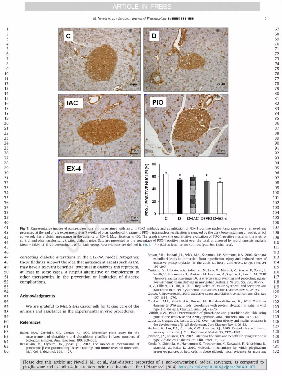
Representative images of pancreatic sections of each group,immunostained for PDX-1, are shown in Fig. 7. A significantreduction of PDX-1 nuclear localization, that is dependent on itsactivation (Kaneto et al., 2007b), was observed in untreateddiabetic islets with respect to control ones (Fig. 7, right bottompanel). IAC treatment enhanced PDX-1 activity in diabetic animalsto values that were not significantly different from normal,whereas treatment with EX-4 or PIO was ineffective.
4. Discussion
Oxidative stress is believed to play a central role in thefunctional and cellular pancreatic β-cell loss during the progres-sive development of T2D (Kaneto et al., 2010; Robertson, 2006). Tocounteract the deleterious effects of oxidative stress on β-cells, anumber of antioxidant agents have been tested in different modelsof diabetes. For instance, N-acetyl-cysteine and the anti-hyperlipidemic agent probucol were reported to partially reducebasal hyperglycemia and improve β-cell mass in rodent models ofT2D (Kaneto et al., 1999; Matsuoka et al., 2007). Another low-molecular-weight antioxidant compound, TEMPOL, a redox cyclingnitroxide, was shown to prevent both dysfunction of pancreaticislet cells and insulin resistance in models of free fatty acid orglucose-infused rats and in rat models of the metabolic syndrome(for a review, see Wilcox (2010)). Over the past years, we havesynthesized a novel low-molecular weight radical scavenger, bis(1-hydroxy-2,2,6,6-tetramethyl-4-piperidinyl)-decandioate (IAC,see Fig. 1), displaying remarkable reactivity toward the freeradicals of biological interest, so that it has been successfullyemployed as a radical probe for quantitative measurement ofoxidative stress (Valgimigli et al., 2001). IAC possesses a majoradvantage compared to conventional anti-oxidants, thanks to itsunique physical–chemical properties, favoring its distribution inboth extra-cellular and intra-cellular compartments, due to theadditional lipophilic chain that further increases cell membranecrossing. An additional distinctive characteristic of IAC, not shared
123456789
101112131415161718192021222324252627282930313233343536373839404142434445464748495051525354555657585960616263646566
676869707172737475767778798081828384858687888990919293949596979899
100101102103104105106107108109110111112113114115116117118119120121122123124125126127128129130131132
Fig. 3. Time course of glycemic values in non-fasting control and diabetic miceduring pharmacological treatments. Data are expressed as mean7S.E.M. of 6–7mice for each group (n¼12 for controls). C indicates healthy controls; D, STZ-NAdiabetic mice without pharmacological treatment; Dþ IAC, diabetic mice treatedwith IAC 30 mg/kg b.w. per day; DþPIO, diabetic mice treated with pioglitazone10 mg/kg b.w. per day, DþEX-4, diabetic mice treated with exendin-4 50 μg/kg b.w.per day. The arrow indicates start of pharmacological treatment.
M. Novelli et al. / European Journal of Pharmacology ∎ (∎∎∎∎) ∎∎∎–∎∎∎4
Please cite this article as: Novelli, M., et al., Anti-diabetic properties of a non-conventional radical scavenger, as compared topioglitazone and exendin-4, in streptozotocin-nicotinamide.... Eur J Pharmacol (2014), http://dx.doi.org/10.1016/j.ejphar.2014.01.071i

by most conventional antioxidants, is the ability to auto-regener-ate, as the nitroxide derived from IAC interactions with radicalscan be reduced back by ascorbate, GSH, and a number of radicalreactions such as alkyl radicals trapping (Valgimigli et al., 2001).Thanks to these properties, IAC has been shown to exert beneficialeffects in several pathological situations where oxidative stressplays a central role, such as prevention or reduction of tissuedamage in experimental models of colitis in rats (Vasina et al.,2009, 2010) as well as in the post-ischemic brain of Mongoliangerbils (Canistro et al., 2010). Furthermore, IAC has been recentlyreported to oppose amyloid plaque formation and behavioralimpairment in a transgenic mouse model of Alzheimer disease(Puoliväli et al., 2011). The ability of this radical scavenger toimprove the metabolic abnormalities of the STZ-NA mouse modelof diabetes, while reducing oxidative stress, has also been pre-viously recognized (Novelli et al., 2007, 2010). The non-geneticnon-obese STZ-NA mouse model of T2D, established in variouslaboratories (Masiello et al., 2006; Nakamura et al., 2006;Matsuyama-Yokono et al., 2008Q4 ), is characterized by reducedβ-cell mass and moderate hyperglycemia, similar to that occurringin most diabetic patients, and is increasingly being employed inpharmacological research in diabetes (Tahara et al., 2009, 2011).
In the light of these observations, the main goal of this workwas to directly compare the anti-diabetic effects of IAC with thoseof two reference drugs, PIO and EX-4, acting through differentmechanisms, in the STZ-NA induced diabetes mouse model. PIO,via the activation of the peroxisome-proliferator activated receptorgamma (PPAR-γ), is believed to mainly improve the insulinsensitivity of target tissues, thereby counteracting insulin resis-tance (Johnson and Colmers, 2012). EX-4, a glucagon-like peptide 1(GLP-1) receptor agonist, possesses multiple anti-diabetic actions,including an increase of glucose-dependent stimulation of insulin
secretion, suppression of glucagon release, and preservation ofβ-cell mass (Murphy, 2012).
Our results show that, after 7 weeks of treatment, IAC given at adosage of 30 mg/kg b.w., chosen on the basis of previous results(Novelli et al., 2007, 2010), was more effective than PIO and EX-4in reducing basal hyperglycemia and relieving glucose intoleranceof diabetic animals, this beneficial effect being mediated by animproved insulin secretory responsiveness.
The cellular basis for this functional improvement of β-cells ismost likely related to the protective effect exerted by effective andlong acting anti-oxidant compounds, such as IAC, on the main-tenance of differentiated function and integrity of these cells, uponexposure to the potentially harmful consequences of chronichyperglycemia and hyperlipidemia. This has already been docu-mented in previous studies (Novelli et al., 2007, 2010) and isfurther endorsed by the immunohistochemical evidence in thepresent report that there is better preservation of intra-islet β-celldensity in IAC-treated as compared to untreated diabetic mice. PIOand EX-4 treatments also show a trend toward partial restorationof β-cell density within the islets, but for each compound theeffect was weaker than that of IAC and did not achieve statisticalsignificance.
Further support comes from the enhancement of PDX-1 activitydetected in IAC-treated diabetic animals. PDX-1, an oxidativestress-sensitive transcription factor, is the best studied and per-haps the most important of the transcription factors involved notonly in the embryonic development of pancreatic islets, but also inthe postnatal maintenance and efficiency of β-cell function(Kaneto et al., 2007b). The quantitative analysis of the nuclearlocalization of PDX-1 immunostaining, assumed as an index of itsactivated state (Kaneto et al., 2007b), reveals a significant reduc-tion in untreated diabetic islets with respect to control ones.
123456789
101112131415161718192021222324252627282930313233343536373839404142434445464748495051525354555657585960616263646566
676869707172737475767778798081828384858687888990919293949596979899
100101102103104105106107108109110111112113114115116117118119120121122123124125126127128129130131132
Fig. 4. Glucose tolerance test in control and pharmacologically treated diabetic mice. Glycemia (A) and insulinemia (B) were determined during an intraperitoneal glucosetolerance test (glucose 1.5 g/kg b.w.) performed after 5 weeks of pharmacological treatment. Data are expressed as mean7S.E.M. of 5–6 mice for each group. Abbreviationsare defined in Fig. 3. The inset of A shows the area under the curve (AUC) for the glycemic values. The inset of B shows the percent increase of post-loading plasma insulinpeak over basal value. * Po0.05 at least, versus the corresponding controls; § Po0.05 at least, versus the corresponding diabetics (post-hoc Fisher test).
M. Novelli et al. / European Journal of Pharmacology ∎ (∎∎∎∎) ∎∎∎–∎∎∎ 5
Please cite this article as: Novelli, M., et al., Anti-diabetic properties of a non-conventional radical scavenger, as compared topioglitazone and exendin-4, in streptozotocin-nicotinamide.... Eur J Pharmacol (2014), http://dx.doi.org/10.1016/j.ejphar.2014.01.071i
Interestingly, this reduction appears less conspicuous thanexpected on the basis of the reported inhibition exerted byoxidative stress on this factor (Kaneto et al., 2007b), suggestingthat residual β-cells in diabetic animals (less than 50% as com-pared to controls) are able to maintain a satisfactory functionaldifferentiation. In any case, IAC treatment enhanced PDX-1 activityin diabetic animals to values that are not significantly differentfrom normal, whereas treatment with EX-4 or PIO was ineffective.These observations are consistent with an improved β-cell redoxbalance and functional activity in diabetic mice treated with IAC.However, it should be taken into account that the above men-tioned cellular and functional improvements lead to enhancedinsulin responsiveness to glucose and consequent amelioration ofglucose intolerance but are insufficient to determine completenormalization of basal glycemia or increase in basal insulinemia.
Independent confirmation of the anti-oxidant role exerted byIAC comes from the observation that IAC treatment significantlyprevented the increase in GSSG/total GSH ratio and TBARS levels,both considered reliable markers of oxidative stress, in the heart ofdiabetic mice with respect to controls. PIO and IAC also had someeffect, although this was not significant. A similar enhancement ofoxidative markers in diabetic mice and a similar trend towardnormalization upon pharmacological treatment occurred in othertissues, such as liver and kidney. Consistently with our results, ithas indeed been reported that the two reference anti-diabeticdrugs used in this investigation are able to prevent oxidativestress-induced β-cell dysfunction and death (Kim et al., 2010;Kanda et al., 2010). In particular, EX-4 was shown to preventreperfusion injury after ischemic events in rat heart (Brown et al.,2010) as well as to reduce oxidative stress markers in type 2 diabeticpatients (Wu et al., 2011). In this study, the radical scavenger IACproved more successful in correcting diabetic alterations than thereference anti-diabetic compounds EX-4 or PIO, which were both lesseffective than expected with respect to previous reports in theliterature. Such a discrepancy might be explained considering thatthe STZ-NA induced diabetic model employed in the present studymight be especially sensitive to potent and rapidly acting anti-oxidant compounds, such as IAC, and conversely less sensitive toother drugs mainly acting through different mechanisms, such as PIOand EX-4. The lack of insulin resistance in this model might indeedmake it less suitable for a substantial metabolic improvement inresponse to PIO administration, taking into account the well recog-nized insulin sensitizing effect of this drug. It should also bementioned that the therapeutic action of EX-4 may vary largely,depending on dosage and duration of treatment, as well as on itspreparation form (Xue et al., 2010).
5. Conclusions
In conclusion, while confirming that the potent non-conventional radical scavenger IAC improves metabolic alterationsin STZ-NA diabetic mice, our study provides evidence that thisimprovement is mainly due to a better maintenance of pancreaticβ-cell function and density in islets, driven by a preserved activityof the oxidative stress-sensitive PDX-1 transcription factor.Furthermore, our data suggest that oxidative stress-linked bio-chemical changes in other tissues (e.g., heart) are efficientlyprevented by IAC treatment. Other anti-diabetic compounds, suchas pioglitazone and exendin-4, are clearly less effective than IAC in
123456789
101112131415161718192021222324252627282930313233343536373839404142434445464748495051525354555657585960616263646566
676869707172737475767778798081828384858687888990919293949596979899
100101102103104105106107108109110111112113114115116117118119120121122123124125126127128129130131132
Table 1Thiobarbituric acid reactive substances (TBARS) levels in liver and kidney of controland pharmacologically treated diabetic mice.
TBARS (nmol/g tissue)
C D Dþ IAC DþPIO DþEX-4
LIVER 3.5870.30 4.6970.28 3.2870.21b 3.8970.36 3.9570.40KIDNEY 5.6070.22 7.9970.38a 6.5770.57 6.4970.61 6.2070.69
Data are expressed as mean7S.E.M. of 4–6 mice for each group. C, healthycontrols; D, diabetic mice without pharmacological treatment; Dþ IAC, diabeticmice treated with IAC; DþPIO, diabetic mice treated with pioglitazone, DþEX-4,diabetic mice treated with exendin-4.
a Po0.05 at least, versus controls.b Po0.05 at least, versus non-treated diabetic mice (post-hoc Fisher test).
Fig. 6. Percentage of β-cell area over total islet area in control and pharmacologi-cally treated diabetic mice. This parameter was evaluated on images at lowmagnification (�40), obtained after insulin immunostaining, using an appropriatesoftware, as described in Materials and Methods. Mean7S.E.M. of 5–6 mice foreach group. Abbreviations are defined in Fig. 3. * Po0.05 at least, versus controls; §Po0.05 at least, versus non-treated diabetic mice (post-hoc Fisher test).
Fig. 5. Ratio between oxidized and total reduced glutathione (GSSG/ total GSH) andthiobarbituric acid reactive substances (TBARS) levels in the left ventricle of controland diabetic hearts. Oxidized and reduced intracellular glutathione content in theheart was determined at the end of the pharmacological treatments as described inMaterials and Methods. TBARS content was determined by a spectrophotometricmethod. Data are expressed as mean7S.E.M. of 5–6 mice for each group.Abbreviations are defined in Fig. 3. * Po0.05 at least, versus controls; § Po0.05at least, versus non-treated diabetic mice (post-hoc Fisher test).
M. Novelli et al. / European Journal of Pharmacology ∎ (∎∎∎∎) ∎∎∎–∎∎∎6
Please cite this article as: Novelli, M., et al., Anti-diabetic properties of a non-conventional radical scavenger, as compared topioglitazone and exendin-4, in streptozotocin-nicotinamide.... Eur J Pharmacol (2014), http://dx.doi.org/10.1016/j.ejphar.2014.01.071i
correcting diabetic alterations in the STZ-NA model. Altogether,these findings support the idea that antioxidant agents such as IACmay have a relevant beneficial potential in diabetes and represent,at least in some cases, a helpful alternative or complement toother therapeutics in the prevention or limitation of diabeticcomplications.
Acknowledgments
We are grateful to Mrs. Silvia Giacomelli for taking care of theanimals and assistance in the experimental in vivo procedures.
References
Baker, M.A., Cerniglia, G.J., Zaman, A., 1990. Microtiter plate assay for themeasurement of glutathione and glutathione disulfide in large numbers ofbiological samples. Anal. Biochem. 190, 360–365.
Bensellam, M., Laybutt, D.R., Jonas, J.C., 2012. The molecular mechanisms ofpancreatic β-cell glucotoxicity: recent findings and future research directions.Mol. Cell Endocrinol. 364, 1–27.
Brown, S.B., Libonati, J.R., Selak, M.A., Shannon, R.P., Simmons, R.A., 2010. Neonatalexendin-4 leads to protection from reperfusion injury and reduced rates ofoxidative phosphorylation in the adult rat heart. Cardiovasc. Drugs Ther. 24,197–205.
Canistro, D., Affatato, A.A., Soleti, A., Mollace, V., Muscoli, C., Sculco, F., Sacco, I.,Visalli, V., Bonamassa, B., Martano, M., Iannone, M., Sapone, A., Paolini, M., 2010.The novel radical scavenger IAC is effective in preventing and protecting againstpost-ischemic brain damage in mongolian gerbils. J. Neurol. Sci. 290, 90–95.
Fu, Z., Gilbert, E.R., Liu, D., 2013. Regulation of insulin synthesis and secretion andpancreatic beta-cell dysfunction in diabetes. Curr. Diabetes Rev. 9, 25–53.
Giacco, F., Brownlee, M., 2010. Oxidative stress and diabetic complications. Circ. Res.107, 1058–1070.
Goodarzi, M.T., Navidi, A.A., Rezaei, M., Babahmadi-Rezaei, H., 2010. Oxidativedamage to DNA and lipids: correlation with protein glycation in patients withtype 1 diabetes. J. Clin. Lab. Anal. 24, 72–76.
Griffith, O.W., 1980. Determination of glutathione and glutathione disulfide usingglutathione reductase and 2-vinylpyridine. Anal. Biochem. 106, 207–212.
Gupta, D., Krueger, C.B., Lastra, G., 2012. Over-nutrition, obesity and insulin resistance inthe development of β-cell dysfunction. Curr. Diabetes Rev. 8, 76–83.
Herbert, V., Lau, K.S., Gottlieb, C.W., Bleicher, S.J., 1965. Coated charcoal immu-noassay of insulin. J. Clin. Endocrinol. Metab. 25, 1375–1384.
Johnson, J.A., Colmers, I.N., 2012. Balancing the risks and benefits for pioglitazone intype 2 diabetes. Diabetes Res. Clin. Pract. 98, 1–2.
Kanda, Y., Shimoda, M., Hamamoto, S., Tawaramoto, K., Kawasaki, F., Nakashima, K.,Matsuki, M., Kaku, K., 2010. Molecular mechanisms by which pioglitazonepreserves pancreatic beta cells in obese diabetic mice: evidence for acute and
123456789
101112131415161718192021222324252627282930313233343536373839404142434445464748495051525354555657585960616263646566
676869707172737475767778798081828384858687888990919293949596979899
100101102103104105106107108109110111112113114115116117118119120121122123124125126127128129130131132
Fig. 7. Representative images of pancreas sections immunostained with an anti-PDX1 antibody and quantitation of PDX-1 positive nuclei. Pancreases were removed andprocessed at the end of the experiment after 7 weeks of pharmacological treatment. PDX-1 intranuclear localization is signaled by the dark brown staining of nuclei, whichconversely has a bluish appearance in the absence of PDX-1. Magnification �400. The graph shows the quantitative evaluation of PDX-1-positive nuclei in the islets ofcontrol and pharmacologically treated diabetic mice. Data are presented as the percentage of PDX-1 positive nuclei over the total, as assessed by morphometric analysis.Mean7S.E.M. of 15-20 determinations for each group. Abbreviations are defined in Fig. 3. * Po0.05 at least, versus controls (post-hoc Fisher test).
M. Novelli et al. / European Journal of Pharmacology ∎ (∎∎∎∎) ∎∎∎–∎∎∎ 7
Please cite this article as: Novelli, M., et al., Anti-diabetic properties of a non-conventional radical scavenger, as compared topioglitazone and exendin-4, in streptozotocin-nicotinamide.... Eur J Pharmacol (2014), http://dx.doi.org/10.1016/j.ejphar.2014.01.071i

chronic actions as a PPAR-gamma agonist. Am. J. Physiol. Endocrinol. Metab.298, E278–286.
Kaneto, H., Kajimoto, Y., Miyagawa, J., Matsuoka, T., Fujitani, Y., Umayahara, Y.,Hanafusa, T., Matsuzawa, Y., Yamasaki, Y., Hori, M., 1999. Beneficial effects ofantioxidants in diabetes: possible protection of pancreatic beta-cells againstglucose toxicity. Diabetes 48, 2398–2406.
Kaneto, H., Katakami, N., Kawamori, D., Miyatsuka, T., Sakamoto, K., Matsuoka, T.A.,Matsuhisa, M., Yamasaki, Y., 2007a. Involvement of oxidative stress in thepathogenesis of diabetes. Antioxid. Redox Signal. 9, 355–366.
Kaneto, H., Miyatsuka, T., Kawamori, D., Matsuoka, T.A., 2007b. Pleiotropic roles ofPDX-1 in the pancreas. Rev. Diabet. Stud. 4, 209–225.
Kaneto, H., Katakami, N., Matsuhisa, M., Matsuoka, T.A., 2010. Role of reactiveoxygen species in the progression of type 2 diabetes and atherosclerosis.Mediat. Inflamm. 2010, 453892–453902.
Karunakara, U., Park, K.G., 2013. A systematic review of oxidative stress and safetyof antioxidants in diabetes: focus on islets and their defense. Diabetes Metab. J.37, 106–112.
Kim, J.Y., Lim, D.M., Moon, C.L., Jo, K.J., Lee, S.K., Baik, H.W., Lee, K.H., Lee, K.W., Park,K.Y., Kim, B.J., 2010. Exendin-4 protects oxidative stress-induced beta-cellapoptosis through reduced JNK and GSK-3beta activity. J. Korean Med. Sci. 25,1626–1632.
L’Abbate, A., Neglia, D., Vecoli, C., Novelli, M., Ottaviano, V., Baldi, S., Barsacchi, R.,Paolicchi, A., Masiello, P., Drummond, G.S., McClung, J.A., Abraham, N.G., 2007.Beneficial effect of heme oxygenase-1 expression on myocardial ischemia-reperfusion involves an increase in adiponectin in mildly diabetic rats. Am. J.Physiol. Heart Circ. Physiol. 293, H3532–3541.
Laybutt, D.R., Sharma, A., Sgroi, D.C., Gaudet, J., Bonner-Weir, S., Weir, G.C., 2002.Genetic regulation of metabolic pathways in beta-cells disrupted by hypergly-cemia. J. Biol. Chem. 277, 10912–10921.
Masiello, P., D’Aleo, V., Lupi, R., Paolini, M., Soleti, A., Marchetti, P., Novelli, M., 2006.Characterization of a non-genetic non-obese model of type 2 diabetes in micegiven streptozotocin and nicotinamide and its application for testing theantidiabetic properties of a new antioxidant compound. Diabetologia 49 (Suppl1), 381.
Matsuoka, T.A., Kaneto, H., Stein, R., Miyatsuka, T., Kawamori, D., Henderson, E.,Kojima, I., Matsuhisa, M., Hori, M., Yamasaki, Y., 2007. MafA regulates expres-sion of genes important to islet beta-cell function. Mol. Endocrinol. 21,2764–2774.
Matsuyama-Yokono, A., Tahara, A., Nakano, R., Someya, Y., Nagase, I., Hayakawa, M.,Shibasaki, M., 2008. ASP8497 is a novel selective and competitive dipeptidylpeptidase-IV inhibitor with antihyperglycemic activity. Biochem. Pharmacol.76, 98–107.
Men, X., Wang, H., Li, M., Cai, H., Xu, S., Zhang, W., Xu, Y., Ye, L., Yang, W., Wollheim,C.B., Lou, J., 2009. Dynamin-related protein 1 mediates high glucose inducedpancreatic beta cell apoptosis. Int. J. Biochem. Cell Biol. 41, 879–890.
Murphy, C.E., 2012. Review of the safety and efficacy of exenatide once weekly forthe treatment of type 2 diabetes mellitus. Ann. Pharmacother. 46, 812–821.
Nakamura, T., Terajima, T., Ogata, T., Ueno, K., Hashimoto, N., Ono, K., Yano, S., 2006.Establishment and pathophysiological characterization of type 2 diabeticmouse model produced by streptozotocin and nicotinamide. Biol. Pharm. Bull.29, 1167–1174.
Noh, J.S., Park, C.H., Yokozawa, T., 2011. Treatment with oligonol, a low-molecularpolyphenol derived from lychee fruit, attenuates diabetes-induced hepaticdamage through regulation of oxidative stress and lipid metabolism. Br. J. Nutr.106, 1013–1022.
Novelli, M., Bonamassa, B., Masini, M., Funel, N., Canestro, D., De Tata, V., Martano,M., Soleti, A., Campani, D., Paolini, M., Masiello, P., 2010. Persistent correction ofhyperglycemia in streptozotocin-nicotinamide-induced diabetic mice by a non-conventional radical scavenger. Naunyn Schmiedebergs Arch. Pharmacol. 382,127–137.
Novelli, M., D’Aleo, V., Lupi, R., Paolini, M., Soleti, A., Marchetti, P., Masiello, P., 2007.Reduction of oxidative stress by a new low-molecular-weight antioxidantimproves metabolic alterations in a nonobese mouse diabetes model. Pancreas35, e10–17.
Puoliväli, J., Nurmi, A., Miettinen, T.K., Soleti, A., Riccardino, F., Kalesnykas, G.,Heikkinen, T., Vartiainen, N., Pussinen, r., Tähtivaara, L., Lehtimäki, K., Yrjän-heikki, J., Canistro, D., Sapone, A., Spisni, E., Paolini, M., 2011. The radicalscavenger IAC (bis(1-hydroxy-2,2,6,6-tetramethyl-4-piperidinyl) decantionate)decreases mortality, enhances cognitive functions in water maze and reducesamyloid plaque burden in hAβPP transgenic mice. J. Alzheimers Dis. 27,499–510.
Reuter, S., Gupta, S.C., Chaturvedi, M.M., Aggarwal, B.B., 2010. Oxidative stress,inflammation, and cancer: how are they linked? Free Radic. Biol. Med. 49,1603–1616.
Robertson, R.P., 2006. Oxidative stress and impaired insulin secretion in type2 diabetes. Curr. Opin. Pharmacol. 6, 615–619.
Rota, R., Chiavaroli, C., Garay, R.P., Hannaert, P., 2004. Reduction of retinal albuminleakage by the antioxidant calcium dobesilate in streptozotocin-diabetic rats.Eur. J. Pharmacol. 495, 217–224.
Styskal, J., Van Remmen, H., Richardson, A., Salmon, A.B., 2012. Oxidative stress anddiabetes: what can we learn about insulin resistance from antioxidant mutantmouse models? Free Radic. Biol. Med. 52, 46–58.
Tahara, A., Matsuyama-Yokono, A., Nakano, R., Someya, Y., Hayakawa, M., Shibasaki,M., 2009. Effects of the combination of dipeptidyl peptidase-IV inhibitorASP8497 and antidiabetic drugs in streptozotocin-nicotinamide-induced mildlydiabetic mice. Eur. J. Pharmacol. 605, 170–176.
Tahara, A., Matsuyama-Yokono, A., Shibasaki, M., 2011. Effects of antidiabetic drugsin high-fat diet and streptozotocin-nicotinamide-induced type 2 diabetic mice.Eur. J. Pharmacol. 655, 108–116.
Tietze, F., 1969. Enzymic method for quantitative determination of nanogramamounts of total and oxidized glutathione: applications to mammalian bloodand other tissues. Anal. Biochem. 27, 502–522.
Valgimigli, L., Pedulli, G.F., Paolini, M., 2001. Measurement of oxidative stress byEPR radical-probe technique. Free Radic. Biol. Med. 31, 708–716.
Vasina, V., Broccoli, M., Ursino, M.G., Bellot, S.F., Soleti, A., Paolini, M., De Ponti, F.,2009. Effects of the non-peptidyl lowmolecular weight radical scavenger IAC inDNBS-induced colitis in rats. Eur. J. Pharmacol. 614, 137–145.
Vasina, V., Broccoli, M., Ursino, M.G., Canistro, D., Valgimigli, L., Soleti, A., Paolini, M.,De Ponti, F., 2010. Non-peptidyl low molecular weight radical scavenger IACattenuates DSS-induced colitis in rats. World J. Gastroenterol. 16, 3642–3650.
Wilcox, C.S., 2010. Effects of tempol and redox-cycling nitroxides in models ofoxidative stress. Pharmacol. Ther. 126, 119–145.
Wu, J.D., Xu, X.H., Zhu, J., Ding, B., Du, T.X., Gao, G., Mao, X.M., Ye, L., Lee, K.O., Ma, J.H., 2011. Effect of exenatide on inflammatory and oxidative stress markers inpatients with type 2 diabetes mellitus. Diabetes Technol. Ther. 13, 143–148.
Xue, S., Wasserfall, C., Parker, M., McGrail, S., McGrail, K., Campbell-Thompson, M.,Schatz, D.A., Atkinson, M.A., Haller, M.J., 2010. Exendin-4 treatment of non-obese diabetic mice increase beta-cell proliferation and fractional insulinreactive area. J. Diabetes Complicat. 24, 163–167.
123456789
1011121314151617181920212223242526272829303132333435363738394041424344
45464748495051525354555657585960616263646566676869707172737475767778798081828384858687
M. Novelli et al. / European Journal of Pharmacology ∎ (∎∎∎∎) ∎∎∎–∎∎∎8
Please cite this article as: Novelli, M., et al., Anti-diabetic properties of a non-conventional radical scavenger, as compared topioglitazone and exendin-4, in streptozotocin-nicotinamide.... Eur J Pharmacol (2014), http://dx.doi.org/10.1016/j.ejphar.2014.01.071i


