Embed Size (px)
Citation preview
cover next page >
Cover
title: The Psychopharmacology of Herbal Medicine : PlantDrugs That Alter Mind, Brain, and Behavior
author: Spinella, Marcello.publisher: MIT Press
isbn10 | asin: 0262692651print isbn13: 9780262692656
ebook isbn13: 9780585386645language: English
subject Psychotropic drugs, Herbs--Therapeutic use,Psychopharmacology, Medicinal plants--Psychologicalaspects.
publication date: 2001lcc: RC483.S65 2001eb
ddc: 615/.788subject: Psychotropic drugs, Herbs--Therapeutic use,
Psychopharmacology, Medicinal plants--Psychologicalaspects.
cover next page >

< previous page page_i next page >
Page iThe Psychopharmacology of Herbal Medicine
< previous page page_i next page >

cover next page >
Cover
title: The Psychopharmacology of Herbal Medicine : PlantDrugs That Alter Mind, Brain, and Behavior
author: Spinella, Marcello.publisher: MIT Press
isbn10 | asin: 0262692651print isbn13: 9780262692656
ebook isbn13: 9780585386645language: English
subject Psychotropic drugs, Herbs--Therapeutic use,Psychopharmacology, Medicinal plants--Psychologicalaspects.
publication date: 2001lcc: RC483.S65 2001eb
ddc: 615/.788subject: Psychotropic drugs, Herbs--Therapeutic use,
Psychopharmacology, Medicinal plants--Psychologicalaspects.
cover next page >

< previous page page_ii next page >
Page iiThis page intentionally left blank.
< previous page page_ii next page >

< previous page page_iii next page >
Page iiiThe Psychopharmacology of Herbal MedicinePlant Drugs That Alter Mind, Brain, and BehaviorMarcello Spinella
< previous page page_iii next page >
< previous page page_iv next page >
Page iv© 2001 Massachusetts Institute of TechnologyAll rights reserved. No part of this book may be reproduced in any form by any electronic ormechanical means (including photocopying, recording, or information storage and retrieval) withoutpermission in writing from the publisher.This book was set in Adobe Sabon in QuarkXPress by Asco Typesetters, Hong Kong and was printedand bound in the United States of America.Library of Congress Cataloging-in-Publication Data
Spinella, Marcello. The psychopharmacology of herbal medicine : plant drugs that alter mind, brain, and behavior / Marcello Spinella. p. cm. Includes bibliographical references and index. ISBN 0-262-69265-1 (pbk. : alk. paper) 1. Psychotropic drugs. 2. Herbs—Therapeutic use. 3. Psychopharmacology.4. Medicinal plants—Psychological aspects.RC483 .S65 2001615′.788—dc21 00-052535
< previous page page_iv next page >

< previous page page_v next page >
Page vContentsPreface vii1 Introduction 12 Basic Neurosciences 233 Basic Pharmacology 534 Stimulant Plants 735 Cognitive Enhancers 1316 Herbal Sedatives and Anxiolytics 1957 Psychotherapeutic Herbs 2338 Analgesic and Anesthetic Plants 2819 Hallucinogenic Plants 32910 Cannabis 391References 429Recommended Reading 555Index 561
< previous page page_v next page >

< previous page page_vi next page >
Page viThis page intentionally left blank.
< previous page page_vi next page >

< previous page page_vii next page >
Page viiPrefaceAlternative and complementary medicine have been enjoying a recent growth in popularity in theUnited States. People are increasingly likely to consult them in an attempt to treat their illnesses.While this may be a positive trend, there is also an enormous potential for harm, depending on whatillness is being treated and the type of treatments being sought. Given the myriad of alternative andcomplementary treatments available, both consumers and practitioners of health care are hardpressed to select appropriate, effective, and safe treatments.Although treating illness and creating psychological effects using plants are ancient practices,scientific developments in the past few decades allow us to now look at these phenomena in adifferent way. As our understanding of the nervous system advances, so does our understanding ofpsychoactive drugs that alter it. We can now begin to explain how the psychoactive effects occur,from molecular up to behavioral levels.Whereas much of alternative and complementary medicine is criticized for a lack of empiricalresearch, there seems to be a wealth of research done on plant medicines. In the course doingresearch in pharmacology and neuroscience, it became apparent that a large body of researchexisted concerning psychoactive herbal medicines. However, it was also apparent that few, if any,had comprehensively and thoroughly organized and integrated this research into a single source.This text is primarily aimed to benefit researchers and health care practitioners. Since health carepractitioners find many of their patients using over-the-counter herbal medicines, they need a sourceto help them anticipate how these herbs will interact with their patients' illnesses and
< previous page page_vii next page >

< previous page page_viii next page >
Page viiitreatments. Alternatively, they may wish to incorporate herbal medicines into a patient's treatmentregimen whenever appropriate. Toward this end, this book details the chemical constituents,mechanisms, and physiological effects of psychoactive plant drugs. Further, information is given onany controlled clinical trials that address the safety and efficacy of psychoactive herbal medicines.Researchers in this field will also benefit from the broad summary of data collected here,incorporating biochemistry, pharmacology, physiology, cognition, and behavior. From thisperspective, future avenues for research often become apparent.Ultimately, this book is intended to further the cause of herbal medicines. If they are properlyresearched and the public is properly educated about their effects and uses, herbal medicines will bean asset to us, rather than a liability.AcknowledgmentsThere are several people I would like to thank regarding the creation of this book. The library staff atJFK Medical Center in Edison, New Jersey and Richard Stockton College in Pomona, NJ has beenindispensable with their assistance in research. The editors and staff at MIT Press have also beenextremely helpful in guiding me through the process of publishing a book.I would like to thank my former teachers for sharing freely their knowledge and inspiration. Specialthanks are extended to Richard Bodnar, Thomas Frumkes, Christopher Capuano, and John Santelli forguiding through the field of neuroscience.Most of all, I would like to thank my family and friends for their persistent encouragement, love, andsupport.
< previous page page_viii next page >

< previous page page_ix next page >
Page ixNoticeBoth author and publisher have tried to ensure that all information in this work concerning drugadministration, doses, and schedules is accurate and in accord with the standards of the generalmedical community as of the time of publication. Because medical science continually advances,however, therapeutic standards may change.Due to the possibility of human error or changes in medical sciences, neither the author nor thepublisher nor any other party who has been involved in the preparation or publication of this workdeclares that the information herein is totally accurate or complete. They are not accountable for anyerrors or omissions or for the results obtained from the use of such information.Readers are encouraged to consult with a physician who is directly involved in their care to obtainthe most accurate and up-to-date information. Readers should also check the product informationsheet included in the package of each drug for the most reliable information about the currentlyrecommended doses, side effects, contraindications, etc. This recommendation is particularlyimportant with new or unregulated drugs.This publication is only intended to provide background information; all information on drugadministration, doses, and schedules should be reconfirmed for accuracy by referring to themanufacturer's information sheet.
< previous page page_ix next page >

< previous page page_x next page >
Page xThis page intentionally left blank.
< previous page page_x next page >

< previous page page_xi next page >
Page xiThe Psychopharmacology of Herbal Medicine
< previous page page_xi next page >

< previous page page_xii next page >
Page xiiThis page intentionally left blank.
< previous page page_xii next page >
< previous page page_1 next page >
Page 11IntroductionThe use of psychoactive plants is practically a universal human phenomenon. Except for a fewcultures lacking access to psychoactive plants, humans tend to use them routinely. The psychologicaland behavioral effects of such plants have long been recognized and information about their useshas been passed down through generations. However, with the advent of modern science, we areable to better understand the composition of these plants and how they specifically interact with thenervous system. Toward that end, this book has been written to compile and integrate the availableinformation on the major psychoactive plant drugs.Plants are the oldest and perhaps still the most commonly used medicine. A chief example is caffeine—probably the most common stimulant in the world—consumed in tea or coffee. Additionally, manymodern drugs are still derived from plant sources. Despite the existence of many synthetic opioids(e.g., methadone, Darvocet), morphine and codeine are still extracted from the opium poppy becauseof the difficulties and economics of their chemical synthesis. Synthetic drugs are now designed forspecific uses, but often their design is guided by knowledge about their plant-based predecessors.The relationships between animals and plants date as far back as evolutionary history. Althoughecological food chains necessitate that herbivorous animals eat plants to derive nutrition and energy,it is not the only reason plants are consumed. In our searches for edible plants, humans (and certainother species, arguably) have come across numerous species of plants with nonnutritive benefits.The distinction between drug and nutrient deserves clarification. This is especially the case now withsingle refined nutrients (such as single
< previous page page_1 next page >
< previous page page_2 next page >
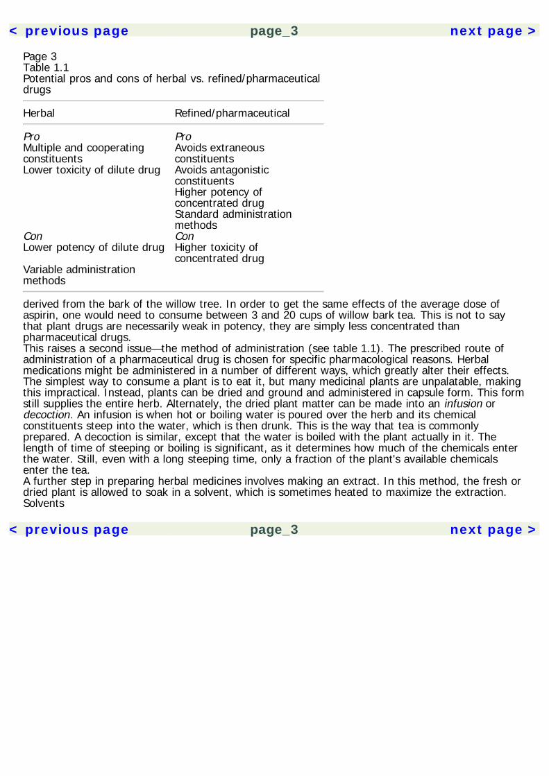
Page 2amino acids), which blur the distinction. Although both nutrients and drugs are derived from externalsources, they differ in their innate necessity. A nutrient is a substance that is essential to processesof life and growth. Without essential nutrients such as vitamin C, an individual suffers illness. A drugis any chemical that alters biological processes, with the connotation that it has some therapeutic orrecreational use. A poison or toxin, then, is a substance with predominantly harmful effects onbiological processes. However, as the sixteenth-century physician Paracelsus noted, the differencebetween a remedy and a poison is often a matter of dosage.Many plant-derived drugs are common in modern medicine. For example, morphine is derived fromthe poppy plant and is a standard medication used to treat pain. It has also been the progenitor ofmany other synthetic pain killers. Digoxin and digitoxin are cardiac medicines that derive from thefoxglove plant. But there are many herbal medications with reputed therapeutic value that have notgained acceptance in mainstream medicine. (For the purposes of this book, the words herb and plantwill be used interchangeably.) Knowledge of such herbs has been passed down through tradition, butthey have not gained mainstream acceptance due to a lack of research to support their therapeuticuse. Fortunately, many such herbal medications are currently undergoing scientific study. Inparticular, investigations are identifying their active chemical components, physiological effects,pharmacological properties, and clinical efficacy. In the meantime, their therapeutic status is mostoften relegated to alternative medicine.Herbal versus Synthetic DrugsHerbal medications are drugs in every sense of the word. They chemically modify bodily processesand can have therapeutic or harmful effects, depending on how they are used. However, there are afew general differences between herbal and pharmaceutical drugs (Tyler 1994). Herbal drugs tend tobe more dilute than pharmaceutical drugs (table 1.1). For example, caffeine is available in 200 mgtablets to produce stimulation. Coffee contains 1–2% caffeine, so in order to get the same amount ofcaffeine one must use 20 g of coffee bean. Similarly, aspirin is
< previous page page_2 next page >
< previous page page_3 next page >
Page 3Table 1.1Potential pros and cons of herbal vs. refined/pharmaceuticaldrugs
Herbal Refined/pharmaceutical
Pro ProMultiple and cooperatingconstituents
Avoids extraneousconstituents
Lower toxicity of dilute drug Avoids antagonisticconstituents
Higher potency ofconcentrated drug
Standard administrationmethods
Con ConLower potency of dilute drug Higher toxicity of
concentrated drugVariable administrationmethods
derived from the bark of the willow tree. In order to get the same effects of the average dose ofaspirin, one would need to consume between 3 and 20 cups of willow bark tea. This is not to saythat plant drugs are necessarily weak in potency, they are simply less concentrated thanpharmaceutical drugs.This raises a second issue—the method of administration (see table 1.1). The prescribed route ofadministration of a pharmaceutical drug is chosen for specific pharmacological reasons. Herbalmedications might be administered in a number of different ways, which greatly alter their effects.The simplest way to consume a plant is to eat it, but many medicinal plants are unpalatable, makingthis impractical. Instead, plants can be dried and ground and administered in capsule form. This formstill supplies the entire herb. Alternately, the dried plant matter can be made into an infusion ordecoction. An infusion is when hot or boiling water is poured over the herb and its chemicalconstituents steep into the water, which is then drunk. This is the way that tea is commonlyprepared. A decoction is similar, except that the water is boiled with the plant actually in it. Thelength of time of steeping or boiling is significant, as it determines how much of the chemicals enterthe water. Still, even with a long steeping time, only a fraction of the plant's available chemicalsenter the tea.A further step in preparing herbal medicines involves making an extract. In this method, the fresh ordried plant is allowed to soak in a solvent, which is sometimes heated to maximize the extraction.Solvents
< previous page page_3 next page >
< previous page page_4 next page >
Page 4frequently used are water, alcohol (ethanol or methanol), or acetone. After soaking in the solvent,the plant matter is discarded and the solvent is allowed to evaporate so that none of it remains inthe final product. All that remains is a concentration of the active constituents of the plant. The typeof solvent can affect the composition of the extract. For example, aqueous preparations do notefficiently extract the lipid-soluble, hydrophobic constituents of herbs. In contrast, alcohol extractswill contain far more lipid-soluble constituents. Despite the fact that both extracts derive from thesame plant, they have different compositions and can have widely varying pharmacological effects.An advantage of consuming an herbal extract instead of the whole plant is that it greatly reduces thevolume of matter to be ingested. In order to get a sufficent dose of the active chemicals, in somecases, this means the difference between swallowing a few pills and consuming a bale of dried plant.The number of different methods of application of herbal drugs means variability in the activechemicals obtained and the effects they produce. When taking or prescribing an herb, it is alwaysnecessary to attend to the type of preparation. Even pharmaceutical drugs vary from person toperson in terms of how much of the ingested drug is absorbed and reaches the targeted organs.Greater variability means greater unpredictability of negative side effects. The process ofstandardization of herbs ensures that a certain percentage of the desired active constituents arepresent in a preparation, which allows for greater reliability in dosing.Another significant difference between herbal and pharmaceutical drugs is that pharmaceutical drugsconsist of a purified single active drug. Herbal medications, on the other hand, may have multipleactive constituents. For example, the opium poppy contains more than 30 alkaloids (Robbers et al.1996). The active chemical constituents of a plant may each have different individual effects, so thatthe effect of the total herb is a combination of the effects of several different constituents. In suchcases, it might be preferable to purify one constituent for use, thus avoiding the extraneous effectsof others. While one may desire the analgesic effects of morphine, the vasodilator effects of anotherpoppy alkaloid, papaverine, may be unwanted. Using pure morphine, rather than opium, avoids thisproblem. Another scenario is when the desired effect of one constituent is antagonized or nullified byother constituents, making it even more necessary to isolate the desired constituent.
< previous page page_4 next page >
< previous page page_5 next page >
Page 5Conversely, active constituents may have cooperative effects and together act in an additive orsynergistic (supra-additive) manner. In such cases, it would be better to consume the whole plant orextract, because the combination of constituents would give a greater effect than one alone. Thus, toblindly advocate either the use of whole herb or refined single constituents is naive. To fully knowwhat is best for the desired effect, herbs must be considered on a case-by-case basis and the natureof the interactions between the chemical constituents must be carefully considered. Not only must weunderstand what the plant's chemical constituents do, we must also investigate how they interact.The Use of Herbal MedicineThe Current Prevalence of Alternative MedicineNational surveys have shown the use of alternative medicines, including herbal medicine and otherpractices, to be prevalent (Eisenberg et al. 1998). In 1997, 42% of people in a large national samplereported consulting at least one form of alternative medicine during the previous year, increasingfrom 33.8% in 1990. The alternative therapies with the greatest increase in use were herbalmedicine, massage therapy, megavitamins, self-help groups, folk remedies, energy healing, andhomeopathy. The illnesses for which people most often consulted alternative medicine tended to bechronic conditions, such as back problems, anxiety, depression, and headaches.The total number of visits to alternative medicine practitioners had increased by 47% across theseven-year span, and the majority of consumers (~60%) paid for these therapies at their ownexpense. The 1990 survey found that people made an average of 19 visits per year to an alternativemedicine practitioner, with the average charge per visit at $27.60 (Eisenberg et al. 1993). Aconservative estimate of the cost for all these therapies in 1997 was $21.2 billion, with $12.2 billionpaid out of pocket (Eisenberg et al. 1998). To put this in perspective, this amount exceeds out-of-pocket expenditures for all U.S. hospitalizations in 1997. The authors conclude that the increaseconstitutes a greater number of people seeking alternative therapies, rather than just an increasednumber of visits per patient.
< previous page page_5 next page >
< previous page page_6 next page >
Page 6The Appeal of Herbal MedicinesDespite the developing nature of the rational use of herbal medications, many are freely available tothe public as over-the-counter supplements. They have gained popularity as alternatives totraditional medicine. To some degree, their popularity may reflect a dissatisfaction with modernmedicine. Another potential factor is the perception that herbal medications are more ''natural" andhave greater aesthetic appeal than synthetic drugs. Of course, this is not necessarily so. In manycases, herbal medications can be just as toxic or deadly as synthetic medications. There are nointrinsic benefits from a drug simply because it comes from a plant. In some cases, consuming theplant may be more beneficial, and in other cases, consuming an extract or synthetic drug is better.Such cases need to be examined on a rational basis, rather than out of blind faith to naturalist ortechnological dogma. All the potential risks must be weighed against the benefits.Another possible reason for public interest in herbal medications is that they provide some autonomyfrom medical professionals. Multiple factors contribute to a person's satisfaction with medical care(Siahpush et al. 1999; Sixma et al. 1998; Kaptchuk and Eisenberg 1998). One cause of dissatisfactionresults from the traditionally passive role of the patient. Independent of its efficacy, alternativemedicine provides an individual with a greater range of explanations and treatment options for theirillnesses. The psychological appeal of more options is sufficient to draw individuals to consultalternative medicine, especially in cases where modern medicine has not been effective.Implications of Herbal Medicine for Medical FieldsA significant fact was revealed by Eisenberg and colleagues regarding disclosure and the use ofherbal medicines (1993, 1998). Of the proportion of respondents who utilized alternative medicine,only about 40% had disclosed this fact to their physician. The use of herbal medications was amongthe most common alternative treatments, and also one of the treatments with the greatest increasein use. Further, it was estimated that during the year of the survey 15 million adults took prescription
< previous page page_6 next page >
< previous page page_7 next page >
Page 7medications concurrently with herbal medications and/or high-dose vitamins. Thus, a large numberof people are taking herbal medications and not disclosing the fact to their physicians. Becauseherbal medicines are active drugs, they present enormous potential for adverse medicationinteractions. Adverse interactions could be averted if people were more comfortable with informingtheir physicians of their use of herbal medications. Of course, physicians would need to have agreater knowledge of the actions of herbal medications to foresee such potential interactions.The extent of knowledge among medical professionals (physician and nonphysician) about herbalmedications varies across and within cultures. On the whole, American physicians are currently notvery knowledgeable about herbal medications, and as a result, they tend not to prescribe them.Comparatively, the use of herbal medications by physicians is much more common in Europe andAsia (Tyler 1994).But the American medical system is not blind to this fact, and there are some indications of change.A number of studies have appeared in medical journals reviewing the known efficacy and safety ofherbs for therapeutic use (e.g., O'Hara et al. 1998; Hadley and Petry 1999; Wong et al. 1998; Cupp1999; Onopa 1999). A good proportion (64%) of surveyed medical schools reported offering somecourse work in alternative or complementary medicine (Wetzel et al. 1998). A majority (68%) ofthese were stand-alone courses and the remaining addressed alternative medicine in the context ofother courses.The reluctance of many physicians to prescribe herbal medications is in many ways understandable.Much research needs to be done to ensure the efficacy and safety of many herbs. Few authoritativetexts are available that summarize the available clinical research (see Tyler 1994). Becausephysicians naturally want to provide their patients with the best available care, herbal medications donot always present an attractive option due to the uncertainty that surrounds them.The darker side to this issue concerns malpractice. Malpractice claims against nonmedicalpractitioners of alternative medicine are generally low (Studdert et al. 1998). Although referring apatient to another physician does not generally expose a physician to liability for malpractice, thereare exceptions when the referral is negligent or when there is joint treatment. Therefore, referrals toalternative medicine can potentially carry risk.
< previous page page_7 next page >
< previous page page_8 next page >
Page 8In this context, judging the qualifications of an alternative medicine practitioner can be difficult—there are no universally accepted guidelines. Some states require licensing of alternative medicinepractitioners (e.g., acupuncturists, naturopathic herbalists), and licensure is often used by courts toestablish school-specific standards of care. In cases where no licensing exists, courts applyconventional medical or lay standards of care. Eisenberg (1997) has proposed strategies forphysicians in guiding patients who seek alternative medical treatment.Government Regulation of Herbal MedicationsGovernment regulation of herbal medicines has changed appreciably over time (Tyler 1994; Ray andKsir 1990). In the early twentieth century, the Food and Drug Act of 1906 and the Sherleyamendment of 1912 were enacted to combat rampant fraud by food and drug producers. Primarily,these laws addressed mislabeling and adulteration of drugs. The Food, Drug, and Cosmetic Acts wereenacted in 1938 in response to the tragic deaths of 105 people taking an untested medication (elixirsulfanilamide). These laws stipulated that drugs entering interstate commerce must demonstratesafety, while grandfathering many existing drugs. Tragedy again prompted legislation when pregnantmothers taking the sedative drug thalidomide gave birth to babies with severe limb deformities.Although this occurred outside of the United States, concern prompted the drug amendments of1962, commonly referred to as the Kefauver-Harris amendments (Hollister et al. 1968; Tyler 1994).This legislation required that drugs marketed after 1962 be not only safe, but also effective.The U.S. Food and Drug Administration (FDA) undertook a large study of over-the-counter drugs,releasing their results in 1990. This classified drugs and herbs into three categories: (1) effective, (2)unsafe or ineffective, or (3) insufficient evidence to evaluate effectiveness. However, thisclassification was criticized for overreliance on data from industry. For example, prunes were placedin category (3) as having insufficient evidence for their effectiveness as a laxative! Many herbalmedications were kept on the market following this study, but the incriminating statements abouttheir supposed use were removed. While
< previous page page_8 next page >

< previous page page_9 next page >
Page 9this may have been legally feasible, it left the consumer even less informed than before.In 1994, the Dietary Supplement Health and Education Act was passed by the U.S. Congress. Thisallowed herbal medications to be advertised and sold without oversight from the FDA. Specifically, itstates that a substance will not legally be classified as a ''drug" if it is not represented as treatmentfor a disease (Heiligenstein and Guenther. 1998; Dietary Supplement Health and Education Act of1994). Thus, many herbal medicines are now sold and regulated as dietary supplements.There has been some government recognition of the need for knowledge about herbal medications.European governments have traditionally been more advanced than the United States in this area.The German Federal Institute for Drugs and Medical Devices appointed a special expert committee,entitled the German Commission E, to compile scientific and clinical information regarding therapeuticherbs. Their results are published in the book, The Complete German Commission E Monographs:Therapeutic Guide to Herbal Medicines (English translation sponsored by the American BotanicalCouncil, 1998). The U.S. National Institutes of Health have a branch dedicated to researchingalternative and complementary medicines—the National Center for Complementary and AlternativeMedicine (NCCAM). For example, in 1997 the NCCAM launched an ongoing large-scale study of Saint-John's-wort for treatment of depression.The Evolutionary Perspective: Why Plants Make DrugsAt first glance, it seems unusually convenient that plants would be so kind as to make drugs forhuman benefit. What causes plants to develop therapeutic chemicals? The answer requires a slightshift of perspective, to ask why plants manufacture chemicals in general. To understand this, onemust consider it in the context of natural selection.Evolution by natural selection was first explained by Charles Darwin in his book On the Origin ofSpecies (1859). Briefly stated, the theory suggests that evolution occurs through heritablepropagation of adaptive traits. Nature produces a large variation in the traits of organisms. Thosetraits that are in some way adaptive, increasing the survival and
< previous page page_9 next page >
< previous page page_10 next page >
Page 10reproductive success of the organism, are propagated to future generations. Darwin's schema issimple but powerful, having great explanatory strength; it is a cornerstone of modern biology. Thecontext of natural selection is essential for understanding the mutual adaptations between plants andanimals that led to the manufacture of drugs by plants.Living organisms are divided into five kingdoms: Animalia, Plantae, Fungi, Protista, and Monera.Green, multicellular plants fall under the Plantae kingdom, which originated in the Silurian period(505 to 400 million years ago) (Southwood 1984). They have the shortest evolutionary history andare the only kingdom to have completely evolved on land. Also evolving around this time wereinsects. Insects evolving on land found an abundant source of food in plants, provided they couldadapt to eating them. This probably occurred in gradual increments, beginning with insectsscavenging on pollen and spores, progressing to insects feeding on the reproductive organs ofplants, and eventually leaf feeding.However, this newfound feast for insects was at a great cost to plants. Research indicates that plantsprotected from insect predators by pesticides live longer, produce more seeds, and propagate over alarger area. Thus, natural selection would favor plants that somehow deter predators from consumingthem. The defenses plants have evolved fall into three categories: nutritional, physical, and chemical(Southwood 1984). Nutritional defenses involve having low nitrogen levels or balances of amino acidsthat are unfavorable to insect metabolism. Physical defenses involve the growth of external cuticlesand epidermal hairs that make the plant mechanically difficult to hold, manipulate, and consume. Thefocus of this book is the chemical defenses evolved by plants, which involve the production ofsubstances that strongly and adversely alter a predator's physiology (either by poisoning orrepelling). Also, plant defenses typically are involved in life functions other than defense, so theirdefense advantages may well have been serendipitous. Regardless, any trait (serendipitous or not)that increases the survival and reproductive ability of a plant will propagate through naturalselection. These chemical defenses alter our physiology in diverse ways. While some are outrighttoxic, many others can be subverted for therapeutic uses. To a large extent, this depends upon ourcleverness in finding uses.
< previous page page_10 next page >
< previous page page_11 next page >
Page 11Thus, plants and predators have coevolved, reciprocally adapting to one another. Plants developchemicals that deter predators and increase their survival advantage. Predators, in turn, adapt bydeveloping tolerance, attractions, or even utilization of plant chemicals.Although humans differ greatly from insects, a reciprocal relationship between humans and plantsexists. In the course of foraging for food, humans have serendipitously discovered plants withtherapeutic effects. Over time, traditions of knowledge about the therapeutic use of plants haveaccumulated within cultures, even though the rationales for why such plants are effective are oftenincorrect. For example, Asian systems of herbal medicine are based on the theory of balancing one'syin and yang energy. While this might have some unforeseen metaphorical value, it has no apparentbasis in the actual anatomy and physiology of the human body. During the Renaissance, herbalistsused the doctrine of signatures to guide the therapeutic use of plants. The doctrine of signatures isthe belief that the shape or color of a plant indicates its therapeutic uses (e.g., if a plant containedred then it would be used to treat blood-related disorders). Although their explanations for whyherbs treat an illness are inaccurate, they have still accumulated knowledge about which plants treatwhich illnesses, through trial and error discovery.The therapeutic use of plants is seemingly not limited to humans. Certainly, humans are the onlyspecies with the verbal and cognitive capacity to label therapeutic plants as such and to accumulatelarge bodies of knowledge on their specific uses. Nonetheless, the field of zoo-pharmacognosy hasarisen in recent years to study the therapeutic uses of plants by animals. This was prompted by thediscovery of Tanzanian chimpanzees using the Aspilia plant (Rodrigues et al. 1985; Page et al. 1992).The chimps swallow the whole leaves of the plant without chewing them and derive no apparentnutrition. However, the plant contains a chemical called thiarubine-A, which prevents infection bycertain parasites and microorganisms in the gastrointestinal tract. Chimps consuming the plantappear to be free of such infections and related illnesses.At first glance, it may seem unlikely that chimpanzees premeditatively use such plants. However, nospecific knowledge about the plant need be assumed in the chimpanzees for them to use it. From abehavioral standpoint, they would only need to ingest the plant and experience some
< previous page page_11 next page >

< previous page page_12 next page >
Page 12noticeable benefit for the Aspilia-eating behavior to be reinforced. Simply stated, an organism (eitheranimal or human) does not need to know how or why an herbal medication works in order to derivebenefit and keep using it. The organism only needs to experience some improvement so that theplant-using behavior is reinforced. The two requirements of behavioral reinforcement arecontingency, where the plant gives some reliable benefit, and contiguity, where the benefit occursfairly close in time to the consumption. Some plants directly affect the brain mechanisms involved inbehavioral reinforcement, bringing pleasure in mild cases and addiction in severe ones.In this respect, the human use of plants with various incorrect rationales (e.g., the doctrine ofsignatures) is not much different than the chimpanzee use of Aspilia: an accurate knowledge of howthe treatment works is not necessary for plant-use to be behaviorally reinforced and maintained.Human knowledge about the therapeutic uses of plants has accumulated through the millennia bytrial and error. Our explanations have been secondary.However, a new era of plant medicines began with modern science. Rather than trial and error, wecan now begin to understand how and why plant medicines work. It is hoped that we can use thisinformation to guide a more safe and effective use of medicinal plants.Scientific Methodology in Herbal MedicineScience is essentially a way of acquiring knowledge. It involves the formation of testable hypothesesbased on observations. Further, there is a systematic collection of data to support or disprove thehypotheses. The requirement to verify one's observations through objective methods gives scienceenormous epistemological power. Information acquired through carefully designed experiments thatare subject to repeated verification is the most reliable form of knowledge we have. This isparticularly so because the misleading influences of personal bias and random chance are minimized.In order for a hypothesis to be scientific, it must not only be provable through research, but alsofalsifiable.To be fair, the majority of scientific data probably do not immediately cast hypotheses into clear-cutcategories of proven or disproven. There
< previous page page_12 next page >

< previous page page_13 next page >
Page 13are most often multiple interpretations and conclusions to be drawn from a set of data. For thisreason, scientific findings are subject to peer review and criticism. Although science is conducted byindividuals, it ultimately becomes a process of interaction, discussion, and debate among acommunity of researchers.The purpose of this discussion of science, clearly, is to apply it to herbal medicine. The reputed usesof drugs may not always be accurate or effective. Applying the scientific method to herbal medicinetherefore allows us to test the traditional uses and to know with greater certainty what an herbalmedication does and how reliably it does it. Although far from infallible, the process as a whole givesus greater confidence in our conclusions.Scientific research has many times verified traditional remedies. It is advisable for scientists in thefield who are looking for new research ideas to consult the traditional herbal literature.Adding scientific verification to the body of traditional knowledge can only strengthen our confidencein the traditional use of herbal medicines. Delving into the chemistry and pharmacology of plantsopens up a different level of understanding, which may suggest new uses of herbs never thought ofbefore.There are many ways in which scientific research is carried out, but there are a few essentialelements. One form of scientific study is experimentation. In order for causation to be established, anexperimental study must perform some kind of manipulation. When an effect is produced, it can thenbe attributed to the manipulation. However, in order for this to be done, one must control forextraneous influences. To the degree possible, one must keep all other conditions constant so thatthe experimental effect can be correctly attributed to the manipulation.For example, if one were studying an herb to treat depression, one would want to control as manyfactors as possible that could influence the outcome. People already taking an antidepressant drugwould have to be excluded. One might also balance the subjects in different groups for severity ofdepression, psychotherapy treatment, or even levels of physical exercise. Certainly, the subjectgroups should be balanced for number of males and females, because sex differences in depressioncould contaminate the results.
< previous page page_13 next page >
< previous page page_14 next page >
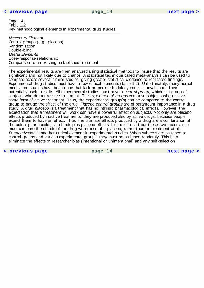
Page 14Table 1.2Key methodological elements in experimental drug studies
Necessary ElementsControl groups (e.g., placebo)RandomizationDouble-blindUseful ElementsDose-response relationshipComparison to an existing, established treatment
The experimental results are then analyzed using statistical methods to insure that the results aresignificant and not likely due to chance. A statistical technique called meta-analysis can be used tocompare across several similar studies, giving greater statistical credence to replicated findings.Experimental drug studies must have a few critical elements (table 1.2). Unfortunately, many herbalmedication studies have been done that lack proper methodology controls, invalidating theirpotentially useful results. All experimental studies must have a control group, which is a group ofsubjects who do not receive treatment. The experimental groups comprise subjects who receivesome form of active treatment. Thus, the experimental group(s) can be compared to the controlgroup to gauge the effect of the drug. Placebo control groups are of paramount importance in a drugstudy. A drug placebo is a treatment that has no intrinsic pharmacological effects. However, theexpectation that a treatment will work can have a powerful effect on subjects. Not only are placeboeffects produced by inactive treatments, they are produced also by active drugs, because peopleexpect them to have an effect. Thus, the ultimate effects produced by a drug are a combination ofthe actual pharmacological effects plus placebo effects. In order to sort out these two factors, onemust compare the effects of the drug with those of a placebo, rather than no treatment at all.Randomization is another critical element in experimental studies. When subjects are assigned tocontrol groups and various experimental groups, they must be assigned randomly. This is toeliminate the effects of researcher bias (intentional or unintentional) and any self-selection
< previous page page_14 next page >
< previous page page_15 next page >
Page 15effects by the subjects. As a hypothetical example, if all subjects who show up early are assigned toone group, and latecomers are assigned to another group, then the differences between groupscould be a product of the subjects' personality features, and not just the effects of the drug.Randomization eliminates such a confound.A double-blind control is also an essential element of drug studies. Double-blind means that neitherexperimenter nor subject knows who is receiving the active treatment or the placebo. Again, this isto eliminate potential bias from both parties. Otherwise, experimenters could unwittingly influencethe outcome of the study by subtly treating subjects differently.In addition to these critical elements, it is often useful to establish a dose-response relationship in astudy. That is, to show that the size of the dose changes the magnitude of effect. This isaccomplished by multiple experimental groups, each receiving a different dose. Showing a dose-response relationship supports the pharmacological nature of the effect and also helps establish theappropriate dose range for clinical treatment.Clinical Use of Herbal Medicines: Quality of EvidenceWhen a treatment is considered in a clinical setting, one must evaluate the class of evidence thatsupports its use. In 1979, a Canadian task force developed guidelines for evaluating the quality ofevidence supporting a treatment (see table 1.3). These guidelines have subsequently been adoptedby a number of other regulatory panels (Canadian Task Force on the Periodic Health Examination1979; Woolf 1992).The Canadian Task Force categorized the quality of evidence based on the type of research study.The quality of evidence was organized into three classes: Class I evidence comes from procedureshaving at least one randomized controlled study to support them. Class II is divided into threesubclasses, where II-1 involves a well-designed controlled study without randomization. Class II-2evidence comes from well-designed cohort or case-control studies, preferably carried out at morethan one research setting. Class II-3 involves uncontrolled research with dramatic results (e.g.,penicillin trials in the 1940s). Class III evidence includes the opinions of experts and authorities in thefield based on clinical
< previous page page_15 next page >

< previous page page_16 next page >
Page 16Table 1.3Quality of evidence
ClassI
At least 1 properly designed, randomized controlled trial
ClassII
II-1 Well-designed, controlled trials without randomizationII-2 Well-designed, cohort or case-control analytic studies, preferably from more
than one center or research groupII-3 Comparisons between times or places without intervention; dramatic results in
uncontrolled experimentsClassIII
Opinions of respected authorities, based on clinical experience, descriptivestudies, or reports of expert committees
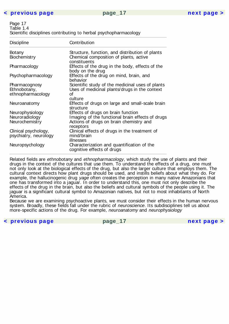
Source: Canadian Task Force on the Periodic Health Examination, 1979.experience or descriptive studies. Clearly, Class I evidence is the strongest class of evidence, andeach successive class carries less empirical value.Although the latter classes of evidence provide useful information, Class I evidence is the goal ofresearch in herbal medication. Case studies and anecdotal evidence should be treated seriously, butthey should also be followed up with empirical research.Scientific Disciplines in Herbal PsychopharmacologySeveral scientific disciplines contribute to the body of knowledge that is surveyed in this text (table1.4). Because plants are the focus of attention, we consult botany to learn the structure, function,and geographical habitats of the plants in question. Biochemistry is necessary to understand thechemical composition of the plant, including the active constituents that give the desiredpsychological and physiological effects. Pharmacology is the study of drugs, including how they affectthe body and how the body affects them. Chapter 3 is dedicated to outlining the basic principles ofpharmacology, which are then applied to herbal medicines. Psychopharmacology is a branch ofpharmacology that studies how drugs affect the mind, brain, and behavior. Pharmacognosy isspecifically the scientific study of the medicinal uses of plants. Thus, the subject of this text couldalternately be titled psychopharmacognosy, or the scientific study of plants that alter mind, brain,and behavior.
< previous page page_16 next page >
< previous page page_17 next page >
Page 17Table 1.4Scientific disciplines contributing to herbal psychopharmacology
Discipline Contribution
Botany Structure, function, and distribution of plantsBiochemistry Chemical composition of plants, active
constituentsPharmacology Effects of the drug in the body, effects of the
body on the drugPsychopharmacology Effects of the drug on mind, brain, and
behaviorPharmacognosy Scientific study of the medicinal uses of plantsEthnobotany,ethnopharmacology
Uses of medicinal plants/drugs in the contextofculture
Neuroanatomy Effects of drugs on large and small-scale brainstructure
Neurophysiology Effects of drugs on brain functionNeuroradiology Imaging of the functional brain effects of drugsNeurochemistry Actions of drugs on brain chemistry and
receptorsClinical psychology,psychiatry, neurology
Clinical effects of drugs in the treatment ofmind/brainillnesses
Neuropsychology Characterization and quantification of thecognitive effects of drugs
Related fields are ethnobotany and ethnopharmacology, which study the use of plants and theirdrugs in the context of the cultures that use them. To understand the effects of a drug, one mustnot only look at the biological effects of the drug, but also the larger culture that employs them. Thecultural context directs how plant drugs should be used, and instills beliefs about what they do. Forexample, the hallucinogenic drug yage often creates the perception in many native Amazonians thatone has transformed into a jaguar. In order to understand this, one must not only describe theeffects of the drug in the brain, but also the beliefs and cultural symbols of the people using it. Thejaguar is a significant cultural symbol to Amazonian natives, but not to most inhabitants of NorthAmerica.Because we are examining psychoactive plants, we must consider their effects in the human nervoussystem. Broadly, these fields fall under the rubric of neuroscience. Its subdisciplines tell us aboutmore-specific actions of the drug. For example, neuroanatomy and neurophysiology
< previous page page_17 next page >
< previous page page_18 next page >
Page 18tell us the effects of drugs on the structure and function of the nervous system. Specifically,neuroradiology allows us to examine the functional neuroanatomical effects of psychoactive drugsthrough neuroimaging techniques such as single photon emission computed tomography (SPECT),positron emission tomography (PET), and functional magnetic resonance imaging (fMRI). Essentially,these methods can examine the changes in cerebral blood flow and/or glucose metabolism caused bypsychoactive herbs. Neurochemistry describes how such drugs will interact with the variousneurotransmitters and chemical messengers in the nervous system.The disciplines of psychiatry, psychology, and neurology are equipped to diagnose disorders of themind and brain and study the clinical effects of psychoactive herbs. Neuropsychology specializes inbrain-behavior relationships and is well equipped to quantify and characterize the mental andbehavioral effects of psychoactive herbs by using psychometric tests. In this text, particular attentionis paid to objective testing data.Neuropsychology: A Brief Taxonomy of Cognitive FunctionsHerbal drugs can have general or specific effects on cognition. To understand the results ofneuropsychological testing in drug studies, it will be useful to briefly discuss the breakdown ofcognitive functions.Attention and ConcentrationSelective attention involves the ability to focus on some feature of the environment, while filteringout other extraneous features (Lezak 1995; Posner and Driver 1992). For example, when drivingthrough an intersection, one must focus and mentally track other cars, while simultaneously filteringout distractors such as a mailbox on the corner or music on the radio. Other aspects of attentioninvolve sustaining attention over a period of time, and shifting attention from one target to another.Attention is a limited-capacity system, so dividing attention among several targets reduces theamount of attention allocated to each. Also related to attention is the speed at which one is able toprocess information.Colloquially, many aspects of working memory are referred to as attention or concentration. Workingmemory also holds information in
< previous page page_18 next page >

< previous page page_19 next page >
Page 19consciousness, allowing for manipulation. An example would be remembering three numbers whileadding them together. Whereas attention involves operations with present stimuli, working memorysustains activation of a percept in its absence (e.g., remembering a phone number after you lookedit up).Learning and MemoryLearning and memory refers to the ability to acquire, store, and retrieve information (Lezak 1995). Astage model posits that information first enters short-term memory, which is governed by ongoingcircuits of activity in the brain. By semantic and contextual meaning, information is then consolidatedinto long-term memory, which is a relatively permanent store and involves synaptic changes in thebrain. Information is later accessed by the process of memory retrieval. Memory is typically evaluatedin two modalities: visual and verbal. There are also two very different forms of memory: explicit andimplicit. Explicit memory involves personal experiences, facts, and semantic information (Schacter1997; Verfaellie and Keane 1997). Implicit memory, on the other hand, involves skills, habits, andpriming. These two forms of memory are mediated by different systems of structures in the brain.LanguageLanguage is a symbolic representation system that allows for sophisticated and abstractcommunication. Several aspects of language can be distinguished, including expression,comprehension, naming, reading, and writing. Language also incorporates our semantic store offactual knowledge and is widely distributed in the brain, typically in the left hemisphere for mostpeople. There are specialized language areas, so that it is possible to selectively lose one aspect butnot another (e.g., loss of language expressive ability while retaining comprehension).PerceptionSensation involves the conversion of physical energy into nerve impulses (e.g., photons are convertedinto neural signals for vision), but perception involves making a meaningful representation of thosesignals. This includes all sensory modalities: vision (sight), audition (hearing),
< previous page page_19 next page >
< previous page page_20 next page >
Page 20somatosenses (bodily senses), olfaction (smell), and gustation (taste), but standardized tests mostfrequently assess visual or auditory processing. Aspects of visual perception most commonly assessedinclude shape discrimination, spatial location, line orientation, face perception, visual organization,and construction.Executive FunctionsExecutive functions are a varied group of cognitive abilities encompassing abstract reasoning,planning, organization, mental shifting, initiation and inhibition of behavior, judgment, and socialconduct. They are crucial for efficient and effective functioning in everyday life. These functions arelargely governed by the frontal lobes and associated subcortical structures. To some degree, differentbrain structures control different executive functions, so one may have a deficit of organization andabstraction, while social conduct is preserved. Numerous experimental and clinical measures havebeen developed to tap into executive functions, such as the Wisconsin Card Sorting Test, HalsteadCategory Test, Tower of London, and go/no-go tests (Spreen and Strauss 1998).Emotion and PersonalityEmotions are subjective mood states that interact reciprocally with cognitive processes. Personalityrefers to traits of emotion and behavior that are more stable over time. Normal and pathologicalemotional states can be measured, to some degree, with objective tests to quantify changes in moodover time (or after drug treatment). Thus, several clinical scales have been developed for anxiety,depression, and mania. These measures are particularly useful for evaluating the effectiveness ofpsychotherapeutic herbs.ConclusionsPsychoactive plants have been a part of human life since our beginning. Our first experiences withthem probably came from foraging among plants for food. Through trial-and-error learning andbehavioral reinforcement, experience has shaped our use of herbal drugs. Similarly,
< previous page page_20 next page >

< previous page page_21 next page >
Page 21other nonhuman species may have acquired a limited repertoire of herbal medications throughsimilar means.With our capacity for language and to explain natural phenomena, humans have historicallydeveloped a number of belief systems to explain the effects of medicinal herbs. These vary acrosscultures and have been passed down across generations through tradition. Although many of theuses of herbs discovered by traditional means are proving to be accurate, their explanations aresometimes rooted in erroneous assumptions or archaic concepts. These traditional rationales mayseem innocuous enough, but they lack heuristic value and can hinder further understanding.Modern scientific research is providing a new analysis of traditional herbs, explaining their effects inbiological terms and testing for reliability. With this biological level of understanding, science hasbeen verifying much of traditional herbal wisdom and in some cases suggesting new uses for herbs.Thus, today's herbal medicine is an interesting combination of traditional lore and modern scientifictechniques. When properly applied, they work to each other's mutual benefit.Although our relationship with psychoactive plants is ancient, our knowledge of their uses is stilldeveloping. Numerous such plants are available in multiple forms to the general public, and all toooften there is little authoritative information to guide their use. More frequently, individuals willpublish books that fanatically advocate the use of herbs with overblown claims lacking empiricalsupport. In contrast, the level of expertise among medical professionals is variable, differing acrosscultures and individuals. The free availability of psychoactive herbs combined with people's tendencyto not disclose their use to medical personnel creates an enormous potential for adverse druginteractions. It would be unfortunate if a series of unnecessary and tragic herb-drug interactionspublicly marred the enormous therapeutic potential of herbs.There are essentially two ways to avoid such a scenario. One would be to increase governmentregulation of herbal medicines so that they are only available with a prescription. This solution isimpractical and unfavorable for a few reasons. It would unnecessarily limit the responsible andconscientious use of herbs carried out by millions of people. Also,
< previous page page_21 next page >

< previous page page_22 next page >
Page 22it would be a largely ineffective restriction because most herbal medications grow abundantly in thewild or in herb gardens. Government restriction would only effectively harm the herbal medicineindustry and abolish the standardization and quality control to which we hold it.Rather than drive herb use underground, a far more rational solution would be to further ourunderstanding of herbs. The lack of proprietary interests in herbal medicines has limited the financialincentives that encourage research by private industry. Instead, government funds could be directedto researchers who investigate the composition of herbs, their effects in the body and brain, andtheir clinical efficacy. The compilation of traditional and scientific herbal knowledge could beincorporated in the education of medical professionals, guiding the responsible and effective use ofherbs in the treatment of illnesses. Efforts could be directed at public education, advising on theproper use of herbs and providing information on when to consult professional medical help.This text has been written in the spirit of the latter approach. It is hoped that the informationcompiled and integrated here will further our understanding and capabilities and perhaps inspire newresearch as well.
< previous page page_22 next page >

< previous page page_23 next page >
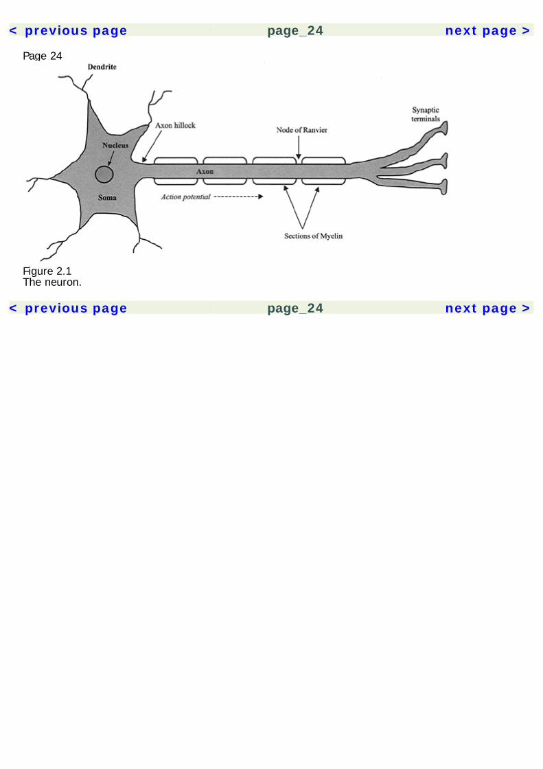
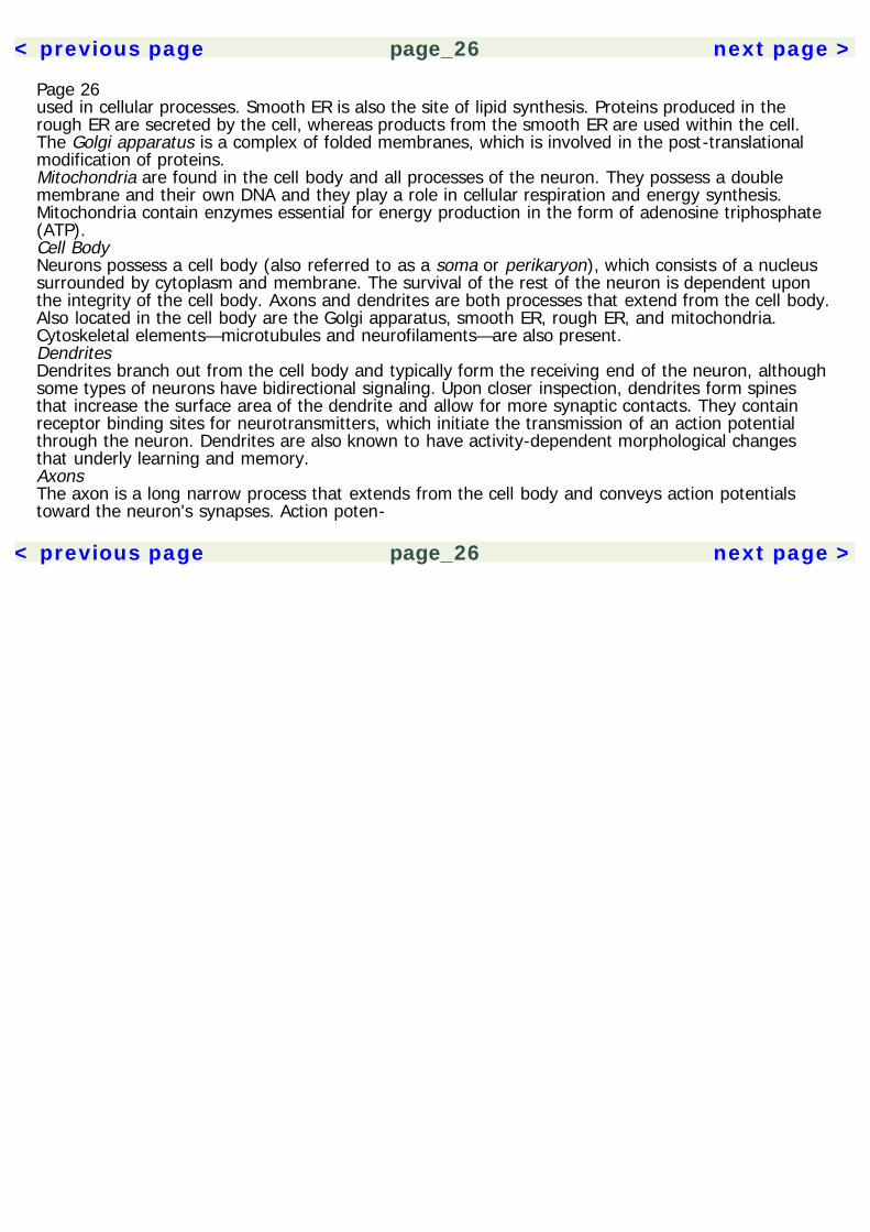
Page 232Basic NeurosciencesAn understanding of the nervous system is essential to psychopharmacology. It provides the contextin which the actions of drugs are understood. The effects of drugs can be compared based on thecommonalities and differences of their actions on neurons. When encountering a new drug, herbal orpharmaceutical, knowing its neuropharmacological mechanisms also allows us to make predictionsabout its effects. Space limitations allow for only a cursory description of the nervous system here, sothe reader is directed to the Recommended Reading section for a more thorough treatment of thetopic.The NeuronA description of the nervous system probably best starts with its basic functional unit, the neuron(figure 2.1). Neurons have many features in common with other cells of the body, but there are twothat make them unique. They are able to conduct electrochemical impulses and they are connectedin networks suited for information processing. Their link to the senses and motor effectors allows usto monitor and operate in the environment. The adult human brain is estimated to contain 1011neurons, and each may synapse with 10,000 other neurons. Despite some common core features,neurons are very heterogeneous in their morphology, with thousands of variants. The following arecomponents of the neuron.
< previous page page_23 next page >
< previous page page_24 next page >
Page 24
Figure 2.1The neuron.
< previous page page_24 next page >
< previous page page_25 next page >
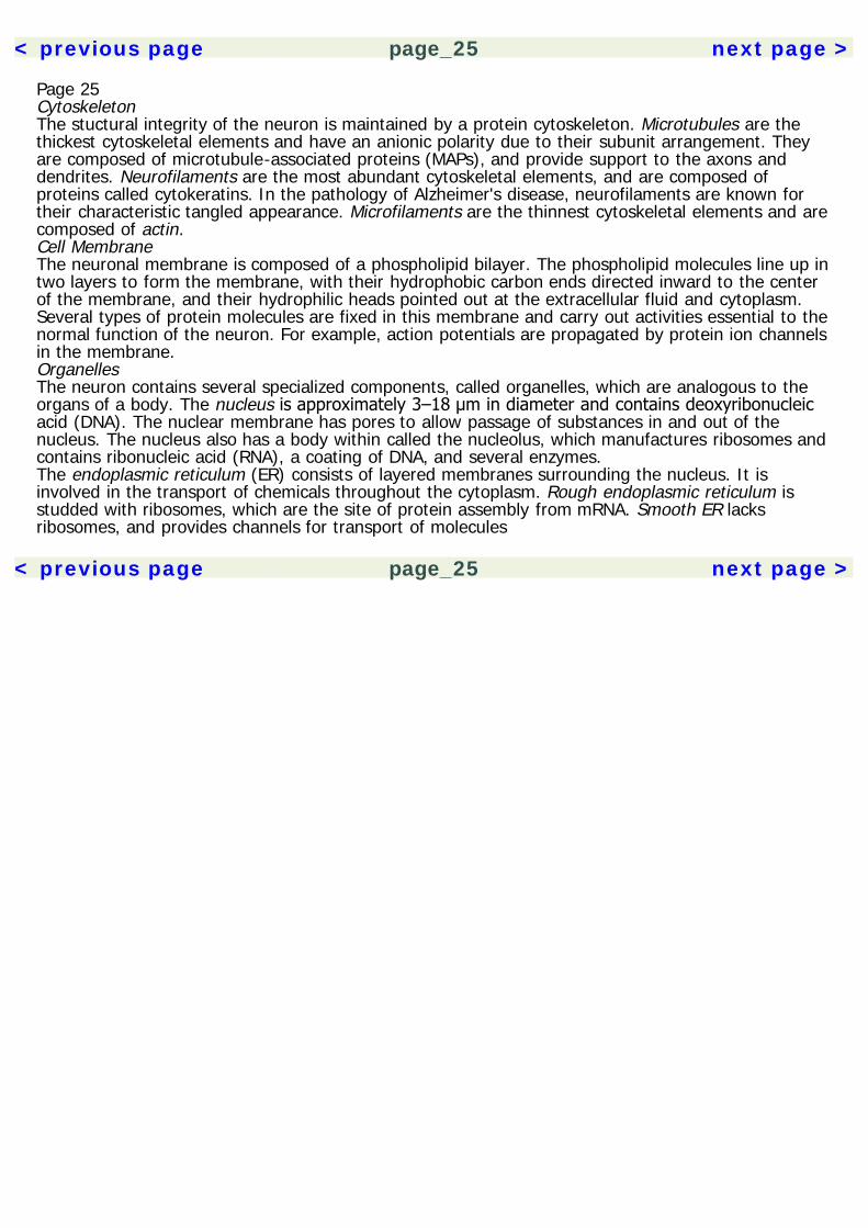
Page 25CytoskeletonThe stuctural integrity of the neuron is maintained by a protein cytoskeleton. Microtubules are thethickest cytoskeletal elements and have an anionic polarity due to their subunit arrangement. Theyare composed of microtubule-associated proteins (MAPs), and provide support to the axons anddendrites. Neurofilaments are the most abundant cytoskeletal elements, and are composed ofproteins called cytokeratins. In the pathology of Alzheimer's disease, neurofilaments are known fortheir characteristic tangled appearance. Microfilaments are the thinnest cytoskeletal elements and arecomposed of actin.Cell MembraneThe neuronal membrane is composed of a phospholipid bilayer. The phospholipid molecules line up intwo layers to form the membrane, with their hydrophobic carbon ends directed inward to the centerof the membrane, and their hydrophilic heads pointed out at the extracellular fluid and cytoplasm.Several types of protein molecules are fixed in this membrane and carry out activities essential to thenormal function of the neuron. For example, action potentials are propagated by protein ion channelsin the membrane.OrganellesThe neuron contains several specialized components, called organelles, which are analogous to theorgans of a body. The nucleus is approximately 3–18 μm in diameter and contains deoxyribonucleicacid (DNA). The nuclear membrane has pores to allow passage of substances in and out of thenucleus. The nucleus also has a body within called the nucleolus, which manufactures ribosomes andcontains ribonucleic acid (RNA), a coating of DNA, and several enzymes.The endoplasmic reticulum (ER) consists of layered membranes surrounding the nucleus. It isinvolved in the transport of chemicals throughout the cytoplasm. Rough endoplasmic reticulum isstudded with ribosomes, which are the site of protein assembly from mRNA. Smooth ER lacksribosomes, and provides channels for transport of molecules
< previous page page_25 next page >
< previous page page_26 next page >
Page 26used in cellular processes. Smooth ER is also the site of lipid synthesis. Proteins produced in therough ER are secreted by the cell, whereas products from the smooth ER are used within the cell.The Golgi apparatus is a complex of folded membranes, which is involved in the post-translationalmodification of proteins.Mitochondria are found in the cell body and all processes of the neuron. They possess a doublemembrane and their own DNA and they play a role in cellular respiration and energy synthesis.Mitochondria contain enzymes essential for energy production in the form of adenosine triphosphate(ATP).Cell BodyNeurons possess a cell body (also referred to as a soma or perikaryon), which consists of a nucleussurrounded by cytoplasm and membrane. The survival of the rest of the neuron is dependent uponthe integrity of the cell body. Axons and dendrites are both processes that extend from the cell body.Also located in the cell body are the Golgi apparatus, smooth ER, rough ER, and mitochondria.Cytoskeletal elements—microtubules and neurofilaments—are also present.DendritesDendrites branch out from the cell body and typically form the receiving end of the neuron, althoughsome types of neurons have bidirectional signaling. Upon closer inspection, dendrites form spinesthat increase the surface area of the dendrite and allow for more synaptic contacts. They containreceptor binding sites for neurotransmitters, which initiate the transmission of an action potentialthrough the neuron. Dendrites are also known to have activity-dependent morphological changesthat underly learning and memory.AxonsThe axon is a long narrow process that extends from the cell body and conveys action potentialstoward the neuron's synapses. Action poten-
< previous page page_26 next page >
< previous page page_27 next page >
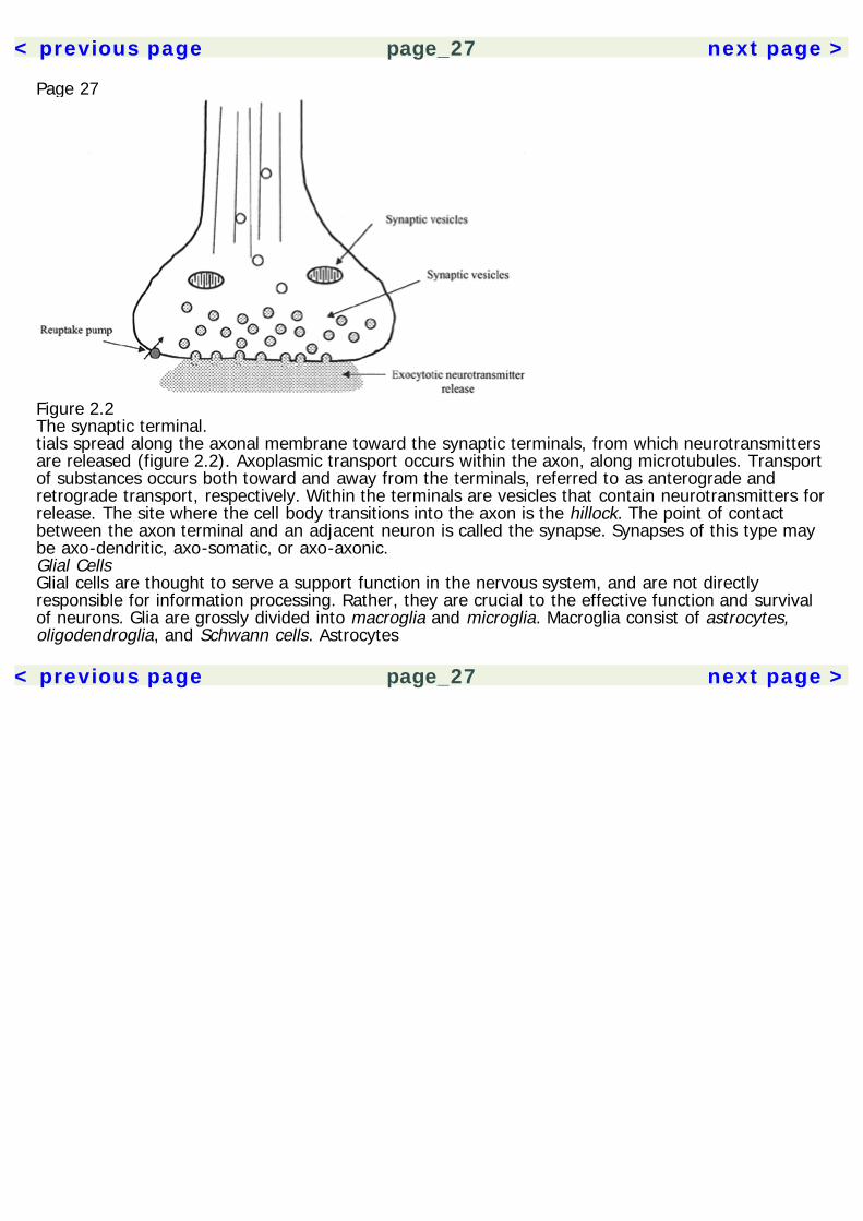
Page 27
Figure 2.2The synaptic terminal.tials spread along the axonal membrane toward the synaptic terminals, from which neurotransmittersare released (figure 2.2). Axoplasmic transport occurs within the axon, along microtubules. Transportof substances occurs both toward and away from the terminals, referred to as anterograde andretrograde transport, respectively. Within the terminals are vesicles that contain neurotransmitters forrelease. The site where the cell body transitions into the axon is the hillock. The point of contactbetween the axon terminal and an adjacent neuron is called the synapse. Synapses of this type maybe axo-dendritic, axo-somatic, or axo-axonic.Glial CellsGlial cells are thought to serve a support function in the nervous system, and are not directlyresponsible for information processing. Rather, they are crucial to the effective function and survivalof neurons. Glia are grossly divided into macroglia and microglia. Macroglia consist of astrocytes,oligodendroglia, and Schwann cells. Astrocytes
< previous page page_27 next page >
< previous page page_28 next page >
Page 28are present in the central nervous system (CNS), providing structural support, transporting nutrients,and phagocytizing cellular debris. They transport glucose and also perform a metabolic service byconverting it to lactate before it is transported to the neurons. Astrocytes also store glucose in theform of glycogen, serving as a small metabolic reservoir.Oligodendrocytes are present in the CNS as well and wrap around axons to form a myelin sheath.Myelin wraps into concentric layers that spiral around the axon. Gaps in the oligodendrocytes are thenodes of Ranvier, where the membrane maintains contact with extracellular fluid. The nodes serve topropagate the action potential in myelinated axons. Schwann cells perform an analogous function,myelinating axons in the peripheral nervous system. Not all neurons are myelinated, but myelinationincreases the metabolic efficiency of action potentials. Demyelination of neurons produces deficits inneuronal conduction, as is seen in multiple sclerosis.Microglia, in contrast, serve as macrophages in the central nervous system. They are relativelyinactive during normal conditions, but rapidly proliferate during inflammatory or degenerativeprocesses.Brain Permeability BarriersThe brain is protected from potentially toxic substances in the blood by a semipermeable barriercalled the blood-brain barrier. This barrier is created by tight junctions between endothelial cellsforming the wall of capillaries in the brain and fatty astrocytes that wrap the capillaries. Whereas inperipheral tissues the endothelial junctions contain gaps that freely allow substances to pass through,brain capillaries do not contain such gaps. Penetration of substances across the blood-brain barrier isdetermined by the molecular size, its lipid-solubility, and the presence of endothelial carrier transportto bring certain substances through. The epithelium of the choroid plexus and ventricles form ablood-cerebrospinal fluid (CSF) barrier, which is a small fraction of the size of the blood-brain barrier.However, it mediates a limited transport of certain circulating peptides, such as leptin.
< previous page page_28 next page >
< previous page page_29 next page >
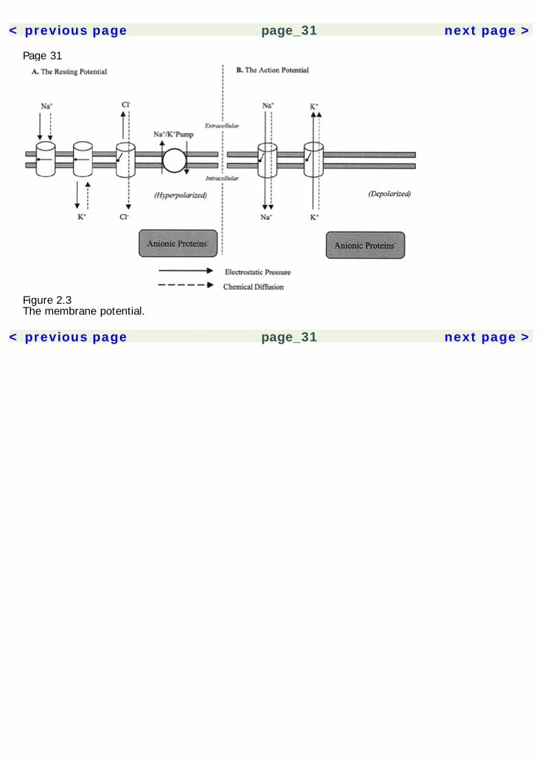
Page 29Membrane ProteinsIon PumpsPresent in the membrane are membrane-spanning proteins that pump ions across the membrane.The differential distribution of ions that form the membrane potential is driven by ion pumps. Despiteleakage currents, the Na+ and K+ gradients are maintained by the Na+/K+ pump. Hydrolysis of onemolecule of ATP allows three Na+ ions to be carried out, and two K+ ions to be carried in. Energy-dependent ion pumps are crucial for maintainance of steady-state ion gradients and the restingpotential. Various forms of energy deprivation will hinder ion pumps, causing catastrophicneurochemical cascades and neurotoxicity.Ion ChannelsIon channels are membrane proteins that allow the passage of ions in or out of the cell. The ion fluxis a passive process and occurs down the electrical and chemical gradients. There are four generaltypes of ion channels recognized. Nongated channels are continuously open, allowing a leakagecurrent across the membrane; voltage-gated channels are opened and closed by their sensitivity tothe local membrane potential; chemically gated channels are opened by the binding of a ligand to areceptor site; and ion-gated channels are gated by sensitivity to intracellular ion concentrations.Many ion channels may be ion-specific, selectively allowing passage of either K+, Na+, or Ca+,although some are less selective. Cations such as Mg2+, Mn2+, Co2+, and Cd2+ act as inhibitors ofvoltage-gated channels. The NMDA receptor channel is unique in that it is both chemically andvoltage-gated, requiring both simultaneously for channel opening.Membrane PotentialIn a resting state, neurons maintain a negatively charged potential, referred to as the restingpotential, that is actively maintained by energy-
< previous page page_29 next page >
< previous page page_30 next page >
Page 30dependent ion pumps. Metabolic deprivation of neurons causes a breakdown of this homeostasis withneurotoxic consequences.Membrane potentials are determined by a balance between two physical forces: chemical diffusionand electrostatic pressure. Chemical diffusion is the tendency of particles to move toward a uniformdistribution throughout a volume. Electrostatic pressure is simply the tendency for like-chargedparticles to repel and oppositely charged particles to attract each other.The neuronal membrane is relatively permeable to potassium (K+). In a resting state, K+ is in higherconcentration intracellularly, due to the activity of the Na+/K+ pump and attraction of ofintraneuronal anionic proteins (figure 2.3). However, by force of diffusion, K+ is also drawn out of thecell by the concentration gradient. The resting potential is largely determined by the distribution ofK+ ions. The membrane is relatively impermeable to Na+, which is pumped out of the cell and inhigher concentrations extracellularly. It is drawn into the neuron by both the forces of chemicaldiffusion and by electrostatic pressure, due to the membrane potential set by K+. This distribution ofions results in a resting potential of –60 to –70 mV with respect to the extracellular fluid. Chloride(Cl–) is a negatively charged ion that is in higher concentration outisde the cell due to membraneimpermeability and the Na+/Cl– pump. At the resting potential, Cl– is drawn intracellularly by forceof diffusion, but also repelled from the negatively charged intracellular milieu by electrostaticpressure. Finally, Ca2+ is another cation that, like Na+, is concentrated outside the cell. It is drawnintracellularly by both chemical diffusion and electrostatic pressure.The Action PotentialIn response to stimulation, the neuronal membrane becomes more permeable to Na+, which isdrawn into the neuron down the electrical and chemical diffusion gradients. This depolarizes theneuron and makes the membrane further permeable to Na+. If sufficiently depolarized, a thresholdof excitation is reached (approximately –60 mV), where voltage-gated Na+ channels open and themembrane becomes highly permeable
< previous page page_30 next page >
< previous page page_31 next page >
Page 31
Figure 2.3The membrane potential.
< previous page page_31 next page >
< previous page page_32 next page >
Page 32to Na+. The critical region for triggering this event is the axon hillock. An action potential is initiatedand the Na+ influx drives the membrane potential up to approximately +55 mV. This depolarizationis limited and reversed by inactivation of Na+ channels and efflux of K+ through delayed-openingK+ channels. The ion pumps then restore resting-level ion gradients. The action potential is followedby a brief refractory period, during which the neuron is slightly hyperpolarized below its restingpotential.Once an action potential is initiated at the hillock, it continues in a self-propagating fashion to theend of the axon. Voltage-sensitive ion channels are opened by the depolarization, allowing furtherinflux of Na+ and a spread of the action potential. Thus, the action potential is a self-regeneratingspread of membrane depolarization.Myelinated ConductionConduction of the action potential in myelinated axons is called saltatory conduction. Because ion fluxonly occurs at the nodes of Ranvier, the action potential jumps, in effect, from node to node. Thisprovides two advantages, speeding the rate of conduction and reducing the metabolic cost of anaction potential, because energy-dependent ion transporters are not needed along myelinatedsegments.Chemical SynapsesNeurotransmitter ReleaseChemical messengers, or neurotransmitters, are normally released when an action potential reachesthe synaptic terminals. This process is entirely Ca2+-dependent. Intracellular Ca2+ causesmovement of neurotransmitter-containing synaptic vesicles toward the membrane. Removal of Ca2+will prevent this process, preventing neurotransmitter release even if an action potential arrives.The synaptic vesicles dock with proteins on the neuronal membrane and release their contentsthrough exocytosis. The neurotransmitters contained in the vesicles spill into the synapse andpassively diffuse across.
< previous page page_32 next page >
< previous page page_33 next page >
Page 33A synaptic neurotransmitter may become inactivated by several mechanisms. It may passively diffuseout of the synapse; it may undergo enzymatic breakdown; it may be taken up into a cell through aselective reuptake pump; and, intracellularly, it may be taken back into a vesicle for future release,or it may be converted enzymatically.In certain neurons, a different type of synapse, called a gap junction, may be formed. Gap junctiontransmission occurs through membrane channels made of six subunits, which directly connect withother postsynaptic gap junction channels. When the channels open, there is a continuity of cytoplasmand exchange of ions between the two neurons. This mode of transmission is faster because it doesnot involve the time-consuming processes of neurotransmitter release, diffusion across the synapse,and receptor binding.ReceptorsNeurotransmitters create their effects by binding to receptor proteins. These may be broadlyclassified by their effector mechanisms as ionotropic or metabotropic. Ionotropic receptors arelocated on membrane ion channels composed of five subunits, each with four transmembranedomains (figure 2.4, 2.5). The binding of a ligand to the receptor causes a conformational change inthe channel, allowing ion permeability. Ions then flow passively down the concentration gradient,either depolarizing or hyperpolarizing the neuron. Other receptors on the ion channel may beallosteric, which may not itself initiate ion permeability, but instead creates effects by altering thebinding of ligands to the receptor.Metabotropic receptors, in contrast, create their effects by activating an intracellular G protein. Themetabotropic receptors are monomers with seven transmembrane domains. The activated G protein,in turn, may activate an ion channel from an intracellular site. Alternately, G proteins work byactivation or inhibition of enzymes that produce intracellular messengers. For example, activation ofadenylate cyclase increases production of cyclic adenosine monophosphate (cAMP). Other effectormechanisms include activation of phospholipases, diacylglycerol, creation of inositol phosphates, andproduction of arachidonic acid products. Ultimately, these cascades can result in proteinphosphorylation,
< previous page page_33 next page >

< previous page page_34 next page >
Page 34
Figure 2.4(A) Example of an ionotropic receptor. (B) Example of a metabotropic receptor.
< previous page page_34 next page >

< previous page page_35 next page >
Page 35
Figure 2.5Subunit structure of receptor proteins. (A) Ionotropic receptors are composed of five subunits, eachconsisting of four transmembrane domains. (B) Metabotropic receptors are composed of a single unitof seven transmembrane domains.release of intracellular Ca2+ from stores, enzyme activation, and altered gene transcription andprotein synthesis.Postsynaptic PotentialsActivation of ionotropic mechanisms creates postsynaptic potentials. An influx of cations or efflux ofanions depolarizes the neuron, creating an excitatory-postsynaptic potential (EPSP). Conversely, aninflux of anions or efflux of cations hyperpolarizes the neuron, creating an inhibitory-postsynapticpotential (IPSP). Postsynaptic potentials are summated both
< previous page page_35 next page >
< previous page page_36 next page >
Page 36spatially and temporally, where they must convergently occur in the same neuron and close enoughin time to amount to a larger effect and trigger an action potential.NeurotransmittersA variety of substances have been found to serve as neurotransmitters in the nervous system. Mostof these have actions outside the nervous system as well. Classically, the term neurotransmitterimplies ionotropic actions on neurons, while those with metabotropic actions are regarded asneuromodulators. This distinction is blurred, however, by the fact that many substances can haveeither action, depending on the receptor to which it binds. Table 2.1 summarizes the major classes ofneurotransmitters and their receptor-effector mechanisms.AcetylcholineAcetylcholine (ACh) is a phospholipid-derived neurotransmitter. It is produced from the precursors,choline and acetyl coenzyme A (CoA), by a reaction with the enzyme choline acetyltransferase(ChAT). It is broken down in the synapse by acetylcholinesterase (AchE), and choline is taken backup into the neuron for reuse. The drug hemicholinium inhibits uptake of choline, and several drugs,including physostigmine, prevent breakdown of ACh by inhibiting AChE. ACh has both ionotropic andmetabotropic receptors. Nicotinic ACh receptors are ionotropic, for which the drug nicotine is anagonist and mecamylamine is an antagonist. The nicotinic channel is cation selective and itsactivation results in depolarization. The muscarinic ACh receptors are metabotropic, and aresubdivided into five subtypes (M1 to M5). Muscarinic agonists include muscarine and pilocarpine, andantagonists include atropine and scopolamine.ACh is necessary for control of skeletal muscle in verterbrates, acting as the neurotransmitter at theneuromuscular junction. It is also involved in transmission in the autonomic nervous system (seebelow, under ''Neuroanatomy"). Central ACh is produced in two general areas in the brain incudingthe basal forebrain (medial septal nuclei, diagonal band
< previous page page_36 next page >
< previous page page_37 next page >
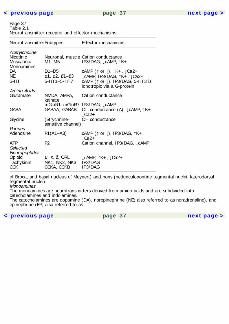
Page 37Table 2.1Neurotransmitter receptor and effector mechanisms
NeurotransmitterSubtypes Effector mechanisms
Acetylcholine Nicotinic Neuronal, muscle Cation conductanceMuscarinic M1–M5 IP3/DAG, ↓cAMP, ↑K+Monoamines DA D1–D5 cAMP (↑ or ↓), ↓K+, ↓Ca2+NE α1, α2, β1–β3 ↓cAMP, IP3/DAG, ↑K+, ↓Ca2+5-HT 5-HT1–5-HT7 cAMP (↑ or ↓), IP3/DAG, 5-HT3 is
ionotropic via a G-proteinAmino Acids Glutamate NMDA, AMPA,
kainateCation conductance
mGluR1–mGluR7 IP3/DAG, ↓cAMPGABA GABAA; GABAB Cl– conductance (A); ↓cAMP, ↑K+,
↓Ca2+Glycine (Strychnine-
sensitive channel)Cl– conductance
Purines Adenosine P1(A1–A3) cAMP (↑ or ↓), IP3/DAG, ↑K+,
↓Ca2+ATP P2 Cation channel, IP3/DAG, ↓cAMPSelectedNeuropeptides
Opioid μ, κ, δ, ORL ↓cAMP, ↑K+, ↓Ca2+Tachykinin NK1, NK2, NK3 IP3/DAGCCK CCKA, CCKB IP3/DAG
of Broca, and basal nucleus of Meynert) and pons (pedunculopontine tegmental nuclei, laterodorsaltegmental nuclei).MonoaminesThe monoamines are neurotransmitters derived from amino acids and are subdivided intocatecholamines and indolamines.The catecholamines are dopamine (DA), norepinephrine (NE; also referred to as noradrenaline), andepinephrine (EP; also referred to as
< previous page page_37 next page >
< previous page page_38 next page >
Page 38adrenaline). They are synthesized from the amino acid L-tyrosine by a series of enzymatic steps. L-tyrosine is converted to L-dopa (dihydroxyphenylalanine) by tyrosine hydroxylase. L-dopa is thenconverted to DA by dopa decarboxylase. In noradrenergic neurons, DA is further converted into NEby dopamine-β-hydroxylase, and in adrenergic neurons, NE is further converted to EP byphenylethanolamine-N-methyltransferase. The catecholamines are degraded extracelluarly bycatechol-O-methyltransferase and intracellularly by monoamine oxidase (MAO). MAO is inhibited bydrugs such as phenylzine. COMT is inhibited by tolcapone. The primary metabolites of DA are 3,4-dihydroxyphenylacetic acid (DOPAC) and homovanillic acid (HVA). The primary noradrenergicmetabolites are 3-methoxy-4-hydroxy-mandelic acid (VMA) and 3-methoxy-4-hydroxy-phenylglycol(MHPG).DA receptors are classified as D1 to D5. Bromocriptine and pergolide act as agonists at certainsubtypes, and neuroleptics such as haloperidol and thorazine act as antagonists. It is produced byseveral nuclei in the brain, but most notably the substantia nigra and the ventral tegmental area(VTA) of the midbrain. The substantia nigra mainly projects to the basal ganglia and the VTAsupplies limbic and cortical structures.NE and EP are agonists at α and β receptors. These are subdivided into α1A, α1B, α1D, α2A, α2B,α2C, β1, β2 and β3 subtypes. Phenylephrine is an agonist and prazosin is an antagonist at α1receptors. Guanabenz is an agonist at α2 receptors, and yohimbine is an antagonist. Isoproterenol isan agonist at β receptors, and propanolol is an antagonist. NE is released by neurons originating inseveral brain stem nuclei, most notably the dorsolateral tegmentum and nucleus locus coeruleus.Adrenergic nuclei are located in the medulla.Serotonin is an indolamine neurotransmitter, derived from the amino acid L-tryptophan. Tryptophanis converted to 5-hydroxytryptophan (5-HTP) by tryptophan hydroxylase. 5-HTP is converted to 5-hydroxytryptamine (serotonin, 5-HT) by aromatic amino acid decarboxylase. In the pineal gland, 5-HT may be further converted to N-acetyl serotonin by 5-HT N-acetyltransferase and then tomelatonin by 5-hyroxyindole-O-methyltransferase. 5-HT is catabolized by monoamine oxidase, andthe primary end metabolite is 5-hydroxyindoleacetic acid (5-HIAA).Receptors for 5-HT are labeled 5-HT1 to 5-HT7. The 5-HT1 subtype is further subdivided into 5-HT1A, 5-HT1B, 5-HT1D, 5-HT1E, and 5-HT1F,
< previous page page_38 next page >
< previous page page_39 next page >
Page 39and 5-HT2 is subdivided into 5-HT2A, 5-HT2B and 5-HT2C. 5-HT receptors work through severaleffector mechanisms including adenylate cyclase and phospholipases, and one subtype gates a cationchannel (5-HT3). Several 5-HT agonists exist with some subtype specificity, including sumatriptan(selective to 5-HT1E, and 5-HT1F) and 2,5-dimethoxy-4iodoamphetamine (selective to 5-HT2A, and5-HT2B). 5-HT antagonists include spiperone (5-HT1A), ketanserin (5-HT2), and ondansetron (5-HT3).Nuclei of 5-HT-producing neurons are located on many parts of the brain stem, mostly consisting ofthe raphe nuclei. Whereas the nuclei raphe obscurus, raphe pallidus, and raphe magnus project tobrain stem and spinal targets, the nuclei raphe dorsalis, raphe medianis, and raphe pontis oralisproject to forebrain sites.Amino AcidsThe amino acid neurotransmitters are subdivided into primarily excitatory (glutamate, aspartate) andinhibitory (γ-aminobutyric acid, GABA, glycine) types.The excitatory amino acids (EAA), glutamate and aspartate, are the principal excitatoryneurotransmitters in the brain. They are released by neurons in several distinct anatomical pathways,such as corticofugal projections, but their distribution is practically ubiquitous in the central nervoussystem. There are both metabotropic and ionotropic EAA receptors. The metabotropic receptors bindglutamate and are labeled mGluR1 to mGluR8. They are coupled via G-proteins to phosphoinositidehydrolysis, phospholipase D, and cAMP production. Ionotropic EAA receptors have been divided intothree subtypes: N-methyl-D-aspartate (NMDA), alpha-amino-3-hydroxy-5-methyl-4-isoxazole-proprionic acid (AMPA), and kainate receptors (Nakanishi 1992).The ionotropic receptors, AMPA and kainate, recognize glutamate but not aspartate as a ligand.Opening the channels allows infux of both Ca2+ and Na+. The NMDA receptor-channel is a cationicchannel that allows influx of Ca2+ and Na+, as well as efflux of K+ when activated by glutamate oraspartate (figure 2.6). In addition to the EAA binding site, the NMDA receptor-channel has severalother allosteric binding sites. Among them is the Mg2+ site, which is located within the channel
< previous page page_39 next page >

< previous page page_40 next page >
Page 40
Figure 2.6The NMDA channel.opening and blocks the channel in a voltage-dependent manner. The Zn2+ site is locatedextracellularly and also inhibits channel opening, but in a voltage-independent manner. Thus, theNMDA receptor-channel is believed to be unique in that it is gated simultaneously by voltage and bythe binding of ligands. Also within the channel opening is a binding site for MK-801, where bindinginhibits ion flux as well. There is also a strychnine-insensitive glycine receptor that is essential forNMDA channel opening, so glycine is regarded as a co-agonist for the NMDA channel.Glutamate may be converted by glutamic acid decarboxylase into GABA. GABA is the prinicpalinhibitory neurotransmitter in the brain, and is also found in the spinal cord. GABA is catabolized byGABA transaminase (GABA-T) into succinic semialdehyde. There are two GABA receptors, GABAA andGABAB. GABAA is a Cl– channel that is gated by GABA (figure 2.7). Muscimol is an agonist at thisreceptor and bicuculline is an antagonist. There are several allosteric sites on the GABAA channelthat enhance GABA binding. These allosteric modifiers include benzodiazepines, barbiturates, ethanol,and steroids such as allopregnanolone. Picrotoxin, in contrast, inhibits GABAA. The GABAB receptor isa metabotropic receptor that inhibits production of cAMP, opens K+ channels, and closes Ca2+channels via a G protein.
< previous page page_40 next page >

< previous page page_41 next page >
Page 41
Figure 2.7The GABAA channel.Glycine plays an important inhibitory role in the lower brain stem and spinal cord. Little is knownabout the synthesis of glycine. It activates a Cl– channel, which is antagonized by strychnine. Otherendogenous amino acids may activate the glycine channel, such as taurine and β-alanine.NeuropeptidesA variety of peptides are utilized in the nervous system as neurotransmitters. Unlike otherneurotransmitters, which can be synthesized in various parts of the neuron like the axon terminals,neuropeptides are produced by gene transcription and translation. They may colocalize and becoreleased with ACh, monoamines, or amino acid neurotransmitters. Their receptors aremetabotropic and may work through a variety of effector mechanisms. Neuropeptides are formedand degraded by a variety of peptidase enzymes.Neuropeptides are often grouped by their structural similarity or tissue source. Among these are thehypothalamic releasing factors (e.g., corticotrophin-releasing factor [CRF], thyrotropin-releasinghormone), anteior pituitary hormones (e.g., adrenocorticotrophic hormone [ACTH], follicle-stimulatinghormone [FSH]), and posterior pituitary hormones
< previous page page_41 next page >
< previous page page_42 next page >
Page 42(vasopressin and oxytocin). Several gastrointestinal peptides have been found to be active in thenervous system including cholecystokinin and neurotensin. Tachykinin neuropeptides includesubstance P, neurokinin A, and neurokinin B. The opioid neuropeptides derive from three mainfamilies: pro-opiomelanocortin, pro-dynorphin, and pro-enkephalin (see chapter 8 for a detaileddescription).NeurohormonesA number of neurotransmitter substances are used peripherally as hormones, including amines andpeptides (see above). Although they are chemically identical, they represent a different pool ofchemical messengers because they generally do not pass the blood-brain barrier. However, severalhormones readily pass the blood-brain barrier and have direct effects in the central nervous system.The hypothalamus releases severeal different releasing factors that travel through portal vessels tothe anterior pituitary. There they initiate the release of trophic hormones, which travel through bloodvessels to the various endocrine glands and trigger release of their hormones. Hormones from theadrenal, gonadal, and thyroid glands feed back to the pituitary gland and the brain, regulatingreleasing factors and trophic hormones. Hormones act through membrane-bound metabotropicreceptors, as well as to intracellular receptors that directly initiate gene expression.Soluble GassesRelatively recently, the gases nitric oxide (NO) and carbon monoxide (CO) have been found to act asneurotransmitters in the nervous system. Nitric oxide is synthesized from L-arginine via nitric oxidesynthase, requiring NADPH as a co-enzyme and tetrahydrobiopterin as a cofactor. Unlike otherneurotransmitters, NO is a small, very soluble molecule and cannot be stored in synaptic vesicles.Rather, it is synthesized on demand and freely diffuses through membranes. It is not broken downenzymatically because it is unstable and degrades rapidly. NO may have several actions, one of whichis to increase the production of cGMP by guanylyl
< previous page page_42 next page >
< previous page page_43 next page >
Page 43cyclase. It is thought to play a role in synaptic plasticity. It also serves as a chemical messenger inseveral other body tissues and is a vasodilator. CO is produced in the conversion of heme tobiliverdin by heme oxygenase in the brain, spleen, and liver.PurinesIn addition to their role in energy metabolism, adenosine and adenosine triphosphate (ATP) serve asneurotransmitters. They are stored in synaptic vesicles and released in response to action potentialsin widespread areas of the nervous system. A number of enzymes catalyze ATP and/or adenosine,including ATP diphosphohydrolase, 5′-nucleotidase, adenosine deaminase, and adenosine kinase.Membrane receptors for purine neurotransmitters are divided into P1, which binds adenosine, andP2, which binds ATP. The cloned P1 receptors have been subdivided into A1, A2A, A2B, and A3,which are metabotropic receptors. The P2 receptors are subdivided into ionotropic receptors (P2x1–P2x7) and metabotropic (P2y1, P2y2, P2y4 and P2y6). However, P2x7, P2y2, P2y4, and P2y6subtypes are not found in the nervous system.NeuroanatomyThe direct action of psychoactive drugs is upon neurochemical systems, but to fully appreciate theireffects one must consider the neurophysiological effects they produce in the context ofneuroanatomical structures. A drug may produce a spectrum of psychological and behavioral effects,each of which may be mediated by actions in different neuroanatomical structures. For example, theeffects of cannabinoids on memory and cognition are through their actions in the cortex, striatum,and hippocampus. However, their behavioral reinforcing effects are likely mediated through thenucleus accumbens, and their effects on thermoregulation are mediated through the hypothalamus.These effects are dissociable through experimental intracerebral injections, but occur in concert whena drug is administered systemically.The nervous system is divided into the central and peripheral nervous systems (CNS and PNS). TheCNS consists of the brain and spinal cord,
< previous page page_43 next page >
< previous page page_44 next page >
Page 44and the PNS consists of peripheral nerves and ganglia extending from the CNS. The spinal cord isfunctionally divided into somatic and autonomic divisions. The somatic division involves bodilysensations and skeletomotor functions, while the autonomic division controls smooth muscle, cardiacmuscle, and exocrine glands.Tissue in the nervous system is grossly classified as white or gray matter. White matter consists oftracts of axons, while gray matter consists of neuronal soma. Axons traveling in a peripheral bundleare referred to as a nerve, while central axons form a tract or funiculus. Alternately, axons thatconnect across the midline of the nervous system are referred to as a commissure. The largestcommissure is the corpus callosum, which connects the left and right hemispheres of the brain. Thecell bodies of gray matter may be organized into layers, which are referred to as cortex, such as thecerebral or cerebellar cortex. Alternately, cell bodies may be organized into a cluster, which isreferred to centrally as a nucleus or peripherally as a ganglion. The exception to this is the basalganglia, which are located deep in the brain.MeningesThe entire CNS is covered by the meninges, which form a protective covering. The outermost is thedura, which is tough and leathery in consistency. It is highly vascularized and innervated, so it issensitive to pain. The arachnoid membrane is a weblike, spongy layer beneath the dura. Beneath thearachnoid is the subarachnoid space, which is filled with cerebrospinal fluid. Beneath thesubarachnoid space is a thin layer of cells called the pia, which covers the brain and spinal cord.Ventricular SystemCerebrospinal fluid is produced in chambers within the brain called ventricles. Two lateral ventriclesand a midline third ventricle are contained within the cerebrum, while the fourth ventricle existswithin the brain stem. CSF is produced by the choroid plexus in the lateral and third ventricles. Itflows out through the ventricles by a series of aqueducts and into subarachnoid space. CSF supportsthe brain and spinal cord, ab-
< previous page page_44 next page >
< previous page page_45 next page >
Page 45sorbing impact. Occlusions of the ventricular system can result in elevated intracranial pressure, orhydrocephalus.Spinal CordThe spinal cord is the major route of input and output for the central nervous system. It is coveredby the meninges and sits within the vertebral column. There are 31 pairs of spinal nerves, exiting atthe dorsal and ventral roots of the spinal cord. The spinal cord is divided into four levels (superior toinferior): cervical, thoracic, lumbar, and sacral. A cross section of the spinal cord shows a butterfly-shaped core of gray matter (neuronal soma), and an outer surrounding area of white matter (axontracts).In general, dorsal regions of the spinal cord are involved in sensory processes, while ventral regionsare involved in motor functions. However, ventral roots and axonal tracts may also be involved inpain and temperature. Many simple and complex motor reflexes are well organized at the spinal level.The cranial nerves exit directly from the brain and brain stem. They control sensory, motor, andautonomic functions for parts of the neck and head. Vision, smell, taste, balance, hearing, andsensations and movements of the face are all mediated by cranial nerves. However, one large nerve,the vagus, regulates many visceral organs including the heart and the digestive tract.Autonomic Nervous SystemThe autonomic nervous system is subdivided into the sympathetic and parasympathetic nervoussystems. The sympathetic branch is involved in the expenditure of energy, while the parasympatheticconserves and stores energy. The sympathetic efferent neurons are in the thoracic and lumbar spinalcord, and project ventrally out to sympathetic ganglia outside of the spinal cord. The preganglionicsympathetic neurons synapse onto postganglionic neurons, which innervate the target organs. Thepreganglionic neurons use ACh as their neurotransmitter, which acts on nicotinic receptors onpostganglionic neurons (table 2.2). The
< previous page page_45 next page >
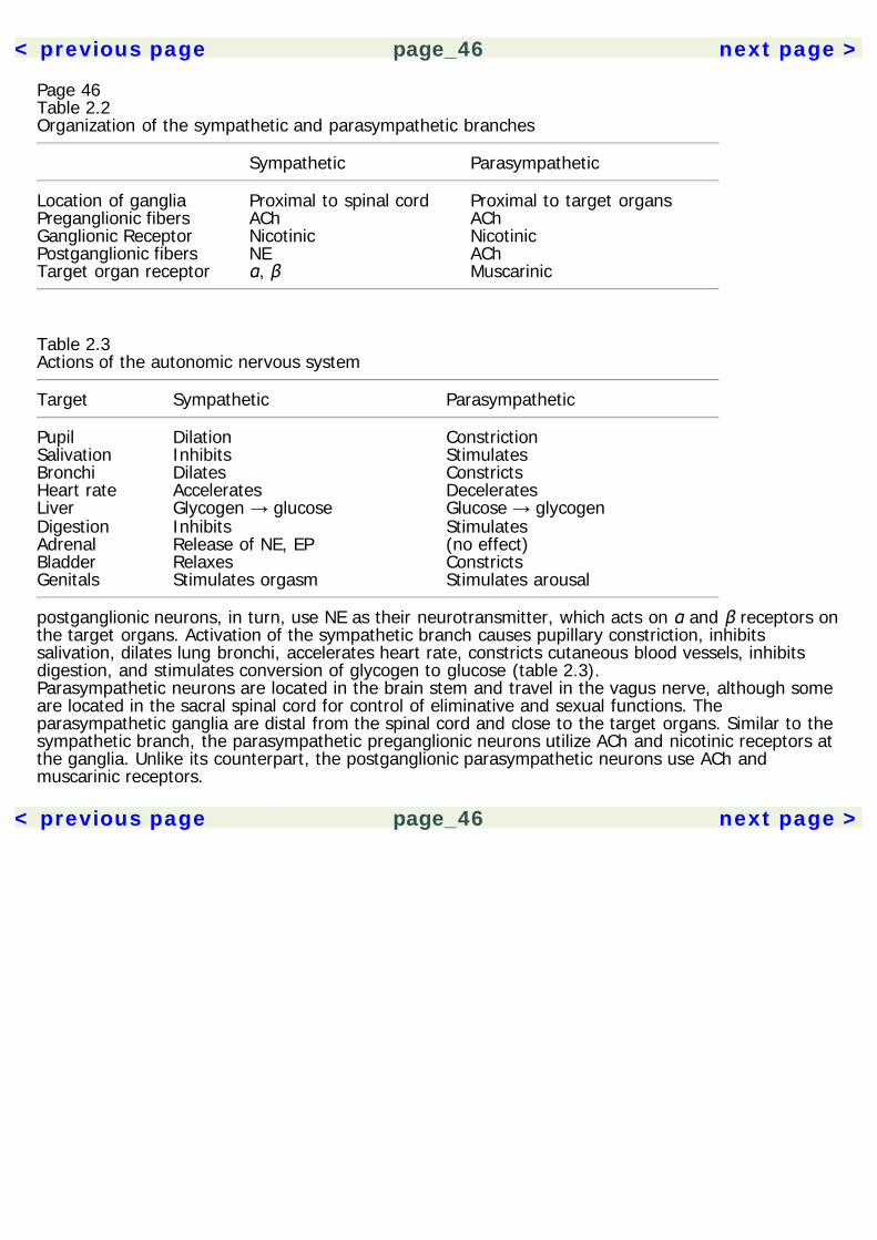
< previous page page_46 next page >
Page 46Table 2.2Organization of the sympathetic and parasympathetic branches
Sympathetic Parasympathetic
Location of ganglia Proximal to spinal cord Proximal to target organsPreganglionic fibers ACh AChGanglionic Receptor Nicotinic NicotinicPostganglionic fibers NE AChTarget organ receptor α, β Muscarinic
Table 2.3Actions of the autonomic nervous system
Target Sympathetic Parasympathetic
Pupil Dilation ConstrictionSalivation Inhibits StimulatesBronchi Dilates ConstrictsHeart rate Accelerates DeceleratesLiver Glycogen → glucose Glucose → glycogenDigestion Inhibits StimulatesAdrenal Release of NE, EP (no effect)Bladder Relaxes ConstrictsGenitals Stimulates orgasm Stimulates arousal
postganglionic neurons, in turn, use NE as their neurotransmitter, which acts on α and β receptors onthe target organs. Activation of the sympathetic branch causes pupillary constriction, inhibitssalivation, dilates lung bronchi, accelerates heart rate, constricts cutaneous blood vessels, inhibitsdigestion, and stimulates conversion of glycogen to glucose (table 2.3).Parasympathetic neurons are located in the brain stem and travel in the vagus nerve, although someare located in the sacral spinal cord for control of eliminative and sexual functions. Theparasympathetic ganglia are distal from the spinal cord and close to the target organs. Similar to thesympathetic branch, the parasympathetic preganglionic neurons utilize ACh and nicotinic receptors atthe ganglia. Unlike its counterpart, the postganglionic parasympathetic neurons use ACh andmuscarinic receptors.
< previous page page_46 next page >
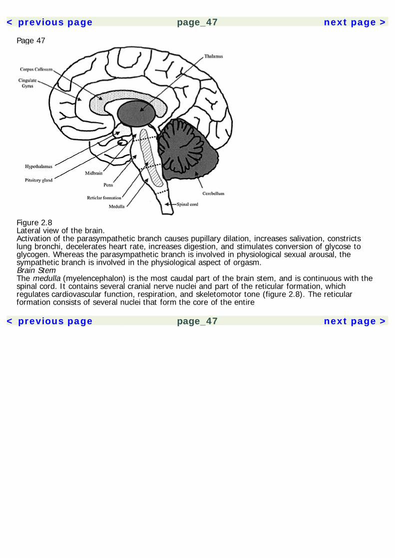
< previous page page_47 next page >
Page 47
Figure 2.8Lateral view of the brain.Activation of the parasympathetic branch causes pupillary dilation, increases salivation, constrictslung bronchi, decelerates heart rate, increases digestion, and stimulates conversion of glycose toglycogen. Whereas the parasympathetic branch is involved in physiological sexual arousal, thesympathetic branch is involved in the physiological aspect of orgasm.Brain StemThe medulla (myelencephalon) is the most caudal part of the brain stem, and is continuous with thespinal cord. It contains several cranial nerve nuclei and part of the reticular formation, whichregulates cardiovascular function, respiration, and skeletomotor tone (figure 2.8). The reticularformation consists of several nuclei that form the core of the entire
< previous page page_47 next page >
< previous page page_48 next page >
Page 48brain stem, projecting rostrally to the brain and caudally to the spinal cord.The pons and cerebellum (metencephalon) form the middle portion of the brain stem, borderedrostrally by the midbrain and caudally by the medulla. It also contains a portion of the brain stemand several nuclei important to arousal and sleep. The cerebellum branches off of the back of thebrain stem. It has a cortical surface and deep nuclei imbedded in white matter. Through itsconnections, it has involvement in diverse functions including balance, fine motor coordination, andeven cognition.The midbrain (mesencephalon) is the top of the brain stem. The dorsal midbrain is the tectum, whichis involved in eye movements and reflexive reactions to sensory stimuli. The mesencephalic reticularformation projects upward to the forebrain and is involved in arousal and attention. Theperiaqueductal gray area surrounds the cerebral aqueduct, and integrates analgesic,defensive/aggressive, sexual, and autonomic responses. The red nucleus and substantia nigra areimportant structures in motor function.There are several nuclei located in the brainstem reticular formation known for their role in producingspecific neurotransmitters and releasing them in the brain and/or spinal cord. As mentioned above,these are the locus coeruleus and dorsolateral tegmentum (norepinephrine), raphe nuclei (5-HT),ventral tegmental area and substantia nigra (DA), and pedunculopontine tegmental nuclei andlaterodorsal tegmental nuclei (acetylcholine). These nuclei also receive projections from forebrainareas. Thus, they may be regarded as part of forebrain-brainstem circuits, providing context-dependent feedback. Through their connections, they play important modulatory roles in cognitive,emotional, motor, and homeostatic functions.DiencephalonThe diencephalon is composed of several nuclei that are grossly grouped into the thalamus,hypothalamus, epithalamus, and subthalamic nucleus. The thalamus serves as a sensory relay,receiving projections from all of
< previous page page_48 next page >
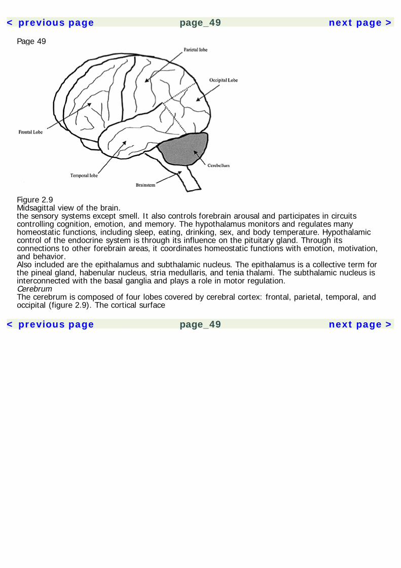
< previous page page_49 next page >
Page 49
Figure 2.9Midsagittal view of the brain.the sensory systems except smell. It also controls forebrain arousal and participates in circuitscontrolling cognition, emotion, and memory. The hypothalamus monitors and regulates manyhomeostatic functions, including sleep, eating, drinking, sex, and body temperature. Hypothalamiccontrol of the endocrine system is through its influence on the pituitary gland. Through itsconnections to other forebrain areas, it coordinates homeostatic functions with emotion, motivation,and behavior.Also included are the epithalamus and subthalamic nucleus. The epithalamus is a collective term forthe pineal gland, habenular nucleus, stria medullaris, and tenia thalami. The subthalamic nucleus isinterconnected with the basal ganglia and plays a role in motor regulation.CerebrumThe cerebrum is composed of four lobes covered by cerebral cortex: frontal, parietal, temporal, andoccipital (figure 2.9). The cortical surface
< previous page page_49 next page >

< previous page page_50 next page >
Page 50is convoluted, especially in humans, allowing for maximum surface area in a limited volume (i.e., thecranium). The ridges of cortex are referred to as gyri, and the valley in between them are sulci.The parietal lobe is involved in body sensations and the total body image, while the temporal lobe isinvolved in hearing. The occipital lobe is involved in basic aspects of vision. However, cortical visualprocessing continues into a ventral, ''what" pathway in the inferior temporal lobe—which is involvedin object and face recognition—and a dorsal, "where" pathway—which perceives spatial location.Higher-order cortical association areas control numerous other higher cognitive functions such asreading, arithmetic, language, and musical perception.The frontal lobes are proportionately larger in humans than in other animals. While more posteriorregions of the frontal lobe are concerned with motor function and the execution of behaviors, themore anterior portion (prefrontal cortex) is involved in cognition. In the most general sense, theprefrontal cortex is concerned with optimizing behavior by allowing for more goal-oriented andconceptually driven behavior. Thus, the prefrontal cortex is essential for such executive functions asabstract reasoning, planning, organization, goal formation, mental flexibility, initiation of behaviors,inhibition of behaviors, and social conduct.Beneath the cortex, the brain contains white-matter tracts, which interconnect the cortex, subcorticalnuclei, and lower structures. The limbic system is a circuit of structures surrounding the brain stemand diencephalon, which regulate emotions and memory. The hippocampus is a cortical structure inthe medial temporal lobe, which is responsible for the formation and consolidation of long-termmemories. It has reciprocal connections with the cerebral cortex, and removal of the hippocampusproduces an inability to form new memories (i.e., anterograde amnesia). The amygdala is a set ofsubcortical nuclei that are located in the anterior temporal lobe. The amygdala is involved indetermining the emotional significance of stimuli and events. Much work has been done to determinethe role of the amygdala in fear and anxiety; it is also likely involved in positive emotional states.Through its connections with executive and motor systems in the brain, the amygdala allows externalincentives to influence behavior.
< previous page page_50 next page >

< previous page page_51 next page >
Page 51The basal ganglia are a set of subcortical nuclei including the caudate, putamen, and globus pallidus.These structures form part of a processing loop running from the cortex to basal ganglia, and fromthere to parts of the thalamus and back to the frontal cortex. There are several such parallelprocessing loops throughout these structures, each mediating distinct cognitive and motor functions.Degeneration of the basal ganglia or its afferent inputs produces disorders of movement andcognition such as Parkinson's disease and Huntington's disease.
< previous page page_51 next page >

< previous page page_52 next page >
Page 52This page intentionally left blank.
< previous page page_52 next page >
< previous page page_53 next page >
Page 533Basic PharmacologyHerbal medicines contain active drugs, in every sense of the word. As such, the principles ofpharmacology pertain to them and allow us to understand their effects. The purpose of this chapteris to outline some of these principles. For a more complete treatment of the topic, the reader isdirected to the sources listed in the references. Although these principles apply to all drugs, thischapter will draw upon examples from plant drugs to illustrate them.There are two main areas of pharmacology: pharmacokinetics and pharmacodynamics. The simplestdistinction between these two is that pharmacokinetics is how the body affects a drug, whilepharmacodynamics is how a drug affects the body.PharmacokineticsPharmacokinetics is specifically concerned with the transfer of a drug through the body by theprocesses of absorption, distribution, biotransformation, and excretion. A drug's dosage andpharmacokinetic properties will determine the concentrations that will be reached, and therefore theintensity of effect produced. Ultimately, these processes determine the bioavailability of a drug, orthe degree to which it reaches the intended site of action.The importance of pharmacokinetics should not be underestimated. The effects of psychoactive drugsare determined not only by their pharmacodynamic mechanisms, but also by how much of the drugreaches the brain, and how fast it does so. A cardinal example of this involves the behavioraldifferences between chewing coca leaves, snorting cocaine
< previous page page_53 next page >
< previous page page_54 next page >
Page 54HCl, and smoking freebase crack. All three of these deliver the drug cocaine, but each deliversprogressively greater amounts to the brain at a quicker rate. The end result of this is that traditionalchewing of coca leaves probably approaches the equivalence of drinking espresso coffee, whilesmoking crack has become an epidemic of drug addiction. Similarly, morphine and heroin may haveidentical mechanisms in the brain, but heroin (diacetylmorphine) gains access faster and to a greaterextent.The processes of pharmacokinetics all involve the transfer of a drug across membranes, beginningwith the cell membrane, and sometimes involving single or multiple layers of cells. Drugs can crossthese membranes through passive or active transport.Passive transport is not energy dependent, and involves drugs moving along a concentrationgradient, from an area of high concentration to an area of low concentration. Drugs that arenonionized generally have good lipid solubility, and thus can pass through the lipid cell membranes.Conversely, ionized drug molecules have low lipid solubility and do not pass through membraneswell. Other factors influencing this process are the drug and pH concentration gradients across themembrane, where the pH gradient can influence the ionization of the drug.Active transport of a drug is mediated by a specific carrier. This is of particular interest in neuraltissues and choroid plexus. In this case, the actual molecular size and shape are important, becauseit involves binding to a specific carrier protein that transports it. Active transport is an energy-dependent process, and may work against a concentration gradient. It is also a saturable mechanismand may be competitively inhibited by other ligands.AbsorptionAbsorption is defined as the rate and extent to which a drug leaves the site of administration.Several factors influence a drug's absorption (table 3.1). Aqueous solutions are more easily absorbedthan a lipid solution or solid form. Absorption of drugs in solid form is affected by the rate at which itdissolves. Higher concentrations of a drug are more rapidly absorbed than low concentrations. Theamount of blood flow to the site also influences absorption: heat and vasodilators increaseabsorption,
< previous page page_54 next page >

< previous page page_55 next page >
Page 55Table 3.1Factors influencing drug absorption
Type of solution (aqueous vs. lipid)Rate of dissolution of solid drugsConcentrations of a drug presentBlood flow to the site of absorptionSurface area of absorption
while vasoconstrictors reduce it. The surface area of absorption is a very important factor. Drugs arevery effectively absorbed by tissues with large surface areas, as in the lungs.Routes of AdministrationAbsorption is greatly altered by the route of administration of a drug. These routes are broadlydichotomized as enteral or parenteral (table 3.2). Enteral administration involves absorption throughthe gastrointestinal (GI) tract, and parenteral refers to any other form of absorption. Each has itsown respective advantages and disadvantages (table 3.3). Enteral routes are the safest, mostconvenient, and most economical means, and thus are the most common. However, they have thedisadvantages of irregular absorption, nausea and vomiting, and enzyme degradation. They alsonecessitate patient cooperation, which is not possible in some instances (e.g., seizures, psychoticagitation). Parenteral routes, on the other hand, allow the drug to be absorbed in active form, in amore rapid absorption and in a predictable manner. Their disadvantages are the risk of infection,they are more difficult to perform (e.g., injections), and they produce greater discomfort.Enteral routes of administration include oral, sublingual, and rectal. Oral ingestion of a drug results inpassive absorption through the GI tract. The pH of the drug affects this process: weak acids are bestabsorbed from the stomach, while weak bases absorb better from the small intestine. Nonionizeddrugs absorb well from any part of the GI tract. Sublingual administration involves absorption fromthe oral mucosa, and avoids hepatic first-pass metabolism. Rectal administration involves absorptionfrom the lower GI tract. It avoids the nausea and vomiting produced by direct stomach irritation, butalso has irregular absorption.
< previous page page_55 next page >
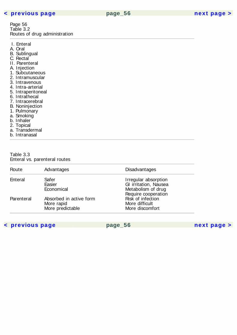
< previous page page_56 next page >
Page 56Table 3.2Routes of drug administration
I. EnteralA. OralB. SublingualC. RectalII. ParenteralA. Injection1. Subcutaneous2. Intramuscular3. Intravenous4. Intra-arterial5. Intraperitoneal6. Intrathecal7. IntracerebralB. Noninjection1. Pulmonarya. Smokingb. Inhaler2. Topicala. Transdermalb. Intranasal
Table 3.3Enteral vs. parenteral routes
Route Advantages Disadvantages
Enteral Safer Irregular absorption Easier GI irritation, Nausea Economical Metabolism of drug Require cooperationParenteral Absorbed in active form Risk of infection More rapid More difficult More predictable More discomfort
< previous page page_56 next page >
< previous page page_57 next page >
Page 57For example, rectal diazepam may be administered to people undergoing continuous seizures (statusepilepticus), because other oral and parenteral routes would not be feasible.Injection forms of parenteral administration include subcutaneous, intramuscular, intravenous, intra-arterial, intraperitoneal, intrathecal, and intracerebral injections. Intravenous administration (IV)involves injection of an aqueous solution into the veins, bypassing the processes of absorption. Thisallows accurate and rapid control of drug concentrations, and less sensitivity to irritating drugs.However, dangerously high concentrations can be easily and quickly reached this way (e.g., heroinoverdose). Subcutaneous injection of drugs (SC) can only be done with nonirritating drugs, otherwisepain, necrosis, and slough of tissues may occur. Subcutaneous absorption depends on blood flow tothe site of injection. Likewise, absorption from intramuscular injection (IM) depends on blood flow tothe site of injection. It is performed with an aqueous solution and absorption may be very rapid.Intra-arterial injections are performed to highly localize the effects of a drug, but this requires greatcaution because hepatic metabolism and lung filtering are bypassed. Intraperitoneal injection (IP)involves absorption from the abdominal cavity into the portal vein. This allows a large absorbingsurface, but the risk of infection to this area is serious. Intrathecal administration involves injectioninto the spinal column, bypassing blood-brain and blood-cerebrospinal fluid (CSF) barriers. This routeis usually reserved for spinal anesthesia or to treat infections of the central nervous system. Moredirectly, intracerebral injections may be performed surgically when a specific locus in the brain mustbe targeted (e.g., injection of fetal cell grafts for Parkinson's disease).Noninjection forms of parenteral administration include pulmonary or topical absorption. Pulmonaryabsorption is performed with gaseous drugs (e.g., general anesthetics) and drugs aerosolized byinhalers (e.g., asthma medications) or by burning (e.g., tobacco smoking). Absorption is rapidthrough the large surface area of the pulmonary epithelium and respiratory tract. The disadvantagesof pulmonary absorption are the difficulty in controlling the dose, inconvenient administration, andpotential irritation of the pulmonary epithelium. Topical (or transdermal) administration involvesabsorption from mucous membranes or the
< previous page page_57 next page >
< previous page page_58 next page >
Page 58Table 3.4Factors influencing drug distribution
Blood flow in the target organsLipid solubility (especially CNS)Accumulation in drug reservoirsSerum proteinsFatMusclesLiverBoneTranscellular (GI, CSF)Molecular size (especially CNS)Specific carrier transport
skin. For example, absorption through mucous membranes is utilized when cocaine HCl is snorted, orwhen medieval ''witches" applied hallucinogenic ointments to their vaginal membranes. Absorptionthrough the skin is more common clinically, although few drugs absorb well this way. However,absorption is improved through an oily medium such as a cream. Nicotine patches for cessation oftobacco smoking are an example of transdermal absorption.DistributionDrug distribution involves its movement into the interstitial and cellular fluids. Several factorsinfluence drug distribution, including blood flow (table 3.4). Organs with the greatest blood flow(e.g., the brain, heart, liver, and kidneys) receive most of the drug initially, while other tisses (e.g.,muscle, viscera, skin, and fat) receive it more gradually. Distribtion is also influenced by a drug's lipidsolubility and accumulation in drug reservoirs.Certain factors determine distribution of drugs to the central nervous system. An importantdeterminant is the drug's molecular size, because cerebral capillaries have tight junctions and onlythe smallest molecules can pass through unless there is a specific carrier mechanism. However, drugsthat are highly lipid soluble may pass through the brain's perme-
< previous page page_58 next page >

< previous page page_59 next page >
Page 59ability barriers. Drug distribution to the brain is limited also by the degree of cerebral blood flow.However, the blood-brain barrier is not impermeable: large doses may leak through, particularly atareas of non-uniformity (e.g., circumventricular organs).Drug reservoirs are bodily compartments in which a drug is stored. When a portion of the drug is in areservoir it limits the amount of free drug available, because only unbound drug is in equilibriumacross membranes. On the other hand, reservoirs also prolong the action of a drug, maintaining itsconcentrations by releasing it gradually as blood levels decline. There are several drug reservoirs,such as plasma proteins albumin and glycoproteins. Binding to plasma proteins is usually reversibleand competitive. Therefore, the plasma level of one drug will be influenced by any other drugs thatbind to the same plasma proteins, displacing some of it from that reservoir. Certain intracellularreservoirs may also accumulate drugs, such as muscles and the liver, due to active transport orbinding to a protein. Fat may serve as a large drug reservoir, which slowly releases the drug whenplasma levels decline. Bone may also serve as a reservoir, particularly for heavy metals and chelatingagents, with very slow and sustained release of the drug. Finally, drugs may accumulate intranscellular reservoirs, such as in the GI tract or CSF.When levels of the drug decline in plasma, usually due to biotransformation and excretion, the drugthen redistributes away from its site of action into other tissues (e.g., drugs may distribute across theplacenta during pregnancy). This is a passive process, but lipid-soluble drugs penetrate the most.This is of special concern with psychoactive drugs, which tend to be lipid-soluble.BiotransformationBiotransformation involves the chemical alteration of a molecule to alter its effects. This oftenterminates the pharmacological effects of a drug, but active metabolites are produced in some cases.Biotransformation also changes the ease with which a drug is eliminated. This involves conversion ofthe drug to a more hydrophilic metabolite that enhances renal excretion. Although this processpertains to most drugs, it probably
< previous page page_59 next page >

< previous page page_60 next page >
Page 60Table 3.5Biotransformation reactions
Phase I ReactionsA. Oxidative Reactions1. N-dealkylation2. O-dealkylation3. Aliphatic hydroxylation4. Aromatic hydroxylation5. N-oxidation6. S-oxidation7. DeaminationB. Hydrolysis ReactionPhase II ReactionsA. GlucuronidationB. SulfationC. Acetylation
evolved to remove chemical constituents of ingested plants, includng flavonoids, terpenes, steroids,and alkaloids.Biotransformation of drugs occurs in two phases (table 3.5). Phase I reactions involve oxidative orhydrolysis reactions. Oxidative reactions include N-dealkylation, O-dealkylation, aliphatichydroxylation, aromatic hydroxylation, N-oxidation, S-oxidation, or deamination. Following a phase Ireaction, a covalent conjugation reaction may occur, making the compound more polar, watersoluble, and thus more easily excreted in urine and feces. Phase II reactions are conjugationreactions: glucuronidation, sulfation, and acetylation. As mentioned above, metabolic conversion ofdrugs can produce active metabolites, which sometimes may be more active than the drugadministered. For example, although morphine is an active drug, some of its clinical effects areattributable to its active metabolite morphine-6-glucuronide (M6G). In some cases, the compoundadministered may itself be inactive, but becomes active after metabolic conversion.The primary organ of biotransformation is the liver, although some other tissues have a degree ofmetabolic capacity, including the GI tract, kidneys, and even the skin. Depending on the route ofadministration,
< previous page page_60 next page >
< previous page page_61 next page >
Page 61drugs may undergo first-pass metabolism, so that a proportion of the drug is inactivated before itreaches the target tissue.The microsomal enzymes responsible for catalyzing oxidative and reductive reactions are thecytochrome P450 enzyme family. These are heme-containing membrane proteins that are found inthe smooth endoplasmic reticulum of the cells of many tissues, but especially in the liver. Twelvegene families of the cytochrome P450 enzyme have been identified in humans, and the threefamilies, CYP1, CYP2, and CYP3 account for most drug biotransformations. Hydrolytic reactions arecarried out by the enzyme epoxide hydrolase and several amidases, esterases, proteases, andpeptidases. Conjugation reactions are carried out by uridine diphosphate glucuronosyltransferases,sulfotransferases, and N-acetyltransferases.Several factors may influence the processes of biotransformation. Certain drugs or toxins may inducesynthesis of cytochrome P450. For drugs that are transformed into toxic metabolites, this mayamount to significant hepatic or renal toxicity over time. A common example of this is with chronicalcoholism resulting in liver damage. Drugs may also inhibit cytochrome P450 enzymes, resulting inslower metabolism and prolonged drug concentrations. Unintentional interactions between diversetypes of drugs may occur through competition for enzymes. For example, several studies haveexamined the inhibition of cytochrome P450 3A4 (CYP3A4) by grapefruit juice. Biotransformation maybe reduced in cases of liver disease. It is also reduced in fetal and neonatal stages of development,and in many elderly. Genetic differences normally exist between individuals in drug metabolismcapacity, which result from polymorphisms in the cytochrome P450 family.ExcretionExcretion is the process of eliminating drugs from the body. They may be excreted as metabolites oras unchanged drug. As mentioned above, compounds that are polar and water soluble are morereadily eliminated. The major routes of excretion are renal, biliary/fecal, lactational, and pulmonary.
< previous page page_61 next page >

< previous page page_62 next page >
Page 62The kidneys are the most important routes of drug excretion. This begins with filtration of a drugfrom the blood by renal glomeruli. Through these tiny clusters of capillaries, a filtrate is formed thathas essentially the same composition of plasma, minus the larger proteins. The filtrate containswater, glucose, amino acids, urea, uric acid, creatine, creatinine, anions, and cations. Nonselectivecarrier systems transport organic anions and cations into the filtrate, and transport can sometimes bebidirectional, allowing for reabsorption. Organic acids and drug metabolites are transported by acarrier system that normally transports uric acid.After glomerular filtration, fluid passes through proximal and distal tubules, and eventually collectsinto the ureter and bladder, where it is eliminated. However, considerable reabsorption occurs fromthe proximal and distal tubules. Nonionized weak acids and bases are passively reabsorbed, movingdown a concentration gradient. Reabsorption of ionized substances is pH dependent. The kidneys ofan average adult filter 45 gallons per 24-hour period, the majority of which is obviously reabsorbed.Hepatic metabolites are excreted into the intestinal tract in bile. From there, the metabolites may beexcreted in feces or reabsorbed into the blood. Metabolites are carried into the bile via activetransport. Pulmonary excretion of drugs occurs mainly for gases and vapors, such as generalanesthetic drugs. Excretion of drugs and metabolites occurs in breast milk, which is of significantconcern for breast-feeding mothers. Breast milk is slightly more acidic than plasma, so basiccompounds may be present in greater concentrations, while acidic compounds are in lowerconcentrations. Nonelectrolytes enter breast milk in a pH-independent manner. Other less-importantroutes of excretion are through sweat, saliva, and tears, which primarily excrete nonionized, lipid-soluble drugs. Elimination of drugs in the skin and hair is quantitatively minute, but detectable forforensic purposes.Clinical PharmacokineticsThere are several pharmacokinetic paramaters that are relevant to the clinical use of drugs. Thesehelp quantify a dose-response relationship, or
< previous page page_62 next page >

< previous page page_63 next page >
Page 63
Figure 3.1Steady-state concentration.the relationship between the drug concentrations and its magnitude of effect. It is often desirableclinically to achieve a steady-state concentration of a drug to maintain a relatively constant level ofeffect (figure 3.1). To rapidly reach this level, a larger initial loading dose may be given, followed bysmaller maintenance doses to sustain that level.The three main parameters of clinical pharmacokinetics are clearance, distribution volume, andbioavailability. Clearance is the rate at which the body eliminates a drug. In order to achieve asteady-state concentration, the drug must be given so that the rate of clearance equals the rate ofadministration. If the drug is given as quickly as it is eliminated, a consistent level in the body will bemaintained.Clearance is constant across drug concentrations because the mechanisms are normally notsaturated. Under such normal conditions, a constant fraction of the drug is eliminated per unit time,whereas if there is
< previous page page_63 next page >
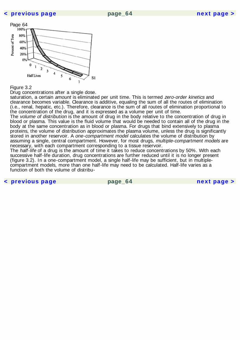
< previous page page_64 next page >
Page 64
Figure 3.2Drug concentrations after a single dose.saturation, a certain amount is eliminated per unit time. This is termed zero-order kinetics andclearance becomes variable. Clearance is additive, equaling the sum of all the routes of elimination(i.e., renal, hepatic, etc.). Therefore, clearance is the sum of all routes of elimination proportional tothe concentration of the drug, and it is expressed as a volume per unit of time.The volume of distribution is the amount of drug in the body relative to the concentration of drug inblood or plasma. This value is the fluid volume that would be needed to contain all of the drug in thebody at the same concentration as in blood or plasma. For drugs that bind extensively to plasmaproteins, the volume of distribution approximates the plasma volume, unless the drug is significantlystored in another reservoir. A one-compartment model calculates the volume of distribution byassuming a single, central compartment. However, for most drugs, multiple-compartment models arenecessary, with each compartment corresponding to a tissue reservoir.The half-life of a drug is the amount of time it takes to reduce concentrations by 50%. With eachsuccessive half-life duration, drug concentrations are further reduced until it is no longer present(figure 3.2). In a one-compartment model, a single half-life may be sufficient, but in multiple-compartment models, more than one half-life may need to be calculated. Half-life varies as afunction of both the volume of distribu-
< previous page page_64 next page >
< previous page page_65 next page >
Page 65tion and clearance. The half-life may be altered in certain disease states. It may be increased, forexample, by reduced drug metabolism with age. On the other hand, the half-life of a drug may bereduced in cases of microsomal enzyme induction.As mentioned above, bioavailability is the degree to which a drug reaches the intended site of action.The amount of drug that reaches systemic circulation will depend on the processes of absorption,distribution, and biotransformation (when the route of administration exposes the drug to first-passmetabolism). Pharmacokinetics are often linear and when they are nonlinear it is often due to asaturation of protein binding, metabolism, or active renal transport.PharmacodynamicsPharmacodynamics consist of the chemical and physiological mechanisms of action of a drug.Further, it concern the relationship between drug concentrations and the magnitude of effect.Mechanism of Drug ActionThe mechanisms of most drugs involve binding of the drug to a receptor. A receptor may be anymacromolecular target, but the most common receptors are proteins. These include membraneproteins, enzymes, transporters, and structural elements. Some of the main receptors of interest forpsychopharmacology are receptors for neurotransmitters and hormones, which show a high degree ofselectivity.A drug may have several potential effects at a receptor target. A drug that binds to the receptor andimitates the actions of the endogenous ligand is called an agonist. A partial agonist also activates thereceptor mechanism, but does not reach full efficacy. A drug that binds to the receptor and has nointrinsic effect, but instead prevents binding of other ligands, is called an antagonist. An inverseagonist is a drug that has the opposite effect of an agonist.Drugs are thought to bind to receptors in a lock-and-key model, meaning that the molecular shapeof a drug determines how well, if at all, it will attach to a receptor and what activity it will havethere.
< previous page page_65 next page >
< previous page page_66 next page >
Page 66Structure-activity relationships are very important in understanding drug actions and in designingnovel drugs. Seemingly small modifications in a drug's molecular structure can radically alter itseffects.Drug ReceptorsProtein receptors for endogenous ligands (e.g., hormones, neurotransmitters) share some commonproperties. They contain a ligand-binding domain, where the hormone or neurotransmitter attaches,and an effector domain, which translates the binding into some from of action.Receptor-effector mechanisms include (1) enzymes with catalytic activities, (2) ion channels that gatethe transmembrane flux of ions (ionotropic receptors), (3) G protein-coupled receptors that activateintracellular messengers (metabotropic receptors), and (4) cytosolic receptors that regulate genetranscription. Cytosolic receptors are a specific mechanism of many steroid and thyroid hormones.The ionotropic and metabotropic receptors are discussed in relevance to specific neurotransmitters inchapter 2.Receptor RegulationReceptor regulation is under homeostatic control. Continuous stimulation of a receptor may lead toits desensitization (or down regulation). Homologous densensitization involves covalent modification,destruction, decreasesd synthesis, or relocalization of the receptor within the cell. Heterologousdensensitization, on the other hand, occurs when a receptor is desensitized by sharing a commonsignaling pathway with other receptors. For example, several neurotransmitter receptors mayconvergently act upon adenylate cyclase synthesis of cAMP.Conversely, understimulation of a receptor may lead to its sensitization (or up regulation). This mayoccur when an antagonist is administered for a long period of time, or when tissue is denervated anddoes not receive the normal afferent stimulation.Quantifying Dose-Response RelationshipsUnder certain circumstances, the magnitude of drug effect is related to the proportion of receptorsoccupied, and maximal effects occur when all
< previous page page_66 next page >
< previous page page_67 next page >
Page 67receptors are occupied. However, there are many exceptions and nonlinear relationships also exist.The concept of efficacy means that different agonists could have different intensity of activity at areceptor, and it explains why two different agonists could occupy the same proportion of receptorsbut have different magnitudes of effect.Plotting the dose of an agonist against the effect produces a linear curve, and plotting the log doseversus the effect produces a sigmoidal curve (figure 3.3). A full agonist may create maximal effectsat the appropriate dose. Full agonists may differ in their potency, where both reach maximal effects,but one does so at a lower dose (figure 3.4). In contrast, full and partial agonists differ by theirmaximum efficacy. No matter how much of a partial agonist is given, it will not achieve the samemagnitude of effect as a full agonist. Combinations of agonists may produce additive effects, orsynergistic effects, which are greater than the sum of the effects of each individual drug.Antagonist drugs may reduce the potency or efficacy of an agonist, depending on the nature of theirreceptor binding: competitive or non-competitive. Competitive antagonists can be overcome byincreasing the amount of agonist, and maximum agonist effects may still be achieved. This producesa rightward shift in the dose-response curve, reflecting altered potency (figure 3.5). Noncompetitiveantagonists, however, have irreversible receptor binding and are not overcome by agonists. This isreflected in the dose-response curve as a reduction in maximum efficacy.Inverse agonists will not only antagonize or nullify the effects of agonists when given together, theywill have the opposite effect of the agonist when administered alone (figure 3.6). This is especiallytrue with receptors that have a basal rate of activity, which might be increased by an agonist orfurther decreased by an inverse agonist. For example, diazepam is regarded as an agonist at theGABAA benzodiazepine receptor and has anxiolytic effects. β-carbolines, in contrast, are regarded asinverse agonists of the benzodiazepine receptor, and are anxiogenic. (However, uncertainty over theendogenous ligand of the benzodiazepine receptor makes the assignment of agonist versus inverseagonist somewhat arbitrary. Indeed, there are endogenous β-carbolines, so they might be regardedas the actual agonists.)Dose-response relationships may be examined in individuals or in populations. The dose-responsecurves discussed above (e.g., figure 3.3)
< previous page page_67 next page >
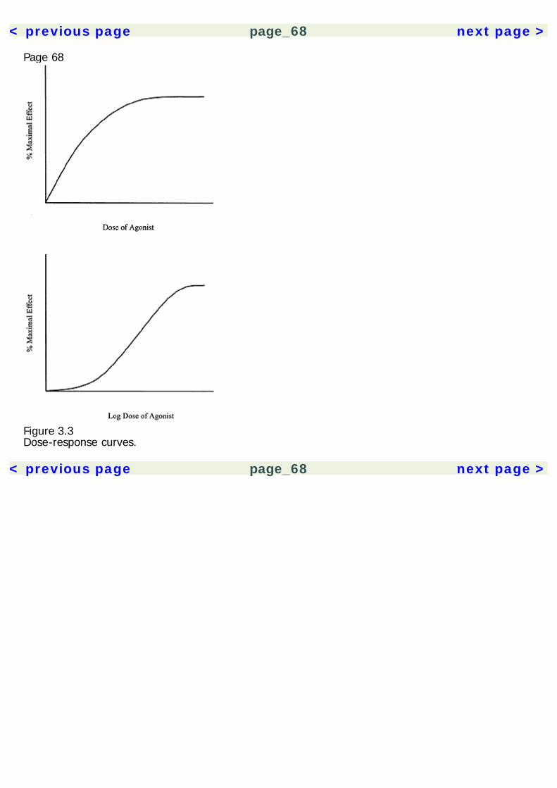
< previous page page_68 next page >
Page 68
Figure 3.3Dose-response curves.
< previous page page_68 next page >
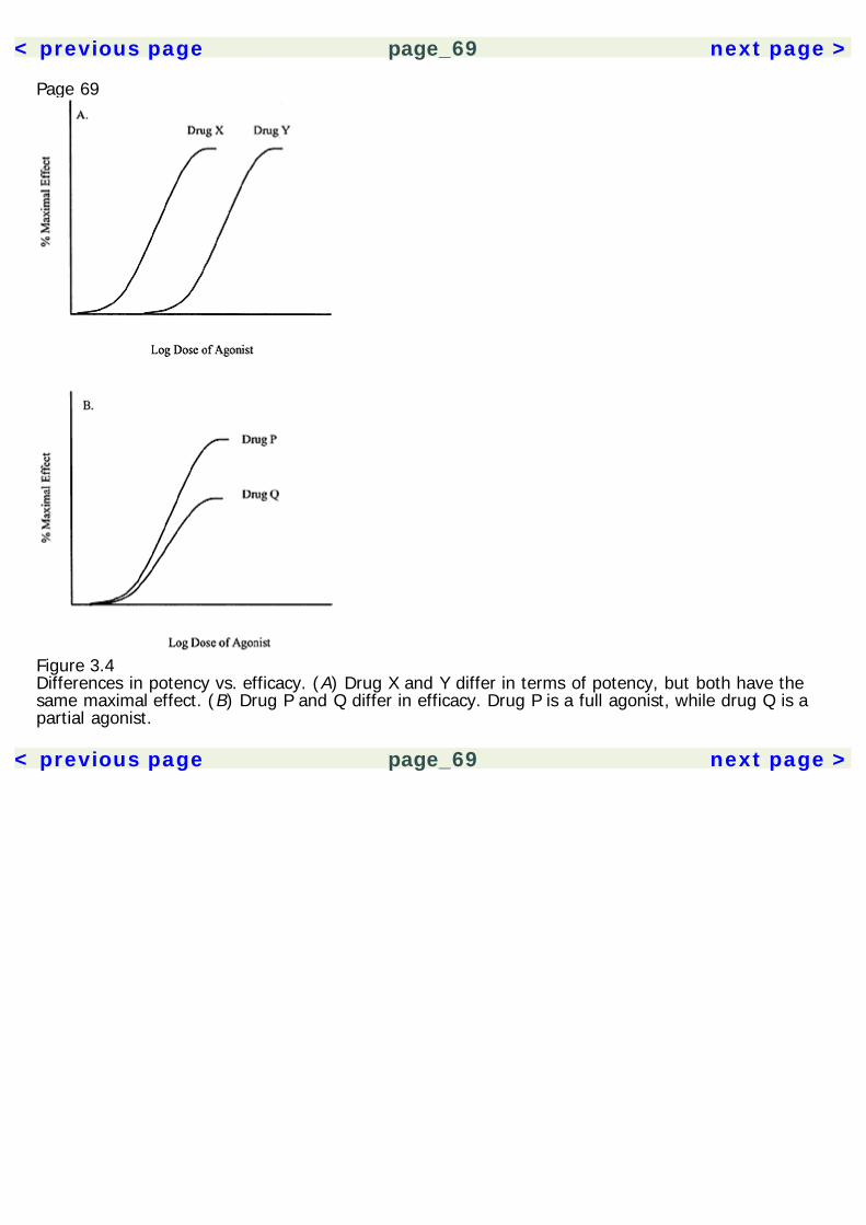
< previous page page_69 next page >
Page 69
Figure 3.4Differences in potency vs. efficacy. (A) Drug X and Y differ in terms of potency, but both have thesame maximal effect. (B) Drug P and Q differ in efficacy. Drug P is a full agonist, while drug Q is apartial agonist.
< previous page page_69 next page >
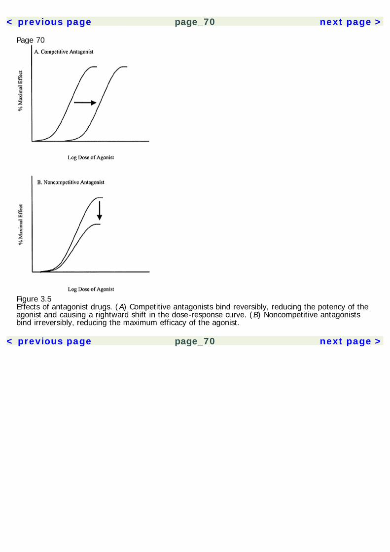
< previous page page_70 next page >
Page 70
Figure 3.5Effects of antagonist drugs. (A) Competitive antagonists bind reversibly, reducing the potency of theagonist and causing a rightward shift in the dose-response curve. (B) Noncompetitive antagonistsbind irreversibly, reducing the maximum efficacy of the agonist.
< previous page page_70 next page >
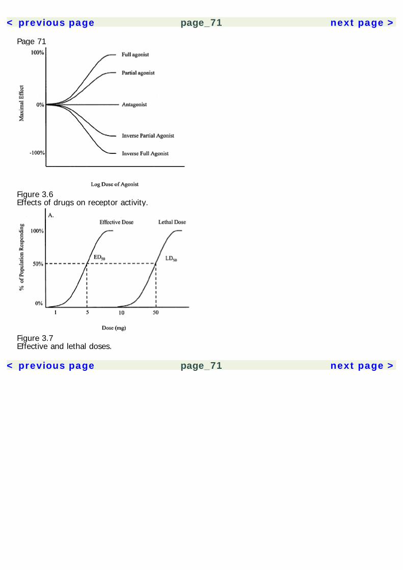
< previous page page_71 next page >
Page 71
Figure 3.6Effects of drugs on receptor activity.
Figure 3.7Effective and lethal doses.
< previous page page_71 next page >
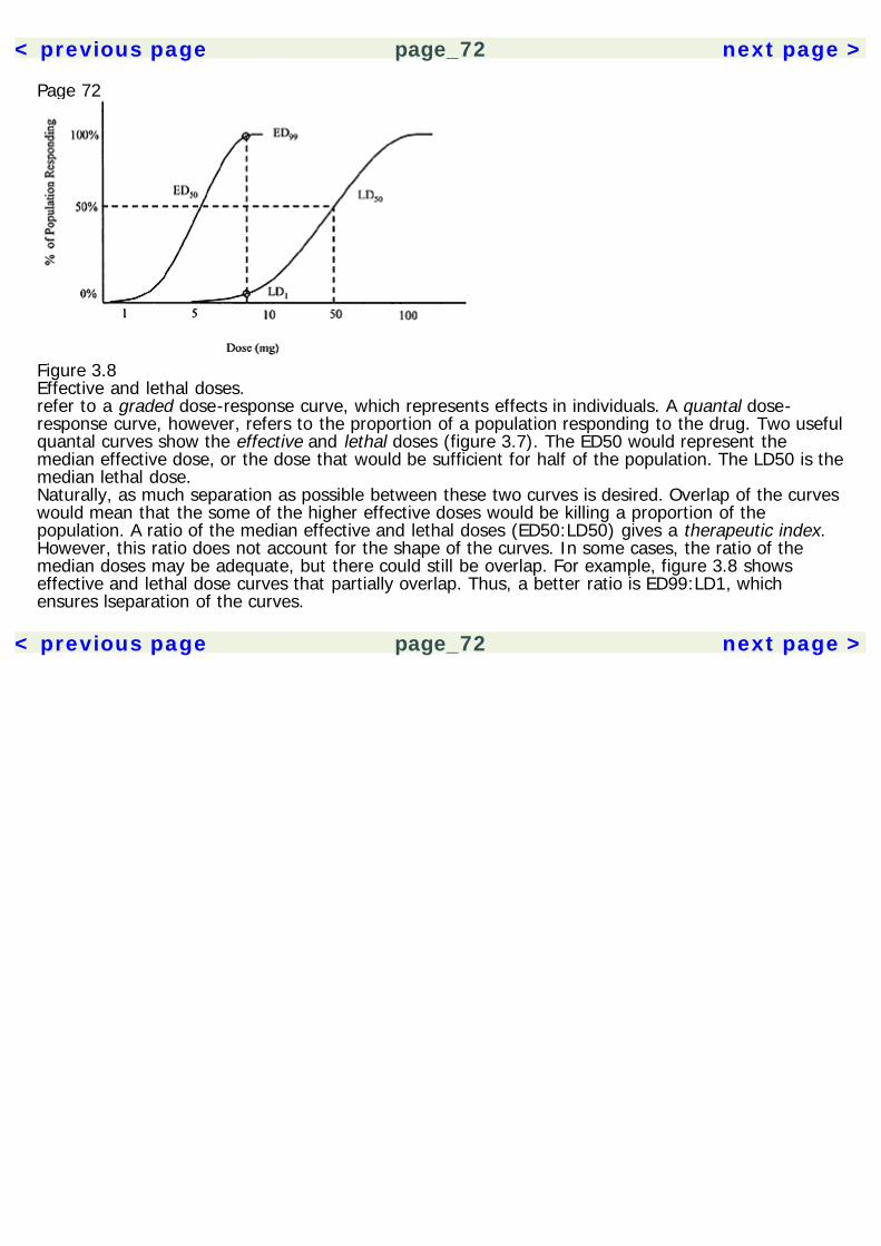
< previous page page_72 next page >
Page 72
Figure 3.8Effective and lethal doses.refer to a graded dose-response curve, which represents effects in individuals. A quantal dose-response curve, however, refers to the proportion of a population responding to the drug. Two usefulquantal curves show the effective and lethal doses (figure 3.7). The ED50 would represent themedian effective dose, or the dose that would be sufficient for half of the population. The LD50 is themedian lethal dose.Naturally, as much separation as possible between these two curves is desired. Overlap of the curveswould mean that the some of the higher effective doses would be killing a proportion of thepopulation. A ratio of the median effective and lethal doses (ED50:LD50) gives a therapeutic index.However, this ratio does not account for the shape of the curves. In some cases, the ratio of themedian doses may be adequate, but there could still be overlap. For example, figure 3.8 showseffective and lethal dose curves that partially overlap. Thus, a better ratio is ED99:LD1, whichensures lseparation of the curves.
< previous page page_72 next page >
< previous page page_73 next page >
Page 734Stimulant PlantsPlants with stimulant effects are among the earliest plants used by humankind for psychoactiveeffects. Our interest in them is motivated by their profound effect on mental functioning, increasingalertness, the ability to sustain effort, and in some cases, the induction of euphoria. Despite theirnative geographical habitats, several stimulant plants are used across the planet. A sizable portion ofthe world's population regularly consumes tobacco, coffee, tea, or some other form of stimulant on adaily basis (Piha et al. 1993). In some cases, these are socially accepted aspects of daily life (e.g.,coffee, tea), while others are reviled for their addicting effects and socioeconmic impact (e.g., coca).The stimulant plants are organized here by their predominant neurochemical mechanism: purinergic(coffee, tea, cocoa, maté, guarana, and kola), cholinergic (tobacco, areca, and lobelia), andmonoaminergic (ephedra, coca, and khat) (see table 4.1). It is worthwhile to note that most of theseplant drugs work through neuromodulatory systems. They mostly create stimulation not throughdirect neuronal excitation, but rather by a modulation of neuronal activity. On the other hand, drugsthat directly stimulate excitatory amino acids or block inhibitory amino acids more often haveexcitotoxic or convulsive effects (e.g., strychnine from Strychnos nux-vomica, or ibotenate fromAmanita muscaria). Rather, the stimulant plants generally act through neuromodulatory receptorslinked to G-proteins and intracellular messengers.Several synthetic stimulants have been developed for clinical use or illegal trade. Amphetamine wassynthesized in 1887, but its medical applications were not investigated until the 1920s (Rudgley1999). It was first marketed in 1932 as a treatment for asthma and nasal congestion. Since
< previous page page_73 next page >
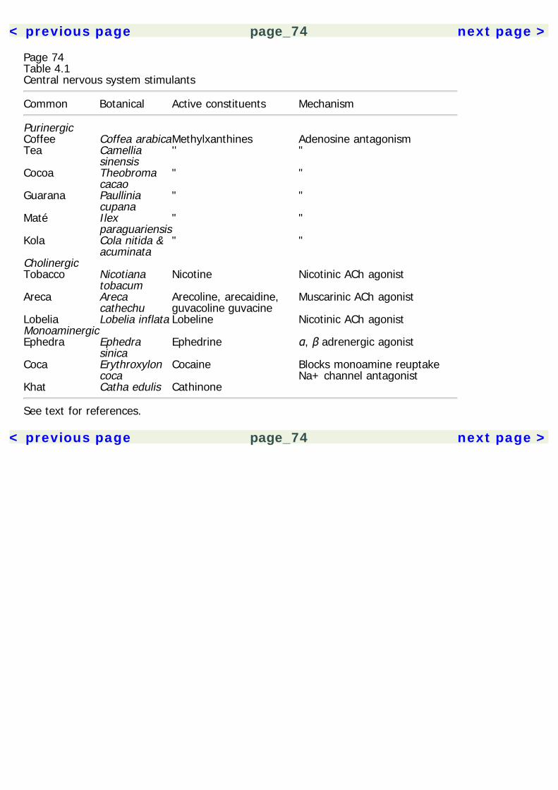
< previous page page_74 next page >
Page 74Table 4.1Central nervous system stimulants
Common Botanical Active constituents Mechanism
Purinergic Coffee Coffea arabicaMethylxanthines Adenosine antagonismTea Camellia
sinensis'' "
Cocoa Theobromacacao
" "
Guarana Paulliniacupana
" "
Maté Ilexparaguariensis
" "
Kola Cola nitida &acuminata
" "
Cholinergic Tobacco Nicotiana
tobacumNicotine Nicotinic ACh agonist
Areca Arecacathechu
Arecoline, arecaidine,guvacoline guvacine
Muscarinic ACh agonist
Lobelia Lobelia inflata Lobeline Nicotinic ACh agonistMonoaminergic Ephedra Ephedra
sinicaEphedrine α, β adrenergic agonist
Coca Erythroxyloncoca
Cocaine Blocks monoamine reuptakeNa+ channel antagonist
Khat Catha edulis Cathinone
See text for references.
< previous page page_74 next page >
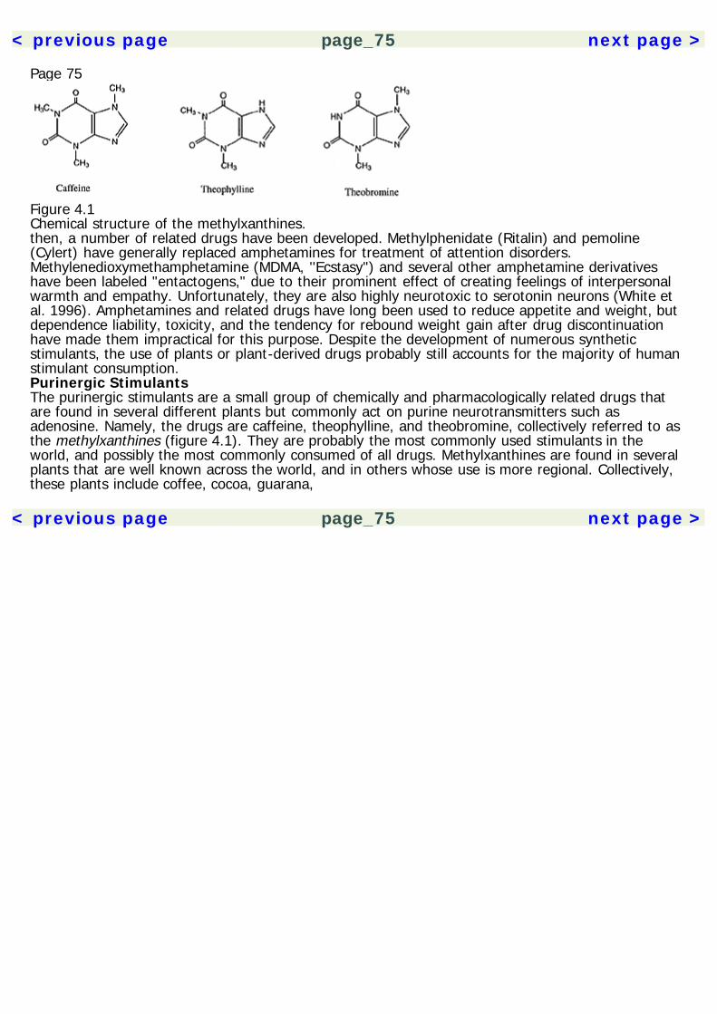
< previous page page_75 next page >
Page 75
Figure 4.1Chemical structure of the methylxanthines.then, a number of related drugs have been developed. Methylphenidate (Ritalin) and pemoline(Cylert) have generally replaced amphetamines for treatment of attention disorders.Methylenedioxymethamphetamine (MDMA, ''Ecstasy") and several other amphetamine derivativeshave been labeled "entactogens," due to their prominent effect of creating feelings of interpersonalwarmth and empathy. Unfortunately, they are also highly neurotoxic to serotonin neurons (White etal. 1996). Amphetamines and related drugs have long been used to reduce appetite and weight, butdependence liability, toxicity, and the tendency for rebound weight gain after drug discontinuationhave made them impractical for this purpose. Despite the development of numerous syntheticstimulants, the use of plants or plant-derived drugs probably still accounts for the majority of humanstimulant consumption.Purinergic StimulantsThe purinergic stimulants are a small group of chemically and pharmacologically related drugs thatare found in several different plants but commonly act on purine neurotransmitters such asadenosine. Namely, the drugs are caffeine, theophylline, and theobromine, collectively referred to asthe methylxanthines (figure 4.1). They are probably the most commonly used stimulants in theworld, and possibly the most commonly consumed of all drugs. Methylxanthines are found in severalplants that are well known across the world, and in others whose use is more regional. Collectively,these plants include coffee, cocoa, guarana,
< previous page page_75 next page >
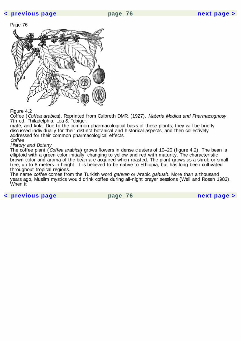
< previous page page_76 next page >
Page 76
Figure 4.2Coffee (Coffea arabica). Reprinted from Culbreth DMR. (1927). Materia Medica and Pharmacognosy,7th ed. Philadelphia: Lea & Febiger.maté, and kola. Due to the common pharmacological basis of these plants, they will be brieflydiscussed individually for their distinct botanical and historical aspects, and then collectivelyaddressed for their common pharmacological effects.CoffeeHistory and BotanyThe coffee plant (Coffea arabica) grows flowers in dense clusters of 10–20 (figure 4.2). The bean iselliptoid with a green color initially, changing to yellow and red with maturity. The characteristicbrown color and aroma of the bean are acquired when roasted. The plant grows as a shrub or smalltree, up to 8 meters in height. It is believed to be native to Ethiopia, but has long been cultivatedthroughout tropical regions.The name coffee comes from the Turkish word gahveh or Arabic gahuah. More than a thousandyears ago, Muslim mystics would drink coffee during all-night prayer sessions (Weil and Rosen 1983).When it
< previous page page_76 next page >

< previous page page_77 next page >
Page 77was introduced to Europe in the seventeenth century C.E., coffee met with official opposition beforelater gaining widespread acceptance.Chemical ConstituentsThe major pharmacological constituents of coffee are the purine alkaloids caffeine (1–2%), and to alesser degree, theobromine and theophylline (Gruenwald et al. 1998). Also present are trigonelline,caffeic acid, and tannin. The caffeine content of coffee varies somewhat with its preparation: drip-brewed coffee may contain 60–180 mg (per 5 oz cup), while instant coffee may contain 30–120 mg(Gilbert et al. 1976; Bunker and McWilliams 1979; Lelo et al. 1986). ''Decaffeinated" coffee is notcompletely decaffeinated, still having 5 mg or less. By comparison, over-the-counter stimulants (e.g.,NoDoz, Vivarin) contain approximately 100 mg per tablet, and caffeinated soft drinks have 40–60 mgof caffeine per 12 oz serving.TeaHistory and BotanyBoth black and green tea are from the plant Camellia sinensis (Gruenwald et al. 1998; Robbers et al.1996). It grows white or pale pink flowers and fruit in a greenish-brown capsule, each containingone to three brown seeds (figure 4.3). Green tea is produced by simply drying the leaves andbrewing them in hot water. Black tea is first fermented before drying and brewing. Tea was originallycultivated in China, but is now grown in many parts of the world including India, Indonesia, theMiddle East, Africa, and Argentina. Tea has been a popular drink throughout history and, as a result,it has been a major force in trade, economics, and politics.Chemical ConstituentsThe major pharmacological constituents of tea are the purine alkaloids caffeine (2.9–4.2%),theobromine (0.15–0.2%), and theophylline (0.02–0.04%). Also present are triterpene saponins(including barringtogenol C and R1-barringenol), catechins (theaflavine, theaflavin acid,thearubigine), and caffeic acid derivatives (chlorogenic acid and theogallin). A cup of brewed teacontains approximately 20–100 mg of caffeine
< previous page page_77 next page >
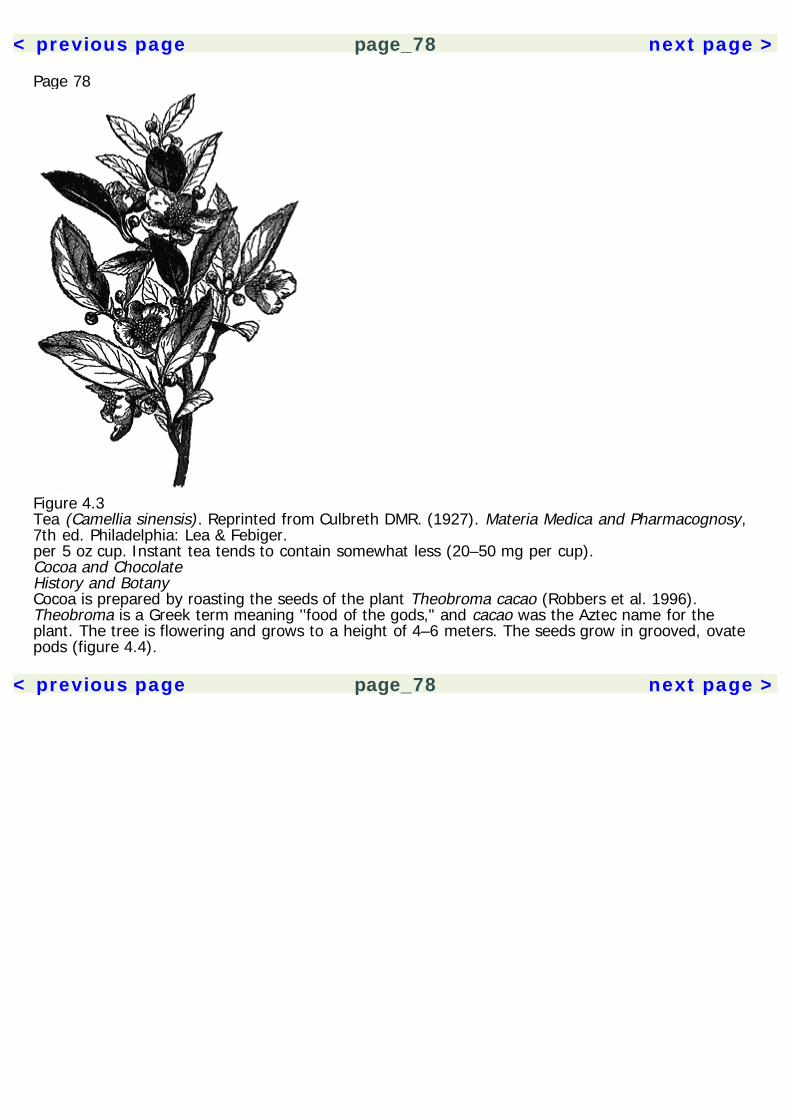
< previous page page_78 next page >
Page 78
Figure 4.3Tea (Camellia sinensis). Reprinted from Culbreth DMR. (1927). Materia Medica and Pharmacognosy,7th ed. Philadelphia: Lea & Febiger.per 5 oz cup. Instant tea tends to contain somewhat less (20–50 mg per cup).Cocoa and ChocolateHistory and BotanyCocoa is prepared by roasting the seeds of the plant Theobroma cacao (Robbers et al. 1996).Theobroma is a Greek term meaning ''food of the gods," and cacao was the Aztec name for theplant. The tree is flowering and grows to a height of 4–6 meters. The seeds grow in grooved, ovatepods (figure 4.4).
< previous page page_78 next page >

< previous page page_79 next page >
Page 79
Figure 4.4Cocoa (Theobroma cacao). Reprinted from Culbreth DMR. (1927). Materia Medica andPharmacognosy, 7th ed. Philadelphia: Lea & Febiger.The most common way of consuming cocoa is in sweetened chocolate or baked goods. The word''chocolate" derives from the Nahuatl word chocolatl. Cocoa is prepared by removing the seeds fromtheir shells, fermenting, and then roasting them. Cocoa was brought to Europe by Cortez in 1528,but did not become popular as a drink until the seventeenth century. It was initially consumed as abitter drink, and was later added to milk solids and sugar to produce milk chocolate. The Swissaltered the history of candy in 1876 by marketing milk chocolate under the name Nestlé.Chemical ConstituentsChocolate constitutes a significant dietary source of theobromine (Shivley and Tarka 1984). Hotcocoa, for example, prepared from instant mix provides approximately 60 mg of theobromine and 4mg of caffeine per serving. Chocolate contains less caffeine per serving than coffee (0.4–1.2% ofcaffeine in cocoa), but may still have significant amounts, depending on the preparation. Forexample, milk chocolate contains up to 15 mg of caffeine per ounce, but dark chocolate can containup to 35 mg per ounce.
< previous page page_79 next page >
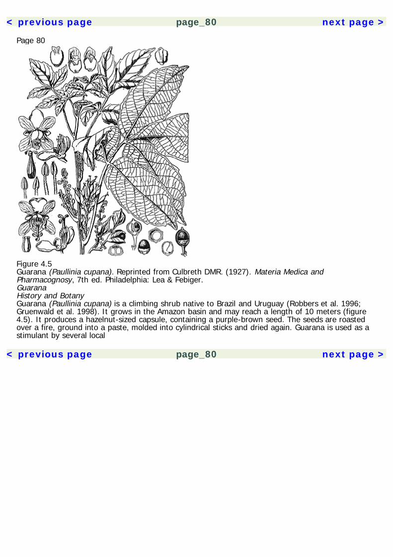
< previous page page_80 next page >
Page 80
Figure 4.5Guarana (Paullinia cupana). Reprinted from Culbreth DMR. (1927). Materia Medica andPharmacognosy, 7th ed. Philadelphia: Lea & Febiger.GuaranaHistory and BotanyGuarana (Paullinia cupana) is a climbing shrub native to Brazil and Uruguay (Robbers et al. 1996;Gruenwald et al. 1998). It grows in the Amazon basin and may reach a length of 10 meters (figure4.5). It produces a hazelnut-sized capsule, containing a purple-brown seed. The seeds are roastedover a fire, ground into a paste, molded into cylindrical sticks and dried again. Guarana is used as astimulant by several local
< previous page page_80 next page >

< previous page page_81 next page >
Page 81Indian cultures to maintain alertness during tasks like hunting (Rudgley 1999). Guarana is nowcommercially cultivated and its use has increased in the West.Chemical ConstituentsThe principal psychoactive constituent of guarana is caffeine (2.5–5%), although theobromine andtheophylline are present in smaller amounts (Gruenwald et al. 1998). Depending on the preparation,1 g of the seed contains approximately 35 to 55 mg of caffeine. It also contains a tannin,catechutannic acid (25%).MatéHistory and BotanyMaté (also known as yerba maté or Paraguay tea) is prepared from the plant Ilex paraguariensis(Robbers et al. 1996; Gruenwald et al. 1998). It is an evergreen shrub or tree, growing up to 20meters in height. White flowers grow in clusters of 40–50, and the leaves are green and ovate. Thefruit is fleshy and contains 5–8 seeds each (figure 4.6). I. paraguariensis grows in regions of SouthAmerica and is a very popular drink in Argentina. Maté is prepared by adding boiling water to thedried leaves and stem. Characteristic straws called bombillas are used to drink maté. They are ornateand made of metal, with a strainer at the end.Chemical ConstituentsMaté contains both caffeine (0.4–2.4%) and theobromine (0.3–0.5%). Also found are triterpenesaponins and the caffeic acid derivatives—chlorogenic acid, neochlorogenic acid, andcryptochlorogenic acid. Flavonoids in maté are rutin, isoquercetin, and kaemferol glycosides. A nitrileglycoside, menisdaurin, is also present, which is noncyanogenic.KolaHistory and BotanyKola (also spelled cola) is a nut from the plants Cola nitida and C. acuminata (figure 4.7). It is anevergreen tree, growing 15 to 20 meters
< previous page page_81 next page >

< previous page page_82 next page >
Page 82
Figure 4.6Maté (Ilex paraguariensis). Reprinted with permission. Illustration by Lorraine Hara.in height. The trunk branches as low as the base, and the dark green elliptoid leaves end in aspiraled tip. The flowers grow in small groups, and the star-shaped fruit contain 14 seeds, square toovate in shape. The plant is indigenous to parts of West Africa, including Togo, Sierra Leone, andAngola. However, it is also cultivated in East Africa, Sri Lanka, Indonesia, Brazil, and the West Indies.The seeds (or nuts) of the kola plant are taken from the pods and their outer coat is removed. Theyare chewed fresh or dried because they spoil quickly. The nuts are especially popular in Africa, wherethey are given as ceremonial gifts. However, kola is consumed around the world in the form ofcarbonated soft drinks, such as Coca-Cola. As the name implies,
< previous page page_82 next page >
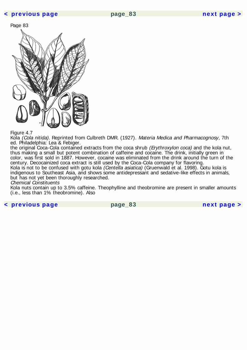
< previous page page_83 next page >
Page 83
Figure 4.7Kola (Cola nitida). Reprinted from Culbreth DMR. (1927). Materia Medica and Pharmacognosy, 7thed. Philadelphia: Lea & Febiger.the original Coca-Cola contained extracts from the coca shrub (Erythroxylon coca) and the kola nut,thus making a small but potent combination of caffeine and cocaine. The drink, initially green incolor, was first sold in 1887. However, cocaine was eliminated from the drink around the turn of thecentury. Decocainized coca extract is still used by the Coca-Cola company for flavoring.Kola is not to be confused with gotu kola (Centella asiatica) (Gruenwald et al. 1998). Gotu kola isindigenous to Southeast Asia, and shows some antidepressant and sedative-like effects in animals,but has not yet been thoroughly researched.Chemical ConstituentsKola nuts contain up to 3.5% caffeine. Theophylline and theobromine are present in smaller amounts(i.e., less than 1% theobromine). Also
< previous page page_83 next page >

< previous page page_84 next page >
Page 84contained are (+)-catechin, (–)-epicatechin, catechin tannins and oligomeric proanthocyanidins. Thefresh nut contains the tannin kolacatechin, which is split into caffeine and theobromine in the dryingprocess. This process also gives kola the characteristic red-brown color, which is imitated by caramelcoloring in cola soft drinks.The Pharmacology of Purinergic StimulantsPharmacokineticsCaffeine is highly bioavailable, being rapidly absorbed from the digestive tract (Serafin 1996).Absorption of oral theophylline is somewhat slower, peaking at 2 hours in the absence of food. Foodgenerally slows, but does not limit, the absorption of methylxanthines. They are readily distributed toall body tissues and cross the placental barrier to enter breast milk. Caffeine and theophylline havesimilar volume distributions. Blood and brain concentrations of caffeine and theophylline in commonlyconsumed doses are 10–50 μM (Snyder and Sklar 1984). The half-life of caffeine is 3 to 7 hours,which increases in women using oral contraceptives or in late-stage pregnancy. The elimination ofcaffeine is variable, as much as fourfold, among individuals. The elimination half-life in adults isusually 8 to 9 hours.Caffeine pharmacokinetics are nonlinear. For example, when comparing a 500 mg dose to a 250 mgdose, the clearance is reduced and elimination half-life is prolonged with the higher dose (Kaplan etal. 1997). Thus, larger doses prolong the action of the drug. Active metabolites of caffeine areparaxanthine, and to a lesser degree, theobromine, and theophylline. Urinary metabolites are l-methylxanthine, l-methyluric acid, and an acetylated uracil derivative.Mechanisms of ActionAdenosineThe chief direct mechanism of the methylxanthines is the antagonism of adenosine receptors (Snyderand Sklar 1984). Adenosine is an inhibitory neurotransmitter, acting at inhibitory presynapticautoreceptors. At
< previous page page_84 next page >

< previous page page_85 next page >
Page 85micromolar concentrations, methylxanthines occupy 50% of adenosine receptors. Low micromolarconcentrations of adenosine inhibit the release of most neurotransmitters, both excitatory andinhibitory. Chronic administration of caffeine elevates the number of adenosine receptors, likelyreflecting a compensatory supersensitivity. So, by antagonizing adenosine receptors, methylxanthinescause a disinhibitory release of neurotransmitters.Indirect MechanismsSeveral indirect neurochemical effects of methylxanthines contribute to their effects. Micromolarconcentrations of caffeine enhance release of acetylcholine (Pedata et al. 1984). However, this effectis biphasic, augmenting release at 50 μM, but decreasing it at 0.5 μM. This effect is also modulatory,affecting stimulated, but not basal, release. Caffeine enhances acetylcholine release in thehippocampus, which is due to adenosine A1 receptor subtypes (Carter et al. 1995). Conversely,chronic caffeine reduces the excitatory effect of acetylcholine in the cerebral cortex (Lin and Phillis1990).Adenosine inhibits the evoked release of excitatory amino acids glutamate and aspartate (Poli et al.1991). So, methylxanthines cause a disinhibited release of excitatory amino acids. Caffeine also has anet disinhibitory effect on GABA neurotransmission (Kardos and Blandl 1994). It enhances GABArelease in the spinal cord through nonbenzodiazepine mechanisms (Berti and Nistri 1983).Caffeine increases extracellular serotonin levels in the hippocampus (Okada et al. 1999). Enhancedrelease of dopamine and norepinephrine occurs at higher doses (Morgan and Vestal 1989). Aninhibition of monoamine reuptake occurs, but only in the millimolar range, which would not matter atnormal oral doses (Reith et al. 1987).Methylxanthines are inhibitors of the enzyme phosphodiesterase, preventing breakdown of andincreasing intracellular levels of cAMP (Serafin 1996; Snyder and Sklar 1984). However, this actionalso only occurs in the millimolar range, at concentrations 100 times greater than those normallyachieved (10–50 μM).Caffeine induces a release of stored intracellular calcium (Snyder and Sklar 1984; Garaschuk et al.1997). It augments twitching in skeletal
< previous page page_85 next page >
< previous page page_86 next page >
Page 86muscle, but this effect is found in the millimolar range. Ca2+ effects alone do not explain caffeine'spsychoactive effects, which occur at normal doses, but they could contribute to enhancement ofneurotransmitter release (Avidor et al. 1994).General EffectsSmooth MuscleMethylxanthines relax smooth muscle, and have a bronchodilating effect in the lungs. Theophylline isused as a treatment for asthma. Methylxanthines dilate coronary arteries, increasing cardiac bloodflow, but an opposite effect occurs on cerebral blood vessels (see below).Diuretic EffectsMethylxanthines increase urine production. Increases are also seen in renal blood flow andglomerular filtration rate.Cardiovascular EffectsMethylxanthines have complex effects on circulation (Serafin 1996). They mildly reduce peripheralvascular resistance, and were used to treat congestive heart failure until more selective drugs weredeveloped. But the net cardiovascular effect of methylxanthines is due to a diverse mix of influences.Methylxanthines influence vagal and vasomotor areas in the brain, vascular, and cardiac tissues, andhormonal systems such as plasma catecholamines and renin-angiotensin. Caffeine generally increasesblood pressure (Smith et al. 1994). Higher doses of caffeine can cause tachycardia and arrhythmias.NeuroendocrineHigh doses of caffeine produce a stresslike neuroendocrine response (Spindel 1984; Spindel et al.1984). This includes increased levels of corticosterone, β-endorphin, and decreased serum growthhormone and thyrotropin-stimulating hormone in the rat. However, similar effects are not observed inhumans until approximately 500 mg is consumed, the equivalent of 5 cups of coffee. At this dose inhumans, elevations are seen in adrenocorticotrophic hormone, β-endorphin, and cortisol.
< previous page page_86 next page >
< previous page page_87 next page >
Page 87Metabolic clearance of caffeine is altered according to menstrual phase and hormonal status inwomen (Lane et al. 1992). Clearance is slower during the late luteal phase compared to the follicularphase, prior to the onset of menstruation. However, the size of the effect and significance ineveryday activity remains in question.Skeletal MuscleCaffeine increases the capacity of skeletal muscle. This effect is probably due to the release ofintracellular Ca2+, and is observable in human performance. The contribution of central effects isalso likely. Whether caffeine actually benefits exercise is contested (Spriet 1995; Dodd et al. 1993).Research does not consistently support caffeine enhancement of performance during high-intensity,short-term exercise. There is some evidence to suggest benefit during prolonged, moderate-intensityexercise.Motor and Behavioral EffectsCaffeine improves psychomotor performance. For example, improvements are seen in manualdexterity (Bernstein et al. 1994). Enhancement of psychomotor performance is most evident whensubjects are fatigued (Snyder and Sklar 1984).Although perhaps counterintuitive, caffeine does not increase disruptive behavior in preschoolchildren when objectively assessed (Baer 1987). A meta-analysis of 12 studies failed to show anyadverse cognitve or behavioral effects in children (Stein et al. 1996).The effect of caffeine in children may depend on physiological predisposition (Rapoport et al. 1984).Children who are normally high habitual consumers of caffeine (500 mg/day or more) show higheranxiety and lower autonomic arousal when not receiving caffeine. Habitually low caffeine consumersare more emotional, inattentive, and restless when receiving 10 mg/kg of caffeine, while the highhabitual users are rated as unchanged. Low doses of caffeine added to methylphenidate producegreater improvement in attention deficit disorder than methylphenidate alone (Garfinkel et al. 1981).Although some people ingest caffeine in an attempt to ''sober up" after drinking ethanol, researchdoes not support this. While caffeine may increase arousal and reverse deficits in reaction timecaused by alcohol, it does not affect other variables such as
< previous page page_87 next page >
< previous page page_88 next page >
Page 88standing steadiness, manual dexterity, numerical reasoning, perceptual speed, or verbal fluency(Franks et al. 1975).Sleep and ArousalCaffeine has potent psychostimulant effects, increasing the general level of arousal (Rogers andDernoncourt 1998). Along with increasing alertness, caffeine increases the sleep latency(Zwyghuizen-Doorenbos et al. 1990). Coffee and caffeine have insomnia-like effects on sleep, shiftingthe onset of REM sleep and stages 3 and 4 to later in the night (Karacan et al. 1976).EmotionalCaffeine has mixed effects on mood and emotion. Low doses improve feelings of energy and mood(Warburton 1995; Rogers and Dernoncourt 1998). However, progressively higher doses also increasesubjective feelings of anxiety, tension, and nervousness (Loke 1988; Bernstein et al. 1994). Forexample, positive mood effects are generally obtained with a 250 mg dose, and negative effectsoccur at a 500 mg does (Kaplan et al. 1997). People with anxiety or panic disorders are even moresusceptible to the anxiogenic effects of caffeine (Shear 1986). Caffeine also increases the autonomicresponse to novel situations (Davidson and Smith 1991).Electrophysiological EffectsAs might be expected, caffeine produces an activation effect on the EEG, reducing theta and deltapower (Dimpfel and Schober 1993). Caffeine reverses the EEG slowing brought on by nicotineabstinence (Cohen et al. 1994). ERP studies of the effects of caffeine on visual attention suggest thatit depends on the number of relevant visual targets, and not the overall number of targets (Lorist etal. 1996). On the other hand, caffeine withdrawal causes a decrease in P300 amplitude or latency(Reeves et al. 1999). Caffeine improves performance on a visual search task, with increases in theamplitude of the N1, N2b, and P3b waves (Lorist et al. 1995). The effect of caffeine on the P300 ismore pronounced in fatigued than well-rested subjects performing a visual search task (Lorist et al.1994).
< previous page page_88 next page >
< previous page page_89 next page >
Page 89Cerebral Blood Flow and Glucose MetabolismPET and 133Xenon SPECT show significant decreases (~30%) in wholebrain cerebral blood flow aftera dose of 250 mg of caffeine (Cameron et al. 1990; Mathew et al. 1983). Local cerebral blood flow isdecreased after administration of caffeine, mostly in motor and auditory areas, but no regionaldifferences are seen in humans (Nehlig et al. 1990; Cameron et al. 1990). Decreases in cerebralblood flow persist at least 90 minutes after ingestion (Mathew and Wilson 1985). Despite decreasedcerebral blood flow, caffeine causes widespread increases in brain metabolism and glucose utilization(Schroeder et al. 1989; Nehlig et al. 1984).In subjects who experience anxiety and unpleasant physiological arousal from caffeine, it can causean incease in brain lactate (Dager et al. 1999). This is significant in light of the association betweenlactate and anxiety in general (Dager et al. 1990).Cognitive EffectsCaffeine has been found to quicken reaction time and enhance vigilance, increase self-ratedalertness, and improve mood (Rogers and Dernoncourt 1998). Even though some tolerance mightdevelop to the cognition-enhancing effects of caffeine, improvements are sustained with chronicconsumption (Jarvis 1993). Conversely, caffeine withdrawal has adverse effects on cognition (James1998; Lane and Phillips-Bute 1998). For example, several psychometric measures of managerialperformance suffer upon withdrawal (Streufert et al. 1995). Some have contended that caffeine-induced improvements are partly due to reversal of withdrawal-associated deficits (Rogers andDernoncourt 1998).However, caffeine still shows improvements in mood, attention, problem solving, and memory, whichare distinct from alleviating abstinence (Warburton 1995). Cognitive effects are somewhat moreassociated with coffee than tea, but the stimulant and cognitive effects of black tea and coffee aresimilar on several cognitive measures (Hindmarch et al. 1998; Jarvis 1993). Several studies indicatesex differences in the cognitive effects of caffeine, with generally greater effects in females (Smith etal. 1993). However, this effect varies with the menstrual cycle, so it has been suggested thatestrogen levels may play a role (Arnold et al. 1987). For unknown neurochemical reasons, acombination of caffeine with aspirin
< previous page page_89 next page >
< previous page page_90 next page >
Page 90further improves vigilance and mood, more so than caffeine or aspirin alone (Lieberman et al. 1987).Caffeine reverses deficits in alertness produced by sleep deprivation (Penetar et al. 1993). It alsoimproves performance in sustained attention tasks (Smith et al. 1994; Warburton 1995). Somestudies show nonsignificant effects on attention, but improvement is evident in more complexattention tasks, like complex cancellation (involving addition and multiplication) as compared tosimple digit cancellation (Loke 1988). But this may depend on the nature of the task. Caffeine alsoreduces subjective fatigue, making exertion of concentration less effortful. This effect differs acrosssexes, decreasing subjective effort in men and increasing it in women performing a complex mentaltask (e.g., Raven's progressive matrices) (Linde 1995). On the other hand, caffeine may have someadverse effects on attention as well. Some have shown poorer accuracy with caffeine in vigilancetasks (Smith et al. 1992). Men given caffeine perform more poorly on a numerical version of theStroop task, which requires suppression of automatic responses to conflicting stimuli (Foreman et al.1989). However, other studies (using 125 or 250 mg) have not shown any effect of caffeine in theStroop test (Edwards et al. 1996).Although caffeine increases response speed in some visual attention tasks, it is apparently not due todecreased distractibility or suppression of irrelevant responses (Kenemans and Verbaten 1998).Cognitive decline is evident during withdrawal from caffeine, primarily on measures of response timeand sustained attention (Bernstein et al. 1998). The degree of habitual caffeine use is the strongestvariable predicting the response to caffeine in a visual attention task (Loke and Meliska 1984).Similar to caffeine, theophylline produces improvement on sustained attention tasks (Bryant et al.1998), but caffeine appears to be a more potent CNS stimulant (Yu et al. 1991). Caffeine improvesthe accuracy of reaction time tasks in hyperkinetic children (Reichard and Elder 1977).Caffeine improves performance on tests of semantic memory, free recall, and recognition memory(Smith et al. 1994; Warburton 1995). However, the effects are both dose and task dependent. Insome studies, higher doses produced greater improvement, while in others subjects receivingcaffeine performed slightly worse than placebo (Loke 1988;
< previous page page_90 next page >

< previous page page_91 next page >
Page 91Terry and Phifer 1986). Caffeine reduces the impairments caused by scopolamine in short- and long-term free recall memory in humans (Riedel et al. 1995). It also reverses scopolamine-induceddecrements in visual search, reading speed, and fine motor speed (e.g., finger-tapping). Theseeffects are interesting in light of caffeine's effect on cerebral acetylcholine activity. Indirect cholinergicmechanisms may be involved in the mnestic effects of caffeine, because scopolamine reversescaffeine's effect in animal maze tasks (Roussinov and Yonkov 1976). However, dopamine may also beinvolved because the beneficial effects of caffeine on memory consolidation in mice are also blockedby a D2 antagonist (sulpiride) (Cestari and Castellano 1996).Caffeine improves performance on tests of logical reasoning and problem solving (Smith et al. 1994;Warburton 1995). However, this varies with the task, because no effects are objectively seen onRaven's Progressive Matrices, a test of nonverbal reasoning (Linde 1995)Addiction and DependenceSome individuals use caffeine in a manner that could arguably be classified as abuse. Tolerance isevident with chronic use, and discontinuation can produce an aversive withdrawal syndrome.Cessation of even low to moderate use (the equivalent of 2.5 cups of coffee per day) causes fatigue,headache, increased anxiety, and decreases in mood. Physiological changes such as alteredadenosine receptors may accompany dependence, and caffeine affects the release of dopamine.Certainly, caffeine dependence is not as intense and consuming as that produced by cocaine orheroin. Severe withdrawal is avoidable by gradual tapering of the dose, and clinical treatment isgenerally not necessary. However, combined cigarette and coffee abstinence in regular consumerscan cause more severe symptoms such as mental cloudiness, irritability, and muscular tension,(Cohen et al. 1994).ToxicityConsumed in normal doses, caffeine generally does not have any acute toxicity. Fatal poisoning withcaffeine is rare, occuring with doses
< previous page page_91 next page >

< previous page page_92 next page >
Page 92between 5 and 10 g. Adverse effects are seen with doses of 1 g or lower, due primarily to circulatoryand central effects. Overdoses initially present with behavioral excitation and insomnia, progressingto delirum and convulsions. Tachycardia and vomiting are common at these doses.Chronic use of large amounts of caffeine has been associated with an increased risk of cardiovasculardisease. However, this finding is debated because statistically adjusting for other risk factors shows aminimized added risk for caffeine (Grobbee et al. 1990). Nonetheless, a lipid fraction of boiled coffeedose-dependently elevates cholesterol and low density lipoproteins, which is prevented by the filteredpreparation of coffee (Pirich et al. 1993). Another potential influence on cardiovascular disease is anelevation of homocysteine levels, which also occurs in drinkers of filtered coffee (Nygård et al. 1997).GenotoxicityGenotoxic effects of caffeine have been reported, but they only occur in large doses (>1 mM), farexceeding what could be obtained by oral dosing (D'Ambrosio 1994).Pediatric and Prenantal ToxicityCaffeine is likely the most commonly used drug among pregnant women, but whether or not it issafe during pregnancy is still a matter of debate. Caffeine intake during pregnancy is not related toinfant height, weight, or head circumference, or to measures of IQ and attention at 7 years of age(Barr and Streissguth 1991). On the other hand, there are associations with reduced fertility,miscarriage, growth retardation in utero, and sudden infant death syndrome (Golding 1995; Ford etal. 1998). Unlike the adult brain, fetal brains may accumulate theophylline and theobromine whenexposed to doses reached during normal consumption (Wilkinson and Pollard 1993). Definitiveprospective studies on the safety of caffeine during pregnancy have not yet been done (Golding1995; Barr and Streissguth 1991).Seizures and EpilepsyVery high doses of caffeine can induce seizures in those with no preexisting seizure disorder. Caffeinealso prolongs seizures induced by electroconvulsive treatment in humans (Shapira et al. 1987). Giventhe
< previous page page_92 next page >
< previous page page_93 next page >
Page 93known neurochemical actions of methylxanthines, they are probably best avoided by people withepilepsy.Cholinergic StimulantsTobaccoHistory and BotanyThe tobacco plant (Nicotiana tabacum) is an annual or biennial plant growing 1–3 meters in height(Gruenwald et al. 1998). The leaves range from ovate in shape to narrow and tapering, and grow upto 50 cm in length. Its pinkish flowers grow in branched pedicels (figure 4.8). The tobacco plantoriginated in tropical regions of America, but is now cultivated in China, Turkey, Greece, Holland,France, Germany, and many countries in subtropical regions. The leaves are harvested and dried forconsumption by smoking or chewing.The tobacco plant was long used by Native Americans before the arrival of Europeans. The wordtobacco derives from tabacum, the Indian name for the pipe they used to smoke it. Nicotine, theprinciple psychoactive chemical in tobacco, was named after Jean Nicot de Villemain, a Frenchdiplomat who advocated its use in Europe in the late 1500s (Rudgley 1999). It is less commonlyknown that tobacco grew wildly in the interior of Australia, and was used by inhabitants there beforeEuropeans arrived.Chemical ConstituentsThe primary psychoactive alkaloid in tobacco is nicotine (figure 4.9), occuring between 0.6 and 9.0%(Robbers et al. 1996; Gruenwald et al. 1998). Other alkaloids include nornicotine, N-formylnornicotine, cotinine, myosmin, β-nicotyrine, anabasine, and nicotellin. Nicotine alone canproduce the effects commonly associated with tobacco use, but other tobacco alkaloids are likely tocontribute.Mechanisms of ActionAcetylcholine Nicotine is an agonist at nicotinic acetylcholine receptors, which were named after thedrug (Taylor 1996). Nicotine opens the
< previous page page_93 next page >
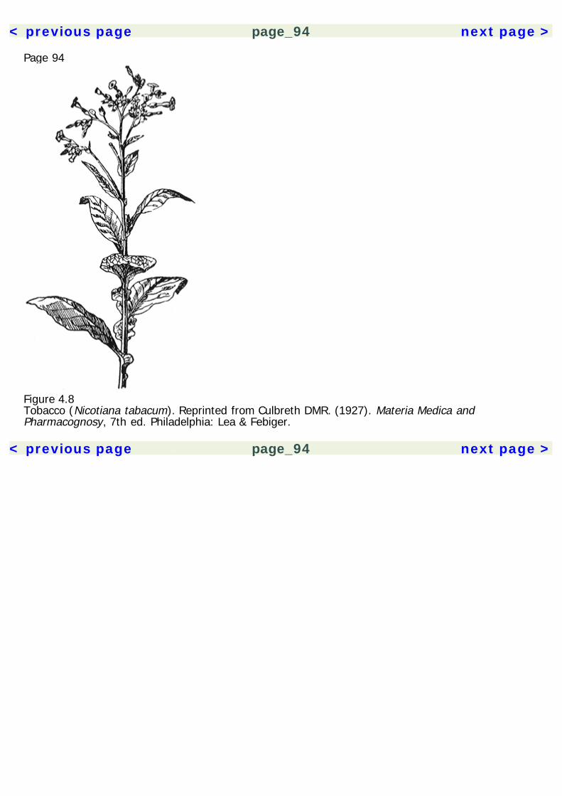
< previous page page_94 next page >
Page 94
Figure 4.8Tobacco (Nicotiana tabacum). Reprinted from Culbreth DMR. (1927). Materia Medica andPharmacognosy, 7th ed. Philadelphia: Lea & Febiger.
< previous page page_94 next page >
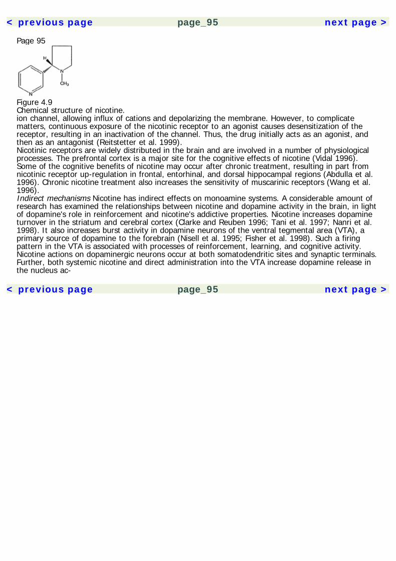
< previous page page_95 next page >
Page 95
Figure 4.9Chemical structure of nicotine.ion channel, allowing influx of cations and depolarizing the membrane. However, to complicatematters, continuous exposure of the nicotinic receptor to an agonist causes desensitization of thereceptor, resulting in an inactivation of the channel. Thus, the drug initially acts as an agonist, andthen as an antagonist (Reitstetter et al. 1999).Nicotinic receptors are widely distributed in the brain and are involved in a number of physiologicalprocesses. The prefrontal cortex is a major site for the cognitive effects of nicotine (Vidal 1996).Some of the cognitive benefits of nicotine may occur after chronic treatment, resulting in part fromnicotinic receptor up-regulation in frontal, entorhinal, and dorsal hippocampal regions (Abdulla et al.1996). Chronic nicotine treatment also increases the sensitivity of muscarinic receptors (Wang et al.1996).Indirect mechanisms Nicotine has indirect effects on monoamine systems. A considerable amount ofresearch has examined the relationships between nicotine and dopamine activity in the brain, in lightof dopamine's role in reinforcement and nicotine's addictive properties. Nicotine increases dopamineturnover in the striatum and cerebral cortex (Clarke and Reuben 1996; Tani et al. 1997; Nanri et al.1998). It also increases burst activity in dopamine neurons of the ventral tegmental area (VTA), aprimary source of dopamine to the forebrain (Nisell et al. 1995; Fisher et al. 1998). Such a firingpattern in the VTA is associated with processes of reinforcement, learning, and cognitive activity.Nicotine actions on dopaminergic neurons occur at both somatodendritic sites and synaptic terminals.Further, both systemic nicotine and direct administration into the VTA increase dopamine release inthe nucleus ac-
< previous page page_95 next page >
< previous page page_96 next page >
Page 96cumbens. Nicotine sensitizes mesoaccumbens dopamine neurons, which is related to enhanced burstfiring (Balfour et al. 1998).Nicotine causes a release of norepinephrine from the locus coeruleus and facilitates release ofnorepinephrine in the hippocampus (Gallardo and Leslie 1998; Mitchell 1993; Sershen et al. 1997; Fuet al. 1999). The norepinephrine released by nicotine, in turn, modulates raphe neurons (Li et al.1998).More complicated effects are seen on 5-HT turnover, with increases at low doses but decreases athigher doses (Smith et al. 1991). Systemic nicotine increases release of serotonin in frontal cortex,beginning within 20 minutes and lasting approximately 2 hours (Ribeiro, et al. 1993) Smokers show a40% decrease in the level of MAOB and 28% decrease in MAOA compared to nonsmokers or formersmokers (Fowler et al. 1996a, 1996b, 1998). This difference is likely to be pharmacological and notgenetic because former smokers do not differ from nonsmokers in this respect. Chronic nicotineincreases tyrosine hydroxylase activity in noradrenergic and dopaminergic cell bodies (Smith et al.1991).Nicotine also has effects on amino acid neurotransmitters. Presynaptic nicotinic receptors enhancetransmission at glutamate and GABA synapses (McGehee et al. 1995; Gray et al. 1996; Léna andChangeux 1997). Nicotine dose-dependently increases glutamate release in rat prefrontal cortex, andfacilitates activity in projections from the thalamus to prefrontal cortex (Gioanni et al. 1999). In thehippocampus, nicotinic stimulation enhances synaptic transmission of glutamate in a Ca2+-dependentmanner (Radcliffe and Dani 1998). This enhancement lasts for minutes, and could alter the time spanduring which incoming signals could elicit synaptic plasticity. Nicotine modulates excitatoryneurotransmission through NMDA receptors in prefrontal cortex (Vidal and Changeux 1993). It alsoincreases production of nitric oxide and cyclic GMP in the hippocampus through NMDA receptors(Fedele et al. 1998).PharmacokineticsWhen smoked, nicotine is very rapidly and efficiently (90%) absorbed into the bloodstream. Whenchewed, absorption is slower but the duration of effect is longer. The average cigarette containsapproximately 12 mg of nicotine and delivers approximately 1 mg to the smoker (Fuku-
< previous page page_96 next page >
< previous page page_97 next page >
Page 97moto et al. 1997; Gori and Lynch 1985). Changing the intensity of puffing can greatly alter thedelivery of nicotine. Behavioral studies using reduced-nicotine cigarettes show that people increasetheir consumption to compensate for the lower nicotine levels (Rusted et al. 1996).Absorption occurs through the respiratory tract, oral membranes, and skin (Taylor 1996). Absorptionfrom the stomach is limited, unless the acidity is reduced, because nicotine is a strong base. Between80 and 90% of nicotine is metabolized, mainly in the liver but also the kidneys and lungs. Cotinine isthe primary metabolite of nicotine, and the half-life of nicotine is about 2 hours. Elimination occurs bythe kidneys, but it is also present in the breast milk of lactating women.General EffectsNicotine affects nearly every physiological system of the body (Le Houezec and Benowitz 1991).However, the effects are complex and unpredictable due to actions on multiple sites, and thestimulation-desensitization of nicotinic receptors.Cardiovascular Nicotine increases heart rate, heart contractility, and blood pressure by activation ofthe sympathetic ganglia and inactivation of parasympathetic ganglia (Haass and Kubler 1997; Grassiet al. 1994). Nicotinic receptors are present on postganglionic sympathetic nerve endings and theadrenal medulla. Effects also occur through receptors on the aortic and carotid bodies, and centralmedullary areas regulating heart rate. Cessation in smoking is associated with notable improvementsin exercise capacity (Albrecht et al. 1998).Gastrointestinal Tobacco has gastrointestinal effects that result from parasympathetic stimulation,with a net effect of increased bowel activity (Taylor 1996). Initial exposure to nicotine typically causesnausea, vomiting, and occasionally diarrhea.Neuroendocrine Tobacco smoking is acutely related to elevations in plasma arginine vasopressin,neurophs I, β-endorphin, and β-lipotropin (Pomerleau et al. 1983). Cigarette smoking elevates plasmacortisol levels via a central mechanism in the hypothalamus or brain stem
< previous page page_97 next page >
< previous page page_98 next page >
Page 98(Targovnik 1989). Smoking cessation causes a decrease in plasma cortisol and epinephrine levels.However, effects on the adrenal medulla are biphasic, with small doses evoking release of plasmacatecholamines and large doses inhibiting it (Talyor 1996).Nicotinic receptors are present in the pineal gland (Stankov et al. 1993). Although there appear to beno direct effects of nicotine on the release of melatonin, it indirectly reduces the accumulation ofmelatonin stimulated by norepinephrine. Nicotine increases circulating levels of norepinephrine andepinephrine (Pomerleau et al. 1983).Neuromuscular Nicotinic receptors are responsible for transmission at the neuromuscular junction.While briefly causing stimulation, this phase is rapidly obscured by desensitization and neuromuscularblockade. Thus, nicotine has muscle-relaxant effects.Behavioral and emotional Nicotine has a number of behavioral and emotional effects. It increaseslocomotor activity, which is mediated by increased dopamine in the nucleus accumbens (Mirza 1996).Nicotine suppresses appetite and decreases weight gain in rats (Grunberg et al. 1986). Conversely,cessation of smoking causes increases in body weight. Nicotine increases sexual receptivity in femalerats, but whether this occurs in humans has not been studied—or at least not formally (Fuxe et al.1977).Nicotine reduces anxiety in humans, more so in females than males (Stewart et al. 1997). Similar tonicotine, the nicotinic channel activator ABT-418 has anxiolytic effects (Brioni et al. 1994). Nicotinereduces anxiety and right hemisphere EEG activation in subjects watching a stress-inducing movie(Gilbert et al. 1989). Conversely, smoking cessation is commonly associated with increases in anxietyand dysphoria (West and Hajek 1997).Electrophysiological Tobacco smoking produces cortical activation on the EEG. Broadly, it reducesslow-wave activity (theta and delta) and increases activity in alpha and beta frequency bands (Knottet al. 1998; Pritchard et al. 1999). Other studies have found more isolated effects in alphafrequencies (Domino and Matsuoka 1994; Foulds et al. 1994).
< previous page page_98 next page >
< previous page page_99 next page >
Page 99Nicotine gum similarly increases alpha and beta activity on the EEG and decreases theta activity(Pickworth et al. 1986). In contrast, denicotinized cigarettes do not have any EEG effects (Pritchard etal. 1999).Nicotine increases the occurrence of the hippocampal theta rhythm in rabbits (Yamamoto 1998). Thehippocampal theta activity is closely associated with learning and memory (Bennett 1973). Nicotinealso induces theta activity in the rat hippocampus and activates functionally related neurons in theposterior cingulate gyrus (Colom et al. 1988). Similar effects are found in nicotine replacement(Newton et al. 1983). Conversely, smoking abstinence causes an increase in theta power anddecreases in alpha and beta activity (Pickworth et al. 1989, 1996). This effect appears as early as 29hours after cessation and may persist for a week. The nicotinic antagonist mecamylamine causesdecreases in alpha frequency and increases in delta frequency, and exacerbates EEG signs of tobaccoabstinence in smokers (Pickworth et al. 1997). This is accompanied by cognitive decrease inpeformance on tests of vigilance, distractibility, and delayed recall.The anxiolytic effects of nicotine are evident in EEG changes. While subjects watched a stress-inducing movie, higher doses of nicotine reduced anxiety and reduced right hemisphere EEGactivation (Gilbert et al. 1989).When abstaining smokers resume smoking, a beneficial reduction in P300 latency occurs for visual,but not auditory, oddball tasks (Houlihan et al. 1996). Using ERPs and a recognition memory task,smokers show enhanced P300s to both targets and distractors (Pineda et al. 1998). Abstinentsmokers, however, show less of a discriminating difference in reactions to targets versus distractors.Further, abstinent smokers have greater latencies relative to nonsmokers.Cerebral blood flow and glucose metabolism Nicotine increases cerebral blood flow (Hara et al. 1993;Yokoi et al. 1993). Functional MRI of smokers who were administered intravenous nicotine showsincreases of cerebral blood flow in several areas of the brain. Corresponding to feelings of moodelevation, nicotine activates the nucleus accumbens, amygdala, cingulate cortex, and frontal lobes.This activation is very consistent with functional systems subserving arousal and reinforcement.
< previous page page_99 next page >
< previous page page_100 next page >
Page 100Increases in cerebral blood flow elicited from stimulation of the basal forebrain are mediated bynicotinic and not muscarinic receptors (Linville et al. 1993). During an attention task, nicotineincreases cerebral blood flow to the the anterior cingulate cortex, cerebellum, and occipital cortex,supporting its role in activating attentional systems (Ghatan et al. 1998).Nicotine dose-dependently increases regional glucose metabolism in the rat brain (McNamara et al.1990). The regional specificty also depends on dosage used, where low doses (0.1 mg/kg) have noeffect, moderate doses (1.0 mg/kg) have region-specific effects in multiple areas, and high doses (10mg/kg) produce generalized activation. Moderate doses increase glucose metabolism in primarilysubcortical areas: thalamic nuclei, cerebellum, superior colliculus, median raphe, reticular formation,and the habenulointerpeduncular pathway. Similar to other abused drugs, nicotine increases glucoseutilization and dopamine activity in the shell of the nucleus accumbens (Pontieri et al. 1998).Dependence and AddictionFew, if any, clinicians or scientists would argue with the position that tobacco is addicting. It hasaddictive properties in both animals and in humans (Dougherty et al. 1981). Regular smokers whoundergo abstinence show a common withdrawal syndrome consisting of craving, mood changes,overarousal, cognitive deficits, and sleep disturbance (Shiffman et al. 1995). Light occasionalsmokers, however, do not seem to experience this syndrome. Although the physical withdrawalsymptoms of nicotine are mild compared to those of ethanol or opioid withdrawal, they are stillunpleasant and there is a high rate of long-term relapse (Nil 1991).Emotional factors play a significant role in quitting tobacco use (Carmody 1989). Depression isoverrepresented among smokers (Hall et al. 1993). Depressed smokers experience more intensewithdrawal symptoms, have less success at quitting, and a greater chance of relapse. They reporthigher levels of dependence than nondepressed smokers and are more likely to use nicotine as self-medication to reduce depression and gain stimulation (Lerman et al. 1996). There may be some sexdifferences in the pattern of smoking: women report smoking more in emotional
< previous page page_100 next page >

< previous page page_101 next page >
Page 101and social situations, whereas men smoke more in situations requiring close attention to a task. Inboth sexes, the trait of sensation seeking is related to being a former or current smoker.The habit-forming property of tobacco occurs through a combination of positive and negativereinforment. Nicotine effects of mood elevation and cognitive enhancements serve as positivereinforcers for smoking. The rewarding effects of tobacco are related to nicotine's stimulation of themesolimbic dopamine system (Balfour 1994; Stolerman and Shoaib 1991; Clarke 1990). This issimilar to other drugs of abuse and occurs through nicotinic receptors in the ventral tegmental area,although the stimulus properties of nicotine do not depend on dopamine alone, and involve multipleneuroanatomical areas (Corrigall et al. 1992; Leshner and Koob 1999; Schilstrom et al. 1998; Shoaib1998). Nicotine increases burst activity in mesolimbic areas, which is associated with motivationalprocesses in reinforcement, learning, and cognition (Nisell et al. 1995). Chronic exposure to levelsachieved by smokers desensitizes nicotinic receptors that modulate mesolimbic dopamine activity(Benwell et al. 1995; Pidoplichko et al. 1997). This correlates with the fact that inital exposures arereinforcing or pleasurable, which subsequently decrease over repeated exposures.Negative reinforcement of tobacco use occurs in the form of anxiety reduction and avoidance of theunpleasant nicotine withdrawal syndrome. Nicotine withdrawal is aversive, involving depressed mood,irritability, and drug craving. It also increases anxiety, which abates approximately after one week ofabstinence (West and Hajek 1997; Hughes et al. 1991). Other withdrawal symptoms includeincreased appetite, sleep disruption, difficulty concentrating, restlessness, and irritability (Jorenby etal. 1996). Most symptoms return to baseline level by 1 month, but weight gain, appetite increases,and drug craving continue for 6 months in many smokers (Hughes et al. 1991). Cravings mayparticularly contribute to relapse in the long term. Prevention of the weight gain that ensues aftertobacco cessation may serve as a further source of negative reinforcement. Reinstatement of nicotinealleviates the withdrawal symptoms. Nicotine withdrawal is also accompanied by elevations in brainreward thresholds for several days (Epping-Jordan et al. 1998).
< previous page page_101 next page >
< previous page page_102 next page >
Page 102Dependence on nicotine does not necessarily begin immediately, but rather may develop over time(Nelson and Wittchen 1998). Among a sample of adolescent and young adult smokers in Germany,the lifetime prevalence of nicotine dependence (using DSM-IV criteria) was 19% and rose to 52%over time. No gender differences are seen in the progression of occasional use to dependence. Adose-response relationship is found between the number of cigarettes smoked daily and unsuccessfulcutback attempts. Despite one's desire and repeated attempts to quit, smoking is a behavior that isresistant to change.Pharmacological Treatment of Nicotine DependenceSeveral pharmacological approaches have been developed to treat tobacco dependence (Sachs andLeischow 1991). Optimally, they should be integrated with psychotherapeutic or behaviormodification programs.Replacement strategies One strategy to treat tobacco addiction is nicotine replacement. This allowsone to alter the behavioral habit of smoking while keeping nicotine levels constant, which caneventually be tapered. Nicotine replacement is achieved through transdermal and oral (gum)approaches. The primary difference between nicotine delivery from cigarettes versus nicotine gumand transdermal nicotine is the rate of absorption (Pomerleau 1992). Smoking allows high levels tobuild up in the blood stream, whereas the other methods allow more gradual absorption across theskin or mucosal membranes. Thus, the gum and patch have a slower onset of action and lessreinforcing effects. Nonetheless, controlled studies of nicotine replacement show that it reduces theseverity of withdrawal symptoms such as anxiety, irritability, increased appetite, and weight gain(Jorenby et al. 1996).Alternative pharmacological approaches Clonidine, an α2 adrenergic agonist, has been employed asadjunctive therapy to assist in smoking cessation. However, results have been mixed or the effectssmall (Gourlay et al. 1994; Hilleman et al. 1993; Franks et al. 1989). Buspirone (BuSpar) is a 5-HT1Apartial agonist with anxiolytic effects. It has been tested as a treatment for smoking cessationbecause anxiety is a prominent feature of nicotine withdrawal (Farid and Abate 1998). To date,results have been mixed and more controlled research is needed.
< previous page page_102 next page >
< previous page page_103 next page >
Page 103Bupropion is another alternative pharmacological approach to tobacco abstinence. It is anantidepressant drug that blocks reuptake of norepinephrine and dopamine, and also blocks nicotinicreceptors in the low to intermediate micromolar range (Fryer and Lukas 1999). Thus, the effects ofbupropion on nicotine addiction may be through dual effects on dopaminergic and nicotinic systems.Further, it has been an effective treatment in controlled studies, both alone and in combination withthe nicotine patch. Bupropion alone or in combination with a nicotine patch was more effective thanplacebo or the nicotine patch alone.ToxicityAcute Nicotine is a very toxic drug in acute high doses (Taylor 1996). The lethal dose in adulthumans is approximately 60 mg. Symptoms of acute poisoning include headache, dizziness,salivation, cold sweats, abdominal pain, nausea, vomiting, and diarrhea. Sensory disturbances,confusion, and convulsions also occur. Blood pressure drops, the pulse becomes weak and irregular,and respiration becomes difficult. Death usually results from respiratory failure.Chronic Several illnesses are associated with long-term chronic tobacco use. Over 435,000 deaths inthe United States and 2.5 million deaths worldwide are annually attributed to tobacco smoking (U.S.Department of Health and Human Services 1989; Barry 1991). These associated illnesses areprimarily cancer, pulmonary, and cardiovascular disease. Cigarette smoking is a primary cause oflung cancer, which results from carcinogenic products in cigarette smoke. One identified carcinogendamages the cancer suppressor gene P53 (Denissenko et al. 1996).Respiratory problems caused by chronic cigarette smoking include labored breathing, wheezing, chestpain, congestion, and increased susceptibility to respiratory infections. Tobacco smoking is also theleading cause of emphysema (Thurlbeck 1984). Smoking can increase blood pressure and heart rate,and both nicotine and the carbon monoxide in tobacco smoke increase the incidence ofatherosclerosis and thrombosis. Chronically, smoking is associated with a 3 to 19 times greater rateof death by coronary artery disease.
< previous page page_103 next page >

< previous page page_104 next page >
Page 104
Figure 4.10Areca (Areca catechu). Reprinted from Culbreth DMR. (1927). Materia Medica and Pharmacognosy,7th ed. Philadelphia: Lea & Febiger.Prenatal and neonatal toxicity Although nicotine may provide cognitive benefits in adulthood,particularly in dementia, there is evidence that developmental exposure to tobacco or nicotine isdetrimental. Prenatal exposure to nicotine in rats causes subtle changes in choice accuracy andresponse latency during learning (Levin et al. 1993). Further, such rats show an exaggeratedimpairment when challenged with nicotinic antagonists. Neonatal exposure to nicotine causesimpairments in the electrophysiological responses of the hippocampus to novel auditory stimuli(Ehlers et al. 1997).In humans, smoking during pregnancy is associated with reduced size during the fetal stage andinfancy, and a greater tendency toward obesity at age 5 (Vik et al. 1996). Passive exposure to smokeis associated with reduced pulmonary function in children, especially in those with respiratoryillnesses such as asthma (Bek et al. 1999; Di Benedetto 1995). Prenatal cigarette smoking, andperhaps postnatal passive exposure, also increases the risk for sudden infant death syndrome(Schellscheidt et al. 1997; Malloy 1988; Alm et al. 1998).ArecaHistory and BotanyAreca (Areca catechu) is a palm tree that grows up to 30 meters in height (Gruenwald et al. 1998;Robbers et al. 1996). It is cultivated in India, southeast Asia, the East Indies, and East Africa. Itgrows green leaflets and numerous flowers. The fruit of the areca palm is a nut that contains asingle seed and a thin seed coat (figure 4.10). Areca is chewed alone or
< previous page page_104 next page >
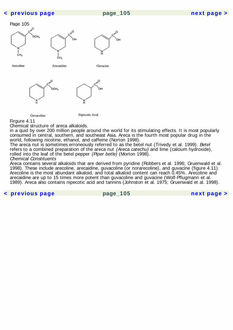
< previous page page_105 next page >
Page 105
Firgure 4.11Chemical structure of areca alkaloids.in a quid by over 200 million people around the world for its stimulating effects. It is most popularlyconsumed in central, southern, and southeast Asia. Areca is the fourth most popular drug in theworld, following nicotine, ethanol, and caffeine (Norton 1998).The areca nut is sometimes erroneously referred to as the betel nut (Trivedy et al. 1999). Betelrefers to a combined preparation of the areca nut (Areca catechu) and lime (calcium hydroxide),rolled into the leaf of the betel pepper (Piper betle) (Morton 1998).Chemical ConstituentsAreca contains several alkaloids that are derived from pyridine (Robbers et al. 1996; Gruenwald et al.1998). These include arecoline, arecaidine, guvacoline (or norarecoline), and guvacine (figure 4.11).Arecoline is the most abundant alkaloid, and total alkaloid content can reach 0.45%. Arecoline andarecaidine are up to 15 times more potent than guvacoline and guvacine (Wolf-Pflugmann et al.1989). Areca also contains nipecotic acid and tannins (Johnston et al. 1975; Gruenwald et al. 1998).
< previous page page_105 next page >
< previous page page_106 next page >
Page 106PharmacokineticsLimited data are available on the pharmacokinetics of arecoline. Intravenously administered arecolinein subjects with Alzheimer's disease shows variation in the optimal dose (between 4 and 16 mg/day)due to differing plasma kinetics (Asthana et al. 1996). The mean plasma half-lives for these doseswere 0.95 ± 0.54 and 9.3 ± 4.5 minutes, respectively. However, the mean plasma concentrationsthat optimized cognitive effects were 0.31 ± 0.14 ng/ml. Drug clearance was 13.6 ± 5.8 L/min andthe volume of distribution was 205 ± 170 L.Oral administration of arecoline is ineffective for clinical purposes due to first-pass metabolism(Hussain and Mollica 1991). The nasal route is an alternate possibility, with 85% bioavailabilitycompared to intramuscular administration.Mechanisms of ActionAcetylcholine All four alkaloids derived from areca (arecoline, arecaidine, guvacoline, and guvacine)act as full agonists at muscarinic acetylcholine receptors (Wolf-Pflugmann et al. 1989). Peripherallyadministered arecoline (10 mg/kg) subtly reduces cortical and subcortical acetylcholine levels(Molinengo et al. 1986).Monoamine Arecoline has indirect effects on catecholamine levels (Molinengo et al. 1986). Significantreductions occur in norepinephrine levels, but increases occur in dopamine levels in both cortical andsubcortical areas.Amino acid Nipecotic acid is a potent inhibitor of GABA uptake, effective in the low micromolar range(Johnston et al. 1975; Liachenko et al. 1999). Nipecotic acid is structurally related to the antiepilepticdrug tiagabine (Gabatril), which also blocks reuptake of GABA in the micromolar range. Arecaidineand guvacine also inhibit GABA uptake in micromolar concentrations, but do not inhibit glycine ortaurine (Lodge et al. 1977; Johnston et al. 1975). This effect is observed in the cat spinal cord andcerebellum, enhancing the activity of Purkinje cells. However, the relevance of arecaidine in chewingareca is questionable because arecaidine was active only when applied centrally, and notsystemically.
< previous page page_106 next page >
< previous page page_107 next page >
Page 107General EffectsAutonomic effects Areca produces autonomic effects through actions at cholinergic receptors. Itcauses sweating, facial flush, and a superficial warm sensation (Chu 1995). Skin temperature mayrise up to 2 degrees Celsius, through both sympathetic and parasympathetic mechanisms. Heart rateincreases during areca chewing (Sekkadde et al. 1994). Arecoline, isoarecoline, and arecaidine causea brief depressor and bradycardic response, followed by an increase in arterial blood pressure andheart rate, which are mediated through muscarinic M1 receptors (Wess et al. 1987).Neuroendocrine In human subjects with Alzheimer's disease, intravenous infusion of arecoline causedelevations of adrenocorticotrophic hormone, cortisol, and β-endorphin, indicating an activation of thehypothalamic-pituitary-adrenal axis (Asthana et al. 1995).Cerebral blood flow and glucose metabolism Increases in cerebral glucose metabolism are producedby arecoline, as seen in autoradiography with 2-deoxy-D-glucose (Soncrant et al. 1985). Lower dosesproduced selective increases in areas such as the hippocampus and nucleus raphe medianis, buthigher doses produced more generalized elevations. More specifically, hippocampal metabolism isincreased in layers that receive cholinergic innervation. Elevations in metabolism in extrapyramidalmotor areas were related to the intensity of tremor. The increases in cerebral glucose utilization areaccompanied by increases in cerebral blood flow during both acute and chronic treatment (Maiese etal. 1994). However, glucose utilization increases to a greater degree than blood flow in thehippocampus.Electrophysiological effects Areca chewing in experienced users creates EEG activation (Chu 1994b).Increases are seen in beta and alpha activity, and decreases in theta activity. Topographically, alphaincreases are seen in the occipital region, while the beta and theta effects are widespread. Theseeffects are consistent with those of other cholinergic stimulant and cognitive enhancing drugs.
< previous page page_107 next page >
< previous page page_108 next page >
Page 108Cognitive effects Animal studies have found some beneficial effects of arecoline on memory, varyingwith the task employed, dose, and degree of tolerance. Areca chewing in human subjects increasesthe speed of mental processing. Mixed results have been found in subjects with Alzheimer's disease.The cognitive effects of areca are discussed at greater length in chapter 5.Addiction and DependenceA dependency syndrome occurs in many who chew areca (Trivedy et al. 1999). This is not entirelysuprising because other cholinergic stimulants (e.g., tobacco) also have addictive properties, andarecoline causes a release of dopamine like many other abused drugs. Use of areca persists despiteadverse health effects—one of the criteria for addiction.Toxic EffectsChewing of areca nuts causes a reddish-brown discoloration of the oral mucosa and a tendency forthe epithelial surface to peel (Reichart and Phillipsen 1998). This has been called betel chewer'smucosa, and other oral lesions such as leukoedema, leukoplakia and ulceration may occur. The teethalso become stained reddish-brown or even black in more extreme cases (Morton 1992). Chewingareca is also associated with oral submucous fibrosis and squamous cell carcinoma (Warnakulasuriyaet al. 1997; Kuo et al. 1999; Jeng et al. 1999). Several carcinogenic N-nitrosamines are formedduring areca chewing (Hoffman et al. 1994). Tannins are another source of potential carcinogenicityin areca (Morton 1992).Associations have also been made between areca and cardiovascular disease, diabetes, and asthma(Winstock et al. in press). Areca may affect cardiovascular disease by increasing homocysteineconcentrations and/or through areca copper concentrations and interaction with the lysyl oxidaseenzyme (Trivedy et al. 1999). Areca chewing has been associated with cardiac dysrhythmias in a fewcases and a case of myocardial infarction was temporally associated with areca use (Hung and Deng1998; Chiang et al. 1998).Approximately 61% of people with asthma report that areca aggravates the condition. Clinical studiesshow that areca causes broncho-
< previous page page_108 next page >
< previous page page_109 next page >
Page 109constriction, as evidenced by decreases in forced expiration volume (Sekkadde et al. 1994; Taylor etal. 1992).Areca may interact adversely with antipsychotic medications (Deahl 1989). Two cases have beenreported of schizophrenic patients who were taking neuroleptics and developed severeextrapyramidal symptoms after areca chewing. Given the functional antagonism between dopamineand acetylcholine in the striatum, it is likely that arecoline amplified the dyskinetic effect ofneuroleptic medications.LobeliaHistory and BotanyLobelia (Lobelia inflata), also referred to as Indian tobacco, is an annual or biennial herb that growsfrom 30 to 60 cm in height (Gruenwald et al. 1998). Its leaves are green or yellowish and range fromovate to lanceolate in shape (figure 4.12). Its flowers are a pale violet-blue and the fruit is an ovoidcapsule containing many small brown seeds.Lobelia was named after a Flemish botanist, Matthias L'Obel (Robbers et al. 1996). Inflata refers tothe hollow, distended fruit. It was also used by Native Americans as a substitute for tobacco. It wasused medicinally by a group of physicians during the nineteenth century C.E. (Tyler 1994). However,due to toxicity and better treatments for tobacco dependence, lobelia is now rarely, if ever, indicated.Chemical ConstituentsLobelia contains 14 alkaloids, of which lobeline is the most significant (figure 4.13). Other alkaloidsinclude lobelanine, lobelanidine, norlobelanine, and isolobinine. Another relevant chemical that isisolated from the leaves of lobelia is beta-amyrin palmitate.PharmacokineticsLobeline is somewhat soluble in water, but not soluble in alcohol (Robbers et al. 1996). It is nolonger used medicinally, but has been put in lozenges to assist in breaking the tobacco habit.Unfortunately, it has not been successful in this regard. Lobelia derivatives may cross the blood-brainbarrier by passing through basic amine transporters
< previous page page_109 next page >

< previous page page_110 next page >
Page 110
Figure 4.12Lobelia (Lobelia inflata). Reprinted from Culbreth DMR. (1927). Materia Medica and Pharmacognosy,7th ed. Philadelphia: Lea & Febiger.
Figure 4.13Chemical structure of lobeline.
< previous page page_110 next page >
< previous page page_111 next page >
Page 111(Metting et al. 1998). Tolerance develops to lobeline's behavioral or analgesic effects by 10 days ofdaily treatment, and cross-tolerance develops between lobeline and nicotine (Damaj et al. 1997).Mechanisms of ActionAcetylcholine Lobeline acts as a partial agonist at nicotinic receptors. It displaces nicotine with a lownanomolar affinity in the rat brain (Damaj et al. 1997). Similar to nicotine, it may act as anantagonist through persistent activation and desensitization of the receptor (Briggs and McKenna1998; Tani et al. 1998). Lobeline shows a greater affinity for the β2 subunit of the nicotinic receptorthan β4 subunits (Parker et al. 1989).However, lobeline shows several significant pharmacological differences from nicotine. It decreasesthe basal release of acetylcholine, and also reduces NMDA-evoked acetylcholine release in themicromolar range (Rao et al. 1997). Nicotine, in contrast, does not have this effect. Lobeline binds todifferent nicotinic receptor subtypes than nicotine, and it is unaffected by the nicotine antagonistmecamylamine (Terry et al. 1998). Lobeline activates nicotinic channels at low concentrations, butinhibits at higher concentrations. Whereas nicotine elicits the release of acetylcholine in thehippocampus and frontal cortex, lobeline does not (Tani et al. 1998). Additionally, lobeline displacesmethylscopolamine in the micromolar range, suggesting activity at muscarinic receptors, and it is aweak AChE inhibitor—several hundred times less potent than physostigmine (Terry et al. 1998).Monoamines Lobeline has direct effects on monoamine systems, independent of cholinergic effects. Italters presynaptic dopamine storage in the striatum by binding to the vesicular monoamine reuptaketransporter and preventing uptake (Teng et al. 1998). Half-maximal inhibition occurs in thesubmicromolar range (20 times lower than that of d-amphetamine). Lobeline inhibits dopamineuptake more potently than it evokes release, but low micromolar concentrations still cause thespontaneous and evoked release of dopamine (Teng et al. 1997; Clarke and Reuben 1996; Rao et al.1997; Sakurai et al. 1982). This dopamine
< previous page page_111 next page >
< previous page page_112 next page >
Page 112release is independent of calcium and mecamylamine, suggesting that it is caused by inhibiting theuptake of dopamine into vesicles. Lobeline does not alter levels of dopamine or DOPAC in the nucleusaccumbens (Benwell and Balfour 1998).Lobeline also increases basal release of norepinephrine, but norepinephrine release may be reducedat higher lobeline concentrations (Rao et al. 1997). Unlike acetylcholine, lobeline does not reduce therelease of dopamine or norepinephrine by NMDA receptors, but it does block nicotine-induced releaseof norepinephrine from the locus coeruleus (Gallardo and Leslie 1998). Lobeline also evokes releaseof serotonin, which is mediated by uptake transporters and unaffected by mecamylamine (Lendvai etal. 1996).Another psychoactive constituent of lobelia, beta-amyrin palmitate, causes a release ofnorepinephrine in mouse brain synaptosomes, possibly releasing it from newly synthesized pools(Subarnas et al. 1993b).Ca2+ channels Lobeline acts as an antagonist at neuronal Ca2+ channels (Toth and Vizi 1998).Micromolar doses of lobeline block Ca2+ currents in superior cervical ganglia neurons, and are notaltered by G-protein manipulations.General EffectsPhysiological effects Humans given intravenous microgram doses of lobeline experience sensations ofchoking and pressure in the throat and upper chest, and eventually a dry cough (Raj et al. 1995).Reflex changes in breathing occur shortly before the onset of the sensations. Chronic three-weektreatment with lobeline in rats induced no changes in weight, rectal temperature, or motorcoordination (Sopranzi et al. 1991). Lobeline dose-dependently decreases body temperature in mice(Damaj et al. 1997). Gastrointestinal symptoms may occur with larger doses, resembling nicotine'seffects.Behavioral and cognitive effects Lobeline appears to be reinforcing in a similar manner to nicotine.Despite weak nicotinic effects, drug-naive animals will self-administer lobeline, as they would withother reinforc-
< previous page page_112 next page >

< previous page page_113 next page >
Page 113ing drugs (Rasmussen and Swedberg 1998). However, unlike nicotine, lobeline does not induce aconditioned place preference nor stimulate locomotor activity (Benwell and Balfour 1998; Fudala andIwamoto 1986; Stolerman et al. 1995). Lobeline's effects are also distinguishable from nicotine'seffects on animals in drug discrimination paradigms (Reavill et al. 1990). Beta-amyrin palmitate alsodose-dependently decreases locomotor activity in mice (Subarnas et al. 1993c). Pharmacologicalevidence suggests that this is through α1 adrenergic and not dopamine receptors.An acute dose of lobeline impairs attention in one animal model, but not as much as mecamylamine(Turchi et al. 1995). Lobeline improves memory when administered after a passive avoidanceparadigm (Decker et al. 1993). Pretreatment with lobeline improves performance in rats with septallesions on a spatial discrimination water maze. Lobeline is about one-tenth as potent as nicotine inthe passive avoidance memory task, but equivalent to nicotine in the water maze.Electrophysiology Studies of the electrophysiological effects of lobelia are scant. In one study,increases were seen in low-frequency activity in the hippocampus but in higher-frequency activity inthe amygdala (Sopranzi et al. 1991).Antidepressant Some animal models show antidepressant effects of lobelia extract (Subarnas et al.1992). Similar to imipramine and mianserin, beta-amyrin palmitate shows antidepressant-like effectsin the forced-swimming test (Subarnas et al. 1993a). Whereas mianserin and beta-amyrin palmitatereduce locomotor activity induced by methamphetamine, imipramine increases it. It potentiatessodium pentobarbital-induced sleep more potently than imipramine, but less than mianserin.Collectively, the effects of beta-amyrin palmitate in behavioral and physiological assays suggests itmay work in a manner more similar to mianserin than imipramine. However, the mechanism ofantidepressant-like effects of lobelia is uncertain. It may be through the beta-amyrin palmitate'sability to release norepinephrine (Subarnas et al. 1993b). An antidepressant effect of lobelia has notbeen established in humans.
< previous page page_113 next page >

< previous page page_114 next page >
Page 114
Figure 4.14Chemical structure of ephedrine.Monoamine StimulantsEphedraHistory and BotanyEphedra is a group of related plant species that have long been used for their stimulant effects.Ephedra sinica is a shrub that grows 60–90 cm in height (Robbers et al. 1996; Gruenwald et al.1998). It has long cylindrical branches 1–2 mm in diameter, which terminate in a sharp point. Thebranches are green and have very small leaves, which sometimes are reduced to pointed scales. Thefruit is red and berrylike. Ephedra sinica is native to Asia, growing mostly in Mongolia andneighboring parts of China.Ephedra is also known by the Chinese name ma huang, which translates as ''yellow astringent." Ithas been used medicinally by the Chinese for more than five thousand years. A related species,Ephedra nevadensis, grows in North America and is sometimes referred to as Mormon tea (Rudgley1999). The Mormons were introduced to ephedra by Indians when they arrived in Utah. They used itas an alternate to tea and coffee, ironically, because their religious views prohibited use of thosestimulants. Ephedra might be the oldest known human stimulant, because remains of the plant werediscovered in a fifty thousand-year-old Neanderthal grave in Iraq. Modern medical use of ephedrabegan with the identification of the alkaloid ephedrine in 1923.Chemical ConstituentsSeveral alkaloids are present in Ephedra, with ephedrine being the most significant (Gruenwald et al.1998). Ephedra sinica contains approxi-
< previous page page_114 next page >
< previous page page_115 next page >
Page 115mately 1.25% ephedrine (figure 4.14). Also present are pseudoephedrine, methylephedrine andnorpseudoephedrine, or cathine, which is also a major alkaloid of khat (Catha edulis) (Robbers et al.1996; Kalix 1991). Different ephedra species may vary considerably in their content of alkaloids(Robbers et al. 1996; White et al. 1997).PharmacokineticsCommercial samples containing approximately 400 mg of ephedra per capsule yield roughly 5 mg ofephedrine, 1 mg of pseudoephedrine, and less than 1 mg of methylephedrine (White et al. 1997). Fora dose of four capsules, yielding approximately 20 mg of ephedrine, the elimination half-life is 5.2hours. The time to reach maxium concentration is 3.9 hours. Compared to pure ephedrine tablets,the elimination kinetics of ephedra are comparable. However, ephedra showed somewhat differentabsorption kinetics (e.g., lag time, area under the concentration-time curve, and maximum plasmaconcentration). So, ephedra tablets may vary from pure ephedrine in the onset of action, but thedurations of action are grossly equivalent.Mechanisms of ActionEphedrine is an agonist of α and β adrenergic receptors (Meston and Heiman 1998; Robbers et al.1996). However, it does not bind to β3 adrenergic receptors (Shannon et al. 1999). It also increasesrelease of norepinephrine from sympathetic neurons.General EffectsEphedrine increases heart rate and increases systolic blood pressure (Kuitunen et al. 1984).Stimulation of β receptors in the lung by ephedrine promotes bronchial dilation. Accordingly, ephedrahas been used historically to treat asthma, and pseudoephedrine is commercially available as anover-the-counter decongestant. Stimulation of α adrenergic receptors on smooth muscle increasesresistance to urine flow. Although ephedrine facilitates the neuromuscular endplate potential byincreasing the quantal release of acetylcholine, this only occurs at high concentrations unlikely to bereached with oral dosing (10–4 M). Ephedrine increases the physiological aspect of sexual arousal inwomen
< previous page page_115 next page >

< previous page page_116 next page >
Page 116Table 4.2Relative doses for stimulus generalization of psychostimulants(ED50)
Drug ED50
(–)ephedrine 0.8 mg/kgS(+)amphetamine 0.4 mg/kgcocaine 2.7 mg/kgmethylphenidate 1.2 mg/kgS(–)methcathinone 0.3 mg/kgcaffeine 36.7 mg/kg
Source: Young and Glennon 1998.viewing erotic material, as measured by vaginal pulse amplitude (Meston and Heiman 1998).Insomnia, anxiety, cardiovascular, and other sympathomimetic effects limit the clinical use ofephedrine (Kalix 1991). Chronic doses may be associated with a toxic psychosis.Ephedrine causes a potent stimulation of the central nervous system, resulting in increased arousaland activity. On neuropsychological tests, ephedrine does not have any effects on finger-tapping,sign-recording, or digit-span tests (Kuitunen et al. 1983). It does reverse impairments in auditoryvigilance and serial-choice performance produced by the sedating antihistamine chlorpheniramine,but does not affect simple reaction speed (Millar and Wilkinson 1981). Ephedrine counteracts some ofthe deficits produced by scopolamine in sensory processing (critical fusion flicker) (Nuotto 1983).Animal drug discrimination paradigms show stimulus generalization between ephedrine and otherCNS stimulants such as amphetamine, methylphenidate, methcathinone, cocaine, and caffeine (seetable 4.2; Young and Glennon 1998). However, it does not generalize to MDMA. Pseudoephedrinesubstitutes for amphetamine, but only at high doses (40 mg/kg in rats) (Tongjaroenbuangam et al.1998).ToxicityNormal doses range from 15 to 30 mg of actual alkaloid (ephedrine), and the lethal dose is in the 1–2 g range. Side effects from ephedra use include headache, irritability, restlessness, anxiety,insomnia, tachycardia, urinary disorders, and vomiting.
< previous page page_116 next page >
< previous page page_117 next page >
Page 117Recently, ephedra preparations have been sold with misleading names alluding to the drug ecstasy(MDMA) (Blumenthal and King 1997). Several deaths have resulted from cardiac toxicity becausehypertension and cardiac arrhythmia are chief problems with higher doses of ephedrine. Use ofephedra is thus contraindicated in people with existing high blood pressure. Cases have beenreported of intracerebral hemorrhage and vasculitis in association with ephedrine (Forman et al.1989; Kaye and Fainstat 1987; Wooten et al. 1983). The risk of cerebral hemorrhage is even greaterwhen combining ephedrine with other catecholamine stimulants such as the over-the-counterstimulant phenyl-propanolamine (Stoessl et al. 1985). Pseudoephedrine may be safer than ephedrinein some respects (Porta et al. 1986). In a large sample (n > 100,000) of pseudoephedrine users,there were no reports of cerebrovascular disorders within 15 days after administration. The incidenceof myocardial infarction, seizures, and neuropsychiatric disorders were no greater than base rates inthe general population.Other conditions in which ephedra is contraindicated are anxiety disorders, angle-closure glaucoma,prostate adenoma with residual urine volume, pheochromocytoma, and thyrotoxicosis (Gruenwald etal. 1998). Known medications that may interact adversely with ephedrine include heart glycosides,halothane, guanethidine, MAO inhibitors, secale alkaloids, and oxytocin.Addiction and DependenceUse of ephedrine is sometimes associated with dependence (Gruber and Pope 1998). One studyreported a high incidence of eating and body-image disorders among ephedrine users, but this islikely confounded by the fact that the sample consisted entirely of female weightlifters and is likelynot to represent the greater population.CocaHistory and BotanyCoca (Erythroxylon coca) is a small shrublike tree that grows to 5 m in height. The leaves are oval,green, and tough, growing 5 cm in length and 2.5 cm in width (Robbers et al. 1996; Gruenwald et al.1998). The
< previous page page_117 next page >
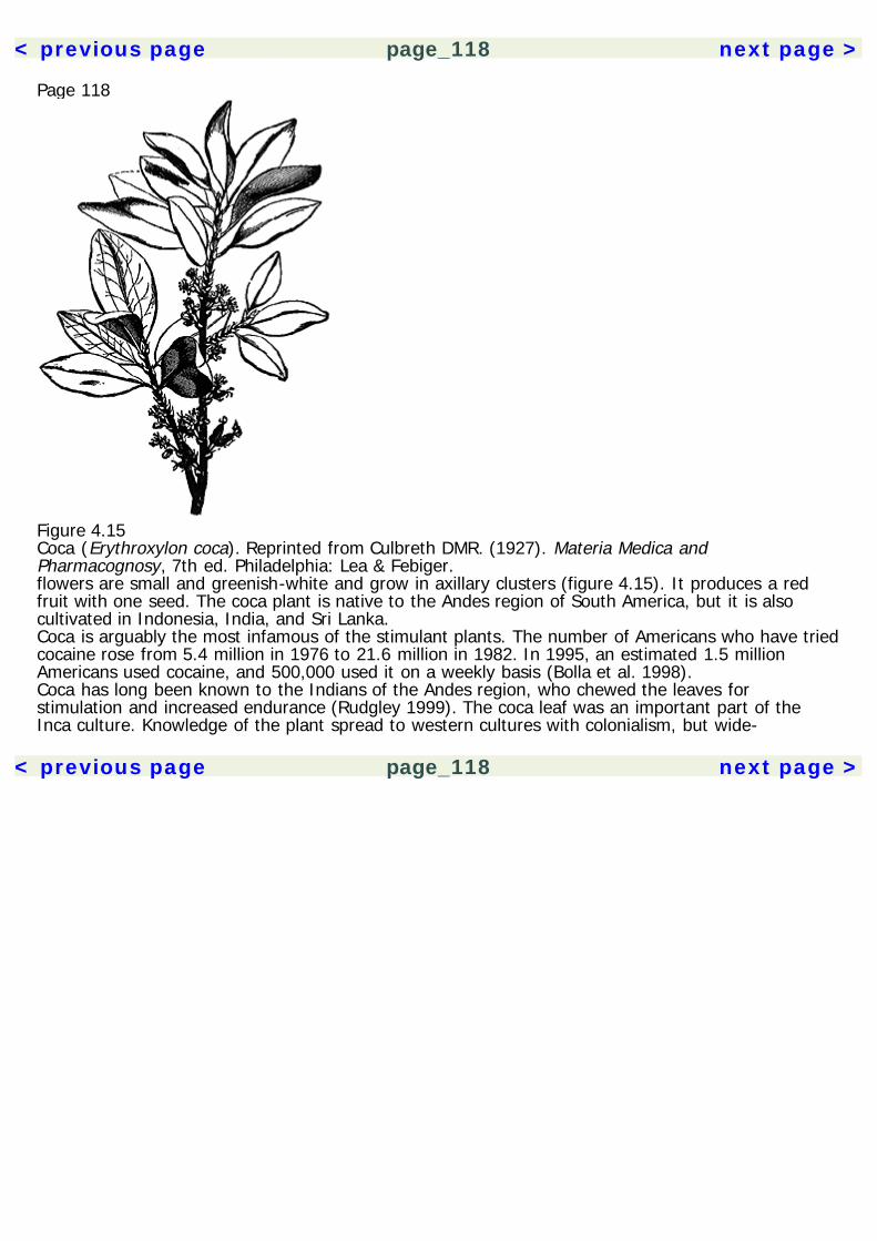
< previous page page_118 next page >
Page 118
Figure 4.15Coca (Erythroxylon coca). Reprinted from Culbreth DMR. (1927). Materia Medica andPharmacognosy, 7th ed. Philadelphia: Lea & Febiger.flowers are small and greenish-white and grow in axillary clusters (figure 4.15). It produces a redfruit with one seed. The coca plant is native to the Andes region of South America, but it is alsocultivated in Indonesia, India, and Sri Lanka.Coca is arguably the most infamous of the stimulant plants. The number of Americans who have triedcocaine rose from 5.4 million in 1976 to 21.6 million in 1982. In 1995, an estimated 1.5 millionAmericans used cocaine, and 500,000 used it on a weekly basis (Bolla et al. 1998).Coca has long been known to the Indians of the Andes region, who chewed the leaves forstimulation and increased endurance (Rudgley 1999). The coca leaf was an important part of theInca culture. Knowledge of the plant spread to western cultures with colonialism, but wide-
< previous page page_118 next page >
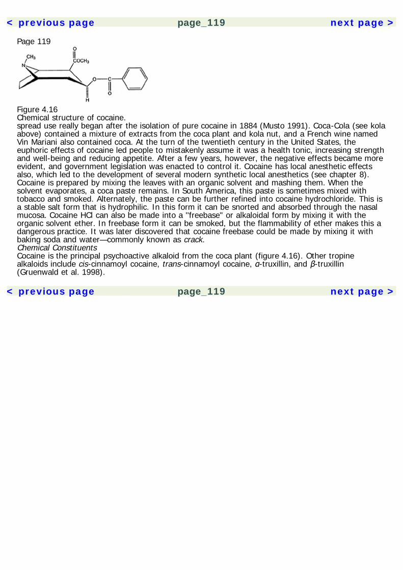
< previous page page_119 next page >
Page 119
Figure 4.16Chemical structure of cocaine.spread use really began after the isolation of pure cocaine in 1884 (Musto 1991). Coca-Cola (see kolaabove) contained a mixture of extracts from the coca plant and kola nut, and a French wine namedVin Mariani also contained coca. At the turn of the twentieth century in the United States, theeuphoric effects of cocaine led people to mistakenly assume it was a health tonic, increasing strengthand well-being and reducing appetite. After a few years, however, the negative effects became moreevident, and government legislation was enacted to control it. Cocaine has local anesthetic effectsalso, which led to the development of several modern synthetic local anesthetics (see chapter 8).Cocaine is prepared by mixing the leaves with an organic solvent and mashing them. When thesolvent evaporates, a coca paste remains. In South America, this paste is sometimes mixed withtobacco and smoked. Alternately, the paste can be further refined into cocaine hydrochloride. This isa stable salt form that is hydrophilic. In this form it can be snorted and absorbed through the nasalmucosa. Cocaine HCl can also be made into a ''freebase" or alkaloidal form by mixing it with theorganic solvent ether. In freebase form it can be smoked, but the flammability of ether makes this adangerous practice. It was later discovered that cocaine freebase could be made by mixing it withbaking soda and water—commonly known as crack.Chemical ConstituentsCocaine is the principal psychoactive alkaloid from the coca plant (figure 4.16). Other tropinealkaloids include cis-cinnamoyl cocaine, trans-cinnamoyl cocaine, α-truxillin, and β-truxillin(Gruenwald et al. 1998).
< previous page page_119 next page >
< previous page page_120 next page >
Page 120PharmacokineticsCocaine is administered in several ways. When the leaves are chewed, the drug absorbtion is slowand prolonged (Homstedt et al. 1979). Snorting or smoking cocaine allow for rapid absorptionthrough the nasal mucosa or the lungs, respectively. Absorption is poor through the nasal mucosa(20–30% absorption), which is further limited by the vasoconstriction it causes. Cocaine is lessfrequently injected intravenously, but it is sometimes combined with heroin to make an especiallyaddictive concoction called a speedball. While the euphoric effects of heroin and cocaine are additive,they also counteract each other's negative side effects of sedation and overstimulation. Smokingcrack is probably the fastest and most direct means of delivering cocaine to the brain, and thusprobably the most addictive (Hatsukami and Fischman 1996). When equivalent doses are giventhrough smoked and intravenous routes, the smoked method produces greater behavioral responses(Cone 1995). Absorption is rapid and complete through the lung surfaces, with onset occuring inseconds and the effect lasting about 30 minutes.Once in the blood stream, cocaine levels quickly rise in the brain, faster than plasma levels, whichthen redistribute to other tissues. Cocaine is rapidly metabolized in the blood and liver, with a half-life of 30 to 90 minutes. The major metabolites have a half-life of approximately 8 hours. Althoughcocaine itself is detected in urine for only 12 hours, the metabolite benzoylecgonine can be detectedin urine for at least 48 hours and sometimes up to 2 weeks. Concurrent use of cocaine and ethanolproduces an ethyl ester of benzoylecgonine called cocaethylene. Cocaethylene is an activemetabolite, blocking dopamine reuptake, and potentiating the effect of cocaine. Thus, concurrent useof cocaine and ethanol can further increase the additional effects of the drugs and the risk ofdependency.Mechanisms of ActionMonoamine The primary psychoactive mechanism of cocaine is blocking reuptake of the monoamineneurotransmitters dopamine, norepinephrine, and serotonin, leading to increased available synaptictransmitters (O'Brien 1996). Chronic use is associated with changes in
< previous page page_120 next page >
< previous page page_121 next page >
Page 121monoamine metabolites, receptors, and uptake transporters (Belej et al. 1996; Perret et al. 1998;Baumann and Rothman 1998). A decreased availability of D2 receptors is seen, possibly reflecting acompensatory down-regulation of the postsynaptic receptors in response to increased stimulation(Volkow et al. 1993). Evidence of low dopamine function after chronic cocaine use is supported byconcurrent hyperprolactinemia (Dackis and Gold 1985). Neuroimaging studies show decreaseddopamine activity in cocaine withdrawal, when comparing cocaine users to nonusers (Wu et al.1997). Collectively, evidence strongly suggests that alterations in monoamine systems and theirtargets mediate the acute effects of cocaine, and that changes in these systems mirror the addiction,cravings, and withdrawal symptoms.Na+ channels Cocaine is an antagonist of voltage-gated Na+ channels, reducing axonal conductionand acting as a local anesthetic. This effect will be discussed at greater length in chapter 8. Cocaine'sblockade of Na+ channels as a contribution to its psychotropic effects is possible but has not beenexamined.General EffectsCocaine causes profound mental stimulation, behaviorally evidenced by increased talkativeness andactivity, flight of ideas, euphoria, and inflated self-esteem. Fatigue is offset and there is a reducedneed for sleep. Supression of appetite also occurs.Autonomic Cocaine has stong sympathomimetic effects due to inhibition of norepinephrine reuptake,and perhaps central mechanisms as well. Effects include those typical of sympathetic autonomicactivation. Cardiovascular and cerebrovascular effects are prominent.Vascular effects Cocaine causes increases in blood pressure and heart rate, which fall to normallevels between doses (Foltin et al. 1995). Tachyphylaxis develops to the cardiovascular effects, evenwithin a single session. Concurrent use of ethanol, cannabis, and cocaine causes even greatercardiovascular effects than those of each drug alone. Interactions can also occur with antidepressantdrugs like desipramine.
< previous page page_121 next page >
< previous page page_122 next page >
Page 122Cerebral blood flow and glucose metabolism Several studies have examined the effects of cocaineusing neuroimaging techniques. In humans and nonhuman primates alike, cocaine decreases cerebralglucose metabolism (Volkow et al. 1991). This includes areas such as the nucleus accumbens, ventralstriatum, orbitofrontal cortex, prefrontal cortex, and hippocampus, suggesting a pronounced effect ofcocaine on corticostriatal circuits. When methylphenidate (Ritalin) is given to cocaine users, increasesin metabolism are seen in the orbitofrontal cortex and striatum, which correspond to reports ofcravings. On the other hand, increases in other prefrontal areas correspond to enhanced mood(Volkow et al. 1999). In cocaine users, decreased metabolism in the prefrontal cortex (includingcingulate and orbitofrontal regions) is associated with decreases in D2 receptors (Volkow et al. 1999).Decreases are also seen in cerebral blood flow in cocaine users (Volkow et al. 1988). This may bedue, in part or wholly, to the vasoconstrictor effects of cocaine. Indeed, greater deficits are seen incerebral blood flow compared to glucose metabolism, even within the same individual.Cognitive Cognitive studies of cocaine users have produced conflicting results. Deficits have beenfound in cocaine users in mental processing speed, visual and verbal memory, and executivefunctions (O'Malley et al. 1992; Easton and Bauer 1997; Berry et al. 1993; Hoff et al. 1996; Selbyand Azrin 1998). Executive deficits have been seen in measures such as the Category test, the TrailMaking Test B, and the Shipley Abstraction Subtest. Yet improvements are seen in some studies ontests of nonverbal reasoning (Wisconsin Card Sorting Test—categories achieved) and verbal fluency(Controlled Oral Word Association). Others have found improvement on other tests like the FingerTapping Test and visuospatial memory.Methodological issues are paramount in the cognitive studies of cocaine users. Primary concernssurround preexisting cognitive deficits or a self-selection effect among subjects (Bolla et al. 1998).Other confounding factors include duration of use, duration of abstinence, sample size, controlgroups, premorbid depression, and personality factors such as antisocial personality disorder. Still,the available neuropsychological and neuroimaging data convergently support executive deficitsresulting
< previous page page_122 next page >
< previous page page_123 next page >
Page 123from altered frontostriatal function. It remains to be shown how long it takes for these deficits todevelop (and at what doses), how long they persist, and to what degree they recover.Addiction and DependenceAddiction is a prominent problem with cocaine use. Cocaine is highly reinforcing to both animals andhumans, probably through inhibition of dopamine reuptake in mesolimbic systems and stimulation ofbrain areas known to subserve behavioral reinforcement (Kiyatkin and Stein 1995; Woolverton andJohnson 1992; Ritz et al. 1987). Although sensitization to the stimulant effects occurs in animals,humans do not sensitize to the euphoric effects of cocaine but develop a tolerance (O'Brien 1996). Inanimals and humans alike, self-administration often follows a binge pattern, consisting of repeateduse over a period of hours or days until the supply is used up.A cocaine withdrawal syndrome is recognized that generally mirrors the acute effects of intoxication.It includes depression, sleepiness, fatigue, increased appetite, bradycardia, and craving cocaine.Treatment of cocaine addiction is a stubborn problem resulting from the intense euphoria andreinforcement caused by the drug (O'Brien 1996). Treatments include psychosocial support (e.g.,Narcotics Anonymous) and behavioral reinforcement of abstinence, often monitored with serial urinetests. Pharmacological treatments of cocaine addiction have been attempted with antidepressants(e.g., fluoxetine), dopamine agonists (e.g., amantadine), and opioids (e.g., buprenorphine). Thebenefits of any pharmacological treatment to date for treatment of cocaine addiction have beenmodest at best. A novel approach to treatment involves catalytic or monoclonal antibodies for cocaine(Mets et al. 1998; Fox et al. 1996). Rather than replace the neurochemical effects of cocaine, thisstrategy seeks to neutralize cocaine in the blood stream, making future administration of cocaine lesseffective or ineffective.ToxicityCardiovascular and cerebrovascular Cocaine has cardiotoxic effects, sometimes resulting inarrhythmia, ventricular fibrillation, or cardiac arrest (Nanji and Filipenko 1984; Sherief and Carpentier1991). Vaso-
< previous page page_123 next page >
< previous page page_124 next page >
Page 124constriction caused by cocaine greatly increases the risk for ischemic or hemorrhagic stroke (Levineet al. 1991). Vasoconstriction is also caused by benzoylecgonine, and norcocaine, in addition tococaine (Bolla et al. 1998). Whereas freebase alkaloidal cocaine (crack) is equally likely to causeeither kind of stroke, cocaine HCl is more likely to cause a hemorrhagic stroke.Neonatal Cocaine freely crosses the placental barrier, and prenatal exposure to cocaine altersneurobehavioral development in rat pups (Sobrian et al. 1990). The effects on humans exposedprenatally to cocaine is a complicated matter, because so many other concurrent factors contributeto development. Common confounds are prenatal care and maternal polydrug use. Prenatal cocaineuse is associated with reduced gestational age, birth weight, body length, and head circumference(Richardson et al. 1999). In children exposed prenatally to cocaine, some studies have shownbehavioral differences evident at 1 to 3 years of age (Richardson et al. 1993; Richardson 1998).Associations are also made with impulsivity and attention deficits at age 6 (Leech et al. 1999).Toxic psychosis Several monoamine stimulants including cocaine are known to produce a temporaryor even a lasting psychotic state after heavy use. Reviews of numerous clinical case reports haveshown amphetamine to produce a chronic psychotic state, sometimes persisting for months aftercessation. There appears to be a sensitization effect in this regard, because after recovery, psychoticstates may recur with minimal use of amphetamine or alcohol. When compared to schizophrenicpatients, people with amphetamine-induced psychosis demonstrate fewer negative symptoms(Boutros and Bowers 1996).Chronic cocaine use can cause a syndrome of insomnia, hallucinations, delusions, and apathy. Thissyndrome develops around the time when the euphoria turns to a paranoid psychosis, whichresembles paranoid schizophrenia. Further, after cessation of cocaine use, the hallucinations maystop, but the delusions can persist. Still, the incidence of a persistent cocaine-induced psychosisappears to be rare. One study found only 4 out of 298 chronic cocaine users receiving a diagnosis ofpsychotic disorder (Rounsaville et al. 1991). This incidence is approximately the
< previous page page_124 next page >

< previous page page_125 next page >
Page 125same as the base rate of schizophrenia in the general population, so cocaine may be precipitatingparanoid psychosis in individuals who had predisposing factors.KhatHistory and BotanyKhat (Catha edulis) is a small shrub or tree native to East Africa (Robbers et al. 1996; Gruenwald etal. 1998). It is cultivated in the highlands of Ethiopia as well as Yemen and South Africa. Khat isalternately referred to by different regional names such as qat, tschat, and miraa. Medieval Arabicwritings mention khat as far back as 1237 C.E. (LeBras and Fretillere 1965; Kalix 1992; Wilder et al.1994). In some Muslim cultures it is regarded as a more acceptable alternative to alcohol. Khat isoften used in social settings in some cultures, such as Yemen, where users gather in private homesand chew the leaves while conversing (Kalix 1994). At times khat is used by laborers and students tooffset fatigue.Chewing leaves of the khat shrub is practiced in parts of East Africa and the Arabian peninsula (Kalix1988; Widler et al. 1994). Some estimate daily use at 5 million portions. Use in the West is lesscommon, but has increased somewhat. More common in the United States has been use of thesynthetic drug methcathinone (or ''cat"), which is derived from khat alkaloids. Only the fresh khatleaves are pharmacologically active, so for some time use was limited to local areas that grew theplant. However, with air transportation, use has spread with emigrants in Europe and the UnitedStates. Because of its pharmacological similarities to amphetamine and its addictive properties, khathas been listed on Schedule I of the United Nations Convention on Psychotropic Substances.Chemical ConstituentsThere are more than 40 alkaloids, glycosides, tannins, and terpenoids in khat (Elmi 1983). Twophenylalkylamines, namely, cathine (norpseudoephedrine) and cathinone [S(–)-alpha-aminopropiophenone] well account for the CNS stimulant effects (Kalix 1988) (figure 4.17). The
< previous page page_125 next page >

< previous page page_126 next page >
Page 126
Figure 4.17Chemical structure of cathinone.S(–) enantiomer occurs naturally, and the R(+) enantiomer is synthetic (Kalix 1992).PharmacokineticsIn its natural form, fresh khat leaves are chewed because the drug is perishable. Young leaves fromthe tip of the branch are more potent. While the leaves are chewed, the juices are swallowed andthe residue is rejected. During a session, one may ingest 100 to 200 grams of leaves.When khat is chewed, absorption of cathinone is slow, with maximal plasma concentrations occurringat approximately 2 hours (Widler et al. 1994; Halket et al. 1995). The terminal elimination half-life isapproximately 4.3 hours. Similar effects are achieved with orally administered pure cathinone.Cathinone is the keto-analog of cathine and because it is more lipophilic it penetrates the blood-brainbarrier more easily.The plasma half-life of cathinone is 1.5 hours. The primary metabolites are norpseudoephedrine,norephedrine, 3,6-dimethyl-2,5-diphenyl-pyrazine, and 1-phenyl-1,2-propanedione (Szendrei 1980;Brenneisen et al. 1986; Guantai and Maitai 1983). However, norpseudoephedrine and norephedrinealso originate directly from the leaves, as well as being metabolic products (Widler et al. 1994).Maximal plasma concentrations of norephedrine and norpseudoephedrine are reached at about 3.3and 3.1 hours, respectively. These two drugs have a much longer duration of action than cathinone,where terminal half-lives could not be calculated after 10 hours.Animal drug discrimination paradigms show cross-tolerance between cathinone, cathine, andamphetamine (Schechter 1990). S(+)methcathi-
< previous page page_126 next page >
< previous page page_127 next page >
Page 127none is about twice as potent as S(+)amphetamine, and five times as potent as R(+)methcathinone,although both isomers have CNS stimulant effects (Glennon et al. 1995).Mechanisms of ActionThe stimulant effects of khat are mediated through monoamine neurotransmitter systems. Cathinoneinduces release of monoamines through membrane transporters (Kalix 1984, 1992). It causes releaseof dopamine in the striatum and nucleus accumbens, with similar potency to amphetamine in lowmicromolar concentrations (Kalix 1980, 1982; Pehek et al. 1990). Consequently, increases are seen inextracellular levels of the metabolite DOPAC in the caudate nucleus, nucleus accumbens, and frontalcortex (Mereu et al. 1983). Norpseudoephedrine also induces release of catecholamines (Kalix 1983).Cathinone decreases firing of substantia nigra neurons, similar to amphetamine. Animal discriminationof cathinone is dependent on dopamine release, but is independent of 5-HT3 modulation or Ca2+influx through L-type channels (Schechter 1992). High doses of cathinone result in depletion ofdopamine and also have neurotoxic effects on dopaminergic neurons (Wagner et al. 1982). Incontrast, long-term administration of cathinone does not deplete norepinephrine or serotonin(Wagner et al. 1982). Cathinone and cathine may also have monoamine oxidase inhibition effects(Nencini et al. 1984).General EffectsKhat produces effects similar to those of other monoamine stimulants, (i.e., increases in mentalstimulation, physical endurance, elevated mood) (Widler et al. 1994; Kalix 1994; Brenneisen et al.1990). Stimulus generalization occurs between cathinone, amphetamine, and cocaine, suggestingsimilar subjective effects (Huang and Wilson 1986). Similar to other monoamine stimulants,cathinone causes dose-dependent reductions in eating and body weight (Islam et al. 1990; Zelgerand Carlini 1980). Oral cathinone increases sexual arousal in rats, but does not affect erectile orejaculatory responses (Taha et al. 1995).Khat may cause irritability, but in the cultural context, sociability is generally increased. Users mayincrease in talkativeness, sometimes to
< previous page page_127 next page >

< previous page page_128 next page >
Page 128the point of logorrhea. Elevated self-esteem occurs, sometimes to a manic degree. As with othermonoamine stimulants, psychosis is possible with chronic heavy use. There have been a few reportedcases of khat-induced psychosis, which are treated with phenothiazines or haloperidol (Pantellis et al.1989). Persistent hypnagogic hallucinations have been reported in chronic khat chewers, possiblythrough chronic suppression of REM sleep (Granek et al. 1988).Khat produces sympathomimetic effects, increasing heart rate and blood pressure. When khat ischewed, the increases are gradual, maximizing at about 2 hours and lasting for 4 hours. However,tolerance develops to blood pressure and heart rate effects in habitual users. Mydriasis and increasesin respiration also occur. Cathinone induces thermogenesis in brown adipose tissue, which ismediated by β-adrenergic receptors (Tariq et al. 1989).Cathinone and norpseudoephedrine have antagonist effects at the neuromuscular junction (Guantaiet al. 1987). This is likely a direct blocking effect, independent of cholinergic and adrenergicinnervation. However, motor effects are not reported at doses commonly used.Addiction and DependenceThe similarities between cathinone and amphetamine raises the possibility of abuse potential (Kalix1994). Amphetamine and cathinone both appear to produce their stimulus-discrimination effectsthrough the dopamine system (Kalix and Glennon 1986).Animals self-administer cathinone in a pattern common to abuses of monoamine stimulants such ascocaine (Woolverton and Johanson 1984). Cathinone can induce a conditioned place preference inrats (Schechter 1991). Withdrawal symptoms of khat include lethargy, depression, nightmares, andmild tremor (Kalix 1994). N-methylated cathinone (methcathinone) is more potent, and has becomeavailable on the illegal market. It was subsequently scheduled as a controlled substance (Glennon etal. 1995).Use of cathinone in the form of chewing khat may have some features that limit its addictiveness(Kalix 1994). The large bulk of the material and the effort required to chew khat limits the amountthat can be ingested in a given time. Absorption in this manner is slow and gradual,
< previous page page_128 next page >
< previous page page_129 next page >
Page 129and the short half-life of cathinone also limits the amount that can accumulate over time. Thus, theaddictive potential of khat is likely minimized by a combination of pharmacokinetic factors.Toxic EffectsAs would be expected, khat overuse produces symptoms similar to those of other monoaminestimulants, such as cocaine or amphetamine, including signs of sympathetic overarousal. In theextreme this can involve a toxic psychosis. Disorders more frequently associated with chronic khatuse in males are headaches, anorexia, insomnia, constipation, and respiratory illnesses (Kennedy etal. 1983). Females report higher incidences of acute gastritis, jaundice, bronchitis and hepaticdiseases. Also, cathinone has toxic reproductive effects in humans and experimental animals (Islamet al. 1990). It decreases sperm count and motility, and increases the number of abnormal spermcells. It also decreases plasma testosterone in rats.SummaryAs reviewed in this chapter, a number of plants are recognized for their stimulant effects on thecentral nervous system. Although they work through a variety of neurochemical mechanisms, they allconvergently alter neuromodulatory systems to produce a common effect.All of the stimulants discussed have some potential for abuse, varying with environmental factors,context of use, and individual predispositions. Still, pharmacology plays a major causal role: drugswith direct or indirect actions on dopamine systems are more likely to be used compulsively.Stimulants have important roles in the cultures that use them. Whether used for positive or negativepurposes, it is likely that our relationships with stimulant plants will long continue.
< previous page page_129 next page >

< previous page page_130 next page >
Page 130This page intentionally left blank.
< previous page page_130 next page >
< previous page page_131 next page >
Page 1315Cognitive EnhancersHumans draw heavily on cognitive abilities to survive and flourish. Compared to other species we relyfar less on instinctual and environmentally driven behaviors, and instead have a greater capacity forbehavior driven by internal goals and abstract representations. This strategy for survival depends onmore complex cognitive ability and has coincided phylogenetically with enlargement of the cerebralcortex and frontal lobes. Consequently, in the search for plant medications, attention has been paidto those that improve mental ability. Some individuals are interested in cognitive-enhancing drugs, ornootropics, in an attempt to optimize their mental functioning. However, their potential use intreating dementia is of paramount interest. Whatever the impetus, several herbs have been identifiedwith benefical cognitive effects.DementiaDementia is a general term for loss of cognitive functions and behavioral impairment. It may arisefrom several etiologies and may present with a large variety of manifestations. The financial cost inhealth care engendered by dementia is enormous, because individuals with dementia requireprogressive amounts of supervision, eventually needing total care. The human and emotional costsare unquantifiable. Several forms of dementia exist, which will be briefly classified and describedhere.Dementia generally involves an impairment in (1) memory and other cogntive abilities and (2) socialand occupational functioning. The Diagnostic and Statistical Manual of Mental Disorders (DSM-IV)defines dementia as a persistent deficit in memory and at least one other area of cognitive function:language, praxis, object recognition, or executive
< previous page page_131 next page >
< previous page page_132 next page >
Page 132functions (organization, planning, abstract thinking, sequencing, etc.) (American PsychiatricAssociation, 1994). In addition to cognitive difficulties, impairments occur in social and occupationalfunctioning. Other classification systems do not require memory to be present to diagnose dementia,because several progressive dementias, such as frontotemporal dementias, may not show markedmemory loss (Neary et al. 1998). The onset of dementia is usually slow and other conditions thatcould mimic its symptoms (e.g., metabolic disorder) must be ruled out. However, the onset may berapid in a vascular dementia, resulting from compromised cerebral blood flow. A diagnosis of vasculardementia requires neuroimaging evidence or focal neurological signs, such as limb weakness orabnormal reflexes.Dementia may be dichotomized into degenerative and nondegenerative types. Degenerative dementiainvolves progressive cell loss due to pathological processes. Each subtype of dementia may havecharacteristic neuropathological signs, although there is much overlap in some cases. There is alsospecificity in the neuroanatomical distribution of pathology, which determines the functional brainchanges and constellation of clinical symptoms. For example, dementias arising from the basalganglia typically have pronounced motor symptoms, while a cortical dementia such as Alzheimer'sdisease may have little motor involvement until end stages.Two neurodegenerative diseases will be considered in greater detail here, Alzheimer's disease andParkinson's disease, due to the pertinence of herbal medications to their treatment. Also discussed insome detail are vascular dementia and normal aging. Other degenerative conditions may benefit fromherbal medications, but have not received the amount of attention in research that the aboveconditions have. Of particular interest to many degenerative conditions are herbal medications withdemonstrated antioxidant and neuroprotective effects.Degenerative Dementias: Cortical and Mixed Cortical/Subcortical TypesAlzheimer's DiseaseThe most common form of dementia is Alzheimer's disease, constituting 20% of psychiatrichospitalizations (Adams et al. 1997). In a European
< previous page page_132 next page >
< previous page page_133 next page >
Page 133sample, the prevalence was 0.02% for ages 30–59, 0.3% for ages 60–69, 3.2% for ages 70–79, and10.8% for ages 80–89. Progression of the disease follows three general stages, which lastapproximately 2 years each (Boller and Duyckaerts 1997). The earliest stage is characterized bydeficits of episodic and semantic memory and language. Following this stage is a frank dementia, inwhich other cognitive functions decline and one's ability to function in daily activities is impaired.There may be a considerable amount of interindividual variation and even intraindividual fluctuationduring this period. The final stage is a vegetative state in which the person is bedridden and unableto perform basic self-care. Death results from health complications and not directly from the diseaseitself, per se.Risk factors for Alzheimer's disease The incidence of Alzheimer's disease increases with age, but it isa specific disease process and not a part of normal aging. A hereditary component of the disease isevident, with an autosomal dominant mode of transmission. Genetic studies have identified anassociation between Alzheimer's disease and a gene on chromosome 19 encoding alipoprotein E(Boller and Duyckaerts 1997). There are four alleles of the gene, and the Apo ε4 allele codes for theApo E4 phenotype. Apo E4 is associated with both familial and sporadic incidences of the disease.Another risk factor for Alzheimer's disease appears to be lower levels of education. One explanationfor this finding is that education increases cerebral development and synaptic connections, providinga greater cognitive reserve.Neuroanatomical and neuropathological basis of Alzheimer's disease Histological features ofAlzheimer's disease include neuritic plaques and neurofibrillary tangles (Boller and Duyckaerts 1997).Neuritic plaques are composed of extracellular deposits of β-amyloid protein and apolipoprotein E andare found primarily in neocortex. β-amyloid is derived from an amyloid precursor protein, and issuspected to be a chief causal factor in Alzheimer's disease pathology (Samuel et al. 1997).Neurofibrillary tangles are clusters of protein fibers found in the cell body and composed of tauprotein, which normally serves as a cytoskeletal element. Neurofibrillary tangles progress fromentorhinal cortex to hippocampus, and then to neocortical areas.
< previous page page_133 next page >
< previous page page_134 next page >
Page 134There is clear gross atrophy in the brains of people with Alzheimer's disease, but whether thisinvolves overall neuronal death or volume loss is debated. Structural neuroimaging shows widenedsulci and ventricles, and functional neuroimaging shows hypoactivity, beginning with bilateraltemporoparietal areas. Significant pathology in Alzheimer's disease occurs in the basal forebrainnuclei (Samuel et al. 1997). These nuclei are the primary source of cholinergic innervation to theforebrain (Lawrence and Sahakian 1998). Alzheimer's disease is associated with a loss of corticalcholine acetyltransferase and acetylcholinesterase, the synthesizing and degrading enzymes foracetylcholine. Also seen are neurofibrillary tangles, neuritic plaques, and synaptic and neuronal lossin the basal forebrain.Several neurotransmitter and receptor changes are observed in Alzheimer's disease (Nordberg 1992).Losses occur in nicotinic receptors, but muscarinic receptors are relatively preserved. Reductions arealso seen in serotonin 5-HT1 and 5-HT2 receptors. Glutamate NMDA receptors decrease, whilekainate receptors increase. β-adrenergic and dopamine receptors are preserved. Decreases occur inreceptors for somatostatin and neuropeptide Y, but corticotrophin-releasing factor receptors increase.Across all receptor subtypes for which there is a loss, the number of receptors decrease but theaffinity constant remains unchanged.Cognitive deficits in Alzheimer's disease Attention deficits are a core feature of Alzheimer's disease(Lawrence and Sahakian 1995). Problems arise primarily from divided and disengaging/shiftingattention and less from focusing of attention (Nebes 1997). Language deficits are seen early in thecourse of Alzheimer's disease. While grammar is relatively intact, there is a deficit in expression andcomprehension of semantic meaning.Memory deficits are typically the first to appear in Alzheimer's disease. There is a pronouncedimpairment of explicit long-term memory. This involves subjective memory for events (episodic) andfor factual information (semantic). Remote memories are impaired, but less so than recent ones.Later stages of Alzheimer's disease show a spread of decline to other areas of cognitive functioningsuch as visuospatial and executive abilities.
< previous page page_134 next page >

< previous page page_135 next page >
Page 135Other Cortical DementiasFrontotemporal dementia involves an early and primary degenerative process of frontal and/ortemporal cortex. Several disorders fall under this rubric, such as Pick's disease and the dementiaassociated with amyotrophic lateral sclerosis (ALS). ALS is a degenerative disease of upper motorneurons that is sometimes accompanied by a frontal lobe dementia (Vercelletto et al. 1999; Abe et al.1997). ALS has been associated with mutations in the free radical scavenging enzyme superoxidedismutase 1 (Price et al. 1997). Pick's disease is associated histologically with a loss of neurons andcytoplasmic Pick bodies in surviving neurons.Frontotemporal dementias are characterized by gross structural changes in the frontal and anteriortemporal lobes, metabolic disturbances, and involvement of certain subcortical structures as well(Ishii et al. 1998). Whereas in Alzheimer's disease the early cognitive disturbances are in memory, infrontotemporal dementias the early manifestations are in executive and behavioral function (Pfefferet al. 1999; Varma et al. 1999). This relative cognitive distinction persists throughout the course ofthe two disorders (Pachana et al. 1996). Disinhibition and disorganization are common, and psychoticsymptoms may be prominent in frontotemporal dementia.Corticobasal degeneration involves degeneration in frontal and anterior parietal cortex along withprojections to subcortical nuclei. Alien hand syndrome, involuntary limb posturing, and parkinsonismare frequent accompanying motor sings. Lewy body dementia is a form of dementia involving neuriticplaques and spherical inclusion bodies entitled Lewy bodies. Although it is Alzheimer's-like incognitive presentation, hallucinations and parkinsonian features may be prominent. Primaryprogressive aphasia and semantia dementia are focal forms of progressive dementia, characterizedby relatvely isolated language deficits with insidious onset and gradual progression. The languagedeficit may be the only noticeable deficit for two years or longer. Other deficits in praxis andarithmetic calculation may also occur.Pharmacological Treatments for Alzheimer's DiseaseSeveral drugs are available or under consideration for the treatment of Alzheimer's disease. They maybe broadly characterized under neuro-
< previous page page_135 next page >
< previous page page_136 next page >
Page 136transmitter replacement strategies or neuroprotective drugs. Whereas neurotransmitter replacementstrategies attempt to compensate for what is already missing, neuroprotective strategies aim to haltor slow the progression of the disease.Neurotransmitter replacement strategies Inhibitors of acetylcholinesterase, and in some instancesbutyrylcholinesterase, have been put to clinical trials for treatment of cognitive decline in Alzheimer'sdisease (Krall et al. 1999). Collectively, they produce statistically significant but modest-sized effectson measures of cognitive function. Three cholinesterase inhibitors are available for clinical use in theUnited States: tetrahydroacridine or tacrine (Cognex), donepezil (Aricept) and rivastigmine (Exelon)(Nordberg and Svensson 1998). Several other cholinesterase inhibitors are under clinical evaluation.Donepezil and several other newer drugs are more selective to cerebral acetylcholinesterase andhave few peripheral effects. Tacrine has also been associated with liver and cardiovascular toxicity ina number of patients, while donepezil does not appear to have this problem (Shintani and Uchida1997).Chronic treatment with tacrine produces improvements in cerebral blood flow, EEG activation, andcognitive performance (Nordberg et al. 1998; Shigeta et al. 1993). Increases are seen in nicotinicreceptors, but decreases in muscarinic receptors occur in temporal cortex (Nordberg et al. 1997).Tacrine also indirectly induces an increase in turnover of serotonin, norepinephrine, and dopamine. Itmay also reduce levels of β-amyloid protein (Lahiri et al. 1998). A meta-analysis of controlled tacrinestudies in Alzheimer's disease confirmed improvements in cognitive performance and global clinicalfunctioning (Qizilbash et al. 1998). However, behavioral changes were mild and there were nochanges in functional autonomy. Cholinesterase inhibitors may also have benefits for Lewy bodydementia (Lebert et al. 1998).Neuroprotective strategies Antioxidants have been considered as therapeutic agents for Alzheimer'sand other neurodegenerative diseases. Their mechanism of action is scavenging free radicals andproviding protection from cellular damage. High blood levels of antioxidants correlate with betterperformance in the elderly (Perrig et al. 1997).
< previous page page_136 next page >
< previous page page_137 next page >
Page 137Two drugs proposed for Alzheimer's disaese are α-tocopherol, or vitamin E, and selegiline (Eldepryl).Selegiline is also an inhibitor of MAOB and may provide additional benefit through monoaminemechanisms. Vitamin E has neuroprotective effects in several models and reduces apoptosis (Tagamiet al. 1999; Yatin et al. 1999). It prevents β-amyloid-induced neurotoxicity and memory deficits inrats (Yamada et al. 1999). Conversely, vitamin E levels are reduced in the cerebrospinal fluid ofpeople with Alzheimer's disease, and postmortem temporal cortex samples from Alzheimer's diseasepatients show increased susceptibility to free radical damage (Jimenez-Jimenez et al. 1997; McIntoshet al. 1997). On the other hand, those who had taken antioxidants showed a reduction in damage. Acontrolled study of selegiline (10 mg/day) and vitamin E (2000 IU/day) in patients with Alzheimer'sdisease of moderate severity was undertaken (Perrig et al. 1997). Those taking both drugs showed aslowing of disease progression and a delay of the necessity for nursing home placement.Another neuroprotective strategy is with nonsteroidal anti-inflammatory drugs (NSAIDs). Peopleregularly taking NSAIDs have a 50% lower chance of developing Alzheimer's disease (Stewart et al.1997). The mechanism of this effect is uncertain, but anti-inflammatory and platelet mechanismshave been proposed (Karplus and Saag 1998). The cognitive effect of NSAIDs is significant: theyreduce decline on tests of visual memory, object naming, verbal fluency, spatial recognition, andorientation in patients with Alzheimer's disease (Rich et al. 1995).Finally, estrogen may preserve cognition in Alzheimer's and non-Alzheimer's conditions. Possiblemechanisms are trophic effects on cholinergic neurons, altered expression of apolipoprotein E,antioxidant effects, and altered processing of β-amyloid (Inestrosa et al. 1998). Females takingestrogen have a reduced risk of developing Alzheimer's disease (Kawas et al. 1997).Subcortical DementiasParkinson's DiseaseParkinson's disease is a neurological disorder with marked motor, cognitive, and emotional sequelae(Adams et al. 1997). It affects
< previous page page_137 next page >

< previous page page_138 next page >
Page 138approximately 1% of the population over age 50. Although it typically affects ages 40 to 70, it hasbeen known to occur at younger ages. The etiology of idiopathic Parkinson's disease is not certain,but there are manifold causes of parksinsonism, including exposure to various toxins, viralencephalitis, and stroke.The motor symptoms of Parkinson's disease consist of difficulty initiating movement, slowedmovement (bradykinesia), and muscle rigidity (Adams et al. 1997). A resting tremor is frequently theinitial motor sign, and occasionally may manifest as a pill-rolling motion of the thumb and fingertips.Cogwheel rigidity also occurs, where there is a ratchet-like rhythmic resistance to passive opening ofa limb. Stooped posture results from axial muscle hypotonus and a festinating gait is characteristic,where the person leans forward and executes a rapid, shuffling gait to prevent from falling. Apoverty of facial expression or a mask-like face becomes evident.Along with the motor symptoms, a host of cognitive and emotional symptoms accompany Parkinson'sdisease. Depression is prominent, occuring in 45% of cases. People with Parkinson's disease alsohave a higher incidence of dementia compared to age-matched controls, occuring in 30–50% ofcases (Jacobs et al. 1997). This seems to involve additional and extranigral neuropathologycompared to nondemented cases. However, a number of cognitive deficits have been found innondementia cases of Parkinson's disease (Raskin et al. 1992; Taylor et al. 1986; Brown et al. 1991).These occur in executive, visuospatial, and short-term memory functions, with preservation of globalintellectual function. Memory deficits are limited to immediate recall, while long-term semanticknowledge is preserved. Executive deficits are seen in tasks requiring planning, working memory, setshifting, and dual task performance. These deficits are thought to arise from alteration offrontostriatal processing loops by the nigrostriatal dopamine deficiency (Jagust et al. 1992; Owen etal. 1998).It is widely accepted that Parkinson's disease primarily results from degeneration of pigmentedneurons in the substantia nigra (Gibb 1998). This causes a loss of nigrostriatal projections and lack ofdopamine modulation in the striatum. In addition to loss of neurons, many of the remaining neuronscontain Lewy bodies.
< previous page page_138 next page >
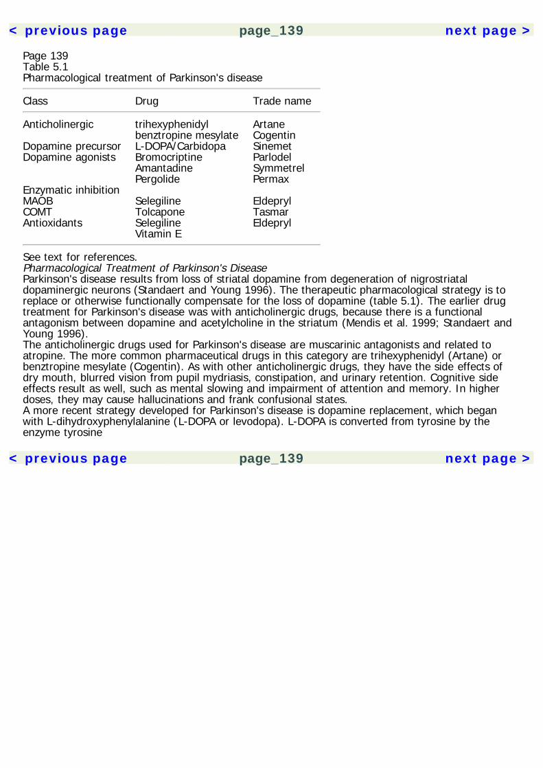
< previous page page_139 next page >
Page 139Table 5.1Pharmacological treatment of Parkinson's disease
Class Drug Trade name
Anticholinergic trihexyphenidyl Artane benztropine mesylate CogentinDopamine precursor L-DOPA/Carbidopa SinemetDopamine agonists Bromocriptine Parlodel Amantadine Symmetrel Pergolide PermaxEnzymatic inhibition MAOB Selegiline EldeprylCOMT Tolcapone TasmarAntioxidants Selegiline Eldepryl Vitamin E
See text for references.Pharmacological Treatment of Parkinson's DiseaseParkinson's disease results from loss of striatal dopamine from degeneration of nigrostriataldopaminergic neurons (Standaert and Young 1996). The therapeutic pharmacological strategy is toreplace or otherwise functionally compensate for the loss of dopamine (table 5.1). The earlier drugtreatment for Parkinson's disease was with anticholinergic drugs, because there is a functionalantagonism between dopamine and acetylcholine in the striatum (Mendis et al. 1999; Standaert andYoung 1996).The anticholinergic drugs used for Parkinson's disease are muscarinic antagonists and related toatropine. The more common pharmaceutical drugs in this category are trihexyphenidyl (Artane) orbenztropine mesylate (Cogentin). As with other anticholinergic drugs, they have the side effects ofdry mouth, blurred vision from pupil mydriasis, constipation, and urinary retention. Cognitive sideeffects result as well, such as mental slowing and impairment of attention and memory. In higherdoses, they may cause hallucinations and frank confusional states.A more recent strategy developed for Parkinson's disease is dopamine replacement, which beganwith L-dihydroxyphenylalanine (L-DOPA or levodopa). L-DOPA is converted from tyrosine by theenzyme tyrosine
< previous page page_139 next page >
< previous page page_140 next page >
Page 140hydroxylase. In turn, L-DOPA is converted to dopamine by dopa decarboxylase either in the capillaryendothelium or in dopaminergic neurons. Thus, administration of L-DOPA increases the substrate fordopamine and results in increased dopamine synthesis and release. Although L-DOPA passes theblood-brain barrier, L-DOPA is rapidly converted into dopamine in the blood when administeredorally. This renders approximately 95% of it unable to enter the CNS, while the remaining 5%penetrates and is then converted to dopamine. Although this was enough to alleviate parkinsoniansymptoms, the large amounts of circulating dopamine created the side effect of nausea. The nextstep of progress came with the drug carbidopa, which inhibits peripheral DOPA decarboxylase. Thus,conversion of L-DOPA to dopamine is supressed in circulation, but continues unhindered centrallybecause carbidopa does not cross the blood-brain barrier. This effectively reduces the necessary doseof L-DOPA by 75%. L-DOPA and carbidopa are sold in combination under the name Sinemet. Two-thirds of patients given L-DOPA tolerate it well initially, while others may experience nausea,hypotension, or in some cases, depression. But more frequently, mood tends to improve with L-DOPA, and other changes such as excitement, aggressiveness, or increased libido may occur.One of the drawbacks of L-DOPA/carbidopa treatment is that it loses effectiveness over time. Greaterfluctuations are seen between doses due to the short half-life of the drug (1–3 hours), so asustained-release preparation of Sinemet (Sinemet CR) has been developed to improve the stabilityof blood L-DOPA levels. As the effectiveness of L-DOPA wanes, the dose must be increased. Withhigher doses, the therapeutic margin decreases and there is greater incidence of dyskinesia. Commondyskinesia seen in high doses of L-DOPA are facial grimacing, head wagging, dystonia of the limbsand neck, choreoathetosis, and general restlessness. Psychoses consisting of delusional thinkingand/or hallucinations also occur in 15–25% of people taking L-DOPA.As an adjunct or alternate to L-DOPA, dopamine agonists are employed. Because they are directreceptor agonists, these have the advantage of not relying on the integrity of presynaptic nigrostriatalneurons, which are necessary for the synthesis and release of dopamine. Bromocriptine
< previous page page_140 next page >
< previous page page_141 next page >
Page 141(Parlodel) is an ergot alkaloid and a potent agonist of D2 receptors located on corticostriatalterminals. It has a longer duration of action (3–7 hours) than L-DOPA and tends to produce lessnausea. Pergolide (Permax) is another ergot derivative that is an agonist at both D1 and D2receptors. Lisuride is a third dopamine agonist ergot-derivative and is a D2 agonist. However, itsusefulness may be limited by its side-effect profile. Amantadine (Symmetrel) was developed as anantiviral agent but was later found to have antiparkinsonian effects. It is not a dopamine receptoragonist, but rather seems to work by releasing dopamine in the striatum. Amantadine is also anantagonist of NMDA receptors, by binding at the phencyclidine (PCP) site within the channel(Kornhuber et al. 1995). NMDA antagonism prevents release of ACh in the striatum, thus providing apotential second mechanism for amantadine's antiparkinsonian effects (Stoof et al. 1992).A final pharmacological strategy for treatment of Parkinson's disease comes from enzyme inhibition.This was initally done with an MAO inhibitor, L-deprenyl (selegiline, Eldepryl), but more recent drugshave become available that are COMT inhibitors. L-Deprenyl is an inhibitor of MAOB, which is theform of MAO selective to dopamine. Thus, it may increase the amount of available dopamine forrelease. Second, it may protect dopamine neurons by reducing the oxidative stress concomitant withdopamine metabolism (Olanow 1997). Third, L-deprenyl is metabolized into amphetamine andmethamphetamine, which may contribute to their antiparkinsonian effects. Unlike other treatmentsfor Parkinson's disease, L-deprenyl seems to slow the progression of the disease. Tolcapone (Tasmar)is a COMT inhibitor, which prevents extracellular breakdown of dopamine.Other Subcortical Degenerative DiseasesWilson's disease (or hepatolenticular degeneration) is a autosomal recessive parkinsonian conditionresulting from abnormalities of copper metabolism. Degeneration is most prominent in the putamenand globus pallidus, and it is treatable with chelating agents. Progressive supranuclear palsy (PSP) ischaracterized by supranuclear ophthalmoplegia (resulting in an impairment of vertical gaze),pseudobulbar palsy, and
< previous page page_141 next page >
< previous page page_142 next page >
Page 142dysarthria (Jacobs et al. 1997; Adams et al. 1997). Parkinsonian symptoms are seen but axial rigidityis more predominant than the limb rigidity seen in Parkinson's disease, and dopaminergic medicationsare of limited benefit. Cognitive changes in PSP include forgetfulness, slowed information processing,emotional changes, and working-memory deficits.Striatonigral degeneration is another parkinsonian syndrome involving loss of projections from thestriatum to the substantia nigra. Olivo-pontocerebellar atrophy (OPCA) is a degenerative conditioninvolving the inferior olivary complex, pontine nuclei, middle cerebellar peduncle, cerebellar cortex,and dentate nucleus. Motor deficits consist of typical cerebellar motor signs of ataxia, dysmetria, andtremor. Mild to moderate cognitive deficits occur in attention, memory, and executive function.Huntington's disease is a hereditary disorder involving degeneration of the caudate and putamen,with moderate atropy in frontal and temporal cortex. Motor manifestations are characterized bychoreiform movements. Psychiatric and cognitive disturbances are also prominent.Nondegenerative DementiasVascular DementiaVascular dementia results from several etiologies, all resulting in compromised cerebral blood flowand pathological changes (Nyenhuis and Gorelick 1998). The severity, volume, and location of thevascular lesions all contribute to the clinical picture. Risk factors for vascular dementia includehypertension, atherosclerosis, high alcohol consumption, stress, lower formal education, andoccupational toxic exposure (Skoog 1979; Konno et al. 1997). Binswanger's disease is a form ofvascular dementia with characteristic changes in periventricular white matter, as seen on structuralneuroimaging (Olsen and Clasen 1998). Clinical manifestations include cognitive disturbance,depression, gait abnormalities, motor rigidity, and neurogenic bladder incontinence.Frontal and subcortical lacunar infarcts typically affect attention, language, visuospatial function, andmotor programming (Babikian et al. 1990). Compared to patients with Alzheimer's disease, those withvascular dementia show better orientation, recall, and language ability. On
< previous page page_142 next page >

< previous page page_143 next page >
Page 143the other hand, Alzheimer's patients show better attention, executive function, and fine motor control(Bowler and Hachinski 1997).Traumatic Brain InjuryAlthough not a neurodegenerative disorder, traumatic brain injury is another condition wheredysfunctional cholinergic transmission may play a major role, and thus cholinergic medications maybe useful. Memory is a cardinal cognitive problem after traumatic brain injury, and concurrentalterations in cerebral cholinergic systems have been found. Postmortem examination of humansafter fatal traumatic injuries shows choline acetyltransferase activity to be reduced in several corticalareas including inferior temporal gyrus, cingulate gyrus, and superior parietal cortex (Murdoch et al.1998). Nicotinic receptors are unchanged, but synaptophysin immunoreactivity is reduced by 30% inthe cingulate gyrus, suggesting a loss of presynaptic cholinergic terminals.Normal AgingNormal aging involves some degree of cognitive decline and neuropathological changes (Boller andDuyckaerts 1997). There is a reduction in gross volume and weight in the aged brain, approaching15% of total weight. After the age of 60, there is an average loss of 2% of brain weight per decade,primarily due to reduction of white matter. Neuritic plaques and neurofibrillary tangles occur, buttheir distribution and degree is different than that seen in Alzheimer's disease. On functionalneuroimaging, there is a trend toward decreased metabolism in frontal areas during old age, but thisis distinct in degree and progression from patterns seen in dementia (Mielke et al. 1998). Age-relatedmemory changes are also apparent on electrophysiological measures, but still distinct from dementia(Joyce et al. 1998; Stevens and Kircher 1998).Age-related changes in many cognitive functions are seen in healthy elderly adults. Most notable arechanges in processing speed and short-term memory (Sliwinski and Buschke 1999; Unger et al.1999; Keefover 1998). While difficulties occur in the formation of new memories, memory for familiarinformation and skills is well preserved (Burke and Mackay 1997). Mild cognitive complaints are fairlycommon in
< previous page page_143 next page >

< previous page page_144 next page >
Page 144
Figure 5.1Ginkgo (Ginkgo biloba). Reprinted with permission from Sturdivant and Blakely. (1999). MedicinalHerbs in the Garden, Field, and Marketplace. Friday Harbor, WA: San Juan Naturals. Illustration byPeggy Sue McRae.community-dwelling elderly, but these are accounted for by age-related effects and depression(Schroder et al. 1998). Literacy and education play a role in preserving cognitive function andproviding the neural basis for cognitive reserves to be drawn upon later in life (Manly et al. 1999).Herbal NootropicsGinkgoHistory and BotanyGinkgo biloba, also known as the kew or maidenhair tree, is a species of gymnosperm that has beenin existence for 200 million years. Some living specimens are estimated to be more than 3,000 yearsold (Field and Vadnal 1998). Ginkgo is a seed-bearing tree that typically grows to a height of 35–80feet. Its leaves are green and fan shaped, and produce an acid that is resistant to insects (figure5.1). Ginkgo trees do not reproduce until 20 years of age, but may continue to reproduce past 1,000years of age (Major 1967). The seeds (sometimes referred to as fruit) are round, fleshy, and have ayellow or green color (Gruenwald et al. 1998). At present, approximately fifty million ginkgo trees arein cultivation for
< previous page page_144 next page >

< previous page page_145 next page >
Page 145preparation as herbal supplements, yielding 4,000 tons of dried leaves annually from the UnitedStates and France alone (Field and Vadnal 1998).The name ginkgo is a Japanese word derived from the Chinese yinshing, meaning ''silver apricot."The Chinese emperor Shen Nung wrote in his medical text Pen T'sao Ching that ginkgo leaves areused to treat memory loss and breathing ailments. Liu Wen-Tai in 1505 C.E. reported uses of ginkgofor skin sores and diarrhea due to parasites (Fields and Vadnal 1998). The ginkgo tree was notbrought to Europe and North America until the eighteenth century, and common use of ginkgo in theWest did not occur until the 1960s.Today, ginkgo extracts are marketed under a variety of registered trade names including Ginkgobene,Ginkgold, Ginkoba, Kaveri, Rokan, Tanakene, Tramisal, Valverde Vital, and Vasan. Although brandsmay vary in their bioequivalence, no one to date has examined the implications of this variance oncognitive function (Itil and Martorano 1995). Ginkgo is among the most commonly prescribed drugsin France and Germany, making up 1% and 4% of their prescriptions, respectively.Chemical ConstituentsGinkgo's active chemical constituents are classified into flavonoid glycosides, biflavones, and terpenelactones (table 5.2) (Field and Vadnal 1998; Oken et al. 1998). Flavonoid glycosides includekaemferol, quercetin, apigenin, luteolin, and myricetin. Biflavones include amentoflavone, bilobetin,ginkgetin, isoginkgetin, sciadopitysin, and 5′-methoxybilobelin. Terpene lactones in ginko are theginkgolides A, B, C, M, and J, and bilobalide (figure 5.2). The ginkgolides are diterpenes andbilobalide is a sesquiterpene (Gruenwald et al. 1998). Ginkgolides are unique twenty-carbon terpenes,occurring naturally only in the roots and leaves of the ginkgo tree (Braquet and Hosford 1991). Otherconstituents of interest are alkylphenols such as ginkgolic acid (Satyan et al. 1998; Jaggy and Koch1997). Proanthocyanidins are also present, and may create vasoactive and antioxidant effects. Ginkgoextracts are standardized by their flavonoid glycoside and terpene content (Li and Wong 1997).Preparations include dried leaf, tinctures, and several extracts (Field and Vadnal 1998). Differentextracts of ginkgo are used in research, which produce different relative compositions of its chemical
< previous page page_145 next page >
< previous page page_146 next page >
Page 146Table 5.2Chemical constituents of ginkgo
Flavonoid glycosides Kaempferol Quercetin Apigenin Luteolin MyricetinBiflavones Amentoflavone Bilobetin Ginkgetin Isoginkgetin Sciadopitysin 5′-methoxybilobelinTerpene lactones Ginkgolides A, B, C, M, and J BilobalideAlkylphenols Ginkgolic acidProanthocyanidins
See text for references.constituents. The most common standardized extraction process, termed EGb 761, uses water andacetone extraction in the initial stage. The lipophilic and condensed polyphenolic compounds are thenremoved (Itil and Martorano 1995; Kleijnen and Knipschild 1992a). This produces an extractcontaining 24% ginkgo-flavone glycosides and 6% terpenoids, and has been marketed in Europeunder several trade names. Another extract, LI 1370, has 25% ginkgo-flavone glycosides and 6%terpenoids. Preparation of ginkgo as a tea is ineffective because it would only contain small amountsof active constituents (Tyler 1990).Mechanisms of ActionGinkgo has effects on several neurotransmitter and physiological systems, which are addressed hereseparately (table 5.3).Acetylcholine Ginkgo enhances release of acetylcholine and alters cholinergic receptors. Both direct invitro application and long-term oral administration increases presynaptic uptake of choline inhippocampal synaptosomes (Kristofikova et al. 1992). The effective concentrations for
< previous page page_146 next page >

< previous page page_147 next page >
Page 147
Figure 5.2Chemical structure of ginkgo constituents.this effect are 100 mg/ml in vitro or 20–30 mg/g of brain tissue, which could be achieved withchronic dosing. The increase in choline uptake is due to an increase in the number of choline uptaketransporters and/or rate of transport (Kristofikova et al. 1997). Chronic oral treatment with extractalso increases the number of muscarinic receptors in the hippocampus (Taylor 1986).Short-term memory tasks increase high-affinity uptake of acetylcholine, whereas long-term referencememory tasks cause an acute increase followed by a long-lasting decrease and inhibition (Decker etal. 1988). In contrast, the drug scopolamine creates amnestic effects by blocking muscarinic receptorsand decreasing acetylcholine levels. Thus, at
< previous page page_147 next page >

< previous page page_148 next page >
Page 148Table 5.3Mechanisms of action of ginkgo
Cholinergic Increases uptake of choline and release ofacetylcholine
Increases the number of muscarinic receptorsMonoamineIncrease uptake of 5-HT Inhibition of MAOA and MAOB Prevents desensitization and age-related reduction of
5-HT1A Alters norepinephrine turnover Reduced β-adrenergic binding and activity Reverses age-related decline in α2 adrengic receptorsGABA Elevation of GABA levels Increased glutamic acid decarboxylase activityVasoactive PAF inhibition NO-dependent vasodilation
See text for references.normal oral doses in humans, perhaps slightly higher than those commonly used, ginkgo extractsenhance cholinergic transmission by increasing uptake and release. The nootropic drugs tacrine anddonepezil similarly enhance cholinergic transmission by preventing breakdown of acetylcholine byacetylcholinesterase. Although not the only effect of ginkgo, cholinergic mechanisms could wellaccount for its cognitive effects.Monoamines Ginkgo extracts and commercial preparations inhibit MAOA and MAOB in vitro,achieving total inhibition, but it has yet to be demonstrated that this occurs in the brain and atnormal oral doses (White et al. 1996). Although EGb 761 increases uptake of 5-HT (but notdopamine), chronic administration to aged rats increases serotonin in frontal cortex, hippocampus,striatum, and hypothalamus (Ramassamy et al. 1992). Chronic treatment reverses an age-relatedreduction in the maximal binding sites of 5-HT1A receptors (Huguet et al. 1994). Chronic oraladministration of EGb 761 prevents stress-induced desensitization of 5-HT1A receptors, withoutaltering their density or affinity (Bolanos-Jimenez et al. 1995). Accordingly, drug discriminationparadigms suggest effects at 5-HT1A, but not 5-HT2 receptors (Winter and Timineri 1999).
< previous page page_148 next page >
< previous page page_149 next page >
Page 149A biphasic effect of ginkgo is seen over time on norepinephrine turnover (Brunello et al. 1985). Aninitial decrease in the norepinephrine metabolite normetanephrine occurs at 45 minutes, followed bya marked increase that was present after 14 days. Chronic treatment reduces β-adrenergic bindingand β-adrenergic-stimulated adenylate cyclase activity. Thus, an increase in norepinephrine levels isinducing β-adrenergic receptor regulation and functional activity. Ginkgo chronically administered alsoreverses the age-related decline in binding density of α2 adrenergic receptors (Huguet and Tarrade1992).GABA Flavonoids present in ginkgo may endow it with benzodiazepine actions but it remains inquestion whether they cross the blood-brain barrer (Lobstein-Guth et al. 1988; Baureithel et al.1997). However, oral doses of bilobalide elevates GABA levels in the hippocampus (Sasaki et al.1999). Increases occur in the activity and amount of glutamic acid decarboxylase. Glutamate levelsare not affected by bilobalide, nor are the number or dissociation constants of the GABAA receptor.Such effects were suggested to mediate the antiseizure effects of bilobalide.Intracellular messengers A biphasic effect of ginkgo extract is seen on cAMP phosphodiesterase underin vitro and ex vivo conditions (Saponara and Bosisio 1998; Macovschi et al. 1987). Whereas lowconcentrations (0.25–4.0 mg/L) activate the enzyme, higher concentrations (5–250 mg/L), dose-dependently inhibit it. However, tolerance develops to this effect because it is undetectable afterdaily administration for 4 days. Thus, ginkgo may initially produce effects by inhibiting enzymaticbreakdown of cAMP. This mechanism is similar to the stimulant caffeine, but it is not likely to explainany long-term effects of ginkgo because it disappears after chronic daily treatment. The responsibleconstituent for this effect has not been identified.PharmacokineticsGinkgo extracts show rapid absorption after oral administration of capsules, tablets, and drops (Liand Wong 1997; Wójcicki et al. 1995). The pharmacokinetics for the ginkgo terpene lactones havebeen determined
< previous page page_149 next page >
< previous page page_150 next page >
Page 150Table 5.4Pharmacokinetics of ginkgo terpene lactones
ConstituentOralbioavailability
Time to peakconcentration
Half-life
Eliminationhalf-life
GinkgolideA
>98% 1–2 hrs 4–6hrs
4.5 hrs
GinkgolideB
>80% 1–2 hrs 4–6hrs
10.5
Bilobalide 70% 1–2 hrs 3hours
3.2
See text for references.(table 5.4) (Kleijnen and Knipschild 1992a). Ginkgo flavone glycosides have an oral bioavailability of>60%, and reach peak concentrations in 1.5–3 hours. Their half-life is between 2 and 4 hours.Ginkgolides A or B have an oral bioavailability of greater than 80% (>98% for A, >80% for B). Theyreach peak concentrations in 1–2 hours and have a half-life of 4–6 hours. Bilobalide has an oralbioavailability of 70%, reaches peak concentrations in 1–2 hours, and has a half-life of approximately3 hours. Elimination half-lives for ginkgolides A, B, and bilobalide are, respectively, 4.5, 10.5, and 3.2hours. Similar results were confirmed by others (Biber and Koch 1999).Another study showed high bioavailability of ginkgolides and bilobalide, and was suggestive of twice-daily dosing (Fourtillan et al. 1995). Concurrent food intake did not alter the total effect, butincreased the time to reach peak concentrations. Different extraction processes can enhance thebioavailability of ginkgo constituents (Li and Wong 1997). The brain bioavailability of ginkgo has beenshown indirectly with electrophysiological studies of oral dosing in humans (Itil and Martorano 1995;Itil et al. 1998).Pharmacokinetic parameters have been determined for the ginkgo flavonoid glycosides quercetin,kaempferol, and isorhamnetin (table 5.5) (Wójcicki et al. 1995). Rapid absorption occurs and peakconcentrations are reached in approximately three hours for all three flavone glycosides. Eliminationfollows a typical exponential function and the volume distribution suggests extensive tissuepenetration. A short elimination half-life and total body clearance suggest that ginkgo flavoneglycosides are rapidly eliminated. Futher, bioequivalence is found for these constituents acrosscapsule, drops, and tablet formulations, based on FDA guidelines.
< previous page page_150 next page >
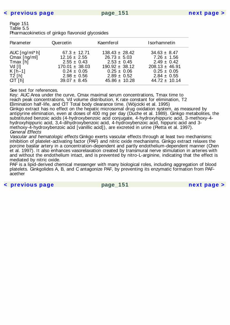
< previous page page_151 next page >
Page 151Table 5.5Pharmacokinetics of ginkgo flavonoid glycosides
Parameter Quercetin Kaemferol Isorhamnetin
AUC [ng/ml*h] 67.3 ± 12.71 138.43 ± 28.42 34.63 ± 8.47Cmax [ng/ml] 12.16 ± 2.55 26.73 ± 5.03 7.26 ± 1.56Tmax [h] 2.55 ± 0.43 2.53 ± 0.45 2.49 ± 0.42Vd [l] 170.01 ± 38.03 190.92 ± 38.12 208.13 ± 46.91K [h–1] 0.24 ± 0.05 0.25 ± 0.06 0.25 ± 0.05T2 [h] 2.98 ± 0.56 2.89 ± 0.52 2.84 ± 0.55ClT [h] 39.07 ± 8.45 45.86 ± 10.28 44.72 ± 10.14
See text for references.Key: AUC Area under the curve, Cmax maximal serum concentrations, Tmax time toreach peak concentrations, Vd volume distribution, K rate constant for elimination, T2Elimination half-life, and ClT Total body clearance time. (Wójcicki et al. 1995)Ginkgo extract has no effect on the hepatic microsomal drug oxidation system, as measured byantipyrine elimination, even at doses of 400 mg per day (Duche et al. 1989). Ginkgo metabolites, thesubstituted benzoic acids (4-hydroxybenzoic acid conjugate, 4-hydroxyhippuric acid, 3-methoxy-4-hydroxyhippuric acid, 3,4-dihydroxybenzoic acid, 4-hydroxybenzoic acid, hippuric acid and 3-methoxy-4-hydroxybenzoic acid [vanillic acid]), are excreted in urine (Pietta et al. 1997).General EffectsVascular and hematologic effects Ginkgo exerts vascular effects through at least two mechanisms:inhibition of platelet-activating factor (PAF) and nitric oxide mechanisms. Ginkgo extract relaxes theporcine basilar artery in a concentration-dependent and partly endothelium-dependent manner (Chenet al. 1997). It also enhances vasorelaxation created by transmural nerve stimulation in arteries withand without the endothelium intact, and is prevented by nitro-L-arginine, indicating that the effect ismediated by nitric oxide.PAF is a lipid-derived chemical messenger with many biological roles, including aggregation of bloodplatelets. Ginkgolides A, B, and C antagonize PAF, by preventing its enzymatic formation from PAF-acether
< previous page page_151 next page >
< previous page page_152 next page >
Page 152(Lamant et al. 1987). Ginkgolide B is the most potent, followed by Ginkgolides A and C. Half maximalinhibition occurs for all three in the low micromolar range. Nonginkgolide, nonflavonoid fractions ofginkgo extract have a potent vasorelaxing effect, producing a dose-dependent relaxation of rabbitand human cavernosal smooth muscle (Paick and Lee 1996). A subfraction (304U-1) shows the mostpotent effects, which are mediated by cAMP and perhaps adrenergic effects. Thus, gingko mayimprove microcirculation by inhibiting platelet-aggregation by PAF.Ginkgo has a number of other hematologic effects. Acute administration of the ginkgo extract, Kaveri,does not alter blood pressure or heart rate in humans (Jung et al. 1990). Chronic treatment withginkgo extract (120 mg/day for 3 months) lowered diastolic blood pressure (Winther et al. 1998).However, no changes were seen at a higher dose (240 mg/day for 3 months). Also unchanged byginkgo are haematocrit, plasma viscosity, erythrocyte rigidity, thrombocyte and leukocyte count, andthrombocyte aggregation. However, ginkgo does reduce erythrocyte aggregation by 15.6% (Jung etal. 1990). Ginkgo reduces pulmonary hypertension induced by chronic isobaric hypoxia (Cheng et al.1996). In human platelets, ginkgolides reduce phospholipase C activation and the mobilization andinflux of Ca2+ (Simon et al. 1987). This action is not observed with thrombin, suggesting thatginkgolides act through the membrane receptor of PAF-acether. Ginkgolides also reduce thromboxaneB2 levels (Becker et al. 1988).EGb 761 has both antiplatelet and antithrombotic effects when administered orally, prolongingbleeding time in experimental models (Kim YS et al. 1998; Bourgain et al. 1987). Ginkgolide Bprevents thrombus formation in experimentally injured arterial segments (Bourgain et al. 1986). Italso inhibits specific binding of PAF to eosinophils and neutrophils in a concentration-dependentmanner (Kurihara et al. 1989). Intravenous EGb 761 significantly reduces arteriolar spasm in ratcremaster muscle, effective when vasospasm is induced by serum and a thromboxane analog, butnot by serotonin or thrombin (Stœcker et al. 1996). Ginkgolides and proanthocyanidins are believedto mediate this effect.A systematic literature review showed ten controlled studies of ginkgo for treatment of intermittentclaudication (Ernst 1996). Many studies
< previous page page_152 next page >
< previous page page_153 next page >
Page 153had methodological flaws, but the consensus is that gingko is an effective therapy for intermittentclaudication. This effect is most likely mediated by ginkgo's effects on vessel dilation andmicrocirculation through inhibiting platelet activation. A meta-analysis found ginkgo to have asignificant effect in treating peripheral arterial diseases, with a global effect size of 0.75 (Schneider1992).Sexual effects Ginkgo was serendipitously found to improve sexual function in an elderly patienttaking it for memory effects. A subsequent open trial of ginkgo extract was performed to assess itseffect on antidepressant-induced sexual dysfunction (SSRI, SNRI, MAOI, and tricyclics) (Cohen andBartlik 1998). Ginkgo had a positive effect on sexual function in 84% of subjects. Both male andfemale subjects participated, and a larger proportion of females reported benefit (91% female, 76%male). Improvement was noted across all four phases of the sexual cycle: desire, excitement,orgasm, and resolution. The dosages used for this effect were in the range of 240 mg. A controlledstudy is needed to confirm these findings and determine magnitude of effect.Penile erection occurs by relaxation of the smooth muscle of the corpus cavernosum, increasing bloodflow into the penis and producing erection and rigidity. In a parallel fashion, vaginal pressurestimulation increases blood velocity and flow into clitoral arteries (Lavoisier et al. 1995). Cavernosalvasodilation is accomplished by neurotransmitters released from the cavernosal nerve and endothelialcells. One of the most important transmitters in this cascade is nitric oxide (NO), which inducessynthesis of cyclic GMP from guanylate cyclase (Rajfer et al. 1992). Thus, ginkgo's vascularmechanisms could be responsible for some of the putative sexual effects.Vestibulocochlear effects Ginkgo has positive effects on vestibulocochlear function in animal models,primarly through vasoactive and antioxidant mechanisms. EGb 761 dilates the cochlear vasculatureand prevents flow reduction caused by sodium salicylate (Didier et al. 1996). Oral EGb 761 reducedtinnitus induced by sodium salicylate in rats (Jastreboff et al. 1997). The recovery of balance afterunilateral vestibular neurectomy is accelerated by postoperative treatment with EGb 761,
< previous page page_153 next page >
< previous page page_154 next page >
Page 154most likely due to cerebral plasticity mechanisms involved in vestibular compensation (Tighilet andLacour 1995; Smith and Darlington 1994). Effects are dose-dependent and involve nonterpeneconstituents. EGb 761 has a direct excitatory effect on the lateral vestibular nuclei and decreasesgain of the horizontal vestibulo-ocular reflex without altering phase (Yabe et al. 1995). Functionalimprovements in balance are accompanied by morphological neural correlates, such as fastersynaptic reoccupation in the deafferented medial vestibular nucleus (Lacour et al. 1991). Oral andparenteral ginkgo extract morphologically preserve the vestibular epithelia, probably due to improvedcapillary permeability and microcirculation (Raymond 1986).Ginkgo also improves tinnitus and dizziness in human studies, but this effect may occur later thanimprovements in memory and concentration (Soholm 1998). Ginkgo extract reduces tinnitus andvertigo in humans associated with vertebrobasilar insufficiency (Cano-Cuenca et al 1995). Sizablemulticenter controlled studies indicate that ginkgo is effective in treating tinnitus, irrespective ofprognostic factors such as the site and periodicity of the disease (Meyer 1986a, 1986b).In humans, chronic (3 month) treatment with EGb 761 reduces vertigo and dizziness, with efficacycomparable to betahistine dihydrochloride (Cesarani et al. 1998). Oculomotor improvements are seenin a slight decrease of saccadic delay and significantly increased saccadic velocities, improved smoothpursuit gain, and reduced nystagmus. Also noted are improvements of the sinusoidal vestibulo-ocularreflex and visuovestibular ocular reflex. Other multicenter, controlled studies have supported ginkgoin the treatment of vertigo (Haguenauer et al. 1986).Neuroprotective effects Ginkgo has a number of significant antioxidant effects. EGb 761 protectsagainst oxidative damage induced by peroxyl radicals (Maitra et al. 1995; Lugasi et al. 1999). Itreduces oxidative-induced morphological changes in mitochondria, glutathianone oxidation, andperoxide formation (Sastre and El-Fattah 1998; Seif-El-Nasr et al. 1995). Terpenoid constituents ofginkgo show anti-ischemic effects in the myocardium, due to inhibition of free radical synthesis, butnot by direct free radical scavenging (Pietri et al. 1997). The ginkgo flavonoids myricetin andquercetin reduce oxidative metabolism at nanomolar con-
< previous page page_154 next page >

< previous page page_155 next page >
Page 155centrations (3 and 10 nM, respectively) (Oyama et al. 1994). Ginkgo and methylprednisolone bothreduced injury in a spinal cord compression injury model in rats, probably by antioxidant actions (Kocet al. 1995). EGb 761 has dual effects on the free radical nitric oxide (Kobuchi et al. 1997; Marcocciet al. 1994). It scavenges nitric oxide as well as inhibits its synthesis in lipopolysaccharide/interferon-gamma macrophages. Ginkgo extract protects neurons against oxidative stress produced by hydrogenperoxide (Oyama et al. 1996). Also reduced are apoptosis and DNA fragmentation induced byoxidative stress (Ni et al. 1996). This is accomplished by flavonoids and other constituents of EGb761, but not terpenoids (Chen et al. 1999). EGb 761 protects against oxidative free radical damage inthe retina, as evidenced by electrophysiological changes (Droy-Lefaix et al. 1995). Ginkgo extract andginkgolide B reduce glutamate excitotoxicity after peripheral administration by preventing increases inCa2+ flux (Zhu et al. 1997). Ginkgo also protects dopaminergic neurons from the neurotoxic agent N-methyl-4-phenyl-1,2,3,6-tetrahydrophyridine (MPTP) (Ramassamy et al. 1990).Ginkgo shows some anti-ischemic effects, although results depend highly on methodology. EGb 761prevents reduction of Na,K-ATPase after cerebral ischemia (Pierre et al. 1999). PAF has beenimplicated in hypoxic damage during cerebral ischemia. Hypoxic/ischemic injuries in neonatal rats aremediated by PAF, and are reduced by EGb 761 (Akisü et al. 1998). Ginkgolides reduce the cerebraldamage produced by ligature of the common carotid artery in adult Mongolian gerbils (Spinnewyn etal. 1987). A methodologically controlled study of ginkgo extract was carried out in adults with acuteischemic stroke (Garg et al. 1995). Although results were negative, this may relate to a relatively lowdosage and/or administration time post-injury (>48 hours). The authors also conclude that thereappear to be no adverse effects of ginkgo in acute ischemic stroke.Ginkgo alters lipid metabolism created by electroconvulsive shock treatments. EGb 761 reducedaccumulation of free fatty acids and removal of diacylglycerol, which is more pronounced in thehippocampus than cerebral cortex (Rodriguez de Turco et al. 1993). Ginkgo also has protectiveeffects on lipid membranes under hypoxic conditions. Bilobalide, but not ginkgolides, suppressedhydrolysis of choline induced
< previous page page_155 next page >
< previous page page_156 next page >
Page 156by hypoxia (Klein et al. 1997). This effect is seen in the low micromolar range, both in vitro and exvivo by oral doses (6 mg/kg). EGb 761 had the same effect at 200 mg/kg. EGb 761 and bilobalideboth inhibit the hypoxia-induced drop in ATP release from endothelial cells (Janssens et al. 1995).Bilobalide increases glucose transport under normal oxygen conditions but not under hypoxia.Ginkgo may speed recovery from neurological injury. EGb 761 accelerates recovery from hemiplegiainduced by ablation of motor cortex (Brailowsky and Montiel 1997). This effect required chronictreatment (10 days) in aged rats. Histologically, treated rats showed less glial fibrillary acid proteinand ex vacuo hydrocephalus. Bilobalide also has trophic and protective effects on neurons andSchwann cells in nerve injuries (Bruno et al. 1993). In rats treated with bilobalide, a fasterreinnervation of the extensor digitorum longus muscle was observed following traumatic nervedamage.Neurophysiological effects PAF decreases b-wave amplitude on the electroretinogram, which isreversed by ginkgo extract and ginkgolide B (BN 52021) (Doly et al. 1987). This effect is interpretedin terms of direct effects on PAF membrane receptors in the retina. Membrane receptors for PAFreceptors act through intracellular messengers, activating the phosphatidylinositol cycle and forminginositol-triphosphate (Doly et al. 1987).PAF enhances excitatory amino acid release in the hippocampus and may serve as a retrogrademessenger in long-term potentiation (Bazan and Allan 1996). On the surface, this might seem atodds with ginkgo's pro-mnestic effects because ginkgolides inhibit PAF. However, the role of PAF inlong-term potentiation occurs at low concentrations and it is not essential for its induction, so ginkgomay not significantly inhibit this process (Wieraszko et al. 1993; Kobayashi et al. 1999). On the otherhand, ginkgolides may impair long-term potentiation, but other pro-mnestic effects of ginkgo mayovershadow this.Chronic treatment with EGb 761 in mice created histological changes in the hippocampus (Barkats etal. 1995). Increases are seen in the projection field of hippocampal mossy fibers and a reduction inthe area of the stratum radiatum. These changes are hypothesized to result from
< previous page page_156 next page >
< previous page page_157 next page >
Page 157neuroprotective or neurotrophic actions of ginkgo. Other researchers have also found histologicalchanges in the hippocampus after treatment with ginkgo (Cohen-Salmon et al. 1997).Cerebral blood flow and glucose utilization Local cerebral blood flow is increased by ginkgo extract inmultiple regions of the brain in conscious rats (Krieglstein et al. 1986). Ginkgo increases cerebralblood flow in humans as well (Heiss and Podreka 1978).Although ginkgo causes an increase in cerebral blood flow, it can decrease cerebral glucoseutilization. An autoradiographic study of chronic oral EGb 761 showed decreases up to 18% infrontoparietal somatosensory cortex, the nucleus accumbens, cerebellar cortex and pons (Duverger etal. 1995). Another study, using higher doses, showed greater decreases (20%) of glucose utilizationin the hippocampus, globus pallidus, neostriatum, ventral thalamus, inferior colliculus, and inferiorolive (Lamour et al. 1992). Changes in several of these neuroanatomical areas could account for thecognitive therapeutic effects of ginkgo. Studies on the effects of individual ginkgo constituents onglucose metabolism are needed to discern their respective effects (Duverger et al. 1995). Thereasons for decreases are uncertain, but one author suggests that it is a factor of route and durationof administration as well as the physiological state of the organism (Duverger et al. 1995). Althoughginkgo raises the blood glucose level dose-dependently, perfusion studies show that cortical glucoseconcentration is reduced, suggesting that ginkgo may inhibit glucose uptake (Krieglstein et al. 1986).Neuroendocrine effects In both young and old rats, chronic treatment with oral ginkgo extractreduces stress-induced impairments in learning and elevation of stress hormones (Rapin et al. 1994).Ginkgolide B reduces the binding and expression of peripheral-type benzodiazepine receptors in theadrenal cortex (Amri et al. 1996; Amri et al. 1997; Papadopoulos et al. 1998). This reduces theproduction of corticosterone in response to adrenocorticotrophic hormone (ACTH). Thus, gingkolide Bcauses a reduction of glucocorticoid production. Additionally, there is a reduction of progesteroneproduction in pregnant rats. Ginkgo constituents may also act at the hypothalamic level, reducingexpression and
< previous page page_157 next page >
< previous page page_158 next page >
Page 158secretion of corticotrophin-releasing hormone under certain conditions (Marcilhac et al. 1998).Chronic elevation of corticosteroids has been shown to impair cognitive processes and haveneurotoxic effects (Sheline et al. 1996; de Kloet et al. 1999). The cumulative effects of elevatedcortisol levels are associated with cognitive impairments in human aging (Lupien et al. 1999). Thus,the cognitive and neuroprotective effects of ginkgo may be partly mediated through itsneuroendocrine effects.Electrophysiological effects Several studies have looked at the electrophysiological effects of ginkgo.Short-term chronic (3 day) oral doses of ginkgo extract produces electrophysiological effects in manyfrequencies and topographical areas, and on several EEG power measures (Kunkel 1993). Effects areseen across widespread cerebra regions, but are maximal in frontotemporal areas. Further, differentfractions of the ginkgo extract produced similar effects. Other studies consistently show activationeffects or decrease in slow-wave activity (Kanowski et al. 1996; Pidoux 1986).Other researchers report a more specific cognitive activation effect both in young healthy subjectsand elderly subjects suffering from mild dementia. The activation effects are characterized by relativeincreases in alpha activity (7.5 to 13 Hz) and decreases in delta and theta activity (1.3 to 7.5 Hz) (Itilet al. 1998). Ginkgo extract has electrophysiological effects similar to tacrine, donepezil, and othernootropic drugs (piracetam and oxiracetam). In the study by Itil and colleagues (1998), moresubjects showed the cognitive activation response to ginkgo extract (240 mg; 44% responders) thantacrine (40 mg; 18% responders). This was an open study, so controlled studies are now needed toconfirm these results. They also used a single test dose, so a study of chronic dosing is needed tosee if these effects sustain or increase over time.The differences found by these two EEG studies may be due to methodological factors. Kunkel(1993) prescreened subjects for high alpha activity (70% or more of the recording period), whereasItil and colleagues did not. Kunkel also used lower doses (40, 80, and 160 mg) than Itil andcolleagues (240 mg).
< previous page page_158 next page >
< previous page page_159 next page >
Page 159One study of chronic ginkgo extract found no differences in EEG between ginkgo and placebo inelderly subjects with age-related decline. However, subjects with the more severe baseline EEGfindings showed the most improvement in vigilance with ginkgo (Gessner et al. 1985). This studyused a smaller dose of extract than others who found positive results (120 mg).Ginkgo extract shortens stimulus evaluation time in an ERP paradigm (auditory oddball) (Semlitsch etal. 1995). Subjects with age-related memory decline show a shortening of the latency P300, while noeffects are seen on the latency of N1, P2, or N2, nor the amplitudes of N1, P2, N2, and P300.Chronicity of dosage does not affect the P300 latency, because similar results were found for bothacute and chronic administration.Cognitive effects: Animal studies A few studies have examined ginkgo extracts on cognition in animalmodels. An aqueous ginkgo extract shortens sleeping time induced by anesthetics in mice(hexobarbital, achloralose, and urethane) (Wada et al. 1993). The terpene lactones bilobalide andginkgolide A accomplished this effect. Improvements in maze learning are noted in mice after ginkgotreatment (Gajewski and Hensch 1999). Similarly, acute administration to rats produces improvementin working memory (delayed nonmatch to position) as well as learning and error rate in maze tasks(Winter 1998). Also noted in this study was a greater longevity of rats administered ginkgo versusplacebo. The mechanism of this effect is uncertain, but it did not appear to involve caloric restriction.Antioxidant mechanisms are hypothesized by the author to be the responsible agents. Ginkgo extractimproves short-term memory in a passive avoidance task in mice (Stoll et al. 1996). Although it alsoimproved lipid membrane fluidity, it did not correlate with improvements in short-term memory.A ginkgo extract (GK 501) produces improvement on four animal models of learning (shuttle box,step down, step through, and Morris water maze) (Petkov et al. 1993). In some cases, combinedadministration of ginkgo and ginseng showed a potentiation of effects. Ginkgo extract protectsagainst stress-induced learning deficits in older rats (Rapin et al. 1994). Finally, ginkgo attenuatesamnesia induced by scopolamine,
< previous page page_159 next page >
< previous page page_160 next page >
Page 160a muscarinic antagonist, in rats (Chopin and Briley 1992). At higher doses, effects are comparable tothose of tacrine.Cognitive effects: Humans with dementia Several studies have looked at the effects of ginkgoextracts on cognitive function in people with dementia. German health authorities approved a ginkgoextract in 1994 for treatment of primary degenerative and vascular dementias (Itil et al. 1998).Ginkgo has been examined in a number of clinical populations, including Alzheimer's disease, vasculardementia, and age-associated cognitive decline. Most studies employed the extracts EGb 761 or LI1370. Many have methodological flaws including limited sample size or insufficient description ofrandomization, patient characteristics, measurement techniques, or result presentation, but there area number of well-controlled studies available for drawing preliminary conclusions (Field and Vadnal1998).Ginkgo's cognitive effects are examined using a number of measures, but the focus has been onmemory and attention/processing speed. Subjects presenting with mild memory complaints showimprovement on measures such as the digit copying (Kendrick battery) and a computerizedclassification task, indicating underlying improvements in processing speed (Rai et al. 1991). Short-term memory improvements are produced by ginkgo on the Sternberg memory test (Hindmarch1986). The Sternberg test measures short-term retention of 4–6 digits, assessed with yes/nodiscrimination responses. Benefits are seen in subjects with age-related memory loss using a dual-coding task that involves rapid encoding of visual and verbal material into memory (Allain et al.1993). Ginkgo extract improves performance when given in one acute dose one hour before testing.Improvements are also seen in subjects with Alzheimer's disease and vascular dementia using theGerman syndromkurztest, a battery measuring attention and memory (Maurer 1997). Changes havenot been seen in the Trail Making Test in subjects with Alzheimer's disease taking ginkgo extract.Other studies have shown improvement in cognitive measures designed to grossly gage cognitivefunction in Alzheimer's disease, such as the ADAS (Alzheimer's Disease Assessment Scale), whichsamples memory, language, praxis, and orientation (LeBars et al. 1997).
< previous page page_160 next page >

< previous page page_161 next page >
Page 161Ginkgo treatment also produces improvements in ratings by examiners and caregivers (clinical globalimpressions scale, geriatric evaluation by relative's rating scale) as well as self-assessment ofactivities of daily living (Nürnberger Alters-Beobachtungsskala) relative to placebo (Maurer 1997;Kanowski et al. 1996; Haase et al. 1996; Taillandier et al. 1986). Improvements are also noted inseveral studies on subjects' self-ratings versus placebo (Kleijnen and Knipchild 1992b).A meta-analysis of studies using rigorous methodological inclusion criteria was carried out by Okenand colleagues (1998). Collectively, the studies reviewed support a real effect of ginkgo extracts oncognition in people with Alzheimer's disease. A small effect size (0.4) was found, but this may bepartly due to the fact that effects were averaged over ten outcome measures, some of which wereless sensitive to improvement. Another reason may be the variable dosages used in the studies,ranging from 120 to 240 mg. Studies using 240 mg showed greater effect sizes. However, the effectsize is substantial when compared to the prescription nootropic donepezil (Aricept), for which effectsizes are between 0.42 to 0.48 (Rogers et al. 1998). Other review of the literature reached similarconclusions about ginkgo's cognitive effects in dementia (Soholm 1998). Also mentioned is atherapeutic lag period of 4–6 weeks after initiation of treatment, which likely corresponds withreceptor regulation and functional changes.Although the meta-analysis by Oken and colleagues examined efficacy in Alzheimer's disease only,improvements have been seen in other conditions such as age-related memory decline and vasculardementia (Kanowski et al. 1996; Haase et al. 1996; Allain et al. 1993). More research is needed toestablish the quantitative clinical significance of ginkgo extract.Cognitive effects: Normal subjects Two studies to date have examined the cognitive effects of anacute dose of ginkgo in normal subjects. Similar to studies in memory-impaired subjects, benefitsoccurred in the Sternberg memory test, but not in critical flicker frequency and choice reaction time(Subhan and Hindmarch 1984). Although preliminary, this methodologically controlled study indicatesthat one need not have severe memory deficits to derive benefit from ginkgo. A replication of this
< previous page page_161 next page >
< previous page page_162 next page >
Page 162study only found benefits on a free-recall measure (Warot et al. 1991). More studies are needed toestablish the beneficial effects of ginkgo in young healthy subjects, perhaps using chronic dosing.Cognitive effects: Conclusions A number of studies have shown cognitive benefits from ginkgoextracts. The majority of studies have been done in subjects with some form of dementia(Alzheimer's disease, vascular dementia, or age-related memory impairment), and primarily focus onmemory, speed of information processing, and clinical and functional ratings. Meta-analyses indicatea modest but consistent benefit from ginkgo in methodologically-controlled studies. Cognitiveimprovement is not found in all measures administered, and results may depend on severalmethodological factors. One factor is the dosage used, where studies have used between 120 and600 mg in a single dose. A second factor is the chronicity of the dosage. Although many studies showimprovement with a single acute dose, greater benefit may be derived from chronic doses.Congruently, ginkgo has both immediate and chronic neuropharmacological effects. Another possiblefactor determining the cognitive benefits of ginkgo is the cognitive task employed. Benefits may bespecific to certain cognitive operations while not affecting others.Future studies should employ stringent methodology, as several have done, with ample descriptionsof their methods to allow comparison with the existing body of literature. They also might use morestandard neuropsychological measures. Some studies have exemplified this approach (Winther et al.1998). These would allow for easier replication, generalization to clinical populations, and comparisonto other cognitive-enhancing medications.ToxicityNo serious side effects are reported by large clinical studies of ginkgo (Field and Vadnal 1998).Adverse effects resulting from improper dosage have also not been reported (Gruenwald et al. 1998).Mild gastrointestinal symptoms may occur or allergic skin reactions, albeit rarely. Other potential sideeffects are headaches, dizziness, and palpitations. However, some studies have reported adverseeffects to be the same as that of
< previous page page_162 next page >

< previous page page_163 next page >
Page 163placebo (Le Bars et al. 1997). Ginkgo appears well tolerated even in doses many times higher thanusual recommended levels (Warburton 1986).The most concerning side effect of ginkgo probably relates to its hematologic effects. Given that itpotently inhibits platelet-activating factor, such side effects should be closely examined in those atrisk for bleeding disorders. Four cases of bleeding have been reported in people taking ginkgo. Acase of spontaneous bilateral subdural hematomas was reported, but the role of ginkgo was laterdisputed (Rowin and Lewis 1996; Odawara et al. 1997). A second case involving hyphema wasreported in a person concurrently taking aspirin (325 mg/day) (Rosenblatt and Mindel 1997). A thirdcase was reported of a 72-year-old man taking ginkgo extract. Finally, a fourth case developedparietal intracerebral hemorrhage with concurrent use of warfarin. Given the large number of peopletaking ginkgo and the small number of reported cases, it is not likely that ginkgo poses a major risk.However, the possibility of cases being underreported makes the magnitude of the problem unknown.Individuals with blood-clotting disorders or taking blood thinner medication should be monitored bymedical professionals, or may avoid ginkgo altogether.An injectable form of ginkgo extract was removed from the market due to adverse effects, and use ofginkgo seed kernels rather than leaves is more associated with toxicity.Summary and ConclusionsThe ginkgo tree has a long history of medicinal uses, which include cognitive actions. The chief activechemical constituents are flavonoid glycosides, biflavones, and terpene lactones, which have effectson cholinergic, monoamine, and amino acid neurotransmitters and adrenocortical hormones.Additionally, ginkgo has vasodilating effects through PAF and nitric oxide systems. Its effects onblood flow in the cavernosum probably account for reputed benefits in sexual performance. Benefitsare seen in the vestibulocochlear system through increased peripheral blood flow, antioxidant effects,and perhaps central mechanisms. The neuroprotective effects of ginkgo are mediated by itsantioxidant free radical-scavenging effects, and it may speed recovery from neurological injury.
< previous page page_163 next page >
< previous page page_164 next page >
Page 164Table 5.6Species of ginseng
Colloquial name Botanical name
Asian ginseng Panax ginsengAmerican ginseng Panax quinquefoliusJapanese ginseng Panax japonicusVietnamese ginseng Panax vietnamensis''San qui" Panax notoginsengNon-Panax "ginseng" Species Siberian ginseng Eleutherococcus senticosusBrazilian ginseng Pfaffia paniculata
See text for references.Additional cognitive and neuroprotective effects may occur through attenuation of stress-inducedrelease of glucocorticoids. Ginkgo increases cerebral blood flow, decreases glucose utilization, andproduces activation effects in electrophysiological studies.Improvements are seen in memory across animal and human studies of ginkgo. A meta-analysis ofcontrolled studies showed modest but consistent beneficial effects in Alzheimer's disease. Further, theeffects are comparable in size to the pharmaceutical drugs such as donepezil (Aricept). Given thecommon cholinergic mechanisms, concurrent use of ginkgo with cholinesterase inhibitors may becontraindicated until formally studied. Ginkgo may also have benefits in vascular dementia andnormal aging. It has relatively low incidence of toxicity and side effects. Some cases of spontaneousbleeding have been reported, underscoring the need for caution when combining ginkgo with bloodthinners such as aspirin or warfarin.GinsengHistory and BotanyGinseng is a plant with a long history of use across several cultures (Hobbs 1996). The name ginsengis used traditionally to refer to several related species of plants (table 5.6). To further confuse thepicture, it is also used to refer to several other unrelated species with similar effects.
< previous page page_164 next page >
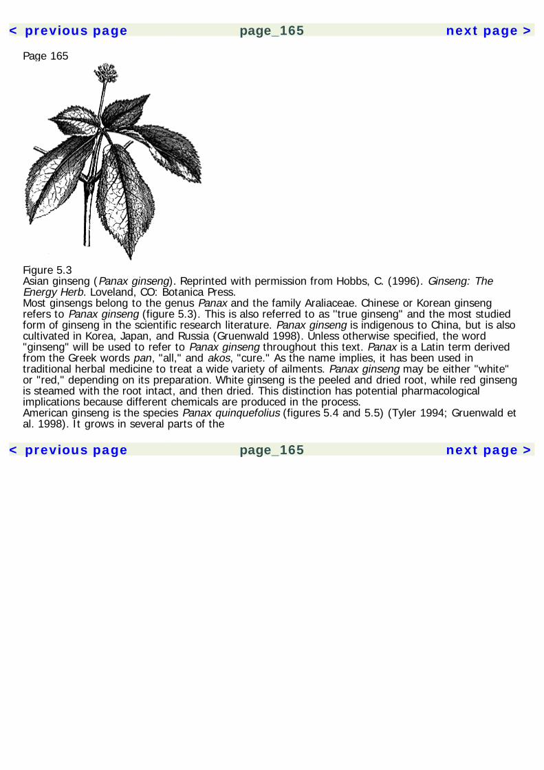
< previous page page_165 next page >
Page 165
Figure 5.3Asian ginseng (Panax ginseng). Reprinted with permission from Hobbs, C. (1996). Ginseng: TheEnergy Herb. Loveland, CO: Botanica Press.Most ginsengs belong to the genus Panax and the family Araliaceae. Chinese or Korean ginsengrefers to Panax ginseng (figure 5.3). This is also referred to as ''true ginseng" and the most studiedform of ginseng in the scientific research literature. Panax ginseng is indigenous to China, but is alsocultivated in Korea, Japan, and Russia (Gruenwald 1998). Unless otherwise specified, the word"ginseng" will be used to refer to Panax ginseng throughout this text. Panax is a Latin term derivedfrom the Greek words pan, "all," and akos, "cure." As the name implies, it has been used intraditional herbal medicine to treat a wide variety of ailments. Panax ginseng may be either "white"or "red," depending on its preparation. White ginseng is the peeled and dried root, while red ginsengis steamed with the root intact, and then dried. This distinction has potential pharmacologicalimplications because different chemicals are produced in the process.American ginseng is the species Panax quinquefolius (figures 5.4 and 5.5) (Tyler 1994; Gruenwald etal. 1998). It grows in several parts of the
< previous page page_165 next page >
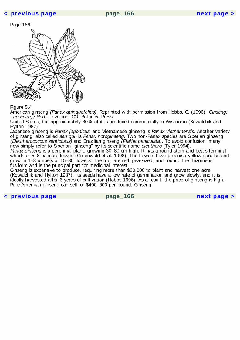
< previous page page_166 next page >
Page 166
Figure 5.4American ginseng (Panax quinquefolius). Reprinted with permission from Hobbs, C. (1996). Ginseng:The Energy Herb. Loveland, CO: Botanica Press.United States, but approximately 80% of it is produced commercially in Wisconsin (Kowalchik andHylton 1987).Japanese ginseng is Panax japonicus, and Vietnamese ginseng is Panax vietnamensis. Another varietyof ginseng, also called san qui, is Panax notoginseng. Two non-Panax species are Siberian ginseng(Eleutherococcus senticosus) and Brazilian ginseng (Pfaffia paniculata). To avoid confusion, manynow simply refer to Siberian ''ginseng" by its scientific name eleuthero (Tyler 1994).Panax ginseng is a perennial plant, growing 30–80 cm high. It has a round stem and bears terminalwhorls of 5–8 palmate leaves (Gruenwald et al. 1998). The flowers have greenish-yellow corollas andgrow in 1–3 umbels of 15–30 flowers. The fruit are red, pea-sized, and round. The rhizome isfusiform and is the principal part for medicinal interest.Ginseng is expensive to produce, requiring more than $20,000 to plant and harvest one acre(Kowalchik and Hylton 1987). Its seeds have a low rate of germination and grow slowly, and it isideally harvested after 6 years of cultivation (Hobbs 1996). As a result, the price of ginseng is high.Pure American ginseng can sell for $400–600 per pound. Ginseng
< previous page page_166 next page >
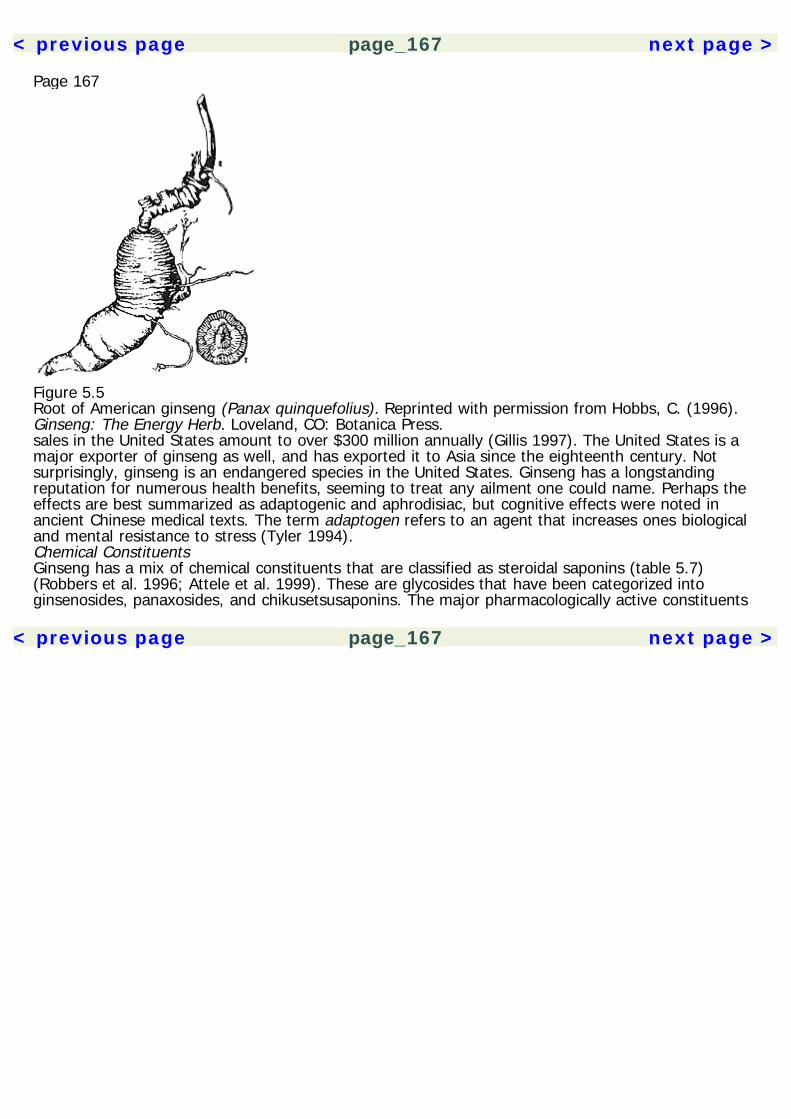
< previous page page_167 next page >
Page 167
Figure 5.5Root of American ginseng (Panax quinquefolius). Reprinted with permission from Hobbs, C. (1996).Ginseng: The Energy Herb. Loveland, CO: Botanica Press.sales in the United States amount to over $300 million annually (Gillis 1997). The United States is amajor exporter of ginseng as well, and has exported it to Asia since the eighteenth century. Notsurprisingly, ginseng is an endangered species in the United States. Ginseng has a longstandingreputation for numerous health benefits, seeming to treat any ailment one could name. Perhaps theeffects are best summarized as adaptogenic and aphrodisiac, but cognitive effects were noted inancient Chinese medical texts. The term adaptogen refers to an agent that increases ones biologicaland mental resistance to stress (Tyler 1994).Chemical ConstituentsGinseng has a mix of chemical constituents that are classified as steroidal saponins (table 5.7)(Robbers et al. 1996; Attele et al. 1999). These are glycosides that have been categorized intoginsenosides, panaxosides, and chikusetsusaponins. The major pharmacologically active constituents
< previous page page_167 next page >
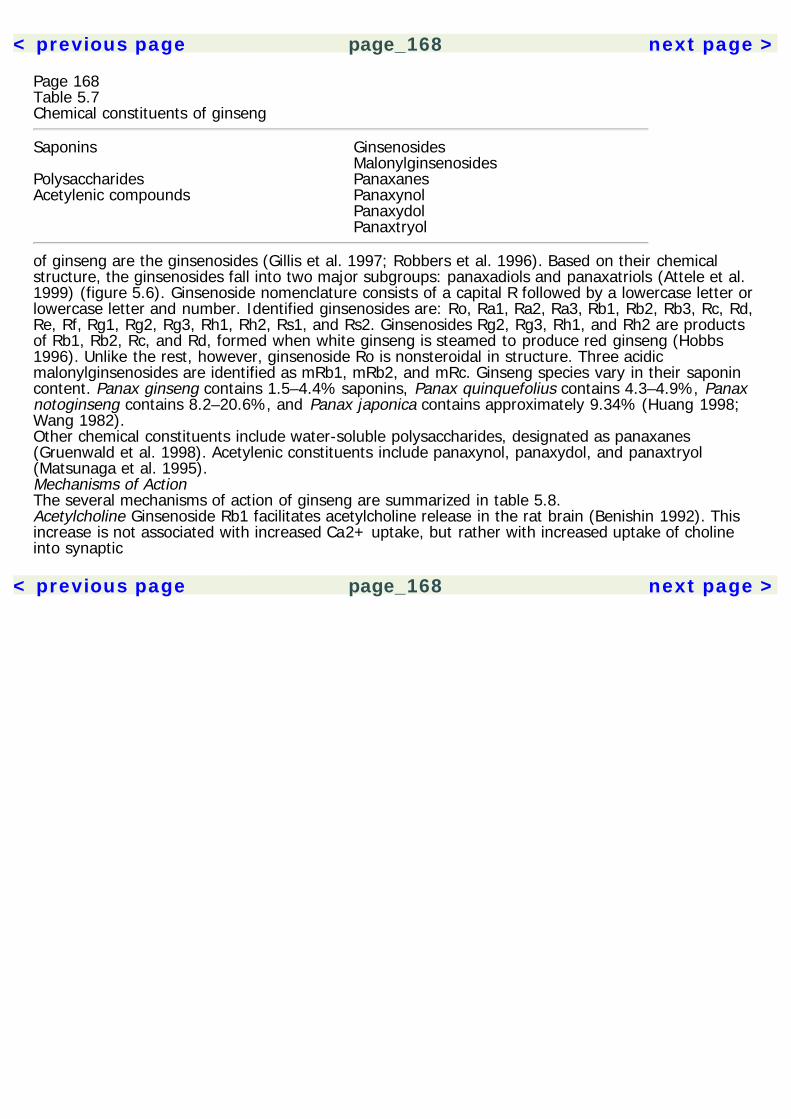
< previous page page_168 next page >
Page 168Table 5.7Chemical constituents of ginseng
Saponins Ginsenosides MalonylginsenosidesPolysaccharides PanaxanesAcetylenic compounds Panaxynol Panaxydol Panaxtryol
of ginseng are the ginsenosides (Gillis et al. 1997; Robbers et al. 1996). Based on their chemicalstructure, the ginsenosides fall into two major subgroups: panaxadiols and panaxatriols (Attele et al.1999) (figure 5.6). Ginsenoside nomenclature consists of a capital R followed by a lowercase letter orlowercase letter and number. Identified ginsenosides are: Ro, Ra1, Ra2, Ra3, Rb1, Rb2, Rb3, Rc, Rd,Re, Rf, Rg1, Rg2, Rg3, Rh1, Rh2, Rs1, and Rs2. Ginsenosides Rg2, Rg3, Rh1, and Rh2 are productsof Rb1, Rb2, Rc, and Rd, formed when white ginseng is steamed to produce red ginseng (Hobbs1996). Unlike the rest, however, ginsenoside Ro is nonsteroidal in structure. Three acidicmalonylginsenosides are identified as mRb1, mRb2, and mRc. Ginseng species vary in their saponincontent. Panax ginseng contains 1.5–4.4% saponins, Panax quinquefolius contains 4.3–4.9%, Panaxnotoginseng contains 8.2–20.6%, and Panax japonica contains approximately 9.34% (Huang 1998;Wang 1982).Other chemical constituents include water-soluble polysaccharides, designated as panaxanes(Gruenwald et al. 1998). Acetylenic constituents include panaxynol, panaxydol, and panaxtryol(Matsunaga et al. 1995).Mechanisms of ActionThe several mechanisms of action of ginseng are summarized in table 5.8.Acetylcholine Ginsenoside Rb1 facilitates acetylcholine release in the rat brain (Benishin 1992). Thisincrease is not associated with increased Ca2+ uptake, but rather with increased uptake of cholineinto synaptic
< previous page page_168 next page >
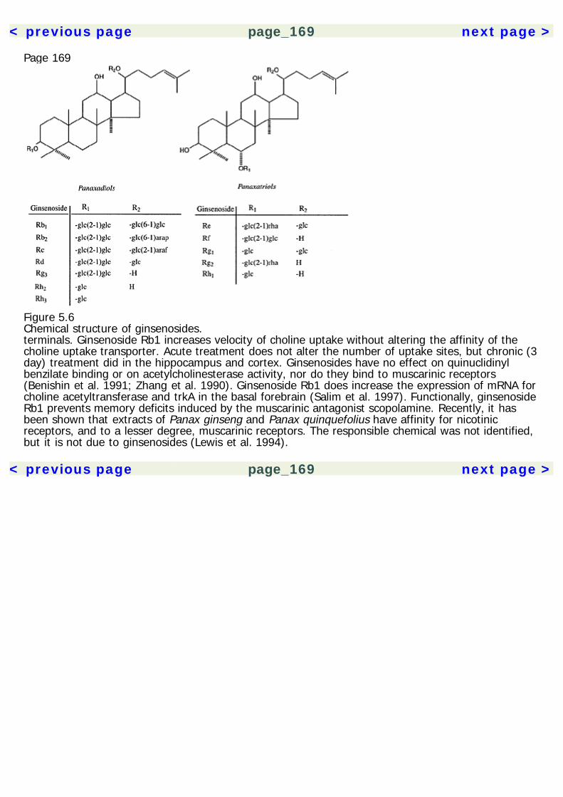
< previous page page_169 next page >
Page 169
Figure 5.6Chemical structure of ginsenosides.terminals. Ginsenoside Rb1 increases velocity of choline uptake without altering the affinity of thecholine uptake transporter. Acute treatment does not alter the number of uptake sites, but chronic (3day) treatment did in the hippocampus and cortex. Ginsenosides have no effect on quinuclidinylbenzilate binding or on acetylcholinesterase activity, nor do they bind to muscarinic receptors(Benishin et al. 1991; Zhang et al. 1990). Ginsenoside Rb1 does increase the expression of mRNA forcholine acetyltransferase and trkA in the basal forebrain (Salim et al. 1997). Functionally, ginsenosideRb1 prevents memory deficits induced by the muscarinic antagonist scopolamine. Recently, it hasbeen shown that extracts of Panax ginseng and Panax quinquefolius have affinity for nicotinicreceptors, and to a lesser degree, muscarinic receptors. The responsible chemical was not identified,but it is not due to ginsenosides (Lewis et al. 1994).
< previous page page_169 next page >

< previous page page_170 next page >
Page 170Table 5.8Mechanisms of action of ginseng
Cholinergic Increased acetylcholine release Ginsenoside Rb1 ChAT expression Ginsenoside Rb1 Nicotinic binding Non-ginsenoside (PQ)Amino acid GABAA Ginsenoside Rg2 (PQ) GABAA neurosteroid site Majonoside-R2 (PV)Monoamine Complex effects on turnover Ginsenosides Rb, Rg1Nitric oxide Enhanced epithelial synthesis GinsenosidesNeurotrophic Increased NGF expression Ginsenosides Rb1, Rg1 malonylginsenoside Rb1Ca2+ flux Inhibits Ca2+ channels Ginsenosides Rc, Re, Rf, Rg1, & Rb1
See text for references.Key: PQ Panax quinquefolius, PV Panax vietnamensis.Amino acid neurotransmitters Panax quinquefolius extracts reduce the discharge rate of neurons inthe medial nucleus solitarius with microgram doses and reversed by muscimol, indicating actions atGABAA receptors. There is no direct binding of ginsenosides Rb1 or Rg1 to GABA receptors (Zhang etal. 1991). In adrenal chromaffin cells, ginsenoside Rg2 putatively blocks GABA receptors (Tachikawaet al. 1999). Majonoside-R2, from Vietnamese ginseng, creates behavioral effects though theneurosteroid site on the GABAA receptor (Nguyen et al. 1997).Monoamines Ginseng has complex effects on endogenous monoamines. After two weeks oftreatment, turnover of dopamine and norepinephrine is increased in the cerebral cortex, andserotonin turnover is increased in the striatum and cerebellum (Itoh et al. 1989). However, dopamineturnover in the striatum is reduced, as is serotonin in the hypothalamus and midbrain. After sevenweeks of treatment, on the other hand, serotonin turnover in the cerebellum is increased, butturnover of dopamine, norepinephrine, and serotonin is reduced in all other areas studied.Ginsenosides Rb1 and Rg1 decrease 5-HT levels or reduce 5-HT turnover (Zhang et al. 1991). Thereis no specific binding observed of Rg1 and Rb1 adrenergic (α1 α2, or β), dopamine, or 5-HTreceptors.
< previous page page_170 next page >
< previous page page_171 next page >
Page 171In contrast, some studies have reported elevations in monoamines by chronic doses of ginsengthrough an unspecified mechanism (Wang et al. 1995). Ginseng prevents the heat stress-inducedreduction in brain norepinephrine and serotonin (Yuan et al. 1998). Ginseng saponins also reverse κ-opioid analgesia through serotonergic mechanisms (Kim HS et al. 1992).Chronic (5 day) oral ginseng extract alters striatal D2 receptors in aged rats, but acute administrationdoes not affect either D1 or D2 receptors (Watanabe et al. 1991).Nitric oxide Ginsenosides enhance nitric oxide synthesis, with effects in the endothelium of the lungs,heart, and kidneys and in the corpus cavernosum (Kim H et al. 1992; Ko et al. 1996). This is apotential mechanism of ginseng-associated vasodilation. Effects on neuronal nitric oxide have notbeen specifically investigated.Neurotrophic factors Some ginsenosides enhance neuronal survival in culture and interact with nervegrowth factor. Ginsenoside Rb1 increases the expression of mRNA for nerve growth factor in thehippocampus (Salim et al. 1997). However, the brain-derived neurotrophic factor and neurotrophin-3are not affected. In micromolar doses, malonylginsenoside Rb1 potentiates neurite growth induced bynerve growth factor in dorsal root ganglion cells (Nishiyama et al. 1994). Potentiation was also seenbetween ginsenoside Rg1 and nerve growth factor in cultured cortical neurons (Himi et al. 1989).Neuropeptides Ginsenosides have no apparent effect on neuropeptide precursors (preproenkephalin,preprotachykinin) or amyloid protein precursor (Salim et al. 1997).Ca2+ channels Ginseng extract rapidly and reversibly inhibits high-threshold, voltage-dependentCa2+ channels in micromolar doses (Nah and McCleskey 1994; Nah et al. 1995). This effect ismediated by a receptor linked to a pertussis toxin-sensitive G protein, but it is not through α2adrenergic, GABAB, muscarinic, or opioid receptors (Nah and McCleskey 1994). Several ginsenosidesinhibit Ca2+ currents by 16 to
< previous page page_171 next page >
< previous page page_172 next page >
Page 17237%, depending on the ginsenoside (Kim HS et al. 1998). The order of potency for this effect is:Rc>Re>Rf>Rg1>Rb1.PharmacokineticsGinsenosides are rapidly absorbed from the upper gastrointestinal tract, varying with differentginsenosides, doses, preparation, and stomach acidity (Odani et al. 1983). Between 2 and 30% ofginsenoside Rg1 is absorbed within 30–60 minutes, but little of ginsenoside Rb1 is absorbed. Abovedoses of 3–9 g, absorption is reduced (Han 1986). Peak blood concentrations are reached in 30minutes, and tissue levels peak at 90 minutes. Differences between ginsenoside pharmacokineticsmay be due to differences in protein binding (Chen et al. 1980). Tinctures and extracts allow formore rapid absorption than whole root. Ginsenosides are excreted in urine mostly unmetabolized(Huang 1998).General EffectsPhysiological There is an extensive literature that deals with the effects ginseng on CNS function, buteffects are also seen in neuroendocrine function, carbohydrate and lipid metabolism, the immunesystem, and cardiovascular function (Gillis 1997). Ginsenosides induce a decrease in heart rate andhave biphasic effects on blood pressure, with decreases preceded by a slight increase (Kaku et al.1975). Ginsenoside Rg1 had the most potent effects on blood pressure. Little or no effect is observedon respiration.Vascular and sexual effects Ginseng saponin has demonstrated vasoactive effects. It activates therelease of NO from bovine aortic endothelium, and from the endothelial cells and perivascular nerveof the rabbit corpus cavernosum (Kim HS et al. 1998; Ko et al. 1996; Choi and Seong 1995; Chenand Lee 1995). Nitric oxide synthesis is also observed in the endothelium of the lung, heart, andkidney (Gillis 1997).Ginseng's mechanism of vasorelaxation and nitric oxide release is probably by conversion of L-arginine to L-citrulline (Kim H et al. 1992). Ginseng saponin induces relaxation of the corpuscavernosum smooth muscle in a dose-dependent manner (Kim HJ et al. 1998). This effect is
< previous page page_172 next page >
< previous page page_173 next page >
Page 173reduced by anticholinergic and nitiric oxide inhibitors, and restored by addition of L-arginine. Thesefindings convergently suggest that ginseng facilitates penile erection via a nitric oxide pathway.One study indicated beneficial effect of ginseng on sexual behavior in male rats, increasing thefrequency of copulation in a 45 minute period, but also decreasing the latency to ejaculation (Kim etal. 1976). Another study found a decrease in sexual behavior in male rats acutely administeredginseng, but increases when it was chronically given (Murphy et al. 1998). In humans, ginseng wasmore effective than placebo and trazodone for treatment of erectile dysfunction (Choi and Seong1995). Although objective measurement, placebo control, and randomization were employed in thisstudy, there is no explicit mention of a double-blind element.Neuroendocrine and stress effects Ginseng activates the hypothalamopituitary-adrenal axis.Combined ginsenosides elevate plasma ACTH and corticosterone up to 90 minutes after injection inrats, by actions in the hypothalamus and/or pituitary (Hiai et al. 1979). The ginsenosides Rg1 andRb1 alone increase plasma levels of ACTH (Zhang et al. 1990). Ginsenoside Rg1 is a functional ligandat the glucocorticoid receptor (Lee YJ et al. 1997). It competes with dexamethasone and binds withlow micromolar affinity. However, ginsenosides inhibit the stress-induced elevation of plasmacorticosterone in mice (Kim DH et al. 1998). The effect is central, occuring in low microgramintracerebral doses, and is dependent on nitric oxide synthesis.Plasma prolactin levels are reduced with acute treatment and remain suppressed after 28 days ofchronic treatment (Murphy et al. 1998). With acute treatment, no effects are seen on plasmaluteinizing hormone or testosterone levels. However, chronic dietary 5% ginseng increasestestosterone levels in male rats (Fahim et al. 1982). Chronic ginsenosides do not alter posteriorpituitary hormones oxytocin and vasopressin (Zierer 1991). Similarly, human males administeredginseng extract showed an increase in plasma testosterone, dihydrotestosterone, follicle-stimulatinghormone, and luteinizing hormone, but a decrease in prolactin (Salvati et al. 1996).
< previous page page_173 next page >

< previous page page_174 next page >
Page 174Neurophysiological and neuroprotective effects Two ginsenosides have been found to affect long-term potentiation (Abe et al. 1994). While ginsenoside Rb1 attenuates it in the hippocampus,malonylginsenoside Rb1 facilitates it. This effect of malonylginsenoside Rb1 is dose dependent,facilitating it in the low nanomolar range (5 nM), but not at higher nanomolar doses (50 nM).Decreased c-fos mRNA and fos protein are observed in the hippocampus during aging, andginsenoside Rg1 increases the expression of c-fos mRNA and protein in both young and aged rats(Liu and Zhang 1996). Also increased are levels of cAMP in the hippocampus. The expression of c-fosserves as a marker for neuronal activity and is likely to be involved in neuronal plasticity. Thus,ginsenoside Rg1 may improve cognitive function by altering c-fos and cAMP in cerebral structures.Ginseng may have neuroprotective effects. An ischemia paradigm in gerbils showed that ginsengimproves response latency in a step-down passive-avoidance task (Wen et al. 1996). Loss ofpyramidal neurons in the hippocampal CA1 region is prevented in a dose-dependent manner. Whileginsenoside Rb1 was effective, Rg1 and Ro were ineffective. Central administration of ginsenosideRb1 after cerebral ischemia protects hippocampal neurons. The likely mechanism of this effect isantioxidant scavenging of free radicals (Lim et al. 1997). Oral red ginseng powder reduces thelearning impairment produced by ischemia in a passive-avoidance test, and preserves hippocampalneurons and synapses (Wen et al. 1996). Ginsenosides have a similar neuroprotective effect.Nonginsenoside constituents are also effective, but less potent in this regard.Behavioral and emotional effects In animal studies, ginseng does not prolong pentobarbital-inducedsleep, nor does it affect spontaneous locomotion (Mitra et al. 1996). It does potentiateamphetamine-induced locomotion, but it reduces the stereotypy and lethality caused byamphetamine. Ginseng has analgesic effects, which are discussed at greater length in chapter 8.Catalepsy induced by haloperidol is potentiated by ginseng, while the hyperthermic effect of 5-HTP isattenuated. No antiseizure effects have been observed.Although a single dose of ginseng has little acute effects, chronic dosing (twice daily, 5 days) reducesanxiety in behavioral models (open-field
< previous page page_174 next page >

< previous page page_175 next page >
Page 175and elevated plus-maze) (Bhattacharya and Mitra 1991). Also reduced by ginseng are aggressivebehavior in water-deprived rats and foot-shock-induced fighting in mice.Majonoside-R2, a saponin found in Vietnamese ginseng restores the deficit in pentobarbital sleepcaused by the stress of social isolation (Nguyen et al. 1997). This effect appears to be mediated bythe neurosteroid receptor on the GABAA receptor.The effects of ginseng on motor activity are highly variable. An oral dose of 1.8% ginseng extractdecreases spontaneous activity in young rats, but increases it in aged rats (Watanabe et al. 1991).The increase in activity in aged rats corresponds with reduced striatal D2 receptors. Thus, resultsvary across ginsenosides, the age of subjects, and chronicity of administration.The effects of two ginsenosides on ingestive behavior have been investigated in animal paradigms.Ginsenoside Rg1 reverses the supression of water intake by interleukin-1 β when administeredintracerebroventricularly (Kang et al. 1995). However, this effect was observed at the millimolarrange, and no effects on feeding were observed. Rg1 did reverse anorexia induced by heat andsurgical implantation, again only at millimolar doses (8 mM). On the other hand, low micromolardoses (0.01 μM) of ginsenoside Rb1 decreases food intake when injected into the ventromedialhypothalamus (Etou et al. 1988). Drinking behavior is also decreased with doses in the lowmicromolar range (0.2 μM). No effects were observed in the lateral hypothalamus. Ginseng extractstabilized fluctuations in wakefulness and slow-wave sleep in food-deprived rats (Lee et al. 1990).Ginseng extract has reported anti-aggresive effects in animals, although results vary depending onadministration and which ginsenoside is used (Mitra et al. 1996). Ginsenoside Rb1 and Rg1 suppressresident aggression in a resident-intruder paradigm using mice (Yoshimura et al. 1988a). In the sameparadigm, Rb1, but not Rg1, suppresses aggression in the intruder. In a maternal aggressionparadigm, crude ginseng saponins and Rb1 suppress aggression when administered acutely, but Rg1is ineffective. During chronic administration, crude ginseng saponins again reduce maternalaggression, while Rg1 facilitates it (Yoshimura et al. 1988b). Slightly different results are found inmice, where
< previous page page_175 next page >
< previous page page_176 next page >
Page 176Rg1, Rf, and Rd suppress foot-shock-induced aggression, but Rb1 and Rb2 have little effect (Kaku etal. 1975). Aggression is also reduced by ginseng between water-deprived rats and in foot-shock-induced conflict in mice (Bhattacharya and Mitra 1991).Cognitive effects: Animal studies There is an extensive literature that deals with the effects ofginseng on memory, learning, and behavior (Gillis 1997; Wang et al. 1995). However, ginseng extract(G115) failed to show anxiolytic effects in an animal model (Petkov et al. 1987)Ginseng extract improves the spatial learning performance of aged rats in an eight-arm radial mazeand operant discrimination task (Nitta et al. 1995). It also improves memory performance in active-avoidance (shuttle-box) and passive-avoidance (step-down) tasks, and reinforces staircase-mazelearning in both young and aged rats. (Petkov and Mosharrof 1987; Petkov et al. 1990; Petkov et al.1992). The effects were also very dose dependent, with inverted U-shape dose-response curves.The effects of ginsenoside Rg2 on learning and memory in rats were evaluated using a two-wayactive-avoidance method. Memory impairments induced by scopolamine and cycloheximide arereversed by ginsenoside Rg2 (Ma et al. 1991; Ma and Yu 1993). Ginseng extract (G115) reduces thememory impairment produced by electroshock treatment on a passive-avoidance (step-down) task(Lasarova et al. 1987). Adult rats given lesions of medial prefrontal cortex show cognitive deficits in aposition-reversal task, which is improved by chronic (30-day) treatment with ginseng.Several studies have looked at the effects of combined preparations that include ginseng. Apreparation of Biota orientalis, Panax ginseng, and Schisandra chinensis (S-113 m) improved memoryretention in a passive-avoidance task with senescence-accelerated mice (Nishiyama et al. 1996). Thiscombination also reduced memory impairments induced by ethanol and scopolamine in the step-down test and electroconvulsive shock-induced memory impairment (Nishiyama et al. 1995).Gincosan is a combination of extracts from Panax ginseng (G115) and Ginkgo biloba (GK501) (Petkovet al. 1993). Chronic oral pretreatment with
< previous page page_176 next page >
< previous page page_177 next page >
Page 177Gincosan improved learning in some animal models. Effects between ginseng and ginkgo werecomparable, and effective in combination.While promising, the use of multiple herbs in studies makes their results of limited generalizability.However, studies such as the one by Petkov and colleagues (1993), which examine both theindividual and combined effects, have conscientiously avoided this methodological problem.Cognitive effects: Human studies Few studies of the effects of ginseng on cognitive function havebeen performed on humans. One controlled study assessed the effect of chronic (12 week)administration of a ginseng preparation (G115) on cognitive function in healthy volunteers (D'Angeloet al. 1986). Several functions were measured: visual attention (with a cancellation task), auditoryreaction time, processing speed (choice reaction time, digit symbol substitution), fine motor speed(tapping test), mental arithmetic, and logical deduction. However, significant improvements wereonly seen in the mental arithmetic test.A controlled study of chronic (8 week) ginseng and multivitamin complex in elderly adults showed noapparent benefits on tests of attention or memory, as measured by the Trail Making Test andKendrick Object Learning Test. No differences were seen in the length of hospital stay, objectivemeasures of emotional and somatic symptoms, or activities of daily living. Although this studyproduced negative results, a greater variety of cognitive measures with more sensitivity could havebeen used. In contrast, another study of ginseng extract (G115) in adults showed improvement in all11 items of a standardized quality of life index (Caso Marasco et al. 1996).ToxicityGinsenosides show low toxicity in pharmacological studies (Chen et al. 1980). A ''ginseng abusesyndrome" was reported as consisting of hypertension, irritability, nervousness, and sleeplessness(Siegel 1979). However, the types of ginseng that produced this syndrome are unknown. The peoplein this study were also taking enormously high doses, such as 15 g per day, whereas conventionaldoses are 1 to 2 g per day (Gruen-
< previous page page_177 next page >

< previous page page_178 next page >
Page 178wald et al. 1998). Conventional doses of ginseng are not usually associated with serious adverseeffects (Tyler 1994; Gruenwald et al. 1998).Summary and ConclusionsGinseng has an ancient reputation for treating several illnesses, including memory loss. A variety ofspecies are colloquially labeled as ''ginseng," requiring attention to the exact species used forexperimental trials and clinical preparations. Several members of the Panax genus are appropriatelylabeled ginseng, and have similar chemical and neuropharmacological effects. The chief chemicalconstituents are the ginsenosides, which have actions on cholinergic, GABA, monoamine, andneurotrophic systems. Additionally, ginseng blocks Ca2+ channels and is a vasodilator through nitricoxide mechanisms.Vasodilating effects probably enhance cerebral blood flow and increase sexual performance, althoughelevations in dopamine and testosterone function may also contribute. Other neuroendocrine effectsinclude elevation of plasma ACTH and supression of prolactin. Ginseng has neuroprotective effectsthat likely relate to its antioxidant properties. Effects are seen on analgesia, anxiety, motor activity,ingestive behavior, and aggression. Benefits in memory and learning are seen in several animalstudies. However, most of the empirical research in ginseng has been done on animals. There aresome preliminary results with humans, but much more is needed to establish its effects. Inreasonable doses, ginseng has shown little toxicity.Ergot Derivatives: HydergineHistory and BotanyErgot alkaloids (or ergoloids) are derived from the ergot fungus Claviceps purpurea (van Dongen andde Groot 1995). As a toxic fungus that grows on rye and other grains, it has gained infamythroughout the history of civilization. Ergot is discussed at length in chapter 9 because of its potenthallucinogenic chemical lysergic acid diethylamide (LSD). However, its other nonhallucinogenicconstituents have been tried in clinical trials to treat dementia conditions, peripheral vascular disease,hypertension, angina pectoris, and tinnitus. Currently, the primary proposed use for
< previous page page_178 next page >
< previous page page_179 next page >
Page 179Table 5.9Natural and semisynthetic ergot alkaloids (ergoloids)
Amine alkaloids and Congeners d-Lysergic acid d-Isolysergic acid d-Lysergic acid diethylamide Ergonovine (ergometrine) Methylergonovine Methysergide Lisuride Lisurol Lergotrile MetergolineAmino acid alkaloids Ergomatine Ergotine Ergostine Ergotoxine: Ergocristine Ergocornine α-Ergokryptine β-Ergokryptine Bromocriptine
Source: Adapted from Peroutka 1996.Hydergine is for treatment of dementia. It has been approved by the Food and Drug administrationfor treatment of ''idiopathic decline in mental capacity."Chemical ConstituentsTwo general classes of alkaloids are distinguished in ergot: amine alkaloids and amino acid alkaloids(table 5.9) (Peroutka 1996). While the amine alkaloids are selective for antagonist effects onserotonin receptors, the amino acid alkaloids are less selective and act upon other monoaminereceptors. The constituents of interest for cognitive enhancement are predominantly the aminealkaloids.The chemical constituents of interest for cognitive effects are the ergotoxine derivatives, otherwisereferred to as ergoloid mesylates (Schneider and Olin 1994). Ergotoxine was isolated in 1906, butlater discovered to be a mix of four alkaloids: ergocristine, ergocornine, α-ergokryptine,
< previous page page_179 next page >
< previous page page_180 next page >
Page 180Table 5.10Mechanisms of action of Hydergine
Cholinergic Site-specific alteration of acetylcholine levels Reverses age-related decline in choline acetyltransferase Reverses an age-related decline in muscarinic receptors Stimulates learning-related cholinergic activityMonoamine Facilitates release of norepinephrine (through α2) Agonist/antagonist effects on serotonin receptors Time-dependent increases/decreases on dopamine and norepinephrine Age-dependent effects on MAO Increased firing of locus coeruleus neurons
See text for references.and β-ergokryptine. Co-dergocrine (referred to here by the trade name, Hydergine) was introducedclinically in 1949, and is a combination of four dihydro derivatives of ergotoxine (Peroutka 1996).Mechanisms of ActionHydergine's mechanisms of action are summarized in table 5.10.Acetylcholine A few studies have shown Hydergine to augment central cholinergic transmission. Invivo microdialysis shows that Hydergine decreases concentrations of acetylcholine in the striatum,while dose-dependently increasing it in the hippocampus (Imperato et al. 1994). Treatment withHydergine reverses age-related decline in choline acetyltransferase activity in the hippocampus andstriatum (Dravid 1983). It also reverses an age-related decline in the number of muscarinic receptorsin rats (Amenta et al. 1989). Greatest change in muscarinic receptors is seen in CA1 and CA2 of thehippocampus, and in the dentate gyrus to a lesser degree. Hydergine stimulates learning-relatedincreases in central cholinergic activity in rats (Le Poncin-Lafitte et al. 1985).Monoamines Hydergine has effects on the monoamines dopamine, norepinephrine, and serotonin. Itfacilitates electrically-evoked norepinephrine release though antagonism of α1 and presynaptic α2adrenergic
< previous page page_180 next page >
< previous page page_181 next page >
Page 181receptors (Markstein 1985). Mixed agonist/antagonist effects are seen at postsynaptic D1 receptors,and at pre- and postsynaptic D2 receptors. Mixed agonist/antagonist effects are also seen atserotonin receptors in the hippocampus and cortex. Collectively, these results indicate a dual effect ofHydergine on monoamine receptors, possibly compensating for deficits in transmitter function andreversing overactivity.Hydergine shows time-dependent effects on monoamines in various brain regions. Dopamine andnorepinephrine levels in the midbrain and diencephalon increase at 90 minutes and then sharplydecrease up to 120 minutes (Copeland et al. 1981). A similar pattern is seen with norepinephrine inthe pons and medulla, and with dopamine in the caudate nucleus. Hydergine alters MAO activity tovarying degrees with age: increases in MAO are greater in the hypothalamus and cerebellum of agedrats, while increases are more pronounced in the hippocampus of adult rats (Büyüköztürk et al.1995).Systemic administration of Hydergine significantly increases the firing rate of noradrenergic neuronsin the locus coeruleus in rats (Olpe and Steinmann 1982). At a dose of 1 mg/kg, a mean increase infiring rate of 70% is observed. Given the role of the locus coeruleus and norepinephrine in cognition,this could account in part for Hydergine's effects (Aston-Jones et al. 1994).PharmacokineticsAfter intravenous administration, dihydroergotoxine alkaloids show maximal uptake in visceral organs(10–5 M) and the central nervous system (10–7 M), and concentrations increase with repeatedadministration (Iwangoff et al. 1978). Maximal plasma concentrations of ergoloids occurapproximately 2 hours after oral administration (Aellig and Nuesch 1977). Elimination half-life variesfrom 1.4 to 6.2 hours for the alpha phase and from 13 to 50 hours for the beta phase.General EffectsNeuroprotective effects Hydergine may have some neuroprotective effects. It protects neurons inhippocampal CA1 following ischemia (Izumiyama et al. 1988). Hydergine also increases the activity ofthe anti-
< previous page page_181 next page >
< previous page page_182 next page >
Page 182oxidant enzymes superoxide dismutase and catalase, most prominently in the hippocampus andstriatum (Sözmen et al. 1998).Cognitive effects: Animal studies A few studies have been done on the cognitive effects of Hyderginein animal models. Hydergine elicits increases in spontaneous motor activity and self-stimulation ofthe ventral tegmental area, peaking at 80–120 minutes (Copeland et al. 1981). In mice, systemic andcentral doses of hydergine improve recall and retrieval memory in a T-maze and step-throughapparatus, with an inverted-U dose-response curve (Flood et al. 1985). No benefits were seen in theacquistion phase of active-avoidance tasks. Memory is improved in rats learning maze navigation, butparadoxically none of the components of Hydergine individually had a similar effect (Jaton andVigouret 1985). Hydergine reverses the amnestic effects of the muscarinic antagonist scopolamineand a protein synthesis inhibitor. Although effective against scopolamine-induced memory deficits,Hydergine is ineffective against hypoxic memory disturbance in rats (DeNoble et al. 1986). Higherdoses (10 mg/kg PO) impaired motor function.Cognitive effects: Human studies A meta-analysis was performed of methodologically controlled trialsof Hydergine in treatment of dementia (Schneider and Olin 1994). The studies examined had bothinpatient and outpatient subject pools, with a mean age of subjects varying from 59 to 84 acrossstudies. The mean treatment duration was of 17.1 weeks, and the mean dose of Hydergine persubject was 4.5 mg per day. Several studies examined effects in populations with Alzheimer's diseaseor vascular dementia. Outcome measures used included clinical global rating scales orneuropsychological measures (among them were subtests of Wechsler Adult Intelligence Scale, theTrail Making Test, and measures of orientation). Overall, studies showed a modest but consistenteffect of Hydergine in the areas assessed. The effect size varied with the type of measure used: 0.47for comprehensive ratings, 0.56 for clinical global ratings, and 0.27 for combined neuropsychologicalmeasures. Analysis of just the studies of Alzheimer's disease subjects found an effect only incombined neuropsychological testing (0.30). Despite the modest size of the effect, it is generallyconsistent with the effects obtained with other
< previous page page_182 next page >
< previous page page_183 next page >
Page 183antidementia drugs (e.g., Aricept, ranging from 0.42 to 0.48) (Rogers et al. 1998).ToxicityHydergine has been approved by the U.S. Food and Drug Administration as a treatment for cognitivedecline. No serious side effects are reported. Those reported include transient nausea and othergastrointestinal effects. Hydergine does not have the vasoconstrictive effects of other ergoloids.Hydergine may be contraindicated in people suffering from chronic psychoses. Depending on thedosage, overdoses of Hydergine can produce mild symptoms such as nasal stuffiness, flushing of theface, and headache, or more serious effects such as nausea, vomiting, tremulousness, spasticity,hypotension, circulatory collapse, and coma.Summary and ConclusionsHydergine is composed of four ergot alkaloids derived from the fungus Claviceps purpurea. Otherconstituents have vasoconstrictive or hallucinogenic effects, but Hydergine is now consideredprimarily as treatment for cognitive decline. Its mechanism of action appears to be throughcholinergic and/or monoamine mechanisms, and there is also evidence for antioxidant effects.Cognitive effects are seen in several animal models looking at memory. Human studies showHydergine to provide a modest but consistent benefit. Despite this, Hydergine seems to have fallenout of favor for treatment of dementia. Perhaps this is due to the promise of more recentcholinesterase inhibitors.TobaccoAlthough tobacco (Nicotiana tabacum) is widely known for its stimulant and addictive properties, itsprincipal psychoactive constituent, nicotine, has become a candidate drug for improving cognition (LeHouezec 1998; Lawrence and Sahakian 1998; Newhouse et al. 1997). Clearly, the health risksassociated with smoking tobacco preclude its clinical use, but nicotine delivered in other forms maybe useful.Nicotine is the principal psychoactive alkaloid from tobacco and has effects on cognition in animaland human studies. This is not surprising
< previous page page_183 next page >
< previous page page_184 next page >
Page 184given its direct effects on central cholinergic systems, and indirect effects on many otherneurochemical systems. When considering research on the cognitive effects of nicotine, one must notonly take into account the species (e.g., human, nonhuman), but also the degree of exposure andtolerance in research subjects. Chronic administration of nicotine induces neurochemical andfunctional compensatory mechanisms that alter the neural milieu. Thus, chronic tobacco smokers andnicotine-naive subjects are not equivalent, physiologically or on cognitive measures. The differencesfound between them are informative of effects of nicotine withdrawal.Mechanisms of Cognitive EnhancementNicotine acts as an agonist at the nicotinic cholinergic receptor (Cooper et al. 1996). It binds to asubunit of the nicotinic receptor, creating a conformational change in the receptor and allowing influxof cations. Cholinergic receptors are widespread in the forebrain, rich in many anatomical areasthought to underlie cognition. The prefrontal cortex is a major site for the cognitive effects of nicotine(Vidal 1996). Cognitive effects may result from immediate receptor activation and/or chronic effects.For example, the cognitive effects of nicotine result, in part, from nicotinic receptor up-regulationafter chronic treatment (Abdulla et al. 1996). It increases the number of nicotinic receptors in frontal,entorhinal, and dorsal hippocampal regions. Chronic nicotine also increases the sensitivity ofmuscarinic receptors (Wang et al. 1996). Nicotine interacts with other neurochemical systems,including dopamine, norepinephrine, serotonin, and glutamate (Levin and Simon 1998). Nicotine'scognitive effects may be due in part to monoamine mechanisms. Depletion of monoamines reducesthe cognitive benefit of nicotine (Riekkinen et al. 1996). See chapter 4 for a more detailed descriptionof nicotine's neuropharmacological mechanisms.Cognitive effects: Animals Cognitive effects of nicotine have been observed in several species,including humans. Experimental studies have focused primarily on the effects on attention andmemory. Cognitive benefits are seen after both acute and chronic administration (Levin et al. 1992).In experimental animals, nicotine improves learning and memory on a variety of tasks. Conversely,the nicotinic antagonist mecamylamine
< previous page page_184 next page >
< previous page page_185 next page >
Page 185impairs memory. Nicotine facilitates memory in a passive-avoidance task in mice with acceleratedsenescence; aged rats administered nicotine have a better rate of learning in an active-avoidancepole-jumping test (Arendash et al. 1995a; Meguro et al. 1994). Errors are also reduced in maze tests(Lashley III, 17-arm radial maze), affecting reference but not working memory. In the Morris watermaze, chronic (3 day) nicotine improves the learning acquisition of aged rats and the memoryretention of young adult rats (Socci et al. 1995). Nicotine also augments choice accuracy in theradial-arm maze (Levin and Rose 1991).Memory impairments produced by choline deprivation in rats are improved by nicotine (Sasaki et al.1991). It reduces memory deficits due to septohippocampal lesions or aged animals (Levin 1992).Nicotine's effect on memory not only appears after acute administration, but may persist for at least4 weeks after the end of chronic administration (Levin et al. 1992).Nicotine increases sensory gating, as measured in an acoustic startle paradigm, although results havevaried with subject strain (Acri et al. 1994; Faraday et al. 1998). Also improved is sustained attentionin rats, depending on task parameters (Mirza and Stolerman 1998). In choice reaction-timeparadigms there is improved accuracy and decreased omission errors under weak signal and lowevent-rate conditions. No benefits are seen under high event-rate conditions. Improvements are alsoseen in reaction time.Working-memory enhancement by nicotine is seen using delayed matching-to-sample tasks inmonkeys (Terry et al. 1993). This benefit is reversed by scopolamine, suggesting that nicotine'sbeneficial effect is due to increased central acetylcholine release and subsequent actions atmuscarinic receptors. Nicotine appears to shift female rats toward a male-typical navigationalstrategy, using spatial cues rather than landmarks (Kanit et al. 1998).Other nicotinic agonists show similar cognitive benefits. GTS-21 is as effective as nicotine in aged ratsfor improving memory performance in a one-way active-avoidance task, Lashley III maze, and 17-arm radial maze testing (Arendash et al. 1995b). ABT-418 is a recently developed drug that activatesnicotinic channels (Buccafusco et al. 1995; Anderson et al. 1995). Peripheral doses (2–30 nM/kg)improve performance in a delayed matching-to-sample task in macaques. Over an 8 day period, no
< previous page page_185 next page >
< previous page page_186 next page >
Page 186tolerance is seen in this mnestic effect, and similar cognitive benefits have been observed in rats.ABT-418 may represent a more attractive alternative to nicotine because it shares many of itsbeneficial effects, but not dose-limiting side effects.Cognitive effects: Human nonsmokers Administration of nicotine to tobacco nonusers produces smallcognitive enhancements. Studies in tobacco nonusers have primarily measured attention, informationprocessing speed, and memory. Nicotine improves perceptual speed, choice reaction time, and digitrecall (Stough et al. 1995; Le Houezec et al. 1994; Foulds et al. 1996). Improvements also occur indigit symbol substitution and continuous performance tests without reducing accuracy (Petrie andDeary 1989; Levin et al. 1998).A stimulus-filter model of nicotine reinforcement has been proposed that suggests that it helpsscreen irrelevant stimuli from awareness (Kassel 1997). An alternative explanation asserts thatnicotine induces attentional narrowing and facilitates perceptual processing. Human nonsmokersgiven an acute dose of nicotine (administered in gum) showed dose-related trends toward decreasedaccuracy and increased response time (Heishman et al. 1993).Cognitive effects: Smokers and abstinence Nicotine abstinence in chronic tobacco users creates amild cognitive impairment (Snyder et al. 1989). Abstinent cigarette smokers performed comparably tononsmokers on cognitive tasks when administered nicotine, but the abstinent smokers had poorerperformances before the nicotine was given (Foulds et al. 1996). Abstinent smokers show mentalslowing on complex cognitive tasks that is beyond any reduction in simple reaction time or motorperformance (Shiffman et al. 1995). Smoking cessation causes a reduction in digit recall and serialaddition/subtraction (Sommese and Patterson 1995). A decrease in performance on the trail-makingtest B is seen as soon as 4 hours after tobacco discontinuation (Hatsukami et al. 1989). At 24 hoursafter cessation, changes occur in increased reaction time and decreased accuracy on a vigilance task.Reinstatement of nicotine reverses the cognitive deficits produced by withdrawal. Tobacco smokingafter short-term (overnight) abstinence improves processing speed in smokers on the Stroop colorand color-
< previous page page_186 next page >
< previous page page_187 next page >
Page 187word tasks (Pomerlau et al. 1994). Speed is reduced in a modified Stroop task using smoking-relatedwords as compared to neutral words, suggesting that abstinence produces a content-specific shift inattentional focus (Gross et al. 1993). Improvements are seen in complex visual-reasoning tasks aswell (Raven's advanced progressive matrices) after tobacco reinstatement (Stough et al. 1994).Improved driving skills such as brake reaction times and tracking accuracy are evident when smokersreceive nicotine (Sherwood 1995). Withdrawal deficits are also reversed by replacement with nicotinegum (Snyder and Henningfield 1989). On measures of choice reaction time, memory scanning,tracking, and flicker fusion threshold, smokers showed increased improvement with nicotine gumafter smoking (Hindmarch et al. 1990).The cognitive and behavioral changes brought on by tobacco abstinence are worsened by quittingboth caffeine and tobacco simultaneously. Combined cigarette and coffee abstinence in regular usersreduces performance on serial arithmetic and digit recall tasks, and decreases reports of feeling clearheaded and quick witted (Cohen et al. 1994). Conversely, reports of feeling irritability, musculartension, headache, and drowsiness, are increased.Cognitive effects: Dementia Nicotine has been considered as a therapeutic agent for indivduals withAlzheimer's disease (Warburton 1992). People with Alzheimer's disease show modest cognitiveimprovements when given nicotine (Sunderland et al. 1988). Cognitive effects are seen in doseslower than those required in normal controls, suggesting increased sensitivity to cholinergic agents inAlzheimer's disease. Learning improvements persist for a period of time after the drug is stopped(Wilson et al. 1995). However, behavior and global cognitive function are not markedly improved.Chronic transdermal nicotine improves attention in Alzheimer's patients, as assessed by a continuousperformance test (White and Levin 1999). However, another study of transdermal nicotine did notshow improvement over a 4-week period (Snaedel et al. 1996).Summary and ConclusionsNicotine, the principal psychoactive alkaloid in tobacco, has cognitive-enhancing effects. It directlyactivates nicotinic acetylcholine receptors in brain areas critical for cognition, creating both acute andchronic effects.
< previous page page_187 next page >
< previous page page_188 next page >
Page 188Nicotine also indirectly interacts with a number of other neurotransmitter systems includingmonoamines and excitatory amino acids. Cognitive benefits are seen in attention and memory acrossmany animal and human experiments, partially depending on the test methodology used. Modesteffects are produced in nonsmokers administered nicotine, and larger effects are seen in abstinentsmokers. A limited number of studies have been performed in Alzheimer's disease. Again, effectshave been modest, but further research is warranted. It is not certain what the long-term benefits ofnicotine or nicotinic drugs will be to Alzheimer's disease or other dementias, whether theimprovements sustain or whether it will result in daily functional changes.The harmful effects of smoking and chewing tobacco (e.g., cancer, emphysema) make it animpractical form of drug delivery, but other delivery systems such as gum and transdermal patchesmay circumvent this problem. Alternatively, other nicotinic drugs are under development, such asABT-418, which may create the desired clinical effects and minimize side effects.ArecaAreca (Areca catechu) is a tall palm tree that yields a nut, commonly chewed in Asia for its stimulanteffects. It is discussed at greater length in chapter 4, but is mentioned here briefly for its cognitiveeffects. Areca's psychoactive constituents (arecoline, arecaidine, guvacoline, and guvacine) areagonists at muscarinic acetylcholine receptors. They also directly inhibit reuptake of GABA.In addition to autonomic effects, areca produces mental stimulation, increases cerebral blood flowand glucose metabolism, and creates electrophysiological activation as seen on the EEG.Cognitive EffectsStudies on the cognitive effects of areca are scarce, but its known cholinergic mechansims have ledsome researchers to formally investigate the effects. Arecoline improves memory on a staircase testin rats, but effects were dose dependent (Molinengo et al. 1995). A low dose (0.5 mg/kg) improvesperformance, but a large dose (3.5–8 mg/kg) causes impair-
< previous page page_188 next page >
< previous page page_189 next page >
Page 189ment. However, the beneficial effect diminished with tolerance. The memory effects of a singleadministration are also time dependent, where improvements occur at 3 and 24 hours, but no effectwas seen at 168 hours (Flood et al. 1988). Some studies have found a time lag for the onset ofcognitive benefits, suggesting that receptor regulation is partly responsible for the effects (Mondadoriet al. 1994). Arecoline and other cholinergic agonists reverse memory and working-memory deficitsinduced by scopolamine (Hironaka and Ando 1996; Quartermain and Leo 1988). However, arecolinedoes not reverse scopolamine-induced deficits in mice learning a maze, and may actually potentiatethe deficit (Bratt et al. 1996). Arecoline also partially offsets cognitive deficits induced by chronicethanol intake (Arendt 1994). A synergistic effect occurs between tacrine and arecoline, independentof route of administration (Flood and Cherkin 1988).A few formal studies have been done on cognitive effects of areca chewing in humans (Chu 1994).Although no differences are seen on simple reaction time, areca facilitates choice reaction time. It isinteresting to note that some improvement is produced by the chewing movements themselves (i.e.,chewing gum), but higher significance is found with areca, indicating effects of cholinergicmechanisms. No effects are seen in early-stage visual processing, as measured by the critical flickerfusion test (Frewer 1990). A limited study of betel quid showed an increase in reaction time latency.Whether this is an experimental effect or interactive effect of other chemical constituents (e.g., Piperbetle and/or calcium hydroxide) was not discerned. Human studies have also shown arecoline toreverse scopolamine-induced deficits in serial learning tasks (Sitaram et al. 1978).Given its cholinergic activity, some have suggested arecoline as a treatment for Alzheimer's disease,although the benefits have not been investigated nearly as much as other cholinergic agents(Asthana et al. 1996). People with Alzheimer's disease show improvement in verbal ability at lowdoses, but improvements in attention and visuospatial ability at higher doses (Raffaele et al. 1996).Significant improvements occur in verbal memory, with the average number of words recalled nearlydoubling (11.8–20.1 words) at a dose of 4 mg/day. However, two of the eight subjects in one studydid not respond to the drug.
< previous page page_189 next page >
< previous page page_190 next page >
Page 190However, another controlled study in Alzheimer's disease patients found only marginal improvementsin picture recognition and ratings of word-finding ability (Tariot et al. 1988). No benefits were seen insemantic or episodic memory. Biphasic effects were found on psychomotor activation, withimprovements at lower doses and impairments at higher doses.SolanaceaeThe fruit of a number of solanaceous plants, including tomato (Lycopersicon esculentum), potato(Solanum tuberosum) and eggplant (Solanum melongena esculentum), have cholinesterase-inhibitingeffects (Krasowski et al. 1997). They contain solanaceous glycoalkaloids α-solanine and α-chaconine,which are triglycosides of solanidine, a steroidal alkaloid derived from cholesterol. They are the onlyplant chemicals known to inhibit both acetlycholinesterase and butyrylcholinesterase, both in vitroand in vivo.Levels of solanaceous glycoalkaloids can be significant, occasionally causing toxicity in humans andlivestock. Symptoms of potato poisoning include gastrointestinal disturbances, apathy, drowsiness,mental confusion, visual disturbances, dizziness, hallucinations, and trembling. Symptoms may perist2–24 hours after consumption, and one recent outbreak showed reduced butyrylcholinesterase levelssix days later, which then returned to normal in 4 to 5 weeks.Apart from rare toxic-level exposures to solanaceous glycoalkaloids, there is evidence that normaldietary consumption still contains physiologically significant levels. An indivudual consuming a normalportion of potatoes can ingest 20 to 60 mg of solanaceous glycoalkaloids. Meals combining othercommon solanaceae such as tomatoes and eggplant may further increase the levels ingested.Krasowski and colleagues (1997) have elucidated how dietary solanaceae can alter metabolism ofanesthetic drugs by blocking butyrylcholinesterase. Given the role of cholinesterase inhibitors inhuman cognitive function, it is speculative whether dietary consumption of solanaceae has ameasurable effect on cognition. Perhaps of greater practical concern are potential interactions forthose taking pharmaceutical cholinesterase inhibitors such as tacrine or donepezil.
< previous page page_190 next page >

< previous page page_191 next page >
Page 191Fava BeansFava beans (broad beans, Vicia faba) contain a significant amount of L-DOPA. Anecdotal reports havenoted improvement of Parkinson's disease after the consumption of fava beans (Spengos andVassilopoulos 1988). Rabey and colleagues (1992, 1993) have demonstrated the efficacy of favabeans in this respect.Six patients with Parkinson's disease were withdrawn from their antiparkinsonian medications (L-DOPA/carbidopa, bromocriptine, or lisuride) (Rabey et al. 1992, 1993). After 12 hours off medication,the subjects ate 250 g of cooked fava beans. Significant improvements in motor symptoms werenoted, comparable to those seen with 125 mg of L-DOPA and 12.5 mg of carbidopa. In fact, threesubjects developed severe dyskinesias after fava ingestion, akin to those seen after larger doses ofpharmaceutical L-DOPA. Plasma levels of L-DOPA increased after fava ingestion in a mannercomparable to that seen with administration of oral L-DOPA. These results suggest that the L-DOPAcontained in fava beans was transported into the CNS and converted to dopamine. In fivenonparkinsonian, healthy volunteers, a similar increase in plasma L-DOPA was observed after favaingestion, although much lower. The difference in plasma L-DOPA between normal volunteers andparkinsonian patients is apparently due to a residual effect of carbidopa in the subjects withParkinson's disease. Without carbidopa, the L-DOPA from fava is rapidly converted to dopamine inthe blood stream and never crosses the blood-brain barrier.The therapeutic usefulness of fava beans in Parkinson's disease is unlikely because of the variablecontent of L-DOPA in available fava beans. In addition, the palatability of indefinitely eating favabeans several times daily to maintain therapeutic concentrations makes it impractical. Rabey andcolleagues suggest that fava beans may serve as a complementary source of L-DOPA forParkinsonian patients (Rabey et al. 1993). Conversely, they should also be regarded with caution, ifnot avoided altogether, by individuals already taking L-DOPA/carbidopa because additive effectswould occur. Thus, fava beans pose a source of unanticipated toxicity to a casual consumer.Nonparkinsonian people, on the other hand, would not meet with this degree of toxicity becausemost of L-DOPA is
< previous page page_191 next page >
< previous page page_192 next page >
Page 192rapidly converted to dopamine in the blood and never reaches the brain. One peripheral effect offava beans, which would be experienced by all those consuming them, is increased natriuresis anddiuresis through renal dopamine receptors (Vered et al. 1997).Nootropic Herbs: Summary and ConclusionsThe need for medications to improve cognitive function is rooted in the desire of healthy people tooptimize their performance, and to preserve function in those with dementia conditions. Of theseveral forms of dementia, the ones given most attention here are Alzheimer's disease, Parkinson'sdisease, and vascular dementia. The two general strategies for treatment of dementia includereplacement/augmentation of neurotransmitter and neuroprotective drugs to slow the loss ofneuronal function. To varying degrees, herbal treatments such as ginkgo offer both of these.Of the herbal medications reviewed here, ginkgo, ginseng, Hydergine, nicotine, and areca all haveprominent effects on cholinergic and monoamine systems. Ginseng and ginkgo also have directeffects on increasing cerebral blood flow. Similarly, ginkgo, ginseng, and Hydergine also havesignificant antioxidant effects and neuroprotective effects in several models. Ginkgo has received themost attention recently for treatment of dementia, with cognitive effects in animal and human trialscomparable to pharmaceutical cholinesterase inhibitors tacrine and donepezil. Hydergine showssimilar modest effects across trials. Ginseng has some common neuropharmacological mechanismswith ginkgo, but has only been studied in animals for the most part. Nicotine produces measurableimprovements, but carries the problems of dependence and tolerance. Newer nicotinic medicationsmay provide the same cognitive benefits with a more tolerable profile. Areca provides mixed cognitivebenefits, and does not seem to offer any clear advantage over other available medications.It is unlikely that any herbal medications will replace the pharmaceutical drugs for dementia atpresent. However, ginkgo makes a compelling case because it is similar in efficacy to the availablecholinesterase inhibitors, has additional antioxidant effects, and is relatively inexpensive.
< previous page page_192 next page >

< previous page page_193 next page >
Page 193The chief unresolved issue with ginkgo is the concern of bleeding when combined with anticoagulantmedications. The possibility of combining herbal and/or pharmaceutical medications for a greatercognitive effect has not yet been addressed.Solanaceous vegetables (tomato, potato, and eggplant) might contribute to cognitive function due totheir cholinesterase-inhibiting constituents. Similarly, fava beans could contribute significant amountsof L-DOPA to people with Parkinson's disease. However, this would necessitate daily intake, leadingto a rather mundane diet. A more critical problem would be the unpredictable varying levels of theactive constituents. To ensure reliability in their effects, herbal extracts need to be standardized. Amore practical concern with solanaceous vegetables and fava beans is that they may interfere withongoing therapeutic regimens.
< previous page page_193 next page >

< previous page page_194 next page >
Page 194This page intentionally left blank.
< previous page page_194 next page >
< previous page page_195 next page >
Page 1956Herbal Sedatives and AnxiolyticsFunctioning in a complex and dangerous environment requires one to possess effective mechanismsof arousal, both arousal to consciousness and emotional arousal, in order to meet the demands onbehavior. For example, an organism needs to be able to arouse behaviorally in order to deal withpredators and other environmental threats. As is often the case with disorders of the mind and brain,normal and adaptive mechanisms can be overactivated and thus become maladaptive. A commonoutcome of this overactivation is anxiety and insomnia.Anxiety is a subjective feeling of unease and discomfort, and is accompanied by a host of autonomicand somatic manifestations. It correlates with activity in neuroanatomical structures, including theamygdala and other related limbic structures (Davidson et al. 1999). Anxiety is discussed in greaterdetail in chapter 7. Insomnia is the inability to sleep or obtain a good quality sleep. This frequentlymanifests as difficulty falling asleep, awakening at night or early morning, and daytime fatigue(American Psychiatric Association 1994; Eddy and Walbroehl 1999). Insomnia may be a primarydisorder, but frequently it is a symptom of another neurologic or psychiatric disorder, such asanxiety.Central Nervous System DepressantsHerbal sedatives and anxiolytics are a diverse group of plant drugs that commonly act asdepressants of the central nervous system (CNS) (table 6.1). Pharmaceutical CNS depressants areused as anxiolytics, anti-epileptics, sedatives, sleep-inducers (sedatives or hypnotics), generalanesthetics, and recreational drugs (e.g., ethanol) (table 6.2). CNS
< previous page page_195 next page >
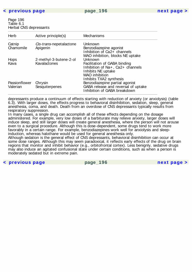
< previous page page_196 next page >
Page 196Table 6.1Herbal CNS depressants
Herb Active principle(s) Mechanisms
Catnip Cis-trans-nepetalactone UnknownChamomile Apigenin Benzodiazepine agonist Inhibition of Ca2+ channels MAO inhibition, blocks NE uptakeHops 2-methyl-3-butene-2-ol UnknownKava Kavalactones Facilitation of GABA binding Inhibition of Na+, Ca2+ channels Inhibits NE uptake MAO inhibition Inhibits TXA2 synthesisPassionflower Chrysin Benzodiazepine partial agonistValerian Sesquiterpenes GABA release and reversal of uptake Inhibition of GABA breakdown
depressants produce a continuum of effects starting with reduction of anxiety (or anxiolysis) (table6.3). With larger doses, the effects progress to behavioral disinhibition, sedation, sleep, generalanesthesia, coma, and death. Death from an overdose of CNS depressants typically results fromrespiratory suppression.In many cases, a single drug can accomplish all of these effects depending on the dosageadministered. For example, very low doses of a barbiturate may relieve anxiety, larger doses willinduce sleep, and still larger doses will create general anesthesia, where the person will not arouseeven to a surgical procedure. Although this is dose-dependent, some drugs tend to work morefavorably in a certain range. For example, benzodiazepines work well for anxiolysis and sleep-induction, whereas halothane would be used for general anesthesia only.Although sedation is the general effect of CNS depressants, behavioral disinhibition can occur atsome dose ranges. Although this may seem paradoxical, it reflects early effects of the drug on brainregions that monitor and inhibit behavior (e.g., orbitofrontal cortex). Less benignly, sedative drugsmay also induce an agitated confusional state under certain conditions, such as when a person ismoderately sedated but in extreme pain.
< previous page page_196 next page >
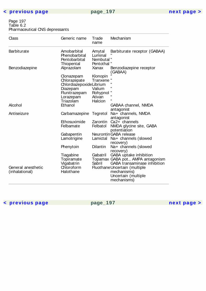
< previous page page_197 next page >
Page 197Table 6.2Pharmaceutical CNS depressants
Class Generic name Tradename
Mechanism
Barbiturate Amobarbital Amytal Barbiturate receptor (GABAA) Phenobarbital Luminal '' Pentobarbital Nembutal " Thiopental Pentothal "Benzodiazepine Alprazolam Xanax Benzodiazepine receptor
(GABAA) Clonazepam Klonopin " Chlorazepate Tranxene " ChlordiazepoxideLibrium " Diazepam Valium " Flunitrazepam Rohypnol " Lorazepam Ativan " Triazolam Halcion "Alcohol Ethanol GABAA channel, NMDA
antagonistAntiseizure Carbamazepine Tegretol Na+ channels, NMDA
antagonist Ethosuximide Zarontin Ca2+ channels Felbamate Felbatol NMDA glycine site, GABA
potentiation Gabapentin NeurontinGABA release Lamotrigine Lamictal Na+ channels (slowed
recovery) Phenytoin Dilantin Na+ channels (slowed
recovery) Tiagabine Gabatril GABA uptake inhibition Topiramate Topamax GABA pot., AMPA antagonism Vigabatrin Sabril GABA transaminase inhibitionGeneral anesthetic(inhalational)
ChloroformHalothane
FluothaneUncertain (multiplemechanisms)Uncertain (multiplemechanisms)
< previous page page_197 next page >
< previous page page_198 next page >
Page 198Table 6.3Continuum of effects of CNS depressants
AnxiolysisBehavioral disinhibitionSedationSleepGeneral anesthesiaComaDeath
Pharmacological Issues in CNS DepressantsSeveral pharmacological issues pertain to most CNS depressant drugs (Hobbs et al. 1996; Julien1997). Depending on their pharmacological mechanisms, combinations of CNS depressants canproduce additive or synergistic effects, when the total effect is equal to or greater than the sum oftheir individual effects, respectively. For example, in doses that would be safe individually,combinations of alcohol and barbiturates can be lethal.Many CNS depressants have some liability for dependence. This is typically greater with barbiturates,but lesser with benzodiazepines, and perhaps nonexistent in many antiseizure medications. CNSdepressants produce tolerance when administered chronically, where increasingly larger doses arerequired to sustain the same level of effect. Further, a cross-tolerance often develops, where thetolerance is generalized to other CNS depressants. For example, a person with an ethanol tolerancewill also display some tolerance to barbiturates. The therapeutic index tends to decrease as toleranceincreases, so that the difference between an effective and toxic dose diminishes. Thus, tolerance toCNS depressants is accompanied by a smaller safety margin.Once tolerance develops, acute cessation of CNS depressants creates a withdrawal syndrome.Tolerance is associated with neural adaptations to the continued inhibitory influence of the drug.Suddenly removing the drug and its inhibitory influence creates a rebound excitation, due toneuronal disinhibition. Depending on the degree of tolerance, subjects may experience anxiety,irritability, delirium, and seizures. Withdrawal
< previous page page_198 next page >
< previous page page_199 next page >
Page 199from CNS depressants is a serious medical issue, and must be controlled carefully. Typically, peopleare switched to a safer CNS depressant drug such as a benzodiazepine, and the drug is slowlytapered over a number of weeks. This allows for gradual neural adaptation and minimizes withdrawalsymptoms.General Mechanisms of Action for CNS DepressantsIn order for a drug to induce CNS depression it must either facilitate neuronal inhibition or inhibit theneuronal excitation. There are a few mechanisms by which these may occur, and CNS depressantdrugs may possess one or more of these mechanisms.Many CNS depressants work by facilitating the activity of the inhibitory neurotransmitter GABA (table6.1). The GABAA receptor is an ion channel with several receptor sites (Cooper et al. 1996). There isa binding site for the endogenous neurotransmitter GABA, which opens the channel and allows Cl–flux into the neuron. This hyperpolarizes the neuron, making it less likely to reach the threshold ofexcitation. In addition to the GABA receptor, there are several separate allosteric binding sites forbenzodiazepines, barbiturates, certain steroids, and ethanol. Agonists at these sites have no intrinsicinhibitory effects, but instead potentiate and facilitate the the Cl– conductance induced by GABA.Other means of enhancing GABA activity include blocking its reuptake (e.g., tiagabine), or increasingits release (gabapentin). The net result of all of these actions is a facilitation of neuronal inhibition byGABA.A second mechanism of action of CNS depressants is the inhibition of an excitatory neurotransmitter.Several of the drugs in this class (ethanol, barbiturates, Tegretol) block the actions of the excitatoryamino acid (EAA) neurotransmitter glutamate at the N-methyl-D-aspartate (NMDA) receptor. TheNMDA channel requires both receptor activation and membrane depolarization to open, allowingCa2+ influx. An intracellular chemical cascade follows, inducing synaptic changes, thought to be thecellular basis of memory formation (Kandel et al. 1991). Congruently, drugs that inhibit the NMDAchannel prevent the formation of memories while the drug is in effect. This would explain, in part,why a common side effect of some CNS depressants is memory impairment.
< previous page page_199 next page >
< previous page page_200 next page >
Page 200However, it is not the only mechanism, because drugs that do not work at NMDA receptors canimpair memory (e.g., benzodiazepines). Other EAA receptors include the ionotropic AMPA and kainatereceptors, and the metabotropic glutamate receptors (mGluR).The third mechanism of CNS depressants is the blocking of voltage-gated ion channels (Hille 1992).These are typically Na+ or Ca2+ channels, which are located in the neuronal membrane. Na+channels are responsible for conveyance of the action potential along the membrane, and Ca2+entry is necessary for release of neurotransmitters from synaptic terminals. Thus, drugs that blockeither or both of these channels will reduce the neuronal output. The CNS depressant drugs thathave this mode of action are predominantly antiseizure drugs, although many local anesthetics areantagonists of the Na+ channel (see chapter 8).Herbal CNS DepressantsValerianHistory and BotanyValerian is native to Europe and Asia, but now grows in most parts of the world. Valeriana officinalisis the most commonly known and studied, but approximately 200 species are known (figure 6.1)(Kowalchick and Hylton 1987). Unless otherwise specified, the name valerian will be used here torefer to V. officinalis. Valerian grows 50 to 100 cm in height, with an erect stem with pinnate leavesand numerous small pink-white flowers at the top. The parts of the plant used medicinally are theroots and rhizome (figure 6.2).The use of valerian extends back at least 1000 years, and it gained a reputation in sixteenth-centuryEurope as a treatment for epilepsy (Tyler 1994; Temkin 1971). Its reported uses are broad (digestiveaid, muscle relaxant, antipyretic, etc.) but it is commonly known to treat insomnia and anxiety(Gruenwald et al. 1998; Kowalchick and Hylton 1987). Valerian has a distinct, unpleasant odor.Perhaps appropriately, it is believed to be an herb in the writings of Galen and Dioscorides, calledphu (Leyel 1994).
< previous page page_200 next page >
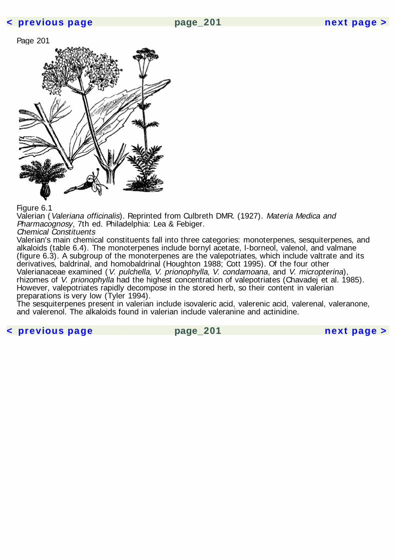
< previous page page_201 next page >
Page 201
Figure 6.1Valerian (Valeriana officinalis). Reprinted from Culbreth DMR. (1927). Materia Medica andPharmacognosy, 7th ed. Philadelphia: Lea & Febiger.Chemical ConstituentsValerian's main chemical constituents fall into three categories: monoterpenes, sesquiterpenes, andalkaloids (table 6.4). The monoterpenes include bornyl acetate, l-borneol, valenol, and valmane(figure 6.3). A subgroup of the monoterpenes are the valepotriates, which include valtrate and itsderivatives, baldrinal, and homobaldrinal (Houghton 1988; Cott 1995). Of the four otherValerianaceae examined (V. pulchella, V. prionophylla, V. condamoana, and V. micropterina),rhizomes of V. prionophylla had the highest concentration of valepotriates (Chavadej et al. 1985).However, valepotriates rapidly decompose in the stored herb, so their content in valerianpreparations is very low (Tyler 1994).The sesquiterpenes present in valerian include isovaleric acid, valerenic acid, valerenal, valeranone,and valerenol. The alkaloids found in valerian include valeranine and actinidine.
< previous page page_201 next page >
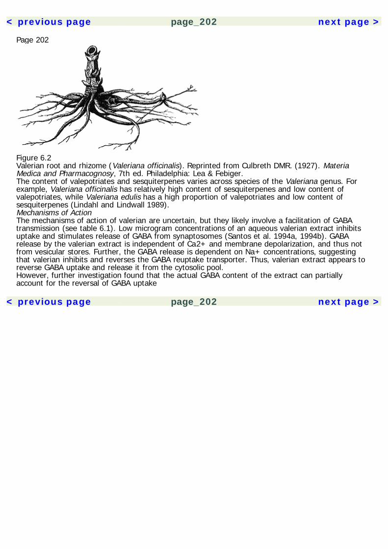
< previous page page_202 next page >
Page 202
Figure 6.2Valerian root and rhizome (Valeriana officinalis). Reprinted from Culbreth DMR. (1927). MateriaMedica and Pharmacognosy, 7th ed. Philadelphia: Lea & Febiger.The content of valepotriates and sesquiterpenes varies across species of the Valeriana genus. Forexample, Valeriana officinalis has relatively high content of sesquiterpenes and low content ofvalepotriates, while Valeriana edulis has a high proportion of valepotriates and low content ofsesquiterpenes (Lindahl and Lindwall 1989).Mechanisms of ActionThe mechanisms of action of valerian are uncertain, but they likely involve a facilitation of GABAtransmission (see table 6.1). Low microgram concentrations of an aqueous valerian extract inhibitsuptake and stimulates release of GABA from synaptosomes (Santos et al. 1994a, 1994b). GABArelease by the valerian extract is independent of Ca2+ and membrane depolarization, and thus notfrom vesicular stores. Further, the GABA release is dependent on Na+ concentrations, suggestingthat valerian inhibits and reverses the GABA reuptake transporter. Thus, valerian extract appears toreverse GABA uptake and release it from the cytosolic pool.However, further investigation found that the actual GABA content of the extract can partiallyaccount for the reversal of GABA uptake
< previous page page_202 next page >
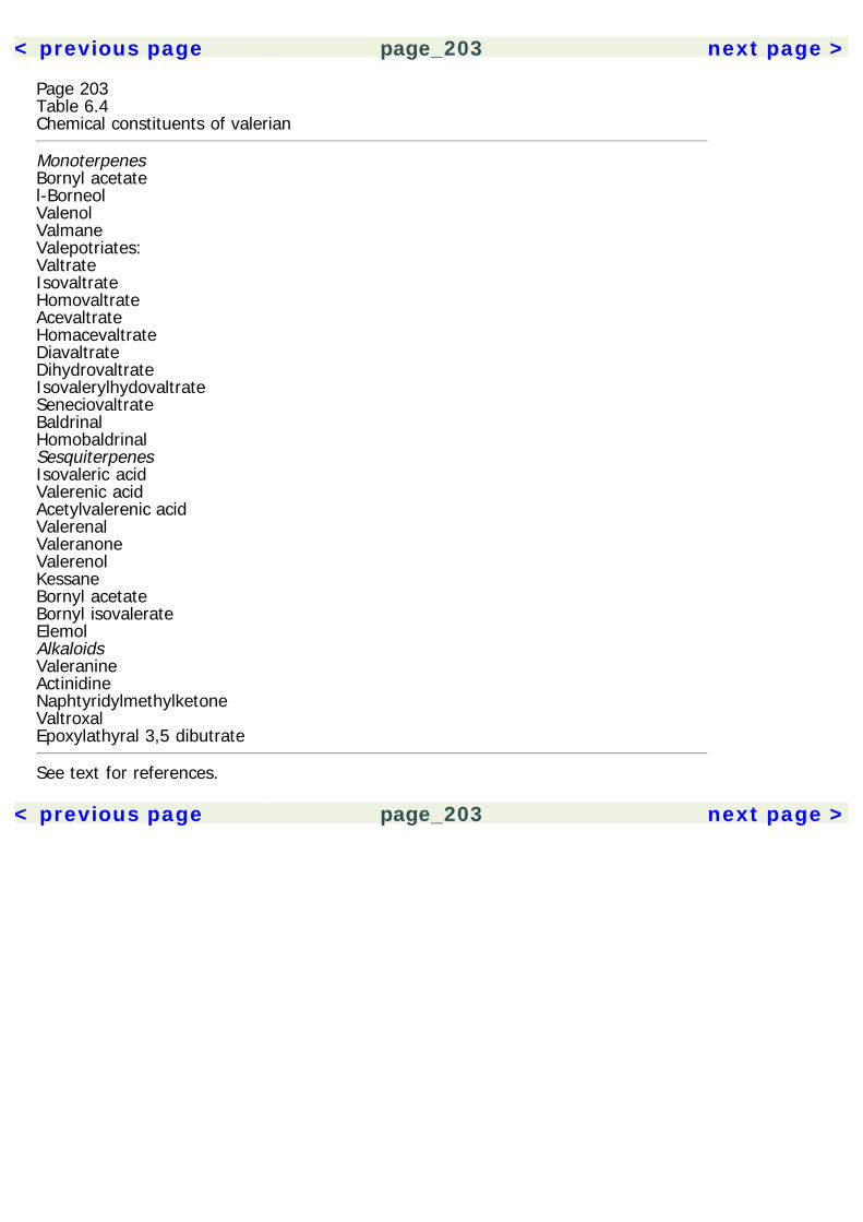
< previous page page_203 next page >
Page 203Table 6.4Chemical constituents of valerian
MonoterpenesBornyl acetatel-BorneolValenolValmaneValepotriates:ValtrateIsovaltrateHomovaltrateAcevaltrateHomacevaltrateDiavaltrateDihydrovaltrateIsovalerylhydovaltrateSeneciovaltrateBaldrinalHomobaldrinalSesquiterpenesIsovaleric acidValerenic acidAcetylvalerenic acidValerenalValeranoneValerenolKessaneBornyl acetateBornyl isovalerateElemolAlkaloidsValeranineActinidineNaphtyridylmethylketoneValtroxalEpoxylathyral 3,5 dibutrate
See text for references.
< previous page page_203 next page >
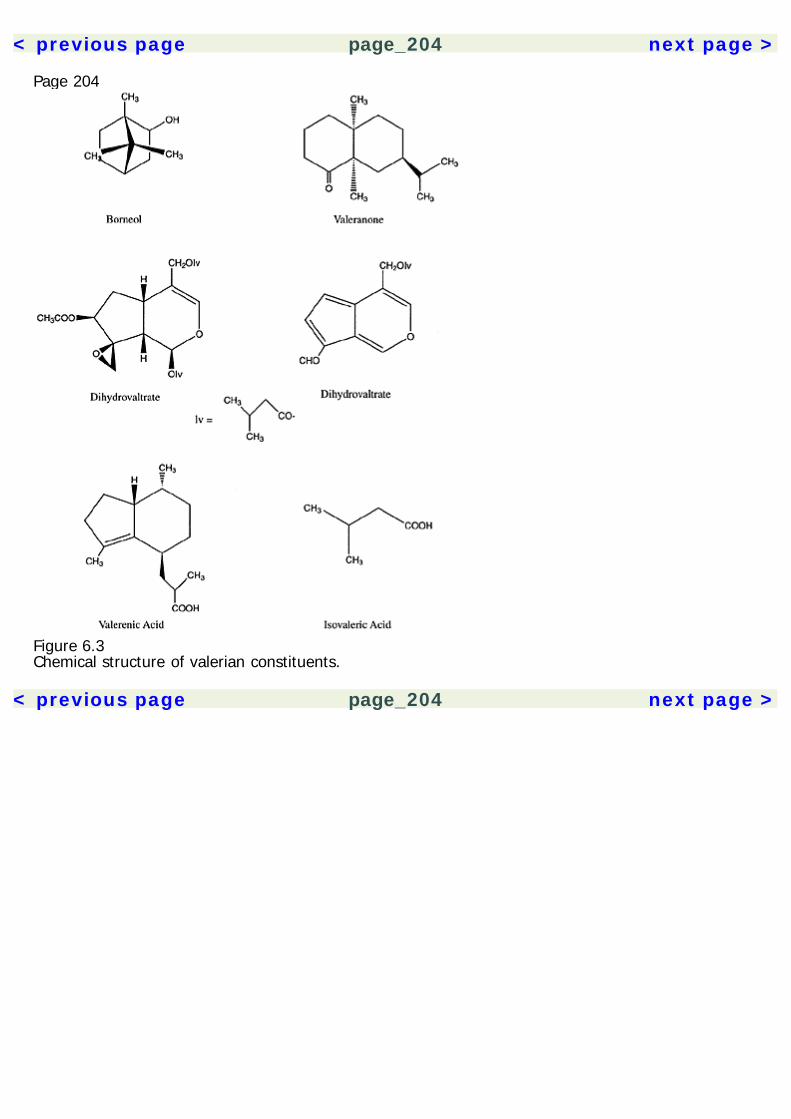
< previous page page_204 next page >
Page 204
Figure 6.3Chemical structure of valerian constituents.
< previous page page_204 next page >

< previous page page_205 next page >
Page 205in vitro (Santos et al. 1994c). Alternately, because GABA does not pass the blood-brain barrier well, itis possible that the glutamine content of valerian extract contributes to this effect because it bothcrosses the blood-brain barrier and serves as a precursor for GABA.Although some binding of valerian extract occurs at GABAA receptors, this has been attributed to theamino acid content of the extract and deemed insufficient by itself to explain valerian's CNSdepressant effects (Cavadas et al. 1995). Valerenic acid has been shown to inhibit enzymaticbreakdown of GABA (Riedel et al. 1982; Houghton 1999). Low concentrations of valerian extractsenhance benzodiazepine binding ([3H]flunitrazepam) (Ortiz et al. 1999). The valepotriates maythemselves have CNS depressant effects through in vivo conversion to homobaldrinal (Houghton1999). Further, an ethanol extract of valerian potentiates the effects of a barbiturate (thiopental) andinhibits seizures induced by the GABAA antagonist picrotoxin (Hiller and Zetler 1996).Collectively, valerian extracts show a variety of GABAergic mechanisms. The predominant mechanismhas yet to be determined, but an additive or synergistic interaction among the several mechanisms ispossible. Future research on valerian extracts needs to address any compositional differencesbetween aqueous and nonaqueous extracts, and to determine the responsible active constitutents.For some time it was thought to be the valepotriates, but this was later discredited due to theirunstable nature.Physiological EffectsMuscle relaxant effects In vitro effects of valepotriates (isovaltrate and valtrate) and valeranone wereinvestigated in the guinea pig ileum (Hazelhoff et al. 1982). It produced relaxation of the ileum,probably by a direct muscular action and not by action on autonomic receptors. The GABAergiceffects of valerian may provide central muscle relaxant effects through spinal actions, but this has yetto be explicity investigated.Behavioral effects Several behavioral effects of valerian have been noted in animals. These includesuppression of the orienting response in an open-field paradigm, decreasing spontaneous andcaffeine-induced motor
< previous page page_205 next page >
< previous page page_206 next page >
Page 206activity, and potentiation of the behavioral actions of barbiturates (Dunaev et al. 1987). Oral andintravenous doses of valtrate and acetoxyvaltrate, more specifically, were found to reduce locomotoractivity (Hölzl and Fink 1984).Valerian extracts show sedative and anxiolytic effects. Whereas passionflower and chamomile haverelatively specific anxiolytic effects, valerian shows more general sedative effects, but all effects occurin a dose-dependent manner (Della Logia et al. 1981; Leuschner et al. 1993). The sedative effects ofvalerian extract are moderate when compared to diazepam and the neuroleptic chlorpromazine(Leuschner et al. 1993). However, valepotriates reverse the anxiogenic effects of diazepamwithdrawal in rats in the elevated plus maze. This effect is dose dependent, effective at 12 mg/kgbut not 6 mg/kg. Interestingly, the fragrant valerian compound bornyl acetate has sedative effects inmice, but only when inhaled (Buchbauer et al. 1992).In a manner similar to imipramine, an ethanol extract of valerian root was found to preventimmobility induced by a forced-swimming test in rats, suggesting a potential antidepressant effect ofvalerian (Sakamoto et al. 1992).Antiseizure effects Valerian extracts were found to have weak antiseizure effects in mice (Dunaev etal. 1987; Leuschner et al. 1993). This would be consistent with its putative GABAergic mechanisms.So the traditional use of valerian to treat epilepsy is perhaps an empirical reality. However, nostudies have been done to date that compare valerian to modern antiseizure medications.Sleep electrophysiology To date, four studies have been published that examine the effects ofvalerian on human sleep and its electrophysiology, which have produced variable results. An earlystudy (Muller-Limmroth and Ehrenstein 1977) showed that a combined preparation of valerian andhops in sleep-disturbed subjects increased the amount of slow-wave and REM sleep. Another studyusing an aqueous extract (400 mg) of valerian did not show significant EEG effects, but suggested arelation between the EEG effects and subjective effects, that is, shortened sleep
< previous page page_206 next page >
< previous page page_207 next page >
Page 207latency and increased latency to first waking (Leathwood and Chauffard 1982–3, 1985). Anotherstudy using aqueous extracts showed no effects on sleep stages or EEG spectra (Balderer andBorbely 1985).A methodologically controlled study of valerian in sleep was published that utilized both double-blindand placebo controls, as well as randomization (Gessner and Klasser 1984). Also employed were twodoses of valerian (60 and 120 mg), computerized EEG power spectral analysis and psychometricmood questionnaires. Valerian increased sleep stages 1, 2, and 3 and reduced stage 4 and REM.Dose-dependent effects were noted, where the 120 mg dose produced greater sedative effects. Peakeffects occurred 2–3 hours after administration. Mood ratings did not differ, positively or negatively,between the experimental and control conditions.The effects of valerian in poor sleepers was studied comparing it to placebo controls (Schulz et al.1994). Valerian showed an increase in slow-wave and a decrease in stage 1 sleep. K-complex densitywas increased, but REM was unaltered, and no effects were reported on subjective sleep quality.The physiological effects of valerian across animals and humans are consistent with its sedativeproperties. The human electrophysiological findings are somewhat incongruent, but the methodologymust be taken into account. Earlier studies used less-rigorous controls, and one used aqueousextract vs. the entire herb, so that hydrophobic constituents were likely excluded. Such differenceshave been observed with other sedative herbs such as kava (Jamieson et al. 1989). Anotherdifference between the two latter studies that may account for differing results is the differentpopulations used (normal versus sleep-disturbed) (Gessner and Klasser 1984; Schulz et al. 1994).Indeed, poor-quality sleepers of various etiologies are noted to have sleep EEG patterns that differfrom normals (Kupfer et al. 1976; Bourdet and Goldenberg 1994; Morisson et al. 1998; Adam et al.1986).The EEG effects of valerian are most concordant with those of tiagabine, a GABA uptake inhibitorused to treat epilepsy. Tiagabine also increases slow-wave sleep, but unlike valerian has little or noeffect on REM (Lancel et al. 1998). GABAA agonists also tend to have this effect,
< previous page page_207 next page >

< previous page page_208 next page >
Page 208in contrast to GABAB and benzodiazepine agonists (Faulhaber et al. 1997; Lancel and Faulhaber1996; Lancel et al. 1996; Juhász et al. 1994).Sleep quality A randomized, placebo-controlled, and double-blind crossover study was employedusing a commercially available valerian preparation (Lindahl and Lindwall 1989). The preparationused contained primarily sesquiterpenes, and very low amounts of valepotriates. The subjects were27 adults who were seen in a medical clinic for sleep difficulties. Those receiving valerianexperienced improvements in sleep quality (89% of subjects), with a proportion (44%) rating highestquality sleep. No differences were seen between those who received either valerian or placebo first.An absence of adverse side effects, including nightmares, was reported. Although these results arepromising, this study unfortunately used a preparation that included two other herbs (lemon melissaand flores humuli), which limit the conclusions that can be exclusively drawn with valerian. Anothermethodologically well-controlled study was performed, which had a much larger (n = 128) number ofsubjects (Leathwood et al. 1982). Improvements were seen in sleep quality and decreases in sleeplatency. Furthermore, sleep quality was most improved in poor sleepers and tobacco smokers.Thus, the above two studies plus the one conducted by Gessner and Klassner (1984; see above)constitute three methodologically controlled studies of valerian on human sleep. By establishedguidelines, this constitutes Class I evidence for valerian in the treatment of insomnia (Woolf 1992).Cognitive effects Studies of the cognitive effects of valerian are scant. One study compared theeffects of valerian to those of the benzodiazepine flunitrazepam, and to a combined preparation ofvalerian and hops (Gerhard et al. 1996). Whereas flunitrazepam produced significant impairment themorning after administration, valerian (alone or in combination with hops) did not. However, 1 to 2hours after administration, valerian produced a slight but statistically significant decrease in vigilanceand processing of complex information. Although mild, this effect
< previous page page_208 next page >
< previous page page_209 next page >
Page 209may contraindicate valerian use in situations where peak cognitive performance is required (e.g.,driving).ToxicityBecause of their chemical structure, there has been concern over liver damage resulting fromvalepotriates. Baldrinal and homobaldrinal were noted to have some mutagenic effects in vitro (vonder Hude et al. 1986). However, Valeriana officinalis has a low valepotriate content—they decomposerapidly in the stored herb and they are not well absorbed. Liver toxicity in animals or humans hasnever been demonstrated (Tyler 1994; Lindahl and Lindwall 1989). Thus, the valepotriate content ofvalerian is not likely to cause toxicity. No health hazards have been reported with normal use ofvalerian, and it has been approved by the German Commission E as a treatment for anxiety andsleep (Gruenwald et al. 1998).High doses of valerian are reported to produce headache, vomiting, stupor, dizziness, and cardiacdysfunction (Kowalchick and Hylton 1987; Gruenwald et al. 1998). A case was reported of a singleoverdose (approximately 20 times the ''recommended" therapeutic dose) that produced reportedlymild symptoms, which resolved in 24 hours. One other study published the results of overdoses of"Sleep-Qik," an over-the-counter herbal sedative (Chan et al. 1995). However, in addition to valerian,the preparation included cyproheptadine hydrochloride and hyoscine hydrobromide, a potentanticholinergic drug. Given its CNS depressant effects and putative GABAergic mechanisms,concurrent use of valerian with other CNS depressants, including ethanol, should be avoided.SummaryThere is preliminary but promising empirical evidence that valerian is a useful medication forimproving sleep (tables 6.5, 6.6). Although the responsible constituents are uncertain, valerian likelyacts through facilitation of central GABAergic transmission. Further, its pharmacological mechanismssuggest it may be useful in treating anxiety or other mood disorders, but there have not been anycontrolled studies to support this.
< previous page page_209 next page >
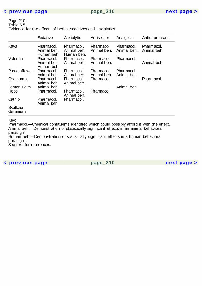
< previous page page_210 next page >
Page 210Table 6.5Evidence for the effects of herbal sedatives and anxiolytics
Sedative Anxiolytic Antiseizure Analgesic Antidepressant
Kava Pharmacol. Pharmacol. Pharmacol. Pharmacol. Pharmacol. Animal beh. Animal beh. Animal beh. Animal beh. Animal beh. Human beh. Human beh. Valerian Pharmacol. Pharmacol. Pharmacol. Pharmacol. Animal beh. Animal beh. Animal beh. Animal beh. Human beh. Passionflower Pharmacol. Pharmacol. Pharmacol. Pharmacol. Animal beh. Animal beh. Animal beh. Animal beh. Chamomile Pharmacol. Pharmacol. Pharmacol. Pharmacol. Animal beh. Animal beh. Lemon Balm Animal beh. Animal beh. Hops Pharmacol. Pharmacol. Pharmacol. Animal beh. Catnip Pharmacol. Pharmacol. Animal beh. Skullcap Geranium
Key:Pharmacol.—Chemical contituents identified which could possibly afford it with the effect.Animal beh.—Demonstration of statistically significant effects in an animal behavioralparadigm.Human beh.—Demonstration of statistically significant effects in a human behavioralparadigm.See text for references.
< previous page page_210 next page >
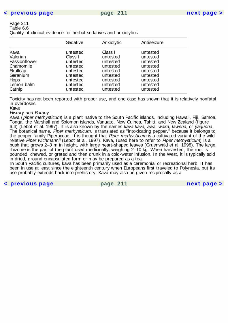
< previous page page_211 next page >
Page 211Table 6.6Quality of clinical evidence for herbal sedatives and anxiolytics
Sedative Anxiolytic Antiseizure
Kava untested Class I untestedValerian Class I untested untestedPassionflower untested untested untestedChamomile untested untested untestedSkullcap untested untested untestedGeranium untested untested untestedHops untested untested untestedLemon balm untested untested untestedCatnip untested untested untested
Toxicity has not been reported with proper use, and one case has shown that it is relatively nonfatalin overdoses.KavaHistory and BotanyKava (piper methysticum) is a plant native to the South Pacific islands, including Hawaii, Fiji, Samoa,Tonga, the Marshall and Solomon islands, Vanuato, New Guinea, Tahiti, and New Zealand (figure6.4) (Lebot et al. 1997). It is also known by the names kava kava, awa, waka, lawena, or yaquona.The botanical name, Piper methysticum, is translated as ''intoxicating pepper," because it belongs tothe pepper family Piperaceae. It is thought that Piper methysticum is a cultivated variant of the wildrelative Piper wichmannii (Lebot et al. 1997). Kava, (used here to refer to Piper methysticum) is abush that grows 2–3 m in height, with large heart-shaped leaves (Gruenwald et al. 1998). The largerhizome is the part of the plant used medicinally, weighing 2–10 kg. When harvested, the root ispounded, chewed, or grated and then drunk in a cold-water infusion. In the West, it is typically soldin dried, ground encapsulated form or may be prepared as a tea.In South Pacific cultures, kava has been primarily used as a ceremonial or recreational herb. It hasbeen in use at least since the eighteenth century when Europeans first traveled to Polynesia, but itsuse probably extends back into prehistory. Kava may also be given reciprocally as a
< previous page page_211 next page >
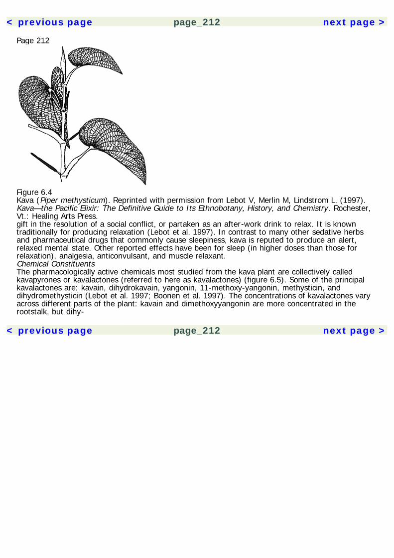
< previous page page_212 next page >
Page 212
Figure 6.4Kava (Piper methysticum). Reprinted with permission from Lebot V, Merlin M, Lindstrom L. (1997).Kava—the Pacific Elixir: The Definitive Guide to Its Ethnobotany, History, and Chemistry . Rochester,Vt.: Healing Arts Press.gift in the resolution of a social conflict, or partaken as an after-work drink to relax. It is knowntraditionally for producing relaxation (Lebot et al. 1997). In contrast to many other sedative herbsand pharmaceutical drugs that commonly cause sleepiness, kava is reputed to produce an alert,relaxed mental state. Other reported effects have been for sleep (in higher doses than those forrelaxation), analgesia, anticonvulsant, and muscle relaxant.Chemical ConstituentsThe pharmacologically active chemicals most studied from the kava plant are collectively calledkavapyrones or kavalactones (referred to here as kavalactones) (figure 6.5). Some of the principalkavalactones are: kavain, dihydrokavain, yangonin, 11-methoxy-yangonin, methysticin, anddihydromethysticin (Lebot et al. 1997; Boonen et al. 1997). The concentrations of kavalactones varyacross different parts of the plant: kavain and dimethoxyyangonin are more concentrated in therootstalk, but dihy-
< previous page page_212 next page >
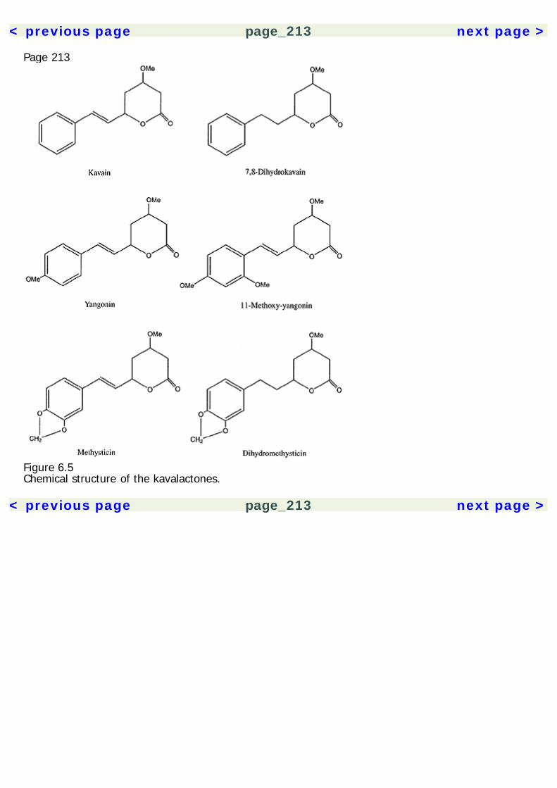
< previous page page_213 next page >
Page 213
Figure 6.5Chemical structure of the kavalactones.
< previous page page_213 next page >

< previous page page_214 next page >
Page 214drokavain and dihydromethysticin are found in higher concentrations in the stalks and leaves (Cassand McNally 1998). Kavain and dihydrokavain are reportedly the most permeable to the blood-brainbarrier.Mechanisms of ActionLike many psychotropic herbs, Kava seems to create its effects by a combination of effects that aremediated by several of its constituents. To date, at least four mechanisms have been identified thatappear to mediate the effects of kava.GABA Kava appears to facilitate GABA transmission. Low micromolar (0.1–1 μM) concentrations ofkavalactones [(+)-kavain, (+)dihydrokavain, (+)-methysticin, and (+)-dihydromethysticin] enhancethe binding of ligands to the GABAA receptor (Boonen and Heberlein 1998; Jussofie et al. 1994). Thisfacilitation would potentiate binding of GABA and increase Cl– influx. Some structure-activityrelationships were noted to increase the efficacy of kavalactones, such as the addition of an aromaticmethoxy group and angular lactone ring. Kavalactones do not alter the binding of flunitrazepam, sotheir effect on GABAA is not through the benzodiazepine receptor (Davies et al. 1992). Therefore,kavalactones and benzodiazepines similarly facilitate GABAA activity, but apparently through differentmechanisms. Convergently, kavalactone displacement of the GABAA agonist muscimol is highest inthe hippocampus and amygdala (Jussofie et al. 1994). Combination with pentobarbital causedsynergistic effects on binding.Voltage-gated ion channels A second mechanism by which kava produces CNS depression is byinhibition of voltage-gated ion channels (Gleitz et al. 1995; Gleitz et al. 1996a; Magura et al. 1997;Schirrmacher et al. 1999). Kavain has a fast and specific inhibitory action on Na+ channels. (+)-methysticin was 4–5 times more potent than (±)-kavain in this respect (Magura et al. 1997). Kavain,dihydrokavain, and dihydromethysticin act as noncompetitive inhibitors of the Na+ channel (Frieseand Gleitz 1998). It has been proposed that (+)-methysticin and (±)-kavain bind to the Na+ channelin its inactivated state and prolong the inactivation (Magura et al. 1997). Kavain blocks the[3H]batrachotoxin binding, but not [3H]saxitoxin binding (Gleitz et al. 1996a).
< previous page page_214 next page >
< previous page page_215 next page >
Page 215This suggests an action at receptor site 2 of the Na+ channel, a site common to local anestheticdrugs.It was further shown that kavain inhibits L-type Ca2+ channels as well, and the subsequent releaseof glutamate (Gleitz et al. 1996b; Ferger et al. 1998; Schirrmacher et al. 1999). At concentrations of70 μM/L, glutamate release was reduced by 70%. The effects of kavain and dihydromethysticin werefound to be additive when both were administered in vitro (Walden et al. 1997). These effects onvoltage-gated ion channels are obtained in the micromolar range, and are consistent withconcentrations reached in the brain by peripheral administration (Keledjian et al. 1988). Further,many of these studies examined individual kavalactones, which does not account for the additive orsynergistic effects of combined kavalactones.Monoamine Kavalactones block the reuptake of norepinephrine with the following order of potency:(±)-kavain = (+)-kavain > (+)-methysticin (Seitz et al. 1997). However, neither acute nor chronicdoses of kavalactones alter turnover of serotonin. The effects of kavalactones were far less potentthan desipramine, and only occurred at concentrations of less than 10 μM. However, this study onlyused a single kavalactone in vitro, ruling out additive and synergistic actions of combinedadministration. Another potential monoamine mechanism of kavalactones is inhibition of MAOB(Uebelhack et al. 1998). Kavalactones reversibly inhibited platelet MAOB in the micromolar range(IC50 1.2–24 μM). The order of potency for MAOB inhibition is: desmethoxyyangonin > (±)-methysticin > yangonin > (±)-dihydromethysticin > (±)-dihydrokavain > (±)-kavain. The two mostpotent, desmethoxyyangonin and (±)-methysticin, both showed competitive inhibition and mean Kiof 0.28 μM and 1.14 μM, respectively. This would be consisent with a slow onset of MAO inhibitionby kavalactones. In three subjects given 120–240 mg of kavalactones for 3–4 weeks, a 26–34%reduction of MAOB in platelet-rich plasma was observed (Uebelhack et al. 1998).Neither an acute dose of dihydromethysticin (100 mg/kg) or chronic doses of (±)-kavain alteredlevels of dopamine, serotonin, or their metabolites in the striatum and cortex of rats (Boonen et al.1998). However, kavalactones have complex and mixed effects on monoamine levels in the nucleusaccumbens, depending both on which kavapyrone was
< previous page page_215 next page >
< previous page page_216 next page >
Page 216used and at what dose (Baum et al. 1998). D,L-kavain had biphasic effects on dopamine, decreasingit at low doses (20 mg/kg), and increasing or not altering it at high doses (120 mg/kg). Similarly,desmethyoxyyangonin caused increases in dopamine levels in the accumbens. In contrast, yangoninpotently decreased dopamine levels to below the limits of detection. Other kavalactones(dihydrokavain, methysticin, and dihydromethysticin) had no effect on dopamine levels. Kavain alsocaused a decrease in serotonin levels. Baum and colleagues suggest that the elevations in dopamineby kavain are due to blocking reuptake of cytoplasmic dopamine into vesicles, because large amountsof DOPAC were excreted. This proposed mechanism is similar to that of the drug reserpine, exceptthat unlike reserpine the actions of kavalactones are reversible.The interactions of kava with monoamines are complex, apparently varying with which dose, whichkavalactone is used, and which neuroanatomical region is examined. Isolated findings with individualkavalactones in isolated brain regions are insufficient to explain the actions of kava unless they areintegrated in a larger picture. The neurophysiological and neurochemical effects of kava extract needto be further investigated when administered to the whole organism. For example, althoughincreases in dopamine levels in the accumbens have been found with certain kavalactones, otherlines of evidence point to a sum antagonistic effect on central dopamine. Cases of dyskinesiasuggestive of dopamine antagonism have been reported (Schelosky et al. 1995). Kava extracts alsoantagonize apomorphine-induced stereotypies in mice (Jamieson et al. 1989).Thus, although the mechanism of interaction with monoamine is uncertain, peripheral doses of kavado have functional effects on central monoamine systems (Baum et al. 1998).Eicosanoid Finally, (+)-kavain was reported to inhibit cyclooxygenase, suppressing synthesis ofthromboxane A2 in the high micromolar range (Gleitz et al. 1997). Thromboxanes are prostaglandinssynthesized from arachidonic acid (see chapter 7). Although they are more commonly known fortheir hemodynamic effects, they are also present in the brain. Their function in the CNS has not beenwell delineated, but they have been shown to inhibit GABAA receptors. So, suppression of thrombox-
< previous page page_216 next page >
< previous page page_217 next page >
Page 217ane A2 synthesis would increase GABAA function (Schwartz-Bloom et al. 1996). Again, because onlyone kavalactone was examined, whether combined kavalactones or extract could create aphysiologically significanct effect remains to be determined. In order for central eicosanoid effects tobe relevant, it would likely require additive or synergistic effects of combined kavalactones.Summary of mechanisms of action Of the above-mentioned mechanisms, GABA facilitation andblockade of voltage-gated cation channels would best account for the sedative properties of kava.Other drugs that facilitate GABA transmission (e.g., benzodiazepines, barbiturates) are well known fortheir sedative and anxiolytic effects. Such activity in the amygdala could particularly account foranxiolytic effects (Jussofie et al. 1994; Sanders and Shekhar 1995; Davis et al. 1994). Inhibition ofvoltage-gated cation channels depresses neuronal function and can create sedation as well. Indeed,many antiseizure medications (e.g., hydantoins, carbamazepine) have such actions, and are knownfor their sedating side effects. In contrast, the blocking of norepinephrine reuptake by kava would beexpected to have alerting effects, based on animal and human studies of noradrenergic function(Aston-Jones et al. 1994; Rajkowski et al. 1994). This effect needs to be examined with combinedkavalactones or total extract to confirm its physiological relevance. However, it is interesting thatkava is known for creating a relaxed but alert state (Cass and McNally 1998).The functional significance of thromboxane A2 inhibition by kava is not certain. More needs to beunderstood about the functions of prostaglandins in the CNS, but given the inhibition of GABAA bythromboxane A2, kava's suppression of thromboxane A2 synthesis could conceivably contribute to itsanxiolytic and sedative effects. More research is needed to determine if this mechanism of action isphysiologically significant at normal kavalactone concentrations.PharmacokineticsPharmacological effects of kavalactones are estimated to occur at plasma concentrations of 50–150μM/L (Kretzschmar and Meyer 1968). Kavalactones peripherally administered quickly gain access tothe CNS (Keled-
< previous page page_217 next page >
< previous page page_218 next page >
Page 218jian et al. 1988). Five minutes after a 100 mg/kg bolus injection (IP) in mice, the following brainconcentrations of kavalactones were obtained (ng/mg): dihydrokavain (64.7 ± 13.1), kavain (29.3 ±0.8), desmethoxyyangonin (10.4 ± 1.5), and yangonin (1.2 ± 0.3). These accumulate and areeliminated at different rates: whereas kavain and dihydrokavain accumulate and are eliminatedquickly, yangonin and desmethoxyyangonin do so slowly. Metabolites of kavalactones are detectablein human urine (Duffield et al. 1989).A pharmacokinetic synergy occurs with kavalactones. When a kava resin is administered (containingcombined kavapyrones), levels of kavain and yangonin in the brain are multiplied 2 and 20 times,respectively, as compared to individual administration (Keledjian et al. 1988). Although themechanism of this synergy is uncertain, it may be due to competition for plasma binding sites. Somereport that these synergistic effects occur with yangonin and desmethoxyyangonin and otherkavalactones, implying an influence on intestinal absorption. Thus, results from studies usingindividual kavalactones must be generalized cautiously, because they do not account for themagnified effects of the naturalistic combined administration of kavalactones.The preparation of kava used may greatly affect its effectiveness. A comparison was made betweenaqueous and lipid-soluble extracts of kava in mice (Jamieson et al. 1989). The lipid-soluble extractproduced sedation, analgesia, and local anesthesia, while the aqueous extract was ineffective orally.There is evidence to suggest that limited tolerance to kava develops (Duffield and Jamieson 1991). Akava extract parenterally administered daily for three days produced little physiological or behavioraltolerance in mice. However, kava extract and ethanol were found to interact, potentiating eachother's sedative effects and increasing the toxicity of kava (Jamieson and Duffield 1990). Similarly,synergistic interactions occur between kavalactones and barbiturates (Jussofie et al. 1994).Physiological EffectsPhysiological studies of kava constituents have focused on several of their effects: muscle relaxant,sedative, analgesic, antiseizure, and neuroprotective.
< previous page page_218 next page >
< previous page page_219 next page >
Page 219Neuromuscular effects The muscle relaxant effect of kava was studied in mouse and frog muscles. Itproduced direct neuromuscular effects, reducing the amplitude of end-plate potentials and miniatureend-plate potentials, and depressed muscle action potentials. This effect was not reversed byapplication of Ca2+. It was concluded that the mechanism of neuromuscular relaxation in kava wassimilar to that of local anesthetics: by a direct blocking effect on ion channels of the muscle.Analgesia Analgesic effects of kava components have been investigated in mice (Jamieson andDuffield 1990). Kava extract and kavalactones were both found to effectively produce analgesia thatwas not mediated by opioid mechanisms, because it was not reversed by naloxone. Althoughspeculative, the GABAergic and monoamine actions of kava could account for its analgesic effects.Neuroprotective effect At last, kavain has been examined for its neuroprotective effect. Theneuroprotective effect is probably mediated by its blocking of voltage-gated Na+ and Ca2+ channels.Similar to other neuroprotective Na+ channel blockers (i.e., tetrodotoxin), kavain inhibits themetabolic consequences of anoxia in neurons (Gleitz et al. 1996b). Kava was found to diminish thevolume and area of infarcted tissue in mice, which was probably mediated by methysticin anddihydromethysticin (Backhauss and Krieglstein 1992).Electrophysiological and cognitive effects The effects of kava on brain electrophysiology wereinvestigated in cats (Holm et al. 1991). Kavain produces high-amplitude delta activity, spindlelikeformations, and continuous alpha or beta activity in the amygdala. Similar to barbiturates, kavainprolongs synchronized sleep.The effects of kavain on human electrophysiology was examined in a double-blind, placebo-controlled study (Frey 1991). Dose-dependent increases were seen in delta, theta, and alpha 1power, and decreases ocurred in alpha 2 and beta power. These changes were suggestive of asedative effect of kavain, and were maximal in frontal areas. Interestingly, an initial activating effectwas seen at the lowest dose (200 mg) but not at the largest dose (600 mg).
< previous page page_219 next page >
< previous page page_220 next page >
Page 220Event-related potentials (ERPs) were used to study the cognitive effects of kava, as compared tooxazepam (Münte et al. 1993). In a double-blind crossover study, subjects were given a verbalmemory task (word recognition). While oxazepam impaired performance, kava actually improvedperformance. Whereas oxazepam caused a poorer recognition rate and reduced theelectrophysiological difference between old and new words, kava increased the new/old differenceand led to a slightly improved recognition rate. Another double-blind study used ERPs to examine theeffects of a kava root extract on visual search (Heinze et al. 1994). Again, whereas oxazepamreduced task-associated potentials in frontal, parietal, and occipital areas, kava was associated withincreases.A study of the cognitive effects of kava on psychometric tests was undertaken by Foo and Lemon(1997). They employed a number of measures to assess attention, reaction time, visuomotortracking, and short-term memory. Of several measures given, kava only produced minor effects onthe digit symbol task, but no other tests of attention or concentration. Unlike ethanol, kava producedno effects on subjective measures of impairment given to the subjects. However, when giventogether, kava and ethanol potentiated each other's effects on subjective and objective cognitivemeasures.Antiseizure effects Kavalactones have been investigated for their antiseizure effects in animals(Kretzschmar et al. 1969; Jamieson et al. 1989; Gleitz et al. 1995, 1996a). This action appears to bemediated by suppression of voltage-gated ion channels, although a contribution from their GABAergicor noradrenergic activity could contribute. Inhibition of ion channels and facilitating of GABA activityare mechanisms shared by many antiseizure medications. Kavain was found to reduce excitatoryactivity in hippocampal slices, but not to affect long-term potentiation or synaptic plasticity (Langoschet al. 1998). However, kava extracts were found to only have a weak effect on strychnine-inducedseizures (Jamieson et al. 1989). Concentrations of 10–100 μM/L of methysticin had antiseizure effectsin slices of hippocampus and entorhinal cortex.Although there is some evidence for an antiseizure effect of kava, it is not clear whether these effectsextend to humans. There have not been any studies that have demonstrated its effectiveness inclinically control-
< previous page page_220 next page >
< previous page page_221 next page >
Page 221ling seizures. Individuals who are being treated for epilepsy are urged not to attempt to replace theircurrent medications with kava, and especially not without the direction of a physician.Anxiolytic effects Several methodologically controlled studies have been performed to assess the anti-anxiety effects of kava. The studies employed standardized doses of kavalactones ranging from 60 to240 mg per day, and were 4 to 24 weeks in duration (Pittler and Ernst 2000). Subjects presentedwith anxiety symptoms that were diagnosed with criteria from the DSM-III-R, and/or severalpsychometric measures of anxiety (Hamilton Anxiety Scale and Adjective Check List [of Janke andDebus], Anxiety Status Inventory and Zung's Self-Rating Anxiety Scale, and State-Trait AnxietyInventory). A review and meta-analysis of methodologically controlled studies of kava for treatinganxiety was undertaken by Pittler and Ernst (2000). They found kava to be superior to placeboacross all seven controlled studies. A meta-analysis of three studies that used the Hamilton AnxietyScale found a significant reduction in anxiety.Some studies found kava to be effective using only 2 acute doses (preoperatively), while othersfound benefit after chronic doses (Bhate et al. 1989; Lehmann et al. 1989; Volz and Kieser 1997).Another interesting study found kavain to be equivalent in efficacy to the benzodiazepine oxazepamon standard measures of anxiety (Anxiety Status Inventory and Zung's Self-Rating Anxiety Scale)(Lindenberg and Pitule-Schodel 1990). There were no serious side effects reported in the clinicalstudies reviewed by Pittler and Ernst (2000).ToxicityNo health hazards are known with the proper use of kava (Gruenwald et al. 1998). Kava has beenapproved by the German Commission E for treatment of anxiety and insomnia. In clinical studies ofkava for anxiety, adverse effects were uncommon and did not differ across placebo and kava groups.There do not appear to be any studies published on the effects of acute overdosage with kava. Givenits CNS depressant effects, it should not be taken with other similar drugs, includingbenzodiazepines, barbiturates,
< previous page page_221 next page >
< previous page page_222 next page >
Page 222ethanol, or antiseizure medications. Animal research shows an interaction between kava and ethanol,increasing the toxicity of kava (Jamieson and Duffield 1990). There is one clinical report of combinedadministration of kava and the benzodiazepine alprazolam (Almeida and Grimsley 1996). A 54-year-old man taking the two presented in a disoriented and lethargic state that resolved after severalhours. The use of kava during pregnancy has not been formally studied.Kava dermopathy A scaly skin eruption called kava dermopathy is reported to occur from kava use(Norton and Ruze 1994). It is reversible, and appears to only occur with heavy chronic use. Asuspected contribution of niacin deficiency to kava dermopathy was ruled out (Ruze 1990).Parksinsonism Four cases were reported of dyskinesia presenting with kava use in individuals(Schelosky et al. 1995). These consisted of dystonia, tonic head rotation, twisting of the trunk,oculogyric crises, and increased duration of ''off" periods in a parkinsonian patient. These symptomssubsided with discontinuation of kava and treatment with a cholinergic muscarinic antagonist(biperiden). The authors suggest that this represents a dopamine antagonist action of kava, andraise caution about its use in the elderly.SummaryKava is an herbal drug known for centuries through the South Pacific islands, with therapeutic, social,and cultural uses. Its chemical constituents, the kavalactones, create psychoactive effects by alteringcentral GABA transmission, blocking voltage-gated ion channels (Na+ and Ca2+), monoaminemechanisms, and possibly eicosanoid systems. Although kavalactones create significant effectsindividually, they exhibit pharmacokinetic synergy, reaching higher brain concentrations whenadministered collectively. Kava's long-reputed effect for creating relaxation is supportedpharmacologically, and a few controlled studies have confirmed its usefulness in treating anxiety.Formal studies on improving sleep have not been conducted, but are warranted. There appears to belittle toxicity with proper use of kava. Perhaps one of the most useful
< previous page page_222 next page >
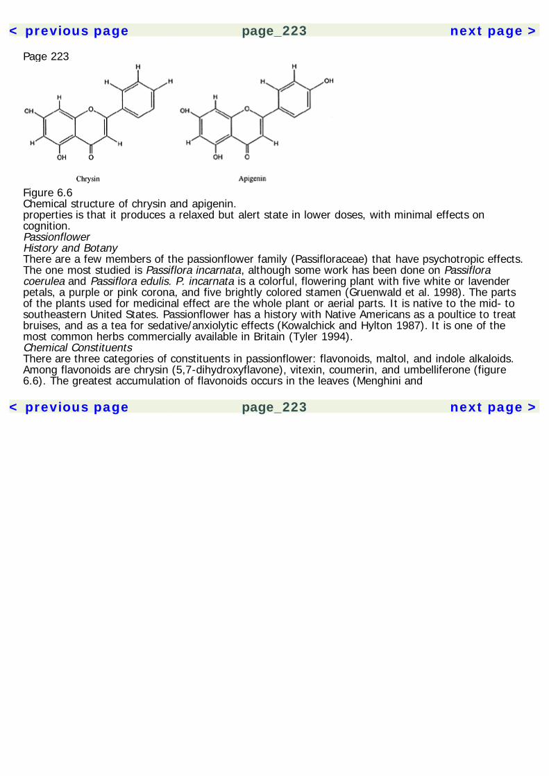
< previous page page_223 next page >
Page 223
Figure 6.6Chemical structure of chrysin and apigenin.properties is that it produces a relaxed but alert state in lower doses, with minimal effects oncognition.PassionflowerHistory and BotanyThere are a few members of the passionflower family (Passifloraceae) that have psychotropic effects.The one most studied is Passiflora incarnata, although some work has been done on Passifloracoerulea and Passiflora edulis. P. incarnata is a colorful, flowering plant with five white or lavenderpetals, a purple or pink corona, and five brightly colored stamen (Gruenwald et al. 1998). The partsof the plants used for medicinal effect are the whole plant or aerial parts. It is native to the mid- tosoutheastern United States. Passionflower has a history with Native Americans as a poultice to treatbruises, and as a tea for sedative/anxiolytic effects (Kowalchick and Hylton 1987). It is one of themost common herbs commercially available in Britain (Tyler 1994).Chemical ConstituentsThere are three categories of constituents in passionflower: flavonoids, maltol, and indole alkaloids.Among flavonoids are chrysin (5,7-dihydroxyflavone), vitexin, coumerin, and umbelliferone (figure6.6). The greatest accumulation of flavonoids occurs in the leaves (Menghini and
< previous page page_223 next page >

< previous page page_224 next page >
Page 224Mancini 1988). Flavonoids vary across the ontogenic cycle, with the highest concentration ofisovitexin occurring between preflowering and flowering stages. The indole alkaloids are smallamounts (up to 0.01%), including harman, harmine, harmaline, and harmalol (Tyler 1994).Mechanisms of ActionPerhaps the most studied constituent of passionflower is the flavonoid chrysin. Chrysin was isolatedfrom P. coerulea, a species closely related to P. incarnata. It binds to benzodiazepine receptors withmicromolar affinity (Ki = 3 μM) and competes for binding with the benzodiazepine flunitrazepam(Medina et al. 1990). Behavioral assays (see below) suggest that chrysin acts as a partial agonist atcentral benzodiazepine receptors (Wolfman et al. 1994).Other flavones in passionflower (vitexin, coumerin, umbelliferone) have not been well characterizedfor their neuropharmacological action. Vitexin (derived from millet in one study) was found to haveantithyroid properties. The remaining constituents, maltol and harman alkaloids, are present in smallquantities and not presumed to contribute to the psychotropic effects (Meier 1995). Harman alkaloidsin greater quantities are hallucinogenic and presumed to work through 5-HT receptors (Grella et al.1998).Physiological EffectsSedative and anxiolytic effects A number of flavonoids have been shown to bind to benzodiazepinereceptors and have anxiolytic effects (Medina et al. 1997). The anxiolytic effects of chrysin wereexamined in mice (Wolfman et al. 1994). Chrysin (1 mg/kg IP) reduces behavioral measures ofanxiety (elevated-plus maze) in a manner similar to diazepam (0.3–0.6 mg/kg), which was reversedby pretreatment with a benzodiazepine antagonist, Ro 15-1788. The anxiolytic effect is not likely dueto sedation because there is no concurrent reduction in motor activity at the doses used. Unlikediazepam, chrysin does not produce muscle relaxation at higher doses.The sedative and anxiolytic effects of passionflower were examined in two other animal behavioralassays (staircase test, light/dark box choice test). Both anxiolytic and sedative effects occur, as wellas potentiation
< previous page page_224 next page >

< previous page page_225 next page >
Page 225of pentobarbital sedation, at 400 mg/kg of hydroalcoholic extract in mice (Soulimani et al. 1997). Theanxiolytic and sedative activity occur in a dose-dependent continuum (Della Logia et al. 1981).Passiflora edulis has sedative effects as well (Maluf et al. 1991).Despite neuropharmacolgical and animal data to support sedative and anxiolytic effects ofpassionflower, there have not been any such controlled studies in humans. Two studies have beenpublished that examined the effects of combined herbal extracts on anxiety, including passionflower(Bourin et al. 1997). Although there were significant and experimentally controlled effects, acombined herbal treatment confounds the ability to selectively identify the effects of passionflower. Asecond controlled study was similarly confounded by the use of a three-herb combination (Gerhard etal. 1991).General physiological effects Physiological and electrophysiological changes in response to chronicpassionflower treatment were studied in the rat (Sopranzi et al. 1990). There was no change in bodyweight, core body temperature, motor coordination, or analgesia (as measured by the tail-flick test).There was some general reduction of activity (in the one-arm radial maze), but the EEG appearednormal.Cognitive effects A study of the effects of chrysin on memory was performed in the rat (Salgueiro etal. 1997). Chrysin does not have any amnestic effects on either acquisition or retention in three testsof memory (inhibitory avoidance, shuttle avoidance, and habituation to an open field), even at higherdoses than required to produce anxiolytic effects. The cognitive effects of passionflower have notbeen examined in humans.Analgesic effects Although pharmaceutical benzodiazepine drugs and certain flavones have analgesiceffects, chrysin and vitexin do not (Salgueiro et al. 1997; Rylski et al. 1979). However, an extract ofPassiflora incarnata, administered orally or IP did raise nociceptive thresholds, raising the possibilityof nonchrysin analgesic flavones present in P. incarnata (Speroni and Minghetti 1988). Chrysin wasshown to reduce morphine withdrawal in a physiological assay (guinea pig ileum contraction) in adose-dependent manner (Capasso et al. 1998).
< previous page page_225 next page >

< previous page page_226 next page >
Page 226Antiseizure effects One study examined antiseizure effects of chrysin on chemically induced(pentylenetetrazol) seizures in mice (Medina et al. 1997). Peripheral (IP) administration producedinconsistent effects, but central (intracerebroventricular) injection (40 μg) had a significantanticonvulsant effect. Further, this effect was abolished by prior injection of the benzodiazepineantagonist, Ro 15-1788 (3 mg/kg IP).ToxicityThere have been no formal studies of the toxicity of passionflower, but adverse effects have not beenreported. There is one report of a case of inflammatory vasculitis associated with a preparation ofpassionflower (Smith et al. 1993). Like other herbs in this category, its putative benzodiazepineaction contraindicates its combined use with other CNS depressants.ChamomileHistory and BotanyChamomile refers to two similar species of plants: German chamomile (Matricaria recutita) andRoman Chamomile (Chamaemelum nobile), both of which are members of the Asteraceae family(figure 6.7). C. nobile is a low-growing perennial herb, while M. recutita is a tall, annual plant(Kowalchick and Hylton 1987). Chamomile has been used throughout history, including ancientEgyptian, Roman, and Greek cultures. The two species look very much like daisies, with white petalsand a yellow central disc. They are both native to Europe, Africa, and Asia and have been naturalizedin North America. The most common form of ingestion is to dry the flowering tops and brew them ina tea. Although there are many reported uses of chamomile, it is best known for its antispasmodic,anti-inflammatory, and mild calming effects (Leyel 1994; Gruenwald et al. 1998).Chemical ConstituentsChamomile contains the terpenoids (–)-alpha-bisabolol, (–)-alpha-bisabololoxides A and B, and agauianolide lactone called matricin. Also contained are the flavonoids apigenin and apigenin-7-glucoside (see figure 6.6) (Tyler 1994; Robbers et al. 1996). Apigenin was found
< previous page page_226 next page >
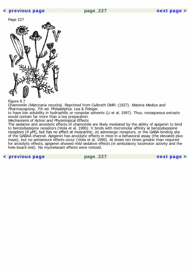
< previous page page_227 next page >
Page 227
Figure 6.7Chamomile (Matricaria recutita). Reprinted from Culbreth DMR. (1927). Materia Medica andPharmacognosy, 7th ed. Philadelphia: Lea & Febiger.to have low solubility in hydrophilic or nonpolar solvents (Li et al. 1997). Thus, nonaqueous extractswould contain far more than a tea preparation.Mechanisms of Action and Physiological EffectsThe sedative and anxiolytic effects of chamomile are likely mediated by the ability of apigenin to bindto benzodiazepine receptors (Viola et al. 1995). It binds with micromolar affinity at benzodiazepinereceptors (4 μM), but has no effect at muscarinic, α1 adrenergic receptors, or the GABA binding siteof the GABAA channel. Apigenin has anxiolytic effects in mice in a behavioral assay (the elevated plusmaze), but no antiseizure effects occur (Viola et al. 1995). At doses ten times greater than requiredfor anxiolytic effects, apigenin showed mild sedative effects (in ambulatory locomotor activity and thehole-board test). No myorelaxant effects were noticed.
< previous page page_227 next page >

< previous page page_228 next page >
Page 228A study of the effects of apigenin on memory was performed in the rat (Salgueiro et al. 1997). Itdoes not show any amnestic effects on either acquisition or retention in three tests of memory(inhibitory avoidance, shuttle avoidance, and habituation to an open field), even at doses higher thanthose required to produce anxiolytic effects.Studies of apigenin on the rat thoracic aorta have found apigenin to have vasodilatory effects (Ko etal. 1991). This effect was probably mediated by an inhibition of voltage- and ligand-gated Ca2+channels. It would be interesting to see if this effect also occurs in the CNS.There is some evidence for apigenin actions on the monoamine systems. Apigenin was found tostimulate uptake of tyrosine into adrenal chromaffin cells in a concentration-dependent manner,resulting in increased monoamine production (Morita et al. 1990). Apigenin was also found to be acompetitive inhibitor of a protein-tyrosine kinase (p40) (Kellis and Vickery 1984). Finally, apigeninwas found to inhibit MAO, and to inhibit uptake of norepinephrine in the rat atria (Lorenzo et al.1996). Again, it would be interesting to see if these effects extend to the CNS and chamomile'spsychotropic effects, especially when administered peripherally, and if they have behaviorallysignificant actions in the amounts derived from common over-the-counter preparations.There is some literature that examines flavonoids such as apigenin as ligands for the estrogen betareceptor, and as an inhibitor of the enzyme estrogen synthetase (aromatase) (Kuiper et al. 1998;Kellis and Vickery 1984). These compounds do so potently, but the behavioral significance ofamounts observed in common preparations needs to be further investigated.Apigenin has been found to inhibit release of histamine (Middleton and Drzewiecki 1984). Althoughinhibition of histamine by apigenin could potentially have psychotropic effects, it would need to bedemonstrated that this effect occurs in the CNS (Loring and Meador 1989; Kim et al. 1996). This maynot be the case, because apigenin's effect on histamine release is dependent on immunoglobulinantigens (Middleton and Drzewiecki 1984).Thus, the best candidate for chamomile's psychotropic effects is the activity of apigenin atbenzodiazepine receptors. There are other potential mechanisms that may contribute to its effects,but they need to be further investigated for the magnitude of their contribution.
< previous page page_228 next page >
< previous page page_229 next page >
Page 229ToxicityChamomile appears very low in toxicity. It has been listed as Generally Regarded as Safe (GRAS) bythe Food and Drug Administration (Tyler 1994). Adverse reactions may include allergic reactions tothe pollen in the flowers (Kowalchick and Hylton 1987; Subiza et al. 1990).Remaining Sedative and Anxioltyic HerbsThere are several remaining herbs that are reputed to have sedative effects: skullcap, geranium,hops, and catnip. However, the neuropharmacological mechanisms have not been well characterized,and behavioral data in animals and humans are lacking. The existing knowledge on these plants ispresented here.CatnipCatnip (Nepeta cataria) is a coarse green-grey leaved perennial plant with spotted white tubularflowers (Kowalchick and Hylton 1987). It has a long recorded history of use, extending back toancient Roman civilization. Among its many reported uses, catnip is noted for sedative properties.The active agent for this effect is uncertain, but catnip has in it several terpenes, includingnepetalactone. One terpene, cis-trans-nepetalactone was hypothesized to be responsible for thesedative effect based on its structural similarity to valepotriates, the depressant principles fromvalerian.Paradoxically, catnip fed to mice had stimulant effects, with increased rearing, locomotion, andstereotypical behavior, increased susceptibility to chemically induced (picrotoxin and strychnine)seizures, and decreased sleeping time after barbiturate administration (Massoco et al. 1995). TheLD50 for nepetalactone in mice was reported to be quite high at 1300 mg/kg (Harney et al. 1978). Inchicks, an alcohol extract of catnip had biphasic effects, where low to moderate doses (25–1800mg/kg) produced sedative effects, while higher doses (>2 g/kg) had less sedative and perhapsstimulant effects (Sherry and Hunter 1979). Humans have reported sedative effects of catnip, andone accidental ingestion by a young child reportedly produced sedative effects (Osterhoudt et al.1997).
< previous page page_229 next page >
< previous page page_230 next page >
Page 230The inconsistencies in the effects of catnip could be attributed to a few possible causes. As Sherryand Hunter illustrated (1979), the effects could be biphasic and thus dose dependent. Anotherpossibility is interspecies differences. A more dramatic illustration of this is in the reaction that catshave to catnip (Siegel 1989). Cats will sniff, lick, and chew the leaves of the plant, followed by shortperiods of blank staring into space and shaking the head from side to side. Next they will rub theirbody against the plant and some fall over and roll in it. When introduced to catnip, cats will laterreturn to the site where they encountered it. These behaviors indicate a pleasurable state, and bothmale and females may adopt mating postures with erections occurring in the males. Nepetalactonesare believed to mimic a scent in cat urine that triggers sexual behaviors. The sexual nature of thefeline response to catnip is suggestive of pheromonal activity at the vomeronasal organ (VNO). TheVNO is a chemical sense organ found in the nasal sinuses that is involved in sexual behaviors andhormonal effects, and not the sense of smell (Halpern 1987). Counter-intuitively though, the effectsof catnip on felines is mediated by the main olfactory system, and not the VNO (Hart and Leedy1985).There was some association of hallucinogenic effects with catnip in humans, particularly throughadministration by smoking. This was reported in a 1969 paper, but apparently resulted fromconfusion with cannabis (Tyler 1994). Any reports of hallucinogenic or sedative effects of catnip inhumans, at this point, are purely anecdotal.HopsHop (Humulus lupulus) is a flowering vine that grows in Europe, Western Asia, and North America,and is reported to have anxiolytic/sedative effects (figure 6.8) (Kowalchik and Hylton 1987). It is aningredient of beer, but is also consumed as a tea. The female flowers are placed in a pillow to treatinsomnia by their fragrance. It has not been well studied, but the responsible agent is believed to be2-methyl-3-butene-2-ol, because it produces sedation when injected intraperitoneally in mice (Hänselet al. 1980). In humans, one study found no CNS depressant effects when administered orally(Hänsel and Wagener 1967). However, the effects produced by inhalation (thus avoiding digestionand absorption issues), have not been studied.
< previous page page_230 next page >
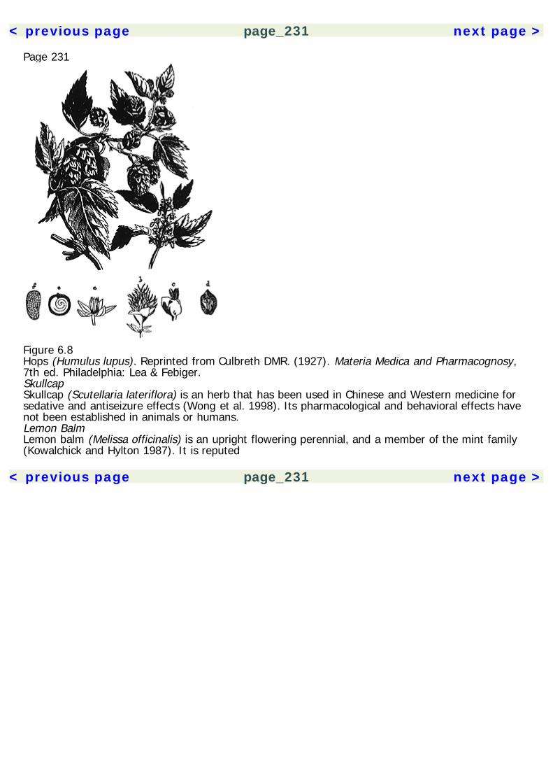
< previous page page_231 next page >
Page 231
Figure 6.8Hops (Humulus lupus). Reprinted from Culbreth DMR. (1927). Materia Medica and Pharmacognosy,7th ed. Philadelphia: Lea & Febiger.SkullcapSkullcap (Scutellaria lateriflora) is an herb that has been used in Chinese and Western medicine forsedative and antiseizure effects (Wong et al. 1998). Its pharmacological and behavioral effects havenot been established in animals or humans.Lemon BalmLemon balm (Melissa officinalis) is an upright flowering perennial, and a member of the mint family(Kowalchick and Hylton 1987). It is reputed
< previous page page_231 next page >
< previous page page_232 next page >
Page 232to have anxiolytic effects in humans, but no formal studies have been done. In mice, lemon balm hassedative effects in familiar (staircase test) and unfamiliar (two-compartment test) environments, andto have analgesic activity (writhing test) (Soulimani et al. 1991). It also increased the sedativeactivity of a barbiturate (pentobarbital). Although these effects are promising, further work would beneeded to identify active constituents and confirm whether effects occur in humans.GeraniumThere are several members of the Geranium family (Geraniaceae), some of which have beenanecdotally reported to have anxiolytic or sedative effects. However, there is a paucity of research tosupport the use of this herb as a CNS depressant (Manolov et al. 1977; Tasev et al. 1969).Summary and ConclusionsThere are several herbs with CNS depressant effects that have been used mostly for anxiolytic andsedative effects historically. This has been supported by neuropharmacological, animal, and humanstudies (table 6.1). In some cases, the herbs meet Class I criteria for therapeutic treatment (table6.4). Much work remains to be done in further testing the efficacy of these drugs and more firmlydelineating their biochemical mechanisms.Several other herbs have weaker evidence for CNS depressant effects, but hold potential for furtherresearch and evaluation. Certain herbs have also had historical use for treating epilepsy, before theinvention of several modern, very effective synthetic antiseizure medications. There is, at present,absolutely no evidence that the CNS depressant herbs will be effective or sufficient in the treatmentof epilepsy. Much further research would be needed to support this contention. Further, withdrawalof current antiseizure medications would put an individual at high risk for recurrence or worsening ofseizures.
< previous page page_232 next page >

< previous page page_233 next page >
Page 2337Psychotherapeutic HerbsLong before the advent of modern pharmaceutical drugs, several herbal medications were employedfor the alleviation of mental disorders. Some of these are considered impractical by today's standardsfor treatment of psychopathology. For a short time, Freud advocated the use of cocaine to alleviatedepression. Cannabis and opium were used to treat agitation. Other herbal medications are beingsupported by scientific research and seriously considered by the medical community. Some areoffered here more for historical interest, and yet others have only recently added psychotherapeuticvalue to their list of potential benefits.As will be discussed in detail here, these herbs may create several effects, due to multipleneuropharmacological actions or even due to a single action. For the sake of convenience, the herbshave been broadly classified here into antidepressant/anxiolytic and antipsychotic categories due totheir frequently overlapping effects.The Psychopharmacological EraThe twentieth century has witnessed a revolution in the discovery of drugs for the treatment ofmental illness (Baldessarini 1996; Lickey and Gordon 1991). Formerly, many drug treatments wereeither ineffective or nonspecific. The first modern psychopharmacological treatment came from theAustralian psychiatrist John F. J. Cade, who proposed the use of lithium salts to treat bipolardisorder. Although slow to gain acceptance, lithium has become a standard treatment. In 1950,chlorpromazine was synthesized in France and successfully tested for treatment of psychoticagitation. The first antidepressant drug, iproniazid, was originally
< previous page page_233 next page >
< previous page page_234 next page >
Page 234developed to treat tuberculosis and serendipitously found to have antidepressant effects in the late1950s. Meprobamate was introduced as an anxiolytic in the mid-1950s. Although it has fallen intodisuse, it heralded the discovery of a series of modern sedative and anxiolytic drugs.The discovery of psychopharmacological medications was revolutionary because they provided ameans of treating illnesses that were otherwise intractable. With the exception of electroconvulsivetreatments for severe depression, there were no medical treatments for disorders that did notrespond to psychotherapy. Once established, the drugs led to an ongoing search for more effectiveand safer medications. A second reason for their revolutionary status is that they furtheredunderstanding of mental illnesses and normal brain function. Investigations of their therapeuticmechanisms led to theories of the neurochemical bases of mental illnesses.The advent of clincal psychopharmacology had an enormous socioeconomic impact as well. Thenumber of patients residing in psychiatric hospitals had been steadily increasing in the United Statesto a peak of 550,000 following World War II (Lickey and Gordon 1991). Since the introduction ofpsychotherapeutic medications, there has been a steady decline to 111,000 by 1986. Drugtreatments allowed for briefer hospital stays. Unfortunately, there was a gap in social services forsome time, when adequate follow-up care was not provided to the mentally ill who were releasedfrom hospitals but still not functionally independent. Nonetheless, the discovery of psychotherapeuticdrugs has alleviated unquantifiable amounts of human suffering. With careful research, some herbalmedications may add to this effort.Depression and AnxietyMajor depression is one of the most common mental disorders. It has a lifetime prevalence of 17%in the general population, and a current prevalence of 4.9%, constituting a significant public healthproblem (Blazer et al. 1994). It is characterized by intense sadness and cognitive impairments mostnotable in concentration, worry, pessimism, and lowered self-esteem (American PsychiatricAssociation 1994). Interpersonal relationships may suffer and social withdrawal occurs. Physio-
< previous page page_234 next page >
< previous page page_235 next page >
Page 235logical changes are common in homeostatic functions such as appetite, sleep, sex drive, andhormonal levels. Although sadness and grief are normal emotions, major depression is a pathologicalcondition that is distinguished by its duration and severity. Unlike normal emotions, major depressionlacks the adaptive benefits that normal emotions provide, and causes a resulting impairment in socialand occupational functioning.Anxiety disorders are a collection of heterogeneous disorders including panic disorder, phobias,obsessive-compulsive disorder, posttraumatic stress disorder, and generalized anxiety disorder (APA1994). What they all share is anxiety as a principal symptom—in greater frequency or degree thannormal experience and leading to behavioral maladjustment. Anxiety includes a subjective componentwith feelings of dread, emotional tension, and worry. Behavioral manifestations include avoidace ofanxiety-producing situations, motor impairment, and cognitive impairment (e.g., inability toconcentrate). Physiological manifestations of anxiety include muscle tension and sympatheticautonomic reactions such as increased heart rate and blood pressure, sweating, dry mouth, nausea,dizziness, and diarrhea. Although low levels of anxiety are adaptive, their severity, frequency, andduration make them maladaptive in anxiety disorders.Pharmaceutical Antidepressants and AnxiolyticsThere are numerous antidepressant medications on the market (table 7.1). Following development ofmonoamine oxidase (MAO) inhibitors were tricyclic antidepressants, selective serotonin reuptakeinhibitors (SSRIs), and several atypical antidepressants (Baldessarini 1996). Successive generations ofantidepressants have not necessarily become more effective in treating depression, but rather offermore favorable side-effect profiles—a crucial factor in effective clinical pharmacotherapy. An effectivemedication is not useful if its side effects are intolerable.The variety of antidepressant agents developed all seem to share one commonality: alteration ofmonoamine systems (Bolden-Watson and Richelson 1993; Bonhomme and Esposito 1998). Only veryrecently have drugs that work outside of monoamine systems been shown to have reliableantidepressant effects.
< previous page page_235 next page >
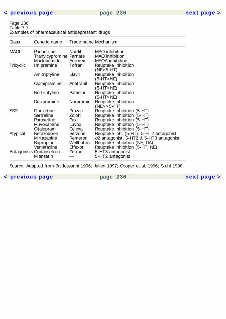
< previous page page_236 next page >
Page 236Table 7.1Examples of pharmaceutical antidepressant drugs
Class Generic name Trade name Mechanism
MAOI Phenelzine Nardil MAO inhibition Tranylcypromine Parnate MAO inhibition Moclobemide Aurorex MAOA inhibitionTricyclic Imipramine Tofranil Reuptake inhibition
(NE>5-HT) Amitriptyline Elavil Reuptake inhibition
(5-HT>NE) Clomipramine Anafranil Reuptake inhibition
(5-HT>NE) Nortriptyline Pamelor Reuptake inhibition
(5-HT>NE) Desipramine Norpramin Reuptake inhibition
(NE>>5-HT)SSRI Fluoxetine Prozac Reuptake inhibition (5-HT) Sertraline Zoloft Reuptake inhibition (5-HT) Paroxetine Paxil Reuptake inhibition (5-HT) Fluvoxamine Luvox Reuptake inhibition (5-HT) Citalopram Celexa Reuptake inhibition (5-HT)Atypical Nefazodone Serzone Reuptake inh. (5-HT), 5-HT2 antagonist Mirtazapine Remeron α2 antagonist, 5-HT2 & 5-HT3 antagonist Bupropion Wellbutrin Reuptake inhibition (NE, DA) Venlafaxine Effexor Reuptake inhibition (5-HT, NE)Antagonists Ondansetron Zofran 5-HT3 antagonist Mianserin — 5-HT2 antagonist
Source: Adapted from Baldessarini 1996; Julien 1997; Cooper et al. 1996; Stahl 1998.
< previous page page_236 next page >

< previous page page_237 next page >
Page 237The MAO inhibitors irreversibly bind to mitochondrial MAO, preventing oxidative deamination of themonoamines dopamine, norepinephrine, and serotonin. These bind to both forms of MAO (MAOAand MAOB) and include the drugs phenelzine, tranylcypromine, and isocarboxazid (Baldessarini1996). These drugs also prevent hepatic breakdown of tyramine (a substance found in many foodssuch as aged cheese, red wine, and figs) by MAOB, which can precipitate a hypertensive crisis. Anewer class of reversible, short-acting MAO inhibitors (e.g., moclobemide) are being investigated thatare selective to MAOA, and thus do not have the same tyramine toxicity problems. Thus, theantidepressant effect of MAO inhibitors is believed to lie in their ability to increase availablemonoamines.Tricyclic drugs have, as the name implies, a three-ring structure, and interfere with reuptake ofnorepinephrine and/or serotonin into axon terminals. Tricyclic drugs include imipramine (Tofranil),amitriptyline (Elavil), clomipramine (Anafranil), and nortriptyline (Pamelor, Aventil). Tricyclics havethe occasional but unfortunate cardiovascular side effects of arrhythmia and postural hypotension.Newer, nontricyclic antidepressants have been developed that are collectively referred to as SSRIs.These have a potent and selective action on serotonin, and lack the cardiovascular side effects of thetricyclics. These include fluoxetine (Prozac), paroxetine (Paxil), sertraline (Zoloft), and fluvoxamine(Luvox). A fifth SSRI, citalopram (Celexa) has been used in Europe and has recently been approvedin the United States. Venlafaxine (Effexor) blocks reuptake of norepinephrine and serotonin, whilebupropion (Wellbutrin) acts on both dopamine and norepinephrine.Mirtazapine (Remeron) is a newer antidepressant that also blocks 5-HT reuptake, but additionally hasantagonistic effects at adrenergic α2, 5-HT2, and 5-HT3 receptors (Stahl 1998). Mirtazapine appearsto have indirect agonistic effects on 5-HT1A receptors, which may contribute to its antidepressanteffect (Berendsen and Broekkamp 1997). Nefazodone, as well, has SSRI and 5-HT2 antagonisteffects. The 5-HT2 antagonist effects of these antidepressants is believed to be responsible for theirlower incidence of sexual side effects (Nutt 1997).In addition to acute effects, chronic use of antidepressants causes chronic receptor adaptations. Forexample, venlafaxine blocks reuptake
< previous page page_237 next page >

< previous page page_238 next page >
Page 238of serotonin and norepinephrine and chronically causes a reduction of β1 adrenergic receptors infrontal cortex (McGrath and Norman 1998). Imipramine causes alterations in serotonin release inboth raphe nuclei and frontal cortex (Maione et al. 1997). Both imipramine and fluoxetine alter theexpression of dopamine receptors (Dziedzicka-Wasylewska et al. 1997). Chronic administration ofSSRIs and MAO inhibitors desensitizes 5-HT1A and 5-HT1B/1D receptors (Bonhomme and Esposito1998; Le Poul et al. 1997). Others have shown a tonic activation of 5-HT1A receptors after chronictreatment with antidepressant drugs (Maione et al. 1997; Haddjeri et al. 1998). Although there doesnot appear to be a single unifying hypothesis that accounts for all of the effects created by thediverse mechanisms of antidepressant drugs, increased function in monoamine systems and receptormodulation are consistent findings (Bonhomme and Esposito 1998; Maione et al. 1997).In summary, the antidepressant drugs discussed here all have a common effect on monoaminesystems, either by preventing enzyme degradation, blocking reuptake, or direct receptor effects. Alsocommon to all of these drugs is the lag of 2–3 weeks for therapeutic effects to begin after treatmentis started. It is likely that the actual antidepressant mechanism of these medications is the receptoralterations. Reuptake occurs immediately after taking the drug, but the therapeutic effects correlatewell with chronic receptor adaptations.There is a high comorbidity of depression and anxiety, suggestive of common neurobiologicalsubstrates (Rodney et al. 1997; Flint 1994; Gorman 1996–97; Enns et al. 1998; Eison MS 1990;Montgomery 1983; Paul 1988). Therefore, it is not surprising that pharmacological treatments thataddress one disorder may have ameliorative effects on the other. Indeed, some antidepressants havebeen tested for effectiveness in treating generalized anxiety disorder and were found to be aseffective (trazodone) or better (imipramine) than diazepam (Rickels et al. 1993; Schweizer et al.1998).Other serotonergic drugs that are direct receptor agonists or antagonists have been found to haveanxiolytic effects (Stahl 1998; Bonhomme and Esposito 1998). A novel class of anxiolytic drugs calledazapirones act as partial agonists at 5-HT1A receptors (Yocca 1990). Clinically, they are representedby BuSpar, which was approved for use in 1986 (Eison
< previous page page_238 next page >
< previous page page_239 next page >
Page 239AS 1990). Several 5-HT1A agonists have been found to produce anxiolytic effects in animal modelsand in human clinical trials (File et al. 1996; Krummel and Kathol 1987; Goldberg and Finnerty 1979).Furthermore, they have antidepressant effects; they augment the antidepressant effects of serotoninreuptake inhibitors, and they decrease the therapeutic latency (Bouwer and Stein 1997; Artigas et al.1996; Sussman 1998; Rickels et al. 1991; Wieland and Lucki 1990; Jenkins et al. 1990; Fabre 1990).However, results have not been uniformly positive, such as in refractory severe depression (Sussman1998; Fischer et al. 1998)Drugs acting as antagonists at 5-HT2 receptors have been found to have antidepressant andanxiolytic effects, including the antidepressant nefazodone (Deakin 1988; Martin and Lemonnier1994; Graeff et al. 1996). Mianserin is a more selective 5-HT2 antagonist, which is undergoing theFDA drug approval process. It has been shown to have antidepressant and anxiolytic effects (Granieret al. 1985; Brogden et al. 1978; Murphy 1978). 5-HT3 antagonists, such as ondansetron, have beendeveloped as antiemetic drugs, primarily due to the role of 5-HT3 receptors in emesis (Costall andNaylor 1992). 5-HT3 receptors involved in this effect are found in the human brain stem (areapostrema, dorsal vagal complex) and enteric nervous system. However, 5-HT3 receptors are alsolocated in the hippocampus, amygdala, nucleus accumbens and basal ganglia (Parker et al. 1996;Abi-Dargham et al. 1993). 5-HT3 antagonists have shown some antidepressant and nonsedatinganxiolytic effects in animal models, and hold potential for application to humans (Martin et al. 1992;Costall et al. 1990; Bloom and Morales 1998). There is also some physiological and anecdotalevidence to suggest that 5-HT3 antagonists might serve as adjuncts to pharmacotherapy ofschizophrenia (Gerlach 1991; Greenshaw and Silverstone 1997; Greenshaw 1993).One final mechanism that has recently shown promise for antidepressant effects appears distinctfrom all of the monoamine systems. A study published in 1998 demonstrated that a substance Pantagonist (MK-869) had antidepressant effects in humans (Kramer et al. 1998). Substance P islocalized to many areas of the brain that are involved in emotional processing, including theamygdala, dentate gyrus, subiculum, nucleus accumbens, striatum, locus coeruleus andperiaqueductal grey (Kramer
< previous page page_239 next page >
< previous page page_240 next page >
Page 240et al. 1998; Quirion et al. 1983). Substance P antagonists have shown antidepressant effects inanimal models, and levels of substance P are reduced in forebrain areas after chronic treatment withantidepressants (Kramer et al. 1998, Shirayama et al. 1996). Conversely, withdrawal of chronicantidepressant treatment increases sensitivity to substance P in the cingulate gyrus (Jones and Olpe1984).Although several putative anxiolytic herbs are discussed in chapter 6, a select few are discussed herebecause of their similarity to antidepressant medications in terms of neuropharmacological activity,and because of the close relationship between depression, anxiety, and their pharmacologicaltreatment in general. Whereas the anxiolytic herbs discussed previously have anxiolytic effectsthrough more-general CNS depressant actions, the ones discussed here seem to have more-specificactions, particularly on serotonin.Psychotic DisordersSchizophrenia is a group of psychotic disorders characterized by several key disturbances: cognitive,emotional, and behavioral (American Psychiatric Association 1994; Goodwin and Guze 1996).Symptoms have been traditionally divided into positive and negative. Positive symptoms areexcesses, which include hallucinations and delusions. Negative symtoms consist of deficits inpsychological function. Among these are such cognitive disturbances as difficulties in logical andabstract thinking, attention, thought organization, lack of volition, affective flattening, and socialwithdrawal.The duration and nature of the psychotic disorder present determines which diagnosis is given:schizophrenia, schizophreniform, or brief psychotic disorder. Schizoaffective disorder is diagnosedwhen there is a significant concurrent mood disturbance such as major depression, mania, or bipolardisorder. Delusional disorder is a condition where nonbizarre delusions persist for at least one month,but other symptoms of schizophrenia are not present. Subtypes of schizophrenia are recognized,which are defined by their predominant symptoms. These include paranoid, disorganized, andcatatonic types. Catatonic schizophrenia is characterized by maintaining unusual postures for longperiods of time.
< previous page page_240 next page >
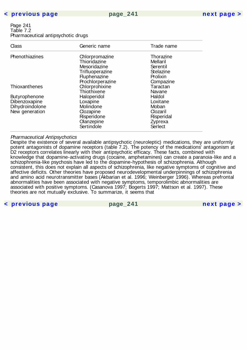
< previous page page_241 next page >
Page 241Table 7.2Pharmaceutical antipsychotic drugs
Class Generic name Trade name
Phenothiazines Chlorpromazine Thorazine Thioridazine Mellaril Mesoridazine Serentil Trifluoperazine Stelazine Fluphenazine Prolixin Prochlorperazine CompazineThioxanthenes Chlorprohixine Taractan Thiothixene NavaneButyrophenone Haloperidol HaldolDibenzoxapine Loxapine LoxitaneDihydroindolone Molindone MobanNew generation Clozapine Clozaril Risperidone Risperidal Olanzepine Zyprexa Sertindole Serlect
Pharmaceutical AntipsychoticsDespite the existence of several available antipsychotic (neuroleptic) medications, they are uniformlypotent antagonists of dopamine receptors (table 7.2). The potency of the medications' antagonism atD2 receptors correlates linearly with their antipsychotic efficacy. These facts, combined withknowledge that dopamine-activating drugs (cocaine, amphetamines) can create a paranoia-like and aschizophrenia-like psychosis have led to the dopamine-hypothesis of schizophrenia. Althoughconsistent, this does not explain all aspects of schizophrenia, like negative symptoms of cognitive andaffective deficits. Other theories have proposed neurodevelopmental underpinnings of schizophreniaand amino acid neurotransmitter bases (Akbarian et al. 1996; Weinberger 1996). Whereas prefrontalabnormalities have been associated with negative symptoms, temporolimbic abnormalities areassociated with positive symptoms. (Casanova 1997; Bogerts 1997; Mattson et al. 1997). Thesetheories are not mutually exclusive. To summarize, it seems that
< previous page page_241 next page >

< previous page page_242 next page >
Page 242genetic predispositions and prenatal insults result in aberrant brain development involving prefrontaland temporolimbic areas. The functional activity of these regions and neurotransmitter systems arealtered, resulting in the symptomatology of schizophrenia.Whatever the underlying causes may be, neuroleptic medications are the most effective treatment forschizophrenia. All antipsychotic medications have some form of dopamine receptor antagonism andthey are distinguished by their chemical class. The phenothiazines include chlorpromazine(Thorazine), thioridazine (Mellaril), mesoridazine (Serentil), trifluoperazine (Stelazine), fluphenazine(Prolixin), and prochlorperazine (Compazine). The thioxanthenes include chlorprohixine (Taractan)and thiothixene (Navane). Butyrophenones are represented by haloperidol (Haldol). Loxapine(Loxitane) is a dibenzoxapine, and molindone (Moban) is a dihydroindolone.A newer generation of antipsychotics have been introduced in the 1990s that have the dualadvantages of reduced extrapyramidal effects and amelioration of negative symptoms. These drugsinclude clozapine (Clozaril), risperidone (Risperidal), and olanzepine (Zyprexa). Pharmacologically,they antagonize 5-HT2A receptors as well as dopamine receptors. Sertindole (Serlect) is yet anothernew generation antipsychotic that has a variety of dopamine and serotonin antagonist effects. Withthe discovery of dopamine receptors beyond the D1/D2 dichotomy, many antipsychotics were foundto have effects there too. For example, clozapine has less efficacy at the D2 receptor, but strongereffects at D4 receptors. Haloperidol is an antagonist at D3 as well as D2 receptors.Side EffectsAntipsychotic drugs vary according to their side-effect profiles regarding sedation, autonomic effects,and extrapyramidal motor symptoms. Sedation and autonomic effects are mediated, in part, by themuscarinic cholinergic and alpha adrenergic effects of the drugs. While the binding to D2 receptorspredicts dosage and efficacy for antipsychotic drugs, it also predicts extrapyramidal motor symptoms(Seeman 1987). These include akathisia (or motor restlesness), dystonic postures, and parkinsonism(resting tremor, bradykinesia, and limb rigidity). Due to the functional relationship between dopamineand acetylcholine in the basal
< previous page page_242 next page >
< previous page page_243 next page >
Page 243ganglia, anticholinergic effects of antipsychotic drugs may actually reduce the parkinsonism theyproduce. Unfortunately, anticholinergic drugs tend to impair such cognitive processes as attentionand memory—an effect that schizophrenics do not need. Agranulocytosis is an occasional (occuringin 1–2%) and reversible side effect of clozapine, for which patients taking the medication must bemonitored.Tardive dyskinesia is a condition that sometimes results from chronic neuroleptic treatment lastingfrom months to years (Baldessarini 1996; Stahl et al. 1982). It occurs in 15–25% of treated chronicpsychotic patients and is characterized by repetitive, athetoid writhing and stereotyped choreiformmovements of the face, eyes, mouth, extremities, and trunk. Discontinuation of neurolepticmedication allows the symptoms to gradually decline, but sometimes they can persist indefinitely.The pathophysiology of tardive dyskinesia is poorly understood, but it appears to involvesupersensitive postsynaptic dopamine receptors in the basal ganglia.Neuroleptic malignant syndrome is an acute iatrogenic condition caused by neuroleptics,characterized by tremor, catatonia, fluctuating consciousness, hyperthermia, and cardiovascularinstability. It is relatively uncommon, occuring in 1–1.5% of patients but is fatal in 11–38%, mostoften due to cardiovascular collapse (Jahan et al. 1992). The pathogenesis of neuroleptic malignantsyndrome is poorly understood, but it is believed to result from altered dopamine and serotonintransmission in the hypothalamus, spinal cord, and striatum. Treatment includes discontinuation ofneuroleptics and administration of drugs that increase dopamine transmission: bromocriptine or L-dopa (Jahan et al. 1992; Baldessarini 1996).Herbal Antidepressants and AnxiolyticsSaint-John's-WortHistory and BotanySaint-John's-wort (Hypericum perforatum) is a perennial herb with round stems having two distinctlengthwise ridges (Kowalchik and Hylton 1987; Leyel 1994). It produces bright yellow flowers withfive
< previous page page_243 next page >

< previous page page_244 next page >
Page 244
Figure 7.1Saint-John's-wort (Hypericum perforatum). Reprinted with permission from Sturdivant and Blakely.(1999). Medicinal Herbs in the Garden, Field, and Marketplace. Friday Harbor, WA: San JuanNaturals. Illustration by Peggy Sue McRae.petals and sepals, and three stamen bundles (figure 7.1). The leaves are light green, oblong, andgrow in pairs on opposing sides of the stems. There are several species of hypericum, but for thesake of convenience, the name hypericum will be used here to refer to H. perforatum throughout thiswork, unless otherwise specified.Saint-John's-wort was used in ancient Greece and medieval Europe, where it was believed to wardoff evil spirits. Its name derives from wort, the Old English word for herb, and the fact that it washarvested in Europe on the eve of St. John's day (June 24th) and burned to purify the air(Heiligenstein and Guenther 1998). Traditional uses include treatment of depression, insomnia,enuresis, and anxiety. Modern use has focused on its antidepressant effects and possible antiviraleffects for treatment of the human immunodeficiency virus (HIV) (Heiligenstein et al. 1998) (table7.3). There has been some interest in its antiglioma effects as well (Couldwell et al. 1993).
< previous page page_244 next page >
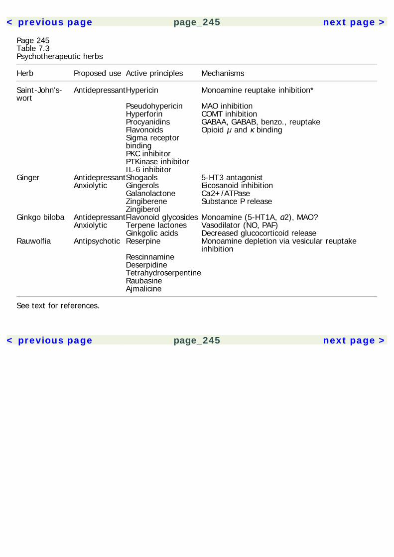
< previous page page_245 next page >
Page 245Table 7.3Psychotherapeutic herbs
Herb Proposed use Active principles Mechanisms
Saint-John's-wort
AntidepressantHypericin Monoamine reuptake inhibition*
Pseudohypericin MAO inhibition Hyperforin COMT inhibition Procyanidins GABAA, GABAB, benzo., reuptake Flavonoids Opioid μ and κ binding Sigma receptor
binding
PKC inhibitor PTKinase inhibitor IL-6 inhibitor Ginger AntidepressantShogaols 5-HT3 antagonist Anxiolytic Gingerols Eicosanoid inhibition Galanolactone Ca2+/ATPase Zingiberene Substance P release Zingiberol Ginkgo biloba AntidepressantFlavonoid glycosides Monoamine (5-HT1A, α2), MAO? Anxiolytic Terpene lactones Vasodilator (NO, PAF) Ginkgolic acids Decreased glucocorticoid releaseRauwolfia Antipsychotic Reserpine Monoamine depletion via vesicular reuptake
inhibition Rescinnamine Deserpidine Tetrahydroserpentine Raubasine Ajmalicine
See text for references.
< previous page page_245 next page >
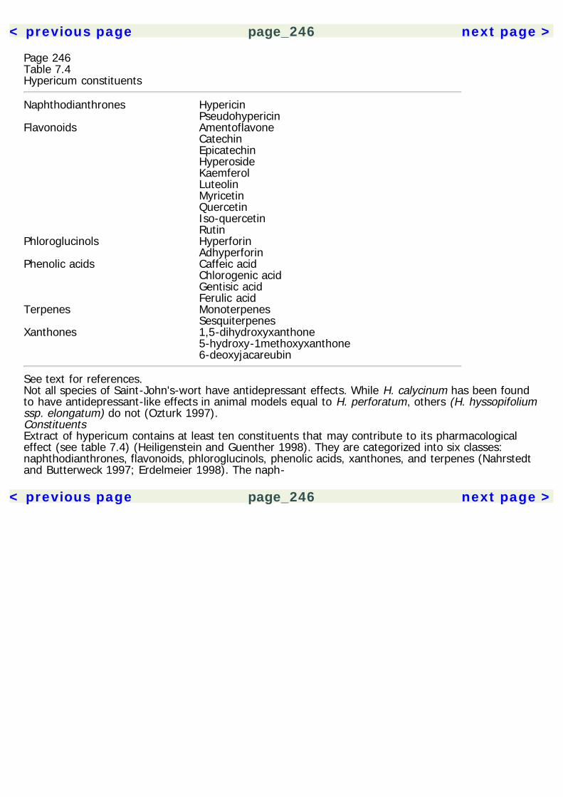
< previous page page_246 next page >
Page 246Table 7.4Hypericum constituents
Naphthodianthrones Hypericin PseudohypericinFlavonoids Amentoflavone Catechin Epicatechin Hyperoside Kaemferol Luteolin Myricetin Quercetin Iso-quercetin RutinPhloroglucinols Hyperforin AdhyperforinPhenolic acids Caffeic acid Chlorogenic acid Gentisic acid Ferulic acidTerpenes Monoterpenes SesquiterpenesXanthones 1,5-dihydroxyxanthone 5-hydroxy-1methoxyxanthone 6-deoxyjacareubin
See text for references.Not all species of Saint-John's-wort have antidepressant effects. While H. calycinum has been foundto have antidepressant-like effects in animal models equal to H. perforatum, others (H. hyssopifoliumssp. elongatum) do not (Ozturk 1997).ConstituentsExtract of hypericum contains at least ten constituents that may contribute to its pharmacologicaleffect (see table 7.4) (Heiligenstein and Guenther 1998). They are categorized into six classes:naphthodianthrones, flavonoids, phloroglucinols, phenolic acids, xanthones, and terpenes (Nahrstedtand Butterweck 1997; Erdelmeier 1998). The naph-
< previous page page_246 next page >
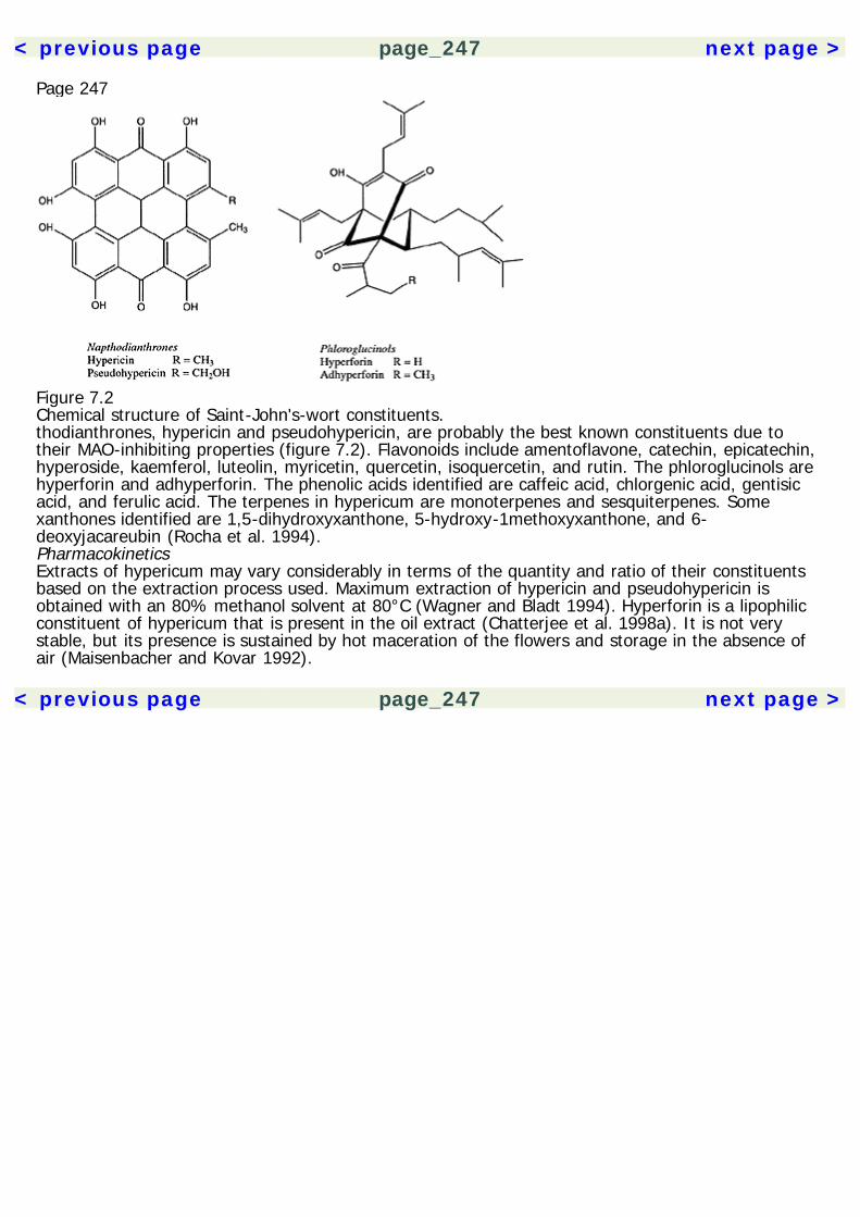
< previous page page_247 next page >
Page 247
Figure 7.2Chemical structure of Saint-John's-wort constituents.thodianthrones, hypericin and pseudohypericin, are probably the best known constituents due totheir MAO-inhibiting properties (figure 7.2). Flavonoids include amentoflavone, catechin, epicatechin,hyperoside, kaemferol, luteolin, myricetin, quercetin, isoquercetin, and rutin. The phloroglucinols arehyperforin and adhyperforin. The phenolic acids identified are caffeic acid, chlorgenic acid, gentisicacid, and ferulic acid. The terpenes in hypericum are monoterpenes and sesquiterpenes. Somexanthones identified are 1,5-dihydroxyxanthone, 5-hydroxy-1methoxyxanthone, and 6-deoxyjacareubin (Rocha et al. 1994).PharmacokineticsExtracts of hypericum may vary considerably in terms of the quantity and ratio of their constituentsbased on the extraction process used. Maximum extraction of hypericin and pseudohypericin isobtained with an 80% methanol solvent at 80°C (Wagner and Bladt 1994). Hyperforin is a lipophilicconstituent of hypericum that is present in the oil extract (Chatterjee et al. 1998a). It is not verystable, but its presence is sustained by hot maceration of the flowers and storage in the absence ofair (Maisenbacher and Kovar 1992).
< previous page page_247 next page >
< previous page page_248 next page >
Page 248The pharmacokinetics of hyperforin have been studied in rats and humans (Biber et al. 1998). Inrats, after a 300 mg/kg oral dose of hypericum extract (WS 5572, containing 5% hyperforin),maximum plasma levels of 370 ng/ml (690 nM) are achieved at 3 hours. The half-life of hyperforin is6 hours. Humans given a 300 mg tablet of hypericum (containing 14.8 mg hyperforin) showedmaximum plasma levels of 150 ng/ml (280 nM) at 3.5 hours. The half-life is 9 hours, and meanresidence time is 12 hours. Pharmacokinetics of hyperforin are linear up to 600 mg, and noaccumulation occurs after repeated doses. By comparison, effective and safe plasma levels ofparoxetine and fluoxetine vary between 40 and 200 ng/ml (Preskorn 1997). The effective plasmaconcentration of hyperforin predicted from computer-fit data is approximately 97 ng/ml (180 nM),which could be easily monitored (Biber et al. 1998). There is a linear correlation between oral dose ofhyperforin and plasma levels, and steady-state concentrations of 100 ng/ml (180 nM) could beachieved with three-times-daily dosing.The pharmacokinetics of hypericin and pseudohypericin plasma have been studied as well(Brockmöller et al. 1997). Human subjects receiving placebo, or 900, 1800, or 3600 mg of astandardized hypericum extract (LI 160), which contained 0, 2.81, 5.62, and 11.25 mg of totalhypericin and pseudohypericin, achieved maximum total plasma concentrations at 4 hours (0.028,0.061, and 0.159 mg/L, respectively). The half-lives of absorption, distribution, and elimination were0.6, 6.0, and 43.1 hours, respectively, using 750 μg of hypericin, and are slightly different for 1578μg of pseudohypericin (1.3, 1.4, and 24.8 hours, respectively) (Kerb et al. 1996). The systemicavailability of the hypericum extract LI 160 is between 14 and 21%. Comparable results are found inanother study using LI 160 (Staffeldt et al. 1994). Long-term dosing of 3 × 300 mg per day showedthat steady-state levels of hypericin are reached after 4 days.The oral bioavailability of hypericum may be altered and improved by a combination of itsconstituents. A hypericum extract containing naphthodianthrones is inactive in a water suspension,but very effective when another constituent, procyanidin, is present. Procyanidin had the effect ofincreasing the water solubility of naphthodianthrones, and thus increasing their pharmacokineticavailability (Butterweck et al. 1997). Further, the facilitative effect of procyanidin exhibited aninverted U curve.
< previous page page_248 next page >
< previous page page_249 next page >
Page 249This is a prime example of how plant constituents can act cooperatively, and isolation of a singleconstituent may reduce its effectiveness.Reported clinical doses of hypericum extract range from 300 to 1200 mg per day. (Heiligenstein andGuenther 1998; Linde et al. 1996)Mechanisms of ActionMonoamine The constituents of Hypericum perforatum and a few other species of hypericum haveseveral pharmacological mechanisms that could account for its antidepressant effects. Much attentionhad been given to the ability of hypericin to inhibit MAO. Hypericum extract (LI 160) does inhibitboth MAOA and MAOB, but does so weakly, occuring only in the millimolar range (Muller et al. 1997;Thiede and Walper 1994). MAO inhibition is attributed to both hypericin and flavonol content (Bladtand Wagner 1994). Thus, inhibition of MAO alone is insufficient to explain the antidepressant effectof hypericum. Inihibition of 35% of COMT activity occured at the millimolar range as well, for whichflavonols are chiefly responsible. Although these actions may contribute to the antidepressant effect,they are inadequate by themselves to account for it. MAO inhibition has been found by xanthonesextracted from other hypericum species such as H. brasiliense (Rocha et al. 1994).Although the individual inhibition of either MAO or COMT may be comparatively minor in isolation,their combined inhibition along with other monoamine or nonmonoamine actions could have additiveif not synergistic effects. For example, a fraction with combined hypericin and flavonoids hadantidepressant effects in an animal model (Butterweck et al. 1997). Hypericum is a particular casewherein a single isolated principle may be sufficient for the desired effect, but less effective than theentire plant extract.Another action of hypericum constituents that is particularly relevant to antidepressant effects is theability to inhibit neuronal reuptake of monoamines. The half-maximal inhibition for monoamineuptake is 100 times lower than for the inhibition of MAOA or MAOB (Chatterjee et al. 1998a).Hyperforin is the major contributor to this action, blocking reuptake of 5-HT, norepinephrine, anddopamine (Muller et al. 1998). Half-maximal inhibition occurs at nanomolar concentrations (80 to
< previous page page_249 next page >
< previous page page_250 next page >
Page 250200 nM). Affinity for inhibition of reuptake was equal (2 μg/ml) for all three monoamines studied(Muller et al. 1997). The antidepressant effects are accompanied by increases in urinary metabolitesof dopamine and norepinephrine (Muldner and Zollner 1984). Not only is hyperforin more potentthan hypericin, but also present in higher concentrations in hypericum (>2.5% vs. <0.4%,respectively). Thus, the plasma concentrations of hyperforin used in a pharmacokinetic study arevery close to those used in in vitro experiments that demonstrate synaptosomal reuptake inhibitionof serotonin, norepinephrine, and dopamine (Biber et al. 1998). Radiolabeled hyperforin does crossthe blood-brain barrier and penetrates brain tissue (Ostrowski 1988). Hypericin itself has minorreuptake inhibition effects on DA (22% inhibition in the low micromolar range), but not on NE or 5-HT.In addition to reuptake effects, subchronic treatment with hyperforin down-regulates β adrenergicreceptors and up-regulates 5-HT2 receptors in frontal cortex of the rat (Muller and Rossol 1994,1997, 1998). Chronic (26-week) daily treatment with 2700 mg/kg of a hypericum extract (LI 160) up-regulates the affinity and density of both 5-HT1A and 5-HT2 receptors by 50%. This appearsselective to hyperforin, because other hypericum constituents (hypericin and kaemferol) fail to showthe same effect. There may be a direct effect on 5-HT receptors, because activity in the guinea pigileum and Bezold-Jarisch reflex assays indicate 5-HT3 and/or 5-HT4-like effects (Chatterjee et al.1998a, 1998b). However, hypericin inhibits 5-HT1A receptors (30% inhibition) in the low micromolarrange (Raffa 1998) The monoamine activity of hypericum is differentially influenced by the extractused (Bhattacharya et al. 1998). The ethanol extract (containing 4.5% hyperforins) showed moredopaminergic activity, whereas the CO2 extract (containing 38.8% hyperforins) showed moreserotonergic activity, but no dopaminergic activity.Amino acid neurotransmitter Constituents of hypericum also appear to have effects on amino acidneurotransmission, particularly GABA. Hypericin and a crude extract bind to GABAA and GABABreceptors (Cott 1997). Hyperforin also inhibits synaptosomal GABA reuptake in the low micromolarrange (IC50 values of 0.05–0.10 μg/ml). Activity at GABAA benzodiazepine receptors was noted inextracts of four hy-
< previous page page_250 next page >
< previous page page_251 next page >
Page 251pericum species (H. perforatum, H. hirsutum, H. patulum, and H. olympicum). Amentoflavone is theresponsible constituent, displacing flumazenil from benzodiazepine receptors in the nanomolar range(IC50 14.9+/–1.9 nM). Again, no such effect is observed for hypericin. Amentoflavone acts as apartial agonist at GABA benzodiazepine receptors. However, it remains in question whether thisconstituent crosses the blood-brain barrer because it does not inhibit central flunitrazepam bindingwhen administered peripherally (Nielsen et al. 1988). In addition to GABA activity, hyperforin inhibitsreuptake of glutamate at 0.05–0.10 μg/ml (Baureithel et al. 1997). Activity on GABA transmission isconsistent with anxiolytic or even antidepressant effects of hypericum.Opioid A recent study has shown activity of hypericum extracts at opioid receptors (Simmen et al.1998). Extracts displace naloxone from μ and κ opioid receptors in the micromolar range (IC50 25and 90 μg/ml, respectively). In contrast, extracts of the sedative herb Valeriana officinalis do nothave this effect. This effect is due to unidentified constituents and not by the flavonoids quercetin orkaemferol. Opioids are known to have effects on emotion, so it is conceivable that activity ofhypericum at μ and κ receptors contributes to its therapeutic effects (Gerra et al. 1998; Tejedor-Realet al. 1995; Walker and Zacny 1998). Although they are not conventional treatment for depression,opioids such as buprenorphine have been effective in treatment of refractory depression (Bodkin etal. 1995). However, for any further conclusions to be drawn, it would be necessary to furtherelucidate the opioid effects of hypericum to determine what functional effect, if any, hypericum hason the receptors.Sigma receptor Several other mechanisms of action have been identified in hypericum and itsextracts. Hypericin inhibits greater than 40% of binding at muscarinic and σ (sigma) receptors in thelow micromolar (1.0 μM) range (Raffa et al. 1998). The σ receptor was originally classified as anopioid receptor, but was later identified to be nonopioid in nature (Martin et al. 1976; Johnson andJones 1990). Along with the NMDA receptor, the σ receptor was found to be a major target of thedissociative anesthetic drug phencyclidine (PCP, angel dust). The physiological roles
< previous page page_251 next page >

< previous page page_252 next page >
Page 252and endogenous ligand of the σ receptor are not yet fully understood. Certain neurosteroids(dehydroepiandrosterone and pregnenolone) have antidepressant effects in an animal model (Porsoltforced-swim test), which are mediated in part by the σ receptor (Reddy et al. 1998).Protein kinases Hypericin also inhibits protein tyrosine kinase activity in the nanomolar range, andprotein kinase C (PKC) in the micromolar range (Agostinis et al. 1995; Takahashi et al. 1989; Harriset al. 1996), which has led to its consideration as an antiglioma treatment (Zhang et al. 1997;Couldwell et al. 1994). The psychotherapeutic ramifications of this finding are not readily apparent,but PKC is integral to many intracellular communication mechanisms in the brain. Relatively littleattention has been paid to this effect of hypericin, but the activity of protein kinases is intimatelyrelated with the function of monoamine systems, decreased in the brains of depressed suicidevictims, and increased by antidepressant drugs (Hrdina et al. 1998; Blakely et al. 1998; Tadokoro etal. 1998). Specific mechanisms of monoamine-protein kinase interactions are beginning to beelucidated. For example, activators of PKC decrease the expression of the norepinephrine transporter,and thus the rate of norepinephrine reuptake from the synapse (Apparsundaram 1998). Further,these antidepressant-protein kinase interactions have some anatomical specificity to areas such asthe frontal cortex (Tadokoro et al. 1998).Other mechanisms A few other effects of hypericin, and a crude hypericum extract have been found,including affinity for NMDA, inositol triphosphate, and adenosine receptors. However, these are notlikely to be significant to its therapeutic effects because concentrations required for these interactionsare not likely to be achieved by oral administration (Cott 1997). Vasoactive effects are possiblebecause hypericum extracts blocked the vasoconstricting effects of histamine and prostaglandin F2αin porcine coronary arteries, and some vasorelaxation occurs in one particular fraction. These effectsare hypothesized to be mediated by inhibition of phosphodiesterase (Melzer et al. 1991).Hypericum also has effects on cytokine levels (Thiele et al. 1994). A hypericum extract (LI 160) givento people with depression and healthy
< previous page page_252 next page >

< previous page page_253 next page >
Page 253volunteers almost completely suppressed interleukin-6 (IL-6), interleukin-1β (IL-1β), and tumornecrosis factor-α. IL-6 and IL-1β are both known to reduce the release of corticotrophin-releasinghormone, which is implicated in depression (Arborelius et al. 1999).Finally, a thorough receptor binding study by Raffa and colleagues (1998) showed that hypericinextracts had no effect at adrenergic (alpha or beta), adenosine, angiotensin, benzodiazepine,dopamine, bradykinin, neuropeptide Y, PCP, NMDA, opioid, cholecystokinin A, histamine H1, ornicotinic ACh receptors. Although comprehensive, this study did not look at the binding of any otherhypericum constituents.Mechanisms: summary Thus, although much attention has been focused initially on MAO inhibition,there are several pharmacological mechanisms possessed by hypericum that could account for itsreputed antidepressant effects. These include increased monoamine activity through inhibition ofdegrading enzymes and blocking reuptake, GABA receptor activity and blocking reuptake, inhibition ofprotein kinase C, binding to opioid and σ receptors, and suppression of interleukins. Whereas someof these effects (e.g., blocking monoamine reuptake) may be sufficient to explain the antidepressanteffect, a combined additive or synergistic effect of the collective actions is .feasible (Bennett et al.1998). Furthermore, different constituents of hypericum have been demonstrated to have differenteffects, so that a single isolated constituent may be less effective than the whole plant extract.Physiological EffectsElectrophysiological effects Extracts of hypericum were examined for their electrophysiological effectsin animals. The onset of effects occurred 3–4 hours after administration. Frequencies affected firstwere in the alpha range and were maximal in the frontal cortex (Dimpfel and Hofmann 1995).Another study examined the EEG effects for two hypericum extracts in rats: one extract high inhyperforin and lacking naphthodi-anthrones (CO2), and another extract (LI 160) low in hyperforin.Both extracts showed similar early alpha effects, but only LI 160 had a late effect of increased deltafrequencies. The alpha effects are comparable to
< previous page page_253 next page >

< previous page page_254 next page >
Page 254those found in pharmaceutical serotonin reuptake inhibitors, and the delta effects are comparable tothose found with NMDA antagonists such as MK-801 (Dimpfel et al. 1998).To date, four methodologically controlled studies have been published that examined theelectrophysiological effects of hypericum. One study used 18 subjects taking 900 mg per day for 8days, with quantitative EEG performed on days 1 and 8 (Schellenberg et al. 1998). Effects peakedbetween 4 and 8 hours after administration. This study also attended to extracts that differed inhyperforin content (0.5% and 5.0%), while keeping the hypericin content constant. The higher doseof hyperforin (5.0%) produced increases in delta (1.25–4.5 Hz) and beta-1 (12.75–18.5) frequencies.Theta (4.75–6.75) and alpha-1 (7.0–9.5 Hz) effects were more pronounced on day 8 than day 1. Asecond study compared hypericum to the tricyclic antidepressant maprotiline on EEG and evokedpotentials (Johnson et al. 1994). Opposing effects occurred in the theta band for the two drugs, butsimilar effects ocurred on alpha and beta. The third study examined the effect of the extract LI 160on sleep EEG. Increases in slow-wave sleep (stages 3 and 4) were seen throughout the total sleepperiod. No effects were seen on sleep continuity, onset, duration, or wake-up phases (Schulz andJobert 1994). The effects of hypericum extract on sleep EEG were examined (Sharpley et al. 1998).Two doses (0.9 and 1.8 mg of ''Kira," Lichtwer Pharma) increased the latency to enter REM innondepressed volunteers. This effect is consistent with pharmaceutical antidepressants, whichincrease REM latency and/or reduce total REM duration.Cognitive effects The effects of LI 160 on cognitive performance of subjects being treated fordepression were examined. A significant antidepressant effect was seen and with low incidence ofside effects, but no adverse cognitive effects were found on measures of attention, concentration, orreaction time (Schmidt and Sommer 1993).Antidepressant effects Hypericum has been shown to have antidepressant effects in several animalmodels. An extract fraction high in naphthodianthrones showed antidepressant effects in the forced-swim test, and was attenuated by a dopamine antagonist (sulpiride) (Butterweck et
< previous page page_254 next page >
< previous page page_255 next page >
Page 255al. 1997). A commercially available extract of hypericum, LI 160, is rich in flavonoids andnaphthodianthrones and has antidepressant effects in the forced-swim and tail suspension tests.These antidepressant effects occurred in an inverted U shape and were reduced by dopamineantagonists (Butterweck et al. 1997). Preparations high in hyperforin but devoid of hypericin haveantidepressant effects in the behavioral despair and learned helplessness paradigms (Chatterjee et al.1998a, 1998b). These latter studies suggested that hyperforin is sufficient to produce anantidepressant effect, although it is not necessarily the only constituent with that effect.Studies in mice have shown a hypericum extract to increase exploration in an unfamiliarenvironment, prolong sedative sleep time, and antagonize the effects of reserpine. Otherantidepressant-like effects are found on the water-wheel test, and chronic administration decreasedaggression in socially isolated male mice (Okpanyi and Weischer 1987).Several human clinical studies have been performed on the effectiveness of hypericum in depression(Linde et al. 1996; Volz 1997). There has been a wide variety in the methodology used with respectto experimental controls, doses, preparations used (extracts, whole herb), hypericin contents, andsample size. Most studies ranged from 4 to 12 weeks in duration, and employed doses of 350–1000mg of extract. Diagnoses were made using DSM or ICD criteria, and subjects were typically rated inthe mild to moderate range in severity of depression. Many studies employed quantitativepsychometric instruments such as the Hamilton Depression Rating Scale, Clinical Global ImpressionScale, and the Depression Self-Rating Scale. Improvements were noted in mood, as well as insomatic symptoms of depression such as fatigue, disturbed sleep, and inactivity (Hubner et al. 1994).One study suggested that hypericum may be effective in treatment of seasonal affective disorder aswell (Kasper 1997).Methodological controls. Some published studies on the effectiveness of hypericum haveexperimental design flaws, but there are several that are methodologically controlled, employingdouble-blind, randomization, and placebo controls. Randomized, placebo-controlled studies weresummarized and evaluated in a meta-analysis by Linde and colleagues (1996). The combined subjectpool was 1,757 outpatients with mild to
< previous page page_255 next page >
< previous page page_256 next page >
Page 256moderate depression. In total, 23 randomized studies were found, of which 15 were placebocontrolled. Hypericum was superior to placebo across studies. One study used a second phase wherethe placebo group was crossed over to active treatment (LI 160) producing significant improvement(Hansgen et al. 1994). A more stringent meta-analysis, which also used double-blind as an inclusioncriteria, found Saint-John's-wort to be superior to placebo and equivalent to tricyclics in efficacy (Kimet al. 1999).Responsible Constituents. The contribution of the different constituents of hypericum was examinedby one methodologically controlled study (Laakmann et al. 1998). A large sample (n = 174) ofsubjects diagnosed by DSM-IV criteria were tried on 300 mg of either of two extracts (WS 5572 andWS 5573), relative to a placebo group. These two extracts differ in their hyperforin content (5.0%and 0.5%, respectively). The high-hyperforin group showed the greatest improvement, suggestingthat it is very relevant to the antidepressant response. Further, a dose-response relationship wasshown between the hyperforin content of hypericum extracts and its antidepressant effect. A sizablereduction in ratings of depression (Hamilton rating scale for depression, 10.3 ± 4.6) was found withWS 5572. Because these two extracts differ primarily by their hyperforin, it appears that hyperforincontent may be the most salient chemical constituent for standardization in the future, rather thanhypericin.Pharmaceutical Comparison. At least 8 studies to date have examined the effectiveness of hypericumcompared to the pharmaceutical antidepressants imipramine, amitriptyline, and maprotiline.Preliminary results indicate that hypericum is equivalent to standard antidepressants in effectiveness(Linde et al. 1996; Vorbach 1997). Similar to the pharmaceutical antidepressants, there is a 10–14day lag for therapeutic effects of hypericum (Harrer et al. 1994). Indeed, the differences seenbetween hypericum and placebo groups becomes apparent between 2 and 4 weeks (Sommer andHarrer 1994). Hypericum has been reported to have a more favorable side-effect profile than severalpharmaceutical antidepressants as well (Vorbach et al. 1994; Harrer et al. 1994). In double-blindstudies, subjects have reported fewer and less-severe side effects. Although these initial results arepromising, Linde and colleagues (1996) have concluded that the present evidence is inadequate toestablish
< previous page page_256 next page >
< previous page page_257 next page >
Page 257whether hypericum is as effective as other antidepressants and whether it has fewer side effects.ToxicityAlthough used extensively, there have not been any published reports of serious drug interactions ortoxicity for hypericum (Heiligenstein and Guenther 1998). The long-term safety of hypericum has notbeen evaluated.Side effects In an open study with 3,250 subjects, the most common side effects reported weregastrointestinal irritation (0.6%), allergic reactions (0.5%), fatigue (0.4%), and restlessness (0.3%).A review of case reports, clinical trials, post-marketing surveillance, and drug monitoring studiesconcurrently showed that the most common side effects were gastrointestinal, dizziness/confusion,and sedation (Ernst et al. 1998). Importantly, the side effects of hypericum in this study werecomparable to placebo levels. A pharmacokinetic study showed that plasma levels of up to 300 ng/mlwere well tolerated. Headache occured in one subject who was taking 1200 mg extract (59 mghyperforin, plasma conc. >400 ng/ml) (Biber et al. 1998).The meta-analysis by Linde and colleagues (1996) found greater incidence of side effects reported bytaking pharmaceutical antidepressants (52.8%) than those taking hypericum (19.8%). The meta-analysis by Kim and colleagues (1999) found that subjects were more likely to discontinue treatmentwith tricyclics than Saint-John's-wort, and tricylics were twice as likely to cause side effects.Phototoxicity Hypericum has been noted to cause a phototoxicity in cell cultures and a dermatitis andinflammation of nasal mucosa in grazing animals that consume the plant (Brockmöller et al. 1997).There have been no reported cases in humans, but perhaps caution is advised to individualsreceiving significant sun exposure or who are particularly photosensitive (Tyler 1994). Theresponsible element for this photosensitivity is believed to be hypericin. One methodologically-controlled study quantified the response of subjects taking hypericum (LI 160) to ultraviolet radiation(UVA and UVB) (Brockmöller et al. 1997). In a single-dose phase, a comparison of light sensitivity didnot differ between hypericum
< previous page page_257 next page >

< previous page page_258 next page >
Page 258extract and placebo groups, and there was no dose relation in light sensitivity. Sensitivity to selectiveUVA light was increased only after the highest dose (3600 mg of extract) with marginal statisticalsignificance. In the multiple-dose phase (1800 mg/day, 15 days), there was a slight increase in lightand UVA sensitivity. However, the photosensitivity occurred at doses much higher than those usedfor antidepressant effects.Pharmacokinetic interactions Preliminary evidence suggests that Saint-John's-wort induces thecytochrome oxidase enzyme isoform CYP3A4 (Ernst 1999). This raises the potential forpharmacokinetic interactions with drugs metabolized by the same enzyme. A few cases have beenreported of reduced warfarin levels (Yue et al. 2000). Similar interactions have also been reported forconcurrent use with digoxin, theophylline, and cyclosporin (Nebel et al. 1999; Ruschitzka et al. 2000;Johne et al. 1999). As with any other medication, potential interactions should be considered whentaking a combination of drugs.Cytotoxicity There has been some concern raised over cytotoxic and mutagenic in-vitro effects ofhypericum, which have been ascribed to the constituent quercetin (Tyler 1994). However, quercetinis a flavonoid that is present in many plants and vegetables, and it is estimated that the averageperson consumes 50 mg per day. Consumption of hypericum would not significantly add to this (1mg in commonly used amounts). The German Commission E has concluded that this is not of greatconcern.Hypomania and use with other antidepressants One case has been reported of concurrent use ofhypericum with an SSRI. Gordon (1998) reported a case of a 50-year-old woman taking 600 mg/dayof hypericum for chronic depression. She had discontinued taking Paxil 10 days prior to hypericumand experienced no ill effects at that time. However, she added 20 mg of paroxetine to her regimenof hypericum to improve her sleep. She presented with lethargy, nausea, and weakness, but vitalsigns and mental status were normal. Following discontinuation of medications, she returned tonormal status the next day.Two cases of hypomania resulting from hypericum have been reported (O'Breasail and Argouarch1998). One case involved post-stroke depres-
< previous page page_258 next page >
< previous page page_259 next page >
Page 259sion with a history of unsuccessful treatment with other antidepressant treatments: paroxetine,fluoxetine, and ECT, but not concurrent with the hypericum. After 6 weeks with hypericum (dosageunspecified), he developed increased drive, pressured speech, irritability, grandiosity, and alterationsof concentration and sleep. His behavioral problems persisted after discontinuation of hypericum, butwere controlled by valproate. The second case reported was of a patient presenting with manicfeatures: irritability, increased drive, anger, mood lability, grandiosity, flight of ideas, and sleepdisturbance. A year prior to this he witnessed a fatal accident and began experiencing anxiety,depression, and some post-traumatic stress symptoms. His manic symptoms began following 3months of treatment with hypericum. He was treated with lithium carbonate and his mood symptomsstabilized over 2 weeks.Given the wide availability and usage of hypericum, manic reactions such as the ones describedappear to be uncommon. As expected, concomittant use of hypericum with pharmaceuticalantidepressant medication appears to be contraindicated. Although some antidepressant medicationscan be given in combination, this must be done with caution and with dosage considerations toaccount for additive or synergistic effects.Sexual function One of the potential benefits of hypericum is the apparent reduced or lack of adverseeffects upon sexual function, compared to pharmaceutical antidepressants. The SSRIs are particularlynotorious for inhibition of sexual function, whereas antidepressants with dopaminergic actions (e.g.,bupropion) do not, and may actually enhance sexual function (Rosen et al. 1999; Piazza et al. 1997).Anecdotal reports and the fact that there are no clinical reports of sexual dysfunction with hypericumis encouraging, but it remains to be tested empirically.ConclusionsThus, the long tradition of hypericum as a treatment for depression has been well supported bymodern scientific research. Several active constituents have been identified, includingnaphthodianthrones (e.g., hypericin), phloroglucinols (e.g., hyperforin) and flavonoids(amentoflavone). Research has delineated its pharmacokinetic properties, and many of itsneurochemical mechanisms have been identified: enhancing monoamine
< previous page page_259 next page >
< previous page page_260 next page >
Page 260transmission (MAO, COMT, reuptake inhibition, and chronic receptor alterations), enhancing GABAtransmission, and affecting transmission in opioid, sigma, protein kinase, and interleukin systems.Antidepressant effects of hypericum are consistent across animal and human studies.Future directions for research on hypericum may continue the work done in clinical efficacy. Morespecifically, studies may be of interest that examine its effects in treatment of more severedepression and different subtypes of depression. The comparative efficacy of different hypericumpreparations could be further investigated, and optimum dosages need to be established (Linde et al.1996). Further work is needed to compare hypericum's efficacy and side effects with those of theSSRIs or atypical antidepressants, because published studies to date have only compared it withtricyclics.Other directions hypericum research might take would be to examine analgesic effects of hypericum.Numerous antidepressants have been found to have analgesic effects, or to potentiate the effects ofopioid analgesia, due to the role of monoamine systems in analgesia (Clifford 1985). Further, theopioid and σ effects of hypericum could create analgesic effects, but the nature of hypericum's actionat opioid receptors needs to be elucidated. Given that clomipramine and selected SSRIs have beenfound to be effective in treating obsessive-compulsive disorder, the possibility of whether hypericumwould have a similar effect may be investigated (Pigott and Seay 1999). Finally, given thatantidepressants are effective in treating anxiety, controlled trials may be attempted for hypericum aswell (Rickels et al. 1993).GingerHistory and BotanyGinger is a rhizome, or underground stem, of the plant Zingiber officinale from the Zingiberaceaefamily (figure 7.3). It is a tropical perennial that grows 6–12-inch stalks and dense, cone-like flowersat the end. Its leaves are long and grasslike, typically 6–12 inches in length and 0.75 inch wide(Kowalchik and Hylton 1987).Ginger's use was well known among the ancient Greeks and Romans, and was a common importfrom Asia during the eleventh and thirteenth
< previous page page_260 next page >
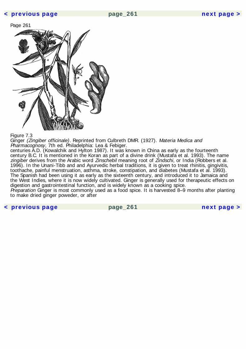
< previous page page_261 next page >
Page 261
Figure 7.3Ginger (Zingiber officinale). Reprinted from Culbreth DMR. (1927). Materia Medica andPharmacognosy, 7th ed. Philadelphia: Lea & Febiger.centuries A.D. (Kowalchik and Hylton 1987). It was known in China as early as the fourteenthcentury B.C. It is mentioned in the Koran as part of a divine drink (Mustafa et al. 1993). The namezingiber derives from the Arabic word Zinschebil meaning root of Zindschi, or India (Robbers et al.1996). In the Unani-Tibb and and Ayurvedic herbal traditions, it is given to treat rhinitis, gingivitis,toothache, painful menstruation, asthma, stroke, constipation, and diabetes (Mustafa et al. 1993).The Spanish had been using it as early as the sixteenth century, and introduced it to Jamaica andthe West Indies, where it is now widely cultivated. Ginger is generally used for therapeutic effects ondigestion and gastrointestinal function, and is widely known as a cooking spice.Preparation Ginger is most commonly used as a food spice. It is harvested 8–9 months after plantingto make dried ginger poweder, or after
< previous page page_261 next page >
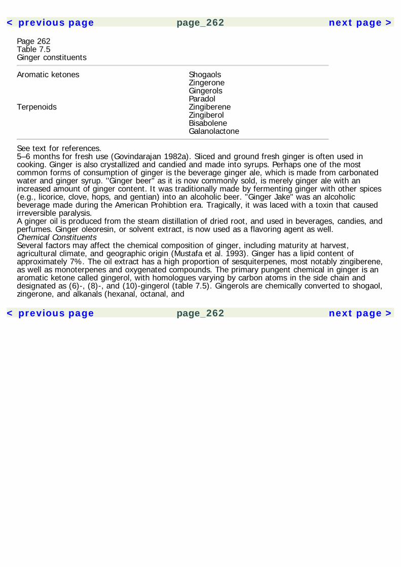
< previous page page_262 next page >
Page 262Table 7.5Ginger constituents
Aromatic ketones Shogaols Zingerone Gingerols ParadolTerpenoids Zingiberene Zingiberol Bisabolene Galanolactone
See text for references.5–6 months for fresh use (Govindarajan 1982a). Sliced and ground fresh ginger is often used incooking. Ginger is also crystallized and candied and made into syrups. Perhaps one of the mostcommon forms of consumption of ginger is the beverage ginger ale, which is made from carbonatedwater and ginger syrup. ''Ginger beer" as it is now commonly sold, is merely ginger ale with anincreased amount of ginger content. It was traditionally made by fermenting ginger with other spices(e.g., licorice, clove, hops, and gentian) into an alcoholic beer. "Ginger Jake" was an alcoholicbeverage made during the American Prohibtion era. Tragically, it was laced with a toxin that causedirreversible paralysis.A ginger oil is produced from the steam distillation of dried root, and used in beverages, candies, andperfumes. Ginger oleoresin, or solvent extract, is now used as a flavoring agent as well.Chemical ConstituentsSeveral factors may affect the chemical composition of ginger, including maturity at harvest,agricultural climate, and geographic origin (Mustafa et al. 1993). Ginger has a lipid content ofapproximately 7%. The oil extract has a high proportion of sesquiterpenes, most notably zingiberene,as well as monoterpenes and oxygenated compounds. The primary pungent chemical in ginger is anaromatic ketone called gingerol, with homologues varying by carbon atoms in the side chain anddesignated as (6)-, (8)-, and (10)-gingerol (table 7.5). Gingerols are chemically converted to shogaol,zingerone, and alkanals (hexanal, octanal, and
< previous page page_262 next page >

< previous page page_263 next page >
Page 263decanal). Shogaol may be further converted to paradol by hydrogenation. It is the commercialpreparation that is responsible for some of these reactions, because zingerone and shogaol are foundin small amounts in fresh ginger, but in large amounts in stored ginger (Mustafa et al. 1993; Ye et al.1989). Thus, the composition of fresh and stored ginger may vary, potentially altering itspharmacological effects. The primary terpenoid in ginger is zingiberene. Other terpenes includezingiberol, bisabolene, and galanolactone (figure 7.4). Lesser components of ginger includecurcuminoids (cassumunin A and B), and homologues of gingediol and gingediacetate (Mustafa et al.1993; Nagano et al. 1997).PharmacokineticsFew studies have looked at the pharmacokinetic properties of ginger constituents. Onepharmacokinetic study in rats showed a rapid clearance of (6)-gingerol, with a terminal half-life of7.23 minutes and a total body clearance of 16.8 ml/min/kg (Ding et al. 1991). There was also a highdegree of serum binding for (6)-gingerol (92.4%). Further, the disappearance of [6]-gingerol fromplasma is mediated by the liver, in part, but not the kidneys (Naora et al. 1992). Shogaol has beenshown to be metabolized to paradol and dehydroparadol by liver fractions (Surh and Lee 1992).Mechanisms of ActionFour principal mechanisms have been cited to explain the diverse physiological mechanisms ofginger. Broadly, these are (1) eicosanoid inhibition, (2) serotonin antagonism, (3) substance Prelease, and (4) Ca2+/ATPase activity.Eicosanoid activity Eicosanoids are cellular messengers involved in numerous physiological processesand pathological conditions and are active in the central nervous system (Cooper et al. 1996). Theyare synthesized from phospholipids into arachidonic acid by the enzymes phospholipase A2 andphospholipase C (see figure 7.5). Arachidonic acid is further converted by cyclooxygenases intoprostaglandins and thromboxanes. Various lipoxygenases convert arachidonic acid into HPETEs(hydroperoxyeicosatetraenoic acid), which are then converted
< previous page page_263 next page >
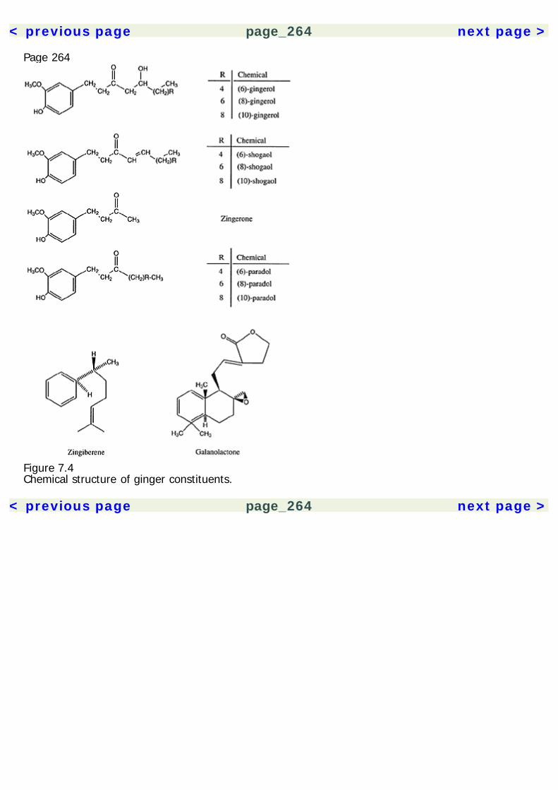
< previous page page_264 next page >
Page 264
Figure 7.4Chemical structure of ginger constituents.
< previous page page_264 next page >

< previous page page_265 next page >
Page 265
Figure 7.5Eicosanoid synthesis. Arachidonic acid is converted by cyclooxygenases into prostaglandins, andthromboxanes. Lipoxygenases convert arachidonic acid into HPETEs, which are then converted tolipoxins, leukotrienes, and 12-HETE (hydroxyeicosatetraenoic acid). Epoxygenases convertarachidonic acid into epoxides.to lipoxins, leukotrienes, and 12-HETE (hydroxyeicosatetraenoic acid). Several consitutents of gingerhave been found to interact with eicosanoid metabolism. Ginger extract inhibits formation ofthromboxanes, prostaglandins (prostaglandin F2α, prostaglandin E2, and prostaglandin D2), andleukotrienes (Srivastava 1986). Gingerdione inhibits synthesis of 5-HETE and prostaglandin E2 (IC50= 18 μM) (Flynn et al. 1986). Shogaol inhibits 5-HETE synthesis, and gingerol and dehydroparadolinhibit cyclooxygenase. Gingerol was found to inhibit synthesis of prostaglandins and leukotrienes(Kiuchi et al. 1992).Serotonergic activity Several physiological functions of the 5-HT3 receptor have been demonstratedin the central and peripheral nervous system. It has been implicated in cognitive, emotional, andphysiological processes as a consequence of its neuroanatomical distribution and neurophysiologicaleffects (Greenshaw 1993; Greenshaw and Silverstone 1997). The acetone extract of ginger wasfound to have serotonin
< previous page page_265 next page >
< previous page page_266 next page >
Page 266antagonist effects (Huang et al. 1991). Galanolactone, a diterpenoid found in ginger, was responsiblefor this action in the guinea pig ileum assay and effective in the micromolar range (Huang et al.1991). This effect was much more pronounced in the guinea pig ileum (predominantly a 5-HT3effect), but not in the rat fundis and rabbit aorta strips, which are respectively mediated by 5-HT1and 5-HT2 receptors. Further, galanolactone had a much greater effect in response to a selective 5-HT3 agonist (2-methyl-5-HT). Gingerol has also been shown to have 5-HT antagonist effects(Yamahara et al. 1989). Other physiological assays (5-HT-induced hypothermia and diarrhea) haveshown serotonin antagonist activity from oral doses of ginger (Huang et al. 1990). Among theconstituents from the effective fractions, (6)-shogaol was more effective than (6)-dehydrogingerdioneand (8)- and (10)-gingerol in inhibiting 5-HT-induced diarrhea. The ability of the responsibleconstituents to cross the blood-brain barrier, however, has not yet been explicitly demonstrated.Substance P Ginger has been shown to release substance P. Substance P is a peptideneurotransmitter present in the brain and spinal cord, and which has modulatory effects on severalneural processes, including pain transmission. (6)-shogaol has been shown to dose-dependentlycause a Ca2+-dependent release of substance P from spinal dorsal horn, in a similar manner tocapsaicin from chili peppers (Onogi et al. 1992). This action relates closely to the analgesic effects ofginger and will be discussed at greater length in chapter 8.Ca2+-ATPase A fourth mechanism of action of ginger involves activity on Ca2+-related processes.Gingerol was found to enhance Ca2+-pumping ATPase in the sarcoplasmic reticulum in cardiac andskeletal muscles (Kobayashi et al. 1987). It inhibits the enzyme in lower concentrations (0.24 μM),but activates it at higher concentrations (3–30 μM), suggesting that it increases the rate constant forthe enzyme-substrate complex breakdown and decreases the affinity of the enzyme for Ca2+.Gingerol had an inotropic effect in guinea pig atrial muscle, but there was no effect on Na+-K+-ATPase activity, suggesting that this was not due to cellular cAMP levels or an influx of extracellularCa2+.
< previous page page_266 next page >
< previous page page_267 next page >
Page 267Cytosolic calcium homeostasis is tightly regulated in neurons as well as in muscle cells. It issequestered in intracellular pools, which is accomplished by three components: (1) a calcium-activated pump (Ca2+-ATPase), (2) a calcium channel (such as the inositol trisphosphate receptor orryanodine receptor), and (3) a calcium-binding protein such as calsequestrin or calreticulin (Johnsonet al. 1993). Ca2+/ATPase is present in organelles of neural tissues although neurons lack asarcoplasmic reticulum. Neuronal Ca2+/ATPase pumps exist in organelles and in plasma membranesas well (Johnson et al. 1993, Panfoli et al. 1997). In cerebellar neurons, Ca2+/ATPase was found incisternae of the endoplasmic reticulum and in intracellular compartments for storage of Ca2+, calledcalciosomes (Villa et al. 1991). The Ca2+/ATPase pump in muscle and neuronal tissue derive fromthe same gene, but appear to be alternately spliced in a tissue-specific manner (Baba-Aïssa et al.1998).The regulation of intracellular neuronal Ca2+ is of high functional significance. Ca2+-dependentneurotransmitter release is reduced by the Ca2+/ATPase pump (Fossier et al. 1993). Althoughimmediate Ca2+ currents are mediated by ionotropic glutamate receptors (i.e., NMDA) inhippocampal neurons, a sizable portion of neurons show delayed Ca2+ increases from intracellularpools (Miller et al. 1996). These delayed changes were synaptically mediated and dependent uponendosomal Ca2+/ATPase.Whether ginger exerts psychotropic effects through this mechanism is highly dependent on a fewfactors. It still needs to be established that ginger constituents penetrate the blood-brain barrier insufficient concentrations to cause this effect. Further, there are alternate splices of neuronal andmuscle Ca2+/ATPase, so it cannot be assumed a priori that ginger constituents that affect muscularCa2+/ATPase will bind to neuronal Ca2+/ATPase or have the same degree of effect. The behavioraland large-scale neural consequences of Ca2+/ATPase modulation in general also need to be furtherelucidated.Physiological EffectsGinger has numerous physiological effects, including increasing bile emptying, increasing gastricmotility, antiulcer, anti-inflammatory,
< previous page page_267 next page >
< previous page page_268 next page >
Page 268antipyretic, vascular pressor, cardiotonic, analgesic, and antiemetic effects (Mustafa et al. 1993;Yamahara et al. 1989). Pressor effects are centrally mediated and produced by shogaol (Suekawa etal. 1986). However, the pressor effects are dose dependent, because low doses of (6)-shogaol and(6)-gingerol had depressor effects (Suekawa et al. 1984). Other effects of ginger are increased bilesecretion, antipyretic, and anti-inflammatory effects. (6)-shogaol was found to mediate theantipyretic effect, to a degree comparable with aspirin (Mascolo et al. 1989). The effects that will beconcentrated on in this chapter are the antiemetic and anxiolytic/antidepressant ones, and thosemediated by eicosanoid pathways.Platelet inhibition Ginger extract has been found to inhibit platelet aggregation induced byarachidonic acid, epinephrine, ADP, and collagen (Srivastava 1984). The extract's ability to inhibitcyclooxygenase activity and thromboxane levels correlated well with inhibition of platelet aggregation(Srivastava 1984; Mustafa et al. 1993). The type of preparation used also affects platelet inhibition,because roasted and charcoal of ginger were effective, while ether extracts of raw and dried gingerwere not (Wu et al. 1993).Ginger's platelet inhibition effect was demonstrated with oral doses in humans (Srivastava 1989).Although this was not confirmed in a later study, it highlights the importance of dosage andpreparation (Janssen et al. 1996). The effect is dose dependent, and while fresh ginger is effective,dried ginger is not (Lumb 1994).Gastrointestinal effects: animal studies Ginger enhances gastric motility with an oral administration ofacetone extract, involving (6)-shogaol and several gingerols (Mustafa et al. 1993). However, gastricmotility was inhibited by intravenous gingerol and shogaol, suggesting that dosage and route ofadministration are critical. In one study using intravenous (6)-shogaol, gastric transit was inhibited at3.5 mg/kg but facilitated at 35 mg/kg, indicating that a U-shaped dose-response curve may be ineffect (Suekawa et al. 1984).Ginger extracts (acetone, 50% ethanolic and aqueous) were investigated for antiemetic activity indogs (Sharma et al. 1997). Emesis was induced by 3 mg/kg cisplatin (a 100% emetic dose IV) inhealthy mongrel
< previous page page_268 next page >
< previous page page_269 next page >
Page 269Table 7.6Clinical studies of the antiemetic/nausea efficacy of ginger
Type of nausea Efficacy Comparison Ginger dose Reference
GA – Droperidol (ineffective) 2 g 1GA – Placebo 0.5, 1.0 g 2GA + Metoclopramide 0.5 g 3GA + Metoclopramide 1 g 4SS + Placebo 1 g 5HG + Placebo 0.25 g/day 6
Key: – Ineffective, + Effective, CD Crossover design, DB Double blind, GAGeneral anesthesia (postoperative nausea), HG Hyperemesis gravidarum,PC Placebo control, RA Randomized assignment, SS Seasickness.References: 1. Visalyaputra et al. 1998; 2. Arfeen et al. 1995; 3. Phillips etal. 1993; 4. Bone et al. 1990; 5. Grontved et al. 1988; 6. Fischer-Rasmussen et al. 1991.dogs. Across a range of oral doses (25–200 mg/kg) the aqueous extract was ineffective. The acetoneextract was more effective than ethanolic extract, and both were less effective than the 5-HT3antagonist granisetron. In contrast, neither of the ginger extracts was effective against apomorphine-induced emesis. In addition to nausea and vomiting, the cancer chemotherapeutic agent cisplatincauses inhibition of gastric emptying. The acetone and ethanolic extracts of ginger were found toreverse this effect of cisplatin in rats (Sharma et al. 1998). Ginger juice and acetone extracts weremore effective than the ethanolic extract. This reversal was comparable to that produced by the 5-HT3 receptor antagonist ondansetron. In fact, ginger juice was more efficacious than ondansetron.Gastrointestinal effects: human studies To date, six methodologically controlled studies have beenundertaken to evaluate the antinausea and antiemetic efficacy of ginger in humans (table 7.6).Despite overwhelming anecdotal evidence, the results of clinical studies have been variable. Thesedifferences in results may be accounted for, at least in part, by differences in methodology acrossstudies. One paradigm in which ginger had mixed efficacy is postoperative nausea from generalanesthesia. Of the four such studies conducted, two failed to support, while the other
< previous page page_269 next page >
< previous page page_270 next page >
Page 270two supported this role of ginger (Visalyaputra et al. 1998; Arfeen et al. 1995; Phillips et al. 1993;Bone et al. 1990). However, one study, which failed to show an effect with ginger, also failed toshow a benefit from droperidol, an antiemetic drug that has proven effective in several studies ofpostanesthetic nausea and emesis (Visalyaputra et al. 1998; Koivuranta et al. 1997; Fortney et al.1998). In the studies that supported ginger's effects, its efficacy was comparable to the drugmetoclopramide. Ginger has been shown to be effective for treatment of motion sickness andhyperemesis gravidarum (Grontved et al. 1988; Fischer-Rasmussen et al. 1991). For the prevention ofseasickness, ginger reduced cold sweating and the incidence of vomiting. There was also a trendtoward reduced nausea and vertigo. Further study showed ginger to reduce vertigo but notnystagmus in response to caloric stimulation of the vestibular system (Grontved and Hentzer 1986).Thus, there is emprical evidence to support the antinausea/emetic efficacy of ginger. The degree ofthis efficacy may vary depending on the intensity and/or source of the nausea (e.g., generalanesthetic, pregnancy, motion sickness). It would be interesting to see ginger's efficacy in nausea ofother etiologies such as viral, ethanol hangover, and chemotherapy.Behavioral Effects and ElectrophsyiologyPharmacological studies of oral and intravenously administered ginger constituents producebehavioral effects (Suekawa et al. 1984). Intravenous (1.75–3.5 mg/kg) and oral (70–140 mg/kg)doses of (6)-shogaol inhibit spontaneous motor actvity and prolong hexabarbital-induced sleepingtime. (6)-shogaol was more potent than (6)-gingerol. In an EEG study, intravenous (6)-shogaolshifted a low-amplitude fast-wave pattern to a high-amplitude slow-wave pattern after 5 minutes,which persisited for 60 minutes (Qian and Liu 1992; Suekawa et al. 1984).Two studies have been done to date that address ginger's anxiolytic effects in an animal model(Hassenohrl et al. 1996, 1998). Unfortunately, both of these employ a combined treatment of gingerand Ginkgo biloba so they do not allow for differentiation of effects. The combination had diazepam-like effects in an animal model of anxiety (elevated-plus maze) (Hassenohrl et al. 1996). Theseeffects were dose dependent and tripha-
< previous page page_270 next page >

< previous page page_271 next page >
Page 271sic: low doses (0.5 mg/kg intragastrically) had anxiolytic effects, intermediate doses (1 and 10mg/kg) had no effect, and higher doses (100 mg/kg) had anxiogenic effects. Unlike diazepam, theginger/ginkgo combination did not have memory-imparing effects on spatial navigation memory(water maze) or inhibitory avoidance learning (Hassenohrl et al. 1998). The ratio of ginger to ginkgoaffected the anxiolytic efficacy in the elevated-plus maze. The standard preparation with a ratio ofginger to ginkgo of 2.5 : 1 had anxiolytic efficacy, whereas other ratios, (1 : 1 or 1 : 2.5) wereineffective across all doses tested.If ginger alone exerts anxiolytic effects, one potential mechanism is by 5-HT3 antagonism bygalanolactone, because pharmaceutical 5-HT3 antagonists show antidepressant and anxiolytic effects(Huang et al. 1991; Martin et al. 1992; Costall and Naylor 1992; Costall et al. 1990; Menard and Treit1999; Alvarez-Guerra et al. 2000). Another potential mechanism of anxiolytic effects of ginger is theeicosanoid inhibition (Kiuchi et al 1992; Srivastava 1984; Mustafa et al. 1993; Cooper et al. 1996).Apart from direct neuronal effects of eicosanoids, arachidonic acid and its metabolites inhibit GABAAtransmission (Schwartz and Yu 1992). For example, thromboxane A2 inhibits GABA receptor function,independent of its actions on cerebrovasculature and glial cells (Schwartz-Bloom et al. 1996). Theinteractions between eicosanoids and GABA are complex, but arachidonic acid levels increase alongwith increases in intracellular Ca2+ and GABA efflux, suggesting that it may be acting as an intrinsicfeedback modulator in GABAergic neurons (Asakura and Matsuda 1984). Ginger constituents couldhypothetically create an anxiolytic effect by facilitating GABA conductance through inhibition ofarachidonic acid metabolites. Whether or not this occurs to a significant degree could be verified byin vitro and in vivo experiments with ginger constituents and assessing their effect on GABAtransmission.The antithromboxane effect of ginger is of potential significance in stress and depression.Hypercortisolism is found in approximately 50% of patients with major depression. Plasmathromboxane B levels correlated in a group of depressed patients with high levels of cortisol, but notwith depressed individuals with low cortisol or with normal controls
< previous page page_271 next page >
< previous page page_272 next page >
Page 272(Piccirillo et al. 1994). The effects of thromboxane A2 were examined in nondepressed, normalindividuals. Thromboxane A2 was found to have direct CNS effects on hemodynamic, ACTH, andcortisol responses (Cudd 1998). Whether ginger has a significant effect on these processes andwhether it has an antistress or antidepressant effect is speculative, but the eicosanoid activities ofginger and relationship to depression warrant further investigation.ToxicityThere is little known toxicity of ginger. The LD50 of oral ginger is very high (170 to 250 g/kgdepending on the preparation) (Wu et al. 1990). It is listed by the Food and Drug Administration asGenerally Regarded As Safe. No adverse reactions are noted in the normal doses of 2–4 g. It hasbeen recommended not to take ginger in the presence of morning sickness or gallstones (Gruenwaldet al. 1998).Ginger Jake was rendered toxic only because of an adulterant organophosphate, triorthocresylphosphate (TOCP), and not any constituent of ginger itself (Woolf 1995; Morgan and Tulloss 1976,Morgan and Penovich 1978). About 50,000 people ingested TOCP and experienced muscle pain,weakness, and minimal sensory impairment. TOCP causes irreversible central and peripheral neuronaldamage, consisting of axonal dying-back neuropathy and upper motor neuron syndrome withspasticity and abnormal reflexes.Summary and ConclusionsFour pharmacological mechanisms of ginger constituents have been investigated that could putativelymediate a central behavioral effect: (1) eicosanoid inhibition, (2) serotonin antagonism, (3) substanceP release, and (4) Ca2+/ATPase activity. Whether any of these occur is entirely contingent upon thepassing of ginger constituents through the blood-brain barrier. Although there have been no studiesthat explicitly address this, electrophysiological and behavioral changes from peripheraladministration provide indirect support.Pharmacological data and experimental data indicate two potential uses of ginger: antiemetic andanxiolytic/antidepressant. Studies of antiemetic effects have brought generally positive results,although further
< previous page page_272 next page >

< previous page page_273 next page >
Page 273research would allow stronger conclusions. Animal research and pharmacological mechanismssuggest an anxiolytic role of ginger, but there are no human studies to date that examine this. It isunlikely that ginger by itself will be sufficient as an antidepressant or anxiolytic for cases of clinicalseverity. However, ginger has few negative effects when used in reasonable quantities, and couldaugment other medications and/or counteract negative side effects.GinkgoAlthough primarily recognized for its nootropic effects, ginkgo has been studied somewhat forpsychotherapeutic effects. Because ginkgo has been discussed at length in chapter 5, this section willfocus primarily on the studies of its anxiolytic and/or antidepressant effects.Anxiolytic EffectsTwo studies by Hassenohrl and colleagues (1996, 1998), which are described above, showedanxiolytic effects of a combined ginger and ginkgo preparation in an animal model. Again, theprimary problem with these investigations is that they did not evaluate each herb in isolation. It ispossible that each has anxiolytic effects and that combined they have synergistic effects. On theother hand, it is also possible that either of the two is ineffective and that a single herb is accountingfor all of the effects. It is interesting to note, however, that in those studies, the ratio of ginger toginkgo had significant effects on the anxiolytic potency.A few other studies have been done that show anxiolytic effects of ginkgo in animal models. Ginkgoextract was found to reduce learning impairment induced by an auditory stressor on a discriminationtask (Rapin et al. 1994). Young and old rats were given 20 days of oral treatment with EGb 761 (50or 100 mg/kg/day). Although effective in both groups, EGb 761 was more effective in older thanyounger rats. Chronic administration of EGB 761 (50 and 100 mg/kg/day for 5 days) reducedavoidance deficits in the learned helplessness paradigm in rats, but was less effective when givenafterward (Porsolt et al. 1990). Anxiolytic-like effects were also seen in an emotional hypophagiaparadigm in mice.
< previous page page_273 next page >

< previous page page_274 next page >
Page 274Antidepressant EffectsGinkgo has been tested in one study for treatment of seasonal affective disorder (Lingjaerde et al.1999). This methodologically controlled study used a standardized extract, containing 48 mg flavoneglycosides and 12 mg terpene lactones per day, for 10 weeks. Subjects were evaluated using theMongomery-Åsberg Depression Rating Scale and an atypical symptom scale directed at symptoms ofseasonal affective disorder (hypersomnia, hyperphagia, and carbohydrate craving). The results werenegative, and failed to show an effect of ginkgo on any of the measures used. The authors cite asmall number of subjects as a disadvantage of the study, potentially raising the probability of a falsenegative error. Other potential influences on the outcome may have been the dose used and thelatency between symptom onset and treatment initiation.Putative Mechanisms and ConstituentsGinkgolic acid conjugates (6-alkylsalicylates) were tested for anxiolytic effects in several animalparadigms (Satyan et al. 1998). Single oral doses (0.3 and 0.6 mg/kg) of ginkgolic acid conjugatesshowed dose-related anxiolytic effects in elevated-plus maze, the open field, and novelty-inducedfeeding tests. Diazepam had similar effects in the models used. The anxiolytic effects of ginkgo maybe dependent on the extract used. The standardized ginkgo extract EGb 761, which lacks ginkgolicacid conjugates, showed lesser anxiolytic effects in some of the measures. The doses of ginkgolicacids were equivalent to what would be found in a 50 or 100 mg dose of extract, respectively.Further, given that the anxiolytic effects were obtained by oral administration, it indirectly suggestspenetration of the blood-brain barrier. Paradoxically, acute and chronic administration of EGb 761 (8–16 mg/kg, IP) was found to reduce social contact in rats, which was reversed by diazepam,suggesting anxiogenic effects (Chermat et al. 1997). It should be noted that Satyan and colleagues(1998) showed anxiolytic effects of ginkgo in several measures but not in the social interaction test.Monoamine There are several mechanisms that, alone or in combination, could account for ginkgo'spotentially mood-altering effects. These are broadly categorized into monoamine and GABAergiceffects. Ginkgo
< previous page page_274 next page >
< previous page page_275 next page >
Page 275extract (EGb 761) increases uptake of 5-HT, but not dopamine, into synaptosomes, which isprevented by the 5-HT reuptake inhibitor clomipramine (Ramassamy et al. 1992). Hypothetically,increasing 5-HT reuptake could create anxiolytic effects by reducing 5-HT stimulation of 5-HT2receptors that have anxiogenic effects (Jenck et al. 1998). However, this action is contrary to theaction of SSRIs, so reconciling an antidepressant action of ginkgo would likely necessitate othermechanisms. Aqueous and ethanolic ginkgo extracts as well as commercial preparations inhibit MAOAand MAOB in vitro (White et al. 1996). It remains to be shown that this occurs in the brain and atnormal oral doses. But the authors also reported preliminary evidence that an oral dose of ginkgocould produce 30–50% inhibition of MAOB in platelets in humans. However, a PET study of ginkgoextract in humans showed that it does not produce significant changes in the activity of brain MAOAand MAOB.Although the monoamine mechanism of ginkgo is uncertain, treatment does alter monoaminereceptors in animals. Chronic ginkgo extract reversed an age-related reduction in the maximalnumber of binding sites of 5-HT1A receptors in aged (24-month-old) but not young (4-month-old)rats (Huguet et al. 1994). Chronic oral administration of ginkgo extract (EGb 761) also prevents thestress-induced desensitization of 5-HT1A receptors, without altering their density or affinity (Bolanos-Jimenez et al. 1995). Drug discrimination paradigms also support that ginkgo extracts have effects on5-HT1A, but not 5-HT2 receptors (Winter and Timineri 1999). Ginkgo chronically administered alsoreverses the decline in binding density of α2-adrenergic receptors in aged rats (Huguet and Tarrade1992).GABA Another potential mechanism of ginkgo's psychotropic effects is amentoflavone. Amentoflavoneis a flavonoid with benzodiazepine activity in the nanomolar range (Lobstein-Guth, et al. 1988;Baureithel et al. 1997). Amentoflavone seems to act as a partial agonist at GABA benzodiazepinereceptors. However, it remains in question whether this constituent crosses the blood-brain barrerbecause it does not inhibit central flunitrazepam binding when administered peripherally (Nielsen etal. 1988). Still, there is some behavioral pharmacology evidence that
< previous page page_275 next page >

< previous page page_276 next page >
Page 276supports a direct or indirect GABAergic action of ginkgo (Chermat et al. 1997).Neuroendocrine Chronic treatment with oral ginkgo extract reduces stress-induced impariments inlearning and elevation of stress hormones, in both young and old rats (Rapin et al. 1994). GinkgolideB is one of the constituents responsible for this effect, which acts by reducing the binding andexpression of peripheral-type benzodiazepine receptors in the adrenal cortex (Amri et al. 1996, 1997;Papadopoulos et al. 1998). In this way, it reduces production glucocorticoids, and may also act at thehypothalamic level to inhibit the hypothalamic-pituitary-adrenal (HPA) axis (Marcilhac et al. 1998).Overactivation of HPA axis is associated with anxiety and depression. Elevated levels of cortisol occurduring situational anxiety (Armario et al. 1996; Salmon et al. 1986). Dysregulation of the HPA axisoccurs in such anxiety disorders as generalized anxiety and panic disorder (Avery et al. 1985).Concordantly, disruption of the glucocorticoid receptor gene is associated with reduced anxiety inmice (Tronche et al. 1999). Major depression is associated with disruption of the HPA axis due toimpaired feedback mechanisms, resulting in elevation of cortisol levels (Carroll et al. 1981; Young etal. 1991). Chronic elevation of glucocorticoids is neurotoxic, and recurring depression is associatedwith neuropathological changes in the hippocampus (Sheline et al. 1996). Remission of depression isassociated with a return of cortisol to normal levels (Steiger et al. 1989).Thus, ginkgo's actions on the HPA axis may afford it anxiolytic and antidepressant-like effects,particularly when stress is a causal factor. Considering its neuroendocrine effects, investigation ofginkgo in depression may still be warranted, particularly where stress and anxiety play causal roles.Summary and ConclusionsGinkgo is most commonly known for its cognitive effects, but there is evidence to suggest it mayhave psychotherapeutic effects as well. As with its cognitive effects, the mechanisms responsible forits anxiolytic or antidepressant effects are uncertain, but likely involve monoamine and/or
< previous page page_276 next page >
< previous page page_277 next page >
Page 277GABAergic mechanisms. Animal studies suggest that ginkgo has anxiolytic effects that increase withadvancing age. Despite evidence in several animal studies, there are few, if any, human studies thataddress its psychotherapeutic effects. Ginkgo remains to be tested in controlled clinical trials todetermine in which disorders and at what doses it is effective.There have been few, if any, studies on ginkgo and depression. Although it holds potential forpsychotherapeutic effects, it does not seem that ginkgo could be used alone to treat anxiety ordepression. More likely, it could serve as adjunctive treatment for anxiety or depression, and havepositive side effects on those using it for nootropic effects.Antipsychotic HerbsRauwolfiaHistory and BotanyRauwolfia serpentina (serpentwood), of the Apocynaceae family, was reputed for thousands of yearsin India as a tranquilizer. In Sanskrit, it was referred to as sarpagandha, and in Hindi as chandrika(Robbers et al. 1996). Western medical interest in the plant began in the 1950s, when theconstituent reserpine was identified. Rauwolfia is a small tree or shrub with numerous small pink andwhite flowers, deciduous leaves, and a fleshy ovoid fruit (Kowalchik and Hylton 1987). The medicinalpart of the plant is the dried root (Gruenwald et al. 1998). Its name derives from Leonhard Rauwolf,a sixteenth century German botanist. Three primary species are recognized with distinct geographicaldistributions: Rauwolfia serpentina (India, Sri Lanka, Thailand, Indonesia, Burma), R. tetraphylla(southern Mexico to Colombia and the West Indies), and R. vomitoria (Africa, from Senegal toMozambique).Mechanism of ActionThe psychoactive constituents of rauwolfia are alkaloids classified in three groups: (1) weakly basicindole alkaloids, (2) intermediate basic indoline alkaloids, and (3) strong anhydronium bases.Approximately 50 alkaloids have been identified, but the principal indole alkaloids
< previous page page_277 next page >
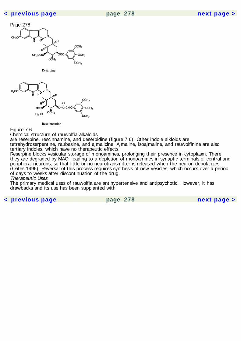
< previous page page_278 next page >
Page 278
Figure 7.6Chemical structure of rauwolfia alkaloids.are reserpine, rescinnamine, and deserpidine (figure 7.6). Other indole alkloids aretetrahydroserpentine, raubasine, and ajmalicine. Ajmaline, isoajmaline, and rauwolfinine are alsotertiary indoles, which have no therapeutic effects.Reserpine blocks vesicular storage of monoamines, prolonging their presence in cytoplasm. Therethey are degraded by MAO, leading to a depletion of monoamines in synaptic terminals of central andperipheral neurons, so that little or no neurotransmitter is released when the neuron depolarizes(Oates 1996). Reversal of this process requires synthesis of new vesicles, which occurs over a periodof days to weeks after discontinuation of the drug.Therapeutic UsesThe primary medical uses of rauwolfia are antihypertensive and antipsychotic. However, it hasdrawbacks and its use has been supplanted with
< previous page page_278 next page >
< previous page page_279 next page >
Page 279the invention of more modern antipsychotics such as piperazine, phenothiazines, butyrophenones,and thioxanthenes. Antagonism of dopamine receptors is a primary action of antipsychotic drugs(Carlsson 1978; Seeman 1987). Thus, reserpine may create antipsychotic effects by reducing theavailability of dopamine or other monoamines. Despite the drawbacks, there is a place for the use ofreserpine as an adjunct treatment for psychosis (Solon 1996; Wolkowitz et al. 1993). Schizophrenicpatients on long-term neuroleptic treatment who were given adjunct reserpine showed a decrease inthought disturbance, agitation and hostility, and suspiciousness (Bagdy et al. 1988). However, thesechanges did not correlate with CSF changes in monoamine levels. In fact, although norepinephrinelevels changed, other monoamine markers (CSF dopamine metabolites, platelet MAO, and serumdopamine-beta-hydroxylase) did not.Rauwolfia derivatives became available in the 1950s in western medicine for the treatment ofhypertension. The antihypertensive effects of rauwolfia alkaloids occur from their depletion ofmonoamines in adrenal chromaffin cells and sympathetic ganglia, and perhaps central neurons aswell (Oates 1996).ToxicityThe primary limiting effect of reserpine is depression. Depletion of central monoamines is believed tobe the mechanism for this effect (Heninger et al. 1996; Charney 1998). The depression may occur ina gradual and insidious manner, and the causal association between the drug and depression may bemissed (Oates 1996). Rauwolfia alkaloids are contraindicated in anyone with a history of depression,and careful vigilance is required to ensure that they do not induce depression in otherwise normalindividuals. Additional side effects are sedation and difficulty with concentration and performingcomplex mental tasks.Psychotherapeutic Herbs: ConclusionsThe discovery of pharmaceutical medications for the treatment of mental illnesses has revolutionizedtreatment and our understanding of the brain. Several herbal medications have been employedhistorically for the
< previous page page_279 next page >

< previous page page_280 next page >
Page 280treatment of mental illness, many of which are ineffective or impractical. However, a few holdpromise. Rauwolfia has long been used as a tranquilizer and antipsychotic medication, which is likelyrelated to its depletion of monoamine neurotransmitters. Although this has an antipsychotic effect, italso has the unfortunate side effects of depression and cognitive impairment. Although it may havesome use as an adjunctive treatment for schizophrenia, the advent of neuroleptics and neweratypical antipsychotic drugs are likely to render it obsolete.Hypericum not only has a long tradition of use as an antidepressant, but also a considerable amountof scientific research to support it. More research is needed, but it certainly works better thanplacebo and may be as efficacious as pharmaceutical antidepressants. The mechanism of this effectis not certain although monoamine reuptake mechanisms are most likely involved. Various othermechanisms may contribute additive or synergistic effects.Ginger is most commonly used as a spice, but has significant antiemetic effects and may haveanxiolytic or antidepressant effects. It may not likely be sufficient by itself, but it could augmentother medications and/or counteract negative side effects. At least four mechanisms have been foundthat may mediate ginger's central effects. Although known for its cognitive effects, ginkgo may alsohave anxiolytic or antidepressant effects. Ginger and ginkgo have potential as psychotherapeuticherbal medications, but far more research is needed to establish their basic efficacy.It is not likely that herbal psychotherapeutic medications will replace the use of pharmaceuticaltreatments. Herbal treatments are too few, and those known at present need more research toestablish their use. By far, hypericum has the best potential of this class.
< previous page page_280 next page >

< previous page page_281 next page >
Page 2818Analgesic and Anesthetic PlantsPain is a basic sensory, emotional, and cognitive phenomenon that signals some type of harm to theorganism. It is practically universal to human experience, and although it is initially adaptive, itfrequently outlives its usefulness and impedes one's ability to function. Accordingly, plants thatalleviate pain have become essential to human civilization.The plant drugs discussed here for their analgesic and anesthetic effects are diverse in form as wellas pharmacology, reflecting the diverse neurochemistry of the sensory and analgesic branches of thenervous system. They are grouped here by their pharmacological mechanism, including: opioid(poppy, myrrh), cholinergic (tobacco, lobelia, areca), eicosanoid (willow, feverfew), neurokinin (chili,ginger), purinergic (coffee, tea, chocolate, guarana, maté, kola), cannabinoid, monoamine (coca,khat), and uncertain mechanisms (ginseng). Plants discussed here for their local anesthetic effectsare coca and clove.Definition of TermsWhereas pain is used to refer to the total subjective phenomena associated with injury and suffering,nociception more specifically refers to the aspect of sensory transmission. Nociception is the morecorrect term for the experimental study of pain, because it is objectively measured through abehavior in animals and humans alike (e.g., withdrawal reflexes, verbal ratings). The actualsubjective experiences are private and known to each subject alone. However, for the sake ofconvenience, the word pain will be used here to address both aspects. Similarly, the word analgesia,meaning the relief from pain, will be used in place of the more objective term antinociception.
< previous page page_281 next page >
< previous page page_282 next page >
Page 282Another distinction must be made between analgesia and anesthesia. While anesthesia refers toblocking of all sensations, analgesia refers to the specific blocking of pain, leaving other cutaneoussenses (e.g., touch, pressure, temperature) intact. Among anesthetics, local and general anestheticsare distinguished. Local anesthesia refers to local blockade of nerve conduction, whereas generalanesthesia involves inducing deep unconsciousness with a depressant drug. The anesthetic plantmedications discussed here are solely local anesthetics. Although they effectively block all sensation,local anesthetics are ultimately applied to prevent painful sensations (e.g., dental procedures).Pain and Analgesia SystemsSensory Transmission of PainIn order to accurately appreciate drugs that block pain, it is necessary to have at least a cursoryunderstanding of pain and analgesia in the nervous system. Pain typically begins with injury to bodilytissue and stimulation of primary afferent neurons (Price 1988; Kandel et al. 1991). Although manytypes of axons participate in different aspects of pain, the primary types are Aδ and C fibers. Thermaland mechanical pain is mediated by the small-diameter Aδ fibers, conducting signals at 5 to 30m/sec, while C fibers are polymodal, and thus activated by several high-threshold stimuli. They alsoconduct at a slower rate (0.5–2 m/sec). Aδ and C fibers are distributed throughout the skin anddeeper tissues. The cell bodies of primary afferents are in the dorsal root ganglion, and they projectto the dorsal horn of the spinal cord.In addition to mechanical and thermal stimulation, there are a number of endogenous chemicals thatactivate or sensitize primary afferents. These include K+, serotonin, bradykinin, histamine,prostaglandins, leukotrienes, and neurokinins such as substance P.Aδ and C primarily project to lamina II and V of the dorsal horn, where they synapse onto localinterneurons or directly onto upward-projecting neurons (figure 8.1). These primary afferents releasea number of neurotransmitters to relay pain, including glutamate, aspartate, substance P, neurokininA and B, and calcitonin gene-related peptide (table 8.1). NMDA, non-NMDA and neurokinin receptorsare involved in re-
< previous page page_282 next page >

< previous page page_283 next page >
Page 283
Figure 8.1Central pain and analgesia systems. Abbreviations: 5-HT, serotonin; ACh, acetylcholine; ATP,adenosine triphosphate; Asp, aspartate; CCK, cholecystokinin; CGRP, calcitonin gene-related peptide;DYN, dynorphin; ENK, enkephalin; GABA, gamma-aminobutyric acid; GAL, galanin; Glu, glutamate;Gly, glycine; NE, norepinephrine; NK, neurokinin; NO, nitric oxide; SS, somatostatin; SP, substance P;TRH, thyrotropin-releasing hormone; VIP, vasoactive intestinal peptide.
< previous page page_283 next page >

< previous page page_284 next page >
Page 284Table 8.1Neurotransmitters released by nociceptive afferents in the dorsal horn
Adenosine triphosphate (ATP)GlutamateAspartateSubstance PNeurokinin ANeurokinin BCalcitonin gene-related peptideVasoactive intestinal peptideGalaninSomatostatin
Table 8.2Neurotransmitters released by interneurons in the dorsal horn
CholecystokininEnkephalinDynorphinGABAGlycineNitric oxide
ceiving the signal and exciting projection neurons. Dorsal horn interneurons release a number ofneurotransmitters, including opioids and GABA (table 8.2). Afferents from cutaneous and visceralsources may converge in the spinal cord. This sometimes causes referred pain, where painoriginating in one part of the body may be experienced in another part. Thus, a heart attack issometimes experienced as chest and left-sided ulnar pain.Projection neurons in the dorsal horn send ascending axons through anterolateral pathways in thespinal white matter. These reach the thalamus, but send collaterals to brain stem sites along theway. Among the ascending tracts are the spinothalamic tract and spinoreticular tract. Thalamic nucleireceiving nociceptive afferents are the central lateral nucleus and ventroposterolateral nuclei. Thecentral-lateral nuclei have widespread projections and a diffuse influence on the cortex. In contrast,the ventroposterolateral nuclei convey fine-scale epicritic information,
< previous page page_284 next page >

< previous page page_285 next page >
Page 285and project to specific sensory areas such as primary and secondary somatosensory cortex (SI, SII)in the parietal lobe. Pain activates several other cerebral areas, including limbic structures, the insula,anterior cingulate, and orbitofrontal cortex.Neural Inhibition of PainThe nervous system has its own mechanisms for controlling pain. Endogenous control of pain isaccomplished through a descending set of projections involving multiple structures from supraspinalto spinal levels. Findings from anatomical studies combined with pharmacological andelectrophysiological studies have yielded an approximate working model of analgesia (Basbaum andFields 1984). In general, the system receives input from multiple brain areas and incoming sensoryinformation. When sufficient pain is encountered, it activates a descending system in the brain stemthat projects down to the spinal cord and dampens or blocks the pain transmission at the level of thedorsal horn. The rest of this section will consider the relevant neuroanatomical areas in more detail.Running through the center of the midbrain is the cerebral aqueduct, which conducts cerebrospinalfluid, and surrounding it is the periaqueductal grey (PAG) (Bandler and Shipley 1994). The role of thePAG is perhaps best summarized as receiving sensory input to integrate analgesic,defensive/aggressive, sexual, and autonomic responses (Mayer and Price 1976; Bandler and Shipley1994). The ventrolateral section of the PAG is of particular interest for analgesia. The PAG connectsreciprocally multiple brain areas, including the forebrain (e.g., frontal cortex, cingulate, amygdala,hypothalamus) (Van Bockstaele et al. 1991; Bandler and Shipley 1994). It also connects with brainstem regions such as raphe and reticular nuclei (Van Bockstaele et al. 1991). Both electrical andneurochemical stimulation of the PAG produces analgesia.The nucleus locus coeruleus (LC) in the pons is an important source of noradrenergic projections inthe central nervous system, with extensive forebrain, brain stem, and spinal projections (Lindvall andBjörklund 1974). Particularly relevant to analgesia are projections from the LC to nuclei in the rostralventromedial medulla (RVM) (see review: Moore
< previous page page_285 next page >

< previous page page_286 next page >
Page 286and Bloom 1979). Stimulation of the LC faciltates analgesia produced by both electrical stimulationand morphine microinjection (Bodnar et al. 1988, 1991; Segal and Sandberg 1977). Projections fromthe RVM to LC and then to the spinal cord are important in mediating analgesia (Clark and Proudfit1991). Analgesia elicited through the vlPAG and LC depends in part on α2 receptors in the dorsalhorn (Peng et al. 1996).Another pontine area of interest regarding analgesia is the pedunculopontine tegmental nucleus(PPTN), which is a major brain stem source of cholinergic cells. The PPTN has projections to the RVMand spinal cord (Iwamoto and Marion 1993). The RVM receives input from many brain areas relevantto analgesia, and has projections to the spinal cord (Fields et al. 1991; Zagon 1995).Thus, the primary descending circuit that mediates analgesia involves projections from the PAG tothe RVM, and from the RVM to the spinal cord, with various forebrain and brain stem areasmodulating the circuit (Basbaum and Fields 1984). A number of neurotransmitters are used in thissystem, including serotonin, norepinephrine, GABA, neurotensin, excitatory amino acids, and opioids.(Clements et al. 1985; Moore and Bloom 1979; Clements et al. 1987; Beitz 1982). Serotonergicneurons in this circuit corelease Substance P and thyrotropin-releasing hormone or enkephalin(Hokfelt et al. 1977). So, administering various receptor antagonists, including serotonin, excitatoryamino acid, and acetylcholine, into the RVM blocks the descending transmission of signals that inhibitpain (Kiefel et al. 1992a, 1992b, 1993; Spinella and Bodnar 1996; Spinella et al. 1997, 1999).Opioid Neurotransmitters and AnalgesiaThe classic endogenous opioid peptides are derived from one of three families of precursors: pro-opiomelanocortin (POMC), pro-dynorphin, and pro-enkephalin. Many active opioid peptides arederived from these three, but the best known are β-endorphin, enkephalin, and dynorphin. POMC isproduced by nuclei in the hypothalamus and medulla (Khachaturian et al. 1985; Watson et al. 1978;Bloom et al. 1978). Enkephalin and dynorphin neurons are distributed to all levels of the centralnervous system (Hokfelt et al. 1977; Khachaturian et al. 1983; Sar et al. 1978; Khachaturian et al.1985).
< previous page page_286 next page >

< previous page page_287 next page >
Page 287Table 8.3Opioid subtypes and ligands
Receptor Agonist Antagonist Endogenous ligands
μ Morphine, DAMGO β-FNA EM, ENK, β-ENDμ1 — Naloxonazine μ2 — — δ DSLET, DADLE Naltrindole ENKδ1 DPDPE DALCE δ2 Deltorphin NTII κ U50,488H nor-BNI DYN
Key: DAMGO—D-Ala2, met-Phe4, Gly(ol)5-enkephalin; DADLE—D-Ala2, D-Leu5-enkephalin; DALCE—D-Ala2, Leu5, Cys6-enkephalin;DPDPE—D-Pen2, D-Pen5-enkephalin; DSLET—D-Ser2, Leu5-enkephalin, Thr6; DYN—Dynorphin; EM—endomorphin; ENK—enkephalin; β-FNA—β-funaltrexamine, NTII—naltrindole 5′-isothiocyanate; nor-BNI—Nor-binaltorphimine.Recently, a few new opioid peptides have been identified (Taylor and Dickenson 1998). In 1995 apeptide called orphanin FQ (alternately referred to as nociceptin) was identified, which binds to anopioid-like receptor, entitled ORL1 (Reinscheid et al. 1995). Similar to other opioid receptors, ORL1 isa g-protein-coupled metabotropic receptor. Unlike other opioids, orphanin FQ/nociceptin can createanalgesic or hyperalgesic effects, depending on the dose used and site of injection (Yamamoto et al.1999; Rossi et al. 1998). Two other novel opioid neuropeptides have been discovered, entitledendomorphin1 and endomorphin2 (Zadina et al. 1997). Both show a high affinity and selectivity forthe μ receptor. Endomorphins appear to localize to many areas of the brain, but not necessarily to allareas in which mu receptors are found. Physiological actions of endomorphins are similar or identicalto those produced by μ-selective agonists.Opioids create their effects by acting at opioid receptors (table 8.3). These are, namely, μ, κ, and δ.Each is further divided into subtypes, which are differentially distributed throughout the nervoussystem: μ1, μ2, δ1, δ2, κ1, and κ3. A κ2 receptor was defined pharmacologically, but no one to datehas cloned a receptor that resembles it (Zukin et al. 1998). Selective agonists have been developedfor the receptor subtypes (see table 8.3). Naloxone is a general antagonist for all classical opioidreceptors. All opioid receptors create cellular effects through G-protein-
< previous page page_287 next page >
< previous page page_288 next page >
Page 288Table 8.4Classification of analgesic and anesthetic plants
Mechanism Plants
Analgesic Plants Opioid Opium poppy, myrrhCholinergic Tobacco, lobelia, arecaEicosanoid Willow, feverfewNeurokinin Chili, gingerPurinergic Coffee, tea, cocoa, guarana, maté, kolaCannabinoid CannabisMonoamine Khat, cocaUncertain GinsengLocal Anesthetic Plants Block of Na+ channels Coca, clove
coupled mechanisms. They inhibit adenylate cyclase activity, activate receptor-gated K+ currents,and inhibit voltage-gated Ca2+ currents (Reisine and Pasternak 1996).Many opioid receptors have now been cloned and their amino acid sequences determined (Evans etal. 1992; Kieffer et al. 1992).Analgesic DrugsThus, there are numerous neurochemical systems that participate in pain transmission and analgesiasystems in the brain. Potentially, any drug that interferes with any of these systems could haveanalgesic effects. As will be demonstrated in this chapter, there are many plants with analgesic andanesthetic effects, working through a variety of pharmacological mechanisms (table 8.4).Analgesic PlantsOpium PoppyHistory and BotanyThe opium poppy (Papaver somniferum) is a blue-grey annual plant growing 30 to 150 cm in height(Robbers et al. 1996; Gruenwald et al.
< previous page page_288 next page >

< previous page page_289 next page >
Page 289
Figure 8.2The opium poppy (Papaver somniferum). Reprinted from Culbreth DMR. (1927). Materia Medica andPharmacognosy, 7th ed. Philadelphia: Lea & Febiger.1998). It has a single erect stem with a round seed pod at the top. Its seeds are small, reniform, andblack or blue in color (figures 8.2, 8.3). After being sterilized, the seeds are commonly used forbaking. The flowers are violet-white or red in color. Papaver is the Latin name for the opium poppy,and somniferum is also from Latin, meaning ''sleep-producing." Opium comes from the Greek wordopion, meaning poppy juice (Robbers et al. 1996). Whereas the word opiate refers to drugs derivedfrom the poppy, opioid encompasses both natural and synthetic drugs in this class, as well asendogenous neurotransmitters. Narcotic is an archaic term for opioids, and its usage has beenconfused with other illicit drugs.The opium poppy is one of several plants that have profoundly affected human history. It hasprovided an unmatched medicine for
< previous page page_289 next page >

< previous page page_290 next page >
Page 290
Figure 8.3Seed pod of the opium poppy (Papaver somniferum). Reprinted from Culbreth DMR. (1927). MateriaMedica and Pharmacognosy, 7th ed. Philadelphia: Lea & Febiger.relieving pain and suffering. On the other hand, opioids' powerful emotional and reinforcing effectshave made addiction to them a persistent and prevalent dilemma. The opium poppy has probablybeen used throughout much of human history. Poppy seeds were discovered in lake villages inSwitzerland dating back to the Neolithic period and Bronze Age (Rudgley 1993). Its use was knownamong the ancient Romans, Greeks, and Egyptians, and the first known writings come fromTheophrastus in the third century B.C.E. The Arabs used opium as a medical treatment andintroduced it to East Asia, where it was used to treat dysentery. During the eighteenth century,smoking of opium became popular in East Asia. Use of opioids in the United States was legallyunrestricted until the twentieth century, when addiction gained prominence (Musto 1991).Some major alkaloids were isolated from opium during the nineteenth century, which led to the useof pure alkaloids rather than the crude preparation. A series of synthetic opioids have beendeveloped in the twentieth century, including meperidine and methadone, as well as the opioidantagonists naloxone and naltrexone. Understanding of opioids has continued in this century with theidentification of opioid receptors and the isolation of endogenous opioid neuropeptides. Althoughmany neurotransmitters are involved in analgesia, it was the research into the powerful mechanismsof opium alkaloids that began the understanding of analgesia.
< previous page page_290 next page >
< previous page page_291 next page >
Page 291Cultivation of the plant in India began in the fifteenth century C.E. It was probably originallycultivated in Europe, but it is now grown in an area between Turkey and Southeast Asia. Poppies aregrown commercially for pharmaceutical use, which is controlled by the International Narcotics ControlBoard of the United Nations.To extract opium, razor cuts are made on the ripening capsule at the proper stage of development. Awhite latex exudes, which oxidizes to a red-brown color overnight and is later collected. Thisconstitutes raw opium, which is compressed, dried, and shipped for later processing.Chemical ConstituentsMore than 30 alkaloids have been identified in opium (Robbers et al. 1996). The most relevant aremorphine (4–21%), codeine (0.8–2.5%), noscapine (4–8%), papaverine (0.5–2.5%), and thebaine(0.5–2%) (figure 8.4). Other opium alkaloids include narceine, protopine, laudanine, codamine,cytopine, lanthopine, and meconidine. Numerous other opioids have been synthesized from opiumalkaloids. Among them is heroin, or diacetylmorphine.Mechanisms of ActionAs discussed in greater detail above, endogenous and exogenous opioids alike create their effects bybinding to μ κ, and/or δ opioid receptors. The physiological actions of opioids depend also on theirneuroanatomical distribution. Concerning analgesia, presynaptic opioid receptors are present on theterminals of sensory neurons in the spinal cord. Stimulation of those receptors causes decreasedrelease of substance P. Postsynaptic effects of morphine also occur on interneurons and ascendingspinothalamic neurons. Agonists of δ and κ receptors act similarly in the spinal cord. In addition tospinal mechanisms, several supraspinal sites mediate opioid analgesia. Administration of opioids incombinations of supraspinal and/or spinal sites produces supra-additive or synergistic effects (Yeungand Rudy 1980; Bodnar et al. 1991; Rossi et al. 1993, 1994).Indirect MechanismsOpioids dose-dependently reduce the release of acetylcholine in several brain areas, including thehippocampus, striatum, and cerebral cortex
< previous page page_291 next page >

< previous page page_292 next page >
Page 292
Figure 8.4Chemical structure of opium alkaloids.(Lapchak et al. 1989). Interspecies differences are found, however, where κ agonists reduceacetylcholine release in the guinea pig hippocampus, while μ receptors have this effect in the rat.Although species differences exist, inhibition of acetylcholine release by opioids commonly involvesCa2+ mechanisms and do not appear to involve GABAergic mechanisms (Feuerstein et al. 1996).However, they may be mediated by interneurons releasing somatostatin (Feuerstein et al. 1998).Central monoamine release is influenced by opioids. Indeed, opioid analgesic mechanisms are knownto depend on descending monoamine projections (Basbaum and Fields 1984). However, opioids andmonoamines also interact at other levels of the neuraxis. Opioid agonists of μ, κ, and δ receptorsinhibit release of norepinephrine in the cortex and
< previous page page_292 next page >
< previous page page_293 next page >
Page 293hypothalamus (Werling et al. 1987; Diez-Guerra et al. 1987). This effect is mediated by μ receptorslocated on noradrenergic neurons. Cortical serotonin projections, in contrast, are relatively unaffectedby opioids (Hagan and Hughes 1984).Overall, μ agonists increase transmission in the mesolimbic dopamine system (Devine et al. 1993;Johnson and North 1992). They inhibit GABAergic neurons in the ventral tegmental area, disinhibitingdopamine activity. In contrast, κ agonists inhibit dopamine release in the same system. As with otherdrugs of abuse, an influence on the mesolimbic dopamine is believed to mediate the positivereinforcing effects of opioids.Opioids also interact with excitatory amino acid neurotransmitters. At lower micromolarconcentrations, μ agonists (e.g., DAMGO) enhance NMDA activity in the nucleus accumbens, butinhibit non-NMDA activity (Martin et al. 1997). At higher concentrations (5 μM), NMDA currents arereduced. Conversely, central administration of glutamate can precipitate a withdrawal syndrome inmorphine-dependent animals, similar to the opioid antagonist naloxone. NMDA mechanisms alsoappear to be involved in the development of morphine tolerance. Competitive and noncompetitiveNMDA antagonists and inhibitors of nitric oxide synthase reduce or eliminate tolerance to morphine(Elliott et al. 1995; Bilsky et al. 1996). However, this does not occur for tolerance to κ opioids.PharmacokineticsOpioids are administered in several ways. Opium was most commonly taken recreationally bysmoking, but intravenous administration has become most common since the isolation of opiumalkaloids and invention of the hypodermic needle. The development of heroin from morphine at theturn of the twentieth century led to more intense euphoric effects and greater risk for addiction.Heroin may also be snorted, or it can be smoked when added to a medium such as tobacco.Medically, opioids are commonly given through oral, subcutaneous, intravenous, transdermal, orrectal routes.The presence of alkaloids in poppy seeds has led to arguments that they may result in positive drugscreens, even when no illicit drugs have
< previous page page_293 next page >
< previous page page_294 next page >
Page 294been taken. This concern was common enough to be satirized in an episode of the Americantelevision show Seinfeld. Morphine concentrations found in poppy seeds are approximately 167 μg/g,and codeine concentrations are around 44 μg/g (Paul et al. 1996). While the amount of alkaloid is notsufficient to cause significant psychoactive effects, they may be detectable on drug screen tests,leading to false positive results. In an attempt to avoid this confusion, a cutoff concentration of 300ng/ml is used to distinguish between opioid levels obtained from drug use and poppy seed ingestion.Consumption of bread rolls sprinkled with poppy seeds will most likely (but not always) yield negativeresults, but ingestion of poppy seed cakes or streusels can easily exceed cutoff levels (Meadway etal. 1998). In one study, positive screens occurred for 24 hours after consuming cakes that containedan average of 4.69 g of seed (dose equivalent to 0.07 g poppy seed/kg). One proposed way todiscriminate between poppy seed ingestion and illicit drug use in forensic screenings involvesthebaine, because it is only found in poppy seeds (Cassella et al. 1997). However, the elimination ofthebaine varies between individuals, so the absence of thebaine does not necessarily mean use ofillicit drugs (Meadway et al. 1998). A better discriminant is 6-monoacetylmorphine, which is presentin urine after heroin use, but not after poppy seed consumption (Mulé and Casella 1988).Opioids are generally well absorbed enterally (Reisine and Pasternak 1996). However, blood levels ofmorphine only reach half of those reached by injection. The more lipid soluble opioids are absorbedthrough the nasal mucosa or transdermally (e.g., fentanyl). Transdermal administration is muchslower and more prolonged, avoiding the fluctuating levels produced by injection.Opioid levels in the brain are significant within seconds to minutes after injection. As mentionedbefore, heroin is more lipid soluble than morphine, so a greater amount penetrates the brain. Lipidsolubility and ionization are the predominant factors that determine the distribution of opioids(Mather 1987). At therapeutic concentrations, about one-third of morphine is bound to protein in theblood.When heroin is administered, it is rapidly converted to 6-monoacetylmorphine and then to morphinein the brain. Heroin is also more lipid soluble, suggesting that its increased potency is due toincreased distribution into the brain. The major metabolic pathway for morphine is
< previous page page_294 next page >
< previous page page_295 next page >
Page 295conjugation with glucuronic acid to form morphine-6-glucuronide (M6G). While morphine itself isactive, M6G may account for the majority of morphine's effects. Glucuronidation of the 6-hydroxylgroup of morphine and codeine does not affect their μ affinity, but slightly increases their affinity forδ receptors and reduces their κ affinity (Mignat et al. 1995). Morphine is eliminated in urine, mostlyas morphine-3-glucuronide.General EffectsAnalgesia Opioid analgesics, namely morphine and codeine, are potent analgesics. In addition toreducing the sensory aspect of pain, opioids alter the emotional aspect, where the pain may still bepresent but less troubling. However, even intrathecal morphine alone will reduce pain, so emotionalchanges are not solely responsible. Generally, continuous, dull pain is more responsive to opioidsthan sharp, intermittent pain, but sufficient doses can reduce both types. Although pain from tissuedamage responds to opioids, pain due to nerve injury (neuropathic pain) does not. While actions inspinal and brainstem areas may be sufficient to explain the block of the sensory aspects of pain,involvement in cortical, limbic, and thalamic structures may dull the emotional response to pain.Cardiovascular Morphine has no significant cardiovascular effects on a person laying supine, butorthostatic hypotension may occur due to peripheral vasodilation and inhibited baroreceptor reflexes.Myocardial effects are not significant in normal individuals, but may be more variable duringmyocardial infarction.Miosis Morphine causes constriction of the pupil by a direct action on the parasympathetic nerve.Miosis is pathognomonic of opioid (specifically μ agonist) overdose, but mydriasis occurs withrespiratory suppression and asphyxiation.Respiratory μ and δ ligands participate in the regulation of respiration (Su et al. 1998). Respiratorysuppression is a regular feature of morphine, which suppresses all phases of respiration. Respiratoryarrest is the most frequent cause of death from morphine or heroin overdose. In
< previous page page_295 next page >
< previous page page_296 next page >
Page 296normal therapeutic doses, however, respiratory effects are not problematic in people withoutpulmonary disease. Respiratory depression results from altering the response of brainstem areas tocarbon dioxide levels.Digestive system μ agonists decrease secretion of stomach acid, reduce gastric motility, and prolonggastric emptying. Pancreatic, biliary, and intestinal secretions are reduced. Intestinal transit is alsoslowed. Peristaltic movements are reduced, but tone is increased, sometimes causing spasm. As aresult, constipation is a frequent problem with opioid use. Bile duct pressure is also increased byopioids.Morphine also causes nausea and vomiting by stimulation of the area postrema. Nausea is a commonreaction to intravenous injection, but tolerance develops to this effect over repeated use.Cough Opioids are effective cough suppressants, through direct effects on a cough center in themedulla. This was a prominent use of opioids, but effective nonopioid agents have been developedfor this purpose that are nonaddicting and do not suppress respiration. Ironically, heroin wasdeveloped as a cough medicine in the hopes that it would have less toxic and addicting effects.Neuroendocrine Through hypothalamic actions, morphine inhibits the release of gonadotropin-releasing hormone and corticotrophin-releasing hormone (Desjardins et al. 1990; Grossman 1983;Reisine and Pasternak 1996). Consequently, there is a decrease in circulating levels of luteinizinghormone, follicle-stimulating hormone, adrenocorticotrohpic hormone, β-endorphin, testosterone andcortisol. Increased prolactin levels occur, possibly through interactions with dopaminergic systems.However, tolerance to these neuroendocrine effects develops with chronic administration.Behavioral effects Opioids produce sedation, but not as profoundly as CNS depressants likebarbiturates or general anesthetics. A person administered an opioid is generally lethargic butarousable.Opioids have noted effects on ingestive behavior and appetite (Bodnar 1996). Morphine increasesfood intake, and μ antagonists conversely
< previous page page_296 next page >
< previous page page_297 next page >
Page 297decrease food intake. Similarly, δ agonists increase spontaneous food intake, but κ agonists havemarginal effects.Morphine generally has sedative effects, reducing overall activity. But local injections intodopaminergic systems can increase locomotor activity, and daily administration can also sensitize oneto the locomotor effect (Kalivas and Duffy 1987). High does of opioids create muscle rigidity andcatalepsy.Emotional effects Morphine creates a pleasant, euphoric state, which is an aspect of the behaviorallyreinforcing effect. The subjective state produced by opioids is often described as ecstatic and iscompared to a sexual experience. These effects are most likely mediated by μ and possibly δreceptors, and interactions with the mesolimbic dopamine system (Shippenberg et al. 1993; Di Chiaraand North 1992; Wise 1989). In contrast, κ opioids have dysphoric and psychotomimetic effects(Kumor et al. 1986).Morphine has anxiolytic effects in humans. Users typically experience a sense of contentment andcomplacency. This effect is most likely mediated by opioid receptors in limbic structures such as theamygdala (File and Rodgers 1979).Cerebral blood flow The differential mood-altering effects of μ and κ opioid agonists is reflected inneuroimaging studies. Hydromorphone, a μ agonist, increases CBF in the anterior cingulate cortex,bilateral amygdala, and the thalamus, as measured by single photon emission computed tomography(SPECT) (Schlaepfer et al. 1998). In contrast, butorphanol (which has kappa activity) produces a less-distinct pattern of activation, with increases primarily in the temporal lobes. Cerebral blood flowdifferences in opioid-dependent subjects tend to normalize within 2 weeks after discontinuation(Rose et al. 1996).Neuroimaging of people with opioid dependence shows differences in this population compared tocontrols (Gerra et al. 1998). However, these differences may be more related to concurrentpsychiatric disturbances than the opioid effects (Gerra et al. 1998). Chronic opioid dependence withcomorbid depression is associated with decreased perfusion in the right frontal and left temporallobes. A negative correlation
< previous page page_297 next page >
< previous page page_298 next page >
Page 298exists between left temporal cerebral blood flow and the Depression subscale scores on theMinnesota Multiphasic Personality Inventory. A decrease in right frontal perfusion is also seen inopioid-dependent subjects who have antisocial tendencies.Cognitive Opioids affect cognition through receptors located in many parts of the cerebrum and brainstem. Clinical doses of opioids generally have modest or mixed effects on cognition in humans,depending on the drug and cognitive paradigm used. To further complicate the picture, many studiesare done on people suffering from a severe illness and significant levels of pain, which themselvesaffect cognitive function and may confound experimental results. Thus, results of such studies mustbe interpreted with caution. However, studies of opioids in nonclinical populations confirm theircognitive effects.Opioids are sedating and cause a reduction in processing speed in clinical populations (e.g., DigitSymbol Substitution Test) (Wood et al. 1998). However, a study in healthy subjects did not confirmthese effects on digit substitution (Walker and Zacny 1998). Improvements are seen in choicereaction time after morphine administration (Hanks et al. 1995). Deficits have been reported in early-stage visual processing (O'Neill et al. 1995; Hanks et al. 1995). By comparison, morphine's cognitiveeffects are lesser than those of lorazepam, but milder than hydromorphone (Rapp et al. 1996; Hankset al. 1995).Opioid effects on memory are consistent in humans and animal models (Braida et al. 1994; Saha etal. 1991). Verbal and visual memory are impaired by morphine one hour after oral administration(Hanks et al. 1995; Kerr et al. 1991). These could be due to direct neuronal effects of opioids, orperhaps through indirect effects on cholinergic transmission. However, in some animal paradigmsmorphine can enhance memory consolidation through indirect dopaminergic mechanisms (Castellanoet al. 1994).Addiction and DependenceTolerance and dependence are characteristic of repeated administration of opioid drugs, which limitstheir use in treatment. It has been proposed that tolerance results from desensitization of opioidreceptors and altered
< previous page page_298 next page >

< previous page page_299 next page >
Page 299intracellular G-protein/adenylate cyclase interactions (Reisine and Bell 1993). As mentioned above,NMDA and nitric oxide mechanisms have also been implicated.Dependence is evident with chronic use of opioids, and discontinuation precipitates a withdrawalsyndrome. A withdrawal syndrome can also be rapidly precipitated by an antagonist. The opioidwithdrawal syndrome is characteristic and aversive (Farrell 1994; Milhorn 1992). Within 8 hours afterdiscontinuation, one experiences tearing, yawning, sweating, and runny nose. By 12–14 hours,tossing, restless sleep occurs. The peak occurs at 48–72 hours, with pupil dilations, inability to eat,gooseflesh, restlessness, tachycardia, motor tremors, muscle cramps, nausea, diarrhea, intestinalspasms, and alternating chills and flushing. During this time, the person is understandably irritable,anxious, and dysphoric.Treatment of acute opioid withdrawal is largely pharmacological. Most commonly, a replacementstrategy is used where the person is switched to a longer-acting, oral opioid such as methadone.This maintains the person in a steadier state and avoids the fluctuations that occur with injecting ashorter-acting opioid (e.g., heroin). While some treatment programs seek to maintain users onmethadone indefinitely, others slowly taper the dose down. Tapering avoids the severe acutewithdrawal symptoms, but prolongs the discomfort over a longer period of time. A second opioid hasbeen approved for this purpose, L-α-acetylmethadol (LAAM), which has the advantage of a longerduration of action than methadone (Houtsmuller et al. 1998).Another pharmacological approach is to reduce the intensity of the symptoms with the α2 adrenergicantagonist clonidine, which is normally used to treat hypertension. Overactivity of the locus coeruleusis associated with opioid withdrawal signs such as tachycardia, nausea, vomiting, and sweating.Thus, clonidine attenuates these symptoms by reducing firing of the locus coeruleus.A protracted opioid withdrawal syndrome is recognized, consisting of mood disturbance and drugcraving and may last months or longer after discontinuation. Accordingly, release from treatmentafter acute withdrawal and without any further treatment is associated with a high relapse rate.Follow-up treatment is essential to address longer-term
< previous page page_299 next page >
< previous page page_300 next page >
Page 300aspects of opioid addiction. Relapse is treatable with methadone, and due to methadone/heroincross-tolerance, people taking methadone who inject heroin report that the effects are not asintense. For those completely withdrawn from opioids and with high motivation and cooperation,maintenance on the opioid antagonist naltrexone (Trexan) will reduce or prevent the reinforcingeffects of heroin. Thus, it helps reduce the short-term temptation to relapse.ToxicityAcute toxicity results from intentional or accidental overdoses of opioids (Reisine and Pasternak1996). After an overdose, consciousness is impaired and arousal is difficult, sometimes to the point ofcoma. The respiratory rate lowers and may stop entirely. Cyanosis and a fall in blood pressure occursubsequent to hypoxia. The skin becomes cold and clammy, and skeletal muscles become flaccid.Seizures may occur, but respiratory arrest is the primary cause of death in opioid overdose.Fortunately, opioid antagonists such as naltrexone (Trexan) are very fast and effective in reversingan acute overdose, when applied in time. However, antagonists can precipitate an acute withdrawalsyndrome in people who are dependent, and rebound sympathetic effects must be monitored.Neonatal toxicity Opioid use during pregnancy causes dependence in the neonate, who undergoeswithdrawal symptoms after birth (Franck and Vilardi 1995). Opioid abuse is associated with lowerbirth weight and increased risk of neonatal mortality (Hulse et al. 1997, 1998). Preliminary evidencesuggests that the μ partial agonist buprenorphine may be a more attractive treatment thanmethadone for opioid-dependent pregnant women (Fischer et al. 1998).MyrrhHistory and BotanyMyrrh (Commiphora molmol) is a shrub that grows in eastern Mediterranean regions and Somalia(Gruenwald, 1998). It grows irregular, knotted branches, trifoliate leaves, and yellow-red flowers(figure 8.5). The part used is a resin that exudes from the bark. It was made into an oint-
< previous page page_300 next page >
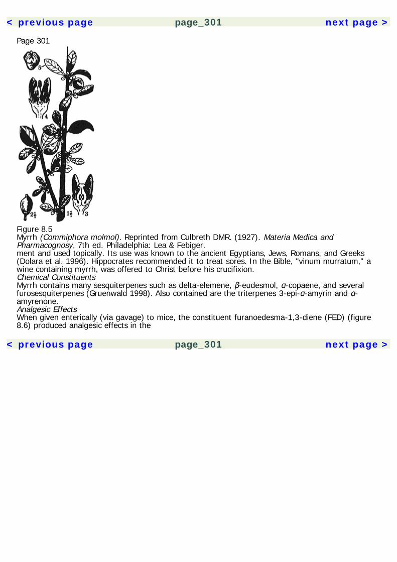
< previous page page_301 next page >
Page 301
Figure 8.5Myrrh (Commiphora molmol). Reprinted from Culbreth DMR. (1927). Materia Medica andPharmacognosy, 7th ed. Philadelphia: Lea & Febiger.ment and used topically. Its use was known to the ancient Egyptians, Jews, Romans, and Greeks(Dolara et al. 1996). Hippocrates recommended it to treat sores. In the Bible, ''vinum murratum," awine containing myrrh, was offered to Christ before his crucifixion.Chemical ConstituentsMyrrh contains many sesquiterpenes such as delta-elemene, β-eudesmol, α-copaene, and severalfurosesquiterpenes (Gruenwald 1998). Also contained are the triterpenes 3-epi-α-amyrin and α-amyrenone.Analgesic EffectsWhen given enterically (via gavage) to mice, the constituent furanoedesma-1,3-diene (FED) (figure8.6) produced analgesic effects in the
< previous page page_301 next page >

< previous page page_302 next page >
Page 302
Figure 8.6Chemical structure of analgesic myrrh constituents.acetic acid writhing test and hot-plate test (Dolara et al. 1996). A dose of 1.25 mg/kg of FED wascomparable to 5 mg/kg PO of morphine. Further, the FED analgesia was reversible by naloxone,indicating the involvement of opioid receptors. FED also displaced [3H]diprenorphine from centralopioid receptors, but with a high inhibition constant.It is unlikely that myrrh will be used clinically for analgesic purposes, given the presence of othersuperior analgesics. However, it is mentioned here for the historical and ethnopharmacologicalaspects.Cholinergic AnalgesicsGiven the role of acetylcholine receptors in analgesic circuits, it is not surprising that a number ofplants with cholinergic mechanisms have analgesic effects. The RVM has muscarinic and nicotinicreceptors (Cortés and Palacios 1986; London et al. 1985). The PAG has nicotinic receptors as well(Segal et al. 1978).The stimulant aspects of nicotine, the primary alkaloid from tobacco, are discussed in greater detail inchapter 4. But nicotine also has significant analgesic effects. Nicotine produces analgesia wheninjected into the midbrain ventrolateral PAG (Llewelyn et al. 1981). Nicotine injected into the PPTN orRVM increases hot-plate and tail-flick latencies in rats, which is blocked by muscarinic drugs in bothsites (Iwamoto 1989, 1991). Recently, a high-potency nicotinic agonist has been developed, ABT-594, that has analgesic efficacy comparable to morphine in a number of measures (Bannon et al.1998). Cholinergic analgesic mechanisms also interact with opioid mechanisms. Subanalgesic doses ofnicotine
< previous page page_302 next page >

< previous page page_303 next page >
Page 303potentiate morphine analgesia, and a cross-tolerance develops between nicotine and morphine(Zarrindast et al. 1996, 1999). Nicotine only potentiates morphine analgesia, and not that induced byβ-endorphin, a δ1 agonist (DPDPE), or κ agonist (U50,488H) (Suh et al. 1996). Nicotine analgesiaalone seems to depend on endogenous opioid mechanisms (Zarrindast et al. 1997).Similar to tobacco, lobelia may also have analgesic effects. However, it depends on the mode ofadministration (Damaj et al. 1997). Intrathecal lobeline produces analgesia on the tail-flick test, butsubcutaneous administration is ineffective. On the other hand, subcutaneous lobeline dose-dependently enhances nicotine analgesia. Tolerance develops to this effect of lobeline after 10 days.Lobeline can also produce hyperalgesic effects when administered into the dorsal posteriormesencephalic tegmentum (Hamann and Martin 1994). However, the relevance of this to peripheraladministration of lobelia is questionable because chronic injections (IP) of lobeline in rats induced nochanges in tail-flick latencies (Sopranzi et al. 1991).Areca is another stimulant plant with cholinergic mechanisms and analgesic effects. Its chemicalconstituents (arecoline, arecaidine, guvacoline, and guvacine) have muscarinic agonist effects as wellas GABAergic mechanisms. However, the muscarinic actions are sufficient to explain arecoline'sanalgesic effect in rats on the tail-flick test, because they are prevented by cholinergic antagonists(Gower 1987).Eicosanoid AnalgesicsWillowHistory and BotanyWillows are several species of the Salix genus. Salix nigra is the American willow. It is a tree growing6 to 18 meters in height with gray bark. The leaves are small and lanceolate. Its flowers are yellowor green, depending on the sex of the tree. The medicinal part of the tree used is its bark.Willow bark has been used throughout recorded history to treat pain, fevers, and inflammation. Itsuses have been independently discovered in cultures as diverse as the ancient Greeks, Hottentots,and American
< previous page page_303 next page >
< previous page page_304 next page >
Page 304Indians. The active principle, salicin, was isolated in 1829. It is chemically converted to the activeprinciple, salicylic acid, in the intestine and liver. The chemically modified acetylsalicylic acid was firstmarketed in 1899 as aspirin. Salicin is also found in meadowsweet (Filipendula ulmaria, then referredto as Spireaea ulmaria), from which the name aspirin derives (acetyl spiric acid). The sodium salt ofsalicylic acid has the drawback of producing gastrointestinal irritation, but acetylsalicylic acid is welltolerated.Eventually the drug was synthesized rather than extracted from willow. Until the invention ofacetaminophen, ibuprofen, and other related drugs, aspirin was the most widely used analgesic drug.Between 10,000 and 20,000 tons of aspirin are consumed annually in the United States (Insel 1996).Chemical ConstituentsSalix species contain several glycosides and esters yielding salicylic acid, including salicin (0.1–2%),salicortin (0.01–11%), and other salicylin derivatives (Gruenwald et al. 1998). Also contained in Salixspecies are tannins and flavonoids.PharmacokineticsSalicin occurs in small amounts in willow, making raw preparations impractical for treatment (Tyler1994). Between 3 and 20 cups of willow bark tea, depending on salicin content, would be necessaryto get an effect. At such high doses, the presence of tannins in the bark would also cause aversiveeffects. Thus, aspirin is needed to reach therapeutic effects.Orally ingested aspirin is rapidly absorbed and significant blood levels occur within 30 minutes,peaking at 2 hours. Rectal absorption is slower and less reliable, but absorption through the skin isfairly rapid. Salicylates are distributed to most body tissues, and 80 to 90% is bound to plasmaproteins such as albumin. Aspirin competes for several plasma binding sites, including those sharedby certain thyroid hormones (thyroxine, triiodothyronine), phenytoin, bilirubin, penicillin, and otherNSAIDs. The plasma half-life of aspirin is 15 minutes, but salicylate has a half life of 2 to 3 hours.The major metabolic products are salicyluric acid, and glucuronide conjugates. Some salicylic acid isexrcreted in urine unchanged.
< previous page page_304 next page >
< previous page page_305 next page >
Page 305Mechanisms of ActionThe therapeutic effects of aspirin and other nonsteroidal anti-inflammatory drugs (NSAIDs) derivefrom their ability to inhibit prostaglandin synthesis. Prostaglandins are a subgroup of eicosanoids,synthesized from phospholipids into arachidonic acid, which may then be converted intoprostaglandins by cyclooxygenases. Specifically, aspirin inhibits cyclooxygenase, preventingconversion from arachidonic acid to prostaglandins (Insel 1996).General EffectsSeveral characteristic effects are known of aspirin, which are present to varying degrees in otherNSAIDs. These include analgesic, antipyretic, anti-inflammatory, and anticoagulant effects.Prostaglandins released during tissue injury sensitize the endings of peripheral sensory nerves toother pain transmitters such as histamine and bradykinin. Thus, inhibition of prostaglandins inhibitsthe initiation of pain signals in the periphery, creating analgesia. However, there may also be somecentral analgesic effects of aspirin, through monoamine mechanisms (Shyu and Lin 1985; Taiwo andLevine 1988). Aspirin is an effective analgesic for low- to medium-intensity pain, but is not effectivefor high-intensity pain. Aspirin's effectiveness also varies with the type of pain: it is effective forheadache and musculoskeletal pain, less effective for toothaches and sore throat, and ineffective forvisceral and traumatic pain (Insel 1996).In addition to analgesic effects, aspirin has antipyretic effects, reducing fevers caused by infection.Normal body temperature is not lowered by aspirin and it has no effect on hyperthermia induced byexercise. Fevers from infections are initiated by cytokines in the blood stream that enter the brainthrough circumventricular organs and cause production of PGE2 in the hypothalamus (Saper andBreder 1994). Thus, salicylates reduce fever by inhibiting PGE2 production.ToxicityAspirin is generally low in toxicity and produces comparatively little tolerance and no addiction.However, its effects are not entirely benign, especially in certain medical conditions. Acute overdoseof aspirin is fatal in doses of 10 to 30 mg, although some high doses have been reported
< previous page page_305 next page >

< previous page page_306 next page >
Page 306without lethality (Insel 1996). Mild intoxication manifests as headache, dizziness, tinnitus, dimmedvision, hearing difficulty, confusion, sweating, nausea, hyperventilation, and diarrhea. Higher dosescreate skin eruptions, acid-base balance disturbances, and more pronounced disturbance of thecentral nervous system, including seizures and coma. The anticoagulant effects of aspirin increasebleeding time and hemorrhage is occasionally seen. Treatment is directed at cardiovascular andrespiratory support, and correction of acid-base imbalances.Reye's syndome is a rare but serious sequela of aspirin use, occuring primarily in people under age20 who were given aspirin during a viral infection, especially influenza or chicken pox. It manifests ascontinuous vomiting, disorientation, behavior changes, and lethargy. Encephalopathy and death areoccasional consequences, with the mortality rate around 31% (Belay 1999). Since it was reported in1980, there has been a steep decrease in the number of cases. A rediagnosis of many cases alsofound a high percentage (69%) to have an underlying metabolic disorder that mimicked the clinicalsymptoms of Reye's syndrome.FeverfewHistory and BotanyFeverfew (Tanacetum parthenium) has a traditional reputation for treating pain and anxiety. It is astrongly aromatic flowering plant growing 15 to 60 cm in height, with daisylike flowers (figure 8.7)(Gruenwald 1998). Feverfew originated in southeast Europe, but now grows in most parts of Europe,as well as Australia and North America. It may be consumed by chewing fresh or freeze-dried leaves,but extracts are now also commercially available. Its modern uses include treatment of arthritis,rheumatic illnesses, migraine, and, as the name suggests, fevers.Chemical Constituents and MechanismsThe primary active chemical constituent in feverfew is parthenolide, a sesquiterpenoid lactone (figure8.8) (Robbers et al. 1996). Levels of sesquiterpene lactones vary across different types of extracts:ethanol extracts contain about 0.5%, whereas aqueous ones contain 0.3% (Gromek et al. 1991). Thesesquiterpene lactones in feverfew inhibit the
< previous page page_306 next page >
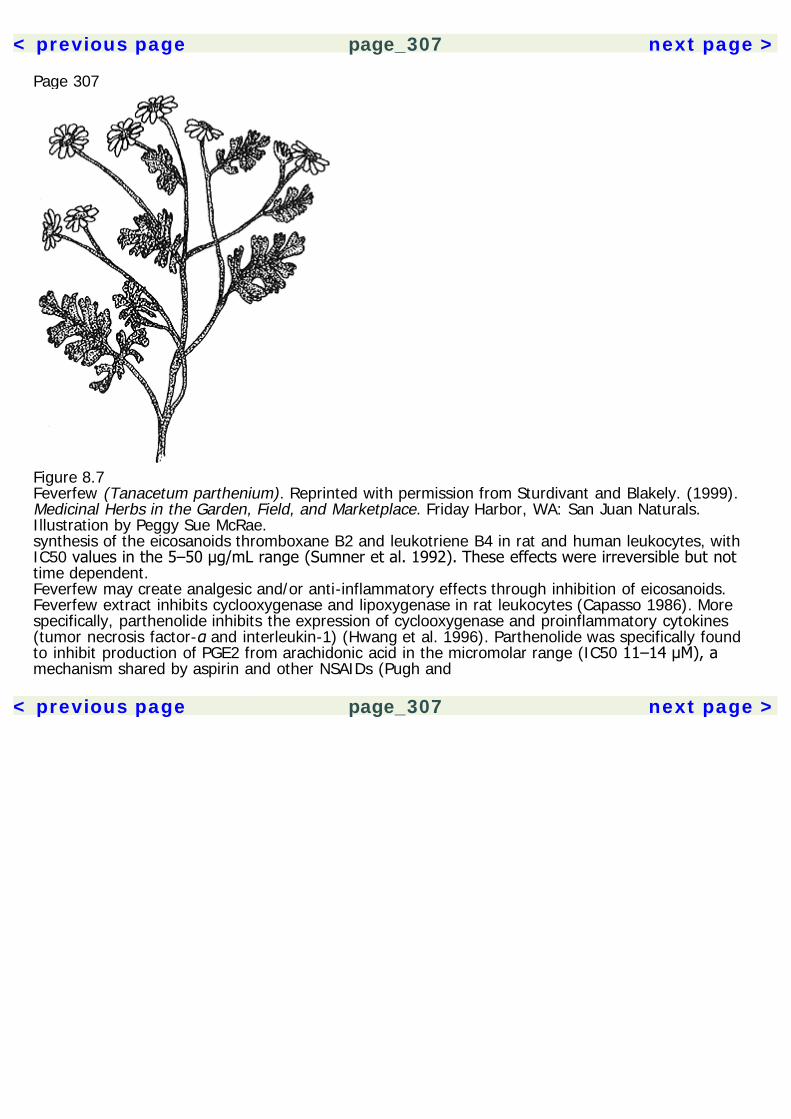
< previous page page_307 next page >
Page 307
Figure 8.7Feverfew (Tanacetum parthenium). Reprinted with permission from Sturdivant and Blakely. (1999).Medicinal Herbs in the Garden, Field, and Marketplace. Friday Harbor, WA: San Juan Naturals.Illustration by Peggy Sue McRae.synthesis of the eicosanoids thromboxane B2 and leukotriene B4 in rat and human leukocytes, withIC50 values in the 5–50 μg/mL range (Sumner et al. 1992). These effects were irreversible but nottime dependent.Feverfew may create analgesic and/or anti-inflammatory effects through inhibition of eicosanoids.Feverfew extract inhibits cyclooxygenase and lipoxygenase in rat leukocytes (Capasso 1986). Morespecifically, parthenolide inhibits the expression of cyclooxygenase and proinflammatory cytokines(tumor necrosis factor-α and interleukin-1) (Hwang et al. 1996). Parthenolide was specifically foundto inhibit production of PGE2 from arachidonic acid in the micromolar range (IC50 11–14 μM), amechanism shared by aspirin and other NSAIDs (Pugh and
< previous page page_307 next page >
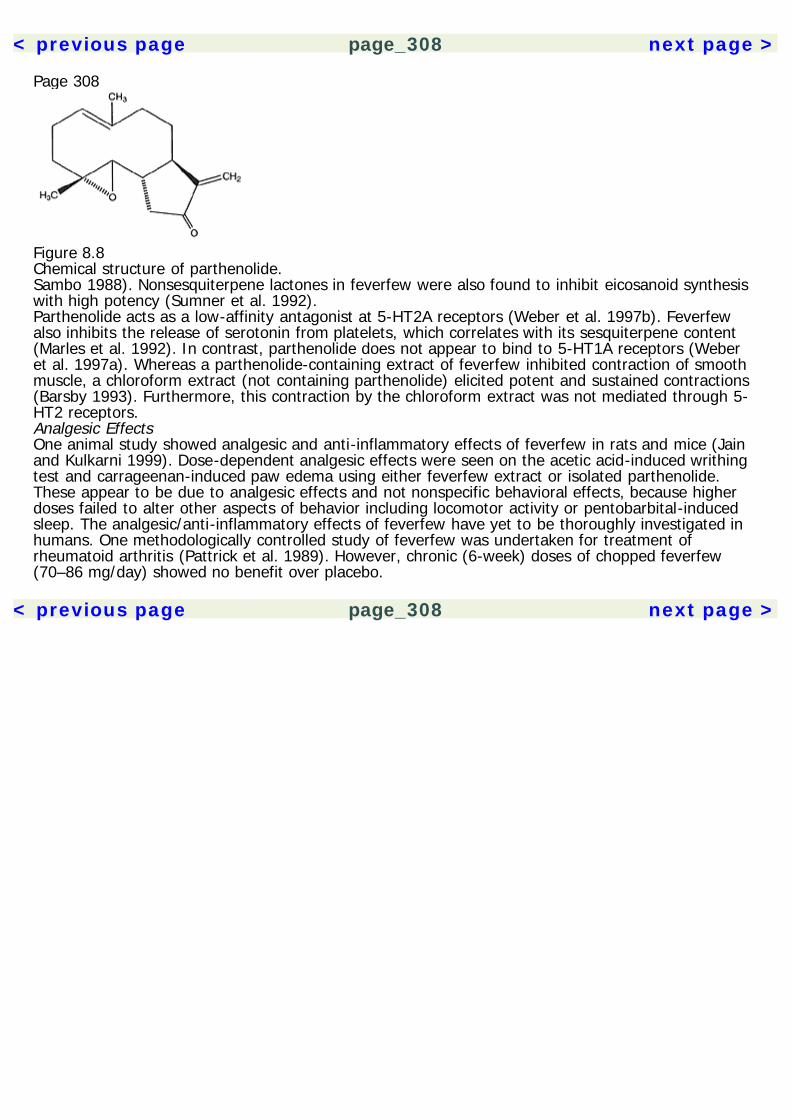
< previous page page_308 next page >
Page 308
Figure 8.8Chemical structure of parthenolide.Sambo 1988). Nonsesquiterpene lactones in feverfew were also found to inhibit eicosanoid synthesiswith high potency (Sumner et al. 1992).Parthenolide acts as a low-affinity antagonist at 5-HT2A receptors (Weber et al. 1997b). Feverfewalso inhibits the release of serotonin from platelets, which correlates with its sesquiterpene content(Marles et al. 1992). In contrast, parthenolide does not appear to bind to 5-HT1A receptors (Weberet al. 1997a). Whereas a parthenolide-containing extract of feverfew inhibited contraction of smoothmuscle, a chloroform extract (not containing parthenolide) elicited potent and sustained contractions(Barsby 1993). Furthermore, this contraction by the chloroform extract was not mediated through 5-HT2 receptors.Analgesic EffectsOne animal study showed analgesic and anti-inflammatory effects of feverfew in rats and mice (Jainand Kulkarni 1999). Dose-dependent analgesic effects were seen on the acetic acid-induced writhingtest and carrageenan-induced paw edema using either feverfew extract or isolated parthenolide.These appear to be due to analgesic effects and not nonspecific behavioral effects, because higherdoses failed to alter other aspects of behavior including locomotor activity or pentobarbital-inducedsleep. The analgesic/anti-inflammatory effects of feverfew have yet to be thoroughly investigated inhumans. One methodologically controlled study of feverfew was undertaken for treatment ofrheumatoid arthritis (Pattrick et al. 1989). However, chronic (6-week) doses of chopped feverfew(70–86 mg/day) showed no benefit over placebo.
< previous page page_308 next page >
< previous page page_309 next page >
Page 309The analgesic effects of feverfew have not been tested in humans. Feverfew's inhibitory effects onthe eicosanoid system, especially on cyclooxygenase and PGE2, is the most likely mechanism of itsanalgesic effects. Inhibition of PGE2 could also account for feverfew's reputed antipyretic effects.Analgesic effects of feverfew through central 5-HT2A receptors are another possibility. Central 5-HT2A mechanisms may be partly responsible for the analgesic effects of acetaminophen (Srikiatkha-chorn et al. 1999). Peripheral administration of a 5-HT2 antagonist (ketanserin) has analgesic effects,perhaps through removal of inhibition of descending serotonergic projections (Alhaider 1991). On theother hand, administration of ketanserin also inhibits the analgesic effects of morphine, as well asopioid and nonopioid forms of stress-induced analgesia (Paul et al. 1988; Kellstein et al. 1988; Kiefelet al. 1989). Thus, the analgesic effects of feverfew through central serotonergic mechanisms is apossiblity, but is not a necessary conclusion. Of course, this is also contingent on whether feverfewconstituents penetrate the blood-brain barrier, which has not yet been specifically investigated.Feverfew appears to treat migraines through vascular serotonin receptors. Some clinical studies offeverfew support its use in the prevention of migraine (Murphy et al. 1998; Johnson et al. 1985). Thisincludes a reduction in the number and severity of attacks, and in the degree of vomiting caused bythe attacks. A systematic review of clinical trials of feverfew for migraine identified fivemethodologically controlled studies (Vogler et al. 1998). Although the majority of studies supportfeverfew in this respect, its effectiveness has not yet been established beyond doubt.ToxicityNo significant adverse effects are known to result from typical doses of feverfew (Gruenwald 1998).A contact dermatitis occurs infrequently. Because feverfew also inhibits human blood plateletaggregation, interactions are possible with antithrombotic medications such as aspirin or warfarin(Groenewegen and Heptinstall 1990). Abrupt discontinuation of feverfew by people taking itchronically for treatment of migraine can produce rebound withdrawal symptoms. These consist ofmigraines, anxiety, poor sleep patterns, and stiffness of the muscles and joints.
< previous page page_309 next page >

< previous page page_310 next page >
Page 310
Figure 8.9Chili pepper (Capsicum frutescens). Reprinted from Culbreth DMR. (1927). Materia Medica andPharmacognosy, 7th ed. Philadelphia: Lea & Febiger.Neurokinin AnalgesicsChili PeppersHistory and BotanyChili or Cayenne peppers (Capsicum annum, C. frutescens) are members of the Solanaceae familyand are characteristically known for the burning sensation they produce on contact (figure 8.9)(Gruenwald 1998; Robbers et al. 1996). It is estimated that they are eaten on a daily basis by one-quarter of the world's population. Numerous varieties are available, including the ultrahot habanero.C. annum is an annual plant cultivated in temperate to semitropical climates. It grows 20–100 cm inheight with solitary leaves. The fruit produced (or peppers) are hollow and boxlike (hence the namederivation from the Latin capsa, or box). The wall of the fruit is tough and may be red, yellow-green,or brownish. Its seeds are numerous, small, and disc shaped (figure 8.10). Although indigenous toCentral America, the plant was later introduced
< previous page page_310 next page >
< previous page page_311 next page >
Page 311
Figure 8.10Fruit of Capsicum annuum. Reprinted from Culbreth DMR. (1927). Materia Medica andPharmacognosy, 7th ed. Philadelphia: Lea & Febiger.to Asia and Africa by the Portugese. It is now cultivated in many areas with warmer climates.The first known written record of chilies dates to 1494, by the physician Chauca, who accompaniedColumbus to the West Indies. Chili is the Nahuatl word for the plant (Szallasi and Blumberg 1999).It is interesting to note that most of our modern uses of chili peppers are preceded by ancientMesoamericans. Capsaicin, the active chemical constituent, is used today as an irritant in self-defensesprays, and dried chilis were burned by the Inca to blind the invading Spaniards. Just as we applycapsaicin for analgesia, native Americans used to rub chili on the gums to relieve the pain of atoothache. Eunuchs serving Chinese Emperors were castrated after hot pepper extracts werechronically applied to their scrotums.Capsaicin was extracted and named by Thomas Thresh in 1846, and its chemical structure wasdetermined in 1919. The Scoville Unit was also developed in 1912 as a measurement of the hotnessof chili peppers (Scoville 1912). A pepper with 1000 Scoville Units means that an alcoholic extractmust be diluted 1:1000 before it ceases to be hot. The Mexican habanero pepper has an astonishing350,000 Scoville Units.
< previous page page_311 next page >
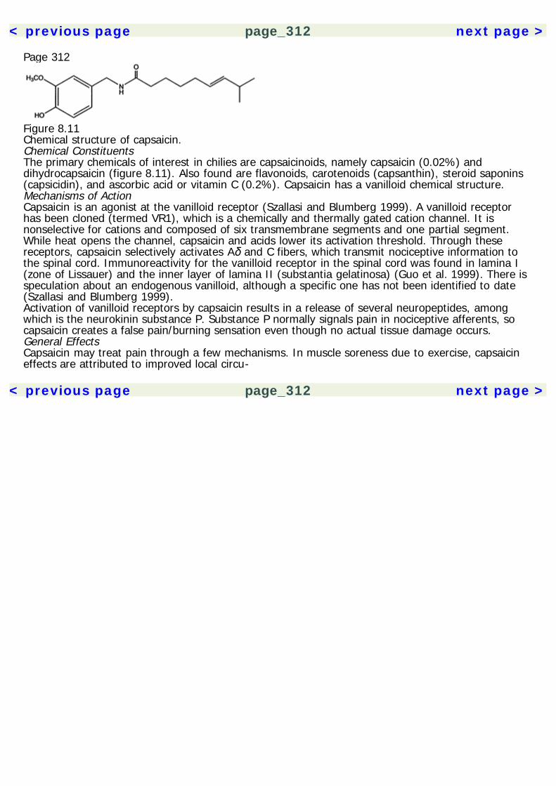
< previous page page_312 next page >
Page 312
Figure 8.11Chemical structure of capsaicin.Chemical ConstituentsThe primary chemicals of interest in chilies are capsaicinoids, namely capsaicin (0.02%) anddihydrocapsaicin (figure 8.11). Also found are flavonoids, carotenoids (capsanthin), steroid saponins(capsicidin), and ascorbic acid or vitamin C (0.2%). Capsaicin has a vanilloid chemical structure.Mechanisms of ActionCapsaicin is an agonist at the vanilloid receptor (Szallasi and Blumberg 1999). A vanilloid receptorhas been cloned (termed VR1), which is a chemically and thermally gated cation channel. It isnonselective for cations and composed of six transmembrane segments and one partial segment.While heat opens the channel, capsaicin and acids lower its activation threshold. Through thesereceptors, capsaicin selectively activates Aδ and C fibers, which transmit nociceptive information tothe spinal cord. Immunoreactivity for the vanilloid receptor in the spinal cord was found in lamina I(zone of Lissauer) and the inner layer of lamina II (substantia gelatinosa) (Guo et al. 1999). There isspeculation about an endogenous vanilloid, although a specific one has not been identified to date(Szallasi and Blumberg 1999).Activation of vanilloid receptors by capsaicin results in a release of several neuropeptides, amongwhich is the neurokinin substance P. Substance P normally signals pain in nociceptive afferents, socapsaicin creates a false pain/burning sensation even though no actual tissue damage occurs.General EffectsCapsaicin may treat pain through a few mechanisms. In muscle soreness due to exercise, capsaicineffects are attributed to improved local circu-
< previous page page_312 next page >

< previous page page_313 next page >
Page 313lation (Szallasi and Blumberg 1999). Acutely, capsaicin may affect other forms of pain throughcounterirritation, although the mechanisms of this are not well understood. When administeredchronically, capsaicin depletes substance P, by continuously releasing it and downregulating itsexpression. There is also a desensitization of vanilloid receptors after the initial stimulation.Therapeutic use of capsaicin is primarily through a topical cream containing 0.025–0.075% of thechemical. The cream is applied locally to treat various forms of pain, including those associated withrheumatoid arthritis, osteoarthritis, postherpetic neuralgia, diabetic neuropathy, postmastectomy andpostamputation neuroma (or phantom limb pain). When the cream is applied acutely, a mild burningsensation occurs. But when applied 4 to 5 times daily for several days to weeks, the painfulsensation may subside. Unfortunately, it is not possible to do double-blind studies of capsaicin due tothe burning sensation it produces.ToxicityThe predominant adverse effect of clinical use of capsaicin is the burning sensation it produces. Manyfind it intolerable, and withdrawal rates from clinical studies have been reported as 30% or higher(Szallasi and Blumberg 1999). Excessive ingestion of chili peppers can cause visceral pain, increasedperistalsis and diarrhea (Gruenwald 1998). Excessive external application can cause blister and ulcerformation. Very high doses can cause a serious hypothermia.An occasional common side effect of capsaicin exposure is bronchoconstriction, in asthmatic people,caused by inhaled airborne particles. In nonasthmatic people, this induces a cough. This is preventedby washing the skin surface where capsaicin was applied after 30–40 minutes of exposure. Chronicoverdosage can cause chronic gastritis, kidney and liver toxicity, and neurotoxic effects.GingerGinger is a rhizome, or underground stem, of the plant Zingiber officinale. It is discussed in greaterdetail in chapter 7, but will be mentioned briefly here for its analgesic effects. Micromolar doses of(6)-shogaol have been shown to dose-dependently cause a release of substance P
< previous page page_313 next page >
< previous page page_314 next page >
Page 314from spinal dorsal horn in a Ca2+-dependent manner, similar to capsaicin from chili peppers (Onogiet al. 1992). However, this effect is 1/50 as potent as capsaicin, where 100 μM of shogaol produceshalf the effect of 10 μM of capsaicin.The clincal usefulness of this effect of ginger is not established. Ginger does have analgesic effects inanimals (Suekawa et al. 1984). Systemically administered (6)-shogaol at 160 mg/kg in rats producesan analgesic effect, peaking between 15 and 30 minutes. However, a smaller dose of 80 mg/kg isineffective. Ginger extract was ineffective in supressing writhing induced by intraperitoneal aceticacid. Ginger's analgesic effects have not been formally tested in humans, but one study examinedthe postsurgical antiemetic effects of oral ginger and failed to observe any differences in the need forpostoperative analgesics between placebo and ginger groups (Philips et al. 1993). A more specificstudy would be needed to confirm or rule out clinically relevant analgesic effects of ginger in humans.Purinergic AnalgesicsThere are several herbal medicines that contain the methylxanthines caffeine, theobromine, andtheophylline. These are stimulant drugs, which are discussed in greater detail in chapter 4. Namely,they are coffee (Coffea arabica), tea (Camellia sinensis), cocoa (Theobroma cacao), guarana(Paullinia cupana), maté (Ilex paraguariensis), and kola (Cola nitida and C. acuminata). The primarypharmacodynamic mechanism of the methylxanthines is antagonism of presynaptic adenosinereceptors. This prevents the synaptic negative feedback of adenosine and increases neurotransmitterrelease. The methylxanthines are mentioned here briefly for their analgesic effects. Adenosine andadenosine triphosphate (ATP) are involved in pain transmission at peripheral and spinal sites(Sawynok 1998).Caffeine alone may produce analgesic effects in some forms of pain, but clinically it is most oftenused as an adjuvant medication (Laska et al. 1983; Ghelardini et al. 1997; Kraetsch et al. 1996;Forbes et al. 1991). It enhances the analgesic effects of nonsteroidal anti-inflammatory drugs such asacetaminophen and also speeds the onset of analgesia. It is effec-
< previous page page_314 next page >
< previous page page_315 next page >
Page 315tive in different types of clinical pain, including uterine cramping, episiotomy, and molar extraction.Caffeine and theophylline potentiate morphine analgesia in rats (Malec and Michalska 1988; Misra etal. 1985). Chronic caffeine lowers the ED50 for morphine analgesia (Ahlijanian and Takemori 1986).Conversely, peripheral administration of adenosine agonists (2-chloroadenosine and L-N6-phenylisopropyladenosine) reduce morphine analgesia in rats on the tail-flick tests (Mantegazza et al.1984). Caffeine prevents the reduction of morphine analgesia by adenosine agonists. The interactionof purinergic drugs and morphine analgesia is believed to be mediated by actions on adenylatecyclase.There is also evidence for cholinergic involvement in caffeine analgesia (Ghelardini et al. 1997). Themuscarinic antagonists atropine and pirenzepine, and the choline uptake inhibitor hemicholinium-3prevent caffeine analgesia. In contrast, it was unaffected by an opioid antagonist (naloxone) or atyrosine hydroxylase inhibitor (α-methyl-p-tyrosine).The role of caffeine in migraine headaches is due to its vasoconstrictor effects. Caffeine is used incombination with ergotamine for migraine (Sawynok 1995; Sargent et al. 1988). However,discontinuation or missing a dose of caffeine after chronic use can precipitate headaches (Fennelly etal. 1991; Couturier et al. 1992).Cannabinoid AnalgesiaCannabinoids are known to reduce pain. THC, the principal active constituent of Cannabis species, iseffective in models of both phasic and tonic pain (Moss and Johnson 1980). Several othercannabinoids also have analgesic effects at the level of the spinal cord, including 11-hydroxy-delta 9-THC, delta 9-THC, and delta 8-THC, although they each have different ED50 values (Welch andStevens 1992). The analgesia produced by different cannabinoids is differentially blocked by a CB1antagonist (SR141716A), with 15-fold differences in some cases (Welch et al. 1998). On the otherhand, some cannabinoids antagonize the analgesia produced by others. For example, cannabidioldose-dependently antagonizes the analgesia produced by THC and cannabinol (Welburn et al. 1976).
< previous page page_315 next page >
< previous page page_316 next page >
Page 316The analgesic effects are mediated through CB1 receptors, and reversed by cannabinoid antagonists(Lichtman and Martin 1997; Vivian et al. 1998). THC produces analgesic effects at both spinal andsupraspinal levels (Reche et al. 1996). THC activates structures in the descending pain inhibitingsystem, involving the RVM (Basbaum and Fields 1978, 1984; Meng et al. 1998). Cannabinoid agonistsinjected into the RVM elevate tail-flick latencies by more than 50%, which is reduced by a CB1antagonist (Martin et al. 1998). Actions of THC in the RVM are similar to morphine butpharmacologically distinct (Meng et al. 1998). Cannabinoid antagonists reverse THC analgesia atsupraspinal sites more effectively than at spinal sites (Welch et al. 1998). Still, THC producesanalgesia in mice with spinal transections, suggesting independent spinal mechanisms (Smith andMartin 1992).The cellular mechanism of cannabinoid analgesia is uncertain. Although cAMP and adenylate cyclasemediate other effects of cannabinoids, they do not appear to be involved in cannabinoid-inducedanalgesia (Cook et al. 1995). Instead, other mechanisms such as cannabinoid receptor-coupledcalcium or potassium channels may be responsible.THC and Opioid AnalgesiaThere are several lines of evidence that suggest opioid and cannabinoid systems interact inanalgesia. Inhibition of opioid peptidases potentiates THC analgesia, which is blocked by supraspinalμ and spinal κ opioid receptors (Reche et al. 1998). An intrathecally administered κ antagonist (nor-binaltorphimine) blocks THC cannabinoid analgesia, but not other physiological effects ofcannabinoids (e.g., hypothermia, hypoactivity, and catalepsy) (Smith et al. 1994). Cross-toleranceoccurs between morphine and THC for analgesic and hypothermic effects (Bloom and Dewey 1978).Cross-tolerance occurs at the spinal level between THC and κ agonists (U-50,488H and CI-977) onanalgesic tests, but not between THC and agonists of μ (DAMGO) or δ (DPDPE) receptors.Convergently, antisense studies suggest that cannabinoid analgesia is related to spinal κ1, but not μor δ receptors (Pugh et al. 1995).Synergistic effects are produced by co-administration of morphine and cannabinoids (Pugh et al.1996; Welch and Stevens 1992). THC en-
< previous page page_316 next page >

< previous page page_317 next page >
Page 317hances morphine analgesia when with either central (intracerebroventricular or intrathecal) orperipheral (PO or SC) administration (Smith et al. 1998). The ED50 of THC is reduced by morphine,and vice versa, on several measures of nociceptive threshold (the tail-flick test, hot-plate test, jumptest, and paw-lick responses) (Reche et al. 1996; Welch and Stevens 1992; Hine 1985). THCenhancement of morphine analgesia is mediated by the CB1 receptor and reduced with a selectiveCB1 antagonist. Supraspinal synergy between morphine and THC also involves μ and spinal κ opioidreceptors (Reche et al. 1996; Pugh et al. 1996).Nonopioid Mechanisms of THC AnalgesiaAlthough cannabinoids interact with opioids in analgesia, there also appear to be opioid-independentmechanisms (Hamann and di Vadi 1999). The analgesia produced by cannabinoids alone is notabolished by pretreatment with the general opioid antagonist naloxone (Gilbert 1981; Welch andStevens 1992; Ferri et al. 1986).There is evidence to suggest that cannabinoids create analgesia by activating descendingnoradrenergic neurons. The catecholamine neurotoxin, 6-hydroxydopamine, reduces THC analgesia(Ferri et al. 1986). Further, THC analgesia is blocked by an intrathecal α2-noradrenergic antagonist(yohimbine), but not by a nonspecific serotonin antagonist (methysergide) (Lichtman and Martin1991). Yohimbine blocks THC analgesia at lumbar but not thoracic regions.Some cannabinoids may create analgesia though eicosanoid systems. Delta 1-THC-7-oic acid, ametabolite of delta 1-THC, creates analgesia through inhibition of eicosanoid synthesis (Burstein et al.1988). In contrast, delta 1-THC creates hyperalgesic effects by elevating levels of prostaglandins.Endocannabinoids in AnalgesiaSimilar to exogenous cannabinoids, anandamide has analgesic effects (Fride and Mechoulam 1993).Peripheral anandamide (10 mg/kg IM) produces antinociception in primates. However, centrallyadministered anandamide does not alter pain perceptive thresholds (Murillo-
< previous page page_317 next page >
< previous page page_318 next page >
Page 318Rodriguez 1998). THC- and anandamide-induced analgesia also have different durations (90 min and15 min, respectively) (Smith et al. 1998).Similarities in receptor binding are observed between cannabinoids and anandamide. Binding to CBreceptors by anandamide, a selective CB1 antagonist (SR 141716A), and CB agonist (CP 55,940)shows that all three ligands compete for the same cannabinoid receptor (Adams et al. 1998). Still,THC and anandamide may create analgesia through different mechanisms. CB1 antagonists blockTHC analgesia but have no effect on anandamide analgesia, strongly suggesting different sites ofaction for THC and anandamide in some processes (Smith et al. 1998; Adams et al. 1998).Anandamide also has pharmacological effects distinct from CB receptors: anandamide, but not THC,has direct effects on the NMDA receptor (Hampson et al. 1998). The cross-tolerance betweenanandamide and THC in analgesic paradigms was differentially cross-tolerant to opioids. While THCproduced cross tolerance to κ opioids, anandamide did not (Welch 1997).Anandamide may reduce pain by a peripheral action, by acting on CB1-like receptors located outsidethe CNS (Calignano et al. 1998). Palmitylethanolamide (PEA) is an endocannabinoid that iscoreleased with anandamide and activates peripheral CB2 receptors. When the two are administeredtogether, they show a 100-fold synergistic effect on analgesic measures. Measurements ofanandamide and PEA levels in the skin show that there are sufficient amounts to create tonicactivation of local cannabinoid receptors. Thus, endocannabinoids may tonically inhibit cutaneouspain.Analgesic Effects of THC in HumansThe clinical analgesic efficacy of THC is prominent. In a study of cancer patients, 10 mg of THC wasequivalent to 60 mg of codeine (Noyes et al. 1975). This dose produced sedative but no otherpsychoactive effects. A dose of 20 mg was more effective, but produced additional dizziness, ataxia,blurred vision, and psychoactive effects.The analgesic response to THC may be variable and appears to depend on personality characteristicsof the subject (Raft et al. 1977).
< previous page page_318 next page >
< previous page page_319 next page >
Page 319Objective measures of personality found those with the characterstics of submissiveness, rigidity, lowintrospection, and high-state anxiety to be poor responders to the analgesic effects of THC.Unfortunately, although there is ample evidence to support the analgesic efficacy of cannabinoids,there is little clinical research to match it.Monoamine AnalgesicsKhatKhat alkaloids are monoamine stimulants, causing a release of neurotransmitters in a manner similarto amphetamine (see chapter 4). The major alkaloid cathinone has analgesic effects on the tail-flicktest in rats (Nencini et al. 1984). It also enhances the analgesia induced by brief electric foot shock(Nencini et al. 1988). The analgesia produced by cathinone is long lasting, with effects seen at both30 minutes and 24 hours. Cathinone-induced analgesia depends on α adrenergic receptors, followedby release of opioid peptides and by activation of serotonergic pathways (Della Bella et al. 1985). Itis prevented by monoamine depletion with reserpine or p-chlorophenylalanine. Cathinone potentiatesmorphine analgesia, and cathinone analgesia is blocked by naloxone (Della Bella et al. 1985; Nenciniet al. 1984). The cathinone metabolite norpseudoephedrine does not have intrinsic analgesic effectson the hot-plate test, but it potentiates the analgesic effects of morphine (Nencini et al. 1998). Incontrast, there is no apparent contribution from adrenohypophyseal hormones to cathinone analgesia(Nencini et al. 1984).CocaCocaine, from the coca plant, is one of the few drugs with dual analgesic and local anesthetic effects.These are distinct actions, which occur through different pharmacological mechanisms. (The localanesthetic effects are discussed below.) Given the role of monoamines in analgesia, it is notsurprising that cocaine has analgesic effects in the central nervous system. This is likely to involvedopaminergic mechanisms at both
< previous page page_319 next page >
< previous page page_320 next page >
Page 320supraspinal and spinal levels (Lin et al. 1989; Pertovaara and Tukeva 1990; Kiritsy-Roy et al. 1994).Cocaine alone produces analgesia in the hot-plate, but not the paw-pressure test (Waddell andHoltzman 1999).Interactions are seen between cocaine and the opioid system in analgesia. A synergistic effect occurswhen cocaine is combined with a μ agonist (morphine) in the hot-plate test and a κ agonist(U69,593) in the hot-plate test (Waddell and Holtzman 1999). Cocaine enhances morphine analgesiain several analgesic paradigms (e.g., formalin test, hot-plate test, and thermal tail-flick test)(Kauppila et al. 1992). The synergistic interaction between cocaine and opioid analgesia likelyinvolves noradrenergic mechanisms, supraspinal μ and δ receptors, and spinal μ receptors (Misra etal. 1987; Sierra et al. 1992).Interestingly, one study has shown that cocaine may be a direct antagonist at nicotinic receptors(Damaj et al. 1999). Cocaine blocks several of nicotine's effects, including analgesia on the tail-flicktest, independent of its monoamine activity or local anesthetic effects.Uncertain Analgesic MechanismsGinsengGinseng species are discussed at length in chapter 5, but mentioned briefly here for their analgesicproperties. Ginsenosides produce dose-dependent analgesic effects in the formalin test whenadministered intrathecally in microgram doses (Yoon et al. 1998). They also dose-dependently inhibitpain behaviors induced by intrathecal injection of substance P. Ginsenoside Rf has dose-dependentanalgesic effects when administered systemically with effects in two measures of pain: acetic acidabdominal constriction test and biphasic formalin test (Mogil et al. 1998). Analgesic effects in theacetic acid abdominal constriction test were comparable at concentrations similar to those reportedfor aspirin and acetaminophen. Ginsenoside Rf is effective in the tonic phase of the formalin test, butineffective in the acute phase. It is also ineffective in the thermal tail-flick and hot-plate tests. Apossible (but not necessary) conclusion is that ginsenoside Rf is effective in tonic but not acute formsof pain. Opioid analgesia is mediated through Ca2+ channels in sensory
< previous page page_320 next page >

< previous page page_321 next page >
Page 321neurons, so it is possible that the block of Ca2+ channels by ginsenoside Rf is a responsiblemechanism. However, it inhibits large, but not small, nociceptors, which is inconsistent with tonicpain. So, mechanisms other than Ca2+ block are likely involved.Systemic (IP) administration of ginseng extract produces analgesia and hypothermia in rats, which isnot reversed by the opioid antagonist naltrexone (Ramarao and Bhargava 1990). However, ginsengreduces the analgesia, hyperthermia, and catalepsy induced by morphine. This effect shows aninverted-U dose-respone curve, where morphine analgesia is blocked by ginseng at 25 and 50 mg/kgdoses, but not by 12.5, 100, and 200 mg/kg doses. Spinal ginsenosides reduce the analgesic effectof supraspinal morphine (intracerebroventricular, 2 μg) in the tail-flick test for at least 6 hours (Suhet al. 1997). Individual ginsenosides having this effect are Rb2, Rc, Rd, and Rg1, but not Rb1. Whileindividual ginsensosides have this effect, total extract of ginseng may not. Ginseng's antagonisticeffects on opioid analgesia may rely on serotonergic mechanisms (Kim et al. 1992). Ginsenosidereduction of κ opioid-induced (U50,488H) analgesia was prevented by treatment with the serotoninprecursor, 5-hydroxytryptophan (5-HTP). The dopamine precursor L-dopa did not have the sameeffect.Constituents of Vietnamese ginseng (Panax vietnamensis) also show analgesic effects andantagonism of opioid analgesia. Majonoside-R2, a saponin from Vietnamese ginseng, dose-dependently reduces analgesia induced by subcutaneous mu (morphine) or kappa (U-50,488H) opioidagonists in the tail-pinch test (Huong et al. 1997). This effect was produced when majonoside-R2 isadministered peripherally (IP) or supraspinally (intracerebroventricular). Also, majonoside-R2 alsoantagonizes the opioid analgesia when they are co-administered intracerebroventricular orintrathecal. Diazepam had similar effects, and both were reversed by the benzodiazepine antagonistflumazenil and the GABA channel blocker picrotoxin. Thus, majonoside-R2 antagonizes opioidanalgesia at spinal and supraspinal levels, at least partly by acting through GABAA receptors.Systemic and central majonoside-R2 also antagonizes analgesia induced by the alpha-2 agonistclonidine in the tail-pinch test (Nguyen et al. 1996, 1997). Again, this antagonistic effect wasreversed by flumazenil and picrotoxin, suggesting GABAA mediation.
< previous page page_321 next page >

< previous page page_322 next page >
Page 322Similar to naloxone, majonoside-R2 also reduces stress-induced analgesia measured by the tail-pinchtest (Nguyen et al. 1995).In summary, species of ginseng have analgesic effects in several animal models. Panax ginsengcreates analgesia through nonopioid mechanisms and is antagonistic to opioid and adrenergicanalgesia. Antagonism of opioid analgesia occurs at spinal and supraspinal levels and is mediated byserotonergic mechanisms. Other putative mechanisms are antagonism of substance P receptors andCa2+ channels. Panax vietnamensis shows similar analgesic effects and antagonism of opioidanalgesia and involves GABAA mechanisms. Despite notable evidence for an analgesic effect ofginseng, there have not been any controlled studies carried out in humans to date. Although it hasnot been examined in the context of analgesia, nonginsenoside constituents of ginseng have nicotinicactivity, which could contribute to analgesic effects.Local AnestheticsLocal anesthesia involves the blockade of nerve conduction in order to stop sensation. Because localanesthetics act on all nerve fibers they may also temporarily create motor paralysis. The usefulness oflocal anesthetics is their ability to completely block axonal transduction, which is reversible andwithout any apparent lasting effects.CocaCocaine was probably the first drug with recognized local anesthetic effects (Catterall and Mackie1996). Natives of Peru have long known that chewing coca leaves produces oral numbness (Youngand MacKenzie 1992). When cocaine was first isolated from the coca plant by Albert Nieman in 1860,he noticed that it had a numbing effect on his tongue when he tasted it. Carl Koller introducedcocaine as a local anesthetic for ophthalmologic surgery in 1884. Given that the euphoric andautonomic effects of cocaine are unwanted during clinical anesthesia, Einhorn and colleagues begansearching for a more tolerable synthetic substitute in 1892, and synthesized procaine (Novocain) in1905. Since then, many related synthetic local anesthetics have been developed,
< previous page page_322 next page >

< previous page page_323 next page >
Page 323
Figure 8.12Chemical structure of cocaine and synthetic local anesthetics.including lidocaine (Xylocaine), bupivacaine (Marcaine), and tetracaine (Pontocaine) (figure 8.12).The use of cocaine for local anesthesia has been largely replaced by synthetic drugs that produceless psychoactive effects. However, cocaine is still used sometimes for topical anesthesia of the upperrespiratory tract.Mechanism of ActionThe local anesthetic actions of cocaine are independent of its well-known actions on monoamines.Rather, the local anesthetic effects occur as a consequence of its interaction with voltage-gated Na+channels (Matthews and Collins 1983). Cocaine's cerebral vasoconstrictor effects occur through localanesthetic rather than sympathomimetic mechanisms (Albuquerque and Kurth 1993).The site of action on the Na+ channel for cocaine and its analogs appears to be on the intracellularside of the channel, so cocaine must evidently cross the cell membrane to be effective (Narahashiand Frazier 1971). The Na+ channel is composed of a large protein divided into α, β1, and β2subunits. The α subunit is further divided into four domains, each of which contains fourtransmembrane segments (designated I–IV).
< previous page page_323 next page >
< previous page page_324 next page >
Page 324This protein is fixed in the cell membrane, and is sensitive to voltage. When the cell is sufficientlydepolarized, a conformational change occurs in the protein, allowing flow of Na+ across themembrane. After several milliseconds, an inactivation gate closes, ceasing the flux of Na+. Theinactivation gate is likely located on an intracellular loop between domains III and IV.The inhibition produced by local anesthetics is both frequency- and voltage-dependent, and alsoreferred to as ''use-dependent block." Nerves that are more stimulated encounter a greater degree ofinhibition, while resting axons are relatively less sensitive (Butterworth et al. 1987). The duration ofaction is prolonged by vasoconstrictors because they keep the drug in contact with the nerve longerrather than it being carried away in the blood stream. Different size nerve fibers also have varyingsensitivity to local anesthetics. Smaller C and Aδ fibers are more sensitive than the larger Aγ, Aβ, andAα fibers. Thus, sensory and autonomic nerves are more susceptible than motor and proprioceptivenerves.Clove OilClove oil is used as a local anesthetic. Clove comes from the tree Syzygium aromaticum (formerlyreferred to as Eugenia caryophyllus or Caryophyllus aromaticus) (Gruenwald et al. 1998; Tyler 1994;Robbers et al. 1996). It is an evergreen tree that grows up to 20 meters in height. The spice derivesfrom the characteristic nail-shape cloves of the tree (figure 8.13). Clove oil, which is the preparationused for local anesthesia, is steam-distilled from the flowers. A large proportion of clove produced isground and added to tobacco. These are referred to as kretek in Indonesia, or as simply as clovecigarettes in the West. They contain approximately 60% tobacco and 40% clove (Clark 1989).Although more popular in Indonesia, they have made their way to the West.Clove was known to the Chinese as early as 266 B.C.E. The Dutch wished to monopolize the trade ofclove in the seventeenth century, and so destroyed all of the trees except those on Ambon andTernate. However, the tree has since been introduced to many other regions. Indigenous to the tothe Molucca Islands, it is also grown on several islands of
< previous page page_324 next page >

< previous page page_325 next page >
Page 325
Figure 8.13Clove (Syzygium aromaticum). Reprinted from Culbreth DMR. (1927). Materia Medica andPharmacognosy, 7th ed. Philadelphia: Lea & Febiger.Africa, Asia, and the West Indies. The majority of the world's supply comes from Tanzania andZanzibar.Chemical ConstituentsThere are several related phenolic compounds found in the volatile oil of clove. The primary one iseugenol, accounting for 85% of the total (figure 8.14) (Tyler 1994; Robbers et al. 1996).Mechanism of ActionThe anesthetic action of clove is attributable to eugenol and its natural analogs. Little is really knownabout the local anesthetic mechanism of eugenol. As mentioned above, eugenol is a phenoliccompound, and phenol itself acts as a local anesthetic (Kizelshteyn et al. 1992). Phenol actssynergistically with bupivacaine, and most likely blocks Na+ channels. Eugenol has not beenspecifically shown to block Na+ channels, but its phenolic structure suggests that it does.Low millimolar concentrations of eugenol activate a Ca2+ channel (Ohkubo and Kitamura 1997).Further, a Cl– channel may indirectly activate in response to the Ca2+ influx. Eugenol acts on Ca2+channels
< previous page page_325 next page >
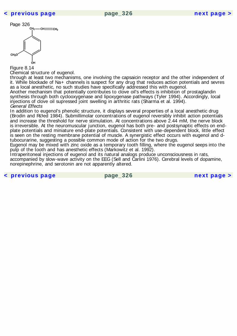
< previous page page_326 next page >
Page 326
Figure 8.14Chemical structure of eugenol.through at least two mechanisms, one involving the capsaicin receptor and the other independent ofit. While blockade of Na+ channels is suspect for any drug that reduces action potentials and sevresas a local anesthetic, no such studies have specifically addressed this with eugenol.Another mechanism that potentially contributes to clove oil's effects is inhibition of prostaglandinsynthesis through both cyclooxygenase and lipoxygenase pathways (Tyler 1994). Accordingly, localinjections of clove oil supressed joint swelling in arthritic rats (Sharma et al. 1994).General EffectsIn addition to eugenol's phenolic structure, it displays several properties of a local anesthetic drug(Brodin and RØed 1984). Submillimolar concentrations of eugenol reversibly inhibit action potentialsand increase the threshold for nerve stimulation. At concentrations above 2.44 mM, the nerve blockis irreversible. At the neuromuscular junction, eugenol has both pre- and postsynaptic effects on end-plate potentials and miniature end-plate potentials. Consistent with use-dependent block, little effectis seen on the resting membrane potential of muscle. A synergistic effect occurs with eugenol and d-tubocurarine, suggesting a possible common mode of action for the two drugs.Eugenol may be mixed with zinc oxide as a temporary tooth filling, where the eugenol seeps into thepulp of the tooth and has anesthetic effects (Markowitz et al. 1992).Intraperitoneal injections of eugenol and its natural analogs produce unconsciousness in rats,accompanied by slow-wave activity on the EEG (Sell and Carlini 1976). Cerebral levels of dopamine,norepinephrine, and serotonin are not apparently altered.
< previous page page_326 next page >
< previous page page_327 next page >
Page 327The central effects of eugenol in humans are uncertain, and what perpetuates clove's addition tocigarettes is also uncertain. However, other local anesthetics have profound effects on the CNS. Forexample, in animals, intravenous procaine activates electrical activity in limbic structures andsuppresses activity in neocortical structures (Servan-Schreiber et al. 1998; Ketter et al. 1996). Inhumans, procaine similarly induces limbic activation, including the anterior cingulate, insula, andamygdala/parahippocampal gyrii. Decreases in activity occur in the left inferior parietal lobule, rightthalamus, and bilateral cerebellum. Reported subjective phenomena include a variety of emotionalexperiences, ranging from euphoria to depression and anxiety. In some instances, the anxietyreaches the intensity of panic, and it correlates with left amygdala activation. Subjects who do notexperience panic have greater activation in the anterior cingulate bilaterally and in the left inferiorfrontal cortex. Thus, local anesthetics produce emotional effects by activation of central limbicstructures. Whether eugenol replicates this pattern remains to be seen.ToxicityClove is listed as Generally Regarded as Safe (GRAS) by the U.S. Food and Drug Administration, andis approved by the German Commission E for use as a local anesthetic.Because clove cigarettes also contain tobacco, they carry all of the hazards of regular tobaccosmoking (Council on Scientific Affairs 1988). Several cases of serious medical illness have beenassociated with clove cigarettes in the United States (Guidotti et al. 1989). These include cases ofhemorrhagic pulmonary edema, pneumonia, bronchitis, and hemoptysis. Because eugenolanesthetizes the respiratory tract, it inhibits the normal gag reflex and has led to aspirationpneumonia in at least one case. Accidental overdoses in children have led to CNS depression, urinaryabnormalities, and anion-gap acidosis. These cases were treated successfully with supportivemeasures (Lane et al. 1991).ConclusionAs is evident in this chapter, a wide variety of plants have been found to have analgesic and/oranesthetic effects. Their pharmacological diversity
< previous page page_327 next page >

< previous page page_328 next page >
Page 328reflects the diverse neurochemistry and neurophysiology of pain and analgesia systems in thenervous system.Some of the plants and drugs discussed here are widely known for their analgesic/anesthetic effects.They have become an integral part of our medical treatment, advanced our understanding of painand analgesia, and led to the development of similar drugs with better clinical properties. Namely,these are the opioids from poppy, salicylates from Salix species, and cocaine from coca. Together,these drugs and their synthetic relatives account for the vast majority of clinical pain relief.The other plants and drugs are discussed for the purpose of elucidating the mechanisms behind theirlong-known clinical effects (e.g., clove). It is also interesting to note that many stimulant drugs(tobacco, areca, lobelia, khat, coca) have incidental but significant analgesic effects. Few peoplesmoke tobacco for the purpose of relieving pain, but it is effective in that regard. It remains to bestudied empirically, but perhaps people smoke tobacco more when in pain without explicitly knowingit.
< previous page page_328 next page >

< previous page page_329 next page >
Page 3299Hallucinogenic PlantsHallucinogenic plants constitute a wide variety of species, all of which profoundly distort perception,cognition, and mood. Although some synthetic hallucinogenic drugs have been produced, themajority of this class are derived from plants. Both traditional shamans and modern scientists areequally captivated by the effects of these drugs, whether for unlocking secrets of the spirit world orfor unlocking secrets of the human mind and brain.The use of hallucinogens is fairly prevalent in western cultures. A 1990 national survey estimated that7.6% of the U.S. population over age 12 had tried a hallucinogenic drug once in their lives, and1.1% had done so within the last 12 months (National Institute on Drug Abuse, 1990). Aftermarijuana, hallucinogens were the most commonly tried illicit drugs. Use of hallucinogens is mostcommon among people between the ages of 18 and 25.TerminologyConsiderable thought has been given to which term most appropriately labels this class of drugs, andhow they are best defined to differentiate them from other drugs with overlapping effects. Thetoxicologist Louis Lewin termed them phantasticants in 1924. Although appropriate, the term is rarelyused today. Psychedelic translates from Greek as ''mind manifesting" or "mind-expanding," but itsimplications are uncertain. William James suggested, "Our normal waking consciousness, rationalconsciousness as we call it, is but one special type of consciousness, whilst all about it, parted from itby the flimsiest of screens, there lie
< previous page page_329 next page >

< previous page page_330 next page >
Page 330potential forms of consciousness entirely different'' (James 1902). In this framework, hallucinogensdemonstrate subjectively how much "external reality" is really an internal construction by the brain.Although this may apply well to the monoamine hallucinogens, the cholinergic hallucinogens cancloud consciousness. They create a state more accurately described as an amnestic delirium than anexpansion of consciousness.The term psychotomimetic was coined to compare the effects of hallucinogens to the symptoms ofschizophrenia and other psychotic conditions. However, the effects produced by hallucinogens andmental illness are qualitatively different. While drug-induced hallucinations tend to be more visual,auditory hallucinations are most common in schizophrenia. The hallucinogenic drugs discussed heremay induce positive symptoms (e.g., hallucinations), but do not induce the negative symptoms (e.g.,cognitive deficits, social withdrawal) of schizophrenia.The term hallucinogen derives from the Latin word alucinari, meaning to "wander in mind" (Abrahamet al. 1996). However, hallucinogens are not the only drugs capable of creating hallucinations. Ifgiven in high enough doses, other drugs such as cocaine, opioids, and cannabis can do the same.Instead, the term is used to label drugs for which hallucinations are their most prominent effect,even at low doses. Thus, a hallucinogen may be defined as "any agent that causes alterations inperception, cognition, and mood as its primary psychobiological actions in the presence of anotherwise clear sensorium" (Abraham et al. 1996). This definition, however, excludes the cholinergichallucinogens because they certainly cloud consciousness.Perhaps there is no completely satisfactory term for this class of drugs, but the term hallucinogen willbe used to collectively refer to them throughout this chapter.Historical Uses of Hallucinogenic PlantsGiven the powerful ability of these plants to alter perceptions, it is not surprising that humans haveascribed magical powers to them. The modern interpretation of the effects of these drugs is rooted intheir effects on the brain, altering the apparatus through which we construct reality. But
< previous page page_330 next page >
< previous page page_331 next page >
Page 331that explanation arose in the context of knowledge about the brain, and the neural basis of thoughtand perception. For generations of people in nonindustrial cultures with traditional beliefs, thesupernatural explanation is the most sensible one. Many cultures, such as those of the Aztec andancient Greeks, incorporated them into religious rituals (Grinspoon and Bakalar 1997; Schultes andHofman 1980, 1992).Modern interest in hallucinogenic drugs increased in the West after the synthesis of LSD by AlbertHofman in 1949. Aldous Huxley wrote perhaps the most famous account of hallucinogens, describinghis experiences with mescaline in his book, The Doors of Perception (Huxley 1954). In the 1960s,some experimented with hallucinogens in an attempt to expand consciousness, while othersproposed that they could create a temporary model of schizophrenia in humans. Hallucinogenic drugswere classified under Schedule I of the Controlled Substances Act of 1970. This restricted their use,asserting that they have a high potential for abuse and lack any therapeutic value. However, the useof peyote by the Native American Church was protected by the Religious Freedom Restoration Act in1993.Classification of HallucinogensThere are many ways to potentially classify hallucinogenic drugs. They are organized here by theirprimary mechanism of action: monoamine, cholinergic, and amino acid. The monoaminehallucinogens are further subclassified as indole (indolealkylamine) or catechol (phenethylamine) instructure, although actions at serotonin receptors are likely to underlie all of their hallucinogeniceffects (Rech and Commissaris 1982).Monoamine HallucinogensThe monoamine hallucinogens are a diverse group of plants, consisting of fungi (ergot andpsilocybe), cacti (peyote, San Pedro, cawe), vines (yage, morning glory, Hawaiian rosewood), andtrees (nutmeg) (table 9.1). They have convergently evolved biochemical constituents that interferewith monoamine activity.
< previous page page_331 next page >

< previous page page_332 next page >
Page 332Table 9.1Monoamine hallucinogens
Common Botanical Type Active principles
Ergot Claviceps purpurea Fungus d-isolysergic acid d-lysergic acidPsilocybe Conocybe silineoides Fungus Psilocin, psilocybin Panaeolus sphinctrinus Psilocybe aztecorum P. caerulescans P. mexicana Stropharia cubensis Peyote Lophophora williamsii Cactus Mescaline, etc. Trichocereus pachanoi Cactus Mescaline 3,4-dimethoxyphenyl-
ethylamine 3-methoxy-tyramineCawe Pachycereus pecten-
aboriginumCactus 4-hydroxy-3-ethoxy-
phenylethylamineYage Banisteriopsis caapi Woody
vineHarmala alkaloidsDimethyltryptamine
Morning Glory OloliuquiBadoh Negroh
Turbina corymbosaIpomoea violacea
Floweringvine
Lysergic acid amideLysergic acid hydroxy-ethylamide
Hawaiian BabyRosewood
Argyreia nervosa Lysergic acid amide
Iboga Tabernanthe iboga Shrub Ibogaine Ibogamine Tabernanthine CoronaridineNutmeg Myristica fragrans Tree Myristicin Elemicin
See text for references.
< previous page page_332 next page >

< previous page page_333 next page >
Page 333
Figure 9.1Ergot (Claviceps purpurea). Reprinted from Barron et al., ''The Hallucinogenic Drugs." ScientificAmerican. April 1964. Illustration by Alex Semenoick.ErgotHistory and BotanyErgot (Claviceps purpurea) is a parasitic fungus that grows on rye (Secale cereale) and other grains,particularly during wet seasons (Gruenwald et al. 1998). It is black and hard, and protrudes from thegrains. Insects carry and deposit the spores, which grow hyphae down into the ovary of the rye toaccess its nutrients. When the ovary is destroyed, a mycelium grows. A long black sclerotium growsto a length of 8 cm, which eventually falls to the ground. In the following spring, pink fruiting bodiesgrow from it, releasing thread-like acospores (figure 9.1).The ergot fungus and its noxious effects were known to the Mesopotamian cultures of Assyria andBabylonia (van Dongen and de Groot 1995). The earliest known written mention of ergot is anAssyrian cuneiform tablet dating circa 600 B.C.E., referring to it as a "noxious pustule in the ear ofgrain." Ironically, it was not until 1676 C.E. that the
< previous page page_333 next page >
< previous page page_334 next page >
Page 334symptoms of ergot poisoning (or ergotism) were associated with ingestion of ergot. The Romansreferred to ergotism as Ignis sacer, or Holy Fire. This name was carried through to the EuropeanMiddle Ages, and refers to the burning sensation it causes in the extremities. Casualties fromergotism were significant: one epidemic in Europe during 944 C.E. was responsible for the deaths of40,000 people (Abraham et al. 1996).Saint Anthony was a monk who lived in the third and fourth centuries C.E. A shrine was named afterhim in the south of France in 1039 C.E., where an outbreak of ergotism occurred. The shrine becamea popular pilgrimage for those suffering from ergotism. Thus, ergotism became known as SaintAnthony's fire. It is speculated that increased peripheral circulation from walking and change in dietaccounted for the healing properties of such a journey.There are still occasional modern outbreaks of ergotism, such as in Ukrania and Ireland in 1929, andin Belgium in 1953. It has been suggested that the Salem witch trials of 1692 may have beensparked by ergotism, although others favor a social psychological explanation for the events(Matossian 1982; Spanos 1983).Chemical ConstituentsErgot possesses two general classes of alkaloids: amine and amino acid (Peroutka 1996). The aminealkaloids have antagonist effects on serotonin receptors, while the amino acid alkaloids are lessselective and act upon other monoamine receptors. While some amine alkaloids are of interest forcognition-enhancing effects (e.g., Hydergine), certain amino acid alkaloids are known for theirhallucinogenic effects. Namely, these are d-isolysergic acid and d-lysergic acid (figure 9.2). LSD (d-lysergic acid diethylamide) is a synthetic compound derived from ergoloids, but it serves as a modelfor plant-derived psychedelics. Both likely work through similar or identical mechanisms and differonly in efficacy or potency. The majority of research in this area has focused on LSD, rather than onnaturally occuring hallucinogenic ergoloids.PharmacologyLSD is odorless and tasteless and is effective in microgram doses. Doses of 25–50 μg produce anintensification of perceptions, while larger doses are needed for full hallucinations. The total durationof the drug effect
< previous page page_334 next page >
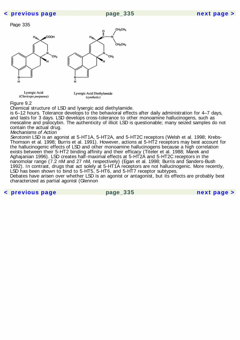
< previous page page_335 next page >
Page 335
Figure 9.2Chemical structure of LSD and lysergic acid diethylamide.is 6–12 hours. Tolerance develops to the behavioral effects after daily administration for 4–7 days,and lasts for 3 days. LSD develops cross-tolerance to other monoamine hallucinogens, such asmescaline and psilocybin. The authenticity of illicit LSD is questionable; many seized samples do notcontain the actual drug.Mechanisms of ActionSerotonin LSD is an agonist at 5-HT1A, 5-HT2A, and 5-HT2C receptors (Welsh et al. 1998; Krebs-Thomson et al. 1998; Burris et al. 1991). However, actions at 5-HT2 receptors may best account forthe hallucinogenic effects of LSD and other monoamine hallucinogens because a high correlationexists between their 5-HT2 binding affinity and their efficacy (Titeler et al. 1988; Marek andAghajanian 1996). LSD creates half-maximal effects at 5-HT2A and 5-HT2C receptors in thenanomolar range (7.2 nM and 27 nM, respectively) (Egan et al. 1998; Burris and Sanders-Bush1992). In contrast, drugs that act solely at 5-HT1A receptors are not hallucinogenic. More recently,LSD has been shown to bind to 5-HT5, 5-HT6, and 5-HT7 receptor subtypes.Debates have arisen over whether LSD is an agonist or antagonist, but its effects are probably bestcharacterized as partial agonist (Glennon
< previous page page_335 next page >
< previous page page_336 next page >
Page 3361990; Marek and Aghajanian 1996; Pierce and Peroutka 1990a; Zhang and Dyer 1993). LSD hasinhibitory effects on raphe neurons, but this is dissociable from its behavioral effects, suggesting apostsynaptic site of action (Trulson 1986; Vandermaelen and Aghajanian 1983; Blier et al. 1989).However, LSD shows a preferential activation of phospholipase A2-arachidonic acid pathways. Still,LSD stimulates phosphoinositide hydrolysis at 25% of that maximally achieved by serotonin,consistent with a partial agonist effect (Sanders-Bush et al. 1988; Berg et al. 1998). LSD alsodecreases spontaneous activity in the locus coeruleus, but increases its response to stimulation(Rasmussen and Aghajanian 1986).Significant tolerance develops to the effects of LSD. Daily repeated administration of LSDdownregulates 5-HT2 receptors, but not 5-HT1A, 5-HT1B, adrenergic, or dopaminergic receptors(Buckholtz et al. 1990, 1988). Further, down-regulation of 5-HT2 receptors closely parallels thedevelopment of behavioral tolerance to LSD. In contrast to cortical 5-HT2 receptors, there is noreduction in 5-HT1A, 5-HT1B, β-adrenergic, α1 or α2 adrenergic, D2 receptors, or a serotoninreuptake site (Buckholtz et al. 1990).Dopamine A few studies have examined the dopaminergic effects of LSD. The affinity of LSD for D2receptors is similar to its affinity for 5-HT2 sites, and it has a slightly lower affininty for D1 receptors(Watts et al. 1995). LSD has partial agonist effects at D2 receptors as seen in the inhibition ofprolactin release (Giacomelli et al. 1998). Neuroleptic drugs are also used clinically to terminate anLSD experience. Thus, the effects of LSD on dopaminergic function may contribute to itshallucinogeinc effects.General EffectsAutonomic LSD produces sympathomimetic effects, including pupil dilation, tachycardia, increasedblood pressure, piloerection, hyperreflexia, nausea, and fever.Behavioral LSD typically suppresses locomotor and exploratory behaviors (Mittman and Geyer 1991).It reduces attention and response to
< previous page page_336 next page >
< previous page page_337 next page >
Page 337external stimuli, presumably reflecting greater attention to internal stimuli (e.g., illusions,hallucinations). LSD does induce a conditioned place preference in rats (Meehan and Schechter1998).Electrophysiology LSD hyperpolarizes pyramidal neurons in somatosensory cortex, whereas serotonininitially depolarizes and then causes a long-lasting hyperpolarization (Pierce and Peroutka 1990b). Incats, the EEG effects of LSD indicate increased arousal and wakefulness (Kay and Martin 1978).During sleep, LSD decreases REM sleep and sleep spindles. Chronic treatment with selectiveserotonin reuptake inhibitors (SSRIs) decreases the electrophysiological responsiveness to LSD (deMontigny et al. 1990). Through 5-HT2-mediated mechanisms, LSD enhances nonsynchronousglutamatergic EPSPs in cortical pyramidal cells (Aghajanian and Marek 1999).The effect of LSD on cerebral blood flow was studied in the rat (Goldman et al. 1975). Anintravenous injection of LSD increased blood flow to frontal and parietal cortex within 10 minutes,and to the cerebellum within 20 minutes. Increases (significant or nonsignificant) were evidenced inall brain areas assessed, except for the hippocampus, where a minor, nonsignificant reduction wasnoted.Neuroimaging An LSD analog [N1-([11C]-Methyl)-2-Br-LSD ([11C]-MBL)]was used to assess 5-HT2binding in the human brain with positron emission tomography (PET) (Wong et al. 1987). A distinctlyserotonergic pattern of binding is seen with highest levels of labeling in frontal, temporal, andparietal cortex. Lower levels of binding are observed in caudate and putamen. There are no studiesthat address the hallucinatory effects of LSD with functional neuroimaging, but one exists formescaline (see below).Cognitive and subjective effects The effects of LSD on attention have been examined in animalparadigms. LSD reduces accuracy on a multiple-choice reaction time task, which is reversed by a 5-HT2 antagonist (Carli and Samanin 1992). LSD produces gross alterations in time perception, whichholds true in animal models as well as human reports (Frederick et al. 1997).
< previous page page_337 next page >
< previous page page_338 next page >
Page 338The hallmark effects of LSD are profound perceptual distortions and hallucinations. It produces aheightened awareness of sensations, where mundane sensations become vivid and intense. Lowerdoses produce illusions and alterations of perception, including intensification of colors anddistortions of shade, movement, and form. One may experience macropsia or micropsia, which arethe perceptions that visual objects are getting larger or smaller, respectively. Visual afterimages maybe prolonged and visual percepts may overlap. Emotional meaning is intensified and experience maytake on a magical or epiphanic intensity (Abraham et al. 1996). Higher doses may produce geometricvisual patterns or fully formed hallucinatory experiences. Hallucinations are most typically visual innature, and begin 2–3 hours after drug administration. Auditory hallucinations are rare, butdistortions of body image are not uncommon. LSD enhances classical conditioning, through actions at5-HT2A and 5-HT2C receptors (Welsh et al. 1998). Memory is generally unimpaired, and pastmemories may be vividly reexperienced.Another effect of LSD is an altered sense of self. Many experience a derealization or a ''spectatorego," where one feels like a passive observer of one's own experiences. Many also experience alessened sense of interpersonal boundaries. In a positive social context, this can result in a sense ofunion with others. However, if it becomes frightening it can result in anxiety and fears ofdepersonalization.The effects of LSD are highly variable between and within individuals, and are highly influenced byone's mental state and environmental setting (Abraham et al. 1996). Those taking the drug alone aremore likely to experience anxiety, speech disruption, and reduced motility, while those taking it in agroup setting are more likely to experience euphoria. More adverse experiences are reported bythose who are unaware that they are receiving it.Addiction and DependenceLSD produces a rapid and complete tolerance, but it is not powerfully reinforcing the way drugs ofabuse like cocaine and heroin are. LSD does not produce any known withdrawal syndrome (Abrahamet al. 1996). It does not produce positive reinforcement in animal self-administration models.Concurrently in humans, it does not lead to patterns of repeti-
< previous page page_338 next page >
< previous page page_339 next page >
Page 339tive, compulsive use. Regular LSD users typically show a pattern of usage consisting of periodic usespaced several weeks or more apart.ToxicityMassive overdoses of LSD are required to have lethal effects. Many of the symptoms of ergotism,excluding hallucinations, are not caused by lysergic acid, but by other ergoloids. A high safety marginexists with LSD, with fatal doses in the range of 150,000 mg/kg in rodents. No human fatalities haveever been reported from an overdose of LSD. The cases of eight people were reported, who snortedpure LSD, mistaking it for cocaine (Klock et al. 1975). The estimated doses taken were between10,000 and 100,000 μg. They experienced confusion, hallucinations, and hemorrhaging (throughinhibition of platelet formation), but all recovered.Ergotism Ergotism is a constellation of symptoms that occurs as a result of eating the ergot fungus.The effects of ergotism were apparent to ancient writers on the subject (Schultes and Hofman 1980,1992). Two general forms are distinguished: a neurological form consisting of seizures andhallucinations, and a vascular form consisting of gangrene and loss of extremities (noses, ear lobes,fingers, toes, and feet). The vascular symptoms of ergotism are due to the vasoconstricting effect ofergoloids, and the neurological form seems to result from ergot occurring primarily in wild grasses.Another effect of ergot is uterine contractions, frequently causing spontaneous abortions. This effectwas taken advantage of by European midwives, who employed ergot to assist contractions duringdifficult births. Although it is not a drug of choice due to unpredictable side effects, the ergoloidergotmetrine is still available for treatment of postpartum hemorrhage (van Dongen and de Groot1995).Bad trips A commonly known outcome of hallucinogenic experience is colloquially known as a badtrip. This essentially consists of a panic episode, sometimes brought on by a dislike of thehallucinogenic effects and fear that the experience will not end. Given that LSD prolongs one'sperception of time, it may seem as if the experience will never end and it
< previous page page_339 next page >
< previous page page_340 next page >
Page 340is easy to understand how such fears may rapidly escalate into panic. This reaction responds tosupportiveness and a familiar environment, although some contend that reassurance may worsen thecondition (Abraham et al. 1996). Anecdotal clinical experience indicates that benzodiazepines andneuroleptics may be useful as acute pharmacological treatments.Hallucinogen-persisting perception disorder Hallucinogen-persisting perception disorder (HPPD),commonly called flashbacks, are phenomena where the person reexperiences aspects of thehallucinogenic trip long after it has ended. This is reported by approximately 15% of LSD users.Some precipitants of HPPD are cannabis use, anxiety, fatigue, and sudden movement into a darkenvironment. This may be a normal function of memory that becomes exaggerated due to theintense nature of the hallucinogenic experience. Alternately, it may also be a direct pharmacologicaleffect.In LSD users who presented with visual disturbances, the disorder remained after 5 years in half ofthe sample (Abraham 1983). Common visual disturbances reported were geometric percepts, falsefleeting perceptions in the peripheral visual field, flashes of color, and afterimages. Diagnosis ofHPPD may seem relatively simple on the surface because hallucinogenic drugs are known to havethese effects in a subset of the population. However, one must rule out other potential diagnosesthat could mimic HPPD, such as toxic exposures, stroke, brain tumors, central infections, and braintrauma.HPPD has been studied with quantitative EEG. Compared to controls, people with HPPD show afaster alpha frequency and shorter visual-evoked-response latency, consistent with LSD-inducedcortical disinhibition (Abraham and Duffy 1996). Conversely, the auditory-evoked-response latency isincreased. Analyses indicate that these differences are predominant in temporal and left parietalregions.A few cases have been reported of exacerbation of HPPD by risperidone (Risperdal) (Abraham andMamen 1996). The exact reason for this is uncertain, but the fact that risperidone and LSD both actat 5-HT2 receptors is probably causative. Also, two cases have been reported of an exacerbation ofHPPD by selective serotonin reuptake inhibitors (Markel et al. 1994).
< previous page page_340 next page >
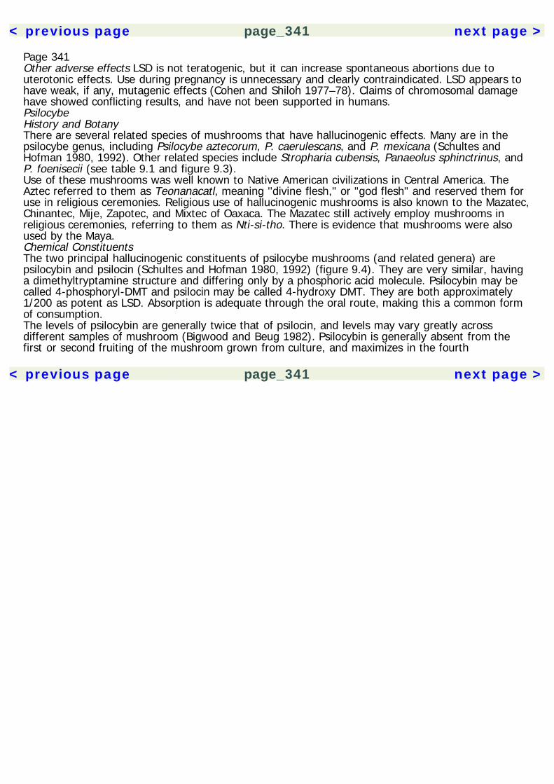
< previous page page_341 next page >
Page 341Other adverse effects LSD is not teratogenic, but it can increase spontaneous abortions due touterotonic effects. Use during pregnancy is unnecessary and clearly contraindicated. LSD appears tohave weak, if any, mutagenic effects (Cohen and Shiloh 1977–78). Claims of chromosomal damagehave showed conflicting results, and have not been supported in humans.PsilocybeHistory and BotanyThere are several related species of mushrooms that have hallucinogenic effects. Many are in thepsilocybe genus, including Psilocybe aztecorum, P. caerulescans, and P. mexicana (Schultes andHofman 1980, 1992). Other related species include Stropharia cubensis, Panaeolus sphinctrinus, andP. foenisecii (see table 9.1 and figure 9.3).Use of these mushrooms was well known to Native American civilizations in Central America. TheAztec referred to them as Teonanacatl, meaning ''divine flesh," or "god flesh" and reserved them foruse in religious ceremonies. Religious use of hallucinogenic mushrooms is also known to the Mazatec,Chinantec, Mije, Zapotec, and Mixtec of Oaxaca. The Mazatec still actively employ mushrooms inreligious ceremonies, referring to them as Nti-si-tho. There is evidence that mushrooms were alsoused by the Maya.Chemical ConstituentsThe two principal hallucinogenic constituents of psilocybe mushrooms (and related genera) arepsilocybin and psilocin (Schultes and Hofman 1980, 1992) (figure 9.4). They are very similar, havinga dimethyltryptamine structure and differing only by a phosphoric acid molecule. Psilocybin may becalled 4-phosphoryl-DMT and psilocin may be called 4-hydroxy DMT. They are both approximately1/200 as potent as LSD. Absorption is adequate through the oral route, making this a common formof consumption.The levels of psilocybin are generally twice that of psilocin, and levels may vary greatly acrossdifferent samples of mushroom (Bigwood and Beug 1982). Psilocybin is generally absent from thefirst or second fruiting of the mushroom grown from culture, and maximizes in the fourth
< previous page page_341 next page >
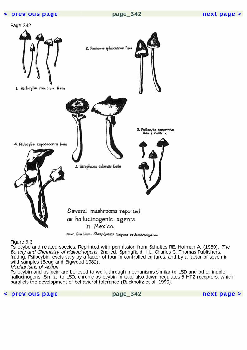
< previous page page_342 next page >
Page 342
Figure 9.3Psilocybe and related species. Reprinted with permission from Schultes RE, Hofman A. (1980). TheBotany and Chemistry of Hallucinogens, 2nd ed. Springfield, Ill.: Charles C. Thomas Publishers.fruting. Psilocybin levels vary by a factor of four in controlled cultures, and by a factor of seven inwild samples (Beug and Bigwood 1982).Mechanisms of ActionPsilocybin and psilocin are believed to work through mechanisms similar to LSD and other indolehallucinogens. Similar to LSD, chronic psilocybin in take also down-regulates 5-HT2 receptors, whichparallels the development of behavioral tolerance (Buckholtz et al. 1990).
< previous page page_342 next page >
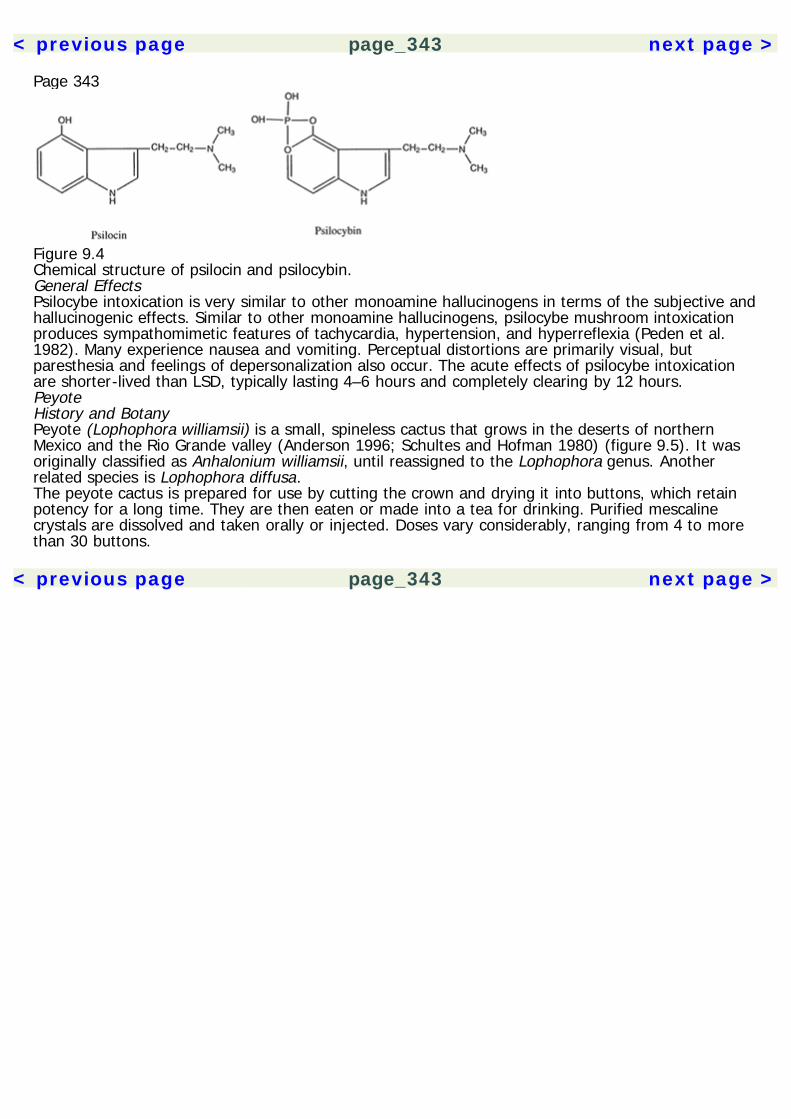
< previous page page_343 next page >
Page 343
Figure 9.4Chemical structure of psilocin and psilocybin.General EffectsPsilocybe intoxication is very similar to other monoamine hallucinogens in terms of the subjective andhallucinogenic effects. Similar to other monoamine hallucinogens, psilocybe mushroom intoxicationproduces sympathomimetic features of tachycardia, hypertension, and hyperreflexia (Peden et al.1982). Many experience nausea and vomiting. Perceptual distortions are primarily visual, butparesthesia and feelings of depersonalization also occur. The acute effects of psilocybe intoxicationare shorter-lived than LSD, typically lasting 4–6 hours and completely clearing by 12 hours.PeyoteHistory and BotanyPeyote (Lophophora williamsii) is a small, spineless cactus that grows in the deserts of northernMexico and the Rio Grande valley (Anderson 1996; Schultes and Hofman 1980) (figure 9.5). It wasoriginally classified as Anhalonium williamsii, until reassigned to the Lophophora genus. Anotherrelated species is Lophophora diffusa.The peyote cactus is prepared for use by cutting the crown and drying it into buttons, which retainpotency for a long time. They are then eaten or made into a tea for drinking. Purified mescalinecrystals are dissolved and taken orally or injected. Doses vary considerably, ranging from 4 to morethan 30 buttons.
< previous page page_343 next page >

< previous page page_344 next page >
Page 344
Figure 9.5Peyote (Lophophora williamsii). Reprinted from Barron et al., ''The Hallucinogenic Drugs." ScientificAmerican. April 1964, p. 210. Illustration by Alex Semenoick.The word peyote derives from the Nahuatl word for the cactus, peyotl. Religious use of peyote waswell established at the time of European arrival. Early European reports date peyote use back at leastas far as 300 B.C.E. among the Chichimecas and Toltecs (depending on the accuracy of interpretationof native calendars), but samples found in caves have been carbon dated to 8,000 years ago. TheSpanish tried to eradicate the use of peyote, but only succeded in driving its use into secrecy. Peyoteuse was known in Texas in 1760, but began to spread among tribes by 1880. The Native AmericanChurch was founded in 1918 by several groups practicing ceremonial peyote use and currently hasover 250,000 members.Mescal BeanDue to similarities in their names, the mescal bean is often confused with mescaline (Schultes andHofman 1980, 1992). These derive from entirely different plants: whereas mescaline derives frompeyote cactus, the mescal bean grows on the shrub Sophora secundiflora. The mescal bean itself haspsychoactive effects, and was used since prehistoric times by Indians in the Rio Grande basin.Samples have been found dating back
< previous page page_344 next page >
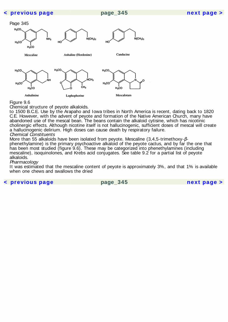
< previous page page_345 next page >
Page 345
Figure 9.6Chemical structure of peyote alkaloids.to 1500 B.C.E. Use by the Arapaho and Iowa tribes in North America is recent, dating back to 1820C.E. However, with the advent of peyote and formation of the Native American Church, many haveabandoned use of the mescal bean. The beans contain the alkaloid cytisine, which has nicotiniccholinergic effects. Although nicotine itself is not hallucinogenic, sufficient doses of mescal will createa hallucinogenic delirium. High doses can cause death by respiratory failure.Chemical ConstituentsMore than 55 alkaloids have been isolated from peyote. Mescaline (3,4,5-trimethoxy-β-phenethylamine) is the primary psychoactive alkaloid of the peyote cactus, and by far the one thathas been most studied (figure 9.6). These may be categorized into phenethylamines (includingmescaline), isoquinolones, and Krebs acid conjugates. See table 9.2 for a partial list of peyotealkaloids.PharmacologyIt was estimated that the mescaline content of peyote is approximately 3%, and that 1% is availablewhen one chews and swallows the dried
< previous page page_345 next page >
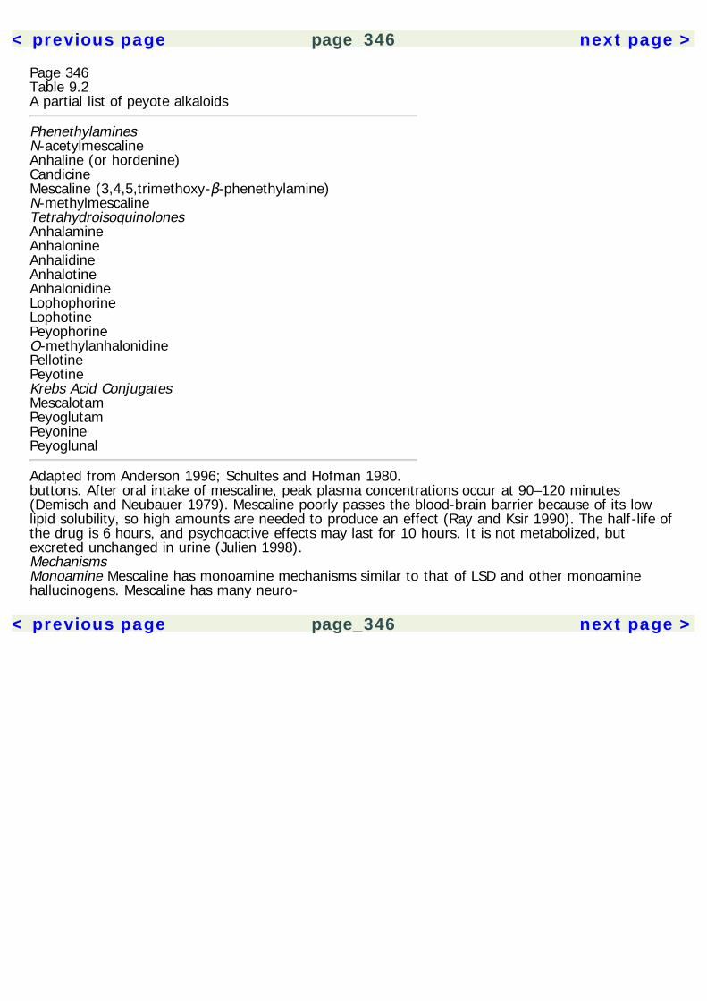
< previous page page_346 next page >
Page 346Table 9.2A partial list of peyote alkaloids
PhenethylaminesN-acetylmescalineAnhaline (or hordenine)CandicineMescaline (3,4,5,trimethoxy-β-phenethylamine)N-methylmescalineTetrahydroisoquinolonesAnhalamineAnhalonineAnhalidineAnhalotineAnhalonidineLophophorineLophotinePeyophorineO-methylanhalonidinePellotinePeyotineKrebs Acid ConjugatesMescalotamPeyoglutamPeyoninePeyoglunal
Adapted from Anderson 1996; Schultes and Hofman 1980.buttons. After oral intake of mescaline, peak plasma concentrations occur at 90–120 minutes(Demisch and Neubauer 1979). Mescaline poorly passes the blood-brain barrier because of its lowlipid solubility, so high amounts are needed to produce an effect (Ray and Ksir 1990). The half-life ofthe drug is 6 hours, and psychoactive effects may last for 10 hours. It is not metabolized, butexcreted unchanged in urine (Julien 1998).MechanismsMonoamine Mescaline has monoamine mechanisms similar to that of LSD and other monoaminehallucinogens. Mescaline has many neuro-
< previous page page_346 next page >
< previous page page_347 next page >
Page 347chemical effects in common with LSD, but with a few differences as well. It binds to 5-HT2 receptorswith high nanomolar affinity (302 nM), and activates phosphoinositide hydrolysis at 5-HT2A receptors(Newton et al. 1996; Titeler et al. 1988). But chronic mescaline use does not alter binding to 5-HT2receptors as LSD does (Buckholtz et al. 1990). Mescaline increases the acoustic startle, which isprevented by 5-HT2 antagonists (Davis 1987). Also similar to LSD, mescaline decreases thespontaneous activity of the locus coeruleus, but increases its activation in response to stimulation(Rasmussen and Aghajanian 1986; Aghajanian 1980). There is evidence that mescaline's effects aremediated by dopaminergic as well as serotonergic mechanisms (Trulson et al. 1983; Ahn andMakman 1979). Drug discrimination studies in rats indicate that mescaline generalizes to LSD andpsilocybin (Appel and Callahan 1989).Unlike LSD, dorsal raphe neurons are insensitive to mescaline, and unlike LSD and psilocybin,mescaline does not down-regulate 5-HT2 receptors (Penington and Reiffenstein 1986; Buckholz et al.1990). Mescaline also fails to directly inhibit 5-HT neurons (McCall 1982). Some tolerance developswith repeated administration of mescaline, which also shows cross-tolerance with LSD, but notamphetamine (Colasanti and Khazan 1975).Acetylcholine Mescaline, in micromolar concentrations, decreases the release of acetylcholine at theneuromuscular junction, with subsequent effects on end-plate potentials and motor function(Ghansah et al. 1993). Whether or not any similar effect occurs on central cholinergic systems hasnot been investigated.General EffectsLittle is known specifically about the effects of mescaline compared to LSD. Much information aboutit relies upon its similarities with other monoamine hallucinogens. The majority of attention is givenhere to mescaline, but some brief mention of the diverse effects of other peyote alkaloids (of whichlittle is known) is deserved. Often, their effects are very different from mescaline. Many alkaloids arepresent in sufficent concentrations to alter human physiology after oral consumption of peyote.Lophophorine in humans causes a ''sickening feeling in the back of
< previous page page_347 next page >
< previous page page_348 next page >
Page 348the head,'' a decrease in pulse, and a blushing in the face (Anderson 1996). Anhalodine has CNSstimulant properties. Pellotine has sedative effects, causing drowsiness and reduced motivation foractivity, bradycardia, and reduced blood pressure.Behavioral and motor effects Mescaline and its analogs inhibit cholinergic neuromusculartransmission by blocking release of acetylcholine and reducing end-plate potentials in the micromolarrange (Ghansah et al. 1993). This effect has not been investigated in humans, but it could reduce theforce of muscle contractions and motor control.Similar to LSD and other monoamine hallucinogens, mescaline suppresses locomotor and exploratorybehavior in novel environments (Wing et al. 1990). Also similar to LSD, tolerance develops to thebehavioral effects of chronic doses of mescaline (Murray et al. 1977). Mescaline increases aggressionin rat models (Sbordone et al. 1978); however, this is an elicited aggression (by electric shock) anddoes not necessarily generalize to human behavior. Increased aggression is not characteristic ofhumans using mescaline.Neuroendocrine Mescaline increases release of prolactin to levels four times greater than baseline,and also stimulates secretion of growth hormone (Demisch and Neubauer 1979).Electrophysiology and neuroimaging Similar to LSD, peripheral injections of mescaline producedesynchronization of the EEG and increase arousal in the rat (Colasanti and Khazan 1975). Thisperiod lasts for 2–3 hours, and is followed by a period of slow-wave and REM sleep with normalsleep cycles.A SPECT imaging study of mescaline showed a hyperfrontal pattern in cerebral blood flow, withgreater increases on the right side (Hermle et al. 1992). Increases are also seen in inferior temporalcortex and the hippocampus. Decreases in cerebral blood flow occur in occipital and parietal corticalregions.Subjective effects The subjective effects of equipotent doses of mescaline and LSD are said to beindistinguishable (Schwartz 1988). Behav-
< previous page page_348 next page >
< previous page page_349 next page >
Page 349ioral evidence also concurs temporally with the neuropharmacological effects of mescaline. This issupported by drug discrimination paradigms in rats (Appel and Callahan 1989).ToxicityThere is no clear evidence for genetic toxicity of mescaline. Chromosomal damage in lymphocyteswas not evident in Huichol Indians, who have a 1,600-year cultural tradition of religious peyote use(Dorrance et al. 1975).However, mescaline is teratogenic and has adverse effects when given during pregnancy, withdecreased reproductive success evident in hamsters (Hirsch and Fritz 1981). One effect is a delay inossification of the skull, sternum, and metatarsals. Given the serotonergic mechanisms of mescaline,it may also affect uterine contractions. Mescaline, like any other hallucinogenic drug, is probably bestavoided during pregnancy. Its developmental effects are uncertain and the risks far outweigh anypotential benefit from religious, psychotherapeutic, or recreational use.Other Hallucinogenic CactiThere are other species of cactus with hallucinogenic effects. One is the San Pedro cactus(Trichocereus pachanoi), which grows in the Andes region of Peru, Ecuador, and Bolivia. It is abranched, spineless, and columnar cactus, growing to a height of 9–20 feet. San Pedro is alsonoteworthy in that it contains mescaline and other related phenethylamines such as 3,4-dimethoxyphenylethylamine and 3-methoxytyramine (Pardanani et al. 1977; Schultes and Hofman1980, 1992). Mescaline levels in dried samples reach 2%. Use of San Pedro dates back to 1300B.C.E. in Peru. Similar to other hallucinogenic plants, it has been used in religious ceremonies.Cawe (Pachycereus pecten-aboriginum) is yet another species of hallucinogenic cactus, which growsin Mexico. It is also a columnar cactus, growing up to 35 feet in height. It is known as Cawe orWichowaka to the Tarahumara. Studies have identfied 4-hydroxy-3-methoxyphenylethylamine and 4-tetrahydroisoquinoline alkaloids as the probable psychoactive constituents.
< previous page page_349 next page >
< previous page page_350 next page >
Page 350YageHistory and BotanyYage (also known as yaje, hoasca, ayahuasca, caapi, pinde, or natema) consists of one or morehallucinogenic plants combined, but most importantly Banisteriopsis caapi or B. inebrians (Schultesand Hofman 1980, 1992) (figure 9.7). Other plants commonly added are Psychotria viridis, P.carthagenensis, or B. rusbyana. Banisteriopsis are lianas (thick woody vines) with dark ovate leavesand grow in Brazil, Bolivia, Columbia, Ecuador, Peru, and Venezuela. The combination of plants isreported to prolong and intensify the hallucinogenic effects of yage due to pharmacologicalinteractions.The bark of Banisteriopsis is prepared in cold or boiling water. It may also be chewed or administeredas a snuff. Liquid preparations are bitter and may induce dizziness, nausea, and vomiting.Chemical ConstituentsBanisteriopsis caapi The primary hallucinogenic constituents of B. caapi are the β-carbolines. Theseinclude harmaline, tetrahydroharmine, harmol, harmic acid methyl ester, harmic amide, acetylnorharmine, harmine N-oxide, harmalinic acid, and ketotetrahydronorharmine (figure 9.8). B.rusbyana also contains DMT, as well as N-methyltryptamine, 5-methoxy-N,N-dimethyltryptamine, and5-hydroxy-N,N-dimethyltryptamine (bufotenin). N-methyltetrahydro-β-carboline is found in traceamounts.Psychotria viridis and P. carthagenensis Psychotria species contain DMT, N-methyltryptamine, and N-methyltetrahydro-β-carboline. However, a more recent study of P. carthagenensis showed the plantto be devoid of psychoactive alkaloids (Leal and Elisabetsky 1996).Mechanisms of ActionThere are two major types of hallucinogenic alkaloids in yage. These are DMT and the harmalaalkaloids (or β-carbolines).
< previous page page_350 next page >

< previous page page_351 next page >
Page 351
Figure 9.7Banisteriopsis caapi. Reprinted with permission from Harner, MJ. (1973). The role of hallucinogenicplants in European witchcraft, in Hallucinogens and Shamanism. Harner MJ, ed. London: OxfordUniversity Press.
< previous page page_351 next page >
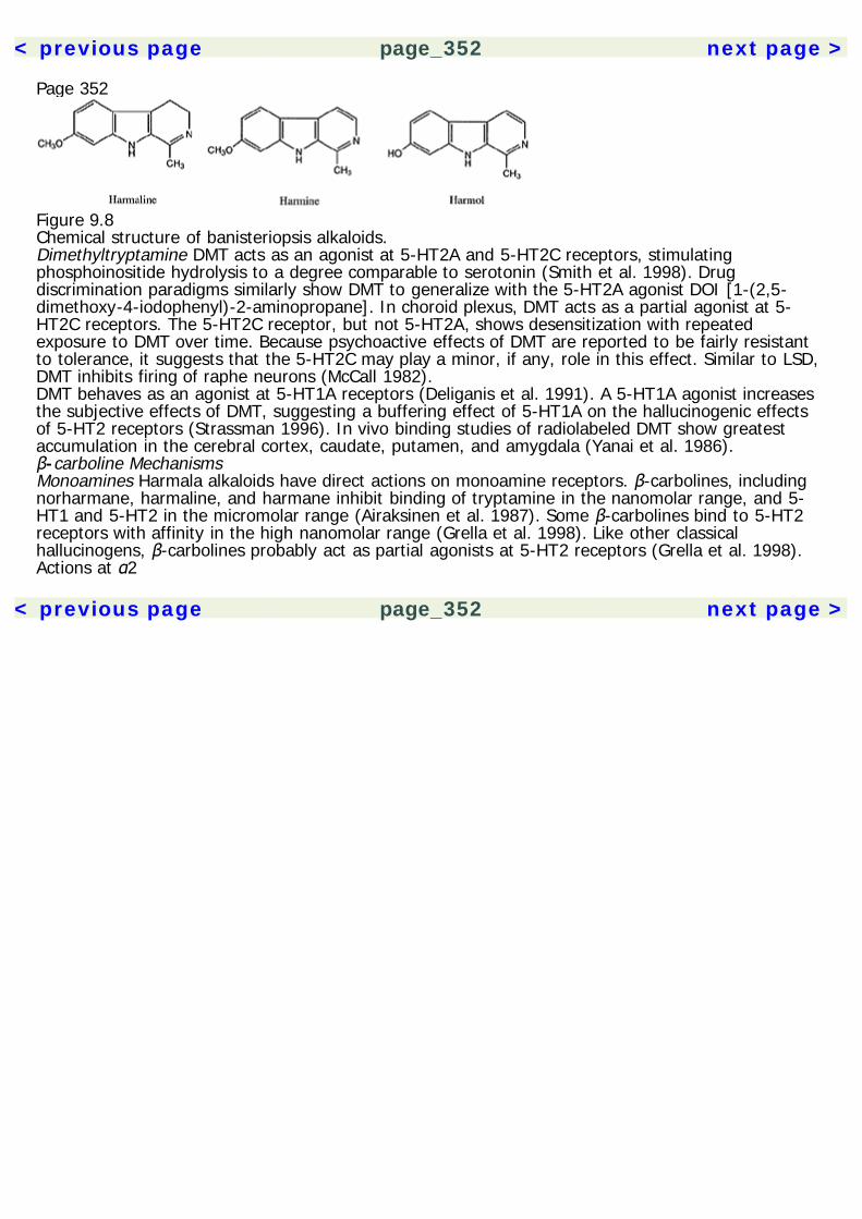
< previous page page_352 next page >
Page 352
Figure 9.8Chemical structure of banisteriopsis alkaloids.Dimethyltryptamine DMT acts as an agonist at 5-HT2A and 5-HT2C receptors, stimulatingphosphoinositide hydrolysis to a degree comparable to serotonin (Smith et al. 1998). Drugdiscrimination paradigms similarly show DMT to generalize with the 5-HT2A agonist DOI [1-(2,5-dimethoxy-4-iodophenyl)-2-aminopropane]. In choroid plexus, DMT acts as a partial agonist at 5-HT2C receptors. The 5-HT2C receptor, but not 5-HT2A, shows desensitization with repeatedexposure to DMT over time. Because psychoactive effects of DMT are reported to be fairly resistantto tolerance, it suggests that the 5-HT2C may play a minor, if any, role in this effect. Similar to LSD,DMT inhibits firing of raphe neurons (McCall 1982).DMT behaves as an agonist at 5-HT1A receptors (Deliganis et al. 1991). A 5-HT1A agonist increasesthe subjective effects of DMT, suggesting a buffering effect of 5-HT1A on the hallucinogenic effectsof 5-HT2 receptors (Strassman 1996). In vivo binding studies of radiolabeled DMT show greatestaccumulation in the cerebral cortex, caudate, putamen, and amygdala (Yanai et al. 1986).β-carboline MechanismsMonoamines Harmala alkaloids have direct actions on monoamine receptors. β-carbolines, includingnorharmane, harmaline, and harmane inhibit binding of tryptamine in the nanomolar range, and 5-HT1 and 5-HT2 in the micromolar range (Airaksinen et al. 1987). Some β-carbolines bind to 5-HT2receptors with affinity in the high nanomolar range (Grella et al. 1998). Like other classicalhallucinogens, β-carbolines probably act as partial agonists at 5-HT2 receptors (Grella et al. 1998).Actions at α2
< previous page page_352 next page >
< previous page page_353 next page >
Page 353adrenergic receptors by harman and 3-methylharman are implied by effects on the guinea pig ileumin the submicromolar range (Dolzhenko et al. 1983).Also, harmala alkaloids create effects on monoamine turnover. Postnatal rats administered harmaline(shortly before birth) have elevations in brain levels of the norepinephrine metabolite 3-methoxy-4-hydroxy-phenylglycol (MHPG), but decreases in the dopamine and serotonin metabolites 3,4-dihydroxyphenylacetic acid (DOPAC) and 5-hydroxyindole acetic acid (5-HIAA) (Okonmah et al.1988). Harmine pretreatment increases brain levels of L-dopa with concurrent increases in striataldopamine (Meneguz et al. 1994). However, this effect is species-specific, occurring in the rabbit butnot in the rat.Harmala alkaloids are inhibitors of brain MAO. Harmane inhibits MAOA in the submicromolar range (5× 10⁻⁷ M) and MAOB in the micromolar range (5 × 10⁻⁶ M) (Glover et al. 1982). Harmine inhibitsMAOA, but also has additional unspecified monoamine modulatory effects (Meneguz et al. 1994;Fernández de Arriba et al. 1994). Harmaline or its metabolites may also stimulate aldehyde reductaseor catechol O-methyltransferase (COMT) (Okonmah et al. 1988).Excitatory amino acids Harmaline competitively displaces MK-801 binding in the micromolar range,suggesting that it binds to the channel site of the NMDA receptor-channel (Du et al. 1997). However,unlike MK-801 or PCP, which block the channel, it has been proposed that harmaline acts as aninverse agonist at this site, opening the cation channel and increasing Ca2+ flux. The effects ofharmaline at NMDA receptors is blocked by MK-801 (Du and Harvey 1997).GABAA benzodiazepine β-carbolines act as inverse agonists of the GABAA benzodiazepine receptor(Mehta and Ticku 1989). Whereas benzodiazepines such as diazepam stimulate Cl– influx, β-carbolines inhibit it. Harmine, harmaline, harmane, and harmalol inhibit flunitrazepam binding in themicromolar range (half-maximal inhibition between 28 and 130 μM) (Mousah et al. 1986).Endogenous β-carbolines, such as harmane, have also been found in the human brain, but theirendogenous functions are uncertain (Tse et al.
< previous page page_353 next page >
< previous page page_354 next page >
Page 3541991; Rommelspacher et al. 1980). Harmaline tremor is reversed by the benzodiazepine agonistdiazepam (Valium) (Robertson 1980).PharmacologyIngestion of yage in healthy volunteers yields plasma concentrations of 10 to 250 ng/mL for harmineand 1.0 to 25.0 ng/mL of harmaline (Callaway et al. 1996). The dose-concentration relationships arelinear in this range. DMT shows linear dose-concentration relationships for plasma concentrationsbetween 5 and 1000 ng/mL. Systemically administered β-carbolines penetrate brain tissue, withrelatively even distribution (Moncrieff 1989). DMT taken alone is not absorbed well orally. It may betaken as a snuff or smoked, or mixed with other plants to improve absorption.Harmine and harmol are metabolized by three liver microsomes to 6-hydroxy-7-methoxyharman and3- or 4-hydroxy-7-methoxyharman, which are further metabolized to unidentified metabolites(Tweedy and Burke 1987). Protein binding of harmine and harmol occurs, and is dependent onmetabolism alkalinity.Unlike most hallucinogens, tolerance to subjective effects is not observed with DMT, even overseveral closely spaced doses (Strassman 1996). In contrast, tolerance was evident to hormonal andheart-rate responses.General EffectsAutonomic effects Harmala alkaloids, as well as isolated harmine and harmaline, produce a dose-dependent hypothermia (Abdel-Fattah et al. 1995). This effect is serotonin-dependent, and partlymediated by 5-HT1A receptors.On the other hand, DMT produces a dose-dependent hyperthermia (Strassman and Qualls 1994).Although statistically significant, these effects are small. DMT produces mydriasis, which is probablymediated by 5-HT2 receptors.Neuroendocrine effects DMT causes a transient increase in release of several hormones. In humansintravenously administered DMT (0.4 mg/kg), increases are seen in β-endorphin, corticotrophin,prolactin, cor-
< previous page page_354 next page >

< previous page page_355 next page >
Page 355tisol, and growth hormone, but not melatonin (Strassman and Qualls 1994).Cardiovascular effects Harmala alkaloids have cardiovascular effects (Aarons et al. 1977). Harmine,harmaline, and harmalol decrease heart rate, but increase pulse pressure, peak aortic flow, andmyocardial contractile force in dogs. Harmine reduces systemic arterial blood pressure and peripheralvascular resistance. Vascular resistance effects are not mediated by β-adrenergic or histamine H1receptors.DMT elevates pulse rate and blood pressure. In one study, heart rate elevated from around 70 beatsper minute at baseline to 100 beats per minute at the highest dose (0.4 mg/kg, IV).Neurophysiological effects Harmane produces excitation in the nucleus accumbens in nanomolarconcentrations, but depression in micromolar concentrations (Ergene and Schoener 1993). Harmineand B. caapi extract decrease the acoustic startle, which is reversed to some degree by DMT(Freedland and Mansbach 1999). β-carbolines may have antioxidant effects through inhibitingoxidative enzymes in the microsomal system (Tse et al. 1991).Motor effects Harmaline produces a motor tremor (8–14 Hz) through activation of cells in the inferiorolive, which is blocked by noncompetitive NMDA antagonists (Du et al. 1997; Stanford and Fowler1998). Harmaline tremor is also reversed by benzodiazepine agonists (Robertson 1980). The tremor isinitiated by synchronous rhythms in the olivocere-bellar system and red nucleus (Lorden et al. 1988;Gogolák et al. 1977; Batini et al. 1980). The tremor is associated with increased cGMP in thecerebellum, and tolerance with a relative normalization of cGMP (Lutes et al. 1988). Rapid tolerancedevelops to this effect with repeated doses.Subjective effects The subjective effects of DMT are very much like those of the other monoaminehallucinogens. Animal drug discrimination paradigms indicate that DMT and related compoundsproduce similar effects to LSD (Helsley et al. 1998). However, β-carbolines do not substitute well forLSD in these paradigms. Maximal substitution occurs with harmane only at intermediate levels. Somehave reported that unlike
< previous page page_355 next page >
< previous page page_356 next page >
Page 356other hallucinogens, ecstatic feelings are not experienced with yage. Amazonian Indians ingestingyage often report seeing snakes and jaguars, likely due to their prominent role in that culture.One feature that distinguishes DMT is the short duration of effect (Strassman et al. 1994). The onsetof effect is very rapid, partially due to administration method, and subsides completely in 30 minutes.With intravenous doses, subjects commonly report a ''rush," where the rapid onset of effects isacutely experienced. In some, this creates feelings of anxiety and being overwhelmed. Intravenousdoses of DMT at 0.2 mg/kg was a threshold for halluinogenic effects, while 0.05 mg/kg was oftennot discriminated from placebo.β-carbolines have anxiogenic effects, likely through inverse agonism of the GABAA benzodiazepinereceptor (File et al. 1982). However, prominent anxiety has not been noted with yage in comparisonto other monoamine hallucinogens. It is possible that the effects of harmala alkaloids are notsufficient to produce this effect, or that they may be offset by other pharmacological mechanisms.ToxicityHarmala alkaloids are potent inhibitors of monoamine oxidase (Callaway and Grob 1998). Thus, ifcombined with other antidepressants, such as selective serotonin reuptake inhibitors, there ispotential for serious side effects. Harmaline or its metabolites also cross the placental barrier(Okonmah et al. 1988).Harmaline has also been found to have neurotoxic effects on Purkinje neurons (O'Hearn and Molliver1997). This likely results from excitotoxic consequences induced by prolonged stimulation of inferiorolivary neurons. Whether this occurs in levels reached during human consumption has not beenspecifically addressed. Prolonged neurological symptoms have not been reported in those whoconsume the drug.Morning Glory and Related SpeciesHistory and BotanyThere are several species of flowering vines with hallucinogenic properties, commonly referred to asmorning glory (Schultes and Hofman
< previous page page_356 next page >

< previous page page_357 next page >
Page 357
Figure 9.9Ololiuqui (Turbina corymbosa). Reprinted with permission from Schultes RE, Hofman A. (1980). TheBotany and Chemistry of Hallucinogens, 2nd ed. Springfield, Ill.: Charles C. Thomas Publishers.1980, 1992; Shawcross 1983). Perhaps the most popular is ololiuqui (Turbina corymbosa, formerlyRivea corymbosa). It is a large vine with heart-shaped, green leaves and multiple bell-shaped flowers(figure 9.9). Ololiuqui is the Nahuatl word for the seed, literally meaning ''round object." Itshallucinogenic effects were known to the Aztecs, who used it in religious ceremonies. Another relatedspecies is known as badoh negro (Ipomoea violacea) among the Zapotec, and among the Aztec astlililtzin. Several varieties of this plant are commonly known as "heavenly blue," "pearly gates," and"flying saucers." The Hawaiian Baby Woodrose (Argyreia nervosa) is another related species thatappears to
< previous page page_357 next page >

< previous page page_358 next page >
Page 358have the highest concentration of hallucinogenic chemicals among these species.Chemical ConstituentsThe identified psychoactive constituents of the morning glories are lysergic acid alkaloids, similar tothose isolated from ergot. These include d-lysergic acid amide (or ergine), lysergol, lysergic acidhydroxyethylamide, and d-isolysergic acid amide. As the names imply, lysergic acid amide andlysergic acid diethylamide (LSD) only differ by the replacement of two hydrogen atoms with ethylgroups. As a result, LSD is about 100 times more potent.Mechanisms and EffectsBased on structural similarities, the neuropharmacological mechanisms of morning glory alkaloids arelikely similar or identical to those of LSD. The subjective effects are also similar, but the lowerpotency of the alkaloids requires larger doses for hallucinogenic effects. Somatic effects tend to bepronounced, with nausea and gastrointestinal symptoms being very common.Nutmeg and MaceNutmeg and mace are spices derived from the tree Myristica fragrans (Schultes and Hofman 1980,1992; Julien 1998). It is an evergreen tree, growing up to 15 meters in height, bearing flowers and afleshy, round fruit (Gruenwald et al. 1998) (figure 9.10). Other species of the Myristica genus alsoyield nutmeg and mace, including M. malabarica. Myristica fragrans was native to the Moluccaislands, but is now cultivated throughout tropical regions (Robbers et al. 1996). Islands of the Malayarchipelago are the primary source. Their use in Europe began when it was introduced by Arabs inthe first century C.E. However, their use was well known in ancient Egypt and India, where nutmegis sometimes snuffed together with tobacco.Nutmeg is prepared from the seed of the tree, while mace is prepared from the fleshy covering thatsurrounds it. Differing composition of the
< previous page page_358 next page >
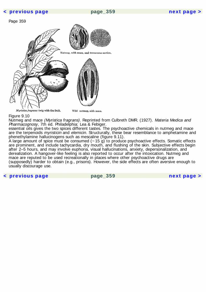
< previous page page_359 next page >
Page 359
Figure 9.10Nutmeg and mace (Myristica fragrans). Reprinted from Culbreth DMR. (1927). Materia Medica andPharmacognosy, 7th ed. Philadelphia: Lea & Febiger.essential oils gives the two spices different tastes. The psychoactive chemicals in nutmeg and maceare the terpenoids myristicin and elemicin. Structurally, these bear resemblance to amphetamine andphenethylamine hallucinogens such as mescaline (figure 9.11).A large amount of spice must be consumed (~15 g) to produce psychoactive effects. Somatic effectsare prominent, and include tachycardia, dry mouth, and flushing of the skin. Subjective effects beginafter 2–5 hours, and may involve euphoria, visual hallucinations, anxiety, depersonalization, andderealization. A hangover-like feeling is also reported to occur after the intoxication. Nutmeg andmace are reputed to be used recreationally in places where other psychoactive drugs are(supposedly) harder to obtain (e.g., prisons). However, the side effects are often aversive enough tousually discourage use.
< previous page page_359 next page >

< previous page page_360 next page >
Page 360
Figure 9.11Chemical structure of myristicin and elemicin.IbogaHistory and BotanyIboga (Tabernanthe iboga) is a shrub native to west-central Africa, and used in cultures of the Gabonand Congo (Schultes and Hofman 1980, 1992). It grows to a height of 4–6 feet in the undergrowthof tropical forests, but has also been cultivated. Its leaves are ovate, 3–4.5 inches in length and 1.25inches wide, with a green color on top and yellowish-green underneath. Tiny flowers grow in groupsof 5–12 with whitish, yellowish, or pinkish color. Its fruits are yellow-orange in color, ovoid, andgrow as large as olives (figure 9.12).The plant is used in elaborate religious ceremonies, which vary from area to area. Adherents use theplant to communicate spiritually with their ancestors. In lower doses, it is used to counteract fatigueand hunger. Warriors and hunters have used it to remain awake at night. Some Europeans haveclaimed that it has aphrodisiac effects as well. More recently, anecdotal reports have indicatedpossible antiaddictive effects of iboga, stimulating a rush of scientific research into the neurochemicaleffects of the plant (Mash et al. 1998).Chemical ConstituentsSeveral indole alkaloids have been isolated from the iboga plant, but the principal psychoactive agentis ibogaine. However, other alkaloids have physiological effects, and may also have psychoactiveeffects (figure 9.13). These include ibogamine, coronaridine, and tabernanthine. Some research hasalso focused on the effects of noribogaine (12-hydroxy-
< previous page page_360 next page >

< previous page page_361 next page >
Page 361
Figure 9.12Iboga (Tabernanthe iboga). Reprinted with permission from Schultes RE, Hofman A. (1992). Plants ofthe Gods: Their Sacred, Healing, and Hallucinogenic Powers. Rochester, VT: Healing Arts Press.
< previous page page_361 next page >
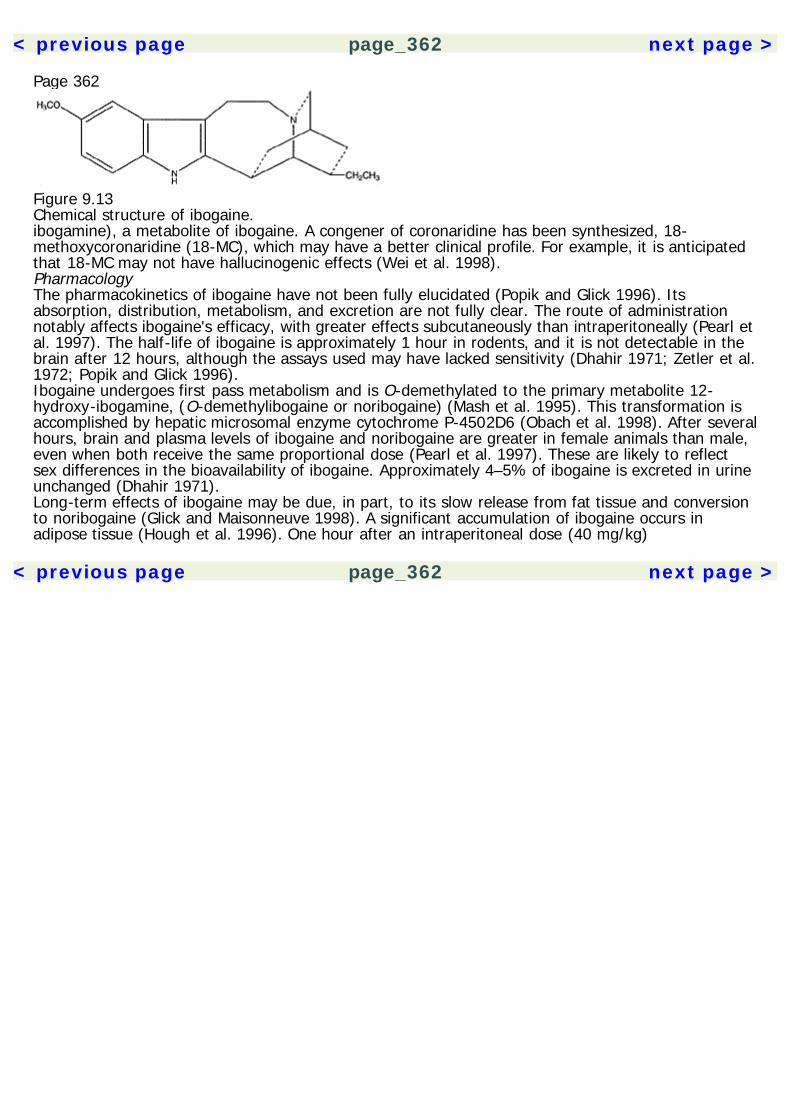
< previous page page_362 next page >
Page 362
Figure 9.13Chemical structure of ibogaine.ibogamine), a metabolite of ibogaine. A congener of coronaridine has been synthesized, 18-methoxycoronaridine (18-MC), which may have a better clinical profile. For example, it is anticipatedthat 18-MC may not have hallucinogenic effects (Wei et al. 1998).PharmacologyThe pharmacokinetics of ibogaine have not been fully elucidated (Popik and Glick 1996). Itsabsorption, distribution, metabolism, and excretion are not fully clear. The route of administrationnotably affects ibogaine's efficacy, with greater effects subcutaneously than intraperitoneally (Pearl etal. 1997). The half-life of ibogaine is approximately 1 hour in rodents, and it is not detectable in thebrain after 12 hours, although the assays used may have lacked sensitivity (Dhahir 1971; Zetler et al.1972; Popik and Glick 1996).Ibogaine undergoes first pass metabolism and is O-demethylated to the primary metabolite 12-hydroxy-ibogamine, (O-demethylibogaine or noribogaine) (Mash et al. 1995). This transformation isaccomplished by hepatic microsomal enzyme cytochrome P-4502D6 (Obach et al. 1998). After severalhours, brain and plasma levels of ibogaine and noribogaine are greater in female animals than male,even when both receive the same proportional dose (Pearl et al. 1997). These are likely to reflectsex differences in the bioavailability of ibogaine. Approximately 4–5% of ibogaine is excreted in urineunchanged (Dhahir 1971).Long-term effects of ibogaine may be due, in part, to its slow release from fat tissue and conversionto noribogaine (Glick and Maisonneuve 1998). A significant accumulation of ibogaine occurs inadipose tissue (Hough et al. 1996). One hour after an intraperitoneal dose (40 mg/kg)
< previous page page_362 next page >
< previous page page_363 next page >
Page 363of ibogaine, plasma levels of 106 ng/ml and fat levels of 11,308 ng/g are obtained. Higher levels arefound with subcutaneous administration, suggesting extensive first-pass hepatic metabolism afterintraperitoneal dosing. Levels are reduced ten- to twentyfold after 12 hours.Mechanisms of ActionAcetylcholine Ibogaine functionally blocks nicotinic acetylcholine receptors in the low to intermediatemicromolar range (Fryer and Lukas 1999). Ibogaine also blocks ion flux through ganglionic nicotinicchannels in the nanomolar range (20 nM) (Badio et al. 1997). However, it was much weaker atneuromuscular nicotinic receptors, with half-maximal inhibition in the micromolar range (2 μM).Ibogaine inhibits the nicotine-mediated release of catecholamines at concentrations below 10 μM(Mah et al. 1998; Maisonneuve et al. 1997; Schneider et al. 1996).Although ibogaine binds to muscarinic M1 and M2 receptors in the micromolar range, its action thereis uncertain (Sweetnam et al. 1995). It fails to alter the adenylyl cyclase activity of a muscarinicagonist carbachol (Rabin and Winter 1996b).Monoamine Ibogaine blocks uptake of both dopamine and serotonin, with half-maximal effects in thelow- to mid-micromolar range (Wells et al. 1999). Noribogaine also blocks reuptake of serotonin invivo (Wei et al. 1998). Both ibogaine and its metabolite noribogaine bind to the cocaine site of theserotonin transporter (Staley et al. 1996; Mash et al. 1995). However, ibogaine prevents cocaine-induced increases in serotonin levels in the brain (Sershen et al. 1997). Ibogaine also reduces releaseof dopamine induced by nicotine, cocaine, and morphine (Benwell et al. 1996; Sershen et al. 1997;Glick and Maisonneuve 1998). It causes a release of cytoplasmic dopamine in the striatum, which isreversed by dopamine uptake inhibitors (e.g., cocaine) (Harsing et al. 1994). However, the effects ofibogaine on dopamine release outlast the elimination time of the drug (Maisonneuve et al. 1991). Theeffects of a single dose of ibogaine on dopamine levels last up to a week, but are not seen at onemonth after injection (Maisonneuve et al. 1992).
< previous page page_363 next page >
< previous page page_364 next page >
Page 364The effects of ibogaine on monoamine turnover are complex. Acute intravenous ibogaine causes ashort-lasting excitement of dopaminergic neurons in the ventral tegmental nucleus, but pretreatmentfails to alter basal activity or activity stimulated by morphine and cocaine (French et al. 1996). Itcauses dramatic reductions in dopamine levels and increases in the metabolites DOPAC and HVA(Baumann et al. 1998; Binienda et al. 1998). These effects occur in the striatum and frontal cortex assoon as 30 minutes after intraperitoneal administration (Ali et al. 1996). It has been suggested thatibogaine's inhibitory effects on dopamine release in the accumbens and striatum are mediated by κopioid receptors (Reid et al. 1996). It also blocks serotonin-induced release of dopamine in thestriatum, which may be mediated by the 5-HT1B receptor (Sershen et al. 1994a). Tabernanthine, aswell, increases synthesis and elimination of catecholamines in many areas of the brain, but not thehypothalamus (Prioux-Guyonneau et al. 1984).Ibogaine may release and block reuptake of serotonin (Wei et al. 1998). Reports exist of bothincreases and decreases of serotonin levels in frontal cortex (Ali et al. 1996; Binienda et al. 1998).However, increases in 5-HIAA occur in frontal cortex and decreases occur in the striatum and nucleusaccumbens (Ali et al. 1996; Benwell et al. 1996). Like many other monoamine hallucinogens,ibogaine has affinity for 5-HT2A receptors in the high micromolar range (92.5 μM) (Helsley et al.1998). Animal drug discrimination studies support ibogaine agonist activity at 5-HT2A and 5-HT2Creceptors, but not 5-HT1A or 5-HT3 antagonists (Helsley et al. 1998a, 1998b). Ibogaine andnoribogaine increase the inhibition of adenylyl cyclase by morphine and serotonin, but do not alterbasal levels (Rabin and Winter 1996b).Amino acids Ibogaine is a competitive inhibitor of the NMDA receptor. It binds to the MK-801receptor site within the NMDA channel in the submicromolar range, but not to the NMDA receptoritself (Itzhak and Ali 1998). Ibogaine's antagonism of the NMDA channel is also voltage-dependent(Popik et al. 1995; Chen et al. 1996). Noribogaine also has antagonistic effects at the MK-801 site,with potency 4–6 times greater than ibogaine (Mash et al. 1995; Ali et al. 1996). In contrast, ibogainefails to affect kainate or GABA currents.
< previous page page_364 next page >
< previous page page_365 next page >
Page 365The NMDA antagonist effects of ibogaine are significant in light of the fact that MK-801 also reducestolerance to opiates and alcohol, and reverses tolerance to stimulants (Trujillo and Akil 1991; Khannaet al. 1993; Karler et al. 1989).Neurotensin Ibogaine treatment increases levels of neurotensin in the striatum, substantia nigra, andnucleus accumbens, but not frontal cortex (Alburges and Hanson 1999). This effect occurs 12 hoursafter administration and is blocked by a dopamine antagonist. Iboga also blocked release ofneurotensin by cocaine. Neurotensin colocalizes with dopamine in neurons of the ventral tegmentalarea, and is altered by abused drugs (Cooper et al. 1996; Pilotte et al. 1991; Morley et al. 1980).Opioids Iboga alkaloids have several effects at opioid receptors. Ibogaine has low micromolar affinityfor κ opioid receptors (~3 μM), less for μ receptors (~11 μM), and no specific affinity for δ receptors(>100 μM) (Pearl et al. 1995). Noribogaine has even higher affinity for all three opioid receptorsassayed: κ (~0.96 μM), μ (~2.66 μM), and δ (~24 μM). Thus, noribogaine may play a greater rolethan ibogaine in opioid actions. Others have found affinity for the μ opioid receptor to be in thenanomolar range (130 nM) (Codd 1995). Noribogaine is a full agonist at μ opioid receptors,stimulating G-protein activity at submicromolar concentrations (Pablo and Mash 1998). It also reducestolerance to chronic morphine in mice (Bhargava and Cao 1997). Other iboga alkaloids havemicromolar affinity for the μ receptor (coronaridine), δ receptor (coronaridine and tabernanthine),and κ receptor (ibogamine, coronaridine, and tabernanthine) (Deecher et al. 1992).Both ibogaine and noribogaine enhance morphine analgesia in morphine-tolerant, but not morphine-naive rats (Sunder Sharma et al. 1998; Bhargava and Cao 1997). However, ibogaine does not altertolerance to κ or δ opioid agonists (Cao and Bhargava 1997). Noribogaine does have affinity for theκ1 opioid receptor (Ali et al. 1996).Sigma receptors Ibogaine binds to both σ1 and σ2 receptors, with affinities in the range of 1.5–3 μM(Itzhak and Ali 1998). Some have
< previous page page_365 next page >
< previous page page_366 next page >
Page 366reported that affinity is higher for σ2 receptors (201 nM) than σ1 receptors (8 μM) (Bowen et al.1995). Activity at σ2 receptors is believed to partly mediate ibogaine's neurotoxicity (Glick andMaisonneuve 1998).Ion channels Ibogaine and other iboga alkaloids have micromolar affinity for voltage gated Na+channels (Deecher et al. 1992). Tabernanthine is also a Ca2+ channel blocker, producing half-maximal inhibition in the micromolar range (7–21 μM) (Miller and Godfraind 1983).Intracellular messengers Noribogaine, but not ibogaine, stimulates phosphoinositide hydrolysis (Rabinand Winter 1996a). This effect was unaltered by tetrodotoxin or ω-conotoxin, indicating that it is notsecondary to release of other neurotransmitters.Physiological EffectsCardiovascular A moderate rise (10–15%) in blood pressure occurs after administration of ibogaine,peaking at 1.5 to 2 hours and lasting up to 5 hours (Lotsoff 1995). On the other hand, tabernanthinealone produces bradycardia and hypotension, which is independent of autonomic innervation andmuscarinic receptors (Hajo et al. 1981). In micromolar concentrations, it antagonizes arterialcontractions induced by norepinephine and Ca2+ in the rat (Hajo-Tello et al. 1985). It also has anegative inotropic effect in heart muscle. The mechanisms of tabernanthine's cardiovascular effectsare seemingly complex, but involve stimulation of vascular β2 adrenergic receptors and cellular Ca2+homeostasis (Miller and Godfraind 1983).Neuroendocrine Preliminary research has been done on the hormonal effects of iboga. The findingsconsist of elevated levels of corticosterone and prolactin by ibogaine in rats (Baumann et. al. 1998;Ali et al. 1996).Behavioral and cognitive effects As might be expected, ibogaine has powerful sensory and motoreffects. Rats administered ibogaine (20–60 mg/kg) show slower response times on sensory andsensory-motor tests (Kesner et al. 1995). Motor reflexes are impaired at 40–60 mg/kg, and
< previous page page_366 next page >
< previous page page_367 next page >
Page 367there is an overall reduction in activity and emotionality. Ibogaine induces a tremor and ataxia(O'Hearn and Molliver 1997). Tabernanthine also induces tremor, which is mediated by GABAAbenzodiazepine receptors (Trouvn et al. 1987).Ibogaine itself stimulates locomotor activity in rats. However, it reduces the locomotor activityinduced by morphine, with greater effects in female animals than males (Pearl et al. 1997). It alsoreduces locomotor activity induced by cocaine and amphetamine (Sershen et al. 1992a, 1992b;Blackburn and Szumlinski 1997). However, the interaction between ibogaine and cocaine is time-dependent, with motor activity inhibited at short delays, but potentiated at long delays (Maisonneuveet al. 1997).Ibogaine has not shown any adverse effects on learning or memory retrieval in animals performing amaze task (Helsley et al. 1997a). In fact, pretreatment with ibogaine or O-demethylibogaine, but nott-butyl ibogaine, improves memory retrieval in the Morris water maze task (Popik 1996).Electrophysiology Little work has been done to characterize the electrophysiological effects of iboga.Rats administered ibogaine showed decreases in power in delta, theta, alpha, and beta frequenciesduring the first 30 minutes (Binienda et al. 1998). However, this recovered after 15 minutes.Tabernanthine derivatives produce electrophysiological activation, increase wakefulness, reduce slow-wave activity, and block REM sleep in animals (Da Costa et al. 1980).Subjective effects Ibogaine has psychoactive effects at doses of 200–300 mg and above (Sheppard1994). Despite its short half-life, the effects of ibogaine are long lasting, typically in the range of 24–36 hours. Use of the plant is also associated with behavioral and motor effects. At certain doses andperiods of the intoxication, people experience stimulation, whereas at other doses the subjecttypically stares into space and eventually collapses into a catatonic-like state. In African cultures, it isbelieved that the person's soul has left the body during this time and entered into the spirit world.Feelings of bodily levitation or floating have also been reported. A rainbowlike aura is seen aroundobjects.
< previous page page_367 next page >

< previous page page_368 next page >
Page 368Time perception is altered, where short periods may seem extremely long. Users also reportdepersonalization or a sense of detachment, as if externally observing one's own actions. Anotherfeature of ibogaine intoxication seems to be a panoramic recall of events from long-term memory,primarily in visual memory. The ability of iboga to allow one to commune with ancestors is mostlikely not a pharmacological property of the drug, but rather a result of set and setting influenced byculture.Given ibogaine's monoamine and indole actions (particularly actions at 5-HT2 receptors), it is likelythat it produces hallucinations in a manner similar to those produced by other monoaminehallucinogens. However, the additional actions of iboga on other neurochemical systems, such asNMDA, may also modulate the quality of the hallucinogenic experience. Animal drug discriminationparadigms indicate generalization between LSD and ibogaine (Palumbo and Winter 1992).Intermediate degrees of generalization occur between ibogaine and noribogaine in animal drugdiscrimination tests (Helsley et al. 1997b, 1998b). However, almost complete (83.5%) generalizationoccurs between ibogaine and harmaline. Ibogaine generalizes to noribogaine in animal stimulusdiscrimination paradigms, but not to the NMDA antagonist MK-801 or κ opioid agonist U50,488(Zubaran et al. 1999).Antiaddictive EffectsThe putative antiaddictive effects of ibogaine come from its reputed ability to reduce or eliminatewithdrawal syndromes and cravings for long periods of time (Mash et al. 1998). Although thesederived from anecdotal reports and were discovered serendipitously in humans, they have beensupported by empirical research.Several lines of research in animals support this contention. Ibogaine reduces cocaine intake in mice,which sustains for longer periods by repeated administration (Cappendijk and Dzoljic 1993; Sershenet al. 1994b). It also suppresses withdrawal symptoms in morphine-dependent rats precipitated bynaltrexone, independent of tremor effects (Glick et al. 1992b). However, a reduction of opioidwithdrawal symptoms is not evident in mice (Frances et al. 1992). While ibogaine pretreatment re-
< previous page page_368 next page >
< previous page page_369 next page >
Page 369duces acquisition of place preference with drugs of abuse such as morphine and amphetamine, itdoes not abolish previously established place preferences (Luxton et al. 1996; Moroz et al. 1997).Ibogaine binds to nicotinic receptors in the nanomolar range (IC50 20 nM)(Badio et al. 1997; Glickand Maisonneuve 1998). It has been reported to curtail smoking and have antinicotinic effects, andmay do so by reducing the dopaminergic response to nicotine (Maisonneuve et al. 1997).Possible neurochemical mechanisms The interactions of ibogaine with various drugs of abuse may bemediated by different receptors, or combinations of receptors (Glick and Maisonneuve 1998).Combined treatment with a κ opioid antagonist and NMDA agonist partially block the effects ofibogaine, suggesting a role for these two receptors in ibogaine's antiaddictive effects (Glick et al.1997). Ibogaine's effect on morphine dependence is related to it's NMDA antagonist activity (Layer etal. 1996). Effects on morphine-induced dopamine release may be dependent on prior exposure (Pearlet al. 1996). Serotonergic and dopaminergic actions of ibogaine also mediate its antiaddictive actions(Sershen et al. 1997). In particular, serotonergic actions may be more important for ibogaine'sreduction of alcohol intake (Glick and Maisonneuve 1998). Similar to ibogaine, noribogaine decreasesmorphine and cocaine self-administration, the locomotor stimulant effect of morphine, andextracellular dopamine in the accumbens and striatum (Glick et al. 1996b).Some, but not all of ibogaine's effects may involve pharmacokinetic actions. Although ibogaine doesnot alter brain morphine or alcohol levels, it does alter amphetamine levels, suggesting a possiblehepatic interaction (Glick et al. 1992a; Rezvani et al. 1995).Alternative iboga alkaloids Potential neurotoxic effects of ibogaine have raised concern over its clinicaluse. It is possible that the antiaddictive and neurotoxic actions of ibogaine are discrete, allowing forpotential separation of clinical and toxic effects (Molinari et al. 1996). Some other iboga alkaloids mayhave these properties, such as 18-MC.18-MC is a synthetic iboga alkaloid that may retain ibogaine's antiaddictive effects, but lack theeffects of neurotoxicity and tremor (Glick
< previous page page_369 next page >
< previous page page_370 next page >
Page 370et al. 1996). Acute administration of 18-MC decreases self-administration of morphine and cocaine inrats, for a period of days to weeks (Glick et al. 1996a). It also reduces alcohol consumption andpreference in rats (Rezvani et al. 1997). On the other hand, 18-MC does not show any cerebellartoxicity, even after high doses (100 mg/kg) (Glick et al. 1996). 18-MC also has similar actions toibogaine in reducing morphine withdrawal (Rho and Glick et al. 1998). Similar to ibogaine, it reducesnicotine-induced release of dopamine (Glick et al. 1998).Other iboga alkaloids besides ibogaine (tabernanthine, R- and S-coronaridine, R- and S-ibogamine,and desethylcoronaridine) decrease morphine and cocaine intake in animals shortly afteradministration, and in some cases one day after administration (Glick et al. 1994). R-ibogamine wasthe most consistent in this effect, reducing intake for several days after a single injection.Studies in humans An open trial of ibogaine was carried out in seven subjects with opioiddependence, who were administered 700–1800 mg. After a prolonged hallucinatory period (24–38hours), none of the subjects showed significant withdrawal symptoms. However, one who had takenthe lowest dose resumed opioid use in two days. Of the subjects taking 1000 mg or greater, halfrelapsed and half remained abstinent for at least 14 weeks (Sheppard 1994). There are other casestudies in humans in the medical literature (Cantor 1990; Luciano 1998; Sisko 1993; Kovera et al.1998; Mash et al. 1998). Commonly reported phenomena are reductions in drug craving and opiatewithdrawal symptoms within 1 to 2 hours, followed by a total resolution of the withdrawal syndromewithin 24 hours. A review of cases indicates a 10% success rate at two years following a singletreatment (Lotsoff 1995).In 1996, the National Institute of Drug abuse discontinued clinical trials of ibogaine. Research iscurrently undertaken by researchers outside of the United States. Far more empirical research isneeded to establish the antiaddictive effects of ibogaine. The neuropharmacological mechanismssupport possible antiaddictive effects, and results of case studies suggest further investigation. Ifsuccessful, it would still be necessary to complement it with psychosocial support and counseling, aswith other addiction therapies.
< previous page page_370 next page >
< previous page page_371 next page >
Page 371ToxicityIbogaine has demonstrated toxic effects, which could potentially limit its usefulness in treatingaddiction. However, the proper dosage of alternative iboga alkaloids may avert this problem. Themedian lethal dose of ibogaine is 82 mg/kg in the guinea pig and 327 mg/kg in the rat (Dhahir 1971;Delourme-Houde 1946). Use of ibogaine for addiction treatment has been associated with two deathsoverseas.Neurotoxicity In sufficient concentrations, ibogaine produces cerebellar neurotoxicity in rats,characterized by degeneration of Purkinje cells in the vermis, intermediate, and lateral cerebellum(Molinari et al. 1996; O'Hearn and Molliver 1997). Ibogaine's cerebellar toxicity is likely due toexcitation of neurons in the inferior olive, causing release of glutamate from climbing fibers apposedto Purkinje cells. Congruently, ablation of the inferior olive prevents this toxicity. Ibogaineneurotoxicity is accompanied by a late induction of neuronal nitric oxide synthase adjacent toPurkinje cells, which is likely to contribute to the excitotoxic cascade (O'Hearn et al. 1995). Acutehigh doses of ibogaine are also accompanied by reactive gliosis in the vermis, hippocampus, olfactorybulbs, brain stem, and striatum (O'Callaghan et al. 1996).One caveat to the neurotoxic effect of ibogaine is that the effect is dose-dependent (Molinari et al.1996). Ibogaine does not show neurotoxic effects at doses that produce clinically related effects inrats (40 mg/kg IP).Monoamine Hallucinogens: Collective IssuesTherapeutic UsesSome have used hallucinogenic drugs to facilitate the psychotherapy process, soon after thediscovery of LSD in 1949 (Abraham et al. 1996; Grinspoon and Bakalar 1986). This has sometimesbeen referred to as psycholytic therapy. However, the Drug Abuse Control amendments to theHarrison Narcotics Act in 1965 restricted such use. Studies at that time used widely varyingmethodology, but a meta-analysis incorporating over 1600 subjects showed ''good/very good"outcome in 40–60% of cases (Mascher 1967). While this could be due to altered cognitive
< previous page page_371 next page >
< previous page page_372 next page >
Page 372processing of emotions and experiences during the hallucinogenic state, there may also be a directpharmacological action as well. An uncontrolled study of LSD in depression showed robustantidepressant effects (Savage 1952; Strassman 1995).LSD has been examined for treatment of alcoholism. Some have reported that it was as effective asother treatments available at the time, but did not have any particular advantage (Ludwig et al.1969; Abuzzahab and Anderson 1971). Peyote use in the Native American Church has beenanecdotally reported to alleviate alcoholism, for which there is a possible pharmacological basis (Blumet al. 1977). However, it is also feasible that the therapeutic effect is due to intense experienceselicited by the ceremony and the context of increased social involvement.During the early 1960s, Timothy Leary conducted the Concord Prison Experiment to study thepsychotherapeutic use of hallucinogenic drugs in prison inmates. Treatment involved administrationof psilocybin and group psychotherapy in 32 prisoners. A follow-up study of recidivism in theseprisonsers concluded that there was no long-term treatment effect and emphasized the value ofpostrelease social support (Doblin 1998).There is no formal consensus on this subject, but it seems that people do benefit frompsychotherapy with hallucinogenic drugs. This benefit is not necessarily greater or worse thantraditional psychotherapy; it has not clearly been demonstrated that hallucinogens give any clearadvantage. At present, the variable methodology of many past studies prevent a firm conclusion.When one weighs the benefits against the elaborate safety precautions that would be needed forsuch therapy and potential liability, it becomes apparent why this form of therapy has not flourishedas it might have. Empirical research on ibogaine for addiction treatment represents the most solidevidence for a therapeutic use of a hallucinogenic drug. Further research is needed to establish thesafety and effectiveness of this drug in humans.Monoamine Hallucinogens and Concurrent Psychotherapeutic MedicationsAdministration of certain therapeutic psychotropic medications has been shown to alterresponsiveness to hallucinogenic drugs. This is not surprising, because they both have actions onmonoamine systems, sometimes even the same receptor subtytpes. Tricyclic antidepressants and
< previous page page_372 next page >
< previous page page_373 next page >
Page 373lithium enhance the effects of LSD, whereas MAO inhibitors and selective serotonin reuptakeinhibitors (SSRIs) decrease the effects (Bonson and Murphy 1996; Bonson et al. 1996). Three-weekadministration of an SSRI abolished the effect of LSD in some cases.Monoamine Hallucinogens and Chronic PsychosisA few cases have been described where use of large amounts of LSD were thought to contribute tothe development of a chronic psychosis (Boutros and Bowers 1996). Many cases involve polydruguse, and hallucinations occured spontaneously or with cannabis use. LSD psychosis may bemultifactorial, involving family history, premorbid personality, and exposure to other drugs(Strassman 1984). Indeed, predictors of poor outcome following LSD psychosis are suggestive ofneurological predispositions. Parental alcohol abuse also appears to be a predisposing factor.Epidemiologic studies show an increase in the admission diagnosis of schizophrenia and paranoiddisorders to state hospitals during times when use of hallucinogenic drugs increased in the generalpopulation. People with psychoses are also more likely to abuse hallucinogenic drugs (Tsuang et al.1982). The literature in this area suffers from methodological problems, limiting the confidence ofconclusions that can be made (Boutros and Bowers 1996). However, it does seem reasonable tosuggest that heavy use of hallucinogenic drugs can facilitate a psychotic disorder in those with riskfactors.A psychological assessment of long-term religious users of yage was undertaken using diagnosticinterviews, personality testing, and neuropsychological evaluation was performed, using age-matchedcontrols as reference group (Grob et al. 1996). Yage users had an overall high functional status. Afew cases were reported with remission of psychopathology after initiation of yage use, with noapparent detrimental effects on personality or cognition. Although these data by no means constituteempirical proof, they suggest that it is possible for people to use monoamine hallucinogens over timewithout adverse effects.Chronic Cognitive Effects of Monoamine HallucinogensIt is difficult to assess chronic cognitive effects of LSD or other hallucinogens in humans. Thisresearch faces the problem of lacking premorbid data, uncertainty of the actual drug used, andconcurrent use of other
< previous page page_373 next page >
< previous page page_374 next page >
Page 374psychoactive drugs. Of particular interest is premorbid cognitive problems in this research. It maywell be that people with cognitive impairments are more likely to use psychoactive drugs. Indeed,abnormalities were found in the prefrontal cortex of polysubstance users when compared to normalcontrols (Liu et al. 1998). In hallucinogen users, statistical decreases were found in the Halsteadcategory test and trail-making test A, but scores were still well within the normal range (Culver andKing 1974; McGlothlin et al. 1969). Further, scores were not different on numerous other measuresof cognitive function.Cholinergic HallucinogensHistory and BotanyThere are several plants classified as cholinergic hallucinogens, all belonging to the familySolanaceae. Namely, these are belladonna (Atropa belladonna), henbane (Hyoscyamus niger), andmandrake (Mandragora officinarum). This family also includes datura genus, of which there are manyspecies. The most commonly known is perhaps jimsonweed (Datura stramonium), also known asJamestown weed or thorn apple (figure 9.14). Another species in this category is scopolia (Scopoliacarniolica) (table 9.3).The monoamine and cholinergic hallucinogens not only differ by neurochemical actions, but also bythe qualitative alteration in consciousness they produce. Unlike the monoamine hallucinogens, thecholinergic hallucinogens produce partial or complete amnesia for the duration of their effect.Although they both induce hallucinations, the cholinergic hallucinogens create a state bettercharacterized as delirium and sedation.BelladonnaBelladonna is a perennial herb growing 1 to 2 meters in height. It has a straight woody stem andovate pointy leaves. The flowers are solitary and hang from the plant, yellow on the inside withcrimson veins (Gruenwald 1998). It grows in Europe, Iran, and northern Africa. The name belladonnacomes from the Italian words meaning ''beautiful woman," because the plant causes a prominentpupil dilation thought to
< previous page page_374 next page >
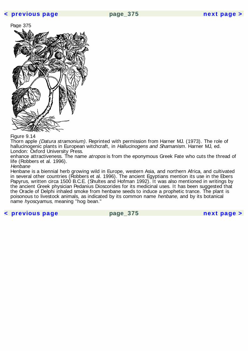
< previous page page_375 next page >
Page 375
Figure 9.14Thorn apple (Datura stramonium). Reprinted with permission from Harner MJ. (1973). The role ofhallucinogenic plants in European witchcraft, in Hallucinogens and Shamanism. Harner MJ, ed.London: Oxford University Press.enhance attractiveness. The name atropos is from the eponymous Greek Fate who cuts the thread oflife (Robbers et al. 1996).HenbaneHenbane is a biennial herb growing wild in Europe, western Asia, and northern Africa, and cultivatedin several other countries (Robbers et al. 1996). The ancient Egyptians mention its use in the EbersPapyrus, written circa 1500 B.C.E. (Shultes and Hofman 1992). It was also mentioned in writings bythe ancient Greek physician Pedanius Dioscorides for its medicinal uses. It has been suggested thatthe Oracle of Delphi inhaled smoke from henbane seeds to induce a prophetic trance. The plant ispoisonous to livestock animals, as indicated by its common name henbane, and by its botanicalname hyoscyamus, meaning ''hog bean."
< previous page page_375 next page >
< previous page page_376 next page >
Page 376Table 9.3Cholinergic and amino acid hallucinogens
Common Name Botanical name
Cholinergic Hallucinogens Belladonna Atropa belladonnaDatura Datura metel D. feroxToloache Datura inoxia D. discolor D. kymatocarpa D. pruinosa D. quercifolia D. reburra D. stramonium D. wrightiiJimsonweed Thornapple Datura stramoniumHenbane Hyoscyamus nigerMandrake Mandragora officinarumScopolia Scopolia carniolicaAmino Acid Hallucinogens Fly Agaric Amanita muscaria
See text for references.MandrakeMandrake was known to the ancient Egyptians, and has been found in several burial tombs (Rudgley1993). The human-like form of the roots (sometimes resembling arms and legs), led early Europeansto believe that it could cure many human ailments (figure 9.15). Much superstition surroundsEuropean lore regarding mandrake. It was believed to grow at the foot of gallows, springing from thesemen of those who were hanged. Evil spirits were supposed to emit a shrieking sound whenmandrake was uprooted.
< previous page page_376 next page >

< previous page page_377 next page >
Page 377
Figure 9.15Mandrake (Mandragora officinarum). Reprinted with permission from Harner MJ. (1973). The role ofhallucinogenic plants in European witchcraft, in Hallucinogens and Shamanism. Harner MJ, ed.London: Oxford University Press.DaturaThere are many species of datura, the best known of which is probably jimsonweed (Daturastramonium). It is an annual plant reaching a height of 1–2 meters, with large white solitary flowers(Gruenwald 1998) (figure 9.14). Datura is indigenous to many parts of the world, including Europe,Noth America, northern Africa, and eastern and southwestern Asia.The word datura derives from its Sanskrit name, dhattura. Stramonium derives from the French wordstramoine, meaning ''stink weed," alluding to the unpleasant smell of the folium (Gruenwald 1998;
< previous page page_377 next page >

< previous page page_378 next page >
Page 378Robbers et al. 1996). It was grown in England by seeds imported from Constantinople, and known tosettlers in Virginia (hence the name Jamestown weed or jimsonweed).Historical Uses: Witchcraft, Lycanthropy, and ShamanismBelladonna, henbane, and mandrake were used to induce hallucinatory states during witchcraftrituals in Europe. A salve was prepared using these plants, which was then applied to the skin(Harner 1973; Rudgley 1993). Absorption through the skin is questionable, but it was applied to thevaginal membranes, which are an effective site for drug absorption. Thus, an anointed broomstickwas an effective way to apply the drug. The plants can create a sensation of flying, which is thesource of the legend of witches flying on broomsticks. Use of such hallucinogenic herbs was also toinduce a mental experience of lycanthropy, or changing into an animal form (such as a werewolf)(Harner 1973). Descriptions of lycanthropes in ancient Greece very closely resemble anticholinergicsymptoms, including dry mouth, thirst, blurred vision, difficulty swallowing, and staggering gait.Cholinergic hallucinogens were familiar to Native Americans long before the arrival of Europeans(Grinspoon and Bakalar 1997; Schultes and Hofman 1980, 1992). The Aztec used a plant calledtoloache or toloatzin (Datura inoxia) to prophesize, and it is still used among some Native Americantribes for shamanic purposes. Carlos Castaneda wrote about the shamanic use of datura among theYaqui in his book The Teachings of Don Juan: A Yaqui Way of Knowledge (Castaneda 1968).Chemical ConstituentsThe cholinergic hallucinogens all have common chemical constituents that are responsible for theirpharmacological effects (Robbers et al. 1996). These are the tropane alkaloids hyoscyamine,scopolamine (or hyoscine), and atropine (figure 9.16). It is scopolamine, and not atropine orhyoscyamine, which primarily produces the central and hallucinogenic effects because it is the onlyone that passes the blood-brain barrier sufficiently. However, all three have peripheral effects. Daturastramonium contains 0.1–0.65% tropane alkaloids, which is principally
< previous page page_378 next page >

< previous page page_379 next page >
Page 379
Figure 9.16Chemical structure of the tropane alkaloids.hyoscyamine, and scopolamine to a lesser extent. Belladonna contains 0.33–0.6% tropane alkaloids,primarily hyoscyamine. Henbane contains 0.05–0.28 tropane alkaloids, again primarily consisting ofhyoscyamine. Scopolia contains 0.2–0.5% tropane alkaloids. The tropane akaloid content depends onthe part of the plant used, with highest concentrations in the root (Gruenwald 1998). Drying theplant transforms some of the alkaloids, increasing the amount of atropine and scopolamine.Mechanisms of ActionAcetylcholineTropane alkaloids are antagonists of muscarinic cholinergic receptors, competing with acetylcholineand other agonists (Brown and Taylor 1996). The antagonism is competitive, and can be overcomeby larger concentrations of acetylcholine or other agonists. They are active at all known subtypes ofthe muscarinic receptor (Kebabian and Neumeyer 1994). Effects are seen at muscarinic receptors inall central and peripheral locations, including exocrine glands, smooth and cardiac muscle, andautonomic ganglia (Brown and Taylor 1996).MonoaminesCholinergic antagonists increase dopamine activity in the striatum, as measured in humans with PET(Dewey et al. 1993). The pedunculo-
< previous page page_379 next page >
< previous page page_380 next page >
Page 380pontine and laterodorsal tegmental nuclei project to dopaminergic neurons of the substantia nigrapars compacta and ventral tegmental area (Yeomans 1995). Muscarinic antagonists like scopolaminedisinhibit cholinergic neurons, which in turn excite dopaminergic neurons. Whereas cholinergic drugsfor Parkinson's disease selectively block M1 muscarinic receptors, the cholinergic hallucinogens arenonselective. This mechanism may explain the reinforcing effects of cholinergic hallucinogens.Reinforcement is associated with activity of the mesolimbic dopamine system originating in theventral tegmental area, and scopolamine reduces the threshold for reinforcing brain stimulation(Yeomans et al. 1993). Ironically, anticholinergic hallucinogens are reinforcing even though theperson may have little or no memory of the experience.Other NeurotransmittersMuscarinic receptors have modulatory effects on numerous other neurotransmitters in the brain (vander Zee and Luiten 1999). Of particular interest are interactions between glutamate and cholinergicsystems in learning and memory (Aigner 1995).PharmacokineticsMaximum serum concentrations of scopolamine occurred 10 to 30 minutes after subcutaneousadministration (Ebert et al. 1998). The elimination half-life is approximately 220 minutes.General EffectsAutonomicMuscarinic receptors are present on ganglionic and postganglionic neurons, for both sympathetic andparasympathetic branches. Thus, the effect of systemic scopolamine is hard to predict and is oftenparadoxical (Brown and Taylor 1996).Atropine and scopolamine have antispasmodic effects on the gastrointestinal tract. It partly inhibitsvagal influence in the gut, reducing motility. However, the enteric nervous system also employsserotonin and dopamine, so parasympathetic innervation plays a modulatory role.
< previous page page_380 next page >

< previous page page_381 next page >
Page 381Nonetheless, a sufficient dose will reduce or temporarily halt peristalsis (Brown and Taylor 1996).Some effects on esophageal peristalsis may be mediated by central mechanisms (Bieger 1984).Atropine reduces the tone and contractions of the ureter and bladder, and has antispasmodic effectson the gallbladder and bile duct. Negligible effects are produced on uterine contractions. Sweatglands are inhibited by atropine, and it can impair thermoregulation, especially in hot environments.Cardiac EffectsAtropine generally increases heart rate, but it may briefly and mildly decrease it initially, due to M1receptors on postganglionic parasympathetic neurons. Larger doses of atropine produce greatertachycardia, due to M2 receptors on the sinoatrial node pacemaker cells. There are no changes inblood pressure, but arrhythmias may occur. Scopolamine produces more bradycardia and decreasesarterial pressure, whereas atropine has little effect on blood pressure (Vesalainen et al. 1997; Brownand Taylor 1996).Respiratory EffectsTropane alkaloids act as bronchodilators, but have been replaced clinically by methylxanthines andadrenergic drugs. They inhibit secretions from mucous membranes in the nose, mouth, pharynx, andbronchi.Behavioral and Motor EffectsCentral muscarinic antagonists reduce behavioral activity at higher doses by causing sedation.Scopolamine reduces locomotion and exploration in rats (Renner et al. 1992). However, they alsoproduce some degree of euphoria, probably by indirect actions on dopaminergic systems (Yeomans etal. 1993). Muscarinic antagonists have long been used as antiparkinsonian agents, which is discussedin chapter 5.Saccades are also affected by anticholinergic hallucinogens in humans (Oliva et al. 1993). Increasesin saccade duration and latency occur along with decreases in velocity. Postsaccadic fixation isimpaired as well, where the eye drifts after reaching the target.
< previous page page_381 next page >
< previous page page_382 next page >
Page 382Arousal and SleepTherapeutic doses of scopolamine produce CNS depression, characterized by drowsiness, amnesia,and dreamless sleep (Brown and Taylor 1996). It reduces arousal and increases the effort required toawaken (Parrott 1987). Higher therapeutic doses of atropine cause central excitation, characterizedby restlessness, irritability, confusion, disorientation, hallucinations, and delirium. Larger dosesproduce central depression, paralysis, coma, and death by respiratory failure and cardiovascularcollapse.Tonic arousal and wakefulness are sustained by projections from the reticular formation to thethalamus, which affects thalamocortical processes (Young 1998). An important part of this ascendingsystem is cholinergic projections from the pontine reticular formation, including the laterodorsal andpedunculopontine tegmental nuclei. Cholinergic reticular neurons also influence arousal throughprojections to the basal forebrain cholinergic nuclei, which in turn has widespread projections to theforebrain. Thus, scopolamine alters forebrain function through dual actions on brain stem andforebrain cholinergic nuclei.Scopolamine supresses REM sleep in animals, and a rebound increase occurs during withdrawal fromchronic treatment (Sutin et al. 1986; Zolotski et al. 1993). Similarly, scopolamine increases thelatency for REM onset and decreases its total duration in humans (Sagalés et al. 1975). Decreasesalso occur in eye and body movements. This effect shows tolerance over chronic administration, anda rebound increase in REM occurs when scopolamine is replaced with saline placebo. Rapid eyemovement (REM) sleep is driven by nuclei in the pontine reticular formation, namely the laterodorsaland pedunculopontine tegmental nuclei, and acetylcholine release is mediated by muscarinicautoreceptors (Shiromani and Fishbein 1986; Roth et al. 1996). Muscarinic receptors regulating REMare found throughout the brain stem, including dorsal raphe, locus coeruleus, and pontine reticularformation (Baghdoyan et al. 1994).ElectrophysiologyScopolamine creates electrophysiological slowing in humans, with increases in absolute power indelta activity, and in relative power in delta
< previous page page_382 next page >

< previous page page_383 next page >
Page 383and theta-1 activity over the central and parieto-occipital regions. Decreases occur in relative alpha-2(9.2–12.8 Hz) over the frontal regions and in absolute alpha-2 over most recording sites. These EEGchanges were congruent with changes on memory test (the Wechsler memory scale) (Kikuchi et al.1999). Decreases also occur in fast alpha power (Ebert et al. 1998). In event-related-potentialstudies, scopolamine prolongs the latency of the P300 and reduces spectral power of long-latency-evoked potentials without altering earlier components (Meador et al. 1988). In contrast, thecholinesterase inhibitor physostigmine reverses these effects.Functional NeuroimagingScopolamine increases cerebral blood flow to lateral occipital cortex bilaterally and to the leftorbitofrontal region (Grasby et al. 1995). Decreases are seen in the right thalamus, precuneus, andlateral premotor areas bilaterally. When normal subjects are chronically administered scopolamine,there is a 12% increase in cerebral blood flow on single photon emission computed tomography(SPECT), but a decrease in cerebral muscarinic binding (Sunderland et al. 1995). On the other hand,acute administration dose-dependently reduces cortical blood flow, which is maximal in frontal cortex(Gitelman and Prohovnik 1992; Prohovnik et al. 1997).In addition to altering cerebral blood flow, scopolamine alters cerebral glucose metabolism. Inhealthy older volunteers, absolute and normalized glucose metabolism are decreased in prefrontaland occipital regions and increased in parietal-occipital cortical regions and a left middle temporalregion (Molchan et al. 1994). Metabolism is also reduced in the thalamus (Cohen et al. 1994).Functional Activation in Cognitive TasksSome functional neuroimaging studies have examined the effects of scopolamine in cognitive tasks.Although scopolamine inhibits increases in cerebral blood flow to somatosensory stimulation, it doesnot inhibit the neural response (Ogawa et al. 1994). Thus, cholinergic systems may be involved incoordination of cerebral blood flow increases to neural activation. Subjects performing an attentionalauditory discrimination
< previous page page_383 next page >
< previous page page_384 next page >
Page 384task showed metabolic reduction in cingulate and increases in the basal ganglia (Cohen et al. 1994).The effects of scopolamine on memory have also been investigated using functional neuroimaging.Scopolamine reduces cerebral blood flow in left and right prefrontal cortex and the right anteriorcingulate during performance of a verbal memory task (Grasby et al. 1995).Collectively, these results indicate that scopolamine has profound effects on cerebral activity, both inresting and cognitive task-activation conditions. Of particular note are effects on frontal and striatalactivity, which could well account for impairments on cognitive abilities such as working memory.Also of note is a decrease in thalamic activation, which could result from blocked activation frompontine tegmental cholinergic nuclei, and/or altered activity of fronto-striato-thalamic circuits. Ineither case, the functional imaging findings well account for the cognitive and subjective effects ofcentral anticholinergic drugs.Cognitive Effects: Animal StudiesScopolamine impairs cognitive functions in a number of animal paradigms. Orientation of attention toa cued visual target (the Posner task) is reduced—in reaction time and accuracy (Davidson et al.1999). It impairs working memory in both delayed match-to-sample and non-matching-to-sampletasks in rats and primates (Granon et al. 1995; Rupniak et al. 1991; Bartus and Johnson 1976).Scopolamine produces memory deficits similar to lesions of basal forebrain cholinergic nuclei,characterized as impairments in visual discrimination learning, retention, and reversal (Harder et al.1998).Cognitive Effects: Human StudiesScopolamine produces cognitive deficits in humans, which are more pronounced in the elderly(Zemishlany and Thorne 1991). Cognitive studies have focused on memory and attention, but somestudies have shown impairments in a variety of cognitive functions.Scopolamine produces dose-dependent impairments of attention and slows the speed of informationprocessing (Ebert et al. 1998; Vitiello et al. 1997). It causes psychomotor slowing when it isadministered to the elderly, and to young adults at higher doses (Flicker et al. 1992). Digit span isnot affected by scopolamine in young volunteers, but more omis-
< previous page page_384 next page >
< previous page page_385 next page >
Page 385sions are produced on letter cancellation tasks (Parrot 1987; Rusted 1988). Impairments are alsoseen in praxis and visual perception (Flicker et al. 1990). Deficits induced in problem solving andworking memory are suggestive of effects on executive allocation of working memory (Rusted andWarburton 1988). Scopolamine impairs chess performance in trained players (Liljequist and Mattila1979). On the other hand, no effects are observed in immediate and remote memory, languagefunction, object sorting, or intrusion errors (Flicker et al. 1992, 1990; Zemishlany and Thorne 1991).Scopolamine interferes with the encoding and consolidation of information into long-term memory.Reductions are seen in the overall score of the Wechsler memory scale after acute administration ofscopolamine (Kikuchi et al. 1999). Impairments occur across both verbal and visual recall, and visualrecognition as well (Flicker et al. 1990; Frumin et al. 1976; Rusted 1988). It significantly impairsdelayed recognition of abstract shapes in humans when administered before the encoding stage(Rosier et al. 1998). However, the effect is only seen on delayed recognition: scopolamine does notaffect immediate recognition or visuoperceptual discrimination. Impairments are seen in recall ofparagraph information in humans, using the logical memory subtest of the Wechsler memory scale.Memory is also impaired on a verbal selective reminding test (Zemishlany and Thorne 1991; Rabey etal. 1996). Caffeine and nicotine partially reverse memory deficits induced by scopolamine in humans(Riedel et al. 1995).Scopolamine produces greater impairment in older than younger subjects on cognitive tasks (Molchanet al. 1992; Zemishlany and Thorne 1991). Given the cognitive impairments it produces and thecholinergic nature of many dementias such as Alzheimer's disease, scopolamine has been explored asa pharmacological model of dementia in numerous animal and human cognitive studies. Althoughscopolamine intoxication mimics dementia in some respects, scopolamine does not completely createall of the symptoms (Ebert and Kirch 1998). A scopolamine-challenge is also ineffective as a probefor Alzheimer's disease, because it does not distinguish it from Parkinson's disease or vasculardementia, or predict cognitive decline (Rabey et al. 1996; Barker et al. 1998). However, a combinedblockade of muscarinic and nicotinic receptors
< previous page page_385 next page >
< previous page page_386 next page >
Page 386may provide a better pharmacological model of dementia (Little et al. 1998).ToxicityToxicity from tropane alkaloids often results from their effects on autonomic function. Several speciesof Datura are freely available, either growing in the wild or cultivated as ornamental plants, such asangel's trumpet (Datura sauveolens). Periodic outbreaks of jimsonweed use occur, when adolescentslearn about the psychoactive effects of the plant and intentionally ingest it.Transdermal scopolamine is used for treatment of motion sickness, causing relatively infrequentcases of confusion and visual hallucinations (Ayub et al. 1997; Wilkinson 1987). A case of paranoiddelusions and hallucinations was reported after a two-week period of transdermal scopolamine,which continued 11 days after discontinuation (Rubner et al. 1997). Children may be particularlysensitive to adverse effects of anticholinergic drugs (Holland 1992). Others more sensitive topsychoactive side effects are the elderly, people with preexisting psychiatric illness, and those takingother anticholinergic medications (Ziskind 1988). Treatment of anticholinergic syndromes involvesdrugs that enhance cholinergic transmission, such as physostigmine.Amino Acid HallucinogensFly AgaricFly agaric (Amanita muscaria) is a mushroom that grows in forests, often under birches, firs, andlarches (Schultes and Hofman 1980, 1992). It has a reddish, flat, and ovate cap, with distinct whitewarts over the surface. Another variety has an orange or yellowish cap, with yellow warts. The stemis white, cylindrical, and hollow, with a bulbous shape at the bottom (figure 9.17).The Amanita genus comprises about 50 or 60 species, with varying chemical constituents—a numberof which are toxic (Schultes and Hofman 1980, 1992). Fly agaric (Amanita muscaria) grows inwidespread northern temperate zones of Eurasia and North America. In North
< previous page page_386 next page >

< previous page page_387 next page >
Page 387
Figure 9.17Fly Agaric (Amanita muscaria). Schultes RE, Hofman A. (1980). The Botany and Chemistry ofHallucinogens, 2nd ed. Springfield, Ill.: Charles C. Thomas Publishers.America, it was used by the Ojibway Indians of Michigan. In Eurasia, it has also been used by severaltribes spanning Siberia. A related species, Amanita pantherina, is used in western Washington State.Although less popular now, fly agaric may historically be the oldest and most widely usedhallucinogen. Fly agaric was probably discovered in Siberia, through observation of its effect inintoxicated reindeer (Abraham et al. 1996). It is likely to be the drug known as soma in the ancientIndian Rig Veda.Chemical ConstituentsThe psychoactive constituents of fly agaric are amino acid derivatives (Eugster et al. 1965; Schultesand Hofman 1980, 1992). These include ibotenic acid, muscimol, muscazone, muscarine, and (R)-4-hydroxy-
< previous page page_387 next page >

< previous page page_388 next page >
Page 388
Figure 9.18Chemical constituents of amanita muscaria.pyrrolidone-(2) (figure 9.18 and see table 9.3). Ibotenic acid is present in amounts of 0.3–1 mg/kg ofundried material. Muscimol is most likely formed by degradation of ibotenic acid. Muscazone is alsoformed from ibotenic acid but is less active pharmacologically.PharmacokineticsIngestion of one to four mushrooms is necessary for intoxication, and the onset of effects occursabout 15 minutes to 1 hour after ingestion. Little is known about the absorption and distribution ofthese chemicals. But muscimol does seem to pass the blood-brain barrier (Schultes and Hofman1980, 1992).The active constituents of amanita, and perhaps active metabolites as well, are excreted in urine.Because the mushrooms can be very expensive, many Siberian tribesmen drink their urine to prolongintoxication. Both ibotenic acid and muscimol are detected in urine. Up to 27% of muscimol injectedinto mice has been recovered from urine.Mechanisms of ActionAcetylcholine Muscarine is an agonist at muscarinic acetylcholine receptors, for which the receptorwas named. It is not certain what role this plays in the psychoactive effects of amanita.
< previous page page_388 next page >

< previous page page_389 next page >
Page 389Excitatory amino acids Ibotenic acid is structurally related to glutamate, the principal excitatoryneurotransmitter, and it activates NMDA receptors (Madsen et al. 1990; Mayer and Westbrook 1987;Schwarcz et al. 1979; Honore et al. 1981).Ibotenic acid is also a potent agonist at group I and group II metabotropic glutamate receptors(mGluRs) (Brauner-Osbourne et al. 1998). Similar to glutamate, ibotenic acid stimulates production ofinositol triphosphates through a G-protein mediated mechanism (Scholz 1994). It stimulatesphosphorylation of protein kinase C substrates and increases phospholipase D activity (Boss andConn 1992). Further, it increases the release of glutamate.Ibotenate creates neurotoxic and phosphoinositide effects through distinct receptors (Zinkand et al.1992). The neurotoxic effects are prevented by MK-801 and enhanced by glycine, implying NMDAinvolvement. Phosphoinositide hydrolysis is mediated by metabotropic receptors, and is unaffected byNMDA agents.GABA Muscimol is a GABAA agonist, creating effects in the low micromolar range (Bekar et al. 1999).Regional differences in the sensitivity of GABAA receptors to muscimol are seen across the cerebralcortex, hippocampus, and cerebellum (Ito et al. 1995). Muscimol induces long-term depression in theCA1 region of the hippocampus at concentrations of 10 μM (Akhondzadeh and Stone 1995, 1996).This occurs through actions at GABAA, but not glutamate receptors.General EffectsAlthough they apparently differ in their mechanisms of action, ibotenic acid and muscimol apparentlyproduce a qualitatively similar subjective and behavioral state (Schultes and Hofman 1980, 1992).However, muscimol is approximately 5 times as potent as ibotenic acid. Vegetative functions do notappear to be grossly affected at usual doses.Motor effects Amanita constituents cause a notable motor dysfunction in both animals and humans. Ageneral inhibition of motor function occurs through supraspinal mechanisms. Other effects includetwitching, trembling, or convulsive-like movements of the extremities.
< previous page page_389 next page >

< previous page page_390 next page >
Page 390Subjective effects A state of intoxication occurs with muscimol, characterized by confusion,disorientation, sensory disturbances (e.g., auditory, visual, numbness in the feet, etc.). Also commonare fatigue and sedation, followed by sleep. Cognitive ability is diminished.Animal drug discrimination paradigms suggest that muscimol generalizes well to other GABAAagonists such as THIP, but only partially to benzodiazepine agonists (diazepam), GABA uptakeinhibitors (tiagabine), and not at all to GABA-transaminase inhibitors (valproate, vigabatrin) (Jonesand Balster 1998). Thus, although they are all working through the GABAergic system, these drugsappear to create distinct subjective effects. Partial to full substitution occurs with the noncompetitiveNMDA antagonist phencyclidine (PCP), depending on the dose and varying between individuals(Grech and Balster 1997).ToxicityLittle is formally known about the toxicity of amanita use. Ibotenic acid is a potent neurotoxin,through excitatory amino acid mechanisms (Steiner et al. 1984; Schwarcz et al. 1984). It has beenused extensively in animal research to create discrete neuroanatomical lesions. For example, it hasbeen used to lesion the basal forebrain nuclei to create a putative animal model of Alzheimer'sdisease (Arbogast and Kozlowski 1988).Intracerebral injections of ibotenic acid produce cell loss in several cerebral areas, including thestriatum, the hippocampus, substantia nigra, and piriform cortex (Schwarcz et al. 1979). Thisdegeneration is limited to the site of injection and does not affect axons, passage, or synapticterminals originating in other areas.Compared to other excitotoxic drugs, ibotenate has relatively lesser epileptogenic effects (Zaczek andCoyle 1982). However, intracerebroventricular infusion of ibotenic acid (6 nmol/min) induces clonicconvulsions in mice (Laudrup and Klitgaard 1993).
< previous page page_390 next page >
< previous page page_391 next page >
Page 39110CannabisHistoryCannabis is one of civilization's oldest cultivated nonfood plants, and does not seem to exist anymorein its wild form (figure 10.1). In addition to its psychoactive effects, the cannabis plant has also beenused for its fibers. Hemp fibers have been found in China dating from 4000 B.C.E., and hemp ropeswere dated to 3000 B.C.E. in Turkestan, but it is not certain that cannabis was used for psychoactivepurposes at those places and times (Schultes and Hofman 1992).The psychoactive use of cannabis may be a relatively recent phenomenon, but it is also ancient. Theearliest recorded psychoactive use of cannabis dates from 2000 B.C.E. in a Chinese pharmacopoeiaby Emperor Shen Nung (Schultes and Hofman 1992). He recommended it for absentmindedness andpain, as well as malaria, beri beri, constipation, and female disorders. Hoa-Glio, an ancient Chineseherbalist, recommended a combination of hemp resin and wine as an anesthetic for surgery. But itspsychoactive use seems to have declined in China by the time of European contact. Assyrians usedcannabis incense as early as 900 B.C.E. (Schultes and Hofman 1992). The Indian Vedas cite cannabisas a divine nectar: the favorite drink of the god Indra was made from it. Indian medical systemscited widespread uses for cannabis, including mania, leprosy, and dandruff. But they also recognizedappetite-stimulant and digestive effects. Bhang, charas, and ganja are all Indian terms forpreparations of the cannabis plant.The Scythians are credited with spreading the use of cannabis to the Greeks from the steppes(Rudgley 1993). The ancient Greeks knew of the psychoactive effects of cannabis, although theprevalence of its use is
< previous page page_391 next page >
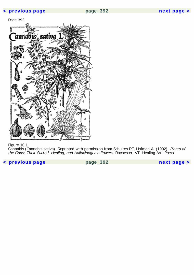
< previous page page_392 next page >
Page 392
Figure 10.1Cannabis (Cannabis sativa). Reprinted with permission from Schultes RE, Hofman A. (1992). Plants ofthe Gods: Their Sacred, Healing, and Hallucinogenic Powers. Rochester, VT: Healing Arts Press.
< previous page page_392 next page >
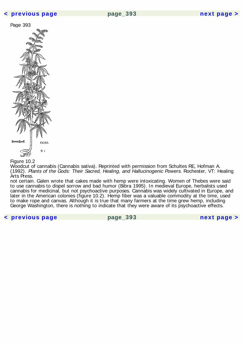
< previous page page_393 next page >
Page 393
Figure 10.2Woodcut of cannabis (Cannabis sativa). Reprinted with permission from Schultes RE, Hofman A.(1992). Plants of the Gods: Their Sacred, Healing, and Hallucinogenic Powers. Rochester, VT: HealingArts Press.not certain. Galen wrote that cakes made with hemp were intoxicating. Women of Thebes were saidto use cannabis to dispel sorrow and bad humor (Bibra 1995). In medieval Europe, herbalists usedcannabis for medicinal, but not psychoactive purposes. Cannabis was widely cultivated in Europe, andlater in the American colonies (figure 10.2). Hemp fiber was a valuable commodity at the time, usedto make rope and canvas. Although it is true that many farmers at the time grew hemp, includingGeorge Washington, there is nothing to indicate that they were aware of its psychoactive effects.
< previous page page_393 next page >
< previous page page_394 next page >
Page 394Cannabis was officially listed in the United States Pharmacopoeia until 1937, where it wasrecommended for a number of therapeutic uses, including sedation (Ray and Ksir 1990). TheMarijuana Tax Act was passed in 1937 making it effectively illegal to grow, sell, buy, or distributecannabis. From the early 1920s, cannabis use gained a public reputation as a menacing deviant drug,an attitude characterized in the movie Reefer Madness. By the 1950s and 1960s, cannabis use hadgained popularity, particularly with American youths. Use peaked in the late 1970s, when 60% ofhigh school seniors surveyed reported having used cannabis and 11% reported daily use (O'Brien1996). By 1996, 35% of high school seniors had used cannabis at least once in the past year, butonly 4.6% used it on a daily basis (Duffy and Milin 1996). In contrast, use of alcohol and cocainehad not changed across these time periods. In 1990, it was estimated that 6–10 million Americanssmoke cannabis at least once per week.The legal status of cannabis in the United States has varied greatly. The Controlled Substances Act of1970 classified cannabis as a Schedule I drug, which implies that the drug has no currently acceptedmedical use and has a high potential for abuse. During the 1970s, however, a degree of tolerancegrew. By 1977, most states considered possession to be a misdemeanor, and by 1980, eleven stateshad decriminalized possession (Grinspoon 1999). This atmosphere changed in the 1980s, whensevere legal penalties were enacted and the toxic pesticide paraquat was sprayed on domesticcannabis crops.The Dutch have depenalized and de facto legalized cannabis since 1976 (Engelsman 1989; MacCounand Reuter 1997). Depenalization has not increased usage in the population since this time, but itscommercial access has increased usage. However, parallel increases were also seen in the UnitedStates during this time period despite prohibition of cannabis use (Dutch cannabis use has remainedequal or below that of the United States). How this example relates to the issue of Americanlegalization or depenalization is a matter of debate.Botany and PreparationThere was uncertainty in the early botanical classification of cannabis. It was originally designated amember of the Nettle family (Urticaceae),
< previous page page_394 next page >
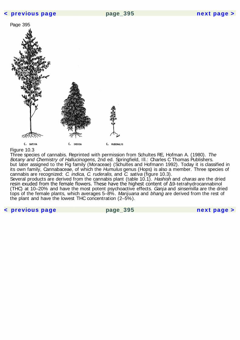
< previous page page_395 next page >
Page 395
Figure 10.3Three species of cannabis. Reprinted with permission from Schultes RE, Hofman A. (1980). TheBotany and Chemistry of Hallucinogens, 2nd ed. Springfield, Ill.: Charles C Thomas Publishers.but later assigned to the Fig family (Moraceae) (Schultes and Hofmann 1992). Today it is classified inits own family, Cannabaceae, of which the Humulus genus (Hops) is also a member. Three species ofcannabis are recognized: C. indica, C. ruderalis, and C. sativa (figure 10.3).Several products are derived from the cannabis plant (table 10.1). Hashish and charas are the driedresin exuded from the female flowers. These have the highest content of Δ9-tetrahydrocannabinol(THC) at 10–20% and have the most potent psychoactive effects. Ganja and sinsemilla are the driedtops of the female plants, which averages 5–8%. Marijuana and bhang are derived from the rest ofthe plant and have the lowest THC concentration (2–5%).
< previous page page_395 next page >
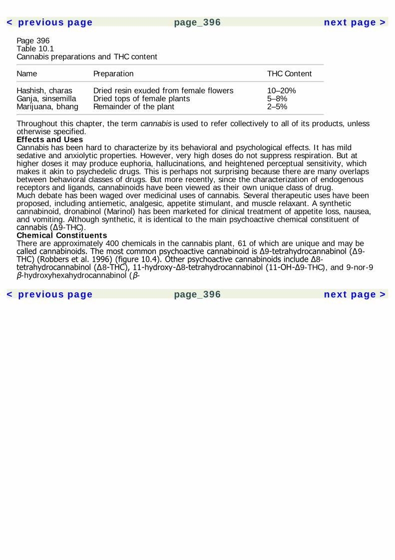
< previous page page_396 next page >
Page 396Table 10.1Cannabis preparations and THC content
Name Preparation THC Content
Hashish, charas Dried resin exuded from female flowers 10–20%Ganja, sinsemilla Dried tops of female plants 5–8%Marijuana, bhang Remainder of the plant 2–5%
Throughout this chapter, the term cannabis is used to refer collectively to all of its products, unlessotherwise specified.Effects and UsesCannabis has been hard to characterize by its behavioral and psychological effects. It has mildsedative and anxiolytic properties. However, very high doses do not suppress respiration. But athigher doses it may produce euphoria, hallucinations, and heightened perceptual sensitivity, whichmakes it akin to psychedelic drugs. This is perhaps not surprising because there are many overlapsbetween behavioral classes of drugs. But more recently, since the characterization of endogenousreceptors and ligands, cannabinoids have been viewed as their own unique class of drug.Much debate has been waged over medicinal uses of cannabis. Several therapeutic uses have beenproposed, including antiemetic, analgesic, appetite stimulant, and muscle relaxant. A syntheticcannabinoid, dronabinol (Marinol) has been marketed for clinical treatment of appetite loss, nausea,and vomiting. Although synthetic, it is identical to the main psychoactive chemical constituent ofcannabis (Δ9-THC).Chemical ConstituentsThere are approximately 400 chemicals in the cannabis plant, 61 of which are unique and may becalled cannabinoids. The most common psychoactive cannabinoid is Δ9-tetrahydrocannabinol (Δ9-THC) (Robbers et al. 1996) (figure 10.4). Other psychoactive cannabinoids include Δ8-tetrahydrocannabinol (Δ8-THC), 11-hydroxy-Δ8-tetrahydrocannabinol (11-OH-Δ9-THC), and 9-nor-9β-hydroxyhexahydrocannabinol (β-
< previous page page_396 next page >
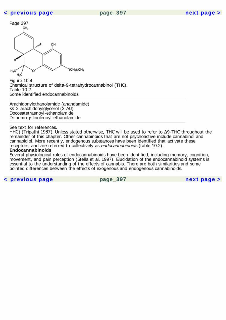
< previous page page_397 next page >
Page 397
Figure 10.4Chemical structure of delta-9-tetrahydrocannabinol (THC).Table 10.2Some identified endocannabinoids
Arachidonylethanolamide (anandamide)sn-2-arachidonylglycerol (2-AG)Docosatetraenoyl-ethanolamideDi-homo-γ-linolenoyl-ethanolamide
See text for references.HHC) (Tripathi 1987). Unless stated otherwise, THC will be used to refer to Δ9-THC throughout theremainder of this chapter. Other cannabinoids that are not psychoactive include cannabinol andcannabidiol. More recently, endogenous substances have been identified that activate thesereceptors, and are referred to collectively as endocannabinoids (table 10.2).EndocannabinoidsSeveral physiological roles of endocannabinoids have been identified, including memory, cognition,movement, and pain perception (Stella et al. 1997). Elucidation of the endocannabinoid systems isessential to the understanding of the effects of cannabis. There are both similarities and somepointed differences between the effects of exogenous and endogenous cannabinoids.
< previous page page_397 next page >
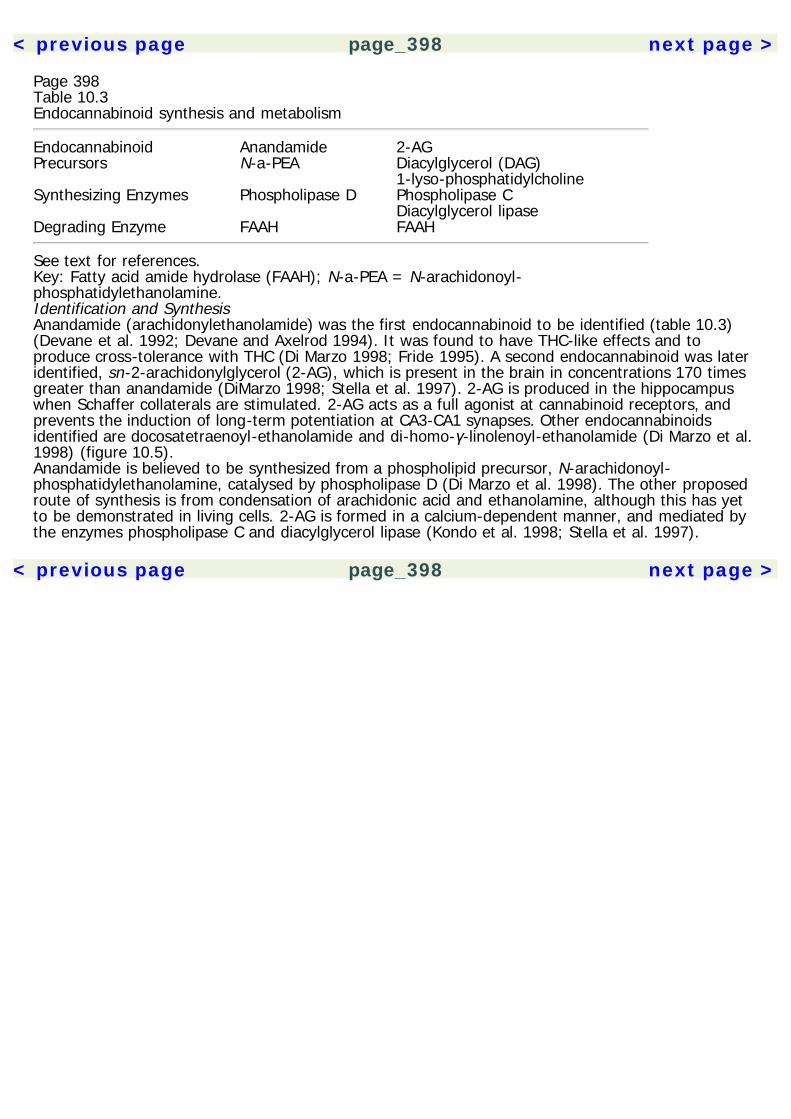
< previous page page_398 next page >
Page 398Table 10.3Endocannabinoid synthesis and metabolism
Endocannabinoid Anandamide 2-AGPrecursors N-a-PEA Diacylglycerol (DAG) 1-lyso-phosphatidylcholineSynthesizing Enzymes Phospholipase D Phospholipase C Diacylglycerol lipaseDegrading Enzyme FAAH FAAH
See text for references.Key: Fatty acid amide hydrolase (FAAH); N-a-PEA = N-arachidonoyl-phosphatidylethanolamine.Identification and SynthesisAnandamide (arachidonylethanolamide) was the first endocannabinoid to be identified (table 10.3)(Devane et al. 1992; Devane and Axelrod 1994). It was found to have THC-like effects and toproduce cross-tolerance with THC (Di Marzo 1998; Fride 1995). A second endocannabinoid was lateridentified, sn-2-arachidonylglycerol (2-AG), which is present in the brain in concentrations 170 timesgreater than anandamide (DiMarzo 1998; Stella et al. 1997). 2-AG is produced in the hippocampuswhen Schaffer collaterals are stimulated. 2-AG acts as a full agonist at cannabinoid receptors, andprevents the induction of long-term potentiation at CA3-CA1 synapses. Other endocannabinoidsidentified are docosatetraenoyl-ethanolamide and di-homo-γ-linolenoyl-ethanolamide (Di Marzo et al.1998) (figure 10.5).Anandamide is believed to be synthesized from a phospholipid precursor, N-arachidonoyl-phosphatidylethanolamine, catalysed by phospholipase D (Di Marzo et al. 1998). The other proposedroute of synthesis is from condensation of arachidonic acid and ethanolamine, although this has yetto be demonstrated in living cells. 2-AG is formed in a calcium-dependent manner, and mediated bythe enzymes phospholipase C and diacylglycerol lipase (Kondo et al. 1998; Stella et al. 1997).
< previous page page_398 next page >
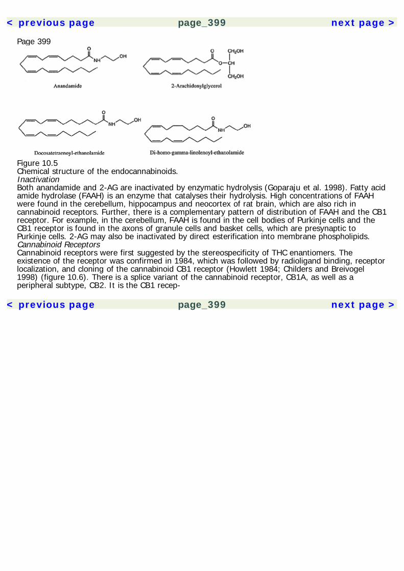
< previous page page_399 next page >
Page 399
Figure 10.5Chemical structure of the endocannabinoids.InactivationBoth anandamide and 2-AG are inactivated by enzymatic hydrolysis (Goparaju et al. 1998). Fatty acidamide hydrolase (FAAH) is an enzyme that catalyses their hydrolysis. High concentrations of FAAHwere found in the cerebellum, hippocampus and neocortex of rat brain, which are also rich incannabinoid receptors. Further, there is a complementary pattern of distribution of FAAH and the CB1receptor. For example, in the cerebellum, FAAH is found in the cell bodies of Purkinje cells and theCB1 receptor is found in the axons of granule cells and basket cells, which are presynaptic toPurkinje cells. 2-AG may also be inactivated by direct esterification into membrane phospholipids.Cannabinoid ReceptorsCannabinoid receptors were first suggested by the stereospecificity of THC enantiomers. Theexistence of the receptor was confirmed in 1984, which was followed by radioligand binding, receptorlocalization, and cloning of the cannabinoid CB1 receptor (Howlett 1984; Childers and Breivogel1998) (figure 10.6). There is a splice variant of the cannabinoid receptor, CB1A, as well as aperipheral subtype, CB2. It is the CB1 recep-
< previous page page_399 next page >
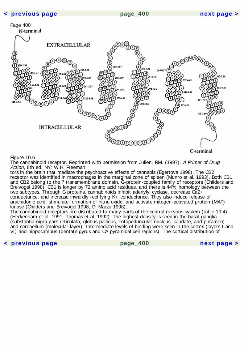
< previous page page_400 next page >
Page 400
Figure 10.6The cannabinoid receptor. Reprinted with permission from Julien, RM. (1997). A Primer of DrugAction, 8th ed. NY: W.H. Freeman.tors in the brain that mediate the psychoactive effects of cannabis (Egertova 1998). The CB2receptor was identified in macrophages in the marginal zone of spleen (Munro et al. 1993). Both CB1and CB2 belong to the 7 transmembrane domain, G-protein-coupled family of receptors (Childers andBreivogel 1998). CB1 is longer by 72 amino acid residues, and there is 44% homology between thetwo subtypes. Through G proteins, cannabinoids inhibit adenylyl cyclase, decrease Ca2+conductance, and increase inwardly rectifying K+ conductance. They also induce release ofarachidonic acid, stimulate formation of nitric oxide, and activate mitogen-activated protein (MAP)kinase (Childers and Breivogel 1998; Di Marzo 1998).The cannabinoid receptors are distributed to many parts of the central nervous system (table 10.4)(Herkenham et al. 1991; Thomas et al. 1992). The highest density is seen in the basal ganglia(substantia nigra pars reticulata, globus pallidus, entopeduncular nucleus, caudate, and putamen)and cerebellum (molecular layer). Intermediate levels of binding were seen in the cortex (layers I andVI) and hippocampus (dentate gyrus and CA pyramidal cell regions). The cortical distribution of
< previous page page_400 next page >

< previous page page_401 next page >
Page 401Table 10.4Central distribution of cannabinoid receptors
High BindingBasal gangliasubstantia nigra pars reticulataglobus pallidusentopeduncular nucleuscaudateputamenCerebellum (molecular layer)Intermediate BindingCerebral cortex (layers I and VI)Hippocampus (dentate gyrus and CA pyramidal cells)Frontal cortexAmygdalaCingulate gyrusLow BindingSpinal cordBrain stem (medulla and pons)Thalamic nucleiHypothalamusCorpus callosumCerebellum (deep nuclear layer)
See text for references.cannabinoid receptors is particularly high in the frontal cortex. Binding was also observed in limbicstructures including the amygdala and cingulate gyrus. Low levels of binding were observed in thespinal cord and brain stem (medulla and pons), thalamic nuclei, hypothalamus, corpus callosum andthe deep nuclear layer of the cerebellum. The neuroanatomical distribution of cannabinoid receptorswell account for the cognitive, motor, and emotional effects of cannabis. In contrast, there are littleor no cannabinoid receptors in the brain stem, which explains why cannabis has such a high fataldose. Similarities in binding are observed between cannabinoids and anandamide. Binding tocannabinoid receptors by anandamide and a selective CB1 antagonist (SR 141716A) and agonist (CP55,940), showed that all three ligands compete for the same receptor (Adams et al. 1998).
< previous page page_401 next page >

< previous page page_402 next page >
Page 402Chronic exposure to THC causes a regulation of cannabinoid receptors, which appears to be region-specific (Zhuang et al. 1998). For example, while increases in cannabinoid receptor mRNA are seen inthe cerebellum and hippocampus at 7 and 14 days of chronic treatment, decreases were seen in thestriatum from days 2 to 14. However, levels returned to normal in all the regions by day 21, whichcoincides with reports of behavioral tolerance.Physiological Effects of EndocannabinoidsSimilar to exogenous cannabinoids, the endocannabinoids have roles in behavior, motor function,memory, thermoregulation and analgesia (Childers and Breivogel 1998; Adams et al. 1998).Endocannabinoids have a biphasic effect on locomotion, stimulating it at low doses and inhibiting itat higher doses, which has been attributed to cannabinoid receptors in the basal ganglia andcerebellum. Detrimental effects on memory are consistent with inhibitory effects of endocannabinoidsin the hippocampus. Analgesic effects are produced at spinal and supraspinal levels, involvingstructures such as the dorsal horn and periaqueductal gray. Along with the similarities betweenendocannabinoids and cannabis constituents (referred to here as exocannabinoids), are significantdifferences. Although they have several common physiological effects, they are prevented by a CB1antagonist (SR 141716A) when induced by THC, but not when induced by anandamide (Adams et al.1998).Interaction with Other Neurochemical SystemsAmino AcidsAnandamide has dual effects on NMDA receptor function. It reduces NMDA Ca2+ currents, which ismediated by cannabinoid receptors and G-protein mechanisms (Hampson et al. 1998). However,anandamide potentiates NMDA currents due to a direct effect on the NMDA receptor itself. THC didnot have the same effect. So anandamide appears to have at least one other CNS effect that is notthrough the cannabinoid receptors. THC also reduces AMPA/kainate activity, which is mediated byCB1 receptors (Shen and Thayer 1999) (table 10.5). THC may in-
< previous page page_402 next page >
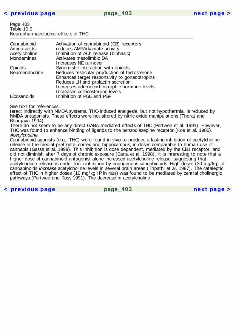
< previous page page_403 next page >
Page 403Table 10.5Neuropharmacological effects of THC
Cannabinoid Activation of cannabinoid (CB) receptorsAmino acids reduces AMPA/kainate activityAcetylcholine Inhibition of ACh release (biphasic)Monoamines Activates mesolimbic DA Increases NE turnoverOpioids Synergistic interaction with opioidsNeuroendocrine Reduces testicular production of testosterone Enhances target responsivity to gonadotropins Reduces LH and prolactin secretion Increases adrenocorticotrophic hormone levels Increases corticosterone levelsEicosanoids Inhibition of PGE and PGF
See text for references.teract indirectly with NMDA systems. THC-induced analgesia, but not hypothermia, is reduced byNMDA antagonists. These effects were not altered by nitric oxide manipulations (Thorat andBhargava 1994).There do not seem to be any direct GABA-mediated effects of THC (Pertwee et al. 1991). However,THC was found to enhance binding of ligands to the benzodiazepine receptor (Koe et al. 1985).AcetylcholineCannabinoid agonists (e.g., THC) were found in vivo to produce a lasting inhibition of acetylcholinerelease in the medial-prefrontal cortex and hippocampus, in doses comparable to human use ofcannabis (Gessa et al. 1998). This inhibition is dose dependent, mediated by the CB1 receptor, anddid not diminish after 7 days of chronic exposure (Carta et al. 1998). It is interesting to note that ahigher dose of cannabinoid antagonist alone increased acetylcholine release, suggesting thatacetylcholine release is under tonic inhibition by endogenous cannabinoids. High doses (30 mg/kg) ofcannabinoids increase acetylcholine levels in several brain areas (Tripathi et al. 1987). The catalepticeffect of THC in higher doses (10 mg/kg IP in rats) was found to be mediated by central cholinergicpathways (Pertwee and Ross 1991). The decrease in acetylcholine
< previous page page_403 next page >

< previous page page_404 next page >
Page 404in response to THC has also been related to an increase of slow-wave activity on EEG (Domino1981). In vitro THC (1–100 nM) inhibited muscarinic receptor binding, which also ocurred with twononpsychoactive cannabinoids (cannabidiol and cannabinol) (Ali et al. 1991). However, these effectswere not observed in vivo.MonoaminesCannabinoid agonists produce dose-related activation of meso-prefrontal dopaminergic transmission,which depends upon the CB1 receptor (Diana et al. 1998a). Conversely, withdrawal from chroniccannabinoid administration produces a reduction in mesolimbic dopaminergic transmission. AlthoughTHC stimulates the presynaptic activity of mesolimbic dopaminergic neurons, there is a decrease inpostsynaptic sensitivity (Bonnin et al. 1993). Further, this effect is modulated by estrogen, but notestradiol. THC increases the firing rate and burst-pattern firing in the ventral tegmental area andsubstantia nigra, although the ventral tegmental area is more sensitive in this respect (French et al.1997). In contrast, cannabidiol failed to have this effect. In vivo microdialysis, on the other hand,showed that THC has no effect on extracellular dopamine or 5-HT concentrations in the striatum ornucleus accumbens (Castaneda et al. 1991). However, this study also failed to show behavioraldifferences between drug and vehicle groups. Cannabinoids also induce increases in norepinephrine,but not 5-HT turnover in the prefrontal cortex (Jentsch et al. 1997).OpioidsCannabinoids are known to interact with opioid systems. This is particularly relevant to theiranalgesic effects and is discussed at length in chapter 8.NeuroendocrineCannabis compounds interact with the endocrine system. THC reduces testicular production oftestosterone, even though it enhances respon-
< previous page page_404 next page >

< previous page page_405 next page >
Page 405sivity at neuroendocrine target sites to gonadotropins (Dalterio et al. 1985a, 1987) (table 10.5). Theeffects of THC on plasma gonadotropins may be mediated by effects on hypothalamic catecholaminesand direct gonadal effects (Dalterio et al. 1985b; Murphy et al. 1994). THC also reduces male sexualbehaviors in the presence of a receptive female, but the effects are reversible with time (Harclerode1984).Despite structural similarity of cannabinoids to estrogens, they do not possess direct estrogenicactivity (Abel 1981). Neither psychoactive nor nonpsychoactive cannabinoids acted as agonists inassays of the estrogen receptor (Ruh et al. 1997). THC does suppress secretion of luteinizinghormone and prolactin and is mediated through central and/or pituitary actions (Rettori et al. 1988;Murphy et al. 1991). The effects of cannabinoids on brain function may differ between the sexes(Rodriguez de Fonseca et al. 1994). For example, cannabinoid receptor affinity is higher in malesthan females in the limbic forebrain, although receptor density is the same. Sensitivity within femalesvaries across the estrous cycle, where affinity is higher in diestrus and lower in estrus, but receptordensity was unchanged. Direct effects occur on the ovulatory process and on steroid secretion fromthe corpus luteum and the preovulatory follicle (Tyrey 1984). Whether these effects have significanteffects on physiological function remains to be seen. The CB1 receptor is present in the uterus andchronic THC may act as a weak uterotrophic agent (Paria et al. 1994). In addition to sex effects, THCalso had similar electrophysiological effects to 17-β-estradiol in the hippocampus, increasing fieldpotentials at very low (picomolar) doses, but depressing function at higher (nanomolar) doses (Foyet al. 1982).Cannabinoids interact with the adrenal axis also. Anandamide, similar to THC, activates thehypothalamic-pituitary-adrenal axis, increasing levels of adrenocorticotrophic hormone andcorticosterone (Weidenfeld et al. 1994). THC interacts primarily with the type II class of glucocorticoidreceptors (Eldridgde and Landfield 1990). Receptor down-regulation was noted after chronic (14days) THC treatment, comparable to that seen in chronic stress or high corticosteroid administration.THC, but not cannabidiol, showed some noncompetitive agonist properties at glucocorticoid receptors(Eldridge et al. 1991). Withdrawal from chronic cannabis administration elevates corticotrophin-releasing factor (CRF)
< previous page page_405 next page >
< previous page page_406 next page >
Page 406and gene transcription in the central nucleus amygdala with maximal increases corresponding to peakbehavioral signs of withdrawal (Rodriguez de Fonseca et al. 1997).EicosanoidsTHC has been shown to alter eicosanoid levels (Reichman et al. 1987). Psychoactive, but notnonpsychoactive constituents altered levels of prostaglandins (PGE and PGF). This effect was region-specific, altering levels in the striatum but not cerebellum.PharmacokineticsCannabis is most commonly consumed by smoking in a cigarette or pipe. Because the averagecannabis cigarette contains 0.5 to 1 g of cannabis, and the usual THC content of cannabis is 4 to 5percent, then the average cannabis cigarette would contain approximately 50 mg of THC (Julien1998). Of this amount, 25–50% (12–25 mg) of the THC is available in the smoke. Accordingly, theamount of THC that is absorbed into the blood after smoking one cannabis cigarette is 0.4 to 10 mg.People appear to regulate their intake based on the potency of the cannabis being smoked byaltering the length and depth of inhalation. Smoking topography measures revealed smaller puff andinhalation volumes and shorter puff duration for the high cannabis dose compared to the low dose(Heishman et al. 1989).After smoking the first puff, the acute intoxication starts within 6 to 12 minutes, and maximumeffects occur at 15 to 30 minutes. Effects typically last 2 to 4 hours. THC remains in fatty tissues andhas a long half-life of 57 hours. The lungs and liver convert THC to the metabolite 11-OH-THC,which is also active. There is a recirculation of 10–15% of hepatic metabolites. 11-OH-THC is furtherconverted by the liver to inactive metabolites, which are eliminated by the kidneys.Although there is a high degree of binding to protein, both bound and unbound drug is metabolized.The estimated terminal half-life is relatively long at 1.24 days, and is apparently unaffected byenzyme induction. Tolerance may be due to a shift toward highly polar metabolites in
< previous page page_406 next page >

< previous page page_407 next page >
Page 407the brain, rather than an increased elimination or decreased sensitivity of the brain tissue to THC(Magour et al. 1977).Route of administration alters the effectiveness of cannabinoids. Orally administered THC has aslower and more erratic absorption. THC was found to be 45 times more effective for analgesia afterintravenous than after subcutaneous administration (Martin 1985). The pharmacokinetics of differentchemical constituents of cannabis vary (Consroe et al. 1991). The elimination half-life of cannabidiolis estimated to be about 2–5 days, with no differences between genders. Comparably, theelimination half-life of Δ1-THC is approximately 4 days, and may be prolonged in chronic users(Johansson et al. 1988, 1989).The central distribution of THC was studied in the primate brain using positron emission tomography(Charlambous et al. 1991). The THC analog, (–)-18F-Δ8-THC, was administered intravenously tobaboons. Neuroanatomical distribution in the baboon brain was comparable to prior autoradiographicstudies, with binding evident in the basal ganglia, thalamus, and cerebellum. Clearance was rapidfrom these areas.General EffectsSubjective EffectsAs with many other psychoactive drugs, the psychological effects of cannabis are variable, dependingon the degree of prior use, as well as the expectations and personality of the user (Taylor 1998). Theacute subjective effects of cannabis may include relaxation, mild euphoria, and giddiness (table10.6). There is a heightening of sensory perceptions and time is perceived to pass more slowly.Adverse effects include a panic or anxiety reaction and are more common in novice users. Half ofusers report having experienced adverse effects at least once (O'Brien 1996). Other adverse effectsmay include dissociative symptoms such as derealization, the feeling that one's experiences are notreal, or depersonalization, a feeling of detachment from oneself (Mathew et al. 1993). The besttreatment for these experiences is supportive reassurance, or ''talking down," although anxiolyticmedication may be used in more extreme cases.
< previous page page_407 next page >

< previous page page_408 next page >
Page 408Table 10.6Physiological effects of cannabis
Subjective Effects (vary with set and setting)RelaxationMild euphoriaGiddinessHeightened sensory perceptionsStimulates appetitePerceived slower passage of timePanic or anxietyDissociative symptomsPhysiological EffectsIncreases heart rateReduces body temperatureSlowed gastrointestinal functionElectrophysiologyMixed excitation and inhibition, biphasic patternIncreased theta activity (long-term use)''Alpha hyperfrontality"Increased slow-wave sleep, reduces REM sleep
See text for references.Physiological EffectsTHC produces dose-dependent increases in heart rate and reductions in body temperature afterinjection, maximizing after 2 to 3 hours (table 10.6) (Heishman et al. 1989). Similarly, anandamidecreates hypothermia in mice after injection (Fride and Mechoulam 1993). The cognitive effects ofTHC are dissociable from the autonomic effects (Bachman et al. 1979).Certain cannabinoids exert an inhibitory effect on gastrointestinal transit (Shook and Burks 1989).THC slows the rate of gastric emptying and small intestinal transit. Other cannabinoids (Δ9,11-THC,cannabinol, and nabilone) also inhibit transit through the small intestine in mice, but they have lesseffect on gastric emptying. In contrast, cannabidiol has no effect on gastric emptying or intestinaltransit. Further, THC has a greater effect on transit in the small intestine than in the large intestine.
< previous page page_408 next page >
< previous page page_409 next page >
Page 409Cannabis use does not appear to induce a ''hangover" syndrome like that produced with alcohol orsedative-hypnotics in terms of sleep effects, mood, psychomotor skills, or cognitive function (Chait1990).Hormonal and Reproductive EffectsThe hormonal effects of smoking cannabis were examined in healthy male subjects (cigarettes with2.8% THC) (Cone et al. 1986). Plasma luteinizing hormone levels were reduced and cortisol wassignificantly elevated. Nonsignificant depressions were seen in prolactin, FSH, and testosterone, aswell as a nonsignificant elevation of GH. In female volunteers, smoking cannabis (1 g cigarette, 1.8%THC) caused a 30% suppression of plasma luteinizing hormone levels during the luteal phase of themenstrual cycle (Mendelson et al. 1986). The authors speculate that this could have adverse effectson reproductive function. Although cannabis usage does adversely affect sperm production, thephysiological significance of these effects is unclear (Abel 1981). There is no evidence at this timethat cannabis exposure reduces male fertility or increases risk for dominant lethal mutations inanimals or humans.Appetite and Ingestive BehaviorAnimal studies support the reputation of cannabis for stimulating appetite, although this effect hasnot been universally reported (Graceffo and Robinson 1998). Sated rats showed substantialhyperphagia when administered THC at doses of 0.5, 1.0, and 2.0 mg/kg (Williams et al. 1998). Thisappetitive effect of THC was found in other species, such as dogs, whereas LSD, by contrast,produces anorexia (Vaupel and Morton 1982).Conversely, food intake and body weight are reduced by the selective cannabinoid CB1 antagonist SR141716. Tolerance developed to the anorectic effect of SR 141716 within 5 days of chronictreatment, but body weight remained low throughout 14 days of treatment. In contrast, chronic THCtreatment (15 days) suppresses intake of food and water (Drewnowski and Grinker 1978). This effectis of short duration, with
< previous page page_409 next page >

< previous page page_410 next page >
Page 410no apparent rebound eating afterwards. However, this may be due to general low arousal rather thana specific appetite suppression.Electrophysiology: Animal StudiesElectrophysiological responses to cannabinoids are complex. THC produces both CNS excitation anddepression in several electrophysiological paradigms (Turkanis and Karler 1981). The effect produceddepended on the cannabinoid dosage and paradigm used. In contrast to THC, cannabidiol generatedno CNS excitation, and produced either depressant effects or no effects at all.THC (0.75–4.0 mg/kg IP) produced a biphasic pattern on EEG in rhesus monkeys (Matsuzaki et al.1987). The first phase consisted of high-voltage slow waves (HVSW) on the EEG, which appeared20–30 minutes after drug administration and lasted for 3–4 hours. Spike-bursts occurred in frontaland temporal lobes and hypothalamus, theta waves ocurred in parietal and occipital lobes, andgeneralized HVSW ocurred in subcortical regions. Behavioral sedation, bradycardia, and hypothermiawere also evident during this phase. Similarly, high-voltage EEG bursts were also seen in the rat afterTHC administration, which carried over into the initial subsequent REM sleep episodes (Buonamici etal. 1982). The second EEG/behavioral phase consisted of high-voltage fast waves on EEG, withdecreased neocortical spike-bursts. These physiological, behavioral, and electroencephalographicchanges correlated with THC levels and disposition in blood. EEG slowing has been related to theconcomitant decrease in neocortical acetylcholine release (Domino 1981).Electrophysiology: Human StudiesWhen long-term chronic cannabis users (i.e., daily use for 15–24 years) were compared to short-term users and nonusers, they were found to have elevated absolute power of theta activity overbilateral frontal-central cortex, as well as significantly increased interhemispheric coherence of thetaactivity across central and posterior regions (Struve et al. 1998). Concurrent reaction time studiesalso showed slowed cognitive processing in that group. Other work by this group has shown that in
< previous page page_410 next page >
< previous page page_411 next page >
Page 411daily cannabis users, as contrasted with nonusers, there is an association between increased absoluteand relative power and interhemispheric coherence of EEG alpha activity over the bilateral frontal-central cortex (termed ''alpha hyperfrontality") (Struve et al. 1994).These EEG changes have been related to effects of cannabis on motor functions and sleep. Onestudy of occasional cannabis smokers had them report quantitative changes in their mood duringEEG monitoring (Lukas et al. 1995). Mood elevations were reported during the first 15 minutes, whichoccurred while plasma THC levels were rising and EEG alpha power was significantly higher.A few studies have been performed with event-related potentials (ERP) to examine the cognitiveeffects of THC. In subjects viewing emotionally valenced words (neutral, negative, or positive), THCproduced a behavioral decrease in recognition rate (Leweke et al. 1998). The ERPs showed thetypical difference between old and new words (enhanced positivity for old words beginning 250milliseconds poststimulus), and THC enhanced the positivity in response to old words. This counter-intuitive dissociation between ERPs and behavioral indices was interpreted as a reflection of implicitmemory processes underlying the ERP effect.Several studies of ERPs and brain function have produced mixed results due to methodologicalconfounds. When subjects are screened for medical and psychiatric illness, and age effects werecontrolled, THC does not alter brain stem and auditory or visual P300 responses (Patrick et al. 1997).Despite daily cannabis use in subjects, the only finding consisted of an elevated auditory P50amplitude.SleepAnandamide increases slow-wave and REM sleep at the expense of wakefulness (Murillo-Rodriguez etal. 1998). In contrast, chronic THC treatment in nonhuman primates reduced the time spent in slow-wave sleep (Adams and Barratt 1975). The time spent in stage 1 increased with repeated treatmentand remained elevated through recovery.Cannabis has electrophysiological effects on human sleep as well. Daily doses (70 to 210 mg) of THCreduced REM sleep and increased
< previous page page_411 next page >
< previous page page_412 next page >
Page 412stage 4 sleep (Feinberg et al. 1976, 1975). Abrupt withdrawal increased REM and reduced stage 4 tobaseline levels.Neuroimaging: Human StudiesA few studies have been done to determine the in vivo effects of cannabis on humans usingfunctional neuroimaging. The effects of an acute dose of THC on cerebral blood flow (CBF) wasexamined in volunteers with prior histories of exposure to cannabis by using positron emissiontomography (PET) (Mathew et al. 1997). THC increased CBF in the frontal regions bilaterally, insulaand cingulate gyrus and subcortical regions with somewhat greater effects in the right hemisphere.The effects were significant at 60 minutes for the lower dose and at 30 minutes for the larger dose.There was also a highly significant change in the anterior/posterior ratio, reflecting minimal change inoccipital regions but significant increases in frontal CBF. Ratings of subjective effects correlated mostmarkedly with alterations in the right frontal region.Another study examined the effects of THC on regional brain glucose metabolism (18F-2-fluoro-2-deoxyglucose and PET) following an acute intravenous dose (Volkow et al. 1991). Changes in globalglucose metabolism in response to THC were variable, and only the cerebellum consistently showedan increase in metabolic activity. Further, this cerebellar increase was correlated with subjectiveratings of intoxication and plasma THC concentration. This finding is interesting in light of the highdensity of cannabinoid receptors in the cerebellum.A third neuroimaging study using CBF (PET) and magnetic resonance imaging found a significantincrease in cortical and cerebellar blood flow following THC administration, but again this effect wasinconsistent across subjects (Mathew et al. 1998). The decrease in cerebellar CBF was related toalterations in time sense. This is interesting in light of the proposed role of the cerebellum as aninternal timing system.Motor EffectsThe effects of THC on motor activity are dependent on dosage, species examined, and behavioralparadigm used. THC increases activity in mice
< previous page page_412 next page >

< previous page page_413 next page >
Page 413but reduces social investigation and sexual behaviors (Evans et al. 1976; Cutler and Mackintosh1984). Central administration of anandamide also increases locomotor activity in immobility andopen-field tests (Murillo-Rodriguez et al. 1998). THC and other cannabinoids were shown to produceataxia in dogs at doses as low as 2 μg/kg (Stark and Dews 1980).The motor changes produced by THC may relate to altered nigrostriatal dopaminergic function(Navarro et al. 1993). An oral dose of THC reduced spontaneous motor activity and stereotypicbehaviors (e.g., rearing and self-grooming) in the rat. These motor changes correlated with a lownumber of D1 receptors in the striatum, but not other measures of dopamine or 5-HT activity.Anandamide motor inhibition also corresponded with reductions in nigrostriatal activity (Romero et al.1995). High doses (10 mg/kg IP) produce catalepsy (Pertwee and Ross 1991).Cognition: Animal StudiesCentral administration (intracerebroventricular) of anandamide has been found to impair memoryconsolidation in animals (Murillo-Rodriguez et al. 1998). Both THC and anandamide impair recentmemory in animals (Mallet and Beninger 1998). This effect was mediated by the CB1 receptor in aworking memory (delayed nonmatch to sample) paradigm. The impairment of working memory bycannabinoids may relate to the activation of mesoprefrontal dopaminergic transmission via CB1receptors, because overstimulation of dopamine receptors impairs working memory (Diana et al.1998a; Jentsch et al. 1997). On the other hand, administration of dopamine (D1 and D2) agonistsimproved the memory impairment induced by anandamide on an inhibitory avoidance response(Castellano et al. 1997). Clearly, the experimental paradigm used, as well as endogenous versusexogenous ligands alters the results obtained. The effect of THC on a working memory measure(delayed-match-to-sample) was found to correspond to effects in the hippocampus (Heyser et al.1993). However, these effects completely reversed within 24 hours of injection. Another possiblemechanism for THC's effect on memory is inhibition of hippocampal long-term potentiation, althoughthis has only been observed with synthetic cannabinoids (Collins et al. 1994).
< previous page page_413 next page >
< previous page page_414 next page >
Page 414Table 10.7Sources of variability in cognitive studies of THC
Dosage/potencyRoute of administrationFrequency of useAcute vs. chronic effectsSubject population
See text for references.Cognition: Human StudiesThe effects of cannabis on cognition is a complicated issue. Numerous studies have been conductedto measure the mental effects of cannabis, but many of these are fraught with methodologicalproblems. Several variables must be controlled in this type of research, including the dosage andpotency of cannabis used, route of administration (e.g., smoking, oral, etc.), chronicity of dose, andthe subject population (nonusers, occasional users, chronic users) employed (table 10.7). Populationmakeup has a large effect on outcome because there have been differences demonstrated betweennaive and experienced cannabis users (Murray 1986).Impairment is observed in some studies on measures of concentration and attention (e.g., reactiontime, digit span, and digit symbol tasks) (table 10.8). In some studies, the effect only occured at thehigher (2.7%) but not lower (1.3%) dose of THC (Wilson et al. 1994). Some studies did not show aneffect on divided-attention tasks, while other studies showed effects correlating with THC plasmalevels for approximately 2 hours after smoking (Barnett et al. 1985; Heishman et al. 1989). Thenature and complexity of the task may affect whether differences are found between cannabis andplacebo groups. No effects are seen on simple measures of immediate or sustained attention(Rafaelsen et al. 1973; Waskow et al. 1970; Hooker and Jones 1987). Casual and heavy cannabisusers did not differ on a simple vigilance task, but both were impaired on a goal directed serialalternation task, requiring mental calculations, working memory, and alternating attention (Casswelland Marks 1973a, 1973b). Cannabis (1.2% THC) increased the Stroop
< previous page page_414 next page >
< previous page page_415 next page >
Page 415Table 10.8Cognitive effects of cannabis
Attention/concentration Simple and sustained attention are unaffected Mild impairment of some forms of complex attentionMemory Impairment of recent memory, consolidation Increased recognition false positive errors Remote memory unaffectedPerceptual and motor Mild effects at low doses Hallucinations and catalepsy at high dosesExecutive functions Mild increase in perseverative responses with heavy use
Note: Effects of THC on cognition are likely to be dose and task dependent. See textfor references.effect, decreasing subjects' ability to inhibit automatic responses (Hooker and Jones 1987). Yetperformance was not affected on the paced auditory serial attention test (PASAT), a demanding taskrequiring attention, working memory, and mental calculations.The effects of cannabis on memory was examined in numerous studies, and again the results arevariable. For example, fewer words are recalled from a list by subjects administered cannabiscompared to placebo, but no effects were seen on a verbal paired-associate learning (Abel 1971;Hooker and Jones 1987).Perhaps the most consistent effect of cannabis is consolidation of information from short-termmemory (Dornbush et al 1971; Murray 1986). In contrast, cannabis does not appear to impair accessto information already in long-term memory (Darley et al. 1977; Parker et al. 1980). While cannabisand placebo groups performed equally well on a word list recognition task, the cannabis group mademore false positive errors (Abel 1970, 1971). Another effect on memory reported in severeal studiesis increased intrusion of irrelevant material (Abel 1970, 1971; Clark et al. 1970; Tinklenberg et al.1970; Pfefferbaum et al. 1977).THC produced small but consistent detrimental effects and increased the variability of performanceon a battery of sensory and perceptual-motor tests (Peters et al. 1976). This mild effect is in contrastto the profound subjective effects concurrently reported on subjective measures
< previous page page_415 next page >
< previous page page_416 next page >
Page 416(the subjective drug effects questionnaire). Subjective changes reported in mood and perceptualexperiences were more pronounced among occasional than frequent cannabis users. Although it haseffects on motor performance, it appears that cannabis has little effect on eye movements (Stapletonet al. 1986; Baloh et al. 1979). No effects were observed on a controlled verbal fluency measure(controlled oral word associations) (Hooker and Jones 1987).Chronic use has been associated with an ''amotivational syndrome" characterized by loss of interestin social activities, school, work, or other goal-directed activities. Cannabis use is cited as the causeof this phenomenon, but there is no evidence to support any causal relationship. There is evidence,however, that the symptoms of the "amotivational syndrome" are secondary to depression (Mustyand Kraback 1995). In contrast to ethanol, there is no evidence to support that cannabis causes anincrease in violent behavior (Murray 1986). However, cannabis use may be contraindicated in thosewith preexisting psychiatric disturbances such as bipolar disorder or schizophrenia.Cognitive Effects in Experienced UsersA study of experienced cannabis smokers tested them on cognitive measures using two doses(1.75% or 3.55%) and placebo (Wilson et al. 1994). The functions most sensitive to the effects ofTHC were mental processing speed (digit-symbol substitution) and reaction time. When comparedwith cannabis nonusers, chronic users of cannabis—with a mean duration of use of 6.76 years andan average daily intake of 150 mg of THC—were found to have slower reactions on perceptuo-motortasks, but no differences on intelligence or memory tests (Varma et al. 1988).Compared to infrequent (monthly) users of cannabis, regular (daily) users show decreases inattention and executive functions, such as greater perseverations on card sorting and reducedlearning of word lists (Pope and Yurgelun-Todd 1996). However, studies comparing heavy users tooccasional users or nonusers, although otherwise well-controlled, are plagued with themethodological problem of self-selecting groups. It may well be that people with executive andattentional dysfunctions are more prone to heavier cannabis use. Indeed, abstinent polysubstance
< previous page page_416 next page >

< previous page page_417 next page >
Page 417users were found to have lesser grey matter in prefrontal cortex than nonusers (Liu et al. 1998).In a study by Fletcher and colleagues (1996), long-term users tended to experience greater difficultyin complex tasks of memory and attention. However, it was concluded that these deficits arecomparably subtle. The older long-term users were functional, employable, and did not show thedegree of cognitive deficits that are associated with comparable use of alcohol. The physical health ofthe subjects was generally good.THC and EthanolComparisons have been made of the cognitive effects of THC and alcohol, alone and in combination.Separately, THC and alcohol were found to produce comparable impairments on the digit-symbolsubstitution test and word recall (Heishman et al. 1997). The interaction of THC and ethanol wasstudied in a signal detection paradigm (Marks and MacAvoy 1989). Cannabis users were lessimpaired in peripheral signal detection than nonusers while intoxicated by cannabis and/or alcohol.These findings suggest the development of tolerance and cross-tolerance in regular cannabis usersand/or the ability to compensate for intoxication effects.Both THC and ethanol increase reaction time, and produce decrements in standing steadiness andpsychomotor coordination (Belgrave et al. 1979). Whereas the peak effects of ethanol appearedquickly and wore off quickly (after 280 minutes), THC's effects were slower in onset and longer induration. The effects of combined THC and ethanol are additive and not synergistic, and THC did notalter blood-ethanol levels. Other studies have shown an interaction between ethanol and THC onpsychomotor skills necessary for driving, although there were no interactions on the subjective''high," heart rate acceleration, or THC plasma concentration (Perez-Reyes et al. 1988).Cognitive Effects of Cannabis: ConclusionsThe cognitive effects of cannabis are mild, and have many variables (dosage, chronicity of use,cognitive task employed, etc.). Of prime importance in such studies is the usage (or misusage) of theterm impair-
< previous page page_417 next page >

< previous page page_418 next page >
Page 418ment. Many studies label an impairment when they find statistically significant differences betweenusers and nonusers, or various dosages of THC. While statistically significant, this does not addressthe size of the effect. Neuropsychologists use a cutoff of two standard deviations below the mean tosignify impairment. While cannabis does create some subtle deficits, the test performances are oftenwell within the normal range and would not be diagnosed as an impairment.A comprehensive study by Wert and Raulin (1986) examined both American and cross-culturalstudies, and pointed out methodological problems in the body of research on cannabis. It wasconcluded that cannabis use may produce subtle impairment, but there is no evidence that cannabisproduces gross structural cerebral changes or functional impairment. Many cannabis users remainintelligent, functional, and productive members of society (Grinspoon 1999; Davidson 1999).Tolerance, Dependence, and AddictionA rapid tolerance to the acute effects of THC occurs in mice, which rapidly reverses as well(Hutcheson et al. 1998). Although chronic exposure to THC alters cannabinoid receptors, this effect isnot irreversible (Westlake et al. 1991).Pharmacokinetic tolerance to THC occurs, which parallels the development of psychological,cardiovascular, and hypothermic effects (Hunt and Jones 1980). These changes include alteredmetabolic clearance, distribution volume, urinary excretion, and slow elimination of metabolites. Lowrenal clearance indicated an accumulation of metabolites. Despite the pharmacokinetic toleranceevident here, it was concluded that this alone is not sufficient to account for tolerance tocardiovascular, psychological, and skin hypothermic effects, which are likely to involve central effects.Cannabis has the potential to produce dependence (Taylor 1998). Like many drugs of abuse, cerebraldopamine is involved in the pleasurable effects of cannabis, and corticotrophin-releasing factor isinvolved in the unpleasant withdrawal phenomena. While central dopaminergic activity does notautomatically make a drug addictive (e.g., neither the antidepressant bupropion nor the dopamineagonist bromocriptine appear to
< previous page page_418 next page >
< previous page page_419 next page >
Page 419Table 10.9Cannabis withdrawal syndrome
RestlessnessIrritabilityInsomniaNauseaMuscle cramping
See text for references.be addictive), it does raise the potential. On the other hand, cannabis is less addictive and producesmilder withdrawal symptoms compared to cocaine, alcohol, and heroin (Taylor 1998). Nonetheless,dependence to cannabis may develop after heavy chronic use, and the intensity of the withdrawalsyndrome is related to the degree of use and tolerance.Concurrent use with ethanol may alter the patterns of drug use. While low to moderate doses ofethanol do not influence the amount of cannabis self-administered by humans, there is evidence thatchronic THC administration can increase ethanol intake (McMillan and Saodgrass 1991; Chait andPerry 1994).Cannabis Withdrawal SyndromeCessation of chronic cannabis use is known to produce a withdrawal syndrome consisting ofrestlessness, irritability, insomnia, nausea, and muscle cramping (table 10.9) (O'Brien 1996).However, this syndrome is only seen in people who use high daily amounts and then suddenly stop(O'Brien 1996). These symptoms are not usually seen in clinical populations, and frequent users ofcannabis are not driven by a fear to avoid a withdrawal syndrome as seen in opioid addiction.In mice, somatic signs of withdrawal include wet dog shakes, front paw tremor, ataxia, hunchedposture, tremor, ptosis, piloerection, mastication and decreased locomotor activity (Hutcheson et al.1998). The CB1 antagonist (SR141716A) has been used to precipitate a withdrawal state in THC-tolerant animals, and higher doses of THC produced a greater withdrawal syndrome (Aceto et al.1995; Cook et al. 1998). Withdrawal from chronic cannabis use reduces mesolimbic dopaminer-
< previous page page_419 next page >

< previous page page_420 next page >
Page 420gic transmission, which is also observed in other drugs of abuse (Diana et al. 1998b). Withdrawalfrom chronic cannabis administration elevates corticotrophin-releasing factor (CRF) and activity in thecentral nucleus amygdala (Rodriguez de Fonseca et al. 1997).Abuse of Therapeutic CannabinoidsDronabinol (Marinol) is an oral form of THC indicated for treatment of AIDS and cancerchemotherapy (Calhoun et al. 1998). Because it is the active principle of cannabis, there has beensome concern over its abuse potential. However, in a thorough study utilizing literature review,surveys, and interviews with medical and law enforcement personnel, it was concluded thatdronabinol has very low abuse potential (Calhoun et al. 1998). There was no evidence of abuse orillegal diversion of dronabinol. Prescription data indicate usage within the therapeutic dosage rangeover time, and no instances of ''script-chasing" or "doctor-shopping" were reported. As opposed tosmoked cannabis, the onset of action in Dronabinol is slow and gradual. It seems at best weaklyreinforcing, and many users report unappealing effects.Therapeutic Uses of Cannabis and CannabinoidsCannabis shows promise for clinical treatment of many illnesses, including glaucoma, nausea andvomiting, analgesia, spasticity, multiple sclerosis, and AIDS wasting syndrome (table 10.10) (Taylor1998). While the legality of treatment with cannabis varies from region to region, ballot initiatives inCalifornia and Arizona have recently made crudeTable 10.10Potential therapeutic uses of cannabis and cannabinoids
GlaucomaNausea/emesisPainMuscle spasticityMultiple sclerosisAppetite loss (e.g. AIDS wasting syndrome)
See text for references.
< previous page page_420 next page >

< previous page page_421 next page >
Page 421cannabis accessible to patients under certain circumstances (Voth and Schwartz 1997).AntiemeticTHC was first used to control nausea and vomiting during chemotherapy in the 1970s (Vincent et al.1983). Until the development of 5-HT3 antagonists, dronabinol and cannabis were frequently used bycancer patients receiving chemotherapy (Taylor 1998). THC and synthetic cannabinoids have beeneffective in controlled clinical trials. THC is superior to placebo and as effective or superior toprochlorperazine. Although there are more drug-related effects associated with THC thanprochlorperazine, they did not reduce the patients' preference for the drug (Ungerleider et al. 1982).The antiemetic effectiveness of THC correlates with the subjective high reported (Vincent et al.1983). THC is effective in several chemotherapy regimens, including methotrexate and thedoxorubicin/cyclophosphamide/fluorouracil combination. Cisplatin treatment, however, is moreresistant. Side effects of THC are generally well tolerated, and use may be limited in the elderly orwith higher doses. Nabilone is a synthetic cannabinoid that is more effective than prochlorperazine inchemotherapy-induced emesis, including cisplatin. Its side effects are similar to THC. Levonantradol isanother synthetic cannabinoid with antiemetic effects, and may be administered orally or intra-muscularly. The side effect of dysphoria may limit its use.Appetite StimulantA review of evidence supported the use of pure THC preparations to treat nausea associated withcancer chemotherapy and to stimulate appetite (Voth and Schwartz 1997). In patients with anorexiadue to advanced cancer, THC (2.5 mg PO tid) was effective in stimulating appetite and was welltolerated at low doses (Nelson et al. 1994).Cannabinoids produced positive results for treatment of AIDS-related anorexia. Positive results ofopen trials were later confirmed with methodologically controlled studies. Dronabinol (2.5 mg POtwice daily) produced consistent and substantial improvement in appetite in patients with AIDS (Bealet al. 1995, 1997). Patients also reported improved
< previous page page_421 next page >

< previous page page_422 next page >
Page 422mood and tended to maintain stable body weight for at least 7 months. Side effects reported wereprimarily psychoactive. Increases were seen in body fat (1%) and prealbumin (Struwe et al. 1993).There was also decreased symptom distress and improved appetite. There are reports that thepsychotropic effects of dronabinol can be decreased by administration of prochlorperazine (Plasse etal. 1991).GlaucomaIn the early 1970s, cannabis was noted to reduce intraocular pressure in people with glaucoma(Cooler and Gregg 1977). In a controlled study, THC decreased intraocular pressure an average of37%, with maximum decreases at 51% of baseline. Concurrent increases in heart rate were alsoobserved. The mechanism of this effect of THC is uncertain.SpasticityThere are some antispasticity effects of THC (Petro and Ellenberger 1981). Animal studies haveindicated that THC inhibits polysynaptic reflexes. In a controlled study, THC (10 mg) significantlyreduced spasticity of various etiologies. However, no benefit was noted in patients with spasticity dueto cerebellar disease. In a survey of multiple sclerosis patients, cannabis was reported to relievespasticity, pain, and tremors (Consroe et al. 1997). A recent study in a mouse model of multiplesclerosis (chronic relapsing experimental encephalomyelitis) has provided more quantitative supportalong these lines (Baker et al. 2000). Cannabinoid receptor agonists improved tremor and spasticityin mice, which was dependent on the CB2 and, to a greater degree, the CB1 receptors.ToxicityDevelopmental ToxicityCannabinoids are teratogenic and should be avoided during pregnancy. Cannabis use duringpregnancy is associated with lower gestational age
< previous page page_422 next page >
< previous page page_423 next page >
Page 423at delivery, increased risk of prematurity, and reduction in birth weight (Abel et al. 1980; Walker, etal. 1999; Sherwood et al. 1999). There are also behavioral alterations in the development ofspontaneous locomotion and exploration in rats (Navarro et al. 1995). Exposure to THC throughgestational and lactational periods produces changes in response to novelty, social interactions, andsexual behavior. Primates exposed to THC in utero show an altered visual attention characterized bya reduced ability to disengage from novel stimuli (Golub et al. 1982).Human newborns of mothers who smoke cannabis during pregnancy experience mild withdrawalsymptoms and some autonomic disruption upon childbirth (Fried 1995a, 1995b). Although behavioralconsequences are not apparent until 3 years of age, decrements in languange comprehension,sustained attention, memory, and behavior are related to in utero cannabis exposure.Pulmonary ToxicityOne of the major toxicity issues in cannabis consumption relates to the fact that it is most oftensmoked. Cannabis and tobacco smoke, apart from having different psychoactive constituents, areactually very similar in their composition (Hoffman et al. 1975). Cannabis smoke is mutagenic, whichgives it carcinogenic potential (Nahas and Latour 1992). Although no specific reports of lung canceror emphysema from cannabis smoke exist, it is at least as harmful as tobacco smoke, containingthree times as much tar and five times as much carbon monoxide (Wu et al. 1988). Cannabis smokeinflames the airways and reduces respiratory capacity. Airway obstruction and squamous metaplasiasmay also occur.CardiovascularA common effect of THC is increased heart rate and blood pressure. Corneal blood vessels dilate,creating the red-eyed presentation that is commonly associated with cannabis. These effects aregenerally not dangerous because cannabis has a high therapeutic margin and no fatal dosages areknown in humans. However, these effects may be a danger to those individuals with preexistingcardiovascular disease.
< previous page page_423 next page >
< previous page page_424 next page >
Page 424Immune SystemThe presence of cannabinoid receptors in the immune system has led to consideration of the effectsof cannabis on its function. Cannabinoid receptors have been found in spleen cells (Kaminski et al.1992). Activation of these receptors would inhibit their function in the immune response. Similarsuppressant effects occur on lymphocytes (Diaz et al. 1993). THC and anandamide inhibitmacrophage-mediated tumor necrosis (Cabral et al. 1995). Despite these effects, their functionalsignificance remains to be determined. These effects are most likely subtle.Neurotoxicity and NeuroprotectionUntil recently, there was little clear evidence that cannabis use was toxic to neurons. Cannabinoidshave been shown to have a mix of neurotoxic and neuroprotective effects and mixed results havebeen found in different studies. Scallet (1991) points out significant methodological differences in theexisting studies that influence their outcomes. Critical determinants of neurotoxicity are both ageduring exposure and duration of exposure. For example, cannabinoid administration was required forat least three months (8–10% of a rat's lifespan) to produce neurotoxic effects in peripubertalrodents. This is comparable to about three years exposure in rhesus monkeys and seven to ten yearsin humans. Reports of neurotoxicity in primates were inconsistent, with up to 12 months of dailyexposure. Longer exposures have not yet been studied (Scallet 1991).A recent study showed THC to be toxic to hippocampal neurons, while sparing cortical neurons inconcentrations likely to be reached in normal human doses (0.5 μM) (Chan et al. 1998). Apoptoticchanges were noted in the hippocampus such as shrinkage of neuronal cell bodies and nuclei as wellas DNA strand breaks. THC stimulates release of arachidonic acid, and it was hypothesized that thisneurotoxic effect is mediated by cyclooxygenase pathways and free radical generation. In support ofthis, the toxicity is inhibited by nonsteroidal anti-inflammatory drugs, such as aspirin, andantioxidants such as vitamin E.
< previous page page_424 next page >
< previous page page_425 next page >
Page 425Cannabidiol and THC reduced neurotoxicty induced by glutamate in cortical neurons (Hampson et al.1998). This result was effective for toxicity induced at both NMDA and AMPA/kainate receptors, andwas independent of cannabinoid receptor activity. The mechanism of neuroprotection appears to beby their potent antioxidant activity. They were even more protective against glutamate neurotoxicitythan either ascorbate or α-tocopherol.Dexanabinol is a synthetic cannabinoid (devoid of psychoactive effects) that is being evaluated forneuroprotective effects (Brewster et al. 1997). It acts as a noncompetitive NMDA receptor antagonistand also as a D1 agonist (Striem et al. 1997). Dexanabinol is also a potent scavenger of free radicalsand protects cultured neurons from toxicity of radical generators (Biegon and Joseph 1995). Itsneuroprotective effects are seen in models of ischemia and optic nerve crush. Dexanabinol inhibitsproduction of brain tumor necrosis factor (TNFα) and nitric oxide after head injury and septic shock(Gallily et al. 1997).Seizures and EpilepsyAlthough cannabis does not induce seizures in normal doses, it may have negative effects on seizuredisorders. The effects of cannabinoids on seizures are complex. Either inhibition or facilitation ofseizures may occur depending on the cannabinoid used, dose, tolerance, and the experimentalseizure paradigm used (Karler and Turkanis 1980). THC itself has mixed effects on seizures. Someresearchers have reported antiseizure effects (Wada et al. 1975, 1976; Sofia et al. 1976). However,other studies have shown proseizure effects, such as increases in the frequency of focal potentials,generalized bursts of polyspikes, and frank convulsions in cobalt-induced seizures (Chiu et al. 1979).The major metabolite of THC, 11-OH-Δ9-THC, also has excitatory effects. THC enhanced bothelectrical and chemical (pentylenetetrazol or picrotoxin) kindling in mice (Karler et al. 1984). In somecases, kindling was facilitated after withdrawal of the cannabinoid. The effect on kindling may be dueto prolonged ''rebound hyperexcitability," which occurs after THC administration (Karler and Turkanis1981; Karler et al. 1986). The peak
< previous page page_425 next page >
< previous page page_426 next page >
Page 426of this effect occurred at 24 hours after administration and lasted as long as 196 hours. Thisrebound hyperexcitability enhanced electrical seizure kindling and blocked the antiseizure effect ofphenytoin. Thus, THC itself may inhibit seizure activity, but may leave in its wake a period ofhyperexcitability that promotes seizures.The chronicity of THC administration also influences its effects on seizures. Δ8-THC and Δ9-THCreduced cobalt-induced seizures, but tolerance to this effect developed after only two days (Colasantiet al. 1982). Additionally, even though seizures were suppressed, interictal spiking was enhanced.Studies that showed a preventive effect of cannabinoids on kindling also showed rapid developmentof tolerance to the effect (Corcoran et al. 1978). It was suggested that the initial anticonvulsanteffect occurred by limiting the spread of epileptogenic activity originating from the focus (Colasanti etal. 1982).Different cannabinoids have different effects on seizures. Cannabidiol may have the most therapeuticpotential of the cannibinoids (Turkanis et al. 1979). Unlike THC, cannabidiol does not have excitatoryeffects and instead elevates the afterdischarge threshold. Similar to ethosuximide, it decreased theduration and amplitude of afterdischarges. Cannabidiol also lacks the CNS excitatory effects that THCproduces. Lesser tolerance is observed with cannabidiol than other cannabinoids (Karler and Turkanis1981).The effects of cannabis on seizures is, at best, unpredictable. Although some cannabinoids haveantiseizure effects, tolerance rapidly develops. Further, rebound hyperexcitability following THCadministration may render these benefits impractical. Indeed, cannabinoids facilitate seizures andkindling in many studies. Certain isolated cannabinoids may eventually prove useful for treatingseizures, but cannabis as a whole is not effective. In light of this, individuals with epilepsy arestrongly recommended to avoid cannabis.Behavioral ToxicityThere has been discussion in the clinical literature over whether chronic use of psychoactivesubstances can cause a lasting psychiatric disturbance, such as psychosis (Boutros and Bowers1996). One review de-
< previous page page_426 next page >

< previous page page_427 next page >
Page 427scribed 70 cases of cannabis-related psychoses, with only rare occasions where the effects persistedpast the acute stage (Tunving 1985). Acute cannabis psychosis may have paranoid characteristics,but unlike paranoid schizophrenia, subjects respond quickly to antipsychotic medications and tend torecover completely. A distinction should be made between substance-induced and primary psychoticdisorders. Cannabis may promote relapse into psychosis in individuals who have had prior psychoticepisodes. There is little support to suggest that cannabis contributes to the development ofschizophrenia, although controlled longitudinal studies are needed to state this with certainty(Grinspoon and Bakalar 1992; Boutros and Bowers 1996).The amotivational syndrome discussed earlier has also been ascribed to chronic cannabis use, butthere is no evidence for a causal relationship. Rather, it may be a manifestation of depression.College students who were occasional or heavy cannabis users were objectively compared inpsychiatric functioning (DSM diagnoses). The two groups were found to be indistinguishable (Kouri etal. 1995). One aspect in which they did differ was use of other psychoactive drugs, in which theheavy users reported greater use. The correlational nature of this study does not permit one toconclude that heavier cannabis use causes polydrug use, because the directionality may be opposite,or underlying factors may govern both.ConclusionsCannabis has been used by humans for thousands of years for both psychoactive andnonpsychoactive purposes. It has long been known and remains in popular use for recreationalpurposes. Through the study of cannabis, we have discovered new chemical systems in the brain andbody, and are just beginning to appreciate their functions. To be certain, cannabis is not aninnocuous substance. Its chemical constituents potently act on the nervous system and otherphysiological systems. Several neurochemical systems in the brain are affected, with consequences insystems governing cognitive, motor, and emotional function.Cannabis has definite effects on cognitive functioning, but these have been overstated in manycases. The acute, short-term effects on cognitive
< previous page page_427 next page >
< previous page page_428 next page >
Page 428functioning are dose- and task-dependent, but they are relatively mild. Only with heavier chronic usedo cognitive effects become more apparent. As with alcohol, cannabis should certainly not be usedby adolescents or pregnant women due to potentially harmful effects on development. Also similar toalcohol, cannabis use is not appropriate when peak cognitive abilities are essential.Cannabis carries some potential for dependence and addiction. Compared to cocaine, heroin, alcohol,and nicotine, cannabis has lesser addictive potential and withdrawal effects, but some users dodevelop compulsive and maladaptive use patterns that require treatment (Taylor 1998). Individualswith underlying psychopathology or tendencies for substance abuse should be particularly leery ofusing cannabis in the interests of avoiding compulsive use patterns.Several potential therapeutic uses of cannabis have been discovered for such conditions as cancer,AIDS, multiple sclerosis, muscle spasticity, pain, and glaucoma. More controlled research is warrantedin order for cannabis to become a legitimate treatment for these conditions. The Administrative LawJudge of the DEA, Francis J. Young, reviewed the evidence for medical use of cannabis in 1988 andstated that it met the standard for a schedule II drug, with acceptable safety under medicalsupervision. He also added that to conclude otherwise would be ''unreasonable, arbitrary, andcapricious" (Grinspoon 1999). However, political climate hampers the progress of research in thesupervised medical use of cannabis through restricting the use of cannabinoids in research and theallocation of grant funding.Ultimately, the cultivation, sale, and possession of cannabis is illegal at present in most places.Individuals who use it for whatever purpose in most areas carry the risk of arrest and prosecution.From the perspective of addictiveness, it seems inconsistent that cannabis use is prohibited, while theuse of alcohol and nicotine are permitted. Apart from arguments about the recreational use ofcannabis, resistance to its medical application is even more inconsistent. For example, the opioidscarry some risk for dependence, but with medical supervision they are useful medications and theirabuse is minimal (Joranson et al. 2000). Whether use of cannabis is to be permitted and for whichpurposes (medical or recreational) remains to be collectively decided by society and the legislators itappoints.
< previous page page_428 next page >

< previous page page_429 next page >
Page 429ReferencesChapter 1American Botanical Council (English trans.). (1998). The Complete German Commission EMonographs: Therapeutic Guide to Herbal Medicines. Original source: German Commission E of theFederal Institute for Drugs and Medical Devices. Austin, TX: American Botanical Council.Canadian Task Force on the Periodic Health Examination. (1979). The periodic health examination.Can Med Assoc J. 121: 1193–254.Cupp MJ. (1999). Herbal remedies: adverse effects and drug interactions. Am Fam Physician. 59(5):1239–45.Darwin C. (1859). On the Origin of Species. Reprinted by Washington Square. New York: New YorkUniversity Press, 1988.Dietary Supplement Health and Education Act of 1994. (1994). Pub L No. 103–417.Eisenberg DM. (1997). Advising patients who seek alternative medical therapies. Ann Intern Med.127(1): 61–69.Eisenberg DM, Davis RB, Ettner SL, Appel S, Wilkey S, Van Rompay M, Kessler RC. (1998). Trends inalternative medicine use in the United States, 1990–1997: results of a follow-up national survey.JAMA. 280(18): 1569–75.Eisenberg DM, Kessler RC, Foster C, Norlock FE, Calkins DR, Delbanco TL. (1993). Unconventionalmedicine in the United States. Prevalence, costs, and patterns of use. N Engl J Med. 328(4): 246–52.Hadley SK, Petry JJ. (1999). Medicinal herbs: a primer for primary care. Hosp Pract (Off Ed). 34(6):105–6, 109–12, 115–16.Hollister LE, Page IH, Pfeiffer CC, Visscher MB. (1968). The Kefauver-Harris amendments of 1962: acritical appraisal of the first five years. J Clin Pharmacol J New Drugs. 8(2): 69–73.Heiligenstein E, Guenther G. (1998). Over-the-counter psychotropics: a review of melatonin, StJohn's wort, valerian, and kava-kava. J Am Coll Health. 46(6): 271–6.
< previous page page_429 next page >

< previous page page_430 next page >
Page 430Kaptchuk TJ, Eisenberg DM. (1998). The persuasive appeal of alternative medicine. Ann Intern Med.129(12): 1061–65.Lezak, MD. (1995). Neuropsychological Assessment. New York: Oxford University Press.O'Hara M, Kiefer D, Farrell K, Kemper K. (1998). A review of 12 commonly used medicinal herbs.Arch Fam Med. 7(6): 523–36.Onopa J. (1999). Complementary and alternative medicine (CAM): a review for the primary carephysician. Hawaii Med J. 58(2): 9–19.Page JE, Balza F, Nishida T, Towers GH. (1992). Biologically active diterpenes from Aspiliamossambicensis, a chimpanzee medicinal plant. Phytochemistry. 31(10): 3437–39.Posner MI, Driver J. (1992). The neurobiology of selective attention. Curr Opin Neurobiol. 2(2): 165–69.Ray OS, Ksir C. (1990). Drugs, Society, and Human Behavior. St. Louis: Times Mirror/Mosby.Robbers JE, Speedie MK, Tyler VE. (1996). Pharmacognosy and Pharmacobiotechnology. Baltimore:Willams and Wilkins.Rodriguez E, Aregullin M, Nishida T, Uehara S, Wrangham R, Abramowski Z, Finlayson A, Towers GH.(1985). Thiarubrine A, a bioactive constituent of Aspilia (Asteraceae) consumed by wild chimpanzees.Experientia. 41(3): 419–20.Schacter DL. (1997). The cognitive neuroscience of memory: perspectives from neuroimagingresearch. Philos Trans R Soc Lond B Biol Sci. 352(1362): 1689–95.Siahpush M. (1999). Why do people favour alternative medicine? Aust N Z J Public Health. 23(3):266–71.Sixma HJ, Spreeuwenberg PMM, van der Pasch MA. (1998). Patient satisfaction with the generalpractitioner: A two-level analysis. Medical Care. 36(2): 212–29.Southwood TR. (1984). Insect-plant adaptations. Ciba Found Symp. 102: 138–51.Spreen O, Strauss E. (1998). A Compendium of Neuropsychological Tests: Administration, Norms,and Commentary. New York: Oxford University Press.Studdert DM, Eisenberg DM, Miller FH, Curto DA, Kaptchuk TJ, Brennan TA. (1998). Medicalmalpractice implications of alternative medicine. JAMA. 280(18): 1610–15.Tyler V. (1994). Herbs of Choice. New York: Pharmaceutical Products Press.Verfaellie M, Keane MM. (1997). The neural basis of aware and unaware forms of memory. SeminNeurol. 17(2): 153–61.Wetzel MS, Eisenberg DM, Kaptchuk TJ. (1998). Courses involving complementary and alternativemedicine at US medical schools. JAMA. 280(9): 784–87.Wong AH, Smith M, Boon HS. (1998). Herbal remedies in psychiatric practice. Arch Gen Psychiatry.55(11): 1033–44.
< previous page page_430 next page >
< previous page page_431 next page >
Page 431Woolf, SH. (1992). Practice guidelines, a new reality in medicine: II. Methods of developingguidelines. Ann Intern Med. 152: 946–52.Chapter 2Cooper JR, Bloom FE, Roth RH. (1996). The Biochemical Basis of Neuropharmacology, 7th ed. NewYork: Oxford University Press.DeArmond SJ, Fusco MM, Dewey MM. (1989). Structure of the Human Brain, 3rd ed. New York:Oxford University Press.Hall ZW. (1992). Introduction to Molecular Neurobiology. Sunderland, MA: Sinauer Associates.Hardman JG, Gilman AG, Limbird LE, eds. (1996). Goodman and Gilman's The Pharmacological Basisof Therapeutics, 9th ed. New York: McGraw-Hill.Hille B. (1992). Ionic Channels of Excitable Membranes, 2nd ed. Sunderland, MA: Sinauer Associates.Kandel E, Schwartz JH, Jessel TM, eds. (1991). Principles of Neural Science, 3rd ed. Norwalk, CT:Appleton and Lange.Kebabian JW, Neumeyer JL. (1994). The RBI Handbook of Receptor Classification. Natick, MA:Research Biochemicals International.Parent A. (1996). Carpenter's Human Neuroanatomy, 9th ed. Baltimore: Williams and Wilkins.Siegel GJ, Agranoff BW, Albers RW, Molinoff PB, eds. (1994). Basic Neurochemistry: Molecular,Cellular, and Medical aspects, 6th ed. New York: Lipincott-Raven.Chapter 3Clark WG, Brater DC, Johnson AR. (1992). Goth's Medical Pharmacology, 12th ed. St. Louis: Mosby,1988.Cooper JR, Bloom FE, Roth RH. (1996). The Biochemical Basis of Neuropharmacology, 7th ed. NewYork: Oxford University Press.Hardman JG, Gilman AG, Limbird LE, eds. (1996). Goodman and Gilman's The Pharmacological Basisof Therapeutics, 9th ed. New York: McGraw-Hill.Hille B. (1992). Ionic Channels of Excitable Membranes, 2nd ed. Sunderland, MA: Sinauer Associates.Chapter 4Abdulla FA, Bradbury E, Calaminici MR, Lippiello PM, Wonnacott S, Gray JA, Sinden JD. (1996).Relationship between up-regulation of nicotine binding sites
< previous page page_431 next page >
< previous page page_432 next page >
Page 432in rat brain and delayed cognitive enhancement observed after chronic or acute nicotinic receptorstimulation. Psychopharmacology (Berlin). April 124(4): 323–31.Albrecht AE, Marcus BH, Roberts M, Forman DE, Parisi AF. (1998). Effect of smoking cessation onexercise performance in female smokers participating in exercise training. Am J Cardiol. 82(8): 950–55.Alm B, Milerad J, Wennergren G, Skjaerven R, Oyen N, Norvenius G, Daltveit AK, Helweg-Larsen K,Markestad T, Irgens LM. (1998). A case-control study of smoking and sudden infant death syndromein the Scandinavian countries, 1992 to 1995. The Nordic Epidemiological SIDS Study. Arch Dis Child.78(4): 329–34.Arnold ME, Petros TV, Beckwith BE, Coons G, Gorman N. (1987). The effects of caffeine, impulsivity,and sex on memory for word lists. Physiol Behav. 41(1): 25–30.Asthana S, Greig NH, Holloway HW, Raffaele KC, Berardi A, Schapiro MB, Rapoport SI, Soncrant TT.(1996). Clinical pharmacokinetics of arecoline in subjects with Alzheimer's disease. Clin PharmacolTher. 60(3): 276–82.Asthana S, Raffaele KC, Greig NH, Berardi A, Morris PP, Schapiro MB, Rapoport SI, Blackman MR,Soncrant TT. (1995). Neuroendocrine responses to intravenous infusion of arecoline in patients withAlzheimer's disease. Psychoneuroendocrinology. 20(6): 623–36.Avidor T, Clementi E, Schwartz L, Atlas D. (1994). Caffeine-induced transmitter release is mediatedvia ryanodine-sensitive channel. Neurosci Lett. 165(1–2): 133–36.Baer RA. (1987). Effects of caffeine on classroom behavior, sustained attention, and a memory taskin preschool children. Appl Behav Analysis. 20(3): 225–34.Balfour DJ. (1994). Neural mechanisms underlying nicotine dependence. Addiction. 89(11): 1419–23.Balfour DJ, Benwell ME, Birrell CE, Kelly RJ, Al-Aloul M. (1998). Sensitization of the mesoaccumbensdopamine response to nicotine. Pharmacol Biochem Behav. 59(4): 1021–30.Barr HM, Streissguth A. (1991). Caffeine use during pregnancy and child outcome: a 7-yearprospective study. Neurotoxicol Teratol. 13(4): 441–48.Barry M. (1991). The influence of the U.S. tobacco industry on the health, economy, andenvironment of developing countries. N Engl J Med. 324(13): 917–20.Baumann MH, Rothman RB. (1998). Alterations in serotonergic responsiveness during cocainewithdrawal in rats: similarities to major depression in humans. Biol Psychiatry. 44(7): 578–91.Bek K, Tomac N, Delibas A, Tuna F, Tezic HT, Sungur M. (1999). The effect of passive smoking onpulmonary function during childhood. Postgrad Med J. 75(884): 339–41.
< previous page page_432 next page >

< previous page page_433 next page >
Page 433Belej T, Manji D, Sioutis S, Barros HM, Nobrega JN. (1996). Changes in serotonin and norepinephrineuptake sites after chronic cocaine: pre- vs. post-withdrawal effects. Brain Res. 736(1–2): 287–96.Bennett TL. (1973). The effects of centrally blocking hippocampal theta activity on learning andretention. Behav Biol. 9(5): 541–52.Benwell ME, Balfour DJ. (1998). The influence of lobeline on nucleus accumbens dopamine andlocomotor responses to nicotine in nicotine-pretreated rats. Br J Pharmacol. 125(6): 1115–19.Benwell ME, Balfour DJ, Birrell CE. (1995). Desensitization of the nicotine-induced mesolimbicdopamine responses during constant infusion with nicotine. Br J Pharmacol. 114(2): 454–60.Berlin I, Said S, Spreux-Varoquaux O, Launay JM, Olivares R, Millet V, Lecrubier Y, Puech AJ. (1995).A reversible monoamine oxidase A inhibitor (moclobemide) facilitates smoking cessation andabstinence in heavy, dependent smokers. Clin Pharmacol Ther. 58(4): 444–52.Bernstein GA, Carroll ME, Crosby RD, Perwien AR, Go FS, Benowitz NL. (1994). Caffeine effects onlearning, performance, and anxiety in normal school-age children. J Am Acad Child AdolescPsychiatry. 33(3): 407–15.Bernstein GA, Carroll ME, Dean NW, Crosby RD, Perwien AR, Benowitz NL. (1998). Caffeinewithdrawal in normal school-age children. J Am Acad Child Adolesc Psychiatry. 37(8): 858–65.Berry J, van Gorp WG, Herzberg DS, Hinkin C, Boone K, Steinman L, Wilkins JN. (1993).Neuropsychological deficits in abstinent cocaine abusers: preliminary findings after two weeks ofabstinence. Drug Alcohol Depend. 32(3): 231–37.Berti C, Nistri A. (1983). Influence of caffeine and midazolam on gamma-aminobutyric acid-evokedresponses in the frog spinal cord. Neuropharmacology. 22(12A): 1409–12.Blumenthal M, King P. (1997). The Agony of the Ecstasy. HerbalGram. 37: 20–49.Bolla KI, Cadet JL, London ED. (1998). The neuropsychiatry of chronic cocaine abuse. JNeuropsychiatry Clin Neurosci. 10(3): 280–89.Boutros NN, Bowers MB. (1996). Chronic substance-induced psychotic disorders: state of theliterature. J Neuropsychiatry Clin Neurosci. 8(3): 262–69.Brenneisen R, Fisch HU, Koelbing U, Geisshüsler S, Kalix P. (1990). Amphetamine-like effects inhumans of the khat alkaloid cathinone. Br J Clin Pharmacol. 30(6): 825–28.Brenneisen R, Geisshüsler S, Schorno X. (1986). Metabolism of cathinone to (—)-norephedrine and(—)-norpseudoephedrine. J Pharm Pharmacol. 38(4): 298–300.Briggs CA, McKenna DG. (1998). Activation and inhibition of the human alpha7 nicotinic acetylcholinereceptor by agonists. Neuropharmacology. 37(9): 1095–102.
< previous page page_433 next page >
< previous page page_434 next page >
Page 434Brioni JD, Kim DJ, Brodie MS, Decker MW, Arneric SP. (1995). ABT-418: discriminative stimulusproperties and effect on ventral tegmental cell activity. Psychopharmacology (Berlin). 119(4): 368–75.Brioni JD, O'Neill AB, Kim DJ, Buckley MJ, Decker MW, Arneric SP. (1994). Anxiolytic-like effects ofthe novel cholinergic channel activator ABT-418. J Pharmacol Exp Ther. 271(1): 353–61.Bryant CA, Farmer A, Tiplady B, Keating J, Sherwood R, Swift CG, Jackson SH. (1998). Psychomotorperformance: investigating the dose-response relationship for caffeine and theophylline in elderlyvolunteers. Eur J Clin Pharmacol. 54(4): 309–13.Buccafusco JJ, Jackson WJ, Gattu M, Terry AV Jr. (1995). Isoarecolone-induced enhancement ofdelayed matching to sample performance in monkeys: role of nicotinic receptors. Neuroreport. 6(8):1223–27.Bunker ML, McWilliams M. (1979). Caffeine content of common beverages. J Am Diet Assoc. 74(1):28–32.Cameron OG, Modell JG, Hariharan M. (1990). Caffeine and human cerebral blood flow: a positronemission tomography study. Life Sci. 47(13): 1141–46.Carmody TP. (1989). Affect regulation, nicotine addiction, and smoking cessation. J PsychoactiveDrugs. 21(3): 331–42.Carter AJ, O'Connor WT, Carter MJ, Ungerstedt U. (1995). Caffeine enhances acetylcholine release inthe hippocampus in vivo by a selective interaction with adenosine A1 receptors. J Pharmacol ExpTher. 273(2): 637–42.Cestari V, Castellano C. (1996). Caffeine and cocaine interaction on memory consolidation in mice.Arch Int Pharmacodyn Ther. 331(1): 94–104.Chiang WT, Yang CC, Deng JF, Bullard M. (1998). Cardiac arrhythmia and betel nut chewing—isthere a causal effect? Vet Hum Toxicol. 40(5): 287–89.Chu NS. (1994a). Effect of betel chewing on performance reaction time. J Formos Med Assoc. 93(4):343–45.Chu NS. (1994b). Effects of betel chewing on electroencephalographic activity: spectral analysis andtopographic mapping. J Formos Med Assoc. 93(2): 167–69.Chu NS. (1995). Betel chewing increases the skin temperature: effects of atropine and propranolol.Neurosci Lett. 194(1–2): 130–32.Clarke PB. (1990). Mesolimbic dopamine activation—the key to nicotine reinforcement? Ciba FoundSymp. 152: 153–62; discussion 162–68.Clarke PB, Reuben M. (1996). Release of [3H]-noradrenaline from rat hippocampal synaptosomes bynicotine: mediation by different nicotinic receptor subtypes from striatal [3H]-dopamine release. Br JPharmacol. 117(4): 595–606.Cohen C, Pickworth WB, Bunker EB, Henningfield JE. (1994). Caffeine antagonizes EEG effects oftobacco withdrawal. Pharmacol Biochem Behav. 47(4): 919–36.
< previous page page_434 next page >
< previous page page_435 next page >
Page 435Colom LV, Christie BR, Bland BH. (1988). Cingulate cell discharge patterns related to hippocampalEEG and their modulation by muscarinic and nicotinic agents. Brain Res. 460(2): 329–38.Cone EJ. (1995). Pharmacokinetics and pharmacodynamics of cocaine. J analytical Toxicol. 19(6):459–78.Corrigall WA, Franklin KB, Coen KM, Clarke PB. (1992). The mesolimbic dopaminergic system isimplicated in the reinforcing effects of nicotine. Psychopharmacology (Berlin). 107(2–3): 285–89.Dackis C, Gold MS. (1985). Neurotransmitter and neuroendocrine abnormalities associated withcocaine use. Psychiatr Med. 3(4): 461–83.Dager SR, Layton ME, Strauss W, Richards TL, Heide A, Friedman SD, Artru AA, Hayes CE, Posse S.(1999). Human brain metabolic response to caffeine and the effects of tolerance. Am J Psychiatry.156(2): 229–37.Dager SR, Rainey JM, Kenny MA, Artru AA, Metzger GD, Bowden DM. (1990). Central nervous systemeffects of lactate infusion in primates. Biol Psychiatry. 27(2): 193–204.Damaj MI, Patrick GS, Creasy KR, Martin BR. (1997). Pharmacology of lobeline, a nicotinic receptorligand. J Pharmacol Exp Ther. 282(1): 410–19.D'Ambrosio SM. (1994). Evaluation of the genotoxicity data on caffeine. Regul Toxicol Pharmacol.19(3): 243–81.Davidson RA, Smith BD. (1991). Caffeine and novelty: effects on electrodermal activity andperformance. Physiol Behav. 49(6): 1169–75.Deahl M. (1989). Betel nut-induced extrapyramidal syndrome: an unusual drug interaction. MovDisord. 4(4): 330–32.Decker MW, Majchrzak MJ, Arneric SP. (1993). Effects of lobeline, a nicotinic receptor agonist, onlearning and memory. Pharmacol Biochem Behav. 45(3): 571–76.Decker MW, Brioni JD, Sullivan JP, Buckley MJ, Radek RJ, Raszkiewicz JL, Kang CH, Kim DJ, GiardinaWJ, Wasicak JT, et al. (1994). (S)-3-methyl-5-(1-methyl-2-pyrrolidinyl)isoxazole (ABT 418): a novelcholinergic ligand with cognition-enhancing and anxiolytic activities: II. In vivo characterization. JPharmacol Exp Ther. 270(1): 319–28.Denissenko MF, Pao A, Tang M, Pfeifer GP. (1996). Preferential formation of benzo[a]pyrene adductsat lung cancer mutational hotspots in P53. Science. 274(5286): 430–32.Di Benedetto G. (1995). Passive smoking in childhood. J R Soc Health 115(1): 13–6.Dimpfel W, Schober F. (1993). The influence of caffeine on human EEG under resting conditions andduring mental loads. Clin Investig. 71(3): 197–207.Dodd SL, Herb RA, Powers SK. (1993). Caffeine and exercise performance. An update. Sports Med.15(1): 14–23.
< previous page page_435 next page >
< previous page page_436 next page >
Page 436Domino EF, Matsuoka S. (1994). Effects of tobacco smoking on the topographic EEG-I. ProgNeuropsychopharmacol Biol Psychiatry. 18(5): 879–89.Dougherty J, Miller D, Todd G, Kostenbauder HB. (1981). Reinforcing and other behavioral effects ofnicotine. Neurosci Biobehav Rev. 5(4): 487–95.Easton C, Bauer LO. (1997). Neuropsychological differences between alcohol-dependent and cocaine-dependent patients with or without problematic drinking. Psychiatry Res. 71(2): 97–103.Edwards S, Brice C, Craig C, Penri-Jones R. (1996). Effects of caffeine, practice, and mode ofpresentation on Stroop task performance. Pharmacol Biochem Behav. 54(2): 309–15.Ehlers CL, Somes C, Thomas J, Riley EP. (1997). Effects of neonatal exposure to nicotine onelectrophysiological parameters in adult rats. Pharmacol Biochem Behav. 58(3): 713–20.Elmi AS. (1983). The chewing of khat in Somalia. J Ethnopharmacol. 8(2): 163–76.Epping-Jordan MP, Watkins SS, Koob GF, Markou A. (1998). Dramatic decreases in brain rewardfunction during nicotine withdrawal. Nature. 393(6680): 76–79.Farid P, Abate MA. (1998). Buspirone use for smoking cessation. Ann Pharmacother. 32(12): 1362–64.Fedele E, Varnier G, Ansaldo MA, Raiteri M. (1998). Nicotine administration stimulates the in vivo N-methyl-D-aspartate receptor/nitric oxide/cyclic GMP pathway in rat hippocampus through glutamaterelease. Br J Pharmacol. 125(5): 1042–48.Fisher JL, Pidoplichko VI, Dani JA. (1998). Nicotine modifies the activity of ventral tegmental areadopaminergic neurons and hippocampal GABAergic neurons. J Physiol Paris. 92(3–4): 209–13.Foltin RW, Fischman MW, Levin FR. (1995). Cardiovascular effects of cocaine in humans: laboratorystudies. Drug Alcohol Depend. 37(3): 193–210.Ford RP, Schluter PJ, Mitchell EA, Taylor BJ, Scragg R, Stewart AW. (1998). Heavy caffeine intake inpregnancy and sudden infant death syndrome. New Zealand Cot Death Study Group. Arch Dis Child.78(1): 9–13.Foreman N, Barraclough S, Moore C, Mehta A, Madon M. (1989). High doses of caffeine impairperformance of a numerical version of the Stroop task in men. Pharmacol Biochem Behav. 32(2):399–403.Forman HP, Levin S, Stewart B, Patel M, Feinstein S. (1989). Cerebral vasculitis and hemorrhage inan adolescent taking diet pills containing phenylpropanolamine: case report and review of literature.Pediatrics. 83(5): 737–41.Foulds J, McSorley K, Sneddon J, Feyerabend C, Jarvis MJ, Russell MA. (1994). Effect ofsubcutaneous nicotine injections of EEG alpha frequency in nonsmokers: a placebo-controlled pilotstudy. Psychopharmacology (Berlin). 115(1–2): 163–66.
< previous page page_436 next page >
< previous page page_437 next page >
Page 437Fowler JS, Volkow ND, Wang GJ, Pappas N, Logan J, MacGregor R, Alexoff D, Shea C, Schlyer D,Wolf AP, Warner D, Zezulkova I, Cilento R. (1996a). Inhibition of monoamine oxidase B in the brainsof smokers. Nature. 379(6567): 733–6.Fowler JS, Volkow ND, Wang GJ, Pappas N, Logan J, Shea C, Alexoff D, MacGregor RR, Schlyer DJ,Zezulkova I, Wolf AP. (1996b). Brain monoamine oxidase A inhibition in cigarette smokers. Proc NatlAcad Sci USA. 93(24): 14065–9.Fowler JS, Volkow ND, Wang GJ, Pappas N, Logan J, MacGregor R, Alexoff D, Wolf AP, Warner D,Cilento R, Zezulkova I. (1998). Neuropharmacological actions of cigarette smoke: brain monoamineoxidase B (MAO B) inhibition. J Addict Dis. 17(1): 23–34.Fox BS, Kantak KM, Edwards MA, Black KM, Bollinger BK, Botka AJ, French TL, Thompson TL, SchadVC, Greenstein JL, Gefter ML, Exley MA, Swain PA, Briner TJ. (1996). Efficacy of a therapeutic cocainevaccine in rodent models. Nat Med. 2(10): 1129–32.Franks HM, Hagedorn H, Hensley VR, Hensley WJ, Starmer GA. (1975). The effect of caffeine onhuman performance, alone and in combination with ethanol. Psychopharmacologia. 45(2): 177–81.Franks P, Harp J, Bell B. (1989). Randomized, controlled trial of clonidine for smoking cessation in aprimary care setting. JAMA. 262(21): 3011–13.Frewer LJ. (1990). The effect of betel nut on human performance. PNG Med J. 33(2): 143–45.Fryer JD, Lukas RJ. (1999). Noncompetitive functional inhibition at diverse, human nicotinicacetylcholine receptor subtypes by bupropion, phencyclidine, and ibogaine. J Pharmacol Exp Ther.288(1): 88–92.Fu Y, Matta SG, Sharp BM. (1999). Local alpha-bungarotoxin-sensitive nicotinic receptors modulatehippocampal norepinephrine release by systemic nicotine. J Pharmacol Exp Ther. 289(1): 133–39.Fudala PJ, Iwamoto ET. (1986). Further studies on nicotine-induced conditioned place preference inthe rat. Pharmacol Biochem Behav. 25(5): 1041–49.Fukumoto M, Kubo H, Ogamo A. (1997). Determination of nicotine content of popular cigarettes.Veterinary Hum Toxicol. 39(4): 225–27.Fung YK, Reed JA. (1988). Effect of nicotine on central GABAergic system. Gen Pharmacol. 19(4):533–36.Fuxe K, Everitt BJ, Hokfelt T. (1977). Enhancement of sexual behavior in the female rat by nicotine.Pharmacol Biochem Behav. 7(2): 147–51.Gallardo KA, Leslie FM. (1998). Nicotine-stimulated release of [3H]norepinephrine from fetal rat locuscoeruleus cells in culture. J Neurochem. 70(2): 663–70.Garaschuk O, Yaari Y, Konnerth A. (1997). Release and sequestration of calcium by ryanodine-sensitive stores in rat hippocampal neurones. J Physiol (London). 502(pt 1): 13–30.
< previous page page_437 next page >
< previous page page_438 next page >
Page 438Garfinkel BD, Webster CD, Sloman L. (1981). Responses to methylphenidate and varied doses ofcaffeine in children with attention deficit disorder. Can J Psychiatry. 26(6) 395–401.Ghatan PH, Ingvar M, Eriksson L, Stone-Elander S, Serrander M, Ekberg K, Wahren J. (1998).Cerebral effects of nicotine during cognition in smokers and non-smokers. Psychopharmacology(Berlin). 136(2): 179–89.Gilbert DG, Robinson JH, Chamberlin CL, Spielberger CD. (1989). Effects of smoking/nicotine onanxiety, heart rate, and lateralization of EEG during a stressful movie. Psychophysiology. 26(3): 311–20.Gilbert RM, Marshman JA, Schwieder M, Berg R. (1976). Caffeine content of beverages as consumed.Can Med Assoc J. 114(3): 205–8.Gioanni Y, Rougeot C, Clarke PB, Lepouse C, Thierry AM, Vidal C. (1999). Nicotinic receptors in therat prefrontal cortex: increase in glutamate release and facilitation of mediodorsal thalamo-corticaltransmission. Eur J Neurosci. 11(1): 18–30.Glennon RA, Young R, Martin BR, Dal Cason TA. (1995). Methcathione (''cat"): an enantiomericpotency comparison. Pharmacol Biochem Behav. 50(4): 601–6.Golding J. (1995). Reproduction and caffeine consumption—a literature review. Early Hum Dev.43(1): 1–14.Gori GB, Lynch CJ. (1985). Analytical cigarette yields as predictors of smoke bioavailability. RegulToxicol Pharmacol. 5(3): 314–26.Gourlay S, Forbes A, Marriner T, Kutin J, McNeil J. (1994). A placebo-controlled study of threeclonidine doses for smoking cessation. Clin Pharmacol Ther. 55(1): 64–69.Granek M, Shalev A, Weingarten AM. (1988). Khat-induced hypnagogic hallucinations. Acta PsychiatrScand. 78(4): 458–61.Grassi G, Seravalle G, Calhoun DA, Bolla GB, Giannattasio C, Marabini M, Del Bo A, Mancia G. (1994).Mechanisms responsible for sympathetic activation by cigarette smoking in humans. Circulation.90(1): 248–53.Gray R, Rajan AS, Radcliffe KA, Yakehiro M, Dani JA. (1996). Hippocampal synaptic transmissionenhanced by low concentrations of nicotine. Nature. Oct. 24;383: 6602 713–16.Grobbee DE, Rimm EB, Giovannucci E, Colditz G, Stampfer M, Willett W. (1990). Coffee, caffeine,and cardiovascular disease in men. N Engl J Med. 323(15): 1026–32.Gruber AJ, Pope HG Jr. (1998). Ephedrine abuse among 36 female weightlifters. Am J Addict. 7(4):256–61.Gruenwald J, Brendler T, Jaenicke C. (1998). PDR for Herbal Medicines, 1st ed. Montvale, NJ: MedicalEconomics Company.
< previous page page_438 next page >

< previous page page_439 next page >
Page 439Grunberg NE, Bowen DJ, Winders SE. (1986). Effects of nicotine on body weight and foodconsumption in female rats. Psychopharmacology (Berlin). 90(1): 101–5.Guantai AN, Maitai CK. (1983). Metabolism of cathinone to d-norpseudoephedrine in humans. JPharm Sci. 72(10): 1217–18.Guantai AN, Mwangi JW, Muriuki G, Kuria KA. (1987). Effects of the active constituents of Cathaedulis on the neuromuscular junction. Neuropharmacology. 26(5): 401–5.Haass M, Kubler W. (1997). Nicotine and sympathetic neurotransmission. Cardiovasc Drugs Ther.10(6): 657–65.Halket JM, Karasu Z, Murray-Lyon IM. (1995). Plasma cathinone levels following chewing khat leaves(Catha edulis Forsk.). J Ethnopharmacol. 49(2): 111–13.Hall SM, Munoz RF, Reus VI, Sees KL. (1993). Nicotine, negative affect, and depression. J ConsultClin Psychol. 61(5): 761–67.Hara T, Komiyama T, Yokoi F. (1993). Distribution of 11C-(R)nicotine in human brain measured bypositron emission tomography: measurement of cerebral blood flow. Yakubutsu Seishin Kodo. 13(3):175–81.Hatsukami DK, Fischman MW. (1996). Crack cocaine and cocaine hydrochloride. Are the differencesmyth or reality? JAMA. 276(19): 1580–88.Hilleman DE, Mohiuddin SM, Delcore MG, Lucas BD Jr. (1993). Randomized, controlled trial oftransdermal clonidine for smoking cessation. Ann Pharmacother. 27(9): 1025–28.Hindmarch I, Quinlan PT, Moore KL, Parkin C. (1998). The effects of black tea and other beverageson aspects of cognition and psychomotor performance. Psychopharmacology (Berlin). 139(3): 230–38.Hoff AL, Riordan H, Morris L, Cestaro V, Wieneke M, Alpert R, Wang GJ, Volkow N. (1996). Effects ofcrack cocaine on neurocognitive function. Psychiatry Res. 60(2–3): 167–76.Hoffmann D, Brunnemann KD, Prokopczyk B, Djordjevic MV. (1994). Tobacco-specific N-nitrosaminesand Areca-derived N-nitrosamines: chemistry, biochemistry, carcinogenicity, and relevance tohumans. J Toxicol Environ Health. 41(1): 1–52.Homstedt B, Lindgren JE, Rivier L, Plowman T. (1979). Cocaine in blood of coca chewers. JEthnopharmacol. 1(1): 69–78.Houlihan ME, Pritchard WS, Robinson JH. (1996). Faster P300 latency after smoking in visual but notauditory oddball tasks. Psychopharmacology (Berlin). 123(3): 231–38.Huang D, Wilson MC. (1986). Comparative discriminative stimulus properties of dl-cathinone, d-amphetamine, and cocaine in rats. Pharmacol Biochem Behav. 24(2): 205–10.
< previous page page_439 next page >
< previous page page_440 next page >
Page 440Hughes JR, Gust SW, Skoog K, Keenan RM, Fenwick JW. (1991). Symptoms of tobacco withdrawal. Areplication and extension. Arch Gen Psychiatry. 48(1): 52–9.Hung DZ, Deng JF. (1998). Acute myocardial infarction temporally related to betel nut chewing.Veterinary Hum Toxicol. 40(1): 25–28.Hussain MA, Mollica JA. (1991). Intranasal absorption of physostigmine and arecoline. J Pharm Sci.80(8): 750–51.Islam MW, Tariq M, Ageel AM, el-Feraly FS, al-Meshal IA, Ashraf I. (1990). An evaluation of the malereproductive toxicity of cathinone. Toxicology. 60(3): 223–34.James JE. (1998). Acute and chronic effects of caffeine on performance, mood, headache, and sleep.Neuropsychobiology. 38(1): 32–41.Jarvis MJ. (1993). Does caffeine intake enhance absolute levels of cognitive performance?Psychopharmacology (Berlin). 110(1–2): 45–52.Jeng JH, Hahn LJ, Lin BR, Hsieh CC, Chan CP, Chang MC. (1999). Effects of areca nut, inflorescencepiper betle extracts and arecoline on cytotoxicity, total and unscheduled DNA synthesis in culturedgingival keratinocytes. J Oral Pathol Med. 28(2): 64–71.Johnston GA, Krogsgaard-Larsen P, Stephanson A. (1975). Betel nut constituents as inhibitors ofgamma-aminobutyric acid uptake. Nature. 258(5536): 627–28.Jorenby DE, Hatsukami DK, Smith SS, Fiore MC, Allen S, Jensen J, Baker TB. (1996). Characterizationof tobacco withdrawal symptoms: transdermal nicotine reduces hunger and weight gain.Psychopharmacology (Berlin). 128(2): 130–38.Kalix P. (1980). A constituent of khat leaves with amphetamine-like releasing properties. Eur JPharmacol. 68(2): 213–15.Kalix P. (1981). Cathinone, an alkaloid from khat leaves with an amphetamine-like releasing effect.Psychopharmacology (Berlin). 74(3): 269–70.Kalix P. (1982). The amphetamine-like releasing effect of the alkaloid (–)cathinone on rat nucleusaccumbens and rabbit caudate nucleus. Prog Neuropsychopharmacol Biol Psychiatry. 6(1): 43–49.Kalix P. (1983). A comparison of the catecholamine releasing effect of the khat alkaloids (–)-cathinone and (+)-norpseudoephedrine. Drug Alcohol Depend. 11(3–4): 395–401.Kalix P. (1984). Effect of the alkaloid (–)-cathinone on the release of radioactivity from rat striataltissue prelabelled with 3H-serotonin. Neuropsychobiology. 12(2–3) 127–29.Kalix P. (1988). Khat: a plant with amphetamine effects. J Subst Abuse Treat. 5(3): 163–69.Kalix P. (1991). The pharmacology of psychoactive alkaloids from ephedra and catha. JEthnopharmacol. 32(1–3): 201–8.
< previous page page_440 next page >
< previous page page_441 next page >
Page 441Kalix P. (1992). Cathinone, a natural amphetamine. Pharmacol Toxicol. 70(2): 77–86.Kalix P. (1994). Khat, an amphetamine-like stimulant. Psychoactive Drugs. 26(1): 69–74.Kalix P, Glennon RA. (1986). Further evidence for an amphetamine-like mechanism of action of thealkaloid cathinone. Biochem Pharmacol. 35(18): 3015–19.Kaplan GB, Greenblatt DJ, Ehrenberg BL, Goddard JE, Cotreau MM, Harmatz JS, Shader RI. (1997).Dose-dependent pharmacokinetics and psychomotor effects of caffeine in humans. J Clin Pharmacol.37(8): 693–703.Karacan I, Thornby JI, Anch M, Booth GH, Williams RL, Salis PJ. (1976). Dose-related sleepdisturbances induced by coffee and caffeine. Clin Pharmacol Ther. 20(6): 682–89.Kardos J, Blandl T. (1994). Inhibition of a gamma aminobutyric acid A receptor by caffeine.Neuroreport. 5(10): 1249–52.Kaye BR, Fainstat M. (1987). Cerebral vasculitis associated with cocaine abuse. JAMA. 258(15):2104–6.Kenemans JL, Verbaten MN. (1998). Caffeine and visuo-spatial attention. Psychopharmacology(Berlin). 135(4): 353–60.Kennedy JG, Teague J, Rokaw W, Cooney E. (1983). A medical evaluation of the use of qat in NorthYemen. Soc Sci Med. 17(12): 783–93.Kiyatkin EA, Stein EA. (1995). Fluctuations in nucleus accumbens dopamine during cocaine self-administration behavior: an in vivo electrochemical study. Neuroscience. 64(3): 599–617.Knott VJ, Harr A, Ilivitsky V, Mahoney C. (1998). The cholinergic basis of the smoking-induced EEGactivation profile. Neuropsychobiology. 38(2): 97–107.Kuitunen T, Karkkainen S, Ylitalo P. (1984). Comparison of the acute physical and mental effects ofephedrine, fenfluramine, phentermine and prolintane. Methods Find Exp Clin Pharmacol. 6(5): 265–70.Kuo MY, Huang JS, Hsu HC, Chiang CP, Kok SH, Kuo YS, Hong CY. (1999). Infrequent p53 mutationsin patients with areca quid chewing-associated oral squamous cell carcinomas in Taiwan. J OralPathol Med. 28(5): 221–25.Lane JD, Phillips-Bute BG. (1998). Caffeine deprivation affects vigilance performance and mood.Physiol Behav. 65(1): 171–75.Lane JD, Steege JF, Rupp SL, Kuhn CM. (1992). Menstrual cycle effects on caffeine elimination in thehuman female. Eur J Clin Pharmacol. 43(5): 543–46.Langstrom B. (1997). Imaging of nicotinic and muscarinic receptors in Alzheimer's disease: effect oftacrine treatment. Dement Geriatr Cogn Disord. 8(2): 78–84.Le Houezec J, Benowitz NL. (1991). Basic and clinical psychopharmacology of nicotine. Clin ChestMed. 12(4): 681–99.LeBras M, Fretillere Y. (1965). Les aspects medicaux de la consommation habituelle du cath. MédTrop. 25: 720–32.
< previous page page_441 next page >
< previous page page_442 next page >
Page 442Leech SL, Richardson GA, Goldschmidt L, Day NL. (1999). Prenatal substance exposure: effects onattention and impulsivity of 6-year-olds. Neurotoxicol Teratol. 21(2): 109–18.Lelo A, Miners JO, Robson R, Birkett DJ. (1986). Assessment of caffeine exposure: caffeine content ofbeverages, caffeine intake, and plasma concentrations of methylxanthines. Clin Pharmacol Ther.39(1): 54–59.Léna C, Changeux JP. (1997). Role of Ca2+ ions in nicotinic facilitation of GABA release in mousethalamus. J Neurosci. 17(2): 576–85.Lendvai B, Sershen H, Lajtha A, Santha E, Baranyi M, Vizi ES. (1996). Differential mechanismsinvolved in the effect of nicotinic agonists DMPP and lobeline to release [3H]5-HT from rathippocampal slices. Neuropharmacology. 35(12): 1769–77.Lerman C, Audrain J, Orleans CT, Boyd R, Gold K, Main D, Caporaso N. (1996). Investigation ofmechanisms linking depressed mood to nicotine dependence. Addict Behav. 21(1): 9–19.Leshner AI, Koob GF. (1999). Drugs of abuse and the brain. Proc Assoc Am Physicians. 111(2): 99–108.Levin ED, Briggs SJ, Christopher NC, Rose JE. (1993). Prenatal nicotine exposure and cognitiveperformance in rats. Neurotoxicol Teratol. 15(4): 251–60.Levine SR, Brust JC, Futrell N, Brass LM, Blake D, Fayad P, Schultz LR, Millikan CH, Ho KL, Welch KM.(1991). A comparative study of the cerebrovascular complications of cocaine: alkaloidal versushydrochloride—a review. Neurology. 41(8): 1173–77.Li X, Rainnie DG, McCarley RW, Greene RW. (1998). Presynaptic nicotinic receptors facilitatemonoaminergic transmission. J Neurosci. 18(5): 1904–12.Liachenko S, Tang P, Somogyi GT, Xu Y. (1999). Concentration-dependent isoflurane effects ondepolarization-evoked glutamate and GABA outflows from mouse brain slices. Br J Pharmacol.127(1): 131–38.Lieberman HR, Wurtman RJ, Emde GG, Coviella IL. (1987). The effects of caffeine and aspirin onmood and performance. J Clin Psychopharmacol. 7(5): 315–20.Lin Y, Phillis JW. (1990). Chronic caffeine exposure reduces the excitant action of acetylcholine oncerebral cortical neurons. Brain Res. 524(2): 316–18.Linde L. (1995). Mental effects of caffeine in fatigued and non-fatigued female and male subjects.Ergonomics. 38(5): 864–85.Linville DG, Williams S, Raszkiewicz JL, Arneric SP. (1993). Nicotinic agonists modulate basalforebrain control of cortical cerebral blood flow in anesthetized rats. J Pharmacol Exp Ther. 267(1):440–48.Lodge D, Johnston GA, Curtis DR, Brand SJ. (1977). Effects of the Areca nut constituents arecaidineand guvacine on the action of GABA in the cat central nervous system. Brain Res. 136(3): 513–22.
< previous page page_442 next page >
< previous page page_443 next page >
Page 443Loke WH. (1988). Effects of caffeine on mood and memory. Physiol Behav. 44(3): 367–72.Loke WH, Meliska CJ. (1984). Effects of caffeine use and ingestion on a protracted visual vigilancetask. Psychopharmacology (Berlin). 84(1): 54–57.Lorist MM, Snel J, Kok A, Mulder G. (1994). Influence of caffeine on selective attention in well-restedand fatigued subjects. Psychophysiology. 31(6): 525–34.Lorist MM, Snel J, Kok A, Mulder G. (1996). Acute effects of caffeine on selective attention and visualsearch processes. Psychophysiology. 33(4): 354–61.Lorist MM, Snel J, Mulder G, Kok A. (1995). Aging, caffeine, and information processing: an event-related potential analysis. Electroencephalogr Clin Neurophysiol. 96(5): 453–67.Maiese K, Holloway HH, Larson DM, Soncrant TT. (1994). Effect of acute and chronic arecolinetreatment on cerebral metabolism and blood flow in the conscious rat. Brain Res. 641(1): 65–75.Malloy MH, Kleinman JC, Land GH, Schramm WF. (1988). The association of maternal smoking withage and cause of infant death. Am J Epidemiol. 128(1): 46–55.Mathew RJ, Barr DL, Weinman ML. (1983). Caffeine and cerebral blood flow. Br J Psychiatry 143:604–8.Mathew RJ, Wilson WH. (1985). Caffeine induced changes in cerebral circulation. Stroke. 16(5): 814–17.McGehee DS, Heath MJ, Gelber S, Devay P, Role LW. (1995). Nicotine enhancement of fast excitatorysynaptic transmission in CNS by presynaptic receptors. Science. 269(5231): 1692–96.McNamara D, Larson DM, Rapoport SI, Soncrant TT. (1990). Preferential metabolic activation ofsubcortical brain areas by acute administration of nicotine to rats. J Cereb Blood Flow Metab. 10(1):48–56.Mereu GP, Pacitti C, Argiolas A. (1983). Effect of (–)-cathinone, a khat leaf constituent, ondopaminergic firing and dopamine metabolism in the rat brain. Life Sci. 32(12): 1383–89.Meston CM, Heiman JR. (1998). Ephedrine-activated physiological sexual arousal in women. Arch GenPsychiatry. 55(7): 652–56.Mets B, Winger G, Cabrera C, Seo S, Jamdar S, Yang G, Zhao K, Briscoe RJ, Almonte R, Woods JH,Landry DW. (1998). A catalytic antibody against cocaine prevents cocaine's reinforcing and toxiceffects in rats. Proc Natl Acad Sci USA. 95(17): 10176–81.Metting TL, Burgio DE, Terry AV Jr, Beach JW, McCurdy CR, Allen DD. (1998). Inhibition of braincholine uptake by isoarecolone and lobeline derivatives: implications for potential vector-mediatedbrain drug delivery. Neurosci Lett. Dec. 11;258(1): 25–28.
< previous page page_443 next page >
< previous page page_444 next page >
Page 444Millar K, Wilkinson RT. (1981). The effects upon vigilance and reaction speed of the addition ofephedrine hydrochloride to chlorpheniramine maleate. Eur J Clin Pharmacol. 20(5): 351–57.Mirza NR, Pei Q, Stolerman IP, Zetterstrom TS. (1996). The nicotinic receptor agonists (—)-nicotineand isoarecolone differ in their effects on dopamine release in the nucleus accumbens. Eur JPharmacol. Jan. 11;295(2–3): 207–10.Mitchell SN. (1993). Role of the locus coeruleus in the noradrenergic response to a systemicadministration of nicotine. Neuropharmacology. Oct. 32(10): 937–49.Molinengo L, Cassone MC, Orsetti M. (1986). Action of arecoline on the levels of acetylcholine,norepinephrine, and dopamine in the mouse central nervous system. Pharmacol Biochem Behav.24(6): 1801–3.Molinengo L, Orsetti M, Pastorello B, Scordo I, Ghi P. (1995). The action of arecoline on retrieval andmemory storage evaluated in the staircase maze. Neurobiol Learn Mem. 63(2): 167–73.Morgan ME, Vestal RE. (1989). Methylxanthine effects on caudate dopamine release as measured byin vivo electrochemistry. Life Sci. 45(21): 2025–39.Morton JF. (1992). Widespread tannin intake via stimulants and masticatories, especially guarana,kola nut, betel vine, and accessories. Basic Life Sci. 59: 739–65.Murdoch I, Perry EK, Court JA, Graham DI, Dewar D. (1998). Cortical cholinergic dysfunction afterhuman head injury. J Neurotrauma. 15(5): 295–305.Musto DF. (1991). Opium, cocaine and marijuana in American history. Sci Am. 265(1): 40–7.Nanji AA, Filipenko JD. (1984). Asystole and ventricular fibrillation associated with cocaineintoxication. Chest. 85(1): 132–33.Nanri M, Kasahara N, Yamamoto J, Miyake H, Watanabe H. (1998). A comparative study on theeffects of nicotine and GTS-21, a new nicotinic agonist, on the locomotor activity and brainmonoamine level. Jpn J Pharmacol. 78(3): 385–89.Nehlig A, Lucignani G, Kadekaro M, Porrino LJ, Sokoloff L. (1984). Effects of acute administration ofcaffeine on local cerebral glucose utilization in the rat. Eur J Pharmacol. 101(1–2): 91–100.Nehlig A, Pereira de Vasconcelos A, Dumont I, Boyet S. (1990). Effects of caffeine, L-phenylisopropyladenosine and their combination on local cerebral blood flow in the rat. Eur JPharmacol. 179(3): 271–80.Nelson CB, Wittchen HU. (1998). Smoking and nicotine dependence. Results from a sample of 14- to24-year-olds in Germany. Eur Addict Res. 4(1–2): 42–49.Nencini P, Amiconi G, Befani O, Abdullahi MA, Anania MC. (1984). Possible involvement of amineoxidase inhibition in the sympathetic activation induced by khat (Catha edulis) chewing in humans. JEthnopharmacol. 11: 179–86.
< previous page page_444 next page >
< previous page page_445 next page >
Page 445Newton TF, Cook IA, Holschneider DP, Rosenblatt MR, Lindholm JE, Jarvik MM. (1983). QuantitativeEEG effects of nicotine replacement by cigarette smoking. Neuropsychobiology. 37(2): 112–16.Nil R. (1991). A psychopharmacological and psychophysiological evaluation of smoking motives. RevEnviron Health. 9(2): 85–115.Nisell M, Nomikos GG, Svensson TH. (1995). Nicotine dependence, midbrain dopamine systems andpsychiatric disorders. Pharmacol Toxicol. 76(3): 157–62.Nordberg A, Lundqvist H, Hartvig P, Andersson J, Johansson M, Hellstrom-Lindahi E, Norton SA.(1998). Betel: consumption and consequences. J Am Acad Dermatol. 38(1): 81–88.Nuotto E. (1983). Psychomotor, physiological, and cognitive effects of scopolamine and ephedrine inhealthy man. Eur J Clin Pharmacol. 24(5): 603–9.Nygård O, Refsum H, Ueland PM, Stensvold I, Nordrehaug JE, Kvåle G, Vollset SE. (1997). Coffeeconsumption and plasma total homocysteine: the Hordaland Homocysteine Study. Am J Clin Nutr.65(1): 136–43.O'Brien CP. (1996). Drug addiction and abuse. In: Goodman and Gilman's The Pharmacological Basisof Therapeutics, 9th ed. Hardman JG, Limbird LE, Molinoff PB, Ruddon RW, Goodman Gilman A, eds.New York: McGraw-Hill.Okada M, Kawata Y, Murakami T, Wada K, Mizuno K, Kondo T, Kaneko S. (1999). Differential effectsof adenosine receptor subtypes on release and reuptake of hippocampal serotonin. Eur J Neurosci.11(1): 1–9.O'Malley S, Adamse M, Heaton RK, Gawin FH. (1992). Neuropsychological impairment in chroniccocaine abusers. Am J Drug Alcohol Abuse. 18(2): 131–44.Pantellis C, Hindler C, Taylor J. (1989). Use and abuse of khat: distribution, pharmacology, sideeffects, and description of psychosis attributed to khat chewing. Psychological Med. 19: 657–58.Parker MJ, Beck A, Luetje CW. (1989). Neuronal nicotinic receptor beta2 and beta4 subunits conferlarge differences in agonist binding affinity. Mol Pharmacol. 54(6): 1132–39.Pedata F, Pepeu G, Spignoli G. (1984). Biphasic effect of methylxanthines on acetylcholine releasefrom electrically-stimulated brain slices. Br J Pharmacol. 83(1): 69–73.Pehek EA, Schechter MD, Yamamoto BK. (1990). Effects of cathinone and amphetamine on theneurochemistry of dopamine in vivo. Neuropharmacology. 29(12): 1171–76.Penetar D, McCann U, Thorne D, Kamimori G, Galinski C, Sing H, Thomas M, Belenky G. (1993).Caffeine reversal of sleep deprivation effects on alertness and mood. Psychopharmacology (Berlin).112(2–3): 359–65.Perret G, Schluger JH, Unterwald EM, Kreuter J, Ho A, Kreek MJ. (1998). Downregulation of 5-HT1Areceptors in rat hypothalamus and dentate gyrus after ''binge" pattern cocaine administration.Synapse. 30(2): 166–71.
< previous page page_445 next page >
< previous page page_446 next page >
Page 446Pickworth WB, Fant RV, Butschky MF, Henningfield JE. (1996). Effects of transdermal nicotinedelivery on measures of acute nicotine withdrawal. J Pharmacol Exp Ther. 279(2): 450–56.Pickworth WB, Fant RV, Butschky MF, Henningfield JE. (1997). Effects of mecamylamine onspontaneous EEG and performance in smokers and nonsmokers. Pharmacol Biochem Behav. 56(2):181–87.Pickworth WB, Herning RI, Henningfield JE. (1986). Electroencephalographic effects of nicotinechewing gum in humans. Pharmacol Biochem Behav. 25(4): 879–82.Pickworth WB, Herning RI, Henningfield JE. (1989). Spontaneous EEG changes during tobaccoabstinence and nicotine substitution in human volunteers. J Pharmacol Exp Ther. 251(3): 976–82.Pidoplichko VI, DeBiasi M, Williams JT, Dani JA. (1997). Nicotine activates and desensitizes midbraindopamine neurons. Nature. 390(6658): 401–4.Piha T, Besselink E, Lopez AD. (1993). Tobacco or health. World Health Stat Q. 46(3): 188–94.Pineda JA, Herrera C, Kang C, Sandler A. (1998). Effects of cigarette smoking and 12-h abstention onworking memory during a serial-probe recognition task. Psychopharmacology (Berlin). 139(4): 311–21.Pirich C, O'Grady J, Sinzinger H. (1993). Coffee, lipoproteins, and cardiovascular disease. Wien KlinWochenschr. 105(1): 3–6.Poli A, Lucchi R, Vibio M, Barnabei O. (1991). Adenosine and glutamate modulate each other'srelease from rat hippocampal synaptosomes. J Neurochem. 57(1): 298–306.Pomerleau OF. (1992). Nicotine and the central nervous system: biobehavioral effects of cigarettesmoking. Am J Med. 93(1A): 2S–7S.Pomerleau OF, Fertig JB, Seyler LE, Jaffe J. (1983). Neuroendocrine reactivity to nicotine in smokers.Psychopharmacology (Berlin). 81(1): 61–67.Pontieri FE, Passarelli F, Calo L, Caronti B. (1998). Functional correlates of nicotine administration:similarity with drugs of abuse. J Mol Med. 76(3–4): 193–201.Porta M, Jick H, Habakangas JA. (1986). Follow-up study of pseudoephedrine users. Ann Allergy.57(5): 340–42.Pritchard WS, Houlihan ME, Guy TD, Robinson JH. (1999). Little evidence that ''denicotinized"menthol cigarettes have pharmacological effects: an EEG/heart-rate/subjective-response study.Psychopharmacology (Berlin). 143(3): 273–79.Radcliffe KA, Dani JA. (1998). Nicotinic stimulation produces multiple forms of increasedglutamatergic synaptic transmission. J Neurosci. 18(18): 7075–83.Raffaele KC, Asthana S, Berardi A, Haxby JV, Morris PP, Schapiro MB, Soncrant TT. (1996).Differential response to the cholinergic agonist arecoline among different cognitive modalities inAlzheimer's disease. Neuropsychopharmacology. 15(2): 163–70.
< previous page page_446 next page >

< previous page page_447 next page >
Page 447Raffaele KC, Berardi A, Asthana S, Morris P, Haxby JV, Soncrant TT. (1991). Effects of long-termcontinuous infusion of the muscarinic cholinergic agonist arecoline on verbal memory in dementia ofthe Alzheimer type. Psychopharmacol Bull. 27(3): 315–19.Raj H, Singh VK, Anand A, Paintal AS. (1995). Sensory origin of lobeline-induced sensations: acorrelative study in man and cat. J Physiol (London). 482(pt 1): 235–46.Rao TS, Correa LD, Lloyd GK. (1997). Effects of lobeline and dimethyl-phenylpiperazinium iodide(DMPP) on N-methyl-D-aspartate (NMDA)-evoked acetylcholine release in vitro: evidence for a lackof involvement of classical neuronal nicotinic acetylcholine receptors. Neuropharmacology. 36(1): 39–50.Rapoport JL, Berg CJ, Ismond DR, Zahn TP, Neims A. (1984). Behavioral effects of caffeine inchildren. Relationship between dietary choice and effects of caffeine challenge. Arch Gen Psychiatry.41(11): 1073–79.Rasmussen T, Swedberg MD. (1998). Reinforcing effects of nicotinic compounds: intravenous self-administration in drug-naive mice. Pharmacol Biochem Behav. 60(2): 567–73.Reavill C, Walther B, Stolerman IP, Testa B. (1990). Behavioural and pharmacokinetic studies onnicotine, cytisine, and lobeline. Neuropharmacology. 29(7): 619–24.Reeves RR, Struve FA, Patrick G. (1999). The effects of caffeine withdrawal on cognitive P300auditory and visual evoked potentials. Clin Electroencephalogr. 30(1): 24–27.Reichard CC, Elder ST. (1977). The effects of caffeine on reaction time in hyper-kinetic and normalchildren. Psychiatry. 134(2): 144–48.Reichart PA, Phillipsen HP. (1998). Betel chewer's mucosa—a review. J Oral Pathol Med. 27(6): 239–42.Reith ME, Sershen H, Lajtha A. (1987). Effects of caffeine on monoaminergic systems in mousebrain. Acta Biochim Biophys Hung. 22(2–3): 149–63.Reitstetter R, Lukas RJ, Gruener R. (1999). Dependence of nicotinic acetylcholine receptor recoveryfrom desensitization on the duration of agonist exposure. J Pharmacol Exp Ther. 289(2): 656–60.Ribeiro EB, Bettiker RL, Bogdanov M, Wurtman RJ. (1993). Effects of systemic nicotine on serotoninrelease in rat brain. Brain Res. 621(2): 311–18.Richardson GA. (1998). Prenatal cocaine exposure. A longitudinal study of development. Ann N YAcad Sci. 846: 144–52.Richardson GA, Day NL, McGauhey PJ. (1993). The impact of prenatal marijuana and cocaine use onthe infant and child. Clin Obstet Gynecol. 36(2): 302–18.Richardson GA, Hamel SC, Goldschmidt L, Day NL. (1999). Growth of infants prenatally exposed tococaine/crack: comparison of a prenatal care and a no prenatal care sample. Pediatrics. 104(2): e18.
< previous page page_447 next page >

< previous page page_448 next page >
Page 448Riedel W, Hogervorst E, Leboux R, Verhey F, van Praag H, Jolles J. (1995). Caffeine attenuatesscopolamine-induced memory impairment in humans. Psychopharmacology (Berlin). 122(2): 158–68.Riekkinen M, Aroviita L, Kivipelto M, Taskila K, Riekkinen P Jr. (1996). Depletion of serotonin,dopamine, and noradrenaline in aged rats decreases the therapeutic effect of nicotine, but not oftetrahydroaminoacridine. Eur J Pharmacol. 308(3): 243–50.Ritz MC, Lamb RJ, Goldberg SR, Kuhar MJ. (1987). Cocaine receptors on dopamine transporters arerelated to self-administration of cocaine. Science. 237(4819): 1219–23.Robbers JE, Speedie MK, and Tyler VE. (1996). Pharmacognosy and Pharmacobiotechnology.Baltimore: Willams and Wilkins.Rogers PJ, Dernoncourt C. (1998). Regular caffeine consumption: a balance of adverse and beneficialeffects for mood and psychomotor performance. Pharmacol Biochem Behav. 59(4): 1039–45.Rounsaville BJ, Anton SF, Carroll K, Budde D, Prusoff BA, Gawin F. (1991). Psychiatric diagnoses oftreatment-seeking cocaine abusers. Arch Gen Psychiatry. 48(1): 43–51.Roussinov KS, Yonkov DI. (1976). Cholinergic mechanisms in the learning and memory facilitatingeffect of caffeine. Acta Physiol Pharmacol Bulg. 2(3): 61–68.Rudgley R. (1999). The Encyclopaedia of Psychoactive Substances. NY: St. Martin Press.Rusted JM, Graupner L, Greenwood K. (1996). Methodological considerations in nicotine research: theuse of ''denicotinised" cigarettes as the control condition in smoking studies. Psychopharmacology(Berlin). 125(2): 176–78.Sachs DP, Leischow SJ. (1991). Pharmacologic approaches to smoking cessation. Clin Chest Med.12(4): 769–91.Sakurai Y, Takano Y, Kohjimoto Y, Honda K, Kamiya HO. (1982). Enhancement of [3H]dopaminerelease and its [3H]metabolites in rat striatum by nicotinic drugs. Brain Res. 242(1): 99–106.Schechter MD. (1990). Rats become acutely tolerant to cathine after amphetamine or cathinoneadministration. Psychopharmacology (Berlin). 101(1): 126–31.Schechter MD. (1991). Effect of learned behavior upon conditioned place preference to cathinone.Pharmacol Biochem Behav. 38(1): 7–11.Schechter MD. (1992). Effect of altering dopamine or serotonin neurotransmitters upon cathinonediscrimination. Pharmacol Biochem Behav. 41(1): 37–41.Schellscheidt J, Oyen N, Jorch G. (1997). Interactions between maternal smoking and other prenatalrisk factors for sudden infant death syndrome (SIDS). Acta Paediatr. 86(8): 857–63.Schilstrom B, Svensson HM, Svensson TH, Nomikos GG. (1998). Nicotine and food induced dopaminerelease in the nucleus accumbens of the rat: putative role
< previous page page_448 next page >
< previous page page_449 next page >
Page 449of alpha7 nicotinic receptors in the ventral tegmental area. Neuroscience. 85(4): 1005–9.Schlyer D, Wolf AP, Warner D, Zezulkova I, Cilento R. (1996). Inhibition of monoamine oxidase B inthe brains of smokers. Nature. 379(6567): 733–36.Schroeder H, Boyet S, Nehlig A. (1989). Effects of caffeine and doxapram perfusion on local cerebralglucose utilization in conscious rats. Eur J Pharmacol. 167(2): 245–54.Sekkadde Kiyingi K, Saweri A. (1994). Betelnut chewing causes bronchoconstriction in some asthmapatients. PNG Med J. 37(2): 90–99.Selby MJ, Azrin RL. (1998). Neuropsychological functioning in drug abusers. Drug Alcohol Depend.50(1): 39–45.Serafin WE. (1996). Drugs used in the treatment of asthma. In: Goodman and Gilman's ThePharmacological Basis of Therapeutics, 9th ed. Hardman JG, Limbird LE, Molinoff PB, Ruddon RW,Goodman Gilman A, eds. New York: McGraw-Hill.Sershen H, Balla A, Lajtha A, Vizi ES. (1997). Characterization of nicotinic receptors involved in therelease of noradrenaline from the hippocampus. Neuroscience. 77(1): 121–30.Shannon JR, Gottesdiener K, Jordan J, Chen K, Flattery S, Larson PJ, Candelore MR, Gertz B,Robertson D, Sun M. (1999). Acute effect of ephedrine on 24-h energy balance. Clin Sci (Colch).96(5): 483–91.Shapira B, Lerer B, Gilboa D, Drexler H, Kugelmass S, Calev A. (1987). Facilitation of ECT by caffeinepretreatment. Am J Psychiatry. 144(9): 1199–202.Shear MK. (1986). Pathophysiology of panic: a review of pharmacologic provocative tests andnaturalistic monitoring data. J Clin Psychiatry. 47(suppl): 18–26.Sherief HT, Carpentier RG. (1991). Electrophysiological mechanisms of cocaine-induced cardiacarrest. A possible cause of sudden cardiac death. J Electrocardiol. 24(3): 247–55.Shiffman S, Paty JA, Gnys M, Kassel JD, Elash C. (1995). Nicotine withdrawal in chippers and regularsmokers: subjective and cognitive effects. Health Psychol. 14(4): 301–9.Shively CA, Tarka SM Jr. (1984). Methylxanthine composition and consumption patterns of cocoa andchocolate products. Prog Clin Biol Res. 158: 149–78.Shoaib M. (1998). Is dopamine important in nicotine dependence? J Physiol Paris. 92(3–4): 229–33.Sieb JP, Engel AG. (1993). Ephedrine: effects on neuromuscular transmission. Brain Res. 623(1):167–71.Sloan JW, Martin WR, Bostwick M, Hook R, Wala E. (1988). The comparative binding characteristicsof nicotinic ligands and their pharmacology. Pharmacol Biochem Behav. 30(1): 255–67.
< previous page page_449 next page >

< previous page page_450 next page >
Page 450Smith A, Kendrick A, Maben A, Salmon J. (1994). Effects of breakfast and caffeine on cognitiveperformance, mood, and cardiovascular functioning. Appetite. 22(1): 39–55.Smith BD, Davidson RA, Green RL. (1993). Effects of caffeine and gender on physiology andperformance: further tests of a biobehavioral model. Physiol Behav. 54(3): 415–22.Smith BD, Rafferty J, Lindgren K, Smith DA, Nespor A. (1992). Effects of habitual caffeine use andacute ingestion: testing a biobehavioral model. Physiol Behav. 51(1): 131–37.Smith KM, Mitchell SN, Joseph MH. (1991). Effects of chronic and subchronic nicotine on tyrosinehydroxylase activity in noradrenergic and dopaminergic neurones in the rat brain. J Neurochem.57(5): 1750–56.Snyder SH, Sklar P. (1984). Behavioral and molecular actions of caffeine: focus on adenosine. JPsychiatr Res. 18(2): 91–106.Sobrian SK, Burton LE, Robinson NL, Ashe WK, James H, Stokes DL, Turner LM. (1990).Neurobehavioral and immunological effects of prenatal cocaine exposure in rat. Pharmacol BiochemBehav. 35(3): 617–29.Soncrant TT, Holloway HW, Rapoport SI. (1985). Arecoline-induced elevations of regional cerebralmetabolism in the conscious rat. Brain Res. 347(2): 205–16.Sopranzi N, De Feo G, Mazzanti G, Braghiroli L. (1991). [The biological and electrophysiologicalparameters in the rat chronically treated with Lobelia inflata L]. Clin Ter. 137(4): 265–68.Spindel E. (1984). Action of the methylxanthines on the pituitary and pituitary-dependent hormones.Prog Clin Biol Res. 158: 355–63.Spindel ER, Wurtman RJ, McCall A, Carr DB, Conlay L, Griffith L, Arnold MA. (1984). Neuroendocrineeffects of caffeine in normal subjects. Clin Pharmacol Ther. 36(3): 402–7.Spriet LL. (1995). Caffeine and performance. Int J Sport Nutr. 5(suppl): S84–99.Stankov B, Cimino M, Marini P, Lucini V, Fraschini F, Clementi F. (1993). Identification and functionalsignificance of nicotinic cholinergic receptors in the rat pineal gland. Neurosci Lett. 156(1–2): 131–34.Stein MA, Krasowski M, Leventhal BL, Phillips W, Bender BG. (1996). Behavioral and cognitive effectsof methylxanthines. A meta-analysis of theophylline and caffeine. Arch Pediatr Adolesc Med. 150(3):284–8.Stewart SH, Karp J, Pihl RO, Peterson RA. (1997). Anxiety sensitivity and self-reported reasons fordrug use. J Subst Abuse. 9: 223–40.Stoessl AJ, Young GB, Feasby TE. (1985). Intracerebral haemorrhage and angiographic beadingfollowing ingestion of catecholaminergics. Stroke. 16(4): 734–36.Stolerman IP, Garcha HS, Mirza NR. (1995). Dissociations between the locomotor stimulant anddepressant effects of nicotinic agonists in rats. Psychopharmacology (Berlin). 117(4): 430–37.
< previous page page_450 next page >
< previous page page_451 next page >
Page 451Stolerman IP, Shoaib M. (1991). The neurobiology of tobacco addiction. Trends Pharmacol Sci.12(12): 467–73.Stone WS, Croul CE, Gold PE. (1998). Attenuation of scopolamine-induced amnesia in mice.Psychopharmacology (Berlin). 96(3): 417–20.Streufert S, Pogash R, Miller J, Gingrich D, Landis R, Lonardi L, Severs W, Roache JD. (1995). Effectsof caffeine deprivation on complex human functioning. Psychopharmacology (Berlin). 118(4): 377–84.Stricherz ME, Pratt P. (1976). Betel quid and reaction time. Pharmacol Biochem Behav. 4(5): 627–28.Subarnas A, Oshima Y, Sidik, Ohizumi Y. (1992). An antidepressant principle of Lobelia inflata L.(Campanulaceae). J Pharm Sci. 81(7): 620–21.Subarnas A, Tadano T, Kisara K, Ohizumi Y. (1993c). An alpha-adrenoceptor-mediated mechanism ofhypoactivity induced by beta-amyrin palmitate. J Pharm Pharmacol. 45(11): 1006–8.Subarnas A, Tadano T, Nakahata N, Arai Y, Kinemuchi H, Oshima Y, Kisara K, Ohizumi Y. (1993b). Apossible mechanism of antidepressant activity of beta-amyrin palmitate isolated from Lobelia inflataleaves in the forced swimming test. Life Sci. 52(3): 289–96.Subarnas A, Tadano T, Oshima Y, Kisara K, Ohizumi Y. (1993a). Pharmacological properties of beta-amyrin palmitate, a novel centrally acting compound, isolated from Lobelia inflata leaves. J PharmPharmacol. 45(6): 545–50.Szendrei K. (1980). The chemistry of khat. Bull Narc. 32(3): 5–35.Taha SA, Ageel AM, Islam MW, Ginawi OT. (1995). Effect of (–)-cathinone, a psychoactive alkaloidfrom khat (Catha edulis Forsk.) and caffeine on sexual behaviour in rats. Pharmacol Res. 31(5): 299–303.Tani Y, Saito K, Imoto M, Ohno T. (1998). Pharmacological characterization of nicotinic receptor-mediated acetylcholine release in rat brain—an in vivo microdialysis study. Eur J Pharmacol. 351(2):181–88.Tani Y, Saito K, Tsuneyoshi A, Imoto M, Ohno T. (1997). Nicotinic acetylcholine receptor (nACh-R)agonist-induced changes in brain monoamine turnover in mice. Psychopharmacology (Berlin). 129(3):225–32.Targovnik JH. (1989). Nicotine, corticotropin, and smoking withdrawal symptoms: literature reviewand implications for successful control of nicotine addiction. Clin Ther. 11(6): 846–53.Tariot PN, Cohen RM, Welkowitz JA, Sunderland T, Newhouse PA, Murphy DL, Tariq M, Islam MW,al-Meshal IA, el-Feraly FS, Ageel AM. (1989). Comparative study of cathinone and amphetamine onbrown adipose thermogenesis. Life Sci. 44(14): 951–55.Tariq M, Islam MW, al-Meshal IA, el-Feraly FS, Ageel AM. (1989). Comparative study of cathinoneand amphetamine on brown adipose thermogenesis. Life Sci. 44(14): 951–5.
< previous page page_451 next page >
< previous page page_452 next page >
Page 452Taylor P. (1996) Agents acting at the neuromuscular junction and autonomic ganglia. In: Goodmanand Gilman's The Pharmacological Basis of Therapeutics, 9th ed. Hardman JG, Limbird LE, MolinoffPB, Ruddon RW, Goodman Gilman A, eds. New York: McGraw-Hill.Taylor RF, al-Jarad N, John LM, Conroy DM, Barnes NC. (1992). Betel-nut chewing and asthma.Lancet. 339(8802): 1134–36.Teng L, Crooks PA, Dwoskin LP. (1998). Lobeline displaces [3H]dihydrotetrabenazine binding andreleases [3H]dopamine from rat striatal synaptic vesicles: with d-amphetamine. J Neurochem. 71(1):258–65.Teng L, Crooks PA, Sonsalla PK, Dwoskin LP. (1997). Lobeline and nicotine evoke [3H]overflow fromrat striatal slices preloaded with [3H]dopamine: differential inhibition of synaptosomal and vesicular[3H]dopamine uptake. J Pharmacol Exp Ther. 280(3): 1432–44.Terry AV Jr, Williamson R, Gattu M, Beach JW, McCurdy CR, Sparks JA, Pauly JR. (1998). Lobelineand structurally simplified analogs exhibit differential agonist activity and sensitivity to antagonistblockade when compared to nicotine. Neuropharmacology. 37(1): 93–102.Terry WS, Phifer B. (1986). Caffeine and memory performance on the AVLT. J Clin Psychol. 42(6):860–63.Thurlbeck WM. (1984). The pathobiology and epidemiology of human emphysema. J Toxicol EnvironHealth. 13(2–3): 323–43.Tongjaroenbuangam W, Meksuriyen D, Govitrapong P, Kotchabhakdi N, Baldwin BA. (1998). Drugdiscrimination analysis of pseudoephedrine in rats. Pharmacol Biochem Behav. 59(2): 505–10.Toth PT, Vizi ES. (1998). Lobeline inhibits Ca2+ current in cultured neurones from rat sympatheticganglia. Eur J Pharmacol. 363(1): 75–80.Trivedy C, Warnakulasuriya S, Peters TJ. (1999). Areca nuts can have deleterious effects. BMJ 318:1287.Turchi J, Holley LA, Sarter M. (1995). Effects of nicotinic acetylcholine receptor ligands on behavioralvigilance in rats. Psychopharmacology (Berlin). 118(2): 195–205.Tyler VE. (1994). Herbs of Choice: The Theraputic Use of Phytomedicinals. New York: PharmaceuticalProducts Press.U.S. Department of Health and Human Services. (1989). Reducing the Health Consequences ofSmoking: 25 Years of Progress. A Report of the Surgeon General. Rockville, MD: U.S. Department ofHealth and Human Services.Vidal C. (1996). Nicotinic receptors in the brain. Molecular biology, function, and therapeutics. MolChem Neuropathol. 28(1–3): 3–11.Vidal C, Changeux JP. (1993). Nicotinic and muscarinic modulations of excitatory synaptictransmission in the rat prefrontal cortex in vitro. Neuroscience. 56(1): 23–32.
< previous page page_452 next page >
< previous page page_453 next page >
Page 453Vik T, Jacobsen G, Vatten L, Bakketeig LS. (1996). Pre- and post-natal growth in children of womenwho smoked in pregnancy. Early Hum Dev. 45(3): 245–55.Volkow ND, Fowler JS, Wang GJ, Hitzemann R, Logan J, Schlyer DJ, Dewey SL, Wolf AP. (1993).Decreased dopamine D2 receptor availability is associated with reduced frontal metabolism in cocaineabusers. Synapse. 14(2): 169–77.Volkow ND, Fowler JS, Wolf AP. (1991). Use of positron emission tomography to study cocaine in thehuman brain. NIDA Res Monogr. 112: 168–79.Volkow ND, Mullani N, Gould KL, Adler S, Krajewski K. (1988). Cerebral blood flow in chronic cocaineusers: a study with positron emission tomography. Br J Psychiatry. 152: 641–48.Volkow ND, Wang GJ, Fowler JS, Hitzemann R, Angrist B, Gatley SJ, Logan J, Ding YS, Pappas N.(1999). Association of methylphenidate-induced craving with changes in right striato-orbitofrontalmetabolism in cocaine abusers: implications in addiction. Am J Psychiatry. 156(1): 19–26.Wagner GC, Preston K, Ricaurte GA, Schuster CR, Seiden LS. (1982). Neurochemical similaritiesbetween d,l-cathinone and d-amphetamine. Drug Alcohol Depend. 9(4): 279–84.Wang H, Cui WY, Liu CG. (1996). Regulatory effects of acutely repeated nicotine treatment towardscentral muscarinic receptors. Life Sci. 59(17): 1415–21.Warburton DM. (1995). Effects of caffeine on cognition and mood without caffeine abstinence.Psychopharmacology (Berlin). 119(1): 66–70.Warnakulasuriya KAAS, Trivedy C, Maher R, Johnson NW. (1997). Aetiology of oral submucousfibrosis. Oral Dis. 3: 286–87.Weil A, Rosen W. (1983) Chocolate to Morphine. Boston: Houghton Mifflin Company.Weingartner H. (1988). Multiple-dose arecoline infusions in Alzheimer's disease. Arch Gen Psychiatry.45(10): 901–5.Wess J, Lambrecht G, Moser U, Mutschler E. (1987). Stimulation of ganglionic muscarinic M1receptors by a series of tertiary arecaidine and isoarecaidine esters in the pithed rat. Eur JPharmacol. 134(1): 61–67.West R, Hajek P. (1997). What happens to anxiety levels on giving up smoking? Am J Psychiatry.154(11): 1589–92.White LM, Gardner SF, Gurley BJ, Marx MA, Wang PL, Estes M. (1997). Pharmacokinetics andcardiovascular effects of ma-huang (Ephedra sinica) in normotensive adults. J Clin Pharmacol 37(2):116–22.White SR, Obradovic T, Imel KM, Wheaton MJ. (1996). The effects ofmethylenedioxymethamphetamine (MDMA, ''ecstasy") on monoaminergic neurotransmission in thecentral nervous system. Prog Neurobiol. 49(5): 455–79.Wilder P, Mathys K, Brenneisen R, Kalix P, Fisch HU. (1994). Pharmacodynamics andpharmacokinetics of khat: a controlled study. Clin Pharmacol Ther. 55: 556–62.
< previous page page_453 next page >
< previous page page_454 next page >
Page 454Wilkinson JM, Pollard I. (1993). Accumulation of theophylline, theobromine, and paraxanthine in thefetal rat brain following a single oral dose of caffeine. Brain Res Dev Brain Res. 75(2): 193–99.Winstock AR, Trivedy CR, Warnakulasuriya KAAS, Peters TJ. (In press). A dependency syndromerelated to areca nut use: some medical and psychological aspects among areca nut users in theGujarat community in the UK. Addict Biol.Wolf-Pflugmann M, Lambrecht G, Wess J, Mutschler E. (1989). Synthesis and muscarinic activity of aseries of tertiary and quaternary N-substituted guvacine esters structurally related to arecoline andarecaidine propargyl ester. Arzneimittelforschung. 39(5): 539–44.Woolverton WL, Johanson CE. (1984). Preference in rhesus monkeys given a choice between cocaineand d,l-cathinone. J Exp Analysis Behav. 41(1): 35–43.Woolverton WL, Johnson KM. (1992). Neurobiology of cocaine abuse. Trends Pharmacol Sci. 13(5):193–200.Wooten MR, Khangure MS, Murphy MJ. (1983). Intracerebral hemorrhage and vasculitis related toephedrine abuse. Ann Neurol. 13(3): 337–40.Wu JC, Bell K, Najafi A, Widmark C, Keator D, Tang C, Klein E, Bunney BG, Fallon J, Bunney WE.(1997). Decreasing striatal 6-FDOPA uptake with increasing duration of cocaine withdrawal.Neuropsychopharmacology. 17(6): 402–9.Yamamoto J. (1998). Effects of nicotine, pilocarpine, and tetrahydroaminoacridine on hippocampaltheta waves in freely moving rabbits. Eur J Pharmacol. 359(2–3): 133–37.Yokoi F, Komiyama T, Ito T, Hayashi T, Lio M, Hara T. (1993). Application of carbon-11 labellednicotine in the measurement of human cerebral blood flow and other physiological parameters. Eur JNucl Med. 20(1): 46–52.Young R, Glennon RA. (1998). Discriminative stimulus properties of (—)ephedrine. PharmacolBiochem Behav. 60(3): 771–75.Yu G, Maskray V, Jackson SH, Swift CG, Tiplady B. (1991). A comparison of the central nervoussystem effects of caffeine and theophylline in elderly subjects. Br J Clin Pharmacol. 32(3): 341–45.Zelger JL, Carlini EA. (1980). Anorexigenic effects of two amines obtained from Catha edulis Forsk.(Khat) in rats. Pharmacol Biochem Behav. 12(5): 701–5.Zuckerman M, Ball S, Black J. (1990). Influences of sensation seeking, gender, risk appraisal, andsituational motivation on smoking. Addict Behav. 15(3): 209–20.Zwyghuizen-Doorenbos A, Roehrs TA, Lipschutz L, Timms V, Roth T. (1990). Effects of caffeine onalertness. Psychopharmacology (Berlin). 100(1): 36–39.Chapter 5Abdulla FA, Bradbury E, Calaminici MR, Lippiello PM, Wonnacott S, Gray JA, Sinden JD. (1996).Relationship between up-regulation of nicotine binding sites
< previous page page_454 next page >
< previous page page_455 next page >
Page 455in rat brain and delayed cognitive enhancement observed after chronic or acute nicotinic receptorstimulation. Psychopharmacology (Berlin). 124(4): 323–31.Abe K, Cho SI, Nishiyama N, Saito H. (1994). Differential effects of ginsenoside Rb1 andmalonylginsenoside Rb1 on long-term potentiation in the dentate gyrus. Brain Res. 649(1–2): 7–11.Abe K, Fujimura H, Toyooka K, Sakoda S, Yorifuji S, Yanagihara T. (1997). Cognitive function inamyotrophic lateral sclerosis. Neurol Sci. 148(1): 95–100.Acri JB, Morse DE, Popke EJ, Grunberg NE. (1994). Nicotine increases sensory gating measured asinhibition of the acoustic startle reflex in rats. Psychopharmacology (Berlin). 114(2): 369–74.Adams RD, Victor M, Ropper AH. (1997). Principles of Neurology. New York: McGraw-Hill, HealthProfessions Division.Aellig WH, Nuesch E. (1977). Comparative pharmacokinetic investigations with tritium-labeled ergotalkaloids after oral and intravenous administration in man. Int J Clin Pharmacol Biopharmacy. 15(3):106–12.Akisü M, Kültürsay N, Coker I, Hüseyinov A. (1998). Platelet-activating factor is an importantmediator in hypoxic ischemic brain injury in the newborn rat. Flunarizine and Ginkgo biloba extractreduce PAF concentration in the brain. Biol Neonate. 74(6): 439–44.Allain H, Raoul P, Lieury A, LeCoz F, Gandon JM, d'Arbigny P. (1993). Effect of two doses of ginkgobiloba extract (EGb 761) on the dual-coding test in elderly subjects. Clin Ther. 15(3): 549–58.Amenta F, Cavallotti C, Franch F, Ricci A. (1989). Muscarinic cholinergic receptors in thehippocampus of the aged rat: effects of long-term Hydergine administration. Arch Int PharmacodynTher. 297: 225–34.American Psychiatric Association. (1994). Diagnostic and Statistical Manual of Mental Disorders, 4thed. Washington DC: American Psychiatric Association.Amri H, Drieu K, Papadopoulos V. (1997). Ex vivo regulation of adrenal cortical cell steroid andprotein synthesis, in response to adrenocorticotropic hormone stimulation, by the Ginkgo bilobaextract EGb 761 and isolated ginkgolide B. Endocrinology. 138(12): 5415–26.Amri H, Ogwuegbu SO, Boujrad N, Drieu K, Papadopoulos V. (1996). In vivo regulation of peripheral-type benzodiazepine receptor and glucocorticoid synthesis by Ginkgo biloba extract EGb 761 andisolated ginkgolides. Endocrinology. 137(12): 5707–18.Anderson DJ, Williams M, Pauly JR, Raszkiewicz JL, Campbell JE, Rotert G, Surber B, Thomas SB,Wasicak J, Arneric SP, et al. (1995). Characterization of [3H]ABT-418: a novel cholinergic channelligand. J Pharmacol Exp Ther. 273(3): 1434–41.Arendash GW, Sanberg PR, Sengstock GJ. (1995a). Nicotine enhances the learning and memory ofaged rats. Pharmacol Biochem Behav. 52(3): 517–23.
< previous page page_455 next page >
< previous page page_456 next page >
Page 456Arendash GW, Sengstock GJ, Sanberg PR, Kem WR. (1995b). Improved learning and memory in agedrats with chronic administration of the nicotinic receptor agonist GTS-21. Brain Res. 674(2): 252–9.Arendt T. (1994). Impairment in memory function and neurodegenerative changes in the cholinergicbasal forebrain system induced by chronic intake of ethanol. J Neural Transm. Supplement 44: 173–87.Asthana S, Greig NH, Holloway HW, Raffaele KC, Berardi A, Schapiro MB, Rapoport SI, Soncrant TT.(1996). Clinical pharmacokinetics of arecoline in subjects with Alzheimer's disease. Clin PharmacolTher. 60(3): 276–82.Aston-Jones G, Rajkowski J, Kubiak P, Alexinsky T. (1994). Locus coeruleus neurons in monkey areselectively activated by attended cues in a vigilance task. J Neurosci. 14(7): 4467–80.Attele AS, Wu JA, Yuan CS. (1999). Ginseng pharmacology: multiple constituents and multipleactions. Biochem Pharmacol. 58(11): 1685–93.Babikian VL, Wolfe N, Lin R, Knoefel JE, Albert ML. (1990). Cognitive changes in patients withmultiple cerebral infarcts. Stroke. 21: 1013–18.Barkats M, Venault P, Christen Y, Cohen-Salmon C. (1995). Effect of long-term treatment with EGb761 on age-dependent structural changes in the hippocampi of three inbred mouse strains. Life Sci.56(4): 213–22.Baureithel KH, Buter KB, Engesser A, Burkard W, Schaffner W. (1997). Inhibition of benzodiazepinebinding in vitro by amentoflavone, a constituent of various species of Hypericum. Pharm Acta Helv.72(3): 153–7.Bazan NG, Allan G. (1996). Platelet-activating factor in the modulation of excitatory amino acidneurotransmitter release and of gene expression. J Lipid Mediators Cell Signalling. 14(1–3): 321–30.Becker K, Braquet P, Förster W. (1988). Influence of the specific antagonist of PAF-acether, BN52021, and of Ginkgo extract on the rejection of murine tail skin allografts and the PAF-acethermortality in mice in particular consideration of the role of TXB2. Biomedica Biochimica Acta. 47: 10–11, S165–68.Benishin CG. (1992). Actions of ginsenoside Rb1 on choline uptake in central cholinergic nerveendings. Neurochem Int. 21(1): 1–5.Benishin CG, Lee R, Wang LC, Liu HJ. (1991). Effects of ginsenoside Rb1 on central cholinergicmetabolism. Pharmacology. 42(4): 223–29.Bhattacharya SK, Mitra SK. (1991). Anxiolytic activity of Panax ginseng roots: an experimental study.J Ethnopharmacol. 34(1): 87–92.Biber A, Koch E. (1999). Bioavailability of ginkgolides and bilobalide from extracts of Ginkgo bilobausing GC/MS. Planta Med. 65(2): 192–93.Bolanos-Jimenez F, Manhaes de Castro R, Sarhan H, prudhomme N, Drieu K, Fillion G. (1995).Stress-induced 5-HT1A receptor desensitization: protective effects of Ginkgo biloba extract (EGb761). Fundam Clin Pharmacol. 9(2): 169–74.
< previous page page_456 next page >
< previous page page_457 next page >
Page 457Boller F, Duyckaerts C. (1997). Alzheimer's disease: Clinical and anatomic aspects. In: BehavioralNeurology and Neuropsychology. Feinberg T, Farah MJ, eds. New York: McGraw-Hill.Bourgain RH, Andries R, Braquet P. (1987). Effect of ginkgolide PAF-acether antagonists on arterialthrombosis. Adv Prostaglandin, Thromboxane, Leukotriene Res. 17B: 815–17.Bourgain RH, Maes L, Andries R, Braquet P. (1986). Thrombus induction by endogenic PAF-acetherand its inhibition by Ginkgo biloba extracts in the guinea pig. Prostaglandins 32(1): 142–44.Bowler JV, Hachinski V. (1997). Vascular Dementia. In: Behavioral Neurology and Neuropsychology.Feinberg T, Farah MJ, eds. New York: McGraw-Hill.Brailowsky S, Montiel T. (1997). Motor function in young and aged hemiplegic rats: effects of aGinkgo biloba extract. Neurobiol Aging. 18(2): 219–27.Braquet P, Hosford D. (1991). Ethnopharmacology and the development of natural PAF antagonistsas therapeutic agents. J Ethnopharmacol. 32(1–3): 135–39.Bratt AM, Kelly ME, Domeney AM, Naylor RJ, Costall B. (1996). Acute and chronic arecoline: effectson a scopolamine-induced deficit in complex maze learning. Pharmacol Biochem Behav. 53(3): 713–21.Brown RG, Marsden CD. (1991). Dual task performance and processing resources in normal subjectsand patients with Parkinson's disease. Brain. 114(pt 1A): 215–31.Brunello N, Racagni G, Clostre F, Drieu K, Braquet P. (1985). Effects of an extract of Ginkgo bilobaon noradrenergic systems of rat cerebral cortex. Pharmacol Res Commun. 17(11): 1063–72.Bruno C, Cuppini R, Sartini S, Cecchini T, Ambrogini P, Bombardelli E. (1993). Regeneration of motornerves in bilobalide-treated rats. Planta Med. 59(4): 302–7.Buccafusco JJ, Jackson WJ, Terry AV Jr, Marsh KC, Decker MW, Arneric SP. (1995). Improvement inperformance of a delayed matching-to-sample task by monkeys following ABT-418: a novelcholinergic channel activator for memory enhancement. Psychopharmacology (Berlin). 120(3): 256–66.Burke DM, Mackay DG. (1997). Memory, language, and ageing. Philos Trans R Soc Lond Br Biol Sci.352(1363): 1845–56.Büyüköztürk A, Kanit L, Ersöz B, Mente G, Hariri NI. (1995). The effects of Hydergine on the MAOactivity of the aged and adult rat brain. Eur Neuropsychopharmacol. 5(4): 527–29.Cano-Cuenca B, Algarra JM, Perez Del Valle B, Pellicer P. (1995). [The effect of Gingko biloba oncochleovestibulary pathology of vascular origin]. An Otorrinolaringol Ibero Am. 22(6): 619–29.Caso Marasco A, Vargas Ruiz R, Salas Villagomez A, Begoña Infante C. (1996). Double-blind study ofa multivitamin complex supplemented with ginseng extract. Drugs Exp Clin Res. 22(6): 323–9.
< previous page page_457 next page >
< previous page page_458 next page >
Page 458Cesarani A, Meloni F, Alpini D, Barozzi S, Verderio L, Boscani PF. (1998). Ginkgo biloba (EGb 761) inthe treatment of equilibrium disorders. Adv Ther. 15(5): 291–304.Chen C, Wei T, Gao Z, Zhao B, Hou J, Xu H, Xin W, Packer L. (1999). Different effects of theconstituents of EGb 761 on apoptosis in rat cerebellar granule cells induced by hydroxyl radicals.Biochem Mol Biol Int. 47(3): 397–405.Chen SE, Sawchuk RJ, Staba EJ. (1980). American ginseng: III. Pharmacokinetics of ginsenosides inthe rabbit. Eur J Drug Metab Pharmacokinet. 5(3): 161–68.Chen X, Lee TJ. (1995). Ginsenosides-induced nitric oxide-mediated relaxation of the rabbit corpuscavernosum. Br J Pharmacol. 115(1): 15–18.Chen X, Salwinski S, Lee TJ. (1997). Extracts of Ginkgo biloba and ginsenosides exert cerebralvasorelaxation via a nitric oxide pathway. Clin Exp Pharmacol Physiol. 24(12): 958–59.Cheng D, Chen W, Yang X, Xiao X, Xu D. (1996). [Effects of ginkgo plus on hypoxic pulmonaryhypertension in rats]. Hua Hsi I Ko Ta Hsueh Hsueh Pao. 27(4): 415–17.Choi HK, and Seong DH. (1995). Effectiveness for erectile dysfunction after the administration ofKorean red ginseng. Korean J Ginseng Sci. 19: 17–21.Choi YD, Xin ZC, Choi HK. (1998). Effect of Korean red ginseng on the rabbit corpus cavernosalsmooth muscle. Int J Impot Res. 10(1): 37–43.Chopin P, Briley M. (1992). Effects of four non-cholinergic cognitive enhancers in comparison withtacrine and galanthamine on scopolamine-induced amnesia in rats. Psychopharmacology (Berlin).106(1): 26–30.Chu NS. (1994). Effect of betel chewing on performance reaction time. J Formos Med Assoc. 93(4):343–5.Cohen AJ, Bartlik B. (1998). Ginkgo biloba for antidepressant-induced sexual dysfunction. J SexMarital Ther. 124(2): 139–43.Cohen C, Pickworth WB, Bunker EB, Henningfield JE. (1994). Caffeine antagonizes EEG effects oftobacco withdrawal. Pharmacol Biochem Behav. 47(4): 919–36.Cohen-Salmon C, Venault P, Martin B, Raffalli-Sebille MJ, Barkats M, Clostre F, Pardon MC, ChristenY, Chapouthier G. (1997). Effects of Ginkgo biloba extract (EGb 761) on learning and possible actionson aging. J Physiol Paris. Dec. 91(6): 291–300.Cooper JR, Bloom FE, Roth RH. (1996). The Biochemical Basis of Neuropharmacology, 7th ed. NewYork: Oxford University Press.Copeland RL Jr, Bhattacharyya AK, Aulakh CS, Pradhan SN. (1981). Behavioral and neurochemicaleffects of Hydergine in rats. Arch Int Pharmacodyn Ther. 252(1): 113–23.Damaj MI, Creasy KR, Welch SP, Rosecrans JA, Aceto MD, Martin BR. (1995). Comparativepharmacology of nicotine and ABT-418, a new nicotinic agonist. Psychopharmacology (Berlin).120(4): 483–90.
< previous page page_458 next page >
< previous page page_459 next page >
Page 459D'Angelo L, Grimaldi R, Caravaggi M, Marcoli M, Perucca E, Lecchini S, Frigo GM, Crema A. (1986). Adouble-blind, placebo-controlled clinical study on the effect of a standardized ginseng extract onpsychomotor performance in healthy volunteers. J Ethnopharmacol. 16(1): 15–22.de Kloet ER, Oitzl MS, Joels M. (1999). Stress and cognition: are corticosteroids good or bad guys?Trends Neurosci. 22(10): 422–26.Decker MW, Pelleymounter MA, Gallagher M. (1988). Effects of training on a spatial memory task onhigh affinity choline uptake in hippocampus and cortex in young adult and aged rats. J Neurosci.8(1): 90–9.Delagarza VW. (1998). New drugs for Alzheimer's disease. Am Fam Physician. 58(5): 1175–82.DeNoble VJ, Repetti SJ, Gelpke LW, Wood LM, Keim KL. (1986). Vinpocetine: nootropic effects onscopolamine-induced and hypoxia-induced retrieval deficits of a step-through passive avoidanceresponse in rats. Pharmacol Biochem Behav. 24(4): 1123–28.Didier A, Droy-Lefaix MT, Aurousseau C, Cazals Y. (1996). Effects of Ginkgo biloba extract (EGb 761)on cochlear vasculature in the guinea pig: morphometric measurements and laser Doppler flowmetry.Eur Arch Otorhinolaryngol. 253(1–2): 25–30.Doly M, Braquet P, Bonhomme B, Meyniel G. (1987). Effects of PAF-acether on electrophysiologicalresponse of isolated retina. Int J Tissue React. 9(1): 33–37.Dravid AR. (1983). Deficits in cholinergic enzymes and muscarinic receptors in the hippocampus andstriatum of senescent rats: effect of chronic Hydergine treatment. Arch Int Pharmacodyn Ther.264(2): 195–202.Droy-Lefaix MT, Cluzel J, Menerath JM, Bonhomme B, Doly M. (1995). Antioxidant effect of a Ginkgobiloba extract (EGb 761) on the retina. Int J Tissue React. 17(3): 93–100.Duche JC, Barre J, Guinot P, Duchier J, Cournot A, Tillement JP. (1989). Effect of Ginkgo bilobaextract on microsomal enzyme induction. Int J Clin Pharmacol Res. 9(3): 165–68.Duverger D, DeFeudis FV, Drieu K. (1995). Effects of repeated treatments with an extract of Ginkgobiloba (EGb 761) on cerebral glucose utilization in the rat: an autoradiographic study. GenPharmacol. 26(6): 1375–83.Ernst E. (1996). [Ginkgo biloba in treatment of intermittent claudication. A systematic research basedon controlled studies in the literature]. Fortschr Med. 114(8): 85–87.Etou H, Sakata T, Fujimoto K, Terada K, Yoshimatsu H, Ookuma K, Hayashi T, Arichi S. (1988).[Ginsenoside-Rb1 as a suppressor in central modulation of feeding in the rat]. Nippon YakurigakuZasshi. 91(1): 9–15.Fahim MS, Fahim Z, Harman JM, Clevenger TE, Mullins W, Hafez ES. (1982). Effect of Panax ginsengon testosterone level and prostate in male rats. Arch Androl. 8(4): 261–63.
< previous page page_459 next page >
< previous page page_460 next page >
Page 460Faraday MM, Rahman MA, Scheufele PM, Grunberg NE. (1998). Nicotine administration impairssensory gating in Long-Evans rats. Pharmacol Biochem Behav. 61(3): 281–89.Field B, Vadnal R. (1998). Ginkgo biloba and memory: an overview. Nutr Neurosci. 1: 2565–67.Flood JF, Cherkin A. (1988). Effect of acute arecoline, tacrine, and arecoline + tacrine post-trainingadministration on retention in old mice. Neurobiol Aging. 9(1): 5–8.Flood JF, Smith GE, Cherkin A. (1985). Hydergine enhances memory in mice. J Pharmacol. 16(suppl3): 39–49.Flood JF, Smith GE, Cherkin A. (1988). Memory enhancement in mice: role of drug dose andtraining-testing interval. Pharmacol Biochem Behav. 29(3): 635–39.Foulds J, Stapleton J, Swettenham J, Bell N, McSorley K, Russell MA. (1996). Cognitive performanceeffects of subcutaneous nicotine in smokers and neversmokers. Psychopharmacology (Berlin). 127(1):31–38.Fourtillan JB, Brisson AM, Girault J, Ingrand I, Decourt JP, Drieu K, Jouenne P, Biber A. (1995).[Pharmacokinetic properties of bilobalide and ginkgolides A and B in healthy subjects afterintravenous and oral administration of Ginkgo biloba extract (EGb 761)]. Therapie. 50(2): 137–44.Frewer LJ. (1990). The effect of betel nut on human performance. PNG Med J. 33(2): 143–5.Gajewski A, Hensch SA. (1999). Ginkgo biloba and memory for a maze. Psychol Rep. 84(2): 481–84.Garg RK, Nag D, Agrawal A. (1995). A double blind placebo controlled trial of Ginkgo biloba extract inacute cerebral ischaemia. J Assoc Physicians India. 43(11): 760–63.Gessner B, Voelp A, Klasser M. (1985). Study of the long-term action of a Ginkgo biloba extract onvigilance and mental performance as determined by means of quantitative pharmaco-EEG andpsychometric measurements. Arzneimittelforschung. 35(9): 1459–65.Gibb WRG. (1998). The neuropathology of parkinsonian disorders. In: Parkinson's Disease andMovement Disorders, 3rd ed. Jankovic J, Tolosa E, ed. Baltimore: Williams and Wilkins.Gillis CN. (1997). Panax ginseng pharmacology: a nitric oxide link? Biochem Pharmacol. 54(1): 1–8.Gross TM, Jarvik ME, Rosenblatt MR. (1993). Nicotine abstinence produces content-specific Stroopinterference. Psychopharmacology (Berlin). 110(3): 333–36.Gruenwald J, Brendler T, Jaenicke C. (1998). PDR for Herbal Medicines, 1st ed. Montvale, NJ: MedicalEconomics.
< previous page page_460 next page >

< previous page page_461 next page >
Page 461Haase J, Halama P, Hörr R. (1996). [Effectiveness of brief infusions with Ginkgo biloba special extractEGb 761 in dementia of the vascular and Alzheimer type]. Z Gerontol Geriatr. 29(4): 302–9.Haguenauer JP, Cantenot F, Koskas H, Pierart H. (1986). [Treatment of equilibrium disorders withGinkgo biloba extract. A multicenter double-blind drug vs. placebo study]. Press Med 15(31): 1569–72.Han BH. (1986). Studies on the metabolic fates of ginsenosides. Korean Biochem. 19: 213–18.Hara T, Komiyama T, Yokoi F. (1993). Distribution of 11C-(R)nicotine in human brain measured bypositron emission tomography: measurement of cerebral blood flow. Yakubutsu Seishin Kodo. 13(3):175–81.Hatsukami D, Fletcher L, Morgan S, Keenan R, Amble P. (1989). The effects of varying cigarettedeprivation duration on cognitive and performance tasks. J Subst Abuse. 1(4): 407–16.Heishman SJ, Snyder FR, Henningfield JE. (1993). Performance, subjective, and physiological effectsof nicotine in non-smokers. Drug Alcohol Depend. 34(1): 11–18.Heiss WD, Podreka I. (1978). Assessment of pharmacological effects on cerebral blood flow. EurNeurol. 17(suppl 1): 135–43.Hiai S, Yokoyama H, Oura H, Yano S. (1979). Stimulation of pituitary-adrenocortical system byginseng saponin. Endocrinol Jpn. 26(6): 661–65.Himi T, Saito H, Nishiyama N. (1989). Effect of ginseng saponins on the survival of cerebral cortexneurons in cell cultures. Chem Pharm Bull (Tokyo). 37(2): 481–84.Hindmarch I. (1986). [Activity of Ginkgo biloba extract on short-term memory]. Presse Medicale.15(31): 1592–94.Hindmarch I, Kerr JS, Sherwood N. (1990). Effects of nicotine gum on psychomotor performance insmokers and non-smokers. Psychopharmacology (Berlin). 100(4): 535–41.Hironaka N, Ando K. (1996). Effects of cholinergic drugs on scopolamine-induced memoryimpairment in rhesus monkeys. Nihon Shinkei Seishin Yakurigaku Zasshi. 16(3): 103–8.Hobbs, C. (1996). Ginseng: The Energy Herb. Loveland, CO: Botanica Press.Holgers KM, Axelsson A, Pringle I. (1994). Ginkgo biloba extract for the treatment of tinnitus.Audiology. 33(2): 85–92.Huang, KC. (1998). The Pharmacology of Chinese Herbs. Boca Raton, FL: CRC Press.Huguet F, Drieu K, Piriou A. (1994). Decreased cerebral 5-HT1A receptors during ageing: reversal byGinkgo biloba extract (EGb 761). J Pharm Pharmacol. 46(4): 316–18.Huguet F, Tarrade T. (1992). Alpha 2-adrenoceptor changes during cerebral ageing. The effect ofGinkgo biloba extract. J Pharm Pharmacol. 44(1): 24–27.
< previous page page_461 next page >
< previous page page_462 next page >
Page 462Imperato A, Obinu MC, Dazzi L, Carta G, Mascia MS, Casu MA, Gessa GL. (1994). Co-dergocrine(Hydergine) regulates striatal and hippocampal acetylcholine release through D2 receptors.Neuroreport. 5(6): 674–76.Inestrosa NC, Marzolo MP, Bonnefont AB. (1998). Cellular and molecular basis of estrogen'sneuroprotection. Potential relevance for Alzheimer's disease. Mol Neurobiol. 17(1–3): 73–86.Ishii K, Sakamoto S, Sasaki M, Kitagaki H, Yamaji S, Hashimoto M, Imamura T, Shimomura T, HironoN, Mori E. (1998). Cerebral glucose metabolism in patients with frontotemporal dementia. J NuclMed. 39(11): 1875–78.Itil T, Martorano D. (1995). Natural substances in psychiatry (Ginkgo biloba in dementia).Psychopharmacol Bull. 31(1): 147–58.Itil TM, Eralp E, Ahmed I, Kunitz A, Itil KZ. (1998). The pharmacological effects of ginkgo biloba, aplant extract, on the brain of dementia patients in comparison with tacrine. Psychopharmacol Bull.34(3): 391–97.Itoh T, Zang YF, Murai S, Saito H. (1989). Effects of Panax ginseng root on the vertical andhorizontal motor activities and on brain monoamine-related substances in mice. Planta Med. 55(5):429–33.Iwangoff P, Enz A, Meier-Ruge W. (1978). Incorporation, after single and repeated application ofradioactive labelled DH-ergot alkaloids in different organs of the cat, with special reference to thebrain. Gerontology. 24(suppl 1): 126–38.Izumiyama K, Kogure K. (1988). Effect of dihydroergotoxine mesylate (Hydergine) on delayedneuronal death in the gerbil hippocampus. Acta Neurol Scand. 78(3): 214–20.Jacobs DM, Stern Y, Mayeux R. (1997). Dementia in Parkinson disease, Huntington disease, andother degenerative conditions. In: Behavioral Neurology and Neuropsychology. Feinberg T, Farah MJ,eds. New York: McGraw-Hill.Jaggy H, Koch E. (1997). Chemistry and biology of alkylphenols from Ginkgo biloba L. Pharmazie.52(10): 735–38.Jagust WJ, Reed BR, Martin EM, Eberling JL, Nelson-Abbott RA. (1992). Cognitive function andregional cerebral blood flow in Parkinson's disease. Brain. 115(pt 2): 521–37.Janssens D, Michiels C, Delaive E, Eliaers F, Drieu K, Remacle J. (1995). Protection of hypoxia-induced ATP decrease in endothelial cells by Ginkgo biloba extract and bilobalide. BiochemPharmacol. 50(7): 991–99.Jastreboff PJ, Zhou S, Jastreboff MM, Kwapisz U, Gryczynska U. (1997). Attenuation of salicylate-induced tinnitus by Ginkgo biloba extract in rats. Audiol Neurootol. 2(4): 197–212.Jaton AL, Vigouret JM. (1985). Effects of Hydergine and its components on Lashley maze acquisitionin rats. J Pharmacol. 16(suppl 3): 51–56.Jimenez-Jimenez FJ, de Bustos F, Molina JA, Benito-Leon J, Tallon-Barranco A, Gasalla T, Orti-ParejaM, Guillamon F, Rubio JC, Arenas J, Enriquez-de-
< previous page page_462 next page >
< previous page page_463 next page >
Page 463Salamanca R. (1997). Cerebrospinal fluid levels of alpha-tocopherol (vitamin E) in Alzheimer'sdisease. J Neural Transm. 104(6–7): 703–10.Joyce CA, Paller KA, McIsaac HK, Kutas M. (1998). Memory changes with normal aging: behavioraland electrophysiological measures. Psychophysiology. 35(6): 669–78.Jung F, Mrowietz C, Kiesewetter H, Wenzel E. (1990). Effect of Ginkgo biloba on fluidity of blood andperipheral microcirculation in volunteers. Arzneimittelforschung. 40(5): 589–93.Kaku T, Miyata T, Uruno T, Sako I, Kinoshita A. (1975). Chemico-pharmacological studies onsaponins of Panax ginseng C. A. Meyer: II. Pharmacological part. Arzneimittelforschung. 25(4): 539–47.Kang M, Yoshimatsu H, Oohara A, Kurokawa M, Ogawa R, Sakata T. (1995). Ginsenoside Rg1modulates ingestive behavior and thermal response induced by interleukin-1 beta in rats. PhysiolBehav. 57(2): 393–6.Kanit L, Taskiran D, Furedy JJ, Kulali B, McDonald R, Pogun S. (1998). Nicotine interacts with sex inaffecting rat choice between ''look-out" and "navigational" cognitive styles in the Morris water mazeplace learning task. Brain Res Bull. 46(5): 441–45.Kanowski S, Herrmann WM, Stephan K, Wierich W, Hörr R. (1996). Proof of efficacy of the Ginkgobiloba special extract EGb 761 in outpatients suffering from mild to moderate primary degenerativedementia of the Alzheimer type or multi-infarct dementia. Pharmacopsychiatry. 29(2): 47–56.Karplus TM, Saag KG. (1998). Nonsteroidal anti-inflammatory drugs and cognitive function: do theyhave a beneficial or deleterious effect? Drug Safety. 19(6): 427–33.Kassel JD. (1997). Smoking and attention: a review and reformulation of the stimulus-filterhypothesis. Clin Psychol Rev. 17(5): 451–78.Kawas C, Resnick S, Morrison A, Brookmeyer R, Corrada M, Zonderman A, Bacal C, Lingle DD, MetterE. (1997). A prospective study of estrogen replacement therapy and the risk of developingAlzheimer's disease: the Baltimore longitudinal study of aging. Neurology. 48(6): 1517–21.Keefover RW. (1998). Aging and cognition. Neurol Clin. 16(3): 635–48.Kim C, Choi H, Kim CC, Kim JK, Kim MS. (1976). Influence of ginseng on mating behavior of malerats. Am J Chin Med. 4(2): 163–68.Kim DH, Jung JS, Suh HW, Huh SO, Min SK, Son BK, Park JH, Kim ND, Kim YH, Song DK. (1998).Inhibition of stress-induced plasma corticosterone levels by ginsenosides in mice: involvement ofnitric oxide. Neuroreport. 9(10): 2261–64.Kim H, Chen X, Gillis N. (1992). Ginsenosides protect pulmonary vascular endothelium against freeradical-induced injury. Biochem Biophys Res Community. 189: 670–72.
< previous page page_463 next page >
< previous page page_464 next page >
Page 464Kim HJ, Woo DS, Lee G, Kim JJ. (1998). The relaxation effects of ginseng saponin in rabbit corporalsmooth muscle: is it a nitric oxide donor? Br J Urology. 82: 744–48.Kim HS, Lee JH, Goo YS, Nah SY. (1998). Effects of ginsenosides on Ca2+ channels and membranecapacitance in rat adrenal chromaffin cells. Brain Res Bull. 46(3): 245–51.Kim HS, Oh KW, Rheu HM, Kim SH. (1992). Antagonism of U-50,488H-induced antinociception byginseng total saponins is dependent on serotonergic mechanisms. Pharmacol Biochem Behav. 42(4):587–93.Kim YS, Pyo MK, Park KM, Park PH, Hahn BS, Wu SJ, Yun-Choi HS. (1998). Antiplatelet andantithrombotic effects of a combination of ticlopidine and Ginkgo biloba ext (EGb 761). Thromb Res.91(1): 33–38.Kleijnen J, Knipschild P. (1992a). Ginkgo biloba. Lancet. 340(8828): 1136–39.Kleijnen J, Knipschild P. (1992b). Ginkgo biloba for cerebral insufficiency. Br J Clin Pharmacol. 34(4):352–58.Klein J, Chatterjee SS, Loffelholz K. (1997). Phospholipid breakdown and choline release underhypoxic conditions: inhibition by bilobalide, a constituent of Ginkgo biloba. Brain Res. 755(2): 347–50.Ko SR, Choi KJ, Kim YH. (1996). Comparative study on the essential oil componenets of the Panaxspecies. Korean J Ginseng Sci. 20: 42–48.Kobayashi K, Ishii S, Kume K, Takahashi T, Shimizu T, Manabe T. (1999). Platelet-activating factorreceptor is not required for long-term potentiation in the hippocampal CA1 region. Eur J Neurosci.11(4): 1313–16.Kobuchi H, Droy-Lefaix MT, Christen Y, Packer L. (1997). Ginkgo biloba extract (EGb 761): inhibitoryeffect on nitric oxide production in the macrophage cell line RAW 264.7. Biochem Pharmacol. 53(6):897–903.Koc RK, Akdemir H, Kurtsoy A, Pasaoglu H, Kavuncu I, Pasaoglu A, Karakucuk I. (1995). Lipidperoxidation in experimental spinal cord injury. Comparison of treatment with Ginkgo biloba, TRH,and methylprednisolone. Res Exp Med. 195(2): 117–123.Konno S, Meyer JS, Terayama Y, Margishvili GM, Mortel KF. (1997). Classification, diagnosis, andtreatment of vascular dementia. Drugs Aging. 11(5): 361–73.Kornhuber J, Quack G, Danysz W, Jellinger K, Danielczyk W, Gsell W, Riederer P. (1995). Therapeuticbrain concentration of the NMDA receptor antagonist amantadine. Neuropharmacology. 34(7): 713–21.Kowalchik C, Hylton WH, eds. (1987). Rodale's Illustrated Encyclopedia of Herbs. Emmaus, PA:Rodale Press.Krall WJ, Sramek JJ, Cutler NR. (1999). Cholinesterase inhibitors: a therapeutic strategy for Alzheimerdisease. Ann Pharmacother. 33(4): 441–50.Krasowski MD, McGehee DS, Moss J. (1997). Natural inhibitors of cholinesterases: implications foradverse drug reactions. Can J Anaesthesiol. 44(5): 525–34.
< previous page page_464 next page >
< previous page page_465 next page >
Page 465Krieglstein J, Beck T, Seibert A. (1986). Influence of an extract of Ginkgo biloba on cerebral bloodflow and metabolism. Life Sci. 39(24): 2327–34.Kristofikova Z, Benesova O, Tejkalova H. (1992). Changes in high-affinity choline uptake in thehippocampus of old rats after long-term administration of two nootropic drugs (tacrine and Ginkgobiloba extract). Dementia. 3: 304–7.Kristofikova Z, Klaschka J. (1997). In vitro effect of Ginkgo biloba extract (EGb 761) on the activity ofpresynaptic cholinergic nerve terminals in rat hippocampus. Dementia Geriatr Cognitive Disord. 8(1):43–48.Kunkel H. (1993). EEG profile of three different extractions of Ginkgo biloba. Neuropsychobiology.27(1): 40–45.Kurihara K, Wardlaw AJ, Moqbel R, Kay AB. (1989). Inhibition of platelet-activating factor (PAF)-induced chemotaxis and PAF binding to human eosinophils and neutrophils by the specific ginkgolide-derived PAF antagonist, BN 52021. J Allergy Clin Immunol. 83(1): 83–90.Lacour M, Ez-Zaher L, Raymond J. (1991). Plasticity mechanisms in vestibular compensation in thecat are improved by an extract of Ginkgo biloba (EGb 761). Pharmacol Biochem Behav. 140(2): 367–79.Lahiri DK, Farlow MR, Sambamurti K. (1998). The secretion of amyloid beta-peptides is inhibited inthe tacrine-treated human neuroblastoma cells. Brain Res Mol Brain Res. 62(2): 131–40.Lamant V, Mauco G, Braquet P, Chap H, Douste-Blazy L. (1987). Inhibition of the metabolism ofplatelet activating factor (PAF-acether) by three specific antagonists from Ginkgo biloba. BiochemPharmacol. 36(17): 2749–52.Lamour Y, Holloway HW, Rapoport SI, Soncrant TT. (1992). Ginkgo biloba extract decreases localglucose utilization in the adult rat brain. In Effects of Ginkgo biloba extract (Egb 761) in the CentralNervous System. Christen J, Costentin C, Lacour M, eds. Paris: Elsevier, pp 19–25.Lasarova MB, Mosharrof AH, Petkov VD, Markovska VL, Petkov VV. (1987). Effect of piracetam and ofstandardized ginseng extract on the electroconvulsive shock-induced memory disturbances in ''step-down" passive avoidance. Acta Physiol Pharmacol Bulg. 13(2): 11–17.Lavoisier P, Aloui R, Schmidt MH, Watrelot A. (1995). Clitoral blood flow increases following vaginalpressure stimulation. Arch Sex Behav. 24(1): 37–45.Lawrence AD, Sahakian BJ. (1995). Alzheimer disease, attention, and the cholinergic system.Alzheimer Dis Assoc Disord. 9(suppl 2): 43–49.Lawrence AD, Sahakian BJ. (1998). The cognitive psychopharmacology of Alzheimer's disease: focuson cholinergic systems. Neurochem Res. 23(5): 787–94.Le Bars PL, Katz MM, Berman N, Itil TM, Freedman AM, Schatzberg AF. (1997). A placebo-controlled,double-blind, randomized trial of an extract of Ginkgo biloba for dementia. North American EGb StudyGroup. JAMA. 278(16): 1327–32.
< previous page page_465 next page >
< previous page page_466 next page >
Page 466Lebert F, Pasquier F, Souliez L, Petit H. (1998). Tacrine efficacy in Lewy body dementia. Int J GeriatrPsychiatry. 13(8): 516–19.Lee C, Frangou S, Russell MA, Gray JA. (1997). Effect of haloperidol on nicotine-inducedenhancement of vigilance in human subjects. J Psychopharmacol. 11(3): 253–57.Lee SP, Honda K, Rhee YH, Inoue S. (1990). Chronic intake of Panax ginseng extract stabilizes sleepand wakefulness in food-deprived rats. Neurosci Lett. 111(1–2): 217–21.Lee YJ, Chung E, Lee KY, Lee YH, Huh B, Lee SK. (1997). Ginsenoside-Rg1, one of the major activemolecules from Panax ginseng, is a functional ligand of glucocorticoid receptor. Mol Cell Endocrinol.133(2): 135–40.Le Houezec J. (1998). Nicotine: abused substance and therapeutic agent. J Psychiatry Neurosci.23(2): 95–108.Le Houezec J, Halliday R, Benowitz NL, Callaway E, Naylor H, Herzig K. (1994). A low dose ofsubcutaneous nicotine improves information processing in non-smokers. Psychopharmacology(Berlin). 114(4): 628–34.Le Poncin-Lafitte M, Rapin JR, Duterte D, Galiez V, Lamproglou I. (1985). Learning and cholinergicneurotransmission in old animals: the effect of Hydergine. J Pharmacol. 16(suppl 3): 57–63.Levin ED. (1992). Nicotinic systems and cognitive function. Psychopharmacology (Berlin). 108(4):417–31.Levin ED. (1997). Chronic haloperidol administration does not block acute nicotine-inducedimprovements in radial-arm maze performance in the rat. Pharmacol Biochem Behav. 58(4): 899–902.Levin ED, Briggs SJ, Christopher NC, Rose JE. (1992). Persistence of chronic nicotine-inducedcognitive facilitation. Behav Neural Biol. 58(2): 152–58.Levin ED, Conners CK, Silva D, Hinton SC, Meck WH, March J, Rose JE. (1998). Transdermal nicotineeffects on attention. Psychopharmacology (Berlin). 140(2): 135–41.Levin ED, Rose JE. (1991). Nicotinic and muscarinic interactions and choice accuracy in the radial-arm maze. Brain Res Bull. 27(1): 125–28.Levin ED, Simon BB. (1998). Nicotinic acetylcholine involvement in cognitive function in animals.Psychopharmacology (Berlin). 138(3–4): 217–30.Lewis R, Wake G, Court G, Court JA, Pickering AT, Kim YC, Perry EK. (1994). Non-ginsenosidenicotinic activity in ginseng species. Phytother Res. 13(1): 59–64.Li CL, Wong YY. (1997). The bioavailability of ginkgolides in Ginkgo biloba extracts. Planta Med.63(6): 563–65.Lim JH, Wen TC, Matsuda S, Tanaka J, Maeda N, Peng H, Aburaya J, Ishihara K, Sakanaka M.(1997). Protection of ischemic hippocampal neurons by ginsenoside Rb1, a main ingredient ofginseng root. Neurosci Res. 28(3): 191–200.
< previous page page_466 next page >
< previous page page_467 next page >
Page 467Liu M, Zhang JT. (1996). Effects of ginsenoside Rg1 on c-fos gene expression and cAMP levels in rathippocampus. Chung Kuo Yao Li Hsueh Pao. 17(2): 171–74.Lobstein-Guth A, Briancon-Scheid F, Victoire C, Haag-Berrurier M, Anton R. (1988). Isolation ofamentoflavone from Ginkgo biloba. Planta Med. 54(6): 555–6.Lugasi A, Horvahovich P, Dworschak E. (1999). Additional information to the in vitro antioxidantactivity of Ginkgo biloba L. Phytother Res. 13(2): 160–62.Lupien SJ, Nair NP, Briere S, Maheu F, Tu MT, Lemay M, McEwen BS, Meaney MJ. (1999). Increasedcortisol levels and impaired cognition in human aging: implication for depression and dementia inlater life. Rev Neurosci. 10(2): 117–39.Ma TC, Yu QH. (1993). Effect of 20(S)-ginsenoside-Rg2 and cyproheptadine on two-way activeavoidance learning and memory in rats. Arzneimittelforschung. 43(10): 1049–52.Ma TC, Yu QH, Chen MH. (1991). Effects of ginseng stem-leaves saponins on one-way avoidancebehavior in rats. Chung Kuo Yao Li Hsueh Pao. 12(5): 403–6.Macovschi O, Prigent AF, Nemoz G, Pacheco H. (1987). Effects of an extract of Ginkgo biloba on the3′,5′-cyclic AMP phosphodiesterase activity of the brain of normal and triethyltin-intoxicated rats. JNeurochem. 49(1): 107–14.Maitra I, Marcocci L, Droy-Lefaix MT, Packer L. (1995). Peroxyl radical scavenging activity of Ginkgobiloba extract EGb 761. Biochem Pharmacol. 49(11): 1649–55.Major RT. (1967). The ginkgo, the most ancient living tree. The resistance of Ginkgo biloba L. topests accounts in part for the longevity of this species. Science. 157(794): 1270–3.Manly JJ, Jacobs DM, Sano M, Bell K, Merchant CA, Small SA, Stern Y. (1999). Effect of literacy onneuropsychological test performance in nondemented, education-matched elders. J Int NeuropsycholSoc. 5(3): 191–202.Marcilhac A, Dakine N, Bourhim N, Guillaume V, Grino M, Drieu K, Oliver C. (1998). Effect of chronicadministration of Ginkgo biloba extract or ginkgolide on the hypothalamic-pituitary-adrenal axis in therat. Life Sci. 62(25): 2329–40.Marcocci L, Maguire JJ, Droy-Lefaix MT, Packer L. (1994). The nitric oxide-scavenging properties ofGinkgo biloba extract EGb 761. Biochem Biophys Res Community. 201(2): 748–55.Markstein R. (1985). Hydergine: interaction with the neurotransmitter systems in the central nervoussystem. J Pharmacol. 16(suppl 3): 1–17.Matsunaga H, Saita T, Nagumo F, Mori M, Katano M. (1995). A possible mechanism for thecytotoxicity of a polyacetylenic alcohol, panaxytriol: inhibition of mitochondrial respiration. CancerChemother Pharmacol. 35(4): 291–96.
< previous page page_467 next page >
< previous page page_468 next page >
Page 468Maurer K, Ihl R, Dierks T, Frolich L. (1997). Clinical efficacy of Ginkgo biloba special extract EGb 761in dementia of the Alzheimer type. J Psychiatr Res. 31(6): 645–55.McIntosh LJ, Trush MA, Troncoso JC. (1997). Increased susceptibility of Alzheimer's disease temporalcortex to oxygen free radical-mediated processes. Free Radical Biol Med. 23(2): 183–90.Meguro K, Yamaguchi S, Arai H, Nakagawa T, Doi C, Yamada M, Ikarashi Y, Maruyama Y, Sasaki H.(1994). Nicotine improves cognitive disturbance in senescence-accelerated mice. Pharmacol BiochemBehav. 49(3): 769–72.Mendis T, Suchowersky O, Lang A, Gauthier S. (1999). Management of Parkinson's disease: a reviewof current and new therapies. Can J Neurol Sci. 26(2): 89–103.Meyer B. (1986a). [Multicenter randomized double-blind drug vs. placebo study of the treatment oftinnitus with Ginkgo biloba extract]. Presse Med. 15(31): 1562–64.Meyer B. (1986b). [A multicenter study of tinnitus. Epidemiology and therapy]. Ann Otolaryngol ChirCervicofac. 103(3): 185–88.Mielke R, Kessler J, Szelies B, Herholz K, Wienhard K, Heiss WD. (1998). Normal and pathologicalaging—findings of positron-emission-tomography. J Neural Transm. 105(8–9): 821–37.Mirza NR, Stolerman IP. (1998). Nicotine enhances sustained attention in the rat under specific taskconditions. Psychopharmacology (Berlin). 138(3–4): 266–74.Mitra SK, Chakraborti A, Bhattacharya SK. (1996). Neuropharmacological studies on Panax ginseng.Indian J Exp Biol. 34(1): 41–47.Molinengo L, Orsetti M, Pastorello B, Scordo I, Ghi P. (1995). The action of arecoline on retrieval andmemory storage evaluated in the staircase maze. Neurobiol Learn Mem. 63(2): 167–73.Mondadori C, Hengerer B, Ducret T, Borkowski J. (1994). Delayed emergence of effects of memory-enhancing drugs: implications for the dynamics of long-term memory. Proc Natl Acad Sci USA. 91(6):2041–45.Murdoch I, Perry EK, Court JA, Graham DI, Dewar D. (1998). Cortical cholinergic dysfunction afterhuman head injury. J Neurotrauma. 15(5): 295–305.Murphy LL, Cadena RS, Chavez D, Ferraro JS. (1998). Effect of American ginseng (Panaxquinquefolium) on male copulatory behavior in the rat. Physiol Behav. 64(4): 445–50.Nah SY, McCleskey EW. (1994). Ginseng root extract inhibits calcium channels in rat sensory neuronsthrough a similar path, but different receptor, as mu-type opioids. J Ethnopharmacol. 42(1): 45–51.Nah SY, Park HJ, McCleskey EW. (1995). A trace component of ginseng that inhibits Ca2+ channelsthrough a pertussis toxin-sensitive G protein. Proc Natl Acad Sci USA. 92(19): 8739–43.
< previous page page_468 next page >
< previous page page_469 next page >
Page 469Neary D, Snowden JS, Gustafson L, Passant U, Stuss D, Black S, Freedman M, Kertesz A, Robert PH,Albert M, et al. (1998). Frontotemporal lobar degeneration: a consensus on clinical diagnostic criteria.Neurology. 51(6): 1546–54.Nebes RD. (1997). Alzheimer's disease: Cognitive neuropsychological aspects. In: BehavioralNeurology and Neuropsychology. Feinberg T, Farah MJ, eds. New York: McGraw-Hill.Newhouse PA, Potter A, Levin ED. (1997). Nicotinic system involvement in Alzheimer's andParkinson's diseases. Implications for therapeutics. Drugs Aging. 11(3): 206–28.Nguyen TT, Matsumoto K, Yamasaki K, Watanabe H. (1997). Majonoside-R2 reverses social isolationstress-induced decrease in pentobarbital sleep in mice: possible involvement of neuroactive steroids.Life Sciences. 61(4): 395–402.Ni Y, Zhao B, Hou J, Xin W. (1996). Preventive effect of Ginkgo biloba extract on apoptosis in ratcerebellar neuronal cells induced by hydroxyl radicals. Neurosci Lett. 214(2–3): 115–18.Nil R. (1991). A psychopharmacological and psychophysiological evaluation of smoking motives. RevEnviron Health. 9(2): 85–115.Nishiyama N, Cho SI, Kitagawa I, Saito H. (1994). Malonylginsenoside Rb1 potentiates nerve growthfactor (NGF)-induced neurite outgrowth of cultured chick embryonic dorsal root ganglia. Biol PharmBull. 17(4): 509–13.Nishiyama N, Chu PJ, Saito H. (1996). An herbal prescription, S-113m, consisting of biota, ginsengand schizandra, improves learning performance in senescence accelerated mouse. Biol Pharm Bull.19(3): 388–93.Nishiyama N, Wang YL, Saito H. (1995). Beneficial effects of S-113m, a novel herbal prescription, onlearning impairment model in mice. Biol Pharm Bull. 18(11): 1498–503.Nitta H, Matsumoto K, Shimizu M, Ni XH, Watanabe H. (1995). Panax ginseng extract improves theperformance of aged Fischer 344 rats in radial maze task but not in operant brightness discriminationtask. Biol Pharm Bull. 18(9): 1286–88.Nordberg A. (1992). Neuroreceptor changes in Alzheimer disease. Cerebrovasc Brain Metab Rev.4(4): 303–28.Nordberg A. (1993). Clinical studies in Alzheimer patients with positron emission tomography. BehavBrain Res. 57(2): 215–24.Nordberg A, Amberla K, Shigeta M, Lundqvist H, Viitanen M, Hellstrom-Lindahl E, Johansson M,Andersson J, Hartvig P, Lilja A, et al. (1998). Long-term tacrine treatment in three mild Alzheimerpatients: effects on nicotinic receptors, cerebral blood flow, glucose metabolism, EEG, and cognitiveabilities. Alzheimer Dis Assoc Disord. 12(3): 228–37.Nordberg A, Lundqvist H, Hartvig P, Andersson J, Johansson M, Hellstrom-Lindahi E, Langstrom B.(1997). Imaging of nicotinic and muscarinic receptors in Alzheimer's disease: effect of tacrinetreatment. Dement Geriatr Cogn Disord. 8(2): 78–84.
< previous page page_469 next page >
< previous page page_470 next page >
Page 470Nordberg A, Svensson AL. (1998). Cholinesterase inhibitors in the treatment of Alzheimer's disease: acomparison of tolerability and pharmacology. Drug Safety. 19(6): 465–80.Nyenhuis DL, Gorelick PB. (1998). Vascular dementia: a contemporary review of epidemiology,diagnosis, prevention, and treatment. J Am Geriatr Soc. 46(11): 1437–48.Odani T, Tanizawa H, Takino Y. (1983). Studies on the absorption, distribution, excretion, andmetabolism of ginseng saponins: II. The absorption, distribution and excretion of ginsenoside Rg1 inthe rat. Chem Pharm Bull (Tokyo). 31(1): 292–98.Odawara M, Tamaoka A, Yamashita K. (1997). Ginkgo biloba. Neurology. 48(3): 789–90.Oken BS, Storzbach DM, Kaye JA. (1998). The efficacy of Ginkgo biloba on cognitive function inAlzheimer disease. Arch Neurol. 55(11): 1409–15.Olanow CW. (1997). Attempts to obtain neuroprotection in Parkinson's disease. Neurology. 49(suppl1): S26–33.Olpe HR, Steinmann MW. (1982). The effect of vincamine, Hydergine, and piracetam on the firingrate of locus coeruleus neurons. J Neural Transm. 55(2): 101–9.Olsen CG, Clasen ME. (1998). Senile dementia of the Binswanger's type. Am Fam Physician 58(9):2068–74.Owen AM, Doyon J, Dagher A, Sadikot A, Evans AC. (1998). Abnormal basal ganglia outflow inParkinson's disease identified with PET. Implications for higher cortical functions. Brain. 121(pt 5):949–65.Oyama Y, Chikahisa L, Ueha T, Kanemaru K, Noda K. (1996). Ginkgo biloba extract protects brainneurons against oxidative stress induced by hydrogen peroxide. Brain Res. 712(2): 349–52.Oyama Y, Fuchs PA, Katayama N, Noda K. (1994). Myricetin and quercetin, the flavonoid constituentsof Ginkgo biloba extract, greatly reduce oxidative metabolism in both resting and Ca(2+)-loadedbrain neurons. Brain Res. 635(1–2): 125–29.Pachana NA, Boone KB, Miller BL, Cummings JL, Berman N. (1996). Comparison ofneuropsychological functioning in Alzheimer's disease and frontotemporal dementia. J IntNeuropsychol Soc. 2(6): 505–10.Paick JS, Lee JH. (1996). An experimental study of the effect of Ginkgo biloba extract on the humanand rabbit corpus cavernosum tissue. J Urology. 156: 1876–80.Papadopoulos V, Widmaier EP, Amri H, Zilz A, Li H, Culty M, Castello R, Philip GH, Sridaran R, DrieuK. (1998). In vivo studies on the role of the peripheral benzodiazepine receptor (PBR) insteroidogenesis. Endocr Res. 124(3–4): 479–87.
< previous page page_470 next page >
< previous page page_471 next page >
Page 471Peroutka, SJ. (1996). Drugs effective in the therapy of migraine. In: Goodman and Gilman's ThePharmacological Basis of Therapeutics. Hardman JG, Limbird LE, Molinoff PB, Ruddon RW, GoodmanGilman A, eds. New York: McGraw-Hill.Perrig WJ, Perrig P, Stahelin HB. (1997). The relationship between antioxidants and memoryperformance in the old and very old. J Am Geriatr Soc. 45: 718–24.Perry EK, Perry RH, Smith CJ, Purohit D, Bonham J, Dick DJ, Candy JM, Edwardson JA, Fairbairn A.(1986). Cholinergic receptors in cognitive disorders. Can J Neurol Sci. 13(4 suppl): 521–27.Petkov VD, Cao Y, Todorov I, Lazarova M, Getova D, Stancheva S, Alova L. (1992). Behavioral effectsof stem-leaves extract from Panax ginseng C.A. Meyer. Acta Physiol Pharmacol Bulg. 18(2): 41–48.Petkov VD, Getova D, Mosharrof AH. (1987). A study of nootropic drugs for anti-anxiety action. ActaPhysiol Pharmacol Bulg. 13(4): 25–30.Petkov VD, Kehayov R, Belcheva S, Konstantinova E, Petkov VV, Getova D, Markovska V. (1993b).Memory effects of standardized extracts of Panax ginseng (G115), Ginkgo biloba (GK 501) and theircombination Gincosan (PHL-00701). Planta Med. 59(2): 106–14.Petkov VD, Mosharrof AH. (1987). Effects of standardized ginseng extract on learning, memory, andphysical capabilities. Am J Chin Med. 15(1–2): 19–29.Petkov VD, Mosharrof AH, Petkov VV, Kehayov RA. (1990). Age-related differences in memory and inthe memory effects of nootropic drugs. Acta Physiol Pharmacol Bulg. 16(2): 28–36.Petrie RX, Deary IJ. (1989). Smoking and human information processing. Psychopharmacology(Berlin). 99(3): 393–96.Pfeffer A, Luczywek E, Golebiowski M, Czyzewski K, Barcikowska M. (1999). FrontotemporalDementia: An Attempt at Clinical Characteristics. Dement Geriatr Cogn Disord. 10(3): 217–20.Pickworth WB, Fant RV, Butschky MF, Henningfield JE. (1997). Effects of mecamylamine onspontaneous EEG and performance in smokers and nonsmokers. Pharmacol Biochem Behav. 56(2):181–87.Pidoux B. (1986). [Effects of Ginkgo biloba extract on functional brain activity. An assessment ofclinical and experimental studies]. Presse Med. 15(31): 1588–91.Pierre S, Jamme I, Droy-Lefaix MT, Nouvelot A, Maixent JM. (1999). Ginkgo biloba extract (EGb 761)protects Na,K-ATPase activity during cerebral ischemia in mice. Neuroreport. 10(1): 47–51.Pietri S, Maurelli E, Drieu K, Culcasi M. (1997). Cardioprotective and antioxidant effects of theterpenoid constituents of Ginkgo biloba extract (EGb 761). J Mol Cell Cardiol. 29(2): 733–42.Pietta PG, Gardana C, Mauri PL. (1997). Identification of Gingko biloba flavonol metabolites after oraladministration to humans. J Chromatogr B Biomed Sci Appl. 693(1): 249–55.
< previous page page_471 next page >

< previous page page_472 next page >
Page 472Pomerleau CS, Teuscher F, Goeters S, Pomerleau OF. (1994). Effects of nicotine abstinence andmenstrual phase on task performance. Addict Behav. 19(4): 357–62.Price DL, Wong PC, Borchelt DR, Pardo CA, Thinakaran G, Doan AP, Lee MK, Martin LJ, Sisodia SS.(1997). Amyotrophic lateral sclerosis and Alzheimer disease. Lessons from model systems. RevNeurol (Paris). 153(8–9): 484–95.Pritchard WS, Robinson JH, deBethizy JD, Davis RA, Stiles MF. (1995). Caffeine and smoking:subjective, performance, and psychophysiological effects. Psychophysiology. 32(1): 19–27.Qizilbash N, Whitehead A, Higgins J, Wilcock G, Schneider L, Farlow M. (1998). Cholinesteraseinhibition for Alzheimer disease: a meta-analysis of the tacrine trials. Dementia Trialists'Collaboration. JAMA. 280(20): 1777–82.Quartermain D, Leo P. (1988). Alleviation of scopolamine amnesia by different retrieval enhancingtreatments. Pharmacol Biochem Behav. 30(4): 1093–96.Rabey JM, Vered Y, Shabtai H, Graff F, Harsat A, Korczyn AD. (1993). Broad bean (Vicia faba)consumption and Parkinson's disease. Adv Neurol. 60: 681–84.Rabey JM, Vered Y. Shabtai H, Graff F, Korczyn AD. (1992). Improvement of parkinsonian featurescorrelate with high plasma levodopa values after broad bean (Vicia faba) consumption. J NeurolNeurosurg Psychiatry. 55(8): 725–27.Raffaele KC, Asthana S, Berardi A, Haxby JV, Morris PP, Schapiro MB, Soncrant TT. (1996).Differential response to the cholinergic agonist arecoline among different cognitive modalities inAlzheimer's disease. Neuropsychopharmacology. 15(2): 163–70.Rajfer J, Aronson WJ, Bush PA, Dorey FJ, Ignarro LJ. (1992). Nitric oxide as a mediator of relaxationof the corpus cavernosum in response to noradrenergic, noncholinergic neurotransmission. New EnglJ Med. 326: 90–94.Ramassamy C, Christen Y, Clostre F, Costentin J. (1992). The Ginkgo biloba extract, EGb 761,increases synaptosomal uptake of 5-hydroxytryptamine: in-vitro and ex-vivo studies. J PharmPharmacol. 44(11): 943–45.Ramassamy C, Clostre F, Christen Y, Costentin J. (1990). Prevention by a Ginkgo biloba extract (GBE761) of the dopaminergic neurotoxicity of MPTP. J Pharm Pharmacol. 42(11): 785–89.Rapin JR, Lamproglou I, Drieu K, DeFeudis FV. (1994). Demonstration of the ''anti-stress" activity ofan extract of Ginkgo biloba (EGb 761) using a discrimination learning task. Gen Pharmacol. 25(5):1009–16.Raskin SA, Borod JC, Tweedy JR. (1992). Set-shifting and spatial orientation in patients withParkinson's disease. J Clin Exp Neuropsychol. 14(5): 801–21.Raymond J. (1986). [Effects of Ginkgo biloba extract on the morphological preservation of vestibularsensory epithelia in mice]. Presse Med. 15(31): 1484–87.Rich JB, Rasmusson DX, Folstein MF, Carson KA, Kawas C, Brandt J. (1995). Nonsteroidal anti-inflammatory drugs in Alzheimer's disease. Neurology. 45(1): 51–55.
< previous page page_472 next page >
< previous page page_473 next page >
Page 473Robbins TW, McAlonan G, Muir JL, Everitt BJ. (1997). Cognitive enhancers in theory and practice:studies of the cholinergic hypothesis of cognitive deficits in Alzheimer's disease. Behav Brain Res.83(1–2): 15–23.Robbers JE, Speedie MK, Tyler VE. (1996). Pharmacognosy and Pharmacobiotechnology. Baltimore:Willams and Wilkins.Rodriguez de Turco EB, Droy-Lefaix MT, Bazan NG. (1993). Decreased electroconvulsive shock-induced diacylglycerols and free fatty acid accumulation in the rat brain by Ginkgo biloba extract(EGb 761): selective effect in hippocampus as compared with cerebral cortex. J Neurochem. 61(4):1438–44.Rogers SL, Farlow MR, Doody RS, Mohs R, Friedhoff LT. (1998). A 24-week, double-blind, placebo-controlled trial of donepezil in patients with Alzheimer's disease. Donepezil Study Group. Neurology.50(1): 136–45.Rosenblatt M, Mindel J. (1997). Spontaneous hyphema associated with ingestion of Ginkgo bilobaextract. N Engl J Med. 336(15): 1108.Rowland LP. (1998). Diagnosis of amyotrophic lateral sclerosis. J Neurol Sci. 160(suppl 1): S6–24.Rowin J, Lewis SL. (1996). Spontaneous bilateral subdural hematomas associated with chronicGinkgo biloba ingestion. Neurology. 46(6): 1775–6.Salim KN, McEwen BS, Chao HM. (1997). Ginsenoside Rb1 regulates ChAT, NGF, and trkA mRNAexpression in the rat brain. Brain Res Mol Brain Res. 47(1–2): 177–82.Salvati G, Genovesi G, Marcellini L, Paolini P, De Nuccio I, Pepe M, Re M. (1996). Effects of Panaxginseng C.A. Meyer saponins on male fertility. Panminerva Med. 38(4): 249–54.Samuel W, Galasko D, Thal LJ. (1997). Alzheimer disease: biochemical and pharmacologic aspects.In: Behavioral Neurology and Neuropsychology. Feinberg T, Farah MJ, eds. New York: McGraw-Hill.Sano M, Ernesto C, Thomas RG, Klauber MR, Schafer K, Grundman M, Woodbury P, Growdon J,Cotman CW, Pfeiffer E, et al. (1997). A controlled trial of selegiline, alpha-tocopherol, or both astreatment for Alzheimer's disease. The Alzheimer's Disease Cooperative Study. N Engl J Med.336(17): 1216–22.Saponara R, Bosisio E. (1998). Inhibition of cAMP-phosphodiesterase by biflavones of Ginkgo bilobain rat adipose tissue. J Nat Prod. 61(11): 1386–87.Sasaki H, Yanai M, Meguro K, Sekizawa K, Ikarashi Y, Maruyama Y, Yamamoto M, Matsuzaki Y,Takishima T. (1991). Nicotine improves cognitive disturbance in rodents fed with a choline-deficientdiet. Pharmacol Biochem Behav. 138(4): 921–25.Sasaki K, Hatta S, Haga M, Ohshika H. (1999). Effects of bilobalide on gamma-aminobutyric acidlevels and glutamic acid decarboxylase in mouse brain. Eur J Pharmacol. 367(2–3): 165–73.Sastre J, Millan A, Garcia de la Asuncion J, Pla R, Juan G, Pallardo FV, O'Connor E, Martin JA, Droy-Lefaix MT, Vina J. (1998). A Ginkgo biloba
< previous page page_473 next page >
< previous page page_474 next page >
Page 474extract (EGb 761) prevents mitochondrial aging by protecting against oxidative stress. Free Radic BiolMed. 24(2): 298–304.Satyan KS, Jaiswal AK, Ghosal S, Bhattacharya SK. (1998). Anxiolytic activity of ginkgolic acidconjugates from Indian Ginkgo biloba. Psychopharmacology (Berlin). 136(2): 148–52.Schneider B. (1992). [Ginkgo biloba extract in peripheral arterial diseases. Meta-analysis of controlledclinical studies]. Arzneimittelforschung. 42(4): 428–36.Schneider LS, Olin JT. (1994). Overview of clinical trials of Hydergine in dementia. Arch Neurol. 51:787–97.Schroder J, Kratz B, Pantel J, Minnemann E, Lehr U, Sauer H. (1998). Prevalence of mild cognitiveimpairment in an elderly community sample. J Neural Transm. (Suppl) 54: 51–59.Seif-El-Nasr M, El-Fattah AA. (1995). Lipid peroxide, phospholipids, glutathione levels and superoxidedismutase activity in rat brain after ischaemia: effect of Ginkgo biloba extract. Pharmacol Res. 32(5):273–78.Semlitsch HV, Anderer P, Saletu B, Binder GA, Decker KA. (1995). Cognitive psychophysiology innootropic drug research: effects of Ginkgo biloba on event-related potentials (P300) in age-associated memory impairment. Pharmacopsychiatry. 28(4): 134–42.Sheline YI, Wang PW, Gado MH, Csernansky JG, Vannier MW. (1996). Hippocampal atrophy inrecurrent major depression. Proc Natl Acad Sci USA. 93(9): 3908–13.Sherwood N. (1995). Effects of cigarette smoking on performance in a simulated driving task.Neuropsychobiology. 32(3): 161–65.Shiffman S, Paty JA, Gnys M, Kassel JD, Elash C. (1995). Nicotine withdrawal in chippers and regularsmokers: subjective and cognitive effects. Health Psychol. 14(4): 301–9.Shigeta M, Persson A, Viitanen M, Winblad B, Nordberg A. (1993). EEG regional changes during long-term treatment with tetrahydroaminoacridine (THA) in Alzheimer's disease. Acta Neurol Scand Suppl.149: 58–61.Shintani EY, Uchida KM. (1997). Donepezil: an anticholinesterase inhibitor for Alzheimer's disease.Am J Health Syst Pharm. 54(24): 2805–10.Siegel RK. (1979). Ginseng abuse syndrome. Problems with the panacea. JAMA. 241(15): 1614–15.Simon MF, Chap H, Braquet P, Douste-Blazy L. (1987). Effect of BN 52021, a specific antagonist ofplatelet activating factor (PAF-acether), on calcium movements and phosphatidic acid productioninduced by PAF-acether in human platelets. Thromb Res. 45(4): 299–309.Sitaram N, Weingartner H, Gillin JC. (1978). Human serial learning: enhancement with arecholine andcholine impairment with scopolamine. Science. 201(4352): 274–6.
< previous page page_474 next page >

< previous page page_475 next page >
Page 475Skoog I. (1979). Status of risk factors for vascular dementia. Neuroepidemiology. 17(1): 2–9.Sliwinski M, Buschke H. (1999). Cross-sectional and longitudinal relationships among age, cognition,and processing speed. Psychol Aging. 14(1): 18–33.Smith PF, Darlington CL. (1994). Can vestibular compensation be enhanced by drug treatment? Areview of recent evidence. J Vestib Res. 4(3): 169–79.Snaedal J, Johannesson T, Jonsson JE, Gylfadottir G. (1996). The effects of nicotine in dermal plasteron cognitive functions in patients with Alzheimer's disease. Dementia. 7(1): 47–52.Snyder FR, Davis FC, Henningfield JE. (1989). The tobacco withdrawal syndrome: performancedecrements assessed on a computerized test battery. Drug Alcohol Depend. 23(3): 259–66.Snyder FR, Henningfield JE. (1989). Effects of nicotine administration following 12 h of tobaccodeprivation: assessment on computerized performance tasks. Psychopharmacology (Berlin). 97(1):17–22.Socci DJ, Sanberg PR, Arendash GW. (1995). Nicotine enhances Morris water maze performance ofyoung and aged rats. Neurobiol Aging. 16(5): 857–60.Soholm B. (1998). Clinical improvement of memory and other cognitive functions by Ginkgo biloba:review of relevant literature. Adv Ther. 15(1): 54–65.Sommese T, Patterson JC. (1995). Acute effects of cigarette smoking withdrawal: a review of theliterature. Aviat Space Environ Med. 66(2): 164–67.Sözmen EY, Kanit L, Kutay FZ, Hariri NI. (1998). Possible supportive effects of co-dergocrinemesylate on antioxidant enzyme systems in aged rat brain. Eur Neuropsychopharmacol. 8(1): 13–16.Spengos M, Vassilopoulos D. (1988). Improvement of Parkinson's disease after Vicia fabaconsumption. Ninth International Symposium on Parkinson's disease. Jerusalem, Isreal. Abs. 46.Spinnewyn B, Blavet N, Clostre F, Bazan N, Braquet P. (1987). Involvement of platelet-activatingfactor (PAF) in cerebral post-ischemic phase in Mongolian gerbils. Prostaglandins. 34(3): 337–49.Standaert DG, Young AB. (1996). Treatment of central nervous system degenerative disorders. In:Goodman and Gilman's The Pharmacological Basis of Therapeutics. Hardman JG, Limbird LE, MolinoffPB, Ruddon RW, Goodman Gilman A, eds. New York: McGraw-Hill.Stein EA, Pankiewicz J, Harsch HH, Cho JK, Fuller SA, Hoffmann RG, Hawkins M, Rao SM, BandettiniPA, Bloom AS. (1998). Nicotine-induced limbic cortical activation in the human brain: a functionalMRI study. Am J Psychiatry. 155(8): 1009–15.Stevens A, Kircher T. (1998). Cognitive decline unlike normal aging is associated with alterations ofEEG temporo-spatial characteristics. Eur Arch Psychiatry Clin Neurosci. 248(5): 259–66.
< previous page page_475 next page >

< previous page page_476 next page >
Page 476Stewart WF, Kawas C, Corrada M, Metter EJ. (1997). Risk of Alzheimer's disease and duration ofNSAID use. Neurology. 48(3): 626–32.Stoll S, Scheuer K, Pohl O, Muller WE. (1996). Ginkgo biloba extract (EGb 761) independentlyimproves changes in passive avoidance learning and brain membrane fluidity in the aging mouse.Pharmacopsychiatry. 29(4): 144–49.Stoof JC, Booji J, Drukarch B. (1992). Amantadine as N-methyl-D-aspartic acid receptor antagonist:new possibilities for therapeutic applications? Clin Neurol Neurosurg. 94(suppl): S4–6.Stough C, Mangan G, Bates T, Frank N, Kerkin B, Pellett O. (1995). Effects of nicotine on perceptualspeed. Psychopharmacology (Berlin). 119(3): 305–10.Stough C, Mangan G, Bates T, Pellett O. (1994). Smoking and Raven IQ. Psychopharmacology(Berlin). 116(3): 382–84.Stücker O, Pons C, Duverger JP, Drieu K. (1996). Effects of Ginkgo biloba extract (EGb 761) onarteriolar spasm in a rat cremaster muscle preparation. Int J Microcirc Clin Exp. 16(2): 98–104.Subhan Z, Hindmarch I. (1984). The psychopharmacological effects of Ginkgo biloba extract innormal healthy volunteers. Int J Clin Pharmacol Res. 4(2): 89–93.Sunderland T, Tariot PN, Newhouse PA. (1988). Differential responsivity of mood, behavior, andcognition to cholinergic agents in elderly neuropsychiatric populations. Brain Res. 472(4): 371–89.Tachikawa E, Kudo K, Harada K, Kashimoto T, Miyate Y, Kakizaki A, Takahashi E. (1999). Effects ofginseng saponins on responses induced by various receptor stimuli. Eur J Pharmacol 369(1): 23–32.Tagami M, Ikeda K, Yamagata K, Nara Y, Fujino H, Kubota A, Numano F, Yamori Y. (1999). VitaminE prevents apoptosis in hippocampal neurons caused by cerebral ischemia and reperfusion in stroke-prone spontaneously hypertensive rats. Lab Invest. 79(5): 609–15.Taillandier J, Ammar A, Rabourdin JP, Ribeyre JP, Pichon J, Niddam S, Pierart H. (1986). [Treatmentof cerebral aging disorders with Ginkgo biloba extract. A longitudinal multicenter double-blind drugvs. placebo study]. Presse Med. 15(31): 1583–87.Takino Y, Odani T, Tanizawa H, Hayashi T. (1982). Studies on the absorption, distribution, excretion,and metabolism of ginseng saponins: I. Quantitative analysis of ginsenoside Rg1 in rats. Chem PharmBull (Tokyo). 30(6): 2196–201.Tariot PN, Cohen RM, Welkowitz JA, Sunderland T, Newhouse PA, Murphy DL, Weingartner H.(1988). Multiple-dose arecoline infusions in Alzheimer's disease. Arch Gen Psychiatry. 45(10): 901–5.Taylor AE, Saint-Cyr JA, Lang AE. (1986). Frontal lobe dysfunction in Parkinson's disease. The corticalfocus of neostriatal outflow. Brain. Oct. 109(pt 5): 845–83.
< previous page page_476 next page >
< previous page page_477 next page >
Page 477Taylor JE. (1986). [Neuromediator binding to receptors in the rat brain. The effect of chronicadministration of Ginkgo biloba extract]. Presse Med. 15: 1491–93.Terry AV Jr, Buccafusco JJ, Jackson WJ. (1993). Scopolamine reversal of nicotine enhanced delayedmatching-to-sample performance in monkeys. Pharmacol Biochem Behav. 45(4): 925–29.Tighilet B, Lacour M. (1995). Pharmacological activity of the Ginkgo biloba extract (EGb 761) onequilibrium function recovery in the unilateral vestibular neurectomized cat. J Vestib Res. 5(3): 187–200.Turchi J, Holley LA, Sarter M. (1995). Effects of nicotinic acetylcholine receptor ligands on behavioralvigilance in rats. Psychopharmacology (Berlin). 118(2): 195–205.Tyler, VE. (1994) Herbs of Choice. New York: Pharmaceutical Products Press.Unger JM, van Belle G, Heyman A. (1999). Cross-sectional versus longitudinal estimates of cognitivechange in nondemented older people: a CERAD study. Consortium to Establish a Registry forAlzheimer's Disease. J Am Geriatr Soc. 47(5): 559–63.van Dongen PWJ, de Groot ANJA. (1995). History of ergot alkaloids frmo ergotism to ergometrine.Eur J Obstet Gynecol Reprod Biol. 60: 109–116.Varma AR, Snowden JS, Lloyd JJ, Talbot PR, Mann DM, Neary D. (1999). Evaluation of the NINCDS-ADRDA criteria in the differentiation of Alzheimer's disease and frontotemporal dementia. J NeurolNeurosurg Psychiatry. 66(2): 184–88.Vercelletto M, Ronin M, Huvet M, Magne C, Feve JR. (1999). Frontal type dementia precedingamyotrophic lateral sclerosis: a neuropsychological and SPECT study of five clinical cases. Eur JNeurol. 6(3): 295–99.Vered Y, Grosskopf I, Palevitch D, Harsat A, Charach G, Weintraub MS, Graff F. (1997). The influenceof Vicia faba (broad bean) seedlings on urinary sodium excretion. Planta Med. 63(3): 237–40.Vidal C. (1996). Nicotinic receptors in the brain. Molecular biology, function, and therapeutics. MolChem Neuropathol. 28(1–3): 3–11.Wada K, Sasaki K, Miura K, Yagi M, Kubota Y, Matsumoto T, Haga M. (1993). Isolation of bilobalideand ginkgolide A from Ginkgo biloba L. shorten the sleeping time induced in mice by anesthetics. BiolPharm Bull. 16(2): 210–12.Wang A, Cao Y, Wang Y, Zhao R, Liu C. (1995). [Effects of Chinese ginseng root and stem-leafsaponins on learning, memory, and biogenic monoamines of brain in rats]. Chung Kuo Chung YaoTsa Chih. 20(8): 493–95.Wang H, Cui WY, Liu CH. (1996). Modulation by nicotine on binding of cerebral muscarinic receptorswith muscarinic agonist and antagonist. Chung Kuo Yao Li Hsueh Pao. 17(6): 497–9.Wang J. (1982). [Quantitative determination of saponins in the roots of Panax notoginseng, P.ginseng, and P. quinquefolius.] Yaozue Tongbao. 17: 244–45.
< previous page page_477 next page >
< previous page page_478 next page >
Page 478Warburton DM. (1986). [Clinical psychopharmacology of Ginkgo biloba extract]. Presse Med. 15(31):1595–604.Warburton DM. (1992). Nicotine as a cognitive enhancer. Prog Neuropsychopharmacol BiolPsychiatry. 16(2): 181–91.Warot D, Lacomblez L, Danjou P, Weiller E, Payan C, Puech AJ. (1991). [Comparative effects ofGinkgo biloba extracts on psychomotor performances and memory in healthy subjects]. Therapie.46(1): 33–36.Watanabe H, Ohta H, Imamura L, Asakura W, Matoba Y, Matsumoto K. (1991). Effect of Panaxginseng on age-related changes in the spontaneous motor activity and dopaminergic nervous systemin the rat. Jpn J Pharmacol. 55(1): 51–56.Wen TC, Yoshimura H, Matsuda S, Lim JH, Sakanaka M. (1996). Ginseng root prevents learningdisability and neuronal loss in gerbils with 5-minute forebrain ischemia. Acta Neuropathol (Berlin).91(1): 15–22.White HK, Levin ED. (1999). Four-week nicotine skin patch treatment effects on cognitiveperformance in Alzheimer's disease. Psychopharmacology (Berlin). 143(2): 158–65.White HL, Scates PW, Cooper BR. (1996). Extracts of Ginkgo biloba leaves inhibit monoamineoxidase. Life Sci. 58(16): 1315–21.Wieraszko A, Li G, Kornecki E, Hogan MV, Ehrlich YH. (1993). Long-term potentiation in thehippocampus induced by platelet-activating factor. Neuron. 10(3): 553–57.Wilson AL, Langley LK, Monley J, Bauer T, Rottunda S, McFalls E, Kovera C, McCarten JR. (1995).Nicotine patches in Alzheimer's disease: pilot study on learning, memory, and safety. PharmacolBiochem Behav. 51(2–3): 509–14.Winter JC. (1998). The effects of an extract of Ginkgo biloba, EGb 761, on cognitive behavior andlongevity in the rat. Physiol Behav. 63(3): 425–33.Winter JC, Timineri D. (1999). The discriminative stimulus properties of EGb 761, an extract ofGinkgo biloba. Pharmacol Biochem Behav. 62(3): 543–47.Winther K, Randlov C, Rein E, Mehlsen J. (1998). Effects of Ginkgo biloba extract on cognitivefunction and blood pressure in elderly subjects. Curr Therapeutic Res. 59(12): 881–88.Wójcicki J, Gawroska-Szklarz B, Bieganowski W, Patalan M, Smulski HK, Samochowiec L, ZakrzewskiJ. (1995). Comparative pharmacokinetics and bioavailability of flavonoid glycosides of Ginkgo bilobaafter a single oral administration of three formulations to healthy volunteers. Materia Med Polona.27(4): 141–46.Yabe T, Chat M, Malherbe E, Vidal PP. (1995). Effects of Ginkgo biloba extract (EGb 761) on theguinea pig vestibular system. Pharmacol Biochem Behav. 42(4): 595–604.Yamada K, Tanaka T, Han D, Senzaki K, Kameyama T, Nabeshima T. (1999). Protective effects ofidebenone and alpha-tocopherol on beta-amyloid-(1-42)-
< previous page page_478 next page >

< previous page page_479 next page >
Page 479induced learning and memory deficits in rats: implication of oxidative stress in beta-amyloid-inducedneurotoxicity in vivo. Eur J Neurosci. 11(1): 83–90.Yamamoto J. (1998). Effects of nicotine, pilocarpine, and tetrahydroaminoacridine on hippocampaltheta waves in freely moving rabbits. Eur J Pharmacol. 359(2–3): 133–37.Yatin SM, Yatin M, Aulick T, Ain KB, Butterfield DA. (1999). Alzheimer's amyloid beta-peptideassociated free radicals increase rat embryonic neuronal polyamine uptake and ornithinedecarboxylase activity: protective effect of vitamin E. Neurosci Lett. 263(1): 17–20.Yokoi F, Komiyama T, Ito T, Hayashi T, Lio M, Hara T. (1993). Application of carbon-11 labellednicotine in the measurement of human cerebral blood flow and other physiological parameters. Eur JNuc Med. 20(1): 46–52.Yoshimura H, Watanabe K, Ogawa N. (1988a). Psychotropic effects of ginseng saponins on agonisticbehavior between resident and intruder mice. Eur J Pharmacol. 146(2–3): 291–97.Yoshimura H, Watanabe K, Ogawa N. (1988b). Acute and chronic effects of ginseng saponins onmaternal aggression in mice. Eur J Pharmacol. 150(3): 319–24.Yuan CS, Attele AS, Wu JA, Liu D. (1998). Modulation of American ginseng on brainstem GABAergiceffects in rats. J Ethnopharmacol. 62(3): 215–22.Zhang JT, Qu ZW, Liu Y, Deng HL. (1990). Preliminary study on antiamnestic mechanism ofginsenoside Rg1 and Rb1. Chin Med J (Engl). 103(11): 932–38.Zhang Y, Saito H, Nishiyama N, Abe K. (1994). Effects of DX-9386, a traditional Chinese medicinalprescription, on long-term potentiation in the dentate gyrus in rats. Biol Pharm Bull 17(10): 1337–40.Zhao R, McDaniel WF. (1998). Ginseng improves strategic learning by normal and brain-damagedrats. Neuroreport. 9(7): 1619–24.Zhu L, Wu J, Liao H, Gao J, Zhao XN, Zhang ZX. (1997). Antagonistic effects of extract from leaves ofGinkgo biloba on glutamate neurotoxicity. Chung Kuo Yao Li Hsueh Pao. 18(4): 344–47.Zierer R. (1991). Prolonged infusion of Panax ginseng saponins into the rat does not alter thechemical and kinetic profile of hormones from the posterior pituitary. J Ethnopharmacol. 34(2–3):269–74.Chapter 6Adam K, Tomeny M, Oswald I. (1986). Physiological and psychological differences between good andpoor sleepers. J Psychiatr Res. 20(4): 301–16.Almeida JC, Grimsley EW. (1996). Coma from the health food store: interaction between kava andalprazolam. Ann Intern Med. 125(11): 940–1.American Psychiatric Association. (1994). Diagnostic and Statistical Manual of Mental Disorders, 4thed. Washington, DC: American Psychiatric Association.
< previous page page_479 next page >

< previous page page_480 next page >
Page 480Aston-Jones G, Rajkowski J, Kubiak P, Alexinsky T. (1994). Locus coeruleus neurons in monkey areselectively activated by attended cues in a vigilance task. J Neurosci. 14(7): 4467–80.Backhauss C, Krieglstein J. (1992). Extract of kava (Piper methysticum) and its methysticinconstituents protect brain tissue against ischemic damage in rodents. Eur J Pharmacol. 215(2–3):265–69.Balderer G, Borbely AA. (1985). Effect of valerian on human sleep. Psychopharmacology (Berlin).87(4): 406–9.Basbaum AI, Fields HL. (1984). Endogenous pain control systems: brainstem spinal pathways andendorphin circuitry. Annu Rev Neurosci. 7: 309–38.Baum SS, Hill R, Rommelspacher H. (1998). Effect of kava extract and individual kavalactones onneurotransmitter levels in the nucleus accumbens of rats. Prog Neuropsychopharmacol BiolPsychiatry. 22(7): 1105–20.Bhate H, Gerster G, Gracza E. (1989). Orale pramedikation mit Zubereitungen aus Piper methysticumbei operativen Eingriffen in Epiduralanasthesie. Erfahrungsheilkunde. 6: 339–45.Boonen G, Beck MA, Häberlein H. (1997). Contribution to the quantitative and enantioselectivedetermination of kavalactones by high-performance liquid chromatography on ChiraSpher NTmaterial. J Chromatogr B Biomed Sci Appl. 702(1–2): 240–44.Boonen G, Ferger B, Kuschinsky K, Haberlein H. (1998). In vivo effects of the kavalactones (+)-dihydromethysticin and (+/–)-kavain on dopamine, 3,4-dihydroxyphenylacetic acid, serotonin and 5-hydroxyindoleacetic acid levels in striatal and cortical brain regions. Planta Med. 64(6): 507–10.Boonen G, Haberlein H. (1998). Influence of genuine kavalactone enantiomers on the GABA-Abinding site. Planta Med. 64(6): 504–6.Bourdet C, Goldenberg F. (1994). Insomnia in anxiety: sleep EEG changes. J Psychosom Res.38(suppl 1): 93–104.Bourin M, Bougerol T, Guitton B, Broutin E. (1997). A combination of plant extracts in the treatmentof outpatients with adjustment disorder with anxious mood: controlled study versus placebo. FundamClin Pharmacol. 11(2): 127–32.Buchbauer G, Jager W, Jirovetz L, Meyer F, Dietrich H. (1992). [Effects of valerian root oil, borneol,isoborneol, bornyl acetate and isobornyl acetate on the motility of laboratory animals (mice) afterinhalation]. Pharmazie. 1992 Aug. 47(8): 620–2.Capasso A, Piacente S, Pizza C, Sorrentino L. (1998). Flavonoids reduce morphine withdrawal in-vitro. J Pharm Pharmacol. May 50: 5 561–64.Cavadas C, Araujo I, Cotrim MD, Amaral T, Cunha AP, Macedo T, Ribeiro CF. (1995). In vitro studyon the interaction of Valeriana officinalis L. extracts and their amino acids on GABAA receptor in ratbrain. Arzneimittelforschung. 45(7): 753–5.
< previous page page_480 next page >
< previous page page_481 next page >
Page 481Chan TY, Tang CH, Critchley JA. (1995). Poisoning due to an over-the-counter hypnotic, Sleep-Qik(hyoscine, cyproheptadine, valerian). Postgrad Med J. 71(834): 227–8.Chavadej S, Becker H, Weberling F. (1985). Further investigations of valepotriates in theValerianaceae. Pharm Weekbl Sci. 7(4): 167–8.Cooper JR, Bloom FE, Roth RH. (1996). The Biochemical Basis of Neuropharmacology, 7th ed. NewYork: Oxford University Press.Cott J. (1995). NCDEU update. Natural product formulations available in europe for psychotropicindications. Psychopharmacol Bull. 31(4): 745–51.Davidson RJ, Abercrombie H, Nitschke JB, Putnam K. (1999). Regional brain function, emotion, anddisorders of emotion. Curr Opin Neurobiol. 9(2): 228–34.Davies LP, Drew CA, Duffield P, Johnston GA, Jamieson DD. (1992). Kava pyrones and resin: studieson GABAA, GABAB and benzodiazepine binding sites in rodent brain. Pharmacol Toxicol. 71(2): 120–26.Davis M, Rainnie D, Cassell M. (1994). Neurotransmission in the rat amygdala related to fear andanxiety. Trends Neurosci. 17(5): 208–14.Della Logia R, Tubaro A, Redaelli C. (1981). [Evaluation of the activity on the mouse CNS of severalplant extracts and a combination of them]. Riv Neurol. 51(5): 297–310.Duffield AM, Jamieson DD, Lidgard RO, Duffield PH, Bourne DJ. (1989). Identification of some humanurinary metabolites of the intoxicating beverage kava. J Chromatogr. 475: 273–81.Duffield PH, Jamieson D. (1991). Development of tolerance to kava in mice. Clin Exp PharmacolPhysiol. 18(8): 571–8.Dunaev VV, Trzhetsinskii SD, Tishkin VS, Fursa NS, Linenko VI. (1987). [Biological activity of the sumof the valepotriates isolated from Valeriana alliariifolia]. Farmakol Toksikol. 50(6): 33–37.Eddy M, Walbroehl GS. (1999). Insomnia. Am Fam Physician. 59(7): 1911–16, 1918.Faulhaber J, Steiger A, Lancel M. (1997). The GABAA agonist THIP produces slow wave sleep andreduces spindling activity in NREM sleep in humans. Psychopharmacology (Berlin). 130(3): 285–91.Ferger B, Boonen G, Häberlein H, Kuschinsky K. (1998). In vivo microdialysis study of (+/–)-kavainon veratridine-induced glutamate release. Eur J Pharmacol. 347(2–3): 211–14.Foo H, Lemon J. (1997). Acute effects of kava, alone or in combination with alcohol, on subjectivemeasures of impairment and intoxication and on cognitive performance. Drug Alcohol Rev. 16(2):147–55.Frey R. (1991). [Demonstration of the central effects of D,L-kawain with EEG brain mapping].Fortschr Med. 109(25): 505–58.Friese J, Gleitz J. (1998). Kavain, dihydrokavain, and dihydromethysticin non-competitively inhibit thespecific binding of [3H]-batrachotoxinin-A 20-alpha-
< previous page page_481 next page >
< previous page page_482 next page >
Page 482benzoate to receptor site 2 of voltage-gated Na+ channels. Planta Med. 64(5): 458–59.Geahlen RL, Koonchanok NM, McLaughlin JL, Pratt DE. (1989). Inhibition of protein-tyrosine kinaseactivity by flavonoids and related compounds. J Nat Prod. 52(5): 982–86.Gerhard U, Hobi V, Kocher R, König C. (1991). [Acute sedative effect of an herbal relaxation tablet ascompared to that of bromazepam]. Schweiz Rundsch Med Prax. 80(52): 1481–86.Gerhard U, Linnenbrink N, Georghiadou C, Hobi V. (1996). [Vigilance-decreasing effects of 2 plant-derived sedatives]. Schweiz Rundsch Med Prax. 85(15): 473–81.Gessner B, Klasser M. (1984). [Studies on the effect of Harmonicum Much on sleep using polygraphicEEG recordings]. EEG EMG Z Elektroenzephalogr Verwandte Geb. 15(1): 45–51.Gleitz J, Beile A, Peters T. (1995). (+/–)-Kavain inhibits veratridine-activated voltage-dependentNa(+)-channels in synaptosomes prepared from rat cerebral cortex. Neuropharmacology. 34(9):1133–38.Gleitz J, Friese J, Beile A, Ameri A, Peters T. (1996a). Anticonvulsive action of (+/–)-kavain estimatedfrom its properties on stimulated synaptosomes and Na+ channel receptor sites. Eur J Pharmacol.Nov. 7. 315(1): 89–97.Gleitz J, Tosch C, Beile A, Peters T (1996b). The protective action of tetrodotoxin and (+/–)-kavainon anaerobic glycolysis, ATP content, and intracellular Na+ and Ca2+ of anoxic brain vesicles.Neuropharmacology. 35(12): 1743–52.Gleitz J, Beile A, Wilkens P, Ameri A, Peters T. (1997). Antithrombotic action of the kava pyrone (+)-kavain prepared from Piper methysticum on human platelets. Planta Med. 63(1): 27–30.Grella B, Dukat M, Young R, Teitler M, Herrick-Davis K, Gauthier CB, Glennon RA. (1998).Investigation of hallucinogenic and related beta-carbolines. Drug Alcohol Depend. 50(2): 99–107.Gruenwald J, Brendler T, Jaenicke C. (1998). PDR for Herbal Medicines, 1st ed. Montvale, NJ: MedicalEconomics.Halpern M. (1987). The organization and function of the vomeronasal system. Annu Rev Neurosci.10: 325–62.Hänsel R, Wagener HH. (1967). [Attempts to identify sedative-hypnotic active substances in hops].Arzneimittelforschung. 17(1): 79–81.Hänsel R, Wohlfart R, Coper H. (1980). [Sedative-hypnotic compounds in the exhalation of hops, II].Z Naturforsch. 35(11–12): 1096–7.Harney JW, Barofsky IM, Leary JD. (1978). Behavioral and toxicological studies of cyclopentanoidmonoterpenes from Nepeta cataria. Lloydia. 41(4): 367–74.Hart BL, Leedy MG. (1985). Analysis of the catnip reaction: mediation by olfactory system, notvomeronasal organ. Behav Neural Biol. 44(1): 38–46.
< previous page page_482 next page >
< previous page page_483 next page >
Page 483Hazelhoff B, Malingre TM, Meijer DK. (1982). Antispasmodic effects of valeriana compounds: an in-vivo and in-vitro study on the guinea-pig ileum. Arch Int Pharmacodyn Ther. 257(2): 274–87.Heinze HJ, Münthe TF, Steitz J, Matzke M. (1994). Pharmacopsychological effects of oxazepam andkava-extract in a visual search paradigm assessed with event-related potentials. Pharmacopsychiatry.Nov. 27(6): 224–30.Hille B. (1992). Ionic Channels of Excitable membranes, 2nd ed. Sunderland, MA: Sinauer.Hiller KO, Zetler G. (1996). Neuropharmacological studies on ethanol extracts of Valeriana officinalisL.: behavioural and anticonvulsant properties. Phytother Res. 10(2): 145–151.Hobbs WR, Rall TW, Verdoon TA. (1996). Hypnotics and Sedatives: Ethanol. In: Goodman andGilman's The Pharmacological Basis of Therapeutics. Hardman JG, Limbird LE, Molinoff PB, RuddonRW, Goodman Gilman A, eds. New York: McGraw-Hill.Holm E, Staedt U, Heep J, Kortsik C, Behne F, Kaske A, Mennicke I. (1991). [The action profile ofD,L-kavain. Cerebral sites and sleep-wakefulness-rhythm in animals]. Arzneimittelforschung. July41(7): 673–83.Hölzl J, Fink C. (1984). [Effect of valeprotriate on spontaneous motor activity in mice].Arzneimittelforschung. 34(1): 44–47.Houghton PJ. (1988). The biological activity of Valerian and related plants. J Ethnopharmacol. 22(2):121–42.Houghton PJ. (1999). The scientific basis for the reputed activity of valerian. J Pharm Pharmacol.51(5): 505–12.Jamieson DD, Duffield PH. (1990). Positive interaction of ethanol and kava resin in mice. Clin ExpPharmacol Physiol. 17(7): 509–14.Jamieson DD, Duffield PH, Cheng D, Duffield AM. (1989). Comparison of the central nervous systemactivity of the aqueous and lipid extract of kava (Piper methysticum). Arch Int Pharmacodyn Ther.Sept.–Oct. 301: 66–80.Juhász G, Emri Z, Kékesi KA, Salfay O, Crunelli V. (1994). Blockade of thalamic GABAB receptorsdecreases EEG synchronization. Neurosci Lett. 172(1–2): 155–58.Julien, RM. (1997). A Primer of Drug Action: A Concise, Nontechnical Guide to the Actions, Uses, andSide Effects of Psychoactive Drugs. New York: W. H. Freeman.Jussofie A, Schmiz A, Hiemke C. (1994). Kavapyrone enriched extract from Piper methysticum asmodulator of the GABA binding site in different regions of rat brain. Psychopharmacology (Berlin).116(4): 469–74.Kandel E, Schwartz JH, Jessel TM, eds. (1991). Principles of Neural Science, 3rd ed. Norwalk, CT:Appleton and Lange.
< previous page page_483 next page >
< previous page page_484 next page >
Page 484Keledjian J, Duffield PH, Jamieson DD, Lidgard RO, Duffield AM. (1988). Uptake into mouse brain offour compounds present in the psychoactive beverage kava. J Pharm Sci. 77(12): 1003–6.Kellis JT Jr, Vickery LE. (1984). Inhibition of human estrogen synthetase (aromatase) by flavones.Science. 225(4666): 1032–34.Kim KY, McCartney JR, Kaye W, Boland RJ, Niaura R. (1996). The effect of cimetidine and ranitidineon cognitive function in postoperative cardiac surgical patients. Int J Psychiatry Med. 26(3): 295–307.Ko FN, Huang TF, Teng CM. (1991). Vasodilatory action mechanisms of apigenin isolated from Apiumgraveolens in rat thoracic aorta. Biochim Biophys Acta. 1115(1): 69–74.Kowalchik C, Hylton WH, eds. (1987). Rodale's Illustrated Encyclopedia of Herbs. Emmaus, PA:Rodale Press.Kretzschmar R, Meyer HJ. (1968). Der Einfluß naturlicher 5,6-hydrierter Kava-Pyrone auf isolierteHerzpraparate und irhe antifibrallatorische Wirkung am Ganztier. Arch Int Pharmacodyn Ther. 177:261–77.Kretzschmar R, Meyer HJ, Teschendorf HJ, Zöllner B (1969). [Antagonistic action of natural 5,6-hydrogenated kava pyrones against strychnine poisoning and experimental local tetanus]. Arch IntPharmacodyn Ther. Dec. 182(2): 251–68.Kuiper GG, Lemmen JG, Carlsson B, Corton JC, Safe SH, van der Saag PT, van der Burg B,Gustafsson JA. (1998). Interaction of estrogenic chemicals and phytoestrogens with estrogenreceptor beta. Endocrinology. 139(10): 4252–63.Kupfer DJ, Foster FG, Reich L, Thompson SK, Weiss B. (1976). EEG sleep changes as predictors indepression. Am J Psychiatry. 133(6): 622–26.Lancel M, Crönlein TA, Faulhaber J. (1996). Role of GABAA receptors in sleep regulation. Differentialeffects of muscimol and midazolam on sleep in rats. Neuropsychopharmacology. 15(1): 63–74.Lancel M, Faulhaber J. (1996). The GABAA agonist THIP (gaboxadol) increases non-REM sleep andenhances delta activity in the rat. Neuroreport. 7(13): 2241–45.Lancel M, Faulhaber J, Deisz RA. (1998). Effect of the GABA uptake inhibitor tiagabine on sleep andEEG power spectra in the rat. Br J Pharmacol. 123(7): 1471–77.Langosch JM, Normann C, Schirrmacher K, Berger M, Walden J. (1998). The influence of (+/–)-kavain on population spikes and long-term potentiation in guinea pig hippocampal slices. CompBiochem Physiol A Mol Integr Physiol. July 120(3): 545–49.Leathwood PD, Chauffard F. (1982–3). Quantifying the effects of mild sedatives. J Psychiatr Res.17(2): 115–22.Leathwood PD, Chauffard F. (1985). Aqueous extract of valerian reduces latency to fall asleep inman. Planta Med. 2: 144–48.
< previous page page_484 next page >

< previous page page_485 next page >
Page 485Leathwood PD, Chauffard F, Heck E, Munoz-Box R. (1982). Aqueous extract of valerian root(Valeriana officinalis L.) improves sleep quality in man. Pharmacol Biochem Behav. 17(1): 65–71.Lebot V, Merlin M, Lindstrom L. (1997). Kava—the Pacific Elixir: The Definitive Guide to ItsEthnobotany, History, and Chemistry. Rochester, VT: Healing Arts Press. [Originally published: NewHaven: Yale University Press, 1992.]Lehmann E, Klieser E, Klimke A, Krach H, Spatz R. (1989). The efficacy of Cavain in patients sufferingfrom anxiety. Pharmacopsychiatry. 22(6): 258–62.Leuschner J, Müller J, Rudmann M. (1993). Characterisation of the central nervous depressant activityof a commercially available valerian root extract. Arzneimittelforschung. 43(6): 638–41.Leyel CF, ed. (1994). A Modern Herbal. NY: Dorset Press.Li B, Robinson DH, Birt DF. (1997). Evaluation of properties of apigenin and [G-3H]apigenin andanalytic method development. J Pharm Sci. 86(6): 721–5.Lindahl O, Lindwall L. (1989). Double blind study of a valerian preparation. Pharmacol BiochemBehav. 32(4): 1065–66.Lindenberg D, Pitule-Schodel H. (1990). [D,L-kavain in comparison with oxazepam in anxietydisorders. A double-blind study of clinical effectiveness]. Fortschr Med. 108(2): 49–50, 53–54.Lorenzo PS, Rubio MC, Medina JH, Adler-Graschinsky E. (1996). Involvement of monoamine oxidaseand noradrenaline uptake in the positive chronotropic effects of apigenin in rat atria. Eur JPharmacol. 312(2): 203–7.Loring DW, Meador KJ. (1989). Central nervous system effects of antihistamines on evokedpotentials. Ann Allergy. 63(6): Pt 2 604–8.Magura EI, Kopanitsa MV, Gleitz J, Peters T, Krishtal OA. (1997). Kava extract ingredients, (+)-methysticin and (+/–)-kavain inhibit voltage-operated Na(+)-channels in rat CA1 hippocampalneurons. Neuroscience. 81(2): 345–51.Maluf E, Baros HMT, Frochtengarten ML, Benti R, Leite JR. (1991). Assessment of thehypnotic/sedative effects and toxicity of Passiflora edulis aqueous extract in rodents and humans.Phytother Res. 5(6): 262–66.Manolov P, Petkov VD, Ivancheva S. (1977). Studies on the central depressive action of methanolextract from Geranium macrorrhizum L. Dokl Bolg Akad Nauk. 30(11): 1657–59.Massoco CO, Silva MR, Gorniak SL, Spinosa MS, Bernardi MM. (1995). Behavioral effects of acute andlong-term administration of catnip (Nepeta cataria) in mice. Vet Hum Toxicol. 37(6): 530–33.Meier B. (1995). Passiflora incarnata L.—Passion flower. Zeitschrift fur Phytotherapie. 16(2): 115–26.Medina JH, Paladini AC, Wolfman C, Levi de Stein M, Calvo D, Diaz LE, Pena C. (1990). Chrysin (5,7-di-OH-flavone), a naturally-occurring ligand for benzodiazepine receptors, with anticonvulsantproperties. Biochem Pharmacol. 40(10): 2227–31.
< previous page page_485 next page >
< previous page page_486 next page >
Page 486Medina JH, Viola H, Wolfman C, Marder M, Wasowski C, Calvo D, Paladini AC. (1997). Overview—flavonoids: a new family of benzodiazepine receptor ligands. Neurochem Res. 22(4): 419–25.Menghini A, Mancini LA. (1988). TLC determination of flavonoid accumulation in clonal populations ofPassiflora incarnata L. Pharmacol Res Commun. 20(suppl 5): 113–16.Middleton E Jr, Drzewiecki G. (1984). Flavonoid inhibition of human basophil histamine releasestimulated by various agents. Biochem Pharmacol. 33(21): 3333–38.Morisson F, Lavigne G, Petit D, Nielsen T, Malo J, Montplaisir J. (1998). Spectral analysis ofwakefulness and REM sleep EEG in patients with sleep apnoea syndrome. Eur Respir J. 11(5): 1135–40.Morita K, Hamano S, Oka M, Teraoka K. (1990). Stimulatory actions of bioflavonoids on tyrosineuptake into cultured bovine adrenal chromaffin cells. Biochem Biophys Res Commun. 171(3): 1199–204.Muller-Limmroth W, Ehrenstein W. (1977). [Experimental studies of the effects of Seda-Kneipp onthe sleep of sleep disturbed subjects; implications for the treatment of different sleep disturbances].Med Klin. 72(25): 1119–25.Münte TF, Heinze HJ, Matzke M, Steitz J. (1993). Effects of oxazepam and an extract of kava roots(Piper methysticum) on event-related potentials in a word recognition task. Neuropsychobiology.27(1): 46–53.Norton SA, Ruze P. (1994). Kava dermopathy. J Am Acad Dermatol. 31(1): 89–97.Ortiz JG, Nieves-Natal J, Chavez P. (1999). Effects of Valeriana officinalis extracts on[3H]flunitrazepam binding, synaptosomal [3H]GABA uptake, and hippocampal [3H]GABA release.Neurochem Res. 24(11): 1373–78.Osterhoudt KC, Lee SK, Callahan JM, Henretig FM. (1997). Catnip and the alteration of humanconsciousness. Vet Hum Toxicol. 39(6): 373–75.Pittler MH, Ernst E. (2000). Efficacy of kava extract for treating anxiety: systematic review and meta-analysis. J Clin Psychopharmacol. (1): 84–89.Rajkowski J, Kubiak P, Aston-Jones G. (1994). Locus coeruleus activity in monkey: phasic and tonicchanges are associated with altered vigilance. Brain Res Bull. 35(5–6): 607–16.Riedel E, Hansel R, Ehrke G. (1982). [Inhibition of gamma-aminobutyric acid catabolism by valerenicacid derivatives]. Planta Med. 46(4): 219–20.Robbers JE, Speedie MK, Tyler VE. (1996). Pharmacognosy and Pharmacobiotechnology. Baltimore:Williams & Wilkins.Ruze P. (1990). Kava-induced dermopathy: a niacin deficiency? Lancet. 335(8703): 1442–5.Rylski M, Duriasz-Rowinska H, Rewerski W. (1979). The analgesic action of some flavonoids in thehot plate test. Acta Physiol Pol. 30(3): 385–8.
< previous page page_486 next page >

< previous page page_487 next page >
Page 487Sakamoto T, Mitani Y, Nakajima K. (1992). Psychotropic effects of Japanese valerian root extract.Chem Pharm Bull (Tokyo). 40(3): 758–61.Salgueiro JB, Ardenghi P, Dias M, Ferreira MB, Izquierdo I, Medina JH. (1997). Anxiolytic natural andsynthetic flavonoid ligands of the central benzodiazepine receptor have no effect on memory tasks inrats. Pharmacol Biochem Behav. 58(4): 887–91.Sanders SK, Shekhar A. (1995). Regulation of anxiety by GABAA receptors in the rat amygdala.Pharmacol Biochem Behav. Dec 52(4): 701–6.Santos MS, Ferreira F, Cunha AP, Carvalho AP, Ribeiro CF, Macedo T. (1994a). Synaptosomal GABArelease as influenced by valerian root extract—involvement of the GABA carrier. Arch IntPharmacodyn Ther. 327(2): 220–31.Santos MS, Ferreira F, Cunha AP, Carvalho AP, Macedo T. (1994b). An aqueous extract of valerianinfluences the transport of GABA in synaptosomes. Planta Med. 60(3): 278–79.Santos MS, Ferreira F, Faro C, Pires E, Carvalho AP, Cunha AP, Macedo T. (1994c). The amount ofGABA present in aqueous extracts of valerian is sufficient to account for [3H]GABA release insynaptosomes. Planta Med. 60(5): 475–76.Schelosky L, Raffauf C, Jendroska K, Poewe W. (1995). Kava and dopamine antagonism [letter]. JNeurol Neurosurg Psychiatry. 58(5): 639–40.Schirrmacher K, Busselberg D, Langosch JM, Walden J, Winter U, Bingmann D. (1999). Effects of(+/–)-kavain on voltage-activated inward currents of dorsal root ganglion cells from neonatal rats.Eur Neuropsychopharmacol. 9(1–2): 171–6.Schulz H, Stolz C, Müller J. (1994). The effect of valerian extract on sleep polygraphy in poorsleepers: a pilot study. Pharmacopsychiatry. 27(4): 147–51.Schwartz-Bloom RD, Cook TA, Yu X. (1996). Inhibition of GABA-gated chloride channels in brain bythe arachidonic acid metabolite, thromboxane A2. Neuropharmacology. 35(9–10): 1347–53.Seitz U, Schule A, Gleitz J. (1997). [3H]-monoamine uptake inhibition properties of kava pyrones.Planta Med. 63(6): 548–49.Sherry CJ, Hunter PS. (1979). The effect of an ethanol extract of catnip (Nepeta cataria) on thebehavior of the young chick. Experientia. 35(2): 237–38.Siegel RK. (1989). Intoxication: Life in Pursuit of Articificial Paradise. New York: Pocket Books.Smith GW, Chalmers TM, Nuki G. (1993). Vasculitis associated with herbal preparation containingPassiflora extract [letter]. Br J Rheumatol. 32(1): 87–88.Sopranzi N, De Feo G, Mazzanti G, Tolu L. (1990). [Biological and electroencephalographicparameters in rats in relation to Passiflora incarnata L.] Clin Ter. 132(5): 329–33.
< previous page page_487 next page >
< previous page page_488 next page >
Page 488Soulimani R, Fleurentin J, Mortier F, Misslin R, Derrieu G, Pelt JM. (1991). Neurotropic action of thehydroalcoholic extract of Melissa officinalis in the mouse. Planta Med. 57(2): 105–9.Soulimani R, Younos C, Jarmouni S, Bousta D, Misslin R, Mortier F. (1997). Behavioural effects ofPassiflora incarnata L. and its indole alkaloid and flavonoid derivatives and maltol in the mouse. JEthnopharmacol. 57(1): 11–20.Speroni E, Minghetti A. (1988). Neuropharmacological activity of extracts from Passiflora incarnata.Planta Med. 54(6): 488–91.Subiza J, Subiza JL, Alonso M, Hinojosa M, Garcia R, Jerez M, Subiza E. (1990). Allergic conjunctivitisto chamomile tea. Ann Allergy. 65(2): 127–32.Tasev T, Toléva P, Balabanova V. (1969). [Neurophysical effect of Bulgarian essential oils from rose,lavender, and geranium]. Folia Med (Plovdiv). 11(5): 307–17.Temkin O. (1971). The Falling Sickness: A History of Epilepsy from the Greeks to the Beginnings ofModern Neurology. Baltimore: Johns Hopkins University Press.Tyler VE. (1994). Herbs of Choice. New York: Pharmaceutical Products Press.Uebelhack R, Franke L, Schewe HJ. (1998). Inhibition of platelet MAO-B by kava pyrone-enrichedextract from Piper methysticum Forster (kava-kava). Pharmacopsychiatry. 31(5): 187–92.Viola H, Wasowski C, Levi de Stein M, Wolfman C, Silveira R, Dajas F, Medina JH, Paladini AC.(1995). Apigenin, a component of Matricaria recutita flowers, is a central benzodiazepine receptors-ligand with anxiolytic effects. Planta Med. 61(3): 213–16.Volz HP, Kieser M. (1997). Kava-kava extract WS 1490 versus placebo in anxiety disorders—arandomized placebo-controlled 25-week outpatient trial. Pharmacopsychiatry. 30(1): 1–5.von der Hude W, Scheutwinkel-Reich M, Braun R. (1986). Bacterial mutagenicity of the tranquilizingconstituents of Valerianaceae roots. Mutat Res. 169(1–2): 23–7.Walden J, von Wegerer J, Winter U, Berger M, Grunze H. (1997). Effects of kawain anddihydromethysticin on field potential changes in the hippocampus. Prog Neuropsychopharmacol BiolPsychiatry. 21(4): 697–706.Wolfman C, Viola H, Paladini A, Dajas F, Medina JH. (1994). Possible anxiolytic effects of chrysin, acentral benzodiazepine receptor ligand isolated from Passiflora coerulea. Pharmacol Biochem Behav.47(1): 1–4.Wong AHC, Smith M, Boon H. (1998). Herbal remedies in psychiatric practice. Arch Gen Psychiatry.Nov. 55: 1033–44.Woolf, SH. (1992). Practice guidelines, a new reality in medicine: II. Methods of developingguidelines. Ann Intern Med. 152: 946–52.
< previous page page_488 next page >
< previous page page_489 next page >
Page 489Chapter 7Abi-Dargham A, Laruelle M, Wong DT, Robertson DW, Weinberger DR, Kleinman JE. (1993).Pharmacological and regional characterization of [3H]LY278584 binding sites in human brain. JNeurochem. 60(2): 730–37.Agostinis P, Vandenbogaerde A, Donella-Deana A, Pinna LA, Lee KT, Goris J, Merlevede W,Vandenheede JR, De Witte P. (1995). Photosensitized inhibition of growth factor-regulated proteinkinases by hypericin. Biochem Pharmacol. 49(11): 1615–22.Akbarian S, Sucher NJ, Bradley D, Tafazzoli A, Trinh D, Hetrick WP, Potkin SG, Sandman CA, BunneyWE Jr, Jones EG. (1996). Selective alterations in gene expression for NMDA receptor subunits inprefrontal cortex of schizophrenics. J Neurosci. 16(1): 19–30.Alvarez-Guerra M, d'Alche-Biree F, Wolf WA, Vargas F, Dib M, Garay RP. (2000). 5-HT3- and 5-HT2C-antagonist properties of cyamemazine: significance for its clinical anxiolytic activity.Psychopharmacology (Berlin). 147(4): 412–17.American Psychiatric Association. (1994). Diagnostic and Statistical Manual of Mental Disorders, 4thed. Washington, DC: American Psychiatric Association.Amri H, Drieu K, Papadopoulos V. (1997). Ex vivo regulation of adrenal cortical cell steroid andprotein synthesis, in response to adrenocorticotropic hormone stimulation, by the Ginkgo bilobaextract EGb 761 and isolated ginkgolide B. Endocrinology. 138(12): 5415–26.Amri H, Ogwuegbu SO, Boujrad N, Drieu K, Papadopoulos V. (1996). In vivo regulation of peripheral-type benzodiazepine receptor and glucocorticoid synthesis by Ginkgo biloba extract EGb 761 andisolated ginkgolides. Endocrinology. 137(12): 5707–18.Apparsundaram S, Schroeter S, Giovanetti E, Blakely RD. (1998). Acute regulation of norepinephrinetransport: II. PKC-modulated surface expression of human norepinephrine transporter proteins. JPharmacol Exp Ther. 287(2): 744–51.Arborelius L, Owens MJ, Plotsky PM, Nemeroff CB. (1999). The role of corticotropin-releasing factor indepression and anxiety disorders. J Endocrinol. 160(1): 1–12.Arfeen Z, Owen H, Plummer JL, Ilsley AH, Sorby-Adams RA, Doecke CJ. (1995). A double-blindrandomized controlled trial of ginger for the prevention of postoperative nausea and vomiting.Anaesth Intensive Care. 23(4): 449–52.Armario A, Marti O, Molina T, de Pablo J, Valdes M. (1996). Acute stress markers in humans:response of plasma glucose, cortisol, and prolactin to two examinations differing in the anxiety theyprovoke. Psychoneuroendocrinology. 21(1): 17–24.
< previous page page_489 next page >
< previous page page_490 next page >
Page 490Artigas F, Romero L, de Montigny C, Blier P. (1996). Acceleration of the effect of selectedantidepressant drugs in major depression by 5-HT1A antagonists. Trends Neurosci. 19(9): 378–83.Asakura T, Matsuda M. (1984). Efflux of gamma-aminobutyric acid from and appearance of freearachidonic acid inside synaptosomes. Biochim Biophys Acta. 773(2): 301–7.Avery DH, Osgood TB, Ishiki DM, Wilson LG, Kenny M, Dunner DL. (1985). The DST in psychiatricoutpatients with generalized anxiety disorder, panic disorder, or primary affective disorder. Am JPsychiatry. 142(7): 844–48.Baba-Aïssa F, Raeymaekers L, Wuytack F, Dode L, Casteels R. (1998). Distribution and isoformdiversity of the organellar Ca2+ pumps in the brain. Mol Chem Neuropathol. 33(3): 199–208.Bagdy G, Perenyi A, Frecska E, Seregi A, Fekete MI, Tothfalusi L, Magyar K, Bela A, Arato M. (1988).Effect of adjuvant reserpine treatment on catecholamine metabolism in schizophrenic patients underlong-term neuroleptic treatment. J Neural Transm. 71(1): 73–78.Baldessarini, R. (1996). Drugs and the Treatment of Psychiatric Disorders. In: Goodman and Gilman'sThe Pharmacological Basis of Therapeutics. Hardman JG, Limbird LE, Molinoff PB, Ruddon RW,Goodman Gilman A, eds. New York: McGraw-Hill.Baureithel KH, Buter KB, Engesser A, Burkard W, Schaffner W. (1997). Inhibition of benzodiazepinebinding in vitro by amentoflavone, a constituent of various species of hypericum. Pharm Acta Helv.72(3): 153–57.Bennett DA Jr, Phun L, Polk JF, Voglino SA, Zlotnik V, Raffa RB. (1998). Neuropharmacology of St.John's Wort (Hypericum). Ann Pharmacother. 32(11): 1201–8.Berendsen HH, Broekkamp CL. (1997). Indirect in vivo 5-HT1A-agonistic effects of the newantidepressant mirtazapine. Psychopharmacology (Berlin). 133(3): 275–82.Bhattacharya SK, Chakrabarti A, Chatterjee SS. (1998). Activity profiles of two hyperforin-containinghypericum extracts in behavioral models. Pharmacopsychiatry. 31(suppl 1): 22–29.Biber A, Fischer H, Romer A, Chatterjee SS. (1998). Oral bioavailability of hyperforin from hypericumextracts in rats and human volunteers. Pharmacopsychiatry. 31(suppl 1): 36–43.Bladt S, Wagner H. (1994). Inhibition of MAO by fractions and constituents of hypericum extract. JGeriatr Psychiatry Neurol. 7(suppl 1): S57–59.Blakely RD, Ramamoorthy S, Schroeter S, Qian Y, Apparsundaram S, Galli A, DeFelice LJ. (1998).Regulated phosphorylation and trafficking of antidepressant-sensitive serotonin transporter proteins.Biol Psychiatry. 44(3): 169–78.Blazer DG, Kessler RC, McGonagle KA, Swartz MS. (1994). The prevalence and distribution of majordepression in a national community sample: the National Comorbidity Survey. Am J Psychiatry. July151: 7979–86.
< previous page page_490 next page >
< previous page page_491 next page >
Page 491Bloom FE, Morales M. (1998). The central 5-HT3 receptor in CNS disorders. Neurochem Res. 23(5):653–59.Bodkin JA, Zornberg GL, Lukas SE, Cole JO. (1995). Buprenorphine treatment of refractorydepression. J Clin Psychopharmacol. 15(1): 49–57.Bogerts B. (1997). The temporolimbic system theory of positive schizophrenic symptoms. SchizophrBull. 23(3): 423–35.Bolanos-Jimenez F, Manhaes de Castro R, Sarhan H, Prudhomme N, Drieu K, Fillion G. (1995).Stress-induced 5-HT1A receptor desensitization: protective effects of Ginkgo biloba extract (EGb761). Fundam Clin Pharmacol. 9(2): 169–74.Bolden-Watson C, Richelson E. (1993). Blockade by newly-developed antidepressants of biogenicamine uptake into rat brain synaptosomes. Life Sci. 52(12): 1023–29.Bone ME, Wilkinson DJ, Young JR, McNeil J, Charlton S. (1990). Ginger root—a new antiemetic. Theeffect of ginger root on postoperative nausea and vomiting after major gynaecological surgery.Anaesthesia. 45(8): 669–71.Bonhomme N, Esposito E. (1998). Involvement of serotonin and dopamine in the mechanism ofaction of novel antidepressant drugs: a review. J Clin Psychopharmacol. 18(6): 447–54.Bouwer C, Stein DJ. (1997). Buspirone is an effective augmenting agent of serotonin selective re-uptake inhibitors in severe treatment-refractory depression. South Afr Med J. 87(4 suppl): 534–37,540.Brockmöller J, Reum T, Bauer S, Kerb R, Hübner WD, Roots I. (1997). Hypericin andpseudohypericin: pharmacokinetics and effects on photosensitivity in humans. Pharmacopsychiatry.30(suppl 2): 94–101.Brogden RN, Heel RC, Speight TM, Avery GS. (1978). Mianserin: a review of its pharmacologicalproperties and therapeutic efficacy in depressive illness. Drugs. 16(4): 273–301.Butterweck V, Wall A, Lieflander-Wulf U, Winterhoff H, Nahrstedt A. (1997). Effects of the totalextract and fractions of Hypericum perforatum in animal assays for antidepressant activity.Pharmacopsychiatry. 30(suppl. 2): 117–24.Carlsson A. (1978). Antipsychotic drugs, neurotransmitters, and schizophrenia. Am J Psychiatry135(2): 165–73.Carroll BJ, Feinberg M, Greden JF, Tarika J, Albala AA, Haskett RF, James NM, Kronfol Z, Lohr N,Steiner M, et al. (1981). A specific laboratory test for the diagnosis of melancholia. Standardization,validation, and clinical utility. Arch Gen Psychiatry. 38(1): 15–22.Casanova MF. (1997). The temporolimbic system theory of paranoid schizophrenia. Schizophr Bull.23(3): 513–15.Charney DS. (1998). Monoamine dysfunction and the pathophysiology and treatment of depression. JClin Psychiatry. 59(suppl 14): 11–14.
< previous page page_491 next page >
< previous page page_492 next page >
Page 492Chatterjee SS, Bhattacharya SK, Wonnemann M, Singer A, Muller WE. (1998a). Hyperforin as apossible antidepressant component of hypericum extracts. Life Sci. 63(6): 499–510.Chatterjee SS, Noldner M, Koch E, Erdelmeier C. (1998b). Antidepressant activity of hypericumperforatum and hyperforin: the neglected possibility. Pharmacopsychiatry. 31(suppl 1): 7–15.Chermat R, Brochet D, DeFeudis FV, Drieu K. (1997). Interactions of Ginkgo biloba extract (EGb 761),diazepam and ethyl beta-carboline-3-carboxylate on social behavior of the rat. Pharmacol BiochemBehav. 56(2): 333–9.Clifford DB. (1985). Treatment of pain with antidepressants. Am Fam Physician. 31(2): 181–5.Cooper JR, Bloom FE, Roth RH. (1996). The Biochemical Basis of Neuropharmacology, 7th ed. NewYork: Oxford.Costall B, Naylor RJ. (1992). Neuropharmacology of emesis in relation to clinical response. Br JCancer. 19: S2–7, discussion S7–8.Costall B, Naylor RJ, Tyers MB. (1990). The psychopharmacology of 5-HT3 receptors. PharmacolTher. 47(2): 181–202.Cott JM. (1997). In vitro receptor binding and enzyme inhibition by Hypericum perforatum extract.Pharmacopsychiatry. 30(suppl 2): 108–12.Couldwell WT, Gopalakrishna R, Hinton DR, He S, Weiss MH, Law RE, Apuzzo ML, Law RE. (1994).Hypericin: a potential antiglioma therapy. Neurosurgery. 35(4): 705–9.Cudd TA. (1998). Thromboxane A2 acts on the brain to mediate hemodynamic, adrenocorticotropin,and cortisol responses. Am J Physiol. 274(5 Pt 2): R1353–60.Deakin JF. (1988). 5HT2 receptors, depression and anxiety. Pharmacol Biochem Behav. 29(4): 819–20.Dimpfel W, Hofmann R. (1995). Pharmacodynamic effects of St. John's Wort on rat intracerebral fieldpotentials. Eur J Med Res. 1(3): 157–67.Dimpfel W, Schober F, Mannel M. (1998). Effects of a methanolic extract and a hyperforin-enrichedCO2 extract of St. John's Wort (Hypericum perforatum) on intracerebral field potentials in the freelymoving rat (Tele-Stereo-EEG). Pharmacopsychiatry. 31(suppl 1): 30–5.Ding GH, Naora K, Hayashibara M, Katagiri Y, Kano Y, Iwamoto K. (1991). Pharmacokinetics of [6]-gingerol after intravenous administration in rats. Chem Pharm Bull (Tokyo). 39(6): 1612–14.Dziedzicka-Wasylewska M, Willner P, Papp M. (1997). Changes in dopamine receptor mRNAexpression following chronic mild stress and chronic antidepressant treatment. Behav Pharmacol.8(6–7): 607–18.Eison AS. (1990). Azapirones: history of development. J Clin Psychopharmacol. 10(3): Suppl 2S–5S.
< previous page page_492 next page >
< previous page page_493 next page >
Page 493Eison MS. (1990). Serotonin: a common neurobiologic substrate in anxiety and depression. J ClinPsychopharmacol. 10(3): Suppl 26S–30S.Enns MW, Cox BJ, Parker JD, Guertin JE. (1998). Confirmatory factor analysis of the Beck anxietyand depression inventories in patients with major depression. J Affective Disord. 47(1–3): 195–200.Erdelmeier CA. (1998). Hyperforin, possibly the major non-nitrogenous secondary metabolite ofHypericum perforatum L. Pharmacopsychiatry. 31(suppl 1): 2–6.Ernst E. (1999). Second thoughts about safety of St. John's wort. Lancet. 354(9195): 2014–16.Ernst E, Rand JI, Barnes J, Stevinson C. (1998). Adverse effects profile of the herbal antidepressantSt. John's wort (Hypericum perforatum L.). Eur J Clin Pharmacol. 54(8): 589–94.Fabre LF. (1990). Buspirone in the management of major depression: a placebo-controlledcomparison. J Clin Psychiatry. 51(suppl): 55–61.File SE, Gonzalez LE, Andrews N. (1996). Comparative study of pre- and post-synaptic 5-HT1Areceptor modulation of anxiety in two ethological animal tests. J Neurosci. 16(15): 4810–15.Fischer P, Tauscher J, Kufferle B, Kasper S. (1998). Weak antidepressant response after buspironeaugmentation of serotonin reuptake inhibitors in refractory severe depression. Int ClinPsychopharmacol. 13(2): 83–86.Fischer-Rasmussen W, Kjaer SK, Dahl C, Asping U. (1991). Ginger treatment of hyperemesisgravidarum. Eur J Obstet Gynecol Reprod Biol. 38(1): 19–24.Flint AJ. (1994). Epidemiology and comorbidity of anxiety disorders in the elderly. Am J Psychiatry.151(5): 640–49.Flynn DL, Rafferty MF, Boctor AM. (1986). Inhibition of human neutrophil 5-lipoxygenase activity bygingerdione, shogaol, capsaicin and related pungent compounds. Prostaglandins Leukot Med. 24(2–3): 195–98.Fortney JT, Gan TJ, Graczyk S, Wetchler B, Melson T, Khalil S, McKenzie R, Parrillo S, Glass PS,Moote C, Wermeling D, Parasuraman TV, Duncan B, Creed MR. (1998). A comparison of the efficacy,safety, and patient satisfaction of ondansetron versus droperidol as antiemetics for electiveoutpatient surgical procedures. S3A-409 and S3A-410 Study Groups. Anesth Analg. 86(4): 731–8.Fossier P, Baux G, Tauc L. (1998). Role of different types of Ca2+ channels and a reticulum-likeCa2+ pump in neurotransmitter release. J Physiol Paris. 87(1): 3–14.Fowler JS, Wang GJ, Volkow ND, Logan J, Franceschi D, Franceschi M, MacGregor R, Shea C, GarzaV, Liu N, Ding YS. (2000). Evidence that Gingko biloba extract does not inhibit MAO A and B in livinghuman brain. Life Sci. 66(9): PL141–46.
< previous page page_493 next page >
< previous page page_494 next page >
Page 494Gerlach J. (1991). New antipsychotics: classification, efficacy, and adverse effects. Schizophr Bull.17(2): 289–309.Gerra G, Calbiani B, Zaimovic A, Sartori R, Ugolotti G, Ippolito L, Delsignore R, Rustichelli P,Fontanesi B. (1998). Regional cerebral blood flow and comorbid diagnosis in abstinent opioid addicts.Psychiatry Res. 83(2): 117–26.Goldberg HL, Finnerty RJ. (1979). The comparative efficacy of buspirone and diazepam in thetreatment of anxiety. Am J Psychiatry. 136(9): 1184–87.Goodwin DW, Guze SB. (1996). Psychiatric Diagnosis. New York: Oxford University Press.Gordon JB. (1998). SSRIs and St. John's Wort: possible toxicity? Am Fam Physician. 57(5): 950–51.Gorman JM. (1996–97). Comorbid depression and anxiety spectrum disorders. Depression Anxiety.4(4): 160–68.Govindarajan VS. (1982a). Ginger—chemistry, technology, and quality evaluation: part 1. Crit RevFood Sci Nutr. 17(1): 1–96.Govindarajan VS. (1982b). Ginger—chemistry, technology, and quality evaluation: part 2. Crit RevFood Sci Nutr. 17(3): 189–258.Graeff FG, Guimaraes FS, De Andrade TG, Deakin JF. (1996). Role of 5-HT in stress, anxiety, anddepression. Pharmacol Biochem Behav. 54(1): 129–41.Granier F, Girard M, Schmitt L, Boscredon J, Oules J, Escande M. (1985). Depression and anxiety:mianserin and nomifensine compared in a double-blind multicentre trial. Acta Psychiatr Scand,320(suppl): 67–74.Greenshaw AJ. (1993). Behavioural pharmacology of 5-HT3 receptor antagonists: a critical update ontherapeutic potential. Trends Pharmacol Sci. 14(7): 265–70.Greenshaw AJ, Silverstone PH. (1997). The non-antiemetic uses of serotonin 5-HT3 receptorantagonists. Clinical pharmacology and therapeutic applications. Drugs. 53(1): 20–39.Grontved A, Hentzer E. (1986). Vertigo-reducing effect of ginger root. A controlled clinical study. ORLJ Otorhinolaryngol Relat Spec. 48(5): 282–6.Grontved A, Brask T, Kambskard J, Hentzer E. (1988). Ginger root against seasickness. A controlledtrial on the open sea. Acta Otolaryngol. 105(1–2): 45–9.Gruenwald J, Brendler T, Jaenicke C. (1998). PDR for Herbal Medicines, 1st ed. Montvale, NJ: MedicalEconomics.Haddjeri N, Blier P, de Montigny C. (1998). Long-term antidepressant treatments result in a tonicactivation of forebrain 5-HT1A receptors. J Neurosci. 18(23): 10150–56.Hansgen KD, Vesper J, Ploch M. (1994). Multicenter double-blind study examining the antidepressanteffectiveness of the hypericum extract LI 160. J Geriatr Psychiatry Neurol. 7(suppl 1): S15–8.
< previous page page_494 next page >

< previous page page_495 next page >
Page 495Harrer G, Hubner WD, Podzuweit H. (1994). Effectiveness and tolerance of the hypericum extract LI160 compared to maprotiline: a multicenter double-blind study. J Geriatr Psychiatry Neurol. 7(suppl1): S24–28.Harris MS, Sakamoto T, Kimura H, He S, Spee C, Gopalakrishna R, Gundimeda U, Yoo JS, Hinton DR,Ryan SJ. (1996). Hypericin inhibits cell growth and induces apoptosis in retinal pigment epithelialcells: possible involvement of protein kinase C. Curr Eye Res. 15(3): 255–62.Hasenohrl RU, Nichau CH, Frisch CH, De Souza Silva MA, Huston JP, Mattern CM, Hacker R. (1996).Anxiolytic-like effect of combined extracts of Zingiber officinale and Ginkgo biloba in the elevatedplus-maze. Pharmacol Biochem Behav. 53(2): 271–5.Hasenohrl RU, Topic B, Frisch C, Hacker R, Mattern CM, Huston JP. (1998). Dissociation betweenanxiolytic and hypomnestic effects for combined extracts of zingiber officinale and ginkgo biloba, asopposed to diazepam. Pharmacol Biochem Behav. 59(2): 527–35.Heiligenstein E, Guenther G. (1998). Over-the-counter psychotropics: a review of melatonin, St.John's wort, valerian, and kava-kava. J Am Coll Health. 46(6): 271–76.Heninger GR, Delgado PL, Charney DS. (1996). The revised monoamine theory of depression: amodulatory role for monoamines, based on new findings from monoamine depletion experiments inhumans. Pharmacopsychiatry. 29(1): 2–11.Huang QR, Iwamoto M, Aoki S, Tanaka N, Tajima K, Yamahara J, Takaishi Y, Yoshida M, TomimatsuT, Tamai Y. (1991). Anti-5-hydroxytryptamine3 effect of galanolactone, diterpenoid isolated fromginger. Chem Pharm Bull (Tokyo). 39(2): 397–9.Huang Q, Matsuda H, Sakai K, Yamahara J, Tamai Y. (1990). [The effect of ginger on serotonininduced hypothermia and diarrhea]. Yakugaku Zasshi. 110(12): 936–42.Hubner WD, Lande S, Podzuweit H. (1994). Hypericum treatment of mild depressions with somaticsymptoms. J Geriatr Psychiatry Neurol. 7(suppl 1): S12–4.Huguet F, Drieu K, Piriou A. (1994). Decreased cerebral 5-HT1A receptors during ageing: reversal byGinkgo biloba extract (EGb 761). J Pharm Pharmacol. 46(4): 316–18.Huguet F, Tarrade T. (1992). Alpha 2-adrenoceptor changes during cerebral ageing. The effect ofGinkgo biloba extract. J Pharm Pharmacol. 44(1): 24–27.Hrdina P, Faludi G, Li Q, Bendotti C, Tekes K, Sotonyi P, Palkovits M. (1998). Growth-associatedprotein (GAP-43), its mRNA, and protein kinase C (PKC) isoenzymes in brain regions of depressedsuicides. Mol Psychiatry. 3(5): 411–8.Jahan MS, Farooque AI, Wahid Z. (1992). Neuroleptic malignant syndrome. J Natl Med Assoc.84(11): 966–70.
< previous page page_495 next page >

< previous page page_496 next page >
Page 496Janssen PL, Meyboom S, van Staveren WA, de Vegt F, Katan MB. (1996). Consumption of ginger(Zingiber officinale roscoe) does not affect ex vivo platelet thromboxane production in humans. Eur JClin Nutr. 50(11): 772–4.Jenck F, Moreau JL, Berendsen HH, Boes M, Broekkamp CL, Martin JR, Wichmann J, Van Delft AM.(1998). Antiaversive effects of 5HT2C receptor agonists and fluoxetine in a model of panic-likeanxiety in rats. Eur Neuropsychopharmacol. 8(3): 161–68.Jenkins SW, Robinson DS, Fabre LF Jr, Andary JJ, Messina ME, Reich LA. (1990). Gepirone in thetreatment of major depression. J Clin Psychopharmacol. Suppl 10(3): 77S–85S.Johne A, Brockmoller J, Bauer S, Maurer A, Langheinrich M, Roots I. (1999). Pharmacokineticinteraction of digoxin with an herbal extract from St. John's wort (Hypericum perforatum). ClinPharmacol Ther. 66(4): 338–45.Johnson D, Ksciuk H, Woelk H, Sauerwein-Giese E, Frauendorf A. (1994). Effects of hypericumextract LI 160 compared with maprotiline on resting EEG and evoked potentials in 24 volunteers. JGeriatr Psychiatry Neurol. 7(suppl 1): S44–6.Johnson KM, Jones SM. (1990). Neuropharmacology of phencyclidine: basic mechanisms andtherapeutic potential. Ann Rev Pharmacol Toxicol. 30: 707–50.Johnson RJ, Pyun HY, Lytton J, Fine RE. (1993). Differences in the subcellular localization ofcalreticulin and organellar Ca(2+)-ATPase in neurons. Brain Res Mol Brain Res. 17(1–2): 9–16.Jones RS, Olpe HR. (1984). An increase in sensitivity of rat cingulate cortical neurones to substanceP occurs following withdrawal of chronic administration of antidepressant drugs. Br J Pharmacol.81(4): 659–64.Kasper S. (1997). Treatment of seasonal affective disorder (SAD) with hypericum extract.Pharmacopsychiatry. 30(suppl 2): 89–93.Kerb R, Brockmoller J, Staffeldt B, Ploch M, Roots I. (1996). Single-dose and steady-statepharmacokinetics of hypericin and pseudohypericin. Antimicrob Agents Chemother. 40(9): 2087–93.Kim HL, Streltzer J, Goebert D. (1999). St. John's wort for depression: a meta-analysis of well-defined clinical trials. J Nerv Ment Dis. 187(9): 532–38.Kiuchi F, Iwakami S, Shibuya M, Hanaoka F, Sankawa U. (1992). Inhibition of prostaglandin andleukotriene biosynthesis by gingerols and diarylheptanoids. Chem Pharm Bull (Tokyo). 40(2): 387–91.Kobayashi M, Shoji N, Ohizumi Y. (1987). Gingerol, a novel cardiotonic agent, activates the Ca2+-pumping ATPase in skeletal and cardiac sarcoplasmic reticulum. Biochim Biophys Acta. 903(1): 96–102.Koivuranta M, Laara E, Ranta P, Ravaska P, Alahuhta S. (1997). Comparison of ondansetron anddroperidol in the prevention of postoperative nausea and vomiting after laparoscopic surgery inwomen. A randomised, double-blind, placebo-controlled trial. Acta Anaesthesiol Scand. 41(10): 1273–9.
< previous page page_496 next page >
< previous page page_497 next page >
Page 497Kowalchik C, Hylton WH, eds. (1987). Rodale's Illustrated Encyclopedia of Herbs. Emmaus, PA:Rodale Press.Kramer MS, Cutler N, Feighner J, Shrivastava R, Carman J, Sramek JJ, Reines SA, Liu G, Snavely D,Wyatt-Knowles E, et al. (1998). Distinct mechanism for antidepressant activity by blockade of centralsubstance P receptors. Science. 281(5383): 1640–45.Krummel S, Kathol RG. (1987). Buspirone: a new treatment for anxiety. Iowa Med. 77(6): 292–93,295.Laakmann G, Schule C, Baghai T, Kieser M. (1998). St. John's wort in mild to moderate depression:the relevance of hyperforin for the clinical efficacy. Pharmacopsychiatry. 31(suppl 1): 54–59.Le Poul E, Laaris N, Hamon M, Lanfumey L. (1997). Fluoxetine-induced desensitization ofsomatodendritic 5-HT1A autoreceptors is independent of glucocorticoid(s). Synapse. 27(4): 303–12.Leyel CF, ed. (1994). A Modern Herbal. NY: Dorset Press.Lickey ME, Gordon B. (1991). Medicine and Mental Illness. New York: W. H. Freeman and Company.Linde K, Ramirez G, Mulrow CD, Pauls A, Weidenhammer W, Melchart D. (1996). St. John's wort fordepression—an overview and meta-analysis of randomised clinical trials. BMJ. 313(7052): 253–58.Lingjaerde O, Foreland AR, Magnusson A. (1999). Can winter depression be prevented by Ginkgobiloba extract? A placebo-controlled trial. Acta Psychiatr Scand. 100(1): 62–66.Lobstein-Guth A, Briancon-Scheid F, Victoire C, Haag-Berrurier M, Anton R. (1988). Isolation ofamentoflavone from Ginkgo biloba. Planta Med. 54(6): 555–56.Lumb AB. (1994). Effect of dried ginger on human platelet function. Thromb Haemost. 71(1): 110–1.Maione S, Palazzo E, Pallotta M, Leyva J, Berrino L, Rossi F. (1997). Effects of imipramine on raphenuclei and prefrontal cortex extracellular serotonin levels in the rat. Psychopharmacology (Berlin).134(4): 401–5.Maisenbacher P, Kovar KA. (1992). Analysis and stability of Hyperici oleum. Planta Med. 58(4): 351–54.Marcilhac A, Dakine N, Bourhim N, Guillaume V, Grino M, Drieu K, Oliver C. (1998). Effect of chronicadministration of Ginkgo biloba extract or Ginkgolide on the hypothalamic-pituitary-adrenal axis inthe rat. Life Sci. 62(25): 2329–40.Martin P, Gozlan H, Puech AJ. (1992). 5-HT3 receptor antagonists reverse helpless behaviour in rats.Eur J Pharmacol. 212(1): 73–78.Martin P, Lemonnier F. (1994). [The role of type 2 serotonin receptors, 5-HT2A and 5-HT2C, indepressive disorders: effect of medifoxamine]. Encephale. 20(4): 427–35.
< previous page page_497 next page >
< previous page page_498 next page >
Page 498Martin WR, Eades CG, Thompson JA, Huppler RE, Gilber PE. (1976). The effects of morphine-likedrugs in the morphine-dependent chronic spinal dog. J Pharmacol Exp Ther. 197(3): 517–32.Mascolo N, Jain R, Jain SC, Capasso F. (1989). Ethnopharmacologic investigation of ginger (Zingiberofficinale). J Ethnopharmacol. 27(1–2): 129–40.Mattson DT, Berk M, Lucas MD. (1997). A neuropsychological study of prefrontal lobe function in thepositive and negative subtypes of schizophrenia. J Genet Psychol. 158(4): 487–94.McGrath C, Norman TR. (1998). The effect of venlafaxine treatment on the behavioural andneurochemical changes in the olfactory bulbectomised rat. Psychopharmacology (Berlin). 136(4):394–401.Melzer R, Fricke U, Hölzl J. (1991). Vasoactive properties of procyanidins from Hypericum perforatumL. in isolated porcine coronary arteries. Arzneimittelforschung. 41(5): 481–3.Menard J, Treit D. (1999). Effects of centrally administered anxiolytic compounds in animal models ofanxiety. Neurosci Biobehav Rev. 23(4): 591–613.Montgomery SA. (1983). Anxiety as part of depression. Acta Psychiatr Scand. Suppl 308: 171–74.Morgan JP, Tulloss TC. (1976). The Jake Walk Blues. A toxicologic tragedy mirrored in Americanpopular music. Ann Intern Med. 85(6): 804–8.Morgan JP, Penovich P. (1978). Jamaica ginger paralysis. Forty-seven-year follow-up. Arch Neurol.35(8): 530–2.Muldner H, Zollner M. (1984). Antidepressive wirkung eines auf dem Wirkstoffkomplex Hypericinstarndardisierten Hypericum-Extraktes. Arneimittelforschung. 34: 918–20.Muller WE, Rossol R. (1994). Effects of hypericum extract on the expression of serotonin receptors. JGeriatr Psychiatry Neurol. 7(suppl 1): S63–64.Muller WE, Rolli M, Schafer C, Hafner U. (1997). Effects of hypericum extract (LI 160) in biochemicalmodels of antidepressant activity. Pharmacopsychiatry. 30(suppl 2): 102–7.Muller WE, Singer A, Wonnemann M, Hafner U, Rolli M, Schafer C. (1998). Hyperforin represents theneurotransmitter reuptake inhibiting constituent of hypericum extract. Pharmacopsychiatry. 31(suppl1): 16–21.Murphy JE. (1978). Mianserin in the treatment of depressive illness and anxiety states in generalpractice. Br J Clin Pharmacol. 5(suppl 1): 81S–85S.Mustafa T, Srivastava KC, Jensen KB. (1993). Pharmacology of ginger, Zingiber officinale. J DrugDev. 6(1): 25–39.Nagano T, Oyama Y, Kajita N, Chikahisa L, Nakata M, Okazaki E, Masuda T. (1997). Newcurcuminoids isolated from Zingiber cassumunar protect cells suffering from oxidative stress: a flow-cytometric study using rat thymocytes and H2O2. Jpn J Pharmacol. 75(4): 363–70.
< previous page page_498 next page >
< previous page page_499 next page >
Page 499Nahrstedt A, Butterweck V. (1997). Biologically active and other chemical constituents of the herb ofHypericum perforatum L. Pharmacopsychiatry. 30(suppl 2): 129–34.Naora K, Ding G, Hayashibara M, Katagiri Y, Kano Y, Iwamoto K. (1992). Pharmacokinetics of [6]-gingerol after intravenous administration in rats with acute renal or hepatic failure. Chem Pharm Bull(Tokyo). 40(5): 1295–98.Nebel A, Schneider BJ, Baker RK, Kroll DJ. (1999). Potential metabolic interaction between St. John'swort and theophylline. Ann Pharmacother. 33(4): 502.Nielsen M, Frokjaer S, Braestrup C. (1988). High affinity of the naturally-occurring biflavonoid,amentoflavon, to brain benzodiazepine receptors in vitro. Biochem Pharmacol. 37(17): 3285–87.Nutt D. (1997). Mirtazapine: pharmacology in relation to adverse effects. Acta Psychiatr Scand. Suppl391: 31–37.Oates J. (1996) Antihypertensive agents and the drug therapy of hypertension. In: Goodman andGilman's The Pharmacological Basis of Therapeutics. Hardman JG, Limbird LE, Molinoff PB, RuddonRW, Goodman Gilman A, eds. New York: McGraw-Hill.O'Breasail AM, Argouarch S. (1998). Hypomania and St. John's Wort. Can J Psychiatry. 43(7): 746–47.Okpanyi SN, Weischer ML. (1987). [Animal experiments on the psychotropic action of a Hypericumextract]. Arzneimittelforschung. 37(1): 10–13.Onogi T, Minami M, Kuraishi Y, Satoh M. (1992). Capsaicin-like effect of (6)-shogaol on substance P-containing primary afferents of rats: a possible mechanism of its analgesic action.Neuropharmacology. 31(11): 1165–9.Ostrowski E. (1988). Untersuchung zur analytik, 14-Cmarkierung and pharmakokinetic phenolischerinhaltssoffe von Hypericum perforatum L. Dissertation. Marburg, FRG. P. 18 ff.Ozturk Y. (1997). Testing the antidepressant effects of Hypericum species on animal models.Pharmacopsychiatry. 30(suppl 2): 125–28.Papadopoulos V, Widmaier EP, Amri H, Zilz A, Li H, Culty M, Castello R, Philip GH, Sridaran R, DrieuK. (1998). In vivo studies on the role of the peripheral benzodiazepine receptor (PBR) insteroidogenesis. Endocr Res. 24(3–4): 479–87.Panfoli I, Musante L, Morelli A, Thellung S, Cupello A. (1997). Ca(2+)-ATPase pump forms and anendogenous inhibitor in bovine brain synaptosomes. Neurochem Res. 22(3): 297–304.Parker RM, Barnes JM, Ge J, Barber PC, Barnes NM. (1996). Autoradiographic distribution of [3H]-(S)-zacopride-labelled 5-HT3 receptors in human brain. J Neurol Sci. 144(1–2): 119–27.Paul SM. (1988). Anxiety and depression: a common neurobiological substrate? J Clin Psychiatry.49(suppl): 13–16.
< previous page page_499 next page >

< previous page page_500 next page >
Page 500Phillips S, Hutchinson S, Ruggier R. (1993). Zingiber officinale does not affect gastric emptying rate.A randomised, placebo-controlled, crossover trial. Anaesthesia. 48(5): 393–5.Piazza LA, Markowitz JC, Kocsis JH, Leon AC, Portera L, Miller NL, Adler D. (1997). Sexual functioningin chronically depressed patients treated with SSRI antidepressants: a pilot study. Am J Psychiatry.154(12): 1757–59.Piccirillo G, Fimognari FL, Infantino V, Monteleone G, Fimognari GB, Falletti D, Marigliano V. (1994).High plasma concentrations of cortisol and thromboxane B2 in patients with depression. Am J MedSci. 307(3): 228–32.Pigott TA, Seay SM. (1999). A review of the efficacy of selective serotonin reuptake inhibitors inobsessive-compulsive disorder. J Clin Psychiatry. 60(2): 101–6.Porsolt RD, Martin P, Lenegre A, Fromage S, Drieu K. (1990). Effects of an extract of Ginkgo biloba(EGB 761) on ''learned helplessness" and other models of stress in rodents. Pharmacol BiochemBehav. 36(4): 963–71.Preskorn S. (1997). Clinically relevant pharmacology of selective serotonin reuptake inhibitors. ClinicalPharmacokinetics. 32: 1–21.Qian DS, Liu ZS. (1992). [Pharmacologic studies of antimotion sickness actions of ginger]. Chung KuoChung Hsi I Chieh Ho Tsa Chih. 12(2): 95–8, 70.Quirion R, Shults CW, Moody TW, Pert CB, Chase TN, O'Donohue TL. (1983). Autoradiographicdistribution of substance P receptors in rat central nervous system. Nature. 303(5919): 714–16.Raffa RB. (1998). Screen of receptor and uptake-site activity of hypericin component of St. John'swort reveals sigma receptor binding. Life Sci. 62(16): PL265–70.Ramassamy C, Christen Y, Clostre F, Costentin J. (1992). The Ginkgo biloba extract, EGb 761,increases synaptosomal uptake of 5-hydroxytryptamine: in-vitro and ex-vivo studies. J PharmPharmacol. 44(11): 943–45.Rapin JR, Lamproglou I, Drieu K, DeFeudis FV. (1994). Demonstration of the "anti-stress" activity ofan extract of Ginkgo biloba (EGb 761) using a discrimination learning task. Gen Pharmacol. 25(5):1009–16.Reddy DS, Kaur G, Kulkarni SK. (1998). Sigma (sigmal) receptor mediated antidepressant-like effectsof neurosteroids in the Porsolt forced swim test. Neuroreport. 9(13): 3069–73.Rickels K, Amsterdam JD, Clary C, Puzzuoli G, Schweizer E. (1991). Buspirone in major depression: acontrolled study. J Clin Psychiatry. 52(1): 34–38.Rickels K, Downing R, Schweizer E, Hassman H. (1993). Antidepressants for the treatment ofgeneralized anxiety disorder. A placebo-controlled comparison of imipramine, trazodone, anddiazepam. Arch Gen Psychiatry. 50(11): 884–95.Robbers JE, Speedie MK, Tyler VE. (1996). Pharmacognosy and Pharmacobiotechnology. Baltimore:Willams and Wilkins.Rocha L, Marston A, Kaplan MA, Stoeckli-Evans H, Thull U, Testa B, Hostettmann K. (1994). Anantifungal gamma-pyrone and xanthones with
< previous page page_500 next page >
< previous page page_501 next page >
Page 501monoamine oxidase inhibitory activity from Hypericum brasiliense. Phytochemistry. 36(6): 1381–85.Rodney J, Prior N, Cooper B, Theodoros M, Browning J, Steinberg B, Evans L. (1997). Thecomorbidity of anxiety and depression. Aust N Z J Psychiatry. 31(5): 700–3.Rosen RC, Lane RM, Menza M. (1999). Effects of SSRIs on sexual function: a critical review. J ClinPsychopharmacol. 19(1): 67–85.Ruschitzka F, Meier PJ, Turina M, Luscher TF, Noll G. (2000). Acute heart transplant rejection due toSaint John's wort. Lancet. 355(9203): 548–49.Salmon P, Evans R, Humphrey DE. (1986). Anxiety and endocrine changes in surgical patients. Br JClin Psychol. 25(pt 2): 135–41.Satyan KS, Jaiswal AK, Ghosal S, Bhattacharya SK. (1998). Anxiolytic activity of ginkgolic acidconjugates from Indian Ginkgo biloba. Psychopharmacology (Berlin). 136(2): 148–52.Schellenberg R, Sauer S, Dimpfel W. (1998). Pharmacodynamic effects of two different hypericumextracts in healthy volunteers measured by quantitative EEG. Pharmacopsychiatry. 31(suppl 1): 44–53.Schmidt U, Sommer H. (1993). [St. John's wort extract in the ambulatory therapy of depression.Attention and reaction ability are preserved]. Fortschr Med. 111(19): 339–42.Schulz H, Jobert M. (1994). Effects of hypericum extract on the sleep EEG in older volunteers. JGeriatr Psychiatry Neurol. 7(suppl 1): S39–43.Schwartz RD, Yu X. (1992). Inhibition of GABA-gated chloride channel function by arachidonic acid.Brain Res. 585(1–2): 405–10.Schwartz-Bloom RD, Cook TA, Yu X. (1996). Inhibition of GABA-gated chloride channels in brain bythe arachidonic acid metabolite, thromboxane A2. Neuropharmacology. 35(9–10): 1347–53.Schweizer E, Rickels K, Hassman H, Garcia-Espana F. (1998). Buspirone and imipramine for thetreatment of major depression in the elderly. J Clin Psychiatry. 59(4): 175–83.Seeman P. (1987). Dopamine receptors and the dopamine hypothesis of schizophrenia. Synapse.1(2): 133–52.Sharma SS, Gupta YK. (1998). Reversal of cisplatin-induced delay in gastric emptying in rats byginger (Zingiber officinale). J Ethnopharmacol. 62(1): 49–55.Sharma SS, Kochupillai V, Gupta SK, Seth SD, Gupta YK. (1997). Antiemetic efficacy of ginger(Zingiber officinale) against cisplatin-induced emesis in dogs. J Ethnopharmacol. 57(2): 93–6.Sharpley AL, McGavin CL, Whale R, Cowen PJ. (1998). Antidepressant-like effect of Hypericumperforatum (St. John's wort) on the sleep polysomnogram. Psychopharmacology (Berlin). Oct. 139(3):286–87.
< previous page page_501 next page >
< previous page page_502 next page >
Page 502Sheline YI, Wang PW, Gado MH, Csernansky JG, Vannier MW. (1996). Hippocampal atrophy inrecurrent major depression. Proc Natl Acad Sci USA. 93(9): 3908–13.Shirayama Y, Mitsushio H, Takashima M, Ichikawa H, Takahashi K. (1996). Reduction of substance Pafter chronic antidepressants treatment in the striatum, substantia nigra, and amygdala of the rat.Brain Res. 739(1–2): 70–78.Simmen U, Schweitzer C, Burkard W, Schaffner W, Lundstrom K. (1998). Hypericum perforatuminhibited the binding of mu- and kappa-opioid receptor expressed with the Semliki Forest virussystem. Pharm Acta Helv. 73: 53–56.Solon EN. (1996). Risperidone-reserpine combination in refractory psychosis. Schizophr Res. 22(3):265–66.Sommer H, Harrer G. (1994). Placebo-controlled double-blind study examining the effectiveness ofan hypericum preparation in 105 mildly depressed patients. J Geriatr Psychiatry Neurol. 7(suppl 1):S9–11.Srivastava KC. (1984). Aqueous extracts of onion, garlic and ginger inhibit platelet aggregation andalter arachidonic acid metabolism. Biomed Biochim Acta. 43(8–9): S335–46.Srivastava KC. (1989). Effect of onion and ginger consumption on platelet thromboxane production inhumans. Prostaglandins Leukot Essent Fatty Acids. 35(3): 183–5.Srivastava KC. (1986). Isolation and effects of some ginger components of platelet aggregation andeicosanoid biosynthesis. Prostaglandins Leukot Med. 25(2–3): 187–98.Staffeldt B, Kerb R, Brockmoller J, Ploch M, Roots I. (1994). Pharmacokinetics of hypericin andpseudohypericin after oral intake of the hypericum perforatum extract LI 160 in healthy volunteers. JGeriatr Psychiatry Neurol. 7(suppl 1): S47–53.Stahl SM. (1998). Basic psychopharmacology of antidepressants, part 1: antidepressants have sevendistinct mechanisms of action. J Clin Psychiatry. 59(suppl 4): 5–14.Stahl SM, Davis KL, Berger PA. (1982). The neuropharmacology of tardive dyskinesia, spontaneousdyskinesia, and other dystonias. J Clin Psychopharmacol. 2(5): 321–28.Steiger A, von Bardeleben U, Herth T, Holsboer F. (1989). Sleep EEG and nocturnal secretion ofcortisol and growth hormone in male patients with endogenous depression before treatment andafter recovery. J Affect Disord. 16(2–3): 189–95.Suekawa M, Ishige A, Yuasa K, Sudo K, Aburada M, Hosoya E. (1984). Pharmacological studies onginger. I. Pharmacological actions of pungent constitutents, (6)-gingerol and (6)-shogaol. JPharmacobiodyn. 7(11): 836–48.Suekawa M, Aburada M, Hosoya E. (1986). Pharmacological studies on ginger. II. Pressor action of(6)-shogaol in anesthetized rats, or hindquarters, tail and mesenteric vascular beds of rats. JPharmacobiodyn. 9(10): 842–52.
< previous page page_502 next page >
< previous page page_503 next page >
Page 503Surh YJ, Lee SS. (1992). Enzymatic reduction of shogaol: a novel biotransformation pathway for thealpha, beta-unsaturated ketone system. Biochem Int. 27(1): 179–87.Sussman N. (1998). Anxiolytic antidepressant augmentation. J Clin Psychiatry. 59(suppl 5): 42–48.Tadokoro C, Kiuchi Y, Yamazaki Y, Oguchi K, Kamijima K. (1998). Effects of imipramine andsertraline on protein kinase activity in rat frontal cortex. Eur J Pharmacol. 342(1): 51–4.Takahashi I, Nakanishi S, Kobayashi E, Nakano H, Suzuki K, Tamaoki T. (1989). Hypericin andpseudohypericin specifically inhibit protein kinase C: possible relation to their antiretroviral activity.Biochem Biophys Res Commun. 165(3): 1207–12.Tejedor-Real P, Mico JA, Maldonado R, Roques BP, Gibert-Rahola J. (1995). Implication ofendogenous opioid system in the learned helplessness model of depression. Pharmacol BiochemBehav. 52(1): 145–52.Thiede HM, Walper A. (1994). Inhibition of MAO and COMT by hypericum extracts and hypericin. JGeriatr Psychiatry Neurol. 7(suppl 1): S54–56.Thiele B, Brink I, Ploch M. (1994). Modulation of cytokine expression by hypericum extract. J GeriatrPsychiatry Neurol. 7(suppl 1): S60–62.Tronche F, Kellendonk C, Kretz O, Gass P, Anlag K, Orban PC, Bock R, Klein R, Schutz G. (1999).Disruption of the glucocorticoid receptor gene in the nervous system results in reduced anxiety. NatGenet. 23(1): 99–103.Tyler V. (1994). Herbs of Choice. New York: Pharmaceutical Products Press.Visalyaputra S, Petchpaisit N, Somcharoen K, Choavaratana R. (1998). The efficacy of ginger root inthe prevention of postoperative nausea and vomiting after outpatient gynaecological laparoscopy.Anaesthesia. 53(5): 506–10.Volz HP. (1997). Controlled clinical trials of hypericum extracts in depressed patients—an overview.Pharmacopsychiatry. 30(suppl 2): 72–6.Vorbach EU, Arnoldt KH, Hubner WD. (1997). Efficacy and tolerability of St. John's wort extract LI160 versus imipramine in patients with severe depressive episodes according to ICD-10.Pharmacopsychiatry. 30(suppl 2): 81–85.Vorbach EU, Hubner WD, Arnoldt KH. (1994). Effectiveness and tolerance of the hypericum extract LI160 in comparison with imipramine: randomized double-blind study with 135 outpatients. J GeriatrPsychiatry Neurol. 7(suppl 1): S19–23.Wagner H, Bladt S. (1994). Pharmaceutical quality of hypericum extracts. J Geriatr Psychiatry Neurol.7(suppl 1): S65–68.Walker DJ, Zacny JP. (1998). Subjective, psychomotor, and analgesic effects of oral codeine andmorphine in healthy volunteers. Psychopharmacology (Berlin). 140(2): 191–201.Weinberger DR. (1996). On the plausibility of ''the neurodevelopmental hypothesis" of schizophrenia.Neuropsychopharmacology. 14(3 suppl): 1S–11S.
< previous page page_503 next page >
< previous page page_504 next page >
Page 504White HL, Scates PW, Cooper BR. (1996). Extracts of Ginkgo biloba leaves inhibit monoamineoxidase. Life Sci. 58(16): 1315–21.Wieland S, Lucki I. (1990). Antidepressant-like activity of 5-HT1A agonists measured with the forcedswim test. Psychopharmacology (Berlin). 101: 4497–504.Winter JC, Timineri D. (1996). The discriminative stimulus properties of EGb 761, an extract ofGinkgo biloba. Pharmacol Biochem Behav. 62(3): 543–47.Wolkowitz OM. (1993). Rational polypharmacy in schizophrenia. Ann Clin Psychiatry. 5(2): 79–90.Woolf AD. (1995). Ginger Jake and the blues: a tragic song of poisoning. Vet Hum Toxicol. 37(3):252–4.Wu H, Ye D, Bai Y, Zhao Y. (1990). [Effect of dry ginger and roasted ginger on experimental gastriculcers in rats]. Chung Kuo Chung Yao Tsa Chih. 15(5): 278–80, 317–8.Wu H, Ye DJ, Zhao YZ, Wang SL. (1993). [Effect of different preparations of ginger on bloodcoagulation time in mice]. Chung Kuo Chung Yao Tsa Chih. 18(3): 147–9, 190.Ye DJ, Ding AW, Guo R. (1989). [A research on the constituents of ginger in various preparations].Chung Kuo Chung Yao Tsa Chih. 14(5): 278–80.Yocca FD. (1990). Neurochemistry and neurophysiology of buspirone and gepirone: interactions atpresynaptic and postsynaptic 5-HT1A receptors. J Clin Psychopharmacol. Suppl 10(3): 6S–12S.Young EA, Haskett RF, Murphy-Weinberg V, Watson SJ, Akil H. (1991). Loss of glucocorticoid fastfeedback in depression. Arch Gen Psychiatry. 48(8): 693–99.Yue QY, Bergquist C, Gerden B. (2000). Safety of St. John's wort. Lancet. 355(9203): 576–77.Zhang W, Law RE, Hinton DR, Couldwell WT. (1997). Inhibition of human malignant glioma cellmotility and invasion in vitro by hypericin, a potent protein kinase C inhibitor. Cancer Lett. 120(1):31–8.Chapter 8Adams IB, Compton DR, Martin BR. (1998). Assessment of anandamide interaction with thecannabinoid brain receptor: SR 141716A antagonism studies in mice and autoradiographic analysis ofreceptor binding in rat brain. J Pharmacol Exp Ther. 284(3): 1209–17.Ahlijanian MK, Takemori AE. (1986). The effect of chronic administration of caffeine on morphine-induced analgesia, tolerance and dependence in mice. Eur J Pharmacol. 120(1): 25–32.Albuquerque ML, Kurth CD. (1993). Cocaine constricts immature cerebral arterioles by a localanesthetic mechanism. Eur J Pharmacol. 249(2): 215–20.
< previous page page_504 next page >
< previous page page_505 next page >
Page 505Alhaider AA. (1991). Antinociceptive effect of ketanserin in mice: involvement of supraspinal 5-HT2receptors in nociceptive transmission. Brain Res. 543(2): 335–40.Bandler R, Shipley MT. (1994). Columnar organization in the midbrain periaqueductal gray: modulesfor emotional expression? Trends Neurosci. 17: 379–89.Bannon AW, Decker MW, Holladay MW, Curzon P, Donnelly-Roberts D, Puttfarcken PS, Bitner RS,Diaz A, Dickenson AH, Porsolt RD, Williams M, Arneric SP. (1998). Broad-spectrum, non-opioidanalgesic activity by selective modulation of neuronal nicotinic acetylcholine receptors. Science. 279:77–81.Barsby RW, Salan U, Knight DW, Hoult JR. (1993). Feverfew and vascular smooth muscle: extractsfrom fresh and dried plants show opposing pharmacological profiles, dependent upon sesquiterpenelactone content. Planta Med. 59(1): 20–25.Basbaum AI, Fields HL. (1978). Endogenous pain control mechanisms: review and hypothesis. AnnNeurol. 4: 451–62.Basbaum AI, Fields HL. (1984). Endogenous pain control systems: brainstem spinal pathways andendorphin circuitry. Ann Rev Neurosci. 7: 309–38.Beitz AJ. (1982). The sites of origin of brainstem neurotensin and serotonin projections to the rodentnucleus raphe magnus. J Neurosci. 2: 829–42.Belay ED, Bresee JS, Holman RC, Khan AS, Shahriari A, Schonberger LB. (1999). Reye's syndrome inthe United States from 1981 through 1997. N Engl J Med. 340(18): 1377–82.Bilsky EJ, Inturrisi CE, Sad e W, Hruby VJ, Porreca F. (1996). Competitive and non-competitiveNMDA antagonists block the development of antinociceptive tolerance to morphine, but not toselective mu or delta opioid agonists in mice. Pain. 68(2–3): 229–37.Bloom AS, Dewey WL. (1978). A comparison of some pharmacological actions of morphine anddelta9-tetrahydrocannabinol in the mouse. Psychopharmacology (Berlin). 57(3): 243–48.Bloom FE, Rossier J, Battenberg EL, Bayon A, French E, Henriksen SJ, Siggins GR, Segal D, BrowneR, Ling N, Guillemin R. (1978). Beta-endorphin: cellular localization, electrophysiological andbehavioral effects. Adv Biochem Psychopharmacol. 18: 89–109.Bodnar RJ. (1996). Opioid receptor subtype antagonists and ingestion. In Drug Receptor Subtypesand Ingestive Behavior, Cooper SG, Clifton PG, eds. London: Academic Press, pp. 127–46.Bodnar RJ, Paul D, Pasternak GW. (1991). Synergistic analgesic interactions between theperiaqueductal gray and the locus coeruleus: studies with the partial mu-1 agonistethylketocyclazocine. Brain Res. 558: 224–30.Bodnar RJ, Williams CL, Lee SJ, Pasternak GW. (1988). Role of mu1 opiate receptors in supraspinalopiate analgesia: a microinjection study. Brain Res. 447: 25–34.
< previous page page_505 next page >
< previous page page_506 next page >
Page 506Braida D, Gori E, Sala M. (1994). Relationship between morphine and etonitazene-induced workingmemory impairment and analgesia. Eur J Pharmacol. 271(2–3): 497–504.Brodin P, Røed A. (1984). Effects of eugenol on rat phrenic nerve and phrenic nerve-diaphragmpreparations. Arch Oral Biol. 29(8): 611–15.Burstein SH, Hull K, Hunter SA, Latham V. (1988). Cannabinoids and pain responses: a possible rolefor prostaglandins. FASEB J. 2(14): 3022–26.Butterworth JF 4th, Moran JR, Whitesides GM, Strichartz GR. (1987). Limited nerve impulse blockadeby ''leashed" local anesthetics. J Med Chem. 30(8): 1295–302.Calignano A, La Rana G, Giuffrida A, Piomelli D. (1998). Control of pain initiation by endogenouscannabinoids. Nature. 394(6690): 277–81.Capasso F. (1986). The effect of an aqueous extract of Tanacetum parthenium L. on arachidonic acidmetabolism by rat peritoneal leucocytes. J Pharm Pharmacol. 38(1): 71–72.Cassella G, Wu AH, Shaw BR, Hill DW. (1997). The analysis of thebaine in urine for the detection ofpoppy seed consumption. J Analytical Toxicol. 21(5): 376–83.Castellano C, Cestari V, Cabib S, Puglisi-Allegra S. (1994). The effects of morphine on memoryconsolidation in mice involve both D1 and D2 dopamine receptors. Behav Neural Biol. 61(2): 156–61.Catterall WA, Mackie K. (1996). Local Anesthetics. In: Goodman and Gilman's The PharmacologicalBasis of Therapeutics. Hardman JG, Limbird LE, Molinoff PB, Ruddon RW, Goodman Gilman A, eds.New York: McGraw-Hill.Clark FM, Proudfit HK. (1991). Projections of neurons in the ventromedial medulla to pontinecatecholamine cell groups involved in the modulation of nociception. Brain Res. 540: 105–115.Clark GC. (1989). Comparison of the inhalation toxicity of kretek (clove cigarette) smoke with that ofAmerican cigarette smoke: I. One day exposure. Arch Toxicol. 63(1): 1–6.Clements JR, Beitz AJ, Fletcher TF, Mullett MA. (1985). Immunocytochemical localization of serotoninin the rat periaqueductal gray: a quantitative light and electron microscopic study. J Comp Neurol.236: 60–70.Clements JR, Madl JE, Johnson RL, Larson AA, Beitz AJ. (1987). Localization of glutamate,glutaminase, aspartate, and aspartate aminotransferase in the rat midbrain periaqueductal gray. ExpBrain Res. 67: 594–602.Cook SA, Welch SP, Lichtman AH, Martin BR. (1995). Evaluation of cAMP involvement in cannabinoid-induced antinociception. Life Sci. 56(23–24): 2049–56.Cortés R, Palacios JM. (1986). Muscarinic cholinergic receptor subtypes in the rat brain. I.Quantitative autoradiographic studies. Brain Res. 362: 227–38.Council on Scientific Affairs. (1988). Evaluation of the health hazard of clove cigarettes. JAMA.260(24): 3641–44.
< previous page page_506 next page >

< previous page page_507 next page >
Page 507Couturier EG, Hering R, Steiner TJ. (1992). Weekend attacks in migraine patients: caused by caffeinewithdrawal? Cephalalgia. 12(2): 99–100.Damaj MI, Patrick GS, Creasy KR, Martin BR. (1997). Pharmacology of lobeline, a nicotinic receptorligand. J Pharmacol Exp Ther. 282(1): 410–19.Damaj MI, Slemmer JE, Carroll FI, Martin BR. (1999). Pharmacological characterization of nicotine'sinteraction with cocaine and cocaine analogs. J Pharmacol Exp Ther. 289(3): 1229–36.Della Bella D, Carenzi A, Frigeni V, Reggiani A, Zambon A. (1985). Involvement of monoaminergicand peptidergic components in cathinone-induced analgesia. Eur J Pharmacol. 114(2): 231–34.Desjardins GC, Brawer JR, Beaudet A. (1990). Distribution of mu, delta, and kappa opioid receptorsin the hypothalamus of the rat. Brain Res. 536(1–2): 114–23.Devine DP, Leone P, Wise RA. (1993). Mesolimbic dopamine neurotransmission is increased byadministration of mu-opioid receptor antagonists. Eur J Pharmacol. 243(1): 55–64.Di Chiara G, North RA. (1992). Neurobiology of opiate abuse. Trends Pharmacol Sci. 13(5): 185–93.Diez-Guerra FJ, Augood S, Emson PC, Dyer RG. (1987). Opioid peptides inhibit the release ofnoradrenaline from slices of rat medial preoptic area. Exp Brain Res. 66(2): 378–84.Dolara P, Luceri C, Ghelardini C, Monserrat C, Aiolli S, Luceri F, Lodovici M, Menichetti S, RomanelliMN. (1996). Analgesic effects of myrrh. Nature. 379(6560): 29.Elliott K, Kest B, Man A, Kao B, Inturrisi CE. (1995). N-methyl-D-aspartate (NMDA) receptors, muand kappa opioid tolerance, and perspectives on new analgesic drug development.Neuropsychopharmacology. 13(4): 347–56.Evans CJ, Keith DE, Morrison H, Magendzo K, Edwards RH. (1992). Cloning of a delta opioid receptorby functional expression. Science. 258: 1952–55.Farrell M. (1994). Opiate withdrawal. Addiction. 89(11): 1471–75.Fennelly M, Galletly DC, Purdie GI. (1991). Is caffeine withdrawal the mechanism of postoperativeheadache? Anesth Analg. 72(4): 449–53.Ferreira SH, Moncada S, Vane JR. (1997). Prostaglandins and the mechanism of analgesia producedby aspirin-like drugs. 1973. Br J Pharmacol. Feb. 120(4 suppl): 401–12.Ferri S, Cavicchini E, Romualdi P, Speroni E, Murari G. (1986). Possible mediation ofcatecholaminergic pathways in the antinociceptive effect of an extract of Cannabis sativa L.Psychopharmacology (Berlin). 89(2): 244–47.Feuerstein TJ, Albrecht C, Wessler I, Zentner J, Jackisch R. (1998). delta 1-Opioid receptor-mediatedcontrol of acetylcholine (ACh) release in human neocortex slices. Int J Dev Neurosci. 16(7–8): 795–802.
< previous page page_507 next page >

< previous page page_508 next page >
Page 508Feuerstein TJ, Gleichauf O, Peckys D, Landwehrmeyer GB, Scheremet R, Jackisch R. (1996). Opioidreceptor-mediated control of acetylcholine release in human neocortex tissue. Naunyn SchmiedebergsArch Pharmacol. 354(5): 586–92.Fields HL, Heinricher MM, Mason P. (1991). Neurotransmitters in nociceptive modulatory circuits. AnnRev Neurosci. 14: 219–45.File SE, Rodgers RJ. (1979). Partial anxiolytic action of morphine sulphate following microinjectioninto the central nucleus of the amygdala in rats. Pharmacol Biochem Behav. 11(3): 313–18.Fischer G, Etzersdorfer P, Eder H, Jagsch R, Langer M, Weninger M. (1998). Buprenorphinemaintenance in pregnant opiate addicts. Eur Addict Res. 4(suppl 1): 32–36.Forbes JA, Beaver WT, Jones KF, Kehm CJ, Smith WK, Gongloff CM, Zeleznock JR, Smith JW. (1991).Effect of caffeine on ibuprofen analgesia in postoperative oral surgery pain. Clin Pharmacol Ther.49(6): 674–84.Franck L, Vilardi J. (1995). Assessment and management of opioid withdrawal in ill neonates.Neonatal Network. 14(2): 39–48.Fride E, Mechoulam R. (1993). Pharmacological activity of the cannabinoid receptor agonist,anandamide, a brain constituent. Eur J Pharmacol. 231(2): 313–4.Gerra G, Calbiani B, Zaimovic A, Sartori R, Ugolotti G, Ippolito L, Delsignore R, Rustichelli P,Fontanesi B. (1998). Regional cerebral blood flow and comorbid diagnosis in abstinent opioid addicts.Psychiatry Res. 83(2): 117–26.Ghelardini C, Galeotti N, Bartolini A. (1997). Caffeine induces central cholinergic analgesia. NaunynSchmiedebergs Arch Pharmacol. 356(5): 590–95.Gilbert PE. (1981). A comparison of THC, nantradol, nabilone, and morphine in the chronic spinaldog. J Clin Pharmacol. 21(8–9 suppl): 311S–19S.Gower AJ. (1987). Effects of acetylcholine agonists and antagonists on yawning and analgesia in therat. Eur J Pharmacol. 139(1): 79–89.Groenewegen WA, Heptinstall S. (1990). A comparison of the effects of an extract of feverfew andparthenolide, a component of feverfew, on human platelet activity in-vitro. J Pharm Pharmacol.42(8): 553–57.Gromek D, Kisiel W, Stojakowska A, Kohlmunzer S. (1991). Attempts of chemical standardizing ofChrysanthemum parthenium as a prospective antimigraine drug. Pol J Pharmacol Pharm. 43(3): 213–17.Grossman A. (1983). Brain opiates and neuroendocrine function. Clin Endocrinol Metab. 12(3): 725–46.Gruenwald J, Brendler T, Jaenicke C. (1998). PDR for Herbal Medicines, 1st ed. Montvale, NJ: MedicalEconomics Company.Guidotti TL, Laing L, Prakash UB. (1989). Clove cigarettes. The basis for concern regarding healtheffects. West J Med. 151(2): 220–28.
< previous page page_508 next page >
< previous page page_509 next page >
Page 509Guo A, Vulchanova L, Wang J, Li X, Elde R. (1999). Immunocytochemical localization of the vanilloidreceptor 1 (VR1): relationship to neuropeptides, the P2X3 purinoceptor and IB4 binding sites. Eur JNeurosci. 11(3): 946–58.Hagan RM, Hughes IE. (1984). Opioid receptor sub-types involved in the control of transmitterrelease in cortex of the brain of the rat. Neuropharmacology. 23(5): 491–95.Hamann SR, Martin WR. (1994). Hyperalgesic and analgesic actions of morphine, U50-488,naltrexone, and (–)-lobeline in the rat brainstem. Pharmacol Biochem Behav. 47(1): 197–201.Hamann W, di Vadi PP. (1999). Analgesic effect of the cannabinoid analogue nabilone is notmediated by opioid receptors. Lancet. 353(9152): 560.Hampson AJ, Bornheim LM, Scanziani M, Yost CS, Gray AT, Hansen BM, Leonoudakis DJ, Bickler PE.(1998). Dual effects of anandamide on NMDA receptor-mediated responses and neurotransmission. JNeurochem. 70(2): 671–6.Hanks GW, O'Neill WM, Simpson P, Wesnes K. (1995). The cognitive and psychomotor effects ofopioid analgesics: II. A randomized controlled trial of single doses of morphine, lorazepam, andplacebo in healthy subjects. Eur J Clin Pharmacol. 48(6): 455–60.Hine B. (1985). Morphine and delta 9-tetrahydrocannabinol: two-way cross tolerance forantinociceptive and heart-rate responses in the rat. Psychopharmacology (Berlin). 87(1): 34–38.Hokfelt T, Elde R, Johansson O, Terenuis L, Stein L. (1977). The distribution of enkephalin-immunoreactive cell bodies in the rat central nervous system. Neurosci Lett. 5: 25.Houtsmuller EJ, Walsh SL, Schuh KJ, Johnson RE, Stitzer ML, Bigelow GE. (1998). Dose-responseanalysis of opioid cross-tolerance and withdrawal suppression during LAAM maintenance. JPharmacol Exp Ther. 285(2): 387–96.Hulse GK, Milne E, English DR, Holman CD. (1997). The relationship between maternal use of heroinand methadone and infant birth weight. Addiction. 92(11): 1571–79.Hulse GK, Milne E, English DR, Holman CD. (1998). Assessing the relationship between maternalopiate use and neonatal mortality. Addiction. 93(7): 1033–42.Huong NT, Matsumoto K, Yamasaki K, Duc NM, Nham NT, Watanabe H. (1997). Majonoside-R2, amajor constituent of Vietnamese ginseng, attenuates opioid-induced antinociception. PharmacolBiochem Behav. 57(1–2): 285–91.Hwang D, Fischer NH, Jang BC, Tak H, Kim JK, Lee W. (1996). Inhibition of the expression ofinducible cyclooxygenase and proinflammatory cytokines by sesquiterpene lactones in macrophagescorrelates with the inhibition of MAP kinases. Biochem Biophys Res Commun. 226(3): 810–18.Insel PA. (1996). Analgesic-antipyrectic and anti-inflammatory agents and drugs employed in thetreatment of gout. In: Goodman and Gilman's The Pharmacol-
< previous page page_509 next page >
< previous page page_510 next page >
Page 510ogical Basis of Therapeutics. Hardman JG, Limbird LE, Molinoff PB, Ruddon RW, Goodman Gilman A,eds. New York: McGraw-Hill.Iwamoto FT. (1989). Antinociception after nicotine administration into the mesopontine tegmentumof rats: Evidence for muscarinic actions. J Pharmacol Exp Ther. 251: 412–21.Iwamoto FT. (1991). Characterization of the antinociception induced by nicotine in thepedunculopontine tegmental nucleus and the nucleus raphe magnus. J Pharmacol Exp Ther. 257:120–33.Iwamoto FJ, Marion L. (1993). Characterization of the antinociception produced by intrathecallyadministered muscarinic agonists in rats. J Pharmacol Exp Ther. 266: 329–38.Jain NK, Kulkarni SK. (1999). Antinociceptive and anti-inflammatory effects of Tanacetum partheniumL. extract in mice and rats. J Ethnopharmacol. 68(1–3): 251–59.Johnson ES, Kadam NP, Hylands DM, Hylands PJ. (1985). Efficacy of feverfew as prophylactictreatment of migraine. Br Med J (Clin Res Ed). 291(6495): 569–73.Johnson SW, North RA. (1992). Opioids excite dopamine neurons by hyperpolarization of localinterneurons. J Neurosci. 12(2): 483–88.Kalivas PW, Duffy P. (1987). Sensitization to repeated morphine injection in the rat: possibleinvolvement of A10 dopamine neurons. J Pharmacol Exp Ther. 241(1): 204–12.Kandel ER, Schwartz JH, Jessell TM, eds. (1991). Principles of Neural Science, 3rd ed. Norwalk, CT:Appleton and Lange.Kauppila T, Mecke E, Pertovaara A. (1992). Enhancement of morphine-induced analgesia andattenuation of morphine-induced side-effects by cocaine in rats. Pharmacol Toxicol. 71(3): 173–78.Kellstein DE, Malseed RT, Goldstein FJ. (1988). Opioid-monoamine interactions in spinalantinociception: evidence for serotonin but not norepinephrine reciprocity. Pain. 34(1): 85–92.Kerr B, Hill H, Coda B, Calogero M, Chapman CR, Hunt E, Buffington V, Mackie A. (1991).Concentration-related effects of morphine on cognition and motor control in human subjects.Neuropsychopharmacology. 5(3): 157–66.Ketter TA, Andreason PJ, George MS, Lee C, Gill DS, Parekh PI, Willis MW, Herscovitch P, Post RM.(1996). Anterior paralimbic mediation of procaine-induced emotional and psychosensory experiences.Arch Gen Psychiatry. 53(1): 59–69.Khachaturian H, Lewis ME, Alessi NE, Watson SJ. (1985). Time of origin of opioid peptide-containingneurons in the rat hypothalamus. J Comp Neurol. 236: 538–46.Khachaturian H, Lewis ME, Hollt V, Watson SJ. (1983). Telencephalic enkephalinergic systems in therat brain. J Neurosci. 3: 844–55.
< previous page page_510 next page >

< previous page page_511 next page >
Page 511Kiefel JM, Cooper ML, Bodnar RJ. (1992a). Inhibition of mesencephalic morphine analgesia bymethysergide in the medial ventral medulla of rats. Physiol Behav. 51: 201–5.Kiefel JM, Cooper ML, Bodnar RJ. (1992b). Serotonin receptor subtype antagonists in the medialventral medulla inhibit mesencephalic opiate analgesia. Brain Res. 597: 331–38.Kiefel JM, Paul D, Bodnar RJ. (1989). Reduction in opioid and non-opioid forms of swim analgesia by5-HT2 receptor antagonists. Brain Res. 500(1–2): 231–40.Kiefel JM, Rossi GC, Bodnar RJ. (1993). Medullary mu and delta opioid receptors modulatemesencephalic morphine analgesia in rats. Brain Res. 624: 151–61.Kieffer BL, Befort K, Gaveriaux-Ruff C, Hirth CG. (1992). The delta-opioid receptor: isolation of acDNA by expression cloning and pharmacological characterization. Proc Nat Acad Sci USA. 89(24):12048–52.Kim HS, Oh KW, Rheu HM, Kim SH. (1992). Antagonism of U-50,488H-induced antinociception byginseng total saponins is dependent on serotonergic mechanisms. Pharmacol Biochem Behav. 42(4):587–93.Kiritsy-Roy JA, Shyu BC, Danneman PJ, Morrow TJ, Belczynski C, Casey KL. (1994). Spinalantinociception mediated by a cocaine-sensitive dopaminergic supraspinal mechanism. Brain Res.644(1): 109–16.Kizelshteyn G, Bairamian M, Inchiosa MA Jr, Chase JE. (1992). Enhancement of bupivacaine sensoryblockade of rat sciatic nerve by combination with phenol. Anesth Analg. 74(4): 499–502.Kraetsch HG, Hummel T, Lotsch J, Kussat R, Kobal G. (1996). Analgesic effects of propyphenazone incomparison to its combination with caffeine. Eur J Clin Pharmacol. 49(5): 377–82.Kumor KM, Haertzen CA, Johnson RE, Kocher T, Jasinski D. (1986). Human psychopharmacology ofketocyclazocine as compared with cyclazocine, morphine, and placebo. J Pharmacol Exp Ther.238(3): 960–68.Lane BW, Ellenhorn MJ, Hulbert TV, McCarron M. (1991). Clove oil ingestion in an infant. Hum ExpToxicol. 10(4): 291–94.Lapchak PA, Araujo DM, Collier B. (1989). Regulation of endogenous acetylcholine release frommammalian brain slices by opiate receptors: hippocampus, striatum, and cerebral cortex of guinea-pig and rat. Neuroscience. 31(2): 313–25.Laska EM, Sunshine A, Zighelboim I, Roure C, Marrero I, Wanderling J, Olson N. (1983). Effect ofcaffeine on acetaminophen analgesia. Clin Pharmacol Ther. 33(4): 498–509.Lichtman AH, Martin BR. (1991). Cannabinoid-induced antinociception is mediated by a spinal alpha2-noradrenergic mechanism. Brain Res. 559(2): 309–14.Lichtman AH, Martin BR. (1997). The selective cannabinoid antagonist SR 141716A blockscannabinoid-induced antinociception in rats. Pharmacol Biochem Behav. 57(1–2): 7–12.
< previous page page_511 next page >
< previous page page_512 next page >
Page 512Lin Y, Morrow TJ, Kiritsy-Roy JA, Terry LC, Casey KL. (1989). Cocaine: evidence for supraspinal,dopamine-mediated, non-opiate analgesia. Brain Res. 479(2): 306–12.Lindvall O, Björklund A. (1974). The organization of the ascending catecholamine neuron systems inthe rat brain as revealed by the glyoxylic acid fluorescence method. Acta Phys Scand Suppl. 412: 1–48.Llewelyn MB, Azami B, Grant CM, Roberts MHJ. (1981). Analgesia following microinjection of nicotineinto the periaqueductal gray matter (PAG). Neurosci Lett. 7: S277.London ED, Waller SB, Wamsley JK. (1985). Autoradiographic localization of [3H] nicotine bindingsites in the rat brain. Neurosci Lett. 53: 179–84.Malec D, Michalska E. (1988). The effect of methylxanthines on morphine analgesia in mice and rats.Pol J Pharmacol Pharm. 40(3): 223–32.Mantegazza P, Tammiso R, Zambotti F, Zecca L, Zonta N. (1984). Purine involvement in morphineantinociception. Br J Pharmacol. 83(4): 883–88.Markowitz K, Moynihan M, Liu M, Kim S. (1992). Biologic properties of eugenol and zinc oxide-eugenol. A clinically oriented review. Oral Surg Oral Med Oral Pathol. 73(6): 729–37.Marles RJ, Kaminski J, Arnason JT, Pazos-Sanou L, Heptinstall S, Fischer NH, Crompton CW, KindackDG, Awang DV. (1992). A bioassay for inhibition of serotonin release from bovine platelets. J NatProd. 55(8): 1044–56.Martin G, Nie Z, Siggins GR. (1997). mu-Opioid receptors modulate NMDA receptor-mediatedresponses in nucleus accumbens neurons. J Neurosci. 17(1): 11–22.Martin WJ, Tsou K, Walker JM. (1998). Cannabinoid receptor-mediated inhibition of the rat tail-flickreflex after microinjection into the rostral ventromedial medulla. Neurosci Lett. 242(1): 33–36.Mascolo N, Jain R, Jain SC, Capasso F. (1989). Ethnopharmacologic investigation of ginger (Zingiberofficinale). J Ethnopharmacol. Nov. 27(1–2): 129–40.Mather LE. (1987). Opioid pharmacokinetics in relation to their effects. Anaesth Intensive Care.15(1): 15–22.Matthews JC, Collins A. (1983). Interactions of cocaine and cocaine congeners with sodium channels.Biochem Pharmacol. 32(3): 455–60.Mayer DJ, Price DD. (1976). Central nervous system mechanisms of analgesia. Pain. 2(4): 379–404.Meadway C, George S, Braithwaite R. (1998). Opiate concentrations following the ingestion of poppyseed products—evidence for ''the poppy seed defence". Forensic Sci Int. 96(1): 29–38.Meng ID, Manning BH, Martin WJ, Fields HL. (1998). An analgesia circuit activated by cannabinoids.Nature. 395(6700): 381–3.
< previous page page_512 next page >

< previous page page_513 next page >
Page 513Mignat C, Wille U, Ziegler A. (1995). Affinity profiles of morphine, codeine, dihydrocodeine, and theirglucuronides at opioid receptor subtypes. Life Sci. 56(10): 793–99.Milhorn HT Jr. (1992). Pharmacologic management of acute abstinence syndromes. Am FamPhysician. 45(1): 231–39.Misra AL, Pontani RB, Vadlamani NL. (1985). Potentiation of morphine analgesia by caffeine. Br JPharmacol. 84(4): 789–91.Misra AL, Pontani RB, Vadlamani NL. (1987). Stereospecific potentiation of opiate analgesia bycocaine: predominant role of noradrenaline. Pain. 28(1): 129–38.Mogil JS, Shin YH, McCleskey EW, Kim SC, Nah SY. (1998). Ginsenoside Rf, a trace component ofginseng root, produces antinociception in mice. Brain Res. May 11;792(2): 218–28.Moore RY, Bloom FE. (1979). Central catecholamine neuron systems: anatomy and physiology of thenorepinephrine and epinephrine systems. Ann Rev Neurosci. 113–68.Moss DE, Johnson RL. (1980). Tonic analgesic effects of delta 9-tetrahydrocannabinol as measuredwith the formalin test. Eur J Pharmacol. Feb. 8;61(3): 313–15.Mulé SJ, Casella GA. (1988). Rendering the ''poppy-seed defense" defenseless: identification of 6-monoacetylmorphine in urine by gas chromatography/mass spectroscopy. Clin Chem. July 34: 71427–30.Murillo-Rodriguez E, Sanchez-Alavez M, Navarro L, Martinez-Gonzalez D, Drucker-Colin R, Prospero-Garcia O. (1998). Anandamide modulates sleep and memory in rats. Brain Res. 812(1–2): 270–4.Murphy JJ, Heptinstall S, Mitchell JR. (1998). Randomised double-blind placebo-controlled trial offeverfew in migraine prevention. Lancet. 2(8604): 189–92.Musto D. (1991). Opium, cocaine, and marijuana in American history. Sci Am. 7: 40–47.Nah SY, Park HJ, McCleskey EW. (1995). A trace component of ginseng that inhibits Ca2+ channelsthrough a pertussis toxin-sensitive G protein. Proc Natl Acad Sci USA. 92(19): 8739–43.Narahashi T, Frazier DT. (1971). Site of action and active form of local anesthetics. Neurosci Res. 4:65–99.Negus SS, Henriksen SJ, Mattox A, Pasternak GW, Portoghese PS, Takemori AE, Weinger MB, KoobGF. (1993). Effect of antagonists selective for mu, delta, and kappa opioid receptors on thereinforcing effects of heroin in rats. J Pharmacol Exp Ther. 265(3): 1245–52.Nencini P, Ahmed AM, Anania MC, Moscucci M, Paroli E. (1984). Prolonged analgesia induced bycathinone. The role of stress and opioid and nonopioid mechanisms. Pharmacology. 29(5): 269–81.
< previous page page_513 next page >
< previous page page_514 next page >
Page 514Nencini P, Anania MC, Moscucci M, Pasquarelli V, Ahmed AM. (1988). Brief foot shock analgesia:long-lasting enhancement induced by cathinone, an amphetamine-like agent. Pharmacology. 37(2)114–24.Nencini P, Fraioli S, Pascucci T, Nucerito CV. (1998). (–)-Norpseudoephedrine, a metabolite ofcathinone with amphetamine-like stimulus properties, enhances the analgesic and rate decreasingeffects of morphine, but inhibits its discriminative properties. Behav Brain Res. 92(1): 11–20.Nguyen TT, Matsumoto K, Yamasaki K, Nguyen MD, Nguyen TN, Watanabe H. (1995). Crude saponinextracted from Vietnamese ginseng and its major constituent majonoside-R2 attenuate thepsychological stress- and foot-shock stress-induced antinociception in mice. Pharmacol BiochemBehav. 52(2): 427–32.Nguyen TT, Matsumoto K, Yamasaki K, Nguyen MD, Nguyen TN, Watanabe H. (1996). The possibleinvolvement of GABAA systems in the antinarcotic effect of majonoside-R2, a major constituent ofVietnamese ginseng, in mice. Jpn J Pharmacol. 71(4): 345–49.Nguyen TT, Matsumoto K, Yamasaki K, Watanabe H. (1997). Involvement of supraspinal GABAreceptors in majonoside-R2 suppression of clonidine-induced antinociception in mice. Life Sci. 61(4):427–36.Noyes R Jr, Brunk SF, Avery DAH, Canter AC. (1975). The analgesic properties of delta-9-tetrahydrocannabinol and codeine. Clin Pharmacol Ther. 18(1): 84–89.Ohkubo T, Kitamura K. (1997). Eugenol activates Ca(2+)-permeable currents in rat dorsal rootganglion cells. J Dent Res. 76(11): 1737–44.O'Neill WM, Hanks GW, White L, Simpson P, Wesnes K. (1995). The cognitive and psychomotoreffects of opioid analgesics: I. A randomized controlled trial of single doses of dextropropoxyphene,lorazepam, and placebo in healthy subjects. Eur J Clin Pharmacol. 48(6): 447–53.Onogi T, Minami M, Kuraishi Y, Satoh M. (1992). Capsaicin-like effect of (6)-shogaol on substance P-containing primary afferents of rats: a possible mechanism of its analgesic action.Neuropharmacology. 31(11): 1165–69.Orlowski JP. (1999). Whatever happened to Reye's syndrome? Did it ever really exist? Crit Care Med.27(8): 1582–87.Pasternak GW, Standifer KM. (1995). Mapping of opioid receptors using antisenseoligodeoxynucleotides: correlating their molecular biology and pharmacology. Trends Pharmacol Sci.16(10): 344–50.Pattrick M, Heptinstall S, Doherty M. (1989). Feverfew in rheumatoid arthritis: a double blind,placebo controlled study. Ann Rheum Dis. 48(7): 547–49.Paul BD, Dreka C, Knight ES, Smith ML. (1996). Gas chromatographic/mass spectrometric detectionof narcotine, papaverine, and thebaine in seeds of Papaver somniferum. Planta Med. 62(6): 544–47.
< previous page page_514 next page >

< previous page page_515 next page >
Page 515Paul D, Mana MJ, Pfaus JG, Pinel JP. (1988). Attenuation of morphine analgesia by the S2antagonists, pirenperone and ketanserin. Pharmacol Biochem Behav. 31(3): 641–47.Peng YB, Lin Q, Willis WD. (1996). Involvement of alpha-2 adrenoceptors in the periaqueductal gray-induced inhibition of dorsal horn cell activity in rats. J Pharmacol Exp Ther. 278: 125–35.Pertovaara A, Tukeva T. (1990). Cocaine: effect on spinal projection neurons in the rat. Brain ResBull. 25(1): 1–6.Phillips S, Ruggier R, Hutchinson SE. (1993). Zingiber officinale (ginger)—an antiemetic for day casesurgery. Anaesthesia. 48(8): 715–17.Price DD. (1988). Psychological and Neural Mechanisms of Pain. New York: Raven Press, 1988.Pugh G Jr, Abood ME, Welch SP. (1995). Antisense oligodeoxynucleotides to the kappa-1 receptorblock the antinociceptive effects of delta 9-THC in the spinal cord. Brain Res. 689(1): 157–58.Pugh G Jr, Smith PB, Dombrowski DS, Welch SP. (1996). The role of endogenous opioids inenhancing the antinociception produced by the combination of delta 9-tetrahydrocannabinol andmorphine in the spinal cord. J Pharmacol Exp Ther. 279(2): 608–16.Pugh WJ, Sambo K. (1988). Prostaglandin synthetase inhibitors in feverfew. J Pharm Pharmacol.40(10): 743–45.Raft D, Gregg J, Ghia J, Harris L. (1977). Effects of intravenous tetrahydrocannabinol onexperimental and surgical pain. Psychological correlates of the analgesic response. Clin PharmacolTher. 21(1): 26–33.Ramarao P, Bhargava HN. (1990). Antagonism of the acute pharmacological actions of morphine byPanax ginseng extract. Gen Pharmacol. 21(6): 877–80.Rapp SE, Egan KJ, Ross BK, Wild LM, Terman GW, Ching JM. (1996). A multi-dimensional comparisonof morphine and hydromorphone patient-controlled analgesia. Anesth Analg. 82(5): 1043–48.Reche I, Fuentes JA, Ruiz-Gayo M. (1996). Potentiation of delta 9-tetrahydrocannabinol-inducedanalgesia by morphine in mice: involvement of mu- and kappa-opioid receptors. Eur J Pharmacol.318(1): 11–16.Reche I, Ruiz-Gayo M, Fuentes JA. (1998). Inhibition of opioid-degrading enzymes potentiatesdelta9-tetrahydrocannabinol-induced antinociception in mice. Neuropharmacology. 37(2): 215–22.Reinscheid RK, Nothacker HP, Bourson A, Ardati A, Henningsen RA, Bunzow JR, Grandy DK, LangenH, Monsma FJ Jr, Civelli O. (1995). Orphanin FQ: a neuropeptide that activates an opioidlike Gprotein-coupled receptor. Science. 270(5237): 792–94.Reisine T, Bell GI. (1993). Molecular biology of opioid receptors. Trends Neurosci. 16(12): 506–10.
< previous page page_515 next page >

< previous page page_516 next page >
Page 516Reisine T, Pasternak G. (1996). Opioid analgesics and antagonists. In: Goodman and Gilman's ThePharmacological Basis of Therapeutics. Hardman JG, Limbird LE, Molinoff PB, Ruddon RW, GoodmanGilman A, eds. New York: McGraw-Hill.Robbers JE, Speedie MK, Tyler VE. (1996). Pharmacognosy and Pharmacobiotechnology. Baltimore:Willams and Wilkins.Rose JS, Branchey M, Buydens-Branchey L, Stapleton JM, Chasten K, Werrell A, Maayan ML. (1996).Cerebral perfusion in early and late opiate withdrawal: a technetium-99m-HMPAO SPECT study.Psychiatry Res. 67(1): 39–47.Rossi G, Pan YX, Cheng J, Pasternak GW. (1994). Blockade of morphine analgesia by an antisenseoligodeoxynucleotide against the mu receptor. Life Sci. 54: 375–79.Rossi GC, Pasternak GW, Bodnar RJ. (1993). Synergistic brainstem interactions for morphineanalgesia. Brain Res. 624: 171–80.Rossi GC, Pasternak GW, Bodnar RJ. (1994). Mu and delta opioid synergy between theperiaqueductal gray and the rostro-ventral medulla. Brain Res. 665: 85–93.Rossi GC, Perlmutter M, Leventhal L, Talatti A, Pasternak GW. (1998). Orphanin FQ/nociceptinanalgesia in the rat. Brain Res. 792(2): 327–30.Rudgley R. (1993). Essential Substances. New York: Kodansha International.Saha N, Datta H, Sharma PL. (1991). Effects of morphine on memory: interactions with naloxone,propranolol, and haloperidol. Pharmacology. 42(1): 10–14.Saper CB, Breder CD. (1994). The neurologic basis of fever. N Engl J Med. 330(26): 1880–86.Sar M, Stumpf WE, Miller RJ, Chang KJ, Cuatrecasas P. (1978). Immunohisto-chemical localization ofenkephalin in the rat brain and spinal cord. J Comp Neurol. 182: 17–37.Sargent JD, Baumel B, Peters K, Diamond S, Saper JR, Eisner LS, Solbach P. (1988). Aborting amigraine attack: naproxen sodium v ergotamine plus caffeine. Headache. 28(4): 263–66.Sawynok J. (1995). Pharmacological rationale for the clinical use of caffeine. Drugs. 49(1): 37–50.Sawynok J. (1998). Adenosine receptor activation and nociception. Eur J Pharmacol. 347(1): 1–11.Schlaepfer TE, Strain EC, Greenberg BD, Preston KL, Lancaster E, Bigelow GE, Barta PE, PearlsonGD. (1998). Site of opioid action in the human brain: mu and kappa agonists' subjective and cerebralblood flow effects. Am J Psychiatry. 155(4): 470–73.Scoville WL. (1912). Note on capsicum. J Am Pharm Assoc. 1: 453.Segal M, Dudai Y, Amsterdam A. (1978). Distribution of an alpha-bungarotoxin-binding cholinergicnicotinic receptor in rat brain. Brain Res. 148(1): 105–19.
< previous page page_516 next page >
< previous page page_517 next page >
Page 517Segal M, Sandberg D. (1977). Analgesia produced by electrical stimulation of catecholamine nuclei inthe rat brain. Brain Res. 123: 369–72.Sell AB, Carlini EA. (1976). Anesthetic action of methyleugenol and other eugenol derivatives.Pharmacology. 14(4): 367–77.Servan-Schreiber D, Perlstein WM, Cohen JD, Mintun M. (1998). Selective pharmacological activationof limbic structures in human volunteers: a positron emission tomography study. J NeuropsychiatryClin Neurosci. 10(2): 148–59.Sharma JN, Srivastava KC, Gan EK. (1994). Suppressive effects of eugenol and ginger oil on arthriticrats. Pharmacology. 49(5): 314–18.Shippenberg TS, Bals-Kubik R, Herz A. (1993). Examination of the neurochemical substratesmediating the motivational effects of opioids: role of the mesolimbic dopamine system and D-1 vs. D-2 dopamine receptors. J Pharmacol Exp Ther. 265(1): 53–59.Shyu KW, Lin MT. (1985). Hypothalamic monoaminergic mechanisms of aspirin-induced analgesia inmonkeys. J Neural Transm. 62(3–4): 285–93.Sierra V, Duttaroy A, Lutfy K, Candido J, Billings B, Zito SW, Yoburn BC. (1992). Potentiation ofopioid analgesia by cocaine: the role of spinal and supraspinal receptors. Life Sci. 50(8): 591–97.Smith FL, Cichewicz D, Martin ZL, Welch SP. (1998). The enhancement of morphine antinociceptionin mice by delta9-tetrahydrocannabinol. Pharmacol Biochem Behav. 60(2): 559–66.Smith FL, Fujimori K, Lowe J, Welch SP. (1998). Characterization of delta9-tetrahydrocannabinol andanandamide antinociception in nonarthritic and arthritic rats. Pharmacol Biochem Behav. 60(1): 183–91.Smith PB, Martin BR. (1992). Spinal mechanisms of delta 9-tetrahydrocannabinol-induced analgesia.Brain Res. 578(1–2): 8–12.Smith PB, Welch SP, Martin BR. (1994). Interactions between delta 9-tetrahydrocannabinol andkappa opioids in mice. J Pharmacol Exp Ther. 268(3): 1381–87.Sopranzi N, De Feo G, Mazzanti G, Braghiroli L. (1991). [The biological and electrophysiologicalparameters in the rat chronically treated with Lobelia inflata L]. Clin Ter. 137(4): 265–68.Spanagel R, Herz A, Shippenberg TS. (1992). Opposing tonically active endogenous opioid systemsmodulate the mesolimbic dopaminergic pathway. Proc Natl Acad Sci USA. 89(6): 2046–50.Spinella M, Bodnar RJ. (1996). Excitatory amino acid antagonists in the rostro-ventral medulla inhibitmesencephalic morphine analgesia in rats. Pain. 64: 545–52.Spinella M, Schaeffer LA, Bodnar RJ. (1997). Ventral medullary mediation of mesencephalic morphineanalgesia by muscarinic and nicotinic cholinergic receptor antagonists in rats. Analgesia. 3: 119–30.
< previous page page_517 next page >
< previous page page_518 next page >
Page 518Spinella M, Znamensky V, Moroz M, Ragnauth A, Bodnar RJ. (1999). Actions of NMDA and cholinergicreceptor antagonists in the rostral ventromedial medulla upon beta-endorphin analgesia elicited fromthe ventrolateral periaqueductal gray. Brain Res. 829(1–2): 151–59.Srikiatkhachorn A, Tarasub N, Govitrapong P. (1999). Acetaminophen-induced antinociception viacentral 5-HT(2A) receptors. Neurochem Int. 34(6): 491–98.Su YF, McNutt RW, Chang KJ: (1998). Delta-opioid ligands reverse alfentanil-induced respiratorydepression but not antinociception. J Pharmacol Exp Ther. 287(3): 815–23.Suekawa M, Ishige A, Yuasa K, Sudo K, Aburada M, Hosoya E. (1984). Pharmacological studies onginger: I. Pharmacological actions of pungent constitutents, (6)-gingerol and (6)-shogaol. JPharmacobiodyn. 7(11): 836–48.Suh HW, Song DK, Kim YH. (1997). Effects of ginsenosides injected intrathecally orintracerebroventricularly on antinociception induced by morphine administratedintracerebroventricularly in the mouse. Gen Pharmacol. 29(5): 873–77.Suh HW, Song DK, Lee KJ, Choi SR, Kim YH. (1996). Intrathecally injected nicotine enhances theantinociception induced by morphine but not beta-endorphin, D-Pen2,5-enkephalin and U50,488Hadministered intrathecally in the mouse. Neuropeptides. 30(4): 373–78.Sumner H, Salan U, Knight DW, Hoult JR. (1992). Inhibition of 5-lipoxygenase and cyclooxygenase inleukocytes by feverfew. Involvement of sesquiterpene lactones and other components. BiochemPharmacol. 43(11): 2313–20.Szallasi A, Blumberg PM. (1999). Vanilloid (Capsaicin) receptors and mechanisms. Pharmacol Rev.51(2): 159–212.Taiwo YO, Levine JD. (1988). Prostaglandins inhibit endogenous pain control mechanisms by blockingtransmission at spinal noradrenergic synapses. J Neurosci. 8(4): 1346–49.Taylor F, Dickenson A. (1998). Nociceptin/orphanin FQ. A new opioid, a new analgesic? Neuroreport.9(12): R65–70.Tokuyama S, Wakabayashi H, Ho IK. (1996). Direct evidence for a role of glutamate in theexpression of the opioid withdrawal syndrome. Eur J Pharmacol. 295(2–3): 123–29.Tyler VE. (1994). Herbs of Choice: The Therapeutic Use of Phytomedicinals. Binghamton, NY: TheHaworth Press.Van Bockstaele EJ, Aston-Jones G, Pieribone VA, Ennis H, Shipley HY. (1991). Subregions of theperiaqueductal gray topographically innervate the rostral ventral medulla in the rat. J Comp Neurol.309: 305–27.Vivian JA, Kishioka S, Butelman ER, Broadbear J, Lee KO, Woods JH. (1998). Analgesic, respiratory,and heart rate effects of cannabinoid and opioid agonists in rhesus monkeys: antagonist effects of SR141716A. J Pharmacol Exp Ther. 286(2): 697–703.
< previous page page_518 next page >

< previous page page_519 next page >
Page 519Vogler BK, Pittler MH, Ernst E. (1998). Feverfew as a preventive treatment for migraine: a systematicreview. Cephalalgia. 18(10): 704–8.Waddell AB, Holtzman SG. (1999). Modulation of cocaine-induced antinociception by opioid-receptoragonists. Pharmacol Biochem Behav. 62(2): 247–53.Walker DJ, Zacny JP. (1998). Subjective, psychomotor, and analgesic effects of oral codeine andmorphine in healthy volunteers. Psychopharmacology (Berlin). 140(2): 191–201.Watling KJ. (1998). The RBI Handbook of Receptor Classification and Signal Transduction, 3rd ed.Natick, MA: RBI.Watson SJ, Akil H, Richard CW, Barchas JD. (1978). Evidence for two separate opiate peptideneuronal systems. Nature. 275: 226–28.Weber JT, Hayataka K, O'Connor MF, Parker KK. (1997a). Rabbit cerebral cortex 5HT1a receptors.Comp Biochem Physiol C Pharmacol Toxicol Endocrinol. 117(1): 19–24.Weber JT, O'Connor MF, Hayataka K, Colson N, Medora R, Russo EB, Parker KK. (1997b). Activity ofParthenolide at 5HT2A receptors. J Nat Prod. 60(6): 651–53.Welburn PJ, Starmer GA, Chesher GB, Jackson DM. (1976). Effect of cannabinoids on the abdominalconstriction response in mice: within cannabinoid interactions. Psychopharmacologia. 46(1): 83–85.Welch SP. (1997). Characterization of anandamide-induced tolerance: comparison to delta 9-THC-induced interactions with dynorphinergic systems. Drug Alcohol Depend. 45(1–2): 39–45.Welch SP, Huffman JW, Lowe J. (1998). Differential blockade of the antinociceptive effects ofcentrally administered cannabinoids by SR141716A. J Pharmacol Exp Ther. 286(3): 1301–8.Welch SP, Stevens DL. (1992). Antinociceptive activity of intrathecally administered cannabinoidsalone, and in combination with morphine, in mice. J Pharmacol Exp Ther. 262(1): 10–18.Werling LL, Brown SR, Cox BM. (1987). Opioid receptor regulation of the release of norepinephrine inbrain. Neuropharmacology. 26(7B): 987–96.Wise RA. (1989). Opiate reward: sites and substrates. Neurosci Biobehav Rev. 13(2–3): 129–33.Wood MM, Ashby MA, Somogyi AA, Fleming BG. (1998). Neuropsychological and pharmacokineticassessment of hospice inpatients receiving morphine. J Pain Symptom Manage. 16(2): 112–20.Yamamoto T, Nozaki-Taguchi N, Sakashita Y, Kimura S. (1999). Nociceptin/orphanin FQ: role innociceptive information processing. Prog Neurobiol. 57(5): 527–35.Yeung JC, Rudy TA. (1980). Multiplicative interaction between narcotic agonisms expressed at spinaland supraspinal sites of antinociceptive action as
< previous page page_519 next page >

< previous page page_520 next page >
Page 520revealed by concurrent intrathecal and intracerebroventricular injections of morphine. J PharmacolExp Ther. 215: 633–42.Yoon SR, Nah JJ, Shin YH, Kim SK, Nam KY, Choi HS, Nah SY. (1998). Ginsenosides inducedifferential antinociception and inhibit substance P-induced nociceptive response in mice. Life Sci.62(21): PL 319–25.Young ER, MacKenzie TA. (1992). The pharmacology of local anesthetics—a review of the literature.J Can Dent Assoc. 58(1): 34–42.Zadina JE, Hackler L, Ge LJ, Kastin AJ. (1997). A potent and selective endogenous agonist for themu-opiate receptor. Nature. 386(6624): 499–502.Zagon A. (1995). Internal connection in the rostral ventromedial medulla of the rat. J Autonomic NervSys. 53: 43–56.Zarrindast MR, Khoshayand MR, Shafaghi B. (1999). The development of cross-tolerance betweenmorphine and nicotine in mice. Eur Neuropsychopharmacol. 9(3): 227–33.Zarrindast MR, Nami AB, Farzin D. (1996). Nicotine potentiates morphine antinociception: a possiblecholinergic mechanism. Eur Neuropsychopharmacol. 6(2): 127–33.Zarrindast MR, Pazouki M, Nassiri-Rad S. (1997). Involvement of cholinergic and opioid receptormechanisms in nicotine-induced antinociception. Pharmacol Toxicol. 81(5): 209–13.Zukin RS, Eghbali M, Olive D, Unterwald EM, Tempel A. (1998). Characterization and visualization ofrat and guinea pig brain kappa opioid receptors: evidence for kappa 1 and kappa 2 opioid receptors.Proc Natl Acad Sci USA. 85(11): 4061–65.Chapter 9Aarons DH, Rossi GV, Orzechowski RF. (1977). Cardiovascular actions of three harmala alkaloids:harmine, harmaline, and harmalol. J Pharm Sci. 66(9): 1244–48.Abdel-Fattah AF, Matsumoto K, Gammaz HA, Watanabe H. (1995). Hypothermic effect of harmalaalkaloid in rats: involvement of serotonergic mechanism. Pharmacol Biochem Behav. 52(2): 421–26.Abraham HD. (1983). Visual phenomenology of the LSD flashback. Arch Gen Psychiatry. 40(8): 884–89.Abraham HD, Aldridge AM, Gogia P. (1996). The psychopharmacology of hallucinogens.Neuropsychopharmacology. 14(4): 285–98.Abraham HD, Duffy FH. (1996). Stable quantitative EEG difference in post-LSD visual disorder bysplit-half analysis: evidence for disinhibition. Psychiatry Res. 67(3): 173–87.Abraham HD, Mamen A. (1996). LSD-like panic from risperidone in post-LSD visual disorder. J ClinPsychopharmacol. June 16(3): 238–41.
< previous page page_520 next page >
< previous page page_521 next page >
Page 521Abuzzahab FS Sr, Anderson BJ. (1971). A review of LSD treatment in alcoholism. IntPharmacopsychiatry. 6(4): 223–35.Aghajanian GK. (1980). Mescaline and LSD facilitate the activation of locus coeruleus neurons byperipheral stimuli. Brain Res. 186(2): 492–98.Aghajanian GK, Marek GJ. (1999). Serotonin and Hallucinogens. Neuropsychopharmacology. 21(2S):16S–23S.Ahn HS, Makman MH. (1979). Interaction of LSD and other hallucinogens with dopamine-sensitiveadenylate cyclase in primate brain: regional differences. Brain Res. 162(1): 77–88.Aigner TG. (1995). Pharmacology of memory: cholinergic-glutamatergic interactions. Curr OpinionNeurobiol. 5(2): 155–60.Airaksinen MM, Lecklin A, Saano V, Tuomisto L, Gynther J. (1987). Tremorigenic effect and inhibitionof tryptamine and serotonin receptor binding by beta-carbolines. Pharmacol Toxicol. 60(1): 5–8.Akhondzadeh S, Stone TW. (1995). Induction of a novel form of hippocampal long-term depressionby muscimol: involvement of GABAA but not glutamate receptors. Br J Pharmacol. 115(3): 527–33.Akhondzadeh S, Stone TW. (1996). Maintenance of muscimol-induced long-term depression byneurosteroids. Prog Neuropsychopharmacol Biol Psychiatry. 20(2): 277–89.Alburges ME, Hanson GR. (1999). Ibogaine pretreatment dramatically enhances the dynorphinresponse to cocaine. Brain Res. 847(1): 139–42.Ali SF, Newport GD, Slikker W Jr, Rothman RB, Baumann MH. (1996). Neuroendocrine andneurochemical effects of acute ibogaine administration: a time course evaluation. Brain Res. 737(1–2): 215–20.Anderson EF. (1996). Peyote: The Divine Cactus. Tuscon: University of Arizona Press.Appel JB, Callahan PM. (1989). Involvement of 5-HT receptor subtypes in the discriminative stimulusproperties of mescaline. Eur J Pharmacol. 159(1): 41–46.Arbogast RE, Kozlowski MR. (1988). Quantitative morphometric analysis of the neurotoxic effects ofthe excitotoxin, ibotenic acid, on the basal forebrain. Neurotoxicology Spring. 9(1): 39–45.Ayub N, Donaldson D, Bedford D, Alloway R, Ryalls M. (1997). Lessons to be learned: a case studyapproach. Hyperactivity and confusion in the presentation of hyoscine overdose. J Royal Soc Health.117(4): 242–44.Badio B, Padgett WL, Daly JW. (1997). Ibogaine: a potent noncompetitive blocker ofganglionic/neuronal nicotinic receptors. Mol Pharmacol. 51(1): 1–5.Baghdoyan HA, Mallios VJ, Duckrow RB, Mash DC. (1994). Localization of muscarinic receptorsubtypes in brain stem areas regulating sleep. Neuroreport. 5(13): 1631–34.
< previous page page_521 next page >
< previous page page_522 next page >
Page 522Barker A, Jones R, Prior J, Wesnes K. (1998). Scopolamine-induced cognitive impairment as apredictor of cognitive decline in healthy elderly volunteers: a 6-year follow-up. Int J GeriatrPsychiatry. 13(4): 244–47.Bartus RT, Johnson HR. (1976). Short-term memory in the rhesus monkey: disruption from the anti-cholinergic scopolamine. Pharmacol Biochem Behav. 5(1): 39–46.Batini C, Bernard JF, Buisseret-Delmas C, Horcholle-Bossavit G. (1980). [Harmaline tremor: activity ofthe interposito-rubral system and of the bulboponto-reticular formation]. C R Seances Acad Sci D.291(11): 905–7.Baumann MH, Rothman RB, Ali SF. (1998). Neurochemical and neuroendocrine effects of ibogaine inrats: comparison to MK-801. Ann NY Acad Sci. 844: 252–64.Bekar LK, Jabs R, Walz W. (1999). GABAA receptor agonists modulate K+ currents in adulthippocampal glial cells in situ. Glia. 26(2): 129–38.Benwell ME, Holtom PE, Moran RJ, Balfour DJ. (1996). Neurochemical and behavioural interactionsbetween ibogaine and nicotine in the rat. Br J Pharmacol. 117(4): 743–9.Berg KA, Maayani S, Goldfarb J, Clarke WP. (1998). Pleiotropic behavior of 5-HT2A and 5-HT2Creceptor agonists. Ann NY Acad Sci. 861: 104–10.Beug MW, Bigwood J. (1982). Psilocybin and psilocin levels in twenty species from seven genera ofwild mushrooms in the Pacific Northwest, U.S.A. J Ethnopharmacol. 5(3): 271–85.Bhargava HN, Cao YJ. (1997). Effects of noribogaine on the development of tolerance toantinociceptive action of morphine in mice. Brain Res. 771(2): 343–46.Bieger D. (1984). Muscarinic activation of rhombencephalic neurones controlling oesophagealperistalsis in the rat. Neuropharmacology. 23(12A): 1451–64.Binienda Z, Beaudoin MA, Thorn BT, Prapurna DR, Johnson JR, Fogle CM, Slikker W Jr, Ali SF.(1998). Alteration of electroencephalogram and monoamine concentrations in rat brain followingibogaine treatment. Ann NY Acad Sci. 844: 265–73.Bigwood J, Beug MW. (1982). Variation of psilocybin and psilocin levels with repeated flushes(harvests) of mature sporocarps of Psilocybe cubensis (Earle) Singer. J Ethnopharmacol. 5(3): 287–91.Blackburn JR, Szumlinski KK. (1997). Ibogaine effects on sweet preference and amphetamineinduced locomotion: implications for drug addiction. Behav Brain Res. 89(1–2): 99–106.Blier P, Steinberg S, Chaput Y, de Montigny C. (1989). Electrophysiological assessment of putativeantagonists of 5-hydroxytryptamine receptors: a single-cell study in the rat dorsal raphe nucleus. CanJ Physiol Pharmacol. 67(2): 98–105.
< previous page page_522 next page >
< previous page page_523 next page >
Page 523Blin J, Piercey MF, Giuffra ME, Mouradian MM, Chase TN. (1994). Metabolic effects of scopolamineand physostigmine in human brain measured by positron emission tomography. J Neurol Sci. 123(1–2): 44–51.Blum K, Futterman SL, Pascarosa P. (1977). Peyote, a potential ethnopharmacologic agent foralcoholism and other drug dependencies: possible biochemical rationale. Clinical Toxicol. 11(4): 459–72.Bonson KR, Buckholtz JW, Murphy DL. (1996). Chronic administration of serotonergic antidepressantsattenuates the subjective effects of LSD in humans. Neuropsychopharmacology. 14(6): 425–36,Bonson KR, Murphy DL. (1996). Alterations in responses to LSD in humans associated with chronicadministration of tricyclic antidepressants, monoamine oxidase inhibitors, or lithium. Behav Brain Res.73(1–2): 229–33.Boss V, Conn PJ. (1992). Metabotropic excitatory amino acid receptor activation stimulatesphospholipase D in hippocampal slices. J Neurochem. 59(6): 2340–43.Boutros NN, Bowers MB. (1996). Chronic substance-induced psychotic disorders: state of theliterature. J Neuropsychiatry Clin Neurosci. 8(3): 262–9.Bowen WD, Vilner BJ, Williams W, Bertha CM, Kuehne ME, Jacobson AE. (1995). Ibogaine and itscongeners are sigma 2 receptor-selective ligands with moderate affinity. Eur J Pharmacol. 1279(1):R1–3.Brauner-Osborne H, Nielsen B, Krogsgaard-Larsen P. (1998). Molecular pharmacology of homologuesof ibotenic acid at cloned metabotropic glutamic acid receptors. Eur J Pharmacol 350(2–3): 311–16.Brown JH, Taylor P. (1996). Muscarinic receptor agonists and antagonists. In: Goodman and Gilman'sThe Pharmacological Basis of Therapeutics, 9th ed. Hardman JG, Limbird LE, Molinoff PB, RuddonRW, Goodman Gilman A, eds. New York: McGraw-Hill.Buckholtz NS, Zhou DF, Freedman DX. (1988). Serotonin2 agonist administration down-regulates ratbrain serotonin2 receptors. Life Sci. 42(24): 2439–45.Buckholtz NS, Zhou DF, Freedman DX, Potter WZ. (1990). Lysergic acid diethylamide (LSD)administration selectively downregulates serotonin2 receptors in rat brain.Neuropsychopharmacology. 3(2): 137–48.Burris KD, Breeding M, Sanders-Bush E. (1991). (+)Lysergic acid diethylamide, but not itsnonhallucinogenic congeners, is a potent serotonin 5HT1C receptor agonist. J Pharmacol Exp Ther.Sept. 258(3): 891–96.Burris KD, Sanders-Bush E. (1992). Unsurmountable antagonism of brain 5-hydroxytryptamine2receptors by (+)-lysergic acid diethylamide and bromolysergic acid diethylamide. Mol Pharm. 42(5):826–30.Callaway JC, Grob CS. (1998). Ayahuasca preparations and serotonin reuptake inhibitors: a potentialcombination for severe adverse interactions. J Psychoactive Drugs. 30(4): 367–69.Callaway JC, Raymon LP, Hearn WL, McKenna DJ, Grob CS, Brito GS, Mash DC. (1996). Quantitationof N,N-dimethyltryptamine and harmala alkaloids
< previous page page_523 next page >

< previous page page_524 next page >
Page 524in human plasma after oral dosing with ayahuasca. J Analytical Toxicol. 20(6): 492–97.Cantor M. (1990). Interviews: ibogaine treated addicts. The Truth Seeker. 117: 23–26.Cao YJ, Bhargava HN. (1997). Effects of ibogaine on the development of tolerance to antinociceptiveaction of mu-, delta-, and kappa-opioid receptor agonists in mice. Brain Res. 752(1–2): 250–4.Cappendijk SL, Dzoljic MR. (1993). Inhibitory effects of ibogaine on cocaine self-administration inrats. Eur J Pharmacol. 241(2–3): 261–65.Carli M, Samanin R. (1992). Serotonin2 receptor agonists and serotonergic anorectic drugs affectrats' performance differently in a five-choice serial reaction time task. Psychopharmacology (Berlin).106(2): 228–34.Castaneda C. (1968). The Teachings of Don Juan: A Yaqui Way of Knowledge. Berkeley and LosAngeles: University of California Press.Chen K, Kokate TG, Donevan SD, Carroll FI, Rogawski MA. (1996). Ibogaine block of the NMDAreceptor: in vitro and in vivo studies. Neuropharmacology. 35(4): 423–31.Codd EE. (1995). High affinity ibogaine binding to a mu opioid agonist site. Life Sci. 57(20): PL315–20.Cohen MM, Shiloh Y. (1977–78). Genetic toxicology of lysergic acid diethylamide (LSD-25). MutationRes. 47(3–4): 183–209.Cohen RM, Gross M, Semple WE, Nordahl TE, Sunderland T. (1994). The metabolic brain pattern ofyoung subjects given scopolamine. Exp Brain Res. 100(1): 133–43.Colasanti B, Khazan N. (1975). Electroencephalographic studies on the development of tolerance andcross tolerance to mescaline in the rat. Psychopharmacologia. 43(3): 201–5.Cooper JR, Bloom FE, Roth RH. (1996). The Biochemical Basis of Neuropharmacology, 7th ed. NewYork: Oxford.Culver CM, King FW. (1974). Neuropsychological assessment of undergraduate marijuana and LSDusers. Arch Gen Psychiatry. 31: 707–711.Da Costa L, Sulklaper I, Naquet R. (1980). [Modification of awake-sleep equilibrium by tabernanthineand some of its derivatives in the cat]. Rev Elec Neurophysiol Clin. 10(1): 105–12.Davidson MC, Cutrell EB, Marrocco RT. (1999). Scopolamine slows the orienting of attention inprimates to cued visual targets. Psychopharmacology (Berlin). 1142(1): 1–8.Davis M. (1987). Mescaline: excitatory effects on acoustic startle are blocked by serotonin2antagonists. Psychopharmacology (Berlin). 93(3): 286–91.Deecher DC, Teitler M, Soderlund DM, Bornmann WG, Kuehne ME, Glick SD. (1992). Mechanisms ofaction of ibogaine and harmaline congeners based on radioligand binding studies. Brain Res. 571(2):242–47.
< previous page page_524 next page >
< previous page page_525 next page >
Page 525Deliganis AV, Pierce PA, Peroutka SJ. (1991). Differential interactions of dimethyltryptamine (DMT)with 5-HT1A and 5-HT2 receptors. Biochem Pharmacol. 41(11): 1739–44.Delourme-Houde J. (1946). Contribution a l'etude de l'iboga [Tabernanthe Iboga H. Bn]. Ann PharmFranc. 4: 30–36.Demisch L, Neubauer M. (1979). Stimulation of human prolactin secretion by mescaline.Psychopharmacology (Berlin). 64(3): 361–63.de Montigny C, Chaput Y, Blier P. (1990). Modification of serotonergic neuron properties by long-term treatment with serotonin reuptake blockers. J Clin Psychiatry. 51(suppl B): 4–8.Dewey SL, Smith GS, Logan J, Brodie JD, Simkowitz P, MacGregor RR, Fowler JS, Volkow ND, WolfAP. (1993). Effects of central cholinergic blockade on striatal dopamine release measured withpositron emission tomography in normal human subjects. Proc Nat Acad Sci USA. 90(24): 11816–20.Dhahir HI. (1971). A comparative study of the toxicity of ibogaine and serotonin. DissertationAbstract Int. 32(4-B): 2311.Doblin R. (1998). Dr. Leary's Concord Prison Experiment: a 34-year follow-up study. J PsychoactiveDrugs. 30(4): 419–26.Dolzhenko AT, Komissarov IV, Serdiuk SE. (1983). [Harman and 3-methylharman—blockaders ofpresynaptic alpha 2-adrenoreceptors]. Farmakol Toksikol. 46(1): 20–23.Dorrance DL, Janiger O, Teplitz RL. (1975). Effect of peyote on human chromosomes. Cytogeneticstudy of the Huichol Indians of Northern Mexico. JAMA. 234(3): 299–302.Du W, Aloyo VJ, Harvey JA. (1997). Harmaline competitively inhibits [3H]MK-801 binding to theNMDA receptor in rabbit brain. Brain Res. 770(1–2): 26–29.Du W, Harvey JA. (1997). Harmaline-induced tremor and impairment of learning are both blocked bydizocilpine in the rabbit. Brain Res. 745(1–2): 183–88.Ebert U, Kirch W. (1998). Scopolamine model of dementia: electroencephalogram findings andcognitive performance. Eur J Clin Investigation. 28(11): 944–49.Ebert U, Siepmann M, Oertel R, Wesnes KA, Kirch W. (1998). Pharmacokinetics andpharmacodynamics of scopolamine after subcutaneous administration. J Clin Pharmacol. 38(8): 720–26.Egan CT, Herrick-Davis K, Miller K, Glennon RA, Teitler M. (1998). Agonist activity of LSD and lisurideat cloned 5HT2A and 5HT2C receptors. Psychopharmacology (Berlin). 136(4): 409–14.Ergene E, Schoener EP. (1993). Effects of harmane (1-methyl-beta-carboline) on neurons in thenucleus accumbens of the rat. Pharmacol Biochem Behav. 44: 4951–57.Eugster CH, Muller GF, Good R. (1965). [The active ingredients from Amanita muscaria: ibotenic acidand muscazone]. Tetrahedron Lett. 23: 1813–15.
< previous page page_525 next page >
< previous page page_526 next page >
Page 526Fernández de Arriba A, Lizcano JM, Balsa MD, Unzeta M. (1994). Inhibition of monoamine oxidasefrom bovine retina by beta-carbolines. J Pharm Pharmacol. 46(10): 809–13.File SE, Lister RG, Nutt DJ. (1982). The anxiogenic action of benzodiazepine antagonists.Neuropharmacology. 21(10): 1033–37.Flicker C, Ferris SH, Serby M. (1992). Hypersensitivity to scopolamine in the elderly.Psychopharmacology (Berlin). 107(2–3): 437–41.Flicker C, Serby M, Ferris SH. (1990). Scopolamine effects on memory, language, visuospatial praxis,and psychomotor speed. Psychopharmacology (Berlin). 100(2): 243–50.Frances B, Gout R, Cros J, Zajac JM. (1992). Effects of ibogaine on naloxone-precipitated withdrawalin morphine-dependent mice. Fundam Clin Pharmacol. 6(8–9): 327–32.Frederick DL, Gillam MP, Lensing S, Paule MG. (1997). Acute effects of LSD on rhesus monkeyoperant test battery performance. Pharmacol Biochem Behav. 57(4): 633–41.Freedland CS, Mansbach RS. (1999). Behavioral profile of constituents in ayahuasca, an Amazonianpsychoactive plant mixture. Drug Alcohol Depend. 54(3): 183–94.French ED, Dillon K, Ali SF. (1996). Effects of ibogaine, and cocaine and morphine after ibogaine, onventral tegmental dopamine neurons. Life Sci. 59(12): PL199–205.Frumin MJ, Herekar VR, Jarvik ME. (1976). Amnesic actions of diazepam and scopolamine in man.Anesthesiology. 45(4): 406–12.Fryer JD, Lukas RJ. (1999). Noncompetitive functional inhibition at diverse, human nicotinicacetylcholine receptor subtypes by bupropion, phencyclidine, and ibogaine. J Pharmacol Exp Ther.288(1): 88–92.Ghansah E, Kopsombut P, Malleque MA, Brossi A. (1993). Effects of mescaline and some of itsanalogs on cholinergic neuromuscular transmission. Neuropharmacology. 32(2): 169–74.Giacomelli S, Palmery M, Romanelli L, Cheng CY, Silvestrini B. (1998). Lysergic acid diethylamide(LSD) is a partial agonist of D2 dopaminergic receptors and it potentiates dopamine-mediatedprolactin secretion in lactotrophs in vitro. Life Sci. 63(3): 215–22.Gitelman DR, Prohovnik I. (1992). Muscarinic and nicotinic contributions to cognitive function andcortical blood flow. Neurobiol Aging. 13(2): 313–18.Glennon RA. (1990). Do classical hallucinogens act as 5-HT2 agonists or antagonists?Neuropsychopharmacology. 3(5–6): 509–17.Glick SD, Gallagher CA, Hough LB, Rossman KL, Maisonneuve IM. (1992a). Differential effects ofibogaine pretreatment on brain levels of morphine and (+)-amphetamine. Brain Res. 588(1): 173–76.
< previous page page_526 next page >
< previous page page_527 next page >
Page 527Glick SD, Kuehne ME, Maisonneuve IM, Bandarage UK, Molinari HH. (1996a). 18-Methoxycoronaridine, a non-toxic iboga alkaloid congener: effects on morphine and cocaine self-administration and on mesolimbic dopamine release in rats. Brain Res. 719(1–2): 29–35.Glick SD, Kuehne ME, Raucci J, Wilson TE, Larson D, Keller RW Jr, Carlson JN. (1994). Effects ofiboga alkaloids on morphine and cocaine self-administration in rats: relationship to tremorigeniceffects and to effects on dopamine release in nucleus accumbens and striatum. Brain Res. Sept.19;657(1–2): 14–22.Glick SD, Maisonneuve IS. (1998). Mechanisms of antiaddictive actions of ibogaine. Ann NY Acad Sci.844: 214–26.Glick SD, Maisonneuve IM, Pearl SM. (1997). Evidence for roles of kappa-opioid and NMDA receptorsin the mechanism of action of ibogaine. Brain Res. 749(2): 340–43.Glick SD, Maisonneuve IM, Visker KE, Fritz KA, Bandarage UK, Kuehne ME. (1998). 18-Methoxycoronardine attenuates nicotine-induced dopamine release and nicotine preferences in rats.Psychopharmacology (Berlin). 139(3): 274–80.Glick SD, Pearl SM, Cai J, Maisonneuve IM. (1996b). Ibogaine-like effects of noribogaine in rats. BrainRes. 713(1–2): 294–97.Glick SD, Rossman K, Rao NC, Maisonneuve IM, Carlson JN. (1992b). Effects of ibogaine on acutesigns of morphine withdrawal in rats: independence from tremor. Neuropharmacology. 31(5): 497–500.Glover V, Liebowitz J, Armando I, Sandler M. (1982). beta-Carbolines as selective monoamineoxidase inhibitors: in vivo implications. J Neural Transm. 54(3–4): 209–18.Gogolák G, Jindra R, Stumpf C. (1977). Effect of harmaline on the cerebellorubral system.Experientia. 33(10): 1352–54.Goldman H, Fischer R, Nicolov N, Murphy S. (1975). Lysergic acid diethylamide affects blood flow tospecific areas of the conscious rat brain. Experientia. 31(3): 328–30.Granon S, Poucet B, Thinus-Blanc C, Changeux JP, Vidal C. (1995). Nicotinic and muscarinicreceptors in the rat prefrontal cortex: differential roles in working memory, response selection, andeffortful processing. Psychopharmacology (Berlin). 119(2): 139–44.Grasby PM, Frith CD, Paulesu E, Friston KJ, Frackowiak RS, Dolan RJ. (1995). The effect of themuscarinic antagonist scopolamine on regional cerebral blood flow during the performance of amemory task. Exp Brain Res. 104(2): 337–48.Grech DM, Balster RL. (1997). The discriminative stimulus effects of muscimol in rats.Psychopharmacology (Berlin). 129(4): 339–47.Grella B, Dukat M, Young R, Teitler M, Herrick-Davis K, Gauthier CB, Glennon RA. (1998).Investigation of hallucinogenic and related beta-carbolines. Drug Alcohol Depend. 50(2): 99–107.
< previous page page_527 next page >
< previous page page_528 next page >
Page 528Grinspoon L, Bakalar JB. (1986). Can drugs be used to enhance the psychotherapeutic process? Am JPsychotherapy. 40(3): 393–404.Grinspoon L, Bakalar JB. (1997). Psychedelic Drugs Reconsidered. New York: Lindesmith Center.Gruenwald J, Brendler T, Jaenicke C. (1998). PDR for Herbal Medicines, 1st ed. Montvale, NJ: MedicalEconomics Company.Grob CS, McKenna DJ, Callaway JC, Brito GS, Neves ES, Oberlaender G, Saide OL, Labigalini E, TaclaC, Miranda CT, Strassman RJ, Boone KB. (1996). Human psychopharmacology of hoasca, a planthallucinogen used in ritual context in Brazil. J Nerv Ment Dis. 184(2): 86–94.Hajo N, Dupont C, Wepierre J. (1981). [Effects of tabernanthine on various cardiovascularparameters in the rat and dog]. J Pharmacol. 12(4): 441–53.Hajo-Tello N, Dupont C, Wepierre J, Cohen Y, Miller R, Godfraind T. (1985). Effects of tabernanthineon calcium and catecholamine stimulated contractions of isolated vascular and cardiac muscle. ArchInt Pharmacodyn Ther. 276(1): 35–43.Hall RC, Popkin MK, Mchenry LE. (1977). Angel's trumpet psychosis: a central nervous systemanticholinergic syndrome. Am J Psychiatry. 134(3): 312–14.Halpern JH, Pope HG Jr. (1999). Do hallucinogens cause residual neuropsychological toxicity? DrugAlcohol Depend. 53(3): 247–56.Hamon G, Castillon A, Gaignault JC, Worcel M. (1985). Peripheral cardiovascular effects oftabernanthine tartrate in anaesthetized rats. Arch Int Pharmacodyn Ther. 276(1): 60–72.Harder JA, Baker HF, Ridley RM. (1998). The role of the central cholinergic projections in cognition:implications of the effects of scopolamine on discrimination learning by monkeys. Brain Res Bull.45(3): 319–26.Harner, MJ. (1973). The role of hallucinogenic plants in European witchcraft. In: Hallucinogens andShamanism. Harner MJ, ed. London: Oxford University Press.Harsing LG Jr, Sershen H, Lajtha A. (1994). Evidence that ibogaine releases dopamine from thecytoplasmic pool in isolated mouse striatum. J Neural Transm. Gen sect. 96(3): 215–25.Helsley S, Fiorella D, Rabin RA, Winter JC. (1997a). Effects of ibogaine on performance in the 8-armradial maze. Pharmacol Biochem Behav. 58(1): 37–41.Helsley S, Fiorella D, Rabin RA, Winter JC. (1998a). A comparison of N,N-dimethyltryptamine,harmaline, and selected congeners in rats trained with LSD as a discriminative stimulus. ProgNeuropsychopharmacol Biol Psychiatry. 22(4): 649–63.Helsley S, Rabin RA, Winter JC. (1997b). The effects of noribogaine and harmaline in rats trainedwith ibogaine as a discriminative stimulus. Life Sci. 60(9): PL147–53.
< previous page page_528 next page >
< previous page page_529 next page >
Page 529Helsley S, Rabin RA, Winter JC. (1998b). The effects of beta-carbolines in rats trained with ibogaineas a discriminative stimulus. Eur J Pharmacol. 345(2): 139–43.Hermle L, Funfgeld M, Oepen G, Botsch H, Borchardt D, Gouzoulis E, Fehrenbach RA, Hirsch KS, FritzHI. (1981). Teratogenic effects of mescaline, epinephrine, and norepinephrine in the hamster.Teratology. 23(3): 287–91.Hermle L, Funfgeld M, Oepen G, Botsch H, Borchardt D, Gouzoulis E, Fehrenbach RA, Spitzer M.(1992). Mescaline-induced psychopathological, neuropsychological, and neurometabolic effects innormal subjects: experimental psychosis as a tool for psychiatric research. Biol Psychiatry. 32(11):976–91.Hirsch KS, Fritz HI. (1981). Teratogenic effects of mescaline, epinephrine, and norepinephrine in thehamster. Teratology. 23(3): 287–91.Holland MS. (1992). Central anticholinergic syndrome in a pediatric patient following transdermalscopolamine patch placement. Nurse Anesth. 3(3): 121–24.Honore T, Krogsgaard-Larsen P, Hansen JJ, Lauridsen J. (1981). Glutamate and aspartate agonistsstructurally related to ibotenic acid. Molec Cell Biochem. 38 Spec. no. (pt 1): 123–28.Hough LB, Pearl SM, Glick SD. (1996). Tissue distribution of ibogaine after intraperitoneal andsubcutaneous administration. Life Sci. 58(7): PL119–22.Huxley A. (1954). The Doors of Perception. New York: Harper.Ito Y, Segawa K, Fukuda H. (1995). Functional diversity of GABAA receptor ligand-gated chloridechannels in rat synaptoneurosomes. Synapse. 19(3): 188–96.Itzhak Y, Ali SF. (1998). Effect of ibogaine on the various sites of the NMDA receptor complex andsigma binding sites in rat brain. Ann NY Acad Sci. 844: 245–51.James W. (1902). The Varieties of Religious Experience. New York: Longmans, Green.Jones HE, Balster RL. (1998). Muscimol-like discriminative stimulus effects of GABA agonists in rats.Pharmacol Biochem Behav. 59(2): 319–26.Julien RM. (1998). A Primer of Drug Action, 8th ed. New York: W. H. Freeman.Karler R, Calder LD, Chaudhry IA, Turkanis SA. (1989). Blockade of ''reverse tolerance" to cocaineand amphetamine by MK-801. Life Sci 145(7): 599–606.Kay DC, Martin WR. (1978). LSD and tryptamine effects on sleep/wakefulness and electrocorticogrampatterns in intact cats. Psychopharmacology (Berlin). 58(3): 223–28.Kebabian JW, Neumeyer JL. (1994). The RBI Handbook of Receptor Classification. Natick, MA:Research Biochemicals International.Kesner RP, Jackson-Smith P, Henry C, Amann K. (1995). Effects of ibogaine on sensory-motorfunction, activity, and spatial learning in rats. Pharmacol Biochem Behav. 51(1): 103–9.
< previous page page_529 next page >

< previous page page_530 next page >
Page 530Khanna JM, Shah G, Weiner J, Wu PH, Kalant H. (1993). Effect of NMDA receptor antagonists onrapid tolerance to ethanol. Eur J Pharmacol. 230(1): 23–31.Kikuchi M, Wada Y, Nanbu Y, Nakajima A, Tachibana H, Takeda T, Hashimoto T. (1999). EEGchanges following scopolamine administration in healthy subjects. Quantitative analysis during restand photic stimulation. Neuropsychobiol. 39(4): 219–26.Klock JC, Boerner U, Becker CE. (1975). Coma, hyperthermia, and bleeding associated with massiveLSD overdose, a report of eight cases. Clin Toxicol. 8(2): 191–203.Kovera CA, Kovera MB, Singleton EG, Ervin FR, Williams IC, Mash DC. (1998). Decreased drugcraving during inpatient detoxification with ibogaine. Poster presented at the College of ProblemsDrug Dependence Sixtieth Annual Scientific Meeting.Krebs-Thomson K, Paulus MP, Geyer MA. (1998). Effects of hallucinogens on locomotor andinvestigatory activity and patterns: influence of 5-HT2A and 5-HT2C receptors.Neuropsychopharmacology. 18(5): 339–51.Laudrup P, Klitgaard H. (1993). Metabotropic and ionotropic excitatory amino acid receptor agonistsinduce different behavioral effects in mice. Eur J Pharmacol. 250(1): 15–22.Layer RT, Skolnick P, Bertha CM, Bandarage UK, Kuehne ME, Popik P. (1996). Structurally modifiedibogaine analogs exhibit differing affinities for NMDA receptors. Eur J Pharmacol 309(2): 159–65.Leal MB, Elisabetsky E. (1996). Absence of alkaloids in Psychotria carthagenensis Jacq. (Rubiaceae). JEthnopharmacol. 54(1): 37–40.Liljequist R, Mattila MJ. (1979). Effect of physostigmine and scopolamine on the memory functions ofchess players. Med Biol. 57(6): 402–5.Little JT, Johnson DN, Minichiello M, Weingartner H, Sunderland T. (1998). Combined nicotinic andmuscarinic blockade in elderly normal volunteers: cognitive, behavioral, and physiologic responses.Neuropsychopharmacology. 19(1): 60–69.Liu X, Matochik JA, Cadet JL, London ED. (1998). Smaller volume of prefrontal lobe in polysubstanceabusers: a magnetic resonance imaging study. Neuropsychopharmacology. 18(4): 243–52.Lorden JF, Stratton SE, Mays LE, Oltmans GA. (1988). Purkinje cell activity in rats following chronictreatment with harmaline. Neuroscience. 27(2): 465–72.Lotsof HS. (1995). Ibogaine in the treatment of chemical dependence disorders: clinical perspectives.Bull Multidisciplinary Assoc Psychedelic Stud. 5: 16–27.Ludwig A, Levine J, Stark L, Lazar R. (1969). A clinical study of LSD treatment in alcoholism. Am JPsychiatry. 126(1): 59–69.Luciano D. (1998). Observations on treatment with ibogaine. Am J Addict. 7(1): 89–90.
< previous page page_530 next page >
< previous page page_531 next page >
Page 531Lutes J, Lorden JF, Beales M, Oltmans GA. (1988). Tolerance to the tremorogenic effects ofharmaline: evidence for altered olivo-cerebellar function. Neuropharmacology. 27(8): 849–55.Luxton T, Parker LA, Siegel S. (1996). Ibogaine fails to interrupt the expression of a previouslyestablished one-trial morphine place preference. Prog Neuropsychopharmacol Biol Psychiatry. 20(5):857–72.Mach RH, Smith CR, Childers SR. (1995). Ibogaine possesses a selective affinity for sigma 2receptors. Life Sci. 57(4): PL57–62.Madsen U, Ferkany JW, Jones BE, Ebert B, Johansen TN, Holm T, Krogsgaard-Larsen P. (1990).NMDA receptor agonists derived from ibotenic acid. Preparation, neuroexcitation, and neurotoxicity.Eur J Pharmacol. 189(6): 381–91.Mah SJ, Tang Y, Liauw PE, Nagel JE, Schneider AS. (1998). Ibogaine acts at the nicotinicacetylcholine receptor to inhibit catecholamine release. Brain Res. 797(1): 173–80.Maisonneuve IM, Keller RW Jr, Glick SD. (1991). Interactions between ibogaine, a potential anti-addictive agent, and morphine: an in vivo microdialysis study. Eur J Pharmacol. 199(1): 35–42.Maisonneuve IM, Mann GL, Deibel CR, Glick SD. (1997). Ibogaine and the dopaminergic response tonicotine. Psychopharmacology (Berlin). 129(3): 249–56.Maisonneuve IM, Rossman KL, Keller RW Jr, Glick SD. (1992). Acute and prolonged effects ofibogaine on brain dopamine metabolism and morphine-induced locomotor activity in rats. Brain Res.575(1): 69–73.Marek GJ, Aghajanian GK. (1996). LSD and the phenethylamine hallucinogen DOI are potent partialagonists at 5-HT2A receptors on interneurons in rat piriform cortex. J Pharmacol Exp Ther. 278(3):1373–82.Markel H, Lee A, Holmes RD, Domino EF. (1994). LSD flashback syndrome exacerbated by selectiveserotonin reuptake inhibitor antidepressants in adolescents. J Pediatrics. Pt 1. 125(5): 817–19.Mascher E. (1967). Psycholytic therapy: Statistics and indications. In: Neuropsychopharmacology,Brill H, ed. Amsterdam: Excerpta Medica Foundation.Mash DC, Kovera CA, Buck BE, Norenberg MD, Shapshak P, Hearn WL, Sanchez-Ramos J. (1998).Medication development of ibogaine as a pharmacotherapy for drug dependence. Ann NY Acad Sci.844: 274–92.Mash DC, Staley JK, Pablo JP, Holohean AM, Hackman JC, Davidoff RA. (1995). Properties of ibogaineand its principal metabolite (12-hydroxyibogamine) at the MK-801 binding site of the NMDA receptorcomplex. Neurosci Lett. 192(1): 53–56.Matossian MK. (1982). Ergot and the Salem witchcraft affair. Am Scientist. 70(4): 355–57.Mayer ML, Westbrook GL. (1987). The physiology of excitatory amino acids in the vertebrate centralnervous system. Prog Neurobiol. 28(3): 197–276.
< previous page page_531 next page >
< previous page page_532 next page >
Page 532McCall RB. (1982). Neurophysiological effects of hallucinogens on serotonergic neuronal systems.Neurosci Biobehav Rev. 6(4): 509–14.McGlothlin WH, Arnold DO, Freedman DX. (1969). Organicity measures following repeated LSDingestion. Arch Gen Psychiatry. 21: 704–9.Meador KJ, Loring DW, Lee GP, Taylor HS, Hughes DR, Feldman DS. (1988). In vivo probe of centralcholinergic systems. J Gerontol. 43(6): M158–62.Meehan SM, Schechter MD. (1998). LSD produces conditioned place preference in male but notfemale fawn hooded rats. Pharmacol Biochem Behav. 59(1): 105–8.Mehta AK, Ticku MK. (1989). Benzodiazepine and beta-carboline interactions with GABAA receptor-gated chloride channels in mammalian cultured spinal cord neurons. J Pharmacol Exp Ther. 249(2):418–23.Meneguz A, Betto P, Ricciarello G. (1994). Different effects of harmine on plasma concentrations of L-dopa and on cerebral dopamine metabolism in rabbits and rats. Pharmacology. 48(6): 360–66.Miller RC, Godfraind T. (1983). The action of tabernanthine on noradrenaline-stimulated contractionsand 45Ca movements in rat isolated vascular smooth muscle. Eur J Pharmacol. 96(3–4): 251–59.Mittman SM, Geyer MA. (1991). Dissociation of multiple effects of acute LSD on exploratory behaviorin rats by ritanserin and propranolol. Psychopharmacology (Berlin). 105(1): 69–76.Molchan SE, Martinez RA, Hill JL, Weingartner HJ, Thompson K, Vitiello B, Sunderland T. (1992).Increased cognitive sensitivity to scopolamine with age and a perspective on the scopolamine model.Brain Res Rev. 17(3): 215–26.Molchan SE, Matochik JA, Zametkin AJ, Szymanski HV, Cantillon M, Cohen RM, Sunderland T. (1994).A double FDG/PET study of the effects of scopolamine in older adults. Neuropsychopharmacology.10(3): 191–98.Molinari HH, Maisonneuve IM, Glick SD. (1996). Ibogaine neurotoxicity: a re-evaluation. Brain Res.737(1–2): 255–62.Moncrieff J. (1989). Determination of pharmacological levels of harmane, harmine, and harmaline inmammalian brain tissue, cerebrospinal fluid, and plasma by high-performance liquid chromatographywith fluorimetric detection. J Chromatogr. 496(2): 269–78.Morley JE, Yamada T, Walsh JH, Lamers CB, Wong H, Shulkes A, Damassa DA, Gordon J, Carlson HE,Hershman JM. (1980). Morphine addiction and withdrawal alters brain peptide concentrations. LifeSci. 26(26): 2239–44.Moroz I, Parker LA, Siegel S. (1997). Ibogaine interferes with the establishment of amphetamineplace preference learning. Exp Clin Psychopharmacol. 5(2): 119–22.Mousah H, Jacqmin P, Lesne M. (1986). Interaction of carbolines and some GABA receptor ligandswith the GABA and the benzodiazepine receptors. J Pharmacol. 17(4): 686–91.
< previous page page_532 next page >
< previous page page_533 next page >
Page 533Murray TF, Craigmill AL, Fischer GJ. (1977). Pharmacological and behavioral components of toleranceto LSD and mescaline in rats. Pharmacol Biochem Behav. 7(3): 239–44.National Institute of Drug Abuse (1990). National Household Survey of Drug Abuse, Main Findings.Pub. No. 91-1788, Washington, DC. U.S. Department of Health and Human Services; Alcohol andDrug Abuse, and Mental Health Administration.Newton RA, Phipps SL, Flanigan TP, Newberry NR, Carey JE, Kumar C, McDonald B, Chen C, ElliottJM. (1996). Characterisation of human 5-hydroxytryptamine2A and 5-hydroxytryptamine2C receptorsexpressed in the human neuroblastoma cell line SH-SY5Y: comparative stimulation by hallucinogenicdrugs. J Neurochem. 67(6): 2521–31.Obach RS, Pablo J, Mash DC. (1998). Cytochrome P4502D6 catalyzes the O-demethylation of thepsychoactive alkaloid ibogaine to 12-hydroxyibogamine. Drug Metab Dispos. 26(8): 764–68.O'Callaghan JP, Rogers TS, Rodman LE, Page JG. (1996). Acute and chronic administration ofibogaine to the rat results in astrogliosis that is not confined to the cerebellar vermis. Ann NY AcadSci. 801: 205–16.Ogawa M, Magata Y, Ouchi Y, Fukuyama H, Yamauchi H, Kimura J, Yonekura Y, Konishi J. (1994).Scopolamine abolishes cerebral blood flow response to somatosensory stimulation in anesthetizedcats: PET study. Brain Res. 650(2): 249–52.O'Hearn E, Molliver ME. (1997). The olivocerebellar projection mediates ibogaine-induceddegeneration of Purkinje cells: a model of indirect, transsynaptic excitotoxicity. J Neurosci. 17(22):8828–41.O'Hearn E, Zhang P, Molliver ME. (1995). Excitotoxic insult due to ibogaine leads to delayedinduction of neuronal NOS in Purkinje cells. Neuroreport. 6(12): 1611–16.Okonmah AD, Brown JW, Blyden GT, Soliman KF. (1988). Prenatal effects of acute harmalineexposure on fetal brain biogenic amine metabolism. Pharmacology. 37(3): 203–8.Oliva GA, Bucci MP, Fioravanti R. (1993). Impairment of saccadic eye movements by scopolaminetreatment. Percept Motor Skills 76(1): 159–67.Pablo JP, Mash DC. (1998). Noribogaine stimulates naloxone-sensitive [35S]GTPgammaS binding.Neuroreport. Jan. 5;9(1): 109–14.Palumbo PA, Winter JC. (1992). Stimulus effects of ibogaine in rats trained with yohimbine, DOM, orLSD. Pharmacol Biochem Behav. 43(4): 1221–26.Pardanani JH, McLaughlin JL, Kondrat RW, Cooks RG. (1977). Cactus alkaloids: XXXVI. Mescaline andrelated compounds from Trichocereus peruvianus. Lloydia. 40(6): 585–90.Parrott AC. (1987). Transdermal scopolamine: effects of single and repeated patches uponpsychological task performance. Neuropsychobiology. 17(1–2): 53–59.
< previous page page_533 next page >

< previous page page_534 next page >
Page 534Pearl SM, Herrick-Davis K, Teitler M, Glick SD. (1995). Radioligand-binding study of noribogaine, alikely metabolite of ibogaine. Brain Res. 675(1–2): 342–44.Pearl SM, Hough LB, Boyd DL, Glick SD. (1997). Sex differences in ibogaine antagonism of morphine-induced locomotor activity and in ibogaine brain levels and metabolism. Pharmacol Biochem Behav.57(4): 809–15.Pearl SM, Maisonneuve IM, Glick SD. (1996). Prior morphine exposure enhances ibogaine antagonismof morphine-induced dopamine release in rats. Neuropharmacology. 35(12): 1779–84.Peden NR, Pringle SD, Crooks J. (1982). The problem of psilocybin mushroom abuse. Hum Toxicol.1(4): 417–24.Penington NJ, Reiffenstein RJ. (1986). Direct comparison of hallucinogenic phenethylamines and D-amphetamine on dorsal raphe neurons. Eur J Pharmacol. 122(3): 373–77.Perouka SJ. (1996). Drugs effective in the therapy of migraine. In Goodman and Gilman's ThePharmacological Basis of Therapeutics. Hardman JG, Limbird LE, Molinoff PB, Ruddon RW, GoodmanGilman A, eds. New York: McGraw-Hill.Pierce PA, Peroutka SJ. (1990a). Antagonist properties of d-LSD at 5-hydroxytryptamine2 receptors.Neuropsychopharmacology. 3(5–6): 503–8.Pierce PA, Peroutka SJ. (1990b). d-Lysergic acid diethylamide differentially affects the dual actions of5-hydroxytryptamine on cortical neurons. Neuropharmacology. 29(8): 705–12.Pilotte NS, Mitchell WM, Sharpe LG, De Souza EB, Dax EM. (1991). Chronic cocaine administrationand withdrawal of cocaine modify neurotensin binding in rat brain. Synapse. 9(2): 111–20.Popik P. (1996). Facilitation of memory retrieval by the ''anti-addictive" alkaloid, ibogaine. Life Sci.59(24): PL379–85.Popik P, Glick SD. (1996). Ibogaine, a putatively anti-addictive alkaloid. Drugs of the Future. 21(11):1109–15.Popik P, Layer RT, Fossom LH, Benveniste M, Geter-Douglass B, Witkin JM, Skolnick P. (1995). NMDAantagonist properties of the putative antiaddictive drug, ibogaine. J Pharmacol Exp Ther. 275(2):753–60.Prioux-Guyonneau M, Mocaer-Cretet E, Cohen Y, Jacquot C. (1984). Evidence for an activating effectof tabernanthine on rat brain catecholamine synthesis and elimination. Experientia. 40(12): 1388–89.Prohovnik I, Arnold SE, Smith G, Lucas LR. (1997). Physostigmine reversal of scopolamine-inducedhypofrontality. Cereb Blood Flow Metab. 17(2): 220–28.Rabey JM, Neufeld MY, Treves TA, Sifris P, Korczyn AD. (1996). Cognitive effects of scopolamine indementia. J Neural Transm. Gen sect. 103(7): 873–81.Rabin RA, Winter JC. (1996a). Effects of ibogaine and noribogaine on phosphoinositide hydrolysis.Brain Res. 731(1–2): 226–29.
< previous page page_534 next page >

< previous page page_535 next page >
Page 535Rabin RA, Winter JC. (1996b). Ibogaine and noribogaine potentiate the inhibition of adenylyl cyclaseactivity by opioid and 5-HT receptors. Eur J Pharmacol. 316(2–3): 343–48.Rasmussen K, Aghajanian GK. (1986). Effect of hallucinogens on spontaneous and sensory-evokedlocus coeruleus unit activity in the rat: reversal by selective 5-HT2 antagonists. Brain Res. 385(2):395–400.Ray OS, Ksir C. (1990). Drugs, Society, and Human Behavior. St. Louis: Times Mirror/Mosby.Rech RH, Commissaris RL. (1982). Neurotransmitter basis of the behavioral effects of hallucinogens.Neurosci Biobehav Rev. 6(4): 521–27.Reid MS, Hsu K Jr, Souza KH, Broderick PA, Berger SP. (1996). Neuropharmacologicalcharacterization of local ibogaine effects on dopamine release. J Neural Transm. 103(8–9): 967–85.Renner MJ, Dodson DL, Leduc PA. (1992). Scopolamine suppresses both locomotion and objectcontact in a free-exploration situation. Pharmacol Biochem Behav. 41(3): 625–36.Rezvani AH, Overstreet DH, Lee YW. (1995). Attenuation of alcohol intake by ibogaine in threestrains of alcohol-preferring rats. Pharmacol Biochem Behav. 52(3): 615–20.Rezvani AH, Overstreet DH, Yang Y, Maisonneuve IM, Bandarage UK, Kuehne ME, Glick SD. (1997).Attenuation of alcohol consumption by a novel nontoxic ibogaine analogue (18-methoxycoronaridine)in alcohol-preferring rats. Pharmacol Biochem Behav. 58(2): 615–19.Rho B, Glick SD. (1998). Effects of 18-methoxycoronaridine on acute signs of morphine withdrawal inrats. Neuroreport. 9(7): 1283–85.Robbers JE, Speedie MK, Tyler VE. (1996). Pharmacosnosy and Pharmacobiotechnology. Baltimore:Williams and Wilkins.Riedel W, Hogervorst E, Leboux R, Verhey F, van Praag H, Jolles J. (1995). Caffeine attenuatesscopolamine-induced memory impairment in humans. Psychopharmacology (Berlin). 122(2): 158–68.Robertson HA. (1980). Harmaline-induced tremor: the benzodiazepine receptor as a site of action.Eur J Pharmacol. 67(1): 129–32.Rommelspacher H, Nanz C, Borbe HO, Fehske KJ, Müller WE, Wollert U. (1980). 1-Methyl-beta-carboline (harmane), a potent endogenous inhibitor of benzodiazepine receptor binding. NaunynSchmiedebergs Arch Pharmacol. 314(1): 97–100.Rosier A, Cornette L, Orban GA. (1998). Scopolamine-induced impairment of delayed recognition ofabstract visual shapes. Neuropsychobiology. 37(2): 98–103.Roth MT, Fleegal MA, Lydic R, Baghdoyan HA. (1996). Pontine acetylcholine release is regulated bymuscarinic autoreceptors. Neuroreport. 7(18): 3069–72.Rubner O, Kummerhoff PW, Haase H. (1997). [An unusual case of psychosis caused by long-termadministration of a scopolamine membrane patch. Paranoid hallucinogenic and delusional symptoms].Nervenarzt. 68(1): 77–79.
< previous page page_535 next page >
< previous page page_536 next page >
Page 536Rudgley R. (1993). Essential Substances. New York: Kodansha International.Rupniak NM, Samson NA, Steventon MJ, Iversen SD. (1991). Induction of cognitive impairment byscopolamine and noncholinergic agents in rhesus monkeys. Life Sci. 48(9): 893–99.Rusted JM. (1988). Dissociative effects of scopolamine on working memory in healthy youngvolunteers. Psychopharmacology (Berlin). 96(4): 487–92.Rusted JM, Warburton DM. (1988). The effects of scopolamine on working memory in healthy youngvolunteers. Psychopharmacology (Berlin). 96(2): 145–52.Sagalés T, Erill S, Domino EF. (1975). Effects of repeated doses of scopolamine on theelectroencephalographic stages of sleep in normal volunteers. Clin Pharmacol Ther. 18(06): 727–32.Sanders-Bush E, Burris KD, Knoth K. (1988). Lysergic acid diethylamide and 2,5-dimethoxy-4-methylamphetamine are partial agonists at serotonin receptors linked to phosphoinositide hydrolysis.J Pharmacol Exp Ther. 246(3): 924–28.Savage C. (1952). Lysergic acid diethylamide (LSD-25). A clinical-psychological study. Am JPsychiatry. 108: 896–900.Sbordone RJ, Wingard JA, Elliott MK, Jervey J. (1978). Mescaline produces pathological aggression inrats regardless of age or strain. Pharmacol Biochem Behav. 8(5): 543–46.Schneider AS, Nagel JE, Mah SJ. (1996). Ibogaine selectively inhibits nicotinic receptor-mediatedcatecholamine release. Eur J Pharmacol. 317(2–3): R1–2.Scholz WK. (1994). An ibotenate-selective metabotropic glutamate receptor mediates proteinphosphorylation in cultured hippocampal pyramidal neurons. J Neurochem. 62(5): 1764–72.Schultes RE, Hofman A. (1980). The Botany and Chemistry of Hallucinogens, 2nd ed. Springfield, IL:Charles C. Thomas Publishers.Schultes RE, Hofman A. (1992). Plants of the Gods: Their Sacred, Healing, and HallucinogenicPowers. Rochester, VT: Healing Arts Press.Schwarcz R, Foster AC, French ED, Whetsell WO Jr, Köhler C. (1984). Excitotoxic models forneurodegenerative disorders. Life Sci. 35(1): 19–32.Schwarcz R, Hokfelt T, Fuxe K, Jonsson G, Goldstein M, Terenius L. (1979). Ibotenic acid-inducedneuronal degeneration: a morphological and neurochemical study. Exp Brain Res. 37(2): 199–216.Schwartz RH. (1988). Mescaline: a survey. Am Fam Physician. 37(4): 122–24.Sershen H, Harsing LG Jr, Hashim A, Lajtha A. (1992b). Ibogaine reduces amphetamine-inducedlocomotor stimulation in C57BL/6By mice, but stimulates locomotor activity in rats. Life Sci. 51(13):1003–11.Sershen H, Hashim A, Harsing L, Lajtha A. (1992a). Ibogaine antagonizes cocaine-induced locomotorstimulation in mice. Life Sci. 50(15): 1079–86.
< previous page page_536 next page >
< previous page page_537 next page >
Page 537Sershen H, Hashim A, Lajtha A. (1994a). Effect of ibogaine on serotonergic and dopaminergicinteractions in striatum from mice and rats. Neurochem Res. 19(11): 1463–66.Sershen H, Hashim A, Lajtha A. (1994b). Ibogaine reduces preference for cocaine consumption inC57BL/6By mice. Pharmacol Biochem Behav. 47(1): 13–19.Sershen H, Hashim A, Lajtha A. (1997). Ibogaine and cocaine abuse: pharmacological interactions atdopamine and serotonin receptors. Brain Res Bull. 42(3): 161–68.Shawcross WE. (1983). Recreational use of ergoline alkaloids from Agyreia nervosa. J PsychoactiveDrugs. 15(4): 251–59.Sheppard SG. (1994). A preliminary investigation of ibogaine: case reports and recommendations forfurther study. J Subst Abuse Treat. 11(4): 379–85.Shiromani PJ, Fishbein W. (1986). Continuous pontine cholinergic microinfusion via mini-pumpinduces sustained alterations in rapid eye movement (REM) sleep. Pharmacol Biochem Behav. 25(6):1253–61.Sisko, B. (1993). Interrupting drug dependency with ibogaine: a summary four case case histories.Multidisciplinary Association For Psychedelic Studies. 4: 15–24.Smith RL, Canton H, Barrett RJ, Sanders-Bush E. (1998). Agonist properties of N,N-dimethyltryptamine at serotonin 5-HT2A and 5-HT2C receptors. Pharmacol Biochem Behav. Nov.61(3): 323–30.Spanos NP. (1983). Ergotism and the Salem witch panic: a critical analysis and an alternativeconceptualization. J Hist Behav Sci. 19(4): 358–69.Spitzer M. (1992). Mescaline-induced psychopathological, neuropsychological, and neurometaboliceffects in normal subjects: experimental psychosis as a tool for psychiatric research. Biol Psychiatry.32(11): 976–91.Staley JK, Ouyang Q, Pablo J, Hearn WL, Flynn DD, Rothman RB, Rice KC, Mash DC. (1996).Pharmacological screen for activities of 12-hydroxyibogamine: a primary metabolite of the indolealkaloid ibogaine. Psychopharmacology (Berlin). 127(1): 10–8.Stanford JA, Fowler SC. (1998). At low doses, harmaline increases forelimb tremor in the rat.Neurosci Lett. 241(1): 41–44.Steiner HX, McBean GJ, Kohler C, Roberts PJ, Schwarcz R. (1984). Ibotenate-induced neuronaldegeneration in immature rat brain. Brain Res. 307(1–2): 117–24.Strassman RJ. (1984). Adverse reactions to psychedelic drugs: a review of the literature. J Nerv MentDis. 172: 577–95.Strassman RJ. (1995). Hallucinogenic drugs in psychiatric research and treatment. Perspectives andprospects. J Nerv Ment Dis. 183(3): 127–38.Strassman RJ. (1996). Human psychopharmacology of N,N-dimethyltryptamine. Behav Brain Res.73(1–2): 121–24.
< previous page page_537 next page >
< previous page page_538 next page >
Page 538Strassman RJ, Qualls CR. (1994). Dose-response study of N,N-dimethyltryptamine in humans: I.Neuroendocrine, autonomic, and cardiovascular effects. Arch Gen Psychiatry. 51(2): 85–97.Strassman RJ, Qualls CR, Uhlenhuth EH, Kellner R. (1994). Dose-response study of N,N-dimethyltryptamine in humans: II. Subjective effects and preliminary results of a new rating scale.Arch Gen Psychiatry. 51(2): 98–108.Sunder Sharma S, Bhargava HN. (1998). Enhancement of morphine antinociception by ibogaine andnoribogaine in morphine-tolerant mice. Pharmacology. 57(5): 229–32.Sunderland T, Esposito G, Molchan SE, Coppola R, Jones DW, Gorey J, Little JT, Bahro M,Weinberger DR. (1995). Differential cholinergic regulation in Alzheimer's patients compared tocontrols following chronic blockade with scopolamine: a SPECT study. Psychopharmacology (Berlin).121(2): 231–41.Sutin EL, Shiromani PJ, Kelsoe JR Jr, Storch FI, Gillin JC. (1986). Rapid-eye movement sleep andmuscarinic receptor binding in rats are augmented during withdrawal from chronic scopolaminetreatment. Life Sci. 39(25): 2419–27.Sweetnam PM, Lancaster J, Snowman A, Collins JL, Perschke S, Bauer C, Ferkany J. (1995). Receptorbinding profile suggests multiple mechanisms of action are responsible for ibogaine's putative anti-addictive activity. Psychopharmacology (Berlin). 118(4): 369–76.Titeler M, Lyon RA, Glennon RA. (1988). Radioligand binding evidence implicates the brain 5-HT2receptor as a site of action for LSD and phenylisopropylamine hallucinogens. Psychopharmacology(Berlin). 94(2): 213–16.Trouvin JH, Jacqmin P, Rouch C, Lesne M, Jacquot C. (1987). Benzodiazepine receptors are involvedin tabernanthine-induced tremor: in vitro and in vivo evidence. Eur J Pharmacol. 140(3): 303–9.Trujillo KA, Akil H. (1991). Inhibition of morphine tolerance and dependence by the NMDA receptorantagonist MK-801. Science. 251(4989): 85–7.Trulson ME. (1986). Dissociations between the effects of hallucinogens on behavior and raphe unitactivity in behaving cats. Pharmacol Biochem Behav. 24(2): 351–57.Trulson ME, Crisp T, Henderson LJ. (1983). Mescaline elicits behavioral effects in cats by an action atboth serotonin and dopamine receptors. Eur J Pharmacol. 96(1–2): 151–54.Tse SY, Mak IT, Dickens BF. (1991). Antioxidative properties of harmane and beta-carbolinealkaloids. Biochem Pharmacol. 42(3): 459–64.Tsuang MT, Simpson JC, Kronfol Z. (1982). Subtypes of drug abuse with psychosis. Demographiccharacteristics, clinical features, and family history. Arch Gen Psychiatry. 39(2): 141–47.Tweedie DJ, Burke MD. (1987). Metabolism of the beta-carbolines, harmine and harmol, by livermicrosomes from phenobarbitone- or 3-methylcholanthrenetreated mice. Identification andquantitation of two novel harmine metabolites. Drug Metab Dispos. 15(1): 74–81.
< previous page page_538 next page >
< previous page page_539 next page >
Page 539Vandermaelen CP, Aghajanian GK. (1983). Electrophysiological and pharmacological characterizationof serotonergic dorsal raphe neurons recorded extracellularly and intracellularly in rat brain slices.Brain Res. 289(1–2): 109–19.van der Zee EA, Luiten PG. (1999). Muscarinic acetylcholine receptors in the hippocampus,neocortex, and amygdala: a review of immunocytochemical localization in relation to learning andmemory. Prog Neurobiol. 58(5): 409–71.van Dongen PW, de Groot AN. (1995). History of ergot alkaloids from ergotism to ergometrine. Eur JObstet Gynecol Reprod Biol. 160(2): 109–16.Venault P, Chapouthier G, de Carvalho LP, Simiand J, Morre M, Dodd RH, Rossier J. (1986).Benzodiazepine impairs and beta-carboline enhances performance in learning and memory tasks.Nature. 321(6073): 864–66.Vesalainen RK, Kaila TJ, Kantola IM, Tahvanainen KU, Juhani Airaksinen KE, Kuusela TA, Eckberg DL.(1998). Low-dose transdermal scopolamine decreases blood pressure in mild essential hypertension.J Hypertens. 16(3): 321–9.Vitiello B, Martin A, Hill J, Mack C, Molchan S, Martinez R, Murphy DL, Sunderland T. (1997).Cognitive and behavioral effects of cholinergic, dopaminergic, and serotonergic blockade in humans.Neuropsychopharmacology. 16(1): 15–24.Watts VJ, Lawler CP, Fox DR, Neve KA, Nichols DE, Mailman RB. (1995). LSD and structural analogs:pharmacological evaluation at D1 dopamine receptors. Psychopharmacology (Berlin). 118(4): 401–9.Wei D, Maisonneuve IM, Kuehne ME, Glick SD. (1998). Acute iboga alkaloid effects on extracellularserotonin (5-HT) levels in nucleus accumbens and striatum in rats. Brain Res. 800(2): 260–68.Wells GB, Lopez MC, Tanaka JC. (1999). The effects of ibogaine on dopamine and serotonintransport in rat brain synaptosomes. Brain Res Bull. 48(6): 641–7.Welsh SE, Kachelries WJ, Romano AG, Simansky KJ, Harvey JA. (1998). Effects of LSD, ritanserin, 8-OH-DPAT, and lisuride on classical conditioning in the rabbit. Pharmacol Biochem Behav. 59(2): 469–75.Wilkinson JA. (1987). Side effects of transdermal scopolamine. J Emerg Med. 5(5): 389–92.Wing LL, Tapson GS, Geyer MA. (1990). 5HT-2 mediation of acute behavioral effects of hallucinogensin rats. Psychopharmacology (Berlin). 100(3): 417–25.Wong DF, Lever JR, Hartig PR, Dannals RF, Villemagne V, Hoffman BJ, Wilson AA, Ravert HT, LinksJM, Scheffel U, et al. (1987). Localization of serotonin 5-HT2 receptors in living human brain bypositron emission tomography using N1-([11C]-methyl)-2-Br-LSD. Synapse. 1(5): 393–98.Yanai K, Ido T, Ishiwata K, Hatazawa J, Takahashi T, Iwata R, Matsuzawa T. (1986). In vivo kineticsand displacement study of a carbon-11-labeled hallucinogen, N,N-[11C]dimethyltryptamine. Eur JNucl Med. 12(3): 141–46.Yeomans JS. (1995). Role of tegmental cholinergic neurons in dopaminergic activation, antimuscarinicpsychosis and schizophrenia. Neuropsychopharmacology. 12(1): 3–16.
< previous page page_539 next page >
< previous page page_540 next page >
Page 540Yeomans JS, Mathur A, Tampakeras M. (1993). Rewarding brain stimulation: role of tegmentalcholinergic neurons that activate dopamine neurons. Behav Neurosci. 107(6): 1077–87.Young GB. (1998). Consciousness, in Coma and Impaired Consciousness: A Clinical Perspective.Young GB, Ropper AH, Bolton CF, eds. New York: McGraw-Hill.Yu X, Imam SZ, Newport GD, Slikker W Jr, Ali SF. (1999). Ibogaine blocked methamphetamine-induced hyperthermia and induction of heat shock protein in mice. Brain Res. 823(1–2): 213–16.Zaczek R, Coyle JT. (1982). Excitatory amino acid analogues: neurotoxicity and seizures.Neuropharmacology. 21(1): 15–26.Zemishlany Z, Thorne AE. (1991). Anticholinergic challenge and cognitive functions: a comparisonbetween young and elderly normal subjects. Isr J Psychiatry Relat Sci. 28(3): 32–41.Zetler G, Singbartl G, Schlosser L. (1972). Cerebral pharmacokinetics of tremor-producing harmalaand iboga alkaloids. Pharmacology. 7(4): 237–48.Zhang L, Dyer DC. (1993). Lysergic acid diethylamide is a partial agonist at 5-HT2 receptors in ovineuterine artery of late pregnancy. Eur J Pharmacol. 230(1): 115–17.Zinkand WC, Moore WC, Thompson C, Salama AI, Patel J. (1992). Ibotenic acid mediatesneurotoxicity and phosphoinositide hydrolysis by independent receptor mechanisms. Mol ChemNeuropathol. 16(1–2): 1–10.Ziskind AA. (1988). Transdermal scopolamine-induced psychosis. Postgrad Med. 84(3): 73–76.Zoltoski RK, Velazquez-Moctezuma J, Shiromani PJ, Gillin JC. (1993). The relative effects of selectiveM1 muscarinic antagonists on rapid eye movement sleep. Brain Res. 608(2): 186–90.Zubaran C, Shoaib M, Stolerman IP, Pablo J, Mash DC. (1999). Noribogaine generalization to theibogaine stimulus: correlation with noribogaine concentration in rat brain. Neuropsychopharmacology.July 21(1): 119–26.Chapter 10Abel EL. (1970). Marijuana and memory. Nature. 227(263): 1151–52.Abel EL. (1971). Marihuana and memory: acquisition or retrieval? Science. 173(4001): 1038–40.Abel EL. (1981). Marihuana and sex: a critical survey. Drug Alcohol Dependence. 8(1): 1–22.Abel EL, Dintcheff BA, Day N. (1980). Effects of marihuana on pregnant rats and their offspring.Psychopharmacology (Berlin). 71(1): 71–74.
< previous page page_540 next page >
< previous page page_541 next page >
Page 541Aceto MD, Scates SM, Lowe JA, Martin BR. (1995). Cannabinoid precipitated withdrawal by theselective cannabinoid receptor antagonist, SR 141716A. Eur J Pharmacol. 282(1–3): R1–2.Adams IB, Compton DR, Martin BR. (1998). Assessment of anandamide interaction with thecannabinoid brain receptor: SR 141716A antagonism studies in mice and autoradiographic analysis ofreceptor binding in rat brain. J Pharmacol Exp Ther. 284(3): 1209–17.Adams PM, Barratt ES. (1975). Effect of chronic marijuana administration of stages of primate sleep-wakefulness. Biol Psychiatry. 10(3): 315–22.Ali SF, Newport GD, Scallet AC, Paule MG, Bailey JR, Slikker W Jr. (1991). Chronic marijuana smokeexposure in the rhesus monkey: IV. Neurochemical effects and comparison to acute and chronicexposure to delta-9-tetrahydrocannabinol (THC) in rats. Pharmacol Biochem Behav. 40(3): 677–82.Bachman JA, Benowitz NL, Herning RI, Jones RT. (1979). Dissociation of autonomic and cognitiveeffects of THC in man. Psychopharmacology (Berlin). 61(2): 171–75.Baker D, Pryce G, Croxford JL, Brown P, Pertwee RG, Huffman JW, Layward L. (2000). Cannabinoidscontrol spasticity and tremor in a multiple sclerosis model. Nature. 404(6773): 84–87.Baloh RW, Sharma S, Moskowitz H, Griffith R. (1979). Effect of alcohol and marijuana on eyemovements. Aviat Space Environ Med. 50(1): 18–23.Barnett G, Licko V, Thompson T. (1985). Behavioral pharmacokinetics of marijuana.Psychopharmacology (Berlin). 85(1): 51–56.Basbaum AI, Fields HL. (1978). Endogenous pain control mechanisms: review and hypothesis. AnnNeurol. 4: 451–62.Basbaum AI, Fields HL. (1984). Endogenous pain control systems: brainstem spinal pathways andendorphin circuitry. Ann Rev Neurosci. 7: 309–38.Beal JE, Olson R, Laubenstein L, Morales JO, Bellman P, Yangco B, Lefkowitz L, Plasse TF, ShepardKV. (1995). Dronabinol as a treatment for anorexia associated with weight loss in patients with AIDS.J Pain Symptom Manage. 10(2): 89–97.Beal JE, Olson R, Lefkowitz L, Laubenstein L, Bellman P, Yangco B, Morales JO, Murphy R, PowderlyW, Plasse TF, et al. (1997). Long-term efficacy and safety of dronabinol for acquiredimmunodeficiency syndrome-associated anorexia. J Pain Symptom Manage. 14(1): 7–14.Belgrave BE, Bird KD, Chesher GB, Jackson DM, Lubbe KE, Starmer GA, Teo RK. (1979). The effectof (—) trans-delta9-tetrahydrocannabinol, alone and in combination with ethanol, on humanperformance. Psychopharmacology (Berlin). 62(1): 53–60.Bibra E. (1995). Plant Intoxicants. Rochester, VT: Healing Arts Press.Biegon A, Joseph A. (1995). Development of HU-211 as a neuroprotectant for ischemic braindamage. Neurol Res. 17: 4275–80.
< previous page page_541 next page >
< previous page page_542 next page >
Page 542Bonnin A, Fernandez-Ruiz JJ, Martin M, Rodriguez de Fonseca F, Hernandez ML, Ramos JA. (1993).Delta 9-Tetrahydrocannabinol affects mesolimbic dopaminergic activity in the female rat brain:interactions with estrogens. J Neural Trans Gen Section. 92(2–3): 81–95.Boutros NN, Bowers MB. (1996). Chronic substance-induced psychotic disorders: state of theliterature. J Neuropsychiatry Clin Neurosci. 8(3): 262–69.Brewster ME, Pop E, Foltz RL, Reuschel S, Griffith W, Amselem S, Biegon A. (1997). Clinicalpharmacokinetics of escalating i.v. doses of dexanabinol (HU211), a neuroprotectant agent, in normalvolunteers. Int J Clin Pharmacol Ther. 35: 9361–65.Buonamici M, Young GA, Khazan N. (1982). EEG power spectra in the rat. Effects of acute delta 9-THC administration on EEG and EEG power spectra in the rat. Neuropharmacology. 21(8): 825–29.Cabral GA, Toney DM, Fischer-Stenger K, Harrison MP, Marciano-Cabral F. (1995). Anandamideinhibits macrophage-mediated killing of tumor necrosis factor-sensitive cells. Life Sci. 56(23–24):2065–72.Calhoun SR, Galloway GP, Smith DE. (1998). Abuse potential of dronabinol (Marinol). J PsychoactiveDrugs. 30(2): 187–96.Carta G, Nava F, Gessa GL. (1998). Inhibition of hippocampal acetylcholine release after acute andrepeated Delta9-tetrahydrocannabinol in rats. Brain Res. 809(1): 1–4.Casswell S, Marks DF. (1973a). Cannabis and temporal disintegration in experienced and naivesubjects. Science. 179(75): 803–5.Casswell S, Marks DF. (1973b). Cannabis induced impairment of performance of a divided attentiontask. Nature. 241(5384): 60–61.Castaneda E, Moss DE, Oddie SD, Whishaw IQ. (1991). THC does not affect striatal dopaminerelease: microdialysis in freely moving rats. Pharmacol Biochem Behav. 40(3): 587–91.Castellano C, Cabib S, Palmisano A, Di Marzo V, Puglisi-Allegra S. (1997). The effects of anandamideon memory consolidation in mice involve both D1 and D2 dopamine receptors. Behav Pharmacol.8(8): 707–12.Chait LD. (1990). Subjective and behavioral effects of marijuana the morning after smoking.Psychopharmacology (Berlin). 100(3): 328–33.Chait LD, Perry JL. (1994). Effects of alcohol pretreatment on human marijuana self-administration.Psychopharmacology (Berlin). 113(3–4): 346–50.Chan GC, Hinds TR, Impey S, Storm DR. (1998). Hippocampal neurotoxicity of Delta9-tetrahydrocannabinol. J Neurosci. 18(14): 5322–32.Charalambous A, Marciniak G, Shiue CY, Dewey SL, Schlyer DJ, Wolf AP, Makriyannis A. (1991). PETstudies in the primate brain and biodistribution in mice using (—)-5′-18F-delta 8-THC. PharmacolBiochem Behav. 40(3): 503–7.Childers SR, Breivogel CS. (1998). Cannabis and endogenous cannabinoid systems. Drug AlcoholDependence. 51(1–2): 173–87.
< previous page page_542 next page >
< previous page page_543 next page >
Page 543Chiu P, Olsen DM, Borys HK, Karler R, Turkanis SA. (1979). The influence of cannabidiol and delta 9-tetrahydrocannabinol on cobalt epilepsy in rats. Epilepsia. 20(4): 365–75.Clark LD, Hughes R, Nakashima EN. (1970). Behavioral effects of marihuana. Experimental studies.Arch Gen Psychiatry. 23(3): 193–98.Colasanti BK, Lindamood C 3d, Craig CR. (1982). Effects of marihuana cannabinoids on seizureactivity in cobalt-epileptic rats. Pharmacol Biochem Behav. 16(4): 573–78.Collins DR, Pertwee RG, Davies SN. (1994). The action of synthetic cannabinoids on the induction oflong-term potentiation in the rat hippocampal slice. Eur J Pharmacol. 259(3): R7–8.Colombo G, Agabio R, Diaz G, Lobina C, Reali R, Gessa GL. (1998). Appetite suppression and weightloss after the cannabinoid antagonist SR 141716. Life Sci. 63(8): PL113–17.Cone EJ, Johnson RE, Moore JD, Roache JD. (1986). Acute effects of smoking marijuana onhormones, subjective effects and performance in male human subjects. Pharmacol Biochem Behav.24(6): 1749–54.Consroe P, Kennedy K, Schram K. (1991). Assay of plasma cannabidiol by capillary gaschromatography/ion trap mass spectroscopy following high-dose repeated daily oral administration inhumans. Pharmacol Biochem Behav. 40(3): 517–22.Consroe P, Musty R, Rein J, Tillery W, Pertwee R. (1997). The perceived effects of smoked cannabison patients with multiple sclerosis. Eur Neurol. 38(1): 44–48.Cook SA, Lowe JA, Martin BR. (1998). CB1 receptor antagonist precipitates withdrawal in miceexposed to Delta9-tetrahydrocannabinol. J Pharmacol Exp Ther. 285(3): 1150–56.Cooler P, Gregg M. (1977). Effect of delta-9-tetrahydrocannabinol on intraocular pressure in humans.Southern Med J. 70(8): 951–54.Corcoran ME, McCaughran JA Jr, Wada JA. (1978). Antiepileptic and prophylactic effects oftetrahydrocannabinols in amygdaloid kindled rats. Epilepsia. 19(1): 47–55.Cutler MG, Mackintosh JH. (1984). Cannabis and delta-9-tetrahydrocannabinol. Effects on elements ofsocial behaviour in mice. Neuropharmacology. 23(9): 1091–97.Dalterio S, Bartke A, Mayfield D. (1985a). Effects of delta 9-tetrahydrocannabinol on testosteroneproduction in vitro: influence of Ca++, Mg++, or glucose. Life Sci. 37(15): 1425–33.Dalterio S, Mayfield D, Bartke A, Morgan W. (1985b). Effects of psychoactive and non-psychoactivecannabinoids on neuroendocrine and testicular responsiveness in mice. Life Sci. 36(13): 1299–306.Dalterio S, Steger R, Peluso J, de Paolo L. (1987). Acute delta 9-tetrahydrocannabinol exposure:effects on hypothalamic-pituitary-testicular activity in mice. Pharmacol Biochem Behav. March 26(3):533–37.
< previous page page_543 next page >
< previous page page_544 next page >
Page 544Darley CF, Tinklenberg JR, Roth WT, Vernon S, Kopell BS. (1977). Marijuana effects on long-termmemory assessment and retrieval. Psychopharmacology (Berlin). May 9;52(3): 239–41.Das SK, Paria BC, Chakraborty I, Dey SK. (1995). Cannabinoid ligand-receptor signaling in the mouseuterus. Proc Natl Acad Sci USA. 92(10): 4332–36.Davidson, K. (1999). Carl Sagan: A Life. NY: John Wiley and Sons.Devane WA, Axelrod J. (1994). Enzymatic synthesis of anandamide, an endogenous ligand for thecannabinoid receptor, by brain membranes. Proc Natl Acad Sci USA. 91(14): 6698–701.Devane WA, Hanus L, Breuer A, Pertwee RG, Stevenson LA, Griffin G, Gibson D, Mandelbaum A,Etinger A, Mechoulam R. (1992). Isolation and structure of a brain constituent that binds to thecannabinoid receptor. Science. 258(5090): 1946–49.Diana M, Melis M, Gessa GL. (1998a). Increase in meso-prefrontal dopaminergic activity afterstimulation of CB1 receptors by cannabinoids. Eur J Neurosci. Sept. 10(9): 2825–30.Diana M, Melis M, Muntoni AL, Gessa GL. (1998b). Mesolimbic dopaminergic decline aftercannabinoid withdrawal. Proc Natl Acad Sci USA. Aug. 18;95(17): 10269–73.Diaz S, Specter S, Coffey RG. (1993). Suppression of lymphocyte adenosine 3′:5′-cyclicmonophosphate (cAMP) by delta-9-tetrahydrocannabinol. Int J Immunopharmacol. 15(4): 523–32.Di Marzo V. (1998). 2-Arachidonoyl-glycerol as an ''endocannabinoid": limelight for a formerlyneglected metabolite. Biochemistry (Moscow). 63(1): 13–21.Dittrich A, Battig K, von Zeppelin I. (1973). Effects of (–) delta-9-tetrahydrocannabinol (delta-9-THC)on memory, attention, and subjective state. Psychopharmacologia. 33: 369–76.Domino EF. (1981). Cannabinoids and the cholinergic system. J Clin Pharmacol. 21(8–9 suppl):249S–55S.Dornbush RL, Fink M, Freedman AM. (1971). Marijuana, memory, and perception. Am J Psychiatry.128(2): 194–97.Drewnowski A, Grinker JA. (1978). Food and water intake, meal patterns, and activity of obese andlean Zucker rats following chronic and acute treatment with delta9-tetrahydrocannabinol. PharmacolBiochem Behav. 9(5): 619–30.Duffy A, Milin R. (1996). Case study: withdrawal syndrome in adolescent chronic cannabis users. JAm Acad Child Adolesc Psychiatry. 35(12): 1618–21.Egertova M, Giang DK, Cravatt BF, Elphick MR. (1998). A new perspective on cannabinoid signalling:complementary localization of fatty acid amide hydrolase and the CB1 receptor in rat brain. ProcRoyal Soc London B Biol Sci. 265(1410): 2081–85.Eldridge JC, Landfield PW. (1990). Cannabinoid interactions with glucocorticoid receptors in rathippocampus. Brain Res. 534(1–2): 135–41.
< previous page page_544 next page >
< previous page page_545 next page >
Page 545Eldridge JC, Murphy LL, Landfield PW. (1991). Cannabinoids and the hippocampal glucocorticoidreceptor: recent findings and possible significance. Steroids. 56(5): 226–31.Engelsman EL. (1989). Dutch policy on the management of drug-related problems. Br J Addict.84(2): 211–18.Evans MA, Harbison RD, Brown DJ, Forney RB. (1976). Stimulant actions of delta9-tetrahydrocannabinol in mice. Psychopharmacology (Berlin). 50(3): 245–50.Feinberg I, Jones R, Walker JM, Cavness C, March J. (1975). Effects of high dosage delta-9-tetrahydrocannabinol on sleep patterns in man. Clin Pharmacol Ther. 17(4): 458–66.Feinberg I, Jones R, Walker J, Cavness C, Floyd T. (1976). Effects of marijuana extract andtetrahydrocannabinol on electroencephalographic sleep patterns. Clin Pharmacol Ther. 19(6): 782–94.Fletcher JM, Page JB, Francis DJ, Copeland K, Naus MJ, Davis CM, Morris R, Krauskopf D, Satz P.(1996). Cognitive correlates of long-term cannabis use in Costa Rican men. Arch Gen Psychiatry.53(11): 1051–57.Foy MR, Teyler TJ, Vardaris RM. (1982). delta 9-THC and 17-beta-estradiol in hippocampus. BrainRes Bull. 8(4): 341–45.French ED, Dillon K, Wu X. (1997). Cannabinoids excite dopamine neurons in the ventral tegmentumand substantia nigra. Neuroreport. 8(3): 649–52.Fride E. (1995). Anandamides: tolerance and cross-tolerance to delta 9-tetrahydrocannabinol. BrainRes. 697(1–2): 83–90.Fride E, Mechoulam R. (1993). Pharmacological activity of the cannabinoid receptor agonist,anandamide, a brain constituent. Eur J Pharmacol. 231(2): 313–14.Fried PA. (1995a). Prenatal exposure to marihuana and tobacco during infancy, early and middlechildhood: effects and an attempt at synthesis. Arch Toxicol. 17: 233–60.Fried PA. (1995b). The Ottawa Prenatal Prospective Study (OPPS): methodological issues andfindings—it's easy to throw the baby out with the bath water. Life Sci. 56(23–24): 2159–68.Gallily R, Yamin A, Waksmann Y, Ovadia H, Weidenfeld J, Bar-Joseph A, Biegon A, Mechoulam R,Shohami E. (1997). Protection against septic shock and suppression of tumor necrosis factor alphaand nitric oxide production by dexanabinol (HU-211), a nonpsychotropic cannabinoid. J PharmacolExp Ther. 283(2): 918–24.Garrett ER, Hunt CA. (1977). Pharmacokinetics of delta9-tetrahydrocannabinol in dogs. J Pharm Sci.66(3): 395–407.Gessa GL, Casu MA, Carta G, Mascia MS. (1998). Cannabinoids decrease acetylcholine release in themedial-prefrontal cortex and hippocampus, reversal by SR 141716A. Eur J Pharmacol. 355(2–3): 119–24.
< previous page page_545 next page >
< previous page page_546 next page >
Page 546Golub MS, Sassenrath EN, Chapman LF. (1982). An analysis of altered attention in monkeys exposedto delta-9-tetrahydrocannabinol during development. Neurobehav Toxicol Teratol. 4(4): 469–72.Goparaju SK, Ueda N, Taniguchi K, Yamamoto S. (1999). Enzymes of porcine brain hydrolyzing 2-arachidonoylglycerol, an endogenous ligand of cannabinoid receptors. Biochem Pharmacol. 57(4):417–23.Goparaju SK, Ueda N, Yamaguchi H, Yamamoto S. (1998). Anandamide amidohydrolase reacting with2-arachidonoylglycerol, another cannabinoid receptor ligand. FEBS Letters. 422(1): 69–73.Graceffo TJ, Robinson JK. (1998). Delta-9-tetrahydrocannabinol (THC) fails to stimulate consumptionof a highly palatable food in the rat. Life Sci. 62(8): PL85–88.Grinspoon L. (1999). Marihuana Reconsidered, 2nd ed. Oakland, CA: Quick American Archives. Orig.published Cambridge: Harvard University Press, 1977.Grinspoon L, Bakalar JB. (1992). Marihuana. In: Substance Abuse: A Comprehensive Textbook, 2nded. Lowinson JH, Ruiz P, Millman RB, eds. Baltimore: Williams and Wilkins, pp 236–46.Hamann W, di Vadi PP. (1999). Analgesic effect of the cannabinoid analogue nabilone is notmediated by opioid receptors. Lancet. 353(9152): 560.Hampson AJ, Grimaldi M, Axelrod J, Wink D. (1998). Cannabidiol and (–)Delta9-tetrahydrocannabinolare neuroprotective antioxidants. Proc Natl Acad Sci USA. 95(14): 8268–73.Harclerode J. (1984). Endocrine effects of marijuana in the male: preclinical studies. NIDA ResMonogr. 44: 46–64.Heishman SJ, Arasteh K, Stitzer ML. (1997). Comparative effects of alcohol and marijuana on mood,memory, and performance. Pharmacol Biochem Behav. 58(1): 93–101.Heishman SJ, Stitzer ML, Yingling JE. (1989). Effects of tetrahydrocannabinol content on marijuanasmoking behavior, subjective reports, and performance. Pharmacol Biochem Behav. 34(1): 173–79.Herkenham M, Lynn AB, Johnson, MR, Melvin LS, de Costa BR, Rice KC. (1991). Characterization andlocalization of cannabinoid receptors in the rat brain: a quantitative in vitro autoradiographic study. JNeurosci. 11: 563–83.Heyser CJ, Hampson RE, Deadwyler SA. (1993). Effects of delta-9-tetrahydrocannabinol on delayedmatch to sample performance in rats: alterations in short-term memory associated with changes intask specific firing of hippocampal cells. J Pharmacol Exp Ther. 264(1): 294–307.Hoffman DI, Brunemann KD, Gori GB, Wynder EL. (1975). On the carcinogenicity of marijuanasmoke. Recent Adv Phytochemistry. 9: 45–48.Hooker WD, Jones RT. (1987). Increased susceptibility to memory intrusions and the Stroopinterference effect during acute marijuana intoxication. Psychopharmacology (Berlin). 91(1): 20–24.
< previous page page_546 next page >
< previous page page_547 next page >
Page 547Howlett AC. (1984). Inhibition of neuroblastoma adenylate cyclase by cannabinoid and nantradolcompounds. Life Sci. 35(17): 1803–10.Hunt CA, Jones RT. (1980). Tolerance and disposition of tetrahydrocannabinol in man. J PharmacolExp Ther. 215(1): 35–44.Hutcheson DM, Tzavara ET, Smadja C, Valjent E, Roques BP, Hanoune J, Maldonado R. (1998).Behavioural and biochemical evidence for signs of abstinence in mice chronically treated with delta-9-tetrahydrocannabinol. Br J Pharmacol. 125(7): 1567–77.Jentsch JD, Andrusiak E, Tran A, Bowers MB Jr, Roth RH. (1997). Delta 9-tetrahydrocannabinolincreases prefrontal cortical catecholaminergic utilization and impairs spatial working memory in therat: blockade of dopaminergic effects with HA966. Neuropsychopharmacology. 16(6): 426–32.Johansson E, Agurell S, Hollister LE, Halldin MM. (1988). Prolonged apparent half-life of delta 1-tetrahydrocannabinol in plasma of chronic marijuana users. J Pharm Pharmacol. 40(5): 374–75.Johansson E, Halldin MM, Agurell S, Hollister LE, Gillespie HK. (1989). Terminal elimination plasmahalf-life of delta 1-tetrahydrocannabinol (delta 1-THC) in heavy users of marijuana. Eur J ClinPharmacol. 37(3): 273–77.Joranson DE, Ryan KM, Gilson AM, Dahl JL. (2000). Trends in medical use and abuse of opioidanalgesics. JAMA. 283(13): 1710–14.Joy JE, Watson SJ, Benson JA, eds. (1999). Marijuana and Medicine: Assessing the Science Base.Institute of Medicine. Washington, DC: National Academy Press.Kaminski NE, Abood ME, Kessler FK, Martin BR, Schatz AR. (1992). Identification of a functionallyrelevant cannabinoid receptor on mouse spleen cells that is involved in cannabinoid-mediatedimmune modulation. Mol Pharmacol. 42(5): 736–42.Karler R, Calder LD, Sangdee P, Turkanis SA. (1984). Interaction between delta-9-tetrahydrocannabinol and kindling by electrical and chemical stimuli in mice. Neuropharmacology.23(11): 1315–20.Karler R, Calder LD, Turkanis SA. (1986). Prolonged CNS hyperexcitability in mice after a singleexposure to delta-9-tetrahydrocannabinol. Neuropharmacology. 25(4): 441–46.Karler R, Turkanis SA. (1981). The cannabinoids as potential antiepileptics. J Clin Pharmacol. 21(8–9suppl): 437S–48S.Koe BK, Milne GM, Weissman A, Johnson MR, Melvin LS. (1985). Enhancement of brain[3H]flunitrazepam binding and analgesic activity of synthetic cannabimimetics. Eur J Pharmacol.109(2): 201–12.Kondo S, Kondo H, Nakane S, Kodaka T, Tokumura A, Waku K, Sugiura T. (1998). 2-Arachidonoylglycerol, an endogenous cannabinoid receptor agonist: identification as one of the majorspecies of monoacylglycerols in various rat tissues, and evidence for its generation through CA2+-dependent and -independent mechanisms. FEBS Lett. 429(2): 152–56.
< previous page page_547 next page >

< previous page page_548 next page >
Page 548Kouri E, Pope HG Jr, Yurgelun-Todd D, Gruber S. (1995). Attributes of heavy vs. occasionalmarijuana smokers in a college population. Biol Psychiatry. 38(7): 475–81.Leweke M, Kampmann C, Radwan M, Dietrich DE, Johannes S, Emrich HM, Munte TF. (1998). Theeffects of tetrahydrocannabinol on the recognition of emotionally charged words: an analysis usingevent-related brain potentials. Neuropsychobiology. 37(2): 104–11.Liu X, Matochik JA, Cadet JL, London ED. (1998). Smaller volume of prefrontal lobe in polysubstanceabusers: a magnetic resonance imaging study. Neuropsychopharmacology. 18(4): 243–52.Lukas SE, Mendelson JH, Benedikt R. (1995). Electroencephalographic correlates of marihuana-induced euphoria. Drug Alcohol Dependence. 37(2): 131–40.MacCoun R, Reuter P. (1997). Interpreting Dutch cannabis policy: reasoning by analogy in thelegalization debate. Science. 278(5335): 47–52.Mallet PE, Beninger RJ. (1998). The cannabinoid CB1 receptor antagonist SR141716A attenuates thememory impairment produced by delta9-tetrahydrocannabinol or anandamide. Psychopharmacology(Berlin). 140(1): 11–19.Marks DF, MacAvoy MG. (1989). Divided attention performance in cannabis users and non-usersfollowing alcohol and cannabis separately and in combination. Psychopharmacology (Berlin). 99(3):397–401.Martin BR. (1985). Structural requirements for cannabinoid-induced antinociceptive activity in mice.Life Sci. 36(16): 1523–30.Mathew RJ, Wilson WH, Coleman RE, Turkington TG, DeGrado TR. (1997). Marijuana intoxicationand brain activation in marijuana smokers. Life Sci. 60(23): 2075–89.Mathew RJ, Wilson WH, Humphreys D, Lowe JV, Weithe KE. (1993). Depersonalization aftermarijuana smoking. Biol Psychiatry. 33(6): 431–41.Mathew RJ, Wilson WH, Turkington TG, Coleman RE. (1998). Cerebellar activity and disturbed timesense after THC. Brain Res. 797(2): 183–89.Matsuzaki M, Casella GA, Ratner M. (1987). delta 9-Tetrahydrocannabinol: EEG changes, bradycardia,and hypothermia in the rhesus monkey. Brain Res Bull. 19(2): 223–29.Mattes RD, Engelman K, Shaw LM, Elsohly MA. (1994). Cannabinoids and appetite stimulation.Pharmacol Biochem Behav. 49(1): 187–95.McMillan DE, Snodgrass SH. (1991). Effects of acute and chronic administration of delta 9-tetrahydrocannabinol or cocaine on ethanol intake in a rat model. Drug Alcohol Dependence. 27(3):263–74.Mendelson JH, Mello NK, Ellingboe J, Skupny AS, Lex BW, Griffin M. (1986). Marihuana smokingsuppresses luteinizing hormone in women. J Pharmacol Exp Ther. 237(3): 862–66.Munro S, Thomas KL, Abu-Shaar M. (1993). Molecular characterization of a peripheral receptor forcannabinoids. Nature. 365(6441): 61–65.
< previous page page_548 next page >
< previous page page_549 next page >
Page 549Murillo-Rodriguez E, Sanchez-Alavez M, Navarro L, Martinez-Gonzalez D, Drucker-Colin R, Prospero-Garcia O. (1998). Anandamide modulates sleep and memory in rats. Brain Res. 812(1–2): 270–74.Murphy LL, Gher J, Steger RW, Bartke A. (1994). Effects of delta 9-tetrahydrocannabinol oncopulatory behavior and neuroendocrine responses of male rats to female conspecifics. PharmacolBiochem Behav. 48(4): 1011–17.Murphy LL, Newton SC, Dhali J, Chavez D. (1991). Evidence for a direct anterior pituitary site ofdelta-9-tetrahydrocannabinol action. Pharmacol Biochem Behav. 40(3): 603–7.Murray JB. (1986). Marijuana's effects on human cognitive functions, psychomotor functions, andpersonality. J Gen Psychol. 113(1): 23–55.Musty RE, Kaback L. (1995). Relationships between motivation and depression in chronic marijuanausers. Life Sci. 56(23–24): 2151–58.Nahas G, Latour C. (1992). The human toxicity of marijuana. Med J Aust. 156(7): 495–97.Navarro M, Fernandez-Ruiz JJ, De Miguel R, Hernandez ML, Cebeira M, Ramos JA. (1993). Motordisturbances induced by an acute dose of delta 9-tetrahydrocannabinol: possible involvement ofnigrostriatal dopaminergic alterations. Pharmacol Biochem Behav. 45(2): 291–98.Navarro M, Rubio P, de Fonseca FR. (1995). Behavioural consequences of maternal exposure tonatural cannabinoids in rats. Psychopharmacology (Berlin). 122(1): 1–14.Nelson K, Walsh D, Deeter P, Sheehan F. (1994). A phase II study of delta-9-tetrahydrocannabinolfor appetite stimulation in cancer-associated anorexia. J Palliative Care. 10(1): 14–18.Noyes R Jr, Brunk SF, Avery DAH, Canter AC. (1975). The analgesic properties of delta-9-tetrahydrocannabinol and codeine. Clin Pharmacol Ther. 18(1): 84–89.O'Brien CP. (1996). Drug Addiction and Abuse. In: Goodman and Gilman's The Pharmacological Basisof Therapeutics. Hardman JG, Limbird LE, Molinoff PB, Ruddon RW, Goodman Gilman A, eds. NewYork: McGraw-Hill.Paria BC, Wang XN, Dey SK. (1994). Effects of chronic treatment with delta-9-tetrahydrocannabinolon uterine growth in the mouse. Life Sci. 55(9): 729–34.Parker ES, Birnbaum IM, Weingartner H, Hartley JT, Stillman RC, Wyatt RJ. (1980). Retrogradeenhancement of human memory with alcohol. Psychopharmacology (Berlin). 69(2): 219–22.Patrick G, Straumanis JJ, Struve FA, Fitz-Gerald MJ, Manno JE. (1997). Early and middle latencyevoked potentials in medically and psychiatrically normal daily marihuana users: a paucity ofsignificant findings. Clin Electroencephalogr. 28(1): 26–31.Patrick G, Straumanis JJ, Struve FA, Nixon F, Fitz-Gerald MJ, Manno JE, Soucair M. (1995). Auditoryand visual P300 event related potentials are not
< previous page page_549 next page >
< previous page page_550 next page >
Page 550altered in medically and psychiatrically normal chronic marihuana users. Life Sci. 56(23–24): 2135–40.Paule MG, Allen RR, Bailey JR, Scallet AC, Ali SF, Brown RM, Slikker W Jr. (1992). Chronic marijuanasmoke exposure in the rhesus monkey: II. Effects on progressive ratio and conditioned positionresponding. J Pharmacol Exp Ther. 260(1): 210–22.Perez-Reyes M, Hicks RE, Bumberry J, Jeffcoat AR, Cook CE. (1988). Interaction between marihuanaand ethanol: effects on psychomotor performance. Alcohol Clin Exp Res. 12(2): 268–76.Pertwee RG, Browne SE, Ross TM, Stretton CD. (1991). An investigation of the involvement of GABAin certain pharmacological effects of delta-9-tetrahydrocannabinol. Pharmacol Biochem Behav. 40(3):581–85.Pertwee RG, Ross TM. (1991). Drugs which stimulate or facilitate central cholinergic transmissioninteract synergistically with delta-9-tetrahydrocannabinol to produce marked catalepsy in mice.Neuropharmacology. 30(1): 67–71.Peters BA, Lewis EG, Dustman RE, Straight RC, Beck EC. (1976). Sensory, perceptual, motor, andcognitive functioning and subjective reports following oral administration of delta9-tetrahydrocannabinol. Psychopharmacologia. 47(2): 141–48.Petro DJ, Ellenberger C Jr. (1981). Treatment of human spasticity with delta 9-tetrahydrocannabinol.J Clin Pharmacol. 21(8–9 suppl): 413S–16S.Pfefferbaum A, Darley CF, Tinklenberg JR, Roth WT, Kopell BS. (1977). Marijuana and memoryintrusions. J Nerv Ment Dis. 165(6): 381–86.Plasse TF, Gorter RW, Krasnow SH, Lane M, Shepard KV, Wadleigh RG. (1991). Recent clinicalexperience with dronabinol. Pharmacol Biochem Behav. 40(3): 695–700.Pope HG Jr, Yurgelun-Todd D. (1996). The residual cognitive effects of heavy marijuana use incollege students. JAMA. 275(7): 521–27.Rafaelsen L, Christrup H, Bech P, Rafaelsen OJ. (1973). Effects of cannabis and alcohol onpsychological tests. Nature. 242(5393): 117–18.Raft D, Gregg J, Ghia J, Harris L. (1977). Effects of intravenous tetrahydrocannabinol onexperimental and surgical pain. Psychological correlates of the analgesic response. Clin PharmacolTher. 21(1): 26–33.Ray O, Ksir C. (1990). Drugs, Society, and Human Behavior. St. Louis: Times Mirror/Mosby CollegePublishing.Reichman M, Nen W, Hokin LE. (1987). Effects of delta 9-tetrahydrocannabinol on prostaglandinformation in brain. Mol Pharmacol. 32(5): 686–90.Rettori V, Wenger T, Snyder G, Dalterio S, McCann SM. (1988). Hypothalamic action of delta-9-tetrahydrocannabinol to inhibit the release of prolactin and growth hormone in the rat.Neuroendocrinology. 47(6): 498–503.Robbers JE, Speedie MK, Tyler VE. (1996). Pharmacognosy and Pharmacobiotechnology. Baltimore:Willams and Wilkins.
< previous page page_550 next page >

< previous page page_551 next page >
Page 551Rodriguez de Fonseca F, Carrera MRA, Navarro M, Koob GF, Weiss F. (1997). Activation ofcorticotropin-releasing factor in the limbic system during cannabinoid withdrawal. Science.276(5321): 2050–54.Rodriguez de Fonseca F, Cebeira M, Ramos JA, Martin M, Fernandez-Ruiz JJ. (1994). Cannabinoidreceptors in rat brain areas: sexual differences, fluctuations during estrous cycle, and changes aftergonadectomy and sex steroid replacement. Life Sci. 54(3): 159–70.Romero J, de Miguel R, García-Palomero E, Fernández-Ruiz JJ, Ramos JA. (1995). Time-course of theeffects of anandamide, the putative endogenous cannabinoid receptor ligand, on extrapyramidalfunction. Brain Res. 694(1–2): 223–32.Rudgley R. (1993). Essential Substances. New York: Kodansha International.Ruh MF, Taylor JA, Howlett AC, Welshons WV. (1997). Failure of cannabinoid compounds tostimulate estrogen receptors. Biochem Pharmacol. 53(1): 35–41.Scallet AC. (1991). Neurotoxicology of cannabis and THC: a review of chronic exposure studies inanimals. Pharmacol Biochem Behav. 40(3): 671–76.Schultes RE, Hofman A. (1992). Plants of the Gods. Rochester, VT: Healing Arts Press.Shen M, Thayer SA. (1999). Delta9-tetrahydrocannabinol acts as a partial agonist to modulateglutamatergic synaptic transmission between rat hippocampal neurons in culture. Mol Pharmacol.55(1): 8–13.Sherwood RA, Keating J, Kavvadia V, Greenough A, Peters TJ. (1999). Substance misuse in earlypregnancy and relationship to fetal outcome. Eur J Pediatr. 158(6): 488–92.Shook JE, Burks TF. (1989). Psychoactive cannabinoids reduce gastrointestinal propulsion andmotility in rodents. J Pharmacol Exp Ther. 249(2): 444–49.Sofia RD, Solomon TA, Barry H 3rd. (1976). Anticonvulsant activity of delta9-tetrahydrocannabinolcompared with three other drugs. Eur J Pharmacol. 35(1): 7–16.Stapleton JM, Guthrie S, Linnoila M. (1986). Effects of alcohol and other psychotropic drugs on eyemovements: relevance to traffic safety. J Stud Alcohol. 47(5): 426–32.Stark P, Dews PB. (1980). Cannabinoids: I. Behavioral effects. J Pharmacol Exp Ther. 214(1): 124–30.Stella N, Schweitzer P, Piomelli D. (1997). A second endogenous cannabinoid that modulates long-term potentiation. Nature. 388(6644): 773–78.Striem S, Bar-Joseph A, Berkovitch Y, Biegon A. (1997). Interaction of dexanabinol (HU-211), a novelNMDA receptor antagonist, with the dopaminergic system. Eur J Pharmacol. 338(3): 205–13.Struve FA, Patrick G, Straumanis JJ, Fitz-Gerald MJ, Manno J. (1998). Possible EEG sequelae of verylong duration marihuana use: pilot findings from topo-
< previous page page_551 next page >
< previous page page_552 next page >
Page 552graphic quantitative EEG analyses of subjects with 15 to 24 years of cumulative daily exposure toTHC. Clin Electroencephalogr. 29(1): 31–36.Struve FA, Straumanis JJ, Patrick G. (1994). Persistent topographic quantitative EEG sequelae ofchronic marihuana use: a replication study and initial discriminant function analysis. ClinElectroencephalogr. 25(2): 63–75.Struwe M, Kaempfer SH, Geiger CJ, Pavia AT, Plasse TF, Shepard KV, Ries K, Evans TG. (1993).Effect of dronabinol on nutritional status in HIV infection. Ann Pharmacother. 27(7–8): 827–31.Taylor HG. (1998). Analysis of the medical use of marijuana and its societal implications. J Am PharmAssoc (Washington). 38(2): 220–27.Thomas BF, Wei X, Martin BR. (1992). Characterization and autoradiographic localization of thecannabinoid binding site in rat brain using [3H]11-OH-delta 9-THC-DMH. J Pharmacol Exp Ther.263(3): 1383–90.Thorat SN, Bhargava HN. (1994). Effects of NMDA receptor blockade and nitric oxide synthaseinhibition on the acute and chronic actions of delta 9-tetrahydrocannabinol in mice. Brain Res.667(1): 77–82.Tinklenberg JR, Melges FT, Hollister LE, Gillespie HK. (1970). Marijuana and immediate memory.Nature. 226(251): 1171–72.Tramposch A, Sangdee C, Franz DN, Karler R, Turkanis SA. (1981). Cannabinoid-inducedenhancement and depression of cat monosynaptic reflexes. Neuropharmacology. 20(6): 617–21.Tripathi HL, Vocci FJ, Brase DA, Dewey WL. (1987). Effects of cannabinoids on levels of acetylcholineand choline and on turnover rate of acetylcholine in various regions of the mouse brain. Alcohol DrugRes. 7(5–6): 525–32.Tunving K. (1985). Psychiatric effects of cannabis use. Acta Psychiat Scand. 72: 209–17.Turkanis SA, Karler R. (1981). Electrophysiologic properties of the cannabinoids. J Clin Pharmacol.21(8–9 suppl): 449S–63S.Turkanis SA, Smiley KA, Borys HK, Olsen DM, Karler R. (1979). An electrophysiological analysis of theanticonvulsant action of cannabidiol on limbic seizures in conscious rats. Epilepsia. 20(4): 351–63.Tyrey L. (1984). Endocrine aspects of cannabinoid action in female subprimates: search for sites ofaction. NIDA Res Monogr. 44: 65–81.Ungerleider JT, Andrysiak T, Fairbanks L, Goodnight J, Sarna G, Jamison K. (1982). Cannabis andcancer chemotherapy: a comparison of oral delta-9-THC and prochlorperazine. Cancer. 50(4): 636–45.Varma VK, Malhotra AK, Dang R, Das K, Nehra R. (1988). Cannabis and cognitive functions: aprospective study. Drug Alcohol Dependence. 21(2): 147–52.Vaupel DB, Morton EC. (1982). Anorexia and hyperphagia produced by five pharmacologic classes ofhallucinogens. Pharmacol Biochem Behav. 17(3): 539–45.
< previous page page_552 next page >
< previous page page_553 next page >
Page 553Vincent BJ, McQuiston DJ, Einhorn LH, Nagy CM, Brames MJ. (1983). Review of cannabinoids andtheir antiemetic effectiveness. Drugs. 25(suppl 1): 52–62.Volkow ND, Gillespie H, Mullani N, Tancredi L, Grant C, Ivanovic M, Hollister L. (1991). Cerebellarmetabolic activation by delta-9-tetrahydrocannabinol in human brain: a study with positron emissiontomography and 18F-2-fluoro-2-deoxyglucose. Psychiatry Res. 40(1): 69–78.Voth EA, Schwartz RH. (1997). Medicinal applications of delta-9-tetrahydrocannabinol and marijuana.Ann Int Med. May 126(10): 791–98.Wada JA, Osawa T, Corcoran ME. (1975). Effects of tetrahydrocannabinols on kindled amygdaloidseizures and photogenic seizures in Senegalese baboons, Papio papio. Epilepsia. 16(3): 439–48.Wada JA, Wake A, Sato M, Corcoran ME. (1975). Antiepileptic and prophylactic effects oftetrahydrocannabinols in amygdaloid kindled cats. Epilepsia. 16(3): 503–10.Walker A, Rosenberg M, Balaban-Gil K. (1999). Neurodevelopmental and neurobehavioral sequelae ofselected substances of abuse and psychiatric medications in utero. Child Adolesc Psychiatr Clin NorthAm. 8(4): 845–67.Waskow IE, Olsson JE, Salzman C, Katz MM. (1970). Psychological effects of tetrahydrocannabinol.Arch Gen Psychiatry. 22(2): 97–107.Weidenfeld J, Feldman S, Mechoulam R. (1994). Effect of the brain constituent anandamide, acannabinoid receptor agonist, on the hypothalamo-pituitaryadrenal axis in the rat.Neuroendocrinology. 59(2): 110–2.Welch SP. (1997). Characterization of anandamide-induced tolerance: comparison to delta 9-THC-induced interactions with dynorphinergic systems. Drug Alcohol Dependence. 45(1–2) 39–45.Wert RC, Raulin ML. (1986). The chronic cerebral effects of cannabis use: I. Methodological issuesand neurological findings. Int J Addict. 21(6): 605–28.Westlake TM, Howlett AC, Ali SF, Paule MG, Scallet AC, Slikker W Jr. (1991). Chronic exposure todelta 9-tetrahydrocannabinol fails to irreversibly alter brain cannabinoid receptors. Brain Res. 544(1):145–49.Williams CM, Rogers PJ, Kirkham TC. (1998). Hyperphagia in pre-fed rats following oral delta9-THC.Physiol Behav. 65(2): 343–46.Wilson WH, Ellinwood EH, Mathew RJ, Johnson K. (1994). Effects of marijuana on performance of acomputerized cognitive-neuromotor test battery. Psychiatry Res. 51(2): 115–25.Wu TC, Tashkin DP, Rose JE, Djahed B. (1988). Influence of marijuana potency and amount ofcigarette consumed on marijuana smoking pattern. J Psychoactive Drugs. 20(1): 43–46.Zhuang S, Kittler J, Grigorenko EV, Kirby MT, Sim LJ, Hampson RE, Childers SR, Deadwyler SA.(1998). Effects of long-term exposure to delta9-THC on expression of cannabinoid receptor (CB1)mRNA in different rat brain regions. Brain Res Mol Brain Res. 62(2): 141–49.
< previous page page_553 next page >

< previous page page_554 next page >
Page 554This page intentionally left blank.
< previous page page_554 next page >
< previous page page_555 next page >
Page 555Recommended ReadingThis is a list of texts and key articles that are especially valuable in the study of psychoactive plantdrugs. They are highly recommended for those seeking further reading on the respective topics.General NeuroscienceCarlson NR. (2001). Physiology of Behavior, 7th ed. Boston: Allyn and Bacon.Cooper JR, Bloom FE, Roth RH. (1996). The Biochemical Basis of Neuropharmacology, 7th ed. NewYork: Oxford University Press.DeArmond SJ, Fusco MM, Dewey MM. (1989). Structure of the Human Brain, 3rd ed. New York:Oxford University Press.Hall ZW. (1992). Introduction to Molecular Neurobiology. Sunderland, MA: Sinauer.Hille B. (1992). Ionic Channels of Excitable Membranes, 2nd ed. Sunderland, MA: Sinauer Associates.Kandel E, Schwartz JH, Jessel TM, eds. (2000). Principles of Neural Science, 4th ed. New York:McGraw-Hill.Waitling KJ. (1998). The RBI Handbook of Receptor Classification, 3rd ed. Natick, MA: ResearchBiochemicals International.Parent A. (1996). Carpenter's Human Neuroanatomy, 9th ed. Baltimore: Williams and Wilkins.Siegel GJ, Agranoff BW, Albers RW, Molinoff PB, eds. (1994). Basic Neurochemistry: Molecular,Cellular, and Medical aspects, 6th ed. New York: Lipincott–Raven.NeuropsychologyHeilman KM, Valenstein E. (1993). Clinical Neuropsychology, 3rd ed. New York: Oxford UniversityPress.
< previous page page_555 next page >
< previous page page_556 next page >
Page 556Lezak, MD. (1995). Neuropsychological Assessment. New York: Oxford University Press.Spreen O, Strauss E. (1998). A Compendium of Neuropsychological Tests, 2nd ed. New York: OxfordUniversity Press.PharmacologyHardman JG, Gilman AG, Limbird LE, eds. (1996). Goodman and Gilman's The Pharmacological Basisof Therapeutics, 9th ed. New York: McGraw-Hill.Clark WG, Brater DC, Johnson AR. (1992). Goth's Medical Pharmacology, 12th ed. St. Louis: Mosby,1988.PharmacognosyBlumenthal M, ed. (1998). The Complete German Commission E Monographs: Therapeutic Guide toHerbal Medicines. Integrative Medicine Communications.Gruenwald J, Brendler T, Jaenicke C. (1998). PDR for Herbal Medicines, 1st ed. Montvale, NJ: MedicalEconomics.Robbers JE, Speedie MK, Tyler VE. (1996). Pharmacognosy and Pharmacobiotechnology. Baltimore:Willams and Wilkins.Tyler V. (1994). Herbs of Choice. New York: The Haworth Press. 1999.History/EthnopharmacologyRudgley R. (1993). Essential Substances. New York: Kodansha International.Rudgley R. (1998). The Encyclopaedia of Psychoactive Substances. NY: St. Martin Press.Escohotado A. (1999). A Brief History of Drugs: From the Stone Age to the Stonedl Age. SymingtonKA, trans. Rochester, VT: Park Street Press.Herb CultivationSturdivant L, Blakely T. (1999). Medicinal Herbs in the Garden, Field, and Marketplace. Friday Harbor,WA: San Juan Naturals.Herbal Sedatives and AnxiolyticsLebot V, Merlin M, Lindstrom L. (1997). Kava—The Pacific Elixir: The Definitive Guide to ItsEthnobotany, History, and Chemistry. Rochester, VT: Healing Arts Press. [Originally published: NewHaven: Yale University Press, c1992.]
< previous page page_556 next page >
< previous page page_557 next page >
Page 557Houghton PJ. (1988). The biological activity of Valerian and related plants. J Ethnopharmacol. 22(2):121–42.Stimulant PlantsBalfour DJ. (1994). Neural mechanisms underlying nicotine dependence. Addiction. 89(11): 1419–23.Bolla KI, Cadet JL, London ED. (1998). The neuropsychiatry of chronic cocaine abuse. JNeuropsychiatry Clin Neurosci. 10(3): 280–89.Elmi AS. (1983). The chewing of khat in Somalia. J Ethnopharmacol. 8(2): 163–76.Kalix P. (1988). Khat: a plant with amphetamine effects. J Subst Abuse Treat. 5(3): 163–69.Leshner AI, Koob GF. (1999). Drugs of abuse and the brain. Proc Assoc Am Physicians. 111(2): 99–108.Pomerleau OF. (1992). Nicotine and the central nervous system: biobehavioral effects of cigarettesmoking. Am J Med. 93(1A): 2S–7S.Snyder SH, Sklar P. (1984). Behavioral and molecular actions of caffeine: focus on adenosine. JPsychiatr Res. 18(2): 91–106.Stolerman IP, Shoaib M. (1991). The neurobiology of tobacco addiction. Trends Pharmacol Sci.12(12): 467–73.Woolverton WL, Johnson KM. (1992). Neurobiology of cocaine abuse. Trends Pharmacol Sci. 13(5):193–200.Cognitive EnhancersItil T, Martorano D. (1995). Natural substances in psychiatry (Ginkgo biloba in dementia).Psychopharmacol Bull. 31(1): 147–58.Itil TM, Eralp E, Ahmed I, Kunitz A, Itil KZ. (1998). The pharmacological effects of Ginkgo biloba, aplant extract, on the brain of dementia patients in comparison with tacrine. Psychopharmacol Bull.34(3): 391–97.Field B, Vadnal R. (1998). Ginkgo biloba and memory: an overview. Nutr Neurosci. 1: 2565–67.Oken BS, Storzbach DM, Kaye JA. (1998). The efficacy of Ginkgo biloba on cognitive function inAlzheimer disease. Arch Neurol. 55(11): 1409–15.Schneider LS, Olin JT. (1994). Overview of clinical trials of Hydergine in dementia. Arch Neurol. 51:787–97.Warburton DM. (1992). Nicotine as a cognitive enhancer. Prog Neuropsychopharmacol BiolPsychiatry. 16(2): 181–91.
< previous page page_557 next page >

< previous page page_558 next page >
Page 558Psychotherapeutic HerbsKim HL, Streltzer J, Goebert D. (1999). St. John's wort for depression: a meta-analysis of well-defined clinical trials. J Nerv Ment Dis. 187(9): 532–38.Linde K, Ramirez G, Mulrow CD, Pauls A, Weidenhammer W, Melchart D. (1996). St. John's wort fordepression—an overview and meta-analysis of randomised clinical trials. BMJ. 313(7052): 253–58.Gaster B, Holroyd J. (2000). St John's wort for depression: a systematic review. Arch Intern Med.160(2): 152–56.Analgesics and AnestheticsBasbaum AI, Fields HL. (1984). Endogenous pain control systems: brainstem spinal pathways andendorphin circuitry. Annu Rev Neurosci. 7: 309–38.Price DD. (1988). Psychological and Neural Mechanisms of Pain. New York: Raven Press.Pasternak GW. (1993). Pharmacological mechanisms of opioid analgesics. Clin Neuropharmacol.16(1): 1–18.HallucinogensAnderson, EF. (1996). Peyote: The Divine Cactus. Tuscon: University of Arizona Press.Harner MJ, ed. (1973). Hallucinogens and Shamanism. New York: Oxford University Press.Schultes RE, Hofman A. (1992). Plants of the Gods: Their Sacred, Healing, and HallucinogenicPowers. Rochester, VT: Healing Arts Press.Schultes RE, Hofman A. (1980). The Botany and Chemistry of Hallucinogens, 2nd ed. Springfield, IL:Charles C. Thomas Publishers.Grinspoon L, Bakalar JB. (1997). Psychedelic Drugs Reconsidered. New York: The Lindesmith Center.CannabisChilders SR, Breivogel CS. (1998). Cannabis and endogenous cannabinoid systems. Drug AlcoholDepartment. 51(1–2): 173–87.Fletcher JM, Page JB, Francis DJ, Copeland K, Naus MJ, Davis CM, Morris R, Krauskopf D, Satz P.(1996). Cognitive correlates of long-term cannabis use in Costa Rican men. Arch Gen Psychiatry.53(11): 1051–57.Grinspoon L, Bakalar JB. (1997). Marihuana, the Forbidden Medicine. New Haven: Yale UniversityPress.
< previous page page_558 next page >

< previous page page_559 next page >
Page 559Joy JE, Watson SJ, Benson JA, eds. (1999). Marijuana and Medicine: Assessing the Science Base.Institute of Medicine. Washington, DC: National Academy Press.Mechoulam R, Fride E, Di Marzo V. (1998). Endocannabinoids. Eur J Pharmacol. 359(1): 1–18.Murray JB. (1986). Marijuana's effects on human cognitive functions, psychomotor functions, andpersonality. J Gen Psychol. 113(1): 23–55.Taylor HG. (1998). Analysis of the medical use of marijuana and its societal implications. J Am PharmAssoc (Washington). 38(2): 220–27.
< previous page page_559 next page >

< previous page page_560 next page >
Page 560This page intentionally left blank.
< previous page page_560 next page >
< previous page page_561 next page >
Page 561IndexAbsorptionfactors influencing, 54–55of nicotine, 97Acetylcholine, 36–37and areca, 106and cannabinoid receptors, 403–404and cholinergic analgesics, 302–303and ergot derivatives, 180and fly agaric, 388–389and ginkgo biloba, 146–148and ginseng, 168–169and hydergine, 180and iboga, 363and lobelia, 111neurotransmitters, 36–37and nicotine, 93–95and peyote, 347Action potential, 30–32Active constituents, 4–5Active transport, 54Addiction and dependenceand areca, 108and caffeine, 91central nervous system depressant, 198–199cocaine, 123ephedra, 117ergot, 338–339iboga, 368–369khat, 128–129nicotine, 100–102opium poppy, 298–300Additive effects, 67Adenosine triphosphate (ATP), 43, 84–86Administrationand absorption, 54–58enteral, 55–57of herbal drugs, 3injection, 57parenteral, 57–58of pharmaceutical drugs, 3routes of, 55–58Aging, normal, 143–144Agonists, 65, 67and antidepressant drugs, 238–239Agranulocytosis, 243Alcohol, 4and cannabis, 417Alkaloids, 201, 352–353, 365, 369–370. See also Ergot; Hallucinogenic drugsAlternative medicine, 5–6Alzheimer's disease, 132–137and areca, 188–190and ginkgo biloba, 160–161Amino acids, 38–41and areca, 106–107and cannabinoid receptors, 402–403cholinergic hallucinogens, 386–390and iboga, 364–365neurotransmitters, 38–41, 250–251and yage, 353Amygdala, 50

< previous page page_561 next page >
< previous page page_562 next page >
Page 562Analgesics. See also Anesthesia; Painvs. anesthesia, 282caffeine, 314–315cannabis, 315–319chili peppers, 310–313cholinergic, 302–303feverfew, 306–309ginger, 313–314ginseng, 320–322kava, 219monoamine, 319–320neurokinin, 310–313and opioid neurotransmitters, 286–288opium poppy, 288–300passionflower, 225purinergic, 314–315willow tree bark, 303–306Anandamide, 317–318, 398–399Anesthesia, 282. See also Analgesics; Painclove oil, 324–327coca, 322–324cocaine and, 322–324Animal studies, 159–160, 176–177, 182–186, 254–255, 268–269, 384, 410, 413Antagonists, 67, 70fand antidepressant drugs, 239–240Antidepressants. See also Brain; Central nervous system, depressants; Drugs; Herbal drugs;Pharmaceutical drugs; Psychotherapeutic herbsin animal studies, 254–255, 268–269herbal, 243–280in human studies, 255–257, 269–270and hypomania, 258–259lobelia, 113pharmaceutical, 235–240Antinociception, 281Antioxidants, 136–137, 154–156Antipsychotic drugs. See also Herbal drugs; Pharmaceutical drugsherbal, 277–279pharmaceutical, 241–243Anxiety, 195, 234–235. See also Psychotic disordersAnxiolytics. See Central nervous system, depressants; Herbal drugs, sedativesArecaaddiction and dependence, 108analgesic effects of, 303and cognitive function, 108, 188–190general effects of, 107history and chemistry, 104–105mechanisms of action, 106toxicity, 108–109Aspirin, 2–3, 303–306and cognitive function, 89–90Astrocytes, 27–28Attention and concentration. See Concentration and attentionAutonomic nervous system. See Nervous systemand areca, 107and cholinergic hallucinogens, 380–381and cocaine, 121and ergot, 336and yage, 354Autonomy, patient, 6Axons, 26–27Basal ganglia, 51

Behavior. See Emotion and personalityBelladonna, 374–375Betel nut. See ArecaBioavailability, 53, 65ginkgo biloba, 150Saint-John's-Wort, 248Biochemistry, 16Biotransformation, 59–61Bladder, 62Bloodbrain barrier, 28, 59flow, cerebral, 89, 98–100, 107, 122, 157, 297–298, 412
< previous page page_562 next page >

< previous page page_563 next page >
Page 563Boiling, 3Botany, 16Brain, 54. See also Antidepressants; Cognitive enhancers; Cognitive functions; Hallucinogenic drugs;Psychotherapeutic herbsand Alzheimer's disease, 132–137amygdala, 50and antioxidants, 136–137and areca, 107blood flow, 89, 98–100and caffeine, 89–91and cannabis, 406–407caudate, 51cerebellum, 48cerebral cortex, 49–51cerebrum, 49–51and cholinergic hallucinogens, 383and cocaine, 122–124diencephalon, 48–49and drug transport, 53–54epithalamus, 49and ergot, 337frontal lobes, 49–50and frontotemporal dementia, 135globus pallidus, 51lobes, 49–50medulla, 47–48meninges, 44mesencephalon, 48midbrain, 48myelencephalon, 47–48and nicotine, 95–96, 98–100and nondegenerative dementias, 142–143and normal aging, 143–144occipital lobe, 49–50and opium poppies, 293–295, 297–298pain and the, 282–285parietal lobe, 49–50and Parkinson's disease, 137–141and pedunculopontine tegmental nucleus (PPTN), 286and periaqueductal grey (PAG), 285permeability barriers, 28pons, 48putamen, 51spinal cord, 45stem, 47–48temporal lobe, 49–50ventral tegmental area (VTA), 95–96ventricles, 44–45and yage, 355Bupropion, 102Ca2+ channelsand ginger, 266–267and ginseng, 171–172Caffeine, 1, 2. See also Stimulant plantsaddiction and dependence, 91analgesic effects of, 314–315and cognitive function, 89–91general effects of, 86–91genotoxicity, 92mechanisms of action, 85–86and pediatric and prenatal toxicity, 92

and seizures and epilepsy, 92–93toxicity, 91–93Canadian Task Force studies, 15–16Cannabis. See also Hallucinogenic drugs; Psychotherapeutic herbsaddiction and dependence, 418–419and alcohol use, 417analgesic effects of, 315–316and anandamide, 317–318in animal studies, 410antiemetic use of, 421appetite and injestive behavior effects of, 409–410appetite stimulant use of, 421–422botany and preparation, 394–396and cannabinoid receptors, 399–402and cerebral blood flow, 412chemistry of, 396–397effects and uses, 396electrophysiological responses to, 410–411and endocannabinoids, 397–402
< previous page page_563 next page >
< previous page page_564 next page >
Page 564Cannabis (cont.)experienced users of, 416–417general effects of, 407–418and glaucoma, 422history of, 391–394hormonal and reproductive effects, 409in human studies, 318–319, 410–411interaction with other neurochemical systems, 402–406and motor behavior, 412–413and neuronal activity, 424–425nonopioid mechanisms of, 317and opioid analgesia, 316–317and pediatric and prenatal toxicity, 422–423pharmacokinetics, 406–407physiological effects of, 408–409and seizures and epilepsy, 425–426and spasticity, 422subjective effects of, 407therapeutic uses of, 420–422tolerance, 418–419toxicitybehavioral, 426–428cardiovascular, 423developmental, 422–423immune system, 424pulmonary, 423withdrawal syndrome, 419–420Capsaicin, 310–313Cardiovascular systemand cannabis, 423and cholinergic hallucinogens, 381and cocaine, 121, 123–124and ephedra, 115–117and ginkgo biloba, 151–153and ginseng, 172–173and iboga, 366and methylxanthines, 86and nicotine, 97and opium poppies, 295and yage, 355Catnip, 229–230Caudate, 51Cawe, 349–350Cell body, 26Cell membrane, 25Central nervous system, 43–44. See also Nervous systemdepressants, 195–197catnip, 229–230chamomile, 226–229geranium, 232herbal, 200–229hops, 230kava, 211–223lemon balm, 231–232mechanisms of action, 199–200, 202–205, 217, 224–226, 227–228passionflower, 223–226pharmacological issues, 198–199skullcap, 231toxicity, 209, 221–222, 226, 229Cerebellum, 48Cerebral cortex, 49–51

Cerebrum, 49–51Chamomilehistory and chemistry, 226–227mechanisms of action, 227–228toxicity, 229Chemical diffusion, 30Chemically gated channels, 29Chemical synapses, 32–33Chili peppersgeneral effects of, 312–313history and chemistry, 310–312mechanisms of action, 312toxicity, 313Chocolate and cocoa, 78–79Cholinergic stimulantsaddiction and dependence, 100–102, 108areca, 104–109and cognitive function, 108, 112–113general effects of, 97–103, 107, 112–113, 115–116hallucinogenic, 374–386
< previous page page_564 next page >
< previous page page_565 next page >
Page 565lobelia, 109–113mechanisms of action, 93–97, 106, 111monoamine, 114–117tobacco, 93–104toxicity, 103–104, 108–109Clearance, 63–64Clinical evidence for herbal medicines, 15–16Clonidine, 102Clove oilgeneral effects of, 325–327history and chemistry, 324–325toxicity, 327Coca. See also Stimulant plantsaddiction and dependence, 123analgesic effects of, 319–320anesthetic effects of, 322–324and cognitive function, 122–123general effects of, 121–122history and chemistry, 117–119mechanisms of action, 120–121toxicity, 123–125and toxic psychosis, 124–125Coca-cola, 82–83Cocaine. See CocaCocoa and chocolate, 78–79Codeine, 1Coffee, 2, 76–77. See also CaffeineCognitive enhancers. See also Brain; Cognitive functions; Dementia; Drugsin animal studies, 159–160, 176–177, 182–186areca, 188–190and dementia, 131–144ergot derivatives, 178–183fava beans, 191–192ginkgo biloba, 144–164ginseng, 164–178, 320–322in human studies, 160–162, 177, 182–183, 186–187hydergine, 178–183mechanisms of action, 146–151, 168–172, 180–181neuroendocrine effects of, 157–158, 173neurophysiological effects of, 156–157neuroprotective effects of, 154–156nicotine, 183–187solanaceae, 190tobacco, 183–187toxicity, 162–163, 177–178, 183vascular and sexual effects, 172–173Cognitive functions, 18–20. See also Brain; Cognitive enhancers; Dementiaanimal studies, 159–160, 176–177, 182and areca, 108, 188–190and caffeine, 89–91and cannabis, 413–418and cholinergic hallucinogens, 383–386and cocaine, 122–123and ergot, 337–338and hallucinogenic drugs, 373–374human studies, 160–162, 177, 182and iboga, 366–367and lobelia, 112–113and methylxanthines, 89–91and nicotine, 183–187and normal aging, 143–144

and opium poppy, 298and passionflower, 225and Saint-John's-Wort, 254and tobacco, 183–187and valerian, 208–209Competitive antagonists, 67Complete German Commission E Monographs: Therapeutic Guide to Herbal Medicines, 9Concentration and attention, 18–19. See also Cognitive functionsand cannabis, 414–415Cortex, cerebral, 49–51and dementia, 132–137Cough, 296Crack. See Coca
< previous page page_565 next page >
< previous page page_566 next page >
Page 566Cytochrome P450, 61Cytoskeleton, 25Cytotoxicity, 258Datura, 377–378Decoction, 3Degenerative dementia, 132–137Dementia. See also Brain; Cognitive enhancers; Cognitive functionsAlzheimer's disease, 132–137definition of, 131–132degenerative, 132–137frontotemporal, 135human studies, 160–161nondegenerative, 142–143subcortical, 137–142and tobacco, 183–187Dendrites, 26Depression. See also Antidepressants; Psychotic disordersand anxiety, 234–235herbal drugs for treating, 243–280and lobelia, 113pharmaceutical drugs for treating, 235–240Diencephalon, 48–49Digestive system. See StomachDigitoxin, 2Digoxin, 2Dimethyltryptamine, 352Distribution, drug, 58–59volume of, 64Diuretics, 86Doors of Perception, The, 331Dopamine, 336Dorsolateral tegmentum, 48Dosage, 2and dose-relationship studies, 15and dose-response relationship, 62–63, 66–72efficacy of, 71–72flethal, 71–72f, 72loading, 63maintenance, 63response curves, 67–72Double-blind studies, 15Drugs. See also Analgesics; Antidepressants; Central nervous system, depressants; Cognitiveenhancers; Hallucinogenic drugs; Herbal drugs; Monoamines; Pharmaceutical drugs; Prescriptionmedications; Psychotherapeutic herbs; Purinergic stimulants; Stimulant plantsabsorption of, 54–58, 97action, mechanism of, 65–66additive effects of, 67agonist, 64, 65, 67, 238–239antagonist, 67, 70f, 239–240antipsychotic, 241–243, 277–279bioavailability of, 53biotransformation of, 59–61clearance, 63–64distribution factors, 58–59and dose-response relationship, 62–63efficacy of, 67, 69fenteral administration of, 55–57excretion of, 61–62half-life, 64–65inverse agonist, 67mechanisms of action, 65–66

multiple-compartment model, 64noncompetitive antagonist, 67one-compartment model, 64parenteral administration of, 57–58potency, 67, 69fand psychopharmacology, 233–234receptors, 66reservoirs, 58–59routes of administration of, 55–58and seizures and epilepsy, 92–93and steady-state concentrations, 63synergistic effects of, 67transport to the brain, 53–54and zero-order kinetics, 64Ecstasy, 117Efficacy, 67, 69fEggplants, 190Eicosanoids
< previous page page_566 next page >
< previous page page_567 next page >
Page 567analgesics, 303–309and cannabinoid receptors, 406and ginger, 263–265and kava, 216–217Electrophysiological effectsareca, 107caffeine, 88cannabis, 410–411cholinergic hallucinogens, 382–383cholinergic stimulants, 107, 113ergot, 337gingko biloba, 158–159iboga, 367kava, 219–220lobelia, 113methylxanthines, 88nicotine, 98–99peyote, 348Saint-John's-Wort, 253–254Electrostatic pressure, 30Emotion and personality, 20and cannabis, 426–428and cholinergic hallucinogens, 381and cholinergic stimulants, 98, 112–113and ergot, 336–337and ginger, 270–272and ginseng, 174–176and iboga, 366–367and lobelia, 112–113and methylxanthines, 88and nicotine, 98and opium poppies, 296–297and peyote, 348and valerian, 205–206Endoplasmic reticulum (ER), 25–26Energy healing, 5Enteral administration, 55–57Ephedraaddiction and dependence, 117general effects of, 115–116history and chemistry, 114–115mechanisms of action, 115toxicity, 116–117Epilepsy. See Seizures and epilepsyEpithalamus, 49Ergoloids. See Ergot derivativesErgotderivativesgeneral effects of, 181–183history and chemistry, 178–179mechanisms of action, 180–181neuroprotective effects of, 181–182pharmacokinetics, 181toxicity, 183hallucinogenicaddiction and dependence, 338–339bad trips, 339–340behavioral and emotional effects of, 336–337and cognitive function, 337–338and ergotism, 339general effects of, 336–338
and hallucinogen-persisting perception disorder, 340history and chemistry, 333–334mechanisms of action, 335–336pharmacology, 334–335subjective effects of, 348–349toxicity, 339Ergotism, 339Estrogen, 137Ethnobotany, 17Ethnopharmacology, 17Excitatory amino acids, 38Excretion, 61–62Executive functions, 20Experimental studies of herbal medicines, 12–15Extraction, 3–4Fava beans, 191–192Feverfewanalgesic effects of, 308–309history and chemistry, 306–308toxicity, 309Fly agaricgeneral effects of, 389–390history and chemistry, 386–388mechanisms of action, 388–389pharmacokinetics, 388
< previous page page_567 next page >
< previous page page_568 next page >
Page 568Fly agaric (cont.)subjective effects of, 390toxicity, 390Folk remedies, 5Food and Drug Administration, U.S., 8–9Foxglove plant, 2Frontal lobes, 49–50Frontotemporal dementia, 135GABA channel, 40–41and areca, 106–107and central nervous system depressants, 199–200, 202–205and fly agaric, 389and gingko biloba, 275–276and ginkgo biloba, 149and kava, 214and yage, 353–354Gastrointestinal tract, 55–56, 60–62and ginger, 268–270and lobelia, 112and nicotine, 97Genotoxicity, and caffeine, 92Geranium, 232German Federal Institute for Drugs and Medical Devices, 9Gingeranalgesic effects of, 313–314in animal studies, 268–269behavioral and emotional effects of, 270–272history and chemistry, 260–263in human studies, 269–270mechanisms of action, 263–267pharmacokinetics, 263physiological effects of, 267–272and platelet inhibition, 268toxicity, 272Ginkgo bilobain animal studies, 159–160antidepressant effects of, 274anxiolytic effects of, 273and bioavailability, 150general effects of, 151–162history and chemistry, 144–146in human studies, 160–162mechanisms of action, 146–151, 274–277neuroendocrine effects of, 157–158, 276neurophysiological effects of, 156–157neuroprotective effects of, 154–156pharmacokinetics, 149–151putative mechanisms, 274sexual effects of, 153toxicity, 162–163vascular and hematologic effects of, 151–153vestibulocochlear effects of, 153–154Ginsenganalgesic effects of, 320–322animal studies, 176–177behavioral and emotional effects of, 174–176history and chemistry, 164–168human studies, 177mechanisms of action, 168–172neuroendocrine effects of, 173pharmacokinetics, 172

toxicity, 177–178vascular and sexual effects, 172–173Glial cells, 27–28Globus pallidus, 51Glucose metabolism, 89and areca, 107and cocaine, 122and ginkgo biloba, 157and nicotine, 98–100Glutamate, 40Glycine, 41Graded dose-response curves, 72Guarana, 80–81Half-life, 64–65Hallucinogenic drugs. See also Cannabisamino acid, 386–390and bad trips, 339–340
< previous page page_568 next page >
< previous page page_569 next page >
Page 569cawe, 349–350cholinergicin animal studies, 384belladonna, 374–375chemistry of, 378–379datura, 377–378general effects of, 380–386henbane, 375historical uses of, 378history and botany, 374–378in human studies, 384–386mandrake, 376mechanisms of action, 379–380pharmacokinetics, 380toxicity, 386classification of, 331ergot, 333–341fly agaric, 386–390and hallucinogen-persisting perception disorder, 340historical uses of, 330–331iboga, 360–371monoamine, 331–374and chronic psychosis, 373and concurrent psychotherapeutic medications, 372–373therapeutic uses of, 371–372morning glory, 356–358nutmeg and mace, 358–359peyote, 343–349psilocybe, 341–343San Pedro cactus, 349terminology, 329–330toxicity, 339, 356, 371yage, 350–356Henbane, 375Herbal drugs. See also Antidepressants; Central nervous system, depressants; Drugs; Pharmaceuticaldrugs; Plants; Psychotherapeutic herbsactive constituents in, 4–5administration of, 3in alternative medicine, 5–6analgesicchili peppers, 310–313feverfew, 306–309myrrh, 300–302opium poppy, 288–300willow tree bark, 303–306in animal studies, 254–255, 268–269antidepressantginger, 260–273gingko biloba, 273–277Saint-John's-Wort, 243_96260antipsychotic, 277–279appeal of, 6and boiling, 3central nervous system depressant, 195–197, 200–229and cholinergic analgesics, 302–303clinical use of, 15–16and eicosanoid analgesics, 303–309and evolution of plants, 9–12and extraction, 3–4government regulation of, 8–9hallucinogenic, 329–390

in human studies, 186–187, 255–257, 269–270, 318–319implications for medical fields, 6–8mechanisms of action, 249–253, 263–267modern use of, 5–9and patient autonomy, 6vs. pharmaceutical drugs, 2–5, 6physicians and, 6–8properties of, 2–5rauwolfia, 277–279scientific methodology in, 12–15sedatives (see also Central nervous system, depressants)catnip, 229–230chamomile, 226–229geranium, 232hops, 230kava, 211–223lemon balm, 231–232passionflower, 223–226skullcap, 231valerian, 200–210
< previous page page_569 next page >
< previous page page_570 next page >
Page 570Herbal drugs (cont.)and solvents, 3–4standardization of, 4and steeping, 3toxicity, 257–259, 272, 313variability in applications of, 4Heroin, 294–295Hillock, 27Hippocampus, 50Homeopathy, 5Hops, 230Human studies, 186–187, 255–257, 269–270, 318–319, 370, 384–386, 410–411, 413–418Huntington's disease, 142Hyderginegeneral effects of, 181–183history and chemistry, 178–179mechanisms of action, 180–181neuroprotective effects of, 181–182pharmacokinetics, 181toxicity, 183Hydrocephalus, 45Hypericum, 243–260Hypomania, 258–259Ibogaalternative alkaloids, 369–370antiaddictive effects of, 368–369history and chemistry, 360–362in human studies, 370mechanisms of action, 363–366neurochemical mechanisms of, 369pharmacology, 362–363physiological effects of, 366–368subjective effects of, 367–368toxicity, 371Immune system, 424Infusion, 3Injection administrations, 57Insects, 10Intra-arterial injections, 57Intracellular messengers, 366Intracerebral injections, 57Intramuscular injections (IM), 57Intraperitoneal injections (IP), 57Intravenous administrations (IV), 57Inverse agonists, 65, 67Ion channels, 29, 366Ion-gated channels, 29Ionotropic receptors, 33–35, 39–40Ion pumps, 29Kavaand cognitive function, 219–220dermopathy, 222history and chemistry, 211–214mechanisms of action, 217neuroprotective effects of, 219and parkinsonism, 222pharmacokinetics, 217–218physiological effects of, 218–221and seizures and epilepsy, 220–221toxicity, 221–222Khat

addiction and dependence, 128–129analgesic effects of, 319general effects of, 127–128history and chemistry, 125–126mechanisms of action, 127toxicity, 129Kidneys, 62, 86Kola, 81–84Language, 19Learning and memory, 19Legislation, 8–9Lemon balm, 231–232Lethal doses, 71–72f, 72Limbic system, 50Liver, 60–61Loading dose, 63Lobeliaanalgesic effects of, 303as antidepressant, 113and cognitive function, 112–113general effects of, 112–113history and chemistry, 109mechanisms of action, 111pharmacokinetics, 109–111
< previous page page_570 next page >
< previous page page_571 next page >
Page 571Lobes, brain, 49–50Locus coeruleus, 48LSD. See Ergot, hallucinogenicLungs, 57–58and areca, 108–109and cannabis, 406–407and cholinergic hallucinogens, 381and nicotine, 103–104and opium poppies, 295–296Mace and nutmeg, 358–359Macroglia, 27–28Ma huang. See EphedraMaintenance doses, 63Malpractice, 7Mandrake, 376Massage therapy, 5Mate, 81Medicine, alternative, 5. See also Herbal drugsMedicine, traditional, 6–8. See also Pharmaceutical drugsMedulla, 47–48Megavitamins, 5Membrane potential, 29–32Membrane proteins, 29Memory and learning, 19, 298. See also Brain; Cognitive enhancers; Cognitive functionsand cannabis, 415–416Meninges, 44Mescaline. See PeyoteMesencephalon, 48Metabotropic receptors, 33–35Methylxanthines, 75–76. See also Purinergic stimulantsanalgesic effects of, 314–315general effects of, 86–91mechanisms of action, 84–86Microfilaments, 25Microglia, 27–28Microtubules, 25Midbrain, 48Miosis, 295Mitochondria, 26Monoamines, 37–39addiction and dependence, 117, 120–121, 128–129analgesics, 319–320and antidepressant drugs, 235–238, 249–250and areca, 106and cannabinoid receptors, 404coca, 120–121, 319–320ephedra, 114–117and ergot derivatives, 180–181general effects of, 121–122, 127–128and ginkgo biloba, 148–149, 274–275and ginseng, 170–171hallucinogenic, 331–374and herbal drugs, 249–250and hydergine, 180–181and iboga, 363–364and kava, 215–216khat, 125–129, 319and lobelia, 111mechanisms of action, 115, 120–121, 127and opium poppies, 292–293and pediatric and prenatal toxicity, 124

stimulants, 114–117toxicity, 116–117, 123–125, 129and toxic psychosis, 124–125and yage, 352–353Monoterpenes, 201Morning glory, 356–358Morphine, 1, 2, 4. See also Opium poppyMultiple-compartment model, 64Muscarine. See Fly agaricMuscimol, 40Muscular systemand kava, 219and methylxanthines, 86, 87and nicotine, 98and valerian, 205Myelencephalon, 47–48Myelinated conduction, 32
< previous page page_571 next page >
< previous page page_572 next page >
Page 572Myrrhanalgesic effects of, 301–302history and botany, 300–301Na+ channels, 121National Center for Complementary and Alternative Medicine (NCCAM), 9National Institutes of Health, U.S., 9Nervous system. See also Central nervous system; Neuronsanatomy of the, 43–51autonomic, 45–47basal ganglia, 51brain lobes, 49–50brain permeability barriers, 28and cannabis, 424–425cerebellum, 48cerebral cortex, 49–51cerebrum, 49–51chemical synapses, 32–33diencephalon, 48′9649epithalamus, 49hydrocephalus, 45medulla, 47–48membrane potential, 29–32membrane proteins, 29meninges, 44mesencephalon, 48midbrain, 48myelencephalon, 47–48myelinated conduction, 32neuromodulators, 36neurons, 23–28neurotransmitters, 36–43and pain, 282–286parasympathetic, 45–47peripheral, 43–44pons, 48receptors, 33–35spinal cord, 45sympathetic, 45–47ventral tegmental area (VTA), 95–96ventricles, 44–45Neuroanatomy, 17, 43–51Neurochemistry, 18, 369Neuroendocrines, 86–87and areca, 107and cannabinoid receptors, 404–405and gingko biloba, 276and ginkgo biloba, 157–158and ginseng, 173and iboga, 366and nicotine, 97–98and opium poppies, 296and peyote, 348and yage, 354–355Neurofilaments, 25Neurohormones, 42Neurokinin analgesics, 310–319Neuroleptic malignant syndrome, 243Neurology, 18Neuromodulators, 36Neurons, 23–28. See also Nervous systemaction potential, 30–32

axons, 26–27and brain permeability barriers, 28and cannabis, 424–425cell body, 26cell membrane, 25cytoskeleton, 25dendrites, 26endoplasmic reticulum (ER), 25–26glial cells, 27–28hillock, 27membrane potential, 29–32microfilaments, 25microtubules, 25mitochondria, 26neurofilaments, 25neurotransmitter release, 32–33organelles, 25postsynaptic potentials, 35–36resting potential, 29–30synaptic terminals, 27Neuropeptides, 41–42and ginseng, 171Neurophysiology, 17–18and ginkgo biloba, 156–157
< previous page page_572 next page >
< previous page page_573 next page >
Page 573Neuropsychology, 18–20Neuroradiology, 18Neuroscience, 17. See also Nervous systemNeurotensin, 365Neurotransmitters. See also Acetylcholine; Amino acidsand Alzheimer's disease, 136monoamine, 37–39neurohormone, 42neuropeptide, 41–42and pain, 282–285purine, 43release, 32–33soluble gas, 42–43Neurotrophic factors, and ginseng, 171Nicotine. See also Stimulant plants; Tobaccoabsorption of, 97analgesic effects of, 302–303in animal studies, 182–186and the brain, 95–97and cognitive function, 183–187dependence treatment, 102–103general effects of, 97–103in human studies, 186–187mechanisms of action, 93–97toxicity, 103–104Nitric oxide (NO), 42–43and ginseng, 171N-methyl-D-aspartate (NMDA) receptors, 39–40Nociception, 281Nodes of Ranvier, 28Noncompetitive antagonists, 67Nondegenerative dementias, 142–143Nongated channels, 29Nonsteroidal anti-inflammatory drugs (NSAIDs), 137, 303–306Nootropics. See Cognitive enhancersNucleus, 25locus coeruleus, 285–286Nutmeg and mace, 358–359Nutrients, vs. drugs, 1–2Occipital lobes, 49–50Oligodendrocytes, 28Oligodendroglia, 27–28Olivopontocerebellar atrophy (OPCA), 142One-compartment model, 64Opioidsand cannabinoid receptors, 404and herbal drugs, 251and iboga, 365neurotransmitter release, 286–288synthetic, 1Opium poppy, 1–2, 4. See also Morphineaddiction and dependence, 298–300and cognitive function, 298general effects of, 295–300history and botany, 288–291mechanisms of action, 291–293and pediatric and prenatal toxicity, 300pharmacokinetics, 293–295toxicity, 300Oral ingestion, 55of areca, 108–109

Organelles, 25Pain. See also Analgesics; Anesthesiaand the brain, 282–285definitions of, 281–282and kava, 219neural inhibition of, 285–286and opioid neurotransmitters, 286–288and passionflower, 225sensory transmission of, 282–285treatment by morphine, 2Paracelsus, 2Parenteral administration, 57–58Parietal lobes, 49–50Parkinsonism, 222, 242–243Parkinson's disease, 137–141and fava beans, 191–192Partial agonists, 65
< previous page page_573 next page >

< previous page page_574 next page >
Page 574Passionflower, 223–226and analgesics, 225and cognitive function, 225history and chemistry, 223–224mechanisms of action, 224physiological effects of, 224–226and seizures and epilepsy, 226toxicity, 226Passive transport, 54Patient autonomy, 6Pediatric and prenatal toxicityand caffeine, 92and cannabis, 422–423and cocaine, 124and opium poppies, 300Pedunculopontine tegmental nucleus (PPTN), 286Perception, 19–20Periaqueductal grey (PAG), 285Perikaryon, 26Peripheral nervous system. See Nervous systemPersonality and emotion, 20Peyotechemistry of, 345general effects of, 347–349history and botany, 343–344mechanisms of action, 346–347mescal bean, 344–345pharmacology, 345–346toxicity, 349Pharmaceutical drugs. See also Antidepressants; Central nervous system, depressants; Drugs; Herbaldrugs; Psychotherapeutic herbsactive constituents in, 4–5administration of, 3antipsychotic, 241–243central nervous system depressant, 195–197for depression, 235–240vs. herbal drugs, 2–5variability in applications of, 4Pharmacodynamicsand antagonist drugs, 67, 70fand dose-response relationship, 66–72and drug receptors, 66and efficacy, 67, 69fand lethal doses, 71–72f, 72and mechanism of drug action, 65–66and potency, 67, 69fand synergistic effects, 67Pharmacognosy, 16Pharmacokineticsand absorption, 54–58and bioavailability, 53, 65and biotransformation, 59–61and cholinergic stimulants, 96–97, 106, 109–111clinical, 62–65coca, 120and dose-response relationship, 62–63, 66–72and drug clearance, 63–64and drug distribution, 58–59and drug transport, 53–54ephedra, 115ergot, 181

and excretion, 61–62ginger, 263ginkgo biloba, 149–151ginseng, 172and half-life, 64–65hydergine, 181kava, 217–218monoamine, 115, 120, 126–127and multiple-compartment model, 64and one-compartment model, 64opium poppy, 293–295and routes of administration, 55–58Saint-John's-Wort, 247–249, 258and steady-state concentrations, 63and volume of distribution, 64and zero-order kinetics, 64Pharmacology, 16. See also Pharmacodynamics; PharmacokineticsPhysicians and herbal medicine, 6–8
< previous page page_574 next page >
< previous page page_575 next page >
Page 575Placebos, 14Plants. See also Analgesics; Cognitive enhancers; Hallucinogenic drugs; Herbal drugs;Psychotherapeutic herbs; Purinergic stimulants; Stimulant plantsboiling, 3decoction of, 3defenses evolved by, 10edible, 1evolution of, 9–12extraction from, 3–4infusion of, 3poppy, 1–2, 4and predators, 10–11and solvents, 3–4steeping of, 3therapeutic uses of, by animals, 11–12therapeutic uses of, by humans, 11–12use of psychoactive, 1Platelet inhibition, 268Poison, 2Pons, 48, 285–286Postsynaptic potentials, 35–36Potatoes, 190Potency, 67, 69fPrescription medications, 6–8. See also Pharmaceutical drugsProgressive supranuclear palsy (PSP), 141–142Protein kinases, 252Psilocybegeneral effects of, 343history and chemistry, 341–342mechanisms of action, 342Psychedelic drugs. See Hallucinogenic drugsPsychiatry, 18Psychology, 18Psychomotor performance, and methylxanthines, 87–88Psychopharmacologyhistory of, 233–234scientific disciplines in, 16–18Psychotherapeutic herbs. See also Brain; Cannabis; Herbal drugsgingerbehavioral and emotional effects of, 270–272history and chemistry, 260–263mechanisms of action, 263–267pharmacokinetics, 263physiological effects of, 267–272toxicity, 272gingko bilobaantidepressant effects of, 274mechanisms of action, 274–275neuroendocrine effects of, 276and hallucinogenic drugs, 372–373rauwolfia, 277–279Saint-John's-Worthistory and chemistry, 243–247mechanisms of action, 249–253physiological effects of, 253–257toxicity, 257–259Psychotic disorders, 241–242. See also Anxiety; DepressionPulmonary absorption, 57Purinergic stimulants. See also Stimulant plantsaddiction and dependence, 91analgesic effects of, 314–315

cocoa and chocolate, 78–79coffee, 76–77and cognitive function, 89–91general effects of, 86–91guarana, 80–81kola, 81–84mate, 81mechanisms of action, 84–86and pediatric and prenatal toxicity, 92and seizures and epilepsy, 92–93tea, 77–78toxicity, 91–93types and history of, 75–76Purines, 43Putamen, 51Quantal dose-response curves, 72
< previous page page_575 next page >

< previous page page_576 next page >
Page 576Randomization, 14–15Raphe nuclei, 48Rauwolfiahistory and botany, 277mechanisms of action, 277–278therapeutic uses, 278–279toxicity, 279Receptors, 33–35, 71fcannabinoid, 399–402and mechanism of drug action, 65–66regulation, 66Rectal administration, 55Replacement strategies, 102Reservoirs, drug, 58–59Respiration. See LungsReye's syndrome, 306Saint-John's-Wortand cytotoxicity, 258history and chemistry, 243–247and hypomania, 258–259mechanisms of action, 249–253pharmacokinetics, 247–249physiological effects of, 253–257sexual effects of, 259toxicity, 257–259Saltatory conduction, 32San Pedro cactus, 349Schizophrenia, 241–242and hallucinogenic drugs, 330–331Schwann cells, 27–28Scientific research of herbal medicines, 12–15Scopolamine. See Hallucinogenic drugs, cholinergicSedatives. See Central nervous system, depressants; Herbal drugs, sedativesSeizures and epilepsyand caffeine, 92–93and cannabis, 425–426and kava, 220–221and passionflower, 226and valerian, 206Self-help groups, 5Serotonin, 38and ginger, 265–266Sesquiterpenes, 201Sexual effectsginkgo biloba, 153ginseng, 172–173Saint-John's-Wort, 259Sigma receptors, 251–252, 365–366Skin, 57–58Skullcap, 231Sleep and arousal. See also Central nervous system, depressants; Herbal drugs, sedativesand cannabis, 411–412and cholinergic hallucinogens, 382and ephedra, 116and insomnia, 195and methylxanthines, 88and valerian, 206–208Smoking. See Nicotine; TobaccoSolanaceae, 190Soluble gasses, 42–43Solvents, 3–4

Soma, 26Spinal cord, 45Standardization, 4Steady-state concentrations, 63Stem, brain, 47–48Steroidal saponins, 167–168Stimulant plants. See also Caffeine; Drugs; Nicotine; Purinergic stimulantscholinergic, 93–113cocoa and chocolate, 78–79coffee, 76–77guarana, 80–81kola, 81–84lobelia, 109–113mate, 81methylxanthine, 75–76monoamine, 125–129, 319–320purinergic, 75–93tea, 77–78types and history of, 73–75
< previous page page_576 next page >

< previous page page_577 next page >
Page 577Stomach, 55–56and opium poppies, 296Striatonigral degeneration, 142Subcortical dementiasParkinson's disease, 137–141Wilson's disease, 141–142Subcutaneous injections (SQ), 57Sublingual administration, 55Substance P, and ginger, 266Synaptic terminals, 27Synergistic effects, 67Synthetic drugs. See Pharmaceutical drugsTardive dyskinesia, 243Tea, 3, 77–78ginkgo biloba, 146Temporal lobes, 49–50THC. See CannabisTobacco. See also Nicotine; Stimulant plantsin animal studies, 182–186and cognitive function, 183–187history and chemistry, 93in human studies, 186–187mechanisms of action, 93–97pharmacokinetics of, 96–97Tolerance. See Addiction and dependenceTomatoes, 190Topical absorption, 57–58Toxicityamino acid hallucinogens, 390areca, 108–109aspirin, 305–306caffeine, 91–93cannabis, 422–427chamomile, 229chili peppers, 313cholinergic hallucinogens, 386clove oil, 327cocaine, 123–125and cytotoxicity, 258ephedra, 116–117ergot, 183, 339feverfew, 309fly agaric, 390ginger, 272ginkgo biloba, 162–163ginseng, 177–178hydergine, 183iboga, 371kava, 221–222khat, 129nicotine, 103–104opium poppy, 300passionflower, 226peyote, 349rauwolfia, 279Saint-John's-Wort, 257–259solanaceae, 190and toxic psychosis, 124–125valerian, 209willow tree bark, 305–306yage, 356

Toxin, 2Traditional medicine, 6–8Transdermal absorption, 57–58Transport, drug, 54Ureter, 62Valerianbehavioral and emotional effects of, 205–206and cognitive function, 208–209history and chemistry, 200–201mechanisms of action, 202–205physiological effects of, 205–210and seizures and epilepsy, 206and sleep, 208–209toxicity, 209Vascular dementia, 142–143Ventral tegmental area (VTA), 95–96Ventricles, 44–45Vestibulocochlear effects, and ginkgo biloba, 153–154Vitamin C, 2Vitamin E, 137
< previous page page_577 next page >

< previous page page_578
Page 578Voltage-gated channels, 29, 214–215Volume of distribution, 64Willow tree bark, 2–3general effects of, 305history and chemistry, 303–304pharmacokinetics, 304–305toxicity, 305Wilson's disease, 141–142Yagegeneral effects of, 354–356history and chemistry, 350mechanisms of action, 350–354pharmacology, 354subjective effects of, 355–356toxicity, 356Zero-order kinetics, 64
< previous page page_578