Embed Size (px)
Citation preview
The interaction of Bacillus subtilis rA
with RNA polymerase
Elecia B. Johnston, Peter J. Lewis, and Renate Griffith*
Discipline of Biological Sciences, School of Environmental and Life Sciences, The University of Newcastle,Callaghan, NSW, Australia
Received 6 July 2009; Revised 20 August 2009; Accepted 21 August 2009DOI: 10.1002/pro.239
Published online 4 September 2009 proteinscience.org
Abstract: RNA polymerase (RNAP) is an essential and highly conserved enzyme in all organisms.
The process of transcription initiation is fundamentally different between prokaryotes and
eukaryotes. In prokaryotes, initiation is regulated by r factors, making the essential interactionbetween r factors and RNAP an attractive target for antimicrobial agents. Our objective was to
achieve the first step in the process of developing novel antimicrobial agents, namely to prove
experimentally that the interaction between a bacterial RNAP and an essential r factor can bedisrupted by introducing carefully designed mutations into rA of Bacillus subtilis. This disruption
was demonstrated qualitatively by Far-Western blotting. Design of mutant rs was achieved by
computer-aided visualization of the RNAP-r interface of the B. subtilis holoenzyme (RNAP 1 r)constructed using a homology modeling approach with published crystal structures of bacterial
RNAPs. Models of the holoenzyme and the core RNAP were rigorously built, evaluated, and
validated. To allow a high-quality RNAP-r interface model to be constructed for the design ofmutations, a crucial error in the B. subtilis rA sequence in published databases at amino acid
165 had to be corrected first. The new model was validated through determination of RNAP-rinteractions using targeted mutations.
Keywords: RNA polymerase; sigma factor; homology model; antimicrobials; protein-protein
interactions
IntroductionRNA polymerase (RNAP) is the enzyme responsible
for transcription in all organisms. Although RNAP is
structurally conserved among all domains of life,1
there is considerable difference in how the enzyme
functions in eukaryotes and prokaryotes. The active
site is highly conserved but the factors that regulate
transcription are very different.
Bacterial RNAP core enzyme consists of five subu-
nits: two a, and single b, b0, and x subunits. A sixth
subunit, r, binds to the core to form what is known as
the holoenzyme (HE) enabling the enzyme to bind to
specific promoter sequences on the DNA and to initi-
ate transcription.2 This is the first step of the tran-
scription cycle which consists of three stages: initia-
tion, elongation, and termination. Upon initiation of
transcription, RNAP frequently produces short RNA
transcripts, two to nine nucleotides in length, without
clearing the promoter region (abortive initiation).
Once the RNA chain reaches �12 nucleotides, the
transcription complex becomes more stable allowing rto stochastically dissociate and RNA chain elongation
to continue.3–5 r Factors are a large family of proteins
which are essential for initiation and specificity of
transcription. In addition to a ‘‘housekeeping’’ r factor,
several additional r factors may be present in a cell at
any one time. The additional r factors generally
become activated due to specific environmental sig-
nals, leading to the transcription of specific genes,
such as those involved in the stress response/
Additional Supporting Information may be found in the onlineversion of this article
Abbreviations: core, core RNA polymerase; DDM, distance dif-ference matrix; HE, RNA polymerase holoenzyme; HtH, helix-turn-helix; RNAP, RNA polymerase.
Grant sponsor: ARC; Grant number: DP0664370; Grantsponsor: NHMRC; Grant number: 455597.
*Correspondence to: Renate Griffith, School of MedicalSciences/Pharmacology, University of New South Wales,Sydney, NSW 2052, Australia. E-mail: [email protected]
Published by Wiley-Blackwell. VC 2009 The Protein Society PROTEIN SCIENCE 2009 VOL 18:2287—2297 2287
pathogenicity. In Bacillus subtilis, the primary ‘‘house-
keeping’’ r factor is rA.6
High resolution structures of different forms of
RNAP have been solved, including core,7,8 HE,3,9 and
an elongation complex.10 Although these high resolution
structures are all from extremophiles, due to high levels
of sequence conservation, the information obtained is
transferable to other bacterial RNAPs. The structure
and function of bacterial RNAP HEs have been reviewed
recently.11 The highly conserved bacterial r factors con-
sist of four major conserved regions, designated 1–4.
There are many contacts between r and the core
enzyme, but the most extensive interaction occurs
between the polar surface of r region 2.2 and a solvent
exposed coiled-coil region of the RNAP b0 subunit.3,9
Because RNAP is an essential and highly conserved
enzyme, it has become an attractive target for the devel-
opment of new antimicrobial drugs. The mechanism of
initiation, as regulated by r factors, is unique to prokar-
yotes, so their interaction with RNAP represents an
excellent target. This target is exploited by natural anti-
sigma factors in Gram positive and negative organisms,
and also in drug discovery programs.12,13 B. subtilis has
been extensively studied as a paradigm for r-factorregulated gene expression (particularly during sporula-
tion) (e.g., as shown in Ref. 14). It is a harmless soil
bacterium but is closely related to many important
pathogens in the low GþC group of Gram positive bac-
teria (firmicutes). Creation of high-quality models of
the RNAP HE enables their use as a tool to design tran-
scription initiation inhibitors. Key residues involved in
the interaction between r factors and the RNAP core
can then be specifically targeted which may lead to
improved models and aid rational design of inhibitors
of this essential interaction as potential antibiotics with
a novel mechanism of action.
B. subtilis RNAP core and HE have been modeled
previously.15 The interface between b0 and rA was
investigated and peptide mimics proposed to compete
with rA for binding to the core enzyme. Because of
software limitations and sequencing errors in rA used
in the previous study, we have re-investigated this
essential interaction. We show that the previous
(incorrect) sequence of rA resulted in a flawed analysis
of its interaction with RNAP. Our new model, and the
importance of the corrected amino acid sequence, is
supported by a protein–protein interaction assay. Our
investigation of the interface between RNAP and rA
will now permit accurate rational design of inhibitors
of this interaction.
Results
sigA sequencing
When sequencing multiple clones containing B. subti-
lis sigA or regions of sigA (data not shown), a ‘‘muta-
tion’’ was always observed in region 2.2. To investigate
this further, sigA from B. subtilis strains 168, 168CA,
and SB19 (refer Table SI in Supporting Information)
was sequenced. All three strains showed the same
sequence, all with a single amino acid different to that
found in the B. subtilis 168CA genome sequence Subti-
List web server (http://genolist.pasteur.fr/SubtiList/).
An example of a sequencing run is shown in the Sup-
porting Information (Fig. S1). A substitution of G for T
gives rise to a glutamine (CAG) in position 165, which
was previously thought to be a histidine (CAT).16
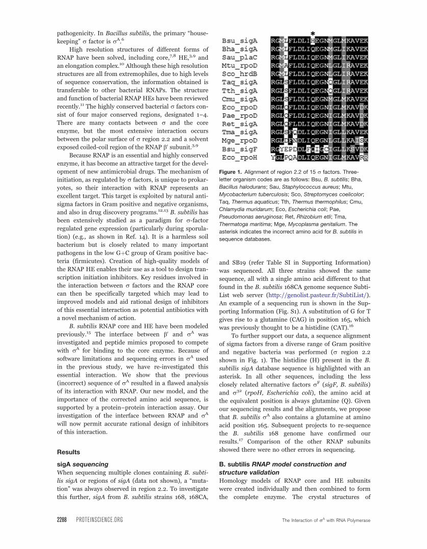
To further support our data, a sequence alignment
of sigma factors from a diverse range of Gram positive
and negative bacteria was performed (r region 2.2
shown in Fig. 1). The histidine (H) present in the B.
subtilis sigA database sequence is highlighted with an
asterisk. In all other sequences, including the less
closely related alternative factors rF (sigF, B. subtilis)
and r32 (rpoH, Escherichia coli), the amino acid at
the equivalent position is always glutamine (Q). Given
our sequencing results and the alignments, we propose
that B. subtilis rA also contains a glutamine at amino
acid position 165. Subsequent projects to re-sequence
the B. subtilis 168 genome have confirmed our
results.17 Comparison of the other RNAP subunits
showed there were no other errors in sequencing.
B. subtilis RNAP model construction and
structure validationHomology models of RNAP core and HE subunits
were created individually and then combined to form
the complete enzyme. The crystal structures of
Figure 1. Alignment of region 2.2 of 15 r factors. Three-
letter organism codes are as follows: Bsu, B. subtilis; Bha,
Bacillus halodurans; Sau, Staphylococcus aureus; Mtu,
Mycobacterium tuberculosis; Sco, Streptomyces coelicolor;
Taq, Thermus aquaticus; Tth, Thermus thermophilus; Cmu,
Chlamydia muridarum; Eco, Escherichia coli; Pae,
Pseudomonas aeruginosa; Ret, Rhizobium etli; Tma,
Thermatoga maritima; Mge, Mycoplasma genitalium. The
asterisk indicates the incorrect amino acid for B. subtilis in
sequence databases.
2288 PROTEINSCIENCE.ORG The Interaction of rA with RNA Polymerase
Thermus aquaticus core8 and Thermus thermophilus
HE3 were used as templates. The major subunits (a, b,b0) of B. subtilis RNAP share about 45% identity and
65% similarity with the corresponding T. aquaticus
and T. thermophilus subunits (refer Table SII in the
Supporting Information for a full identity and similar-
ity list).
The enzyme models were subjected to structure
clean-up and optimization. After each minimization
set, the quality of the model was examined by Rama-
chandran analysis18–20 (refer Fig. S2 in the Supporting
Information). A check of the templates showed that
only 54% of T. aquaticus core residues were in the
favored regions of the Ramachandran plot (81% in the
allowed regions), while for T. thermophilus HE 86% of
residues were in the favorable region (97% in the
allowed regions). This very low score for the T. aquati-
cus core is not ideal for a template, but this is the only
core RNAP structure available that is not in complex
with an antibiotic. The T. thermophilus HE score of
86% is very high considering the large size of the
enzyme. The scores for the homology models are com-
parable to the crystal structure scores, suggesting the
models are of good quality. These scores were obtained
for models refined by minimization of the sidechains
with the backbone constrained. Further minimization
without constraints led to deterioration of the back-
bone secondary structures for both our models and
the crystal structures. Only models minimized with the
backbone constrained were used in further analysis.
An alternative approach to enzyme construction was
also undertaken to investigate if there was a difference
between minimizing the subunits first and then put-
ting together the enzyme, or putting the enzyme to-
gether from unminimized structures and then mini-
mizing. This second procedure was performed to
account for possible changes in the structure of each
subunit under the influence of the surrounding subu-
nits. However, the same result was obtained (data not
shown).
The final B. subtilis RNAP models, with side-
chains minimized, are shown in Figure 2. Panel A
shows the core enzyme while Panel B shows the HE.
Parts of the b and b0 subunits where there are gaps in
the template structure (>10 amino acids) are not dis-
played, because the models are not reliable under
these circumstances. Sequence alignments available as
Supporting Information (A1–A4) show these regions.
Both models show the classic crab claw shape made
with the two a subunits (cyan and pink) at the back,
and the b (green) and b0 (red) subunits forming the
two halves of the DNA-binding channel. x (cream) is
positioned on top of b0 and rA (blue) is on the top
front of the HE only and protrudes across the channel.
The region where the major interaction between rA
and the core occurs is shown in the insets of Panels A
and B. The interacting helix-turn-helix (HtH) motif of
b0(red) can be seen clearly in the Panel B inset.
RNAP is a dynamic structure: Changes fromcore to HE
Apart from the obvious addition of rA, the core enzyme
undergoes a number of changes when forming the HE.
The major change is highlighted in Panel C of Figure 2.
The top half of the ‘‘claw’’ of the HE (yellow) has closed
down to hold rA compared to the more open channel in
the core enzyme (purple). Showing the HtH motif only
from two different angles emphasizes the fact that this
region of the HE has moved down in comparison to the
core, rather than rotated. The presence of more disor-
dered regions with less secondary structure in the core
enzyme may be explained by the lower resolution and
quality of the core crystal structure template. To analyze
the flexibility of RNAP in more detail, all subunits were
subjected to distance difference matrix (DDM) analysis.21
This allows evaluation of relative changes in the position
of each amino acid Ca atom with respect to all other Caatoms in each subunit. As has been suggested before
(reviewed in Ref. 11), our models confirm that the two asubunits, the x subunit and the C-terminal halves of the
b and b0 subunits move as rigid bodies upon binding of
r. This leads to DDMs with no significant changes within
any of the subunits. The DDMs of the N-terminal halves
of b and b0 (refer Fig. S3 in the Supporting Information)
did show changes between the core and HE. However,
the largest of these changes are due to the insertions in
the B. subtilis sequence when compared to the template
sequences, and are thus not meaningful.
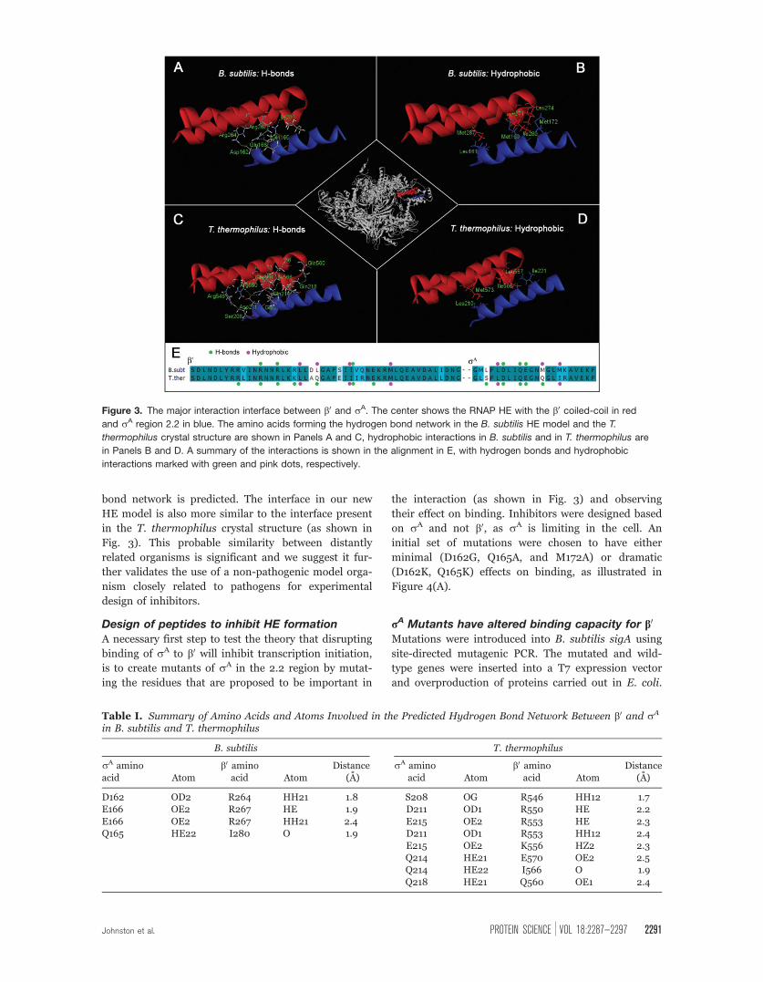
The interface between b0 and rA
We minimized the B. subtilis homology model and the
T. thermophilus crystal structure in exactly the same
way, so any differences observed are meaningful. The
major interaction between b0 and rA is between a
coiled-coil region of b0(comprising a HtH motif) and
region 2.2 of rA (a single helix), highlighted red and
blue, respectively, in the center of Figure 3. Panels A–
D of Figure 3 show the amino acids forming hydrogen
bonds (A and C) and hydrophobic interactions (B and
D) in both the B. subtilis and T. thermophilus struc-
tures. Our B. subtilis model has an extensive network
of proposed hydrogen bonds as shown in Panel A.
Interestingly, one of these is formed by rA Q165,
which was previously thought to be a histidine (refer
previous section), and when the enzyme is modeled
with H in position 165, this hydrogen bond is not
formed (data not shown). The hydrogen bond network
seen in the T. thermophilus crystal structure [Fig.
3(C)] is even more extensive, with more amino acids
involved. It has been suggested, that no single molecu-
lar cause can explain the greater thermal stability of
thermophilic proteins, and that they rely instead on
combinations of stabilizing effects.22 On the other
hand, older studies proposed that a greater number of
specific interactions, particularly electrostatic interac-
tions and hydrogen bonds, are largely responsible for
the higher stability of thermophilic proteins.23,24 The
Johnston et al. PROTEIN SCIENCE VOL 18:2287—2297 2289
alignment in Panel E summarizes the amino acids
involved in hydrogen bonds (green dots) and shows
that corresponding amino acids in B. subtilis and
T. thermophilus are implicated, with T. thermophilus
having an extra four amino acids from b0 and an extra
two from rA involved. A detailed list of predicted
hydrogen bonds is given in Table I, the predicted
hydrogen bond lengths are listed as an indication of
the likely strength of the bonds. While more hydrogen
bonds are predicted for T. thermophilus, most of these
are expected to be weak (>2 A).
Figure 3 (Panels B and D) shows the residues
involved in hydrophobic interactions between b0 and
rA. They are illustrated as sidechains from each subu-
nit extending towards each other. This occurs in two
areas along the interface in both the B. subtilis model
(B) and the T. thermophilus crystal structure (D), with
more B. subtilis amino acids predicted to be involved
in hydrophobic interactions. In T. thermophilus, three
residues from b0 and two from rA interact and in B.
subtilis, two additional amino acids from each chain
are involved (Panel E, pink dots). The extra amino
acids involved in B. subtilis are rA M169 and b0 L274.Interestingly, the corresponding amino acid in T. ther-
mophilus at both these positions is a polar glutamine,
and these two amino acids are instead involved in
hydrogen bonds.
Comparison of new HE model toprevious modeling
When comparing the new B. subtilis holoenzyme
model from this study to a previous model,15 several
important interactions not observed previously were
found between the residues at the b0-rA interface, as
summarized in Table SIII in the Supporting Informa-
tion. One difference in particular is the presence of rA
Q165 in the predicted hydrogen bond network of the
model created in this study. Q165 is the residue previ-
ously thought to be a histidine. With the correct amino
acid now in this position, a more extensive hydrogen
Figure 2. Homology models of B. subtilis RNAP core (A) and RNAP HE (B). The inset panels show regions of RNAP that
change upon binding of r, from core to HE. The enzyme subunits are color coded as indicated. Panel C: Superimposition of
the two RNAP models, using the a subunits. Only the b and b0 subunits are shown.
2290 PROTEINSCIENCE.ORG The Interaction of rA with RNA Polymerase
bond network is predicted. The interface in our new
HE model is also more similar to the interface present
in the T. thermophilus crystal structure (as shown in
Fig. 3). This probable similarity between distantly
related organisms is significant and we suggest it fur-
ther validates the use of a non-pathogenic model orga-
nism closely related to pathogens for experimental
design of inhibitors.
Design of peptides to inhibit HE formation
A necessary first step to test the theory that disrupting
binding of rA to b0 will inhibit transcription initiation,
is to create mutants of rA in the 2.2 region by mutat-
ing the residues that are proposed to be important in
the interaction (as shown in Fig. 3) and observing
their effect on binding. Inhibitors were designed based
on rA and not b0, as rA is limiting in the cell. An
initial set of mutations were chosen to have either
minimal (D162G, Q165A, and M172A) or dramatic
(D162K, Q165K) effects on binding, as illustrated in
Figure 4(A).
rA Mutants have altered binding capacity for b0
Mutations were introduced into B. subtilis sigA using
site-directed mutagenic PCR. The mutated and wild-
type genes were inserted into a T7 expression vector
and overproduction of proteins carried out in E. coli.
Figure 3. The major interaction interface between b0 and rA. The center shows the RNAP HE with the b0 coiled-coil in red
and rA region 2.2 in blue. The amino acids forming the hydrogen bond network in the B. subtilis HE model and the T.
thermophilus crystal structure are shown in Panels A and C, hydrophobic interactions in B. subtilis and in T. thermophilus are
in Panels B and D. A summary of the interactions is shown in the alignment in E, with hydrogen bonds and hydrophobic
interactions marked with green and pink dots, respectively.
Table I. Summary of Amino Acids and Atoms Involved in the Predicted Hydrogen Bond Network Between b0 and rA
in B. subtilis and T. thermophilus
B. subtilis T. thermophilus
rA aminoacid Atom
b0 aminoacid Atom
Distance(A)
rA aminoacid Atom
b0 aminoacid Atom
Distance(A)
D162 OD2 R264 HH21 1.8 S208 OG R546 HH12 1.7E166 OE2 R267 HE 1.9 D211 OD1 R550 HE 2.2E166 OE2 R267 HH21 2.4 E215 OE2 R553 HE 2.3Q165 HE22 I280 O 1.9 D211 OD1 R553 HH12 2.4
E215 OE2 K556 HZ2 2.3Q214 HE21 E570 OE2 2.5Q214 HE22 I566 O 1.9Q218 HE21 Q560 OE1 2.4
Johnston et al. PROTEIN SCIENCE VOL 18:2287—2297 2291
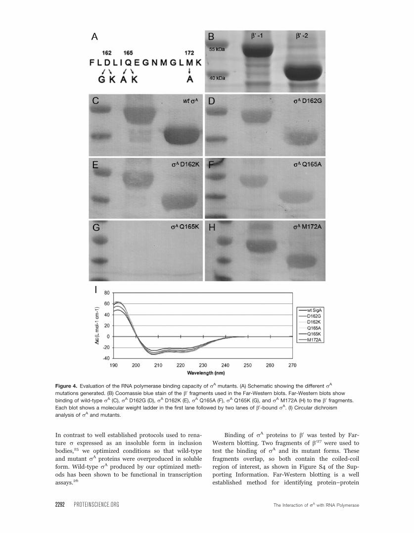
In contrast to well established protocols used to rena-
ture r expressed as an insoluble form in inclusion
bodies,25 we optimized conditions so that wild-type
and mutant rA proteins were overproduced in soluble
form. Wild-type rA produced by our optimized meth-
ods has been shown to be functional in transcription
assays.26
Binding of rA proteins to b0 was tested by Far-
Western blotting. Two fragments of b027 were used to
test the binding of rA and its mutant forms. These
fragments overlap, so both contain the coiled-coil
region of interest, as shown in Figure S4 of the Sup-
porting Information. Far-Western blotting is a well
established method for identifying protein–protein
Figure 4. Evaluation of the RNA polymerase binding capacity of rA mutants. (A) Schematic showing the different rA
mutations generated. (B) Coomassie blue stain of the b0 fragments used in the Far-Western blots. Far-Western blots show
binding of wild-type rA (C), rA D162G (D), rA D162K (E), rA Q165A (F), rA Q165K (G), and rA M172A (H) to the b0 fragments.
Each blot shows a molecular weight ladder in the first lane followed by two lanes of b0-bound rA. (I) Circular dichroism
analysis of rA and mutants.
2292 PROTEINSCIENCE.ORG The Interaction of rA with RNA Polymerase
interactions and has been used in previous studies to
identify where r70 binds to RNAP in E. coli28 and
where NusA binds to RNAP in B. subtilis.27
The two b0 fragments are shown in Figure 4(B),
with molecular weights of �40 and 55 kDa when com-
pared with the molecular weight marker (first lane of
each panel). Upon addition of wild-type rA to the
membrane, binding to both b0 fragments was observed
as shown in Figure 4(C) (second and third lanes). By
comparing the wild-type data to that of the mutant
proteins, qualitative comparisons can be made as to
their ability to bind to b0. rA mutations D162G,
D162K, and M172A (Panels D, E, and H) do not
appear to have a large effect on the ability of rA to
bind to b0. On the other hand, mutating amino acid
165 drastically reduces the capacity for rA binding.
Q165K (Panel G) abolishes binding altogether, while
Q165A (Panel F) clearly shows a reduced level of
binding.
To confirm that the altered binding was due to
the change in the rA-b0 interface and not due to pro-
tein misfolding, circular dichroism (CD) was per-
formed to compare the structures of the rA proteins.
All mutants and the wild-type rA displayed the same
CD spectra (Panel I, Fig. 4) indicating that all proteins
were folded correctly.
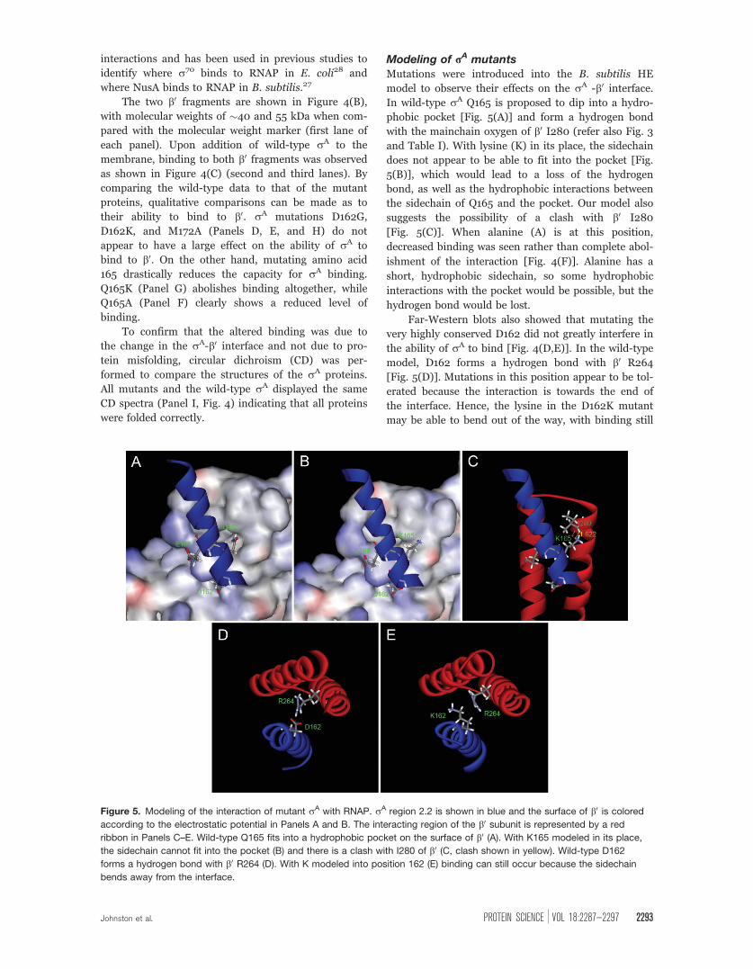
Modeling of rA mutantsMutations were introduced into the B. subtilis HE
model to observe their effects on the rA -b0 interface.In wild-type rA Q165 is proposed to dip into a hydro-
phobic pocket [Fig. 5(A)] and form a hydrogen bond
with the mainchain oxygen of b0 I280 (refer also Fig. 3
and Table I). With lysine (K) in its place, the sidechain
does not appear to be able to fit into the pocket [Fig.
5(B)], which would lead to a loss of the hydrogen
bond, as well as the hydrophobic interactions between
the sidechain of Q165 and the pocket. Our model also
suggests the possibility of a clash with b0 I280
[Fig. 5(C)]. When alanine (A) is at this position,
decreased binding was seen rather than complete abol-
ishment of the interaction [Fig. 4(F)]. Alanine has a
short, hydrophobic sidechain, so some hydrophobic
interactions with the pocket would be possible, but the
hydrogen bond would be lost.
Far-Western blots also showed that mutating the
very highly conserved D162 did not greatly interfere in
the ability of rA to bind [Fig. 4(D,E)]. In the wild-type
model, D162 forms a hydrogen bond with b0 R264
[Fig. 5(D)]. Mutations in this position appear to be tol-
erated because the interaction is towards the end of
the interface. Hence, the lysine in the D162K mutant
may be able to bend out of the way, with binding still
Figure 5. Modeling of the interaction of mutant rA with RNAP. rA region 2.2 is shown in blue and the surface of b0 is colored
according to the electrostatic potential in Panels A and B. The interacting region of the b0 subunit is represented by a red
ribbon in Panels C–E. Wild-type Q165 fits into a hydrophobic pocket on the surface of b0 (A). With K165 modeled in its place,
the sidechain cannot fit into the pocket (B) and there is a clash with I280 of b0 (C, clash shown in yellow). Wild-type D162
forms a hydrogen bond with b0 R264 (D). With K modeled into position 162 (E) binding can still occur because the sidechain
bends away from the interface.
Johnston et al. PROTEIN SCIENCE VOL 18:2287—2297 2293
possible across the rest of the interface (Fig. 5, com-
pare E to D). However, there would still be a loss of a
hydrogen bond, which may translate to slightly weaker
binding using more sensitive assays. Similarly, the
M172A mutation does not show dramatically decreased
interaction, possibly because it is also positioned at
the end of the interface [Fig. 3(B)].
Discussion
We detected a mutation in the published sequence of
B. subtilis rA, and this was recently confirmed by
other authors.17 The correct amino acid at position
165, in region 2.2 of rA is glutamine (Q), not histidine
(H), as previously thought.16 This is significant for
the current work, because the major interaction
between b0 and rA in the RNAP holoenzyme involves
rA region 2.2.
Previously, a homology model of B. subtilis HE
was published15 which included the incorrect amino
acid in the rA sequence. This model has now been cor-
rected and extensively refined and validated. A model
of the core enzyme has also been built and compared
to the HE. The differences between core and HE mod-
els showed that the b and b0 subunits move as rigid
bodies (as has previously been suggested11), rather
than the subunits being flexible within themselves.
The comparison between the b0 and rA interface in the
models and the crystal structures used as templates
has yielded interesting results. The crystal structures
are both of RNAP from thermophiles, where a greater
number of hydrogen bonds seem to be involved in the
rA-b0 interaction than in the mesophilic B. subtilis,
where more hydrophobic interactions appear to be
involved.
Our models propose that the corrected amino acid
Q165 is crucial for the rA-b0 interaction. We designed
several mutants to test our model and show that tar-
geted disruption of residues at the rA-b0 interface
could lead to decreased interaction.
rA mutants were prepared and used to demon-
strate that mutation of Q165 dramatically reduces the
ability of rA to bind to b0. Binding was significantly
decreased for the Q165A mutant, and was undetectable
in the Q165K mutant. Previous studies have also
shown that mutation of the equivalent residue in E.
coli r32 (Q80; an alternate r factor) to R or N,
reduces core association markedly and greatly lowers
the level of transcription.29 Similar results were seen
with an E. coli r70 Q406A mutant (equivalent to B.
subtilis Q165A).30 Interestingly, this study also had a
D403N mutation (equivalent to B. subtilis rA D162)
which showed that 9 times the amount of mutant pro-
tein was required to achieve the same level of binding
as wild-type r70. Our results did not show a significant
effect on binding when mutating this residue, but this
could be due to the lack of sensitivity of our assay.
Importantly, mutation of this residue in E. coli did not
abolish the interaction completely, which fits well with
our modeling of mutant D162K. Additionally,
mutation of hydrophobic residues along the interface
in E. coli r70 had little or no effect on core binding.
This was also observed in this study with the M172A
mutation.30
Combining computer-modeling and in vitro bind-
ing assays, we have thus been able to take the first
steps towards validating the rA-b0 interaction in bacte-
rial RNAP holoenzymes as a potential new target for
the development of antibacterial agents with a novel
mode of action.
Materials and Methods
Sequence and template acquisitionAll sequences of B. subtilis proteins were obtained
from the SubtiList web server (genolist.pasteur.fr/Sub-
tiList). Gene names were as follows: a-rpoA, b-rpoB,b0-rpoC, x-yloH, rA-sigA. Protein sequences from all
other organisms were obtained from NCBI
(www.ncbi.nih.gov). These represent RNAP subunits
from a range of Gram positive and Gram negative
bacteria.
Crystal structure templates were obtained from
the Protein Data Bank (PDB)31 (www.rcsb.org/pdb).
The template for core RNAP was of T. aquaticus
core,8 PDB code 1HQM, chains A–E; and the template
for the HE was of T. thermophilus HE,3 PDB code
1IW7, chains A–F.
DNA sequencingsigA from three different strains of B. subtilis was
amplified by PCR and then sequenced (Australian Ge-
nome Research Facility, Brisbane, Australia). Two of
the strains used were "wild-type" 168 laboratory
strains from different research groups; this is impor-
tant as the provenance of strains is often hard to
determine. Strain 168, a highly transformable trypto-
phan auxotroph, was originally obtained by X-ray mu-
tagenesis in the 1940s32 and has since been passaged
through many laboratories, making unambiguous
establishment of strain provenance problematic. This
accounts for slight differences between strains/stocks
between labs. The strains used in this study included:
168 (laboratory stock, P.J. Lewis), 168CA,33 the type of
strain used in the B. subtilis genome sequencing pro-
ject and SB19 (R.G. Wake, The University of Sydney).
Refer Supporting Information, Table SI for strain
details.
Homology modeling
Multiple sequence alignments were completed for
each subunit using the ClustalW2 program34
(www.ebi.ac.uk/Tools/clustalw2) and then submitted
to the SWISS-MODEL server35 (http://swissmodel.ex-
pasy.org) using the "alignment interface." In the case
of b0 (rpoC) and x (yloH), the position of gaps would
not allow a model to be created, so the gaps in the
2294 PROTEINSCIENCE.ORG The Interaction of rA with RNA Polymerase
alignment had to be manipulated manually using
Swiss-Pdb Viewer35 and then submitted to SWISS-
MODEL using the project mode. Alignments for these
subunits are provided in the Supporting Information.
Core b0 had to be split into two parts to obtain the
homology model.
Structure assembly and clean-up
To create the entire enzyme, each subunit of B. subtilis
core and HE was imported on top of the crystal struc-
ture template to check for correct positioning. The
template was then deleted, leaving behind B. subtilis
RNAP models only.
Swiss-Pdb Viewer35 was used to clean-up models
before further refinement. Amino acids with steric
clashes were selected and then "fixed" (repaired) using
the "quick and dirty" method. This was repeated until
there was no improvement in the number of amino
acids with clashes.
Structure minimization
Energy minimization of the structures was carried out
using Discovery Studio 2.0 (Accelrys). Conditions were
set by the CHARMM forcefield36 as implemented in
the Accelrys software. First, hydrogens were added to
the model or crystal structure and the remainder of
the structure was constrained (fixed position in space)
to refine the positioning of the hydrogens. Next, the
backbone was constrained and minimization carried
out to refine the positioning of the sidechains; and
finally, the entire structure was minimized with no
constraints. For mutation analysis, single amino acids
were substituted and minimized alone (i.e., with the
rest of the enzyme constrained) followed by minimiza-
tion of sidechains within 10 A of the rA-b0 interface
(backbone constrained).
Each set of refinements involved first minimizing
with the Steepest Descent algorithm until the maxi-
mum derivative was less than 0.1 kJ/(mol A); and
then minimizing with the Adopted-Basis Newton
Raphson (ABNR) algorithm until the maximum deriv-
ative was less than 0.01 kJ/(mol A). All other parame-
ters were kept at default values.
Structure evaluationQuality of models was checked using online programs,
including MolProbity18 (http://kinemage.biochem.du-
ke.edu) and PROCHECK19 (Protein Data Bank). These
packages make use of Ramachandran plots20 and
clash-scores, and determine outliers in areas such as
bond lengths, angles, and spatial arrangement. To
assess subunit flexibility, the ProFlex2.0 program21
was used to obtain distance difference matrix (DDM)
plots. The b and b0 subunits were split into two parts
because the program cannot evaluate chains longer
than 999 amino acids.
Strain constructionA full strain and plasmid list can be found in the Sup-
porting Information, Table SI.
E. coli DH5a was used for all cloning procedures.
B. subtilis sigA was amplified from 168 genomic DNA
and inserted into pETMCSIII via the XbaI and Acc65I
sites to create pNG590 (wild-type sigA with an N-ter-
minal 6xHis tag to enable protein purification). PCR
site-directed mutagenesis37 was used to create mutants
of sigA that were similarly inserted into pETMCSIII.
The plasmids pNG570, 571, 572, 573, and 600 code
for rA mutations M172A, D162G, D162K, Q165A, and
Q165K, respectively.
Protein overproduction, purification,
and evaluation
E. coli strain C41(DE3) (Table SI) was transformed
with each sigA-containing plasmid and resulting colo-
nies placed into 1L of Luria-Bertani broth (LB) con-
taining 100 lg/mL ampicillin and grown shaking at
37�C. At A600 0.6–1.0 isopropyl-b-D-thiogalactoside(IPTG) was added to 0.5 mM and shaking continued
at room temperature for further 5 h. Cells were har-
vested by centrifugation and stored at �80�C.
Cells were resuspended in 20–40 mL buffer A
(20 mM NaH2PO4, 500 mM NaCl, 20 mM imidazole,
pH 7.5) and lysed using an EmulsiFlex-C5 homoge-
nizer (Avestin). Proteins were purified by affinity chro-
matography using a 1 mL HisTrap HP column (GE
Healthcare) and an AKTA FPLC system (GE Health-
care). The cell lysate (soluble fraction) was applied to
the column then washed with 6.6% buffer B (50 mM
NaH2PO4, 500 mM NaCl, 400 mM imidazole, pH 7.5)
and eluted with a step to 100% buffer B. The protein
peak was collected and dialyzed into storage buffer by
two step dialysis (step 1: 50 mM NaH2PO4, 150 mM
NaCl, 1 mM EDTA, 0.1 mM diothiothreitol, pH 7.5;
step 2: 50 mM NaH2PO4, 150 mM NaCl, 0.1 mM dio-
thiothreitol, 30% glycerol, pH 7.5) and stored at
�80�C.
To check for correct protein folding, CD spectra
were recorded from 300 nm to 190 nm on a JASCO J-
810 spectropolarimeter, with proteins diluted to �0.25
mg/mL in 10 mM NaH2PO4.
Far-Western blottingpNG482 and pNG48327 containing overlapping b0
fragments (both with the coiled-coil region of interest)
were transformed into BL21(DE3)pLysS. Colonies were
added to 10 mL LB containing 100 lg/mL ampicillin
and grown at 37�C until A600 reached 0.5–0.8, where
IPTG was then added (to 0.5 mM) and the culture
grown for a further 3 h at 37�C; 1 mL aliquots were
centrifuged and cells resuspended to an A600 of 10 in
TES [200 mM Tris-HCl (pH 7.5), 5 mM EDTA, 100
mM NaCl]; 5–10 lL of each cell lysate was subjected
to SDS-PAGE and then transferred to a nitrocellulose
Johnston et al. PROTEIN SCIENCE VOL 18:2287—2297 2295
membrane. Following transfer, the membrane was
washed three times (5 min each) in PBS and three
times in FWB (20 mM Tris, 100 mM NaCl, 0.5 mM
EDTA, 10% glycerol, 0.1% Tween-20). The membranes
were blocked and proteins refolded by immersing in
FWB þ 5% skim milk (w/v) for 1 h while shaking. The
membrane was then probed with a rA protein (�10 lgprotein added to FWB þ 5% skim milk) for 1 h and
then washed three times in FWB. Rabbit polyclonal
anti-rA antibody (M. Yudkin, University of Oxford)
diluted 1:3000 in FWB þ 5% milk was added to the
membrane and left to incubate for 1 h, followed by
another three washes and incubation with 1:3000
goat-anti-rabbit horsereadish peroxidase (BioRad). Af-
ter the three final washes, the antibody was detected
using an Opti-4CNTM substrate kit (BioRad) following
the manufacturer’s instructions.
Acknowledgments
The authors thank M. Yudkin, University of Oxford, for
kindly supplying polyclonal anti-rA anti serum. E.J. was
supported by a Ph.D. scholarship from the ARC and the
University of Newcastle.
References
1. Ebright RH (2000) RNA polymerase: structural similar-ities between bacterial RNA polymerase and eukaryoticRNA polymerase II. J Mol Biol 304:687–698.
2. Burgess RR, Travers AA, Dunn JJ, Bautz EKF (1969)Factor stimulating transcription by RNA polymerase. Na-ture 221:43–46.
3. Vassylyev DG, Sekine S, Laptenko O, Lee J, VassylyevaMN, Borukhov S, Yokoyama S (2002) Crystal structureof a bacterial RNA polymerase holoenzyme at 2.6 A reso-lution. Nature 417:712–719.
4. Murakami KS, Masuda S, Campbell EA, Muzzin O, DarstSA (2002) Structural basis of transcription initiation: anRNA polymerase holoenzyme-DNA complex. Science296:1285–1290.
5. Mooney RA, Davis SE, Peters JM, Rowland JL, AnsariAZ, Landick R (2009) Regulator trafficking on bacterialtranscription units in vivo. Mol Cell 33:97–108.
6. Gross CA, Chan C, Dombroski A, Gruber T, Sharp M,Tupy J, Young B (1998) The functional and regulatoryroles of sigma factors in transcription. Cold Spring HarbSymp Quant Biol 63:141–155.
7. Zhang G, Campbell EA, Minakhin L, Richter C, SeverinovK, Darst SA (1999) Crystal structure of thermus aquaticuscore RNA polymerase at 3.3 A resolution. Cell 98:811–824.
8. Minakhin L, Bhagat S, Brunning A, Campbell EA, DarstSA, Ebright RH, Severinov K (2001) Bacterial RNA poly-merase subunit omega and eukaryotic RNA polymerasesubunit RPB6 are sequence, structural, and functionalhomologs and promote RNA polymerase assembly. ProcNatl Acad Sci USA 98:892–897.
9. Murakami KS, Masuda S, Darst SA (2002) Structural ba-sis of transcription initiation: RNA polymerase holo-enzyme at 4 A resolution. Science 296:1280–1284.
10. Vassylyev DG, Vassylyeva MN, Perederina A, Tahirov TH,Artsimovitch I (2007) Structural basis for transcriptionelongation by bacterial RNA polymerase. Nature 448:157–162.
11. Murakami KS, Darst SA (2003) Bacterial RNA polymer-ases: the wholo story. Curr Opin Struct Biol 13:31–39.
12. Hughes KT, Mathee K (1998) The anti-sigma factors.Annu Rev Microbiol 52:231–286.
13. Andre E, Bastide L, Michaux-Charachon S, Gouby A, Vil-lain-Guillot P, Latouche J, Bouchet A, Gualtieri M, Leo-netti JP (2006) Novel synthetic molecules targeting thebacterial RNA polymerase assembly. J Antimicrob Che-mother 57:245–251.
14. Errington J (1993) Bacillus subtilis sporulation: regula-tion of gene expression and control of morphogenesis.Microbiol Rev 57:1–33.
15. MacDougall IJ, Lewis PJ, Griffith R (2005) Homologymodeling of RNA polymerase and associated transcriptionfactors from Bacillus subtilis. J Mol Graph Model 23:297–303.
16. Kunst F, Ogasawara N, Moszer I, Albertini AM, Alloni G,Azevedo V, Bertero MG, Bessieres P, Bolotin A,Borchert S, et al. (1997) The complete genome sequenceof the gram-positive bacterium Bacillus subtilis. Nature390:249–256.
17. Barbe V, Cruveiller S, Kunst F, Lenoble P, Meurice G,Sekowska A, Vallenet D, Wang T, Moszer I, Medigue C,et al. (2009) From a consortium sequence to a unifiedsequence: the Bacillus subtilis 168 reference genome adecade later. Microbiology 155:1758–1775.
18. Lovell SC, Davis IW, Arendall WB, III, de Bakker PI,Word JM, Prisant MG, Richardson JS, Richardson DC(2003) Structure validation by calpha geometry: phi, psiand Cbeta deviation. Proteins 50:437–450.
19. Laskowski RA, MacArthur MW, Moss DS, Thornton JM(1993) PROCHECK: a program to check stereochemicalquality of protein structures. J Appl Crystallogr 26:283–291.
20. Ramachandran GN, Ramakrishnan C, Sasisekharan V(1963) Stereochemistry of polypeptide chain configura-tions. J Mol Biol 7:95–99.
21. Keller PA, Leach SP, Luu TT, Titmuss SJ, Griffith R(2000) Development of computational and graphicaltools for analysis of movement and flexibility in largemolecules. J Mol Graph Model 18:235–241, 299.
22. Razvi A, Scholtz JM (2006) Lessons in stability fromthermophilic proteins. Protein Sci 15:1569–1578.
23. Kumar S, Tsai C-J, Nussinov R (2001) Thermodynamicdifferences among homologous thermophilic and meso-philic proteins. Biochemistry 40:14152–14165.
24. Vogt G, Woell S, Argos P (1997) Protein thermal stability,hydrogen bonds, and ion pairs. J Mol Biol 269:631–643.
25. Helmann JD, Purification of Bacillus subtilis RNA poly-merase and associated factors. Methods in enzymology.In: SAaS Garges, Ed. (2003) RNA polymerases andassociated factors, Part C, Volume 370. Academic Press,San Diego, CA pp 10–24.
26. Yang X, Lewis PJ (2008) Overproduction and purifica-tion of recombinant Bacillus subtilis RNA polymerase.Protein Expr Purif 59:86–93.
27. Yang X, Molimau S, Doherty GP, Johnston EB, Marles-Wright J, Rothnagel R, Hankamer B, Lewis RJ, Lewis PJ(in press) The structure of bacterial RNA polymerase incomplex with the essential transcription elongation factorNusA. EMBO Rep 10:997–1002.
28. Arthur TM, Burgess RR (1998) Localization of a sigma70binding site on the N terminus of the Escherichia coliRNA polymerase beta0 subunit. J Biol Chem 273:31381–31387.
29. Joo DM, Ng N, Calendar R (1997) A r32 mutant with asingle amino acid change in the highly conserved region2.2 exhibits reduced core RNA polymerase affinity. ProcNatl Acad Sci USA 94:4907–4912.
2296 PROTEINSCIENCE.ORG The Interaction of rA with RNA Polymerase
30. Sharp MM, Chan CL, Lu CZ, Marr MT, Nechaev S, MerrittEW, Severinov K, Roberts JW, Gross CA (1999) The inter-face of r, with core RNA polymerase is extensive, conserved,and functionally specialized. Genes Dev 13:3015–3026.
31. Berman HM, Westbrook J, Feng Z, Gilliland G, Bhat TN,Weissig H, Shindyalov IN, Bourne PE (2000) The proteindata bank. Nucleic Acids Res 28:235–242.
32. Burkholder PR, Giles NH, Jr. (1947) Induced biochemicalmutations in Bacillus subtilis. Am J Bot 34:345–348.
33. Anagnostopoulos C, Spizizen J (1961) Requirements fortransformation in Bacillus subtilis. J Bacteriol 81:741–746.
34. Chenna R, Sugawara H, Koike T, Lopez R, Gibson TJ,Higgins DG, Thompson JD (2003) Multiple sequence
alignment with the clustal series of programs. NucleicAcids Res 31:3497–3500.
35. Guex N, Peitsch MC (1997) SWISS-MODEL and theSwiss-PdbViewer: an environment for comparative pro-tein modeling. Electrophoresis 18:2714–2723.
36. Brooks BR, Bruccoleri RE, Olafson BD, States DJ, Swa-minathan S, Karplus M (1983) CHARMM: a program formacromolecular energy, minimization, and dynamics cal-culations. J Comput Chem 4:187–217.
37. Ho SN, Hunt HD, Horton RM, Pullen JK, Pease LR(1989) Site-directed mutagenesis by overlap exten-sion using the polymerase chain reaction. Gene 77:51–59.
Johnston et al. PROTEIN SCIENCE VOL 18:2287—2297 2297


