Embed Size (px)
Citation preview
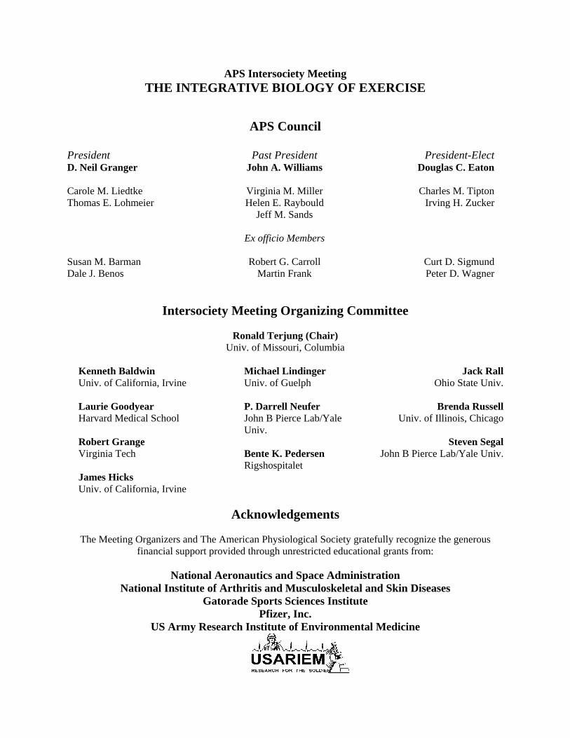
APS Intersociety Meeting THE INTEGRATIVE BIOLOGY OF EXERCISE
APS Council
President Past President President-Elect D. Neil Granger John A. Williams Douglas C. Eaton Carole M. Liedtke Virginia M. Miller Charles M. Tipton Thomas E. Lohmeier Helen E. Raybould Irving H. Zucker Jeff M. Sands Ex officio Members Susan M. Barman Robert G. Carroll Curt D. Sigmund Dale J. Benos Martin Frank Peter D. Wagner
Intersociety Meeting Organizing Committee
Ronald Terjung (Chair)
Univ. of Missouri, Columbia
Kenneth Baldwin Univ. of California, Irvine Laurie Goodyear Harvard Medical School Robert Grange Virginia Tech James Hicks Univ. of California, Irvine
Michael Lindinger Univ. of Guelph P. Darrell Neufer John B Pierce Lab/Yale Univ. Bente K. Pedersen Rigshospitalet
Jack Rall Ohio State Univ.
Brenda Russell
Univ. of Illinois, Chicago
Steven Segal John B Pierce Lab/Yale Univ.
Acknowledgements
The Meeting Organizers and The American Physiological Society gratefully recognize the generous
financial support provided through unrestricted educational grants from:
National Aeronautics and Space Administration National Institute of Arthritis and Musculoskeletal and Skin Diseases
Gatorade Sports Sciences Institute Pfizer, Inc.
US Army Research Institute of Environmental Medicine

2004 APS Intersociety Meeting: The Integrative Biology of Exercise October 6-9, 2004—Hilton Austin Hotel, Austin, TX
Registration Opens: Wednesday, October 6, 2004, 2:00 PM Opening Reception: Wednesday, October 6, 2004, 6:00-9:30 PM
Thursday
October 7 Friday
October 8 Saturday October 9
8:30-11:00 AM Concurrent Symposia
1.0 Mechanical Signal
Transduction: Response and Remodeling in the Musculo-skeletal System
Brenda Russell (Chair) 2.0 Altered Cardiovascular
Control and Blood Flow to Exercising Muscles
Michael J. Joyner (Chair)
13.0 Cytokines, Muscle and
Metabolism Pope Moseley and Bente
Karlund Pedersen (Chairs)
14.0 Genetic Engineering
and Muscle Performance
Joe Metzger (Chair)
25.0 Striated Muscle Hyper-
trophy: Factors Con-trolling Cell Enlarge-ment and Phenotype Transformations
Eva R. Chin and Roger Hill (Chairs)
26.0 AMP-Activated Protein
Kinase: Regulation of Metabolic and Tran-scription Processes in Contracting Skeletal Muscle
Neil Ruderman (Chair)
Afternoon Activities
11:00 AM-12:30 PM 3.0—10.0 Poster Presentations and Exhibits 12:30-1:30 PM Free Time 1:30-3:00 PM Poster Presentations and Exhibits
11:00 AM-12:30 PM 15.0—22.0 Poster Presentations and Exhibits 12:30-1:30 PM Free Time 1:30-3:00 PM Poster Presentations and Exhibits
11:00 AM-12:30 PM 27.0—35.0 Poster Presentations and Exhibits 12:30-1:30 PM Free Time 1:30-3:00 PM Poster Presentations and Exhibits
3:00-5:00 PM Concurrent Symposia
11.0 Mechanical Forces and
Signal Transduction in Vascular Remodeling
Steven S. Segal (Chair) 12.0 Exercise-Induced
Injury and Repair of Skeletal Muscle: Cellular and Molecular Mechanisms
Dan Garry and Mike Lindinger (Chairs)
23.0 Design of Muscle for
Different Functions Larry Rome and Jack
Rall (Chairs) 24.0 Basic Mechanisms
Contributing to Physical Inactivity-Induced Disorders
Frank Booth and P. Darrell Neufer (Chairs)
36.0 Interpreting Phys-
iological Adaptations to Exercise and Disease States through Bio-informatics, Genomics, and Proteomics
Eric Hoffman and Robert Grange (Chairs)
37.0 Comparative Biomech-
anics and Muscle Func-tion in Terrestrial Ver-tebrates: In Vivo Studies
Donald F. Hoyt (Chair)
Evening Events
7:00 PM-10:30 PM Special Purchase Event: The Salt Lick—Austin’s first choice for authentic barbecue! Come enjoy the sumptuous barbecue, lively music and beautiful surroundings.
Evening Free Austin: The Live Music Capital of the World!
7:00-10:00 PM Banquet and Awards Presentation Included with registration

GENERAL INFORMATION Location:
The APS Intersociety Meeting: The Integrative Biology of Exercise will be held October 6-9 at the Hilton Austin Hotel, 500 East 4th Street, Austin, TX 78701, telephone (512) 482-8000, FAX (512) 469-0078, website www.hilton.com.
Onsite Registration Hours: Wednesday, October 6 ................... 2:00—8:30 PM Thursday, October 7................7:30 AM—5:00 PM Friday, October 8 ....................8:00 AM—5:00 PM Saturday, October 9....................8:30 AM-5:00 PM On-Site Registration Fees: APS Member................................................... $325 Retired Member .............................................. $215 Nonmember................................................. $375 Postdoctoral................................................. $265 Student ............................................................ $215
The registration fee includes entry into all scien-tific sessions, opening reception and banquet.
Payment Information: Registrants may pay by institutional or personal
check, traveler’s check, MasterCard, VISA or American Express. Checks must be payable to “The American Physiological Society” and drawn on a United States bank payable in US dollars.
Student Registration
Any student member or regularly matriculated stu-dent working toward a degree in one of the bio-medical sciences is eligible to register at the stu-dent fee. Nonmember postdoctoral fellows, hospi-tal residents and interns, and laboratory technicians do not qualify as students. Nonmember Students who register onsite must provide a valid university student ID card. APS Student members should present their current APS membership card indi-cating their student category status.
Postdoctoral Registration
Any person who has received a Ph.D. degree in physiology or related field, within four years of this meeting, as attested to by the department head is eligible to register at the postdoctoral fee. A statement signed by the department head must ac-company the registration form and remittance when registering.
Press Press badges will be issued at the APS Press
Office, Meeting Room 602, only to members of the working press and freelance writers bearing a letter of assignment from an editor. Representatives of allied fields (public relations, public affairs, etc.) must register as nonmembers.
Continuing Medical Education (CME)
The Federation of American Societies for Experi-mental Biology is accredited by the Accreditation Council for Continuing Medical Education to spon-sor continuing medical education for physicians.
Category I Continuing Medical Education (CME) credits will be offered at this meeting. CME application forms will be available in the Onsite Meeting Registration Counter. For the purposes of Continuing Medical Education credits toward the American Medical Association Physician’s Recognition Award, the APS Intersociety Meeting: The Integrative Biology of Exercise is jointly sponsored by the Federation of American Societies for Experimental Biology. There is a $45 appli-cation fee, payable upon submission of the form. For more information, contact the FASEB Office of Scientific Meetings and Conferences at 301-634-7010.
Program Objective The goal of the meeting is to convene an internationally recognized and interdisciplinary group of investigators focusing on the use of integrative approaches for the study of exercise involving physiology, molecular biology and genetics and to interest new investigators and students in pursuing research opportunities to understand the integrative biology of exercise and its relation to gender and aging.
At the completion of the meeting, participants should have a broader understanding of exercise physiology and interdisciplinary efforts to assess its impact on the systems of the body.
Target Audience This meeting is intended for all professionals involved in teaching, research and clinical fields related to exercise biology.

GENERAL INFORMATION Housing Reservations:
Hotel rooms have been reserved at a rate of $129 single or double occupancy at the Hilton Austin Hotel, 500 East 4th Street, Austin, TX. To make reservations call: (800) 236-1592 (within the US) or (512) 482-8000 (outside the US) and ask for reservations. Be sure to identify yourself as an APS Meeting Attendee and provide the meeting dates. You will need to provide a first night’s deposit, refundable up to 48 hours in advance of your arrival date to secure your reservation. The deadline for making housing reservations is August 27, 2004.
Hotel Surroundings and Ammenities:
The Hilton Austin, one of Austin’s newest hotels, officially opened in January 2004. It is situated in the center of the downtown entertainment district. “Sixth Street”, the center of why Austin is considered the Live Music Capital of the World is one block from the hotel. Additionally, the Warehouse District—with its eclectic offerings of restaurants, bars and dance clubs—is but a short walk away. All APS meeting attendees staying at the Hilton will receive complimentary high-speed Internet access at no charge for in-room service.
Local Information:
Activities in Austin range from the outdoorsy to the eccentric, from the historically significant to the naturally sublime. One of the many highlights to any Austin visit between April and October is the unusual experience of watching 1.5 million bats taking flight from beneath Congress Avenue Bridge. The bats arrive in spring and by mid-summer their population more than doubles.
For a dose of the eccentric side, take a stroll down either the Warehouse District or Sixth Street and explore the many unique dining establishments and nightlife there.
If you have a penchant for history, visit the new Bob Bullock Texas State History Museum for an interactive journey through the state’s lively past. As the the Lone Star State’s capital, Austin has a deep connection to history—something that can be explored by visiting the State Capitol and Governor’s Mansion.
For further information visit the Austin Convention & Visitors Bureau online at: www.austintexas.org.
Ground Transportation: The Austin-Bergstrom Airport is 7 miles from the
Hilton hotel. Typical minimum charge for a taxi is $20. You can also take the SuperShuttle Transport Systems at a cost of $10 per person one way. Reservations are not required, however you may make reservations by calling 800-258-3826 or 512-258-3826 or via the Internet at: www.supershuttle.com/htm/cities/aus.htm.
Car Rental: Renting a car should be fun. So Alamo puts you
smiles ahead with deals and services. As the official car rental provider for the 2004 APS Meetings and Conferences, Alamo is offering special discounted rates to all delegates. These special rates are available one week before and one week after the meeting dates and include unlimited mileage. So, choose Alamo and let your fun begin! For reservations, contact your travel agent or call Alamo at 1-800-732-3232 or experience Alamo’s hot new site at: www.Alamo.com. Be sure to request Group ID# 308201 and plan code GR at time of reservation.
Travel Reservations:
The APS is pleased to announce that it has been able to secure a special discount agreement with United Airlines unavailable to the general public. United Airlines is offering special meeting fares for all attendees who use the Special Meeting Desk to book their reservations. Book early and take advantage of the promotional fares that give you the greatest savings! Earn a 5% discount off the lowest applicable fare, including First Class, or 10% off the mid-week coach fare. By purchasing your ticket at least 30 days in advance of your scheduled travel you will receive an additional 5% discount! To take advantage of these savings, simply call (or have your travel agent call) 1-800-521-4041 and refer to Meeting ID Number 557HS. Mileage Plus members receive full credit for all miles flown to this meeting. You or your travel agent should call today, as seats may be limited.
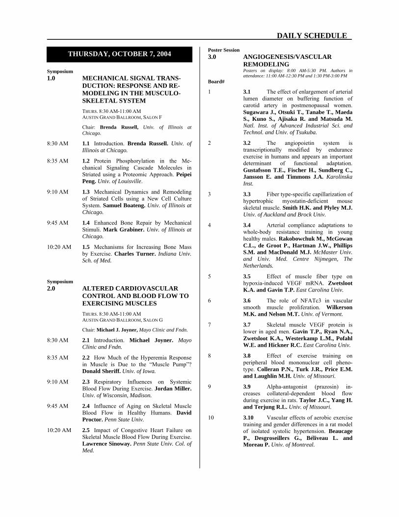
DAILY SCHEDULE
Symposium 1.0 MECHANICAL SIGNAL TRANS-
DUCTION: RESPONSE AND RE-MODELING IN THE MUSCULO-SKELETAL SYSTEM
THURS. 8:30 AM-11:00 AM AUSTIN GRAND BALLROOM, SALON F
Chair: Brenda Russell, Univ. of Illinois at Chicago.
8:30 AM 1.1 Introduction. Brenda Russell. Univ. of Illinois at Chicago.
8:35 AM 1.2 Protein Phosphorylation in the Me-chanical Signaling Cascade Molecules in Striated using a Proteomic Approach. Peipei Peng. Univ. of Louisville.
9:10 AM 1.3 Mechanical Dynamics and Remodeling of Striated Cells using a New Cell Culture System. Samuel Boateng. Univ. of Illinois at Chicago.
9:45 AM 1.4 Enhanced Bone Repair by Mechanical Stimuli. Mark Grabiner. Univ. of Illinois at Chicago.
10:20 AM 1.5 Mechanisms for Increasing Bone Mass by Exercise. Charles Turner. Indiana Univ. Sch. of Med.
Symposium 2.0 ALTERED CARDIOVASCULAR
CONTROL AND BLOOD FLOW TO EXERCISING MUSCLES
THURS. 8:30 AM-11:00 AM AUSTIN GRAND BALLROOM, SALON G
Chair: Michael J. Joyner, Mayo Clinic and Fndn.
8:30 AM 2.1 Introduction. Michael Joyner. Mayo Clinic and Fndn.
8:35 AM 2.2 How Much of the Hyperemia Response in Muscle is Due to the “Muscle Pump”? Donald Sheriff. Univ. of Iowa.
9:10 AM 2.3 Respiratory Influences on Systemic Blood Flow During Exercise. Jordan Miller. Univ. of Wisconsin, Madison.
9:45 AM 2.4 Influence of Aging on Skeletal Muscle Blood Flow in Healthy Humans. David Proctor. Penn State Univ.
10:20 AM 2.5 Impact of Congestive Heart Failure on Skeletal Muscle Blood Flow During Exercise. Lawrence Sinoway. Penn State Univ. Col. of Med.
Poster Session 3.0 ANGIOGENESIS/VASCULAR
REMODELING Posters on display: 8:00 AM-5:30 PM. Authors in
attendance: 11:00 AM-12:30 PM and 1:30 PM-3:00 PM Board#
1 3.1 The effect of enlargement of arterial lumen diameter on buffering function of carotid artery in postmenopausal women. Sugawara J., Otsuki T., Tanabe T., Maeda S., Kuno S., Ajisaka R. and Matsuda M. Natl. Inst. of Advanced Industrial Sci. and Technol. and Univ. of Tsukuba.
2 3.2 The angiopoietin system is transcriptionally modified by endurance exercise in humans and appears an important determinant of functional adaptation. Gustafsson T.E., Fischer H., Sundberg C., Jansson E. and Timmons J.A. Karolinska Inst.
3 3.3 Fiber type-specific capillarization of hypertrophic myostatin-deficient mouse skeletal muscle. Smith H.K. and Plyley M.J. Univ. of Auckland and Brock Univ.
4 3.4 Arterial compliance adaptations to whole-body resistance training in young healthy males. Rakobowchuk M., McGowan C.L., de Groot P., Hartman J.W., Phillips S.M. and MacDonald M.J. McMaster Univ. and Univ. Med. Centre Nijmegen, The Netherlands.
5 3.5 Effect of muscle fiber type on hypoxia-induced VEGF mRNA. Zwetsloot K.A. and Gavin T.P. East Carolina Univ.
6 3.6 The role of NFATc3 in vascular smooth muscle proliferation. Wilkerson M.K. and Nelson M.T. Univ. of Vermont.
7 3.7 Skeletal muscle VEGF protein is lower in aged men. Gavin T.P., Ryan N.A., Zwetsloot K.A., Westerkamp L.M., Pofahl W.E. and Hickner R.C. East Carolina Univ.
8 3.8 Effect of exercise training on peripheral blood mononuclear cell pheno-type. Colleran P.N., Turk J.R., Price E.M. and Laughlin M.H. Univ. of Missouri.
9 3.9 Alpha-antagonist (prazosin) in-creases collateral-dependent blood flow during exercise in rats. Taylor J.C., Yang H. and Terjung R.L. Univ. of Missouri.
10 3.10 Vascular effects of aerobic exercise training and gender differences in a rat model of isolated systolic hypertension. Beaucage P., Desgroseillers G., Béliveau L. and Moreau P. Univ. of Montreal.
THURSDAY, OCTOBER 7, 2004

DAILY SCHEDULE
Board # 11 3.11 Effect of age on skeletal muscle
interstitial VEGF protein. Ruster R.S., Carrithers J.A., Hickner R.C. and Gavin T.P., Carrithers J.A., Hickner R.C. and Gavin T.P. East Carolina Univ.
Poster Session 4.0 CARDIOVASCULAR CONTROL
Posters on display: 8:00 AM-5:30 PM. Authors in attendance: 11:00 AM-12:30 PM and 1:30 PM-3:00 PM
Board # 12 4.1 Modulation of AV conduction
during dynamic exercise in humans. Nakamoto T., Matsukawa K., Murata J. and Komine H. Inst. of Health Sciences, Hirohsima Univ.
13 4.2 Exercise pressor reflex induces re-nal vasoconstriction via sympathetic activa-tion in decerebrate rats. Koba S., Yoshida T. and Hayashi N. Osaka Univ. and Kyushu Univ.
14 4.3 Arterial baroreflex and muscle mechanoreflex mutually change the response range of sympathetic nerve activity in the other reflex. Yamamoto K., Kawada T., Kamiya A., Takaki H., Sugimachi M. and Sunagawa K. Pharmaceuticals and Med. Devices Agency, Natl. Cardiovascular Ctr. Res. Inst. and Grad. Sch. of Med. Sci. Kyushu Univ.
15 4.4 Lack of age-related decreases in limb blood Flow in resistance-trained men. Miyachi M., Tabata I., Kawano H., Okajima M., Oka J. and Tanaka H. Natl. Inst. of Hlth. and Nutrition, Kawasaki Univ. of Med. Welfare, Japan Women's Col. of Phys. Ed., Tokyo Kasei Univ. and Univ. of Texas.
16 4.5 Blockade of spinal P2X receptor attenuates reflex pressor response to muscle contraction. Gao Z., Sinoway L. and Li J. Penn State Col. of Med.
17 4.6 Is postexercise hypotension ex-plained by a histamine-mediated peripheral vasodilation? Lockwood J.M., Wilkins B.W. and Halliwill J.R. Univ. of Oregon.
18 4.7 Effects of the intensity of resistance training on central arterial compliance. Kawano H., Tanaka H., Onodera S., Yuzuki O. and Miyachi M. Kawasaki Univ. of Med. Welfare, Univ. of Texas at Austin and Natl. Inst. of Hlth. and Nutrition, Japan.
Board # 19 4.8 Heart rate variability and perfor-
mance response of competitive swimmers to high intensity interval training and regen-eration. Wilkinson J.G., Siegel P. and Urhausen A. Univ. of Wyoming, Cal. Poly. Inst., and Univ. of Saarland, Germany.
20 4.9 Age-associated changes in vessel diameter and blood velocity of the carotid and brachial artery in women aged 18-88 years. Shimizu S. and Kagaya A. Women’s Col. of Phys. Ed., Setagaya, Japan.
21 4.10 Increase in systemic arterial com-pliance by aerobic exercise training decreases myocardial oxygen uptake during exercise. Otsuki T., Kesen Y., Yokoyama N., Tanabe T., Sugawara J., Miyauchi T., Maeda S., Kuno S., Ajisaka R. and Matsuda M. Univ. of Tsukuba, Fukushima Sch. for the Blind and Natl. Inst. of Advanced Industrial Sci. and Tech., Japan.
22 4.11 Effect of two methods of dehydra-tion on orthostatic tolerance. Davis J.E., LoPiccolo M. and Luetkemeier M. Alma Col., MI.
23 4.12 Exercise training attenuates the en-hanced cardiac β2-adrenerigc receptor sensi-tivity induced by myocardial infarction. Billman G.E. and Kukielka M. Ohio State Univ.
24 4.13 Disuse atrophy increases the exer-cise pressor reflex in rats. Hayashi N. and Koba S. Kyushu Univ. and Osaka Univ.
25 4.14 Role of vascular ATP-sensitive potassium channels in exercise hyperemia. Hamann J.J., Buckwalter J.B., Valic Z. and Clifford P.S. Med. Col. of Wisconsin.
26 4.15 Decrease in skin blood flow during venous stasis with cuff inflation is not solely related to cutaneous venoarteriolar response. Okazaki K., Fu Q., Martini E., Zhang R., Crandall C.G. and Levine B.D. Presbyterian Hosp. of Dallas, and the Univ. of Texas Southwestern Med. Center at Dallas
27 4.16 Skeletal muscle arteriolar vasocon-strictor reactivity to local, humoral, and neural agonists are differentially affected by muscle fiber type and exercise training. Donato A.J., Lesniewski L.A. and Delp M.D. Texas A&M Univ.
28 4.17 The effect of isometric arm or leg exercise on resting blood pressure and arterial distensibility in persons medicated for hyper-tension. Visocchi A., McGowan C., Faulkner M., Verduyn R., McCartney N. and MacDonald M. McMaster Univ.

DAILY SCHEDULE
Board # 29 4.18 The effect of stimulation of mid-
brain dopaminergic neurons on limb blood flow in anesthetized cats and rats. Matsukawa K., Murata J., Nakamoto T., Komine H. and Wilson L.B. Inst. of Hlth. Sci., Hiroshima Univ. Fac. of Med., Japan and Univ. of South Carolina Sch. of Med.
30 4.19 Effects of gender and physical fit-ness on the cardiovascular response to exer-cise. Martini E.R., Fu Q., Stray-Gundersen J., Zhang R. and Levine B.D. Presbyterian Hosp. of Dallas and Univ. of Texas Southwestern Med. Ctr. at Dallas.
31 4.20 Roles of the three isoforms of nitric oxide synthase within the ventrolateral medul-la during the exercise pressor reflex. Ally A. and Maher T.J. Palm Beach Atlantic Univ. and Massachusetts Col. of Pharm. & HS, Boston.
32 4.21 Heart rate recovery following exer-cise: a predictor of ventricular fibrillation sus-ceptibility. Smith L., Kukielka M. and Billman G.E. Case Western Reserve Univ. and Ohio State Univ.
33 4.22 Functional sympatholysis is impair-ed in the exercising forearms of nitrate tolerant subjects. Fadel P.J., Gallagher K.M., Wang Z. and Thomas G.D. UT Southwestern Med. Ctr., Dallas.
34 4.23 Otolithic activation elicits a reduc-tion in blood pressure in endurance runners. Ray C.A. and Ung C.W. Penn State Col. of Med.
35 4.24 Bilateral cerebral tissue oxygena-tion during exposure to lower body negative pressure. Lusina S.C., Scott J.M., Esch B.T., McKenzie D.C., Sheel A.W. and Warburton D.E. Univ. of British Columbia.
36 4.25 Hypoxia vs hyperpnea: effects on cutaneous vascular tone. Simmons G.H., Wilkins B.W., Pricher M.P., Minson C.T. and Halliwill J.R. Univ. of Oregon.
37 4.26 Muscle mechanoreceptors have heightened sensitivty in heart failure. Chiu J., Hamilton M., Fonarow G. and Middlekauff H. Keck Sch. of Med. and Geffen Sch. of Med. at UCLA.
38 4.27 Role of central command to cutaneous vascular responses during isometric exercise. Shibasaki M., Secher N.H., Johnson J.M. and Crandall C.G. Presbyterian Hosp. of Dallas, Univ. of Copenhagen, Denmark, Univ. of Texas Health Science Center at San Antonio and Univ. of Texas Southwestern Med. Center at Dallas.
Board # 39 4.28 Effects of resistance training on
muscle sympathetic nerve response to static contraction. Saito M. Toyota Technological Inst.
40 4.29 Human beta-2 adrenergic receptor polymorphism alters the hemodynamic re-sponse to cycling. Schrage W., Eisenach J.H., Johson C.P., Wick D.E., Walker B.G., Jensen M.D. and Joyner M.J. Mayo Clinic.
41 4.30 Changes of resting blood pressure response to controlled aerobic exercise in older adults. Huang G., Shi X. and Osness W.H. Univ. of Southern Indiana, Univ. of North Texas Hlth. Sci. Ctr. and Univ. of Kansas.
42 4.31 Evidence for autoregulation of cu-taneous blood flow during isometric handgrip exercise. McCord G.R. and Minson D.T. Univ. of Oregon.
43 4.32 Role of limb and measurement site in vascular responsiveness during dynamic exercise. Wray D.W., Uberoi A., Lawrenson L. and Richardson R. UCSD.
44 4.33 MRI-measured post-contractile hy-peremia transients and post-exercise blood flow in active vs. inactive individuals. Towse T.F., Slade J.M. and Meyer R.A. Michigan State Univ.
45 4.34 Effects of acute aerobic training on nutritive skeletal muscle blood flow with aging. Carrithers J.A., Evans C.A., Kraus R.M., Carrithers H.M., Gavin T.P., Ruster R.S., Knapp D.J., Drew J.L., Garry J. and Hickner R.C. East Carolina Univ.
46 4.35 Effects of L-NMMA administration on exercising nutritive skeletal muscle blood flow before and after 7-days of aerobic training. Hickner R.C., Evans C.A., Kraus R.M., Ruster R.S., Gavin T.P., Carrithers H.M., Knapp D.J., Tanenberg R.J., Drew J.L. and Carrithers J.A. East Carolina Univ.
Visit the Exhibitsdaily from 11:00 AM
until 3:30 PM

DAILY SCHEDULE
Poster Session 5.0 CELL SIGNALING Posters on display: 8:00 AM-5:30 PM. Authors in
attendance: 11:00 AM-12:30 PM and 1:30 PM-3:00 PM Board # 47 5.1 mTOR-dependent signaling me-
diates an increase in the ε-subunit of eukaryotic initiation factor 2B (eIF2B) that is associated with an increase in skeletal muscle eIF2B activity and protein synthesis following acute muscle loading. Kubica N., Wil-liamson D.L., Bolster D.R., Crozier S.J., Kimball S.R., Farrell P.A. and Jefferson L.S. Penn State Univ. Col. of Med., Univ. Space Sci. Res. Assoc., Houston and East Carolina Univ.
48 5.2 Mechanical Stimuli Regulate mTOR via a PI3K/Akt and Growth Factor Independent Mechanism. Hornberger T.A., Fedele M.J. and Esser K.A. UCSD and Univ. of Illinois, Chicago.
49 5.3 Signaling kinase activation by twitch and tetanic contractions in red and white fast-twitch muscle. Ljubicic V. and Hood D.A. York Univ.
50 5.4 IGF-I activates pkb and prevents apoptosis in hypoxic tendon cells. Scott A., Khan K. and Duronio V. Univ. of British Columbia.
51 5.5 The CaMK inhibitor, KN-62, prevents insulin-, contraction-, and AICAR-stimulated glucose uptake, but not via inhibition of Akt, AMPK, or PKCλ/ζ phosphorylation. Witczak C.A., Jessen N. and Goodyear L.J. Joslin Diabetes Center.
52 5.6 Exercise training increases oxida-tive stress-induced mechanical dysfunction in rat hearts: Role of endothelial nitric oxide synthase. Starnes J.W., Park Y., Mathis B.J. and Harris M.B. Univ. of Texas, Austin and Med. Col. of Georgia.
53 5.7 Loading increases MAP kinase phosphorylation and collagen synthesis in engineered tendons. Andrick J., Mundy K. and Baar K. Univ. of Michigan.
54 5.8 Influence of pre-exercise muscle glycogen levels on mitogenic responses to resistance exercise. Creer A., Gallagher P., Jemiolo B., Fink W. and Trappe S. Ball State Univ.
Poster Session 6.0 ENDOTHELIAL FUNCTION
Posters on display: 8:00 AM-5:30 PM. Authors in attendance: 11:00 AM-12:30 PM and 1:30 PM-3:00 PM
Board # 55 6.1 Effects of acute exhausting exercise
and acute psychological stress on the hemo-dynamics of the rat small intestine: role of endothelin-A (ET-A) and endothelin-B (ET-B) receptors. Gandur S., Yegen B. and Kurtel H. Marmara Med. Sch., Istanbul, Turkey.
56 6.2 A single bout of exercise improves endothelial function for 24 hours in rats. Haram P.M., Arbo I., Brubakk A.O., Kemi O.J., Ellingsen Ø. and Wisløff U. NTNU, Trondheim, Norway.
57 6.3 Isometric handgrip training im-proves blood pressure and endothelial func-tion in persons medicated for hypertension. McGowan C.L., Visocchi A., Faulkner M., Rakobowchuk M., McCartney N. and MacDonald M.J. McMaster Univ.
58 6.4 Role of free radicals in the atten-uated exercise blood flow associated with age. Uberoi A., Wray D.W., Lawrenson L., Bailey D.M. and Richardson R.S. UCSD Sch. of Med. and Sch. of Applied Sciences, Univ. of Glamorgan, Wales, UK.
59 6.5 Endothelial function in coronary arterioles from female pigs fed a high fat/ cholesterol diet: effect of exercise training. Henderson K.K., Turk J.R., Woodman C.R. and Laughlin M.H. Univ. of Missouri-Columbia.
60 6.6 A single bout of exercise does not affect in vitro vasomotor responses of rat thoracic aorta. Aultman C.D., Graham D.A., Denniss S.G. and Rush J.W. Univ. of Waterloo.
61 6.7 Cyclooxygenase Expression and Activity in Skeletal Muscle Arterioles: Effects of Age and Exercise Training. Muller-Delp J.M., Stallone J.N., Sellers M.M., Spier S.A. and Delp M.D. Texas A&M Univ. and Univ. of Texas, Tyler.
62 6.8 Endothelium-dependent relaxation of left anterior descending coronary arteries from the Ossabaw swine: characterization of a model of metabolic syndrome. Eklund K.E., Henderson K.K., VanVickle G.D., Sturek M. and Laughlin M.H. Univ. of Missouri, Columbia.
63 6.9 Reduced femoral artery endothe-lium-dependent vasodilation occurs concur-rently with femoral bone loss in type II diabetic rats. Prisby R., Bloomfield S., Stallone J. and Delp M. Texas A&M Univ.

DAILY SCHEDULE
Poster Session 7.0 HEART Posters on display: 8:00 AM-5:30 PM. Authors in
attendance: 11:00 AM-12:30 PM and 1:30 PM-3:00 PM Board # 64 7.1 Cardiac myocyte contractile func-
tion is increased in early-stage pressure overload hypertrophy. Brickson S.L. and Diffee G.M. Univ. of Wisconsin-Madison.
65 7.2 Analysis of rest to exercise (and reverse) transitions via end systolic-end dia-stolic volumes and starling's law. Spencer R.P. Univ. Connecticut Hlth. Ctr.
66 7.3 The impact of age and endurance training on cardiac power output in men. Clements R.E., Chantler P.D., Sharp L.J., Tan L.B. and Goldspink D.F. Liverpool John Moores Univ. and Leeds General Infirmary.
67 7.4 Age-related changes in cardiac power output and VO2max in healthy women. Sharp L.J., Chantler P.D., Clements R.E., Patwala A., Tan L.B. and Goldspink D.F. Liverpool John Moores Univ., Cardio-Thoracic Ctr. and Leeds General Infirmary.
68 7.5 Increased hypertrophy and diastolic performance with exercise training in chronic hypertension. MacDonnell S.M., Barbe M.F., Kubo H., Mahora J., Reger P.O., Renna B.F. and Libonati J.R. Temple Univ.
69 7.6 Force-velocity and power properties in adult and senescent rat myocardium. Chung E. and Diffee G.M. Univ. of Wisconsin-Madison.
70 7.7 Intensity of exercise determines in-crease in aerobic fitness whereas detraining leads to quick regression: big role for cardio-myocyte and less for artery endothelium. Kemi O.J., Haram P.M., Wisløff U. and Ellingsen Ø. Norwegian Univ. of Sci. and Tech.
71 7.8 The effect of lifelong fitness on brain natriuretic peptide levels in healthy sen-iors. Prasad A., Zadeh A.A., Palmer D., Fu Q. and Levine B. UT-Southwest Med. Ctr., Dallas and Inst. for Exercise and Environ. Med., Dallas.
72 7.9 Endurance training dose-response to left ventricular mass is greater in young than senior sedentary populations. Palmer D., Prasad A., Arbab-Zadeh A., Okazaki K., Zhang R., Martini E., Fu Q., Levine B. and Dijk E. Inst. for Exercise and Environ. Med., Dallas.
Poster Session 8.0 OXIDANT/ANTIOXIDANT
EFFECTS Posters on display: 8:00 AM-5:30 PM. Authors in attendance: 11:00 AM-12:30 PM and 1:30 PM-3:00 PM
Board # 73 8.1 Effect of cycling exercise on anti-
oxidant capacity in human muscle measured by ESR. Tanabe K., Masuda K., Hirayama A., Nagase S. and Kuno S. Univ. of Tsukuba and Kanazawa Univ., Japan.
74 8.2 Time course of oxidative stress in skeletal muscle before and after muscle contraction. Kon M., Tanabe K., Kimura F., Akimoto T. and Kono I. Univ. of Tsukuba, Japan.
75 8.3 Examining blood flow and oxida-tive stress with short-term ischemia-reperfu-sion. Kearns A.K., Kwak H., Berman J., Blumberg J. and Clarkson P. Univ. of Massachusetts, Jean Mayer Tufts Univ. and Baystate Med. Ctr., Springfield, MA.
76 8.4 Effect of short-term ascorbic acid consumption on maximal aerobic capacity and cardiac output in young and older adult humans. Motte N.W., Bell C., Carson J.M. and Seals D.R. Univ. of Colorado.
77 8.5 Effect of treadmill running on me-tallothionein gene expression in rats. Ken-nedy J.M., Lomax M. and Todd H. Univ. of Illinois at Chicago and Univ. of Michigan.
78 8.6 Sex differences in myocardial in-farct size following ischemia-reperfusion: correlation with increased superoxide dismutase protein expression. Brown D.A., Lynch J.M., Armstrong C.J., Caruso N.M., Ehlers L.B., Johnson M.S. and Moore R.L. Univ. of Colorado.
79 8.7 Formation of reactive oxygen spe-cies during in vitro electrical stimulation: stimulation of creatine transport and potential artifact in the study of muscle contractions. Derave W. and Hespel P. K.U. Leuven, Belgium.
80 8.8 Muscle weakness caused by tumor necrosis factor- α(TNF-α). Hardin B.J., Smith J.L. and Reid M.B. Univ. of Kentucky.
81 8.9 Reactive oxygen species production in subsarcolemmal and intermyofibrillar mito-chondria. Adhihetty P.J., Ljubicic V., Menzies K.J. and Hood D.A. York Univ.

DAILY SCHEDULE
Poster Session 9.0 MICROCIRCULATION
Posters on display: 8:00 AM-5:30 PM. Authors in attendance: 11:00 AM-12:30 PM and 1:30 PM-3:00 PM
Board # 82 9.1 Effects of eccentric exercise on
microcirculation and microvascular oxygen pressures in rat spinotrapezius muscle. Kano Y., Padilla D., Behnke B.J., Hageman K., Musch T.I. and Poole D.C. Univ. of Electro-Communications, Chofu, Japan, Kansas State Univ. and Texas A&M Univ.
83 9.2 The impact of a 6-month aerobic exercise programme on microvascular func-tion in type 2 diabetes. Elston L., Mid-dlebrooke A., Ball C., Mawson D., MacLeod K., Tooke J. and Shore A. Peninsula Med. Sch., Exeter and Univ. of Exeter, UK.
84 9.3 Three-dimensional sructure of capillary ntwork in atrophied soleus muscle. Fujino H., Kohzuki H., Takeda I. and Kajiya F. Okayama Univ. and Suzuka Univ. of Med. Sci., Japan.
85 9.4 Effect of aging and exercise training on thromboxane-induced vasoconstriction in coronary arterioles. Dougherty P., Shipley R. and Muller-Delp J. Texas A&M Univ.
Poster Session 10.0 MUSCLE INJURY
Posters on display: 8:00 AM-5:30 PM. Authors in attendance: 11:00 AM-12:30 PM and 1:30 PM-3:00 PM
Board # 86 10.1 Myofiber necrosis in the vastus
lateralis muscle does not induce DOMS or muscle force decline after exhaustive ec-centric exercise in humans. Crameri R.M., Aagaard P., Qvortrup K., Møller M. and Kjaer M. Concordia Univ., Inst. of Sports Med., Copenhagen and Univ. of Copenhagen.
87 10.2 A comparison of changes in indices of muscle damage following fast and slow velocity eccentric exercise. Chapman D., Newton M., Sacco P. and Nosaka K. Edith Cowan Univ., Perth, Australia.
88 10.3 Inhibition of nNOS reduces the pro-tection from contraction-induced injury pro-vided by passive stretch. Lockhart N.C. and Brooks S.V. Univ. of Michigan.
89 10.4 Muscle damage, Ca2+ accumulation, and loss of force in rat skeletal muscle induc-ed by electroporation in vivo. Gissel H. and Clausen T. Univ. of Aarhus.
90 10.5 The role of cyclooxygenase-1 and cyclooxygenase-2 in skeletal muscle satellite cell proliferation, differentiation and fusion. Mendias C. and Allen R.E. Univ. of Michigan and Univ. of Arizona.
Board # 91 10.6 Evidence for myofibril remodeling
as opposed to myofibril damage in human muscles with DOMS. Thornell L., Lena C. and Yu J. Umeå Univ., Sweden.
92 10.7 Skeletal muscle regeneration in the selective absence of Akt isoforms. Barton E.R. Univ. of Pennsylvania Sch. of Dental Med.
93 10.8 Effect of exercise to improve of rat lower limb healing after physical injury. Suh D., Suh D., Yoo K.S., Jung D.K., Lee D.H., Son H.H. and KIM J.Y. Col. Med. Dong-A Univ., Busan, Republic of Korea.
94 10.9 Anoxia and hypoxia induce Ca2+ in-flux and loss of cellular integrity in rat EDL muscle. Fredsted A., Mikkelsen U.R., Gissel H. and Clausen T. Univ. of Aarhus.
95 10.10 Effects of massage on muscle soreness and parameters associated with muscle damage following eccentric exercise of the elbow flexors. Abidin Z.Z., Nosaka K., Newton M.J. and Sacco P. Edith Cowan Univ., Perth, Australia.
96 10.11 uPA is a positive regulator of skele-tal muscle regeneration. Koh T., Bryer S.C., Pucci A.M. and Sisson T.H. Univ. of Illinois at Chicago and Univ. of Michigan.
97 10.12 Efficacy of functional electrical stimulation in maintaining the mass and mechanical properties of an inactive fast hindlimb extensor muscle. Kim S.J., Roy R.R., Zhong H., Ambartsumyan L. and Edgerton R. UCLA.
98 10.13 Calpain activity is transiently increased in maturing dystrophic skeletal muscle. Wang Q., Draper K.E. and Grange R.W. Virginia Polytechnic Inst. and State Univ.
99 10.14 Apoptosis of skeletal muscle in male and female rats after eccentric contrac-tions. Oshima M. and Kano Y. Univ. of Electro-Communications, Chofu, Japan.
100 10.15 Force loss, muscle damage and Ca2+ accumulation following step-exercise. Over-gaard K., Fredsted A. and Clausen T. Univ. of Aarhus.
101 10.16 Effect of tourniquet induced I/R upon in vivo muscle function. Merritt E., Jennings A., Walters T. and Farrar R.P. Univ. of Texas, Austin and US Army Inst. of Surgical Res., Ft. Sam Houston, TX.

DAILY SCHEDULE
Board # 102 10.17 Satellite cell activation in human
skeletal muscle biopsies after downhill run-ning-induced microdamage. Myburgh K.H., van Tubbergh K. and Niesler C. Stellenbosch Univ., South Africa.
103 10.18 Effects of systemic injury on voluntary wheel-running in mice. Villarin J.J. and Carlsen R.C. Univ. of California, Davis.
104 10.19 Foxk1 is a regulator of skeletal muscle stem cell populations. Meeson A., Hawke T., Goetsch S., Graham S., Gallardo T., Jiang N., Wiliams S. and Garry D. Univ. of Texas Southwestern Med. Ctr. at Dallas, York Univ., Toronto and Duke Univ. Med. Ctr.
Symposium 11.0 MECHANICAL FORCES AND
SIGNAL TRANSDUCTION IN VASCULAR REMODELING
THURS. 3:00 PM-5:00 PM AUSTIN GRAND BALLROOM, SALON F
Chair: Steven Segal. Yale Univ. Sch. of Med.
3:00 PM 11.1 Introduction. Steven Segal. Yale Univ. Sch. of Med.
3:05 PM 11.2 Microvascular Remodeling in Re-sponse to Mechanical Forces in Skeletal Muscle. Tara Haas. York Univ.
3:35 PM 11.3 Bioengineering of Vascular Pattern-ing During Angiogenesis. Richard Price. Univ. of Virginia Health Sciences Ctr.
4:05 PM 11.4 Remodeling of Resistance Arteries in Response to Shear Stress. Joseph Unthank. Indiana Univ.
4:35 PM 11.5 Growth and Differentiation of Vas-cular Smooth Muscle Modulated by Sympa-thetic Innervation. Deborah Damon. Univ. of Vermont.
Symposium 12.0 EXERCISE-INDUCED INJURY AND
REPAIR OF SKELETAL MUSCLE: CELLULAR AND MOLECULAR MECHANISMS
THURS. 3:00 PM-5:00 PM AUSTIN GRAND BALLROOM, SALON G
Chairs: Dan Garry, Univ. of Texas Southwestern Med. Center. Michael Lindinger, Univ. of Guelph.
3:00 PM 12.1 Introduction. Dan Garry. Univ. of Texas Southwestern Med. Ctr.
3:05 PM 12.2 Skeletal Muscle Injury and Repair: An Anatomical and Physiological Overview. Edward Schultz. Univ. of Wisconsin Med. Sch.
3:35 PM 12.3 Molecular Mechanisms of Skeletal Muscle Regeneration. Thomas Hawke. York Univ.
4:05 PM 12.4 Regulation of Myogenic Stem Cells. Natasha Frank. Harvard Med. Sch.
4:35 PM 12.5 Repair and Regeneration of Aged Skeletal Muscle. Bradley Olwin. Univ. of Colorado.
Symposium 13.0 CYTOKINES, MUSCLE, AND
METABOLISM FRI. 8:30 AM-11:00 AM GRAND AUSTIN BALLROOM, SALON F
Chairs: Pope Moseley, Univ. of New Mexico Bente Karlund Pedersen, Univ. of
Copenhagen
8:30 AM 13.1 Introduction. Pope Moseley. Univ. of New Mexico.
8:35 AM 13.2 Signal Transduction in the Activa-tion of the IL-6 gene in Skeletal Muscle-does HSP Play a Role? Pope Moseley. Univ. of New Mexico.
9:10 AM 13.3 Cytokine Signalling Pathways with-in the Skeletal Muscle-Coupling to Metabolic Genes? Mark Febbraio. Univ. of Melbourne.
9:45 AM 13.4 The Biological Role of Muscle-derived Cytokines: Hormonal Effect of IL-6 on Lipolysis. Bente Pedersen. Univ. of Copenhagen.
10:20 AM 13.5 Role of IL-6 in Metabolism-Studies in Transgenic Mice. Ville Wallenius. Sahlgrenska Univ. Hosp., Sweden.
FRIDAY, OCTOBER 8, 2004
Don’t forget… Pick Up your Banquet Ticket
by 11:00 AM on THURSDAY
The banquet is free but you

DAILY SCHEDULE
Symposium 14.0 GENETIC ENGINEERING AND
MUSCLE PERFORMANCE FRI. 8:30 AM-11:00 AM AUSTIN GRAND BALLROOM, SALON G
Chair: Joe Metzger, Univ. of Michigan.
8:30 AM 14.1 Introduction. Joe Metzger. Univ. of Michigan.
8:35 AM 14.2 Skeletal Muscle Genes for Increas-ed Heart Muscle Performance. Joe Metzger. Univ. of Michigan.
9:10 AM 14.3 Motor Proteins and Exercise Performance. Leslie Leinwand. Univ. of Colorado, Boulder.
9:45 AM 14.4 Genetic Interventions for Myopathies. Jeff Chamberlain. Univ. of Washington Sch. of Med.
10:20 AM 14.5 Genetic Interventions for Improved Muscle Function in Aging. H. Sweeney. Univ. of Pennsylvania Sch. of Med.
Poster Session 15.0 CHO/LIPID METABOLISM I
Posters on display: 8:00 AM-5:30 PM. Authors in attendance: 11:00 AM-12:30 PM and 1:30 PM-3:00 PM
Board # 1 15.1 Fad diets in untrained normal
weight and overweight/obese females: effect on caloric intake, postprandial lipemia, and mood. Kist W.B. Missouri Valley Col.
2 15.2 Evaluation of net leg norepineph-rine balance before and after endurance training. Fattor J.A., Jacobs K.A., Bauer T., Hagobian T., Friedlander A.L., Wolfel E.E. and Brooks G.A. Univ. of California, Berkeley and Univ. of Colorado.
3 15.3 Withdrawn
4 15.4 A single bout of exercise increases VLDL-triglyceride clearance, indpendent of muscle lipoprotein lipase content, and has no effect on VLDL-TG secretion. Wright D., Patterson B.W., Mohammed B.S., Klein S. and Mittendorfer B. Washington Univ. Sch. of Med.
Board # 5 15.5 Menstrual cycle phase and gender
influence muscle proglycogen and total glycogen storage and utilization during exercise. Devries M.C., Hamadeh M.J. and Tarnopolsky M.A. McMaster Univ.
6 15.6 Why do horses have delayed muscle glycogen replenishment? Jose-Cunilleras E., Hinchcliff K.W., Lacombe V.A., Sams R.A., Kohn C.W., Taylor L.E. and Devor S.T. Ohio State Univ. and Otterbein Col., Westerville, OH
7 15.7 Small Increases in active leg tracer measured FFA uptake with endurance training. Friedlander A.L., Jacobs K.A., Fattor J.A., Horning M.A., Bauer T., Hagobian T., Wolfel E.E. and Brooks G.A. VA Palo Alto/Stanford Univ., Univ. of Cali-fornia, Berkeley and Univ. Colorado Hlth. Sci. Ctr.
8 15.8 Minimal effects of endurance training on whole-body FFA flux and oxidation. Brooks G.A., Fattor J.A., Wolfel E.E., Horning M.A., Jacobs K.A., Bauer T., Hagobian T. and Friedlander A.L. Univ. of California, Berkeley and VA Palo Alto Hlth. Care System.
9 15.9 Net leg individual fatty acid, lipoprotein, and triglyceride balances at rest and during exercise are unaffected by endurance training. Jacobs K.A., Krauss R.M., Fattor J.A., Horning M.A., Friedlander A.L., Bauer T., Hagobian T., Wolfel E.E. and Brooks G.A. Univ. of California, Berkeley and Children's Hosp. Oakland Res. Inst., Oakland, CA.
10 15.10 Role of testosterone in substrate use during exercise. Gerson L., Hagobian T., Grow D., Chipkin S. and Braun B. Univ. of Massachusetts and Baystate Med. Ctr., Springfield, MA.
11 15.11 Effect of 10 days of endurance training on intramuscular triglyceride level in lean and obese people. Bajpeyi S., Berggren J.R., Tanner C.J. and Houmard J.A. East Carolina Univ.
12 15.12 Muscle glycogen concentrations in Alaskan sled dogs during extended endurance exercise. McKenzie E., Davis M., Holbrook T., Williamson K., Hinchcliff K., Jose-Cunilleras E., Nelson S. and Valberg S. Oklahoma State Univ., Ohio State Univ., Iditarod Trail Committee and Univ. of Minnesota.
Visit the Exhibits
Daily from 11:00 AM to 3:30 PM
DAILY SCHEDULE
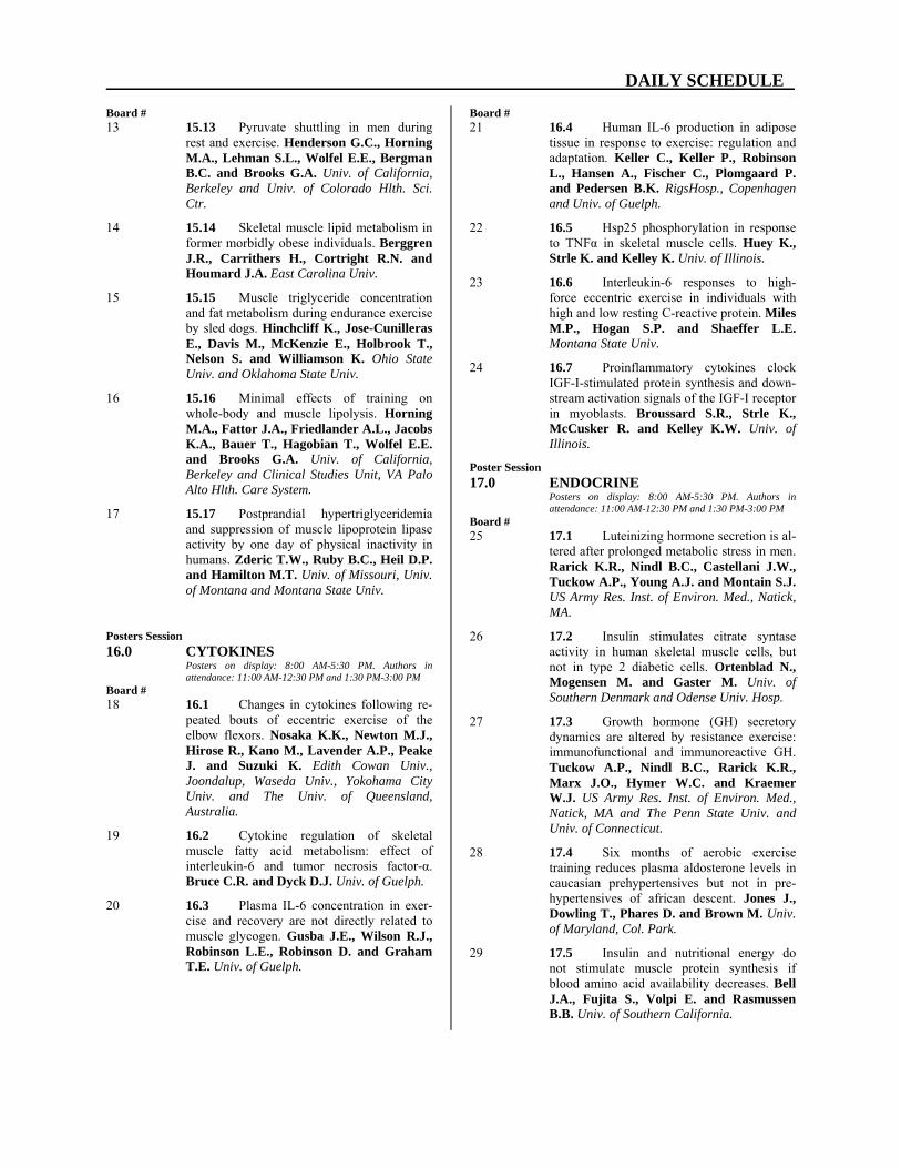
Board # 13 15.13 Pyruvate shuttling in men during
rest and exercise. Henderson G.C., Horning M.A., Lehman S.L., Wolfel E.E., Bergman B.C. and Brooks G.A. Univ. of California, Berkeley and Univ. of Colorado Hlth. Sci. Ctr.
14 15.14 Skeletal muscle lipid metabolism in former morbidly obese individuals. Berggren J.R., Carrithers H., Cortright R.N. and Houmard J.A. East Carolina Univ.
15 15.15 Muscle triglyceride concentration and fat metabolism during endurance exercise by sled dogs. Hinchcliff K., Jose-Cunilleras E., Davis M., McKenzie E., Holbrook T., Nelson S. and Williamson K. Ohio State Univ. and Oklahoma State Univ.
16 15.16 Minimal effects of training on whole-body and muscle lipolysis. Horning M.A., Fattor J.A., Friedlander A.L., Jacobs K.A., Bauer T., Hagobian T., Wolfel E.E. and Brooks G.A. Univ. of California, Berkeley and Clinical Studies Unit, VA Palo Alto Hlth. Care System.
17 15.17 Postprandial hypertriglyceridemia and suppression of muscle lipoprotein lipase activity by one day of physical inactivity in humans. Zderic T.W., Ruby B.C., Heil D.P. and Hamilton M.T. Univ. of Missouri, Univ. of Montana and Montana State Univ.
Posters Session 16.0 CYTOKINES
Posters on display: 8:00 AM-5:30 PM. Authors in attendance: 11:00 AM-12:30 PM and 1:30 PM-3:00 PM
Board # 18 16.1 Changes in cytokines following re-
peated bouts of eccentric exercise of the elbow flexors. Nosaka K.K., Newton M.J., Hirose R., Kano M., Lavender A.P., Peake J. and Suzuki K. Edith Cowan Univ., Joondalup, Waseda Univ., Yokohama City Univ. and The Univ. of Queensland, Australia.
19 16.2 Cytokine regulation of skeletal muscle fatty acid metabolism: effect of interleukin-6 and tumor necrosis factor-α. Bruce C.R. and Dyck D.J. Univ. of Guelph.
20 16.3 Plasma IL-6 concentration in exer-cise and recovery are not directly related to muscle glycogen. Gusba J.E., Wilson R.J., Robinson L.E., Robinson D. and Graham T.E. Univ. of Guelph.
Board # 21 16.4 Human IL-6 production in adipose
tissue in response to exercise: regulation and adaptation. Keller C., Keller P., Robinson L., Hansen A., Fischer C., Plomgaard P. and Pedersen B.K. RigsHosp., Copenhagen and Univ. of Guelph.
22 16.5 Hsp25 phosphorylation in response to TNFα in skeletal muscle cells. Huey K., Strle K. and Kelley K. Univ. of Illinois.
23 16.6 Interleukin-6 responses to high-force eccentric exercise in individuals with high and low resting C-reactive protein. Miles M.P., Hogan S.P. and Shaeffer L.E. Montana State Univ.
24 16.7 Proinflammatory cytokines clock IGF-I-stimulated protein synthesis and down-stream activation signals of the IGF-I receptor in myoblasts. Broussard S.R., Strle K., McCusker R. and Kelley K.W. Univ. of Illinois.
Poster Session 17.0 ENDOCRINE
Posters on display: 8:00 AM-5:30 PM. Authors in attendance: 11:00 AM-12:30 PM and 1:30 PM-3:00 PM
Board # 25 17.1 Luteinizing hormone secretion is al-
tered after prolonged metabolic stress in men. Rarick K.R., Nindl B.C., Castellani J.W., Tuckow A.P., Young A.J. and Montain S.J. US Army Res. Inst. of Environ. Med., Natick, MA.
26 17.2 Insulin stimulates citrate syntase activity in human skeletal muscle cells, but not in type 2 diabetic cells. Ortenblad N., Mogensen M. and Gaster M. Univ. of Southern Denmark and Odense Univ. Hosp.
27 17.3 Growth hormone (GH) secretory dynamics are altered by resistance exercise: immunofunctional and immunoreactive GH. Tuckow A.P., Nindl B.C., Rarick K.R., Marx J.O., Hymer W.C. and Kraemer W.J. US Army Res. Inst. of Environ. Med., Natick, MA and The Penn State Univ. and Univ. of Connecticut.
28 17.4 Six months of aerobic exercise training reduces plasma aldosterone levels in caucasian prehypertensives but not in pre-hypertensives of african descent. Jones J., Dowling T., Phares D. and Brown M. Univ. of Maryland, Col. Park.
29 17.5 Insulin and nutritional energy do not stimulate muscle protein synthesis if blood amino acid availability decreases. Bell J.A., Fujita S., Volpi E. and Rasmussen B.B. Univ. of Southern California.
DAILY SCHEDULE
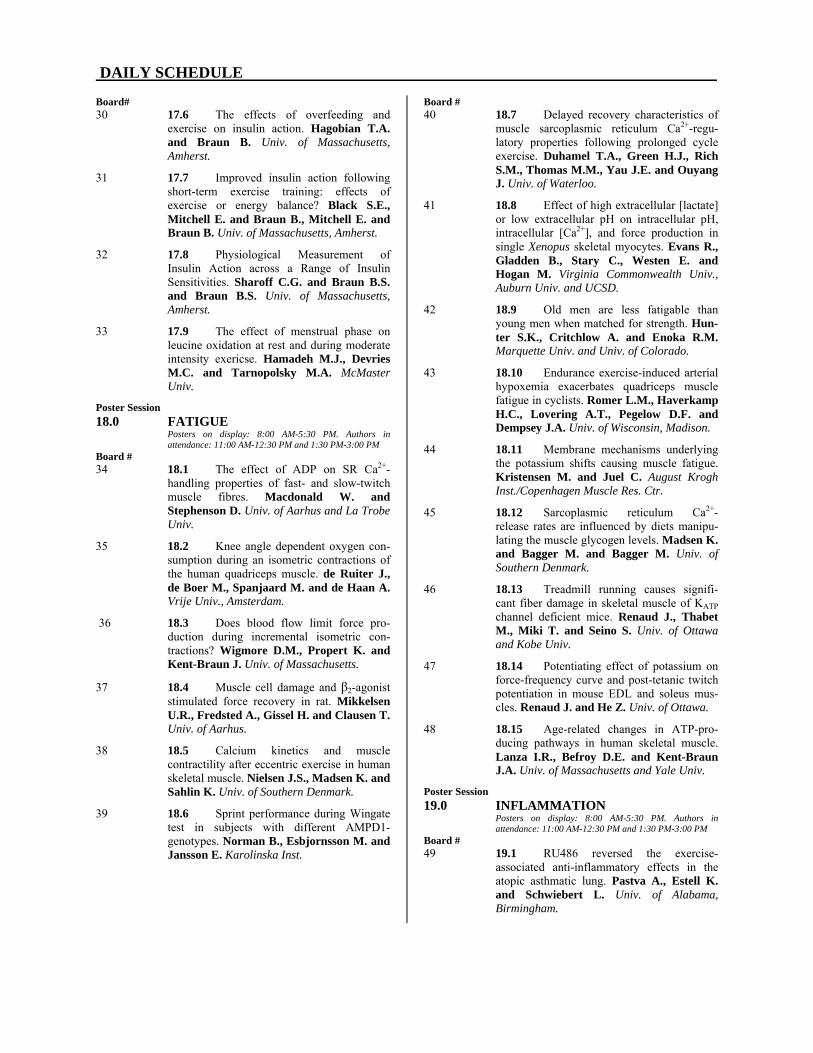
Board# 30 17.6 The effects of overfeeding and
exercise on insulin action. Hagobian T.A. and Braun B. Univ. of Massachusetts, Amherst.
31 17.7 Improved insulin action following short-term exercise training: effects of exercise or energy balance? Black S.E., Mitchell E. and Braun B., Mitchell E. and Braun B. Univ. of Massachusetts, Amherst.
32 17.8 Physiological Measurement of Insulin Action across a Range of Insulin Sensitivities. Sharoff C.G. and Braun B.S. and Braun B.S. Univ. of Massachusetts, Amherst.
33 17.9 The effect of menstrual phase on leucine oxidation at rest and during moderate intensity exericse. Hamadeh M.J., Devries M.C. and Tarnopolsky M.A. McMaster Univ.
Poster Session 18.0 FATIGUE
Posters on display: 8:00 AM-5:30 PM. Authors in attendance: 11:00 AM-12:30 PM and 1:30 PM-3:00 PM
Board # 34 18.1 The effect of ADP on SR Ca2+-
handling properties of fast- and slow-twitch muscle fibres. Macdonald W. and Stephenson D. Univ. of Aarhus and La Trobe Univ.
35 18.2 Knee angle dependent oxygen con-sumption during an isometric contractions of the human quadriceps muscle. de Ruiter J., de Boer M., Spanjaard M. and de Haan A. Vrije Univ., Amsterdam.
36 18.3 Does blood flow limit force pro-duction during incremental isometric con-tractions? Wigmore D.M., Propert K. and Kent-Braun J. Univ. of Massachusetts.
37 18.4 Muscle cell damage and β2-agonist stimulated force recovery in rat. Mikkelsen U.R., Fredsted A., Gissel H. and Clausen T. Univ. of Aarhus.
38 18.5 Calcium kinetics and muscle contractility after eccentric exercise in human skeletal muscle. Nielsen J.S., Madsen K. and Sahlin K. Univ. of Southern Denmark.
39 18.6 Sprint performance during Wingate test in subjects with different AMPD1-genotypes. Norman B., Esbjornsson M. and Jansson E. Karolinska Inst.
Board # 40 18.7 Delayed recovery characteristics of
muscle sarcoplasmic reticulum Ca2+-regu-latory properties following prolonged cycle exercise. Duhamel T.A., Green H.J., Rich S.M., Thomas M.M., Yau J.E. and Ouyang J. Univ. of Waterloo.
41 18.8 Effect of high extracellular [lactate] or low extracellular pH on intracellular pH, intracellular [Ca2+], and force production in single Xenopus skeletal myocytes. Evans R., Gladden B., Stary C., Westen E. and Hogan M. Virginia Commonwealth Univ., Auburn Univ. and UCSD.
42 18.9 Old men are less fatigable than young men when matched for strength. Hun-ter S.K., Critchlow A. and Enoka R.M. Marquette Univ. and Univ. of Colorado.
43 18.10 Endurance exercise-induced arterial hypoxemia exacerbates quadriceps muscle fatigue in cyclists. Romer L.M., Haverkamp H.C., Lovering A.T., Pegelow D.F. and Dempsey J.A. Univ. of Wisconsin, Madison.
44 18.11 Membrane mechanisms underlying the potassium shifts causing muscle fatigue. Kristensen M. and Juel C. August Krogh Inst./Copenhagen Muscle Res. Ctr.
45 18.12 Sarcoplasmic reticulum Ca2+-release rates are influenced by diets manipu-lating the muscle glycogen levels. Madsen K. and Bagger M. and Bagger M. Univ. of Southern Denmark.
46 18.13 Treadmill running causes signifi-cant fiber damage in skeletal muscle of KATP channel deficient mice. Renaud J., Thabet M., Miki T. and Seino S. Univ. of Ottawa and Kobe Univ.
47 18.14 Potentiating effect of potassium on force-frequency curve and post-tetanic twitch potentiation in mouse EDL and soleus mus-cles. Renaud J. and He Z. Univ. of Ottawa.
48 18.15 Age-related changes in ATP-pro-ducing pathways in human skeletal muscle. Lanza I.R., Befroy D.E. and Kent-Braun J.A. Univ. of Massachusetts and Yale Univ.
Poster Session 19.0 INFLAMMATION
Posters on display: 8:00 AM-5:30 PM. Authors in attendance: 11:00 AM-12:30 PM and 1:30 PM-3:00 PM
Board # 49 19.1 RU486 reversed the exercise-
associated anti-inflammatory effects in the atopic asthmatic lung. Pastva A., Estell K. and Schwiebert L. Univ. of Alabama, Birmingham.

DAILY SCHEDULE
Board # 50 19.2 Effect of ovarian hormones on
neutrophil and macrophage infiltration in eccentrically-contracted murine plantarflexor muscles. St. Pierre Schneider B., Barber A., Meyer L. and Tiidus P. Univ. of Wisconsin-Madision, and Wilfrid Laurier Univ., Ontario.
51 19.3 Regular exercise prior to colitis in-duction ameliorates oxidative colonic damage in rats. Kasimay O., Guzel E., Abdyli A., Sulovari A., Gemici A., Ercan F. and Yegen B.C. Marmara Univ. Sch. of Med., Istanbul, Turkey.
52 19.4 Moderate swimming exercise re-duces stress-induced hepatic oxidative injury in rats. Cakir B., Kolgazi M., Kasimay O., Ersoy Y., Ercan F. and Yegen B.C. Marmara Univ. Sch. of Med. Istanbul, Turkey.
53 19.5 Exercise training induces an anti-inflammatory gene expression profile in skeletal muscle. Huffman K.M., Hittel D.S., Hoffman E.P., Duscha B.D. and Kraus W.E. Duke Univ. Med. Ctr. and Children's Natl. Med. Ctr. and George Washington Univ.
Poster Session 20.0 INTEGRATED EXERCISE
RESPONSES Posters on display: 8:00 AM-5:30 PM. Authors in attendance: 11:00 AM-12:30 PM and 1:30 PM-3:00 PM
Board # 54 20.1 Differential effects of long-term
exercise training for 120 minutes or 170 minutes per week on peak VO2, lipoproteins and body habitus changes: the STRRIDE Study. Kraus W.E., Johnson J.L., Aiken L.B., Duscha B.D. and Slentz C.A. Duke Univ. Med. Ctr.
22 20.2 Robust homeostatic control of quadriceps pH during natural locomotor activ-ity in man. Jeneson J.A. and Bruggeman F.J. Utrecht Univ. and Biocentrum, Vrije Univ.
56 20.3 Peak exercise limitation in burned children. McEntire S.J., Beck K.C., Herndon D.N. and Suman O.E. Shriners Hosp. for Children and Guidant Corp.
57 20.4 Weekly MET expenditure and quality of life in hemodialysis patients. Brenner I., Brohart, K. and Beaubien, E. Trent Univ. and Peterborough Regional Hlth. Ctr., Ontario.
58 20.5 Absence of collagen receptor inte-grin α1β1 induces collagen accumulation in skeletal muscle and sensitizes muscles to post-exercise injury. Kovanen V.M., Väliaho J. and Heino J. Univ Jyväskylä, Finland.
Board # 59 20.6 The effect of exercise intensity on
VO2 and muscle deoxygenation kinetics in young and older adults. DeLorey D.S., Kowalchuk J.M. and Paterson D.H. The Univ. of Western Ontario.
60 20.7 Oxygen uptake kinetics during elevated lactate and acidemia. Gladden L.B., Rossiter H.B., Sorensen J.B., Pedersen P.K. and Sahlin K. Auburn Univ., UCSD and Univ. of Southern Denmark, Odense.
61 20.8 Skeletal muscle creatine kinase isoform expression and O2 uptake kinetics during moderate exercise in humans. MacFarlane N.G., Paterson N.D., Awede B., Behan W.M. and Ward S.A. Univ. of Glasgow.
62 20.9 Endurance training induces favor-able changes in lipoprotein subclass concen-trations. Ellis T., Wilund K., Goldberg A., Phares D. and Hagberg J. Univ. of Maryland, Col. Park and Univ. of Maryland Sch. of Med./Baltimore VA GRECC.
63 20.10 Influence of renal function on blood pressure changes with exercise train-ing. Fenty N.M., Jones J.M., Phares D.A. and Brown M.D. Univeristy of Maryland, Col. Park.
64 20.11 Eccentric exercise alters activity of group IV muscle afferents through the release of inflammatory mediators. Marqueste T., Decherchi P., Messan F., Kipson N., Grelot L. and J Ammes Y. Univ. of Aix-Marseille II, France and Univ. of Manitoba.
65 20.12 Six-weeks of respiratory muscle training improves Valsalva component of the Anti-G straining maneuver. Yang P., Frier B.C. and Goodman L. Univ. of Toronto, Univ. of Western Ontario and Defence Res. and Devel. Canada, Toronto.
66 20.13 Elevated temperature accelerates O2 onset kinetics in isolated Xenopus myocytes during moderate intensity work. Walsh B., Koga S., Kindig C.A., Stary C.M. and Hogan M.C. UCSD and Kobe Design Univ., Japan.
67 20.14 Impaired voluntary wheel running exercise performance in creatine kinase defi-cient mice. Ventura-Clapier R.F., Momken I., Koulmann N., Lechene P., Fortin D., Bigard X. and Veksler V. Univ. of Paris-Sud, INSERM and CRSSA, La Tronche.
DAILY SCHEDULE
Board# 68 20.15 Effect of multi-day sustained stren-
uous exercise on peripheral blood leukocytes in sled dogs. Davis M., Ensign W., Hol-brook T., Wiliamson K., Hinchcliff K., Nelson S. and Davis W. Oklahoma State Univ., Naval Hlth. Res. Ctr., San Diego, Ohio State Univ., Iditarod Trail Committee and Washington State Univ.
69 20.16 Influence of progesterone on hemo-dynamics during treadmill locomotion in rats. Rogers J. and Sheriff D. Univ. of Iowa.
70 20.17 Evaluation of heart rate, electro-myographic signals and ventilatory variables as physiological markers of exercise an-aerobic threshold in men. Marães V.R., Kaiser A.A., Diniz C.A., Martins L.E., Oliveira L., Catai A.M., Ortolan R.L., Gallo Jr L. and Silva E. Univ. Fed. de São Carlos, UNIMEP, UFSCAR, UNIC AMP, USP and HC/FMRP/USP, Brazil.
71 20.18 Adipose tissue eliminates plasma ammonia after sprint exercise. Esbjornsson M.E., Norman B., Bülov J., Nowak J. and Jansson E. Karolinska Inst.
72 20.19 Influence of hypoxia at rest and during exercise on pulmonary capillary blood volume and alveolar-capillary conductance. Snyder E.M., Beck, K.C. and Johnson, B.D. Mayo Clinic and Fndn. and Guidant Corp.
73 20.20 Ventilation and oxygen consump-tion during hypoxic incremental cycle exer-cise. Sporer B., Koehle M., Hodges A., Lane K. and McKenzie D. Univ. of British Columbia.
74 20.21 Do obstetricians recommend exer-cise to pregnant patients? Munhall K.M., Parmer R.L., Herman, M.D. D.M. and Entin P.L. Northern Arizona Univ. and Private Consultant, Flagstaff, AZ.
75 20.22 Reasons for resuming running after injury in male and female runners. Entin P.L., Entin E.E. and Entin E. Northern Arizona Univ. and Aptima Inc., Woburn, MA.
76 20.23 The effects of short-term exercise, in negative or zero energy balance, on cardio-vascular disease risk factors. Mitchell E., Black S. and Braun B. Univ. of Massachusetts, Amherst.
Poster Session 21.0 MUSCLE ADAPTATION I
Posters on display: 8:00 AM-5:30 PM. Authors in attendance: 11:00 AM-12:30 PM and 1:30 PM-3:00 PM
Board # 77 21.1 A priming mechanism corrects the
slowing of O2 uptake kinetics by leg tour-niquets. Phatak K., Knight D. and Luo Y. Northwestern Med. Sch., Columbus Child-ren's Hosp. and Case Western Univ.
78 21.2 Normal mitochondrial creatine kin-ase in human skeletal muscle creatine de-pletion (GA). Peltola K.E., Tarnopolsky M., Kalimo H., Simell O. and Heinonen O.J. Univ. of Turku, McMaster Univ. and Univ. of Turku, Helsinki and Uppsala.
79 21.3 Hypoxia enhances the alterations induced by high-intensity interval training in rat diaphragm. Ogura Y., Naito H., Uchi-maru J., Sugiura T., Katamoto S. and Aoki J. Juntendo Univ. and Yamaguchi Univ., Japan.
80 21.4 Effect of endurance exercise in soleus of diabetic rats with peripheral neuro-pathy. Snow L.M., Sanchez O., Serfass R., McLoon L. and Thompson L. Univ. of Minnesota.
81 21.5 Does intermittent normobaric hy-poxia improve anaerobic performances in highly trained athletes? Basset F.A., Joanisse D.R., Boivin F., Billaut F., Doré J., St-Onge J. and Boulay M.R. Memorial Univ. of Newfoundland, Laval Univ. and Université Toulon-Var, La Garde, France.
82 21.6 High fatigability but normal EC-coupling in creatine-deficient skeletal muscle of G AMT-/- mice. de Haan A., Kan H., van der Vliet R., Offringa C., Isbrandt D. and Heerschap A. Vrije Univ., UMC St Radboud Nijmegen Univ. Hamburg.
83 21.7 The potential regulatory role of glutamate in nitrogen balance in trained and untrained muscle. Mourtzakis M., Graham T., Gonzalez-Alonso J. and Saltin B. Univ. of Guelph and Copenhagen Muscle Res. Ctr.
84 21.8 Disuse-induced alterations in contractile properties of the human triceps surae: a pilot study. Clark B.C. and Ploutz-Snyder L.L. Syracuse Univ.
85 21.9 Rapid and transient regulation of signal transduction by thyroid hormone in fast-and slow-twitch skeletal muscle. Irrcher I., Sheehan T., Joseph A., Adhihetty P.J. and Hood D.A. York Univ.

DAILY SCHEDULE
Board # 86 21.10 Effects of vibration and strength
training on hormonal parameters and muscle strength. Kvorning T., Bagger M., Caserotti P. and Madsen K. Univ. of Southern Denmark, Odense.
87 21.11 The effects of age on passive and active stiffness of fast- and slow-twitch skel-etal muscle. Moran A.L., Warren G.L. and Lowe D.A. Univ. of Minnesota and Georgia State Univ.
88 21.12 Contractile decline and metabolic shift in aging rat laryngeal muscles. Andrade F.H., Hatala D.A. and McMullen C.A. Univ. of Kentucky and Case Western Reserve Univ.
89 21.13 The effect of endurance training on the amino acid profile in human skeletal muscle at rest and during exercise. Howarth K.R., Heigenhauser G.J., LeBlanc P.J. and Gibala M.J. McMaster Univ.
90 21.14 The force of contraction is not re-sponsible for mitogen activated protein kinase phosphorylation in mouse fast-twitch skeletal muscle during exercise. Wiseman R.W., Dentel J.N., Blanchard S.G., Ankrapp D.P. and McCabe L.R. Michigan State Univ.
91 21.15 Low intensity exercise training re-duces markers of oxidative stress in mdx mice. Kaczor J.J., Payne E.T., Hall J.E. and Tarnopolsky M.A. McMaster Univ.
92 21.16 Microgravity, exercise counter-measures and human single muscle fiber function. Fitts R.H., Romatowski J.G., Lim W., Gallagher P., Trappe S., Costill D. and Riley D.A. Marquette Univ., Ball State Univ. and Med. Col. of Wisconsin.
93 21.17 The effect of 6-mo microgravity on human skeletal muscle structure. Riley D.A., Bain J.L., Gallagher P., Trappe S., Costill D. and Fitts R.H. Med. Col. of Wisconsin, Ball State Univ. and Marquette Univ.
94 21.18 Effect of essential amino acid and carboydrate supplementation on bed rest-induced alterations in human single muscle fiber function. Romatowski J.G., Lim W., Peters J.R., Paddon-Jones D., Wolfe R.R., Ferrando A.A. and Fitts R.H. Marquette Univ. and Univ. of Texas Med. Branch.
95 21.19 Human muscle volume and perfor-mance: the effect of 6-mo of microgravity. Gallagher P., Trappe S., Costill D., Riley D.A., LeBlanc A., Evans H., Peters J.R. and Fitts R.H. Ball State Univ., Med. Col. of Wisconsin, Baylor Col. of Med., Wyle Labs., Houston and Marquette Univ.
Board # 96 21.20 Endurance training affects the
mitochondrial substrate utilization in both oxidative and glycolytic muscles. Bigard A., Sanchez H., Barneoud L., Koulmann N. and Ventura-Clapier R. CRSSA and INSERM U-446.
97 21.21 Stress protein adaptations following a repeated bout of exercise. Thompson H.S., Zois C. and Scordilis S.P. Smith Col.
98 21.22 Short sprint interval training in-creases pyruvate dehydrogenase activity dur-ing exercise in human skeletal muscle. Burgomaster K.A., Heigenhauser G. and Gibala M.J. McMaster Univ.
Poster Session 22.0 PHYSICAL INACTIVITY AND
CHRONIC DISEASE Posters on display: 8:00 AM-5:30 PM. Authors in attendance: 11:00 AM-12:30 PM and 1:30 PM-3:00 PM
Board # 99 22.1 Maximal strength training improves
work economy in patients with chronic obstructive pulmonary disease. Tjønna A.E., Høydal M.A., Helgerud J., Steinshavn S. and Hoff J. Norwegian Univ. of Sci. and Tech and Norwegian Univ. of Sci. and Tech.
100 22.2 Peripheral muscle adaptation to one leg endurance training in patients with chron-ic obstructive pulmonary disease. Hoeydal M.A., Tjønna A.E., Hoff J., Steinshamn S. and Helgerud J. Norwegian Univ. of Sci. and Tech. and Norwegian Univ. of Sci. and Tech.
101 22.3 Caloric restriction maintains the functional viability of skeletal muscle during incremental isometric contraction in old F344BN rats. Baker D.J., Krause D.J. and Hepple R.T. Univ. of Calgary.
102 22.4 Changes in plasma and muscle glu-tamine concentration in horses with aging and exercise training. Manso-Filho H.C., Betros C., Gordon M.E., Costa H., Watford M. and McKeever K.H. Rutgers Univ.
103 22.5 Skeletal muscle abnormalities are manifested in glycolytic fibers in a mouse model of chronic heart failure. Li P. Duke Univ. Med. Center.
104 22.6 Disease risk factors emerge from artificial selection for aerobic capacity in rats. Wisløff U., Najjar S.M., Ellingsen Ø., Haram P.M., Koch L.G. and Britton S.L. NTNU, Trondheim, Norway and Med. Col. of Ohio.

DAILY SCHEDULE
Board # 105 22.7 Caloric restriction attenuates the
age-associated decline of skeletal muscle aerobic function. Hepple R.T., Baker D.J. and Krause D.J. Univ. of Calgary.
106 22.8 Evidence of Type 2 diabetes and compromised cognitive function in young sedentary laboratory rats. Alessio H.M., Hagerman A.E., Schweitzer N.B., Michalak K., Vonder Haar K., Berry S.D. and Wiley R.L. Miami Univ.
107 22.9 High intensity is more effective at maintaining enhanced insulin action than low intensity endurance training. Janiec M.A., Tanner C.J., Slentz C.A., Duscha B.D., McCartney J.S., Kraus W.E. and Houmard J.A. East Carolina Univ. and Duke Univ.
108 22.10 The effects of body-weight-supported-treadmill-training on cardiovascu-lar structure and function and functional walking ability in sub-acute spinal cord in-jury. Crozier J., Ditor D., Adams M., Smith B., Campbell A., Hicks A. and MacDonald M. McMaster Univ.
109 22.11 Reduced NO-mediated flow-induc-ed vasodilation accompanies the onset of type 2 diabetes and elevated mean arterial pressure in the Zucker diabetic fatty rat. Lesniewski L., Donato A., Behnke B. and Delp M. Texas A&M Univ.
110 22.12 Changes in electrophysiological properties of tibial motoneurones in the rat following 2 weeks of hind limb suspension. Cormery B., Beaumont E. and Gardiner P. Univ. of Montreal.
111 22.13 Physical activity and vascular re-modeling in skeletal muscle of young and aged rats. Behnke B.J., Lesniewski L.A., Prisby R.D., Donato A.J., Olin H.M. and Delp M.D. Texas A&M Univ.
112 22.14 Effects of 14 days of unilateral leg immobilization on muscle function and mor-phology in men and women. Yasuda N., Glover E.I., Phillips S.M. and Tarnopolsky M.A. McMaster Univ.
113 22.15 Estimates of energy expenditure during swimming in humans using accelerometry. Johnston J.D. and Stager J.M. Indiana Univ.
114 22.16 Response to endurance training and detraining in mitochondrial myopathy: a case study. Wyrick P., Taylor R., Schaefer A., Turnbull D., Haller R. and Taivassalo T. IEEM, Dallas and Univ. of Newcastle upon Tyne.
Board # 115 22.17 Muscle size and glucose tolerance
after 12 weeks of electrically-stimulated re-sistance training in chronic SCI patients. Mahoney E.T., Bickel C.S., Slade J.M., Elder C. and Dudley G.A. Univ. of Georgia, Louisiana State Univ. and Michigan State Univ.
116 22.18 Differential changes in the extracellular matrix of muscle and tendon following two months of denervation. Mundy K. and Baar K. Univ. of Michigan.
Symposium 23.0 DESIGN OF MUSCLE FOR
DIFFERENT FUNCTIONS FRI. 3:00 PM-5:00 PM AUSTIN GRAND BALLROOM, SALON F
Chairs: Larry Rome, Univ. of Pennsylvania Jack Rall, Ohio State Univ. 3:00 PM 23.1 Intorduction. Larry Rome. Univ. of
Pennsylvania.
3:05 PM 23.2 Mechanical Design and Tradeoffs for Different Functions. Larry Rome. Univ. of Pennsylvania.
3:35 PM 23.3 The Role of Thick and Thin Filament Elasticity and 3-D Sarcomeric Structure in Setting Mechanical Function of Muscle. Thomas Daniel. Univ. of Washington.
4:05 PM 23.4 Ontogenetic and Environmental Changes in the Molecular and Mechanical Properties of Dragonfly Flight Muscles. James Marden. Pennsylvania State Univ.
4:35 PM 23.5 Design of Muscle for Function as a Spring. Stan Lindstedt. Northern Arizona Univ.
Visit the Exhibits Daily From
11:00 AM to 3:30 PM

DAILY SCHEDULE
Symposium 24.0 BASIC MECHANISMS CONTRIBU-
TING TO PHYSICAL INACTIVITY-INDUCED DISORDERS
FRI. 3:00 PM-5:00 PM AUSTIN GRAND BALLROOM, SALON G
Chairs: Frank Booth, Univ. of Missouri P. Darrell Neufer, Yale Univ. 3:00 PM 24.1 Introduction. Frank Booth. Univ.
of Missouri.
3:05 PM 24.2 Insulin Resistance-Is Physical Activity a Key to the Cure? Gerald Shulman. Yale Univ. Sch. of Med.
3:35 PM 24.3 Insulin, Insulin-like Growth Factors and Colon Cancer: A Link with Physical Inactivity. Edward Giovannucci. Harvard Sch. of Public Health.
4:05 PM 24.4 Effects of Exercise Training on Vascular Function and Myocardial Perfusion. Rainer Hambrecht. Univ. of Leipzig.
4:35 PM 24.5 Exercise and Cellular Innate Im-mune Function. J. Woods. Univ. of Illinois, Urbana-Champaign.
Symposium 25.0 STRIATED MUSCLE HYPERTROPHY:
FACTORS CONTROLLING CELL ENLARGEMENT AND PHENOTYPE TRANSFORMATIONS
SAT. 8:30 AM-11:00 AM AUSTIN GRAND BALLROOM, SALON F
Chairs: Eva Chin, Pfizer Global Res. & Devel. Roger Hill, Pfizer Global Res. & Devel. 8:30 AM 25.1 Introduction. Eva Chin. Pfizer Glo-
bal Res. & Devel.
8:35 AM 25.2 Role of Insulin Like Growth Factor-1 Initiating Growth in Response to Mechani-cal Stress. Geoffrey Goldspink. Univ. Col. Med. Sch., London, UK.
9:10 AM 25.3 Satellite Cell Response During Muscle Enlargement Prompted by Muscle Overload. Gregory Adams. Univ. of California, Irvine.
9:45 AM 25.4 Regulation of Translation in Skel-etal Muscle in Response to Insulin, Amino Acids, and Exercise. Scot Kimball. Penn. State Univ.
10:20 AM 25.5 Control of Muscle Remodeling by Calcium-Dependent Signaling. Rhonda Bassel-Duby. Univ. of Texas Southwestern Med. Ctr., Dallas.
Symposium 26.0 AMP-ACTIVATED PROTEIN KI-
NASE: REGULATION OF META-BOLIC AND TRANSCRIPTION PROCESSES IN CONTRACTING SKELETAL MUSCLE
SAT. 8:30 AM-11:00 AM AUSTIN GRAND BALLROOM, SALON G
Chair: Neil Ruderman, Boston Med. Ctr.
8:30 AM 26.1 Introduction. Neil Ruderman. Boston Med. Ctr.
8:35 AM 26.2 AMP Kinase, Fuel Sensor of the Mammalian Cell. David Carling. Imperial Col. Sch. of Med., London, UK.
9:10 AM 26.3 AMP Kinase Regulation of Carbo-hydrate Metabolism During Exercise. Laurie Goodyear. Harvard Med. Sch.
9:45 AM 26.4 AMP-activated Protein Kinase and Endurance Training. William Winder. Brigham Young Univ.
10:20 AM 26.5 AMP Kinase as a Target for the Treatment of Type 2 Diabetes. Gaochao Zhou. Merck & Co Res. Labs.
Poster Session 27.0 AMP KINASE
Posters on display: 8:00 AM-5:30 PM. Authors in attendance: 11:00 AM-12:30 PM and 1:30 PM-3:00 PM
Board # 1 27.1 Regulation of an AMPK-related ki-
nase by muscle contractions and insulin. Fisher J.S., Ju J., Oppelt P.J. and Smith J.L. St. Louis Univ.
2 27.2 Knockout of α2- AMP-activated protein kinase does not impair exercise training-induced increase in PGC-1α mRNA/ protein and mitochondrial enzyme activities. Treebak J.T., Jørgensen S.B., Rose A.J., Hargreaves M., Wojtaszewski J.F. and Richter E.A. Copenhagen Muscle Res. Ctr. and Deakin Univ., Melbourne, Australia.
3 27.3 Angiotensin converting enzyme in-hibition and AMPK in overloaded skeletal muscle. Fick C.A., Westerkamp C.M., Thomson D.M. and Gordon S.E. East Carolina Univ.
4 27.4 Contraction-mediated activation of AMPK is lower in skeletal muscle of adenyl-ate kinase deficient mice. Hancock C.R., Abraham K.A. and Terjung R.L. Univ. of Missouri,Columbia.
SATURDAY, OCTOBER 9, 2004

DAILY SCHEDULE
Board # 5 27.5 Exercise training increases AMPK
phosphorylation and PGC-1 protein expres-sion in human skeletal muscle. Musi N., Christ-Roberts C., Schimmack G., Berria R., Pratipanawtr T. and Mandarino L. Univ. of Texas Hlth. Sci. Ctr., San Antonio.
6 27.6 Passive stretch produces AMPK-independent translocation of GLUT4 and augmentation of glucose uptake in murine skeletal muscles. Ito Y., Ikeda R., Obara K. and Nakayama K. Univ. of Shizuoka Grad. Sch. of Pharm. Sci., Japan.
Poster Session 28.0 CHO/LIPID METABOLISM II
Posters on display: 8:00 AM-5:30 PM. Authors in attendance: 11:00 AM-12:30 PM and 1:30 PM-3:00 PM
Board # 7 28.1 African-American women have
increased rates of fat oxidation after 10 days of endurance exercise training. Cortright R.N., Sandhoff K.M., Basilio J.L., Berggren J.R., Hickner R.C. and Houmard J.A. East Carolina Univ.
8 28.2 Acute muscle contraction restores insulin effect on glucose uptake in insulin resistant muscle. Thyfault J., Tapscott E.B., Fish R.R., Zheng D. and Dohm L. East Carolina Univ.
9 28.3 Release of pyruvate dehydrogenase kinase 4 from pyruvate dehydrogenase com-plex by muscle contraction. Murakami T., Shiozawa K. and Shimomura Y., Shiozawa K. and Shimomura Y. Nagoya Inst. of Technol.
10 28.4 Fatty acid binding protein 4 is detected by oligonucleotide microarray as being modulated by endurance exercise and predicts for functional adaptation in humans. Fischer H., Timmons J.A., Gustafsson T., Jansson E., Greenhaff P.L. and Sundberg C. Karolinska Inst. and Univ. of Nottingham.
11 28.5 Chronic aerobic exercise enhances classical and novel insulin signaling in sprague dawley rat skeletal muscle. Bernard J.R., Rivas D.A., Crain A.M., Herr H.J., Reeder D.W. and Yaspelkis III B.B. California State Univ., Northridge.
12 28.6 Effects of carbohydrate supple-mentation in olympic style weightlifters. Recinos J.H. and Vrongistinos K. and Vrongistinos K. California State Univ., Northridge.
Board # 13 28.7 5’-aminoimidazole-4-carboxamide
riboside (AICAR) stimulates both fatty acid and glucose oxidation in rat soleus muscle: pyruvate dehydrogenase may be a potential target of AMP-activated protein kinase. Smith A.C. and Dyck D.J. Univ. of Guelph.
14 28.8 Skeletal muscle malonyl-CoA, glu-cose uptake, and FFA oxidation in healthy and type 2 diabetics. Bell J.A., Volpi E., Fujita S. and Rasmussen B.B. Univ. of Southern California.
15 28.9 Effect of nutritional status on skel-etal muscle glucose uptake during prolonged exercise in humans. Moreau N.A., Howarth K.R., Phillips S.M., MacDonald M.J., Richards D.L., Lawrence R.L. and Gibala M.J. McMaster Univ.
16 28.10 Association of lipoprotein-lipids with the GNB3 C825T polymorphism and ex-ercise training. Paton C.M., Prior S.J., Phares D.A., Goldberg A.P. and Hagberg J.M. Univ. of Maryland Col. Park and Univ. of Maryland Med. Sch., Baltimore.
17 28.11 Adaptations in glucose and protein metabolism after short-term dietary carbo-hydrate restriction. Harber M., Schenk S., Barkan A. and Horowtiz J. Univ. of Michigan.
18 28.12 Stimulation of glucose transport by insulin is followed by an increase in insulin sensitivity. Geiger P.C., Wright D.C., Han D. and Holloszy J.O. Washington Univ. Sch. of Med.
Poster Session 29.0 COMPARATIVE PHYSIOLOGY
Posters on display: 8:00 AM-5:30 PM. Authors in attendance: 11:00 AM-12:30 PM and 1:30 PM-3:00 PM
Board # 19 29.1 Longitudinal study of pulmonary
function in elderly rowers. Hanel B. and Law I. Copenhagen Univ. Hosp.
20 29.2 The ontogeny of skeletal muscle adaptations that transform young Weddell seals into elite deep long duration divers. Kanatous S.B., Watson R.R., Williams T.M., Davis R.W. and Garry D.J. Univ. of Texas Southwestern Med. Center, Dallas, Texas A&M Univ. and Univ. of California. Santa Cruz.
21 29.3 Time course and magnitude of changes in fluid and electrolyte shifts during recovery from high intensity exercise in Standardbred racehorses. Waller A. and Lindinger M.I. Univ. of Guelph.

DAILY SCHEDULE
Board # 22 29.4 The effects of hind limb im-
mobilization on skeletal muscle plasticity in Varanus exanthematicus. Szucsik A.M., Bennett A.F. and Hicks J.W. Univ. of California, Irvine.
Poster Session 30.0 CONTRACTILE PROTEINS AND
MUSCLE DESIGN Posters on display: 8:00 AM-5:30 PM. Authors in attendance: 11:00 AM-12:30 PM and 1:30 PM-3:00 PM
Board # 23 30.1 Exercise-induced injury in extra-
fusal and intrafusal muscle fibers. Seene T., Umnova M., Kaasik P. and Alev K. Univ. of Tartu, Estonia and Russian Acad. of Sci., Moscow.
24 30.2 Skeletal muscle function in senescence-accelerated mice. Eijnde B.O., Derave W. and Hespel P. K.U. Leuven.
25 30.3 Systematic variations in the level of fast-type myosin light chain 1 expression among slow fibers of five mammalian species. Reiser P.J. and Bicer S. Ohio State Univ.
26 30.4 Structural and functional alterations of myosin in dystrophic muscle. Lowe D.A., Williams B.O., Thomas D.D. and Grange R.W. Univ. of Minnesota and Virginia Poly. Inst. and State Univ.
27 30.5 Phosphate metabolites and pH in muscle of AK1 knockout mice during re-peated bouts of intense contraction. Brault J.J., Hancock C.R., Terjung R.L., Meyer R.A. and Wiseman R.W. Michigan State Univ. and Univ. of Missouri, Columbia.
28 30.6 A gated 31P-NMR protocol for measurement of contractile ATP cost and PCr recovery without intense exercise. Slade J.M., Towse T.F., Brault J.J., Wiseman R.W., Delano M.C. and Meyer R.A. Michigan State Univ.
29 30.7 Low-intensity exercise training re-duces cardiac β-myosin heavy chain isoform in spontaneously hypertensive heart failure rat. Emter C.A., Moore R.L. and McCune S.A. Univ. of Colorado.
30 30.8 Don't subtract all of the passive force! MacIntosh B.R. and MacNaughton M.B. Univ. of Calgary.
31 30.9 Effects of electrical stimulation of semitendinosus muscle on the force-velocity relation of knee-hip extension movement in humans. Yamauchi J., Mishima C., Nakayama S. and Ishii N. Univ. of Tokyo and Matsushita Electric Works Ltd.
Board # 32 30.10 Myosin structural regions that influ-
ence muscle mechanical properties. Swank D.M., Kronert W.A., Zhang S., Miller B.M., Bernstein S.I. and Maughan D.W. Univ. of Vermont and San Diego State Univ.
33 30.11 Influence of length on force output in the turkey lateral gastrocnemius muscle during running. Nelson F.E. and Roberts T.J. Oregon State Univ.
34 30.12 Assessment of maximum rowing power requires repeated bouts in untrained but not trained rowers. Sprague IV R.C., Martin J.C., Davidson C.J. and Farrar R.P. Univ. of Texas, Austin and Univ. of Utah.
35 30.13 Ca2+-ionophore-induced fast-to-slow transformation on the level of MHC-promoter activity in C2C12 myotubes. Meissner J.D., Umeda P.K., Chang K., Gros G. and Scheibe R.J. Hannover Med. Sch., Germany, Univ. of Alabama and Univ. of Glasgow.
36 30.14 Separate roles of fiber type-specific troponin and myosin isoforms in determining skeletal muscle contractility. Nosek T.M., Brotto M.A., Biesiadecki B.J., Brotto L.S. and Jin J.P. Case Western Reserve Univ., Univ. of Med. and Dentistry of New Jersey and Northwestern Univ.
37 30.15 Distribution and nuclear transloca-tion of NFATc in adult skeletal muscle fibers. Shen T., Liu Y., Cseresnyes Z., Rodney G.G., Randall W.R. and Schneider M.F. Univ. of Maryland Sch. of Med.
38 30.16 Myogenic cells from fast and slow muscles are functionally different. Baar K., Huang Y. and Dennis R.G. Univ. of Michigan.
Poster Session 31.0 GENOMICS/PROTEOMICS
Posters on display: 8:00 AM-5:30 PM. Authors in attendance: 11:00 AM-12:30 PM and 1:30 PM-3:00 PM
Board # 39 31.1 NADPH oxidase p22phox sequence
variants and cardiovascular fitness level cor-respond to modulation of systemic oxidative stress by exercise training. Park J., Park J., Ferrell R.E., Phares D.A., Hagberg J.M. and Brown M.D. Univ. of Maryland, Col. Park and Univ. of Pittsburgh.
40 31.2 Multilocus adrenergic receptor (adr) genotype is associated with PAI-1 activity response to aerobic exercise training. Phares D.A. Univ. of Maryland, Col. Park.
DAILY SCHEDULE
Board # 41 31.3 Effects of TF AM, NRF1 and
PPARGC1 gene polymorphisms on VO2max, oxidative capacity in skeletal muscle and those changes by endurance training. Murakami H., Ajisaka R., Matsuda M. and Kuno S. Univ. of Tsukuba.
42 31.4 Association of skeletal muscle cap-illarity with VEGF gene sequence variation. Prior S.J., Gavin T.P., Westerkamp L.M. and Roth S.M. Univ. of Maryland, Col. Park and East Carolina Univ.
43 31.5 A comparison between the muscle proteome of acclimatized Caucasians and Tibetans. Gelfi C., Ripamonti M., De Palma S., Wait R., Howald H., Hoppeler H. and Cerretelli P. CNR, Segrate, Italy, Imperial Col., London, Inst. of Anatomy, Berne and Univ. of Milan.
44 31.6 Associations between exercise he-modynamics in postmenopausal women and gene variants in the renin-angiotensin system. Witkowski S., McCole S.D., Ferrell R.E., Huberty A. and Hagberg J.M. Univ. of Maryland, Col. Park McDaniel Col., Westminster, MD and Univ. of Pittsburgh.
45 31.7 IL-6 Genotype influences aerobic exercise training-induced change in glucose tolerance test indices. McKenzie J.A., Weiss E.P., Ghiu I.A., Kulaputana O., Phares D.A., Ferrell R.E. and Hagberg J.M. Univ. of Maryland, Col. Parkk and Univ. of Pittsburgh.
46 31.8 Interleukin-6 genotype is associated with high-density lipoprotein-cholesterol re-sponses to exercise training. Halverstadt A., Phares D.A., Ferrell R.E., Roth S., Goldberg A.P. and Hagberg J.M. Univ. of Maryland, Col. Park, Univ. of Pittsburgh and Univ. of Maryland Sch. of Med./ Baltimore VA Med. Ctr.
47 31.9 Vitamin D receptor FOKI and BSMI genotypes influence bone mineral density response to strength training, but not aerobic training. Rabon-Stith K.M., Hagberg J.M., Phares D.B., Kostek M.C., Delmonico M.J., Roth S.M., Conway J.M., Ryan A.S. and Hurley B.F. Univ. of Maryland, Col. Park, USDA, Beltsville, MD and Univ. of Maryland Sch. of Med./ GRECC, Baltimore.
48 31.10 G Protein beta 3 C825T poly-morphism and body composition, fasting insulin and glucose with 6-months of aerobic exercise training. Lockard M.M., Paton C.M., Prior S.J., Phares D.A. and Hagberg J.M. Univ. of Maryland Col. Park.
Board # 49 31.11 Gene expression profiling in human
skeletal muscle during recovery from acute endurance exercise. Mahoney D.J., Parise G., Melov S., Safdar A., Fu M. and Tarnopolsky M.A. McMaster Univ. and Buck Inst. for Age Res., Novato, CA.
50 31.12 Evaluation of daily physical activity phenotypes in first generation crossbred mice. Turner M.J., Graf M.A., Courtney S.M., Brown C.M. and Lightfoot J.T. Univ. of North Carolina, Charlotte.
51 31.13 Global gene expression during un-weighting and reloading. Scordilis S.P., Aung L. and Hall A.C. Smith Col.
52 31.14 Transcript profiling identifies key genes that may explain lack of responsiveness to endurance training in humans. Timmons J.A., Fischer H., Gustafsson T., Jansson E. and Sundberg C. Karolinska Inst.
53 31.15 Electrical stimulation of skeletal muscles attenuates denervation induced changes in gene expression. Kostrominova T.Y., Dow D.E., Dennis R.G., Miller R.A. and Faulkner J.A. Univ. of Michigan.
54 31.16 Global gene expression profiling highlights extracellular matrix genes as most abundantly modified following 6 weeks of endurance exercise in humans. Sundberg C., Fischer H., Janssson E., Greenhaff P.L., Gustafsson T. and Timmons J.A. Karolinska Inst. and Nottingham Univ.
55 31.17 Statins, exercise, and gene expres-sion in skeletal muscle. Urso M.L., Chi C., Hittel D., Hoffman E., Thompson P., White M. and Clarkson P.M. Univ. of Massachusetts, Res. Ctr. for Genetic Med., Washington, DC and Hartford Hosp, CT.
56 31.18 Statins and gene expression in hu-man skeletal muscle. Chi C.F., Urso M.L., Hittel D., Hoffman E., Thompson P.D., White M. and Clarkson P.M. Univ. of Massachusetts, Res. Ctr. for Genetic Med., Washington, DC and Hartford Hosp., CT.
57 31.19 Gender, exercise and menstrual phase selectively and independently influence mRNA expression of genes involved in fat metabolism in human skeletal muscle. Fu M., Hamadeh M.J. and Tarnopolsky M.A. McMaster Univ.
58 31.20 Gene expression profiling in human skeletal muscle during recovery from eccentric contractions. Parise G., Douglas M.J., Melov S., Fu M., Safdar A. and Tarnopolsky M.A. McMaster Univ. and Buck Inst. for Age Res., Novato, CA.

DAILY SCHEDULE
Poster Session 32.0 MOLECULAR REGULATORY
MECHANISMS Posters on display: 8:00 AM-5:30 PM. Authors in attendance: 11:00 AM-12:30 PM and 1:30 PM-3:00 PM
Board # 59 32.1 Real-time imaging of peroxisome
proliferator activated receptor γ co-activator-1α promoter activity in skeletal muscles of living mice. Akimoto T., Sorg B. and Yan Z. Duke Univ. Med. Ctr.
60 32.2 Influence of intralipid infusion on PDH regulation in human skeletal muscle at rest and during exercise. Pilegaard H., Birk J., Neufer D., Saltin B., Sacchetti M., Mourtzakis M., Hardie D., van Hall G. and Pind Wojtaszewski J.F. CMRC/August Krogh Inst., CMRC/Inst. of Exer. & Sport Sci., Copenhagen, John B Pierce Lab., CMRC, Rigshospitalet, and Wellcome Trust Bioctr.
61 32.3 Down-regulation of both fast and slow fiber-type specific genes by myostatin and TGFβ. Genido J., Schaefer J.F. and Chin E.R. Pfizer Global Res & Devel.
62 32.4 Transcriptional regulation of myo-statin expression. Allen D.L. Univ. of Colorado, Boulder.
63 32.5 The expression of TRAF-2, Bcl-2, and Bax is altered differently with aging in fast- and slow-twitch skeletal muscle. Kinnard R.S., Mylabathula D.B., Boskovic O. and Blough E.R. Marshall Univ.
64 32.6 Suppressor of cytokine signaling-3 induces C2C12 myoblast differentiation. Spangenburg E.E. and Pettycrew E. and Pettycrew E. Univ. of California, Davis.
65 32.7 Impact of resistance loading on myostatin expression in young and older men and women. Kim J., Cross J.M. and Bamman M.M. Univ. of Alabama, Birmingham.
66 32.8 Impact of resistance loading on myogenic gene expression in young and older men and women. Bamman M.M., Kim J. and Cross J.M. Univ. of Alabama, Birmingham.
67 32.9 Mechanotransduction in cardiac cells involves multiple protein phosphoryla-tion events. Koshman Y., Vracar-Grabar M., Mansour H. and Russell B. Univ. of Illinois, Chicago.
68 32.10 Effect of creatine ingestion on muscle oxygen consumption: an in vivo near infrared spectroscopy study. Smith S.A. and Im J. Temple Univ. and Univ. of Pennsylvania.
Poster Session 33.0 MUSCLE ADAPTATION II
Posters on display: 8:00 AM-5:30 PM. Authors in attendance: 11:00 AM-12:30 PM and 1:30 PM-3:00 PM
Board # 69 33.1 The effect of two stretching pro-
tocols on MYO-D, myostatin and atrogin gene expression in the rat skeletal muscle. Gomes A., Soares A., Barbosa R., Salvini T. and Moriscot A. UFSCar,São Carlos and Univ. of São Paulo, Brazil.
70 33.2 Red blood cell velocity and capil-lary diameter in atrophied rat soleus and gastrocnemius muscles. Kohzuki H., Fujino H. and Kajiya F. Okayama Gakuin Univ., Suzuka Univ. of Med. Sci. and Okayama Univ. Grad. Sch. of Med. and Dent., Japan.
71 33.3 Congestive heart failure-associated perturbations in single skeletal muscle fiber function. Zhong S. and Thompson L. Univ. of Minnesota.
72 33.4 Global changes in muscle gene expression during immobilisation induced atrophy and rehabilitation in healthy humans. Greenhaff P.L., Des Etages S., Jones S.W., Krasney P.A., Maurice A.E., Peirce N.S. and Hill R.J. Univ. of Nottingham and Pfizer Inc.
73 33.5 Resistance training increases desmin content in human skeletal muscle. Parcell A.C., Sawyer R.D. and Woolstenhulme M.T. Brigham Young Univ.
74 33.6 Desmin increases with high in-tensity concentric contractions in humans. Woolstenhulme M.T., Jutte L.S., Drummond M.J. and Parcell A.C. Brigham Young Univ.
75 33.7 Resistance exercise during hind limb suspension decreases protein degra-dation, but not apoptosis. Dupont-Versteegden E.E., Peterson C.A., Strotman B.A., Knox M., Bennett P., Dana G. and Fluckey J.D. Univ. of Arkansas for Med. Sci.
76 33.8 Serum IGF-I deficiency does not prevent compensatory skeletal muscle hyper-trophy in resistance exercise. Matheny W., Merritt E., Zannikos S.V., Farrar R.P. and Adamo M.L. Univ. of Texas Hlth. Sci. Ctr., San Antonio and Univ. of Texas, Austin.
77 33.9 Age specific responses among slow-and fast-twitch muscle fibers in females following 12-wks of resistance training. Trappe S., Minchev K. and Slivka D. Ball State Univ.
DAILY SCHEDULE
Board # 78 33.10 Investigation of load and protein
degradation during disuse atrophy. Morris C.A., Morris L.D., Kennedy A.R. and Sweeney H. Univ. of Pennsylvania.
79 33.11 Three sessions of passive stretching a week induced sarcomerogenesis on soleus rat muscle. Gomes A., Coutinho E., DeLuca C. and Salvini T. UFSCar, São Carlos and Federal Univ. of São Carlos, Brazil.
80 33.12 Myosin heavy chain polymorphic expression following 12 weeks of concurrent resistance and endurance training. Sawyer R.D., Drummond M., ONeil B., Conlee R.K. and Parcell A.C. Brigham Young Univ.
81 33.13 Morphological alterations induced by immobilization and stretching on soleus muscle of rat. Mattiello-Sverzut A., Gomes A., Coutinho E., Moreira R., Cornachione A. and Salvini T.s USP and UFSCar.
82 33.14 COPD patients reveal attenuated muscle plasticity following isolated quadriceps training. Lawrenson L., Poole J.G., Barden J., Uberoi A., Wagner P.D. and Richardson R.S. UCSD.
Poster Session 34.0 MUSCLE HYPERTROPHY
Posters on display: 8:00 AM-5:30 PM. Authors in attendance: 11:00 AM-12:30 PM and 1:30 PM-3:00 PM
Board # 83 34.1 Heat shock attenuates hypertrophy
independent of satellite cell replication. Frier B.C. and Locke M. Univ. of Western Ontario and Univ. of Toronto.
84 34.2 Dose-dependent muscle damage and growth following infusion of the β2-adrenoceptor agonist, clenbuterol. Burniston J.G., Goldspink D.F. and Tan L. Liverpool John Moores Univ. and Univ. of Leeds.
85 34.3 Mechanisms related to muscle fiber hypertrophy in strength trained and strength trained and doped elite power lifters. Eriksson A. and Thornell L. Umeå Univ., Sweden.
86 34.4 Differential expression of p70 ribo-somal s-6 kinase and glycogen synthase kinase- 3ß with aging in soleus and extensor digitorum longus. Uddemarri S., Mylabathula D.B., Keller B.R. and Blough E.R. Marshall Univ.
87 34.5 Soleus and extensor digitorum longus stretch induced p38 activation is altered with aging. Mylabathula D.B., Wang Z. and Blough E.R. Marshall Univ.
Board # 88 34.6 Skeletal muscle overload is as-
sociated with nitric oxide-dependent in-duction of cyclooxygenase-2 mRNA. Cris-well D.S., Soltow Q.A., Sellman J.E. and Betters J.L. Univ. of Florida.
89 34.7 Differential effects of aging on bas-al expression level and stretch-induced activation of ERK1/2 in rat soleus and EDL muscles. Wang Z., Mylabathula D.B. and Blough E.R. Marshall Univ.
90 34.8 The phosphorylation of AKT, GSK-3, P70S6K in reponse to eccentric con-tractions is depenedent on functional stretch activated ion channels. McBride T.A., Pettycrew E. and Spangenburg E.E. California State Univ., Bakersfield and Univ. of California, Davis.
91 34.9 Myostatin overexpression negative-ly regulates muscle gene expression in mature skeletal muscle fibers. Durieux A.C., Amirouche A., Bonnefoy R., Koulmann N., Bigard X. and Freyssenet D. Unite PPEH, St. Etienne and CRSSA, La Tronche.
92 34.10 Myofiber and muscle collagen synthetic rates in m. vastus mirror those in other human muscles independent of anat-omical location or fiber-type composition. Babraj J., Smith K., Rennie M.J., Plomgaard P., Andersen J.L. and Mittendorfer B. Univ. of Dundee, Univ. of Nottingham, Copenhagen Muscle Res. Ctr. and Washington Univ.
93 34.11 Effects of age and gender on myofiber hypertrophy. Kosek D.J., Petrella J.K., Ragan R.C., Kim J., Cross J.M. and Bamman M.M. Univ. of Alabama, Birmingham.
94 34.12 SIRT1 expression in muscle-derived cells isolated form young and old rats. Lees S.J., Machida S. and Booth F. Univ. of Missouri, Columbia.
95 34.13 The age-related decline in overload-induced fast-twitch skeletal muscle hypertrophy may be related to altered eEF2 signaling. Thomson D.M. and Gordon S.E. East Carolina Univ.
96 34.14 Heat shock protein expression in re-sponse to overload-induced hypertrophy. Murlasits Z., Alway S.E., Cutlip R.G., Geronilla K. and Rao M. West Virginia Univ. and Natl. Inst. for Occupational Safety and Hlth.

DAILY SCHEDULE
Board # 97 34.15 Impact of insulin-like growth
factor-I on type II myosin heavy chain promoter activity. Shanely R., Childs T.E., Pattison J. and Booth F.W. Univ. of Missouri, Columbia.
98 34.16 Mechanical overload induces muscle hypertrophy in Type IIb/x muscle fibers in mice. Zannilos S., Merritt E., Matheny R.W., Adamo M. and Farrar R.P. Univ. of Texas, Austin and Univ. of Texas Hlth. Sci. Ctr., San Antonio.
99 34.17 Heat stress-associated activation of satellite cells in rat soleus muscles. Goto K., Kobayashi T., Kojima A., Akema T., Sugiura T., Yamada S., Ohira Y. and Yoshioka T. St. Marianna Univ. Sch. of Med., Kawasaki, Yamaguchi Univ., Tokyo Univ., Osaka Univ. and Hirosaki Gakuin Univ., Japan.
Poster Session 35.0 OXYGEN TRANSPORT
Posters on display: 8:00 AM-5:30 PM. Authors in attendance: 11:00 AM-12:30 PM and 1:30 PM-3:00 PM
Board # 100 35.1 Effects of two paradigms of inter-
mittent hypoxia on acute hypoxic ventilatory response and on cerebral tissue oxygenation. Foster G.E., McKenzie D.C., Milsom W.K. and Sheel A.W. Univ. of British Columbia.
101 35.2 Effect of intermittent normobaric hypoxia on oxyhemoglobin dissociation curve in cyclist. Uchimaru J., Waga T., Sakuramoto K., Shirasawa T., Sunayama S., Naito H., Katamoto S. and Aoki J. Juntendo Univ., Asahi Brewres, Ltd. and Tokyo Metro. Inst. of Gerontol., Japan.
102 35.3 Effects of type II diabetes on mus-cle micovascular oxygen pressures. Padilla D., McDonough P., Behnke B., Kano Y., Hageman K.S., Musch T. and Poole D. Kansas State Univ., Univ. of Texas Southwestern Med. Ctr., Dallas, Texas A&M Univ. and Univ. of Electro-Communications, Chofu, Japan.
103 35.4 Muscle structure, function and an-giogenic response to exercise in chronic heart failure patients. Esposito F., Mathieu-Costello O., Shabetai R., Wagner H., Wagner P. and Richardson R. Univ. of Milan and UCSD.
104 35.5 The metabolic cost of calcium handling during myosin inhibition in contracting isolated single myocytes. Howlett R.A., Walsh B., Stary C.M., Kindig C.A. and Hogan M.C. UCSD.
Board # 105 35.6 High blood flow during sub-
maximal forearm exercise in mitochondrial myopathy: a result of impaired muscle oxidative metabolism. Taivassalo T., Levine B.D. and Haller R.G. Inst. for Exercise & Environ. Med., Dallas.
106 35.7 Anaerobic threshold during exercise in healthy subjects: comparison among visual analysis and mathematical models. Crescêncio J.C., Martins L.B., Marães V.F., Murta Jr. L., Antloga C.M., Catai A.M., Silva E., Kozuki R.T., Marin Neto J.A., Maciel B.C. and Gallo Jr. L. Med. Sch. of Ribeirão Preto, Univ. of São Paulo, UNIC AMP, UNIMEP and UFSCar, São Carlos, Brazil.
107 35.8 Comparison of different methods for detecting exercise anaerobic threshold in men. Sakabe D.I., Novais L.D., Sirol F.N., Marães V.R., Oliveira L., Darezzo F., Martins L.E., Catai A.M., Ortolan R.L., Gallo Jr. L. and Silva E. Univ. Fed. de São Carlos, UFSCAR, UNIMEP, UNIC AMP, USP and HC/FMRP-USP, Brazil.
108 35.9 Regional differences of muscle oxy-gen supply and consumption in the forearm area during a static handgrip exercise. Miura H., Toiguchi K. and Chance B. Univ. of Tokushima, Japan and Univ. of Pennsylvania.
This meeting has been made possible through the
generous support from:
NASA
US Army Res. Inst. of En-viron. Med, Natick
Gatorade Sports
Sciences Inst.
Pfizer, Inc.

DAILY SCHEDULE
Symposium 36.0 INTERPRETING PHYSIOLOGICAL
ADAPTATIONS TO EXERCISE AND DISEASE STATES THROUGH BIOINFORMATICS, GENOMICS, AND PROTEOMICS
Sat. 3:00 PM-5:00 PM Austin Grand Ballroom, Salon F
Chairs: Eric Hoffman, Children’s Natl. Med. Ctr. Robert Grange, Virginia Tech.
3:00 PM 36.1 Introduction. Eric Hoffman. Children's Natl. Med. Ctr.
3:05 PM 36.2 Cross Species, Cross Condition Transcriptome Mapping in Muscle: Mining the Meat in the Data. Eric Hoffman. Children's Natl. Med. Ctr.
3:25 PM 36.3 Functional Genomics and Striated Muscle Adaptation. Zhen Yan. Duke Univ.
3:45 PM 36.4 Identification of Novel Molecular Pathways Involved in Skeletal Muscle Response to Contraction through Microarray Analysis. Karyn Esser. Univ. of Kentucky
4:05 PM 36.5 Molecular Adaptation to Hypoxia: Genes to Genomes. David Millhorn. Univ. Cincinnati.
4:25 PM 36.6 Exercise-responsive Gene Expres-sion Patterns in Skeletal Muscle of Overweight Subjects with Impairments in Insulin Action. Dustin Hittel. Children's Natl. Med. Ctr.
Symposium 37.0 COMPARATIVE BIOMECHANICS
AND MUSCLE FUNCTION IN TERRESTRIAL VERTEBRATES: IN VIVO STUDIES
Sat. 3:00 PM-5:00 PM Austin Grand Ballroom, Salon G
Chairs: Donald F. Hoyt, Cal. State Poly. Univ. James Hicks, Univ. of California, Irvine.
3:00 PM 37.1 Introduction. Donald Hoyt. Ca. State Poly. Univ.
3:05 PM 37.2 How do Muscle Contractile Properties Limit Acceleration in Running Dogs and Turkeys? Thomas Roberts. Oregon State Univ.
3:25 PM 37.3 The Influence of Body Size on Homologous Limb Muscle Strain Patterns in Mammalian Quadrupeds. Gary Gillis. Mount Holyoke Col.
3:45 PM 37.4 Evaluating the Role of Eleastic Energy Recovery and Force Economy in Relation to Demand for Mechanical Power During Steady Versus Non-steady Hopping in Tammar Wallabies. Andrew Biewener. Harvard Univ.
4:05 PM 37.5 Leg Muscle Function and Ener-getics During Incline Running in the Guinea Fowl. Richard Marsh. Northeastern Univ.
4:25 PM 37.6 Muscle Function and External Joint Moments During Jumping in Horses. Donald Hoyt. California State Polytechnic Univ.

2004 APS Intersociety Meeting The Integrative Biology of Exercise
Abstracts of Invited and Contributed Presentations Thursday, October 7 1.0 Mechanical Signal Transduction: Response and Remodeling in the Musculoskeletal System ...................271 2.0 Altered Cardiovascular Control and Blood Flow to Exercising Muscles....................................................271 3.0 Angiogenesis/Vascular Remodeing.............................................................................................................272 4.0 Cardiovascular Control................................................................................................................................274 5.0 Cell Signaling..............................................................................................................................................282 6.0 Endothelial Function ...................................................................................................................................284 7.0 Heart............................................................................................................................................................286 8.0 Oxidant/Antioxidant Effects........................................................................................................................288 9.0 Microcirculation ..........................................................................................................................................290 10.0 Muscle Injury ..............................................................................................................................................291 11.0 Mechanical Forces and Signal Transduction in Vascular Remodeling .......................................................295 12.0 Exercise-induced Injury and Repair of Skeletal Muscle: Cellular and Molecular Mechanisms .................297 Friday, October 8 13.0 Cytokines, Muscle and Metabolism ............................................................................................................298 15.0 CHO/Lipid Metabolism I ...........................................................................................................................299 16.0 Cytokines.....................................................................................................................................................303 17.0 Endocrine ....................................................................................................................................................305 18.0 Fatigue.........................................................................................................................................................307 19.0 Inflammation ...............................................................................................................................................310 20.0 Integrated Exercise Responses ....................................................................................................................311 21.0 Muscle Adaptation I ....................................................................................................................................317 22.0 Physical Inactivity and Chronic Disease .....................................................................................................322 23.0 Design of Muscle for Different Functions...................................................................................................325 24.0 Basic Mechanisms Contributing to Physical Inactivity-induced Disorders.................................................327 Saturday, October 9 25.0 Striated Muscle Hypertrophy: Factors Controlling Cell Enlargement ........................................................327 and Phenotype Transformations 26.0 AMP-activated Protein Kinase: Regulation of Metabolic and Transcription ..............................................328 Processes in Contracting Skeletal Muscle 27.0 AMP Kinase ................................................................................................................................................328 28.0 CHO/Lipid Metabolism II ...........................................................................................................................330 29.0 Comparative Physiology .............................................................................................................................332 30.0 Contractile Proteins and Muscle Design .....................................................................................................333 31.0 Genomics/Proteomics..................................................................................................................................337 32.0 Molecular Regulatory Mechanisms.............................................................................................................341 33.0 Muscle Adaptation II...................................................................................................................................344 34.0 Muscle Hypertrophy....................................................................................................................................347 35.0 Oxygen Transport........................................................................................................................................350 36.0 Interpreting Physiological Adaptations to Exercise and Disease States through.........................................352 Bioinformatics, Genomics, and Proteomics Author Index............................................................................................................................................................355

APS Intersociety Meeting: Integrative Biology of Exercise—Abstracts of Invited and Contributed Presentaitons 1.3 MECHANO-SENSING AND MOLECULAR RESPONSES TO MECHANICAL STIMULATION IN CARDIAC CELLS Samuel Boateng, Physiology, University of Illinois at Chicago, 835 South Wolcott Avenue, Chicago, Illinois, 60612 Mechanical strain is a powerful stimulus for remodeling cardiac myocytes in normal and pathological situations. The goal is to recapitulate cardiac myocyte structure and function in culture by altering the surface topography and chemistry. Topographies used were 10µm pegs, 10µm grooves and a combination of both. Cardiac fibroblasts were cultured on pegged membranes. Cell proliferation was halved by 5 days of culture compared with flat membranes and there was a concomitant decrease in cyclin D1 protein levels, suggesting a G1/S cell cycle arrest due to the microtopography. Inhibition of Rho kinase by Y-27632 reduced attachment of fibroblasts to the pegs by half, suggesting that this signaling pathway plays an important role in the process. Using mobile and immobile 10µm polystyrene spheres, we have shown that reactive forces are important for inhibiting fibroblast cell proliferation since mobile spheres failed to reduce cell proliferation. Thus we show for the first time that cell proliferation can be inhibited when a cell meets an inert immovable object. Cardiac myocytes cultured on the combo topography were strained by 10%. There was uniform lengthening of sarcomeres as determined by fast Fourier transform analysis of images. The time course peaked in length in minutes and had a half time of recovery of 2 hours. The quick strain increased focal adhesion kinase phosphorylation in both confocal images and in Westerns blots. In conclusion, this novel culture system permits the study of mechanical strain and the subsequent molecular and cellular responses in cultured cells. Funded by NIH HL 64956,HL 62426. REFERENCES 1. Boateng S, Lateef SS, Crot CA, Motlagh D, Desai T, Samarel AM,
Russell B and Hanley L. Peptides bound to silicone membranes and 3D microfabrication for cardiac cell culture. Advanced Materials 14(6):461-463 (2002). Importance of surface chemistry for cell adhesion.
2. Boateng, S. Y., T. J. Hartman, N. Ahluwalia, H. Vidula, T. A. Desai, and B. Russell. Inhibition of fibroblast proliferation in cardiac myocyte cultures by surface microtopography. American Journal of Physiology-Cell Physiology 285: C171-82, 2003. Importance of topography for cultured cells.
3. Mansour, H., P. P. de Tombe, A. M. Samarel, and B. Russell. Restoration of resting sarcomere length after uniaxial static strain is regulated by protein kinase Cepsilon and focal adhesion kinase. Circulation Research 94: 642-9, 2004. Kinetics of sarcomere addition in myocytes following stretch.
1.5 MECHANISMS FOR INCREASING BONE MASS BY EXERCISE Charles H Turner, Orthopaedic Surgery, Indiana University, 1120 W. South Drive, FH 115, Indianapolis, IN, 46202 Mechanical loading through exercise builds bone strength and this effect is most pronounced during skeletal growth and development. The strengthening effect of exercise is very efficient because the cellular mechanosensors within bone direct osteogenesis to where it is most needed to improve bone strength. Biological processes involved in bone mechanotransduction are poorly understood, yet several pathways are emerging from current research. These include ion channels in the cell membrane, ATP signaling, and second messengers such as prostaglandins and nitric oxide. Specific targets of mechanical loading include the L-type calcium channel (alpha 1C isoform), a gadolinium-sensitive stretch-activated channel, P2Y2 and P2X7 purinergic receptors, EP2 and EP4 prostanoid receptors, and the parathyroid hormone receptor. One characteristic of the mechanosensing apparatus that has
only recently been studied is the important role of desensitization. Experimental protocols that insert “rest” periods to reduce the effects of desensitization can double anabolic responses to mechanical loading. At this point it is unclear how desensitization of bone cells occurs but if it could be avoided exercise would be far more effective at building bone.
1.0 Mechanical Signal Transduction: Re-sponse and Remodeling in the Musculo-skeletal System
REFERENCES 1. Turner, C. H. (2002) Mechanotransduction in skeletal cells (invited
review). Curr. Opin. Orthop. 13: 363-367. A recent review of mechanisms for mechanotransduction in bone cells.
2. Robling, A. G., F. M. Hinant, D. B. Burr, C. H. Turner (2002) Shorter, more frequent mechanical loading sessions enhance bone mass. Med. Sci. Sports Exer. 34: 196-202. First description of taking rest periods between exercise bouts improves bone formation.
3. Srinivasan, S., S. C. Agans, K. A. King, N. Y. Moy, S. L. Poliachik, T. S. Grose (2003) Enabling bone formation in the aged skeleton via rest-inserted mechanical loading. Bone 33: 946-55. Description of how taking very short pauses between exercise repetitions can greatly improve the bone building effects of exercise.
2.0 Altered Cardiovascular Control and Blood Flow to Exercising Muscles
2.3 RESPIRATORY INFLUENCES ON SYSTEMIC BLOOD FLOW DURING EXERCISE Jordan D. Miller, Curtis A. Smith, Jerome A. Dempsey, Population Health Sciences, University of Wisconsin, 504 N. Walnut St., Madison, WI, 53726 Reducing respiratory muscle work with positive-pressure ventilation during maximal exercise in the human results in reductions in cardiac output (QT) and stroke volume (SV) which are greater than the reductions in oxygen consumption. Our beat-by-beat measures of QT and SV in the exercising dog receiving positive-pressure ventilation suggest that these reductions occur immediately, and are result of a direct mechanical effect of intrathoracic pressure (PITP) impeding cardiac filling. In chronic heart failure (CHF), the heart is exquisitely sensitive to changes in left ventricular afterload. Our data from the exercising dog with tachycardia-induced CHF suggest that reducing the magnitude of the PITP excursion using positive pressure ventilation actually increases QT and SV. Increases in locomotor limb blood flow with respiratory muscle unloading occur only during maximal exercise in the healthy human, and are likely due to a selective redistribution of blood flow from the respiratory muscles to the locomotor limbs. Our preliminary data from the exercising dog with CHF indicate that blood flow to the hindlimb (QHL) may be dictated by global changes in sympathetic activation, as changes in QT and QHL (and their vascular conductances) occur in parallel. Collectively, these data suggest that the pressures produced by the respiratory muscles during exercise can influence cardiovascular function in both health and CHF via direct mechanical effects or by the reflex activation of the sympathetic nervous system. NHLBI REFERENCES 1. Harms CA, Dempsey JA. Cardiovascular consequences of
exercise hyperpnea. Exerc Sport Sci Rev. 1999;27:37-62. Review article discussing the cardiovascular effects of respiratory muscle unloading/loading in the maximally exercising, healthy human.
2. Rodman JR, Henderson KS, Smith CA, Dempsey JA. Cardiovascular effects of the respiratory muscle metaboreflexes in dogs: rest and exercise. J Appl Physiol. 2003 Sep;95(3):1159-69. Reports that lactic acid injected into the phrenic artery elicits reflex reductions in hindlimb blood flow in the resting and exercising healthy dog.
2. O'Donnell DE, D'Arsigny C, Raj S, Abdollah H, Webb KA. Ventilatory assistance improves exercise endurance in stable

APS Intersociety Meeting: Integrative Biology of Exercise—Abstracts of Invited and Contributed Presentaitons
congestive heart failure. Am J Respir Crit Care Med. 1999 Dec;160(6):1804-11. Reports that respiratory unloading lowers perceptions of limb fatigue and substantially improves exercise performance in patients with heart failure.
2.4 Influence of Aging on Skeletal Muscle Blood Flow in Healthy Humans David Proctor1, Dennis Koch2, 1Noll Laboratory, Penn State University, 105 Noll Laboratory, University Park, PA, 16802-6900, 2Penn State University During exercise involving a large muscle mass, sympathetic outflow to active muscle helps balance the rise in muscle blood flow with the rise in cardiac output so that systemic blood pressure can be maintained. With advancing age, the absolute rise in cardiac output during exercise is reduced, while skeletal muscle vasodilator capacity is relatively well preserved, suggesting that sympathetic restraint of vasodilation in active muscle may be especially important for older adults. We examined the effect of age on sympathetic vasoconstrictor responsiveness during exercise by measuring changes in leg vascular conductance when local cold stimulation was added to ongoing leg cycle exercise in younger and older men. The results suggest that active leg sympathetic vasoconstrictor responsiveness is greater in older men during exercise. This would help older men achieve the higher level of vascular resistance needed to maintain blood pressure despite reduced cardiac output. Future studies will examine sympathetic responsiveness during exercise in women, as well as the mechanisms underlying age-related changes in vascular responsiveness to sympathetic stimulation. Finally, we plan to block sympathetic outflow in an exercising limb to examine the importance of sympathetic outflow in regulating vascular tone and metabolism during exercise. Supported by RO1-AG-18246, MO1-RR-10732, and T32-GM-08619. REFERENCES 1. Koch, D., Leuenberger, U., Proctor D. (2003). Augmented leg
vasoconstriction in dynamically exercising older men during acute sympathetic stimulation. J. Physiol. 551, 337-344. First study to suggest an increase in sympathetic vasoconstrictor responsiveness in exercising muscle of older humans.
2. Dinneno, F. Dietz, N., Joyner M. (2002). Aging and forearm postjunctional alpha adrenergic vasoconstriction in healthy men. Circulation. 106, 1349-1354. Demonstrates that under resting conditions, alpha adrenergic responsiveness of the human forearm to endogenous NE release is reduced with age.
3. Buckwalter, J. Clifford, P. (2001). The paradox of sympathetic vasoconstriction in exercising skeletal muscle. Exerc Sport Sci Rev. 29, 159-163. A review presenting evidence that the vasculature of active skeletal muscle displays a reduced sensitivity to alpha-adrenergic stimuli which is important for systemic blood pressure regulation.
3.1 The effect of enlargement of arterial lumen diameter on buffering function of carotid artery in postmenopausal women Jun Sugawara1, Takeshi Otsuki2, Takumi Tanabe2, Seiji Maeda2, Shinya Kuno2, Ryuichi Ajisaka2, Mitsuo Matsuda2, 1Institute for Human Sci. and Biomed. Engneering, National Institute of Advanced Industrial Science and Technology, Higashi 1-1-1, Tsukuba, 305-8566, Japan, 2University of Tsukuba, Japan The aim of the present study was to determine the influence of the carotid arterial enlargement on its buffering function (arterial compliance) in postmenopausal females. Carotid artery blood pressure and pulse pressure, intima-media thickness, diastolic lumen diameter, blood pressure-independent arterial wall stiffness (β-stiffness index), and compliance were measured with applanation tonometry and B-mode ultrasound in 98 normotensive postmenopausal females. Carotid artery
β-stiffness index (r=0.37) and diastolic lumen diameter (r=0.32) but not compliance (r=-0.16) significantly correlated with age. Carotid arterial compliance related with β-stiffness index (r=-0.61), diastolic lumen diameter (r=0.31), and carotid artery systolic (r=-0.19) and pulse pressure (r=-0.19). A stepwise regression analysis revealed that β-stiffness index (β=-0.86), diastolic lumen diameter (β=0.70), and carotid artery diastolic (β=-0.47) and pulse pressure (β=-0.31) were significant independent predictors of carotid arterial compliance and that first 2 predictors explained 52% and 21% of the total variance of carotid arterial compliance. When the influence of carotid artery lumen diameter enlargement was accounted for using partial correlation analysis, the relation between carotid arterial compliance and age became significant (r=-0.29). These results suggest that age-related increase in arterial stiffness reduces arterial compliance, but the enlargement of arterial lumen diameter might compensate for the increase in stiffness and partly limit the decrease in buffering function (≈20%) in normotensive postmenopausal females. 3.2 The angiopoietin system is transcriptionally modified by endurance exercise in humans and appears an important determinant of functional adaptation Thomas Erik Gustafsson1, Helene Fischer2, Carl Johan Sundberg2, Eva Jansson3, James A Timmons2, 1Clinical Physiology, Karolinska Institutet, Berzelius Vag 13, Stockholm, 171 77, Sweden, 2Physiology and Pharmacology, Karolinska Institutet, 3Clinical Physiology, Karolinska Institutet The molecular basis for the variability in training responses may be a direct consequence of the regulation of skeletal muscle angiogenesis. 24 male subjects undertook supervised aerobic training; cycling at 75% of peak VO2 (4 times per week, 6 weeks). Aerobic capacity, submaximal heart rate response & exercise performance was quantified and subjects were ranked on the basis of their % adaptation. Muscle gene expression was studied in the top 8 (high responders, 24±1yr, 183±3cm, 77±6kg, Baseline VO2peak =3.5±0.3 l/min) and compared with the 8 lowest ranked subjects (low responders, 23±1yr, 180±3cm, 77±3kg, Baseline VO2peak =3.7±0.1 l/min). Using TaqMan Real Time PCR we found the following differences (mean±sd). The study was approved by the ethics committee High responders Low responders Gene ID Fold∆ P-value Fold∆ P-value Ratio COL3A1 9.2±1.2 0.0002 0.9±0.3 0.8 P=0.00 Tie1 3.5±1.6 0.005 1.8±1.0 0.04 P=0.05 Tie2 3.2±1.8 0.01 1.3±0.8 0.3 P=0.02 ANG1 2.7±1.5 0.02 1.0±0.5 0.9 P=0.009 ANG2 2.0±1.0 0.02 2.7±2.5 0.1 P=0.5 The change in vascular collagen gene expression (COL3A1) indicates that adaptation to exercise was linked with a greater pro-angiogenic response. Angiopoietins regulate vessel maturation, promoting smooth muscle cell alignment with the primary endothelial structures. Ang1 up-regulation (an endothelial cell Tie2 agonist) was related to gain in aerobic fitness. Ang2, which can oppose Tie2 signalling, was up-regulated in the present study regardless of functional adaptation.∆
3.0 Angiogenesis/Vascular Remodeling
3.3 Fiber type-specific capillarization of hypertrophic myostatin-deficient mouse skeletal muscle. Heather K Smith1, Michael J Plyley2, 1Sport and Exercise Science, University of Auckland, Private Bag 92019, Auckland, 1020, New Zealand, 2Faculty of Applied Health Sciences, Brock University, St Catharines, Ontario, L2S 3A1, Canada To evaluate angiogenesis in the hypertrophic muscle of mice lacking the myostatin gene (Mstn -/-), the anatomic capillarity of individual, type-classed fibers in 5 hindlimb muscles was quantified and compared between 8 week-old, male Mstn -/- and wild-type (WT) mice. The mean number of capillary contacts and the individual capillary-to-fiber ratio (CC; C:Fi, ≥ 50 fibers) of Mstn -/- vs WT mice were significantly (P ≤ 0.05) lower in Type IIb fibers of vastus lateralis (VL: 3.16 vs 4.00; 1.16 vs 1.46), medial gastrocnemius (MG: 2.30 vs 3.32; 0.84 vs 1.22) and
APS Intersociety Meeting: Integrative Biology of Exercise—Abstracts of Invited and Contributed Presentaitons tibialis anterior (TA: 5.74 vs 7.07; 2.19 vs 2.83) and in Type IId/x fibers of TA (5.28 vs 5.90; 2.08 vs 2.38), but not different in Type I (4.66 vs 4.30; 1.79 vs 1.62) or IIa (4.28 vs 4.24; 1.67 vs 1.56) fibers of soleus. The mean fiber area/CC and fiber perimeter/C:Fi were significantly greater in Type IIb fibers of TA (526 vs 402 µm2; 109 vs 83 µm), but not different in Type IIb fibers of MG (1147 vs 989; 277 vs 228) or VL (790 vs 695; 189 vs 167), Type IId/x fibers of TA (291 vs 267; 79 vs 71), or Type I (400 vs 435; 105 vs 110) or IIa (413 vs 435; 106 vs 118) fibers of soleus, of Mstn -/- vs WT mice. The capillary supply, while lower to individual Type IIb and IId/x fibers, was similar in Type I and IIa fibers and relative to individual fiber size in all cases but for the Type IIb fibers of TA, thus suggesting a satisfactory matching of capillarization and muscle fiber size in hypertrophic Mstn -/- muscle. 3.4 ARTERIAL COMPLIANCE ADAPTATIONS TO WHOLE-BODY RESISTANCE TRAINING IN YOUNG HEALTHY MALES Mark Rakobowchuk1, Cheri L. M. McGowan2, Patricia de Groot3, Joeseph W. Hartman2, Stuart M. Phillips2, Maureen J. MacDonald2, 1Kinesiology, McMaster University, 1280 Main Street West, Hamilton, Ontario, L8S 4K1, Canada, 2Kinesiology, McMaster University, Canada, 3University Medical Centre Nijmegen, The Netherlands Cross-sectional studies show resistance training is associated with reduced whole body arterial compliance (Bertovic et al. Hypertension 1999) and enhanced age-related arterial stiffening (Miyachi et al. Hypertension 2003). Longitudinal studies confirming these studies have not been conducted. Young healthy males (n=28, age: 23 3.9 [mean SE]) were whole body resistance trained 5 times a week for 12 weeks, using a split body design. Brachial blood pressure, carotid, brachial and femoral cross-sectional compliance (CSC), mean arterial diameter and pulse transit time were measured non-invasively. CSC was measured using the pressure-sonography method (Ultrasound combined with applanation tonometry). All measurements were acquired in supine at pre, mid and post training time-points. Results indicate a significant reduction in brachial pulse pressure (61 1.4 vs. 57.6 1.2 mmHg) pre to post training and increased diastolic BP (61.8 1.3 vs. 65.4 1.2 mmHg) mid to post training. Mean brachial artery diameter increased by 6 weeks and remained elevated at 12 weeks compared to pre-training (Pre 3.81 0.1 Mid 4.05 0.1 Post 4.03 0.1 mm). Carotid and brachial CSC did not change with resistance exercise training, yet femoral CSC was significantly reduced both at Mid and Post training (Pre 0.163 0.01 Mid 0.125 0.01 Post 0.129 0.01mm2/mmHg). This study shows many structural adaptations of the vasculature to resistance exercise training including arterial remodeling and altered arterial compliance. 3.5 Effect of muscle fiber type on hypoxia-induced VEGF mRNA Kevin A Zwetsloot, Timothy P Gavin, Department of Exercise and Sport Science, Human Performance Laboratory, East Carolina University, 363 Ward Sports Medicine Bldg., Greenville, NC, 27858 The capillary supply is critical for the delivery of oxygen and substrates to skeletal muscle. It is currently hypothesized that the size of the capillary supply is proportional to the metabolic demand of the muscle fiber, in that more oxidative fibers demonstrate greater capillarity than less oxidative fibers. Vascular endothelial growth factor (VEGF) is essential in the maintenance and expansion of the skeletal muscle capillary supply and may be largely regulated by hypoxia. Purpose: To investigate if basal and hypoxia-induced VEGF mRNA are different in muscle groups of different fiber type composition. Methods: Female C57BL/6 mice (3 months) were exposed to normoxia (21% O2) or severe normobaric hypoxia (6% O2) for two hours. Immediately after the two-hour exposure soleus (predominately oxidative), plantaris (predominately glycolytic), and gastrocnemius (mixed) muscles were harvested. VEGF mRNA was analyzed by Northern blot and normalized to -actin mRNA. Results: In normoxia, soleus and plantaris VEGF mRNA were ~4-fold greater than that of gastrocnemius. In response to hypoxia, gastrocnemius VEGF mRNA was increased ~3-fold over
normoxic values, while soleus and plantaris VEGF mRNA appear to remain unchanged. Conclusion: These data suggest that basal VEGF mRNA is different between muscles of different fiber type composition and that acute systemic hypoxia increases VEGF mRNA in gastrocnemius muscle, but not in soleus or plantaris muscles. Supported by NIA AG021891 3.6 The role of NFATc3 in vascular smooth muscle proliferation. M. Keith Wilkerson, Mark T. Nelson, Pharmacology, University of Vermont, 89 Beaumont Ave., Burlington, VT, 05405 The Ca2+-dependent transcription factor, nuclear factor of activated T-cells, NFATc3, has been implicated in the maintenance of the contractile phenotype and the dedifferentiation of vascular smooth muscle cells (VSMCs). Physiologic changes in intraluminal pressure cause NFATc3 nuclear accumulation, where it presumably affects transcription. Thus, it has been proposed that NFAT nuclear translocation is important in phenotype maintenance. Contrary to this, others have proposed that NFAT translocation initiates proliferation based upon evidence showing VSMCs loose ryanodine receptor (RyR) expression but preserve inositol triphosphate receptor (IP3R) expression in culture. This view is supported by other data linking NFAT activation to IP3R mediated Ca2+ release and RyR Ca2+ spark inhibition. To investigate this discrepancy, we utilized artery explant cultures, which permit examination of cellular proliferation, migration, and Ca2+ dynamics in both intact arteries and outgrowth VSMCs. Characteristics of cultured explants from wild type (WT) and NFATc3 functional knockout (KO) animals were examined daily. Ki-67 and proliferating cell nuclear antigen (PCNA) were used to identify proliferation status. Five days exposure to IP3R antagonists, xestospongin (10 µM) or 2-aminoethoxydiphenylborate (100 µM), inhibited VSMC proliferation in WT but not KO explants. Furthermore, serum deprivation failed to completely prevent VSMC proliferation in KO explants. These results indicate a role for functional NFATc3 in preventing VSMC proliferation. Supported by NIH grants HL44455 and HL63722 (to MTN), and Postdoctoral Training Grant HL07944 (to MKW). 3.7 Skeletal Muscle VEGF Protein is Lower in Aged Men Timothy P Gavin1, Nicholas A Ryan2, Kevin A Zwetsloot2, Lenna M Westerkamp2, Walter E Pofahl3, Robert C Hickner4, 1Exercise and Sport Science, Physiology, and Human Performance Laboratory, East Carolina University, 363 Ward Sports Medicine Building, Greenville, NC, 27858, 2Exercise and Sport Science and Human Performance Laboratory, East Carolina University, 3Surgery, East Carolina University, 4Exercise and Sport Science, Physiology, and Human Performance Laboratory, East Carolina University The capillary supply is lower in aged skeletal muscle. Vascular endothelial growth factor (VEGF) is an important angiogenic growth factor for the maintenance and expansion of the skeletal muscle capillary supply. Skeletal muscle VEGF protein is lower in aged compared to young women, while VEGF receptor protein is not different. PURPOSE: To determine if skeletal muscle VEGF and VEGF receptor protein are lower in aged compared to young men. METHODS: Eight young (21±1 yrs) and seven aged (65±2 yrs) men had a resting muscle biopsy obtained from the vastus lateralis. VEGF, KDR, and Flt-1 protein were analyzed by ELISA. Skeletal muscle morphology and VEGF immunohistochemistry were performed by standard techniques. RESULTS: VEGF protein was significantly lower (~25%) in aged skeletal muscle. KDR and Flt-1 protein tended to be lower in aged muscle. The skeletal muscle capillary supply was significantly lower (~20%) in aged muscle. VEGF was localized to the sarcolemma and capillaries around the muscle fiber in young and aged men. In addition, aged men demonstrated substantial VEGF within the muscle fibers. CONCLUSIONS: These results provide evidence in men that VEGF protein is lower in aged skeletal muscle consistent with the lower capillary supply and that the distribution of VEGF within skeletal muscle may be different in aged compared to young men. Supported by NIA AG-21891 and AG-19209.

APS Intersociety Meeting: Integrative Biology of Exercise—Abstracts of Invited and Contributed Presentaitons 3.8 Effect of Exercise Training on Peripheral Blood Mononuclear Cell Phenotype Patrick Nicholas Colleran, James R. Turk, Elmer M. Price, M. Harold Laughlin, Biomedical Science, University of Missouri, 1600 E. Rollins, Columbia, Missouri, 65211-5120 Exercise training results in cardiovascular adaptations including angiogenesis (capillary growth) and arteriogenesis (remodeling of existing conduit vessels). The mechanisms underlying these adaptations have yet to be fully established. It has been proposed that progenitor cells from bone marrow-derived peripheral blood mononuclear cells (PBMC) participate in the formation of new capillaries or incorporate into remodeling vessels during exercise-induced cardiovascular adaptation. We hypothesized that exercise training would influence the phenotype of marrow-derived PBMCs isolated from porcine peripheral blood. Leukocytes were isolated using density gradient centrifugation and characterized by flow cytometry analysis using an array of monoclonal antibodies against cell surface markers CD31, CD62, CD51/61, CD34, CD45, CD106 and CD14. Results suggest that exercise training increases the number of PBMCs expressing CD31 and CD106. Conversely, the number of PBMC expressing CD62 and CD14 were diminished by exercise training. There was no apparent change in cells expressing CD51/61, CD34, or CD45. Preliminary results suggest a trend for exercise training to increase the number of PBMC. Supported by NIH grants HL-52490 and AR-048523. 3.9 Alpha-antagonist (prazosin) increases collateral-dependent blood flow during exercise in rats. Jessica C Taylor1, Hsiaotung Yang2, Ronald L Terjung2, 1Biomedical Sciences, University of Missouri, E102 Vet Med bldg, Columbia, MO, 65211, 2Biomedical Sciences, University of Missouri, E102 Vet Med Bldg, Columbia, MO, 65211 We determined whether the chronic increase in skeletal muscle blood flow induced by prazosin (Dawson & Hudlicka, 1989) would stimulate collateral vessel enlargement (e.g., by increased shear stress) following femoral artery occlusion. Adult female SD rats (~290 g) received unilateral femoral artery occlusion and were given tap water (Controls, n=10) or 50 mg/L prazosin in the drinking water for up to 5 wks (Prazosin, n=11). Prazosin intake was ~1.6 mg/rat/d. Hind limb blood flows (BF) were determined with 85Sr and 141Ce µspheres at ‘rest’ and during treadmill running (20 m/min). Blood pressures (~128 mmHg) and heart rates (~445 bpm) at rest or during running were not influenced by prazosin. Distal muscle BFs were reduced by femoral artery occlusion, under all conditions. At rest, prazosin increased calf BF in the absence (32±6 vs 19±6.1 ml/min/100 g; @ 5 wks), but not in the presence of occlusion (13±1.6 vs 11±2.1). During exercise, prazosin did not increase calf BF in the absence (99±5.4 vs 105±9.8), but did in the presence of occlusion (57±4.0 vs 37±4.1). This latter increase in collateral blood flow capacity observed with prazosin may not be due vessel enlargement, since chronic flow to the occluded limb was not elevated in the absence of exercise. Rather, it is likely due to less alpha-adrenergic inhibition of collateral resistance during exercise, suggested by less overall sympathetic vasoconstriction in the prazosin animals (e.g., renal BF were 25-30% higher with prazosin). However, alpha-inhibition may be useful in prompting collateral vessel enlargement when coupled with the increased BF of daily exercise. Supported by NIH HL38387. 3.10 Vascular effects of aerobic exercise training and gender differences in a rat model of isolated systolic hypertension Pierre Beaucage1, Ghislain Desgroseillers2, Louise Béliveau3, Pierre Moreau4, 1pharmacy, university of Montreal, C.P. 6128, Succursale Centre-Ville, Montreal, Quebec, H3C 3J7, Canada, 2kinesiology, university of Montreal, C.P. 6128, Succursale Centre-Ville, Montreal, Quebec, H3C 3J7, Canada, 3kinesiology, university of Montreal, C.P.6128, Succursale Centre-Ville, Montreal, Quebec, H3C 3J7, Canada, 4pharmacy, university of Montreal, C.P. 6128, succursale Centre-Ville, Montreal, Quebec, H3C 3J7, Canada
We previously demonstrated that pharmacological treatment prevented medial arterial calcification (MAC) in aorta and pulse pressure elevation in a rat model of isolated systolic hypertension (ISH). The aim of this study was to evaluate the preventive effect of aerobic exercise training on the development of MAC associated with ISH. Moreover, we have investigated the sensitivity of young female rats to develop MAC and determine the role of aerobic exercise training on the vascular compartment. Wistar male and female rats were treated with warfarin 15 mg/kg/d and vitamin K1 15 mg/kg/d (WVK) to induce MAC alone, or in combinaison with aerobic exercise training. Results obtained in presence of WVK treatment demonstrate that aerobic exercise training prevented significantly (P‹ 0.05) pulse pressure increment and aortic calcium content in this conductance vessel. Furthermore, pulse wave velocity, an index of aortic rigidity, was significantly increased by WVK treatment and aerobic exercise training was able to prevent this elevation (P‹ 0.05). Finally, hemodynamics and calcium content were unchanged in female rats treated with WVK, and aerobic exercise training did not modify these parameters. In conclusion, aerobic exercise training could protect conductance vessels against MAC in male rats. Thus, our results suggest that aerobic exercise training represents a promising preventive therapy for ISH development. Moreover, young female rats may be protected against MAC and ISH, an observation that warrants further studies. 3.11 Effect of age on skeletal muscle interstitial VEGF protein Rebecca S Ruster1, John A Carrithers2, Robert C Hickner2, Timothy P Gavin3, 1Human Performance Laboratory, East Carolina University, 363 Ward Sports Medicine Bldg, Greenville, NC, 27858, 2Department of Exercise and Sport Science, East Carolina University, 363 Ward Sports Medicine Bldg, Greenville, NC, 27858, 3Department of Physiology, East Carolina University, 363 Ward Sports Medicine Bldg, Greenville, NC, 27858 Aged skeletal muscle demonstrates both lower capillary supply and lower VEGF protein. In young men, acute exercise increases skeletal muscle interstitial VEGF. An increase in interstitial VEGF may be an important step in the mechanism of action for skeletal muscle VEGF to interact with the VEGF receptors located on the capillary endothelial cells. PURPOSE: To determine if aging alters interstitial VEGF in skeletal muscle at rest and in response to acute systemic exercise. METHODS: A microdialysis probe was inserted into the vastus lateralis of young (YM)(25.2±4.9 yrs) and aged (AM)(60.7±1.2 yrs) sedentary men. Dialysate was collected at rest and during acute cycle ergometry exercise. Interstitial VEGF protein was measured by ELISA. RESULTS: Acute exercise increased interstitial VEGF in skeletal muscle. Interstitial VEGF was ~33% lower in aged men compared to young men both at rest (YM=9.7±3.1 pg/ml, AM=7.5±3.0 pg/ml) and during acute exercise (YM=120.2±38.5 pg/ml, AM=80.6±31.4 pg/ml). CONCLUSIONS: These results suggest that aging may lower resting and exercise-induced increases in interstitial skeletal muscle VEGF, a potentially important step in the mechanism of action of VEGF. Supported by NIA AG-21891 and AG-19209. 4.1 Modulation of AV conduction during dynamic exercise in humans Tomoko Nakamoto, Kanji Matsukawa, Jun Murata, Hidehiko Komine, Department of Physiology, Institute of Health Sciences, Hirohsima University, 1-2-3 Kasumi, Minami-ku, Hiroshima, 734-8551, Japan Although ventricular excitation rhythm usually corresponds to atrial excitation rhythm, the beat-to-beat variation in ventricular excitation interval may not always match that in atrial excitation interval. To examine this assumption, we analyzed the beat-to-beat changes in PP, PR, and RR intervals during dynamic exercise in 11 sedentary and 9 trained subjects. Each one performed stair-stepping exercise for 10 min. In sedentary, the mean PP and RR intervals decreased similarly to 378 ± 22 ms during exercise from the control value of 654 ± 60 ms. The mean PR interval also decreased to 143 ± 12 ms from 166 ± 15 ms. Although the standard deviation (SD) of PP and RR intervals reduced during
4.0 Cardiovascular Control

APS Intersociety Meeting: Integrative Biology of Exercise—Abstracts of Invited and Contributed Presentaitons dynamic exercise, the reduction in RR interval variation was greater than that of PP interval. In contrast, the SD of PR intervals augmented during exercise. Although the peak heart rate (HR) during exercise was lower in trained subjects than sedentary, we found the same characteristics of the PP, PR, and RR intervals. The present findings indicate that a beat-to-beat variation of ventricular excitation does not simply follow atrial excitation during dynamic exercise, despite the same time-averaged values. We conclude that PR interval changes during dynamic exercise so as to cancel an alteration in the preceding PP interval, irrespective of the peak HR level during exercise, and accordingly ventricular excitation rhythm seems more stable during exercise than atrial excitation rhythm in humans. 4.2 Exercise pressor reflex induces renal vasoconstriction via sympathetic activation in decerebrate rats Satoshi Koba1, Takayoshi Yoshida2, Naoyuki Hayashi3, 1Graduate School of Engineering Science, Osaka University, 1-3 Machikaneyama-cho, Toyonaka, Osaka, 560-0043, Japan, 2School of Health and Sport Sciences, Osaka University, 3Institute of Health Science, Kyushu University We examined whether exercise pressor reflex evoked by muscle contraction and stretch induces renal vasoconstriction via sympathetic activation in mid-collicular decerebrated rats. All protocols were conducted according to the guidelines of the Declaration of Helsinki and the APS "The Integrative Biology of Exercise". Mean arterial pressure (MAP), heart rate (HR), left renal blood flow (RBF), and left renal sympathetic nerve activities (RSNA) were recorded before and during 30-s left tibial nerve stimulation (n=12), which induced static contraction of the triceps surae muscles, and during 30-s mechanical stretch of the left calcaneal tendon (n=11), which selectively stimulated mechanosensitive receptors in the muscles. The RBF was measured by a laser Doppler flowmeter by using a needle probe inserted in the left kidney. Both static contraction (352±49 g developed tension, mean±SE) and stretch (336±31 g) significantly increased the MAP (+7±2 and +7±2 mmHg from baseline, respectively), HR (+4±1 and +5±4 bpm), and RSNA (+43±29 and +66±37 %), and significantly decreased RBF (-6±3 and -18±8 %) and renal vascular conductance (RVC, -10±5 and -21±8 %). After cutting the renal nerves (n=7), the decrease in RBF and RVC during contraction and stretch of calcaneal tendon were abolished (RBF: +13±11 and +11±3 %, RVC: -4±6 and -4±6 %). These results revealed that the exercise pressor reflex induces renal vasoconstriction via sympathetic activation in decerebrate rats. This study was supported by JSPS 15700418 (to NH). 4.3 Arterial baroreflex and muscle mechanoreflex mutually change the response range of sympathetic nerve activity in the other reflex Kenta Yamamoto1, Toru Kawada2, Atsunori Kamiya2, Hiroshi Takaki2, Masaru Sugimachi2, Kenji Sunagawa3, 1Pharmaceuticals and Medical Devices Agency, 5-7-1 Fujishirodai, Suita, Osaka, 565-8565, Japan, 2National Cardiovascular Center Research Institute, 3Graduate School of Medical Sciences Kyushu University The manner of central integration between arterial baroreflex and muscle mechanoreflex remains to be clarified. We characterized central integration of the two reflexes in anesthetized rabbits (n = 7). Under baroreflex open-loop conditions, we recorded renal sympathetic nerve activity (SNA) at carotid sinus pressure (CSP) of 40, 80, 120, and 160 mmHg while passively stretching the hindlimb muscle at muscle tension (MT) of 0, 2, 4, and 6 kg. An increase in CSP from 40 to 120 and 160 mmHg shifted the MT-SNA relationship downward and reduced the response range of SNA (the difference between the maximum and minimum SNA) to 43±10 and 19±6 % (P<0.01). An increase in MT from 0 to 2, 4 and 6 kg shifted the CSP-SNA relationship upward and extended the response range of SNA to 133±8, 156±14, and 178±15 % (P<0.01). We modeled the MT-SNA and CSP-SNA relationships as a straight line and a sigmoid curve, respectively. A model of algebraic summation, i.e., evoking parallel shift, with threshold of SNA reproduced the functional integration of the two reflexes (y = 1.0x + 0.0, r2 = 1.0, RMS=2.6% between estimated and measured values). In conclusion, the baroreceptor input and muscle mechanoreceptor input mutually change the response range of SNA in the other reflex in a manner explained by a parallel shift with threshold.
SNA
(a.u
.)
CSP (mmHg) Tension (kg)
200
150
100
50
040
80120
160 02
46
200
150
100
50
040
80120
160 02
46
SNA
(a.u
.)
CSP (mmHg) Tension (kg)
Measured Model
4.4 Lack of Age-related Decreases in Limb Blood Flow in Resistance-Trained Men Motohiko Miyachi1, Izumi Tabata1, Hiroshi Kawano2, Mayumi Okajima3, Jun Oka4, Hirofumi Tanaka5, 1National Institute of Health and Nutrition, 1-23-1 Toyama, Shinjuku, 162-8636, Japan, 2Kawasaki University of Medical Welfare, 3Japan Women's College of Physical Education, 4Tokyo Kasei University, 5University of Texas Background-Reductions in basal limb blood flow have been implicated in the pathogenesis of metabolic syndrome and functional impairment in humans. We tested the hypothesis that reductions in limb blood flow and vascular conductance with age are either absent or attenuated in those who perform regular strength training. Methods-A total of 104 apparently healthy normotensive men aged 20-35 (young) and 35-65 years (middle-aged) who were either sedentary or resistance trained were studied. Results-Systolic, mean, and diastolic blood pressures were higher (P<0.05-0.001) in the middle-aged compared with the young men, but there were no significant differences between the sedentary and resistance-trained groups. In sedentary group, basal femoral blood flow (duplex Doppler ultrasound) and vascular conductance were lower (P<0.001) in the middle-aged compared with the young men. There were no such age-related differences in resistance-trained group. In the young men, femoral blood flow and vascular conductance were not different between the two activity groups, but, in the middle-aged men, they were ~27% higher (P<0.01) in the resistance-trained men than in the sedentary men. These activity related differences in limb blood flow was associated with ~15% higher leg fat-free mass (DEXA; P<0.01) in the resistance-trained men than their sedentary peers (r=0.37-0.43, P<0.001). Conclusions-We concluded that the age-related reductions in basal whole-limb blood flow and vascular conductance are absent in resistance-trained men. These results suggest that resistance training may favorably influence leg perfusion in aging humans through its impact on leg skeletal muscle mass. 4.5 Blockade of Spinal P2X Receptor Attenuates Reflex Pressor Response to Muscle Contraction Zhaohui Gao, Lawrence Sinoway, Jianhua Li, Medicine, Penn State College of Medicine, 500 University Dr., Hershey, PA, 17033 Static contraction of skeletal muscle evokes increases in blood pressure and heart rate. Previous studies suggest that the dorsal horn of the spinal cord is the first synaptic site responsible for those cardiovascular responses. In this study we examined the role of ATP sensitive-P2X receptors in the pressor response to contraction by microdialyzing the P2X receptor antagonist PPADS (2.5, 5.0 and 10 mM) into the L7 level of the dorsal horn of five anesthetized cats. Contraction was elicited by electrical stimulation of the L7 and S1 ventral roots. PPADS significantly attenuated contraction induced-pressor response (16 5 mmHg vs 38 7 mmHg in control). Additionally, the pressor response to a passive stretch was also blunted by PPADS (16 2 mmHg vs 29 9 in control). These data demonstrate that blockade of P2X receptors in the dorsal horn attenuates the pressor response to activation of muscle afferents, indicating that P2X receptors play a role in mediating the exercise pressor reflex at the first synaptic site of this reflex. (All procedures in this study were approved by the IACUC of the Penn State College of Medicine and performed in conformance with the rules and regulations described in the NIH Guide for the Care and Use of Laboratory Animals).
APS Intersociety Meeting: Integrative Biology of Exercise—Abstracts of Invited and Contributed Presentaitons 4.6 Is Postexercise Hypotension Explained by a Histamine-Mediated Peripheral Vasodilation? Jennifer M. Lockwood, Brad W. Wilkins, John R. Halliwill, Department of Human Physiology, University of Oregon, 1240 University of Oregon, 122 C Esslinger Hall, Eugene, OR, 97403 In normally active individuals, postexercise hypotension after a single bout of aerobic exercise is due to an unexplained peripheral vasodilation. Histamine has been shown to be released during exercise and could contribute to postexercise vasodilation via H1-receptors in the peripheral vasculature. The purpose of this study was to determine the potential contribution of a histamine-mediated vasodilation during postexercise hypotension. We studied seven healthy normotensive men and one woman (ages 21.0±0.8) before and through 90 min after a 60 min bout of cycling at 60% VO2peak on randomized control and H1-receptor antagonist days (540 mg fexofenadine hydrochloride, Allegra). Arterial blood pressure (automated auscultation) and femoral blood flow (Doppler ultrasound) were measured. Femoral vascular conductance was calculated as flow/pressure. Fexofenadine had no effect on preexercise femoral vascular conductance or mean arterial pressure (P>0.6). After exercise on the control day, femoral vascular conductance was increased (∆44.8±11.2%; P<0.05 vs. preexercise) while mean arterial pressure was reduced (∆-7.0±1.5 mmHg; P<0.05 vs. preexercise). In contrast, after exercise on the fexofenadine day, femoral vascular conductance was not elevated (∆13.3±9.6%; P=0.39 vs. preexercise) and mean arterial pressure was not reduced (∆-1.6±1.7 mmHg; P=0.41 vs. preexercise). Thus, ingestion of an H1-receptor antagonist markedly reduces vasodilation after exercise and postexercise hypotension. These data suggest histamine-mediated vasodilation contributes to postexercise hypotension. Supported by AHA grant: 30403Z 4.7 Effects of the Intensity of Resistance Training on Central Arterial Compliance Hiroshi Kawano1, Hirofumi Tanaka2, Sho Onodera3, Osamu Yuzuki3, Motohiko Miyachi4, 1Master's Program in Health and Sports Science, Kawasaki University of Medical Welfare, 288 Matsushima, Kurashiki, Okayama, 701-0193, Japan, 2University of Texas at Austin, 3Kawasaki University of Medical Welfare, 4National Institute of Health and Nutrition Background-Reduced central artery compliance is an independent risk factor for future cardiovascular disease. We have recently demonstrated that strenuous resistance training is associated with reduced arterial compliance. The aim of this study was to determine the effects of different intensities of resistance training on central arterial compliance using the intervention study design. Methods-Thirty-nine sedentary but healthy men (19~36 yrs) were assigned to either the moderate-intensity training (60%1RM x 15 reps x 3 sets, n=10), the high-intensity training (80%1RM x 10 reps x 3 sets, n=14), or the non-exercising control (n=15) group. Subjects in the training groups underwent 3 supervised resistance training/wk for 4 month and detraining for subsequent 4 month. Results-Both training groups increased maximal strength in all muscle groups tested (P<0.05-0.001), and increases in muscle strength were greater (P<0.05) in the high-intensity than the moderate-intensity group. There were no significant differences in baseline arterial compliance (via simultaneous ultrasound and applanation tonometry) among 3 groups. Both training groups experienced similar (~20%) reductions (P<0.05) in carotid arterial compliance after resistance training; these values returned to the baseline levels completely during the detraining period. No significant changes were observed in the control group. Conclusions-We concluded that both moderate and high-intensity resistance training reduce central arterial compliance in young healthy men. 4.8 HEART RATE VARIABILITY AND PERFORMANCE RESPONSE OF COMPETITIVE SWIMMERS TO HIGH INTENSITY INTERVAL TRAINING AND REGENERATION John G Wilkinson1, Peter Siegel2, Axel Urhausen3, 1University of Wyoming, Kinesiology & Health, Laramie, WY, 82071, 2Cal Polytechnical Institute, CA, 3University of Saarland, Germany
The purpose of this study was to compare the effects of 3 weeks of high-intensity interval training (HIT), endurance (E) training, and regeneration (R), on swimming performance and heart rate variability (HRV). Nineteen, (7 female and 12 male) competitive swimmers were pair matched according to gender and competitive swimming results and randomly assigned to either the E or HIT group. Swimming performance was assessed by a 5x100-m water test at baseline (B), after training weeks 1 and 3 (Twk1, Twk3), and R. Resting HRV in the time domain and low and high frequency, (LF & HF) spectral analysis was reported as % change from baseline, and was measured during lying (L1) standing (S) and lying (L2) at B, Twk3, and R. There were significant improvements in 100-m swimming performance and maximal blood lactate in both E and HIT groups after Twk3 and R. Average R-R interval (L1=5%) and standard deviation (L1=17% and L2= 18%) were significantly elevated after HIT training (Twk3), and there were significant differences between E and HIT. Spectral analysis revealed an increased (S) LF/HF ratio after HIT and the E and HIT groups were significantly different. There was a significant increase in HRV total power (L1=37% and L2=49%) and despite substantial variability a trend (P<0.052 and P<0.058) for increased HF power following HIT (L1=68%, and S=464%). This trend persisted after R during lying (L1=67%). These results demonstrated that 3 wks of both HIT and E training improve swimming performance. HIT training provided a significantly training stress and increased resting HRV more than E training. Spectral analysis suggested that 3 weeks of HIT training leads to a greater increase in parasympathetic nervous system control of resting heart rate compared to E training. 4.9 Age-associated changes in vessel diameter and blood velocity of the carotid and brachial artery in women aged 18-88 years. Shizuyo Shimizu, Atsuko Kagaya, Japan Women's College of Physical Education, Reseach Institute of Physical Fitness, 8-19-1 Kitakarasuyama, Setagaya, 157-8565, Japan Purpose: The purpose of this study was to determine a relationship between age-associated changes in diameter and blood velocity in the common carotid artery and brachial artery in female subjects ranging from 18-88 years of age. Methods: Diameter and blood flow velocity of the common carotid artery and brachial artery, and intima-media thickness of the carotid artery (carotid IMT) were measured using B-mode and Doppler ultrasound methods (HP8500GP, USA). After giving their written informed consent, 515 females were studied at rest in a supine position. Their blood pressure was measured on the left arm (Riva-Rocci sphygmomanometer) at heart level. Results and Discussion: The diameter of the common carotid and brachial artery were significantly (p<0.01) larger in women aged 50-65 years than those in women aged 30-50 years, whereas they did not differ significantly between subjects in their 20s and 30-50s. A significant correlation coefficient of 0.471 (p<0.001) was obtained between diameters of the common carotid artery and brachial artery. The blood flow velocities in the common carotid artery were also significantly correlated (r = -0.423, p<0.001). These results suggest that the age-associated enlargement of arteries and decelerated flow velocities occurred in close relationship to the common carotid artery and brachial artery. 4.10 Increase in Systemic Arterial Compliance by Aerobic Exercise Training Decreases Myocardial Oxygen Uptake during Exercise Takeshi Otsuki1, Yumiko Kesen2, Noriko Yokoyama1, Takumi Tanabe1, Jun Sugawara3, Takashi Miyauchi1, Seiji Maeda1, Shinya Kuno1, Ryuichi Ajisaka1, Mitsuo Matsuda1, 1University of Tsukuba, Tennoudai, Tsukuba, 305-8577, Japan, 2Fukushima School for the Blind, 3National Institute of Advanced Industrial Science and Technology A decrease in systemic arterial compliance (SAC) with aging may increase myocardial oxygen demand via an increase in left ventricular afterload. However, it has been reported that SAC is increased by aerobic exercise training.

APS Intersociety Meeting: Integrative Biology of Exercise—Abstracts of Invited and Contributed Presentaitons Purpose: The purpose of this study was to investigate whether the increase in SAC by aerobic exercise training might decrease myocardial oxygen uptake at the sub-maximal intensity of an aerobic exercise. Methods: Subjects were 15 elderly humans (63 ± 7 yrs). For the estimation of SAC using area method, finger arterial waveform was recorded using volume clamp method and was transformed using transfer function into brachial arterial waveform. Stroke volume was obtained using Modelflow method from the same waveform. Double product (DP) was calculated from heart rate and systolic blood pressure measured by using Korotkoff sound and R-wave. These indices were measured before and after 6 months of aerobic exercise (cycling at 80 % anaerobic threshold for 30 min, 5 d /w). Results Regular exercise significantly increased SAC and significantly decreased DP at 20 watt exercise (DP20) and DP at 50 watt exercise (DP50). The increase in SAC was significantly related to the decrease in DP20 (r = - 0.58) and the decrease in DP50 (r = - 0.51). Conclusion: These results suggest that the increase in SAC by aerobic exercise training decreases myocardial oxygen uptake at the sub-maximal intensity of an aerobic exercise. 4.11 Effect of Two Methods of Dehydration on Orthostatic Tolerance John Edward Davis1, Matteo LoPiccolo2, Maurie Luetkemeier2, 1Health Science, Alma College, 614 Superior St., Alma, MI, 48801, 2Alma College Several circumstances, including high intensity exercise and space flight, have been documented to result in orthostatic intolerance in humans. To date, few studies have compared the effects of different modes of dehydration on orthostatic tolerance. The purpose of this study was to examine the effects of dehydration on the development of orthostatic intolerance, and the subsequent onset of syncope in human subjects when exposed to a gradient of lower body negative pressure (LBNP). Seven healthy, active subjects (age = 21.3 + 3.2 yrs) were tested under three different conditions. One condition (E) involved dehydrating the subjects through a 90-minute exercise bout in the heat. In a second condition (D), subjects were dehydrated using a diuretic (Lasix) . The third condition (C) involved no dehydration, and was the control. Subjects were exposed to a graded LBNP protocol in each condition until pre-syncopal symptoms occurred. Heart rate (HR), blood pressure (BP), forearm blood flow (FBF), calf circumference (CC), and cardiac output (CO) were measured at each stage of the graded LBNP protocol. Orthostatic tolerance was measured and recorded as the cumulative stress index(mmHg x min). Orthostatic tolerance was significantly lower (p < 0.05) in D (733.3 + 372.50 mmHg x min) than in C (1307.86 + 431.97 mmHg x min) or E (1013.76 + 469.80 mmHg x min). No differences in the other variables were found due to condition. While there were no differences between dehydration conditions, BP and FBF were significantly reduced in response to increasing LBNP. Calf circumference and HR were significantly increased with increasing levels of LBNP. However, as with the other variables there were no differences between the dehydration conditions. In conclusion, these data suggest that diuretic-induced dehydration plays a role in the development of orthostatic intolerance. 4.12 Exercise Training Attenuates the Enhanced Cardiac β2-adrenerigc Receptor Sensitivity Induced by Myocardial Infarction George E. Billman1, Monica Kukielka2, 1Physiology and Cell Biology, The Ohio State University, 304 Hamilton Hall, 1645 Neil Ave., Columbus, Ohio, 43210-1218, 2Physiology and Cell Biology, The Ohio State University, 304 Hamilton Hall, 1645 Neil ave., Columbus, Ohio, 43210-1218 Myocardial infarction can enhance the cardiac contractile response to β2-adrenergic receptor (β2AR) stimulation. Since it is well established that exercise conditioning can alter cardiac autonomic balance, it is possible that this intervention could attenuate this enhanced β2AR response. Therefore, the contractile response to β2AR stimulation was evaluated using echocardiography (velocity of circumferential fiber shortening, Vcf) before and after either 10 wk endurance training (treadmill running, n=16) or an equivalent sedentary period (n=7) in dogs with healed anterior myocardial infarctions. The βAR response to increasing doses of isoproterenol (ISO) was determined with and without the selective
β2AR antagonist ICI 118,552 (0.2 mg/kg) both before and after the 10 wk exercise or sedentary period. Before exercise training, ICI 118,552 reduced the peak Vcf response to ISO by 34.6 ± 5.5%; (5.0 ± 0.3, ICI 3.1 ± 0.2), while after training, the Vcf response was reduced only by 21.6 ± 6.3% (4.9 ± 0.3, ICI 3.8 ± 0.3). In contrast, ICI 118,552 elicited a greater Vcf response at the end of the 10 wk sedentary period (-41.5 ± 4.5%; 4.4. ± 0.3, ICI 2.5 ± 0.1) than at the start of the study (-27. 2 ± 5.7 %; 4.0 ± 0.2, ICI 3.1 ± 3). These data suggest that exercise training can attenuate the enhanced β2AR responsiveness that accompanies myocardial infarction. (Supported by NIH grant HL-68609) 4.13 Disuse atrophy increases the exercise pressor reflex in rats Naoyuki Hayashi1, Satoshi Koba2, 1Health Science, Kyushu University, 6-1 Kasuga-kouen, Kasuga, 812-8580, Japan, 2Graduate School of Engineering Science, Osaka university, 1-3 Machikaneyama, Toyonaka, 560-0043, Japan To investigate the effect of disuse atrophy on the magnitude of exercise pressor reflex, we put the left leg of eight rats (7-8 weeks, male) in a plaster cast. All protocols were conducted according to the guidelines of the Declaration of Helsinki and the APS "The Integrative Biology of Exercise". After a week, rats were decerebrated at midcollicular level. We compared the response in mean arterial pressure (MAP) and heart rate (HR) to 30-s mechanical stretch of calcaneal tendon, which selectively stimulated mechanosensitive receptors in the muscle, between left and right control triceps surae muscles. Compared the responses at the same stretch tension (229±20 g), the response of MAP was significantly greater in the left leg than the right control leg (13±3 vs. 4±2 mmHg, p<0.05) but not significantly different in the response of HR (8±4 vs. 4±2 bpm). The weight of the left triceps surae muslcles was significantly smaller than that of the right (1.0±0.1 vs. 1.4±0.1g; p<0.05). Compared at the similar relative tension corrected by the weight of triceps surae muscles (229±43 vs. 233±47 tension/weight), the response of MAP still significantly greater in the left leg than that in the right leg (13±3 vs. 5±2, p<0.05). These results suggest that disuse atrophy increases the magnitude of exercise pressor reflex induced by mechanosensitive receptors. Supported by JSPS 15700418 to NH 4.14 Role of vascular ATP-sensitive potassium channels in exercise hyperemia Jason J Hamann, John B Buckwalter, Zoran Valic, Philip S Clifford, Anesthesiology, Medical College of Wisconsin, VA Medical Center, 5000 W. National Ave, Milwaukee, WI, 53295 Previously, we have shown that KATP channel blockade attenuates vasodilation during steady-state exercise. To assess whether reductions in blood flow are the direct effect of vascular KATP channel inhibition or an indirect effect of impaired muscle function, mongrel dogs (n=7) were instrumented with flow probes on the external iliac arteries. During steady-state exercise at 6 miles/hr (moderate intensity), the KATP channel antagonist glibenclamide was infused into the femoral artery to elicit a 20% reduction in blood flow. Arterial and venous blood samples were obtained simultaneously to calculate hindlimb Vo2. On a separate day, the effects on Vo2 were determined with a similar decrease in iliac blood flow via inflation of a terminal aortic occluder. Infusion of glibenclamide during exercise decreased iliac blood flow from 616 33 to 479 29 ml/min and decreased Vo2 by 10%. When blood flow was reduced to the same extent with vascular occlusion Vo2 was also reduced by 10%. The similar reductions in Vo2 by both interventions suggest that muscle function was not directly affected by glibenclamide. This finding supports the idea that metabolic vasodilation is dependent on activation of vascular smooth muscle KATP channels. (Supported by NHLBI and VA) 4.15 Decrease in skin blood flow during venous stasis with cuff inflation is not solely related to cutaneous venoarteriolar response Kazunobu Okazaki, Qi Fu, Emily Martini, Rong Zhang, Craig G. Crandall, Benjamin D. Levine, Institute for Exercise and Environmental Medicine, Presbyterian Hospital of Dallas, and the University of Texas Southwestern Medical Center at Dallas, 7232 Greenville Ave, Suite 435, Dallas, TX, 75231
APS Intersociety Meeting: Integrative Biology of Exercise—Abstracts of Invited and Contributed Presentaitons PURPOSE: To assess whether a decrease in skin blood flow (SkBF) during venous stasis with cuff inflation is associated with a decrease in perfusion pressure independent of the cutaneous venoarteriolar response (VAR). METHODS: SkBF was measured in 8 supine healthy subjects (32 3 years, mean SE) via laser-Doppler flowmetry: 1) over areas of forearm and calf skin in which the VAR was blocked by local anesthesia with application of EMLA (2.5% lidocaine and 2.5% prilocaine) cream (EMLA-sites); and 2) over the contralateral forearm or calf skin (Control-sites) during 2-min each of baseline, limb dependency of 25-37 cm below the heart level, and cuff inflation at 40 mmHg. RESULTS: During limb dependency, SkBF decreased by 49.5 3.1% in the forearm and by 49.6 4.9% in the calf at the Control-sites (both P<0.0001), whereas it remained unchanged at the EMLA-sites (forearm, -1.0 7.5%, P=0.75; calf, -2.5 10.8%, P=0.93). In contrast, during cuff inflation, SkBF decreased by 58.2 4.7% in the forearm and by 57.7 4.7% in the calf at the Control-sites, and it also decreased at the EMLA-sites by 34.6 3.5% in the forearm and by 36.4 6.0% in the calf (all P<0.0001). Estimated changes in skin arterial resistance at the ELMA-sites remained unchanged either during limb dependency or cuff inflation. CONCLUSION: The decrease in SkBF during venous stasis with cuff inflation is associated not solely with the cutaneous VAR, but also with a decrease in perfusion pressure. Assessment of the VAR should therefore only be performed during limb dependency. 4.16 Skeletal Muscle Arteriolar Vasoconstrictor Reactivity to Local, Humoral, and Neural Agonists are Differentially Affected by Muscle Fiber Type and Exercise Training Anthony J Donato, Lisa A Lesniewski, Michael D Delp, Health and Kinesiology, Texas A&M University, TAMU4243, College Station, TX, 77843 Regional distribution of blood flow among muscles at rest and during exercise is due to the resultant vasoconstrictor-vasodilator balance in resistance arteries. Soleus muscle (SOL), a slow twitch oxidative skeletal muscle, has higher resting and exercise blood flow than white gastrocnemius (GAST), a fast twitch glycolytic skeletal muscle. Exercise training augments exercise blood flow to oxidative skeletal muscle. We examined vasoconstriction to local, humoral, and neural agonists in SOL and GAST arterioles from sedentary and exercise trained rats. METHODS: Young sedentary (n=12), and young trained (n=18) rats were used. Trained rats completed 12wks of treadmill exercise at 15 m/min up a 15° incline. SOL and GAST arterioles were excised, cannulated and pressurized. Vasoconstrictor responses to either KCl, norepinephrine (NE), angiotensin II (ANG), or endothelin-1 (ET) were measured. RESULTS: GAST arterioles vasoconstricted more than SOL arterioles to KCl, NE, and ANG. Conversely, SOL arterioles constricted more to ET than GAST. Training did not alter vasoconstriction in SOL arterioles, but attenuated vasoconstrictor responsiveness to NE and KCL in GAST. In GAST ET and ANG were unaltered by training. CONCLUSIONS: Augmented vasoconstrictor responsiveness in GAST arterioles may contribute to lower muscle blood flows at rest and exercise. After exercise training, vasoconstrictor responsiveness was preserved in SOL arterioles and cannot explain greater exercise blood flows in SOL muscle after training. 4.17 The effect of isometric arm or leg exercise on resting blood pressure and arterial distensibility in persons medicated for hypertension Adrienne Visocchi1, Cheri McGowan2, Martha Faulkner2, Robin Verduyn2, Neil McCartney2, Maureen MacDonald2, 1Kinesiology, McMaster University, 1280 Main St W, Hamilton, Ontario, L8S 4L8, Canada, 2McMaster University Hypertension and arterial distensibility are independent risk factors for CVD. Previous research has found that isometric training reduces resting blood pressure (RBP) (Wiley et al. 1992; Taylor et al. 2003) yet the mechanisms responsible remain elusive. Improved arterial distensibility (AD) may contribute to this reduction in RBP. This study compared the impact of isometric arm and leg exercise on RBP and central and peripheral AD in persons medicated for hypertension.
RBP, as assessed by brachial oscillometry, and AD, as assessed by Doppler ultrasound and applanation tonometry in the carotid, brachial and femoral arteries, were measured pre and post training. Participants performed isometric handgrip (IHG) exercise (n=10), or isometric leg press (ILP) exercise (n=6) 3 times/wk for 8 wks at 30% MVC. Results indicated that following IHG exercise systolic blood pressure decreased significantly (140.2 mmHg 3.82 to 132.3mmHg 3.97), while no decrease was observed after ILP exercise. Diastolic blood pressure did not change after IHG or ILP exercise. Carotid AD improved significantly following IHG exercise (0.1105 mmHg -1 x 10⁻² 0.0093 to 0.1669 mmHg -1 x 10⁻² 0.0221), while no changes occurred in the ILP exercise group. Peripheral AD did not change following IHG or ILP exercise. The results of the study indicate that IHG exercise improves resting systolic blood pressure and carotid AD. 4.18 The effect of stimulation of midbrain dopaminergic neurons on limb blood flow in anesthetized cats and rats Kanji Matsukawa1, Jun Murata1, Tomoko Nakamoto1, Hidehiko Komine1, L Britt Wilson2, 1Physiology, Institute of Health Sciences, Hiroshima University Faculty of Medicine, 1-2-3 Kasumi, Minami-ku, Hiroshima, 734-8551, Japan, 2Pharmacology, Physiology and Neuroscience, University of South Carolina School of Medicine, VA Campus, Basic Sci Bldg 1, Rm D-54, Columbia, SC, 29208 Heart rate (HR) has a positive linear relationship with arterial pressure (AP) during 24-hour period in humans, suggesting that the major changes in HR are not controlled by the arterial baroreflex but by central command coupled with voluntary activities in daily life. Since the positive AP-HR relationship is lost or weakened in both idiopathetic Parkinson's disease patients and vascular Parkinsonism patients, we hypothesized that the midbrain dopaminergic system plays a role in producing central command signals for autonomic control of the cardiovascular system. To examine this hypothesis, we investigated whether or not stimulation of dopaminergic neurons in the midbrain, in particular the ventral tegmental area (VTA) and substantia nigra (SN), evokes the cardiovascular changes in anesthetized rats and cats. We measured HR, AP, and brachial and femoral blood flows. Microstimulation of the midbrain VTA caused a large increase in limb blood flow, suggesting vasodilatation of skeletal muscle blood vessels, whereas microstimulation of the SN did not induce any increase in limb blood flow. The same results were obtained in both rats and cats. Therefore, it is likely that activation of dopaminergic neurons in the midbrain VTA can produce vasodilatation of skeletal muscle. The present findings are in favor of our hypothesis that the midbrain dopaminergic neurons are closely linked with generating central command signals that are responsible for the cardiovascular adjustment during exercise. 4.19 Effects of Gender and Physical Fitness on the Cardiovascular Response to Exercise Emily R Martini1, Qi Fu2, Jim Stray-Gundersen1, Rong Zhang2, Benjamin D. Levine2, 1Institute for Exercise and Environmental Medicine, Presbyterian Hospital of Dallas, 7232 Greenville Avenue, Dallas, TX, 75231, 2University of Texas Southwestern Medical Center at Dallas PURPOSE: To determine the effects of gender and physical fitness on the cardiac output response to exercise in humans. METHODS: Six healthy sedentary young women and 6 men (VO2 max, 35.7±0.8 and 43.2±1.5 ml/kg/min), plus 12 age-matched female and 27 male endurance athletes (VO2 max, 57.7±1.2 and 66.4±6.9 ml/kg/min) were studied during submaximal and maximal running on a treadmill. Cardiac output (Qc, C2H2 rebreathing) and oxygen uptake (VO2, Douglas Bag Method) were measured. The correlation between Qc and VO2 was determined by linear regression analysis. RESULTS: See Table 1. All data points fell along the same regression line with an intercept of 3.40L/min and a slope of 6.06 L/min. There was no significant difference among group slopes regardless of gender or training status. CONCLUSIONS: We conclude that oxygen delivery during exercise is precisely regulated in humans independent of gender or physical fitness. Table 1. Correlation between VO2 and Qc during exercise.
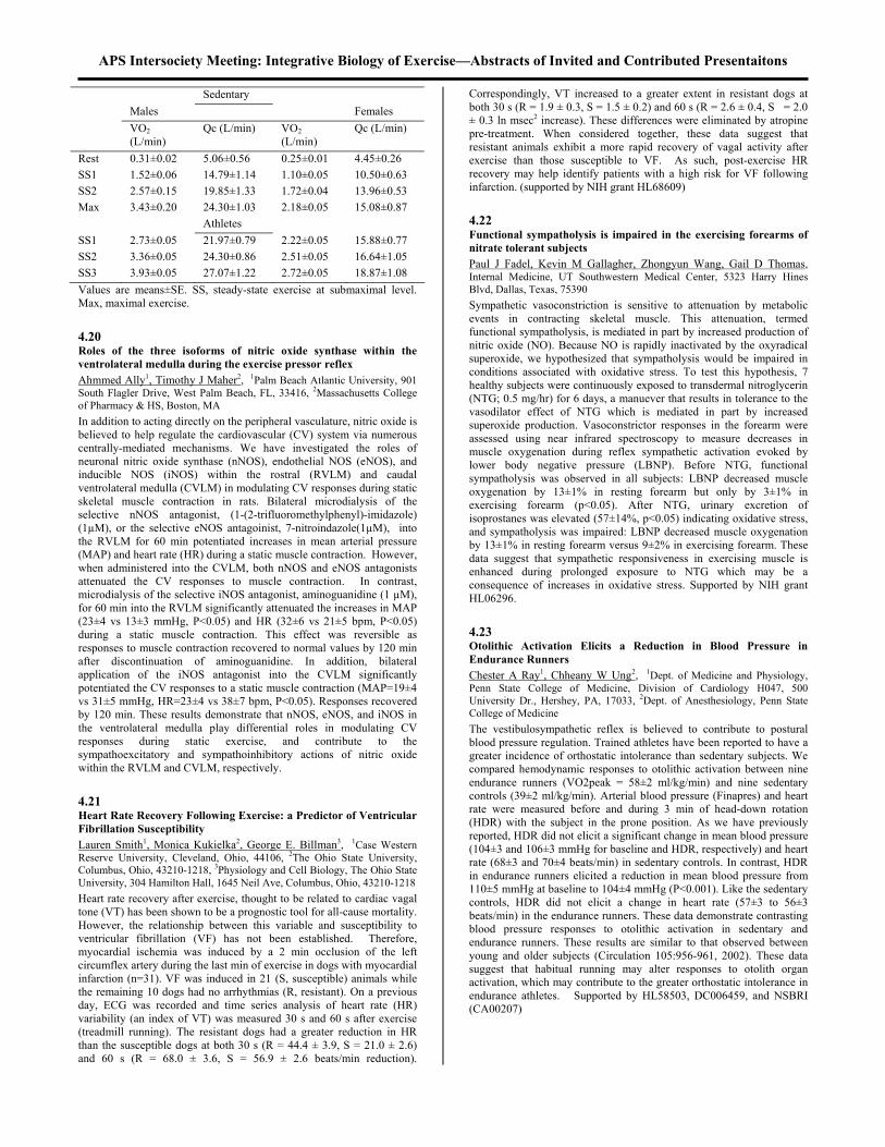
APS Intersociety Meeting: Integrative Biology of Exercise—Abstracts of Invited and Contributed Presentaitons Sedentary Males Females VO2
(L/min) Qc (L/min) VO2
(L/min) Qc (L/min)
Rest 0.31±0.02 5.06±0.56 0.25±0.01 4.45±0.26 SS1 1.52±0.06 14.79±1.14 1.10±0.05 10.50±0.63 SS2 2.57±0.15 19.85±1.33 1.72±0.04 13.96±0.53 Max 3.43±0.20 24.30±1.03 2.18±0.05 15.08±0.87 Athletes SS1 2.73±0.05 21.97±0.79 2.22±0.05 15.88±0.77 SS2 3.36±0.05 24.30±0.86 2.51±0.05 16.64±1.05 SS3 3.93±0.05 27.07±1.22 2.72±0.05 18.87±1.08 Values are means±SE. SS, steady-state exercise at submaximal level. Max, maximal exercise. 4.20 Roles of the three isoforms of nitric oxide synthase within the ventrolateral medulla during the exercise pressor reflex Ahmmed Ally1, Timothy J Maher2, 1Palm Beach Atlantic University, 901 South Flagler Drive, West Palm Beach, FL, 33416, 2Massachusetts College of Pharmacy & HS, Boston, MA In addition to acting directly on the peripheral vasculature, nitric oxide is believed to help regulate the cardiovascular (CV) system via numerous centrally-mediated mechanisms. We have investigated the roles of neuronal nitric oxide synthase (nNOS), endothelial NOS (eNOS), and inducible NOS (iNOS) within the rostral (RVLM) and caudal ventrolateral medulla (CVLM) in modulating CV responses during static skeletal muscle contraction in rats. Bilateral microdialysis of the selective nNOS antagonist, (1-(2-trifluoromethylphenyl)-imidazole) (1µM), or the selective eNOS antagoinist, 7-nitroindazole(1µM), into the RVLM for 60 min potentiated increases in mean arterial pressure (MAP) and heart rate (HR) during a static muscle contraction. However, when administered into the CVLM, both nNOS and eNOS antagonists attenuated the CV responses to muscle contraction. In contrast, microdialysis of the selective iNOS antagonist, aminoguanidine (1 µM), for 60 min into the RVLM significantly attenuated the increases in MAP (23±4 vs 13±3 mmHg, P<0.05) and HR (32±6 vs 21±5 bpm, P<0.05) during a static muscle contraction. This effect was reversible as responses to muscle contraction recovered to normal values by 120 min after discontinuation of aminoguanidine. In addition, bilateral application of the iNOS antagonist into the CVLM significantly potentiated the CV responses to a static muscle contraction (MAP=19±4 vs 31±5 mmHg, HR=23±4 vs 38±7 bpm, P<0.05). Responses recovered by 120 min. These results demonstrate that nNOS, eNOS, and iNOS in the ventrolateral medulla play differential roles in modulating CV responses during static exercise, and contribute to the sympathoexcitatory and sympathoinhibitory actions of nitric oxide within the RVLM and CVLM, respectively. 4.21 Heart Rate Recovery Following Exercise: a Predictor of Ventricular Fibrillation Susceptibility Lauren Smith1, Monica Kukielka2, George E. Billman3, 1Case Western Reserve University, Cleveland, Ohio, 44106, 2The Ohio State University, Columbus, Ohio, 43210-1218, 3Physiology and Cell Biology, The Ohio State University, 304 Hamilton Hall, 1645 Neil Ave, Columbus, Ohio, 43210-1218 Heart rate recovery after exercise, thought to be related to cardiac vagal tone (VT) has been shown to be a prognostic tool for all-cause mortality. However, the relationship between this variable and susceptibility to ventricular fibrillation (VF) has not been established. Therefore, myocardial ischemia was induced by a 2 min occlusion of the left circumflex artery during the last min of exercise in dogs with myocardial infarction (n=31). VF was induced in 21 (S, susceptible) animals while the remaining 10 dogs had no arrhythmias (R, resistant). On a previous day, ECG was recorded and time series analysis of heart rate (HR) variability (an index of VT) was measured 30 s and 60 s after exercise (treadmill running). The resistant dogs had a greater reduction in HR than the susceptible dogs at both 30 s (R = 44.4 ± 3.9, S = 21.0 ± 2.6) and 60 s (R = 68.0 ± 3.6, S = 56.9 ± 2.6 beats/min reduction).
Correspondingly, VT increased to a greater extent in resistant dogs at both 30 s (R = 1.9 ± 0.3, S = 1.5 ± 0.2) and 60 s (R = 2.6 ± 0.4, S = 2.0 ± 0.3 ln msec2 increase). These differences were eliminated by atropine pre-treatment. When considered together, these data suggest that resistant animals exhibit a more rapid recovery of vagal activity after exercise than those susceptible to VF. As such, post-exercise HR recovery may help identify patients with a high risk for VF following infarction. (supported by NIH grant HL68609) 4.22 Functional sympatholysis is impaired in the exercising forearms of nitrate tolerant subjects Paul J Fadel, Kevin M Gallagher, Zhongyun Wang, Gail D Thomas, Internal Medicine, UT Southwestern Medical Center, 5323 Harry Hines Blvd, Dallas, Texas, 75390 Sympathetic vasoconstriction is sensitive to attenuation by metabolic events in contracting skeletal muscle. This attenuation, termed functional sympatholysis, is mediated in part by increased production of nitric oxide (NO). Because NO is rapidly inactivated by the oxyradical superoxide, we hypothesized that sympatholysis would be impaired in conditions associated with oxidative stress. To test this hypothesis, 7 healthy subjects were continuously exposed to transdermal nitroglycerin (NTG; 0.5 mg/hr) for 6 days, a manuever that results in tolerance to the vasodilator effect of NTG which is mediated in part by increased superoxide production. Vasoconstrictor responses in the forearm were assessed using near infrared spectroscopy to measure decreases in muscle oxygenation during reflex sympathetic activation evoked by lower body negative pressure (LBNP). Before NTG, functional sympatholysis was observed in all subjects: LBNP decreased muscle oxygenation by 13±1% in resting forearm but only by 3±1% in exercising forearm (p<0.05). After NTG, urinary excretion of isoprostanes was elevated (57±14%, p<0.05) indicating oxidative stress, and sympatholysis was impaired: LBNP decreased muscle oxygenation by 13±1% in resting forearm versus 9±2% in exercising forearm. These data suggest that sympathetic responsiveness in exercising muscle is enhanced during prolonged exposure to NTG which may be a consequence of increases in oxidative stress. Supported by NIH grant HL06296. 4.23 Otolithic Activation Elicits a Reduction in Blood Pressure in Endurance Runners Chester A Ray1, Chheany W Ung2, 1Dept. of Medicine and Physiology, Penn State College of Medicine, Division of Cardiology H047, 500 University Dr., Hershey, PA, 17033, 2Dept. of Anesthesiology, Penn State College of Medicine The vestibulosympathetic reflex is believed to contribute to postural blood pressure regulation. Trained athletes have been reported to have a greater incidence of orthostatic intolerance than sedentary subjects. We compared hemodynamic responses to otolithic activation between nine endurance runners (VO2peak = 58±2 ml/kg/min) and nine sedentary controls (39±2 ml/kg/min). Arterial blood pressure (Finapres) and heart rate were measured before and during 3 min of head-down rotation (HDR) with the subject in the prone position. As we have previously reported, HDR did not elicit a significant change in mean blood pressure (104±3 and 106±3 mmHg for baseline and HDR, respectively) and heart rate (68±3 and 70±4 beats/min) in sedentary controls. In contrast, HDR in endurance runners elicited a reduction in mean blood pressure from 110±5 mmHg at baseline to 104±4 mmHg (P<0.001). Like the sedentary controls, HDR did not elicit a change in heart rate (57±3 to 56±3 beats/min) in the endurance runners. These data demonstrate contrasting blood pressure responses to otolithic activation in sedentary and endurance runners. These results are similar to that observed between young and older subjects (Circulation 105:956-961, 2002). These data suggest that habitual running may alter responses to otolith organ activation, which may contribute to the greater orthostatic intolerance in endurance athletes. Supported by HL58503, DC006459, and NSBRI (CA00207)

APS Intersociety Meeting: Integrative Biology of Exercise—Abstracts of Invited and Contributed Presentaitons 4.24 Bilateral cerebral tissue oxygenation (TOI) during exposure to lower body negative pressure (LBNP) Sarah-Jane Carrie Lusina1, Jessica M Scott2, Benjamin TA Esch2, Don C McKenzie2, Andrew W Sheel2, Darren ER Warburton2, 1University of British Columbia, 210, War Memorial Gym, 6081 University Blvd, Vancouver, British Columbia, V6T 1Z1, Canada, 2University of British Columbia, 210, Memorial Gym, 6081 University Blvd, Vancouver, British Columbia, V6T 1Z1, Canada Cerebral hemodynamics are altered with exposure to LBNP. Hemodynamic changes can be evaluated by TOI using near-infrared spectroscopy (NIRS). Research has indicated that TOI may be different between the right (RH) and left (LH) cerebral hemispheres; however little is known about the effects of LBNP. The purpose of this investigation is to simultaneously measure the effects of LBNP on TOI in the RH and LH. Ten endurance-trained men (Age=30 5 yr; VO2max=62.4 3.0 mL/kg/min) were exposed to five 10 min stages consisting of: three normobaric baseline conditions separated by -20 and -40 mmHg LBNP (randomly assigned). The NIRS data was collected continuously and a steady state mean value for each condition was calculated. The TOI of the RH was consistently lower (n.s.) than the LH for all stages. The TOI during –40 mmHg LBNP (69.0 10.2%) was significantly (p<0.05) lower than the three baseline conditions (72.2 10.0, 71.4 9.6, and 70.9 9.4%, respectively). No order effect was observed. It can be concluded that acute exposure to –40 mmHg LBNP caused a significant attenuation in cerebral TOI, likely as the result of increased cerebral vasomotor activity (mediated by sympathetic activation) and/or a reduction in central O2 transport (mediated by a decreased cardiac preload). Evidence suggests that TOI may be different between hemispheres, although the response to LBNP is similar. Support: NSERC, CFI, and MSFHR 4.25 HYPOXIA VS HYPERPNEA: EFFECTS ON CUTANEOUS VASCULAR TONE Grant H Simmons, Brad W Wilkins, Mollie P Pricher, Christopher T Minson, John R Halliwill, Department of Human Physiology, University of Oregon, Eugene, OR In contrast to skeletal muscle, little is known about the cutaneous sympathetic or vascular response to hypoxia. The purpose of this study was to explore the effect of hypoxia and hyperpnea on cutaneous vascular regulation. Twelve healthy, unacclimatized subjects (24.0±1.0 yrs) were instrumented with two microdialysis fibers in the ventral forearm. Each site was continuously infused with Ringer’s (control) or bretylium tosylate (10mM) to prevent sympathetically mediated vasoconstriction. Skin blood flow was assessed at each site (laser-Doppler flowmetry) and cutaneous vascular conductance (CVC) was calculated (red blood cell flux/mean arterial pressure) and scaled as % maximal CVC (local heating to 43ºC). Adequacy of bretylium administration was verified via whole body cold stress. Subjects were exposed to isocapnic hypoxia, reducing arterial O2 saturation to 80% (assessed by pulse oximetry), and normoxic isocapnic hyperpnea, matching hypoxic tidal volume and breathing frequency. During hypoxia, CVC in the control site increased from 11.7±2.0 to 13.8±2.0%CVCmax ( 23.2% from baseline, P<0.05) and CVC in the bretylium site increased from 12.7±1.6 to 15.4±2.2%CVCmax ( 18.5% from baseline, P<0.05). During hyperpnea, CVC in the control site increased from 11.2±1.9 to 13.4±2.0%CVCmax ( 25.7% from baseline, P<0.05); however, there was no change in the bretylium site (12.8±2.0 vs 13.1±2.2%CVCmax; P=0.37). Thus, both hypoxia and hyperpnea increase CVC. However, it appears the hyperpnic response is due to a decrease in sympathetic vasoconstrictor tone, whereas the hypoxic response involves additional vasodilator factors. Supported by NIH grant HL 65305. 4.26 Muscle Mechanoreceptors Have Heightened Sensitivty in Heart Failure Josephine Chiu1, Michele Hamilton2, Gregg Fonarow3, Holly Middlekauff3, 1Medicine, Keck School of Medicine, 47-123 CHS, 10833 Le
Conte Avenue, Los Angeles, CA, 90095, 2Medicine, Geffen School of Medicine at UCLA, 3Medicine, Geffen School of Medicine at UCLA, 47-123 CHS, 10833 Le Conte Avenue, Los Angeles, CA, 90095 Patients with advanced heart failure(HF) have heightened sympathetic nerve activity(SNA) in response to exercise, but the mechanism(s) of this exaggerated sympathetic activation remain unknown. The purpose of the current study was to test the hypothesis that muscle mechanoreceptors, which are muscle afferent nerve fibers sensitive to stretch, contribute to this heightened sympathetic response. Methods. Ten HF pts and 11 age-matched healthy controls were studied. In order to isolate the muscle mechanoreceptor contribution from that of muscle metaboreceptors and central command, passive rhythmic forearm exercise was performed for 3 minutes. Peroneal microneurography was used to measure muscle SNA. Results. In controls, muscle SNA remained unchanged throughout passive exercise (baseline total SNA 954 vs Ex1 962, Ex2 1002, Ex3 997, p=NS). In contrast, in HF patients, muscle SNA increased in the first minute of exercise and remained elevated throughout exercise (baseline SNA 1481 vs Ex1 1680, Ex2 1667, Ex3 1737, p<0.001; time*group interaction p=0.006). Muscle SNA returned to baseline levels during the first minute of recovery. Heart rate and blood pressure did not change during exercise in either group. Conclusion. Muscle mechanoreceptors have heightened sensitivity in patients with heart failure, and likely contribute to their heightened sympathetic activation during exercise. 4.27 Role of central command to cutaneous vascular responses during isometric exercise Manabu Shibasaki1, Niels H. Secher2, John M. Johnson3, Craig G. Crandall4, 1Institute for Exercise and Environmental Medicine, Presbyterian Hospital of Dallas, 7232 Greenville Ave, Dallas, TX, 75231, 2Department of Anaesthesia, University of Copenhagen, Denmark, Denmark, 3Department of Physiology, University of Texas Health Science Center at San Antonio, 4Department of Internal Medicine, University of Texas Southwestern Medical Center at Dallas The purpose of this project was to test the hypothesis that central command is capable of modulating cutaneous vascular responses during exercise in heat stressed subjects. Seven subjects performed isometric handgrip exercise (IHG; 35% MVC for 2 min) followed by 2-min of post-exercise ischemia (PEI) while normothermic and during heat stress (increase internal temperature ~ 1 C). On a separate day the protocol was repeated with partial neuromuscular blockade (I.V. Nimbex; NB trial). Forearm cutaneous vascular conductance (CVC) was calculated from the ratio of skin blood flow to mean arterial pressure (MAP). Despite encouragement to attain the same IHG workload during the NB trials, force production was attenuated (16 1 kg versus 5 1 kg; P<0.001). In normothermia, forearm CVC was not altered during IHG or PEI in either the control or NB trials. Heat stress responses are shown below.
Control Trial NB Trial CVC (%) MAP
(mmHg) CVC (%) MAP
(mmHg) Rest 100 76.6 2.5 100 76.2 3.6 IHG 88.7 4.0* 108.8 3.3*† 90.7 2.2* 94.0 3.3* PEI 100.3 4.0 89.9 3.1*† 105.7 3.8 77.0 3.4
* Significant difference from rest (P<0.05); † Significant difference from NB trial (P<0.05). Similar reductions in forearm CVC during IHG between trials, coupled with minimal metaboreflex activation during the NB trial, suggests that central command is capable of modulating forearm cutaneous vascular responses in heat stressed humans. Funded in part by NIH (HL-61388 and HL-67422) and Danish National Research Foundation (504-4) 4.28 Effects of resistance training on muscle sympathetic nerve response to static contraction Mitsuru Saito, Applied Physiology Laboratory, Toyota Technological Institute, 2-12 Hisakata, Tempakua-ku, Nagoya, 468-8511, Japan

APS Intersociety Meeting: Integrative Biology of Exercise—Abstracts of Invited and Contributed Presentaitons Although the reduction of muscle sympathetic nerve activity (MSNA) after endurance training has been documented, little information exists concerning MSNA adaptation to resistance training (RT). We investigated whether RT can alter MSNA response to static contraction. MSNA from the tibial nerve during repetitive static handgrip exercise (SHG) with maximal voluntary effort (MVE) was compared before and after high intensity SHG training. Subjects performed a 15-s contraction followed by a 15-s relaxation for up to 20 contraction in the right and the left arm alternately. This study was approved by the TTI ethical committee. Nine healthy subjects, who gave informed consent, were trained using unilateral SHG of the left arm 5 days/wk for 4 wk. The RT paradigm consisted of three sets of 10 repetitions of a 10-s SHG with MVE with a 10-s relaxation between contractions, separated by rest periods. The MSNA burst frequency significantly increased during repetitive SHGs in both the trained and control arms before and after RT (ANOVA). The average increase for the first four SHGs in both arms was greater (9 bursts/min, p=0.017, t-test) after training compared with that before training, but was not different after the fifth to the 10th SHG. The after-training force output during the test exercise increased for both the trained and control arms, while no difference between the arm was observed. The results suggest that RT may enhance MSNA response during the early phase of strong SHG. 4.29 Human beta-2 adrenergic receptor polymorphism alters the hemodynamic response to cycling William Schrage1, John H Eisenach1, Christopher P Johson1, Diane E Wick1, Branton G Walker1, Michael D Jensen2, Michael J Joyner1, 1Anesthesiology, Mayo Clinic, 200 1st St SW, Rochester, MN, 55905, 2Endocrinology, Mayo Clinic, 200 1st St SW, Rochester, MN, 55905 Humans homozygous for glycine (G/G) versus arginine (A/A) at amino acid 16 of the beta-2 adrenergic receptor ( 2-AR) dilate more to forearm infusion of isoproterenol. We tested the hypothesis that G/G subjects would exhibit greater cardiac output (CO) responses during cycling exercise to compensate for greater exercise-induced vasodilation. We measured HR (ECG), arterial pressure (radial artery), and CO (acetylene wash-in) in 13 healthy adults at rest, during 90 min cycling at 50% VO2 peak, and during recovery for 60 min. HR response to cycling was similar between A/A (n=4) and G/G (n=9) (p = 0.64). MAP was similar at rest (89 3 mm Hg), but the increase in MAP during cycling was lower in G/G (p < 0.05). In fact, MAP returned to resting levels by 45 min of cycling in G/G, but remained 13 3 mm Hg higher in A/A. CO was 15-20% higher in G/G throughout cycling (p < 0.05). Further, total vascular conductance was similar at rest (p = 0.28), but was greater throughout cycling in G/G. The catecholamine response to cycling was similar between genotypes (NE, p = 0.66; Epi, p = 0.17, n = 5). We conclude humans homozygous for G/G at amino acid 16 of the -AR respond to moderate exercise with higher CO and lower MAP, but similar HR. These data suggest the G/G genotype is associated with greater peripheral vasodilation than the A/A genotype and implicate an important role of this common 2-AR polymorphism in the response to exercise. 4.30 CHANGES OF RESTING BLOOD PRESSURE RESPONSE TO CONTROLLED AEROBIC EXERCISE IN OLDER ADULTS Guoyuan Huang1, Xiangrong Shi2, Wayne H Osness3, 1University of Southern Indiana, 8600 University Blvd., Evansville, IN, 47712, 2University of North Texas Health Science Center, Fort Worth, TX, 76107, 3University of Kansas, Lawrence, KS, 66045 PURPOSE: To determine the effects of controlled aerobic exercise training on resting blood pressure among sedentary older adults (age 60 yrs) using meta-analysis. METHODS: The inclusion criteria were: controlled trials with aerobic exercise as the only intervention and presence of a control group, exercise lasting a minimum of 2 wks, mean ages of subjects 60 yrs, studies published in English-language journals between January 1980 and January 2002, and a measure of changes in resting systolic/diastolic blood pressure (SBP/DBP). RESULTS: Twenty-three studies with a total of 1,226 subjects in 23 control (n=461) and 26 exercise (n=765) were included in the final analysis. The pooled standardized effect size showed an average effect of –0.33±0.06
(mean±SEM, 95% CI = –0.45 to –0.21) by a fixed-effect model in resting SBP and –0.39 ±0.09 (mean±SEM, 95% CI = –0.56 to –0.23) by a random model in resting DBP. The net changes in SBP and DBP was statistically significant (p<0.001) and represented net decreases of approximately 3.9% for SBP (mean±SEM, –5.39±1.21 mmHg, 95% CI = –7.82 to –2.95) and 4.5% for DBP (mean±SEM, –3.68±0.83 mmHg, 95% CI = –5.35 to –2.00). For sample size of the studies less than 30 subjects, greater decreases were found in both resting SBP (p=0.021) and DBP (p=0.012). CONCLUSION: The results of this study suggest that aerobic exercise does reduce resting SBP and DBP in older adults. Low to moderate intensity aerobic training may be just as effective as higher intensity for reducing resting SBP/DBP in older individuals. Resting SBP and DBP do not appear to decrease further with prolonged training more than 16 wks. However, these decreases may be exaggerated if sample size in a study is less than 30 subjects. 4.31 Evidence for autoregulation of cutaneous blood flow during isometric handgrip exercise Gregg Russell McCord, Dr. Christopher T Minson, Human Phsyiology, University of Oregon, 1240 University of Oregon, 122 Esslinger Hall, Eugene, OR, 97401 Abstract The dramatic rise in skin blood flow (SkBF) and sweating observed during heat stress are mediated by poorly understood sympathetic cholinergic mechanisms. One theory suggests that a single sympathetic cholinergic nerve mediates active vasodilation (AVD) and sweating via cotransmission of separate neurotransmitters, as AVD and sweating track temporally and directionally when activated during heat stress. However, it has also been suggested that these responses are mediated by separate cholinergic nerves, as cutaneous vascular conductance (CVC) has been shown to decrease while sweat rate (SR) increases during hyperthermia when isometric handgrip exercise (IHG) is performed to raise arterial pressure. We tested the hypothesis that the decrease in CVC observed during IHG is due to vessel autoregulation potentially occurring to minimize the rise in flow to a large rise in arterial pressure, and not due to withdrawal of AVD. Subjects performed IHG as CVC was elevated at selected sites to varying levels by local heating (which is independent of AVD) in thermoneutral and hyperthermic conditions. In thermoneutral conditions, CVC decreased during IHG when blood flow was elevated prior to exercise (-6.5 1.8% at 41 C and -10.5 2.0% at 43 C; P<0.05 versus preexercise). During IHG in hyperthermia, a progressively greater decrease in CVC was associated with the level of CVC prior to exercise. Taken together, these findings suggest that the decrease in CVC observed during IHG during hyperthermia is due to vessel autoregulation, and not withdrawal of AVD, providing evidence against independent neural control of AVD and sweating. 4.32 Role of limb and measurement site in vascular responsiveness during dynamic exercise David Walter Wray, Abhimanyu Uberoi, Lesley Lawrenson, Russell Richardson, Department of Medicine, University of California San Diego, 9500 Gilman Drive, San Diego, CA, 92093-0623 Comparison of arm and leg vascular responsiveness during dynamic exercise of an isolated muscle mass has not been performed in humans. In the leg, measurements taken distal to the common femoral artery (CFA) in the deep femoral artery (DFA) are closer to the exercising muscle, and may more closely resemble the brachial artery in structure and anatomical location within the arterial tree. Consequently, subjects performed incremental, intermittent handgrip exercise (arm) and knee-extensor exercise (leg) from 5-60% of maximal work rate (WR). Ultrasound Doppler measurements were taken in the brachial artery, CFA, and DFA at rest and at each WR. Exercise increased blood flow more in the leg (≈25-fold) than in the arm (≈6-fold). Exercise at WR60%max did not alter CFA vessel diameter, but increased in the brachial artery (0.42 ± 0.01 to 0.49 ± 0.01 cm, rest vs. exercise) and the DFA (0.59 ± 0.05 to 0.64 ± 0.04 cm, rest vs. exercise). These findings suggest that differences in vascular responsiveness between limbs are less evident when vessels of similar diameter and location in the arterial

APS Intersociety Meeting: Integrative Biology of Exercise—Abstracts of Invited and Contributed Presentaitons tree are compared. However, when dilation of each vessel is normalized for the increase in blood flow and sheer stress, the leg vasculature remains much less responsive than the arm. Teleologically, this disparity between arm and leg vasomotion may act to preserve systemic normotension and perfusion during leg exercise. 4.33 MRI-measured post-contractile hyperemia transients and post-exercise blood flow in active vs. inactive individuals. Theodore F Towse, Jill M Slade, Ronald A Meyer, Physiology and Radiology, Michigan State University, 2201 Biomedical and Physical Sciences Building, East Lansing, MI, 48824 Transient reflex hyperemia after single muscle contractions measured by MRI or other methods (e.g., Brock, RW, et al, J. Appl. Physiol. 85:2249, 1998) might provide an index of microvascular density and/or reactivity. This study compared transient increases in MRI signal intensity (SI, via one-shot echo-planar at 1.5T, TR/TE=1000/45) after single, 1 s duration contractions of ant. tibial muscle in chronically active (A, n=8, estimated kcal/kg/day 42.4±3.7) vs. inactive (I, n=7, 32.3±0.7) young subjects (18-34 yrs). In addition, anterior tibial artery x-sectional area and blood flow were measured by phase-contrast MRI angiography before and after fatiguing repetitive ankle dorsiflexion exercise (2 min @ 50% duty cycle) in the same subjects. MRI-measured SI transients after single contractions were 4-fold greater in active ( SI=5.50±1%) compared to inactive (1.61±0.3 %, p<0.01) subjects. In contrast, after the repetitive exercise there was no significant difference between groups in blood flow (A, 262±52 vs. I, 260±68 ml/min), apparent arterial stiffness (% change in arterial area [100*(systolic-diastolic)/diastolic], A 58.0±20.2, I 53.4±17.8 %), or flow-mediated arterial dilatation (% change in diastolic area, A 66.8±16.3, I 59.7±13.6). The results suggest that transient hyperemia after single contractions is a more sensitive index of muscle vascular fitness than conventional measures of macrovascular structure or post-exercise blood flow. (Supported by AR043903 & DK063497) 4.34 Effects of Acute Aerobic Training on Nutritive Skeletal Muscle Blood Flow with Aging John A. Carrithers1, Chris A Evans2, Ray M Kraus2, Hannah M Carrithers2, Tim P Gavin2, Rebecca S Ruster2, Deb J Knapp2, James L Drew2, Joseph Garry2, Robert C Hickner2, 1Human Performance Laboratory, East Carolina University, 363 Ward Sports Medicine, Greenville, NC, 27858, 2East Carolina University This study determined the effects of seven days of aerobic training on resting nutritive skeletal muscle blood flow in five young (25±5y) and four old (61±2y) males. Nutritive muscle blood flow was determined in the vastus lateralis muscle via the microdialysis ethanol outflow/inflow technique before and after training. Lower limb blood flow was measured via strain gauge plethysmography prior to and following training. Aerobic exercise bouts were performed on a cycle ergometer for 60 minutes and completed as follows: 5 min. at 30% VO2max, 50 min. at 65% VO2max, 5 min. at 30% VO2max. Basal nutritive blood flow was not different before or after training in the young (pre: 0.650 ±0.03 to post: 0.581 ±0.06) or old individuals (pre: 0.576 ±0.06 to post: 0.549 ±0.08). However, there was a main effect (p<0.05) for training to improve basal nutritive muscle blood flow. Note a decrease in ethanol outflow/inflow ratio represents an increase in nutritive blood flow. Limb blood flow increased (p<0.05) in young (2.73 ±0.8 to 3.26 ±0.8 ml/100ml tissue/min) and old (1.64 ±0.6 to 2.13 ±0.3 ml/100ml tissue/min) after training; in-addition, post training limb blood flow was greater (p<0.05) in the young compared to the old. It appears that seven days of aerobic training improves nutritive skeletal muscle blood flow and limb blood flow in young and old individuals. Additional subjects are warranted to determine if aging affects resting skeletal muscle nutritive blood flow. This project was supported in part by NIH grant AG-19209 and AG-21891. 4.35 Effects of L-NMMA administration on Exercising Nutritive Skeletal Muscle Blood Flow Before and After 7-days of Aerobic Training Robert C Hickner1, Chris A Evans2, Ray M Kraus2, Rebecca S Ruster2, Tim P Gavin2, Hannah M Carrithers2, Deb J Knapp2, Robert J
Tanenberg2, James L Drew2, John A Carrithers2, 1Human Performance Laboratory, East Carolina University, 363 Ward Sports Medicine, Greenville, NC, 27858, 2East Carolina University We have previously demonstrated a reduced nutritive flow to exercising muscle in young individuals by L-NMMA administration, indicating that nitric oxide synthase enzyme inhibition results in lower skeletal muscle nutritive flow. The purpose of this study was to determine the effects of L-NMMA administration on nutritive skeletal muscle blood flow during exercise before and after seven days of aerobic training in four young (24±5yr) and four older (61±2yr) males. Nutritive skeletal muscle blood flow was determined in the vastus lateralis of the quadriceps femoris muscle group using the microdialysis ethanol technique during low-intensity cycle ergometry exercise before and after seven days of cycle ergometry training for 60 min per day. The ethanol outflow/inflow ratio was lower (indicative of higher nutritive flow) in seven of eight subjects after, compared to before, exercise training (0.260±0.038 after, 0.316±0.031 before; P=0.06). Local L-NMMA administration through the microdialysis probe resulted in a higher outflow/inflow ratio than saline administration regardless of age and training status (0.404±0.034 L-NMMA, 0.288±0.025 saline; P<0.05). Administration of the NO donor sodium nitroprusside resulted in ethanol outflow/inflow ratios similar to saline. It can be concluded that local L-NMMA administration to muscle results in reduced nutritive flow during exercise in young and old individuals, and that seven days of cycle ergometry training likely results in improvements in skeletal muscle nutritive blood flow during low-intensity exercise. This project was supported by NIH grants AG-19209 (RH) and NIA AG-21891 (TG). 5.1 mTOR-dependent signaling mediates an increase in the ε-subunit of eukaryotic initiation factor 2B (eIF2B) that is associated with an increase in skeletal muscle eIF2B activity and protein synthesis following acute muscle loading Neil Kubica1, David L Williamson1, Douglas R Bolster2, Stephen J Crozier1, Scot R Kimball1, Peter A Farrell3, Leonard S Jefferson1, 1Department of Cellular and Molecular Physiology, The Pennsylvania State University College of Medicine, H166, 500 University Drive, Hershey, PA, 17033, 2Division of Space Life Sciences, Universities Space Science Research Association, 3600 Bay Area Blvd, Houston, TX, 77058, 3Department of Exercise and Sports Science, East Carolina University, Greenville, NC, 27858 The contribution of mTOR signaling to increases in protein synthesis was assessed by administering rapamycin to rats 2 h prior to a bout of resistance exercise. Animals were sacrificed 16 h post-exercise and gastrocnemius protein synthesis and biomarkers of translation initiation were assessed. Acute resistance exercise caused an increase in the fractional rate of protein synthesis (138% of control; p<0.01), however exercised animals treated with rapamycin showed no significant change in protein synthesis. Furthermore, there was a significant decrease in the proportion of eIF2B phosphorylated on Ser 535 (51% of control; p<0.01) that was explained by a significant increase in the expression of eIF2B protein (174% of control; p<0.001). This occurred in the absence of an increase in two other subunits of the eIF2B complex, and was also completely blunted by rapamycin treatment. The increase in eIF2B protein expression is consistent with previously published reports demonstrating an increase in eIF2B activity 16 h following a bout of acute resistance exercise. Currently, in vivo electroporation gene delivery methodology is being employed to assess the role of eIF2B and eIF2B S535A overexpression on eIF2B activity and protein synthesis in the gastrocnemius in sedentary and exercised animals.
5.0 Cell Signaling

APS Intersociety Meeting: Integrative Biology of Exercise—Abstracts of Invited and Contributed Presentaitons 5.2 Mechanical Stimuli Regulate mTOR via a PI3K/Akt and Growth Factor Independent Mechanism Troy A. Hornberger1, Mark J. Fedele2, Karyn A. Esser2, 1Bioengineering, UCSD, 5265 Toscana Way #234, San Diego, CA, 92122, 2UIC In response to growth factors, mTOR has been identified as a central component of the signaling pathways that control the translational machinery and cell growth. Signaling through mTOR has also been shown to be necessary for mechanical load-induced growth of cardiac and skeletal muscle. Although the mechanisms involved in the mechanically-induced activation of mTOR are not known, it has been suggested that activation of PI3K and Akt, via the release of locally-acting growth factors, underlies this process. Here we show that mechanically stimulating (passive stretch) skeletal muscle, ex vivo, results in the activation of mTOR-dependent signaling events and signaling through mTOR is required for an increase in protein synthesis. Using wortmannin we show that activation of mTOR-dependent signaling occurs through a PI3K-independent pathway. Consistent with these results, mechanically-induced signaling through mTOR was not disrupted in muscles from Akt1-/- mice. In addition, ex vivo co-incubation experiments, along with in vitro conditioned media experiments, demonstrate that a mechanically-induced release of locally acting growth factors was not sufficient for the activation of the mTOR pathway. Taken together, our data demonstrate that mechanical stimuli can activate the mTOR pathway independent of PI3K/Akt1 and locally acting growth factors. Thus, mechanical stimuli and growth factors provide distinct inputs through which mTOR coordinates an increase in protein synthesis. 5.3 SIGNALING KINASE ACTIVATION BY TWITCH AND TETANIC CONTRACTIONS IN RED AND WHITE FAST-TWITCH MUSCLE Vladimir Ljubicic, David A. Hood, School of Kinesiology and Health Science, York University, 4700 Keele St., Toronto, Ontario, Canada Acute contractile activity increases protein kinase activation leading to signal transduction. We hypothesized that contractile activity-induced kinase activation would be affected differently by twitch (TW) and tetanic (TET) contractions, and by mitochondrial content. We compared red (RTA) and white (WTA) tibialis anterior (TA) muscle possessing 2.5-fold differences in mitochondrial content. Acute stimulation was used to illicit TW (10 Hz) or TET (100 Hz, 100 msec) contractions of the TA for 5 mins. The extent of fatigue to 50% of initial tension was similar in TW and TET contractions, but TET contractions generated a 6-fold greater force over the 5 min period. We investigated 1) the total protein content and 2) the activation of Akt, AMPKα and ERK1/2 by western blotting. Total ERK1/2 and AMPKα protein content in RTA was greater than or equal to that in WTA, but total Akt was 1.3-fold higher in WTA. AMPKα, ERK1 and ERK2 activation were greater in resting WTA by 2-, 8- and 2-fold, respectively, compared to resting RTA. All kinases were activated 2-6-fold by TW and TET contractions in RTA, whereas in WTA AMPKα and ERK2 were not activated, and Akt and ERK1 activation only increased with TW contractions (1.7-fold), and not further with TET contractions. Our data indicate that 1) kinase activation was not proportional to the energy cost of contractions, 2) kinase expression and resting activation differ between red and white muscle, and 3) mitochondrial content may influence the extent of kinase activation at rest and during contractions. 5.4 IGF-I ACTIVATES PKB AND PREVENTS APOPTOSIS IN HYPOXIC TENDON CELLS Alexander Scott1, Karim Khan2, Vince Duronio2, 1University of British Columbia, 2660 Oak Street, Vancouver, B.C., V6T 1Z3, Canada, 2University of British Columbia INTRODUCTION Some authors propose that chronic hypoxia contributes to the pathogenesis of chronic tendon pathology. The objectives of the current study were to investigate the effect of chronic hypoxia on Achilles tendon cell (ATC) viability, and to examine the ability of IGF-I, a factor with known regenerative properties in tendon, to activate pro-survival signal pathways and prevent ATC death.
METHODS Porcine ATCs were released by enzymatic digestion and cultured up to the 5th passage, and then maintained in a standard 5% CO2 incubator (normoxia) or placed at 37°C in an anaerobic chamber with or without IGF-I for periods up to 96 hours. RESULTS The predominant mechanism of hypoxic cell death was apoptosis, as evidenced by Annexin-V labeling. Hypoxia resulted in substantial caspase activation as determined by a flourometric probe (FAM-VAD-FMK FLICA). IGF-I restored the level of cell death in hypoxic conditions to control values, and resulted in a rapid onset and decay of PKB activation that was inhibited by 25µM LY294002. DISCUSSION Apoptosis was a prominent feature of tendon cells’ response to prolonged hypoxia. IGF-I exerted a potent pro-survival effect in tendon cells, adding to its attractiveness as a tendon-regenerative factor. Figure 1 Caspase activation in hypoxia tendon cells
Left panel; control. Right panel; hypoxic. Note prominent peak of activation in hypoxic cells. 5.5 The CaMK Inhibitor, KN-62, Prevents Insulin-, Contraction-, and AICAR-Stimulated Glucose Uptake, But Not Via Inhibition of Akt, AMPK, or PKCλ/ζ Phosphorylation Carol A Witczak1, Niels Jessen1, Laurie J Goodyear2, 1Metabolism, Joslin Diabetes Center, Research Division, Rm 530, One Joslin Place, Boston, MA, 02215, 2Metabolism, Joslin Diabetes Center, Research Division, Rm 520A, One Joslin Place, Boston, MA, 02215 Recent evidence suggests a role for Ca2+/calmodulin-activated kinases (CaMKs) in the regulation of skeletal muscle glucose uptake. Our goal was to determine the mechanism by which the CaMK inhibitor, KN-62, inhibits glucose uptake in mouse soleus. Soleus muscles were excised from male ICR mice (6-8 wks), treated KN-62 (10 M, 30-min), and then stimulated by insulin (50 mU/ml, 20-min), contraction (10-min), or the AMP-activated protein kinase (AMPK) activator, AICAR (2 mM, 40-min). [3H]-2-Deoxyglucose uptake was increased following insulin (4-fold), contraction (2-fold), and AICAR (2-fold) treatment; and KN-62 prevented these rises in all groups. To determine if KN-62 inhibited glucose transport by direct inhibition of sarcolemmal glucose transporters, muscles were treated with insulin and then either KN-62 or cytochalasin B (10 M), an inhibitor of the glucose transporter glucose-binding site. Only cytochalasin B inhibited glucose uptake suggesting that KN-62 does not directly inhibit glucose transporters. To determine if KN-62 acted via inhibition of key signaling molecules, immunoblots were performed using phospho-antibodies for Akt, AMPK, or protein kinase C / (PKC / ). Interestingly, phosphorylation of these proteins was not altered by KN-62. In conclusion, in mouse soleus KN-62 prevents insulin-, contraction-, and AICAR-stimulated glucose uptake, but not via direct inhibition of glucose transporters or inhibition of Akt, AMPK, or PKC / signaling. 5.6 Exercise training increases oxidative stress-induced mechanical dysfunction in rat hearts: Role of endothelial nitric oxide synthase (eNOS). Joseph W Starnes1, Yoonjung Park2, B. Jason Mathis2, M. Brennan Harris3, 1Kinesiology, University of Texas, 1 University Station D3700, Austin, TX, 78712-0360, 2Kinesiology, University of Texas, 3Medical College of Georgia The purpose was to determine the role of nitric oxide in hydrogen peroxide tolerance by exercise trained rat hearts. Male SD rats ran 60

APS Intersociety Meeting: Integrative Biology of Exercise—Abstracts of Invited and Contributed Presentaitons
Kinase Phosphorylation and Collagen hesis in Engineered Tendons
min/day at 25 m/min, 6° incline for 9 wks. Isolated hearts from exercised (ET) and sedentary (S) rats (6-7/gp) were perfused with 100 µM aminoguanidine (AG) to inhibit inducible NOS (iNOS), 100 µM L-N(G)-nitro-arginine-methyl-ester (L-NAME) to inhibit iNOS and eNOS, or no inhibitor. After establishing baseline function all hearts were perfused 20 min with 150 µM H2O2 followed by 20 min with H2O2-free buffer. After 5 min H2O2 exposure with no inhibitor, mechanical function was not changed relative to pre-H2O2 values, but coronary flow (CF) increased 39% in S and 50% in ET (P<0.05, S vs ET). Thereafter, CF and mechanical function declined, especially in ET, and by 20 min exposure both were lower in ET than S (P<0.05). Release of lactate dehydrogenase (cellular damage marker) tended to be less in ET in spite of greater mechanical dysfunction. Inhibiting iNOS increased H2O2-induced damage indicating that NO produced by iNOS is cardioprotective. L-NAME attenuated H2O2-induced mechanical dysfunction and CF increase and eliminated all differences between groups. Exercise training increased myocardial eNOS content 3-fold and catalase activity by 28%, but did not alter iNOS, other antioxidant enzymes, or activation of ERK1/2. We conclude that increased dysfunction with H2O2 exposure in exercise trained hearts is associated with elevated eNOS. 5.7 Loading Increases MAPSyntJonathan Andrick1, Kevin Mundy2, Keith Baar2, 1University of Michigan, 300 N. Ingalls Rm 977, Ann Arbor, MI, 48109, 2University of Michigan
asic
e-Exercise Muscle Glycogen Levels on Mitogenic onses to Resistance Exercise
Tendons respond to long-term mechanical loading by increasing the production and turnover of collagen. To understand the bmechanism behind this adaptation, fibroblasts were isolated from the Achilles tendon of mature rats and engineered into a 3-dimensional (3D) tendon using fibrin gel casting. During the formation of the tendon, the cells contracted around an oval mold, migrated to the site of maximal tension within the graft, and began to secrete their own extracellular matrix (ECM). After seven days in culture, the mold was removed and the tendons were loaded into a cyclic strain bioreactor. The immediate effects of stretch were studied by administering stretches at 0.1Hz at strains of 110% of casting length for 5, 15, or 30 minutes. Acute strain resulted in increased phosphorylation of ERK 1/2 (2.14-fold), p38 (2.58-fold), and S6K1 (2.79-fold). In contrast, PKB and focal adhesion kinase (FAK) phosphorylation were unchanged. The finding that FAK was not activated by stretch in 3D engineered tendon conflicts with previous findings in 2D cell culture and may mean that the 3D system better reflects what occurs in vivo. Seven days of chronic strain resulted in a 2.3-fold increase in hydroxyproline concentration, an estimate of total collagen, within the engineered tendons. We conclude that tendon cells embedded in a fibrin gel are mechanoresponsive and that these engineered tissues will provide a model to study the effects of loading in vitro. This work was supported by DARPA/Navy N66001-02-C-8034. 5.8 Influence of PrRespAndrew Creer1, Philip Gallagher2, Bozena Jemiolo2, William Fink2, Scott Trappe2, 1Ball State University, Human Performance Laboratory,
ercise, eight trained subjects
ute Exhausting Exercise and Acute Psychological Stress he Hemodynamics of the Rat Small Intestine: Role of
Muncie, IN, 47306, 2Ball State University To determine the effects of pre exercise muscle glycogen (MG) levels on mitogenic responses to resistance experformed 3 x 10 repetitions of knee extension exercise at 70% of 1 RM following a low (2%; LCHO) or high (77%; HCHO) carbohydrate diet. Muscle biopsies taken from the vastus lateralis pre, post, and 10 min post exercise, were analyzed for extracellular signal-regulated kinase (ERK1/2), p90 ribosomal S6 kinase (p90rsk), Akt, mammalian target of rapamycin (mTOR), MG, and intramuscular triglyceride (IMTG) content. Pre MG was 70% higher (p<0.05) in HCHO vs. LCHO. MG levels decreased in both groups post (p<0.05), with 43% more (p<0.05) MG used during HCHO. MG levels increased from post to post 10 (p=0.05) in HCHO. Pre exercise IMTG levels were 40% higher (p<0.05) in LCHO vs. HCHO, and decreased (p<0.05) in LCHO immediately post. ERK1/2 and p90rsk phosphorylation (PHOS) increased (p<0.05) at post 10 in both trials. Akt PHOS increased (p<0.05) at post 10 in
HCHO. mTOR PHOS increased (p<0.05) immediately post in both trials. At post 10, a trend (p=.097) for an increase in mTOR PHOS from pre was found in HCHO, while PHOS returned to baseline in LCHO. In conclusion, the ERK1/2 pathway was unaffected by MG content following resistance exercise. However, MG may affect Akt and mTOR regulation, possibly attenuating cellular growth and adaptation in response to resistance exercise with low MG levels. NIH grant AG18409 (S. Trappe) 6.1 Effects of Acon tEndothelin-A (ET-A) and Endothelin-B (ET-B) Receptors Salah Gandur1, Berrak Yegen2, Hizir Kurtel2, 1Physiology, Marmara Medical School, Tibbiye Cad, Haydarpasa, Istanbul, 34668, Turkey,
e acute exhausting exercise (AEx)
xercise improves endothelial function for 24 hours ts
2Physiology, Marmara Medical School Although the hemodynamic alterations have been implicated in the gastrointestinal responses following thor acute psychological stress (APS), the mechanisms of these alterations are not well understood. The aim of the present study was to assess the role of ET-A and ET-B receptors on AEx and APS induced changes in superior mesenteric artery (SMA) hemodynamics. METHODS: The study was approved by institutional Animal Use and Care Committee. AEx and APS were induced by 2 h of forced swimming and water avoidance stress in Wistar Albino rats of both sexes, respectively. Blood flow in the SMA was measured by the ultrasonic transit time technique. The mean arterial pressure (MAP) was recorded simultaneously and the resistance of SMA calculated. All measurements were obtained immediately after induction of AEx or APS. ET-A or ET-B receptor antagonists (BQ-485;60 µg/kg,ip and BQ-788; 60 µg/kg,ip respectively) were administered 20 min before AEx or APS. Data from groups (n=5-7) were analyzed using ANOVA. RESULTS: Both AEx and APS increased the MAP and SMA resistance values (p<0.01) and decreased the SMA blood flow (p<0.01). APS-induced hemodynamic alterations were more profound than AEx (p<0.05). Pretreatments with ET-A or ET-B receptor antagonists significantly inhibited the AEx and APS-induced hemodynamic changes (p<0.01). CONCLUSIONS: Our results suggest psychological stress is a more powerful stimulator of mesenteric hemodynamic changes than the stress caused by acute exercise. Moreover, both ET-A and ET-B receptors are involved in mesenteric hemodynamic alterations observed after inductions of AEx and APS. Supported by Scientific and Technical Research Council of Turkey (TUBITAK, SBAG-2605) 6.2 A single bout of ein raPer Magnus Haram, Ingerid Arbo, Alf O Brubakk, Ole J Kemi, Øyvind Ellingsen, Ulrik Wisløff, Department of Circulation and Medical Imaging,
ggests that improved
NTNU, Olav Kyrres gt 3, Trondheim, 7489, Norway Maximal oxygen uptake serve as the prominent clinical reference point for defining health, and most previous studies sumaximal oxygen uptake leads to improved endothelial function. Previous studies have either not clarified when the endothelial function is measured or have measured it 24-h after the last exercise bout to avoid influence of acute exercise effects. The aim of the present study was to determine whether improved endothelial function requires chronic exercise as is the case for maximal oxygen uptake, or whether a single bout of exercise may have impact upon the endothelial function measured as EC50 (drug concentration that provokes a response halfway between baseline and maximum). Aortic vessel segments from female sprague-Dawley rats, pre-contracted with phenylephrine, were challenged with NO-dependent (acetylcholine) and NO-independent (Na+ nitroprusside) relaxants. NO-dependent endothelial function improved 1.6 fold 12-24h after a single bout of exercise; the effect were
6.0 Endothelial Function
APS Intersociety Meeting: Integrative Biology of Exercise—Abstracts of Invited and Contributed Presentaitons
etric Handgrip Training Improves Blood Pressure and thelial Function in Persons Medicated for Hypertension
absent 48h post-exercise. Chronic exercise improved endothelial function 3-fold 12-24 hours post-training (p<0.03 vs. a single bout of exercise); the effect were absent 192h post-training. CONCLUSION: A single bout of high-intensity treadmill running improves endothelial function, lasting 24-h without improving maximal oxygen uptake. These observations may get impact upon designing exercise programs for individuals with vascular diseases. Furthermore, to avoid acute exercise-effects upon endothelial function the measurements must be performed 48h post-exercise. 6.3 IsomEndoCheri L.M. McGowan1, Adrienne Visocchi2, Martha Faulkner2, Mark Rakobowchuk2, Neil McCartney2, Maureen J. MacDonald2,
metric Handgrip (IHG)
1Kinesiology, McMaster University, 1280 Main Street West, Hamilton, Ontario, L8S 4K1, Canada, 2McMaster University Hypertension is associated with endothelial dysfunction and the development of coronary artery disease. Isotraining reduces blood pressure (BP) in people medicated for hypertension, yet the mechanisms remain elusive. This study investigated improved endothelial function as a mechanism. Participants (n=8, 62 + 3.5yrs) performed 4 sets of 2-minute isometric contractions at 30% of their maximal voluntary contraction, using
ular reactivity was assessed in
alternate hands, 3X/week for 8 weeks. Prior to and following IHG training, resting BP was measured using automated brachial oscillometry. Vascboth arms using Ultrasound to determine brachial artery flow-mediated dilation (FMD). Vasoactive medications were controlled throughout the investigation. Post-training, systolic BP decreased (137 + 5.3 to 121.7+ 4.8mmHg, p = 0.03), FMD increased (relative, 1.6 + 0.3 to 4.5 + 0.5% and normalized to average shear rate, 0.007 + 0.001 to 0.02 + 0.004%/s-1) and reactive hyperemic flow decreased (peak, 344.3 + 36.5 to 258.2 + 27.2 ml/min and average, 301.6 + 33.1 to 239.0 + 28.4 ml/min). Average resting diameter and resting flow remained unchanged. IHG training improves systolic BP and endothelial function in persons medicated for hypertension. Reduced reactive hyperemic flow,
Free Radicals in the Attenuated Exercise Blood Flow ciated with Age
accompanied by improvements in normalized FMD, suggests a heightened vasoactive sensitivity to the reactive hyperemic stimulus, implicating it as a mechanism of improved cardiovascular function. 6.4 Role of AssoAbhimanyu Uberoi1, D. Walter Wray1, Lesley Lawrenson1, Damian M Bailey2, Russell S Richardson1, 1Physiology, UCSD School of Medicine,
l muscle. An
thelial Function in Coronary Arterioles from Female Pigs Fed
man, M.
UCSD Dept of Medicine 0623A, 9500 Gilman Drive, La Jolla, CA, 92093-0623, 2School of Applied Sciences, University of Glamorgan Aging attenuates vascular function by increasing peripheral vascular resistance, thus limiting blood flow to active skeletaelevated free radical concentration with aging has been speculated to play a role in this process by directly attenuating endothelial nitric oxide-mediated dilation. An orally ingested antioxidant (AO) cocktail (vitamins C, E, and alpha-lipoic acid) previously confirmed by electron paramagnetic resonance spectroscopy to reduce free radicals was employed. Skeletal muscle blood flow of six old subjects (71 ± 6 years) was measured (Doppler) during knee-extensor exercise with and without AO and vascular responsiveness was assessed with flow-mediated dilation (FMD) in the brachial artery (BA). The subjects demonstrated an attenuated basal FMD in the BA (2 ± 5 %). In the common femoral artery the average resting diameter (1.02 ± 0.1 cm) was unchanged with AO supplementation. Blood flow increased in a linear fashion with increasing knee extensor work rate. However, neither the blood flow to work rate relationship (slope: 0.66 ± 0.3 L/min·watt) nor the y-intercept was statistically different after AO supplementation. Although age related reductions in muscle blood flow during exercise have been documented, these data do not support the role of free radicals in this response.
6.5 Endoa High Fat/Cholesterol Diet: Effect of Exercise Training Kyle K. Henderson, James R. Turk, Christopher R. WoodHarold Laughlin, Biomedical Science, University of Missouri-Columbia, E102 Vet. Med. Bldg., 1600 E. Rollins, Columbia, MO, 65211-5120 A high fat/cholesterol (HFC) diet leads to endothelial dysfunction (ED)
.6 gle bout of exercise does not affect in vitro vasomotor responses
rew A Graham, Steven G Denniss, James WE
in the left anterior descending (LAD) coronary artery which is prevented with exercise training in female Yucatan swine. The purpose of this study was to determine whether a HFC diet leads to ED in coronary arterioles and, if so, whether exercise training would prevent this effect. Female Yucatan swine were divided into normal fat (8%kcal from fat) or high fat (46%kcal from fat) groups which were subdivided into sedentary or exercise trained groups. Treadmill exercise training was implemented 4wks after the diets were started and lasted 16wks. Myocardial arterioles were isolated from the left ventricular apex. Endothelial dependent and independent dilation was assessed with bradykinin (BK) and sodium nitroprusside (SNP), respectively. Further, the relative contribution of nitric oxide (NO) and cycloxygenase (COX) was assessed with L-NAME and indomethacin, respectively. BK and SNP dilation was similar in all four groups. Additionally, the relative contribution of NO, COX and non-NOS/non-COX mediators were similar in all groups. Lesions were not evident in histological sections. These data suggest that endothelial dependent dilation was maintained in the coronary arterioles. Additionally, a spatial pattern of endothelial function and pathology exists within the coronary circulation such that LADs have ED and early signs of disease while the arterioles do not exhibit ED or pathology. (NIH grant #HL-52490). 6A sinof rat thoracic aorta Crystal D Aultman, DRush, Department of Kinesiology, University of Waterloo, 200 University Ave. W., Waterloo, Ontario, N2L 3G1, Canada This study examined in vitro vasomotor function of thoracic aortic rings
ooxygenase Expression and Activity in Skeletal Muscle
a M Sellers , Scott
from male Sprague-Dawley rats immediately after or 24 hours following an acute bout of treadmill running (21 m/min, 60 min, 15 % grade), and in non-exercised (Con) rats. Experiments were performed following preincubation of rings with 200 nM angiotensin-II (A-II) or in the absence of A-II (no drug; ND). Constriction to 60 mM KCl in Con ND (1.6±0.1 g) was similar to both exercise groups. Preconstriction to 10-7 M phenylephrine (Con ND: 2.1±0.1 g) and relaxation responses to increasing concentrations of the endothelium-dependent agent, acetylcholine (ACh), in Con ND (to 10-4 M ACh: 98±4 %) were similar to exercised groups. Relaxation to the endothelium-independent agent, sodium nitroprusside (SNP), in Con ND (to 10-4 M SNP: 111±3 %) was similar to exercised groups. Preincubation with A-II had a slight but significant main effect on the relaxation response to high doses of SNP (at 10-6.5, 10-6 and 10-5 M SNP: 104±1 %, 105±1 % and 106±1 %, respectively) compared to ND rings (109±2 %, 109±2 % and 110±2 %, respectively; all p<0.05). These data suggest that an acute bout of moderate-to-high intensity treadmill running had no effect on in vitro vasomotor responses of aortic rings from male Sprague-Dawley rats, and that preincubation of rings with A-II caused only minor blunting of relaxation to high SNP doses. Funded by Heart and Stroke Foundation of Ontario and Natural Sciences and Engineering Research Council of Canada. 6.7 CyclArterioles: Effects of Age and Exercise Training Judy Marie Muller-Delp1, John N Stallone2, Ming 2
Allan Spier3, Michael David Delp2, 1Health and Kinesiology, Texas A&M University, 158 Read Building, College Station, Texas, 77843, 2Texas A&M University, 3University of Texas-Tyler Age impairs flow-induced vasodilation in skeletal muscle arterioles. Exercise training enhances flow-induced vasodilation and effectively reverses this age-related decline in endothelial function. Because endothelial production of prostacyclin (PGI2) contributes to flow-induced vasodilation in skeletal muscle arterioles, we tested the

APS Intersociety Meeting: Integrative Biology of Exercise—Abstracts of Invited and Contributed Presentaitons
thelium-dependent relaxation of left anterior descending
enderson1, Gregory D. VanVickle1,
hypothesis that exercise training increases the expression and activity of cyclooxygenase-1 (COX-1), the key enzyme in the pathway for formation of PGI2, in skeletal muscle arterioles from aged Fischer rats. Young (4 mos) and aged (24 mos) male Fischer 344 rats underwent ten weeks of treadmill exercise training or remained sedentary. Feed arteries and 1A arterioles were isolated from the gastrocnemius muscle. Real time PCR was used to assess COX-1 mRNA expression. Basal and acetylcholine (Ach, 10-4 M)-stimulated release of PGI2 from feed arteries were assessed in vitro by radioimmunoassay of 6-keto-PGF1α. Age decreased COX-1 mRNA in arterioles from sedentary rats (P<0.05). Exercise training decreased COX-1 mRNA expression in arterioles from young but not from old rats. After exercise training, Ach-stimulated PGI2 production was reduced by 29% in feed arteries from young rats (1,257±5 pg/45 min) as compared to those from old rats (1,780±171 pg/45 min). These data indicate that a reduction in COX-1 expression may contribute to the age-related reduction in endothelium-dependent function of gastrocnemius muscle arterioles. Although exercise training augments flow-induced vasodilation in arterioles from both young and aged rats, an increase in COX-1 expression does not appear to contribute to the training-induced enhancement of endothelial function. 6.8 Endocoronary arteries from the Ossabaw swine: characterization of a model of metabolic syndrome. Kevin E. Eklund1, Kyle K. HMichael Sturek2, M. Harold Laughlin1, 1Biomedical Sciences, University of Missouri, E102 Vet Med Bldg, Columbia, Missouri, 65211, 2Medical Pharmacology and Physiology, University of Missouri, MA415 Medical Science Building, University of Missouri School of Medicine, Columbia, Missouri, 65212 Ossabaw Island swine, when fed a high fat/cholesterol diet, are
ced Femoral Artery Endothelium-dependent Vasodilation
a Prisby1, Susan Bloomfield2, Janet Stallone2, Michael Delp3
genetically predisposed to obesity and develop more traits of the metabolic syndrome and, potentially, more vascular disease compared to the commonly used Yucatan. The purpose of this study was to compare/contrast endothelium-dependent relaxation (EDR) of left anterior descending coronary artery (LAD) rings from healthy, lean Ossabaw (Oss) and Yucatan (Yuc) swine fed a normal (low-fat, 8% kcals from fat) diet (NF). Our hypothesis was that EDR would be similar among NF-fed groups. Adult (12 mo) male Yuc (n=12) and Oss (n=6) swine received NF for 20 wks. During anesthesia, hearts were rapidly removed, placed in iced ringers, and LAD segments carefully removed. Rings were stretched to the length that produced maximal active tension and preconstricted with PGF2á. SNP and ACh responses as well as PGF2á-induced specific tension (ST) did not differ among groups. However, KCl-induced ST was greater in Oss vs. Yuc LADs. Importantly, bradykinin (BK) induced similar dose dependent EDR responses in both groups. However, EDR to BK with L-NAME+Indo treatment was greater in Oss vs. Yuc LAD rings (P<0.05). These data indicate that, versus the Yuc, the Oss LAD is more dependent upon a non-COX, non-NOS mechanism for EDR; perhaps EDHF. We conclude that Oss and Yuc LAD rings exhibit similar BK-induced EDR but the relative importance of these signaling pathways appear to be different. (NIH # HL52490, RR13223, Am. Diabetes Assoc.) 6.9 ReduOccurs Concurrently with Femoral Bone Loss in Type II Diabetic rats. Rhond ,
between normal bone growth and bone blood flow (BBF)
iac myocyte contractile function is increased in early-stage
Diffee2
1Health & Kinesiology, Texas A&M University, 158 Read, College Station, TX, 77843, 2Health & Kinesiology, 3Health & Kinesiology and Medical Physiology The relationhas recently been established, e.g., reduced BBF has been linked to osteoporosis. Despite the coupling between bone remodeling and BBF, no study has evaluated vascular dysfunction and bone loss in type II diabetes. The purpose of this investigation was to evaluate this relation during three stages of the disease, i.e., during pre-diabetes (7 wk), acute diabetes (13 wk), and chronic diabetes (20 wk). Femoral principal nutrient arteries (PNAs) were isolated from Zucker Diabetic Fatty (ZDF) rats and their lean controls. The PNAs were cannulated and
endothelium-dependent vasodilation to acetylcholine (ACh: 10-9-10-4 M) was assessed. Femora were scanned ex vivo by pQCT. ACh-induced vasodilation was enhanced at 7 wk, reduced at 13 wk, and similar to lean controls at 20 wk. At 7 wk, distal femur total BMD and cortical shell area was similar and cancellous BMD and total bone area higher in ZDF’s vs lean controls. At 13 and 20 wk, however, total BMD and cortical shell area was lower in ZDF’s and by 20 wk, cancellous BMD was also lower. In the femoral shaft, ZDF rats exhibited greater cortical BMD and total bone and cortical areas at 7 wk; by 20 wk, these 3 variables were lower and marrow area greater in ZDF’s vs. lean controls. These results indicate that the onset of type II diabetes impairs vasodilatory function, which may contribute to reduced bone health as the disease progresses. Supported by NASA Grant NCC2-1166 7.1 Cardpressure overload hypertrophy Stacey L. Brickson1, Gary M. , 1Kinesiology, University of
o respond to chronic pressure overload (PO) in
2 LYSIS OF REST TO EXERCISE (AND REVERSE)
Wisconsin-Madison, 2000 Observatory Dr, Madison, WI, 53706, 2University of Wisconsin-Madison The heart is known tways that initially preserve systolic function but ultimately lead to decreased function and heart failure. Decreased Ca2+ sensitivity has been demonstrated as one cellular mechanism for decreased contractility at end-stage failure (> 20 weeks of PO). In this study, we tested the hypothesis that an early response to PO is an increase in myocardial contractility. We tested the effect of PO induced by abdominal aortic banding on the Ca2+ sensitivity of steady state tension in permeablized myocytes. Sprague-Dawley rats were randomly divided into banded (B) and sham control (C) groups and were sacrificed at 3 or 8 weeks post-surgery. We found that PO induced cardiac hypertrophy, as evidenced by a 39% increase in the heart wt/body wt ratio at 3 weeks (P<0.05). There was an increase in MHC expression ( MHC = 26% of total MHC in C; 39% of total in B at 3 weeks, P<0.05; 43% of total in B at 8 weeks, P<0.01). Cardiomyocytes from B rats showed increased sensitivity of tension to Ca2+ at 8 weeks (P < 0.05). The mean SD pCa50 (the [Ca2+] giving 50% of maximal tension) was 5.75 0.16 in B myocytes compared to 5.67 0.13 in C. There was no difference in Ca2+of tension at 3 weeks. This result suggests that PO effects the myofibrillar proteins such that Ca2+ sensitivity is increased and that this may be the mechanism that underlies, in part, the increase myocardial contractility during the compensatory phase. . 7.ANATRANSITIONS VIA END SYSTOLIC-END DIASTOLIC VOLUMES AND STARLING'S LAW. Richard P. Spencer, Diagnostic Imaging, Univ. Connecticut Health Center,
xtent of end systolic
rm, ESV = a.EDV + R, where R is the residual
urgitation.
(27 cases,p<0.01)
(32 cases,p<0.01)
263 Farmington Avenue, Farmington, CT, 06030-2804 A simplified expression of Starling's law is that the evolume (ESV)depends on the degree of prior filling of the ventricle (EDV). This presents a unifying way to quantify cardiac transitions (rest to exercise or reverse). Equations are of the foterm. As an example, analyzed were results of the study by L. Johnson on aortic regurgitation. Asymptomatic aortic regRest ESV = 0.54 EDV - 10 Exercise ESV = 0.62 EDV - 26Symptomatic aortic regurgitation Rest ESV = 0.80 EDV - 39 Exercise ESV = 0.85 EDV - 38
7.0 Heart

APS Intersociety Meeting: Integrative Biology of Exercise—Abstracts of Invited and Contributed Presentaitons
sis can be extended to medication effects. From Breisblatt's study
79(21 cases,p<0.01 ation or exercise
Impact of Age and Endurance Training on Cardiac Power
Clements , Paul David Chantler , Lisa Joanne Sharp ,
Analyof nitroglycerin peak effect on the heart: Rest ESV = 0.95 EDV - 57 Nitroglc.peak ESV = 0.99 EDV - This is a qantitative technique for expressing mediceffects on the heart. 7.3 The Output in Men. Richard Edward 1 1 1
Lip Bun Tan2, David Frank Goldspink1, 1Research Institute for Sport and Exercise Sciences, Liverpool John Moores University, Henry Cotton Campus, 15-21 Webster Street, Liverpool, L3 2ET, United Kingdom, 2Molecular Vascular Medicine, Leeds General Infirmary, G Floor, Martin Wing, Great George Street, Leeds, LS1 3EX, United Kingdom The loss of cardiomyocytes (Olivetti et al., 1996) and blunted responses
, 40 (n=12), 50 (n=10), 60
) x 2.22 x 10
CPOrest between any of the
on between
arch was funded by the British Heart Foundation and the
Related Changes in Cardiac Power Output and VO2max in
, Paul David Chantler , Richard Edward Clements ,
to catecholamines (Lakatta, 2000) are consequences of ageing. This study assessed the impact of normal healthy ageing on Cardiac Power Output (CPO) (Cooke et al., 1998) and the extent to which endurance exercise limits any age-related decrease. Five groups of healthy men aged 20 (n=25)(n=11) and 70 (n=14) years were tested. A group of veteran endurance trained men (54 years; n=22) was also studied. Mean Arterial Pressure (MAP) was measured using a manual sphygmomanometer, and Cardiac Output (CO) using a CO2 rebreathe technique at rest (CPOrest) and maximal exercise (CPOmax). CPO (in Watts) = (CO x MAP -3
Cardiac Reserve (CR) = CPOmax - CPOrest There were no significant differences ingroups. CPOmax decreased significantly (P < 0.05) between 20 and 70 years in the healthy untrained groups, but not as much as predicted by CO alone. The veteran athletes had significantly higher CPOmax and Cardiac Reserve (CR) than the sedentary 50, 60 and 70 year-old men, and comparable to those in the 20 year-old sedentary group. This study illustrates that the decline in overall cardiac functi20 and 70 years of healthy ageing can be ameliorated by endurance training. This reseDunhill Medical Trust. 7.4 Age-Healthy Women. Lisa Joanne Sharp1 1 1
Ashish Patwala2, Lip Bun Tan3, David Frank Goldspink1, 1Research Institute of Sport and Exercise Sciences, Liverpool John Moores University, Henry Cotton Campus, 15-21 Webster Street, Liverpool, L3 2ET, United Kingdom, 2Cardiology, Cardio-Thoracic Centre, Thomas Drive, Liverpool, L17 3PE, United Kingdom, 3Molecular Vascular Medicine, Leeds General Infirmary, G Floor, Martin Wing, Great George Street, Leeds, LS1 3EX, United Kingdom Some aspects of cardiovascular function are known to decline with
r Output (CPO) is a measure of the overall function of the
study was to describe the effects of ageing per se on the
increasing age (Lakatta 2000). However, many previous studies have failed to distinguish normal healthy ageing from superimposed disease processes. Cardiac Poweheart and incorporates both its flow and pressure generating capacities (Cooke 1998). The aim of this overall function and reserve capacity of the heart in female subjects (n=151) between the ages 19-76 years. Subjects were free from cardiovascular disease and all medications and underwent three tests to measure their maximal aerobic capacity (Vo2max), resting (CPOrest) and maximal (CPOmax) cardiac power output. Mean arterial pressure (MAP) and cardiac output (CO) were measured; the latter using a non-invasive CO2 re-breathe technique. CPO was then calculated as (CO x MAP) x 2.22x10⁻³ and functional reserve (CR) as CPOmax – CPOrest.
Significant (P<0.05) decreases occurred in Vo2max, CPOmax and CR, between 19-76 years. Significant (P<0.001) decreases in COmax were partially offset by significant (P<0.001) increase in MAPmax. In contrast, CPOrest remained largely unaltered across the age range. In conclusion, overall cardiac function and reserve capacity decreased with age, but less than predicted by CO alone. These impact on an individual’s aerobic capacity, with significant decreases in Vo2max. 7.5 Increased Hypertrophy and Diastolic Performance with Exercise Training in Chronic Hypertension Scott M MacDonnell1, Mary F Barbe2, Hajime Kubo3, Jun Mahora4, Patricia O Reger5, Brian F Renna5, Joseph R Libonati6, 1Kinesiology, Temple University, 14 Pearson Hall, 1800 North Broad Street, Philadelphia, PA, 19122, 2Physical Therapy; Anatomy and Cell Biology, Temple University, 3CVRC, School of Medicine, Temple University, 4Surgery, Temple University, 5Kinesiology, Temple University, 6Kinesiology, CVRC, Temple University Chronic hypertension induces myocardial hypertrophy and impairs diastolic performance during acute stress. Purpose: To determine whether exercise training alters myocardial hypertrophy and diastolic performance in the SHR model. Methods: Female, SHR (age: 4mo) were placed into a treadmill running group (TRD,n=12;20m/min,1hr/day,5d/wk,12 wks) or kept sedentary (SED,n=12). Age-matched WKY rats (n=12) acted as controls. Tail cuff systolic blood pressure (SBP) was determined. Myocardial performance was measured in a constant flow (16 ml/min), isovolumic Langendorff preparation. Preload was set to elicit an end-diastolic pressure (EDP) of 10mmHg and myocardial performance was assessed at 4.25 Hz and 8.5 Hz. After fixation and sectioning, SERCA/GAPDH, histo-morphometry, and cell cross-sectional area (CSA) were measured. Results: At sacrifice, SBP was significantly higher in SHR vs. WKY (WKY:142+2; SED:177+3; TRD:174+3 mmHg,P< 0.05). LVEDP was increased and LV devP was decreased significantly with pacing (8.5 Hz) in all groups. Training offset the pacing induced increase in LVEDP observed in WKY and SED (WKY:14+3; SED:14+2; TRD:6+2 mmHg,P< 0.05). Myocardial wall thickness and CSA were greater in SHR than WKY and augmented with exercise training (P< 0.05). No difference was observed for LV diastolic dimensions or SERCA/GAPDH among groups. Conclusion: In SHR training increases myocardial hypertrophy and improves diastolic function during pacing stress despite similar SERCA expression. 7.6 Force-velocity and power properties in adult and senescent rat myocardium Eunhee Chung1, Gary M. Diffee2, 1Kinesiology, University of Wisconsin-Madison, 2000 Observatory Drive, Madison, WI, 53706, 2University of Wisconsin-Madison Aging is associated with declining cardiac function, but the cellular mechanisms for this decline are not completely understood. The ability of the myocardium to perform external work is a critical aspect of ventricular function. However, previous studies of age-associated alterations in myocardial function have been limited to measurements of isometric tension or unloaded shortening velocity, conditions in which work output is zero. We measured force-velocity properties in single permeabilized myocytes in order to determine the effect of aging on loaded shortening and power output. We isolated single cardiac myocytes from the hearts of Adult (A: 9-month-old) and senescent (S: 33-month-old) Fischer 344/BNF1 rats. There were no significant differences in heart weight and heart weight/tibia length in A compared to S rats. Velocity of shortening in single myocytes was determined during loaded contractions using a force-clamp technique. Power output was calculated by multiplying force and shortening velocity values. We found no difference in maximal isometric tension or maximal unloaded shortening velocity (Vmax) in A versus S myocytes. However, peak power output was decreased by 20% in S compared to A myocytes (P <0.05). These results suggest that aging is associated with a significant decrease the ability of the myocardium to do work, even in the absence of altered maximal shortening velocity.

APS Intersociety Meeting: Integrative Biology of Exercise—Abstracts of Invited and Contributed Presentaitons 7.7 Intensity of exercise determines increase in aerobic fitness whereas detraining leads to quick regression; big role for cardiomyocyte and less for artery endothelium Ole Johan Kemi, Per Magnus Haram, Ulrik Wisløff, Øyvind Ellingsen, Department of Circulation and Medical Imaging, Norwegian University of Science and Technology, Olav Kyrres gate 3, Trondheim, N-7489, Norway Physical fitness is closely related to cardiovascular health. To assess effectiveness of HIGH versus MODerate intensity, and to detraining after high intensity training, integrative and cellular functions were compared. Sprague-Dawley rats performed treadmill running intervals at either 85-90% (HIGH) or 65-70% (MOD) of VO2max 1 hour/day, 5 days/week, for 10 weeks. In a second effort, similar high intensity training was performed, whereupon training was withdrawn. HIGH and MOD increased VO2max by 71% and 28%, whereas upon detraining, exercise-gained VO2max decreased 50% within 2 weeks and stabilized 5% above sedentary after 4 weeks. Cardiomyocyte hypertrophy paralleled this, as HIGH and MOD increased cell length by 14% and 5%, respectively, and detraining induced regression; cells remained 7% and 5% longer after 2 and 4 weeks of detraining. Both cell shortening and Ca2+ sensitivity increased ~40% and ~30% in HIGH and MOD, respectively, while 2 weeks of detraining abolished these effects. Contraction and relaxation rates showed intensity-dependence as well, but remained slightly improved after 2 weeks of detraining, but not 4. Ca2+ transient time-courses paralleled contraction/relaxation rates. Artery endothelial function improved similarly with both intensities, and regressed completely within 2 weeks of detraining. Multiple regression identified systolic Ca2+ increase and diastolic myocyte relaxation as the main variables associated with intensity-dependent VO2max, and myocyte length and endothelial function as the main mechanism behind regressed VO2max. Thus, cardiovascular cell adaptations to training are highly dynamic, intensity-dependent, and vanish within 2-4 weeks of detraining. 7.8 The effect of lifelong fitness on brain natriuretic peptide levels in healthy seniors. Anand Prasad1, Armin Arbab Zadeh2, Dean Palmer3, Qi Fu2, Benjamin Levine2, 1Medicine (Cardiology), UTSW Dallas, 7232 Greenville Ave., Dallas, Tx, 75231, 2UTSW Dallas, 3Institute for Exercise and Environmental Medicine Background: B-type (Brain) natriuretic peptide is a protein released by the left ventricle which plays an important role in mediating natriuresis and vasodilation. While elevations of BNP occur in response to acute hemodynamic changes, such as those seen during aerobic exercise, the effect of chronic exercise on BNP secretion has not been evaluated. The goal of this study was to determine if lifelong exercise could alter plasma BNP levels. Methods: 13 healthy but sedentary seniors (70 4 years) and 12 fit seniors (68 3 years) were enrolled. The fit seniors participated in > 25 years of continuous athletic training while the sedentary individuals were excluded if they participated in endurance exercise for greater than 30 min, 3 x a week. All subjects were screened for cardiac disease. Each subject underwent measurement of PCWP by right heart catheterization with simultaneous measurements of LVEDV by TTE, as well as BNP levels at 5 different levels of cardiac filling. Results: As expected, as BNP levels correlated with PCWP in both groups. The fit individuals had higher BNP levels at every level of cardiac filling (p=0.003), with two fit subjects having BNP levels >95.0 pcg/ml. This relationship was further corroborated when BNP was normalized for LV mass as well as when BNP was plotted against circumferential end diastolic wall tension and strain. On subgroup analysis the fit women had the highest levels of BNP at every level of cardiac filling. Conclusions: BNP levels can be modestly elevated in normal healthy subjects and may be an indicator of physical fitness or recent high intensity training. This relationship may be most significant in women. We speculate that BNP may play a role in chronic remodeling of the heart that occurs with exercise training.
7.9 ENDURANCE TRAINING DOSE-RESPONSE TO LEFT VENTRICULAR MASS IS GREATER IN YOUNG THAN SENIOR SEDENTARY POPULATIONS Dean Palmer1, Anand Prasad2, Armin Arbab-Zadeh2, Kazunobu Okazaki2, Rong Zhang2, Emily Martini2, Qi Fu2, Benjamin Levine2, Erika Dijk2, 1Exercise Physiology, Institute for Exercise and Environmental Medicine, 7232 Greenville Ave., Dallas, Texas, 75231-5129, 2Institute for Exercise and Environmental Medicine Endurance exercise is associated with left ventricular (LV) hypertrophy of the heart. What is not clear is whether the dose-response of sedentary seniors is the same as their younger counterparts. Hypothesis: An equivalent training program between young and older subjects will result in a comparable increase in LV mass. Methods: We studied 11 young (6 men and 5 women, ages 19-48) and 8 elderly (5 men and 3 women, ages 65-77 yr), sedentary subjects before and after an endurance training program. The imposed training volume progressively increased to four 45min, base training sessions/week plus two 30min, higher intensity (threshold) sessions/month and one interval session/week followed by a recovery training day. The equivalent dose volume required 6 months of training in the young vs. 12 months in the older subjects. The training stimulus was quantified using the method of Banister et al. for training impulse (TRIMP). LV mass was measured using MRI. Results: The increase in LV mass in the younger subjects was significantly greater than that measured for the seniors (30.1 ± 5.0g, 17.9% vs.10.1 ± 4.1g, 7.45; p = 0.01) despite an equivalent training load (final month: 1558 ± 364 vs. 1243 ± 553 TRIMP; p = 0.19). Conclusions: Equivalent endurance training produces a greater increase in LV mass in young vs. older subjects. We speculate that this difference may be explained by reduced anabolic androgens or growth hormone in the elderly. 8.1 Effect of cycling exercise on antioxidant capacity in human muscle measured by ESR Kai Tanabe1, Kazumi Masuda2, Aki Hirayama1, Sohji Nagase1, Shinya Kuno1, 1University of Tsukuba, 1-1-1 Tennodai, Tsukuba City, 305-8574, Japan, 2Kanazawa University The effect of physical activity on antioxidant capacity in muscle remains unknown. This study was carried out in order to examine effects of acute and chronic cycling exercise on antioxidant capacity in human muscle measured by electron spin resonance (ESR) with spin-trapping technique. Healthy male adults took part in this study (n=15, 18-26 years). Subjects performed an incremental cycling exercise to evaluate their peak oxygen uptake (VO2peak) and ventilatory threshold (VT) before and after the training. The training was carried out 60 minutes cycling at 60-70% of VO2peak cycling exercise for 8 weeks (3.5 sessions/week). All subjects were investigated with muscle biopsies from the vastus lateralis muscle before and after acute exercise (30 minutes of cycling at 80% of VT) or the 8-week training. Scavenging activity against superoxide anions (O2
·-) and hydroxyl radicals (HO·) in these specimens were determined by ESR using a spin-trapping chemical (DMPO). The citrate synthase (CS) activity of these specimens was analyzed as a marker of oxidative capacity in mitochondria. Although, the VO2peak and the CS activity increased after training (p<0.01), the radical scavenging activity against both O2
·- and HO· did not significantly change after the training. In terms of effect of acute exercise, O2
·- scavenging activity unchanged immediately after acute exercise, but HO· scavenging activity decreased (p<0.05). These findings suggested that ESR would be a useful strategy in order to evaluate scavenging activity in muscle tissue, however, that the cycling training increased capacity for aerobic power output, without improving antioxidant capacity in muscle tissue. After acute exercise, HO· scavenging activity was decreased. This result was considered a possible reason of temporary oxidation of scavenging protein against HO·.
8.0 Oxidant/Antioxidant Effects
APS Intersociety Meeting: Integrative Biology of Exercise—Abstracts of Invited and Contributed Presentaitons 8.2 Time course of oxidative stress in skeletal muscle before and after muscle contraction Michihiro Kon, Kai Tanabe, Fuminori Kimura, Takayuki Akimoto, Ichiro Kono, University of Tsukuba, 1-1-1 Tennodai, Tsukuba, 305-8574, Japan OBJECTIVE: The purpose of this study was to investigate whether generation of reactive oxygen radicals in skeletal muscle increased after resistance exercise, and to compare the responses of radical generation induced by eccentric and concentric muscle contraction. METHODS: Eight-week-old ICR mice (n=90) were classified as eccentric muscle contraction group (ECC, n=42), concentric muscle contraction group (CON, n=42), and control group (pre, n=6). The muscle contraction was applied involuntary model, contraction of the tibialis anterior (TA) muscle was induced by electrical stimulation (frequency 100 Hz, duration 5 ms at 3 V) of the peroneal nerve. Animals performed total 150 contractions grouped into 5sets of 30 contractions. The TA muscle was isolated pre and at 0, 6, 12, 18, 24, 72, and 168 hours after muscle contractions. The TA muscle was frozen with liquid nitrogen, and stored at -80 �‹C until determination of oxidative stress. RESULTS: Immediately after muscle contractions, thiobarbituric acid reactive substances (TBARS) concentration provided an indication of radical generation, in skeletal muscle significantly increased as compared to pre in both ECC and CON. In ECC, the TBARS concentration increased to two peaks at 12 and 72 hours. But CON had no peak for TBARS alterations. CONCLUSIONS: These data suggest that resistance exercise induces incrementation of radical generation in skeletal muscle, and the response of radical generation induced by resistance exercise varies according to muscle contraction pattern. 8.3 Examining blood flow and oxidative stress with short-term ischemia-reperfusion Amy K Kearns1, H-K Kwak2, J Berman3, J Blumberg2, P Clarkson1, 1Exercise Science, University of Massachusetts, 30 Eastman Lane, 110 Totman, Amherst, MA, 01003, 2Jean Mayer USDA Human Nutrition Research Center on Aging, Tufts University, 3Baystate Medical Center Springfield MA Introduction: We hypothesized that the hyperemic response following ischemia-reperfusion induced by muscle contraction produces oxidative stress. We also tested the effect of antioxidant supplementation on measures of antioxidant capacity and oxidative stress. Methods: 14 men (18-25 y) performed an isometric handgrip exercise to induce ischemia and produce a high hyperemic response. They then performed a second test in which blood flow was occluded by a blood pressure cuff to induce a lower hyperemic response. Plasma samples were taken pre-, post-, 15 and 30 min following each protocol. Subjects were supplemented for 30days with either vitamin E/α-lipoic acid or placebo and then repeated the two protocols. The relationship between the blood flow response and plasma Oxygen Radical Absorption Capacity (ORACPCA), a measure of total antioxidant capacity, and malondialdehyde (MDA), a measure of lipid peroxidation, was assessed. Results: Blood flow was greater following the exercise protocol compared to the cuff occlusion. There were no differences from baseline in MDA following the exercise or cuff occlusion trial. ORACPCAdecreased immediately following both trials. Subjects receiving antioxidants had increased in plasma α-tocopherol levels. There were no differences in plasma ORACPCA or MDA following antioxidant treatment. Conclusions: Our results suggest a modest increase in oxidative stress following short-term ischemia-reperfusion as evidenced by a decreased ORACPCA. There was no relationship between hyperemia and MDA, perhaps because the magnitude of oxidative stress was insufficient or preferentially affected non-lipid targets. Supported by: ACSM EAS grant, Baystate/UMass research grant, Cognis.
8.4 Effect of short-term ascorbic acid consumption on maximal aerobic capacity and cardiac output in young and older adult humans Nathaniel W Motte, Christopher Bell, John M Carson, Douglas R Seals, Integrative Physiology, University of Colorado, 354 UCB, Boulder, CO, 80309-0354 Maximal aerobic capacity (VO2max), an independent predictor of functional capacity, decreases progressively with age primarily because of a reduction in maximal cardiac output (Qmax). This age-associated decline in VO2max may be mediated in part by the development of oxidative stress, which can suppress beta-adrenergic receptor responsiveness and consequently reduce left ventricular contractility, maximal stroke volume, and Qmax. To test this hypothesis VO2max (indirect calorimetry) and Qmax (open-circuit acetylene breathing) were determined in 12 young (23±1 years, mean±SE) and 10 older (61±1 years) adults at baseline and after 30-day oral administration of the antioxidant ascorbic acid (500 mg/day). Ascorbic acid administration increased plasma concentrations similarly in the young (from 76±13 to 101±5 mol/L) and older (from 88±4 to 119±9 mol/L) adults. Baseline VO2max and Qmax were positively related (r=0.75, P<0.001), and were lower (P<0.05) in the older (35±3 ml/kg/min; 16.2±1.1 l/min) compared with the young (43±3 ml/kg/min; 20.2±0.9 l/min) adults. Following ascorbic acid administration neither VO2max (young:-1±1 vs. older: 0±1 ml/kg/min, P>0.05) nor Qmax (young: 0.3±1 vs. older: 1.0±0.8 l/min, P>0.05) was changed. These results do not support the hypothesis that increased oxidative stress is a mechanism contributing to decreased Qmax and VO2max with primary aging. Support: NIH AG06537, AG13038, AG15897, AG022053, and RR00051 8.5 Effect of Treadmill Running on Metallothionein Gene Expression in Rats John Martin Kennedy1, Margaret Lomax2, Holly Todd1, 1Physiology and Biophysics, University of Illinois at Chicago, 835 S. Wolcott Ave., Chicago, IL, 60612, 2Kresge Hearing Research Institute, University of Michigan, 9301E MSBRIII, Box 0648, Ann Arbor, MI, 48109 Metallothioneins (MT) are a group of ubiquitously expressed thiol-rich metal binding proteins with significant roles in metal detoxification and scavenging of reactive oxygen species (ROS) in many cell types. Two closely related MT isoforms, MT-1 and MT-2, are expressed in skeletal and cardiac muscle at relatively low levels, while two others MT-3 and MT-4 show restriction of expression to specific non-muscle tissues. The effect of aerobic exercise on MT-1 and MT-2 mRNA expression was examined in naïve and trained rats following a bout of treadmill running. The naïve group of rats were acclimated to the treadmill for 5 days by walking at 10 m./min. for 5-10 min. before performing a single exhaustive run at a peak speed of 25 m./min. for 30-45 min. The trained group performed successfully longer runs at greater speeds over a period of 12 weeks, reaching peak speeds of 33 m./min. for a duration of up to 60 min. Rats from both groups were sacrificed 1, 2, 4, 8, or 24 hours after the final run. Large increases in both MT-1 and MT-2 mRNA were seen at 1, 2, 4, and 8 hours post-run in the naïve group of rats. This response was transient as the mRNA levels returned to sedentary control levels by 24 hours. The increase in MT-1 and MT-2 mRNA in trained rats was less pronounced in trained rats and returned to control sedentary levels by 4 hours after the run. The induction of MT mRNA by aerobic exercise suggests that this protein may play an anti-oxidant function in muscle. 8.6 Sex Differences in Myocardial Infarct Size Following Ischemia-Reperfusion: Correlation with Increased Superoxide Dismutase Protein Expression David Avery Brown, Joshua M Lynch, Casey J Armstrong, Nicholas M Caruso, Lindsay B Ehlers, Micah S Johnson, Russell L Moore, Department of IPHY, University of Colorado, Campus Box 354, Boulder, CO, 80309 We recently conducted experiments to determine the influence of sex and short-term exercise (10 days; treadmill running) on rat myocardial infarct size following ischemia-reperfusion. Rats were either exercised for 10 days (n = 6 males and 8 females) or placed on the non-moving

APS Intersociety Meeting: Integrative Biology of Exercise—Abstracts of Invited and Contributed Presentaitons treadmill as a handling-control sedentary group (n = 9 males and 9 females). Hearts were excised one day after the last exercise or handling-control session and retrograde perfused on a modified Langnedorff apparatus. Regional ischemia (1 hour) was induced by occlusion of the left anterior coronary artery with a reversible snare, and reperfusion (2 hours) was achieved by loosening the snare. Infarct size was assessed via tetrazolium salt staining and expressed as a function of the heart zone at risk (ZAR). A separate group of hearts was homogenized for analysis of superoxide dismutase protein expression. Female hearts had significantly smaller infarct sizes (23 + 3 % of the ZAR) than male hearts (34 + 3 % of the ZAR; P < 0.05). Western blot analysis of superoxide dismutase isoforms indicated that both magnesium superoxide dismutase (25 kDa) and copper/zinc superoxide dismutase (19 kDa) protein levels were higher in females than males (P < 0.05). There were no differences in infarct size or superoxide dismutase protein content in either sex following 10 days of exercise. These preliminary data suggest that the infarct-sparing in female hearts after ischemia-reperfusion may be related to higher superoxide dismutase protein expression. This work was supported by NIH HL40306 and HL072790. 8.7 Formation of reactive oxygen species during in vitro electrical stimulation: stimulation of creatine transport and potential artifact in the study of muscle contractions Wim Derave, Peter Hespel, Faculty of Physical Education and Physiotherapy, K.U.Leuven, Tervuursevest 101, Leuven, 3001, Belgium Electrical field stimulation of isolated, incubated rodent skeletal muscles is a frequently used model to study the effect of contractions on muscle metabolism. In this study, this model was used to investigate the effect of electrically-stimulated contractions on creatine transport. Soleus and extensor digitorum longus (EDL) muscles of male NMRi mice (35-50g) were dissected and incubated in oxygenated Krebs-buffer between platinum electrodes. The experiments were in accordance with the Declaration of Helsinki. Muscles were exposed to 14C-labelled creatine for 30 min following repeated tetanic isometric contractions (contractions) or following electrical stimulation of the buffer without the presence of muscle (electrolysis). Effects of electrolysis were also investigated in the presence of reactive oxygen species (ROS) scavenging enzymes superoxide dismutase (SOD) and catalase. Contractions stimulated creatine transport 2- to 4-fold in both muscle types. However, creatine uptake was also stimulated 2-fold when only the buffer was electrically stimulated (electrolysis) prior to incubation of the muscle. SOD and catalase at 200 U/ml fully inhibited electrolysis-induced creatine transport. The current results indicate that electrical stimulation of incubated muscle stimulates creatine transport by a mechanism independent of muscle contractions, but caused by electrolysis-induced generation of ROS. Formation of ROS may be a confounding factor in any study using electrical field stimulation of incubated muscles. 8.8 Muscle weakness caused by tumor necrosis factor-α(TNF-α) Brian Jason Hardin1, Jacqueline Lee Smith2, Michael B Reid2, 1Physiology, University of Kentucky, 800 Rose Street, Lexington, Kentucky, 40536, 2University of Kentucky TNF-α is a catabolic cytokine thought to stimulate muscle wasting and weakness in a variety of inflammatory diseases. Previous experiments suggest this is mediated in part by an increase in muscle-derived reactive oxygen species (ROS) stimulated by TNF-α receptor binding. The current experiments tested the magnitude and time course of contractile losses following a single intraperitoneal injection of TNF-α 100 mg/kg in adult male ICR mice. At various times after injection, animals were deeply anesthetized and diaphragm fiber bundles were surgically isolated for study in vitro. Results show that force was markedly altered by TNF-α stimulation. Two hrs after TNF-α injection, maximal tetanic force developed by fiber bundles was decreased 25% (P<0.05); decrements of comparable magnitude were seen at submaximal tetanic stimulus frequencies. An identical pattern of weakness persisted at 4 hr and 12 hr. However, after 24 hr, a delayed response caused force to fall further with tetanic forces depressed 35-50% across the entire range of
tetanic activation frequencies (P<0.05). Partial recovery of contractile function was seen 48 hr after TNF-α injection, although treated fiber bundles continued to develop less force than controls. Our data suggest a biphasic effect of TNF-α on muscle contraction. We postulate that rapid force loss over the first 2 hrs reflects contractile dysfuntion, e.g., due to oxidative stress. Secondary losses seen 24 hrs after TNF-α stimulation are more likely to reflect catabolic changes that may be mediated by transcriptional mechanisms. Supported by NIH grant HL59878 and an award from the Muscular Dystrophy Association. 8.9 REACTIVE OXYGEN SPECIES (ROS) PRODUCTION IN SUBSARCOLEMMAL AND INTERMYOFIBRILLAR MITOCHONDRIA. Peter J Adhihetty, Vladimir Ljubicic, Keir J Menzies, David A Hood, York University, 4700 Keele St., Toronto, ON, M3J1P3, Canada Mitochondria are a primary source of reactive oxygen species (ROS). The formation of ROS is dependent on mitochondrial membrane potential and respiration rate. Within skeletal muscle, the mitochondrial reticulum contains subsarcolemmal (SS) and intermyobfibrillar (IMF) mitochondria that are biochemically and functionally distinct, but ROS production in each subfraction is unclear. Thus, we investigated ROS production, respiration rate and membrane potential in SS and IMF mitochondria from rat muscle (n=8). State 3 (ADP-stimulated) respiration was 3-4-fold higher than state 4 (resting) respiration in both SS and IMF mitochondria. However, ROS production was 30% lower during state 3 respiration. In SS mitochondria, state 3 respiration was 2.5-fold lower compared to IMF mitochondria. However, SS mitochondrial ROS production was 2.7-fold greater than IMF during State 3 respiration, while the antioxidant enzyme MnSOD was similar between IMF and SS subfractions. Mitochondrial uncoupling using DNP induced respiration rates similar to State 3, and reduced ROS production by 30%. Flow cytometric analyses of SS and IMF mitochondrial membrane potential using JC-1 showed that SS mitochondria have an approximate 50% greater membrane potential than IMF mitochondria. These results indicate that ROS production is: 1) inversely related to the rate of oxygen consumption, 2) directly related to the membrane potential, and 3) generated at different rates in distinct subcellular regions. 9.1 Effects of eccentric exercise on microcirculation and microvascular oxygen pressures in rat spinotrapezius muscle Yutaka Kano1, Danielle Padilla2, Brad J Behnke3, K. Sue Hageman2, Timothy I Musch2, David C Poole2, 1Univ. of Electro-Communications, 1-5-1 Chofugaoka, Chofu, 1828585, Japan, 2Kansas State Univ., 3Texas A&M Univ. In order to understand the functional sequelae to muscle damage resulting from a single exercise bout, it is crucial to examine muscle microcirculatory structure and function. In spinotrapezius muscles of control rats (CON, n = 10) and rats that ran downhill (DH) intermittently on a motor-driven treadmill at -14 deg for 90 min (18 bouts of 5 min each of running separated by 2 min rest periods) either 1 (DH-1day, n = 6) or 3 (DH-3 day, n = 6) days previously, we tested the following hypothesis: 1. The proportion of capillaries sustaining red blood cell (RBC) flux would be decreased, and 2. at the onset of contractions, the profile of microvascular O2 pressures (PmvO2) which is determined by the O2 delivery (QO2) to O2 utilization (Vo2) ratio would be altered. Compared with control muscles, intravital microscopy revealed the presence of sarcomere disruptions and increased capillary diameter which were particularly evident in DH-3. At rest, there was a significant reduction in the % of capillaries that sustained continuous RBC flux in both DH running groups (CON: 90.0 ± 2.1, DH-1 day: 66.4 ± 5.2, DH-3 day: 72.9 ± 4.1%, both P < 0.01, vs CON). Baseline PmvO2 prior to contractions was unchanged but the time constant of the exponential fall
9.0 Microcirculation

APS Intersociety Meeting: Integrative Biology of Exercise—Abstracts of Invited and Contributed Presentaitons to contracting PmvO2 values was accelerated (CON: 14.7 ± 1.4, DH-1 day: 8.9 ± 1.4, DH-3 day: 8.7 ±1.4, both P < 0.05, vs CON). These findings are consistent with the presence of substantial microvascular dysfunction following DH running which slows the exercise hyperemic response at the onset of contractions and reduces the microvascular O2 pressure head that facilitates blood-muscle O2 exchange. Supported by: Ministry of Education, Culture, Sports, Science and Technology of Japan and the National Institutes of Health HL-50306, 69739 and AG 19228. 9.2 The impact of a 6-month aerobic exercise programme on microvascular function in type 2 diabetes. Lucinda Elston1, Andrew Middlebrooke2, Claire Ball3, David Mawson3, Kenneth MacLeod3, John Tooke3, Angela Shore3, 1Diabetes and Vascular Medicine Research, Peninsula Medical School, Barrack Road, Exeter, EX2 5BS, United Kingdom, 2University of Exeter, 3Peninsula Medical School Microvascular function is impaired in type 2 diabetes (T2DM). It is hypothesised that it may be improved through regular exercise. We investigated (with full ethical approval) the impact of a 6-month personalised aerobic exercise programme on microvascular function in T2DM. 62 people with T2DM (34M/28F, age 62.7±7.7y) were randomised to either the exercise programme (30 minutes, 3x/week, 70-80% maximal heart rate) or a “non-exercise” control group. Pre and post 6 months of exercise, skin microvascular function was assessed by maximum hyperaemia to local heating and blood flow response to iontophoresis of acetylcholine (ACh) and sodium nitroprusside (SNP). Maximal oxygen uptake (peak VO2) was assessed using a graded maximal exercise test to exhaustion; insulin sensitivity was assessed by an insulin tolerance test (0.1U.kg-1 intravenous insulin, glucose response measured over 15 minutes). In the exercisers compared with the controls there was no significant improvement in peak VO2 (20.43±5.7 v 19.25±4.1 mL.kg-1.min-1, p=0.135), insulin sensitivity (-0.17±0.1 v 0.17±0.1 mmol.L-1.min-1, p=0.974), maximal hyperaemia (1.47±0.33 v 1.52±0.57 V, p=0.845), the peak response to ACh (1.44±0.23 v 1.28±0.37 V, p=0.193) or SNP (1.13±0.4 v 1.1±0.39 V,p=0.977) over the 6-months (2-way ANOVA with repeated measures). This demonstrates that in this well-controlled group with T2DM (HbA1c 7.1±1.1%) 6-months of aerobic exercise did not improve aerobic fitness, insulin sensitivity or microvascular function. 9.3 Three-Dimensional Structure of Capillary Network in Atrophied Soleus Muscle Hidemi Fujino1, Hisaharu Kohzuki1, Isao Takeda2, Fumihiko Kajiya1, 1Cardiovascular Physiology, Okayama University, 2-5-1 Shikata -cho, Okayama, 700-8558, Japan, 2Physical Therapy, Suzuka University of Medical Science, 1001-1 Kishioka-cho, Suzuka, 510-0293, Japan Much less is known about the coupling between the microvasculature and a decreased functional demand, such as muscle disuse. To clarify the alteration of capillary network in skeletal muscle after hindlimb suspension (HS), three-dimensional (3-D) architecture of capillary network of the rat soleus was morphometrically examined by a confocal laser scanning microscope. Rats were randomly divided into a control group (CONT) and HS. HS was performed hindlimb suspension for 2wks by their tails according to the technique of Morey. Microscopic images were scanned for 100 mm in depth divided by 1mm for 1 slice thickness at the longitudinal section. The 3-D images were converted into the stack files for the morphometric analysis. The muscle weight and myofibril protein were significantly decreased by HS. In HS, the mean capillary volume, the number of anastomotic capillaries and capillary luminal diameter were significantly smaller than CONT. In addition, the tortuosity was significantly decreased. And we observed the terminal deoxynucleotidyl transferase-mediated dUTP nick-end labeling (TUNEL) positive endothelial cell in tortuosus capillaries and anastomoses in HS muscles. These results demonstrate hindlimb suspended-induced adaptive changes in capillary networks, and suggest involvement of apoptosis in these changes.
9.4 Effect of Aging and Exercise Training on Thromboxane-induced Vasoconstriction in Coronary Arterioles Patrick Dougherty, Robert Shipley, Judy Muller-Delp, Health and Kinesiology, Texas A&M University, TAMU 4243, College Station, TX, 77843-4243 Regulation of coronary vasomotor tone depends upon smooth muscle reactivity to locally produced dilator and constrictor agents. Aging has been associated with increased prostanoid-dependent vasoconstriction; however, whether this relationship exists in the coronary vasculature is unknown. The purpose of this study was to determine whether age alters the reactivity of coronary arterioles to stimulation by a thromboxane (Tx) A2 analogue, and whether exercise training reverses the effects of aging. Coronary resistance arterioles (<150 m diameter) from young (5 mos) and old (24 mos), control (YC and OC) and exercise-trained (YT and OT), male Fischer 344 rats were isolated, cannulated and pressurized. Inner diameter was monitored in response to increasing concentrations of U46619 (1 nM to 100 M). Vasoconstriction to U46619 was significantly blunted in OC compared to YC (13 ± 6% vs. 53 ± 5%). Exercise training reversed the aging effect (OT, 42 ± 6%; YT, 40 ± 7%). Thus, aging impairs smooth muscle reactivity to exogenous TxA2, suggesting downregulation of the TxA2 receptor or signaling mechanisms downstream of the receptor. Exercise training restores smooth muscle responsiveness to TxA2 in coronary arterioles from old rats. This age-related reduction in smooth muscle responsiveness may contribute to altered distribution of coronary vascular resistance and blood flow in aged hearts. 10.1 Myofiber necrosis in the vastus lateralis muscle does not induce DOMS or muscle force decline after exhaustive eccentric exercise in humans Regina M Crameri1, Per Aagaard2, Klaus Qvortrup3, Martin Møller2, Michael Kjaer2, 1Department of Exercise Science, Concordia University, 7141 Sherbrooke St West, Montreal, Quebec, H4B 1R6, Canada, 2Institute of Sports Medicine, Copenhagen, Denmark, 3Department of Medical Anatomy, University of Copenhagen, Denmark Disruption to myofiber proteins after unaccustomed eccentric exercise is hypothesised to induce delayed onset of muscle soreness (DOMS) and muscle force decline. However no confirmatory data exists in humans. Eight untrained males (22–27yrs) performed 210 maximum eccentric contractions in an isokinetic dynamometer. One leg exercised involuntarily using electrical stimulation (ES), while the contralateral leg exercised voluntarily (VOL). Assessments were obtained at 0 (pre) and 4, 24, 96 and 192 hr post-exercise. Muscle tenderness showed no significant differences between the two legs. Maximum isometric contraction force however showed a decline in the VOL leg (0hr: 3.35±0.34, 4hr: 2.83±0.44*, 24hr: 2.56±0.29*, 96hr: 3.31±0.39, 192hr: 3.14±0.30* Nm/kg; *p<0.05 vs 0hr) while remaining unchanged in the ES leg (0hr: 3.13±0.32, 4hr: 3.01±0.14, 24hr: 2.71±0.50§, 96hr: 3.05±0.59, 192hr: 2.97±0.49 Nm/kg; §p=0.06 vs 0hr). In contrast, significant disruptions were observed for myofiber proteins in the ES leg (desmin, vimetin, dystrophin and fibronectin) but not the VOL leg. These data suggests that disruption to myofiber proteins does not per se induce DOMS nor decrease maximum isometric force capacity. Rather, other mechanisms may be responsible for the post exercise soreness and force reduction in human subjects. 10.2 A comparison of changes in indices of muscle damage following fast and slow velocity eccentric exercise Dale Chapman1, Mike Newton2, Paul Sacco2, Kazunori Nosaka2, 1School of Biomedical and Sport Sciences, Edith Cowan University, 100 Joondalup Drive, Joondalup, Perth, 6027, Australia, 2School of Biomedical and Sport Sciences, Edith Cowan University This study examined whether the velocity of eccentric exercise affected the magnitude of muscle damage. Twelve untrained subjects performed
10.0 Muscle Injury
APS Intersociety Meeting: Integrative Biology of Exercise—Abstracts of Invited and Contributed Presentaitons a series of slow velocity isokinetic eccentric elbow flexions (SV:30°/s) of one arm, and a fast velocity exercise (FV:210°/s) of the other arm separated by 7 days. In order to standardise time under tension for the two conditions (120 s) the number of muscle actions for SV was 30, and 210 for FV. Criterion measures consisted of maximal voluntary torque for isometric, concentric (4 velocities) and eccentric contractions (2 velocities), range of motion (ROM) and relaxed elbow joint angle (RANG), upper arm circumference, muscle soreness and plasma creatine kinase (CK) activity. Measures were taken before, immediately after, 0.5 hour and 24 - 168 hours after each eccentric exercise protocol and comparisons between FV and SV by a repeated measure ANOVA. Institution ethical clearance was granted and procedures conformed to the declaration of Helsinki for human research. Both exercises resulted in significant decrements in isometric and dynamic torque (p<0.01) with FV resulting in significantly (p<0.05) greater reductions over time (~55%) and a slower recovery compared to SV (~30%). Significantly (p<0.05) larger decreases in ROM and RANG and slower recovery of these were evident after FV than SV. FV had significantly (p<0.05) larger increases in circumference and soreness compared to SV, and peak plasma CK activity was also greater (p<0.05) following FV (1298 IU/L) than SV (278 IU/L). These results suggest that the velocity of eccentric exercise significantly influences the extent of muscle damage and that the greater effect observed following fast velocity exercise may relate to the recruitment of fast twitch fibres that are more susceptible to muscle damage than slow twitch fibres. 10.3 Inhibition of nNOS reduces the protection from contraction-induced injury provided by passive stretch Nicole C Lockhart, Susan V Brooks, Department of Physiology and Institute of Gerontology, University of Michigan, 300 North Ingalls, Ann Arbor, MI, 48105 Exposing skeletal muscle to stretches without activation (passive stretches) prior to lengthening contractions reduces the resultant injury, but the mechanism of the protection is unknown. Passive stretch leads to release of nitric oxide (NO) and overexpression of neuronal nitric oxide synthase (nNOS) reduces inflammation and membrane damage associated with reloading a muscle after a period of hind limb suspension. Our hypothesis was that inhibiting nNOS during passive stretches would decrease protection from contraction-induced injury. Extensor digitorum longus (EDL) muscles of anesthetized mice were administered 75 lengthening contractions in situ with or without 75 passive stretches 1 hour before. Mice were untreated or treated with the nNOS inhibitor 1-(2-trifluoromethyl-phenyl)-imidazole (TRIM). Three days after lengthening contractions, isometric force of EDL muscles was determined in situ in anesthetized mice, EDL muscles were removed, and mice were euthanized. Muscles of TRIM-treated mice had approximately 2-fold larger force deficits (43% vs. 24%) and numbers of injured fibers in cross-section (24% vs. 12%) than control muscles of untreated mice. Neither force deficits nor numbers of injured fibers were different following lengthening contractions for passive-stretch conditioned muscles of TRIM-treated mice and unconditioned muscles of untreated mice. We conclude that NO may play a role in the protection from contraction-induced injury provided by passive stretch. 10.4 Muscle damage, Ca2+ accumulation, and loss of force in rat skeletal muscle induced by electroporation in vivo. Hanne Gissel, Torben Clausen, Department of Physiology, University of Aarhus, Ole Worms Alle 160, Århus, 8000 Århus C, Denmark Electroporation (EP) transiently increases the permeability of the plasma membrane and may serve as a model for muscle cell damage (1). The aim of the study was to determine how EP would influence muscle Ca2+, Na+, and K+ contents over time, as well as cellular integrity, and force. An electrode was placed around the lower part of the hindleg of an anaesthetized rat and eight 0.1 ms pulses of 900 or 1200 V/cm were applied. Blood samples were collected from the tail vein for determination of lactate dehydrogenase (LDH) activity. Force, and Ca2+, Na+, and K+ contents were determined on isolated muscle. EP (1200 V/cm) caused a 7-fold increase in the Ca2+ content of soleus peaking 6 hrs after EP. A large increase in Na+ content and a decrease in K+
content were observed immediately after EP. Ca2+, Na+, and, K+contents were almost completely restored 48 hrs after EP. Similar, albeit larger, changes were observed in TA and EDL muscle. Plasma LDH started increasing 40 min after EP, peaking at 4 hrs (14-fold increase), and returning almost back to normal after 24 hrs. Force recovery in soleus muscles exposed to EP at 900 V/cm was 40 % in saline treated rats but 60 % in rats treated with an i.v. injection of the 2 agonist salbutamol (1 g/ ml ecv) immediately after EP. Conclusion: Exposure to electric shocks leads to loss of cellular integrity, marked but reversible accumulation of Ca2+, and loss of muscle function. Muscle function can partly be restored by salbutamol. 1. Gissel, H. & Clausen, T. 2003. Am J Physiol 285, R132-42. 10.5 The Role of Cyclooxygenase-1 (COX-1) and Cyclooxygenase-2 (COX-2) in Skeletal Muscle Satellite Cell Proliferation, Differentiation and Fusion Christopher Mendias1, Ronald E Allen2, 1Department of Molecular and Integrative Physiology and Institute of Gerontology, University of Michigan, 300 North Ingalls Building, Room 973, Ann Arbor, MI, 48109-2007, 2Muscle Biology Group, Department of Animal Sciences, University of Arizona, Shantz Building, Room 623, PO Box 210038, Tucson, AZ, 85721 Satellite cells play an important role in skeletal muscle regeneration following exercise-induced injury. Previous work has suggested that nonsteroidal anti-inflammatory drugs (NSAIDs) may inhibit the activity of satellite cells. We cultured skeletal muscle satellite cells from 9 month old Sprague-Dawley rats and exposed them to naproxen sodium (a non-selective cyclooxygenase inhibitor), NS-398 (a selective cyclooxygenase-2 inhibitor) and SC-560 (a selective cyclooxygenase-1 inhibitor) for 96 hours. We show that cyclooxygenase-2 inhibition alone resulted in decreased satellite cell proliferation, and inhibition of both cyclooxygenase-1 and cyclooxygenase-2 resulted in decreased satellite cell differentiation and fusion. This study suggests that the cyclooxygenase enzymes appear to play an important part in satellite cell proliferation, differentiation and fusion and that NSAIDs may have an adverse effect on muscle regeneration following exercise-induced injury. The use of a selective cyclooxygenase-2 inhibitor over non-specific cyclooxygenase inhibitors in the treatment of muscle injuries is not supported. 10.6 Evidence for myofibril remodeling as opposed to myofibril damage in human muscles with DOMS Lars-Eric Thornell1, Carlsson Lena2, Ji-Guo Yu3, 1IMB; anatomy, Umeå university, Umeå University, Umeå, SE-90187, Sweden, 2IMB; anatomy, Umeå university, Umeå University, Umeå, SE 90187, Sweden, 3Umeå university Classical views hold that myofibrillar lesions like Z-disc streaming and damage to the desmin cytoskeleton are the morphological hallmarks of injury induced by eccentric contraction. Results from our laboratory using immunohistochemical analysis of biopsies taken at different time intervals after eccentric exercise, which induced classical delayed onset muscle soreness (DOMS), have provided new information as to the pathologic and molecular changes that occur. Surprisingly, muscle necrosis and lack of desmin staining were not observed. Instead, high resolution immunohistochemistry revealed a focal increase of actin and desmin in parallell with lack of staining for alpha-actinin, titin and nebulin. Z-disc streaming, smearing, and disruption were present in the biopsies but by immunoelectron microscopy we confirmed that the main Z-disc protein alpha-actinin was not present in such areas. On basis of our observations we have suggested that muscle fibres subjected to eccentric contractions adapt to unaccustomed activity by the addition of new sarcomeres. We have now further examined the same biopsies for the distribution of myotilin and obscurin, two newly recognized proteins related to the myofibrillar Z-disc and the M-band. We show that myotilin is present in normal Z-discs but co-distributed with the increase of actin in myofibrillar alterations. Obscurin surrounded the M-bands and was not affected in sarcomeres where only one Z-disc was affected whereas an increased staining was seen in areas of large myofibrillar alterations. Taken together our results strongly support that the
APS Intersociety Meeting: Integrative Biology of Exercise—Abstracts of Invited and Contributed Presentaitons myofibrillar and cytoskeletal alterations considered to be the hallmarks of DOMS reflect an adaptive remodeling of the myofibrils. 10.7 Skeletal muscle regeneration in the selective absence of Akt isoforms Elisabeth Ruth Barton, Anatomy and Cell Biology, University of Pennsylvania School of Dental Medicine, 441 Levy, 240 S. 40th Street, Philadelphia, PA, 19104 Muscle regeneration is a highly regulated process. Upon damage, satellite cells on the periphery of muscle fibers undergo activation, proliferation, differentation, and fusion to replace damaged tissue. The serine-threonine kinase Akt isoforms have been shown in culture to be differentially activated during this process. In order to test if Akt activity is necessary during muscle regeneration, the tibialis anterior muscles of wildtype mice and those lacking specific isoforms of Akt were injected with notexin to instigate the process of degeneration and regeneration. Muscles were dissected from the mice at 1,3,7,11, and 22 days post notexin and subjected either to histological analysis or immunoblotting for phosphorylated Akt (P-Akt) compared to noninjected controls. Immunoblotting revealed a significant increase in total and phosphorylated Akt at day 3 post damage in muscles from control mice and those lacking Akt1 or Akt2. Further, the pattern of localized P-Akt in muscle cryosections also showed similar patterns. Increased cytoplasmic P-Akt was observed 3 days post-notexin, and the accumulation of nuclear P-Akt began to occur at 5 days post-notexin and continue to at least 11 days. Finally, the resolution of regeneration at 22 days post-notexin appeared to not be affected by the lack of either Akt isoform. This study shows that Akt is activated during muscle regeneration process, and suggests that there is functional overlap between Akt isoforms. 10.8 Effect of exercise to improve of rat lower limb healing after physical injury Duk-Joon SUH1, Duk-Joon SUH1, Ki Soo YOO2, Dong Keun JUNG3, Dae Hee LEE4, Hyun Hee SON4, Ji Young KIM2, 1Physiology, Col. Med. Dong-A Univ., 3-1 Dongdaesin-Dong, Seo-Gu, Busan, 602-714, Korea, Republic of, 2Anatomy, Col. Med. Dong-A Univ., 3-1, Dongdaesin-Dong, Seo-Gu, Busan, 602-714, Korea, Republic of, 3Medical Engineering and Biophysics, Col. Med. Dong-A Univ., 3-1, Dongdaesin-Dong, Seo-Gu, Busan, 602-714, Korea, Republic of, 4Physiology, Col. Med. Dong-A Univ., 3-1, Dongdaesin-Dong, Seo-Gu, Busan, 602-714, Korea, Republic of The aim of this report is to show that treadmill running exercise under well-controlled conditions is to improve of regeneration in rat gastrocnemius muscles after physical injury. For this, rats were submitted to bouts of exercise on a treadmill up a 10 degrees decline for 60 min and gastrocnemius muscles were analysed at different exercise periods by immunohistochemistry in comparison with injured non-exercised muscles. Rats were used with guidelines for experimental procedures as set forth in the Declaration of Helsinki. We analysed the regenerative processes by detection of immunoreactivity for the two intermediate filaments, desmin and vimentin. Desmin and vimentin are specific components of the cytoskeleton of striated muscle fibers and of mononuclear cells of mesenchymal origin including myoblasts, respectively. We found that non-exercised rats had more desmin- and vimentin-positive myofibers than that of exercised rats at 9th, 16th, 23rd, 30th day after physical injury. At 30th day, non-exercised rats had several desmin- and vimentin-positive myofibers, but exercised rats had numerous normal myofibers. These results show that exercise is able to improve regeneration processes in physical injured gastrocnemius muscles of rats. 10.9 Anoxia and hypoxia induce Ca2+ influx and loss of cellular integrity in rat EDL muscle Anne Fredsted, Ulla Ramer Mikkelsen, Hanne Gissel, Torben Clausen, Department of Physiology, University of Aarhus, Ole Worms Allé 160, Aarhus C, 8000, Denmark Electrical stimulation of isolated rat muscles leads to increased influx of 45Ca, accumulation of Ca2+ and cell membrane damage. It is well-known
that hypoxia also causes muscle cell damage. The present study explores the role of Ca2+ in this process. Isolated EDL muscles from 4-wk old Wistar rats were mounted at resting length and were either resting or stimulated (40 Hz, 10 s on, 30 s off) during oxygenation, anoxia or hypoxia. Resting 45Ca influx, total cellular Ca2+ content, and release of lactic acid dehydrogenase (LDH) were measured at varying [Ca2+]o. 15 min of exposure to 0% O2 increased 45Ca influx by 46% and total cellular Ca2+ content by 15% in resting muscles (P<0.001). At 20 and 5% O2, 45Ca influx was also increased. After 120 min of anoxia resting muscles showed an increase in 14C-sucrose space of 7% (P<0.01) and an increase in LDH release of 53 % (P<0.001). When electrical stimulation was applied in the first 30 min of the 120 min anoxic-period LDH release was 7-fold higher (P<0.001). Increasing [Ca2+]o to 5 mM markedly augmented both the 45Ca uptake and LDH release, whereas lowering [Ca2+]o to 0.3 mM decreased both parameters. In conclusion, hypoxia and anoxia lead to an increased influx of 45Ca and loss of cellular integrity. Anoxic muscles exposed to high [Ca2+]o show dramatically augmented 45Ca uptake and LDH release. Hypoxia leads to muscle cell damage, possibly with Ca2+ as initiator. This may be important for patients with cardiac or respiratory failure. 10.10 Effects of Massage on Muscle Soreness and Parameters Associated With Muscle Damage Following Eccentric Exercise of the Elbow Flexors Zainuddin Zainal Abidin1, Kazunori Nosaka2, Michael J Newton2, Paul Sacco2, 1School of Biomedical and Sports Science, Edith Cowan University, 100 Joondalup Drive, Joondalup, 6027, Australia, 2Edith Cowan University Massage is widely used as a therapeutic modality for alleviating delayed onset muscle soreness(DOMS). Although a number of studies have evaluated the efficiency of therapeutic massage in DOMS, the large variability in responses between individuals to the effects of eccentric exercise has made comparison with control conditions difficult. This study used an arm to arm comparison model to account for individual variability in responses. Ten healthy subjects performed 10 sets of 6 eccentric isokinetic muscle actions (90°/s) on the Cybex 6000 by each arm separated by 2 weeks. One arm received 10 minutes massage 3 hours after eccentric exercise, and the control arm had no treatment. Changes in indirect markers of muscle damage and DOMS (visual analogue scale: 0=no pain, 100=extremely painful) were compared between massage and control arms by a repeated measures ANOVA or a paired t test. Eccentric exercise resulted in a large strength loss (≈l50%), reduced range of motion (≈l15°), increased upper arm circumference (10 mm), elevated creatine kinase (CK) activity, and development of DOMS. DOMS was significantly (p<.05)lower for the massage versus control condition for peak soreness of extending (42.9 vs 52.8 mm) and palpating the brachioradialis (33.0 vs 51.6). Soreness while flexing the elbow joint (25.1 vs 42.1, p=.07)and palpating the brachialis (35.0 vs 46.7, p=.06) was also lower with massage. No significant effects of massage on other markers were evident except CK, which showed significantly (p<.05) lower peak at 4 days post-exercise for massage (981 IU/L) compared to control condition (2,705 IU/L).It was concluded that massage is effective in alleviating DOMS approximately 30% with minimum effects on muscle function and swelling. 10.11 uPA is a positive regulator of skeletal muscle regeneration Timothy Koh1, Scott C Bryer2, Augustina M Pucci2, Thomas H Sisson3, 1Department of Movement Sciences, University of Illinois at Chicago, 901 W Roosevelt Rd, Chicago, IL, 60608, 2University of Illinois at Chicago, 3University of Michigan Skeletal muscle possesses a remarkable capacity for regeneration following injury. However, the molecular regulation of muscle regeneration remains largely undefined. The urokinase-type plasminogen activator (uPA) appears to play a critical role, as loss of uPA activity results in severely impaired regeneration. We tested the hypothesis that uPA is a positive regulator of muscle regeneration. Following cardiotoxin injury, muscles from uPA null mice demonstrated severely impaired recovery of isometric force and little evidence of fiber regeneration. Muscles from mice lacking the endogenous inhibitor of

APS Intersociety Meeting: Integrative Biology of Exercise—Abstracts of Invited and Contributed Presentaitons uPA, PAI-1, showed increased uPA activity, and enhanced recovery of isometric force and fiber regeneration compared to wild-type mice. These data demonstrate that removal of PAI-1 results in accelerated regeneration, and suggest that enhanced regeneration may be due to increased uPA activity. Macrophage accumulation was impaired in uPA deficient mice, and enhanced in PAI-1 deficient mice, compared to wild-type mice. Macrophage accumulation was positively correlated with removal of protein following injury, expression of muscle developmental proteins, and recovery of muscle function, morphology and total protein levels. In short, uPA appears to be a positive regulator of muscle regeneration following injury, perhaps through modulation of inflammatory cell activity. 10.12 EFFICACY OF FUNCTIONAL ELECTRICAL STIMULATION IN MAINTAINING THE MASS AND MECHANICAL PROPERTIES OF AN INACTIVE FAST HINDLIMB EXTENSOR MUSCLE Soo Jung Kim1, Roland R. Roy2, Hui Zhong3, Lusine Ambartsumyan4, Reggie Edgerton5, 1Molecular, Cellular, and Integrative Physiology, University of California, Los Angeles, 621 Charles E. Young Drive South, Los Angeles, CA, 90095, 2Brain Research Institute, University of California, Los Angeles, 3Physiological Science, University of California, Los Angeles, 4University of California, Los Angeles, 5Neurobiology, University of California, Los Angeles The efficacy of daily short-duration high-load isometric contractions in ameliorating the mechanical and phenotypic adaptations of the medial gastrocnemius (MG) exposed to inactivity (spinal cord isolation, SI) was examined. Adult rats were assigned to control (Con), SI, and SI-Stim groups. Starting 2 days post-surgery maximum isometric contractions were induced in SI-Stim rats via a wireless microstimulator (BION) implanted along the sciatic nerve. After 30 d of stimulation (100 Hz, 1 s on:29 s off for 5 min with a 5 min rest, for 3 bouts twice/d; total stimulation of 1 min/d), the absolute MG masses of SI and SI-Stim groups were 53 and 62% of Con. Mean maximum tetanic tension was 75% higher in SI-Stim than SI. A shorter training duration (5 d) also had a significant effect: MG mass was 50 and 69% of Con in SI and SI-Stim groups. Five days of submaximal contractions (50 Hz, 4 s on) or doubling the number of contractions (100 Hz, 1 s on, 12 bouts/d; total of 2 min/d) resulted in MG masses that were 67 and 72% of Con. Increasing tetanus duration (100 Hz, 4 s on, 6 bouts/d; total of 4 min/d) increased MG mass to 77% of Con, whereas starting this stimulation paradigm 1 d post-surgery preserved MG mass to 83% of Con. These data indicate that brief periods (0.27% of 24 hr) of high-load isometric contractions can be an efficacious rehabilitative strategy to reduce, and possibly eliminate, the atrophy and loss in force generating capacity associated with debilitating conditions. Supported by NIH Grant NS16333 and Alfred Mann Foundation. 10.13 Calpain activity is transiently increased in maturing dystrophic skeletal muscle Qiong Wang, Kati E. Draper, Robert W. Grange, Human Nutrition Foods and Exercise, Virginia Polytechnic Institute and State University, 321 Wallace Hall, Blacksburg, VA, 24061 Duchenne muscular dystrophy (DMD) is a fatal X-linked muscle wasting disease characterized by the absence of dystrophin and the dystrophin-glycoprotein complex (DGC) that yields muscle membrane fragility and/or loss of signaling. The onset mechanisms of DMD and their time course remain undefined. During maturation, increased cytosolic calcium could activate proteases such as the calcium-dependent calpains. We tested the hypothesis that calpain activity is progressively increased between age 7- and 35-d. Quadriceps muscle homogenates from control and dystrophic mdx mice were separated into supernatant (cytosolic) and pellet (structure-bound) fractions. Proteolytic activity was determined by the hydrolysis-rate of a fluorogenic calpain substrate, Suc-Leu-Tyr-AMC, in the absence and presence of calcium (±Ca2+). At 7-d, proteolytic activity (+Ca2+) was increased ~2-fold in mdx vs control pellets (Means; 44.9 vs 23.5AU; both n=6; p<0.05). Between 7 and 21-d (n=6), mdx pellet activity decreased ~2-fold (to 21.3AU; +Ca2+; p<0.05), and was not different than 21-d control pellet activity (20.6AU; n=8). At 35-d, mdx pellet activity (48.0AU; +Ca2+;
n=6; p<0.05) was ~2-fold that of the mdx 21-d and the 35-d control activity (22.8 AU; n=3). These data suggest increases in calpain activity during maturation are transient rather than progressive in dystrophic muscle. Further pellet fractionation could determine if the transient increases in calpain activity are localized to the sarcolemma. 10.14 Apoptosis of skeletal muscle in male and female rats after eccentric contractions Mizuki Oshima1, Yutaka Kano2, 1Depertmant of Applied Physics and Chemistry, University of Electro-Communications, 1-5-1 Chofugaoka, Chofu, 1828585, Japan, 2University of Electro-Communications Gender differences in exercise-induced skeletal muscle damage have been demonstrated in humans and animals. Exercise stimulates the expression of proteins related to apoptosis and increases apoptosis myonuclei. This study tested the hypothesis that apoptosis in skeletal muscle may be induced in a gender-specific manner after eccentric contractions. Male (13 weeks old, BW. 283 ± 23.8 g) and female (13 weeks old, BW. 166 ± 6.5 g) Wistar rats were used in the experiments. The right tibialis anterior (TA) muscles were subjected to 20 controlled eccentric contractions by using an ankle extensor apparatus and surface electric stimulation under anesthetization. Rats of the same age were used as a normal control (CONT, n = 6). Induction of apoptosis and muscle damage was examined at one hour (1H), one day (1D), three days (3D) and seven days (7D) after eccentric contractions (n = 4 to 6, each group). Apoptosis was assessed by TUNEL assay, and labeled nuclei were identified under a fluorescence microscope. Histological muscle damage such as focal inflammation and necrosis muscle fiber was found in male rats but not in female rats. TUNEL-positive nuclei were significantly increased in 3D and 7D male rats compared with male CONT (3D, 3.1 ± 1.1; 7D, 2.8 ± 0.9 vs. CONT, 0.5 ± 0.3, positive nuclei / mm2). There was no difference in positive nuclei between CONT and eccentric exercise groups in female rats. There were gender (p < 0.05) and a time-course (p < 0.05) effects on eccentric-induced apoptosis response (male groups 1H, 0.8 ± 0.2; 1D, 1.4 ± 0.3; 3D, 3.1 ± 1.1; 7D, 2.8 ± 0.9; female groups 1H, 0.3 ± 0.2; 1D, 0.8 ± 0.5; 3D, 1.4 ± 0.2; 7D, 1.3 ± 0.6, positive nuclei / mm2). These results suggest that there are gender-related differences in apoptosis response to eccentric contractions as well as inflammatory response after muscle damage. 10.15 Force loss, muscle damage and Ca++ accumulation following step-exercise Kristian Overgaard1, Anne Fredsted2, Torben Clausen3, 1Department of Sport Science, University of Aarhus, Katrinebjergvej 89C, Århus N, 8200, Denmark, 2Department of Physiology, University of Aarhus, Denmark, 3Department of Physiology, University of Aarhus Eccentric exercise produces more muscle damage than concentric exercise. The cellular events initiating muscle damage may include increased cytosolic Ca++. Here we investigate if eccentric exercise leads to larger increase in muscle Ca++ content than concentric exercise of similar load. 19 healthy subjects performed 30 min of step-exercise in which one leg lifts the body by concentric activity of primarily the knee extensor muscles (CON leg) and the other leg lowers the body by eccentric activity (ECC leg). Muscle strength (MVC), plasma creatine kinase (CK) and vastus lateralis Ca++ content were measured before and in the days following the step-exercise. The study was approved by the local ethical board. 24 h following step-exercise muscle Ca++ content increased by 25 6% (P<0.01) in the ECC leg and by 20 7% (P<0.05) in the CON leg. Relative increases in Ca++ content in the two legs were not significantly different. Immediately following step-exercise MVC decreased by 9 2 % in the CON leg and a significantly larger decrease of 17 2% was seen in the ECC leg. Plasma CK increased in all subjects following step-exercise. In conclusion, step-exercise increases muscle Ca++ content. However, no significant difference in Ca++ content was found between the ECC and CON legs despite a significantly larger loss of MVC in the ECC leg. This indicates that other factors in addition to Ca++ accumulation are
APS Intersociety Meeting: Integrative Biology of Exercise—Abstracts of Invited and Contributed Presentaitons involved in eliciting a higher degree of force loss in muscles after eccentric exercise. 10.16 Effect of tourniquet induced I/R upon in vivo muscle function Ed Merritt1, AnneMarie Jennings1, Tom Walters2, Roger P. farrar1, 1Kinesiology, University of Texas, 1 University Station/D3700, Austin, TX, 78712, 2Soft Tissue Injury Lab, US Army Institute of Surgical Research, Building R, Fort Sam Houston, TX, 78234-6315 The complete occlusion of blood flow to skeletal muscle followed reperfusion causes pathological damage affecting muscle function. The magnitude of the damage is dependent on the length of the ischemic insult. The purpose of this study was to determine the time course of functional recovery after varying durations of ischemia followed by reperfusion. Subjects were adult, 3-4 mo., male Sprague Dawley rats. Three groups were exposed to: 2 hr (n = 10); 3 hr (n = 5); or 4 hr (n = 5) of tourniquet (TK) induced ischemia. In vivo force production of the plantar flexors about the ankle were activated through an indwelling nerve cuff about the tibial nerve, Walters et al (1990), and was measured in a method similar to that of Ashton-Miller et al (1997). At day seven, the triceps surae group did not contract with nerve or direct stimulation to the muscle in either the 3 or 4 hr groups. In the 2 hr group, the max force generated by the plantar flexor group, was measured longitudinally at 7, 14, and 21 days of recovery post-TK. The forces, as a % of the maximum force prior to tourniquet application ±SEM, were 24.1 ± 4.1% , 56.0 ± 4.8% , and to 79.3± 3.1% at the respective time points. Force measured from contralateral control muscles did not significantly differ from pre-treatment values. These data suggest that the time course of recovery of function parallels the known pattern of skeletal muscle degeneration/regeneration. 10.17 SATELLITE CELL ACTIVATION IN HUMAN SKELETAL MUSCLE BIOPSIES AFTER DOWNHILL RUNNING-INDUCED MICRODAMAGE. Kathy H Myburgh, Karen van Tubbergh, Carola Niesler, Physiological Sciences, Stellenbosch University, Cnr Merriman and Bosman Str, Private Bag X1, Matieland, Stellenbosch, 7602, South Africa Satellite cells (SC) are quiescent stem cell-like mononuclear cells in skeletal muscle. After muscle damage SC may be activated to enter the cell cycle, proliferate, differentiate and fuse with adult fibres to aid repair. SC express distinct cell surface markers at different stages of their cell cycle: quiescence, activation, proliferation and differentiation. The aim of this study was to determine expression of some cell cycle markers before and after mild muscle damage induced by downhill running (DHR) in humans. 6 subjects signed informed consent and followed a 6-week protocol: week 1: baseline biopsy; week 3: maximal treadmill test; week 5: individually standardised, intermittent DHR (-10 % gradient; 80% of peak treadmill speed; 8 min. x 5, with 2 min. of rest between bouts). Muscle biopsies were taken either on Days 1 and 7 or Days 2 and 9 after DHR. 8 µm sections were stained with CD34 and CD56 antibodies to mark quiescent and activated SC respectively. Images, captured using a fluorescence microscope and digital camera, were analysed with commercially available software. Prior to DHR, CD34 expression was higher than that of CD56. The CD56+ cell numbers increased shortly after DHR, suggesting that our model induced microdamage and activated satellite cells. The CD34+ SC numbers increased at Day 9, suggesting an effort to maintain the size of the quiescent mononuclear pool. Subjects had inter-individual variations in absolute SC numbers (% CD34+ or CD56+ cells vs. total nucleii), but the response to DHR followed similar trends in the different subjects. 10.18 Effects Of Systemic Injury On Voluntary Wheel-Running In Mice Jason J Villarin1, Richard C Carlsen2, 1Med: Physiology, UC Davis, One Shields Ave, Davis, CA, 95616, 2UC Davis Increased activity improves muscle function. Systemic injury may decrease functional capacity in muscles anatomically remote from the injury site. We tested for interactions between systemic injury produced by burn-injury and locomotory activity assessed by voluntary wheel running. Adult female mice received a full-thickness dermal burn to the
shaved back or were sham-treated. Mice were allowed continuous access to running-wheels for 14 days. Injured animals began running within a day of the injury, but ran 85% less distance than sham-treated animals over the first 24 hours. A training response occurred over the first 7 days for both groups. Running activity increased by 1.7km per day in each group, but the absolute daily distance remained 35% lower in the injured group. A daily distance plateau was reached by both groups at day-7 and maintained for the final 7 days of the trial. At the plateau, the burn-injured group ran 14% less daily distance than the control group (13.2 vs. 15.5km/24h). Training produced a 20% increase in heart weight in both groups compared to a sedentary control group. Individual hindlimb muscles in the burn-injured group tended toward lower wet weights. This effect by injury on muscle weight was not prevented by the running activity, which along with the common rate of increased activity, suggests the burn-induced muscle signaling is separate from activity-based muscle signaling pathways. Supported by Shriners Hospitals for Children. 10.19 Foxk1 is a regulator of skeletal muscle stem cell populations Annette Meeson1, Thomas Hawke2, Sean Goetsch3, Sarabeth Graham3, Teresa Gallardo3, Nan Jiang3, Sanders Wiliams4, Daniel Garry3, 1Internal Medicine, University of Texas Southwestern Medical Ctr. at Dallas, 5323 Harry Hines Blvd., Dallas, Texas, 75390-8573, 2School of Kinesiology and Health Sceince, York University, 352 Bethune College, Toronto, ON, 3University of Texas Southwestern Medical Ctr. at Dallas, 4Duke University Medical Ctr Muscle progenitor cells function in the maintenance and repair of adult skeletal muscle. Side population (SP) cells are enriched in repopulating activity and also reside in adult skeletal muscle. In this study, we observed that Abcg2 is a determinant of the SP cell phenotype and using RT-PCR and immunohistochemical techniques we localized Abcg2 expressing cells in the interstitium and in close approximation to the vasculature of adult skeletal muscle. Muscle SP cells are able to differentiate into myotubes and increase in number following muscle injury. Similar to myogenic progenitor cells, muscle SP cells express Foxk1 and are decreased in number in Foxk1 mutant skeletal muscle. Using emerging technologies, we examined the molecular signature of muscle SP cells from normal, injured and Foxk1 mutant skeletal muscle to define common and distinct molecular programs. We further utilized a transgenic strategy to examine the Foxk1 promoter sequence to identify the regulatory elements that direct expression of Foxk1 in the myogenic progenitor stem cell population. Foxk1 upstream regulatory promoter fragments (4kb, 1.6kb and 0.6kb) were used to drive expression of the lacZ reporter gene in mice. We observed that specific fragments conferred expression in distinct lineages including myogenic progenitor, neuronal and cartilage cells. Furthermore Foxk1 is expressed in both MPC and SP cells and we propose that both cell populations participate in regeneration of adult muscle. 1MVSCOrMdssmpamm
11.0 Mechanical Forces and Signal Transduction in Vascular Remodeling
1.1 ECHANICAL FORCES AND SIGNAL TRANSDUCTION IN ASCULAR REMODELING teven S. Segal, The John B. Pierce Laboratory, Yale University, 290 ongress Avenue, New Haven, CT, 06519 ur symposium explores signaling events underlying vascular
emodeling in response to mechanical forces generated during exercise. atrix metalloproteinases play a key role in capillary proliferation by
egrading basement membrane proteins, enabling endothelial cells to prout into the extracellular matrix. The first presentation considers pecific signaling pathways underlying selective regulation of matrix etalloproteinases in light of endothelial cell migration and
roliferation. Arteriolar smooth muscle cells control capillary perfusion nd the distribution of blood flow within skeletal muscle. Using smooth uscle-specific markers in conjunction with computational network odeling, the second presentation considers how elevated

APS Intersociety Meeting: Integrative Biology of Exercise—Abstracts of Invited and Contributed Presentaitons circumferential wall stress mediates arterialization of capillaries through their investment by smooth muscle cells. Resistance arteries are key sites for governing the distribution of cardiac output and magnitude of muscle blood flow. In addition to elevated luminal shear stress, the third presentation considers roles for inflammation, matrix metalloproteinase activation and cell growth in light of proteomic and genomic evidence for differences in the ability of resistance arteries to remodel. Circumferential wall stress and luminal shear stress are modulated by sympathetic nerve activity during the regulation of arterial blood pressure. The final presentation considers how neurotransmitters, neuropeptides, and growth factors released by sympathetic nerves promote the growth, migration and differentiation of smooth muscle cells. We present an integrated perspective on how mechanical forces affect vascular remodeling in response to exercise. (NIH HL56786 & AG19347). REFERENCES 1. Haas, T.L. Molecular control of capillary growth in skeletal
muscle. Can. J. Appl. Physiol. 27(5): 491-515, 2002. 2. Damon, D.H. VSM growth is stimulated in sympathetic
neuron/VSM cocultures: Role of TGF-B2 and endothelin. Am. J. Physiol. 278: 404-411, 2000.
3. Van Gieson, E.J., W.L. Murfee, T.C. Skalak, R.J. Price. Enhanced spatial smooth muscle cell coverage of microvessels exposed to increased hemodynamic stresses in vivo. Circ. Res. 92: 929-936, 2003.
11.2 MICROVASCULAR REMODELING IN RESPONSE TO MECHANICAL FORCES IN SKELETAL MUSCLE Tara L. Haas, Kinesiology and Health Sciences, York University, Rm. 341 Farquharson, 4700 Keele St., Toronto, ON, M3J 1P3, Canada Angiogenesis in skeletal muscle may be stimulated by increased mechanical stretch of the muscle or by increased shear stress. These stimuli elicit two distinct patterns of capillary growth, implies differential receptor and signal pathway activation leading to unique endothelial cell behaviors. Matrix metalloproteinases are thought to play a critical role in angiogenesis through selective cleavage of basement membrane proteins, enabling endothelial cell sprouting into the interstitial matrix. We have found that two key endothelial cell matrix metalloproteinases, MMP-2 and MT1-MMP, are upregulated by muscle stretch but not by shear stress. This pattern of MMP expression correlates with basement membrane degradation, which is increased in response to muscle stretch but not to shear stress. Utilizing cell culture models of the stretch and shear stress stimuli, we are investigating the roles of specific signaling pathways and transcription factors responsible for regulation of MMP-2 and MT1-MMP, and examine the possible roles that these pathways may play in simultaneously regulating endothelial cell migration and proliferation. These approaches will contribute to our understanding of how the angiogenesis process is modulated by endothelial cell responsiveness to varying mechanical forces. Funding from CIHR and NSERC. REFERENCES 1. Han, X., P.J. Boyd, S. Colgan, J.A. Madri and T. L. Haas.
Transcriptional upregulation of endothelial cell matrix metalloproteinase-2 in response to extracellular cues involves GATA-2. J. Biol. Chem. 278:47785-47791, 2003. Characterization of the MMP-2 promoter regulation by GATA-2 in endothelial cells.
2. Rivilis, I., M. Milkiewicz, P. Boyd, J. Goldstein, M.D. Brown, S. Egginton, F.M. Hansen, O. Hudlicka, T.L. Haas. Differential involvement of MMP-2 and VEGF during muscle stretch- versus shear stress-induced angiogenesis. Am. J. Physiol. 283:H1430-H1438, 2002. Unique expression of MMP-2 and MT1-MMP dependent on the type of angiogenic stimulus.
3. Haas, T.L. Molecular control of capillary growth in skeletal muscle. Can. J. Appl. Physiol. 27(5): 491-515, 2002. Review discussing in details of what is known about regulation of capillary growth in muscle.
11.3 BIOENGINEERING OF VASCULAR PATTERNING DURING ANGIOGENESIS Richard J Price, Biomedical Engineering, University of Virginia, Box 800759, UVA Health System, Charlottesville, VA, 22908 Over the past ten years, our laboratory has studied the role of hemodynamic forces in microvascular remodeling using a combined experimental and computational approach. We are specifically interested in how circumferential wall stress may trigger the transformation of capillaries into arterioles, a process we denote as capillary arterialization. In early studies, we used markers of smooth muscle phenotype, namely smooth muscle muscle α-actin (SMA) and smooth muscle myosin heavy chain (SM-MHC), to describe the network pattern of terminal and arcade arteriole formation in skeletal muscle. Following these observations, we used a computational network model of a capillary unit to show that this pattern of new arteriole development was consistent with regulation by circumferential wall stress. A whole muscle computational model then demonstrated that, during normal maturation, the lumenal development of existing skeletal muscle arcade arterioles may also be regulated via circumferential wall stress. More recently, to experimentally isolate circumferential wall stress from other factors that may impact arteriolar remodeling, we developed a surgical ligation model for rat mesenteric microcirculation. Here, we found that moderate increases in circumferential wall stress over a 5-10 day period elicited a significant (2-3 fold) increase in microvessels with a mature smooth muscle coating. These results indicate that, in the presence of increased circumferential wall stress, new arterioles form when capillaries become invested with mature smooth muscle. Supported by AHA 9730025N and NIH HL66307. REFERENCES 1. Price, RJ, GK Owens, and TC Skalak (1994)
Immunohistochemical Identification of Arteriolar Development Using Markers of Smooth Muscle Differentiation: Evidence that Capillary Arterialization Proceeds from Terminal Arterioles. Circ Res. 75:520-527. Study used contractile protein markers of smooth muscle differentiation to determine that new arterioles form in skeletal muscle when existing capillaries become invested with smooth muscle, a process denoted as capillary arterialization.
2. Price, RJ, and TC Skalak (1995) A Circumferential Wall Stress-Growth Rule Predicts Arcade Arteriole Formation in a Network Model. Microcirculation. 2:41-51. Computational model of flow and pressure in a capillary unit of an exercising skeletal muscle. Model predicts that arteriole growth and formation by circumferential wall stress yields new anastomosing arcade arterioles.
3. Van Gieson, EJ, WL Murfee, TC Skalak, and RJ Price (2003) Enhanced Spatial Smooth Muscle Cell Coverage of Microvessels Exposed to Increased Hemodynamic Stresses In Vivo. Circ. Res. 92: 929-936. Introduces a chronic ligation model for rat mesentery that isolates changes in microvascular hemodynamics from other potentially confounding stimuli. Networks exposed to increased circumferential wall stress exhibit enhanced capillary arterialization.
11.4 REMODELING OF RESISTANCE ARTERIES IN RESPONSE TO SHEAR STRESS Joseph L Unthank, Surgery, and Cellular and Integrative Physiology, Indiana University School of Medicine, 1001 West Tenth Street, Indianapolis, IN, 46202 Arterial shear stress is increased during exercise and in collateral vessels during arterial occlusion. Increased levels of arterial shear stress result in alterations in the expression/activity of growth modulators, cell growth, and matrix activity to effect luminal expansion. The associated wall remodeling involves every layer of the arterial wall. The degree of luminal expansion and the specific nature of wall remodeling are dependent upon the level of shear elevation. While much is known about specific transduction mechanisms involved in cultured endothelial cell responses to elevated shear, major questions related to shear-mediated expansion of the vascular lumen remain unanswered or

APS Intersociety Meeting: Integrative Biology of Exercise—Abstracts of Invited and Contributed Presentaitons controversial. These include the role of inflammation and monocytes, and mechanisms responsible for the regulation of matrix metalloproteinases and cell growth. Not only is the capacity for luminal expansion diminished with maturation and in the presence of endothelial dysfunction, but significant differences exist in subspecies. Recent results with high throughput proteomic and genomic techniques reveal very substantial differences in molecular expression profiles in resistance arteries of various rodent models which exhibit different capacities for shear-mediated remodeling. These results also identify novel pathways which may be involved. REFERENCES 1. Pourageaud F and De Mey JGR. Structural properties of rat
mesenteric small arteries after 4-wk exposure to elevated or reduced blood flow. Am J Physiol 273: H1669-H1706, 1997.
2. Tuttle JL, Condict KW, Bhuller AS, Nachreiner RD, Connors BA, Herring BP, Dalsing MC and Unthank JL. Shear-level influences resistance artery remodeling: wall dimensions, cell density, and eNOS expression. Am J Physiol 281: H1380-H1389, 2001.
3. Tuttle JL, Hahn TL, Sanders BM, Witzmann FA, Miller SJ, Dalsing MC and Unthank JL. Impaired collateral development in mature rats. Am J Physiol Heart Circ Physiol 283: H146-H155, 2002.
11.5 SYMPATHETIC INNERVATION AND VASCULAR REMODEL-ING Deborah Heins Damon, Pharmacology, University of Vermont, 89 Beaumont Avenue, Given Building, Burlington, VT, 05405 Many blood vessels are innervated by postganglionic sympathetic neurons, which are known to modulate many aspects of vascular function. Here we consider the effects of sympathetic neurons on vascular remodeling. In vitro and in vivo studies indicate that sympathetic innervation stimulates the growth of vascular muscle (VSM; 1.5-fold). These effects are mediated by sympathetic neurotransmitters, neuropeptides, and growth factors. The sympathetic neurotransmitter, norepinephrine, also stimulates VSM migration (~2.5-fold). Several lines of evidence also indicate that sympathetic neurons promote the differentiation of VSM. Smooth muscle myosin type 2 (SM2) is the most stringent marker for VSM differentiation. Rat arteries that are innervated express more (6-fold) SM2 than arteries that are not innervated. Femoral arteries from 30 day old rats that developed in the presence of sympathetic innervation expressed more smooth muscle alpha actin (another marker for VSM differentiation) than arteries from sympathectomized rats that developed in the absence of innervation. Sympathetic neurons maintained SM2 in femoral arteries grown in organ culture and increased -actin (3-fold) in VSM grown in endothelial cell/VSM cultures. Vascular cell growth, migration and differentiation all contribute to vascular remodeling. These findings indicate that sympathetic innervation can serve as a key determinant of vascular remodeling. (Supported by NIH R29-HL-51130 and the AHA) REFERENCES 1. Damon, D.H. VSM growth is stimulated in sympathetic
neuron/VSM cocultures: role of TGF-β2 and endothelin. Am. J. Physiol. Heart Circ. Physiol. 278: H404-H411, 2000.
2. Damon, D.H. PC12 cells stimulate vascular smooth muscle growth. J. Cardiovasc. Pharmacol. 38: 625-632, 2001.
3. Zhang, H., Facemire, C.S., Banes, A.J., and Faber, J.E. Different α-adrenoceptors mediate migration of vascualr smoth muscle cells and adventitial fibroblasts in vitro. Am. J. Physiol. Heart Circ. Physiol. 282: H2364-H2370, 2002.
12.0 VALIDATION OF MFBIA FOR DETERMINATION OF DEHYDRATION IN STANDARDBRED RACEHORSES AFTER EXERCISE Michael I Lindinger, Amanda Waller, Human Biology, University of Guelph, Animal Science Building, Guelph, ON, N1G 2W1, Canada Predicted values of plasma volume (PV), extracellular fluid volume (ECFV), total body water (TBW) and body mass (BM) determined using multifrequency bioelectrical impedance analysis (MFBIA) were compared to volumes obtained using indicator dilution techniques (scale for BM). MFBIA measurements at 24 frequencies between 5 and 280 kHz were obtained at rest 1 h before heavy training exercise and after 90 min of recovery in 13 Standardbred racehorses. Impedance-frequency response curves were described by a 2nd order exponential decay curves from which 4 coefficients were used, together with height, length and BIA data to generate predictive equations for estimating BM, TBW, ECFV and PV. Heavy training exercise decreased TBW by 12.0+1.9 L (3.9% decrease) which, at 90 min recovery, was equally partitioned between the ECFV (6.2+0.7 L) and intracellular fluid volume compartments. PV was decreased by 0.93+0.10 L after 90 min recovery. Predictive equations for BM, TBW, ECFV and PV estimated the exercise-induced decreases in these parameters within 20% of the measured value. These results indicate that MFBIA can be used to obtain reasonably accurate assessments of exercise-induced dehydration in horses. Supported by Equistat Ltd. and NSERC Canada. 12.2 SKELETAL MUSCLE INJURY AND REPAIR:OVERVIEW OF SATELLITE CELLS Edward Schultz, Anatomy, University of Wisconsin, 1300 University Avenue, Madison, WI, 53706 An overview of satellite cells (SC) as well as our recent findings will be presented. SC are the major source of myogenic cells for skeletal muscle growth and regeneration. Myogenic cells derived from outside the SC compartment contribute relatively little to postnatal growth or regeneration. Since SC migrate within, but not between muscles, growth and regeneration depend solely upon resident cells. SC in growing muscles function similar to those in remodeling and regenerating muscles because they generate needed myoblasts yet maintain a reserve population. The profile of satellite cells expressing Pax, MyoD, myogenin, and labeled with BrdU was studied in growing rat muscles to understand better how SC generate myoblasts during growth and regeneration. Overall, expression patterns of these markers in the soleus and EDL are similar, with one notable difference. The soleus muscle maintains a Pax-7+/MyoD-/myogenin- compartment that constitutes approximately 40% of all SC and appears to participate relatively little in postnatal growth. These SC constitute a likely candidate reserve SC compartment that functions to provide myoblasts during regeneration. However, this same population is proportionately smaller in the EDL muscle (20%), but there is an coincident increase in the proportion of Pax-7+/MyoD+ cells. The absolute populations of SC are different in red and white muscles and reflect the nuclear accretion rates and nuclear densities of the fiber types. Why the relative number of reserve cells differs among muscles is not clear, but may reflect alternate mechanisms of myoblast generation during growth and after injury.
12.0 Exercise-Induced Injury and Repair of Skeletal Muscle: Cellular and Molecular Mechanisms
APS Intersociety Meeting: Integrative Biology of Exercise—Abstracts of Invited and Contributed Presentaitons 12.3 MOLECULAR MECHANISMS OF SKELETAL MUSCLE REGENERATION Thomas James Hawke, Kinesiology and Health Science, York University, 4700 Keele St, Toronto, Ontario, m3J 1P3, Canada Adult skeletal muscle is capable of efficient, reproducible regeneration in response to an extensive injury. Muscle regeneration is a complex process requiring the coordinated interaction between the myogenic progenitor cells, growth factors, cytokines, inflammatory components, vascular components and the extracellular matrix. Following muscle injury or increased work demand, myogenic progenitor cells become activated, proliferate, differentiate and fuse into multinucleated myofibres. The myogenic progenitor cell population is self-renewing, and a residual pool of progenitor cells, capable of supporting additional rounds of regeneration, is reestablished after each discrete episode of muscle injury. Previous studies have described the physiological modulation of the regenerative process in response to muscle injury but the molecular mechanisms that regulate the myogenic progenitor cell and characterize the discrete stages of the repair process remains ill defined. Completion of the Human Genome Project and the advent of technologies such as microarray and proteomics analysis facilitate an expanded analysis of complex processes such as muscle regeneration. REFERENCES 1. Schultz E and McCormick KM. Skeletal muscle satellite cells. Rev
Physiol Biochem Pharmacol 1994; 123: 213-257. Comprehensive review on satellite cell anatomy and physiology
2. Hawke TJ and Garry DJ. Myogenic satellite cells: physiology to molecular biology. J Appl Physiol 2001; 91: 534-551. In-depth examination of the literature concerning the physiology and biochemical roles of satellite cells in skeletal muscle.
3. Goetsch SC, Hawke TJ, Gallardo TD, Richardson JA, Garry DJ. Transcriptional profiling and regulation of the extracellular matrix during muscle regeneration. Physiol Genomics. 2003; 14: 261-271. Microarray analysis of skeletal muscle regeneration.
12.4 EXPRESSION OF EARLY DEVELOPMENTAL GENES BY HUMAN FETAL SKELETAL MUSCLE SIDE POPULATION CELLS. Natasha Frank1, Alvin Kho2, Marco Ramoni2, Mei Han2, Alan Flint2, Kristina Muskiewicz2, Isaac Kohane, Emanuela Gussoni2, 1Children's Hospital Boston, 320 Longwood Ave, Boston, 02115, 2Children's Hospital Boston Recent studies have shown that differentiated muscle tissues contain non-committed progenitors capable of differentiation and contribution to tissue regeneration 1. Intriguingly, these progenitor cells can maintain an undifferentiated phenotype even though they are surrounded by more differentiated cells. In most tissues, including skeletal muscle, the exact mechanisms by which these very primitive cells preserve their status remain elusive. Muscle side population cells (muscle SP) are progenitors capable of multi-lineage differentiation in vitro and in vivo 1. However, little is known about SP cells, including the specific markers or gene pathways that may confer precise functions. In this current study, we examined the gene expression profile of side population (SP) compared to the more developmentally committed main population (MP) cells isolated from human fetal skeletal muscle. Our results indicate that human muscle SP cells express members of BMP4 and Notch signaling pathways at a higher level, compared to MP cells. These signaling pathways have been previously reported to inhibit terminal myogenic differentiation of the murine myogenic cell line C2C12 2. We also found that the human muscle MP cells over-expressed the BMP4 inhibitor Gremlin, an important regulator of embryonic limb development 3. Using an in vitro assay, we demonstrated that Gremlin could reverse BMP4-induced inhibition of myogenic differentiation. We propose that early developmental mechanisms are preserved in later stages of muscle cell commitment and differentiation, and are likely to be involved in maintaining muscle SP cells in an undifferentiated state and available on demand for muscle regeneration.
REFERENCES 1. Gussoni E, Soneoka Y, Strickland CD, et al. Dystrophin
expression in the mdx mouse restored by stem cell transplantation. Nature 1999; 401:390-4.
2. Dahlqvist C, Blokzijl A, Chapman G, et al. Functional Notch signaling is required for BMP4-induced inhibition of myogenic differentiation. Development 2003; 130:6089-99.
3. Khokha MK, Hsu D, Brunet LJ, Dionne MS, Harland RM. Gremlin is the BMP antagonist required for maintenance of Shh and Fgf signals during limb patterning. Nat Genet 2003; 34:303-7.
13.2 HEAT SHOCK PROTEIN (HSP) ASSOCIATED ALTERATIONS IN INTERLEUKIN 6 (IL-6) AND TUMOR NECROSIS FACTOR (TNF) Pope L Moseley1, Walker Wharton2, 1Internal Medicine, University of New Mexico, 5-ACC, 2211 Lomas Blvd NE, Albuquerque, NM, 87131, 2Internal Medicine, University of New Mexico, 2211 Lomas Blvd NE, Albuquerque, NM, 87131 Exercise is a complex stress resulting in tissue hyperthermia, acidosis, and an altered cytokine profile, including increased IL-6 production. Exercise-associated stresses such as hyperthermia also result in the cellular accumulation of HSPs. Interestingly, cellular HSP accumulation in immune effector cells is associated with decreased TNF production. This HSP accumulation has no effect on IL-6 production. Parallel studies in the intact organism demonstrate that heat conditioning sufficient to cause liver HSP accumulation results in an altered febrile response, a decrease in serum TNF, but no change in IL-6 following endotoxin challenge. These effects are abrogated by the inhibition of liver HSP synthesis during the heat conditioning regimen. Understanding the link between cellular HSP accumulation and subsequent cytokine alterations is difficult since heat and exercise induce the entire HSP superfamily as well as numerous non-HSP dependent responses. To understand the effects of individual HSPs in the absence of the heat stimulus, we created a series of gene transfer vectors to express individual HSPs. We demonstrated unique effects of particular HSPs on viral replication, suggesting important differences in function among the HSPs. Using a parallel approach, we are investigating the ability of individual HSP family members to alter TNF induction of NFkB transcriptional activity. Modulating NFkB activation by the directed expression of wild type or mutant HSPs, when coupled with the results obtained in the intact organism, provides an integrated model of the molecular actions of the HSP superfamily on cytokine regulation. Supported by NIH AR40771, HL61389, and AG14687. REFERENCES 1. Kluger MJ, Rudolph K, Soszynski D, Conn CA, Leon LR, Kozak
W, Wallen ES and Moseley PL. Effect of heat stress on LPS-induced fever and tumor necrosis factor. Am J Physiol. 273: R858-R863, 1997.
2. Glotzer J, Saltih M, Chicocca S, Michon A, Moseley PL, Cotten M. A DNA tumor virus protein GAM-1 has the essential function in virus replication of activating a heat shock response. Nature 407(6801): 207-211, 2000.
3. Dokladny K, Kozak A, Wachulec M, Wallen E, Menache M, Kozak W, Kluger M, Moseley PL. Effect of heat stress on LPS-induced febrile response in D-galactosamine sensitized rats. Am J Physiol.: 280: R338-R344, 2001.
13.3 CYTOKINE SIGNALING IN SKELETAL MUSCLE: NEW INSIGHTS INTO UPSTREAM ACTIVATORS AND DOWNSTREAM METABOLIC TARGETS Mark A Febbraio, Medical Science, RMIT University, Plenty road, Bundoora, 3083, Australia
13.0 Cytokines, Muscle, and Metabolism
APS Intersociety Meeting: Integrative Biology of Exercise—Abstracts of Invited and Contributed Presentaitons Recent evidence suggest that skeletal muscle can produce cytokines in response to many stressors, however, concentric contraction induces the expression of only a few cytokines including interleukin (IL)-6 and IL-8 and the contraction-induced expression of these cytokines is influenced by glycogen availability (1). We have recently shown that myocytes per se produce IL-6 and that there are fiber type differences in expression (2). We have, therefore, become interested in examining the signaling events that would induce the myocellular expression of IL-6. It is apparent that intracellular calcium may be important in signalling IL-6 gene transcription, since the calcium ionophore ionomycin activates IL-6 mRNA in both incubated rat soleus and L6 cultured myotubes (3). In human skeletal muscle, low intramuscular glycogen phosphorylates the mitogen activated protein kinase (MAPK) p38 in the nucleus and this is positively correlated with IL-6 mRNA abundance during contraction (3). To test whether this correlation was causally linked, we have incubated L6 myotubes with ionomycin in the presence or absence of the pyridinylimidazole p38 MAPK inhibitor SB203580. Incubation with SB203580 results in total inhibition of the ionomycin-induced increase in IL-6 mRNA. We are currently using site directed mutagenesis and reporter gene analyses to determine potential enhancer elements linked to the promoter region of the IL-6 gene. Apart from upstream signalling events, we have also been interested in determination of downstream targets for cytokines within skeletal muscle. We have examined the role of IL-6, IL-10, tumor necrosis factor (TNF)-a and the neuropoetic cytokine ciliary neurotrophic factor (CNTF) on insulin signaling, glucose transport and AMP-activated protein kinase activity. These data will be discussed in this presentation. REFERENCES 1. Chan MH, Carey AL, Watt MJ, Febbraio MA. Cytokine gene
expression in human skeletal muscle during concentric contraction: evidence that IL-8, like IL-6, is influenced by glycogen availability. Am J Physiol Regul Integr Comp Physiol. 2004 Apr 8 [Epub ahead of print].
2. Hiscock N, Chan MH, Bisucci T, Darby IA, Febbraio MA. Skeletal myocytes are a source of interleukin-6 mRNA expression and protein release during contraction: evidence of fiber type specificity. FASEB J. 18: 992-994, 2004.
3. Chan MH, McGee ML, Watt MJ, Hargreaves M, Febbraio MA. Altering dietary nutrient intake that reduces glycogen content leads to phosphorylation of nuclear p38 MAP kinase in human skeletal muscle: association with IL-6 gene transcription during contraction. FASEB J (in press).
13.5 ROLE OF IL-6 IN METABOLISM - STUDIES IN TRANSGENIC MICE. Ville Wallenius, Kristina Wallenius, Ingrid Wernstedt, Jenny Fäldt, John-Olov Jansson, Research Centre for Endocrinology and Metabolism (RCEM) and Wallenberg Laboratory, Sahlgrenska University Hospital, Gröna stråket 8, Gothenburg, SE 41345, Sweden IL-6 is a versatile immune-modulating cytokine produced also by many non-immune tissues, e.g. adipose tissue and muscle. The function of IL-6 secreted from these and other peripheral tissues is unclear, but it has been suggested to have important effects on glucose and lipid metabolism. We have reported that knockout mice (IL-6-/-) lacking IL-6 develop mature-onset obesity, increased basal glucose levels and decreased glucose tolerance. Female IL-6-/- mice also showed increased triglyceride and VLDL levels. Further, we have shown that IL-6, preferably by central action, increases energy expenditure in rats. In order to elucidate genes of possible importance for these antiobesity effects of IL-6 we performed global gene expression comparisons on key tissues from IL-6 knockout and wild-type control mice. Some of the genes found regulated in IL-6-/- mice are potentially of importance for the obese phenotype, e.g. a group of genes of involved in the formation of acylation stimulating protein (ASP) that stimulates triglyceride and glucose uptake in adipocytes. Since IL-6 is released during exercise in muscle, endurance capacity was studied in IL-6-/- and control mice and the IL-6-/- mice were found to have a decreased maximal endurance capacity during treadmill running. Taken together, these data show that
IL-6 has anti-obesity effects by several different potential mechanisms, and lack of IL-6 decreases endurance capacity in rodents. Supported by the Swedish Medical Research Council (Grants 9894 and 05239), the Novo Nordisk Foundation, the Lars Hierta Foundation, the Adlerbertska Research Foundation, and the European Commission FP6 founding (Contract No. LSHM-CT-2003-503041). REFERENCES 1. Wallenius, V., Wallenius, K., Ahren, B., Rudling, M., Carlsten, H.,
Dickson, S. L., Ohlsson, C., Jansson J.-O. Interleukin-6-deficient mice develop mature-onset obesity. Nat. Med., 8:75-79 (2002). Original article describing the obese phenotype of IL-6-deficient mice.
2. Wallenius, K., Jansson, J.-O., Wallenius, V. The therapeutic potential of interleukin-6 in treating obesity. Expert Opin. Biol. Ther., 3: 1061-1070 (2003). Recent review on the antiobesity effects of IL-6.
3. Faldt, J., Wernstedt, I., Fitzgerald, S. M., Wallenius, K., Bergstrom, G., Jansson, J.-O. Reduced exercise endurance in interleukin-6-deficient mice. Endocrinology, 145: 2680-2686 (2004). Original article showing reduced endurance capacity in mice lacking IL-6.
15.1 Fad diets in untrained normal weight and overweight/obese females: effect on caloric intake, postprandial lipemia, and mood. William B. Kist, Physical Education, Missouri Valley College, 500 East College Avenue, Marshall, Missouri, 65340 Objectives: The purposes of this study were to determine the effects of the Atkins and Ornish diets on postprandial lipemia (PPL), caloric intake, and mood in untrained premenopausal females. Design: Normal weight (NW, BMI = 22.4 + 1.2 kg/m2, waist to hip (W:H) ratio = 0.76 + 0.1, N = 10) and overweight/obese (OW/OB, BMI = 31.1 + 5.5 kg/m2, W:H = 0.79 + 0.1, N = 10) females performed three four-day dietary trials (crossover design): a normal diet trial (ND) in which subjects consumed their usual diet, an Atkins diet trial (Atkins, less than 20 g carbohydrate/day), and a Ornish trial (Ornish, less than 20 g of fat/day). Food intake and mood were recorded. On the fifth day subjects ingested a high-fat milkshake (900 kilocalories, 90 g fat) and triglyceride levels were measured. Statistical analyses: Data were analyzed via ANOVA (between subjects and repeated measures). Results: Results demonstrated no difference (p < 0.05) on PPL via triglyceride scores (NW ND = 1382 + 417, Atkins = 1343 + 487, Ornish = 1456 + 458; OW/OB ND = 1568 + 472, Atkins = 1342 + 291, Ornish = 1667 + 630). However, caloric intake was reduced on both the Atkins and Ornish diets (NW ND = 1842 + 497 kcals, Atkins = 1273 + 291, Ornish = 1549 + 496; OW/OB ND =1552 + 375, Atkins = 1358 + 437, Ornish = 1205 + 262). Moods were altered (p < 0.05) on the Atkins, “anger” (NW ND-anger = 0.9 + 0.9, Atkins-anger = 5.5 + 5.2) and “fatigue” (NW ND-fatigue = 3.1 + 1.4, Atkins-fatigue = 8.5 + 6.0; OW/OB ND-fatigue = 4.1 + 2.1 Atkins-fatigue = 6.2 + 2.8). Conclusions/applications: The findings suggest that sedentary women with ideal (< 0.80) W:H values can use either the Atkins or Ornish diets without adverse effects on PPL and reduce caloric intake without exercise. However, people following the Atkins diet should be aware that their mood might be adversely affected, at least in the short-term. 15.2 Evaluation of net leg norepinephrine balance before and after endurance training. Jill A. Fattor1, Kevin A. Jacobs1, Tim Bauer2, Todd Hagobian1, Anne L. Friedlander1, Eugene E. Wolfel2, George A. Brooks1, 1Integrative Biology, University of California, Berkeley, 3060 VLSB, Berkeley, CA, 94720, 2Health Sciences Center, University of Colorado, Denver, CO, 80262 Norepinephrine (NE) spillover from the working muscle into venous circulation has been reported, but the effects of endurance training on net leg NE balance are unknown. Thus, we evaluated net leg NE balance
15.0 CHO/Lipid Metabolism I

APS Intersociety Meeting: Integrative Biology of Exercise—Abstracts of Invited and Contributed Presentaitons at rest and during exercise before (UT) and after training. Femoral arterial and venous blood samples were taken during rest and exercise at two intensities before [45% (UT45) and 65% (UT65) VO2peak] and after training [65% pre training VO2peak, same absolute workload (ABT), and 65% post training VO2peak, same relative intensity (RLT)]. Eight sedentary men (26 ± 1 yr, 77.4 ± 3.7 kg, 180 ± 2 cm) trained for 9 wk on a cycle ergometer, 5 times/wk at 75% VO2peak. NE was assayed from blood samples collected at rest (75 and 90 min) and during exercise (30, 45, and 60 min) using HPLC-electrochemical detection. Leg blood flow was measured using thermodilution. Training significantly increased VO2peak (45.2 ± 1.2 to 52.0 ± 1.8 ml•kg-1•min-1, P<0.05). Arterial [NE] increased 326% from rest to exercise and there was an intensity and training effect with [NE] lower in UT45 (542 ± 83 pg•ml-1) than UT65 (1193 ± 81 pg•ml-1), and a training effect with UT65 lower than RLT(1395 ± 165 pg•ml-1) and ABT (947 ± 81 pg•ml-1) lower than RLT (P<0.05). In all conditions net leg balance was in net uptake during rest (P<0.05) and switched toward net release during exercise. Exercise intensity is the main determinant in arterial norepinephrine concentration. Training affects arterial [NE], but does not appear to affect net leg NE balance at rest or during moderate intensity exercise. 15.3 Effects of PKB/Akt inhibition on insulin-induced LCFA metabolism in L6 myotubes: Evidence for involvement of separate insulin-mediated regulatory pathways for LCFA uptake and oxidation. Karen R Kelly1, Melanie T Cheng1, Chin K Sung2, Lorraine P Turcotte1, 1Kinesiology, University of Southern California, 3650 Watt Way, PED 107, Los Angeles, CA, 90089-0652, 2Pathology & Laboratory Medicine, University of Southern California Insulin (I) has been shown to be important in the regulation of long chain fatty acid (LCFA) metabolism in muscle. It has been shown that insulin, in the presence of glucose, increases LCFA uptake and decreases LCFA oxidation, possibly via the PI3K pathway. However, the effects of insulin on LCFA metabolism, independent of glucose, are not clear. To determine whether insulin regulates LCFA uptake and oxidation independently of glucose and if so, whether the PI3K-PKB/Akt signaling pathway is involved in this regulation, L6 myotubes were incubated, without glucose, in the presence or absence of I (100 nM, or 0.1, 1, 10, 100, 1000 nM for insulin curve), and either the PI3K inhibitor wortmannin (W, 50 nM) or the PKB/Akt inhibitor (A, 10 uM) for 30 minutes. Basal LCFA uptake was found to increase linearly with time (1, 5, 15, 30, 45, 60 min). Insulin significantly (p<0.05) increased LCFA uptake and decreased LCFA oxidation. Insulin-induced LCFA uptake was maximal at 1 nM. While A+I prevented the insulin-induced increase in LCFA uptake (C:634.3±72.0; I:802.7±26.0; A+I:706.9±63.6 nmol/g/min), the insulin-induced decrease of LCFA oxidation was not affected by the presence of the inhibitor (C:6.2±0.1; I:4.9±.2; A+I:4.9±0.1 nmol/g/min). These results show that the PI3K-PKB/Akt insulin pathway may be involved in the insulin-induced regulation of LCFA uptake but not that of LCFA oxidation.µµµ 15.4 A single bout of exercise increases VLDL-triglyceride clearance, indpendent of muscle lipoprotein lipase content, and has no effect on VLDL-TG secretion David Wright1, Bruce W. Patterson2, B. Selma Mohammed3, Samuel Klein3, Bettina Mittendorfer3, 1Washington University School of Medicine, Center for Human Nutrition, 660 S. Euclid Ave., Campus Box 8031, St Louis, MO, 63110, 2Washington University School of Medicine, Center for Human Nutrition, 660 S. Euclid Ave., Campus Box 8031, 3Washington University School of Medicine Exercise (EX) lowers fasting plasma triglyceride (TG) concentrations; the mechanisms responsible for this effect are unknown. We hypothesized that EX decreases hepatic VLDL-TG secretion and enhances VLDL-TG plasma clearance, mainly by muscle via increased LPL content. We measured VLDL-TG kinetics (stable isotope labeled tracers) and muscle LPL content in 5 men (age: 27±2 y; BMI: 21±1 kg/m2; means±SEM) on 2 occasions: after 1) 2h of cycling at 60% VO2max and 2) rest on the evening before the study. Compared with evening rest, evening EX lowered plasma VLDL-TG concentration by 31±7%, increased plasma free fatty acid (FFA) concentration and Ra by ~50% and whole-body fat oxidation rate by ~40% (all p<0.05). VLDL-
TG secretion rate was not different after rest and EX (0.11±0.02 and 0.10±0.01 mol·kg-1·min-1) but VLDL-TG plasma clearance increased from 24±3 to 34±5 mL·min-1 (p<0.05); this was not accompanied by a corresponding change in muscle LPL content (post EX relative to rest: total 0.74±0.16; cytosolic 0.76±0.31; particulate 0.67±0.25). We conclude that: i) more efficient VLDL-TG removal from plasma is largely responsible for the lower plasma TG concentration after EX, ii) the EX-induced increase in VLDL-TG clearance is mediated by mechanisms other than changes in muscle LPL content, and iii) VLDL-TG secretion is not affected by EX despite increased plasma FFA availability, possibly due to increased whole body fat oxidation that limits the use of FFA for VLDL-TG production. 15.5 Menstrual cycle phase and gender influence muscle proglycogen and total glycogen storage and utilization during exercise Michaela C. Devries, Mazen J. Hamadeh, Mark A. Tarnopolsky, Pediatrics and Medicine, McMaster University, 1200 Main St. West, Hamilton, Ontario, L8N 3Z5, Canada Women, as compared with men, rely less on CHO sources to fuel exercise. However, many of these studies have not considered the possible effect of menstrual phase on fuel selection during exercise. We determined whether muscle glycogen storage and utilization were affected by menstrual phase and compared these values to those in men. In a randomized, single-blind, cross-over design, 12 women and 12 men cycled for 90 min at 65% VO2max. Women were tested in the follicular (FP, d 7-9) and luteal (LP, d 19-21) phases of the menstrual cycle. Biopsies were obtained prior to and following exercise from the vastus lateralis muscle, and muscle pro- (PG), macro- (MG) and total (Gtot) glycogen were determined as mmol glucosyl units/kg dry weight. Exercise decreased all muscle glycogen fractions in both men and women (P < 0.001). LP women had higher PG than FP women (P < 0.05). LP women, as compared with FP women, utilized less PG (30%, P < 0.01) and Gtot (24%, P < 0.01) with a trend towards a lower MG utilization (16%, P = 0.059). LP women utilized less PG (25%, P < 0.05) with a trend towards a lower Gtot (18%, P = 0.1) as compared with men. We conclude that menstrual phase influences skeletal muscle glycogen storage and utilization, particularly the PG fraction. LP women differ to a greater extent than FP women with respect to glycogen utilization during exercise as compared with men. (This research was funded by the Hamilton Health Sciences Foundation and NSERC Canada) 15.6 Why do horses have delayed muscle glycogen replenishment? Eduard Jose-Cunilleras1, Kenneth W Hinchcliff1, Veronique A Lacombe1, Richard A Sams1, Catherine W Kohn1, Lynn E Taylor2, Steven T Devor3, 1Veterinary Clinical Sciences, The Ohio State University, 601 Vernon L. Tharp St., Columbus, OH, 43210, 2Equine Science, Otterbein College, One Otterbein College, Westerville, OH, 43081, 3Sport and Exercise Sciences, The Ohio State University, 337 W 17th Ave., Columbus, OH, 43210 Muscle glycogen replenishment after exercise is slower in horses relative to humans and rodents. In the present set of studies we hypothesized that ingestion of starch rich meals during the hours following exercise would result in: enhanced whole body glucose availability and utilization by peripheral tissues; increased glucose transporter type 4 (GLUT4) gene expression; and greater muscle glycogen replenishment after exercise. In a randomized, cross-over study 7 fit horses with exercise-induced muscle glycogen depletion were either not fed for 8 h (NF), fed half of the daily energy requirements (~15 Mcal DE) as mixed alfalfa and grass hay (H), or fed an isocaloric meal of corn (C) immediately and 4 h after exercise. The rates of plasma glucose appearance and disappearance from blood at 1-8 h after exercise were ~2 and ~3 fold greater in horses fed H and C, respectively, when compared with NF (5.2±0.5, 11.0±0.8, 15.4±1.4 mol/kg/min, P<0.05). However, the magnitude of increase in glucose kinetics after exercise and feeding in horses was lower (~1/5) relative to that seen in dogs and humans. GLUT 4 gene expression in muscle increased by ~3-4 fold 4-8 h after exercise, but there was no effect of feeding. Feeding status only minimally affected net muscle glycogen concentrations over 24 h after exercise, despite marked differences in starch ingestion and availability
APS Intersociety Meeting: Integrative Biology of Exercise—Abstracts of Invited and Contributed Presentaitons of glucose to skeletal muscle. Delayed muscle glycogen replenishment may be due to low glucose delivery to muscle. 15.7 Small Increases in active leg tracer measured FFA uptake with endurance training Anne L. Friedlander1, Kevin A. Jacobs2, Jill A. Fattor2, Michael A. Horning2, Timothy Bauer3, Todd Hagobian, Eugene E. Wolfel, George A. Brooks, 1Clinical Studies Unit, VA Palo Alto/Stanford University, GRECC, 182B, VAPAHCS, 3801 Miranda Ave, Palo Alto, CA, 94304, 2Univ. of Calif., Berkeley, 3Univ. Colorado Health Science Center To test the Crossover Concept prediction that energy flux determines energy substrate partitioning in post-absorptive individuals, we examined the effects of exercise intensity and endurance training (5 d/w, 1 hr, 75% VO2peak) on leg FFA extraction (%Ext), net balance (NB), tracer measured uptake (TMU) and release (TMR) in 8 healthy male subjects (26±1 yr, 77.4±3.7 kg). Two pre-training trials (45% and 65% VO2peak [45UT, 65UT]) and two post-training trials (same absolute workload-65% of old VO2peak [ABT], and same relative-65% of new VO2peak [RLT]) were performed using an infusion of [1-13C]palmitate. Training increased VO2peak by 15% (45.2±1.2 vs 52.0±1.8 ml kg-
1 min-1). %Ext was lower during EX compared to rest regardless of workload or training status (≈ 48% vs. 20%, P<0.0001). NB changed from net release at rest (≈ –34±17 µmol•min-1) to net uptake during EX for 45UT (254±102), 65UT (143±124), ABT (278±84), and RLT (206±163). Leg TMU was higher during EX than rest for all trials and was greater post-training in RLT (871±157µmol•min-1, p<0.01), but not ABT (728±135), compared to pretraining (45UT: 567±111, 65UT: 581±115). TMR showed no training effect, but tended to be workload dependent; rest (224±33), 45UT (318±56), 65UT (465±147), ABT (450±94), and RLT (721±218). These data suggest that FFA TMU is increased during EX relative to rest despite a decrease in %Ext. Further, FFA TMU is augmented by training, but only when a greater power output is sustained. 15.8 Minimal Effects of Endurance Training on Whole-body FFA Flux and Oxidation George A. Brooks1, Jill A. Fattor, Eugene E. Wolfel, Michael A. Horning, Kevin A. Jacobs, Timothy Bauer, Todd Hagobian, Anne L. Friedlander2, 1Integrative Biology, University of California, Exercise Physiology Laboratory, 5101 VLSB, Berkeley, CA, 94720-3140, 2Clinical Studies Unit, Veterans Affairs Palo Alto Health Care System, Palo Alto, CA, 94304 To test the Crossover Concept prediction that energy flux determines energy substrate partitioning we examined the effects of exercise intensity and a 9-week leg cycle ergometer training program (5 d/w, 1 hr, 75% VO2peak) on plasma FFA flux (Ra, Rd, MCR), total lipid (Lox) and fatty acid oxidation rates (Rox) in 8 healthy male subjects (25.9±1.4 yr, 77.4±3.7 kg, 179.5±2.0 cm). Two pre-training trials (45% and 65% VO2peak, UT) and two post-training trials (same absolute workload as 65%UT VO2peak [ABT], and same relative-65% of post-training VO2peak [RLT]) were performed using infusion of [1-13C]palmitate. Subjects were studied 3-hr post-absorptive for 90 min of rest and 1 hr of leg cycling exercise (Ex). Training increased VO2peak 15% (45.2±1.2 vs. 52.0±1.8 ml(kg-1(min-1, p<0.05). Pre-training, plasma FFA kinetics were greater during Ex (Rd 45% 11.8±1.6; and 65% 10.0±1.3) than Re (6.0±0.5 µmol•kg-1•min-1, p<0.05). Following training FFA Rd was again NSD between trails (ABT 10.9±1.4 vs. RLT 12.7±1.7 µmol•kg-1•min-1). Ra responded in parallel to Rd, and likewise MCR (=Rd/[FFA] and FFA Rox from pulmonary 13CO2 excretion were greater in Ex than Re, but there were no exercise intensity or training effects. Before training, total body lipid oxidation (Lox from VO2 and RER) during Re (3.3±0.3 µmol•kg-1•min-1) increased in Ex (p<0.05), but NSD 45 and 65% UT. Training increased Lox in ABT (13.6±2.2) compared to 65%UT (9.7±2.2 µmol•kg-1•min-1). Thus, in young men, Lox is increased slightly during exercise following endurance training, but parameters of FFA flux changed little. Energy flux is the predominant factor in determining energy substrate partitioning.
15.9 Net leg individual fatty acid, lipoprotein, and triglyceride balances at rest and during exercise are unaffected by endurance training Kevin A. Jacobs1, Ronald M. Krauss2, Jill A. Fattor1, Michael A. Horning1, Anne L. Friedlander, Tim Bauer, Todd Hagobian, Eugene E. Wolfel, George A. Brooks1, 1Integrative Biology, University of California, Berkeley, 3060 Valley Life Sciences Building, Berkeley, CA, 94720, 2Children's Hospital Oakland Research Institute, Oakland, CA, 94609 We evaluated the hypothesis that net leg fatty acid (FA), low-density lipoprotein cholesterol (LDL-C), and triglyceride (TG) uptake during moderate intensity cycling exercise would be increased following endurance training. METHODS; Eight sedentary men (26 ± 1 yr, 77.4 ± 3.7 kg) were studied during 90 min of rest and 60 min of exercise before [45% and 65% peak oxygen consumption (VO2peak)] and after 9 wk of endurance training (65% pre training VO2peak and 65% post training VO2peak). Femoral arterial and venous blood samples drawn simultaneously at rest and during exercise were analyzed for individual FA, LDL-C, and TG concentrations and limb blood flow was determined by thermodilution. RESULTS: The transition from rest to exercise resulted in a shift from net leg FA release (- 47 ± 28 µmol•min-
1) to uptake (236 ± 120 µmol•min-1) that was unaffected by either exercise intensity or endurance training. The relative net leg release and uptake of individual FA closely resembled their relative abundance in the plasma. Approximately 22 and 41% of net leg FA uptake during exercise was accounted for by palmitate (16:0) and oleate (18:1), respectively. Net leg LDL-C and TG balances were not different than zero at rest or during exercise before or after endurance training. CONCLUSION: Relative net leg individual FA uptake during moderate intensity exercise is proportional to relative individual FA concentration and is not affected by endurance training. Young healthy men exhibit no net LDL-C or TG uptake across the resting or active leg before or after endurance training. 15.10 Role of Testosterone in Substrate Use During Exercise Laura Gerson1, Todd Hagobian1, Daniel Grow2, Stuart Chipkin3, Barry Braun1, 1Exercise Science, University of Massachusetts, 80 Eastman Lane, Amherst, MA, 01003, 2Division of Reproductive Endocrinology, Baystate Medical Center, 759 Chestnut Street, Springfield, MA, 01109, 3Division of Endocrinology, Baystate Medical Center, 3300 Main Street, Springfield, MA, 01199 Men use proportionately more carbohydrate and less fat during exercise than women, implying that gender may be important in developing exercise recommendations for optimal health. We have shown that circulating estrogen and progesterone have potent effects on the regulation of substrate use during exercise in women, but the role of testosterone in mediating exercise substrate use in men is unknown. The purpose of this investigation was to assess how variations in the androgenic hormone testosterone (T) alter substrate use during exercise. Nine healthy active men exercised on a cycle ergometer at ~60% of VO2peak for 90 min under 3 hormonal conditions: physiological T (no intervention), low T (pharmacological suppression of endogenous T with GnRH antagonist) and high T (supplementation with transdermal T). Total plasma testosterone was significantly different between physiological T, low T, and high T (mean±SEM, 5.5±0.4 ng/ml, 0.8±0.1, 10.9±1.0, respectively). Despite the large change in plasma T, there were no differences in RER, carbohydrate oxidation, or plasma lactate at rest or during exercise. In conclusion, pharmacological manipulation of testosterone does not alter substrate use during exercise in men. It is possible that the balance between the use of blood glucose versus muscle glycogen is impacted by circulating testosterone and awaits analysis of blood glucose kinetics. 15.11 Effect of 10 days of endurance training on intramuscular triglyceride level in lean and obese people Sudip Bajpeyi, Jason R Berggren, Charles J Tanner, Joseph A Houmard, East Carolina University, 363 Ward Sports Medicine bldg., Greenville, NC, 27858 Abstract: It has been demonstrated that skeletal muscle lipid content in obese individuals is elevated compared to their lean counterparts. This
APS Intersociety Meeting: Integrative Biology of Exercise—Abstracts of Invited and Contributed Presentaitons accumulation of lipid is associated with insulin resistance. Cross sectional studies also showed a significantly elevated level of intramuscular triglyceride (TGm) in trained people. However, it is not known yet whether exercise training has any direct effect on TGm in sedentary lean and obese individuals. The purpose of the current study was to determine the influence of a 10-day endurance training program on TGm content in sedentary lean and obese subjects. Four lean (age, 22.5 + 1.9 years; BMI, 22.4 + 1.7 kg/m2) and four obese (Age, 27.0 + 4.7 years; BMI, 39.7 + 2.9 kg/m2) sedentary women exercised on a bicycle ergometer at 70% VO2max, for one hour each day for 10 consecutive days. Muscle biopsies from vastus lateralis muscle were taken before and after the exercise intervention. TGm was quantified from image analysis of Oil Red O staining of muscle samples. At baseline, mean TGm content of obese subjects were 36 % higher compared to lean subjects. After 10 days of exercise training lean subjects decreased TGm content by 66% and obese subjects increased TGm content by 24%. In summary, these findings suggest that TGm of lean subjects responds differently from obese subjects. 15.12 Muscle glycogen concentrations in Alaskan sled dogs during extended endurance exercise Erica McKenzie1, Michael Davis2, Todd Holbrook2, Katherine Williamson2, Ken Hinchcliff3, Eduard Jose-Cunilleras3, Stuart Nelson4, Stephanie Valberg5, 1Oklahoma State University, 264 McElroy Hall, Stillwater, OK, 74078, 2Oklahoma State University, 3Ohio State University, 4Iditarod Trail Committee, 5University of Minnesota Objective— To determine the ability of Alaskan sled dogs to maintain muscle glycogen concentrations during 5 days of daily distance running (100 mi/day) while consuming a diet high in fat and protein and low in carbohydrate. Procedure—42 Alaskan sled dogs were trained to fitness for 5 months prior to the trial. Of the 42 dogs, 6 dogs were retained as resting controls and remained sedentary during the trial. The remaining 36 dogs ran 100, 200, 300, 400 or 500 mi by running 100 mi/day for up to 5 days while consuming a standard diet (high in fat and protein). Muscle biopsies were taken before feeding from 6 randomly selected dogs within 3 hours after each 100 mi section was completed. Dogs that underwent the muscle biopsy procedure were withdrawn from further running. Muscle samples were prepared for analysis of glycogen content and myosin ATP’ase staining. Results—Muscle glycogen concentrations (mmol/kg dw) in the 6 control dogs were 340.4 +/- 102.2 (mean +/- SD). Muscle glycogen concentrations were lower in dogs that ran 100 miles (73.7 +/- 16.3). Muscle glycogens in dogs that ran 200 miles or more were lower than control dogs, but higher than in dogs that ran 100 miles. There was no difference in muscle glycogen concentrations between dogs that ran 200 miles (177.2 +/- 33.7), 300 miles (190.1 +/- 64.1), 400 miles (221 +/- 40.9) or 500 miles (213.9 +/- 44.2). Myosin ATP’ase staining of muscle biopsies revealed that Type 1 fibers comprised 40.1% and Type 2 fibers 59.9% of fibers counted. Conclusion—Alaskan sled dogs running 100 mi a day for 5 consecutive days and consuming a high fat, low starch diet were able to replete muscle glycogen in spite of continued endurance exercise. 15.13 Pyruvate Shuttling in Men during Rest and Exercise Gregory C. Henderson1, Michael A. Horning1, Steven L. Lehman1, Eugene E. Wolfel2, Bryan C. Bergman2, George A. Brooks1, 1Department of Integrative Biology, University of California, Berkeley, 3060 Valley Life Sciences Building, Berkeley, CA, 94720-3140, 2Division of Cardiology, University of Colorado Health Sciences Center, Denver, CO, 80262 We describe the isotopic exchange of lactate and pyruvate in men during rest and exercise. We tested the hypothesis that working muscle is the source of the elevated systemic lactate-to-pyruvate concentration ratio (L/P) during exercise. We also hypothesized that isotopic equilibration between lactate and pyruvate would decrease in arterial blood as glycolytic flux increased. Nine men were studied at rest and during exercise before and after 9 weeks of endurance training. Femoral arteries and veins were catheterized for serial (a-v) sampling and
subjects received a primed continuous infusion of [3-13C]lactate. During exercise, arterial [pyruvate] decreased to below rest values (P < 0.05). Pyruvate net release from working muscle was as large as lactate net release under all exercise conditions, and exogenous (arterial) lactate was the predominant origin of pyruvate released from working muscle. With no significant effect of exercise intensity or training, arterial isotopic equilibration [(pyruvate IE/lactate IE) x (100%)] decreased significantly (P < 0.05) from 60 ± 3.1% at rest to an average value of 12 ± 2.7% during exercise, and there were no changes in femoral venous isotopic equilibration. These data imply compartmentation of pyruvate pools within working skeletal muscle, facilitating a lactate-to-pyruvate shuttle. We conclude that working muscle is not solely responsible for the decreased arterial isotopic equilibration or elevated arterial L/P occurring during exercise. We also conclude that pyruvate release by working muscle is substantial (similar to lactate release) but rapidly cleared outside of working muscle. 15.14 Skeletal Muscle Lipid Metabolism in Former Morbidly Obese Individuals Jason Reed Berggren, Hannah Carrithers, Ronald N Cortright, Joseph A Houmard, Exercise & Sport Science, Human Performance Lab, East Carolina University, 363 Ward Sports Medicine Building, Greenville, NC, 27858 We have recently reported reduced skeletal muscle fatty acid oxidation (FAO) and an increase in intramuscular lipids with morbid obesity. This is important as increased lipid accumulation is associated with insulin resistance and diabetes. The purpose of this investigation was to 1) determine if considerable weight loss is associated with improvements in skeletal muscle FAO and 2) determine the effect of exercise training on skeletal muscle FAO. FAO was measured using 1-14C palmitate in muscle homogenates obtained from biopsies of the vastus lateralis. All groups were Caucasian females (20-40y) consisting of 7 lean (BMI=23.6 ± 0.7 kg/m2), 9 morbidly obese (BMI=51.9 ± 2.1), and 6 post (~1y) gastric bypass (GOP) patients (BMI=37.1 ± 3.1) who had lost >45 kg and were weight stable. A subset of subjects (7 lean, VO2peak=1.80 ± 0.1L/min and 3 post GOPs, VO2peak=1.76 ± .24) performed 10 consecutive days (1hr/day) of cycle training at ~75%VO2peak. Skeletal muscle FAO was significantly reduced (~46%) in both the morbidly obese (27.72 ±5.4 ηM/g protein/min) and post GOP (27.98 ±5.4) compared to lean controls (52.51±10.3). Additionally, exercise training significantly increased FAO in the lean (52.51±10.3 to 73.97±10.7, 40% increase) and post GOP patients (27.98±5.4 to 79.0±20, 163% increase). While massive weight loss does not affect FAO in previously morbidly obese patients, short term exercise training does increase skeletal muscle FAO to the same absolute level of lean controls. 15.15 Muscle triglyceride concentration and fat metabolism during endurance exercise by sled dogs Kenneth Hinchcliff1, Eduard Jose-Cunilleras2, Michael Davis3, Erica McKenzie3, Todd Holbrook, Stewart Nelson4, Katherine Williamson3, 1Veterinary Clinical Sciences, The Ohio State University, 601 Tharp st., Columbus, OH, 43210, 2Veterinary Clinical Sciences, The Ohio State University, 3Oklahoma State University, 4Iditarod Trail Committee Objective—To determine changes in muscle triglyceride concentration (IMTG) and plasma concentrations of lipid metabolites in sled dogs in response to repeated endurance exercise. Procedure—Thirty six fit Alaskan sled dogs ran 100 miles/day for 100, 200, 300, 400 or 500 miles. Blood and muscle samples were collected before exercise and within 2 hours of completion of exercise each day. Results—IMTG (mmol/kg dw) declined (P < 0.001) from 25.3 ± 3.5 before exercise to 6.0 ± 1.6 after 100 miles, which was not different to values after 500 miles (4.6 ± 0.4). Serum total ketones increased (P < 0.01) from 18.8 ± 1.2 umol/l before exercise to 165 ± 10 and 135 ± 21 after 100 and 500 miles, respectively. Serum glycerol concentrations (mM) were 1.0 ± 0.0, 2.2 ± 0.1 and 1.4 ± 0.1 before exercise and after 100 and 500 miles, respectively. Conclusion— Depletion of IMTG and hyperglycerolemia demonstrate on-going lipolysis in dogs performing repetitive endurance exercise. Hyperketonemia suggests that ketone bodies, produced by the liver, may
APS Intersociety Meeting: Integrative Biology of Exercise—Abstracts of Invited and Contributed Presentaitons be an important energy substrate in dogs with depletion of intramuscular glycogen and triglycerides. 15.16 Minimal Effects of Training on Whole-body and Muscle Lipolysis Michael A Horning1, Jill A. Fattor1, Anne L. Friedlander2, Kevin A. Jacobs1, Tim Bauer, Todd Hagobian2, Eugene E. Wolfel, George A. Brooks1, 1Exercise Physiology Laboratory, Department of Integrative Biology, University of California, Berkeley, 3060 VLSB, Berkeley, CA, 94720, 2Clinical Studies Unit, VA Palo Alto Health Care System, Palo Alto, CA, 94304 We examined the effects of exercise intensity and training on whole body and working muscle glycerol fluxes. Two pre-training trials (45% and 65% VO2peak, UT) and two post-training trials (same absolute workload as 65%UT VO2peak, [ABT], and same relative-65% of post-training VO2peak, [RLT]) were performed using infusion of [D5]glycerol in 8 men. Subjects were studied 3-hr post-absorptive for 90 min of rest and 1 hr of leg cycling exercise. Training increased VO2peak, 15% (45.2±1.2 vs. 52.0±1.8 ml·kg-1·min-1, p<0.05). Whole body glycerol Ra increased significantly from rest (2.28±0.15 µmol•kg-1•min-1) to exercise in all conditions, e.g. ABT (5.26±0.63 µmol•kg-1•min-1 p>.05), but exercise trials were NSD from one another. Limb % extraction (%ext) was lower during exercise compared to rest regardless of workload or training (≈54% vs. 11%, p<0.05). At rest there was significant net leg glycerol release (≈0.041±0.008) and net leg balance during all exercise conditions was not different then zero (0.048±0.031 mmol/min). Although whole body glycerol turnover was elevated during exercise compared with rest, there were no significant exercise intensity or training effects. Working limb muscle triglyceride lipolysis occurs, but turnover is minimal. 15.17 POSTPRANDIAL HYPERTRIGLYCERIDEMIA AND SUPPRESSION OF MUSCLE LIPOPROTEIN LIPASE ACTIVITY BY ONE DAY OF PHYSICAL INACTIVITY IN HUMANS Theodore W Zderic1, Brent C Ruby2, Daniel P Heil3, Marc T Hamilton1, 1Biomedical Sciences, University of Missouri, 1600 E Rollins Rd, Columbia, MO, 65211, 2Health and Human Performance, University of Montana, Missoula, MT, 59801, 3Dept. of Health and Human Development, Montana State University, Hoseaus 101, Bozeman, MT, 59717 Studies have shown that laboratory rats are ambulatory at a very low relative intensity for greater than 8 hours per day, and inducing physical inactivity by hindlimb unloading for less than a day significantly decreases skeletal muscle lipoprotein lipase (LPL) activity and 3H-triglyceride uptake (Bey and Hamilton, J. Physiol. 551:673-682, 2003). In a translational study in 8 healthy humans, fasting and postprandial plasma triglyceride concentrations, and muscle LPL activity were assessed after one day of physical inactivity (i.e., no standing or walking) and after a day of low-intensity ambulatory activity ( 25%VO2max). Plasma triglyceride concentrations were profoundly elevated by one day of physical inactivity during the postprandial state, while this effect of physical inactivity was completely masked under the fasting state. All 8 subjects had between 80 and 670% greater postprandial triglyceride area under curve on the inactivity day with a mean increase of 266 75% (p<0.01) for the 7 hour fat tolerance test. Immediately before the fat tolerance test, muscle heparin-releasable (capillary) LPL activity was suppressed ~40% by the day of physical inactivity (p<0.05). These translational findings of postprandial hypertriglyceridemia and lower muscle LPL activity indicate that daily non-vigorous ambulatory activity is important for lipoprotein metabolism in humans and may provide one piece of the puzzle for why physical inactivity is a risk factor for metabolic disease.
16.1 Changes in cytokines following repeated bouts of eccentric exercise of the elbow flexors Kazunori Ken Nosaka1, Michael J. Newton2, Risa Hirose3, Masumi Kano3, Andrew P. Lavender4, Jonathan Peake5, Katsuhiko Suzuki3, 1School of Biomedical and Sports Science, Edith Cowan University, 100 Joondalup Drive, Joondalup, 6010, Australia, 2Edith Cowan University, 3Waseda University, 4Yokohama City University, 5The University of Queensland Repeated bouts of eccentric exercise induce less muscle damage than the initial bout. Cytokines are important mediators of inflammation, and may therefore contribute to this adaptive response to eccentric exercise. This study was completed in conformity with the Declaration of Helsinki, and examined the effect of repeated bouts of eccentric exercise on isometric muscle strength, upper arm circumference, plasma creatine kinase (CK) activity, concentrations of myoglobin (Mb) and a wide range of cytokines (interleukin (IL)-1b, IL-1 receptor antagonist, IL-4, IL-6, IL-8, IL-10, IL-12p40, tumor necrosis factor-α, granulocyte colony-stimulating factor). Ten untrained male students performed two bouts of eccentric exercise (6 sets of 5 reps, set interval: 2 min, ROM: 90-180°) of the left elbow flexors 4 weeks apart. Measures of strength and upper arm circumference (swelling), and blood samples were taken before, immediately after, and up to 96 h after exercise. After the second bout of eccentric exercise, loss of strength, swelling and CK and Mb were all significantly lower (p<0.05) than after the first bout. Neither exercise bout markedly increased the plasma concentrations of most cytokines. Plasma IL-6 and IL-10 concentrations increased significantly only after the second bout. In conclusion, eccentric exercise does not appear to elicit systemic cytokine release, but anti-inflammatory cytokines such as IL-6 and IL-10 could play a role in the adaptation to eccentric exercise. 16.2 Cytokine regulation of skeletal muscle fatty acid metabolism: effect of interleukin-6 and tumor necrosis factor-α Clinton R Bruce, David J Dyck, Human Biology and Nutritional Sciences, University of Guelph, Guelph, Ontario, N1G 2W1, Canada Interleukin-6 (IL-6) and tumor necrosis factor-α(TNF-α) have been associated with insulin resistance and type 2 diabetes. Furthermore, abnormalities in muscle fatty acid (FA) metabolism are strongly associated with the development of insulin resistance. However, few studies have directly examined the effects of either IL-6 or TNF-α on skeletal muscle FA metabolism. Here we used a pulse-chase technique to determine the effect of IL-6 (50-5000 pg/mL) and TNF-α (50-5000 pg/mL) on FA metabolism in isolated rat soleus muscle. IL-6 (5000 pg/mL) increased exogenous and endogenous FA oxidation by ~50% (P<0.05) but had no effect on FA uptake or incorporation of FA into endogenous lipid pools. In contrast, TNF-α had no effect on FA oxidation but increased FA incorporation into diacylglycerol (DAG) by 45% (P<0.05). When both IL-6 (5000 pg/mL) and insulin (10 mU/mL) were present, IL-6 attenuated insulin’s suppressive effect on FA oxidation, increasing exogenous FA oxidation (+37%, P<0.05). Furthermore, in the presence of insulin, IL-6 reduced the esterification of FA to triacylglycerol (TAG) by 22% (P<0.05). When added in combination with IL-6 or leptin (10µg/mL), the TNF-α-induced increase in DAG synthesis was inhibited. In conclusion, the results demonstrate that IL-6 plays an important role in regulating fat metabolism in muscle, increasing rates of FA oxidation and attenuating insulin’s lipogenic effects. In contrast, TNF-α had no effect on FA oxidation but increased FA incorporation into DAG, which may be involved in the development of TNF-α-induced insulin resistance in skeletal muscle. This study was funded by NNSERC Discovery and Collaborative Health Research grants.
16.0 Cytokines

APS Intersociety Meeting: Integrative Biology of Exercise—Abstracts of Invited and Contributed Presentaitons 16.3 Plasma IL-6 Concentration in Exercise and Recovery are not Directly Related to Muscle Glycogen J E Gusba, R J Wilson, L E Robinson, D Robinson, T E Graham, Human Biology and Nutritional Sciences, University of Guelph, ANNU Building, Guelph, ON, N1G 2W1, Canada Muscle produces large amounts of IL-6 during prolonged exercise, which is suggested to be related to decreasing muscle glycogen (GLN). This IL-6 has been shown to be attenuated by carbohydrate (CHO) ingestion. We hypothesized that muscle GLN availability is related to circulating IL-6 levels. Recreationally active men (n=6) exercised at 65% of VO2max to exhaustion (~2h) on 2 occasions. They ingested either 75g CHO /h or water [PL] for 5h during recovery. Venous blood samples and muscle biopsies were taken at rest, exhaustion, and during 5h of recovery. Plasma IL-6 increased from 0.8 + 0.1 ng/ml at rest to 11.2 and 14.1 ng/ml at exhaustion in the 2 tests and there was no difference between trials during recovery. Muscle GLN was 144.2 + 33.7 and 78.6 + 17.5 mmol/kg dw at exhaustion and after 5 h it was 286.2 + 27.4 and 101.2 + 12.5 mmol/kg dw for the CHO and placebo trials, respectively (p<0.05). Within a subject, IL-6 at exhaustion varied by as much as 9 ng/ml in the 2 trials. This variation and the increase during exercise, was not significantly correlated with net change or absolute GLN concentration. Similarly, no correlation was found during 5h recovery between IL-6 and GLN, nor with blood glucose. Although plasma IL-6 increases with prolonged exercise and declines during recovery, there is no apparent relationship with muscle GLN in either phase. Moreover, any effects of CHO ingestion during recovery appear to be independent of GLN restoration. Supported by NSERC of Canada. 16.4 Human IL-6 production in adipose tissue in response to exercise: Regulation and adaptation Charlotte Keller1, Pernille Keller1, Lindsay Robinson2, Anne Hansen3, Christian Fischer3, Peter Plomgaard3, Bente Klarlund Pedersen3, 1The Copenhagen Muscle Research Centre, Rigshospitalet, Blegdamsvej 9, Copenhagen, 2100, Denmark, 2University of Guelph, Canada, Canada, 3Rigshospitalet Interleukin-6 (IL-6) is a pleiotropic cytokine involved in inflammatory responses, but has also been demonstrated to increase markedly in response to exercise. IL-6 is produced locally in the muscle in response to contraction, resulting in elevated systemic IL-6 levels. The IL-6 response is even further elevated when energy supplies are sparse, such as low intramusclular glycogen levels. The increase in circulating IL-6 in response to exercise evokes lipolysis as well as increased endogenous glucose production, indicating a hormonal role of exercise-derived IL-6. The IL-6 response is however, not uniquely elevated in skeletal muscle in response to exercise. In the present studies, we have demonstrated that 3h of exercise elicits an IL-6 response in human adipose tissue, which can be modulated by changing energy availability, such as blood glucose levels, resulting in greater IL-6 production when energy supplies are low. This adipose tissue IL-6 production can also be regulated by a 3h adrenaline infusion into human subjects, partly mimicking the plasma levels of IL-6 observed during exercise. Looking at long-term training, there is an adaptational response, resulting in lower basal IL-6 expression after a 10-week training period. Thus, the IL-6 response to exercise in human adipose tissue is regulated both at the IL-6 mRNA and protein level. Furthermore, the response is linked to energy availability such as blood glucose levels as well as adrenaline levels and training status. These data support a role of IL-6 as an energy-sensing hormone released from muscle and adipose tissue in response to exercise, in order to mobilise energy such as FFA and glucose to be taken up by the working muscle. 16.5 Hsp25 phosphorylation in response to TNFα in skeletal muscle cells Kimberly Huey1, Klemen Strle2, Keith Kelley2, 1Kinesiology, University of Illinois, 906 S. Goodwin, 120 Freer Hall, Urbana, IL, 61801, 2Immunophysiology, University of Illinois The accelerated degradation of muscle contractile proteins by the proinflammatory cytokine tumor necrosis factor α (TNFα) seen in chronic catabolic states can be mediated by nuclear factor κB (NF-κB)
activation. Evidence in non-muscle cells suggests that phosphorylation of heat shock protein 25 (Hsp25) may attenuate TNFα activation of NF-κB. The goal of this study was to investigate the effects of TNFα on Hsp25 expression and phosphorylation in skeletal muscle cells. C2C12 myoblasts were grown to ~80% confluence and serum-starved for 5 h prior to the addition of 10 ng/ml murine recombinant TNFα for 5, 15, 30, 60, or 120 min. Cell lysates were subjected to SDS-PAGE and western blotted with antibodies for Hsp25 and phosphoHsp25 (serine 78/82). The expression of Hsp25 was unchanged in response to TNFα. However, TNFα increased Hsp25 phosphorylation at 5, 15, and 30 min, with maximal phosphorylation at 30 min. After 120 min, phosphorylation was reduced below control levels. These data are consistent with the idea that TNFα enhances the acute kinetics of Hsp25 phosphorylation. Additional studies will investigate later time points and the associated changes in NF-κB to establish if decreased Hsp25 phosphorylation with chronic TNFα exposure contributes to muscle wasting. Since muscle loading has been shown to increase Hsp25 expression in muscle, this may provide a mechanism whereby exercise can help maintain muscle mass in populations with chronically elevated TNFα levels such as the elderly or cancer patients. 16.6 Interleukin-6 Responses to High-Force Eccentric Exercise in Individuals with High and Low Resting C-Reactive Protein Mary P Miles, Shelly P Hogan, Lauren E Shaeffer, Health and Human Development, Montana State University, 101 Hosaeus, Bozeman, MT, 59717 C-reactive protein (CRP) is induced by interleukin-6 (IL-6) and considered a marker of underlying inflammation. Higher CRP levels are associated with increased risk for several diseases, but the mechanisms responsible for chronically elevated CRP levels are largely unknown. The aim of this preliminary investigation was to determine whether individuals with high and low resting CRP levels differ in their interleukin-6 (IL-6) responses to maximal-force eccentric exercise of the elbow flexor muscles of one arm (3 sets, 15 repetitions). Blood samples for the analysis of CRP, IL-6 and creatine kinase enzyme activity (CK) were collected from subjects (n=18, 19-36 y, 10 male, 8 female) pre- and 3, 6, 9, 12, 24, 72, 120, and 168 hours post-exercise. Control blood samples were collected on a time-matched schedule and the magnitude of IL-6 and CK responses was defined as the greatest difference between time-matched samples (IL-6 from 3 to 24 h post-exercise and CK from 72 to 168 h post-exercise) between exercise and control conditions. Subjects were grouped according to high (> 1.0 mg/l, n= 8) and low (< 1.0 mg/l, n=10) resting CRP. There was a significant correlation (r=0.61) between resting CRP and the magnitude of the IL-6 response. There was a trend (p=0.10) for the magnitude of the IL-6 response to be greater in the high (2.21 1.68 pg/ml) verses the low CRP group (0.95 1.48 pg/ml). The magnitude of the CK responses was similar (p=0.27) between groups. We conclude that additional investigation of the IL-6 response to eccentric exercise is warranted and may be useful in elucidating differences in inflammatory control between individuals with lower and higher resting CRP levels. Supported by NIH P20 RR-16455-03 from the BRIN Program of the NCRR to MPM. 16.7 Proinflammatory Cytokines Block IGF-I-Stimulated Protein Synthesis and Downstream Activation Signals of the IGF-I Receptor in Myoblasts Suzanne R Broussard1, Klemen Strle2, Robert McCusker2, Keith W Kelley3, 1Animal Sciences, University of Illinois, 1201 W. Greggory, Urbana, Il, 61801, 2Animal Sciences, University of Illinois, 3University of Illinois Growth and regeneration of skeletal muscle requires fusion of progenitor mononucleated myoblasts into multinucleated, terminally-differentiated myofibers. Insulin-like growth factor-I (IGF-I) has long been recognized to increase skeletal muscle myogenesis and hypertrophy. Conversely, tumor necrosis factor α (TNFα) and interleukin-1β (IL-1β) are elevated following damage to muscle tissue and are positively correlated to muscle wasting in AIDS and cancer patients and in aged individuals. Here we provide evidence that TNFα and IL-1β inhibit myoblast differentiation by inducing a state of IGF-I receptor resistance. TNFα and IL-1β, at concentrations as low as 10 pg/ml, significantly inhibit

APS Intersociety Meeting: Integrative Biology of Exercise—Abstracts of Invited and Contributed Presentaitons IGF-I-induced protein synthesis in myoblasts. In the absence of IGF-I, there is no effect of either cytokine. Both cytokines (10 pg/ml) also block the ability of IGF-I to induce expression of a key myogenic transcription factor, myogenin. These effects are not caused by cell death or inhibition of IGF-I receptor β chain autophosphorylation. Instead, TNFα and IL-1β (100 pg/ml) significantly reduce IGF-I-stimulated tyrosine phosphorylation of two of the major downstream IGF-I receptor docking molecules, IRS-1 and IRS-2 by ~50%. Ceramide, a second messenger of both the TNFα and IL-1β receptor signaling pathways, is a key downstream sphingosine-based lipid that blocks the anabolic actions of IGF-I. These data are consistent with the concept that physiological concentrations of TNFα and IL-1βinterfere with both protein synthesis and the muscle cell differentiation by inducing a state of IGF-I receptor resistance. (Supported by NIH AI-50442) 17.1 LUTEINIZING HORMONE SECRETION IS ALTERED AFTER PROLONGED METABOLIC STRESS IN MEN Kevin R. Rarick1, Bradley C. Nindl1, John W. Castellani2, Alexander P. Tuckow1, Andrew J. Young3, Scott J. Montain3, 1Military Performance Division, USARIEM, Kansas St, Bldg. 42, Natick, MA, 01760, 2Thermal and Mountain Medicine Division, USARIEM, Kansas St, Bldg. 42, Natick, MA, 01760, 3Military Nutrition Division, USARIEM, Kansas St, Bldg. 42, Natick, MA, 01760 Loucks et al. reported for women (JCEM 2003) that energy availability below 30 kcal/kg LBM·d disrupts luteinizing hormone (LH) pulsatility. We investigated the impact of metabolic stress on the male reproductive axis. Overnight release of LH, total (TT) and free testosterone (FT) in 6 men (22±3 yr, 85±3 kg, 18±1 %body fat) were measured before and after 4 days of sustained military operations with high energy expenditure (~4500 kcal/day) superimposed upon caloric (1600 kcal/day; ~23.5 kcal/kg LBM/d) and sleep (~6.2 h total) deprivation. Blood samples were taken every 20 min for LH and every 2 h for TT and FT from 1800 h until 0600 h. LH secretion characteristics were calculated via deconvolution analysis. Metabolic stress increased LH concentration (3.7±0.4 vs. 5.4±0.7 mIU·ml-1) and decreased TT (4.8±0.5 vs. 4.0±0.5 ng·ml-1) and FT (15.8±1.6 vs. 12.4±1.4 pg·ml-1). Increases were observed in LH amplitude of bursts (0.13±0.03 vs. 0.21±0.03 mIU·ml-1), overnight pulsatile secretion (22.8±4.8 vs. 33.2±6.4 mIU·ml-
1), interval between bursts (109.3±6.3 vs. 120.3±6.2 mIU·ml-1), and area under bursts (3.9±0.8 vs. 5.9±1.1 mIU·ml-1) (All changes P<0.05). LH burst frequency (6.0±0.4 vs. 5.7±0.4 #/12h) and half-life (45.2±3.5 vs. 51.8±4.1 min) was unchanged. In contrast to women, we have shown that men maintained LH burst frequency and increased LH mean concentration. Prolonged metabolic stress uncoupled the pituitary-testicular axis possibly due to gonadal LH insensitivity. 17.2 Insulin stimulates citrate syntase activity in human skeletal muscle cells, but not in type 2 diabetic cells Niels Ortenblad1, Martin Mogensen2, Michael Gaster3, 1Institute of Sports Science and Clinical Biomechanics, University of Southern Denmark, Campusvej 55, Odense M, DK-5230, Denmark, 2Institute of Sports Science and Clinical Biomechanics, University of Southern Denmark, Campusvej 55, Odense, DK-5230, Denmark, 3Department of Endocrinology, Odense University Hospital, Winsløwparken 15, Odense, DK-5000, Denmark Relationships between skeletal muscle mitochondrial function and insulin-resistance-related metabolic disorders, such as obesity and type 2 diabetes (T2D) have been suggested, although little is know about insulin effect on mitochondrial oxidative capacity. The aim of the present study was to examine the mitochondrial uncoupled respiration and citrate syntase (CS) activity in cultured myotubes established from muscle biopsies from both lean and fat controls and T2D subjects. Myotube cultures were incubated under basal conditions and after 4h stimulation of either high insulin (1 µM), high fatty acid (palmitate, 0.6 mM) or both. Insulin increased muscle CS enzyme activities by 24% (P < 0.001) in both lean and obese healthy humans, however, there was no
stimulatory action of insulin on CS activity in T2D subjects. Furthermore, the stimulatory effect of high insulin levels was repressed when incubated with high palmitate concentration, whereas palmitate had no effect on the activity per se. There was no effect on myotube mitochondrial uncoupled respiration by incubation with insulin or palmitate, and no difference was found between myotubes established from control and diabetic subjects. These results indicate that the pathogenesis of T2D could be linked with a reduced stimulatory action of insulin on CS activity of genetic origin and additionally, the high fatty acid levels often observed in these patients, could further contribute to an altered metabolism. 17.3 Growth Hormone (GH) Secretory Dynamics are Altered by Resistance Exercise: Immunofunctional and Immunoreactive GH Alexander Paul Tuckow1, Bradley C Nindl1, Kevin R Rarick1, James O Marx2, Wesley C Hymer3, William J Kraemer4, 1Military Peformance Division, US Army Research Institute of Environmental Medicine, Kansas St, Building 42, Natick, MA, 01760, 2Department of Kinesiology, The Pennsylvania State University, 3Department of Biochemistry and Molecular Biology, The Pennsylvania State University, 4Department of Kinesiology, University of Connecticut
17.0 Endocrine
We previously reported changes in growth hormone (GH) concentration profiles after resistance exercise (Nindl et al., JAP, 2001). The present study re-evaluated these data using a multi-parameter deconvolution mathematical modeling program to extend the analysis of the underlying secretory events and elimination kinetics. GH was measured every 10 min for 12 h (1800 to 0600 h) in ten healthy young men (21 ± 2.1 yr) following either a 50-set resistance exercise protocol performed from 1500 to 1700 (EX) or a control (CON) condition. GH was measured using both immunoreactive (IR) and immunofunctional (IF) assays. The IF assay resulted in values ~50% that of IR GH for pulsatile and total secretion, and this ratio was unaltered by exercise. The estimated half-life of IF GH was significantly lower than IR GH (IF: 15.3 ± 0.3 < IR 19.8 ± 0.4min) but similar between CON and EX conditions (~17 min). Main condition effects revealed (p < 0.05) that EX resulted in a higher secretory burst frequency (CON: 7.6 ± 0.5 < EX: 9.4 ± 0.5 bursts per 12 h) but lower mean burst mass (CON: 9.2 ± 1.0 > EX: 6.0 ± 0.6 µg/L) and burst amplitude (CON: 0.68 ± 0.07 > EX: 0.48 ± 0.05 µg/L/min). Despite the changes in secretory dynamics, 12-h mean and integrated GH concentrations were similar between conditions. The results suggest that although quantitatively similar total amounts of GH are secreted overnight in CON and EX conditions, resistance exercise alters the dynamics of secretion by attenuating burst mass and amplitude but increasing burst frequency. 17.4 Six Months of Aerobic Exercise Training Reduces Plasma Aldosterone Levels in Caucasian Prehypertensives but Not in Prehypertensives of African Descent Jennifer Jones1, Thomas Dowling2, Dana Phares1, Michael Brown1, 1Department of Kinesiology, University of Maryland, College Park, 2351 Health and Human Performance Bldg., College Park, MD, 20742-2611, 2Department of Pharmacy Practice and Science, University of Maryland Aldosterone influences the kidney’s normal regulation of blood pressure (BP), but if consistently elevated, aldosterone may contribute to hypertension. BP reduces with aerobic exercise training (AEX), but the extent to which plasma aldosterone (PA) levels change is unclear. The purpose of the study was to determine if PA levels and BP decreased with 6 mo of AEX. Thirty-three (17 women, 16 men) sedentary prehypertensive (systolic BP (SBP) 131±2 mmHg, diastolic BP (DBP) 83±1 mmHg) individuals underwent 6 mo of AEX. There were 12 participants of African descent (AD), 19 Caucasians, and 2 of other ethnicity. Blood samples were collected under fasting and supine conditions and PA was measured by RIA. Baseline PA levels (p=0.002) were suppressed in ADs compared to Caucasians (57±7 vs. 126±19 pg/ml). Baseline PA levels were correlated with DBP (r= -0.66, p=0.05) and tended to be correlated with SBP (r= -0.57, p=0.08) among AD. In the total population, PA levels decreased with AEX (95±12 vs.71±5 pg/ml p=0.03), which was primarily due to the Caucasian group’s response (p=0.03). SBP tended to decrease in the Caucasians (p=0.07). PA levels remained suppressed (p=0.01) and did not change in ADs after
APS Intersociety Meeting: Integrative Biology of Exercise—Abstracts of Invited and Contributed Presentaitons AEX, and were no longer correlated with BP. Hypertensives of AD have suppressed PA levels and a differential response to AEX compared to Caucasians. The regulation and contribution of PA to hypertension appears to differ between the two ethnic groups. 17.5 Insulin and nutritional energy do not stimulate muscle protein synthesis if blood amino acid availability decreases Jill A Bell1, Satoshi Fujita2, Elena Volpi2, Blake B Rasmussen3, 1Department of Kinesiology, University of Southern California, 3560 Watt Way, PED 107, Los Angeles, CA, 90089-0652, 2Department of Medicine, University of Southern California, 3Departments of Kinesiology and Biological Sciences, University of Southern California Muscle protein synthesis requires energy and amino acids to proceed, and can be stimulated by insulin under certain circumstances. Using stable isotopic techniques, we hypothesized that insulin and energy stimulate muscle protein synthesis in healthy subjects only if amino acid availability does not decrease. We compared the effects of an amino acid-lowering, high energy (HE, n=4, 160±33 kcal·hr-1) hyperglycemic, hyperlipidemic, hyperinsulinemic clamp with systemic insulin infusion to a low energy (LE, n=7, 38±4 kcal·hr-1, P=0.02 vs. HE) euglycemic-hyperinsulinemic clamp with local insulin infusion in the femoral artery on muscle phenylalanine (phe) kinetics across the leg. Basal blood phe concentrations, phe net balance (NB), and muscle protein synthesis were not different between groups. During the clamp, phe concentration decreased 24±10% in the HE group but only 8±5% in the LE group (P<0.01 HE vs LE). NB increased in both groups, but the change was greater (P<0.05) in the LE group. Muscle protein synthesis (nmol·min-
1·100 ml leg volume-1) did not change in the HE group (from 50±9 to 41±5) and increased (P<0.05) in the LE group (from 35±8 to 71±19). We conclude that amino acid availability is an essential factor in the regulation of the response of muscle protein synthesis to insulin, as decreased blood amino acid concentrations override the positive effect of insulin on muscle protein synthesis even if excess energy is provided. 17.6 THE EFFECTS OF OVERFEEDING AND EXERCISE ON INSULIN ACTION Todd Alan Hagobian, Barry Braun, Exercise Science, University of Massachusetts Amherst, 110 Totman Bldg, 80 Eastman Ln, Amherst, MA, 01003 Short-term overfeeding (3-7 days) reduces insulin sensitivity whereas short-term exercise training enhances it. Despite the real-world relevance, the impact of combined overfeeding and exercise has not yet been evaluated. In this study, the hypothesis that a single exercise bout would restore the insulin sensitivity lost after 3 days of overfeeding was tested. To date, the baseline insulin sensitivity index (C-ISI) was measured during an oral glucose tolerance test in two healthy men after 48h without exercise. C-ISI was measured after 3 more sedentary days in which positive energy balance of +750 kcal·d-1 was maintained (OF). Finally, C-ISI was assessed the day after overfeeding by +1500 kcal·d-1 which was offset by exercise expenditure of 750 kcal·d-1 to maintain the same +750 kcal·d-1 energy surplus (OF+EX). In the two men studied so far, OF increased the insulin area under the curve (AUC) and decreased C-ISI compared to baseline. OF+EX partially restored the insulin AUC and C-ISI to similar baseline values. Although data are very preliminary, if these results represent a consistent pattern, a single bout of exercise may offset the insulin resistance caused by short-term overfeeding. 17.7 IMPROVED INSULIN ACTION FOLLOWING SHORT-TERM EXERCISE TRAINING: EFFECTS OF EXERCISE OR ENERGY BALANCE? Steven E. Black, Elizabeth Mitchell, Barry Braun, Exercise Science, University of Massachusetts, Amherst, 110 Totman, Amherst, MA, 01003 The purpose of this study was to establish the role of energy balance in mediating improved insulin action after short-term exercise training. METHODS: Previously sedentary, overweight subjects were randomly placed in a zero energy balance group, in which energy expended during exercise was promptly replaced (ZERO, n=8) or in a negative energy
balance group in which the energy was not replaced (NEG, n=8). The groups were similar in age, BMI, trunk fat, lean mass, VO2max and insulin resistance. Training consisted of 6 consecutive days of treadmill walking at 60-65% VO2 max to expend 500 kcals (mean duration=62±6.5 min/d). Insulin action pre- and post-training was measured by continuous infusion of glucose plus stable isotope tracer (CIG-SIT). RESULTS: As designed, daily energy expenditure increased by approx. 500 kcal/d during training in each group (NEG = 2389±158 kcals pre, 2858±182 during training; ZERO=2537±149 kcals pre, 3074±169 training). Fasting insulin declined 12.8% in NEG and 2.4% in ZERO. Insulin action (glucose rate of disappearance/steady state insulin) increased 40% in NEG (p=0.037) but again was unchanged in ZERO. CONCLUSIONS: Short-term exercise training in negative energy balance significantly reduced insulin resistance. Feeding back the 500 kcal of energy expended during exercise resulted in no change in insulin action. These findings suggest that subtle changes in energy balance that precede measurable fat loss play a key role in mediating the beneficial effects of exercise on whole-body insulin action. Supported by Glass Family Trust. 17.8 Physiological Measurement of Insulin Action across a Range of Insulin Sensitivities Carrie G Sharoff, Barry S Braun, Exercise Science Department, University of Massachusetts, Amherst, 110 Totman Building, Amherst, Massachusetts, 01003 The underlying pathophysiology of type 2 diabetes is thought to be resistance to insulin action. Quantifying insulin action in skeletal muscle and fat (peripheral) and in the liver (hepatic) has become increasingly important for researchers and clinicians. Purpose: To develop a method able to assess peripheral and hepatic insulin action under physiologically relevant conditions. Methods: On 2 separate mornings in balanced order, insulin action was measured by CIG-SIT (continuous infusion of glucose with stable isotope tracer) or OGTT in 18 people who based on body composition and habitual physical activity were expected to have a wide range of insulin actions. In the CIG-SIT test, [6,62H] glucose was infused for 90' in the fasted state followed by infusion of 20% dextrose + 2% [6,62H] glucose at 8.45 mg/kg FFM for 60'. CIG-SIT insulin action was defined as glucose uptake/steady-state insulin. OGTT insulin action was calculated from glucose and insulin measured every 30' and incorporated into a mathematical model (C-ISI). The correlation between the 2 measures of insulin action was tested by Pearson’s product moment analysis. Results: CIG-SIT glucose and insulin values were similar to peak OGTT glucose and insulin values. There was a strong relationship (r=.79, p<.001) between insulin action measured by CIG-SIT and C-ISI. Conclusion: Steady state glucose and insulin responses to the CIG-SIT infusion simulate typical mixed meal values. Insulin action measured by CIG-SIT is in good agreement with estimates based on the OGTT. Because CIG-SIT does not suppress hepatic glucose output, CIG-SIT can be used to measure changes in both peripheral and hepatic insulin action after exercise interventions. Supported by the Glass Family Trust 17.9 The effect of menstrual phase on leucine oxidation at rest and during moderate intensity exericse Mazen J Hamadeh, Michaela C Devries, Mark A Tarnopolsky, Pediatrics and Medicine, McMaster University, 4U4 Health Sciences Center, 1200 Main Street West, Hamilton, Ontario, L8N 3Z5, Canada Women in the follicular phase (FOL) of the menstrual cycle exhibit reduced rates of leucine oxidation as compared with men. To investigate whether this discrepancy is maintained in the luteal phase (LUT), we studied the effect of menstrual phase on leucine oxidation and compared the results with those for eleven men at rest and during moderate intensity endurance exercise. Six women taking no oral contraceptives were tested in the FOL (day 9 ± 2; mean ± SEM) and LUT phases (day 21 ± 1) of the menstrual cycle. Following a primed continuous infusion of L-[13C]leucine in the postabsorptive state, VCO2 and steady state breath 13CO2 and plasma [13C] -KIC enrichments were measured at rest and 60, 75 and 90 min during cycling at an intensity of 65% VO2max. Leucine oxidation was significantly higher 1- in men as

APS Intersociety Meeting: Integrative Biology of Exercise—Abstracts of Invited and Contributed Presentaitons compared with women (1.6 fold), 2- during FOL as compared with LUT (1.3 fold) and 3- during exercise as compared with rest (2 fold). We conclude that men have an increased dependence on amino acids as a fuel source at rest and during moderate intensity endurance exercise as compared with women in the FOL and LUT phases. In addition, women in the FOL phase exhibit increased dependence on amino acids as a fuel source as compared with women in the LUT phase. (This research was funded by Hamilton Health Sciences Foundation, NSERC and National Institute of Nutrition). Gender Men* Women Menstrual phase Follicular† Luteal Rest 28 ± 2 21 ± 5 16 ± 2 Exercise¶ 63 ± 5 42 ± 5 32 ± 4 Data are leucine oxidation in µmol/kg/h (mean ± SEM; n = 11 men and 6 women). Three-way repeated measures ANOVA; * main effect of gender, P = 0.004; † main effect of menstrual phase, P = 0.006; ¶ main effect of exercise, P < 0.0001. 18.1 The effect of ADP on SR Ca2+-handling properties of fast- and slow-twitch muscle fibres William Macdonald1, D.George Stephenson2, 1Department of Physiology, University of Aarhus, Ole Worms Alle, 160, Århus, DK-8000, Denmark, 2Department of Zoology, La Trobe University, Bundoora, 3086, Australia Little is known about differences in the SR Ca2+ handling properties of fast- (FT) and slow-twitch (ST) muscle that may contribute to the distinctive fatigue profiles of the two muscle fibre types. Here we use the freshly mechanically skinned fibre preparation to investigate and compare the effects of ADP, which increases during fatigue, on the SR Ca2+ properties of FT and ST muscle. Single mechanically skinned fibres from rat EDL (FT) and soleus (ST) muscles were attached to a force transducer and bathed in solutions mimicking the myoplasmic environment. The SR was loaded with Ca2+ under a variety of conditions and released using 30mM caffeine in the presence of 0.05mM Mg2+. Increasing [ADP] from 0.0001mM to 0.04mM and 1.0mM decreased the maximum SR Ca2+ capacity by 20 and 35%, respectively in ST, and by 400 and 600%, respectively in FT. The reduced SR Ca2+ capacity in both ST and FT was associated with a reduced SR Ca2+ pump rate (20% in both ST and FT), and an increased leak of Ca2+ from the SR via the Ca2+ pump. While the SR Ca2+ leak rate was only increased by 25% in ST at 1.0mM [ADP], in FT it increased by 300 and 450% as [ADP] was increased from 0.0001mM to 0.04mM and 1.0mM, respectively. This lower sensitivity of ST muscle to ADP-induced alterations in SR Ca2+ handling, which is most likely associated with the different isoforms of the Ca2+ pump expressed in the FT and ST fibres would markedly contribute to the fatigue resistant nature of ST muscle. 18.2 Knee angle dependent oxygen consumption during an isometric contractions of the human quadriceps muscle. Jo de Ruiter1, Maarten de Boer2, Marcel Spanjaard2, Arnold de Haan2, 1Institute for Fundamental and Clinical Human Movement Sciences-FBW, vrije universiteit, Van der Boechorststraat 9, Amsterdam, 1081 BT, Netherlands, 2IFKB-FBW Several studies have shown that endurance time of a long lasting isometric knee extension is shorter at 90° knee angle than at 30° knee angle (0° is full extension). In the present study we hypothesized that the greater fatigue at 90° was due to higher rates of ATP turnover at longer muscle length, as previously has been suggested by Fitch and McComas (1). Ten young male subjects participated after signing informed consent. Experiments were conducted in conformance with guidelines for experimental procedures as set forth in the Declaration of Helsinki. Oxygen consumption (VO2) was determined with near infrared spectroscopy (NIRS) during arterial occlusion (cuff inflated > 400 mmHg) during isometric contractions at 10, 30, 50 and 100 % MVC at 30 and 90° knee angle. The steepest decrease in oxyhaemoglobin
concentration during an ischemic contraction was converted into VO2, which was taken as a measure of energy consumption. VO2 (corrected for skin fold thickness) increased with torque level (P<0.05, ANOVA repeated measures) in lateral and medial vasti and rectus femoris muscle. Moreover, at each torque level VO2 was significantly greater at the 90° knee angle. In conclusion, during an isometric contraction quadriceps femoris energy consumption was higher at 90 than at 30° knee angle. The present finding may explain the greater fatigability at 90° found previously. 1. Fitch S and McComas A. Influence of human muscle length on fatigue. J Physiol 362: 205-213, 1985. 18.3 Does blood flow limit force production during incremental isometric contractions? Danielle M. Wigmore, Keith Propert, Jane Kent-Braun, Exercise Science, University of Massachusetts, 110 Totman Building, 30 Eastman Lane, Amherst, MA, 01003 It has been suggested that a transient limitation in blood flow during intermittent muscular contractions can contribute to muscle fatigue, and that this limitation is greater as contraction intensity increases. We investigated muscle blood flow and fatigue in 9 healthy, sedentary men (21-27 yrs) during 16 min of intermittent (4 s contract, 6 s relax) isometric dorsiflexor contractions. Contractions began at 10% of pre-exercise maximal voluntary contraction (MVC) force and increased by 10% every 2 min. Hyperemia (i.e., post-contraction blood flow, measured by venous occlusion plethysmography) and MVC were measured at the end of each stage. Hyperemia increased linearly with increasing contraction intensity (p<0.001), reflecting a match between blood flow and force production throughout the protocol. In contrast, MVC fell markedly from 10 min of exercise onwards (p=0.04). The temporal dissociation between changes in blood flow and the onset of fatigue (fall of MVC) suggest that limited blood flow was not a factor in the impaired force production observed during intermittent isometric dorsiflexor contractions in healthy young men.
18.0 Fatigue
18.4 Muscle cell damage and β2-agonist stimulated force recovery in rat Ulla Ramer Mikkelsen, Anne Fredsted, Hanne Gissel, Torben Clausen, Dept of Physiology, University of Aarhus, Ole Worms Allé 160, Aarhus C, 8000, Denmark In skeletal muscle electrical stimulation leads to influx and accumulation of Ca2+ which may increase free cytoplasmic Ca2+. This, in turn, may activate degradative mechanisms contributing to loss of cellular integrity, rundown of Na+, K+ gradients, depolarization and loss of force. Stimulation of the Na,K-pump by the 2-agonist, salbutamol, has been shown to compensate for the loss of excitability elicited by electroporation. A similar effect was tested in fatigued muscle. EDL muscles from 4-wk old rats were fatigued using intermittent 40 Hz stimulation. During and after stimulation, 45Ca uptake, sucrose space and force recovery were followed. During the first 2 min of stimulation 45Ca uptake was markedly increased using both direct (10-fold) and indirect (6-fold) stimulation. The increase persisted throughout 60 min of stimulation. Even after cessation of stimulation the uptake of 45Ca was increased, reflecting unspecific leakage of 45Ca into the muscle fibers. Loss of cellular integrity was also evidenced by a 43% increase in 14C-sucrose space. When salbutamol (10-5 M) was added during recovery, tetanic force was improved by 56 % indicating that the electrogenic effect of the Na,K-pump allows restoration of excitability. In conclusion, fatiguing stimulation leads to severe functional impairment and loss of cellular integrity. Ca2+ uptake during and after stimulation may be important in eliciting the damage observed. Salbutamol partially restores muscle contractility. 18.5 Calcium kinetics and muscle contractility after eccentric exercise in human skeletal muscle Jens Steen Nielsen, Klavs Madsen, Kent Sahlin, Institute of Sports Science and Clinical Biomechanics, University of Southern Denmark, Campusvej 55, Odense M, 5230, Denmark
APS Intersociety Meeting: Integrative Biology of Exercise—Abstracts of Invited and Contributed Presentaitons We have tested the hypothesis that eccentric exercise affects muscle contractility due to altered calcium (Ca) kinetics. Subjects (8 male, 25.9±3.1 years (mean±SD)) performed 100 drop jumps from 40 cm. Muscle contractility was assessed in m. vastus lateralis using superficial electrical stimulation at maximal single twitch (1 Hz), 20, and 50 Hz. Maximal rates of Ca uptake (CaU) and release (CaR) were measured in homogenate of muscle biopsies using a fluorometric method. The amount of myosin heavy chain II (gel electrophoresis) was 61.1±9.7 %. Plasma creatine kinase (CK) and muscle soreness increased 24 h post exercise vs. pre (p<0.01). The ratio between force at 20 and 50 Hz decreased at 0 (0.54±0.12) and 3 h (0.63±0.12) post-exercise, p<0.05 vs. pre-exercise (0.77±0.07), but was restored at 24 h. Maximal voluntary contraction force (MVC), and time of relaxation (TR) (1 and 50 Hz), decreased at 0 and 3 h post-exercise (p<0.05), but was restored at 24 h. The CaU tended (seven of eight subjects) to decrease at 0 h and CaR increased 3 h post-exercise (p<0.05). TR (pre-exercise) related inversely to CaU (at 1 (p<0.05) and 50 Hz (p=0.06)) and to MHC II (p<0.05). Eccentric exercise induced muscle damage (increased CK and soreness) and altered contractility (reduced MVC, force ratio 20:50 Hz, and TR), which is consistent with our hypothesis. However, the observed changes in muscle contractility could not be explained by changes in Ca kinetics as measured in vitro 18.6 Sprint performance during Wingate test in subjects with different AMPD1-genotypes Barbara Norman1, Mona Esbjornsson1, Eva Jansson2, 1Clinical Physiology, Karolinska Institutet, Karolinska University Hospital, Huddinge, Stockholm, 141 86, Sweden, 2Clinical Physiology, Karolinska Institutet Introduction Due to a C34T mutation in the AMPD1-gene, encoding myoadenylate deaminase, individuals who are homozygous for the mutant AMPD1 allele (TT) have extremely low myoadenylate deaminase activity, the heterozygotes (CT) have intermediate and normal homozygotes (CC) have high enzyme activity. This variation is associated with differences in ATP catabolism (Norman et al. 2001), which may have influence on physical performance. Aim The aim of the study was to compare performance in subjects with different AMPD1-genotypes during exercise conditions, such as i.e. short-term high intensity exercise, leading to pronounced activation of myoadenylate deaminase. Methods Healthy, physically active males (n=23; CC=13, CT=6, TT=4 ) and females (n=35; CC=20, CT= 12, TT=3) with different AMPD1-genotype participated in the study. AMPD1-genotype was determined by allelic discrimination with TaqMan. The subjects performed a 30-seconds Wingate test. Average power output for 5-s periods was automatically registered during exercise and related to body mass. Power output profiles for the different AMPD1-genotypes were compared separately for males and females. Results Power decrease between 5 and 10-s of exercise was significantly greater for females with TT-genotype as compared with CC+CT-genotype, but peak power and mean power were similar. Also the male subjects with TT-genotype showed significantly greater power decrease between 5 and 10-s of exercise but also a greater decrease during 5 to 25-s of exercise and consequently a lower mean power as compared with CC+CT-genotype. Conclusion The influence of AMPD1-genotype on ATP catabolism, probably resulting in larger increases in ADP in subjects with TT-genotype during exercise may lead to an earlier decline in performance. 18.7 Delayed recovery characteristics of muscle sarcoplasmic reticulum Ca2+-regulatory properties following prolonged cycle exercise Todd A. Duhamel1, H J. Green2, S M Rich2, M M Thomas2, J E Yau2, J Ouyang3, 1Kinesiology, University of Waterloo, 200 University Ave. W., University of Waterloo, BMH 2407, Waterloo, Ontario, N2L3G1, Canada, 2University of Waterloo, 3Universityof Waterloo This study examined the recovery characteristics of sarcoplasmic reticulum (SR) Ca2+-handling properties modified by prolonged exercise. To induce alterations in SR function, 10 untrained volunteers (VO2peak 40.6 ± 2.3 ml.kg-1.min-1) cycled at ~65% VO2peak for 2 h. Tissue samples were extracted from the vastus lateralis prior to exercise (Pre);
following exercise (Post) at 1 (R1), 2 (R2) and 3 (R3) days of recovery and analyzed for the kinetic properties of the Ca2+-ATPase (Vmax; maximal activity; nH, Hill coefficient and Ca50, the Ca2+
f needed to elicit 50% Vmax), Ca2+-uptake (measured at 2 M) and Ca2+-release. Exercise-induced reductions ( mol.g pro-1.min-1 ;P<0.05) in Vmax (132±7.7 vs 103±5.0), Ca2+-uptake (4.55±0.18 vs 3.35±0.21) and both Phase 1 (16.7±0.09 vs 11.2±0.8) and Phase 2 (5.53±0.48 vs 3.34±0.36) of Ca2+-release. At R1, R2 and R3 increases (P<0.05) in all properties were observed compared to Post. No differences existed between R1, R2, and R3. At R1, a lower (P<0.05) Vmax (-10%), Ca2+-uptake (-15%) and Ca2+-release, both Phase 1 (-20%) and Phase 2 (-14%) was found, compared to Pre. No changes in nH or Ca50 were found either during exercise or recovery. It is concluded that although partial recovery of SR Ca2+-handling properties occurs 24 h following exercise, 48 h are needed for full recovery of Ca2+-regulatory properties following prolonged cycle exercise. Supported by NSERC (Canada) 18.8 Effect of high extracellular [lactate] or low extracellular pH on intracellular pH, intracellular [Ca2+], and force production in single Xenopus skeletal myocytes
Ronald Evans1, Bruce Gladden2, Creed Stary3, Ed Westen3, Michael Hogan3, 1Department of Exercise Science, Virginia Commonwealth University, 817 W. Franklin Street, PO Box 842037, Richmond, VA, 23284-2037, 2Auburn University, Auburn, AL, 3University of California, San Diego, La Jolla, CA We examined the effect of altering extracellular lactate concentration ([La-]e) or extracellular pH (pHe) on intracellular pH (pHi), intracellular calcium concentration ([Ca2+]i), and twitch force production (Ptw) in single skeletal myocytes isolated from lumbrical muscles of adult Xenopus laevis. Single fibers were incubated in BCECF or pressure injected with Fura-2 for determination of pHi and [Ca2+]i, respectively. The isolated myocytes were mounted in a glass superfusion chamber, placed on the stage of an inverted fluorescent microscope, and stimulated to elicit twitch contractions at a frequency of 1 twitch/3s. The myocytes (n=16) were alternately (blocked order design) superfused (20 C) with either 1) a Ringer control solution (CON) (pH 7.25) followed by a high lactate solution ( La-, [La-]e=20.0 mM, pH 7.25) or 2) a Ringer CON solution (pH 7.25) followed by a low pH solution ( pH, pHe = 6.8,) while pHi or [Ca2+]i and Ptw were simultaneously measured. Compared to CON, La- and pH significantly decreased steady state pHi (p<0.05) by 0.12 and 0.14 pH units, respectively. Compared to CON, there was no significant change in steady state Ptw during the La- trials, whereas pH resulted in a 5.4% (p<0.05) increase in steady state Ptw. The lack of a distinguishing difference in [Ca2+]i among the trials suggests that the differences in Ptw were due to a Ca2+-independent mechanism.
18.9 Old Men are Less Fatigable than Young Men When Matched for Strength Sandra K Hunter1, Ashley Critchlow2, Roger M Enoka2, 1Exercise Science, Physical Therapy, Marquette University, PO Box 1881, Milwaukee, WI, 53201, 2University of Colorado, Boulder, CO, 80309-354 The duration that an isometric contraction can be sustained at a submaximal intensity depends on the target force, at least for young adults. The purpose was to compare the time to task failure for a sustained submaximal isometric contraction performed with the elbow flexor muscles by young and old men who were matched for strength. Eight young men (mean ± SE: 22 ± 1 years) and 8 old men (71 ± 2 years) were matched within 5% for maximal voluntary contraction (MVC) torque of the elbow flexor muscles. The men sustained an isometric contraction at 20% of MVC torque until the target torque could no longer be achieved for at least 5 s. The strength of the young (65.4 ± 8.7 N.m) and old (65.9 ± 2.8 N.m) men was similar (P < 0.05) and therefore the target torque comparable. However, the time to task failure was longer for the old men (22.6 ± 2.6 min) compared with the young men (13.0 ± 1.8 min, P < 0.05), despite a similar decrease in MVC force after the fatiguing contraction (31 ± 5 % vs 31 ± 2 %). Mean arterial pressure (MAP), heart rate, average electromyographic activity (EMG), and force fluctuations increased during the fatiguing

APS Intersociety Meeting: Integrative Biology of Exercise—Abstracts of Invited and Contributed Presentaitons contraction (P < 0.05) for all subjects. Despite the similar target torque, the rate of increase in heart rate, EMG, and force fluctuations was greater for the young men (P < 0.05). However, the rate of increase in MAP was similar for the two groups (P = 0.09). The longer time to task failure for the old men was due to factors other than those related to differences in muscle strength. Supported by an ACSM Research Endowment award to SKH and an NIH award (NS43275) to RME. 18.10 ENDURANCE EXERCISE-INDUCED ARTERIAL HYPOXEMIA EXACERBATES QUADRICEPS MUSCLE FATIGUE IN CYCLISTS Lee M. Romer1, Hans C. Haverkamp2, Andrew T. Lovering2, David F. Pegelow2, Jerome A. Dempsey2, 1University of Wisconsin, 504 N. Walnut St., Madison, Wisconsin, 53726, 2University of Wisconsin Five male cyclists (mean SD VO2max=63.4 8.3 ml/kg/min) exercised at 90% VO2max to exhaustion (13.0 2.8 min), whereby SaO2 decreased (HYPOX) from 97.8 0.2% at rest to 90.9 1.4% (range 85-93%) at end-exercise, due almost entirely to changes in blood pH (7.15 0.05) and temperature (39.3 0.1 C). On a separate occasion, subjects repeated the exercise test while breathing a gas mixture (FIO2=0.25-0.31) that prevented the decrease in SaO2 (CTRL). Quadriceps twitch force, in response to supramaximal paired magnetic stimuli of the femoral nerve (1-100 Hz), decreased less after CTRL vs. HYPOX (-12 21 vs. -28 17%, respectively; P<0.01). Voluntary activation of the quadriceps, assessed using twitch interpolation during maximal voluntary contractions, decreased less after CTRL (92.7 4.6 to 85.8 12.6%) vs. HYPOX (94.2 6.4 to 79.1 5.6%). Blood lactate tended to be lower throughout CTRL vs. HYPOX (9.2 2.1 vs. 11.4 2.6 mM at end-exercise; P=0.079). Ratings of perceived exertion (Borg CR10) at end-exercise were lower during CTRL vs. HYPOX (7.0 1.9 vs. 10.0 0.8 for dyspnea, 7.6 1.5 vs. 10.0 0.0 for limb discomfort; P<0.05). In conclusion, the arterial hypoxemia (>5% SaO2 from rest) that occurred during high-intensity endurance exercise contributed significantly towards quadriceps muscle fatigue. The detrimental effect of arterial hypoxemia on endurance exercise performance may be due, in part, to the effect of decreased systemic oxygen transport on locomotor muscle fatigue. Support: NHLBI. 18.11 Membrane mechanisms underlying the potassium shifts causing muscle fatigue Michael Kristensen1, Carsten Juel2, 1August Krogh Institutet/Copenhagen Muscle Research Centre, Universitetsparken 13, Copenhagen, 2200 Copenhagen N, Denmark, 2August Krogh Institutet/Copenhagen Muscle Research Centre Muscle activity is associated with potassium displacements, which may cause muscle fatigue. The present study investigated the distribution and function of membrane proteins (channels and co-transporters) involved in these potassium shifts. The sub-cellular localization of the proteins was investigated using western blotting of membrane fractions and the function was studied by manipulating the opening probability or transport capacity of the involved proteins. The strong inward rectifier 2.1 K+ (Kir2.1) channel was exclusively located in the T-tubule membrane, the big conductance Ca2+ dependent K+ (BKCa2+) channel was mainly located in the T-tubule membrane, whereas the Na+,K+,2Cl- 1 (NKCC1) co-transporter and the ATP-sensitive K+ channel (KATP) were mainly located in the sarcolemma membrane. Manipulating the transport capacity for the NKCC co-transporter had no effect on the force development during electrical stimulation of isolated soleus muscles. Increasing the opening probability for the KATP channels and the BKCa2+ channels or decreasing the opening probability for the Kir2.1 channels resulted in a faster reduction in force development. In conclusion, the effect of the BKCa2+ and Kir2.1 channels on muscle force (although opposing) together with their sub-cellular localization provides evidence that K+ accumulation in the T-tubules can be of importance for the development of muscle fatigue.
18.12 Sarcoplasmic reticulum Ca2+-release rates are influenced by diets manipulating the muscle glycogen levels Klavs Madsen, Malene Bagger, Institute of Sports Science and Clinical Biomechanics, University of Southern Denmark, Campusvej 55, Odense M, 5230, Denmark We tested the hypothesis that reduced muscle glycogen availability can impair excitation-contraction coupling within skeletal muscle. Muscle contractility, muscle glycogen, and sarcoplasmic reticulum (SR) Ca2+-release rates were assessed in soleus (SOL) and extensor digitorum longus (EDL) muscles from Sprague Dawley rats subjected to either a high fat (FAT), high fat fasted (FATfasted) or high carbohydrate (GLU) diet for ten days. Following the diet period, SOL and EDL muscles were excised. Muscles were kept resting or placed in Krebs-Ringer bicarbonate buffer and electrically stimulated in-vitro (n=8 in each group). Maximal rates of Ca2+-release were measured in homogenate using a fluorometric method. Resting muscle glycogen values in SOL were 50±3, 85±4 and 165±20 mmol/kg dw in FATfasted, FAT and GLU; respectively (P<0.01). No differences were seen in force or rate of force development (RFD) at a maximal tetanus (60 Hz, 1.5 s) Maximal Ca2+-release rates were around 20% higher in GLU compared to FATfasted (P<0.05). Following a prolonged stimulation protocol (30 Hz, 0.2/3.0 in 30 min), FATfasted muscles showed a more pronounced decrease in force and RFD (P<0.01) and Ca2+-release rates were reduced with around 15% in all three groups (P<0.05). An almost similar pattern was observed in the EDL muscles. Muscles subjected to different diets showed similar mechanical properties with one maximal tetanus in spite of large variation in resting glycogen levels. However, a reduction in maximal Ca2+-release in muscles with the lowest glycogen content indicates that very low glycogen levels might affect SR function. 18.13 Treadmill running causes significant fiber damage in skeletal muscle of KATP channel deficient mice Jean-Marc Renaud1, Mohammed Thabet2, Takashi Miki3, Susumo Seino3, 1Cellular Molecular Medicine, University of Ottawa, 451 Smyth Rd, Ottawa, Ontario, K1H 8M5, Canada, 2Cellular and Molecular Medicine, University of Ottawa, 451 Smyth Rd, Ottawa, Ontario, K1H 8M5, Canada, 3Division of Cellular and Molecular Medicine, Kobe University, 7-5-1, Kusunoki-cho, Chuo-ku, 650-0017, Japan The objective of this study was to test the hypothesis that fiber damage occurs during treadmill running in skeletal muscle that has no cell membrane KATP channel activity. Wild type and KATP channel deficient mice (Kir6.2 / mice) were subjected to 5 weeks of treadmill running at 20 m/min with 0° inclination or 4 weeks at 24 m/min with 20° inclination. All muscles of wild type mice and of non-exercised Kir6.2 / mice had very few fibers with internal nuclei. After 4-5 weeks of treadmill running, fibers with internal nuclei represented between 5% and 25% of the total number of fibers in Kir6.2 / EDL, plantaris and tibialis muscles. Most of such fibers (i.e., 99%) were type IIB fibers. Kir6.2 / soleus and diaphragm had very few fibers with internal nuclei, but mild to severe fiber damage was observed in diaphragm of Kir6.2 / mice that had run at 24 m/min. Hypertrophy was observed in tibialis type IIB fibers of non-exercised Kir6.2 / mice compared to wild type mice, and in all diaphragm muscle fibers of Kir6.2 / mice after running at 24 m/min. In conclusion, the study provides evidence that the KATP channels of skeletal muscle are active under physiological conditions and are essential against fiber damage, and thus muscle dysfunction. 18.14 Potentiating effect of potassium on force-frequency curve and post-tetanic twitch potentiation in mouse EDL and soleus muscles. Jean-Marc Renaud1, Zheng He2, 1Cellular and Molecular Medicine, University of Ottawa, 451 Smyth Rd, Ottawa, Ontario, K1H 8M5, Canada, 2Cellular and Molecular Medicine, University of Ottawa, 451 Smyth Rd., Ottawa, Ontario, K1H 8M5, Canada At 37°C, small increases in extracellular [K+] to 8-12 mM potentiates twitch force of non-fatigued mammalian muscles. The objective of this study was to further characterize this potentiation by studying the effects of 9 mM K+ on force-frequency curve and post-tetanic potentiation. In
APS Intersociety Meeting: Integrative Biology of Exercise—Abstracts of Invited and Contributed Presentaitons mouse EDL, a fast-twitch muscle, the force-frequency curve is shifted to lower frequencies; the forces generated between 10 and 80 Hz were 80 to 100% greater at 9 mM than 4.7 mM (control) K+. In mouse soleus, a slow-twitch muscle, the K+-induced potentiation was restricted to stimulation frequencies between 10 and 30Hz, with a peak value of 170% at 20 Hz. For both EDL and soleus, 9 mM K+ had no effect on tetanic force. In mouse EDL, the K+-induced twitch potentiation and the post-tetanic twitch potentiation were additive following 200 and 400 ms tetani. It is therefore concluded that K+-induced potentiation is physiologically relevant in terms of stimulation frequencies at which it is observed, and it appears to involve a mechanism that is different from the post-tetanic twitch potentiation, which is due to myosin light chain phosphorylation. 18.15 Age-related changes in ATP-producing pathways in human skeletal muscle Ian R. Lanza1, Douglas E. Befroy2, Jane A. Kent-Braun1, 1Exercise Science, University of Massachusetts, 30 Eastman Lane, 110 Totman Building, Amherst, MA, 01003, 2Int Med Endocrinology, Yale University, New Haven, Ct, 06520 It has been observed that, during fatiguing muscular contractions, muscle of healthy older adults demonstrates a greater reliance on oxidative sources of ATP than that of young adults. To quantify the in-vivo kinetics for the ATP synthesis pathways in muscle, we studied 4 young (22±1yrs) and 4 older (72±5) healthy men during 2 maximal isometric dorsiflexion contraction protocols (16s and 60s). Measurements of intracellular energy compounds and pH were made using phosphorus magnetic resonance spectroscopy at 4 tesla. In both protocols, force declined similarly in young and old, expressed relative to initial levels. At the end of the 60s protocol, young subjects had lower PCr (Y=8.1±1.5mM, O=15.3±2.3, P=0.036), higher Pi (Y=34.4±1.5, O=27.2±2.3mM, P=0.036) and lower pH (Y=6.68±0.04, O=6.93±0.03, P=0.002) compared to older subjects. Oxidative capacity was similar in young and old, as calculated from PCr recovery kinetics following the 16s protocol (rate constant of PCr recovery: Y=0.093±0.011, O=0.092±0.045). Creatine kinase kinetics were similar across groups, as determined by the initial rate of PCr breakdown. Given the similarities across groups in the creatine kinase and oxidative pathways, these data suggest that the metabolic differences observed during the 60s protocol were due to a greater capacity for glycolytic flux in the young, consistent with our earlier studies of fatigue in healthy older adults. 19.1 RU486 reversed the exercise-associated anti-inflammatory effects in the atopic asthmatic lung Amy Pastva1, Kim Estell2, Lisa Schwiebert2, 1Dept of Physiology and Biophysics, UAB, 1918 University Blvd, MCLM 966, Birmingham, AL, 35249, 2Department of Physiology and Biophysics, UAB Purpose: In a mouse model of chronic asthma, we previously documented that moderate-intensity aerobic exercise training (ME) attenuates airway inflammatory responses, NF-κB activation, and disease progression. Glucocorticoids (GCs) are potent anti-inflammatory agents that when bound to its receptor (GR) alter transcriptional events important in asthma, such as NF-κB inhibition. Notably, the signaling capacity of GCs may be altered in asthma and with exercise. Our hypothesis was that the exercise-induced effects in the sensitized lung occur through a GC/GR-mediated mechanism. Methods: Female BALB/cJ mice were injected with ovalbumin (OVA) on Day 0 and 14, OVA aerosolized for 30 min/day on Days 21-25, and OVA aerosolized 5 day/wk for 10 min/day throughout the exercise protocol (wks 4-8). Non-sensitized groups received saline only. GR antagonist RU486 time-release or placebo pellets were implanted on Day 27. ME (≈ 60-70% MVO2) was performed on a motorized treadmill for up to 1h, 3x/wk for a total of 4 wks starting on day 28. Differences in lung architecture and inflammatory responses were measured 24h after the conclusion of the experimental protocol. Results: RU486 treatment reversed the exercise-
induced reductions in remodeling and cellular infiltrate (p<0.05) in the lung, KC and sVCAM-1 protein levels in the bronchioalveloar lavage fluid (BALF) (p<0.05), and NF-κB translocation and DNA binding in the lung. RU486 also reversed exercise-induced increases in GC plasma levels and GR expression and translocation in the lung. Conclusion: Together, these data suggest that exercise exerts its anti-inflammatory effects via a GC/GR-mediated mechanism and support the use of physical activity in the therapeutic management of asthma. 19.2 EFFECT OF OVARIAN HORMONES ON NEUTROPHIL AND MACROPHAGE INFILTRATION IN ECCENTRICALLY-CONTRACTED MURINE PLANTARFLEXOR MUSCLES. Barbara St. Pierre Schneider1, Amelia Barber2, Lisa Meyer3, Peter Tiidus4, 1Nursing, University of Wisconsin-Madision, 600 N. Highland Avenue, Madison, WI, 53792-2455, 2Nursing, University of Wisconsin, 600 N. Highland Avenue, Madison, WI, 53792-2455, 3Nursing, University of Wisconsin-Madison, 600 N. Highland Avenue, Madison, Wisconsin, 53792-2455, 4Department of Kinesiology & Physical Education, Wilfrid Laurier University, 75 University Ave. W., Waterloo, Ontario, N2L 3C5, Canada Studies suggest that estradiol and/or progesterone as well as type of insult may influence the quality and quantity of intramuscular leukocyte infiltration. The purpose of this ongoing study is to examine the effects of the ovarian hormones, estradiol or progesterone, on neutrophil and macrophage infiltration into murine plantarflexor muscles after eccentric contractions (ECs). Ovariectomized female mice are treated with a 17-beta estradiol (OE), progesterone (OP), or placebo (OC) pellet, or a combination of estradiol and progesterone (OEP) pellets for 16 days. Then the left plantarflexor muscles undergo three sets of 150 tetanic ECs. About 24 hours after ECs, the plantarflexor muscles are harvested and the mice euthanized. Male and female mice with intact gonads (IM and IF, respectively) also undergo the same protocol, without pellet insertion. Plantarflexor muscles are examined for neutrophil (7/4)- and macrophage (F4/80)-positive fibers and for the quantity of neutrophils and macrophages in the interstitial spaces. Preliminary results indicate that at 24 hr post-contraction, neutrophils are present in fibers and within interstitial spaces of the lateral gastrocnemius muscle; however, significant macrophage presence is not detectable. Initial observations suggest that more neutrophils are present in the muscles of OP and OE mice than in that of IM mice. Supported by R01 NR05258. 19.3 REGULAR EXERCISE PRIOR TO COLITIS INDUCTION AMELIORATES OXIDATIVE COLONIC DAMAGE IN RATS Ozgur Kasimay1, Esra Guzel2, Asead Abdyli2, Admir Sulovari2, Ali Gemici2, Feriha Ercan3, Berrak C Yegen4, 1Physiology, Marmara University School of Medicine, Marmara Universitesi Tip Fakultesi Fizyoloji AbD Haydarpasa, Istanbul, 34668, Turkey, 2Marmara University School of Medicine, 3Histology and Embryology, Marmara University School of Medicine, 4Physiology, Marmara University School of Medicine
19.0 Inflammation
Epidemiological studies have shown that exercise protects the gastrointestinal tract, reducing the risk of diverticulosis, gastrointestinal hemorrhage and inflammatory bowel disease, while many digestive complaints occuring during exercise are attributed to the adverse effects of exercise on the colon. In order to assess the effects of regular exercise on the colitis pathogenesis, Sprague-Dawley rats of both sexes were either kept sedantary or had exercise on a running wheel (0.4 km/h, 30 min for 3 days/week). At the end of 6 weeks, either saline or acetic acid (4 %) was given intracolonically, and 48 h later a holeboard test was performed for the evaluation of anxiety, by recording the freezing time in 10 min. Increased freezing time in the sedantary group, representing increased anxiety, was reduced in the exercise group (p<0.05). On the 3rd day of colonic instillations, the rats were decapitated and distal 8 cm of the colon were removed. In the sedantary group, macroscopic and microscopic damage scores, malondialdehyde levels and myeloperoxidase activity were increased when compared to control group (p<0.05-0.001), while exercise prior to colitis reduced all the measurements (p<0.05-0.001). The results demonstrate that a low intensity, repetetive exercise protects against oxidative colonic injury, which appears to involve a possible anxiolytic effect of exercise, suggesting that exercise may have a therapeutic value in reducing stress-related exacerbations of colitis.

APS Intersociety Meeting: Integrative Biology of Exercise—Abstracts of Invited and Contributed Presentaitons 19.4 MODERATE SWIMMING EXERCISE REDUCES STRESS-INDUCED HEPATIC OXIDATIVE INJURY IN RATS Baris Cakir1, Meltem Kolgazi2, Ozgur Kasimay2, Yasemin Ersoy3, Feriha Ercan3, Berrak C Yegen2, 1Physiology, Marmara University School of Medicine, Marmara Universitesi Tip Fakultesi Fizyoloji AbD Haydarpasa, Istanbul, 34668, Turkey, 2Physiology, Marmara University School of Medicine, 3Histology and Embryology, Marmara University School of Medicine It has been reported that regular exercise increases the resistance to oxidative stress, while studies document the psychological benefits of exercise. The purpose of the present study was to investigate the effects of moderate exercise on stress response and associated oxidative injury of the liver. Male Sprague-Dawley rats were either kept sedantary or had swimming exercise (60 min/day, 5 days/week). At the end of 8 weeks, rats were exposed to a psychological stress procedure (0.3-0.6 mA, 5 sec long, 20 random foot shocks in 30 min). Thirty minutes later, holeboard and open field tests were performed for the evaluation of anxiety. As assessed by both tests, acute stress in sedantary rats increased the level of anxiety, while exercise prior to stress diminished anxiety level (p<0.05-0.001). An hour after the stress exposure, rats were decapitated and blood and hepatic tissue samples were obtained. Stress-induced elevation in serum cortisol levels were not different between exercise and sedantary groups. Serum aspartate transaminase and hepatic malondialdehyde levels were increased, and hepatic glutathione content was reduced in the stressed-sedantary rats, while these changes were reversed in the rats that had exercise (p<0.05-0.001). The results implicate that a moderate repetetive exercise alleviates stress-induced anxiety and ameliorates oxidative hepatic injury, implicating the impact of exercise in counteracting the possible detrimental effects of psychological stress. 19.5 Exercise Training Induces an Anti-Inflammatory Gene Expression Profile in Skeletal Muscle Kim M. Huffman1, Dustin S. Hittel2, Eric P. Hoffman2, Brian D. Duscha1, William E. Kraus1, 1Duke University Medical Center, Trent Drive, Durham, NC, 27710, 2Children's National Medical Center and George Washington University We hypothesize that the molecular basis of beneficial responses to exercise training is at least partially due to changes in the expression of inflammatory mediators in exercising muscle. This investigation utilizes a well characterized sample (STRRIDE) of middle aged dyslipidemic detrained subjects to test this hypothesis. Vastus lateralis biopsies were obtained at baseline, after 6 months of training, after 96 hours of detraining, and after 2 weeks of detraining. Gene expression profiling with Affymetrix Human U133A microarrays was performed on RNA isolated from muscle biopsies from four men and three women in a high-intensity-high-amount exercise training group. Using GeneSpring 6, we identified several exercise responsive inflammatory genes. Genes were initially filtered on flags for genes which are present in 4 of the 28 chips. Selection continued with filtering for genes that change expression by one and a half-fold with a p value of less than 0.05. Four proinflammatory genes (adenosine A1 receptor, osteopontin, interleukin 5 receptor, hepatocyte growth factor) and two anti-inflammatory genes (KIAA1449, Leukocyte immunoglobulin-like receptor, subfamily B) demonstrated decreased expression after six months of exercise training. After confirmation with real time RT-PCR and protein expression analysis, these results suggest that exercise training induces an anti-inflammatory response which may mediate some of the beneficial responses to exercise. 20.1 Differential effects of long-term exercise training for 120 minutes or 170 minutes per week on peak VO2, lipoproteins and body habitus changes: the STRRIDE Study. William E. Kraus1, Johanna L Johnson2, Lori B Aiken3, Brian D Duscha2, Cris A Slentz2, 1Medicine, Duke University Medical Center, P.O.
Box 3327, 595 Lasalle St., GSRB1, Room 1033, Durham, NC, 27710, 2Medicine, Duke University Medical Center, 3Medicine, Duke University Mecdical Center We conducted a controlled clinical study (STRRIDE) in 240 individuals to explore the differential effects of aerobic exercise intensity and amount on serum lipoproteins, body habitus and cardiorespiratory fitness over 8 months. Previously results demonstrated the greatest beneficial changes in the group that exercised at 60-80% peak VO2 for 170 minutes per wk (HIGH), while those that exercised at an intermediate level (60-80% peak VO2 for only 120 minutes per wk; MOD) experienced benefits, but of a lower magnitude than that of the highest group. We hypothesized that the difference in responses could be explained by total exercise exposure (volume) over the course of the study and that if the individuals in the MOD group continued exercise beyond the eight month study period that they would continue to accrue benefits in the study endpoints such that comparison of responses between MOD at 14 months and HIGH at 8 months (roughly the same total volume of exercise exposure) would be equivalent. We investigated whether 25 subjects of the MOD exercise group continued to accrue additional health benefits if they continued training for six additional months. At that point, health parameters were compared to those of the 60 subjects in the HIGH group in the original STRRIDE study. We assayed peak VO2, LDL-C size, HDL-C particle number, body weight loss, % fat loss and waist circumference and observed confirmation of our study hypotheses. We conclude that individuals training at 60-80% peak VO2 continue to accrue additional metabolic benefits of exercise training for at least another 6 months of exercise training following the initial 8 months exercise exposure. 20.2 Robust homeostatic control of quadriceps pH during natural locomotor activity in man Jeroen A.L. Jeneson1, Frank J Bruggeman2, 1Physiology, School of Veterinary Medicine, Utrecht University, Yalelaan 1, Utrecht, 3518 TD, Netherlands, 2Molecular Cell Physiology, Biocentrum, Vrije Universiteit It is generally thought that intracellular pH (pHi) of skeletal muscle falls at least 0.5 units during intense activity, but evidence on natural (i.e. voluntary, two-legged (2L)) locomotor activity in man has exclusively come from invasive studies of upper leg muscle. We used non-invasive 31P nuclear magnetic resonance spectroscopy to study human quadriceps muscle energetics and pHi during incremental bicycling exercise to exhaustion in six normally active subjects. Cellular energy charge (CEC; [PCr]/([PCr]+[Pi])) linearly (r 0.90) dropped 83 + 3% during ramp exercise to exhaustion from 0.92 + 0.01 at rest to 0.16 + 0.03 at maximal sustained work-rate (WR) (166 + 17 W; range: 108-223 W). Surprisingly, pHi likewise dropped linearly (r 0.82) no more than 0.2 units over the entire range of WR between rest and maximal (pHi 7.08 + 0.01 and 6.84 + 0.02, respectively). But after termination of exercise pHi dropped rapidly to textbook acidic values of 6.6 explaining classic biopsy results. Comparative co-response analysis of pHi and CEC changes during 2L- versus 1L-cycling showed that homeostatic control of quadriceps pHi during bicycling is robust, and unique to natural locomotor exercise. These results highlight the robustness of the integrative set of physicochemical and physiological control mechanisms in acid-base balance during natural locomotor activity in man. 20.3 PEAK EXERCISE LIMITATION IN BURNED CHILDREN Serina J McEntire1, Kenneth C Beck2, David N Herndon1, Oscar E Suman1, 1Surgery, Shriners Hospitals for Children, 815 Market St, Galveston, TX, 77550, 2Guidant Corporation, 4100 Hamline Ave, St. Paul, MN, 55112
20.0 Integrated Exercise Responses Introduction: In burned children, the factors that limit peak exercise (i.e. cardiac, pulmonary, or skeletal muscle) are largely unknown. We hypothesize that due to the persistent loss of muscle mass, burned children would be exercise limited primarily due to non-cardiac or pulmonary factors. Methods: We assessed the cardiorespiratory response of children with a >40% total body surface area burned (n=6) and that of healthy, non-burned children (n=6) during a progressive exercise test. Results: Age, height, and weight were not statistically

APS Intersociety Meeting: Integrative Biology of Exercise—Abstracts of Invited and Contributed Presentaitons different between groups. Peak oxygen uptake (VO2 peak) was significantly greater (2.83+0.55 L/min; 41.9+2.8 ml/kg/min) in non-burned children than in burned children (1.42+0.28 L/min; 32.2+3.0 ml/kg/min). O2 pulse (VO2/HR) at peak exercise was significantly lower (7.85+1.25 L/min vs. 20.4+5.72 L/min) in burned children compared to non-burned children, though peak heart rate was not different (186.2+ 6.5 vs. 190.7+ 5.6 bpm). No pulmonary limitations were noted during exercise in either group. Peak heart rate was within 15 bpm of age predicted in three burned children and in three non-burned children. Conclusions: A low O2 pulse, but normal heart rate in burned children at VO2 peak, suggests that peak exercise limitation may be due to problems in O2 extraction in peripheral skeletal muscles, though limits in O2 delivery to skeletal muscles cannot be discounted. 20.4 Weekly MET Expenditure and Quality of Life in Hemodialysis Patients Ingrid Brenner, R.N., Ph.D. 1, Kayla Brohart, B.Sc., 2, Eliot Beaubien, M.D., 3, 1Nursing, Trent University, 1600 West Bank Dr, Peterborough, ON, K9J 7B8, Canada, 2Biology, Trent University, 1600 West Bank Dr, Peterborough, ON, K9J 7B8, Canada, 3Nephrology, Peterborough Regional Health Center, One Hospital Dr, Peterborough, ON, K9J 7C6, Canada Purpose: The purpose of this study was to examine the relationship between physical activity patterns and quality of life among hemodialysis patients. Methods: Nineteen patients ranging in age between 31 and 82 years (60.2 ± 4.0 yrs, mean ± SE) volunteered to participate in the study. All patients underwent regular dialysis treatment at a local regional hospital and completed two questionnaires while undergoing their treatment: (1) a physical activity questionnaire dealing with questions about exercise and household activities and (2) the Short-Form (SF-36) questionnaire. A maximal metabolic equivalent (MET) table was used to score the physical activity questionnaire. Individuals were separated into a high or low energy expenditure group based upon their weekly MET expenditure. Total and component SF-36 scores were compared between the two groups. Results: Individuals in the high MET group had significantly higher total SF-36 scores (58.35 ± 4.49 vs 42.85 ± 3.86, mean ± SE, p = 0.028) and physical functioning scores (62.22 ± 8.00 vs 27.14 ± 3.16, mean ± SE, p = 0.002) compared to individuals in the low MET group. There was also a tendency for the high MET group to score higher on general health, social functioning and role limitations based on health or emotional factors. Conclusions: These results suggest that an increase in weekly MET expenditure, either through physical activity or household tasks is associated with an increase in quality of life and an overall level of physical functioning in hemodialysis patients. 20.5 Absence of collagen receptor integrin alpha1beta1 induces collagen accumulation in skeletal muscle and sensitizes muscles to post-exercise injury Vuokko M Kovanen1, J Väliaho2, J Heino3, 1Dept Health Sci, Univ Jyväskylä, P.O. Box 35, Jyväskylä, FIN40014 - Univ Jyväskylä, Finland, 2Dept Health Sci, Univ Jyväskylä, Finland, 3Dept Biol Environ Sci, Univ Jyväskylä, Finland Increased physical activity accelerates collagen synthesis and degradation in skeletal muscle as a significant part of muscle’s regeneration and adaptation. Collagens are ligands for specific integrin receptors, via which they activate intracellular signalling to affect gene expression, cell proliferation, migration and differentiation. One of the collagen receptor integrins, α1β1, regulates e.g. cell cycle progression and in negative manner collagen gene expression. This study with α1–integrin knockout (KO) mice investigates the contribution of α1β1-integrin in collagen metabolism in connection of physical exercise. Young (8-20 wks) and old (1 yr) α1-KO and wild type micex) of both sexes were put to run on a treadmill (uphill 11º; 8-10 m/min, 4,5 hrs). Muscle samples (proximal quadriceps femoris, MQF) were analyzed 2 and 4 d post-exercise. Total collagen concentration in MQF was higher in α1–KO than in wild mice, irrespective of age or gender. The expression of MMP-2, a metalloproteinase degrading ECM, was higher in the MQF of α1–KO than in that of wild mice, but only in young males. The muscles both of α1- KO and wild mice showed signs of exercise-induced muscle damage. Nevertheless, the muscles of young females
and old males were more severely damages in α1-KO than in wild mice. Further studies are needed to clarify the significance of these findings.x)The guidelines of Declaration of Helsinki and the APS "The Integrative Biology of Exercise" were followed. 20.6 THE EFFECT OF EXERCISE INTENSITY ON VO2 AND MUSCLE DEOXYGENATION KINETICS IN YOUNG AND OLDER ADULTS Darren S DeLorey1, John M Kowalchuk2, Donald H Paterson2, 1The University of Western Ontario, 1490 Richmond Street, London, Ontario, N6A2M3, Canada, 2The University of Western Ontario To examine the effect of exercise intensity on VO2, HR and muscle deoxygenation kinetics, young (Y; n=5; 25 ± 3 yrs) and older (O; n=6; 68 ± 3 yrs) adults performed repeated transitions from 20 W to work rates corresponding to moderate- (MOD) and heavy-intensity (HVY) exercise. VO2 was measured breath-by-breath; deoxygenated (HHb) hemoglobin/myoglobin was measured continuously by near-infrared spectroscopy (NIRS). Phase II VO2 and HR data were fit with a mono-exponential; HHb data were fit with a mono-exponential model after a time delay (TD). τVO2 was greater (p<0.01) in O compared to Y during MOD (O: 42 ± 9 s; Y: 25 ± 8 s) and HVY (O: 49 ± 8 s; Y: 29 ± 4 s), while τVO2 in O was greater (p=0.01) in HVY than MOD. τHR was greater (P<0.01) in O than Y during MOD (O: 47 ± 21 s; Y: 19 ± 9 s) and HVY (O: 71 ± 31 s; Y: 36 ± 8 s), with τHR in both groups being greater (p<0.05) in HVY. The adaptation of HHb (TD + τ) was similar in O and Y during MOD (O: 20 ± 3 s; Y: 25 ± 11 s) and HVY (O: 16 ± 1 s; Y: 22 ± 1 s). The slower VO2 kinetics in O compared to Y may be related to a slower adaptation of local muscle blood flow, evidenced by slower HR kinetics in O and similar HHb kinetics (reflecting the balance between local O2 delivery and utilization)in O and Y, despite slower muscle O2 utilization in O. The slower VO2 kinetics during HVY compared to MOD in O suggests that the greater O2 demand of HVY exercise exacerbated an O2 delivery limitation in O. (Funded by NSERC, Canada and CIHR) 20.7 Oxygen uptake kinetics during elevated lactate and acidemia L. Bruce Gladden1, Harry B. Rossiter2, Jes Bak Sorensen3, Preben K. Pedersen3, Kent Sahlin3, 1Health & Human Performance, Auburn University, 2050 Memorial Coliseum, Auburn, AL, 36849-5323, 2Division of Physiology, University of California, San Diego, 3Institute of Sports Science and Clinical Biomechanics, University of Southern Denmark, Odense Metabolic acidosis alters pulmonary oxygen uptake (VO2) kinetics at exercise onset. Given that these kinetics are thought to be determined by intramuscular events, we sought to quantify the effect of known and extremely high intramuscular lactate concentrations ([La-]s), elicited prior to exercise, on VO2 kinetics. Young trained men (N=9, VO2max 56.8 4.3 ml kg-1 min-1; mean SD) performed two exercise bouts for 10 min at 75% VO2max separated by three 2-3 min repetitions of exhaustive exercise; VO2 was measured breath by breath. The second, acidic bout (A) began with elevated blood (14.0 vs 0.9 mM) and muscle (68.9 vs 4.7 mmol kg-1 dry muscle) [La-]s and lower blood pH (7.16 vs 7.42) in comparison to the first, control bout (C). Parameters of the kinetic response of phase II VO2 were established by non-linear least-squares fitting techniques. In A vs C, the amplitude of the phase II VO2 response was significantly increased (1.99 0.21 vs 1.77 0.34 L min-1) as was the pre-exercise baseline (1.57 0.21 vs. 1.34 0.20 L min-1). However, the time constant (22.3 3.2 vs 24.9 7.6 s) was not changed by acidosis. The slow component was essentially eliminated in the A condition (0.06 0.06 vs 0.38 0.07 L min-1). The greater amplitude and total VO2 in A may reflect an increased energy demand or a reduced metabolic efficiency. Further, the prior muscle recruitment or metabolic ‘history’ elicits a more homogeneous VO2 contour as manifested in the attenuation of the slow component.

APS Intersociety Meeting: Integrative Biology of Exercise—Abstracts of Invited and Contributed Presentaitons 20.8 Skeletal Muscle Creatine Kinase Isoform Expression and O2 Uptake Kinetics during Moderate Exercise in Humans N G MacFarlane1, N D Paterson, B Awede, W MH Behan2, S A Ward3, 1Institute of Biomedical & Life Sciences, University of Glasgow, University Avenue, Glasgow, G12 8QQ, United Kingdom, 2Medicine, University of Glasgow, 3School of Sport & Exercise Sciences, University of Leeds, Leeds, LS2 9JT, United Kingdom Traditional control theories of muscle O2 consumption (QO2) during exercise are based on inertial feedback operating through features of ATP hydrolysis (e.g. ADP, phosphorylation potential). It has recently been reported (Roman et al. Am J Physiol 283:C1776-83, 2002) that phosphocreatine (PCr) breakdown following the onset of twitch contractions was slow in CK-deficient mice. As PCr kinetics associate closely with those of QO2 (as reflected by VO2) during moderate exercise in humans (Rossiter et al., J Physiol 518:921-32, 1999), we propose that low levels of intramuscular CK activity should be reflected in relatively slow VO2 kinetics. We therefore explored the relationship between intramuscular CK isoform expression and VO2 kinetics in 6 males (22 + 2 yr), each of whom performed 6 repetitions of moderate constant-load cycling (90% of lactate threshold). VO2 was measured breath-by-breath (turbine, mass spectrometer), and the phase 2 time constant (Tau) determined for each subject from the ensemble-averaged VO2 profile. Total CK activity (spectrophotometry) and CK isoform (MM, MB, BB) activity (gel electrophoresis) were measured in biopsy samples from resting m quadriceps femoris. CK-MM activity predominated, with the other isoforms being undetectable. CK-MM activity ranged from 2.38-3.11 absorbance units/min, and Tau from 16-25 s. CK-MM activity was highly and positively correlated with Tau (p<0.01), but not with the VO2 gain. In conclusion, these findings are consistent with an involvement of PCr in QO2 control during moderate exercise in humans. 20.9 Endurance Training Induces Favorable Changes in Lipoprotein Subclass Concentrations Tina Ellis1, Kenneth Wilund1, Andy Goldberg2, Dana Phares1, Jim Hagberg1, 1Department of Kinesiology, University of Maryland, 2351 Health & Human Performance Bldg, College Park, MD, 20742, 2Division of Gerontology, University of Maryland School of Medicine, Baltimore VA GRECC, Baltimore, MD, 21201 VLDL, LDL and HDL are composed of a heterogeneous mixture of subclasses that differ in size and density and are strongly related to risk for cardiovascular disease (CVD). High levels of large VLDL, LDL particles, small dense LDL, and small HDL are associated with increased CVD risk. Regular physical activity has been shown to influence lipoprotein levels, in part by enhanced clearance of VLDL and LDL by hepatic and lipoprotein lipase (HL, LPL), formation of large HDL by LPL, and reduced HDL catabolism via HL. The purpose of this study was to examine the effects of aerobic exercise training on changes in lipoprotein subclass concentrations. Sedentary adults completed six months of endurance training after dietary stabilization. Lipoprotein subclass concentrations were analyzed by nuclear magnetic resonance. Endurance training resulted in a significant decrease in the total number of LDL particles (p=0.002), mainly due to an 11% reduction in small LDL (p=0.001). There was also a significant increase in the total number of HDL particles (p=0.03), mostly due to a 17% increase in large HDL (p<0.0001). In addition, training increased LDL and HDL particle sizes (p=0.02 and p<0.0001, respectively) and decreased the number of medium-sized VLDL particles by 14% (p=0.02). Changes in medium-small LDL concentrations with training negatively correlated with changes in post-heparin LPL activity (r=-0.25, p=0.03), while changes in HDL particle size negatively correlated with changes in HL activity (r=-0.30, p=0.01). Thus, aerobic exercise training results in favorable changes in VLDL, LDL and HDL subclasses concentrations.
20.10 Influence of Renal Function on Blood Pressure Changes with Exercise Training Nicola M. Fenty, Jennifer M. Jones, Dana A. Phares, Michael D. Brown, Department of Kinesiology, Univeristy of Maryland (College Park), Department of Kinesiology, 255 Valley Drive, College Park, MD, 20742 The reduction in blood pressure (BP) that results from aerobic exercise training (AEX) may be due in part to the influence of the kidney. PURPOSE: To determine the effect of initial renal function (GFR) on BP changes with AEX in pre and stage 1 hypertensives (Systolic BP 132 ± 2mmHg, Diastolic BP 83 ± 1mmHg). METHODS: Twenty-eight sedentary subjects completed 6 months of AEX. Subjects were weight-stable on an AHA Step 1 Diet. Abdominal visceral and subcutaneous fat were measured by computed tomography. Baseline GFR was estimated using serum creatinine (SCr) concentrations and the MDRD equation. Subjects were divided into two SCr groups; low (≤1.01mg/dl) and high (>1.01mg/dl). RESULTS: At baseline, there was a significant relationship between SCr level and subcutaneous fat (r=-.41, P=.003). There were no significant changes in BP for the total group however, the change in mean BP was different between the two SCr groups when accounting for subcutaneous fat (P=.04). Subjects with low baseline SCr decreased BP (systolic BP 135 ± 3 vs.131 ± 3mmHg, diastolic BP 85 ± 2 vs. 84 ± 2mmHg) and those with high baseline SCr increased BP (systolic BP 127 ± 2 vs. 130 ± 3mmHg, diastolic BP 81 ± 1 vs. 84 ± 1mmHg) with AEX. In addition, baseline GFR tended to be related to the change in diastolic BP with AEX (r=.33, P=.09). CONCLUSION: Together these results suggest that baseline renal function should be considered when prescribing AEX for hypertensive individuals, as it may influence changes in BP. 20.11 Eccentric exercise alters activity of group IV muscle afferents through the release of inflammatory mediators Tanguy MARQUESTE1, Patrick DECHERCHI2, Folly MESSAN3, Nathalie KIPSON3, Laurent GRELOT2, Yves JAMMES4, 1Physiology, University of Aix-Marseille II, France & University of Manitoba, Faculty of Medicine, 436 Basic Medical Sciences Bldg, 730 William Avenue, Winnipeg, Manitoba, R3E 3J7, Canada, 2EA3285 University of Aix-Marseille II, France, University of Aix-Marseille II, France, 3University of Aix-Marseille II, France, 4EA2201, University of Aix-Marseille II, France Following downhill exercise, muscle damage and local inflammatory reactions, induced by eccentric contractions, are observed and voluntary muscle activation decreases. The hypothesis that feedback carried by the group IV muscle afferents could be involved has often been raised but never measured in vivo in these conditions. In this experiment we tested the response of the group IV muscle afferents from the lower limb, to injections of KCl and lactic acid, in non-exercising rats and in exercising rats at 1, 2, and 8 days after one running session (-13°, 16 m/min). At days 1 and 2, the baseline discharge of the group IV afferents increased, but further activation by test agents was absent. After 8 days, the afferents’ response was equivalent to the control response. Pre-treatment with betamethasone before exercise abolished the effects of downhill exercise. In non-exercising rats, arachidonic acid evoked group IV afferent discharge and suppressed their further response to another stimulus. These results demonstrate that exhaustive downhill running highly activates, for at least 2 days, the sensory feedback carried by group IV afferents through the local release of inflammatory mediators. Such an altered sensori-motor control, accompanying the post-eccentric inflammatory syndrome, could play a key role in deterioration of muscle performance and of its voluntary activation. Keywords: Downhill exercise, Inflammatory syndrome, Group IV muscle afferents, DOMS, Rat 20.12 Six-weeks of respiratory muscle training improves Valsalva component of the Anti-G Straining Maneuver Pearl Yang1, Bruce Charles Frier2, Leonard Goodman3, 1Exercise Science, University of Toronto, 55 Harbord St., Toronto, ON, M5S2W6, Canada, 2Kinesiology, University of Western Ontario, 3Defence Research and Development Canada - Toronto
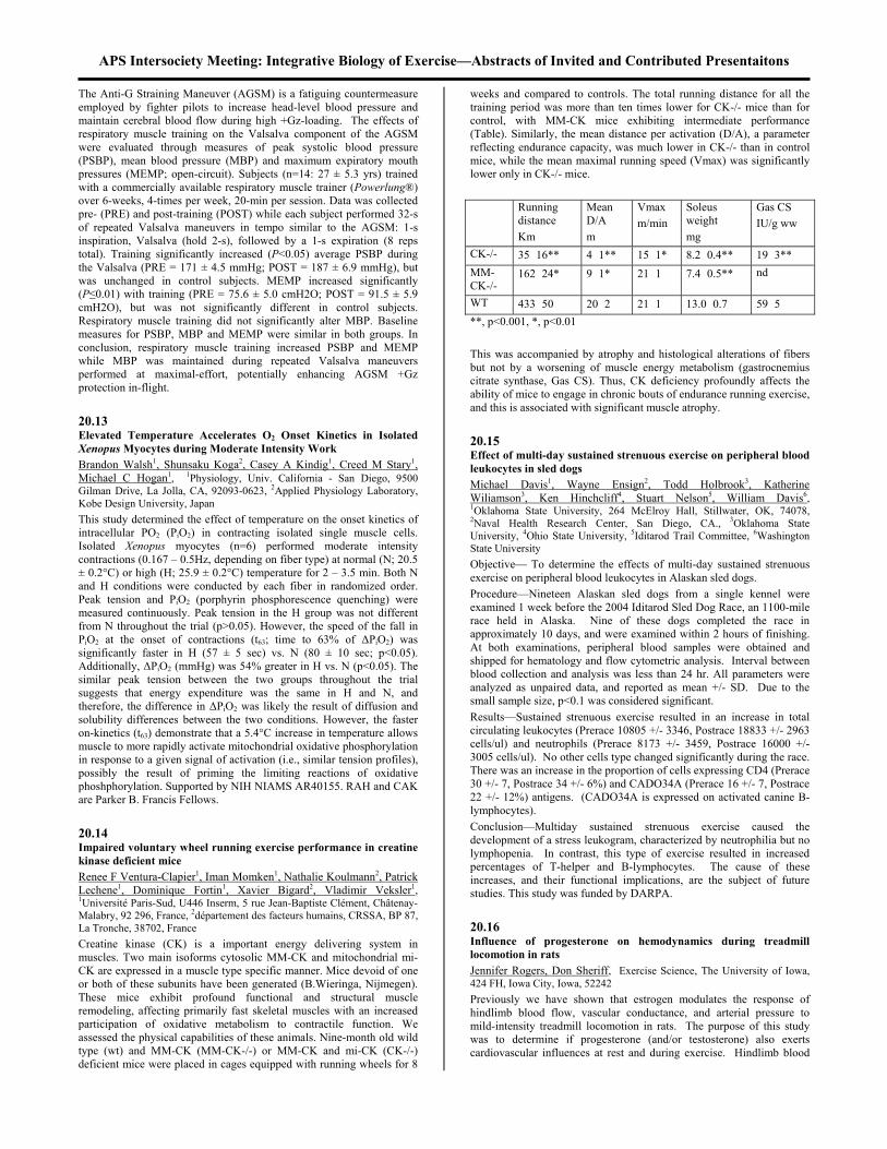
APS Intersociety Meeting: Integrative Biology of Exercise—Abstracts of Invited and Contributed Presentaitons The Anti-G Straining Maneuver (AGSM) is a fatiguing countermeasure employed by fighter pilots to increase head-level blood pressure and maintain cerebral blood flow during high +Gz-loading. The effects of respiratory muscle training on the Valsalva component of the AGSM were evaluated through measures of peak systolic blood pressure (PSBP), mean blood pressure (MBP) and maximum expiratory mouth pressures (MEMP; open-circuit). Subjects (n=14: 27 ± 5.3 yrs) trained with a commercially available respiratory muscle trainer (Powerlung®) over 6-weeks, 4-times per week, 20-min per session. Data was collected pre- (PRE) and post-training (POST) while each subject performed 32-s of repeated Valsalva maneuvers in tempo similar to the AGSM: 1-s inspiration, Valsalva (hold 2-s), followed by a 1-s expiration (8 reps total). Training significantly increased (P<0.05) average PSBP during the Valsalva (PRE = 171 ± 4.5 mmHg; POST = 187 ± 6.9 mmHg), but was unchanged in control subjects. MEMP increased significantly (P≤0.01) with training (PRE = 75.6 ± 5.0 cmH2O; POST = 91.5 ± 5.9 cmH2O), but was not significantly different in control subjects. Respiratory muscle training did not significantly alter MBP. Baseline measures for PSBP, MBP and MEMP were similar in both groups. In conclusion, respiratory muscle training increased PSBP and MEMP while MBP was maintained during repeated Valsalva maneuvers performed at maximal-effort, potentially enhancing AGSM +Gz protection in-flight. 20.13 Elevated Temperature Accelerates O2 Onset Kinetics in Isolated Xenopus Myocytes during Moderate Intensity Work Brandon Walsh1, Shunsaku Koga2, Casey A Kindig1, Creed M Stary1, Michael C Hogan1, 1Physiology, Univ. California - San Diego, 9500 Gilman Drive, La Jolla, CA, 92093-0623, 2Applied Physiology Laboratory, Kobe Design University, Japan This study determined the effect of temperature on the onset kinetics of intracellular PO2 (PiO2) in contracting isolated single muscle cells. Isolated Xenopus myocytes (n=6) performed moderate intensity contractions (0.167 – 0.5Hz, depending on fiber type) at normal (N; 20.5 ± 0.2°C) or high (H; 25.9 ± 0.2°C) temperature for 2 – 3.5 min. Both N and H conditions were conducted by each fiber in randomized order. Peak tension and PiO2 (porphyrin phosphorescence quenching) were measured continuously. Peak tension in the H group was not different from N throughout the trial (p>0.05). However, the speed of the fall in PiO2 at the onset of contractions (t63; time to 63% of ∆PiO2) was significantly faster in H (57 ± 5 sec) vs. N (80 ± 10 sec; p<0.05). Additionally, ∆PiO2 (mmHg) was 54% greater in H vs. N (p<0.05). The similar peak tension between the two groups throughout the trial suggests that energy expenditure was the same in H and N, and therefore, the difference in ∆PiO2 was likely the result of diffusion and solubility differences between the two conditions. However, the faster on-kinetics (t63) demonstrate that a 5.4°C increase in temperature allows muscle to more rapidly activate mitochondrial oxidative phosphorylation in response to a given signal of activation (i.e., similar tension profiles), possibly the result of priming the limiting reactions of oxidative phoshphorylation. Supported by NIH NIAMS AR40155. RAH and CAK are Parker B. Francis Fellows. 20.14 Impaired voluntary wheel running exercise performance in creatine kinase deficient mice Renee F Ventura-Clapier1, Iman Momken1, Nathalie Koulmann2, Patrick Lechene1, Dominique Fortin1, Xavier Bigard2, Vladimir Veksler1, 1Université Paris-Sud, U446 Inserm, 5 rue Jean-Baptiste Clément, Châtenay-Malabry, 92 296, France, 2département des facteurs humains, CRSSA, BP 87, La Tronche, 38702, France Creatine kinase (CK) is a important energy delivering system in muscles. Two main isoforms cytosolic MM-CK and mitochondrial mi-CK are expressed in a muscle type specific manner. Mice devoid of one or both of these subunits have been generated (B.Wieringa, Nijmegen). These mice exhibit profound functional and structural muscle remodeling, affecting primarily fast skeletal muscles with an increased participation of oxidative metabolism to contractile function. We assessed the physical capabilities of these animals. Nine-month old wild type (wt) and MM-CK (MM-CK-/-) or MM-CK and mi-CK (CK-/-) deficient mice were placed in cages equipped with running wheels for 8
weeks and compared to controls. The total running distance for all the training period was more than ten times lower for CK-/- mice than for control, with MM-CK mice exhibiting intermediate performance (Table). Similarly, the mean distance per activation (D/A), a parameter reflecting endurance capacity, was much lower in CK-/- than in control mice, while the mean maximal running speed (Vmax) was significantly lower only in CK-/- mice. Running
distance Km
Mean D/A m
Vmax m/min
Soleus weight mg
Gas CS IU/g ww
CK-/- 35 16** 4 1** 15 1* 8.2 0.4** 19 3** MM-CK-/-
162 24* 9 1* 21 1 7.4 0.5** nd
WT 433 50 20 2 21 1 13.0 0.7 59 5 **, p<0.001, *, p<0.01 This was accompanied by atrophy and histological alterations of fibers but not by a worsening of muscle energy metabolism (gastrocnemius citrate synthase, Gas CS). Thus, CK deficiency profoundly affects the ability of mice to engage in chronic bouts of endurance running exercise, and this is associated with significant muscle atrophy. 20.15 Effect of multi-day sustained strenuous exercise on peripheral blood leukocytes in sled dogs Michael Davis1, Wayne Ensign2, Todd Holbrook3, Katherine Wiliamson3, Ken Hinchcliff4, Stuart Nelson5, William Davis6, 1Oklahoma State University, 264 McElroy Hall, Stillwater, OK, 74078, 2Naval Health Research Center, San Diego, CA., 3Oklahoma State University, 4Ohio State University, 5Iditarod Trail Committee, 6Washington State University Objective— To determine the effects of multi-day sustained strenuous exercise on peripheral blood leukocytes in Alaskan sled dogs. Procedure—Nineteen Alaskan sled dogs from a single kennel were examined 1 week before the 2004 Iditarod Sled Dog Race, an 1100-mile race held in Alaska. Nine of these dogs completed the race in approximately 10 days, and were examined within 2 hours of finishing. At both examinations, peripheral blood samples were obtained and shipped for hematology and flow cytometric analysis. Interval between blood collection and analysis was less than 24 hr. All parameters were analyzed as unpaired data, and reported as mean +/- SD. Due to the small sample size, p<0.1 was considered significant. Results—Sustained strenuous exercise resulted in an increase in total circulating leukocytes (Prerace 10805 +/- 3346, Postrace 18833 +/- 2963 cells/ul) and neutrophils (Prerace 8173 +/- 3459, Postrace 16000 +/- 3005 cells/ul). No other cells type changed significantly during the race. There was an increase in the proportion of cells expressing CD4 (Prerace 30 +/- 7, Postrace 34 +/- 6%) and CADO34A (Prerace 16 +/- 7, Postrace 22 +/- 12%) antigens. (CADO34A is expressed on activated canine B-lymphocytes). Conclusion—Multiday sustained strenuous exercise caused the development of a stress leukogram, characterized by neutrophilia but no lymphopenia. In contrast, this type of exercise resulted in increased percentages of T-helper and B-lymphocytes. The cause of these increases, and their functional implications, are the subject of future studies. This study was funded by DARPA. 20.16 Influence of progesterone on hemodynamics during treadmill locomotion in rats Jennifer Rogers, Don Sheriff, Exercise Science, The University of Iowa, 424 FH, Iowa City, Iowa, 52242 Previously we have shown that estrogen modulates the response of hindlimb blood flow, vascular conductance, and arterial pressure to mild-intensity treadmill locomotion in rats. The purpose of this study was to determine if progesterone (and/or testosterone) also exerts cardiovascular influences at rest and during exercise. Hindlimb blood

APS Intersociety Meeting: Integrative Biology of Exercise—Abstracts of Invited and Contributed Presentaitons flow (ultrasonic transit time flow probe), arterial pressure, and vascular conductance (calculated as the ratio of blood flow and arterial pressure) were determined in male (N=4), female (N=5), ovariectomized female (N=3), and ovariectomized female with chronic progesterone replacement (n=4) Sprague-Dawley rats at rest and during treadmill locomotion at 7.5 and 15 m/min. Blood samples were obtained for immunoassay measurement of plasma progesterone and testosterone concentrations. Regression analysis revealed that female gender was associated with higher blood flow and conductance and lower blood pressure (all p <0.05). Neither progesterone nor testosterone modulated hindlimb blood flow or vascular conductance. However, progesterone was associated with a significant increase in arterial pressure (regression coefficient = +0.20 mmHg/ng/ml; p < 0.05) while testosterone was associated with a significant decrease in arterial pressure (-0.29 mmHg/ng/dl; p < 0.05). These results suggest that sex hormones influence cardiovascular function at rest and during exercise. Supported by NIH HL 46314. 20.17 EVALUATION OF HEART RATE, ELECTROMYOGRAPHIC SIGNALS AND VENTILATORY VARIABLES AS PHYSIOLOGICAL MARKERS OF EXERCISE ANAEROBIC THRESHOLD IN MEN. Vera Regina Fernandes da Silva Marães1, Adonis Aurélio Souza Kaiser2, Carlos Alberto Ribeiro Diniz3, Luis Eduardo Barreto Martins4, Lucien Oliveira3, Aparecida Maria Catai3, Rodrigo Licio Ortolan5, Lourenço Gallo Jr6, Ester Silva2, 1Fisioterapia, Universidade Federal de São Carlos, Rod. Washington Luis, Km 235, São Carlos, 13565-905, Brazil, 2UNIMEP, 3UFSCAR, 4UNICAMP, 5USP, 6HC/FMRP/USP Objective: To test the hypothesis that the anaerobic threshold (AT) of young volunteers during dynamic exercise (DE) can be determined by changes in the superficial electromyographic signals (SEMG), heart rate (HR) and carbonic gas production (VCO2). Methods and Results: 13 healthy young men (21.4±1.8 years) were submitted to an ergospirometric test (CPX/D-Medigraphics) with power increments of 25 Watts/min (ramp) on an electromagnetic cycle ergometer until physical exhaustion, with simultaneous determination of beat-to-beat HR (bpm), of breath-to-breath ventilatory variables (VCO2 and VO2 ml/min), and of SEMG (µVolts) of the vastus lateralis muscle of the right thigh expressed as root mean square (RMS). Criteria for AT analysis: 1) change of VCO2 inclination identified by a graphic visual method (VM); 2) change in slope of the VCO2 response in the fitting of bisegmented linear regression model by the least square method (LSM); 3) maximum likelihood (ML) method for HR (MLHR), VCO2 (MLVCO2) and RMS of the SEMG (MLRMS). Statistical analysis: Friedman and Dunn tests, α= 5%. No significant differences in median AT values were identified among methods, expressed as power (Watts): VM 110; LSM 102; MLHR 95; MLVCO2 107; MLRMS 135 and as VO2 (mlO2/kg/min): VM 17.5; LSM 14.3; MLHR 15.1; MLVCO2 15.8; MLRMS 19.9 (p>0.05). Conclusion: The models tested in young and healthy individuals for the variables of physiological systems studied were promising as markers of AT during DE. Ethics Committee 86/2000. Support: FAPESP, Capes and CNPq. 20.18 Adipose tissue eliminates plasma ammonia after sprint exercise Mona Elisabeth Esbjornsson1, , Barbara Norman2, Jens Bülov3, Jacek Nowak4, Eva Jansson4, 1Clinical Physiology, Karolinska Institutet, C1-88 Karolinska Universitetssjukhus, Huddinge, Stockholm, 141 86, Sweden, 2Karolinska Institutet, Sweden, 3Bispebjerg Hospital, Denmark, 4Clinical Physiology, Karolinska Institutet, Sweden The AIM of the study was to evaluate the significance of adipose tissue as an eliminator of ammonia in blood after sprint exercise and to assess possible gender differences in this respect, based on findings in a previous study where women showed a lower accumulation of plasma ammonia than men following sprint exercise. METHODS: Seven women and six men, all young and physically active performed three consecutive 30-s cycle sprints (Wingate-test) with 20 min rest between the sprints. Blood samples were obtained repeatedly during the experimental procedure from the brachial artery (A) and the
abdominal vein (V), and analyzed for plasma ammonia by a flow injection technique. RESULTS: A general uptake of plasma ammonia was observed in the abdominal adipose tissue in both genders at all time points (p<0.05 - 0.01; Fig). When adjusting for the concentration of ammonia in the artery blood (fractional extraction; [A]-[V]/[A]) a general gender difference appeared: women showed significantly higher fractional extraction compared to men at all time points (no Fig).
.
025
5075
100125150175200225250
Women
Men
rest 9
Sprint I Sprint II Sprint III
49382918minutes
abdominal veinµmol
L-1
CONCLUSION: Adipose tissue appears to play a role in eliminating exercise induced ammonia from blood irrespective of gender. The greater fractional extraction in women may indicate that they have a greater capacity to eliminate plasma ammonia from blood to adipose tissue than men. The findings support a role of adipose tissue, in the metabolic recovery from high concentration of ammonia in blood after high intensity exercise. 20.19 Influence of Hypoxia at Rest and During Exercise on Pulmonary Capillary Blood Volume and Alveolar-Capillary Conductance Eric M. Snyder1, Kenneth C. Beck, PhD2, Bruce D. Johnson, PhD1, 1Division of Cardiovascular Diseases, Mayo Clinic and Foundation, 200 SW First Street, Rochester, MN, 55905, 2Guidant Corporation, St. Paul, MN, 55112 Hypoxia causes a rise in pulmonary vascular pressures, alters pulmonary capillary recruitment and may alter lung fluid balance. Exercise causes further increases in pulmonary pressures and challenges the ability of the lungs to regulate fluid. To examine the influence of hypoxia and hypoxic exercise on pulmonary capillary recruitment and lung fluid balance, we studied 7 healthy adults (age=30±7yrs, peak VO2=42±8 ml/kg/min) after a 16 hr exposure to 12% inspired oxygen (FIO2) and again 10-15 min after exercise to exhaustion on a cycle ergometer (55% peak work, 144 43w 15 2min). Pulmonary capillary blood volume (Vc) and alveolar-capillary conductance (Dm) were determined by measuring the diffusing capacity of the lungs for carbon monoxide and nitric oxide simultaneously (DLCO, DLNO). Cardiac output (Q) was determined using an acetylene uptake method.
21% FIO2 12% FIO2 Post Exer. 12% FIO2
O2 saturation (%) 99±1 84±2* 81±4* DLCO (ml/mmHg/min)
28±5 31±7* 33±8*
Vc (ml) 65±19 111±49* 108±43* Dm (ml/mmHg/min)
44±13 45±15 49±19
Q (L/min) 6.0±1.0 6.0±2.0 5.4±2.0 Vc/Q 10.8±2.3* 18.5±5.0* 20±6.2* Dm/Q 7.3±2.0 7.5±4.1 9.1±3.2 VA (L) 3.6±1.0 3.4±0.9 3.7±1.3
*air vs. hypoxic conditions, p<0.05. There were increases in Vc with minimal changes in Dm and Q post hypoxic exposure and post hypoxic exercise. Vc normalized for Q increased post hypoxia but did not increase further post exercise. Dm relative to Q remained constant post hypoxic exposure and rose slightly post exercise (p>0.05). The increase in Vc without a concomitant rise in

APS Intersociety Meeting: Integrative Biology of Exercise—Abstracts of Invited and Contributed Presentaitons Dm suggests changes at the alveolar-capillary membrane possibly related to changes in lung fluid balance. HL71478, AHA 0410073Z 20.20 Ventilation and Oxygen Consumption during Hypoxic Incremental Cycle Exercise Ben Sporer1, Michael Koehle2, Alastair Hodges2, Kirstin Lane2, Don McKenzie2, 1UBC, 3055 Wesbrook Mall, Vancouver, BC, V6T 1Z3, Canada, 2UBC The purpose of this study was to examine the effects of hypoxia on exercise ventilation and oxygen consumption at submaximal workloads during an incremental cycle exercise test. Subjects were healthy, active males (n=9), and each completed 4 incremental cycle exercise tests while breathing a gas mixture of either 21%, 18%, 15%, or 12% FiO2 balance N2. A 30 watt/minute ramped protocol to exhaustion was used. Throughout each test, standard metabolic data were collected along with SaO2, which was measured via an ear pulse oximeter. Subjects were stopped if arterial oxygen saturation fell below 75%. Both VO2 and VE were recorded at 60 watt intervals up to 300 watts and then compared across hypoxic conditions. A repeated measures ANOVA was used to determine statistical significance across treatments for ventilation and oxygen consumption responses. Post-hoc analyses were performed using Tukey’s honest test for significance (a=0.05). No subjects were able to complete the VO2max test during the 12% trial without desaturation below 75% therefore this data was excluded from analysis. Analysis revealed a significant effect of hypoxia on VE (p=0.004) at a given wattage but not on VO2 (p=0.886). This effect on VE was seen at 180, 240, and 300 watts (p=0.039, p=0.005, and p=0.000 respectively). VE at each of these loads was significantly greater during the 15% trial than both the 18% and 21% trials. There were no differences found in VE between 18% and 21% trials at any workload. These findings indicate that although hypoxia has no apparent effects on oxygen consumption at a specific workload, exercise ventilation is increased. 20.21 Do Obstetricians Recommend Exercise to Pregnant Patients? Kelly M Munhall1, Russell L Parmer1, Diana M Herman, M.D. 2, Pauline L Entin1, 1Exercise Science and Athletic Training, Northern Arizona University, Box 15092, Flagstaff, AZ, 86011, 2Private Consultant, Flagstaff, AZ, 86001 For pregnant women, exercise offers numerous benefits with little risk. Both the American College of Obstetrics and Gynecology (ACOG) and the Society of Obstetricians and Gynaecologists of Canada (SOGC) recommend aerobic exercise for all pregnant women without medical or obstetric complications, and the SOGC also recommends resistance exercise. Our purpose was to investigate the extent to which obstetricians (Obs) recommend exercise to their pregnant patients. Surveys were sent to 300 Obs in 33 American cities (populations 6,000 - 300,000). Currently, n=78. Half (54%) of respondents reported discussing exercise with 81-100% of their patients. Using a 7-point Likert-type scale (1=never, 7=always), Obs reported recommending aerobic exercise (mean = 5.7) more often than resistance exercise (mean = 3.8). When asked how often they advise sedentary gravidae to begin an exercise program, the mean was 4.4 on the same scale. Although ACOG guidelines do not impose a maximum heart rate, 69% of Obs chose 5 or higher when asked how often they recommend a maximum heart rate such as 140 or 150 bpm. Of the 65% of Obs who make a specific recommendation for aerobic exercise duration, nearly all (49/51) recommend >16 min. Respondents perceive a need for more research on outcomes of exercise during pregnancy (mean = 5.8 out of 7). This perceived uncertainty may account for the hesitancy of Obs to recommend resistance exercise or to advise sedentary gravidae to start exercise.
20.22 Reasons for Resuming Running After Injury in Male and Female Runners Pauline L. Entin1, Elliot E Entin2, Eileen Entin2, 1Exercise Science and Athletic Training, Northern Arizona University, Box 15092, Flagstaff, AZ, 86011, 2Aptima Inc., Woburn, MA, 01801 Long term adherence to exercise promotes health and fitness. However, surveys show that 50% - 70% of runners will, at some point, sustain an injury that necessitates a hiatus from running. We wanted to learn what motivates runners who have sustained an injury to start again. Using an internet-based survey, we found that 68% of 569 runners had suffered an injury that stopped them from running for >7 days, and of those, 67% indicated that the injury occurred while running. The injury rate was higher for men than women (76% vs. 63%). We asked runners to rate on a 7-point Likert scale the importance of several factors in motivating them to resume running after such an injury. Maintenance of health and fitness was the highest rated reason to resume running, interestingly women rated this slightly higher than did men (means = 6.4 & 6.1, respectively; p< .02). Women also rated weight control significantly higher than did men (means = 5.7 & 4.5, respectively; p<.001), while men rated the desire for challenge and competition higher than did women (means = 4.9 & 4.4, respectively; p<.02). Although older (>40 yrs) runners and those running >6 yrs were significantly more likely to have been injured, neither age nor length of time running had an impact on the reasons that motivated people to resume running. These data indicate that individuals who resume running after an injury-induced hiatus are most motivated by goals of health and fitness, and for women, weight control. 20.23 THE EFFECTS OF SHORT-TERM EXERCISE, IN NEGATIVE OR ZERO ENERGY BALANCE, ON CARDIOVASCULAR DISEASE RISK FACTORS Elizabeth Mitchell, Steve Black, Barry Braun, Exercise Science, University of Massachusetts, Amherst, 110 Totman, Amherst, MA, 01003 This study was designed to explore the effects of altering energy balance in overweight subjects performing 1 week of aerobic exercise on cardiovascular disease (CVD) risk factors. METHODS: Previously sedentary, overweight/obese subjects aged 30-60 were randomly placed in 1 of 2 groups: a group in zero energy balance in which the energy expended during exercise was replaced (ZERO, n=8) or a group in negative energy balance in which the energy was not replaced (NEG, n=8). The groups were similar in age, BMI, body fat%, trunk fat% and VO2max. Training consisted of 6 consecutive days of treadmill walking at 60-65% VO2 max designed to expend 500 kcals (duration=62±6.5 min/d). RESULTS: The daily exercise increased energy expenditure from pre-training in both groups (NEG=+469±45; ZERO= +469±45). Mean weight loss occurred in NEG (-0.7 kg, p=0.005) but not in ZERO (0.03 kg, p=0.65). Triglycerides declined 16% (NS) in NEG (1.4±0.3 mM pre, 1.1±0.2 mM post, p=0.17) and 5.3% (NS) in ZERO (1.5±0.4 mM pre; 1.4±0.3 post, p=0.58). Total cholesterol declined modestly in NEG (5.3±0.2 mM pre, 5.0±0.2 post; p=0.07) and ZERO (4.6±0.2 mM pre, 4.5±0.3mM post; p=0.49). There was no change in HDL in NEG (1.3±0.1 mM pre, 1.4±0.1 post; p=0.69) while HDL decreased in ZERO (1.3±0.1 mM pre, 1.2±0.1 post; p=0.03). There was a non-significant 18.5% decrease in CRP concentrations in NEG (3.8±0.6 mg/L pre and 3.1±0.6 post; p=0.31) with no change in ZERO (4.9±1.7 mg/L pre; 5.0±1.6 post, p=0.80). Adiponectin changed slightly in NEG (9.9±1.4 µg/ml pre, 10.5±0.6 post; p=0.13) while there was no change in ZERO (6.5±1.4 µg/ml pre; 6.5±1.3 post, p=0.94). CONCLUSIONS: The data trends are striking despite a lack of statistical significance: only the exercise group in negative energy balance exhibited positive trends in traditional and novel CVD risk factors. Supported by Glass Family Trust.

APS Intersociety Meeting: Integrative Biology of Exercise—Abstracts of Invited and Contributed Presentaitons 21.1 A priming mechanism corrects the slowing of O2 uptake kinetics by leg tourniquets Kavita Phatak1, Douglas Knight2, Yuqun Luo3, 1Northwestern Medical School, 215 East Chicago Ave. #1713, Chicago, IL, 60611, 2Pediatrics, Columbus Children's Hospital, 3Department of Epidemiology and Biostatistics, Case Western University Background: Square wave exercise stimulates biphasic exponential growth of the body's rate of O2 uptake (VO2) at moderate test loads. The time-constant (τ) for the 2nd phase is increased in severe peripheral arterial disease and might be increased in an experimental model of acutely obstructed leg blood flow. Hypothesis: Acutely obstructed leg blood flow increases the τ of phase 2 VO2 unless corrected by a priming mechanism. Methods: 12 athletes (18±1y) performed 4 trials (A,B,C,D) of square-wave exercise in random order. The square wave was a 5-min base load (25W) and 5-min test load (116±29W). Subjects wore pneumatic cuffs around both thighs. Cuffs were not inflated in A. Cuffs were inflated to 78±7 Torr during the base load of B, test load of C, and both loads of D. The model for phase 2 VO2 on-kinetics in the test load was: VO2 = B + G[1- e -(t-δ)/τ ] where t = 30-120s and δ = 30s. Trial A’s τ was compared to the τ’s of trials B-D by z scores considered significant (5% level) at |z| >1.96. Results: The τ’s were 24±3s (A), 25±2s (B, z -0.3), 39±5s (C, z -2.4), and 28±3s (D, z -0.8). Conclusions: Phase 2’s τ was significantly increased in trial C due to the acute reduction of O2 delivery. Muscle ischemia during the base load stimulated a priming mechanism that corrected the phase 2 τ in trial D. {This project was approved by Columbus Children’s Hospital Institutional Review Board and funded by Columbus Children’s Research Institute} 21.2 NORMAL MITOCHONDRIAL CREATINE KINASE IN HUMAN SKELETAL MUSCLE CREATINE DEPLETION (GA) Katja Elina Peltola1, Mark Tarnopolsky2, Hannu Kalimo3, Olli Simell4, Olli J Heinonen5, 1Departments of Physiology and Pediatrics, University of Turku, Paavo Nurmi Centre, Kiinamyllynkatu 10, Turku, Finland, 2Departments of Pediatrics and Medicine, McMaster University, Canada, 3Dept of Patholology, Universities of Turku, Helsinki and Uppsala, 4Dept of Pediatrics, University of Turku, 5Paavo Nurmi Centre, University of Turku, Finland The creatine kinase (CK) system buffers cellular high-energy phosphates during periods of high energy needs. Mitochondrial creatine kinase (mtCK) is located in the mitochondrial intermembrane space and recent reports have addressed the role of mtCK in the functional coupling of mitochondrial oxidation and phosphorylation. The regulation of mtCK with regard to cellular creatine (Cr) content is not well understood. Studies on animal models of Cr depletion have shown accumulation of intramitochondrial inclusions enriched for mtCK, but mtCK is not reported in human Cr depletion. Gyrate atrophy (GA) is a rare inborn error of metabolism featuring Cr depletion in muscle and brain due to inhibition of Cr synthesis in the kidney. Thus, GA constitutes a model for Cr human depletion. We investigated mtCK adaptation to human muscle creatine depletion. Muscle biopsy samples of 4 male GA patients (age 19-49 years) with biochemically verified 50% depletion of Cr, phosphocreatine (PCr) and ATP were analysed by Western blot with antibodies against human sarcomeric mtCK. Samples from healthy age- and physical activity matched men served as controls. Despite a marked decrease in Cr and PCr concentrations, sarcomeric mtCK content was similar in GA patients and controls. Thus, in conclusion, our data indicates that modest decreases in cellular Cr, PCr and ATP concentrations per se do not affect mtCK protein content in human skeletal muscle.
21.3 21.0 Muscle Adaptation I Hypoxia enhances the alterations induced by high-intensity interval
training in rat diaphragm Yuji Ogura1, Hisashi Naito1, Jin Uchimaru1, Takao Sugiura2, Shizuo Katamoto1, Junichiro Aoki1, 1Juntendo University, 1-1 Hiragagakuendai, Inba-mura, Inba-gun, 270-1695, Japan, 2Yamaguchi University, Japan The purposes of the present study were to examine 1) whether high-intensity interval training (HIT) could alter the myosin heavy chain (MyHC) composition and bioenergetic properties of rat diaphragm, 2) whether hypoxia could enhance the effects of HIT on rat diaphragm. The present study was approved by the Juntendo University Animal Care and Use Committee in conformance with APS guideline. Male Wistar rats (N=21) were randomly divided into three groups, control: CON (n=7), normoxic training: NT (n=7), hypoxic training: HT (n=7). HIT training (1 min/sprint, 6-10 repetitions/day and 5-6 days/week for 9 weeks) was conducted on an animal treadmill under 20.9%O2 or 14.5%O2. After the training, MyHC composition was analyzed by sodium dodecyl sulfate polyacrylamide gel electrophoresis. Citrate synthase (CS) activity was also measured spectrophotometrically. In MyHC composition, type IIa isoform was increased (CON: 20.5% < NT: 24.6% < HT 30.1%, P < 0.05) and type IId isoform was decreased (CON: 51.7% > NT: 47.2% > HT 42.1%, P < 0.05) by HIT. CS activities were increased by HIT, and the activity in HT was significantly higher compared with NT (P < 0.05). Therefore, we concluded that HIT could induce the increasing of oxidative capacity and the fast-to-slow shift of MyHC composition in diaphragm, which would contribute to improve the diaphragmatic endurance ability. Moreover hypoxia could enhance the improvements. 21.4 Effect of endurance exercise in soleus of diabetic rats with peripheral neuropathy LeAnn M. Snow1, Otto Sanchez2, Robert Serfass2, Linda McLoon2, LaDora Thompson2, 1University of Minnesota, 420 Delaware St SE, MMC 388, Minneapolis, MN, 55455, 2University of Minnesota This study evaluated the effect of endurance exercise on myosin heavy chain (MHC) expression in soleus (SOL) muscle of diabetic rats with peripheral neuropathy. Methods: Sprague Dawley rats were randomly divided into four groups: control sedentary (CS), diabetic sedentary (DS), control exercise (CE), and diabetic exercise (DE). The exercised animals did treadmill running five times per week. After 12 weeks, electrophysiologic testing documented peripheral neuropathy in the diabetics. SOL muscles were excised and quick-frozen. Cross sections were immunohistochemically stained for slow, fast, developmental and neonatal MHCs. Fiber type composition and fiber cross-sectional areas were determined. Results: The diabetic groups showed a significantly greater percentage of fast MHC than did the control groups, regardless of exercise status (DS-22.6%, DE-25.2%, CS-13.5%, CE-13.1%). The exercised animals showed greater expression of developmental MHC than did the sedentary animals (DS-1.6%, DE-3.8%, CS-0.8%, CE-2.0%). No cross-sectional area differences existed between the groups. Conclusion: The altered fast MHC expression in the diabetics is consistent with denervation. Chronic endurance training does not alter mature MHC expression in the diabetic soleus muscle. The exercise-associated increase in developmental MHC expression may reflect increased fiber regeneration in the soleus after damage from increased muscle stretch during uphill treadmill running. 21.5 Does Intermittent Normobaric Hypoxia Improve Anaerobic Performances in Highly Trained Athletes? Fabien A Basset1, Denis R Joanisse2, Frédéric Boivin2, François Billaut3, Jean Doré2, Josée St-Onge2, Marcel R Boulay2, 1School of Human Kinetics and Recreation, Memorial University of Newfoundland, Elizabeth Avenue, St.John's, Newfoundland, A1C 5S7, Canada, 2Médecine Sociale et Préventive, Laval University, PEPS, Cité Universitaire, Québec, Québec, G1K 7P4, Canada, 3Sciences et Techniques des Activités Physiques et Sportives, Université Toulon-Var, Avenue de l'Université, La Garde, 83957, France The present study aimed to determine if short-term intermittent normobaric hypoxia (INH) could improve anaerobic and aerobic performances. Twelve (7 males and 5 females) highly trained athletes

APS Intersociety Meeting: Integrative Biology of Exercise—Abstracts of Invited and Contributed Presentaitons were randomly split into 2 groups which spent 8 h·night-1 for 2 consecutive nights per week over a 3 week period under either INH (O2=12%) or normobaric normoxia (NNC). Following a 3 week washout period, athletes were then exposed to the other condition. Athletes were tested for maximal oxygen uptake, Time-to-Exhaustion, Wingate and anaerobic capacity (90 sec all-out) on an electro-magnetically braked cycle ergometer before and after each period. Blood samples and vastus lateralis muscle biopsies samples were taken before and after each experimental and control period. Whereas INH resulted in a significant increase in Peak Power output during the 90 sec all-out test compared to the control condition (P<0.05), no other performance measures were changed. This was accompanied by an increase in RBC count, Ht, Hb, platelet number and EPO level (P<0.05). In skeletal muscle, a decrease in PFK activity following INH was observed (P<0.05). The present study shows that INH increased anaerobic performance in highly trained athletes. These results suggest that the increase in haematological parameters enhances O2 availability to muscle during short-term high intensity work, possibly shifting metabolic dependence away from anaerobic processes and their fatigue-contributing products. 21.6 High fatigability but normal EC-coupling in creatine-deficient skeletal muscle of GAMT-/- mice Arnold de Haan1, Hermien Kan2, Ruth van der Vliet3, Carla Offringa3, Dirk Isbrandt4, Arend Heerschap2, 1Faculty of Human Movement Sciences, Vrije University, Van der Boechorststraat 7, Amsterdam, 1086 BT, Netherlands, 2UMC St Radboud Nijmegen, 3Vrije Universiteit, 4Universiteit Hamburg Skeletal muscles from transgenic mice lacking Creatine Kinase (CK-/-) show impaired force output during repetitive contractions. Moreover, in these mice, the inactivation of the CK-PCr system induced impairment of EC-coupling. During the course of high-intensity exercise in wild type (WT) muscles, the CK-PCr system of muscles becomes less active because of low phosphocreatine (PCr) levels. The aim of the present study was to investigate whether reduced activity of the CK-PCr system by low total creatine levels has similar effects as previously found with CK-deficiency. All experiments were conducted according to accepted guidelines. Mice lacking guanidinoacetate methyltransferase (GAMT-/-) can not synthesize creatine. When fed a creatine-free diet, total creatine content in their medial gastrocnemius muscles was very low (GAMT-/-, n=6, 6.3±3.4 vs. WT, n=13, 92.9±16.4 µmol/gdw). There was a similar fast decrease in force during repetitive contractions in the muscles of anaesthetized GAMT-/- mice (n=3, reduction to ~40% within 4 contractions) as was earlier observed for CK-/- muscles, which is in contrast to WT muscles (n=7) that showed no significant initial decrease. However, force-frequency relations were not different between the GAMT-/- (n=4) and WT (n=7) muscles. Hence, in contrast to CK-/- muscles, GAMT-/- muscles showed no signs of impairment of EC-coupling, indicating differential effects of different ways of reducing the activity of the CK-PCr system. 21.7 The potential regulatory role of glutamate in nitrogen balance in trained and untrained muscle Marina Mourtzakis1, Terry Graham1, Jose Gonzalez-Alonso2, Bengt Saltin2, 1University of Guelph, Dept. Human Biology & Nutritional Sciences, Guelph, Ontario, N1G 2W1, Canada, 2Copenhagen Muscle Research Centre, Rigshospitalet, University of Copenhagen, Copenhagen, Denmark Glutamate (G) is central to several transamination reactions that are coupled with nitrogen balance and the TCA cycle. To examine the role of G in oxidative and nitrogen metabolism in skeletal muscle, 5 subjects trained (T) the quadriceps muscle of one thigh while the other thigh remained untrained (UT). After 5 weeks of one-legged knee-extensor training (70%Wmax; 1 h/d; 5 d/wk), incremental max tests were conducted on the UT and T muscles during control (C) conditions and with G infusion. Muscle biopsy, femoral arteriovenous, and blood flow measurements were analyzed for hormones and metabolites including amino acids, ammonia and TCA intermediates. Muscle TCA flux (VO2peak) was 23% greater in T muscle, but was unaltered with G
infusion. G infusion increased G uptake in UT (C=10±2 vs G=25±5umol/min, p<0.05) and T muscle (C=12±6 vs G=19±6umol/min, p<0.05), but neither training nor G infusion altered muscle concentrations or flux of most amino acids. The distribution of key nitrogen carriers (glutamine, alanine, ammonia) was altered with training and G infusion while net release was attenuated during exercise. Training reduced ammonia (33%) and enhanced alanine (30%) efflux; with G infusion, UT muscle released 88% less ammonia and 30% more alanine whereas T muscle released 18% more ammonia and 20% less alanine. Thus, G may have key implications on nitrogen balance that may differentiate with endurance training. Supported by NSERC of Canada and Danish National Research Foundation. 21.8 Disuse-induced alterations in contractile properties of the human triceps surae: a pilot study Brian C. Clark, Lori L. Ploutz-Snyder, Department of Exercise Science, Syracuse University, 820 Comstock Ave., Room 201, Syracuse, NY, 13244 The purpose of this study was to characterize adaptations in electrophysiological, morphological and force-velocity characteristics of the triceps surae after 4-weeks of unilateral lower limb suspension (ULLS). Methods: Voluntary and tetanically evoked (250-msec, 100-Hz) muscle force generation, muscle cross-sectional area (CSA), rates of evoked twitch force development (+df/dt) and relaxation (-df/dt) were determined before and immediately following ULLS. Additionally, the soleus compound muscle fiber action potential (CMAP) duration and latency were determined to assess muscle cell membrane function, and specific force (tetanic force/CSA) was calculated. Effect sizes (ES) are reported instead of p-values due to the small sample size (n=2). Results: Voluntary muscle strength decreased 17.1 ± 1.7%, whereas CSA decreased 3.8 ± 0.2% (ES=0.86 and 0.99, respectively). ULLS resulted in a 19.1 ± 3.3% loss in tetanically evoked peak force (ES=0.82), and thus specific force decreased 22.2 ± 1.0% (ES=0.85). The +df/dt and –df/dt were slowed 23.2 ± 15.5% and 37.1 ± 16.2% (ES=0.65 and 0.71, respectively). One of the most notable adaptations was observed in CMAP duration, with ULLS resulting in a > 50% elongation time of the action potential (2.25 ± 0.4 vs 3.6 ± 1.6 msec) (ES=0.51). Additionally, a 21% increase in M-wave latency was observed (5.4 ± 0.2 vs 6.5 ± 0.3 msec) (ES=0.75). Conclusion: Prolonged unweighting and disuse results in negative alterations in the excitation-contraction coupling process. Acknowledgements: Supported in part by a NASA training fellowship (NGT5-50446), Syracuse University, and the Mid-Atlantic Chapter of the American College of Sports Medicine. 21.9 Rapid and transient regulation of signal transduction by thyroid hormone in fast-and slow-twitch skeletal muscle. Isabella Irrcher, Treacey Sheehan, Anna-Maria Joseph, Peter J Adhihetty, David A Hood, Biology, York University, 4700 Keele Street, Toronto, Ontario, M3J 1P3, Canada Skeletal muscle phenotype can be altered by thyroid hormone (T3) treatment. This occurs via the binding of T3 to thyroid receptors (TRs) and the association of this complex with thyroid hormone response elements in the promoter region of target genes. However, recent work suggests that some of the rapid effects of T3 may occur via the activation of signal transduction pathways. To investigate this in skeletal muscle, we treated male rats with T3 for 2,4,6, and 12 hrs, and the time dependent phosphorylation of signaling kinases was assessed by Western blot analyses of extracts isolated from slow-twitch (ST) soleus and fast-twitch (FT) plantaris muscles. Two hrs of T3 treatment resulted in p38 MAPK and CREB activation in the ST muscle. Only p38 MAPK activation was still evident by 4 hrs. T3 treatment (2 hrs) also activated p38 MAPK in FT muscle, but this returned to control values by 4 hrs. AMPKα and ERK1/2 MAPK were activated later, by 6 hrs of T3 treatment, an effect which was no longer apparent by 12 hrs. No effect of T3 was observed on the activation of Forkhead (FKHR) transcription factor or Akt at any time. These data suggest that T3 can initiate phenotypic adaptations in muscle via the rapid and transient induction of signaling pathways, followed by the traditional T3/TR-mediated changes

APS Intersociety Meeting: Integrative Biology of Exercise—Abstracts of Invited and Contributed Presentaitons in gene expression. These early events are more pronounced in ST, compared to FT muscle. 21.10 Effects of vibration and strength training on hormonal parameters and muscle strength Thue Kvorning, Malene Bagger, Paolo Caserotti, Klavs Madsen, Institute of Sports Science and Clinical Biomechanics, University of Southern Denmark, Odense, DK., Campusvej 55, Odense, 5230, Denmark Effects of combining whole body vibrations (WBV) and strength training have to our knowledge not been studied before. Therefore, the aim of the present study was to test the hypothesis that the combination of WBV and strength training enhances the level of anabolic hormones, and for that reason increases muscle strength to a greater extent compared to strength training alone. Methods: 29 young men were divided into three groups: WBV (V), squat (S) and the combination of squat and WBV (SV). SV performed 6 sets of 8RM of squat on the vibrating platform (Galileo 2000). S and V performed the same protocol, but without WBV and resistance respectively. Pre and post 9 wk of training, hormonal responses, isometric leg strength and dynamic leg strength were measured. Results: Isometric strength increased only in S (12 %) and SV (9 %) (P<0.05). Dynamic strength increased in all groups (S (48 %), SV (48 %), V (35 %)), where S and SV showed larger increase than V (P<0.05). The concentrations of testosterone increased around 10 % due to training in S and SV, but not in V (P<0.05). Growth hormone (GH) increased in all groups (between 1500 - 4500 %), yet SV showed a more pronounced response (P<0.05). Cortisol increased only in SV, whereas V showed a decrease due to training (P<0.05). Conclusion: In spite of higher GH responses with the combination of WBV and strength training, no greater improvements were seen compared to strength training alone. WBV alone showed significant increases in GH and significant decreases in cortisol and a minor increase in dynamic strength. </B> 21.11 THE EFFECTS OF AGE ON PASSIVE AND ACTIVE STIFFNESS OF FAST- AND SLOW-TWITCH SKELETAL MUSCLE Amy L. Moran1, Gordon L Warren2, Dawn A. Lowe3, 1Dept. of Kinesiology, University of Minnesota, 100 Cooke Hall, 1900 University Ave. SE, Minneapolis, MN, 55455, 2Dept. of Physical Therapy, Georgia State University, Atlanta, GA, 30303, 3Dept. of Biochemistry, Molecular Biology, and Biophysics, University of Minnesota, Minneapolis, MN, 55455 The purpose of this study was to test the hypotheses that with aging there is an increase in passive stiffness (dP/dx) and a decrease in active dP/dx in fast- and slow-twitch skeletal muscles. Young (3-4 mo; n=13) and older (22-24 mo; n=10) C57BL/6 mice were used for this study. Peak twitch force (Pt), maximal isometric tetanic force (P0), and passive and active dP/dxof the soleus (Sol) and extensor digitorum longus (EDL) muscles were measured in vitro at 25°C. No differences were found between Sol muscles of young and older mice for muscle mass (7.3 ± 0.3 vs 7.3 ± 0.4 (mean ± SE) mg; p=0.98), passive dP/dx (11.6 ± 0.5 vs 11.7 ± 0.4 N/m; p= 0.85), Pt (22.1 ± 1.6 vs 22.7 ± 1.8 mN; p= 0.83), P0 (138 ± 7 vs. 134 ±11 mN; p= 0.80) or active dP/dx (275 ± 21 vs 280 ± 23 N/m; p= 0.86), or for EDL muscles; muscle mass (7.9 ±0.3 vs 8.2 ± 0.3 mg; p= 0.66), passive dP/dx (11.1 ± 0.4 vs 12.8 ± 0.8 N/m; p=0.053), Pt (69.0 ± 7.3 vs 70.4 ± 5.4 mN; p = 0.88), P0 (316 ±17 vs 281 ± 27 mN; p = 0.26), or active dP/dx (490 ± 32 vs 504 ± 24 N/m; p= 0.74). We will continue testing these hypotheses on muscles from senescent mice (27-28 mo) when the animals become available from NIA. 21.12 Contractile decline and metabolic shift in aging rat laryngeal muscles Francisco H. Andrade1, Denise A. Hatala2, Colleen A. McMullen3, 1Physiology, University of Kentucky, 800 Rose St., Lexington, KY, 40536, 2Ophthalmology, Case Western Reserve University, Cleveland, OH, 44106, 3Physiology, University of Kentucky, Lexington, KY, 40536 The larynx and its intrinsic musculature serve critical roles for ventilation and airway protective reflexes, swallowing and phonation.
Due to the functional demands imposed on them, the intrinsic laryngeal muscles have a unique phenotype: very small, mostly fast, fibers with high mitochondrial content. How age affects their function is largely unknown. In this project, we tested the hypothesis that two laryngeal muscles (thyroarytenoid, TA, and posterior cricoarytenoid, PCA) would become progressively weaker and slower with age. Intact TA and PCA muscles from Fisher 344-Brown Norway F1 hybrid rats (6 and 30 months of age) were used for histochemistry and in vitro contractile function. TA and PCA from 30-mo old rats developed significantly lower maximal tetanic forces, and the force-frequency curves were shifted to the left. Velocity of unloaded shortening was decreased at 30 months in PCA. Histochemical studies showed a small increase in fibers with low myosin ATPase activity in PCA and TA at 30 months, and a qualitative decrease in selected markers of mitochondrial content. In addition, TA from 30-mo old rats had a notable increase in the number of PAS-positive fibers, an index of glycogen content. We conclude that rat intrinsic laryngeal muscles become weaker and slower with age. These functional changes were not due to alterations in myosin ATPase activity, but a switch in the relative content of myosin isoforms remains a possibility. Finally, the observed alterations in mitochondrial and glycogen content indicate a shift in the metabolic characteristics of these muscles with age. Supported by NIDCD 21.13 The effect of endurance training on the amino acid (AA) profile in human skeletal muscle at rest and during exercise. Krista R. Howarth1, George J.F. Heigenhauser2, Paul J. LeBlanc2, Martin J. Gibala3, 1Kinesiology, McMaster University, 1280 Main St. W., Hamilton, Ontario, L8S 4K1, Canada, 2Medicine, McMaster University, 3Kinesiology, McMaster University Little is known regarding the skeletal muscle AA response to any form of exercise training. PURPOSE: For the first time in humans, to describe the effect of endurance training (TR) on the muscle AA profile at rest and during an acute bout of exercise. METHODS: Eight active men (22±1 y; VO2peak = 3.9±0.1 L·min-1) cycled at ~80% of their pre-TR VO2peak before and after 7 wks of TR (1 hr·d-1, 5 d·wk-1). Biopsies (v.lateralis) were obtained during both trials at rest, after 5 min of exercise and at the point of exhaustion during the pre-TR trial (42±6 min). RESULTS: The effect of TR was evidenced by an increased cycle endurance capacity (91±6 min), VO2peak and citrate synthase activity (all P<0.05). HPLC revealed (all values mmol·kg-1 dw): Muscle glutamine (Gln) and taurine (Tau) did not change during exercise vs rest in either trial, however there was a TR-induced decrease in Gln (Post-rest: 50±3 vs Pre-rest: 57±3) and increase in Tau (Post-rest: 23±2 vs Pre-rest: 20±1) (main effect, P<0.05). Glutamate (Glu) and Aspartate (Asp) were also higher post-TR (main effect, P<0.05). Except for Glu which was lower, most AAs including Alanine (Ala), Asp and the branched-chain AAs were higher during exercise vs rest in both trials (main effect, P<0.05). The acute exercise-induced decrease in Glu (Post: 2.3±0.4 vs Pre: 4.3±0.3) and increase in Ala (Post: 2.7±0.2 vs Pre: 4.2±0.3) were attenuated post-TR (P<0.05), but there was no effect of TR on the exercise response of any other AA. CONCLUSION: Aerobic TR altered the concentrations and/or exercise-induced response of several metabolically-active AAs in human skeletal muscle. Support: NSERC, Canada 21.14 The force of contraction is not responsible for mitogen activated protein kinase phosphorylation in mouse fast-twitch skeletal muscle during exercise Robert W. Wiseman1, John N. Dentel2, Samuel G. Blanchard2, David P. Ankrapp2, Laura R. McCabe2, 1Physiology and Radiology, Michigan State University, 2201 BPS Bldg., East Lansing, MI, 48824, 2Physiology, Michigan State University The mitogen-activated protein kinase (MAPK) pathways, in particular p38 MAPK, are phosphorylated in response to contractile activity. The physiologic mechanism for this phenomenon is unknown. We tested the hypothesis that force produced during contraction is responsible for p38 MAPK phosphorylation in fast-twitch skeletal muscle. Extensor digitorum longus (EDL) muscles were isolated from adult male Swiss Webster mice and stimulated at 10 Hz continuously for a period of 15

APS Intersociety Meeting: Integrative Biology of Exercise—Abstracts of Invited and Contributed Presentaitons min, removed and frozen for later analysis. Control muscles were fixed at resting length and incubated in Ringer's but not stimulated. There was 2.5-fold increase in phosphorylation of p38 MAPK relative to resting contralateral muscles after 10 Hz stimulation. Inhibition of force production by 30 min pretreatment with N-benzyl-p-toluene sulphonamide (BTS) decreased force in both a time and concentration dependent manner where 25, 75 and 150 uM BTS caused a 78+/-4, 95+/-0.2 and 99+/-0.2 % inhibition respectively (n=3). Western blot analysis of BTS treated muscles showed that the level of p38 MAPK phosphorylation was the same in stimulated EDL in the presence of 75 uM BTS as with electrical stimulation alone. Since phosphorylation of p38 MAPK was the same in the absence or presence of force in stimulated muscles, mechanical signaling can not be responsible for activation of p38 MAPK and thus other events associated with contraction must be responsible. Supported by NSBRI MA 00210 and MSU IRPG 41006. 21.15 Low intensity exercise training reduces markers of oxidative stress in mdx mice Jan J Kaczor, Eric T Payne, Julie E Hall, Mark A Tarnopolsky, Pediatrics and Medicine, McMaster University, 1200 Main Street West, Hamilton, Ontario, L8N 3Z5, Canada Duchenne muscular dystrophy (DMD) is an x-linked degenerative disease resulting in progressive muscle wasting and weakness. The mdx mouse is an animal model for DMD and lacks the dystrophin protein. Studies of both human DMD and murine mdx muscle disease have found that reactive oxygen species (ROS) may contribute to the pathogenesis of this disorder. It has been reported that moderate intensity exercise induced skeletal muscle damage in mdx mice. We hypothesized that low intensity exercise training would decrease markers of oxidative stress in skeletal muscle of mdx mice. The mdx and control wild-type (WT) mice were randomly divided into two groups: one group performed low intensity exercise training (LIT), (treadmill running, 9 wk, 2x/wk, 30 min/d @ 9 m/min), while the other was sedentary (SED). The level of malondialdehyde (MDA) was significantly higher in white gastrocnemius muscle of SED mdx mice (66.2 ± 6.6) as compared to SED WT, LIT mdx and WT mice (41.1 ± 11.4, 45.9 ± 7.8 and 52.2 ± 5.1, respectively) nmol·g ww-1 (p<0.0004; p<0.0005 and p<0.02, respectively). The level of protein carbonyls was higher in SED mdx mice (1.6 ± 0.2) as compared to SED WT, LIT mdx and WT mice (1.0 ± 0.3, 0.8 ± 0.3 and 1.0 ± 0.2, respectively) nmol·mg protein-1 (p<0.04; p<0.001 and p<0.02, respectively). We conclude that low intensity exercise training decreases markers of lipid and protein peroxidation in the white gastrocnemius muscle of mdx but not wild type mice. (Supported by US MDA). 21.16 Microgravity, Exercise Countermeasures and Human Single Muscle Fiber Function Robert H Fitts1, Janell G Romatowski1, Wau-May Lim1, Phil Gallagher2, Scott Trappe2, David Costill2, Danny A Riley3, 1Biological Sciences, Marquette University, 530 N. 15th St., Milwaukee, WI, 53233, 2Human Performance Lab, Ball State University, Muncie, IN, 3Cell Biology, Neurobiology & Anatomy, Medical College of Wisconsin, Milwaukee, WI Muscle biopsies were obtained from the soleus and gastrocnemius muscles of 5 crewmembers before and immediately following a 6-mo stay on the International Space Station. Chemically permeabilized fibers were prepared from both biopsies, and a total of 458 (pre-flight) and 453 (post-flight) single fiber segments were mounted between a force transducer and position motor, studied functionally, and fiber typed by their myosin isozyme profile on SDS polyacrylamide gels. Microgravity induced the greatest decline in diameter (18-22%) and loss of peak force and power in soleus fibers (both slow type I and fast type IIa), while the gastrocnemius type I fibers showed less atrophy (12%). The functionally most significant change was peak power, which was 47 and 57 % less in post-flight soleus type I and IIa fibers compared to the pre-flight fibers. For soleus type I fibers, peak power averaged 17.19 0.80 and 9.09 0.35 N·FL·s-1 in the pre- and post-flight samples, respectively. Both velocity and force at peak power were significantly lower. Unlike 17-d spaceflight where the maximal shortening velocity (V0) of the soleus
type I fiber increased by 20%, there was no change in V0 (0.87 0.02 pre- vs 0.83 0.02 FL/s post-flight). Comparing crew who performed high (>200 min/wk) and low (<100 min/wk) treadmill exercise suggests that this modality was effective as type I fiber peak power in the high group declined by only 13% compared to 51% in the low exercise group. Supported by NASA NCC9-116 21.17 The Effect of 6-mo Microgravity on Human Skeletal Muscle Structure Danny A Riley1, James L Bain1, Phil Gallagher2, Scott Trappe2, David Costill2, Robert H Fitts3, 1Cell Biology, Neurobiology & Anatomy, Medical College of Wisconsin, 8701 Watertown Plank Rd., Milwaukee, WI, 53213, 2Human Performance Lab, Ball State Univ., Muncie, IN, 3Dept. of Biological Sciences, Marquette Univ., Milwaukee, WI This study investigated the effects of 6-mo spaceflight aboard the international Space Station on the structural characteristics of human skeletal muscle. Biopsies from 5 crewmembers were obtained from the soleus and gastrocnemius before and immediately after flight, and processed for light and electron microscopy (EM). Fiber type and cross-sectional area (CSA) were determined from serial sections stained for myosin ATPase and antibodies specific for type I , IIA, and IIX myosin. Lipid content was assessed by oil red O staining. Ultrastructure was examined at 16,750x and 201,000x. Post-flight the CSA of the slow type I and fast type IIa fibers of the soleus were 36.9 and 30.3 % less than pre-flight, while for the gastrocnemius, type I fiber atrophy was (22.5% of pre-flight) and type II fibers had less atrophy (17.1%). Spaceflight had no significant effect on the mean distribution of slow versus fast fibers in either muscle, however, some crewmembers did show a significant increase in the % of type IIa and IIx fibers. As a group, the % of hybrid (I/IIa) fibers increased from 4 to 15 %. Crewmembers with a significant fiber shift (type I to hybrid and IIa) showed the greatest fiber atrophy. Pre-flight the lipid content was higher in slow fibers, while post-flight the reverse was true. The EM analysis showed the post-flight soleus type I fiber myofibril bundles to be proportionally reduced in diameter relative to the CSA of pre-flight controls. However, the ratio of the contractile proteins actin and myosin was not significantly altered. Supported by NASA NCC9-116 21.18 Effect of Essential Amino Acid and Carboydrate Supplementation on Bed Rest-Induced Alterations in Human Single Muscle Fiber Function Janell G Romatowski1, Wai-May Lim1, James R. Peters1, Doug Paddon-Jones2, Robert R. Wolfe2, Arny A. Ferrando2, Robert H. Fitts1, 1Dept. of Biological Sciences, Marquette Univ., 530 N. 15th St., Milwaukee, WI, 53233, 2Metabolism Unit, Shriners Hospital for Children and Depts. of Surgery and Anesthesiology, Univ. of Texas Medical Branch, Galveston, TX The purpose of this study was to determine if essential amino acid and carbohydrate supplementation (EAAC) during 28 d of bedrest could prevent muscle fiber atrophy and loss of function normally associated with bedrest. The EAAC group consumed supplements containing 15g essential amino acids and 30g carbohydrate 3x daily. Muscle biopsies (~50mg) were taken from the vastus lateralis of 5 control and 5 EAAC subjects immediately before and on day 28 of bedrest. Chemically permeabilized fibers were prepared and single fiber segments mounted between a force transducer and position motor for the determination of peak force (P0) and power, maximal shortening velocity (V0), maximal rate of force development (Ktr) and the Ca2+ sensitivity (pCa-force relationship). The effects of bedrest on muscle function varied considerably between individuals. Slow type I fiber atrophy occurred in 3 of the 5 control subjects, but in only 1 of the EAAC subjects. However, for the most part, the EAAC supplementation did not protect the type I fiber type. Type I fiber V0, P0, Ktr and peak power were equally depressed by bedrest in both groups. EAAC did prevent the bedrest-induced decline in the cooperativity of activation. In fast type II fibers, EAAC prevented the decline in peak power (pre-bedrest, 69 3; post EAAC, 64 4; and post control 58 3 N·FL·s-1) an effect attributed to an increased velocity at peak power (0.35 vs 0.31 FL/s). This effect likely contributed to the reduced loss of maximal leg extension strength in the EAAC compared to the control group. Supported by NSBRI NCC9-58-207

APS Intersociety Meeting: Integrative Biology of Exercise—Abstracts of Invited and Contributed Presentaitons 21.19 Human Muscle Volume and Performance: The Effect of 6-mo of Microgravity Phil Gallagher1, Scott Trappe1, David Costill1, Danny A. Riley2, Adrian LeBlanc3, Harlan Evans4, James R. Peters5, Robert H. Fitts5, 1Human Performance Lab, Ball State University, 2000 W. University Ave., Muncie, IN, 47306, 2Dept. of Cell Biology, Neurobiology & Anatomy, Medical College of Wisconsin, Milwaukee, WI, 3Dept. of Medicine, Baylor College of Medicine, Houston, TX, 4Life Sciences, Systems and Services, Wyle Laboratories, Houston, TX, 5Dept. of Biological Sciences, Marquette Univ., Milwaukee, WI Calf muscle volume and in vivo contractile properties were determined in 5 crewmembers before and following a 6-mo stay on the International Space Station. On the 3rd and 18th day post-flight calf muscle volume averaged 12 3 and 7 2 % less than pre-flight. Considerable variation was observed between crewmembers with the % muscle volume change less in those that performed high (>200 min/wk) compared to low (<100 min/wk) amounts of treadmill exercise. At day 7 and 28 post-flight, the mean peak torque was depressed by 10 and 5 %, respectively. Peak power calculated from isokinetic force-velocity measurements was more affected by microgravity than isometric force and at recovery day 7 was 19 % less than the pre-flight value. By day 28, peak power had recovered to 94% of the pre-flight value. Crewmembers with the greatest flight-induced decline in calf muscle volume also showed the largest loss in peak power. With 30 maximal contractions (180 /s), the initial force was lower post-flight (44 3 vs 28 6 N), and decreased more such that the 30th contraction was 52 6 and 40 11 % of the initial value for the pre- and post-flight test, respectively. In summary, although evidence indicates that the inflight countermeasure exercise does attenuate muscle atrophy and strength loss, significant decrements in muscle volume and strength continues. Although not complete, significant recovery occurs within the first 30 days after flight. Supported by NASA NCC9-116 21.20 Endurance training affects the mitochondrial substrate utilization in both oxidative and glycolytic muscles. Andre-Xavier BIGARD1, Herve SANCHEZ2, Lise BARNEOUD2, Nathalie KOULMANN2, Renee VENTURA-CLAPIER3, 1Department of human factors, CRSSA, Avenue des maquis du Gresivaudan, La Tronche, 38702, France, 2CRSSA, 3INSERM U-446 The responses of mitochondrial oxidative phosphorylation to endurance training have been mainly carried out in isolated mitochondria, a technique known to alter the structure of membranes and the function of mitochondria. This limitation supports alternative methods such as the measure of oxygen consumption by mitochondria in situ, in saponin-skinned fibers. The present experiment was therefore aimed to investigate the effects of endurance training on substrate utilization in saponin-skinned fibers. Female rats were either endurance-trained on a treadmill during 8 weeks (T, n=8), or maintained sedentary (S, n=8). The substrate utilization was examined in myofibers from soleus (slow-oxidative) and plantaris muscles (fast-glycolytic). An increase in the citrate synthase activity occurred in soleus and plantaris (65%, 53%, respectively, P<0.01), evidencing the training status of T rats. As expected, an increase in the maximal respiration rate occurred in both soleus and plantaris muscles of T rats (20%, 35%, respectively, P<0.01). An increase in mitochondrial respiration was observed in soleus of T rats with octanoylcarnitine (medium-chain fatty acid) and glycerol-3-phospate as substrates (40%, 38%, respectively, P<0.01). We showed also an increase in the relative maximal use of octanoylcarnitine in plantaris muscle (31%, P<0.05), but in contrast to soleus, a slight decrease in the maximal relative use of glycerol-3-phospate was observed in T rats (20%, P<0.05). Based on a functional approach of mitochondrial respiration, results of the present study show that endurance training 1) improved fatty acid utilization in both oxidative and glycolytic muscles, and 2) minimized the tissue specificity of glycerol-3-phospate utilization.
21.21 Stress Protein Adaptations Following a Repeated Bout of Exercise Heather S Thompson1, Christos Zois2, Stylianos P Scordilis3, 1Biological Sciences, Smith College, Sabin-Reed 349, Clark Science Center, Northampton, Massachusetts, 01063, 2Smith College, 3Biological Sciences, Smith College, Sabin-Reed 339, Clark Science Center, Northampton, Massachusetts, 01063 Unfamiliar exercise may cause indirect symptoms of muscle damage, however, repetition of the identical exercise results in their attenuation. The aim of this study was the analysis of the protein expression of three stress proteins, HSC70, HSP70, and HSP25 subsequent to a second bout of exercise. Exercise-naïve male mice, 8 to 10 weeks old, performed two identical bouts of downhill running (15 min, 25 m min-1, -15°) on a motorized treadmill separated by one week. Stress protein expression levels were analyzed at various time points (n=5) post-second bout (PSB) of exercise by quantitative immunoblotting. HSC70 was significantly elevated (3-fold) at 0 hr PSB, followed by a second response (3-fold) at 24 and 48 hr PSB and finally a third response at 168 hr (2-fold). HSP25 increased (4-fold) at 1 hr PSB followed by a second response (2.5-fold) at 48 hr and a third 2 fold elevation at 672 hr PSB. HSP70 was elevated (19-fold) at 1 hr PSB followed by a 33X response at 3 hr and peaked at 6 hr (37-fold) and lastly an 18-fold increase at 72 hr. When compared to the responses following a single bout of identical exercise, the expression patterns following the second bout are earlier, more rapid and peak at higher levels. These results support our hypothesis that a second bout of identical downhill running promotes HSP adaptation to a single bout. Such adaptation may mitigate potential muscle damage. (1 and 2 are equal contributors) Funded by the Blakeslee Fund for Genetics Research to SPS 21.22 Short sprint interval training (SIT) increases pyruvate dehydrogenase activity (PDHa) during exercise in human skeletal muscle. Kirsten A Burgomaster1, George J.F. Heigenhauser2, Martin J. Gibala3, 1EMRG Kinesiology, McMaster, 1280 Main Street West, Hamilton, Ontario, L8S 4K1, Canada, 2Medicine, McMaster, 3EMRG Kinesiology, McMaster Recently we described changes in muscle carbohydrate (CHO) metabolism and endurance capacity after only 2 wks of SIT (Med Sci Sports Exerc. 36S, 2004). Part of the adaptive response to SIT may include changes in PDHa — the rate-limiting enzyme for CHO oxidation — however this has not been studied. PURPOSE: To examine potential changes in PDHa and the maximal activities of marker enzymes for oxidative metabolism after SIT. METHODS: 8 active men (21 1 yr; VO2peak = 49±2 ml/kg/min) performed 6 sessions of SIT (4-7x30-s “all out” cycle rides; 4 min recovery) over 2 wks. Before and after SIT, muscle biopsies were obtained at rest and after each stage of a challenge ride that consisted of 10 min @ 50% followed by 10 min @ 80% VO2peak. Subjects also completed a 250 kJ self-paced TT with no temporal, verbal and physiological feedback. RESULTS: PDHa was higher after SIT (main effect, P 0.05) with the greatest difference at the highest workload (Post: 3.1±0.4 vs Pre: 2.3 0.2 mmol kg-1 min-1 wet wt). Citrate synthase was 18% higher after SIT (11.8 0.4 vs 10.0 0.7 mmol kg-1 min-1, P 0.05), whereas 3-Hydroxyacyl-CoA dehydrogenase was unchanged. TT performance improved by 10.4% after SIT (Post: 15.3±1.5 vs Pre: 17.3±2.7 min; P≤0.05) and mean power output increased from 247 37 to 272 24 W (P<0.05). A control group showed no change in TT performance when tested 2 wks apart under identical conditions. CONCLUSION: 6 sessions of SIT increased TT performance and altered the activity of muscle enzymes involved in oxidative CHO metabolism. Support: NSERC Canada, ACSM Research Endowment Grant and a Gatorade Sports Science Institute Student Research Award.
APS Intersociety Meeting: Integrative Biology of Exercise—Abstracts of Invited and Contributed Presentaitons 22.1 MAXIMAL STRENGTH TRAINING IMPROVES WORK ECONOMY IN PATIENTS WITH CHRONIC OBSTRUCTIVE PULMONARY DISEASE Arnt Erik Tjønna1, Morten Andre Høydal2, Jan Helgerud3, Sigurd Steinshavn3, Jan Hoff3, 1Department of circulation and medical imaging, Faculty of medicine, Norwegian University of Science and Technology, Olav Kyrres gt 3, 4.etg. north, Trondheim, 7489, Norway, 2Faculty of medicine, The Norwegian University of Science and Technology, 3Faculty of medicine, Norwegian University of Science and Technology The purpose of this study was to evaluate whether maximal strength training could improve work economy in patients with chronic obstructive pulmonary disease (COPD). Eleven patients with moderate to severe COPD were assigned to either a strength training group (n = 6) or control group (n = 5). The strength training regime lasted for 8 weeks with 3 weekly sessions, the training consisted of 5 repetitions and 4 series with emphasis on maximal mobilization in the concentric part in a leg press device. Maximal oxygen uptake (VO2max), work economy, rate of force development (RFD) and one repetition maximum (1RM) were measured before and after the 8-week training period. Maximal strength training improved work economy by 17.4% (P = 0.042), 1RM (90°) and 1RM (70°) by 26.7% (P=0.026) and 24.2% (P = 0.027) respectively. Static peak force improved by 28% (P = 0.028) and dynamic rate of force development improved 83.3% in strength training group. No significant changes occurred in the control group. There were no significant changes in either VO2max or bodyweight (kg). The present study shows that strength training with emphasis on maximal mobilization in the concentric part improves work economy in COPD patients. The substantial improved RFD, indicate that the main training response is from neural adaptations and changes in recruitment pattern. The findings in the present study may be important for an improved daily activity, and probably quality of life in COPD patients. 22.2 PERIPHERAL MUSCLE ADAPTATION TO ONE LEG ENDURANCE TRAINING IN PATIENTS WITH CHRONIC OBSTRUCTIVE PULMONARY DISEASE Morten Andre Hoeydal1, Arnt Erik Tjønna2, Jan Hoff2, Sigurd Steinshamn2, Jan helgerud2, 1circulation and biomedical imaging, Faculty of medicine, Norwegian university of science and technology, Rosenborgsgt 23a, Trondheim, 7014, Norway, 2Faculty of medicine. Norwegian university of technology and science The present study focuses on the peripheral muscle endurance capacity in patients with chronic obstructive pulmonary disease (COPD). The aim was to reveal whether the peripheral muscles are taxed to the limit or if there exists a reserve capacity to consume oxygen when performing work with a reduced muscle mass in these patients. The influence on peripheral muscle endurance capacity was investigated following 8 weeks of one leg endurance training performed by cycling. Ten patients with diagnosis consistent with COPD participated in the study. Five patients underwent 8 weeks of one leg endurance training, while 5 patients served as control. Incremental exercise responses were performed to the limit of tolerance with both one- and two legs cycling. One leg peak oxygen consumption and peak work rate increased significantly (15.3 % and 35.4 %, respectively) in the training group compared to the control patients. Two legs maximal oxygen consumption and maximal work rate also increased significantly (15.3 % and 27.7 %, respectively). No significant changes were found at constant work loads. It is concluded that training by one leg cycling is an appropriate adopted model to increase endurance capacity in peripheral muscles and also in whole body exercise. From the study it seems to be a peripheral muscle limitation when concerning one leg cycling, and a supply limitation when attending whole body exercise.
22.3 22.0 Physical Inactivity and Chronic Disease
Caloric restriction maintains the functional viability of skeletal muscle during incremental isometric contraction in old F344BN rats David J. Baker, Daniel J. Krause, Russell T. Hepple, Faculty of Medicine and Faculty of Kinesiology, University of Calgary, 2500 University Drive NW, Calgary, Alberta, T2N 1N4, Canada Previous studies have investigated the effect of caloric restriction (CR) on the contractile responses of skeletal muscle in both the adult F344BN rat and in the context of ageing. The current study reports the effects of CR on muscle force production during a protocol of increasing contraction intensity in the oldest animals reported thus far, and has employed an in situ pump-perfused hindlimb model to match the rate of convective O2 delivery between groups. It was hypothesized that CR would improve force production in even the oldest animals and across a wide range of contractile demand when compared with ad libitum (AL) fed and age matched controls. To test this hypothesis we studied AL fed and CR 8-10 mo old (young adult) and 35 mo old (senescence for AL) F344BN rats. Force was measured from the contracting Gastrocnemius-Plantaris–Soleus muscle group. Peak tension (Pt) was higher in the 8 mo AL (34.9 ± 4.7 N) versus 35 mo AL (7.9 ± 1.5 N), whereas the difference was attenuated by CR (8 mo 27.6 ± 2.1 versus 35 mo 21.0 ± 0.7 N). Normalized tension (N g-1) was similar between 8 mo AL, 8 mo CR and 35 mo CR animals; however, it was 52 % lower in the 35 mo AL group. Although there was no difference in fatigue (% of Pt) with aging or CR, force was significantly higher across the range of contractile demand in 35 mo old CR versus 35 mo old AL animals. This shows that CR markedly attenuates the age-associated decline in skeletal muscle function across a wide range of contractile demand. 22.4 Changes in Plasma and Muscle Glutamine Concentration in Horses with Aging and Exercise Training Helio Cordeiro Manso-Filho1, Cynthia Betros2, Mary E. Gordon2, Helena Emilia Costa2, Malcolm Watford3, Kenneth Harrington McKeever2, 1Animal Sciences, Equine Science Center, Rutgers University, 84 Lipman Drive, New Brunswick, NJ, 08901, 2Animal Sciences, Equine Science Center, Rutgers University, 3Nutritional Sciences, Rutgers University Two groups of unfit Standardbred mares (adult: 9-14y,~540 kg, n=7) and old (20-25y, ~530 kg, n=5) were used to test the hypothesis that aging and training would alter plasma and muscle glutamine[GLN] and glutamate[GLU]concentrations in horses. Mares were fed to meet or exceed NRC(1989) nutrient recommendations for moderate to heavy exercise. Blood samples and muscle biopsies (gluteus) obtained before and after 8 wks of training were used for measurement of [GLN] and [GLU] using enzymatic methods. Mares were trained 5 d/wk for 50 min/d,(20 min walk and 30 min at ~70% HRmax). Data were analyzed using repeated measures ANOVA and Tukey test. Plasma [GLU] did not differ between age groups or with training (P>0.05). There were no differences (P>0.05) in muscle [GLU] due to aging. Training decreased (P<0.05) muscle [GLU] from 7,561 ± 701 nmol/g of tissue (mean ± SE)in pre-training samples to 4,491 ± 701 nmol/g post-training. Plasma [GLN] decreased (P<0.05) with training (368 ± 13 nmol/mL vs. 317 ± 14nmol/mL). There was a trend (p=0.063) towards an effect of aging. There were significant interactions between age and training for plasma [GLN]. Old mares had lower (P<0.05) post-exercise plasma [GLN] (224 ± 21 nmol/mL) when compared with pre-exercise plasma [GLN](372 ± 21 nmol/mL). Post-training, plasma [GLN] was lower (P<0.05) in the old mares compared to adult mares (225 ± 21.05 nmol/mL vs. 410 ± 18 nmol/mL). There was an effect (P<0.05) of age on muscle [GLN] (old = 6,126 ± 870 nmol/g; adult = 3,176 ± 735 nmol/g); however, there were no changes (P>0.05)due to training. It was concluded that training and aging produce changes in plasma and muscle [GLN] which may affect immune function. 22.5 Skeletal muscle abnormalities are manifested in glycolytic fibers in a mouse model of chronic heart failure Ping Li, Duke University Medical Center, 4321 Medical Park Dr., Durham, NC, 27704 Exercise intolerance, a clinical hallmark of chronic heart failure (CHF), is caused by pathophysiological changes in skeletal muscle. However,

APS Intersociety Meeting: Integrative Biology of Exercise—Abstracts of Invited and Contributed Presentaitons the abnormalities that play a causative role in exercise intolerance remain to be defined. In this study, transgenic mice with cardiac overexpression of calsequestrin (CSQ), a genetic model of CHF, were employed to investigate fiber type-specific abnormalities in skeletal muscle. Expression of cytochrome oxidase IV (COXIV), myoglobin (Mb) and peroxisome proliferator activated receptor γ co-activator-1α (PGC-1α) were measured by immunoblot analysis in three muscles of different fiber type compositions: soleus (SO, type I and IIa fibers), plantaris (PL, type IIa and IIb fibers) and white vastus lateralis muscles (WV, type IIb fibers). Compared with the wild type littermates, CSQ mice have decreased expression of COXIV, Mb and PGC-1α proteins in WV and to a lesser degree in PL, but not in SO muscle. Indirect immunofluroscence showed a slow-to-fast fiber type switching and decreased capillary density in type IIb+IId/x myofibers in PL muscle. These findings suggest that increased glycolytic fibers with manifested abnormalities play an important role in exercise intolerance in CHF. 22.6 Disease Risk Factors Emerge From Artificial Selection For Aerobic Capacity in Rats Ulrik Wisløff1, Sonia M Najjar2, Øyvind Ellingsen1, Per M Haram1, Lauren G Koch2, Steven L Britton2, 1Department of Circulation and Medical Imaging, NTNU, Olav Kyrres gt 3, Trondheim, 7489, Norway, 2Functional Genomics Laboratory, Medical College of Ohio We speculated that the widened redox potential afforded by oxygen was so important for the evolution of biocomplexity that it must also form the basis for complex diseases. As an extension of this idea we hypothesized that artificial selection for low and high aerobic capacity would yield lines that contrasted for health risks associated with complex diseases. Eleven generations of selection produced lines that differed by 347% in running capacity. The low line demonstrated risk factors including higher visceral adiposity, blood pressure, insulin, free fatty acids, and triglycerides. The high line was superior for VO2max, economy of running, heart function, adaptation to exercise, and nitric oxide-induced vascular dilation. Consistent with these divides in subordinate likely-determinant phenotypes, the low line demonstrated lower abundance in soleus skeletal muscle for six proteins associated with mitochondrial energy transduction. 22.7 Caloric restriction attenuates the age-associated decline of skeletal muscle aerobic function Russell T. Hepple, David J. Baker, Daniel J. Krause, Faculty of Kinesiology, University of Calgary, 2500 University Dr. NW, Calgary, AB, T2N 1N4, Canada Aging causes a reduction in the mass and function of skeletal muscle. Previous results show that maximal aerobic function of aged muscles (VO2max) is reduced by ~50% between young adulthood and senescence in Fischer 344 X Brown Norway F1-hybrid (F344BN) rats even when the muscles are provided with the same rate of O2 delivery. Furthermore, a portion of this decline in VO2max has been ascribed to mitochondrial dysfunction, resulting from the age-associated accumulation of mitochondrial DNA damage. On the other hand, caloric restriction (CR) slows the rate of mitochondrial DNA damage and better preserves mitochondrial function in aged muscles. Thus, we hypothesized that CR would attenuate the age-associated decline in skeletal muscle VO2max. To test this hypothesis, we studied ad libitum (AL) fed and CR 8-10 mo old (young adult) and 35 mo old (senescence for AL animals) F344BN rats using a pump-perfused hindlimb preparation to permit matching of muscle convective O2 delivery between groups during a 6 min incremental stimulation frequency contraction bout. Whereas aging was associated with a 49% lower VO2max in 35 mo old (281 ± 54 mol O2 min-1 100 g-1; mean ± SE) versus 8-10 mo old (540 ± 13 mol O2 min-1 100 g-1) AL rats, there was no difference in VO2max between 8-10 mo old (452 ± 42 mol O2 min-1 100 g-1) and 35 mo old (452 ± 48 mol O2 min-1 100 g-1) CR rats. Thus, our results show that CR helps preserve aerobic function in aged skeletal muscles.
22.8 Evidence of Type 2 diabetes and compromised cognitive function in young sedentary laboratory rats. Helaine M. Alessio1, Ann E. Hagerman2, Natalie B. Schweitzer3, Kelly Michalak2, Kelly Vonder Haar2, Stephen D. Berry4, Ronald L Wiley3, 1PHS dept, Miami U, Oak Street, Oxford, OH, 45056, 2Chemistry and Biochemistry, Miami U, High Street, Oxford, OH, 45056, 3Zoology, Miami U, High Street, Oxford, OH, 45056, 4Psychology, Miami U, High Street, Oxford, OH, 45056 In laboratory experiments animals often reside in standard cages with little access to exercise. The purpose of this study was to determine if animals housed in standard cages with no access to exercise demonstrate distinct metabolic and cognitive profiles compared with animals having access to exercise. 36 female Sprague Dawley rats were separated into three groups (n=12) having: 1) no access to exercise (SED), 2) access to twice-weekly physical activity in a large box (PA), and 3) regular access to running wheel exercise (EX). Performance on a radial-arm spatial maze was based upon total time to collect 5 reinforcements (correct responses), number of working errors (returning to the same place on the maze for reinforcements) and reference errors (going to a place on the maze that never has reinforcements). Insulin and glucose balance, blood lipids, body weight, and blood pressure were measured. Mean time to complete the maze was slower in the SED group (p<0.05). The SED group also performed worse than EX group on the spatial maze making less correct responses and committing more errors. At age 6 months blood insulin levels were highest in the SED and lowest in the EX group. Blood glucose, blood lipids, body weight, and blood pressure did not differ among the groups. In conclusion, the ability to successfully complete a spatial maze test was compromised in SED rats. In addition to performing poorly on the spatial maze, SED animals also showed evidence of Type 2 diabetes. Supported by NIH Grant 1 R15 AG 20526-01A1. 22.9 High Intensity Is More Effective at Maintaining Enhanced Insulin Action Than Low Intensity Endurance Training M. A. Janiec1, C. J. Tanner2, C. A. Slentz3, B. D. Duscha3, J. S. McCartney2, W. E. Kraus3, J. A. Houmard2, 1East Carolina University, Ward Sports Medicine Bldg., Greenville, 27858, 2East Carolina University, 3Duke University Aerobic exercise increases insulin action in overweight individuals. The optimal exercise intensity and volume for maximizing increases in insulin sensitivity is not, however, evident. The purpose of this study was to test the hypothesis that higher intensity exercise training would elicit greater positive changes in insulin sensitivity. Inactive overweight individuals were randomly assigned to one of three exercise group for six months: 1) low-volume/moderate-intensity group (LM), N= 46 [~12 miles walking/wk at 40-55% VO2 peak, 2) low-volume/high-intensity group (LH) N= 49 [~12 miles jogging/wk at 65-80% VO2 peak, and 3) high-volume/high-intensity group (HH) N= 58 [~20 miles jogging/wk at 65-80% VO2 peak. Insulin action was determined with an insulin sensitivity index (SI) from an intravenous glucose tolerance test. SI was measured before, 24hr, 96hr and 14 days post training. 24 hours after the last exercise bout, insulin action in all groups increased significantly. However, regardless of intensity and volume all groups declined to pre-exercise insulin sensitivity levels at 96hr post exercise. 14 days post training SI of the High Volume/High Intensity group returned to a significantly (P<0.001) elevated value compared with before training (+25%) suggesting that high intensity training may be the most beneficial for maintaining insulin action. 22.10 The Effects of Body-Weight-Supported-Treadmill-Training on Cardiovascular Structure and Function and Functional Walking Ability in Sub-Acute Spinal Cord Injury J. Crozier1, D. Ditor2, M. Adams2, B. Smith2, A. Campbell2, A. Hicks2, M. MacDonald2, 1Department of Kinesiology, McMaster University, 1280 Main St. W, Hamilton, Ontario, L8S 4K1, Canada, 2McMaster University Dramatic changes in cardiovascular structure and function have been observed during the acute phase following spinal cord injury (SCI). Body-weight-supported-treadmill-training (BWSTT) may reverse some

APS Intersociety Meeting: Integrative Biology of Exercise—Abstracts of Invited and Contributed Presentaitons of these changes. PURPOSE: To examine the effects of 48 sessions of twice-weekly BWSTT on cardiovascular regulation and functional treadmill walking performance in 5 individuals with SCI in the sub-acute phase (mean time post-injury:15.4 ± 5.9 weeks). METHODS: Carotid and femoral arteries were examined pre and post training using Doppler ultrasound. RESULTS: BWSTT resulted in increases in femoral artery diameter (p=0.04; pre: 0.64±0.09cm, post: 0.73±0.11cm), carotid artery diameter (p=0.06; pre: 0.61±0.07cm, post: 0.67±0.06cm), and femoral artery blood flow (p=0.05; pre: 249±86mL/min, post: 353±56mL/min) and decreases in femoral artery distensibility (p=0.080; pre: 0.004±0.003mm/mmHg, post: 0.0017±0.0007 mm/mmHg). All subjects were able to decrease the amount of body weight support from 84.7% to 44.5% and one required no body weight support at the end of the 6 month training period. Treadmill speed increased from 0.6 Km/h to 1.6 Km/h while walking duration also increased in all subjects. CONCLUSION: 48 sessions of BWSTT in 5 SCI individuals resulted in changes in cardiovascular structure and function and increased treadmill functional walking ability. Supported by NSERC Canada and Ontario Neurotrauma Foundation 22.11 Reduced NO-Mediated Flow-Induced Vasodilation Accompanies the Onset of Type 2 Diabetes and Elevated Mean Arterial Pressure in the Zucker Diabetic Fatty Rat Lisa Lesniewski, Anthony Donato, Bradley Behnke, Michael Delp, Texas A&M University, 158 Read Building, 4243 TAMU, College Station, Texas, 77843-4243 Type 2 diabetes, associated with obesity and inactivity, is often accompanied by hypertension and endothelial dysfunction. Whether this endothelial dysfunction and a resultant reduction in dilator capacity is a cause or consequence of the overt disease is unclear. PURPOSE: Examine mechanisms of flow-induced dilation during the progression of type 2 diabetes in Zucker Diabetic Fatty (ZDF) rats. METHODS: Male ZDF fatty and lean control rats at 7 (prediabetic), 13 (acute), and 20 (chronic) weeks of age were used. Blood pressure (MAP) and a fasted blood sample were collected. Flow-induced dilation was measured in isolated soleus muscle arterioles. To assess the roles of the nitric oxide (NO) and cyclooxygense (COX) pathways, the flow responses were repeated in the presence of the NO synthase inhibitor L-NAME and combined L-NAME and indomethacin (INDO), a COX inhibitor. RESULTS: Insulin was 3-fold higher in fatty rats in pre- and acute diabetes. Glucose was ~2-fold higher in fatty rats during acute and chronic diabetes. Mean arterial pressure was elevated during acute diabetes (+19 mmHg) and remained similarly higher with chronic disease. Flow-induced dilation was reduced ~55% with the onset of acute diabetes and resulted from a diminished role of NO, which was maintained with chronic disease. CONCLUSION: Endothelial dysfunction through the NO synthase signaling mechanism is manifest after the onset of diabetes, which accompanies and may contribute to hypertension in diabetes. 22.12 Changes in electrophysiological properties of tibial motoneurones in the rat following 2 weeks of hind limb suspension Bruno Cormery, Eric Beaumont, Phillip Gardiner, Dept de Kinésiologie, Université de Montréal, CEPSUM -2100 Édouard-Montpetit, Montréal, Québec, H3T 1J4, Canada In this study, we tested the hypothesis that 2 weeks of hind limb suspension would alter the passive membrane properties of rat tibial motoneurones. Female Sprague-Dawley rats were assigned to normal control and tail suspension groups. We subsequently examined, in anaesthetized (ketamine/xylazine) rats, the properties of motoneurones with axons in the tibial nerve. We impaled motoneurones using sharp glass microelectrodes, and measured the following properties : resting membrane potential, spike threshold, spike height, rheobase, input resistance, and the amplitude and time-course of the afterhyperpolarization (AHP). Multiple ANOVA revealed significant suspension and type effects without interaction between these conditions. Both slow and fast motoneurones demonstrated significant decreases in the AHP amplitudes (slow 2.8 vs. 2.1 mV; fast, 1.7 vs. 1.3 mV)) and spike heights (slow 87 vs. 74 mV; fast, 79 vs. 73 mV). The
results add further support to the notion that activity-related adaptations occur in the density, localization, and/or modulation of ionic membrane channels that control motoneurone properties. These changes might help us to understand the " deconditioning " process that occurs during space flight. Supported by grants from NSERC and Canadian Space Agency to PFG. 22.13 Physical Activity and Vascular Remodeling in Skeletal Muscle of Young and Aged Rats Brad J Behnke1, Lisa A Lesniewski1, Rhonda D Prisby1, Anthony J Donato1, Hillary M Olin2, Michael D Delp3, 1Health & Kinesiology, Texas A&M, 158 Read, College Station, TX, 77843, 2Biology, Texas A&M, 3Health & Kinesiology and Medical Physiology, Texas A&M Exercise capacity and muscular function are reduced with aging. However, key structural and functional relations within young and old skeletal muscle responsible for a diminished exercise capacity with aging remain obscure. The purpose of this investigation was to test the hypothesis that reduced levels of spontaneous physical activity in aged rats are associated with skeletal muscle vascular remodeling, i.e., arterial rarefaction. Radiotelemeters (MiniMitter Co., Bend, OR) were implanted (i.p.) in young (6 mon) and old (24 mon) Fisher 344 rats. Activity during light and dark cycles (12:12 hr) was measured at 30 sec intervals for 2 weeks. Subsequently, the gastrocnemius complex was excised and all feed arteries and 1st-order arterioles were counted, isolated, cannulated and maximally dilated for measurement of luminal diameter. Overall, young rats were ~2 times more active during dark periods and ~5 times more active during light hours than old rats. In addition, young rats had a total of 8 feed arteries whereas only 7 were present in the gastrocnemius of old rats. In conclusion, these data suggest that the decline in daily activity (most prevalent during the light period) in old rats may contribute directly to resistance artery rarefaction and, thus, altered microcirculatory blood flow patterns. Supported by NASA grant NCC2-1166. 22.14 EFFECTS OF 14 DAYS OF UNILATERAL LEG IMMOBILIZATION ON MUSCLE FUNCTION AND MORPHOLOGY IN MEN AND WOMEN Nobuo Yasuda1, Elisa I. Glover2, Stuart M. Phillips2, Mark A. Tarnopolsky3, 1Department of Kinesiology, McMaster University, 1280 Main Street West, Hamilton, Ontario, L8S 4K1, Canada, 2Department of Kinesiology, McMaster University, 3Department of Pediatrics, McMaster University PURPOSE: The aim of this study was to determine the effects of 14d of unilateral leg immobilization on muscle function and morphology in men and women. METHODS: Recreationally active males (M, N=13, BMI=24.7) and females (F, N=14, BMI=22.6) had their legs immobilized by using a standard knee brace. Isometric and isokinetic (concentric SLOW-30 /s and FAST-300 /s) knee extensor peak torques were determined for all subjects using a dynamometer for three time points (PRE, 2-Day, and 14-Day). At the same time points, magnetic resonance imaging was used to measure the cross-sectional areas of vasti (vastus lateralis, vastus intermedius, and vastus medialis) and rectus femoris muscles. RESULTS: Women showed significantly greater decreases (PRE vs. 14-Day) compared to men in both isometric (M=8.4 14.4, F=22.2 14.7%; [mean SD]) and SLOW (M=9.8 13.5, F=21.8 16.3%) peak torques (both, p<0.05), with no differences seen in the decrement in FAST peak torque (M=14.3 18.8, F=20.5 15.4%). There were no significant gender differences in the decreases (PRE vs. 14-Day) in cross-sectional area of the quadriceps femoris (vastus area: M=5.9 5.3, F=6.4 5.6%; rectus femoris: M=3.7 7.0, F=2.8 6.0%; overall: M=5.7 5.0, F=5.9 5.2%). CONCLUSION: These findings indicate that immobilization-induced loss of knee extensor muscle function may be attenuated in men compared to women due to gender-specific neuromuscular signals in spite of similar morphological change between genders.
APS Intersociety Meeting: Integrative Biology of Exercise—Abstracts of Invited and Contributed Presentaitons 22.15 Estimates of Energy Expenditure During Swimming In Humans Using Accelerometry Jeanne D Johnston, Joel M. Stager, Kinesiology, Indiana University, East 10th Street, HPER 112, Bloomington, Indiana, 47405 PURPOSE: Evaluate the efficacy of an omnidirectional accelerometer (ODA) to quantify swimming energy expenditure (SWE). METHODS: Eight men (26.5 +/-8.8 yrs) and 10 women (27.1 +/-9.8 yrs) performed three submaximal 365.76 meter (400-yard) front crawl swims. Expired gases were collected for 20 seconds at the completion of each swim and the backwards extrapolation method was utilized to determine oxygen consumption. An ODA (Mini-Mitter Corporation, Actical accelerometer) was worn on the right wrist, waist, and right leg. Multiple regression techniques were utilized to develop prediction equations for SWE (kcal·kg-1·min-1) based on accelerometer counts, swim performance traits and subject characteristics. RESULTS: Significant correlations between measured VO2 and ODA output were noted for men (leg, r = 0.82, p< 0.01; waist, r = 0.48, p< 0.05) and women (wrist, r = 0.74, p< 0.01; leg, r = 0.42, p < 0.05). Age and height were included in the prediction equation for both men and women. The best prediction equations included leg counts for the men (R2 = 0.72, SEE = .033 kcal·kg-1·min-1) and wrist counts for the women (R2= 0.62, SEE = .043 kcal·kg-1·min-1). CONCLUSION: The optimal placement of ODA differs depending on the sex of the subject. The Actical activity monitor provided reasonable estimates of SWE and may be a useful tool to monitor swimming energy expenditure. Acknowledgements: Supported, in part, by Mini-Mitter Corporation and United States Master Swimming 22.16 Response to Endurance Training and Detraining in Mitochondrial Myopathy: A Case Study Phil Wyrick1, Robert Taylor2, Andrew Schaefer2, Douglass Turnbull2, Ronald Haller1, Tanja Taivassalo1, 1NM Center, IEEM, 7232 Greenville Avenue, Dallas, TX, 75231, 2University of Newcastle upon Tyne, United Kingdom Objective: To assess the response of endurance training followed by detraining on physiological and molecular measures in a patient with severe muscle oxidative impairment due to high levels of mutation in mitochondrial DNA (mtDNA). Background: Patients with mitochondrial myopathies (MM) due to mtDNA mutations in skeletal muscle often suffer disabling exercise intolerance. Our previous studies suggest endurance training improves exercise capacity and increases rate-limiting enzymatic capacity but may also increase the percentage of mutant mtDNA. The effect of detraining on these parameters has not been studied. Methods: A 40-year female with a heteroplasmic (87% mutant, 13% wild type) large-scale deletion of mtDNA underwent 14 weeks of cycle exercise training followed by 14 weeks of detraining. Peak work, oxygen uptake (VO2), submaximal heart rate and blood lactate, and mutation load were determined at baseline, after training and after subsequent detraining. Results: Compared to baseline, training increased work (60 to 80 watts) and VO2 (0.91 to 1.06 L/min) and resulted in no change in mutation load. After detraining, peak work and VO2 fell to 50 watts and 0.71 L/min respectively. After detraining, compared to post training, heart rate (146 vs 118 bpm) and lactate levels (5.1 vs. 2.4 mM) during submaximal exercise were higher. Analysis of mtDNA mutations load is in progress. Conclusion: Physiological benefit of endurance training in MM is confirmed and achieved with no increase in level of mtDNA mutation supporting safety of training as therapy for this type of mutation. Furthermore, detraining had remarkably negative physiological effects, impairing exercise capacity below pre-training levels. 22.17 Muscle size and glucose tolerance after 12 weeks of electrically-stimulated resistance training in chronic SCI patients. Edward T. Mahoney1, C. Scott Bickel2, Jill M. Slade3, Christopher Elder4, Gary A. Dudley4, 1University of Georgia, 300 River Road, 107D
Ramsey Center, Athens, GA, 30602, 2Louisiana State University, 3Michigan State University, 4University of Georgia This study examined the effect of 12 weeks of neuromuscular electrical stimulation (NMES) induced resistance training on affected skeletal muscle size and plasma glucose levels in five chronic spinal cord injured (SCI) males. Magnetic resonance images (MRI) of the thighs were collected and oral glucose tolerance tests (OGTT) were administered before and after training. Subjects trained both thighs with dynamic, loaded unilateral knee extensions 2 days/week for four sets of ten repetitions. NMES activated the quadriceps femoris (QF) muscle group and stimulation amplitude was increased during each repetition to evoke full knee extension. Muscle cross sectional area (CSA) increased 35% in the right thigh (32.6 to 44.0 cm2, p < 0.05) and 39% in the left thigh (34.6 to 47.9 cm2, p < 0.05) after training. From OGTT, plasma glucose was significantly lower at the 90 min time point after training, with a trend for a reduction at 60 min (p = 0.081). These results suggest that the affected skeletal muscle can still achieve substantial hypertrophy several years after SCI with resistance exercise. Furthermore, it appears that SCI subjects with impaired glucose tolerance can enhance glucose disposal with increases in muscle size and/or contractile activity of the paralyzed limbs. 22.18 Differential Changes in the Extracellular Matrix of Muscle and Tendon Following Two Months of Denervation Kevin Mundy1, Keith Baar2, 1University of Michigan, 300 N. Ingalls Rm#977, Ann Arbor, MI, 48109, 2University of Michigan Inactivity decreases the activity of a number of enzymes required for collagen synthesis in the extracellular matrix of muscle and tendon. During immobilization, denervation, and aging, muscle mass and force production decrease dramatically but the concentration of collagen in skeletal muscle increases. The effect of decreased skeletal muscle activation on tendon was studied by denervating the right leg of Fisher-Brown Norway F1 hybrid rats at the sciatic nerve prior to its trifurcation. Two months later the wet and dry mass of the tibialis anterior (TA) muscle and its tendon were determined. The dried samples were then hydrolyzed in acid and the amount of hydroxyproline, a marker of collagen, was determined. Following two months of denervation, the wet and dry mass of the TA decreased 69.0 1.9% and 73.4 2.9% respectively. In contrast, the dry mass of the TA tendon increased 8.4 2.7%. Denervation resulted in a 2.8-fold increase in the concentration of collagen in muscle but no change in the collagen content. In the tendon, no significant changes occurred in either collagen concentration or content. We conclude that inactivity results in a significant decrease in collagen turnover in both tendon and muscle ECM and therefore that loading is required to promote the degradation of interstitial collagen and allow collagen turnover and maintenance. This work was supported by DARPA/Navy N66001-02-C-8034. 23.2 MECHANICAL DESIGN AND TRADEOFFS FOR DIFFERENT FUNCTIONS Lawrence Craig Rome, Biology, University of Pennsylvania, Leidy Labs, Philadelphia, PA, 19104 Vertebrate muscle performs mechanical work over a wide range of frequencies (<1 Hz to >200 Hz). Our kinetics measurements on the muscles of toadfish (twitch speed varies > 50-fold) show that no one muscle type can perform all activities, rather specific molecular modifications must be made to permit operation at different frequencies. Specifically 3 modifications are necessary to accelerate relaxation: (1) the Ca2+ transient duration must be sped up- this is achieved by a high concentration of SR-Ca2+ pumps and parvalbumin. (2) Ca2+ must come off troponin soon after the fall of myoplasmic Ca2+- this is achieved by a low-affinity troponin which likely has a fast Ca2+off-rate. (3) There must be a modification of the myosin to provide a fast cross-bridge detachment rate constant, g. The superfast swimbladder muscle, which is
23.0 Design of Muscle for Different Functions

APS Intersociety Meeting: Integrative Biology of Exercise—Abstracts of Invited and Contributed Presentaitons used to produce a mating call at >200 Hz, has the fastest Ca2+ transient, the lowest affinity force-[Ca2+] relationship, and the fastest g of any vertebrate muscle type. However, there is a cost for speed: because of the superfast g only a small fraction of the cross-bridges are attached and hence the force generated by the swimbladder is only ~1/10 that of locomotory muscles. This tradeoff of force for speed results in mutually exclusive designs: The superfast sound producing muscles generate too little force to power locomotion, while the locomotory muscles relax too slowly to produce sound. Supported by NIAMS AR38404, AR46125, & ONR N000140310568. REFERENCES 1. Rome, L.C., Syme D., Hollingsworth S. Lindstedt S., & Baylor S.
The whistle and the rattle: the design of sound producing muscles. Proc. Natl. Acad. Sci. 1996 93(15): 8095-8100. Ca2+ transient duration, force-pCa relationship, and Vmax change systematically with twitch speed.
2. Rome, L.C. Cook, C., Syme, D., Connaughton, M., Ashley-Ross, M., Klimov, A., Tikunov, B. and Goldman, Y. E. Trading force for speed: Crossbridge kinetics of super-fast muscle fibers. Proc. Natl. Acad. Sci. 1999: 96: 5826-5831. Extraordinarily high detachment rate constant (108 s-1) leads to low force production.
3. Young, I.S. and L.C. Rome Mutually exclusive muscle designs: power output of locomotory and sonic muscles of the oyster toadfish (Opsanus tau). Proc. Roy. Soc., 2001; 268: 1965-1970. Low force in sonic muscle results in reduced power output at the low frequencies used during locomotion. Slow relaxation in locomotor muscles prevent high frequency sound production.
23.3 MODELING MUSCLE EFFICIENCY: MONTE CARLO SIMULATION OF MOLECULAR MOTORS Thomas L Daniel1, Bertrand C.W. Tanner2, Michael S. Tu3, 1Biology, University of Washington, Box 35-1800, Seattle, Washington, 98195-1800, 2Bioengineering, University of Washington, Box 35-7962, 3Biology, University of Washington Muscles undergoing cyclic length changes during locomotion produce mechanical power that strongly depends upon their phase of activation. We commonly assume that muscle power output operates at or near the peak realizable power output. One recent example of insect synchronous flight muscle, however, shows that they operate at a physiological phase of activation that yields only 40% of the maximum in vitro power. However, ATP consumption could depend upon the phase of activation in such a way that the efficiency (the ratio power output to input) may be higher in cases where the mechanical power output is submaximal. To explore whether ATPase rate and mechanical power output covary with phase of activation we used a spatially explicit model of myosin motors immersed in a lattice of compliant filaments. The model examines how phase of activation determines muscle force generation and ATP utilization. We found that muscles operating at a physiological phase of activation maximize the total mechanical power generated relative to the ATP utilization rate. This result suggests that the mechanical design of the myofilament apparatus can greatly effect muscle contraction efficiency. REFERENCES 1. Tu, M.S. and Daniel, T.L. (2004). Cardiac like behavior in insect
flight muscle. J. Exp. Biol. 207:22455-2464. 2. Daniel, T.L., Trimble, A.C. and Chase, P.B. (1998). Compliant
realignment of binding sites in muscle: transient behavior and mechanical tuning. Biophysical Journal 74:1611-1621.
3. Daniel, T.L. and Tu, M.S. (1999). Animal movement, mechanical tuning and coupled systems. J.Exp.Biol. 202:3415-3421
23.4 MICROVASCULAR REMODELING IN RESPONSE TO MECHANICAL FORCES IN SKELETAL MUSCLE Tara L. Haas, Kinesiology and Health Sciences, York University, Rm. 341 Farquharson, 4700 Keele St., Toronto, ON, M3J 1P3, Canada Angiogenesis in skeletal muscle may be stimulated by increased mechanical stretch of the muscle or by increased shear stress. These stimuli elicit two distinct patterns of capillary growth, implies differential receptor and signal pathway activation leading to unique endothelial cell behaviors. Matrix metalloproteinases are thought to play a critical role in angiogenesis through selective cleavage of basement membrane proteins, enabling endothelial cell sprouting into the interstitial matrix. We have found that two key endothelial cell matrix metalloproteinases, MMP-2 and MT1-MMP, are upregulated by muscle stretch but not by shear stress. This pattern of MMP expression correlates with basement membrane degradation, which is increased in response to muscle stretch but not to shear stress. Utilizing cell culture models of the stretch and shear stress stimuli, we are investigating the roles of specific signaling pathways and transcription factors responsible for regulation of MMP-2 and MT1-MMP, and examine the possible roles that these pathways may play in simultaneously regulating endothelial cell migration and proliferation. These approaches will contribute to our understanding of how the angiogenesis process is modulated by endothelial cell responsiveness to varying mechanical forces. Funding from CIHR and NSERC. REFERENCES 1. Han, X., P.J. Boyd, S. Colgan, J.A. Madri and T. L. Haas.
Transcriptional upregulation of endothelial cell matrix metalloproteinase-2 in response to extracellular cues involves GATA-2. J. Biol. Chem. 278:47785-47791, 2003. Characterization of the MMP-2 promoter regulation by GATA-2 in endothelial cells.
2. Rivilis, I., M. Milkiewicz, P. Boyd, J. Goldstein, M.D. Brown, S. Egginton, F.M. Hansen, O. Hudlicka, T.L. Haas. Differential involvement of MMP-2 and VEGF during muscle stretch- versus shear stress-induced angiogenesis. Am. J. Physiol. 283:H1430-H1438, 2002. Unique expression of MMP-2 and MT1-MMP dependent on the type of angiogenic stimulus.
3. Haas, T.L. Molecular control of capillary growth in skeletal muscle. Can. J. Appl. Physiol. 27(5): 491-515, 2002. Review discussing in details of what is known about regulation of capillary growth in muscle.
23.5 DESIGN OF MUSCLE FOR FUNCTION AS A SPRING Stan L Lindstedt1, Trude Reich2, Paul C LaStayo3, 1Biology, Northern Arizona University, South Beaver Street, Flagstaff, AZ, 86011-5640, 2Northern Arizona University, 3University of Utah The textbook view of skeletal muscle is generally restricted to isometric and isotonic contractions, largely because this is how muscle has traditionally been studied. Thus, our thinking has been informed by force-velocity and length-tension curves. Useful though these are, they are inadequate to describe many in vivo functions of muscle which commonly include lengthening contractions and the “pre-activation” of a muscle, prior to a movement, both of which can “load” the muscle with elastic strain energy (elastic recoil potential energy). When recovered, this energy can greatly modify the power and force production of a subsequent contraction. While a great deal of research has demonstrated the often important role of tendons in this process, much less attention has been paid to those structures within the muscle itself that function as springs. The gigantic protein titin spans the entire sarcomere, linking both Z-disk and Actin filaments to the Myosin filaments. It is an elastic molecule, the compliance of which is largely a function of its size (i.e., length), shorter (e.g., cardiac titin) stiffer and longer more compliant. If titin plays a role in storage of elastic energy, it should be tuned to the frequency of muscle use and be adaptable to changes in muscle use. We present data (body size and training studies) to suggest that: 1) titin functions as a major component in the vertebrate muscle spring and, 2) that the “spring properties” of muscle can be exploited clinically. NIH AG 18701, AZ Prop 301, Found. for P.T., NIFTI

APS Intersociety Meeting: Integrative Biology of Exercise—Abstracts of Invited and Contributed Presentaitons REFERENCES 1. Gregorio, C.C., Granzier, H., Sorimachi, H., and S. Labeit. (1999)
Muscle assembly: a titanic achievement? Curr Opin. Cell Biol., 11:18-25. Excellent description of titin structure.
2. Lindstedt SL, Reich T, Keim P, LaStayo PC. Do muscles function as adaptable locomotor springs? J Exp Biol. 2002;205:2211–2216. Our view of how muscles function as locomotor springs.
3. LaStayo PC, Ewy G, Pierotti D, Johns R, Lindstedt SL: The positive effects of negative work: Increased muscle strength and decreased fall risk in a frail-elderly population. J Gerontol A Biol Sci Med Sci 58: M419-M424, 2003
24.1 PHYSICAL INACTIVITY-INDUCED LOSS OF INSULIN SENSITIVITY IN EPITROCHLEARIS MUSCLE. Frank W. Booth1, David S. Kump2, 1Biomedical Sciences, University of Missouri, 1600 E. Rollins, Columbia, Missouri, 65211, 2Med Pharm Physiol, University of Missouri, 1600 E. Rollins, Columbia, Missouri, 65211 Physically active humans who become inactive have decreased insulin sensitivity. Early cellular events that could potentially be triggering the loss of insulin sensitivity when inactivity is imposed are not well understood. 70-gram male rats were provided 3 weeks of access to voluntary running; running an average of 5.7 km/day during the 3rd week. Running wheels were then locked and rats sacrificed at 5-, 29-, and 53-hr post wheel lock (WL5, WL29, and WL53 groups, respectively) and compared to a group which was never provided access to the wheels (SED). Submaximal insulin-mediated glucose uptake into the epitrochlearis (Epi) muscle was not different in WL5 vs. WL29, but was significantly lower in WL53 vs. WL5; WL29 was not different than WL53. Other variables were not different in WL5 vs. WL29, but were significantly lower in WL53 vs. both WL5 and WL29, including a decline in insulin binding to the muscle, insulin receptor beta-subunit (IRbeta) protein concentration, and submaximal insulin-stimulated tyrosine phosphorylation per unit IRbeta protein in the Epi muscle. No differences in PTP1B, SHP2, and PKCtheta protein concentrations or their association with IRbeta in the Epi muscle were noted after 40 minutes of submaximal insulin stimulation among any of the groups. In summary, major cellular changes in the downregulation of insulin sensitivity in the Epi muscle are already occurring on the second day of wheel lock in rats previously running ~6 km/day. Supported by R21 AR48368 and AHA Heartland 0135202Z pre-doctoral fellowship. 24.4 EFFECTS OF EXERCISE TRAINING ON VASCULAR FUNCTION AND MYOCARDIAL PERFUSION Rainer P Hambrecht, Dept.of Cardiology, Heart Center, Struempellstr. 39, Leipzig, 04289, Germany Exercise training has well-established beneficial effects on symptoms and myocardial perfusion in patients with coronary artery disease (CAD). Regression of coronary artery stenosis and improvement of collateralization have been suggested as potential mechanisms. But myocardial perfusion and angina levels improve within weeks after the initiation of training programs – much more rapidly than explained by morphologic changes. Within the last decade endothelial dysfunction has been identified as a trigger of myocardial ischemia. The impaired production of endothelium-derived nitric oxide (NO) in response to acetylcholine and flow leads to paradoxic vasoconstriction and exercise-induced ischemia. Reular exercise training has recently been shown to attenuate paradoxic vasoconstriction in CAD and to increase coronary blood flow in response to acetylcholine. Exercise also affects the microcirculation where it sensitizes resistance arteries for the vasodilatory effects of adenosine (1). The improvement in endothelium-dependent coronary vasodilation are attributed to a shear-stress induced phosphorylation of endothelial nitric oxide synthase (ecNOS) at Ser 1177, which indicates a further increase in ecNOS activation (2). These
findings provide a new pathophysiological framework to explain the improvement of myocardial perfusion in the absence of changes in coronary artery diameter. Since the degree of coronary endothelial dysfunction has been identified as a predictor of cardiac events exercise may contribute to long-term reduction of cardiovascular morbidity and mortality (3). REFERENCES 1. Hambrecht, R., Wolf, A., Gielen, S., Linke, A., Hofer, J., Erbs, S.,
Schoene, N. and G. Schuler. Effect of exercise on coronary endothelial function in patients with coronary artery disease. N Engl J Med,342:454-60 (2000). Imroved endothelium-dependent vasodilation of coronary arteries by exercise training.
2. Hambrecht, R., Adams, V., Erbs, S., Linke, A., Kränkel, N., Shu, Y., Baither, Y., Gielen, S., Gummert, J., Mohr, F.W. and G. Schuler. Regular Physical Activity Improves Endothelial Function in Patients With Coronary Artery Disease by Increasing Phosphorylation of Endothelial Nitric Oxide Synthase. Circulation, 107(25):3152-8 (2003).
24.0 Basic Mechanisms Contributing to Physical Inactivity-Induced Disorders
3. Hambrecht, R., Walther, C., Möbius-Winkler, S., Gielen, S., Conradi, K., Erbs, S., Yu, J., Sick, P., Kluge, R. and G. Schuler. Percutaneous coronary angioplasty compared with exercise training in patients with stable coronary artery disease. - A randomised trial -. Circulation,109(11):1371-8 (2004).
24.5 PROTECTIVE EFFECT OF EXERCISE ON MORTALITY DUE TO INFLUENZA IN MICE Jeff Woods1, Tom Lowder2, 1Kinesiology, University of Illinois, 906 S. Goodwin Ave., Urbana, IL, 61801, 2Kinesiology, Univ. of Illinois We wanted to determine if moderate exercise increased survival following influenza infection. Male 11-20wk Balb/cByJ mice were randomly assigned to exercise (EX) or home cage control (HCC) groups. 40 HAU of influenza virus (50uL of A/Puerto Rico/8/34 strain) was administered intranasally to lightly anesthetized mice. Mice were infected 3-4 hr into their dark cycle and exercised on a motorized treadmill for 20-30min at 8-12 meters/min, 3hr post-infection for 4 consecutive days. Survival, body weight, food and water intake, activity level, appearance, and response to prodding were recorded daily. Results: 20wk-old EX mice had significantly (P=0.008) higher survival rates (18 of 22) vs. HCC of the same age (10 of 22). 11-16wk-old mice did not show a significantly higher survival rate following exercise (P=0.059). When comparing all EX (n=47) vs. all HCC (n=48), EX had twice the survival rate (28 of 47, 59%) vs. HCC (14 of 48, 29.4%) (P=0.003). None of the variables (food/water intake, activity, sickness scores) proved to be reliable at predicting mortality. Mice showed decreased activity and response to prodding 4-5d post-infection, with increased appearance scores 2-3 days post-infection. Severe lethargy was usually evident 1-2d prior to death. Conclusions: Our work has shown that moderate exercise for 4 consecutive days post-infection significantly increased survivability to influenza infection.
25.0 Striated Muscle Hypertrophy: Factors Controlling Cell Enlargement and Phenotype Transformations
25.2 The role of growth factors in regulating skeletal muscle growth and maintenance Geoffrey Goldspink, Royal Free & University College Medical School, Royal Free Campus, Rowland Hill Street, London, NW3 2PF, United Kingdom It has been realized for some time that there must be local as well as systemic regulators of muscle growth. Using muscles that were stretched and/or electrically stimulated we were able to detect and clone a RNA transcript which has been called mechano growth factor (MGF). This is a splice variant of the IGF-I gene but it has a reading frame shift which means that its carboxy peptide sequence differs from that of the systemic or liver type of IGF-I (IGF-IEa). In order to determine the role

APS Intersociety Meeting: Integrative Biology of Exercise—Abstracts of Invited and Contributed Presentaitons of MGF in muscle maintenance we have studied muscle in mdx mice and it was found that their muscle cannot respond to mechanical stimuli by producing MGF. Studies in muscle in old rats and in elderly volunteers show that the ability to produce MGF in response to resistance exercise declines markedly with age but this is improved by administration of growth hormone. Experiments using muscle cells in culture which have been transfected with the cDNA of MGF and of IGF-IEa indicate that the role of MGF and in particular its carboxy peptide is to activate muscle satellite (stem) cells to proliferate. This has now been shown to be true following muscle damage in vivo. IGF-IEa which is also produced in the main supplier of “mature IGF-I” but clearly the splice variant MGF which is upregulated by exercise also has a separate and important role in replenishing the stem cell pool which is important for muscle mass regulation and repair. Funding Wellcome Trust UK. REFERENCES 1. Yang SY and Goldspink G. (2002) Different roles of the IGF-IEc
peptide (MGF) and mature IGF-I in myoblast proliferation and differentiation. FEBS Letters 522: 156-160.
2. Hill M and Goldspink G. (2003) Expression and splicing of the insulin-like growth factor gene in rodent muscle is associated with muscle satellite (stem) cell activation following local tissue damage. J. Physiol. 549: 409-418.
3. Hameed M, Lange KHW, Andersen JL, Schjerling P, Kjaer M, Harridge SDR and Goldspink G. (2004) The effect of recombinant human growth hormone and resistance training on IGF-I mRNA expression in the muscles of elderly men. J. Physiol. 555: 231-240.
25.5 CONTROL OF MUSCLE REMODELING BY CALCIUM-DEPENDENT SIGNALING Rhonda Bassel-Duby1, Beverly Rothermel2, Rick Vega1, Tomasa Barrientos1, Norbert Frey3, Eric N. Olson1, 1Molecular Biology, UT Southwestern, 5323 Harry Hines Boulevard, Dallas, TX, 75390-9148, 2Internal Medicine, UT Southwestern, 5323 Harry Hines Boulevard, Dallas, TX, 75390-8574, 3Internal Medicine, University of Heidelberg Striated muscles respond to activity and stress by modifying their patterns of gene expression and growth. We have shown that calcium-dependent effectors, such as the calcineurin and various calcium-dependent kinases, play pivotal roles in the transduction of signals for muscle cell growth and remodeling. The actions of calcineurin on muscle cells are dependent on an array of effector proteins that influence its enzymatic activity, subcellular distribution, and stability. In particular, MCIP proteins modulate the activity of calcineurin signaling by directly interacting and inhibiting calcineurin. Additional proteins transmit calcineurin-dependent signals to the nucleus with consequent changes in gene transcription. While many of its effectors are ubiquitous, others are restricted to striated muscle, providing muscle-specificity to calcineurin signaling. Responsiveness of striated muscles to calcineurin signaling is also controlled by a family of intracellular calcineurin-binding proteins, known as calsarcins, which tether calcineurin to the Z-line of the sarcomere. Another transcriptional effector of calcium signaling in striated muscle cells is the MEF2 transcription factor, which is regulated by its signal-dependent association with class II histone deacetylases (HDACs). The diversity of calcineurin effectors provides entry points into the signaling pathways that govern muscle growth and function and provides opportunities for pharmacological and genetic modification of these processes. REFERENCES 1. Bassel-Duby R, Olson EN. Role of calcineurin in striated muscle:
development, adaptation, and disease. Biochem Biophys Res Commun. 311: 1133-1141 (2003). Review of the role of calcineurin in striated muscle.
2. Rothermel BA, Vega RB, Williams RS. The role of modulatory calcineurin-interacting proteins in calcineurin signaling. Trends Cardiovasc Med. 13: 15-21 (2003). Review of the role of MCIP in striated muscle.
3. Frey N, Richardson JA, Olson EN. Calsarcins, a novel family of sarcomeric calcineurin-binding proteins. Proc Natl Acad Sci U S A. 97:14632-14637 2000. Description of calsarcin, a sarcomeric protein that binds to calcineurin.
26.2 AMP-ACTIVATED PROTEIN KINASE: FUEL SENSOR OF THE MAMMALIAN CELL David Carling, Cellular Stress Group, MRC Clinical Siences Centre, Imperial College, Hammersmith Hospital Campus, DuCane Road, London, W12 0NN, United Kingdom AMP-activated protein kinase (AMPK) is the downstream component of a protein kinase cascade that acts as an intracellular energy sensor maintaining energy balance. This pivotal role of AMPK places it in an ideal position for regulating whole body energy metabolism, and AMPK may play a role in protecting the body from metabolic diseases such as type 2 diabetes and obesity. AMPK is a heterotrimeric complex consisting of an α catalytic subunit and two regulatory subunits (β and γ). Isoforms of all three subunits have been identified so that in mammals there are 12 possible combinations of the heterotrimeric complex. The γ subunit contains four copies of a protein module termed a CBS domain, and these domains play a role in binding allosteric effectors of the kinase. Mutations in the γ2 subunit cause cardiac hypertrophy and arrhythmia in humans, and this is associated with cardiac glycogen accumulation. Interestingly, the β subunit contains a glycogen binding domain, suggesting that this may play a role in the cardiac abnormality. AMPK is activated by phosphorylation within the activation loop in the catalytic subunit. Recent findings have identified LKB1 as a candidate for the upstream kinase in the AMPK cascade. Inactivating mutations in LKB1 lead to a hereditary form of cancer termed Peutz-Jeghers syndrome. These findings suggest that AMPK may provide a link in human diseases whose underlying cause is due to defects in energy metabolism. Funded by the MRC (UK), British Heart Foundation, Diabetes UK and EC grant QLG1-CT-2001-01488. REFERENCES 1. Carling, D. (2004). The AMP-activated protein kinase cascade – a
unifying system for energy control. Trends Biochem. Sci. 29, 18-24. A general review of AMPK.
2. Woods, A., Johnstone, S.R., Dickerson, K., Leiper, F.C., Fryer, L.G.D., Neumann, D., Schlattner, U., Wallimann, T., Carlson, M. and Carling, D. (2003). LKB1 is the upstream kinase in the AMP-activated protein kinase cascade. Curr. Biol. 13, 2004-2008. Identification of LKB1 as the upstream kinase in the cascade.
3. Hudson, E.R. et al. (2003) A novel domain in AMP-activated protein kinase causes glycogen storage bodies similar to those seen in hereditary cardiac arrhythmias. Curr. Biol. 13, 861-866. Identification of a glycogen-binding domain in the β subunit.
27.1 Regulation of an AMPK-related kinase by muscle contractions and insulin Jonathan S Fisher1, Jeong-Sun Ju2, Peter J. Oppelt2, Jill L. Smith2, 1Department of Biology, Saint Louis University, 3507 Laclede Ave, St. Louis, MO, 63103, 2Department of Biology, Saint Louis University We hypothesized that ARK5, an AMPK-related kinase that is known to be stimulated by Akt, would be activated in rat EDL muscle by electrically-elicited contractions or insulin. Kinase activity toward SAMS peptide, an AMPK substrate, was increased by ~50% by muscle contractions, when assessed in ARK5 immunoprecipitates. AMPK was not detected in the ARK5 immunoprecipitates. However, cross-reactivity of the ARK5 immunoprecipitates with antibodies against phospho-AMPK (P-AMPK) was increased by ~30% by muscle contractions. Insulin stimulated an ~8-fold increase in activating phosphorylation of Akt in EDL, but treatment with insulin markedly reduced recovery of ARK5 in immunoprecipitates. We suspected that
26.0 AMP-Activated Protein Kinase: Regulation of Metabolic and Transcription Processes in Contracting Skeletal Muscle
27.0 AMP Kinase
APS Intersociety Meeting: Integrative Biology of Exercise—Abstracts of Invited and Contributed Presentaitons insulin-related phosphorylation of a tyrosine residue within or near the antigen reduced binding of the ARK5 antibody. As an alternative approach, we probed non-immunoprecipitated samples in sequence for phosphotyrosine (P-Tyr), ARK5, and phosphorylated substrates of Akt (P-Akt-Substrate). The ARK5 band could be precisely superimposed on phosphoprotein bands from the P-Tyr and P-Akt-Substrate blots. In the band corresponding to ARK5, insulin increased P-Tyr content by ~45% and cross-reactivity with the antibody against P-Akt-Substrate by ~3-fold. Our data suggest that muscle contractions stimulate ARK5 activity that is related to threonine phosphorylation of ARK5 (detected by the P-AMPK antibody) and that insulin stimulates ARK5 phosphorylation by Akt and on tyrosine. 27.2 Knockout of α2-AMP-activated protein kinase does not impair exercise training-induced increase in PGC-1α mRNA/protein and mitochondrial enzyme activities Jonas Thue Treebak1, Sebastian Beck Jørgensen1, Adam John Rose1, Mark Hargreaves2, Jørgen F.P. Wojtaszewski1, Erik A. Richter1, 1Department of Human Physiology, Copenhagen Muscle Research Centre, Universitetsparken 13, Copenhagen, 2100 Ø, Denmark, 2School of Exercise and Nutrition Sciences, Deakin University, Melbourne, Australia We examined the role of AMPK in PPAR-γ coactivator-1α (PGC-1) gene/protein expression and mitochondrial enzyme activities in response to exercise training. α2-AMPK whole-body knockout (KO) and wild type (WT) mice did either 4 weeks of voluntary wheel running or were controls (n=10-16). In white gastrocnemius muscle, protein/mRNA levels of PGC-1 were measured. Citrate synthase (CS) and 3-hydroxyacyl-CoA dehydrogenase (HAD) activities were mitochondrial activity markers, and AMPK (Thr 172) and acetyl-CoA carboxylase β (ACCβ) (Ser 227) phosphorylation were AMPK activity markers. In response to acute wheel running phosphorylation of AMPK was 2-fold higher in WT than in KO (p<0.001) whereas phosphorylation of ACCβ increased similarly. PGC-1 protein (20-30%, P=0.001) and gene (100%, P<0.001) expression increased similarly in KO and WT in response to training. CS (24%, p<0.01) and HAD (14%, p<0.05) activities increased similarly with training in both genotypes. However, CS and HAD activities were lower in KO than WT regardless of training status (16.5%, P<0.05). In conclusion, α2-AMPK KO does not affect training-induced increases in PGC-1 gene/protein expression or CS and HAD activities in skeletal muscle. This suggests either no role for α2-AMPK in gene regulation in response to exercise training, or the remaining α1-AMPK mediates the training effects. Still, the lower CS and HAD activities in α2-AMPK KO mice suggest that α2-AMPK plays a role for expression of mitochondrial enzymes in skeletal muscle. 27.3 Angiotensin Converting Enzyme Inhibition and AMPK in Overloaded Skeletal Muscle. Christopher A. Fick, Christopher M. Westerkamp, David M. Thomson, Scott E. Gordon, Human Performance Laboratory, Dept. of Exercise and Sport Science, and Dept. of Physiology, East Carolina University, 363 Ward Sports Medicine Building, Greenville, NC, 27858 Angiotensin converting enzyme inhibition (ACE-I) attenuates overload-induced skeletal muscle hypertrophy by an unclear mechanism, but recent findings have elucidated a mechanism (bradykinin) by which ACE-I might theoretically stimulate 5’-AMP-activated protein kinase (AMPK) activity. Because we have previously found that AMPK phosphorylation status (indicative of activity) is negatively correlated with overload-induced muscle hypertrophy, the purpose of this investigation was to examine whether ACE-I stimulates AMPK phosphorylation in overloaded skeletal muscle. Adult female Sprague Dawley rats (~215g) were placed into one of four groups (n = 8/group): 7-day muscle overload, sham-operation, 7-day muscle overload with ACE-I (enalapril maleate, 0.3 mg/ml in daily drinking water), or sham-operation with ACE-I. Plantaris muscles were overloaded via bilateral gastrocnemius muscle ablation, eliciting significant (p≤0.05) muscle hypertrophy that was attenuated by ACE-I. Western blotting revealed that total AMPK protein content in the plantaris muscle remained
unchanged with overload or ACE-I. However, there were significant overload-induced increases in muscle phospho-AMPK (Thr172) content as well as AMPK phosphorylation status [phospho-AMPK/total AMPK], both of which were suppressed by ACE-I. For reasons still unclear, these data directly contradict our hypothesis that ACE-I may inhibit overloaded-induced skeletal muscle hypertrophy via stimulating AMPK activity. 27.4 Contraction-mediated activation of AMPK is lower in skeletal muscle of adenylate kinase deficient mice Chad R. Hancock1, Kirk A. Abraham2, Ronald L. Terjung2, 1Medical Pharmacology and Physiology, University of Missouri-Columbia, MA415 Medical Science Building, Columbia, MO, 65212, 2Biomedical Sciences, University of Missouri-Columbia The activity of AMP-activated protein kinase (AMPK) is increased during muscle contractions as a result of elevated AMP levels. We tested whether activation of AMPK would be altered during contractions in adenylate kinase 1-deficient (AK1-/-) mice, as they have a reduced capacity to form AMP. The right gastrocnemius-plantaris-soleus muscle group was stimulated via the sciatic nerve at 2 Hz for 30 min in both wild-type (WT) and AK1-/- animals. Initial force production was not different between the two groups (129.2 ± 3.3 g vs. 140.9 ± 8.5 g for WT and AK1-/-, respectively); however, force production by AK1-/- mice was significantly greater over the 30 min stimulation period and final tension was 85 ± 4.5% of initial in WT and 102 ± 3.2% initial in AK1-/- mice. Western blot analysis showed that the fold-increase in AMPK phosphorylation with contractions was 6.78 ± 1.25 and 1.94 ± 0.63 in WT and AK1-/- muscle, respectively, despite greater energy demand in the AK1-/- mice. However, increases in phosphorylation of acetyl CoA carboxylase (ACC) were not different between the two groups. These results suggest that reduced formation of AMP during contractions in skeletal muscle of AK1-/- mice results in lower activation of AMPK and diminished AMPK-mediated signaling in response to contractile activity. However, altered AMPK signaling was not apparent in the phosphorylation status of ACC, a typical marker of AMPK activity. Supported by NIH grant AR 21617 27.5 Exercise Training Increases AMPK Phosphorylation and PGC-1 Protein Expression in Human Skeletal Muscle Nicolas Musi1, Christine Christ-Roberts2, Greg Schimmack2, Rachelle Berria2, Thongchai Pratipanawtr2, Larry Mandarino2, 1Medicine, UTHSCSA, 701 S. Zarzamora, 3rd floor, San Antonio, TX, 78207, 2UTHSCSA Exercise training leads to several adaptations in skeletal muscle including stimulation of mitochondrial biogenesis and enhancement of the expression of key metabolic proteins, including hexokinase II, GLUT4, and mitochondrial enzymes. It is widely accepted that these adaptations contribute to the insulin-sensitizing effect of exercise training, however, the signaling mechanisms responsible for these adaptations are poorly understood. Data from animal and cell culture studies suggest that AMPK plays a central role in the muscle adaptations to training through a signaling cascade which involves PGC-1 as a downstream signal for AMPK. Yet, the effect that exercise training has on AMPK and PGC-1 signaling in human skeletal muscle is not clear. In this study we examined the effect of 8 weeks of training on AMPK[alpha] subunit Thr172 phosphorylation, acetyl-CoA (ACC) Ser221 phosphorylation, and PGC-1[alpha] protein expression in vastus lateralis muscle from eight healthy subjects (age 34[plusmn]4 years, BMI 28[plusmn]0.6). Biopsies were taken in the resting state, before training and 24 h after the last exercise bout. Training increased VO2max by 10% (P<0.05). Training also lead to a 2.4-fold increase in AMPK phosphorylation (P<0.05), and importantly, this increase was not explained by changes in AMPK[alpha] subunit protein expression. Accordingly, ACC phosphorylation was 1.7-fold higher in response to training (P<0.05). Furthermore, training-induced increases in AMPK phosphorylation were accompanied by a 1.5-fold increase in PGC-1 protein expression (P<0.05). In conclusion, training stimulates AMPK-PGC-1 signaling in human muscle. This pathway may be an important mediator of the skeletal muscle adaptations to exercise training in humans.

APS Intersociety Meeting: Integrative Biology of Exercise—Abstracts of Invited and Contributed Presentaitons 27.6 Passive stretch produces AMPK-independent translocation of GLUT4 and augmentation of glucose uptake in murine skeletal muscles Yoshihiko Ito, Rikuko Ikeda, Kazuo Obara, Koichi Nakayama, Cellular and Moleucular Pharmacology, University of Shizuoka Graduate school of Pharmaceutical sciences, 52-1 Yada, Shizuoka City, 422-8526, Japan Physical exercise or muscle contraction is known to translocate GLUT4 from intracellular membranes (IM) to cell surface membranes, including plasma membranes (PM) and transverse tubules (T-T) in skeletal muscles. Mechanical stress such as passive stretch or deformation of cells and tissues produced by contraction is an alternative mechanism for activating cellular signalings. However, little is known about the effects of acute passive stretch on GLUT4 translocation and glucose uptake. The present study was thus undertaken to investigate the effect of passive stretch on the GLUT4 translocation and glucose uptake in isolated murine skeletal muscles. Thereby, we established a procedure for the simultaneous isolation of PM, T-T, and IM from murine skeletal muscles. Passive stretch induced GLUT4 translocation from IM to PM, and accelerated glucose uptake in hindlimb muscles, whereas electrical stimulation of the muscles, an exercise model in vitro, induced GLUT4 translocation from IM to both PM and T-T, and subsequent glucose uptake. The electrical stimulation augmented the activity of AMP-activated protein kinase (AMPK), but the mechanical stretch did not. These results suggest that the mechanical stretch induces GLUT4 translocation and increases the glucose uptake through an AMPK-independent manner. Thus, it seems possible that mechanical stretch is an alternative way to modulate glucose metabolism in skeletal muscles. 28.1 African-American Women Have Increased Rates of Fat Oxidation After 10 days of Endurance Exercise Training Ronald N Cortright1, Kimberly M Sandhoff2, Jessica L Basilio2, Jason R Berggren2, Robert C Hickner1, Joseph A Houmard2, 1Exercise and Sport Science/Physiology, East Carolina University, Ward Sports Medicine Building and Brody School of Medicine, Greenville, NC, 27858, 2Exercise and Sport Science, East Carolina University, Ward Sports Medicine Building, Greenville, NC, 27858 Background: Obese African-American women (AAW) have an increased impairment in skeletal muscle (SkM) fatty acid oxidation (FAO) vs. Caucasian women (CW) which contributes to an increased incidence of obesity and diabetes. This defect may pre-exist in non-obese AAW. In lean CW, endurance exercise training (EET) increases the FAO capacity of SkM. The purpose of this study was to determine the effects of EET on FAO in sedentary, lean and obese AAW vs. CW. Methods: Subjects- 7 lean CW (BMI=24±0.7 kg/m2, mean±SEM), 6 lean AAW (22±0.9), 7 obese CW (40±1.3), and 9 obese AAW (36±1.2). Each underwent 10 consecutive days of EET on a cycle ergometer (60 min/d, 75% VO2peak). FAO was measured as captured 14CO2 from homogenates of vastus lateralis using [1-14C] palmitate(Pal), palmitoyl-CoA(Pal-CoA), and palmitoyl-carnitine (Pal-Car). Results: Following EET, rates of SkM Pal oxidation increased (P=0.05) similarly in lean AAW (37±9.3 to 58±14.7 nmol/g protein/min) and CW (52±10.4 to 74±10.7). In obese subjects, Pre-EET Pal oxidation was lower (P=0.05) in AAW. After EET, obese AAW displayed a 100% increase in Pal oxidation (30±6.2 to 61±6.9; P<0.05); obese CW increased 59% (46±6.5 to 73±5.8; P<0.05). Similar increases post-EET were observed for Pal-CoA and Pal-Car (obese only). Conclusion: 10 days of EET increases SkM FAO similarly in AAW vs. CW. These data suggest the use of EET for treatment against obesity and diabetes for both AAW and CW. Supported by NIH grants DK56112 and R21 DK065183
28.2 Acute muscle contraction restores insulin effect on glucose uptake in insulin resistant muscle. John Thyfault, Edward B Tapscott, Robert R Fish, Donghai Zheng, Lynis Dohm, Physiology, East Carolina University, 600 Moye Blvd., Greenville, NC, 27858 Contraction stimulates glucose uptake in muscle, however, little work has examined the effect of acute muscle contraction on insulin action and glucose uptake in muscle from obesity related insulin resistance. We performed hindlimb perfusions in obese and lean Zucker rats (n=7-10) and measured glucose uptake (2-deoxyglucose) in mixed gastrocnemius under the following conditions: basal, contraction (stimulation of sciatic nerve), insulin (0.1 µM) and combined contraction + insulin. Insulin stimulated glucose transport (∆ over basal) was lower in obese vs. lean (0.88±0.15 vs. 3.43±0.73 µmol/g/h; p=0.003). However, contraction combined with insulin stimulation restored insulin action in obese muscle as rates of glucose uptake were similar between obese and lean (4.75±0.75 vs. 5.31±1.44 µmol/g/h) following contraction + insulin conditions. To examine insulin action with and without muscle contraction we subtracted the contraction effect from the contraction + insulin ((I+C)-C) and compared this to insulin alone. The obese had a 2-3 fold increase in glucose uptake following (I+C)-C compared to insulin stimulation and this effect was not seen in the lean. In conclusion, insulin action is restored in muscle from obese Zucker rats following muscle contraction. The mechanisms are unknown, but could involve contraction activation of protein kinase C λ / ζ and protein kinase B/Akt whose activities are decreased with insulin resistance and play a vital role in insulin action. 28.3
28.0 CHO/Lipid Metabolism II Release of pyruvate dehydrogenase kinase 4 from pyruvate dehydrogenase complex by muscle contraction Taro Murakami, Kiyomitsu Shiozawa, Yoshiharu Shimomura, Materials Science and Engineering, Nagoya Institute of Technology, Gokiso-Cho, Showa-Ku, Nagoya, 466-8555, Japan The pyruvate dehydrogenase complex (PDC) catalyzes the reaction that links between glycolysis and citric acid cycle. Also, this reaction is one of the main regulatory steps of oxidative glycolysis. The activity of this enzyme is regulated by covalent modification by specific kinase (PDKs 1-4) and phosphatase (PDPs 1 and 2). PDKs are responsible for the phosphorylation and inactivation of the PDC. On the other hand, PDPs are responsible for the dephosphorylate and activate (reactivate) the PDC. This study investigated the effect of muscle contraction on the binding status of PDK4 or PDP1 to the PDC in gastrocnemius muscles of rats. The right hindlim of the rats were subjected to the electrical stimulation of for 3 min (1 min electrical stimulation and 30 sec resting for 3 times, train rate, 1/s; train duration, 500 ms; pulse rate 100 Hz; duration 0,1 ms, at 5 V) under anesthetized condition. Immediately after the electrical stimulation, the gastrocnemius muscles in both hindlims were removed and measured the activities of PDC and PDK, and the bound forms of PDK4 and PDP1 proteins to the PDC. The PDC activity of gastrocnemius muscle was significantly increased by the muscle contraction. In contrast, the PDK activity was decreased. The bound form of PDK4 to the PDC, which was determined by immunoprecipitation, was decreased by the muscle contraction. However, the bound form of PDP1 to the PDC was not detected in either control or contracted muscles. These results suggest that the release of PDK4 from the PDC is one of the mechanisms by which muscle contraction increases the PDC activity. 28.4 Fatty acid binding protein 4 is detected by oligonucleotide microarray as being modulated by endurance exercise and predicts for functional adaptation in humans Helene Fischer1, James A Timmons2, Thomas Gustafsson2, Eva Jansson2, Paul L Greenhaff3, Carl Johan Sundberg2, 1Physiology and Pharmacology, Karolinska Institutet, Berzelius Vag 13, Stockholm, 171 77, Sweden, 2Physiology and Pharmacology, Karolinska Institutet, 3Integrative Medicine, University of Nottingham Gene profiling can identify novel pathways involved in adaptation to exercise. Affymetrix genechip™ demonstrated that fatty acid binding
APS Intersociety Meeting: Integrative Biology of Exercise—Abstracts of Invited and Contributed Presentaitons protein 4 (FABP4) expression increased 3.7 fold and the glucose transport gene, PEA-15 increased 1.7 fold with training. However, inhibition of FABP4 has been identified as a potential drug target for treating insulin resistance in mice. Utilising an endurance training model, 24 sedentary male subjects undertook supervised aerobic training; cycling at 75% of peak VO2 (4 times/week (45min), 6 weeks), approved by the ethics committee. As physical fitness is a determinant of insulin sensitivity we calculated the change in aerobic capacity, submaximal heart rate response & aerobic performance and ranked subjects on the basis of their gain in fitness. Muscle gene expression was studied in the top 8 (high responders, 24±1yr, 183±3cm, 77±6kg, Baseline VO2peak =3.5±0.3 l/min) and compared with the 8 lowest ranked subjects (low responders, 23±1yr, 180±3cm, 77±3kg, Baseline VO2peak =3.7±0.1 l/min) using TaqMan Real Time PCR. In subjects that demonstrated the greatest gain in fitness (n=8) an 8.1 fold increase in mRNA was noted (P=0.02) while in those subjects that adapted the least (n=8) only a 2.3 fold increase was noted (P=0.03). The more pronounced increase in FABP4 (3.5 fold, P=0.02) suggests that it is a marker for improved muscle aerobic capacity. In contrast to transgenic murine data, FABP4 may be important in humans for insulin sensitivity. 28.5 CHRONIC AEROBIC EXERCISE ENHANCES CLASSICAL AND NOVEL INSULIN SIGNALING IN SPRAGUE DAWLEY RAT SKELETAL MUSCLE Jeffrey R Bernard1, Donato A Rivas2, Andrew M Crain2, Henry J Herr2, Donald W Reeder2, Ben B Yaspelkis III2, 1Kinesiology, California State University Northridge, 18111 Nordhoff Street, Northridge, CA, 91330-8287, 2Kinesiology, California State University Northridge A limited body of literature exists that has evaluated the effects of aerobic training on components of the classical and novel insulin signaling cascades in normal rodent skeletal muscle. In this investigation male Sprague Dawley rats were assigned to either control (CON, n=7) or chronic aerobic exercise (AT, n=7) groups. AT animals were run 3 d/wk for 45 min on a motor-driven treadmill (32 m/min, 15% grade) for a 12 wk period. Following the training period, all animals were subjected to hind limb perfusion in the presence of 500 µU/mL insulin to determine what effect chronic aerobic training had on various components of the insulin signaling cascade, c-Cbl protein concentration and c-Cbl phosphorylation. Twelve weeks of aerobic training did not alter skeletal muscle Akt 1/2 protein concentration, Akt Ser 473 phosphorylation, Akt Thr 308 phosphorylation, Akt 1 activity, aPKC-ζ protein concentration, aPKC-λ protein concentration or c-Cbl protein concentration. In contrast, chronic aerobic exercise increased insulin-stimulated PI 3-kinase, Akt 2 kinase and aPKC-ζ /λ kinase activities, as well as c-Cbl tyrosine phosphorylation, in a fiber type specific response to aerobic training. In addition, chronic aerobic exercise enhanced insulin-stimulated plasma membrane GLUT4 protein concentration. Collectively, these findings suggest that chronic aerobic exercise enhances components of both the classical and novel insulin signaling cascades in normal rodent skeletal muscle, which appear to contribute to an increased insulin-stimulated plasma membrane GLUT4 protein concentration. Supported by NIH Grants GM-48680 and MARC U*STAR GM-08395. 28.6 Effects of Carbohydrate Supplementation in Olympic Style Weightlifters James Hugo Recinos1, Konstantinos Vrongistinos2, 1Kinesiology, California State University, Northridge, 18111 Nordhoff Street, Northridge, Ca, 91330-8287, 2Kinesiology, CSU, Northridge, 18111 Nordhoff Street, Northridge, CA, 91330-8287 The Index of Volume (IV), repetitions above 80%*percentages/bodyweight, allows the comparison of effort made by different athletes, knowing that force is proportional to bodyweight. The effects of Carbohydrate (CHO) Supplementation on IV were examined for six weeks with six participants, (mean +/- SD; 22.5 +/- 4.79 years; 173.1 +/- .25 cm; 94.17 +/- 20.9 kg) that compete in the sport of Olympic Weightlifting. Exercises included: Snatch, Clean & Jerk, Squats, and closely related multi-joint exercises. Participants were separated into two groups, placebo (P) and experimental (E). The experimental group ingested a prepared Gatorade solution of 8%
concentration at 1g CHO*kg/hr the first hour and half this amount the following hour. The placebo group ingested a dilute Gatorade solution at 3%. Splenda was added to this solution for taste purposes. At the fourth week conditions for the groups were switched (e.g. placebo group became the experimental). P vs. E weeks were used to analyze the results with a paired one-tailed t test. The results showed that IV was statistically higher (t = 2.109, p = .0443) during the E (2.17 +/- 1.44) than the P (1.78+/- 1.29) condition. This suggests that carbohydrate supplementation has a positive effect on IV, meaning subjects supplementing with carbohydrate were able to tolerate more strenuous activity, allowing for a greater training effect. 28.7 5’-aminoimidazole-4-carboxamide riboside (AICAR) stimulates both fatty acid and glucose oxidation in rat soleus muscle: pyruvate dehydrogenase may be a potential target of AMP-activated protein kinase. Angela C. Smith, David J. Dyck, Human Biology & Nutritional Sciences, University of Guelph, Guelph, Ontario, N1G 2W1, Canada AMP-activated protein kinase (AMPK) is an important metabolic enzyme in energy sensing and ATP provision in skeletal muscle. AMPK phosphorylates and inactivates acetyl-CoA carboxylase, decreases malonyl-CoA, and relieves inhibition of carnitine palmitoyl transferase I, the rate limiting enzyme for fatty acid (FA) entry into mitochondria and oxidation. However, its role in regulating glucose oxidation, or the balance of fat/glucose utilization, is debatable. Therefore, we examined the acute (60 min) effects of AICAR (an AMPK activator) on FA oxidation and esterification, glucose oxidation, and the time-course of pyruvate dehydrogenase activation (PDHa) in isolated rat soleus muscle. AICAR increased FA oxidation (+37%, P=0.01), resulting in ~50% increase in the amount of FA partitioned toward oxidation relative to intramuscular triacylglycerol esterification (P=0.03). AICAR simultaneously increased glucose oxidation (+136%, P<0.001), indicating increased ATP provision from both FA and glucose substrates. There was a progressive increase in PDHa with AICAR, which was significantly greater at 60 min (+103%, P=0.01). PDHa was unchanged in the absence of AICAR. In conclusion, AMPK activation simultaneously increases FA and glucose oxidation, resulting in increased ATP turnover. Increases in glucose oxidation may be mediated by activation of PDH, as a potential downstream target of AMPK. 28.8 Skeletal muscle malonyl-CoA, glucose uptake, and FFA oxidation in healthy and type 2 diabetics Jill A Bell1, Elena Volpi2, Satoshi Fujita2, Blake B Rasmussen3, 1Department of Kinesiology, University of Southern California, 3560 Watt Way, PED 107, Los Angeles, CA, 90089-0652, 2Department of Medicine, University of Southern California, 3Departments of Kinesiology and Biological Sciences, University of Southern California Increases in muscle malonyl-CoA are associated with increased glucose uptake and inhibition of FFA oxidation in healthy humans. We hypothesized that malonyl-CoA regulation is altered in type 2 diabetes mellitus (T2DM). Healthy controls (CTRL) and T2DM subjects were studied in the basal state and during a hyperinsulinemic (0.5 mU·kg-
1·min-1) hyperglycemic clamp. Glucose uptake across the leg was measured using femoral arteriovenous blood sampling. Muscle malonyl-CoA concentrations, measured in biopsies obtained from the vastus lateralis before and at the end of the clamp, significantly increased in both groups during the clamp, but were lower in T2DM (CTRL: from 0.17 ±0.06 to 0.26 ±0.06; T2DM: from 0.06 ±0.02 to 0.16 ±0.01 nmoles/g). Basal and clamp glucose uptake were significantly lower in T2DM. Fasting malonyl-CoA concentrations and glucose uptake were significantly correlated (r = 0.52, P = 0.001). Whole body fatty acid oxidation decreased during the clamp by 76%, (P = 0.04) and by 65% (P = 0.07) in T2DM, but remained slightly higher in T2DM. We conclude that muscle glucose uptake regulates malonyl-CoA concentration in T2DM muscle and that physiological hyperglycemia with hyperinsulinemia increases malonyl-CoA regardless of diabetes. It remains to be determined if high levels of malonyl-CoA play a role in reducing muscle FFA oxidation and increasing intramuscular triglyceride storage in the pre-diabetic phase when insulin sensitivity begins to decline.
APS Intersociety Meeting: Integrative Biology of Exercise—Abstracts of Invited and Contributed Presentaitons 28.9 Effect of nutritional status on skeletal muscle glucose uptake during prolonged exercise in humans. Natalie A. Moreau1, Krista R. Howarth2, Stuart M. Phillips2, Maureen J. MacDonald2, Douglas L. Richards2, Randa L. Lawrence2, Martin J. Gibala2, 1Dept. of Kinesiology, McMaster University, 1280 Main West, IWC AB122, Hamilton, L8S 4K1, Canada, 2McMaster University There are equivocal data regarding the effect of CHO availability on muscle glucose uptake during exercise. A common strategy is to deplete one leg of glycogen prior to two-legged exercise, however a potential confound is the residual effects of prior exercise in one limb. PURPOSE: To examine muscle glucose uptake during exercise after a glycogen-lowering exercise bout and ingestion of a high- (HC) or low-CHO (LC) diet for ~2 d. METHODS: Six men (24±1 yr) cycled at ~75% VO2peak to exhaustion and then consumed a HC (6837±518 kcal; 71±2% CHO; 19±3 fat; 10±1% protein) or LC diet (6172±739kcal; 11±1% CHO; 63±2% fat, 25±2% protein). The exercise trial consisted of 2 h of two-legged kicking exercise (Ex) on a custom ergometer at ~45% of Wmax. Glucose uptake was calculated based on the Fick principle, using Ultrasound Doppler measurements of femoral arterial blood flow and blood samples drawn from a radial artery and femoral vein. RESULTS: Biopsies (v. lateralis) confirmed that [glycogen] was lower in LC vs HC at rest (292±30 vs 430±29 vs mmol·kg-1 dry wt) and during Ex (P<0.05). Arterial [insulin] was lower in LC vs HC at rest (2.1±0.6 vs 3.1±0.8 mU·L-1) and during Ex (P<0.05). Leg glucose uptake was higher during exercise in LC vs HC (P<0.05) reaching peak values of 0.76±0.07 and 0.42±0.10 mmol·min-1, respectively, after 2 h of exercise. CONCLUSION: CHO availability and/or muscle glycogen content can influence muscle glucose uptake during prolonged exercise. Support: NSERC Canada. 28.10 Association of lipoprotein-lipids with the GNB3 C825T polymorphism and exercise training. Chad Michael Paton1, Steve J Prior2, Dana A Phares2, Andrew P Goldberg3, James M Hagberg2, 1Kinesiology, University of Maryland College Park, 255 Valley Dr., 2131B HHP Bldg, College Park, MD, 20742, 2Kinesiology, University of Maryland College Park, 3University of Maryland Medical School Background: A C825T polymorphism in the G protein β3 gene (GNB3) is associated with differences in plasma lipoprotein-lipids. We hypothesized that polymorphisms in the GNB3 gene would affect lipoprotein responses to exercise training in middle-aged and older sedentary adults. Methods: 74 sedentary men and post menopausal women (58.6 ± 0.6 yrs) completed 6-months of aerobic exercise training 3 days/week at 70% VO2max. Blood samples were obtained before (BL) and after training for measurement of fasting TG, HDL2, and HDL3. ANCOVA was used to test for mean differences between time and genotypes for Log10 transformed TG, HDL2 and HDL3 (α=.05). Results: VO2max increased with training by 16%(TT) and 11%(CT and CC). Log10TG were lower at BL and final in the TT v CT group (BL p=.01 and final p=.0004) and lower after training in the TT v CC group (p=.0005). Only TT subjects reduced Log10TG values with exercise training (BL TT v final TT p=.03). HDL2 was different between groups at BL and final, with a trend for an increase with exercise training only in the TT group (BL TT v final TT p=.06). There were no differences between groups at BL for HDL3 and the TT group was higher following training v the CC group (TT 52.1±3.3 v CC 38.7±2.4 mg/dl p=.002) with no significant increase with training in any group. Conclusions: The GNB3 TT genotype is associated with a more cardioprotective lipoprotein-lipid response to exercise training compared to common allele carriers. 28.11 Adaptations in glucose and protein metabolism after short-term dietary carbohydrate restriction Matthew Harber1, Simon Schenk1, Ariel Barkan2, Jeffrey Horowtiz1, 1Kinesiology, University of Michigan, 401 Washtenaw Ave., CCRB 3040, Ann Arbor, MI, 48109-2208, 2Endocrinology, University of Michigan
We examined changes in endogenous glucose production and whole-body proteolysis after 1d and 7d of dietary carbohydrate (CHO) restriction, without reducing daily energy intake in 6 subjects (30±4 yr; BMI=24±1 kg/m2) during a 9d hospital visit. We measured post-absorptive rates of glucose appearance in plasma [Ra] and leucine Ra using isotope dilution methods after 2d on a conventional diet [PRE] (60% CHO, 30% PRO, 10% FAT; kcals=1.3 x resting energy expenditure [REE]) and again after 1d and 7d of a weight-maintaining low CHO (LC) diet (5% CHO, 60% FAT, 35% PRO; kcals=1.3 x REE). Glucose Ra was reduced (P<0.05) after 1d compared with PRE (11.2±0.7 and 14.1±1.1 µmol·FFM-1·min-1) but increased after 7d (12.1±0.6 µmol·FFM-1·min-1; P<0.05 vs 1d), showing a trend toward returning to PRE. This increase in glucose production combined with a persistent suppression of CHO oxidation (P<0.05 at 1d and 7d vs PRE) helped return plasma glucose concentration back to PRE after 7d (5.1±0.2, 4.0±0.3, and 4.9±0.4 mM). Leucine Ra was elevated 20% (P<0.05) above PRE after 1d and 7d (2.2±0.1, 2.6±0.2 and 2.5±0.2 µmol·FFM-1·min-1). CHO restriction reduced (P<0.05) 24h plasma insulin concentration (AUC 449±39, 243±17, and 218±25 µU·ml-1·hr-1 for PRE, 1d and 7d). This reduced exposure to plasma insulin and the increased rate of proteolysis (and potential gluconeogenic precursors) may help explain the rebound of endogenous glucose production despite a very low dietary CHO intake. 28.12 Stimulation of glucose transport by insulin is followed by an increase in insulin sensitivity Paige C Geiger, David C Wright, Dong Ho Han, John O. Holloszy, Internal Medicine, Washington University School of Medicine, 660 S Euclid, Campus Box 8113, St. Louis, MO, 63110 Exercise and stimulation of muscles to contract in vitro induce an increase in glucose transport activity in skeletal muscle. The acute effect of muscle contractions on glucose transport is independent of insulin and reverses rapidly after cessation of exercise. As the acute increase in glucose transport reverses, a marked increase in the sensitivity of muscle to insulin occurs. Despite numerous studies, the mechanism for this phenomenon is unknown. We hypothesize that the increase in insulin sensitivity following exercise is a general phenomenon that occurs during reversal of an increase in cell surface GLUT4 induced by any stimulus, not just exercise. To test this hypothesis, rat soleus muscles were incubated for 30 min in rat serum with an insulin concentration previously shown to induce a maximal glucose transport response in skeletal muscle (500 U/ml). Glucose transport was measured 3.5 h after exposure to the maximal insulin dose. Muscles were exposed to a submaximal insulin dose (60 U/ml) for 30 min before and during measurement of glucose transport. Basal glucose transport was not increased 3.5 h after exposure to a maximal insulin concentration. Pre-incubation with the maximal insulin stimulus, however, did result in an increase in insulin-stimulated glucose transport 50% above that of control muscles. Therefore, insulin treatment, like exercise, is followed by an increase in insulin sensitivity of glucose transport. 29.1 Longitudinal study of pulmonary function in elderly rowers Birgitte Hanel1, Ian Law2, 1Clinical Physiology and Nuclear Medicine, Copenhagen University Hospital, Rigshospitalet, Blegdamsvej 9, Denmark, Copenhagen, 2100 Ø, Denmark, 2Clinical Physiology and Nuclear Medicine, Copenhagen University Hospital, Blegdamsvej 9, Copenhagen, 2100 Ø In the pulmonary function literature there are very few studies investigating the longitudinal effect of aging in a population of advanced age. In this study we determined the longitudinal effect of aging over 10 years in two groups of elderly Danish men: healthy oarsmen (n=17) and sedentary controls (n=11). The oarsmen had rowing careers of 63 ± 7 yr, and two subjects were former national and international champions. Measurements were performed in 1993 and in 2003. There were no significant differences in age, weight and height between groups.
29.0 Comparative Physiology

APS Intersociety Meeting: Integrative Biology of Exercise—Abstracts of Invited and Contributed Presentaitons Seventy six % of the rowers (n=13) and 63% of the controls (n=7) were alive in 2003 with mean (± SD) ages of 87 ± 6.2 yr and 82 ± 4.9 yr, respectively. The weekly time spent exercising among the oarsmen was reduced from 6 (2-18) h in 1993 to 3 (0-8) h in 2003 (median and range). Both groups decreased their forced expiratory flow in one second (FEV1), forced vital capacity (FVC), peak flow (PEF), total lung capacity (TLC), pulmonary diffusion capacity (DLco), pulmonary diffusion capacity per alveolar volume (DL/A), and respiratory muscle strength. In 1993 the oarsmen demonstrated larger PEF, DL, DL/A when adjusted for height and age compared to the controls. However, in 2003 this difference could only be found for PEF (p<0.05). From this longitudinal study we conclude that weekly habitual physical activity for many years reduces the age-related decline in peak flow. This can be due to both genetics as well as training induced adaptations. 29.2 The Ontogeny of skeletal muscle adaptations that transform young Weddell Seals into elite deep long duration divers. Shane B. Kanatous1, Rebecca R Watson2, Terrie M Williams3, Randall W Davis2, Daniel J Garry4, 1Internal Medicine, University of Texas Southwestern Medical Center, 5323 Harry Hines Blvd, Dallas, Texas, 75390-8573, 2Marine Biology, Texas A&M University, Galveston, Texas, 77551, 3Biology, University of California at Santa Cruz, Santa Cruz, California, 95064, 4Internal Medicine, University of Texas Southwestern Medical Center, Dallas, Texas, 75390-8573 The objective of this study was to characterize the ontogenetic changes in aerobic capacity, and myoglobin concentration in newborn, subadult, and adult Weddell seals. Biopsy samples were collected from swimming muscles (Longissimus dorsi) of all three age classes and analyzed for the volume density of mitochondria, fiber type population, aerobic enzyme activities and myoglobin concentration. The swimming muscles of adult Weddell seals are composed of a mixed fiber type (84±2.5% slow oxidative fibers (Type I), 15±3.0% fast oxidative fibers (Type IIA) and less than 1% of fast glycolytic fibers (Type IIB)). The fiber type results for all the age classes show a trend towards a decrease in type I fibers (93±3.7% to 84±2.5%) with a significant increase in type IIA fibers (1.8±1.2% to 15±3.0%). These results are further corroborated by the data from the volume density of mitochondria analysis which shows a significant decrease in the volume density of total mitochondria (9.3±0.5% to 3.0±0.2%) as the seals mature and indicate that the aerobic capacities of pups and juveniles are significantly greater than adults and similar to athletic terrestrial mammals and short duration divers of comparable size. In conclusion, Weddell seals are the first reported instance where there is a shift towards a more sedentary state with a significant decrease in aerobic capacity as the animals mature in order to maintain the low levels of aerobic metabolism associated with long duration diving. 29.3 Time course and magnitude of changes in fluid and electrolyte shifts during recovery from high intensity exercise in Standardbred racehorses Amanda Waller1, Michael I Lindinger2, 1Human Biology and Nutritional Sciences, University of Guelph, Animal Science Building, Guelph, ON, N1G 2W1, Canada, 2Human Biology and Nutritional Sciences, University of Guelph, Animal, Science Building The present study characterized the fluid and electrolyte shifts that occur in Standardbred racehorses during recovery from high intensity exercise. Jugular venous blood was sampled from 13 Standardbreds in racing condition, at rest and for 2 h following a usual training workout. Total body water (TBW), extracellular fluid volume (ECFV) and plasma volume (PV) were measured at rest using indicator dilution techniques (D2O, thiocyanate and Evans Blue, respectively). Changes in TBW were assessed from measures of body mass, and changes in PV and ECFV were calculated from changes in plasma [protein]. Exercise resulted in a 26.9% decrease in PV. TBW and ECFV were decreased 2.2% and 16.7% respectively, while ICFV was increased 11.0%. There was a continued loss of TBW throughout the recovery period such that it was decreased by 3.9% at 2 h of recovery. The decrease in TBW was equally partitioned between the ECF and ICF compartments by 90 min of recovery. Plasma Na+ and Cl- contents were significantly decreased at
the end of exercise, but had recovered by 40 min post exercise. Plasma K+ content 1 min post-exercise was not different from pre-exercise, however by 5 min of recovery K+ content was significantly lower than pre-exercise, and remained lower throughout the recovery period. It is concluded that there are very rapid and large fluid and electrolyte shifts between body compartments during and after high intensity exercise, and that full recovery of these shifts requires 90-120 min. Supported by Equistat Ltd and NSERC of Canada. 29.4 The Effects of Hind Limb Immobilization on Skeletal Muscle Plasticity in Varanus exanthematicus Amanda M. Szucsik, Albert F. Bennett, James W. Hicks, Ecology and Evolutionary Biology, University of California-Irvine, 321 Steinhaus Hall, Irvine, CA, 92697-2525 Reptiles are purported to possess limited plasticity in their oxygen transport system upon exposure to various forms of chronic physiological stress. This lack of plasticity is manifest in reptiles’ apparent inability to respond physiologically to aerobic exercise training at the organismal as well as macromolecular levels. To determine whether Savannah monitor lizards (Varanus exanthematicus) demonstrate evidence of skeletal muscle plasticity in response to disuse, we employed hind limb immobilization as an atrophic stimulus. Control (n = 6) lizards were euthanized immediately upon acquisition for the purpose of obtaining baseline values of measured parameters. Their gastrocnemius (GASTROC), extensor digitorum longus (EDL), and peroneus longus (PL) muscles were excised and subsequently analyzed for wet mass, total protein concentration, cross-sectional area, and MHC isoform expression. The right hind limbs of experimental lizards (n = 6) were immobilized for a period of 6 weeks following which lizards were euthanized and the GASTROC, EDL, and PL muscles removed and analyzed for aforementioned indices of plasticity. Preliminary data suggests that the immobilization response of V. exanthematicus skeletal muscle varies considerably from that of other vertebrate species. 30.1 Exercise -Induced Injury in Extrafusal and Intrafusal Muscle Fibers TEET SEENE1, MARIA UMNOVA2, PRIIT KAASIK3, KARIN ALEV3, 1Functional Morphology, University of Tartu, Ulikooli str 18, Tartu, 50090, Estonia, 2Institute of Ecology and Evolution,Russian Academy of Sciences,Moscow, 3University of Tartu The purpose of this study was to find ultrastructural and molecular changes in contractile apparatus of rat fast-twitch (FT)and slow-twitch (ST) muscle fibers and muscle spindles during exercise of different intensity and duration.The most typical ultrastructural changes occurred in endurance training in ST oxidative (O) and FT oxidative-glycolytic (OG) fibers.The lesions in myosin and acitn filaments and distri-buted regularity of Z-disc in sarcomeres are examples of these changes.In FT glycolytic (G) fibers in sprint exercise myofibrils becoming twisted in small area due to overtension and losing contact with the adjacent structures in places.Endurance exercise cause the most essential destruction in the myofibrillar apparatus of nuclear-bag intrafusal muscle fibers in the region of STO fibers.In the region of FTOG fibers there are no conspicuous destructive changes in the myofibrils.The increase in the number of satellite cells under the basal membrane of all studied fibers in both exercise show that this is a source of renewal of damaged structures.In STO fibers myosin heavy chain (MyHC)1 isoform relative content increased and MyHC 2a isoform decreased during increase in the volume of endurance exercise training.In FTOG fibers at the same time MyHC 1 and 2b isoforms content decreased and MyHC 2a and 2d increased.In FTOG fibers the
30.0 Contractile Proteins and Muscle Design

APS Intersociety Meeting: Integrative Biology of Exercise—Abstracts of Invited and Contributed Presentaitons relative content of MyLC 1slow isoform decreased during endurance training and MyLC 3fast isoform increased.Use of the animals was in accordance with European Convention for the Protection of Vertebrate Animals Used for Experimental and Other ScientificPurposes. 30.2 Skeletal muscle function in senescence-accelerated mice Bert O Eijnde, Wim Derave, Peter Hespel, Kinesiology, K.U.Leuven, Tervuursevest 101, Leuven, B-3000, Belgium The age effects on muscle weight and contractile function in the P6 strain of senescence-accelerated mice (SAMP6) were evaluated and compared with a resistant strain (SAMR1). Peak tetanic tension (Ppeak) and twitch half relaxation time (RT1/2) of incubated (25°C) and electrically stimulated soleus (SOL, twitch: 1Hz, tetanus, 700ms at 50Hz) and extensor digitorum longus (EDL, twitch: 1Hz, tetanus: 500ms at 100Hz) of male SAMR1 (n=7) mice were investigated at 60 weeks. SAMP6 were investigated at 25 (n=16, SAMP625) and 60 (n=16, SAMP660) weeks. Ppeak and RT1/2 was expressed in N·cm-1 and ms, respectively. One-way ANOVA statistical analyses were performed. Body weight was 36±2g and 43±2g in SAMP625 and SAMP660 mice, respectively (P<0.05), and 39±2g in SAMR1. Relative muscle weight of SAMP660 (SOL: 0.26±0.02‰, EDL: 0.33±0.02‰) was identical to 60-week SAMR1 (SOL: 0.32±0.02‰, EDL: 0.33±0.02‰) but lower (p<0.05) compared with SAMP625 (SOL: 0.33±0.01‰, EDL: 0.42±0.01‰) mice. Ppeak in SOL (SAMP625: 22±1, SAMP660: 22±2, SAMR1: 22±2) and EDL (SAMP625: 39±1, SAMP660: 40±4, SAMR1: 37±4) was not affected by age or strain. In any muscle (SOL, SAMP625: 37.9±1.4, SAMP660: 41.1±1.0, SAMR1: 42.2±1.1; EDL, SAMP625: 16.0±0.7, SAMP660: 17.1±1.0, SAMR1: 17.8±0.3) RT1/2 was similar between groups. Although ageing affects oxidative and glycolytic relative muscle weight, it can be concluded that SAMP6 mice are not an ideal model for studying the typical age-related decline in skeletal muscle function. 30.3 Systematic Variations in the Level of Fast-type Myosin Light Chain 1 Expression among Slow Fibers of Five Mammalian Species Peter J. Reiser, Sabahattin Bicer, Oral Biology, Ohio State University, 305 West 12th Avenue, Columbus, OH, 43210-1247 We recently reported an unusual myosin light chain (MLC) 1 isoform expression pattern in slow fibers (SFs) in the orbital layer of dog extraocular muscles (Invest. Ophthalmol. Vis. Sci., 45:138-143, 2004). The objective of this study was to determine the MLC1 isoform expression patterns among SFs in limb muscles of dogs and four additional mammalian species (rat, rabbit, cat and monkey). Single fibers (n=247) were isolated from eleven dog muscles and their MLC1 isoform composition was analyzed with SDS-PAGE. The relative amounts of MLC1 isoforms in each fiber were determined using scanning densitometry. The results reveal that there are marked and consistent differences in MLC1 isoform expression between dog SFs isolated from predominantly fast, compared to predominantly slow, muscles. Virtually all of the dog SFs from slow muscles express exclusively slow-type MLC1 (MLC1s), whereas almost all of the SFs from predominantly fast muscles express both fast-type MLC1 (MLC1f) and MLC1s, with MLC1f being up to 62% of total MLC1. Furthermore, there are consistent differences between dog fast muscles in the relative level of MLC1f in SFs with, in many cases, non-overlapping ranges in the levels of MLC1f among SFs in a given muscle. Similar results were obtained from cat and monkey SFs. However, the propensity of expression of MLC1f in rat and rabbit SFs is much lower. We conclude that there are marked and consistent variations in the level of MLC1f expression in SFs among fast and slow mammalian muscles and that there are marked species differences. Supported by the National Science Foundation. 30.4 Structural and functional alterations of myosin in dystrophic muscle Dawn A Lowe1, Brian O. Williams2, David D. Thomas3, Robert W. Grange2, 1Biochemistry, Molecular Biology, and Biophysics, Univ. of
Minnesota, 321 Church St SE, 6-155 Jackson Hall, Minneapolis, MN, 55455, 2Virginia Polytechnic Institute and State Univ., 3Univ. of Minnesota The purpose of this study was to determine molecular mechanisms underlying contractile decrements in maturing dystrophic muscle. Extensor digitorum longus (EDL) muscles from 21 and 35 day-old mice lacking dystrophin (mdx) or lacking both dystrophin and utrophin (dko), and wildtype (wt) mice were studied for force generating capacity at levels of the whole muscle, fibers, and contractile proteins. Ca2+-activated force was determined in permeabilized fibers and myosin structural distribution during contraction was determined by electron paramagnetic resonance (EPR) spectroscopy. For EPR, fibers were spin-labeled with IASL at Cys 707 on the catalytic domain of myosin. Forces generated by isolated EDL muscles from mdx and dko mice were ~30 and 50% lower than wt, respectively. Permeabilized fibers from wt and mdx mice generated similar amounts of Ca2+-activated force (66 17 and 59 14 kN/m2, respectively (means ± SE)) but fibers from dko mice generated less (37 13 kN/m2; p = 0.003). EPR spectroscopy revealed that the fraction of strong-binding myosin during a maximal contraction was lower in muscle from dko mice (0.306 ± 0.013) compared to wt (0.345 ± 0.009; p = 0.047) with mdx being intermediate (0.323 ± 0.007). These results indicate that part of the force-generating deficit in maturing dystrophic muscle is due to a change in myosin structure and function. Funded by the Muscular Dystrophy Association 30.5 Phosphate metabolites and pH in muscle of AK1 knockout mice during repeated bouts of intense contraction Jeffrey J. Brault1, Chad R. Hancock2, Ronald L. Terjung3, Ronald A. Meyer4, Robert W. Wiseman4, 1Physiology, Michigan State University, 2201 Biomedical and Physical Sciences Bldg, Lansing, MI, 48910, 2Medical Pharm. & Phys., University of Missouri-Columbia, 3Biomedical Sciences, University of Missouri-Columbia, 4Physiology and Radiology, Michigan State University The production of AMP via adenylate kinase (AK) in muscle during intense exercise is considered important for activation of glycogenolysis/glycolysis and for preservation of cytoplasmic ATP/ADP ratio via the degradation of AMP to IMP. This study compared intracellular pH, ATP depletion, and PCr recovery in triceps surae muscles of AK1 knockout (KO) vs. wild type (WT) mice during three 48 s bouts of maximal sciatic nerve stimulation (100 ms isometric tetani at 2 per s), with 12-15 min recovery between bouts. 31P-NMR spectra (162 MHz, TR 1s) were acquired during and after each bout. There were no significant differences between WT vs. KO in initial force, extent of fatigue, PCr depletion (<10% of initial), PCr recovery time constant (WT 69±5 s vs. KO 71±12) or intracellular pH (WT 6.42±0.07 vs. KO 6.47±0.05) due to the first stimulation bout. However, ATP was progressively more depleted in WT compared to KO, such that prior to bout 3, ATP was decreased to 57±15 % of initial in WT vs. 92±13 % in KO. WT muscles were less acidic after bout 3 compared to KO (pH 6.79±0.06 vs. 6.66±0.04), and PCr recovery was faster in WT (time constant 46±3 s vs. 66±11). The results suggest that AMP production by AK1 is not crucial for activation of glycolysis, but is required for normal nucleotide depletion during intense contractions. The differences observed after bout 3 may be attributed to the difference in nucleotide contents rather than AK deficiency per se. (NSBRI MA00210, NIH AR21617) 30.6 A gated 31P-NMR protocol for measurement of contractile ATP cost and PCr recovery without intense exercise Jill M Slade1, Theodore F Towse1, Jeffrey J Brault1, Robert W Wiseman1, Mark C Delano2, Ronald A Meyer1, 1Physiology and Radiology, Michigan State University, 2201 Biomedical and Physical Sciences, East Lansing, MI, 48824, 2Radiology, Michigan State University 31P-NMR is used to estimate muscle contractile cost (ATP use) and aerobic capacity (phosphocreatine (PCr) recovery time) from PCr and pH changes during and after repetitive exercise (e.g., Conley, KE, et al, Med Sci Sports Exerc. 34:1719, 2002). This requires fairly intense exercise (to deplete PCr) and good spectral S/N and time resolution (to measure PCr recovery). In this study we show that ATP cost and PCr recovery time can be estimated from a gated protocol, in which subjects

APS Intersociety Meeting: Integrative Biology of Exercise—Abstracts of Invited and Contributed Presentaitons contract only briefly at 30 s intervals, and spectra are continuously acquired for as long as needed to obtain the desired S/N. Subjects (n=7, 21-53 yr. old) performed 2 s duration, ankle dorsiflexion MVCs at 30 s intervals for 8 min (total 15 contractions), while spectra (52 MHz, 1 NEX, TR 3 s) were continuously acquired via an 8x5 cm surface coil placed on the ant. tibial muscle. PCr in spectra summed from just after vs. before contractions was 14.5±3.5% of rest, corresponding to an ATP cost of 2.32±0.5 mM/s, assuming 8 mM ATP. Assuming the PCr drop due to the contractions (Q) equals the exponential PCr recovery during the interval ( t) it can be shown that the time constant for PCr recovery, = - t / ln(D/[D+Q]), where D is the steady state drop in PCr. computed by this method was 40±4 s (range 27-59 s). This was similar to measured in the same subjects after 30 s of 0.5-1 Hz repetitive dorsiflexion exercise (42±3 s, range 32-53 s, r=0.86). Supported by AR043903 & NSBRI MA-00210 30.7 Low-Intensity Exercise Training Reduces Cardiac β-Myosin Heavy Chain Isoform in Spontaneously Hypertensive Heart Failure Rat Craig A. Emter, Russell L. Moore, Sylvia A. McCune, Integrative Physiology, University of Colorado, Carlson 111, Boulder, CO, 80309 No consensus exists regarding the effects of exercise training on patients with congestive heart failure (CHF). Shifts in cardiac myosin heavy chain (My-HC) isoforms from α to β have been seen with the progression to heart failure in the spontaneously hypertensive heart failure (SHHF) rat. Our hypothesis was that low-intensity exercise training would reverse this shift. Male SHHF rats were treadmill trained at low intensity 3 days/wk for 6 months starting at 9 and 15 months of age with sedentary age-matched counterparts. At the end of the training period, hearts were excised and the left ventricle (LV) isolated for cardiac My-HC analysis via SDS gel electrophoresis. Training significantly decreased (p<.05; n=9 for both groups) while aging significantly increased (p<.05; n=10 & 8) β My-HC isoform content. Aged trained (n=4) and young sedentary (n=5) groups had approximately the same α My-HC content (45%), while the young trained (n=5) showed more (55%) and the aged sedentary (n=4) less (28%). In conclusion, low-intensity exercise training shifts cardiac My-HC from the β back to the α isoform in the SHHF rat and appears to normalize CHF-induced My-HC isoform changes among aged animals to those found in young sedentary animals. Exercise training may be beneficial to patients developing or already diagnosed with CHF as the training induced shift to the cardiac α My-HC isoform is associated with a healthier state of the myocardium. 30.8 Don't Subtract All of the Passive Force! Brian Robert MacIntosh, Meredith Brooke MacNaughton, Faculty of Kinesiology, University of Calgary, 2500 University Dr. NW, Calgary, Alberta, T2N 1N4, Canada The purpose of this study was to compare the length-dependence of force using assumptions inherent in two models of skeletal muscle; model A with the parallel elastic element in parallel with the contractile component and the series elastic element, and model B with the parallel elastic element in parallel with only the contractile component. Rats were anesthetized and passive and peak force was obtained at a variety of muscle lengths for the rat medial gastrocnemius muscle in situ. Fascicle length during these contractions was measured with sonomicrometry. Passive force was measured prior to the contraction, at the corresponding muscle-tendon unit length (passive A), or was estimated for the fascicle length at which peak force occurred (passive B). Active force was calculated by subtracting passive (A or B) force from peak force at each length. Optimal length, that length at which active force is maximized was 13.1 ± 1.2 mm with passive, and 14.0 ± 1.1 mm with passive B. When the muscle was held at a long length, passive force decreased due to stress relaxation. This decrease while muscle-tendon length remained fixed was accompanied by a slight decrease in fascicle length (<0.6 mm). In additional experiments, repeat assessment of the length-dependence of force resulted in decreased passive force at any length, with no decrease in corresponding peak force. Subtraction of passive A would result in an unexplained increase in active force. This decrease in passive force and fascicle length, while
peak force decreased much less is consistent with model B, but not with model A. Therefore, it seems appropriate to subtract passive force corresponding to fascicle length at which peak force occurs, rather than passive force at the length at which the contraction begins. These observations have important implications for any muscle with substantial passive force. Supported by NSERC, Canada 30.9 Effects of electrical stimulation of semitendinosus muscle on the force-velocity relation of knee-hip extension movement in humans Junichiro Yamauchi1, Chizuko Mishima2, Satoshi Nakayama2, Naokata Ishii, 1Department of Life Sciences, University of Tokyo, 3-8-1 Komaba, Meguro-ku, 153-8902, Japan, 2Matsushita Electric Works Ltd. [Introduction] We have studied the force-velocity relation of human knee-hip extension movement using a servo-controlled dynamometer, and previously shown that the relation is described with a linear function (Yamauchi et al. 2004). Since the multi-joint movements are produced by contractions of many muscle groups, coordination among agonist and antagonist muscles are important and its alternation may change the dynamic performance. The present study investigated how electrical stimulation of bi-articular, semitendinosus (ST) muscle affects the force-velocity relation of knee-hip extension. [Methods] Twelve subjects (age, 25.2±2.8yr; height, 161.0±7.0cm; mass, 51.7±4.6kg; means±S.D.) performed knee-hip extension at their maximal effort on a servo-controlled dynamometer. EMGs were simultaneously recorded by using surface electrodes. The same test was repeated with percutaneous electrical stimulation (ES) applied to the belly of ST. The stimulus intensity ranged between 10 and 40mA at a frequency of 5Hz. [Results] The force-velocity relation obtained was linear (r2=0.97). The application of ES to ST changed the shape of the relation (r2=0.74), and this effect depended on the activation level of ST in the subjects: in individuals with a low activation level of ST, ES increased velocities at low loads, whereas in those with a high activation level of ST, ES caused an opposite effect. [Conclusion] The results suggest that one of the important factors to determine the force-velocity relation of knee-hip extension movements is the activation level of bi-articular muscle, and some optimal activation level exists for a high velocity of movement. 30.10 Myosin Structural Regions that Influence Muscle Mechanical Properties Douglas M. Swank1, William A. Kronert2, Shuxing Zhang2, Becky M. Miller2, Sanford I. Bernstein2, David W. Maughan1, 1University of Vermont, Dept of Mol. Physiology and Biophysics, Burlington, VT, 05405, 2San Diego State University, Department of Biology We are investigating which structural regions of myosin heavy chain (MHC) determine muscle fiber type mechanical properties using the Drosophila system. Previously we found that substitution of an embryonic MHC (EMB) for the native fast myosin (IFI) in Drosophila indirect flight muscle (IFM) transformed the IFM from a high power-generating muscle that performs optimally at high oscillation frequencies (140-160 Hz), to one that produce less power and functions best at low frequencies (15-25 Hz). There are 4 variable regions that differ between the EMB and IFI MHC head regions due to alternative splicing from a single Mhc gene. We have systematically exchanged these regions and transgenically expressed the resulting chimeric myosins in the IFM. Fiber mechanical measures revealed that the converter domain has the greatest influence on power, work, force and muscle kinetics followed by the relay loop domain. This supports the hypothesis that these regions play a critical role in amplifying small structural changes at the nucleotide binding site into large force generating movements of the lever arm. A near N-terminal region has a small influence on muscle mechanical properties. Surprisingly, a region encoding the lip of the ATP binding site appears to have little or no effect on mechanical properties. Drosophila expressing chimeric myosins that alter muscle kinetics compensate by tuning wing beat frequency to be closer to the frequency of optimal muscle power generation. We conclude that the alternative MHC domains

APS Intersociety Meeting: Integrative Biology of Exercise—Abstracts of Invited and Contributed Presentaitons differentially influence muscle mechanical properties, but must cooperate to yield the specific EMB or IFI fiber type. 30.11 Influence of length on force output in the turkey lateral gastrocnemius muscle during running Frank Edward Nelson1, Thomas Jay Roberts2, 1Zoology, Oregon State University, 3029 Cordley Hall, Corvallis, OR, 97331, 2Zoology, Oregon State University The mechanical properties of skeletal muscle dictate that contractions must occur on the plateau of the length tension curve to produce maximum force. We measured muscle length in the lateral gastrocnemius muscle (LG) of running turkeys (Meleagris gallopavo) to determine whether the muscle operates at optimal lengths during stance and swing phase of running at speeds from 1 to 3 ms-1. We also wanted to determine what lengths the muscle operated at with changes in slope (0º, 6º, and 12º) at a single running speed (2 ms-1). Muscle length was measured by sonomicrometry as animals ran on a level and inclined motorized treadmill. In situ length-tension measurements were performed in anesthetized animals following the running experiments. Muscles were supramaximally activated via the sciatic nerve and muscle force was recorded via a muscle ergometer attached to the LG tendon. The optimum length (L0) for each muscle was determined by fitting a parabola to the in situ length-tension data and finding the length where force was highest. The muscle operated in vivo over a range of lengths (0.67 to 0.87 L/L0) during stance phase and (0.76 to 1.07 L/ L0) during swing phase. At all level running speeds and across inclines at a single speed the LG produced force on the ascending limb of the length-tension curve during stance phase and the ascending limb and plateau of the length-tension curve during swing phase. Supported by NIH grant AR46499. 30.12 Assessment of Maximum Rowing Power Requires Repeated Bouts in Untrained but not Trained Rowers. Robert C. Sprague IV1, James C. Martin2, Christopher J Davidson2, Roger P. Farrar3, 1University of Texas at Austin, 2121 Market St #508, Philadelphia, PA, 19103, 2University of Utah, 3University of Texas at Austin Maximum power production requires the coordinated activation of all motor units contributing to movement for the selected mode of exercise. In constrained, multi-joint exercise (i.e. cycling), this can be achieved within a few bouts of exercise in trained subjects; however, repeated bouts of exercise over the course of several days are required in untrained subjects. The purpose of this experiment was to investigate the time course of learning to develop maximum power in rowing, which is a less constrained multi-joint exercise. Trained (n = 7) and untrained (n = 7) rowers performed five maximum rowing trials of approximately 8 seconds each for five consecutive days on a modified rowing ergometer, where resistance was provided solely by the moment of inertia of the flywheel. Maximum power increased significantly from day one to day three, with no significant increase beyond day three in untrained rowers. There was no significant increase in maximum power after the first bout of exercise in day one for trained rowers. There was no significant difference in handle velocity at maximum power among days within subjects for either group. This study extends the learning effects demonstrated in simple multi-joint exercise to more complex modes of exercise. Assessment of maximum neuromuscular rowing power can be achieved in one day of testing in trained rowers; however, multiple days of testing are required for untrained rowers. 30.13 Ca2+-ionophore-induced fast-to-slow transformation on the level of MHC-promoter activity in C2C12 myotubes Joachim D. Meissner1, Patrick K. Umeda2, Kin-Chow Chang3, Gerolf Gros1, Renate J. Scheibe1, 1Dept. of Physiology, Hannover Medical School, Carl-Neuberg-Str. 1, Hannover, D-30625, Germany, 2Dept. of Medicine, University of Alabama, 3Inst. of Comparative Medicine, University of Glasgow To investigate the fast-to-slow transformation on the level of myosin heavy chain (MHC) isoform promoter activities, C2C12 myoblasts were transiently transfected with a MHCβ promoter or a MHCIId/x promoter
luciferase reporter construct. Cells grown in differentiation medium were treated with Ca2+-ionophore A23187 (0.1µM) for 48h. Ca2+-ionophore caused an increase in MHCβ and a decrease in MHCIId/x promoter activity, demonstrating a Ca2+-induced fast-to-slow transformation on the MHC promoter level in the C2C12 myotubes. To investigate a possible role of the protein phosphatase calcineurin in regulating MHC promoter activity during the Ca2+-induced transformation, the specific inhibitor cyclosporin A (CsA, 500ng/ml) was used. CsA did not affect the basal activities of both MHC promoter constructs, indicating that calcineurin is not involved in maintainance of their basal transcription levels in the C2C12 myotubes. In contrast, CsA abolished the Ca2+-induced upregulation of the MHCβ promoter, but did not affect the decrease in MHCIId/x promoter activity. Thus, calcineurin is involved in the regulation of MHCβ but not of MHCIId/x promoter activity during the transformation process. 30.14 Separate roles of fiber type-specific troponin and myosin isoforms in determining skeletal muscle contractility Thomas M. Nosek1, Marco A. Brotto2, Brandon J. Biesiadecki3, Leticia S. Brotto4, J. P. Jin5, 1Physiology and Biophysics, Case Western Reserve University, 10900 Euclid Avenue, Cleveland, Ohio, 44106, 2Physiology, University of Medicine and Dentistry of New Jersey, 3Physiology and Biophysics, Case Western Reserve University, 4University of Medicine and Dentistry of New Jersey, 5Evanston Northwestern Healthcare Research Institute, Northwestern University Striated muscle contraction is powered by actin-activated myosin ATPase and regulated by Ca2+ via the troponin (TN) complex. Fast and slow fibers of vertebrate skeletal muscle express Type I and II myosin, respectively, and these myosin isoenzymes have different ATPase activities. Skeletal muscle TN also has fast and slow fiber isoforms, but their functional differences are not fully understood. To investigate the regulation and functional significance of TN isoforms in skeletal muscle contraction with correlation to the myosin isoforms, we carried out a detailed analysis of rat muscle single fibers to determine myosin and TN isoform contents and contractility. Characterization of Triton X-100 skinned muscle fibers containing a single type of myosin and TN isoforms demonstrates that Type II myosin produces higher force than does Type I. No significant difference was found for fibers containing myosin IIa, IIb or IIx. The expressions of fast or slow TN T and I isoforms are tightly coupled in individual fibers. Skinned fibers containing slow TN demonstrated higher Ca2+ sensitivity than that of the fast TN fibers. In contrast, fibers containing fast TN showed higher cooperativity of Ca2+ activated contraction. The results reveal separate roles for myosin and TN isoforms in determining skeletal muscle contractility. (Support: NIH AR 048816 to J.-P.J. and T.M.N. and AHA NE Ohio to B.J.B.) 30.15 Distribution and nuclear translocation of NFATc in adult skeletal muscle fibers Tiansheng Shen1, Yewei Liu2, Zoltan Cseresnyes2, George G. Rodney2, William R. Randall3, Martin F. Schneider2, 1Dept. of Biochemistry & Molecular Biology, University of Maryland School of Medicine, 108 North Greene Street, Baltimore, MD, 21201, 2Dept. of Biochemistry & Molecular Biology, University of Maryland School of Medicine, 3Dept. of Pharmacology and Experimental Therapeutics, University of Maryland School of Medicine Our previous studies showed an activity-dependent cytoplasm to nuclear translocation of exogenous NFATc-GFP delivered by adenovirus infection in dissociated flexor digitorum brevis (FDB) muscle fibers from adult mouse. Electrical stimulation with patterns typical of slow-twitch muscle caused a calcineurin-dependent (cyclosporine A-sensitive) appearance of fluorescent foci of NFATc-GFP in all nuclei. However, the distribution of endogenous NFATc in skeletal muscle and whether NFATc shuttles between cytoplasm and nuclei under resting conditions is unknown. Here we used immunocytochemistry to localize the endogenous NFATc in adult mouse FDB muscle fibers, showing a sarcomeric pattern with Z line localization similar to the distribution of expressed NFATc-GFP. Additionally, we found that under resting conditions, expressed NFATc-GFP shuttled between cytoplasm and nuclei. Treatment with leptomycin
APS Intersociety Meeting: Integrative Biology of Exercise—Abstracts of Invited and Contributed Presentaitons B caused a time-dependent accumulation and foci formation of NFATc-GFP in the nuclei, demonstrating that the nuclear export of NFATc-GFP is CRM1-dependend. However, to our surprise, the NFATc-GFP nuclear accumulation effect of leptomycin B was not influenced by pretreatment of the muscle fiber with cyclosporine A, a calcineurin inhibitor. Future studies will focus on investigating whether endogenous NFATc can also translocate between cytoplasm and nuclei under different conditions and whether the mechanism of this translocation is different under resting and activated conditions. 30.16 Myogenic Cells From Fast and Slow Muscles are Functionally Different Keith Baar1, Yen-Chih Huang2, Robert G. Dennis2, 1University of Michigan, 300 N. Ingalls Rm 977, Ann Arbor, MI, 48109, 2University of Michigan Over the past 25 years, in vitro studies have used protein markers in attempts to distinguish between myogenic cells isolated from fast and slow skeletal muscles. The protein markers have provided some support for the hypothesis that satellite cells from fast and slow muscles are different, but the data are equivocal. To test this hypothesis directly, 3-dimensional skeletal muscle constructs were engineered from myogenic cells isolated from fast tibialis anterior (TA) and slow soleus (SOL) muscles and functional capacities were tested. For the constructs, time to peak twitch tension (TPT) was SOL=52 0.8ms and TA=40 0.5ms and 1/2 relaxation times (HRT) were SOL=46 3.7ms and TA=35 1.7ms. The slower contraction and relaxation times for the SOL constructs resulted in left shift of the force-frequency curve compared to those from the TA. Western blot analysis showed a 60% greater quantity of fast myosin heavy chain in the TA constructs. To determine if contractility could be modified in vitro, a second group of constructs were stimulated electrically for 14 days. Two stimulation protocols were tested: 1) fast (5-100Hz pulses every 100 seconds); and 2) slow (5-20Hz pulses every 4 seconds). For the TA constructs, the slow stimulation protocol resulted in a 15% slower TPT and a 14% slower HRT, but no change in absolute force production. In SOL constructs, slow electrical stimulation resulted in an 80% increase in absolute force production with no change in TPT or HRT. The fast stimulation had no effect on either group. We conclude that myogenic cells associated with a slow muscle are imprinted to produce muscle that contracts and relaxes slowly and that these cells increase force production specifically in response to a slow pattern of electrical stimulation. Supported by DARPA/Navy N66001-02-C-8034. 31.1 NADPH oxidase p22phox sequence variants and cardiovascular fitness level correspond to modulation of systemic oxidative stress by exercise training Joon-Young Park1, Jung-Jun Park1, Robert E Ferrell2, Dana A Phares1, James M Hagberg1, Michael D Brown1, 1Department of Kinesiology, University of Maryland, 255 Valley Drive, College Park, Maryland, 20742, 2Department of Human Genetics, University of Pittsburgh, Pittsburgh, Pennsylvania, 15261 Oxidative stress plays a role in the etiology of cardiovascular disease (CVD). Exercise Training (EXTR) can reduce the overall oxidative stress level. However, the oxidative stress response to EXTR is highly heterogeneous among individuals. Therefore, the objective of this study was to investigate whether systemic lipid peroxidation level response to EXTR was associated with common sequence variations in the p22phox gene and other CVD risk factors. Sixty-six sedentary subjects (50-75 yr, M=26, F=40) with at least one lipid abnormality underwent 6 mo of EXTR. Before and after EXTR, plasma lipid peroxidation levels measured by MDA equivalent (MDAeq), lipid profiles, body composition (DEXA), and cardiovascular fitness (VO2max) were assessed. p22phox C242T and A640G sequence variations were genotyped by RFLP. After EXTR, MDAeq level was significantly decreased in males (p<0.01) and females (p<0.001). The MDAeq
reduction was highly variable (%∆range = -59.5 to +64.9%). ∆MDAeq was associated with baseline MDAeq level (r=-0.681, p<0.001), ∆VO2max (r=-0.30, p<0.05), and ∆SBP (r=0.25, p<0.05). Subjects having the CC (p<0.001) and CT (p<0.01) C242T genotypes, and the AA (p<0.05) and AG (p<0.001) A640G genotypes significantly decreased MDAeq level, but those with the TT and GG genotypes did not. Diplotype analysis showed that subjects having C-A haplotype allele tend to respond more favorablly to EXTR in terms of MDAeq level. In conclusion, the modulation of systemic oxidative stress level by EXTR is likely to be associated with changes in cardiovascular fitness and p22phox gene polymorphisms. 31.2 MULTILOCUS ADRENERGIC RECEPTOR (ADR) GENOTYPE IS ASSOCIATED WITH PAI-1 ACTIVITY RESPONSE TO AEROBIC EXERCISE TRAINING Dana Alexis Phares, Kinesiology/Kinesiogenomics, University of Maryland, HHP Bldg. 255 Valley Drive, Room 2134F, College Park, Maryland, 20742 Catecholamines, through stimulation of ADRs spanning the surface of human adipocytes, are known to be important in the regulation of plasiminogen activator inhibitor-1 (PAI-1) expression and secretion. Aerobic exercise training (AEX) enhances both catecholamine sensitivity and fibrinolytic activity. The purpose of this study was to examine the contribution of ADR gene polymorphisms and their gene-gene interactions to the variability of exercise training-induced PAI-1 activity response. Thirty-four healthy sedentary men (n=10) and women (n=24), aged 50-75 yrs completed 6 wks of dietary stabilization followed by baseline testing, 6 months of AEX, and final testing. ADR gene markers (Glu12/Glu9 α2bADR, Trp64Arg β3ADR, and Gln27Glu β2ADR) were identified by PCR-RFLP. In multivariate analysis (covariates: age, gender and change in % total body fat), the best fit model for response of PAI-1 activity included main effects of all 3 ADR gene loci and the effects of each of their gene-gene interactions (P =0.005) with genetic factors contributing prominently to the overall model explained variance (43%). Change in % total body fat did not contribute significantly to the model (P=0.77). In general, carriers of variant alleles (Glu9 α2b-, Arg64 β3-, and/or Glu27 β2-ADR) demonstrated greater reduction of PAI-1 activity. In conclusion, the response of PAI-1 activity to AEX in older adults is associated with this multilocus ADR genotype, independent of changes in % total body fat. 31.3 Effects of TFAM, NRF1 and PPARGC1 gene polymorphisms on VO2max, oxidative capacity in skeletal muscle and those changes by endurance training
31.0 Genomics/Proteomics Haruka Murakami, Ryuichi Ajisaka, Mitsuo Matsuda, Shinya Kuno, University of Tsukuba, 1-1-1 Tennoudai, Tsukuba, 305-8577, Japan The purpose of present study was to investigate whether the polymorphisms of TFAM, NRF1 and PPARGC1 genes were related to the individual differences of endurance capacity, mtDNA content and citrate synthase (CS) activity in skeletal muscle, and those trainability. Twenty-nine sedentary young males completed a 8-week endurance training that was performed at the 70% VO2max, 60min/session, and at frequency of 3.5 days a week. Before and after training, VO2max was measured, and vastus lateralis muscle biopsy samples were taken at rest, then mtDNA content and CS activity were determined. We investigated 3 polymorphisms (rs NO: 2279340, 3830273, 2306604) of mtTFA gene, 5 polymorphisms (rs NO: 1732015, 1882094, 3735006, 3800602, 4731622) of NRF1 gene and 5 polymorphisms (rs NO: 768695, 3736265, 2290604, 4452416, 3755857) of PPARGC1 gene. The comparison of the initial level or the change rate of VO2max, CS activity and mtDNA content in skeletal muscle were done among genotypes at each polymorphism region. The polymorphism of TFAM tended to be relates the initial level and the change rate of CS activity. The polymorphisms of NRF1 tended to be related to the initial level of mtDNA content and was significantly related to the %∆VO2max. The trend that the polymorphism of PPARGC1 gene was related to the %∆VO2max was shown. In conclusion, we suggest that several polymorphisms of mitochondrial-related genes might be relate to individual differences in endurance capacity and its trainability.

APS Intersociety Meeting: Integrative Biology of Exercise—Abstracts of Invited and Contributed Presentaitons 31.4 Association of skeletal muscle capillarity with VEGF gene sequence variation Steven J Prior1, Timothy P Gavin2, Lenna M Westerkamp2, Stephen M Roth3, 1Department of Kinesiology, University of Maryland, 2351 HHP Building, College Park, MD, 20742, 2Department of Exercise and Sport Science, East Carolina University, 3Department of Kinesiology, University of Maryland Vascular endothelial growth factor (VEGF) is an endothelial cell mitogen involved in angiogenesis and influenced by exercise. The aim of this project was to determine if DNA sequence variation in the VEGF gene is associated with skeletal muscle (SkM) capillarity. Three single nucleotide polymorphisms (SNPs) in the VEGF gene (C-2578A, G-634C, & C-7T) were chosen for study and RFLP genotyping of each SNP was performed in 16 subjects (3 young men, 2 young women, 4 older men, 7 older women). SkM biopsies were obtained and capillary contacts per fiber (CC/F) and capillary to fiber ratio (C:F) were determined with standard procedures. ANCOVA was used for statistical analyses, with age and sex as covariates where appropriate. The G-634C SNP did not appear to be associated with SkM capillarity. The C-2578A SNP tended to be associated with overall CC/F in SkM (p=.06) and CC/F and C:F (p<.06) in Type I SkM fibers with subjects of AA genotype exhibiting the highest capillarity. Subjects with the T-allele (CT & TT genotypes) at the C-7T SNP had higher CC/F (2.92±0.21 vs 2.18±0.1, p=.007) and higher C:F (1.08±0.08 vs 0.82±0.04, p=.01) than subjects of CC genotype in overall SkM. The same relationship was observed in Type II SkM fibers where subjects with the T-allele (CT & TT genotypes) at the C-7T SNP had higher CC/F (2.96±0.2 vs 2.02±0.09, p=.0008) and higher C:F (1.09±0.08 vs 0.75±0.04, p=.001) than subjects of CC genotype. We conclude that variation in the VEGF gene appears to be associated with SkM capillarity. Supported by NIH AG22791 & AG021891. 31.5 A comparison between the muscle proteome of acclimatized Caucasians and Tibetans. Cecilia Gelfi1, Marilena Ripamonti2, Sara De Palma2, Robin Wait3, Hans Howald4, Hans Hoppeler4, Paolo Cerretelli5, 1IBFM, CNR, Via Fratelli Cervi 93, Segrate, 20090, Italy, 2IBFM-CNR, 3Institute of Rheumatology, Imperial College, London, 4Institute of Anatomy, Bern, 5Dep. of Biomedical Sciences and Technologies, University of Milano Caucasians exposed for 8 -12 wks to altitudes >5000 meters undergo a 12% reduction of muscle mass, a 25% drop of muscle mitochondrial volume density, and an accumulation of lipofuscin. By contrast, altitude Tibetans are characterized by low mitochondrial mass, normal lipofuscin, and by a different pattern of regulatory proteins (+ 400% Ä2-enoyl-CoA-hydratase, -55% glyceraldehyde-3-phosphate dehydrogenase, -30% lactate dehydrogenase, +40% phosphoglycerate mutase, +10% NADH-ubiquinone oxidoreductase, +210% pI=7.29 myoglobin isoform and, particularly, by a 400% increase of GST P1-1, an enzyme catalyzing detoxifying reactions). The aim of the present investigation was to compare muscle protein expression in acclimatized Caucasians with that of Tibetans. To this aim, proteomic maps of the vastus lateralis muscle were obtained from 4 participants in the 1986-1990 Swiss Mts.Everest and Lhotse expeditions, i.e. members of the same groups in whom the ultrastructural and metabolic changes described above were observed 15 years before. Protein extracts from bioptic samples taken after altitude exposure were fluorescently labelled. Proteins were separated by 2-DE, analyzed by Decyder software and identified by ESI MS/MS. The results of differential analysis between Caucasians and Tibetans indicate that the protein changes evidenced by the latter do not occur in the early phase of acclimatization, a finding compatible with their genetic nature. 31.6 Associations Between Exercise Hemodynamics in Postmenopausal Women and Gene Variants in the Renin-Angiotensin System (RAS) Sarah Witkowski1, Steve D. McCole2, Robert E. Ferrell3, Andrea Huberty4, James M. Hagberg5, 1Department of Kinesiology, University of Maryland, 2338 Health and Human Performance Building, College Park, MD, 20742, 2Department of Exercise Science and Physical Education,
McDaniel College, 3Department of Human Genetics, University of Pittsburgh, 4Department of Animal and Avian Sciences, University of Maryland, 5Department of Kinesiology, University of Maryland Polymorphisms in RAS genes have been associated with differences in exercise hemodynamics. Our objective was to determine whether two polymorphisms in the angiotensin II type 1 receptor gene, A1166C and C573T and the A-20C polymorphism of the angiotensinogen gene were associated with cardiovascular (CV) hemodynamics during submaximal and maximal exercise. Postmenopausal (PM) women (n=60-62) with 3 different habitual physical activity (PA) levels (sedentary, physically-active, endurance athletes) were evaluated for oxygen uptake (VO2), heart rate (HR), blood pressure (BP), cardiac output (CO), stroke volume (SV), total peripheral resistance (TPR), and arteriovenous O2 difference (a-vDO2) during 40, 60, 80, and 100% of VO2max treadmill exercise. The AGTR1 A1166C genotype tended to interact with habitual PA levels to associate with submaximal DBP (p=.0504) and TPR (p=.0540). The C573T polymorphism was significantly associated with submaximal CO (p=.0371), TPR (p=.0465), VO2 (p=.0316) and VO2max (p=.0159). C573T also significantly interacted with habitual PA levels to associate with maximal SBP (p=.0330) and HR (p=.0465) and tended to interact with submaximal CO (p=.0543) and TPR (p=.0541). There were no significant associations with the AGT A-20T genotype, either independently or interacting with PA levels. Thus, in addition to previously published associations with ACE I/D and AGT M235T variants, AGTR1 C573T and A1166C polymorphisms also contribute to the RAS genes that are responsible for differences in exercise hemodynamics in PM women. 31.7 IL-6 GENOTYPE INFLUENCES AEROBIC EXERCISE TRAINING-INDUCED CHANGE IN GLUCOSE TOLERANCE TEST INDICES Jennifer A McKenzie1, Edward P Weiss1, Ioana A Ghiu1, Onanong Kulaputana1, Dana A Phares1, Robert E Ferrell2, James M Hagberg1, 1Kinesiology, University of Maryland, 255 Valley Drive, College Park, MD, 20742, 2Human Genetics, University of Pittsburgh, Graduate School of Public Health, Pittsburgh, PA, 15261 The IL-6 gene -174 G/C polymorphism has been associated with diabetes and insulin resistance. Aerobic exercise training improves glucose metabolism and insulin sensitivity. Therefore, the purpose of this project was to determine if individuals with different IL-6 -174 genotypes experience similar aerobic exercise training-induced changes in oral glucose tolerance test (OGTT) indices. Following dietary stabilization, glucose and insulin responses to a 3-hr OGTT were assessed in 87 sedentary adults. After 6 months of supervised aerobic exercise training, 56 of these individuals underwent a second 3-hr OGTT. An insulin sensitivity index (ISI) was calculated by the method of Matsuda and DeFronzo, and glucose and insulin areas under the curve (AUC) were calculated using the trapezoidal method. Baseline characteristics did not differ among IL-6 genotype groups with the exception of fasting glucose (p = 0.02), which was higher in the CC genotype group than in the CG and GG genotype groups (p ≤ 0.05 and 0.01, respectively). Training-induced change in weight, VO2max, fasting glucose, fasting insulin, insulin AUC and ISI did not differ by IL-6 genotype group. However, the change in glucose AUC with exercise training varied significantly by genotype group (p = 0.05), with only the GG genotype group significantly decreasing glucose AUC (-1573 ± 619 mg/dL × min, p = 0.02). In conclusion, the IL-6 gene -174 polymorphism may influence change in OGTT indices with aerobic exercise training. 31.8 Interleukin-6 (IL6) Genotype Is Associated With High-Density Lipoprotein-Cholesterol (HDL-C) Responses to Exercise Training Amy Halverstadt1, Dana A Phares1, Robert E Ferrell2, Stephen Roth1, Andrew P Goldberg3, James M Hagberg1, 1Kinesiology, University of Maryland, 255 Valley Drive, College Park, MD, 20742, 2Human Genetics, University of Pittsburgh, Pittsburgh, PA, 15261, 3Division of Gerontology, University of Maryland School of Medicine and Baltimore VAMC, Baltimore, MD, 21201 Levels of HDL-C and its subfractions are modifiable with exercise training, and the responses are heritable. Evidence suggests an

APS Intersociety Meeting: Integrative Biology of Exercise—Abstracts of Invited and Contributed Presentaitons association between the interleukin-6 (IL6) G-174C gene polymorphism and HDL-C levels. However, no studies have assessed the interaction of exercise training and IL6 genotype on changes in HDL-C and its subfractions. Sixty-five sedentary, healthy 50- to 75-year-olds on a standardized diet were studied before and after 24 weeks of aerobic exercise training. Significant differences existed among genotype groups for change with exercise training in HDL-C, HDL3-C, integrated HDL4,5NMR-C, and HDLsize. For HDL-C, the CC group increased significantly more than both the GG (7.0 ± 1.3 v. 1.0 ± 1.1 mg/dL, p=0.001) and the GC group (3.3 ± 0.9 mg/dL, p=0.02). For HDL3-C, the CC group increased significantly more than both the GG (6.1 ± 1.0 v. 0.9 ± 0.9, mg/dL p< 0.001) and the GC group (2.5 ± 0.7 mg/dL, p=0.006). For integrated HDL4,5NMR-C, the CC group increased significantly more than the GG group (6.5 ± 1.6 mg/dL v. 1.0 ± 1.3 mg/dL, p=0.01). For HDLsize, the CC group increased significantly more than both the GG (0.3 ± 0.1 v. 0.1 ± 0.1 nm, p=0.02) and the GC groups (0.0 ± 0.0 nm, p=0.007). IL6 genotype is associated with inter-individual variability in the response of HDL-C and its subfractions to prolonged exercise training. These findings have important health implications for CVD prevention and treatment. 31.9 VITAMIN D RECEPTOR (VDR) FOKI AND BSMI GENOTYPES INFLUENCE BONE MINERAL DENSITY (BMD) RESPONSE TO STRENGTH TRAINING (ST), BUT NOT AEROBIC TRAINING (AT) Karma Melisa Rabon-Stith1, Jim M Hagberg2, Dana B Phares2, Matthew C Kostek2, Matthew J Delmonico2, Steve M Roth2, Joan M Conway3, Alice S Ryan4, Ben F Hurley2, 1Kinesiology, University of Maryland, 2144 HHP Building, College Park, Maryland, 20742, 2Kinesiology, University of Maryland, College Park, MD, 3USDA, Diet and Human Performance Laboratory, Beltsville, MD, 4University of Maryland School of Medicine, Division of Gerontology and GRECC, Baltimore, MD To determine the influence of VDR FOKI & BSMI genotypes on BMD response to exercise training, 206 healthy men & women (50-85 yrs) were studied before & after ~ 6 months. One hundred & six subjects volunteered to participate in an AT program (n = 51 men & 72 women) & 83 subjects participated in a ST program (N = 40 men & 43 women). DNA was extracted from blood samples of subjects from each training modality & genotyping was performed at the VDR FOKI & BSMI loci to determine their association to training response. BMD was measured using DEXA for total body, greater trochanter (GT) & femoral neck (FN) at baseline & after training. There was a significant increase in BMD only at the GT site with ST (P < 0.01), but no significant changes with AT. At baseline, all BMD sites were significantly related to VDR BSMI when both groups were combined (P < 0.01). However, VDR FOKI was significantly related to training-induced changes in FN BMD only in the ST group. Analysis showed the change in the BMD at the femoral neck for carriers of the F allele (FF & Ff) was significantly different than those homozygous for the variant allele (ff) (+0.002 ± 0.03 & +0.02 ± 0.03 vs. -0.02 ± 0.03 g/cm2, p = 0.03). There were no significant genotype relationships with the AT group. These data indicate that the VDR FOKI may influence BMD response to ST, but not AT. Supported by NIH Grants: Training Grant T32-AG00219, RO1 AG18336, AG21500 & AG22791 31.10 G PROTEIN BETA 3 C825T POLYMORPHISM AND BODY COMPOSITION, FASTING INSULIN AND GLUCOSE WITH 6-MONTHS OF AEROBIC EXERCISE TRAINING Michael M Lockard1, Chad M Paton2, Steven J Prior2, Dana A Phares2, James M Hagberg2, 1Kinesiology, University of Maryland, Dept. of Kinesiology, HHP Building, University of Maryland, College Park, MD, 20742, 2Kinesiology, University of Maryland Regular physical activity is known to help regulate body composition, insulin resistance and glucose tolerance. However, there is significant inter-individual variation within these phenotypes in response to aerobic exercise training. PURPOSE: To investigate the association of G protein beta 3 (GNB3) C825T gene polymorphism with body composition, fasting insulin and glucose with 6-months of aerobic exercise training. METHODS: Subjects were nonsmoking sedentary men and post-menopausal women aged 50-75 years. Subjects underwent 6-months
aerobic exercise training after a dietary stabilization period. Measurements were performed before and after exercise training. Percent total body fat (TBF) was measured using DEXA; inter-abdominal fat (IAF) was measured using CT; glucose and insulin samples were drawn after a 12-hour fast. RESULTS: Baseline and final TBF and IAF were higher in TT homozygotes than individuals with CT and CC genotypes. Genotype did not significantly affect change in TBF or IAF with training. Fasting insulin and glucose were higher in CC homozygotes compared to T-allele carriers before and after training. The change in fasting glucose with training differed significantly between CC homozygotes and T allele carriers. CONCLUSIONS: GNB3 C825T genotype is associated with IAF and TBF at rest but not the change with training. C825T genotype is associated with fasting glucose and insulin and the change in fasting glucose with exercise training. 31.11 Gene expression profiling in human skeletal muscle during recovery from acute endurance exercise Douglas J Mahoney1, Gianni Parise2, Simon Melov3, Adeel Safdar2, Minghua Fu2, Mark A Tarnopolsky2, 1Medical Sciences, McMaster University, 1200 Main St. West, Hamilton, ON, L8N 3Z5, Canada, 2McMaster University, 3Buck Institute for Age Research Skeletal muscle adaptation to endurance exercise training is mainly due to the cumulative effect of altered gene expression following repeated bouts of acute endurance exercise (END). Currently, the gene expression profile during recovery from acute END is largely unknown. We examined skeletal muscle gene expression during recovery from acute END using cDNA microarrays (~7000 genes). Four male subjects performed a single bout of high-intensity cycle ergometry to exhaustion (~75 min). Muscle biopsies were taken from the vastus lateralis before, and at 3 h and 48 h following exercise. Total RNA was extracted, amplified, and gene expression was interrogated at 3 h and 48 h versus baseline, within each subject. Real-time RT-PCR confirmed differential expression of 7 of 8 genes tested with >2 fold increase. Based on this, we defined ‘increased gene expression’ as (i) mean increase < 2 fold, and (ii) increased expression in all 4 subjects. In total, 124 genes increased at 3 h and 76 genes increased at 48 h, including genes involved in: transcription activation, mitochondrial biogenesis, fatty acid oxidation, glucose metabolism, oxidative stress response, cell stress response, stem cell activation, growth regulation, calcium regulation, and apoptosis. This is the first study to examine skeletal muscle gene expression following acute END using DNA microarray technology. We have identified several novel genes up-regulated during recovery from acute END. Supported by NSERC. 31.12 Evaluation of daily physical activity phenotypes in first generation crossbred mice Michael J. Turner1, Melissa A. Graf2, Sean M. Courtney2, Christa M. Brown2, J. Timothy Lightfoot2, 1Kinesiology, UNC Charlotte, 9201 University City Blvd., Dept. of Kinesiology, UNC Charlotte, Charlotte, NC, 28223, 2UNC Charlotte Physical activity (PA) declines with age. Previously, we found a strong genetic contribution to PA with aging mice. To further investigate the genetic influence on the age-related changes in daily PA patterns with mice, we crossbred mice from our high (SWR/J) and low active (DBA/2J) aging mice. The daily PA patterns of the first generation progeny (D2SW F1) were compared to the parental strains from 7 to 26 weeks of age. All mice were housed in separate cages, each with a running wheel and magnetic sensor. Daily duration, distance, and average velocity, as well as weekly body weights, were analyzed by ANOVA with repeated measures (p < 0.05). By 26 weeks, the age-related change in duration, distance, and velocity (p < 0.001) were different between the three groups of mice. The SWR/J inbred mice increased their PA level over this time period while the DBA/2J and D2SW F1 mice decreased their PA level. There was no difference in the daily PA patterns between the DBA/2J and D2SW F1 mice. Body weight increased throughout the study period. Therefore, evaluation of PA phenotypes throughout the first 6 months of life in first generation crossbred mice (D2SW F1) suggests similar PA levels to the DBA/2J mice, who exhibited a decreasing activity level throughout the study

APS Intersociety Meeting: Integrative Biology of Exercise—Abstracts of Invited and Contributed Presentaitons period. Supported by an NIH AG022417 (Turner, Graf, Courtney, and Brown) and NIH DK61635 (Lightfoot). 31.13 Global Gene Expression During Unweighting and Reloading Stylianos P Scordilis1, Latt Latt Aung2, Adam C Hall2, 1Biological Sciences, Smith College, Sabin-Reed 339, Clark Science Center, Northampton, Massachusetts, 01063, 2Smith College DNA microarrays were used to study the adaptation of muscle to decreased use and subsequent injury in the hindlimb suspension/reloading model. Adult female Sprague Dawley rats (≈ 220 gm) were suspended (tail cast) for 14 days and then sacrificed at 0 hr, 3 hr, 12 hr and 24 hr (n=5) after being reloaded (released from suspension). Total soleus RNA (courtesy of S. Swoap, Williams College) was fluorescently labeled (Genisphere Inc.) and used to probe 8K rat oligo arrays (NGEL, Rutger’s Univ.). Of the 8,065 genes, after LOWESS normalization and quality control filtering, 608 candidates were considered differentially expressed as defined by an increase or decrease of ≥ 2-fold (ANOVA p<0.01). During the 14 day suspension (atrophy) 29 genes involved in fatty acid metabolism, glucose metabolism, extracellular matrix structural constituent proteins, cell growth and maintenance were down-regulated, and 66 genes for signaling, transcription, muscle phenotype, and antioxidant defense mechanisms were up-regulated. During damage sustained through reloading, genes for protein catabolism and anabolism, angiogenic factors involved in capillary remodeling, antioxidant enzymes, contractile and cytoskeletal proteins exhibited dramatically increased expression at 3 hr following reloading. The most dramatic increases, 15- to 40-fold, were in the stress proteins, HSP70s and HSP22. This global approach has identified many of the signals, sensors, and metabolic genes involved in skeletal muscle adaptation during atrophy (suspension) and damage (reloading). Funded by the Blakeslee Fund for Genetics to SPS. 31.14 Transcript profiling identifies key genes that may explain lack of responsiveness to endurance training in humans James A Timmons1, Helene Fischer2, Thomas Gustafsson2, Eva Jansson2, Carl Johan Sundberg2, 1Physiology and Pharmacology, Karolinska Institutet, Berzelius Vag 13, Stockholm, 17177, Sweden, 2Physiology and Pharmacology, Karolinska Institutet The molecular basis for the variability in adaptation to training when exposed to a standardised exercise program is unknown. 24 male subjects undertook supervised aerobic training; cycling at 75% of peak VO2 (4 times per week (45min), 6 weeks). Aerobic capacity, submaximal heart rate response & exercise performance was quantified and subjects were ranked on the basis of their adaptation. Muscle gene expression was studied in the top 8 (24±1yr, 183±3cm, 77±6kg, baseline VO2peak =3.5±0.3 l/min) and the lowest 8 ranked subjects (23±1yr, 180±3cm, 77±3kg, Baseline VO2peak =3.7±0.1 l/min). Using TaqMan Real Time PCR we found the following differences (mean±sem). The study was approved by the Institute ethics committee. High responders Low responders Ratio Gene ID Fold∆ P-value Fold∆ P-value HIF-1α 1.70.3 0.04 1.7 0.3 0.07 P=0.9 A2M 3.4±0.6 0.004 1.6±0.4 0.2 P=0.02 THBS4 9.2±1.2 0.0002 4.3±1.5 0.1 P=0.02 TGF-β2 0.4±0.2 0.009 1.6±0.4 0.2 P=0.02 TGF-βR2 3.9±0.9 0.02 1.3±0.2 0.2 P=0.02 Higher A2M (a negative regulator of TGF-β2) combined with reduced TGF-β2 expression and a typical (for this family) compensatory increase in receptor expression (TGF-βR2) suggests that withdrawal of TGF-β2 signalling is important for muscle remodelling. In addition, THBS4, a member of a gene family also known to regulate TGF-β signalling appears related to gain in aerobic fitness. A loss of function SNP in THBS4 is associated with coronary events in humans; our data suggests aerobic fitness as a potential explanation.
31.15 Electrical stimulation of skeletal muscles attenuates denervation induced changes in gene expression. Tatiana Y. Kostrominova, Douglas E. Dow, Robert G. Dennis, Richard A. Miller, John A. Faulkner, Institute of Gerontology, the University of Michigan, 300 N. Ingalls st., Ann Arbor, MI, 48109-2007 Loss of innervation in skeletal muscles leads to degeneration, atrophy, and loss of force. Our previous study comparing gene expression profiles of 2 month denervated and control rat EDL muscles identified 121 genes with increased and 7 genes with decreased mRNA expression. Electrical stimulation preserved muscle mass and maximal force at the levels similar to those in innervated control muscles. The cross sectional areas of stimulated-denervated muscle fibers were comparable to those of innervated control fibers. To understand the molecular events underlying the effect of electrical stimulation on skeletal muscles, we compared mRNA expression of a number of genes, identified by our previous study. We tested the hypothesis that loss of nerve evoked action potential activity plays the major role in regulation of denervation induced changes in gene expression in skeletal muscles. Comparisons were made among 2 month denervated, 2 month stimulated-denervated and innervated control rat EDL muscles. After denervation an increase occurred in expression of AML1 ~ 273-fold, myogenin ~ 108-fold, calsequestrin ~ 33-fold, biglycan ~ 32-fold, calpain 2 ~ 6-fold and Kzf1 ~ 5.5–fold. Electrical stimulation kept the expression of these genes at levels close to the control values. Our results confirmed that gene expression of skeletal muscles is primarily a function of nerve electrical impulses and trophic factors are of little significance. 31.16 Global gene expression profiling highlights extracellular matrix genes as most abundantly modified following 6 weeks of endurance exercise in humans Carl Johan Sundberg1, Helene Fischer2, Eva Janssson2, Paul L Greenhaff3, Thomas Gustafsson2, James A Timmons2, 1Physiology and Pharmacology, Karolinska Institutet, Berzelius Vag 13, Stockholm, 171 77, Sweden, 2Physiology and Pharmacology, Karolinska Institutet, 3Integrative Medicine, Nottingham University The molecular basis for adaptation to aerobic training in humans is largely unknown. 24 male subjects undertook supervised aerobic training; cycling at 75% of peak VO2 (4 times per week (45min), 6 weeks) approved by the ethics committee. Aerobic capacity, submaximal heart rate response & exercise performance was quantified and subjects were ranked on the basis of their gain in fitness. Global muscle gene expression was studied in the top 8 (24±1yr, 183±3cm, 77±6kg, baseline VO2peak =3.5±0.3 l/min) using the Affymetrix U95 array. The GeneChip® array contained ~63,000 probe sets. Each subjects RNA (pre and post training) was hybridized resulting in a total of 80 hybridizations (5 chips x 8 subjects x 2 biopsies). The microarray data was subject to global normalization using the Robust Multi-Array Average expression measure (RMA) and the RMA log2-file was analyzed with the Significance Analysis of Microarray (SAM 2.21) method. SAM provides a list of “significant” genes and an estimate of the false discovery rate (FDR), which represents the percentage of genes that could be identified by chance. Using a FDR of 5% and a 1.5 fold threshold for changes in gene expression we observed 140 genes modulated by exercise. The vast majority of these genes have not been associated with exercise in humans. In particular, genes involved with angiogenesis, extracellular matrix remodelling and cell cycle were especially modulated by endurance exercise, somewhat reminiscent of tumorigenesis 31.17 Statins, Exercise, and Gene Expression in Skeletal Muscle Maria Laina Urso1, Cynthia Chi1, Dustin Hittel2, Eric Hoffman2, Paul Thompson3, Michael White3, Priscilla M Clarkson1, 1Exercise Science, University of Massachusetts, 30 Eastman Lane, Amherst, MA, 01003, 2Research Center for Genetic Medicine, Washington, DC, 3Hartford Hospital, Hartford, CT Statin drugs inhibit HMG-CoA reductase, the enzyme that converts HMG-CoA to mevalonate in the pathway leading to cholesterol production and prenylation of signaling proteins. The major side effect

APS Intersociety Meeting: Integrative Biology of Exercise—Abstracts of Invited and Contributed Presentaitons of statins is muscle pain, and in rare cases, statin myopathy (a dramatic loss of muscle), and there is a suggestion that exercise may influence deleterious responses to statins. We examined statin effects on human muscle by using a case-control study with an exercise intervention and paired t-test of microarray data using exercised and non-exercise vastus lateralis (VL) biopsies. 8 healthy men (24.5 +/- 1.6yr) took 30 d of Placebo (N=4) or Statin (N=4) (80 mg/d atorvastatin). Subjects completed 300 eccentric contractions with the left leg. Biopsies were taken from the right and left VL at 8 hrs post exercise. T-tests identified gene transcription that was altered by statins, after exercise. Statins repressed many prenylation and muscle metabolism pathways, while accentuating (up-regulating) protein catabolism and stress response pathways. We developed a model where decreased G-protein expression after exercise may explain cellular response to statins and downstream improvement in cholesterol levels. Impaired G-Protein activation affects prenylation of signaling proteins (Ras/Rho) and events downstream, attenuating exercise-induced transcription, mitochondrial energy production, and cell maintenance and repair. In certain people taking statins, this process may be exacerbated such that normal muscle contractions lead to myofiber death. Supported by Hartford Hospital, Hartford CT. 31.18 Statins and Gene Expression in Human Skeletal Muscle Cynthia Frances Chi1, Maria Laina Urso1, Dustin Hittel2, Eric Hoffman2, Paul D Thompson3, Michael White3, Priscilla M Clarkson1, 1Exercise Science, University of Massachusetts, 30 Eastman Lane, Amherst, MA, 01003, 2Research Center for Genetic Medicine, 3Hartford Hospital Little is known about how statin drugs, taken to lower blood cholesterol, cause skeletal muscle damage- a side effect in humans. This study aimed to examine how statins affect gene expression in resting muscle, and is part of a larger study to examine the effects of exercise (see Urso et al. abstract). Blood samples and biopsies of the vastus lateralis muscle were taken from 8 healthy male subjects (24.5±1.6 yrs) before and after treatment with statin (atorvastatin 80 mg/d) (N=4) for 30 d. A significant decrease in blood cholesterol was seen post-treatment (p<0.05). Additionally, gene chip analysis of biopsy muscle pre-statin were compared to biopsy muscle post-statin treatment by paired t-tests to reveal differentially expressed genes in the resting muscle samples (p<0.05). Statins interrupted cell signalling pathways, as well as glycolytic pathways in the resting muscle. They also caused an increase in proteins that aid cell survival in the face of environmental insults. In contrast to the effects of exercise and statin treatment on muscle - where there was a downregulation in genes coding for G-proteins - we found an upregulation of these genes in resting muscle. These results show that statins alter several important pathways in resting muscle and that the cell responds, in turn, with its own defense mechanisms. We believe that these alterations may make the muscle more susceptible to an exercise challenge and may serve to explain how the statin treatment poses a threat to skeletal muscle. Supported by Hartford Hospital, Hartford, CT. 31.19 Gender, exercise and menstrual phase selectively and independently influence mRNA expression of genes involved in fat metabolism in human skeletal muscle Ming-Hua Fu, Mazen J. Hamadeh, Mark A. Tarnopolsky, Pediatrics and Medicine, McMaster University, 1200 Main Street West, Hamilton, Ontario, L8N 3Z5, Canada Women oxidize more fat and less carbohydrate during endurance exercise as compared with men. We studied the influence of exercise on skeletal muscle mRNA content of five genes involved in fat metabolism between men and women. Muscle biopsies from the vastus lateralis were taken before and after 90 min of cycling (65% VO2max) in men (N = 13) and women (N = 12). The women performed testing during the follicular (FOL, day 7-9) and luteal (LUT, d 19-21) phases of the menstrual cycle. mRNA content of FABP, PPAR- , PPAR- , HSL and VLCAD were measured using TaqMan real time RT-PCR. FOL (65%, P = 0.05) and LUT (97%, P = 0.008) women had significantly higher FABP mRNA content as compared with men. Post-exercise FABP mRNA content significantly increased in men and women, with women
increasing to a greater extent during LUT (P < 0.05). FOL women had significantly higher PPAR- mRNA content as compared with men (57%, P = 0.04). Post-exercise PPAR- mRNA content was significantly higher in LUT women as compared with men (P = 0.04). PPAR- mRNA content tended to be higher in FOL women as compared with men (P = 0.07). There was a trend toward an increase in HSL mRNA content post-exercise in men and FOL women as compared with pre-exercise (P = 0.08). There were no significant changes in VLCAD mRNA content. We conclude that gender, exercise and menstrual phase selectively and independently influence mRNA content of genes involved in fat metabolism. (This research was funded by Hamilton Health Sciences Foundation and NSERC (Canada)). 31.20 Gene expression profiling in human skeletal muscle during recovery from eccentric contractions. Gianni Parise1, Mahoney J Douglas2, Simon Melov3, Ming-Hua Fu2, Adeel Safdar2, Mark A Tarnopolsky2, 1Kinesiology, McMaster University, 1200 Main Street West, Hamilton, L8N 3Z5, Canada, 2McMaster University, 3Buck Institute for Age Research Skeletal muscle recovery from, and adaptation to, an acute bout of damaging eccentric (ECC) contractions is largely due to alterations in gene expression. Currently, the gene expression profile during recovery from ECC contractions is not fully understood. We examined skeletal muscle gene expression during recovery from a single bout of ECC contractions using cDNA microarrays (~7000 genes). Four male subjects performed 300 single leg ECC contractions on a Biodex dynamometer (120 degrees per sec). Muscle biopsies were taken from the vastus lateralis before, and at 3h and 48h following exercise. Total RNA was extracted, amplified, and gene expression was interrogated at 3 h and 48 h versus baseline, within each subject. Real-time RT-PCR confirmed differential expression of genes with >2 fold increases. Based on this, we defined ‘increased gene expression’ as (i) mean increase >2 fold, and (ii) increased expression in all 4 subjects. In total, 113 genes increased at 3 h, and 58 increased at 48 h, including genes involved in: stress response (chaperone proteins), proteolysis, hypertrophy, inflammatory response, transcriptional activation, mRNA processing, DNA repair, stem cell activation, cell cycle regulation, DNA replication, apoptosis, oxidative stress, mitochondrial adaptation, glucose metabolism, fatty acid oxidation, and others. In this study, we have confirmed many genes that are known to be up-regulated during recovery from ECC contractions, as well as identified several novel genes. Supported by NSERC. 32.1 Real-time imaging of peroxisome proliferator activated receptorγ co-activator-1α promoter activity in skeletal muscles of living mice Takayuki Akimoto1, Brian Sorg2, Zhen Yan1, 1Department of Medicine, Duke University Medical Center, 4321 Medical Park Dr., Suite 200, Durham, NC, 27704, 2Department of Radiation Oncology, Duke University Medical Center, 201 MSRB, Research Dr., Durham, NC, 27710 In response to sustained increase in contractile activity, mammalian skeletal muscle undergoes adaptation with enhanced mitochondrial biogenesis and fiber type switching. The peroxisome proliferator activated receptorγ co-activator-1α (PGC-1α) has recently been identified as a key regulator for these adaptive processes. To investigate the sequence elements in the PGC-1α gene that are responsible for activity-dependent transcriptional activation, we have established a unique system to analyze promoter activity in skeletal muscle of living mice. Expression of PGC-1α-firefly luciferase reporter gene in mouse tibialis anterior (TA) muscle transfected by electric pulse-mediated gene transfer was assessed repeatedly in the same muscle by optical bioluminescence imaging analysis before and after low-frequency (10 Hz) motor nerve stimulation. Nerve stimulation (2 h) resulted in a transient (0-3 h post-stimulation) 3-fold increase (P<0.05) in PGC-1α promoter activity along with a 1.6-fold increase (P<0.05) in endogenous PGC-1α mRNA. Mutation of two consensus myocyte enhancer factor 2
32.0 Molecular Regulatory Mechanisms

APS Intersociety Meeting: Integrative Biology of Exercise—Abstracts of Invited and Contributed Presentaitons (MEF2) binding sites (-2901 and -539) or a cAMP response element (CRE) (-222) completely abolished nerve stimulation-induced increase in PGC-1α promoter activity. These findings provide direct evidence that contractile activity-induced PGC-1α promoter activity in skeletal muscle is dependent on the MEF2 and the CRE sequence elements. The experimental methods used here have general applicability to studies of gene regulation in muscle. 32.2 INFLUENCE OF INTRALIPID INFUSION ON PDH REGULATION IN HUMAN SKELETAL MUSCLE AT REST AND DURING EXERCISE Henriette Pilegaard1, Jesper Birk2, Darrell Neufer3, Bengt Saltin4, Massimo Sacchetti4, Marina Mourtzakis5, D Grahame Hardie6, Gerrit van Hall4, Jørgen Frank Pind Wojtaszewski2, 1CMRC, August Krogh Institute, Universitetsparken 13, Copenhagen Ø, 2100, Denmark, 2CMRC, Institute of Exercise and Sport Sciences, 3John B Pierce Laboratory, 4CMRC, Rigshospitalet, 5Dept. Human Biology & Nutritional Sciences, 6Devision of Molecular Physiology, Wellcome Trust Biocentre To test if free fatty acid (FFA) availability regulates PDH activity, 7 subjects completed 2 trials (intralipid/heparin (FAT) or saline infusion (CON)) each consisting of 4h rest followed by 3h of knee extensor exercise. Vastus lateralis muscle biopsies were obtained before infusion (pre), at 4h of rest, and at 45, 90, 135 and 180min of exercise. During FAT, arterial plasma FFA was 3 (1.7 mM) and 2 (3.3 mM) fold higher (P<0.05) at 4h rest and at the end of exercise, respectively, and utilization of muscle glycogen was 39% lower (P<0.05) during the initial 45 min of exercise than in CON. PDH kinase (PDK) 4 mRNA increased (P<0.05) 15 fold at 4h rest in FAT and in both trials during exercise (~40 fold). PDK4 mRNA was higher (P<0.05) in FAT than in CON at 4h rest and 45 min of exercise. PDH phosphorylation at site 1 and site 2 (PDH-P1/P2) decreased (P<0.05) during exercise in both trials and was higher (P<0.05) at rest and lower (P<0.05) during exercise in FAT than in CON. PDH activity increased (P<0.05) in both trials 2-3 fold to similar levels at 45-90min of exercise. At the end of exercise, PDH activity was not different from pre in CON, but remained higher (P<0.05) than pre in FAT. In conclusion, FFA may elevate PDH-P at rest, but increasing plasma FFA levels during exercise reduced PDH-P and attenuated the decrease in PDH activity at the end of exercise; this may be the result of muscle glycogen sparing initially during exercise when plasma FFA availability is elevated. 32.3 Down-regulation of both fast and slow fiber-type specific genes by myostatin and TGFβ Jane Genido, Jean F Schaefer, Eva R Chin, Cardiovascular & Metabolic Disease, Pfizer Global R & D, Eastern Point Road, Groton, CT, 06340 Myostatin has been identified as a TGFβ family member that negatively regulates muscle growth through inhibition of myoD and myogenin-dependent myocyte differentiation. However, the downstream targets of this signaling pathway have not been identified and it is not known whether myostatin influences fiber-type specific gene expression. The purpose of this study was to determine whether myostatin and other TGFβ family members regulate representative fast and slow muscle-specific genes. We examined the transcriptional regulation of muscle creatine kinase (MCK; fast & slow), troponin I slow (TnIs) and myosin light chain 1/3 fast (MLC1/3f) genes by myostatin (growth & differentiation factor 8; GDF8), TGFβ, and activin. C2C12 cells were transfected with MCK, TnIs, and MLC1/3f luciferase reporter constructs, treated 24 hrs later with GDF8, TGFβ, activin or vehicle (Veh) in low serum media to induce differentiation and harvested 72 hrs later to assess luciferase activity. GDF8 decreased transcriptional activation of MCK, TnIs, and MLC1/3f during differentiation to similar levels: 22 ± 9, 18 ± 5 and 42 ± 15% of Veh control, respectively (p<0.05). TGFβ also reduced transcription to comparable levels: 4.8 ± 4.0% (MCK), 1.6 ± 0.9% (TnIs) and 8.4 ± 4.5% (MLC1/3f) of Veh levels (p<0.05), whereas activin had no effect. Down-regulation of endogenous MCK, TnIs, and MLC1/3f was confirmed by Taqman in non-transfected cells treated with GDF8 and TGFβ. This data indicates that both slow and fast muscle fiber type genes are negatively regulated by myostatin, likely through suppression of myoD and myogenin-dependent transcriptional activation.
32.4 TRANSCRIPTIONAL REGULATION OF MYOSTATIN EXPRESSION David Lehigh Allen, Integrative Physiology, University of Colorado, Boulder, Campus box 347, Boulder, Colorado, 80309 Myostatin is an powerful inhibitor of skeletal muscle growth. Because different species differ in body and muscle mass, we hypothesized that differences in myostatin expression may underlie some of this inter-species difference in muscle growth. We therefore cloned and compared the activity of regulatory sequences from the myostatin gene from 3 species: mouse, human, and cow. 1 kb of the upstream promoter region of the mouse myostatin gene had approximately 2-fold greater activity in C2C12 myotubes in vitro than the same length of human myostatin promoter sequence, and approximately 3-fold greater activity than the cow myostatin promoter. Sequence comparison between the 3 promoters identified several putative regulatory elements that differed between mouse, cow, and human myostatin promoters. In particular, we identified a second TATA sequence, a CACC motif, two AT-rich sequences, and a palindromic sequence, all of which were conserved in human and cow but not mouse. Mutagenesis of these motifs in the context of the mouse myostatin promoter to the sequence of the cow/human elements revealed that the cow/human TATA, CACC, and the proximal AT-rich sequences all depressed activity compared to a wild type mouse myostatin construct, and are thus responsible for at least part of the decrease in activity of the human and cow myostatin promoters compared to mouse. In addition, we discovered that the FOXO family of transcription factors also appears to regulate myostatin promoter activity, but that the effects of FOXO over-expression differ between different species. Finally, luciferase activity of constructs containing either the 5’ or the 3’ untranslated regions (UTRs) of mouse and cow were not appreciably different. Together these data suggest that transcription of the myostatin gene differs between species, and this difference can be attributed at least in part to differences in promoter activity. 32.5 The expression of TRAF-2, Bcl-2, and Bax is altered differently with aging in fast- and slow-twitch skeletal muscle. Randy S. Kinnard1, Deepak Benjamin Mylabathula2, Olivia Boskovic2, Eric R Blough2, 1Marshall University, 311 Science Building, One John Marshall Drive, Huntington, WV, 25702, 2Marshall University Muscle mass and fiber number decrease with age and it is thought that these age-associated alterations in muscle morphology may vary between muscle types. The mechanism(s) governing these processes remain unidentified; however it is possible that these changes may be linked to the expression of survival genes and / or the regulation of various apoptotic pathways. The purpose therefore, of these experiments was to examine the effects of aging on the expression of TRAF2, Bcl-2, and Bax and to determine if the expression of these proteins varied between type I and type II muscles. To address these questions, age-associated alterations in the expression of TRAF-2, Bcl-2, and Bax were determined in the slow-twitch soleus (Sol) and fast-twitch extensor digitorum longus (EDL) muscles from adult (6 mo.), aged (30 mo.), and very aged (36 mo.) F344-Brown Norway (F1) Hybrid rats by Western blot and densitometry analysis. The results demonstrated a significant decrease (p<0.015) in TRAF2 expression in the EDL muscles with age. In the Sol, significant increases in the expression of Bcl-2 (p=0.001) and Bax (p=0.003) were found and interestingly these changes resulted altered the ratio of Bcl-2 to Bax expression from ~1:1 in the adult animals to ~12:1 in the very aged. These data suggest that expression of these proteins is differentially regulated between fast and slow twitch muscle types with age. Supported by NIH Grant AG20370. 32.6 Suppressor of Cytokine Signaling-3 (SOCS-3) induces C2C12 myoblast differentiation. Espen Eric Spangenburg1, Emily Pettycrew2, 1University of California-Davis, Exercise Biology Program, One Shields Ave, Davis, CA, 95616, 2Exercise Biology, University of California-Davis, Exercise Biology Program, One Shields Ave, Davis, CA, 95616

APS Intersociety Meeting: Integrative Biology of Exercise—Abstracts of Invited and Contributed Presentaitons Members of the suppressor of cytokine signaling protein (SOCS) family are thought to negatively inhibit growth factor-induced intracellular signaling. The purpose of this study was to examine the role of SOCS-3 in myoblast differentiation. SOCS-3 mRNA expression significantly increased during C2C12 myoblast differentiation. To determine the role of SOCS-3 in myoblast differentiation, a SOCS-3 expression plasmid was co-transfected with a skeletal alpha actin promoter construct (SkA). SOCS-3 expression increased SkA transcriptional activity by 106%. To confirm these findings, the SOCS-3 expression plasmid was co-transfected with either the mouse myosin heavy chain IIx or the IIb promoter construct. Over expression of SOCS-3 increased transcriptional activity of the IIx and IIb construct by 47% and 62%, respectively. Insulin-like growth factor (IGF-I) increases myoblast differentiation and therefore we tested whether or not SOCS-3 expression was altered by IGF-I. IGF-I (15ng/mL-125ng/mL) significantly increased the activity of the mouse SOCS-3 promoter construct by as much as 43% in differentiating myoblasts. Although, IGF-I (30ng/mL) increased SkA activity by 30%, overexpression of SOCS-3 with IGF-I did not have an additive affect on SkA activity. These data indicate that induction of SOCS-3 expression, potentially by various growth factors, in C2C12 myoblasts may contribute to the myoblast differentiation process. 32.7 IMPACT OF RESISTANCE LOADING ON MYOSTATIN EXPRESSION IN YOUNG AND OLDER MEN AND WOMEN Jeong-su Kim1, James M Cross2, Marcas M Bamman1, 1Physiology and Biophysics, The University of Alabama at Birmingham, GRECC Muscle Research Laboratory, VAMC/11G, 1530 3rd AVE South, Birmingham, AL, 35294, 2Surgery, The University of Alabama at Birmingham Myostatin inhibits myoblast proliferation and differentiation in developing muscle. We tested the hypothesis that myostatin mRNA expression would decrease after resistance loading (RL) with a blunted response in older (O) females (F) who have shown minimal hypertrophy (vs. males, M) after long-term RL. As myostatin is thought to modulate cell cycle activity, we also studied the response of gene transcripts key to activation (cyclin D1) and inhibition (p27kip) of progression through G1. Twenty young (Y) (20-35 yr, 10 YF, 10 YM) and 18 O (60-75 yr, 9 OF, 9 OM) consented to vastus lateralis biopsy before and 24h after a bout of RL (3 sets x 80% 1RM squat, leg press, knee extension). Gene expression levels were determined by semi-quantitative RT-PCR with 18S as an internal standard and analyzed by age x gender x load repeated measures ANOVA. A load effect was found for all 3 transcripts (P<0.005), as mRNA levels decreased for myostatin (-44%) and p27kip (-15%) and increased 34% for cyclin D1. For myostatin, age x load and gender x load interactions (P<0.05) were driven by a lack of change in OF, while marked declines were noted in YM (-56%), YF (-48%), and OM (-40%). Higher cyclin D1 levels in OF led to a main age effect (36%, O > Y) and an age x gender interaction (66%, OF > YF vs. 10%, OM > YM) (P<0.05). An age x gender x load interaction (P<0.05) for cyclin D1 resulted from a 48% increase in OF. These data clearly demonstrate that RL down-regulates myostatin expression and exerts a potent influence on genes key to cell cycle initiation. However, failure to reduce myostatin expression may play a role in limiting RL-induced hypertrophy in OF. Support: R01 AG17896 and GCRC M01 RR00032. 32.8 IMPACT OF RESISTANCE LOADING ON MYOGENIC GENE EXPRESSION IN YOUNG AND OLDER MEN AND WOMEN Marcas M Bamman1, Jeong-su Kim1, James M Cross2, 1Physiology and Biophysics, University of Alabama at Birmingham, GRECC Muscle Research Laboratory, VAMC GRECC/11G, 1530 3rd Ave South, Birmingham, AL, 35294-0001, 2Surgery, University of Alabama at Birmingham Increasing evidence suggests the regenerative capacity consequent to mechanical overload is impaired in sarcopenic muscle. Because acute induction of local growth factors and/or myogenic transcription factors is thought to play an important role in long-term adaptations to overload, we hypothesized that resistance loading (RL)-induced expression of myogenic factors would be blunted in older (O) males (M) and females (F) compared to younger (Y) adults. Twenty young (Y) (20-35 yr, 10 YF, 10 YM) and 18 O (60-75 yr, 9 OF, 9 OM) consented to vastus
lateralis biopsy before and 24h after a bout of RL (3 sets x 80% 1RM squat, leg press, knee extension). Gene expression levels of load-sensitive mechano-growth factor (MGF) and myogenin were determined by semi-quantitative RT-PCR with 18S as an internal standard and analyzed by age x gender x load repeated measures ANOVA. A load effect was found for both transcripts (P<0.001), as mRNA levels increased markedly for MGF (49%) and myogenin (53%). For myogenin, an age x load interaction (P<0.05) was driven by increases in YF (50%) and YM (80%) while no significant changes were noted for OF and OM. While no interactions were detected for MGF, the increase with load was clearly driven by YM (91%) as post hoc tests revealed no other within-group increases. These data support the concept that responsiveness to a given loading stimulus is impaired in O, suggesting one potential mechanism underlying the limited hypertrophy response seen in O after long-term RL. Support: R01 AG17896 and GCRC M01 RR00032. 32.9 Mechanotransduction in cardiac cells involves multiple protein phosphorylation events Yevgeniya Koshman1, Marina Vracar-Grabar2, Haytham Mansour2, Brenda Russell2, 1Physiology and Biophysics, University of Illinois at Chicago, 835 S. Wolcott Av., Chicago, IL, 60613, 2Physiology and Biophysics, University of Illinois at Chicago Our objective is to study processes of activation and adaptation in rat neonatal cardiac myocytes to altered mechanical work. Identifying initial events in the cellular response to mechanical stimuli is important in order to explore pathways of signal transduction. To do so, we cultured neonatal rat cardiomyocytes on laminin-coated surfaces, mounted them on a Borg-Terracio device and rapidly applied 10% static strain. PKC epsilon has been linked to activation of FAK during induced cardiac hypertrophy. Applied uniaxial strain increased phosphorylation of serine S729 of PKCe (315 +/- 116%. At the same time a quick strain caused an increase in FAK autophosphorylation at Y397, as detected with phosphospecific antibodies both in epifluorescent images and in Western blots (54+/-18%, n=3). Aligned cardiac myoctes grown on 3-D textured surfaces were probed for the phosphorylated form of FAK after strain. The images were captured 10 minutes after strain and show a uniform distribution throughout the cell. No phosphorylation was noted in cardiac myocytes that were unstrained. Paxillin is a protein that is involved in mechanotransduction through the integrin receptors and known to be activated by FAK. A quick strain caused an increase in paxillin phosphorylation of tyrosin at the positions 31 (69+/-15%) and 118 (72+/-26%)(n=3). In conclusion, we provide evidence that mechano-transduction occurs in cardiomyocytes through a series of phosphorylation events of focal adhesion proteins. Cell adaptation and remodeling in response to mechanical stress begins with mechanosensing at the focal adhesion sites. 32.10 Effect of creatine ingestion on muscle oxygen consumption: An in vivo near infrared spectroscopy study Sinclair A Smith1, Joohee Im2, 1Dept of Occupational Therapy, Temple University, 3307 N. Braod St, Philadelphia, PA, 19075, 2Dept of Biochemistry and Biophyscis, University of Pennsylvania, Philadelphia, PA Studies using magnet resonance spectroscopy (MRS) have shown that muscle phosphocreatine (PCr) and creatine (Cr) may modulate ADP activation of mitochondrial respiration. To further address the possible influence of Cr on mitochondrial respiration, this study examined the effect of Cr ingestion on forearm muscle oxygen consumption using near infrared spectroscopy (NIRS). Nine men and women (45.9±9.1 years) performed maximal static handgrip exercise on a 3 s work/rest cycle for 90 s. Two trials were performed after 7 days of placebo and Cr ingestion at 0.3 g/kg body weight/day. Forearm blood flow was occluded using a cuff placed around the brachium for 1 min before exercise and released 1 min after exercise. Continuous wave NIRS using 730, 805 and 850 nm wavelengths was used to determine resting muscle oxygen consumption as the percent decline in oxygen saturation during blood flow occlusion (%/s). Ischemic exercise oxygen consumption was determined in a similar manner within the first 30 s of exercise. Paired t-tests were used to determine differences between the placebo and Cr

APS Intersociety Meeting: Integrative Biology of Exercise—Abstracts of Invited and Contributed Presentaitons trials. Values are means ± SD with significance set at p<0.05. Forearm muscle oxygen consumption at rest was greater in the Cr trial, 1.1±0.4 %/s, compared to the placebo trial, 0.8±0.2 %/s (p=0.02). During static handgrip exercise, oxygen consumption was also greater in the Cr trial, 3.0±1.1 %/s, compared to the placebo trial, 2.3±0.7 %/s (p=0.05). The differences in forearm muscle oxygen consumption observed during resting and exercise ischemia after Cr ingestion may result from Cr and/or PCr directly impacting mitochondrial oxygen metabolism. These findings support MRS studies as well as isolated muscle fiber studies, which suggest Cr and PCr regulate ADP activation of mitochondrial respiration. 33.1 THE EFFECT OF TWO STRETCHING PROTOCOLS ON MYO-D, MYOSTATIN AND ATROGIN GENE EXPRESSION IN THE RAT SKELETAL MUSCLE Anna Raquel Gomes1, Antonio Soares2, Rubia Barbosa3, Tania Salvini3, Anselmo Moriscot2, 1Physical Therapy, UFSCar, Rodovia Washington Luiz, Km 235, São Carlos, 13560-140, Brazil, 2USP, 3UFSCar The aim of this study was to investigate the effect of two stretching protocols on myo-D, myostatin and atrogin gene expression in the soleus muscle. Thirty male Wistar rats (345 22g) were divided into five groups (n=6) and submitted to two stretching protocols named: 1 - the left soleus muscle was stretched for 30 min; 2 - the stretching was performed by one set of 10 repetitions of 1min, relaxing 30s between each repetition; both protocols were evaluated 8h and 24h after the stretching stimuli. One group of animals (n=6) was used as control. The left soleus muscles of all animals were excised and cooled for RNA isolation and posterior quantification of myo-D, myostatin and atrogin gene expression by Real time PCR. Protocol 1 (8h) increased Myo-D gene expression by 12 0.04 (p<0.05) fold; although both myostatin and atrogin gene expression were unaltered. Protocol 2 in the same time point mildly increased (1.39 0.07, p< 0.05) Myo-D gene expression and myostatin and atrogin gene expression were unaltered. After 24 h of stretching, protocol 1 induced a 4,24 0.58 fold (p<0.01) elevation in myostatin gene expression and Myo-D and atroging were unchanged. Also at 24h after stretching, protocol 2 did not change myostatin and atrogin and modestly increased Myo-D gene expression (1.45 0.08, p 0.001). We conclude that the 2 protocols used in the present study differentially induce myo-D and myostatin, which could explain differences in longitudinal growth efficiencies. 33.2 Red blood cell velocity and capillary diameter in atrophied rat soleus and gastrocnemius muscles Hisaharu Kohzuki1, Hidemi Fujino2, Fumihiko Kajiya3, 1Department of food and nutrition, Okayama Gakuin Univ, 787 Aruki, Kurashiki 710-8511, Kurashiki, 710-8511, Japan, 2Physical Therapy, Suzuka University of Medical Science, 3Cardiovascular Physiology, Okayama University Graduate School of Medicine and Dentistry The relationship between capillary diameter and red blood cell velocity (VRBC) has not been examined in atrophied skeletal muscle having reduced fiber radius. Thus we measured VRBC and capillary diameter of atrophied rat soleus and gastrocnemius muscles by hindlimb-suspension with the use of a pencil-lens intravital microscopy. VRBC was calculated as angle in consecutive spatiotemporal images and capillary diameter was measured as green filter diameter. Both muscle weights were significantly decreased by 2-wk hindlimb-suspension. In control, VRBC and diameter of capillary and anastomosis of soleus were significantly lower than those for superficial portion of gastrocnemius. The results seems to support the notion that the narrower capillaries of soleus provide a shorter diffusion distance for oxygen and the slower VRBC permits a longer transit time in capillary as compared with gastrocnemius. Diameter of capillary and anastomosis in atrophied soleus and gastrocnemius was significantly lower than control. VRBC and its coefficient of variation were significantly increased by atrophied
soleus. The present results suggest that an increased heterogeneity of VRBC is closely related to a reduction in diameter of capillary through which red blood cells pass and its reduction seems to be a vascular adaptation to cope with oxygen diffusion limitation due to increased or intrinsically high red blood cell velocity in disused skeletal muscle. 33.3 CONGESTIVE HEART FAILURE-ASSOCIATED PERTURBATIONS IN SINGLE SKELETAL MUSCLE FIBER FUNCTION Sheng Zhong1, LaDora Thompson2, 1Physical Medicine & Rehab, University of Minnesota, 420 Delaware St. SE, MMC 388, Minneapolis, MN, 55455, 2Physical Medicine & Rehab, University of Minnesota Skeletal muscle strength is lower in patients with congestive heart failure CHF). We propose that the decreased strength is the result of functional changes in individual muscle fiber function that occur with CHF. In this study we test the hypothesis that specific tension (force/cross-sectional area) is lower in single fibers from CHF-induced animals compared to normal animals. Single muscle fibers (n=160) from the flexor digitorum muscles were glycerinated from dogs following rapid ventricular pacing to induce CHF or from normal dogs. The development of CHF was associated with decreases of left ventricular (LV) systolic pressure (26 2%), coronary blood flow (42 3%) and LV-dP/dtmax (44 3%) at rest and during exercise, while resting LV end-diastolic pressure increased from 4 1 to 25 1 mm Hg (p<0.01). Single fiber diameter (µm, indicator of muscle atrophy), absolute force production (mg, indicator of myosin and actin interaction), specific tension (kN/m2) and myosin isoform content were determined. Tyrosine nitration was determined by western blot (NT). Following CHF, type I fiber diameter decreased (74 2 vs 63 2 µm), force production decreased 50.5% (81 6 vs 40 3 mg), and specific tension decreased (183 10 vs 123 6 kN/m2) compared to fibers from normal dogs. There was an increase in NT-modified proteins in muscles from the CHF dogs. These data suggest that CHF-associated decline in strength results from chemical modifications and fiber deficits.
33.0 Muscle Adaptation II
33.4 Global changes in muscle gene expression during immobilisation induced atrophy and rehabilitation in healthy humans Paul L Greenhaff1, Shelley Des Etages2, Simon W Jones1, Philip A Krasney2, Alison E Maurice2, Nicholas S Peirce1, Roger J Hill2, 1Centre for Integrated Systems Biology and Medicine, University of Nottingham, School of Biomedical Sciences, University Medical School, Queen's Medical Centre, Nottingham, NG7 2UH, United Kingdom, 2Dept. Cardiovascular & Metabolic Diseases, Pfizer Inc., Groton, CT Global gene expression during muscle atrophy and rehabilitation in humans has not been reported. Nine healthy males were fitted with a limb cast for 2 wks to induce quadriceps atrophy. Subjects then underwent 6-wks of rehabilitation training (5 x 30 maximal isokinetic contractions 3 times each wk). Skeletal muscle mass (DEXA), isokinetic and isometric muscle function, and quadriceps muscle biopsy samples were obtained in resting subjects immediately before and after immobilisation, 24 hrs after cast removal (and the first bout of rehabilitation training) and after 1 and 6 wks of rehabilitation. RNA was isolated from biopsy samples and gene expression was determined on pooled RNA from 5 subjects at all time points using Affymetrix U133A arrays. Immobilisation reduced muscle mass, strength and power, which were returned to basal by rehabilitation. Upon cast removal, 417 genes were differentially expressed from pre-immobilisation, which increased to 658 genes 24 hrs after the first bout of rehabilitation exercise. After 1 wk of rehabilitation, only 31 genes were differentially regulated from basal, and this figure decreased to 6 genes after 6 wks. Thus, immobilization-induced atrophy of human limb muscle has a marked effect on gene expression, which is accentuated by contraction performed immediately following cast removal. However, the pattern of gene expression is rapidly normalized with rehabilitation training. 33.5 Resistance Training Increases Desmin Content in Human Skeletal Muscle

APS Intersociety Meeting: Integrative Biology of Exercise—Abstracts of Invited and Contributed Presentaitons Allen C. Parcell, Robert D. Sawyer, Mandy T. Woolstenhulme, Human Performance Research Center, Brigham Young University, 120-E Richards Building, Provo, UT, 84602 The intermediate filament desmin is important in coupling myofibrils and impacts the force generating capacity of skeletal muscle. We have examined desmin content in human skeletal muscle following 12 weeks of resistance and 12 weeks of endurance training. Resistance training consisted of a progressive lifting program of 3 sets of 8-10 reps at 75% 1 RM for 3 lower body resistance exercises. Endurance training involved cycle ergometer training at 70% VO2max that increased from 30 min 3x/week to 90 min 3x/week. Muscle biopsies of the vastus lateralis were obtained before and after training. Desmin and actin content of samples was determined using immunoblotting. In response to resistance training, desmin content increased 2.25 fold (P<0.05), whereas there was no change in desmin content in the endurance cycling group. Actin content did not change in either group. 1 RM and VO2max increased (P<0.05) markedly in the resistance and endurance group, respectively. These data demonstrate that the high-tension stimulus provided by resistance exercise impacts the cytoskeleton of human skeletal muscle. When compared to the concentric nature of cycling the eccentric component of resistance training is implicated in desmin alterations. Furthermore, functional improvements resulting from resistance training may be related in part to the mechanical integration provided by the desmin protein. 33.6 Desmin Increases with High Intensity Concentric Contractions in Humans Mandy T. Woolstenhulme, Lisa S. Jutte, Micah J. Drummond, Allen C. Parcell, Human Performance Research Center, Brigham Young University, 116 Richards Building, Provo, UT, 84604 The cytoskeletal protein desmin mechanically integrates the muscle fiber and is important for normal force generation. PURPOSE: We measured desmin protein content in response to high intensity, cycle training. METHODS: Seven untrained college-age men participated in 8 weeks of a progressive sprint cycle training program. Training involved 15-second sprints followed by 5 minutes of rest. Subjects began with 4 sprints 2x/week, and increased to 6 sprints 3x/week. A 30-second maximal sprint test was measured pre- and post-training. Muscle biopsies from the vastus lateralis were obtained pre- and post-training. Immunoblotting was used to determine desmin and actin protein levels. RESULTS: Desmin protein levels were increased by 1.6 fold (P<0.05), while actin protein levels did not change after training. Mean power was increased for the first 15 seconds of the maximal sprint test (834±32 and 893±41 watts, pre and post-training respectively, P<0.05). CONCLUSIONS: The cytoskeletal protein desmin increases in response to a high-tension, concentric-only load incident to sprint training. Desmin appears to increase as the force generating capacity of the muscle increases. 33.7 Resistance Exercise During Hind Limb Suspension Decreases Protein Degradation, but not Apoptosis. Esther E Dupont-Versteegden1, Charlotte A Peterson1, Beau A Strotman1, Micheal Knox1, Patrick Bennett1, Gaddy Dana2, James D Fluckey3, 1Geriatrics, University of Arkansas for Medical Sciences, 4301 West Markham #807, Little Rock, AR, 72205, 2Physiology and Biophysics, University of Arkansas for Medical Sciences, 4301 West Markham #505, Little Rock, AR, 72205, 3Geriatrics, University of Arkansas for Medical Sciences, 4301 West Markham #806, Little Rock, AR, 72205 Flywheel-based resistance exercise (RE) decreases the muscle atrophy, that is induced during hind limb suspension (HS). We hypothesized that RE attenuates HS-induced increase in protein degradation as well as apoptosis. Male 6 month-old Sprague Dawley rats were assigned to 3 groups: control, HS for 14 days (HS) or HS and resistance trained 3 times a week using flywheel technology (RE). A significant improvement in average peak power was observed across exercise sessions (262.5 ± 11.7g in session 1 to 465.7 ± 21.1g in session 6). Soleus muscle mass was decreased by 38% in HS, and RE attenuated the atrophy by 35%. MAFbx and ubiquitin mRNA abundance were increased in HS soleus 3- and 4-fold, respectively, and RE reduced the
abundance by 50%, suggesting that protein degradation is decreased with RE. mRNA abundance of C2 and C9 proteasome subunits increased in HS soleus by 40 and 60%, respectively, but no effect of RE was observed, suggesting that suspension may be associated with changes in proteasome composition, with or without RE. Apoptosis, measured by TUNEL staining and cytosolic nucleosomal content, was increased 3-5 fold in HS soleus and was unaffected by RE. These data indicate that attenuation of muscle atrophy by RE likely occurs through a decrease in muscle protein degradation, in addition to the known effect of RE on muscle protein synthesis. However, RE at this intensity does not affect apoptotic nuclear loss induced by HS in the soleus. 33.8 Serum IGF-I Deficiency Does Not Prevent Compensatory Skeletal Muscle Hypertrophy in Resistance Exercise Wayne Matheny1, Edward Merritt2, Symeon V Zannikos2, Roger P Farrar2, Martin L Adamo1, 1Biochemistry, University of Texas Health Science Center at San Antonio, 7703 Floyd Curl Drive, San Antonio, TX, 78229-3900, 2Department of Kinesiology and Health Education, University of Texas at Austin, 1 University Station, D3700, Austin, TX, 78712-0360 Muscle IGF-I increases in skeletal muscle in response to exercise in hypophysectomized rats and in man, suggesting that IGF-I acts in an autocrine/paracrine manner to stimulate hypertrophy. The aim of this study was to determine whether resistance training would induce muscle hypertrophy in mice deficient in serum IGF-I (LID) mice, and to characterize molecular mechanisms for increased IGF-I expression and action in these muscles. Fifteen-month old male LID mice and their wild-type controls (L/L) underwent resistance training for 16-weeks. The plantaris, gastrocnemius, and quadriceps muscles of exercised LID and L/L mice hypertrophied as a percentage of body weight after exercise (p<0.05). The absolute weight of the plantaris was also higher in exercised mice than in their respective sedentary controls (p<0.05). IGF-I mRNA levels were higher in sedentary LID gastrocnemius and plantaris than in sedentary L/L. Exercise increased IGF-I mRNA levels in the plantaris of both genotypes. In quadriceps, levels of phosphorylated Stat5b were higher in both sedentary and exercised LID mice than their respective controls. In quadriceps, exercise reduced Stat5b phosphorylation, although Stat5b was higher in exercised LID than in exercised L/L. In summary, the muscles of serum IGF-I-deficient LID mice responded to exercise in a manner similar to control mice, indicating that local GH-independent production of IGF-I may be adequate for the hypertrophic response to long-term resistance exercise. 33.9 Age Specific Responses Among Slow-and Fast-Twitch Muscle Fibers in Females Following 12-wks of Resistance Training Scott Trappe, Kiril Minchev, Dustin Slivka, Ball State University, Human Performance Laboratory, Muncie, IN, 47306 We have previously shown that progressive resistance training (PRT) improves single muscle fiber contractile function in older women with the adaptation mainly occurring in the MHC I muscle fibers (Trappe et al. AJP 281: C398-C406, 2001). The purpose of this investigation was to resistance train a sedentary group of young women to determine if the cellular adaptation was similar to older women. Seven females (21±2 yr, 166±3 cm, 66±5 kg) resistance trained the knee extensors at 80% one repetition maximum 3 days/wk for 12 wks. Vastus lateralis muscle biopsies were obtained before and after PRT with isolated muscle fibers studied at 15°C for diameter, peak tension (Po), unloaded shortening velocity (Vo) and power. Whole muscle strength and size increased (p<0.05) by 37% and 4%, respectively. MHC I diameter was unchanged while MHC IIa diameter increased (p<0.05) 13% (69±5 to 78±4 µm). Po increased (p<0.05) 17% and 32% in the MHC I and IIa fibers, respectively. Muscle fiber Vo was unaltered in both fiber types with PRT. Peak power was unchanged in MHC I fibers and elevated (p<0.05) 36% in MHC IIa fibers (44±5 to 60±6 µN•FL/s). These data show that PRT in young women preferentially targets fast-twitch muscle fibers. These data on young women coupled with our previous study on old women show similar improvements in the whole muscle profile and suggest an age effect in the cellular adaptation among the slow- and fast-twitch muscle fibers in response to PRT. NIH Grant AG18409 (S. Trappe)
APS Intersociety Meeting: Integrative Biology of Exercise—Abstracts of Invited and Contributed Presentaitons 33.10 Investigation of load and protein degradation during disuse atrophy. Carl A Morris1, Linda D Morris1, Ann R Kennedy2, H. Lee Sweeney1, 1Department of Physiology, University of Pennsylvania, A-500 Richards Bldg., 3700 Hamilton Walk, Philadelphia, PA, 19104, 2Department of Radiation Oncology, University of Pennsylvania Muscle unloading reduces growth signals associated with normal weight bearing activity and in combination with accelerated protein degradation rates, leads to a significant loss of muscle protein and overall muscle mass and strength. We have investigated the contributions of a component of load-dependent growth signaling and also inhibition of protein degradation to the loss of functional muscle mass during periods of hindlimb unloading (HU) in mice. First, to evaluate the role of load-dependent signaling during disuse, we attempted to mimic normal load by viral-mediated overexpression of focal adhesion kinase (FAK), a protein responsive to muscle load and a component of the integrin/ focal adhesion complex. Second, as increased protein degradation leads to reduced muscle function during atrophy, inhibition of protease activity may attenuate muscle loss. To test whether reducing protease activity slows muscle atrophy; we used the Bowman-Birk protease inhibitor (BBI) to ameliorate the observed loss in muscle mass during HU. Mice were suspended for a period of up to two weeks. Following HU, hindlimb muscles were removed for weighing and analysis. The results indicate dietary supplementation with BBI reduces the loss of muscle mass during HU, suggesting a potential therapy for countering muscle atrophy. Although simple overexpression of FAK increased muscle size, it did not slow muscle loss during HU and is likely not sufficient to completely mimic the load-dependent signaling. 33.11 Three sessions of passive stretching a week induced sarcomerogenesis on soleus rat muscle Anna Raquel Gomes1, Eliane Coutinho2, Camila DeLuca2, Tania Salvini2, 1UFSCar, Conde do Pinhal number 2952, apartment 803, São Carlos, 13560-140, Brazil, 2Federal University of São Carlos Objective: The goal of this study was to evaluate the effect of the stretching, performed by 10 repetitions of 1min with 30s of resting between each repetition, applied 7 or 3 times a week for 3 weeks, over normal rat soleus muscle. Methods and Results: Twelve male Wistar rats (280 25g) were divided into 2 groups (n=6): G1- soleus muscle was stretched 7 times a week and G2- soleus muscle was stretched 3 times a week. After 3 weeks, the soleus muscles of both hindlimbs were dissected and it was evaluated muscle weight; length; serial sarcomere number and length. The contralateral soleus muscle was used as control. The stretching stimulus applied 3 times a week over normal soleus muscle was enough to increase the muscle weight and the serial sarcomere number by 24.9 30.2% (p 0.05) and 6.5 9.1% (p 0.05) respectively, compared to the contralateral soleus muscle. Contrarily, the sarcomere length decreased 5.4 8.6% (p 0.05). On the other hand, stretching 7 times a week did not show any difference on variables analyzed. Conclusion: Passive sessions of stretching applied 3 times a week was better than daily stretching to induce sarcomerogenesis. 33.12 Myosin heavy chain polymorphic expression following 12 weeks of concurrent resistance and endurance training Robert D. Sawyer, Micah Drummond, Brock ONeil, Robert K. Conlee, Allen C. Parcell, Human Performance Research Center, Brigham Young University, 122 Richards Building, Provo, UT, 84602 Mode of exercise has a directional effect on myosin heavy chain (MHC) isoform alterations. We examined single muscle fiber MHC isoform expression in response to 12 weeks of resistance training (RT), endurance training (ET), and concurrent resistance and endurance training (CT). RT (n=8) performed 3-4 sets of a 10 repetition maximum (RM) load, 3x/wk, for leg press, knee extensions, and leg curls. ET rode a cycle ergometer 3x/wk, progressing from 30 to 90 minutes, at 60-80% VO2peak. CT exercised 6x/wk performing the resistance and endurance protocols on alternating days. Subjects underwent pre- and post-training
biopsies from the vastus lateralis. Muscle samples were analyzed for single fiber MHC composition and fiber cross-sectional area. Subjects were tested pre and post for 1 RM strength, and VO2peak. ET had a greater increase in VO2peak than RT (P < 0.01). RT and CT improved in 1 RM strength compared to ET (P < 0.01). RT and CT increased type I and II fiber CSA, and post training CSA values for RT and CT were greater the than ET (P < 0.01). Single fiber analyses revealed a 20% increase in type IIa MHC for RT (P < 0.01) while both RT and ET had an 11% decline in MHC IIa/IIx expression (P < 0.01). MHC IIa/IIx did not change in CT. There was a trend for an increase in MHC I in ET (15%, NS). All three groups decreased in total hybrid composition with training (ET = 16%, RT = 19%, CT = 13%, P < 0.01). The findings demonstrate that the mode of exercise influences MHC isoform adaptations. Although all 3 groups had reduced total hybrid fiber expression CT was the least effected. These data suggest that a diverse single fiber MHC profile is an endpoint exercise adaptation and may complement certain types of muscle function. 33.13 MORPHOLOGICAL ALTERATIONS INDUCED BY IMMOBILIZATION AND STRETCHING ON SOLEUS MUSCLE OF RAT Ana Claudia Mattiello-Sverzut1, Anna Raquel Gomes2, Eliane Coutinho2, Roberta Moreira2, Anabelle Cornachione3, Tania Salvini2, 1USP, Av, Bandeirantes, 3900, Ribeirão Preto, 14048-900, Brazil, 2UFSCar, 3USP Objective: Evaluate the morphological features of rat soleus muscle immobilized on shortened position for 21 days and stretched once a week. Methods and Results: Nine male Wistar rats (281 16g) were divided into 3 groups (n=3): G1- the rats had their left soleus muscle immobilized on shortened position for 21 days and stretched for 40min once a week; G2- the left soleus muscle was only stretched for 40min once a week and G3- intact control group. After 21 days, the left soleus muscle was dissected and processed to light (LM) and electron microscopy (EM) analysis. By LM, it was observed in G1 morphological degenerative alterations including moth eaten fibers, vacuolization on cytoplasm, delta and central core lesions and an increase on connective tissue; G2 presented isolated fibers with intracellular vacuoles. EM evaluation showed in the G1 alterations as rupture of myofilaments, focal Z-line absence and areas with amorphous material near the plasmatic membrane; G2 presented swollen of the sarcoplasmic reticulum and minor focal areas with myofilament disruption; in the G3 was not observed tissue abnormalities. In the G1 and G2 occurred a decrease on cross-sectional area of all type fibers: type I 924 331 m2 and 1582 471 m2, respectively, when compared to G3, 1801 334 m2; type II 654 268 m2 and 1495 343 m2, when compared to G3, 2499 1113 m2, p 0.05.Conclusion: The stretching applied once a week on both shortened and intact groups induced degenerative alterations and atrophy on rat soleus muscle. 33.14 COPD PATIENTS REVEAL ATTENUATED MUSCLE PLASTICITY FOLLOWING ISOLATED QUADRICEPS TRAINING L Lawrenson, J G Poole, Barden J, A Uberoi, P D Wagner, R S Richardson, UC San Diego, 9500 Gilman Drive, La Jolla, CA, 92093-0623A To study the plasticity of skeletal muscle in COPD patients, metabolic and vascular adaptations to single leg knee-extensor (KE) training (1 hr, 3x/ week, 8 weeks) were measured in 5 COPD patients (FEV1=0.9±0.1 l/sec; 66±4 yrs) and 6 sedentary controls (67±2 yrs, O). Femoral arterial and venous blood sampling with thermodilution measurements facilitated the evaluation of O2 transport and quadriceps muscle O2 consumption (VO2). Pre-training, COPD patients had a lower maximal KE work rate (WRmax, COPD=12±2, O=24±4 W) and VO2 (COPD=0.27±0.04, O=0.42±0.05 l/min). KE training increased WRmax by 50% in the O, to 36±4 W, and 33% in the COPD patients to 16±2 W. In both groups, quadriceps blood flow and O2 delivery during submaximal KE were not altered by training. Maximal muscle blood flow and arterial-venous O2 difference were increased in both groups,

APS Intersociety Meeting: Integrative Biology of Exercise—Abstracts of Invited and Contributed Presentaitons facilitating a post training increase in muscle VO2max (COPD=0.33±0.05, O=0.63±0.07 l/min). Thus, although 8 weeks of KE training improved WRmax and VO2max in COPD patients, these gains were significantly less than those achieved by the controls. However, the COPD patients did attain 67% and 81% of the controls WRmax and VO2max, respectively. Together, these findings support the restoration of skeletal muscle power and metabolic capacity in COPD patients towards sedentary control levels, yet even during isolated small muscle mass exercise, provide evidence of attenuated muscle plasticity associated with COPD. 34.1 Heat shock attenuates hypertrophy independent of satellite cell replication Bruce Charles Frier1, Marius Locke2, 1Kinesiology, University of Western Ontario, 1067 Talbot St., London, Ontario, N6A 2W3, Canada, 2University of Toronto, Canada The effects of synergistic ablation (SA+) and heat shock (HS+) on the rat plantaris muscle was investigated by measuring changes in muscle mass (g), MHC I content (µg), HSP72, HSP25, PCNA and MyoD over a 7-day period. Fifty Sprague-Dawley rats were assigned to one of two groups consisting of SA+ alone (SA+HS-; n = 25) or HS+ 24-hours prior to SA+ (SA+HS+; n = 25). The contralateral limb of each animal served as a control. Muscle mass (g) was significantly greater than controls after 1-day, with or without HS+. MHC Type I content (µg) was significantly increased (P<0.05) following SA+HS-, but not in SA+HS+ plantaris muscles. HSP72 was significantly greater (P<0.05) in SA+HS+ muscles with prior HS+. HSP25 demonstrated no difference after 2-days between the two conditions. HSP72 and HSP25 were significantly elevated (P<0.05) over control values 3- and 2-days without prior HS+, respectively. Muscle protein concentration was lower following HS+. PCNA and MyoD quantification indicated a similar pattern of satellite cell replication in both conditions. In conclusion, HS+ prior to SA+ lead to significantly greater HSP72, decreased protein concentration and attenuated MHC Type I expression. Satellite cell replication was similar between HS+ and HS- plantaris muscles following SA+. The mechanism by which HS+ attenuated hypertrophy in the rat plantaris muscle may be independent of satellite cell replication, and possibly related to greater HSP72 levels than normally associated with SA+. 34.2 Dose-dependent muscle damage and growth following infusion of the β2-adrenoceptor agonist, clenbuterol Jatin George Burniston1, David F Goldspink2, Lip-Bun Tan3, 1Research Institue for Sports and Exercise Sciences, Liverpool John Moores University, Henry Cotton Campus, 15-21 Webster Street, Liverpool, L3 2ET, United Kingdom, 2Research Insitute for Sports and Exercise Sciences, Liverpool John Moores University, Henry Cotton Campus, 15-21 Webster Street, Liverpool, L3 2ET, United Kingdom, 3Molecular and Cardiovascular Medicine, University of Leeds, Leeds, LS2 9JT, United Kingdom Chronic administration of high (e.g. 1 mg kg-1) doses of the 2-adrenoceptor agonist, clenbuterol, induces significant muscle hypertrophy in laboratory animals. However, we have reported significant myocyte death in the heart and skeletal muscles of the rat following single administrations of this agent over the range 10 g to 5 mg kg-1. The current work tests the hypothesis that a ‘safe’ dose of clenbuterol exists that is capable of inducing significant muscle hypertrophy in the absence of myocyte death. Experiments were conducted under the British Home Office Animals (Scientific Procedures) Act 1986. Male Wistar rats (287 5 g; n 5, in each group) were infused with either 1, 10, 100 g or 1 mg clenbuterol kg-1 d-1 for 14 days, via subcutaneous osmotic pumps; control animals received the saline vehicle only. Apoptotic and necrotic myocytes were only detected in the heart, soleus and plantaris muscles of animals that received infusions of 100 g of clenbuterol, or higher. Whereas the protein content of the heart and skeletal muscles from animals that received 10 g of clenbuterol significantly (P<0.05) increased (on average 12 %), compared to
controls, myosin isoform profiles of the plantaris and soleus muscles did not change in response to this dose of clenbuterol. These data show that, at appropriate doses, clenbuterol is capable of inducing significant muscle hypertrophy, without causing myocyte death. This can be achieved at doses 100-fold lower than those commonly used. 34.3 Mechanisms related to muscle fiber hypertrophy in strength trained and strength trained and doped elite power lifters. Anders Eriksson1, Lars-Eric Thornell2, 1IMB;anatomy, Umeå university, Umeå university, Umeå, SE-90187, Sweden, 2Umeå university 34.0 Muscle Hypertrophy To understand the effects of training and doping we have investigated biopsies from the trapezius and vastus lateralis muscles from elite power lifters and power lifters using anabolic steroids. For the trapezius muscle we first showed that muscle fibre composition and size different significantly between the two groups. We further demonstrated a highly significant proportional increase in myonuclear number in relation to fiber area as well as a significant increase in frequency of satellite cells. This suggested that satellite cells were incorporated to increase the number of muscle fiber nuclei allowing a balance between fiber area and nuclear domains i.e the area supplied by each nucleus (1). We can now show similar effects in the vastus lateralis biopsies i.e significant hypertrophy in the doped subjects, proportional increase of nuclei and internal nuclei (related to fiber area) and an increase of satellite cells. By calculating the number of nuclei per fibre in relation to the nuclear domain we observed that there was a highly significant correlation between these parameters for both groups. However the regression line for the doped lifters was shifted up and to the right. This shows for the first time that each nucleus supports a larger fiber area in doped subjects than in the strength trained subjects. Altogether, these data indicate that doping significantly increase muscle protein synthesis in addition to incorporation of satellite cell nuclei. 1. Kadi F, Eriksson A, Holmner S, Thornell L-E. Effects pf anabolic steroids on the muscle cells of strength trained athletes. Med Sci Sports Exercise 31, 1528-1534 (1999) 34.4 Differential expression of p70 ribosomal s-6 kinase and glycogen synthase kinase- 3ß with aging in soleus and extensor digitorum longus Sreevani Uddemarri1, Deepak Benjamin Mylabathula2, Bridget R Keller2, Eric R Blough2, 1Biological Sciences, Marshall University, 311 Science Bldg., 1 John Marshall Drive, Huntington, WV, 25702, 2Marshall University Muscle adaptability decreases with increasing of age. The mechanism(s) underlying the reduced plasticity of aged muscles are not known but they may be associated with alterations in the expression of molecules that have been implicated in control of muscle adaptation. The purpose therefore, of the current study was to examine the effects of aging on the expression of p70 ribosomal s-6 kinase (p70s6k) and glycogen synthase kinase 3-beta (GSK3-ß) and to determine if the effects of aging varied by muscle type. For this study, slow-twitch soleus (Sol) and fast-twitch extensor digitorum longus (EDL) muscles were harvested from adult (6mo.), aged (30mo.) and very aged (36mo.) F344-Brown Norway (F1) Hybrid rats and the expression of p70s6k and GSK-3ß were quantified by Western blot and densitometry. The results demonstrate that while no significant age related alterations in the expression of p70 and GSK3-ß occurred in the EDL, aging in the Sol. resulted in significant decreases of ~81% and ~63% (p<0.05) of p70s6k and GSK3-ß, respectively These data suggest that aging affects the basal expression of p70s6k and GSK3-ß in slow and fast-twitch muscles differently. Supported by NIH Grant AG20370 34.5 Soleus and extensor digitorum longus stretch induced p38 activation is altered with aging. Deepak Benjamin Mylabathula1, Zihao Wang2, Eric R Blough2, 1Biological Sciences, Marshall University, 311 Science Bldg., 1 John Marshall Drive, Huntington, WV, 25702, 2Marshall University
APS Intersociety Meeting: Integrative Biology of Exercise—Abstracts of Invited and Contributed Presentaitons Mechanical stress has been implicated in the control of muscle growth and adaptation. The signaling pathways regulating load-induced muscle hypertrophy are presently unclear however; increasing evidence supports thenotion that the MAPK p38 may play a key role. How the ability of skeletal muscle to “sense” and respond to mechanical stimuli is altered with aging is not known. The purpose of this study therefore, was to assess the effect of aging on muscle mechanotransduction. For these experiments, soleus (Sol) and extensor digitorum longus (EDL) muscles from adult (6mo.), aged (30mo.) and very aged (36mo.) F344-Brown Norway (F1) Hybrid rats were subjected to a 20% stretch stimulus for either 5 or 15 minutes. p38 expression levels with aging and the degree of activation with muscle stretch were quantified by Western blotting and densitometry. In adults, muscle stretch increased p38 activation in both the EDL and Sol at all time points ranging ~76% - 400% (p<0.05). In comparison to adults, the magnitude of stretch induced Sol p38 activation was decreased in both the older age groups at 15 min. Conversely, the magnitude of stretch induced EDL p38 activation with stretch was not altered with aging. These data suggest that aging effects the mechanotransduction properties of the EDL and Sol muscles differently. Supported by NIH Grant AG20370 34.6 Skeletal muscle overload is associated with nitric oxide-dependent induction of cyclooxygenase-2 mRNA. David S. Criswell, Quinlyn A. Soltow, Jeff E. Sellman, Jenna L. Betters, University of Florida, Center for Exercise Science, P.O. Box 118206, Gainesville, Florida, 32611 We hypothesized that nitric oxide synthase (NOS) activity is involved in upregulation of cyclooxygenase-2 (COX-2) mRNA and macrophage infiltration during functional overload of the plantaris. METHODS: Twenty female Sprague-Dawley rats (~250g) were randomly divided into two groups (n=10/group); Control and L-NAME (1 mg/ml in drinking water). Unilateral removal of the gastrocnemius and soleus induced chronic overload (OL) of the right plantaris. Sham surgery was performed on the left hindlimb, which served as a contralateral (CL) control. L-NAME treatment began 48h before surgery and continued until the rats were killed, 5 or 10-days post surgery. COX-2 and beta-glucuronidase (B-gluc; a marker of cell damage) mRNAs were quantified in 5d rats using real-time RT-PCR. Macrophage infiltration was assessed in muscle sections from 10d rats using anti-ED1+ and anti-ED2+ primary antibodies. RESULTS: L-NAME treatment caused a 45% inhibition of hypertrophy (P<.05). ED1+ and ED2+ macrophage concentrations were increased 50% and 200%, respectively, in OL muscle vs. CL (P<.05). No effects of L-NAME on macrophage concentrations were observed. In 5d rats, B-gluc mRNA was increased ~3 fold with OL (P<.05), and was unaffected by L-NAME treatment. COX-2 mRNA expression was elevated (P<.05) with OL in both Control and L-NAME rats, but the effect was greatly inhibited in the L-NAME group (+18 fold in Control vs. +5 fold in L-NAME; P<.05). CONCLUSIONS: Muscle damage, COX-2 induction and macrophage infiltration is associated with hypertrophy of adult skeletal muscle during overload. While muscle damage and macrophage infiltration occur independent of NOS activity, induction of COX-2 mRNA and muscle hypertrophy are significantly reduced when NOS is inhibited. 34.7 Differential effects of aging on basal expression level and stretch-induced activation of ERK1/2 in rat soleus and EDL muscles Zihao Wang1, Deepak benjamin Mylabathula2, Eric R Blough2, 1Biological Sciences, Marshall University, 311 Science Building, One John Marshall Drive, Huntington, WV, 25702, 2Marshall University Mechanical overload has been implicated in the control of muscle adaptation and hypertrophy. The components of signaling pathways regulating load-induced muscle hypertrophy are not completely defined. However, increasing evidence suggests that MAPK member extracellular signal-regulated kinases (ERK1/2) is involved and play an important role in the pathways. In the current study, we assessed the effects of aging on skeletal muscle mechanotransduction by examining ERK1/2 basal expression level and degree of activation in slow twitch soleus (Sol) and fast twitch extensor digitorum longus (EDL) muscles from adult (6 mo.), aged (30 mo.) and very aged (36 mo.) F344-Brown
Norway Hybrid (F1) rats. Surgically removed muscles were subjected to 20% (in length) stretch in vitro for either 5 or 15 minutes. Western blot analysis and densitometry were deployed to quantify ERK1/2 per unit of total protein and its degree of activation. We observed 1) that expression of ERK1/2 was significantly increased in Sol (43+4% and 47+6%) and EDL (64+10% and 71+15%) of 30 mo. and 36 mo. rats, respectively (p<0.05), and 2) that relatively activation level of ERK1/2 (phospho- vs. total) was attenuated with aging in EDL, while significantly increased in Sol (p<0.05). These findings suggest that aging affects the muscle mechanotransduction in EDL and Sol differently. Supported by NIH Grant AG20370 34.8 THE PHOSPHORYLATION OF AKT, GSK-3, P70S6K IN REPONSE TO ECCENTRIC CONTRACTIONS IS DEPENEDENT ON FUNCTIONAL STRETCH ACTIVATED ION CHANNELS. Todd A. McBride1, Emily Pettycrew2, Espen E. Spangenburg3, 1Cal. State Univ., 9001 Stockdale Highway, Bakersfield, CA, 93311, 2U.C. Davis, 3U.C. Davis, One Shields Ave, Davis, CA, 95616 The purpose of this study was to test the hypothesis that an intracellular pathway directly associated with muscle hypertrophy is coupled to the opening of stretch activated ion channels (SAC). Specifically we measured the activation of the PI3-K-Akt-mTOR-p70S6k pathway 2 hours following a single bout of eccentric contractions (EC) in the rat tibialis anterior (TA) muscle. Previous work has demonstrated that EC result in the opening of SAC causing a significant change in ion conductance and depolarization of the resting membrane potential in the TA, which resulted in muscle hypertrophy. The degree of Akt, GSK-3, p70s6k phosphorylation was measured in the TA muscle from 6 rats, 2 hours following a single bout of EC. A separate group of 6 rats were pre-treated with streptomycin in their drinking water (4g/L) in order to block SAC in vivo prior to the EC. EC resulted in a significant increase in the level of phosphorylated Akt, GSK-3, p70s6k in the TA muscle from non-treated rats by 23%, 15%, 92%, respectively. TA muscle from rats treated with the SAC blocker did not demonstrate significant increases in the level of phosphorylated Akt or GSK-3. There was a significant increase in phosphorylated p70s6k in the treated animals, however the level of phosphorylation was significantly reduced compared to the non-treated TA. The results from these data indicate the necessity of functional SAC for the activation of the PI3-K-Akt-mTOR-p70S6k pathway in response to EC. 34.9 Myostatin overexpression negatively regulates muscle gene expression in mature skeletal muscle fibers. Anne Cecile Durieux1, Adel Amirouche1, Regis Bonnefoy1, Nathalie Koulmann2, Xavier Bigard2, Damien Freyssenet1, 1Unite PPEH, Faculte de Medecine, 15 rue A Pare, Saint Etienne, 42023, France, 2Departement des Facteurs Humains, CRSSA, BP 57, La Tronche, 38702, France In the present study, we determined the effects of an acute overexpression of Mstn on mature skeletal fibers. The pcDNA-Mstn plasmid encoding the Mstn protein under the control of cytomegalovirus promoter was electrotransferred in Tibialis anterior muscles of male Sprague Dawley rats (312 7 g, n=13). Seven and 14 days later, muscle mass was reduced (9.9 1.2%, n=7 and 20.4 4.2%, n=6 at day 7 and 14 respectively, p<0.001). Similarly, muscle cross-sectional area was also reduced (13.0 0.79%, n=7 and 17.0 2.8%, n=6 at day 7 and 14 respectively, p<0.001). However, no difference in fiber number and fiber type was observed. Mstn was also electrotransferred in combination with reporter plasmids encoding firefly luciferase gene under the control of MyoD promoter, troponin I promoter and C3 subunit promoter of proteasome. Seven days after gene electrotransfer, a 30% reduction in MyoD transactivation (p<0.05, n=7) and a 2-fold reduction in troponin I transactivation (p<0.01, n=8) were observed. Surprisingly, C3 subunit transactivation was also decreased (p<0.05, n=8). Furthermore, proteasome activity was no significantly reduced. To our knowledge, these data identify for the first time that Mstn negatively regulates the expression of muscle specific genes in mature skeletal myofibers. The effect of Mstn on muscle mass does not seem to involved proteasome activation.

APS Intersociety Meeting: Integrative Biology of Exercise—Abstracts of Invited and Contributed Presentaitons 34.10 Myofiber and muscle collagen synthetic rates in m. vastus mirror those in other human muscles independent of anatomical location or fiber-type composition. John Babraj1, Kenneth Smith2, Michael J Rennie3, Peter Plomgaard4, Jesper L. Andersen4, Bettina Mittendorfer5, 1University of Dundee, School of Life Sciences, Dundee, DD1 4HN, United Kingdom, 2University of Dundee, 3University of Nottingham, 4Copenhagen Muscle Research Center, 5Washington University in St. Louis, School of Medicine To discover if muscles in the human body show differences in the rates of protein synthesis we measured [13C]leucine incorporation into anatomically distinct muscles of different fiber-type composition (vastus, triceps, soleus) after an overnight fast and during infusion of a mixed amino acid (AA) solution (75 mg amino acids·kg-1·h-1) in 8 healthy men (26±1 y; 76±3 kg; means±SEM). Type-1 fibers contributed 59±4% in vastus, 18±1% in triceps, and 82±4% in soleus. The basal myofibrillar and sarcoplasmic fractional synthetic rates (FSR, %·h-1) were 0.031±0.001 and 0.059±0.001 (vastus), 0.027±0.001 and 0.056±0.001 (triceps) and 0.034±0.001 and 0.064±0.002 (soleus). During AA infusion, myofibrillar FSR was three-, and sarcoplasmic two-times, basal values (P<0.001). The differences between muscles although significant statistically (P<0.05), were unlikely to be biologically significant. The rates of collagen synthesis were not different between muscles irrespective of feeding state (Figure 1). Thus (i) neither anatomical location nor fiber-type composition seem to be major determinants of the rate of human muscle protein synthesis, (ii) extrapolating the findings from one muscle to all skeletal muscles is valid, and (iii) rodents and lagomorphs are poor models for investigations of the effects of conditions likely to affect human muscle protein turnover (e.g., feeding, exercise, sarcopenia).
34.11 Effects of age and gender on myofiber hypertrophy David J. Kosek1, John K. Petrella1, Ron C. Ragan1, Jeong-su Kim1, James M. Cross2, Marcas M. Bamman1, 1Physiology and Biophysics, University of Alabama at Birmingham, GRECC Muscle Research Laboratory, VAMC GRECC/11G, 1530 3rd Ave South, Birmingham, AL, 35294-0001, 2Surgery, University of Alabama at Birmingham We are testing the hypothesis that load-mediated hypertrophy and myosin isoform shifts are blunted in older versus younger adults. Younger (Y) females (F) and males (M) (20-35 yr, 8 YF, 9 YM) and older adults (60-75 yr, 7 OF, 10 OM) consented to vastus lateralis (VL) muscle biopsy pre- and post-resistance training (RT) (3 sets of 80% 1RM for squat, leg press, and knee extension performed 3 days per week for 16 weeks). 1RM strength was assessed pre-, mid-, and post-RT. VL mounts were fiber typed by immunofluorescence using anti-myosin heavy chain (MHC) antibodies against MHCI and MHCIIa. Type IIx myofibers stain negative. We confirmed the MHC isoform specificity of these antibodies by immunoblot. Sarcolemmae were identified using an anti-laminin antibody and nuclei were revealed by Hoechst. Although measures of hypertrophy are not complete, we speculate that group differences in 1RM strength gains across the training program indicate an early neural adaptation in most subjects but blunted hypertrophy in O and in F. Gender x load interactions for 1RM tests (P<0.05) were driven by lack of strength gain from mid- to post-training in F. An age x load interaction (P<0.05) was caused by lack of strength gain from mid- to
post-training in OF. We expect the morphological analysis of myofiber size to reveal the greatest hypertrophy response in YM while hypertrophy will be blunted in the other subject groups and particularly limited in OF. Supported by R01 AG17896 and M01 RR00032. 34.12 SIRT1 expression in muscle-derived cells isolated form young and
es Lees, Shuichi Machida, Frank Boothold rats. Simon Jam , Biomedical Sciences,
ans is
e-related Decline in Overload-induced Fast-twitch Skeletal
University of Missouri, 1600 E. Rollins, Columbia, Missouri, 65211 The silent information regulator (Sir2 in yeast and C. elegdesignated as SIRT1 in mammals) is a class III histone deacetylase that has been shown to increase lifespan in yeast and C. elegans (Kaeberlein et al. 1991, Tissenbaum and Guarente 2001). SIRT1 also interacts with and deacetylates p53 in mammalian cells which impairs its activation of the apoptotic program and regulation of cellular senescence (Luo et al. 2001, Vaziri et al. 2001, Langley et al. 2002). Further, p53 has been shown to be required for myoblast differentiation (Porrello et al. 2000). Fulco et al. (2003) demonstrated that Sir2 overexpression inhibited myogenesis in both C2C12 cells and human primary skeletal muscle cells. Since we have previously shown that muscle-derived cells (MDC) isolated from old rats have reduced proliferative capacity, we sought to determine whether SIRT1 was decreased in MDC isolated from old animals. However, contrary to our hypothesis, we found that nuclear levels if SIRT1 were elevated by 75% in proliferating MDC isolated from 30-month old Fisher 344xBrown Norway rats compared to those isolated from 3-month old rats. Sirtinol treatment (50 µM), a SIRT1 inhibitor, resulted in an 83% increase in p53 acetylation (K382/373) in differentiating rat primary MDC. Since myogenic capacity is reduced in MDC isolated from aged skeletal muscle and SIRT1 is elevated in proliferating MDC isolated from old rats, SIRT1 is being investigated in differentiating MDC isolated from old rats. Supported by AG18780.
0.00
0.02
0.04
0.06
0.08
0.10
0.12
0.14FSR (%/h)
Fasted AAMyofibrillar (n=8)
Fasted AASarcoplasmic (n=7)
Fasted AACollagen (n=4)
Triceps
Vastus
Soleus
0.00
0.02
0.04
0.06
0.08
0.10
0.12
0.14FSR (%/h)
Fasted AAFasted AAMyofibrillar (n=8)
Fasted AAFasted AASarcoplasmic (n=7)
Fasted AAFasted AACollagen (n=4)
Triceps
Vastus
Soleus
Triceps
Vastus
Soleus
34.13 The AgMuscle Hypertrophy May be Related to Altered eEF2 Signaling. David M. Thomson, Scott E. Gordon, Human Performance Laboratory,
-
ion in Response to Overload-Induced Hypertrophy Kenneth
Dept. of Exercise & Sport Science, and Dept. of Physiology, East Carolina University, 363 Ward Sports Medicine Bldg., Greenville, NC, 27858 We previously observed an age-related increase in plantaris muscle 5’AMP-activated protein kinase (AMPK) phosphorylation status (indicative of activity), which was negatively correlated with overload (OVLD)-induced plantaris hypertrophy. Because AMPK may regulate protein synthesis through eukaryotic elongation factor 2 (eEF2), we examined plantaris muscle eEF2 expression and phosphorylation status (Thr56; inversely related to activity) in young adult (YA; 8 mo.; n=7) vs. old (O; 30 mo.; n=7) Fischer Brown Norway male rats after 1 week of OVLD (via gastrocnemius ablation). As expected, OVLD resulted in significantly (p≤0.05) less hypertrophy in O vs. YA. Total eEF2 expression increased with OVLD in O and YA, with a tendency for a greater increase in YA vs. O (p=0.08). Moreover, a significant positive correlation between percent hypertrophy and the increase in total eEF2 was observed across groups (r=0.71). A main effect of age (O>YA) and loading (OVLD>sham-operated) was observed for phospho-eEF2 content. Although eEF2 phosphorylation status (phospho-eEF2/total eEF2) was not different between groups or with loading, its increase with overload tended to be negatively correlated with percent hypertrophy in YA OVLD muscles (r=-0.72; p=0.07). These data support a role for eEF2 in overload-induced fast-twitch muscle hypertrophy regulation, and suggest that the reduced hypertrophic response with age may be related to altered eEF2 signaling. Supported by an ACSM student grant. 34.14 ExpressZsolt Murlasits1, Stephen E. Alway2, R. Greg Cutlip3, Geronilla3, Murali Rao3, 1Exercise Physiology, West Virginia University, R.C. Byrd Health Science Center, Morgantown, WV, 26506, 2West Virginia University, 3National Institute for Occupational Safety and Health

APS Intersociety Meeting: Integrative Biology of Exercise—Abstracts of Invited and Contributed Presentaitons An important role of some HSP’s is their chaperone function during protein synthesis by binding to the nascent polypeptide chains and preventing improper folding. Since muscle hypertrophy increases the level of protein synthesis, the aim of this study was to determine if HSP72 and HSC70 gene and protein expressions increased in response to overload-induced hypertrophy. The left common peroneal nerve was stimulated at 120 Hz to maximally activate the dorsi flexor muscles in adult male (3 mo old) Fisher344XBrown rats (n=6). Animals were exposed to 14 training sessions of 80 repetitions/session over a four-week exposure period. The animals were sacrificed 24 hours following the last exposure and the tibialis anterior (TA) muscles were removed for analysis. Muscle wet weight was 17% (11.9-23.4%) greater in the experimental as compared to the control TA muscle. HSP mRNA expression was determined by real-time PCR and the results showed no difference between the control and experimental muscles for HSP70-1, HSP70-2, HSP70-3 and HSC70 genes. On the other hand, HSP72 protein level was 130.9% greater in the experimental vs. control muscles, although this change was not significant due to the large variability between the animals. Based on previous evidence it is likely that HSP mRNA expressions increased transiently then returned to baseline by 24 hours after the last training session. Furthermore, the regulation of HSP expression likely occurs posttranscriptionally. HSP expression may continue to rise for several days after the last bout of exercise, reaching peak levels at a time point later than 24 hours postactivation, and this may have contributed to the non-significant changes. 34.15 Impact of Insulin-Like Growth Factor-I on Type II Myosin Heavy Chain Promoter Activity R. Andrew Shanely, Thomas E. Childs, J. Scott Pattison, Frank W. Booth, Biomedical Sciences, University of Missouri, E102 Vet Med Bldg, 1600 E. Rollins, Columbia, MO, 65211 With aging, myosin heavy chain (MyHC) shifts from fast-to-slow and plasma IGF-I falls. IGF-I overexpression restores MyHC IIb to youthful levels in old mice, but the mechanism(s) is unclear. Thus, the hypothesis that IGF-I stimulates type II MyHC promoter activity was tested. 3-kb of the IIa, IIx and IIb MyHC promoters were cloned from BAC RP23-294E23 with engineered restriction endonuclease sites at each end. The promoters were digested, purified, and ligated into pGL3 Basic. 2-, 1-, and 0.5-kb constructs were made from each of the 3-kb constructs by nested PCR. Fidelity and promoter orientation were confirmed by automated DNA sequencing. C2C12 myoblasts were transiently transfected with the promoter constructs. 24h later, differentiation was initiated. After 72h of differentiation either differentiation media (DM) or DM+IGF-I (250ng/mL) was added for 48h. Promoter activities were assessed with the dual luciferase assay. Deletions between 2-kb to 0.5-kb resulted in ~2 fold decreases in IIa and IIb promoter activities (p<0.05); deletions in the IIx promoter from 1- to 0.5-kb caused a 19-fold decrease (p<0.001). IGF-I treatment resulted in >10% decreases in IIa and IIx promoter activities and a 1.6-fold increase in IIb promoter activity (p<0.05). These data suggest that the minimal promoter length for IIa and IIb is ~2-kb and ~3-kb for IIx. Contrary to the hypothesis, IGF-I only had a stimulatory effect on the IIb reporter construct. (MU Life Sciences Fellowship & NIH AR19393) 34.16 Mechanical overload induces muscle hypertrophy in Type IIb/x muscle fibers in mice Symeon Zannilos1, Ed Merritt1, Ronald W. Matheny2, Martin Adamo2, Roger P. Farrar1, 1Kinesiology, University of Texas, 1 University Station, Austin, TX, 78712, 2Biochemistry, UTHSC @San Antonio, MSC 7760, UTHSC@SA, San Antonio, TX, 78229-3900 Previous studies utilizing the ladder climbing protocol have shown a fiber type specific adaptation of type IIb/x fibers (Lee et al. 2004). This specificity may be due to actions of IGF-I Purpose: Determine whether a ladder climbing protocol can induce a fiber type specific hypertrophy of type IIb/x fibers in wild type mice and in mice with low levels of systemic IGF-I. Design: The Liver IGF-I Deficient (LID) mice have 20-25% of normal serum IGF-1 levels. Wildtype, LL, mice have normal circulating levels of IGF-1. 20 LID and 20 LL mice, 15-16 mos of age,
were divided into resistance trained and sedentary groups. The resistance training protocol consisted of ladder climbing (85o, 1.5cm spacing), utilizing progressive overload to failure. The training protocol was performed twice a day, every third day for 16-18 weeks. Results: Fiber type composition was the same among all four groups (Type I = 7±1.8%, IIa = 13±2.2%, IIb/x = 80±3.7% of total fiber number). In the LLNT and LIDNT groups, type I, IIa, and IIb/x comprised 4±1.7%, 7±1.1%, and 89±1.8% of total muscle CSA. The LLTR and LIDTR groups showed an increase in the CSA occupied by type IIb/x (95±2.1%, p<0.05), with no differences between the two training groups. Conclusion: The degree of hypertrophy was the greatest in the Plantaris. Fiber type specific hypertrophy of type IIb/x fibers was induced by the mechanical load of resistance training, implicating the action of autocrine/paracrine IGF-I release in the overloaded muscles 34.17 Heat stress-associated activation of satellite cells in rat soleus muscles Katsumasa Goto1, Tetsuo Kobayashi2, Atsushi Kojima2, Tatsuo Akema2, Takao Sugiura3, Shigeru Yamada4, Yoshinobu Ohira5, Toshitada Yoshioka6, 1Physiology, St. Marianna University School of Medicine, 2-16-1 Sugao, Miyamae, Kawasaki, 216-8511, Japan, 2Physiology, St. Marianna University School of Medicine, Japan, 3Yamaguchi University, Japan, 4Tokyo University, Japan, 5Osaka University, Japan, 6Hirosaki Gakuin University, Japan The purpose of this study was to investigate the effects of heat stress on the growth of mammalian skeletal muscles in vivo. Male Wistar rats (7 weeks old) were divided into two groups: control (n = 24) and heat stress (n = 24). Rats of heat stressed group were exposed to environmental heat stress (41�‹C for 60 min) in a heat chamber without anesthesia. The soleus muscles were dissected 1, 3, 7, and 14 days after the heat exposure. The wet weights of muscle relative to body weights in heat stressed group were significantly higher than control group 7 days after heat exposure (p<0.05). The relative proportion of 5-bromo-2�f-deoxyuridine (BrdU)- and proliferating cell nuclear antigen-positive nuclei (PCNA), that are indicators for the cell proliferation, were increased 1 day after heating (p<0.05). Pax7-positive nuclei, that are indicators for the satellite cells, were also increased within 3 day after the exposure. These results suggest that heat stress could promote cell proliferation, activate satellite cells, and induce muscular hypertrophy. All experimental procedures were conducted following the Guiding Principles for the Care and Use of Animals Approved by the Council of the Physiological Society of Japan. This study was also approved by the Committee on the Animal Care and Use at the university. 35.1 Effects of two paradigms of intermittent hypoxia on acute hypoxic ventilatory response (AHVR) and on cerebral tissue oxygenation G. E. Foster1, D. C. McKenzie2, W. K. Milsom3, A. W. Sheel1, 1School of Human Kinetics, University of British Columbia, 210-6108 University Blvd., Vancouver, B.C., V6T 1Z1, Canada, 2School of Human Kinetics and the Faculty of Medicine, University of British Columbia, 210-6108 University Blvd., Vancouver, B.C., V6T 1Z1, Canada, 3Dept. of Zoology, University of British Columbia, 210-6108 University Blvd., Vancouver, B.C., V6T 1Z1, Canada The purpose of this study was to determine how the AHVR and cerebral tissue oxygenation (ScO2) are affected by two paradigms of intermittent isocapnic hypoxia (IH). Subjects (M, 24±1 yrs) were randomly assigned to one of two IH groups with 10 hypoxic exposures over 12 days; sustained IH (SIH, n=5) inhaled 12% O2 for 30 min and repetitive IH (RIH, n=5) inhaled 12% O2 for 5 min separated by 5 min of normoxia cycled 6 times. AHVR was determined immediately before IH exposure on days 1,3,5,8,10, and 12 and again 3 and 5 days following IH. ScO2 was determined during AHVR testing by near-infrared spectrophotometry. The AHVR (L/min/%SaO2) increased significantly after two wks of IH (p<0.05), but was not different between groups. Increased AHVR was not present 5 days after IH had ended (d1 AHVR = 0.56±0.05, d12 AHVR = 0.76±0.10, d17 AHVR =0.65±0.13). The
35.0 Oxygen Transport
APS Intersociety Meeting: Integrative Biology of Exercise—Abstracts of Invited and Contributed Presentaitons change in ScO2 during AHVR testing increased over two wks of IH (p<0.01) (d1 ∆ScO2= -8.2%±0.7%, d12 ∆ScO2=-10.7%±0.8%). IH exposure over two wks results in an increase in AHVR regardless of pattern of exposure and is accompanied by a greater reduction in ScO2. Support: NSERC, CFI, and MSFHR 35.2 Effect of intermittent normobaric hypoxia on oxyhemoglobin dissociation curve in cyclist Jin Uchimaru1, Toshiaki Waga2, Koji Sakuramoto3, Takuji Shirasawa3, Satoshi Sunayama1, Hisashi Naito1, Shizuo Katamoto1, Junichiro Aoki1, 1Juntendo University, Inba, Chiba, 270-1695, Japan, 2Asahi Brewres, LTD, Japan, 3Tokyo Metro. Inst. of Gerontol., Japan The purpose of this study was examine the effect of short-term intermittent normobaric hypoxia on hemoglobin affinity in cyclist. This experiment was approved by the Juntendo University Human Ethics Committee in accordance with the Declaration of Helsinki. Eight male cyclists stayed in a normobaric hypoxic room (15.4%O2; 2,500m) for 10-12 h a day over 5 days. The oxyhemoglobin dissociation curve (ODC) and 2,3-diphosphoglycerate (2,3-DPG) were measured before (pre) and the 3rd and 5th day of the experimental period. The ODC were measure by the Hemox- Analyzer (TCS, Medical Products Division, PA). The ODC is recorded during deoxygenation with nitrogen gas and plotted; the oxygen tension is detected by a Clark electrode, while the oxyhemoglobin fraction (%HbO2) is evaluated by a dual-wavelength spectrophotometer. P50 and 2,3-DPG were significantly increased on the 3rd (29.23mmHg, 2.09µmol/ml) and 5th day (29.61mmHg, 2.16µmol/ml) compared with the pre values (27.84mmHg, 1.81µmol/ml). There was the significant positive relationship between P50 and 2,3-DPG (p<0.05). From these results, it was concluded that short-term intermittent normobaric hypoxia shift ODC to the right (Bohr effect) with an increase of 2,3-DPG and it could improve an endurance performance of cyclist. 35.3 Effects of type II diabetes on muscle micovascular oxygen pressures (PO2m) Danielle Padilla1, Paul McDonough2, Bradley Behnke3, Yutaka Kano4, Karen Sue Hageman1, Timothy Musch1, David Poole1, 1Anatomy and Physiology, Kansas State University, 228 Coles Hall, Manhattan, KS, 66506, 2Internal Medicine, University of Texas Southwestern Medical Center, 5323 Harry Hines Blvd., Dallas, TX, 75390, 3Health and Kinesiology, Texas A & M University, 4243 TAMU, College Station, TX, 77843, 4Applied Physics and Chemistry, University of Electro-Communications, East 1 Bldg; Rm 402, Chofugaoka, Chofu, Japan Type II diabetes slows oxygen uptake (VO2) kinetics at exercise onset and impairs exercise tolerance. Given the pivotal role of O2 in muscle function, we tested the hypothesis that PO2m would be altered in the spinotrapezius muscle of Goto-Kakizaki (GK) diabetic rats at rest and during contractions. PO2m reflects the dynamic balance between O2 delivery (QO2) and VO2 and represents the PO2 driving O2 into the myocytes. At rest, PO2m was lower (P<0.05) in GK (18.4±1.8 mmHg, n=8) than control Wistar (CON, 28.8±2.0 mmHg, n=5) spinotrapezius muscle. Moreover, at the onset of contractions, GK rats: 1. Evidenced a faster fall in PO2m than CON (time constant, τ, 7.4±1.6 vs 15.5±3.5 s, p<0.05). 2. In contrast to the monoexponential fall in PO2m to the steady-state contracting level seen in CON, the GK rats exhibited a biphasic PO2m response that included a blunted PO2m decrease (GK, 4.5±1.4, CON, 11.2±1.6 mmHg, P<0.05) followed by recovery to a PO2m that was at, or slightly above, resting values. These results suggest that the dynamic relationship between O2 delivery and VO2 is altered in Type II diabetes such that, across the transition to a higher metabolic rate, there is a reduction in the O2 pressure head that will impair blood-muscle O2 exchange. This is expected to slow VO2 kinetics and elevate the O2 deficit at exercise onset. Support: HL-69739, HL-67619, HL-50306, and AG-19228 35.4 MUSCLE STRUCTURE, FUNCTION AND ANGIOGENIC RESPONSE TO EXERCISE IN CHRONIC HEART FAILURE PATIENTS
Fabio Esposito1, Odile Mathieu-Costello2, Ralph Shabetai2, Harrieth Wagner2, Peter Wagner2, Russell Richardson2, 1Institute of Physical Exercise, Health and Sports, University of Milan, Via Colombo 71, Milan, 20133, Italy, 2Dept. of Medicine, UC San Diego, 9500 Gilman Dr., La Jolla, CA, 92093-0623 Vascular endothelial growth factor (VEGF) plays a major role in the regulation of skeletal muscle vasculature which may be impacted by chronic heart failure (CHF). Consequently, we tested whether a) resting skeletal muscle VEGF mRNA level is reduced in CHF compared to controls; b) the VEGF mRNA upregulation, associated with acute exercise, is attenuated in CHF compared to controls; and c) there is a relationship between existing muscle structure and function and the VEGF mRNA response to exercise in both groups. Muscle biopsies were taken after 30 minutes of exercise at 50% of maximum single leg knee-extensor work rate (50% WRmax) in 7 CHF patients and 6 controls, from both the exercised and rested leg. Skeletal muscle blood flow (Q) and muscle O2 uptake (VO2) were measured by thermodilution and blood sampling in both groups at this work rate. CHF and control muscle was similar in terms of capillary-to-fiber ratio and fiber area, while mitochondrial density was 22% lower in CHF (p<0.05). At 50% WRmax, Q and vascular conductance in CHF were 25 and 43% lower than in controls (p<0.05), while VO2 was not different. VEGF mRNA levels were not different between CHF and controls and both groups upregulated VEGF mRNA levels equally in response to exercise. These data suggest that the decrement in exercise capacity accompanying CHF is a consequence of attenuated vascular function and mitochondrial volume rather than diminished angiogenic signaling that limits vascular structure. 35.5 The Metabolic Cost of Calcium Handling During Myosin Inhibition in Contracting Isolated Single Myocytes Richard A Howlett, Brandon Walsh, Creed M Stary, Casey A Kindig, Michael C Hogan, Medicine, Univ. California - San Diego, Physiology MC0623, 9500 Gilman Drive, La Jolla, CA, 92093-0623 This study determined the effects of mysosin ATPase inhibition on the magnitude and kinetics of the changes in intracellular PO2 (PiO2) in isolated single muscle cells. Xenopus single myocytes performed moderate intensity tetanic contractions (0.25 – 0.5Hz) in the absence (CON) and presence (BTS) of 12.5 mM N-benzyl-p-toluene sulfonamide. Peak tension, intracellular [Ca2+] (fura-2 fluorescence) and PiO2 (porphyrin phosphorescence) were continuously measured throughout stimulation. BTS significantly (p<0.05) reduced peak tension throughout the trial to 5 ± 1% of CON, but did not alter peak or baseline [Ca2+] during the initial, middle, and final contractions (p>0.05). The fall in PiO2 (∆PiO2) with BTS was 44 ± 6% of CON. The time to 63% of the ∆PiO2 at the onset of contractions was significantly faster in CON compared to BTS (75 ± 9s vs. 101 ± 9s) whereas the recovery of PiO2 after cessation of contractions was not different between treatments. Extrapolating the ∆PiO2 (a proxy for VO2 in accordance with Fick’s law) at 5% CON force (BTS) to 0% CON force suggests that sarcoplasmic reticulum (SR) ATP consumption accounts for ~42% of total oxygen consumption at this contraction intensity. The slowed VO2 on-kinetics in BTS demonstrate an insufficient signal for mitochondrial activation in the absence of myofibril function, and argues against the importance of Ca2+ as a feed-forward mechanism of activation. Supported by NIH NIAMS AR40155. RAH and CAK are Parker B. Francis Fellows. 35.6 High blood flow during submaximal forearm exercise in mitochondrial myopathy: a result of impaired muscle oxidative metabolism Tanja Taivassalo1, Benjamin D Levine2, Ronald G Haller1, 1Neuromuscular Disease, Institute for Exercise & Environmental Medicine, 7232 Greenville Ave, Dallas, Tx, 75231, 2Cardiovascular physiology, Institute for Exercise & Environmental Medicine Objective: To determine the relationship between muscle blood flow and work intensity during submaximal exercise in patients with mitochondrial myopathy (MM). Background: Severe skeletal muscle oxidative defects disrupt the normal tight coupling between systemic O2 delivery and utilization during exercise, causing a hyperkinetic

APS Intersociety Meeting: Integrative Biology of Exercise—Abstracts of Invited and Contributed Presentaitons circulation with high cardiac output relative to workload. The severity of this mismatch is inversely proportional to the level of muscle oxidative impairment (Taivassalo, Brain 2003). Limb blood flow during exercise is unstudied in MM. Methods: We measured brachial artery blood flow (Doppler ultrasound) and O2 extraction (antecubital venous %O2sat) during submaximal forearm exercise in 5 patients with MM and 5 sex/age matched controls. Results: Resting values were similar in both groups. In controls, exercise blood flow was linearly related to contractile force (r2=0.78) with mean flow =23.0+4.4ml/min/kg force (range 17-28). In MM patients, exercise blood flow was exaggerated (mean=61.7+43 ml/min/kg force, p<.01, range 30-148). Corresponding %O2sat increased from rest to exercise in MM patients (mean rest=66.5+9.2, exercise =73.1+14%) in contrast to a decrease from rest (69.3+12.0%) to exercise (43.8+5.0%) in controls. In MM, high muscle exercise blood flow correlated with high levels of venous %O2sat (r2=0.81) and with exaggerated cardiac output relative to O2uptake in cycle exercise (r2=0.99). Conclusion: High limb blood flow is a direct consequence of impaired muscle oxidative metabolism and underlies the exaggerated exercise cardiac output in MM. 35.7 ANAEROBIC THRESHOLD DURING EXERCISE IN HEALTHY SUBJECTS: COMPARISON AMONG VISUAL ANALYSIS AND MATHEMATICAL MODELS. Júlio César Crescêncio1, Luiz Eduardo Barreto Martins2, Vera Regina Fernandes Silva Marães3, Luis Otávio Murta Jr. 4, Cleide Marques Antloga4, Aparecida Maria Catai5, Ester Silva3, Renata Torres Kozuki4, José Antonio Marin Neto4, Benedito Carlos Maciel4, Lourenço Gallo Jr.
4, 1Medical School of Ribeirão Preto, University of São Paulo, Av. Bandeirantes, 3900, Ribeirão Preto, 14049-900, Brazil, 2UNICAMP, 3UNIMEP, 4Medical School of Ribeirão Preto, University of São Paulo, 5UFSCar Objectives: Based on CO2 production (VCO2) during dynamic exercise (DE) the bisegmental mathematical models (BMM) were compared to visual graphical method (VM) for the measurement of anaerobic threshold (AT). Methods and Results: 23 healthy men (mean±SD age=36.9±9.5 years) were studied. Protocols: DE in seated position on cycle ergometer applying a ramp power of 15-35 Watts/min; VCO2, O2 uptake (VO2), power and other variables were sampled and stored on-line (ergospirometer-MedGraphics CPX/D) for presentation of reports and graphical plots. A software allowed 3 observers to position the cursor visually (VM) on a graphical plot of VCO2 vs. time. AT values were also displayed in power (Watts) and VO2 (ml/min) units. Data were also analyzed using a software (S-PLUS) to detect AT applying BMM: linear-linear (LLM) and linear-quadratic (LQM) to the VCO2. The least square method was used for fitting the data on BMM.The square sum of residuals (SSR) of both models were displayed graphically for the measurement of LA: it corresponded to the point of least SSR value. The LLM has shown the best model for the measurement of AT. The Spearman correlation coefficient between AT values of VM and LLM was high (r=0.82) and statistically significant (p<0.05). However, for LLM, AT median value was significantly lower than VM. Conclusions: 1) there is a good correlation between AT obtained by VM and LLM; 2) however, AT values are underestimated by LLM. Ethic Committee HCFMRP: 6904/00. Support: FAPESP, CNPQ. 35.8 COMPARISON OF DIFFERENT METHODS FOR DETECTING EXERCISE ANAEROBIC THRESHOLD IN MEN. Daniel Iwai Sakabe1, Luciana Duarte Novais2, Fernanda Nardez Sirol3, Vera Regina Fernandes da Silva Marães2, Lucien Oliveira2, Fabiana Darezzo2, Luis Eduardo Barreto Martins4, Aparecida Maria Catai2, Rodrigo Lício Ortolan5, Lourenço Gallo Jr. 6, Ester Silva3, 1Fisioterapia, Universidade Federal de São Carlos, Rod. Washington Luís, Km. 235, São Carlos, 13565-905, Brazil, 2UFSCAR, 3UNIMEP, 4UNICAMP, 5USP, 6HC/FMRP-USP Aim: To compare power, heart rate (HR) and oxygen uptake (VO2) values at anaerobic threshold (AT) detected by three different methods. Design: 20 middle-aged men with coronary heart disease (CHD) or risk factor for developing CHD (median age = 56 years). The volunteers performed a ramp protocol exercise test in cycle ergometer (4 min of
freewheel followed by 15 Watts/min increments, until physical exhaustion). Ventilatory variables were collected on a breath-to-breath basis, HR was registered on a beat-to-beat basis and superficial electromyography signals (sEMG) of vastus lateralis were recorded all over the test. AT was determined by 3 methods: Ventilatory Visual Analysis (VA), considered the gold standard of this investigation, performed by 3 trained observers using the inclination change of carbon dioxide production response criteria; Hinkley Mathematical Model applied to HR (HM-HR) data and to RMS of sEMG (HM-RMS) data. Statistical analysis: Friedman and Dunn tests. Level of significance set at 5%. Results: Data were presented as median values. No significant differences were found among methods (p>0.05): VA: 63 Watts; 9 ml/kg/min; 93 bpm; HM-HR: 60 Watts; 9.5 ml/kg/min; 91 bpm; HM-RMS: 61 Watts; 10 ml/kg/min; 96 bpm. Conclusion: The values of AT obtained by all methods were not statistically different. So, both mathematical methods can be used for detecting AT in patients with CHD or risk factor for CHD. Ethics Committee of UFSCar protocol: 065/2002. Support: FAPESP, CAPES, CNPq. 35.9 Regional differences of muscle oxygen supply and consumption in the forearm area during a static handgrip exercise Hajime Miura1, Kazutake Toiguchi1, Britton Chance2, 1Fac of Integ Arts & Sciences, University of Tokushima, 1-1, Minamijyosanjima, Tokushima, 7708502, Japan, 2University of Pennsylvania, Dept of Biochemistry and Biophysics, 250 Anatomy Chemistry Building, Philadelphia, PA, 19104-6059 The muscle oxygen supply is determined by many factors such as vasoconstriction, drving forces, vasodilation, mechanical factors. In order to identify the mechanism of peripheral circulation, the blood vessel structure and muscle oxygenation in the forearm area were measured during a high intensity period of static handgrip exercise. The artery diameter (Di) and mean blood flow velocity (MBV) in the ulnar and radial artery was measured by an ultrasound colored Doppler imaging device. The level of muscle oxygenated Hb in the flexor caripi ulnaris and radialis muscles were determined by a multiple near infrared imaging device. Six healthy subjects performed the 1min-sustained isometric handgrip exercise without using their thumbs at 60%MVC. During the trial, there was no significantly difference of Di between two arteries. In the ulnaris artery, MBV during recovery was significantly faster than in the radial artery. Larger deoxygenation (optical density) in the flexor carpi ulnaris muscle was also found compared with the flexor carpi radialis muscle during contraction. Although the diameter in both arteries did not change during high muscle contraction, the higher blood flow velocity and greater re-oxygenation suggested that larger circulatory compromise at the ulnaris area might have occurred at the capillary beds. Supported by Grant-in-Aid for Young Scientists (B) from the MEXT 36.3 DIFFERENTIAL GENE EXPRESSION IN MOUSE SKELETAL MUSCLE IN RESPONSE TO A SINGLE BOUT OF VOLUNTARY RUNNING Zhen Yan1, Xuebin Liu2, Sangdun Choi3, 1Medicine, Duke University Medical Center, 4321 Medical Park Dr., Suite 200, Durham, NC, 27704, 2Immunology, UCSD, La Jolla, CA, 92093, 3Biology, California Institute of Technology, Pasadena, CA, 91125 Response in gene expression to exercise provides the molecular basis for skeletal muscle adaptation. To understand the genetic reprogramming better, we employed high-density cDNA microarray analysis to examine changes in global gene expression in mouse plantaris muscle during (3 and 12 h) and following (3, 6, 12, and 24 h post) a single bout of voluntary running. Among 16,272 genes surveyed, 898 showed more than 2-fold changes. Hierarchical clustering analysis classified genes according to the expression patterns. Groups of genes, including some
36.0 Interpreting Physiological Adaptations to Exercise and Disease States through Bioin-formatics , Genomics and Proteomics
APS Intersociety Meeting: Integrative Biology of Exercise—Abstracts of Invited and Contributed Presentaitons early response genes, signaling molecules and heat shock proteins, shared a similar pattern of induction during and shortly after voluntary running, concurrent with that of peroxisome proliferator activated receptor γ co-activator 1α (PGC-1α) and vascular endothelial growth factor (VEGF) known to function in fiber type specialization/mitochondrial biogenesis and angiogenesis, respectively. Of particular interest is the finding that voluntary running induced expression of cell cycle genes at 24 h post exercise, suggesting cell entry into the cell cycle. An increased DNA replication in skeletal muscle was confirmed by in vivo labeling with 5-bromo-2-deoxyuridine (BrdU). Thus, a single bout of voluntary running is sufficient to induce differential expression of the genes involving in cellular signaling and proliferation, suggesting a model of coordinated regulation of the genome in response to the signals from neuromuscular activity. REFERENCES 1. Breen EC, Johnson EC, Wagner H, Tseng HM, Sung LA and
Wagner PD. Angiogenic growth factor mRNA responses in muscle to a single bout of exercise. J Appl Physiol 81: 355-361, 1996. VEGF mRNA increases in skeletal muscle following a single bout of swimming in rats.
2. Lin J, Wu H, Tarr PT, Zhang CY, Wu Z, Boss O, Michael LF, Puigserver P, Isotani E, Olson EN, Lowell BB, Bassel-Duby R and Spiegelman BM. Transcriptional co-activator PGC-1 alpha drives the formation of slow-twitch muscle fibres. Nature 418: 797-801, 2002. Muscle-specific overexpression of PGC-1a results in fast-to-slow fiber type switching and enhanced mitochondrial biogenesis in skeletal muscle.
3. Yan Z, Choi S, Liu X, Zhang M, Schageman JJ, Lee SY, Hart R, Lin L, Thurmond FA and Williams RS. Highly coordinated gene regulation in mouse skeletal muscle regeneration. J Biol Chem, 2002. Microarray analysis showed coordinated regulation of global gene expression in muscle regeneration in mice.
36.6 USING GENOMIC AND PROTEOMIC TECHNIQUES TO INVESTIGATE EXERCSIE ADAPTATION IN UNTRAINED AND OVERWEIGHT MEN AND WOMEN Dustin S Hittel1, William E Kraus2, Joseph A Houmard3, Eric P Hoffman4, 1Research Center For Genetic Medicine, Children's National Medical Center, 111 Michigan Ave NW, Washington, DC, 20010, 2Center for Living Lab, Duke University Medical Center, 3Human Performance Laboratory and Diabetes/Obesity Center, East Carolina University, 4Research Center For Genetic Medicine, Children's National Medical Center In this study we applied genomic and proteomic methods towards a systematic evaluation of skeletal muscle remodeling with exercise training. Towards this end, we studied muscle biopsies from female and male subjects in the “high” exercise group from the STRRIDE study (2,200 kCal/wk, 3 months ramp up, 6 months training). We analyzed 4 muscle biopsies from each subject one at entry, and three after 9 months of aerobic training (24 hrs after the last bout, 96 hrs after the last bout, and 14 days after the last bout). Our rationale for studying this population was to increase understanding of aspects of the metabolic syndrome where serum lipid profiles and insulin resistance were normalized after the exercise intervention. Importantly, the individuals studied had a history of physical inactivity at entry and then were exposed to the same controlled aerobic training regimen. Moreover, obesity and the metabolic syndrome are an emerging public health crisis, and biomarkers for the progression into NIDDM are sorely needed, as are quantitative endpoints for exercise and diet interventions. Results indicate the wide spread and differential expression of metabolic, contractile and signal transduction proteins with training. We believe that comparative mRNA and proteomic profiling has provided us with a unique insight into the underlying metabolic crisis in chronically untrained muscle and clues as to how exercise reverses these effects. REFERENCES 1. Kraus WE, Torgan CE, Duscha BD, Norris J, Brown SA, Cobb
FR, Bales CW, Annex BH, Samsa GP, Houmard JA, and Slentz CA. Studies of a targeted risk reduction intervention through defined exercise (STRRIDE). Med Sci Sports Exerc 33(10): 1774-1784, 2001.
2. Hittel DS, Kraus WE, and Hoffman EP. Skeletal muscle dictates the fibrinolytic state after exercise training in overweight men with characteristics of the metabolic syndrome. J Physiol Lond 548: 401-410, 2003.
3. Houmard JA, Tanner CJ, Slentz CA, Duscha, BD, McCartney, JS and Kraus WE. Effect of the volume and on insulin sensitivity. J Appl Physiol 96: 101–106, 2004.

APS INTERSOCIETY MEETING: THE INTEGRATIVE BIOLOGY OF EXERCISE AUTHOR INDEX *indicates Invited Speaker
A
Aagaard P., 10.1 Abdyli A., 19.3 Abidin Z.Z., 10.10 Abraham K.A., 27.4 Adamo M., 34.16 Adamo M.L., 33.8 Adams G.R., 25.3* Adams M., 22.10 Adhihetty P.J., 21.9, 8.9 Aiken L.B., 20.1 Ajisaka R., 3.1, 31.3, 4.10 Akema T., 34.17 Akimoto T., 32.1, 8.2 Alessio H.M., 22.8 Alev K., 30.1 Allen D.L., 32.4 Allen R.E., 10.5 Ally A., 4.20 Alway S.E., 34.14 Ambartsumyan L., 10.12 Amirouche A., 34.9 Andersen J.L., 34.10 Andrade F.H., 21.12 Andrick J., 5.7 Ankrapp D.P., 21.14 Antloga C.M., 35.7 Aoki J., 21.3, 35.2 Arbab-Zadeh A., 7.9 Arbo I., 6.2 Armstrong C.J., 8.6 Aultman C.D., 6.6 Aung L., 31.13 Awede B., 20.8
B Baar K., 22.18, 30.16, 5.7 Babraj J., 34.10 Bagger M., 18.12, 21.10 Bailey D.M., 6.4 Bain J.L., 21.17 Bajpeyi S., 15.11 Baker D.J., 22.3, 22.7 Ball C., 9.2 Bamman M.M., 32.7, 32.8,
34.11 Barbe M.F., 7.5 Barber A., 19.2 Barbosa R., 33.1 Barden J., 33.14 Barkan A., 28.11 Barneoud L., 21.20 Barrientos T., 25.5 Barton E.R., 10.7 Basilio J.L., 28.1 Bassel-Duby R., 25.5* Basset F.A., 21.5 Bauer T., 15.16, 15.2, 15.7,
15.8, 15.9 Beaubien, M.D., E., 20.4 Beaucage P., 3.10 Beaumont E., 22.12 Beck K.C., 20.3 Beck, PhD K.C., 20.19
Befroy D.E., 18.15 Behan W.M., 20.8 Behnke B., 22.11, 35.3 Behnke B.J., 22.13, 9.1 Béliveau L., 3.10 Bell C., 8.4 Bell J.A., 17.5, 28.8 Bennett A.F., 29.4 Bennett P., 33.7 Berggren J.R., 15.11, 15.14,
28.1 Bergman B.C., 15.13 Berman J., 8.3 Bernard J.R., 28.5 Bernstein S.I., 30.10 Berria R., 27.5 Berry S.D., 22.8 Betros C., 22.4 Betters J.L., 34.6 Bicer S., 30.3 Bickel C.S., 22.17 Biesiadecki B.J., 30.14 Biewener A.A., 37.4* Bigard A., 21.20 Bigard X., 20.14, 34.9 Billaut F., 21.5 Billman G.E., 4.12, 4.21 Birk J., 32.2 Black S., 20.23 Black S.E., 17.7 Blanchard S.G., 21.14 Bloomfield S., 6.9 Blough E.R., 32.5, 34.4, 34.5,
34.7 Blumberg J., 8.3 Boateng S., 1.3* Boivin F., 21.5 Bolster D.R., 5.1 Bonnefoy R., 34.9 Booth F., 34.12 Booth F.W., 24.1, 34.15* Boskovic O., 32.5 Boulay M.R., 21.5 Brault J.J., 30.5, 30.6 Braun B., 15.10, 17.6, 17.7,
20.23 Braun B.S., 17.8 Brenner I., 20.4 Brickson S.L., 7.1 Britton S.L., 22.6 Brohart, B.Sc., K., 20.4 Brooks G.A., 15.13, 15.16,
15.2, 15.7, 15.8, 15.9 Brooks S.V., 10.3 Brotto L.S., 30.14 Brotto M.A., 30.14 Broussard S.R., 16.7 Brown C.M., 31.12 Brown D.A., 8.6 Brown M., 17.4 Brown M.D., 20.10, 31.1 Brubakk A.O., 6.2 Bruce C.R., 16.2 Bruggeman F.J., 20.2
Bryer S.C., 10.11 Buckwalter J.B., 4.14 Bülov J., 20.18 Burgomaster K.A., 21.22 Burniston J.G., 34.2
C Cakir B., 19.4 Campbell A., 22.10 Carling D., 26.2* Carlsen R.C., 10.18 Carrithers H., 15.14 Carrithers H.M., 4.34, 4.35 Carrithers J.A., 3.11, 4.34,
4.35 Carson J.M., 8.4 Caruso N.M., 8.6 Caserotti P., 21.10 Castellani J.W., 17.1 Catai A.M., 20.17, 35.7, 35.8 Cerretelli P., 31.5 Chamberlain J.S., 14.4* Chance B., 35.9 Chang K., 30.13 Chantler P.D., 7.3, 7.4 Chapman D., 10.2 Cheng M.T., 15.3 Chi C., 31.17 Chi C.F., 31.18 Childs T.E., 34.15 Chin E.R., 25.1, 32.3* Chipkin S., 15.10 Chiu J., 4.26 Choi S., 36.3 Christ-Roberts C., 27.5 Chung E., 7.6 Clark B.C., 21.8 Clarkson P., 8.3 Clarkson P.M., 31.17, 31.18 Clausen T., 10.15, 10.4, 10.9,
18.4 Clements R.E., 7.3, 7.4 Clifford P.S., 4.14 Colleran P.N., 3.8 Conlee R.K., 33.12 Conway J.M., 31.9 Cormery B., 22.12 Cornachione A., 33.13 Cortright R.N., 15.14, 28.1 Costa H., 22.4 Costill D., 21.16, 21.17,
21.19 Courtney S.M., 31.12 Coutinho E., 33.11, 33.13 Crain A.M., 28.5 Crameri R.M., 10.1 Crandall C.G., 4.15, 4.27 Creer A., 5.8 Crescêncio J.C., 35.7 Criswell D.S., 34.6 Critchlow A., 18.9 Cross J.M., 32.7, 32.8, 34.11 Crozier J., 22.10 Crozier S.J., 5.1
Cseresnyes Z., 30.15 Cutlip R.G., 34.14
D Damon D.H., 11.5* Dana G., 33.7 Daniel T.L., 23.3* Darezzo F., 35.8 Davidson C.J., 30.12 Davis J.E., 4.11 Davis M., 15.12, 15.15, 20.15 Davis R.W., 29.2 Davis W., 20.15 de Boer M., 18.2 de Groot P., 3.4 de Haan A., 18.2, 21.6 De Palma S., 31.5 de Ruiter J., 18.2 Decherchi p., 20.11 Delano M.C., 30.6 Delmonico M.J., 31.9 DeLorey D.S., 20.6 Delp M., 22.11, 6.9 Delp M.D., 22.13, 4.16, 6.7 DeLuca C., 33.11 Dempsey J.A., 2.3, 18.10 Dennis R.G., 30.16, 31.15 Denniss S.G., 6.6 Dentel J.N., 21.14 Derave W., 30.2, 8.7 Des Etages S., 33.4 Desgroseillers G., 3.10 Devor S.T., 15.6 Devries M.C., 15.5, 17.9 Diffee G.M., 7.1, 7.6 Dijk E., 7.9 Diniz C.A., 20.17 Ditor D., 22.10 Dohm L., 28.2 Donato A., 22.11 Donato A.J., 22.13, 4.16 Doré J., 21.5 Dougherty P., 9.4 Douglas M.J., 31.20 Dow D.E., 31.15 Dowling T., 17.4 Draper K.E., 10.13 Drew J.L., 4.34, 4.35 Drummond M., 33.12 Drummond M.J., 33.6 Dudley G.A., 22.17 Duhamel T.A., 18.7 Dupont-Versteegden E.E.,
33.7 Durieux A.C., 34.9 Duronio V., 5.4 Duscha B.D., 19.5, 20.1, 22.9 Dyck D.J., 16.2, 28.7
E Edgerton R., 10.12 Ehlers L.B., 8.6 Eijnde B.O., 30.2 Eisenach J.H., 4.29

APS INTERSOCIETY MEETING: THE INTEGRATIVE BIOLOGY OF EXERCISE AUTHOR INDEX *indicates Invited Speaker
Eklund K.E., 6.8 Elder C., 22.17 Ellingsen Ø., 22.6, 6.2, 7.7 Ellis T., 20.9 Elston L., 9.2 Emter C.A., 30.7 Enoka R.M., 18.9 Ensign W., 20.15 Entin E., 20.22 Entin E.E., 20.22 Entin P.L., 20.21, 20.22 Ercan F., 19.3, 19.4 Eriksson A., 34.3 Ersoy Y., 19.4 Esbjornsson M., 18.6 Esbjornsson M.E., 20.18 Esch B.T., 4.24 Esposito F., 35.4 Esser K.A., 36.4, 5.2* Estell K., 19.1 Evans C.A., 4.34, 4.35 Evans H., 21.19 Evans R., 18.8
F Fadel P.J., 4.22 Fäldt J., 13.5Farrar R.P., 10.16, 30.12,
33.8, 34.16 Farrell P.A., 5.1 Fattor J.A., 15.16, 15.2, 15.7,
15.8, 15.9 Faulkner J.A., 31.15 Faulkner M., 4.17, 6.3 Febbraio M., 13.3* Fedele M.J., 5.2 Fenty N.M., 20.10 Ferrando A.A., 21.18 Ferrell R.E., 31.1, 31.6, 31.7,
31.8 Fick C.A., 27.3 Fink W., 5.8 Fischer C., 16.4 Fischer H., 28.4, 3.2, 31.14,
31.16 Fish R.R., 28.2 Fisher J.S., 27.1 Fitts R.H., 21.16, 21.17,
21.18, 21.19 Flint A., 12.4 Fluckey J.D., 33.7 Fonarow G., 4.26 Fortin D., 20.14 Foster G.E., 35.1 Frank N.Y., 12.4* Fredsted A., 10.15, 10.9, 18.4 Frey N., 25.5 Freyssenet D., 34.9 Friedlander A.L., 15.16, 15.2,
15.7, 15.8, 15.9 Frier B.C., 20.12, 34.1 Fu M., 31.11, 31.19, 31.20 Fu Q., 4.15, 4.19, 7.8, 7.9 Fujino H., 33.2, 9.3
Fujita S., 17.5, 28.8
G Gallagher K.M., 4.22 Gallagher P., 21.16, 21.17,
21.19, 5.8 Gallardo T., 10.19 Gallo Jr L., 20.17 Gallo Jr. L., 35.7, 35.8 Gandur S., 6.1 Gao Z., 4.5 Gardiner P., 22.12 Garry D., 10.19, 12.1 Garry D.J., 29.2 Garry J., 4.34 Gaster M., 17.2 Gavin T.P., 3.11, 3.5, 3.7,
31.4, 4.34, 4.35 Geiger P.C., 28.12 Gelfi C., 31.5 Gemici A., 19.3 Genido J., 32.3 Geronilla K., 34.14 Gerson L., 15.10 Ghiu I.A., 31.7 Gibala M.J., 21.13, 21.22,
28.9 Gillis G.B., 37.3* Giovannucci E., 24.3* Gissel H., 10.4, 10.9, 18.4 Gladden B., 18.8 Gladden L.B., 20.7 Glover E.I., 22.14 Goetsch S., 10.19 Goldberg A., 20.9 Goldberg A.P., 28.10, 31.8 Goldspink D.F., 34.2, 7.3, 7.4 Goldspink G., 25.2* Gomes A., 33.1, 33.11, 33.13 Gonzalez-Alonso J., 21.7 Goodman L., 20.12 Goodyear L.J., 26.3, 5.5* Gordon M.E., 22.4 Gordon S.E., 27.3, 34.13 Goto K., 34.17 Grabiner M., 1.4* Graf M.A., 31.12 Graham D.A., 6.6 Graham S., 10.19 Graham T., 21.7 Graham T.E., 16.3 Grange R.W., 10.13, 30.4,
36.0 Green H.J., 18.7 Greenhaff P.L., 28.4, 31.16,
33.4 Grelot L., 20.11 Gros G., 30.13 Grow D., 15.10 Gusba J.E., 16.3 Gussoni E., 12.4 Gustafsson T., 28.4, 31.14,
31.16 Gustafsson T.E., 3.2
Guzel E., 19.3
H Haas T., 11.2* Hagberg J., 20.9 Hagberg J.M., 28.10, 31.1,
31.10, 31.6, 31.7, 31.8, 31.9
Hageman K., 9.1 Hageman K.S., 35.3 Hagerman A.E., 22.8 Hagobian T., 15.10, 15.16,
15.2, 15.7, 15.8, 15.9 Hagobian T.A., 17.6 Hall A.C., 31.13 Hall J.E., 21.15 Haller R., 22.16 Haller R.G., 35.6 Halliwill J.R., 4.25, 4.6 Halverstadt A., 31.8 Hamadeh M.J., 15.5, 17.9,
31.19 Hamann J.J., 4.14 Hambrecht R., 24.4* Hamilton M., 4.26 Hamilton M.T., 15.17 Han D., 28.12 Han M., 12.4 Hancock C.R., 27.4, 30.5 Hanel B., 29.1 Hansen A., 16.4 Haram P.M., 22.6, 6.2, 7.7 Harber M., 28.11 Hardie D., 32.2 Hardin B.J., 8.8 Hargreaves M., 27.2 Harris M.B., 5.6 Hartman J.W., 3.4 Hatala D.A., 21.12 Haverkamp H.C., 18.10 Hawke T., 10.19, 12.3 Hayashi N., 4.13, 4.2 He Z., 18.14 Heerschap A., 21.6 Heigenhauser G., 21.22 Heigenhauser G.J., 21.13 Heil D.P., 15.17 Heino J., 20.5 Heinonen O.J., 21.2 Helgerud J., 22.1 Helgerud J., 22.2 Henderson G.C., 15.13 Henderson K.K., 6.5, 6.8 Hepple R.T., 22.3, 22.7 Herman, M.D. D.M., 20.21 Herndon D.N., 20.3 Herr H.J., 28.5 Hespel P., 30.2, 8.7 Hickner R.C., 28.1, 3.11, 3.7,
4.34, 4.35 Hicks A., 22.10 Hicks J.W., 29.4, 37.0 Hill R.J., 25.0, 33.4* Hinchcliff K., 15.12, 15.15,
20.15 Hinchcliff K.W., 15.6 Hirayama A., 8.1 Hirose R., 16.1 Hittel D., 31.17, 31.18, 36.6 Hittel D.S., 19.5 Hodges A., 20.20 Hoeydal M.A., 22.2 Hoff J., 22.1, 22.2 Hoffman E., 31.17, 31.18,
36.1, 36.2, 36.6 Hoffman E.P., 19.5 Hogan M., 18.8 Hogan M.C., 20.13, 35.5 Hogan S.P., 16.6 Holbrook T., 15.12, 15.15,
20.15 Holloszy J.O., 28.12 Hood D.A., 21.9, 5.3, 8.9 Hoppeler H., 31.5 Hornberger T.A., 5.2 Horning M.A., 15.13, 15.16,
15.7, 15.8, 15.9 Horowtiz J., 28.11 Houmard J.A., 15.11, 15.14,
22.9, 28.1, 36.6 Howald H., 31.5 Howarth K.R., 21.13, 28.9 Howlett R.A., 35.5 Høydal M.A., 22.1 Hoyt D., 37.1* Hoyt D.F., 37.6* Huang G., 4.30 Huang Y., 30.16 Huberty A., 31.6 Huey K., 16.5 Huffman K.M., 19.5 Hunter S.K., 18.9 Hurley B.F., 31.9 Hymer W.C., 17.3
I Ikeda R., 27.6 Im J., 32.10 Irrcher I., 21.9 Isbrandt D., 21.6 Ishii N., 30.9 Ito Y., 27.6
J Jacobs K.A., 15.16, 15.2,
15.7, 15.8, 15.9 Jammes Y., 20.11 Janiec M.A., 22.9 Jansson E., 18.6, 20.18, 28.4,
3.2, 31.14 Janssson E., 31.16 Jansson J., 13.5 Jefferson L.S., 5.1 Jemiolo B., 5.8 Jeneson J.A., 20.2 Jennings A., 10.16 Jensen M.D., 4.29 Jessen N., 5.5

APS INTERSOCIETY MEETING: THE INTEGRATIVE BIOLOGY OF EXERCISE AUTHOR INDEX *indicates Invited Speaker
Jiang N., 10.19 Jin J.P., 30.14 Joanisse D.R., 21.5 Johnson J.L., 20.1 Johnson J.M., 4.27 Johnson M.S., 8.6 Johnson, PhD B.D., 20.19 Johnston J.D., 22.15 Johson C.P., 4.29 Jones J., 17.4 Jones J.M., 20.10 Jones S.W., 33.4 Jørgensen S.B., 27.2 Jose-Cunilleras E., 15.12,
15.15, 15.6 Joseph A., 21.9 Joyner M.J., 2.1, 4.29* Ju J., 27.1 Juel C., 18.11 Jung D.K., 10.8 Jutte L.S., 33.6
K Kaasik P., 30.1 Kaczor J.J., 21.15 Kagaya A., 4.9 Kaiser A.A., 20.17 Kajiya F., 33.2, 9.3 Kalimo H., 21.2 Kamiya A., 4.3 Kan H., 21.6 Kanatous S.B., 29.2 Kano M., 16.1 Kano Y., 10.14, 35.3, 9.1 Kasimay O., 19.3, 19.4 Katamoto S., 21.3, 35.2 Kawada T., 4.3 Kawano H., 4.4, 4.7 Kearns A.K., 8.3 Keller B.R., 34.4 Keller C., 16.4 Keller P., 16.4 Kelley K., 16.5 Kelley K.W., 16.7 Kelly K.R., 15.3 Kemi O.J., 6.2, 7.7 Kennedy A.R., 33.10 Kennedy J.M., 8.5 Kent-Braun J., 18.3 Kent-Braun J.A., 18.15 Kesen Y., 4.10 Khan K., 5.4 Kho A., 12.4 Kim J., 32.7, 32.8, 34.11 Kim J.Y., 10.8 Kim S.J., 10.12 Kimball S., 25.4* Kimball S.R., 5.1 Kimura F., 8.2 Kindig C.A., 20.13, 35.5 Kinnard R.S., 32.5 Kipson N., 20.11 Kist W.B., 15.1 Kjaer M., 10.1
Klein S., 15.4 Knapp D.J., 4.34, 4.35 Knight D., 21.1 Knox M., 33.7 Koba S., 4.13, 4.2 Kobayashi T., 34.17 Koch D., 2.4 Koch L.G., 22.6 Koehle M., 20.20 Koga S., 20.13 Koh T., 10.11 Kohane I., 12.4 Kohn C.W., 15.6 Kohzuki H., 33.2, 9.3 Kojima A., 34.17 Kolgazi M., 19.4 Komine H., 4.1, 4.18 Kon M., 8.2 Kono I., 8.2 Kosek D.J., 34.11 Koshman Y., 32.9 Kostek M.C., 31.9 Kostrominova T.Y., 31.15 Koulmann N., 20.14, 21.20,
34.9 Kovanen V.M., 20.5 Kowalchuk J.M., 20.6 Kozuki R.T., 35.7 Kraemer W.J., 17.3 Krasney P.A., 33.4 Kraus R.M., 4.34, 4.35 Kraus W.E., 19.5, 20.1, 22.9,
36.6 Krause D.J., 22.3, 22.7 Krauss R.M., 15.9 Kristensen M., 18.11 Kronert W.A., 30.10 Kubica N., 5.1 Kubo H., 7.5 Kukielka M., 4.12, 4.21 Kulaputana O., 31.7 Kump D.S., 24.1Kuno S., 3.1, 31.3, 4.10, 8.1 Kurtel H., 6.1 Kvorning T., 21.10 Kwak H., 8.3
L Lacombe V.A., 15.6 Lane K., 20.20 Lanza I.R., 18.15 LaStayo P.C., 23.5 Laughlin M.H., 3.8, 6.5, 6.8 Lavender A.P., 16.1 Law I., 29.1 Lawrence R.L., 28.9 Lawrenson L., 33.14, 4.32,
6.4 LeBlanc A., 21.19 LeBlanc P.J., 21.13 Lechene P., 20.14 Lee D.H., 10.8 Lees S.J., 34.12 Lehman S.L., 15.13
Leinwand L., 14.3* Lena C., 10.6 Lesniewski L., 22.11 Lesniewski L.A., 22.13, 4.16 Levine B., 7.8, 7.9 Levine B.D., 35.6, 4.15, 4.19 Li J., 4.5 Li P., 22.5 Libonati J.R., 7.5 Lightfoot J.T., 31.12 Lim W., 21.16, 21.18 Lindinger M., 12.0* Lindinger M.I., 29.3 Lindstedt S.L., 23.5* Liu Y., 30.15 Liu X., 36.3 Ljubicic V., 5.3, 8.9 Lockard M.M., 31.10 Locke M., 34.1 Lockhart N.C., 10.3 Lockwood J.M., 4.6 Lomax M., 8.5 LoPiccolo M., 4.11 Lovering A.T., 18.10 Lowder T., 24.5 Lowe D.A., 21.11, 30.4 Luetkemeier M., 4.11 Luo Y., 21.1 Lusina S.C., 4.24 Lynch J.M., 8.6
M MacDonald M., 22.10, 4.17 MacDonald M.J., 28.9, 3.4,
6.3 Macdonald W., 18.1 MacDonnell S.M., 7.5 MacFarlane N.G., 20.8 Machida S., 34.12 Maciel B.C., 35.7 MacIntosh B.R., 30.8 MacLeod K., 9.2 MacNaughton M.B., 30.8 Madsen K., 18.12, 18.5,
21.10 Maeda S., 3.1, 4.10 Maher T.J., 4.20 Mahoney D.J., 31.11 Mahoney E.T., 22.17 Mahora J., 7.5 Mandarino L., 27.5 Manso-Filho H.C., 22.4 Mansour H., 32.9 Marães V.F., 35.7 Marães V.R., 20.17, 35.8 Marden J., 23.4* Marin Neto J.A., 35.7 Marqueste T., 20.11 Marsh R.L., 37.5* Martin J.C., 30.12 Martini E., 4.15, 7.9 Martini E.R., 4.19 Martins L.B., 35.7 Martins L.E., 20.17, 35.8
Marx J.O., 17.3 Masuda K., 8.1 Matheny R.W., 34.16 Matheny W., 33.8 Mathieu-Costello O., 35.4 Mathis B.J., 5.6 Matsuda M., 3.1, 31.3, 4.10 Matsukawa K., 4.1, 4.18 Mattiello-Sverzut A., 33.13 Maughan D.W., 30.10 Maurice A.E., 33.4 Mawson D., 9.2 McBride T.A., 34.8 McCabe L.R., 21.14 McCartney J.S., 22.9 McCartney N., 4.17, 6.3 McCole S.D., 31.6 McCord G.R., 4.31 McCune S.A., 30.7 McCusker R., 16.7 McDonough P., 35.3 McEntire S.J., 20.3 McGowan C., 4.17 McGowan C.L., 3.4, 6.3 McKeever K.H., 22.4 McKenzie D., 20.20 McKenzie D.C., 35.1, 4.24 McKenzie E., 15.12, 15.15 McKenzie J.A., 31.7 McLoon L., 21.4 McMullen C.A., 21.12 Meeson A., 10.19 Meissner J.D., 30.13 Melov S., 31.11, 31.20 Mendias C., 10.5 Menzies K.J., 8.9 Merritt E., 10.16, 33.8, 34.16 Messan F., 20.11 Metzger J., 14.1, 14.2* Meyer L., 19.2 Meyer R.A., 30.5, 30.6, 4.33 Michalak K., 22.8 Middlebrooke A., 9.2 Middlekauff H., 4.26 Miki T., 18.13 Mikkelsen U.R., 10.9, 18.4 Miles M.P., 16.6 Miller B.M., 30.10 Miller J., 2.3* Miller R.A., 31.15 Millhorn D.E., 36.5* Milsom W.K., 35.1 Minchev K., 33.9 Minson C.T., 4.25 Minson D.T., 4.31 Mishima C., 30.9 Mitchell E., 17.7, 20.23 Mittendorfer B., 15.4, 34.10 Miura H., 35.9 Miyachi M., 4.4, 4.7 Miyauchi T., 4.10 Mogensen M., 17.2 Mohammed B.S., 15.4 Møller M., 10.1

APS INTERSOCIETY MEETING: THE INTEGRATIVE BIOLOGY OF EXERCISE AUTHOR INDEX *indicates Invited Speaker
Momken I., 20.14 Montain S.J., 17.1 Moore R.L., 30.7, 8.6 Moran A.L., 21.11 Moreau N.A., 28.9 Moreau P., 3.10 Moreira R., 33.13 Moriscot A., 33.1 Morris C.A., 33.10 Morris L.D., 33.10 Moseley P., 13.1, 13.2* Motte N.W., 8.4 Mourtzakis M., 21.7, 32.2 Muller-Delp J., 9.4 Muller-Delp J.M., 6.7 Mundy K., 22.18, 5.7 Munhall K.M., 20.21 Murakami H., 31.3 Murakami T., 28.3 Murata J., 4.1, 4.18 Murlasits Z., 34.14 Murta Jr. L., 35.7 Musch T., 35.3 Musch T.I., 9.1 Musi N., 27.5 Muskiewicz K., 12.4 Myburgh K.H., 10.17 Mylabathula D.B., 32.5, 34.4,
34.5 Mylabathula D.b., 34.7
N Nagase S., 8.1 Naito H., 21.3, 35.2 Najjar S.M., 22.6 Nakamoto T., 4.1, 4.18 Nakayama K., 27.6 Nakayama S., 30.9 Nelson F.E., 30.11 Nelson M.T., 3.6 Nelson S., 15.12, 15.15,
20.15 Neufer D., 32.2 Neufer P.D., 24.0* Newton M., 10.2 Newton M.J., 10.10, 16.1 Nielsen J.S., 18.5 Niesler C., 10.17 Nindl B.C., 17.1, 17.3 Norman B., 18.6, 20.18 Nosaka K., 10.10, 10.2 Nosaka K.K., 16.1 Nosek T.M., 30.14 Novais L.D., 35.8 Nowak J., 20.18
O Obara K., 27.6 Offringa C., 21.6 Ogura Y., 21.3 Ohira Y., 34.17 Oka J., 4.4 Okajima M., 4.4 Okazaki K., 4.15, 7.9
Olin H.M., 22.13 Oliveira L., 20.17, 35.8 Olsen E.N., 25.5 Olwin B., 12.5* ONeil B., 33.12 Onodera S., 4.7 Oppelt P.J., 27.1 Ortenblad N., 17.2 Ortolan R.L., 20.17, 35.8 Oshima M., 10.14 Osness W.H., 4.30 Otsuki T., 3.1, 4.10 Ouyang J., 18.7 Overgaard K., 10.15
P Paddon-Jones D., 21.18 Padilla D., 35.3, 9.1 Palmer D., 7.8, 7.9 Parcell A.C., 33.12, 33.5,
33.6 Parise G., 31.11, 31.20 Park J., 31.1, 31.1 Park Y., 5.6 Parmer R.L., 20.21 Pastva A., 19.1 Paterson D.H., 20.6 Paterson N.D., 20.8 Paton C.M., 28.10, 31.10 Patterson B.W., 15.4 Pattison J., 34.15 Patwala A., 7.4 Payne E.T., 21.15 Peake J., 16.1 Pedersen B., 13.4* Pedersen B.K., 16.4 Pedersen P.K., 20.7 Pegelow D.F., 18.10 Peirce N.S., 33.4 Peltola K.E., 21.2 Peng P., 1.2* Peters J.R., 21.18, 21.19 Peterson C.A., 33.7 Petrella J.K., 34.11 Pettycrew E., 32.6, 34.8 Phares D., 17.4, 20.9 Phares D.A., 20.10, 28.10,
31.1, 31.10, 31.2, 31.7, 31.8
Phares D.B., 31.9 Phatak K., 21.1 Phillips S.M., 22.14, 28.9, 3.4 Pilegaard H., 32.2 Pind Wojtaszewski J.F., 32.2 Plomgaard P., 16.4, 34.10 Ploutz-Snyder L.L., 21.8 Plyley M.J., 3.3 Pofahl W.E., 3.7 Poole D., 35.3 Poole D.C., 9.1 Poole J.G., 33.14 Prasad A., 7.8, 7.9 Pratipanawtr T., 27.5 Price E.M., 3.8
Price R.J., 11.3* Pricher M.P., 4.25 Prior S.J., 28.10, 31.10, 31.4 Prisby R., 6.9 Prisby R.D., 22.13 Proctor D.N., 2.4* Propert K., 18.3 Pucci A.M., 10.11
Q Qvortrup K., 10.1
R Rabon-Stith K.M., 31.9 Ragan R.C., 34.11 Rakobowchuk M., 3.4, 6.3 Rall J., 23.0* Ramoni M., 12.4 Randall W.R., 30.15 Rao M., 34.14 Rarick K.R., 17.1, 17.3 Rasmussen B.B., 17.5, 28.8 Ray C.A., 4.23 Recinos J.H., 28.6 Reeder D.W., 28.5 Reger P.O., 7.5 Reich T., 23.5 Reid M.B., 8.8 Reiser P.J., 30.3 Renaud J., 18.13, 18.14 Renna B.F., 7.5 Rennie M.J., 34.10 Rich S.M., 18.7 Richards D.L., 28.9 Richardson R., 35.4, 4.32 Richardson R.S., 33.14, 6.4 Richter E.A., 27.2 Riley D.A., 21.16, 21.17,
21.19 Ripamonti M., 31.5 Rivas D.A., 28.5 Roberts T., 37.2* Roberts T.J., 30.11 Robinson D., 16.3 Robinson L., 16.4 Robinson L.E., 16.3 Rodney G.G., 30.15 Rogers J., 20.16 Romatowski J.G., 21.16,
21.18 Rome L., 23.1* Rome L.C., 23.2* Romer L.M., 18.10 Rose A.J., 27.2 Rossiter H.B., 20.7 Roth S., 31.8 Roth S.M., 31.4, 31.9 Rothermel B., 25.5 Roy R.R., 10.12 Ruby B.C., 15.17 Ruderman N., 26.1* Rush J.W., 6.6 Russell B., 1.1, 32.9* Ruster R.S., 3.11, 4.34, 4.35
Ryan A.S., 31.9 Ryan N.A., 3.7
S Sacchetti M., 32.2 Sacco P., 10.10, 10.2 Safdar A., 31.11, 31.20 Sahlin K., 18.5, 20.7 Saito M., 4.28 Sakabe D.I., 35.8 Sakuramoto K., 35.2 Saltin B., 21.7, 32.2 Salvini T., 33.1, 33.11, 33.13 Sams R.A., 15.6 Sanchez H., 21.20 Sanchez O., 21.4 Sandhoff K.M., 28.1 Sawyer R.D., 33.12, 33.5 Schaefer A., 22.16 Schaefer J.F., 32.3 Scheibe R.J., 30.13 Schenk S., 28.11 Schimmack G., 27.5 Schneider M.F., 30.15 Schrage W., 4.29 Schultz E., 12.2* Schweitzer N.B., 22.8 Schwiebert L., 19.1 Scordilis S.P., 21.21, 31.13 Scott A., 5.4 Scott J.M., 4.24 Seals D.R., 8.4 Secher N.H., 4.27 Seene T., 30.1 Segal S.S., 11.1* Seino S., 18.13 Sellers M.M., 6.7 Sellman J.E., 34.6 Serfass R., 21.4 Shabetai R., 35.4 Shaeffer L.E., 16.6 Shanely R., 34.15 Sharoff C.G., 17.8 Sharp L.J., 7.3, 7.4 Sheehan T., 21.9 Sheel A.W., 35.1, 4.24 Shen T., 30.15 Sheriff D., 2.2, 20.16* Shi X., 4.30 Shibasaki M., 4.27 Shimizu S., 4.9 Shimomura Y., 28.3 Shiozawa K., 28.3 Shipley R., 9.4 Shirasawa T., 35.2 Shore A., 9.2 Shulman G., 24.2* Siegel P., 4.8 Silva E., 20.17, 35.7, 35.8 Simell O., 21.2 Simmons G.H., 4.25 Sinoway L., 2.5, 4.5* Sirol F.N., 35.8 Sisson T.H., 10.11

APS INTERSOCIETY MEETING: THE INTEGRATIVE BIOLOGY OF EXERCISE AUTHOR INDEX *indicates Invited Speaker
Slade J.M., 22.17, 30.6, 4.33 Slentz C.A., 20.1, 22.9 Slivka D., 33.9 Smith A.C., 28.7 Smith B., 22.10 Smith C.A., 2.3 Smith H.K., 3.3 Smith J.L., 27.1, 8.8 Smith K., 34.10 Smith L., 4.21 Smith S.A., 32.10 Snow L.M., 21.4 Snyder E.M., 20.19 Soares A., 33.1 Soltow Q.A., 34.6 Son H.H., 10.8 Sorensen J.B., 20.7 Sorg B., 32.1 Spangenburg E.E., 32.6, 34.8 Spanjaard M., 18.2 Spencer R.P., 7.2 Spier S.A., 6.7 Sporer B., 20.20 Sprague IV R.C., 30.12 St. Pierre Schneider B., 19.2 Stager J.M., 22.15 Stallone J., 6.9 Stallone J.N., 6.7 Starnes J.W., 5.6 Stary C., 18.8 Stary C.M., 20.13, 35.5 Steinshamn S., 22.2 Steinshavn S., 22.1 Stephenson D., 18.1 St-Onge J., 21.5 Stray-Gundersen J., 4.19 Strle K., 16.5, 16.7 Strotman B.A., 33.7 Sturek M., 6.8 Sugawara J., 3.1, 4.10 Sugimachi M., 4.3 Sugiura T., 21.3, 34.17 Suh D., 10.8, 10.8 Sulovari A., 19.3 Suman O.E., 20.3 Sunagawa K., 4.3 Sunayama S., 35.2 Sundberg C., 28.4, 3.2, 31.14,
31.16 Sung C.K., 15.3 Suzuki K., 16.1 Swank D.M., 30.10 Sweeney H., 33.10 Sweeney H.L., 14.5* Szucsik A.M., 29.4
T Tabata I., 4.4 Taivassalo T., 22.16, 35.6 Takaki H., 4.3 Takeda I., 9.3 Tan L., 34.2 Tan L.B., 7.3, 7.4 Tanabe K., 8.1, 8.2
Tanabe T., 3.1, 4.10 Tanaka H., 4.4, 4.7 Tanenberg R.J., 4.35 Tanner C.J., 15.11, 22.9 Tanner C.W.B., 23.3 Tapscott E.B., 28.2 Tarnopolsky M., 21.2 Tarnopolsky M.A., 15.5,
17.9, 21.15, 22.14, 31.11, 31.19, 31.20
Taylor J.C., 3.9 Taylor L.E., 15.6 Taylor R., 22.16 Terjung R.L., 27.4, 3.9, 30.5 Thabet M., 18.13 Thomas D.D., 30.4 Thomas G.D., 4.22 Thomas M.M., 18.7 Thompson H.S., 21.21 Thompson L., 21.4, 33.3 Thompson P., 31.17 Thompson P.D., 31.18 Thomson D.M., 27.3, 34.13 Thornell L., 10.6, 34.3 Thyfault J., 28.2 Tiidus P., 19.2 Timmons J.A., 28.4, 3.2,
31.14, 31.16 Tjønna A.E., 22.1, 22.2 Todd H., 8.5 Toiguchi K., 35.9 Tooke J., 9.2 Towse T.F., 30.6, 4.33 Trappe S., 21.16, 21.17,
21.19, 33.9, 5.8 Treebak J.T., 27.2 Tu M.S., 23.3 Tuckow A.P., 17.1, 17.3 Turcotte L.P., 15.3 Turk J.R., 3.8, 6.5 Turnbull D., 22.16 Turner C., 1.5* Turner M.J., 31.12
U Uberoi A., 33.14, 4.32, 6.4 Uchimaru J., 21.3, 35.2 Uddemarri S., 34.4 Umeda P.K., 30.13 Umnova M., 30.1 Ung C.W., 4.23 Unthank J.L., 11.4* Urhausen A., 4.8 Urso M.L., 31.17, 31.18
V Valberg S., 15.12 Väliaho J., 20.5 Valic Z., 4.14 van der Vliet R., 21.6 van Hall G., 32.2 van Tubbergh K., 10.17 VanVickle G.D., 6.8 Vega R., 25.5
Veksler V., 20.14 Ventura-Clapier R., 21.20 Ventura-Clapier R.F., 20.14 Verduyn R., 4.17 Villarin J.J., 10.18 Visocchi A., 4.17, 6.3 Volpi E., 17.5, 28.8 Vonder Haar K., 22.8 Vracar-Grabar M., 32.9 Vrongistinos K., 28.6
W Waga T., 35.2 Wagner H., 35.4 Wagner P., 35.4 Wagner P.D., 33.14 Wait R., 31.5 Walker B.G., 4.29 Wallenius K., 13.5 Wallenius V., 13.5* Waller A., 12.0, 29.3 Walsh B., 20.13, 35.5 Walters T., 10.16 Wang Q., 10.13 Wang Z., 34.5, 34.7, 4.22 Warburton D.E., 4.24 Wharton W., 13.2Ward S.A., 20.8 Warren G.L., 21.11 Watford M., 22.4 Watson R.R., 29.2 Weiss E.P., 31.7 Wernstedt I., 13.5 Westen E., 18.8 Westerkamp C.M., 27.3 Westerkamp L.M., 3.7, 31.4 White M., 31.17, 31.18 Wick D.E., 4.29 Wigmore D.M., 18.3 Wiley R.L., 22.8 Wiliams S., 10.19 Wiliamson K., 20.15 Wilkerson M.K., 3.6 Wilkins B.W., 4.25, 4.6 Wilkinson J.G., 4.8 Williams B.O., 30.4 Williams T.M., 29.2 Williamson D.L., 5.1 Williamson K., 15.12, 15.15 Wilson L.B., 4.18 Wilson R.J., 16.3 Wilund K., 20.9 Winder W.W., 26.4* Wiseman R.W., 21.14, 30.5,
30.6 Wisløff U., 22.6, 6.2, 7.7 Witczak C.A., 5.5 Witkowski S., 31.6 Wojtaszewski J.F., 27.2 Wolfe R.R., 21.18 Wolfel E.E., 15.13, 15.16,
15.2, 15.7, 15.8, 15.9 Woodman C.R., 6.5 Woods J.A., 24.5*
Woolstenhulme M.T., 33.5, 33.6
Wray D.W., 4.32, 6.4 Wright D., 15.4 Wright D.C., 28.12 Wyrick P., 22.16
Y Yamada S., 34.17 Yamamoto K., 4.3 Yamauchi J., 30.9 Yan Z., 32.1, 36.3 Yang H., 3.9 Yang P., 20.12 Yaspelkis III B.B., 28.5 Yasuda N., 22.14 Yau J.E., 18.7 Yegen B., 6.1 Yegen B.C., 19.3, 19.4 Yokoyama N., 4.10 Yoo K.S., 10.8 Yoshida T., 4.2 Yoshioka T., 34.17 Young A.J., 17.1 Yu J., 10.6 Yuzuki O., 4.7
Z Zadeh A.A., 7.8 Zannikos S.V., 33.8 Zannilos S., 34.16 Zderic T.W., 15.17 Zhang R., 4.15, 4.19, 7.9 Zhang S., 30.10 Zheng D., 28.2 Zhong H., 10.12 Zhong S., 33.3 Zhou G., 26.5* Zois C., 21.21 Zwetsloot K.A., 3.5, 3.7


