Embed Size (px)
Citation preview
<£>12. .01446 V89 a.
PUBLIC HEALTH BULLETIN
THE ALIPHATIC ALCOHOLS: THEIR TOXICITY AND POTENTIAL DANGERS IN RELATION TO THEIR CHEMICAL
CONSTITUTION AND THEIR FATE IN METABOLISM
FEDERAL SECURITY AGENCY
U. S. PUBLIC HEALTH SERVICE WASHINGTON. D. C.

7 • : / . - I
■ ■
! ; V. >'r • 33
U ?&&*£■% • &HS' }
Return this book on or before the Latest Date stamped below.
University of Illinois Library
FEDERAL SECURITY AGENCY
U. S. PUBLIC HEALTH SERVICE
THE ALIPHATIC ALCOHOLS: THEIR TOXICITY AND
POTENTIAL DANGERS IN RELATION TO THEIR
CHEMICAL CONSTITUTION AND THEIR
FATE IN METABOLISM
By
W. F. von OETTINGEN, Principal Industrial Toxicologist U. S. Public Health Service
From the Division of Industrial Hygiene National Institute of Health
PREPARED BY DIRECTION OF THE SURGEON GENERAL
UNITED STATES
GOVERNMENT PRINTING OFFICE
WASHINGTON : 1943 *i /
For sale by the Superintendent of Documents, U. S. Government Printing Office Washington, D. C. - Price 35 cents
U. OF U, im.

CONTENTS
Introduction_•__
A. The monovalent alcohols___ ^
I. The saturated monovalent alcohols__ 1 a. Methyl alcohol_ ^
b. Ethyl alcohol_ 2g
c. Propyl alcohol_
d. Isopropyl alcohol_ ^g e. n-Butyl alcohol.._ 122
f. Iso-butyl alcohol_ 12g
g. Secondary butyl alcohol_ 126
h. Tertiary butyl alcohol_ 12y i. Amyl alcohol_ 12g
j. Higher aliphatic alcohols_ 135
II. The unsaturated monovalent alcohols_ _ 137 III. Acetone substituted aliphatic alcohols_ 139
IV. Phenyl substituted aliphatic alcohols_ 140
V. Relation between the chemical structure and the physiological
action of monovalent alcohols_ _ 142 B. The bivalent or dihydric alcohols. __ _ _ Igg
a. Ethylene glycol_ ^g
b. Propylene glycol_
c. Trimethylene glycol_ _ d. Butylene glycol_ 180
e. Diethylene glycol_ _
f. Dipropylene glycol_ ~ ~ lgg g. Triethylene glycol_ _ ^7
h. Polyethylene glycol_ jgy
i. Relation between chemical constitution and physico-chemical properties of glycols_ Igg
C. Trivalent alcohols_ iq-. Glycerol- " igi
D. The polyvalent alcohols with more than three carbons_ 204 Bibliography-’ 2Q4
Tables _ #
1. Tests for the identification of methyl alcohol_ 2
2. Distribution of methyl and ethyl alcohol in various organs of rabbits after intravenous administration_ _ 7
3. Relative distribution of methanol in tissues and fluids of dogs exposed to methanol vapors in air___ g
4. Effect of inhalation of various concentrations of methyl alcohol in air on rats and dogs_'_ _____ 14
5. Effect of inhalation of various concentrations of methyl alcohol in air on cats_
6. Distribution of ethyl alcohol in the human organism in acute fatal alcohol poisoning_ _ gg
7. The concentration of alcohol in blood and urine after 2 and after 4 bottles of 3.2 percent beer____ 55
(in)
IV
Page
8. Minimal fatal doses of ethyl alcohol for different animal species_ 80
9. Toxic effects of inhalation of ethyl alcohol vapors on man_ 83
10. The relation between the alcohol concentration in the blood and the
degree of intoxication_ 90 11. The relation between the concentration of alcohol in the blood and
impaired driving skill_ 95 12. The effect of alcohol on the blood sugar_ 105 13. The toxicity of propyl alcohol for different animal species and with
« various types of administration_ 115 14. Organ distribution of isopropyl alcohol in dogs, 4 hours after the oral
administration of 90 cc_:_ 119
15. Toxicity of isopropyl alcohol with various routes of administration for
different species of animals_ 121 16. The minimal fatal dose of n-butyl alcohol for different species of ani¬
mals and with various routes of administration___ 125
17. Physico-chemical properties of amyl alcohol_ 129
18. Toxicity of amyl alcohol for different animal species with different
routes of administration_ 133
19. Physico-chemical properties of aliphatic monovalent alcohols_ 142
20. The germicidal action of various alcohols on different organisms_ 143 21. The relation between the physico-chemical properties of aliphatic
alcohols and their antiseptic action against Bacillus typhosus_ 145
22. Effect of aliphatic alcohols on tissue cultures_ 147
23. Minimal narcotic and certain lethal doses of alcohols as determined by oral administration to rabbits_ 148
24. Minimal narcotic and minimal fatal doses of alcohols with intraperi-
toneal injection in rats_ 149
25. The narcotic action of alcohols with oral administration to guinea pigs_ 150
26. Vapor tension of aliphatic alcohols_ 152
27. Effect of alcohols on the nerve-muscle preparation_ 154
28. The effect of aliphatic alcohols on various nerve structures_ 154 29. Effect of alcohols on the perception of taste_ 155
30. Depressant effect of aliphatic alcohols on the isolated frog heart_ 155
31. The concentration of alcohols calculated to occur within the cells of the
cardiac muscle_ 156 32. The effect of aliphatic alcohols on the mammalian heart_ 158
33. The hemolytic action of aliphatic alcohols on red blood cells_ 159
34. Effect of alcohols on the oxygen metabolism of ceffs_ 161
35. Toxicity of aliphatic alcohols with intravenous administration to cats
and rabbits_ 163 36. Narcotic action of alcohols on tadpoles in relation to their partition
coefficient oil/water_ 164 37. Excretion of oxalic acid following administration of ethylene glycol_ 167
38. The minimal fatal doses of ethylene glycol for various species with different forms of administration_ 171
39. The minimal fatal doses of propylene glycol for different species of
animals by various routes of administration_ 178 40. The minimal fatal doses of diethylene glycol for different species with
different forms of administration_ 183
41. The toxicity and hypnotic action of glycols with regard to their
physico-chemical properties_ 189 42. Comparison of the toxicity of certain monovalent and bivalent alcohol*
with regard to their chemical constitution_ 190
V
Page
43. Rate of disappearance of glycerol from blood of rabbits and dogs_ 195
44. Fatal doses of glycerol for different species of animals by various routes of administration_ 201
Figures
1. Accumulation of methanol in the body of rats following inhalation
of various concentrations of methanol in air_ 6
2. Elimination of methyl and ethyl alcohol from the blood of rabbits
following ingestion of 5 cc. per kg. body weight of these alcohols
in 10 percent solution_ 9
3. Concentration of ethyl alcohol in saliva, urine, and venous blood in
man after ingestion of 300 cc. of 7 percent alcohol_ 48
4. Concentration of alcohol in blood and spinal fluid in man after a single
intravenous injection_ 56
5. Concentration of ethyl alcohol in blood subsequent to the ingestion
of 1 and 0.25 gm. per kg. body weight of alcohol, and the begin¬
ning of the clinically established drunkenness and the return to
soberness_ 62 6. Relation between the alcohol level in blood and saliva and the per¬
formance after the administration of 0.8 cc. per kg. body weight
of alcohol and subsequent ingestion of smaller doses sufficient to
maintain a fairly constant concentration of alcohol in the blood_ 87
7. Relation between performance and concentration of alcohol in urine
and saliva during the ingestion of 16 gm. of alcohol in milk every
hour_•__ 88 8. Percent occurrence of positive clinical tests at various concentrations
of alcohol in blood_ 89
9. Percent occurrence of positive clinical tests at various concentrations
of alcohol in urine_ 92
10. Relation between the boiling point of primary, secondary, and tertiary
alcohols and their antiseptic action against Bacillus coli_ 144
11. Reduction of the surface tension of water by various concentrations
of aliphatic alcohols_ 146
12. Narcotic concentrations of vapors of aliphatic alcohols as measured in
mice by the period of time required to produce narcosis with ex¬
posure to certain concentrations___ 151
13. Fatal concentrations of aliphatic alcohols as measured by the shortest period of exposure required, with certain concentrations, to cause
death after discontinuation of the exposure_ 152
14. Effect of aliphatic alcohols on the tortoise heart_157

1 . V- •
■
*
• •
*
. ■•■■ •
. .
■
. ■ .
• ' ■
. «• * / .. *.
• -«
.
-
' ; *> - .. v f i
'.Vw
.
, ~<j - 1 ■ ■ ■>
, ^ * ' ■ ’ ^ •
■
*
' ' V * * f ^
.
t.
'
j
t
-
THE ALIPHATIC ALCOHOLS: THEIR TOXICITY AND PO¬ TENTIAL DANGERS IN RELATION TO THEIR CHEMICAL CONSTITUTION AND THEIR FATE IN METABOLISM
INTRODUCTION
The aliphatic alcohols comprise a large number of chemicals which are of industrial importance. They are used as solvents for many different purposes and they are the starting materials in the manufac¬ ture of other chemicals, such as esters and ethers, which, in turn, are used as solvents; on the other hand, they may be met with as the result of the decomposition of the same products, especially by hydrolytic cleavage in the organism.
The most common compounds of this type are the monovalent or monohydric alcohols, such as methyl alcohol and its higher homo- logues. In recent years the bivalent alcohols, ethylene glycol and its homologues and their esters and ethers, have gained in importance as solvents, and recent developments make it most likely that the trivalent alcohol, glycerol, which by itself is used extensively, will be¬ come the starting material for a new line of solvents. Alcohols with more than three hydroxylic groups are of no industrial toxicological importance; they have none of the toxicological characteristics of mono- and bivalent alcohols.
In the following, the toxicological action of the various alcohols is discussed and attempts are made to affiliate their toxicity to their physico-chemical properties and their fate in the organism.
(VII)
i
V
1
...
x w . -
sS
<•? * .• i
*. , j\ . . •
.
i
‘f
f • - 4
. : ; ’’
.
■5 ; , , , • i
t
:
• i;;- > !/t •' ■':!
; ;• • ■
- f i ,
;
■
■ • > . ■ ■ • 1 ;
'
. 1 ■■ J , / t
*** ' >\-
\
/.*,;« : i ’ >?m:h *••••?> ::
' S ! : ' ” . • V * '' ; ^,
i y
- ; 1 • s
•>
1 & ■ , * > ■ r*
A
?S
1; v
' ■ ' ■ /
A. THE MONOVALENT ALCOHOLS
I. THE SATURATED MONOVALENT ALCOHOLS
The monovalent alcohols are characterized by the chemical formula ROH. They have irritant and narcotic properties which, as will be shown, vary with their chemical configuration and their physico¬ chemical properties. The lowest member of this series is methyl alcohol.
a. Methyl Alcohol
Chemical characteristics.—Methyl alcohol (methanol, wood alco¬ hol, wood spirit, Columbian spirits), of the formula CH3OH, has a
20°
molecular weight of 32.041 and the specific gravity 0.792 at C. 4
It solidifies at —97.8° C. and boils at 64.7° C.; its refractive index is 1.3288 at 20° C.; it is a colorless fluid with an aromatic odor; and it is miscible in all proportions with water, alcohol, and ether.
According to Coward and Jones (1939) the lower limit of inflam¬ mability of methyl alcohol is: with upward propagation of the flame 5.5 to 7.10 percent; with horizontal propagation 6.40 to 7.9 percent; and with downward propagation 6.80 to 8.0 percent; the correspond¬ ing values for the upper limit of inflammability being 21.0 to 36.5 percent, 13.5 to 30.5 percent, and 26.5 percent.
The ignition temperature in an atmosphere of air is 470° C. (Thompson, 1929).
Ordinary methyl alcohol, as prepared by distillation of wood, may contain such impurities as acetone, methyl acetate, dimethyl acetate, furfural, allyl alcohol, homologues and condensation products of acetone oily bodies, and other compounds (Baskerville, 1913). Syn¬ thetic methyl alcohol is usually of a high degree of purity but may contain traces of formaldehyde, acetone, and amines (Browning, 1937).
According to the biennial census of manufacturers, 1937-1, as pub¬ lished by the United States Department of Commerce, Bureau of the Census, the production of synthetic methyl alcohol increased from 8,793,000 gallons in 1935 to 31,606,320 gallons in 1937, which illustrates the wide industrial use of this material. According to Chemical and Metallurgical Engineering (49: 73, 1942) the production of synthetic methanol in 1941 surpassed that of the preceding year by nearly 25 percent or approximately 10,000,000 gallons.
1 Unless otherwise stated the physico-chemical data are quoted from Lange’s Handbook of Chemistry, Handbook Publishers, Inc., Sandusky, Ohio, 1941.
(1)

2
Uses.—Large quantities of methyl alcohol are used in the manu¬ facture of formaldehyde and formic acid, in the synthesis of methyl compounds, in the varnish and lacquer industry, and as a solvent for resins, and it may, therefore, be met with in the manufacture of ar¬ tificial flowers, in the hat and shoe industries, in the varnishing of vats in breweries, and in the polishing of furniture. It is also used as a cleaning agent for many purposes, as an admixture in motor fuel, and as an antifreeze in radiators. It is added to industrial ethyl alcohol as a denaturing agent.
Identification of methanol.—A 5-percent solution of methanol is completely oxidized to carbon dioxide by heating with a solution of 5 gm. of potassium bichromate in 30 cc. of sulfuric acid (1:2) in contrast to the behavior of ethyl alcohol which is oxidized to acetic acid.
Methyl alcohol may best be identified and distinguished from ethyl alcohol by the relation of its refraction to its specific gravity, as pointed out by Gettler (1920) who refers to the publications of Leach and Lythgoe (J. Am. Chem. Soc., 27: 964,1905 and U. S. Dept. Agri. Bur. Chem. Bull. No. 107,100,1907).
It may be identified by the following chemical reactions: 1. Most commonly, methyl alcohol is oxidized to formaldehyde by
oxidation with potassium permanganate. The formaldehyde formed is then identified by other reactions. Forty-six such tests were studied by Gettler (1920) who considered seven of these (listed in table 1) as the most reliable and most sensitive, the sensitivity of the first five color reactions being 1:200,000. The last two quoted in the table were more specific but less sensitive.
2. Methyl alcohol may be transformed to methyl iodide by heating with red phosphorus and iodine. The methyl iodide is distilled off and heated with silver nitrite, and the resulting nitromethane, when mixed with ammonia and vanilline, yields a red color (Rosenthaler, 1923).
Table 1.—Tests for the identification of methyl alcohol
[Gettler, 1920]
Test Reference
Phenylhydrazine—ferric chloride—hy¬ drochloric acid.
Phenylhydrazine—sodium nitroprus- side—sodium hydroxide.
Apomorphine—sulfuric acid... _
Vitali, D.: Ghem. Zentr., 2: 135, 1898. Meth: Chem. Ztg., 30: 666, 1906. Utz, F.: Chem. Zentr., 1: 602, 1906. Rimini, E.: Chem. Zentr., 1: 1152, 1898. Same as above and— Aweng, E.: Apoth. Ztg., 17: 159, 1912. Rimini, E. and T. Jona: Chem. Zentr., 1: 1147,1912. Bono, A.: Chem. Ztg., 36: 1171, 1912. Wolff, H.: Chem. Ztg., 43: 555, 1919. Salkowski, E.: Z, Untersuch. Nahrungs. u. Genusmittel, 36: 262.
1918. Deniges, G.: Compt. rend. acad. sci., 90: 529, 832,1910; Bull. soc.
chim., ser. 4, 7: 951, 1910. Mullikan, S. P.: A method for the identification of pure organic
compounds. New York and London, 1: 24, 1911. Romijn, G.: Chem. Zentr., 2: 257, 1895.
Pepton—ferric chloride___ _
Reduced fuchsine—sulfuric acid. _
/3-naphthol—hydrochloric acid_
Hexamethylene—mercuric chloride_
3
3. Methyl alcohol may also be identified by condensation with certain organic acids (Rosenthaler, 1923).
Treatment of methyl alcohol with sodium hydroxide and brom- benzoyl chloride yields crystals of a p-brombenzoic acid methyl ester which have an anise-like odor and melt at 77° to 78° C.
Heating methyl alcohol with anhydrous oxalic acid yields the crystalline oxalic acid methyl ester with a melting point of 54° C.
4. The heating of methyl alcohol with sodium hydroxide and hy- droxylamine hydrochloride and subsequent acidulation with sulfuric acid yields hydrocyanic acid which is distilled off and identified by the Prussian blue test or by the ammonium sulfocyanide test. In the opinion of Gettler (1920) this test ranks among the best tests for the identification of methyl alcohol.
5. According to Kollo and Crisan (1932) methyl alcohol may be distinguished from ethyl alcohol by the formation of characteristic compounds of their aldehydes with methone (5,5'-dimethy 1-dihydro- resorcinol), these compounds differing with regard to their crystal¬ line structure, melting point, and temperature of sublimation.
Methyl alcohol is usually determined by the procedure of Deniges (1910) which is based on its oxidation by permanganate to formal¬ dehyde and the identification of the latter by Shiff’s reagent. He pointed out that this reaction is improved by the presence of ethyl alcohol which results in the formation of formolacetal which reacts very promptly with the fuchsin reagent. This method was modified by Elvove (1917), Chapin (1921), Wright (1927), and Jephcott (1935).
The Official and Tentative Methods of Analysis of the Association of Official Agricultural Chemists (1940) gives the following pro¬ cedure for the determination of methyl alcohol in the presence of ethyl alcohol:
Determination:
Reagents:
Solution A.—Methyl alcohol, 25 percent by volume (±0.1 percent).
Solution B.—Mix 20 ml. of solution A and 95 ml. of absolute ethyl alcohol
(or equivalent in dilute alcohol) with H20 to volume of 2 liters. Make
all transfers and dilutions at 20° C.
Fuchsin-sulfurous add.—Dissolve 0.2 gm. of fuchsin in 120 ml. of hot
H20, cool solution and add 2 gm. of Na2S03 to 20 ml. of H20. Mix, add
2 ml. of HC1 and dilute to 200 ml.
a. Total alcohols.—Measure at room temperature (20° C.) 25 ml. of sample,
add 90 ml. of H20, neutralize to litmus with 5 percent NaOH, distill, and
dilute volume of distillate to 100 ml. at same temperature as noted when
original aliquot was measured. Determine total alcohol (as ethyl alcohol)
from the specific gravity of distillate in usual way and estimate percentage
of alcohol in original solution by means of proper dilution factor. Test a
portion of this distillate by the U. S. P. test for methyl alcohol, taking
precaution to determine that HCHO, as such, is not present. If methyl alcohol
4
is present transfer 10 ml. of distillate to a separator, add 40 ml. of saturated
salt solution, shake with 25 ml. of petroleum benzine, and draw off the aqueous
salt solution into distilling flask. Wash the petroleum benzine in the separa¬
tor with two 10 ml. portions of saturated salt solution adding these to the
portion already in distilling flask. Distill, receiving distillate in a 50 ml.
graduate flask. Calculate quantity of ethyl alcohol to add to this distillate
to make a 5 percent solution of total alcohol (assuming it to be all ethyl
alcohol) when made up to 50 ml., add this calculated amount, and make up
to a volume of 50 ml. Transfer 5 ml. of this distillate to a 200 ml. volumetric
flask for color comparison with standards.
b. Color standards.—Transfer to 200 ml. volumetric flasks a series of ali¬
quots, 0.5, 1.0, 1.5, 2.0, 2.5, 3.0, 3.5, 4.0, 4.5, and 5.0 ml. of solution B, adding
4.5, 4.0, 3.5, 3.0, 2.5, 2.0, 1.5, 1.0, 0.5, and 0 ml., respectively, of 5 percent ethyl
alcohol. (These amounts of methyl alcohol represent percentages in original
unknown solution when unknown is deducted as outlined above.)
c. Methyl alcohol.—To each of the standards and to the unknown add 1 ml. of
H3PO4 (1+1) and 2 ml. of 3 percent KMnCh solution and allow mixtures to stand
10 minutes. Add 1 ml. of 10 percent oxalic acid solution and allow mixtures to
stand until clear or transparent. Add 5 ml. of H2SO4 solution (1+3) and 5 ml.
of the freshly prepared fuchsin-sulfurous acid mixture and allow solutions to
stand 13/(> hours. Dilute to 200 ml., mix thoroughly, and transfer equal quan¬
tities to a series of test tubes of uniform color and diameter for color comparison.
Compare the unknown with the standard which it most nearly approaches in color
intensity, approximating intervals less than 0.5 percent if desired. The value
obtained represents the percentage of methyl alcohol in original sample.
In the U. S. Pharmacopoeia XII (1942) the following test for methyl alcohol is given:
To 1 drop of the distillate add 1 drop of dilute phosphoric acid (1 in 20) and 1
drop of potassium permanganate solution (1 in 20). Mix, allow to stand 1
minute, and add sodium bisulfite solution (1 in 20) dropwise until the permanga¬
nate color is discharged. If a brown color remains, add 1 drop of the diluted
phosphoric acid. To the colorless solution add 5 cc. of freshly prepared chromo¬
tropic acid T. S.2 and heat in a water bath for 10 minutes at 60° C.
In the presence of methyl alcohol a violet color appears. According to Chapin (1921) carbohydrates, glycerol, formic and
acetic acid, formaldehyde, and benzene should be removed prior to the determination of methyl alcohol but amyl alcohol and acetone are said to be less liable to interfere with the determination.
The determination of methyl alcohol in air.—There appears to be no standard method for the determination of methanol in air. Acker- bauer and Lebowich (1942) worked out the following procedure for the determination of methanol and formaldehyde. Five or ten liters of the air is sampled at the rate of 1 liter per 25 minutes by means of an aspirator through a train of 3 wash bottles. The first of these contains a mixture of 75 cc. each of a 1 percent solution of phosphoric acid and of a 2 percent solution of barium chloride to remove sulfur dioxide and formic and acetic acid which may be present in the air. The second wash bottle contains 200 cc. of an alkaline 5 percent solu-
2 Chromotropic test solution : Dissolve 50 mg. of chromotropic acid or its sodium salt (l,8-dihydroxynaphthalene-3,6-disulfonic acid) in 100 cc. of 75 percent sulfuric acid.
5
tion of potassium permanganate which absorbs and oxidizes methanol to formaldehyde. The third wash bottle contains 225 cc. of modified Schiff’s reagent. The methanol in the second absorber is determined according to Wright’s method (1927) and the formaldehyde in the third absorber according to the method of the same author. A fourth absorber containing 200 cc. of a 2 N sodium bisulfite solution for col¬ lection of any formaldehyde which may pass through the third wash bottle may be omitted, because under the conditions outlined only negligible amounts of formaldehyde escape absorption in the third wash bottle. In this solution formaldehyde may be determined by titration with sodium hydroxide, using rosolic acid as indicator. Lockemann and Croner (1914) absorbed the vapors of methyl alcohol and formaldehyde in water and determined first the formaldehyde by means of hydroxylamine hydrochloride and then the methyl alcohol together with the formaldehyde by oxidation with potassium permanganate, decolorization with oxalic acid and titration of the ex¬ cess of the latter with y2 N potassium permanganate; the difference between these determinations giving the amount of methyl alcohol in the mixture.
The determination of methyl alcohol in blood.—Methyl alcohol in blood may be determined by Widmark’s method for the determination of ethyl alcohol with slight modifications, as shown by Neymark (1936). In the determination of methyl alcohol 0.05 N solutions of sodium bichromate should be used for concentrations up to 2.5 per thousand and 0.1 N solutions for concentrations from 2.5 to 5 per thou¬ sand. The temperature of the water bath should be raised to 70° C. and the duration of the oxidation should be extended to 2y2 hours. It appears, however, questionable to what extent this method can be considered as specific for methanol.
The absorption, distribution, fate, and elimination of methyl alco¬ hol in the organism.—In most poisonings from methyl alcohol the ab¬ sorption takes place in the gastro-intestinal tract following its ingestion as a beverage. However, it may be absorbed through the lungs in sufficient quantities to cause toxic and even fatal effects, as shown by Loewy and von der Heide (1914) in rats, by Bathem (1927) and Weese (1928) in mice, by Witte (1931) (quoted from Flury and Zernik, 1931) in cats, and by McCord (1931) in different species of animals. Sayers, Yant, Schrenk, Chornyak, Pearce, Patty and Linn (1942) found that with daily exposure to 450 to 500 p. p. m. of methyl alcohol in air the methanol level in the blood of dogs was from 10 to 15 mg. per 100 cc. Lowey and von der Heide (1914) stated that fat ani¬ mals absorb less methyl alcohol than thin ones in accordance with the
oil 2 5 low partition coefficient which for methyl alcohol is ; and
they determined in rats the methyl alcohol content of the body after
I 6
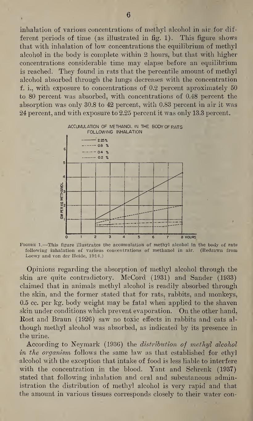
inhalation of various concentrations of methyl alcohol in air for dif¬ ferent periods of time (as illustrated in fig. 1). This figure shows that with inhalation of low concentrations the equilibrium of methyl alcohol in the body is complete within 2 hours, but that with higher concentrations considerable time may elapse before an equilibrium is reached. They found in rats that the percentile amount of methyl alcohol absorbed through the lungs decreases with the concentration f. i., with exposure to concentrations of 0.2 percent aproximately 50 to 80 percent was absorbed, with concentrations of 0.48 percent the absorption was only 30.8 to 42 percent, with 0.83 percent in air it was 24 percent, and with exposure to 2.25 percent it was only 13.3 percent.
ACCUMULATION OF METHANOL IN THE BODY OF RATS
FOLLOWING INHALATION
Figure 1.—This figure illustrates the accumulation of methyl alcohol in the body of rats following inhalation of various concentrations of methanol in air. (Redrawn from Loewy and von der Heide, 1914.)
Opinions regarding the absorption of methyl alcohol through the skin are quite contradictory. McCord (1931) and Sander (1933) claimed that in animals methyl alcohol is readily absorbed through the skin, and the former stated that for rats, rabbits, and monkeys, 0.5 cc. per kg. body weight may be fatal when applied to the shaven skin under conditions which prevent evaporation. On the other hand, Rost and Braun (1926) saw no toxic effects in rabbits and cats al¬ though methyl alcohol was absorbed, as indicated by its presence in the urine.
According to Neymark (1936) the distribution of methyl alcohol in the organism follows the same law as that established for ethyl alcohol with the exception that intake of food is less liable to interfere with the concentration in the blood. Yant and Schrenk (1937) stated that following inhalation and oral and subcutaneous admin¬ istration the distribution of methyl alcohol is very rapid and that the amount in various tissues corresponds closely to their water con-
7
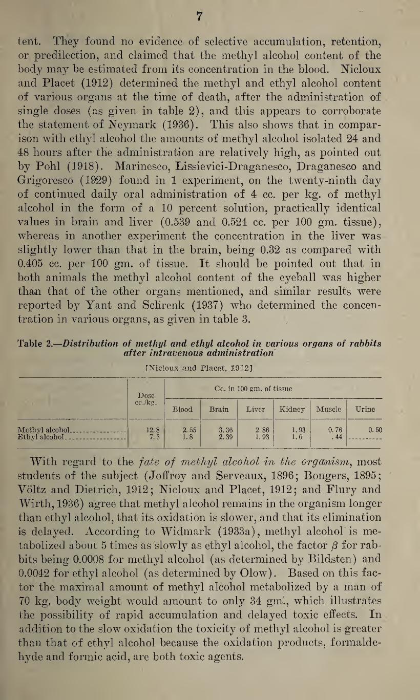
tent. They found no evidence of selective accumulation, retention, or predilection, and claimed that the methyl alcohol content of the body may be estimated from its concentration in the blood. Nicloux and Placet (1912) determined the methyl and ethyl alcohol content of various organs at the time of death, after the administration of single doses (as given in table 2), and this appears to corroborate the statement of Neymark (1936). This also shows that in compar¬ ison with ethyl alcohol the amounts of methyl alcohol isolated 24 and 48 hours after the administration are relatively high, as pointed out by Pohl (1918). Marinesco, Lissievici-Draganesco, Draganesco and Grigoresco (1929) found in 1 experiment, on the twenty-ninth day of continued daily oral administration of 4 cc. per kg. of methyl alcohol in the form of a 10 percent solution, practically identical values in brain and liver (0.539 and 0.524 cc. per 100 gm. tissue), whereas in another experiment the concentration in the liver was slightly lower than that in the brain, being 0.32 as compared with 0.405 cc. per 100 gm. of tissue. It should be pointed out that in both animals the methyl alcohol content of the eyeball was higher than that of the other organs mentioned, and similar results were reported by Yant and Sclirenk (1937) who determined the concen¬ tration in various organs, as given in table 3.
Table 2.—Distribution of methyl and ethyl alcohol in various organs of rabbits after intravenous administration
[Nicloux and Placet, 1912]
Dose cc./kg.
Cc. in 100 gm. of tissue
Blood Brain Liver Kidney Muscle Urine
Methyl alcohol... Ethyl alcohol_
12.8 7.3
2.55 1.8
3.36 2.39
2.86 1.93
1.93 1.6
0. 76 .44
0.50
With regard to the fate of methyl alcohol in the organism, most students of the subject (Joffroy and Serveaux, 1896; Bongers, 1895; Voltz and Dietrich, 1912; Nicloux and Placet, 1912; and Flury and Wirth, 1936) agree that methyl alcohol remains in the organism longer than ethyl alcohol, that its oxidation is slower, and that its elimination is delayed. According to Widmark (1933a), methyl alcohol is me¬ tabolized about 5 times as slowly as ethyl alcohol, the factor /3 for rab¬ bits being 0.0008 for methyl alcohol (as determined by Bildsten) and 0.0042 for ethyl alcohol (as determined by Olow). Based on this fac¬ tor the maximal amount of methyl alcohol metabolized by a man of 70 kg. body weight would amount to only 34 gin., which illustrates the possibility of rapid accumulation and delayed toxic effects. In addition to the slow oxidation the toxicity of methyl alcohol is greater than that of ethyl alcohol because the oxidation products, formalde¬ hyde and formic acid, are both toxic agents.
8
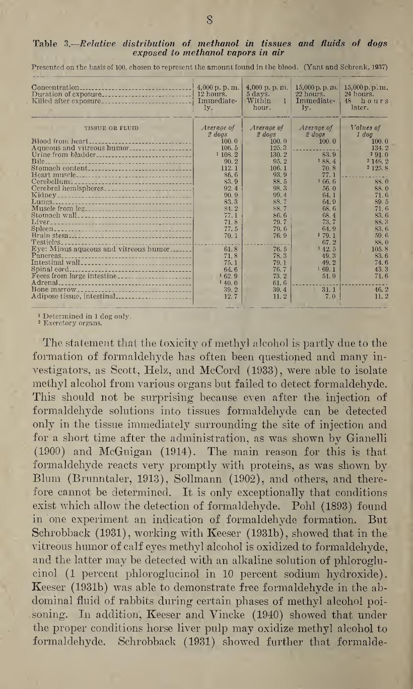
Table 3.—Relative distribution of methanol in tissues and fluids of dogs exposed to methanol vapors in air
Presented on the basis of 100, chosen to represent the amount found in the blood. (Yant and Schrenk, 1937)
Concentration.-..... 4,000 p. p. m. 12 hours.
4,000 p. p. m. 5 days. Within 1
15,000 p. p.m. 22 hours.
15,000p.p.m. 24 hours. Duration of exposure.____
Killed after exposure____ Immediate- Immediate- 48 hours ly. hour. ly. later.
TISSUE OR FLUID
Blood from heart... ____
Average of 2 dogs
100.0
Average of 2 dogs
100.0
Average of 2 dogs
100.0
Values of 1 dog
100.0 Aqueous and vitreous humor.. . _ 106. 5 125.3 134. 2 Urine from bladder... . i 108. 2 130. 2 83.9 2 91.0 Bile____ 90.2 95.2 i 88.4 2146. 2 Stomach content..___ _. ___ 112.1 106.1 70.8 2 125. 8 Heart muscle___ . __ _ 86.6 93.9 77.1 Cerebellum____ . _ 83.9 88.5 i 66.6 88.0 Cerebral hemispheres___ .. .. ... 92.4 98.3 56.0 88. 0 Kidney____ _ .. 90.9 99. 4 64.1 71.6 Lungs_ __..... .... 83.3 88. 7 64.9 89. 5 Muscle from leg... ... __ 84.2 88. 7 68.6 71. 6 Stomach wall .. ___ .. 77. ] 86.6 68.4 83. 6 Liver________ __ 71.8 79. 7 73.7 88.3 Spleen__ _ ___ 77. 5 79. 6 64.9 83. 6 Brainstem_ _ ..... Testicles______ _
70.1 76.9 1 79.1 67.2
59.6 88.0
Eye: Minus aqueous and vitreous humor. 61.8 76. 5 1 42. 5 105.8 Pancreas.. .. . .. ... .. .. _ _ 71.8 78.3 49.3 83.6 Intestinal wall_ .. . _ 75.1 79.1 49.2 74.6 Spinal cord___ ... ... ... 64.6 76.7 1 69.1 43.3 Feces from large intestine___ 1 62.9 73.2 51.9 71.6 Adrenal....... . __ ] 40. 0 61.6 Bone marrow_ ... ... _ 39.2 39.4 31. 1 46.2 Adipose tissue, intestinal... . _ ... 12.7 11.2 7.0 11.2
] Determined in 1 dog only. 2 Excretory organs.
The statement that the toxicit}^ of methyl alcohol is partly due to the formation of formaldehyde has often been questioned and many in¬ vestigators, as Scott, Helz, and McCord (1933), were able to isolate methyl alcohol from various organs but failed to detect formaldehyde. This should not be surprising because even after the injection of formaldehyde solutions into tissues formaldehyde can be detected only in the tissue immediately surrounding the site of injection and for a short time after the administration, as was shown by Gianelli (1900) and McGuigan (1914). The main reason for this is that formaldehyde reacts very promptly with proteins, as was shown by Blum (Brunntaler, 1913), Sollmann (1902), and others, and there¬ fore cannot be determined. It is only exceptionally that conditions exist which allow the detection of formaldehyde. Pohl (1893) found in one experiment an indication of formaldehyde formation. But Schrobback (1931), working with Keeser (1931b), showed that in the vitreous humor of calf eyes methyl alcohol is oxidized to formaldehyde, and the latter may be detected with an alkaline solution of phloroglu- cinol (1 percent phloroglucinol in 10 percent sodium hydroxide). Keeser (1931b) was able to demonstrate free formaldehyde in the ab¬ dominal fluid of rabbits during certain phases of methyl alcohol poi¬ soning. In addition, Keeser and Vincke (1940) showed that under the proper conditions horse liver pulp may oxidize methyl alcohol to formaldehyde. Schrobback (1931) showed further that formalde-
9
hyde formed in the vitreous humor could be condensed with ammonium carbonate to form hexamethylene tetramine.
The formaldehyde formed from methyl alcohol is further oxidized to formic acid. Pohl (1893) showed that, following the administra¬ tion of methyl alcohol, dogs and rabbits excrete formic acid, the maxi¬ mal excretion occurring on the fourth day after the administration; and Rost and Braun (1926) noted the maximal formate excretion on the second and third days. Pohl (1893) determined the formic acid content of the blood as 0.4 mg. per 100 cc., of muscle as 0.5 mg. per 100 gm., of the kidney as 34.5 mg. per 25 gm., and of the lungs as 0.44 mg. per 50 gm. This evidently indicates that formates are not stored in the body. The increased excretion of formic acid following adminis¬ tration of methyl alcohol was also demonstrated by Hunt (1902) and
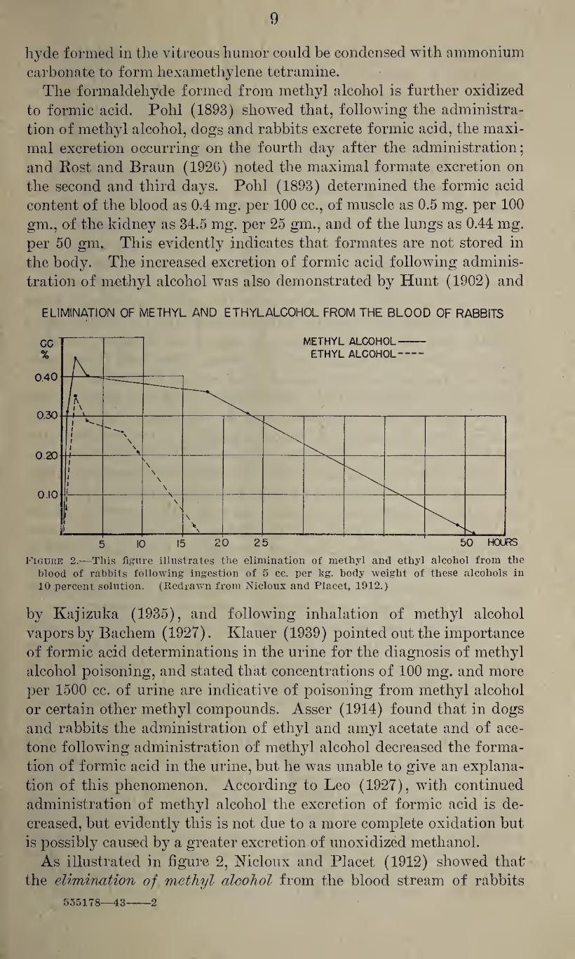
ELIMINATION OF METHYL AND ETHYLALCOHOL FROM THE BLOOD OF RABBITS
Figure 2.—This figure illustrates the elimination of methyl and ethyl alcohol from the blood of rabbits following ingestion of 5 cc. per kg. body weight of these alcohols in 10 percent solution. (Redrawn from Nicloux and Placet, 1912.)
by Kajizuka (1935), and following inhalation of methyl alcohol vapors by Bachem (1927). Klauer (1939) pointed out the importance of formic acid determinations in the urine for the diagnosis of methyl alcohol poisoning, and stated that concentrations of 100 mg. and more per 1500 cc. of urine are indicative of poisoning from methyl alcohol or certain other methyl compounds. Asser (1914) found that in dogs and rabbits the administration of ethyl and amyl acetate and of ace¬ tone following administration of methyl alcohol decreased the forma¬ tion of formic acid in the urine, but he was unable to give an explana¬ tion of this phenomenon. According to Leo (1927), with continued administration of methyl alcohol the excretion of formic acid is de¬ creased, but evidently this is not due to a more complete oxidation but is possibly caused by a greater excretion of unoxidized methanol.
As illustrated in figure 2, Nicloux and Placet (1912) showed that the elimination of methyl alcohol from the blood stream of rabbits
555178—43-2
10
occurs much more slowly than that of ethyl alcohol, and the same holds true to an even greater extent for dogs, in which 120 hours were required for complete elimination of methyl alcohol from the blood. According to Widmark and Bildsten (1924), following its intravenous injection methyl alcohol disappears from the blood of rabbits at a certain rate which is independent of the concentration. As shown by Neymark (1936) the rate of disappearance of methyl alcohol from the blood is about 10 times as slow as that of ethyl alcohol but this can be speeded up by stimulation of the oxygen metabolism, as by the administration of 1,2,4-dinitrophenol.
Pohl (1908) and Cushny (1910) stated that, following intravenous injection, only traces of methyl alcohol are excreted through the lungs. Voltz and Dietrich (1912) found that after administration of 2 cc. per kg. to dogs, 15.3 percent of the amount given is excreted within 24 hours, 13.8 percent being exhaled and 1.5 percent being excreted with the urine. During the subsequent 24 hours an addi¬ tional 7 percent was exhaled and 1.5 percent eliminated through the kidneys. During the entire period of 48 hours following the admin¬ istration, 24.3 percent of the dose administered was excreted and 36.8 percent could be recovered from the organism, so that within 48 hours only 39 percent of the total quantity given had been oxi¬ dized in the organism. That the elimination of methyl alcohol with the urine is slow was already found by Joffroy and Serveaux (1896) and more recently confirmed by Rost and Braun (1926) who stated that after administration of single doses to rabbits, methyl alcohol could be detected in the urine for 4 days, the maximal excretion occurring on the second day. According to Voltz and Dietrich (1912) the elimination of methyl alcohol from the body may be speeded up by exercise, increased respiration, increase of body tem¬ perature (diaphoresis), and the administration of diuretics.
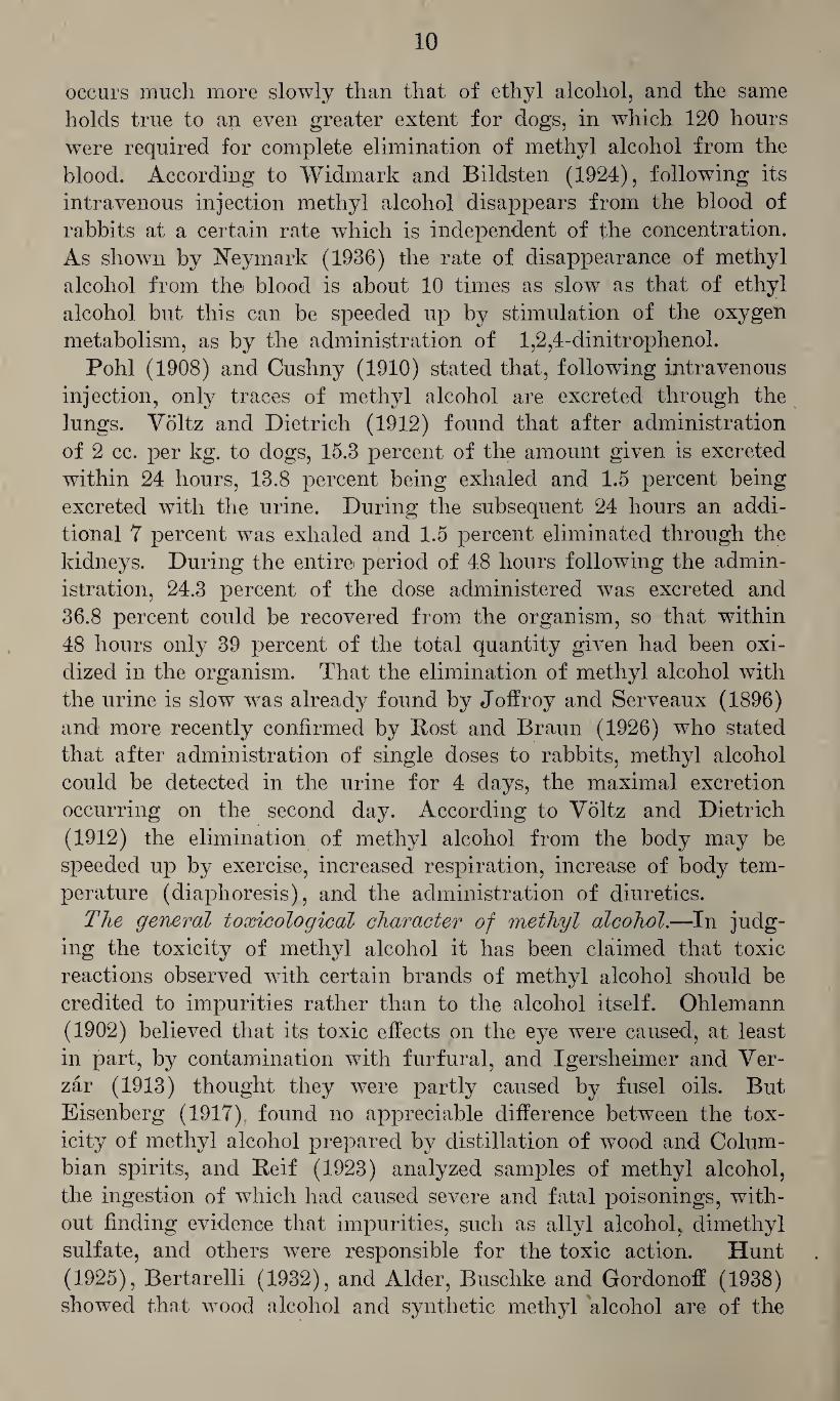
The general toxicological character of methyl alcohol.—In judg¬ ing the toxicity of methyl alcohol it has been claimed that toxic reactions observed with certain brands of methyl alcohol should be credited to impurities rather than to the alcohol itself. Ohlemann (1902) believed that its toxic effects on the eye were caused, at least in part, by contamination with furfural, and Igersheimer and Ver- zar (1913) thought they were partly caused by fusel oils. But Eisenberg (1917) found no appreciable difference between the tox¬ icity of methyl alcohol prepared by distillation of wood and Colum¬ bian spirits, and Reif (1923) analyzed samples of methyl alcohol, the ingestion of which had caused severe and fatal poisonings, with¬ out finding evidence that impurities, such as allyl alcohol, dimethyl sulfate, and others were responsible for the toxic action. Hunt (1925), Bertarelli (1932), and Alder, Buschke and Gordonoff (1938) showed that wood alcohol and synthetic methyl alcohol are of the
11
same toxicity. It appears, therefore, that the toxic effects described in the following are inherent properties of methyl alcohol and should not be credited to impurities.
In judging the potential hazards resulting from the absorption of methyl alcohol one has to distinguish between the toxic action which, as will be shown, is largely due to its metabolites, and the narcotic action which is the characteristic effect of alcohols. The narcotic ac¬ tion of methyl alcohol is less than that of its higher homologues, this being possibly explained by its low solubility in oils, fats, and lipoids and by its greater miscibility with water, and for this reason its affin¬ ity to and accumulation in certain organs is of a different order than that observed with the higher homologues. The toxicity of methanol is greater than that of ethyl alcohol on account of its less complete and slower oxidation, which results in the formation of more toxic meta¬ bolites and the accumulation of methyl alcohol in the organism. For this reason, continued exposure or repeated fractional doses may be more toxic than single doses.
The antiseptic action of methyl alcohol.—The antiseptic action of methyl alcohol is not very marked. Buchner, Fuchs and Megele (1901) found that 10 and 30 percent solutions do not kill brewer’s yeast after contact for 1 hour, and 60 and 100 percent solutions were required to kill staphylococcus pyocyaneus aureus, bacillus typhi, and bacillus pyocyaneus. Whitney (1912) found methyl alcohol less toxic than ethyl alcohol as judged by the rate of reproduction of rotifer a. Bokorny (1911) found that methyl alcohol may even be utilized by algae and bacteria as a source of carbon.
Methyl alcohol vapors have a more or less marked irritant effect on the mucous membranes of the eye and of the upper respiratory tract. Tyson and Schoenberg (1914) noted a copious discharge from the noses and mouths of animals exposed to vapors of methyl alcohol. Flury and Wirth (1934) stated that concentrations of 10 mg. per liter (7,640 p. p. m.) cause only moderate irritation and with concentrations of 90 mg. per liter (68,760 p. p-. m.) the irritation is intolerable; and according to Lehmann and Flury (1938) prolonged exposure to con¬ centrations of 65 mg. per liter (50,000 p. p. m.) cannot be tolerated.
The toxicity of methyl alcohol for animals.—In judging the toxicity of methyl alcohol it is generally found that fractional doses are more toxic than single doses but that in contrast to fractional doses, in single doses methyl alcohol is less toxic than ethyl alcohol, as found by Baer (1898), Hunt (1902), Langgaard (1912), Nicloux and Placet (1912), Rost and Braun (1926), Hufferd (1932a), and others. Rost and Braun (1926) claimed that the toxcity of methyl alcohol varies with different species, depending on the development of the central nervous system, and according to Scott, Helz and McCord (1933) rats are very susceptible and rabbits quite resistant.
12
The minima] fatal doses of methyl alcohol with oral administration has been given by various investigators as follows:
For mice, 10.5-12 cc., Weese (1928).
For rabbits, 8.8 cc./kg., Dujardin-Beaumetz and Audige (1875).
For rabbits, 14 cc./kg., Langgaard (1913).
For rabbits, 13 cc./kg., Rost and Braun (1926).
For dogs, 8 cc./kg., Haskell, Hileman and Gardner (1921).
With intravenous injection the minimal fatal dose has been stated as:
For frogs, 5.3 cc., Sammartino (1933c).
For rabbits, 20.1 cc./kg., Lehman and Newman (1937b).
For rabbits, 16.1 cc./kg., Nicloux and Placet (1912).
For cats, 5.9 cc./kg. Macbt (1920).
The minimal fatal dose for monkeys with absorption through the skin (if all loss by evaporation is prevented) was estimated by McCord
(1931) as 0.5 cc. per kg. The minimal fatal concentration of methyl alcohol vapors in air
has been given for mice with exposure for 3 to 4y2 hours as 0.4 to 0.6 cc. per liter (242,000 to 363,000 p. p. m.) by Weese (1928), and for rats and rabbits with exposure for an unknown number of hours as 0.0071 mole per liter (176,000 p. p. m.) by Bachem (1927). Witte (1931) (quoted from Flury and Zernik, 1931) found the minimal fatal con¬ centration for cats with hours’ exposure to be 380 mg. per liter (290,000 p. p. m.). With longer exposure the minimal fatal concen¬ tration is naturally much lower. Loewy and von der Heide (1914) found that rats die after exposure to concentrations of 41.5 mg. per liter (corresponding to 31,600 p. p. m.) for 10 to 20 hours, and with shorter exposure (6 hours) animals may die after several days, as found by Witte (1931) (quoted from Flury and Zernik, 1931) in cats with exposure to 97.1 and 224.3 mg. per liter (corresponding to 74,000 and 160,000 p. p. m., respectively). Sayers, Yant, Schrenk, Chornyak, Pearce, Patty, and Linn (1942) saw no significant toxic effects in dogs exposed daily for 8 hours for 379 days to concentrations of 450 to 500 p. p. m. of methanol in air.
Fere (1894b) found that the injection of methyl alcohol into fertil¬ ized eggs gives a higher incidence of malformation than observed with ethyl alcohol. Sollmann (1920) noted that continued administration of 5 percent methyl alcohol as drinking water to rats caused a con¬ siderable decrease of weight and, finally, death. The administration of 2.5 percent solutions was found to inhibit growth, this effect being more marked than that observed with 10 percent solutions of ethyl alcohol. Elhardt (1932) found that the injection of from 0.15 to 0.25 cc. of a 40 percent solution of methyl alcohol into the crop of growing chicks over a period of 2 months had a definitely injurious effect on growth and vigor. Smaller doses than these had a less
13
marked effect but affected unfavorably the growth of feathers, the development of the comb, and the general disposition of the chicks.
The effect of methyl alcohol on the central nervous system of animals was first studied by Poincare (1878) who noted temporary stagger¬ ing and attacks of hyperexcitation in animals kept in an atmosphere containing methyl alcohol for 8 to 16 months. Joffroy and Serveaux (1896) observed, in experiments with dogs, motor and sensory dis¬ turbances and changes of the body temperature and the respiration. Tyson and Schoenberg (1915) found that exposure of rabbits, dogs, and monkeys to high concentrations of methyl alcohol caused loss of consciousness, loss of pupillary reflexes, slight constriction of the pupils, and death. Macht and Leach (1929) studied the behavior of rats in a maze and found that methvl alcohol causes less severe
%/
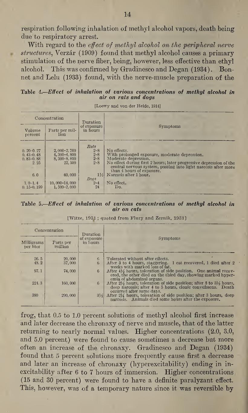
depression of the central nervous system than ethyl alcohol. Accord¬ ing to Lehman and Newman (1937b) the anesthetic dose for rabbits with intravenous injection is 10.5 gm. per kg., methyl alcohol being about one-half as effective as ethyl alcohol. Loew7y and von der Heide (1914) studied the narcotic action of methyl alcohol in rats and dogs and found that it is not very marked, as illustrated in table 4. This was confirmed by Flury and Wirth (1934) who found that the nar¬ cotic action of methyl alcohol is weaker than that of methyl acetate, and by Mashbitz, Sklianskaya, and Urieva (1936) who found its nar¬ cotic action to be inferior to that of acetone. In the experience of Flury and Wirth (1934) concentrations below 170 mg. per liter (130,000 p. p. m.) cause, within 6 hours, only moderate narcosis in cats. Witte (1931) (quoted from Flury and Zernik, 1931) studied in cats the effect of inhalation of various concentrations of methyl alcohol in air, as illustrated in table 5. Comparison of these findings with those of Loewy and von der Fleide (1914) appears to indicate that rats are more sensitive than cats and dogs. It should, however, be emphasized that, as pointed out by Flury and Wirth (1934), even concentrations as low as 86 mg. per liter (66,000 p. p. m.) may cause delayed death. Sammartino (1933c) noted in frogs, following intra¬ venous administration of methyl alcohol, clonic-tonic convulsions, opisthotonus and, finally, progressive paralysis. In mammals, con¬ vulsions following the repeated administration of doses of 10 cc. of methyl alcohol (in 10 percent solution) to cats were reported as late effects by Eost and Braun (1926) and by Witte (1931) (quoted from Flury and Zernik, 1931) as shown in table 5. According to Gradi- nesco (1934), in dogs the intravenous injection of small doses of methyl alcohol (1 cc. per kg.) causes an increase of the respiratory amplitude, whereas large doses (10 cc. per kg.) cause a severe depression. Tyson and Schoenberg (1914) noted a marked reduction of the body tem¬ perature and a primary stimulation and subsequent depression of the
14
respiration following inhalation of methyl alcohol vapors, death being due to respiratory arrest.
With regard to the effect of methyl alcohol on the peripheral nerve structures, Verzar (1909) found that methyl alcohol causes a primary stimulation of the nerve fiber, being, however, less effective than ethyl alcohol. This was confirmed by Gradinesco and Degan (1934). Bon¬ net and Lelu (1933) found, with the nerve-muscle preparation of the
Table 4.—Effect of inhalation of various concentrations of methyl alcohol in air on rats and dogs
[Loewy and von der Heide, 1914]
Concentration Duration
Volume percent
Parts per mil¬ lion
of exposure in hours
Symptoms
0. 20-0. 27 2,000-2,700 Rats
2-8 No effects. 0.43-0.48 4,300-4, 800 2-8 With prolonged exposure, moderate depression. 0. 83-0. 88 8,300-8,800 2-8 Moderate depression.
2. 25 22, 500 2-8 No effect during first 2 hours; later progressive depression of the
6.0 60,000 ltt
central nervous system, passing into light narcosis after more than 4 hours of exposure.
Narcosis after 1 hour.
1.0-1. 4 10,000-14,000 Dogs
2-4 No effect. 0.15-0.199 1, 500-2, 000 24 Do.
Table 5.—Effect of inhalation of various concentrations of methyl alcohol in air on cats
[Witte, 1931; quoted from Flury and Zernik, 1931]
Concentration Duration
Milligrams per liter
Parts per million
of exposure in hours
Symptoms
26.5 20,000 6 Tolerated without after effects. 48.2 37,000 6 After 3 to 4 hours, staggering. 1 cat recovered, 1 died after 2
weeks with marked loss of fat. 97.1 74,000 6 After 434 hours, toleration of side position. One animal recov¬
ered, the other died on the third day, showing marked hyper¬ emia of abdominal organs.
224.3 160,000 6 After 234 hours, toleration of side position; after 5 to 534 hours, deep narcosis; after 4 to 5 hours, clonic convulsions. Death occurred after some daj'S.
380 290, 000 334 After 234 hours, toleiation of side position; after 3 hours, deep narcosis. Animals died some hours after the exposure.
frog, that 0.5 to 1.0 percent solutions of methyl alcohol first increase and later decrease the chronaxy of nerve and muscle, that of the latter returning to nearly normal values. Higher concentrations (2.0, 3.0, and 5.0 percent) were found to cause sometimes a decrease but more often an increase of the chronaxy. Gradinesco and Degan (1934) found that 5 percent solutions more frequently cause first a decrease and later an increase of chronaxy (hyperexcitability) ending in in¬ excitability after 6 to 7 hours of immersion. Higher concentrations (15 and 30 percent) were found to have a definite paralyzant effect. This, however, was of a temporary nature since it was reversible by
15
lavage. The authors pointed out that with concentrations of 5 and 10 percent the results are not uniform and frequently there is first an increase of the chronaxy, as observed by Bonnet and Lelu (1933), which presumably is due to individual differences (seasonal?).
In view of the deleterious effect on the vision observed in methyl alco¬ hol poisoning, this effect has been studied very extensively in animal experiments. Holden (1899) fed dogs 50 cc. of methyl alcohol on two occasions, 5 days apart, and noted on the second day thereafter tempo¬ rary blindness which later gradually subsided. On the eighth day a diffuse turbidit}^ of the cornea without signs of congestion developed. On autopsy he found extensive degenerative changes of the ganglionic cells of the retina and destruction of some medullary sheaths of fibers of the optic nerve, and he assumed that the temporary amblyopia was caused by nutritional disturbances of the ganglionic cells of the retina. Friedenwald (1902) confirmed the destructive effect on the ganglionic cells of the retina in experiments with rabbits which were fed methyl and ethyl alcohol in sufficient doses and for a sufficient period of time to cause, in the case of the latter, cirrhosis of the liver, and he found that these alcohols behaved similarly in this respect. Birch-Hirsch- feld (1900) experimented with rabbits and chicks and noted, on the day following the administration, dilatation and rigidity of the pupils, absence of defense reflexes, and inability of the animals to orient them¬ selves in space. Although he noted no ophthalmological changes dur¬ ing life he found at autopsy degenerative changes in the ganglionic cells of the retina and also, in 1 rabbit, of the optic nerve. Later (1901) he expressed the opinion that the retinal changes were the primary manifestations and that the lesions of the optic nerve de¬ veloped later. Igersheimer and Verzar (1913) repeated the experi¬ ments of Birch-Hirschfeld but used more diluted solutions of methyl alcohol in order to prolong the exposure and reduce acute toxic effects. Although they noted a temporary reduction of the light perception they found no degenerative changes in the retina. Kasass (1913) fed increasingly large doses of methyl alcohol to rabbits for 267 days. He noted peripapillary venous hyperemia and, later, constriction of the arteries and bleaching of the papilla which might result in nutritional disturbance in the retina, as had been assumed by Holden (1899). These symptoms disappeared after some weeks. On autopsy he noted vacuolar degeneration in all, but especially in the interior layers of the retina, hemorrhages in the optic nerve and fatty degeneration of the myelin fibers. This assumption appears to be supported by the publication of Goldschmidt (1922) who found that pretreatment of the retina with methyl alcohol prevents, under certain conditions, the reduction of methylene blue to the leucobase by this tissue. This effect increased with the concentration of methyl alcohol used and no reduction of methylene blue was observed by the retina of animals
16
which had been poisoned with methyl alcohol; it appears, therefore,
that the retina of such animals is unable to utilize oxygen. Grignolo
(1913) found in dogs that following the administration of methyl
alcohol the osmotic pressure of the fluid in the posterior chamber of
the eye was increased and there was also an increase of the hydrogen
ion concentration which was later confirmed by Tyson and Schoenberg
(1914 and 1915) who pointed out that this effect was more marked than
the increase of the hydrogen ion concentration in the blood. They ex¬
plained the greater acidity of the vitreous humor by less complete
buffer action as compared with that of the blood. In view of the
findings of Keeser (1931b) and Schrobback (1931) the possibility
should be considered that more formic acid is formed in this than in
other organs because in this medium formaldehyde is less readily
bound. Grignolo (1913) found that the increase of the osmotic
pressure was paralleled by shrinkage of the ganglionic cells, shrink¬
age and edema of the granular layers, and atrophy of the optic nerve.
Rost and Braun (1926), like Friedenwald (1902), noted similar
changes of the eye in dogs, following repeated oral administration of
methyl and ethyl alcohol. Whereas some cells of the retina were
normal, others were vacuolated, some showed incomplete staining,
and shadow cells were seen quite frequently. In addition, the pigment
of the retina was destroyed in spots and similar changes were seen
in the granular and ganglionic layers, but no changes of the optic
disk were noted. Alder, Buschke, and Gordonoff (1938) administered
by stomach tube to rabbits 70 percent methyl alcohol (2.5 cc. per kg.)
of a high degree of purity on three occasions and killed the animals
on the fifth day, when the histological examination of the retina re¬
vealed reduction of the ganglionic cells, irregularities of the nuclei,
changes and disappearance of Nissl bodies, and loosening of the gran¬
ular layer. It appears that changes similar to those produced by
methyl alcohol may also be seen occasionally in ethyl alcohol poison¬
ing, but it appears to be definitely proved that the injurious effect of
methyl alcohol is more marked, presumably on account of its slower
and less complete oxidation, as indicated by the observations of Grig¬
nolo (1913) and Tyson and Schoenberg (1914 and 1915). Whereas
most of these studies on the effect of methyl alcohol on eye and vision
were made with oral administration, studies regarding the effects on
the eye with inhalation of its vapors are less numerous. Tyson and
Schoenberg (1914 and 1915) found that repeated daily inhalation of
methyl alcohol vapors for a limited time caused reduction of the
vision and, histologically, in one instance, edema of various structures
of the eye and early signs of beginning degenerative changes of the
ganglionic cells of the retina. McCord (1931) noted atrophy of the
optic nerve following inhalation and cutaneous absorption, and Weese
(1928) observed in mice, following inhalation of fatal concentrations
17
of methyl alcohol, degenerative changes in the retina which, however, may possibly have been due to postmortem changes during the prep¬ aration of the tissue.
With regard to the effect of methyl alcohol on the circulation, Kuno (1913) found that the isolated mammalian heart is depressed in %0 to y5o normal concentration (corresponding to 0.8 to 0.6 gm. per liter) and that % normal solutions (1.5 gm. per liter) cause complete arrest within a few minutes. According to Fiihner (1921) the minimal effective concentration causing depression of the isolated frog heart is 3.740 mole per liter (119.7 gm. per liter); and Wolff (1922) found that a 0.02 percent solution (0.006 mole per liter) has no visible ef¬ fect, that 1 percent (0.3 mole per liter) causes reduction of the ampli¬ tude to about one-fourth of the original, and that concentrations of 3 and 6 mole per liter (96 and 192 gm. per liter) cause diastolic arrest which is completely reversible. Similarly, Simon (1933) stated that the isolated frog heart is reversibly arrested by concentrations of 6.25 mole per liter (200 gm. per liter) of methyl alcohol whereas concen¬ trations of formaldehyde and formic acid of 0.0333 and 0.00434 mole per liter caused irreversible arrest, which illustrates the great toxicity of the oxidation products of methyl alcohol. Similar results were also published by Sammartino (1933a). According to Sklianskaya, Urieva, and Nashbitz (1936), the effect of methyl alcohol on the frog heart is less marked but more lasting than that of acetone.
With regard to the effect of methyl alcohol on the blood vessels, Budelmann (1930) noted in perfusion experiments on isolated organs that low concentrations of methyl alcohol caused a peripheral vaso¬ constriction. Simon (1933) and Sammartino (1933b) found in the Trendlenburg preparation of frogs that concentrations of 1:1,000,000 cause vasodilatation, the blood flow being increased by 29 percent. With the same concentration of formic acid or formaldehyde the increase of flow was only 7 and 3 percent, respectively, and, in contrast to methanol, higher concentrations of formic acid and formaldehyde were found to cause vasoconstriction. Therefore, it appears likely that the vasoconstriction observed by Budelmann (1930) may have been due to the formation of these metabolites in the isolated organs.
Miura (1913) studied the effect of methyl alcohol on the blood. He found that in dogs and rabbits, following subcutaneous injections of 3.3 cc. per kg., two-fifths of the animals developed an anemia, a re¬ duction of lymphocytes, and a relative increase of the pseudo-eosino- philes and neutrophiles. These animals suffered from hemoglobi¬ nuria. Tyson and Schoenberg (1914) noted in dogs, in acute poison¬ ings produced by the inhalation of methyl alcohol vapors, an increase of all cellular elements of the blood with the exception of lympho¬ cytes and an increase of the viscosity. It will be shown that similar findings have been observed in man and it appears that this phe-
18
nomenon may be explained on the basis of edema formation and dehy¬ dration of the blood. There is no evidence to show that methyl alco¬ hol produces abnormal blood pigments. Egg (1927) showed, how¬ ever, that bivalent iron may form a complex with methyl alcohol and may thus interfere with the catalytic action of hemoglobin. Al¬ though the-same effect may be observed with ethyl alcohol, this is said to be less significant on account of the more rapid oxidation of the latter. Weese (1928) suggested that the toxic effect of methyl alco¬ hol might be partly explained by an effect on the hemoglobin by im¬ pairing the catalytic action of the blood.
With regard to the effect of methyl alcohol on muscular tissue, Kuno (1914) found that 0.5 and 1 percent solutions of methyl alcoho] in Ringer’s solution increase the pendular movements of the isolated intestine without increasing the average tone. Higher concentrations of from 5 to 10 percent cause a short primary stimulation and subse¬ quent depression of the pendular movements and a moderate increase of the average tone. Verzar (1909) found that the depressant effect of methyl alcohol on the striated muscle and the ciliated epithelium is less marked than that of ethyl alcohol, and that with moderate con¬ centrations the depression is preceded by a short stimulation. As pointed out by Bonnet and Lelu (1933), the depressant effect on the muscle structure is less marked than that on the nerve fiber.
With regard to the effect of methyl alcohol on the metabolism, Gradinesco and Palmhert (1931) found that methyl alcohol inhibits to a lesser extent than ethyl alcohol the digestive action of natural and artificial gastric juice on solid protein material. Krol (1913) showed that in methyl alcohol poisoning there is a considerable increase (100 to 156 percent) of the ammonia excretion, a small fraction of which is neutralized by formic acid. Rewiger (1922) found in experiments with dogs that, in contrast to ethanol, methyl alcohol causes a negative nitrogen balance which could be overcome by an ample intake of pro¬ teins. As shown by Hockendorf (1909-10), methyl alcohol increases the sugar excretion of phloridzin diabetic dogs.
Much effort has been devoted to the question of acidosis in methyl alcohol poisoning. Schmiedeberg (1912) assumed that the essential feature of methyl alcohol poisoning is the formation of formic acid which, especially in under-nourished individuals and in the absence of sufficient ammonia formation, may lead to acidosis, whereas Har- nack (1912) believed that the toxicity of formic acid itself rather than the acidosis produced was the determining factor in methyl alcohol poisoning. Krol (1913) assumed that animals poisoned with methyl alcohol were suffering from acidosis caused by the increased excretion of ammonia with the urine. However, according to Loewy and Miinzer (1923) this is indicative only of increased acid formation and not of acidosis. Tyson and Schoenberg (1914) showed that in animals
19
poisoned with methyl alcohol the hydrogen ion concentration of the blood is increased. Haskell, Hileman and Gardner (1921) found in experiments with dogs that, following administration of methyl alco¬ hol, the blood alkali was not always or not sufficiently reduced to cause severe acidosis. They believed that the latter was not the outstanding phenomenon because in their experience the administration of sodium bicarbonate was of limited value. According to Ziegler (1921) the acidosis may be followed by alkalosis which may result from the forced respiration observed in the last stages of methyl alcohol poisoning. Loewy and Munzer (1923) found no evidence of severe acidosis in experimental methyl alcohol poisoning of rabbits and dogs, as indi¬ cated by the absence of disturbances of the carbon dioxide binding power of the blood. In contrast to the observations of Haskel, Hile¬ man and Gardner (1921), Leo (1925) found in dogs that the adminis¬ tration of alkali was of distinct value in attenuating the picture of methyl alcohol poisoning. This favorable effect was not seen in mice, rats and rabbits and this may. be explained by the observation of Rewiger (1922) that, in contrast to dogs, even large doses of methyl alcohol do not increase the ammonia excretion of these animals. This observation also supports the assumption of Leo (1925) that the bene¬ ficial effect of alkali is directed less towards the acidosis than towards a more rapid elimination of the formic acid as formate. Keeser (1931a) believed that neither acidosis nor the formation of formic acid is as important a feature of methyl alcohol poisoning as is the inhibition of catalytic processes as demonstrated by Egg (1927). It appears, therefore, that in animals the production of acidosis by methyl alcohol poisoning may depend upon the species used, the nutritional status and the time at which the observations were made. In cases of methyl alcohol poisoning in humans acidosis has been ob¬ served repeatedly. Harrop and Benedict (1920) reported such a case in which the acidosis was promptly relieved by the intravenous ad¬ ministration of 5 percent sodium bicarbonate. Similar cases were reported by Ustvedt (1936), Merritt and Brown (1941), and others.
With regard to 'pathological changes in animals, Poincare (1878) noted, in the central nervous system, congestion and hemorrhages in the meninges and other signs of inflammatory processes; and similar changes and degenerative processes in brain and spinal cord were reported by Holden (1899), Riihle (1912), Tyson and Schoenberg (1914), Eisenberg (1917), Scott, Helz, and McCord (1933) and others.
Some of the pathological changes found in the eyes of animals poisoned with methyl alcohol have been discussed in a previous section where it was shown that the ganglionic cells of the retina are primarily affected and atrophy of the optic nerve has been ob-
20
served only occasionally, as reported by Scott, Helz and McCord (1933).
Detailed data on pathological changes in the peripheral nerves are apparently not available. Only Scott, Helz, and McCord (1933) mention injury of tlie peripheral nerves.
With respect to pathological changes in the digestive tract, Rost and Braun (1926) found, after oral administration of methyl alcohol, scarlet red, dark brown, and black red discoloration of the mucosa of the stomach which was edematous, hemorrhagic and, in spots, corroded. These authors and also Tyson and Schoenberg (1914) reported similar changes of less severe character in the duodenum.
The liver may show congestion (as reported by Tyson and Schoen¬ berg, 1914), parenchymatous degeneration, and, in severe cases, focal necroses (as reported by Scott, Helz, and McCord, 1933). In the experience of these investigators the latter is more conspicuous than fatty degeneration which is, in the opinion of Muller (1910), the most common finding and which was also reported by Poincare (1878), Eisenberg (1917), and Weese (1928).
In the kidneys parenchymatous degeneration of the epithelium of the convoluted tubules was reported by Poincare (1878), Weese (1928), and Scott, Helz and McCord (1933) whereas others such as Tyson and Schoenberg (1914) noted only congestion.
The heart muscle may show cloudy swelling and fatty degeneration as observed by Poincare (1878) and Eisenberg (1917), or granular degeneration with occasional necrosis of fibers as reported by Scott, Helz and McCord (1933).
Following inhalation of methyl alcohol vapors, the lungs are usually hyperemic. They may show petechial hemorrhages, as seen by Tyson and Schoenberg (1914), bronchopneumonia (Weese, 1928), and, in milder cases, congestion, edema and desquamation of the alveolar epithelium, as reported by Scott, Helz and McCord (1933).
Methyl alcohol poisoning in man: In man, methyl alcohol poison¬ ing most frequently results from the ingestion of methyl alcohol as a beverage. Baskerville (1913) collected, up to 1913, 720 cases of methyl alcohol poisoning, 390 of which ended fatally, 90 of which developed blindness, and 85 of which suffered impaired vision. Fur¬ ther cases were subsequently reported by Harrop and Benedict (1920), Burhans (1930), Mathewson and Alexander (1932), Neiding, Goldenberg and Blank (1933), Joiris (1935),Kraul (1933), Willemse (1936), Menne (1938), Merritt and Brown (1941) and others. Many single cases result from the ingestion of methanol or alcoholic bever¬ ages adulterated with methyl alcohol, and occasionally mass poison¬ ings are observed, as in Hungary in 1909, Berlin in 1911, Hamburg in 1922, and Odessa in 1933. The character and the intensity of the poisoning depends on the quantity of methyl alcohol ingested and the
21
nutritional status of the individual. In the opinion of Baskerville (1913) who analysed a large number of cases, 55 percent of the cases of methyl alcohol poisoning end fatally, 12 percent suffer permanent blindness, 12 percent have impaired vision, and only 4 percent recover completely.
Whereas injuries resulting from the ingestion of methyl alcohol are, as a rule, not of industrial origin, those caused by inhalation of its vapors do belong in this group. Baskerville (1913) collected from the literature 64 cases of such poisonings, of which 6 ended fatally, 19 suffered permanent blindness, and 33 had impaired vision. More re¬ cently, similar cases have been reported only occasionally, as by Kobin- son (1918) and Schwarzmann (1934), and it appears to be the con¬ sensus that only exposure to high concentrations in limited enclosures will cause serious and lasting effects, as also indicated by a study of Loewy (1914). Humperdinck (1941) believed that concentrations of 1,528 to 7,640 p. p. m. are potentially dangerous with regard to pos¬ sible visual disturbances and that to avoid these the concentration should be kept below 764 p. p. m. (1 mg. per liter).
As pointed out above, the clinical picture of methyl alcohol poison¬ ing following ingestion of methyl alcohol varies with the amount of methyl alcohol ingested, the amount of foodstuff in the gastrointes¬ tinal tract, and the nutritional status of the victim.
In light cases of methyl alcohol poisoning the patient may complain about fatigue, headache, a pulling pain in the limbs, nausea, and moderate gastrointestinal disturbances. Later he may complain of visual disturbances, and there may be a considerable latent period before more serious symptoms become manifest.
In more severe cases the victims suffer from nausea with occasional vomiting and diarrhea. Later they may become cyanotic and rest¬ less, their respiration becomes deep and labored, and more or less severe debility may develop. The pupils are usually dilated, fheir reactivity is reduced, and vision is impaired. If only pupillary symp¬ toms are present the prognosis is usually good (Stadelmann and Magnus-Levy, 1912), but if the patient is dyspneic the prognosis is doubtful and the clinical picture may suddenly become very serious.
In severe methyl alcohol poisoning, nausea, vomiting, and diarrhea are more marked (Krol, 1913; Harrop and Benedict, 1920; and Burhans, 1930), abdominal pain and colic may exist (Schwarzmann, 1934), and the stools may contain blood (Menne, 1938). The patients may be weak, apathetic, and even comatose (Tyson, 1912; Isaacs, 1920; and Burhans, 1930), or they may be excited (Krol, 1913) or even man¬ iacal (Neiding, Goldenberg, and Blank, 1933). They may also suffer from visual hallucinations, as reported by Harrop and Benedict (1920). Frequently they complain about more or less severe head¬ ache and vertigo. Their reflexes may be increased (Krol, 1913; and Schwarzmann, 1934) and they may suffer from convulsions, as re-
ported by Krol (1913), Keiding, Goldenberg, and Blank (1933), Bur- hans (1930) and others. In very severe cases these symptoms may be associated with opisthotonus (Krol, 1913). Later ataxia and periph¬ eral neuritis may develop, as seen by Jeliffe (1905), Schwarzmann (1934) and others. Oppression in the chest and pain in the side are frequent complaints (Krol, 1913; Harrop and Benedict, 1920; and others). Depending upon the stage of the poisoning, the respiration may be rapid and shallow (Menne, 1938) or deep and labored as in diabetic coma (Krol, 1913; Harrop and Benedict, 1920; Burhans, 1930; Ustvedt and Mohn, 1932; Menne, 1938; and others) and the patient may suffer from more or less severe cyanosis. The circulation may show varying degrees of failure, the blood pressure may be lowered, the pulse may be rapid and weak, and the victim may suffer from collapse associated with lowering of the body temperature (Harrop and Benedict, 1920; Merritt and Brown, 1941; and others).
In acute cases of methyl alcohol poisoning the cellular elements and the hemoglobin of the blood may be increased, as observed by Tyson and Schoenberg (1914) and by Merritt and Brown (1941). The urine may contain albumen and casts (Burhans, 1930; Joiris, 1935; Merritt and Brown, 1941; and others). The patient may suffer from more or less severe acidosis (Krol, 1913; Harrop and Benedict, 1920; Ustvedt and Mohn, 1932; Ustvedt, 1936; Merritt and Brown, 1941; and others), and lactic and formic acid may be found in the urine. It has been pointed out above that concentrations of formic acid of more than 100 mg. per 1,500 cc. may be considered to be pathognomonic for poisoning from methyl alcohol or other methyl compounds (Klauer, 1939), and sugar may occasionally be found in the urine (Joiris, 1935). The blood urea may be considerably increased, as observed by Joiris (1935) and Merritt and Brown (1941).
Visual disturbances of varying intensity are the most characteris¬ tic phenomena in methyl alcohol poisoning, as reported by MacFar- lan (1855), Moulton (1901), Hale (1901), Wood and Buller (1904), Strolimberg (1904), Hawes (1905), Tyson (1912), Krol (1913), Harrop and Benedict (1920), Ziegler (1921), Burhans (1930), Mathewson and Alexander (1932), Neiding, Goldenberg and Blank (1933), Joiris (1935), Willemse (1936), Merritt and Brown (1941) and many others. DeSchweinitz (1901) and Wood (1912) wrote a review on this subject. Visual disturbances usually become manifest about 24 hours after the beginning of the poisoning (deSchweinitz, 1901). The pupils are usually dilated (deSchweinitz, 1901; Tyson, 1912; Ziegler, 1921; and Neiding, Goldenberg, and Blank, 1933); they may be unresponsive to light but responsive to convergence (de¬ Schweinitz, 1901; and Ziegler 1921); or they may be completely rigid (Tyson, 1912; Ustvedt and Mohn, 1932; Joiris, 1935; Menne,
23
1938; and others). There may be some scleral congestion (Ziegler 1921), the eyeball may be sensitive to pressure (Ziegler, 1921) and its rotation may cause pain (Tyson, 1912). Occasionally there may be paresis of the muscle, leading to ptosis of the eyelids, as observed by Ziegler (1921). In some cases the first impairment of the vision may show a temporary improvement but later the vision may gradu- ally deteriorate, as observed by deSchweinitz (1901), Harrop and Benedict (1920) and Ziegler (1921). The primary amblyopia may be due to a primary inflammation in the connective tissue of the optic nerve, as assumed by deSchweinitz (1901), or it may be caused by circulatory disturbances in the eye, as assumed by Nagel (1905) and Joiris (1935); and final impairment of the vision may be caused by toxic metabolites, as indicated by the studies of Holden (1899) and Kasass (1913). Ophthalmologically, the edges of the optic disk may be blurred and there may be optic neuritis with exudation into the retina (deSchweinitz, 1901; Wood and Buller, 1904; Tyson, 1912; Harrop and Benedict, 1920; Ziegler, 1921; Ustvedt and Mohn, 1932; and Neiding, Goldenberg, and Blank, 1933). The vessels of the eyeground may be congested, as observed by Strohmberg (1904), and the veins may be dilated, as reported by Tyson (1912). In the opinion of deSchweinitz (1901) and Ziegler (1921) the final ophthal¬ moscopic picture is that of retrobulbar neuritis, but it may also end in optic atrophy, as seen by Hale (1901), Wood and Buller (1904), Ustvedt and Mohn (1932) and others. Temporary or per¬ manent scotoma has been observed by Wood and Buller (1904), Tyson (1912), Harrop and Benedict (1920), Ustvedt and Mohn (1932) and Joiris (1935).
As pointed out before and as stated by Stadelmann and Magnus- Levy (1912), in methyl alcohol poisoning the mortality rate is very high. The immediate cause of death appears most frequently to be respiratory failure, as assumed by Stadelmann and Magnus-Levy (1912), Neiding, Goldenberg, and Blank (1933) and Menne (1938), but death may also be caused by cardiac failure, as reported by Bur- hans (1930). In more protracted cases, injury and dysfunction of the kidney may be the cause of death. Recovery from methyl alco¬ hol poisoning is slow, and marked fatigue, malaise, pain in limbs, and visual disturbances may persist for some time.
Exposure to methyl alcohol vapors may cause irritation of the mucous membranes of the respiratory tract and of the eyes, resulting, in severe cases, in tracheitis and bronchitis (Koelsch, 1921) and in blepharospasm (Thies, 1928). Locally, splashes of methyl alcohol may cause chemosis and superficial lesions of the cornea which, how¬ ever, usually heal promptly and are only exceptionally of serious nature (Thies, 1928). Systemically, inhalation of methyl alcohol vapors may cause headache, vertigo, tinnitus, nausea, gastric distrub-
24
ances, convulsive twitchings, oppression in the chest, visual disturb¬ ances, and even amaurosis. Sensory disturbances (paresthesias and anesthesias) appear to be not infrequent but serious cerebral effects appear to be exceptional and, if observed, result only from very severe exposure. Continued exposure to methyl alcohol vapors may lead to anemic conditions, as reported by the Division of Industrial Hygiene of the New York Department of Labor (1917) and By Burhans (1930). Injury of the eyes from the inhalation of vapors was observed quite frequently at the beginning of this century. DeSchweinitz (1901) reported 1 and Wood and Duller (1904) reported 9 cases of blindness resulting from inhalation of high concentrations of methyl alcohol in small enclosures. Baskerville (1913) collected from the literature 64 cases of methyl alcohol poisoning caused by inhalation, of which 5 suffered from temporary and 19 from permanent blindness, and 33 from impaired vision of varying intensity. Cases of this type were also reported by Patillo (1899), Hale (1901), Hawes (1905), Jeliffe (1905), Tyson (1912), Morson (1918), Koelsch (1921) and Schwarz- mann 1934). These were characterized by impaired accommodation, restriction of the visual field, and scotoma. It appears that such con¬ ditions may improve considerably with discontinuation of the ex¬ posure and proper treatment, but complete cures are exceptional (Koelsch, 1921). In addition, individual susceptibility may play a role.
Continued exposure to vapors of methyl alcohol may lead to chronic
;poisoning which is characterized by irritation of the mucous mem¬ branes, possibly leading to bronchitis and pulmonary affections which may be associated with headache, tinnitus, tremors, local and multiple neuritides, and more or less severe visual disturbances (Flury and Zernik, 1931).
Contact of methyl alcohol with the shin may lead to irritation and eczema, as observed by Mumford (1925), and to dermatitis, as re¬ ported by the Division of Industrial Hygiene of the New York De¬ partment of Labor (1917). Occasionally, cases of methyl alcohol poisoning have been reported in which absorption of methyl alcohol through the skin has been credited with causing systemic poisoning. However, in these cases vapors of methyl alcohol had also been inhaled, therefore such toxic effects cannot be associated exclusively with ab¬ sorption through the skin and one has to agree with Sayers and Yant (1930) that cases of poisoning by absorption through the skin are rare and the evidence for such accidents is inconclusive.
Pathological changes in methyl alcohol poisoning.—In cases of methyl alcohol poisoning the livid spots are said to be reddish but less bright than those seen in carbon monoxide poisoning, the face is frequently cyanotic, and there may be marked rigor mortis (Strohm- berg, 1904). The respiratory tract is hyperemic (Strohmberg, 1904;
/
25
and Fraenckel, 1912), the lungs may be congested (Pierce, 1909) or hyperemic and edematous (Strohmberg, 1904; and Fraenckel, 1912), and there may be bronchitis (Keeser, 1931a). In acute death the heart is flabby and dilated and there may be ecchymosis (Strohmberg, 1904; and Gerbis, 1931). The digestive tract is hyperemic (Strohm¬ berg, 1904; Fraenckel, 1912; and Burhans, 1930), the mucous mem¬ branes of the stomach may be edematous and may show ecchymosis (Pierce, 1909; and Isaacs, 1920), and with delayed death there may be, in addition, erosion and ulceration (Burhans, 1930; and Menne, 1938). The liver has been described as being brownish and friable (Isaacs, 1920), and it may be hyperemic and edematous (Menne, 1938). In acute death there may be diffuse fatty infiltration but no degenera¬ tive changes (Fraenckel, 1912; and Gerbis, 1931), but with delayed death there may be fatty degeneration, as reported by Burhans (1930) and Keeser (1931a). In early cases there may be acute hemorrhagic pancreatitis, as observed by Burhans (1930). The kidneys are hy¬ peremic and may show hemorrhages (Strohmberg, 1904; and Keeser, 1931a), cloudy swelling (Isaacs, 1920; and Burhans, 1930), fatty in¬ filtration (Gerbis, 1931) and degenerative changes of the glomerular apparatus (Burhans, 1930). The meninges are hyperemic and ede¬ matous (Strohmberg, 1904; and Fraenckel, 1912). The brain tissue may also be hyperemic and edematous (Strohmberg, 1904; Burhans, 1930; and Menne, 1938) and the amount of cerebrospinal fluid may be increased. With regard to pathological changes in the eye, Mac¬ donald (1929) found defects of the epithelium of the cornea, optic degeneration of the pigmented epithelial layer of the iris, moderate round cell infiltration of the ciliary body and congestion of the ves¬ sels in cases of acute methyl alcohol poisoning. In addition he found cysts and marked degenerative changes of the ganglionic cell layer of the retina. Other investigators (Keeser, 1931a; and Menne, 1938) reported on hyperemia and cloudy swelling of this structure. Mac¬ donald (1929) noted marked distortion and much cellular debris, especially in the region of the optic nerve. The external nuclear layer and the nuclei of the rod and cone layer were irregular; there were many cystic spaces in the reticular layer, and there was engorgement of the choroid vessels. A very similar picture was described by Birch- Hirschfeld (1900) and by Tyson and Schoenberg (1914). Others (Burhans, 1930; and Keeser, 1931a) reported edema and, with longer duration, cloudy swelling and marked degeneration of the optic nerve. Lewin and Guillery (1913) assumed that the pathologic changes of the ganglionic cells are due to a direct toxic action and that changes of the optic nerve are not of secondary, but of primary nature, caused either by a neurotoxic action or by an effect on the blood vessels. In
555178—43 3
26
their opinion it is only in delayed cases that a secondary ascending degeneration may develop atrophy of the optic nerve.
Maximal 'permissible concentration of methyl alcohol in air.—No definite information is available regarding maximal permissible con¬ centrations of methyl alcohol. Greenburg, Mayers, Goldwater, and Burke (1939) determined the concentration of methyl alcohol and of acetone in “fused collar” operations and found concentrations of 20 to 25 p. p. m. of the former and 40 to 45 p. p. m. of the latter. They noted no abnormalities in workers having this exposure. In the opinion of McCord (1931) and of Scott, Helz, and McCord (1933) the danger threshold of methyl alcohol is well below 1,000 p. p. m.; and prolonged exposure to 4,000 p. p. m. has been found to be fatal to man. At present the consensus is that 200 p. p. m. may be considered as maximal permissible concentration for continued exposure for 8 hours
daily. Dangers from the industrial use of methyl alcohol.—Danger from
methyl alcohol poisoning exists in all operations where this material is handled without adequate precautions. This holds true for its manufacture by distillation of wood or by synthetic processes, for its use in the chemical industries, and, especially, for its use in those manufacturing processes such as the making of artificial flowers, straw hats, etc. where methyl alcohol is used as a solvent. At one time there was much discussion as to whether or not the use of methyl alcohol as an antifreeze in automobile radiators involved a hazard to the public, and some Government agencies, such as the Arkansas Legislature (1931), passed a law regulating its use in antifreeze mixtures by requiring the coloring of such solutions, proper labeling, and the keeping of records with regard to its sale. Trumper (1931) was apparently the first to point out the possible danger from the use of methyl alcohol as an antifreeze, but Shum- way as well as deSchweinitz (see Trumper, 1931) doubted whether there was any danger from evaporation of methyl alcohol from the radiator. The studies of Sayers and Yant (1930) indicate that there is no danger of poisoning from reasonable handling of methanol as an antifreeze, and Yant, Schrenk, and Sayers (1931) stated that an investigation of many conditions of exposure to vapors and contact with the skin in the manufacturing and handling of methyl alcohol, comparable to the degree of exposure resulting from the handling and use of methyl alcohol as an antifreeze, offered no signs of health hazard.
The prevention of methyl alcohol poisoning.—All operations in which methyl alcohol is handled should have sufficient ventilation so that the concentration of methyl alcohol in air does not exceed 200 p. p. m., and precautions should be taken that the contaminated air does not enter other buildings. If possible, the vapors should be
27
removed at the site of their liberation by adequate local exhaust ventilation. All vats, barrels, or receptacles of any kind containing methyl alcohol should be kept hermetically closed. In handling methyl alcohol, contact with the skin and contamination of garments should be avoided, and any spilled methyl alcohol should be removed at once. Whenever methyl alcohol is used in an industrial process, printed cau¬ tionary signs, calling attention to the danger from exposure to methyl alcohol, should be posted in the workrooms. Enclosures known to contain high concentrations of methyl alcohol vapors should be entered only with adequate protection, such as open air masks and safety lines, and under the supervision of a crew familiar with such exposure.
With regard to other precautionary measures, an agreement should be mentioned between the United States Public Health Service and the manufacturers of methanol, methyl alcohol or wood alcohol which pro¬ vides that any material containing more than 15 percent of free methanol for use as an antifreeze shall contain sufficient dye to give an intense, permanent, violet color to the solution equivalent to that given by the addition of 0.01 pound of methyl violet 2B to 100 gallons. In addition, all containers shall have prominently dis¬ played a warning sign reading as follows, in prominent, heavy, Gothic capital letters, red on white background:
(Skull and crossbones)
POISON
CONTAINS OVER PERCENT METHANOL
CAN NOT BE MADE NONPOISONOUS
In case the methanol is used for purposes other than as antifreeze it should carry the label:
(Skull and crossbones)
POISON
CONTAINS OVER_PERCENT METHANOL
CAN NOT BE MADE NONPOISONOUS
AVOID PROLONGED BREATHING OF VAPOR
In operations in which methyl alcohol is handled, workers should undergo periodic medical examinations. Subjective complaints, such as burning of the eyes, headache, dizziness and fatigue, and gastro¬ intestinal disturbances, should be considered indicative of harmful exposure. Special attention should be paid to the condition of the eye (conjunctivitis and visual acuity) and to the functioning of the kidneys. The urine should be tested for albumen and casts and the formic acid content should be determined. An increase of the latter above the normal value of 100 mg. per 1,500 cc. should be considered as indicative of excessive exposure.
The treatment of methyl alcohol poisoning.—In poisonings from inhalation of methyl alcohol vapors the patient should be removed from the exposure and given rest, and the elimination should be en¬ hanced by the application of hot packings and diuretics. Special
attention should be paid to visual disturbances and these should be treated by an ophthalmologist.
In cases of poisoning from the ingestion of methyl alcohol the poison should be removed from the stomach by gastric lavage. Mathewson and Alexander (1932) recommended that this be repeated on several clays, and Isaacs (1920) suggested that a 1 to 2 percent solution of sodium bicarbonate be used for this purpose and, following this, 100 to 120 cc. of a 50 percent solution of magnesium sulfate (Epsom salt), the latter being left in the stomach to induce catharsis. Saline cathar¬ tics should be given in any case unless the patient is in collapse. As suggested by Pohl (1918) these may be preceded by the administration of adsorbent charcoal in order to reduce the absorption of methyl al¬ cohol from the gastro-intestinal tract. The urinary excretion of methyl alcohol and its metabolites should be enhanced by the ad¬ ministration of diuretics and the intake of a large quantity of warm fluids (tea). The elimination may be further increased by diaphoretic measures such as hot baths, hot packings and, if necessary, by pilocar¬ pine (Mathewson and Alexander, 1932). If the patient is cyanotic, inhalation of oxygen may give prompt relief (Merritt and Brown, 1941) and in severe cases venae section and subsequent infusion of saline—in acidosis of 5 percent sodium bicarbonate (300 cc.), repeated if necessary—as suggested by Burhans (1930) and Merritt and Brown (1941) may be very beneficial. As pointed out by Leo (1925) the oral administration of 3 gm. of sodium bicarbonate every 2 hours on 6 occasions during the first day and on 3 occasions during the subse¬ quent days may also prove helpful in the elimination of formates. In severe cases the administration of cardiac stimulants may become necessary and spinal puncture may alleviate the symptoms from the central nervous system and the eye (Mathewson and Alexander, 1932). Visual disturbances may also be improved by the administration of thiamine hydrochloride, as suggested by Simons (1942).
b. Ethyl Alcohol
Chemical characteristics' Ethyl alcohol, ethanol, alcohol, C2H5OH, is a colorless liquid of aromatic odor. It has a molecular weight of
90°
46.07 and a specific gravity of 0.789 at ^-C. It solidifies at —112° C.
and boils at 78.4° C. Its refractive index is 1.3610 at 20.5° C. and it is miscible in all proportions with water, chloroform, and ether. According to Coward and Jones (1939) the lower limit of inflamma¬ bility with upward progagation of the flame is 3.28 to 5.02 percent, with horizontal propagation, 3.70 to 5.18 percent, and with downward propagation, 3.70 to 5.21 percent. For the upper limit of inflamma¬ bility the corresponding values are 14.0 to 18.95 percent, 13.80 percent, and 11.50 to 13.65 percent. Its ignition temperature is 425° C. in oxygen and 558° C. in air.
29
Ordinarily ethyl alcohol is prepared by fermentation of substances containing sugars, such as glucose, levulose, saccharose, and maltose, or by fermentation of amylaceous matters such as starch, dextrin, inulin, and others, after these have first been hydrolysed to sugars. It is separated from the fermented mash by distillation and purified by rectification. Ethyl alcohol can also be prepared synthetically by oxidation of acetylene to acetaldehyde in the presence of mercuric salts and subsequent reduction of the aldehyde by electrolysis or cata- lytic reduction with nickel. According to Chemical and Metallurgi¬ cal Engineering, 49: 73, 1942, the annual production of ethyl alcohol in the United States was 337,040,937 proof gallons in 1941.
Uses.—Ethyl alcohol is used extensively as a solvent for fats and oils. It plays an important role in chemical industries, as in the manufacture of ether, artificial vinegar, and fulminate of mercury. It is used extensively in the explosives industry, in the manufacture of artificial silk, in lacquers and varnishes, and as a fuel for com¬ bustion engines.
Denatured alcohol.—In order to reduce the high cost of pure alcohol due to taxation, alcohol used in industry is frequently denatured to render it unsuitable for consumption. To fulfill this purpose the de¬ naturing agents must give the alcohol an unpleasant smell or disagree¬ able taste or cause physiological effects such as vomiting which interfere with the absorption of alcohol. In addition, the denaturing agent must have such physical-chemical characteristics that it is not easily removed from the denatured alcohol. Wiley, Sawyer, Tolman, Bryan, Given, and Berger (1910) gave an extensive discussion on the question of denatured alcohol and the Treasury Department of the United States published in 1932 a pamphlet concerning regulations pertaining to the use and handling of denatured alcohol. The fact that the denaturing agent should not interfere with the industrial uses of denatured alcohol for various purposes must also be considered and this has resulted in the use of a great variety of chemicals for the denaturing of ethyl alcohol. Zangger (1931) discussed in great detail the denaturing technique and the dangers resulting from the indiscriminate use of chemicals for this purpose.3
3 He enumerates as denaturing agents the following chemicals which in at least one country (Switzerland) are listed as industrial poisons :
Acetone, ether, aniline dyes, acetaldehyde, benzine, benzol, ethyl bromide, methyl bromide, cadmium iodide, chloroform, quinine, chloral hydrate, acetic acid, formaldehyde, ethyl- acetylic ester, amyl acetate, ethyl chloride, iodoform, ethyl iodide, nitrobenzene, methyl- ethyl chloride, nicotine, paraldehyde, phenol, oil of turpentine, carbon tetrachloride, coal tar, pitch, oils, petroleum distillates, and even mercuric chloride and benzyl chloride which have been used tentatively.
Among other chemicals and drugs which have been suggested and sometimes used Zangger (1931) listed:
Agaricin, allyl formate, erythrosin, camphor, ipecacuanha, podophyllin, scammonium,
tannin, glucosides, thymol, menthol, pyridine, salicylic acid, salicyl derivatives, varieties of camphor, collodion resin (also toxic resins), phthalic acid esters, acetonitrile, paralyde- hyde, acridine, naphthalene and naphthalene derivatives, dimethyl, ethyl-methyl, and ethyl-phenyl esters of phthalic acid, oxalic acid ethyl ester, nitric acid ethyl ester, dimethyl
30
This resume indicates that the indiscriminate use of chemicals as denaturing agents may result in a great variety of toxic effects which have nothing to do with the toxic action of ethyl alcohol itself. It is obvious that a great number of the chemicals listed above should never be used as denaturing agents and it appears advisable to have the denaturing agent stated on the label so that the denatured alcohol may be handled with adequate precautions.
Identification of ethyl alcohol.—In contrast to methyl alcohol, ethyl alcohol is oxidized by potassium bichromate and sulfuric acid only to acetaldehyde and acetic acid. Ethyl alcohol may be identi¬ fied as follows:
1. When ethyl alcohol is mixed and warmed with an excess of potassium hydroxide and then potassium iodide is added until a permanent yellow color is formed, a crystalline sediment of iodoform is formed upon standing after the addition of a further small amount of potassium hydroxide. This test is said to be sensitive up to con¬ centrations of 1:1,000 and it is only characteristic for ethyl alcohol in the absence of substances with aldehyde and carboxylic acid groups, such as acetone, aldehyde, lactic acid, succinic acid^ and others.
2. If ethyl alcohol is mixed with concentrated sulfuric and acetic acid while being cooled and then carefully heated, acetic acid ethyl ester is formed which can be recognized by its characteristic odor.
3. If ethyl alcohol is shaken with benzoyl chloride in a closed vessel and then mixed with sodium hydroxide while shaken until the odor of benzoyl chloride has disappeared, the benzoic acid ethyl ester is formed, which may be identified by its characteristic odor (sensitivity 1 :1,000).
The determination of ethyl alcohol in alcoholic leverages.—The determination of ethyl alcohol in alcoholic beverages varies with the nature of the materials. The standard procedures for such deter¬ minations are given in the Official and Tentative Methods of Anaylsis of the Association of Official Agricultural Chemists. They are based on the distillation of such samples and the determination of the specific gravity of the distillate.
The determination of ethyl alcohol in air.—There appears to be no accepted method for the determination of ethyl alcohol in air. Hag¬ gard and Greenberg (1934a) determined it by oxidizing a measured sample of air by means of iodine pentoxide and subsequent titra¬ tion of the amounts of iodine and hydroiodic acid liberated during this reaction. Greenberg and Keator (1941) described a portable
sulfide, diethyl sulfide, methyl-ethyl sulfide, methyl rhodanide, thioacetic acid, thiobutyric acid, allyl alcohol, bromo-acetone and chloroacetobenzene.
Finally, as pointed out by Zangger (1931), products of unknown composition, such as acetone oil, aldehyde mixtures, and proprietary mixtures of unknown composition are also used as denaturing agents.
31
apparatus for the determination of ethyl alcohol in exhaled air which is based on this principle and in which the intensity of the color produced by the liberated iodine in potassium iodide starch solution is measured photoelectrometrically and the resulting values can be read on a scale in mg. percent. It is obvious that this method is not specific and that any volatile organic constituents may be oxidized in the same way and thus cause too high readings. Jetter, Moore and Forrester (1941) determined the alcohol in exhaled air by absorbing the alcohol in magnesium perchlorate and the carbon dioxide content of the same sample in ascarite. The absorbed alco¬ hol is liberated by dissolution of the magnesium perchlorate in water and subsequent steam distillation, and is determined in the distillate by Harger’s method.
The determination of ethyl alcohol in biological materials.—The determination of ethyl alcohol in biological materials is of great forensic importance and many methods have been devised for this purpose.
Gettler, Mederl and Benedetti-Pichler (1932) devised an apparatus for the isolation of pure anhydrous ethyl alcohol from very dilute solutions.
Alcohol may be determined in blood and other specimens by dis¬ tilling it and subsequently determining the alcohol in the distillate by means of the interferometer. As pointed out by Kionka and Hirsch (1924), acetone and acetaldehyde interfere with this determination, whereas carbon dioxide, lactic acid, and other fatty acids were said to be eliminated by the distillation. But Hirsch (1928) pointed out that fatty acids may pass into the distillate and suggested that the first distillation be performed after acidulation of the biological mate¬ rial (urine) with phosphoric acid, treating the distillate for 2 hours with lime and redistilling it before making the interferometric deter¬ mination in the second distillate. Decker (1940) stated that this method is of similar exactness to that of Widmark.
Mcolai (1928) published a specific microdetermination of ethyl alco-
Jiol based on the formation of ethyl iodide by heating the alcohol dis¬ tillate with hydroiodic acid and decomposing the ethyl iodide formed with silver nitrate to form silver iodide which can be determined gravimetrically according to the procedure of Stritar (1906). Gettler and Umberger (1942) used a similar procedure but determined the ethyl iodide formed by interaction with bromine, resulting in the for¬ mation of iodate which they determined titrimetrically with sodium thiosulfate.
Kluge (1939) published another specific determination which is based on the esterification of ethyl alcohol with dinitrobenzoic acid.
The resulting dinitrobenzoic acid ethyl ester forms well-defined crys¬ tals which may be used for qualitative identification. For the quan-
32
titative determination the ester is hydrolyzed with sodium hydroxide and the alcohol is distilled into an N/15 solution of potassium bichro¬ mate in diluted sulfuric acid and the excess of bichromate is deter¬ mined by titration with sodium thiosulfate.
Haggard and Greenberg (1934a) determined ethyl alcohol in air, blood, and urine by oxidation with iodine pentoxide, resulting in the formation of iodine and hydroiodic acid which are determined in¬ dividually and the sum of which yields the total amount of ethyl alcohol.
Werkman and Osborn (1930) showed that ethyl alcohol can he deter¬
mined in the presence of other alcohols, like butyl alcohol, by oxidizing them to the corresponding fatty acids by means of a mixture of potas-
^ sium dichromate and phosphoric acid. The acids are distilled off and their total amount is determined titrimetrically in the distillate, and the individual acids are determined by the partition method described by Werkman (1930), which is based on their partition between isopropyl ether and water.
Friedemann and Klaas (1936) worked out a method for the determi¬
nation of ethyl alcohol in biological specimens. The alcohol is first removed from the original material by distillation after the addition of lime which binds the commonly occurring volatile substances, then aliquot parts of the distillate are oxidized by heating for 20 minutes in a water bath with potassium permanganate, and after it is cooled and acidified the excess of permanganate is determined iodometrically. For the determination of ethyl alcohol in saliva, Friedemann and Brook (1938) worked out a method in which the saliva is first treated with a solution of copper, mercury, and iron sulfate in diluted sul¬ furic acid, then shaken with calcium hydroxide suspension, filled up to 100 cc. and kept until clear. In 10 cc. of the supernatant fluid the alcohol is then determined as outlined above. Elbel (1938) and Hinsberg and Breutel (1939) showed that this method is more specific than that advocated by Widmark which will be discussed below. Elbel (1938) pointed out that in putrefying blood it is advantageous to precede the distillation from alkaline medium by one in acid medium in order to prevent certain volatile bases from passing into the distillate. It appears that this method is more tedious but more specific than many others, and that it should be preferred in forensic analyses.
A great number of procedures for the determination of ethyl alcohol
are based on its oxidation to acetic acid by means of potassium bi¬
chromate in acid solution, but none of these is specific unless the alcohol is first isolated in such a way as to exclude other volatile materials.
Nicloux (1900b) determined ethyl alcohol in blood and milk by separating the former by distillation in vacuo at 60° C. In the distil-
33
late the alcohol is oxidized by an acid solution of potassium bi¬ chromate and the latter is partly reduced to chromium sesquisulfate, the amount of which is proportionate to the quantity of alcohol orig¬ inally present. The amount of chromium sesquisulfate is measured by the color changes from yellow over green to blue by comparison with standards made up in the same way. Nicloux (1931) modified his original procedure in that the alcohol is oxidized in a closed container with a slight excess of acid chromate of known strength at 80° C., with subsequent determination of the excess of chromic acid by titration with permanganate after the addition of ferrous ammonium sulfate. A further modification was advocated by Nicloux, Le Britton and Dontcheff (1934) by using a more diluted solution for oxidation, by determining the amount of reducing sub¬ stances in the reagents, and by using smaller containers for the determination.
Heise and Halporn (1932) and Heise (1934) used a modification of Nicloux’s method for the determination of alcohol in urine. They mixed 10 cc. of urine with an equal volume of a solution of 10 percent tartaric acid in a half saturated solution of picric acid, distilled the mixture, and collected the first 10 cc. of the distillate. One cc. of this distillate and fractions made up to 1 cc. with water were mixed with 3 cc. of a potassium bichromate reagent made up by dissolving 0.33 percent of potassium bichromate in 50 percent sulfuric acid. After heating the mixtures for 4 minutes in a boiling water bath they were compared colorimetrically. The same procedure can also be utilized for the determination of ethyl alcohol in blood, serum, and plasma by mixing 1 cc. of these with 15 cc. of a 10 percent aque¬ ous solution of tartaric acid containing sufficient pier ate to give a definite yellow color and distilling the alcohol from this mixture. Heise and Halporn (1932) believed, by comparison of values obtained by this method and by refractometric determinations, that chloro¬ form, ether, chloral hydrate, salicylates, and acetone did not interfere with this determination since in biological specimens they could not be present in sufficient amounts to cause appreciable reduction of the bichromate reagent. However, Swim, McCawley, and Leake (1940) questioned the specificity of this test and believed that volatile fatty acids, such as lactic and pyruvic acid, and possibly also methyl gly- oxal may cause the finding of too high alcohol values.
Gettler and Tiber (1927a) determined ethyl alcohol in the brain by grinding ice cold, weighed portions of the tissues, mixing the pulp with water, tartaric acid, and a small amount of white mineral oil, and distilling this mixture with steam. This distillate is treated with potassium bichromate in concentrated sulfuric acid, mixed, and distilled at a slow rate. After thorough mixing, 50 cc. of the second distillate are titrated against phenolphthalein with an N/20 solution
34
of sodium hydroxide. The amount of ethyl alcohol per kg. of tissue is obtained by multiplying the number of cc. of sodium hydroxide solution used for this titration, minus the number of cc. used for the blank titration of brain tissue, by 71.8. Russell and Thienes (1932) modified this method by refluxing the first distillate with bichromate and sulfuric acid for 30 minutes prior to the second distillation. They claimed that in this way certain interfering substances, such as methyl alcohol and formaldehyde, are completely oxidized. Abernethy, Russell, and Thienes (1934) showed further that any volatile fatty acids can be removed and a correction for their presence (blank titra¬ tion of brain tissue) made unnecessary by treating the first distillate with sodium or calcium hydroxide and following this by a second distillation with steam. Kozelka and Hine (1941) removed the inter¬ fering substances, such as acetone and formaldehyde, by passing the alcoholic steam distillate through alkaline mercuric chloride into 0.1 normal chromate solution. At the end of the distillation this is mixed with potassium iodide and the liberated iodine is titrated with 0.1 normal sodium thiosulfate, using starch as indicator.
Widmark (1922) described a micromethod for the determination of ethyl alcohol in blood which is also based on the oxidation of ethyl alcohol by chromic acid. The reaction is carried out in a glass- stoppered Erlenmeyer flask with a horizontal glass spoon attached to the stopper. Depending upon the amount of alcohol to be determined, 1 cc. of various concentrations of acid chromate solution are pipetted into the flask. For concentrations of the order of 0.5 percent ethyl alcohol, 0.25 gm. recrystallized potassium bichromate, dissolved in 1 cc. of water and filled up to 100 cc. with concentrated sulfuric acid, are recommended, and for concentrations of less than 0.1 percent of ethyl alcohol 0.05 gm. potassium bichromate dissolved in the same volumes of water and sulfuric acid should be used. The blood sample is withdrawn from a punctured fingertip by means of a glass capillary, the latter is weighed on a torsion balance, its contents is discharged into the glass spoon of the Erlenmeyer, and the empty capillary is weighed again. The well-closed Erlenmeyer is then kept for 2 hours in a water bath at 60° C., during which time the alcohol is distilled from the blood and reacts with the acid chromate solution. After this time the spoon with the dried blood is carefully removed, the acid in the flask is diluted with 25 cc. of distilled water, 0.5 cc. of a 5 percent solution of potassium iodide (free of iodate) are added, and after 1 to iy2 minutes the iodine is titrated with N/10 or N/20 sodium thiosulfate, using starch solution as indicator. The difference be¬ tween the amount of thiosulfate used for the blank (a) and that used for the titration of the blood distillate (b) is proportional to the amount of alcohol in the blood sample. The results of the thiosul¬ fate determination are too low and 0.01 cc. N/100 thiosulfate solution
35
corresponds to 1.13 gamma of alcohol (factor /) and the alcohol con¬ tent of the blood sample (x) can be calculated according to the formula:
a?=1.13 (b — a)
In case N/200 thiosulfate solutions are used for the titration the factor should be 0.57. Mayer (1936a) pointed out that factor / should be 1.15 for complete oxidation of alcohol to acetic acid with an N/100 thiosulfate solution and should be theoretically 0.575 for N/200 thio¬ sulfate solutions, and he quoted the determination of this factor by various investigators as follows:
Widmark__ 1.13 (1.129) and 0.57 (0.565).
Schmal_ 1.1228 and 0.568.
Jungmichel__1.129 and 0.565.
Graf and Flake_1.09.
He believed that factor / varies with the amount of sulfuric acid used and that this can be avoided by using phosphoric acid instead of sulfuric acid which is said to give the correct value.
Widmark’s method has been widelv used for clinical and forensic determinations of alcohol in blood. Stempel (1940) suggested that the use of the viscous acid chromate solution be avoided by placing in the Erlenmeyer flasks 5 cc. of an aqueous chromate solution of proper strength, drying this at 100 to 120° C., and adding, before use, 2 drops of distilled water and 1.5 cc. of sulfuric acid (specific gravity 1.84). Graf and Flake (1932-33) thought that Widmark’s method was not as exact as claimed, but Meier and Wyler (1934) showed that it is essential that the proper temperature (60° C.±0.2) be main¬ tained during the distillation, because at this temperature there is no decomposition of the blood. But they confirmed the observation of Graf and Flake (1932-33) that the bichromate mixtures used in these determinations are sensitive to light and, therefore, should be pro¬ tected against bright light during the distillation. Nicloux, Le Brit¬ ton and Dontcheff (1934) cautioned against the use of such high con¬ centrations of sulfuric acid as were used by Widmark and other stu¬ dents of the subject, such as Ivluge (1939). Kanitz (1939) pointed out that this method is not specific for ethyl alcohol. Elbel (1938) found that it is less specific than the procedure of Friedemann and Klaas (1936), and this opinion was shared by Hinsberg and Breutel (1939) who pointed out that after ingestion of large doses of ethyl alcohol Widmark’s method gives essentially correct values, but with smaller doses the fraction of nonalcoholic volatile constitutents gains in importance, especially when such results are used for further cal¬ culation of the amount of alcohol ingested.
Similar methods were used by Janke and Kropacsy (1935) and Newman and Abramson (1942) who described an improvement of this procedure in which the final color determination is made by
36
means of an electrophotometer, using a filter with maximal transmis¬ sion at about 480 millimicrons.
Absorption, fate, excretion, and distribution of ethyl alcohol in the
organism.—The question as to whether or not ethyl alcohol is a “nor¬
mal” constituent of body fluids and tissues, especially the blood, has been investigated repeatedly and the knowledge of the concentration of alcohol in the blood of abstinent persons is of importance in the interpretation of blood alcohol findings in forensic cases.
Ford (1872) studied the question of whether or not animal tissues in contact with sugar were able to produce alcohol, but since in his experi¬ ments the contamination with yeast or bacteria was not excluded, his positive findings are of questionable value. Landsberg (1904) showed that organs of recently killed animals contained only traces of alcohol but gave markedly positive tests when stored without precautions to prevent bacterial infection and growth. He found in organs of fresh¬ ly killed rabbits quantities of ethyl alcohol of the order of 0.0028 to 0.0083 percent. Maignon (1905), using Nicloux’s method, found in various organs of dogs removed by biopsy quantities ranging from 15 to 140 cubic millimeters per kilogram of tissue, 0.0013 to 0.0016 per¬ cent in the blood, and 0.0013 to 0.0020 percent in the urine. Reach (1907) used the more specific method of Stritar (1906) and found 0.0017 percent in fresh muscle tissue of rabbits, whereas in older tissues lie found values as high as 0.016 percent. In fresh brain tissue he found 0.001 to 0.003 percent and also traces of ethyl esters. Pring- sheim (1908) found, in various organs of rats, concentrations of 0.0018 to 0.004 percent. Gettler, Niederl, and Benedetti-Pichler (1932) determined the alcohol content of human, dog, and pig brain as 0.0004, 0.0003 and 0.00007 percent, respectively, and that of human and pig liver as 0.00256 and 0.0007 percent, respectively. With re¬ gard to the concentration of “normal” alcohol in the blood, Maignon (1905) determined it as 0.0013 to 0.0016 percent, Schweisheimer (1913) as 0.0036 percent, Kuhn (1924) as 0.0006 to 0.0051 percent, Kionka (1928) as 0.001 to 0.006 percent, and Gettler, Niederl, and Benedetti-Pichler (1932) as 0.0013 to 0.004 percent. It appears, therefore, as pointed out by Heise (1934), that the concentration of normal alcohol in the blood is below 0.005 percent, and that this is without medico-legal significance (Heise, 1940).
With regard to the source of the “normal alcohol” found in blood and tisues, Landsberg (1904) assumed that it was formed from carbo¬ hydrates, and, similarly, Taylor (1913) believed that it was largely the result of bacterial decomposition of carbohydrates in the gastro¬ intestinal tract but that small quantities may also be formed during the lactic acid metabolism in the muscle. The relation between intake of food and normal alcohol in the blood was demonstrated by Schweis¬ heimer (1913) and Kionka (1928). The former found that blood
37
taken before meals contained an average of 0.0029 percent, whereas
blood taken after meals contained 0.0036 percent; and the latter found
that in persons who had been abstinent for 72 hours and then had *
fasted for 10 hours the blood alcohol level rose after the ingestion of
carbohydrates from 0.001 to 0.006 percent to a level of 0.0015 to 0.021
percent. On the other hand, Kiilm (1924) found that the intake of
single, fairly large doses of carbohydrates had no uniform or distinct
effect on the normal blood alcohol level. Kalter and Katzenstein
(1932) claimed that during sleep the concentration of normal alcohol
in the blood is higher than during waking hours, and they believed
that this was connected with the carbohydrate metabolism. Maignon
(1905) and Hirsch (1928) showed that in abstinent persons small
quantities of ethyl alcohol, similar to those found in the blood, may be
eliminated with the urine.
The absorption of ethyl alcohol may take place to a moderate extent
through the intact skin, as assumed by Filehne (1898) and as shown
by Schwenkenbecher (1904). But as demonstrated in mice by Sander
(1933) and Bowers, Burleson and Blades (1942) the amount of alco¬
hol absorbed in this way is not sufficient to cause toxic effects.
As shown by Grehant and Quinquand (1883) (quoted from Koch-
mann, 1923) ethyl alcohol is readily absorbed through the lungs.
Loewy and von der Heide (1918) found in rats that with inhalation
of concentrations of less than 5 percent of alcohol in air the absorp¬
tion is quite rapid, equilibrium being established within 8^ hours.
With higher concentrations it takes comparatively more time to reach
an equilibrium. It appears that different species- may vary in this
respect, in that the accumulation is said to be slower in guinea pigs
than in rats, and the former are also more resistant and tolerate
higher concentrations than the latter.
As pointed out by Kochmann (1923), ethyl alcohol is readily ab¬
sorbed from the subcutaneous tissue if diluted solutions are injected.
High concentrations of 80 to 90 percent precipitate proteins cause
local necroses, and their absorption is, therefore, slow and irregular.
As demonstrated by Lallemand, Perrin and Duroy (1861) (quoted
from Kionka and Haufe, 1928) and by Grehant (1899a), ethyl alco¬
hol is readily absorbed from the gastro-intestinal tract, and as shown
by Yollmering (1912) the larger portion is absorbed during the first
hour after the ingestion. Hanzlik and Collins (1913) showed in dogs
and cats that the absorption from the stomach and from the small
intestine is of the same order and from the colon about one-fifth
higher. They found that the rate of absorption varies with the con¬
centration in which the ethyl alcohol is given, in that 10 percent
solutions are more rapidly absorbed than 5 percent or 50 and 90 per¬
cent solutions, the maximum being reached after one-half hour.
Similarly, Dybing, Rasmussen (1940) found iu rats that very little
38
alcohol is absorbed from the stomach when given in 40 percent solu¬ tion, and that with the administration of a 5 percent solution the concentration in the blood reaches only one-third of the calculated level. Similar observations were reported by Haggard, Greenberg, and Lolli (1941) who stated that with high concentrations the absorp¬ tion may be delayed on account of pylorospasm. Harger and Hul- pieu (1935) found that, when given on an empty stomach, in concen¬ trations of 25 percent, ethyl alcohol disappears from the gastro¬ intestinal tract to the extent of 57.7 percent during the first half hour and that after 1 hour 88.5 percent and after iy2 hours 93.4 percent have been absorbed. In the opinion of Jungmichel (1933) the absorption of alcohol from the gastro-intestinal tract is normally
completed in 40 to 60 minutes. As pointed out by Kionka (1928), Widmark (1933a), Haggard,
Greenberg, and Lolli (1941) and others, the absorption of ethyl alco¬ hol from the stomach is'delayed in the presence of food. Hanzlik and Collins (1913) found that the intravenous injection of ethyl alcohol interferes with the absorption of ethyl alcohol from the stomach, possibly on account of its excretion into the stomach from the blood, but it appears more likely that the rate of absorption of alcohol from the gastro-intestinal tract depends, at least in part, upon the concen¬ tration of ethyl alcohol in the blood, that is, on the gradient between the concentration in the intestine and in the blood. In addition, changes in the splanchnic circulation (produced by the intravenous injection of alcohol) may also delay the absorption of alcohol from the gastro-intestinal tract.
With regard to the effect of habituation to ethyl alcohol on the absorption from the gastro-intestinal tract, Miles (1922) believed that habituated persons absorb ethyl alcohol at the same rate as do normal persons. But Faure and Loewe (1923) found that the rate of absorp¬ tion was more rapid in habituated rabbits than in normal animals. Schweisheimer (1913) and Siegmann (1936) claimed that in chronic alcoholics the absorption is more rapid than in normal persons.
With regard to the effect of various food constituents and drugs on the absorption of alcohol from the stomach, Hanzlik and Collins (1913) found that fats and oils delay the absorption, whereas Hag¬ gard, Greenberg and Lolli (1941) believed that it has the opposite effect. Widmark (1933b) claimed that glycine and alanine, when given with ethyl alcohol, delay its absorption by affecting the intes¬ tinal epithelium or the liver, or possibly by forming esters in the gas¬ tro-intestinal tract. Neymark and Widmark (1936a) found that this effect is absent in dogs fed alcohol on an empty stomach, and irregular when alcohol was mixed with the food. Bohmer (1938) claimed that aspirin and amidopyrine, taken simultaneously with alcohol, cause
39
delayed and incomplete absorption, whereas quinine has no such
effect. Voltz, Baudrexel and Dietrich (1912) showed that ethyl alcohol is
readily absorbed from the urinary bladder. This was confirmed by
Nicloux and Nowicka (1913) who believed that alcohol excreted with
the urine may be partly reabsorbed from the bladder. But Haggard,
Greenberg, Carroll and Miller (1940) claimed that with such con¬
centrations of alcohol as may be eliminated with the urine, its absorp¬
tion from the bladder is insignificant. Moritz and Jetter (1942) dem¬
onstrated that alcohol may diffuse through the wall of the bladder in
either direction, depending upon its concentration in the blood and
in the urine. They found that in both living and dead animals the
passage of alcohol through the mucous membranes of the bladder
tends to bring its concentration in blood and urine closer rather than
farther from a state of equilibrium.
With regard to the fate of ethyl alcohol in the organism., Anstie
(1874) found that it is rapidly destroyed in the body. Bodlander
(1883) showed that at least 95 percent of the ingested alcohol is
oxidized, and according to Strassmann (1891) at least 90 percent is
oxidized. Atwater and Benedict (1899) showed that not more than
2 percent of the ingested alcohol is eliminated undecomposed, whereas
98 and even 99 percent are oxidized in the body. Whereas Abelous,
Bardier, and Ribaut (1903) claimed that in cold and warm blooded
animals alcohol is rapidly and completely oxidized, Nicloux (1931)
showed that the rate of oxidation of ethyl alcohol varies with different
species. He found that in mice doses of 0.85 cc. per gm. body weight
are completely oxidized in 2 hours, that the oxidation is slower in
rabbits and very slow in cold blooded animals (frogs), and that in
the latter species the oxidation may be increased by raising the tem¬
perature. This was confirmed by Newman and Lehman (1937b) who
found that birds metabolize alcohol more rapidly than mammals of
comparable size, and that smaller mammals oxidize it more rapidly
than larger ones.
With regard to the rate of oxidation of ethyl alcohol in the or¬
ganism, Higgins (1916) stated that the oxidation of alcohol begins
from 5 to 11 minutes after ingestion, and Mellanby (1919) assumed
that the rate of oxidation is constant from the beginning of the
absorption until oxidation and elimination are completed. Haggard
and Greenberg (1934c) believed that the oxidation of alcohol is not
uniform but a function of its concentration in the blood, and Eggle-
ton (1940a) assumed that the metabolic rate of alcohol depends
directly upon its concentration in the body and that it is raised by
about 30 percent for every 100 mg. percent increase of the concen¬
tration of alcohol in the plasma. Neymark and Widmark (1936b)
and Keeser and Oelkers (1937) could not confirm the statement of
40
Haggard and Greenberg (1934c), and Newman and Lehman (1937b) found that the rate of oxidation is constant and independent of the dose, as judged by the disappearance of alcohol from the blood after its intravenous administration. They believed that in rabbits and chickens there is first a rapid initial storage or destruction of a certain amount of alcohol, after which a constant rate of oxidation prevails as with other species of animals. Clark, Morrissey, Faze- kas, and Welch (1941) showed, in accordance with Neymark and Widmark (1936b), Keeser and Oelkers (1937) and Newman and Lehman (1937b), that in cats and dogs the rate of oxidation of alcohol is a constant and linear function of the time. Loewy and von der Heide (1918) claimed that with inhalation of alcohol vapors the rate of oxidation of alcohol is 10 to 15 percent higher than that found after oral administration by Voltz and Dietrich (1912).
With regard to the site of oxidation of ethyl alcohol, Pringsheim (1908) believed that alcohol is mainly oxidized in the liver. Battelli and Stern (1909) found that the liver of various animals contains an enzyme “alcoholase” which rapidly oxidizes alcohol to acetic acid. They found that the amount of alcoholase varied with different species; horse liver being very rich, followed by beef, sheep, guinea pig, dog, and rabbit liver, the last two containing very little. Masuda (1912) found indications that the oxidation of alcohol in the liver results in the primary formation of acetaldehyde. Hirsch (1916) found that in the presence of air, liver pulp destroys alcohol Avhen incubated at 37° C. and that boiling and ferment poisons de¬ stroy this action; and he was able to isolate an enzyme “alcohol- oxidase” as a dry powder. Newman, van Winkel, Kennedy and Morton (1940) showed that alcohol is oxidized by perfused cat liver, and Leloir and Munoz (1938) showed that the aerobic oxidation of alcohol was greater in rat than in pigeon liver and that it could be lowered by fasting the animals. Pringsheim (1908) found that other tissues besides the liver, namely heart muscle and brain, may also oxidize alcohol, and he believed that this was a characteristic of animals habituated to alcohol. Similarly, Rhode and Fischer (1916) (quoted from Kochmann, 1923) found that heart muscle may destroy alcohol. Battelli and Stern (1910) believed, in view of the low alcoholase content of human, dog, and rabbit liver, that alcohol may also be oxidized in other organs. But Messner (1913) found no evidence that organ pulp or blood incubated at 38° C. destroyed measurable amounts of alcohol within 6 hours. Leloir and Munoz (1938) found that in contrast to slices of liver, kidney and muscle, other organs oxidize only very small amounts of alcohol. Binet and Marquis (1938) concluded from experiments with the isolated lung that an appreciable amount of alcohol may be oxidized by this organ. The important role played by the liver in the oxidation of alcohol
41
is also illustrated by the experiments of Lundsgaard (1938) who
compared the destruction of alcohol in the perfused liver with that
in the perfused hind leg of cats, the latter being practically nil. He
believed that the oxidation of alcohol is a specific function of the
liver. With regard to the effect of other organs on the oxidation
of alcohol, Mirsky and Nelson (1939) believed that pancreatomy does
not cause significant inhibition, whereas Clark, Morrissey, Fazekas,
and Welch (1941) believed that the oxidative function of the liver
is definitely impaired in pancreatomized animals but that in such
animals it may be restored to normal levels by the. administration
of insulin, the latter increasing the oxidative metabolism of the liver.
Mirsky and Nelson (1939) were able to demonstrate a quantitative
relation between the percentage of intact liver tissue and the amount
of alcohol oxidized, and Clark, Morrissey, Fazekas and Welch (1941)
showed that injury of the liver by chloroform definitely reduced
the rate of oxidation of alcohol. Lundsgaard (1938), Mirsky and
Nelson (1939), and Clark, Morrissey, Fazekas, and Welch (1941)
showed that hepatectomy and, to a greater extent, evisceration,
abolishes, or at least reduces very materially, the oxidation of alcohol.
As shown by Mirsky and Nelson (1939) the oxidative action cannot
be restored in eviscreated animals by the administration of glucose
and insulin. It appears, therefore, that the liver is the main site
for the oxidation of alcohol in the organism.
With regard to the oxidation products resulting from the de¬
struction of ethyl alcohol, Atwater and Benedict (1899) found no
evidence for the excretion of other metabolites than carbon dioxide
and water, especially no indication that acetaldehyde or acetic acid
were among the final decomposition products. Battelli and Stern
(1910) showed that during the oxidation by alcoholase, acetaldehyde
and acetic acid are formed, the amount of the former depending
in part upon the alkalinity of the medium. They pointed out that
the aldehyde is partly further oxidized to acetic acid and partly
bound to the proteins of the tissue, thus escaping the determination.
Similarly, Masuda (1912) found in perfusion experiments with the
isolated liver, indication of a primary formation of acetaldehyde.
In addition, he found small amounts of aceto-acetic acid, presumably
formed by aldol-condensation of acetaldehyde with acetic acid. On
the other hand, it appears possible, as shown by Embden and Baldes
(1912) in the perfused liver, that under certain conditions acetal¬
dehyde may be reduced to alcohol. It has been stated above that,
as demonstrated by Leloir and Munoz (1938) and others, in the liver
the oxidation of alcohol is' carried up to acetic acid; and it appears,
as pointed out by Lundsgaard (1938), that this is further and com¬
pletely oxidized in other organs, especially in the muscle tissue.
555178—43-4
42
Whereas most students of the subject agree that acetaldehyde is
only an intermediate product and Albertoni (18*87) was unable to
detect its presence in the exhaled air or in the urine after the in¬
gestion of alcohol, it appears that under abnormal conditions or in
very severe acute alcohol poisoning small quantities may be detected.
Thus Kretschy (1876) found, in several instances, in patients with
gastric fistula, that free aldehyde could be detected in the stomach,
and Stepp (1920) found, in a severely intoxicated person, small
quantities in blood and urine. Several investigators and, more re¬
cently, Westerfeld, Stotz, and Berg (1942) who gave an extensive
review of the literature on this subject, found that administration
of sodium pyruvate materially increases the metabolism of alcohol
as indicated by a change in the rate of decrease of blood alcohol
from 8.1 mg. percent per hour to 21.1 mg. percent per hour. They
showed that during the metabolic destruction of alcohol, pyruvate
is more readily utilized. Westerfeld, Stotz, and Berg (1942) as¬
sumed that acetaldehyde produced by the primary oxidation of ethyl
alcohol may be condensed with pyruvate to form acetoin (methyl
acetyl carbinol), thus providing a relationship between the meta¬
bolism of alcohol and of carbohydrates. Greenberg (1942) was,
however, unable to demonstrate any increase of acetoin in the blood
following the administration of pyruvate and alcohol, so it appears
that acetoin does not occur as an intermediate product in this re¬
action and that the increased disappearance of alcohol following
the administration of pyruvate cannot be explained on this basis.
With regard to various factors which may influence the oxidation
of ethyl alcohol in the organism, the effect of 'physical exercise has
aroused much interest. Grehant (1903 b) claimed that in dogs physi¬
cal exercise favored the disappearance of alcohol from the blood;
Voltz and Baudrexel (1911b) claimed that it favored the pulmonary
elimination; and Brechmann (1927) (quoted from Carpenter, 1933)
claimed that oxidation was increased during muscular exercise.
Mellanby (1919) claimed that with small doses of alcohol the oxi¬
dation was more rapid during exercise than during rest, but after
larger doses he was unable to detect any difference. Similarly, Cas¬
sinis and Bracaloni (1930) found that in humans muscular exercise
prevents to a certain extent the rise of the alcohol level in the blood
as compared with its rise during rest, leaving it undecided whether
this is due to increased pulmonary excretion or to increased oxi¬
dation. Galami (1927) (quoted from Carpenter, 1933) questioned
whether exercise favors the oxidation of alcohol, and Carpenter,
Lee and Burdett (1933) found in man that muscular exercise has no
significant effect on the rate of disappearance of alcohol from the
organism. As pointed out by Lundsgaard (1938) similar findings
were published by Miles (1924), Nyman and Palmlov (1934), Bap-
43
port, Canzanelli, and Guild (1934) and Le Breton (1936). Car¬
penter (1933) reviewed the subject of the effect of exercise on the
destruction of alcohol and came to the conclusion that the informa¬
tion available does not permit a definite conclusion, but Hopkins
(1942) stated that there is no evidence that the oxidation of alcohol
is more rapid during exercise.
Several factors which tend to increase the oxygen metabolism have
been studied with regard to the oxidation of alcohol in the organism.
Vollmer and Buchholtz (1930) and Preusse (1933) claimed that the
feeding of thyroxin and thyroid to mice increased the oxidation of
alcohol, but this was not confirmed by Keeser and Oelkers (1937).
Widmark (1935) and Le Breton (1936) (both quoted from Lunds-
gaard, 1938), Harger and Hulpieu (1935) and Ewing (1940) showed
that in sufficient doses dinitrophenol increases the oxidation of alco¬
hol in the organs. According to Ewing (1940) this effect is inde¬
pendent of the increased ventilation and the increased body tempera¬
ture, and its mechanism is apparently not fully understood. Starting
from the assumption that irradiation with ultraviolet light increases
the oxidative functions of the organism, Riesser and Hadrossek (1930)
investigated in mice and rats the effect of ultraviolet irradiation on
the toxicity of alcohol and found that it reduces the toxicity of nar¬
cotic doses. Their findings were confirmed by Yollmer and Behr
(1930) who found that this protective action lasts for y2 to H/2 hours
after the last irradiation. Vollmer and Buchholtz (1930) also claimed
that for the same reason premedication with methylene blue decreased
the toxicity of alcohol.
With regard to the effect of amino acids on the oxidation of alcohol,
Le Breton (1934c) found in fasting and glucose-fed rabbits that the
administration of d-alanine increases the rate of oxidation of alcohol
by 40 to 60 percent. This was confirmed with liver slices by Leloir
and Munoz (1938) and in alcohol-habituated animals by Eggleton
(1940a) who believed that this was associated with an increase of the
total metabolism produced by the oxidation of alanine and not due
to a formation of an alanine-ethyl alcohol ester, as postulated by Widmark (1933b). Westerfeld, Stotz and Berg (1942) stated that
the administration of dl-alanine has an effect on the alcohol metabo¬
lism similar to that observed with the administration of sodium
pyruvate. Vollmer (1930) found that premedication of white mice
with caseosan reduces materially the toxicity of alcohol, presumably
by increasing the oxygen metabolism.
Regarding the effect of carbohydrates on the oxidation of ethyl alcohol, Vollmer and Buchholtz (1930) showed that premedication of
mice with dextrose reduces their susceptibility to alcohol on account
of an increase of the oxygen metabolism. Carpenter and Lee (1937c)
found that fructose causes more rapid disappearance of alcohol from
44
the exhaled air than glucose and that it is less rapidly oxidized after
the administration of galactose than after the administration of glu¬
cose. Clark, Morrissey, and Fazekas (1938) believed that insulin is
necessary for the oxidation of alcohol by liver tissue, leaving it un¬
decided whether insulin acts specifically on the oxidation of alcohol
or indirectly by affecting the carbohydrate metabolism, possibly by
accelerating the oxidation of glucose which in turn may catalyze that
of alcohol. In the opinion of Mirsky and Nelson (1939) insulin is
apparently not an essential factor for the oxidation of alcohol. Clark,
Morrissey, Fazekas, and Welch (1941), however, found in experi¬
ments with dogs and cats that the administration of glucose alone has
little effect on the oxidation of well-fed dogs, but that of insulin, alone
or with glucose, or with glucose and sodium bicarbonate, definitely
increased the rate of oxidation of alcohol during the first 2 hours.
Lolli and Greenberg (1942) studied the effect of insulin on the dis¬
appearance of alcohol from the stomach of rats. They found it
ineffective if 50 percent alcohol was given on account of the pyloro-
spasm produced by such high concentrations. With the administra¬
tion of 25 percent alcohol, insulin caused, however, rapid disappear¬
ance from the stomach which may result in a more rapid absorption
from the intestine and an increased concentration of alcohol in the
blood. Voltz, Baudrexel, and Dietrich (1912) showed in dogs that,
when given on an empty stomach, alcohol is oxidized to the extent of
95.8 percent, whereas when given with food 98.4 percent is oxidized.
Similarly, Leloir and Munoz (1938) and Mirsky and Nelson (1939)
found that the oxidation of alcohol is reduced b}^ fasting.
With regard to the effect of fats on the oxidation of alcohol, Ney-
mark and Widmark (1936a) studied the effect of neutral fat, fatty
acids, and glycerol on the alcohol metabolism. They found that fatty
acids (oleic acid) prevent the oxidation of a moderate fraction of the
alcohol administered. This effect is absent with the administration of
glycerol esters of fatty acids (Aracliis oil), but the simultaneous ad¬
ministration of a mixture of glycerol and oleic acid increases con¬
siderably the effect of the latter. Glycerol alone was found to be
ineffective nor did it influence the effect of glycerol and citric acid.
Glycerol phosphate was found to have no effect on the alcohol me¬
tabolism nor did it influence the action of oleic acid.
The effect of habituation to ethyl alcohol on its oxidation has been
studied by many investigators. Hunt (1907) found in experiments
with mice and guinea pigs that alcohol is more readily oxidized in
animals habituated to alcohol than in normal ones. Similar results
were reported by Vollmer (1932). Pringsheim (1908) concluded from
experiments with rats and rabbits that habituation causes, at least
to a certain extent, an increase of the oxidation of alcohol. Voltz
and Baudrexel (1911a) found that in habituated dogs the pulmonary
45
and renal excretion is less than in normal animals. Hansen
(1925), Jungmichel (1933), Gettler and Freireich (1931), and others
believed that in man habituation to alcohol enhances its oxidation in
the organism. In contrast to these statements, Balthazard and Larue
(1921) claimed that normal and habituated dogs metabolize alcohol at
the same rate, and similar results were reported by Newman and Cut¬
ting (1935 and 1936), Faure and Loewe (1923), Keeser and Oelkers
(1937), Neymark and Widmark (1938), and others. Hirsch (1916)
found that the liver pulp of habituated animals destroys alcohol at
the same rate as that of normal animals, and Eggleton (1940b) found
that the metabolic rate of alcohol per liver weight of habituated ani¬
mals was significantly lower than normal, which may perhaps be due
to injury of the liver, as reported by Broggi (1935) (quoted from
Keeser and Oelkers, 1937).
The elimination of ethyl alcohol may take place by various routes.
Kochmann (1923) refers to Masing (1854) and Schulinus (1891) as
having found that traces of alcohol are excreted through the lungs,
and similar statements were made by Perrin and Duroy (1861),
Dupre (1872), Schmidt (1875) (all three quoted from Kochmann,
1923), Atwater and Benedict (1902), Pohl (1908), Cushny (1910),
and Nicloux (1931). Anstie (1874) found that about one-half as
much alcohol is excreted with the exhaled air as with the urine, and
Bodlander (1883) found that in dogs 1.946 percent of the alcohol in¬
gested is eliminated through the lungs as compared with 1.596 per¬
cent excreted by the kidneys. According to Voltz and Dietrich
(1912), 2 to 4 percent is excreted through the lungs and 0.4 to 3.8
percent with the urine during the first 10 to 15 hours after the
oral administration of 2 cc. of alcohol per kg. body weight. Nicloux
and Nowicka (1913) found that in rabbits 1.5 to 12.9 percent is ex¬
creted through the lungs. Voltz and Baudrexel (1911b) found that
the pulmonary excretion of alcohol is increased by physical exercise,
and according to Voltz, Baudrexel, and Dietrich (1912) it is less
when alcohol is given with food. Liljestrand and Linde (1930) de¬
termined the distribution of alcohol between air and blood at 37° C.
as 0.00063 to 0.00073 and found that at 31° C. 1 cc. of blood contained
about as much alcohol as 2 liters of air. They found that alveolar
air collected according to Haldane’s procedure contained, during the
first hours after the ingestion of alcohol, much more than would be
expected from its distribution coefficient, due to the admixture of
alcohol vapors from the stomach. If this error was avoided, the con¬
centration of alcohol in the exhaled air was lower than expected, pre¬
sumably because during exhalation part of the alcohol is precipitated
onto the mucous membranes of the respiratory tract, so that its con¬
centration corresponds to the partition coefficient determined at 31° C.
According to their findings the alcohol content of the exhaled air may
46
vary with the pulmonary ventilation and with the alcohol content
of the blood. With regard to the elimination of ethyl alcohol through the
kidneys, Dupre (1872) and Anstie (1874) showed that only a very
small fraction of the alcohol ingested is eliminated with the urine;
according to Heubach (1875) and Binz (1877) the renal excretion
is extremely small in feverish patients; and as shown by Albertoni
(1887) the urinary excretion depends upon the amount of alcohol
ingested. Bodlander (1883) found that in dogs 1.576 percent of the
alcohol ingested was excreted with the urine, about 0.5 percent less
than was eliminated through the lungs, and that in man 1.177
percent was eliminated by the kidneys. Similarly, Nicloux and
Nowicka (1913) found that the urinary elimination is smaller than
the excretion through the lungs. Heffter (1905) and Voltz and
Baudrexel (1911b) showed that the urinary excretion of alcohol
depends upon the dose ingested. The former pointed out that it
is increased when the diuresis is stimulated by the intake of water,
and this was confirmed by Kionka and Haufe (1928) and Nicloux
and Nowicka (1913) whereas Widmark (1915) believed that it is
not affected by the diuresis. According to Haggard, Greenberg
and Carroll (1941) the amount of alcohol excreted with the urine
depends to a great extent upon the diuretic action of alcohol which
may vary greatly with different individuals. Voltz, Baudrexel and
Dietrich (1912) showed that the urinary excretion is greater when
alcohol is taken on an empty stomach. Carpenter and Lee (1937b)
found that the administration of glucose has little effect on the
urinary excretion of alcohol. Widmark (1915) showed that the
concentration of alcohol in recently voided urine equals that in the
blood at the time when the urine was collected, and this was con¬
firmed by Kionka and Haufe (1928). According to Haggard and
Greenberg (1934b) alcohol passes from the blood into the urine
by diffusion. The ratio of solubility of alcohol (blood=l) was
determined by Southgate and Carter (1926) as 1.37, by Carlson,
Kleitman, Muehlberger, McLean, Gullicksen and Carlson (1934) as
1.32, by Jetter (1938b) as 1.23, by Bavis (1940) as 1.22, and by
Haggard, Greenberg, Carroll and Miller (1940) as 1.31. The latter
found that this ratio varies very little with urines of different
specific gravity. Following the ingestion of ethyl alcohol, small
quantities may be excreted in conjugated form. Neubauer (1901)
(quoted from Heffter, 1905) found that in rabbits small quantities,
and in dogs even smaller quantities, are excreted in conjugation with
glucuronic acid, and according to Pringsheim (1908) it is, to a certain
extent, also excreted as ethereal sulfate. However, the amount of
alcohol excreted in this way appears to be very small, and in the case
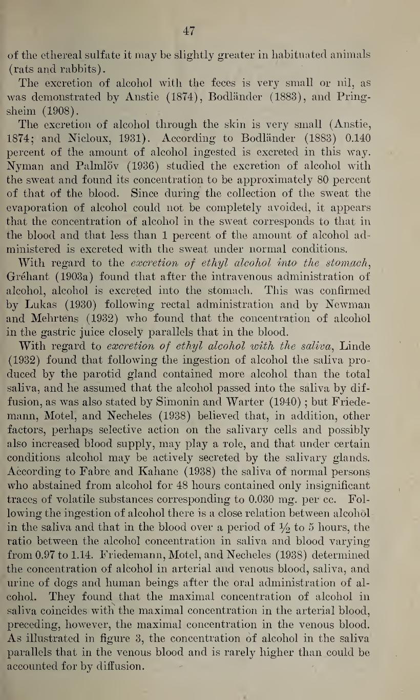
47
of the ethereal sulfate it may be slightly greater in habituated animals
(rats and rabbits).
The excretion of alcohol with the feces is very small or nil, as
was demonstrated by Anstie (1874), Bodlander (1883), and Pring-
sheim (1908). The excretion of alcohol through the skin is very small (Anstie,
1874; and Nicloux, 1931). According to Bodlander (1883) 0.140
percent of the amount of alcohol ingested is excreted in this way.
Nyman and Palmlov (1936) studied the excretion of alcohol with
the sweat and found its concentration to be approximately 80 percent
of that of the blood. Since during the collection of the sweat the
evaporation of alcohol could not be completely avoided, it appears
that the concentration of alcohol in the sweat corresponds to that in
the blood and that less than 1 percent of the amount of alcohol ad¬
ministered is excreted with the sweat under normal conditions.
With regard to the exwetion of ethyl alcohol into the stomach,
Grehant (1903a) found that after the intravenous administration of
alcohol, alcohol is excreted into the stomach. This was confirmed
by Lukas (1930) following rectal administration and by Newman
and Mehrtens (1932) who found that the concentration of alcohol
in the gastric juice closely parallels that in the blood.
With regard to excretion of ethyl alcohol with the saliva, Linde
(1932) found that following the ingestion of alcohol the saliva pro¬
duced by the parotid gland contained more alcohol than the total
saliva, and he assumed that the alcohol passed into the saliva by dif¬
fusion, as was also stated by Simonin and Warter (1940); but Friede-
mann, Motel, and Necheles (1938) believed that, in addition, other
factors, perhaps selective action on the salivary cells and possibly
also increased blood supply, may play a role, and that under certain
conditions alcohol may be actively secreted by the salivary glands.
According to Fabre and Kahane (1938) the saliva of normal persons
who abstained from alcohol for 48 hours contained only insignificant
traces of volatile substances corresponding to 0.030 mg. per cc. Fol¬
lowing the ingestion of alcohol there is a close relation between alcohol
in the saliva and that in the blood over a period of y2 to 5 hours, the
ratio between the alcohol concentration in saliva and blood varying
from 0.97 to 1.14. Friedemann, Motel, and Necheles (1938) determined
the concentration of alcohol in arterial and venous blood, saliva, and
urine of dogs and human beings after the oral administration of al¬
cohol. They found that the maximal concentration of alcohol in
saliva coincides with the maximal concentration in the arterial blood,
preceding, however, the maximal concentration in the venous blood.
As illustrated in figure 3, the concentration of alcohol in the saliva
parallels that in the venous blood and is rarely higher than could be
accounted for by diffusion.
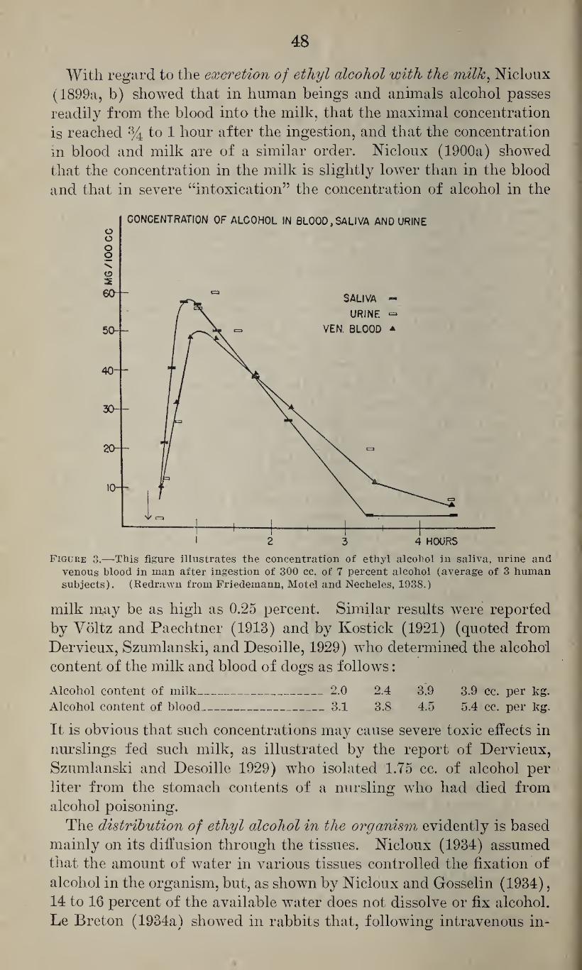
48
With regard to the excretion of ethyl alcohol with the milk, Nicloux
(1899a, b) showed that in human beings and animals alcohol passes
readily from the blood into the milk, that the maximal concentration
is reached % to 1 hour after the ingestion, and that the concentration
in blood and milk are of a similar order. Nicloux (1900a) showed
that the concentration in the milk is slightly lower than in the blood
and that in severe “intoxication” the concentration of alcohol in the
Figure 3.—This figure illustrates the concentration of ethyl alcohol in saliva, urine and venous blood in man after ingestion of 300 cc. of 7 percent alcohol (average of 3 human subjects). (Redrawn from Friedemann, Motel and Necheles, 1938.)
milk may be as high as 0.25 percent. Similar results were reported
by Voltz and Paechtner (1913) and by Kostick (1921) (quoted from
Dervieux, Szumlanski, and Desoille, 1929) who determined the alcohol
content of the milk and blood of dogs as follows:
Alcohol content of milk_ 2.0 2.4 3.9 3.9 cc. per kg. Alcohol content of blood_ 3.1 3.8 4.5 5.4 cc. per kg.
It is obvious that such concentrations may cause severe toxic effects in
nurslings fed such milk, as illustrated by the report of Dervieux,
Szumlanski and Desoille 1929) who isolated 1.75 cc. of alcohol per
liter from the stomach contents of a nursling who had died from
alcohol poisoning.
The distribution of ethyl alcohol in the organism evidently is based
mainly on its diffusion through the tissues. Nicloux (1934) assumed
that the amount of water in various tissues controlled the fixation of
alcohol in the organism, but, as shown by Nicloux and Gosselin (1934),
14 to 16 percent of the available water does not dissolve or fix alcohol.
Le Breton (1934a) showed in rabbits that, following intravenous in-
49
jection of alcohol, it takes from 20 to 60 minutes, depending on the
dose, to establish an equilibrium, and with intraperitoneal injection
it takes from 30 to 90 minutes. As shown in experiments with mice
and rats, this takes more time with subcutaneous administration
and, in addition, larger doses are less readily distributed. When
given on an empty stomach the equilibrium may be reached in dogs
1 hour after the administration, but when given on a full stomach it
may be considerably delayed. According to Loewy and von der Heide
(1918) it takes 8^4 hours to establish an equilibrium when concentra¬
tions of 0.5 percent of alcohol in air are inhaled, and with higher
concentrations it may take even more time.
With regard to the concentration of alcohol in different organs,
Lallemand, Perrin, and Duroy (1861) found, after oral administra¬
tion and following intravenous injection, higher concentrations in
liver and brain than in blood. Later investigators found that after an
equilibrium is reached ethyl alcohol is fairly uniformly distributed
among the various organs. Grehant (1899a) found that in dogs,
after oral administration of 5 cc. per kg. body weight, 54.9 cc. of the
58 cc. administered had been absorbed from the gastro-intestinal tract
after 3^4 hours, and that 100 cc. of blood contained 0.52 cc., 100 gm.
of brain contained 0.41 cc., 100 gm. of muscle contained 0.33 cc., 100 gm.
of liver contained 0.325 cc., and 100 gm. of kidney contained 0.39 cc.
Nicloux (1900c) studied in dogs and guinea pigs, after oral admin¬
istration, the concentration of alcohol in lymph, saliva, bile, pancreatic
fluid, spinal fluid, and amniotic fluid, and found it to be of the same
order as, although slightly lower than, that determined in the blood.
Alcohol was also found in testes, prostate, ovaries, seminal vessels,
and sperm, the content in the testes closely approaching that in the
blood. Vollmering (1912) also found a fairly uniform distribution
of alcohol in various organs, but he pointed out that until equilibrium
is established the blood contains more than other organs, and that
in the brain it takes more time to establish equilibrium and to release
it than with other organs such as liver and muscle tissue. Similarly,
Messner (1913) found in animals killed in alcohol narcosis a fairly
uniform concentration in blood, liver, and brain, and slightly less in
muscle tissue. Carpenter (1929) found in fowls, following exposure
for 2 to 29 hours to vapors of alcohol, the highest concentration in
the blood and only a small amount in fat, confirming in this respect
similar observations of Vollmering (1912). With higher concentra¬
tions the distribution in liver, heart, kidneys, lungs, and spleen was
quite uniform. Following continued administration of 4 cc. of al¬
cohol per kg. daily to rabbits, Marinesco, Lissievici-Draganesco,
Draganesco, and Grigoresco (1929) found larger concentrations in
heart and brain and considerably less in liver and eyeball, the latter
containing less ethyl alcohol than found under similar conditions
50
after the administration of methanol. Nicloux and Redslob (1931)
found that, after oral administration, ethyl alcohol passes from the
blood into the vitreous humor and that after 3 hours the concentration
corresponded to that of the blood, whereas the crystalline humor con¬
tained only about one-half this amount. With regard to the distri¬
bution of alcohol in the human organism, such data were determined
in victims of acute alcohol poisoning; and since the amount of alcohol
ingested, the time elapsing between the ingestion and death, and pos¬
sibly other factors controlling the distribution are not known in such
cases, the interpretation of such findings is difficult. Such determina¬
tions were made by Pauly and Bonne (1897), Juckenack (1908),
Balthazard and Lambert (1921) and Sartori (1930). Whereas Bal-
thazard and Lambert (1921) found a fairly uniform distribution in
blood, urine, brain, spleen, liver, kidneys, lungs, heart, and muscle,
the values reported by Juckenack (1908) and Sartori (1930) are not
uniform as illustrated by table 6.
Table 6.—Distribution of ethyl alcohol in the human organism in acute fatal alcohol poisoning (in weight percent)
Author
Blood of large
vessels and heart
Heart, lungs,
omentum, spleen
Stomach and
esophagus contents
Kid¬ neys Urine
Liver and gall
bladder Brain
Large intestine
and contents
Juckenack (1908). _ Sartori (1930) . _
0.53 .40
0.720 .197
0.72 .44
0.37 .32
0.65 .56
0.24 .39
0.42 .26 0.17
In view of the effect of alcohol on the central nervous system, the
concentration of alcohol in the brain has aroused much interest.
Kuijper (1883) isolated considerable quantities of alcohol from the
brain of a person drowned while intoxicated. Vollmering (1912)
showed that the concentration of alcohol in the brain in relation to
that in the blood varied at different times after the ingestion. Get-
tler and Tiber (1927b) determined the “normal alcohol content” of the
brain as 0.0025 percent and that in the brains of persons who had in¬
gested various amounts of alcohol as 0.005 to 0.6 percent. They
showed that the concentration of alcohol in the brain varied with the
intensity of the intoxication, that concentrations below 0.1 percent
caused no abnormal psychological effects, and that concentrations of
0.1 to 0.24 percent caused certain disturbances such as increased ag¬
gressiveness and carelessness but no disturbance of the equilibrium,
and that concentrations of 0.25, 0.4 and 0.6 percent caused the typical
picture of intoxication. Gettler and Freireich (1931) claimed that
the alcohol content of the brain could be appraised on the basis of the
alcohol content of the spinal fluid by dividing this value by the spinal
fluid-brain ratio which they determined as being from 1.1 to 1.4.
Gettler and St. George (1935) determined the concentration in the
51
brain of an infant who had died from acute alcohol poisoning as 0.65
percent. Olszycka (1940) studied the distribution of alcohol in var¬
ious sections of the brain of rats after doses of 2.4 mg. per gram body
weight. He found that the concentration in telencephalon, dienceph¬
alon, and cerebellum (0.207 to 0.212 gm. percent) was similar to that
in the blood (0.231 gm. percent), and that the concentration in the
spinal cord and the annular proturberance was slightly lower (0.196
and 0.195 gm. percent). With doses of 3.2 mg. per gram body weight
the corresponding values were 0.277 to 0.297 for brain tissue, 0.342 for
blood, and 0.269 for the spinal cord.
Nicloux (1899b) showed that alcohol passes from the mother to the
fetus and that after equilibrium has been reached the concentration in
the maternal and fetal blood are of the same order. Olow (1922)
showed that already 12 minutes after the ingestion of alcohol by the
mother appreciable quantities may be detected in the fetal blood and
that both are of the same order 40 minutes after the ingestion, and
that alcohol disappears from both bloods at the same rate.
The concentration of ethyl alcohol in the Wood varies with the dose
of alcohol ingested and the time elapsing between the ingestion and
the withdrawal of the blood. Grehant (1899a,b), Schweisheimer
(1913), and Miles (1922) found that following the oral administra¬
tion of alcohol the alcohol level in the blood increases during the
first 1 y2 to 2 hours and 70 minutes, respectively, and that it remains
at an elevated level for a variable length of time and then gradually
returns to normal values. Widmark (1915) showed that the rise
depends in part upon the amount of food in the stomach, and Olow
(1922) and Bornstein and Budelmann (1930) found that when given
on an empty stomach the maximal concentration in the blood is
reached within 30 minutes, and the former stated that it may return
to normal value within 2 and 2y2 hours. Haggard and Greenberg
(1934b) showed that the concentration of alcohol in the blood drawn
from an artery, the jugular vein, the right side of the heart, and the
skin capillaries over a period of 6% hours following the ingestion
of alcohol was of very similar order, but that the alcohol level in
blood withdrawn from the femoral vein during the period of absorp¬
tion was much lower and did not correspond to that in the urine or
that in the brain. Haggard, Greenberg and Rakieten (1940) showed
that before equlibrium is established the concentration of alcohol in
the blood drawn from different vessels may show wide differences.
Schmidt (1934) found considerable variations (0.04 to 0.08 percent)
in the blood of normal subjects after the ingestion of 0.6 cc. of alcohol
per kg. body weight in the form of whisky and soda, and he believed
this to be mainly due to a different speed of absorption. Gabbe
(1917) claimed that, following its intravenous administration, alcohol
disappears from the blood rapidly at first and then more slowly. With
u* m u. u&
52
the exception of Haggard and Greenberg (1934c) most investigators
(Mellanby, 1919; Widmark, 1933a; Neymark and Widmark, 19'36b;
Newman and Cutting, 1935; and Newman, Lehman, and Cutting,
1937) agree that, following the administration of single doses, alcohol
disappears from the blood at a constant rate.
With regard to the distribution of alcohol among the different
constituents of the blood, Miles (1922) found that the distribution
ratio between red blood cells and plasma is 2:1, Bornstein and Budel-
mann (1930) noted no difference in the distribution between red blood
cells and serum, and Elbel (1935) found the alcohol content of serum
greater than that of whole blood. The latter also found that the
serum value divided by 1.2 corresponds closely to the amount of
alcohol present in whole blood. This was confirmed by Kiinkele
(1936) who determined the ratio also as 1.2 with 1.12 and 1.31 as
extremes, and that of the blood clot to that of whole blood as 0.74
with 0.70 and 0.82 as extremes.
With regard to factors influencing the concentration of ethyl
alcohol in the blood, Miles (1922) showed that this depends in part
upon the concentration in which the alcohol was given, in that the
same dose of alcohol given in more concentrated form results in
definitely higher values in the blood. This was confirmed by Turner
and Loew (1932) whereas Kionka (1928) claimed that the concen¬
tration of the alcohol ingested had no influence on the blood alcohol
level, and Rasmussen (1940) found in rats that the blood alcohol
level was definitely lower when higher concentrations of alcohol were
given. Dybing and Kasmussen (1940) pointed out that this may be
partly explained by the histological characteristics of the rat’s
stomach, two-thirds of which are covered by mucosa containing sev¬
eral layers of glandless cornified epithelium, only one-third being of
the ordinary type. On account of its irritant action alcohol also
causes pylorospasm and is, therefore, not released into the gut where it
would be readily absorbed. In addition, the concentration of alcohol
in the blood depends upon the amount of alcohol ingested, as demon¬
strated by many investigators and as will be discussed in a later
section.
It has been shown repeatedly that the amount of food in the
stomach affects the alcohol level in the blood. Widmark (1915 and
1934), Miles (1922), Tuovinen (1930), Turner and Loew (1932),
Elbel and Lieck (1936) and others agree that when alcohol is taken
on a full stomach the alcohol level in the blood is lower than when
alcohol is given on an empty stomach or while the subject is starving.
Miles (1922) and Tuovinen (1930) believed that carbohydrates in¬
gested with alcohol reduce its concentration in the blood, but Wid¬
mark (1934) reported no such effects from carbohydrates and Elbel
and Lieck (1936) stated that the quantity of food is more important
than the relative caloric value and the composition with regard to
proteins, carbohydrates, and fats. Whereas Tuovinen (1930) and
Widmark (1934) stated that the ingestion of fats does not interfere
with the alcohol level in blood, both believed that 'proteins had defi¬
nitely an inhibitory effect, in that, as assumed by Widmark (1934),
they are precipitated by alcohol and thus delay its absorption. Wid¬
mark (1934) and Kanitz (1939) showed that the feeding of amino
acids, such as glycine, lowTers the alcohol level in the blood. Hag¬
gard and Greenberg (1940) believed that this effect can be explained
best as a prolonged retention of alcohol in the stomach, caused by
glycine, resulting in delayed absorption. Widmark (1934) stated
that the feeding of higher fatty acids and of citric, malic, and tar¬
taric acids as well as primary and secondary phosphates has a mod¬
erate lowering effect on the concentration of alcohol in the blood.
Kanitz (1939) found that this also holds true for lactic, pyrotartaric,
and succinic acids, whereas fumaric and glutamic acids had no or
very little effect. The latter also showed that feeding of the ethyl
esters of some of these and other acids caused a more or less marked
increase of the volatile constituents of the blood, indicating that
the use of aromatic, alcoholic beverages for such studies may lead to
erroneous results. Allodi and Dapra (1938) showed that serious
damage of the liver may cause abnormally high alcohol concentra¬
tions in the blood. Danopoulos (1938) found that blocking the
reticuloendothelial system with India ink or with colloidal copper
solution increased the alcoholemic curve materially and caused delay
of its decline. Similarly, Arima (1941) found that injury of the
liver delayed the reduction of the blood alcohol level. Danopoulos
(1939a) noted that in patients suffering from scurvy the blood alco¬
hol level was abnormally high without giving an explanation for this
phenomenon. Kanitz (1936) showed that in rabbits the administra¬
tion of 1 gm. of sugar and 1 unit of insulin with small doses of
alcohol (1.5 cc.) caused, during the resorptive phase, an abnormal
increase and later a decrease of the alcohol level in the blood, and
that this effect appears to increase within certain limits with the
dose of insulin given. Bohmer (1938) stated that large doses of
insulin caused lowering of the blood alcohol level during the re¬
sorptive phase and an unusual rise during the post resorptive phase
which he interpreted as indicative of delayed absorption. According
to Arima (1941), injury of the kidney does not affect the blood
alcohol curve. In view of the importance of the concentration of
alcohol in the blood with regard to medico-legal questions, the ef¬
fect of drugs on the blood alcohol level has aroused much interest.
Walter (1938) stated that neither acetyl salicylic acid (aspirin) nor
gardan (an amidopyrine preparation) caused a lowering of the blood
54
alcohol level, the latter causing only a slightly delayed absorption.
Similar negative results were reported by Elbel (1938) who found
the same to hold true for barbital. Rinkel and Myerson (1941) re¬
ported that benzedrine sulfate, paredrine, epinephrine hydrochlor¬
ide, and atropine sulfate lower the blood alcohol level, decreasing in
effectiveness in the order given, and they assumed that this effect
was due to delayed absorption, by delaying the emptying of the
stomach. McCrea and Taylor (1940) found that the administration
of metrazol had no effect on the disappearance of alcohol from the
blood of dogs. Siegmund (1938) studied the question as to whether
or not solvents, such as those used in lacquer spraying, may affect
blood alcohol findings, as determined by Widmark’s test, but found
no evidence of such action at the end of 1 week of spray painting.
According to Leschke (1932), Bichler claimed that at high altitudes
the increase of alcohol in the blood is less rapid, reaches a lower
peak, and declines more rapidly than normally, which he thought
to be due to the more rapid respiration and greater pulmonary
elimination. Scliweisheimer (1913) and Fleming and Stotz (1935)
claimed that in chronic alcoholism the concentration of alcohol in
the blood increases more rapidly, reaches a higher level, and de¬
creases more rapidly than in abstainers, whereas Graf and Flake
(1932-33) believed that habituation has no effect on the alcohol level
in the blood.
With regard to the relation of the concentration of ethyl alcohol in
hlood to that in the urine, Nicloux (1900c) and Nicloux and Nowicka
(1913) claimed that at the moment of secretion the alcohol content of
the urine equals that of the blood, and this was confirmed by Widmark
(1915), Chabanier and Ibarra-Loring (1916), Grehant (quoted from
Haggard and Greenberg, 1934b) and Schweisheimer (1913). Haggard
and Greenberg (1934b) pointed out that alcohol is more soluble in
blood than in urine but that the concentrations in blood and urine
are of the same order. This was confirmed by Siegmann (1936) who
determined the average difference as 0.03 percent. Miles (1922),
Southgate and Carter (1926), Bornstein and Budelmann (1930) and
Carlson, Kleitman, Muehlberger, McLean, Gullicksen, and Carlson
(1934) found that the concentration in the urine parallels that in the
blood, being, however, slightly higher. This was interpreted by Born¬
stein and Budelmann (1930) as indicative of a moderate ability of the
kidneys to concentrate alcohol, but it should also be pointed out that
the concentration of alcohol in blood samples taken at the time of the
collection of urine from the bladder does not represent the concentra¬
tion of alcohol in the blood at the time of its excretion by the kidneys.
The findings of Carlson and his associates (1934) are given in table 7.
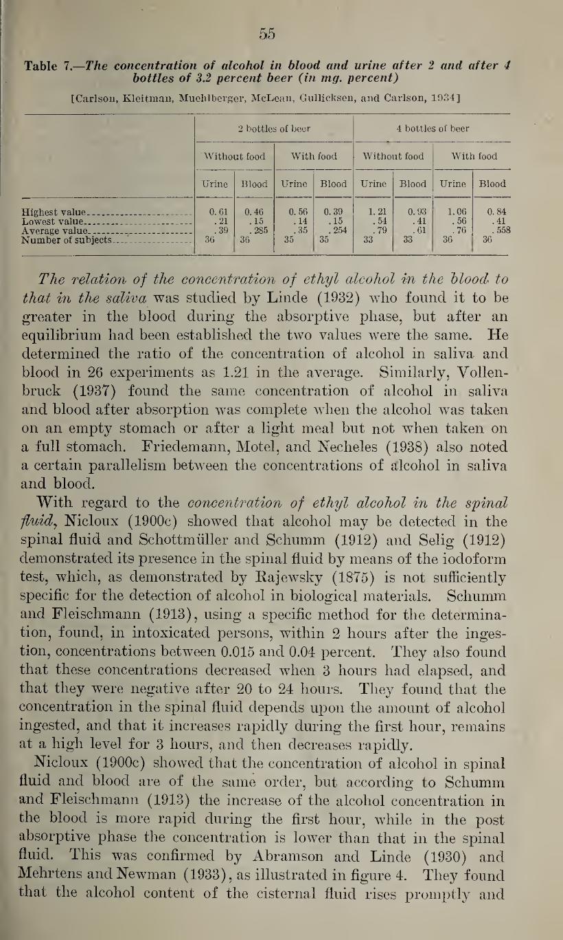
55
Table 7.—The concentration of alcohol in blood and urine after 2 and after 4 bottles of 3.2 percent beer (in mg. percent)
[Carlson, Kleitman, Muehlberger, McLean, Gullicksen, and Carlson, 1934]
2 bottles of beer 4 bottles of beer
Without food With food Without food With food
Urine Blood Urine Blood Urine Blood Urine Blood
Highest value__ 0.61 0.46 0. 56 0.39 1. 21 0. 93 1.06 0.84 Lowest value__ .21 . 15 .14 .15 .54 .41 .56 .41 Average value_... .. _ . .39 .285 .35 .254 .79 .61 .76 .558 Number of subjects_ 36 36 35 35 33 33 36 36
The relation of the concentration of ethyl alcohol in the blood to
that in the saliva was studied by Linde (1932) who found it to be
greater in the blood during the absorptive phase, but after an
equilibrium had been established the two values were the same. He
determined the ratio of the concentration of alcohol in saliva and
blood in 26 experiments as 1.21 in the average. Similarly, Vollen-
bruck (1937) found the same concentration of alcohol in saliva
and blood after absorption was complete when the alcohol was taken
on an empty stomach or after a light meal but not when taken on
a full stomach. Friedemann, Motel, and Necheles (1938) also noted
a certain parallelism between the concentrations of alcohol in saliva
and blood.
With regard to the concentration of ethyl alcohol in the spinal
;fluidy Nicloux (1900c) showed that alcohol may be detected in the
spinal fluid and Schottmtiller and Schumm (1912) and Selig (1912)
demonstrated its presence in the spinal fluid by means of the iodoform
test, which, as demonstrated by Rajewsky (1875) is not sufficiently
specific for the detection of alcohol in biological materials. Schumm
and Fleischmann (1913), using a specific method for the determina¬
tion, found, in intoxicated persons, within 2 hours after the inges¬
tion, concentrations between 0.015 and 0.04 percent. They also found
that these concentrations decreased when 3 hours had elapsed, and
that they were negative after 20 to 24 hours. They found that the
concentration in the spinal fluid depends upon the amount of alcohol
ingested, and that it increases rapidly during the first hour, remains
at a high level for 3 hours, and then decreases rapidly.
Nicloux (1900c) showed that the concentration of alcohol in spinal
fluid and blood are of the same order, but according to Schumm
and Fleischmann (1913) the increase of the alcohol concentration in
the blood is more rapid during the first hour, while in the post
absorptive phase the concentration is lower than that in the spinal
fluid. This was confirmed by Abramson and Linde (1930) and
Mehrtens and Newman (1933), as illustrated in figure 4. They found
that the alcohol content of the cisternal fluid rises promptly and
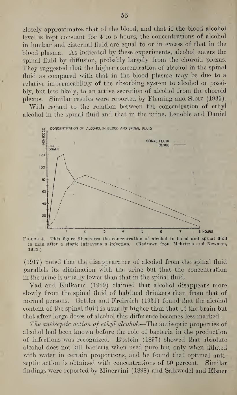
56
closely approximates that of the blood, and that if the blood alcohol
level is kept constant for 4 to 5 hours, the concentrations of alcohol
in lumbar and cisternal fluid are equal to or in excess of that in the
blood plasma. As indicated by these experiments, alcohol enters the
spinal fluid by diffusion, probably largely from the choroid plexus.
They suggested that the higher concentration of alcohol in the spinal
fluid as compared with that in the blood plasma may be due to a
relative impermeability of the absorbing system to alcohol or possi¬
bly, but less likely, to an active secretion of alcohol from the choroid
plexus. Similar results were reported by Fleming and Stotz (1935).
With regard to the relation between the concentration of ethyl
alcohol in the spinal fluid and that in the urine, Lenoble and Daniel
vj p CONCENTRATION OF ALCOHOL IN BLOOD AND SPINAL FLUID
Figure 4.—This figure illustrates the concentration of alcohol in blood and spinal fluid in man after a single intravenous injection. (Redrawn from Mehrtens and Newman,
1933.)
(1917) noted that the disappearance of alcohol from the spinal fluid
parallels its elimination with the urine but that the concentration
in the urine is usually lower than that in the spinal fluid.
Vad and Kulkarni (1929) claimed that alcohol disappears more
slowly from the spinal fluid of habitual drinkers than from that of
normal persons. Gettler and Freireich (1931) found that the alcohol
content of the spinal fluid is usually higher than that of the brain but
that after large doses of alcohol this difference becomes less marked.
The antiseptic action of ethyl alcohol.—The antiseptic properties of
alcohol had been known before the role of bacteria in the production
of infections was recognized. Epstein (1897) showed that absolute
alcohol does not kill bacteria when used pure but only when diluted
with water in certain proportions, and he found that optimal anti¬
septic action is obtained with concentrations of 50 percent. Similar
findings were reported by Minervini (1898) and Salzwedel and Eisner
57
(1900). The latter found that concentrations of 50 to 55 percent cor¬
respond with regard to their antiseptic action to 3-percent solutions
of phenol, but that fresh bacteria may be killed also by absolute
alcohol, as was also shown by Buchner, Fuchs, and Megele (1901) for
staphylococcus pyrogenes aureus, bacillus typhi, and bacillus pyocy-
aneus. Tijmstra (1913) gave the optimal antiseptic concentration as
70 percent. Gerschenfeld (1938) found that 95 percent ethyl alcohol
did not kill Bacillus megatherium and Bacillus subtilis until the ex¬
posure had lasted about 3 months in the first and 7 to 9 months in
the latter case. Christiansen (1918) showed that the antiseptic action
of alcohol was due to a twofold mechanism; that it acted by dehydra¬
tion of the fresh bacteria, as produced by 100 percent alcohol, and by
precipitation of the proteins of the plasma. For these reasons dried
bacteria, such as used in the silk-thread method and in the glass bead
method for testing antisepetics, are not affected to the same extent
by absolute alcohol. Christiansen (1918) showed that the penetration
of alcohol into the cell varies with the inverse ratio of the surface
tension of its solutions which, as shown by Kisch (1912), decreases
with the concentration. In addition, an increase of the temperature
enhances the penetration of alcohol solutions of a certain surface
tension. Within the cell, alcohol precipitates the proteins and there¬
fore kills the organism. This precipitant action depends in part upon
the amount of salts within the cells, and this may explain why spores
are not killed by alcohol, as was demonstrated by Minervini (1898),
Knorr (1932), and others. Since the precipitation of proteins depends
also upon the concentration of alcohol, this explains why the optimal
antiseptic action of alcohol is bound to certain concentrations.
The effect of alcohol on the central nervous system.—Alcohol de¬
presses the functions of the central nervous system. Albertoni and
Lussana (1874) (quoted from Kochmann, 1923) showed that the
intravenous and oral administration of proper doses of alcohol
causes in birds, motor paralysis, but that loss of sensitivity is produced
only by toxic or fatal doses. Baer (1898) studied the effect on rabbits
of oral administration of various doses of alcohol. He found that
doses of 2.5 to 4.1 gm. per kg. caused moderate poisoning, character¬
ized by primary excitation, subsequent moderate motor depression,
and moderate reduction of sensitivity, whereas the corneal, ciliary,
and pupillary reflexes were not affected and respiration, pulse rate,
and body temperature were not distinctly changed. Doses of 4.45
to 6.15 gm. per kg. caused, after 15 minutes following a slight primary
stimulation, complete motor paralysis, marked analgesia, impairment
of corneal reflexes, constriction of pupils with sluggish or negative
reaction, slowing of the respiration, and reduction of the body tem¬
perature, the recovery taking about 10 hours. Doses of 6.25 to 7.44
555178—43-5
58
gm. per kg. caused, within a few minutes, rapid and complete paralysis,
reduction of the sensitivity and reflexes, constriction and nonreactivity
of the pupils, nystagmus, salivation, occasionally tonic convulsions,
marked slowing of the respiration, reduction of the body temperature,
and narcosis lasting 36 hours. Turner and Loew (1932) correlated
in dogs the concentration of alcohol in the blood with the degree of
the depression and found that concentrations of 0.25 percent cause
definite intoxication; of 0.30 percent, marked intoxication; and of
0.4 to 0.5 percent, alcoholic stupor. Alexandrofl and Talpis (1929)
noted in rabbits poisoned with alcohol marked disturbances of the
reflexes controlling the posture of the neck, less marked disturbances
of the locomotor reactions and the labyrinth reflexes. Versteegh
(1922) studied in rabbits the effect of various doses of alcohol on the
postural reflexes. He found that oral administration of 3 cc. per kg.
caused no disturbances of the reflexes except inhibition of locomotion
(lift reaction). Doses of 5 to 6 cc. per kg. abolished the cervical pos¬
tural reflexes, affecting first the anterior and later the posterior re¬
flexes. Doses of about 8 cc. per kg. abolished the postural reflexes,
so that the animals were unable to maintain their normal position.
Other reflexes, such as motor eye reflexes, corneal reflexes, patellar re¬
flexes and flexor reflexes, were still positive at a time when the postural
reflexes had disappeared. Chauchard, Chauchard and Kijiwara
(1930b) studied in dogs the effect of alcohol on the excitability of neu¬
rons of the cerebral cortex, especially of those regions which provoke
a response in the antagonists. They found that under the influence
of alcohol the chronaxy in the cortical neurons becomes stabilized,
but that the chronaxy of the cortical zones of antagonists increases at a
different speed, thereby creating disturbances which may contribute
to the difficulties in coordination observed in acute alcohol intoxication.
With regard to the narcotic dose of alcohol, this varies with the
species and with the route of administration, as illustrated by the
following reports. Lehman and Newman (1937 b) determined the
minimal narcotic dose for rabbits with intravenous injection as 5.6
cc. per kg.; Grilichess (1913), with intramuscular injection, as 8.0
cc. per kg.; Lendle (1928), with intraperitoneal injection in rats, as
4.5 cc. per kg.; and Strauss (1887), with oral administration to rabbits,
as 6 to 8 gm. Latven and Molitor (1939) determined the minimal
hypnotic dose for mice with subcutaneous administration as 2 cc.
per kg., with oral administration as 3 cc. per kg., and with intravenous
administration as 0.8 cc. per kg. Flury and Klimmer (quoted from
Lehmann and Flury, 1938) determined the narcotic action of alcohol
vapors in mice.
The question as to whether or not ethyl alcohol stimulates the
respiration has aroused much interest. Wilmanns (1897) and others,
such as Binz (1891), Heinz (1890), and Weissenfeld (1898) (the
/
last three quoted from Kochmann, 1923) showed that the adminis¬ tration of moderate doses of alcohol causes a moderate stimulation of the respiratory center. Wendelstadt (1899) found that small doses of alcohol cause only a moderate stimulation of the respiration in normal persons, but that with such doses the effect is much more marked in fatigued persons; and from observation that highly aro¬ matic wine was found to be more effective than pure alcohol it ap¬ pears that essential oils may have an additive or perhaps a synergistic effect. This stimulant effect depends, however, upon the dose of alcohol administered. Fonteyne (1906) showed that in rabbits the intravenous injection of 0.16 gm. per kg. stimulates and deepens the respiration but that doses of 1.44 gm. per kg. cause depression. He believed that with regard to the depressant action 3 stages can be distinguished: (1) slight inhibition with deepened respiration; (2) marked slowing with reduced respiratory volume; and (3) very marked slowing with shallow respiration. Hooker (1917) perfused the medulla of dogs wfith defibrinated blood containing various con¬ centrations of alcohol. He found that with an alcohol concentration in the blood of 0.025 percent there was some stimulation of the respira¬ tion, that with 0.1 percent the stimulative response was greater, but that 0.25 percent caused only a transitory stimulation followed by prolonged inhibition. Gradinesco (1934) believed that alcohol af¬ fects the respiratory center, the first costo-thoracic nerve, and later also the phrenic nerve. In contrast to these positive findings, Lieb (1915) found that the oral administration of alcohol to etherized and decerebrated cats had no stimulant effect on the respiration. Simi¬ larly, Hyatt and Jensen (1918) found that in etherized dogs with sectioned spinal cord to insure loss of sensation in the hind legs the intravenous injection of alcohol into the desensitized legs caused only a moderate stimulation of the respiration. Similar results were reported by Hyatt (1919) with oral administration to dogs whose spinal cords were sectioned at the height of the eleventh and twelfth thoracic vertebrae, and he believed that the stimulant effect of alcohol on the respiration is bound to some local irritation. Hitchcock (1942) found, however, in humans that small doses of alcohol stimulate the respiration, at least for a limited period of time; and he believed this was not due to a reflex mechanism from the gastric mucosa but was caused by direct stimulation of the respiratory center. Similarly, experiments of Masserman (1940) indicate that alcohol may have some stimulant effect on certain structures of the brain. He found that in cats under light ether anesthesia the intravenous or intraperitoneal injection of small doses of alcohol may produce a, stimulant effect on the hypothalamus but not on the sigmoid cortex, and that, there¬ fore, small doses of alcohol which are easily metabolized may facilitate the response of the central nervous system, especially that of the lower
60
diencephalic region. It has been stated before that larger doses of
alcohol invariably cause more or less severe depression of the respira¬
tion. According to Haggard, Greenberg, and Rakieten (1940), con¬
centrations of 9.3 mg. per cc. of blood in fasting rats will cause
respiratory failure, and according to McCrea and Taylor (1940) in
dogs the intravenous injection of 3.0 to 7.2 cc. per kg. body weight
will cause respiratory arrest which can be overcome temporarily by
the administration of metrazol. Gold and Travell (1934) studied in
dogs the antagonism between alcohol and strychnine and found that
in strychnine poisoning small doses of alcohol may have an antagon¬
istic effect whereas large doses may lead to respiratory depression.
This was confirmed by Norris (1937) who claimed that both strych¬
nine and alcohol act somewhat alike on the brain and the spinal cord,
causing death by cardiac and respiratory failure, and that alcohol
is no antagonist in strychnine poisoning. Reifenstein (1941) studied
the antagonism between alcohol and amphetamine sulfate and found
that the latter counteracts the narcotic action of moderate doses of
alcohol but not that of lethal doses, and that it may even increase
the toxic effect of the latter. On the other hand, he found that
alcohol may protect rabbits from 1.5 to 2.0 times the minimal fatal
dose of amphetamine sulfate.
Attempts have also been made to use alcohol for the production of
narcosis in humans. Marin (1929) (quoted from Constantin, 1929)
advocated its intravenous administration for this purpose, worked
out detailed directions, and claimed that this form of narcosis was
simple, comparatively free from post narcotic complications, easily
controlled and without untoward effects on the circulation. Con¬
stantin (1929) found it satisfactory in two cases, and Fohl (1931)
found that pathological organ changes are much less than might be
expected, resembling in many respects those seen with other narcotics.
However, he believed that there is considerable danger of thrombus
formation which may cause embolism. He also found indications of
impairment of the circulation and vasomotor mechanism which in his
opinion may predispose to certain complications, such as pneumonia,
and that the narcosis is not as easily controlled as might be desired.
Much effort has been devoted to the effect of ethyl alcohol on the
functions of the central nervous system of man. The first experi¬
mental studies on this subject were carried out by Kraepelin and his
collaborators, by Aschaffenburg (1896), Bergmann (1905), Dodge and
Benedict (1915), Benedict (1916), Gyllensward (1917), Graf (1932-
33), and many others. Jellinek and McFarland (1940) gave an exten¬
sive critical review of such psychological tests. They pointed out that
different psychological functions are affected by alcohol at different
concentrations of alcohol in the blood, or, rather, in the brain. In
their opinion it appears to be established that the ingestion of alcohol
61
impairs the tactile, auditory, and visual sensations and perception,
especially in those tests which involve active attention. They showed
that most authorities agree that ingestion of alcohol results in an in¬
crease of the reaction time, and believed that a critical analysis of
data published by earlier investigators throws some doubt on state¬
ments that ingestion of alcohol causes a primary reduction of the re¬
action time, and that such results are partly due to inadequate arrange¬
ment of the experiments and partly caused by continuation of the
practice effect. The latter effect is not overcome during the first 20
minutes after the ingestion of small doses (10 cc.) of alcohol. It seems
to be established that larger doses of alcohol cause an immediate in¬
crease of the reaction time the magnitude of which varies with the
amount of alcohol ingested and its concentration.
With regard to the effect of ethyl alcohol on muscular strength and
coordination, Schnyder (1903) believed that the ingestion of small
amounts of alcohol lias a favorable effect on the muscular energy
only in cases where the energy has been exhausted, that this effect is
less marked than that of a nutrient of corresponding caloric value, and
that it is unfavorably affected by the depressant effect of alcohol on
the central nervous system. He came to the conclusion that in prop¬
erly nourished persons alcohol has no favorable effect on the muscular
energy, but rather impairs it. Jellinek and McFarland (1940) stated
that small doses of alcohol (10 to 20 cc.) have been generally found
to increase the muscular output above the initial level for at least a
short while, that with medium doses (30 to 45 cc.) the increase is less
pronounced and that with larger doses (60 to 80 cc.) there is either
no change or a deterioration of the performance. They believed that
the apparently favorable effect of alcohol on the muscular output can
be explained either by a sensory excitation or by a facilitation of the
motor impulses. It appears further that impairment produced by
medium doses of alcohol is greater in those tasks which require a
greater muscular coordination, and that in such experiments it may
reach 50 to 60 percent. It is, therefore, evident that alcohol is not
suitable for increasing industrial efficiency, but it appears that small
doses (10 to 20 cc.) do not necessarily interfere with the efficiency of
workers accustomed to alcohol.
With regard to changes in performance of miscellaneous tests of
dexterity and skill, Jellinek and McFarland (1940) believed that these
are affected by alcohol in proportion to their complexity but that this
element may be compensated and even outweighed by familiarity with
the task, unless the complexity is of a high degree.
In respect to changes in learning and simple learned performances
it appears that these are greatly impaired after the ingestion of alco¬
hol, and animal experiments seem to indicate that this also impairs
learned tasks.
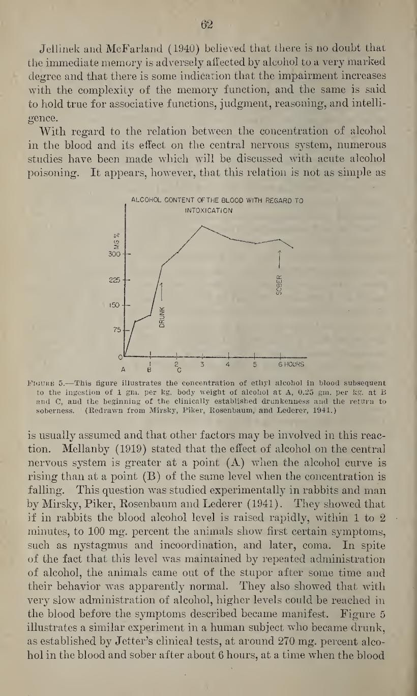
02
Jellinek and McFarland (1940) believed that there is no doubt that
the immediate memory is adversely affected by alcohol to a very marked
degree and that there is some indication that the impairment increases
with the complexity of the memory function, and the same is said
to hold true for associative functions, judgment, reasoning, and intelli¬
gence.
With regard to the relation between the concentration of alcohol
in the blood and its effect on the central nervous system, numerous
studies have been made which will be discussed with acute alcohol
poisoning. It appears, however, that this relation is not as simple as
ALCOHOL CONTENT OF THE BLOOD WITH REGARD TO
Figure 5.—This figure illustrates the concentration of ethyl alcohol in blood subsequent to the ingestion of 1 gm. per kg. body weight of alcohol at A, 0.25 gm. per kg. at B and C, and the beginning of the clinically established drunkenness and the return to soberness. (Redrawn from Mirsky, Piker, Rosenbaum, and Lederer, 1941.)
is usually assumed and that other factors may be involved in this reac¬
tion. Mellanby (1919) stated that the effect of alcohol on the central
nervous system is greater at a point (A) when the alcohol curve is
rising than at a point (B) of the same level when the concentration is
falling. This question was studied experimentally in rabbits and man
by Mirsky, Piker, Rosenbaum and Lederer (1941). They showed that
if in rabbits the blood alcohol level is raised rapidly, within 1 to 2
minutes, to 100 mg. percent the animals show first certain symptoms,
such as nystagmus and incoordination, and later, coma. In spite
of the fact that this level was maintained by repeated administration
of alcohol, the animals came out of the stupor after some time and
their behavior was apparently normal. They also showed that with
very slow administration of alcohol, higher levels could be reached in
the blood before the symptoms described became manifest. Figure 5
illustrates a similar experiment in a human subject who became drunk,
as established by Jelter’s clinical tests, at around 270 mg. percent alco¬
hol in the blood and sober after about 6 hours, at a time when the blood
63
alcohol level was still much higher than it was at the time when he
was pronounced drunk. Mirsky and his associates assumed that some
change or adaptation of the central nervous system may be responsible
for this variation in the clinical response to alcohol, but it may also
be possible that the narcotic action is bound, to a certain extent, to the
gradient between the alcohol concentration in the blood and that in the
brain tissue.
With regard to the effect of ethyl alcohol on the vision. Hollmann
(1905-6), found, in a young man, following acute intoxication, a reduc¬
tion of the visual field for white and for colors, a reduction of the
visual acuity, and, occasionally, negative scotoma, i. e., a scotoma
which is not ordinarily perceived but detected only upon examination
of the entire visual field. Busch (1910) found that the ingestion of 30
cc. of alcohol causes a distinct decrease of the tachistoscopic perception
of visual stimuli but found the visual acuity unimpaired. Schulz
(1916 and 1917a, b) demonstrated that, following the ingestion of
alcohol, the adaptation to light and dark and, to a lesser extent, to
red and green, is affected, and that some persons are more susceptible
in this respect than others. But according to Manz (1940) the speed
of dark adaptation of the human eye is not changed after the ingestion
of alcohol. He found, however, that the realization of depth is dis¬
tinctly impaired with concentrations of 0.06 to 0.125 percent of alco¬
hol in the blood and that with a blood alcohol level of 0.03 percent
there was already some impairment of judgment of depth, mainly
characterized by a delay in making the proper estimate. Colson
(1940) found, in acute intoxication, no reduction of the visual acuity,
the visual field, or the color vision, nor any tendency to hyperphoria.
He noted, however, a gradually increasing esophoria which, in two
subjects, progressed to convergent strabismus and diplopia in the
latter part of the test. One subject had a definite exophoria at the
onset of the test and passed through the stage of orthophoria before
becoming esophoric. Newman and Fletcher (1941) studied in 50
drivers the effect on vision of approximately 1 ounce of whisky per
30 pounds body weight, to be taken in 15 to 30 minutes. They tested
these subjects with regard to visual acuity, depth perception, distance
judgment, lateral field of vision, eye coordination, glare resistance,
and glare recovery. In every case with a blood alcohol level above
0.115 percent, the result of at least 1 of these tests was changed sig¬
nificantly. The greatest number of changes occurred in the visual
acuity, the smallest in the field of vision. * They found no definite
relation between blood alcohol level and performance, but noted a
considerable variation in the tolerance to a given concentration of
alcohol in the blood. These few references may suffice to show, as was
also pointed out by Jellinek and McFarland (1940), that results of
different investigators on the effect of alcohol on the visual acuity are
64
conflicting, but that some impairment of the visual acuity is indicated.
Alcohol may also have a more injurious effect on the eye, as illus¬
trated by the report of Kaiser (1912) on 1 case of acute alcohol am¬
blyopia, characterized by peripheral restriction of the visual field,
loss of sensation to colors and marked reduction of the visual acuity
lasting for several days and resulting presumably from a direct toxic
effect of alcohol on the nervous structures. Carroll and Goodhart
(1938) reported 6 cases of total but temporary blindness lasting
usually 24 hours, and they refer to similar cases reported by
deSchweinitz (1896) and Schrader (1927). In spite of the blindness,
in these patients the pupils reacted normally to light and convergence
and there were no abnormalities in the fundi, indicating the functional
character of the phenomenon.
With regard to pupillary changes, Stapel (1911) noted in acute alco¬
holic intoxication the following abnormalities: (1) Bilaterial dilata¬
tion, rarely constriction; (2) reduction of the accommodation to light,
which may be preceded by a very moderate increase; (3) reduction of
the response to sensory and sense stimuli and to psychic reactions; and
(4) in maximal intoxication, complete rigidity of the pupils.
The effect of ethyl alcohol on the static organ varies with its con¬
centration in the blood. Manz (1940) noted, at a blood alcohol level
of 0.03 to 0.04 percent, an increased excitability of the static apparatus
and nystagmus, and with concentrations of 0.036 to 0.103 percent he
noted that 6 out of 11 test persons showed spontaneous nystagmus.
With regard to the effect of ethyl alcohol on the spinal apparatus,
Finkelnburg (1904) showed that in dogs the oral administration of
ethyl alcohol results, after 12 to 30 minutes, in a slow increase of the
cerebrospinal pressure. He concluded from the duration of this
phenomenon that this is not due to a circulatory effect but is caused
by an increased secretion of cerebrospinal fluid. Lhermitte, de
Ajuriaguerra and Gamier (1938) showed that continued administra¬
tion of alcohol to rabbits does not affect the spinal cord, as claimed by
Lhermitte (1935), but that the changes observed by the latter were due
to avitaminosis rather than caused by alcohol.
With respect to the irritant action of ethyl alcohol, Loewy and von
der Heide (1918) stated that only concentrations of 0.1 to 0.25 percent
(1,000 to 2,500 p. p. m.) of alcohol in air can be inhaled without dis¬
comfort. According to Kochmann (1923) aqueous solutions of 20
percent alcohol cause burning and a feeling of warmth when applied
to mucous membranes, solutions of 50 to 60 percent cause pain, and
concentrations of 80 to 90 percent may cause similar effects on the in¬
tact skin provided that the evaporation of alcohol is prevented.
With regard to the effect of ethyl alcohol on the peripheral motor
nerves, Kochmann (1923) refers to Biedermann (1881), Mommsen
(1881), Efron (1885), Gad (1888), Pietrowski (1893), Breyer (1903),
65
Waller (1897, 1908, and 1909), Verar (1909) and others as having shown that diluted solutions or low concentrations of alcohol in air may stimulate the excitability of the isolated sciatic nerve of frogs. Gioffredi (1898) (quoted from Kochmann, 1923) showed that this effect is due to an increase of the conductivity. More concentrated solutions cause, however, depression and paralysis of the nerve, the intensity of the depression depending upon the concentration and the duration of the exposure. Bonnet and Lelu (1933) found that 1 per¬ cent solutions of alcohol first increase the chronaxy of the isolated sciatic nerve of frogs and later lower it to various extents, and that 2.0 to 3.0 percent cause a moderate decrease and 5 percent a primary increase and subsequent decrease. Gradinesco and Degan (1934) ob¬ served, with concentrations of 0.5 to 10 percent, a primary decrease of the chronaxy of varying intensity, depending in degree upon the con¬ centration of alcohol and the duration of the exposure, which was followed by an increase of the chronaxy and paralysis. They found that concentrations of 15 to 30 percent cause complete paralysis which is reversible with lower concentrations but irreversible with higher concentrations. Lapicque and Kajiwara (1930) found in dogs that alcohol affects the chronaxy of antagonistic nerves to a different extent, resulting in a modification of the chronaxy of the extensor and flexor muscles.
With regard to the effect of ethyl alcohol on the sensory nerves, it was shown by Horvath (1873) that alcohol paralyzes the peripheral sensory nerve endings after a short primary stimulation and that in this way it may act as a local anesthetic.
The effect of ethyl alcohol on the circulation.—The effect of ethyl alcohol on the heart has been studied in cold-blooded and warm-blooded animals. Ringer and Sainsbury (1883) showed in frogs that alcohol depresses the heart muscle and increases moderately the heart rate but not the force of the cardiac contraction. Dreser (1887), working with the isolated frog heart (Williams’ arrangement) found that small concentrations of alcohol (0.015 gm. per 45 cc. of blood corresponding to 0.033 percent) have generally no favorable effect and cause only exceptionally a moderate increase of the energy but usually a depres¬ sion. Using the same procedure Dieballa (1894) found that concen¬ trations of 0.144 to 9.4 percent cause depression of the cardiac action, less vigorous contractions, and a slight effect on the heart rate. These effects were reversible by lavage. Fulmer (1921) determined the mini¬ mal paralyzant concentration of ethyl alcohol for the heart as 1.206 mole per liter (5.52 per cent). Ransom (1919) stated that 1 percent solutions have only a slight effect on the heart action, 2 percent cause temporary depression followed b}^ recovery, and that 3 and 4 percent solutions cause arrest for 30 minutes. However, after this the heart recovers gradually in spite of continued perfusion with such concern
66
tration, although with distinctly toxic concentrations the recovery was never complete. Wolff (1922) found that 2 and 6 normal solutions (9.2 and 27.6 percent) cause cardiac arrest in middle position, and after lavage he noted some increase of the heart rate and more vigor¬ ous systolic contraction. A similar observation was reported by Toyoshima (1928). He believed that alcohol always depresses the action of the isolated frog heart, that this effect is not related to the parasympathetic system but is due to a depressant effect on the cardiac muscle, as had been assumed by Ringer and Sainsbury (1883), and that it is also partly caused by an impairment of the conductivity in the venous sinus, the auricles, and probably also the ventricle, whereas Dreser (1887) assumed that the depressant effect of alcohol on the heart was presumably no direct cardiac action and Seliskar (1926) as¬ sumed that alcohol affects the contractility more than the conduction. Vernon (1910) found that concentrations of 0.5 per cent of alcohol in saline reduce the beat of the perfused turtle heart to a constant level in about 14 minutes and that no further changes occur over a period of 16 minutes. With higher concentrations the depressant effect was more marked and reached a constant level sooner. He be¬ lieved that with each concentration a certain amount of alcohol is bound to the contractile substance of the heart muscle. He also noted that, during lavage of the heart with Ringer’s solution, there was occasionally a temporary increase of the amplitude of the heart and that this effect was greater during recovery from the effects of higher concentrations of alcohol. These findings were confirmed by Robert¬ son and Clark (1933), who determined the amount of alcohol in the heart muscle and who found that after 5 minutes have elapsed no further alcohol is taken up althoughThe perfusion may be continued for as long as 3 hours.
The effect of ethyl alcohol on the mammalian heart was studied by Martin and Stevens (1895) in the perfused isolated dog heart. They found that under the influence of alcohol the systole is less complete and that in the beginning this effect may be compensated by an increase of the diastole so that the volume of the blood ejected is not decreased, but that later this diastolic compensation fails and the output is reduced. Plunder (1905) stated that alcohol has only a depressant effect on the mammalian heart. In opposition to these find¬ ings, Bock (1898) and Loeb (1904) claimed that the mammalian heart is quite resistant to alcohol. Tunicliffe and Rosenheim (1903) found that concentrations of 1:500 (0.2 percent) cause a transient slowing of the isolated mammalian heart and very slight irregu¬ larities, but that the heart can be perfused for a relatively long time without causing further alteration. Similarly, Kochmann (1905) found that perfusion with 0.3 percent solutions of alcohol had no distinct effect, 0.4 percent caused reduction of the pulse volume,
67
0.5 percent caused in addition slowing of the pulse rate, and that
1 percent solutions caused distinct toxic effects but not arrest. Back-
man (1906) pointed to the great variations in the response of dif¬
ferent heart preparations. He found that concentrations of 0.05
to 0.1 percent caused temporary arrhythmia and temporary reduc¬
tion of the systole in the perfused mammalian heart, these effects being
more marked with 0.1 to 0.5 percent solutions. In the opinion of
Dixon (1907) concentrations of 0.5 percent have a distinct, depres¬
sant effect on the heart action. According to Kuno (1913), 0.077 per¬
cent solutions are just sufficient to cause depression of the isolated mam¬
malian heart and concentrations of 2.3 percent cause arrest. Sulzer
(1924) found, in experiments with the heart-lung preparation, that
concentrations of alcohol in the blood as low as 0.06 percent had only
a moderate depressant effect, and that with blood alcohol levels of
0.3 and 0.4 percent the cardiac output was considerably decreased and
the venous and pulmonary pressures were markedly increased. These
findings were confirmed by Peters, Rea and Grossman (1936). Vis-
scher (1927), working with the heart-lung preparation of the dog,
found that even small amounts of alcohol in the circulating blood
cause a marked dilatation of the ventricle if the heart is made to per¬
form a constant amount of work, and, because the oxygen consumption
is directly proportional to the size of the ventricle, it appears that un¬
der the influence of alcohol the mechanical efficiency of the heart is
reduced. Brooks (1910) studied in intact unanesthetized animals the
effect of alcohol on the heart action with various forms of administra¬
tion. He found that with oral administration alcohol causes moderate
slowing of the heart beat and that this effect is more marked with in¬
travenous administration. This was confirmed by Hyatt (1919), who
found that in unanesthetized dogs with transection of the spinal cord
at the heights of the eleventh and twelfth thoracic vertebrae the slow
intravenous administration of alcohol in the leg thus deprived of
central nerve influences causes no stimulation but a slowing of the heart
rate. Loeb (1905) claimed that in the perfused isolated cat heart 0.13 to
0.3 percent solutions of alcohol may have a moderate but distinct stim¬
ulant effect. Similarly, Wood and Hoyt (1905), Bachem (1906) and
Brandini (1908) believed that in proper doses alcohol has a stimulant
effect on the mammalian heart. Dixon (1907) thought that in small
doses alcohol has little effect on the heart rate but that in large doses
it may stimulate the medulla (vagus center), causing slowing of the
heart beat. Hooker (1917), however, perfused the medulla of dogs
with various concentrations of alcohol in defibrinated blood and found
no evidence of such central vagus action. Dixon (1907) claimed that
the failing heart is accelerated by alcohol, and that moderate doses of
alcohol increase activity and the output of the heart, acting presumably
as a nutrient. A similar assumption was made by Hamil (1910), who
68
found that the concentration of alcohol in the perfusate of the iso¬
lated rabbit heart was lower after its passage through the heart but
he made no attempts to determine its metabolites in the perfusate.
Fischer (1916) reported similar findings but also presented no evi¬
dence with regard to the metabolic fate and was unable to find any
definite evidence such as claimed by Dixon (1907). Dodge and Bene¬
dict (1915) found that in man the ingestion of 30 to 45 cc. of alcohol
will cause a moderate increase of the pulse rate which they linked to
a decreased response of the vagus or to stimulation of the accelerator
nerve. Haggard, Greenberg, Cohen, and Bakieten (1941) deter¬
mined, in rats kept alive by artificial respiration, the concentration of
alcohol in the blood causing cardiac arrest as 0.126 percent with 0.120
and 0.132 percent as extremes. They found that with slow intravenous
injection moderate bradycardia develops as soon as the concentration
of alcohol in the blood approaches that causing respiratory failure.
When the respiration fails, further alterations of the cardiac func¬
tion are those of hypoxemia characterized by partial or complete heart
block and later by ventricular fibrillation. With artificial respiration
the heart withstands a concentration of alcohol in the blood until this
is approximately 30 percent higher than that causing respiratory ar¬
rest. They found that two-thirds of their animals could be revived
when artificial respiration was started following respiratory arrest,
and this appears to be significant for the treatment of severe acute
alcoholic poisoning. In the opinion of Grollman (1942) the sum¬
mary of the experimental evidence indicates that alcohol is not a car¬
diac stimulant.
The effect of ethyl alcohol on the coronary arteries varies with the
concentration. Loeb (1904) stated that perfusion of the isolated
mammalian heart with 2 percent alcohol causes no dilatation of the
coronary arteries. Similarly, Backman (1906) found no evidence of
an increased coronary flow with concentrations of 0.5 to 0.1 percent, but
he found that 0.05 percent solutions may cause some, and 0.01 to 0.005
percent a distinct and lasting increase of the coronary flow in spite
of the decreased heart action. Brandini (1908) observed dilatation
with concentrations of 0.002 to 0.007 percent and constriction with
concentrations of 3 percent and more, and Sulzer (1924) found that
in the lung-heart preparation concentrations of 0.1 to 0.2 percent in
the blood caused constriction of the coronary vessels and a reduction of
the coronary flow.
The effect of ethyl alcohol on the blood vessels varies with the con¬
centration and also, to a certain extent, with the location of the vessels.
Hemmeter (1891) found that the intravenous injection of alcohol
causes dilatation of arterioles and capillaries, as judged by an increase
of the blood flow through the carotid artery. This was confirmed by
Weber (1909) in trepanned dogs by means of the Boy and Sherring-
69
ton apparatus, and he found that alcohol dilates the cerebral vessels
but that the duration of this dilatation is shorter than that in other
areas of the body. Stewart (1915) showed that alcoholic beverages
such as wine and whisky cause first a diminution and subsequently
an increase of the blood flow through the hand. Wood and Hoyt
(1905) believed that the vasodilatation was due to depression of the
vasomotor center; and a vasodilatory action was also reported by
McDowall (1925) and Heide and Schilf (1929), wdio noted that with
intravenous injection of large doses of alcohol this may be followed
by a constriction. Cook and Brown (1932) showed that in man the
ingestion of 0.5 cc. of alcohol per kg. body weight causes marked vaso¬
dilatation in the skin, and according to Sjdstrand (1934) the same
holds true for the capillaries of the adrenals, the degree of the dilata¬
tion depending upon the dose. Dixon (1907) showed that with large
doses not only the peripheral blood vessels but also those of the
splanchnic region are dilated. This is in contrast to Kochmann
(1905), who found that the splanchnic vessels are constricted during
the peripheral vasodilatation. Dixon and Ransom (1921) showed
that in isolated organs the intra-arterial injection of alcohol causes
first vasoconstriction and later dilatation and that a similar twofold
effect may be observed in other structures with smooth muscles, such
as the bronchioles. Budelmann (1930), working with Starling’s
heart-lung-kidney preparation, with the perfused hind legs of dogs
and the perfused liver, found that small doses of alcohol cause vaso¬
constriction of the peripheral vessels but not of the splanchnic organs.
The effect of ethyl alcohol on the Mood 'pressure is the composite
result of its various effects on the heart, the blood vessels, and, to a
certain extent, also on the central nervous system. Kochmann (1905)
pointed out that in man, with the administration of proper doses,
alcohol may produce a rise of the blood pressure, presumably caused
by a constriction of the blood vessels in the splanchnic area, which
overshadows the vasodilatation in the periphery. John (1908) pointed
out that the primary rise of the systolic blood pressure is associated
later on with a conspicuous lowering of the diastolic pressure which
he attributed to the same mechanism as assumed by Kochmann (1905),
and he affiliated the secondary fall of the blood pressure to splanchnic
dilatation and congestion in this area, overshadowing the effect of the
peripheral secondary constriction. Bachem (1906) reported observa¬
tions similar to those of Kochmann (1905) but correlated them with
an improvement of the heart action, and this assumption was shared
by Dixon (1907). The latter found that in man and rabbits the in¬
crease of the systolic blood pressure is paralleled by a decrease of the
diastolic pressure, resulting in an increase of the pulse pressure, and
he believed that with oral administration of alcohol a reflectory
mechanism may also be involved, as had also been assumed by Dogiel
70
(1879) and Gutnikow (1897) (both quoted from Lieb, 1915). Others,
such as Zimmerberg (1869) and Rosenfeld (1901) (quoted from Lieb,
1915), did not share this opinion, and Brooks (1910) noted only a
transitory rise when alcohol was given by mouth, whereas with intra¬
venous administration he always noted a sharp fall. Lieb (1915)
noted in man that the administration of small doses of whisky caused a
rise of the blood pressure which was more distinct in persons with
hyperactive reflexes than in apathic patients. In accordance with
these observations he found that in decerebrated cats the oral adminis¬
tration of 0.5 to 5 cc. of alcohol per kg. body weight had no distinct
effect on blood pressure or pulse rate. Similar observations pointing
to a reflex mechanism responsible for the primary rise of the blood
pressure were reported by Hyatt and Jensen (1918) who found that in
contrast to the oral administration the intravenous injection into an
anesthetized leg had no such effect. Hooker (1917) showed in per¬
fusion experiments of the medulla of dogs with various concentra¬
tions of alcohol that this caused a rise of the blood pressure but not
of the pulse rate. Wood and Hoyt (1905) believed that alcohol does not
seriously affect the blood pressure and that only toxic doses, leading
to central vasomotor paralysis, cause a fall of the blood pressure.
Gruber (1923a) pointed out that in susceptible dogs also the oral
administration of alcohol may cause a fall of the blood pressure, and
McDowall (1925) stated that large doses of alcohol may cause typical
shock, associated with excessive dilatation of all vessels and an in¬
crease of the venous pressure, without causing direct injury to the
heart. In the experience of Milbradt (1932) the intravenous, intra¬
muscular, and oral administration of alcohol causes clinically a lower¬
ing of the blood pressure.
With regard to the effect of ethyl alcohol on the blood, Burton-
Opitz (1904) showed that the intravenous injection, and, more mark¬
edly, the administration into the stomach or the duodenum of 10- to
60-percent solutions of alcohol in 0.7-percent sodium chloride solution
cause a temporary increase of the viscosity of the blood, the maximal
effect being observed after 4 to 10 minutes and lasting for 30 to 45
minutes. Juckuff (1895), Vandevelde (1907) (both quoted from
Kochmann, 1923), and Fulmer and Neubauer (1907) showed in vitro
that alcohol has hemolytic properties. Eiger (1911) showed that this
effect is produced by concentrations up to 33.7 percent and that higher
concentrations cause increasingly less hemolysis but more and more
agglutination, so that with 63.2 percent solutions there is no hemolysis
but only agglutination, the corresponding concentrations for methyl
alcohol being slightly higher. Schultz (1912) made a detailed study
of the alcohol hemolysis and the influence of various factors, and
von Fillinger (1912) found that the ingestion of alcohol reduces the
resistance of the erythrocytes to various hemolytic agents but that
71
narcotic doses may have the opposite effect. As pointed out by Koch- mann (1923), in vivo, perhaps with the exception of the intravenous administration of large doses, hemolysis has never been observed. Mil- bradt (1932) noted that, following the administration of alcohol, the coagulation of the blood was increased, the sedimentation slowed and the surface tension and stability of the plasma decreased, presumably on account of some changes of the plasma colloids. He noted also a distinct lowering of the blood sugar and an increase of the amino acids, the latter resulting possibly from protein changes. Keeser and Keeser (1927) noted, after the ingestion of 110 cc. of alcohol as 45 percent solution in the course of 2 hours, changes of the physico¬ chemical properties of lecithin, its increase in the blood, and disturb¬ ances of the normal synthesis and cleavage of phosphatides. He be¬ lieved that in acute alcoholic intoxication ferments are generally acti¬ vated. This is in contrast to chronic alcoholism where, with the exception of phosphatase, they are inhibited. Baglioni (1927) studied the effect of alcohol on the mobility of leucocytes and found that con¬ centrations of 0.028 to 1.0 percent have a stimultant effect, whereas 1.12 percent, and, to a greater extent, 2.8 percent affect distinctly the survival, the latter concentration causing paralysis within a few hours.
The effect of ethyl alcohol on the striated muscle.—The effect of alco¬ hol on the performance of muscular work has been discussed, together with the effect of alcohol on the central nervous system. Lombard (1892) showed that although the ingestion of alcohol may increase the performance of muscular work, it does not increase the response of the muscle proper to electrical stimuli. Foerster (1912) discussed the extensive literature on this subject and expressed the opinion that any improvement of performance is of central origin and not due to improved muscular contractility, and this appears to be confirmed by the observations of Vernon and Greenwood (1919). Lee and Salant (1902) found that oral administration of 0.12 cc. per gram frog in 10-percent solution causes quickening of the muscular contractions and more rapid relaxation, thus favoring a larger number of con¬ tractions and a better performance within a given time, which they considered as direct muscular effect. Larger doses of 0.2 cc. per gram in 33.3 percent solution had a definitely unfavorable effect. Lapicque and Kajiwara (1930) showed that alcohol may affect the chronaxy of flexor and extensor muscles to a different extent, and according to Bonnet and Lelu (1933) its effect on the chronaxy of the muscle is less marked than on that of the nerves. It appears that local application of alcohol causes first stimulation and later depres¬ sion of the striated muscle. With low concentrations the paralysis may be reversible but with higher concentrations it is permanent on account of coagulation of tissue proteins.
72
The effect of ethyl alcohol on the motility of the gastro-intestinal
tract.—With regard to the effect of alcohol on the activity of the gastro-intestinal tract, Kuno (1914) showed that 0.2- to 0.5-percent solutions of alcohol in Ringer’s solution increase the pendular move¬ ments of the isolated intestine without changing its tone, whereas 3- to 5-percent solutions cause a primary stimulation and subsequent depression without permanent loss of tone. Franzen (1928) found that in man the oral administration of alcohol in concentrations of 5 to 7 percent and more delayed the emptying of the stomach, but that lower concentrations had the opposite effect. It should, how¬ ever, be pointed out that he gave the alcohol in the form of sherry, and that therefore the essential oils may have acted as carminatives. This appears to be supported by the finding of Barlow, Beams, and Goldblatt (1936) who showed that in contrast to pure alcohol, small doses of whisky actually increase the peristaltic activity in some individuals. Beazell and Ivy (1940) reviewed the literature on the effect of alcohol on the digestive tract and they stated that doses of 50 to 100 cc. of 10 percent alcohol inhibit the hunger contractions and that doses of 100 to 200 cc. of 5 to 20 percent and 15 cc. of 42.8 percent alcohol have no consistent effect in dogs and in man, re¬ spectively, whereas larger doses delajf the evacuation. Haggard, Greenberg and Lolli (1941) and Tennent (1941b) pointed to the frequent occurrence of pylorospasm after the ingestion of alcohol which they explained on the basis of local irritation and possibly also central mechanism. Greenberg, Lolli and Rubin (1942) found that the intravenous administration of 2 cc. per kg. of alcohol as 45 percent solution caused marked delay in the passage of glucose and alcohol from the stomach into the intestine, so that it appears that the delay in the emptying time must be due to a central action of alcohol. Adler, Beazell, Atkinson and Ivy (1941) found that in dogs the intravenous injection of 200 cc. of 20 percent alcohol in saline causes a slight inhibition of the nonpropulsive motility of the colon, whereas the propulsive action was increased whether alcohol was given intravenously or by mouth. In normal persons 250 cc. of 20 percent whisky had no consistent effect on the non¬ propulsive activity but tended to stimulate the propulsive action. It appears, therefore, that alcohol may alter the type of colonic motility without altering the amount of motility.
With regard to the effect of ethyl alcohol on the digestion, Kretschy (1877) noted in patients with gastric fistula that the in¬ gestion of alcohol slows the digestive action. Chittenden, Mendel and Jackson (1898) found in dogs no favorable influence of alcohol on the digestion. Franzen (1928) claimed that moderate concentrations of alcohol had a favorable effect on the digestive action of pepsin but in the opinion of Gradinesco and Pajmhert (1931) the digestive
73
action of natural and artificial gastric juice for solid proteins is reduced by alcohol. Beazell and Ivy (1940) found that alcohol in concentrations of 5 to 10 percent inhibits the action of digestive enzymes, but they believed this effect to be insignificant because of the fact that such concentrations are only rarely maintained in the stomach and intestine for any considerable length of time. Emiliani and Panza (1940) found that diluted wine (1:3 to 1:10) has no inhibitory effect on the digestive action of pepsin and diminishes only slightly the amylolytic action of ptyalin, the proteolytic action of trypsin and the hydrolytic action (for sucrose) of invert ase, and they pointed out that both the alcohol and tannin content may be responsible for this effect. As demonstrated by Chapman (1914) the inhibitory effect of alcohol on the action of enzymes is evidently linked to its precipitant action on proteins.
With regard to the effect of ethyl alcohol on the secretion of saliva, Chittenden, Mendel and Jackson (1898) showed that the contact of strong alcohol with the mucous membranes of the mouth causes a temporary increase of the salivary flow which is due to local irrita¬ tion. Similarly, Beazell and Ivy (1940) believed that alcohol first stimulates and later depresses the sensory nerve endings in the mouth, thus causing the variation in the salivary secretion.
The effect of ethyl alcohol on the secretion of gastmc juice has been studied by many investigators. Chittenden, Mendel and Jackson (1898) found that alcohol and alcoholic beverages increase markedly the volume and the acid and total solid content of gastric juice when administered into the stomach or into the intestines. This was con¬ firmed by Wallace and Jackson (1902) who showed that this effect disappears after dissection of the nerves supplying the stomach, indicating that it is caused by a reflex mechanism. According to Bickel and Elkeles (1926) the site of this stimulant effect is the para¬ sympathetic intermediate substance of the glandular cells. They found that concentrations up to 10 percent of alcohol increase the se¬ cretion of gastric juice without affecting materially the secretion of mucus, whereas higher concentrations favor the secretion of mucus and impair the secretion of gastric juice. Similarly, Beazell and Ivy (1940) found that the administration of 7 percent alcohol stimulates the secretion of gastric juice, the secretion of acid being, however, increased more than that of pepsin, whereas the administration of 10- to 15-percent solutions favors the secretion of mucus. Newman and Mehrtens (1932) showed that a similar stimulation of the gastric secretion can also be produced by the intravenous injection of proper doses of alcohol, that it may be due to a histamine-like action, and that the site of the action is neither the general circulation nor the mucous membranes but somewhere in between these two. Dragstedt,
555178—43 6
74
Gray, Lawton and Ramirez de Arellano (1940) found that the per¬ fusion of lungs with fluids containing alcohol increased the histamine¬ like action of the perfusate, and they assumed that alcohol has also a “histaminergic” action on the gastric secretion, as was also assumed by Beazell and Ivy (1940). Seymour, Spies, and Payne (1939) claimed that in chronic alcoholics the continued ingestion of strong alcohol causes a diminution of the volume of gastric juice, a diminution of its acidity, an increased evidence of achlorhydria, but little or no change of the peptic activity of the gastric juice.
With regard to the effect of ethyl alcohol on the excretion of bile, it appears, as pointed out by Beazell and Ivy (1940), that this needs further study. Dombrowsky (1891) (quoted from Winogradow, 1927) claimed that alcohol does not affect the secretion of bile, whereas Salant (1904) (quoted from Winogradow, 1927) stated that the oral but not the intravenous administration of alcohol caused an increase. Winogradow (1927) found that the administration of 20 to 25 cc. of alcohol in 200 cc. of water by stomach tube has no effect, and that when given with food 20 cc. delays and 20 to 75 cc. generally inhibits the secretion of bile, presumably by some reflectory stimulation from the gastro-intestinal tract. But since this effect is later compensated by an increased secretion of bile, there is no effect on the total output or the composition of bile when collected over a longer period of time. Similarly, Berman, Snapp, Ivy, and Atkinson (1941) showed in dogs with biliary fistula that ingestion of 40 cc. of absolute alcohol with food caused, constantly, a definite depression of the cholic acid output, but only in 2 out of 10 experiments a decrease of the pigment output and in 2 out of 5 dogs a decrease of the secretion of cholesterol.
With respect to the effect of ethyl alcohol on the pancreatic secre¬
tion, Beazell and Ivy (1940) stated that with any mode of administra¬ tion of alcohol this is moderately increased.
The effect of ethyl alcohol on the function of the kidneys is reflected in functional and histological changes. Januskiewicz (1909) reviewed the older literature and came to the conclusion that the ingestion of 40 to 45 percent alcohol has only a diuretic action lasting only a few hours if taken with considerable quantities of fluid, that neither small nor large quantities stimulate the secretory functions of the kid¬ neys, and that the diuresis observed after the ingestion of large quanti¬ ties of diluted alcohol is the result of hydremia caused by the increased absorption of water. Bruger, Localio, and Guthrie (1939) came to the same conclusion and pointed out that with ingestion of alcoholic beverages essential oils may play a role in the production of diuresis. Haggard, Greenberg and Carroll (1941) did not share this opinion. They pointed out that there are wide individual variations with re¬ gard to the extent of the diuretic action of alcohol, that diuresis is only observed while the concentration of alcohol in the blood is rising,
75
and. that it is absent when the concentration is stationary or falling. Mosonyi and Gomore (1927) found that, in contrast to distilled water, equal doses of 5 percent solutions of alcohol increase the urinary vol¬ ume, and correspondingly, the excretion of electrolytes. Kochmann (1923) believed that, in addition, alcohol may have an effect on the secretory function of the kidney but left it open to discussion as to whether the latter is due secondarily to changes of the circulation or caused by a primary effect on the kidney proper. Eggleton (1942) found that, following the ingestion of alcohol, the diuretic effect roughly parallels the amount of alcohol ingested when the volume of ingested fluid and other factors are kept constant. She found that the onset of diuresis starts 20 to 30 minutes after the ingestion, that the intensity of the diuresis is not related in time to the peak of the alcohol level, that it is initiated by the increase of the alcohol in the blood, but that it fails to be maintained if the concentration of alcohol in the blood is kept at a constant level. The alcohol diuresis may be completely inhibited by the administration of pituitary extracts. The effect of alcohol on the diuresis differs from that on the cerebral cortex in that the latter depends largely upon the rate of the increase of the alcohol level in the blood, whereas the diuretic action depends mainly upon the duration of the rise of the blood alcohol. Thus, the diuretic action is more marked with slow absorption than with rapid absorp¬ tion. Nicholson and Taylor (1938) found that the ingestion of alcohol causes a marked retention of potassium chloride, sodium chloride, nitrogen and Avater, and they assumed that this was due to a direct action of alcohol on the secretory function of the kidney. They be¬ lieved that the retention of potassium chloride in the plasma is re¬ sponsible for some of the after effects of alcoholic intoxication which occur at a time when the blood alcohol level is again low but the po¬ tassium chloride level still at its maximum. Murray (1932), on the other hand, found that alcohol may OArercome to a certain extent the antidiuretic action of pituitrin and that it has no specific effect on the kidney. Hultgen (1910) found that eA^en the daily consumption of alcohol does not produce irritation of the kidnejq and Bruger (1940) in a revieAv of the literature on the irritant action of alcohol on the kidney, came to the conclusion that, with the possible exception of the arterio-sclerotic kidney, there is no evidence that alcohol affects the normal or even the diseased kidney. Bruger, Localio, and Guthrie (1939) found no evidence to show that the ingestion of alcohol aggra¬ vates chronic glomerular nephritis and that the transitory effect ob¬ served in arterio-sclerotic kidneys may be the result of a transitory deviation of the blood flow from the kidney to the periphery. MacNider (1925) showed that in dogs the oral administration of 10 cc. per kg. of 40 percent alcohol every second day had only a moderate nephrotoxic action which varied with the purity of the alcohol used.
76
MacMder and Donnelly (1932 a, b) found that such doses given over a period of 6 months to 2 years, caused in some dogs moderate changes in the glomeruli and accumulation of stainable lipoids in the tubular epithelium without a definite increase of the tubular connective tissue. They also noted functional changes characterized by increased urine formation and occasionally by albuminuria and casts. In addition they observed variations in the elimination of phenolsulfonphthalein which may, however, have been influenced by changes of the liver func¬ tion. Mendel and Hilditch (1910) studied the urinary excretion of urea nitrogen, ammonia nitrogen, creatine nitrogen, creatinine nitro¬ gen, purine nitrogen, and other constituents in dogs and men, under fixed dietary conditions, with and without the intake of alcohol. They found that moderate doses of alcohol exert a moderate protein-saving action which is followed by loss of nitrogen when larger quantities of alcohol are ingested; but even with large doses they found no evi¬ dence of a marked disturbance of the protein metabolism. Keller (1889) claimed that the ingestion of alcohol causes a decrease of the urinary nitrogen excretion on the day of the ingestion, followed by a moderate increase; and he noted a significant increase of the chloride excretion, perhaps on account of the diuretic action. Similarly Hockendorf (1909-10) and Salant and Hinkel (1910) found that sub¬ acute alcoholic intoxication caused a moderate reduction of the nitro¬ gen excretion and, as found by the latter, also of chlorides and of the total sulfur. They noted a much greater reduction of the total inor¬ ganic sulfates and phosphates, whereas the neutral sulfur and ethereal sulfates were increased and indican decreased after the administration of toxic doses of alcohol. With toxic doses the urinary nitrogen may be increased, and according to Melka (1940) this is presumably not the result of increased destruction of proteins but is caused by an in¬ creased excretion of preformed urea from blood and organs since this effect is paralleled by a decrease of the nonprotein values in the blood.
With regard to the effect of alcohol on other glandular structures, Sanchez-Calvo (1941) found that the daily administration of 20 cc. of 10 and 50 percent ethyl alcohol caused in rabbits hyperemia of the pituitary gland and hyperfunction of the thyroid.
The effect of ethyl alcohol on the metabolism.—With regard to the effect of ethyl alcohol on the oxygen metabolism, Geppert (1887) showed that the ingestion of 30 to 75 cc. of alcohol diluted with water had no noticeable effect upon the oxygen consumption in man. But Carpenter and Lee (1937a) found that the ingestion of moderate doses of alcohol causes a reduction of the respiratory quotient, and Goldfarb, Bowman, and Wortis (1940) noted that in humans acute intoxication with alcohol may result in a decrease of the oxygen uptake by the brain which returns to normal after recovery from the toxic effect. The latter effect is also indicated by electroenceph-
77
alographic studies of Davis, Gibbs, Davis, Jetter, and Trowbridge (1941). Since these observations are based on the ratio of the oxy¬ gen content of arterial and venous blood entering and leaving the brain it appears to be possible that an increase of the oxygen con¬ tent of the venous blood does not reflect a decreased oxygen con¬ sumption but is the outcome of a more rapid circulation through the brain. Robertson and Stewart (1932) studied in rabbits the effect of alcohol on the oxygen consumption of brain tissue. They noted a primary increase of the oxygen uptake which was greater in the gray matter than in the white, and, subsequently, a reduction to slightly below normal. They suggested tentatively that alcohol might be adsorbed on the oxidizing surfaces of the cells and slowly oxidized, so that the increased oxygen consumption may be due to the oxidation of alcohol. Wortis (1935) found that in deep al¬ cohol narcosis the oxygen metabolism of brain tissue is reduced. Emerson (1935) confirmed the observations of Robertson and Stew¬ art (1932) and of Wortis (1935), and Munoz (1937) found that in rats small doses of alcohol render the animals less resistant to re¬ duced pressure, becoming, therefore, more susceptible to anoxemia. Newman, van Winkle, Kennedy, and Morton (1940) found, in per¬ fusion experiments, that the oxygen consumption of the perfused liver is reduced by perfusion with alcohol solutions. Warburg (1910) showed that alcohol inhibits the oxygen metabolism of goose erythrocytes, and Ewing (1940) believed that excessive doses of al¬ cohol may be fatal through inhibition of the oxygen metabolism in the tissue.
With regard to the effect of ethyl alcohol on the carbohydrate meta¬
bolism, Clopatt (1901), Atwater, and Benedict (1902), Togel, Bre- zina, and Duriz (1913) and others have shown that the ingestion of alcohol may save carbohydrates. Canzanelli, Guild, and Rapport (1934) pointed out that, although the oxidation of alcohol yields no energy for muscular work, such energy can be utilized for vegetative functions replacing the oxidation of other foodstuffs such as carbo¬ hydrates and fats. However, alcohol cannot replace carbohydrates, as indicated by the observation of Higgins, Peabody, and Fitz (1916) that acidosis produced in humans by a carbohydrate-free diet is not relieved by clinical doses of alcohol. Carpenter (1940) reviewed the literature on the effect of alcohol on the metabolism.
The effect of ethyl alcohol on the fat metabolism was studied by Clopatt (1901), Atwater and Benedict (1902), Benedict and Torok (1906) and others. They found that the ingestion of alcohol may reduce the oxidation of fats. Strassmann (1891) and Mitchell (1935) showed that tissues traceable to alcohol supplementing* the food are richer in fat than those produced on the basal diet without alcohol. Carpenter (1940) in his review on this subject stated that, while not
78
convertible into fat, alcohol may favor deposition of fat in tissues by sparing dietary fats from oxidation.
In contrast to carbohydrates and fats the effect of ethyl alcohol on
the protein metabolism is very small. Although Hammett (1916) believed otherwise, Miura (1892), Clopatt (1901), and Schaeffer, and Le Breton (1933) (quoted from Le Breton, 1934) agree that the in¬ gestion of alcohol does not save proteins. Atwater and Benedict (1902) found that moderate doses of alcohol increase very slightly the availability of proteins and the same opinion was expressed by Carpenter (1940) in his review on this subject.
Thomas (1898) observed that in acute alcoholic intoxication the carbon dioxide and titratable alkali content of the blood is reduced on account of an increase of fatty acids. Similar observations in dogs and man were reported by Himwich, Nahum, Rakieten, Fazekas, DuBois, and Gilden (1933) who believed that this acidosis was prob¬ ably due to a relative reduction of the carbon dioxide content of the blood and an accumulation of lactic acid, and similar findings, al¬ though less marked, were reported by Nicholson and Taylor (1938). Clark and Morrissey (1938) showed that this acidosis can be effective¬ ly treated by the administration of sodium citrate and sodium bicar¬ bonate.
Ethyl alcothol as a source of energy.—Riess (1880), Strassmann (1891), and Bjerre (1899) believed that alcohol can be utilized as food and serve as a source of energy. Atwater and Benedict (1902) shared this opinion and claimed that 1 gm. of alcohol is isodynamic with 1.73 gm. of carbohydrates or 0.78 gm. of fat, and a similar state¬ ment was made by Rosemann (1903). Mitchell (1935) stated that, in comparison with similar supplements of sugar, the energy of alcohol supplements to basic food is only available for physiologic purposes to the extent of 75 percent and that its growth-promoting power is definitely less. Van Hoogenhuyse and Nieuwenhuyse (1913) (quoted from Carpenter, 1933) came to the conclusion that the ingestion of alcohol liberates, directly or indirectly, energy for muscular work and that such work is also produced more economically. Viale and Gianturco (1921) claimed that, especially in high altitudes, alcohol could be utilized as a source of energy in place of other foods. Som- merkamp (1924) believed that alcohol not only spares carbohydrates and fats but that its energy is also directly available for the per¬ formance of muscular work, and similar claims were made by Brech- mann (1925 and 1927) (quoted from Carpenter, 1933). Grubbs and Hitchcock (1938) assumed that muscles are able to utiilze alcohol as a source of energy for work, and it has been mentioned that Dixon (1907), Hamill (1910) and others claimed that alcohol may be utilized as a source of energy by the mammalian heart. In contrast to these reports, Hunt (1907) believed that alcohol is no substitute for food,
79
Hockendorf (1909-10) showed that it does not form glycogen, and Hanzlik (1931) found that the administration of 2 to 4 percent al¬ cohol as drinking water does result in loss of weight, suggesting that the intake of weak alcohol solutions does not act as food. Similarly, Galamini (1927) (quoted from Carpenter, 1933) questioned whether alcohol could be utilized by humans for muscular work, and Terroine and Bonnet (1929) (quoted from Carpenter, 1933) concluded from animal experiments that when production and dissipation of heat are in equilibrium alcohol is oxidized without any benefit to the organism. They believed that at low temperatures alcohol may furnish heat but, as pointed out by Hopkins (1942), the combustion of alcohol is not fast enough to compensate for the greater loss of heat observed under its influence. Le Breton (1934b) showed that alcohol has no specific dynamic action, and Carpenter (1933), in a review of the literature on this subject, concluded that the majority of investigators believe that alcohol is utilized or burned in muscular work and that the work of some of those who are of the opposite opinion may be open to criti¬ cism. However, Canzanelli, Guild, and Bapport (1934) showed that in contrast to carbohydrates and fats, alcohol may not be utilized for muscular work, as was also stated by Hopkins (1942). In a more recent review on the food value of alcohol Carpenter (1940) came to the conclusion that the usefulness of alcohol as a source of energy is limited because the initial reaction leading to complete liberation of its energy takes place in the liver which does not respond to the in¬ creased demand for energy from the tissue, especially from the work¬ ing muscle.
The toxicity of ethyl alcohol.—The toxic effects of alcohol are mani¬ fold. Moderate intoxication is probably the most common type of poisoning, acute intoxication and chronic alcoholism are frequent, but acute fatal intoxication is comparatively rare. Alexander, Moore, and Leary (1939) stated that in Massachusetts, during the period from 1928 to 1937, alcohol was responsible for 52 percent of the fatalities caused by toxic substances. In addition, alcohol is considered a con¬ tributing factor in the fatal outcome of many diseases. This may be illustrated by a recent report on this subject by Schmid (1940). He stated that in 1938, in the United States, 2569 fatalities were at¬ tributed to alcoholism, 959 of which were given as alcoholic cirrhosis
of the liver. In 1936, 3714 fatalities were attributed to alcoholism, in 4334 fatalities alcohol was considered as a contributing factor, and 802 fatalities were listed as alcoholic cirrhosis of the liver. He pointed out that the contributing influence of alcohol was especially marked in heart attacks (1385 out of 4334 cases), in pneumonia (937 out of 4334 cases), in accidents (613 out of 4334 cases), in nephritis (268 out of 4334 cases) and in cerebral hemorrhages (261 out of 4334 cases).
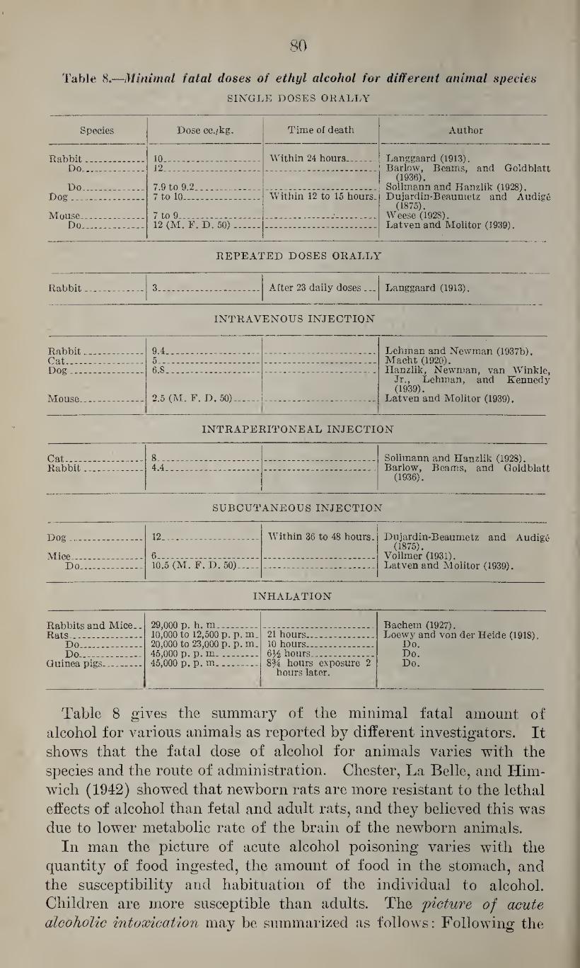
80
Table 8.—Minimal fatal doses of ethyl alcohol for different animal species
SINGLE DOSES ORALLY
Species Dose cc./kg. Time of death Author
Rabbit 10... Within 24 hours_ Langgaard (1913). Barlow, Beams, and Goldblatt
(1936). Sollmann and Eanzlik (1928). Dujardin-Beaumetz and Audige
(1875). Weese (1928). Latven and Molltor (1939).
Do 12 ..__
Do _ 7.9 to 9.2.. Dog 7 to 10_ Within 12 to 15 hours.
Mouse 7 to 9_ _ Do 12 (M. F. D. 50)..
REPEATED DOSES ORALLY
Rabbit 3___ After 23 daily doses ... Langgaard (1913).
INTRAVENOUS INJECTION
Rabbit 9.4 .. Lehman and Newman (1937b). Macht (1920). Hanzlik, Newman, van Winkle,
Jr., Lehman, and Kennedy (1939).
Latven and Molitor (1939).
Cat 5.... Dog 6.S _
Mnnsp. 2.5 (M. F. D. 50)_
INTRAPERITONEAL INJECTION
Cat - - __ 8.... Sollmann and Hanzlik (1928). Barlow, Beams, and Goldblatt
(1936). Rabbi h 4 4
SUBCUTANEOUS INJECTION
Dog 12 _ Within 36 to 48 hours. Dujardin-Beaumetz and Audige (1875).
Vollmer (1931). Latven and Molitor (1939).
Mine 6_ Do 10.5 (M. F. D. 50)_
INHALATION
Rabbits and Mice.. Rat.s . . _
29,000 p. h. m.. Bachem (1927). Loewy and von der Heide (1918).
Do. Do. Do.
10,000 to 12,500 p. p. m_ 20,000 to 23,000 p. p. m. 45,000 p. p. m ..
21 hours..... Do_ 10 hours... Do 6)4 hours_ _
Guinea pigs _ 45^000 p. p. m__ 8% hours exposure 2 hours later.
Table 8 gives the summary of the minimal fatal amount of alcohol for various animals as reported by different investigators. It shows that the fatal dose of alcohol for animals varies with the species and the route of administration. Chester, La Belle, and Him- wich (1942) showed that newborn rats are more resistant to the lethal effects of alcohol than fetal and adult rats, and they believed this was due to lower metabolic rate of the brain of the newborn animals.
In man the picture of acute alcohol poisoning varies with the quantity of food ingested, the amount of food in the stomach, and the susceptibility and habituation of the individual to alcohol. Children are more susceptible than adults. The picture of acute
alcoholic intoxication may be summarized as follows: Following the
81
ingestion of sufficient doses of alcohol the face is flushed, the pulse rate is increased, and there may be some perspiration. The eyes ap¬ pear first bright, later dull, and the pupils may be dilated or con¬ stricted. Motoric unrest, excessive talking, laughing, and joking are characteristic of the early stages of alcoholic intoxication. As the depressant effect becomes more marked these symptoms may be fol¬ lowed by impaired motility, staggering gait, incoordination, lassitude and fatigue. With large doses there may be, in addition, rapid loss of sensitivity, more or less complete impairment of the sensory functions and of the motility, disappearance of reflexes, unconscious¬ ness, and coma. In such advanced cases the conjunctiva is hyper- emic, the pupils dilated, the flushing of the face may pass into pallor, the respiration becomes stridulous, slow, and irregular, the skin is cold and clammy, the pulse flat and slow, and there may be vomiting and incontinence of feces and urine. The body temperature is low¬ ered and there may be twitchings and general convulsions. In fatal cases death results from respiratory and circulatory failure. Kauf- mann (1925) reported the clinical picture of acute alcohol poisoning resulting from the rectal administration of alcohol. Fuhner (1930) pointed out the possible additive and synergistic effect of alcohol with other therapeutic agents. Ajtay (1933) reported on a mass poisoning of 33 severe cases of acute alcohol poisoning resulting in 17 fatalities, and Beyreis (1933), Regus (1937), and others reported on fatalities in which the acute intoxication was associated with cerebral hemor¬ rhages. Fatal outcome in acute alcohol poisoning may also result from the aspiration of vomitus resulting in fatal pneumonia, as illustrated by the case reported by Franz (1936). As pointed out above, children are especially sensitive to alcohol, and death may result from the ingestion of comparatively small doses, as reported by Prievara (1941). Severe and fatal poisonings of nurslings from the ingestion of milk from severly intoxicated mothers were reported by Wyckerheld Bisdom (1937) and Dervieux, Szumlanski and Desoille (1929). Severe and fatal alcohol poisonings in children from in¬ halation of alcohol vapors or its absorption through the skin when applied in the form of alcohol wraps were reported by Kalt (1906), James (1931), Leschke (1932), and Fraenckel (1928). However, it appears questionable whether or not, in the last case mentioned, alcohol was the cause of the fatal outcome. Gettler and St. George (1935) reported on the death of an infant who had received alcohol hypodermically instead of saline.
With regard to the fatal dose of alcohol for mo,n, this has been estimated as approximately 6 to 8 gram (8 to 10 cc.) per kg. of body weight (Flury and Klimmer, 1938). With respect to the concentration of alcohol in the blood of victims of acute alcohol poisoning, these values show considerable variations, depending mainly upon the time
82
elapsed between the ingestion of alcohol and death. Milovanovic (1936) determined the alcohol level in the blood of victims of acute alcohol poisoning as being between 0.519 and 1.332 percent with an average of 0.836 percent. According to this author, Straub found 0.33, Franz 0.41, Jungmichel 0.414, Remund 0.47, Widmark and Schwartz 0.4 to 0.5, Zangger 0.5 to 0.8 and even 1.0, Gelma and Simonin 0.912 and Kohn-Abrest 0.8 to 1.0 percent of ethyl alcohol in the blood of victims of acute alcohol poisoning.
Gettler and St. George (1935) found concentrations of 0.65 percent and it appears, therefore, that in the diagnosis of death due to acute alcohol poisoning the importance of the concentration of alcohol in the blood has to be very carefully evaluated. Villedent (1926) claimed that the concentration of alcohol in the blood of cadavers remains constant for a long time, and Weinig (1936) stated that it is not changed during the first 2 days and that under ordinary condi¬ tions it is only between the second and third days that the alcohol values start to increase due to the formation of volatile decomposition products which in septicemic patients may already be interfering during the first day unless more specific analytical methods are used. On the other hand, Wagner (1936) claimed that in cadavers the blood alcohol content is reduced by 5 to 6 percent during the first 2 days and up to 2.0 to 2.5 percent during the third and fourth days, and he pointed out that blood samples taken more than 4 days after death cannot be properly evaluated on account of the progressing decompo¬ sition. He also pointed out that the concentration of alcohol in blood samples may vary with different locations in the body, in that blood samples from the cranial cavity and the heart show higher values, blood from the lungs is not suitable, and blood from the heart may give erroneous results on account of the inflow of blood from the lungs. On the other hand, Sjovall and Widmark (quoted from Guldberg, 1938) found that blood from the longitudinal sinuses and from the pulmonary vein gave identical values but differed from that drawn from the iliac vein, and they pointed out the possibility that alcohol may diffuse after death from the stomach into the heart so that the heart blood may yield too high values. This possibility seems to be confirmed by the report of Guldberg (1938) who found, in 4 out of 7 cadavers, the concentration of alcohol in the stomach higher than that in blood and urine. As indicated by the report of Weinig (1936), in the determination of the alcohol content in the blood from cadavers it is of paramount importance that such determinations be made with specific methods which exclude the interference of other volatile constituents, and Scheele (1936) pointed out that Widmark’s method is not suitable for this purpose. This was emphasized by Cornish, Draper, and Finn (1938) who demonstrated that alcohol and volatile compounds formed post mortem give very similar reactions
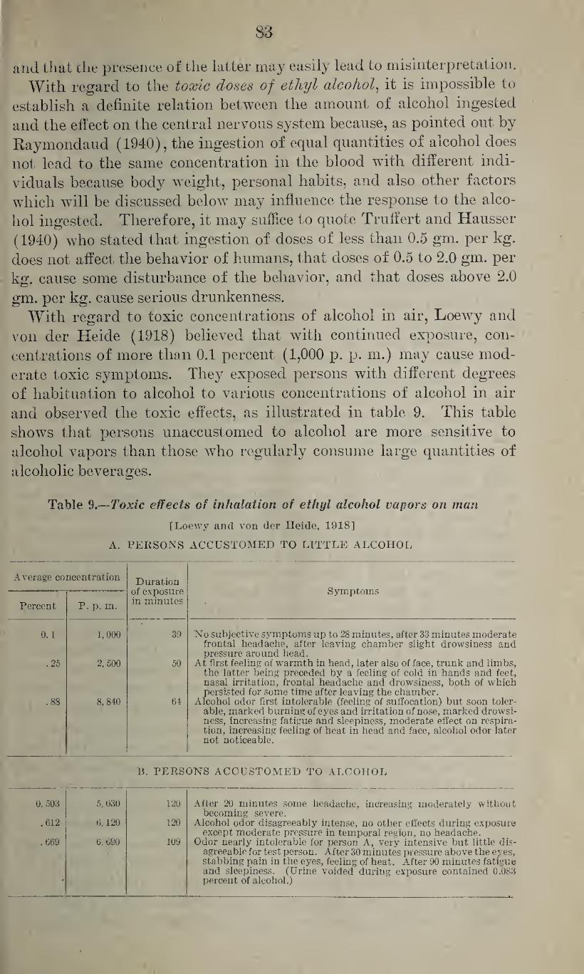
S3
and that the presence of the latter may easily lead to misinterpretation. With regard to the toxic doses of ethyl alcohol, it is impossible to
establish a definite relation between the amount of alcohol ingested and the effect on the central nervous system because, as pointed out by Kaymondaud (1940), the ingestion of equal quantities of alcohol does not lead to the same concentration in the blood wfith different indi¬ viduals because body weight, personal habits, and also other factors which will be discussed below may influence the response to the alco¬ hol ingested. Therefore, it may suffice to quote Truffert and Hausser (1940) who stated that ingestion of doses of less than 0.5 gm. per kg. does not affect the behavior of humans, that doses of 0.5 to 2.0 gm. per kg. cause some disturbance of the behavior, and that doses above 2.0
gm. per kg. cause serious drunkenness. With regard to toxic concentrations of alcohol in air, Loewy and
von der Heide (1918) believed that with continued exposure, con¬ centrations of more than 0.1 percent (1,000 p. p. m.) may cause mod¬ erate toxic symptoms. They exposed persons with different degrees of habituation to alcohol to various concentrations of alcohol in air and observed the toxic effects, as illustrated in table 9. This table shows that persons unaccustomed to alcohol are more sensitive to alcohol vapors than those who regularly consume large quantities of alcoholic beverages.
Table 9.—Toxic effects of inhalation of ethyl alcohol vapors on man
[Loewy and von der Heide, 1918]
A. PERSONS ACCUSTOMED TO LITTLE ALCOHOL
Average concentration Duration of exposure in minutes
Symptoms
Percent P. p. m.
0.1 1,000 39 No subjective symptoms up to 28 minutes, after 33 minutes moderate frontal headache, after leaving chamber slight drowsiness and pressure around head.
.25 2, 500 50 At first feeling of warmth in head, later also of face, trunk and limbs, the latter being preceded by a feeling of cold in hands and feet, nasal irritation, frontal headache and drowsiness, both of which persisted for some time after leaving the chamber.
.88 8, 840 64 Alcohol odor first intolerable (feeling of suffocation) but soon toler¬ able, marked burning of eyes and irritation of nose, marked drowsi¬ ness, increasing fatigue and sleepiness, moderate effect on respira¬ tion, increasing feeling of heat in head and face, alcohol odor later not noticeable.
B. PERSONS ACCUSTOMED TO ALCOHOL
0.503 5,030 120 After 20 minutes some headache, increasing moderately without becoming severe.
.612 6,120 120 Alcohol odor disagreeably intense, no other effects during exposure except moderate pressure in temporal region, no headache.
.669 6, 690 109 Odor nearly intolerable for person A, very intensive but little dis¬ agreeable for test person. After 30 minutes pressure above the eyes, stabbing pain in the eyes, feeling of heat. After 90 minutes fatigue and sleepiness. (Urine voided during exposure contained 0.083 percent of alcohol.)
84
The diagnosis of alcoholic intoxication and the correct appraisal of the degree of the psychic and physical impairment may be of great importance, and the question of the importance of the clinical observa¬ tion as compared with the evaluation of the alcohol level in the blood has aroused much comment. Some authors, for example Roller (1936), emphasize the importance of the clinical observation for the proper appraisal of blood alcohol determinations; others, Morgan (1939), believe that the determination of alcohol in blood and urine is the most accurate basis for the diagnosis of alcoholic intoxication for medico-legal purposes. In view of the conflicting views on this subject one has to agree with Helwig (1940) that the diagnosis of drunken¬ ness and, particularly, of the degree of intoxication is subject to many pitfalls, both medical and legal; that there is much disagreement of opinion as to what constitutes alcoholic intoxication; and that many clinical variables have to be considered before a diagnosis is rendered.
With regard to clinical tests for intoxication, Southgate and Carter (1926) pointed out the inadequacy of many clinical tests such as standing poised, toeing the line, standing immobile with closed eyes, testing memory, etc. They believe that other signs, such as flushed face, perspiration, dilated pupils, congested eyes, and rapid pulse, are more characteristic and are present in nearly all instances. Schwarz (1927) refers to Fog who emphasized the importance of appearance, attitude, orientation, memory, pronunciation, gait, coordination and steadiness of the hand, pulse, pupils, sensitivity to pain, and odor of breath. He also pointed to the importance of the examination for other affections, such as epilepsy, apoplexy, mental confusion, etc., and to the value of the determination of the concentration of alcohol in the blood for the differential diagnosis. Jetter (1938a) considered as most important signs for the diagnosis of alcoholic intox¬ ication, abnormalities of the gait and inability to walk and the posi¬ tive outcome of at least 2 of the following tests: Gross abnormalities of the speech, flushed face, dilated pupils, and alcoholic odor of breath. Kniskern (1939) emphasized the importance of careful neurological examination to discover any variations from normal with regard to lo¬ comotion, stability, dexterity, and speed of reaction, as indicated by Romberg’s test, finger to nose test, writing of signature, ability to but¬ ton clothes and ability to walk a straight line, turn and come back, and stand on 1 foot. He also advised the performance of tests for orien¬ tation, memory and reaction time of thoughts and emotional disturb¬ ances. Hegler (1935) recommended as a short cut procedure having the patient dial his telephone number. Most students of the subject agree that any clinical observations should be supplemented by de¬ terminations of alcohol in blood, saliva, urine, or exhaled air:
With regard to the importance of the determination of alcohol in blood for medicolegal purposes it appears from the work of Rabino-
85
witsch and Wilen (1939) that the sterilization of the skin and of the hypodermic needle with alcohol does not cause sufficient changes in the blood alcohol values to invalidate distinctly positive findings, al¬ though this procedure should be discouraged. It should, however, be pointed out, as stated by Bavis and Arnholt (1939), that all containers should be sealed and labeled before being sent to the laboratory, and that all samples should be run in duplicate or in blood and urine by 2 different methods. Schweisheimer (1913) believed that the psychic condition of an intoxicated person depends upon the concentration of alcohol in his blood. Southgate and Carter (1926) stated that the determination of the alcohol level in blood and urine is a good guide in judging the degree of intoxication, and Bavis and Arnholt (1938) considered it as the best criterion, being definitely related to the degree of intoxication. Truffert and Hausser (1940) believed that the de¬ termination of alcohol in the blood is the best, and its determina¬ tion in the exhaled air the next best criterion for the determination of the degree of intoxication. Friedemann, Motel, and Necheles (1938) recommended the determination of alcohol in the saliva as the most con¬ venient means of measuring the alcohol content of the blood, but Selesnick (1938) believed the determination of alcohol in the blood to be superior to that in urine, saliva, and expired air because it con¬ tains only a negligible amount of volatile constituents other than alcohol. Mozes and Katonak (1941) emphasized the importance of chemical tests because they believed that these furnish a far better criterion than even a detailed clinical study, and Ladd and Gibson (1939) gave a very extensive discussion of the legal aspects of blood alcohol determinations with regard to alcoholic intoxication.
The calculation of the amount of alcohol ingested from that de-
tei'mined in the blood.—Widmark (1932 and 1933a) claimed that after ingestion of single doses the amount of alcohol ingested can be calculated from the concentration of alcohol in the blood at a given time after its ingestion by using the formula: A=pr (ct+(3t). In this formula A is the total quantity of alcohol ingested in grams, p is the body weight of the individual in kilograms, and ct is the concentra¬ tion of alcohol in the blood at the time t calculated in minutes; r is a reduction factor and gives the assumed amount of alcohol at which the concentration of alcohol in the tissue equals everywhere that of the blood, so that pr represents the amount of alcohol in the body. /3 is another constant which gives the decrease of the concentration of alcohol in the blood per minute in parts per thousand at diffusion equilibrium between the alcohol in blood and tissue.
With regard to the factor r, this is independent of the amount of alcohol ingested and of its concentration, and it has been determined by Widmark and his associates as 0.68 for men and as 0.55 for women with deviations of ± 0.085 and ± 0.055, respectively.
86
With regard to constant p which gives the metabolic velocity of ethyl alcohol, this also is not affected by the amount of alcohol ingested or by its concentration, and it was determined in a large gropp of men as 0.0025 ±0.00056, and for women as 0.0026 ±0.00037.
Widmark (1932) gave similar formulas which allow the calculation of the amount ingested with continued and intermittent administra¬ tion. Thurauf (1937) determined the factor p in young, trained soldiers with moderate fat deposits and moderate habituation to alco¬ hol as 0.00217 to 0.00233 and quotes Jungmichel as having found 0.00200. He pointed out that stimulation of the oxygen metabolism, as by dinitrophenol, may increase, and decrease of the oxygen metabolism may lower the factor p. In addition he believed that increased pul¬ monary excretion of alcohol, as produced by exercise, may impair the results of such calculations. Hecksteden (1938) determined the fac¬ tor p as 0.0020 and found that this is not reduced in persons suffering from trauma of the skull which has been credited with reducing the oxygen metabolism. Siegmann (1936) determined factors r and p
in chronic alcoholics and found higher values than those given by Wid¬ mark and Jungmichel. Newman, Lehman, and Cutting (1937) be¬ lieved that Widmark’s formula gives reliable information with re¬ gard to the amount of alcohol ingested. Silberstein (1934) and Elbel and Lieck (1936), on the other hand, claimed that, since the absorp¬ tion of alcohol from the stomach varies with the amount of food in the stomach, any calculation based upon factors r and p may give erroneous results unless this is taken into consideration. Kanitz (1939) pointed out the fact that Widmark’s method for determining alcohol is not specific and that volatile materials contained in alcoholic beverages may cause small errors, which, although insignificant by themselves, may become serious if multiplied by the various factors involved in Widmark’s formula. Hegler (1935) discussed the useful¬ ness of Widmark’s formula and emphasized the need for taking blood samples as early as possible after the ingestion of alcohol, and Mayer (1936a) came to the conclusion that after 1 y2 hours have elapsed single determinations are not sufficient to permit safe calculation by Widmark’s method.
In respect to the appraisal of blood alcohol findings with regard to the determination of the degree of intoxication, Kionka (1928) pointed out that the behavior of different persons and even of the same person at different times may vary considerably with the same alcohol level in the blood, so that the latter cannot be safely considered as an absolute yardstick for the degree of intoxication; and a similar statement was made by Tuovinen (1930). Mir sky, Piker, Rosenbaum, and Lederer (1941) showed, as had been pointed out previously by Mellanby (1919) and Miles (1922), that symptoms of intoxication occur at a lower blood level during the absorptive phase than dur-
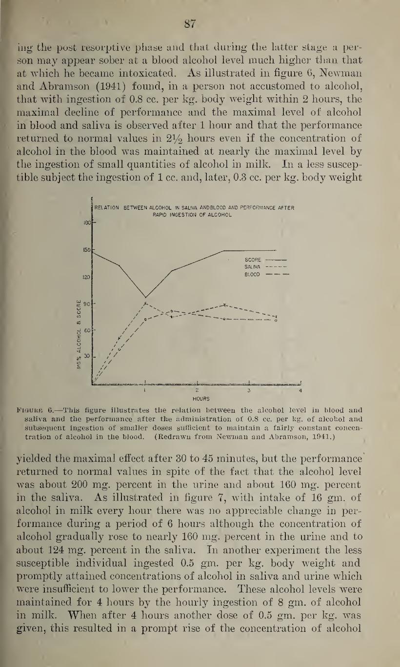
87
ing the post resorptive phase and that during the latter stage a per¬ son may appear sober at a blood alcohol level much higher than that at which he became intoxicated. As illustrated in figure 6, Newman and Abramson (1941) found, in a person not accustomed to alcohol, that with ingestion of 0.8 cc. per kg. body weight within 2 hours, the maximal decline of performance and the maximal level of alcohol in blood and saliva is observed after 1 hour and that the performance returned to normal values in 2% hours even if the concentration of alcohol in the blood was maintained at nearly the maximal level by the ingestion of small quantities of alcohol in milk. In a less suscep¬ tible subject the ingestion of 1 cc. and, later, 0.3 cc. per kg. body weight
Figure 6.—This figure illustrates the relation between the alcohol level in blood and saliva and the performance after the administration of 0.8 cc. per kg. of alcohol and subsequent ingestion of smaller doses sufficient to maintain a fairly constant concen¬ tration of alcohol in the blood. (Redrawn from Newman and Abramson, 1941.)
yielded the maximal effect after 30 to 45 minutes, but the performance returned to normal values in spite of the fact that the alcohol level was about 200 mg. percent in the urine and about 160 mg. percent in the saliva. As illustrated in figure 7, with intake of 16 gin. of alcohol in milk every hour there was no appreciable change in per¬ formance during a period of 6 hours although the concentration of alcohol gradually rose to nearly 160 mg. percent in the urine and to about 124 mg. percent in the saliva. In another experiment the less susceptible individual ingested 0.5 gm. per kg. body weight and promptly attained concentrations of alcohol in saliva and urine which were insufficient to lower the performance. These alcohol levels were maintained for 4 hours by the hourly ingestion of 8 gm. of alcohol in milk. When after 4 hours another dose of 0.5 gm. per kg. was given, this resulted in a prompt rise of the concentration of alcohol
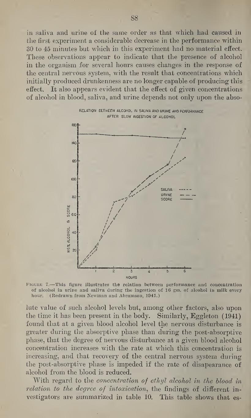
88
in saliva and urine of the same order as that which had caused in the first experiment a considerable decrease in the performance within 30 to 45 minutes but which in this experiment had no material effect. These observations appear to indicate that the presence of alcohol in the organism for several hours causes changes in the response of the central nervous system, with the result that concentrations which initially produced drunkenness are no longer capable of producing this effect. It also appears evident that the effect of given concentrations of alcohol in blood, saliva, and urine depends not only upon the abso-
REIATION BETWEEN ALCOHOL IN SALIVA AND URJNE AND PERFORMANCE
AFTER SLOW INGESTION OF ALCOHOL
HOURS
Figure 7.—This figure illustrates the relation between performance and concentration of alcohol in urine and saliva during the ingestion of 16 gm. of alcohol in milk every hour. (Redrawn from Newman and Abramson, 1941.)
lute value of such alcohol levels but, among other factors, also upon the time it has been present in the body. Similarly, Eggleton (1941) found that at a given blood alcohol level the nervous disturbance is greater during the absorptive phase than during the post-absorptive phase, that the degree of nervous disturbance at a given blood alcohol concentration increases with the rate at which this concentration is increasing, and that recovery of the central nervous system during the post-absorptive phase is impeded if the rate of disapearance of alcohol from the blood is reduced.
With regard to the concentration of ethyl alcohol in the blood in
relation to the degree of intoxication, the findings of different in¬ vestigators are summarized in table 10. This table shows that es-
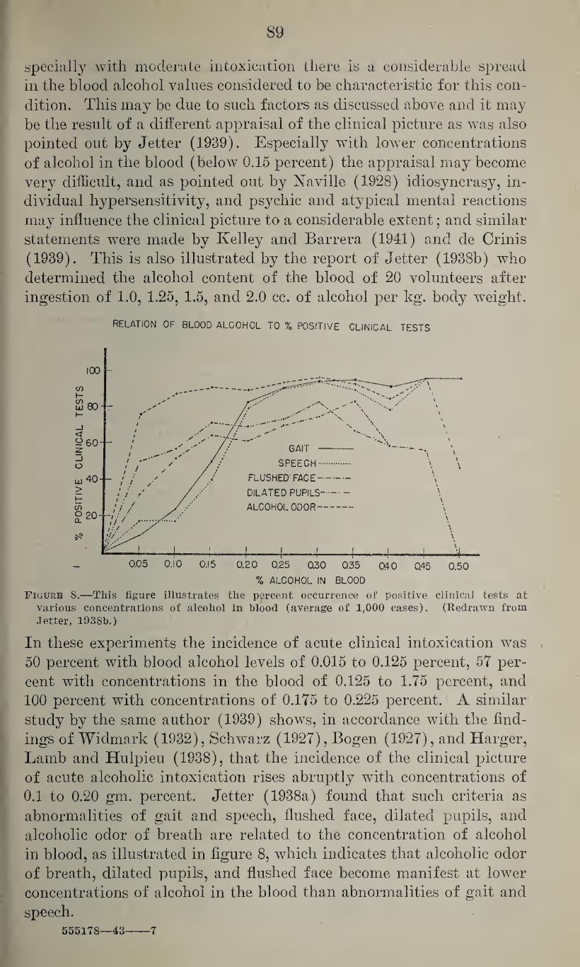
89
specially with moderate intoxication there is a considerable spread in the blood alcohol values considered to be characteristic for this con¬ dition. This may be due to such factors as discussed above and it may be the result of a different appraisal of the clinical picture as was also pointed out by Jetter (1939). Especially with lower concentrations of alcohol in the blood (below 0.15 percent) the appraisal may become very difficult, and as pointed out by Naville (1928) idiosyncrasy, in¬ dividual hypersensitivity, and psychic and atypical mental reactions may influence the clinical picture to a considerable extent; and similar statements were made by Kelley and Barrera (1941) and de Crinis (1939). This is also illustrated by the report of Jetter (1938b) who determined the alcohol content of the blood of 20 volunteers after ingestion of 1.0, 1.25, 1.5, and 2.0 cc. of alcohol per kg. body weight.
RELATION OF BLOOD ALCOHOL TO % POSITIVE CLINICAL TESTS
Figure 8.—This figure illustrates the percent occurrence of positive clinical tests at various concentrations of alcohol in blood (average of 1,000 cases). (Redrawn from Jetter, 1938b.)
In these experiments the incidence of acute clinical intoxication was 50 percent with blood alcohol levels of 0.015 to 0.125 percent, 57 per¬ cent with concentrations in the blood of 0.125 to 1.75 percent, and 100 percent with concentrations of 0.175 to 0.225 percent. A similar study by the same author (1939) shows, in accordance with the find¬ ings of Widmark (1932), Schwarz (1927), Bogen (1927), and Harger, Lamb and Hulpieu (1938), that the incidence of the clinical picture of acute alcoholic intoxication rises abruptly with concentrations of 0.1 to 0.20 gm. percent. Jetter (1938a) found that such criteria as abnormalities of gait and speech, flushed face, dilated pupils, and alcoholic odor of breath are related to the concentration of alcohol in blood, as illustrated in figure 8, which indicates that alcoholic odor of breath, dilated pupils, and flushed face become manifest at lower concentrations of alcohol in the blood than abnormalities of gait and speech.
555178—43 ■7
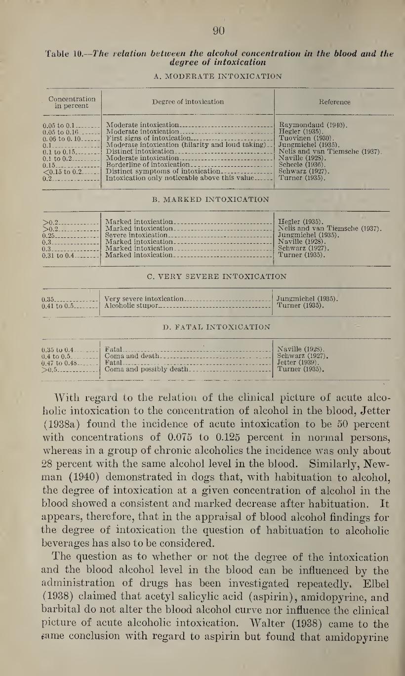
>
90
Table 10.—The relation between the alcohol concentration in the blood and the degree of intoxication
A. MODERATE INTOXICATION
Concentration in percent Degree of intoxication Reference
0.05 to 0.1_ 0 05 to 0 16
Moderate intoxication__ _ Moderate intoxication.-__ _ ..
Raymondaud (1940). Hegler (1935). Tuovinen (1930). Jungmichel (1935). Nelis and van Tiemsche (1937). Naville (1928). Scheele (1936). Schwarz (1927). Turner (1935).
0.06 to 0.10_ 0.1_ 0 1 to 0 15
First signs of intoxication_ __ Moderate intoxication (hilarity and loud taking).. Distinct intoxication. _ _. .. . ... . ... ..
0 1 to 0.2 Moderate intoxication. . .... . .. 0 15 Borderline of intoxication ... . <0.15 to 0.2_ 0.2_
Distinct symptoms of intoxication_ ... . .. Intoxication only noticeable above this value.. ..
B. MARKED INTOXICATION
>0 2 Marked intoxication.__ .. ... . ... .. Hegler (1935). Nelis and van Tiemsche (1937). Jungmichel (1935). Naville (1928). Schwarz (1927). Turner (1935).
>0 2 Marked intoxication. _ .. ___ 0 25 Severe intoxication...... 0 3 Marked intoxication_ __ . ... 0 3 Marked intoxication___ ___ 0 31 to 0.4 M arked intoxication____ _
C. VERY SEVERE INTOXICATION
0 35 Very severe intoxication... .. _ Jungmichel (1935). 0 41 to 0.5 Alcoholic stupor_ __ __ ____ Turner (1935).
D. FATAL INTOXICATION
0 35 to 0.4 Fatal_ .. _ __ _ .. Naville (1928). 0 4 to 0.5 Coma and death. _ ... _ _ _ Schwarz (1927). 0 47 to 0.48 Fatal__ ... ... _ _. .. . Jetter (1939). >0 5 Coma and possibly death__ . ... Turner (1935).
With regard to the relation of the clinical picture of acute alco¬ holic intoxication to the concentration of alcohol in the blood, Jetter (1938a) found the incidence of acute intoxication to be 50 percent with concentrations of 0.075 to 0.125 percent in normal persons, whereas in a group of chronic alcoholics the incidence was only about 28 percent with the same alcohol level in the blood. Similarly, New¬ man (1940) demonstrated in dogs that, with habituation to alcohol, the degree of intoxication at a given concentration of alcohol in the blood showed a consistent and marked decrease after habituation. It appears, therefore, that in the appraisal of blood alcohol findings for the degree of intoxication the question of habituation to alcoholic beverages has also to be considered.
The question as to whether or not the degree of the intoxication and the blood alcohol level in the blood can be influenced by the administration of drugs has been investigated repeatedly. Elbel (1938) claimed that acetyl salicylic acid (aspirin), amidopyrine, and barbital do not alter the blood alcohol curve nor influence the clinical picture of acute alcoholic intoxication. Walter (1938) came to the same conclusion with regard to aspirin but found that amidopyrine
91
preparations may delay insignificantly the absorption without reduc¬
ing the maximal level of alcohol in the blood. Bohmer (1938) be¬
lieved that both aspirin and amidopyrine delayed the absorption
without however reducing the blood alcohol curve, whereas he found
that insulin (30 units) lowered the resorptive phase but increased
the postresorptive heights of the blood alcohol curve. Domenici
(1939) showed that strychnine, strophantin, luminal, pantopon,
metrazol, vitamin C, coramine, ephedrine, caffeine, camphor, digitalis,
and acetyl choline do not influence the concentration of alcohol in
the blood. Rakieten (1942) found that in rats doses of 400, 600, and
800 mg. per kg. of acetphenetidine slowed the rate of disappearance
of alcohol from the blood, whereas similar doses of aniline, p-amino-
phenol, and amidopyrine were ineffective. Doses of 1.3 gm. of aspirin,
1.3 gm. of acetphenetidine, and 1.0 gm. of amidopyrine had no effect
on the rate of disappearance of alcohol from the blood of human sub¬
jects when taken with 120 cc. of 90 proof whisky.
With regard to the relation of the concentration of alcohol in the urine to the degree of intoxication, Widmark (1915), Miles (1924) and
Southgate and Carter (1926) thought that the alcohol level in the urine
was a good gage for the determination of the intensity of the alcoholic
intoxication. Bogen (1927) studied the relation between the con¬
centration of alcohol in the urine and the clinical picture of alcoholic
intoxication. He found that 50 percent of 35 persons with less than
0.1 percent (1 mg./cc.) of alcohol in the urine had admitted drinking
and had alcoholic odor of breath but only a few of these showed flushed
face, dilated pupils, abnormalities of gait, or thickened speech whereas
many showed lack of inhibition and diminution of self-control. Of
45 patients with 0.1 to 0.2 percent (1 to 2 mg./cc.) alcohol in the urine
the majority had a marked odor of alcohol in the breath; more than
50 percent had dilated pupils and flushed face and frequently showed
loss of restraint, staggering gait, swaying on standing, confused and
blurred speech, and general clumsiness. Of 58 patients with 0.2 to
0.3 percent (2 to 3 mg./cc.) alcohol in the urine, all showed these symp¬
toms to a more marked degree, although 25 percent of these were still
able to walk without staggering and to speak clearly, whereas of 64
patients with 0.3 to 0.4 percent (3 to 4 mg./cc.) only 1 could stand with¬
out swaying and only 1 could speak clearly. In 35 patients with 0.4
to 0.5 percent (4 to 5 mg./cc.), narcosis and stupor were the rule and
the dilation of the pupils gave way to constriction, the flushing of the
face to pallor. Thirteen patients with more than 0.5 percent (5
mg./cc.) were all in a state of coma, unable to respond to stimulation,
with slow and shallow respiration, cold skin, and general inertness.
As the alcoholic level in the urine approaches 0.6 percent (6 mg./cc.) the
danger of alcoholic asphyxia becomes imminent. Jetter (1938a) stud¬
ied the relation between impairment of gait and speech, flushed face
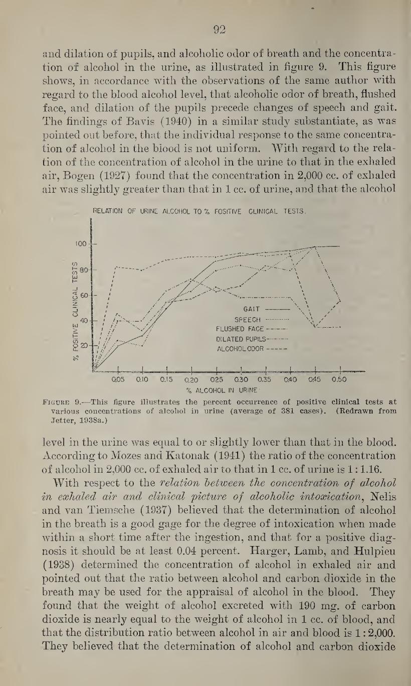
92
and dilation of pupils, and alcoholic odor of breath and the concentra¬
tion of alcohol in the urine, as illustrated in figure 9. This figure
shows, in accordance with the observations of the same author with
regard to the blood alcohol level, that alcoholic odor of breath, flushed
face, and dilation of the pupils precede changes of speech and gait.
The findings of Bavis (1940) in a similar study substantiate, as was
pointed out before, that the individual response to the same concentra¬
tion of alcohol in the blood is not uniform. With regard to the rela¬
tion of the concentration of alcohol in the urine to that in the exhaled
air, Bogen (1927) found that the concentration in 2,000 cc. of exhaled
air was slightly greater than that in 1 cc. of urine, and that the alcohol
RELATION OF URINE ALCOHOL TO % POSITIVE CLINICAL TESTS.
Figure 9.—This figure illustrates the percent occurrence of positive clinical tests at
various concentrations of alcohol in urine (average of 381 cases). (Redrawn from
Jetter, 1938a.)
level in the urine was equal to or slightly lower than that in the blood.
According to Mozes and Katonak (1941) the ratio of the concentration
of alcohol in 2,000 cc. of exhaled air to that in 1 cc. of urine is 1:1.16.
With respect to the relation between the concentration of alcohol in exhaled air and clinical picture of alcoholic intoxication, Nelis
and van Tiemsche (1937) believed that the determination of alcohol
in the breath is a good gage for the degree of intoxication when made
within a short time after the ingestion, and that for a positive diag¬
nosis it should be at least 0.04 percent. Harger, Lamb, and Hulpieu
(1938) determined the concentration of alcohol in exhaled air and
pointed out that the ratio between alcohol and carbon dioxide in the
breath may be used for the appraisal of alcohol in the blood. They
found that the weight of alcohol excreted with 190 mg. of carbon
dioxide is nearly equal to the weight of alcohol in 1 cc. of blood, and
that the distribution ratio between alcohol in air and blood is 1:2,000.
They believed that the determination of alcohol and carbon dioxide
93
in the exhaled air excludes possible fluctuations in the amount of
alveolar air contained in such samples. Haggard, Greenberg, Miller,
and Carroll (1941) determined the distribution ratio between the con¬
centration of alcohol in exhaled air and its concentration in blood
as 1:1,300, and they believed that the discrepancy between this value
and that of Harger, Lamb, and Hulpieu (1938) was largely due to the
loss of alcohol in the condensed water vapors within the container used
for the collection of the samples. In addition, they pointed out that
the concentration of alcohol in the lung air cannot be calculated cor¬
rectly from the concentration in mixed exhaled air on the basis of the
carbon dioxide content of these two types of air because alcohol
diffuses more rapidly into the air of the respiratory dead space than
does carbon dioxide. They showed that the concentration of alcohol in
the alveolar air, the collection of which is rather difficult in untrained
persons, corresponds closely to that in “venous” air. The latter is
collected by having a person wearing a nose clip rebreathe from a
closed glass tube of adequate volume after a deep expiration followed
by normal inhalations and exhalations into the tube. They claimed
that by this procedure the error involved in the determination of the
concentration of alcohol in the exhaled air mentioned above for the
appraisal of the blood alcohol level can be avoided. Jetter and For¬
rester (1941) determined the alcohol and carbon dioxide content of
the exhaled air by the method of Jetter, Moore, and Forrester (1941)
and calculated the percent concentration of alcohol in blood by multi¬
plying the ratio of mg. alcohol determined in breath over mg. carbon
dioxide determined in breath by 0.2. They found no evidence that the
absorption of alcohol by water condensed on the walls of the collecting
apparatus invalidated the results. Acetone, paraldehyde, esters, alde¬
hydes, and other constituents of beverages and methanol are said to
not invalidate the results obtained bv this method, but alkalosis and
acidosis may give rise to erroneous findings.
With regard to the relation between the concentration of alcohol
in the spinal fluid and the degree of intoxication, Schumm and
Fleischmann (1913) found that the concentration of alcohol in
the spinal fluid is approximately proportionate to the amount of
alcohol ingested. Bogen (1927) stated that it corresponds closely
to that in the urine and, therefore, also to that in the blood.
Yad and Kulkarni (1929) claimed that alcohol may be detected in
the spinal fluid before a person shows signs of drunkenness or lack
of mental control. It was stated in a previous section that Gettler
and Tiber (1927b) had developed a method for the calculation of the
alcohol content of the brain from that of the spinal fluid, and that
they claimed that persons with an alcohol content in the brain of
less than 0.1 percent showed no abnormal physiological effects, that
0.1 to 0.25 percent caused such disturbances as increased aggressive-
94
ness, more or less marked carelessness, but no real intoxication which,
however, manifests itself with concentrations of 0.25, 0.4, and 0.6
percent. Helwig (1940) and Gonzales and Gettler (1941) agreed
that the determination of alcohol in the spinal fluid and brain gives
the most exact and most consistent indication of the degree of in¬
toxication.
It has been pointed out that the degree of alcoholic intoxication,
with regard to the concentration of alcohol in the blood, may be
greatly influenced by the individual susceptibility. Nagel (1939)
developed a skin test for the determination of the susceptibility or
tolerance to alcohol. He showed that the intradermal injection of
0.3 cc. of approximately 60 percent alcohol into the lateral aspect of
the deltoid region, using equal quantities of saline on the other arm
as control, causes a cutaneous reaction of a different intensity. He
stated that following this injection all persons develop a wheal of 9
mm. diameter but that they differ with regard to the erythema sur¬
rounding the wheal which reaches its maximum after 20 to 30 min¬
utes and disappears within approximately 1 hour. By grading the
intensity of this erythema he found that of all persons tested, 20
percent were allergic to alcohol and 10 percent belonged to the tol¬
erant or relatively resistant group. As pointed out by Manwaring
(1939) these figures substantiate the findings of the Dresden Al¬
coholic Commission which studied in 1938 the relation between the
concentration of alcohol in the blood and the psychological dis¬
turbances and found that 20 percent of the test subjects were in¬
capacitated with blood alcohol levels of 0.02 percent, whereas 10
percent were perfectly sober after the alcohol concentration in the
blood had increased to 0.12 percent. Manwaring (1939) empha¬
sized, however, that skin allergy and organ allergy do not necessarily
run parallel, so that the supposition that skin allergy indicates nerv¬
ous susceptibility needs further study. Kelley and Barrera (1941)
made a study of the relation between Nagel’s test, blood alcohol level,
and the clinical picture as appraised by criteria similar to those used
by Jetter (1938a). They found that different test subjects showed
considerable variations in the concentration of alcohol in the blood
at the same degree of moderate psychic impairment, and that a num¬
ber of individuals showed a better relation between the intensity
of the skin test and the effect on the mental functions.
A discussion of acute alcoholic intoxication would not be complete
without discussion of the drunken driver, and numerous studies have
been devoted to answering the question of at what concentration of
alcohol in the blood a person should be considered as an unsafe driver.
The opinions of different investigators of this question are summar¬
ized in table 11. It shows that concentrations between 0.1 and 0.2
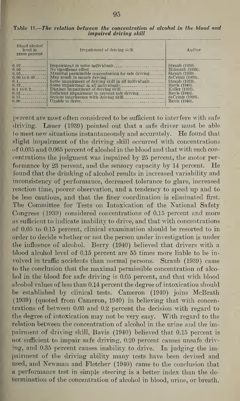
95
Table 11.—The relation between the concentration of alcohol in the blood and impaired driving skill
Blood alcohol level in
gram percent Impairment of driving skill Author
0.02... Impairment in some individuals...... _ Straub (1939). Holcomb (1938). Straub (1939). deCrinis (1939). Straub (1939). Bavis (1940). Roller (1936). Bavis (1940). deCrinis (1939). Bavis (1940).
0.05__ No significant effect . _ _ .. _ . . . 0.05_ 0.06 to 0.08_ 0.1_ 0.12_
Maximal permissible concentration for safe driving_ May result in unsafe driving_ _ Some impairment of driving skill in all individuals.. . Some impairment in all individuals__ . . _ .
0.1 to 0. 2_ 0.15_ 0.15_ 0.30_
Distinct impairment of driving skill.. _ _ Sufficient impairment to prevent safe driving_ . Serious interference with driving skill.. .. . __ _ Unable to drive. . __ . ..... _ .
percent are most often considered to be sufficient to interfere with safe
driving. Lauer (1939) pointed out that a safe driver must be able
to meet new situations instantaneously and accurately. He found that
slight impairment of the driving skill occurred with concentrations
of 0.035 and 0.065 percent of alcohol in the blood and that with such con¬
centrations the judgment was impaired by 25 percent, the motor per¬
formance by 23 percent, and the sensory capacity by 14 percent. He
found that the drinking of alcohol results in increased variability and
inconsistency of performance, decreased tolerance to glare, increased
reaction time, poorer observation, and a tendency to speed up and to
be less cautious, and that the finer coordination is eliminated first.
The Committee for Tests on Intoxication of the National Safety
Congress (1939) considered concentrations of 0.15 percent and more
as sufficient to indicate inability to drive, and that with concentrations
of 0.05 to 0.15 percent, clinical examination should be resorted to in
order to decide whether or not the person under investigation is under
the influence of alcohol. Berry (1940) believed that drivers with a
blood alcohol level of 0.15 percent are 55 times more liable to be in¬
volved in traffic accidents than normal persons. Straub (1939) came
to the conclusion that the maximal permissible concentration of alco¬
hol in the blood for safe driving is 0.05 percent, and that with blood
alcohol values of less than 0.14 percent the degree of intoxication should
be established by clinical tests. Cameron (1940) joins McBrath
(1939) (quoted from Cameron, 1940) in believing that with concen¬
trations of between 0.03 and 0.2 percent the decision with regard to
the degree of intoxication may not be very easy. With regard to the
relation between the concentration of alcohol in the urine and the im¬
pairment of driving skill, Bavis (1940) believed that 0.15 percent is
not sufficient to impair safe driving, 0.20 percent causes unsafe driv¬
ing, and 0.35 percent causes inability to drive. In judging the im¬
pairment of the driving ability many tests have been devised and
used, and Newman and Fletcher (1940) came to the conclusion that
a performance test in simple steering is a better index than the de¬
termination of the concentration of alcohol in blood, urine, or breath.
06
As reported in an editorial in the Journal of the American Medical
Association (1940), the Committee for Tests on Intoxication of the
National Safety Council (1939) recommends that chemical tests of
body fluids or breath (if properly performed) be used in all cases,
criminal or civil, in which the influence of alcohol is suspected. This
committee agreed further with the interpretation of chemical tests
for alcoholic intoxication recommended by the Committee to Study
Problems of Motor Vehicle Accidents of the American Medical As¬
sociation as adopted by the House of Delegates (1939); namely, that
(1) a person with a concentration of less than 0.05 percent by weight
in blood or its equivalent in urine, saliva, or breath should not be
prosecuted for driving while under the influence of alcoholic bever¬
ages; (2) persons with a concentration of above 0.15 percent should
be considered to be under such influence; and (3) persons with a con¬
centration of between 0.05 and 0.15 percent should be prosecuted only
when circumstances and results of physical examinations give definite
confirmation that they are under the influence of alcohol.
Newman, Fletcher, and Abramson (1942) pointed to the consider¬
able variations between the results of aptitude and driving tests of
different individuals and their blood alcohol level. These variations
may be apparent in the effect of alcohol on the vision, coordination,
and the complex activity involved in driving a motor vehicle. They
found, however, that with a blood alcohol concentration of 150 mg.
per 100 cc. of blood all subjects showed some demonstrable effect of
alcohol. On the other hand, they found that not all test subjects
with more than 150 mg. of alcohol per 100 cc. of blood were impaired
to such a degree that their ability was reduced to below that of the
ordinary prudent and cautious driver.
The question as to what extent the drunken pedestrian is the
cause of traffic accidents has aroused much less comment. Eppel
(1940) determined the alcohol content of the blood of pedestrians
killed by their own carelessness in 76 traffic accidents and found 0.196
to 0.431 percent of alcohol in 11.4 percent of these cases; 0.096 to 0.165
percent in 10.1 percent; and 0.02 to 0.095 percent in 8.8 percent. Gon¬
zales and Gettler (1941) found that of 2,472 pedestrians killed in
highway accidents, 30.7 percent were under the influence of alcohol
and 26.2 percent had a brain alcohol content of 0.1 to 0.25 percent or
more. This study indicates that in the evaluation of the medico¬
legal implications of highway accidents the relation of the alcohol
content in the blood or brain of the pedestrian should not be ignored,
and that negligence on the part of the intoxicated pedestrian may
also be an important factor in traffic accidents.
Alcoholic intoxication as an industrial hazard.—It is obvious that
the effect of alcohol on the nervous functions and the effect of alcohol on
the driving skill, discussed above, also holds true for industrial opera-
97
tions where reaction time, judgment, and performance are of impor¬
tance. In addition, it should be emphasized that ingestion of alcoholic
beverages increases the susceptibility to many chemical poisons, such
as aromatic amino and nitro compounds, carbon tetrachloride, and
others. Loewy and von der Heicle (1918) stated that in man the
inhalation of concentrations of 1,000 to 2,500 p. p. m. of alcohol in
air may cause toxic effects, and that with higher concentrations
(7,500 p. p. m.) exposure for 45 to 90 minutes may cause somnolence,
fatigue, and sleepiness. The question of industrial injuries resulting
from the use of denatured alcohol was evidently first discussed by
Lewy (1891) who believed that certain denaturing agents, such as
pyridine bases and crude wood alcohol, may cause numerous symp¬
toms, such as hoarseness, retching, nausea and vomiting, headache,
vertigo, tremors, etc. Such symptoms were observed in workers of
different trades handling denatured alcohol in varnishes and lacquers. %
It appears, however, that these persons worked under very unfavor¬
able conditions (small wmrking places, high temperature, and inade¬
quate ventilation) and a later study by Loewy (1914) on the indus¬
trial hazards of denatured alcohol with special reference to methanol,
showed that only in a small number of cases could irritation of the
eyes and catarrhal affections of the pharnyx and the upper respira¬
tory tract be observed, and these only under especially unfavorable
conditions. He pointed out, however, that adequate ventilation and
general and personal hygiene are essential in preventing injurious
effects from the use of denatured alcohol. With regard to the maxi¬
mal permissible concentration of ethyl alcohol in air, no definite limits
have been established. Several States of the Union have suggested
tentatively 250 p. p. m. but it appears that further studies are required
before definite standards can be established.
The habituation to ethyl alcohol.—It has been stated repeatedly that
there is frequently a difference in the response to alcohol between
persons abstaining from the consumption of alcoholic beverages and
those who regularly ingest such beverages. Experience with human
beings and animal experiments performed by many investigators,
particularly Newman and Lehman (1938), have shown that the con¬
tinued ingestion of alcohol renders persons or animals more tolerant
to the effect of alcohol on the central nervous system, so that a better
neuromuscular coordination is demonstrated at an equal blood alcohol
level than before habituation. Newman and Lehman (1938) believed
that after habituation the tolerance of the tissue to alcohol is increased,
whereby the cells of the central nervous system acquire the ability
to function more effectively at a given alcohol concentration than
before habituation. This assumption appears to be supported by the
observation of Ahlquist and Dille (1940) that alcohol-tolerant rabbits
98
are also much more resistant to the depressant effect of other narcotics
such as pentobarbital, evipal, and ether.
The clinical picture of the toxic effects of alcohol on the nervous
system.—Continued ingestion of excessive amounts of alcohol may re¬
sult in chronic alcoholism which has been associated with many
pathological conditions.
With regard to the effect of chronic alcoholism on the mentality,
it appears questionable, as pointed out by Allen (1938), whether alco¬
holism leads to mental instability or whether mental instability leads
to alcoholism. Wechsler (1941) studied the I. Q. of chronic alcoholics
who did not suffer from other mental diseases and found that their
capacity for logical analysis and organization is often affected to a
greater degree than the more familiar deterioration of the memory.
In his opinion the prolonged use of alcohol impairs the mental func¬
tioning of various abilities before there is any evidence of disease of
the brain, and there is a relation between the intensity of the impair¬
ment and the duration of the alcoholism.
With regard to the relation between chronic alcoholism and certain
psychic disturbances, Heron (1912) believed that alcohol is less fre¬
quently the cause than the sequela of mental defects, and Frankel and
Benjamin (1933) believed that in many instances of chronic alcoholism
excessive drinking is only a secondary symptom, inasmuch as they
found that of 458 drinkers examined 25 percent suffered from severe
somatic or psychic defects. Bowman and Jellinek (1941), in a review
on alcoholic mental disorders, concluded that there seems to be agree¬
ment that alcohol addiction is the symptom of many psychoses rather
than the primary cause, with the exception of such affections as
delirium tremens and Korsakoff’s psychosis. Lewis (1941), on the
other hand, believed that alcohol as an etiologic factor in mental dis¬
ease ranks comparatively high, either as an individual cause or as a
precipitant agent in certain types of disturbances. The following dis¬
cussion is largely based on the review of Bowman and Jellinek (1941).
Of the various mental disturbances associated with alcoholism,
pathological intoxication may be discussed first. As pointed out by
Bowman and Jellinek (1941) the outstanding symptoms of this affec¬
tion are blind rage, confusion, and usually complete amnesia for the
condition. The stage of rage may be characterized by anxiety and
paranoia. The same authors consider as main neurological reactions,
reflex rigidity of pupils, swaying, and weak tendon reflexes which
outlast the mental effects. In their opinion its etiologic factor is an
existing psychopathic condition and alcoholism is only the eliciting
cause. It is not necessarily associated with chronic alcoholism since it
may also result from the ingestion of small doses of alcohol and may
be observed in hypoglycemic conditions.
One psychosis most commonly associated with chronic alcoholism is
99
delirium tremens, the clinical picture of which has been discussed re¬
cently by Wortis (1940) and Bowman and Jellinek (1941). The onset
of delirium tremens is insidious, and the prodromal. symptoms may
occur days and weeks before the outbreak of the psychosis. They are
characterized by anxiety, restlessness, fear, insomnia, nightmares, hy¬
persensitivity of sensory organs, occasional hallucinations, tendency to
perspiration, headache, vertigo, and convulsions, and appear pre¬
dominantly at night. The acute psychosis lasts only from 3 to 4 days,
and most symptoms disappear after the so-called terminal sleep. The
onset of the acute stage is usually sudden, frequently at night, and is
occasionally associated with an epileptic or epileptiform attack. The
attack is characterized mainly by visual hallucinations; less frequently
by illusions, tactile, auditory and combined hallucinations. There is
a marked degree of suggestibility, the learning ability is impaired, the
consciousness is dimmed, and the attention and perception are de¬
creased. Orientation for time and space is impaired and there may
be motor excitation and hyperactivity. With regard to the physical
picture of delirium tremens, this is characterized by a coarse arrhyth¬
mic tremor of the tongue, face, fingers, legs, and even of the trunk.
The patient may be restless and suffer from insomnia. In the opinion
of Bowman and Jellinek (1941) it is questionable whether or not dis¬
turbance of the equilibrium should be considered among the cardinal
symptoms. Nausea, vomiting, profuse perspiration, constipation, and
dehydration may be observed. The body temperature, blood pressure,
and pulse rate may be increased. The urinary findings are suggestive
of disturbances of the protein metabolism and there are definite in¬
dications of impairment of the detoxicating function of the liver and
disturbance of the fat and mineral metabolism. In contrast to the
older conception, at present delirium tremens is not considered a
withdrawal symptom. Delirium tremens is observed only in chronic
alcoholism, and injuries or infections, metabolic disturbances of vari¬
ous types, and lack of vitamin B, may be predisposing factors.
Korsakoff (1889) described a symptom complex which he had ob¬
served both in alcoholic patients and also as sequelae of infectious
conditions and of poisons such as carbon disulfide, carbon monoxide,
and others and which has become known as Korsakoff*s psychosis.
Cases of this condition have been reported frequently and more re¬
cently by Reifenstein, and Davidoff (1940) and Romano, Michael, and
Merritt (1940), and was discussed extensively by Bowman and Jellinek
(1941). The onset of this psychosis may be sudden, with delirium, or
fairly insidious, with stupor. The patients may show an anterograde
amnesia; sometimes impairment of memory, impaired learning ability,
disorientation to space and time, confabulation; occasionally illusions,
stereotypy of speech, aphasia, and agraphia. They are indifferent to
their surroundings or, sometimes, euphoric. The psychosis is often
100
associated with polyneuritis, as discussed by Jolliffe (1940) and, in
contrast to delirium tremens, Korsakoff’s syndrome is always associ¬
ated with degenerative polyneuropathy and brain changes closely
resembling those found in chronic alcoholism, and it appears to be
possible that the disease may be partly due to lack of vitamin Bx.
Another psychosis, acute alcoholic hallucinosis, occurs only in
chronic alcoholism and is not a manifestation of acute intoxication.
Bowman and Jellinek (1941) considered it as one of the earlier syn¬
dromes of chronic alcoholism. The onset of this psychosis is some¬
times sudden, but more frequently it is preceded by a prodromal stage
characterized by general anxiety, headache, insomnia, and acoustic
hypersensitivity. The main symptoms of the psychosis are fear, audi¬
tory hallucinations, inclination to anxiety, misinterpretation and sys¬
tematization with intact orientation and presence of mind. In con¬
trast to delirium tremens, patients suffering from acute hallucinosis
are usually well oriented, their perception and attention are not im¬
paired, and generally they are willing to talk. While delirium tremens
patients accept their hallucinations and delusions, those suffering from
acute hallucinosis seek explanations. The hallucinations of acute hal¬
lucinosis have not the coloring of those in delirium tremens; they are
less dependent upon the sense of perception and are not altered by
perception. In contrast to delirium tremens the motor unrest exists
only in the beginning and is less marked. Acute hallucinosis is much
less dependent upon the consumption of high-proof spirits than is
delirium tremens, and, in contrast to the latter, it is not caused by
precipitating factors. It has been stated that in acute hallucinosis,
especially in the beginning, there is always leucopenia, and, in con¬
trast, in delirium tremens there is ieucocytosis. The duration of the
acute hallucinosis is much longer than that of delirium tremens and,
in contrast to the latter, patients suffering from the former usually
have a high incidence of psychoses, alcoholism, psychopathy, and
suicide in their family.
Another type of alcoholic psychosis, alcoholic 'paranoid condition, is
recognized by some authorities as an alcoholic mental disorder. In
the opinion of Bowman and Jellinek (1941) it cannot be excluded cate¬
gorically but they believed that it is presumably the late precipitation
of a schizophrenic condition.
Chronic alcoholic deterioration, in the opinion of Bowman and
Jellinek (1941), is not a clinical entity and has not been classified as
an alcoholic psychosis. It is characterized by ethical degeneration,
dulling of the finer sentiments, and brutality of the patients. But
more important is their lability and the impairment of their memory.
Such patients frequently show a dilatation of the facial capillaries,
especially of the nose, and myocardial changes; they have a bloated
look and their musculature is flabby. In case hard liquors are taken
101
habitually such patients may suffer from gastritis and possibly also
from cirrhosis of the liver. The patients show tremors, sometimes
twitchings and choreiform movements, and they may suffer from
pain in calves and legs and sometimes from tenderness over the large
nerve trunks and peripheral neuritis. Pupillary changes are fre¬
quent and their reaction is sometimes sluggish. The physical capacity
and stamina of such patients is frequently impaired and their resist¬
ance to disease reduced.
With regard to alcoholic epilepsy and dipsomania, Leary (1938) and
Bowman and Jellinek (1941) agree that these are not sequelae but •
rather causes of alcoholism. The latter pointed out that abnormal
drinking is definitely greater among epileptics than in the general
population, that there is a high concomitance of alcoholic addicts and
epileptics in families with psychopathic heredity, and that there is an
acceleration of epileptic seizures in genuine epileptics through alco¬
hol and some indication that alcohol may precipitate latent epilepsy.
With respect to dipsomania, as stated by Bowman and Jellinek (1941).
it is at present assumed that this belongs to the manic-depressive
group of psychoses and that “it still remains a challenging
problem.”
Another affection of the central nervous system frequently associ¬
ated with alcoholism is Wernicke'’s syndrome which is characterized
by cloudiness of consciousness, varying ophthalmoplegias, and ataxia.
According to Jolliffe, Wortis and Stein (1941) it is probably a com¬
bination of several nutritional disturbances affecting the nervous sys¬
tem, and the picture need not necessarily be complete in 1 patient.
It is frequently preceded by and associated with delirious episodes,
and, in those persons who recover, a residual Korsakoff psychosis
may be seen. The pathological lesions may extend not only to the
cortex but also to the spinal cord and the peripheral nerves. Al¬
though usually associated with alcoholism there is evidence that nutri¬
tional deficiencies, especially of thiamine hydrochloride, seem to be
the most obvious common factor.
Alcoholic polyneuritis is an affection of the peripheral nerves and
is frequently associated with alcoholism. Jolliffe (1940) who reviewed
the literature on this subject pointed out the difference between poly¬
neuritis and polyneuropathy, the latter being characterized by patho¬
logical changes indicating degenerative processes. In the opinion of
this author it appears to be established that alcoholic neuropathies are
basically caused by nutritional deficiencies, specifically by avita¬
minoses, predominantly of vitamin B1? which may be associated with
other vitamin deficiencies. In his opinion the idea of a direct causation
by alcohol has to be abandoned in view of the identity of alcoholic
neuropathy with beriberi, the fact that adequately nourished alco¬
holics do not develop neuropathy, and that a vitamin Bi deficient
102
diet produces experimentally the characteristic symptoms of peri¬
pheral neuritis, including the neurasthenic manifestations. Similarly,
Wayburn and Guerard (1940) came to the conclusion that avitamin¬
osis B x as cause of polyneuropathy seems to be established. Laboratory
investigations (Evans and Lepkovsky, 1929, 1934 and 1935; Kem-
merer and Steenbock, 1933; McHenry, 1937; Salmon and Goodman,
1937; all quoted from Lowry, SJbbrell, Daft and Ashburn, 1942)
have shown that the vitamin B* requirement is influenced by the com¬
position of the diet as well as by the total number of calories. An
abundant supply of carbohydrates increases, while substitution of
fats for carbohydrates decreases the need for vitamin Bx and post¬
pones the onset of polyneuropathy. On this basis it was assumed that,
like carbohydrates, alcohol increased the need for thiamine. Simi¬
larly, Westerfeld, Stotz and Berg (1942) believed that, since the
oxidation of alcohol to acetaldehyde requires the nicotinamide coen¬
zyme, and the pyruvate-acetaldehyde condensation requires diphospho-
thiamine, thus increasing the need for these substances above that
required for the metabolism of other foodstuffs, this may be in part
responsible for the production of “alcoholic pellagra” and polyneuritis.
They believed that on this basis chronic alcoholics maintain a higher
requirement for nicotinamide and thiamine and develop more rapidly
a deficiency of these vitamins because they actually derive a large
part of their daily calories from vitamin-free alcohol. Lowry, Sebrell,
Daft and Ashburn (1942) showed, however, in paired feeding experi¬
ments with rats on a thiamine-deficient diet, that without exception
rats fed water developed neuropathy before the paired litter mates
receiving dilute alcohol or whisky instead of drinking water, indi¬
cating that alcohol consumption does not increase the thiamine re¬
quirement. Wayburn and Guerard (1940) found that a high per¬
centage of peripheral neuropathies occurred in cases of cirrhosis of
the liver and that both conditions improved upon therapy with vitamin
Bi. Brown (1941), on the other hand, reported less striking results
from the administration of thiamine hydrochloride.
The toxic effects of ethyl alcohol on organ functions.—The relation
of alcoholism to cardiovascular disease has been much discussed and
disputed. Weiss (1940), in an extensive review of this subject, came
to the conclusion that among other factors, thiamine deficiency may
be a contributing factor; and Leary (1938) also believed that the role
of alcoholism as primary cause is at least questionable, and that such
sequelae as subarachnoidal hemorrhages, subdural hemorrahages
(without contusion or laceration of the brain), and hemorrhages in the
mammillary bodies observed in alcoholism may be due to a deficiency
of vitamin C in the diet.
The relation of alcoholism to gastritis was discussed by Berry (1941).
Seymour, Spies and Payne (1939) studied the gastric secretion in
103
chronic alcoholics who showed neither clinical evidence of vitamin
deficiency nor laboratory evidence of anemia. They found a reduction
of the average volume of the gastric juice and the acidity but no im¬
pairment of the peptic activity. Beazell and Ivy (1940) came to the
conclusion that the ingestion of large amounts of alcohol causes
gastritis and achlorhydria from which recovery is relatively rapid.
They also believed that dietary deficiencies may be involved in the
causation of gastritis. Gray and Schindler (1941) examined 100
chronic alcoholics gastroscopically and found that 55 percent of these
showed essentially a normal stomach; that the remaining 45 percent
suffered mostly from superficial gastritis, atrophic gastritis or a com¬
bination of both; and that 22 percent of the latter showed mucosal
hemorrhages. They found no relation between the incidence of gas¬
tritis and the duration of the alcoholism, the amount of alcohol in¬
gested, the abuse of nicotine, dental defects, and vitamin deficiency.
In a similar study, Berry (1941) found that of 100 persons with un¬
questionable chronic alcoholism of long standing 30 percent gave
no evidence of gastritis; 35 percent suffered from mild superficial
gastritis, the gastric mucosa differing only slightly from normal;
35 percent showed distinct chronic gastritis.
The toxic effect of ethyl alcohol on the liver has aroused much
comment. Kochmann (1923) in his review on this subject refers
to Rosenfeld (1900) who found that the feeding of alcohol resulted
in a 10 percent higher incidence of fatty livers in animals thus fed
than in the controls. Similarly, Ducceschi (1917-18) found that
daily feeding of alcohol to dogs increased the fat content of the liver
up to 30 percent and that the cholesterol content was also increased
but less markedly. With regard to the effect of alcohol on the liver
function, Lamson (1923) (quoted from Rosenthal, 1930) found that
doses of 4 cc. per kg. body weight cause inpairment of the liver
function on the same or on the following day. Wallace (1927)
studied the liver function of 17 patients suffering from acute alcoholic
intoxication by determining the bilirubin content of the blood and
the urobilinogen excretion in the urine. He found definite evidence
of liver injury, proportionate in intensity to the severity of the intoxi¬
cation, the bilirubin content of the blood and the urobilinogen in
the urine, being materially increased immediately following the excess
and returning gradually to normal values within 4 to 5 days. Beazell,
Berman, Hough and Ivy (1942) found that ingestion of 2 to 3 cc. per
kg. in the form of whisky caused no demonstrable ill effects on the
hepatic function as indicated by the serum van den Bergh, urine bili¬
rubin, bromsulphthalein clearance, hippuric acid excretion, serum
phosphatase, fasting blood sugar, and fructose and glactose tolerance
tests. In these subjects the blood alcohol level rose to 150 to 201
mg. per 100 cc. of blood. However, they believed it possible that
104
with blood alcohol concentrations of 300 to 500 mg. per 100 cc. dis¬
turbance of the liver function may be observed. Animal experiments
carried out in the same way appear to indicate that dogs are more
sensitive than humans to the hepatotoxic action of alcohol. Bins-
wanger (1932) referred to observations of Pohlisch, Biichler, and
Bostrom who found urobilin and urobilinogen in the urine of patients
suffering from delirium tremens, but in his experience this does not
appear to be the rule. However, this may vary with the time elapsed
since the ingestion of alcohol because de Montmollin (1938) observed
in the acute stage of various alcoholic psychoses a very marked in¬
crease of the urinary urobilin which paralleled the intensity of the
psychic disturbance. Using smaller doses (2 cc. per kg.) than Lamson
(1923) (quoted from Rosenthal, 1930), Rosenthal (1930) found no
evidence of urobilin in the urine or bilirubin in the blood, but he
found a slight impairment of the liver function, as indicated by the
results of the bromsulphthalein test, which returned to normal within
24 hours. He found, however, that this moderate injury increased
materially the susceptibility to other poisons such as chloroform and
carbon tetrachloride. Cates (1941b) examined the liver functions
of 17 patients suffering from acute alcoholism and found in 4 of
them a distinct impairment of the excretion of bromsulphthalein. He
believed that in his cases the liver injury was primarily due to lack
of proper nourishment; that the retention of the dye is not necessarily
related to pathological changes of the liver tissue; and that acute
hepatitis may subside completely and only in severe cases lead to
Laennec’s cirrhosis. This appears to be in accordance with observa¬
tions made in animals by MacNider (1933). He found that dogs
intoxicated for 12 hours with sufficient doses of alcohol to become
semianesthetized, developed injury of the liver commencing at the
peripheral lobules and consisting essentially of edema of the cells ac¬
companied by accumulation of stainable lipoid material. These
changes were paralleled by a retention of phenoltetrachlorphthalein
corresponding to the intensity of the injury. Whereas in these ani¬
mals the function and histology returned to normal within 3 days,
it took 12 days with animals which had been intoxicated in the same
way for 24 hours. Berman, Snapp, Ivy and Atkinson (1941) admin¬
istered 40 cc. of alcohol daily to dogs with biliary fistula and found
that this caused definite changes of the liver parenchyma which were
reflected in the composition of the bile.
Kagawa (1931) studied the mobilization of sugar by alcohol in toads
and believed this was due to a reversible narcosis of the liver cells;
and Binswanger (1932) believed that in certain alcoholic psychoses
the ability of the liver to assimilate sugar is reduced. Connor (1940)
believed that ingestion of alcohol with a normal diet causes an abnor¬
mal accumulation of sugar in the blood; and Bowman, Wortis, Oren-
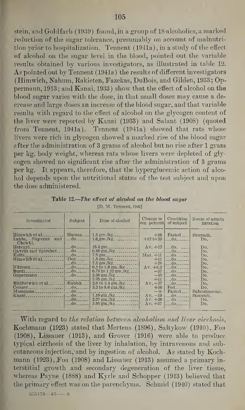
105
stein, and Goldfarb (1939) found, in a group of 18 alcoholics, a marked
reduction of the sugar tolerance, presumably on account of malnutri¬
tion prior to hospitalization. Tennent (1941a),. in a study of the effect
of alcohol on the sugar level in the blood, pointed out the variable
results obtained by various investigators, as illustrated in table 12.
As pointed out by Tennent (1941a) the results of different investigators
(Himwich, Nahum, Rakieten, Fazekas, DuBois, and Gilden, 1933; Op-
permann, 1913; and Kanai, 1933) show that the effect of alcohol on the
blood sugar varies with the dose, in that small doses may cause a de¬
crease and large doses an increase of the blood sugar, and that variable
results with regard to the effect of alcohol on the glycogen content of
the liver were reported by Kanai (1933) and Salant (1908) (quoted
from Tennent, 1941a). Tennent (1941a) showed that rats whose
livers were rich in glycogen showed a marked rise of the blood sugar
after the administration of 3 grams of alcohol but no rise after 1 gram
per kg. body weight, whereas rats whose livers were depleted of gly¬
cogen showed no significant rise after the administration of 3 grams
per kg. It appears, therefore, that the hyperglucemic action of alco¬
hol depends upon the nutritional status of the test subject and upon
the dose administered.
Table 12.—The effect of alcohol on the blood sugar
[D. M. Tennent, 1941]
Investigator Subject Dose of alcohol Change in mg. percent
Condition of subject
Route of admin istration
Himwich et al_ . Human_ 1.5 gm./kg__ +10 Fasted Sf.omaeh, Labbe, Nepveux and ...do_ l.Ojm.'/kg_ +12 to 39 ...do_ Do.
Chewki. • Hetenyi. _ _ _ ..do_ 16.4 gm . . Av. +22 do On. Gavrila and Sparchez.. ...do_ 0.40 gm./kg_ .do Do. Kolta . . _ .do.. 7.9 gm. _ Max. +11 do Do. Himwich et al. .... Dog_ 1.5 gm./kg_ ... +11 do_ Do.
...do_ 7.5 gm./kg___ +28 do _ Do. Nitzescu_ ..do_ 0.5 to 1.0 gm./kg_ Av. +6.3 do Do. Burdi__ .do_ 0.79 to 1.32 gm./kg... . -37 do Do. Oppermann__ ...do_ 2.38 gm./kg..... -35 do Do.
3.95 gm./kg..... .do.. Do. Blatherwick et al ... .. Rabbit. . 2.0 to 2.4 gm./kg._ Av. —27 do Do. Conner..__ ...do_ 3.2 to 8.0 gm./kg_ .. +50 Fed. Do. Masamme ... ... . ...do_ —b Fasted.. Subcutaneous. Kanai_ ...do_ 1.19 gm./kg_ Av. —19 do Stomach.
..do_ 2.37 gm./kg_ Av. +20 .do . Do. ...do_ 3.95 gm./kg_ Av. +37 ...do_ Do.
With regard to the relation between alcoholism and liver cirrhosis,
Kochmann (1923) stated that Mertens (1896), Saltykow (1910), Foa
(1908), Lissauer (1913), and Grover (1916) were able to produce
typical cirrhosis of the liver by inhalation, by intravenous and sub¬
cutaneous injection,'and by ingestion of alcohol. As stated by Koch¬
mann (1923), Foa (1908) and Lissauer (1913) assumed a primary in¬
terstitial growth and secondary degeneration of the liver tissue,
whereas Payne (1888) and Kyrle and Schopper (1913) believed that
the primary effect was on the parenchyma. Schmid (1940) stated that
555178—43—--8
106
in the United States, in 1938, 2,569 fatalities were attributed to alco¬
holism, 959 of these being reported as cirrhosis of the liver; and he
also stated that there appears to be a real or apparent increase of the
incidence of this affection. However, it appears that the extent to
which alcohol is responsible for the production of cirrhosis of the
liver has not been established. Schafir (1913) reviewed the literature
on the relation between alcoholism and cirrhosis of the liver and found
already at that time a considerable discrepancy of opinion on this
subject. He quoted von Hausemann (1904) as stating that cirrhosis
of the liver may occur in alcoholism but that the specific effect of alco¬
hol is fatty degeneration of the liver, and he refers to Klopstock
(1906),Rosie (1907), von Baumgarten (1907), Fahr (1909), and Kauf-
mann (1911) who assumed that cirrhosis of the liver as observed in
alcoholics is not caused by alcohol itself but that alcohol favors cer¬
tain toxic factors which by themselves are able to produce cirrhosis of
the liver. Schafir (1913) himself was unable to produce experimen¬
tally cirrhosis of the liver but only degenerative changes and necroses
with fatty infiltration and some interstitial growth not sufficient to
be called cirrhosis. Connor (1940) studied the question of the extent
to which liver changes in alcoholism were due to inadequate nutrition
and came to the conclusion that for the production of cirrhosis of the
liver alcoholism has to be associated with starvation, nutritional defi-
ciences, or certain diseases such as diabetes. Similarly, Beazell and
Ivy (1940) were of the opinion that alcohol does not cause cirrhosis
of the liver but predisposes to the cirrhogenous effect of other toxic
substances and that sometimes it appears to cause cirrhosis in fatty
livers produced by a high fat diet. Gyorgy and Goldblatt (1941)
showed that rats on a vitamin deficient diet developed often but not
always acute diffuse necrosis of the liver associated with fatty de¬ generation, and that a diet free of casein and deficient in vitamin B
caused necroses with or without cirrhosis within 100 to 150 days.
Jolliffe, Wortis, and Stein (1941) reviewed the question of vitamin
deficiency and liver cirrhosis in alcoholism.
With regard to the relation between alcoholism and affections of
the pancreas, Myers and Keefer (1934) stated that alcohol is a pre¬
disposing factor in acute pancreatic necrosis because they found that
of 21 cases of chronic pancreatitis 7 were associated with alcoholism
or with diseases commonly associated with chronic alcoholism. They
refer to reports on alcoholic panceratitis by Mondon (1929) and Emile
and Andre (Aron and Jacob) (1930), and to the monograph of Gross
and Guleke (1924) who stated that alcoholism, especially in its
chronic form, is the cause of pancreatitis characterized by an in¬
crease of the intra and interlobular fibrous tissue. The mechanism
of this pancreatotoxic effect of alcohol has been explained by a direct
toxic effect of alcohol on the pancreas, by obstruction or infection
107
of the pancreatic duct due to duodenal congestion, and by forcing of
duodenal contents or bile into the pancreatic duct as a result of per¬
sistent vomiting. The latter explanation has also been accepted by
Leary (1938).
With regard to the relation between alcoholism and disturbances
of the kidney function, Hultgen (1910) found that in dogs the daily
intake of alcohol has no irritant effect on the kidney, and he be¬
lieved that nephritis in alcoholics is probably due to some other fac¬
tor than alcohol, such as alimentary disorders and exposure to cold
or infection. Bruger (1940) reviewed the effect of alcohol on the
normal and diseased kidney and came to the conclusion that there is
little direct or irrefutable evidence that alcoholism is responsible for
Bright’s disease and arteriosclerosis because only a negligible amount
of alcohol is excreted through the kidney which may account for any
slight nephrotoxic action. He believed that with the possible excep¬
tion of the arteriosclerotic kidney there is no evidence that alcohol
affects the normal or even the diseased kidney.
Much attention has been given to the relation between alcoholism
and resistance to infections. Thomas (1893) (quoted from Kocli-
mann, 1923) showed in rabbits that the administration of alcohol
reduced the resistance to cholera infections, and similar results were
reported by Abbott (1896) for infections with Streptococcus pyo¬
genes and to a lesser extent for those produced by Bacillus coll and
Staphylococcus pyogenes aureus. Valagussa and Kanelletti (1899)
noted reduction of the resistance to the toxic action of diphtheria
toxin, and reduced resistance was also reported by Laitinen (1900),
Goldberg (1901) (quoted from Kochman, 1923) and Pawlowsky
(1900). Stillman (1924) found that in mice intoxicated with al¬
cohol, pneumococci persisted in the lungs for longer periods of time
than normally, and that a greater incidence of fatal septicemia but
less frequently local infections of the lungs occurred. Stillman and
Branch (1931) showed that mice previously exposed to pneumococci
and then alcoholized and exposed to sprayed cultures developed local
lesions of the lungs, either on account of an alteration of the local
susceptibility to reinfection or, more likely, because of a limited de¬
gree of general immunity. Pickrell (1938) showed that in rabbits
severe alcoholic intoxication leading to lasting stupor destroys the
resistance to pneumococcus infections, and, in a study of 3,422 cases
of lobar pneumonia, Caps and Coleman (1923) found the mortality
considerably higher in excessive drinkers than in moderate drinkers,
and the rate of fatalities greater among the latter than among ab¬
stainers. Pickrell (1940) reported that animals immunized by the
injection of antipneumococcus serum are deprived of their resistance
by intoxication with alcohol. Similar observations with anthrax and
rabies were reported by Delearde (1897) who assumed that this effect
may be due to an interference of alcohol with phagocytolytic proc¬
esses. Reich (1915), in a study on the effect of alcohol on the de¬
struction of tubercle bacilli by human leucocytes, found no constant
relation although he found some evidence of impairment of the
phagocytosis by alcohol. Abbott (1896) assumed that the decreased
resistance to infections may possibly be due to starvation or nutri¬
tional disturbances associated with alcoholism, and Pickrell (1938)
suggested that it may be due to an inhibition of the vascular re¬
sponse by alcohol, interfering with the emigration of leucocytes
which would favor an uninterrupted bacterial growth.
"With regard to the effect of alcohol on growth, Hanzlik (1931)
found that in pigeons the administration of 2 and 4 percent solutions
as drinking water for 13 to 14 months caused loss of weight, and
Barlow, Beams and Goldblatt (1936) found that ingestion of 7.5
percent as drinking water in young rats was without significant effect
on the growth rate, whereas in the experience of Sollmann (1920) and
Barlow, Beams and Goldblatt (1936) 10 and 15 percent, respectively,
had a definite effect. The relation between alcoholism and the physical and mental con¬
dition of offspring has been much discussed, and the literature on this
subject is controversial. Schroder (1933) and Koelsch (1936) stated
that the available literature does not permit a definite answer as to
whether or not alcohol produces genetic changes, but the possibility
of such injurious effects has to be considered. The latter assumed,
however, that for man it may be considered as established that the
ingested alcohol may diffuse into the seminal fluid and that in chronic
alcoholism spermatogenesis may be sufficiently damaged to cause
azospermia and that an injurious effect on the offspring appears to
be likely but is not proved. Statements with regard to the detri¬
mental effect of alcohol are either based on uncritical statistical evi¬
dence (Pearson and Elderton, 1910) or on animal experiments. Fere
(1896) showed that injection of alcohol into fertilized chicken eggs
caused malformation of the embryo. Basset (1910) reported that
daily inhalation of alcohol vapors for 1 to 2 months by doves and
pigeons caused the yolk to become smaller without affecting the de¬
velopment of the eggs. Stockard (1914) claimed that in guinea pigs
the inhalation of alcohol vapors may affect the germ cells of the males,
resulting in defective offspring even if mated with normal females,
and that an injurious effect may be observed even in the third genera¬
tion. Similar results were reported by Stockard and Papanicolaou
(1918). Arlitt (1919) claimed that mental deficiency produced in
rats by continued feeding of alcohol, as demonstrated by their be¬
havior in a maze, was transferred to the offspring equally well by
male and female parents, so that animals in the secondhand third
generations were inferior to normal animals. On the other hand,
109
Rost and Wolf (1925) found in rabbits fed daily doses of 10 to 20 cc.
of alcohol for 2% years, no evidence of a deleterious effect on cell
proliferation, duration of gestation, weight, and general condition of
the animals, nor was there any evidence of a harmful effect on the
embryo or the fetus. Pearson and Elderton (1910) and Elderton and
Pearson (1910) refuted the statement that alcoholism of the parent is
a cause of degeneration in children. They found a higher death rate
among the offspring of alcoholics than among those of sober parents,
but this was apparently due more to domestic neglect, accidents, and
carelessness than to the toxic effect of alcohol. On the contrary, they
found that the general health of children of alcoholics was, on the
whole, slightly better than that of children of sober parents, there
were fewer delicate children, and epilepsy and tuberculosis were less
frequent. In explanation of their findings they suggested that the
physically strongest are more inclined towards alcoholic excesses and
that there is a survial of the fittest in alcoholic families. They found
the relation between parental alcoholism and filial intelligence so
slight that it could not be established.
Pathological changes in alcoholism.—With regard to pathological
changes produced ~by alcohol in the central nervous system one has
to distinguish between the effects of acute intoxication and those of
chronic alcoholism. In acute alcoholic intoxication, hyperemia of pia
and brain, hemorrhagic diathesis, and edema of the brain have been
reported by Regus (1937), Prievara (1941) and Alexander (1941).
Kochmann (1923) refers to Berkley (1896), Kremiansky (1868), Neu¬
mann (1869) and Lewin (1874) as having produced experimentally
hemorrhagic pachymeningitis in dogs. In chronic alcoholism, degen¬
erative changes of various nature may be found. Kochmann (1923)
referred to Mairet and Combemale (1892) as having found softening
of the brain and the spinal cord, and to Magnan (1869), Vas (1894),
Dehio (1895) and Afanassiew (1890) as having reported on degener¬
ative changes of the gray substance of the spinal cord, the ganglionic
cells of the anterior horn, and the ganglia. Duchovnikova and Robin¬
son (1929) found, after continued administration of alcohol,to rabbits,
changes in the parenchyma and mesenchyma and in the ganglionic and
glia cells of regressive and progressive type. They reported on toxic
degenerative changes, especially of the parenchyma, mainly in the
cerebellum, less frequently in the medullary centers and least often in
the second and fifth layers of the cortex. Since the nervous damage
was much more severe than the vascular injury they believed that these
changes were the direct outcome of a toxic action of alcohol on the
tissue. Stender and Liithy (1931) and Lhermitte (1935) described
cerebellar changes but it is questionable whether these were caused by
alcohol. Stevenson, McGowan and Allen (1941) gave an extensive
review of the pathological findings in the brain in alcoholism. They
110
studied the brains of 44 chronic alcoholics and found relatively slight
changes, most of which were presumably due to lack of vitamin and
vitamin B complex. They were unable to confirm the statements of
previous authors with regard to the frequent involvement of the optic
nerve, frequent severe lesions in the medulla and the blood vessels, se¬
vere epenclymitis or gliosis, usual or constant marginal localization of
the lesions and the presence of important lesions in the cerebellum.
With the exception of patients dead from Wernicke’s disease there was
little relation between the clinical picture and the anatomic distribu¬
tion of the lesions. Alexander (1941) stated that the brains from
chronic alcoholics show atrophy and pseudo-atrophy, especially of the
frontal lobe, with a corresponding increase of the fluid in the subarach¬
noid space, frequently referred to as edema. He pointed out that
the shrinkage of the brain ma}^ be due to loss of nerve tissue, to dehy¬
dration, or to both. In chronic alcoholism, subdural hemorrhages are
said to be frequent. In the opinion of Alexander (1941) these are
probably due to vitamin C deficiency. In his opinion the histological
changes are due either to direct and primary damage of the parenchy¬
ma (neuronitis) or to secondary changes of the parenchyma caused by
vascular changes (Wernicke’s disease). The former is said to be
characterized by degenerative changes of the neurons such as swelling,
tigrolysis, demineralization, and, finally, shrinking and destruction of
the ganglionic cells, irregular swelling, and thinning, and, later, frag¬
mentation of the myelin sheaths. He distinguished 3 types of
changes: (1) neuronitis, predominantly of the peripheral nerve fibers;
(2) neuronitis, predominantly of the nerve cells; and (3) neuronitis,
predominantly of the long nerve tracts within the least vascularized
part of the spinal cord. He pointed out that ail 3 of these changes
can be explained on the basis of an avitaminosis.
With regard to pathological changes produced by ethyl alcohol in
the liver, Regus (1937) and Prievara (1941) found, in acute alcohol
poisoning, hyperemia and some degenerative changes. As shown by
MacNicler (1933), acute alcoholic intoxication causes in dogs edema
and accumulation of stainable lipoid material in the liver cells in the
periphery of the liver lobules, these changes being paralleled by a
retention of phenol tetrachlorphthalein. Schafir (1913) found in
rabbits, after continued administration of alcohol, degenerative
changes of the liver cells of variable intensity progressing to necrosis
with fatty infiltration which sometimes was associated with very
moderate interstitial growth but no cirrhosis of the liver. Similar
results were reported by Grover (1916) and Balthazard and Larue
(1921). The latter noted definite liver damage, especially of the
periportal region, with granular, vacuolar, and fatty degeneration,
leucocytic infiltration, and moderate proliferation of the connective
tissue. MacNider and Donnelly (1932b) found that continued ad-
nil
ministration of 10 cc. per kg. of 40 percent alcohol solution daily for
6 months to 2 years to dogs on a well balanced diet caused marked
injury of the liver involving the peripheral liver lobules and extend¬
ing inwardly towards the central vein so that two-thirds of the liver
lobule may participate in this pathologic process. In these regions
the cells were edematous without vacuolization and necrosis, the nuclei
were stained faintly, and the cells contained large amounts of stain-
able lipoid material; the intertubular capillaries were distended with
blood, but there were no hemorrhages nor any increase of the connec¬
tive tissue.
Connor and Chaikoff' (1938) showed that cirrhosis of the liver may
be produced in dogs by continued administration of alcohol if the
liver previously showed fatty infiltration. Connor (1939) in a dis¬
cussion of various forms of cirrhosis of the liver stated that alcoholism
may cause fatty degeneration of the liver which in some cases passes
into cirrhosis, and that other factors, especially dietary deficiencies,
are evidently involved. Boles and Crew (1940) studied 210 cases of
cirrhosis of the liver of all types, 34.4 percent of which had a history
of alcoholism, and they believed that alcohol plays an important part
in the production of cirrhosis of the liver but that portal cirrhosis
may be caused by other factors than alcohol.
With regard to 'pathological -findings in spleen and kidney, Prievara
(1941) found hyperemia of the spleen in acute alcohol poisoning.
Lissauer (1903) and Saltykow (1910) (both quoted from Kochmann,
1923) observed interstitial nephritis and degenerative changes in the
kidneys, and similar findings were reported by Plonda (1919). Bal-
thazard and Larue (1921) found that continued feeding of alcohol
to dogs had comparatively little effect on the kidneys, and Wegelin
(1933) believed that alcoholism plays no very important role in the
genesis of cirrhosis of the kidney.
In respect to pathological findings in glandular structures, Koch¬
mann (1923) referred to Schmiergeld (1909) as having found injury
of the inner secretory glands; to Aubertin (1907) as having seen
damage of the adrenals; and to Kyrle and Scliopper (1913-14) who
reported on severe degenerative changes in testes, as was also stated
by Arlitt and Wells (1917).
With regard to pathological changes in heart and blood vessels,
Kochmann (1923) stated that, experimentally, hypertrophy and
fatty degeneration of the heart muscle have been reported repeatedly,
and Regus (1937) saw degenerative changes in the heart muscle in a
case of acute alcohol poisoning. Von Otto (1914) found experi¬
mentally that small doses (2 cc. per kg.) caused no pathologic changes,
whtereas 4 cc. per Tig. caused changes in the ganglionic cells without
affecting the heart muscle. With larger doses of 6 and 8 cc. per kg.
the ganglionic damage and injury of the heart muscle were more
112
severe, the former being also very marked in chronic alcohol poisoning.
In such cases he noted severe degenerative changes of the blood vessels,
less severe damage of the muscle fibers, and focal necrosis. He believed
that these changes were primarily due to alcohol but were aggravated
by an inadequate blood supply caused by the pathological changes
in the blood vessels, and he noted that the papillary muscles were
especially affected. In the opinion of Kochmann (1923) alcoholism
causes first fatty degeneration and subsequently atheromatosis and
arteriosclerosis, especially in the large and middle-sized vessels; and
Duchovnikova and Robinson (1929) found that in rabbits chronic
poisoning causes swelling and desquamation of the endothelium and
moderate proliferation of the intima.
With regard to 'pathological changes in the lungs, Regus (1927) and
Prievara (1941) found in acute fatal poisonings, bronchopneumonia,
subpleural petechia, and edema of the lungs.
The treatment of acute alcohol poisoning.—In the treatment of acute
alcohol poisoning any alcohol in the stomach should be removed by
gastric lavage and this may even be indicated a considerable time
after the ingestion because as shown by Grehant (1903a), alcohol
may be excreted into the stomach. In case the patient is unconscious,
chilling should be avoided. This is especially important in cases of
acute alcoholic intoxication on account of the heat loss observed in
such patients. If the respiration is slow and superficial it may be
improved by the inhalation of carbon dioxide, and, as pointed out by
van Wulfften Palthe (1926) and Barach (1934), the inhalation of
oxygen may have a favorable effect in case the depression of the
respiration is not too great; but from experiments reported by Butler
(1936) it appears questionable whether in severe poisoning adminis¬
tration of oxygen is of definite value. In severe cases, analeptics and
cardiac stimulants may become necessary but these should be used
with adequate caution (Reifenstein, 1941).
With regard to the treatment of chronic alcoholism, it has been
pointed out that this is frequently superimposed on an existing mental
affection and for this reason, and because of its chronic character, it
requires the supervision of a psychiatrist.
c. Propyl Alcohol
The higher homologue of ethyl alcohol is propyl alcohol.
Chemical characteristics.—Normal propyl alcohol, propanol 1,
CH3GH2CH2OH, has the molecular weight 60.09. It is a colorless
liquid with a fuel-oil-like odor and of the specific gravity 0.804 at 20°
~^o~ C. It solidifies at —127° C., boils at 97.8° C., and is miscible in all
proportions with water, alcohol, and ether. Its flash point is +22° C.
113
(Lehmann and Flury, 1938) and its upper limit of inflammability is
2.55 volume percent (Jones, 1938).
Uses.—Propyl alcohol is used as a solvent for gums, resins, oils, and
some cellulose ethers, and it may be encountered in the lacquer in¬
dustry. In the manufacture of alcohol, it is a constituent of fusel
oil.
Chemical identification.—Propyl alcohol is oxidized to propional-
dehyde and propionic acid. Of its derivatives, phenyl-n-propyl ure¬
thane (C6H5NHCOOCH2CH2CH3) forms needles which melt at 57°
to 59° C., a-naphthyl-n-propyl urethane (Ci0H7NHCOOCH2CH2CH3)
forms long tablets melting at 80° C., and n-propyl iodide (C3H2I) is
a liquid which boils at 102.2° C. There are evidently no accepted
methods for the determination of propyl alcohol in air or biological
specimen.
Absorption, fate, and excretion.—Propyl alcohol is absorbed from
the gastrointestinal tract through the lungs, and also through the
skin (Christiansen, 1918; and Sander, 1933).
With regard to its fate in the organism, Weese (1928) stated that
it is apparently completely oxidized. Neymark (1938) showed that
in dogs the median velocity of oxidation is 0.003 percent per minute
and that propyl alcohol is more readily oxidized than its lower homo-
logues, methyl and ethyl alcohol. Following the administration of
comparative doses the concentration in the blood is lower with propyl
alcohol than with ethyl alcohol and, as shown by Berggren (1938), it
disappears from the blood at a constant rate. After an equilibrium
is reached the distribution throughout the body is quite uniform.
Antiseptic properties.—Propyl alcohol has antiseptic properties.
According to Buchner, Fuchs, and Megele (1901) it is more toxic than
ethyl alcohol for brewer’s yeast, Staphylococcus pyogenes aureus, Ba¬
cillus typhosus, and Bacillus pyocyaneous ajureus. As shown by Chris¬
tiansen (1918) it is 4 times as effective as ethyl alcohol and also covers
a wider range of concentrations. Christiansen (1918) believed that
propyl alcohol is more effective as an antiseptic than butyl and amyl
alcohol on account of its greater solubility in water.
Effect on the nervous system.—Propyl alcohol depresses the central
nervous system, causing narcosis. Grilichess (1913) determined the
minimal narcotic dose with intravenous injection for rabbits as 0.05
to 0.075 cc. per kg., the corresponding values for methyl and ethyl
alcohol being 2.5 and 1.0 cc., respectively. Similarly, Heffter and
Juckenack (1919), Kost and Braun (1926), and Lehman and Newman
(1937b) agreed that propyl alcohol has more marked narcotic proper¬
ties than ethyl alcohol, and the latter found it three times as potent,
the minimal narcotic dose with intravenous injection for rabbits
being 1.71 gm. per kg. body weight. Lendle (1928) showed in rats that
the minimal narcotic dose with intraperitoneal injection is 1.5 cc. per
114
kg. (given as 10 percent solution) and the minimal fatal dose 4.0
cc. per kg., having a margin of safety of 2.66. Baer (1898) found
that oral administration of doses of 1.6 to 2.4 gm. per kg. causes
in rabbits a moderate reduction of the sensitivity and, in 20 to
30 minutes, a slight paralysis without materially affecting the corneal,
ciliary and pupillary reflexes, the respiration, or the pulse rate;
doses of 2.58 to 2.96 cc. per kg. cause a moderate primary stimu¬
lation, followed in 5 to 10 minutes by incipient paralysis, and in 15
minutes by complete analgesia, reduced reflexes, and depression of
respiration and temperature; and doses of 3.0 to 3.46 gm. per kg.
rapidly cause paralysis, abolition of sensitivity and reflexes, con¬
striction of the pupils, nystagmus, salivation, lowering of the body
temperature, and deep narcosis lasting 36 hours. Weese (1928) studied
in mice the narcotic action of the vapor of propyl alcohol and found
them to be more potent than the lower liomologues, and Starrek
(1938) (quoted from Lehmann and Flury, 1938) determined in mice
the concentrations causing various degrees of narcosis. He found that
concentrations of 8 mg. per liter (3,500 p. p. m.) cause staggering,
ataxia, and toleration of side position in 1% to 2 hours, and that
concentrations of 10 mg. per liter (about 4,000 p. p. m.) cause deep
narcosis within 4 hours.
The effect of propyl alcohol on the peripheral nerve structures was
studied by Efron (1885), Breyer (1903) and Bonnet and Lelu (1933).
The latter found that 0.5 percent solutions first decrease and later
increase the chronaxy of nerve and muscle until the excitability is
abolished. In the opinion of Heffter and Juckenack (1919) propyl
alcohol is more irritant than ethyl alcohol, and Rost and Braun (1926)
showed in dogs that oral administration causes irritation of the
stomach, resulting in vomiting.
Effect on the circulatory system.—Like other alcohols, propyl alco¬
hol depresses the function of the isolated heart. Kuno (1913) found
that 1400 (0.3 gm. per liter) molar concentrations are just sufficient
to cause depression and that y10 (6 gm. per liter) molar solutions
cause arrest of the isolated mammalian heart within a few seconds.
Fiihner (1921) gave the minimal depressant concentration for the
isolated frog heart as 0.369 mole per liter (22.6 gm. per liter).
According to Buchner, Fuchs and Megele (1901), when supplied
locally propyl alcohol causes more marked vasodilation of the peri¬
toneal vessels than methyl and ethyl alcohol.
Effect on muscle structures.—As shown by Bonnet and Lelu (1933),
0.5 percent solutions of propyl alcohol first decrease, then increase the
chronaxy of the striated muscle, and with higher concentrations the
depressant effect becomes more marked. The smooth muscle of the
isolated rabbit’s intestine is depressed by concentrations of 0.1 to
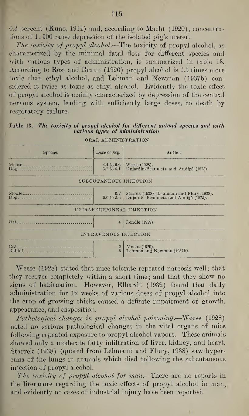
0.3 percent (Kuno, 1914) and, according to Macht (1920), concentra¬ tions of 1: 500 cause depression of the isolated pig’s ureter.
The toxicity of propyl alcohol.—The toxicity of propyl alcohol, as characterized by the minimal fatal dose for different species and with various types of administration, is summarized in table 13. According to Rost and Braun (1926) propyl alcohol is 1.5 times more toxic than ethyl alcohol, and Lehman and Newman (1937b) con¬ sidered it twice as toxic as ethyl alcohol. Evidently the toxic effect of propyl alcohol is mainly characterized by depression of the central nervous system, leading with sufficiently large doses, to death by respiratory failure.
Table 13.—The toxicity of propyl alcohol for different animal species and with various types of administration
ORAL ADMINISTRATION
Species Dose cc./kg. Author
Mouse_ __ _ 4.4 to 5.6 Weese (1928). Dujardin-Beaumetz and AudigS (1875). Dog_ _ _ 3.7 to 4.1
SUBCUTANEOUS INJECTION
Mouse Dog...
6.2 5.0 to 5.6
Starrek (1938) (Lehmann and Flury, 1938). Dujardin-Beaumetz and Audig6 (1875).
INTRAPERITONEAL INJECTION
Rat. Lendle (1928).
INTRAVENOUS INJECTION
Cat-... Rabbit
2 Macht (1920). 5 Lehman and Newman (1937b).
Weese (1928) stated that mice tolerate repeated narcosis well; that they recover completely within a short time; and that they show no signs of habituation. However, Elliardt (1932) found that daily administration for 12 weeks of various doses of propyl alcohol into the crop of growing chicks caused a definite impairment of growth, appearance, and disposition.
Pathological changes in propyl alcohol poisoning.—Weese (1928) noted no serious pathological changes in the vital organs of mice following repeated exposure to propyl alcohol vapors. These animals showed only a moderate fatty infiltration of liver, kidney, and heart. Starrek (1938) (quoted from Lehmann and Flury, 1938) saw hyper¬ emia of the lungs in animals which died following the subcutaneous injection of propyl alcohol.
The toxicity of propyl alcohol for man.—There are no reports in the literature regarding the toxic effects of propyl alcohol in man, and evidently no cases of industrial injury have been reported.
116
d. Isopropyl Alcohol
The only isomeric alcohol of propanol is isopropyl alcohol.
Chemical characteristics.—Isopropyl alcohol, propanol-2, second¬
ary propyl alcohol, dimethylcarbinol, perspirit, petrohol, avantine,
CH3CH3CHOH, has the molecular weight 60.09. It is a colorless
20°
liquid of the specific gravity 0.789 at-^-C. It solidifies at —85.8°
C., boils at 82.5° C., and is miscible with water, alcohol, and ether.
Its flash point is given as +18° C. by Lehmann and Flury (1938) and
as 12° C. by Browning (1937), and its upper limit of inflammability
is 2.65 volume percent (Jones, 1938).
Uses.—Isopropyl alcohol is manufactured by catalytic reduction
of acetone or from olefinic hydrocarbons arising in cracking petroleum
(Garlick, 1927). It is a solvent for certain resins such as mastic,
colophony, shellac, sandarac, and most copals (Browning, 1937), and
it is used in the manufacture of perfumes and cosmetics, in the manu¬
facture of safety glass (as dehydrating agent) and in the lacquer
and varnish industry.
Chemical identification.-—Isopropyl alcohol is oxidized to acetone
and, further, to acetic and formic acid. The phenyl-iso-propyl-
urethane forms needles which melt around 90° C., and the a-naphthyl-
iso-propy 1-urethane forms long tablets of the melting point 105° to
106° C. Isopropyl-iodide boils at 59.5° C. (corr.).
The determination of isopropyl alcohol.—Bae (1926) determined
isopropyl alcohol by oxidation to acetone. The mixture of 10 cc.
of the sample with 20 cc. of a 1 percent solution of potassium bichro¬
mate and 1 cc. of concentrated sulfuric acid is distilled until 3 cc.
of the distillate have been collected. This distillate is floated on the
surface of a mixture of 2 cc. of a 5 percent solution of sodium nitro-
prusside with an equal volume of strong ammonia (sp. gr. 0.880) and
approximately 0.3 gm. of ammonium chloride, and kept standing
for a few minutes. In the presence of acetone a purple ring is formed
at the junction of the 2 layers. Concentrations of 1 cc. of isopropyl
alcohol in 100 cc. of water are said to be detectable in this way. A
similar procedure was used by Wagner (1934).
Cook and Smith (1929) determined isopropyl alcohol in urine by
oxidizing it to acetone and estimating the amount of the latter as the
mercury sulfate complex of Deniges (1910). In order to differentiate
between acetone derived from isopropyl alcohol and the preformed
acetone in the urine, a sample of the urine is distilled into a solution
of hydroxylamine hydrochloride and the amount of acetone is de¬
termined by titration of the liberated hydrochloric acid. The differ¬
ence between the 2 acetone determinations allows the calculation of the amount of isopropyl alcohol present in the urine.
117
Hahn (1937) detei'rrdned isopropyl alcohol in the exhaled air by
absorbing the isopropyl alcohol vapors on a silica gel and subsequently
liberating it by distillation with steam. In the steam distillate the
alcohol is then determined by the procedure of Knipping and Ponn-
dorf (1926) which is based on the conversion of isopropyl alcohol to
isopropyl nitrite, extraction of the latter with carbon tetrachloride,
and the determination of the nitrous acid by volumetric determination.
The procedure is essentially as follows: Two separatory funnels are
arranged, one above the other. Into the lower funnel is placed 50 cc.
of a 5 percent solution of sodium bicarbonate and into the upper one
is placed a measured quantity of the steam distillate which has been
diluted with water to 65 cc. and mixed with 2 gm. sodium nitrite, 40 cc.
of carbon tetrachloride, and a mixture of 3 cc. of 25 percent hydro¬
chloric acid and 7 cc. of water. The separatory funnel is then closed
with a glass stopper and agitated until the nitrous fumes have dis¬
appeared. After separation the carbon tetrachloride layer is drained
through the sodium bicarbonate solution in the lower funnel and
shaken vigorously, and the carbon tetrachloride layer is then trans¬
ferred into a 250 cc. volumetric flask containing 20 cc. of distilled
water. The extraction with carbon tetrachloride is repeated and the
second extract is transferred to the same flask which should contain 20
cc. of water and 90 cc. of chloroform extract. To this is added a
measured volume of N/10 solution of potassium permanganate, % of
its volume of a 20 percent solution of manganous sulfate, and 20 cc.
of normal sulfuric acid. This mixture is shaken vigorously for 10
minutes and then 1 gm. of potassium iodide is added, the mixture is
shaken again, and the liberated iodine is titrated with N/10 solution
of sodium thiosulfate, using starch as indicator. One cc. of the po¬
tassium permanganate solution used corresponds to 3 mg. of isopropyl
alcohol.
Absorption, fate and excretion.—Isopropyl alcohol is absorbed from
the gastrointestinal tract and through the lungs. According to Macht
(1922), massive application to the skin of rats, dogs, and rabbits causes
no deleterious effects.
With regard to its fate in the organism, it was shown by Albertoni
(1887) (quoted from Heffter, 1905) that isopropyl alcohol is partly
oxidized to acetone, partly excreted unchanged, and partly excreted as
acetone. Keubauer (1901) claimed that isopropyl alcohol is excreted
by rabbits, and to a much lesser extent by dogs, in conjugation with
glucuronic acid, but this was not confirmed for man by Kemal (1927).
According to Cushny (1910) it is, like methyl and ethyl alcohol, partly
excreted through the lungs, and according to Pohl (1922) the pulmo¬
nary excretion as isopropyl alcohol or as acetone amounts to 12.8 per¬
cent of the dose administered orally to rabbits. Kemal (1927) stated
that in man the urinary excretion starts 1 hour after the ingestion and,
118
with sufficiently large doses, may last as long as 48 hours. The pul¬
monary excretion, however, had already started after 15 minutes. In
his experience the urinary excretion, which amounts to only a small
part of the total amount ingested, can be increased by diuresis. In a
later study, Kemal (1937) found that with administration of 65 to 90
ec. of isopropyl alcohol to dogs, 55 to 71 mg. percent were excreted as
acetone, 120 to 134 mg. percent were excreted unchanged with the
urine, and 28 to 29 mg. acetone and 63 to 70 mg. isopropyl alcohol were
found in the feces. Hahn (1937) found that in man, after the inges¬
tion of 720 mg. in water, 335.94 mg. (46.7 percent) of isopropyl alcohol
and 17.08 mg. of acetone were exhaled over a period of 3 hours. Ac¬
cording to Neymark (1938) isopropyl alcohol is oxidized in the organ¬
ism of dogs at the rate of 0.001 percent per minute as compared with
0.003 percent per minute found with propyl alcohol. Since it was
found that with the administration of certain amounts of isopropyl al¬
cohol the acetone concentration curve in the blood corresponds to that
obtained with the continued administration of actene, it appears that
isopropyl alcohol passes quantitatively into acetone. In accordance
with its slow rate of oxidation the excretion of isopropyl alcohol is
much slower than that of ethyl alcohol (Morris and Lightbody, 1938).
With regard to the distribution of isopropyl alcohol in various or¬
gans, the findings of Kemal (1937) are summarized in table 14 which
shows that 4 hours after the oral administration of isopropyl alcohol
the distribution among various organs is not uniform and that their
acetone content also shows considerable variation.
Antiseptic properties.—Isopropyl alcohol, like propyl alcohol, has
antiseptic properties which, according to Christiansen (1918), are in¬
ferior to that of the latter with regard to disinfection of the skin.
Bernhardt (1922), on the other hand, claimed that isopropyl alcohol
is a perfect substitute for ethyl alcohol for this purpose, and Grant
(1923a) stated that it has more powerful bactericidal and antiseptic
properties than ethyl alcohol, the most effective concentration being 30 to 50 percent.
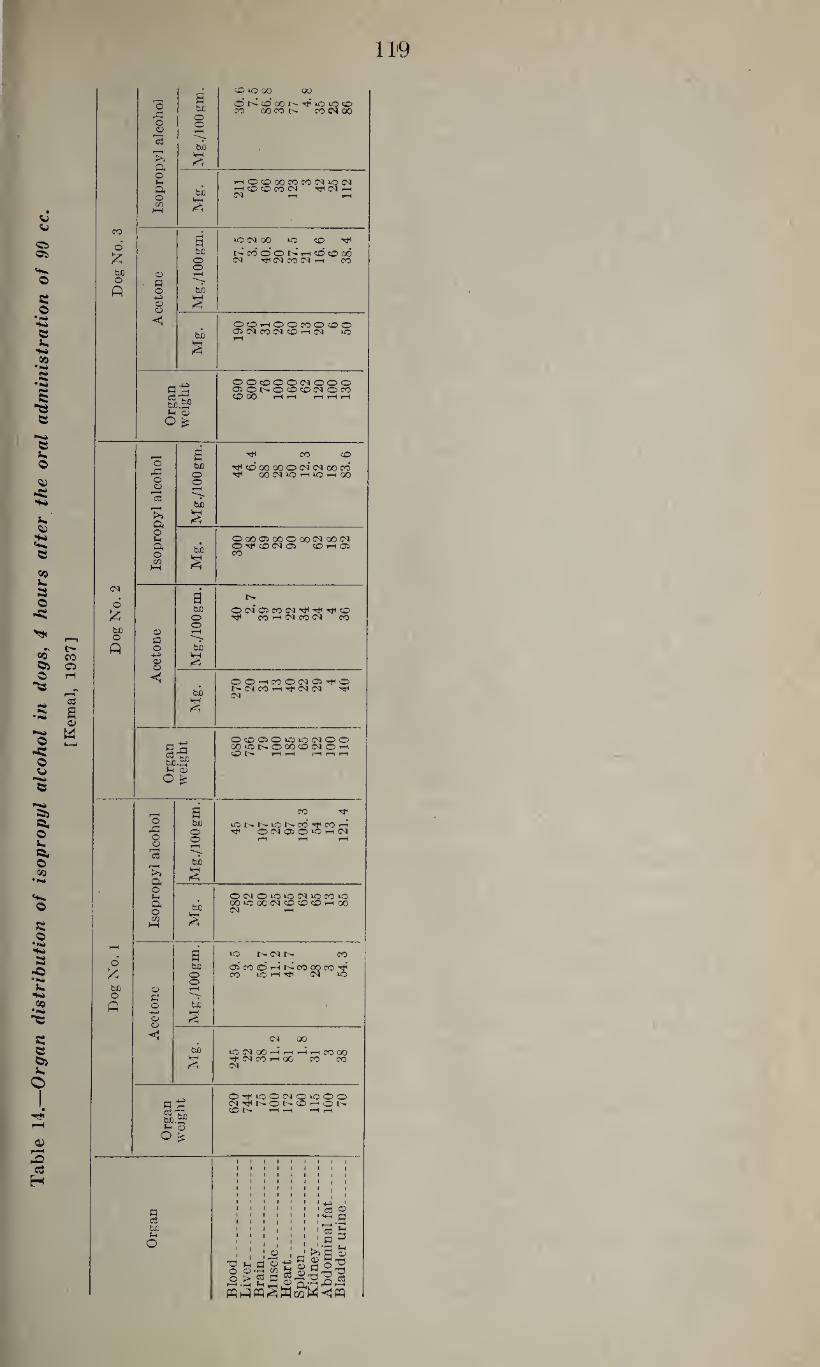
Table
14.—
Org
an dis
trib
uti
on o
f is
opro
pyl
alco
hol
in
dogs,
4 hours aft
er
the o
ral
ad
min
istr
ati
on o
f 90
cc.
[Kem
al,
19
37
] ch co »o co oo
'o A
P be O
O N O GO N TP 1C IP o CO QOCO CO C4 00
o O o
r—i G3
rH
bi
a o l-c
s
HOOCOCOCCWiON H cO CD CO <M ^C4h C4 rH rH
a o
bi
cn 1—< £
CO • a lO 04 00 *0 CO o
£ tuO NMOONHdtOOO o C4 T* CO C^4 rH CO
bjo o
0? r-H o p p O b/D
<D o §
bi OOhOOMOOO 05 <M CO C4 CO »H <M lO
§
OOcOOOiMOOO c ®ONOCO©NOW C3 Tj. bfi.SP
CO 00 rH rH rH r-H rH
rH <D
s ^ CO CO O bfi ^ CO 00 00 O 04 04 00 CO
O ^ OOINIOHIOHGO o o o H
bi
>> Q<
£
o OOOffiOOOOOMOOlN OHcOWOi CO i—1 05 00
(h ft o m w M
g.
<M •
a £
be OdOiMfN^^^cO ^ CO rH 04 CO (N CO o
b/o O o rH
o
A
q o bi
-*H o> o S
bi OOhcOOCIO^O NC1COh^C4C4 ^ cs
VH
OOOOK3K3NOO o QOiONOOO^OCIOh c3 fcdO .£?
O rH rH H n H
<D o £
i ■ i a CO
O be lONNiONCO^COn* rP o rT OdOOCHlN o o r-H rH rH o
13
rH
bi
ft • Jh OlNOOiOCliOfOiO
ft O
bi CO *0 GO 04 CO CO CO rH 00 C4 rH
c/i HH £
rH • a iO t^- 04 CO
o
£
bfl o> co o h co co co Mi CO CS »0 o
be o
o> rH o q p o bxi •
4-> O' O
<1 2
C4 GO
bi >OC4(X)HHHHCOOO
S ^ (M CO rH 00 CO CO C4
0^100^0^000 a C6 Ti. 60.SP
CO I>- rH rH rH rH
o> o £
1 1 1 1 l 1 1 1 l 1 1 1 1 1 1 1 1 1 1 1 1 1 i 1 1 1 l 1 t 1 1 1 1 1 1 1 1 1 • 1 1 1 1 . 1 1 1 1 1 1 1 1 . 1 1 1 1 1 1 1 1jj i
p GS be
1 c3 O • 1 1 1 i • IH M ' » l 1 ' 1 '_ -P • • « i • • i c3 h
: i i © : _* >.'3 ©
S-4 o
Blo
od
L
iver
. B
rain
. M
usc
l H
eart
S
ple
en
Kid
ne
A
bdor
Bla
dd
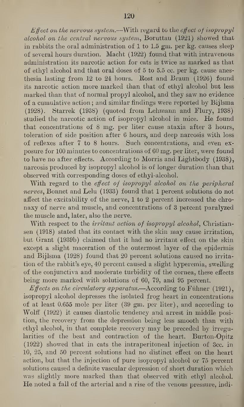
120
Effect on the nervous system.—With regard to the effect of isopropyl
alcohol on the central nervous system, Boruttau (1921) showed that
in rabbits the oral administration of 1 to 1.5 gm. per kg. causes sleep
of several hours duration. Macht (1922) found that with intravenous
administration its narcotic action for cats is twice as marked as that
of ethyl alcohol and that oral doses of 5 to 5.5 cc. per kg. cause anes¬
thesia lasting from 12 to 24 hours. Rost and Braun (1926) found
its narcotic action more marked than that of ethyl alcohol but less
marked than that of normal propyl alcohol, and they saw no evidence
of a cumulative action; and similar findings were reported by Bijlsma
(1928). Starrek (1938') (quoted from Lehmann and Flury, 1938)
studied the narcotic action of isopropyl alcohol in mice. He found
that concentrations of 8 mg. per liter cause ataxia after 3 hours,
toleration of side position after 6 hours, and deep narcosis with loss
of reflexes after 7 to 8 hours. Such concentrations, and even ex¬
posure for 100 minutes to concentrations of 60 mg. per liter, were found
to have no after effects. According to Morris and Lightbody (1938),
narcosis produced by isopropyl alcohol is of longer duration than that
observed with corresponding doses of ethyl-alcohol.
With regard to the effect of isopropyl alcohol on the peripheral
nerves, Bonnet and Lelu (1933) found that 1 percent solutions do not
affect the excitability of the nerve, 1 to 2 percent increased the chro-
naxy of nerve and muscle, and concentrations of 3 percent paralyzed
the muscle and, later, also the nerve.
With respect to the irritant action of isopropyl alcohol, Christian¬
sen (1918) stated that its contact with the skin may cause irritation,
but Grant (1939b) claimed that it had no irritant effect on the skin
except a slight maceration of the outermost layer of the epidermis
and Bijlsma (1928) found that 20 percent solutions caused no irrita¬
tion of the rabbit’s eye, 40 percent caused a slight hyperemia, swelling
of the conjunctiva and moderate turbidity of the cornea, these effects
being more marked with solutions of 60, 79, and 95 percent.
Effects on the circulatory apparatus.—According to Fiihner (1921),
isopropyl alcohol depresses the isolated frog heart in concentrations
of at least 0.655 mole per liter (39 gm. per liter), and according to
Wolff (1922) it causes diastolic tendency and arrest in middle posi¬
tion, the recovery from the depression being less smooth than with
ethyl alcohol, in that complete recovery may be preceded by irregu¬
larities of the beat and contraction of the heart. Burton-Opitz
(1922) showed that in cats the intraperitoneal injection of 3cc. in
10, 25, and 50 percent solutions had no distinct effect on the heart
action, but that the injection of pure isopropyl alcohol or 75 percent
solutions caused a definite vascular depression of short duration which
was slightly more marked than that observed with ethyl alcohol.
He noted a fall of the arterial and a rise of the venous pressure, indi-
121
eating a reduction of the functional energy of the heart (decreased
cardiac output) which was paralleled by slowing of the heart rate.
Macht (1922) stated that sublethal doses do not affect the blood pres¬
sure of cats, and Bijlsma (1928) found that, as compared with ethyl
alcohol, it had little effect on the heart action and the blood pressure.
Grant (1923b) refers to experiments of W. G. Thompson on human
subjects in which the ingestion of 25 cc. of isopropyl alcohol diluted
with 125 cc. of water was followed, after % hour, by a reduction of
the blood pressure of 6 to 20 mm. Hg and, in 4 out of 6 subjects, by
an increase of the pulse rate. Fuller and Hunter (1927) reported
that in man, intoxication with isopropyl alcohol causes lowering of
the blood pressure and the pulse rate.
Toxicity of isopropyl alcohol.—According to Rost and Braun
(1926), Bijlsma (1928) and others, isopropyl alcohol is more toxic
than ethyl alcohol. The minimal fatal dose for different species and
with different forms of administration is summarized in table 15.
In contrast to ethyl alcohol, isopropyl alcohol does not cause a pri¬
mary stimulation but only a depression (Boruttau, 1921). With suf¬
ficiently large doses it causes ataxia and narcosis, and, if fatal, para¬
lysis and dyspnea may precede death (Starrek, 1938) (quoted from
Lehmann and Flury, 1938). Rost and Braun (1926) and Bijlsma
(1928) noted, with repeated administration of isopropyl alcohol, no
evidence of a cumulative action; and, similarly, Weese (1928) stated
that mice tolerate repeated narcosis without signs of cumulative effects
or habituation. Morris and Lightbody (1938), on the other hand,
believed that, on account of its slow elimination, isopropyl alcohol
may have cumulative effects which may be due to the alcohol itself
or to it9 oxidation product, acetone.
Table 15.—Toxicity of isopropyl alcohol with various routes of administration for different species of animals
ORAL ADMINISTRATION
Species Minimal fatal dose Author
cc./kg.
Rabbit_____ 6.3 Boruttau (1921). Weese (1928). Mouse.._____ 6.0 to 7.6
SUBCUTANEOUS INJECTION
Mouse- 7.6 Starrek (1938) (Lehmann and Flury, 1938).
INTRAVENOUS INJECTION
Cat. 2.5 Macht (1922).
INHALATION
Note.—No fatal concentrations have been reported.
555178—43-9
122
With regard to the toxic effects of isopropyl alcohol on the hu' man organism, Grant (1923b) and Morris and Lightbody (1938)
referred to experiments of W. G. Thompson who stated that the
ingestion of 25 cc. in 125 cc. of water caused in man no exhilaration
but dizziness, muscular and nervous disturbances and, after 2 to 3
hours, headache of long duration. Fuller and Hunter (1927) noted,
aside from the effects on blood pressure and pulse rate mentioned
above, no visual disturbances such as have been reported following
ingestion of methanol. There are no reports in the literature with
regard to toxic effects from the industrial use of isopropyl alcohol.
Donley (1936) reported a case of toxic encephalitis, rhinitis, bron¬
chitis associated with slight anemia, and a moderate lymphocytosis
in a patient who had inhaled the vapors of a solvent consisting of
74 percent isopropyl alcohol, 20 percent water, and less than 3 per¬
cent each of dimethyl phthalate and methyl cellosolve.
Pathological changes.—Data with regard to pathological changes
resulting from the administration of isopropyl alcohol are very
scanty. Weese (1928) noted no serious pathological changes except
a very moderate reversible fatty infiltration of the liver, kidneys,
and heart muscle.
e. n-Butyl Alcohol
The next higher homologue is butyl alcohol which exists in 4
isomeric forms.
Physico-chemical characteristics.—Butyl alcohol, butanol-1, CH3-
CH2CH2CH2OH, has a molecular weight of 74.12 and represents a
colorless liquid of somewhat pungent odor. It has the specific grav¬ en0
ity 0.810 at -^-C. It solidifies at —79.9° C., boils at 117° C., is
soluble in 100 parts of water to the extent of 9 parts at 15° C., and
mixes freely with alcohol and ether. The upper limit of its inflam¬
mability is 1.70 percent (Jones, 1938) and its flash point is +34° C.
(Lehmann and Flury, 1938).
Butyl alcohol is manufactured by fermentation of maize flour by
a special strain of bacillus. It may be synthesized from acetalde¬
hyde, and it is one of the constituents of fusel oil (Browning, 1937).
Uses.—Being a good solvent for many gums and resins and in¬
creasing the solvent power of other solvents for cellulose esters or
ethers it is extensively used in the lacquer and dye industries and
in other industries (shoe industry, manufacture of safety glass, hat
and textile industries, etc.) where such solutions are handled
(Browning, 1937).
Chemical identification.—Normal butyl alcohol is oxidized to
n-butyr-aldehyde and n-butyric acid. The phenyl-n-butyl urethane
(C6H5-NHCOO-CH2-(CH2)2-CH3) forms needles which melt at
123
61° C. and the a-naphthyl-n-butyl-urethane (Ci0H7NHCOO-CH2-
(CH2)2-CH3) forms tablets which soften at 69° C. and melt at 71° to 72° C. N-Butyl iodide (CiHJ) boils at 130.4° to 131.4° C. at
754.4 mm. Hg. Absorption, fate, and excretion.—Normal butyl alcohol is absorbed
through the lungs, the gastrointestinal tract, and, according to San¬ der (1933), also through the skin.
Weese (1928) believed that it is completely oxidized in mice, and according to Berggren (1938) the oxidation takes place more rapidly than with ethyl alcohol. It is probably not assimilated as sugar (Hockendorf, 1909-10).
It is evidently not excreted with the urine, at least there are no reports of its excretion in this way.
Effect on the nervous system.—Baer (1898) found that in rabbits the oral administration of 1 to 1.5 gm. per kg. causes a moderate reduc¬ tion of the sensitivity and within 20 to 30 minutes a slight paralysis without materially affecting the corneal, pupillary, and ciliary reflexes, the pulse rate, and the respiration. Doses of 1.6 to 2.0 gm. per kg. were found to cause a short primary excitement and then, in 5 to 10 minutes, slight, and after 15 minutes, complete paralysis, analgesia, and impair¬ ment of the corneal, pupillary, and ciliary reflexes, and reduction of the respiration and body temperature, followed by recovery after a period of 10 hours. With doses of 2.1 to 2.44 gm. per kg. the paralysis was rapid and complete; the corneal, pupillary, and ciliary reflexes were rapidly reduced; the pupils were constricted; and the animals showed nystagmus, salivation, marked reduction of body temperature and respiration, and deep narcosis which lasted 36 hours. Lendle (1928) determined in rats the minimal narcotic dose with intraperitoneal injection as 0.76 cc. per kg. (given as 1.5 percent solution) and the minimal fatal dose as 1.2 cc. per kg., so that the margin of safety is 1.6; and he found that animals recovered from the narcosis within 54 to 106 minutes. Weese (1928) found that mice tolerate repeated narcosis resulting from the inhalation of vapors of butyl alcohol very well; that they recover completely within a few hours; and that they show neither signs of habituation nor serious organ injury. Starrek (1938) (quoted from Lehmann and Flury, 1938) studied the narcotic action
. of butyl alcohol vapors in mice. He found that with concentrations of 20 mg. per liter (about 6,600 p. p. m.) mice started to stagger after 1 hour, tolerated side position after 1% to 2 hours, and became deeply narcotized at the end of 3 hours. Three out of 10 animals anesthe¬ tized to deep narcosis died during the following days.
With regard to the effect of normal butyl alcohol on the peripheral nerves, Bonnet and Lelu (1933) found that concentrations of 0.25, 0.35, and 0.50 percent lowered the chronaxy of the nerve, whereas 1 percent solutions rapidly rendered it inexcitable. Cole and Allison
124
(1930b) found that 0.066 molar concentrations cause irritation of the
skin of frogs. Effect on the circulatory system.—Kuno (1913) found that 1/400 to
1/500 molar solutions (0.19 to 0.15 gm. per liter) of normal butyl alcohol are just sufficient to cause depression of the isolated mam¬ malian heart, whereas 1/15 molar solutions (4.94 gm. per liter) cause cardiac arrest within a few seconds. Similarly, Fiihner (1921) de¬ termined the minimal effective concentration for the isolated frog heart as 0.109 mole or 8.1 gm. per liter, whereas the corresponding values for normal propyl alcohol were 1/200 and 0.369 molar con¬ centration (0.3 gm. and 22.2 gm. per liter), respectively. Normal butyl alcohol is, therefore, more toxic to the isolated frog heart than the lower isomer.
With regard to the effect of normal butyl alcohol on the blood vessels, Buchner, Fuchs, and Megele (1901) found that its application to the peritoneal cavity caused a more marked vasodilatation than that observed with normal propyl alcohol.
With respect to the effect of normal butyl alcohol on the blood, Smyth and Smyth (1928) found that 65 exposures to 100 p. p. m. in air caused a reduction of the red blood cells and a relative and absolute lymphocytosis.
Effect on muscle structure.—According to Bonnet and Lelu (1933) the striated muscle is affected similarly but to a lesser degree than is the nerve by solutions of normal butyl alcohol. The activity of the isolated intestine of the rabbit is depressed by concentrations of 0.025, 0.03, and 0.05 percent, and concentrations of 0.5 percent cause imme¬ diate paralysis and complete loss of tone (Kuno, 1914).
Toxicity of normal butyl alcohol.—The toxicity of normal butyl alcohol, as characterized by its minimal fatal dose for different animal species and with various forms of administration, is illustrated in table 16. Comparison of these data with those presented for other alcohols shows that butyl alcohol is more toxic than the lower liomologues, but, in view of its lower volatility, it is less dangerous when inhaled as a vapor (Weese, 1928).
In contrast to the experience with normal propyl alcohol, Elhardt (1932) found that the daily administration of normal butyl alcohol into the crop of growing chicks for a period of 16 weeks had no effect on the growth and appearance of the chicks.
No definite information is available regarding injurious effects of normal butyl alcohol on the human organism.—Burger and Stock¬ mann (1932) reported on injury of the liver resulting from exposure to the vapors of a solvent which contained, in addition to butyl alco¬ hol, amyl alcohol, butyl acetate, amyl acetate, and acetone, but it was questionable to what extend butyl alcohol was responsible for the toxic effects observed. Kruger (1932) reported on severe irritation
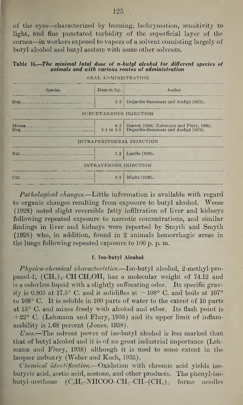
125
of the eyes—characterized by burning, lachrymation, sensitivity to light, and fine punctated turbidity of the superficial layer of the cornea—in workers exposed to vapors of a solvent consisting largely of butyl alcohol and butyl acetate with some other solvents.
Table 16.—The minimal fatal dose of n-butyl alcohol for different species of animals and with various routes of administration
ORAL ADMINISTRATION
Species Dose cc./kg. Author
Dog_ 2.2 Dujardin-Beaumetz and Audige (1875).
SUBCUTANEOUS INJECTION
Mouse Dog...
6.2 2.4 to 2.8
Starrek (1938) (Lehmann and Flury, 1938). Dujardin-Beaumetz and Audige (1875).
INTRAPERITONEAL INJECTION
Rat. 1.2 Lendle (1928).
Cat
INTRAVENOUS INJECTION
0.3 Macht (1920).
Pathological changes.—Little information is available with regard to organic changes resulting from exposure to butyl alcohol. Weese (1928) noted slight reversible fatty infiltration of liver and kidneys following repeated exposure to narcotic concentrations, and similar findings in liver and kidneys were reported by Smyth and Smyth (1928) who, in addition, found in 2 animals hemorrhagic areas in the lungs following repeated exposure to 100 p. p. m.
V
f. Iso-butyl Alcohol
Physico-chemical characteristics.—Iso-butyl alcohol, 2-methyl-pro- panol-I, (CH3)2-CH CHoOH, has a molecular weight of 74.12 and is a colorless liquid with a slightly suffocating odor. Its specific grav¬ ity is 0.805 at 17.5° C. and it solidifies at —108° C. and boils at 107° to 108° C. It is soluble in 100 parts of water to the extent of 10 parts at 15° C. and mixes freely with alcohol and ether. Its flash point is + 22° C. (Lehmann and Flury, 1938) and its upper limit of inflam¬ mability is 1.68' percent (Jones, 1938).
Uses.—The solvent power of iso-butyl alcohol is less marked than that of butyl alcohol and it is of no great industrial importance (Leh¬ mann and Flury, 1938) although it is used to some extent in the lacquer industry (Weber and Koch, 1933).
Chemical identification.—Oxidation with chromic acid yields iso- butyric acid, acetic acid, acetone, and other products. The phenyl-iso¬ butyl-urethane (C6H5-NHCOO-CH2-CH-(CH3)2 forms needles
126
which melt at 80° C., and the corresponding a-naphthyl derivative (C10H7-NHCOO-CH2-(CH3)2 forms needles of the melting point 103° to 105° C. The iodide (C4H9I) boils at 120° C. (corr.)
Absorption, fate, and excretion.—Iso-butyl alcohol is absorbed through the gastrointestinal tract and through the lungs. There is no information as to what extent it may be absorbed through the skin.
According to Neubauer (1901), in rabbits and, to a lesser extent, in dogs, it is partly excreted in conjugation with glucuronic acid, but in the opinion of Weese (1928) it is completely oxidized in mice.
Effect on the nervous system.—According to Rost and Braun (1926) it is more potent as a narcotic with oral administration to rabbits, the minimal narcotic dose being 2.5 cc. per kg. as compared with 5.0 cc. per kg. of normal butyl alcohol. With intravenous injection the minimal narcotic dose for rabbits is 0.9 cc. per kg. (Lehman and New¬ man, 1937b), this being about 6 times as potent as ethyl alcohol. But with inhalation of the vapors the narcotic action is less marked than that of propyl alcohol and of the same order as that of normal butyl alcohol (Weese, 1928) which indicates that with industrial exposure it is potentially less dangerous than the lower homologues.
According to Bonnet and Lelu (1933) concentrations of 0.35 to 1.0 percent decrease the chronaxy of the isolated nerve and muscle, but later the excitability of the muscle diminishes earlier than that of the nerve.
Effect on the circulatory system.—Little information is available with regard to the effect of iso-butyl alcohol on the ciculatory system. According to Fiihner (1921) the minimal depressant concentration for the isolated frog heart is 1.35 mole per liter (100 gm. per liter).
Toxicity.—According to Macht (1920) the minimal fatal dose for cats with intravenous injection is 0.9 cc. per kg., and with slow in¬ travenous administration it is 2.64 gm. per kg. for rabbits (Lehman and Newman, 1937b). There are no reports on the toxic action of iso-butyl alcohol in man.
Pathological changes.—Weese (1928) noted only a slight reversible fatty infiltration of liver and kidneys in mice which had undergone repeated narcosis without any after effects.
g. Secondary Butyl Alcohol
Physico-chemical characteristics.—Secondary butyl alcohol, bu¬ tanol-2, CH3-CH2-CHOHCH3, has a molecular weight of 74.12. Its
20°
specific gravity is 0.808 at C. and it boils at 99.5° C. It is soluble
in 100 parts of water to the extent of 12.5 parts at 20° C. and mixes freely with alcohol and ether. Its flash point is 22° C. (Lehmann and
127
Flury, 1938). Having 1 asymmetrical hydrocarbon it exists in the form of 2 optical isomers, levo and dextro secondary butyl alcohol.
Uses.—Secondary butyl alcohol is used to a moderate extent in the lacquer industry.
Absorption, fate, and excretion.—Secondary butyl alcohol is ab¬ sorbed from the gastrointestinal tract and through the lungs. No information is available with regard to its absorption through the skin, its fate in the metabolism and its excretion, with the exception of a statement by Pohl (quoted from Lehmann and Flury, 1938) that 77 percent is excreted through the lungs after intravenous adminis¬ tration.
Systemic effects.—Weese (1928) found in mice that the narcotic ac¬ tion of vapors of secondary butyl alcohol is greater than that of normal or isobutyl alcohol and that it ranks between those of ethyl and propyl alcohol. In his opinion the more marked narcotic action is linked to the higher vapor tension of the secondary butyl alcohol which en¬ hances a more rapid penetration into the blood and the central nervous system;. Starrek (1938) (quoted from Lehmann and Flury, 1938) studied its narcotic action in mice. He found that concentrations of 10 mg. per liter as compared to 20 mg. per liter of the normal butyl alcohol caused staggering after 1 to 1 y2 hours, toleration of side posi¬ tion after 2 to 3 hours, and deep narcosis in 5 hours which was followed by rapid recovery without after effects. Butler and Dickison (1940) determined the narcotic dose for mice with intraperitoneal injection as 1 mg. per gram mouse. Hufferd (1932) found it to be less toxic than normal butyl alcohol for guinea pigs with intraperitoneal injections, the corresponding doses being 0.00093 and 0.00083 mole per 100 gm., respectively. Viditz (1933) noted no measurable difference between the levo and dextro isomers with regard to their narcotic action for tadpoles and fish and their effect on the nerve muscle preparation and the isolated frog heart; and Butler and Dickison (1940) found that the intraperitoneal injection of the 2 optical isomers had the same nar¬ cotic action in mice.
There are evidently no reports regarding the toxicity of secondary butyl alcohol for humans.
h. Tertiary Butyl Alcohol
Physico-chemical properties.—Tertiary butyl alcohol, 2-methyl- propanol-2, (CH3)3COH, has the molecular weight 74.12 and is a colorless liquid or forms rhombic crystals which melt at 25° to 25.5° C.
20°
and boil at 82.9° C. It has the specific gravity 0.786 at-^C. and mixes
freely with water, alcohol, and ether.
Uses.—The solvent properties of tertiary butyl alcohol are similar
128
to those of isopropyl alcohol, and it is used on a small scale in the per¬
fume industry (Lehmann and Flury, 1938). Chemical identification.—Oxidation with chromic acid yields, aside
from isobutyric acid, acetone, acetic acid, arid other products. The a-naphthyl-tertiary-butyl-urethane, CioH7NHCOO-C(CH3)3, forms long tablets which melt at 100° to 101° C., and the tertiary butyl iodide
boils at 39° C. Systemic effects.—Thierfelder and von Mering (1885) found that the
administration of 6 cc. to rabbits caused somnolence, whereas the ad¬ ministration of 10 cc. to a medium sized dog had no distinct effects, and they believed that in rabbits but not in dogs and man it is excreted in conjugation with glucuronic acid. F'iihner (1921) determined the minimal depressant concentration for the isolated frog heart as 0.637 mole per liter (47.1 gm. per liter), and Weese (1928) found that mice tolerate repeated narcosis well. He found that it is less toxic than isobutyl alcohol but more potent with regard to its narcotic action. Wesse (1928) noted no marked pathological changes except moderate and reversible fatty infiltration of liver, kidneys, and heart. Oettel (1936) reported that when applied to skin tertiary butyl alcohol causes moderate irritation characterized by slight pain, moderate hy¬ peremia, and erythema. Evidently no cases of toxic effects in man have been reported.
i. Amyl Alcohol • -
Physico-chemical characteristics.—Amyl alcohol, C5HnOH, exists in the form of 8 isomers. Its physico-chemical properties and for¬ mulas are summarized in table 17. According to Browning (1937) the amyl alcohol used commercially is obtained from fusel oil and repre¬ sents a mixture of primary isoamyl alcohol, 2-methyl-butanol-4 (No. 3) an active amyl alcohol, C2H5-CH-CH3-CH2OH (No. 8). The fusel oil from potatoes and cereals consists essentially of primary isoamyl alcohol (No. 3) and contains only 13 to 22 percent of active amyl alco¬ hol. There are several grades of commercial amyl alcohol which are characterized by their boiling point and specific gravity. Amyl alco¬ hol has a pungent and penetrating odor.
According to Browning (1937) amyl alcohol may also be manufac¬ tured from petroleum distillates and is marketed under the name of Pentasol. It represents a mixture of 5 of the 8 insomeric alcohols.
Whereas commercial amyl alcohol is a good solvent for many gums and resins and, to a lesser degree, for ethyl cellulose, copal ester, mas¬ tic and coumarone, Pentasol is said to be no solvent for cellulose esters or coumarone (Browning, 1937).
Uses.—Amyl alcohol is used as a solvent in the lacquer industry and in the chemical and explosives industries, and it may be encoun¬ tered in many industries such as textiles, shoe, and hat industries where such lacquers are used.
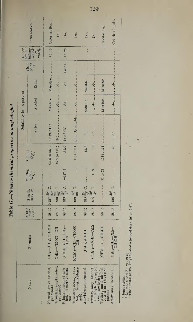
Table 1
7.—
Physi
co-c
hem
ical
pro
pert
ies
of
am
yl
alc
qh
ol
129
o o
Pi cs
o Pm
TJ '5
C/3 cn a;
Th jd
o O
o
o o Q
o Q
o o
A 0
a>
a
flS +-> i/>
>> In
u
a.t5 a g cc 2 o
G^.ga > & ^ .
3|o a a°
o co
4-5 Li <3 Q<
O o rH
c
>1
32 £
3 m
bo 3 g • ^.So •s o 5 D»° PQ
be
.gg • «-So a) o.
,g >* <P 4-5
• ^1 O ►>
S2 CQ tuO
g t-fJ P=I cs be o «—• •*- id® % *
ca r-H
3
o
O <N
Q
to •<f
M <D a
1 1
*2 3
1 1 1 1 1 1 1 1 1 1
O O
1 1 1 1 1
O
» 1 1
a>
1 1 1 1
0
1 1
a>
3
I 1 l 1 1
0 0 *23 T3 *o *o "0 *o
w CO
• rl 1 • 1 d 1 CO 1
S 1 1 1 1 1 1
I 1 1
0 m 1 1 1
1 1 1
0
0 0
1 a>
33 •r-i
cn -d
o_
.do_
op-
1
<0
S d
op-
1 »
r2 3
0 CO
op
-
◄ s
1 1 1 1 1 1 t 1
1
i 1
0 CO
1 1 1 1
• r-4
§
1 1 1 1
1 1
1 1 1 1
1 ©
1 1
1 1
1 1
1 I
1 1 1 32 1 1 1 i ! 3 1 1 1 1
a> 4-»
N
d
1 1 O w
1 1 1
1 1
1 1
1 1
03 0 1 O
>» 1 •
0
1 1
0
1 1
O
1 1
O (N 1 32 T3 T3 *23
CD pH tuo 1 !>.
CO w 1 1 f
CS T-H <M CQ 1 1
1 1
* 1
1 1
M o «
W o
co CN
w
O
w o
W
o
I
W o
H u
I *0
W N
o
L
w o
I W
WO
t§ CO
w
O
w o
w o
L co
w O
w o
W o
cs ^~N
w N
o
W cs
u
I
a o
o
L CO a o
w o c* w o
I o
w o
3 c*
v. CO a/
jo
o O
05 O CO <M 00 i%: CO
05 cm' CO
t-H
0
»o rH
0 H rH
0
rH
0 0 +*
+-> 4-2 CO CO 00 *o
T-H i-H
00 CO H y—i *-H
1 1
1 1 1
1 1 05 CO
1 1
» • 1 1 kO 1 1 1 1 1 rH 1
» O 1 rH 1 1 1 4H 1 1 1 I 1 1 1 CM 1
1 1
1 1
1 1
1 1
kO 1 •
1 •* #
O 0 O D 0 O 1 0
O IO 0 |o O lo 0 |o 0 In 0 lo 1 0 Irt 0 00 <r >0 ^ 05 | O »o 2* 0
1 O °H Cd 1 <N 1 (N pH 1 rH IO CM “ CM ” 1 CM 1^
0 CO* 05 kO 05 1 O rH t-H rH rH rH O 1 rH 00 00 00 00 00 00 1 00
0 * * ’ 1 •
•0 10 1-0 kO IO kO kO rH rH rH rH rH rH rH rH
00 00 00 88
00 00 00 00 00 00 GO 00 00 00 00
w oM
l w io QJ cs
oW
1°
W <M
o
®
a o3
£
O 32 O o
r—1
c3
•
sa S o _ 3
C3 ^
S a W g o a
£
o 32 O o
"3
a
a c3
da 03 ’3 3 3 o o> O Q, <u “
cq
CN O
03
O 03 <0 +J
■aS I
>> >.
as §-g .i S
d ^ Boo 'C’P P a
o <a o ■*-»
ca^ ■&-T
a£ cO -*-> o a>
.2 a
*0 co aaa 000
3
m
a p-T5^ o
§ +» 3 <W a o
33 o o
■3
V> a«
<
3- 33 0^32 3 o ® © o a o 3 g ® ri 03 3 j_ u3 >»
p-,G'3„33 *>> ^ rt a 32 33 a ®
• <D £ d
Jd (D *3 • O •S a g
o ^3 O o
"3
a c3
a> >
o 05 IO
>5 (-• o
03 -t-a
O (-« o > 0
o
. o
S'S
£*03
3 ®
T3 0 3 OT a.a— (30 “ aj
55 3 a 05 a 3 H B +J cC 03
CO 3 3 ® S p S-3 i
•p
130
Chemical identification.—The primary isobutyl carbinol (No. 3) may be oxidized with potassium permanganate (1:1000) with the formation of iso-valerianic acid aldehyde and iso-valerianic acid iso¬ amyl esters which may be identified by their odor. If heated with sulfuric and acetic acid it yields iso-amylacetate. According to Hollander (1910) this can be identified by adding 1 drop of phenyl- hydrazine and boiling. After cooling, the mixture is underlaid with concentrated hydrochloric acid. In the presence of amyl acetate a green color is formed at the zone of contact of the 2 layers. The phenyl-iso-amyl-urethane (CeHsHNCOOCsHu) melts at 57° to 58° C. and the corresponding a-naphthyl compound (CioH7HNCOOC5H1:l) forms leaflets which melt at 67° to 68° C. The iso-amy 1-iodide boils at 152° C. (Kosenthaler, 1923).
Upon oxidation, the active d-amyl alcohol (No. 8) yields d-methyl- ethyl-acetic acid. The phenyl urethane derivative melts at 30° C. whereas the a-naphthyl-urethane compound forms fine needles which melt at 82° C. (Rosenthaler, 1923).
The tertiary amyl alcohol, amylene hydrate (No. 6) yields a ruby red color with y2 cc. of a 1 percent solution of vanilline and 10 cc. of sulfuric acid, and upon oxidation it yields acetone, acetic acid, and carbon dioxide. The a-naphthyl-urethane derivative forms coarse needles which melt at 70° to 72° C.
For the detection of tertiary amyl alcohol in the presence of second¬
ary amyl alcohol, Michael and Zeidler (1911) (quoted from Rosen¬ thaler, 1923) heated the mixture with 3N hydrobromic acid in a sealed tube in a boiling water bath for 5 minutes, resulting in the formation of trimethyl ethylene from tertiary amyl alcohol. If after the sepa¬ ration of this hydrocarbon sufficient hydrobromic acid is added to the remaining alcohol to bring its concentration up to 4.5N and it is heated again in the same way for 1% hours, the secondary amyl alcohol is also changed to trimethyl ethylene.
For the detection of secondary isoamyl alcohol in the presence of
the primary isoamyl alcohol, the same authors dissolved the alcohol mixture in 30 parts of 4.5N hydrobromic acid and heated it as above for 1 hour. The presence of the secondary isoamyl alcohol is indi¬ cated by the formation of trimethyl ethylene.
Determination.—Basset (1910) determined amyl alcohol (fusel oil) in liquids by the pink or red color*produced with furfural and sulfuric acid. Korenman (1932) utilized the same reaction for the determination of amyl alcohol in air. In this procedure the air is collected in a dry flask of known volume. Twenty cc. of ethyl alcohol and the same amount of water are added and shaken for 2 or 3 hours. To 1 cc. of this solution 0.1 cc. of a 1 percent alcoholic solution of fur¬ fural and, while cooling, 1.5 cc. concentrated sulfuric acid are added, heated for 3 minutes in boiling water, and, after cooling, the pink to
131
red color is matched against standards of known strength prepared
in the same way.
Absorption, fate, and excretion.—Amyl alcohol is absorbed through
the gastro-intestinal tract, the lungs, and the skin (Schwenkenbecher,
1904, and Sander, 1933).
Neubauer (1901) claimed that a small fraction of the amount of
amyl alcohol administered is excreted in conjugation with glucuronic
acid, and he refers to Binz, Bodlander, and Strassmann as having found
that only 5 to 10 percent of the total dose administered is excreted
through the lungs and kidneys, the rest being completely oxidized in
the body. According to Hockendorf (1909-10) amyl alcohol increases
the sugar and decreases the nitrogen excretion in phloridzin diabetic
dogs. This may indicate that amyl alcohol may be utilized as sugar.
Pohl (1908) claimed that only traces of isoamyl alcohol are excreted
through the lungs.
Effect on the nervous system.—Strauss (1887) found that in rabbits
doses of 5 to 6 gm. given orally cause, within a few minutes, deep coma
and loss of corneal reflexes lasting for 5 to 6 hours. Lendle (1928)
determined in rats, with intraperitoneal injection, the minimal nar¬
cotic dose as 0.36 cc. per kg. (given in 10 percent solution), recovery
following after 35 to 50 minutes, and the minimal fatal dose as 0.6 cc.
per kg., so that the margin of safety is 1.7. As pointed out by Lendle
(1928), both doses are probably 25 percent smaller since the amyl alco¬
hol was in 33 percent ethyl alcoholic solution. Hufferd (1932) gave
the narcotic dose with oral administration for guinea pigs as 0.00067
mole per 100 gm. (0.72 cc. per kg.) body weight.
Isoamyl alcohol causes narcosis in rats with intraperitoneal injection
of 0.5 cc. per kg., according to Lendle (1928), and since the minimal
fatal dose wTas determined as 1.0 cc. per kg. the margin of safety is
2.0. According to Lehman and Newman (1937b) the minimal nar¬
cotic dose for rabbits with intravenous injection is 0.85 gm. per kg.,
isoamyl alcohol being, therefore, 6.5 times as potent as ethylalcohol.
Hufferd (1932) determined the narcotic dose for guinea pigs with oral
administration as 0.00063 mole per 100 gm. body weight (0.69 gm. per
kg-)- .... The secondary amyl alcohol causes narcosis in rats with intraperi¬
toneal injection of 0.65 cc. per kg., the minimal fatal dose being 1.0 cc.
per kg. and the margin of safety 1.5 (Lendle, 1928). With oral ad¬
ministration to guinea pigs, narcosis is produced by doses of 0.00052
mole per 100 gm. body weight (0.56 gm. kg.) (Hufferd, 1932).
With regard to the irritant action of amyl alcohol, Cole and Allison
(1930b). found that concentrations of 0.022 mole per liter cause irri¬
tation of the skin of frogs, and, according to Muller-Lobeck (1935),
concentrations of 2.7 mg. per liter cause neither turbidity of the cornea
132
nor irritation of the conjunctiva of rabbits as observed with compar¬
able concentrations of amyl acetate.
Effect on the circulatory system.—Salant (1909) found that the
intravenous injection of amyl alcohol in 2 percent solution causes a
more severe and more lasting fall of the blood pressure than observed
with corresponding doses of ethyl alcohol. Heide and Schilf (1929)
found that in dogs anesthetized with morphine, chloroform, or ether,
even small doses of isoamyl alcohol (less than 0.5 cc. of a 2 percent
solution) cause a considerable fall of the blood pressure and moderate
vasodilatation. Kuno (1913) determined the minimal depressant con¬
centration for the isolated mammalian heart as 1/1500 to 1/2000 mole
(0.059 to 0.044 gm.) per liter and the paralyzant concentration as
l/50mole (1.76 gm.) per liter. Fiihner (1921) determined the minimal
depressant concentration for isoamyl alcohol as 0.039 mole (3.4 gm.) per liter.
Buchner, Fuchs, and Megele (1901) found that the vasodilatation
produced by direct application to the peritoneum is more marked than
that observed with propyl and butyl alcohol.
Effect on other organs.—Kuno (1914) found that concentrations of
0.0125, 0.015, and 0.02 percent cause a moderate depression of the
pendular movements of the isolated intestine and that 0.2 to 0.25
percent solutions rapidly cause complete paralysis. Macht (1920)
y a cohol causes depression of the isolated ureter of the pig.
The toxicity of amyl alcohol.—According to Salant (1909) the mini¬
mal fatal dose for frogs is 1/8 to 1/7 that of ethyl alcohol, and for
rabbits it is from 2 to 4 times as toxic as the latter. Baer (1898) found
that in rabbits the oral administration of 0.83 to 1.08 gm. per kg.
causes a moderate reduction of the sensitivity; within 20 to 30 minutes
it causes slight paralysis without distinct impairment of the corneal,
pupillary, and ciliary reflexes and of the pulse rate and respiration.
Doses of 1.25 to 1.66 gm. per kg. were found to cause, after 5 to 10
minutes, slight and, after 15 minutes, deep narcosis with impairment of
the corneal, ciliary, and pupillary reflexes, reduction of the body tem¬
perature, and reduction of the respiration. With administration of
1.7 to 1.95 .gm. per kg. the paralysis was rapid and complete, the
pupillary, ciliary, and corneal reflexes became rapidly weaker, the
pupils were constricted, and body temperature and narcosis were
materially lowered. Starrek (1938) ) (quoted from Lehmann and
Flury, 1938) found that in mice the subcutaneous injection of 5 mg.
per gram of normal amyl alcohol caused staggering, toleration of
side position, dyspnea, motoric irritation, and finally deep narcosis.
Isoamyl alcohol in doses of 6 mg. per gram body weight causes
staggering, toleration of side position in 20 minutes, and narcosis in
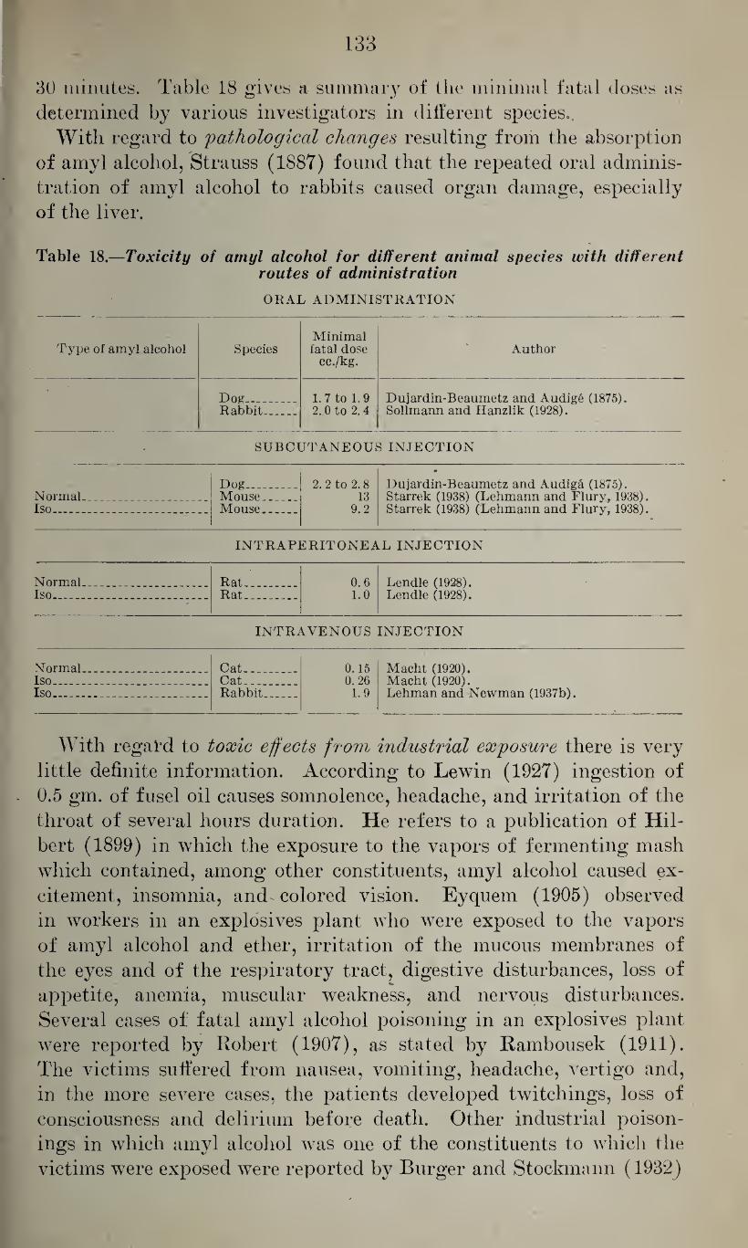
133
30 minutes. Table 18 gives a summary of the minimal fatal doses as
determined by various investigators in different species.
With regard to 'pathological changes resulting from the absorption
of amyl alcohol, Strauss (1887) found that the repeated oral adminis¬
tration of amyl alcohol to rabbits caused organ damage, especially
of the liver.
Table 18.—Toxicity of amyl alcohol for different animal species with different routes of administration
ORAL ADMINISTRATION
Type of amyl alcohol Species Minimal fatal dose
cc./kg. Author
• Dog_ -- 1. 7 to 1.9 Dujardin-Beaumetz and Audig6 (1875).
Sollmann and Hanzlik (1928). Rabbit_ 2. 0 to 2. 4
- SUBCUTANEOUS INJECTION
Dog_ 2. 2 to 2. 8 •
Dujardin-Beaumetz and Audiga (1875). Starrek (1938) (Lehmann and Flury, 1938). Starrek (1938) (Lehmann and Flury, 1938).
Normal .. _ Mouse_ 13 Iso... . Mouse__ 9.2
INTRAPERITONEAL INJECTION
Normal. .. Rat.. 0.6 Lendle (1928). Lendle (1928). Iso___ Rat.- 1.0
INTRAVENOUS INJECTION
Normal..,.. .. Cat.. _ 0.15 Macht (1920). Macht (1920). Lehman and Newman (1937b).
Iso_ .- _ Cat__ 0.26 Iso_____ Rabbit_ 1.9
With regal’d to toxic effects from industrial exposure there is very
little definite information. According to Lewin (1927) ingestion of
. 0.5 gm. of fusel oil causes somnolence, headache, and irritation of the
throat of several hours duration. He refers to a publication of Hil¬
bert (1899) in which the exposure to the vapors of fermenting mash
which contained, among other constituents, amyl alcohol caused ex¬
citement, insomnia, and colored vision. Eyquern (1905) observed
in workers in an explosives plant who were exposed to the vapors
of amyl alcohol and ether, irritation of the mucous membranes of
the eyes and of the respiratory tract, digestive disturbances, loss of
appetite, anemia, muscular weakness, and nervous disturbances.
Several cases ol: fatal amyl alcohol poisoning in an explosives plant
were reported by Robert (1907), as stated by Rambousek (1911).
The victims suffered from nausea, vomiting, headache, vertigo and,
in the more severe cases, the patients developed twitchings, loss of
consciousness and delirium before death. Other industrial poison¬
ings in which amyl alcohol was one of the constituents to which the
victims were exposed were reported by Burger and Stockmann (1932)
134
and by Baader (1933). Two other fatalities reported by Zangger
•(1933) may have had exposure to tetrachlorethane in addition to amyl
alcohol. The tertiary amyl alcohol, amylene hydrate, is of little industrial
importance but it has been used as a hypnotic.
With regard to its absorption, fate, and excretion, it is mainly ab¬
sorbed from the gastrointestinal tract, having a boiling point of 102°
C. According to Pohl (1908) appreciable quantities are excreted
through the lungs following its intravenous administration to dogs.
It is questionable wdiether it is excreted in conjugation with glucuronic
acid (Thierfelder and von Mering, 1885) and is said to be partly
excreted unchanged (Frankel, 1927).
With respect to its effect on the nervous system, Thierfelder and
von Mering (1885) found it more effective than the tertiary butyl
alcohol, doses of 3 cc. given orally causing in a large rabbit, in 10 to
15 minutes, deep sleep lasting for 12 to 24 hours. Lendle (1928) de¬
termined the minimal narcotic dose with intraperitoneal injection
in rats as 0.9 cc. per kg. and the minimal fatal dose as 1.6 cc. per kg.,
so that the margin of safety is 1.8.
According to Fuhner (1921) the minimal depressant concentra¬
tion for the isolated frog heart is 0.184 mole (16.2 gm.) per liter. Bock
(1898) found in the isolated mammalian heart that tertiary amyl
alcohol had little effect on the output and the rate, and he assumed
that the reduction of the blood pressure observed by earlier investiga¬
tors was due to peripheral vasodilatation.
With regard to its toxic action, Flury and Zangger (1928) stated
that with sufficiently large doses it may cause circulatory disturbances
characterized by vasodilatation and small and slow pqlse. Jacobi
and Speer (1920) published a case of medicinal poisoning resulting
from the rectal administration of 35 gm. or as pointed out by Loewe
(quoted from Jacobi and Speer, 1920) probably more correctly 28
to 29 gm. of amylene hydrate. This resulted in deep sleep, loss of
reflexes, collapse, and circulatory failure. Since the patient showed
signs of improvement during the first 24 hours after the poisoning, and
because he was found to have also suffered from pneumonia, it appears
questionable whether death resulted from amylene hydrate poisoning.
A similar case of amylene hydrate poisoning was reported by Anker
(1892) resulting from the ingestion of 27 gm. of amylene hydrate. In
this patient unconsciousness lasted 24 hours, somnolence persisted for
6 days, and for several weeks there was an excessive secretion of mucus
from the respiratory tract. During the first 24 hours after ingestion,
the respiration was impaired and the heart action weak. Large thera¬
peutic doses (4 gm. in water) may cause sleep readily, but are liable
to cause also headache, nausea, and impairment of circulation and
respiration more readily than observed with paraldehyde.
j. Higher Aliphatic Alcohols
The higher aliphatic alcohols—hexyl, heptyl, octyl, nonyl, decyl,
undecyl, and cetyl alcohol (hexadecanol)—are of very limited prac¬
tical importance. Some of these have been studied in serial experi¬
ments covering a number of alcohols with a limited objective, and these
experiments will be discussed in another chapter dealing with the rela¬
tion between the chemical structure and the physiological action of
alcohols.
Of the higher aliphatic alcohols, only octyl alcohol (capryl alcohol)
is of any practical importance. This is used as an antifoaming agent,
as a dehydrating agent for lubricating oils, in the printing of textiles,
in air conditioning, in the manufacture of paper, in photographic
work, and in insecticide sprays.
Macht and Leach (1930) studied the toxic action of 23 of the 89 pos¬
sible isomers of octyl alcohol on goldfish. They found that they
caused paralysis of the respiratory center and the neuromuscular ap¬
paratus, the primary alcohols being more toxic than the secondary
alcohols, and these, in turn, being more effective than the tertiary
alcohols. In some instances these workers also determined toxicity
in cats with intravenous injection of 0.1 percent solutions. Such in¬
jections caused depression of the respiratory center, but no definite
relation between their toxicity and their chemical structure could be
established. Schroeder and Macht (1930) found that nearly all pri¬
mary octyl alcohols show some local anesthetic action in concentrations
of 1:1000. With the tertiary alcohols this anesthetic action is of
shorter duration and requires higher concentrations.
With regard to the effect of octyl alcohol on the circulation, Clerc,
Paris, and Sterne (1932) found that in rabbits and dogs it produces
a lowering of the blood pressure and the surface tension of the blood
which is of fairly long duration, and they suggested its use for the
treatment of certain forms of hypertension in man. Similarly, Paris
(1937) found that in rabbits, doses of 2 to 4 mg. per kg. cause a marked
fall of the blood pressure, paralleled by lowering of the surface tension
of the blood, increased clotting time, increased viscosity of the blood,
and an increase of the red blood cells. According to Cavalli (1940)
octyl alcohol causes no vasodilatation nor vasoconstriction; however,
it lowers the surface tension in concentrations of 0.000087 gm. percent
and-more. McLain (1940) found that caprylic alcohol causes hemo¬
lysis and that in this respect it is 50 times as effective as ethyl alcohol
and 200 times as effective as distilled water.
Cavalli (1940) found that concentrations of 0.5 mg. per 80 cc.
Ringer’s solution cause, in the isolated intestine, immediate loss of
tone and inhibition of the contractions, whereas concentrations one-
tenth as strong have no constant effect. Similarly, Macht and Leach
(1930) found that octyl alcohols depress smooth muscle structures
136
such as the vas deferens of the rat and the isolated uterus of the
guinea pig, but they were unable to establish a relation between the
intensity of this action and the chemical structure of the different
isomeric alcohols. Paris (1937) stated that octyl alcohol has distinct diuretic
properties.
According to Clerc, Paris, and Sterne (1932), pure octyl alcohol
is toxic for rabbits in doses of 2 cc. in contrast to a saturated aqueous
solution which is only slightly toxic.
No information appears to be available with regard to the toxicity
of the vapors of octyl alcohol aside from the fact that they cause
irritation of the mucous membranes of the eyes and the upper
respiratory tract.
II. THE UNSATURATED MONOVALENT ALCOHOLS
The lowest member of the unsaturated alcohols is allyl alcohol
propene-l-ol-3, 0112 = 011 — 0112011, which has the molecular weight • _ 20°
58.08. It is a colorless liquid of the specific gravity 0.854 at C.
which solidifies at —129° C., boils at 96.6° C., and is miscible with
water, alcohol, and ether. According to Jones (1938) the upper limit
of its inflammability is 2.40 percent.
It is prepared by heating glycerol and oxalic acid to 200° C., and
may be obtained from crude wood alcohol by fractional distillation
(Flury and Zernik, 1931).
Allyl alcohol is absorbed through the gastro-intestinal tract, the
lungs, and, according to Sander (1933), readily through the skin of
mice. Evidently nothing is known with regard to its fate in the
organism or in respect to its excretion.
Miessner (1891) found that, in rabbits, doses of 0.15 to 0.1 cc. are
slowly fatal, and doses of 0.2 cc. rapidly prove fatal, causing marked
irritation of the mucous membranes, marked vasodilatation, lowering
of the blood pressure, and considerable irritation of the kidneys as
indicated by the presence of albumen in the urine. Piazza (1915)
noted lowering of the body temperature, diarrhea, irritation of the
kidneys, fibrillary twitcl lings, and paresis. According to Carlier
(1911) the intravenous injection of saline saturated with allyl alco¬
hol causes a primary rise and a subsequent fall of the blood pressure,
first shallow and later dyspneic respiration, and, finally, convulsions.
Similarly, Atkinson (1925) noted in dogs, vomiting, marked irrita¬
tion of the gastric mucosa, convulsive movements, and coma; and in
1 dog he noted, following the administration of a sublethal dose,
temporary opacity of the cornea and blindness.
Miessner (1891) noted irritation of the mucous membranes, red¬
dening of the skin, increasing dyspnea, paralysis of the hind legs, and
irritation of the kidneys in mice exposed to vapors of allyl alcohol.
McCord (1932) exposed rabbits, rats, and 1 monkey to concentrations
of 50,200, and 1,000 p. p. m. of allyl alcohol in air. The monkey, which
was exposed to 1,000 p. p. m., showed vomiting, diarrhea, and severe
pain, and died after 4 hours of exposure. At autopsy the intestinal
tract was markedly inflamed and showed petechial hemorrhages which
were also found in kidneys and spleen, and similar inflammatory
reactions were also seen in the brain and the meninges. Of 2 rabbits
exposed to the same concentration, 1 died 314 hours after the begin¬
ning of the exposure and the other died after 314 hours. Both de¬
veloped severe dyspnea and discharged an exudate from nose and
mouth. Rats exposed to this concentration died after 3 hours.
Three rabbits and 4 rats exposed to 200 p. p. m. showed, after 1
hour, obvious discomfort, noisy respiration, and discharge from nose
and mouth, but tolerated from 3 to 18 exposures for 7 hours each. 555178—43-10 (137)
138
Of 2 rabbits and 5 rats exposed for 7 hours daily to 50 p. p. m. in
air, each 1 of the former died or was killed after either the 14th or
the 28th exposure, whereas the rats died after an average of 30
exposures.
These experiments show that allyl alcohol causes considerable irri¬
tation, characterized by pulmonary edema, hemorrhages, gastroenteri¬
tis, diarrhea, and nephritis with hematuria, and that the dangerous
concentration of allyl alcohol in air may be well below 50 p. p. m.; and,
according to McCord (1932), even 5 p. p. m. will cause some irritation.
According to Lewin and Guillery (1913), exposure to vapors of allyl
alcohol may not only cause lacrimation, irritation of the conjunctiva,
and pain in the eyes, but also disturbances of accommodation. Oettel
(1936) stated that direct contact of allyl alcohol with the skin will
cause, after 70 minutes, moderately sharp pain, and moderate erythema
and hyperemia. According to Miessner (1891), allyl alcohol is 50
times more toxic than propyl alcohol, and Atkinson (1925) considers it
to be 150 times more toxic than methanol.
With regard to cases of human poisoning from exposure to allyl
alcohol vapors, Flury and Zernik (1931) refer to the case of a chemist
who suffered from irritation of the mucous membranes of nose and
eyes, pain in eyes and head, difficult respiration, general malaise, and
disturbances of accommodation as a result of exposure to this
material.
The corresponding alcohol with a triple bond is propargylic alcohol,
propin-l-ol-3, CH C —CH2OH. It has the molecular weight 56.06,
20° and is a colorless liquid of the specific gravity 0.972 at C. which
solidifies at —17° C. and boils at 114° to 115° C. It is soluble in water,
and it mixes freely with alcohol and ether.
Tietze (1926) found that in. rabbits the subcutaneous injection of
0.2 cc. per kg. diluted with 10 cc. of saline causes, at first, light nar¬
cosis, diarrhea, and paresis; after 2 hours, lowering of the body tem¬
perature and slowing of the respiration; later, increasing depression of
the respiration; and, finally, death due to respiratory paralysis. In
another rabbit twice this dose (0.4 cc. per kg. diluted with 20 cc.)
caused no distinct effect in 3 hours, but when 0.8 cc. per kg. was given
on the next day the animal died in 6 hours.
Of the higher homologues of propargylic alcohol, diethyl-ethenyl-
carbinol, (C2H5)2 — C— (C = CH)OH, was studied by Bock (1930)
who found, in dogs, that doses of 0.2 gm. per kg. cause sleep and
narcosis from which the animals do not recover, but smaller doses of
0.16 gm. per kg. have no effect.
According to the same author, the methyl-tertiary-butyl-ethenyl-car-
binol, (CH3)s — C — C— CH3 (C = CH) OH, in doses of 0.25 gm, per kg. causes sleep.
III. ACETONE SUBSTITUTED ALIPHATIC ALCOHOLS
Diacetone alcohol, dimethyl-acetonyl-carbinol, pyranton A, (CH3)2-
COHCH2 —COCH3, is a colorless liquid of the molecular weight 116.16
and the specific gravity 0.931 at 25° C., which boils at 165° to 166° C., and which is miscible with water, alcohol, and ether. Its flash point
is 45° to 46° C. (Lehmann and Flury, 1938).
According to Browning (1937) diacetone alcohol is a good solvent
for nitrocellulose, cellulose acetate, cellulose ethers, and many resins,
and it is, therefore, met with in the lacquer industry, in the textile
and dyeing industries, and in the manufacture of quick-drying ink.
According to Walton, Kehr and Loevenhart (1928), the intravenous
injection causes, in rabbits, fall of the blood pressure, depression and
narcosis, the onset, depth, and duration of the latter varying with the
dose. The fall of the blood pressure is said to be due to reduction of
the cardiac output and not to vasodilatation, and it is not affected by
dissection of the vagus. I11 rats the intravenous injection of even
relatively small doses is said to produce sleep. With intravenous
injection in rabbits the narcotic dose ranges from 1.0 to 1.5 cc. per kg.
body weight, whereas the minimal fatal dose is 3.25 cc. per kg. With
intramuscular injection the minimal fatal dose is slightly higher,
namely 3 to 4 cc. per kg, and the somniferous doses 2.0 cc. per kg.,
whereas with oral administration the minimal fatal dose is 5 cc. per
kg. and the narcotic dose is 2.4 to 4 cc. per kg. These data indicate
that diacetone alcohol is about twice as toxic as acetone, for which
the corresponding minimal fatal doses were determined as 6 to 8 cc. per
kg., 5.0 cc. per kg., and 10 cc. per kg. According to Gros (quoted from
Lehmann and Flury, 1938) the repeated subcutaneous injection of
0.08 cc. causes in rats somnolence followed by recovery. In rabbits,
oral doses of 2 cc. given 12 times daily caused moderate narcosis, injury
of the kidney—as indicated by the presence of albumen and sugar
in the urine—and death in three-fourths of the animals. Inhalation of
concentrations of 10 mg. per liter (2,100 p. p. m.) caused, in mice, rats,
rabbits, and cats, restlessness, irritation of the mucous membranes,
excitement and, later, somnolence. In rabbits kidney injury was
observed. Keith (1932) noted temporary changes of the liver follow¬
ing the oral administration of 2 cc. per kg. to rats. These developed
6 hours after the administration, reached their maximum after 24
hours, but were, after this, retrogressive. These animals showed
also a temporary reduction of the red blood cells and of the hemoglobin.
There appear to be no records with regard to toxic effects observed
with industrial exposure, but the results from animal experiments
indicate that diacetone alcohol is about twice as toxic as acetone and
that it may cause injury of the kidney, liver, and blood.
(139)
IV. PHENYL SUBSTITUTED ALIPHATIC ALCOHOLS
Among the phenyl substituted aliphatic alcohols, "benzyl alcohol, phenyl carbinol, C6H5CH2OH, is of some importance. It is a colorless liquid of the molecular weight 108.13 and the specific gravity 1.043 at 20°
which solidifies at —15.3° C. and boils at 204.7° C. It is soluble
in 100 parts of water to the extent of 4 parts at 12° C. and mixes freely with alcohol and ether. Its flash point is, according to Leh¬ mann and Flury (1938), 96° C.
Benzyl alcohol is used in the manufacture of carbon paper, as dope in the airplane industry, and in the perfume industry.
With regard to the absorption, fate and excretion of benzyl alcohol in the organism, Battelli and Stern (1909) refer to Jaquet as having found that liver and kidneys oxidize benzyl alcohol to benzoic acid. The latter is conjugated with glycine and excreted as hippuric acid (Macht, 1918). Diack and Lewis (1928) found that after the oral administration of benzyl alcohol the excretion of hippuric acid is only slightly less prompt than after the administration of sodium benzoate, illustrating the rapidity of the oxidation to benzoic acid.
The effect of benzyl alcohol on the central nervous system is not very marked. Macht (1918) found that in rabbits and dogs intra¬ venous doses of 5 to 10 cc. per kg. of a 1 percent solution may have some sedative effect without affecting the respiration or the respira¬ tory center; large doses may, however, cause paralysis of the respira- tion. Starrek (1938) (quoted from Lehmann and Flury, 1938) found that in mice the subcutaneous injection of 3 mg. per gram body weight causes, in 8 minutes, toleration of side position, and, in 20 minutes, deep narcosis, labored respiration and paralysis of the hind legs.
As shown by Macht (1918), benzyl alcohol has local anesthetic properties. When applied to the tongue it causes first, slight irrita¬ tion, and, later, numbness. One percent solutions cause complete anesthesia of the frog’s skin and of the cornea of rabbits and dogs, preceded by a slight irritation. According to Sollmann (1919) it is a fairly efficient local anesthetic for mucous membranes, being, in this respect,'superior to procaine but somewhat less effective than holocaine and cocaine, especially with regard to the duration of the anesthesia. He pointed out that even 1 percent solutions of benzyl alcohol cause considerable smarting. As shown by Macht (1918), 1 percent solutions of benzyl alcohol may cause complete paralysis of the isolated sciatic nerve of the frog.
(140)
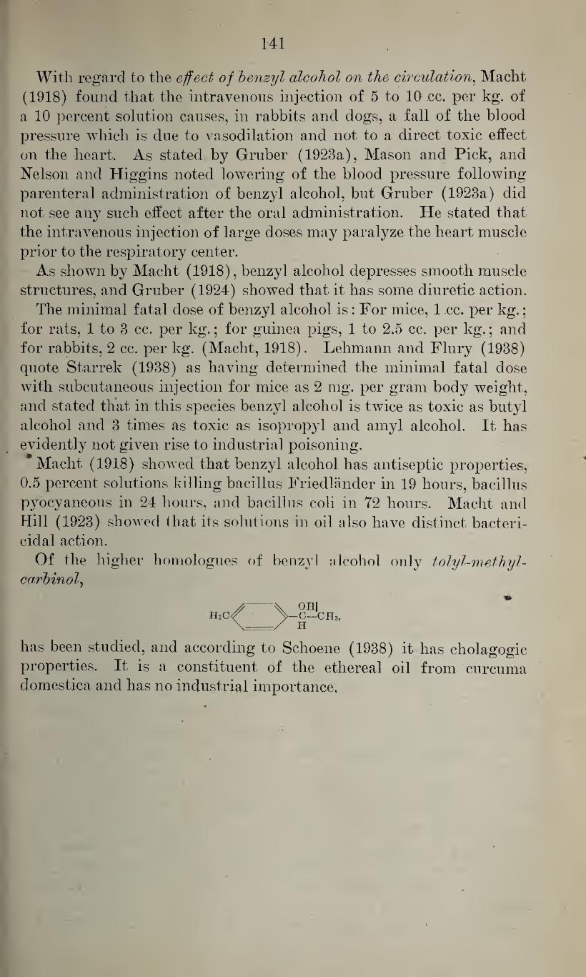
141
With regard to the effect of benzyl alcohol on the circulation, Macht
(1918) found that the intravenous injection of 5 to 10 cc. per kg. of
a 10 percent solution causes, in rabbits and dogs, a fall of the blood
pressure which is due to vasodilation and not to a direct toxic effect
on the heart. As stated by Gruber (1923a), Mason and Pick, and
Nelson and Higgins noted lowering of the blood pressure following
parenteral administration of benzyl alcohol, but Gruber (1923a) did
not see any such effect after the oral administration. He stated that
the intravenous injection of large doses may paralyze the heart muscle
prior to the respiratory center.
As shown by Macht (1918), benzyl alcohol depresses smooth muscle
structures, and Gruber (1924) showed that it has some diuretic action.
The minimal fatal dose of benzyl alcohol is: For mice, 1 cc. per kg.;
for rats, 1 to 3 cc. per kg.; for guinea pigs, 1 to 2.5 cc. per kg.; and
for rabbits, 2 cc. per kg. (Macht, 1918). Lehmann and Flury (1938)
quote Starrek (1938) as having determined the minimal fatal dose
with subcutaneous injection for mice as 2 mg. per gram body weight,
and stated that in this species benzyl alcohol is twice as toxic as butyl
alcohol and 3 times as toxic as isopropyl and amyl alcohol. It has
evidently not given rise to industrial poisoning.
Macht (1918) showed that benzyl alcohol has antiseptic properties,
0.5 percent solutions killing bacillus Friedlander in 19 hours, bacillus
pyocyaneous in 24 hours, and bacillus coli in 72 hours. Macht and
Hill (1923) showed that its solutions in oil also have distinct bacteri¬ cidal action.
Of the higher homologues of benzyl alcohol only tolyl-methyl- carbinol,
h3c^ ^-c-ch3,
has been studied, and according to Schoene (1938) it has cholagogic
properties. It is a constituent of the ethereal oil from curcuma
domestica and has no industrial importance.
V. RELATION BETWEEN THE CHEMICAL STRUCTURE AND THE
PHYSIOLOGICAL ACTION OF MONOVALENT ALCOHOLS
Table 19 gives a resume of the physico-chemical data of some of the
aliphatic alcohols.
It shows that with normal alcohols the specific gravity increases
with the molecular weight, and that the specific gravity of the iso
compounds and, evidently to a greater degree, the specific gravity of
the secondary and tertiary compounds is lower than that of the normal
alcohols.
Similarly, the boiling point increases with the molecular weight of
the normal alcohols, and the boiling point of the iso, secondary, and
tertiary alcohols is lower.
Corresponding to the changes of the boiling point, the vapor pres¬
sure at a given temperature decreases with the molecular wTeight. But
it should be pointed out that the vapor pressure of isopropyl and iso¬
butyl alcohol is greater than that of the corresponding normal alcohols
and that the vapor pressure of the secondary and, especially, the ter¬
tiary alcohols (tertiary butyl alcohol) is considerably higher.
The flash point increases with the molecular weight, and the lower
limit of inflammability decreases with the molecular weight.
The available data indicate that the surface tension of normal alco¬
hols increases with the molecular weight and the surface tension of
the iso, secondary, and tertiary alcohols is lower than that of the
normal compounds.
Whereas the solubility in water decreases with the molecular weight,
the*solubility in oil increases so that the partition coefficient oil/water
increases with the increase of molecular weight. The available data
do not show whether or not, or to what extent, the iso, secondary, and
tertiary alcohols differ from the normal compounds.
The toxicological properties of aliphatic alcohols increase with the
molecular weight, as was already shown by Richardson (1869), and
it has generally been found that the iso compounds are less effective
than the normal alcohols. It has been shown by several investigators,
such as Fulmer (1904 and 1905) on sea urchin eggs, by Fiihner and
Neubauer (1907) with the hemolytic action on red blood cells, by
Fiihner (1921) in the isolated frog heart, by Kamm (1921) in para-
mecia, and by Morgan and Cooper (1912) and Tilley and Schaffer
(1926) on bacteria, that the effectiveness of the various normal alco¬
hols from one member to the next higher homologue increases ap¬
proximately 3.3 times. According to Tilley and Schaffer (1926) for
secondary alcohols this ratio is slightly lower, namely, 3.0, and for
tertiary alcohols it is 2.7.
In contrast to these findings with comparatively primitive test ob-
(142)
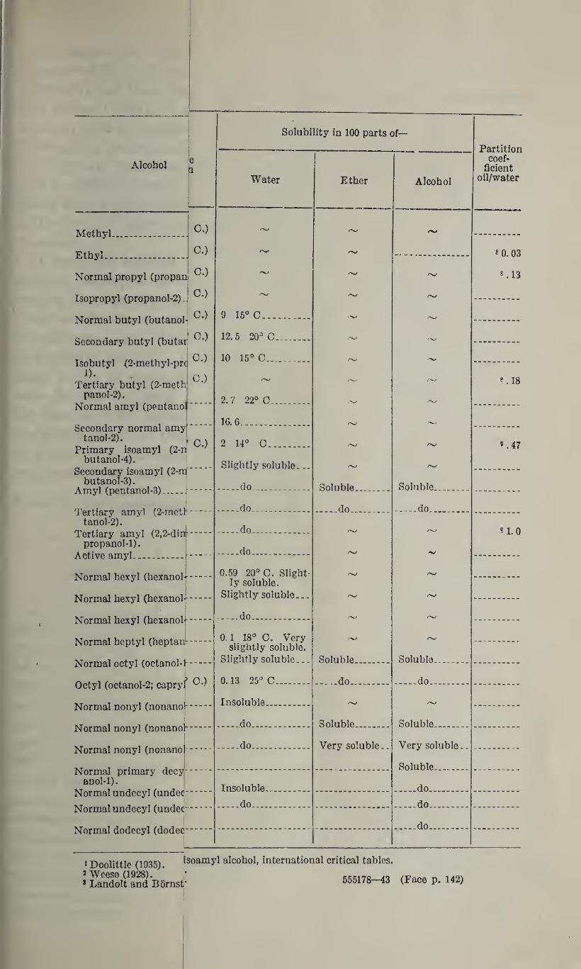
Alcohol
Methyl-
Ethyl..
C.)
C.)
P } Normal propyl (propam '
Isopropyl (propanol-2)-J '
Normal butyl (butanol- ^
Secondary butyl (butan ^
Isobutyl (2-methyl-prc ^ D-
Tertiary butyl (2-meth panol-2).
Normal amyl (pentane!
C.)
Secondary normal amy| tanol-2).
Primary isoamyl (2-nj butanol-4).
Secondary isoamyl (2-ir butanol-3).
Amyl (pentanol-3) —..
O.)
Tertiary amyl (2-metl tanol-2).
Tertiary amyl (2,2-din- propanol-1).
Active amyl.
Normal hexyl (hexanol-i--
Normal hexyl (hexanol^.
Normal hexyl (hexanob-.
Normal heptyl (heptao--
Normal octyl (octanol-f.
Octyl (octanol-2; capryf
Normal nonyl (nonanoj-.
Normal nonyl (nonanol-
Normal nonyl (nonanol-.
Normal primary decy anol-1).
Normal undecyl (unde<|-
Normalundecyl (under
Normal dodecyl (doder
Solubility in 100 parts of-
Water
9 15° C...
12.5 20° C.
10 15° C__.
2. ? 22° 0..
16.6.
2 14° O.
Slightly soluble...
-do.....
-do.___
-do...
.do.
0.59 20° C. Slight¬ ly soluble.
Slightly soluble...
-do__
0.1 18° O. Very slightly soluble.
Slightly soluble...
0.13 25° C.
Insoluble...
-do.
-do..
Insoluble.
_do_
Ether
Soluble.
-do..
Soluble.
-do..
Soluble.
Very soluble..
Alcohol
Soluble.
-do..
Soluble.
-do..
Soluble..
Very soluble.
Soluble_
.do.
.do.
_do.
»Doolittle (1935). 2 Weese (1928). 1 Landolt and Bornst-
isoamyl alcohol, international critical tables.
555178—43 (Face p. 142)
Partition coef¬
ficient oil/water
8 0.03
8.13
.18
*. 47
8 1.0
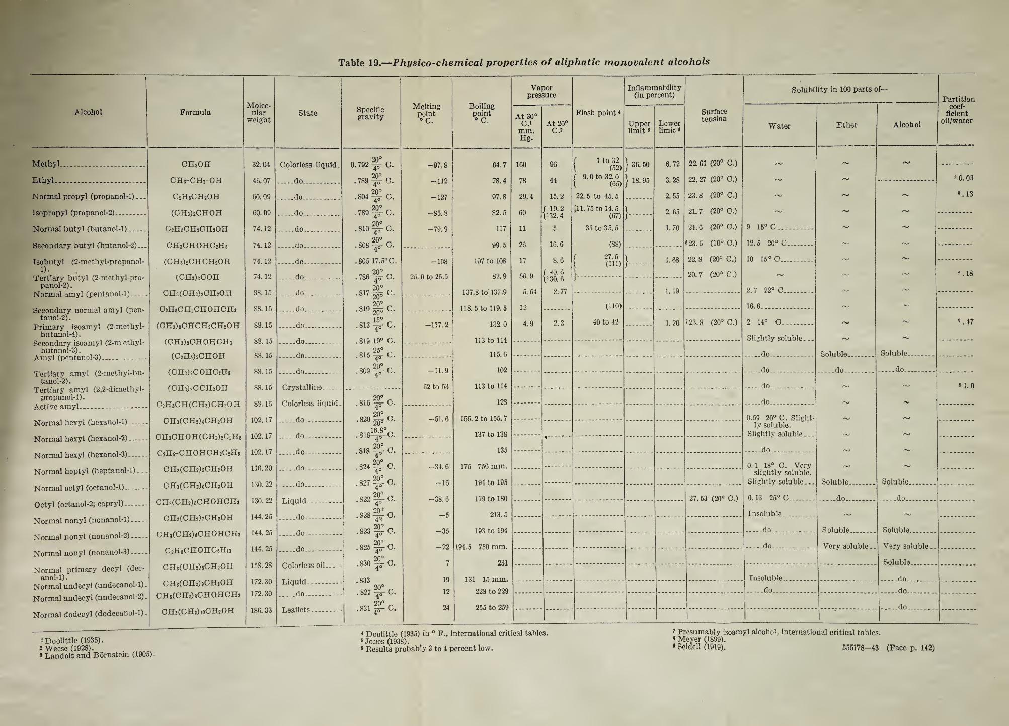
Table 19.—Physico-chemical properties of aliphatic monovalent alcohols
Alcohol Formula Molec¬
ular weight
State
Methyl.... CHsOH 32.04 Colorless liquid.
Ethyl... CH3-CH2-OH 46.07 .do.
Normal propyl (propanol-1)... C2H6CH2OH 60.09 .do.
Isopropyl (propanol-2).. (CH3)2CHOH 60.09 _do..
Normal butyl (butanol-1). C2H5CH2CH20H 74.12 .do.
Secondary butyl (butanol-2)... CH3CHOHC2H5 74.12 .do..
Isobutyl (2-methyl-propanol- (CH3)2CHCH20H 74.12 _do..
Tertiary butyl (2-methyl-pro- (CHslsCOH 74.12 _do.. panol-2). nrmq1 ottittI lrvArttannl-l^ CH3(CH'>)3CH20H 88.15 _do_.._
Secondary normal amyl (pen- C2HjCH2CHOIICH3 88.15 .do.. tanol*2).
Primary isoamyl (2-methyl- (CH3)2CHCH2CH2OH 88.15 _do.. butanol-4).
Secondary isoamyl (2-m ethyl- (CHsJjCHOHCHs 88.15 .do_ butanol-3).
(C2Hs)2CHOH 88.15 _do_
Tertiary amyl (2-methyl-bu- (CH3)2COHC2H5 88.15 _do_ tanol-2).
Tertiary amyl (2,2-dimethyl- (CH3)3CCH20H 88.15 Crystalline- propanol-1).
Active amyl.... C2H5CH(CH3)CH20H 88.15 Colorless liquid.
Normal hexyl (hexanol-1). CH3(CH2)4CH2OH 102.17 .do..
Normal hexyl (hexanol-2). CH3CHOH(CH2)2C2H5 102.17 _do.....
Normal hexyl (hexanol-3).. C2Hs-CHOHCH2C2Ha 192.17 _do..
Normal heptyl (heptanol-1) - - - CH3(CH2)5CH2OH 116. 20 _do..
Normal octyl (octanol-1). CH3(CH2)6CH2OH 130. 22 .do...
Octyl (octanol-2; capryl).. Cn3(CH2)6CHOHCH3 130. 22 Liquid__
Normal nonyl (nonanol-1). CH3(CH2)7CH2OH 144. 25 _do_
Normal nonyl (nonanol-2)- CHs(CH2)«CHOHCH» 144.25 .do..
Normal nonyl (nonanol-3). C2H8CHOHC6Hi3 144.25 _do_
Normal primary decyl (dec- CH3(CH2)8CH2OH 158.28 Colorless oil-
anol-1). Normal undecyl (undecanol-1). CH3(CH2)#CHjOH 172. 30 Liquid.
Normal undecyl (undecanoI-2). CHa(CH2)8CHOHCH3 172.30 _do..
Normal dodecyl (dodecanol-1)„ CH8(CHj)ioCH2OH 186.33 Leaflets.
1 Doolittle (1935). 2 Weese (1928). . * Landolt and Bornstem (1905).
Specific gravity
20°
0.792 c.
20°
.789 C.
20° .804^ O.
20° . 789 gr C.
20°
.810 gr C.
. 808 ^ C.
,805 17.6° O.
20°
4C 786 C.
20°
.817 go C. 20°
.810 2qo C.
.813 ^ C.
.819 19° C. 25°
.815 -jr C.
20°
.809 gs-C.
.816 C.
20°
.820 2qo O.
.818^0.
.818 O.
.824 ^0.
.827 ^£-C.
.822 ^-0.
.828^ C.
.823 ^0.
.825 ^C.
20°
.830 ~-C.
.833
. 827 ^ O.
.831 C.
Melting point °C.
-97.8
-112
-127
-85.8
-79.9
-108
25.0 to 25.5
-117.2
-11.9
52 to 53
-51.6
-34.6
-16
-38.6
-5
-35
-22
7
19
12
24
Boiling point d C.
64.7
78.4
97.8
82.5
117
99.5
107 to 108
82.9
137.3 toT37.9
118.5 to 119.6
132.0
113 to 114
115.6
102
113 to 114
128
155.2 to 155.7
137 to 138
135
175 756 mm.
194 to 195
179 to 180
213.5
193 to 194
194.5 750 mm.
231
131 15 mm.
228 to 229
255 to 259
Vapor pressure
At 30° Flash point8
C.i At 20° mm. C.2 Hg.
160 96 f 1 to 32 l (52)
78 44 / 9.0 to 32.0 1 (65)
29.4 15.2 22.5 to 45. 5
60 / 19.2 ill. 75 to 14.5 l3 32. 4 (67)
11 6 35 to 35. 5
26 16.6 (88)
17 8.6 27.5 (HD
66.9
5. 54
12
4.9
/ 40.6 \3 30. 6
2. 77
2.3
}•
(110)
40 to 42
Inflammability (in percent)
Upper limit *
} 36.50
} 18.95
< Doolittle (1935) in 0 F., international critical tables. 5 Jones (1938). 8 Results probably 3 to 4 percent low.
Lower limit»
6.72
3.28
2.55
2. 65
1.70
1.68
1.19
1.20
Surface tension
22.61 (20° 0.)
22.27 (20° C.)
23.8 (20° 0.)
21.7 (20° 0.)
24.6 (20° C.)
8 23.5 (10° 0.)
22.8 (20° O.)
20.7 (20° C.)
7 23.8 (20° 0.)
27.63 (20° C.)
Solubility in 100 parts of-
Water
9 15° C...
12.5 20° C.
10 15° C...
2. 7 22° C.
16.6.
2 14° O.
Slightly soluble...
_do_...
_do__
-do...
_do..
Ether
0.59 20° C. Slight¬ ly soluble.
Slightly soluble...
do.
0.1 18° O. Very slightly soluble.
Slightly soluble...
0.13 25° C.
Insoluble...
-do.
-do.
Insoluble.
.do_
Soluble.
-do..
Soluble.
-do..
Soluble...
Very soluble.
Alcohol
Soluble.
_do..
Soluble.
-do..
Soluble..
Very soluble.
Soluble.
.do.
.do.
.do.
Partition coef¬
ficient oil/water
7 Presumably isoamyl alcohol, international critical tables. * Meyer (1899). • Seidell (1919). 555178—43 (Face p. 142)
«0.03
8.13
18
*.47
8 1.0
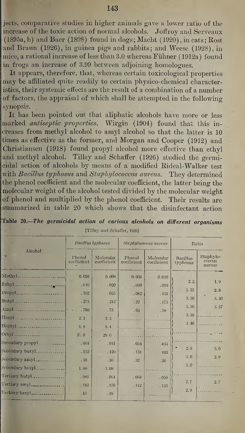
143
jects, comparative studies in higher animals gave a lower ratio of the
increase of the toxic action of normal alcohols. Joffroy and Serveaux
(1895a, b) and Baer (1898) found in dogs; Macht (1920), in cats; Rost
and Braun (1926), in guinea pigs and rabbits; and Weese (1928), in
mice, a rational increase of less than 3.0 whereas Fuhner (1912a) found
in frogs an increase of 3.99 between adjoining homologues.
It appears, therefore, that, whereas certain toxicological properties
may be affiliated quite readily to certain physico-chemical character¬
istics, their systemic effects are the result of a combination of a number
of factors, the appraisal of which shall be attempted in the following
synopsis.
It has been pointed out that aliphatic alcohols have more or less
marked antiseptic properties. Wirgin (1904) found that this in¬
creases from methyl alcohol to amyl alcohol so that the latter is 10
times as effective as the former, and Morgan and Cooper (1912) and
Christiansen (1918) found propyl alcohol more effective than ethyl
and methyl alcohol. Tilley and Schaffer (1926) studied the germi¬
cidal action of alcohols by means of a modified Rideal-Walker test
with Bacillus typhosus and Staphylococcus aureus. They determined
the phenol coefficient and the molecular coefficient, the latter being the
molecular weight of the alcohol tested divided by the molecular weight
of phenol and multiplied by the phenol coefficient. Their results are
summarized in table 20 which shows that the disinfectant action
Table 20.—The germicidal action of various alcohols on different organisms
[Tilley and Schaffer, 1926]
Alcohol t
Bacillus typhosus Staphylococcus aureus Ratio
Phenol coefficient
*
Molecular coefficient
Phenol coefficient
Molecular coefficient
Bacillus typhosus
Staphylo¬ coccus aureus
Methyl. ____ 0. 026 0.009 0. 030 0.010 'I
Ethyl_#_ .040 .020 .039 .019 j 2.2 1.9
Propyl__ . 102 .065 .082 .053 | 3.25 2.8
Butyl_ _ .273 .215' .22 . 175 | 3.30 3. 30
Amyl_ .... .. .780 .73 .63 .59 } 3.39 3.37
Hexyl_ _ 2.3 2. 5 ] 3.38
Heptyl. ... .... 6. 8 8. 4 } 3.46
Octyl.. _ ... . 21.0 29. 0 }. •
Secondary propyl_ . .064 .041 .054 .034 }.------
Secondary butyl_ .152 . 120 . 131 .103 j • 2.9 3.0
Secondary amyl _ .38 .36 .32 .30 j 3.0 2.9
Secondary hexyl_ . .. 1.00 1.09 } 3.0
Tertiary butyl_ .081 .064 .064 .050 .-
Tertiary amyl___ . 182 . 170 . 142 . 133 ) 2.7 2.7
Tertiary hexyl.. .45 .49 ] 2.9
■
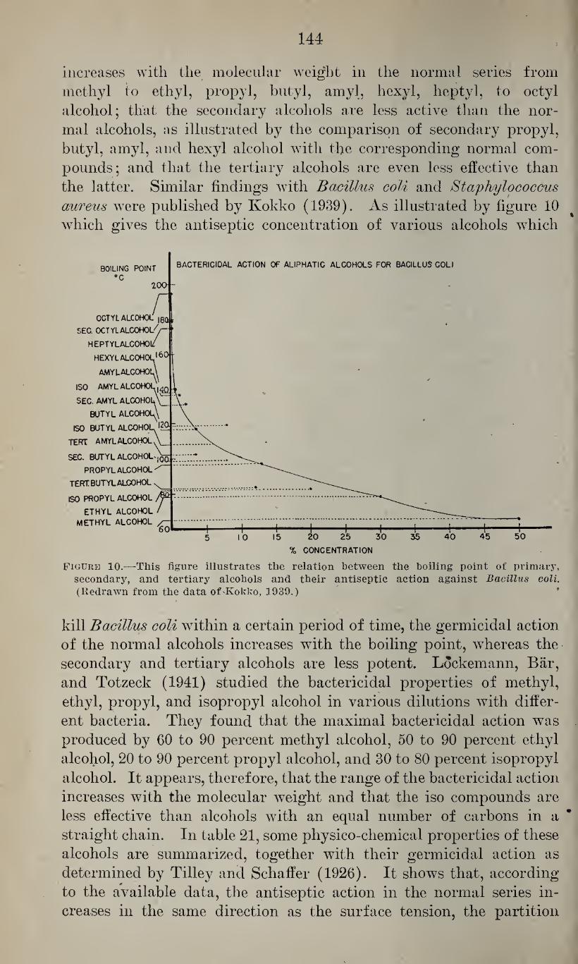
144
increases with the molecular weight in the normal series from
methyl to ethyl, propyl, butyl, amyl, hexyl, heptyl, to octyl
alcohol; that the secondary alcohols are less active than the nor¬
mal alcohols, as illustrated by the comparison of secondary propyl,
butyl, amyl, and hexyl alcohol with the corresponding normal com¬
pounds; and that the tertiary alcohols are even less effective than
the latter. Similar findings with Bacillus coli and Staphylococcus aureus were published by Ivokko (1939). As illustrated by figure 10
which gives the antiseptic concentration of various alcohols which
BACTERICIDAL ACTION OF ALIPHATIC ALCOHOLS FOR BACILLUS COLI BOILING POINT
°C 200 -
OCTYL ALCOHOL |8Q r
SEC. OCTYLALCOHOL^-
HEPTYLALCOHOl/
HEXYLALCOHOL160
amylalcohoiA
ISO amylalo—
SEC. AMYL ALC0H0I
BUTYL ALCOHOL^
ISO BUTYL ALCOHOI l2af“'
TERT AMYLALCOHOL
SEC. BUTYL ALCOHOL',qqJi;
PROPYLALCOHOL
TERT BUTYL ALCOHOL
ISO PROPYL ALCOHOL '
ETHYL ALCOHOL /
METHYL ALCOHOL bU—i i I I 11"'* ' "■
15 20 25 30
% CONCENTRATION
Figure 10.—This figure illustrates the relation between the boiling point of primary, secondary, and tertiary alcohols and their antiseptic action against Bacillus coli. (Redrawn from the data of-Kokko, 1939.)
kill Bacillus coli within a certain period of time, the germicidal action
of the normal alcohols increases with the boiling point, whereas the
secondary and tertiary alcohols are less potent. Lockemann, Bar,
and Totzeck (1941) studied the bactericidal properties of methyl,
ethyl, propyl, and isopropyl alcohol in various dilutions with differ¬
ent bacteria. They found that the maximal bactericidal action was
produced by 60 to 90 percent methyl alcohol, 50 to 90 percent ethyl
alcohol, 20 to 90 percent propyl alcohol, and 30 to 80 percent isopropyl
alcohol. It appears, therefore, that the range of the bactericidal action
increases with the molecular weight and that the iso compounds are
less effective than alcohols with an equal number of carbons in a
straight chain. In table 21, some physico-chemical properties of these
alcohols are summarized, together with their germicidal action as
determined by Tilley and Schaffer (1926). It shows that, according
to the available data, the antiseptic action in the normal series in¬
creases in the same direction as the surface tension, the partition
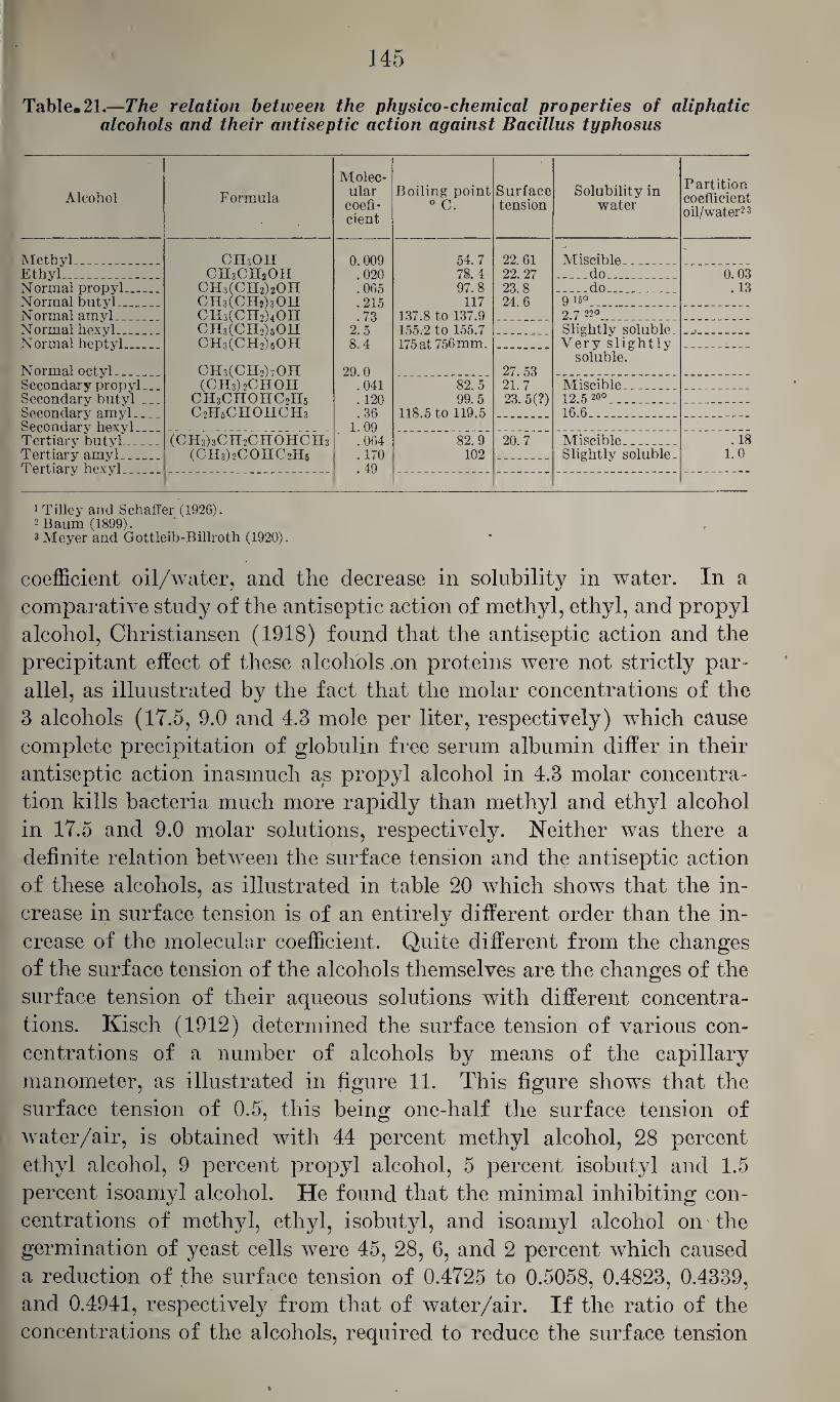
145
Table. 21.—The relation between the physico-chemical properties of aliphatic alcohols and, their antiseptic action against Bacillus typhosus
Alcohol Formula
Molec¬ ular
coefi- cient
Boiling point 0 C.
Surface tension
Solubility in water
Partition coefficient oil/water2 3
Methyl_ Ethyl_ Normal propyl— . Normal butyl_ Normal amyl
CH3OH CH3CH2OH
CH3(CH2)2OH CH3(CHj)3OH CH3(OH2)4OH CH3(CH2)6OH CH3(CH2)60H
CH3(CH2)70H (CH3)2CHOH
ch3chohc2h5 C2H5CHOHCH3
0.009 .020 .065 .215 .73
2.5 8.4
29.0 .041 . 120 .36
1.09 .064 .170 .49
54.7 78.4 97.8
117 137.8 to 137.9 155.-2 to 155.7 175 at 756 mm.
22.61 22. 27 23.8 24.6
Miscible_ .do_ _
_do_ 9 16°_ 91 7 22°
0.03 .13
Normal hexyl_ Normal heptyl.. __
Normal octyl- 27. 53 21.7 23. 5(?)
Slightly soluble. Very slightly
soluble.
Secondary propyl._. Secondary butyl_ Secondary amyl_ Secondary hexyl
82.5 99. 5
118.5 to 119.5
Miscible . . . 12.52o0_ 16.6_
Tertiary butyl_ Tertiary amyl... Tertiarv hc.xvl
(CH3)3CH2CHOHCH3
(CH3)2C0HC2H5
82.9 102
20.7 Miscible_ Slightly soluble.
.18 1.0
1 Tilley and Schaffer (1926). 2 Baum (1899). 3 Meyer and Gottleib-Billroth (1920).
coefficient oil/water, and the decrease in solubility in water. In a
comparative study of the antiseptic action of methyl, ethyl, and propyl
alcohol, Christiansen (1918) found that the antiseptic action and the
precipitant effect of these alcohols .on proteins were not strictly par¬
allel, as illuustrated by the fact that the molar concentrations of the
3 alcohols (1T.5, 9.0 and 4.3 mole per liter, respectively) which cause
complete precipitation of globulin free serum albumin differ in their
antiseptic action inasmuch as propyl alcohol in 4.3 molar concentra¬
tion kills bacteria much more rapidly than methyl and ethyl alcohol
in 17.5 and 9.0 molar solutions, respectively. Neither was there a
definite relation between the surface tension and the antiseptic action
of these alcohols, as illustrated in table 20 which shows that the in¬
crease in surface tension is of an entirely different order than the in¬
crease of the molecular coefficient. Quite different from the changes
of the surface tension of the alcohols themselves are the changes of the
surface tension of their aqueous solutions with different concentra¬
tions. Kisch (1912) determined the surface tension of various con¬
centrations of a number of alcohols by means of the capillary
manometer, as illustrated in figure 11. This figure shows that the
surface tension of 0.5, this being one-half the surface tension of
water/air, is obtained with 44 percent methyl alcohol, 28 percent
ethyl alcohol, 9 percent propyl alcohol, 5 percent isobutyl and 1.5
percent isoamyl alcohol. He found that the minimal inhibiting con¬
centrations of methyl, ethyl, isobutyl, and isoamyl alcohol on the
germination of yeast cells were 45, 28, 6, and 2 percent which caused
a reduction of the surface tension of 0.4725 to 0.5058, 0.4823, 0.4339,
and 0.4941, respectively from that of water/air. If the ratio of the
concentrations of the alcohols, required to reduce the surface tension
»
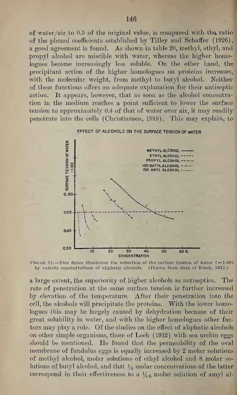
146
of water/air to 0.5 of the original value, is compared with tha ratio
of the phenol coefficients established by Tilley and Schaffer (1926),
a good agreement is found. As shown in table 20, methyl, ethyl, and
propyl alcohol are miscible with water, whereas the higher homo-
logues become increasingly less soluble. On the other hand, the
precipitant action of the higher homologues on proteins increases,
with the molecular weight, from methyl to butyl alcohol. Neither
of these functions offers an adequate explanation for their antiseptic
action. It appears, however, that as soon as the alcohol concentra¬
tion in the medium reaches a point sufficient to lower the surface
tension to approximately 0.4 of that of water over air, it may readily
penetrate into the cells (Christiansen, 1918). This may explain, to
EFFECT OF ALCOHOLS ON THE SURFACE TENSION OF WATER
Figure 11.—This figure illustrates the reduction of the surface tension of water ( = 1.00) by various concentrations of aliphatic alcohols. (Drawn from data of Kisch, 1912.)
a large extent, the superiority of higher alcohols as antiseptics. The
rate of penetration at the same surface tension is further increased
by elevation of the temperature. After their penetration into the
cell, the alcohols will precipitate the proteins. With the lower homo¬
logues this may be largely caused by dehydration because of their
great solubility in water, and with the higher homologues other fac¬
tors may play a role. Of the studies on the effect of aliphatic alcohols
on other simple organisms, those of Loeb (1912) with sea urchin eggs
should be mentioned. He found that the permeability of the oval
membrane of fundulus eggs is equally increased by 2 molar solutions
of methyl alcohol, molar solutions of ethyl alcohol and 8 molar so¬
lutions of butyl alcohol, and that % molar concentrations of the latter
correspond in their effectiveness to a y16 molar solution of amyl al-
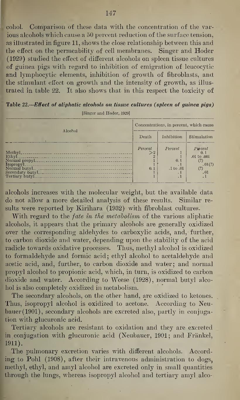
147
cohol. Comparison of these data with the concentration of the var¬
ious alcohols which cause a 50 percent reduction of the surface tension,
as illustrated in figure 11, shows the close relationship between this and
the effect on the permeability of cell membranes. Singer and Hoder
(1929) studied the effect of different alcohols on spleen tissue cultures
of guinea pigs with regard to inhibition of emigration of leucocytic
and lymphocytic elements, inhibition of growth of fibroblasts, and
the stimulant effect on growth and the intensity of growth, as illus¬
trated in table 22. It also shows that in this respect the toxicity of
Table 22.—Effect of aliphatic alcohols on tissue cultures (spleen of guinea pigs)
[Singer and Hoder, 1929]
Alcohol
Concentrations, in percent, which cause
Death Inhibition Stimulation
Methyl____ . . _ _ _ _ Percent
>2 2 1 1 '
0.1 1 1
Percent 1 1
0.1 .1
.1 to .01 .1 .1
Percent 0.1
.01 to .001 (?)
• 01(?) (?)
.01
.1
Ethyl , _ _ . _ _ _ _ _ _ Normal propyl. .. . .. _ _ - _ Isopropyl -. _. - - - . __ _. Normal butyl. _ . _ _ ___ _ _ _ __ Secondary butyl__ _ - _ _ _ - _ _ _ - . - Tertiary butyl- _ .. _ _ _ _
alcohols increases with the molecular weight, but the available data
do not allow a more detailed analysis of these results. Similar re¬
sults were reported by Kirihara (1932) with fibroblast cultures.
With regard to the fate in the metabolism of the various aliphatic
alcohols, it appears that the primary alcohols are generally oxidized
over the corresponding aldehydes to carboxylic acids, and, further,
to carbon dioxide and water, depending upon the stability of the acid
radicle towards oxidative processes. Thus, methyl alcohol is oxidized
to formaldehyde and formic acid; ethyl alcohol to acetaldehyde and
acetic acid, and, further, to carbon dioxide and water; and normal
propyl alcohol to propionic acid, which, in turn, is oxidized to carbon
dioxide and water. According to Weese (1928), normal butyl alco¬
hol is also completely oxidized in metabolism.
The secondary alcohols, on the other hand, are oxidized to ketones.
Thus, isopropyl alcohol is oxidized to acetone. According to Neu-
bauer(1901), secondary alcohols are excreted also, partly in conjuga¬
tion with glucuronic acid.
Tertiary alcohols are resistant to oxidation and they are excreted
in conjugation with glucuronic acid (Neubauer, 1901; and Frankel,
1911).
The pulmonary excretion varies with different alcohols. Accord¬
ing to Pohl (1908), after their intravenous administration to dogs,
methyl, ethyl, and amyl alcohol are excreted only in small quantities
through the lungs, whereas isopropyl alcohol and tertiary amyl alco-
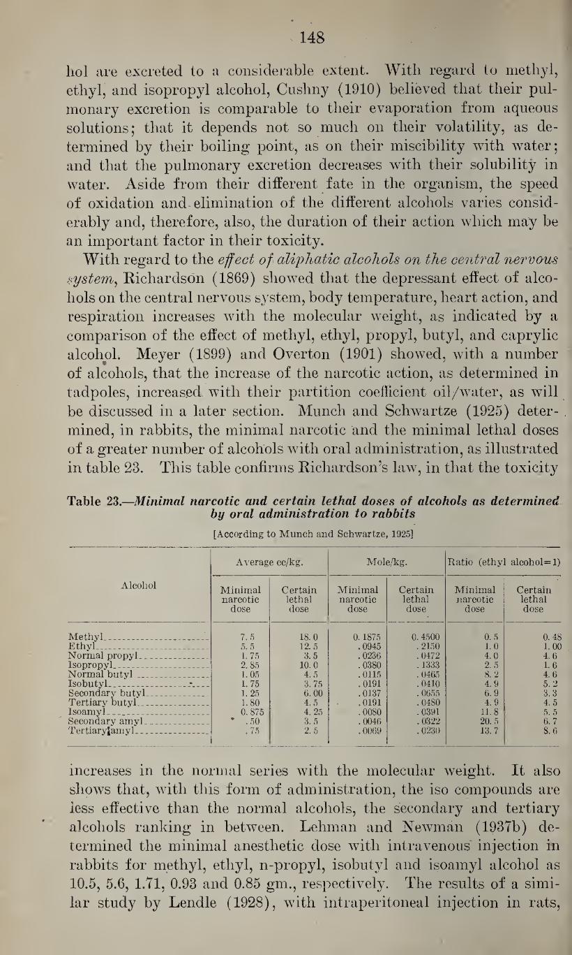
s 148
liol are excreted to a considerable extent. With regard to methyl,
ethyl, and isopropyl alcohol, Cushny (1910) believed that their pul¬
monary excretion is comparable to their evaporation from aqueous
solutions; that it depends not so much on their volatility, as de¬
termined by their boiling point, as on their miscibility with water;
and that the pulmonary excretion decreases with their solubility in
water. Aside from their different fate in the organism, the speed
of oxidation and elimination of the different alcohols varies consid¬
erably and, therefore, also, the duration of their action which may be
an important factor in their toxicity.
With regard to the effect of aliphatic alcohols on the central nervous system, Richardson (1869) showed that the depressant effect of alco¬
hols on the central nervous system, body temperature, heart action, and
respiration increases with the molecular weight, as indicated by a
comparison of the effect of methyl, ethyl, propyl, butyl, and caprylic
alcohol. Meyer (1899) and Overton (1901) showed, with a number
of alcohols, that the increase of the narcotic action, as determined in
tadpoles, increased with their partition coefficient oil/water, as will
be discussed in a later section. Munch and Schwartze (1925) deter¬
mined, in rabbits, the minimal narcotic and the minimal lethal doses
of a greater number of alcohols with oral administration, as illustrated
in table 23. This table confirms Richardson’s law, in that the toxicity
Table 23.—Minimal narcotic and certain lethal doses of alcohols as determined by oral administration to rabbits
[According to Munch and Schwartze, 1925]
Alcohol
Average cc/kg. Mole/kg. Ratio (ethyl alcohol=l)
Minimal narcotic
dose
Certain lethal dose
Minimal narcotic
dose
Certain lethal dose
Minimal narcotic
dose
Certain lethal dose
Methyl_ _ _ _ _ J. 7.5 18.0 0.1875 0.4500 0.5 0.48 Ethyl_ 5.5 12.5 .0945 .2150 1.0 1.00 Normal propyl _ _ 1. 75 3.5 .0236 .0472 4.0 4. 6 Isopropyl_ 2. 85 10.0 .0380 . 1333 2.5 1.6 Normal butyl_ ... 1.05 4.5 .0115 .0465 8.2 4.6 Isobutyl_ 1.75 3. 75 .0191 . 0410 4.9 5.2 Secondary butyl._ _ 1.25 6. 00 .0137 . 0655 6.9 3.3 Tertiary butyl_ 1.80 4.5 .0191 .0480 4.9 4.5 Isoamyl . _ _ 0. 875 4. 25 .0080 .0391 11.8 5.5 Secondary amyl _ . . * .50 3.5 .0046 .0322 20.5 6.7 Tertiaryfamyl _ _ _ .75 2.5 .0069 .0230 13.7 8.6
increases in the normal series with the molecular weight. It also
shows that, with this form of administration, the iso compounds are
less effective than the normal alcohols, the secondary and tertiary
alcohols ranking in between. Lehman and Newman (1937b) de¬
termined the minimal anesthetic dose with intravenous injection in
rabbits for methyl, ethyl, n-propyl, isobutyl and isoamyl alcohol as
10.5, 5.6, 1.71, 0.93 and 0.85 gm., respectively. The results of a simi¬
lar study by Lendle (1928), with intraperitoneal injection in rats,
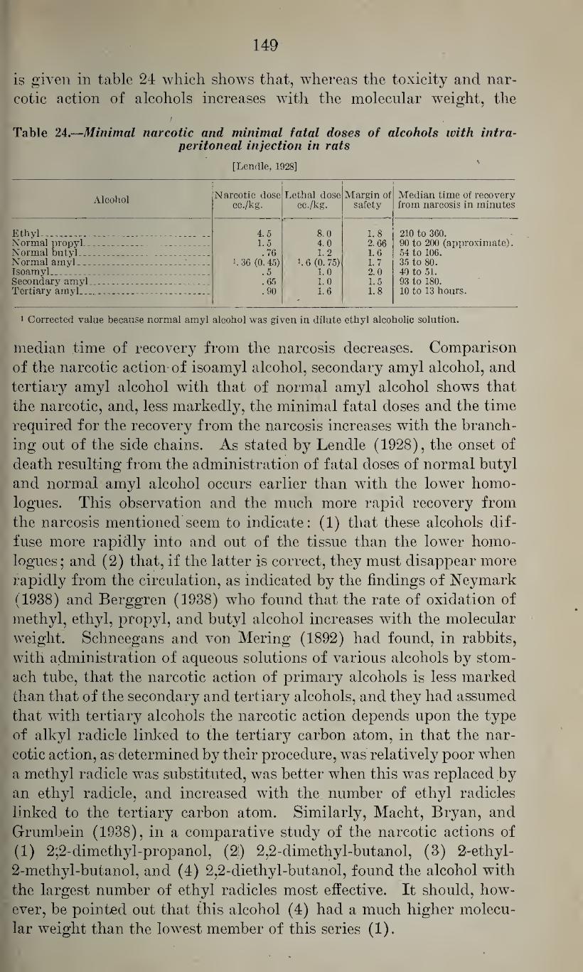
149
is given in table 24 which shows that, whereas the toxicity and nar¬
cotic action of alcohols increases with the molecular weight, the
Table 24.—Minimal narcotic and minimal fatal doses of alcohols with intra- peritoneal injection in rats
[Lendle, 1928]
Alcohol Narcotic dose cc./kg.
Lethal dose cc./kg.
Margin of safety
Median time of recovery from narcosis in minutes
Ethyl _ _ ^ _ 4.5 1.5 .76
>.36 (0.45) .5 .65 .90
8.0 4.0 1.2
i. 6 (0. 75) 1.0 1.0 1.6
1.8 2.66 1.6 1.7 2.0 1.5 1.8
210 to 360. 90 to 200 (approximate). 54 to 106. 35 to 80. 40 to 51. 93 to 180. 10 to 13 hours.
Normal propyl_ Normal butyl_ _ _ Normal amyl _ Isoamyl_ _ _ Secondary amyl._ _ _ Tertiary amyl._ _ _ _
1 Corrected value because normal amyl alcohol was given in dilute ethyl alcoholic solution.
median time of recovery from the narcosis decreases. Comparison
of the narcotic action of isoamyl alcohol, secondary amyl alcohol, and
tertiary amyl alcohol with that of normal amyl alcohol shows that
the narcotic, and, less markedly, the minimal fatal doses and the time
required for the recovery from the narcosis increases with the branch¬
ing out of the side chains. As stated by Lendle (1928), the onset of
death resulting from the administration of fatal doses of normal butyl
and normal amyl alcohol occurs earlier than with the lower homo-
logues. This observation and the much more rapid recovery from
the narcosis mentioned seem to indicate: (1) that these alcohols dif¬
fuse more rapidly into and out of the tissue than the lower homo-
logues; and (2) that, if the latter is correct, they must disappear more
rapidly from the circulation, as indicated by the findings of Neymark
(1938) and Berggren (1938) who found that the rate of oxidation of
methyl, ethyl, propyl, and butyl alcohol increases with the molecular
weight. Schneegans and von Mering (1892) had found, in rabbits,
with administration of aqueous solutions of various alcohols by stom¬
ach tube, that the narcotic action of primary alcohols is less marked
than that of the secondary and tertiary alcohols, and they had assumed
that with tertiary alcohols the narcotic action depends upon the type
of alkyl radicle linked to the tertiary carbon atom, in that the nar¬
cotic action, as determined by their procedure, was relatively poor when
a methyl radicle was substituted, was better when this was replaced by
an ethyl radicle, and increased with the number of ethyl radicles
linked to the tertiary carbon atom. Similarly, Macht, Bryan, and
Grumbein (1938), in a comparative study of the narcotic actions of
(1) 2;2-dimethyl-propanol, (2i) 2,2-dimethyl-butanol, (3) 2-ethyl-
2-methyl-butanol, and (4) 2,2-diethyl-butanol, found the alcohol with
the largest number of ethyl radicles most effective. It should, how¬
ever, be pointed out that this alcohol (4) had a much higher molecu¬
lar weight than the lowest member of this series (1).
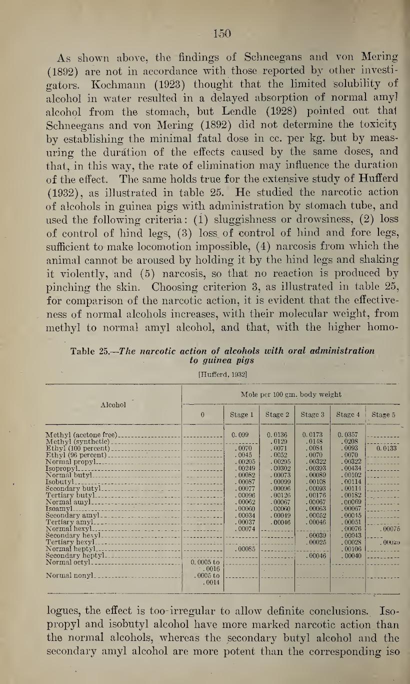
150
As shown above, the findings of Schneegans and von Mering
(1892) are not in accordance with those reported by other investi¬
gators. Kochmann (1923) thought that the limited solubility of
alcohol in water resulted in a delayed absorption of normal amy]
alcohol from the stomach, but Lendle (1928) pointed out thal
Schneegans and von Mering (1892) did not determine the toxicitj
by establishing the minimal fatal dose in cc. per kg. but by meas¬
uring the duration of the effects caused by the same doses, and
that,, in this way, the rate of elimination may influence the duration
of the effect. The same holds true for the extensive study of Hufferd
(1932), as illustrated in table 25. He studied the narcotic action
of alcohols in guinea pigs with administration by stomach tube, and
used the following criteria: (1) sluggishness or drowsiness, (2) loss
of control of hind legs, (3) loss of control of hind and fore legs,
sufficient to make locomotion impossible, (4) narcosis from which the
animal cannot be aroused by holding it by the hind legs and shaking
it violently, and (5) narcosis, so that no reaction is produced by
pinching the skin. Choosing criterion 3, as illustrated in table 25,
for comparison of the narcotic action, it is evident that the effective¬
ness of normal alcohols increases, with their molecular weight, from
methyl to normal amyl alcohol, and that, with the higher homo-
Table 25.—The narcotic action of alcohols with oral administration to guinea pigs
[Hufferd, 1932]
Alcohol
Methyl (acetonefree). Methyl (synthetic)... Ethyl (100 percent). Ethyl (96 percent)_ Normal propyl_ Isopropyl_ Normal butyl_ Isobutyl_ Secondary butyl_ Tertiary butyl_ Normal amyl_ Isoamyl_ Secondary amyl_ Tertiary amyl_ Normal hexyl_ Secondary hexyl_ Tertiary hexyl_ Normal heptyl_ Secondary heptyl- Normal octyl_
Normal nonyl.
Mole per 100 gm. body weight
0. 0005 to .0016
.0005 to .0014
Stage 1
0.099
.0070
.0045
.00205
.00249
.00082
. 00087
.00077
. 00096
. 00062
. 00060
. 00034
. 00037
. 00074
. 00085
Stage 2
0. 0136 .0129 .0071 .0052 . 00295 . 00302 . 00073 . 00099 . 00096 . 00126 . 00067 . 00060 . 00049 . 00046
Stage 3
0.0173 .0148 .0084 .0070 .00322 . 00393 . 00089 . 00108 . 00093 .00176 .00067 .00063 .00052 .00046
. 00039
.00025
.00046
Stage 4
0.0357 .0208 .0093 .0070 . 00322 . 00434 . 00102 . 00114 . 00114 .00182 . 00069 .00067 . 00045 .00051 .00076 . 00043 . 00028 . 00106 . 00040
Stage 5
0. 0133
.00075
.0UU2o
logues, the effect is too irregular to allow definite conclusions. Iso¬
propyl and isobutyl alcohol have more marked narcotic action than
the normal alcohols, whereas the secondary butyl alcohol and the
secondary amyl alcohol are more potent than the corresponding iso
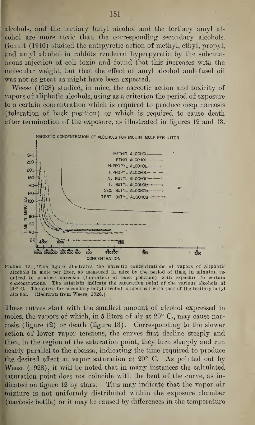
151
alcohols, and the tertiary butyl alcohol and the tertiary amyl al¬
cohol are more toxic than the corresponding secondary alcohols.
Genuit (1940) studied the antipyretic action of methyl, ethyl, propyl,
and amyl alcohol in rabbits rendered hyperpyretic by the subcuta¬
neous injection of coli toxin and found that this increases with the
molecular weight, but that the effect of amyl alcohol and-fusel oil
was not as great as might have been expected.
Weese (1928) studied, in mice, the narcotic action and toxicity of
vapors of aliphatic alcohols, using as a criterion the period of exposure
to a certain concentration which is required to produce deep narcosis
(toleration of back position) or which is required to cause death
after termination of the exposure, as illustrated in figures 12 and 13.
NARCOTIC CONCENTRATION OF ALCOHOLS FOR MICE IN MOLE PER LITER
Figure 12.—This figure illustrates the narcotic concentrations of vapors of aliphatic alcohols in mole per liter, as measured in mice by the period of time, in minutes, re¬ quired to produce narcosis (toleration of back position) with exposure to certain concentrations. The asterisks indicate the saturation point of the various alcohols at 20° C. The curve for secondary butyl alcohol is identical with that of the tertiary butyl alcohol. (Redrawn from Weese, 1928.)
These curves start with the smallest amount of alcohol expressed in
moles, the vapors of which, in 5 liters of air at 20° C., may cause nar¬
cosis (figure 12) or death (figure 13). Corresponding to the slower
action of lower vapor tensions, the curves first decline steeply and
then, in the region of the saturation point, they turn sharply and run
nearly parallel to the abcissa, indicating the time required to produce
the desired effect at vapor saturation at 20° C. As pointed out by
Weese (1928), it will be noted that in many instances the calculated
saturation point does not coincide with the bent of the curve, as in¬
dicated on figure 12 by stars. This may indicate that the vapor air
mixture is not uniformly distributed within the exposure chamber
(narcosis bottle) or it may be caused by differences in the temperature
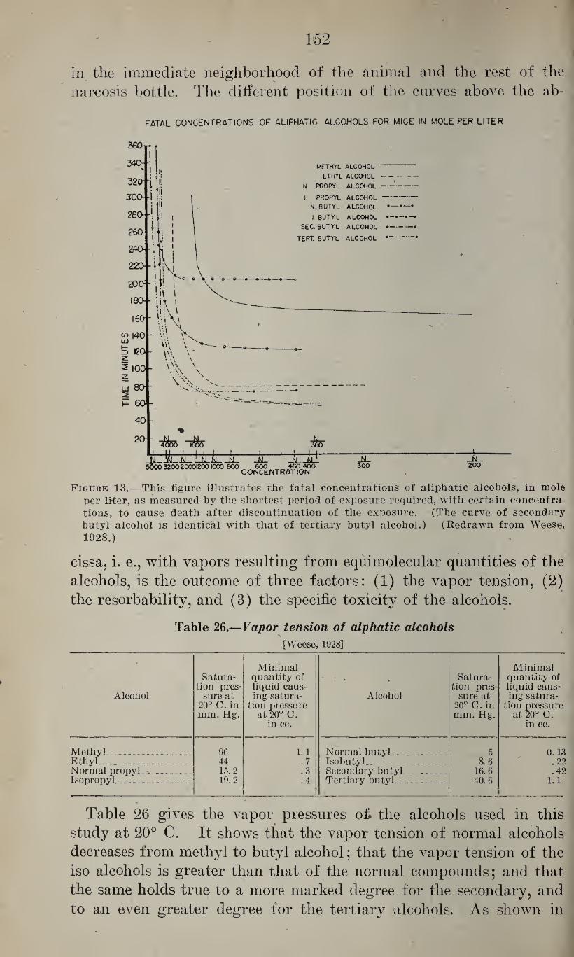
152
in the immediate neighborhood of the animal and the rest of the
narcosis bottle. The different position of the curves above the ab-
FATAL CONCENTRATIONS OF ALIPHATIC ALCOHOLS FOR MICE IN MOLE PER LITER
Figure 13.—This figure illustrates the fatal concentrations of aliphatic alcohols, in mole per liter, as measured by the shortest period of exposure required, with certain concentra¬ tions, to cause death after discontinuation of the exposure. (The curve of secondary butyl alcohol is identical with that of tertiary butyl alcohol.) (Redrawn from Weese, 1928.)
cissa, i. e., with vapors resulting from equimolecular quantities of the
alcohols, is the outcome of three factors: (1) the vapor tension, (2)
the resorbability, and (3) the specific toxicity of the alcohols.
Table 26.—Vapor tension of alphatic alcohols
[Weese, 1928]
Alcohol
Satura¬ tion pres¬
sure at 20° C.in mm. Hg.
Minimal quantity of liquid caus¬ ing satura¬
tion pressure at 20° C.
in cc.
Alcohol
Satura¬ tion pres¬
sure at 20° C. in mm. Hg.
Minimal quantity of liquid caus¬ ing satura¬
tion pressure at 20° C.
in cc.
Methyl.... ... 96 1.1 Normal butyl. 5 0.13 Ethyl_ __ _ 44 .7 Isobutyl .. _ . . 8.6 .22 Normal propyl. *_ Isopropyl___
15.2 19.2
.3
.4 Secondary butyl_ Tertiary butyl_
16.6 40.6
.42 1.1
i
Table 26 gives the vapor pressures oh the alcohols used in this
study at 20° C. It shows that the vapor tension of normal alcohols
decreases from methyl to butyl alcohol; that the vapor tension of the
iso alcohols is greater than that of the normal compounds; and that
the same holds true to a more marked degree for the secondary, and
to an even greater degree for the tertiary alcohols. As shown in
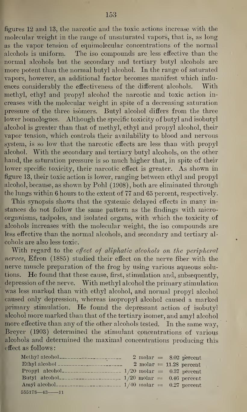
153
figures 12 and 13, the narcotic and the toxic actions increase with the
molecular weight in the range of unsaturated vapors, that is, as long
as the vapor tension of equimolecular concentrations of the normal
alcohols is uniform. The iso compounds are less effective than the
normal alcohols but the secondary and tertiary butyl alcohols are
more potent than the normal butyl alcohol. In the range of saturated
vapors, however, an additional factor becomes manifest which influ¬
ences considerably the effectiveness of the different alcohols. With
methyl, ethyl and propyl alcohol the narcotic and toxic action in¬
creases with the molecular weight in spite of a decreasing saturation
pressure of the three isomers. Butyl alcohol differs from the three
lower homologues. Although the specific toxicity of butyl and isobutyl
alcohol is greater than that of methyl, ethyl and propyl alcohol, their
vapor tension, which controls their availability to blood and nervous
system, is so low that the narcotic effects are less than with propyl
alcohol. With the secondary and tertiary butyl alcohols, on the other
hand, the saturation pressure is so much higher that, in spite of their
lower specific toxicity, their narcotic effect is greater. As shown in
figure 13, their toxic action is lower, ranging between ethyl and propyl
alcohol, because, as shown by Pohl (1908), both are eliminated through
the lungs within 6 hours to the extent of 77 and 65 percent, respectively.
This synopsis shows that the systemic delayed effects in many in¬
stances do not follow the same pattern as the findings with micro¬
organisms, tadpoles, and isolated organs, with which the toxicity of
alcohols increases with the molecular weight, the iso compounds are
less effective than the normal alcohols, and secondary and tertiary al¬
cohols are also less toxic.
With regard to the effect of aliphatic alcohols on the peripheral nerves, Efron (1885) studied their effect on the nerve fiber with the
nerve muscle preparation of the frog by using various aqueous solu¬
tions. He found that these cause, first, stimulation and, subsequently,
depression of the nerve. With methyl alcohol the primary stimulation
was less marked than with ethyl alcohol, and normal propyl alcohol
caused only depression, whereas isopropyl alcohol caused a marked
primary stimulation. He found the depressant action of isobutyl
alcohol more marked than that of the tertiary isomer, and amyl alcohol
more effective than any of the other alcohols tested. In the same way,
Breyer (1903) determined the stimulant concentrations of various
alcohols and determined the maximal concentrations producing this effect as follows:
Methyl alcohol- 2 molar = 8.02 percent
Ethyl alcohol- 2 molar = 11.28 percent
Propyl alcohol-1/20 molar = 0.37 percent
Butyl alcohol-1/20 molar = 0.46 percent
Amyl alcohol-1/40 molar = 0.27 percent
555178—43-11
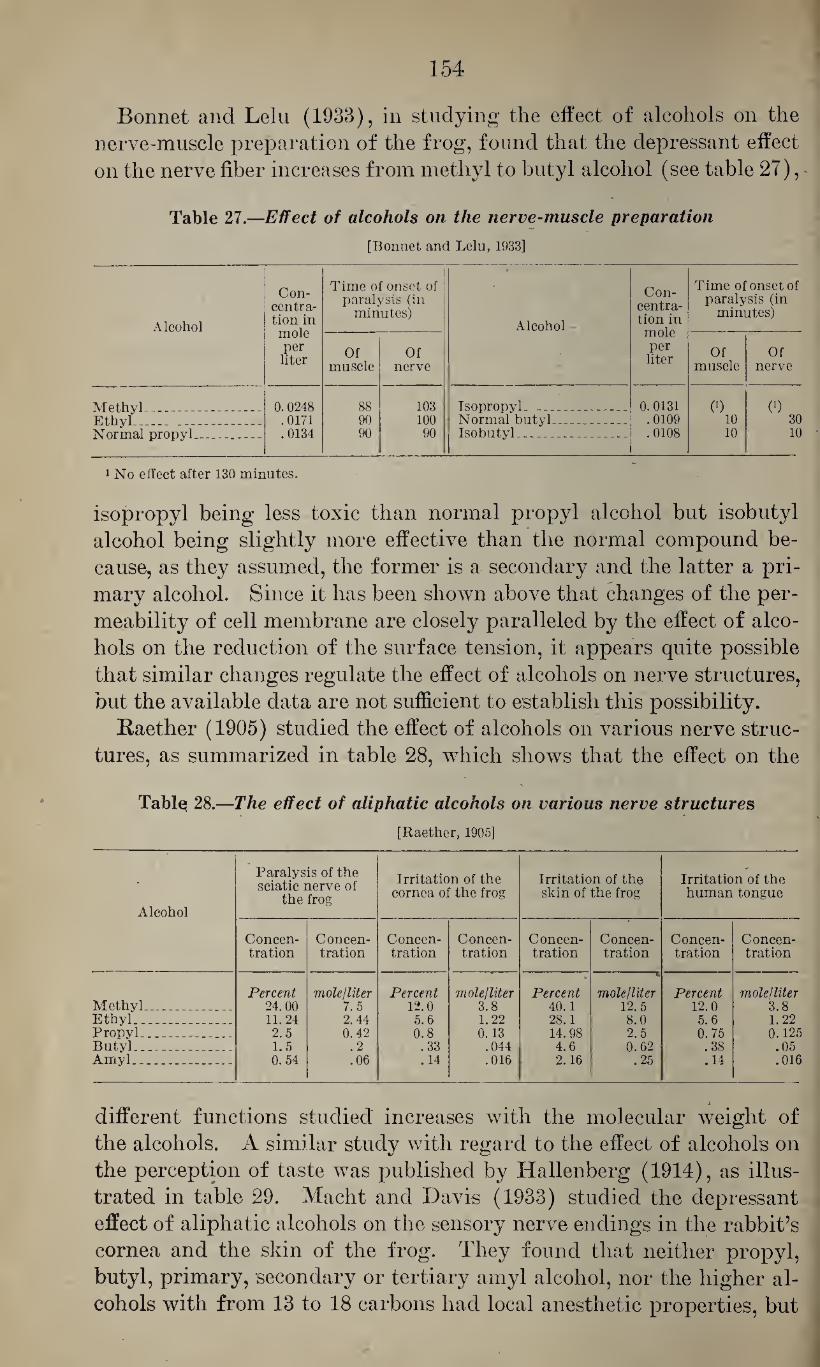
154
Bonnet and Lelu (1933), in studying the effect of alcohols on the
nerve-muscle preparation of the frog, found that the depressant effect
on the nerve fiber increases from methyl to butyl alcohol (see table 27),
Table 27.—Effect of alcohols on the nerve-muscle preparation
[Bonnet and Lelu, 1933]
Alcohol
Con¬ centra¬ tion in mole per liter
Time of onset of 1 paralysis (in
minutes) Alcohol
Con¬ centra¬ tion in mole per liter
Time of onset of paralysis (in
minutes)
Of muscle
Of nerve
Of muscle
Of nerve
Methyl__- Ethyl_ Normal propyl__
0.0248 .0171 .0134
88 90 90
103 100 90
Isopropyl.__... Normal butyl_ Isobutyl.- _ _
0. 0131 .0109 .0108
(9 10 10
0) 30 10
1 No effect after 130 minutes.
isopropyl being less toxic than normal propyl alcohol but isobutyl
alcohol being slightly more effective than the normal compound be¬
cause, as they assumed, the former is a secondary and the latter a pri¬
mary alcohol. Since it has been shown above that changes of the per¬
meability of cell membrane are closely paralleled by the effect of alco¬
hols on the reduction of the surface tension, it appears quite possible
that similar changes regulate the effect of alcohols on nerve structures,
but the available data are not sufficient to establish this possibility.
Raether (1905) studied the effect of alcohols on various nerve struc¬
tures, as summarized in table 28, which shows that the effect on the
Tablei 28.—The effect of aliphatic alcohols on various nerve structures
[Raether, 1905]
Alcohol
Paralysis of the sciatic nerve of
the frog
Irritation of the cornea of the frog
Irritation of the skin of the frog
Irritation of the human tongue
Concen¬ tration
Concen¬ tration
Concen¬ tration
Concen¬ tration
Concen¬ tration
Concen¬ tration
Concen¬ tration
Concen¬ tration
Percent mole lliter Percent mole lliter Percent mole/liter Percent molelliter Methyl_ 24.00 7.5 12.0 3.8 40.1 12.5 12.0 3.8 Ethyl_ 11.24 2.44 5.6 1.22 28.1 8.0 5.6 1.22 Propyl_ .. 2.5 0. 42 0.8 0.13 14.98 2.5 0.75 0.125 Butyl_ 1.5 .2 .33 .044 4.6 0.62 .38 .05 Amyl_ _ 0. 54 .06 .14 .016 2.16 .25 .14 .016
1
different functions studied increases with the molecular weight of
the alcohols. A similar study with regard to the effect of alcohols on
the perception of taste was published by ITallenberg (1914), as illus¬
trated in table 29. Macht and Davis (1933) studied the depressant
effect of aliphatic alcohols on the sensory nerve endings in the rabbit’s
cornea and the skin of the frog. They found that neither propyl,
butyl, primary, secondary or tertiary amyl alcohol, nor the higher al¬
cohols with from 13 to 18 carbons had local anesthetic properties, but
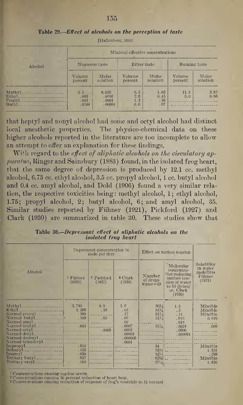
155
Table 29.—Effect of alcohols on the perception of taste
[Tlallenberg, 1914]
Minimal effective concentrations r
Alcohol Nauseous taste Bitter taste Burning taste
Volume Molar Volume Molar Volume Molar percent solution percent solution percent solution
Methyl ... .......... 0.1 0.025 6.5 1.62 11.5 2.87 Ethyl_ .009 .0016 2.6 0.45 5.0 0.86 Propyl .003 .0004 1.2 . 16 Butyl .0004 .00004 0.6 .07
that heptyl and nonyl alcohol had some and octyl alcohol had distinct
local anesthetic properties. The physico-chemical data on these
higher alcohols reported in the literature are too incomplete to allow
an attempt to offer an explanation for these findings.
With regard to the effect of aliphatic alcohols on the circulatory ap¬ paratus, Ringer and Sainsbury (1883) found, in the isolated frog heart,
that the same degree of depression is produced by 12.1 cc. methyl
alcohol, 6.73 cc. ethyl alcohol, 3.5 cc. propyl alcohol, 1 cc. butyl alcohol
and 0.4 ec. amyl alcohol, and Dold (1906) found a very similar rela¬
tion, the respective toxicities being: methyl alcohol, 1; ethyl alcohol,
1.75; propyl alcohol, 2; butyl alcohol, 6; and amyl alcohol, 35.
Similar studies reported by Fiihner (1921), Pickford (1927) and
Clark (1930) are summarized in table 30. These studies show that
Table 30.—Depressant effect of aliphatic alcohols on the isolated frog heart
Alcohol
Depressant concentration in mole per liter Effect on surface tension
Solubility in water mole/liter Fiihner (1921)
1 Fiihner (1921)
2 Pickford (1927)
3 Clark (1930)
Number of drops
water=39
Molecular concentra¬
tion reducing surface ten¬
sion of water to 63 dynes/
cc. Clark (1930)
Methyl. ... . . _ 3. 740 0.9 1.6 5oy2 1.5 Miscible Ethyl_ _ 1. 206 .35 .65 51M .5 Miscible Normal propyl-_ . . _ . 369 . 17 51 y2 . 14 Misp.ihlp Normal butyl_ . 109 .03 .05 si y2 .043 0.919 Normal amyl__ _ .02 .018 Normal heptyl_ _ .003 .0007 51K 0024 .008 Normal octyl__ .0003 .0002 . 0006 Normal decyl_ . 00003 . 000094 Normal dodecyl- _ . .000006 Normal tetradecyl_ .0001 Isopropyl.. - _ - .655 54 Miscible Isobutyl.. _ . . 135 53 y2 1 351 Isoamyl ... . . ... .039 52V6 . 230 Teritary butyl... . _ .. .637 62% Miseible Tertiary amyl_ . 184 57H 1.420
1 Concentrations causing cardiac arrest. 2 Concentrations causing 50 percent reduction of heart beat. 3 Concentrations causing reduction of response of frog’s ventricle to H normal
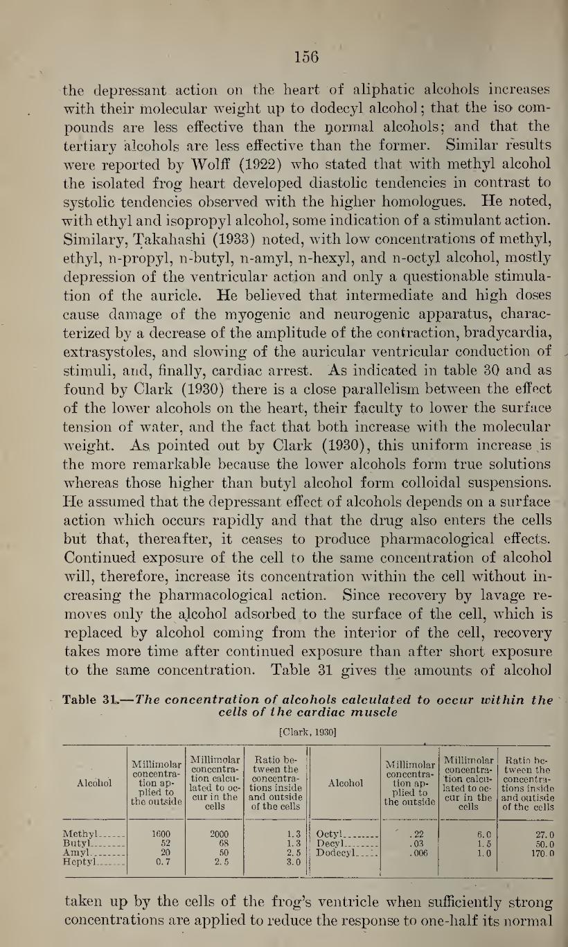
156
the depressant action on the heart of aliphatic alcohols increases
with their molecular weight up to dodecyl alcohol; that the iso com¬
pounds are less effective than the formal alcohols; and that the
tertiary alcohols are less effective than the former. Similar results
were reported by Wolff (1922) who stated that with methyl alcohol
the isolated frog heart developed diastolic tendencies in contrast to
systolic tendencies observed with the higher homologues. He noted,
with ethyl and isopropyl alcohol, some indication of a stimulant action.
Similary, Takahashi (1933) noted, with low concentrations of methyl,
ethyl, n-propyl, n-butyl, n-amyl, n-hexyl, and n-octyl alcohol, mostly
depression of the ventricular action and only a questionable stimula¬
tion of the auricle. He believed that intermediate and high doses
cause damage of the myogenic and neurogenic apparatus, charac¬
terized by a decrease of the amplitude of the contraction, bradycardia,
extrasystoles, and slowing of the auricular ventricular conduction of
stimuli, and, finally, cardiac arrest. As indicated in table 30 and as
found by Clark (1930) there is a close parallelism between the effect
of the lower alcohols on the heart, their faculty to lower the surface
tension of water, and the fact that both increase with the molecular
weight. As pointed out by Clark (1930), this uniform increase is
the more remarkable because the lower alcohols form true solutions
whereas those higher than butyl alcohol form colloidal suspensions.
He assumed that the depressant effect of alcohols depends on a surface
action which occurs rapidly and that the drug also enters the cells
but that, thereafter, it ceases to produce pharmacological effects.
Continued exposure of the cell to the same concentration of alcohol
will, therefore, increase its concentration within the cell without in¬
creasing the pharmacological action. Since recovery by lavage re¬
moves only the alcohol adsorbed to the surface of the cell, which is
replaced by alcohol coming from the interior of the cell, recovery
takes more time after continued exposure than after short exposure
to the same concentration. Table 31 gives the amounts of alcohol
Table 31.—The concentration of alcohols calculated to occur within the cells of the cardiac muscle
[Clark, 1930]
Alcohol
Millimolar concentra¬ tion ap¬ plied to
the outside
Millimolar concentra¬ tion calcu¬ lated to oc¬ cur in the
cells
Ratio be¬ tween the concentra¬ tions inside and outside of the cells
Alcohol
Millimolar concentra¬
tion ap¬ plied to
the outside
Millimolar concentra¬ tion calcu¬ lated to oc¬ cur in the
cells
Ratio be¬ tween the concentra¬ tions inside and outisde of the cells
Methyl_ 1600 2000 1.3 Octyl . .22 6.0 27 0 ButyL_ 52 68 1.3 Decyl..- . .03 1. 5 50.0 Amvl_ 20 50 2.5 DodecyL .1. .006 1.0 1700 Heptyl_ 0.7 2.5 3.0
taken up by the cells of the frog’s ventricle when sufficiently strong
concentrations are applied to reduce the response to one-half its normal
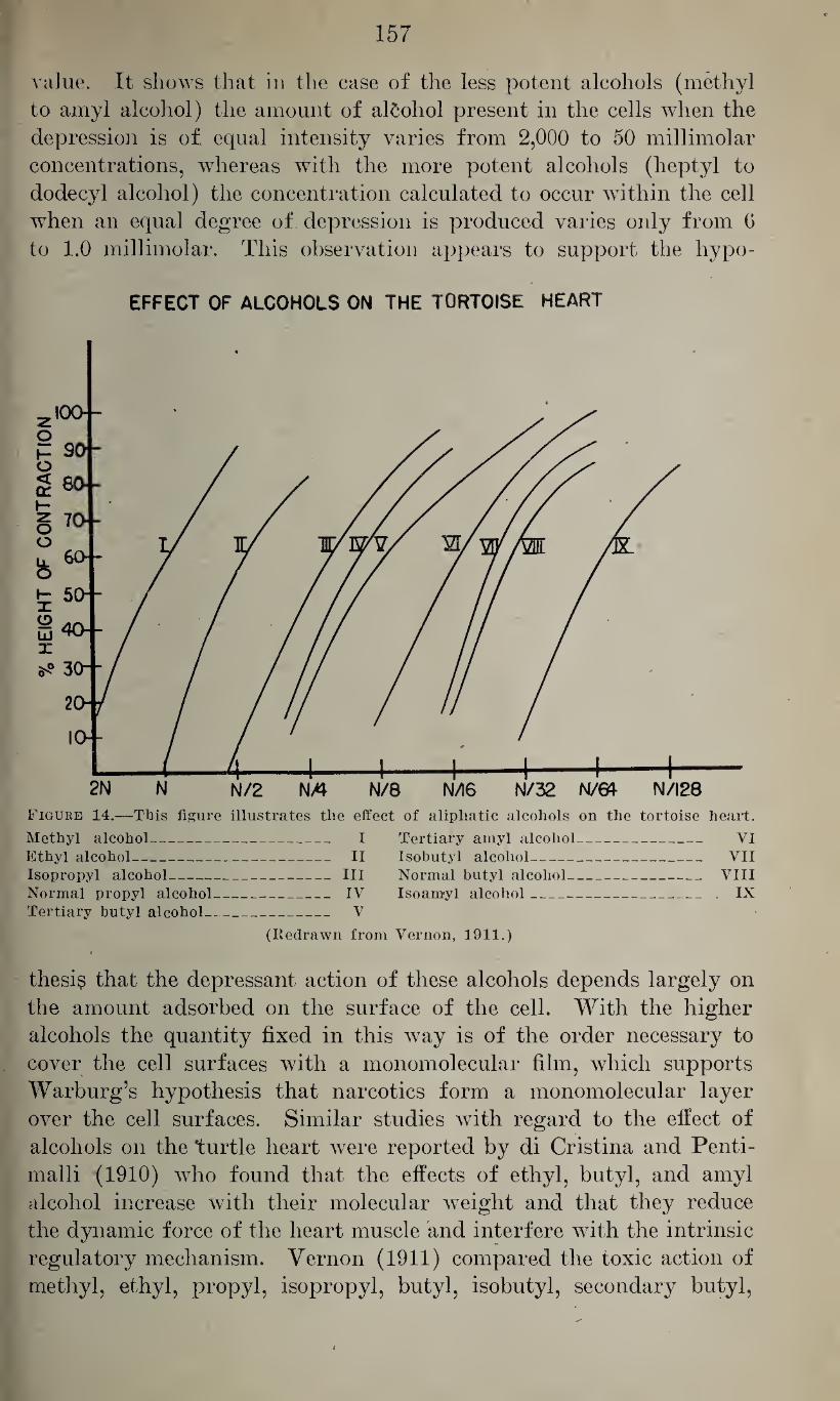
157
value. It shows that in the case of the less potent alcohols (methyl
to amyl alcohol) the amount of alcohol present in the cells when the
depression is of equal intensity varies from 2,000 to 50 millimolar
concentrations, whereas with the more potent alcohols (heptyl to
dodecyl alcohol) the concentration calculated to occur within the cell
when an equal degree of depression is produced varies only from G
to 1.0 millimolar. This observation appears to support the hypo-
EFFECT OF ALCOHOLS ON THE TORTOISE HEART
Figure 14.—This figure illustrates the effect of aliphatic alcohols on the tortoise heart.
Methyl alcohol- Ethyl alcohol- Isopropyl alcohol- Normal propyl alcohol Tertiary butyl alcohol-
- I Tertiary amyl alcohol- VI _ II Isobutyl alcohol_ VII _ III Normal butyl alcohol_ VIII - IV Isoamyl alcohol_ IX _ V
(Redrawn from Vernon, 1911.)
thesis that the depressant action of these alcohols depends largely on
the amount adsorbed on the surface of the cell. With the higher
alcohols the quantity fixed in this way is of the order necessary to
cover the cell surfaces with a monomolecular film, which supports
Warburg’s hypothesis that narcotics form a monomolecular layer
over the cell surfaces. Similar studies with regard to the effect of
alcohols on the turtle heart were reported by di Cristina and Penti-
malli (1910) who found that the effects of ethyl, butyl, and amyl
alcohol increase with their molecular weight and that they reduce
the dynamic force of the heart muscle and interfere with the intrinsic
regulatory mechanism. Yernon (1911) compared the toxic action of
methyl, ethyl, propyl, isopropyl, butyl, isobutyl, secondary butyl,
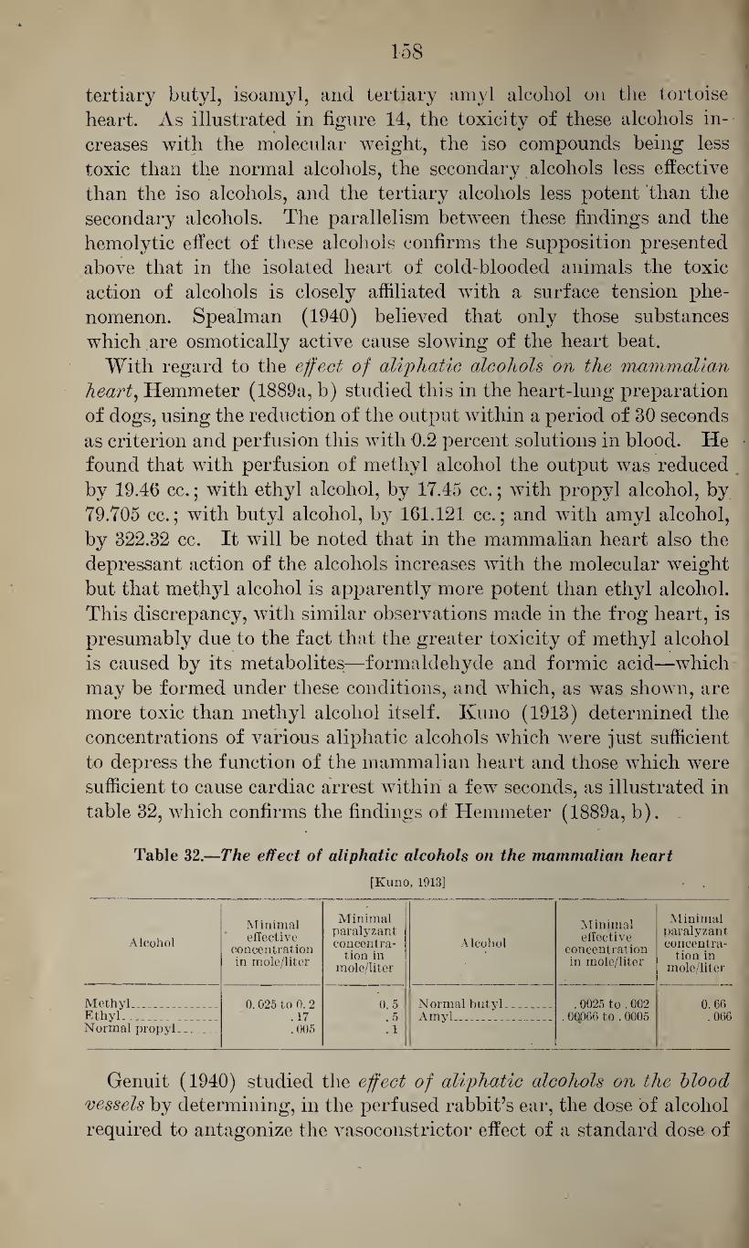
158
tertiary butyl, isoamyl, and tertiary amyl alcohol on the tortoise
heart. As illustrated in figure 14, the toxicity of these alcohols in¬
creases with the molecular weight, the iso compounds being less
toxic than the normal alcohols, the secondary alcohols less effective
than the iso alcohols, and the tertiary alcohols less potent than the
secondary alcohols. The parallelism between these findings and the
hemolytic effect of these alcohols confirms the supposition presented
above that in the isolated heart of cold-blooded animals the toxic
action of alcohols is closely affiliated with a surface tension phe¬
nomenon. Spealman (1940) believed that only those substances
which are osmotically active cause slowing of the heart beat.
With regard to the effect of aliphatic alcohols on the mammalian heart, Hemmeter (1889a, b) studied this in the heart-lung preparation
of dogs, using the reduction of the output within a period of 30 seconds
as criterion and perfusion this with 0.2 percent solutions in blood. He
found that with perfusion of methyl alcohol the output was reduced
by 19.46 cc.; with ethyl alcohol, by 17.45 cc.; with propyl alcohol, by
79.705 cc.; with butyl alcohol, by 161.121 cc.; and with amyl alcohol,
by 322.32 cc. It will be noted that in the mammalian heart also the
depressant action of the alcohols increases with the molecular weight
but that methyl alcohol is apparently more potent than ethyl alcohol.
This discrepancy, with similar observations made in the frog heart, is
presumably due to the fact that the greater toxicity of methyl alcohol
is caused by its metabolites—formaldehyde and formic acid—which
may be formed under these conditions, and which, as was shown, are
more toxic than methyl alcohol itself. Kuno (1913) determined the
concentrations of various aliphatic alcohols which were just sufficient
to depress the function of the mammalian heart and those which were
sufficient to cause cardiac arrest within a few seconds, as illustrated in
table 32, which confirms the findings of Hemmeter (1889a, b).
Table 32.—The effect of aliphatic alcohols on the mammalian heart
[Kuno, 1913]
Alcohol
Minimal effective
concentration in mole/liter
Minimal paralyzant concentra¬
tion in mole/liter
Alcohol
Minimal effective
concentration in mole/liter
Minimal paralyzant concentra¬
tion in mole/liter
Methyl_ 0. 025 to 0. 2 0 5 Normal butyl.. __ . 0025 to . 002 0. 60 Ethyl'._ . 17 .5 Amyl.. . . _ . 0Q066 to . 0005 .066 Normal propyl_ .005 .1
Genuit (1940) studied the effect of aliphatic alcohols on the blood vessels by determining, in the perfused rabbit’s ear, the dose of alcohol
required to antagonize the vasoconstrictor effect of a standard dose of
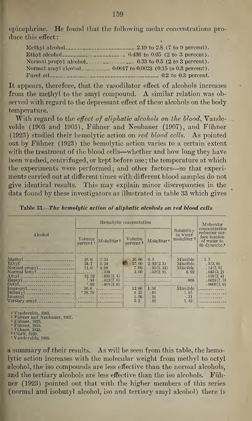
159
epinephrine. He found that the following molar concentrations pro¬
duce this effect: •
Methyl alcohol_2.19 to 2.8 (7 to 9 percent).
Ethyl alcohol___ 0.436 to 0.65 (2 to 3 percent).
Normal propyl alcohol_0.33 to 0.5 (2 to 3 percent).
Normal amyl alcohol_:_0.0017 to 0.0023 ( 0.15 to 0.3 percent).
Fusel oil_0.2 to 0.3 percent.
It appears, therefore, that the vasodilator effect of alcohols increases
from the methyl to the amyl compound. A similar relation was ob¬
served with regard to the depressant effect of these alcohols on the body
temperature.
With regard to the effect of aliphatic alcohols on the blood, Yande-
velde (1903 and 1905), Fuhner and Neubauer (1907), and Fuhner
(1923) .studied their hemolytic action on red blood cells. As pointed
out by Fuhner (1923) the hemolytic action varies to a certain extent
with the treatment of the blood cells—whether and how long they have
been washed, centrifuged, or kept before use; the temperature at which
the experiments were performed; and other factors—so that experi¬
ments carried out at different times with different blood samples do not
give identical results. This may explain minor discrepancies in the
data found by these investigators as illustrated in table 33 which gives
Table 33.—The hemolytic action of aliphatic alcohols on red blood cells
Hemolytic concentration Molecular concentration reducing sur¬ face tension of water to
63 dynes/cc.8
Alcohol Volume percent1
Mole/liter2 Volume percent3 Mole/liter4
Solubility in water
mole/liter 8
Methyl . ___ _ 36.0 7.34 ** 26.00 6.5 Miscible 1.5 Ethyl. . . 24.7 3.24 < B 17.00 2. 92(2.3)
.93(3.14)
. 32(2. 9).
Miscible .5(3.0) .14(3. 6) .043(3. 2) .018(2. 4) .0024(7. 5) .0006(3. 0)
Normal propyl._ 11.0 1.08 7. 00 Miscible Normal butyl.._ . .318 3. 00 0.92 Amyl ___ 12. 52 .091(3. 4)
.012(7. 6)
.004(3. 0) Heptyl _ _ . 84 .008 Octyl . . ._ l 89 Isopropyl_ ... . _. 16.0 12.00 1.56 Miscible Isobutyl _ 2 28. 79 4. 25 .46 1.35 Isoamyl__ . . ... .. 1.50 . 14 .31 Tertiary amyl__ _ 5.2 .48 1.42
1 Vandevelde, 1903. 2 Fuhner and Neubauer, 1907. 3 Fuhner, 1923. 4 Fuhner, 1923. 5 Fuhner, 1923. 8 Clark, 1930. 1 Vandevelde, 1905.
a summary of their results. As will be seen from this table, the hemo¬
lytic action increases with the molecular weight from methyl to octyl
alcohol, the iso compounds are less effective than the normal alcohols,
and the tertiary alcohols are less effective than the iso alcohols. Fuh¬
ner (1923) pointed out that with the higher members of this series
(normal and isobutyl alcohol, iso and tertiary amyl alcohol) there is
160
a certain relation between their hemolytic action and their solubility
in water, in that the ratio of their solubility in water, in mole per
liter, over their hemolytic concentration, expressed in the same way,
is approximately constant, being 2.9. But such a relation cannot be
established for the lower homologues (methyl, ethyl, and propyl alco¬
hol) which are miscible in water. The last column in table 33 gives
the molar concentrations necessary to reduce the surface tension of
water to 63 dynes per cc., as determined by Clark (1930) for the normal
alcohols. Comparison of the increase of the hemolytic action, as pro¬
duced by adjoining homologues, and their effect on the surface tension
of water (both figures being given in parentheses) shows that there is
a very close relation between the two functions. This appears also to
be the intrinsic phenomenon in the depressant effect of aliphatic alco¬
hols on the leucocytic movements which, as shown by Baglioni (1927),
increases, with the molecular weight, from methyl over ethyl to propyl
alcohol.
The effect of aliphatic alcohols on the striated muscle was studied
by Blumenthal (1896) and Kemp (1908) both of whom found that
the depressant effect on the muscular contractibility increases with
• the molecular weight. According to the latter, the iso, secondary, and
tertiary butyl alcohols are decreasingly less effective than the normal
butyl alcohol. Schwenker (1914) showed that, in addition to this
depressant effect on the excitability of the muscle, the aliphatic alco¬
hols cause a contracture of the muscle, and that their effectiveness in
this respect increases rapidly with their effect on the surface activity.
The contracture may continue after the excitability of the muscle has
been completely abolished, therefore it must be caused by a different
mechanism. Laporta (1929) found, by perfusing the gastrocnemius
of the frog in vivo with solutions of aliphatic alcohols in Ringer’s
solution, that the time between the beginning of the exposure and the
contraction decreased with increasing concentrations of the alcohols.
With the use of equimolecular concentrations he found that a similar
decrease occurred with the increasing molecular weight of the alco¬
hols. He noted, however, that the form and the general behavior
of the contracture varied with different alcohols, in that with all
concentrations of methyl alcohol used the main contraction was inter¬
spersed with rapid, almost rhythmical, jerks which were less distinct
with ethyl and propyl alcohol and absent with isoamyl alcohol. With
lower concentrations the contracture is reversible, whereas with higher
concentrations it is permanent.
With regard to the effect of aliphatic alcohols on structures with smooth muscles, Kuno (1914) found that this increases, with the mole¬
cular weight, «from methyl to amyl alcohol.
Several studies deal with the effect of aliphatic alcohols on the oxy¬ gen metabolism. of cells. Warburg (1910 and 1921) studied the effect
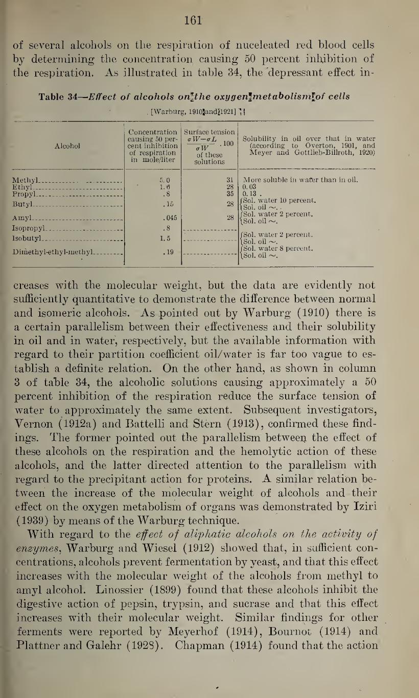
161
of several alcohols on the respiration of nuceleatecl red blood cells
by determining the concentration causing 50 percent inhibition of
the respiration. As illustrated in table 34, the depressant effect in-
Table 34—Effect of alcohols orilthe oxygen\metabolismlpf cells
[Warburg, 1910jand]il921] P
Alcohol
Concentration causing 50 per-
Surface tension aW-aL Solubility in oil over that in water
(according to Overton, 1901, and Meyer and Gottlieb-Billroth, 1920)
cent inhibition of respiration in mole/liter
<rW -100 of these
solutions
Methyl_ _ ___ 5.0 31 More soluble in water than in oil. Ethyl--. _ ___ 1.0 28 0.03 PropyL. __-__ .8 35 0.13 .
Butyl___ .15 28 fSol. water 10 percent. [Sol. oil . fSol. water 2 percent. [Sol. oil Amyl.-- .. ... .045 28
Isopropyl_ -__ .8
Isobutyl___ 1.5 fSol. water 2 percent. [Sol. oil ~. fSol. water 8 percent. [Sol. oil Dimethyl-ethyi-methyl. _ .19
»
creases with the molecular weight, but the data are evidently not
sufficiently quantitative to demonstrate the difference between normal
and isomeric alcohols. As pointed out by Warburg (1910) there is
a certain parallelism between their effectiveness and their solubility
in oil and in water, respectively, but the available information with
regard to their partition coefficient oil/water is far too vague to es¬
tablish a definite relation. On the other hand, as shown in column
3 of table 34, the alcoholic solutions causing approximately a 50
percent inhibition of the respiration reduce the surface tension of
water to approximately the same extent. Subsequent investigators,
Vernon (1912a) and Battelli and Stern (1913), confirmed these find¬
ings. The former pointed out the parallelism between. the effect of
these alcohols on the respiration and the hemolytic action of these
alcohols, and the latter directed attention to the parallelism with
regard to the precipitant action for proteins. A similar relation be¬
tween the increase of the molecular weight of alcohols and their
effect on the oxygen metabolism of organs was demonstrated by Iziri
(1939) by means of the Warburg technique.
With regard to the effect of aliphatic alcohols on the activity of enzymes, Warburg and Wiesel (1912) showed that, in sufficient con¬
centrations, alcohols prevent fermentation by yeast, and that this effect
increases with the molecular weight of the alcohols from methyl to
amyl alcohol. Linossier (1899) found that these alcohols inhibit the
digestive action of pepsin, trypsin, and sucrase and that this effect
increases with their molecular weight. Similar findings for other
ferments were reported by Meyerhof (1914), Bournot (1914) and
Plattner and Galehr (192S). Chapman (1914) found that the action
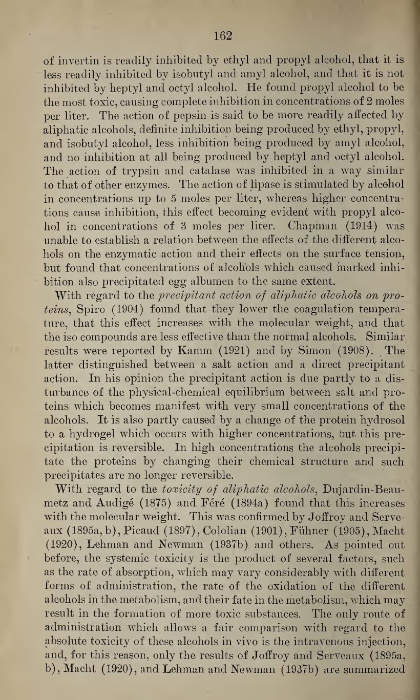
162
of invertin is readily inhibited by ethyl and propyl alcohol, that it is
less readily inhibited by isobutyl and amyl alcohol, and that it is not
inhibited by heptyl and octyl alcohol. He found propyl alcohol to be
the most toxic, causing complete inhibition in concentrations of 2 moles
per liter. The action of pepsin is said to be more readily affected by
aliphatic alcohols, definite inhibition being produced by ethyl, propyl,
and isobutyl alcohol, less inhibition being produced by amyl alcohol,
and no inhibition at all being produced by heptyl and octyl alcohol.
The action of trypsin and catalase was inhibited in a way similar
to that of other enzymes. The action of lipase is stimulated by alcohol
in concentrations up to 5 moles per liter, whereas higher concentra¬
tions cause inhibition, this effect becoming evident with propyl alco¬
hol in concentrations of 3 moles per liter. Chapman (1914) was
unable to establish a relation between the effects of the different alco¬
hols on the enzymatic action and their effects on the surface tension,
but found that concentrations of alcohols which caused marked inhi¬
bition also precipitated egg albumen to the same extent.
With regard to the precipitant action of aliphatic alcohols on pro¬ teins, Spiro (1904) found that they lower the coagulation tempera¬
ture, that this effect increases with the molecular weight, and that
the iso compounds are less effective than the normal alcohols. Similar
results were reported by Kamm (1921) and by Simon (1908). The
latter distinguished between a salt action and a direct precipitant
action. In his opinion the precipitant action is due partly to a dis¬
turbance of the physical-chemical equilibrium between salt and pro¬
teins which becomes manifest with very small concentrations of the
alcohols. It is also partly caused by a change of the protein hydrosol
to a hydrogel which occurs with higher concentrations, out this pre¬
cipitation is reversible. In high concentrations the alcohols precipi¬
tate the proteins by changing their chemical structure and such
precipitates are no longer reversible.
With regard to the toxicity of aliphatic alcohols, Dujardin-Beau-
metz and Audige (1875) and Fere (1894a) found that this increases
with the molecular weight. This was confirmed by Joffroy and Serve-
aux (1895a, b), Picaud (1897), Cololian (1901), Fuhner (1905),Macht
(1920), Lehman and Newman (1937b) and others. As pointed out
before, the systemic toxicity is the product of several factors, such
as the rate of absorption, which may vary considerably with different
forms of administration, the rate of the oxidation of the different
alcohols in the metabolism, and their fate in the metabolism, which may
result in the formation of more toxic substances. The only route of
administration which allows a fair comparison with regard to the
absolute toxicity of these alcohols in vivo is the intravenous injection,
and, for this reason, only the results of Joffroy and Serveaux (1895a,
b), Macht (1920), and Lehman and Newman (1937b) are summarized
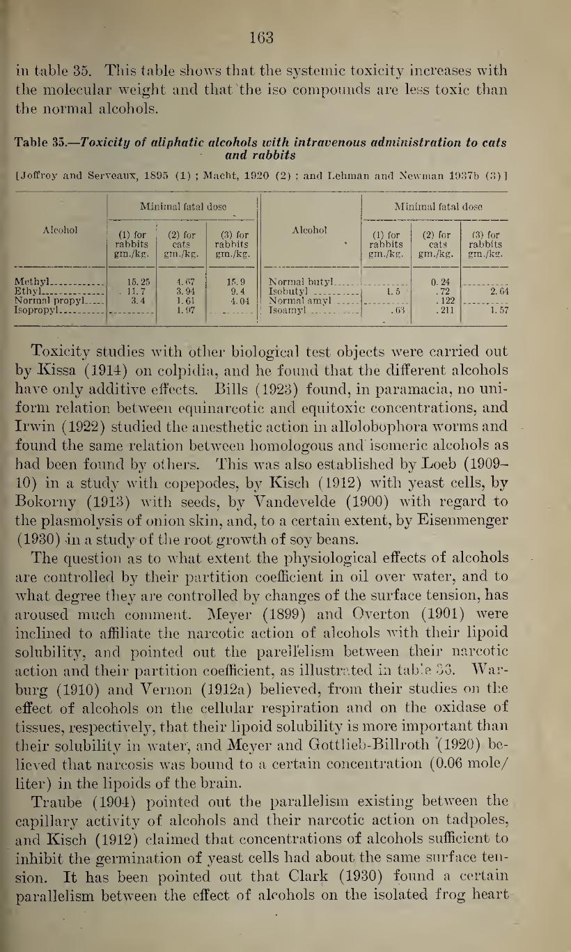
163
in table 35. This table shows that the systemic toxicity increases with the molecular weight and that the iso compounds are less toxic than the normal alcohols.
Table 35.—Toxicity of aliphatic alcohols with intravenous administration to cats and rabbits
[Joffroy and Serveaux, 1895 (1) ; Macht, 1920 (2) ; and Lehman and Newman 1937b (3)1
Alcohol
Minimal fatal dose
Alcohol *
Minimal fatal dose
(1) for rabbits gm./kg.
(2) for cats
gm./kg.
(3) for rabbits gm./kg.
(1) for rabbits gm./kg.
(2) for cats
gm./kg.
(3) for rabbits gm./kg.
Methyl__ 15.25 4.67 15. 9 Normal butyl _ 0. 24 EthyL_ . 11.7 3. 94 9.4 Isobutyl _ 1.5 .72 2.64 Normal propyl ... 3.4 1. 61 4.04 Normal amvl . 122 Isopropyl_ 1.97 Isoamyl _ _ . .63 . 211 1. 57
Toxicity studies with other biological test objects were carried out by Kissa (1914) on colpidia, and he found that the different alcohols have only additive effects. Bills (1923) found, in paramacia, no uni¬ form relation between equinarcotic and equitoxic concentrations, and Irwin (1922) studied the anesthetic action in allolobophora worms and found the same relation between homologous and isomeric alcohols as had been found by others. This was also established by Loeb (1909- 10) in a study with copepodes, by Kisch (1912) with yeast cells, by Bokorny (1913) with seeds, by Vanclevelde (1900) with regard to the plasmolysis of onion skin, and, to a certain extent, by Eisenmenger (1930) -in a study of the root growth of soy beans.
The question as to what extent the physiological effects of alcohols are controlled by their partition coefficient in oil over water, and to what degree they are controlled by changes of the surface tension, has aroused much comment. Meyer (1899) and Overton (1901) were inclined to affiliate the narcotic action of alcohols with their lipoid solubility, and pointed out the parellelism between their narcotic action and their partition coefficient, as illustrated in table 33. War¬ burg (1910) and Vernon (1912a) believed, from their studies on the effect of alcohols on the cellular respiration and on the oxidase of tissues, respectively, that their lipoid solubility is more important than their solubility in water, and Meyer and Gottlieb-Billroth (1920) be¬ lieved that narcosis was bound to a certain concentration (0.06 mole/ liter) in the lipoids of the brain.
Traube (1904) pointed out the parallelism existing between the capillary activity of alcohols and their narcotic action on tadpoles, and Kisch (1912) claimed that concentrations of alcohols sufficient to inhibit the germination of yeast cells had about the same surface ten¬ sion. It has been pointed out that Clark (1930) found a certain parallelism between the effect of alcohols on the isolated frog heart
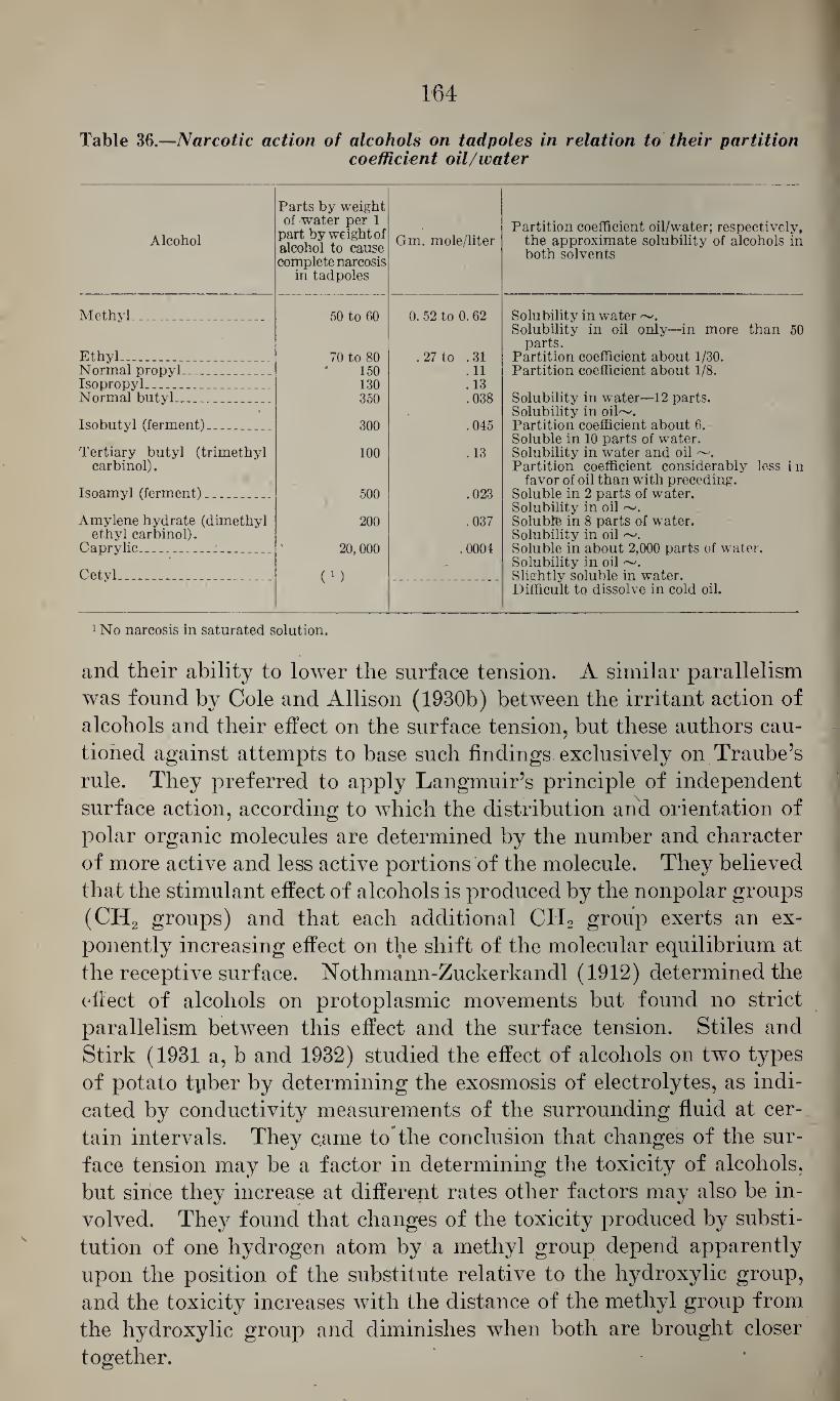
164
Table 36.—Narcotic action of alcohols on tadpoles in relation to their partition coefficient oil/water
Alcohol
Parts by weight of water per 1
part by weight of alcohol to cause complete narcosis
in tadpoles
Gm. mole/liter Partition coefficient oil/water; respectively,
the approximate solubility of alcohols in both solvents
Methyl_ 50 to 60 0. 52 to 0. 62 Solubility in water ~.
Ethyl_ _ _ 70 to 80 .27 to .31
Solubility in oil only—in more than 50 parts.
Partition coefficient about 1/30. Normal propyl_ 150 .11 Partition coefficient about 1/8. Isopropyl_ 130 . 13 Normal butyl_ 350 .038 Solubility in water—12 parts.
Isobutyl (ferment)- . _ 300 .045 Solubility in oil~. Partition coefficient about 6.
Tertiary butyl (trimethyl 100 . 13 Soluble in 10 parts of water. Solubility in water and oil ~.
carbinol).
Isoamyl (ferment)_ 500 .023
Partition coefficient considerably less in favor of oil than with preceding.
Soluble in 2 parts of water.
Amylene hydrate (dimethyl 200 .037 Solubility in oil Soluble in 8 parts of water.
ethyl carbinol). Caprylic_ _ 20, 000 .0004
Solubility in oil Soluble in about 2,000 parts of water.
Cetyl ___ ( 1 ) - Solubility in oil ~.
Slightly soluble in water. Difficult to dissolve in cold oil.
1 No narcosis in saturated solution.
and their ability to lower the surface tension. A similar parallelism
was found by Cole and Allison (1930b) between the irritant action of
alcohols and their effect on the surface tension, but these authors cau¬
tioned against attempts to base such findings exclusively on Traube’s
rule. They preferred to apply Langmuir’s principle of independent
surface action, according to which the distribution and orientation of
polar organic molecules are determined by the number and character
of more active and less active portions of the molecule. They believed
that the stimulant effect of alcohols is produced by the nonpolar groups
(CH2 groups) and that each additional CH2 group exerts an ex-
ponently increasing effect on the shift of the molecular equilibrium at
the receptive surface. Nothmann-Zuckerkandl (1912) determined the
eflect of alcohols on protoplasmic movements but found no strict
parallelism between this effect and the surface tension. Stiles and
Stirk (1931 a, b and 1932) studied the effect of alcohols on two types
of potato tyiber by determining the exosmosis of electrolytes, as indi¬
cated by conductivity measurements of the surrounding fluid at cer¬
tain intervals. They came to the conclusion that changes of the sur¬
face tension may be a factor in determining the toxicity of alcohols,
but since they increase at different rates other factors may also be in¬
volved. They found that changes of the toxicity produced by substi¬
tution of one hydrogen atom by a methyl group depend apparently
upon the position of the substitute relative to the hydroxylic group,
and the toxicity increases with the distance of the methyl group from
the hydroxylic group and diminishes when both are brought closer
together.
It appears, therefore, that in many instances the physiologic action
of alcohols is closely affiliated with their lipoid solubility; in other
instances there seems to exist a striking parallelism with their ability
to lower the surface tension. In this connection, an observation of
Fiihner (1912a) may be mentioned. He noted that, with certain plants
and lower animals, the effectiveness of normal alcohols increased, like
their capillary activity, in the ratio 1: 3: 3 2, etc. whereas, with regard
to the narcotic action in higher animals, the increase was of the order
of 1:4:42, etc. He suggested that the nerve structures of higher ani¬
mals might be richer in lipoid substances and that for this reason, in
such species the solubility of alcohols in oil, as expressed by their par¬
tition coefficient, may play the more important role. But there are
also conditions in which the dehydrating action of alcohols, and other
factors, seem to play a role, and it is probable that the various toxi¬
cological reactions may be affected by one or the other mechanism to a
different extent.
B. THE BIVALENT OR DIHYDRIC ALCOHOLS
The dihydric alcohols or glycols of the type HO-R-ROH are, as a
rule, less volatile than the monohydric alcohols. They find less ex¬
tensive use in the lacquer industry, but some of them are used in the
explosives industry and as solvents for cosmetics and pharmaceutical
products. On the other hand, the ethers and esters of some of these
materials are used quite extensively as solvents, and since these may
be decomposed in the organism with the formation of bivalent alco¬
hols, a knowledge of the toxicity of the latter is important for the
.appraisal of the potential hazards of the former.
a. Ethylene glycol
The lowest member of this series is ethylene glycol, glycol, ethandiol-
1,2, HO-CH2-CH2-OH, of the molecular weight 62.07. It is a color-
190 less liquid of the specific gravity 1.113 at C. which solidifies at
— 15.6° C. and boils at 197.4° C. and which is miscible with water and
alcohol but is less soluble in ether. Lawrie (1928) gave an extensive
review of its physicochemical properties, and Schierholtz and Staples
(1935) determined its refractive index as 1.43192 at 20° C. and as
1.43072 at 25° C. and its density as 1.1140 at 20° C. Lehmann and
Flury (1938) gave its flash point as 117° C. *
Uses.—Ethylene glycol is used as a solvent for dyes and gelatin¬
ized cellulose and also as solvent for many heavy metal salts, barbital,
alkaloids, iodine, and others (Browning, 1937). It is used in the
cosmetic and perfume industry, as antifreeze in radiators, and in the
explosives industry.
Chemical characteristics.—Ethylene glycol yields oxalic acid upon
oxidation with nitric acid, and with acid potassium permanganate it
yields glycol aldehyde, HOCH,CHO, and glyoxalic acid, HOOC-
COOH.
Following oxidation of 1 gm. ethylene glycol with 10 cc. of fresh
3 percent bromine water for 20 minutes on a boiling water bath and
removal of the excess of bromine, the oxidation product (G) gives
the following reactions which are also characteristic for glycerol
(Rosenthaler, 1923): -
1. 0.2 cc. G mixed with 0.1 cc. 5 percent alcoholic codeine solu¬
tion, 0.2 cc. water and 2 cc. concentrated H2S04 and heated for 2
minutes yields a greenish blue color.
(166)
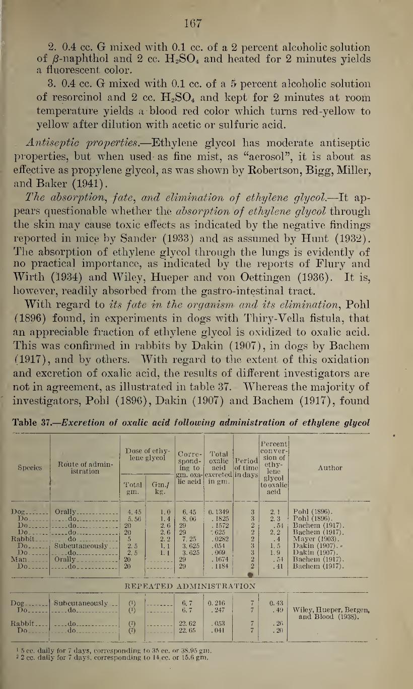
167
2. 0.4 cc. G mixed with 0.1 cc. of a 2 percent alcoholic solution of /2-naphthol and 2 cc. H2S04 and heated for 2 minutes yields a fluorescent color.
3. 0.4 cc. G mixed with 0.1 cc. of a 5 percent alcoholic solution
of resorcinol and 2 cc. H2S04 and kept for 2 minutes at room
temperature yields a blood red color which turns red-yellow to
yellow after dilution with acetic or sulfuric acid.
Antiseptic properties.—Ethylene glycol has moderate antiseptic
properties, but when used as fine mist, as “aerosol”, it is about as
effective as propylene glycol, as was shown by Robertson, Bigg, Miller,
and Baker (1941).
The absorption, fate, and elimination of ethylene glycol.—It ap¬
pears questionable whether the absorption of ethylene glycol through
the skin may cause toxic effects as indicated by the negative findings
reported in mice by Sander (1933) and as assumed by Hunt (1932).
The absorption of ethylene glycol through the lungs is evidently of no practical importance, as indicated by the reports of Flury and
Wirth (1934) and Wiley, Hueper and von Oettingen (1936). It is,
however, readily absorbed from the gastro-intestinal tract.
With regard to its fate in the organism and its elimination, Pohl
(1896) found, in experiments in dogs with Tliiry-Vella fistula, that
an appreciable fraction of ethylene glycol is oxidized to oxalic acid.
This was confirmed in rabbits by Dakin (1907), in dogs by Bachem
(1917), and by others. With regard to the extent of this oxidation
and excretion of oxalic acid, the results of different investigators are
not in agreement, as illustrated in table 37. Whereas the majority of
investigators, Pohl (1896), Dakin (1907) and Bachem (1917), found
Table 37.—Excretion of oxalic acid following administration of ethylene glycol
Species Route of admin¬ istration
Dose of ethy¬ lene glycol Corre¬
spond¬ ing to
gm. oxa¬ lic acid
Total oxalic acid
excreted in gm.
Period of time in days
Percent conver¬ sion of ethy¬ lene
glycol to oxalic
acid
Author
Total gm.
Gm./ kg.
Dog_ Orally ... . _ 4. 45 1.0 6. 45 0.1349 3 2.1 Pohl (1896). Do_ . _ do. 5. 56 1. 4 8. 06 . 1825 3 2. 3 Pohl (1896). Do. ... _do_ 20 2.6 29 . 1572 2 . 54 Bachem (1917). Do_ ... _do_ __ _ 20 2.6 29 .625 2 2. 2 Bachem (1917).
Rabbit . ...do. ..... . 5 2. 2 7. 25 .0282 2 ,4 Mayer (1903). Do_ Subcutaneously... 2.5 1.1 3. 625 .054 3 1. 5 Dakin (1907) . ♦ Do_ ..do .. __ 2. 5 1.1 3. 625 .069 3 1.9 Dakin (1907).
Man. _ Orally ... 20 29 . 1674 2 . 54 Bachem (1917). Do_ _do . . . 20 29 . 1184 2 .41 Bachem (1917).
_
REPEATED ADMINISTRATION
Dog... ... Subcutaneously .. 0) (0
6.7 0.216 7 0.43 Do_ .. do_ 6. 7 .247 7 . 49 Wiley, Hueper, Bergen
and Blood (1938). Rabbit... _do_ _. .. (2) 22. 62 .053 7 . 26 Do_ .. ..do_ (2) 22. 65 .041 7 .20
1 5 cc. daily for 7 days, corresponding to 35 cc. or 38.95 gm, 2 2 cc. daily for 7 days, corresponding to 14 cc. or 16.0 gm.
168
that approximately 2 percent of the ethylene glycol administered is ex¬
creted as oxalic acid, others, Mayer (1903), and Wiley, Hueper, Ber¬
gen and Blood (1938), found considerably lower values of 0.2 to 0.49
percent. With regard to the formation of other metabolites of ethyl¬
ene glycols, Mayer (1903) was unable to detect glycol aldehyde or
' glyoxalic acid in rabbits’ urine but he was able to isolate glycolic acid
(HOCH2COOH) in quantities corresponding to one-fourth of the
amount of ethylene glycol given and was inclined to consider this as
an intermediate in the formation of oxalic acid. According to Bern-
heim and Handler (1941), ethylene glycol is oxidized by liver prepa¬
rations containing active alcohol oxidase. As found by Hockendorf
(1909-10) and confirmed by Page (1927), it is not converted to sugar
in the organism, and Newman, van Winkle, Kennedy and Morton
(1940) found that it is not utilized by the perfused cat’s liver. Miura
(1911) stated that the administration of ethylene glycol does not cause
the appearance of reducing substances or glucuronic acid in the urine.
Lepkovsky, Ouer and Evans (1935) showed that, in contrast to the
ethyl alcohol esters of fatty acids, the corresponding ethylene glycol
esters are poorly utilized as food by rats. -
The effect of ethylene glycol on the nervous system is not very
marked. According to Macht and Ting (1922), the depressant effect
of doses of 120 mg. per 100 gm. body weight on the activity of rats
in a maze corresponds to that of 80 mg. per 100 gm. of ethyl alcohol,
and similar observations were reported by van Winkle and Kennedy
(1940). Von Oettingen and Jirouch (1931) found that ethylene gly¬
col causes only a moderate depression of the central nervous system. *
This was confirmed by Hunt (1932) and Hofbauer (1933). Latven
and Molitor (1939) determined the minimal hypnotic dose for mice,
with subcutaneous injection, as 1.0 cc. per kg.; with oral administra¬
tion as 1.0 cc. per kg.; and, by intravenous route, as 2.0 cc. per kg.,
this dose causing immediate effect. Von Oettingen and Jirouch (1931)
noted, in frogs, following the subcutaneous injection of ethylene
glycol, convulsions which they believed to be due to a peripheral
rather than a central effect. This was confirmed by Busquet (1938).
Hofbauer (1933) observed, in rats, on the first and second days follow¬
ing the subcutaneous administration of 2 gm. per kg. of ethylene gly¬
col, clonic convulsions and opisthotonus; Hanzlik, Seidenfeld and
Johnson (1931) saw, in rabbits, twitchings, tremors and convulsions;
and Hanzlik, Newmarf* van Winkle, Lehman and Kennedy (1939)
reported that, in dogs, the intravenous administration of 20 cc. per
kg. of ethylene glycol caused convulsions without demonstrable anes¬
thesia. As shown by Page (1927), the intravenous administration
of ethylene glycol causes, in dogs, a primary increase and subsequent
slowing of the respiration. This was confirmed by Hanzlik, Seiden¬
feld and Johnson (1931). With respect to the effect of inhalation of
169
vapors of ethylene glycol on the central nervous system, Flury and
Wirth (1934) and Wiley, Hueper and von Oettingen (1936) found
that even air saturated with vapors of ethylene glycol had no distinct
depressant effect on mice.
With regard to the irritant action of ethylene glycol, Bachem (1917)
stated that its subcutaneous injection caused local irritation and
slight inflammation. This was confirmed by Hanzlik, Seidenfeld,
and Johnson (1931) who showed, in addition, that the local applica¬
tion of the undiluted glycol to the conjunctiva of rabbits caused
chemosis and that it also exerted a moderately irritant action on the
frog’s skin (Tiirck’s experiment) and on the sciatic nerve of the frog.
Similar results were reported by Ajazzi-Mancini (1935) and by Page
and Coryllos (1926), but it should be pointed out, as all investigators
agree, that only concentrated ethylene glycol, not the diluted solu¬
tions, has this irritant effect. Von Oettingen and Jirouch (1931)
showed that ethylene glycol causes no depression of the peripheral
nerve fiber as might be expected from its low partition coefficient
oil/water which is 0.5.
With regard to the effect of ethylene glycol on the circulation, von
Oettingen “and Jirouch (1931) found that even 1 and 2 percent solu¬
tions caused only a slight depression of the isolated frog’s heart. Hud¬
dleston (1939) studied the electrocardiographic changes in young
dogs, following the oral administration of a 4 percent solution of
ethylene glycol, and found, among other changes, that these cause
bradycardia, exaggerated sinus arrhythmia, sinus-auricular block,
slight displacement of the S-T segment, and, sometimes, diphasic T
waves. Adult dogs treated in the same way showed sinus arrhythmia,
less marked bradycardia and, less frequently, A-V block, which ap¬
pears to be exceptional in ethylene glycol poisoning. The effect of
ethylene glycol on the blood pressure appears to be not very marked.
Bachem (1917) found that the intravenous injection of 1 cc. caused
only a moderate fall of the blood pressure in rabbits, and Page (1927)
noted that this was followed by a marked increase of the amplitude of
the heart beat, occasionally associated with a slight slowing of the
pulse rate.
Bachem (1917) and von Oettingen and Jirouch (1931) found that,
in vitro, ethylene glycol causes hemolysis, and the former believed
that this may also result from intravenous injections. Di Prisco and
Swarc (1940) showed that in rabbits the repeated intravenous and
subcutaneous administration of alcohol .causes a reduction of the
number of red and white blood cells and of the hemoglobin and the
appearance of immature red blood cells which was not seen with oral
administration or inhalation. In view of the fate of ethylene glycol
in the organism, resulting in the formation of oxalic acid, it was
555178—43-12
170
thought that the latter might affect the calcium level in the blood.
Hofbauer (1933) found, however, that only fatal doses caused a
marked reduction of the blood calcium whereas smaller doses, caus¬
ing no distinct toxic effect, did not affect the calcium level noticeably;
and similar negative results were reported by Dille (1934).
With regard to the effect of ethylene glycol on muscular structures,
von Oettingen and Jirouch (1931) found that pure ethylene glycol
causes a hyaline shrinkage of the muscle fibers and that diluted solu¬
tions cause some depression of the isolated muscle. Busquet (1938)
showed that ethylene glycol does not antagonize the action of curare
but that it does antagonize the action of strychnine iodomethylate
and decrease the chronaxy of the nerve. Hanzlik, Seidenfeld and
Johnson (1931) found that concentrations of ethylene glycol of 1: 500
and less had no effect on smooth muscle organs, and that only con¬
centrations of 1: 250 to 1:100 caused a moderate reversible depression.
This agrees with the findings of von Oettingen and Jirouch (1931).
With regard to the effect of ethylene glycol on metabolism, Cerabona
(1939) showed that about two-thirds of the rabbits chronically poi¬
soned with ethylene glycol showed an increase of the chloride level of
the blood which he attributed to renal injury. Mauro (1939a, b, c)
found that in chronic poisoning with ethylene glycol the creatinine
level in the blood of rabbits is increased, especially when the urinary
excretion is impaired, and there may be a considerable degree of
glucemia, indicating injury of liver and kidney. He found also an
increase of the cholesterol, neutral fats, soaps, and phosphatides in the
blood of such animals, indicating a disturbance of the fat metabolism.
The toxicity of ethylene glycol has aroused much comment. Earlier
investigators believed that it has comparatively little toxicity. Bachem
(1917) and others suggested its use as a substitute for glycerol in
pharmaceutical products, and Haag and Bond (1927) advocated its
use as a solvent for chloretone in anesthesia of dogs. Hunt (1932)
pointed out that with oral, intraperitoneal, and intravenous adminis¬
tration it is distinctly toxic for mice, rats, guinea pigs, rabbits, cats,
and dogs. Table 38 gives a resume of the minimal fatal doses, as de¬
termined by various investigators for different species of animals and
with various routes of administration. It shows, that the results of
various investigators may show considerable variation which may be
partly explained by the limited number of animals and possibly also
by differences in the grade of the ethylene glycol used, the technical
and less pure products being more toxic. The symptoms observed
in acute ethylene glycol poisoning are characterized by weakness, lack
of coordination, tremors, twitchings and convulsions, progressive pros¬
tration, coma, and death which may occur after 18 to 28 hours or
after several days, depending upon the dose given.
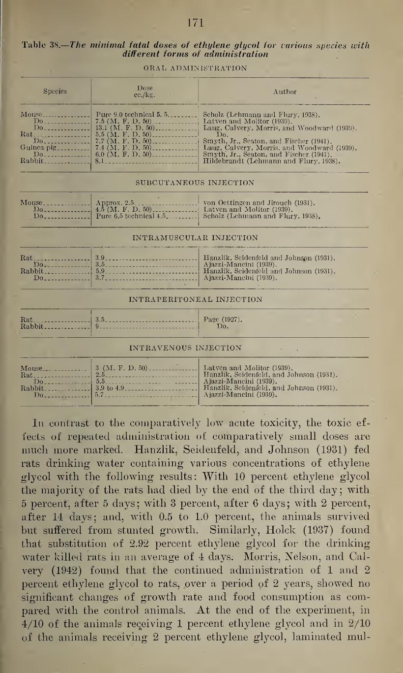
171
Table 38.—The minimal fatal doses of ethylene glycol for carious species with different forms of administration
ORAL ADMINISTRATION
Species Dose cc./kg. Author
Mouse .. Do_ Do_..
Rat_ Do_ ___
Guinea pig_ Do_
Rabbit. _
Pure 9.0 technical 5. 5. 7.5 (M. F. D. 50)_ 13.1 (M. F. D. 50)_ 5.5 (M. F. D. 50)_ 7.7 (M. F. D. 50)_ 7.4 (M. F. D. 50)_ 6.0 (M. F. D. 50)__ 8.1 ___
Scholz (Lehmann and Flury, 1938). Latven and Molitor (1939). Laug, Calvery, Morris, and Woodward (1939).
Do. Smyth, Jr., Seaton, and Fischer (1941). Laug, Calvery, Morris, and Woodward (1939). Smyth, Jr., Seaton, and Fischer (1941). Hildebrand t (Lehmann and Flury, 1938).
SUBCUTANEOUS INJECTION
Mouse.. . ... Do__ Do_
Approx. 2.5___ 4.5 (M. F. D. 50)_ Pure 6.5 technical 4.5._ _
von Oettingen and Jirouch (1931). Latven and Molitor (1939). Scholz (Lehmann and Flury, 1938).
INTRAMUSCULAR INJECTION
Rat__ 3.9_ Hanzlik, Seidenfeld and Johngpn (1931). Ajazzi-Mancini (1939). Hanzlik, Seidenfeld and Johnson (1931). Ajazzi-Mancini (1939).
Do_ 3.5_ Rabbit.. .. 5.9_ Do_ 3.7_
INTRAPERITONEAL INJECTION
Rat . .. 3.5_ Page (1927). Do. Rabbit 9 _ _ _
INTRAVENOUS INJECTION
Mouse.. _ Rat - _
3 (M. F. D. 50)__ 2.5_
Latven and Molitor (1939). Hanzlik, Seidenfeld, and Johnson (1931). Ajazzi-Mancini (1939). Hanzlik, Seidenfeld, and Johnson (1931). Ajazzi-Mancini (1939).
Do 5.5_ Rabbit 3.9 to 4.9 - .. _
Do 5.7_
In contrast to the comparatively low acute toxicity, the toxic ef¬
fects of repeated administration of comparatively small doses are
much more marked. Hanzlik, Seidenfeld, and Johnson (1931) fed
rats drinking water containing various concentrations of ethylene
glycol with the following results: With 10 percent ethylene glycol
the majority of the rats had died by the end of the third day; with
5 percent, after 5 days; with 3 percent, after 6 days; with 2 percent,
after 14 days; and, with 0.5 to 1.0 percent, the animals survived
but suffered from stunted growth. Similarly, Hoick (1937) found
that substitution of 2.92 percent ethylene glycol for the drinking-
water killed rats in an average of 4 days. Morris, Nelson, and Cal-
very (1942) found that the continued administration of 1 and 2
percent ethylene glycol to rats, over a period of 2 years, showed no
significant changes of growth rate and food consumption as com¬
pared with the control animals. At the end of the experiment, in
4/10 of the animals receiving 1 percent ethylene glycol and in 2/10
of the animals receiving 2 percent ethylene glycol, laminated mul-
172
berry bladder stones of 0.8 to 1.7 cm. diameter were found, the
bladder showing marked chronic cystitis. The stones consisted es¬
sentially of calcium oxalate. Otherwise, the animals showed signs
of marked damage of the kidneys and moderate chronic injury of
the liver.
Toxic effects from continued exposure to vapors of ethylene glycol
have not been observed (Flury and Wirth, 1934). Wiley, Hueper,
and von Oettingen (1936) showed that even continued exposure for
5 days a week over 16 weeks had no deleterious effects aside from a
moderate irritation.
The toxicity of ethylene glycol for humans is illustrated by five
cases of acute poisoning resulting from the ingestion of ethylene
glycol, three of which ended fatally, as reported in “Queries and
Minor Notes” in the Journal of the American Medical Association
(1940 and 1942) and by Brekke (1930) and Hansen (1930). After
several hours (over night) all the victims were in stupor and coma.
One patient of Brekke (1930) and one of Hansen (1930) developed
a bilateral abducens paresis and paralysis of the soft palate, and in
both the pupils were dilated and nonreactive. The other patients
showed negative patellar reflexes and a positive Babinski. The pa¬
tients were nauseated and they vomited, the vomitus containing
blood. The pulse was accelerated, small and soft. The respiration
was slow and stertorous in two patients and rapid in two others who
died. The urine became scanty and contained albumen and blood
but no sugar,” and the nonprotein nitrogen in the blood rose to 3.0
and 3.3 per thousand. The patients of Brekke (1930) and Hansen
(1930) recovered following decapsulation of the kidney, but the
two other patients mentioned died from convulsions and respiratory paralysis.
It was estimated that these patients had ingested approximately
100 cc. of ethylene glycol. Smaller single doses have been tolerated
without apparent harm, as reported by Bachem (1917) who ingested
20 gm. diluted with water; and Page (1927) ingested 15 cc. diluted
with water without ill effects.
With regard to industrial poisoning from ethylene glycol no defi¬
nite information is available. According to a report by Hackett
(1935), one case of such poisoning received compensation in the
State of New York, but neither the type of the injury nor details
of the exposure are available.
Pathological changes in ethylene glycol 'poisoning are mainly charac¬
terized by injury of the kidneys. Lepkovsky, Ouer, and Evans (1935)
noted, following oral administration of ethylene glycol esters, enlarge¬
ment of the kidneys, dilatation of the collecting and convoluted tu¬
bules, flattening of the epithelium of the collecting tubules, and, in
places, complete degeneration. Hanzlik, Newman, van Winkle, Leh-
173
man, and Kennedy (1939) reported, after oral administration to dogs,
marked necrosis and complete destruction of the convoluted tubules
and swelling of the glomeruli, and Laug, Calvery, Morris, and Wood¬
ward (1939) saw, in mice, hydropic degeneration of the cells lining
the corjtical convoluted tubules. Wiley, Hueper, Bergen, and Blood
(1938) noted, in dogs and rabbits, mild degrees of nephrosis and scat¬
tered calcium casts in the pyramidal tubules of dogs. With regard
to other organ changes, Laug, Calvery, Morris, and Woodward (1939)
found, in mice, following oral administration, congestion and hemor¬
rhages in the lungs and hemorrhagic areas in the stomach. Following
fatal doses of ethylene glycol, Hanzlik, Newman, van Winkle, Lehman,
and Kennedy (1939) found marked degeneration of the liver and fatty
infiltration of the liver cells with nuclear changes. These data indi¬
cate that injury of the kidneys is the most outstanding phenomenon
in ethylene glycol poisoning and that mainly the tubular apparatus
and, to a much lesser extent, the glomeruli are involved in the effect.
This appears to be supported by an observation of Cortese (1941) who
found that, following subcutaneous injection of small amounts of
ethylene glycol for periods up to 28 days, the tubular apparatus was
damaged whereas the glomeruli were not affected, as indicated by the
outcome of the Behberg test which is indicative of the filtering power
of the glomerular apparatus. With regard to the cause of the renal
injury, many investigators associated this with the formation of oxalic
acid and calcium oxalate deposits in the kidney. However, it appears,
as pointed out by Hunt (1932) and Wiley, Hueper, Bergen, and Blood
(1938), that aside from this effect ethylene glycol must exert some
other toxic action on the kidney.
Prophylaxis and. treatment of ethylene glycol poisoning.—As
pointed out by Flury and Wirth (1934) and Wiley, Hueper, and von
Oettingen (1936), poisonings from the inhalation of vapors of ethylene
glycol appear to be remote on account of its low volatility, and ab¬
sorption through the skin is, at best, too slow to permit the absorption
of injurious quantities. This appears to be supported by a report by
the Factory Inspection Department of the Home Office, quoted by
Browning (1937), on the examination of workers in a plant in which
ethylene glycol was handled in an electrolytic process. This exami¬
nation revealed very little with regard to definite symptoms. One
worker engaged in the operation for 2 years complained of “phlegm”
and another one who had been employed for 9 months complained of
loss of appetite and “dopiness”. Two of four workers examined
showed considerable albuminuria and increased urobilin, and one of
these showed “more red blood corpuscles than would be expected in a
normal specimen”; a third showed a trace of albumen but neither red
blood cells nor casts; and the fourth wTas normal. (The patient with
considerable albuminuria had recently had influenza.) In case ethyl-
174
ene glycol has been taken by mouth the stomach should be emptied by
gastric lavage and the diuresis should be increased by administration
of copious amounts of fluid. As indicated by the reports of Brekke
(1930) and Hansen (1930), decapsulation of one kidney may improve
the renal function and thus prevept fatal outcome.
b. Propylene Glycol
Physico-chemical properties.—Propylene glycol, propandiol-1,2,
CH3CHOHCH2OH, has the molecular weight 76.09. It is a colorless
fluid of the specific gravity 1.040 at 19.4° C., which boils at 188° to
189° C. and which is miscible with alcohol and water and soluble
in ether to the extent of 8 percent. Its flash point is 107.2° C. Its
physico-chemical properties have been discussed by Lawrie (1928),
and Schierholtz and Staples (1935) determined its refractive index
as 1.43162 at 25° C. and its density at 1.0354 at 23° C.
Uses.—Propylene glycol is used in cosmetic and pharmaceutical
preparations and in coloring extracts (Newman and Lehman, 1937a;
Brown, 1935; and Rae, 1935a, b). According to Robertson, Bigg,
Miller, and Baker (1941) it may be used in the form of an aerosol
for the disinfection of air. These authors found that a suspension
* of 1 gm. propylene glycol per 2,000 liters (161 p. p. m.) of air sterilized
an atmosphere containing 200,000 Staphylococci albus per liter. In
the opinion of Robertson, Loosli, Puck, Bigg, and Miller (1941), the
disinfecting action of propylene glycol is mainly due to the rapid
evaporation of its minute droplets. They found that mice exposed
to concentrations of 1 gm. propylene glycol in 2,000 and 3,000 liters
(161 and 106.3 p. p. m) of air and, in addition, for 15 to 60 minutes,
to influenza virus (0.2 to 1.0 cc. of a 10-2 dilution of the virus in the
form of a mist) remain well, whereas controls exposed to the same
concentration of the virus died within 4 to 10 days. Robertson, Bigg,
Puck, and Miller (1942) showed that gram-positive bacteria killed
by exposure to propylene glycol retained their gram-positiveness as
well as their morphological integrity and that they retained their
antigenic properties. It appears that propylene glycol inhibits but
does not destroy the autolytic enzyme system of the cell (pneumo¬
coccus) .
The determination of propylene glycol in air.—Puck (1942) deter¬
mined the concentration of propylene glycol in air by bubbling 2
liters of air at the rate of 0.25 liters per minute through 10 cc. of water
with the aid of a sintered glass filter. This solution is quantitatively
transferred to an Erlenmeyer flask, mixed with 1 cc. m/10 solution
of sodium periodate, and kept in an ice box for 15 minutes. After
this time 5 cc. of 7 percent solution of sodium bicarbonate are added,
followed by 2.5 cc. n/10 sodium arsenite and 2 cc. of a freshly prepared,
20 percent solution of potassium iodide, and allowed to stand for 15
175
minutes. Then 1 cc. of a 1 percent starch solution is added and the
solution is titrated with 0.01 normal iodine solution, 1 cc. of which
corresponds to 0.38 mg. of propylene glycol.
For the determination of propylene glycol in blood and urine, New¬
man and Lehman (1937a) treated protein-free blood filtrate with a
solution of potassium bichromate in strong sulfuric acid and estimated
the amount of reduced chromate colorimetrically. The difference
between this value and that obtained in the same way from blank blood
allows the calculation of the amount of propylene glycol present in
the original sample. A simple procedure may also be applied to urine.
The absorption, fate, and excretion of propylene glycol.—No infor¬
mation is available as to whether or not, or to what extent propylene
glycol is absorbed through the intact skin and the respiratory tract,
but it appears reasonable to assume that these routes of entry are of
no practical importance. As shown by Lehman and Newman (1937a)
and van Winkle (1941b), in rabbits, cats, and rats, propylene glycol
is rapidly absorbed from the intestinal tract, especially from the
jejunum and, to lesser extents, from the duodenum, colon, and stom¬
ach, in the order given. They also showed that diluted solutions are
more readily absorbed than concentrated ones and that high concen¬
trations of propylene glycol in the blood reduce the rapidity of the
absorption.
With regard to the fate of propylene glycol in the organism, Pohl
(1896) found, in dogs with Thiry-Vella fistula, that propylene glycol
is, apparently, completely oxidized, and Lehman and Newman (1937a)
stated that after its absorption it is rapidly distributed through all
tissues and readily oxidized. In the opinion of Neubauer (1901) and
Miura (1911) propylene glycol is partly excreted in conjugation with
glucuronic acid. Newman and Lehman (1937a) found that the con¬
centration of propylene glycol in the blood is proportional to the
concentration in the body, in contrast to ethyl alcohol which disap¬
pears at a constant rate, perhaps on account of a more rapid elimina¬
tion through the kidneys. In dogs which ingested propylene glycol
in 5 percent solution as drinking water over a period of 5 to 9 months,
corresponding to an average daily intake of 5.1 cc. per kg., van Winkle
and Newman (1941) determined the average propylene glycol level
in the blood as 325 mg. percent which evidently caused no toxic
effects.
According to Hanzlik, Newman, van Winkle, Lehman, and Kennedy
(1939), in dogs the excretion of propylene gtycol is completed within
24 hours following the oral administration of 8 cc. per kg. Newman
and Lehman (1937a) were able to recover from the urine about one-
half the amount of propylene glycol given orally or intravenously, and
van Winkle (1941b) recovered from the urine one-third of the amount
1*76
given in the form of a 5 percent solution as drinking water to rats and
found that the concentration in the urine did not exceed 10 percent.
The effect of 'propylene glypol on the central nervous system is not
very marked. Van Winkle and Kennedy (1940) found that its depres-
. sant effect on the central nervous system is less marked than that of
ethylene glycol when substituted for carbohydrate to the extent of 25
percent (representing 12 percent of the total diet). Hanzlik, New¬
man, van Winkle, and Kennedy (1939) determined, in dogs, the aver¬
age anesthetic dose with intravenous injection as 21.2 cc. per kg., and
Latven and Molitor (1939) determined the minimal hypnotic dose for
mice as 10 cc. per kg. with subcutaneous, 8.0 cc. per kg. with oral, and
2.0 cc. per kg. with intravenous administration. According to New¬
man and Lehman (1937a) the concentration of propylene glycol in
the blood required to produce the same degree of narcosis produced
by ethyl alcohol is 1,100 mg. percent as compared with 350 mg. per¬
cent of the latter. As shown by Ajazzi-Mancini (1939) the slow intra¬
venous injection of 20 cc. per kg. causes, in rabbits, a primary increase
and then a decrease, and, finally, paralysis of the respiration.
Propylene glycol has irritant properties. Seidenfeld and Hanzlik
(1932) stated that, when applied to the tongue, propylene glycol
causes a burning sensation, and, when given subcutaneously, it is
more irritant than ethylene glycol. When tested on the rabbit’s eye,
the undiluted glycol causes chemosis, but in 50 percent solution it
causes only lacrimation. Braun and Cartland (1936) and Latven
and Molitor (1939) found that it caused distinct irritation with sub¬
cutaneous injection, but, according to the former investigators, the
irritant effect is only transient.
With regard to the effect of propylene glycol on the circulation,
Spealman (1940) found that 0.1 molar solutions had no apparent effect
on the isolated frog heart and 0.2 molar solutions caused no changes
in the weight of the ventricle, indicating that propylene glycol is only
slightly active osmotically. Ajazzi-Mancini (1939) found that the
slow intravenous injection of 20 cc. per kg. of propylene glycol causes,
in rabbits, a fall of the blood pressure.
Weatherby and Haag (1938) found that solutions of propylene
glycol of more than 0.14 molar (32.8 percent) strength cause hemo¬
lysis in vitro, and they assumed that this was due to a dilution of the
isotonic saline which thus was rendered hypotonic. Similarly, van
Winkle (1941b) found that concentrations of 38 percent of propylene
glycol in normal saline cause partial, and in 45 percent solution com¬
plete hemolysis of red blood cells. He also found that the adminis¬
tration of propylene glycol does not increase the calcium level in the
blood, whereas according to the findings of Hofbauer (1933) in cats,
doses of 3 to 6 gm. per kg. usually increase the concentration of cal¬
cium in the blood. n!
177
With regard to the effect of propylene glycol on the metabolism,
Salter, Robb and Seharles (1935) found that, in contrast to ethylene
glycol, feeding of propylene glycol increases the liver glycogen of
guinea pigs. This was confirmed by Hanzlik, Lehman, van Winkle,
and Kennedy (1939) who showed that, within certain limits, it may
be substituted for carbohydrates with optimal results in the diet of
rats. Van Winkle and Newman (1941) doubted', however, whether
propylene glycol should really be considered as a readily available
source of energy and suggested the possibility that it may increase
the glycogen content of the liver by depressing its metabolism since
it was shown by Newman, van Winkle, Kennedy and Morton (1940)
that propylene glycol depresses the oxygen consumption and carbon
dioxide production of the perfused cat’s liver. In contrast to these
findings with an isolated organ, Hanzlik, Lehman, van Winkle, and
Kennedy (1939) found that propylene glycol has no demonstrable
effect on the basal metabolic rate in man or on the respiratory quotient
of rats. More recently, van Winkle (1942) showed that,/in rats,
propylene glycol in doses of 5 and 10 cc. per kg. decreases the oxygen
consumption ^( average cc. of oxygen consumed per 100 cm.2 of body
surface per minute at 22° C.) to an average of 1.142 cc. from the con¬
trol average 1.363.
The toxicity of propylene glycol is lower than that of ethylene gly¬
col and its acute toxicity is less than one-half that of ethyl alcohol
(Lehman and Newman, 1937a). The minimal fatal doses for differ¬
ent animals with different routes of administration, as found by diff¬
erent investigators, are^ summarized in table 39. After oral admin¬
istration of large doses the animals rapidly become incoordinated,
their motor and sensory functions become more and more depressed,
they become comatose, and die after several hours or even days have
elapsed. With intravenous administration of fatal doses animals may
show-first a stimulation and later a depression of the respiration, fol¬
lowed by motor and sensory depression, coma, and death. Weatherby
and Haag (1938) and Kesten, Mulinos, and Pomerantz (1939) also
noted hematuria and hemoglobin casts in the renal cortex following
the intravenous injection of fatal doses, but according to Lehman and
Newman (1937a) this is not observed after oral administration.
Gross (Lehmann and Flury, 1938) noted a temporary albuminuria
and red blood cells in the urine of rabbits which had received 7.0 gm.
per kg. by stomach tube in the form of a 50-percent solution, and a dog
fed 4 gm. per kg. of the same concentration developed diarrhea.
With regard to the chronic toxicity of propylene glycol, this is much
less than that of ethylene glycol. Hunt (1932) noted no toxic effects
in animals which received 5 percent propylene glycol as drinking water
over a long period of time; and Seidenfeld and Hanzlik (1932) showed
that rats which were fed 1, 2, 5, and 10 percent propylene glycol as
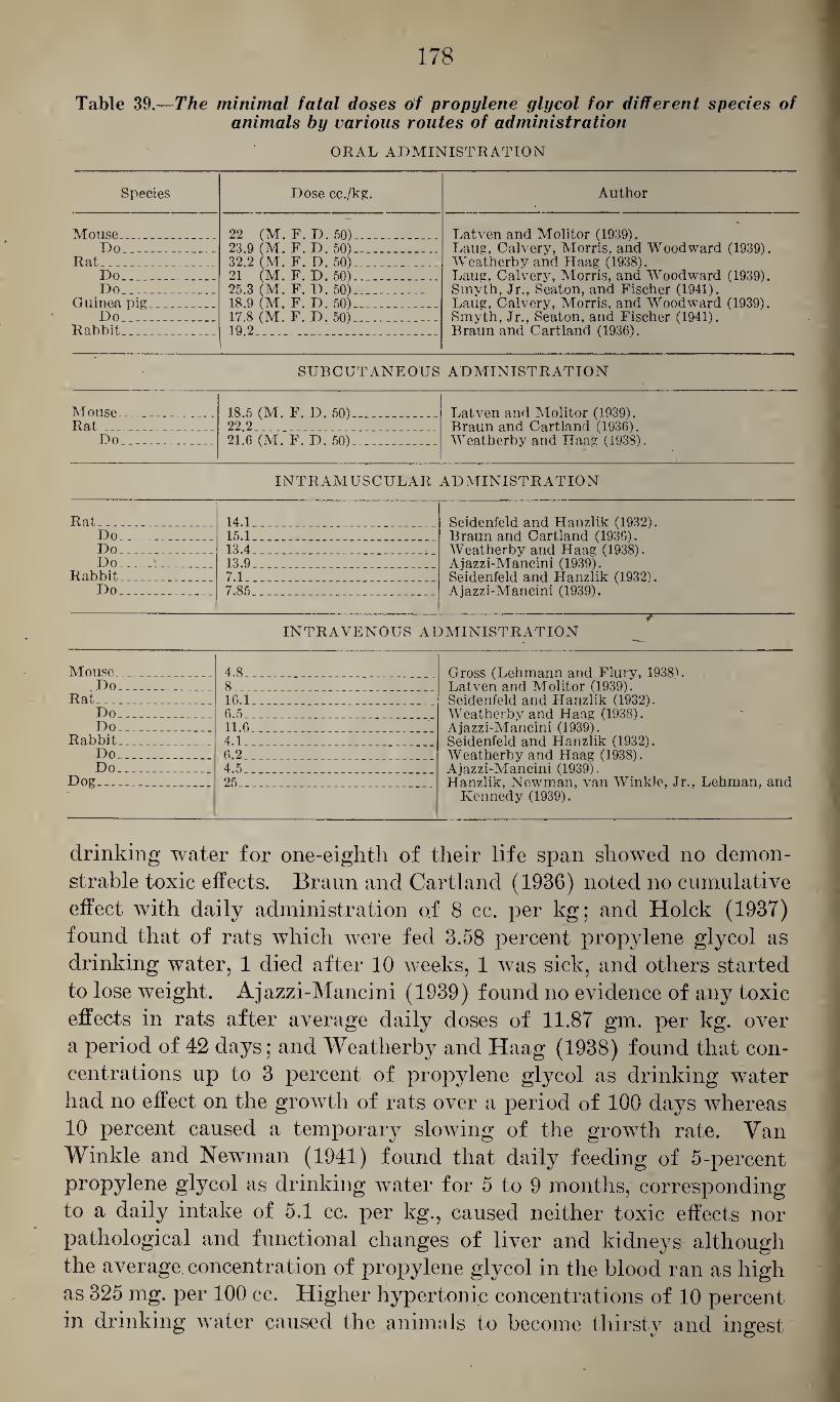
178
Table 39.—The minimal fatal doses of propylene glycol for different species of animals by various routes of administration
ORAL ADMINISTRATION
Species Dose cc./kg. Author
Mouse . .. 22 (M. F. D. 50)_ . Latven and Molitor (1939). Laug, Calvery, Morris, and Woodward (1939). Do___ 23.9 (M. F. D. 50)__
Rat_ 32.2 (M. F. D. 50)_ Weatherby and Haag (1938). Do_ 21 (M. F. D.50)_ Laug, Calvery, Morris, and Woodward (1939). Do_ 25.3 (M. F. D. 50)_ Smyth, Jr., Seaton, and Fischer (1941).
Guinea pig . _ . 18.9 (M. F. D. 50)_ Laug, Calvery, Morris, and Woodward (1939). Do__ 17.8 (M. F. D. 50)_ Smyth, Jr., Seaton, and Fischer (1941).
Rabbit_ _ 19.2_ Braun and Cartland (1936).
SUBCUTANEOUS ADMINISTRATION
Mouse_ .. 18.5 (M. F. D. 50) - Latven and Molitor (1939). Braun and Cartland (1936). Weatherby and Haag (1938).
Rat__ Do_
22 2 21.6 (M. F. D. 50)_
INTRAMUSCULAR ADMINISTRATION
Rat_ 14.1.. _ _ Seidenfeld and Hanzlik (1932). Do_ 15.1. Braun and Cartland (1936). Do_ 13.4_ Weatherby and Haag (1938). Do_ 13.9. Ajazzi-Mancini (1939).
Rabbit. _ _ 7.1_ Seidenfeld and Hanzlik (1932). Do_ 7.85_ Ajazzi-Mancini (1939).
/ INTRAVENOUS ADMINISTRATION
Mouse_ 4.8 Gross (Lehmann and Flury, 1938). Latven and Molitor (1939). Seidenfeld and Hanzlik (1932).
Do_ 8_ ... Rat_ _ . 16.1_ Do_ 6.5 .. . Weatherby and Haag (1938).
Ajazzi-Mancini (1939). Seidenfeld and Hanzlik (1932). Weatherby and Haag (1938). Ajazzi-Mancini (1939). Hanzlik, Newman, van Winkle, Jr., Lehman, and
Kennedy (1939).
Do_ 11.6 Rabbit... _ 4.1 Do_ 6.2 . Do_ 4.5_
Dog_ 25_
Jl
drinking water for one-eighth of their life span showed no demon¬ strable toxic effects. Braun and Cartland (1936) noted no cumulative effect with daily administration of 8 cc. per kg; and Hoick (1937) found that of rats which were fed 3.58 percent propylene glycol as drinking water, 1 died after 10 weeks, 1 was sick, and others started to lose weight. Ajazzi-Mancini (1939) found no evidence of any toxic effects in rats after average daily doses of 11.87 gm. per kg. over a period of 42 days; and Weatherby and Haag (1938) found that con¬ centrations up to 3 percent of propylene glycol as drinking water had no effect on the growth of rats over a period of 100 days whereas 10 percent caused a temporary slowing of the growth rate. Van Winkle and Newman (1941) found that daily feeding of 5-percent propylene glycol as drinking water for 5 to 9 months, corresponding to a daily intake of 5.1 cc. per kg., caused neither toxic effects nor pathological and functional changes of liver and kidneys although the average concentration of propylene glycol in the blood ran as high as 325 mg. per 100 cc. Higher hypertonic concentrations of 10 percent in drinking water caused the animals to become thirsty and ingest
179
much greater doses of propylene glycol, so that the average concen¬ tration of propylene glycol rose to 800 mg. per 100 cc. The increased intake of 10 percent glycol resulted in the production of coma, and the dogs died of marked central depression or of a subsequent pneu¬ monia. If, however, 600 cc. of a 10-percent solution were fed in the morning and pure water in the evening, so that the average daily in¬ take was 4.5 cc. per kg., the animals remained in good health and showed no functional changes of liver or kidneys. Gross (Lehmann and Flury, 1938) found that in rabbits 12 daily doses of 2.0 and 4.0 gm. per kg. over a period of 14 days caused no noticeable toxic effects, but, on autopsy, liver, kidneys, and spleen showed moderate edema and hyperemia. Morris, Nelson and Calvery (1942) showed that con¬ tinued feeding of propylene glycol in concentrations of 2.45 and 4.9 per¬ cent with the food, over a period of up to 2 years, caused no changes in the rate of growth and food consumption. The animals developed no bladder stones, as seen with ethylene glycol, and showed no signs of chronic injury of the kidneys and only slight chronic liver damage. It appears, therefore, as stated by Ajazzi-Mancini (1939), van Winkle and Newman (1941) and others, that there is good reason to believe that the ingestion of moderate doses of propylene glycol involves no individual or public health hazards. Robertson, Bigg, Puck, Miller, and Baker (1942) kept a colony of rats in an atmosphere containing a high concentration of propylene glycol in a mist form for a period of 7 months. At the end of this period they showed no evidence of any deleterious effects on growth rate, fecundity or general condition. The histological examination of liver, lungs, and kidneys revealed no abnormal changes.
With regard to 'pathological changes in propylene glycol poisoning, information is very scanty. Hanzlik, Newman, van Winkle, Lehman, and Kennedy (1939) found no evidence of liver and kidney injury fol¬ lowing continued feeding. With the oral administration of single fatal doses, Laug, Calvery, Morris, and Woodward (1939) noted no definite gross pathology except for hemorrhagic areas in the small intestine, and, histologically, they found only a little damage to the kidney, characterized by nuclear pyknosis, vacuolar degeneration and protein debris, and loose casts in the cortical tubules. The liver showed slight congestion and hyperemia but no fatty changes.
There are no reports on injurious effects from industrial or other use of propylene glycol, and it appears, as pointed out above, that its industrial use does not involve any special hazards.
c. Trimethylene Glycol
Physico-chemical properties.—Trimethylene glycol, propandiol-1,3, HOCH2-CH2-CHvOH, is the isomer of propylene glycol, propandiol-
180
1,2. It has the molecular weight 76.09 and represents a colorless 20°
oily fluid of the specific gravity 1.060 at C. which rapidly turns yel-
low-brdwn on standing (van Winkle, 1941a), which boils at 214° C., and which is miscible with alcohol and water. It is a byproduct in the saponification of fats and, presumably, is formed by fermentation of glycerol (Lawrie, 1928). Scliierholtz and Staples (1935) determined its refractive index as 1.43847 at 25° C. and 1.43940 at 21° C. and its density as 1.0538 at 20° C.
According to Robertson, Bigg, Miller, and Baker (1941), its anti¬ septic efficiency as aerosol is of the same order as that of propylene glycol. But, according to van Winkle (1941a), it is about twice as toxic as propandiol-1,2, causing, in toxic doses, depression of the cen¬ tral nervous system which, in his opinion, is due in part to a different fate in the organism because propandiol-1,2 is said to be oxidized to lactic acid and hence either burned or converted to glycogen, and propandiol-1,3 is oxidized to malonic acid which forms insoluble calcium salts and, therefore, may act in a way similar to oxalic acid. The minimal fatal dose was found to be: For rabbits, with intravenous injection, 4.5 cc. per kg. of body weight (given as 50 percent solution) ; for rats, with intramuscular injection, 6 to 7 cc. per kg.; and, for rats, with oral administration, about 16 cc. per kg. Cats appear to be more sensitive, and 3 cc. per kg. by mouth may be fatal in this species. Con¬ tinued feeding of trimethylene glycol in concentrations up to 5 to 12 percent with the food was found to have a definite inhibiting effect on the weight, and, in contrast to propylene glycol, it is said to have no glycogenic action .
d. Butylene Glycol
The higher liomologue of propylene glycol is butylene glycol, bu- tandiol-1,3. CH3CHOHCH2CH2OH, of the molecular weight 90.12.
20°
It is an oily liquid of the specific gravity 1.026 at C., which boils
at 203 to 204° C., and which is very soluble in water and alcohol but insoluble in ether.
%
According to Gross (Lehmann and Flury, 1938) it is used as a lubricant for special purposes, and it is an intermediate in the manu¬ facture of artificial rubber by the butadiene-Buna process. Also, it has been suggested that it be substituted for glycerol in the cosmetic, tobacco, paper, and textile industries.
As stated by the same author, it does not cause irritation of the skin, even after contact for 72 hours. He refers further to unpub¬ lished data of Hildebrandt who found that, in rabbits, single doses of 7 gm. per kg.,, given orally, cause light narcosis and no albuminuria, and that, in dogs, single doses of 2.4 gm. per kg. have no toxic effect and 0.2 gm. per kg., given daily for 2 weeks, caused only moderate
181
albuminuria. Gross, himself, found that feeding of 10 and 12 gm. per kg., diluted with 10 percent water, was tolerated by rabbits, which showed temporary dyspnea, staggering, and slight narcosis but no albuminuria and recovered rapidly; whereas doses of 15 gm. per kg. were fatal, causing diarrhea, narcosis, and death. Smyth, Seaton, and Fischer (1941) determined the minimal fatal dose with oral administration for rats as 18.61 gm. per kg. and for guinea pigs as 11.46 gm. per kg.
In a rabbit killed by the oral administration of butylene glycol, Gross (Lehmann and Flury, 1938) noted irritation of the gastro¬ intestinal tract, broadening of the intermediate layer of the kidney, and spotty reddening of the marrow, the urine, however, being free of albumen.
There are no reports on toxic effects of butylene glycol on the human organism, and it appears to offer very little potential danger.
One of the isomers of butandiol-1,3 is pseudo-butylene glycol, bu- tandiol-2,3 H3C-CHO-CHOH-CH3, which has the molecular weight 90.12. It is a liquid of the specific gravity 1.048 at 0° C., which boils at 183 to 184° C. and which is miscible with water and ether.
According to Neuberg and Gottschalk (1925), it is excreted in rab¬ bits largely as conjugated glucuronic acid, and from the fact that no acetoin could be isolated from the urine of the animals these investi¬ gators concluded that it is not oxidized to any considerable extent in the organism. Rabbits can tolerate doses of 5 cc. per kg., given in 2 fractions diluted with 40 cc. of water, without any toxic effects. The minimal fatal dose and toxic doses have apparently not been established.
The tertiary glycol, pinacol, 2,3 - d i m e t h y 1 - butandiol - 2,3, ch3 oh on CH0
\i i/ c—c / \
ch3 ch, - -
has the molecular weight 118.17 and forms colorless needles of the specific gravity 0.967 which melt at 41 to 43° C. and boil at 171 to - 172° C. at 739 mm. Hg. It is soluble in cold water, alcohol, and ether.
Thierfelder and von Mering (1885) found that a single dose of 3 gm. had no effect on a large rabbit, whereas 10 gm. caused sleep of 5 to 6 hours duration, and they found that the urine contained con¬ jugated glucuronic acid.
There are a number of bivalent alcohols which represent ethers
formed by the condensation of 2 or more molecules of bivalent alco¬
hols, such as diethylene glycol, dipropylene glycol, triethylene glycol, etc.
e. Diethylene Glycol
The lowest member of this series is diethylene glycol, glycol ether, H0CH2-CH2-0-CH2CH20II, of the molecular weight 106.2. It is a
182
colorless liquid of the specific gravity 1.132 at 0° C., which boils at 250° C., and which is soluble in water, alcohol, and ether. According to Gross (Lehmann and Flury, 1938) its flash point is 124° C.
Diethylene glycol is used in the manufacture of lacquers, as solvent for nitrocellulose and dyes (Browning, 1937), and as hygroscopic agent for tobacco (Mulinos and Osborne, 1934-35). It is also used as lubricant for special purposes (Gross—Lehmann and Flury, 1938) and for the sterilization of surgical instruments (Gurchot and Mel-
lars, 1940). The absorption, fate, and excretion of diethylene glycol.—No in¬
formation is available with regard to the absorption of diethylene glycol through the skin or the respiratory tract. However, the latter route of entry appears to be insignificant in view of the high boiling point of this material. As shown by Hanzlik, Newman, van Winkle, Lehman, and Kennedy (1939), it is quite readily absorbed from the gastrointestinal tract. Very little is known regarding its fate in the metabolism, but it is evidently not oxidized to oxalic acid, presumably on account of the stability of the ether linkage (Wiley, Hueper, Bergen, and Blood, 1938). Morris, Nelson, and Calvery (1942) found laminated mulberry stones in the bladders of 3 out of 20 rats kept for 2 years on a diet containing 1.71 and 3.42 percent diethylene glycol, and they considered this a possible indication that, in the rat, the ether linkage of diethylene glycol may be broken up and result in the excretion of oxalic acid. According to Haag and Ambrose (1937), it is mostly excreted unchanged in the urine, the excretion being com¬ pleted in 36 hours (Hanzlik, Newman, van Winkle, Lehman, and Kennedy, 1939).
With regard to the effect of diethylene glycol on the nervous sys¬
tem, Hanzlik, Newman, van Winkle, Lehman, and Kennedy (1939) found that, in dogs, the intravenous injection of 20 cc. per kg. causes no anesthesia, but convulsions, and, in contrast to propylene glycol, it produces, in equal or smaller doses, a temporary depression of the voluntary running movements of rats (van Winkle and Kennedy, 1940). It has been claimed that the use of diethylene glycol as hygro¬ scopic agent for tobacco results in less irritant smoke than that ob¬ tained with the use of glycerol (Mulinos and Osborne, 1934-35 and 1935; Wallace, Reinhard, and Osborne, 1936; and Flinn, 1935 and 1937). This was not confirmed by Sharlit (1935) or by Haag (1937), and Hoick and Carlson (1937) found no difference in the irritant action of smoke produced by tobaccos containing glycerol and diethy- . lene glycol as hygroscopic agents.
With regard to the effect of diethylene glycol on other physiological
functions, von Oettingen and Jiroucli (1931) found that concentra¬ tions of 1 percent in Ringer’s solution caused a reversible depression and a 2 percent solution caused arrest of the isolated frog’s heart in
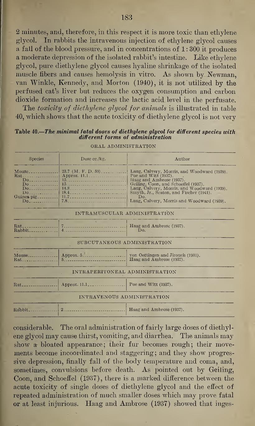
183
2 minutes, and, therefore, in this respect it is more toxic than ethylene glycol. In rabbits the intravenous injection of ethylene glycol causes a fall of the blood pressure, and in concentrations of 1: 300 it produces a moderate depression of the isolated rabbit’s intestine. Like ethylene glycol, pure diethylene glycol causes hyaline shrinkage of the isolated muscle fibers and causes hemolysis in vitro. As shown by Newman, van Winkle, Kennedy, and Morton (1940), it is not utilized by the perfused cat’s liver but reduces the oxygen consumption and carbon dioxide formation and increases the lactic acid level in the perfusate.
The toxicity of diethylene glycol for animals is illustrated in table 40, which shows that the acute toxicity of diethylene glycol is not very
Table 40.—The minimal fatal doses of diethylene glycol for different species with different forms of administration
ORAL ADMINISTRATION
Species Dose cc./kg. Author
Mouse__ _ Rat_ .. - -
Do-. _
23.7 (M. F. D. 50)_ Approx. 11.1__ 15_ _
Laug, Calvery, Morris, and Woodward (1939). Poe and Witt (1937). Haag and Ambrose (1937). Geiling, Coon, and Schoeffel (1937). Laug, Calvery, Morris, and Woodward (1939). Smyth, Jr., Seaton, and Fischer (1941).
Do. Laug, Calvery, Morris and Woodward (1939).
Do . . 15__ Do 14.8_ Do. _ 18.4_
Guinea pig... _ . _ Do_
11.7_ 7.8_
INTRAMUSCULAR ADMINISTRATION
Rat_ _ Rabbit_. _
\
7_ 4_
Haag and Ambrose (1937). Do.
SUBCUTANEOUS ADMINISTRATION
Mouse. . . . Rat...
Approx. 5_ 5___
von Oettingen and Jirouch (1931). Haag and Ambrose (1937).
INTRAPERITONEAL ADMINISTRATION
Rat__ -- _ Approx. 11.1__ Poe and Witt (1937).
INTRAVENOUS ADMINISTRATION
Rabbit.. . . 2_ Haag and Ambrose (1937).
considerable. The oral administration of fairly large doses of diethyl¬ ene glycol may cause thirst, vomiting, and diarrhea. The animals may show a bloated appearance; their fur becomes rough; their move¬ ments become incoordinated and staggering; and they show progres¬ sive depression, finally fall of the body temperature and coma, and, sometimes, convulsions before death. As pointed out by Geiling, Coon, and Schoeffel (1937), there is a marked difference between the acute toxicity of single doses of diethylene glycol and the effect of repeated administration of much smaller doses which may prove fatal or at least injurious. Haag and Ambrose (1937) showed that inges-
184
tion of 3 and 10 percent diethylene glycol as drinking water is rapidly fatal to rats. Similarly, Kesten, Mulinos, and Pomerantz (1939) found that ingestion of 3 percent solutions killed 50 percent of the rats within 2 months and that ingestion of 5 percent solutions killed one- fourth of the rats within 1 week. This is in agreement with the find¬ ings of Hoick (1937) who found that 5 percent solutions, as drinking water, killed rats in 8 days; that 4 percent solutions resulted in the death of some animals and the stunted growth of others; and that even 0.25 percent solutions gave some indication of impaired growth. However, Haag and Ambrose (1937) found no evidence of a toxic action with concentrations of 0.1 to 0.03 percent, and Kesten, Mulinos, and Pomerantz (1939) noted no toxic effects or renal damage in ani¬ mals fed 0.5 to 1.0 percent solutions over a period of 1 to 5 months. When mixed with solid food to the extent of 20 percent, diethylene glycoj was fatal to all rats within 2 weeks, and 5 and 10 percent was fatal to some animals (Hoick, 1937). Morris, Nelson, and Calvery (1942) fed rats a diet containing 1.71 and 3.42 percent diethylene glycol for a period of 2 years. These animals did not show any signifi¬ cant changes of the growth rate and the food consumption as compared with control animals. Three out of 20 animals showed laminated mulberry stones in the bladder. These consisted partly of calcium oxalate and partly of calcium phosphate. These animals showed a lesser degree of chronic cystitis than was observed with animals which were fed ethylene glycol, and they showed slight chronic damage of the kidneys and the liver.
The toxicity of diethylene glycol for humans is illustrated by a series of fatalities (60 up to 1937) which occurred in the United States from the ingestion of an “elixir” of sulfanilamide which contained 72 percent diethylene glycol. According to Kuprecht and Nelson (1937) and Geiling and Cannon (1938), the clinical picture of these poisonings may be described as follows: Upon the ingestion of the “elixir” the victims experienced heartburn, nausea, abdominal cramps, dizziness, and malaise; and they suffered from vomiting, diarrhea, and pain in the kidney region and the abdomen. In the course of from 2 to 5 days they developed oliguria and anuria; they became drowsy and showed puffiness of the face and sometimes slight jaundice; and died from 2 to 7 days after the onset of oliguria, in uremic coma. In addition, they showed pallor of the face without cyanosis, slow and deep respiration; they suffered from varying degrees of pulmonary edema, subnormal temperatures and slow pulse; and there was usually a moderate leucocytosis. The urine contained a large amount of albu¬ men, casts and, occasionally, leucocytes; the nonprotein nitrogen in the blood rose to 200 mg. and the creatinine to 12 mg. per 100 cc.
Pathological changes in animal organs caused by diethylene glycol
refer mainly to the kidneys. Geiling and Cannon (1938) observed a
185
picture characteristic for chemical nephrosis, with cellular edema of most epithelial cells of the convoluted tubules, resulting in tubular obstruction by compression and by intraluminal formation of casts. Similarly, Kesten, Mulinos, and Pomerantz (1939), Hanzlik, Newman, van Winkle, Lehman, and Kennedy (1939) and Laug, Calvery, Morris, and Woodward (1939) noted extensive injury of the convoluted tubu¬ les, leading to obstruction and uremia, whereas the effect on the glomerula apparatus was much less severe. With regard to the latter, Laug, Calvery, Morris, and Woodward (1939) noted a distention of Bowman’s capsule by protein precipitates which was also reported by Wiley, Hueper, Bergen, and Blood (1938) who, in addition, reported on swelling of the glomeruli, with occasional lack of nuclei in the capillary tufts. With regard to other organ changes following oral administration the gastro-intestinal tract may show signs of irritation, characterized by hemorrhages, as seen by Hoick (1937) and by Laug, Calvery, Morris, and Woodward (1939). The liver may be soft and fragile and may show widespread, diffuse, hydropic changes, degener¬ ation of the central portions of the lobules—as seen by Laug, Cal¬ very, Morris, and Woodward (1939), or fatty infiltration with nuclear changes—as reported by Hanzlik, Newman, van Winkle, Lehman, and Kennedy (1939). According to the former authors the lungs may show more or less marked edema and hemorrhages and the spleen may show extensive phagocytosis of blood pigments. As pointed out by Geiling and Cannon (1938), it appears open to discussion whether the intracellular changes in the convoluted tubules of the kidneys are due to hypoxemia with subsequent cellular edema, necrosis, and des¬ quamation, or to the hygroscopic properties of diethylene glycol.
With regard to pathological changes observed in human cases of
diethylene glycol poisoning, Hagebusch (1937) noted, in 4 autopsies, pulmonary edema, marked nephrosis with hemorrhages in the cortex of the kidneys, and hemorrhages in the pericardium, the mucosa of the stomach and duodenum and the serous surface of the lungs. According to Lynch (1938) the most characteristic changes concern the kidneys and are characterized by necrosis of the secretory tubules. In addition, there may be central necrosis of the liver. According to Cannon (1937) and Geiling and Cannon (1938) the general picture of the injured kidney is that of severe chemical nephrosis, charac¬ terized by intracellular edema of most epithelial cells of the con¬ voluted tubules, leading to internal disorientation of these cells. In some of the cases the kidneys showed cortical necroses and recent hemorrhages and were flabby and swollen. Microscopically there was severe vacuolization of the epithelium of the convoluted tubules whereas the glomeruli were affected very little. The liver showed hydropic degeneration of the liver cells which was most marked in
555178—43-13
186
the center of the lobules, and there were some fatty changes, espe¬ cially around the border of the hydropic areas, with little sign of necrosis. The lungs of these patients showed marked congestion, edema, and broncho-pneumonia, and, in severe cases, there were also ascites, hydrothorax, and hydropericardium. In some instances, they noted also recent hemorrhages in the gastro-intestinal tract and in the lungs, presumably caused by increased capillary permeability due to acidosis and uremia.
There are no reports on industrial diethylene glycol poisonings, and it appears that the danger of such injuries is quite remote.
f. Dipropylene Glycol
The higher homologue of diethylene glycol is dipropylene glycol, H0CH2-CH2-CH2-0-CH2-CH2-CH20H, which has the molecular
weight 137.17 and the specific gravity 1.0252 at C., which boils
at 231.8° C., and which is soluble in water and alcohol. Its flash point is 137.78° C.
Its use as partial solvent for low and high viscosity cellulose acetate and as solvent for rosins has been suggested.
According to Hanzlik, Newman, van Winkle, Lehman, and Ken¬ nedy (1939), it is absorbed rapidly from the gastro-intestinal tract and excreted within 24 hours. The minimal fatal dose for dogs, with intravenous injection, is 11.5 cc. per kg; doses of 5.9 cc. per kg. causing anesthesia. In dogs, oral administration of 4 doses of 5 cc. per kg., 6 of 1.5 cc. per kg., and 6 of 2 cc. per kg. gave no evidence of toxic effects. Four animals surviving the repeated administration of dipropylene glycol showed very little evidence of liver damage, and one showed moderate degenerative changes of the convoluted tubules. Kesten, Mulinos, and Pomerantz (1939) noted, after oral and intravenous administration to rats, injuries similar to those seen after the administration of diethylene glycol, namely, hydropic de¬ generation of the tubular epithelium of the kidneys; but these were less frequent than observed with diethylene glycol and resulted only from higher doses. Van Winkle and Kennedy (1940) found that, in contrast to propylene glycol, dipropylene glycol depresses, in equal or smaller doses, the voluntary running movements of rats; and, in contrast to their behavior after the administration of diethy¬ lene glycol, they did not recover from this depression. As found by Newman, van Winkle, Kennedy, and Morton (1940), dipropylene glycol is not utilized by the perfused cat’s liver, but it decreases the consumption of oxygen and the formation of carbon dioxide and in¬ creases the lactic acid level in the blood, being, however, less toxic than ethylene and diethylene glycol.
187
g. Triethylene Glycol
The condensation of 3 molecules of ethylene glycol yields triethyl¬
ene glycol, H0-CH2-CH2-0-CH2-CH2-0CH2CH20H, which has the molecular weight 150.17, and which is a colorless liquid with a specific gravity of 1.138 and a boiling point of 290° C. It is miscible with water and alcohol, and its flash point is 165.56° C.
Robertson, Puck, Lemon, and Loosli (1943) studied the germicidal action of triethylene glycol when used as aerosol. They found that concentrations of 1 gm. in 100 million cc. of air (1.6 p. p. m.) and of 1 gm. in 200 million cc. of air (0.8 p. p. m.) almost immediately cause disappearance of streptococci and pneumococci from the air, and within 10 minutes the test plates were essentially sterile. Dilutions of triethylene glycol as high as 1 gm. in 600 million cc. of air (0.3 p. p. m.) were found to exert a definite killing effect on pneumococci and a group of C streptococci. Concentrations of 1 gm. per 200 million cc. of air (0.8 p. p. m.) were found to protect mice completely against an airborn amount of influenza virus which a short time before had caused the death of all exposed (control) animals.
It is proposed that triethlyene glycol be used as a solvent for nitro¬ cellulose and various gums and rosins. According to Latven and Molitor (1939), the minimal fatal doses (M. F. D. 50) for mice are: With subcutaneous injection, 8.75 cc. per kg.; with oral administra¬ tion, 18.5 cc. per kg.; and, with intravenous injection, 6.5 cc. per kg. Smyth, Seaton, and Fischer (1941) determined the minimal fatal doses with oral administration as 22.06 gm. per kg. for rats and as 14.66 gm. per kg. for guinea pigs. Lauter and Yrla (1940) de¬ termined the minimal fatal doses for rats as 8.4 cc. per kg. with intramuscular injection and 11 to 15 cc. per kg. with oral admin¬ istration, and they stated that doses of 10 cc. per kg. cause definite toxic symptoms. They found that continued ingestion of 5 percent solutions as drinking water was fatal to adult rats, whereas young animals thrive well on 3 percent solutions—in contrast to diethylene glycol which, in concentrations of 0.25 percent, caused definite im¬ pairment. As stated by Latven and Molitor (1939), the oral administration of 8 cc. per kg., the subcutaneous injection of 9 cc. per kg., and the intravenous injection of 3 cc. per kg. cause sleep in mice. According to the same authors, the irritant action of triethylene glycol is not very marked.
There are no reports on human poisonings resulting from tri¬
ethylene glycol. h. Polyethylene Glycol
Smyth, Carpenter, Shaffer, Seaton, and Fischer (1942) studied the
toxicity of polyethylene glycols of the molecular weights, 1,250 and
188
3,600 which are known commercially as “Carbowax” compound 1,500 and 4,000. They found only that very large doses of the order of 50 gm. per kg. orally caused injury to the kidneys and, to a lesser extent, to the liver. Both compounds could be fed in comparatively large doses given in the drinking water in concentrations of 1 to 16 percent over a period of 90 days without causing definite toxic effects, and it was found that with local application both polyglycols were less ir¬ ritant to the skin than petrolatum.
i. Relation Between Chemical Constitution and Physico-chemical Properties of Glycols
With regard to the relation between the chemical constitution and the 'physico-chemical properties and the toxic action, table 41 gives a syn¬ opsis of the few comparable data in the entire series. Comparison of the toxicity of ethylene glycol with that of propandiol-1,3 shows that the former is about twice as toxic as the latter. Unfortunately, no in¬ formation is available with regard to the toxicity of butandiol-1,4, so it is impossible to state whether the separation of the two alcoholic groups by additional methylene radicals decreases the toxicity further, or whether it is due solely to the formation of oxalic acid, whereas, under similar conditions, propandiol-1,3 might form malonic acid and butandiol-1,4 might form succinic acid, both of which are less toxic. As in the case of monovalent alcohols, glycols with a secondary alcohol group appear to be less toxic than the corresponding primary alcohols. Propandiol-1,2 is much less toxic than propandiol-1,3, and butandiol-1,3 is of comparatively low toxicity, although, as pointed out above, no information is available with regard to toxicity of butandiol-1,4.
With regard to the toxicity of the polyalkyl glycols—diethylene glycol, dipropylene glycol, triethylene glycol, tetraethylene glycol, hexaethylene glycol, and nonaethylene glycol—the acute toxicity ap¬ pears to decrease with the molecular weight. The available data in¬ dicate that the lowest member of this series, diethylene glycol, is by far the most toxic with continued administration.
With regard to the relation between the chemical constitution and the toxicological action of bivalent and monovalent alcohols, com¬ paratively few data are available which lend themselves for such com¬ parison. Table 42 gives a few data for ethyl, propyl, isopropyl, and secondary butyl alcohol, and for ethylene glycol, propandiol-1,3, propandiol-1,2 and butandiol-1,3 which may give some indication re¬ garding such relationship. It should be pointed out that the fatal dose for monovalent alcohols, as listed in column 5, refers to rabbits as determined by Munch and Schwartze (1925), and those for the glycols to rats as determined by Smyth, Seaton, and Fischer (1941) because no serial determinations of the toxicity of alcohols with oral
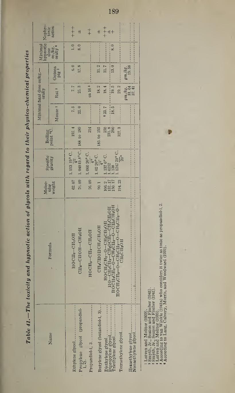
Tab
le 4
1.—
The to
xic
ity a
nd h
yp
no
tic acti
on o
f g
lyco
ls w
ith re
gard
to
th
eir
ph
ysic
o-c
hem
ical
pro
perti
es
189
©•2 . ^ O 0 1 1
1 •
l I
1 • O
1 1
1 1 1 1
in
im
pn
ot
dose
c./
kg
rall
y
t-H 06 1 1 1 1
1 • i* 1
1 1 1 1
!~oo 1 1 1
1 1 1 1
1 1 t 1 1 1 1 1
O 0 1 1
1 1 1
l 1 1
1 1 •
1 1 1
1 1 1 1 1 1
cC co
O 00 1 4 04
1 • 0
1 1
1 . 00 •
I cd 1 1 T-H rH . cd
1 1
1
3 t© rH 1 t-H rH • rH 1 ^05 1
§
3 'S,
O
1 1 1 1
1 1 1 1
1 1 1 1
rH 1
s : ^ 1
O 1 1 1 1
05 CO CO •0 04 Tt<
1 1 iO 04 . Tf< rH
C4 . id 04
co . 1 . C^CO ■44) c3
rH 00 rH
00 rH
• 05 1 H
05 04 ilftft
•—« c3 c3 1 * CO CO cC »ft
4-^ 0 ft O 1 s c3
Wi 1
a 05 CO 7
.5
O
04’
1 1 1
1 1 l co’
1 lO
• 00
1 1 »
1 1 1 1 1 1
3 D 04 r 1 04 1 rH 1 1 1 •rft O
S
1 1 1 k.
1 1 1 1
«0 l 1 1 1 1
1 1 1 1 1
1 1 1 1 1 1 1 1 1 1
33 X —I fto-g
+ + -H +
+ +
+ -H + -U +
+
bcO 3°
"rP 4->
o .s w o pi
05
O 00
00 00
^ 10 t—I 05 04 rH
00
O 00 O CO 10 .05 04 h 04 r-
CO 04 04 CO
<c£ ‘o'£ g 5 ft L-. m m
d
ro CN O 04 00 Tf O 04 CO 04 CO 04 O O !—1 O rH t-h
D
o I®
M
i=l
o o o 04 CO
CO t'-
05 o
o o 05
04 1
CO l>» o O CO 10
CO 04
05
& 3
s (-4 o fa
w o
c*
H o
o
W
a 0 H 0
n
W O
0 «
W W Q
0 w w 0
1 w
0 H
H 0
0 1
0 W 0 m
W L pq
w 0
0 0 O
ww i do o wW L g§M
^g^w W 1 I o o «o I fig L?w 9§w Lo
ifiSgfi t'fggs W « oH 0W0 |0 £9 1 o o«w L 090 w
go a o
Wo W
©
a ©
£
T3
a © ft O
O « t>»
r—1 bu © a 0)
>» 33
o © >> '3)
© a ©
>>^-C ft« O -
ft ft
CO
X} a © 0. o l-H
ft
'O a ©
3 33 O ©
’3) ® a ©
p—« >» 72. ft
ft o o“8
’he be © © 3 S 3 ® © ® *>ftq
3 ft
0 r^_ — J* ®
•2.&-C fifiB
o o >>
r~* bJD
© 3 ©
33 -u © 03 ©
o 8 o 2 p>.>*
"3 wi <d ©
a <=J 8 © p—h T7
t» £5 33 ft
■‘H © ® c3 2 © O
ww
3 © ft o tH ft <Z> ©
M O
© 25 o>
© r-H © w
+* c3
05 05
a> cu ^pa o o w w
co T3 H O <0 o
co ^
§§ o ^ 33 ©
o
05 •» . 1—« >>
^5 05 ^ cohhcOpW >
2’032^’3 '33
t-i 18 133 2 3 a 9P- . w o o
o
■ -1—■ -t—• ■ • O o3 © O ft © ©ft i<0Q02i<
tuo 3 §
t>ft o o
X) T -'3 ■‘H ■*-> 3 ±< >H 3 bflbfi ©ftft © 0 (Z(
3 rT rf 3 'O T3 ©tsti ©St-. >>>>>>00 © J?-*-3 v 0 © H H © O O ft CGCGft-<-<
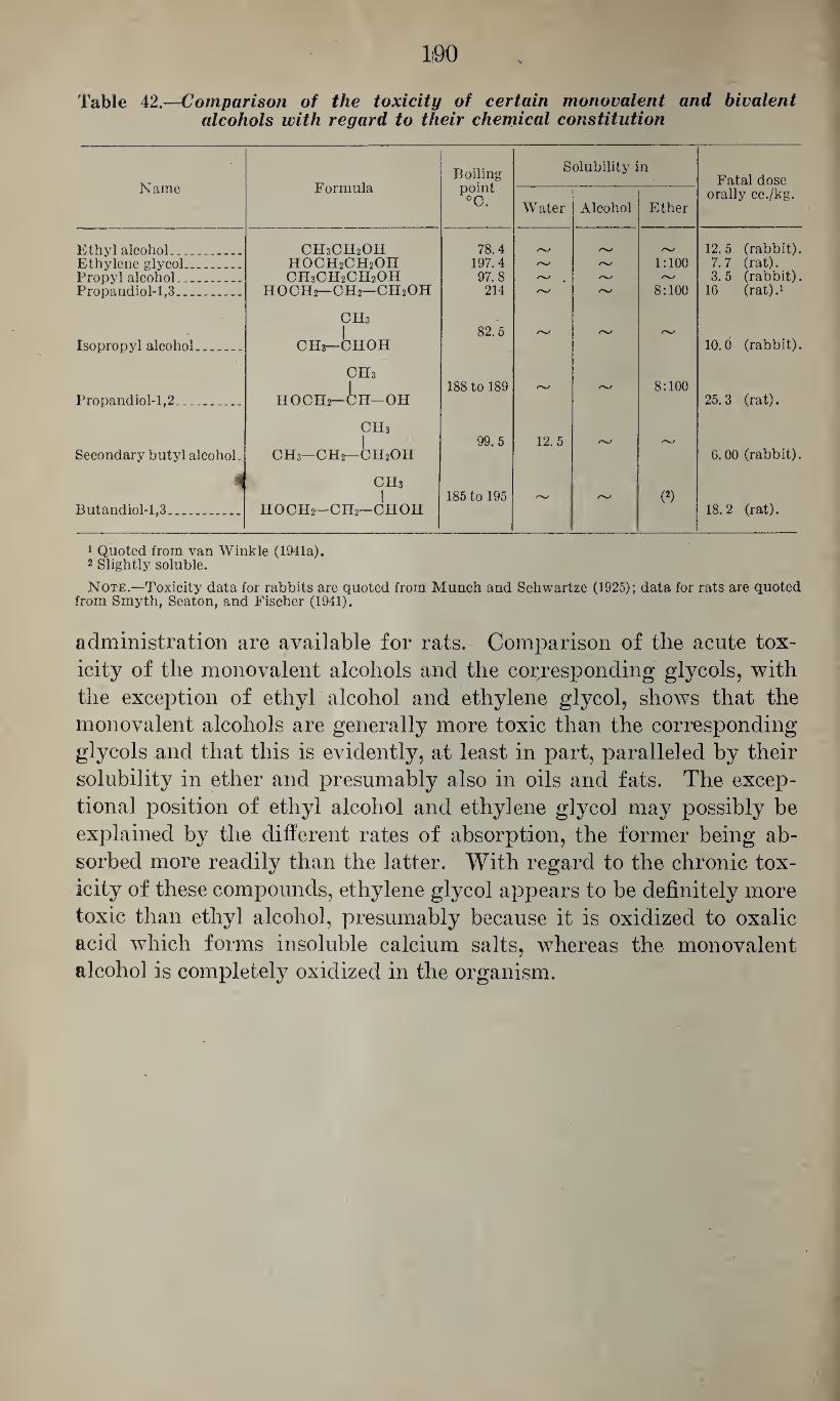
Table 42.—Comparison of the toxicity of certain monovalent and bivalent alcohols with regard to their chemical constitution
Name Formula Boiling point °C.
Solubility in Fatal dose
orally cc./kg. Water Alcohol Ether
Ethyl alcohol_ CH3CH2OH 78.4 12.5 (rabbit). Ethylene glycol—_ HOCH2CH2OH 197.4 /v/ r 1:100 7.7 (rat). Propyl alcohol_ CH3CH2CH2OH 97.8 3. 5 (rabbit). Propandiol-1,3_ HOCH2—CH2—CH2OH 214 /%/ 8:100 16 (rat).1
CHs | 82.5 /v/
Isopropyl alcohol_ - CHs—CHOH 10.0 (rabbit).
CHs 1 188 to 189 rs/ rv 8:100
Propandiol-1,2. .. HOCH2—CH—OH 25.3 (rat).
CH3
| 99.5 12.5 /N/
Secondary butyl alcohol. CH3—CH2—CH2OH 6.00 (rabbit).
i CHs 1 185 to 195 /V/ (2)
Butandiol-1,3.. HOCH2—CH2—CHOH 18.2 (rat).
1 Quoted from van Winkle (1941a). 2 Slightly soluble.
Note.—Toxicity data for rabbits are quoted from Munch and Schwartze (1925); data for rats are quoted from Smyth, Seaton, and Fischer (1941).
administration are available for rats. Comparison of the acute tox¬ icity of the monovalent alcohols and the corresponding glycols, with the exception of ethyl alcohol and ethylene glycol, shows that the monovalent alcohols are generally more toxic than the corresponding glycols and that this is evidently, at least in part, paralleled by their solubility in ether and presumably also in oils and fats. The excep¬ tional position of ethyl alcohol and ethylene glycol may possibly be explained by the different rates of absorption, the former being ab¬ sorbed more readily than the latter. With regard to the chronic tox¬ icity of these compounds, ethylene glycol appears to be definitely more toxic than ethyl alcohol, presumably because it is oxidized to oxalic acid which forms insoluble calcium salts, whereas the monovalent alcohol is completely oxidized in the organism.
C. TRIVALENT ALCOHOLS
Glycerol
The main representative of trihydric alcohols is glycerol or glyc¬ erin.
Chemical characteristics. —Glycerol, HO CH2CH OH CH2OH, has the molecular weight 92.09. It is a colorless liquid with a sweetish taste.
20°
It has the specific gravity 1.260 at C. Its melting point is 17.9° C.
but it solidifies at a much lower temperature, and it boils at 290° C. It is miscible with water and alcohol but is insoluble in ether, fats, and oils. Glycerol is very hygroscopic and can be volatilized with steam.
Identification.—1. Interaction with hydroiodic acid yields allyl io¬ dide (C3H5I) which boils at 101° to 102° C., and, further, isopropyl iodide (C3H7I) which boils at 59.5° C. (Rosenthaler, 1923).
2. When shaken with six times its volume of benzoyl chloride and an excess of sodium hydroxide it forms tribenzoyl glycerol which represents long needles melting at 76° C. (Rosenthaler, 1923).
3. Short heating with 5.1 parts of a-naphthylisocyanate and recrys¬ tallization from pyridine yields glycerol-a-naphthyl urethane, (C10H7NOCO)3OC3H5, which represents needles melting at 279° to 280° C. (Rosenthaler, 1923).
4. It may be identified by dehydration or oxidation to acroleine, or by oxidation to dioxyacetone by heating it with bromine water. The latter method gives the same reactions mentioned for the oxidation product of ethylene glycol (Rosenthaler, 1923).
Uses.—Glycerol is used extensively as a vehicle in the pharmaceu¬ tical and cosmetic industries, as lubricant for specific purposes, as plasticizer, and as starting material in the manufacture of nitroglyc- erol and nitroglycerol explosives.
Determination.—According to Fleury and Fatome (1935), glycerol may be determined by oxidation with an excess of standardized per¬ iodic acid and titrimetrical determination of the remaining periodic acid. In case sugars are present, these first have to be removed by precipitation with barium hydroxide and alcohol and filtration; the excess barium is precipitated with sulfuric acid or carbon dioxide and the alcohol by careful evaporation.
Nicloux (1903b, d) worked out a method for the determination of glycerol in blood which can also be applied to urine. The proteins are precipitated by boiling with 1 percent acetic acid, filtered, and
(191)
192
from the filtrate glycerol is removed by vacuum distillation with
steam. After the distillate is concentrated by careful evaporation,
glycerol is determined by oxidation with chromic acid. If a con¬
centration of 1.9 percent potassium bichromate is used for this pur¬
pose, the number of cubic centimeters used for the oxidation corre¬
sponds approximately to those of glycerol present.
Antiseptic properties.—Glycerol has moderate antiseptic properties.
According to Winslow and Holland (1919), a concentration of 9 per¬
cent has no appreciable effect on the vitality of B. coli, but concentra¬
tions of 28 to 100 percent are increasingly effective.
Absorption, fate, and excretion.—As demonstrated by animal ex¬
periments with mice and rabbits, glycerol evidently is not absorbed
through the intact skin in sufficient quantities to cause toxic effects
(Sander, 1933 and Deichmann, 1941). According to Johnson, Carl¬
son, and Johnson (1933), it is not absorbed readily from the stomach,
but 50 percent of the glycerol introduced into the intestine of anes¬
thetized dogs was absorbed during the first 2 to 3 hours. Similarly,
Hober and Hober (1937) found that 73 percent is absorbed from iso¬
lated loops of rat’s intestine. As indicated by the reports of Mueller
(1894) and Pfannenstiel (1894), sufficient quantities of glycerol may
be absorbed from the pregnant uterus to. cause severe toxic effects.
There is no information available as to what extent glycerol may be
absorbed through the lungs, but, in view of its high boiling point
(290° C.), this possibility appears to be quite remote.
With regard to the fa\te of glycerol in the metabolism, Berthelot
(1857) suggested that certain tissues may metabolize glycerol to glu¬
cose. Cremer (1902) and Luthje (1904) (both quoted from Voegt-
lin, Thompson, and Dunn, 1925) found that the feeding of glycerol
to rabbits increases the blood sugar level. Hockendorf (1909-10) noted
that, in phlorhizinized dogs, glycerol increases the sugar excretion, in¬
dicating that it may be metabolized into sugar; and Chambers and
Deuel (1925), reported that in such animals 55.9 to 98.4 percent of the
glycerol given could be accounted for in the urine as extraglucose.
Similarly, Ferber and Rabinowitsch (1929) found that, in diabetic
patients, the administration of glycerol caused, in 67.5 percent of the
cases, an increase of 20 mg. or more in the blood sugar level. Voegtlin,
Thompson, and Dunn (1925) showed, in rabbits, that the oral and
intraperitoneal administration of glycerol (4 gm. orally) causes a pro¬
gressive rise of the blood sugar, the maximum of 100 percent being
reached in 1 hour, followed by a rapid decline, indicating that glycerol
may be converted into sugar. Johnson, Carlson, and Johnson (1933)
found, in feeding experiments with rats, indications that, within
limits, glycerol may replace carbohydrates in the diet. Whereas Noble
and MacLeod (1923) claimed that, in rabbits, insulin convulsions
were not relieved by the subcutaneous injection of glycerol although
193
there was a very moderate increase of the blood sugar level (from
0.054 to 0.064 percent) shortly after the injection. Voegtlin, Dunn,
and Thompson (1925) found that the administration of adequate
doses of glycerol may relieve the symptoms of hypoglycemia in rats
treated with minimal fatal doses of insulin. It appears, therefore,
that glycerol may be partly converted into glucose in the organism.
Luchsinger (1874) demonstrated that the administration of glycerol
increases the glycogen content of liver and muscle which he interpreted
as assimilation of glycerol to glycogen. Catron and Lewis (1929)
showed that the feeding of 2 cc. of 50 percent glycerol to starved rats
increased the glycogen content of the liver in the course of 1 to 6
hours, progressively, to an average of 142.55 mg. per 100 gm. body
weight (3.24 percent) as compared with 0.09 percent in the controls.
Similar results with rabbits and rats were reported by Lederer (1936)
who also showed that in vitro liver tissue may form glycogen from
glycerol. He found that in a dog the intravenous injection of 1
gm. of glycerol per kg. caused an increase of the glycogen content of the
liver and a rise of the blood sugar level. He noted that four-fifths
of the glycerol injected disappeared from the blood after 5 minutes
and the rest disappeared within 90 minutes. Nicloux (1903a, b, c,
d, e) studied the rate of disappearance of glycerol from blood follow¬
ing its intravenous injection in rabbits and dogs, as illustrated in table
43, which shows that the glycerol level is reduced very rapidly at
first, then gradually, until after 3y2 hours, glycerol has disappeared
almost entirely from the circulating blood. According to the same
author (1903c) the “normal” glycerol level in the blood of dogs is,
on an average, 0.0033 gm. per 100 cc. with 0.0019 and 0.0049 gm. per
100 cc. as extremes; and this is said to be essentially the same whether
the animals are starved or fed a diet rich in fat. Trabucchi (1932)
claimed that the rate of disappearance of glycerol from the blood is
increased after the administration of insulin.
With regard to the excretion of glycerol, the results of different in¬
vestigators do not agree, and this may be attributed, in part, to the
analytical methods used for its determination. Ustimovitsch (1876)
noted in the urine of dogs, after oral administration of glycerol, a re¬
ducing substance other than sugar, and he assumed that this was a
metabolite of glycerol. Plosz (1877) claimed that he had isolated
glycerol aldehyde under similar conditions but the validity of this
statement appears to be questionable. Miura (1911) was unable to
detect glycerol in the urine of dogs and rabbits after oral and sub¬
cutaneous administration, nor did he find evidence that it is excreted
in conjugation with glucuronic acid. Catillon (1877) was able to re¬
cover from human urine 3 to 3.5 gm. after the ingestion of 30 gm. of
glycerol, and he pointed out that with very large doses the excretion is -
not proportionate to the dose but relatively smaller. Leo (1902)
/
194
showed that the excretion of glycerol with the urine depends, in part,
upon the dose administered, in that he found no glycerol in human
urine after the administration of 8.93 gm., only traces after 20.08 gm.
and quantities (0.5 to 1.0 gm.) after the ingestion of 26.76 gm. Simi¬
larly, Munk (1879) found only traces of glycerol in the urine of dogs
after the oral administration of 25 to 30 gm. of glycerol, and Lederer
(1936) detected only small amounts in the urine of man and rabbits.
Tschirwinsky (1879), on the other hand, recovered from 38 to 60 per¬
cent of the glycerol administered from the urine of dogs which had
been fed doses up to 200 gm. mixed with the food; and according to
Arnscliink (1886) 25 percent is excreted by dogs after oral adminis¬
tration. Nicloux (1903b, c) found that, following the intravenous
injection of 2 cc. per kg. in 20 percent solution into a 14 kg. dog,
0.112 gm. was excreted after the first 15 minutes, 3.067 gm. during the
next 75 minutes, 1.409 gm. during the subsequent 67 minutes, and 0.158
during the last 3 hours of the experiment. It appears, therefore, that
the concentration of glycerol in urine depends to a large extent upon
the time of its collection after the administration, and this may offer
an explanation for the contradictory findings reported by different
students of the subject. In the experience of Mcloux (1903b, c) 17
percent of 28 gm. of glycerol given intravenously was recovered from
the urine after 5 hours and 37 minutes, and in another experiment 27.7
percent of 19.5 gm. was recovered after 7 hours and 45 minutes; and
Schubel (1935-36) was able to recover a total of 40 to 50 percent of the
amount of glycerol given intravenously to cats.
The effect of glycerol on various 'physiological functions.—With re¬
gard to the effect of glycerol on the nervous system, Macht and Ting
(1922) found that glycerol has only moderate narcotic properties, in
that doses of 160 mg. per 100 gm. body weight produce the same degree
of depression in rats in a maze as is produced by 80 mg. per 100 gm. of
ethyl alcohol and by 120 mg. per 100 gm. of ethylene glycol. This was
confirmed by Latven and Molitor (1939) who determined the minimal
hypnotic dose in mice as 10 cc. per kg. with oral, 8 cc. per kg. with .
subcutaneous, and 3 cc. per kg. with intravenous administration, the
corresponding figures for ethyl alcohol being 3 cc., 2 cc. and 0.8 cc. per
kg. body weight. Schubel (1936) noted, in frogs, that the depression
is preceded by excitation, and he saw, in mice, after fatal doses, dysp¬
nea, circus movements and convulsions which could be elicited by audi¬
tory stimuli similar to those observed in strychnine poisoning; and
Deichmann (1941) noted inactivity and tremors after oral admin¬
istration to rabbits. Langendorff (1891), who had noted tetanic and
clonic convulsions also, believed these to be partly of central, partly of
peripheral neural, and partly of muscular origin.
Langendorff (1891) believed that glycerol caused irritation of the
peripheral nerves by its dehydrating action, and Santesson (1903)
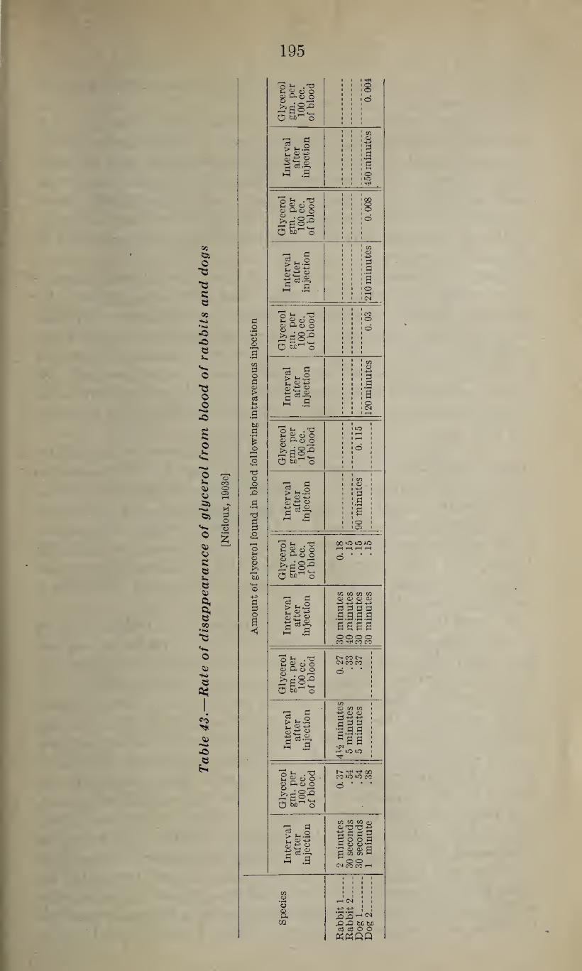
Tab
le 4
3.—
Rate
of
dis
ap
peara
nce o
f g
lycero
l fr
om
blo
od o
f ra
bb
its a
nd d
ogs
[Nic
loux,
1903
e]
195
d o -u © ©
m 3 o a © > c3
bfl | * O
-3 o
3 a •H
3
o © o
'5b
a o a <
_ „ .’O ut g o o " Q< r* “
9 £
8 -
Omho
© o
'S § > £•§ Lj O ©£ s ■4J 93 ©
O ft • 'd u. g O o © Q< O O O . _ rd
oS)Ho
03 # O > ft «rH r, q) +->
|«8 ■3 .g
9^ o o © Q* o o O .
;?a§e O o
'3 § toB ©e 9
c3.; ©
.a
oS .9 i-i 2 o o © a< © o
SgS3 o o
C3
£ © ©£
■a 3
o t! • 'a © a< © o
BaS2 O §>’-'o
'S § >£•£ ®eg os.:
.a
O £ .T)
© Q< © O o i
i^a'S-0 O w)"1 o
a? a to Q
s:«t3 03.®
©;
51
a? a to O > L, O-P © £ « a *•?? ►H .9
TtH o o
d
a s 00 o o
CO 03 -4-3 d .a a o r—I CM
CO o
a o CM
CO ©
-4-9
d .a a o o>
oo *0 *0 AO t-H rH i—l t-H
o
CO CO CO CO © © © © -4-> -4-^ -4-3 -4-3 d d d d .a .a .a .9 asaa o o o o CO ^ CO CO
t> co r- CM CO CO
CO © CO CO
© © 3 -4-9 gdd •pdd
5aa Nf^ r-^- UO »0 Tt-
CO
CO CO CO O © T3 T3 ■a a a a 200d a © ©-g ■s © © a a ot tn m
O O CM CO CO rH
CO ©
8, m .q^hN
rC/rCl beta 03 e8 O O
1196
showed, in frogs, that glycerol lowers the threshold of the peripheral
nerves to electric stimuli. Several investigators (Catillon, 1877; Plosz, 1877; and Arnschink,
1886) noted an increase of the body temperature after administration
of large doses of glycerol, and similar observations were made by
Mueller (1894) and Pfannenstiel (1894) following the intrauterine
injection of glycerol. Johnson, Carlson, and Johnson (1933) noted
no increase of the body temperature following the administration of
smaller doses, and there is no definite explanation of the observations
of the earlier authors. Glycerol causes definite irritation of mucous membranes when in
direct contact, presumably on account of its dehydrating action. Ac¬
cording to Braun and Cartland (1936) the subcutaneous injection
causes severe but transient irritation, and in the experience of Latven
and Molitor (1939) the intradermal injection is distinctly irritant and
application to the rabbit’s eye causes edema and hyperemia. As
pointed out by Flinn (1935), the combustion products of glycerol cause
distinct irritation of the throat when used as hygroscopic agent in
tobacco. With regard to the effect of glycerol on the circulation, Schubel
(1936) found that the energy of the isolated frog’s heart is moderately
reduced by concentrations of 1:1,000 of glycerol in Kinger’s solution,
that concentrations of 1: 50 cause a moderate decrease of systole and
diastole, and that concentrations of 1: 8 cause reversible cardiac arrest.
In the experience of Spealman (1940) 1/10 molar solutions (9:1,000)
do not affect the heart rate, and twice this concentration causes no
change in the weight of the frog’s ventricle, as observed with some of
the monovalent alcohols.
As found by Schubel (1936), concentrations of 1:1,000 to 1:10 do
not affect the diameter of the blood vessels of the perfused frog, but
according to Maignon and Grandclaude (1930) repeated intravenous
injection at the same site may lead to obliteration of the veins.
With regard to the effect of glycerol on the blood pressure, Amidon
(1881) noted that the intravenous injection of 0.5 to 1.0 cc. per kg.
of glycerol causes a fall of the blood pressure accompanied by a dim¬
inution of the force of the heart beat, increased frequency of the heart
rate, and irregularities of the heart action. These effects are not
altered by decerebration, dissection of the vagi and atropinization,
and, therefore, must be due to some peripheral mechanism. Simi¬
larly, Schubel (1936) noted that, in cats, following the intravenous
injection of 0.5 to 1.0 cc. per kg., the blood pressure first was reduced
and then rose again, sometimes above the original level. He be¬
lieved that the primary fall was caused by a temporary impairment
of the heart action and the secondary rise by a hydremic plethora.
However, such changes should not be expected after the oral admin-
197
istration of moderate doses, as indicated by the negative findings of
Johnson, Carlson and Johnson (1933).
With regard to the effect of glycerol on the blood, numerous studies
deal with its hemolytic action. Simon (1915) showed that in vitro
concentrations of 15 to 55 percent cause hemolysis of red blood cells.
However, in the opinion of Schubel (1936) this effect is not as marked
as is usually assumed. He saw no hemolysis with concentrations up
to 40 percent in normal saline, and 50 percent in normal saline caused
hemolysis after 16 to 24 hours whereas the same concentration in
water caused hemolysis within 1 y2 hours. He found aqueous solution
of 2.55 percent glycerol isotonic with 0.9 percent solutions of sodium
chloride, therefore the hemolytic action of glycerol must be due to
other than osmotic properties. Parpart and Shull (1935) studied
the penetration of glycerol into red blood cells, and Jacobs, Glassman,
and Parpart (1935) pointed out that the glycerol hemolysis is greatly
affected by small amounts of copper salts and by changes of the pH,
both factors affecting considerably the permeability of the cells. The
effect of glycerol on leucocytes is much less marked than that of ethyl
alcohol. Baglioni (1927) found that concentrations of 0.25 percent
and 0.50 percent have no effect on the motility and survival of leu¬
cocytes, only 2 percent showing a distinctly injurious effect.
Simon (1920) claimed that, in rabbits, the intravenous injection
of 4.5 gm. per kg. caused stimulation of the leucopoietic apparatus, as
indicated by the increase in the number of erythrocytes and white
blood cells. However, it appears to be questionable whether or not
this cellular increase is due to an actual stimulation of the blood-
forming organs, and it is more likely that this is the result of the
increased viscosity of the blood on account of dehydration, as assumed
by Schubel (1936), especially since Piras (1925) found no indications
of hyperactivity of the bone marrow of such animals. That such
changes are not the result of a permanent damage of the blood-form¬
ing organs is also indicated by the findings of Tompkins (1929) who
noted, following the intraperitoneal injection of 5 cc. of 10 to 25
percent solutions of glycerol, leucocytosis which increased with the
dose, increase of the number of polynuclear neutrophiles, and usually
a slight increase of the monocytes. During the first 12 hours after
the injection there was, at times, a moderate lymphopenia, but usually
the number of lymphocytes remained within normal limits or in¬
creased with that of the leucocytes, and within 48 hours after the
injection the blood picture had returned to normal. After the re¬
peated administration of 12 doses of 2 to 6 cc. per kg. over a period
of 7 weeks, to rabbits, Deichmann (1941) noted a diminution of the
red blood cells from 6 to 4 millions, reduction of the hemoglobin, and
variations in the number of leucocytes; it should be pointed out that
these animals were suffering from hemoglobinuria. Similar changes
198
were observed after intracardiac injections, whereas the intraperi-
toneal injection was followed by a temporary leucocytosis. It should
be pointed out that after oral administration of glycerol neither blood
changes nor hemoglobinuria was observed, and this appears to in¬
dicate that the blood changes observed by various investigators were
due partly to destruction of the blood cells, partly to local irritation
of the tissues at the site of the injection, and partly to thickening of
the blood.
Much attention has been paid to the effect of glycerol on the muscu¬ lature. Husemann and Unmethun (1866) (quoted from Santesson,
1903) noted that the injection of glycerol causes, in frogs, spastic and
convulsive symptoms which they attributed to the dehydrating effect
of this chemical. Amidon (1881) noted that the subcutaneous in¬
jection of glycerol into frogs caused, after a primary irritation, clumsi¬
ness of the movements due to stiffness of the musculature, followed
by fibrillary twitchings of the voluntary muscles and tetanic con¬
vulsions elicited by moderate pressure on the muscle; and that, later
on, even moderate stimulation of this type may elicit general tetanic
convulsions. In contrast to such stimulation, sensory stimuli caused
only attempts at ordinary reflex action. He showed, in addition, that
neither decapitation, pithing of the spinal cord and dissection of the
peripheral nerves, nor curarization prevents the convulsion—which
was confirmed by Schlibel (1935-36)—but that exclusion of the blood
supply by ligation of the vessels would suppress the convulsions in the
ligated limb. He concluded that this effect of glycerol is located in
the muscle tissue proper. Langendorff (1891) made similar observa¬
tions but claimed that decerebration attenuated and curare prevented
these convulsions which, in his opinion, were partly of central origin,
caused by dehydration of the central nervous system. He assumed
that the muscular tetanus was the result of “endogeneous” electric
currents produced by pressure on the muscle. Santesson (1903)
pointed out that the subcutaneous injection of glycerol into frogs
caused the animals to make flash-like extensor movements, but, after
this, permitted only slow adduction of the limbs. In isolated nerve-
muscle preparations of such animals, single electric stimuli of the
nerve caused abnormally high and long-lasting contraction of the
muscle (tetanus) and the plateau of the contracture curve showed,
after some time, a sudden decrease which later flattened out gradually,
this effect being attenuated rather than abolished by curarization. He
concluded that this hyperexcitability was due to an increased excit¬
ability of the nerve endings. He also pointed out that at the height
of the tetanic contraction the muscle was refractory to electric stimu-
- lation and the glycerol contracture represented a real tetanus, whereas
with veratrine, which causes a similar picture, the curve is only a
single twitch contracture; and he believed, like Langendorff (1891),
190
that on account of the increased hyperexcitability, the muscle is stimu¬
lated continuously by its own action currents. Yerzar and Peter
(1925) believed that the prolonged contracture of the glycerolized
muscle is due to an increased permeability of the cell membrane; and
Dux and Low (1921) showed that glycerol causes a high degree of
dehydration of the muscle which may reduce its weight to one-fifth
of its normal value, and that such muscles show a rapid increase and
subsequent decrease of their swelling curve, the former being due to
the avidity of the tissue for water and to the accumulation of lactic
acid.
With regard to the effect of glycerol on smooth muscle structures,
Pelzer (1891) (quoted from Mueller, 1894) showed that, in situ, the
injection of glycerol stimulated the contraction of the uterus. Reach
(1926) found that the local application of glycerol to the upper section
of the duodenum caused relaxation of the sphincter muscles of the
ductus choledochus. This may favorably affect the discharge of bile,
the formation of which, however, is not favorably affected by glyc¬
erol, as was demonstrated by Winogradow (1927). Pal and Prasad
(1935) showed that 0.4, 0.6, 0.8 and 1.0 percent solutions of glycerol
in Ringer’s solution cause a temporary increase of the height of the
contractions of the isolated rabbit’s intestine, the tone showing a pri¬
mary fall and subsequent improvement, and that contractions of the
large intestine were increased proportionately to the concentration of
the solution. Schiibel (1936) found that concentrations of 1:1,000 in¬
crease the peristaltic activity of the isolated ureter, concentrations of
1:100 increase the tone and the number of contractions, and concen¬
trations of 1:20 cause rapid depression of all movements. In his
experience, concentrations of 1:1,000 to 1:150 reduce the tone of the
isolated uterus and the intestine, and such solutions may antagonize
the spasmodic effect^ of barium chloride (1:200) but not that of
physostygmine.
With regard to the effect of glycerol on the kidney function, this
is characterized by a diuretic action and by hemoglobinuria. Usti-
movitsch (1876) found that in dogs the intravenous injection and
the oral administration of glycerol had a definite diuretic effect. Lewin (1879) made the same observation and attributed it to the
hygroscopic properties of glycerol. Cugusi (1922) pointed out that
the effect of glycerol on the kidney function varies with the dose, in
that he found, in rabbits, that the intravenous injection of small and
moderate doses stimulates the renal activity whereas large doses inhibit
the renal function. According to Schiibel (1935-36) the same dose
of glycerol, given intravenously, increases the urinary output to 10
times its normal amount, whereas with oral administration it is in¬
creased only by 3 to 5 times. He believed that the diuretic action
is responsible for the increased viscosity of the blood of such animals.
In the experience of Johnson, Carlson and Johnson (1933) the oral
administration of glycerol always is followed by diuresis, whereas
with intravenous injection, causing a fall of the blood pressure, the
urinary secretion is reduced. In man, 3 doses of 30 cc. each, taken
daily with orange juice after each meal, had no diuretic action.
Rosenfelcl (1924) pointed out that the repeated daily oral adminis¬
tration of glycerol increased considerably the excretion of uric acid in man, and Grabfield and Swanson (1942) believed that uricosuric
action of glycerol is due to a specific effect on the protein metabolism,
whereas the diuretic action is presumably a function of its hyper-
tonicity.
Many experimenters have reported hemoglobinuria following the
administration of glycerol. Amidon (1881), Filehne (1889), Miura
(1911), Simon (1920), Deichmann (1941) and others noted it after
the subcutaneous administration of glycerol; others, like Amidon
(1881), Ustimovitsch (1876) and Deichmann (1941), saw it after
intravenous injection, but it seems that hemoglobinuria has not been
observed after the oral administration of glycerol. Afanassiew
(1883) believed that the hemoglobinuria is not the result of injury
to the kidneys but that it is due to the removal of hemoglobin from
the blood cells because he noted in such cases of hemoglobinuria shadow
cells in the blood and hemoglobin in the serum, so that it appears that
the glycerol hemoglobinuria is of the same type as paroxysmal hemo¬
globinuria in man. As shown by Pfeiffer and Amove (1937), pre¬
medication with ascorbic acid afforded protection against glycerol
hemoglobinuria.
With regard to the effect of glycerol on the metabolism, Catillon
(1877) claimed that glycerol decreased the protein metabolism as in¬
dicated by a reduction of the urea level in the blood and the urea
excretion with the urine. However, Munk (1879) and Lewin (1879)
found no evidence to show that it decreases the protein metabolism and that it has no food value in this respect. Tschirwinsky (1879)
pointed out that findings indicating an increase of the protein metab¬
olism possibly may be overshadowed by a decrease of the urea ex¬
cretion, caused by its dehydrating and diuretic action. As pointed
out above, Grabfield and Swanson (1942) believed that glycerol some¬
how affects the pur in metabolism, as judged by its uricosuric action
and the increased excretion of allantoin in dogs.
It has been pointed out above that glycerol may be substituted,
within limits, for carbohydrates; and this also was shown by Johnson,
Carlson and Johnson (1933).
With regard to the effect of glycerol on the fat metabolism, very
little information is available. Catillon (1877) believed that glycerol
decreases the fat metabolism, and Arnschink (1886) thought that
glycerol may save an equivalent caloric amount of fat.
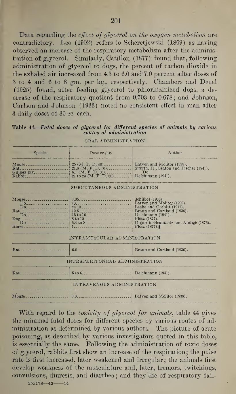
201
Data regarding the effect of glycerol on the oxygen metabolism are
contradictory. Leo (1902) refers to Scheretjewski (1869) as having
observed an increase of the respiratory metabolism after the adminis¬
tration of glycerol. Similarly, Catillon (1877) found that, following
administration of glyercol to dogs, the percent of carbon dioxide in
the exhaled air increased from 4.3 to 6.0 and 7.0 percent after doses of
3 to 4 and 6 to 8 gm. per kg., respectively. Chambers and Deuel
(1925) found, after feeding glycerol to phlorhizinized dogs, a de¬
crease of the respiratory quotient from 0.703 to 0.678; and Johnson,
Carlson and Johnson (1933) noted no consistent effect in man after
3 daily doses of 30 cc. each.
Table 44.—Fatal doses of glycerol for different species of animals by various routes of administration
ORAL ADMINISTRATION
Species Dose cc./kg. Author
Mouse_ 25 (M. F. D. 50)_ Latven and Molitor (1939). Smyth, Jr., Seaton and Fischer (1941).
Do. Deichmann (1941).
Rat_ _ 21.8 (M. F. D. 50)_ Guinea pig...__ Rabbit___
6.1 (M. F. D. 50)1_ 21 to 23 (M. F. D. 60)..
SUBCUTANEOUS ADMINISTRATION
Mouse. .... 0.05____. Schiibel (1936). Do_.__ 10___ Latven and Molitor (1939). Do .. ca 10 _ Leake and Corbitt (1917).
Rat___ 12.5_ Braun and Cartland (1936). Do__-. 15 to 16...__ Deichmann (1941).
Dog ... - 8 to 10.... Plosz (1877).' Do... 6.4 to 8_ Dujardin-Beaumetz and Audigfi (1876).
Horse.. 1__ Plosz (1877) J
INTRAMUSCULAR ADMINISTRATION
Rat.___ _ 6.0___ Braun and Cartland (1936).
INTRAPERITONEAL ADMINISTRATION
Rat.... 5 to 6_ Deichmann (1941).
INTRAVENOUS ADMINISTRATION
Mouse.... 6.0_ Latven and Molitor (1939).
With regard to the toxicity of glyercol for animals, table 44 gives
the minimal fatal doses for different species by various routes of ad¬
ministration as determined by various authors. The picture of acute
poisoning, as described by various investigators quoted in this table,
is essentially the same. Following the administration of toxic doses'
of glycerol, rabbits first show an increase of the respiration; the pulse
rate is first increased, later weakened and irregular; the animals first
develop weakness of the musculature and, later, tremors, twitchings,
convulsions, diuresis, and diarrhea; and they die of respiratory fail-
555178—43-14
202
ure and cardiac arrest. Mice first show gnawing movements of the
jaws; they become dyspneic, discharge soft stools, develop trismus,
show circus movements, and develop spasticity and strychnine-like
convulsions; and with fatal doses death may occur after some time
has elapsed. Dogs may show, in addition, vomiting and diarrhea,
and colic has been reported in horses.
Johnson, Carlson and Johnson (1938) saw no evidence of any toxic
effects in rats from the continued feeding over a period of 40 weeks
of a diet in which 41 percent of the starch had been replaced by
glycerol. With complete replacement of starch by glycerol they noted
a distinct impairment of growth which, however, evidently was caused
not by a toxic action of glycerol but by an inadequate intake of food,
as proved by the fact that the addition of 50 percent of glycerol to the
complete diet not only did not impair the growth but even promoted
it on account of the additional calories furnished. Similarly, Hoick
(1937) saw no unfavorable effects on the growth of young rats fed
a diet containing 20 percent of glycerol.
Cases of glycerol poisoning in humans are exceptional. Robert
(1906) reported on one fatal and one nonfatal poisoning in children
after the ingestion of large doses of glycerol. Jaroschi (1889) re¬
ported on one patient who, after the repeated oral and rectal admin¬
istration of glycerol over a period of 2 weeks, suffered from debility,
vomiting, diarrhea, and muscular cramps which disappeared promptly
after discontinuation of glycerol. In view of the irritant action of
glycerol on mucous membranes, and because of its dehydrating action,
it appears quite possible that the above symptoms may result from
such abuse, but in this specific instance it appears possible that the
glycerol in question was contaminated with appreciable amounts of
arsenic trioxide. Three other cases of glycerol poisoning were re¬
ported by Mueller (1894) and Pfannenstiel (1894). In these in¬
stances, 100 gm. of glycerol had been injected into the pregnant
uterus in order to stimulate uterine contractions. One patient
suffered, after 10 minutes, from vomiting and diarrhea and had, re¬
peatedly, paroxysmal attacks of a fever which was of a different type
from that produced by infection. The second patient became cyanotic,
the pulse was slowed, and there was a short but definite rise of the
temperature. All three patients suffered from more or less severe
hemoglobinuria.
There are no reports on industrial poisonings from the handling of
glycerol, and the possibility of toxic effects from its industrial use
appears to be remote. With regard to 'pathological changes in glycerol poisoning, very
little information is available. Plosz (1877) saw, after oral admin¬
istration of single large doses, hyperemia and inflammation of the
mucous membranes of the gastro-intestinal tract, hyperemia and cloudy
203
swelling of the kidneys, hyperemia of the liver, and hyperemia and
edema of the lungs. Piras (1925) noted, after repeated daily sub¬
cutaneous administrations of 0.5, 1.0, and 2.0 cc. per kg. for 45 days,
hyperemia, hemorrhages and, with the largest dose, fatty degenera¬
tion of the liver; hemorrhages and cloudy swelling of the kidneys, and,
with the largest dose, destruction of the tubular epithelium; and
hyperemia, inflammatory reactions and small hemorrhages of the
intestinal mucosa, even with small doses. The spleen was affected less
and the lungs showed only moderate hyperemia. Other organs, in¬
cluding adrenals and bone marrow, were essentially normal. Similar
but more marked changes were reported by Simon (1915) after the
administration of single fatal doses of glycerol. Maignon and Grand-
claude (1930) studied the local effects of repeated intravenous in¬
jection of glycerol on the vascular walls. They found that in 7 out of
14 experiments the first injection caused no changes of the vascular
walls; in 4, the walls were somewhat thickened; and in 3 instances
they were markedly sclerotic, with complete, or nearly complete,
obliteration of the lumen. These changes became more marked after
the second injection. Microscopically, the layers of the vascular walls
became separated by sclerotic areas and infiltrated with leucocytes, the
endothelium became indistinct and finally fused with a mass in the
lumen, thus obstructing completely the lumen of the vessel, this clot
finally becoming completely organized.
Comparison of the narcotic actions of glycerol and propylene glycol
shows that they are of the same order but that they are considerably
inferior to that of the monohydric alcohol, propanol. This is paral¬
leled by the reduced solubility of propylene glycol and glycerol in fats
and oils which, as pointed out before, is one of the factors controlling
the narcotic action of alcohols. On the other hand, glycerol more
nearly approaches the polyvalent alcohols and sugars, in that, within
certain limits, it may be utilized by the organism and give rise to the
formation of glycogen.
D. THE POLYVALENT ALCOHOLS WITH MORE THAN
THREE CARBONS
The polyvalent alcohols with more than three alcoholic groups are
of such low toxicity that they are of no toxicological importance. The
tetravalent alcohol, erythritol or butantetrol, HOCH2(CHOH)2
ch2oh, is said to cause diuresis in rats in doses of 18 gm. per kg. when
given orally as 50 percent aqueous solution. It is excreted unchanged
with the urine to the extent of 20 to 40 percent, and it does not signi¬
ficantly increase the respiratory quotient of rats nor favor the depo¬
sition of glycogen in the liver (Beck, Carr, and Krantz, 1938). Ac¬
cording to Hober and Hober (1937), it is absorbed from the intestine
of the rat to the extent of 41.4 percent, whereas the corresponding fig¬
ure for glycerol is 73 percent.
The pentavalent alcohol, adonitol, HOCH2(CHOH)3CH2OH, evi¬
dently has not been studied pharmacologically. According to Hober
and Hober (1937) its absorption from the gastrointestinal tract is
slower than that of erythrol.
The hexavalent alcohol, hexanhexol, exists in the form of several
isomeres—mannitol, dulcitol and sorbitol—of the general formula
HOCH2(CHOH)4CH2OH. None of these has a distinct toxic action.
Ellis, Carr, Wiegand and Krantz (1943) found that sorbitol com¬
prising 5 percent of the food intake did not affect deleteriously the
rate of growth, liver glycogen storing capacity, or important metabolic
viscera of white rats through three generations. Smith, Finkelstein
and Smith (1940) noted no toxic manifestations in man after the in¬
travenous injection of 80 gm. sorbitol or mannitol or 50 gm. of dulcitol
over a period of 2 hours. Jaffe (1883) believed that mannitol is not
oxidized readily in the organism of dogs. Carr, Musser, Schmidt,
and Krantz (1933) found that mannitol increases the storage of gly¬
cogen in the liver of rats after oral administration, and Carr and
Krantz (1938) reported that it may cause moderate hyperglycemia in
rabbits. Ellis and Krantz (1941) and Ellis and Carr (1941) found
that it increases the glycogen deposit in the liver of monkeys in con¬
trast to sorbitol, with which such effect is absent. Dulcitol, on the
other hand, increases, to a moderate extent, the glycogen storage in
the liver of monkeys but does not increase the respiratory coefficient
of rats. Spealman (1940) showed that Vio molar solutions of mannitol
slow the beat of the isolated heart and twice this concentration de¬
creases the ventricular weight, presumably by dehydration.
As shown by Hober and Hober (1937), the rate of absorption of
(204)
205
polyhydric alcohols from the intestinal tract decreases with the in¬
crease of their molecular weight. None of them appear to have a
specific toxicologic action and some of them may be converted to
glycogen in the organism. Any effect on the functions of the organ¬
ism presumably are affiliated closely with their osmotic action.
Smith, Finkelstein, and Smith (1940) found that after intravenous
injection in man only 32 percent of 9 gm. sorbitol and 87 percent of
4 gm. dulcitol were excreted with the urine within 10 hours, indi¬
cating that the former is more readily metabolized. Grabfield and
Swanson (1942) showed that the intravenous injection of 15 cc. of
a 50 percent solution of sorbitol caused, in dogs, considerable diuresis
and increase of the excretion of uric acid during the first hour to
the extent of 3 times the original amount. Since mannitol and
dulcitol do not share this uricosuric action, it appears that the optical
configuration plays a role in this respect. The uricosuric action ap¬
pears to be due to a specific effect on the purine metabolism, whereas
the diuretic action is presumably the outcome of the hypertonicity
of the solution of these alcohols.
It appears, therefore, that alcohols with 4, 5, and 6 hydroxylic
groups have none of the properties usually attributed to monovalent
alcohols, and that, with increasing number of hydroxylic groups,
they assume more and more the character of carbohydrates.
BIBLIOGRAPHY
Abbott, A. C.: The influence of acute alcoholism on the normal vital resistance
of rabbits to infection. J. Exptl. Med., 1: 447,1896.
Abelous, E., E. Bardier, and H. Ribaut: Destruction and excretion of ethyl
alcohol in the animal organism. Compt. rend. soc. biol., 55 : 420, 1903.
Abels, J. C.: A simple method for the determination of ethyl alcohol in blood.
Proc. Soc. Exptl. Biol. Med., 34: 346,1936.
Abernethy, R. J., E. R. Russell, and C. H. Thienes: The estimation of ethyl
alcohol in the brain. J. Lab. Clin. Med., 19: 1014,1934.
Abramson, L., and P. Linde: On the passage of ethyl alcohol into the spinal fluid
of man. Arch, intern, pharmacodynamie, 39: 325,1930.
Ackerbauer, C. F., and R. J. Lebowich: A simple and reliable method for the
determination of methyl alcohol and formaldehyde in the air. J. Lab. Clin.
Med., 28: 372, 1942.
Adler, H. F., J. M. Beazell, A. J. Atkinson, and A. C. Ivy: The motor response of
the colon to alcohol. Quart. J. Studies Alcohol, 1: 638,1941.
Afanassiew: On hemoglobinemia. Verhandl. Zweiter Cong. Innere Med., Wies¬
baden, p. 214, April 1883.
Ahlquist, R. P., and J. M. Dille: Reactions of alcohol tolerant rabbits to pento¬
barbital, evipal (barbital derivative), ether, amidopyrine and metrazole.
J. Pharmacol., 70: 301, 1940.
Ajazzi-Mancini, M.: Pharmacology of ethylene glycol. Arch, fisiol., 35: 157,
1935.
Ajazzi-Mancini, M.: Toxicity of ethylene glycol and propylene glycol. Boll. soc.
ital. biol. sper., 14 : 68, 1939.
Ajtay, J.: Ethyl alcohol—mass poisoning in Sandorfalva. Samml. Vergiftung-
sfallen, 4: A163, 1933.
Albertoni, P.: On the formation and distribution of alcohol and aldehyde in the
organism. Ann. chim. farmac. (ser. 4), 6, 250, 1887; abstr., Jahresber,
Fortschr. Tierchem., 17: 75, 1887.
Albertoni, P., and F. Lussana: On alcohol, aldehyde and acetic acid. Lo
Sperimentale, 34: 468, 563, 722,1874.
Alder, P., W. Buschke, and T. Gordonoff: Experimental studies on the toxicity
of methyl alcohol. Arch, intern, pharmacodynamie, 59: 416, 1938.
Alexander, L.: Neuropathological findings in brain and spinal cord of chronic
alcoholic patients. Quart. J. Studies Alcohol, 2: 260,1941. Alexander, L., M. Moore, and T. Leary: Death from poisoning in Massachusetts.
Preprinted from Am. J. Med. Jurisprudence, 2: (No. 7) 1939.
Alexandroff, J., and L. Talpis: On the influence of acute and chronic ethyl alcohol poisoning on the labyrinthine reflexes. Monatschr. Ohrenh., 62:
1196,1928; abstr., Deut. Z. ges. gerichtl. Med., 13 : 276,1929.
Allen, E. B.: Alcoholism as a psychiatric medical problem. N. Y. State J. Med.,
38: 1492, 1938. Allodi, A., and L. Dapra: Alcohol in the blood of normal persons and of patients
with liver disease—before and after meals. Arch. sci. med., 65: 696, 1938.
Amidon, R. W.: A study on the physiological and toxic effects of glycerol in
lower animals. Arch. Med., 6: 107, 1881.
Anker, M.: A case of amylene hydrate poisoning. Therap. Monatschr., 6:
623, 1892.
(206)
207
Anstie: Final experiments on the elimination of alcohol from the body. Prac¬ titioner, 13: 15, 1874.
Arima, Y.: The alcohol level of the blood during experimental functional dis¬
turbances of the organs. Hukuoka Acta Med., 34: 249, 1941, 2d Abstr. Sec.,
34: 25, 1941; Cliem. Abstr., 36: 2016, 1942.
Arkansas law pertaining to antifreeze mixtures. Pub. Health Kept., 46: 2254,
1931.
Arlitt, A. H.: The effect of alcohol on the intelligent behavior of the white rat
and its progeny. Psychol. Monographs, 26: No. 4, 1919; editorial comment:
Hereditary transmission of the effects of alcohol, J. Am. Med. Assoc., 75:
1424, 1920.
Arlitt, A. H., and H. G. Wells: The effect of alcohol on the reproductive tissues.
J. Exptl. Med., 26: 769, 1917.
Arnschink, L.: On the influence of glycerol on the metabolism in the animal
organism and on its nutritional value. Z. Biol., 23: 413, 1886.
Aron, E., and A. Jacob: Sudden death in alcoholics with hemorrhagic pan¬
creatitis. Bull. mem. soc. med. hop. Paris, ser 3, annee, 46: 1321, 1930.
Aschaffenburg, G.: Practical work under influence of alcohol. Kraepelin’s
psychol. Arbeit., 1: 608, 1896.
Asser, E.: On changes of the oxidation of methyl alcohol by other alcohols. Z.
exper. Path. Therap., 15: 322, 1914.
Atkinson, H. Y.: The toxicity of impurities in wood alcohol. I. Allyl alcohol.
J. Pharmacol., 25: 144, 1925.
Atwater, W. O., and F. G. Benedict: Experiments on the metabolism of matter
and energy in the human body. U. S. Dept. Agric., Expt. Sta. Bull., No. 69,1899.
Atwater, W. O., and F. G. Benedict: An experimental inquiry regarding the
nutritive value of alcohol. Mem. Natl. Acad. Sci., 8: 235, 1902.
Baader, E.: Experiences in industrial medicine. Verhandl. deut. Ges. inn. Med.,
45: 318, 1933. Bachem, C.: On the influence of small quantities of alcoholic beverages on the
blood pressure of man. Arch. ges. Physiol. Pfliigers, 114 : 508, 1906.
Bachem, C.: Pharmacological studies on ethylene glycol and its use in pharmacy
and medicine. Med. Klin., 13: 7, 1917.
Bachem, C.: Contribution to the toxicology of halogen alkyls. Arch, exptl.
Path. Pharmakol., 122: 69, 1927.
Backman, E. L.: The effect of ethyl alcohol on the isolated and surviving mam¬
malian heart. Skand. Arch. Physiol., 18: 323, 1906.
Baer, G.: Beitrag zur Kenntnis der acuten Vergiftung mit versehiedenen Alko-
holen. (Acute Poisoning with Various Alcohols.) Inaug. Dissert. Berlin,
1898.
Baer, G.: -Contribution to the knowledge of acute poisoning with different
alcohols. Arch. Anat. Physiol., Physiol. Abt., 283, 1898.
Baglioni, S.: Effect of alcohol, glycerol, and nicotine on surviving leucocytes.
Boll. soc. ital. biol. sper., 2: 976, 1927.
Bahnsen, P., and K. Vedel-Petersen: Alcohol studies. I. Experiments, on drivers
of motor vehicles. J. Ind. Hyg., 16: 304, 1934.
Baldwin, F. M.: Susceptible and resistant phases of the dividing sea-urchin
egg when subjected to various concentrations of lipoid-soluble substances,
especially the higher alcohols. Biol. Bull. 38: 123, 1920; Am. J. Physiol.,
51: 190, 1920. Balthazard, V., and M. Lambert: Toxicological studies on acute alcoholism in
man. Ann. med. ldg., 1: 83, 1921: Ann. hygiene {ser. 4), 34: 207, 1920.
208
Balthazard, V., and G. Larue: Destruction of alcohol in the organism of dogs
habituated to the injection of alcohol. Compt. rend. soc. biol., 84: 343, 1921;
Ann. m6d. leg., 1: 291, 1922.
Barach, A. L.: The action of oxygen in counteracting alcholic intoxication. Am.
J. Physiol., 107: 610, 1934.
Barlow, O. W., A. J. Beams, and H. Goldblatt: Studies on the pharmacology of
ethyl alcohol. I. A comparative study of the pharmacologic effects of grain
and synthetic ethyl alcohols. II. A correlation of the local irritant, anesthetic
and toxic effects of three potable whiskeys with their alcoholic content. J.
Pharmacol., 56: 117, 1936.
Baskerville, C.: Wood alcohol: A report on chemistry, technology, and pharma¬
cology of and the legislation pertaining to methyl alcohol. Second Kept. N. Y.
State Factory Investigating Commission, 1913. Appendix VI, vol. 2: p. 917.
Basset, H. P.: To determine fusel oil in distilled liquors. Ind. Eng. Chem., 2:
389,1910. Battelli, F., and L. Stern: Alcoholase in animal tissues. Compt. rend. soc. biol.,
67: 419, 1909.
Battelli, F., and L. Stern: Production of aldehyde in the course of oxidation of
alcohol by animal tissue alcoholase. Compt. rend. soc. biol., 68: 5, 1910.
Battelli, F., and L. Stern: Effect of anesthetics on oxydones (oxidases). Bio-
chem. Z., 52: 226, 1913.
Baum, F.: On the theory of alcohol narcosis. II. A physical-chemical contri¬
bution to the theory of narcotics. Arch, exptl. Path. Pharmakol., 42: 119,
1899.
Bavis, D. F.: One hundred forty-five drunken drivers: blood and urine alcohol
study. J. Lab. Clin. Med., 25: 823, 1940.
Bavis, D. F., and M. F. Arnholt: The specificity of methods for determining the
concentration of ethyl alcohol in body fluids with special reference to the
Heise method. Nebraska Med. J., 23: 407,1938.
Bavis, D. F., and M. F. Arnholt: Tests of blood and urine of drunken drivers.
Nebraska Med., J., 24: 220, 1939.
Beazell, J. M., A. L. Berman, V. H. Hough, and A. C. Ivy: The effect of acute
alcoholic intoxication on hepatic function. Am. J. Digest. Dis., 9: 82, 1942.
Beazell, J. M., and A. C. Ivy: The influence of alcohol on the digestive tract: a
review. Quart. J. Studies Alcohol, 1: 45,1940.
Beck, F. F., C. J. Carr, and J. C. Krantz, Jr.: Sugar alcohols. 16. The fate of
erythritol and erythritan in the animal body. Quart. J. Pharm. Pharmacol.,
11: 234, 1938.
Benedict, F. G.: The investigation of the influence of alcohol on man, with spe¬
cial reference to psychological effects. J. Am. Med. Assoc., 66: 1424, 1916.
Benedict, H., and B. Torok: Alcohol in nutrition of diabetics. Z. klin. Med., 60: 329,1906; abstr., Biochem. Centr., 5: 771, 1907.
Berggren, S. M.: On the metabolism of n-propyl and n-butyl alcohol in the
organism, with remarks on the oxidation products of alcohols with oxidation
in bichromate-sulfuric acid mixtures. Skand. Arch. Physiol., 78: 249, 1938.
Bergman, P.: On the physiology of the alcohol-chloroform group. Skand. Arch.
Physiol., 17: 60, 1905.
Berman, A. L., E. Snapp, A. C. Ivy, and A. J. Atkinson: The effect of alcohol on
bile volume and constituents in biliary fistula dogs. Quart. J. Studies Alcohol,
1: 645, 1941.
Bernhardt, G.: Isopropyl alcohol as means for the disinfection of the skin.
Deut. med. Wochschr., 48: 68,1922.
Bernheim, F., and P. Handler: Oxidation of some substituted alcohols by rat
liver. Proc. Soc. Exptl. Biol. Med., 46: 470,1941.
209
Berry, D. S.: Alcohol as factor in traffic accidents. Quart. J. Studies Alcohol, 1:
413,1940.
Berry, L. H.: Chronic alcoholic gastritis; evaluation of the concept, with gastro-
scopic studies in one hundred cases. J. Am. Med. Assoc., 117: 2233, 1941.
Bertarelli, E.: Studies on the problem of the toxicity of methyl alcohol. Ann.
Hygiene, 42: 665, 1932. ,
Berthelot: Transformation of mannite and glycerol into a sugar. Compt. rend,
acad. sci., 44: 1002, 1857.
Beyreis, O.: Acute fatal alcohol poisoning. Samml. Vergiftungsfalien, 4: A167,
1933.
Bickel, A., and A. Elkeles: On the influence of alcohol and of some alcoholic
beverages upon the secretion of gastric juice and their effect upon the mecha¬
nism of gastric secretion. Arch. Verdauungskr., 39: 349, 1926.
Bijlsma, U. G.: Isopropyl alcohol. Arch, intern, pharmacodynamie, 34: 204,
1928.
Bills, C. E.: A pharmacological comparison of six alcohols singly and in admix¬
ture, on Paramecium. J. Pharmacol., 22: 49, 1923.
Bills, M., and O. Mankin: Preliminary study of the effects of methyl alcohol
fumes on brightness discrimination in the white rat. J. Comp. Psychol., 1:
495,1921.
Binet, L., and M. Marquis: Effect of ethyl alcohol on the lungs. Bull. mem. soc.
mdd. hop. Paris, ser. 3, 54: 1091,1938.
Binswanger, H.: On the functional pathology of the liver in delirium tremens.
Klin. Wochschr., 11: 1339, 1932.
Binz, C.: The excretion of ethyl alcohol through kidneys and lungs. Arch, exptl.
Path. Pharmakol., 6: 287, 1877.
Binz, C.: Aphorisms and experiments on sedatives. Arch, exptl. Path. Pharma¬
kol., 13: 157, 1880. Birch-Hirschfeld, A.: On the pathogenesis of methyl alcohol amblyopia. Klin.
Monatsbl. Augenheilk., 38: 682, 1900.
Birch-Hirschfeld, A.: Experimental studies on the pathogenesis ' of methanol
amblyopia. Graefe’s Arch. Ophthalmol., 52: 358, 1901.
Bjerre, P.: On the nutritional value of alcohol. Skand. Arch. Physiol., 9: 323,
1899.
Blankenhorn, M. A., and T. D. Spiess: Oral complications of chronic alcoholism:
significance, recognition and treatment. J. Am. Med. Assoc., 107: 641, 1936.
Blatherwick, N. R., L. C. Manwell, and M. L. Long: Effect of oral administration
of alcohol upon blood sugar of rabbits. Am. J. Physiol., 67: 346, 1924.
Blumenthal, A.: On the effect of related chemical compounds on the striated
muscle. Arch. ges. Physiol. Pfliigers, 62: 513, 1896.
Bock, H.: Zur Pharmakologie Ungesattigter Alkohole. (On the pharmacology
of unsaturated alcohols.) Inaug. Dissert. Breslau, 1930.
Bock, J.: Studies on the effect of various poisons on the isolated mammalian
heart. Arch, exptl. Path. Pharmakol., 41: 158, 1898.
Bodlander, G.: The excretion of absorbed alcohol from the organism. Arch,
ges. Physiol. Pfliigers, 32: 398, 1883.
Bohmer, K.: The effect of therapeutic agents on blood alcohol curve. Deut.
Z. ges. gerichtl. Med., 30 : 205, 1938.
Bogen, E.: Drunkenness. A quantitative study of acute alcoholic intoxication.
J. Am. Med. Assoc., 89: 1508, 1927.
Bogen, E., C. W. Muehlberger, D. F. Davis, and H. A. Heise: The effect of alcohol
on driving skill. J. Am. Med. Assoc., 115: 2198, 1940.
210
Bokorny, T.: On the effect of methyl alcohol and other alcohols on green plants
and micro-organisms. Centr. Bakt., Abt. 2, 30: 53, 1911.
Bokorny, T.: On the effect of different compounds on the germination of plant
seeds and the stimulation of growth by some of these. Biochem. Z., 50: 87,
1913.
Boles, R. S., and R. S. Crew: Observations on chronic alcoholism and cirrhosis
of liver. Quart. J. Studies Alcohol, 1: 464, 1940.
Bongers, P.: On the excretion of compounds foreign to the body into the
stomach. Arch, exptl. Path. Pharmakol., 35: 415, 1895.
Bonnet, M. R., and P. Lelu: The comparative effect of different alcohols on
the excitability of nerves, and muscles. Arch, intern, pharmacodynamie, 46:
13, 1933.
Bornstein, A., and G. Budelmann: Excretion of alcohol and ether through the
kidney. Arch, exptl. Path. Pharmakol., 150: 47, 1930.
Boruttau, H.: The use of isopropyl alcohol for hygienic and cosmetic purposes.
Deut. med. Wochschr., 47: 747, 1921.
Bournot, K.: On the enzyme of chelidonium seeds. Comparison of different
alcohols regarding their behavior in the enzymatic ester synthesis with oleic
acid. Biochem. Z., 65: 150, 1914.
Bowers, R. V., W. D. Burleson, and J. F. Blades: Alcohol absorption from the
skin in man. Quart. J. Studies Alcohol, 3: 31, 1942.
Bowman, K. M., and E. M. Jellinek: Alcoholic mental disorders. Quart. J.
Studies Alcohol, 2: 312, 1941.
Bowman, K. M., J. Wortis, L. L. Orenstein, and W. Goldfarb: The sugar tolerance
of alcoholic patients. Proc. Soc. Exptl. Biol. Med., 42: 37, 1939.
Brandini, G.: Action of ethyl alcohol on the isolated heart of mammals. Arch,
ital. biol., 49: 275, 1908.
Braun, H. A., and G. F. Cartland: The toxicity of propylene glycol. J. Am.
Pharm. Assoc., 25: 746, 1936.
Brechmann, H. J.: Studies on the respiratory quotient following intake of
alcohol while working. Z. Biol., 83: 325, 1925.
Brechmann, H. J.: Respiratory experiments following ingestion of fructose,
dextrose and alcohol during rest and during exercise. Z. Biol., 86: 447, 1927.
Brekke, A.: Two cases of ethylene glycol poisoning; acute uremia cured by
unilateral decapsulation of kidney. Norsk. Mag. Laegervidensk., 91: 381,
1930.
Breyer, H.: On the action of different monovalent alcohols on the ciliary epithe¬
lium and motor nerve fibers. Arch. ges. Physiol. Pflugers, 99: 481, 1903.
Brooks, C.: The action of alcohol on the normal, intact, unanesthetized animal.
J. Am. Med. Assoc., 55: 372, 1910.
Brown, C. L. M.: Some properties of glycols with special reference to the use
of propylene glycol as a solvent in pharmaceutical preparations. Pharm. J.,
134: 794, 1935.
Brown, M. R: Alcoholic polyneuritis: an evaluation of the treatment at the
Boston City Hospital from 1920 through 1938. J. Am. Med. Assoc., 116: 1615, 1941.
Browning, E.: Toxicity of Industrial Organic Solvents. Medical Research Coun¬
cil, Industrial Health Research Board, Report No. 80. H. M. Stationery Office, London, 1937.
Bruckner, H.: On the present state of methyl alcohol poisoning, with special
reference to its importance for industries. Zentr. Gewerbehyg. Unfallver-
hiit., N. F. 1: 17, 1924.
Bruger, M.: The effects of alcohol on the normal and pathologic kidney; a
review. Quart. J. Studies Alcohol, 1: 85, 1940.
211
Bruger, M., S. A. Localio, and N. W. Guthrie: Effect of alcohol on normal kidney
and kidney of Bright’s disease. J. Am. Med. Assoc., 112: 1782, 1939.
Brunntaler, J.: The toxic effects of formaldehyde. Arztl. Sachverst.-Z., 19:
142, 1913.
Brunton, T. L., and S. Martin: Action of alcohols and aldehydes on proteid
substances. J. Physiol., 12: 1, 1890.
Buchner, H., F. Fuchs, and L. Megele: Effects of methyl, ethyl and propyl
alcohols on the arterial blood stream with external use. Arch. Hyg., 40:
347, 1901.
Budelmann, G.: On the effect of alcohol on the vascular system. Arch, exptl.
Path. Pharmakol., 151: 65, 1930.
Bujrdi, I,.: Experimental studies on the influence of some drugs on the blood
sugar concentration. Z. ges. exptl. Med., 71: 480, 1930.
Burger, G. C. E., and B. H. Stockmann: On urobilinuria as sequela, of inhalation
of organic solvents. Zentr. Gewerbehyg. Unfallverhiit., N. F. 9: 29, 1932.
Burhans, E. C.: Methyl alcohol poisoning: pathological study of 11 fatal cases.
Ill. Med. J., 57: 260, 1930.
Burtner, It. R., and G. Lehmann: The hypnotic properties of some derivatives of
trihalogenated alcohols. J. Pharmacol., 63: 183, 1938.
Burton-Opitz, R.: The changes of the viscosity of the blood produced by alcohol.
J. Physiol., 32: 8, 1904.
Burton-Opitz, R.: The effect of isopropyl alcohol on the heart action. Arch,
neerland. physiol., 7: 157, 1922.
Busch, A.: On the influence of alcohol on clearness and extension of the optical
consciousness. J. Psychol. Neurol., 17: 63, 89,1910; 18: 36, 1911.
Busquet, H.: Convulsant and decurarizing action of ethylene glycol. Compt.
rend. soc. biol., 128: 56, 1938.
Butler, T. C.: The effect of oxygen inhalation on the course of acute alcoholic
intoxication. J. Pharmacol., 58: 68, 1936.
Butler, T. C., and H. L. Dickison: Anesthetic activity of optical antipodes. I.
The secondary butyl alcohols. J. Pharmacol., 69: 225, 1940.
Cameron, A. T.: Alcohol and automobile driving. Canad. Med. Assoc. J., 43:
46, 1940.
Cannon, P. R.: Elixir of Sulfanilamide-Massengill. IY. Pathological effects fol¬
lowing the ingestion of diethylene glycol, Elixir of Sulfanilamide-Massengill,
“synthetic” elixir of sulfanilamide and sulfanilamide alone. J. Am. Med.
Assoc., 109: 1536, 1937.
Canzanelli, A'., R. Guild, and D. Rapport: The use of ethyl alcohol as fuel in
muscular exercise. Am. J. Physiol., 110: 416, 1934.
Caps, C. A., and G. H. Coleman: Influence of alcohol on prognosis of pneu¬
monia in Cook County Hospital; a statistical report. J. Am. Med. Assoc.,
80: 750, 1923.
Carlier, E. W.: Notes on the physiology of some of the allyl compounds. Brit.
Med. J., 2: 609, 1911.
Carlson, A. J., N. Kleitman, C. W. Muehlberger, F. C. McLean, H. Gullicksen,
and R. B. Carlson: Studies on the possible intoxicating action of 3.2 percent
beer. The University of Chicago Press, 1934.
Carpenter, T. M.: Human metabolism with enemata of alcohol, dextrose and
levulose. Carnegie Institution of Washington Publ. No. 369, 1925.
Carpenter, T. M.: Ethyl alcohol in fowls after exposure to alcohol vapor. ,1.
Pharmacol., 37: 217, 1929.
Carpenter, T. M.: The effect of muscular exercise on the metabolism of ethyl
alcohol. J. Nutrition, 6: 205, 1933.
212
Carpenter, T. M.*: Metabolism of alcohol: a review. Quart. J. Studies Alcohol,
li 201, 1940.
Carpenter, T. M., and R. C. Lee: (a) The effect of galactose on the metabolism
of ethyl alcohol in man. J. Pharmacol., 60: 254, 1937.
Carpenter, T. M., and R. C. Lee: (&) The effect of glucose on the metabolism
of ethyl alcohol in man. J. Pharmacol., 60 : 264, 1937.
Carpenter, T. M., and R. C. Lee: (c) The effect of fructose on the metabolism
of ethyl alcohol in man. J. Pharmacol., 60: 286, 1937.
Carpenter, T. M., R. C. Lee, and M. Burdett: The effect of muscular exercise
on the disappearance of ethyl alcohol in man. Am. J. Physiol., 105: 17, 1933.
Carr, C. J., and J. C. Krantz, Jr.: Sugar alcohols. XII. The fate of polygalitol
and mannitol in the animal body. J. Biol. Chem., 124 : 221, 1938. Carr, C. J., and J. C. Krantz, Jr.: The fate of dulcitol and dulc-itan in the animal
body. J. Biol. Chem., 107: 371, 1934.
Carr, C. J., R. Musser, J. E. Schmidt, and J. C. Krantz, Jr.: The fate of mannitol
and mannitan in the animal body. J. Biol. Chem., 102: 721,1933.
Carroll, F. D., and R. Goodhart: Acute alcoholic amaurosis. Arch. Ophthalmol.,
20 : 797, 1938.
Cassinis, U., and L. Bracaloni: Blood and urine alcohol level in rest and exercise.
Arch, fisiol., 29: 19, 1930.
Cates, H. B.: (a) Acute hepatitis of alcoholism; clinical and laboratory study.
Ann. Internal Med., 15: 244, 1941.
Cates, H. B.: (b) Relation of liver function to cirrhosis of liver and to alcohol¬
ism ; comparison of results of liver function tests with degree of organic
change in cirrhosis of liver, and with results of such tests in persons with
alcoholism without cirrhosis of the liver. Arch. Internal Med., 67: 383, 1941.
Catillon, A.: Studies on the physiological and therapeutic properties of glycerol. Arch, physiol, norm, path., ser. 2, 4: 83, 1877; abstr., Compt. rend. acad. sci.,
84: 194,1877.
Catillon, A.: On the physiological and therapeutic properties of glycerol; analy*
sis of breath after ingestion of glycerol. Bull. mem. soc. therap., ser. 2, 4:
161, 1877; Arch, physiol, norm, path., ser. 2, 5: 144, 1878, abstr., Compt. rend,
soc. biol., ser. 6, 4: 453, 1879.
Catron, L. F., and H. B. Lewis: The formation of glycogen in the liver of the
young white rat after the oral administration of glycerol. J. Biol. Chem., 84:
553.1929.
Cavalli, F.: Pharmacodynamic action of derivatives of the octyl series. Arch,
intern, pharmacodynamie, 64: 354,1940.
Cerabona, M.: Glycol poisoning. V. The electrolyte equilibrium in the blood in
ethylene glycol poisoning. Fol. rued., 25: 1139,1939.
Chabanier, H., and E. Ibarra-Loring: On the mode of excretion of ethyl and
methyl alcohol through the kidney. Compt. rend. soc. biol., 79: 8, 1916.
Chambers, W. H., and H. J. Deuel, Jr.: Animal calorimetry. 30. The metabolism
of glycerol in phlorhizin diabetes. J. Biol. Chem., 65 : 21,1925.
Chapin, R. M.: Improved Denig£s test for the detection and determination of
methanol in the presence of ethyl alcohol. Ind. Eng. Chem., 13: 543, 1921.
Chapman, G. H.: The influence of certain capillary-active substances omenzyme
activity. Intern. Z. physikal. chem. Biol., 1: 293, 1914.
Chauchard, A., B. Chauchard, and S. Kajiwara: (a) The relation of the
chronaxies of cortical motor zones of flexion and extension of limbs. Compt.
rend. soc. biol., 105: 776, 1930.
Chauchard, A., B. Chauchard, and S. Kajiwara: (b) Effect of alcohol on the
excitability of motor neurons of the cortex. Compt. rend. soc. biol., 105:
778.1930.
A
213
Chauveau, A.: Does muscular exercise utilize alcohol substituted for a part of the nutritional ration as source of energy? Compt. rend. acad. sci., 132: 65, 1901.
Chester, A., G. C. La Belle, and H. E. Himwich: The relative effects of toxic doses of alcohol on fetal, newborn and adult rats. Quart. J. Studies Alcohol, 3: 1, 1942.
Chittenden, R. H., L. B. Mendel, and H. C. Jackson: A further study of the influence of alcohol and alcoholic drinks upon digestion, with special refer¬ ence to secretion. Am. J. Physiol., 1: 164, 1898.
Christiansen, J.: On the theory and practice of alcohol disinfection. Z. physiol. Chem., 102: 275, 1918.
Clark, A. J.: The mode of action of alcohols and narcotics on the frog’s ventricle. • Arch, intern, pharmacodynamie, 38: 101,1930.
Clark, B. B., and R. W. Morrissey: Effect of sodium citrate and sodium bicarbo¬ nate on ethyl alcohol acidosis. Proc. Soc. Exptl. Biol. Med., 38: 734, 1938.
Clark, B. B., R. W. Morrissey, and J. F. Fazekas: Insulin and the oxidation of ethyl alcohol by excised diabetic liver tissue. Science, new ser. 88: 285, 1938.
Clark, B. B., R. W. Morrissey, J. F. Fazekas, and C. S. Welch: The role of insulin and the liver in alcohol metabolism. Quart. J. Studies Alcohol, 1: 663, 1941.
Clausen, S. W., B. B. Breese, W. S. Baum, A. B. McCord, and J. O. Rydeen: Effect of alcohol on vitamin A content of blood in human subjects. Science, new ser. 93: 21, 1941.
Clerc, A., R. Paris, and J. Sterne: Effect of octyl alcohol on the arterial blood pressure and on the surface tension of blood in animals. Compt. rend. soc. biol., Ill: 774, 1932.
Clopatt, A.: On the effect of alcohol on the metabolism of men. Skand. Arch. Physiol., 11: 354, 1901.
Clowes, G. H. A., and A. K. Kletch: Influence of (H) concentration on the anesthetic value of a series of general and local anesthetics and hypnotics. Proc. Soc. Exptl. Biol. Med., 29: 312, 1931.
Cole, W. H., and J. B. Allison: (a) Chemical stimulation by alcohols in the barnacle, the frog and planaris. J. Gen. Physiol., 14: 71, 1930.
Cole, W. H., and J. B. Alison: (b) The stimulating efficiency of the normal pri¬ mary alcohols. Proc. Soc. Exptl. Biol. Med., 27: 688, 1930.
Cololian, P.: Toxicity of alcohols for fishes. J. physiol, path. g£n., 3: 535, 1901.
Colson, Z. W.: The effect of alcohol on vision: experimental investigation. J. Am. Med. Assoc., 115: 1525, 1940.
Connor, C. L.: The etiology and pathogenesis of alcoholic cirrhosis of liver. J. Am. Med. Assoc., 112: 387, 1939.
Connor, C. L.: Some effects of chronic alcohol poisoning in rabbits. Arch. Path., 30: 165, 1940.
Connor, C. L., and I. L. Chaikoff: Production of cirrhosis in fatty livers with alcohol. Proc. Soc. Exptl. Biol. Med., 39: 356, 1938.
Constantin, J. D.: General anaesthesia by intravenous injection of ethyl alcohol. Lancet, 1: 1247, 1929.
Cook, C. A., and A. H. Smith: The determination of isopropyl alcohol in the presence of acetone in urine. J. Biol. Chem., 85: 251, 1929.
Cook, E. N., and G. E. Brown: The vasodilating effects of ethyl alcohol on the peripheral arteries* Proc. Staff Meetings Mayo Clinic, 7: 449, 1932.
Cornish, R. E., H. D. Draper, and J. Finn., Jr.: Validity of post mortem tests for alcoholism. Am. J. Med. Jurisprudence, 1: 86, 1938.
Cortese, O.: Ethylene glycol poisoning. IX. Rehberg’s tests in experimental ethylene glycol poisoning. Ras. med. ind., 12: 377, 1941.
214
Coupin, H.: On the assimilation of alcohols and aldehydes by Sterigmatocystis
nigra. Compt. rend. acad. sci., 138: 389, 1904.
Coward, H. F., and G. W. Jones: Limits of inflammability of gases and vapors.
U. S. Bnr. Mines Bull. No. 279, 1939.
Cugusi, C.: On the diuretic action of glycerol as compared with that of ethyl
alcohol. Arch. farm, sper., 34: 150, 161, 1922.
Cushny, A. R.: On the exhalation of drugs by the lungs. J. Physiol., 40:17,1910.
Dakin, H. D.: Experiments bearing on the mode of oxidation of simple aliphatic
substances in the animal organism, (acetic acid, glycollic acid, glyoxylic acid,
oxalic acid, glycocoll and glycol.) J. Biol. Chem. 3: 57, 1907.
Danopoulos, E.: The alimentary alcoholemic curve in blockage of the reticulo¬
endothelial system. Z. ges. exptl. Med., 103: 212, 1938.
Danopoulos, E.: (a) Studies on the alcohol metabolism. I. On the influence of
vitamins on the course of the alimentary alcoholemic curve, with special
reference to vitamin C. Z. ges. exptl. Med., 106: 366, 1939.
Danopoulos, E.: (b) Studies on alcohol metabolism. II. Comparative experi¬
ments on the alcoholemic curve with oral and intravenous administration of
alcohol in normal dogs. Z. ges. exptl. Med., 106 : 374, 1939.
Davis, P. A., F. A. Gibbs, H. Davis, W. W. Jetter, and L. S. Trowbridge: The
effect of alcohol upon the electroencephalogram (brain waves). Quart. J.
Studies Alcohol, 1 - 626, 1941.
Decker, H.: Determination of the alcohol content of the blood by the use of
the interferometer. Deut. Z. ges. gerichtl. Med., 33: 33, 1940.
Deckert, W.: Relation between blood alcohol findings, degree of drunkeness,
and age. Evaluation of practical results of experiments. Deut. Z. ges.
gerichtl. Med., 32: 51, 1939.
de Crinis, M.: Alcohol intoxication and responsibility. Med. Welt, 13: 889, 1939.
Deichmann, W.: Glycerol: Effects upon rabbits and rats. Ind. Med., 10: Ind.
Hyg. Sec., 5, 1941.
Delearde, A.: Contribution to the study of experimental alcoholism and its
influence on immunity. Ann. Inst. Pasteur, 11: 837, 1897.
de Montmollin, R.: Regarding urobilinuria in alcoholic patients. Schweiz, med.
Woclischr., 68: 1165, 1938.
Denig&s, G.: Study of methyl alcohol in general and especially in the presence
of ethanol. Compt. rend. acad. sci., 150: 832, 1910.
Dervieux, Szumlanski, and H. Desoille: Method for determining, on the cadaver
of an infant, the degree of alcohol poisoning of the wet-nurse. Ann. m4d. I6g.,
9: 501, 1929.
deSchweinitz, G. E.: A case of methanol amaurosis, the pathway of entrance
being the lungs and the cutaneous surface. Ophthalmol. Record, 10: 289, 1901.
deSchweinitz, G. E.: The hazards of antifreeze methanol. Trans. Coll. Phys.
Phila., ser. 3, 53: 221, 1931. (Discussion of article by Trumper, 1931.)
Diack, S. L., and H. B. Lewis: Studies in the synthesis of hippuric acid in the
animal organism. VII. A comparison of the rate of elimination of hippuric
acid after the ingestion of sodium benzoate, benzyl alcohol, and benzyl esters
of succinic acid. J. Biol. Chem., 77: 89,1928.
di Cristina, G., and F. Pentimalli: Influence of alcohols on the dynamic action
of the heart of the turtle, Emys europaea. Arch, fisiol., 8: 131, 1910.
Dieballa, G.: On the quantitative effect of various substances of the alcohol and
chloroform series on the frog heart. Arch, exptl. Path. Pharmakol., 34: 137,
1894.
Dille, J. M.: Effect of ethylene glycol on serum calcium of the rabbit. J. Am.
Pharm. Assoc., 23: 202,1934.
215
di Priseo, L., and M. Swarc: Glycol poisoning. VI. Morphologic alterations of
the blood in chronic ethylene glycol poisoning. Fol. med., 26: 287, 1940.
Dixon, W. E.: The action of alcohol on the circulation. J. Physiol., 35: 346, 1907.
Dixon, W. E., and F. Ransom: The immediate action of volatile substances.
J. Physiol., 54: 384,1921.
Dodge, R., and F. G. Benedict: Neuro-muscular effects of moderate doses of
alcohol. Proc. Natl. Acad. Sci., 1: 605, 1915.
Dold, H.: On the effect of ethyl alcohol and related alcohols on the frog heart.
Arch. ges. Physiol. Pfliigers, 112: 600,1906.
Domenici, F.: The possibility that drugs taken during life may cause errors in
the determination of alcohol in the blood of cadavers. Boll. soc. ital. biol.
sper., 14: 314, 1939.
Donley, D. E.: Toxic encephalopathy and volatile solvents in industry. J. Ind.
Hyg. Toxicol., 18: 571,1936.
Doolittle, A. K.: Lacquer solvents in industrial use. Ind. Eng. Chem., 27: 1169,
1935.
Dragstedt, C. A., J. S. Gray, A. H. Lawton, and M. Ramirez de Arellano: Does
alcohol stimulate gastric secretion by liberating histamine? Proc. Soc. Exptl.
Biol. Med., 43: 26, 1940.
Dreser, H.: On heart action and heart poisons. Arch, exptl. Path. Pharmakol.,
24:221,1887.
Ducceschi, V.: On the genesis of alcohol poisoning. Arch, fisiol., 16, 117, 231,
1917/18; abstr., Zentr. Biochem. Biophys., 20: 175,1919.
Duchovnikova, L., and I. Robinson: On the localization of pathological anatomi¬
cal lesions in the central nervous system in experimental alcoholism. Psi-
chogigien. nevrol. issledovaniza, p. 26, 1928; abstr., Deut. Z. ges. gerichtl. Med.,
13: 26,1929. Dujardin-Beaumetz, and Audige: On the toxic properties of fermentation alco¬
hols. Compt. rend. acad. sci., 81: 192,1875.
Dujardin-Beaumetz, and Audige: On the toxic properties of glycerol. Bull. mem.
soc. th6rap., ser. 2, 3: 88, 1876.
Dupre, A.: On the elimination of alcohol. Proc. Roy. Soc. London, 20: 268, 1872.
Duquesnois, P., and P. Revel: Occupational poisonings from vapors of some
esters used as solvents. J. pharm. chim., 19: 590,1934.
Durig, A.: Contributions to the physiology of man in high altitudes. III. On
the effect of alcohol on climbing activity. Arch. ges. Physiol. Pfliigers, 113:
341,1906. Dux, P., and A. Low: On the osmotic behavior of dehydrated glycerol poisoned
frog muscles and on the shrinkage of muscle protein. Biochem. Z., 125: 222,
1921. Dybing, O., and E. W. Rasmussen: Resorption of ethyl alcohol from the stomach
and intestine of the rat. Biochem. Z., 306: 337,1940.
Dyson, G. M.: Toxicity of ethylene glycol, propylene glycol and diethylene diox¬
ide (dioxane). Ind. Chemist Chem. Mfgr., 10: 102, 1934.
Eckhard, C.: Does the subcutaneous injection of glycerol antagonize the sugar puncture? Centr. med. Wissenschaft., 14: 273, 1876.
Editorial: Alcohol and muscular exercise. J. Am. Med. Assoc., 101: 1002, 1933.
Editorial: Food value of ethyl alcohol. J. Am. Med. Assoc., 105: 1775, 1935.
Editorial: A skin test for tolerance to alcohol. J. Am. Med. Assoc., 112: 2069,
1939. Editorial: Tests for alcoholic intoxication. J. Am. Med. Assoc., 114: 415, 1940.
Efron, J.: Contributions to the general physiology of nerves. Arch. ges. Physiol.
Pfliigers, 36: 467, 1885.
216
Egg, C.: On the knowledge of methyl alcohol poisoning. Schweiz, med. Wochschr.,
57: 5, 1927.
Eggleton, M. G.: (a) Determination of metabolic rate of alcohol. J. Physiol., 98: 228, 1940.
Eggleton, M. G.: (b) Some factors affecting metabolic rate of alcohol. J. Physiol.,
98: 239, 1940.
Eggleton, M. G.: The effect of alcohol on the central nervous system. Brit. J.
Psychol., 32: 52, 1041.
Eggleton, M. G.: The diuretic action of alcohol in man. J. Physiol., 101: 172, 1942.
Eiger, M.: The hemolytic and hemagglutinating action of ethyl and methyl
alcohol and of acetone. Hemolysis and hemagglutination under the infleunce
of heat. Z. Immunitatsforschg., 9: 238, 1911.
Eisenberg, A. A.: Visceral changes in wood alcohol poisoning by inhalation. Am.
J. Pub. Health, 7: 765, 1917.
Eisenmenger, W. S.: Toxicity of some aliphatic alcohols. Plant Physiol., 5:
131, 1930.
Elbel, H.: Studies on the usefulness of alcohol determinations according to
Widmark. Deut. Z. ges. gerichtl. Med., 25: 124, 1935.
Elbel, H.: New contributions to the blood alcohol question. Deut. Z. ges. ger¬
ichtl. Med., 30: 218, 1938.
Elbel, H.: Fatal alcohol poisoning, especially in children: case. Deut. Z. ges.
gerichtl. Med., 33: 44, 1940.
Elbel, H., and G. Lieck: Alcohol resorption following intake of food. Deut.
Z. ges. gerichtl. Med., 26: 270, 1936.
Elderton, E. M., and K. Pearson: A first study of influence of parental alcoholism
on the physique and ability of offspring. Francis Galton Laboratory for
National Eugenics, Eug. Lab. Mem. No. 10, 1910.
Elhardt, W. P.: Effect of ethyl alcohol on the growth of chicks. Am. J. Physiol.,
92: 450, 1930.
Elhardt, W. P.: The effect of methyl, propyl and butyl alcohol on the growth of
white leghorn chicks. Am. J. Physiol., 100 : 74, 1932.
Ellis, F. W., and C. J. Carr: A pharmacologic and toxicologic study of mannitol
and sorbitol. J. Pharmacol., 72: 13, 1941.
Ellis, F. W., C. J. Carr, E. J. Wiegand, and J. C. Krantz, Jr.: Sugar alcohols.
XXIII. Effect of sorbitol feeding on successive generations of rats. Proc.
Soc. Exptl. Biol. Med., 52: 260, 1943.
Ellis, F. W., and J. C. Krantz, Jr.: Sugar alcohols. XXII. Metabolism and tox¬
icity studies with mannitol and sorbitol in man and animals. J. Biol. Chem.,
141: 147, 1941.
Elvove, E.: A note on the detection and estimation of small amounts of methyl
alcohol. Ind. Eng. Chem., 9: 295, 1917.
Embden, G., and K. Baldes: On the transformation of acetaldehyde into ethyl
alcohol in the animal organism. Biocliem. Z., 45: 157, 1912.
Emerson, G. A.: Effects of various anesthesias on autoxidation rate of sur¬
viving brain tissue. Proc. Soc. Exptl. Biol. Med., 33: 171, 1935.
Emiliani E., and A. Panza: Influence of wine and of some substances con¬
tained in it on digestive processes. Ann. chim. applicata, 30: 429, 1940.
Eppel, A.: Blood alcohol content of pedestrians killed in traffic accidents through
their own fault. Deut. Z. ges. gerichtl. Med., 32: 312, 1940.
Epstein, F.: On the question of disinfection with alcohol. Z. Hygiene Infek-
tionskrankheiten, 24: 1, 1897.
Ewing, P. L.: Alcohol metabolism in artificial fever. Quart. J. Studies Alcohol,
1: 483, 1940.
217
EjTquem: On the danger of alcohol vapors in the manufacture of smokeless
powder. Ann. hyg. pub., ser. 4, 3: 71, 1905.
Fabre, R., and E. Kahane: Determination of alcohol in the saliva; its importance
for forensic and industrial medicine. J. pliarm. cliim., ser. 8, 27: 426, 1938.
Faure, W., and S. Loewe: Alcohol level in the blood of habituated and non-
habituated rabbits following* test administration. Bioeliem. Z., 143: 47, 1923.
Ferber, J., and S. Rabinowitsch: Increase in blood sugar following ingestion of
glycerol. Am. J. Med. Sci., 177: 827,1929.
F6rd, C.: (a) Experiments on the teratogenous action of higher alcohols. Bull.
m<§m. soc. med. hop. Paris, ser. 3, 11: 136,1894.
F6re, C.: (b) Note on the teratogenous action of methyl alcohol. Compt. rend,
soc. biol., 46: 221, 1894.
Fdrd, C.: Notes on the teratogenous action of some natural alcohols. Compt.
rend. soc. biol., 48: 271,1896.
Filehne, W.: Why does the intravenous administration of glyc'erol cause hemo¬
globinuria less regularly than the subcutaneous injection? Arch. path. Anat., 117: 413, 1889.
Filehne, W.: On the permeability of human epidermis for solid and fluid com¬
pounds. Berlin, klin. Wochschr., 35: 45, 1898.
Finkelnburg, R.: Experimental studies on the influence of alcohol on the cerebro¬
spinal pressure. Deut. Arch. klin. Med., 80: 130,1904.
Fischer, W.: Studies on the effect of smallest doses of ethyl alcohol on the iso¬
lated heart. Arch, exptl. Path. Pharmakol., 80: 93, 1916.
Fleming, R., and E. Stotz: Experimental studies in alcoholism. I. The alcohol
content of the blood and cerebrospinal fluid following oral administration in
chronic alcoholism and in psychoses. Arch. Neurol. Psychiat., 33: 492, 1935.
Fleury, P., and M. Fatome: The determination of glycerol in the presence of
sugars by means of periodic acid. J. pharm. chim., 21: 247,1935.
Flinn, F. B.: Some clinical observations on influence of certain hygroscopic
■agents in cigarettes. Laryngoscope, 45: 149, 1935.
Flinn, F. B.: Further clinical observations on the influence of hygroscopic agents
in cigarettes. Laryngoscope, 47: 58, 1937.
Flury, F.: Pharmacological and toxicological considerations of modern indus¬
trial poisonings. Arch, exptl. Path. Pharmakol., 138: 65, 1928.
Flury, F., and O. Klimmer: Alcohols, esters, aldehydes and ketones, ethers, in¬
cluding plasticizers. In Toxikologie und Hygiene der technischen Losung-
smittel, ed. by K. B. Lehmann and F. Flury, p. 148. J. Springer, Berlin, 1938.
Flury, F., and W. Wirth: On the toxicology of solvents (various esters, acetone,
methanol). Arch. Gewerepath. Gewerehyg., 5:1, 1934.
Flury, F., and W. Wirth: Methyl alcohol and toxic methyl compounds. Arch.
Gewerepath. Gewerehyg., 7: 221, 1936.
Flury, F., and H. Zangger: Lehrbuch der Toxikologie fur Studium und Praxis.
(Textbook of Toxicology for Study and Practice.) J. Springer, Berlin, 1928.
Flury, F., and F. Zernik: Schiidliche Gase, Diimpfe, Nebel, Rauch- und Stau-
barten. (Poisonous Gases, Fumes, Mists, Smokes and Dusts.) J. Springer,
Berlin, 1931. Foerster, R.: Relation between alcohol and muscle work. Arch. ges. Physiol.
Piigers, 144: 51, 1912. Fohl, T.: Ethyl alcohol as narcotic and therapeutic agent with intravenous
administration. Arch. klin. Chir., 165: 641,1931.
Fonteyne, A.: The respiration in certain types of medicinal and infectious poison¬
ings. Arch, intern, pharmacodynamie, 16: 341,1906.
Ford, W. H.: Normal presence of alcohol in blood. N. Y. Med. J., 15: 361, 1872.
555178—43-15
218
Fraenckel, P.: The anatomical findings in poisoned persons of the Berlin Institu¬
tion for the Plomeless. Berlin, klin. Wochschr., 49: 95, 1912.
Fraenckel, P.: The alcohol content of blood and of organs. I. Introduction.
Reason for these studies. Deut. Z. ges. gerichtl. Med., 11: 129, 1928.
Frankel, F., and D. Benjamin: Chronic secondary alcohol poisoning. Samml.
Vergiftungsfallen, 4: A171, 1933.
Frankel, S.: Dynamische Biochemie; Chemie der Lebensvorgange. (Dynamic Chemistry; the Chemistry of Life Processes.) J. F. Bergmann, Wiesbaden,
1911.
Frankel, S.: Die Arzneimittel-Synthese auf Grundlage der Beziehungen zwischen
Chemischem Aufbau und Wirkung; fur Aerzte, Chemiker und Pharmazeuten.
(Medical Synthesis from the Point of View of the Relation between Chemical
Structure and Action, for Physicians, Chemists and Pharmacists.) 6th ed.
J. Springer, Berlin, 1927.
Franz, O.: Acute fatal ethyl alcohol poisoning. Samml. Vergiftungsfallen, 7:
A7,1936. Franzen, G.: Alcohol: its effect on the gastric digestion. Arch, exptl. Path.
Pharmakol., 134: 129, 1928.
Franzen, G.: Influence of alcohol on immunity reactions. Arch, exptl. Path.
Pharmakol., 147: 288, 1930.
Friedemann, T. E., and T. Brook: The direct termination of ethyl alcohol in
saliva without distillation. Proc. Soc. Exptl. Biol. Med., 37: 686, 1938.
Friedemann, T. E., and R. Klaas: The determination of ethyl alcohol. J. Biol.
Chem., 115: 47, 1936.
Friedemann, T. E., W. G. Motel, and H. Neclieles: The excretion of ingested
ethyl alcohol in saliva. J. Lab. Clin. Med., 23: 1007, 1938.
Friedenwald, H.: The newer pathology of the retina, with special reference
to the changes produced in the ganglion cells by certain toxic agents. J.
Am. Med. Assoc., 37: 1443, 1901.
Friedenwald, H.: The toxic effect of alcohol on the ganglion cells of the retina.
Johns Hopkins Hospital Bull., 13: 52, 1902.
Fiihner, H.: On the effect of different alcohols on the development of the sea urchin. Arch, exptl. Path. Pharmakol., 51: 1, 1904.
Fiihner, H.: Pharmacological studies on sea urchin eggs. The efficiency of al¬
cohols. Arch, exptl. Path. Pharmakol., 52: 69, 1905.
Fiihner, H.: (a) The degree of effectiveness of monovalent alcohols; a compara¬
tive pharmacological contribution to the theory of narcosis. Z. Biol., 57:
465, 1912.
Fiihner, H.: (b) Studies on the synergism of poisons. I. The combination of
heart poisons (methyl violet) with alcohol and glycerol. Arch, exptl. Path.
Pharmakol., 69: 29, 1912.
Fiihner, H.: On the intensity of action of narcotics. I. Experiments on iso¬
lated frog hearts. Biochem. Z., 120: 143, 1921.
Fiihner, H.: On the intensity of action of narcotics. II. Hemolysis experiments.
Biochem. Z., 139: 216, 1923.
Fiihner, H.: Medicinal alcohol poisoning. Samml. Vergiftungsfallen, 1, A173,
1930.
Fiihner, H., and E. Neubauer: Hemolysis by substances of homologous series.
Arch, exptl. Path. Pharmakol., 56: 333, 1907.
Fuller, H. C., and O. B. Hunter: Ispropyl alcohol; an investigation of its physi¬
ologic properties. J. Lab. Clin. Med., 12: 326, 1927.
Gabbe, E.: On the alcohol content of blood after its intravenous administration to
man. Deut. Arch. klin. Med., 122: 81, 1917.
219
Galamini, A.: Sugar excretion in man. Boll. soc. ital. biol. sper., 2: 671, 1927.
Garlick, H. S.: Isopropyl alcohol. Ind. Chemist Chem. Mfgr., 3 : 392, 1927.
Gavrila, J., and T. Sparchez: Action of alcohol on glycemia. Compt. rend. soc. biol., 98: 65, 1928.
Geiling, E. M. K., and P. R. Cannon: Pathologic effects of Elixir of Sulfanila¬
mide (diethylene glycol) poisoning. J. Am. Med. Assoc., Ill: 919, 1938.
Geiling, E. M. K., J. M. Coon and E. W. Schoeffel: Elixir of Sulfanilamide-Mas-
sengilk III. Preliminary report of toxicity studies on rats, rabbits and dogs
following ingestion in divided doses of diethylene glycol, Elixir of Sulfanil-
amide-Massengill and “synthetic” elixir. J. Am. Med. Assoc., 109: 1532, 1937.
Genuit, H.: Comparative temperature-reducing and vascular-active properties of
various alcohols. Arch, exptl. Path. Pharmakol., 195: 505, 1940.
Geppert, J.: The effect of alcohol on the gas metabolism of man. Arch, exptl.
Path. Pharmakol., 22 : 367, 1887.
Gerbis, H.: Chronic methyl alcohol poisoning. Samml. Vergiftungsfallen, 2:
A171, 1931.
Gerschenfeld, L.: The sterility of alcohol. Am. J. Med. Sci., 195: 358, 1938.
Gettler, A. O.: Critical study of methods for the detection of methyl alcohol. J. Biol. Chem., 42: 311, 1920.
Gettler, A. O., and A. W. Freireich: Determination of alcholic intoxication
during life by spinal fluid analysis. J. Biol. Chem., 92: 199, 1931.
Gettler, A. O., and C. J. Umberger: Quantitative method for ethyl alcohol nor¬
mally present in the blood. J. Biol. Chem., 143: 633, 1942.
Gettler, A. O., J. B. Niederl, and A. A. Benedetti-Pichler: The isolation of pure,
anhydrous ethyl alcohol from nonalcoholic human and animal tissues. J.
Am. Chem. Soc., 54: 1476, 1932.
Gettler, A. O., and A. Y. St. George: Toxicology in children: ethyl alcohol pois¬
oning. J. Clin. Path., 5: 466, 1935.
Gettler, A. O., and A. Tiber: (a) The quantitative determination of ethyl
alcohol in human tissues. Arch Path. Lab. Med., 3: 75, 1927.
Gettler, A. O., and A. Tiber: (b) The alcohol content of the human brain;
its relation to intoxication. Arch. Path. Lab. Med., 3: 218, 1927.
Gianelli, E.: The fate of formaldehyde in the organism. Ann. farmacoterap.
chim. biol., 31: 469, 1900; abstr., Maly’s Jahresber. Fortschr. Tierchem., 31:
108, 1901.
Gold, H., and J. Travell: Ethyl alcohol and strychnine antagonism. J. Pharma¬
col., 52 : 30, 1934.
Goldfarb, W., K. M. Bowman, and J. Wortis: The effect of alcohol on cerebral
metabolism. Am. J. Psychiat., 97: 384, 1940.
Goldschmidt, M.: Experimental contribution to methyl alcohol poisoning. Ber.
Versamml. dent, ophthalmol. Gesellsch., 43: 129,1922.
Gonzales, T. A., and A. O. Gettler: Alcohol and the pedestrian in traffic acci¬
dents. J. Am. Med. Assoc., 117: 1523, 1941.
Grabfield, G. P., and D. Swanson: The uricosuric effects of certain polyhydric
alcohols and saccharides. J. Pharmacol., 74: 106, 1942.
Gradinesco, A.: The effect of alcohol on the respiratory center. J. physiol, path,
gen., 32 : 363, 1934. Gradinesco, A., and C. Degan: The action of methyl and ethyl alcohol on excita¬
bility of nerves. J. physiol, path, gen., 32: 826, 1934.
Gradinesco, A., and H. Palmhert: Influence of ethyl and methyl alcohol on the
digestion of artificial and natural gastric juice. J. physiol, path, gdn., 29:
45,1931; abstr., Biol. Abstr., 6: 16120, 1932.
220
Graf, O.: On the connection between blood alcohol level and psychic alcohol
effect. Arbeitsphysiol., 6: 169, 1932/33. Graf, O., and E. Flake: On the question of the course of alcohol concentrations
in the blood. Arbeitsphysiol., 6: 141,1932/33. Grant, D. H.: (a) The antiseptic and bactericidal properties of isopropyl alcohol.
Am. J. Med. Sci., 166: 261,1923.
Grant, D. H.: (b) The pharmacology of isopropyl alcohol: a synopsis of avail¬
able data. J. Lab. Clin. Med., 8: 382,1923.
Gray, S., and It. Schindler: Gastric mucosa of chronic alcoholic addicts: a
gastroscopic study. J. Am. Med. Assoc., 117: 1005, 1941.
Greenberg, L. A.: Acetoin not a product of the metabolism of alcohol. Quart. J.
Studies Alcohol, 3: 347, 1942. Greenberg, L. A., and F. W. Keator: A portable automatic apparatus for indirect
determination of the concentration of alcohol in the blood. Quart J. Studies
Alcohol, 2: 57,1941.
Greenberg, L. A., G. Lolli, and M. Rubin: The influence of intravenously admin¬
istered alcohol on the emptying time of the stomach. Quart. J. Studies
Alcohol, 3: 371, 1942.
Greenburg, L., M. D. Mayers, L. J. Goldwater, and W. J. Burke: Toxic concen¬
trations of acetone methanol in the “fusing” of collars. Ind. Bull. (N. Y.
State Dept, of Labor), 18: 233,1939.
Grdhant, N.: (a) Studies on acute alcoholism. Determination of alcohol in
blood and tissues. Compt. rend. acad. sci., 129: 746, 1899.
Grehant, N.: (b) Experimental studies on ethyl alcohol poisoning. Compt. rend,
soc. biol., ser. 11, 1: 808,1899.
Grehant, N.: (a) Proof of the passage of intravenously injected ethyl alcohol
into the stomach containing water. Compt. rend. soc. biol., 55: 376, 1903.
Grehant, N.: (b) Influence of muscular exercise on the elimination of alcohol
after intravenous injection. Compt. rend. soc. biol., 55: 802,1903.
Grignolo, F.: Biochemical changes of the aqueous humor in acute intoxication
with methanol and toxipeptides. Klin. Monatsbl. Augenheilk., N. F. 15: 157,
1913.
Grilichess, R.: On the pharmacologic effect of the combination of urethanes and alcohols. Z. allgem. Physiol., 15: 468, 1913.
Grollman, A.: Influence of alcohol on the circulation. Quart. J. Studies Alcohol,
3: 5, 1942. Gross, O., and N. Guleke: Die Erkrankungen des Pankreas. (The Diseases of
the Pancreas.) J. Springer, Berlin, 1824.'
Grover, A. L.: Experimental alcoholic cirrhosis of the liver. Arch. Internal
Med., 17: 193,1916.
Grubbs, R. C., and F. A. Hitchcock: Effects of small amounts of ethyl alcohol on the respiratory metabolism of human subjects during rest and work. J.
Nutrition, 15: 229, 1938.
Gruber, C. M.: (a) The pharmacology of benzyl alcohol and its esters. I. The
effect of benzyl alcohol, benzyl acetate and benzyl benzoate when given by
mouth upon the blood pressure, pulse and alimentary canal. J. Lab. Clin.
Med., 9: 15,1923. Gruber, C. M.: (b) The pharmacology of benzyl alcohol and its esters. II.
Some of the effects of benzyl alcohol, benzyl benzoate and benzyl acetate when
injected intravenously upon the respiratory and circulatory systems. J. Lab.
Clin. Med., 9: 92, 1923.
Gruber, C. M.: The pharmacology of benzyl alcohol and its esters. IY. The
diuretic effect of benzyl alcohol, benzyl acetate and benzyl benzoate. J. Lab.
Clin. Med., 10 : 284, 1924.
221
Guldberg, G.: On alcohol determinations in medicolegal autopsies. Deut. Z. ges, gerichtl. Med., 30: 101, 1938.
Gurchot, C., and N. D. Melars: Sterilization of surgical instruments by di-
ethylene glycol. Science, new ser. 92: 516, 1940.
Gyllensward, C.: The influence of small doses of alcohol on the orientation
ability of arm and hand. Uppsala lakref. forh., new ser. 22, 141, 1917; Skand.
Arch. Physiol., 35: 327, 1918.
Gyorgy, P., and H. Goldblatt: Experimental production of dietary liver injury
(necrosis, cirrhosis) in rats. Proc. Soc. Exptl. Biol. Med., 46: 492, 1941.
Haag, H. B.: Studies on the physiological action of diethylene glycol. I. The
effect upon the irritating and toxic properties of cigarette smoke. J. Lab.
Clin. Med., 22: 341, 1937.
Haag, H. B., and A. M. Ambrose: Studies on the physiological effect of diethylene
glycol. II. Toxicity and fate. J. Pharmacol., 59: 93, 1937.
Haag, H. B., and W. It. Bond: Some notes on glycol; glycol-chloretone anes¬
thesia. J. Lab. Clin. Med., 12: 882, 1927.
Hackett, J. D.: Occupational poisons and diseases, New York State, 1934. Ind.
Bull. (N. Y. State Dept, of Labor), 14: 4, 1935.
Hagebusch, O. E.: Elixir of Sulfanilamide-Massengill. yi. Necropsies of four
patients following administration of Elixir of Sulfanilamide-Massengill. J.
Am. Med. Assoc., 109: 1537, 1937.
Haggard, H. W., and L. A. Greenberg: (a) Studies on the absorption, distribu¬
tion and elimination of ethyl alcohol. I. Quantitative determination of ethyl
alcohol in air, blood and urine by means of iodine pentoxide. J. Pharmacol.,
52: 137, 1934.
Haggard, H. W., and L. A. Greenberg: (b) Studies on the absorption, distri¬
bution and elimination of ethyl alcohol. II. The excretion of alcohol in urine
and expired air; and the distribution of alcohol between air and water, blood,
and urine. J. Pharmacol., 52: 150, 1934.
Haggard, H. W., and L. A. Greenberg: (c) Studies on the absorption, distri¬
bution and elimination of ethyl alcohol. III. Rate of oxidation of alcohol in the
body. J. Pharmacol., 52: 167, 1934.
Haggard, H. W., and L. A. Greenberg: Studies on the absorption, distribution and
elimination of alcohol. V. Influence of glycocoll upon absorption of alcohol.
J. Pharmacol., 68: 482, 1940.
Haggard, H. W., L. A. Greenberg, and R. P. Carrol: Studies on the absorption,
distribution and elimination of alcohol. VIII. The diuresis from alcohol
and its influence on the elimination of alcohol in the urine. J. Pharmacol.,
71:349,1941. '
Haggard, H. W., L. A. Greenberg, R. P. Carroll, and D. P. Miller: The use
of the urine in the chemical test for intoxication. Possible errors and their
avoidance. J. Am. Med. Assoc., 115: 1680, 1940.
Haggard, H. W., L. A. Greenberg, and L. H. Cohen: Quantitative differences in
the effects of alcoholic beverages. New Eng. J. Med., 219: 466, 1938.
Haggard, H. W., L. A. Greenberg, L. H. Cohen, and N. Rakieten: Studies on
the absorption, distribution and elimination of alcohol. IX. The concentra¬
tion of alcohol in the blood causing primary cardiac failure. J. Pharmacol.,
71: 358, 1941. Haggard, H. W., L. A. Greenberg, and G. Lolli: The absorption of alcohol, with
special reference to its influence on the concentration of alcohol appearing-
in the blood. Quart. J. Studies Alcohol, 1: 684, 1941.
222
Haggard, H. W., L. A. Greenberg, D. P. Miller, and R. P. Carroll: The alcohol
of the lung air as an index of alcohol in the blood. J. Lab. Clin. Med., 26:
1527, 1941.
Haggard, H. W., L. A. Greenberg, and N. Rakieten: Studies on the absorption,
distribution and elimination of alcohol. YI. The principles governing the
concentration of alcohol in the blood and the concentration causing respiratory
failure. J. Pharmacol., 69: 252, 1940.
Hahn, E.: On the determination of isopropyl alcohol in the respiratory air.
Biochem. Z., 292: 148, 1937.
Hale, A. B.: Discussion on methyl alcohol blindness. J. Am. Med. Assoc., 37:
1450, 1901.
Hallenberg, B. A.: Contributions to the knowledge of alcoholic beverages.
Studies on the taste and odor threshold of some monovalent alcohols. Skand.
Arch. Physiol., 31: 75, 1914.
Hamalainen, R., and H. Teraskeli: Contribution to the knowledge of the so-
called methanol poisoning and its treatment; eight cases of methylallyl-
alcohol poisoning, one case of amblyopia healed with lumbar puncture and
two fatal cases. Acta ophthalmol., 6: 260, 1928.
Hammett, F. S.: The effect on nitrogen partition of substituting alcohol for
sucrose in an otherwise fixed diet. J. Biol. Chem., 25: 601, 1916
Hamill, P.: Cardiac metabolism of alcohol. J. Physiol., 39: 476, 1910.
Handwerk, W.: Der Blutalkohol nach Genuss alkoholischer Getranke unter
verschiedenen Resorptionsbedingungen. (The Blood Alcohol Level following
Ingestion of Alcoholic Beverages under Different Conditions of Resorption.)
Inaug. Dissert. Jena, 1927. Also published in Pharmakologische Beitrage
zur Alkoholfrage, ed. by H. Kionka, Heft 2. Gustav Fischer, Jena, 1927.
Hansen, K.: Experimental studies on habituation to alcohol in man. Biochem.
Z., 160: 291,1925.
Hansen, K.: Ethylene glycol poisoning. Samml. Vergiftungsfallen, 1: A175,
1930.
Hanzlik, P. J.: Continued drinking of alcohol in low concentrations; some
experimental results. J. Pharmacol., 43: 339, 1931. Hanzlik, P. J., and R. J. Collins: Quantitative studies on the gastro-intestinal
absorption of drugs. III. The absorption of alcohol. J. Pharmacol., 5: 185,
1913. Hanzlik, P. J., A. J. Lehman, W. van Winkle, Jr., and N. K. Kennedy: General
metabolic and glycogenic actions of propylene glycol and some other glycols.
J. Pharmacol., 67: 114, 1939.
Hanzlik, P. J., H. W. Newman, W. van Winkle, Jr., A. J. Lehman, and N. K.
Kennedy: Toxicity, fate and excretion of propylene glycol and some other
glycols. J. Pharmacol., 67: 101, 1939.
Hanzlik, P. J., M. A. Seidenfeld, and C. C. Johnson: General properties, irritant
and toxic actions of ethylene glycol. J. Pharmacol., 41: 387, 1931.
Harger, R. N., and H. R. Hulpieu: Extent of absorption of alcohol at various
intervals after oral administration. Proc. Soc. Exptl. Biol. Med., 32: 1247,
1935.
Harger, R. N., E. B. Lamb, and H. R. Hulpieu: A rapid chemical test for intQxi
cation employing breath; a new reagent for alcohol and a procedure for
estimating the concentration of alcohol in the body from the ratio of alcohol
to carbon dioxide in the breath. J. Am. Med. Assoc., 110: 779, 1938.
Harnack, E.: On the toxicity of methanol. Deut. med. Wochschr., 38: 358, 1912.
Harrop, G. A., Jr., and E. M. Benedict: Acute methyl alcohol poisoning associated with acidosis. J. Am. Med. Assoc., 74: 25,1920.
223
Haskell, C. C., S. P. Hileman, and W. R. Gardner: The significance of the acidosis
of methyl alcohol poisoning. Arch. Internal Med., 27: 71, 1921.
Haskins, H. D.: Studies on the antagonistic action of drugs. Am. J. Med. Sci.,
new ser. 126: 1036, 1903.
Hawes, A. T.: Amblyopia from the fumes of wood alcohol. Boston Med. Surg.
J., 153: 525, 1905.
Hayhurst, E. R.: Industrial alcoholism. Ind. Med., 7: 629,1938.
Hecksteden, W.: The influence of skull injury on combustion of alcohol. Deut.
Z. ges. gerichtl. Med., 30: 90,1938.
Heffter, A.: Excretion of compounds foreign to the body with the urine. Ergeb.
Physiol., Abt. 1, 4: 184, 1905.
Heffter and Juckenack: The use of propyl alcohol with respect to health.
Vrtjschr. gerichtl. Med., ser. 3, 58: 1,1919.
Hegler, C.: Experiences with the alcohol determination in blood according to
Widmark. Deut. med. Wochschr., 61: 288, 1935.
Heide, E., and E. Schilf: On the influence of isoamyl and ethyl alcohol on blood
pressure and peripheral vessels. Z. Kreislaufforschg., 21: 673, 1929.
Heise, H. A.: The specificity of the test for alcohol in body fluids. Am. J. Clin.
Path., 4: 182, 1934.
Heise, H. A.: Chemical tests for alcoholic intoxication. J. Am. Med. Assoc.,
114: 1391, 1940.
Heise, H. A., and B. Halporn: Medicolegal aspects of drunkenness. Penna. Med.
J., 36: 190, 1932.
Helwig, F. C.: Some medicolegal aspects of alcohol intoxication. South. Med. J.,
33: 648, 1940.
Hemmeter, J. C.: (a) Recent experiments on the physiological activity of ethylic
alcohol. Trans. Med. Chir. Fac. Maryland, 91: 229, 1889.
Hemmeter, J. C.: (b) On the comparative physiological effects of certain mem¬
bers of the ethylic alcohol series (CH40 to C5H120) on the isolated mammalian
heart. Studies Biol. Lab. Johns Hopkins Univ., 4: 225, 1889.
Hemmeter, J. C.: On the effects of certain drugs on the velocity of the blood-
current. Trans. Med. Chir. Fac. Maryland, 93: 281, 1891.
Heron, D.: A second study of extreme alcoholism in adults. Francis Galton
Laboratory for National Eugenics, Eug. Lab. Mem. No. 17, 1912.
Hessberg, R.: Changes and affections of the eyes by trade and profession. Zentr.
ges, Ophthalmol., 20 : 89, 249, 1928.
Hetenyi, G.: The influence of alcohol on blood sugar regulation. Z. ges. exptl.
Med., 40: 261, 1924. Heubach, H.: Ueber Ausscheidung des Weingeistes durch den Harn bei Fie-
bernden. (On the Excretion of Alcohol with the Urine of Fever Patients.)
Inaug. Dissert. Bonn, 1875.
Higgins, H. L.: The rapidity with which alcohol and some sugars may serve as
nutriment. Am. J. Physiol., 41: 258, 1916.
Higgins, H. L.: Effect of alcohol on the respiration and the gaseous metabolism
in man. J. Pharmacol., 9: 441, 1917.
Higgins, H. L., F. W. Peabody, and R. Fitz: A study of acidosis in three normal
subjects, with incidental observations on the action of alcohol as an antike¬
togenic agent. J. Med. Research, 34: 263, 1916.
Himwich, H. E., L. H. Nahum, N. Rakieten, J. F. Fazekas, D. DuBois, and E. F.
Gilden: The metabolism of alcohol. J. Am. Med. Assoc., 100: 651, 1933.
Hinsberg, K., and E. Breutel: Critical comparison between Widmark and
Friedemann-Klaas methods for the determination of ethyl alcohol in the
blood. Deut. Z. ges. gerichtl. Med., 31: 194, 1939; abstr., Zentr. ges. Hyg.,
46 : 209, 1940.
224
Hirsch, J.: On the oxidation of alcohol by the liver of animals habituated and
not habituated to alcohol. Biochem. Z., 77: 129, 1916.
Hirsch, P.: Studies on alcohol. IV. The determination of ethyl alcohol in
urine. Arch, exptl. Path. Pharmakol., 128: 146, 1928.
Hitchcock, F. A.: Alterations in respiration caused by alcohol. Quart. J.
Studies Alcohol, 2: 641, 1942.
Hitchcock, F. A., and It. M. Kraft: The effect of ethyl alcohol on the respira-
. tory exchanges during rest, work and recovery. Am. J. Physiol., 101: 57,
1932.
Hober, R., and J. Hober: Experiments on the absorption of organic solutes in
the small intestine. J. Cell. Comp. Physiol., 10: 401, 1937.
Hockendorf, P.: On the influence of some alcohols, oxy and amino acids of the
aliphatic series on the sugar and nitrogen excretion in phloridzin diabetes
of the dog. Biochem. Z., 23: 281, 1909/10.
Hofbauer, A.: Beitrage zur Toxikologie des Athylenoxyds und der Glykole.
(Contributions to the Toxicology of Ethylene Oxide and of the Glycols.) Inaug.
Dissert. Wurzburg, 1933.
Hoick, H. G. O.: Glycerine, ethylene glycol, propylene glycol and diethylene
glycol. J. Am. Med. Assoc., 109: 1517, 1937.
Hoick, H. G. O., and A. J. Carlson: Alleged influence of glycerine and diethylene
glycol upon the irritating qualities of cigarette smoke. Proc. Soc. Exptl.
Biol, Med., 36: 302, 1937.
Holcomb, R. L.: Alcohol in relation to traffic accidents. J. Am. Med. Assoc.,
Ill: 1076, 1938.
Holden, W. A.: The pathology of the amblyopia following profuse hemorrhage
and of that following the ingestion of methyl alcohol, with remarks on the
pathogenesis of optic-nerve atrophy in general. Arch. Ophthalmol., 28: 125,
1899.
Hollander, H.: Brandy poisonings, with a procedure for the qualitative de¬
tection of amyl alcohol (fusel oil) in alcoholic solutions. Miinch. med.
Wochschr., 57: 82, 1910.
Hollmann: On changes of the visual field following alcohol intoxication. Mitt.
Augenklinik Jurjew-Dorpat, Bd. 2; abstr., Biochem. Centr., 4: 167, 1905/06.
Honda, I.: Pathologic changes in kidneys due to intravenous injection of al¬
cohol. Jap. Med. World, No. 306, 1919; abstr., J. Am. Med. Assoc., 73, 1856,
1919.
Hooker, D. R.: Perfusion of the mammalian medulla: note on the action of ethyl
alcohol. J. Pharmacol., 10: 121, 1917. Hopkins, F. G.: Is alcohol in any sense a food? (London Letter.) J. Am.
Med. Asoc., 118: 62, 1942.
Horvath, A.: On anesthesia by freezing. Centr. med. Wissenschaft., 11: 209,
1873.
Huber, K.: Alcohol hallucinosis and its relation to schizophrenia. Schweiz.
Arch. Neurol. Psychiat., 44: 43, 1939.
Huddleston, O. L.: Electrocardiographic changes in ethylene glycol poisoning.
Proc. Soc. Exptl. Biol. Med., 42: 312, 1939.
Hufferd, R. W.: (a) Toxicity of methanol. J. Am. Pharm. Assoc., 21: 548, 1932.
Hufferd, R. W.: (b) Variations in physiological activity of alcohols among
isomers and homologs. J. Am. Pharm. Assoc., 21: 549, 1932.
Hultgen, J. F.: Alcohol and nephritis: a clinical study of 460 cases of chronic
alcoholism. J. Am. Med. Assoc., 55: 279, 1910. Humperdinck, K.: On the question of the chronic intoxication by methanol
vapors. Arch. Gewerbepath. Gewerbehyg., 10: 569, 1941.
225 \
Hunt, R.: The toxicity of methyl alcohol. Johns Hopkins Hosp. Bull., 13: 213, 1902.
Hunt, R.: Studies in experimental alcoholism. Hyg. Lab. Bull. No. 33, 1907.
Hunt, R.: Synthetic methanol is poisonous. Ind. Eng. Chem., 17: 763, 1925.
Hunt, R.: Toxicity of ethylene and propylene glycols. Ind. Eng. Chem., 24:
361, 1932.
Hunter, F. T., and S. G. Mudd: Carbon dioxide treatment in acute alcoholic in¬
toxication; preliminary report. Boston Med. Surg. J., 190: 971, 1924.
Hyatt, E. G.: Action of alcohol on heart and respiration. J. Lab. Clin. Med.,
5: 56, 1919.
Hyatt, E. G., and V. W. Jensen: The action of alcohol on the respiratory and
vasomotor centers. J. Pharmacol., 11: 173, 1918.
Igersheimer, J., and F. Verzar: On the pathogenesis of methyl alcohol and atoxyl
amblyopia. Arch. Augenheilk., 75: 27, 1913.
International Labor Office: Alcohol (Intoxication by). Brochure No. 48, 1930.
Also in Occupation and Health. Vol. 1, p. 90. International Labor Office,
Geneva, 1930.
International Labor Office: Ethyl alcohol. Brochure No. 50, 1930. Also in Occu¬
pation and Health. Vol. 1, p. 691. International Labor Office, Geneva, 1930.
International Labor Office: Methyl alcohol. Brochure No. 55, 1926. Also in
Occupation and Health. Vol. 2, p. 233. International Labor Office, Geneva, 1934.
Irwin, M.: Sensory stimulation by saturated monohydric alcohols. Am. J.
Physiol., 60: 151,1922.
Isaacs, R.: Acute methyl alcohol poisoning. J. Am. Med. Assoc., 75: 718, 1920.
Iziri, Z.: The effect of aliphatic alcohols on the tissue respiration of the organs
of rats and the significance of vitamin B. 1. The effect of aliphatic alcohols
on the tissue respiration of the organs of rats. Mitteil. med. Akad. Kioto, 27:
814, 1939.
Jacobi, W., and E. Speer: Amylene hydrate poisoning (35 gm.) with fatal out¬
come. Therap. Halbmonatshefte, 34: 445,1920.
Jacobs, M. H., H. N. Glassman, and A. K. Parpart: Osmotic properties of the
erythrocyte. VII. The temperature coefficients of certain hemolytic processes.
J. Cell. Comp. Physiol., 7: 197, 1935.
Jaffe, M.: On the occurrence of mannitol in normal dog’s urine. Z. physiol.
Chem., 7: 297, 1883.
James, V. C.: Acute alcoholic poisoning due to the application of surgical spirit
to the legs. Brit. Med. J., 1: 539,1931.
Janke, A., and S. Kropacsy: On the determination of ethyl alcohol and its oxi¬
dation products in biological materials. Biochem. Z., 278: 30, 1935.
Januskiewicz, A.: On the influence of alcohol on the function of the kidney.
Jahresber. Fortschr. Tierchem., 39: 295,1909.
Jaroschi: A case of poisoning following the ingestion of large quantities of
glycerol. Wien. Med. Presse, 30:969, 1889.
Jeliffe, S. E.: Multiple neuritis in wood alcohol poisoning. Med. News, 86: 387,
1905.
Jellinek, E. M., and R. A. McFarland: Analysis of psychological experiments
on effects of alcohol. Quart. J. Studies Alcohol, 1: 272,1940.
Jephcott, C. M.: Determination of methyl alcohol in the air. Analyst, 60: 588,
1935. Jetter, W. W.: (a) Studies on alcohol. I. Diagnosis of acute alcohol intoxication
by correlation of clinical and chemical findings. Am. J. Med. Sci., 196: 475,
1938.
226
Jetter, W. W.: (b) Studies on alcohol. II. Experimental feeding of alcohol to
nonalcoholic individuals. Am. J. Med. Sci., 196, 487, 1938.
Jetter, W. W.: The chemical and clinical diagnosis of acute alcoholism. New
Eng. J. Med., 221: 1019, 1939.
Jetter, W. W.: A critical survey of various chemical methods for determining the
alcohol content of body fluids and tissues with their physiological and medi¬
colegal significance. Quart. J. Studies Alcohol, 2: 512, 1941.
Jetter, W. W., and G. C. Forrester: Perchlorate method for determining concen¬
tration of alcohol in expired air as medicolegal test. Arch. Path., 32: 828, 1941.
Jetter, W. W., M. Moore, and G. C. Forrester: Studies in alcohol. IV. A new
method for the determination of breath alcohol; description and examination
of perchlorate method for breath alcohol determinations. Am. J. Clin. Path.,
Tech. Suppl., 5: 75, 1941.
Jislin, S. G.: Withdrawal symptoms in chronic alcohol poisoning. Samml. Ver-
giftungsfallen, 4: A173, 1933.
Joffroy, A., and R. Serveaux: (a) New procedure of measuring the toxicity of
liquids by intravenous injection, applied to the determination of the toxicity
of alcohols. Arch. med. exptl. anat. path., 7: 569,1895.
Joffroy, A., and R. Serveaux: (b) On a new method for the determination of
the toxicity of liquids by intravenous injection and the use of this procedure
for the measurement of toxicity of alcohols. Semaine med., 15: 346, 1895.
Joffroy, A., and R. Serveaux: Determination of experimental and true toxicity
of methyl alcohol; symptoms of acute and chronic poisoning by methyl alcohol.
Arch. med. exptl. anat. path., 8: 473, 1896; 9: 681, 1897.
John, M.: On the response of systolic and diastolic blood pressure to the ingestion
of alcoholic beverages of various concentrations. Z. exptl. Path. Therap., 5:
579, 1908. Johnson, V., A. J. Carlson, and A. Johnson: Studies on the physiological
action of glycerol on the animal organism. Am. J. Physiol., 103: 517, 1933.
Joiris, P.: Positive and transitory scotoma (Wecker’s sign) in retrobulbar optical
neuritis in the course of acute methyl alcohol poisoning. Arch, d’ophthalmol.,
52: 578, 1935.
Jolliffe, N.: Vitamin deficiency and liver cirrhosis in alcoholism. Quart. J.
Studies Alcohol, 1: 517, 1940.
Jolliffe, N., H. Wortis, and M. H. Stein: Vitamin deficiencies and liver cirrhosis
in alcoholism. Quart. J. Studies Alcohol, 2: 73, 1941.
Jones, G. W.: Inflammation limits and their practical application in hazardous
industrial operations. Chem. Reviews, 22: 1, 1938.
Juckenack, A.: Contribution to the chapter “alcohol poisoning.” Z. Untersuch.
Nahr. Genussm., 16: 732, 1908.
Jungmichel, G.: The resorption of ethyl alcohol with special reference to alcohol
habituation. Arch, exptl. Path. Pharmakol., 173: 388, 1933.
Jungmichel, G.: On the importance to forensic and clinical medicine of alcohol
determinations in the blood. Munch, med. Wochschr., 82: 365, 1935.
Kagawa, Y.: Effect of different alcohols on the sugar mobilization in the liver.
Jap. J. Med. Sci., Ser. IV. Pharmacol., Trans., 5: 113, 1931.
Kaiser: Acute ethyl alcohol amblyopia. Munch, med. Wochschr., 59 : 2508, 1912.
Kajizuka, S.: Toxicity of methanol. J. Soc. Chem. Ind. Japan, 38: Supplemental
Binding, 746B, 1935.
Kalt, A.: Alcohol intoxication in a child as sequela of inhalation of alcohol
vapors. Correspondenzbl. Schweiz. Aerzte, 36: 725, 1906.
Kalter, S., and C. Katzenstein: On the importance of the intermediate alcohol
for sleep and narcosis. Miinch. med. Wochschr., 79 : 793,1932.
«
227
Kaminski, J., and K. Seelkopf: Technical dichlorhydrine causes fatal occupa¬
tional accident. Samml. Vergiftungsfallen, 4: A147, 1933.
Kamm, O.: Relation between structure and physiological action of the alcohols.
J. Am. Pharm. Assoc., 10: 87, 1921.
Ivanai, I.: Further studies on the influence of alcohol on oxidation. Biochem. Z.,
262:41,1933. Kanitz, H. R.: On the effect of insulin on the course of the alimentary-hyperglyce¬
mic and alimentary hyperalcoholemic curve. Arch, exptl. Path. Pharmakol.,
183: 380, 1936.
Kanitz, H. R.: The nonspecificity of blood alcohol determination according to
Widmark in their use in the detection and the behavior of esters in the blood.
Z. Unters. Lebensm., 78: 433,1939.
Kasass: On the pathology of methyl alcohol amaurosis. Western Ophthalmol.,
January, 1913; abstr., Zentr. Biochem. Biophys., 17: 393,1913.
Kast, L.: Experimental contributions to the action of alcohol on the stomach.
Arch. Yerdauungskr., 12: 487, 1906.
Kast, L.: The influence of glycerol on gastric secretion. Proc. Soc. Exptl. Biol.
Med., 7: 36, 1909. Kaufmann, H.: Acute alcohol intoxication in a child. Deut. med. Wochschr., 51:
1788, 1925. Keeser, E.: (a) On the cause of the toxicity of methyl alcohol. Deut. med.
Wochschr., 57: 398, 1931.
Keeser, E.: (b) Etiology and therapeutic influencibility of the specific toxic ef¬
fects of methyl alcohol. Arch, exptl. Path. Pharmakol., 160: 687, 1931.
Keeser, E., and I. Keeser: Studies on alcohol poisoning. II. Communication.
Arch, exptl. Path. Pharmakol., 119: 285,1927.
Keeser, E., and H. A. Oelkers: On the speed of oxidation of ethyl alcohol in habit¬
uation. Arch, exptl. Path. Pharmakol., 186: 606,1937.
Keeser, E., and E. Yincke: On formation of formaldehyde during metabolism of
methyl alcohol. Klin. Wochschr., 19: 583, 1940.
Keith, H. M.: Effect of diacetone alcohol on the liver of the rat. Arch. Path.
Lab. Med., 13: 707,1932.
Keller, H.: On the influence of ethyl alcohol on the metabolism of man. Z. physiol.
Chem,’l3: 128,1889. Kelley, D. McG., and S. E. Barrera: The alcohol susceptibility skin test. Psy-
ciat. Quart., 15: 224,1941.
Kemal, H.: Contribution to the fate of isopropyl alcohol in the human organism.
Biochem. Z., 187: 461, 1927.
Kemal, H.: The acetone content of the urine, feces and organs of dogs after iso¬ propyl alcohol ingestion. Z. physiol. Chem., 246: 59,1937.
Kemp, H. P.: The physiological effects of primary and secondary propyl alcohol;
normal, primary and tertiary butyl alcohol. J. Physiol., 37: proc. xlix, 190S.
Kemp, H. P., and A. D. Waller : The action of alcohol upon electrically inexcitable muscle. J. Physiol., 37: proc. xliii, 1908.
Kesten, H. D., M. G. Mulinos, and L. Pomerantz: Pathologic effects of certain
glycols and related compounds. Arch. Path., 27: 447, 1939.
Kionka, H.: Studies on alcohol. III. Variations of the alcohol content of human
blood. Arch, exptl. Path. Pharmakol., 128: 133, 1928.
Kionka, H., and M. Haufe: Studies on alcohol. Y. Alcohol excretion by the kidney. Arch, exptl. Path. Pharmakol., 128: 150, 1928.
Kionka, H., and P. Hirsch: Studies on alcohol. I. The determination of ethyl
alcohol in blood. Arch, exptl. Path. Pkarmakol,, 103: 282,1924.
228
Kirihara, S.: On the influence of different alcohols on the growth and histolog¬
ical picture of fibroblast cultures and the resistance which these cultures
acquire for different alcohols. Fol. Pharmacol. Jap., 14: 15, 16, 22, 1932.
Kirihara, S.: Experimental studies on the habituation of fibroblast cultures to
ethyl alcohol and methyl alcohol. Fol. Pharmacol. Jap., 14: 27; 15: 2, 1932.
Kisch, B.: On the surface tension of the living plasma membrane of yeast and
molds. Biochem. Z., 40: 152, 1912.
Kissa, H.: The effect of combined narcotics of the aliphatic series on colpidia.
Z. allgem. Physiol., 16: 320, 1914.
Kisskalt, K.: On the effect of inhalation of S02 on the progress of pulmonary
tuberculosis. Z. Hyg. Infektionskrank., 48: 269, 1904.
Klauer, H.: On the detection of methyl alcohol poisoning. Deut. Z. ges. gerichtl.
Med., 30: 280, 1939.
Kluge, H.: A specific method for the determination of alcohol in the blood. Z.
Untersuch. Lebensm., 78: 449, 1939.
Knipping, H. W., and W. Ponndorf: On the reversible exchange of degrees of
oxidation of oxy and oxo compounds. Z. physiol. Chem., 160: 25, 1926.
Kniskern, P. W.: Examining the intoxicated driver. Mich. State Med. Soc. J.,
38: 325, 1939. Knorr, M.: On the germ content of alcohol. Munch, med. Wochschr., 79: 793,
1932. Kobert, B.: Lehrbuch der Intoxikationen. (Textbook of Toxicology.) 2d. ed.,
F. Enke, Stuttgart, 1906.
Kochmann, M.: The effect of alcohol on the mammalian heart. Arch, intern,
pharmacodynamie, 13: 329, 1904.
Kochmann, M.: Experimental contributions to the effect of alcohol on the cir¬
culation of man. Arch, intern, pharmacodynamie, 15: 443, 1905.
Kochmann, M.: Alcohol. In Handbuch der Experimentellen Pharmakologie, ed.
by A. Hefter, vol. 1: p. 262. J. Springer, Berlin, 1923.
Koelsch, F.: The industrial medical appraisal of methanol or wood alcohol.
Zentr. Gewerbehyg. Unfallverhiit., 9: 198, 1921.
Koelsch, F.: Handbuch der Beruf skrankheiten. (Handbook of Occupational
Diseases.) 2 vol. G. Fischer, Jena, 1936.
Kokko, U. P.: Antiseptic and bacteriostatic action of monovalent saturated al¬ cohols. Arch. Hyg., 122: 44, 1939.
Koller, J.: On the procedure and results of our blood alcohol determinations. Deut. Z. ges. gerichtl. Med., 26: 234,1936.
Kollo, C., and N. Crisan: Identification of methanol in the presence of ethanol
by micro-chemistry. , Cornpt. rend. soc. biol., 109: 795, 1932.
Kolta, E.: The behavior of blood sugar after administration of alcohol. Deut.
Arch. klin. Med., 175: 376, 1933.
Korenman, I. M.: Colorimetric determination of vapors of amyl alcohol and
amyl acetate. Arch. Hyg., 109: 108, 1932.
Korsakoff, S. S’.: On a form of mental disease associated with multiple degen¬
erative neuritis. Comp. rend, congres. internat. mdd. mentale, Paris, 1889. P. 75'; Arch. Psychiat. Nervenkrank., 21: 669, 1890.
Kozelka, F. L., and C. H. Hine: Method for determination of ethyl alcohol for
medicolegal purposes. Ind. Eng. Chem., Anal, ed., 13: 905,1941.
Kraepelin, E.: tJber die Beinflussung einfacher psychisher Vorgange durch
einige Arzneimittel. (On the effect of some therapeutic agents on simple psychic processes.) H. Pohle, Jena, 1892.
Krantz, J. C., Jr., W. E. Evans, Jr., and C. J. Carr: The fate of mannide and
isomannide in the animal body. Quart. J. Pharm. Pharmacol., 8: 213, 1935.
229
Kraul, R.: Sodium silico fluoride poisoning with Albatol (mistake). Samml.
Vergiftungsfalleh, 4: A89, 1933.
Kretschy, F.: Observations and studies on patients with gastric fistula. Deut.
Arch. klin. Med., 18 : 527, 1876 ; abstr., Jahresber. Fortschr. Tierchem., 6: 173,
1877.
Krieger, K.: The utilization of alcohol for muscular work. Arch. ges. Physiol.
Pfliigers, 151: 479,1913.
Krol, J.: On the character of methanol poisoning. Arch, exptl. Path. Pharma-
kol., 72: 444, 1913.
Kruger, E.: Eye affections in using nitro lacquers in the straw hat industry. Arch.
Gewerbepath. Gewerbehyg., 3: 798, 1932.
Kiihn, G.: Studies on alcohol. II. On the alcohol content of human blood when
fasting, following carbohydrates and following ingestion of small quantities
of alcohol. Arch, exptl. Path. Pharmakol., 103, 295, 1924.
Kiilz, E.: On the effect and fate of trichloroethyl and trichlorobutyl alcohol in
animals. Z. Biol., 20: 157, 1884.
Kiinkele, F.: On the alcohol determination in blood. On the distribution of al¬
cohol in clotted blood. Deut. Z. ges. gerichtl. Med., 26: 241,1936.
Kiinkele, F.: On the distribution of alcohol in the organism. Deut. Z. ges.
gerichtl. Med., 31: 253, 1939.
Kuijper, H. F.: Alcohol in brain in intoxication. Z. anal. Chem., 22: 347, 1883.
Kuno, Y.: On the effect of monovalent alcohols on the surviving mammalian
hearth. Arch, exptl. Path. Pharmakol., 74 : 399, 1913.
Kuno, Y.: On the effect of monovalent alcohols on the surviving rabbit’s intes¬
tine. Arch, exptl. Path. Pharmakol., 77: 206,1914.
Kurishita, Y.: Changes in oxygen consumption of kidney during diuresis caused
by aliphatic alcohols in relation to urine production. Jap. J. Med. Sci., Ser.
IY, Pharmacol., Proc., 5: 117, 1931; Mitteil, med. Gesellsch. Tokio, 46: 751, 1932.
Labbd, M., F. Nepveux, and C. Monzaffer: Effect of alcohol on glycemia and on
respiratory changes in normal and diabetic persons. Arch, mal., appar. di¬
gest., 19: 1053, 1929.
Laborde, E.: Influence of some alcohols of simple or complex functions on the
digestion of albumenoids by pepsine and trypsine. Compt. rend. soc. biol.,
51: 821, 1899.
Ladd, M., and R. B. Gibson. The medicolegal aspects of the blood test to deter¬
mine intoxication. Iowa Law Review, 24: 191,1939.
Laitinen, T.: On the influence of alcohol on the susceptibility of the animal or¬
ganism to infectious material. Z. Hyg. Infektionskrank., 34: 206,1900.
Lallemand, Perrin, and Duroy: On the role of alcohols and anesthesia in the
organism. Gaz. med. Paris, 16: 829, 1861.
Landolt, H. H., and R. Bornstein: Physikalisch-chemische tabellen. (Physical-
Chemical Tables.) J. Springer, Berlin, 1905.
Landsberg, G.: On the alcohol content of animal organs. Z. physiol. Chem., 41:
505, 1904. Langendorff, O.: A glycerol effect. Arch. Anat. Physiol., Physiol. Abt., p. 480, 1891.
Langgaard, A.: The toxicity of methyl and ethyl alcohol. Berlin, klin.
Wochschr., 49: 1704, 1912.
Langgaard, A.: Toxicity of methyl and ethyl alcohol. Z. exptl. Path. Therap.,
13: 20, 1913. Lapicque, L., and S. Kajiwara: Changes of the peripheral chronaxy under the
influence of alcohol. Compt. rend. soc. biol., 105: 632, 1930.
Laporta, M.: The action of some monovalent alcohols on the striated muscles.
Boll. soc. ital. biol. sper., 4: 1143, 1929.
230
Laszt, L., and H. Stillman: Demonstration of the formation of phosphoric acid
esters in the intestinal mucosa during the resorption of sugars and glycerol.
Biochem. Z., 278: 401,1935. Latven, A. R., and H. Molitor: Comparison of the toxic, hypnotic and irritating
properties of eight organic solvents. J. Pharmacol., 65: 89,1939.
Lauer, A. R.: Effects of alcohol on driving. Iowa State Med. Soc. J., 29: 282,
1939. Laug, E. P., H. O. Calvery, H. J. Morris, and G. Woodward: The toxicology of
some glycols and derivatives. J. Ind. Hyg. Toxicol., 21: 173, 1939.
Lauter, W. M., and V. L. Vrla: Toxicity of triethylene glycol and effect of para-
amino-benzene-sulfonamide upon the toxicity of this glycol. J. Am. Pharm.
Assoc., Sci. ed., 29: 5, 1940.
Lawrie, J. W.: Glycerols and the glycols; production, properties and analyses.
Am. Chem. Soc. Monogr. Ser. No. 44. The Chemical Catalog Company, New
York, 1928.
Leake, C. P., and H. B. Corbitt: Toxicity of certain preservatives used in serum, viruses and vaccines. P. 35, Hyg. Lab. Bull. No. 110, Washington, 1917.
Leary, T.: Some newer aspects of the alcohol problem. New Eng. J. Med., 218:
827, 1938. Le Breton, E.: (a) On the speed of diffusion of ethyl alcohol in the organism
with different forms of administration, different doses, and the concentration
present in the tissue. Compt. rend. soc. biol., 117: 704, 1934.
Le Breton, E.: (&) Does the concentration of ethyl alcohol in blood permit con¬
clusions regarding its speed of oxidation in the organism? Compt. rend,
soc. biol., 117: 707, 1934.
Le Breton, E.: (c) Influence of the nature of the utilized food on the speed of
oxidation of alcohol in the organism. Proteins. Compt. rend. soc. biol., 117:
709, 1934.
Lederer, J. A.: The metabolism of glycerol and the liver function. Rev. beige
sci. med., 8: 373, 1936.
Lee, F. S., and W. Salant: The action of alcohol on muscle. Am. J. Physiol.,
8: 61, 1902.
Lehman, A. J., and H. W. Newman: (a) Propylene glycol; rate of metabolism,
absorption, and excretion, with a method for estimation in body fluids. J.
Pharmacol., 60: 312, 1937.
Lehman, A. J., and H. W. Newman: (&) Comparative intravenous toxicity of
some monohydric saturated alcohols. J. Pharmacol., 61: 103, 1937.
Lehmann, B., and F. Flury: Toxikologie und Hygiene der Technischen Losungs-
mittel. (Toxicology and Hygiene of Industrial Solvents.) J. Springer,
Berlin, 1938.
Lehmann, G., and P. K. Knoefel: Pharmacology of trichlorethanol. Am. J.
Med. Sci., 197: 638, 1939.
Leloir, L. F., and J. M. Munoz: Ethyl alcohol metabolism in animal tissues.
Biochem. J., 32: 299, 1938.
Lendle, L.: Contribution to the general pharmacology of narcosis. Arch, exptl.
Path. Pharmakol., 132: 214, 1928.
Lenoble, E., and F. Daniel: New research on the quantitative determination and
elimination time of alcohol in cerebro-spinal fluid in acute and subacute ethyl
alcohol intoxication. Bull. soc. med. hop. Paris, ser. 3, 41: 1094, 1917; abstr.,
J. Am. Med. Assoc., 70: 421, 1918.
Leo, A.: On chronic methyl alcohol poisoning. Biochem. Z., 191: 423, 1927.
Leo, H.: On the utilization of glycerol in the organism and its determination
in the urine. Arch. ges. Physiol. Pfltigers, 93: 269, 1902.
231
Leo, H.: On the character of methanol poisoning. Dent, med Wochschr., 51:
1062, 1925.
Lepkovsky, S., R. A. Ouer, and H. M. Evans: The nutritive value of .the fatty
acids of lard and some of their esters. J. Biol. Chem., 108: 431, 1935.
Leschke, E.: Advances in the recognition and treatment of the most important
poisonings. 5b. Ethyl alcohol. Munch, med. Wochschr., 79: 751, 1932.
Leube, F.: Narkoseversuche mit hoheren Alkoholen und Stickstoffderivaten.
(Narcosis Studies with Higher Alcohols and Nitrogen Derivatives.) Inaug.
Dissert. Tubingen, 1931.
Lewin, L.: On the effect of glycerol on the protein metabolism. Z. Biol., 15:
243, 1879. Lewin, L.: Gifte und Vergiftungen. (Poisons and Poisonings.) 4th ed. G.
Stilke, Berlin, 1927.
Lewin, L., and H. Guillery: Die Wirkungen von Arzneimitteln und Giften auf
das Auge. (The Effect of Therapeutic Agents and Poisons on the Eye.) 2d
ed. A. Hirschwald, Berlin, 1913.
Lewis, N. D. C.: Psychiatric resultants of alcoholism; alcoholism and mental
disease. Quart. J. Studies Alcohol, 2: 293, 1941.
Lewy, E.: On the influence of denatured alcohol on the health of workers. Med.
chir. Rundschau, 32: 508, 1891.
Lhermitte, J.: Cortical cerebellar degeneration. Proc. Roy. Soc. Med., 28: 379,
1935.
Lhermitte, J., J. de Ajuriaguerra, and Gamier: The lesions of the nervous
system in experimental alcohol poisoning. Compt. rend. soc. biol., 128: 386,
1938.
Lieb, C. C.: The reflex effects of alcohol on the circulation. J. Am. Med. Assoc.,
64: 898,1915.
Liebesny, P., and H. Schreyer: On the objective demonstration of alcoholic
intoxication. Wien. med. Wochschr., 78: 832, 1928.
Liljestrand, G., and F. Linde: Excretion of alcohol with the expired air. Arch,
exptl. Path. Pharmakol., 157: 100, 1930.
Lillie, R. D., F. S. Daft, and W. H. Sebrell, Jr.: Cirrhosis of the liver in rats on
a deficient diet and the effect of alcohol. Pub. Health Rep., 56: 1255, 1941.
Linde, P.: Passage of ethyl alcohol into carotid saliva. Arch, exptl. Path.
Pharmakol., 167: 285, 1932.
Linossier, G.: Comparative effect of the most important alcohols of fermenta¬
tion on the action of diastase. Compt. rend. soc. biol., 51: 887, 1899.
Lockemann, G., F. Bar, and W. Totzeck: On the bactericidal action of alcohols.
Zentr. Bakt. Parasitenkunde, Abt. 1, 147: 1, 1941.
Lockemann, G., and F. Croner: On the methanol content of formaldehyde-water
vapors with various procedures for disinfection of rooms. Z. Hyg. Infek-
tionskrank., 77: 257, 1914.
Loeb, J.: Chemical constitution and physiologic efficiency of alcohols and acids.
Biochem. Z., 23 : 93, 1909/10.
Loeb, J.: Studies on the permeability and antagonistic electrolyte action with
a new method. IX. Effect of different alcohols on the permeability of oval membrane of fundulus. Biochem. Z., 47: 127, 1912.
Loeb, O.: On the influence of poisons on the coronary circulation. Arch, exptl.
Path. Pharmakol., 51: 64, 1904.
Loeb, O.: The effect of alcohol on the mammalian heart. Arch, exptl. Path.
Pharmakol., 52: 459, 1905.
Loewy, A.: To what extent is the industrial use of denatured alcohol apt to
cause injurious effects on health? Vierteljahrsschr. gerichtl. Med., ser. 3, 48:
2d suppl., 93,1914.
232
Loewy, A., and R. von der Heide: On the absorption of methyl alcohol by respira¬
tion. Biochem. Z., 65: 230, 1914. , Loewy, A., and R. von der Heide: On the absorption of ethyl alcohol by respira¬
tion. Biochem. Z., 86: 125, 1918.
Loewy, A., and E. Miinzer: Contributions to the study of experimental acid
poisoning. III. Does methanol poisoning cause acidosis? Biochem. Z., 134:
442, 1923. Lolli, G., and L. A. Greenberg: The effect of insulin on the rate of disappear¬
ance of alcohol from the stomach. Quart. J. Studies Alcohol, 3 : 92, 1942.
Lombard, W. P.: Some of the influences which affect the power of voluntary
muscular contractions. J. Physiol., 13: 1,1892.
Lowry, J. V., W. H. Sebrell, F. S. Daft, and L. L. Ashburn: Polyneuropathy in
thiamine deficient rats delayed by alcohol or whisky. J. Nutrition, 24: 73, 1942.
Luchsinger, B.: On the formation of glycogen in the liver. Arch. ges. Physiol.
Pfliigers, 8: 289, 1874.
Lukas, A.: On alcohol excretion with gastric juice following rectal adminis¬
tration. Arch. Yerdauungskrank., 48: 332, 1930.
Lundsgaard, E.: Alcohol oxidation as function of the liver. Compt. rend. trav.
lab. Carlsberg, Ser. chim., 22: 333,1938.
Lynch, K. M.: Diethylene glycol poisoning in the human. South. Med. J., 31:
134, 1938. Macdonald, A. E.: The pathology of methanol amblyopia. Trans. Intern. Oph-
thalmological Cong., Amsterdam, 1929. Yol. 2, p. 440.
MacFarlan, J. F.: On methylated spirit, and some of its preparations. Pharm.
J. Trans., 15: 310, 1855.
MacNider, W. deB.: A preliminary report concerning the toxic effect of certain
alcoholic beverages for the kidney of normal and naturally nephropathic dogs.
J. Pharmacol., 26: 97,1925.
MacNider, W. deB.: The acute degenerative changes and the changes of re¬
cuperation occurring in the liver from the use of ethyl alcohol: a functional
and pathological study. J. Pharmacol., 49: 100, 1933.
MacNider, W. deB., and G. L. Donnelly: (a) Effect of the prolonged use of
ethyl alcohol on renal function and pathology in the dog. Proc. Soc. Exptl.
Biol. Med., 29: 581, 1932.
MacNider, W. deB., and G. L. Donnelly: (6) Relationship between liver injury
induced by alcohol and elimination of phenolsulfonephthalein. Proc. Soc.
Exptl. Biol. Med., 29: 586, 1932.
McCord, C. P.: The toxicity of allyl alcohol. J. Am. Med. Assoc., 98: 2269, 1932.
McCord, C. P.: Toxicity of methyl alcohol (methanol) following skin absorption
and inhalation. Ind. Eng. Chem., 23: 931, 1931.
McCrea, F. D., and H. M. Taylor: Use of pentamethylenetetrazol (metrazol)
as respiratory stimulant in acute alcohol depression. J. Pharmacol., 68:
41, 1940.
McDowall, R. J. S.: The action of alcohol on the circulation. J. Pharmacol., 25:
289, 1925.
McGuigan, H.: Formaldehyde. J. Am. Med. Assoc., 62 : 984,1914.
McLain, P. L.: Hemolytic effects of ethyl and caprylic alcohol. J. Lab. Clin.
Med., 25: 531, 869, 1940.
Machella, T. E., and E. F. Maguire: An attempt to produce hepatic cirrhosis by a diet deficient in vitamin B complex. Proc. Soc. Exptl. Biol. Med., 46: 502, 1941.
Macht, D. I.: A pharmacological and therapeutic study of benzyl alcohol as a
local anesthetic. J. Pharmacol., 11: 263, 1918.
233
Macht, D. I.: Further experiences, experimental and clinical, with benzyl ben¬
zoate and benzyl alcohol. J. Pharmacol., 13: 509, 1919.
Macht, D. I.: A toxicological study of some alcohols, with especial reference
to isomers. J. Pharmacol., 16: 1, 1920.
Macht, D. I.: Pharmacological examination of isopropyl alcohol. Arch, intern,
pharmacodynamie, 26: 285, 1922.
Macht, D. I.: Purgative effect of some aliphatic alcohols. Proc. Soc. Exptl. Biol.
Med., 30: 1272, 1933.
Macht, D. I., H. F. Bryan, and M. L. Grumbein: Pharmacological properties of
2,2,2-trialkyl ethanols. J. Pharmacol., 63: 279,1938.
Macht, D. I., and M. E. Davis: Local anesthetic properties of some aliphatic
alcohols. Proc. Soc. Exptl. Biol. Med., 30:1294,1933.
Macht, D. I., and Giu Ching Ting: Experimental inquiry into the narcotic prop¬
erties of some polyhydric alcohols. J. Pharmacol., 19: 252,1922.
Macht, D. I., and J. H. Hill: Concerning the antiseptic action of some benzyl
compounds. Bull. Johns Hopkins Hosp., 34: 154, 1923.
Macht, D. I., and H. P. Leach: Effect of methyl and ethyl alcohol mixtures on
behavior of rats in a maze. Proc. Soc. Exptl. Biol. Med., 26: 330, 1929.
Macht, D. I., and H. P. Leach: Pharmacological action of 23 isomeric octyl alco¬
hols. J. Pharmacol., 39: 71, 1930.
Macht, D. I., and D. E. Nelson: On the antiseptic' action of benzly alcohol. Proc.
Soc. Exptl. Biol. Med., 16: 25,1918.
Maignon, F.: On the presence of alcohol and acetone in tissues and blood of the
organism under ordinary conditions. Compt. rend. acad. sci., 140: 1063, 1905.
Maignon, F., and C. Grandclaude: The sclerogenic action of intravenous injec¬
tions of glycerol; sensibilizing effects of first injection. Compt. rend. acad.
sci., 190: 890, 1930.
Manwaring, W. H.: Alcohol and allergy. J. Allergy, 10: 180, 1939. (Discus¬
sion of paper by Nagel, 1939.)
Manz, R.: The effect of small doses of alcohol on partial functions of eye and
ear. Deut. Z. ges. gerichtl. Med., 32 : 301, 1940.
Marinesco, G., L.issievici-Draganesco, Draganesco, and Grigoresco: Fixation of
ethyl and methyl alcohols in tissues. Compt. rend. soc. biol., 101: 308, 1929.
Marshall, C. R., and H. D. Heath: The pharmacology of the chlor-hydrins; a
contribution to the study of the relation between chemical constitution and
physiological action. J. Physiol., 22: 38, 1897.
Martin, H. N., and L. T. Stevens: The action of ethyl alcohol upon the dog’s
heart. Mem. Biol. Lab. Johns Hopkins Univ., 3: 69, 1895.
Masami, T.: Alcohols on the electrocardiogram of the frog heart. Fol. Phar¬
macol. Jap., 16: 13,1933.
Masamune, H.: On the mechanism of the influence of alcohol on blood sugar regulation. Fukuoka-Ikwadaigaku-Zasshi (Fukuoka J. Med.), 22: 18, 1929.
Mashbitz, L. M., R. M. Sklianskaya, and F. I. Urieva: Relative toxicity of ace¬
tone, methyl alcohol and their mixtures. II. Their action on white mice. J.
Ind. Hyg. Toxicol., 18: 117, 1936.
Masing, R.: De Mutationibus Spiritus Vini in Corpus Ingesti. (On the Changes
of Alcohol in the Organism.) Inaug. Dissert. Dorpat, 1854.
Masserman, J. H.: Stimulant effects of ethyl alcohol on cortico-hypothalamic
functions. J. Pharmacol., 70: 450,1940.
Masuda, N.: On the formation of aldehydelike substances during perfusion of
the liver and on the formation of acetacetic acid from ethyl alcohol. Bio-
chem. Z., 45: 140, 1912.
555178—43 16
234
Mathewson, G. H., and B. Alexander: Blindness from methyl alcohol success¬
fully treated by lumbar puncture. Canad. Med. Assoc. J., 26: 679, 1932.
Mauro, V.: (a) Glycol poisoning. II. Behavior of blood lipoids in ethylene gly¬
col poisoning. Fol. med., 25: 840, 1939. Mauro, V.: (&) Glycol poisoning. III. Behavior of creatine bodies in ethylene
glycol poisoning. Fol. med., 25: 986,1939. Mauro, V.: (c) Glycol poisoning. IV. Glucemia and ketonemia in ethylene
glycol poisoning. Fol. med., 25: 1026, 1939.
Mayer, P.: Ethylene glycol and glycol aldehyde. Z. physiol. Chem., 38: 135,
1903. Mayer, R. M.: (a) Studies on the alcohol factor “f” of the micro determination
of Widmark. Deut. Z. ges. gerichtl. Med., 26: 244,1936. Mayer, R. M.: (&) Intermittent alcohol resorption, salivary alcohol curve and
action of alcohol on the organism. Deut. Z. ges. gerichtl. Med., 26: 250, 1936.
Mayer, R. M.: Reply to W. Hecksteden: Influence of skull injury on alcohol
oxidation. Deut. Z. ges. gerichtl. Med., 30: 203, 1938.
Mehrtens, H. G., and H. W. Newman: Alcohol injected intravenously; its pene¬
tration into the cerebrospinal fluid of man. Arch. Neurol. Psychiat., 30:
1092, 1933. Meier, C. A., and O. Wyler: Newer studies on Widmark’s alcohol determination.
Arbeitsphysiol., 7: 528, 1934.
Melka, J.: Is increased nitrogen excretion after administration of alcohol in¬
dicative of a toxically increased decomposition of albumin? Klin. Wochschr.,
19: 1082, 1940. Mellanby, E.: Alcohol; its absorption into and disappearance from the blood
under different conditions. Medical Research Committee (London), Spec.
Rep. Ser. No. 31, 1919.
Mendel, L. B., and W. W. Hilditch: The influence of alcohol upon the nitrogenous
metabolism in men and animals. Am. J. Physiol., 27: 1, 1910.
Menne, F. R.: Acute methyl alcohol poisoning; report of 22 instances with post¬
mortem examinations. Arch. Path., 26: 77, 1938.
Merritt, W. A., and A. E. Brown: Methyl alcohol poisoning; report of a case.
Proc. Staff Meetings of Mayo Clinic, 16: 666, 1941.
Messner, J.: Can surviving organs destroy alcohol? Centr. Biochem. Biophysik.,
15: 183, 1913.
Meyer, F.: Energy metabolism and intensity of action of the alcohol habituated
under the influence of alcohol. Arbeitsphysiol., 4: 433, 1931.
Meyer, H.: On the theory of alcohol narcosis. I. Which quality of the anes¬
thetic determines its narcotic action? Arch, exptl. Path. Fharmakol., 42: 109,
1899.
Meyer, K. H., and H. Gottlieb-Billroth: Theory of narcosis from inhalation
anesthetics. Z. physiol. Chem., 112: 65, 1920; Mtinch. med. Wochschr., 68: 8, 1921.
Meyerhof, O.: On inhibition of ferment reactions by indifferent narcotics. Arch, ges. Physiol. Pfliigers, 157: 251, 1914.
Middleton, E. L.: Fatal case of poisoning by ethylene chlorhydrin. J. Ind.
Hyg., 12: 265, 1930.
Miessner: On the action of allyl alcohol. Berlin, klin. Wochschr., 28 : 819, 1891.
Milbradt, W.: On the biological effect of parenterally administered ethyl al¬
cohol. Z. ges. exptl. Med., 80: 782, 1932.
Miles, W. R.: Effect of alcohol on psycho-physiological functions. Carnegie Institution of Washington, Publ. No. 266, 1918; abstr., J. Ind. Hyg., 1: 106a, 1919.
235
Miles, W. R.: The comparative concentrations of alcohol in human blood and
urine at intervals after ingestion. J. Pharmacol., 20: 265, 1922.
Miles, W. R.: Alcohol and human efficiency. Experiments with moderate quan¬
tities and dilute solutions of ethyl alcohol on human subjects. Carnegie In¬
stitution of Washington, Publ. No. 333, 1924.
Milovanovic, M.: Acute fatal ethyl alcohol poisoning. Samml. Vergiftungsfalien,
7: A159, 1936.
Minervini, R.: On the bactericidal action of alcohol. Z. Hyg. Infektionskrank.,
29: 117, 1898.
Mirsky, I. A., and N. Nelson: The influence of the pancreas and the liver on the
oxidation of ethyl alcohol. Am. J. Physiol., 127: 308, 1939.
Mirsky, I. A., P. Piker, M. Rosenbaum, and H. Lederer: “Adaption” of the cen¬
tral nervous system to varying concentrations of alcohol in the blood. Quart.
J. Studies Alcohol, 2: 35, 1941.
Mitchell, H. H.: The food value of ethyl alcohol. J. Nutrition, 10, 311, 1935;
correction, ibid., 10 : 460, 1935.
Miura, K.: On the importance of alcohol as saver of protein in nutrition of man.
Z. klin. Med., 20: 137, 1892.
Miura, S.: On the behavior of ethylene glycol, propylene glycol, and glycerol in
the animal body. Biochem. Z., 36: 25, 1911.
Miura, S.: On the effect of methyl alcohol on the circulating blood. Biochem.
Z., 49: 144, 1913.
Moeli, C.: Treatment of ethyl alcohol poisoning. In Handbuch der Gesamten
Therapie, ed. by F. Penzoldt and R. Stintzing. 5th ed. Vol. 1, p. 542. G.
Fischer, Jena, 1914.
Molitoris, H.: Natural death or industrial accident from enodrine poisoning?
Deut. Z. ges. gerichtl. Med., 14: 149, 1929. v
Molitoris, H.: Industrial dichlorhydrine poisoning? Samml. Vergiftungsf alien,
2: A217, 1931.
Mondon, M. H.: Hemorrhagic pancreatitis, mesenteric infarct and myocarditis
in an alcoholic. Bull. mem. soc. med. hop. Paris, 53: 542, 1929.
Moore, M., A. F. Raymond, and M. G. Gray: Alcoholism and the use of drugs;
a review of 841 cases diagnosed “With psychosis due to drugs and other exo¬
genous toxins” or “Without psychosis: drug addiction.” Quart. J. Studies
Alcohol, 2: 496, 1941.
Morgan, G. T., and E. A. Cooper: The influence of the chemical constitution of
certain organic hydroxyl and aminic derivatives on their germicidal power.
Orig. Com. Eighth Intern. Congr. Appl. Chem., 19: 243, 1912.
Morgan, H. W.: Determination of alcohol in blood and urine. Iowa State Med.
S’oc. J., 29: 285, 1939.
Moritz, A. R., and W. W. Jetter: Antemortem and postmortem diffusion of al¬
cohol through the mucosa of the bladder. Arch. Path., 33 : 939, 1942.
Morris, H. J., and H. D. Lightbody: The toxicity of isopropanol. J. Ind Hyg.
Toxicol., 20: 428, 1938.
Morris, H. J., A. A. Nelson, and H. O. Calvery: Observations on the chronic
toxicities of propylene glycol, ethylene glycol, diethylene glycol, ethylene
glycol monoethylether and diethylene glycol monoethyletlier. J. Pharmacol.,
74: 266, 1942.
Morrison, J. L., and G. A. Emerson: Organotropy of tribromoethanol, tribrom-
acetaldehyde and tribromacetic acid. J. Pharmacol., 72: 30, 1941.
Morson, T. D.: The toxicity of methyl alcohol in relation to its industrial uses:
a review of published data. J. Soc. Chem. Ind., 37: 26, 1918.
Mosonyi, J., and P. Gomore: Experimental studies on the diuretic action of
alcohol. Arch, exptl. Path. Pharmakol., 124: 73,1927.
236
Moulton, H.: A case of blindness from drinking bay rum, compared with the
reported cases due to methyl alcohol and to essence of Jamaica ginger, etc.
J. Am. Med. Assoc., 37: 1447,1901.
Mozes, E. B., and L. J. Katonak: One hundred drunken drivers. Ohio State
Med. J., 37: 21, 1941.
Muehlberger, C. W.: Relative toxicological effects of synthetic ethanol and grain
fermentation ethanol in blended whiskies. Am. J. Pub. Health, 25: 1132, 1935.
Mueller, A.: On the technic of inducing artificial abortion. Munch, med.
Wochschr., 41: 63,1894.
Muller, R.: On methyl alcohol poisoning. Z. angew. Chem., 23: 350, 1910.
Muller-Lobeck, E.: Zur Verwendbarkeit des Hornhautversuches fur die Gewerbe-
hygienische Beurteilung von Benzol und verwandten Losungsmitteln. (On
the Usefulness of the Cornea Test for the Industrial Hygienic Appraisal of
Benzene and Related Solvents.) Inaug. Dissert. Rostock, 1935.
Mulinos, M. G., and R. L. Osborne: Pharmacology of inflammation. III. In¬
fluence of hygroscopic agents on irritation from cigarette smoke. Proc. Soc.
Exptl. Biol. Med., 32: 241, 1934/35.
Mulinos, M. G., and R. L. Osborne: Irritating properties of cigarette smoke as
influenced by hygroscopic agents. N. Y. State J. Med., 35: 590, 1935.
Mumford, P. B.: Two forms of dermatitis due to the use of methylated spirit
externally. Brit. Med. J., 2: 607, 1925.
Munch, J. C., and E. W. Schwartze: Narcotic and toxic potency of aliphatic
alcohols upon rabbits. J. Lab. Clin. Med., 10: 985, 1925.
Munk, I.: The physiological importance and the behavior of glycerol in the
organism. Virchow’s Arch. path. Anat., 76: 119,1879.
Munoz, J. M.: Effect of alcohol on resistance to anoxemia. Compt. rend. soc.
biol., 126: 625, 1937.
Murray, M. M.: The diuretic action of alcohol and its relation to pituitrin. J.
Physiol., 76: 379, 1932.
Myers, W. K., and C. S. Keefer: Acute pancreatic necrosis in acute and chronic
alcoholism. New Eng. J. Med., 210:1376,1934.
Nagel, C. S. G.: Methyl alcohol amblyopia, with special reference to the optic
nerve. J. Am. Med. Assoc., 45: 1560, 1905.
Nagel, J. M.: Alcohol susceptibility test. J. Allergy, 10: 179, 1939.
National Safety Congress, Street and Highway Traffic Section, Committee on
Tests for Intoxication: Installing tests for intoxication; report presented
at 28th National Safety Congress and Exposition, Atlantic City, N. J., Oct.
17, 1939.
Naville, F.: Chemical proof of intoxication and chemical determination of its
intensity. Rev. m£d. Suisse Romande, 48: 849,1928.
Neiding, M. N. Goldenberg, and L. Blank: Methyl alcohol mass poisoning. Samml, Vergiftungsfallen, 4: A161, 1933.
Nelis, P., and M. van Tiemsche: On the determination of alcohol in blood and
urine of persons showing signs of intoxication. Ann. hyg. pub., 15: 1, 1937.
Neubauer, O.: On glucuronic acid conjugation with substances of the aliphatic series. Arch, exptl. Path. Pharmakol., 46: 133, 1901.
Neuberg, C., and A. Gottschalk: On the physiological action of acetoin. II. On
the behavior of acetoin in the animal organism. Biochem. Z., 162: 484, 1925.
New York State Dept, of Labor: Dangers in manufacture and industrial uses of wood alcohol. Spec. Bull. No. 86,1917.
Newman, H. W.: The determination of ethyl alcohol in body fluids. J. Pharma¬ col. 56: 278, 1936.
237
Newman, H. W.: Alcohol tolerance: its importance in relation to chemical tests
for drunkenness. California West. Med., 52: 9,1940.
Newman, H. W.: Effects of alcohol on driving skill. J. Am. Med. Assoc., 116:
890, 1941.
Newman, H. W., and M. Abramson: Relation of alcohol concentration to in¬
toxication. Proc. Soc. Exptl. Biol. Med., 48: 509, 1941.
Newman, H. W., and M. Abramson: Determination of ethyl alcohol with the
photoelectric colorimeter. J. Pharmacol., 74: 369, 1942.
Newman, H. W., and J. Card: Duration of acquired tolerance to ethyl alcohol.
J. Pharmacol., 59: 249, 1937.
Newman, H. W., and W. C. Cutting: Alcohol injected intravenously; effect of
habituation on rate of metabolism. J. Pharmacol., 55: 82, 1935.
Newman, H. W., and W. C. Cutting: Alcohol injected intravenously; further
observations on the effect of habituation on rate of metabolism. J. Pharma¬
col., 57: 388,1936.
Newman, H. W., and E. Fletcher: The effect of alcohol on driving skill. J.
Am. Med. Assoc., 115: 1600,1940.
Newman, H. W., and E. Fletcher: Effect of alcohol on vision. Am. J. Med.
Sci., 202: 723,1941.
Newman, H. W., E. Fletcher, and M. Abramson: Alcohol and driving/ Quart.
J. Studies Alcohol, 3: 15,1942.
Newman, H. W., and A. J. Lehman: (a) Propylene glycol; rate of disappear¬
ance from the blood stream. Proc. Soc. Exptl. Biol. Med., 35: 601, 1937.
Newman, H. W., and A. J. Lehman: (&) Rate of disappearance of alcohol from
the blood stream in various species. Arch, intern, pharmacodynamie, 55:
440, 1937.
Newman, H. W., and A. J. Lehman: Nature of acquired tolerance to alcohol.
J. Pharmacol., 62: 301, 1938.
Newman, H. W., A. J. Lehman, and W. C. Cutting: Effect of dosage on rate of
disappearance of alcohol from the blood stream. , J. Pharmacol., 61: 58, 1937.
Newman, H. W., and H. G. Mehrtens: Effect of intravenous injection of ethyl
alcohol on gastric secretion in man. Proc. Soc. Exptl. Biol. Med., 30: 145,
1932.
Newman, H. W., W. van Winkle, Jr., N. Iv. Kennedy, and M. C. Morton: Com¬
parative effects of propylene glycol, other glycols and alcohol on the liver
directly. J. Pharmacol., 68: 194,1940.
Neymark, M.: The distribution and metabolism of methyl alcohol in dogs.
Skand. Arch. Physiol., 73: 227,1936.
Neymark, M.: The kinetics of the metabolic rate of normalpropyl and isopropyl
alcohol. Skand. Arch. Physiol., 78: 242,1938.
Neymark, M., and E. M. P. Widmark: (a) The influence of neutral fat, fatty
acid and glycerol on the metabolism of ethyl alcohol. Skand. Arch. Physiol.,
73: 260, 1936.
Neymark, M., and E. M. P. Widmark: (6) On the question of kinetics of alcohol
metabolism in the organism. Skand. Arch. Physiol., 73: 283, 1936.
Neymark, M., and E. M. P. Wilmark: Experimental studies on chronic alco¬
holism. Compt. rend. trav. lab. Carlsberg, s£r. chim., 22: 375, 1938.
Nicholson, W. N., and H. M. Taylor: The effect of alcohol on the water and
electrolyte balance in man. J. Clin. Invest., 17: 279, 1938.
Nicloux, M.: (a) On the passage of ingested alcohol from the mother to the
fetus, with special reference to the woman. Compt. rend. soc. biol., 51: 980,
1899.
Nicloux, M.: (&) On the passage of ingested alcohol into the milk of women. Compt. rend. soc. biol., 51: 982,1899.
238
Nicloux, M.: (a) Comparative determinations of alcohol in the blood and in
milk following gastric ingestion. Compt. rend. soc. biol., 52: 295, 1900.
Nicloux, M.: (&) Remarks on the determination of alcohol in blood and milk.
Compt. rend. soc. biol., 52: 297,1900. Nicloux, M.: (c) Passage of ingested alcohol into some liquids of the organism
(lymph, saliva, bile, pancreatic fluid, urine, spinal fluid and amniotic fluid).
Compt. rend. soc. biol., 52: 620, 1900. Nicloux, M.: (d) Passage of ingested alcohol into some glands and the genital
secretions. Compt. rend. soc. biol., 52 : 622, 1900. Nicloux, M.: (a) Intravenous injection of glycerol, concentration of glycerol
in the blood, and excretion with the urine. Compt. rend. soc. biol., 55: 1014,
1903. Nicloux, M.: (b) Contributions to the study of the physiology of glycerol. I.
Determination, analysis and separation of glycerol. Applications to the de¬
termination in blood and urine. J. physiol, path, gen., 5: 803, 1903.
Nicloux, M.: (c) Contributions to the study of the physiology of glycerol. II.
The normal glycerol content of the blood and its variations under physiologi¬
cal and experimental conditions. J. physiol, path, gen., 5: 827, 1903.
Nicloux, M.: (d) A method for the determination of glycerol in blood. Compt.
rend, acad, sci., 136: 559, 1903; Compt. rend. soc. biol., 55: 284, 1903.
Nicloux, M.: (e) The intravenous injections of glycerol; determination of
glycerol in the blood; its elimination in the urine. Compt. rend. acad. sci.,
137: 70, 1903. Nicloux, M.: Studies on ethyl alcohol. I. Micro-determination. II. Combus¬
tion in the organism, (a) In homothermic animals. (&) In poikilothermic
animals at different temperatures. Bull. soc. chim. biol., 13: 857. 1931.
Nicloux, M.; Studies on ethyl alcohol. IV. Hypothesis on the distribution of alcohol in the tissues of the organism, water being the prevailing factor.
Bull. soc. chim. biol., 16: 330, 1934.
Nicloux, M., and G. Gosselin: Studies on ethyl alcohol. V. Diffusion of ethyl
alcohol, (a) In vitro, in organic liquids placed in collodion bags. (&) In
vivo, in fishes. The role of water of the tissues. Bull. soc. chim. biol., 16:
338, 1934. Nicloux, M., E. Le Breton, and A. Dontcheff: Micro-determination in blood and
tissues of doses of 0.1 and 0.5 mg. of ethyl alcohol. Bull. soc. chim. biol., 16: 1314, 1934.
Nicloux, M., and Y. Nowicka: Contribution to the study of the permeability and
the absorbent action of the bladder. J. physiol, path. g£n., 15: 296, 1913.
Nicloux, M., and A. Placet: New studies on the toxicity, elimination and trans¬
formation of methyl alcohol as compared with ethyl alcohol. J. physiol, path. g£n., 14: 916, 1912.
Nicloux, M., an E. Redslob; Passage of ingested alcohol into the crystalline
corpus vitreous. Compt. rend. soc. biol., 107: 997, 1931; Ann. ocul., 168: 593, 1931.
Nicolai, H. W.: The alcohol content of blood and organs. II. The micro method
of alcohol determination. Deut. Z. ges. gerichtl. Med., 11: 124, 1928.
Nitzescu, I.-I.: Action of alcohol on glycemia in experimental diabetes. Compt. rend. soc. biol., 97: 1104, 1927.
Noble, E. C., and J. J. R. MacLeod; The influence of sugars and other substances on the toxic effects of insulin. Am. J. Physiol., 64: 5'47, 1923.
Norris, J. C.: Corn whisky and strychnine poisoning. Am. J. Clin. Path., 7: Tech. Suppl. 1: 531, 1937.
Nothmann-Zuckerkandl, H.; The effect of narcotics on plasma movements. Biochem. Z., 45: 412, 1912.
239
Nyman, E., and A. Palrulov: The elimination of ethyl alcohol in sweat. Skand.
Arch. Physiol., 74: 155, 1936.
Oettel, H.: Effect of organic solvents on the skin. Arch, exptl. Path. Pharma-
kol., 183: 641, 1936. Ohlemann: Methyl alcohol and furfurol. Wochschr. Therap. Hyg. Auges, 6:
57, 65, 102, 1902.
Olow, J.: On the passage of alcohol from mother to fetus. Biochem. Z., 134:
407, 1922. Olow, J.: On the passage of alcohol into the milk of feeding mothers. Biochem.
Z., 134: 553, 1923.
Olszycka, L: Distribution of ethyl alcohol in different parts of the central
nervous system of the rat submitted to the action of various doses of this
anesthetic. Compt. rend. soc. biol., 133: 364, 1940.
Opperman, F.: Experimental studies ou the carbohydrate metabolism in nar¬
cosis. Deut. Z. Nervenheilkunde, 47/48: 590, 1913.
Overton, E.: Studien liber die Narkose, zugleich ein Beitrag zur Allgemeinen
Pharmakologie. (Studies in Narcosis, together with a Contribution to the
General Pharmacology.) G. Fischer, Jena, 1901, p. 100.
Page, J. H.: Ethylene glycol—a pharmacological study. J. Pharmacol., 30: 313,
1927.
Page, J. H., and P. Coryllos: Isoamyl ethyl barbituric acid (amytal) ; its use
as an intravenous anesthetic. J. Pharmacol., 27: 189, 1926.
Pal, R. K., and S. Prasad: The effects of some products of digestion and acces¬
sory substances on the rhythmical contractions of the isolated mammalian intes¬
tines. Indian J. Med. Research, 23: 515, 1935.
Paris, F.: Action on the circulatory system of various essential oils and of some
of their constituents (particularly octylic alcohol and the terpene alcohols).
Parfums de France, 15: 41, 1937.
Parpart, A. K., and J. C. Shull: Permeability of the erythrocyte for glycerol.
J. Cell. Comp. Physiol., 6: 129, 1935.
Patillo, R. S.: Two cases of methyl alcohol amaurosis from the inhalation of
the vapor. Ophthalmol. Record, new ser. 8: 599, 1899.
Pauly and C. Bonne: Studies of cases of acute absinth intoxication. Lyon
med., 85: 431, 1897.
Pawlowsky, A. D.: Questions of infection and immunity. The fate of some
(mostly pyogenic) micro-organisms in the organism of susceptible and im¬
mune animals. Z. Hyg. Infektionskrank., 33: 261, 1900.
Pearson, K., and E. M. Elderton: A second study of the influence of parental
alcoholism on the physique and ability of the offspring. Francis Galton La¬
boratory for National Eugenics. Eug. Lab. Mem. No. 13, 1910.
Peters, H. C., C. E. Rea, and J. W. Grossman: Influence of ethyl alcohol on en¬
ergy metabolism of the mammalian heart. Proc. Soc. Exptl. Biol. Med., 34:
61, 1936.
Pfannenstiel, J.: On the dangers of the intra-uterin injection of glycerol for the
induction of abortion. Centr. Gyn., 18: 81, 1894.
Pfeiffer, C., and I. Amove: Glycerol toxicity and hemoglobinuria in relation to vitamin C. Proc. Soc. Exptl. Biol. Med., 37: 467,1937.
Piazza, J. G.: On the knowledge of the action of allyl compounds. Z. exptl.
Path. Therap., 17: 318, 1915.
Picaud: On the toxicity of alcohols. Compt. rend. acad. sci., 124: 829, 1897.
Pick, L., and M. Bielschowsky; On histological findings in the eye and central
nervous system of man in fatal methyl alcohol poisoning. Berlin, klin.
Wochschr., 49: 888, 1912.
240
Pickford, L. M.: The synergism between alcohols and other drugs. J. Physiol.,
63: 19,1927. Pickrell, K. L.: Effect of alcoholic intoxication and ether anesthesia on re¬
sistance to pneumococcic infections. Bull. Johns Hopkins Hosp., 63: 238, 1938.
Pickrell, K. L.: The effect of alcoholic intoxication, avertin (tribromethanol)
and ether anesthesia on resistance to pneumococcal infection. Anesthesia
Analgesia, 19: 272,1940. Pierce, A. H.: A report of twoi cases of poisoning by wood alcohol. Boston Med.
Surg. J., 160: 237, 1909.
Piras, A.: Histological studies on the organs of rabbits treated continuously
with subcutaneous injections of small doses of glycerol. Arch. farm, sper.,
40: 251, 1925.
Pitres, A., and L. Vaillard: Neuritides produced by contact of pure or diluted alco¬
hol with living nerves. Compt. rend. soc. biol., 40: 550, 1888.
Plattner, F., and O. Galehr: The fate of acetylcholin in the blood. VI. Effect
of different narcotics on the cleavage of acetylcholine. Arch. ges. Physiol.
Pfliigers, 220: 606, 1928. Plosz, P.: On the effect and fate of glycerol in the animal organism. Arch. ges.
Physiol. Pfliigers, 16: 153,1877.
Plunder, L.: The action of digitoxin, digitaline and alcohol on the cardiopul¬
monary circulation. J. physiol, path, gen., 7: 455,1905.
Poe, O. F., and P. C. Witt: Toxicity of sulfanilamide and diethylene glycol.
Proc. Soc. Exptl. Biol. Med., 37: 559, 1937.
Pohl, J.: On the oxidation of methyl and ethyl alcohol in the mammalian or¬
ganism. Arch, exptl. Path. Pharmakol., 31: 281, 1893.
Pohl, J.: On the oxidative destruction of fats in the organism. Arch, exptl.
Path. Pharmakol., 37: 413,1896.
Pohl, J.: Quantitative studies on the exhalation of alcohols. Arch, exptl. Path.
Pharmakol., Suppl. 427, 1908.
Pohl, J.: Experiments on the detoxication of methyl alcohol. Arch, exptl.
Path. Pharmakol., 83: 204, 1918.
Pohl, J.: On the knowledge of the fate of methyl and isopropyl alcohol. Bio-
chem. Z., 127: 66, 1922.
Poincare, L.: On the dangers of the use of methyl alcohol in industry. Compt. rend. acad. sci., 87: 682,1878.
Preusse, H.: Zur Frage der Alcoholentgiftung durch Thyroxin. (On the Ques¬
tion of Alcohol Detoxification by Thyroxin.) Inaug. Dissert. Halle, 1933.
Prievara, J.: A case of fatal alcohol poisoning in a child. Samml. Vergiftungs- fallen, 11: A223, 1941.
Pringsheim, J.: Chemical studies on the character of alcohol tolerance. Bio- chem. Z., 12: 143, 1908.
Puck, T. T.: A method for determining the concentration of propylene glycol vapor in air. Science, new ser. 95: 178, 1942.
“Queries and Minor Notes”:
-Possible death from drinking ethylene glycol. J. Am. Med. Assoc., 94: 1940, 1930.
- Use of methanol as antifreeze mixture. J. Am. Med. Assoc., 96: 460, 1931.
-- “Normal” alcohol in tissues—Absorption of alcohol through skin. J. Am. Med. Assoc., Ill: 271, 1938.
- Toxicity of octyl alcohol. J. Am. Med. Assoc., 112: 2353, 1939.
■- Death from Prestone—toxicity of glycols. J. Am. Med. Assoc., 120: 575,1942.
241
Rabinowitsch, J. M., and B. Wilen: Blood alcohol. Am. J. Med. Jurisprudence,
2: 261, 1939.
Rae, J.: The detection of isopropyl alcohol. Pharm. J., 116: 630, 1926.
Rae, J.: (a) The use of propylene glycol as a solvent. Pharm. J., 134: 590,1935.
Rae, J.: (b) Propylene glycol extracts; the stability of vegetable coloring
matters. Pharm. J., 135: 539,1935. Raether, M.: Ueber die Einwirkung verschiedener einwertiger Alkohole auf
sensible Nerven und Nervendigungen. (On the Effect of Different Mono¬
valent Alcohols on Sensible Nerves and Nerve Endings.) Inaug. Dissert. Tubingen, 1905.
Rajewsky, A.: On the occurrence of alcohol in the organism. Arch. ges. Physiol.
Pfliigers, 11: 122, 1875; abstr., Jahresber. Fortschr. Tierchem., 5: 77, 1875.
Rakieten, N.: The effect of aceto-phenetidine, acetanilide, amidopyrine, aniline,
and para-aminophenol on the rate of disappearance of ethyl alcohol from the
blood. Quart. J. Studies Alcohol, 3: 97, 1942.
Rambousek, J.: Gewerbliche Vergiftungen, deren Vorkommen, Erscheinungen,
Behandlung, Verliiitung. (Industrial Poisonings, their occurrence, symptoms,
treatment and prevention.) Veit & Co., Leipzig, p. 264, 1911.
Ransom, F.: Acquired tolerance for alcohol in the frog’s heart. J. Physiol., 53:
141, 1919; abstr., Zentr. Biochem. Biophys., 22 : 254, 1920.
Rasmussen, E. W.: Influence of thirst on the resorption of ethyl alcohol and
the blood alcohol following its peroral administration. Biochem. Z., 304: 358,
1940.
Raymondaud, H.: Estimation of alcoholic intoxication. Presse med., 48: 659,
1940.
Reach, F.: On the occurrence of ethyl alcohol and of ethyl ester in the animal
organism. Biochem. Z., 3: 326, 1907.
Reach, F.: Experimental studies on the sphincter muscle of the ductus chole-
dochus. Wien. med. Wochschr., 76: 369, 1926.
Regus, F.: Acute fatal ethyl alcohol poisoning. Samml. Vergiftungsfallen, 8:
A119, 1937.
Reich, H. W.: On the influence of ingestion of alcohol on bactericidal action,
phagocytosis and resistance of erythrocytes in man. Arch. Hyg., 84: 337, 1915.
Reif, G.: On the toxicity of methyl alcohol. Deut. med. Wochschr., 49: 183, 1923.
Reifenstein, E. C., Jr.: Amphetamine sulfate-ethyl alcohol antagonism in the
rabbit. J. Lab. Clin. Med., 27: 131, 1941.
Reifenstein, E. C., Jr., and E. Davidoff: Use of amphetamine (benzedrine)
sulfate in alcoholism with and without psychosis. N. Y. S’tate J. Med., 40:
247, 1940. Rewiger, K.: On the influence of small quantities of methyl alcohol on the
nitrogen metabolism. Z. ges. exptl. Med., 28: 368, 1922.
Richardson, B. W.: Lectures on experimental and practical medicine. Physi¬
ological research on alcohols. Med. Times Gaz., 2 : 703, 1869.
Riddle, O., and G. C. Basset: Studies on the physiology of reproduction in birds.
V. The effect of alcohol on the size and yolk of pigeon eggs. Am. J. Physiol.,
41: 425, 1916.
Riess, L.: On the influence of alcohol on the metabolism of man. Z. klin. Med.,
2: 1, 1880.
Riesser, O., and A. Hadrossek: On the effect of irradiation on the sensitivity of
white mice to poisons. Arch, exptl. Path. Pharmakol., 155: 139, 1930.
Ringer, S., and H. Sainsbury: Observations on the relative toxic effects of cer¬
tain numbers of the ethylic alcohol series on the ventricle of the frog’s heart.
Practitioner, 30: 339, 1883.
242
Rinkel, M., and A. Myerson: Pharmacologic studies in experimental alcoholism.
I. Effect of sympathomimetic substances on the blood alcohol level in man.
J. Pharmacol., 71: 75, 1941.
Robert, E.: Considerations on several cases of poisoning from alcohol fumes in
the manufacture of smokeless powder. J. med. Bordeaux, 37: 21, 37, 1907.
Robertson, J., and A. J. Clark: The uptake of ethyl alcohol by the isolated tor¬
toise’s heart. Biochem. J., 27: 83, 1933.
Robertson, J. D., and C. P. StewartThe effect of alcohol on the oxygen uptake
of brain tissue. Biochem. J., 26: 65, 1932.
Robertson, O. H., E. Bigg, B. F. Miller, and Z. Baker: Sterilization of air by
certain glycols employed as aerosols. Science, new ser. 93: 213, 1941.
Robertson, O. H., E. Bigg, T. T. Puck, and B. F. Miller: Bactericidal action of
propylene glycol vapor on micro-organisms suspended in air. J. Exptl. Med.,
75 : 593, 1942.
Robertson, O. H., E. Bigg, T. T. Puck, B. F. Miller, and Z. Baker: Sterilization
of the air by means of germicidal aerosol mists and vapors. In Aerobiology,
ed. by F. R. Moulton, p. 271. American Association for the Advancement of
Science (Publ. of the Association, No. 17.) Washington, D. C., 1942.
Robertson, O. H., C. G. Loosli, T. T. Puck, E. Bigg, and B. F. Miller: The pro¬ tection of mice against infection with air-borne influenza virus by means of
propylene glycol vapors. Science, new ser. 94: 612, 1941.
Robertson, O. H., T. T. Puck, H. F. Lemon, and C. G. Loosli: The lethal effect
of triethylene glycol vapor on air-borne bacteria and influenza virus. Science, new ser. 97: 142, 1943.
Robinson, J. M.: Blindness from industrial use of 4 percent admixture of wood
alcohol. J. Am. Med. Assoc., 70: 148, 1918.
Romano, J., M. Michael, Jr., and H. H. Merritt: Alcoholic cerebellar degenera¬
tion. Arch Neurol. Psychiat., 44: 1230, 1940.
Romensky, A.: On the physiologic action of trichlorhydrine. Arch. ges. Physiol. '
Pfliigers, 5: 565, 1872.
Rosemann, R.: Alcohol as nutrient. Arch. ges. Physiol, Pfliigers, 100: 348, 1903.
Rosen, S. R.: The cerebrospinal fluid- total protein in alcoholic psychopathies.
Am. J. Med. ScL, 201: 270, 1941. Rosenfeld, G.: Uric acid studies. Klin. Wochschr., 3: 1908, 1924.
Rosenthal, S. M.: Some effects of alcohol upon the normal and damaged liver.
J. Pharmacol., 38: 291, 1930.
Rosenthaler, L.: Der Nachweis Organischer Verbindungen. (The Identifica¬
tion of Organic Compounds.) 2d ed. F. Enke, Stuttgart, 1923. Rost, E.: Is methyl alcohol poisonous? Med. Klin., 8: 129, 1912.
Rost, E., and A. Braun: On the pharmacology of the lower members of the
monovalent aliphatic alcohols. Arb. Reichsgesundh., 57: 580, 1926.
Rost, E., and G. Wolf: On the question of the effect of alcohol on posterity in
animal experiments. Arch. Hyg., 95: 140, 1925.
Riihle, A.: Findings in the central nervous system of experimental animals after methyl alcohol poisoning. Miinch. med. Wochschr., 59: 964, 1912.
Ruprecht, H. A., and I. A. Nelson: Elixir of Sulfanilamide-Massengill. V.
Clinical and pathological observations. J. Am. Med. Assoc., 109: 1537, 1937.
Russell, E. R., and C. H. Thienes: Estimation of ethanol in the embalmed brain. Proc. Soc. Exptl. Biol. Med., 30 : 23, 1932.
Salant, W.: Comparative toxicity of ethyl and amyl alcohol and their effect
on the blood pressure. Proc. Soc. Exptl. Biol. Med., 6: 134, 1909.
Salant, W., and F. C. Hinkel: The influence of alcohol on the composition of urine. I. J. Pharmacol., 1: 493, 1910.
243
Salter, W. T., P. D. Robb, and F. H. Scharles: Liver glycogen from derivatives
of glucose. J. Nutrition, 9: 11,1935.
Salzwedel, and Eisner: On the effectiveness of alcohol as disinfecting agent and
on the theory of its action. Berlin, klin. Wochschr., 37: 496, 1900.
Sammartino, U.: (&) Studies on the toxicity of methyl alcohol. I. Compara¬ tive action of methyl alcohol, formaldehyde and formic acid on the isolated
heart of the frog. Arch. farm, sper., 56: 311, 1933.
Sammartino, U.: (b) Studies on the toxicity of methyl alcohol. II. Compara¬
tive action of methyl alcohol, formaldehyde and formic acid on the vessels of
the hind legs of the frog. Arch. farm, sper., 56: 320, 1933.
Sammartino, U.: (c) Studies on the toxicity of methyl alcohol. III. Minimal
fatal doses with intravenous injection of methyl alcohol, formaldehyde and formic acid and their symptomatology. Arch. farm, sper., 56: 351, 1933.
Sanchez-Calvo, R.: Alcohol poisoning and its effects on the endocrine and nutri¬
tional system. Virchow’s Arch. path. Anat. Physiol., 308: 14, 1941.
Sander, F.: Uber den Einfluss von Kohlenstoffverbindungen, insbes. d. Aro-
matischen Reihe, auf Mause bei eutaner Applikation. (On the Effect of Car¬
bon Compounds, Especially of the Aaromatic Series, on Mice upon Cutaneous
Application.) Inaug. Dissert. Koln, 1933.
Santesson, C. G.: Contributions on the action of glycerol and veratrin on the
striated muscle of the frog. Skand. Arch. Physiol., 14: 1, 1903.
Sartori, A.: Ether-alcohol poisoning. Samml. Vergiftungsfallen, 1: A127, 1930.
Sauer, H.: Absence of abdominal reflexes in chronic alcoholism. Deut. Z.
Nervenheilkunde, 46 : 229,1913.
Sayers, R. R., and W. P. Yant: Observations and notes on the effect of methanol
antifreeze on health. U. S. Bur. Mines Inform. Circ. 6415, 1930.
Sayers, R. R., W. P. Yant, H. H. Schrenk, J. Chornyak, S. J. Pearce, F. A. Patty,
and J. G. Linn: Methanol poisoning. I. Exposure of dogs to 450-500 p. p. m.
methanol vapor in air. U. S. Bur. Mines Rept. Invest., No. 3617, 1942.
Schafir, M.: On the doctrine of alcoholic cirrhosis of the liver. Virchow’s
Arch, path, Anat., 213: 41, 1913.
Scheele, K.: Alkohol im Leichenblut und Verkehrsunfalle. (Alcohol in the
Blood of Cadavers with Respect to Traffic Accidents.) Inaug. Dissert. Leip¬
zig, 1936.
Schierholtz, O. J., and M. L. Staples : Vapor pressor of certain glycols. J.
Am. Chem. Soc., 57: 2709, 1935.
Schmid, C. F.: Mortality from alcoholism in the United States. Quart. J.
Studies Alcohol, 1: 432, 1940.
Schmidt, A.: The excretion of alcohol by the respiration. Centr. med. Wis-
senschaft., 13: 371,1875.
Schmidt, M.: Alcohol studies. II. Concentration of alcohol in the blood. J.
Ind. Hyg., 16: 355,1934.
Schmiedeberg, O.: Grundriss der Arzneimittellehre. (Outline of Pharmacol¬
ogy.) 3d ed. F. C. W. Vogel, Leipzig, 1895.
Schmiedeberg, O.: On methyl alcohol poisoning. Therap. Monatsh., 26: 329, 1912.
Schneegans, A., and J. von Mering: On the relation between chemical constitu¬
tion and hypnotic action. Therap. Monatsh., 6: 327, 1892.
Schnyder, L.: Alcohol and muscular energy. Arch. ges. Physiol. Pfliigers, 93:
451, 1903.
Schoene, H.: The cholagogic effect of p-tolyl-methyl-carbinol and ferulic acid. Arch, exptl. Path. Pharmakol., 190: 372,1938.
Schottmuller, H., and O. Schumm: Detection of alcohol in the spinal fluid of alcoholics. Neurol. Centr., 31: 1020, 1912.
244
Schrobback, S.: Ueber die Ursache der Giftigkeit des Methylalkohols. (Causes
of the Toxicity of Methyl Alcohol.) Inaug. Dissert. Rostock, 1931.
Schroeder, H., and D. I. Macht: On the local anesthetic action of 23 isomeric
octyl alcohols. Arch, exptl. Path. Pharmakol., 158: 53, 1930.
Schroder, L.: Die Frage der Entstehung von Erbschadigung beim Menschen
druch Gifte. (On the Question of Production of Hereditary Injury in Man
by Poisons.) Inaug. Dissert. Berlin, 1933. Schiibel, K.: On the pharmacology of glycerol. Arch, exptl. Path. Pharmakol.,
181: 132, 1936. Schiibel, K.: The pharmacology of glycerol. Sitzber. physik.-med. Soz. Erlan¬
gen., 67/68: 263, 1935/36. Schultz, J. N.: Contributions to the knowledge of alcohol hemolysis and its
inhibition by human serum, with special reference to the blood serum of luetics. Z. Immunitatsforschg., 12, 353, 1912; Zentr. Biochem. Biophys.,
13: 171, 1912. Schulz, H.: On the effect of alcohol on color vision. Arch. ges. Physiol.
Pfliigers, 164: 274, 1916.
Schulz, H.: (a) On the influence of alcohol and caffeine in food on red and
green vision. Arch. ges. Physiol. Pfliigers, 166: 217,1917.
Schulz, H.: (&) May a moderate amount of alcohol ingested in the form of
beer influence the recognition of a short signal? Arch. ges. Physiol. Pfliigers,
168: 581,1917. Schumm, O.: On the detection of ethyl alcohol in the spinal fluid. Munch,
med. Wochschr., 60: 436, 1913.
Schumm, O., and R. Fleischmann: Studies on the alcohol content of spinal
fluid in alcoholics and delirious patients. Deut. Z. Nervenheilkunde, 46:
275, 1913.
Schwarz, F.: Alcohol determination in forensic practice with special refer¬
ence to the technique. Deut. Z. ges. gerichtl. Med., 10 : 337, 1927.
Schwarzmann, A.: Chronic methyl alcohol poisoning from inhalation of methyl
alcohol containing formaldehyde vapors. Samml. Vergiftungsfallen, 5: A129,
1934.
Schweisheimer, W.: The alcohol content of the blood under different condi¬
tions. Deut. Arch. klin. Med., 109: 271, 1913.
Schwenkenbecher: The absorptive ability of the skin. Arch. Anat. Physiol.,
Physiol. Abt., p. 121, 1904.
Schwenker, G.: On the permanent contracture of striated muscles, caused by chemical substances. Arch. ges. Physiol. Pfliigers, 157: 371, 1914.
Scott, E., M. K. Helz, and C. P. McCord: The histopathology of methyl alcohol
poisoning. Am. J. Clin. Path., 3: 311, 1933.
Seidell, A.: Solubilities of inorganic and organic compounds. 2d ed., van
Nostrand, New York City, 1919.
Seidenfeld, M. A., and P. J. Hanzlik: The general properties, actions, and tox¬ icity of propylene glycol. J. Pharmacol., 44: 109, 1932.
Selesnick, S.: Alcoholic intoxication: its diagnosis and medicolegal implica¬ tions. J. Am. Med. Assoc., 110: 775, 1938.
Selig, A.: On the diagnostic value of the examination of spinal liquor in alco¬
holic psychopathic patients. Gyogyaszat., p. 53, Jan. 1913; abstr., Zentr. Biochem. Biophys., 14: 740,1912.
Seliskar, A.: The action of alcohol upon conduction in the auricle of the tor¬ toise. J. Physiol., 61: 294,1926.
Seybold, C.: On the disinfecting action of metacresol Hauff in comparison to
o-cresol, p-cresol, tricresol (Schering), phenol and guajacol. Z. Hyg. In- fektionskrank., 29: 377,1898.
245
Seymour, W. B., T. D. Spies, and W. Payne: The gastric secretion in chronic
• alcohol addiction. J. Clin. Invest., 18: 15, 1939.
Sharlit, H.: Cigarette smoke as a health hazard. N. Y. State J. Med., 35:
1159.1935. Siegmann, K.: Die Alkoholverbrennung beim chronischen Alkoholiker und die
Beziehung zwischen Rauschgrad und Alkoholgehalt im Blut und im Urin.
(Alcohol Combustion in Chronic Alcoholics and the Relation between De¬
gree of Intoxication and the Alcohol Content of Blood and Urine.) Inaug.
Dissert. Freiburg, 1936. Siegmund, B.: Effect of inhalation of volatile solvents in spray lacquering upon
blood alcohol level. Deut. Z. ges. gerichtl. Med., 30: 151, 1938.
Siegmund, B.: Can the Widmark reaction for the determination of blood al¬
cohol be affected by medicaments? II. Pervitin. Deut. med. Wochschr., 65:
754, 1939.
S'ilberstein, E.: Comments on the evaluation of blood alcohol concentrations
from the point of view of police and law. Wien. klin. Wochschr., 47: 1518,
1934.
Simon, I.: Studies on the coagulation of protein. YI. Critical studies on the
coagulant action of alcohol on serum. Arch, fisiol., 5: 479, 1908.
Simon, I.: Studies on the action of glycerol. I. On the hemolytic action of
glycerol in vitro and in vivo. Arch. farm, sper., 20: 120, 145, 1915.
Simon, I.: Studies on the action of glycerol. II. Action of glycerol on the blood.
Arch. farm, sper., 30: 53, 77, 81, 102, 1920.
Simon, I.: Cause of toxicity of methanol. Boll. soc. ital. biol. sper., 8: 1376,
1933.
Simonin, C., and P. Warter: The passage of alcohol into saliva; calculation of
/3 and y Widmark constants according to variations of amount of alcohol in
saliva. Ann. med. leg., 20: 207, 1940; abstr., Chem. Zentr., 1: 3103, 1941.
Simons, L. T.: Methyl-alcohol amblyopia. Am. J. Ophthalmol., 25: 446, 1942.
Singer, E., and F. Hoder: Effect of alcohols on tissue cultures. Arch, exptl.
Zellforschg., 8: 447, 1929.
Sjostrand, T.: On the capillary circulation of the blood in the suprarenal body
of mice under physiological conditions and under the influence of drugs.
Stand. Arch. Physiol., 71: 85, 1934.
Sklianskaya, R. M., F. E. Urieva, and L. M. Mashbitz: The relative toxicity of
acetone, methylalcohol and their mixtures. I. J. Ind. Hyg. Toxicol., 18: 106,
1936.
Smith, W. W., N. Finkelstein, and H. W. Smith: Renal excretion of hexatols
(sorbitol, mannitol, and dulcitol) and their derivatives (sorbitan, isomannide,
and sorbide) and endogeneous creatinine-like chromogen in dog and man. J.
Biol. Chem., 135: 231, 1940.
Smith, H. W., W. Goldring, and H. Chasis: The measurement of the tubular
excretory mass, effective blood flow and filtration rate in the normal human
kidney. J. Clin. Invest., 17: 263, 1938.
Smyth, H. F., and H. F. Smyth, Jr.: Inhalation experiments with certain lac¬
quer solvents. J. Ind. Hyg., 10: 261, 1928.
Smyth, H. F,. Jr., C. P. Carpenter, C. B. Shaffer, J. Seaton, and L. Fischer: Some
pharmacological properties of polyethylene glycols of high molecular weight
(“Carbowax” compounds). J. Ind. Hyg. Toxicol., 24: 281, 1942.
Smyth, H. F., Jr., J. Seaton, and L. Fischer: The single dose toxicity of some
glycols and derivatives. J. Ind. Hyg. Toxicol., 23: 259, 1941.
Sollmann, T.: The combination of formaldehyde with Witte’s peptone. Am. J. Physiol., 7: 220, 1902.
246
S'ollmann, T.: Benzyl alcohol; its anesthetic efficiency for mucous membranes.
J. Pharmacol., 13: 355, 1919.
Sollmann, T.: Studies of chronic intoxication on albino rats. II. Alcohols
(ethyl, methyl and “wood”) and acetone. J. Pharmacol., 16 : 291, 1920.
Sollmann, T., and F. J. Hanzlik: An introduction to experimental pharmacology.
W. B. Saunders Co., Philadelphia, 1928. Sommerkamp, H.: The utilization of the energy of alcohol for muscular work
in the starving. Arch. ges. Physiol. Pfltigers, 204: 528, 1924.
Southgate, H. W., and G. Carter: Excretion of alcohol in the urine as a guide
to alcoholic intoxication. Brit. Med. J., 1: 463, 1926.
Spealman, C. R.: The action of osmotically-active substances on heart rate.
Am. J. Physiol., 129: 293, 1940.
Spiro, K.: The precipitation of colloids. In Beitrage zur Chemischen Physiolo-
gie und Pathologie, ed. by F. Hofmeister, vol. 4, p. 300. F. Yieweg & Son,
Braunschweig, 1904.
Stadelmann, and A. Magnus-Levy: On the mass poisonings occurring in Berlin
around Christmas, 1911. Berlin, klin. Wochschr., 49: 193, 1912.
Stapel, F.: The behavior of the pupils in acute alcohol poisoning; alcohol experi¬
ments with psychically normal and deficient persons. Monatsschr. Psychiat.
Neurol., 29: 216, 1911.
Staveley, H. E., D. M. Christensen, and E. I. Fulmer: Effect of ethanol upon
fermentative activity of zymin. Proc. Soc. Exptl. Biol. Med., 31: 877, 1934.
S'tempel, B.: Simplified estimation of blood alcohol by a modification of the
Widmark method. Pharm. Zentralhalle, 81: 361, 1940.
Stender, A., and F. Liithy: On late atrophy of the cerebellar cortex in chronic
alcoholism. Deut. Z. Nervenheilkunde, 119: 604, 1931.
Stepp, W.: On the appearance of acetaldehyde in the organism during the
destruction of ethyl alcohol. Arch, exptl. Path. Pharmakol., 87: 148, 1920.
Stevenson, L. D., L. E. McGowan, and A. M. Allen: Changes in the brain in
alcoholism. Arch. Neurol. Psychiat., 45: 56, 1941.
Stewart, G. N.: Studies on the circulation of man. XY. Further observa¬
tions, chiefly pharmacological, on the criteria by which deficiencies in the
blood flow (in the hands or feet) due to mechanical causes may be discrimi¬
nated from changes due to functional (vasomotor) causes. J. Pharmacol.,
7: 281, 1915.
Stiles, W., and M. L. L. Stirk: (a) Studies on toxic action. II. The toxicity
of normal aliphatic alcohols towards potato tuber. Protoplasma, 13: 1,
1931.
Stiles, W., and M. L. L. Stirk: (&) Studies on toxic action. III. The parallel¬
ism between surface activity and toxicity of normal monohydric alcohols.
Protoplasma, 13: 363, 1931.
Stiles, W., and M. L. L. Stirk: Studies on toxic action. IV. The relative toxi-
cities of isomeric alcohols of the aliphatic series. Protoplasma, 16: 79, 1932.
Stillman, E. G.: Persistence of inspired bacteria in the lungs of alcoholized
mice. J. Exptl. Med., 40: 353, 1924.
Stillman, E. G., and A. Branch: Localization of pneumococci in the lungs, of
partially immunized mice following inhalation of pneumococci. J. Exptl.
Med., 54: 623, 1931.
Stockard, C. R.: A study of further generations of mammals from ancestors
treated with alcohol. Proc. Soc. Exptl. Biol. Med., 11: 136, 1914.
Stockard, C. R., and G. N. Papanicolaou: Further studies on the modifications
of the germ-cells in mammals; the effect of alcohol on treated guinea pigs
and their descendants. J. Exptl. Zool., 26: 119, 1918.
247
Strassmann, F.: Studies on the nutritional value and the excretion of alcohol.
Arch. ges. Physiol. Pfliigers, 49: 315, 1891.
Straub, W.: Psychic effects of alcohol and blood alcohol content. Chemik.-Z.,
63: 383,1939.
Strauss: On a method to produce anesthesia in rabbits. Compt. rend. soc. biol.,
39: 54, 1887.
Stritar, M. J.: On the determination of small quantities of ethyl alcohol. Z.
physiol. Chem., 50: 22,1906.
Strohmberg, C.: Sixteen cases of poisoning from methyl alcohol. St. Peters¬
burg med. Wochschr., 29: 421, 433,1904.
Sulzer, R.: The influence of alcohol on the isolated mammalian heart. Heart.,
11: 141, 1924.
Swim, J. B., E. L. McCawley, and C. D. Leake: Chemical tests for alcoholic
intoxication. . J. Am. Med. Assoc., 114: 1098, 1940. (Comment on paper
by Newman and Fletcher, 1940.)
Takahashi, M.: Pharmacological studies on the isolated frog heart by means of
the electrocardiograph. II. Communication. Fol. Pharmacol. Jap., 16: abstr.
sec., 12, 13, 1933.
Taylor, A. E.: On the derivation of ethyl alcohol contained in the muscle. J.
Biol. Chem., 15: 217,1913.
Taylor, F. H. L., S. Weiss, and R. W. Wilkins: The bisulphite binding power of
the blood in health and disease, with special reference to vitamin Bx de¬
ficiency. J. Clin. Invest., 16: 833, 1937.
Tennent, D. M.: (a) Factors influencing the effects of alcohol on blood sugar
and liver glycogen. Quart. J. Studies Alcohol, 2: 263, 1941.
Tennent, D. M.: (b) The influence of alcohol on the emptying time of the stom¬
ach and the absorption of glucose. Quart. J. Studies Alcohol, 2: 271, 1941.
Terroine, E. F., and R. Bonnet: Utilization by the organism of the energy liber¬
ated by oxidative processes, and the question of the nutritional value of
alcohol. Bull. soc. chim. biol., 11: 1223, 1929; Compt. rend. acad. sci., 187:
359, 1928. Thierfelder, H., and J. von Mering: The behavior of tertiary alcohols in the
organism. Z. physiol. Cliem., 9: 511, 1885.
Thies, O.: Eye damage in chemical industries. Zentr. Gewerbeliyg. Unfall-
verhiit., 15, 303, 1928.
Thomas: On the effect of some narcotic agents on the blood gases, and alkalinity
of the blood and the red blood cells. Arch, exptl. Path. Phannakol., 41: 1,
1898. Thompson, N. J.: Auto-ignition temperatures of flammable liquids. Ind. Eng.
Chem., 21: 134,1929.
Thiirauf: New findings regarding the oxidation of ethyl alcohol in the organism.
Klin. Wochschr., 16:1654, 1937.
Tietze, K.: Action of acetylene carbonic acid (propargylic acid). Med. Klin.,
22: 1843, 1926. Tijmstra, S.: Why does the greatest bactericidal action of alcohol depend upon
a concentration of 70 percent? Fol. Microbiol., 2: 162, 1913; abstr., Zentr.
Biochem. Biophys., 16: 224, 1914.
Tilley, F. W., and J. M. Schaffer: Relation between the chemical constitution and germicidal activity of the monohydric alcohols and phenols. J. Bact., 12: 303, 1926.
Togel, O., E. Brezina, and A. Durig: On the carbohydrate saving action of alco¬ hol. Biochem. Z., 50: 296, 1913.
248
Tompkins, E. H.: The effects of glycerine and old tuberculin on the blood of
normal guinea pigs compared with the effects of small amounts of tuberculin
before and after inoculation with living tubercle bacilli. Am. Rev. Tuber¬
culosis, 20: 908, 1929. Toyoshima, J.: Alcohol; its action upon the frog heart. Fol. Pharmacol. Jap.,
8: abstr. sec., 1, 1928. Trabucchi, E.: Glycerol in the metabolism. Boll. soc. ital. biol. sper., 7: 916,
1932.
Traube, J.: On the capillary constants of organic compounds in aqueous solu¬
tions. Liebigs Ann. Chemie, 265: 27, 1891. Traube, J.: Theory of osmosis and narcosis. Arch. ges. Physiol. Pfliigers, 105:
541, 1904. Trimble, H. M., and G. Frazier: The solubility of ethylene glycol; some ternary
systems. Ind. Eng. Chem., 21: 1063, 1929.
Truffert, L., and G. Hausser: Acute alcohol poisoning. General study. ’ Psy-
chotechnical study for doses of 0.5 to 1.5 grams per kilogram. Presse med.,
48: 962, 1940. Trumper, M.: Hazards of antifreeze methanol. Trans. Coll. Physicians, Phila¬
delphia, (ser. 3), 53: 220, 1931.
Tschirwinsky, N.: On the effect of glycerol on the decomposition of proteins in
the animal organism. Z. Biol., 15: 252, 1879.
Tunicliffe, F. W., and O. Rosenheim: On the action of chloroform, ether, alcohol
and acetone upon the excised mammalian heart. J. Physiol., 29: proc. xv,
1903.
Tuovinen, P. I.: On the alcohol content of blood under different conditions.
Skand. Arch. Physiol., 60: 1, 1930.
Turner, R. G.: Blood alcohol in relation to intoxication in man. Proc. Soc.
Exptl. Biol. Med., 32: 1548,1935.
Turner, R. G., and E. R. Loew: Blood alcohol and its relation to intoxication.
* J. Pharmacol., 44: 305,1932.
Tyson, H. H.: Amblyopia from inhalation of methyl alcohol. Arch. Ophthal¬
mol., 41: 459, 1912. Tyson, H. H., and M. J. Schoenberg: Experimental researches in methyl alcohol
inhalation. J. Am. Med. Assoc., 63: 915, 1914.
Tyson, H. H., and M. J. Schoenberg: Changes in blood and aqueous humor in
methyl alcohol inhalation. Arch. Ophthalmol., 44: 275, 1915.
U. S. Dept, of Labor, Bur. of Labor Statistics: Recommendations concerning
the manufacture and use of wood alcohol. Monthly Labor Rev., 10: 197,
(Feb.) 1920.
U. S. Treasury Dept., Industrial Alcohol Bur.: Facts Concerning Industrial
Alcohol; Information Relating to Alcohol for Lawful Use in Arts, Sciences,
and Industry, and Government Methods of Control and Supervision; Re¬
vised to June 30, 1932. U. S. Government Printing Office, Washington, D. C.,
1932.
Ustimovitsch, C.: On the supposedly sugar-destroying property of glycerol. Arch. ges. Physiol. Pfliigers, 13: 453, 1876.
Ustvedt, H. J.: Acidosis in-methyl alcohol poisoning. Acta Med. Skand. Suppl.
No. 78: 857, 1936.
Ustvedt, H. J., and A. Mohn: Methyl alcohol intoxication and acidosis; report
of a case. Norsk. Mag. Laegerwidenskap., 93: 1191, 1932; abstr., J. Am.
Med. Assoc., 100: 232, 1933.
Vad, B. G., and N. W. Kulkarni: Observations on the excretion of alcohol into
the cerebrospinal fluid and urine after oral administration. Indian Med. Gaz., 64: 500, 1929.
249
Valagussa, F., and A. Ranelletti: Diphtheria toxin in relation to the condi¬
tion of the organism. Ann. igiene sper., new ser. 9: 118,1899.
Vandevelde, A. J. J.: Determination of the toxicity of monovalent alcohols by
plasmolysis. Arch, intern, pharmacodynamie, 7: 123; 1900.
Vandevelde, A. J. J.: Some applications of critical phenomena in biochemistry.
Bull. soc. chim. Beige, 17: 253, 269, 1903.
Vandevelde, A. J. J.: Studies on chemical hemolysis. Bull. soc. chim. Beige,
19: 288, 1905. Vandevelde, A. J. J.: On the toxicity of alcohols. Bull. soc. chim. Beige, 26:
104, 1912.
van Winkle, W., Jr.: (a) Toxicity and actions of trimethylene glycol. J.
Pharmacol., 72: 227, 1941.
van Winkle, W., Jr.: (&) Quantitative gastrointestinal absorption and renal
excretion of propylene glycol. J. Pharmacol., 72: 344,1941.
van Winkle, W., Jr.: Comparative effects of propylene glycol and some other
agents on oxygen consumption of the organism. J. Lab. Clin. Med., 27: 770,
1942.
van Winkle, W., Jr., and N. K. Kennedy: Voluntary running activity of rats
fed propylene glycol and other glycols. J. Pharmacol., 69: 140, 1940.
van Winkle, W., Jr., and H. W. Newman: Further results of continued admin¬
istration of propylene glycol. Food Research, 6: 509, 1941.
van Wulfften Palthe, P. M.: On alcoholic poisoning. Deut. Z. Nervenheilkunde,
92: 79, 1926.
Vernon, H. M.: The mode of union of certain poisons with cardiac muscle. J.
Physiol., 41: 194, 1910.
Vernon, H. M.: The action of homologous alcohols and aldehydes on the tor¬
toise heart. J. Physiol., 43: 325, 1911.
Vernon, H. M.: (a) The function of lipoids in tissue respiration and in the ac¬
tivity of oxidases. J. Physiol., 45: 197, 1912.
Vernon, H. M.: (b) The dependence of oxidase action upon lipoids. Biochem.
Z., 47: 374, 1912.
Vernon, H. M., and M. Greenwood: The influence of alcohol on manual work.
J. Am. Med. Assoc., 73: 926, 1919.
Versteegh, C.: Contributions to the pharmacology of posture and labyrinthine
reflexes. IV. The influence of alcohol on the reflexes of balance. Acta
otolaryng., 4: 394, 1922.
Verz&r, F.: On the effect of methyl and ethyl alcohol on the muscle fiber. Arch,
ges. Physiol. Pfliigers, 128: 398, 1909.
Verzar, F., and F. Peter: The action currents of the muscle during aldehyde
and similar contractures. Arch. ges. Physiol. Pfliigers, 207: 192, 1925.
Viale, G., and G. Gianturco: The effect of alcohol on the respiratory metabolism
during rest and exercise. Arch. sci. biol., 2: 89, 1921.
Viditz, F.: On the pharmacology of the optically active secondary butyl alcohol.
Arch, exptl. Path. Pharmakol., 172: 668, 1933.
Villedent: Diagnosing drunkenness by the percentage of alcohol in the blood.
J. Am. Med. Assoc., 87: 1315, 1926.
Visscher, M. B.: The influence of ethyl alcohol upon the oxidative metabolism
and the mechanical efficiency of the dog’s heart. Am. J. Physiol., 81: proc.
512, 1927.
Voegtlin, C., E. R. Dunn, and J. W. Thompson: Antagonistic action of glycerol
on insulin intoxication. J. Pharmacol., 25: 168, 1925.
Voegtlin, C., J. W. Thompson, and E. R. Dunn: Hyperglycemia produced by
glycerol. J. Biol. Chern., 64: 639, 1925.
555178—43 17
250
Voltz, W., and A. Baudrexel: (a) On the quantities of alcohol excreted from
the animal organism under various conditions. Arch. ges. Physiol. Pfliigers,
138: 85, 1911. Voltz, W., and A. Baudrexel: (6) On the quantities of alcohol excreted from the
animal organism under various conditions. II. Effect of muscular activity on
the excretion of alcohol with breath and urine. Arch. ges. Physiol. Pfliigers,
142: 47, 1911.
Voltz, W., A. Baudrexel, and W. Dietrich: On the quantities of alcohol excreted
by the animal organism under various conditions. III. Influence of gastric
and intestinal contents on the excretion of alcohol in urine and breath. Arch,
ges. Physiol. Pfliigers, 145: 210, 1912.
Voltz, W., and W. Dietrich: The role of methyl alcohol and of ethyl alcohol in
the total metabolism in the animal organism. Biochem. Z., 40: 15, 1912.
Voltz, W., and J. Paechtner: On the alcohol content of milk following adminis¬
tration of different quantities of alcohol and under the influence of habituation.
Biochem. Z., 52: 73, 1913.
Vollenbruck, H: Studies on the alcohol content of blood and saliva of the human
organism. Arch, exptl. Path. Pharmakol., 187: 731, 1937.
Vollmer, H.: Studies on the susceptibility of white mice to poisons following
premedication with caseosan, sufrogel and alcohol. Arch, exptl. Path. Pharma¬
kol., 155: 160, 1930.
Vollmer, H.: Continuation of studies on sensitivity of mice and rats to poisons
following irradiation or premedication with substances which increase oxida¬
tion. Arch, exptl. Path. Pharmakol., 160: 635, 1931.
Vollmer, H.: Studies on oxidative toxication and detoxication in relation to
habituation. Arch, exptl. Path. Pharmakol., 166: 405, 1932.
Vollmer, H., and J. Behr: Studies on the increase of oxygen metabolism and
the changes of the susceptibility of white mice to poisons by irradiation.
Arch, exptl. Path. Pharmakol., 155: 219, 1930.
Vollmer, H., and C. Buchholtz: Studies on the susceptibility of white mice to
poisons following premedication with stimulants of the oxygen metabolism.
Arch, exptl. Path. Pharmakol., 155: 185,1930.
Vollmering, J.: Die Verteilung des Alkohols im Organismus. (The distribution
of alcohol in the organism.) Inaug. Dissert. Giessen, 1912.
von Fillinger, F.: Further communications on the reduced resistance of ery¬ throcytes following ingestion of alcohol. Deut. med. Wochschr., 38: 999, 1912.
von Liebermann, L., and F. von Fillinger: On the resistance of erythrocytes in
healthy and sick persons, including a simple method for its determination. Deut. med. Wochschr., 38: 462,1912.
von Oettingen, W. F., and E. A. Jirouch: The pharmacology of ethylene glycol
and some of its derivatives in relation to their chemical constitution and physico-chemical properties. J. Pharmacol., 42: 355, 1931.
von Otto, C.: On anatomical changes of the heart in acute and chronic alohol poisoning. Virchow’s Arch. path. Anat., 216: 264,1914.
Wagner, E. C.: Oxidation of isopropyl alcohol to acetone. J. Chem. Educa¬ tion, 11: 309, 1934.
Wagner, K.: On changes of the alcohol content of cadaver blood and of blood
samples kept under unsterile conditions. Deut. Z. ges. gerichtl. Med., 26: 276, 1936.
Wallace, G. B., and H. C. Jackson: Is the action of alcohol on gastric secre¬ tion specific? Am. J. Physiol., 8: proc. xvii, 1902.
Wallace, G. B., J. P. Reinhard, and R. L. Osborne: Influence of hygroscopic
agents—glycerin and di-ethylene glycol—on irritation from cigaret smoke. Arch. Otolaryngol., 23: 306, 1936.
251
Wallace, R. P.: Acute liver injury in alcoholic poisoning. Proc. Soc. Exptl.
Biol. Med., 24: 598,1927.
Waller, A. D.: The comparative effect upon striated muscle of alcohol, ether
and chloroform. J. Physiol., 37: proc. lxxi, 1908.
Walter, U.: On the influence of therapeutic agents on the blood alcohol level.
Deut. Z. ges. gerichtl. Med., 30: 243, 1938.
Walton, D. C., E. F. Kehr, and A. S. Loevenhart: A comparison of the pharma¬
cological action of diacetone alcohol and acetone. J. Pharmacol., 33: 175,
1928. Warburg, O.: On the influence of living cells on the oxidation in experiments
with red blood cells. Z. physiol. Chem., 69: 452, 1910.
Warburg, O.: Physical chemistry of cellular respiration. Biochem. Z., 119:
134, 1921.
Warburg, O., and R. Wiesel: On the action of substances of homologous series
on biological processes. Arch. ges. Physiol. Pfliigers, 144: 465, 1912.
Wayburn, E., and C. R. Guerard: Relation between multiple peripheral neuro¬
pathy and cirrhosis of the liver. Arch. Internal Med., 66: 161, 1940.
Weatherby, J. H., and H. B. Haag: Toxicity of propylene glycol. J. Am. Pharrn.
Assoc., 27: 466, 1938; abstr., Am. J. Pharrn., 109: 478, 1937; Drug Cosmetic
Ind., 41: 359, 1937.
Weber, E.: The effect of alcohol and of some analgetics on the cerebral vessels.
Arch. Anat. Physiol., Physiol. Abt., p. 34S, 1909.
Weber, H. H., and W. Koch: On methods of technical analysis of technical sol¬
vents. III. Color tests for n-propyl, n-butyl, iso-butyl and iso-amyl alcohol.
Chemik.-Z., 57: 73, 1933.
Weber, S.: On the toxicity of the methyl ester of sulfuric acid (dimethyl sul¬
fate) and some related esters of the aliphatic series. Arch, exptl. Path.
Pharmakol., 47: 113, 1901.
Wechsler, D.: The effect of alcohol on mental activity. Quart. J. Studies
Alcohol, 2: 479, 1941.
Weese, H.: A comparative study on the efficiency and toxicity of vapors of the lower aliphatic alcohols. Arch, exptl. Path. Pharmakol., 135: 118, 1928.
Wegelin, C.: Chronic alcohol poisoning and organ changes. Samml. Vergiftungs-
fallen, 4: A169, 1933. Wehrli, S.: The physical chemistry of the distillation in forensic alcohol de¬
terminations. Deut. Z. ges. gerichtl. Med., 31: 233, 1939.
Weinig, E.: The alcohol level in the blood of cadavers. Deut. Z. ges. gerichtl.
Med., 26: 293, 1936. Weiss, S.: Accidental beriberi with cardiovascular manifestations; its relation
to thiamin deficiency. J. Am. Med. Assoc., 115: 832,1940.
Weiss, S., F. W. Haynes, and P. M. Zoll: Electrocardiographic manifestations
and the cardiac effect of drugs in vitamin Bi deficiency in rats. Am. Heart
J.. 15: 206, 1938. Wendelstadt, H.: The effect of alcohol on the respiration of man. Arch. ges.
Physiol. Pfliigers, 76: 223, 1899.
Werkman, C. H.: Determination of organic acids in mixtures. I. Determina¬
tion of fatty acid in mixtures by partition between isopropyl ether and water.
Ind. Eng. Chem., Anal, ed., 2: 302, 1930.
Werkman, C. H., and O. L. Osborn: Determination of butyl and ethyl alcohols
In fermentation mixtures. Proc. Soc. Exptl. Biol. Med., 28: 241, 1930; J. Bact.,
21: 20,1931. Westerfeld, W. W., E. Stotz, and R. L. Berg: Role of pyruvate in the metabol¬
ism of ethyl alcohol. J. Biol. Chem., 144: 657, 1942.
252
Whitney, D. D.: The relative toxicity of methyl and ethyl alcohol as determined
by the rate of reproduction in Hydantina senta. Am. J. Physiol., 30: 463, 1912.
Widmark, E. M. P.: On the concentration of ingested alcohol in blood and urine
under various conditions. Skand. Arch. Physiol., 33: 85, 1915.
Widmark, E. M. F.: A micromethod for the determination of ethyl alcohol in
the blood. Biochem. Z., 131: 473, 1922.
Widmark, E. M. P.: (a) The maximal limits of alcohol consumption. Biochem.
Z., 259: 285, 1933. Widmark, E. M. P.: (b) On the effect of amino acids on the alcohol content of
the blood. Biochem. Z., 265: 237, 1933.
Widmark, E. M. P.: Die Theoretischen Grundlagen und die Praktische Ver-
wendbarkeit der Gerichtlich-Medizinischen Alkoholbestimmung. (The Theo¬
retical Bases and Practical Applications of the Medico-Legal Estimation of
Alcohol.) Urban und Schwarzenberg, Berlin, 1932.
Widmark, E. M. P.: Studies on the influence of various food constituents on the
ethyl alcohol content of blood. Biochem. Z., 270: 297, 1934.
Widmark, E. M. P., and N. V. Bildsten: The elimination of methyl alcohol and
the conditions under which it is accumulated. Biochem. Z., 148: 325, 1924.
Wiley, F. H., W. C. Hueper, D. S. Bergen, and F. R. Blood: The formation of
oxalic acid from ethylene glycol and related solvents. J. Ind. Hyg. Toxicol.,
20: 269, 1938. Wiley, F. H., W. C. Hueper, and W. F. von Oettingen: Toxicity and potential
dangers of ethylene glycol. J. Ind. Hyg. Toxicol., 18: 123, 1936. Wiley, H. W., H. E. Sawyer, L. M. Tolman, A. H. Bryan, A. Given, and H. W.
Berger: Manufacture of denatured alcohol. U. S. Dept. Agri. Bur. Chem.
Bull. No. 130, 1910. Wilkinson, J. F.: Poisoning by 2-methylbutyraldehyde (isovaleraldehyde). J.
Hyg., 40: 555, 1940.
Willcox, W.: Toxic effects of methylated spirits and impure forms of alcohol. Brit. J. Inebr., 27: 64, 1929.
Willemse, A.: Acute methyl alcohol poisoning treated by intravenous injection of charcoal. Nederl. tijds. geneesk., 80: 5118, 1936.
Williams, J. W.: Effects of ethyl alcohol on red blood cells. Proc. Soc. Exptl.
Biol. Med., 28: 445, 1931.
Wilmanns, C.: The direct stimulation of the respiratory centers by alcohol.
Arch. ges. Physiol. PfUigers, 66: 167, 1897.
Winogradow, A. P.: The effect of therapeutic agents on the secretion of bile. Arch, exptl. Path. Pharmakol., 126: 17,1927.
Winslow, C.-E. A., and D. F. Holland: The disinfectant action of glycerol in varying concentrations. Proc. Soc. Exptl. Biol. Med., 16: 90, 1919.
Winternitz, R.: On the doctrine of skin resorption. Arch, exptl. Path. Pharma¬
kol., 28: 405,1891.
Winterstein, H.: Contributions to the knowledge of narcosis. II. The influ¬
ence of narcosis on the gas metabolism of the spinal cord of the frog. Biochem. Z., 61: 81, 1914.
Winterstein, H.: Die Narkose, in ihrer Bedeutung fur die allgemeine Physiol-
ogie. (Narcosis, and its Significance to General Physiology.) J. Springer, - Berlin, 1919.
Wirgin, G.: Comparative studies on the antiseptic and disinfectant action of methyl, ethyl, propyl, butyl, and amyl alcohols. Z. Hyg. Infektionskrank., 46: 149,1904.
Wolff, P.: On the cardiac action of alcoho’s in relation to their constitution. Biochem. Z., 132: 480,1922.
253
Wood, C. A.: Death and blindness from methyl or wood alcohol poisoning, with
means of prevention. J. Am. Med. Assoc., 59: 1962,1912. Wood, C. A., and F. Buller: Poisoning by wood alcohol; case of death and
blindness from Columbian spirits and other methylated preparations. J. Am.
Med. Assoc., 43: 972, 1904.
Wood, H. C., and D. M. Hoyt: A research upon the action of alcohol upon the
circulation. Mem. Natl. Acad. Sci., Washington, 10: 39, 1905.
Wortis, S. B.: Respiratory metabolism of excised brain tissue. II. The effects
of some drugs on brain oxidations. Arch. Neurol. Psychiat., 33: 1022, 1935.
Wortis, H.: Delirium tremens. Quart. J. Studies Alcohol, 1: 251, 1940.
Wright, L. O.: Comparison of sensitivity of various tests for methanol. Ind.
Eng. Chem., 19: 750, 1927.
Wyckerheld Bisdom, C. J.: Alcohol and nicotine poisoning in nurslings. Maad-
schr. Kindergeneeskunde, 6: 332, 1937; abstr., J. Am. Med. Assoc., 109: 178.
1937.
Yant, W. P., and H. H. Schrenk: Distribution of methanol in dogs after inhala¬
tion and administration by stomach tube and subcutaneously. J. Ind. Hyg.
Toxicol., 19: 337, 1937.
Yant, W. P., H. H. Schrenk, and R. R. Sayers: Methanol antifreeze and methanol
poisoning. Ind. Eng. Chem., 23: 551, 1931.
Zangger, H.: Denaturization as a problem of industrial pathology and hygiene.
Arch. Gewerbepath. Gewerbehyg., 2: 205,1931.
Zangger, H.: Industrial accidents and occupational dangers from working in the
interior of small enclosures. Arch. Gewerbepath. Gewerbehyg., 4: 117, 1933.
Ziegler, S. L.: The ocular menace of wood alcohol poisoning. J. Am. Med. Assoc.,
77: 1160,1921. Zimmerberg, H.: Untersuchungen iiber den Einfluss des Alkohols auf die Thatig-
keit des Herzens. (Studies on the influence of alcohol on the action of the
heart.) Inaug. Dissert. Dorpat, 1869.
o

.
. . » . I . , y
■
. . . , ..
% |
_ ■
/
■
'
.
■


.
.
'
^ w"- ':*$$$£& it- -. ..■K-V. v •• - - ';■ . • V y^. _ -.
fej :•;■• ,-,ir^-." •/' ' ■* - . «— >» . It*- '' . .4 '- W’.T
-y- : -' ‘ST ■ ',: V *f : ^ V •
. .*' . . ■•" . " '•• y 1 . . •
*v ; \ v..?’,' \.:~U!*V- ‘ ‘ ' <- -- •*'
:
.
•'Y* ‘ v ■'*:►••;■ • . V \ ^ . ' JLt* > ■' *{£*
• • > * • * < - ; -. • .H> ■»
■