Embed Size (px)
Citation preview
IntroductionAbercrombie formulated: a cell’s relationship to the substratumis the key to cell crawling, and it is the changing adhesion ofa cell to the substratum that moves a cell along (Abercrombie,1978). Movement itself also requires traction, and the need forcontractility to provide traction for cell movement was clear tothe earliest investigators (Weiss, 1959). We now know thattraction is exerted on the substrate via specialised adhesionsites that interface with the actin cytoskeleton. At least twoclasses of adhesion sites can be distinguished: focal adhesions,which interface with actin stress fibre bundles, and smaller,focal complexes, which are associated with actin filamentnetworks and filopodia (reviewed in Burridge andChrzanowska-Wodnicka, 1996; Small et al., 1999a). Focalcomplexes can serve as precursors of focal adhesions througha transition effected by changes in the balance of activities ofmembers of the Rho GTPase family (Nobes and Hall, 1995;Rottner et al., 1999a). And both focal complexes and focaladhesions require contractility in the actin cytoskeleton fortheir formation and maintenance (Chrzanowska-Wodnicka andBurridge, 1996; Rottner et al., 1999a). Contractility, in turn,probably plays an important part in inside-out signalling events(Shyy and Chien, 1997), possibly through force-inducedconformational changes in molecules of the extracellularmatrix (Zhong et al., 1998), integrins (Vinogradova et al.,2000; Takagi et al., 2001) and cytoskeleton-associatedcomponents (Yamada and Geiger, 1997).
Cell locomotion not only involves the mutual and dynamicreorganisation of the actin cytoskeleton and its associatedadhesion sites but also mechanisms that confer polarity onthese structural changes. Only then can traction forces in theactin cytoskeleton be converted into net movement (e.g.
Beningo et al., 2001). Earlier findings attributed this polarisingfunction to microtubules (Vasiliev and Gelfand, 1976), andmore recent studies have revealed how they may achieve thisrole (Kaverina et al., 1998; Kaverina et al., 1999; Kaverina etal., 2000). It has been shown in fibroblasts that microtubulesspecifically target substrate adhesion sites and that thesetargeting events are followed by the turnover of adhesion sitesor their dislocation from the substrate (Kaverina et al., 1998;Kaverina et al., 1999). Given the dependence of adhesion sitemaintenance on contractility, it was speculated thatmicrotubules destabilise adhesions by delivering signals thatantagonise the contractility pathway. This contention wassupported by the demonstration that dissociation of adhesionsites at the cell edge could be mimicked by the local applicationof inhibitors of actomyosin contractility (Kaverina et al., 2000).
The question addressed in the present study is what is themechanism by which microtubules are guided to adhesionsites. We formerly showed that the local application ofcontractility inhibitors to a cell edge produced a rapid and localdepolymerisation of microtubules towards the cell centre. Inthe present work, we have used alternative approaches tomodulate radial stress at the cell periphery. By this means, wedemonstrate that microtubules specifically invade regionswhere stress is locally increased. These findings highlight astress-dependent feedback mechanism that presumably playsan important role in the selection of adhesion sites for targetedmodulation via microtubules.
Materials and MethodsCells and expression constructsCells of the mouse melanoma line B16F1 (ATCC) were maintained
2283
Cell motility is driven by the sum of asymmetric tractionforces exerted on the substrate through adhesion focithat interface with the actin cytoskeleton. Establishmentof this asymmetry involves microtubules, which exert adestabilising effect on adhesion foci via targeting events.Here, we demonstrate the existence of a mechano-sensingmechanism that signals microtubule polymerisation andguidance of the microtubules towards adhesion sites underincreased stress. Stress was applied either by manipulatingthe body of cells moving on glass with a microneedle or bystretching a flexible substrate that cells were migrating on.
We propose a model for this mechano-sensingphenomenon whereby microtubule polymerisation isstimulated and guided through the interaction of amicrotubule tip complex with actin filaments undertension.
Movies available on-line
Key words: Microtubules, Actin cytoskeleton, Adhesion, Tension,Mechanosensor
Summary
Tensile stress stimulates microtubule outgrowth inliving cellsIrina Kaverina 1, Olga Krylyshkina 1, Karen Beningo 2, Kurt Anderson 3, Yu-Li Wang 2 and J. Victor Small 1,*1Institute of Molecular Biology of the Austrian Academy of Sciences, A-5020, Austria2Department of Physiology, University of Massachusetts Medical School, Worcester, Massachusetts 01605, USA3Max Planck Institute for Molecular Cell Biology and Genetics, Pfotenhauerstr. 108, Dresden, D-01307, Germany*Author for correspondence (e-mail: [email protected])
Accepted 21 March 2002Journal of Cell Science 115, 2283-2291(2002) © The Company of Biologists Ltd
Research Article
2284
in DMEM with 10% FCS at 37°C in 5% CO2. They were transfectedwith 6 µl Superfect lipofection agent (Qiagen) and 1 µg DNA per 30mm dish in 5% serum overnight and plated onto 25 µg/ml laminin-coated coverslips or flexible substrates (see below). Cells were usedfor experiments 4-8 hours after plating on glass and 12-16 hours afterplating on flexible substrates.
Goldfish fin fibroblasts (line CAR, ATCC) were maintained in basalEagle medium with Hanks’ BSS and non-essential amino acids andwith 15% FBS at 25°C. They were transfected transiently as describedpreviously (Kaverina et al., 1999). The stable clone Tub3 wasproduced by selection with 1 mg/ml G418.
Primary keratocytes from scales of black molly fish or Alpine troutwere prepared and maintained as described previously (Anderson andCross, 2000).
The following vectors, kindly provided by J. Wehland and co-workers (Braunschweig, Germany), were used for expression of
EGFP-fused proteins: (1) mouse beta 3 tubulin in a pEGFP-C2 vector;(2) human zyxin in a pEGFP-N1 vector (Rottner et al., 2001); and (3)human VASP in a pEGFP-C2 vector (Rottner et al., 1999).
Mouse h1 calponin in pEGFP-C1 (Danninger and Gimona, 2000)was kindly provided by M. Gimona (Salzburg, Austria). CLIP-170GFP (Hoogenraad et al., 2000) was a generous gift of A. Akhmanova(Rotterdam).
The B16F1 cell line stably expressing EGFP-beta-actin (Ballestremet al., 1998) was kindly provided by C. Ballestrem (Geneva).
Microinjection and cell manipulationInjections were performed with sterile Femtotips (Eppendorf,Hamburg) held in a Leitz Micromanipulator with a pressure supplyfrom an Eppendorf Microinjector 5242. Cells were injected with acontinuous outflow mode from the needle under a constant pressure
Journal of Cell Science 115 (11)
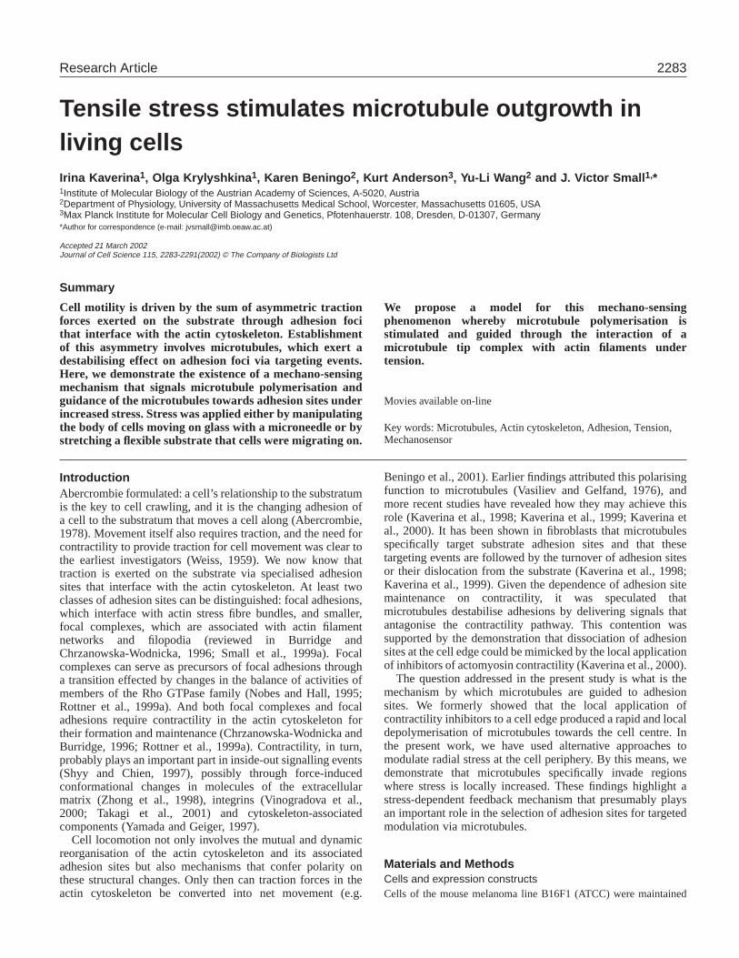
Fig. 1.Cell body displacementstimulates growth of peripheraladhesions in B16 melanomacells and the polymerisation ofmicrotubules towards the celledge. (A) Video frames show amotile cell, expressing GFP-VASP, whose cell body wasdisplaced by a microneedle inthe direction indicated in thephase contrast image. Panel0′00′′ in this and subsequentfigures corresponds to the videoframe before tension application.Boxed insets in the fluorescenceimages show enlargement ofperipheral adhesion sites in theregion diametrically opposite thecell body. The continuedprotrusion of the cell edge isindicated by the persistence ofthe line of GFP-VASP at the tipof the lamellipodium. Anexample of one from five cells isshown. Times are in minutes andseconds. Bar, 10 µm. (B) Theconditions used were the same asin A for a B16 melanoma cellexpressing GFP-tubulin. Arrowsin the phase contrast andfluorescent images of the videosequence indicate the directionof stress application. Insets showinvasion of microtubules intolamella region in the line ofapplied stress. An example ofone from seven cells is shown.(C) The conditions used were thesame as in A for a B16melanoma cell expressing GFP-CLIP-170. Arrows indicate thedirection of stress application.Note the increase in number ofpolymerising microtubules inperipheral lamella (ellipse),which are marked by GFP-CLIP-170 at their tips. An examplefrom one from 15 cells is shown.
2285Tensile stress induces microtubule outgrowth
of between 20 and 40 hPa. For local application of drugs, performedwith the same system, a constant pressure of 50-100 hPa was used.
Cell manipulations were performed using the samemicromanipulator with flamed, curved microinjection needles.
Tetramethyl rhodamine (5-TAMRA; Molecular Probes, USA)conjugated vinculin from turkey gizzard was kindly provided by K.Rottner and M. Gimona. Cy3-conjugated tubulin was kindly providedby J. Peloquin and G. Borisy (Chicago, USA).
For local application through a microneedle, drugs were dissolvedin microinjection buffer (2 mM Tris-Acetate pH 7.0, 50 mM KCl).The inhibitor of myosin light chain kinase, ML-7 (Alexis Corporation,Switzerland) was used at a concentration of 300 µM, the actomyosininhibitor 2,3-butanedione 2-monoxime (BDM) at a concentration 250mM and H7 (Sigma) at a concentration of 1 mM.
Polyacrylamide substratesFlexible substrates composed of 5% acrylamide and 0.08% Bis-acrylamide were prepared as previously described (Wang and Pelham,1998; Beningo et al., 2001). 100 µg/mL poly-D-lysine in PBS wascovalently coated to the substrates overnight at 4°C following a
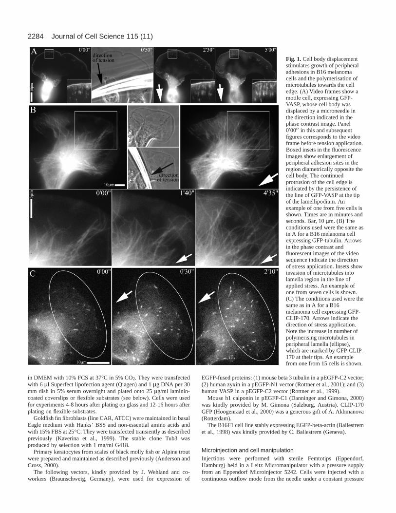
Fig. 2. (A,B) Quantification of the increase in the number ofmicrotubules extending into anterior lamella regions of B16melanoma cells in response to increased stress imposed by cell bodymanipulation (A, 22 cells) and stretching of a flexible growthsubstrate (B, 19 cells). (C) Quantification of the increase of CLIP-170-associated polymerising microtubule tips in lamella regions ofB16 melanoma cells in response to increased stress imposed by cellbody manipulation (blue, 15 cells) and stretching of a flexible growthsubstrate (magenta, four cells). Fig. 3. Microtubules induced to polymerise by increased stress target
the adhesion sites that simultaneously enlarge at the cell periphery.The figure shows a B16 melanoma cell that was transfected withGFP-zyxin and GFP-tubulin and subjected to cell body displacementin the direction indicated by the arrows. Upper phase images showthe cell just before (left) and 6 minutes 15 seconds (6′15′′ ) aftertension application (right) with the microneedle. The area boxed inthe left phase image corresponds to the region shown in fluorescencein the lower video frames. An example of one of 15 cells is shown.
2286
previously published procedure (Wang and Pelham, 1998). Afterrinsing with PBS, the substrate was coated with 25 µg/mL Laminin(dialyzed in PBS 3 hours on ice) for 1 hour at room temperature justprior to plating.
Video microscopy and image analysisCells were injected and observed in an open chamber at roomtemperature for CAR cells and keratocytes and in a heated chamber(Warner Instruments, Reading, UK) at 37°C for B16 cells on an
inverted microscope (Axiovert 135TV; Zeiss, Austria) equipped forepifluorescence and phase contrast microscopy. Injections wereperformed at an objective magnification of 40× (NA 1.3 PlanNeofluar), and video microscopy was performed with a 100×/NA 1.4Plan-Apochromat with or without 1.6 optovar intermediatemagnification. Tungsten lamps (100 W) were used for bothtransmitted and epi-illumination. Data were acquired with a back-illuminated, cooled CCD camera from Princeton ResearchInstruments driven by IPLabs software (both from Visitron Systems,Germany) and stored as 16-bit digital images. The microscope wasadditionally equipped with shutters (Optilas GmbH, Germany) toallow separate recordings of video sequences in phase contrast andfluorescence channels and with a filter wheel for two fluorescentchannels. Times between frames were 10 to 25 seconds.
For quantitative analysis of microtubule penetration in lamella, aline was drawn perpendicular to the direction of tension or, forcontrols, to the direction of protrusion, such that between 5-15microtubules crossed the line at time ‘0’. In consecutive video framesthe line was fixed at a constant distance from the cell front. Dependingon the cell measured, the distance ranged from 4 to 10 µm. Thenumber of microtubules crossing the line was then counted after 1minute for flexible substrate experiments and after two minutes forcell manipulations.
Fluorescence recovery after photobleachingFluorescence recovery after photobleaching (FRAP) was performedin a LSM 5 Pascal confocal microscope (Ziess) using cells expressingGFP-tubulin. A line was bleached across lamella regions and the time-lapse video of the microtubule pattern recorded. ML-7 was appliedlocally as described above.
ResultsStress application via cell body displacement inmelanoma cellsWhen plated on laminin, B16F1 melanoma cells commonlyexpress broad, protruding lamella regions that containrelatively small numbers of focal adhesions. The adhesionpattern, as revealed in cells transfected with GFP-VASP orGFP-zyxin, comprises a set of focal complexes that turnoverrapidly at the base of the protruding lamellipodium (Rottner etal., 1999b) and variable numbers of larger and longer-livedfocal adhesion sites in regions behind the lamellipodium (Fig.1A). The application of force to the cell body with amicroneedle in a direction opposing movement induced anenlargement of these peripheral adhesions in the direction ofstress application (Fig. 1A). In this situation, protrusion of thecell front was not arrested but continued during the period ofadhesion site enlargement, as confirmed by the persistence ofGFP-VASP at the lamellipodium tip (Fig. 1A) (Rottner et al.,1999b). These results showing stress-induced enlargement ofadhesion foci in motile melanoma cells confirm and extendprevious findings using fibroblasts (Riveline et al., 2001).
Lamella regions of B16F1 melanoma cells situated behindrapidly protruding lamellipodia characteristically show fewmicrotubules that extend close to the cell front (see alsoBallestrem et al., 2000). However, when stress was applied viaa microneedle to the cell body of B16 cells that had beentransfected with GFP-tubulin, microtubules were seen toextend towards the base of the protruding lamellipodium (Fig.1B). Measurements of 20 cells for a period of 2 minutesshowed that there was an approximately three-fold increase in
Journal of Cell Science 115 (11)
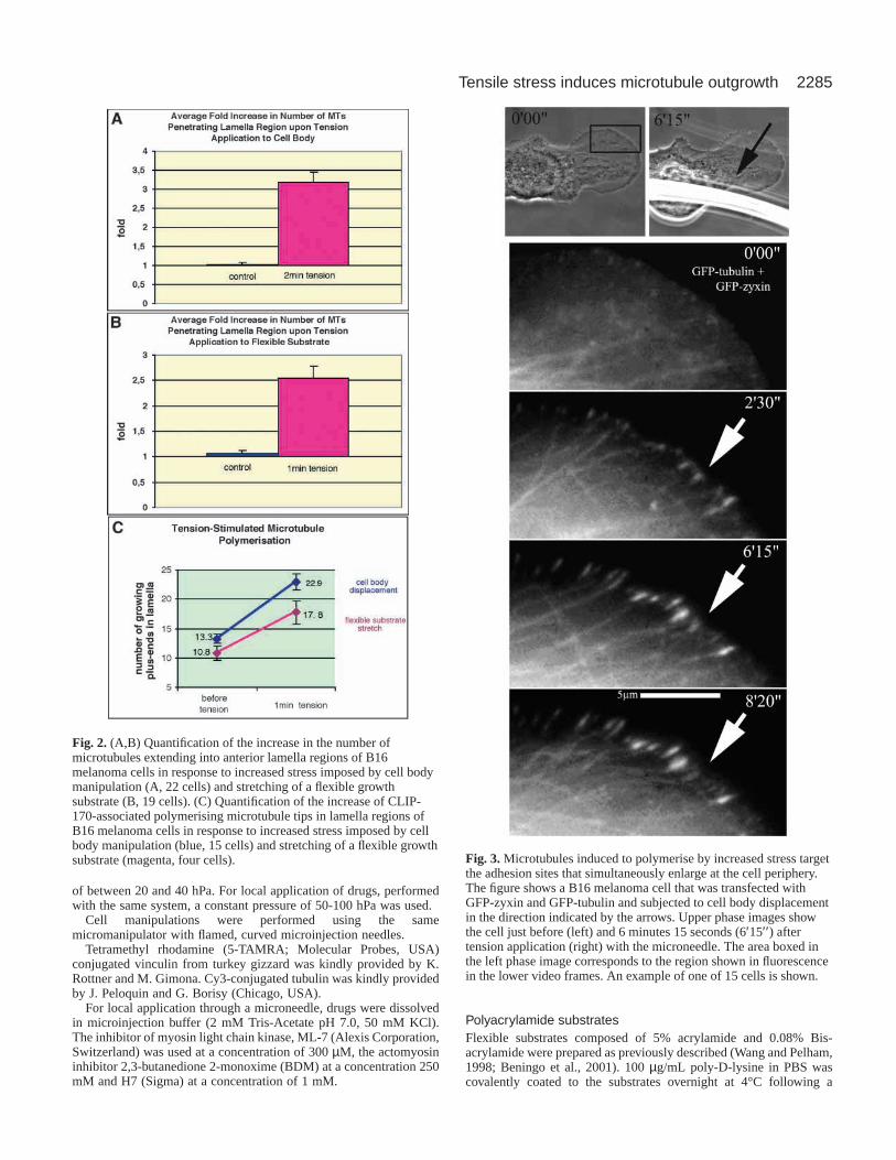
Fig. 4.Cell bodydisplacement stimulatesformation of radialbundles of actinfilaments that terminateat the cell periphery.Video sequences showa B16 melanoma cellexpressing either GFP-actin (A, example fromnine cells) or GFP-calponin h1 (B,example from fivecells.) that wassubjected to cell bodymanipulation at time 0.The chevrons in(A) indicate regions ofbundle formation.Boxed regions in(B) are shown at highermagnification in theright hand panels.
2287Tensile stress induces microtubule outgrowth
the number of microtubules that penetrated a given region of alamella following the application of stress (Fig. 2A).
In GFP-CLIP-170-transfected cells, the number offluorescent microtubule plus ends in peripheral lamella regionsincreased dramatically upon stress application (Fig. 1C, Fig.2C). Since CLIP-170 binds only to polymerising microtubuletips, these findings show that the stress-induced invasion ofmicrotubules is caused by the stimulation of microtubulepolymerisation and not by the transport of pre-existingmicrotubules towards the cell front by other means. By doublytransfecting cells with GFP-tubulin and GFP-zyxin, we couldfurther show that the microtubules that polymerised to the cellfront targeted the adhesion sites that were independentlyamplified at the cell periphery by the increased tension (Fig. 3).
It is notable that the same regions behind rapidly
protruding lamellipodia in B16 cells that are depleted ofmicrotubules also lack well defined bundles of actin filaments(Ballestrem et al., 1998). Instead, there commonly exists aloose network of actin filaments that extends from the baseof the lamellipodium into the perinuclear region, wherebundles become more evident (Rottner et al., 1999b; Small etal., 1999b). In view of the observed growth of both adhesionsites and microtubules in response to applied stress, it wasimportant to establish the accompanying changes in the actincytoskeleton under the same conditions. For this purpose, weused cells transfected either with GFP-actin (Fig. 4A) orGFP-calponin. The actin-binding protein calponin is aparticularly useful probe as it binds preferentially to bundlesof actin filaments and not to actin meshworks in lamellipodiaor to looser actin filament arrays in cultured cells (Gimonaand Mital, 1998). As shown in Fig. 4B, mechanical stressinduced a rapid and dramatic appearance of actin bundlesextending from the cell centre to the periphery, consistentwith the parallel amplification of peripheral focal adhesionsthat occurs under the same conditions (Figs 1 and 3). Thestress exerted in lamellae by cell body displacement wasestimated on the basis of manipulations of cells spread onflexible substrates (see below) to be of the order of 5×10⁵
dynes/cm2. This result revealed the continuity of actinfilaments in the cytoplasmic network with peripheralanchorage sites as well as the competence of these filamentsto form bundles under stress.
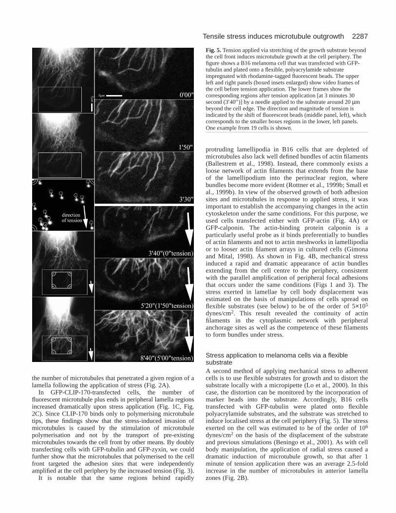
Stress application to melanoma cells via a flexiblesubstrateA second method of applying mechanical stress to adherentcells is to use flexible substrates for growth and to distort thesubstrate locally with a micropipette (Lo et al., 2000). In thiscase, the distortion can be monitored by the incorporation ofmarker beads into the substrate. Accordingly, B16 cellstransfected with GFP-tubulin were plated onto flexiblepolyacrylamide substrates, and the substrate was stretched toinduce localised stress at the cell periphery (Fig. 5). The stressexerted on the cell was estimated to be of the order of 106
dynes/cm2 on the basis of the displacement of the substrateand previous simulations (Beningo et al., 2001). As with cellbody manipulation, the application of radial stress caused adramatic induction of microtubule growth, so that after 1minute of tension application there was an average 2.5-foldincrease in the number of microtubules in anterior lamellazones (Fig. 2B).
Fig. 5.Tension applied via stretching of the growth substrate beyondthe cell front induces microtubule growth at the cell periphery. Thefigure shows a B16 melanoma cell that was transfected with GFP-tubulin and plated onto a flexible, polyacrylamide substrateimpregnated with rhodamine-tagged fluorescent beads. The upperleft and right panels (boxed insets enlarged) show video frames ofthe cell before tension application. The lower frames show thecorresponding regions after tension application [at 3 minutes 30second (3′40′′ )] by a needle applied to the substrate around 20 µmbeyond the cell edge. The direction and magnitude of tension isindicated by the shift of fluorescent beads (middle panel, left), whichcorresponds to the smaller boxes regions in the lower, left panels.One example from 19 cells is shown.
2288
Using GFP-CLIP-170-transfected cells we could showthat this effect was again caused by the enhancement ofmicrotubule polymerisation, as indicated by a dramaticincrease in the number of fluorescent microtubule plus ends instressed regions (Fig. 2C).
Recovery from inhibition of contractility in fibroblastsAn increase in stress in the actin cytoskeleton is also stimulated
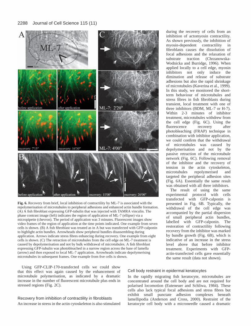
during the recovery of cells from aninhibition of actomyosin contractility.As shown previously, the inhibition ofmyosin-dependent contractility infibroblasts causes the dissolution offocal adhesions and the relaxation ofsubstrate traction (Chrzanowska-Wodnicka and Burridge, 1996). Whenapplied locally to a cell edge, myosininhibitors not only induce thediminution and release of substrateadhesions but also the rapid shrinkageof microtubules (Kaverina et al., 1999).In this study, we monitored the short-term behaviour of microtubules andstress fibres in fish fibroblasts duringtransient, local treatment with one ofthree inhibitors (BDM, ML-7 or H-7).Within 2-3 minutes of inhibitortreatment, microtubules withdrew fromthe cell edge (Fig. 6C). Using thefluorescence recovery afterphotobleaching (FRAP) technique incombination with inhibitor application,we could confirm that the withdrawalof microtubules was caused bydepolymerisation and not by thepassive retraction of the microtubulenetwork (Fig. 6C). Following removalof the inhibitor and the recovery oftension in the actin cytoskeleton,microtubules repolymerised andtargeted the peripheral adhesion sites(Fig. 6A). Essentially the same resultwas obtained with all three inhibitors.
The result of using the sameexperimental protocol with cellstransfected with GFP-calponin ispresented in Fig. 6B. Typically, thewithdrawal of the cell edge wasaccompanied by the partial dispersionof small peripheral actin bundles,labelled with GFP-calponin. Therestoration of contractility followingrecovery from the inhibitor was markedby bundle growth (Fig. 6B), which isindicative of an increase in the stresslevel above that before inhibitortreatment. Experiments with GFP-actin-transfected cells gave essentiallythe same result (data not shown).
Cell body restraint in epidermal keratocytesIn the rapidly migrating fish keratocyte, microtubules areconcentrated around the cell body and are not required forpolarised locomotion (Euteneuer and Schliwa, 1984). Thesecells also lack typical focal adhesions and stress fibres butexhibit small punctate adhesion complexes beneathlamellipodia (Anderson and Cross, 2000). Restraint of thekeratocyte cell body with a microneedle caused a dramatic
Journal of Cell Science 115 (11)
Fig. 6. Recovery from brief, local inhibition of contractility by ML-7 is associated with therepolymerisation of microtubules to peripheral adhesions and enhanced actin bundle formation.(A) A fish fibroblast expressing GFP-tubulin that was injected with TAMRA vinculin. Thephase contrast image (left) indicates the region of application of ML-7 (ellipse) via amicropipette (chevron). The period of application was 3 minutes. Fluorescent images showvideo frames of the region of application at the time points indicated. One example from sevencells is shown. (B) A fish fibroblast was treated as in A but was transfected with GFP-calponinto highlight actin bundles. Arrowheads show peripheral bundles disassembling duringapplication. Arrows indicate stress fibres enhancing during recovery. One example from eightcells is shown. (C) The retraction of microtubules from the cell edge on ML-7 treatment iscaused by depolymerisation and not by bulk withdrawal of microtubules. A fish fibroblastexpressing GFP-tubulin was photobleached in a narrow region across the base of lamella(arrow) and then exposed to local ML-7 application. Arrowheads indicate depolymerisingmicrotubules in subsequent frames. One example from five cells is shown.
2289Tensile stress induces microtubule outgrowth
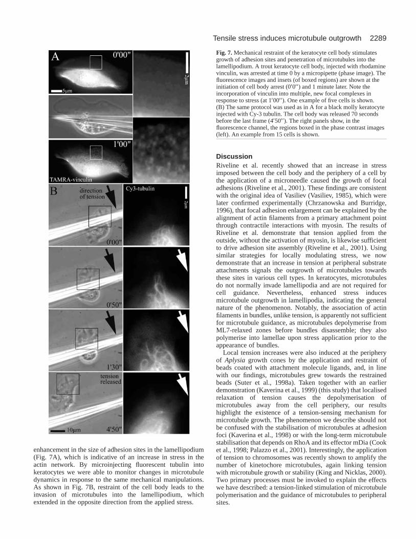
enhancement in the size of adhesion sites in the lamellipodium(Fig. 7A), which is indicative of an increase in stress in theactin network. By microinjecting fluorescent tubulin intokeratocytes we were able to monitor changes in microtubuledynamics in response to the same mechanical manipulations.As shown in Fig. 7B, restraint of the cell body leads to theinvasion of microtubules into the lamellipodium, whichextended in the opposite direction from the applied stress.
DiscussionRiveline et al. recently showed that an increase in stressimposed between the cell body and the periphery of a cell bythe application of a microneedle caused the growth of focaladhesions (Riveline et al., 2001). These findings are consistentwith the original idea of Vasiliev (Vasiliev, 1985), which werelater confirmed experimentally (Chrzanowska and Burridge,1996), that focal adhesion enlargement can be explained by thealignment of actin filaments from a primary attachment pointthrough contractile interactions with myosin. The results ofRiveline et al. demonstrate that tension applied from theoutside, without the activation of myosin, is likewise sufficientto drive adhesion site assembly (Riveline et al., 2001). Usingsimilar strategies for locally modulating stress, we nowdemonstrate that an increase in tension at peripheral substrateattachments signals the outgrowth of microtubules towardsthese sites in various cell types. In keratocytes, microtubulesdo not normally invade lamellipodia and are not required forcell guidance. Nevertheless, enhanced stress inducesmicrotubule outgrowth in lamellipodia, indicating the generalnature of the phenomenon. Notably, the association of actinfilaments in bundles, unlike tension, is apparently not sufficientfor microtubule guidance, as microtubules depolymerise fromML7-relaxed zones before bundles disassemble; they alsopolymerise into lamellae upon stress application prior to theappearance of bundles.
Local tension increases were also induced at the peripheryof Aplysia growth cones by the application and restraint ofbeads coated with attachment molecule ligands, and, in linewith our findings, microtubules grew towards the restrainedbeads (Suter et al., 1998a). Taken together with an earlierdemonstration (Kaverina et al., 1999) (this study) that localisedrelaxation of tension causes the depolymerisation ofmicrotubules away from the cell periphery, our resultshighlight the existence of a tension-sensing mechanism formicrotubule growth. The phenomenon we describe should notbe confused with the stabilisation of microtubules at adhesionfoci (Kaverina et al., 1998) or with the long-term microtubulestabilisation that depends on RhoA and its effector mDia (Cooket al., 1998; Palazzo et al., 2001). Interestingly, the applicationof tension to chromosomes was recently shown to amplify thenumber of kinetochore microtubules, again linking tensionwith microtubule growth or stability (King and Nicklas, 2000).Two primary processes must be invoked to explain the effectswe have described: a tension-linked stimulation of microtubulepolymerisation and the guidance of microtubules to peripheralsites.
Fig. 7. Mechanical restraint of the keratocyte cell body stimulatesgrowth of adhesion sites and penetration of microtubules into thelamellipodium. A trout keratocyte cell body, injected with rhodaminevinculin, was arrested at time 0 by a micropipette (phase image). Thefluorescence images and insets (of boxed regions) are shown at theinitiation of cell body arrest (0′0′′ ) and 1 minute later. Note theincorporation of vinculin into multiple, new focal complexes inresponse to stress (at 1′00′′ ). One example of five cells is shown.(B) The same protocol was used as in A for a black molly keratocyteinjected with Cy-3 tubulin. The cell body was released 70 secondsbefore the last frame (4′50′′ ). The right panels show, in thefluorescence channel, the regions boxed in the phase contrast images(left). An example from 15 cells is shown.
2290
Various possibilities exist for establishing linkages betweenactin and microtubule networks (reviewed in Gavin, 1996;Goode et al., 2000; Waterman-Storer et al., 2000) that may berelevant for the guidance of microtubules to substrateadhesions.
One possibility is suggested by the demonstration thatkinesin and unconventional myosins can cooperate in thetransport of vesicles along microtubules and actin filaments(Rodionov et al., 1998). However, the involvement ofheterodimeric myosin-kinesin motor complexes (Huang et al.,1999; Beningo et al., 2000) in the guidance of microtubulesalong actin filaments to adhesion sites (Goode et al., 2000) isunlikely since a block in kinesin activity has no effect on theability of microtubules to target substrate adhesions(Krylyshkina et al., 2002).
As elaborated in detail by Goode et al., the interaction ofmicrotubules with actin filaments represents a common featureof morphogenetic events in a variety of cell types (Goode etal., 2000). Interestingly, actin cables are needed formaintaining yeast spindle orientation (Palmer et al., 1992;Theesfeld et al., 1999), and this process requires myosin V (Yinet al., 2000), which can bind to the microtubule-associatedprotein Kar 9. Thus, the sliding of a myosin along actinfilaments may serve to direct microtubules toward the cortex(see also Suter et al., 1998b). Myosin V was also found byWaterman-Storer et al. to colocalise with dynamic, co-lineararrays of actin filaments and microtubules observed in Xenopusextracts (Waterman-Storer et al., 2000). A possible mode oflinkage for an unconventional myosin to microtubules has alsobeen suggested by the interaction of a class VI myosin with aCLIP-170 homologue in Drosophila (Lantz and Miller, 1998),but so far an association of myosin VI with microtubules hasnot been observed (Lantz and Miller, 1998; Buss et al., 1998).In this connection, CLIP-170 (Perez et al., 1999) and otherproteins at the tips of growing microtubules (reviewed inSchroer, 2001; Tirnauer and Bierer, 2000; Schuyler andPellman, 2001) including APC (Mimori-Kiyosue et al., 2000a),EB1 (Mimori-Kiyosue et al., 2000b), members of the dynactincomplex (Vaughan et al., 1999) and the CLASP family(Akhamanova et al., 2001) are strategically located wheremicrotubule dynamics as well as the guidance of microtubulegrowth are most probably controlled.
With regard to the link between microtubule guidance andstress, the results with B16 cells transfected with the abundantsmooth muscle isoform of calponin (h1) are particularlyinteresting. As shown by Gimona and Mital, calponin bindsprimarily to stress fibre bundles and not to actin meshworks orloose actin filament arrays (Gimona and Mital, 1998). Thedramatic appearance of calponin-positive bundles in thelamella regions of B16 cells after tension application suggeststhat tension induces conformational changes in actin filaments,or proteins associated with them, that facilitate calponinbinding. It has already been shown that actin-binding proteinscan induce changes in the twist of the long pitch helices of actinfilaments (Bamberg et al., 1999; Galkin et al., 2001): by thesame token the application of torsion to a ‘relaxed’ filamentshould induce structural changes allowing interaction withother binding partners. We suggest that conformationalchanges in actin induced by stretch play a primary role insignalling for microtubule polymerisation and guidance. Oneplausible scenario includes the linkage of an unconventional
myosin to a component of the microtubule tip complex,whereby this myosin is only competent to bind to actinfilaments that are under tension. This interaction could inducefurther conformational changes in the microtubule tip complexitself that promote microtubule polymerisation. Whether or notsuch a scenario pertains to microtubules and whether otherregulatory factors are involved that bind to microtubules (Bestet al., 1996; Ren et al., 1998; Glaven et al., 1999) or thatinfluence microtubule assembly (Carazo-Salas et al., 1999) inresponse to stress-induced signal transduction at adhesion sitesremains to be elucidated. Whatever the mechanism, we appearto have revealed a tension-based feedback loop that plays a rolein the promotion of substrate adhesion disassembly viamicrotubules.
We thank Jurgen Wehland, Gary Borisy, Anna Akhmanova, MarioGimona, Christoph Ballestrem and Klemens Rottner for kindlyproviding probes for this study, and we thank Maria Schmittner fortechnical assistance. This work was supported by a grants from theAustrian Science Research Council (to J.V.S.), NASA (NAG-1197 toY.-L.W.) and the NIH (to K.B.).
ReferencesAbercrombie, M. (1978). The crawling movement of metazoan cells. Proc.
R. Soc. Lond. B Biol. Sci. 207, 129-147.Akhmanova, A., Hoogenraad, C. C., Drabek, K., Stepanova, T., Dortland,
B., Verkerk, T., Vermeulen, W., Burgering, B. M., de Zeeuw, C. I.,Grosveld, F. and Galjart, N. (2001). CLASPs are CLIP-115 and -170associating proteins involved in the regional regulation of microtubuledynamics in motile fibroblasts. Cell 104, 923-935.
Anderson, K. I. and Cross, R.(2000). Contact dynamics during keratocytemotility. Curr. Biol. 10, 253-260.
Ballestrem, C., Wehrle-Haller, B. and Imhof, B. A.(1998). Actin dynamicsin living mammalian cells. J. Cell Sci. 111, 1649-1658.
Ballestrem, C., Wehrle-Haller, B., Hinz, B. and Imhof, B. A.(2000). Actin-dependent lamellipodia formation and microtubule-dependent tail retractioncontrol-directed cell migration. Mol. Biol. Cell11, 2999-3012.
Bamburg, J. R., McGough, A. and Ono, S.(1999). Putting a new twist onactin: ADF/cofilins modulate actin dynamics. Trends Cell Biol. 9, 364-370.
Beningo, K. A., Lillie, S. H. and Brown, S. S.(2000). The yeast kinesin-related protein Smy1p exerts its effects on the class V myosin Myo2p via aphysical interaction. Mol. Biol. Cell11, 691-702.
Beningo, K., Dembo, M., Kaverina, I., Small, J. V. and Wang, Y.-L.(2001).Nascent focal adhesions are responsible for the generation of strongpropulsive forces in migrating fibroblasts. J. Cell Biol. 153, 881-887.
Best, A., Ahmed, S., Kozma, R. and Lim, L.(1996). The Ras-related GTPaserac1 binds tubulin. J. Biol. Chem. 271, 3756-3762.
Burridge, K. and Chrzanowska-Wodnicka, M. (1996). Focal adhesions,contractility, and signalling. Annu. Rev. Cell Dev. Biol. 12, 463-519.
Buss, F., Kendrick-Jones, J., Lionne, C., Knight, A. E., Côté, G. P. andLuzio, J. P.(1998). The localization of myosin VI at the Golgi complex andleading edge of fibroblasts and its phosphorylation and recruitment intomembrane ruffles of A431 cells after growth factor stimulation. J. Cell Biol.143, 1535-1545.
Carazo-Salas, R. E., Guarguaglini, G., Gruss, O. J., Segref, A., Karsenti,E. and Mattaj, I. W. (1999). Generation of GTP-bound ran by RCC1 isrequired for chromatin-induced mitotic spindle formation. Nature400, 178-181.
Chrzanowska-Wodnicka, M. and Burridge, K. (1996). Rho-stimulatedcontractility drives the formation of stress fibers and focal adhesions. J. CellBiol. 133, 1403-1415.
Cook, T. A., Nagasaki, T. and Gundersen, G. G.(1998). Rho guanosinetriphosphatase mediates the selective stabilization of microtubules inducedby lysophosphatidic acid. J. Cell Biol. 141, 175-185.
Danninger, C. and Gimona, M. (2000). Live dynamics of GFP-calponin:isoform-specific modulation of the actin cytoskeleton and autoregulation byC-terminal sequences. J. Cell Sci. 113, 3725-3736.
Euteneuer, U. and Schliwa, M. (1984). Persistent, directional motility of cells
Journal of Cell Science 115 (11)
2291Tensile stress induces microtubule outgrowth
and cytoplasmic fragments in the absence of microtubules. Nature310, 58-61.
Galkin, V. E., Orlova, A., Lukoyanova, N., Wriggers, W. and Egelman, E.H. (2001). Actin depolymerising factor stabilizes an existing state of F-actinand can change the tilt of F-actin subunits. J. Cell Biol. 153, 75-86.
Gavin, R. H. (1997). Microtubule-microfilament synergy in the cytoskeleton.Int. Rev. Cytology173, 207-242.
Gimona, M. and Mital, R. (1998). The single CH domain of calponin isneither sufficient nor necessary for F-actin binding. J. Cell Sci. 111, 1813-1821.
Glaven, J. A., Whitehead, I., Bagrodia, S., Kay, R. and Cerione, R. A.(1999). The Dbl-related protein, Lfc, localizes to microtubules and mediatesthe activation of rac signalling pathways in cells. J. Biol. Chem. 274, 2279-2285.
Goode, B. L., Drubin, D. G. and Barnes, G.(2000). Functional cooperationbetween the microtubule and actin cytoskeletons. Curr. Opin. Cell Biol. 12,63-71.
Hoogenraad, C. C., Akhmanova, A., Grosveld, F., de Zeeuw, C. I. andGaljart, N . (2000). Functional analysis of CLIP-115 and its binding tomicrotubules. J. Cell Sci. 113, 2285-2297.
Huang, J.-D., Brady, S. T., Richards, B. W., Stenoien, D., Resau, J. H.,Copeland, N. G. and Jenkins, N. A.(1999). Direct interaction ofmicrotubule- and actin-based transport motors. Nature397, 267-270.
Kaverina, I., Rottner, K. and Small, J. V. (1998). Targeting, capture, andstabilization of microtubules at early focal adhesions. J. Cell Biol. 142, 181-190.
Kaverina, I., Krylyshkina, O. and Small, J. V. (1999). Microtubule targetingof substrate contacts promotes their relaxation and dissociation. J. Cell Biol.146, 1033-1043.
Kaverina, I., Krylyshkina, O., Gimona, M., Beningo, K., Wang, Y.-L. andSmall, J. V. (2000). Enforced polarisation and locomotion of fibroblastslacking microtubules. Curr. Biol. 10, 739-742.
King, J. M. and Nicklas, R. B. (2000). Tension on chromosomes increasesthe number of kinetochore microtubules but only within limits. J. Cell. Sci.113, 3815-3823.
Krylyshkina, O., Kaverina, I., Kranewitter, W., Steffen, W., Alonso, M. C.,Cross, R. A. and Small, J. V.(2002). Modulation of substrate adhesiondynamics via microtubule targeting requires kinesin-1. J. Cell Biol. 156,349-360.
Lantz, V. A. and Miller, K. G. (1998). A class VI unconventional myosin isassociated with a homologue of a microtubule-binding protein, cytoplasmiclinker protein-170, in neurons and at the posterior pole of Drosophilaembryos. J. Cell Biol. 140, 897-910.
Lo, C.-M., Wang, H.-B., Dembo, M. and Wang, Y.-L. (2000). Cell movementis guided by the rigidity of the substrate. Biophys. J. 79, 114-152.
Mimori-Kiyosue, Y., Shiina, N. and Tsukita, S. (2000a). Adenomatouspolyposis coli (APC) protein moves along microtubules and concentrates attheir growing ends in epithelial cells. J. Cell Biol. 148, 505-517.
Mimori-Kiyosue, Y., Shiina, N. and Tsukita, S. (2000b). The dynamicbehaviour of the APC-binding protein EB1 on the distal ends ofmicrotubules. Curr. Biol. 10, 865-868.
Nobes, C. D. and Hall, A.(1995). Rho, rac, and Cdc42 GTPases regulate theassembly of multimolecular focal complexes associated with actin stressfibers, lamellipodia, and filopodia. Cell 81, 53-62.
Palazzo, A. F., Cook, T. A., Alberts, A. S. and Gundersen, G. G.(2001).mDia mediates Rho-regulated formation and orientation of stablemicrotubules. Nat. Cell Biol. 3, 723-729.
Palmer, R. E., Sullivan, D. S., Huffaker, T. and Koshland, D.(1992). Roleof astral microtubules and actin in spindle orientation and migration in thebudding yeast Saccharomyces cerevisiae. J. Cell Biol. 119, 583-593.
Perez, F., Diamantopoulos, G. S., Stalder, R. and Kreis, T. E.(1999). CLIP-170 highlights growing microtubule ends in vivo. Cell 96, 517-527.
Ren, Y., Li, R., Zheng, Y. and Busch, H.(1998). Cloning and characterizationof GEF-H1, a microtubule-associated guanine nucleotide exchange factorfor rac and rho GTPases. J. Biol. Chem. 273, 34954-34960.
Riveline, D., Zamir, E., Balaban, N. Q., Schwarz, U. S., Ishizaki, T.,Narumiya, S., Kam, Z., Geiger, B. and Bershadsky, A. D.(2001). Focalcontacts as mechanosensors: externally applied local mechanical force
induces growth of focal contacts by a mDia1-dependent and ROCK-independent mechanism. J. Cell Biol. 153, 1175-1185.
Rodionov, V. I., Hope, A. J., Svitkina, T. M. and Borisy, G. G.(1998).Functional coordination of microtubule-based and actin-based motility inmelanophores. Curr. Biol. 8, 165-168.
Rottner, K., Hall, A. and Small, J. V. (1999a). Interplay between rac and rhoin the control of substrate contact dynamics. Curr. Biol. 9, 640-648.
Rottner, K., Behrendt, B., Small, J. V. and Wehland, J.(1999b). VASPdynamics during lamellipodia protrusion. Nat. Cell Biol. 1, 321-322.
Rottner, K., Krause, M., Gimona, M., Small, J. V. and Wehland, J.(2001)Zyxin is not colocalized with vasodilator-stimulated phosphoprotein (VASP)at lamellipodial tips and exhibits different dynamics to vinculin, paxillin,and VASP in focal adhesions. Mol. Biol. Cell. 12, 3103-3113.
Schroer, T. A. (2001). Microtubules don and doff their caps: dynamicattachments at plus and minus ends. Curr. Opin. Cell Biol. 13, 92-96.
Schuyler, S. C. and Pellmann, D.(2001). Microtubule “plus-end-trackingproteins”: the end is just the beginning. Cell 105, 421-424.
Shyy, J. Y.-J. and Chien, S.(1997). Role of integrins in cellular responses tomechanical stress and adhesion. Curr. Opin. Cell Biol. 9, 707-713.
Small, J. V., Kaverina, I., Krylyshkina, O. and Rottner, K. (1999a).Cytoskeleton cross-talk during cell motility. FEBS Lett. 452, 96-99.
Small, J. V., Rottner, K., Hahne, P. and Anderson, K. I.(1999b). Visualisingthe actin cytoskeleton. Microsc. Res. Tech. 47, 3-17.
Suter, D. M. and Forscher, P.(1998). An emerging link between cytoskeletaldynamics and cell adhesion molecules in growth cone guidance. Curr. Biol.8, 106-116.
Suter, D. M., Errante, L. D., Belotserkovsky, V. and Forscher, P.(1998).The Ig superfamily cell adhesion molecule, apCAM mediates growth conesteering by substrate-cytoskeletal coupling. J. Cell Biol. 141, 227-240.
Takagi, J., Erickson, H. P. and Springer, T. A.(2001). C-terminal openingmimics “inside-out” activation of integrin alpha 5 beta1. Nat. Struct. Biol.8, 412-416.
Theesfeld, C. L., Irazoqui, J. E., Bloom, K. and Lew, D. J.(1999). The roleof actin in spindle orientation changes during the Saccharomyces cerevisiaecell cycle. J. Cell Biol. 146, 1019-1032.
Tirnauer, J. S. and Bierer, B. E.(2000). EB1 proteins regulate microtubuledynamics, cell polarity, and chromosome stability. J. Cell Biol. 149, 761-766.
Vasiliev, J. M. (1985). Spreading of non-transformed and transformed cells.Biochim. Biophys. Acta780, 21-65.
Vasiliev, J. M. and Gelfand, I. M. (1976). Effects of colcemid onmorphogenetic processes and locomotion of fibroblasts. In Cell Motility (edsR. Goldman, T. Pollard and J. Rosenbaum), pp. 279-304. Cold SpringHarbor, NY:Cold Spring Harbor Laboratory.
Vaughan, K. T., Tyan, S. H., Faulkner, N. E., Echeverri, C. J. and Vallee,R. B. (1999). Colocalization of cytoplasmic dynein with dynactin and CLIP-170 at microtubule distal ends. J. Cell Sci. 112, 1437-1447.
Vinogradova, O., Haas, T., Plow, E. F. and Qin, J.(2000). A structural basisfor integrin activation by the cytoplasmic tail of the alphaIIb-subunit. Proc.Natl. Acad. Sci. USA97, 1450-1455.
Wang, Y.-L. and Pelham, R. J. (1998). Preparation of a flexible, porouspolyacrylamide substrate for mechanical studies of cultured cells. MethodsEnzymol. 298, 489-496.
Waterman-Storer, C., Duey, D. Y., Weber, K. L., Keech, J., Cheney, R. E.,Salmon, E. D. and Bement, W. M. (2000). Microtubules remodelactomyosin networks in Xenopusegg extracts via two mechanisms of F-actin transport. J. Cell Biol. 150, 361-376.
Weiss, P. (1959). Cellular dynamics. In Biophysical Science – A StudyProgramme (ed. J. L. Oncley), pp. 11-20. New York: John Wiley and Sons,Inc.
Yamada, K. M. and Geiger, B.(1997). Molecular interactions in cell adhesioncomplexes. Curr. Biol. 9, 76-85.
Yin, H., Pruyne, D., Huffaker, T. C. and Bretscher, A.(2000). Myosin Vorientates the mitotic spindle in yeast. Nature406, 1013-1015.
Zhong, C., Chrzanowska-Wodnicka, M., Brown, J., Shaub, A., Belkin, A.M. and Burridge, K. (1998). Rho-mediated contractility exposes a crypticsite in fibronectin and induces fibronectin matrix assembly. J. Cell Biol. 141,539-551.


