Embed Size (px)
Citation preview

Microtubule Initiation from the Nuclear Surface ControlsCortical Microtubule Growth Polarity and Orientation inArabidopsis thalianaChris Ambrose1 and Geoffrey O. Wasteneys1,*1Department of Botany, The University of British Columbia, Vancouver V6T 1Z4, Canada
*Corresponding author: E-mail, [email protected]; Fax, +1-604-822-6089.(Received October 11, 2013; Accepted June 25, 2014)
The nuclear envelope in plant cells has long been known tobe a microtubule organizing center (MTOC), but its influ-ence on microtubule organization in the cell cortex has beenunclear. Here we show that nuclear MTOC activity favors theformation of longitudinal cortical microtubule (CMT)arrays. We used green fluorescent protein (GFP)-taggedgamma tubulin-complex protein 2 (GCP2) to identify nu-clear MTOC activity and GFP-tagged End-Binding Protein 1b(EB1b) to track microtubule growth directions. We foundthat microtubules initiate from nuclei and enter the cortexin two directions along the long axis of the cell, creatingbipolar longitudinal CMT arrays. Such arrays were observedin all cell types showing nuclear MTOC activity, includingroot hairs, recently divided cells in root tips, and the leafepidermis. In order to confirm the causal nature of nuclei inbipolar array formation, we displaced nuclei by centrifuga-tion, which generated a corresponding shift in the bipolaritysplit point. We also found that bipolar CMT arrays wereassociated with bidirectional trafficking of vesicular compo-nents to cell ends. Together, these findings reveal a con-served function of plant nuclear MTOCs and centrosomes/spindle pole bodies in animals and fungi, wherein all struc-tures serve to establish polarities in microtubule growth.
Keywords: Arabidopsis � Cytoskeleton � Microtubule �
Polarity � Root � Root hair.
Abbreviations: ABC, ATP-binding cassette; CLASP, Clip-asso-ciated protein; CMT, cortical microtubule; EB1b, End BindingProtein 1b; EMT, endoplasmic microtubule; GCP2, gammatubulin-complex protein 2; GFP, green fluorescent protein,MBD, microtubule-binding domain; MT, microtubule;MTOC, microtubule organizing center; RFP, red fluorescentprotein; SNX1, Sorting Nexin 1.
Introduction
Microtubules (MTs) are tubulin polymers that undergo dy-namic switches between states of growth (polymerization)and shortening (depolymerization). MTs are inherently polar-ized, possessing a highly dynamic plus end that undergoes rapidgrowth and shrinkage. In order to harness and translate MTpolarity to a cell-wide scale, MT organizing centers (MTOCs)
generate and anchor large groups of MTs, often forming highlypolarized arrays with specialized functions. MTOCs are found inall eukaryotic cells, including centrosomes in animal cells, spin-dle pole bodies in fungi, and basal bodies in flagellated cells.Although MTOC structure varies across the kingdoms, thebasic functions are conserved. These include mitotic spindleassembly/function, cell polarity and shape generation, andcell migration and motility.
In plants, the nuclear envelope can act as a MTOC. It con-tains MT nucleation components at its surface (Clayton et al.1985, Wick 1985, Liu et al. 1993, Erhardt et al. 2002, Kumagaiet al. 2003, Brown and Lemmon 2007, Seltzer et al. 2007) andhas been shown to nucleate MTs at its surface in studies withisolated nuclei (Mizuno 1993, Stoppin et al. 1994), permeabi-lized cells (Wasteneys et al. 1989, Vantard et al. 1990) and intactcells (Falconer et al. 1988). Nuclear MT nucleating activity ismost prominent in the actively cycling cells in the divisionzones of the shoot and root. Within these cells, nuclearMTOC activity is transient, being found in: (i) cells enteringmitosis, during which time it contributes to formation of thepre-prophase band and the accompanying pre-spindle; and (ii)post-cytokinetic cells, where it persists until the onset of cellelongation (Nagata et al. 1994, Brown and Lemmon 2007,Ambrose and Wasteneys 2011).
In post-cytokinetic cells, nuclear MTOC activity generatesendoplasmic MTs (EMTs) that extend through the cytoplasmin all directions, forming a radial MT array. These radial EMTscontact the cell cortex, to which they attach and incorporateinto the cortical MT (CMT) array (Nagata et al. 1994, Kumagaiet al. 2003). To achieve the highly directional (anisotropic) ex-pansion seen in plant cells, CMTs form parallel arrays thatinteract with cellulose synthase complexes to generate ten-sion-bearing cellulose microfibrils aligned in a similar directionto the CMTs. In post-cytokinetic cells, nuclear MTOC-derivedmixed/longitudinally oriented CMT arrays presumably disfavorcell elongation by countering their inherent tendency towardtransverse arrangement. This is supported by observations ofthe botero-1 mutant, which exhibits swollen root tips, and re-tains EMTs and disorganized CMTs for a longer time than inwild-type cells due to lack of MT-severing activity (Bichet et al.2001). Beyond this, our understanding of how nuclear MTOCactivity controls CMT organization is limited by the technicaldifficulty in studying radial EMT arrays, which are highly
Plant Cell Physiol. 55(9): 1636–1645 (2014) doi:10.1093/pcp/pcu094, Advance Access publication on 8 July 2014, available online at www.pcp.oxfordjournals.org! The Author 2014. Published by Oxford University Press on behalf of Japanese Society of Plant Physiologists.All rights reserved. For permissions, please email: [email protected]
Regu
larP
aper
by guest on March 6, 2016
http://pcp.oxfordjournals.org/D
ownloaded from

dynamic in all three spatial dimensions and appear for a shorttime only in small, cytoplasmically dense cells.
In the current study, we show how the activity and position-ing of the nuclear MTOC influence the organization and polar-ity of CMTs in tip-growing root hairs and in recently dividedcells. Specifically, nucleus-derived MTs enter the cell cortex andsplay in two directions along the long axis of the cell, creating abipolar CMT array with the nucleus as the split point. As thenucleus changes its position over time, the accompanyingbipolarity split point follows along, thereby guiding the organ-ization of the CMT array. This nuclear MTOC mechanism mayprovide an explanation for previously described bipolar arraysin plant cells (Sambade et al. 2012, Pietra et al. 2013, Vineyardet al. 2013). Additionally, we found that CMT bipolarity corres-ponds with a bi-directional transit and accumulation of vesiclesto the ends of cells, suggesting a functional role for bipolarizedCMT arrays in cell polarization. We observed other patterns ofpolarity in cells with more complex shapes, indicating that aninterplay exists between MTOC patterns and cellular geometry.
Results
Cortical MTs grow bi-directionally away from thenucleus in root hairs
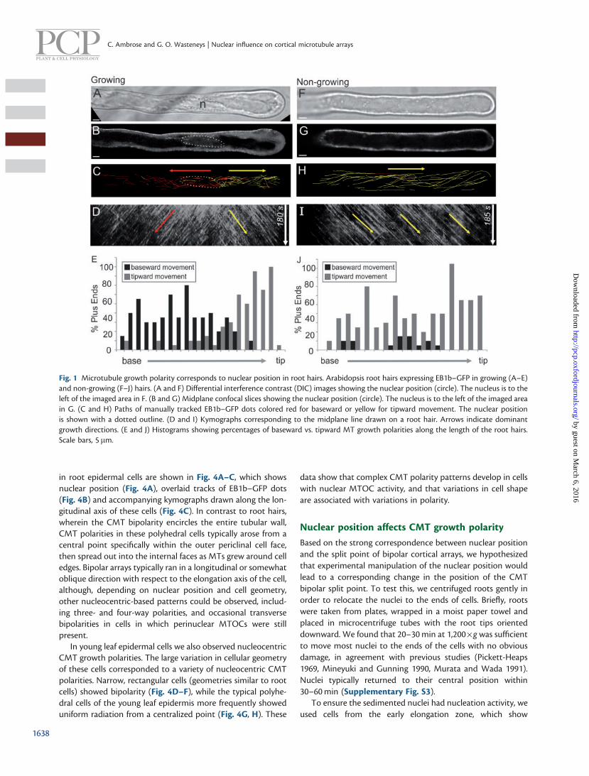
Using the MT plus-end marker EB1b–green fluorescent protein(GFP) as a reporter for MT growth direction, we were able toobserve CMTs and EMTs in actively growing Arabidopsis thali-ana root hairs. CMTs run lengthwise along the root hair. Inassessing the directions of plus-end growth, we found a cleartwo-way growth polarity, wherein those within the distal por-tion grew toward the tip, while those in the proximal regiongrew toward the hair base (Fig. 1A–E; Supplementary MovieS1). This MT growth bipolarity encompassed the entire circum-ference of the cortex, thus being observable at any depth withina hair. The nucleus consistently resided at the split point ofthese two opposing growth polarities. This nucleocentric MTgrowth bipolarity is demonstrated clearly from kymograph ana-lysis as two sets of diagonal lines mirrored about a centralregion, which corresponds to the position of the nucleus(Fig. 1D). Quantification of the direction of EB1b–GFP dotmovement as a function of the length of the root hair showsa split distribution corresponding to the two opposing polari-ties (Fig. 1E). As the nucleus changed position along the lengthof the hair, the two-way split point moved along with it. Thus,in older root hairs, in which the nucleus has retreated to thebase, unidirectional tipward MT growth polarity was observedthroughout the entire hair (Fig. 1F–H; Supplementary MovieS2; Supplementary Fig. S1). Kymographs along these cellsshow a single set of sloped lines (Fig. 1I), and quantificationshows more uniform distribution of tipward growth polarityalong the length of the hair (Fig. 1J). Similarly, in newly emer-ging root hairs, the nuclei still demarcated a polarity split point,but this resided at the junction of the base of the root hair withthe rest of the cell (Supplementary Fig. S1) Thus, these cellsexhibited tripolar MT growth with respect to the nucleus, suchthat CMTs within the main cell region were longitudinal,
splitting in two directions at the nucleus, and with a thirdgroup going out into the root hair. Using the MT markerGFP–MBD (microtubule-binding domain), we observed thesame nucleocentric MT polarities as with EB1b–GFP(Supplementary Fig. S2).
Endoplasmic MTs spawn on the nuclear surfaceand enter the cortex bi-directionally
In order to confirm that the nuclear surface is the source of thebipolar cortical arrays in root hairs, we assessed the growthdirectionality of EMT bundles by means of EB1–GFP. Wefound that the bulk of the EB1–GFP within cytoplasmic strandsmoved away from the nucleus toward the cortex. By imagingthe median plane of the root hair, we found that EB1b–GFPdots moved from the nuclear surface toward the cortex withincytoplasmic strands and, upon encountering the cortex, chan-ged course to continue growing parallel to the long axis(Fig. 2A–E). This is illustrated using paths drawn from manuallytracked EB1b–GFP dots (Fig. 2C), by kymographic analysis(Fig. 2D) and individual frames following the path of a singledot (Fig. 2E).
To test whether MT nucleation factors were situated at thenuclear surface, we observed the distribution of a translationalreporter of gamma tubulin-complex protein 2 (GCP2–3�GFP)(Nakamura et al. 2010). As shown in Fig. 2F, GCP2–3�GFPdistributed to small punctae at the nuclear surface and endo-plasmic strands. GCP2–3�GFP was also detected in the cortexof root hairs, consistent with MT initiation also occurring at thecortex. Taken together, the location of GCP2 at the nuclearsurface and the movement of EB1b–GFP dots away from itdemonstrate nuclear MTOC activity.
Atrichoblast cells lack bipolarity
In contrast to hair-forming cells, hairless cells at maturity didnot exhibit CMT growth bipolarity (Fig. 3A–C). Instead thesecells contained dense longitudinal or oblique CMTs that grewin both directions throughout the cell, regardless of nuclearposition. Kymographs show this as uniform cross-hatching oftwo oppositely sloped lines (Fig. 3B). We also did not see nu-clear MTOC activity in cells lacking MT array bipolarity(Fig. 3D–G). This was evident by the lack of EMTs runningfrom nucleus to cortex in these cells (Fig. 3F) and the lack ofperinuclear GFP–GCP2 accumulation (Fig. 3G).
Taken together, the observations described so far show acorrelation between nuclear position, nuclear MT initiation andCMT growth polarities.
CMT bipolarity occurs in other cells with nuclearMTOC activity
Based on the presence of nucleocentric bipolar CMT arrays inroot hairs, we hypothesized that other cell types with nuclearMTOC activity would have similar CMT polarities. In order totest this, EB1b–GFP movement patterns were analyzed in vari-ous cell types. We identified nucleocentric bipolar CMT arraysin both post-cytokinetic root epidermal cells and young, re-cently formed leaf epidermal cells (Fig. 4). Several examples
1637
Plant Cell Physiol. 55(9): 1636–1645 (2014) doi:10.1093/pcp/pcu094
by guest on March 6, 2016
http://pcp.oxfordjournals.org/D
ownloaded from
in root epidermal cells are shown in Fig. 4A–C, which showsnuclear position (Fig. 4A), overlaid tracks of EB1b–GFP dots(Fig. 4B) and accompanying kymographs drawn along the lon-gitudinal axis of these cells (Fig. 4C). In contrast to root hairs,wherein the CMT bipolarity encircles the entire tubular wall,CMT polarities in these polyhedral cells typically arose from acentral point specifically within the outer periclinal cell face,then spread out into the internal faces as MTs grew around celledges. Bipolar arrays typically ran in a longitudinal or somewhatoblique direction with respect to the elongation axis of the cell,although, depending on nuclear position and cell geometry,other nucleocentric-based patterns could be observed, includ-ing three- and four-way polarities, and occasional transversebipolarities in cells in which perinuclear MTOCs were stillpresent.
In young leaf epidermal cells we also observed nucleocentricCMT growth polarities. The large variation in cellular geometryof these cells corresponded to a variety of nucleocentric CMTpolarities. Narrow, rectangular cells (geometries similar to rootcells) showed bipolarity (Fig. 4D–F), while the typical polyhe-dral cells of the young leaf epidermis more frequently showeduniform radiation from a centralized point (Fig. 4G, H). These
data show that complex CMT polarity patterns develop in cellswith nuclear MTOC activity, and that variations in cell shapeare associated with variations in polarity.
Nuclear position affects CMT growth polarity
Based on the strong correspondence between nuclear positionand the split point of bipolar cortical arrays, we hypothesizedthat experimental manipulation of the nuclear position wouldlead to a corresponding change in the position of the CMTbipolar split point. To test this, we centrifuged roots gently inorder to relocate the nuclei to the ends of cells. Briefly, rootswere taken from plates, wrapped in a moist paper towel andplaced in microcentrifuge tubes with the root tips orienteddownward. We found that 20–30 min at 1,200�g was sufficientto move most nuclei to the ends of the cells with no obviousdamage, in agreement with previous studies (Pickett-Heaps1969, Mineyuki and Gunning 1990, Murata and Wada 1991).Nuclei typically returned to their central position within30–60 min (Supplementary Fig. S3).
To ensure the sedimented nuclei had nucleation activity, weused cells from the early elongation zone, which show
Fig. 1 Microtubule growth polarity corresponds to nuclear position in root hairs. Arabidopsis root hairs expressing EB1b–GFP in growing (A–E)and non-growing (F–J) hairs. (A and F) Differential interference contrast (DIC) images showing the nuclear position (circle). The nucleus is to theleft of the imaged area in F. (B and G) Midplane confocal slices showing the nuclear position (circle). The nucleus is to the left of the imaged areain G. (C and H) Paths of manually tracked EB1b–GFP dots colored red for baseward or yellow for tipward movement. The nuclear positionis shown with a dotted outline. (D and I) Kymographs corresponding to the midplane line drawn on a root hair. Arrows indicate dominantgrowth directions. (E and J) Histograms showing percentages of baseward vs. tipward MT growth polarities along the length of the root hairs.Scale bars, 5 mm.
1638
C. Ambrose and G. O. Wasteneys | Nuclear influence on cortical microtubule arrays
by guest on March 6, 2016
http://pcp.oxfordjournals.org/D
ownloaded from

perinuclear GFP–GCP and initiate EMTs that radiate towardthe cortex (Ambrose and Wasteneys 2011) (SupplementaryFig. S4). By assessing CMT plus-end growth polarity usingtime-lapse imaging of EB1b–GFP, we found that in cells withnuclear MTOC activity, CMTs were often aligned longitudinally,growing away from the sedimented nucleus (Fig. 5A, B). Aswith nucleocentric bipolarities, growth away from sedimentednuclei was most obvious in the elongate/rectangular cells of theearly elongation zone. In contrast, in smaller/isodiametric cellswith sedimented nuclei, a range of more complex patterns wasobserved, presumably due to influences of cell edges, which
contain GCP proteins and nucleate MTs (Ambrose andWasteneys 2011), and act as barriers to oncoming MT plusends (Ambrose et al. 2011). In the case of cells from centrifugedroots where the nucleus retained their centralized nucleus,normal nucleocentric CMT growth bipolarity was observed.
Elongating lateral root cap cells provide a convenient in-ternal control for the centrifugation experiments since theycontain transverse CMTs, lack nuclear MT initiation and pro-vide cellular geometries that are comparable with similarlyshaped cells that have nuclear MTOC activity. In these cells,GCP2–GFP was undetectable on nuclei (Supplementary Fig.S4), consistent with the lack of nuclear MT initiation.
Fig. 2 Microtubules initiate from the nucleus in root hairs and enterthe cortex in two directions (A) DIC image showing the nuclear pos-ition (circle). (B) Single EB1b–GFP midplane image showing the nu-clear position (dotted circle). (C) Higher magnification of the boxedregion in B, showing paths of manually tracked EB1b–GFP dots over-laid on a single time point entering the cortex in two directions abovethe nucleus. Red is baseward movement and yellow is tipward move-ment. Arrows indicate dominant growth directions. (D) Kymographcorresponding to the line drawn on the root hair in B. Arrows indicatedominant growth directions. (E) Montage from time series tracking asingle EB1b–GFP as it enters the cortex and then grows along thecortex. (The yellow line is shown for reference of the track.) Intervalsare 5 s between frames; total time is 50 s. (F) GCP2–3�GFP is localizedto the nuclear surface and surrounding the cytoplasmic strand. Shownis an image of the confocal midplane of a root hair. Scale bars, 10 mmfor all panels except E, which is 5 mm.
Fig. 3 Atrichoblast cells lack bipolarity. (A) ZT projections (180 s) ofEB1b–GFP in atrichoblast cells. The dotted line refers to the kymo-graph in B. (B) Kymograph of the cell in A corresponding to the dottedline drawn along the long axis of the cell. Arrows indicate the oppositebut overlapping MT polarities. (C) Histogram showing percentages ofbaseward vs. tipward MT growth polarities along the length of the cellin A (the one used for the kymograph). (D) ZT projections (180 s) ofEB1b–GFP in atrichoblast cells. (E) Growth paths of manually trackedEB1b–GFP dots overlaid on a single time point from cells in D. Yellowis top-directed movement and red is bottom-directed movement. (F)ZT projection of the cell midplane from cells in D and E, showing thenuclear position and lack of perinuclear MTs. (G) GFP–GCP2 local-ization in an atrichoblast cell. Signal does not accumulate in the peri-nuclear region. Scale bars, 10mm.
1639
Plant Cell Physiol. 55(9): 1636–1645 (2014) doi:10.1093/pcp/pcu094
by guest on March 6, 2016
http://pcp.oxfordjournals.org/D
ownloaded from

Elongating lateral root cap cells with sedimented nuclei re-tained transverse CMT alignment, thus excluding the possibilitythat sedimentation of the cytoplasm was the cause of the po-larity shift (Fig. 5C). These results show that when perinuclearMT nucleation is active, CMT growth polarity and array organ-ization is governed by nuclear position.
Bidirectional vesicle motility in young cells withbipolar CMTs
Given our observation of bipolar CMT arrays in young epider-mal cells, we asked if bipolar distribution of microtubule-asso-ciated organelles could be seen in these cells. To this end we
tracked the endosomal component Sorting Nexin 1 (SNX1),which has recently been shown to associate with CMTs viathe MT-associated protein CLASP (Clip-associated protein;Ambrose et al. 2013). SNX1 was tagged with red fluorescentprotein (RFP), and cells with bipolarized CMT arrays were iden-tified by tracking EB1b–GFP. In agreement with previous data,SNX1 vesicles that associated with CMTs showed movementand deformations along the direction of the CMT (Ambroseet al. 2013). In young cells with bipolarized CMT arrays, weobserved that the jiggling behavior of SNX1 vesicles has a dir-ectional bias along the axis of the bipolar CMT array (Fig. 6).This bidirectional pole-ward movement of SNX1–RFP vesicles is
Fig. 4 Nucleocentric CMT polarities in the root division zone and leaf epidermal cells. (A) Midplane confocal slices showing the nuclear positionfor four representative cells with CMT bipolarities. (B) Paths of manually tracked EB1b–GFP dots colored red (topward movement) or yellow(bottomward movement) and overlaid on a single time point from the series. Dotted circles indicate the nuclear position. (C) Kymographscorresponding to lines drawn along the longitudinal axis in the middle of each cell. (D–F) Young leaf epidermal cell that still contains an activenucleus and exhibits nucleocentric CMT growth polarities. (D) Midplane confocal slice to show the nuclear position. (E) Paths of manuallytracked EB1b–GFP dots colored red (rightward movement) or yellow (leftward movement) and overlaid on a single time point from the series.(F) Kymograph corresponding to the line drawn along the long axis in the middle of the cell. (G, H) Young polyhedral leaf epidermal cell showingEB1b–GFP tracks emanating from a central point in a radial manner. (G) Midplane showing the nuclear position. (F) Time projection of cellsurface showing EB1b–GFP tracks. Arrows indicate growth directions. n, nucleus. Scale bars, 5 mm.
1640
C. Ambrose and G. O. Wasteneys | Nuclear influence on cortical microtubule arrays
by guest on March 6, 2016
http://pcp.oxfordjournals.org/D
ownloaded from

shown in Fig. 6A, wherein a time projection of EB1b–GFP tracksis overlaid with track paths of several RFP–SNX1 vesicles. Thekymographs in Fig. 6B and C were drawn from the cell inFig. 6A, and show correlation between vesicle paths and theEB1b–GFP channel (Fig. 6B). Fig. 6C shows the RFP channelalone to visualize vesicle movement, and is overlaid with thesame SNX1–RFP tracks. Cells with transverse MT arrays do notshow this bipolar transit (Fig. 6D).
Discussion
Cortex and nuclear envelope are sources of CMTs
The orientation of parallel CMT arrays plays a major role indetermining the axis of cell elongation and thus has been thesubject of intense interest since their identification in 1963
(Ledbetter and Porter 1963). Live-cell imaging studies ofCMTs over the past two decades (Wasteneys et al. 1993,Yuan 1994, Chan et al. 2003, Shaw et al. 2003, Dixit and Cyr2004, Chan et al. 2007, Wightman and Turner 2007, Ambrose
Fig. 6 Directional motility of sorting endosomes to cell poles in cellswith bipolar CMTs. (A) Track paths of several RFP–SNX1 vesiclesoverlaid onto time projections of EB1b–GFP (top panel) and RFP–SNX1 (bottom panel). The bottom panel shows a merged image ofGFP and RFP channels. (B) Kymograph showing correlation betweenvesicle paths (colored dots) and the EB1b–GFP channel. (C)Kymograph showing vesicle paths (colored dots) overlaid on theRFP–SNX1 channel. Time intervals are 2.5 s between time points,60? s total. (D) Time projection of EB1b–GFP and RFP–SNX1 fromthe cell with the transverse CMT array. The right panel is themerged image of the first two panels. Total time is 120 s with 10 sintervals between acquisitions. Scale bars, 5 mm.
Fig. 5 CMT bipolarity split point is the over nucleus in centrifugedroot cells. (A) Epidermal division zone cell with sedimented nucleus(N). Left panel is a DIC image corresponding to the right panel, whichshows a ZT projection of an EB1b-GFP time series. (B) Dual coloredoverlay of several EB1b-GFP dots in the cell from A. Red lines indicateupward transit, and yellow lines indicate downward transit. To theright is a kymograph drawn from this cell showing split polarity. (C)Lack of CMT growth polarities in cells with sedimented nuclei thatlack MTOC activity. An elongated lateral root cap cell is shown. Leftpanel: the cellular midplane showing the nuclear position at thebottom of the cell. Middle panel: ZT projection of the same cell.Right panel: overlay of several EB1b–GFP growth paths. Red lines in-dicate right transit, and yellow lines indicate left transit. n, nucleus.Arrows indicate EB1b–GFP growth polarities. Time intervals are 5 sbetween time points. Scale bars, 5 mm.
1641
Plant Cell Physiol. 55(9): 1636–1645 (2014) doi:10.1093/pcp/pcu094
by guest on March 6, 2016
http://pcp.oxfordjournals.org/D
ownloaded from

and Wasteneys 2008, Allard et al. 2010, Tindemans et al. 2010)along with recent mathematical modeling studies (Baulin et al.2007, Allard et al. 2010, Eren et al. 2010, Tindemans et al. 2010,Eren et al. 2012) have identified and validated a wide variety ofMT dynamic behaviors that contribute to the parallel orienta-tion of CMTs. Most of the studies from which these observa-tions have been made used large vacuolated cells, such asepidermal cells from plant hypocotyls and leaves, or algal inter-nodal cells, in which nucleation at the cell cortex is the pre-dominant source of CMTs (Wasteneys and Williamson 1987,Wasteneys and Williamson 1989,Wasteneys et al. 1989, Clearyand Hardham 1990, Shaw et al. 2003, Murata et al. 2005). Ourcurrent study shows that CMT organization and polarity canalso be affected by nuclear envelope-derived MTs. Specifically,in both tip-growing root hairs and recently formed diffuselyexpanding cells, the activity and positioning of the nuclearMTOC can influence CMT orientation and polarity along thelong axis of the cell.
Self-organizational establishment of polarizedCMT arrays
The absence of well-defined MTOCs in the cortical array has ledto the general consensus that CMTs establish parallel arrayspredominantly through self-organization, which describes anysystem wherein global order emerges from local interactionsbetween individual elements. MT–MT interactions influencingCMT self-organization include angle-dependent bundle forma-tion, as well as nucleation of new MTs from pre-existing ones(Wasteneys and Ambrose 2009, Fishel and Dixit 2013). An ap-parent outcome of MT self-organization is the emergence oflarge groups of parallel MTs within the CMT array that growwith a unidirectional bias, often changing position and orien-tation over time (Dixit et al. 2006, Chan et al. 2007, Sambadeet al. 2012, Vineyard et al. 2013). Of particular pertinence to thecurrent study are recent studies that found bipolar longitudinalarrays in the outer periclinal face of hypocotyl epidermal cells(Sambade et al. 2012, Vineyard et al. 2013), and in the outerpericlinal face of trichoblasts just prior to hair formation (Pietraet al. 2013). Our study directly confirms those of Pietra et al.,showing that nuclear MTOC activity may drive these bipola-rities in trichoblasts. Both hypocotyl studies followed the tran-sition from longitudinal/oblique/mixed CMTs into transverseconfiguration upon initiation of light- or hormone-induced cellelongation, and suggest that self-organizational mechanismsalone may be sufficient to generate CMT bipolarity.Specifically, both studies showed that transversely orientedCMTs in the anticlinal faces enter the outer periclinal faceduring reorganization and contribute to the signal-inducedswitch to transverse order. These data are consistent with theobservation that while the CMT orientations can be highlyvariable at the outer periclinal face of hypocotyl epidermalcells, those on the inner periclinal and anticlinal faces tend toremain transversely aligned (Chan et al. 2010, Crowell et al.2011). Sambade et al. (2012) further showed that self-organiza-tional mechanisms can be sufficient to generate bipolar arrays
by restricting entry of CMTs from lateral cell faces in compu-tational simulations.
How is the initial bipolarity established in these large, vacuo-lated cells? From a self-organizational standpoint, these pola-rities have been suggested to result from biased stability of MTsgrowing in the dominant orientation within a domain (Dixitet al. 2006, Eren et al. 2010), and directional nucleation of MTsfrom pre-existing MTs (Chan et al. 2009, Wasteneys andAmbrose 2009). Could transient nuclear MTOC activity inthese cells also contribute to the initial establishment of bipo-larity by over-riding CMT self-organization? This is the case inanimal cells, where self-organizational mechanisms are also pre-sent but masked by MTOC-based inputs (Reilein et al. 2005).
Interplay between cellular geometry and nuclearMTOC
The CMT polarities reported here vary based on cellular geom-etry. Root hair cells are tubular, and show CMT bipolarityaround the entire cortex. In contrast, bipolar arrays appear tobe restricted to the outer periclinal face in the post-cytokineticcells we observed, as well as in hypocotyl cells (Sambade et al.2012, Vineyard et al. 2013). Thus, in all cases, CMT bipolaritiesare present in regions of the cell that are not in contact withother cells. Since the patterns of mechanical stress in an organcorrelate with global CMT orientations on the outer periclinalfaces (Hamant et al. 2008), it will be interesting to determinehow cell–cell contact plays a role in transducing mechanicalinformation to different faces of the cell. It is also possible thatthe observed enrichment of CLASP and GCP2/3 specifically toouter cell edges in post-cytokinetic cells plays a role in generat-ing these outer face bipolarities.
Post-cytokinetic CMT polarity switching
A 1994 study by Nagata and colleagues of tobacco BY-2 sus-pension culture cells showed that immediately after cytokinesis,nuclear envelope-associated EMTs enter the outer cortex nextto the new wall, and orient longitudinally, pointing toward theopposite cell pole (Nagata et al. 1994). This is consistent withour previous finding that newly formed cell edges accumulateGCPs and act as nucleation centers for CMTs (Ambrose andWasteneys 2011), which confirmed Gunning’s hypothesis thatcell edges can act as MTOCs (Gunning et al. 1978), and raisesthe new question of how the cell edge MTOC and the nuclearMTOC function together.
The presence of nuclear MTOC activity prior to cell platefusion suggests a possible role in the delivery of GCPs and othernucleation factors early on to cell edges. Edge nucleation gen-erates longitudinal CMTs that run perpendicular to the newedge, and which show strong cell-wide unipolarity pointingaway from this nucleating edge. Shortly after this stage, thereis a polarity reversal. The MT-associated protein CLASP accu-mulates along the newly formed sharp edges, where it acts tostabilize incoming MT plus ends (Ambrose et al. 2011). In thecurrent study, we show how nucleocentric CMT bipolarityfavors growth away from the cell center. The CMT array re-mains longitudinal, but is now bipolar instead of the edge-based
1642
C. Ambrose and G. O. Wasteneys | Nuclear influence on cortical microtubule arrays
by guest on March 6, 2016
http://pcp.oxfordjournals.org/D
ownloaded from

GCP nucleation that creates unipolar arrays. Bipolarlongitudinal arrays inherently direct CMTs toward the topand bottom cross-walls. If CLASP is present at these sharpedges, they will be stabilized and CMT longitudinality will bemaintained. If CLASP is absent, MT catastrophe will occur,favoring the establishment of a transverse CMT array.
Nuclear MTOC effect on directional vesicle transit
In highly polarized animal cells such as neurons, vesicle move-ment is MT based and highly directional. In these cells,MTs extend along the length of the axon with their plus endstoward the synapse, to which vesicle transport is carried out bymotor proteins. In plants, vesicle movement is primarilyactomyosin based and typically not polarized. However, agrowing body of evidence suggests an additional role for MTsin vesicle movement (Brandizzi and Wasteneys 2013). Incell wall formation, Golgi bodies may transiently associatewith CMTs, during which time they assist in the delivery ofsmaller compartments carrying cellulose synthase components(Crowell et al. 2009, Gutierrez et al. 2009). Interestingly, thesebodies known as smaCCs or MASCs can associate with shorten-ing MT plus ends (Gutierrez et al. 2009). Our group recentlyshowed that vesicles containing the retromer component SNX1associate with MTs in a CLASP-dependent manner to facilitaterecycling of the auxin transporter Pin-formed 2 (PIN2)(Ambrose et al. 2013). The results presented in the currentstudy show that SNX vesicle movement has a directional biasin the direction of growing MT plus ends. It will be interestingto determine if the mechanism behind this is motor based orMT dynamics based, and if this bipolar vesicle flux contributesto cellular polarity in any way.
MTOC-based bipolar MT arrays are common ineukaryotic cells
Our finding here of nuclear MTOC-based MT bipolarity in plantcells reveals a remarkable similarity to previous findings inother systems. Bipolar MT arrays are common in eukaryoticcells, with a tendency to form in elongated cells that havecentrally located MTOCs. The rod-shaped cells of fission yeastSchizosaccharomyces pombe contain interphase MTs that areoriented along the long axis of the cell with the plus endspointing toward the poles and the minus ends overlappingat MTOCs on the centralized nucleus (Sawin and Nurse1998). These bipolar MT arrays are involved in nuclear position-ing (Tran et al. 2001) and trafficking of polarity factors tocell ends (Chang and Martin 2009). When animal cellsare restricted to grow in length but not in width, the centrallypositioned nucleus and associated centrosome generateMTs that form bipolarized arrays along the long axis of thecell to control cell length (Picone et al. 2010). The data of ourcurrent study indicate that conserved mechanisms are at workin bipolarized plant MT arrays, and that they share commonfunctions with other cell types, including cell length control andpolarization.
Materials and Methods
Plant materials and growth conditions
Arabidopsis thaliana Columbia ecotype plants were grown in continuous light
conditions on vertical agar plates containing Hoagland’s medium as previously
reported (Ambrose et al. 2007). Young expanding cotyledon cells were imaged
at 3–4 d. Root tips were used for dividing and expanding root cells. Root hairs
were visualized by growing roots along the coverglass within NuncTM Lab-
TekTM coverglass chambers containing Hoagland’s medium. We used
GCP2:GCP2-3xGFP lines to visualize MT nucleation sites (Nakamura et al.
2010), and 35S-driven EB1b–GFP lines were a gift from Takashi Hashimoto.
Centrifugation experiments
Seedlings were taken from plates and wrapped in a moist paper towel,
which was then inserted into a microcentrifuge tube, with the root
tip facing downward. Samples were then gently spun in a tabletop microcen-
trifuge (Sorvall Microcentrifuge 17) at 1,200�g for 20 min, and viewed within
10 min.
Microscopy and image analysis
Images were acquired on a Zeiss Pascal scanning confocal microscope, or with a
Perkin-Elmer spinning disk microscope. Images were processed using imageJ
software (http://rsb.info.nih.gov/ij/), and figures were assembled using Corel
Draw software. For trafficking of SNX1–RFP vesicles, the imageJ plugin MtrackJ
was used.
Supplementary data
Supplementary data are available at PCP online.
Funding
This work was supported by the National Science andEngineering Research Council [Discovery Grant 298264-2009];the Canadian Institutes of Health Research [MOP-86675 toG.O.W.]; the UBC Bioimaging Facility.
Acknowledgments
We thank Takashi Hashimoto for the generous gifts of35S:EB1b–GFP and GCP2–3�GFP lines.
Disclosures
The authors have no conflicts of interest to declare.
References
Allard, J.F., Wasteneys, G.O. and Cytrynbaum, E.N. (2010) Mechanisms ofself-organization of cortical microtubules in plants revealed by compu-
tational simulations. Mol. Biol. Cel. 21: 278–286.Ambrose, C., Allard, J.F., Cytrynbaum, E.N. and Wasteneys, G.O. (2011) A
CLASP-modulated cell edge barrier mechanism drives cell-wide corticalmicrotubule organization in Arabidopsis. Nat. Commun. 2: 430.
Ambrose, C., Ruan, Y., Gardiner, J., Tamblyn, L.M., Catching, A., Kirik, V.et al. (2013) CLASP interacts with sorting nexin 1 to link microtubules
and auxin transport via PIN2 recycling in Arabidopsis thaliana. Dev. Cell.24: 649–659.
Ambrose, C. and Wasteneys, G.O. (2011) Cell edges accumulate gammatubulin complex components and nucleate microtubules following
cytokinesis in Arabidopsis thaliana cells. PLoS One 6: e27423.
1643
Plant Cell Physiol. 55(9): 1636–1645 (2014) doi:10.1093/pcp/pcu094
by guest on March 6, 2016
http://pcp.oxfordjournals.org/D
ownloaded from

Ambrose, J.C., Shoji, T., Kotzer, A.M., Pighin, J.A. and Wasteneys, G.O.(2007) The Arabidopsis CLASP gene encodes a microtubule-associated
protein involved in cell expansion and division. Plant Cell 19:2763–2775.
Ambrose, J.C. and Wasteneys, G.O. (2008) CLASP modulates microtubule–cortex interaction during self-organization of acentrosomal microtu-
bules. Mol. Biol. Cell 19: 4730–4737.Baulin, V.A., Marques, C.M. and Thalmann, F. (2007) Collision induced
spatial organization of microtubules. Biophys. Chem. 128: 231–244.Bichet, A., Desnos, T., Turner, S., Grandjean, O. and Hofte, H. (2001)
BOTERO1 is required for normal orientation of cortical microtubulesand anisotropic cell expansion in Arabidopsis. Plant J. 25: 137–148.
Brandizzi, F. and Wasteneys, G.O. (2013) Cytoskeleton-dependent endo-membrane organization in plant cells: an emerging role for microtu-
bules. Plant J. 75: 339–349.Brown, R.C. and Lemmon, B.E. (2007) The pleiomorphic plant MTOC: an
evolutionary perspective. J. Integr. Plant Biol. 49: 1142–1153.Chan, J., Calder, G., Fox, S. and Lloyd, C. (2007) Cortical microtubule arrays
undergo rotary movements in Arabidopsis hypocotyl epidermal cells.Nat Cell Biol. 9: 171–175.
Chan, J., Calder, G.M., Doonan, J.H. and Lloyd, C.W. (2003) EB1 revealsmobile microtubule nucleation sites in Arabidopsis. Nat. Cell Biol. 5:
967–971.Chan, J., Crowell, E., Eder, M., Calder, G., Bunnewell, S., Findlay, K. et al.
(2010) The rotation of cellulose synthase trajectories is microtubuledependent and influences the texture of epidermal cell walls in
Arabidopsis hypocotyls. J. Cell Sci. 123: 3490–3495.Chan, J., Sambade, A., Calder, G. and Lloyd, C. (2009) Arabidopsis cortical
microtubules are initiated along, as well as branching from, existingmicrotubules. Plant Cell 21: 2298–2306.
Chang, F. and Martin, S.G. (2009) Shaping fission yeast with microtubules.Cold Spring Harb. Perspect. Biol. 1: a001347–a001347.
Clayton, L., Black, C.M. and Lloyd, C.W. (1985) Microtubule nucleating sites
in higher plant cells identified by an auto-antibody against pericentrio-lar material. J. Cell Biol. 101: 319–324.
Cleary, A.L. and Hardham, A.R. (1990) Reinstatement of microtubule arraysfrom cortical nucleating sites in stomatal complexes of Lolium rigidum
following depolymerization of microtubules by oryzalin and high-pres-sure. Plant Cell Physiol. 31: 903–915.
Crowell, E.F., Bischoff, V., Desprez, T., Rolland, A., Stierhof, Y.-D.,Schumacher, K. et al. (2009) Pausing of Golgi bodies on microtubules
regulates secretion of cellulose synthase complexes in Arabidopsis.Plant Cell 21: 1141–1154.
Crowell, E.F., Timpano, H., Desprez, T., Franssen-Verheijen, T., Emons, A.M.,Hofte, H. et al. (2011) Differential regulation of cellulose orientation at
the inner and outer face of epidermal cells in the Arabidopsis hypocotyl.Plant Cell 23: 2592–2605.
Dixit, R., Chang, E. and Cyr, R. (2006) Establishment of polarity duringorganization of the acentrosomal plant cortical microtubule array.
Mol. Biol. Cell 17: 1298–1305.Dixit, R. and Cyr, R. (2004) Encounters between dynamic cortical
microtubules promote ordering of the cortical array through angle-dependent modifications of microtubule behavior. Plant Cell 16:
3274–3284.Eren, E.C., Dixit, R. and Gautam, N. (2010) A three-dimensional computer
simulation model reveals the mechanisms for self-organization ofplant cortical microtubules into oblique arrays. Mol. Biol. Cell 21:
2674–2684.Eren, E.C., Gautam, N. and Dixit, R. (2012) Computer simulation and
mathematical models of the noncentrosomal plant cortical microtu-bule cytoskeleton. Cytoskeleton (Hoboken) 69: 144–154.
Erhardt, M., Stoppin-Mellet, V., Campagne, S., Canaday, J., Mutterer, J.,Fabian, T. et al. (2002) The plant Spc98p homologue colocalizes with
gamma-tubulin at microtubule nucleation sites and is required formicrotubule nucleation. J. Cell Sci. 115: 2423–2431.
Falconer, M., Donaldson, G. and Seagull, R. (1988) MTOCs in higher plantcells: an immunofluorescent study of microtubule assembly sites fol-
lowing depolymerization by APM. Protoplasma 144: 46–55.Fishel, E.A. and Dixit, R. (2013) Role of nucleation in cortical microtubule
array organization: variations on a theme. Plant J. 75: 270–277.Gunning, B.E.S., Hardham, A.R. and Hughes, J.E (1978) Evidence for initi-
ation of microtubules in discrete regions of cell cortex in azolla root-tipcells, and an hypothesis on development of cortical arrays of microtu-
bules. Planta 143: 161–179.Gutierrez, R., Lindeboom, J.J., Paredez, A.R., Emons, A.M.C. and
Ehrhardt, D.W. (2009) Arabidopsis cortical microtubules position cel-lulose synthase delivery to the plasma membrane and interact with
cellulose synthase trafficking compartments. Nat. Cell Biol. 11: 797–806.Hamant, O., Heisler, M.G., Jonsson, H., Krupinski, P., Uyttewaal, M.,
Bokov, P. et al. (2008) Developmental patterning by mechanical signalsin Arabidopsis. Science 322: 1650–1655.
Kumagai, F., Nagata, T., Yahara, N., Moriyama, Y., Horio, T., Naoi, K. et al.(2003) Gamma-tubulin distribution during cortical microtubule re-
organization at the M/G1 interface in tobacco BY-2 cells. Eur. J. CellBiol. 82: 43–51.
Ledbetter, M.C. and Porter, K.R. (1963) A ‘microtubule’ in plant cell finestructure. J. Cell Biol. 19: 239–250.
Liu, B., Marc, J., Joshi, H.C. and Palevitz, B.A (1993) A gamma-tubulin-related protein associated with the microtubule arrays of higher
plants in a cell cycle-dependent manner. J. Cell Sci 104: 1217–1228.Mineyuki, Y. and Gunning, B.E.S. (1990) A role for preprophase bands of
microtubules in maturation of new cell-walls, and a general proposal onthe function of preprophase band sites in cell-division in higher-plants.
J. Cell Sci. 97: 527–537.Mizuno, K. (1993) Microtubule-nucleation sites on nuclei of higher plant
cells. Protoplasma. 173: 77–85.Murata, T., Sonobe, S., Baskin, T.I., Hyodo, S., Hasezawa, S., Nagata, T. et al.
(2005) Microtubule-dependent microtubule nucleation based on re-
cruitment of gamma-tubulin in higher plants. Nat. Cell Biol. 7: 961–968.Murata, T. and Wada, M. (1991) Effects of centrifugation on preprophase-
band formation in Adiantum protonemata. Planta 183: 391–398.Nagata, T., Kimagai, F. and Hasezawa, S. (1994) The origin and organization
of cortical microtubules during the transition between M and G1phases of the cell cycle as observed in highly synchronized cells of
tobacco BY-2. Planta 193: 567–572.Nakamura, M., Ehrhardt, D.W. and Hashimoto, T. (2010) Microtubule and
katanin-dependent dynamics of microtubule nucleation complexesin the acentrosomal Arabidopsis cortical array. Nat. Cell Biol. 12:
1064–1070.Pickett-Heaps, J.D. (1969) Preprophase microtubules and stomatal differ-
entiation: some effects of centrifugation on symmetrical and asymmet-rical cell division. J. Ultrastruct. Res. 27: 24–44.
Picone, R., Ren, X., Ivanovitch, K.D., Clarke, J.D.W., McKendry, R.A. andBaum, B (2010) A polarised population of dynamic microtubules me-
diates homeostatic length control in animal cells. PLoS Biol 8: e1000542.Pietra, S., Gustavsson, A., Kiefer, C., Kalmbach, L., Horstedt, P., Ikeda, Y.
et al. (2013) Arabidopsis SABRE and CLASP interact to stabilize celldivision plane orientation and planar polarity. Nat. Commun. 4: 2779.
Reilein, A., Yamada, S. and Nelson, W.J. (2005) Self-organization of anacentrosomal microtubule network at the basal cortex of polarized
epithelial cells. J. Cell Biol. 171: 845–855.Sambade, A., Pratap, A., Buschmann, H., Morris, R.J. and Lloyd, C. (2012)
The influence of light on microtubule dynamics and alignment in theArabidopsis hypocotyl. Plant Cell 24: 192–201.
Sawin, K.E. and Nurse, P. (1998) Regulation of cell polarity by microtubulesin fission yeast. J. Cell Biol. 142: 457–471.
Seltzer, V., Janski, N., Canaday, J., Herzog, E., Erhardt, M., Evrard, J.-L. et al.(2007) Arabidopsis GCP2 and GCP3 are part of a soluble g-tubulin
complex and have nuclear envelope targeting domains: targeting ofg-tubulin complex proteins. Plant J. 52: 322–331.
1644
C. Ambrose and G. O. Wasteneys | Nuclear influence on cortical microtubule arrays
by guest on March 6, 2016
http://pcp.oxfordjournals.org/D
ownloaded from

Shaw, S.L., Kamyar, R. and Ehrhardt, D.W. (2003) Sustained micro-tubule treadmilling in Arabidopsis cortical arrays. Science 300:
1715–1718.Stoppin, V., Vantard, M., Schmit, A.C. and Lambert, A.M. (1994)
Isolated plant nuclei nucleate microtubule assembly—the nuclear-surface in higher-plants has centrosome-like activity. Plant Cell 6:
1099–1106.Tindemans, S.H., Hawkins, R.J. and Mulder, B.M. (2010) Survival of the
aligned: ordering of the plant cortical microtubule array. Phys. Rev.Lett. 104: 58103.
Tran, P.T., Marsh, L., Doye, V., Inoue, S. and Chang, F. (2001) A mechanismfor nuclear positioning in fission yeast based on microtubule pushing.
J. Cell Biol. 153: 397–411.Vantard, M., Levilliers, N., Hill, A.M., Adoutte, A. and Lambert, A.M. (1990)
Incorporation of Paramecium axonemal tubulin into higher plant cellsreveals functional sites of microtubule assembly. Proc. Natl Acad. Sci.
USA 87: 8825–8829.Vineyard, L., Elliott, A., Dhingra, S., Lucas, J.R. and Shaw, S.L. (2013)
Progressive transverse microtubule array organization in hormone-induced Arabidopsis hypocotyl cells. Plant Cell 25: 662–676.
Wasteneys, G.O. and Ambrose, J.C. (2009) Spatial organization ofplant cortical microtubules: close encounters of the 2D kind. Trends
Cell Biol. 19: 62–71.
Wasteneys, G.O., Gunning, B.E.S. and Hepler, P.K. (1993) Microinjection offluorescent brain tubulin reveals dynamic properties of cortical micro-
tubules in living plant-cells. Cell Motil. Cytoskel. 24: 205–213.Wasteneys, G.O., Jablonsky, P.P. and Williamson, R.E. (1989) Assembly of
purified brain tubulin at cortical and endoplasmic sites in perfusedinternodal cells of the alga Nitella tasmanica. Cell Biol. Int. Rep. 13:
513–528.Wasteneys, G.O. and Williamson, R.E. (1987) Microtubule orientation in
developing internodal cells of Nitella—a quantitative analysis. Eur. J. CellBiol. 43: 14–22.
Wasteneys, G.O. and Williamson, R.E. (1989) Reassembly of microtubulesin Nitella-tasmanica— assembly of cortical microtubules in branching
clusters and its relevance to steady-state microtubule assembly. J. CellSci. 93: 705–714.
Wick, S. (1985) Immunofluorescence microscopy of tubulin and microtu-bule arrays in plant cells. III. Transition between mitotic/cytokinetic and
interphase microtubule arrays. Cell Biol. Int. Rep. 9: 357–371.Wightman, R. and Turner, S.R. (2007) Severing at sites of microtubule
crossover contributes to microtubule alignment in cortical arrays.Plant J. 52: 742–751.
Yuan, M., Shaw, P.J., Warn, R.M. and Lloyd, C.W. (1994) Dynamic reorien-tation of cortical microtubules, from transverse to longitudinal, in living
plant cells. Proc. Natl Acad. Sci. USA 91: 6050–6053.
1645
Plant Cell Physiol. 55(9): 1636–1645 (2014) doi:10.1093/pcp/pcu094
by guest on March 6, 2016
http://pcp.oxfordjournals.org/D
ownloaded from


