Embed Size (px)
Citation preview
-LT>
=CO=ir>


(ONTIlim Tolts To \(>H Ml- II.
\I1CH11SAL1). ALKXANDKIt. M.It.. Cn.B. KIMN.
( |;ll.K. DKNX1S \V.. B.S.. M.I).
|>F,AYKK. -iolIN !'.. M.D.
l-'l.NNKY. .1. M. 'I'.. M.D.
CKKKNoniH. ROI5KKT M.. M.I).
Me KKCHNIK, liOliKUT K.. M.I)., C.M., K.A.C.S.
MACKKN/IK, KKNNKTII A. .1.. M.I ).. < .M., !..!{.< .P. AMiS.
KI.IV . I'.A.C.S.
MAYO. WILLIAM -I., M.D., D.S( .. F.R.C.S., LL.D., K.A.( .S.
MKYHK. WILLY, M.D.
Ml RI'HY. l-'KKl) '!'., M.D.
OCHSNKH. ALBKRT J.. M.D., LL.D., I'.A.C.S., F.K.M.S.
I'KLIKKLK. DAMON I
1
... M.D.
I'OWKUS, CHAKLKS A., A.M.. M.D.
i; \\H )iioi'i-'. .1. i.oris. M.D.. !
; .A.C.S.
KANSOIIOKK, -lOSLI'IL M.D., K.A.C.S., K.K.C.S. iK\<;.
SIIKIIMAN. HAKHY M.. A.M., M.D.
SMITMIKS. KHANK. M.D.. I-'.A.C.I'.
MS-
SI RGICA.L
DIAGNOSIS AND TREATMENT
BY AMERICAS AUTHORS
m
EDITED IJY
ALBERT J. OCHSNER, M.D., LL.D., F.A.C.S., F.R.M.S.
ILLUSTRATED WITH 323 ENGRAVINGS AND15 COLORED PLATES
VOLUME II
, 9 as
LK A cV- F K me K Px
i'HILADELPIIIA AND NEW YORK1921

< '( il'YKKUIT
I.KA cV 1T.B1GK1!
1021
TORS.
ALEXANDER ARCHIBALD. M.B, Gn.B. fEmx.),Head of Section in the Division of Medicine in the Mayo Clinic, Rochester,
Minn.; formerly Captain in the Reserve Army Medical Corps.
DENNIS W. CRILE, B.S., M.D.,Hon. Captain in the Royal Army Medical Corps; Visiting Orthopedic Surgeon
at St. Mary of Nazareth Dispensary, Chicago, 111.
JOHN B. DEAYEK. M.D.,John Rhea Barton Professor of Surgery in the University of Pennsylvania;
Surgeon-in-Chicf at. the Lankenau Hospital; Surgeon to the Hospital of
the University of Pennsylvania. Philadelphia.
J. M. T. EINNEY, M.D,Professor of Clinical Surgery in the Johns Hopkins University; Visiting Sur-
geon to the Johns Hopkins Hospital, Baltimore, Md.; Ex-Brigadier-General,M. C., U. S. A.; Formerly Chief Consultant in Surgery, A. E. F.
ROBERT B. GREENOUGH, M.D.,Assistant Professor of Surgery in the Medical School of Harvard University;
Visiting Surgeon to the Massachusetts General Hospital; Director of theCancer Commission of Harvard University; Surgeon-in-Charge of theCollis P. Huntington Memorial Hospital, Boston, Mass.
ROBERT E. McKECHNIE, M.D, C.M., E.A.C.S.,Chancellor of the University of British Columbia; Consulting Surgeon to theVancouver General Hospital, Vancouver, B. C., Canada.
KENNETH A. J. MACKENZIE, M.D., C.M., L.R.C.P. and S.iEDix.), F.A.C.S.,Dean and Head of the Department of Surgery in the University of Oregon
Medical School: Surgeon to St. Vincent's Hospital. Portland, Ore.; Fellowof the American Surgical Association.
WILLIAM J. MAYO, A.M., M.D. (-Mich.), D.Sc. (Columbia and Mich.),F.R.C.S. (Eng. and Edin.j, LL.D. (Tor., Med. and Penn.;, C.E., Soc. deChir. de Paris, F.A.C.S.
Surgeon to St. Mary's Hospital, Rochester, Minnesota.
WILLY MEYER, M.D.,Emeritus Professor of Surgery in the New York Post-Graduate Medical
School; Attending Surgeon to the Lenox Hill Hospital; Consulting Surgeonto the New York Postgraduate Hospital, New York Skin and CancerHospital, New York Infirmary for Women and Children, Hospital for
Deformities and Joint Diseases, liar Moriah Hospital and MontefioreHome and Hospital, New York City.
FHEI) T. MUHPIIY, M.D.,Detroit, Mich. Formerly Professor of Surgery in the Washington UniversityMedical School: formerly Surgeon-in-Chief to the Barnes and St. LouisChildren's Hospitals, St. Louis, Mo.
ALBERT J. OCHSNEK, M.D., L.L I)., F.A.C.S., F.R.M.S.,Professor of Surgery in the Medical Department of the University of
Illinois; Surgeon-in-Chief to the Augustana and St. Mary's Hospitals.
Chicago, 111.
( v )
i) \M< >\ iv pi-'KHTKR. M.D..Associate in Suruer\ in the I niversity of I'ennsylvaiiia, Philadelphia; Assistant
Surgeon tn the I 'niversity Hospital: Surgeon to the Almigton Memorial
llnspital: distant Siiriieon to the Presliyterian Hospital and to the Out-Patieiii Department <>t the I.ankenan Ho<pital. Philadeljihia.
( 'IIAm.KS \ P<)\\ KliS. A.M.. M.D.,1'ri iir--i >!' I .p.ici'it ii- n| Sui'iiri'v in the I niversit v ol Coloi'ado, 1 )enver. ( Olo. ;
iMimciU I'lv-idcnt of the American Surgical Association: Lieut .-Colonel
in t lie Medical Reserve ( 'orps of the I '. S. Arinv.
.IOSK1M1 KANSOHOI'F, M.I)., F.A.C.S.. K.R.C.S. KNO.J,Profe-.-of of Suruvrv in the I nix'ersity of ( 'incinnali ; Difectof of the 1'irst
Surmral Service in the ('incinnati (leneral Hospital. l)ean of the Jewish
Hospital, < 'ineijinai i. ( >hio.
I- I! \M\ SMITH IKS, M.I)., T. A.C.P..Associate Professor of Medicine in the School of Medicine of the I 'niversit y
of Illinois: ( last ro-eiit(>n)loffist to the August ana Hospital: Medical ( 'onsult-ant to t In- I". S, Marine Hospitals, Chicago. 111.: l-'onnerly ( lastro-onterolofiist:il the Ma\'o ( 'lime. Rochester, Minn.; l
;ello\v of the American (iastro-nti ri ili 'iMcal A ociat ion, etc.
CONTEXTS.
Till: SKROKRY OK THK ESOPHAGUS .... 17
B> \\ii.i.v MKYKK. M.I).
SKKGKRY OK THK BRKAST . . . . lo'i
BY RuHKKT B. ( iUKKXOl (III, M.D.
THORACIC SKROKRY IW
BY CIIAKLKS A. I'OWKHS. M.I).
\YOKXDS OK TIIK CUKST . :>!:!
BY DKXXIS W. CHILK. B.S., M.I).
SURGERY OK TIIK IIKART AM) 1'KRICARDIKM .
BY HAHKY M. SHKRMAN. A.M.. M.D.
Sl'HGKHV OK Till'; LIYKR AND GALL-liLADDKH :{.')
BY JOSEPH RAXSOHOFF, M.D.. K.A.C.S.. K.R.C.S. (Kxc;J,
and
.1. Lons RA.XSOHOFK, M.I)., K.A.C.S.
SKIUIKIO' OK THK PANCRKAS . . 1'_'
BY JOHN B. DKAVKK, M.D., and DA.MO.N B. I'KKIKJ-KK, M.D.
si R(;KRY OK TIIK SPLKKXBY WII.I.IAM .1. MAYO. M.I)., and AI.KX ANDKH AUCIIIH AI.D. M.D.
DIAGNOSIS OK SKROK'AL DISI^ASI'iS OK Till'! STOMACH ANDTIII-: nroDKNK.M
BY KHANK SMITIIIKS, M.D.. K. \.C.P.
SKROICAI. TRKATMKNT ()!' 1 )lSl-:.\Si;s OK Till': STOMACH 7ic
B^ .1. M. T. KINNKY, M.D.I vii )

- TIIK i)i ( i)i-;\i'.M . .
I
1
,-, \I.IM;HT ,!. S\I:H, M.I)., LL.I) . !
;.. \.C.S.
7S<)
THE SUBGERY OE THE ESOPHAGUS. 1
BY WILLY MEYER, M.D.
UNTIL within recent years the esophagus was the only organ in the
human body which was not open to surgical attack whenever thor-
ough exposure of its thoracic portion was required. Even today there
are surgeons who do not admit the justification of such interference,
although the pathological condition of the esophagus may call for it.
The cause for the peculiar, exceptional position to which this portionof the esophagus was relegated while other portions of the gastro-intestinal tract, so far as the treatment of disease was concerned, had
clearly become and were generally recognized as borderline territory
was the difficulty and the immediate danger of a direct attack on it.
The difficulty was surgical and consisted in rendering the organaccessible between the apex of the pleura! cavity and the diaphragm,if necessary, for this entire length; the immediate danger was physicaland consisted in the required opening of one or both pleural cavities,
involving unilateral or bilateral acute pneumothorax.The introduction of the differential pressure method by Sauer-
bruch,2 in 1904, has brought a final and lasting change. From his
demonstration of the feasibility of overcoming the mentioned obstacle
a new development of thoracic surgerv took its start. Today the thorax
is widely and safely open to surgical work, and we can operate within
the thorax with the same ease of mind as in any other part of the body.
Gradually four useful variations of the employment of differential
pressure have been evolved, from which the surgeon may select in
order to avoid the dangers of the acute pneumothorax.
Through careful observation and study of patients operated uponwe have further learned to appreciate the dangers which confront the
patients in the course of the after-treatment, and have accordinglytried and succeeded in finding proper means to overcome them. Cer-
tainly, the reverential fear of old, to open a healthy pleural cavity
accidentally, or to open it intentionally in order to get at a diseased
esophagus, need no longer exist. On the other hand it is important,
1 The manuscript of this contribution was completed in January, 1910. It was brought
up to date when reading proof, March, 1920. Although no other chapter of operative
surgery has benefited more by the experience of the World War than thoracic surgery,
the surgery of the esophagus has remained in *l<ilu quo, for obvious reasons. Only byfurther careful investigation, observation and operating in civil life, can the fascinating
surgery of the organs situated within the posterior mediastinum become graduallyclarified and broadened in scope and practical possibilities. Diseases which have no
direct bearing upon operative surgery, have not been considered in this article!.
2 Ccntralbl. f. Chir., 1904, No. 6; Arch. f. klin. Chir., 1904, Ixxiii, 977; Mitt. a. d.
Crcnzgeb., 1904, xiii, 399.
VOL. 11 2 ( 1 ' )
IS SntGERY OF THE ESOPHAGUS
that the dangers connected with the occurrence of an acute pneumo-thorax be thoroughly recogni/ed and appreciated.
Briefly stated, the foundation for the more universal employment of
active surgery in diseases of the esophagus rests on the recognition of
the necessity of using differential pressure in these operations.
After these introductory remarks AVC will now turn to the subjectunder consideration.
ANATOMY OF THE ESOPHAGUS.
The reader is familiar Avith the anatomy and physiology of the
esophagus; nevertheless it Avill not be amiss to review them briefly.
The esophagus "food carrier/' literally translated from the Greekis the connecting tube between the pharyngeal cavity and the stomach.
It is about 9 inches (22* cm.) in length. Its walls are continuous with
those of the pharynx and closely resemble the latter in structure. It
has four layers Avhich, counting from Avithin outward, are: mucous,
submucous, muscular and fibrous.
Mucousmembrane.
\cular muscles. ")
ngitudinal mus- vMuscularis.
uroufat c
\Mucou? gland
Ti
cles.
adventitia.
Lymph nodule.
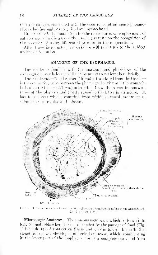
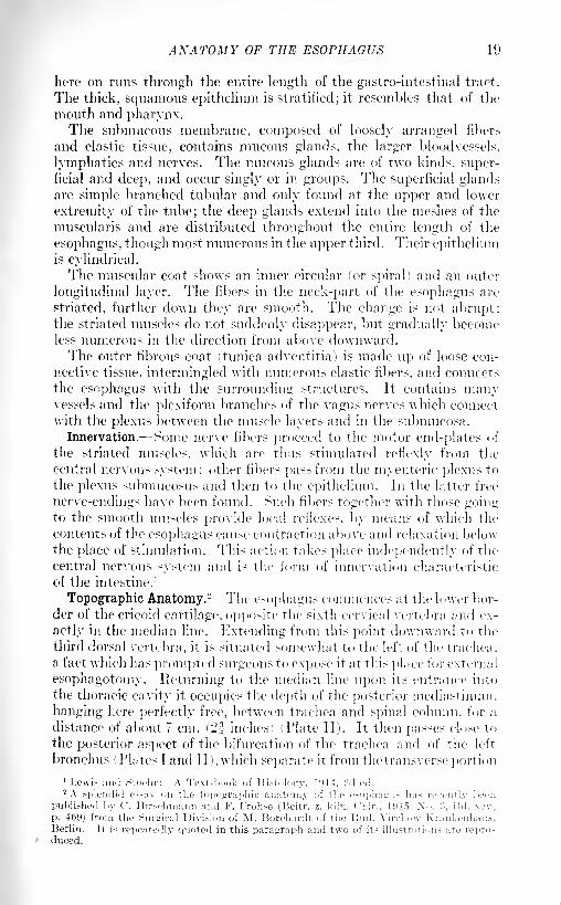
Microscopic Anatomy. The mucous membrane which is drawn into
longitudinal folds when it is not distended by the passage of food (Fig.1) is made up of connective tissue and elastic fibers. Beneath this
structure is a well-developed museularis mucos;e, which, commencingin the lower part of the esophagus, forms a complete coat, and from
ANATOMY OF THE ESOPHAGUS 19
here on runs through the entire length of the gastrointestinal tract.
The thick, squamous epithelium is stratified; it resembles that of the
mouth and pharynx.The submucous membrane, composed of loosely arranged fibers
and elastic tissue, contains mucous glands, the larger bloodvessels,
lymphatics and nerves. The mucous glands are of two kinds, super-ficial and deep, and occur singly or in groups. The superficial glandsare simple branched tubular and only found at the upper and lower
extremity of the tube; the deep glands extend into the meshes of the
muscularis and are distributed throughout the entire length of the
esophagus, though most numerous in the upper third. Their epitheliumis cylindrical.
The muscular coat shows an inner circular (or spiral) and an outer
longitudinal layer. The fibers in the neck-part of the esophagus are
striated, further down they are smooth. The change is not abrupt;the striated muscles do not suddenly disappear, but gradually becomeless numerous in the direction from above downward.The outer fibrous coat (tunica adventitia) is made up of loose con-
nective tissue, intermingled with numerous elastic fibers, and connects
the esophagus with the surrounding structures. It contains manyvessels and the plexiform branches of the vagus nerves which connect
with the plexus between the muscle layers and in the submucosa.
Innervation. Some nerve fibers proceed to the motor end-plates of
the striated muscles, which are thus stimulated reflexly from the
central nervous system; other fibers pass from the myenteric plexus to
the plexus submucosus and then to the epithelium. In the latter free
nerve-endings have been found. Such fibers together with those goingto the smooth muscles provide local reflexes, by means of which the
contents of the esophagus cause contraction above and relaxation belowthe place of stimulation. This action takes place independently of the
central nervous system and is the form of innervation characteristic
of the intestine. 1
Topographic Anatomy.2 The esophagus commences at the lower bor-
der of the cricoid cartilage, opposite the sixth cervical vertebra and ex-
actly in the median line. Extending from this point downward to the
third dorsal vertebra, it is situated somewhat to the left of the trachea.
a fact which has prompted surgeons to expose it at this place for external
esophagotomy. Returning to the median line upon its entrance into
the thoracic cavity it occupies the depth of the posterior mediastinum,
hanging here perfectly free, between trachea and spinal column, for a
distance of about 7 cm. (2f inches) (Plate II). It then passes close to
the posterior aspect of the bifurcation of the trachea and of the left
bronchus ( Plates I and ID, which separate it from thetransverse portion
'Lewis mid Stuchr: A Text-hook of Histology, 1914, LM ed.2 A splendid e.~say on the topographic au:iturny of the esophagus has reivntly hi/en
published by ( '. Hiix-limaim and F. Frohse (Beitr. z. klin. Cliir., lijlo, X<>. .'!, Bd. xcv,p. 4U'Jj from the Sunriral Division of M. Borchardt of the Kud. Yirchow Kraiikenhaus,Berlin. It is repeatedly quoted in this paragraph and two of its illustrations are repro-duced.
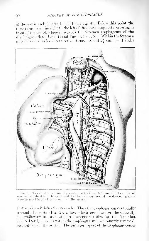
20 SURGERY OF THE ESOPHAGUS
of the aortic arch (Tlates I and IT and Fig. 4). Below this point the
tube turns from the right to the left of the descending aorta, crossing in
front of the vessel, where it reaches the foramen csophageum of the
diaphragm . IMales I and II and Figs. :>, 4 and 5). Within the foramen
it is imbedded in loose connective tissue. About 2^ cm. (= 1 inch)
PuimoPL.tX BROtiCrt POST..;
Diaphragma
vi'.-i pliii- ;niai"ii!\ nt' postcriur iiicili:i>t iiiuin : left hinj with heart turnedi\'cr l'i the ri^ht .~iile. The spiral made \<y the esopluijrus around the descending aorta
s i-xtmsrcnitcM in tin- illu.-tr;itioii. (('. Ileit/i
further down it joins t he stomach. Tims the esophagus curves spirally
around the aorta ( Fig. 2), a fact uliicli accounts for the difficulty
in swallowing in cases of aortic aneurysm; also for the fact that
pointed foreign bodies within the esophagus, unless promptly removed,so easily erode the aorta. The anterior aspect of the esophagus comes


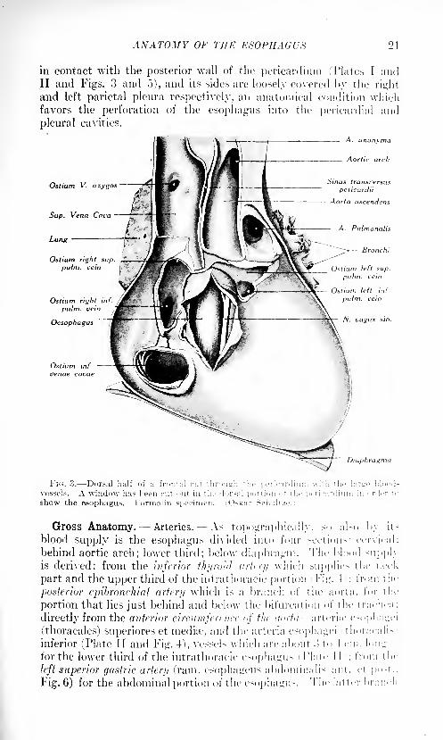
ANATOMY OF THE ESOPHAGUS 21
in contact with the posterior wall of the pericardium (Plates I andII and Figs. 3 and 5), and its sides are loosely covered l>y the right
and left parietal pleura respectively, an anatomical condition whichfavors the perforation of the esophagus into the pericardial and
pleura! cavities.
_ A. jnonvma
Ostium V. azygos
Lung
Aortic arch
Sinus transi'ersus
pericardii
Aorta ascendens
A. Pulmonalis
- Branch:
Ostium right suppulm. vein
Ostium right in
pulm. vein
Ostium left sup
pulm. vein
FIG. 3. Dorsal half of a frontal cut through the pericardium with the lartco blood-
vessels. A window lias been cut <>ut in the dorsal portion of the pericardium in order t
show the esophagus. Formalin specimen. (Oskar Schultze
Oesophagus
Ostium inf.
venae cavae
Gross Anatomy. Arteries. As topographically, so al-o by ns
blood supply is the esophagus divided into four sections: cervical;
behind aortic arch; lower third; below diaphragm. The blood supphis derived: from the inferior thyroid artrry which supplie> the nec-k
part and the upper third of the intrathoracic portion Fig. I ; from the
posterior epibronchial artery which is a branch of the aorta, for t Ill-
portion that lies just behind and below the bifurcation of the trachea :
directly from the anterior circumference <>f the a<>rta --arteri;e esophagei
(thoracales) superiores et medite, and tlie arteria esophagei thoracalis
inferior (Plate II and Fig. 4), vessels which are about :i to 1 cm. long
for the lower third of the intrathoracic esophagus Plate 1 1 ; from the
left superior gastric artery ('ram. esophagens abdomiualis ant. et po-t..
Fig. 6) for the abdominal portion of the esophagus. The latter branch
xrii'dKKY OF THE ESOPHAGUS
niiiy lc rather liiu', and then ijains special importance in resection of the
t'itrdiii.
Ramus oesophageus( cervicalis
Truncus lymphaticdexter
A. subclavia d.
N. recurrens d.
V. oesophagea d.
Lgl. mediastinales
post
vagus d.
N. vagus sin.
Oesophagus
Lgl. cardiacae
. ceruicales prof. inf.
Ductus thoracicus
V. oesophagea sin.
- N. recurrens sin.
Aorta descendens.
Plexus oesophageus.
oesophagea (thoracalis)
Lgl. mediastinales post,
inf.
Diaphragma
Oesophagus
Aorta abdominales.
Ductus thoracicus
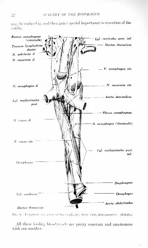
Vic,. 4.----Lynij>h:iti<- app.'ii'at us of the esophagus; front view, diagrammatic. (Sakata.)
All these feeding bloodvessels are pretty constant and anastomosewith one another.
AXATOMY OF THE KS
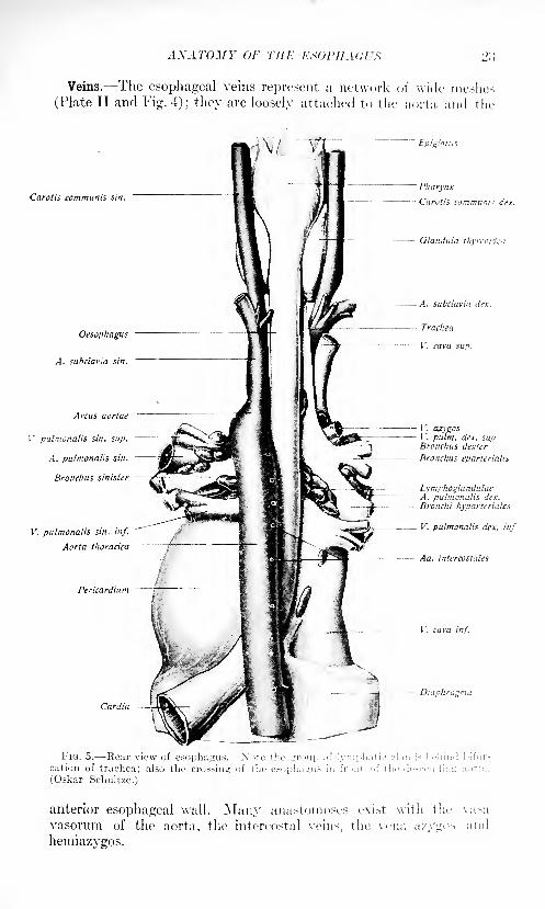
Veins. The esophageal veins represent a network of wide meshes
(Plate II and Fig. 4); they are loosely attaehed to the aorta and the
Carotis communis sin.
Oesophagus
A. subclavia sin.
Arcus aortae
V pulmonalis sin. sup.-
A. pulmonalis sin.
Bronchus sinister 2^ric
V. pulmonalis sin. inf.
Aorta thoradca
Pericardium
Cardia
Epiglottis
Pharynx
Carotis communis dot.
Glandula thvrcoida.
A. subclavia drx.
Trachea
V. cava sup.
V. cuygosV. pulm. dex. sup.Bronchus dexter
Bronchus eparterialt*
LymphoglandulaeA. pulmonalis dex.
Bronchi hyparteriates
V. pulmonalis dex. inf.
Aa. intercostales
-. V. cava inf.
Diaphragma
anterior esophageal wall. Many anastomoses exist with the vasa
vasorum of the aorta, the intercostal veins, the vena a/.ygos and
hemiazygos.
1M St'h'CERY OF THE ESOPHAGUS
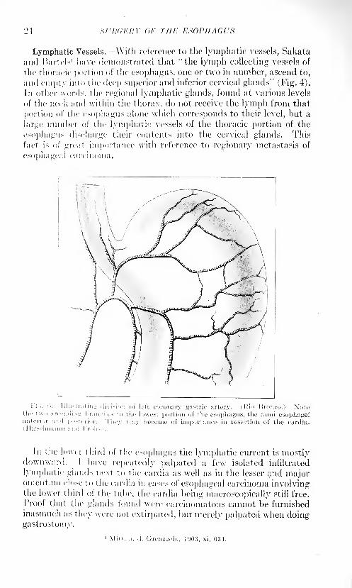
Lymphatic Vessels. With reference to the lymphatic vessels, Sakata
and Bartels 1 have demonstrated that "the lymph collecting vessels of
the thoracic portion of the esophagus, one or twro in number, ascend to,
and empty into the dee]) superior and inferior cervical glands" (Fig. 4).
In other words, the regional lymphatic glands, found at various levels
of the neck and within the thorax, do not receive the lymph from that
portion of the esophagus alone which corresponds to their level, but a
large number of the lymphatic vessels of the thoracic portion of the
esophagus discharge their contents into the cervical glands. This
fact is of great importance with reference to regionary metastasis of
esophageal carcinoma.
1 '' '' Illustratins: division of left coronary gastric artery. (Rio Bronco.) Xotetin- t \vo ascending brandies to the lowest portion of the esophagus, the raini esopha^eianterior and postrni,r. They may become of importance in resection of the cardia.
(.1 lirschmann ain 1 l-'r< ihse. i
In the lower third of the esophagus the lymphatic current is mostlydownward. I have repeatedly palpated a few isolated infiltrated
lymphatic glands next to the cardia as well as in the lesser and majoromentnm close to the cardia in cases of esophageal carcinoma involvingthe lower third of the tube, the cardia being macroscopically still free.
Proof that the glands found were careinomatous cannot be furnishedinasmuch as they were not extirpated, but merely palpated when doinggastrostomy.
'Mitt. a. d. Gremwb., 1903, xi, 634.
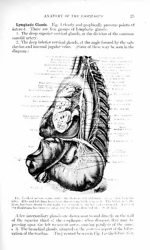
ANATOMY OF TIIK K
Lymphatic Glands.- Fig. 4 clearly and graphically presents points of
interest. There are five groups of lymphatic glands:1. The deej) superior cervical glands, at the division of the common
carotid artery.
'2. The deep inferior cervical glands, at the angle formed by the suh-
clavian and internal jugular veins. (Some of these may be seen in the
diagram.)
IIG. i. Lett nervu> vairu-; within the thi>l';trir ;m<l a: >d' 'initial ravit\ view li in tl
loft). Iiib< and left lunti have lieeti alnn>~l roni|.letely removed. 'I'he latter. \\i'li ih
liver, lias !;een drawn to the riirlit. the stuiii;irh to the lot't aiid iln\vn\v:iril
the diaphragm has heen fut away and the pleura <li~.~ccted "I'l
A few intermediary glands arc >ho\vn near to and direct I\ on the wall
of the superior third of the esophagus; when diseased, they may Im-
pressing upon the left recurrent nerve, can-ing paralysis of the sime.
F 3. The bronchial gland-, situated on the /><>*fi r/nr aspect of t he bifur-
cation of the trachea. > The cannot he seen in Fig. 1 as tin- bifurcatii n
26 SURGERY OF THE ESOPHAGUS
covers them. See Fig. 5.) The greater number of the esophageal
lymphatic glands is formed by*
4. The posterior mediastinal glands, which are scattered around and
along the thoracic aorta from the tracheal bifurcation down to the
hiatus esophageus (Fig. 4).
5. Cardiac glands, two to three in number (Fig. 4).
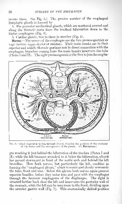
Nerves. The nerves of the esophagus are the two pneumogastrics or
vagi (nervus vagus dexter et sinister). Their main trunks are in their
superior and middle thoracic portions not in direct connection with the
esophagus; branches coming from the main trunks innervate the tube
(Plates I and II). The right pneumogastric is the first tojointheesopha-
A.itV.V.ina.iB.ma.r. int.
FIG. 8. Ideal transverse section through thorax, showing the position of the contents
of the thorax and the arrangement of the pleura. (C. Heitzmann.)
gus reaching it just behind the bifurcation of the trachea (Plates I and
II), while the left becomes attached to it below the bifurcation, after it
has passed downward in front of the aortic arch and behind the left
bronchus. Here both nerves, but particularly the left, combine in
forming the "esophageal plexus," which is motor and closely surrounds
the tube, front and rear. Below this plexus both nerves again present
separate bundles, before they enter into and pass with the esophagus
through the foramen esophageum of the diaphragm. The right is
situated further back than the left and innervates the posterior wall of
the stomach, while the left can be seen more to the front, dividing uponthe anterior gastric wall (Fig. 7). This anatomically shifted position
ANATOMY OF THE ESOPHAGUS 27
of the nerves is explained by the turn of the stoiuacli during embry-ological development with its left portion forward, while the right
portion, and with it the right pneumogastric nerve, turns backward.
Carolis dex.
Subclavia Hex.
Truncus jugularis dex.
V. jugul. com. dex.
A. anonymaTruncus subclavius dex. --"
V. subclavia dex. .---\
V. anonyma dex.
Truncus bronchia mcdi-astinalis dex.
V. cava sup
Aosta ascend
V. ;ug. com. sin.
Truncus jngul sin
Tr. subclavius sin.
V. subclavia sin.
_ V. anonyma sin.
\rcus aortae
\ona thonciai
Ductus thoracicus
_ _O\terna chyti
Truncus lumbtilis w'n.
Triincus int, stinalis
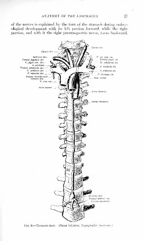
FIG. 9. Thoracic duct. (Oscar Scliultze, Topographic Anatomy.)
2S sntdKItY OF THE ESOPHAGUS
Thoracic Duct, Vena Azytjos niul Hemiazygos, and Sympathetic
AYm-.v.- -lust above the diaphragm the thoracic duct and the vena
a/ygos run alongside the right border of the esophagus in front of the
spinal column (Fig. 8). The thoracic duct, within loose connective
tissue, runs exactly in the median line, straight upward (Fig. 9);
above the a >rtic arch it is found on the left side of the esophagus( Fig. \ i and continues so to its entrance into the left subclavian vein,
on the posterior aspect of the latter's junction with the internal jugular
vein.
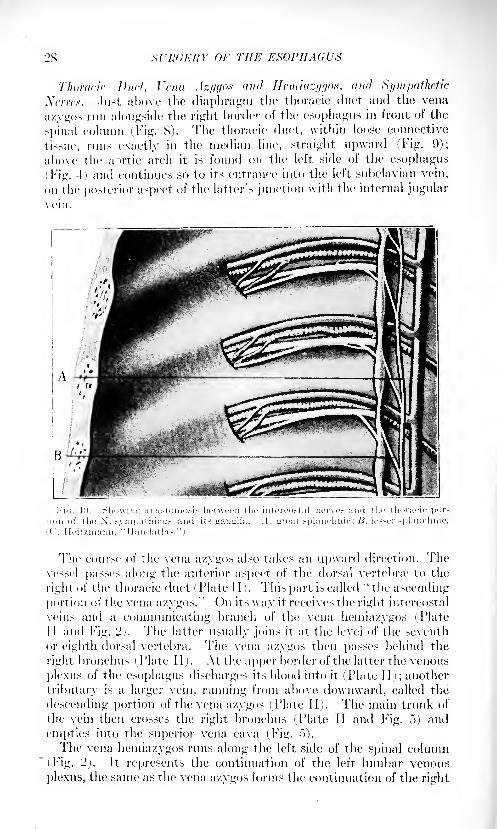
The course of the vena a/ygos also takes an upward direction. Thevessel pusses along the anterior aspect of the dorsal vertebne to the
right of the thoracic duct (Plate II). This part is called "the ascending
portion of the vena azygos." On its way it receives the right intercostal
veins and a communicating branch of the vena hemiazygos (PlateII and Fig. '!). The latter usually joins it at the level of the seventh
or eighth dorsal vertebra. The vena a/ygos then passes behind the
right bronchus (Plate 11). At the upper border of the latter the venous
plexus of the esophagus discharges its blood into it (Plate II); another
tributary is a larger vein, running from above downward, culled the
descending portion of the vena a/ygos (Plate II). The main trunk of
the vein then crosses the right bronchus (Plate II and Fig. 5) and
empties into the superior vena cavu (.Fig. .">).
The vena hemiazygos runs along the left side of the spinal column
(Fig. '2). It represents the continuation of the left lumbar venous
plexus, the same as the vena a/ygos forms the continuation of the right
PHYSIOLOGICAL POINTS OF THE ESOPHAGUS
lumbar venous plexus. It collects the blood from all the twelve left
intercostal veins: those from the lower five ribs empty into it directly
and separately; those from the first to the seventh discharge into the
superior hemiazygos vein, which joins the
lower trunk before it crosses the body of
the seventh or eighth dorsal vertebra.
There are many variations.
Sympathetic Ncrrcs. The thoracic por-tion of the sympathetic nerves is situated
outside of the posterior mediastinum, in
front of the capitulum of the ribs (Figs. 2,
7, and 10) and is covered by the costal
pleura. The eleven thoracic ganglia of
the sympathetic nerves are connected bytwo nerves, which also communicate with
the respective intercostal nerves.
There. are four plexuses: cardiac, aortic,
pulmonary and esophageal. The esopha-
geal and pulmonary plexuses are in closest
relationship to the plexuses of the two
pneumogastrics. Fibers of the latter are
also nourished by the cardiac and aortic
plexuses and the superior thoracic ganglia
(1'ig- ')
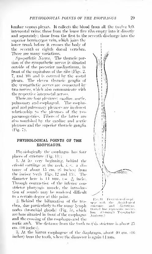
PHYSIOLOGICAL POINTS OF THEESOPHAGUS.
Physiologically the esophagus has four
places of stricture (Fig. 11):
1. At its very beginning, behind the
cricoid cartilage at the neck, /. e., a dis-
tance of about b") cm. (0 inches) fromthe incisor teeth (Figs. 12 and ]:>). Thediameter here is 14 mm. (= -^ inch).
Through contraction of the inferior con-
strictor pharyngis muscle, the introduc-
tion of sounds may be rendered difficult
to a certain degree at this point.2. Behind the bifurcation of the tra-
chea, due particularly to the many lymphnodes (bronchial glands) (Fig. .">), whichare here situated in front of the esophagusand the crossing of the esophagus and the
aortic arch. The distance from the teeth to this stricture is aboutcm. (10 inches).
3. At the hiatus esophageus of the diaphragm, about 40 cm. i
inches) from the teeth, where the diameter is again 14 mm.
30 SURGERY OF THE ESOPHAGUS
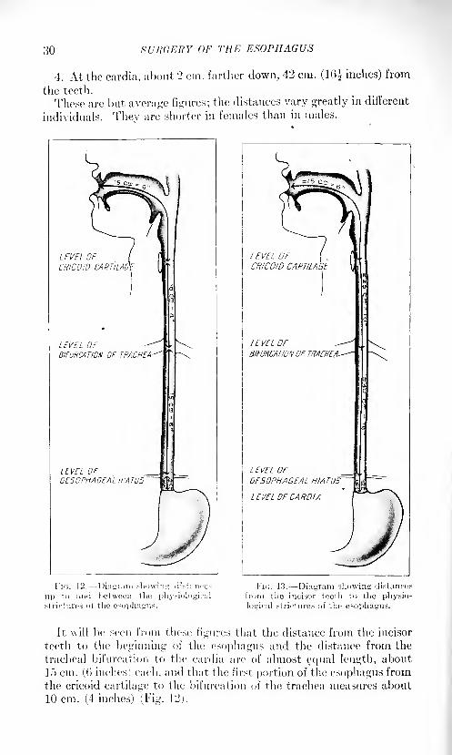
4. At the cardia, about 2 cm. farther down, 42 cm. (16| inches) from
the teeth.
These are but average figures; the distances vary greatly in different
individuals. Thev are shorter in females than in males.
LEVEL OFCRICOJb CAFttMljE
LEVEL OF'BIFURCATION OF TRACHEA
LEVEL orOESOPHAGEAL HIATUS
LEVEL OFCRICOID'CARTILAGE
LEVEL OF ^-BIFURCATIONOF TRACHEA-
L EVEL OFOE50PHAGEAL HIATUS'
LEVEL OF CARDIA
I'lc",. 12. Diagram showing di>l:nice>
up to and between the physiologicalstrictures ot the esophagus.
FK;. 1.3. -Diagram showing distances
from the incisor teeth to the physio-
logical strictures of the esophagus.
It will be seen from these figures that the distance from the incisor
teetli to the beginning of the esophagus and the distance from the
tracheal bifurcation to the cardia are of almost equal length, about
\^ cm. ((') inches) each, and that the first portion of the esophagus from
the cricoid cartilage to the bifurcation of the trachea measures about
10 cm. (4 inches) (Fig. 12,).
EXAMINATION OF THE ESOPHAGUS 31
EXAMINATION OF THE ESOPHAGUS.
As regards examining the esophagus, recent developments haveadded greatly to our facilities for that purpose and have placed us in a
position to diagnose its pathological conditions. We shall take into
consideration
I. The Older Methods of Examination. Sounding. As in other
canals of the body, sounding is done for the purpose of locating and
dilating strictures.
Cf
(a) Posture of the Patient. It is best to have the patient seated;
his clothes properly protected by some kind of cover, preferablv imper-
meable, a basin in his hand to catch saliva or stomach contents. Tooth
plates must be removed. By bending the head slightly forward the
entrance into the esophagus is opened. The majority of patients are
inclined to sit back in the chair and raise the head as far as possible,
looking at the ceiling. That is wrong, as in this posture the contents
of the mouth will flow back toward the larynx and produce coughing.
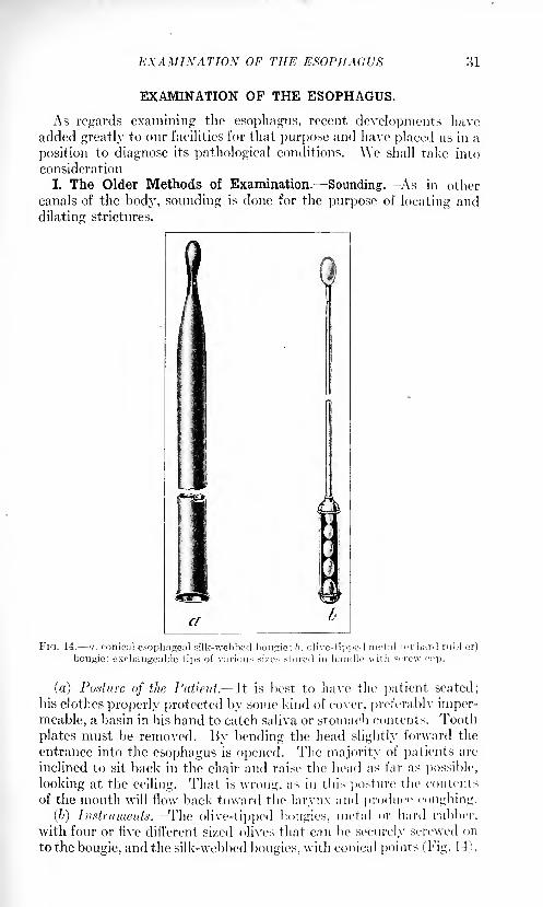
(6) Instruments. The olive-tipped bougies, metal or hard rubber,
with four or five different sized olives that can be securely screwed on
to the bougie, and the silk-webbed bougies, with conical points (Fig. 14),
32 SURGERY OF THE ESOPHAGUS
should be at hand. The first variety of instruments is used for diag-
nostic purposes; the latter for treatment.
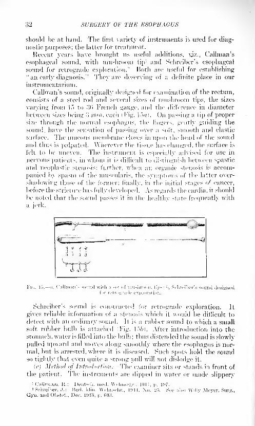
Recent years have brought us useful additions, vj>/., Callman's
esophageal sound, with mushroom tip1 and Schreiber's esophageal
sound for retrograde exploration.2 Both are useful for establishing
"an early diagnosis.'' The}' are deserving of a definite place in our
instrumentarium .
Callman's sound, originally designed for examination of the rectum,consists of a steel rod and several sixes of mushroom tips, the sixes
varying from 1f> to :>(') French gauge, and the difference in diameter
between sixes being >} mm. each (Fig. l.Vn. On passing a tip of propersixe through the normal esophagus, the fingers, gently guiding the
sound, have the sensation of passing over a soft, smooth and elastic
surface. 'Die mucous membrane closes in upon the head of the sound
and thus is palpated. Wherever the tissue has changed, the surface is
felt to be uneven. The instrument is especially advised for use in
nervous patients, in whom it is difficult to distinguish between spasticand neoplastic stenosis; further, when an organic stenosis is accom-
panied by spasm of the muscularis, the symptoms of the latter over-
shadowing those of the former; finally, in the initial stages of cancer,
before the stricture has fully developed. As regards the cardia, it should
be noted that the sound passes it in the healthy state frequently with
a jerk.
*i
Schreiber's sound is constructed for retrograde exploration. It
gives reliable information of a stenosis which it would be difficult to
detect with an ordinary sound. It is a rubber sound to which a small
soft rubber bulb is attached (Fig. !.">/>). After introduction into the
stomach, water is filled into the bulb; thus distended the sound is slowly
pulled upward and moves along smoothly where the esophagus is nor-
mal, but is arrested, where it is diseased. Such spots hold the soundso tightly that even quite a strong pull will not dislodge it.
(c) Method <>f Introduction. The examiner sits or stands in front of
the patient. The instruments are dipped in water or made slippery1 Culhnann, R.: Deiilx-li. nu2 Schreiber, J.: Bcrl. klin. \\
Gyn. and Obstet., Dec. 1913, p. G9H.\ViIly Meyer, Surg.,

EXAMINATION OF THE ESOPHAGUS 33
with some lubricant, a jelly preferably. Olive oil, vaselin and glycerinare objectionable to many patients. The tip of the left finger is placed
upon the back of the patient's tongue and presses it gently forward.
The right hand introduces the sound either in the median line or some-wrhat to the left of it. Contact with the buccal mucosa should be
avoided, as it produces gagging. In very sensitive patients, particularlythose with pronounced reflexes, a brief spraying or touching-up with
cocain (10 per cent.) may be of great benefit. Local analgesia is not
required. The patient is told to swallow, and with that the tip of the
instrument enters the esophagus. The physiological strictures often
offer slight resistance to the downward passage of the bougie, par-
ticularly the first one behind the cricoid cartilage. Most gentle
handling of the sound is imperative during the entire manipulation,on introduction as well as on withdrawal. All force must be absolutelyavoided.
The tip of the instrument may perforate the wall of the tube byrough handling.
Gagging, following the introduction, is often overcome by quick,
deep breathing.Auscultation. Under normal conditions, both solid as well as liquid
food is pushed forcibly through the pharynx and esophagus in the act
of deglutition. That is due to the strong contraction of the muscles
which close the oral cavity, particularly the mylohyoids. This phe-nomenon produces two sounds: the so-called "deglutition sounds,"
first described by Meltzer and Kronecker in 1SS3. The "first degluti-
tion sound" is at times heard simultaneously with the act of swallow-
ing on auscultating at the ensiform process. It is produced by the
fluid being "spurted" down the tube "Durchspritzgerausch." Often
this sound is not perceptible. The second sound is more frequently
noted, occurring about seven seconds after the act of deglutition. It
is produced at the end of the act of swallowing, by the peristaltic con-
traction of the esophagus, which pushes the food into the stomach
"Durchpressgerausch." It is a rale-sound, dependent on the amountof air that travels downward with the contents of the esophagus.The presence of these deglutition sounds permits us to judge, to some
degree, of the permeability of the cardia. If both are absent, especially
the second one, we are justified in assuming that the ingested mass ha>
not entered the stomach but is retained in the esophagus above the
cardia. This is most often the case in strictures (cancer^ although,
occasionallv, this condition might be caused by a deficiency in the peri-
staltic movement of the esophagus.II. The Newer Methods of Examination. Esophagoscopy. The ad-
vent of the electric mignon-lamp and its adaptation to the requirementsof endoscopic examination as well as of operative surgery within a
canal that can be entered from the surface of the body has wrought the
same advance for the esophagus as for the bladder and ureters, the
rectum, bronchi, etc. It aids the diagnosis by permitting (I) direct
visual inspection, (2) the punching out of projecting tumor masses.
VOL. II 3
34 SURGERY OF THE ESOPHAGUS
(3) the scraping of an ulceration for microscopic examination. It is
also of great value in the bloodless extraction of foreign bodies that
were swallowed and became stuck in the esophagus, also in the stretch-
ing of strictures under the guidance of the eye, etc. Ksophagoscopy,within the last, fifteen (<> twenty years, lias gradually developed into
a. specialty. 'Today every hospital should have its official esophagos-copist, with the same right and on the basis of the' same necessity
EXAMINATION OF THE ESOPHAGUS 35
that calls for a cystoscopist, radiologist, etc. Needless to say that the
esophagoscopist should be so thoroughly trained that no harm can byany possibility befall patients through his examination. The colleagueswho call on him for assistance and entrust to him their patients for
examination must be able to feel absolute confidence and assuranceas regards this point.
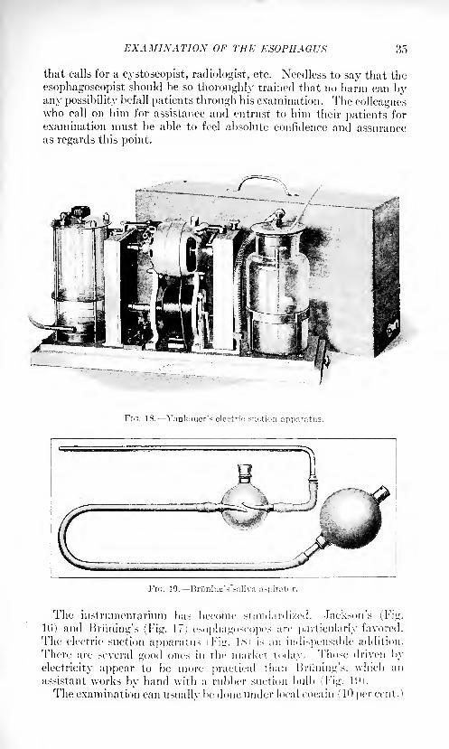
FIG. 18. Yankauer's electric suction apparatus.
l"n;. 19. Bri'iniu
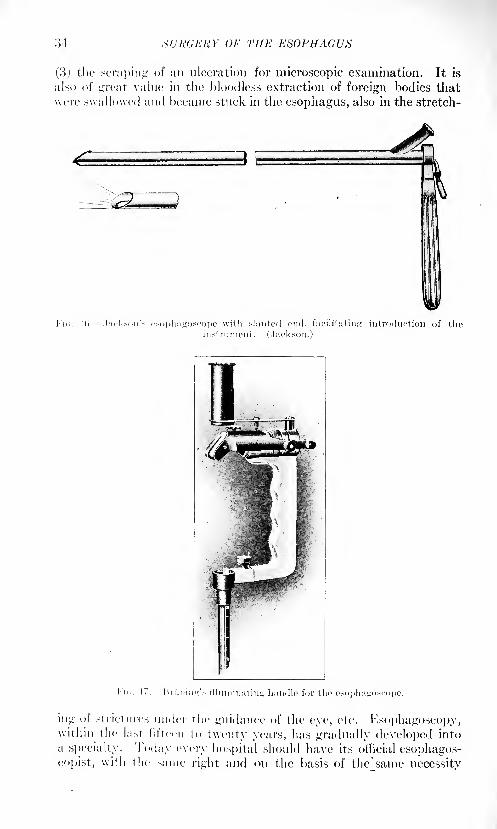
The instrtimentarium has become standardized. Jackson's (Fig.
If)) and Hnining's (Fig. 17] esophagoscopes are particularly favored.
The electric suction apparatus Fig. ISi is an indispensable addition.
There are several good ones in the market today. Those driven by
electricity appear to be more practical than Bn'ining's, which an
assistant works by hand with a rubber suction bulb ('Fig. l!i.
The examination can usually be done under local coca in (10 per cent.")
3(5 SURGERY OF THE ESOPHAGUS
anesthesia, although the patients should always be prepared for generalanesthesia. The horizontal posture appears preferable, the head hang-
ing backward from the table (Hose's posture). The patient's neck is
supported by the arm of an assistant, whose hand at the same time
takes hold of the mouth-gag, which had been put in place. The esopha-
goscope, without obstructing mandrill, is gradually pushed forward
into the tube, same as we proceed in urethroscopy, rectoscopy, etc.
In this way nothing abnormal will escape the inspecting eye.These brief remarks have to suffice with reference to esophagoscopy.
Those more particularly interested in this branch of endoscopic diag-nosis and therapy are referred to the huge literature on the subjectthat has accumulated within the last two decades.
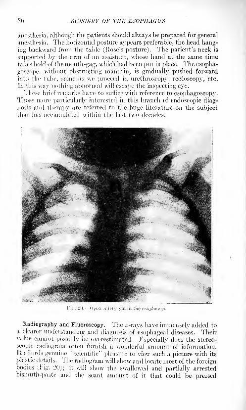
FIG. 20. ---Open safety pin in the esophagus.
Radiography and Fluoroscopy. The .r-rays have immensely added toa clearer understanding and diagnosis of esophageal diseases. Theirvalue cannot possibly be overestimated. Especially does the stereo-
scopic radiogram often furnish a wonderful amount of information.It ail'ords genuine "scientific" pleasure to view such a picture with its
plastic details. The radiogram will show and locate most of the foreignbodies (Fig. 120); it will show the swallowed and partially arrested
bismuth-paste and the scant amount of it that could be pressed
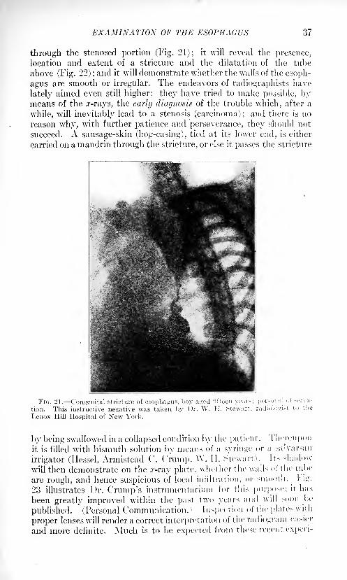
EXAMINATION OF THE ESOPHAGUS 37
through the stenosed portion (Fig. 21); it will reveal the presence,
location and extent of a stricture and the dilatation of the tube
above (Fig. 22) ;and it will demonstrate whether the walls of the esoph-
agus are smooth or irregular. The endeavors of radiograph ists have
lately aimed even still higher: they have tried to make possible, bymeans of the x-rays, the early diagnosis of the trouble which, after a
while, will inevitably lead to a stenosis (carcinoma); and there is no
reason why, with further patience and perseverance, they should not
succeed. A sausage-skin (hog-casing), tied at its lower end, is either
carried on a mandrin through the stricture, or else it passes the stricture
FIG. 21. Congenital stricture of esophagus, boy aired fifteen year-: per.-oiui] ol.-erva-
tion. This instructive negative was taken by Dr. W. II. Stewart, radiulo<:i.-t tu the
Lenox Hill Hospital of New York.
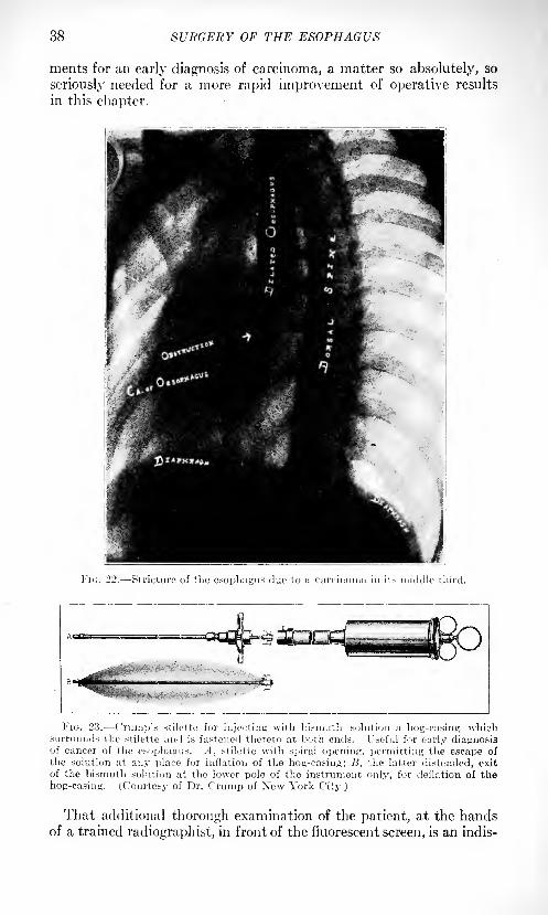
by being swallowed in a collapsed condition by the patient. Thereuponit is filled with bismuth solution by means of a syringe or a salvarsan
irrigator (liessel, Armistead C. Crump, W. II. Stewart'. Its shadow-
will then demonstrate on the .r-ray plate, whether the walls of tin 1 tube
are rough, and hence suspicious of local infiltration, or smooth. Hg.
23 illustrates Dr. Crump's instrunientarium for this purpose; it has
been greatly improved within the past two years and will soonJ>e
published. (Personal Communication. 1 Inspection of the plates with
proper lenses will render a correct interpretation of the radiogram easier
and more definite. Much is to be expected from these recent experi-
38 SURGERY OF THE ESOPHAGUS
merits for an early diagnosis of carcinoma, a matter so absolutely, so
seriously needed for a more rapid improvement of operative results
in this chapter.
Fi<;. 22. Stricture of the esophagus due to a carcinoma in it.- middle third.
FIG. 23. C 'rump's stilctte for injecting with bismuth solution a hog-casing whichsurrounds the stiletto and is fastened thereto at both ends. Useful for early diagnosisof cancer of the esophagus. A, stiletto with spiral opening, permitting the escape of
the solution at any place for inflation of the hog-casing; B, the latter distended, exit
of the bismuth solution at the lower pole of the instrument only, for deflation of the
hog-casing. (Courtesy of Dr. C'rump of New York City.)
That additional thorough examination of the patient, at the handsof a trained radiographist, in front of the fluorescent screen, is an indis-
SURGICAL PATHOLOGY OF THE ESOPHAGUS 39
pensable procedure, and represents a most important addition to diag-nostic resources and capabilities, hardly needs emphasis in a surgical
text-book at this late day.
Regarding the sequence of the various examinations to lie made,one may best proceed as follows:
1. In ( V/.sr.y /// Which the Xt/iu [Joins of Stricture ore Clearly Developed.
History of the case: general clinical examination with observation
of deglutition sounds; usual laboratory tests of urine and blood,
inclusive of a Wassermann test; radiography. If an aortic aneurysmof the arch or of the descending portion of the aorta can be excluded:
sounding, or immediate esophagoscopy. As the tubes used for the
latter examination have a centimeter-scale engraved on them, they
permit of measuring the distance from incisor teeth to stricture.
2. Early Cases. It may be advisable, after the preliminary clinical
and laboratory examinations have been completed, first to make gentle
use of Callmann's or ^chreiber's sound, in order to find out where the
stricture in the lumen of the esophagus is located; then to do esopha-
goscopy and follow this up by radiography. A trained eye will readily
recognize through the esophagoscope any changes in the appearanceof the mucosa. The distance from the teeth having been measured
and compared with the result of sounding, the radiographist will then
know just how far to introduce the casing that is to be filled with
bismuth or barium preparation. U"-'ee above, tinder "Radiography.")The foregoing represents a brief outline of the great advance that
has been made in recent years as regards the possibility of renderinga more refined diagnosis in the pathology of the esophagus. The 1 indi-
cations are that much more may be looked for in the near future.
SURGICAL PATHOLOGY OF THE ESOPHAGUS.
I. Foreign Bodies in the Esophagus.
Foreign bodies often slip accidentally into the esophagus, and byreason of size or shape or sharp points become lodged therein. I sually
they are arrested in their downward course at the physiologically nar-
row places of the thoracic part of the tube, viz., behind the aortic arch
and above the cardia.
Frequently these arrested foreign bodies can still be made to pass
down into the stomach much to the delight of the frightened indi-
vidual by having him swallow crusts of bread or potatoes imme-
diately after the accident. But if, in times past, the sensation ot the
presence of a foreign body persisted, "blind" methods of >earch and
treatment had to be resorted to. Then extraction was attempted e'uher
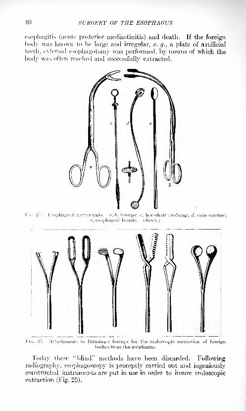
by means of forceps (Fig. 24) or by specially designed instruments:
brush, coin-catcher, etc. (Fig. 24), or else the foreign body was pushedinto the stomach. Frequently this would happen unintentionally. In
unfortunate cases, however, the obstacle was driven further into, and
not infrequently through the esophageal wall, can-ing septic peri-
40 SURGERY OF THE ESOPHAGUS
esophagitis (acute posterior mediastinitis) and death. If the foreign
hody was known To be large and irregular, e. g., a plate of artificial
teeth, external esophagotomy was performed, by means of which thebodv was often reached and successfully extracted.
FH;. -1. Ksophageul instruments, n, >>, forceps; c, horsehair probang: d, coin-catcher
, esopluigen] bougie. (Keen.)
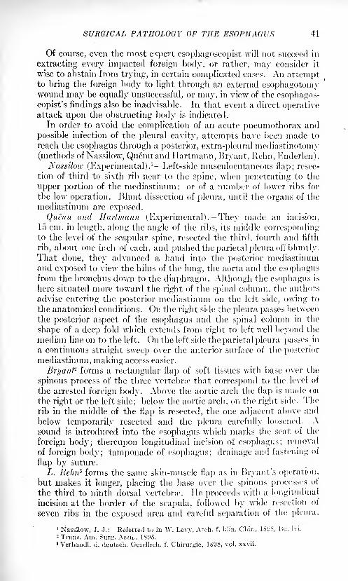
Fi<;. '2-~>. -Attachments to Bri'ming's forceps for the endoscopic extraction of foreignbodies from the esophagus.
Today these "blind" methods have been discarded. Followingradiography, esophagoscopy is promptly carried out and ingeniouslyconstructed instruments are put in use in order to insure endoscopicextraction (Fig. 25).
SURGICAL PATHOLOGY OF THE ESOPHAGUS 41
Of course, even the most expert esophagoscopist will not succeed in
extracting every impacted foreign body, or rather, may consider it
wise to abstain from trying, in certain complicated cases. An attemptto bring the foreign body to light through an external esophagotomywound may be equally unsuccessful, or may, in view of the esophagos-
copist 's findings also be inadvisable. In that event a direct operativeattack upon the obstructing body is indicated.
In order to avoid the complication of an acute pneumothorax and
possible infection of the pleural cavity, attempts have been made to
reach the esophagus through a posterior, extra-pleural mediastinotomy(methods of Xassilow, Quenu and Ilartmann, Bryant, Helm, Enderlen).
Nassiloic (Experimental).1 Left-side musculocutaneous flap; resec-
tion of third to sixth rib near to the spine, when penetrating to the
upper portion of the mediastinum; or of a number of lower ribs for
the low operation. Blunt dissection of pleura, until the organs of the
mediastinum are exposed.
Quenu and Ilartmann (Experimental). They made an incision,
15 cm. in length, along the angle of the ribs, its middle correspondingto the level of the scapular spine, resected the third, fourth and fifth
rib, about one inch of each, and pushed the parietal pleura off bluntly.That done, they advanced a hand into the posterior mediastinumand exposed to view the hilus of the lung, the aorta and the esophagusfrom the bronchus down to the diaphragm. Although the esophagus is
here situated more toward the right of the spinal column, the authors
advise entering the posterior mediastinum on the left side, owing to
the anatomical conditions. On the right side the pleura passes between
the posterior aspect of the esophagus and the spinal column in the
shape of a deep fold which extends from right to left well beyond the
median line on to the left. On the left side the parietal pleura passes in
a continuous straight sweep over the anterior surface of the posterior
mediastinum, making access easier.
Bryant2 forms a rectangular flap of soft tissues with base over the
spinous process of the three vertebra1 that correspond to the level of
the arrested foreign body. Above the aortic arch the flap is made on
the right or the left side; below the aortic arch, on the right side. Therib in the middle of the flap is resected, the one adjacent above and
below temporarily resected and the pleura carefully loosened. Asound is introduced into the esophagus which marks the seat of the
foreign body; thereupon longitudinal incision of esophagus; removal
of foreign body; tamponade of esophagus; drainage and fastening ot
flap by suture.
L. Itehn3 forms the same skin-muscle flap as in Bryant's operation,
but makes it longer, placing the base over the spinous processes of
the third to ninth dorsal vertebra*. lie proceeds with a longitudinal
incision at the border of the scapula, followed by wide resection of
seven ribs in the exposed area and careful separation of the pleura.
1 Nassilow, J. J.: Referred to in W. Levy, Arch. f. klin. Chir., 189S, Bd. Ivi.
2 Trans. Am. Surg. Assn., 1895.
*Verhaudl. d. deutsch. Gesellsch. f. Cbirurgie, 1898, vol. xxvii.
42 SURGERY OF THE ESOPHAGUS
Eiulcrlni'x 1
operation is similar to Helm's. lie points out the fact
that, the skin-muscle-bone flap can be nicely and safely formed by first
exposing the eighth rib in its periostea! envelope for a distance of 10
cm. (4 inchesi; then dividing it, and pushing oil' the pleura (see Fig. 50
on page SOX From here the next rib above is easily reached, againthe pleura pushed oil' and the rib divided, and so on, up to the fourth.
Thus a large flap of bone and muscle is formed. Inasmuch as the
preservation of the divided ribs with the soft parts is of no special
importance, they had best be removed. Gradual stripping off of
pleura toward the spinal column and anteriorly is the next step. Care
has to be exercised not to injure the sympathetic nerves, the vena
azygos and the thoracic duct and, later on, the nervi vagi, while the
esophagus is made more conspicuous by means of the introduction of
a sound.
The fact that in all of the foregoing operations the operating field
is situated from 1:2 to 14 cm. below the level of the skin, renders this
way of advance technically very difficult.
As to the side of approach toward the posterior mediastinum, it
seems best to advance from the right side, that is to say, by makingthe incision to the right of the spinal column.
Kndeiien performed the first successful operation on the thoracic
portion of the esophagus by removing from it an arrested tooth-plate,
by means of mediastinal esophagotomy.
(Reisinger2successfully resected, by this route, a portion of the esoph-
agus dilated in the longitudinal direction and closed the rent by sutures.)
Today it will be safer, when doing operations of this kind, to have
differential pressure apparatus hand}', in order to be prepared for
accidental injury of the pleura.
Some surgeons adhere to this retropleural advance today; others
favor the transpleural route.
The plan of an esophagotomy by the transpleural route is as follows:
Thoracotomy in the seventh intercostal space, the incision to be
lengthened, if necessary, by division of the angle of a few ribs upwardand posteriorly (see below) or, resection of as many ribs within the
oblique incision, as the case may demand; thorough exposure and
separation of the affected portion of the esophagus; gentle milking of
the same upward and downward, starting compression near the seat
of the body and shutting off the operating field with rubber-covered
branches of bayonet clamps above and belo\v; careful tamponade;incision over the foreign body; extraction; suture; postoperative
drainage of the pleural cavity. (Regarding drainage, see further down,under "Cancerous Stricture.")
II. Injuries of the Esophagus.
A. Acute Perforation. (a) From the outside by bullet wounds,cut wounds, stab wounds; (b) from the inside by pointed foreign
1 Deut.-eli. Ztschr. f. Chir.. 1901. Ixi, 440.2 Vcrhandl. d. deutt-ch. Gesellsch. f. Chirurgie, 1907.
SURGICAL PATHOLOGY OF THE ESOPHAGUS 43
bodies (pins, bones, tooth plates, etc. ); attempts at extraction of
the same; unskilled handling of the esophagoscope; (>) spontaneous
rupture.
(a) Bullet Wounds. Bullet wounds of the esophagus are a rare
occurrence in Avar as well as in times of peace.L. Bergheimer, in his thesis, 1903, "Bullet Wounds of the Xeck
Portion of the Esophagus," presented a series of 31 cases collected from
the literature. In some the diagnosis is doubtful. They were taken
from The Medical and Surgical History of the War of the Rebellion,
1861-65, and The Sanitary Report of the German Armies in the Franco-
German War, 1870-1. Madelung briefly refers to this collection in a
recent paper,1 and adds 5 cases seen in the first three years of the late
war, 1914-16, treated at the Strassburg hospitals. In 3 of these the
neck portion had been injured; in 2, the thoracic portion. Three times
only one side of the wall of the esophagus was perforated; in 1 both
anterior and posterior walls were pierced; in another case an injury to
the aorta complicated the lesion. The important nerves and vessels
of the neck as well as larynx and trachea had escaped injury; but in
some of the cases the lungs and spinal column were involved.
Diagnosis of acute injury of the esophagus in the neck portionso early that help by prompt operative intervention was still possible,
proved a difficult task. This particularly for the reason that in
none of the cases reported did food escape from the wound in the neck
during the first days after the injury, not even when the external
wounds were thoroughly enlarged, as had been done in three of the
cases. The patients complained of difficulty in deglutition right after
the injury, but all were able to swallow.
The location of entrance and exit of the bullet does not prove much.
\Vhere no exit occurred, a radiograph will assist in establishing the
diagnosis of an esophageal injury. Esophagoscopy is contra-indicated.
The proper procedures are: (1) in case of an injury situated high up,
external esophagotomy, with feeding through a permanent esophageal
tube; (2) in case of an injury in the thoracic portion, gastrostomy.In the literature cases are reported of the spontaneous healing of
small shot wounds in the esophageal wall. Escape of food and the
formation of a temporary neck fistula do not always occur. Usually,
however, mediastimtis will set in and cause the death of the patient.
More of the seriously injured individuals will likely be saved in future
by the combination of early, free incision of the wound in the neck,
with thorough exposure and careful exploration of the neck portion of
the esophagus; wide incision and drainage of mediastinal abscesses,
and particularly the absolute operative exclusion of the injured por-
tion of the esophagus for the purpose of nutrition by gastrostomy (or
esophagotomy), with a permanent rubber tube in the esophagus.The same reasoning holds good in cases of cut or stab wounds of
the neck, involving the esophagus.
1 Madelung, O.: Deutsch. nied. Wc-hnsdir., 1(
J1.">, X:>. ">, p. 1-1.
44 SURGERY OF THE ESOPHAGUS
(b] Pointed Forcitjn bodies and Attempts at Their Extraction, also,
not infrequently, the esophagoscope in unskilled hands, may cause
perforation of the esophagus in its thoracic portion.
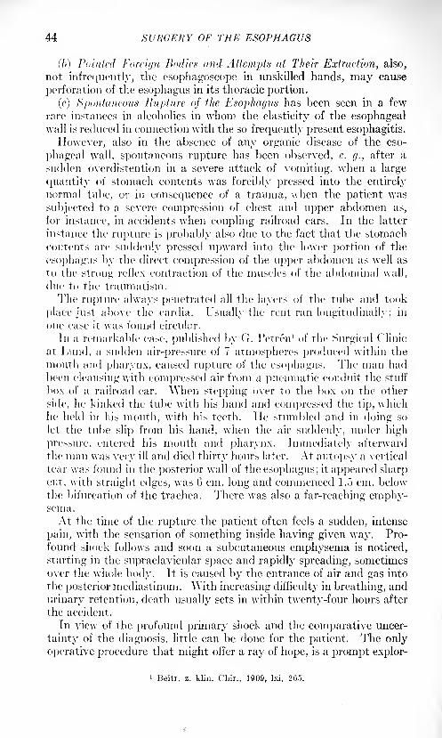
(e) Spontaneous Ihipture of the Esophagus has been seen in a few
rare instances in alcoholics in whom the elasticity of the esophagealwall is reduced in connection with the so frequently present esophagitis.
However, also in the absence of any organic disease of the eso-
phageal wall, spontaneous rupture has been observed, c. g., after a
sudden overdistention in a severe attack of vomiting, when a large
quantity of stomach contents was forcibly pressed into the entirely
normal tube, or in consequence of a trauma, when the patient was
subjected to a severe compression of chest and upper abdomen as,
for instance, in accidents when coupling railroad cars. In the latter
instance the rupture is probably also due to the fact that the stomachcontents are suddenly pressed upward into the lower portion of the
esophagus by the direct compression of the upper abdomen as well as
to the strong reflex contraction of the muscles of the abdominal wall,
due to the traumatism.
The rupture always penetrated all the layers of the tube and took
place just above the cardia. Usually the rent ran longitudinally; in
one case it was found circular.
hi a remarkable case, published by (J. Petren 1 of the Surgical Clinic
at Lund, a sudden air-pressure of 7 atmospheres produced within the
mouth and pharynx, caused rupture of the esophagus. The man had
been cleansing with compressed air from a pneumatic conduit the stuff
box of a railroad car. When stepping over to the box on the other
side, he kinked the tube with his hand and compressed the tip, whichhe held in his mouth, with his teeth. lie stumbled and in doing so
let the tube slip from his hand, when the air suddenly, under high
pressure, entered his mouth and pharynx. Immediately afterward
the man was very ill and died thirty hours later. At autopsy a vertical
tear was found in the posterior wall of the esophagus; it appeared sharpcut, with straight edges, was G cm. long and commenced 1.5 cm. below
the bifurcation of the trachea. There was also a far-reaching emphy-sema.
At the time of the rupture the patient often feels a sudden, intense
pain, with the sensation of something inside having given way. Pro-
found shock follows and soon a subcutaneous emphysema is noticed,
starting in the supraclavicular space and rapidly spreading, sometimesover the whole body. It is caused by the entrance of air and gas into
the posterior mediastinum. With increasing difficulty in breathing, and
urinary retention, death usually sets in within twenty-four hours after
the accident.
In view of the profound primary shock and the comparative uncer-
tainty of the diagnosis, little can be done for the patient. The only
operative procedure that might offer a ray of hope, is a prompt explor-
1 Beitr. z. klin. Chir., 1909, Ixi, 265.
SURGICAL PATHOLOGY OF THE ESOPHAGUS 45
atory thoracotomy, best, it seems, under negative pressure. When a
rupture is found, the rent in the esophageal wall is to be closed bysutures, and the thoracic cavity after proper cleansing treated
with air-tight drainage (see further down, on p. 102).
B. Gradual Perforation. This is flue to ulcerative processes of the
esophageal wall, including peptic ulcer, to burns and broken downneoplasms. According to location, the perforation may occur into the
mediastinum, the trachea, bronchi (lungs), pleura, pericardium and the
large surrounding bloodvessels. Inasmuch as the perforation has been
gradually prepared, and the surrounding tissue has become infiltrated
in consequence of the inflammatory or neoplastic processes, air will
not escape from the esophagus and, therefore, subcutaneous emphy-sema, so characteristic of an acute perforation, is hardly ever observed
in these cases.
The only treatment the surgeon can offer patients of this type con-
sists in maintaining nourishment through a permanent tube in the
esophagus or a gastric fistula, and draining the pleura or pericardiumif perforation took place into these cavities.
In cases of carcinoma behind the bifurcation of the trachea, a per-foration into the left bronchus with subsequent putrid bronchitis,
pneumonia and lung gangrene, is a frequent occurrence.
It is self-evident that in case of disease of the neighboring organs,the focus may also perforate into the esophagus.
Hemorrhage. For the sake of completeness, hemorrhages into the
esophagus will lie mentioned. They occur when one of the adjacent
bloodvessels, such as aorta, pulmonary artery, superior vena cava, vena
azygos, etc., break through into the tube, or when a foreign body has
injured these bloodvessels. Fatal hemorrhage has also been seen in
case of the breaking of a varix in the lower portion of the thoracic
esophagus, which develops in the presence of chronic liver disease
(cirrhosis, etc.).
III. Acute Inflammation of the Esophagus Acute Esophagitis > .
Acute perforation of the esophagus will almost invariably be followed
by an acute inflammation of the esophageal wall, combined with, or
rather caused by an acute onset of a .sr/>//f j>i>.\-frn<>r mediustinitis.
The majority of these cases are doomed at present. Still, it is to be
hoped that more of them will be saved in future by timely operative
interference and proper, carefully conducted after-treatment.
As regards posterior mediastinotomy, further clinical observations
must decide which one of the several procedures should be considered
the method of choice; whether the Relm-Enderlen method, with a
wide, loose tamponade of the peri-esophageal connective tissue; or
whether a perpleural (transthoracic) incision with subsequent free
drainage of the posterior mediastinum through the respective pleural
cavity usually the left. In either method the patient is fed througha permanently placed esophageal tube, introduced into the stomach
40 SURGERY OF THE ESOPHAGUS
through the nostrils, or by way of a gastrostomy. It is certain, how-
ever, that only active surgical treatment offers the patient any hopeof recovery in case of such an emergency.
Another cause of acute esophagitis is the swallowing, either accidental
or with suicidal intent, of a corrosive (caustic lye, carbolic acid, etc.).
In examining such cases the esophagoscope should not be used until
the inflammatory reaction has subsided. Ordinarily this will take place
within a week or ten days.The appearance of the acutely inflamed region of the esophagus is
the same as that of other mucosa-lined canals, viz., intense reddening,diffuse or circumscribed; marked congestion of the capillaries, which
later on, when serous effusion has set in, becomes edematous.
IV. Chronic Inflammation of the Esophageal Wall (Chronic Esophagitis,Subacute Posterior Suppurative Mediastinitis, Ulceration).
1. Chronic exophariliis occurs as a continuation of an acute inflam-
mation, or as a sequela to the stasis of food particles above a stricture,
or through such stasis in a diverticulum. It is also found in alcoholics
under the clinical picture of a chronic catarrhal inflammation.
2. An a/M'trxv will develop if, despite its septic character, the infection
of the connective tissue within the posterior mediastinum is of a low
degree of virulence (subacute posterior suppurative mediastinitis) .
Such abscesses, in turn, may cause local gangrene of the esophageal
wall, by their interference with the blood supply of the esophagus;
they also may penetrate into the trachea.
An important contribution to this chapter has recently been made
by V. Gaudiani,1 of New York City. After a brief review of the
various methods of approach, he cites S cases of abscess in the posterior
mediastinum, collected from the literature, in all of which a cervical
operation was performed. In one instance, a case of gunshot wound of
the thoracic portion of the esophagus, Rasumowski- succeeded in savinga boy of twelve years with a suppuration in the posterior mediastinum,
by means of a free neck incision and drainage of the pus cavity,
though only after months of faithful treatment. The abscess extended
from the neck into the posterior mediastinum for !."> cm. and com-municated with the trachea through a small opening. The patientwas kept in Trendelenburg's posture during the after-treatment andfed through a tube for nine months.To these eight cases the author adds two observations of his own.II is conclusions are:
"Abscesses in the posterior mediastinum must be treated by incision
through the dorsal or cervical route.
"All abscesses located at any point in the posterior mediastinum
may be dealt with by the dorsal incision, but its real indication is for
cavities located low in the mediastinum below the arch of the aorta
from the fourth to fifth dorsal down.
1 Ann. Surg., May, 1916, p. 523. 5 Hildebrandt's Jahresberidit, 1900, p. 411.
SURGICAL PATHOLOGY OF THE ESOPHAGUS 47
"All abscesses situated at the level of, or above the fourth dorsal maybe successfully opened and drained through the cervical incision.
Only secondarily a dorsal mediastinotomy may be necessary."Cervical mediastinotomy has a rather wide range, principally
because of the fact that many abscesses have their origin from the
superior portion of the esophagus, or from the retropharyngeal space,and only secondarily migrate into the chest.
"Abscesses, whose origins are in the superior part of the mediastinum,
have, according to von Hacker, no tendency to spread downward.Because of the lessened density of the cellular tissue above the heart,
they easily migrate toward the neck."
3. Ulceration (peptic, tuberculous, syphilitic). The peptic ulcer is
found below the diaphragm, close to the cardia. Its occurrence is
attributed to functional insufficiency of the cardia. or to the mannerof closure of the proximal end of the stomach, viz., "kinking of the
esophagus at the hiatus, due to pressure of the gastric fundus and of
the perihiatal structures. This permits the stomachal contents to
invade the lower end of the esophagus" (Jackson). As a matter of
fact an esophageal peptic ulcer occurs only in the abdominal portionof the esophagus, while those of tuberculous or syphilitic origin are
always found above the diaphragm, at the places of physiologicalstrictures. At the tracheal bifurcation the perforation of a peri-
bronchial lymphatic gland may be the cause of the trouble.
"Dysphagia is present in a goodly proportion of cases, and this
dysphagia is one of the main diagnostic points in the differentiation of
ulcer of the stomach.
Immediate pain on swallowing and tenderness over the sternum are
characteristic symptoms of esophageal ulcerations. These symptomsare more or less constantly present when the ulcer is at the fourth
constriction." 1
The usual standard methods of examination, enumerated above,
particularly esophagoscopy, preceded by the string test of Einhorn
(Sheehan), the consideration of other signs of tuberculosis, a positive
"Wassermann, and exclusion of the other alternatives producing ulcer-
ation within the esophagus, e. g., a buried foreign body or a neoplasm,must be combined to advance and establish a diagnosis of this kind.
For treatment: topical application of a 20 per cent, silver nitrate
solution, the insertion of Einhorn's duodenal tube for feeding and large
doses of bismuth subnitrate, combined with argyrol crystals and
magnesia usta, three or four times daily by mouth, are recom-
mended. After the duodenal tube is removed the string test is again
employed to note the progress of healing. Many times one gets a
negative test, showing that the ulcer has healed. (Sheehan, 1. c.).
Chronic syphilitic esophagitis may yield to an antispeciiic regimensame as syphilis of the stomach and to direct treatment through
the esophagoscope.
1 Sheehan, Jos. E.: New York Med. Kef., February 21, 1920, p. 319.
48
Lest a stricture develop, a tuberculous ulceration demands the same
topical application and careful observation as the others.
V. Stricture of the Esophagus.
The remaining important affections and diseases: congenital anoma-
lies, cardiospasm, diverticulum, tumors of benign and malignant
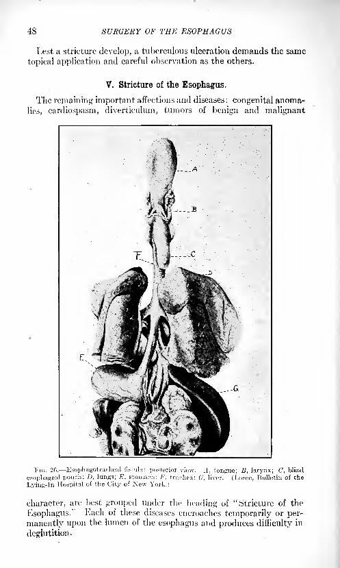
I'm. 26. Esophagotracheal fistula: interior view. .1, tongue: B, larynx; C, blind
esophageal pouch; D, lungs; E, stomach: /'. trachea; G, liver. (Losee, Bulletin of the
Lying-in Hospital of the City of New York.)
character, are best grouped under the heading of "Stricture of the
Esophagus." Each of these diseases encroaches temporarily or per-
manently upon the lumen of the esophagus and produces difficulty in
deglutition.
SURGICAL PATHOLOGY OF THE ESOPHAGUS 49
Congenital Anomalies. Quite a variety of these have been observed.The esophagus may be bifid; it may be in communication with its
lower portion through a typical congenitally strictured canal, several
centimeters long (Fig. 20) or it may end in a blind imperforate pouch(Fig. 26). This usually is from 2.5 to 4 cm. long. In the majority of
cases there exists a communication between this pouch and the air-
passages (Fig. 27).
A fibrous cord may extend from the lower pole of the rudimentaryesophagus to the cardiac end of the stomach; or, a few muscle fibers
may run from the termination of
the sac downward on the posteriorwall of the trachea.
In a third variety there is abso-
lutely no connection with the stomach.
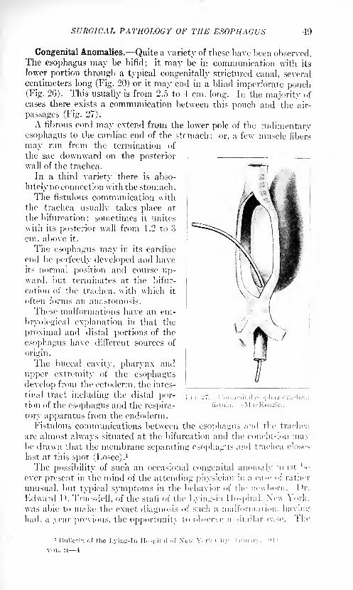
The fistulous communication with
the trachea usuallv takes place at
the bifurcation; sometimes it unites
with its posterior wall from 1.2 to 3
cm. above it.
The esophagus may in its cardiac
end be perfectly developed and haveits normal position and course up-ward, but terminates at the bifur-
cation of the trachea, witli which it
often forms an anastomosis.
These malformations have an em-
bryological explanation in that the
proximal and distal portions of the
esophagus have different sources of
origin.
The buccal cavity, pharynx and
upper extremity of the esophagus
develop from the ectoderm, the intes-
tinal tract including the distal por-tion of the esophagus and the respira-
tory apparatus from the endoderm.Fistulous communications between the esophagus and the trachea
are almost always situated at the bifurcation and the conclusion maybe drawn that the membrane separating esophagus and trachea closes
last at this spot (Losee).1
The possibility of such an occasional congenital anomaly must be
ever present in the mind of the attending physician in a ease of rather
unusual, but typical symptoms in the behavior of the newborn. IV.
Fdward I ). Truesdell, of the staff of the Lying-in Hospital. New V>rk.
was able to make the exact diagnosis of such a malformation, having
had, a year previous, the opportunity to observe a -imi!;i
r.
1 Hnllctin of the Lyins-In IF< ..-pil.-il of \c\v York City, January, I '.'I t.
VOL. II 4
f)0 SURGEItY OF THE ESOPHAGUS
infant became asphyxiated at intervals of thirty minutes to one hour,which condition was promptly relieved by the removal of a large quan-tity of thick, yellow mucus from the pharynx. The lower end of the
crib was elevated and with the mucus flowing from the baby's mouth,no further spasms occurred. It nursed as if it were very hungry, but
only for a few minutes, at the end of which time it choked, became
asphyxiated and relief was obtained by lowering its head.
Diagnosis.- The diagnosis in such cases is made by passing a soft
rubber catheter down the esophagus. It will be arrested at from 10
to 11 cm. (4 to 4- inches) from the alveolar border. With an .r-ray
stilette in position the radiogram shows the lower end of the sac.
The sooner after the birth of the child the diagnosis can be made the
better.
From a therapeutic standpoint immediate gastrostomy is alone
indicated. Whether it will bring relief, depends upon the peculiaritiesof the case. If a free communication of a patent lower portion of the
esophagus exists with the air passages, bronchopneumonia will soon
end the baby's sufferings, for only closure of the cardia could preventit, and this, being in itself a difficult operation technically, would cometoo late, as regurgitation of mucus and food from the stomach after
the first attempts at feeding will have immediately started a pulmonaryaffection. Still, some such cases have been known to live. In one
"a valve-like fold of the mueosa seemed to close the fistula so that
no food escaped into the trachea."
Treatment. The worst a doctor can do and not infrequently does
is to force a stiff bougie down beyond the obstruction. lie will invari-
ably perforate the esophageal pouch and cause the death of the child.
If he has diagnosed the trouble, he should promptly give the parentsthe prognosis. These infants usually die from two to twelve daysafter birth, regardless of whatever treatment.
Should conditions be less complicated, a primary gastrostomy maykeep the child alive.
Children being fed through a gastric fistula can grow up, but they
usually remain underdeveloped (one personal observation: Imperme-able cicatricial stricture of the esophagus; gastrostomy in fourth yearof life; patient still living, twenty-four years old).
Spastic Stenosis of the Esophagus (Cardiospasni), Cardiospasmis a spastic contraction of the distal end of the esophagus at or near
the cardia. It finds its analogy in pylorospasm and other spasmodiccontractions in the course of the gastro-intestinal tract, v. Mikulic/
compared it with the spasmodic contraction of the sphincter ani
muscle, produced by the presence of a fissure. Lately Jackson has
proposed to drop the term "cardiospasm" and call the disease "hiatal
esophagismus."\Vhether it occurs at the cardia proper or above it at the foramen
esophageum has not yet been definitely determined.
The contraction may be of short or long duration. It may last for
weeks, for months or for vears.
SURGICAL PATHOLOGY OF THE ESOPHAGUS 51
Etiology. The cause of the trouble is most likely a hyperirritabilityof the pneumogastrics, a so-called
"vagotonia," and of the sympathetic
system: "syinpathicotonia." It. is known today that both systemsof nerves supply the motor function of the lower esophagus and cardia,
though the chief supply is derived from the two nervi vagi. The hyper-
irritability may be due to a general affection of the vegetative nervous
system, or to a reflex from pathological lesions of intra-abdominal
organs, the geiiito-urinary system, pleura, etc. It may also be caused
by general intoxication and by local lesions, such as a fissure, erosion,
ulcer, etc., at or near the cardia.
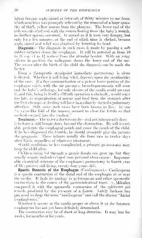
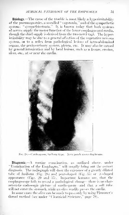
l-'ii;. !>N. Oardiospasm, fusiform type. Note pouch ;il>ovr
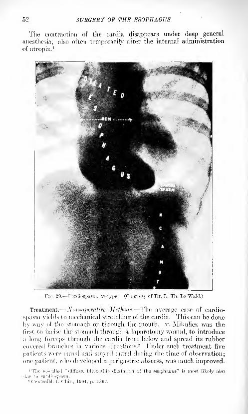
Diagnosis. A routine examination, as outlined above, under
"Examination of the Esophagus," will usually bring out the correct
diagnosis. The radiograph will show the existence of a greatly dilated
tube of fusiform (Fig. 2S) and pear-shaped (Fig. !">) or '/.-shaped
appearance (Figs. 2!) and .">.">). Important features are, that the
esophagoscope fails to reveal a pathological change there is no char-
acteristic endoscopic picture of eardiospasm and that a soft tube
will not enter the stomach, while an olive readily passes the cardia.
In some cases the olive can be made to pass only by using Hummer'sthread method (see under "( 'icatricial Stricture," page 70).
52 SURGERY OF THE ESOPHAGUS
The contraction of the cardia disappears under deep general
anesthesia, also often temporarily after the internal administration
of atropin.1
Fir;. 29. rfirdiospasm, cc-type. (Courtesy of Dr. L. Th. Le Wald.)
Treatment. Non-operative Mcihodx. The average case of cardio-
s]);isni yields to mechanical stretching of the cardia. This can be done
l;y way of the stomach or through the mouth, v. Mikulicz was the
first to incise the stomach through a laparotomy wound, to introduce
a long forceps through the cardia from below and spread its rubber
covered branches in various directions. 2 I'nder such treatment five
patients were cured and stayed cured during the time of observation;one patient, who developed a perigastric abscess, was much improved.
SURGICAL PATHOLOGY OF THE ESOPHAGI'S ')'>
Later v. Mikulicz passed his fingers through the carclia and dilated
with them, in place of the forceps, also*successfully.
SIZE
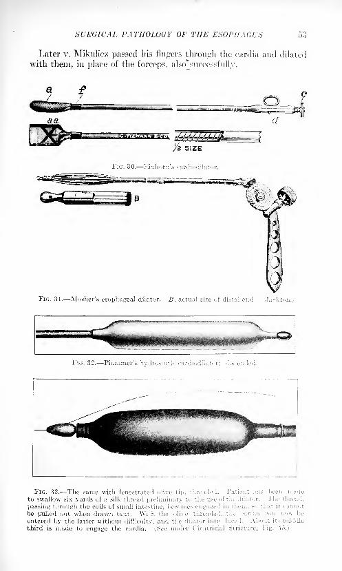
FIG. 30. Einhorn's cardiodilatoi
FIG. 31. Mosher's esophageal dilator. 5, actual size of distal end. (.Tac
FIG. 33. The same with fenestrated olive tip, threads 1. Patient has been madoto swallow six yards of a silk thread preliminary tu the u>e uf the dilatnr. The thread,
passing through the eoils of small intestine, becomes eiigaci i i:. them, so thai n eamiot
be pulled out when drawn taut. \Vith the olive threaded, the cardia can nowentered by the latter without difficulty, and the dilator introduced. About its middle
third is made to engage the cardia. ^ee under f'icatricial stricture, Fig. 45.)
Till-: HSOI'JIAdCS
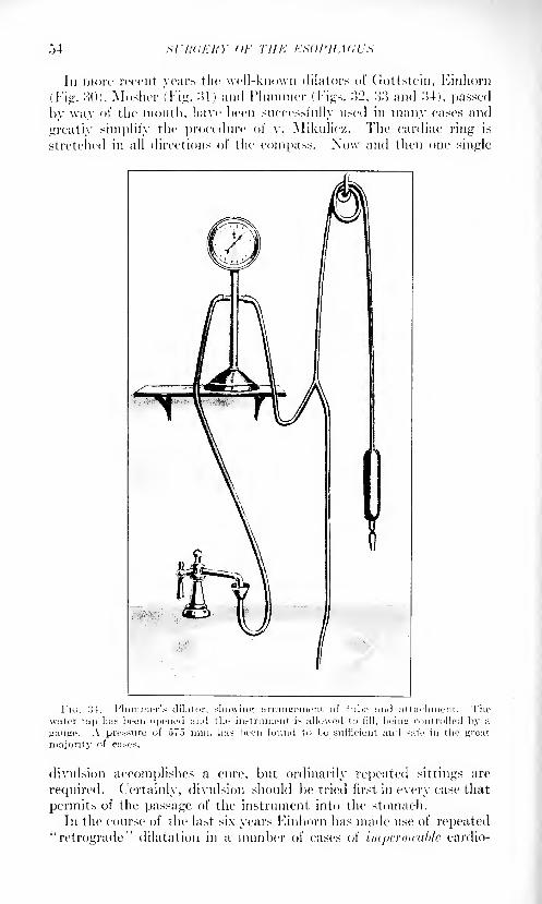
In more recent, years the well-known dilators of Gottstein, Kinhorn
(Fig. :!D), Mosher (Fig. :>1) and Hummer (Figs. :>2, :tt and o4), passed
by way of the mouth, have been successfully used in many cases and
greatly simplify the procedure of v. Mikulicz. The cardiac- ring is
stretched in all directions of the compass. Now and then one single
.its:
divnlsion accomplishes a cure, but ordinarily repeated sittings are
required. Certainly, divulsion should be tried first in every case that
permits of the passage of the instrument into the stomach.
In the course of the last six years Einhorn has made use of repeated
"retrograde" dilatation in a number of cases of impermeable cardio-
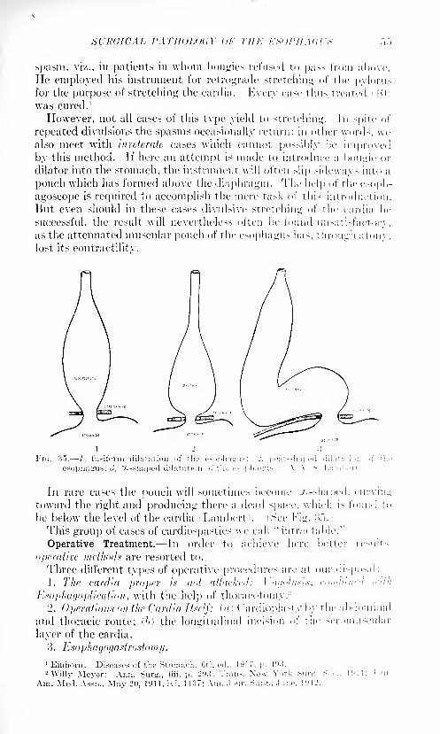
Si'IWICAL I'ATHOUHiY <>/' Till-: KSUl'HAUUti
spasm, viz., in patients in whom bougies refused to pass from above.
lie employed his instrument for retrograde stretching of the pylorusfor the purpose of stretching the cardia. Every case thus treated ( 10 1
was cured. 1
However, not all cases of this tvpe yield to stretching. In spite of
repeated divulsions the spasms occasionally return; in other words wealso meet with inveterate cases which cannot possibly be improvedby this method. If here an attempt is made to introduce a bougie or
dilator into the stomach, the instrument will often slip sideways into a
pouch which has formed above the diaphragm. The help of the e>oph-
agoscope is required to accomplish the mere task of this introduction.
But even should in these cases divulsive stretching of the cardia be
successful, the result will nevertheless often be found unsatisfactory,as the attenuated muscular pouch of the esophagus has, through aton\ ,
lost its contractilitv.
FIG. 35. 1, fusiform dilatation oi th
>phagus; ), X-shaped dilatation of the es->phajius
In rare cases the pouch will sometimes become x-shaped, cnntoward the right and producing there a dead space, which is foim<
be below the level of the cardia (Lambert). (See Fig. :>.").)
This group of cases of cardiospasties we call "intractable."
Operative Treatment. In order to achieve here better resu
operative methods are resorted to.
Three different types of operative procedures arc at our dir-posil
1. The cardia proper is not attacked: I af/oli/sis, combined <
Esophagoplication, with the help of thoracotomy.-'2. Operations on the Cardia Itself: u/i ( 'ardioplasty by the abdo
and thoracic route; (,//)the longitudinal incision of the seromusc
layer of the cardia.
3 . Esoph agoyastrostomy.
56 SURGERY OF THE ESOPHAGUS
1. Vacjolysis combined with cso2)haf/oplication'^_This operation wasfirst carried out by me at the Lenox Hill Hospital on May 31, 1910.
Since then I have employed it again in two cases.
CASE I. Female, age forty-seven years; difficult deglutition of long
standing; large esophageal pouch above the diaphragm; emaciation
First step: gastrostomy. A few weeks later (May 31, 1910), under
positive differential pressure (cabinet), transthoracic exposure of the
esophageal pouch; bilateral vagolysis, starting at the hiatus and car-
ried several inches upward. The short branches of the nervus vagusthat entered the pouch from both sides are severed. Plication of the
pouch in two layers with interrupted silk sutures. Closure of
thorax. In the course of the after-treatment unimpeded deglutitionreturned. No further instrumentation. Recovery. Presented before
the New York Surgical Society November 23, 1910. In January, 1911,
contracted influenza-pneumonia, after which a thoracic esophagealfistula became established in the scar of the chest wall, most likely dueto perforation of one or more of the plicating silk sutures. A subacute
suppurative posterior mediastinitis developed, to which the patientsuccumbed a year after the operation. Swallowing continued with
perfect ease to the last.
CASE II.- -Male, aged forty-four years; dysphagia for last five years.
Operation May, 1911;same plan followed as in Case I, but no primary
gastrostomy this time; double vagolysis with esophagoplication (onefold only), chromicized catgut; primary drainage of thoracic cavity.Here also easy deglutition returns a few days after the operation;
patient soon takes full diet; considers himself cured. Later in the
after-treatment the drainage tube slipped into the thorax and had to
be removed by a second operation, prolonging the time of drainage.It seems that the formation of more abundant cicatricial tissue aroundthe esophagus, due to the extended drainage, was the immediate factor
in disturbing the good functional result primarily obtained, as, grad-
ually, more difficult deglutition returned. (For a continuation of the
patient's history, see below, under"Cardioplasty, Thoracic Route.")
CASE 111. Male, aged forty-six years. Operative procedure of
Case II. He made a rapid recovery, but was not benefited by the
operation. He evidently had a cicatricial stenosis at the cardia,
probably consecutive to a peptic ulcer. Further operating refused bypatient.
Reviewing these cases, it wrould seem that the prompt return of easy
deglutition was due to the double vagolysis rather than to the single
or double esophagoplication. The latter, however, should always be
added in the presence of a large esophageal pouch. In cases of onlymoderate dilatation of the esophagus, 1 believe, the plication may beomitted. Additional operative corroboration as to this point is needed.
Still, it would seem that, at the present moment, transthoracic double
vagolysis, owing to its simplicity, represents the operation of choice
in intractable cases of this type, provided the surgeon prefers the trans-
thoracic route. In the light of our views on the etiology of cardio-
spasm it certainly appears to be a logical procedure.
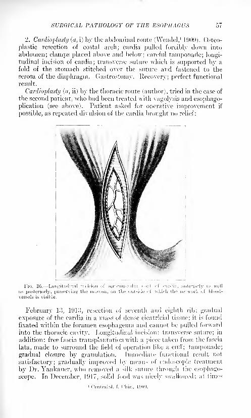
SURGICAL PATHOLOGY OF THE ESOPHAGUS 57
2. Cardioplasty (, i) by the abdominal route (\Vendel,1
190',)). Osteo-
plastic resection of costal arch; cardia pulled forcibly down into
abdomen; clamps placed above and below; careful taniponade; longi-tudinal incision of cardia; transverse suture which is supported by a
fold of the stomach stitched over the suture and fastened to the
serosa of the diaphragm. Gastrostomy. Recovery; perfect functional
result.
Cardioplasty (a, ii) by the thoracic route (author), tried in the case of
the second patient, who had been treated with vagolysis and esophago-
plication (see above). Patient asked for operative improvement if
possible, as repeated divulsion of the cardia brought no relief:
Longitudinal incision of seroniuscular coat of cardia, anteriorly ;i
February 13, 1913, resection of seventh and eighth rib; gradual
exposure of the cardia in a mass of dense cicatricial tissue; it is found
fixated within the foramen esophageum and cannot be pulled forward
into the thoracic cavity. Longitudinal incision; transverse suture; in
addition: free fascia transplantation with a piece taken from the fascia
lata, made to surround the field of operation like a cu!T; taniponade;
gradual closure by granulation. Immediate functional result not-
satisfactory; gradually improved by means of endoscopic treatment
by Dr. Yankauer, who removed a silk suture through the esophago-
scope. In December, 1917, solid food was nicely swallowed; at times
1 Ccntralhl. f. C'liir., 1'JO'J.
,">S WRCERY-OF Till! KHU'HAdUX
some difficulty, the cause of which remained obscure. Since then
lost sight of.
In view of the great mobility" of the lower end of the esophagus andof the rardia within the foramen esophageum of the diaphragm, it is
probable that a i>r>nuinj cardioplasty by the thoracic route, would
prove a much easier procedure.
(l>) Another very simple and, as it seems, effective operation for
intractable cardiospasm has recently been recommended by the Leip/ig
Surgical ( 'linic.1 F. Heller,- first assistant of that institution, after
incising the diaphragm from below, mobilized the cardia and then
divided its layers anteriorly and posteriorly, perpendicularly down to
the siibmucosa, in length of several centimeters (Fig. ><>). These twowounds remained unsutured. The result was splendid; formation of
diverticuli were not observed. This operation corresponds to the one
for the operative treatment of pylorospasm (Fredet-Rammstedt).
Esophugogd.ttrostomy. (a) By the abdominal route, uniting the snpra-
(liaj)hragmatic esophageal pouch with the stomach.
Ileyrowsky3 cites two cases. In one, the osteoplastie resection
of the costal arch made the approach to the cardia easier; in the
other, an oblique incision, parallel to the left costal arch, nicely exposedthe operating field. In both cases the anastomosis was made with a
double row of interrupted sutures. The line of suture was covered
with the vault of the stomach and drainage of the subphrenic spaceadded. 1 ninterrupted recovery in both instances.
A. V. S. Lambert' 1
accomplished the anastomosis with the help of
pressure necrosis by forceps.
Female, aged forty years; typical pouch above the diaphragm of the
"./.-variety. No improvement by dilatation of the cardia. Operation
February 1, lOlo. Left rectangular flap incision into abdomen. Osteo-
plastic resection of left costal arch by means of a special cut beneath
the left mammary gland, by which the seventh, eighth and ninth ribs
were divided near the attachment of their cartilages. The opening of the
gastrostomy, which had been done at a previous sitting, dissected from
the anterior abdominal wall and temporarily closed by a purse-stringsuture. Exposure of the cardiac opening of the stomach. Incision
of the diaphragm next to the cardia. The lower end of the dilated
esophagus which was marked by a bougie that had been introduced
through the month, was freed from surrounding structures by blunt
dissection, pulled into the abdominal cavity and fixated in this position
by means of interrupted catgut sutures, the border of the diaphrag-matic incision being stitched to the esophagus. Now the purse-stringsuture which had closed the gastrostomy opening, was divided and a
long clamp passed through this opening upward in such a way that
one blade of the clamp rested in the dra\vn-down esophageal pouch,the other within the esophagus, introduced upward through the cardia.
1 Payer, K.: Verhandl. d. dentsch. Gosellsch. f. C'hir., 1914, p. 749.2 Mitt. a. d. Grenzfjob., 1914, xxvii, 141.3 Arch. f. klin. Chir., 1913, c, 703.4 SurK-, O'.yner. and Obst., 1914, xviii, 1.
By closing the blades, the clamp became an esophagogast rotribe, similar
in principle to the enterotribe. A ro\v of catgut Lembert sutures united
the contiguous portions of the dilated esophagus and the stomach. Asoft-rubber catheter was passed alongside the clamp into the stomachfor feeding purposes. The gastrostomy fistula was reestablished andattached to the abdominal wall. Drainage. ( 'losure. Six weeks
after the operation the patient was able to eat everything, without
discomfort or vomiting.(1)} />// the Thoracic Route i Trans^lcnnil Kxi>i>h(i<it>ijaxti'<ixt</n/}.
K. Henschen1
proceeded in this way in the case of a twenty-year-oldman who had shown symptoms of cardiac stenosis for seven years.
Specialistic treatment had been of no avail. A -rays showed an almost
entirely impermeable stricture o cm. above the cardia with enormousdistention of the esophagus, of the x-variety, above.
Preliminary gastrostomy AYit/eh. Soon afterward, intercostal
incision in left seventh interspace; exposure of lower segment of
esophagus; division of diaphragm; blunt mobilization of cardia and
stomach which was pulled up into the thorax. Wide anastomosis
between the esophageal sac and the stomach fnndns, made with a
double row of single' sutures. ( 'losure of diaphragm and of mediastinal
pleura by a number of intermitted silk sutures, the anastomosis being
placed below diaphragm.The rather difficult technic of the work was much simplified by the
injection of a '2 per cent, solution of novocain into and around the
phrenic nerve, where the latter runs in a fold of the pericardium. This
produced a temporary paralysis of the respective half of the diaphragm,
lasting over two hours. Simultaneously, both nervi vagi had been
blocked with a novocain solution. The thorax was closed. 1 ndis-
turbed convalescence. A short time thereafter the patient was able to
take his food without difficulty.
This is the first recorded case of a successful esophagogastrostomyfor cardiospasm by the transpleural route.
All these procedures need more careful testing; probaHy a numberof them are reconnnendable. The operator must select the one that,
according to the clinical and ,r-ray findings, appears best adapted to
the case in hand. Any one of them is indicated only after instru-
mental eardiodilatation at repeated sittings has failed to bring relief.
Hut the future bids fair to corroborate the assumption that, in the
light of modern surgery, there seems to he still a chance tor improve-ment or cure even of supposedly intractable cardiospasm.
Compression Stenoses of the Esophagus. They may be produced
by a great variety of pathological conditions.
\Ve exclude from this consideration retropharyngeal abscesses and
tumors of the neck; further, pleural and pericardia! effusions, deformi-
ties of the spine, etc., all of which can compress the esophagus in any
portion of its course. These diseases are treated in the text-books on
internal medicine and general surgery.
1 f'enfndl)!. f. Cln'r., 1
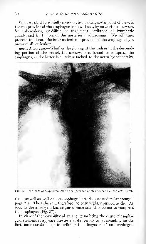
GO SURGERY OF THE ESOPHAGUS
\\'hat we shall here briefly consider, from a diagnostic point of view, is
the compression of the esophagus from without, by an aortic aneurysm,
by tuberculous, syphilitic or malignant peribronchial lymphatic
glands, and by tumors of the posterior mediastinum. We will then
proceed to discuss the inter iiittent compression of the esophagus by a
pressure diverticulum.
Aortic Aneurysm. Whether developing at the arch or in the descend-
ing portion of the vessel, the aneurysm is bound to compress the
esophagus, as the latter is closely attached to the aorta by connective
Stricture of esophagus clue to the presence of an aneurysm of the aortic arch.
tissue as well as by the short esophageal arteries (see under "Anatomy,"page 21). The tube can, therefore, be only slightly pushed aside. Assoon as the aneurysm has acquired some size, it is bound to compressthe esophagus (Fig. 37).
In view of the possibility of an aneurysm being the cause of esopha-
geal stenosis, it appears unwise and dangerous to let sounding be the
first instrumental step in refining the diagnosis of an esophageal
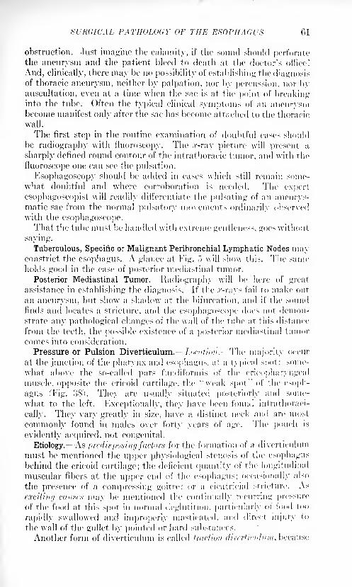
SURGICAL PATHOLOGY OF THE ESOPHAGUS 61
obstruction. Just imagine the calamity, if the sound should perforatethe aneurysm and the patient bleed to death at the doctor's office!
And, clinically, there may be no possibility of establishing the diagnosisof thoracic aneurysm, neither by palpation, nor by percussion, nor byauscultation, even at a time when the sac is at the point of breakinginto the tube. Often the typical clinical symptoms of an aneurysmbecome manifest only after the sac has become attached to the thoracic
wall.
The first step in the routine examination of doubtful cases should
be radiography with fluoroscopy. The .r-ray picture will present a
sharply defined round contour of the intrathoracic tumor, and with the
fluoroscope one can see the pulsation.
Esophagoscopy should be added in cases which still remain some-what doubtful and where corroboration is needed. The expert
esophagoscopist will readily differentiate the pulsating of an aneurys-matic sac from the normal pulsatory movements ordinarily observed
with the esophagoscope.That the tube must be handled with extreme gentleness, goes without
saying.
Tuberculous, Specific or Malignant Peribronchial Lymphatic Nodes mayconstrict the esophagus. A glance at Fig. 5 will show this. The sameholds good in the case of posterior mediastinal tumor.
Posterior Mediastinal Tumor. Radiography will be here of great
assistance in establishing the diagnosis. If the .r-rays fail to make out
an aneurysm, but show a shadow at the bifurcation, and if the sound
finds and locates a stricture, and the esophagoscope does not. demon-strate any pathological changes of the wall of the tube at this distance
from the teeth, the possible existence of a posterior mediastinal tumorcomes into consideration.
Pressure or Pulsion Diverticulum.- Location.- The majority occur
at the junction of the pharynx and esophagus, at a typical spot: some-
what above the so-called pars fundiformis of the cricopharyngeal
muscle, opposite the cricoid cartilage, the "weak spot" of the esoph-
agus (Fig. 38). They are usually situated posteriorly and some-
what to the left. Exceptionally, they have been found intrathoraci-
cally. They vary greatly in size, have a distinct neck and are most
commonly found in males over forty years of age. The pouch is
evidently acquired, not congenital.
Etiology. As predisposing factors for the formation of a diverticulum
must be mentioned the upper physiological stenosis of the esophagusbehind the cricoid cartilage; the deficient quantity of the longitudinal
muscular fibers at the upper end of the esophagus; occasionally a No
the presence of a compressing goitre; or a cicatricial stricture. As
excitiny causes may be mentioned the continually recurring pressure
of the food at this spot in normal deglutition, particularly of food too
rapidly swallowed and improperly masticated, and direct injury to
the wall of the gullet by pointed or hard substances.
Another form of diverticulum is called traction divert icnh/m, because
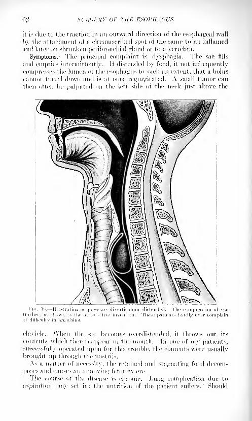
M'KUERY OF THE ESOPHAGUS
it is due to the traction in an outward direction of the esophageal wall
by the attachment of a circumscribed spot of the same to an inflamed
and later on shrunken peribronchial "'land or to a vertebra.
Symptoms. The principal complaint is dysphagia. The sac fills
and empties intermittently. If distended by food, it not infrequently
compresses the lumen of the esophagus to such an extent, that a boltis
cannot travel down and is at once regurgitated. A small tumor can
then often be palpated on the left side of the neck just above the
clavicle. A\ hen the sac becomes overdistended, it throws out its
contents which then reappear in the mouth. In one of my patients,
successfully operated upon for this trouble, the contents were usually
brought up through the nostrils.
As a matter of necessity, the retained and stagnating food decom-
poses and caiiM'> an annoying fetor ex ore.
The course of the disease is chronic. Lung complication due to
aspiration may set in; the nutrition of the patient suffers. Should
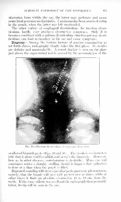
Xl'RdK'AL I'ATllOLOdY ()!' THE KS
ulceration form within the sac, tlie latter may perforate and cause
acute fatal posterior mediastinitis. Carcinoma has been seen to developin the pouch, when the latter was left unattended.
The other variety of esophageal diverticulum, the traction diver-
ticnluin, hardly ever produces obstructive symptoms. Only if it
becomes combined with a pulsion diverticulum (traction-pulsion diver-
ticulum) can food accumulate in the sac and cause symptoms.Diagnosis. Among the various factors of routine examination as
set forth above, radiography clearly takes the first place. Its results
are definite and unmistakable. A round shadow is seen on the plate
just above the snprasternal notch, catised by the accumulation of the
Fi<;. 30. Pressure diverticulum nf esophagus: fr<>nt vie\v.
swallowed bismuth paste ( Figs. :j(.) and -UK This result is so character-
istic that it alone could establish and sustain the diagnosis. ] lowever.
here as in other diseases, corroboration is desirable. Thus, one \\ill
sometimes notice a doughy swelling should it happen that palpationis done at a time when the pouch is filled.
Repeated sounding will show a peculiar pathognomonic phenomenon,
namely, that the bougie will pass with perfect case at times, while at
other times it finds an absolute obstruction ]ii to I'.i cm. from the
teeth. If the .r-ray stilette was used and the radiograph then promptly
taken, its tip will be seen in the sac.
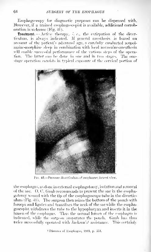
64 SURGERY OF THE ESOPHAGUS
Esophagoscopy for diagnostic purposes
However, if a trained esophagoscopist is a
oration is welcome (Fig. 41).
Treatment. Active therapy, i.e., tlie
ticulum, is always indicated. If generalaccount of the patient's advanced age, a c
amine-inorphine sleep in combination with
will enable successful performance of the v
tion. The latter can be done in one and
stage operation consists in typical exposure
can be dispensed with.
callable, additional corrob-
extirpation of the diver-
anesthesia is feared on
irefully conducted scopol-local novocaine-anesthesia
arious steps of the opera-in two stages. The one-
of the cervical portion of
FIG. 40. Pressure divcrticulum of esophagus; lateral view.
the esophagus, as done in external esophagotomy, isolation and removalof the sac. (). C. Gaub recommends to present the sac in the esopha-
gotomy wound with the tip of the esophagoscope tube in the divertic-
ulum (Fig. 41). The surgeon then seizes the bottom of the pouch with
forceps and ligates and transfixes the neck of the sac while the esopha-
goscopist withdraws the tube to the hypopharynx and inserts it in the
lumen of the esophagus. Thus the normal lumen of the esophagus is
indicated, while the surgeon amputates the pouch. Gaub has thus
twice successfully operated with Jackson's 1 assistance. This certainly
1 Diseases of Esophagus, 1915, p. 551.
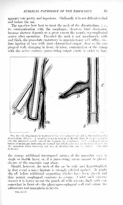
SURGICAL PATHOLOGY OF THE ESOPHAGUS 05
appears very pretty and ingenious. Ordinarily it is not difficult to find
and isolate the sac.
The question how best to treat the neck of the diverticulum ,/'. e.,
its communication with the esophagus, deserves brief discussion,
because thereon depends to a great extent the aseptic, uncomplicatedcourse after operation. Provided the neck is not inordinately wide
and thick, the procedure customary in appendectomy will suffice, vi/.,
firm ligation of base with stout chromicized catgut, close to the eso-
phageal wall; clamping in front; division; cauterization of the stumpwith the active cautery; purse-string catgut suture in order to bury
c
2
FIG. 41. 1, diagrammatic representation of esophagoscopic aid in the oxci-ion of a
diverticulum (Gauh) : A, esophagoscope in bottom of diverticulum ; H, esophagoseopistlift? the dissected pouch toward the surgeon; Or. esophagus. -!. C. esnpliairnscopc in
lumen of esophagus indicating its normal size which must nut. In- encroached up<>n l>y
the operation when removing the sac; D, diverticulum put on stretch. Chevalier
.Jackson.)
the stump; additional interrupted sutures of the muscular coat, in
single or double layer, or, if a purse-string suture cannot be placed,
closure of the muscular coat alone.
Should, however, the neck of the sac be wide and hypertrophied,in that event a mass ligature is strongly advised against. It might
slip off before additional supporting stitches have been placed, and
thus permit esophageal contents to escape. 1 nder such circum-
stances it is better to cut the pouch oil' with scissors, flush with or.
somewhat in front of the pharyngo-esophageal wall and suture the
submucosa and muscularis in layers.
VOL. II 5
66 SURGERY OF THE ESOPHAGUS
To pierce the basis of the tliverticulum with needle and thread andthen ligate, as is customary when dealing with a hernia! sac, may do no
harm, hut is a less advisable procedure.hi the one-stage operation the posterior mediastinum is best drained
by a gauxe tampon plus cigarette drain, led out at the lower angle of
the wound, while the upper portion of the latter is closed by sutures.
The postoperative course is usually satisfactory. When preliminary
gastrostomy had been performed, a procedure which seems indicated in
very reduced patients only the gastric tube alone is used for feeding;otherwise rectal alimentation with hypodermoclyses and hypodermicstimulation arc resorted to during the first two, three or four daysafter the operation. Generally speaking, the rules for the after-
treatment of operations upon mouth and neck hold good.If the sutures do not hold and a fistula develops, the patient's con-
valescence may be delayed, but the ultimate result will not be frus-
trated. These fistulas come to a close almost in every instance.
The risk of this operation is not great.
Of 60 cases radically operated upon in one stage by various surgeons,.~>0 recovered, being a mortality of 16.6 per cent., which, no doubt, will
be greatly reduced in the future by avoiding excision of the sac or
operating in two stages.1
Goldman2prefers a two-stage operation. lie first isolates and
ligates the neck of the sac, and, in the second stage, excises the sac.
In this way technic of the operation and after-treatment are simplified.
Schmid 3
proposes turning the sac upward, instead of excising it,
and stitching the t'undus of the diverticulum to the wall of the pharynxabove.
Murphy 1
preferred a two-stage operation in every case. During the
first stag*1 the sac is thoroughly freed, twisted and sutured in place;
the wound is tamponed; twelve to fourteen days later, after granu-lations have formed, the sac is removed.
Hcvaii"1
recently also advised against removing the sac, and sug-
gested infolding it by means of a scries of purse-string sutures. Thediverticulum is thus gradually turned into the lumen of the esophagus,where it either atrophies or is cast off.
Wherever this infolding operation can be performed, it must be
considered the operation of choice on account of the elimination of
infection. 1 recently had a very satisfactory result from this methodin a case of medium-sized diverticulum with broad base and a rctro-
esophageal attachment. There was a slight temporary discharge of
swallowed fluid from the lower angle of the wound, most likely in con-
sequence of the suture having pierced or necrosed the very thin sac
in one place; but the final result was excellent.
1
Stottoii, I).: Am. Surg., 1909, li, 300.2 ISoitr. z. klin. ( 'liir., 1909, Ixi, 741; 1912, Ixxviii, i, 139 (Ceiges).
\Vien. klin. Wohnschr., 1912, Xo. 13, p. 487.' SuT-g. Clinks of Chicago, 1910, v, 391.: '
Ibid., 1917, i, 449.
SURGICAL PATHOLOGY OF THE ESOPHAGUS 67
The two-stage operation employed by C. II. Mayo 1 consists in dis-
secting the sac without opening it, leaving the neck attached; the
wound in the patient's neck is then sutured, care being taken to fasten
the skin edges to the esophagus at its juncture with the diverticulum;
the sac is left outside of the neck, in the dressing. Ten to twelve days
later, adhesions having formed about the sac, it can be removed with-
out an anesthetic, and the edges be turned into the esophagus.This method is particularly intended for use in cases having a fairly
large sac. In a series of 35 cases operated upon at the Mayo Clinic,
i. e., excision with inversion of the base (18), excision with ligation and
inversion of the base (4), infolding (3), two-stage operation (10), theyhad two deaths in old, feeble patients on the day following the opera-tion. In 2 of the remaining cases there was some evidence of a recur-
rence of the diverticulum; in one of these the patient was entirely
relieved by sounding; the other required a second operation. Almostall of the 33 patients could later be reached by letter and were found
entirely free from symptoms.The infolding operation of Bevan and the two-stage operation are
considered the best procedures.In view of our ability to definitely cure the patients of this distressing
and dangerous condition, active therapy is clearly indicated in everyinstance. Should grave centra-indications in an individual case
render the operation too risky, gastrostomy can still bring relief and
lengthen life.
As to intrathoracic diverticulum, only one operation is so far on
record. It was performed by Henschen2 with the help of blocking with
a 2 per cent, novocain solution the phrenic nerve and both pneumo-gastrics and resulted in the death of the patient. A large, supra-cardiac sac was found and excised and the esophageal wound carefully
closed by three rows of layer-sutures. Leakage occurred on the
fourth day after the operation and produced suppurative posterior
mediastinitis, to which the patient succumbed.
Cicatricial Stenosis of the Esophagus. The principal type of e.ica-
tricial stenosis, interesting us here, is the one due to an escharotic, a
burn from caustic lye or acid (carbolic, sulphuric, etc.X The swallowingof caustic lye was a rather frequent occurrence among young children
at the time when bathtubs were still lined with tin, and lye was gen-
erally used for cleansing; also in the times of home-made soap (Jackson) .
Today, with changed conditions of manufacture, these accidents are
rarely met with. Pure acid is still occasionally swallowed by grownpeople, accidentally or with suicidal intent.
Some days after the accident the result of such a burn of the third
degree manifests itself: the esophagus becomes gradually strictured,
often in a number of places. If left to itself, all kinds of folds, bands,
pockets and diverticula will form and produce a so-called impermeablecicatricial stricture.
1 Judd, E. S.: Surg., Gynec. and Obstet, 1918, xxvii, 13.", 141.2 Central!)!, f Chir., 1916, No. 21.
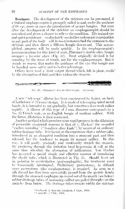
68 SURGERY OF THE ESOPHAGUS
Treatment.- The development of the stricture can be prevented, if
a trained esophagoscopist is promptly called in and, under the guidance
of the eye, starts at once the introduction of proper bougies. But even
after the development of the stricture an esophagoscopist should be
consulted and given a chance to relieve the condition. His trained eyeand quiet persistence so absolutely needed for endoscopic examination
in any part of the body- will in most instances find the entrance to the
stricture and then direct a filiform bougie downward. This accom-
plished, progress will be made quickly. In the csophagoscopist'sinstnimentarium for this kind of work, Plummer's double olive bougie
occupies a favorite place (Fig. 42). Bunt originally devised it for
sounding by the sense of touch, not/for the esophagoscopist. But it
stands to reason that under the guidance of the eye this bougie can
be handled more safely and to better advantage.Others have used a stout catgut thread which, left in place, swells'
by the absorption of fluid and thus widens the stenosis.
Fi<;. 42. Plummer's double olive bougie. (Jackson.)
A new "telescope" dilator has been constructed by Leiter, on basis
of LotheissenV (Vienna) design. It is made of telescoping spiral metal
bands; it is intended to act gradually, but sometimes does work rather
rapidly. A dilator of this type of 5 mm. diameter corresponds to a
No. '2'2 French scale, or an English bougie of medium caliber. Withthe latter, dilatation is then continued.
Another method which promises more rapid progress in the dilatation
of permeable cicatricial stenoses is that of v. Hacker, 2 the so-called
"endless sounding" ("Sondiren ohne Ende") by means of an ordinaryrubber drainage tube. It is based on the experience that a rubber tube,
introduced in an elongated condition into a stenosed part and then
released, has the tendency to regain its normal caliber. In this
way, it will gently, gradually and continually stretch the stenosis.
By producing through this irritation local hyperemia, it will at the
same time stimulate the absorption of infiltrations. Von Hackerhas invented a special simple and effective manner of introducingthe elastic tube, which is illustrated in Fig. 43. Should fever set
in, pointing to ovcrirritation (peri-esophagitis), the treatment mustbe temporarily interrupted. Preliminary gastrostomy, in difficult
cases gastrostomy plus esophagostomy, is required. As soon as a
silk thread has then been successfully passed from the gastric fistula
through the stenosed esophagus up to and out of the mouth (see below)rubber drainage tubes of increasing caliber are pulled through on the
stretch from below. The drainage tubes remain within the stricture
1 Verhandl. d. deutseh. Gcsellsch. f. Cliir., 1914.2 Contrail)!, f. Cliir., 1894.
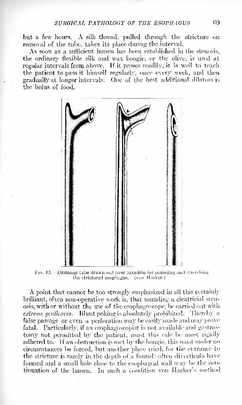
SURGICAL PATHOLOGY OF THE ESOPHAGUS 09
but a few hours. A silk thread, pulled through the stricture on
removal of the tube, takes its place during the interval.
As soon as a sufficient lumen has been established in the stenosis,
the ordinary flexible silk and wax bougie, or the olive, is used at
regular intervals from above. If it passes readily, it is well to teach
the patient to pass it himself regularly, once every week, and then
gradually at longer intervals. One of the best additional dilators is
the bolus of food.
FIG. 43. Drainage tube drawn out over mandriu for sounding; and stretching
the strictured esophagus, (von Hacker.)
A point that cannot be too strongly emphasized in all this certainly
brilliant, often non-operative work is, that sounding a cicatricial sten-
osis, with or without the use of the esophagoscope, be carried out with
extreme gentleness. Blunt poking is absolutely prohibited. Thereby a
false passage or even a perforation may be easily made and may provefatal. Particularly, if an esophagoscopist is not available and gastros-
tomy not permitted by the patient, must this rule be most rigidly
adhered to. If an obstruction is met by the bougie, this must under no
circumstances be forced, but another place tried, for the entrance to
the stricture is rarely in the depth of a funnel; often diverticula have
formed and a small hole close to the esophageal wall may be the con-
tinuation of the lumen. In stich a condition von Hacker's method
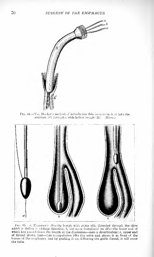
70 SURGERY OF THE ESOPHAGUS
Fio. 44. Von Hacker's method of introducing thin bougies (a, b, c) into thestricture ('/) through a wide hollow bougie (R). (Keen.)
FIG. 45. a, 1'luminer's olive-tip bougie with guide silk, threaded through the olive
which is drilled in oblique direction; b, the same introduced on silk the lower end of
which has passed down the length of the duodenum into a diverticulum; c, upper endof thread drawn taut this manipulation lifts the olive and places it in front of thelumen of the esophagus, and by pushing it on, following the guide thread, it will enterthe tube.
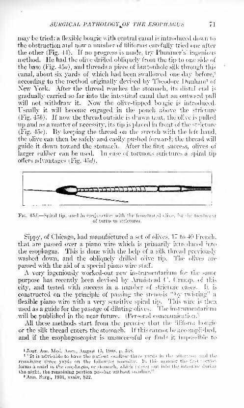
SURGICAL PATHOLOGYJJF THE ESOPHAGUS
may be tried: a flexible bougie with central canal is introduced down to
the obstruction and now a number of filiforms carefully tried one after
the other (Fig. 44). Jf no progress is made, try Plummer's 1
ingeniousmethod. He had the olive drilled obliquely from the tip to one side of
the base (Fig. 45), and threads a piece of buttonhole silk through this
canal, about six yards of which had been swallowed one day before,2
according to the method originally devised by Theodore Dunham 3 of
New York. After the thread reaches the stomach, its distal end is
gradually carried so far into the intestinal canal that an outward pull
will not withdraw it. Now the olive-tipped bougie is introduced.
Usually it will become engaged in the pouch above the stricture
(Fig. 456). If now the thread outside is drawn taut, the olive is pulled
up and as a matter of necessity, its tip is placed in front of the stricture'
(Fig. 45t'). By keeping the thread on the stretch with the left hand,the olive can then be safely and easily pushed forward; the thread will
guide it down toward the stomach. After the first success, olives of
larger caliber can be used, hi case of tortuous strictures a spiral tip
offers advantages (Fig. 4oc/j.
FIG. 45J. Spiral tip, used in conjunction with the fenestrated olive, for the treatmentof tortuous strictures.
Sippy, of Chicago, had manufa'ctured a set of olives, 17 to 40 French,that are passed over a piano wire which is primarily introduced into
the esophagus. This is done with the help of a silk thread previouslywashed down, and the obliquely drilled olive tip. The olives are
passed with the aid of a special piano wire staff.
A very ingeniously worked-out new instrumentarium for the same
purpose has recently been devised by Armistead ('. Crump, of this
city, and tested with success in a number of stricture cases. It is
constructed on the principle of passing the stenosis "by twi-ting'' a
flexible piano wire with a very sensitive spiral tip. This wire is then
used as a guide for the passage of dilating olives. The instrumentarium
will be published in the near future. (Personal communication.)All these methods start from the premise that the filiform bougie
or the silk thread enters the stomach. If this cannot be accomplished,and if the esophagoscopist is unsuccessful or finds it impossible to
1 Jour. Am. Med. Assn., August 15, 1!)OS, p. 548.2 "It is advisable to have the patient swallow three yards in the afternoon and the
remaining three yards on the following nic.riiinir. In this manner the fir.-t portion
forms a snarl in the esophagus, or stomach, which passes out into the intestine during
the night, the remaining portion passing without snarling."3 Ann. Surg., 1901, xxxiv, 822.
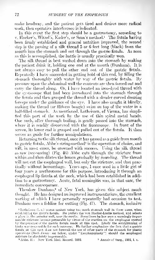
72 SURGERY OF THE ESOPHAGUS
make headway, and the patient gets tired and desires more radical
work, then operative interference is indicated.
In this event the first step should be a gastrostomy, according to
v. Hacker's, Witzel's, Kader's, or Senn's method. 1 The fistula havingbeen firmly established and general nutrition improved, the second
step is the passing of a silk thread 5 or 6 feet long (black) from the
mouth into the stomach and out through the gastric fistula. As soon
as this is accomplished, the battle is usually practically won.
The silk thread is best washed down into the stomach by makingthe patient drink it, holding one end at the mouth (Dunham). It is
not always easy to pull the other end out of the gastric fistula.
Repeatedly I have succeeded in getting hold of this end, by filling the
stomach thoroughly with water by way of the gastric fistula. Bypressure upon the abdominal wall the contents are then forced out and
carry the thread along. Or, I have located an iron-dyed thread with
the cystoscope that had been introduced into the stomach throughthe fistula and then grasped the thread with a slender curved dressing
forceps under the guidance of the eye. I have also caught it blindly,
making the thread (or filiform bougie) swim on top of the water in a
half-filled stomach. As mentioned, Lotheissen, of Vienna, has simpli-
fied this part of the work by the use of thin spiral metal bands.
One such, after thorough boiling, is gently passed into the stomach,where it is readily discovered with the fiuoroscope. In front of the
screen, its lower end is grasped and pulled out of the fistula. It then
serves as guide for further manipulations.
Returning to the silk thread, once it has passed as a guide from mouthto gastric fistula, Abbe's string-method
2is the operation of choice, and
will, in most cases, be crowned with success. Using the silk thread
as saw (see-sawing) (Fig. 40) Abbe cuts through the stricture from
within and then dilates the lumen gradually by sounding. The thread
will not cut the esophageal wall, but only the stricture, and that prac-
tically without hemorrhage. Years ago, I once used in a little girl of
four years a urethrotome for this purpose, introducing it through an
esophageal lip fistula at the neck, which had been established in addi-
tion to a gastrostomy. Acute, fatal meningitis was, in that case, the
immediate consequence.Theodore Dunham, 3 of New York, has given this subject much
thought. He has designed an improved instrumentarium, the excellent
working of which I have personally repeatedly had occasion to test.
Dunham uses a fishline for cutting (Fig. 47). The stomach, inclusive
1 Lotheisseii (1. c.) warns against using too much stomach wall in these cases, whenestablishing the gastric fistula. Pie prefers the von Hacker-Lucke method, and selects
a place in the anterior wall, near the cardia. Sometimes he has seen a seemingly imper-meable stricture become permea-ble by virtue of the traction on the esophagus exerted
by the pre.sence of the gastric fistula made at this spot. Likely the straightening of
kinks was responsible for such outcome. He further emphasizes the fact that a gastric
fistula at this spot does not forestall the use of other parts of the stomach for plastic
operations (Beck-Jianu see below, under "Esophagoplasty") should the esophagealstricture be absolutely impermeable.
2 Abbe, It.: New York Mod. Record, 1893. 3 Annals of Surg., 1901, 1. c.
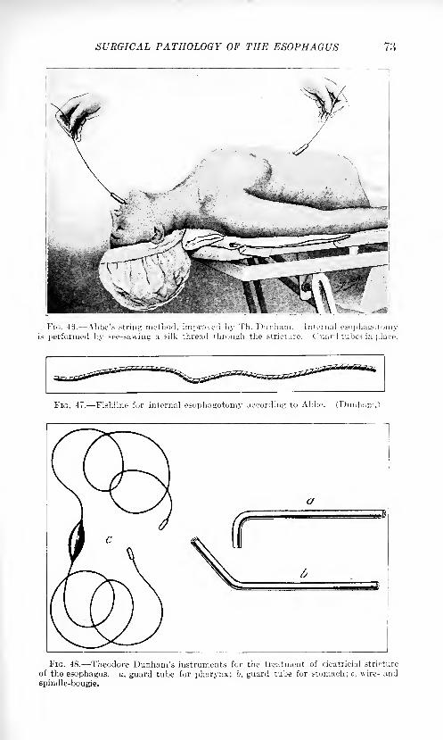
SURGICAL PATHOLOGY OF THE ESOPHAGUS 73
\a
FIG. 46. Abbe's string method, improved by Th. Dunham. Internal esophagotomyis performed by see-sawing a silk thread through the stricture. Guard tubes in place.
FIG. 47. Fishline for internal esophagotomy according to Abbe. (Dunham.)
FIG. 48. -Theodore Dunham's instruments for the treatment of cicatricial stricture
of the esophagus, a, guard tube for pharynx; b, guard tube for stomach; c, wire- and
spindle-bougie.
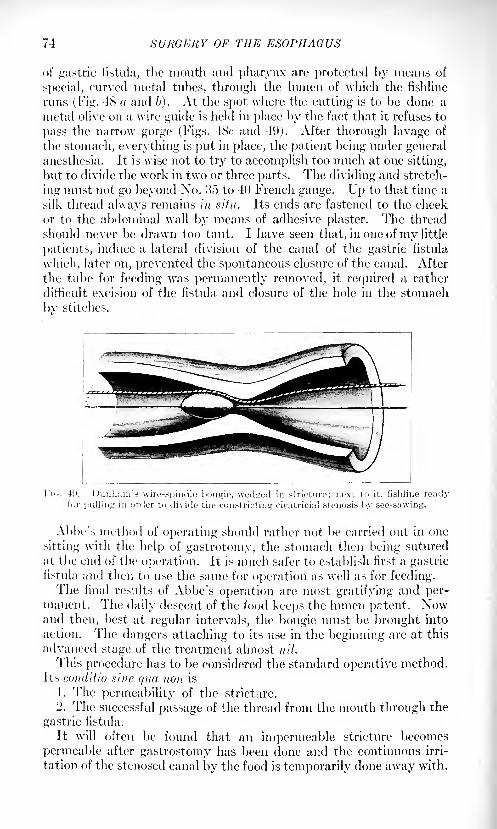
74 SURGERY OF THE ESOPHAGUS
of gastric fistula, the mouth and pharynx are protected by means of
special, curved metal tubes, through the lumen of which the fishline
runs (Fig. 4S a and li). At the spot where the cutting is to be done a
metal olive on a wire guide is held in place by the fact that it refuses to
pass the narrow gorge (Figs. 48c and 49). After thorough lavage of
the stomach, everything is put in place, the patient being under generalanesthesia. It is wise not to try to accomplish too much at one sitting,
but to divide the work in two or three parts. The dividing and stretch-
ing must not go beyond Xo. 35 to 40 French gauge. Up to that time a
silk thread always remains in situ. Its ends are fastened to the cheek
or to the abdominal wall by means of adhesive plaster. The thread
should never be drawn too taut. I have seen that, in one ofmy little
patients, induce a lateral division of the canal of the gastric fistula
which, later on, prevented the spontaneous closure of the canal. After
the tube for feeding was permanently removed, it required a rather
difficult excision of the fistula and closure of the hole in the stomachbv stitches.
FK
Abbe's method of operating should rather not be carried out in one
sitting with the help of gastrotomy, the stomach then being sutured
at the end of the operation. It is much safer to establish first a gastricfistula and then to use the same for operation as well as for feeding.The final results of Abbe's operation are most gratifying and per-
manent. The daily descent of the food keeps the lumen patent. Nowand then, best at regular intervals, the bougie must be brought into
action. The dangers attaching to its use in the beginning are at this
advanced stage of the treatment almost nil.
This procedure has to be considered the standard operative method.Its conditio sine qua -non is
1. The permeability of the stricture.
2. The successful passage of the thread from the mouth through the
gastric fistula.
It will often be found that an impermeable stricture becomes
permeable after gastrostomy has been done and the continuous irri-
tation of the stenosed canal by the food is temporarily done away with.
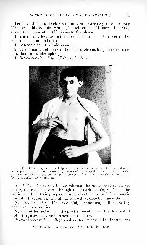
SURGICAL PATHOLOGY OF THE ESOPHAGUS 75
Permanently impermeable strictures are extremely rare. Among253 cases of his own observation, Lotheissen found it once. In 1904 I
have also had one of this kind (see further down).In such cases, lest the patient be made to depend forever on his
gastric fistula, are indicated:
1. Attempts at retrograde sounding.2. The formation of an extrathoracic esophagus by plastic methods,
extrathoracic esophagoplasty.1. Retrograde Sounding. This can be done
(a) Without Operation, by introducing the ureter cystoscope, or,
better, the esophagoscope through the gastric fistula, as far as the
cardia, and then trying to pass a ureteral catheter or a filiform bougie
upward. If successful, the silk thread \vill at once be drawn through.
(b) With Operation. If unsuccessful, advance may still be tried bymeans of an operation.
By way of the abdomen: osteoplastic resection of the left costal
arch with gastrotomy and retrograde sounding.Personal observation: 1
Boy, aged fourteen years; had had to undergo
'Meyer, Willy: Jour. Am. Mod. As>n., 1900, xlvii, lOii'J.
76 SURGERY OF THE ESOPHAGUS
gastrostomy when six years old; for eight years feeding had been done
through the gastric fistula. All attempts on the part of other surgeons,to pass the stricture had been futile. My efforts, too, were unsuccessful.
I, therefore, established an esophageal lip-fistula at the neck, throughwhich Kelly's urethral tubes were introduced, and then searched,
under direct illumination for the entrance. The latter was found, but
no definite progress could be made. It was also impossible to pass
upward through the esophagus a ureter-catheter which had been
introduced through the cardia with the help of a ureter cystoscope.At last, therefore, retrograde sounding by operation, viz., the osteo-
plastic resection of the left costal arch was attempted (1904) (Fig. 50).
The stomach was exposed, longitudinally incised and the cardia entered.
Bougies refused to pass upward, although I risked even direct divulsion
with a long, slender forceps introduced through the cardia into the
esophagus for quite some distance upward. All attempts to pass a
bougie to the mouth failed. So this plan, too, had to be abandoned.
The osteoplastic flap was put back and stitched in place. The woundhealed kindly and the function of the gastric fistula was preserved.The proposed esophagoplasty according to Bircher's method (see page129) was not accepted. The boy is still living and continues to nourisli
himself through the gastric fistula.
By Way of the Thorax. (II. Fischer 1
). Three-year-old boy. Caus-
tic lye burn. Almost completely impermeable cicatricial stricture. Gas-
trostomy, Lenox Hill Hospital, New York. Radiograph shows stric-
ture from tracheal bifurcation down to a spot 2 cm. above diaphragm.Bismuth mixture which had passed through, makes this clearly
visible. Attempt to wash a silk thread down is not successful, neither
is retrograde catheterization by way of the gastrostomy opening.
Thoracotomy under insufflation. Cardia exposed; peritoneal cavityentered from above and stomach pulled into thorax for about 7 cm.
Careful tamponade. Transverse incision of cardiac portion about3 cm. below union of esophagus and stomach, the latter having been
secured with two loops of silk thread. Now a ureter catheter with
mandrin slips easily upward. A dressing forceps which is passed fromthe gastrostomy opening through the cut near the cardia grasps the
other end of the ureter catheter and conducts it out of the gastricfistula. A double row of sutures closes the incision in the wall of the
stomach. The latter is then replaced, the cardia sutured to the dia-
phragm and the thorax closed.
The boy died twenty-four hours later from acute congestion of the
lungs, due to a mistake at the hands of the anesthetist who, by openingthe wrong valve, had blown minute particles of ether into the lungs.
2. Extrathoradc Esophagoplasty. It is in these cases of imperme-able stenosis, that surgeons have succeeded by most ingenious plastic
surgery, done in a series of operations, in forming a new tube extra -
thoracically : partial or total esophagoplasty. The operation has
l Centralbl. f. Chir., 1910, No. 43, p. 1393.
SURGICAL PATHOLOGY OF THE ESOPHAGUS 77
proved to be feasible in various ways, with an absolutely lastingand favorable functional result. (See under "Esophagoplasty," page128.)
Chronic ulcerations, the forerunners of cicatricial contraction other
than escharotic stenosis, such as traumatic, tuberculous, syphilitic, havebeen mentioned above. The history of the case and the many meansat present at our disposal for a refined diagnosis will usually suffice
to clear up the true characteristics of the trouble, before an operationis undertaken.
Stenosis Due to Tumor Formation in the Esophageal Wall. Themost frequent cause for the formation of a stenosis of the esophagusis a growth in its wall. This may be either benign or malignant.
Strictures Due to a Benign Tumor. In comparison with strictures
resulting from malignant tumors, these are rare cases. Nevertheless,
they are more frequent than is generally believed. They do not pro-duce a stricture in a pathological sense, but clinically, in that theycause increasing difficulty in swallowing. As a rule it is not until the
tumor has reached considerable size that the patient's attention is
drawn to it by the resultant difficulty in deglutition. But even whenstill of small dimensions, a benign tumor is accompanied by incon-
veniences which make themselves felt at all meals. The family physi-cian who will probably be first consulted, is apt to make light of the
patient's complaints, ascribing them perhaps to some general nervous
affection, e. g., neurasthenia. It is necessary that, in the face of such
complaints, esophagoscopy should more frequently enter the physi-cian's mind and become as generally recognized for purposes of exami-
nation as cystoscopy, rectoscopy, etc., have become in the course of
recent years.
All classes of benign tumors connected with the "make-up" tissues
of the esophageal wall,' as papilloma, fibroma, myoma, adenoma,
lipoma, angioma and their manifold combinations, have been observed.
Missing infiltration at the base, and pedunculation characterize the
nature of the growth. In cases of doubt regarding the nature of the
tumor a specimen should be punched out for microscopic examination.
The slight hemorrhage sometimes following, will yield to the swallowingof ice pills or ice-cream.
Abenign tumor ought to be removed, as itmight turn into a malignantone. If at all possible, the operation should be done endoscopically.In removing the tumor the base should be carefully treated by biting
it out with proper forceps and cauterizing it with the galvanocaustic
wire, provided hemorrhage does not forbid this. The primary use of
the galvanocaustic wire or snare would appear to be best. Its applica-
tion must depend upon the peculiarities of the case and the dexterity
of the operator. At all events, these tumors need careful attendance,
as many of them have a tendency to recur, same as those of the larynxor bladder.
Strictures Due to the Presence of a Malignant Tumor: Sarcoma and
Cancer of the Esophagus.- (a) SARCOMA. Regarding this type of
78 SURGERY OF THE ESOPHAGUS
malignant neoplasm, von T lacker 1 has made an exhaustive study cover-
in 21 cases described in the literature. One of these, presenting a sessile
tumor in the cervical portion, came under his personal care. Esopha-
goscopical probatory excision of the tumor enabled him to make the
correct diagnosis. The cervical segment of the esophagus was resected.
Sarcoma may appear pedunculated (polypoid) or sessile, with diffuse
infiltration of the wall of the tube. It has a great tendency to show
degenerative changes with subsequent necrosis and ulceration. Nine-
teen per cent, of the cases were found in the cervical, Si per cent, in
the thoracic portion.
(/>) CAXCKH. Occurrence. Cancer of the esophagus, same as
cancer in other parts of the gastro-intestinal tract, has a decided
predilection for physiological stenoses. The manifold lesions to whichthe tube is constantly subjected by the ever-recurring descent of the
food, no doubt favor in patients predisposed to cancer the develop-ment of the tumor at these spots. An old syphilitic or tuberculous
ulceration or scar may also furnish the starting-point for a malignant
growth.In accordance with the aforesaid predilection we find cancer of the
esophagus most frequently :
1. Near the very beginning of the esophagus, behind the cricoid
cartilage at the neck.
2. Behind the bifurcation of the trachea.
3. At the hiatus esophageus of the diaphragm.4. At the cardia.
From a pathological point of view it may be correct to differentiate
as stated under 3 and 4; clinically the last two types are to be com-bined under one head, i. e., cancer of the cardia (von Hacker).
Expressed in percentages and round figures the frequency of cancer
of the esophagus with reference to the above sites, is approximatelyas follows: Neck, 10 per cent.; bifurcation, 40 per cent.; hiatus, 2$
per cent.; cardia, 22 per cent. ;
or the latter two combined and groupedunder the common head of "Cancer of the Cardia,'' 50 per cent., or
one-half of the cases. By adding the malignant tumors of the lesser
curvature, near the cardia, which clearly belong to this group, at least
from an operative point of view, it will become evident that cancer
of the cardia is not a particularly rare disease. Cancer of the esophagustakes the fifth place in the total frequency of all carcinomas.
Type of Tumor. The most common variety, pathologically, is the
squamous-celled epithelioma (or endothelioma) ;the other type is the
cylinder-celled carcinoma which takes its origin in the atypical pro-liferation of the cylindrical epithelial cells of the mucous glands within
the esophageal wall. The former is the more benign of the two. The
growth of a neoplasm of the esophagus is similar to that of a cancer
of the rectum. At first it remains localized in one small place of the
tube; gradually it infiltrates the submucosa and muscularis and sur-
1 Mitt. a. d. Grenzgeb., 1904, No. 3, vol. xix.
SURGICAL PATHOLOGY OF THE ESOPHAGUS 79
rounds the canal in the shape of an irregular crescent. At this timethe previously noted inconvenience in swallowing will give way to
signs of stricture. The infiltrated ring having become complete, onlyfluids now pass down. Constriction and swelling continue to increase,
until finally total occlusion sets in. Ulceration of the surface appears
comparatively early, probably as a result of the ever-repeated irrita-
tion caused by the bolus in its passage through the constricted canal.
There is one important characteristic of cancer of the esophagusthat should be especially emphasized, namely, that it is the most
benign of all carcinomas of the gastro-intestinal tract. Its growth is
comparatively slow; it remains localized for a long time: has little
tendency to metastasis in other parts of the body, or in the lymphaticglands, and is not very apt to recur.
Symptoms. As regards clinical symptoms, the first trouble com-
plained of by a patient afflicted with esophageal carcinoma, is difficulty
in deglutition. lie has as yet no pain ; that begins later. The necessityof pressing a morsel down by means of holding his breath and then
exerting intrathoracic downward pressure, or of washing the bolus
down with a swallow of liquid, first attracts the patient's attention andinduces him to consult a physician. Let the latter then remember,have him know, that such complaints should not be lightly treated;
a pill or a powder is here of no avail. Our undergraduate and post-
graduate students should be taught and should have it impressed upontheir minds, that such a case needs the same prompt attention that
would be given to a case of acute appendicitis. Of course, it is not a
question of immediate operation, as in that case, but of bringing into
play promptly all the modern means for an exact diagnosis. How to
use these means has been dwelt upon at length above, under "Exami-nation of the Esophagus."
1 Let it be remembered and said again and
again, that a plainly present stenosis, unless resulting from the swallow-
ing of a chemical, is in at least M) to 90 per cent, of patients above twenty
years of age, due to a malignant tumor. This, really, is the important
point. Upon its general promulgation among medical students and
practitioners as well as among the laity, hinges the possibility of prog-ress that can and ought to be made in the resection of the esophagusfor carcinoma. Let us always keep in mind that carcinoma of the
esophagus, same as of other sites, is originally an entirely localized
disease, that neighboring organs nervi vagi, bronchus, aorta, lungs
are not involved until in a later stage and can therefore, with care and
anatomical knowledge, be stripped oil without serious injury. 1 hiring
the early period of the disease surgical interference is apt to eti'eet a
cure. "Without operation, on the other hand, with internal medication,
1 Not lon<: atro my attention was called In ai
(.Ironic of clinical symptoms in tin.- iurl// xlu.it- <>!'
in swallowing, pain immediately after swallmIf these symptoms are found, tho suspiciun ot
disease of the esophagus should l>e aroused. 11
skin bougie, sounding and esophagospopy \\<n
cases; personal communication; 1920.)
80 SURGERY OF THE ESOPHAGUS
of one hundred patients afflicted with cancer of the esophagus, onehundred die of the disease, and death in every one of them occurs
only after a long, protracted period of unspeakable suffering and misery.
Certainly, the necessity of a full understanding all around of the impor-tance of a spontaneously occurring difficulty in swallowing, cannot be
too strongly emphasized.As stated above, cancer of the esophagus is the most benign neoplasm
in the entire gastro-intestinal tract. However, the continuous chemical
and mechanical irritation due to the passage of the food over its surface,
as also the comparatively small caliber of the tube and its extreme thin-
ness (in man) make the early involvement of the main adjacent vital
structures unavoidable. Thus the pneumogastric nerves from the aorta
down will be found firmly attached to the tumor rather early. The
growth will become adherent to, and soon involve, larynx, bronchi,
pleura, lung, aorta and neighboring smaller bloodvessels and nerves.
If left to itself, it will begin to ulcerate and then either perforate into
the pleura, pericardium, the bronchial system, aorta, or it will continue
its growth into the posterior mediastinum. Of course, such a breakingdown of a constricting tumor will do away with the stricture. The
patient feels happy, as he is again able to swallow. I hit such happinessis illusory and of very brief duration. The terrific foe-tar ex arc in such
cases, that yields to no remedy, tells the tale, often to the poor sufferer
himself, and the perforation of the wall will soon end his misery.
Truly, the picture of a cancerous stenosis of the esophagus, if left
unattended, or if treated symptomatically, cannot be painted in colors
too dark. Every physician, who has once treated such a patient to
the very end, will admit that.
Treatment. In view of the foregoing, the necessity for continued
endeavor on the part of surgeons to bring the treatment of esophagealcarcinoma more generally within the domain of operative surgery,must, be conceded.
In the early days of such endeavors seemingly insuperable diffi-
culties presented themselves. - All who worked along these lines were
at first necessarily mere tyros in this particular work, and unwittinglydared much more here, than their experience in abdominal surgerywould have prompted them to do in the abdomen under similar con-
ditions. However, persistent and hard effort succeeded in overcomingone obstacle after another, so that today we are in a position to meetthe dangers without trepidation. We know now proper operativemethods for every section of the esophagus, and for every one of thema successful operation for carcinoma is on record.
Let us remember that this new field has only just been opened up.\Ye are still standing on its very threshold. Let us remember howmany years it has taken to bring abdominal surgery to its present
high standard, how many thousands of surgical minds and handshave worked together, in combination with laboratories, to improveand extend its possibilities.
SURGICAL PATHOLOGY OF THE ESOPHAGUS 81
Non-operative Treatment. There is not much to be said about non-
operative treatment, most of which is of little or no avail and shouldcome into consideration only if operation is refused or contra-indicated.
Sounding, still favored by many, even in the earlier stage of the trouble,
should, in my opinion, be definitely discarded. Nothing irritates and,in the end, incites to more rapid growth of the carcinoma in all
directions, than sounding. A constricting tumor that has been sub-
jected to dilatation with sounds, had better not be attacked surgically.Nowhere are the adhesions firmer and more extensive.
I am also opposed to the excision of a piece of the cancer by the
endoscopist for microscopic examination. I have repeatedly observed
pronounced aggravation of the patient's subjective symptoms, even
prolonged fever, which delayed the urgent operation and perhapscontributed to jeopardizing its result.
The many obtainable symptoms and the positive knowledge that at
lea-t 90 per cent, of the spontaneously occurring stricture- in patientsabove the twentieth year are cancerous, permit us to make the diag-nosis without microscopical corroboratioii. Probatory excision before
operation is looked upon with suspicion and disfavor by surgeons andcancer experts also in other parts of the body.
Kvery kind of intra-esophageal manipulation, such as instrumental
stretching of the stricture, or this excision of a piece of tumor for
microscopical examination, is bound to injure the esophageal wall andfrom an operative point of view should be condemned.As regards the hopelessness of all remedies and methods of palliative
treatment in these cases there is one exception that deserves further
faithful experimentation: radium. Dr. Max Einhorn. 1 of New York,was the first to suggest its possible usefulness in this class of cases. At
present there are many at work testing the efficiency of radium in malig-nant esophageal affections. It would be gratifying to find them obtain-
ing favorable results. It is conceivable that a certain percentage of
very early cases of cancer of the esophagus or cardia could be benefited
by intra-esophageal treatment with radium. The majority of these
tumors are of the squamous-celled type, which corresponds to the
epithelioma of the skin, which latter, in many instances, yields to
radium. Of course, the esophagoscopist should always corroborate
by local inspection any apparent change for the better. Clinically the
improvement will manifest itself by the returning ability to swallow.
Personally, I expect to see rcidiffil r-urr* achieved in very few cases,
and fear that by such attempts the best time for operation will often
be lost. Furthermore, the possibility of a perforation by a radium
burn must not be overlooked.
So far there are only a very few cases on record in which an eso-
phageal malignant tumor was permanently benefited by radium.
N. S. Finzi- reports a case of an extensive ulcerating carcinoma three
inches long in the lower part of the esophagus, well three years after.
1 Mod. Roc., .March, 1904; Jour. Am. M,>d. An., July, l'")">, p. 11-1.2 Radium Therapeutics, London, I'.li:;.
VOL. n (j
82 SURGERY OF THE ESOPHAGUS
A second case became so well, that the patient refused to come for
further treatment. Four years later, however, he returned to the
hospital and died of a recurrence in the mediastinal glands.Of o5 patients with cancer of the esophagus, under the care of J.
Guisez1-
1 case was well over two years at the time of the report, 1
one and a half and a third case over one year. In every one of the
three cases the diagnosis had been confirmed by microscopic section.
II. II. Jam-way, of \e\v York City, has two patients who, he thinks,
can be classed as complete retrogression. In both cases, however,the diagnosis was entirely clinical, and made by direct observation
under the esophagoscope. The first case has remained well four
years, the other a little less than a year. Both cases had favorable
lesions at the start. (Personal communication, December 13, 1919).
I ntil more positive results can be shown from the use of radium,it remains our plain duty, it seems to me, to consider all these cases
from a general surgical point of view operative cases, and that at
the earliest possible date, for we know today how best to proceed, weknow a reliable after-treatment and are fast learning to avoid com-
plications in the course of the latter.
A number of cases of intrathoracic operation for esophageal carcinoma
are recorded in the literature, with immediate operative recovery.It is definitely to be expected that a steadily increasing number of
patients will be seen to recover from esophageal resection, provided
they come for operation in time: in the earh/ xtaf/r afihe disease.
Only those whose disease is either too far advanced to justify
surgical interference, or who refuse radical operation, should be left
to the treatment with radium rays.
The cylinder-celled carcinoma which takes its origin from the
epithelial cells of the mucous glands of the esophagus and often involves
several inches of the tube would probably yield to radium in exceptionalinstances only, and had, therefore, best remain definitely and whollywithin the domain of operative surgery.What is necessary is confidence in the present possibilities of operative
surgery on the part of the medical profession as well as the laity. Togain this confidence a "series" of recoveries from operation is absolutely
required. But how can a series of favorable operative results be
obtained, if patients with cancer of the esophagus are usually referred
to the surgeon for operation as a last resort only''
Operative Treatment. This may be radical or for temporary relief.
1. Radical Treatment, viz., Resection of the Esophagus.The procedure differs with reference to the location of the tumor,
i. e., whether it be situated
(a) At the neck.
(6) Within the thorax. (Figs. 22, 51, and 52.)
(c) At the cardia.
It is necessary to discuss the operation from the standpoint of such
subdivision.
1
Stnililen-Tlirnipk', 1914, iv, -44.
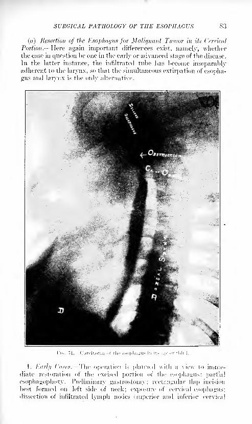
SURGICAL PATHOLOGY OF THE ESOPHAGUS S3
(a) Resection of the Esophagus for Malignant Tumor in its Cervical
Portion. Here again important differences exist, namely, whetherthe case in question be one in the early or advanced stage of the disease.
In the latter instance, the infiltrated tube has become inseparablyadherent to the larynx, so that the simultaneous extirpation of esopha-
gus and larynx is the only alternative.
1. Kuril] fV/.xv.v. The operation is planned with a view t<> imme-diate restoration of the excised portion of the esophagus: partial
esophagoplasty. Preliminary gastrostomy; rectangular flap incision
best formed on left side of neck; exposure of cervical esophagus;dissection of infiltrated lymph nodes (superior and inferior cervical
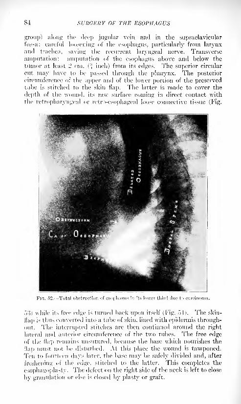
84 SURGERY OF THE ESOPHAGUS
group) along the deep jugular vein and in the supraclavicularfossa; careful loosening of the esophagus, particularly from larynxand trachea, saving the recurrent laryngeal nerve. Transverse
amputation: amputation of the esophagus above and below the
tumor at least '2 cm. (* inch) from its edges. The superior circular
cut may have to be passed through the pharynx. The posteriorcircumference of the upper and of the lower portion of the preservedtube is stitched to the skin flap. The latter is made to cover the
depth of the wound, its raw surface coming in direct contact with
the retropharyngeal or retro-esophageal loose connective tissue (Fig.
FIG. ."52. Total obstruction of esophagus in its lower third duo to carcinoma.
^'.'>) while its free edge is turned back upon itself (Fig. 54). The skin-
flap is thus converted into a tube of skin, lined with epidermis through-out. The interrupted stitches are then continued around the right
lateral and anterior circumference of the two tubes. The free edgeof the flap remains unsutured, because the base which nourishes the
flap must not be disturbed. At this place the wound is tamponed.Ten to fourteen days later, the base may be safely divided and, after
freshening of the edge, stitched to the latter. This completes the
esophagi >plasty . The defect on the right side of the neck is left to close
by granulation or else is closed by plasty or graft.
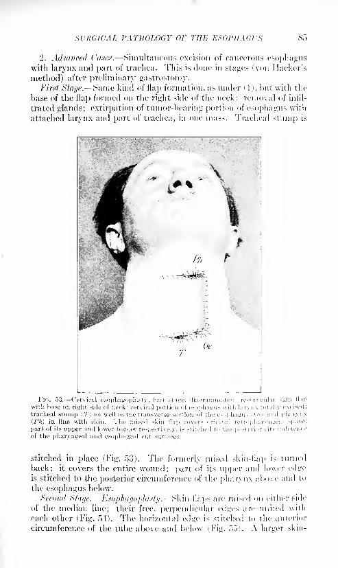
SURGICAL PATHOLOGY OF THE ESOPHAGUS 85
"2. Advanced Cases. Simultaneous excision of cancerous esophaguswith larynx and part of trachea. 'This is clone in stages (von Hacker's
method) after preliminary gastrostoiny.First Stage. J^ame kind of flap formation, as under ( 1 j, but with the
base of the flap formed on the right side of the neck; removal of infil-
trated glands; extirpation of tumor-bearing portion of esophagus with
attached larynx and part of trachea, in one mass. Tracheal stump is
if*
FIG. 53. Cervical esophagoplasty, first stane. diagrammatic: reetaniail
with base on right side of neck: cervical portion of esophagus with larynx tot
tracheal stump (T) as well as the transverse section of the esophagus -,.(>(.) a
(Ph) in line with skin. The raised skin flap covers original ret rophar.x t
part of its upper and lower border respectively, is stitched to the posterior ci
of the pharyn^eal and esopha^cal cut surfaces.
stitched in place (Fig. 53). The formerly raised skin-flap is turned
back; it covers the entire wound; part of its upper and lower edgeis stitched to the posterior circumference of the pharynx above and to
the esophagus below.
Second Stage. Esophago2)l(i$ty.~Skm-llaps are raised on either side
of the median line; their free, perpendicular edges are united with
each other (Fig. 54). The horizontal edge is stitched to the anterior
circumference of the tube above and below (Fig. 55). A larger skin-
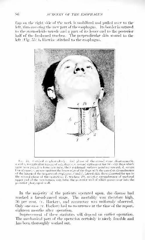
so SfRGI-RY Or THE ESOPHAGUS
flap on the right side of the neck is mobilized and pulled over to the
left, thus covering the new part of the esophagus. Its border is sutured
to the sternocleido muscle and a part of its lower end to the posteriorhalf of the freshened trachea. The perpendicular skin wound to the
left (Fig. .">.")) is likewise stitched to the esophagus.
l'io. "ji. ( 'ervical esophagoplasty; first phase of the second sta<rc. diagrammatic.a and I/, wounds after raising of .skin flap: <\ r. wound surfaces of the two skin flaps whichhave been joined to form new tube, their epidermal .surfaces pointing inward; d, suture
line of same; c, sutures uniting the lower edge of the flaps with the anterior circumferenceof the lumen of the amputated esophagus; / and fi, lateral skin flaps dissected for use in
the second phase of the operation; 7', trachea: 1'h, anterior circumference of unclosed
upper end of the new-formed skin tube, the posterior wall of which passes over into the
posterior pharyngeal wall.
In the majority of the patients operated upon, the disease had
reached a far-advanced stage. The mortality was therefore high,
36 per cent. (v. Hacker), and recurrence was uniformly observed.
Only one case (v. Hacker) had no recurrence at the time of the report,
eighteen months after operation.
Improvement of these statistics will depend on earlier operation.The mechanical part of the operation certainly is nicely feasible andhas been thoroughly worked out.
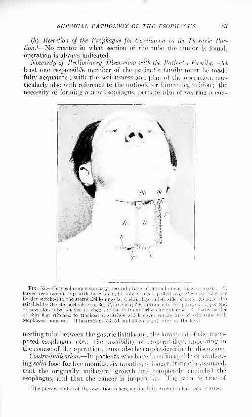
SURGICAL PATHOLOGY OF THE ESOPHAGUS
(b) Resection of the Esophagus for Carcinoma in its Thoracic Por-
tion. 1 No matter in what section of the tube the tumor is found,
operation is always indicated.
Necessity of Preliminary Discnssion with the Patients Family. -At
least one responsible member of the patient's family must be madefully acquainted with the seriousness and plan of the operation, par-
ticularly also with reference to the outlook for future deglutition; the
necessity of forming a new esophagus, perhaps also of wearing a con-
\
FIG. 55. Cervical esophagoplasty, second phase of second stage, diagrammatic, /i,
larger rectangular flap with base on right side of neck pulled over the new tube, its
border stitched to the sternocleido muscle; /, skin flap on left side of neck; its edge also
stitched to the sternocleido muscle; T, trachea; Ph, entrance to the pharynx, upper endof new skin tube not yet stitched to skin in its anterior circumference: h. lower border
of skin flap stitched to trachea; {, stitches which cover suture line of skin tube with
esophageal mucosa. (Illustrations 53, 54 and 55 enlarged after v. Hacker.)
necting tube between the gastric fistula and the lower end of the trans-
posed esophagus, etc.; the possibility of inoperability, appearing in
the course of the operation, must also be emphasized in the discussion.
Contra-mdication. In patients who have been incapable of swallow-
ing solid food for five months, six months, or longer, it may be assumed,
that the originally unilateral growth has completely encircled the
esophagus, and that the cancer is inoperable. The same is true of
1 The present status of the operation is here outlined; its evolution lias been omitted.
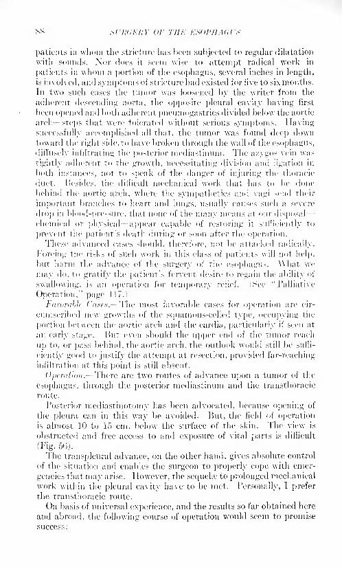
,sT/.v/AV,'ir or THE KxoriiAai'x
patients in whom the stricture lias been subjected to regular dilatation
with sounds. Nor does it seem wise to attempt radical work in
patients in whom a portion of the esophagus, several inches in length,is involved, and symptoms of stricture had existed for five to six months.
In two such cases the tumor was loosened by the writer from the
adherent descending aorta, the opposite pleiiral cavity having first
been opened and both adherent pneumogastrics divided below the aortic
arch steps that were tolerated without serious symptoms. Havingsuccessfully accomplished all that, the tumor was found dee]) downtoward the right side, to have broken through the wall of the esophagus,
diffusely infiltrating the posterior mediastinum. The a/ygos vein was
tightly adherent to the growth, necessitating division and ligation in
both instances, not to speak of the danger of injuring the thoracic
duct, besides, the difficult mechanical work that, has to be done
behind the aortic arch, where the sympathetics and vagi send their
important branches to heart and lungs, usually causes such a severe
drop in blood-pressure, that none 1 of the many means at our disposalchemical or physical appear capable of restoring it sufficiently to
prevent the patient's death during or soon after the operation.These advanced cases should, therefore, not be attacked radically.
Forcing the risks of such work in this class of patients will not help,
but harm the advance of the surgery of the esophagus. \Vhat we
may do, to gratify the patient's fervent desire to regain the ability of
swallowing, is an operation for temporary relief. (See "Palliative
Operation," page 117.)
Favorable (.'ascx. The most favorable cases for operation are cir-
cumscribed new growths of the squamous-celled type, occupying the
portion between the aortic arch and the cardia, particularly if seen at
an early stage. But even should the upper end of the tumor reach
it]) to, or pass behind, the aortic arch, the outlook would still be suffi-
ciently good to justify the attempt at resection, provided far-reachinginfiltration at this point is still absent.
Operation. There are two routes of advance upon a tumor of the
esophagus, through the posterior mediastinum and the transthoracic
route.
Posterior mediastinotomy has been advocated, because opening of
the pleura can in this way be avoided. But, the field of operationis almost 10 to 15 cm. below the surface of the skin. The view is
obstructed and free access to and exposure of vital parts is difficult
(Fig. 51)).
The transpletiral advance, on the other hand, gives absolute control
of the situation and enables the surgeon to properly cope with emer-
gencies that may arise. 1 lowever, the sequela1 to prolonged mechanical
work within the pleural cavity have to be met. Personally, I preferthe transthoracic route.
On basis of universal experience, and the results so far obtained here
and abroad, the following course of operation would seem to promisesuccess :
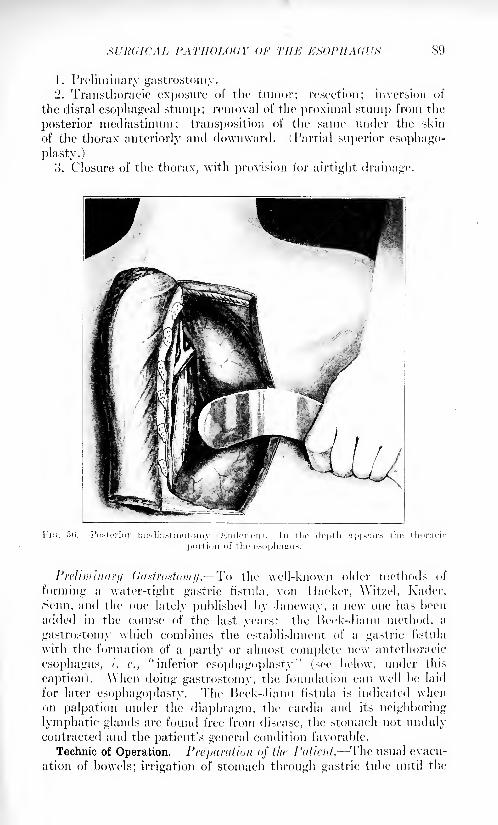
PATHOLOGY OF THE EMPIIAGVK 89
1. Preliminary gastrostomy.2. Transtlioracic exposure of the tumor; resection; inversion of
the distal esophageal stump; removal of the proximal stump from the
posterior mediastinum; transposition of the same under the skin
of the thorax anteriorly and downward. (Partial superior esophago-
plasty.)
3. Closure of the thorax, with provision for airtight drainage.
Preliminary (lastrostaiuy. To the well-known older methods of
forming a water-tight gastric fistula, von I lacker, AVit/el, Kader,
Senn, and the one lately published by Janeway, a new one has been
added in the course of the last years: the Heck-Iianu method, a
gastrostomy which combines the establishment of a gastric fistula
with the formation of a partly or almost complete new antethoracic
esophagus, /. c., "inferior esophagoplasty"
(see below, under this
caption), \\hen doing gastrostomy, the foundation can well be laid
for later esophagoplasty. The Beck-Jianu fistula is indicated whenon palpation under the diaphragm, the cardia and its neighboring
lymphatic glands are found free from disease, the stomach not undulycontracted and the patient's general condition favorable.
Technic of Operation. Preparation <>j the Patient. The usual evacu-
ation of bowels; irrigation of stomach through gastric tube until the
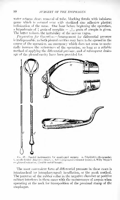
OF THE ESOPHAGUS
water returns clear; removal of tube, blocking fistula with iodoiorm
gauze which is covered over with sterilized zinc adhesive plaster;iodinization of the same. One hour before beginning the operation,a hypodermic of
.[ grain of morphin + , ,, grain of atropin is given.The latter reduces the irritability of the nervus vagus.
Preparation for Operation. Arrangement for differential pressureis indispensable, as both plenral cavities may have to be opened in the
course of the operation, an emergency which does not seem to mate-
rially increase the seriousness of the operation, so long as a reliable
method of applying the differential pressure, and of subsequent drain-
age of the pleural cavity have been provided for.
FIG. 57. Special instruments for esophageal surgery, a, Friedrich's rib-spreader;
1i, 7ieedle-liolder (.Raster's swan); c, Ach's esophageal extractor (wire); d, Willy Meyer'sesophageal extractor, (flexible metal bougie).
The most convenient form of differential pressure in these cases is
intratracheal (or intrapharyngeal) insufflation, or the mask method.
The presence of the rubber collar in the negative chamber or positivecabinet interferes in these cases with the maintenance of asepsis when
operating at the neck for transposition of the proximal stump of the
esophagus.
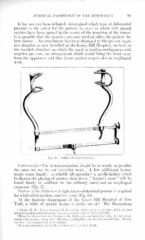
SURGICAL PATHOLOGY OF THE ESOPHAGUS 91
It has not yet been definitely determined which type of differential
pressure is the safest for the patient in cases in which both plenralcavities have been opened in the course of the resection of the tumor.
It is possible that the negative pressure method offers the patient the
best chance. An attachment has been discussed to the present nega-tive chamber as now installed at the Lenox Hill Hospital, on basis of
the Swedish chamber 1 in which the mask is used in combination with
negative pressure, an arrangement which would bring the head awayfrom the apparatus and thus insure perfect asepsis also in esophagealwork.
FIG. 5S. Balfour-Gosset retractor.
Instruments. The instrumentarium should be as nearly as possiblethe same we use in our everyday work. A few additional instru-
ments come handy: a reliable rib-spreader;2 a needle-holder which
facilitates the placing of sutures deep down ("Kiister's swan" will be
found handy in addition to the ordinary ones) and an esophagealextractor (Fig. 57).
Posture of the Patient. A right latero-abdominal posture is requiredfor a left-sided incision, and rice rcrsa (,Fig. 59).
At the thoracic department of the Lenox Hill Hospital of XewYork, a table of special design is made use of.
3 The illustrations
1 Giertz, K. H.: Union, Sweden. D. Z. f. Chir., 1913, cxxvi; C. f. C'hir., 1914, 30, andseveral Swedish implications by the same author of 1913, 1914 and 1916.
2 Here the abdominal one. known as the Ualfour-Gosset retractor 1 1'iir. oV' lias ^iven
satisfactory results;
tho.se of v. Miculicz and Friedrich are also recommendable. Latelyone has I een constructed by Lilienthal.
3 It is manufactured bv the Knv-Scheeri/r ( '<>., of Xew York.
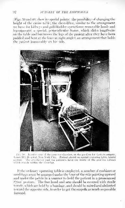
(.1> XVh'dJUfY OF THE ESOPHAGUS
\ Figs. f>9 and GO) show its sj)ecial points: the possibility of changing the
height of the entire table; the chest-lifter, similar to the arrangementwe have for kidney- and gall-bladder operations; removable head- and
leg-supports; a special, perpendicular frame, which slides lengthwiseon the table and buttresses the legs of the patient after they have been
padded and bent at the knee at right angles, an arrangement that holds
the patient immovably on his side.
Fii;. -jit. Interior view of the negative chamber, at the pavilion for thoracic surgery,Lenox Hill Hospital, New York City. Patient placed on special operating table, lateral
posture. The anestheti.-t and the patient's head are inside of the positive cabinet
which stands within the chamber.
If the ordinary operating table is employed, a number of cushions or
sand-bags must be arranged under the knee of the side pointing upwardand under the pelvis in a manner to hold the patient in a pronouncedSyms' posture. The free hand and arm should be covered with sterile
towels, which are held by a bandage, and should be raised and abductedtoward the opposite side, in order to get the scapula as much as possibleforward.
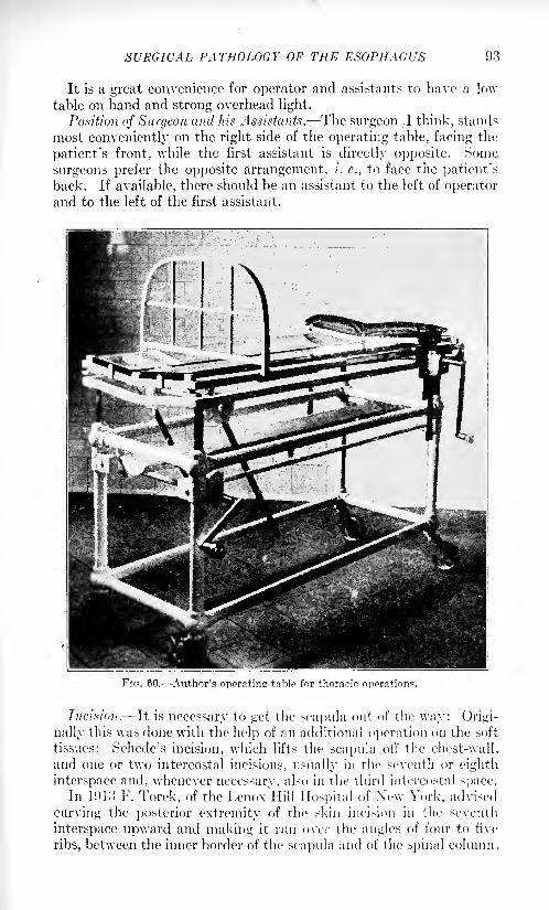
SURGICAL PATHOLOGY OF THE ESOPHAGUS 93
It is a great convenience for operator and assistants to have a low
table on hand and strong overhead light.
Position of Surgeon and his Assistants. The surgeon .1 think, stands
most conveniently on the right side of the operating table, facing the
patient's front, while the first assistant is directly opposite. Some
surgeons prefer the opposite arrangement, i. e., to face the patient's
back. If available, there should be an assistant to the left of operatorand to the left of the first assistant.
Fin. 60. Author's operating table for thoracic operations.
Incision. It is necessary to get the scapula out of the way: Origi-
nally this was done with the help of an additional operation on the soft
tissues: Schede's incision, which lifts the scapula oil' the chest-wall,
and one or two intercostal incisions, usually in the seventh or eighth
interspace and, whenever necessary, also in the third intercostal space.In 1913 F. Torek, of the Lenox Hill Hospital of Xew York, advised
curving the posterior extremity of the skin incision in the seventh
interspace upward and making it run over the angles of four to five
ribs, between the inner border of the scapula and of the spinal column.
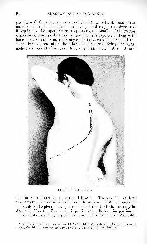
94 SURGERY OF THE ESOPHAGUS
parallel with the spinous processes of the latter. After division of the
muscles of the back, latissimus dorsi, part of major rhomboid andif required of the superior serratus posticus, the bundles of the erector
trunci muscle are pushed inward and the ribs exposed and cut with
bone scissors, either at their angles or between the angle and the
spine (Fig. 1)1 ) one after the other, while the underlying soft parts,
inclusive of costal pleura, are divided gradatim from rib to rib and
\
the intercostal arteries caught and ligated. The division of four
ribs, seventh to fourth inclusive, usually suffices. If direct access to
the vault of the pleiiral cavity must be had, the third rib, too, may be
divided. 1 Now the rib-spreader is put in place, the anterior portion of
the ribs, plus overlying scapula are pressed forward as a whole, yield-
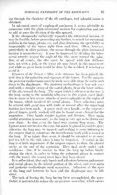
SURGICAL PATHOLOGY OF THE ESOPHAGUS 95
ing through the elasticity of the rib cartilages, and splendid access is
obtained.
In advanced cases of esophageal carcinoma it seems advisable to
commence with the plain intercostal incision for exploration and not
to add at once the division of the ribs upward.If the rib-spreader sufficiently separates the intercostal incision, it
may be feasible, before proceeding any further, to search for metastatic
nodules in the lungs, pleura, etc. and thus determine the operability or
inoperability of the tumor right there and then. Often, however,
particularly in older patients, the access through the plain intercostal
incision is unsatisfactory. It may be improved by dividing the inter-
costal muscles beyond the angle of the ribs, more toward the spine.
But, at all events, the ribs must be spread with slow delibera-
tion, not with a jerk, or the lower rib may break in this manu-uver;and while no great harm would be done by the accident, it is better to
avoid it.
Exposure of the Tumor. After wide entrance has been gained, the
next step is the palpation and exposure of the tumor. For this purposethe posterior mediastinum must be made accessible from the aortic arc!)
down to the diaphragm. The posterior mediastinum is here only cov-
ered with a straight sweep of the costal pleura, from the inner surface
of the ribs toward the lung. The organ which is oftenest in the way is
the lung, owing to the manifold adhesions in this region, most likely
due to more or less severe attacks of peri-esophagitis in the region of
the tumor, which involved the costal pleura. These adhesions mustbe severed with great care with knife or scissors after the superficial
incision has been made. A gauze swab on a handle, or the surgeon's
gloved fingers, gently worked, will also be found most efficient for the
separation. Firm bands require ligation and division. Here againcareful attention is necessary, as the lung is very apt to be drawn out
cone shape. In that event the band may contain lung tissue. The
ligature must not involve the cone, but must be placed beyond its apex;otherwise the lung may be injured, and nothing is more irritating to
the surgeon than to commence the intrathoracic work with a wound in
the lung. If an injury does occur, it should be attended to at once,
and not left to the end, when it may be forgotten. A wound in the
lung is of little importance, if the surgeon expects to drain the pleural
cavity at the end of the operation. They heal nicely under such
conditions. But, if he intends to blow the lung up before finally
closing the chest, in order to bring the pulmonal pleura in contact
with the costal, the wound must have first been closed.
It is self-evident, that only bands and adhesions between pulmonaryand costal pleura in the lateral portion of the chest, and over aorta
and posterior mediastinum need separation. Adhesions at the apexof the lung and between its base and the diaphragm may be left
alone.
The task of freeing the lung having been accomplished, the anes-
thetist is instructed to reduce the air-pressure somewhat, which causes
90 SURGERY OF THE ESOPHAGI'*
a corresponding collapse of the exposed lung. This step facilitates the
surveying of the depth of the chest, as the inflated human lung is a very
bulky organ and obstructs the vie\v, but collapsing, it retracts uponthe hilnni and thus uncovers the field of operation. If left inflated, it
would be necessary, in order to obtain the same free space, to exert a
pull by hand or broad blunt retractor upon the lung tissue, which mightdamage it. In the free space obtained by the lung retraction goodsurgery can be done.
But while the lung is more or less collapsed both surgeon andanesthetist must be very much on the alert and constantly keep in
mind the danger of a double pneumothorax. On severing firm peri-
esophageal adhesions, the opposite pleural cavity may be invaded.
A peculiar hissing noise produced by the entrance and exit of airjntoand out of the second pleural cavity through the narrow rent,
announces this unintended communication, and the more air enters,
the more also the second lung collapses. In that event, the air pressuremust be promptly raised Both lungs must never be allowed to remain
collapsed for any length of time. The heart does not tolerate a pro-
longed acute double pneumothorax. The blood-pressure drops rapidly.It can be brought back to normal by a series of rhythmic raisings and
lowerings of the air pressure, under the influence of which the lungi- inflated and deflated: artificial respiration. There is, in fact, no
better and more promptly acting heart stimulant than repeated
pronounced changes in the air pressure from the apparatus.Thoracic operations require an especially trained and careful
anesthetist. If anywhere in surgery, then here, teamwork is essential
to success. The surgeon must not be required to pay attention to the
needs of the anesthesia. lie may give certain orders regarding pressure,as just stated, but then his mind must be relieved from further responsi-
bility in this direction. His attention is needed elsewhere.
If. in severing the pulmonary adhesions, the posterior mediastinumhas been inadvertently entered, then this hole or rent in the medias-
tinal co-tali pleura is used for advance toward the esophagus.Otherwise a free-hand incision through the mediastinal pleura is made\\i\\\ the knife, parallel with and about one inch away from the right
border of the descending aorta. By spreading the branches of a pairof blunt scissors, or by introducing and separating two fingers, the
mediastinal opening is enlarged and the tumor approached. One is
thus able to feel plainly the extent of the cancer and the condition of
the peri-esophageal tissue. If the tumor appears massive and infiltrat-
ing and is not easily surrounded by the gently advancing forefinger;if it extends up to the aortic arch; further, if the history of the case
shows that a stricture had existed for five to six months and that
sounding of the esophagus had been done repeatedly, it is wise not to
continue the intended radical work. If insisted in, the drama is quite
apt to be transformed into a tragedy, because, as emphasized before,
the necessity of the blind severing of firm and far-reaching adhesions
behind the aortic arch, at the rrnj mot of life, almost without exception
SURGICAL PATHOLOGY OF THE ESOPHAGUS 97
means the death of the patient at the end of or soon after the comple-tion of the difficult work.
On the other hand, if the tumor is localized, so that 1 to 2 inches of
non-infiltrated esophageal wall can be palpated below the aortic arch,
if the tube does not appear to be"cemented in" in the posterior medias-
tinum, then there is justification for deciding on resection. Theextent of the tumor downward toward the cardia is of less moment,
provided it stops short of the cardia. Involvement of the cardia
necessitates a dill'erent operative procedure. (See below.)The most favorable cases for radical operation, as stated above,
are the localized, non-infiltrating tumors, bounded by a healthy pieceof esophagus below the aortic arch and above the diaphragm.
Kxcision having been decided on, everybody and everything mustbe bent on deliberate advance. The healthy portion of the esophagusbelow the tumor is primarily approached. The left forefinger surrounds
it gently, including both pneumogastrics. While lifting it up, a long
piece of tape is conducted around it by means of a slender, curved
forceps or Dechamp's needle. By holding the two ends of the tape
apart, the nervi vagi are made visible. They are carefully followed
upward. If one of them can be separated from the tumor, a great deal
has been won for the patient. If both can be saved, this is exceptionallyfortunate. If both are surrounded by the growth, or intimatelyattached to it, it is better to cut them both primarily with scissors,
in preference to subjecting them to much blunt handling. If only one
is hopelessly involved, this one is divided. Immediately the proximalborder of the tumor is next approached, in order to attend to the nervi
vagi at this place. For it must be remembered that repeated tugging
on, and irritating of, the branches of the anterior and posterior
esophageal plexus adds continuously, at first perhaps unnoticed, to tin-
slow development of shock, likely also to postoperative pneumonia.Therefore, when following the nerves upward, the surgeon should
disregard the loosening of the tumor, and, leaving it untouched.
should first attend to the nerves above it. It will not be difficult
to find the nerves here, and either inject them directly with a ().")
per cent, novoeain-adrenalin solution, taking care not to make this
nerve puncture too close to the aortic arch, or else infiltrate the peri-
neural tissue with the solution. After a few minutes' wait for the solu-
tion to take effect, the same nerve that had to be divided below is nowalso cut above. 1 Then the other nerve 1
, the one that can be saved, or
in the "real lucky" cases both nerves, is followed downward and freed
thoroughly. Xow the tumor-bearing portion is shelled out, the sur-
geon always keeping faithfully in mind the topographic anatomy, as
described above.
In cases in which the growth reaches up behind the aortic arch, or
is situated at this very place, blunt loosening of the esophagus, without
the guidance of the surgeon's eyes, is not permissible. Here the arch
>r ;ulvi.s:mil
98 SURGERY OF THE ESOPHAGUS
of the aorta and the first few inches of its descending portion must be
mobilized (Ach. Helm Jr., Torek). This is done without difficulty bydivision of the respective intercostal arteries, after previous double
libation. The aortic arch must be handled gently when lifting it aside.
\\hen the tumor has been freed all around, a piece of tape or, if this
be not on hand, a twisted piece of gauze, is conducted around the
esophagus above and below the tumor.
It will be noted that, up to this time, the asepsis has remainedunbroken. Local or general reasons making such a course advisable,the operation could, therefore, still be broken oil', the operation thus
remaining an exploratory thoracotomy.Hi xt d ion of (In
1 Tumor. At this stage the intrathoracic work is
interrupted for a few moments and the patient turned over more on his
back in order to expose the esophagus above the left clavicle. It is
annoying to cover and rearrange the operating field for this pur-
pose. It is wiserand 1 have done so in the last several years to
commence the operation with the cervical incision fexternal esopha-
gotomy), surrounding the esophagus with a piece of tape which is
dropped into the wound, while the latter is loosely tamponed and
temporarily closed with a few stitches or clamps, Xo harm has been
done, should the case prove inoperable in the course of the operation.A few stitches close the wound.
Instead of doing preliminary external esophagotomy the surgeon can
also make his way toward the skin of the neck along the esophagus with
his fingers from within, in the course of the operation, bluntly incise
on to his spread fingers or upon a blunt instrument introduced from
within, and then transpose the esophagus through the neck wound by
pulling on the thread with which the proximal end of the tube wastied off.
This method, though, does not appear as safe as external esopha-
gotomy. J have, on one occasion, seen an esophagus tear oil' on being
pulled out through the neck wound, when thus handled from within.
If the surgeon preferred to do external esophagotomy in the course
of the operation, he now returns to the thorax. The moist towels
and sponges, temporarily placed, arc removed and the esophaguslifted out of its bed by means of the two tapes. It is pulled upwardon the lower tape and a purse-string suture of chromicized catgut is
placed fully one and a half to two inches below the spot at which one
expects to divide the esophagus. This thread must be long. Three
loops are formed and the knot of each secured with a non-biting
clamp, all three clamps being of the same type, while the two ends
of the thread are clamped with a forceps of different type (Fig. 62).
Thereupon the operation proceeds as follows:
1. Division of the esophagus below the tumor: A clam]) is placed
right below the tumor and firmly closed, and a silk ligature or one of
thin tape, tied around the tube about five-eighths-inch farther down.
Thorough tamponade with gauze all around; division of esophagusbetween these two marks, either with the active cautery or with the
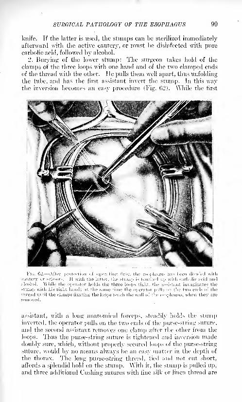
SURGICAL PATHOLOGY OF THE ESOPHAGUS 99
knife. If the latter is used, the stumps can be sterilized immediatelyafterward with the active cautery, or must be disinfected with purecarbolic acid, followed by alcohol.
2. Burying of the lower stump: The surgeon takes hold of the
clamps of the three loops with one hand and of the two clamped ends
of the thread with the other. ITc pulls them well apart, thus unfoldingthe tube, and lias the first assistant invert the stump. In this waythe inversion becomes an easy procedure (Fig. 62). \Vhile the first
FIG. 62. After protection of operating field, the esophagus has been divided with
cautery or scissors. Jf with the latter, the stump is touched up with carbolic acid andalcohol. While the operator holds the three loops light, the assistant invaginates the
stump with his right hand; at the same time the oj orator pulls on the two ends of the
thread until the clamps fixating the loops reach the wall of the esophagus, when they are
removed.
assistant, with a long anatomical forceps, steadily holds the stumpinverted, the operator pulls on the two ends of the purse-string suture,
and the second assistant removes one clamp after the other from the
loops. Thus the purse-string suture is tightened and inversion made
doubly sure, which, without properly secured loops of the purse-string
suture, would by no means always be an easy matter in the depth of
the thorax. The long purse-string thread, tied and not cut short,
affords a splendid hold on the stump. \Vith it, the stump is pulled up,and three additional Gushing sutures with fine silk or linen thread are
100 SURGERY OF THE ESOPHAGUS
placed and closed at either end lirst, then in the middle, after the purse-
string suture has been cut short. Xo\v the stump is allowed to dropback and disappears in the depth.
)>. Extraction and Transposition of the proximal stump: The proxi-
mal stump must now be closed and removed from the mediastinum.
There are two methods:
(a) To l-'.rtfurf Sfn/it/i nnil Tumor Together. instead of clampingthe esophagi^ below the tumor, a ligature might have been placedthere. ISut. be it borne in mind, that nowhere does a mass ligature,
no matter of what material, slip off more readily, than from the short
stump of the esophagus. The safest closure is that by Ach's method:
transfixion with needle and thread and ligation to either side, same as
we are wont to proceed in closing the neck of the hernia! sac. Of course,
this introduce- an clement of sepsis. However, the possibility of infec-
tion is but -light, if the stump is at once iodined and extraction of it
done without delay. This extraction is sometimes accomplished easily,
with a jerk of the left forefinger which surrounds the esophagus above
the clavicle within the esophagotomy wound. 1 have succeeded a
number of times in extracting it in this way, without a hitch. In other
cases the many adhesions of the esophagus to the aorta by short
arteries, and to the pneumogastric nerves, would not yield all at the
same time. Then the esophagus had to be loosened gradually and
placed in front of the aortic arch. Only then the stump would yield to
the traction at the neck.
The second method is apparently more recommendable, viz.:
i> To rescrt the tumor Jirxt and then extract the stump, delivering it
through the neck wound.
In both cases the extracted stum]) is placed antethoracically under
the -kin, being pulled through a bluntly-made tunnel. It is left
closed and its tip secured within the transverse incision of the skin
of the che-t by a few stitches.
Closure of Tl/or/ir/r If 'mi nil. (a] Without Drainage. The whole
operative field, particularly the posterior mediastinum is wiped clean
and revised for hemorrhage, \\here found, ligatures are placed.Then :; to 1 perico-tal Fig. li.'J.l
i,or percnstal ( Fig. ('>:'>/>> silk or strong
chromici/ed catgut .-utnre- bring the two ribs in apposition. Thethird method i- to perforate the lower rib only and make the thread
surround the rib above 'Fig. <>."/' i.
For the prrco-tal -utiire, or it- modification, Friedrich's rib puncherand thread guide Fig. Hh/ and h i are preferable to a drill. I prefersilk sutures fir.-t, and next to each, a stout chromici/ed catgut suture.
The silk sutures are fir-t pulled taut by operator and assistant simul-
taneously, and tied. Then the catgut sutures are united, thus havingto bear much le.-- -train, and the silk ones are removed. In cases of
wound disturbance I have seen the silk threads act as foreign bodies
which had to be removed.
The posterior rib divisions are not sutured. Continuous layer suture
of catgui of muscles and skin with catgut; finally, the lung is thor-
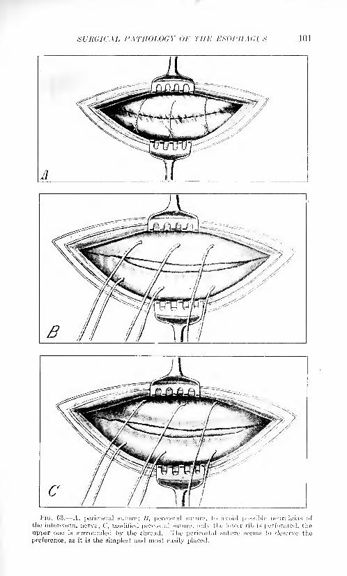
SURGICAL PATHOLOGY OF THE ESOPHAGI'^ 101
A
In;. 03. .1, pericostal suturo; /?, percosfal suture, to avoid possible neuralgias of
the intercostal nerve; C, modified percostal suture, only the lower rib is perforated, the
upper one is, surrounded by the .thread. The pericostal suture seems to deserve the
preference, as it is the simplest and most easily placed.
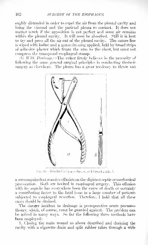
102 SURGERY OF THE ESOPHAGUS
oii^hly distended in order to expel the air from the pleural cavity and
bring the visceral and the parietal pleura to contact. It does not
matter much if the apposition is not perfect and some air remains
within the pleural cavity. It will soon be absorbed. Still it is best
to try and press all the air out of the pleural cavity. The suture line
is wiped with iodine and a gauze dressing applied, held by broad strips
of adhesive plaster which fixate the arm to the chest, but must not
compress the transposed esophageal stump.
(b) With Drainage. The writer firmly believes in the necessity of
following the same general surgical principles in conducting thoracic
surgery as elsewhere. The pleura has a great tendency to throw out
FK;. 04. Friedrich'.-- rip-puncher, , and thread guide. f>.
a serosanguinolent massive effusion on the slightest septic or mechanical
provocation. Both are invited in esophageal surgery. This effusion
with its sequeke has everywhere been the cause of death or certainlya contributing factor to the fatal issue in a large number of patients
subjected to esophageal resection. Therefore, I hold that all these
eases should be drained.
The danger incident to drainage is postoperative acute pneumo-thorax, which, of course, must be guarded against. The problem can
be solved in many ways. So far the following three methods havebeen employed:
1. Closing the main wound as above described and draining the
cavity with a cigarette drain and split rubber tubes through a wide
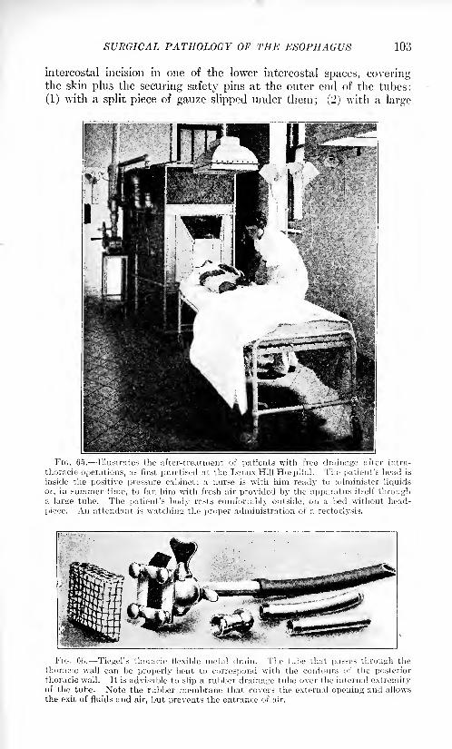
SURGICAL PATHOLOGY OF THE ESOPHAGUS 103
intercostal incision in one of the lower intercostal spaces, coveringthe skin plus the securing safety pins at the outer end of the tubes:
(1) with a split piece of gauze slipped under them; (2) with a large
FIG. 65. Illustrates the after-tre;itment of patients with free drainage after intra-
thoracic operations, as first practised at the Lenox Hill Hospital. The patient's head is
inside the positive pressure cabinet; a nurse is with him ready to administer liquids
or, in summer time, to fan him with fresh air provided by the apparatus itself througha large tube. The patient's body rests comfortably outside, on a bed without head-
piece. An attendant is watching the proper administration of a rectoclysis.
FIG. 00. Tiegel's thoracic flexible metal drain. The tube that passes through thethoracic wall can be properly bent to correspond with the contours of the posteriorthoracic wall. It is advisable to slip a rubber drainage tube over the internal extremityof the tube. Note the rubber membrane that covers the external opening and allows
the exit of fluids and air, but prevents the entrance of air.
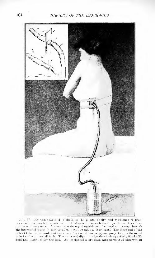
104 SURGERY OF THE ESOPHAGUS
Fi<;. 07. Konyon's method of draining the pleural cavity and avoidance of post-
operative )iiieuniothorax, modified and adapted to intrathoracic operations otlier thandrainage of ompyenm. A metal tube (6) round outside and flattened on its way throughthe intercostal space l<' is covered with rubber tubing. (See inset.) The inner end of therubber tube has a number of holes for additional drainage (d) and projects from the metaltube for about one-half inch. The outer end dips into a bottle which is partially filled withfluid and placed under the bed. An interposed short glass tube permits of observation
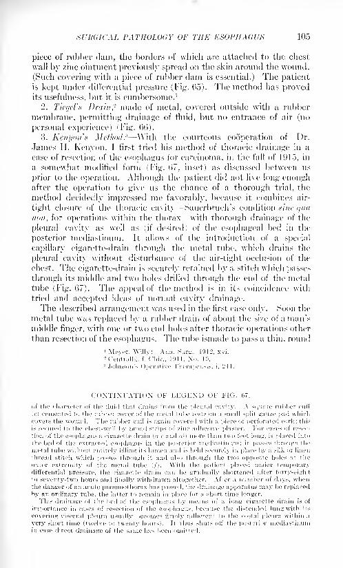
SURGICAL PATHOLOGY OF THE ESOPHAGUS 105
piece of rubber dam, the borders of \vliieh are attached to the chest
wall by zinc ointment previously spread on the skin around the wound.
(Such covering with a piece of rubber dam is essential.) The patientis kept under differential pressure (Fig. Go). The method has provedits usefulness, but it is cumbersome. 1
2. Tiegel's Drain,- made of metal, covered outside with a rubber
membrane, permitting drainage of fluid, but no entrance of air (no
personal experience) (Fig. (>(>).
3. Ken i/on'a Method. 3 AVith the courteous cooperation of Dr.
James II. Kenyon, I first tried his method of thoracic drainage in a
case of resection of the esophagus for carcinoma, in the fall of 191.5, in
a somewhat modified form (Fig. (>7, inset) as discussed between us
prior to the operation. Although the patient did not live long enoughafter the operation to give us the chance of a thorough trial, the
method decidedly impressed me favorably, because it combines air-
tight closure of the thoracic cavity Sauerbruch's condition .tine </u<t
uon, for operations within the thorax with thorough drainage of the
pleural cavity as well as (if desired) of the esophageal bed in the
posterior mediastinum. It allows of the introduction of a special
capillary cigarette-drain through the metal tube, which drains the
pleural cavity without disturbance of the air-tight occlusion of the
chest. The cigarette-drain is securely retained by a stitch which passes
through its middle and two holes drilled through the end of the metal
tube (Fig. 67). The appeal of the method is in its coincidence with
tried and accepted ideas of normal cavity drainage.The described arrangement was used in the .first case only. Soon the
metal tube was replaced by a rubber drain of about the si/e of a man'smiddle finger, with one or two end holes after thoracic operations other
than resection of the esophagus. The tube ismade to pass a thin, round
CONTINUATION OF LEGEND OF FIG. 67.
of the character of the fluid that drains from the pleural cavity. A square nil her cuff
(c.) cemented to the rubber cover of the metal tube rests on a small split gauze p;
covers the wound. The rubber cuff is again covered with a piece of perforated t
is secured to the chest-wall by broad strips of zinc adhesive plaster. For cases
tion of the esophagus a cigarette drain (<t c and ') more than two feet long, is pi;
irk; this
f resec-
ed into
the hed of the extracted esophagus in the posterior mediastinum; it passes thr ugh the
metal tuhe without entirely filling its lumen and is held securely in place hy a silk or linen
thread stitch which passes through it and also through the two opposite holes at the
outer extremity of the metal tuhe ('/). With the patient placed under temporarydifferential pressure, the cigarette drain can he gradually shortened after forty-eightto seventy-two hours and finally withdrawn altogether. After a numhcr of days, whenthe danger of an acute pneumothorax has passed, the drainage apparatus may he replaced
hy an ordinary tuhe, the latter to remain in place for a short time longer.
This drainage of the hed of the esophagus hy means of a long cigarette drain is of
importance in cases of resection of the esophagus, hecause the distended lung with its
covering visceral pleura usually becomes firmly adherent to the costal pleura within a
very short time (twelve to twenty hours). It thus shuts off the posterior mediastinumin case direct drainage of the same has been omitted.
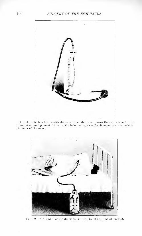
inc. SURGERY OF THE ESOPHAGUS
FIG. 00. Air-tL'lit thorufic drainage, as used by the author at present.
SURGICAL PATHOLOGY OF THE ESOPHAGUS 107
piece of cork with a central hole which is somewhat smaller in size than
the circumference of the tube l,Figs. (58 and 09). The arrangementoutside has remained the same.
The whole apparatus is sterilized by boiling. The tube is intro-
duced one or two intercostal spaces below the thoracic incision, usuallyin the posterior axillary line. Skin and fascia only are incised, the
intercostal muscles with endothoracic fascia and costal pleura bluntly
perforated. Muscles, fascia and skin are then stitched in layers
tightly around the tube.
The method corresponds with Buelau's thoracic drainage, devised morethan twenty-five years n go and used by him in the treatment of empyema.
In view of the results obtained I have adopted it as the standard
method for thoracic drainage.
The fact that the only so far recorded case of recovery after resec-
tion of the thoracic portion of the esophagus, that of Dr. F. Torek, 1
of the Lenox Hill Hospital of Xew York City, healed without drain-
age'- is no valid argument for making non-drainage the method of
choice in this operation. A great number of cases stand out againstit, in which autopsy showed that death occurred as a result of a septic
serosanguinolent effusion that had accumulated in the pleural cavityand could not drain off. It certainly seems an unavoidable conclu-
sion to be drawn from the many fatal issues after this operation, that
omission of drainage has been one of the many contributory factors
in the outcome. In other words, drainage after resection of the
esophagus, in fact, after any intrathoracic operation that required
opening the lumen of the esophagus, is essential and imperative.Xot draining, to my mind, means taking chances, working without a
method of after-treatment which greatly helps to ensure the success
of the complicated and serious operation.
Personally I am employing this"air-tight'' thoracic drainage (Figs.
08 and 09), after every thoracic operation.8
A valuable contribution to this question has recently been furnished
by Adr. J. Bcngoela of Buenos Ayres, Argentine.4 It refers to a
woman, aged thirty-seven years, weighing seventy-seven pounds, in
whom intrathoracic resection of the esophagus for carcinoma in the
lower portion and esophagogastrostomy with the help of a woodenbobbin was done, the patient living thirty-seven days after operation.There is no question in my mind that the air-tight thoracic drainage,commenced at the close of the operation, had a great deal to do with
the immediate favorable result of the serious operation in this greatlyreduced patient. Unfortunately, symptoms of suture insufficiencyset in on the fifteenth day, and the patient died of septic empyemafive and a half weeks after the operation, her regained ability to
swallow, however, not having become impaired up to the last.
1 Jour. Am. Mod. Assn., 1913, Xo. 20, Ix.2 This patient is still alive six and one-half years after the operation and in splendid
condition.3 Annals of Surgery August, 191S, Ixviii 157.4Surg., Oynee. and Obstet., October, 1919, p. 413.
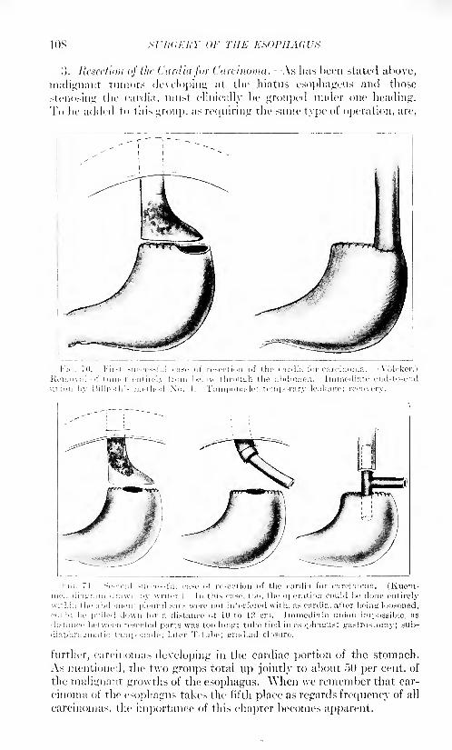
10S .STAY ;/;/,' }-' OF THE ESOPHAGUS
o. Resection of the Cardiafor Carcinoma. - As has been stated above,
malignant tumors developing at the hiatus esophageus and those
sten<ing the eardia, must clinically he grouped under one heading.To he added to this group, as requiring the same type of operation, are,
I'ni. 71.- Second .-uccessful ciiso ot resection of tlio eardia for carcinoma. (Kuem-iicl, diagram drawn by writer.) In thN ca.-c. too, the operation could be done entirelyvithin the abdomen; pleural sacs were not interfered with, as eardia, after being loosened,ould be pulled down for a distance of 10 to 12 cm. Immediate union impossible, as
li.-tance. between resected parts was too long; tube tied in esophagus; gastrostomy; sub-
liapliraL'inatic tamp< made; later T-tube; gradual closure.
further, carcinomas developing in the cardiac portion of the stomach.
As mentioned, the two groups total up jointly to about 50 per cent, of
the malignant growths of the esophagus. When we remember that car-
cinoma of the esophagus takes the fifth place as regards frequency of all
carcinomas, the importance of this chapter becomes apparent.
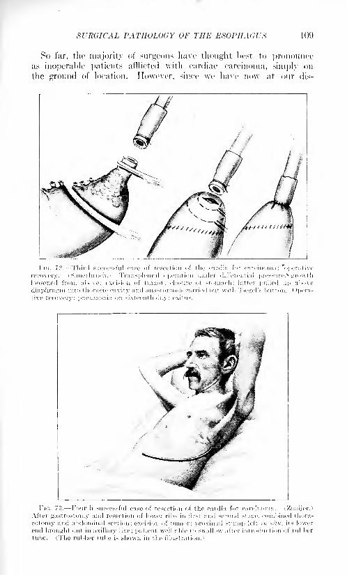
SURGICAL PATHOLOGY OF THE ESOPHAGUS 109
So far, the majority of surgeons have thought best to pronounceas inoperable patients afflicted with cardiac carcinoma, simply on
the ground of location. However, since we have nmv at our dis-
Fic;. 72. Third successful case of resection of the cardia for carcinoma: 'operative
recovery. (Sauerbruch.) Transpleural operation under differential pressure,1; growth
loosened from above: excision of tumor: closure of stomach: latter pulled up above
diaphragm into thoracic cavity and anastomosis carried out with Tiemel's. button. ( Ipera-
tive recovery; pneumonia on sixteenth day; exit us.
Fni. 73. Fourth successful case of resection of the cardia for carcinoma. (Znnijer.)
After gastrostomy and resection of lower ribs in first and second slaw, combined thora-
cotomy and abdominal section; excision of tumor: proximal stump left in xilu, its lower
end brought out in axillary line; patient well able to swallow after introduction of rubbertube. (The rubber tube i.-> shown in the illustration.
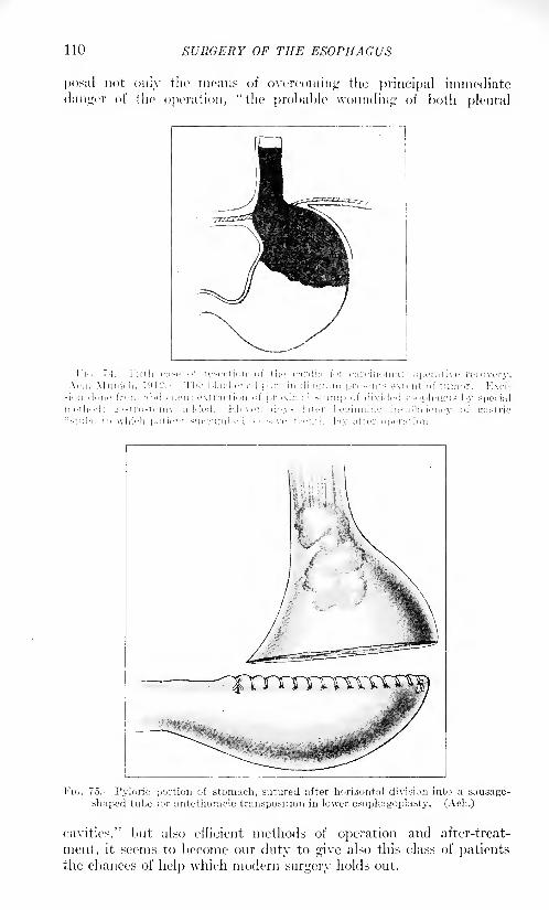
110 SURGERY OF THE ESOPHAGUS
posal not only the means of overcoming the principal immediate
danger of the operation, "the probable wounding of both plenral
Flo. 7-1. Filth fu.se of resection of the cardia for carcinoma: operative recovery.Ach. Munich, l!H2.i Tlie lilackened ]i;rt in dia'-rram pre-ents extent of tumor. ]-]xci-
^ion done from al>doti;en : extraction of pr iximal slum]) of di\ p ided e-i 'jiliairus by sjtecial
method; ii;>.-t ro-t nmy added. J-!Ie\'e:i d-'y- later 1 leirinnin-. 1
; insufRcieiicy of gastric
fistula, to which patient succumhe 1 on seventeenth day after operation.
Fie;. 75. Pylorio portion of stomach, sutured after horizontal division into a sausage-
shaped tube for antethoracic transposition in lower esophagoplasty. (Ach.)
eavities." l)iit also efHcient methods of operation and after-treat-
ment, it seems to become our duty to give also this class of patientsthe chances of help which modern surgery holds out.
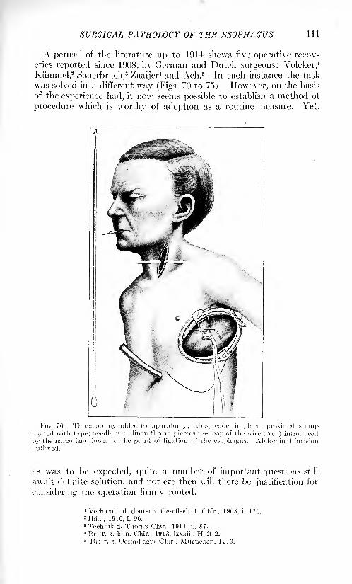
SURGICAL PATHOLOGY OF THE ESOPHAGUS 111
A perusal of the literature up to 1914 shows five operative recov-
eries reported since 1908, by German and Dutch surgeons: Volcker,1
Kiimmel,2Sauerbruch,
3/aaijer
4 and Ach.5 In each instance the task
was solved in a different way (Figs. 70 to 75). However, on the basis
of the experience had, it now seems possible to establish a method of
procedure which is worthy of adoption as a routine measure. Yet,
FIG. 70. Thoraeotomy added to laparotomy; rib spreader in place?; proximal stumpligated with tape; needle with linen thread pierces the loop of the wire (Ach) introduced
by the narcotizer down to the point of ligation of the esophagus. Abdominal incision
outlined.
as was to be expected, quite a number of important questions still
await definite solution, and not ere then will there be justification for
considering the operation firmly rooted.
' Vcrhandl. d. deutach. CJesollsch. f. Cliir., 190S, i, 120.2Ibid., 1910, i, 90.
8 Technik d. Thorax Chir., 1911, p. 87.4 Beitr. z. klin. Chir., 1913, Ixxxiii, Heft 2.
5 Beitr. z. Oesophagus Chir., Muenchcn, 1913.
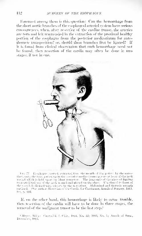
112 SURGERY OF THE ESOPHAGUS
Foremost among them is tliis question: Can the hemorrhage from
the short aortic branches of the esophageal arterial system have serious
consequences, when, after resection of the cardiac tumor, the arteries
are torn and left unattended in the extraction of the proximal healthy
portion of the esophagus from the posterior mediastinum for ante-
thoracic transposition? or, should these branches first be Heated? If
it is found from clinical observation that such hemorrhage need not
be feared, then resection of the cardia may often be done in two
>taires. if not in one.
In.. 77. K.-opha^us everted, extracted frnm the mouth .if the patient by the nareo-
ti/er. until the tape, pulled up in the posterior nicdia-tiniini appears at level of the neckwound which is held apart by blunt retractors. The loiiLr ends of the piece of Hunting
tape arc lifted out of the neck wound and placed on the chest. The thread in fiont of
the mouth is divided with scissors by the narc.it izer. Abdominal and thoracic woundsoutlined. (See author: Resection of the Cardia for Carcinoma, Annals, of Surgery, 1915,
Ixii, p. 093.
If, on the other hand, this hemorrhage is likely to cause trouble,
then resection of the cardia will have to be done in three stages, the
removal of the malignant tumor to be the last step.1
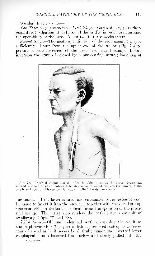
SURGICAL PATHOLOGY OF THE ESOPHAGUS 113
We shall first consider
The Three-stage Operation. First Stage. Gastrostomy, plus thor-
ough direct palpation at and around the cardia, in order to determine
the operability of the case. About two to three weeks later:
Second Stage. Thoracotomy; division of the esophagus at a spot
sufficiently distant from the upper end of the tumor (Fig. 70) to
permit of safe inversion of the lower esophageal stump. Before
inversion the stump is closed by a purse-string suture; loosening of
FIG. 78. Proximal stump placed under the skin tunnel of the chest; lower end
opened, stitched in place; rubber tube shown, as it would connect the lumen of the
esophageal stump with the gastric fistula. (Gluck-Perthes method.)
the tumor. If the latter is small and circumscribed, an attempt maybe made to invert it into the stomach together with the distal stump(Sauerbruch) . Antethoracic, subcutaneous transposition of the proxi-
mal stump. The latter step renders the patient again capable of
swallowing (Figs. 77 and 78).
Third Stage. Oblique abdominal section, exposing the vault of
the diaphragm (Fig. 79); gastric fistula preserved; osteoplastic resec-
tion of costal arch, if access be difficult; tumor and inverted lower
esophageal stump loosened from below and slowly pulled into the
VOL. ii 8
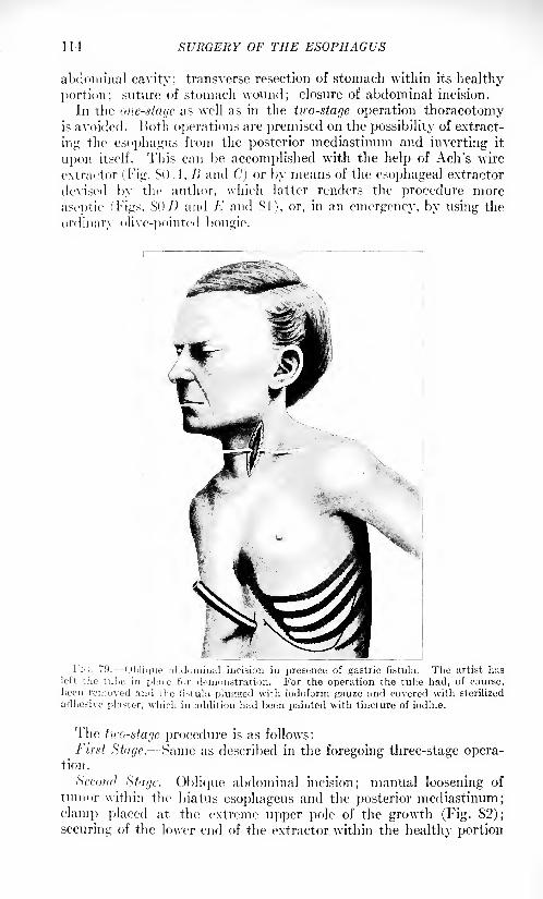
114 SURGERY OF THE ESOPHAGUS
abdominal cavity; transverse resection of stomach within its healthy
portion; suture of stomach wound; closure of abdominal incision.
In the one-stage as well as in the two-stage operation thoracotomyis avoided. Both operations are premised on the possibility of extract-
ing the esophagus from the posterior mediastinum and inverting it
upon itself. This can be accomplished with the help of Ach's wire
extractor (Fig. SO J, /> and C] or by means of the esophageal extractor
devised by the author, which latter renders the procedure more
aseptic (Tigs. SO 7) and E and SI), or, in an emergency, by using the
ordinary olive-pointed bougie.
Fi'i. 79. Oblique abdominal incision in presence of gastric fistula. The artist lias
left the tube in place for demonstration. For the operation the tube had, of course,been removed and the fistula plujzsed with iodoform gauze and covered with sterilized
adhesive plaster, which in addition had been painted with tincture of iodine.
The two-stage procedure is as follows:
First Stage. Same as described in the foregoing three-stage opera-tion.
St'cotifJ Stage. Oblique abdominal incision; manual loosening of
tumor within the hiatus esophageus and the posterior mediastinum;
clamp placed at the extreme upper pole of the growth (Fig. 82);
securing of the lower end of the extractor within the healthy portion
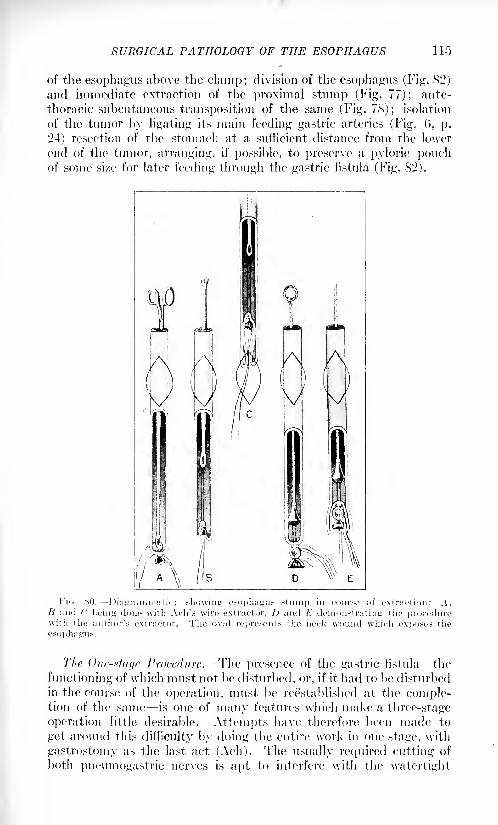
SURGICAL PATHOLOGY OF THE ESOPHAGUS
of the esophagus above the clamp; division of the esophagus (Fig. 82)
and immediate extraction of the proximal stump (Fig. 77); ante-
thoracic subcutaneous transposition of the same (Fig. 78); isolation
of the tumor by ligating its main feeding gastric arteries (Fig. 0, p.
24) resection of the stomach at a sufficient distance from the lower
end of the tumor, arranging, if possible, to preserve a pyloric pouchof some si/e for later feeding through the gastric fistula (Fig. 82).
The One-stage Procedure. The presence of the gastric fistula -the
functioning of which must not be disturbed, or, if it had to be disturbed
in the course of the operation, must be reestablished at the comple-tion of the same is one of many features which make a three-stage
operation little desirable. Attempts have therefore been made to
get around this difficulty by doing the entire work in one stage, with
gastrostomy as the last act (Ach). The usually required cutting of
both pneumogastric nerves is apt to interfere with the watertight
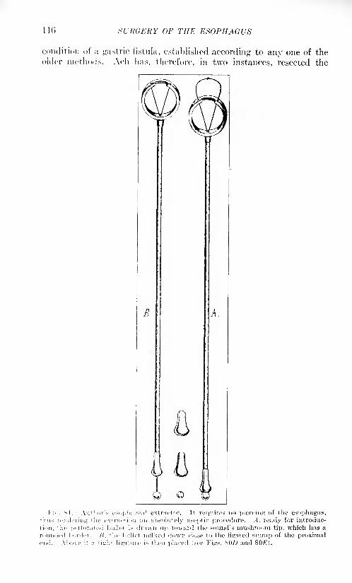
110 SURGERY OF THE ESOPHAGUS
condition of a gastric fistula, established according to any one of theolder methods. Ach has, therefore, in two instances, resected the
B
..
Q &I'n.. M. Author's I'.-ophageal extractor. It requires no piercing of the esophagus,
thus rendering the extraction an absolutely aseptic procedure. -4, ready for introduc-
tion, the perforated bullet is drawn up toward the sound's mushroom tip, which has a
rounded border. H. the bullet milked down close to the ligated stump of the proximalend. Above it a tight ligature is then placed (see Figs. SOD and 807?).
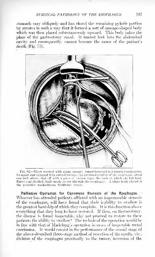
SURGICAL PATHOLOGY OF THE ESOPHAGUS 117
stomach very obliquely and lias closed the remaining pyloric portion
by sutures in such a way that it formed a sort of sausage-shaped bodywhich was then placed subcutaneously upward. This body takes the
place of the gastrostomy canal. It cannot leak into the abdominal
cavity and consequently, cannot become the cause of the patient's
death (Fig. 75).
FIG. 82. Liver covered with gauze sponge; tumor loosened in foramen esophageum,its upper end clamped with curved forceps; the proximal portion of the esophagus, about
one inch above, tied off with a piece of narrow tape, the ends of which are left long.
Nervi vagi divided; knife ready to cut through the esophagus. A blunt hook placed in
the posterior mediastinum facilitates access.
Palliative Operation for Cancerous Stenosis of the Esophagus.-Whoever has attended patients afflicted with an impermeable stenosis
of the esophagus, will have found that their inability to swallow is
the greatest hardship of which they complain. It is this function above
everything that they long to have restored. If then, on thoracotomy,the disease is found inoperable, why not proceed to restore to these
patients the ability to swallow? The technic of the operation would be
in line with that of Madelung's operation in cases of inoperable rectal
carcinoma. It would consist in the performance of the second stage of
the above-described three-stage method of resection of the cardia, vi/.,
division of the esophagus proximally to the tumor, inversion of the
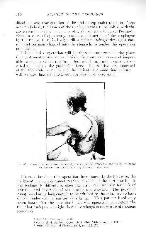
us Hl'RCKHY OF THE ESOPHAGUS
distal end and transposition of the oral stump under the skin of the
neck and chest, the lumen of the esophagus then to be united with the
gastrostomy opening by means of a rubber tube ((iluck,1 Perthes2
).
Kveii in east's of apparently complete obstruction of the esophagus
by the tumor, there is, likely, still sufficient drainage through a nar-
row and tortuous channel into the stomach, to render this operation
practicable.
This palliative operation will in thoracic surgery take the placethat gastro-cnterostomy has in abdominal surgery in eases of inoper-
able carcinoma of the pylorus. Both are, to my mind, equally indi-
cated to alleviate the patient's misery. His relatives are informed
of the true state of afl'airs, but the patient for some time at least--
will consider himself cured, surely a justifiable deception.
I have so far done this operation three times. In the first case, the
malignant, inoperable tumor reached up behind the aortic arch. It
was technically difficult to close the distal end securely for lack of
material, and inversion of the stump was irksome. The proximal
stum]) was barely long enough to be stitched to the skin, having been
slipped underneath a narrow7 skin bridge. This patient lived only
seven hours after the operation.3 He was operated upon before the
time that I adopted air-tight thoracic drainage in every case of thoracic
operation.
1 Bed. klin. Wrhnschr., 1898.- Verhandl. d. deutsch. Gesellsch. f. Chir. 34th Kongress, 1911.3Surp., (Jyiice. and Obstet., 1915, pp. 162-170.
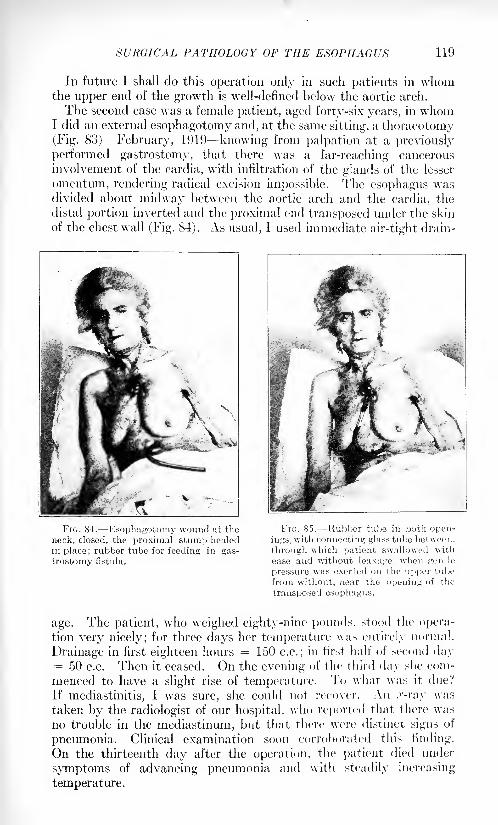
SURGICAL PATHOLOGY OF THE ESOPHAGUS
In future I shall do this operation only in such patients in whomthe upper end of the growth is well-defined below the aortic arch.
The second case was a female patient, aged forty-six years, in whomI did an external esophagotomy and, at the same sitting, a thoracotomy(Fig. 83) February, 1919 knowing from palpation at a previously
performed gastrostomy, that there was a far-reaching cancerous
involvement of the cardia, with infiltration of the glands of the lesser
omentum, rendering radical excision impossible. The esophagus wasdivided about midway between the aortic arch and the cardia, the
distal portion inverted and the proximal end transposed under the skin
of the chest wall (Fig. 84). As usual, I used immediate air-tight drain-
FIG. 84. Esophagotomy wound at the
neck, closed, the proximal stump healedin place; rubber tube for feeding in gas-
trostomy fistula.
FIG. 85. 'Rubber tube in both open-
ings, with connecting glass tube between,
through which patient swallowed with
ease and without leakage, when gentle
pressure was exerted on the upper tube
from without, near the opening of the
transposed esophagus.
age. The patient, who weighed eighty-nine pounds, stood the opera-
tion very nicely; for three days her temperature was entirely normal.
Drainage in first eighteen hours = 150 c.c.; in first half of second day= 50 c.c. Then it ceased. On the evening of the third day she com-
menced to have a slight rise of temperature. To what was it due?
If mediastinitis, I was sure, she could not recover. An .r-ray was
taken by the radiologist of our hospital, who reported that there was
no trouble in the mediastinum, but that there were distinct signs of
pneumonia. Clinical examination soon corroborated this finding.
On the thirteenth day after the operation, the patient died under
symptoms of advancing pneumonia and with steadily increasing
temperature.
120 Xl'K(;i-:j{Y OF THE E
However, we had succeeded in giving her back the power of swallowing.The lower end of the transposed esophagus was connected with the
gastrostoniy opening by a large-sized rubber tube (Fig. 85). Through an
interposed glass tube we could nicely watch the swallowed water, milk,
sonj), etc., being pressed down into the stomach in typical jets by the force
of the contracting pharyngeal muscles and the gravity of the food itself.
The patient's easy operative recovery, in spite of her greatly reduced
general condition, was due, 1 believe, principally to the air-tight
thoracic drainage, started on the operating table. Jt was the pneu-monia that caused the patient's death; not the operation as such. 1
In closing this chapter, I desire to reiterate the statement pre-
viously made that thoracic surgery is as yet in its very infancy, not
the least so the question of the resection of the esophagus for carcinoma.
However, the important point is that the thorax, too, is now finally
and permanently open to surgery. The further evolution of this sub-
ject will and can only come gradually through constant and untiringeii'ort. Hut the feasibility of the surgical work has been demonstrated.
Let us remember that the xifr of the tumor is no longer the deter-
mining factor in the question of its operability or inoperability, but the
pathological condition within the thorax and within the vault of the
diaphragm. This condition can be determined only by the palpatinghand of the surgeon.
Esophagoplasty In cases of impermeable cicatricial stenosis of the
esophagus the formation of a new tube by plasty has been attempted
esophagoplasty. Several successful operations done on this indication,
with excellent and permanent functional result, have been reportedwithin the last few years.
AYhere the continuity of the esophagus had to be destroyed by resec-
tion, the attempt should also be made to restore to the patient the
power of deglutition by means of esophagoplasty; and this final objectshould be kept clearly in mind when planning for the main operation.The new tube formed by plasty may be either partial or total.
Partial Esophagoplasty. (a) Inferior Partial Esophagoplasty. Tothe various considerations which determine the selection of one of the
several methods of gastrostomy in esophageal stenosis, a new one is
added in the cases in which an esophagoplasty is contemplated, viz.,
the bridging of the distance from the opening of the gastric fistula at
the stomach to that of the esophageal fistula at the neck or chest. Thecloser these openings can be brought to each other, the shorter naturallywill be the bridge. For purposes of esophagoplasty it is therefore advis-
able to select a method of gastrostomy which in itself constitutes an
1 In the third CUM' tin' tumor again was located behind the aortic! arch. The patient's
pitiful pleas induced me to go ahead, April, 1920. It was most difficult to arrange to
have .sufficient length of esophagus left in the vault of the thorax to place and close a
purse-string suture after inversion of the csophageal stump, in spite of thoroughmobilization of the aortic arch. This greatly reduced patient, too, stood the operation
nicely, was soon able to swallow through an interposed rubber and glass tube, but died
at the end of the second postoperative week. Wound inspection showed a spontaneous
perforation of the esophagus at the "lower" end of the tumor as the immediate cause of
death. In its wake had followed advancing fascia necrosis inside and outside of the
thorax. (Addition at the time of reading of final proof.)
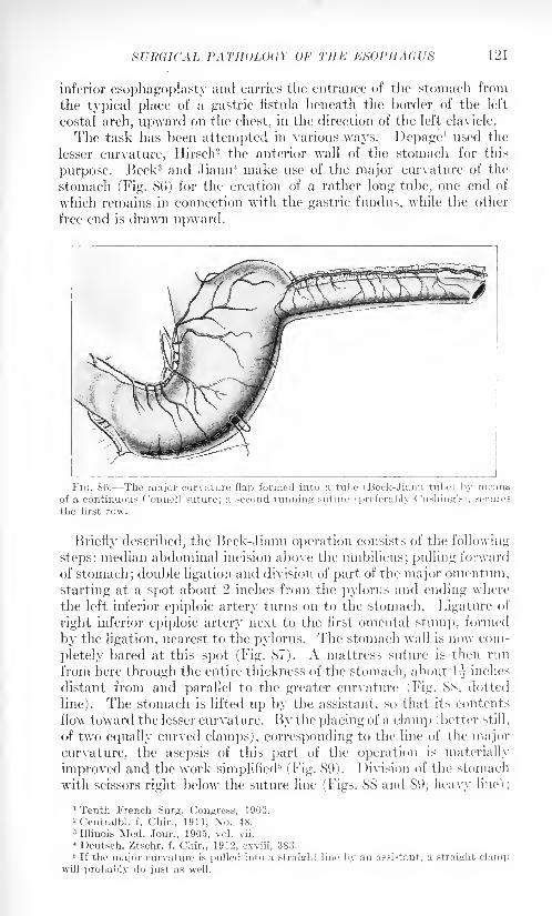
SURGICAL PATHOLOGY OF THE ESOPIIAC!US 121
inferior esophagoplasty and carries the entrance of the stomach from
the typical place of a gastric fistula beneath the border of the left
costal arch, upward on the chest, in the direction of the left clavicle.
The task has been attempted in various ways. Depage1 used the
lesser curvature, Hirsch2 the anterior wall of the stomach for this
purpose. Beck3 and Jianu 4 make use of the major curvature of the
stomach (Fig. 86) for the creation of a rather long tube, one end of
wrhich remains in connection with the gastric fimdus, while the other
free end is drawn upward.
FIG. 86. The major curvature flap formed into a tube (Beck-Jianu tube) by meansof a continuous Oonnell suture; a second running suture (preferably dishing'*), secures
the first row.
Briefly described, the Beck-Jianu operation consists of the following
steps: median abdominal incision above the umbilicus; pulling forward
of stomach; double ligation and division of part of the major omentum,
starting at a spot about 2 inches from the pylorus and ending where
the left inferior epiploic artery turns on to the stomach. Ligature of
right inferior epiploic artery next to the first omental stump, formed
by the ligation, nearest to the pylorus. The stomach wall is now com-
pletely bared at this spot (Fig. 87). A mattress suture is then run
from here through the entire thickness of the stomach, about 1^ inches
distant from and parallel to the greater curvature (Fig. 88, dotted
line). The stomach is lifted up by the assistant, so that its contents
flow toward the lesser curvature. By the placing of a clam]) ( better still,
of two equally curved clamps), corresponding to the line of the major
curvature, the asepsis of this part of the operation is materially
improved and the work simplified5
(Fig. 89). Division of the stomach
with scissors right below the suture line (Figs. 88 and 89, heavy line) ;
'Tenth French Surg. Congress, 1903.
"Centralbl. f. Chir., 1911, No. 48.3 Illinois Med. Jour., 1905, vol. vii.
4 Deutsch. Ztschr. f. Chir., 1912, cxviii, 383.5 If the major curvature is pulled into a straight line by an assistant, a straight clamp
will probably do just as well.
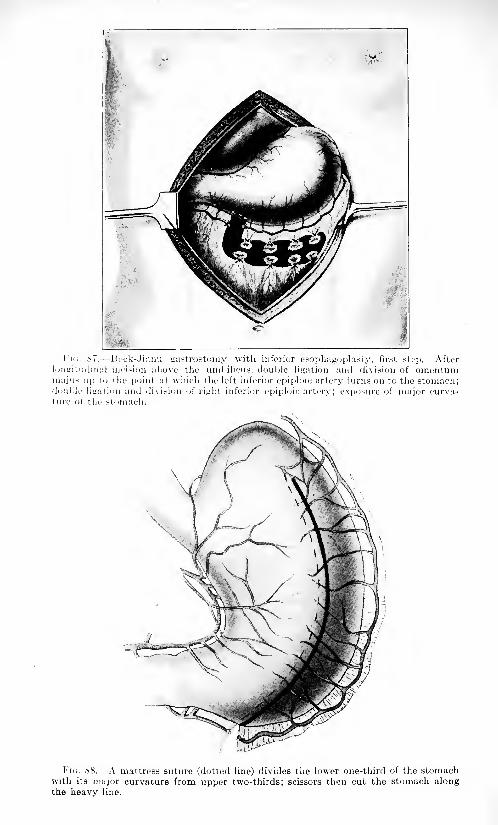
Ij
'i<;. 87. Bcck-Jianu fiastrostomy with inferior esophagoplasty, first step. Afterlim<iitudinal incision above the umbilicus, double ligation and division of omuntunimajus up to the point at which the left inferior epiploic artery turns on to the stomach;double libation and division of rijiht inferior epiploic artery; exposure of major curva-ture of the stomach.
Flu. <S8. A mattress suture (dotted line) divides the lower one-third of the stomachwith its major curvature from upper two-thirds; scissors then cut the stomach alongthe heavy line.
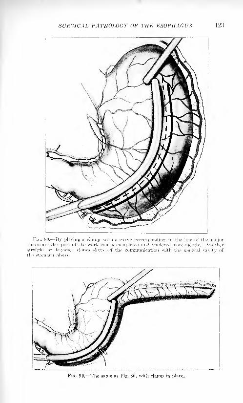
SURGICAL PATHOLOGY OF THE ESOPHAGUS 123
Fu;. 89. By placing a clamp with a curve corresponding to the line of the majorcurvature this part of the work can be completed and rendered more aseptic. Anotherstraight or bayonet clamp shuts off the communication with the general cavity of
the stomach above.
FIG. 90. The same as Fig. 86, with clamp in place.
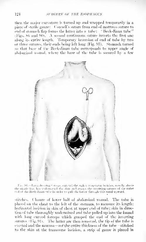
124 Xl'ItCEKY or THE ESOPHAGUS
then the major curvature is turned up and wrapped temporarily in a
piece of sterile gau/e: ( 'onnell's suture from end of mattress suture to
end of stomach ilap forms the latter into a tube: "Beck-Jianu tube"i Figs. SO and 90). A second continuous suture inverts the first one
along its entire length. Temporary inversion of end of tube by twoor three sutures, their ends being left long (Fig. 91). Stomach turned
so that base of the Beek-Jianu tube corresponds to upper angle of
abdominal wound, where the base of the tube is secured by a few
stitches. Closure of lower half of abdominal wound. The tube is
placed oil the chest to the left of the sternum, to measure its length;horizontal incision in skin of chest at upper end of tube; skin in direc-
tion of tube thoroughly undermined anol tube pulled up into the funnel
with long curved forceps which grasped the end of the invertingsutures ( Fig. 91 ). The latter are then removed, the tip of the tube is
everted and the mucosa not the entire thickness of the tube stitched
to the skin at the transverse incision, a strip of gauze is placed in
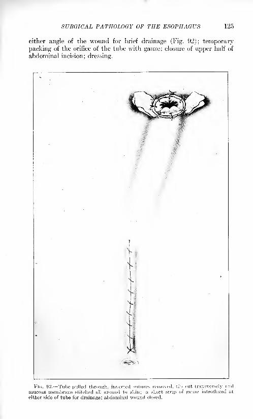
SURGICAL PATHOLOGY OF THE ESOPHAGUS 125
either angle of the wound for brief drainage (Fig. 92); temporary
packing of the orifice of the tube with gauze; closure of upper half of
abdominal incision; dressing.
FIG. 92. Tube pulled through, inverted sutures removed, tip cut transversely and
mucous membrane stitched all around to skin; a short strip of gauze introduced at
either side of tube for drainage; abdominal wound closed.
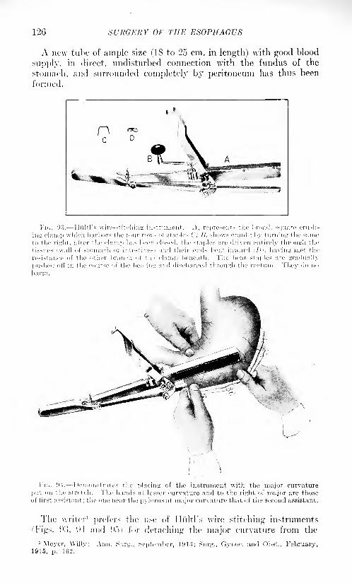
SURGERY OF THE ESOPHAGUS
A new tube of ample size (18 to 25 cm. in length) with good blood
supply, in direct, undisturbed connection with the fundus of the
stomach, and surrounded completely by peritoneum has thus been
formed.
Tic. 93. Ili'iltl's wire-stitching ii
>-ed, the staples are driven c
L; and discharged through the r<
:irely through t he
l'i,. 04. Demonstrates the placing of the instrument with the major curvature
put on the stretch. The hands at lesser curvature and to the right of major are thoseof first assistant; the one near the pylorus at major curvature that of the second assistant.
The writer' prefers the use of Iliiltl's wire stitching instruments
['Figs. !):>, tl and Hoi for detaching the major curvature from the
1 Meyer, Willy: Ann. Surg., September, 1913; Surg., Gynec. and Obst., February,1915, p. 162.
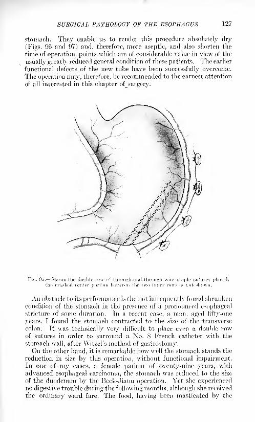
SURGICAL PATHOLOGY OF THE ESOPHAGUS 127
stomach. They enable us to render this procedure absolutely dry
(Figs. 96 and 97) and, therefore, more aseptic, and also shorten the
time of operation, points which are of considerable value in view of the
usually greatly reduced general condition of these patients. The earlier
functional defects of the new tube have been successfully overcome.
The operation may, therefore, be recommended to the earnest attention
of all interested in this chapter of_surgery.
An obstacle to its performance is the not infrequently found shrunken
condition of the stomach in the presence of a pronounced esophagealstricture of some duration. In a recent case, a man, aged fifty-one
years, I found the stomach contracted to the size of the transverse
colon. It was technically very difficult to place even a double rowof sutures in order to surround a No. 8 French catheter with the
stomach wall, after Witzel's method of gastrostomy.On the other hand, it is remarkable how well the stomach stands the
reduction in size by this operation, without functional impairment.In one of my cases, a female patient of twenty-nine years, with
advanced esophageal carcinoma, the stomach was reduced to the size
of the duodenum by the Beck-Jianu operation. Yet she experiencedno digestive trouble during the following months, although she received
the ordinary ward fare. The food, having been masticated by the
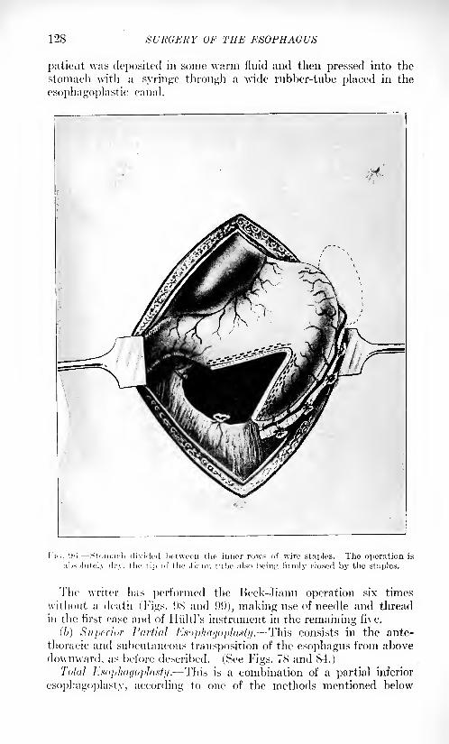
128 SURGERY OF THE ESOPHAGUS
patient was deposited in sonic warm fluid and then pressed into the
stomach with a syringe through a wide rubber-tube placed in the
esophagoplastic canal.
The writer has performed the Beck-Jianu operation six times
without a death (Figs. 98 and 99), making use of needle and thread
in the first case and of Hiiltl's instrument in the remaining five.
(6) Superior Partial Esophagoplasty. This consists in the ante-
thoracic and subcutaneous transposition of the esophagus from above
downward, as before described. (See Figs. 78 and 84.)
Total Esophagoplasty. This is a combination of a partial inferior
esophagoplasty, according to one of the methods mentioned below
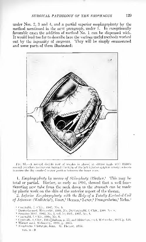
129
under Nos. 2, 3 and 4, and a partial superior esophagoplasty by the
method mentioned in the next paragraph, under 1. In exceptionallyfavorable cases the addition of method No. 1 can be dispensed with.
It would lead too far to describe here the various useful methods worked
out by the ingenuity of surgeons. They will be simply enumerated
and some parts of them illustrated:
FIG. 97. A second double row of staples is placed at obtuse anyle with Hi'iltl's
second (smaller) instrument beyond the turn of the left inferior epiploic artery; scissors
traverse the dry crushed center portion between the inner rows.
1. Esophagoplasty by means of Skin-plasty (Bircher. 1 This may be
total or partial. Bircher, as early as 1894, showed that a well func-
tionating new tube from the neck down to the stomach can be made
by plastic work on the skin of the anterior aspect of the thorax.
2. Inferior Esophagoplasty Kith the Help of a Total!]/ Excluded Coil
of Jejunum (Wullstein2), lloux,
3ITerzen,
4Lexer,
5Frangenheim,
6Ixehn,
7
1 Ceiitralbl. f. Uliir., 1907, N'o. 5.
- DeuUfhe mod. Wclmschr., 1901, No. 20; Centralbl. i". Chir.* Semaine Mod.. 1902, Xo. 1, vol. Iv; ibid., 1907, No. 4.
4 Centralbl. f. Chir., 1908, No. 8.
< Centralbl. f. Chir., 19 1 l^Beilaw, p. .15; and Muncheii. mod.6 Wiener mod. \\Vhnsdir., 1911. p. 2075.7Esophu^u.-s Chirurgie, Jena. G. i'ischer, 1914.
VOL. II 9
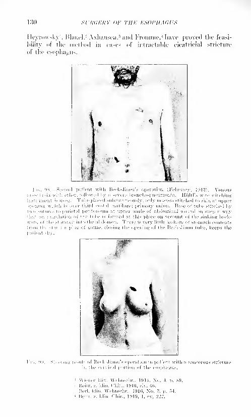
130 SURGERY OF THE EHOPllAGUS
Ileyrowsky1
, Blauel,- Axhausen,3 and Fromme, 4 have ])roved the feasi-
bility of the method in cases of intractable cieatricial stricture
of the esophagus.
Fi*,. 9v Second patient with Beck-Jianu's operation (February, 1913). Venousanothesia with ether, followed by a severe broncho-pneumonia, Hiiltl's wire stitchinginMrumeiit is used. Tube placed subcutaueously, only mucosa st itched to skin at upperopening wliich is over third coMal cartilage; primary union. Base of tube stitched bytwo sutures to parietal peritoneum at upper tingle of abdominal wound in such a waythat an angulation of 1 he tube is formed at this place on account of the sinking back-ward of the stomach into the abdomen. There is very little leakage of stomach contentsfrom the >tart; a plus; of gauze, closing the opening of the Beck-.Jianu tube, keeps the
pat ient dry.
'
Wiener klin. Wdmschr., 1914, No. 4, p. SO.- Heitr. /.. klin. ( 'hir., HUG, civ, 40.
I'.erl. klin. Wchnschr.. 1910, No. 3, p. .",4.
4 I'.eitr. v,. klin. Chir., 1919, 1, ev, 222.
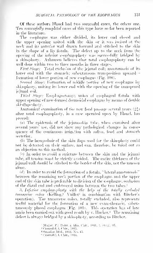
SURGICAL PATHOLOGY OF THE ESOPHAGUS 131
Of these authors Blauel had two successful cases, the others one.
Ten successfully completed cases of this type have so far been reportedin the literature.
The esophagus was either divided, its lower end closed and
the upper opening united with the skin or it was incised at the
neck and its anterior wall drawn forward and stitched to the skin
in the shape of a lip fistula. The defect up to the neck from the
opening of the inferior esophagoplasty was successfully bridged bya skin-plasty. Axhausen believes that total esophagoplasty can be
well done within two to three months in three stages.
First Stac/e: Total exclusion of the jejunal coil; anastomosis of its
lower end with the stomach; subcutaneous transposition upwardformation of lower portion of new esophagus (Fig. 100).
Second Stage: Formation, of middle portion of new esophagus byskin-plasty, uniting its lower end with the opening of the transposed
jejunal coil.
Third Shi^e: Esophagostomy; union of esophageal fistula with
upper opening of new-formed dennoidal esophagus by means of double
skinflap-plasty.
Anatomical examination of the new food passage several years (2|)
after total esophagoplasty, in a case operated upon by 1'lauel, has
proved:
(//) The epidermis of the jejuno-skin tube, when examined after
several years' use, did not show any pathological changes in conse-
quence of the continuous irrigation with saliva, food and stomach
secretion.
(6) The lanugo-hair of the skin flaps used for the skin-plasty could
not be detected on their surface, and can, therefore, be ruled out as
an objection to this method.
(<") In order to avoid a stricture between the skin and the jejunal
tube, all tension must be strictly avoided. The entire thickness of the
jejunal wall should be stitched to the border of the skin, not the mucosaalone.
(d) In order to avoid the formation of a fistula,"lateral anastomosis"
between the remaining neck portion of the esophagus and the upper('nd of the skin tube is preferable to division of the esophagus, occlusion
of the distal end and end-to-cnd union between the two tubes. 1
3. Inferior esopliagoplasty with the help of the totally excluded
transverse colon ( Kelling,- Yuillet"1 in combination with lurcher's
operation). The transverse colon, totally excluded, also representsuseful material for the formation of a new extra-thoracic, subcu-
taneously placed esophagus (I;ig. 101,). This operation lias of late
again been carried out with good result by v. Hacker. 4 The remainingdefect is always bridged by a skin-plasty, according to Bircher.
1
Miiller, I'.: IVitr. /. klin. Chir.
-Centnilbl. f. Chir., 191.').
3 Sein:iinc Mc'-d, 1'Jll, \o. 4.">.
-' Centralbl. f. Chir., ID 14.
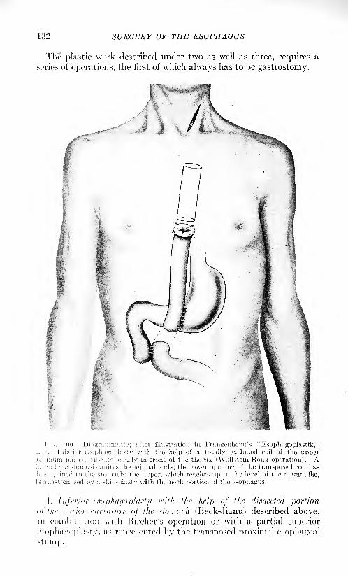
132 SURGERY OF THE ESOPHAGUS
The plastic work described under two as well as three, requires a
series of operations, the first of which always has to be gastrostomy.
I i.. 100. Diagrammatic; after illustration in Frangenheim's "Esophagoplastik,"1. c. Inferior csophagoplasty with the help of a totally excluded coil of the upperjejunum placed subcutaneously in front of the thorax (Wullstein-Roux operation). Alateral anastomosis unites the jejunal ends; the lower opening of the transposed coil has
been joined to the stomach; the upper, which reaches up to the level of the mammillae,is anastomosed by a skin-plasty with the neck portion of the esophagus.
1. Inferior csophagoplasty icifh the help of the dissected portion
<>f flif major curvature of the stomach (Beck-Jianu) described above,in combination with Bircher's operation or with a partial superior
esophairoplastv, as represented by the transposed proximal esophageal
stump.
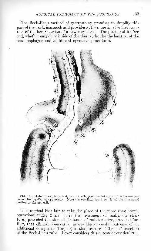
SURGICAL PATHOLOGY OF THE ESOPHAGUS 133
The Beck-Jianu method of gastrostomy promises to simplify this
part of the work, inasmuch as it provides at the same time for the forma-
tion of the lower portion of a new esophagus. The placing of its free
end, \vhether outside or inside of the thorax, decides the location of the
new esophagus and additional operative procedures.
FIG. 101. Inferior esophagoplasty with the help of the totally excluded transversecolon (Kelling-Vulliet operation). Note the excellent blood supply of the transposedportion by the art. coli.
This method bids fair to take the place of the more complicatedoperations under 2 and 3, in the treatment of malignant stric-
tures, provided the stomach is found of sufficient sixe, provided fur-
ther, that clinical observation proves the successful outcome of anadditional skin-plasty (Bircher) in the presence of the acid secretion
of the Beck-Jianu tube. Lexer considers this outcome very doubtful.
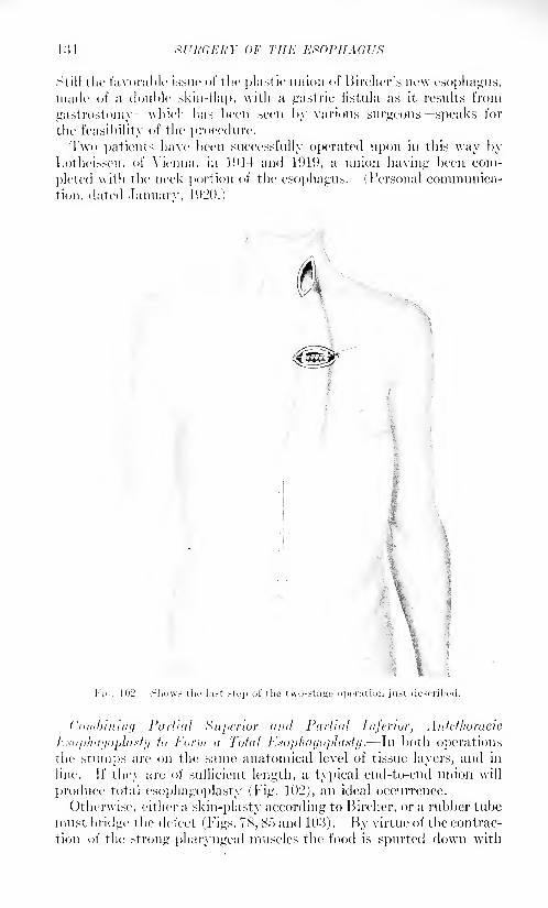
134 GEIi'Y OF THE ESOPHAGUS
Still the favorable issue of the plast ic union of Bircher's new esophagus,made of a double skin-flap, with a gastric fistula as it results from
gastrostomy which has been seen by various surgeons speaks for
the feasibility of the procedure.Two patients have been successfully operated upon in this way by
Lotheissen, of Vienna, in 1!)14 and 1919, a union having been com-
pleted with the neck portion of the esophagus. (Personal communica-
tion, dated January, 1920.)
Combining Partial Superior <uid Partial Inferior, Antethoracic
Ksophagoplasty to Form a Total Esopliagoplasty . In both operationsthe stumps are on the same anatomical level of tissue layers, and in
line. If they are of sufficient length, a typical end-to-end union will
produce total esophagoplasty (Fig. 102), an ideal occurrence.
Otherwise, either a skin-plasty according to Bircher, or a rubber tube
must bridge the defect (Figs. 78, 85 and 103). By virtue of the contrac-
tion of the strong pharyngeal muscles the food is spurted down with
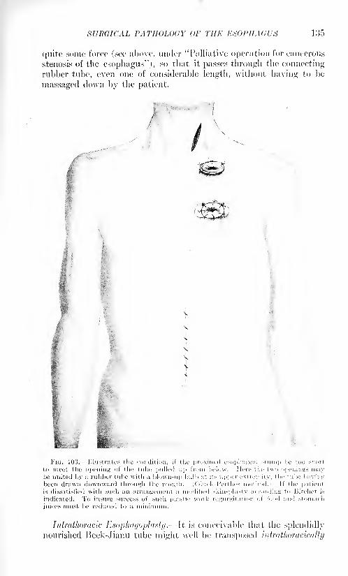
SURGICAL PATHOLOGY OF THE ESOPHAGUS 135
quite some force (see above, under "Palliative operation for cancerous
stenosis of the esophagus"), so that it passes through the connectingrubber tube, even one of considerable length, without having to be
massaged down by the patient.
FIG. 103. Illustrates the condition, if the proximal csophageal stump be too short
to meet the opening of the tube pulled up from below. Here the t\vo openings maybe united by a rubber tube with a blown-up bulb at its upper extremity, the tube havingbeen drawn downward through the mouth. (Gluck-I'erthes method.) If the patient,
is dissatisfied with such an arrangement a modified skiii-plasty according to Bircher is
indicated. To insure success of such plastic work regurgitation of food and stomach
juices must be rcdticed to a minimum.
Iidrathoracic Esophagoplasty.It is conceivable that the splendidlynourished Beck-Jianu tube might well be transposed intrathoracically
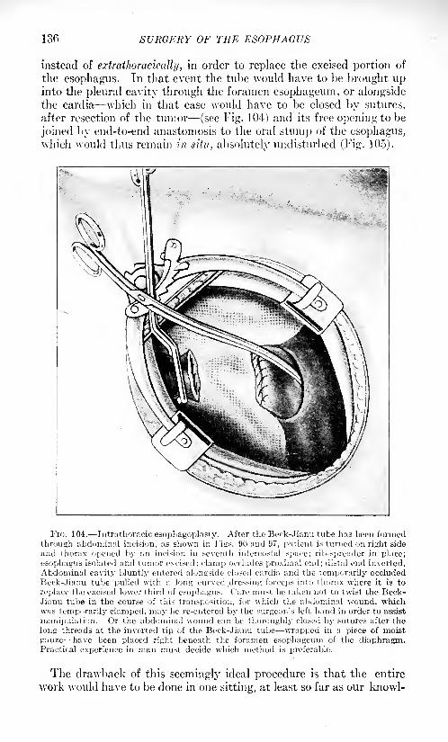
136 SURGERY OF THE ESOPHAGUS
instead of extrathoracically, in order to replace the excised portion of
the esophagus. In that event the tube would have to be brought upinto the pleural cavity through the foramen esophageum, or alongsidethe cardia which in that case would have to be closed by sutures,
after resection of the tumor (see Fig. 104) and its free opening to be
joined by end-to-end anastomosis to the oral stump of the esophagus,which would thus remain in situ, absolutely undisturbed (Fig. 105).
FK;. 104. Intrathoracic esophagoplasty. After the Beek-Jiami tube has been formed
through abdominal incision, as shown in Fig*. 96 and 97, patient is turned on right side
and thorax opened by an incision in seventh intercostal space; rib-spreader in place;
esophagus isolated and tumor excised; clamp occludes proximal end; distal end inverted.
Abdominal cavity bluntly entered alongside closed cardia and the temporarily occluded
Beck-Jianu tube pulled with a long curved dressing forceps into thorax where it is to
replace the excised lower third of esophagus. ( 'are must be taken not to twist the Beck-Jianu tube in the course of this transposition, for which the abdominal wound, whichwas temporarily clamped, may be re-entered by the surgeon's left hand in order to assist
manipulation. Or the abdominal wound can be thoroughly closed by sutures after the
long threads at the inverted tip of the Beck-Jianu tube wrapped in a piece of moist
gauze have been placed right beneath the foramen esophageum of the diaphragm.Practical experience in man must decide which method is preferable.
The drawback of this seemingly ideal procedure is that the entire
work would have to be done in one sitting, at least so far as our knowl-
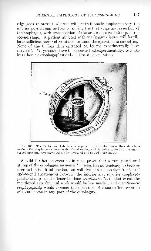
SURGICAL PATHOLOGY OF THE ESOPHAGUS 137
edge goes at present, whereas with extrathoracic esophagoplasty the
inferior portion can be formed during the first stage and resection of
the esophagus, with transposition of the oral esophagcal stump, in the
second stage. A patient afflicted with malignant disease will hardlyhave sufficient power of resistance to stand the operation in one sitting.
None of the 6 dogs thus operated on by me experimentally have
survived. Ways would have to be worked out experimentally, to makein trathoracic esophagoplasty also a two-stage operation.
FIG. 105. -The Beck-Jianu tube has been pulled up into the thorax through a hole
made in the diaphragm alongside the closed oardia, and is being united to the undis-
turbed proximal esophageal stump by means of end-to-end anastomosis.
Should further observation in man prove that a transposed oral
stump of the esophagus, no matter how long, has no tendency to becomenecrosed in its distal portion, but will live, as a rule, so that "the ideal"
end-to-end anastomosis between the inferior and superior esophago-
plastic stump could oftener be done extrathorically, in that event the
mentioned experimental work would be less needed, and extrathoracic
esophagoplasty would become the operation of choice after resection
of a carcinoma in any part of the esophagus.

SVIWEfiY OF THE BKEAS1
BY ROBERT B. GREENOUGH, M.I).
ANATOMY AND DEVELOPMENT OF THE BREASTS.
THE breast of the adult female is a glandular structure situated
upon the anterior and lateral aspect of the chest, extending on each
side from the third to the seventh rib, and fiom the sternum to the
border of the axilla. The rounded eminence of the breast is composedof the tissue of the mammary gland, with a firm deposit of subcutan-
eous fat surrounding it on every side. The gland tissue proper is
flattened from before backward, and of an irregular lobular shape. It
is composed of a series of gland lobules radiating from the nipple.
Each main duct branches into many smaller ducts, and these againinto a series of bulbous ends which are lined with a double layer of
epithelial cells the acini of the gland.1 The ducts and acini of one
lobule are distinct from those of other lobules. The largest and best
defined lobule occupies the upper and outer quadrant of the gland, andextends nearly to the axilla. The gland tissue is supported in a frame-
work of interlobular connective tissue through which pass the blood-
vessels, nerves, and lymphatics of the breast. The nipple is situated
near the center of the breast, and presents the openings of the terminal
ducts of the mammary gland, ten to twenty in number. The nipple
normally projects above the surface of the breast and is endowed with
erectile properties due to the presence of muscle fibers surroundingthe ducts and attached to the overlying skin. The nipple is surrounded
by the areola, an area of pink or pigmented skin, containing numbers of
large sebaceous glands, beneath which there is very little of the sub-
cutaneous fat which underlies the skin in other places.
The bloodvessels of the breast are derived from the internal mam-mary, intercostal and axillary systems, and the nerves chiefly from the
second, third, fourth and fifth intercostal nerves. The lymphatics of
the breast are especially rich and of great importance to the surgeon.A superficial, subcutaneous plexus covers the outer surface of the
breast, and a dense meshwork of deep lymphatic channels penetratesto all parts of the gland substance, and is continued in the lymphatictrunks of the deep fascia of the chest and abdominal wall. Abundantanastomoses exist between the two systems. The main trunks follow
the axillary border of the pectoral muscles, and empty into the lymphnodes of the axillary region, but the mesial portions of the gland are
drained to a certain extent by channels which perforate the intercostal
1 Bizzozero and OttolciiKui: Anatoini^chc Ergebuisie, Abth. iv, Ed. ix.
( 139 )
140 SURGERY OF THE BREAST
spaces and empty into the nodes of the anterior mediastinum. The
superficial axillary nodes under the border of the pectoralis majorare those in most direct communication with the breast, and throughthem the nodes in the axilla, and eventually the subclavicular and
supraclavicular nodes are reached. Minute lymph nodes have been
found lying under the pectoralis major and in communication with the
breast lymphatics by branches which perforate the muscle. Anasto-
moses exist between the lymphatics of the right and left breasts, across
the sternum, and a few communicating brandies occasionally carryinfection directly across the clavicle to the supraclavicular lymphnodes. 1
The development of the mammary gland in the embryo takes place
by an infolding of the epidermis of the lateral aspect of the chest. In
animals which have multiple mammary glands, this infolding of the
epidermis occurs in a line extending from the axilla to the groin, the
so-called "milk line." The human embryo normally presents but one
gland on each side. Supernumerary breasts occur occasionally in the
human being, however, and are, as a rule, developed along this "milk
line." At birth and during infancy the development of the gland is
slight and equal in both sexes. In the male, the development of the
gland is rudimentary and rarely advances beyond the infantile stage.
In the female, an increase in the si/e of the breast takes place at the
time of puberty, and is due to the development of gland tissue, and to
the deposit of fat within and about the gland. One of the character-
istic features of the developing breast gland is the hyaline connective
tissue which surrounds the gland ducts and develops with them. This
tissue contributes to the firm and elastic consistency of the breast andis of importance to the surgeon on account of the fibre-epithelial
tumors of the breast in which it frequently takes an important part.Each lobule of the breast gland, composed of the epithelial lined ducts
and hyaline periductal tissue, is separated from the adjacent lobule byinterlobular connective tissue of the ordinary type, through which run
the bloodvessels, lymphatics and nerves of the gland. This inter-
lobular tissue binds all of the breast together, and is prolonged in fibrous
tissue strands attaching the organ to the fascia of the chest wall below,and to the skin above. These fibrous attachments to the skin have been
called the ligaments of Sir Astley Cooper. It is by their retraction
when infiltrated by cancer tissue that the adherence of the skin over
the breast is produced one of the most constant and earliest signs of
malignant disease. When pregnancy occurs, a great increase in the
development of the gland tissue takes place. The secreting surface
becomes greater, new bloodvessels are formed, and the entire organincreases in size and weight, so that it covers a much larger part of the
chest wall, extending down even on to the abdomen. When lactation
is established this vascularity continues, and the secretion of the epithe-lial cells is elaborated and discharged as milk. After lactation ceases,
'Oelsner: Arch. klin. Chir., 1901, Ixiv, 134. Sappey: Traite d'Anatomic descriptive,
Paris, 1889, vol. ii and iv.
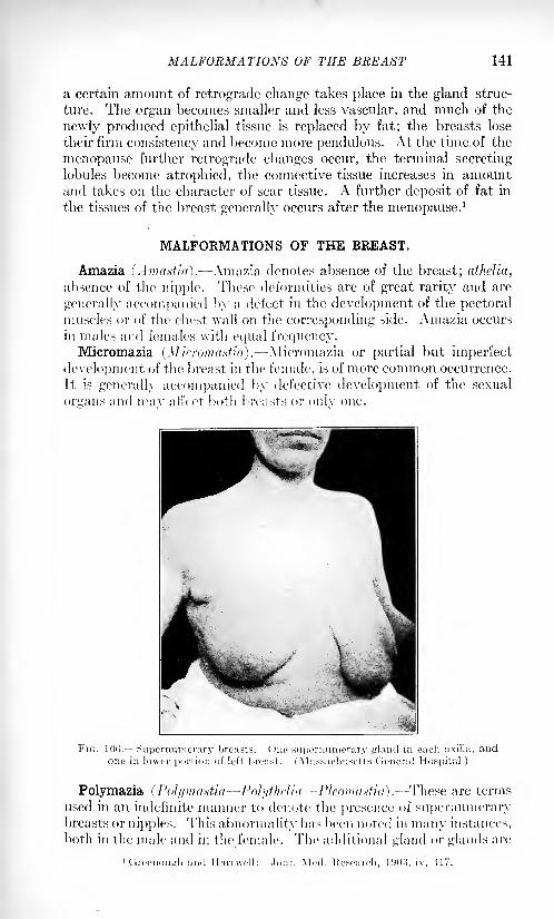
MALFORMATIONS OF THE BREAST 141
a certain amount of retrograde change takes place in the gland struc-
ture. The organ becomes smaller and less vascular, and much of the
newly produced epithelial tissue is replaced by fat; the breasts lose
their firm consistency and become more pendulous. At the time, of the
menopause further retrograde changes occur, the terminal secretinglobules become atrophied, the connective tissue increases in amountand takes on the character of scar tissue. A further deposit of fat in
the tissues of the breast generally occurs after the menopause.1
MALFORMATIONS OF THE BREAST.
Amazia (Amastia). Amazia denotes absence of the breast; athelia,
absence of the nipple. These deformities are of great rarity and are
generally accompanied by a defect in the development of the pectoralmuscles or of the chest wall on the corresponding side. Amazia occurs
in males and females with equal frequency.Micromazia (Micromastia) . Micromazia or partial but imperfect
development of the breast in the female, is of more common occurrence.
It is generally accompanied by defective development of the sexual
organs and ivav affect both breasts or only one.
Polymazia (PolymastiaPolythelia- -Pleomajstia). These are terms
used in an indefinite manner to denote the presence of supernumerarybreasts or nipples. This abnormality has been noted in many instances,
both in the male and in the female. The additional gland or glands are
Mecl. Ke> 1903, iv, 41 ;
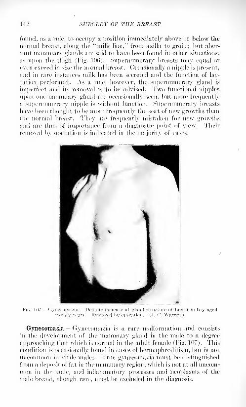
14:2 SURGERY OF THE BREAST
found, as u rule, to occupy a position immediately above or below the
normal breast, along the ''milk line," from axilla to groin; but aber-
rant mammary glands are said to have been found in other situations,
as upon tlic thigh (Fig. 10l>). Supernumerary breasts may equal or
even exceed in si/e the normal breast. Occasionally a nipple is present,and in rare instances milk has been secreted and the function of lac-
tation performed. As a rule, however, the supernumerary gland is
imperfect and its removal is to be advised. Two functional nipples
upon one mammary gland are occasionally seen, but more frequentlya supernumerary nipple is without function. Supernumerary breasts
have been thought to be more frequently the seat of new growths than
the normal breast. They are frequently mistaken for new growthsand are thus of importance from a diagnostic point of view. Their
removal by operation is indicated in the majority of cases.
FIG. 107. uvnecomazia. Definite increase of gland structure of breast in boy aged
Gynecomazia.- Gynecomazia is a rare malformation and consists
in the development of the mammary gland in the male to a degree
approaching that which is normal in the adult female (Fig. 107). This
condition is occasionally found in cases of herinaphroditism, but is not
uncommon in virile males. True gynecoma/.ia must be distinguishedfrom a deposit of fat in the mammary region, which is not at all uncom-mon in the male, and inflammatory processes and neoplasms of the
male breast, though rare, must be excluded in the diagnosis.
DISEASES OF THE NIPPLE 143
INJURIES OF THE BREAST.
Injuries of the breast are less common than might be expected,
owing to the protection of clothing and the yielding nature of the part.Contusions and Hematoma. Contusions and hematoma of the
subcutaneous tissues may occur and blood may be poured out in the
substance of the gland itself. Injuries of this nature generally yield to
treatment by support and evaporating lotions. Suppuration mayoccur, however, immediately or at some subsequent time, wheninfection is admitted to the hematoma directly or through the blood
current. A history of preceding contusion can often be obtained in new
growths or inflammatory lesions of the breast. The induration left
in the tissues by the organization of such an exudate may occasionally
present certain difficulties in the diagnosis of new growths, and in such
cases exploration by operation is the only safe course 1 to pursue.Wounds. Wounds of the breast may be produced as in other
organs, and require no special consideration. A penetrating wound of
the lactating breast which severs one or more of the larger milk-ducts
may result in the production of a milk fixtnla. Such fistula' are slow
to heal during the continuance of lactation, but close readily at its
termination. The scar of such a wound may cause obstruction and
lead to the formation of a galactocele or milk cyst, at a subsequentlactation, or may produce a simple retention cyst in the intervening
period when the breast is not secreting milk.
Foreign Bodies. Foreign bodies are occasionally introduced into
the tissues of the breast by accident or design. Pins and needles are
not infrequently driven into the breast by insane women. Foreignbodies in the breast produce symptoms similar to those in other situ-
ations, and their removal by operation is indicated.
DISEASES OF THE NIPPLE.
Retraction (Depression or Inversion of the Nipple). The nipple
normally projects somewhat above the surface of the areola in a conical
or cylindrical form. The prominence of the nipple is normally increased
during pregnancy and lactation. Retraction is present when this
prominence is lacking and the nipple cannot be drawn out by the
suckling of an infant or by artificial measures. Retraction may be due
t;> developmental defects. It is also produced by the contraction of
scars and by the infiltration of malignant tumors, in which case it is
significant in diagnosis.
Treatment. During lactation, retraction of the nipple may prevent
nursing and thus lead to serious engorgement of the breast. Thebreast pump and the suction of the nursing child will relieve the lesser
degrees of this affection. In more obstinate eases attempts may be
made with plaster or contractile collodion to cause contraction of the
areola and maintain the prominence of the nipple. A plastic operationhas been recommended by Kehrer for the same purpose by the removal
144 SURGERY OF THE BREAST
of crescent ic folds of skin above and below the nipple, and Warren has
operated upon siieh eases, with success, by turning up the breast and
uniting the tissues beneath the nipple by subcutaneous catgut stitches.
Imperforate Nipple. Imperforate nipple may occur as a develop-mental defect or by cicatricial occlusion of the ducts. When lactation
occurs in such a case, engorgement results and galactocele or abscess
is likely to ensue.
Treatment.- The treatment is that of the resulting condition, and
amputation should be performed.Cracks or Fissures.- Cracks or fissures of the delicate integument
of the nipple are readily produced by trauma. The first attempts of the
child at suckling are especially prone to bring about this condition,
owing TO the maceration of the part in the mouth of the child and the
congestion which accompanies the beginning of lactation. Fissures
produced in this way may be so painful as to prevent nursing altogether,but their importance is far greater than this, for it is through these
fissures, in the majority of cases, that the infection is transmitted which
produces acute mastitis or abscess of the breast. The treatment of
fissures is thus of great importance in the prevention of abscess.
Treatment. - Fissures are to be treated with mild antiseptic washes,
such as boric acid, and scrupulous cleanliness is to be maintained. Asa rule, nursing should be abandoned on the affected side and the breast
emptied periodically by massage or the breast pump. During the
establishment of lactation, scrupulous cleanliness and washing with
boiled water before and after nursing, will tend to prevent the formation
of fis.Mires, and should be strongly recommended as a prophylactic at
the commencement of lactation.
Fissures of the nipple may occur at other times than during lactation.
They may he produced by trauma or uncleanliness, and are not
uncommon in ecxema of the nipple. Fissure of the nipple may also
occur as a primary manifestation of syphilis.
Eczema. Fc/.ema of the nipple may occur during pregnancy as
well as during lactation, when it is produced by lack of cleanliness andis favored by the maceration of the skin in the fluids escaping from the
lacteal ducts. The skin is red and thickened and crust formation
occurs.
Treatment. Kczeina of this form is generally relieved by soothing
applications and by cleanliness. It may, however, result in the forma-
tion of a fissure, and thus favor infection and acute mastitis.
Paget's Disease. A peculiar form of eczema of the nipple which
has been the subject of much investigation and many controversies,
is that described in 1874 by Sir James Paget and now known generallyas
"Paget's Disease of the Nipple." Its most significant characteristic
is its connection with cancer of the breast. Paget's disease occurs
independent of lactation, and, indeed, its cause is as yet unknown. It
attacks the breasts of women of adult and mature age and produces a
characteristic lesion. The nipple, areola and after the lapse of somemonths often the greater portion of the cutaneous surface of the breast,
DISEASES OF THE NIPPLE 145
present a raw, red, moist surface of irregular outline, covered here andthere with crusts of exudate and Hakes of dried-up epidermis. The
appearance was likened by Paget to that of an acute balanitis -a most
striking simile. In advanced cases the nipple may be entirely eroded.
The ec/ematous condition persists and spreads and no medicinal
applications appear to affect its progress. 'The course of the disease
may continue for several years, until sooner or later a nodule of cancer
is found in the substance of the breast tissue.
Pathology. The pathology of one type of Paget's disease has been
well established as a form of very superficial epidermoid cancer of the
same general character as rodent ulcer. Thickening of the epidermis,
prolongation of the papillr and intense, round-cell infiltration of the
cutis are the characteristics of the histology of the disease. Thecolumns of epidermal cells infiltrate the subcutaneous tissues, andin the late stages of the disease involve the ducts of the mammarygland, thus producing an epidermoid cancer of the breast.
In other cases of Paget's disease, however, cancer of glandular origin
develops in the deeper portions of the breast, and by extension to the
deeper layers of the skin produces an infiltration which leads to destruc-
tion of the skin and ulceration similar in appearance to the epidermoid
type of Paget's disease. In still other cases the relation between ec/emaof the nipple and gland cancer of the breast may be impossible to
determine, although the two conditions may be coincident.
Diagnosis. The diagnosis of Paget's disease of the nipple depends
upon its characteristic appearance. Xo other condition can be mis-
taken for it, unless perhaps the eczema of lactation, which can be
readily distinguished by its occurrence during lactation and its ready
disappearance under simple treatment.
Prognosis.- The prognosis, with operation, is, as-a rule, more favor-
able than in the average case of cancer of the breast, but depends always
upon the extent and duration of the disease and upon the type of cancer
present. The epidermoid type of cancer in itself is less malignant than
glandular carcinoma of the ordinary form.
Treatment. The treatment of Paget's disease of the nipple is by
operation the amputation of the breast. The "complete operation"for cancer of the breast should be performed as in other cases of cancer.
A'-rays and radium have been employed successfully for the treatment
of the ec/ema, but the important condition is the predisposition to
cancer, and for this reason amputation should always be performed.Tumors of the Nipple. Xew growths originating in the nipple are
extremely rare. A number of different forms of fibre-epithelial tumors
have been described, and it is noteworthy that almost all tumors of
this origin assume a rounded, pendulous form, with the production of
a slender pedicle. Papillary tumors of fibro-epithelial type 'papillary
cystadenoma or the villous papillomata of Knglish writers 1
) which
arise in the extremities of the lacteal sinuses, occasionally develop
1 Shield: Diseases of the Breast, 1S9S, London, McMillan & Co.
VOL. II 10
146 SURGERY OF THE BREAST
outward with the production of a tumor of the nipple. The projectionof fungous papillary masses from the lacteal ducts may occur in certain
cases and produce a tumor on the surface of the nipple resemblingexuberant granulations.The treatment of tumors of the nipple is by excision, but amputation
of the breast may be indicated if the whole of the nipple is to be
excised.
DISEASES OF THE BREAST.
The diseases of the breast may be divided into: (1) functional
diseases; (2) inflammatory processes; (3) cysts and cystic disease;
and (4) new growths.
Functional Diseases of the Breast.
The process of lactation may present a number of abnormal vari-
ations.
It may occur, though rarely, independent of pregnancy, and it is
said to have been performed by the breast of the male.
Abnormalities of the milk are occasionally noted, such as a blue, red
or yellow color, due to bacterial invasion of the ducts. Biliary coloring
matters may appear in the milk, and a number of drugs are thus
secreted.' Vicarious menstruation is said to be evidenced by the
periodical discharge of blood from the nipple. This condition is often
simulated, however, by certain papillary tumors in the gland ducts,
which bleed more readily at the time of the catamenia because of the
increased congestion of the breast.
Mastodynia (Neuralgia Irritable Mamma Hysterical Breaxl).
Neurotic conditions of the breast are by no means rare. Mastodynia,or pain in the breast, may occur in individuals with a neurotic taint,
independent of obvious pathological conditions. In such cases abnor-
malities of the genital organs are not infrequently observed. Pain in
the breast of a degree out of all proportion to the diseased condition is
also found accompanying certain pathological conditions. Cases of
chronic cystic mastitis and of diffuse hypertrophy of the breast not
infrequently present this symptom. The pain may extend down the
arm of the all'ected side by involvement of the intercostohumeral
nerve. When pregnancy occurs the sensitiveness of the breast may be
such as to prevent nursing.
Treatment. The treatment of neurotic conditions of the breast is
general and symptomatic and the neurasthenic condition of the patientis to be met with appropriate treatment. Any associated genital dis-
order should be corrected and pathological conditions of the breast
itself, if present, should be relieved. A supporting bandage often
relieves pain to a great extent, but local applications are to be avoided.
Tumors of the form described as neuromata, or painful fibromata,
such as occur in other situations, are occasionally found in the sub-
Biz/.ozoro :md Ot tolon^'ii: Aiifitomischo KrKolmis.se, Abth. iv, Bd. ix.
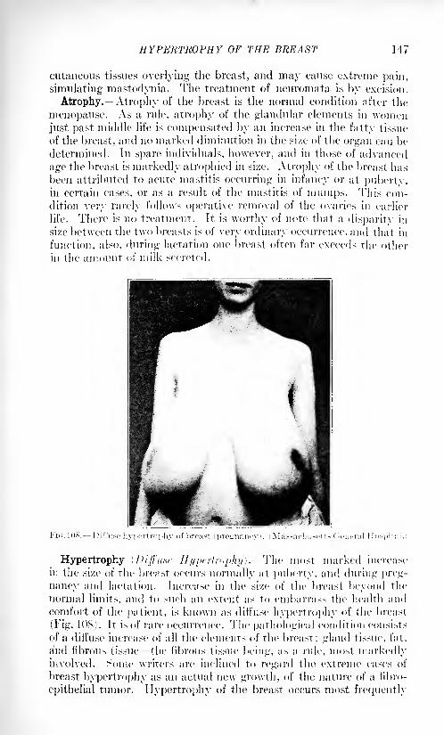
HYPERTROPHY OF THE BREAST 147
cutaneous tissues overlying the breast, and may cause extreme pain,
simulating mastodynia. The treatment of neuromata is by excision.
Atrophy. Atrophy of the breast is the normal condition after the
menopause. As a rule, atrophy of the glandular elements in womenjust past middle life is compensated by an increase in the fatty tissue
of the breast, and no marked diminution in the size of the organ can be
determined. In spare individuals, however, and in those of advanced
age the breast is markedly atrophied in size. Atrophy of the breast has
been attributed to acute mastitis occurring in infancy or at puberty,in certain cases, or as a result of the mastitis of mumps. This con-
dition very rarely follows operative removal of the ovaries in earlier
life. There is no treatment. It is worthy of note that a disparity in
size between the two breasts is of very ordinary occurrence, and that in
function, also, during lactation one breast often far exceeds the other
in the amount of milk secreted.
Hypertrophy (Diffuse Hypertrophy), The most marked increase
in the size of the breast occurs normally at puberty, and during preg-
nancy and lactation. Increase in the size of the breast beyond the
normal limits, and to such an extent as to embarrass the health and
comfort of the patient, is known as diffuse hypertrophy of the breast
(Fig. IDS). It is of rare occurrence. The pathological condition consists
of a diffuse increase of all the elements of the breast: gland tissue, fat.
and fibrous tissue the fibrous tissue being, as a rule, most markedlyinvolved. Some writers are inclined to regard the extreme cases of
breast hypertrophy as an actual new growth, of the nature of a iibro-
epithelial tumor. Hypertrophy of the breast occurs most frequently
148 SURGERY OF THE BREAST
in pregnancy, and progresses with great rapidity until its termination.
The function of lactation may be deficient. A diminution in the size
of the breast may be expected after lactation is ended, but the breast
remains of abnormal size and is almost certain to undergo further hyper-
trophy in succeeding pregnancies. Hypertrophy of the breast in younggirls at the age of puberty is occasionally seen, and is often accom-
panied by mastodynia. In such cases genital abnormalities of structure
or function are not uncommon. The pathological condition is the sameas in the; cases whieh develop during pregnancy, but the si/e of The
breasts is rarely so excessive. In ('. P>. Porter's case, a woman, aged
thirty-seven years, the combined weight of the two breasts after
{imputation was sixty pounds.1
Enlargement of one breast alone, in
younger women, is more often due to a fibre-epithelial tumor than to
diffuse hypertrophy. In tropical countries the breast is occasionallyall'ected in a manner similar to the leg or scrotum by filarial disease.
A condition of elephantiasis or lymphangiectasis of the breast is thus
produced, which is distinguished from true hypertrophy by the limi-
tation of the pathological changes to the skin and subcutaneous tissues.
The disease is extremely rare.
Diagnosis. In diagnosis extreme cases of diffuse hypertrophy are
not likely to be confused with any other condition. ( 'ases of less degreeuf development and cases affecting one breast alone must be distin-
guished from cysts and from the fibro-epithelial tumors. The diffuse
nature of the growth in hypertrophy and the absence of a localized,
encapsulated tumor must be relied upon to distinguish the con-
dition.
Prognosis. The prognosis of diffuse hypertrophy of the breast is
for a slow but continuous increase in si/e. If pregnancy intervenes a
rapid development may be expected. Partial remissions may occur,
but in well-marked eases the disease is progressive. The nature of the
growth, however, is benign and a malignant metamorphosis of the
ti>sues is improbable.Treatment. The treatment of well-marked cases of diffuse hyper-
trophy of the breast is by operation. Amputation of the breast may be
done in the ordinary manner. A tourniquet, held by Wyeth's pins,
was used by Porter in one classical case to compress the pedicle, as in
amputation of The hip. In eases of bilateral hypertrophy both breasts
may be amputated simultaneously or an interval of time may be
allowed to elapse between the two operations. The lesser degrees of
hypertrophy demand only supporting bandages and general consti-
tutional treatment. Applications, massage and manipulation are to
be strenuously avoided. Pelvic disorders when present should be
corrected.
Infantile Hypertrophy. -Early development of the mammary glandin female children is not uncommon. Cases have been recorded of
marked development of the breasts, together with growth of pubic
1 Porter: Boston Mod. and surg. Jour., March 3, 1S92; Tr. Am. Surg. Assn., 1891,vol. ix.

INFLAMMATORY DISEASES OF THE BREAST 149
hair, at between two and three years of age. As a rule the other sexual
organs share with the breast in the precocious development.1
Inflammatory Diseases of the Breast.
Acute Mastitis. Acute inflammation of the breast may occur at
several different periods in the development of the gland. At birth,
at the age of puberty, and during pregnancy and lactation, the
development of the breast is rapid, and is accompanied by an increased
vascularity and tumefaction or engorgement, which especially favor
the development of infective organisms. This infection is introduced,
in the vast majority of cases, through neglected fissures of the nipple,
and as these fissures are especially frequent during lactation, and as
this is the period when vascular engorgement of the breast is most
pronounced, acute mastitis of the puerperal breast is by far the most
common form.
FIG. 109.- -Acule mastitis of infants. Abscess, (('use of Dr. .fames S. Stone,
Infants' Hospital, Boston, Mass.)
Mastitis Neonatorum. In the newborn, swelling of the breast and
the escape from the nipple of a few drops of milky fluid ("witches'
milk") is not infrequently observed. The breast is swollen, red and
tender. Infection may readily occur through injury to the delicate
skin of the nipple, or especially by manipulation, a method of treat-
ment much favored by ignorant midwives (Fig. 109). The inflammation
rarely progresses to actual abscess formation. Scrupulous cleanliness,
1 Bryant: Diseases of the Breast, 1887. London, Cassell & Co.
150 SrRGERY OF THE BREAST
washing the part with sterile water or dilute alcohol, and a light pro-tective covering will generally avoid infection and allow the process to
subside. Should an abscess form it is to be opened and drained as in
other situations. The injury to the breast produced by this condition
cannot be correctly estimated. It has been thought to predispose to
inflammatory processes or tumor formation in later life.
Mastitis of Puberty. At the age of puberty a marked developmentof the breast takes place in the female, and inflammatory conditions
occasionally result. In the male, also, congestion and tumefaction of
the gland at this time may occur. .Swelling and tenderness are not
uncommon. In rare cases infection and suppuration may occur, and
treatment by incision may be necessary.
Puerperal Mastitis (Breast Abscess "Broken Breast"). This condi-
tion is of common occurrence even at the present day, although its
cause is generally understood and its prevention clearly indicated. Byfar the greater number of cases of acute mastitis occur during lactation,
and in the period from the fourth to the eighth week after labor the
breast appears to be specially predisposed to this disease.
Mill: Engorgement.- During the first week after childbirth, whenthe function of lactation is beginning, and at later periods, when the
periodical emptying of the breast is interrupted either by the death of
the child or its inability to suckle or by any other reason, a, collection
of the products of secretion in the gland lobules may occur, causing"milk engorgement" a distention of the gland and "caking" of the
breast, with swelling, tenderness and even a rise of temperature. This
condition predisposes to an acute mastitis and affords a favorable
culture medium for infective organisms, which readily find entrance
through the fissures in the nipple and areola, which are so commonlyobserved in nursing women. Whether infection occurs through the
ducts themselves, in the absence of fissures, has been disputed. In the
vast majority of cases infection occurs by way of the lymphatics from
a fissure of the nipple. Engorgement is not always primary but mayoccur as a secondary process and be due to the blocking of the ducts
by the products of infective inflammation. The exact onset of infec-
tion is not always readily distinguished.Acute mastitis of the puerperal breast is frequently ushered in by a
chill and a marked rise of temperature. The breast becomes enlarged
and heavy. Fain and tenderness are marked and the skin assumes a
dusky red color. Constitutional disturbances may be extreme or maybe of slight extent, depending on the severity of the infection.
Abscess.- The staphylococcus is the organism most frequently
present in abscess of the breast, but streptococci occasionally find
entrance and produce a grave form of infection. Other bacteria are
rarely found. One breast is commonly affected, but both breasts maybe involved. Asa result of acute mastitis, abscess formation is the rule.
When abscess does not occur the process has been one of engorgementonly, or the invading bacteria have not been of sufficient virulence to
overcome the resistance of the patient. The abscess may be purely
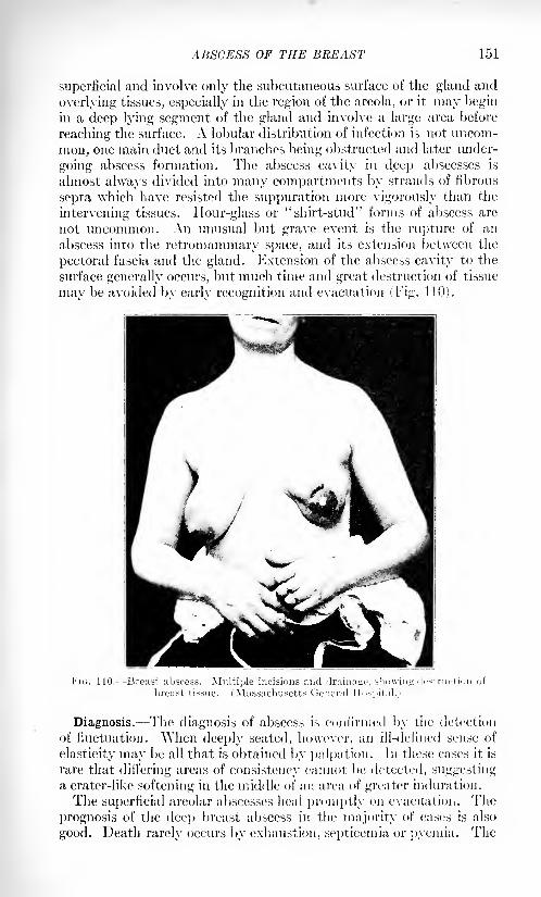
ABSCESS OF THE BREAST 151
superficial and involve only the subcutaneous surface of the gland and
overlying tissues, especially in the region of the areola, or it may beginin a deep lying segment of the gland and involve a large area before
reaching the surface. A lobular distribution of infection is not uncom-
mon, one main duct and its branches being obstructed and later under-
going abscess formation. The abscess cavity in deep abscesses is
almost always divided into many compartments by strands of fibrous
septa which have resisted the suppuration more vigorously than the
intervening tissues. Hour-glass or "shirt-stud" forms of abscess are
not uncommon. An unusual but grave event is the rupture of an
abscess into the retromammary space, and its extension between the
pectoral fascia and the gland. Extension of the abscess cavity to the
surface generally occurs, but much time and great destruction of tissue
mav be avoided bv earlv recognition and evacuation (Fig. 110).
Multiple incisions and drainage,(Massachusetts General Hospital.
Diagnosis. The diagnosis of abscess is confirmed by the detection
of fluctuation. \Vhen deeply seated, ho\vever, an ill-defined sense of
elasticity may be all that is obtained by palpation. In these eases it is
rare that differing areas of consistency cannot be detected, suggestinga crater-like softening in the middle of an area of greater induration.
The superficial areolar abscesses heal promptly on evacuation. The
prognosis of the deep breast abscess in the majority of eases is also
good. Death rarely occurs by exhaustion, septicemia or pyemia. The
152 SURGERY OF THE BREAST
prognosis for satisfactory lactation at a later pregnancy is dubious, andin a certain number of cases recurrence of the abscess, chronic inflam-
matory processes, and rarely the development of a new growth at a
later date may be anticipated.Treatment. The treatment of acute mastitis should be its prophy-
laxis. Scrupulous care and cleanliness during the establishment andmaintenance of lactation should be observed. Cold water and dilute
alcohol washes during the latter part of pregnancy will increase the
resistance of the skin of the nipple. Inverted and distorted nipplesshould be corrected by gentle manipulation, and in extreme cases, byoperation. After childbirth the nipple should be washed with boiled
water before and after nursing, and boracic acid ointment or a simplemild antiseptic should be applied. A fissure once detected should
receive energetic treatment until entirely healed.
Engorgement of the breast is best relieved by a supporting bandage,restriction of liquid diet, mild purgation, and the removal of the excess
of secretion by massage or with a sterile breast-pump. Massage is
undoubtedly the method most to be preferred, but should be practicedwith the utmost gentleness and cleanliness, "\\hen abscess of the
breast is present, operative treatment is at once required. The preser-
vation of the function of the breast demands interference at the earliest
possible moment after the diagnosis is reasonably certain. Morebreasts are sacrificed by dilatory treatment than are injured by too
early incision. On account of the multilocular character of abscesses
of the breast, two or more incisions are needed, as a rule, to ensure
drainage. 1 Radical incisions, with the patient under an anesthetic, and
abundant irrigation are demanded. When the abscess is pointing in
the upper portion of the breast, a small incision may be made in this
situation, and counter incisions of 10 to 15 cm. in length may then be
made at the lower and axillary margin of the breast to secure dependent
drainage. All incisions unless following the thoracomammary fold,
should be made in a direction radiating from the nipple, in order to
avoid injury to the gland ducts. Incisions should not traverse the
areola of the breast when it is possible to avoid this region. A generousincision following the lower and outer border of the breast is the most
satisfactory, especially when a small counter incision above provides
through-and-through drainage. The patient should be placed in a semi-
recumbent position and the wound packed with rubber "protective"wicks. The operative treatment of abscess of the breast is of great
importance, but its successful issue depends largely upon constitutional
and hygienic measures which should never be neglected. Saline purgesshould be given, and the amount of liquids ingested reduced to a
minimum. Nursing with the affected breast is practically impossibleon account of pain, but should be forbidden, under any circumstances,
in the interest of the child as well as of the mother. Nursing with the
other breast should, in all but the mildest cases, be abandoned also, in
order to facilitate the termination of lactation. Engorgement of the
other breast should be relieved by massage, and areas of engorgement
SYPHILIS OF THE BREAST 15.,
in the uninfected portions of the affected breast should be reduced bythe gentlest massage. A tight supporting bandage to both breasts
should be applied. ( '(institutional remedies, such as iron and malt, are
frequently indicated. Painstaking and energetic prosecution of the
after-care and dressing of the wound yield prompt and satisfactory
results, but with neglect relapse and extension of the infective processis not infrequent. Palliative treatment of this condition by external
applications or by puncture and aspiration of the abscess, or the instil-
lation of carbolic solution, have not met with general acceptance,
although the passive hyperemia treatment of Bier, by suction, is an
undoubted aid in the after-treatment when adequate surgical drainagehas been obtained.
Mastitis of Mumps.- -In common with the ovary and the testicle
the breast is peculiarly liable to an acute inflammatory process in the
course of an infectious parotitis. Girls between twelve and twentyare most liable to this affection, and its frequency varies in different
epidemics. Whether due to an actual bacterial infection or to the toxins
derived from infection in other regions, cannot be stated. Inasmuchas suppuration almost never occurs, the latter hypothesis seems more
probable. The breast is swollen, hard, and tender, and an elevated
temperature is common. Movement of the arm may be painful, and
a supporting bandage is indicated. Abscess formation is extremely
rare, and the process generally subsides in the course of a few days.
Atrophy of the breast or lack of further development may result, and
the loss of function in later life may be of importance.Abscess of the breast resulting from acute mastitis has been dis-
cussed in the preceding paragraphs. Abscesses of the breast produced
by other causes are occasionally observed, but are very rare. Abscesses
may involve the breast by extension from the chest wall beneath, as in
necrosis of the ribs, or empyema. Abscess may result, also, from
trauma and hematoma in the mammary region, and in this connection
the frequent selection of the subpectoral region for subcutaneous
infusion should be borne in mind.
Chronic Inflammatory Diseases of the Breast.
Syphilis. Syphilis may involve the breast, both in its primaryand in its later manifestations. Chancre of the breast is unusual, but
a number of cases have been reported. The usual manner of infection
is that of the wet-nurse by a syphilitic child. The primary lesion is
generally situated upon or at the base of the nipple and presents an
atypical fissure or ulceration, one-half to one inch in diameter, with
indurated edges and grayish-yellow base. The absence of pain is a
conspicuous feature, and the indolence of the lesion is marked. Involve-
ment of the axillary glands and, later, secondary symptoms confirm the
diagnosis. Secondary eruptions in the form of papules, macules and
pustules appear upon the skin of the breast, and mucus patches may lie
prodticed in the fold under a dependent breast, by maceration.
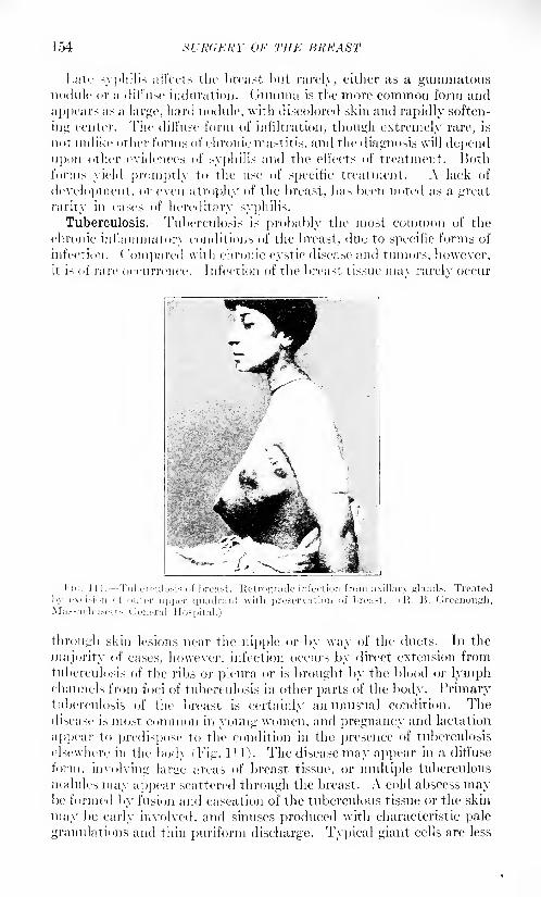
154 SURGERY OF THE BREAST
I.ate syphilis a 1 loots the breast but rarely, either as a gummatousnodule or a dill'use induration. Guinina is the more common form and
appears as a large, hard nodule, with discolored skin and rapidly soften-
ing center. The diffuse form of infiltration, though extremely rare, is
not unlike other forms of chronic mastitis, and the diagnosis will dependupon other evidences of syphilis and the effects of treatment. Bothforms yield promptly to the use of specific treatment. A lack of
development, or even atrophy of the breast, has been noted as a great
rarity in cases of hereditary syphilis.
Tuberculosis. -Tuberculosis is probably the most common of the
chronic inflammatory conditions of the breast, due to specific forms of
infection. ( 'ompared with chronic cystic disease and tumors, however,it is of rare occurrence. Infection of the breast tissue mav rarelv occur
through skin lesions near the nipple or by way of the ducts. In the
majority of cases, however, infection occurs by direct extension from
tuberculosis of the ribs or pleura or is brought by the blood or lymphchannels from foci of tuberculosis in other parts of the body. Primarytuberculosis of the breast is certainly an unusual condition. Thedisease is most common in voting women, and pregnancy and lactation
appear to predispose to the condition in the presence of tuberculosis
elsewhere in the body (Tig. 111). The disease may appear in a diffuse
form, involving large areas of breast tissue, or multiple tuberculous
nodules may appear scattered through the breast. A cold abscess may1)0 formed by fusion and caseation of the tuberculous tissue or the skin
may be early involved, and sinuses produced with characteristic pale
granulations and thin puriform discharge. Typical giant cells are less
CHRONIC MASTITIS 155
common than in other forms of tuberculosis, but they occur in manycases, and the inoculation of suspected tissue in guinea-pigs will alwayssettle the pathological diagnosis. The identification of the bacilli in
stained sections is extremely difficult.
Diagnosis. Clinically, tuberculosis of the breast is to be distinguishedfrom chronic cystic mastitis, other forms of chronic inflammation of
the breast and new growths. The diagnosis may be suggested by the
varied consistency of the indurated mass, its irregular outline and
by the early and extensive involvement of the axillary glands, which is
characteristic of tuberculosis. The constitutional symptoms of tuber-
culosis and the presence of disease elsewhere in the body, will be of
assistance. Advanced cases, with sinus formation and involvement of
the axillary glands, present less difficulty. The presence of tuber-
culosis and carcinoma in the same breast has been noted in a number of
cases sufficient to deserve mention.
Treatment. The best treatment of tuberculosis of the breast is
removal of the* breast and of the axillary glands, combined with the
constitutional treatment and hygiene so necessary in all tuberculosis.
Palliative treatment alone is indicated when the evidence of tubercu-
losis elsewhere in the body is such as to lead to no hope of delaying the
fatal issue. I'nder these circumstances, incision, curetting and iodo-
t'orm or iodin applications may be employed.Actinomycosis. Infection of the breast with actinomyces is a rare
condition, especially in America. It occurs, as a rule, by extension
from the pleura and ribs, and is thus but a part of a far more serious
disease. The resemblance to tuberculosis is marked, but the disease
is even more chronic in nature and the induration more pronounced.
Multiple sinuses are produced from which is discharged a thin pus
showing the characteristic sulphur granules.
Treatment. The treatment is by removal of the infected area -in
toto or by laying open and curetting the sinuses and the administration
of potassium iodide. The prognosis depends upon the extent of the
infection elsewhere than in the breast.
Echinococcus. Ilydatids of the breast are of extreme rarity, andin America are almost unknown. They are commonly conveyed to
the breast by extension from the lung and pleura, and there producethe large encapsulated cyst filled with daughter cysts, which is char-
acteristic of the disease. Suppuration is the ultimate result in the
majority of cases and the diagnosis from cysts of other origin is rarely
made before this occurs. The treatment is by incision and curetting
or by excision of the entire mass. A recent aid to the diagnosis of
echinococcus is the complement-fixation test.
Chronic Mastitis. Chronic inflammatory processes occurring in
the breast, and due to other causes than the specific microorganisms,have been variously described by many writers under the general nameof "Chronic Mastitis." The most common variety of this condition
accompanies the condition to be described under the heading of
Cvstic Disease of the Breast or Chronic Cvstic Mastitis. Chronic
156 SURGERY OF THE BREAST
mastitis independent of cystic disease is comparatively rare, and is the
result, as a rule, of a previous acute or suhacute mastitis or of trauma.
One or more lobules of the breast are tender and painful and present a
thickened and indurated consistency on palpation. They occur mostoften after lactation and especially when engorgement or suddencessation of lactation has provided favorable conditions for infection
from the ducts. The pathology of the process is an increase of fibrous
tissue, with round cell infiltration along the ducts of one or morelobules of the gland. Periductal mastitis of I )elbet. 1 Maladie Xoueiise
(Phocas).Minute abscesses may occur and even the condition commonly
described as "chronic abscess." In these cases a chronic induration of
the breast tissue is found of irregular outline, and the presence of an
abscess is rarely recognized. Operation is frequently advised under a
diagnosis of new growth, and only after excision is it discovered that the
indurated fibrous tissue surrounds an abscess of small extent with thick
or even inspissated contents. A form of chronic abscess described byFrench writers is said to affect the large ducts of the breast, without
extension to the surrounding tissues.
A number of cases of chronic inflammatory disease of the breast
following lactation have come to the attention of the writer in which
the ductal origin of the infection is almost certain. In these cases the
ducts of one or more lobules of the breast present foci or suppuration,
suggesting strongly the appearances of a branchopneumonia in the
lung. Other writers have described this condition as "mastitis obliter-
ans." 2 All of these conditions, however, are very rare.
Diagnosis. The diagnosis of chronic abscess of the breast can be
made only by exclusion of tuberculosis, guinma, actiomycosis andnew growths, and is rarely achieved before an exploratory incision has
been made. In the absence of abscesses no definite tumor is producedand the condition is of minor importance unless cystic disease develops.The treatment of the minor cases should be by supporting bandages.
Applications are rarely beneficial. The more extensive eases require
exploratory incision and operative treatment according to the con-
dition present.A form of chronic mastitis occurs occasionally in young unmarried
women, similar to the postlactation form, but of less extent. Such
cases are often accompanied by mastodynia, and tenderness and pain,
especially at the time of the catamenia, may be excessive. Trauma is
found to be responsible for certain cases of this nature, and continued
applications and manipulation are often effective in prolonging or
increasing the affection. The neurotic element is strong in many such
cases. The treatment is hygienic, and supporting dressings or strap-
ping of the breast may be employed. The disease may be of long
duration, but operative interference is rarely necessary, unless in
extreme cases, to confirm or establish a diagnosis.
1 Bulletin de Soc. d'Anat., January, 1893, Paris.
-Horz: Beitr. z. klin. Chir., Tubingen, 1910, Ixx, GS2-G94.
CYSTS OF THE BREAST 157
Cysts of the Breast.
The tendency to the formation of cysts is a conspicuous feature of
many of the diseases and new growths of the breast. This is due ta the
distribution throughout the breast tissue of epithelial ducts which are
readily obstructed by disease. Rapidly growing solid tumors ef the
breast, however, may develop cysts by reason of the ranid liquefactionof their tissues which accompanies degeneration, and cysts of this
nature are discussed under the section on tumors of the breast.
Cysts due to obstruction of the ducts of the breast assume a variety of
forms according to the development and function of the breast at the
time of their appearance. Retention cysts of the breast are single or
multiple. Single ci/stx (evolution cysts Gross 1
) are rare, and occur, as
a rule, in young adults. Obstruction to one of the gland ducts is the
usual cause and may be produced by the contraction of scar tissue
after trauma or inflammatory process. Cysts of this nature rarely
exceed the sixe of a hen's egg; they are commonly thin-walled and
contain a thin, yellowish or brownish fluid. They are not painful or
adherent to the skin; suppuration rarely results, and the developmentof malignant disease in a single cyst in early life is extremely rare.
Discharge from the nipple rarely occurs with spontaneous diminution
in the sixe of the cyst.
Galactocete. A galactocele is a simple cyst of the breast containingmilk. It occurs generally during, or at some period after the puerperalstate. The milky contents of a galactocele become progressivelythicker as time progresses, and the more fluid portions are absorbed,
and in this way creamy or oily contents may be obtained and later
substances resembling butter or soap. In cases of very long duration
even calcareous concretions have been found. The origin of the
obstruction to the duct which produces a galactocele is, as a rule,
traumatic or inflammatory, although instances of malignant disease
causing obstruction of ducts have been recorded. The cicatrix of a
healed breast abscess is not uncommonly at fault.
Treatment. - The treatment of a galactocele should be by incision
and drainage of the cavity, or by excision of the affected segment of the
gland.
Cystic Disease of the Breast (Chronic Cyxtic Mastitis Abnormal
Involution (Warren). Senile Parenchymatous Hypertrophy (Blood-
good). Cyst-adenoma Mamma' (Schimmelbusch). Maladie de Heclus.-
\Vith the decline of the development and function of the breast at the
time of the menopause a progressive atrophy of the gland elements and
1 Tumors of the Mammary Gland, 1880, Philadelphia.2 Bertels: J )eutsch. Ztschr f. Chir., 1913, exxiv, 9-46; Bloodgood, Joseph C.: Sur^.,
Gynec. and Obst., 1900, No. 6, iii, 721-730; Greenoush and Hartwell: .lour. Mod.
Research, 1903, iv, 417; Koeni^: Gentralbl. f. Chir., 1893, xx, 49; McCarthy: Surur .,
Gyiiec. and Obst., 1913, vol. xvii, also 1914, xviii, 284; M tiller, G. P.: Ann. Surg., 1914,
Ix, 595 (iOO; Greenou<di and .Simmons: Ann. Surg., .July, 1914; Jopson, Speeso andWhite: Ann. Surii., November, 1908; Tietzc: Ztschr. f. Chir., 1900, Ivi, 512; Speeso,
John: Ann. Sur-;., 1912, Iv, 212; Basse: Arch. f. klin. Chir..l 897, liv, 1.
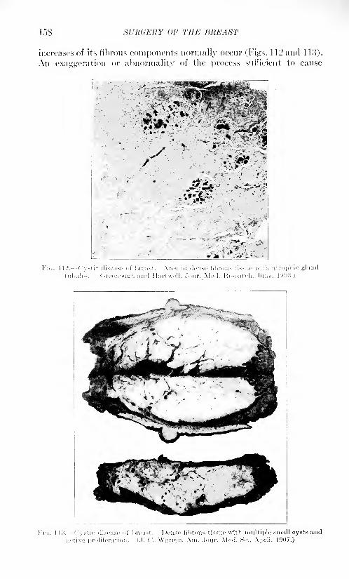
I.IS SURGERY OF THE BREAST
increases of its fibrous components normally occur (Figs. 112 and 113).
An exaggeration or abnormality of the process sufficient to cause
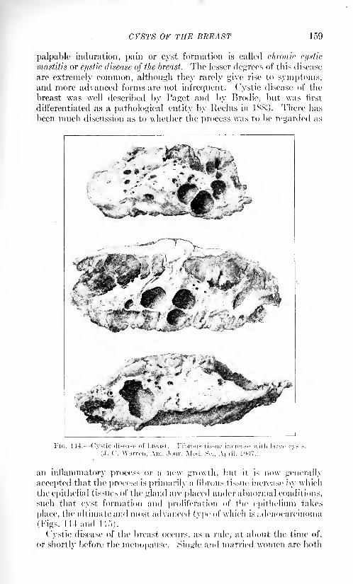
CYSTS OF THE BREAST
palpable induration, pain or cyst formation is called chronic ci/xtic
mastitis or cystic disease of the breast. The lesser degrees of this disease
are extremely common, although they rarely give rise to symptoms,and more advanced forms are not infrequent. Cystic disease of the
breast was well described by Paget and by Brodie, but was first
differentiated as a pathological entity by Reclus in 1883. Then 1 has
been much discussion as to whether the process was to be regarded as
FIG. 114. Cystic disease of breast. Fibrous tissue increase with large cvsts.
J. C. Warren, Am. .lour. Mod
an inflammatory process or a new gnnvth, but it is now generally
accepted that the process is primarily a fibrous tissue increase by which
the epithelial tissues of the gland are placed under abnormal conditions,
such that cyst formation and proliferation of the epithelium takes
place, the ultimate and most advanced type of which is adenocarcinoma
(Figs. Ill and 11.")).
Cystic disease of the breast occurs, as a rule, at about the time of,
or shortly before the menopause. Single and married, women are both
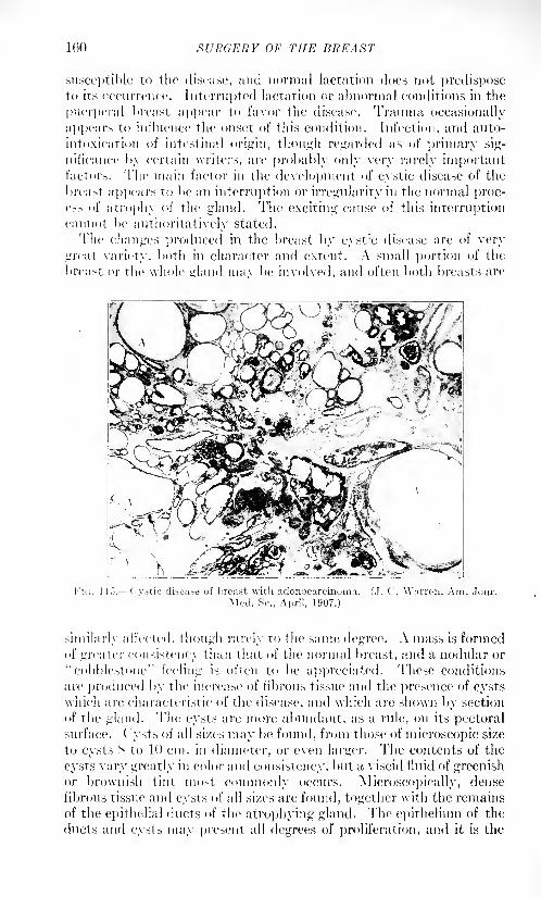
160 SURGERY OF THE BREAST
susceptible to the disease, and normal lactation does not predisposeto its occurrence. Interrupted lactation or abnormal conditions in the
puerperal breast appear to favor the disease. Trauma occasionally
appears to influence the onset of this condition. Infection, and auto-
intoxication of intestinal origin, though regarded as of primary sig-
nificance by certain writers, are probably only very rarely importantfactors. The main factor in the development of cystic disease of the
breast appears To be an interruption or irregularity in the normal proc-ess of atrophy of the gland. The exciting cause of this interruptioncannot be authoritatively stated.
The changes produced in the breast by cystic disease are of very
great variety, both in character and extent. A small portion of the
breast or the whole gland mav be involved, and often both breasts are
l'i<;. 1 13.- Cystic disease of breast with adenocarcinoma. (,). C. Warren, Am. Jour.
Mod. Sc., April, 1907.)
similarly all'ected, though rarely to the same degree. A mass is formedof greater consistency than that of the normal breast, and a nodular or
"cobblestone" feeling is often to be appreciated. These conditions
are produced by the increase of fibrous tissue and the presence of cystswhich are characteristic of the disease, and which arc shown by section
of the gland. The cysts are more 1 abundant, as a rule, on its pectoralsurface. ( ysts of all sixes may be found, from those of microscopic size
to cysts S to 10 cm. in diameter, or even larger. The contents of the
cysts vary greatly in color and consistency, but a viscid fluid of greenishor brownish tint most commonly occurs. Microscopically, dense
fibrous tissue and cysts of all sixes are found, together with the remains
of the epithelial ducts of the atrophying gland. The epithelium of the
ducts and cysts may present all degrees of proliferation, and it is the
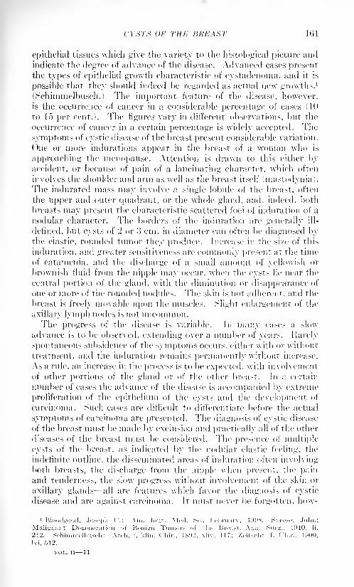
CYSTS OF THE BREAST 161
epithelial tissues which give the variety to the histological picture andindicate the degree of advance of the disease. Advanced cases presentthe types of epithelial growth characteristic of cystadenoma, and it is
possible that they should indeed be regarded as actual new growths.1
(Schimmelbusch.) The important feature of the disease, however,is the occurrence of cancer in a considerable percentage of cases (10
to 15 per cent.!. The figures vary in different observations, but the
occurrence of cancer in a certain percentage is widely accepted. The
symptoms of cystic disease of the breast present considerable variation.
One or more indurations appear in the breast of a woman who is
approaching the menopause. Attention is drawn to this either byaccident, or because of pain of a lancinating character, which often
involves the shoulder and arm as well as the breast itself fmastodynia).The indurated mass may involve a single lobule of the breast, often
the upper and outer quadrant, or the whole gland, and, indeed, both
breasts may present the characteristic scattered foci of induration of a
nodular character. The borders of the induration are generally ill-
defined, but cysts of 2 or o cm. in diameter can often be diagnosed bythe elastic, rounded tumor they produce. Increase in the size of this
induration, and greater sensitiveness are commonly present at the time
of catamenia, and the discharge of a small amount of yellowish or
brownish fluid from the nipple may occur, when the cysts lie near the
central portion of the gland, with the diminution or disappearance of
one or more of the rounded nodules. The skin is not adherent, and the
breast is freely movable upon the muscles. Slight enlargement of the
axillary lymph nodes is not uncommon.The progress of the disease is variable. In many cases a slow
advance is to be observed, extending over a number of years. Rarely
spontaneous subsidence of the symptoms occurs, either with or without
treatment, and the induration remains permanently without increase.
As a rule, an increase in the process is to be expected, with involvement
of other portions of the gland or of the other breast. In a certain
number of eases the advance of the disease is accompanied by extreme
proliferation of the epithelium of the cysts and the development of
carcinoma. Such cases are difficult to differentiate before the actual
symptoms of carcinoma are presented. The diagnosis of cystic disease
of the breast must be made by exclusion and practically all of the other
diseases of the breast must be considered. The presence of multiple
cysts of the breast, as indicated by the nodular elastic feeling, the
indefinite outline, the disseminated areas of induration often involvingboth breasts, the discharge from the nipple when present, the painand tenderness, the slow progress without involvement of the skin or
axillary glands- all are features which favor the diagnosis of cystic
disease and are against carcinoma. It must never be forgotten, how-
1 Blood^ood, Joseph ('.: Am. Jour. Mod. Sc., I'Vl.ruarv, 1DOS. Speee, John:
Malignant Degeneration of Bcnitrn Tumor,- of tin- Broa.-t. Ann. c?urjr., 1910, li,
212. SchiimiK'ltmseh: Arch, f. klin. Chir., 18 (
J2, xli
M, 512.
VOL. II 11
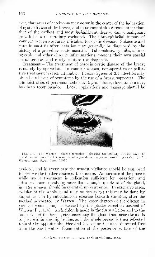
162 SURGERY OF THE BREAST
ever, that areas of carcinoma may occur in the center of the induration
of cystic disease of the breast, and in no case of this disease, other than
that of the earliest and most insignificant degree, can a malignantgrowth be with certainty excluded. The fibro-epithelial tumors of
younger women are rarely mistaken for cystic disease. Subacute andchronic mastitis after lactation may generally be diagnosed by the
history of a preceding acute mastitis. Tuberculosis, syphilis, actino-
mycosis and other chronic inflammations, present their own specialcharacteristics and rarely confuse the diagnosis.
Treatment. The treatment of chronic cystic disease of the breast
is mainly by operation. In younger women, non-operative or pallia-
tive treatment is often advisable. Lesser degrees of the affection mayoften be relieved of symptoms by the use of a breast supporter. Theadministration of potassium iodide in 10-grain doses, three times a day,has been recommended. Local applications and massage should be
FIG. 110.- The Warren "plastic resection," showing the axillary incision and the
breast turned back for the removal of a pie-shaped segment containing cysts. (J. C.
Warren, Ann. Surg., June, 1907.)
avoided, and in every case the utmost vigilance should be employedto observe the further course of the disease. An increase of the processwhile under treatment is indication sufficient for operation, andadvanced cases involving more than a single quadrant of the gland,in older women, should be operated upon at once. In extensive cases,
excision of the whole gland may be necessary; this may be done byamputation or by subcutaneous excision beneath the skin, after the
method advocated by "Warren. The lesser degrees of the disease in
younger women may be excised by the plastic resection method of
Warren (Fig. 116). An incision is made in the furrow below and to the
outer side of the breast, circumscribing the gland from near the axilla
to just within the nipple line, and the whole breast is then reflected
toward the opposite shoulder and its pectoral surface dissected free
from the chest wall. 1 Examination of the posterior surface of the
1 Gaillard, Thomas T.: New York Med. Jour., 1882.
TUMORS OF THE BREAST 163
gland can now be made, and the diseased segment excised in the formof a V, with its apex toward the nipple. A few buried sutures bring the
remaining surfaces together and the breast is turned back into position,
and the skin wound closed with sutures. By this method the entire
glandular structure can be excised without removal of the nipple, andthe subcutaneous tissues and fat drawn together by sutures to simulate
the rounded swelling of the normal breast. In the after-treatment of
these operations a form of dressing which supports the breast without
undue compression is required. A return of the disease in cases of
complete excision is impossible except in the other breast. Whenpartial excision is done, the portions of the gland remaining may giverise to further disease. This occurred in 8 of 83 cases collected by the
writer.
Cysts Due to New Growths. The cysts which are produced bytumors of the breast are of great variety, and will be discussed in the
section allotted to tumors of the breast. The tumors wrhich are
specially notable on account of their cystic character, and whichmust be borne in mind in the diagnosis of any breast lesion which is
accompanied by cyst formation, are the papillary cystadenoma, cystic
sarcoma, and cancer cysts.
TUMORS OF THE BREAST.
Tumors of the breast have long excited the interest of surgeons and
pathologists. They are very common and they occur in great variety.Carcinoma is more common in the mammary gland than in any other
organ of the female except the uterus, and in this fact lies the extreme
importance of the correct diagnosis of tumors of the breast. Forclinical purposes it is important to divide the tumors of the breast into
two classes benign and malignant. Benign tumors are, as a rule,
encapsulated. They do not infiltrate the surrounding tissues, and whenremoved they do not recur. Malignant tumors are not encapsulated.
They show a tendency to infiltration of surrounding tissues and to
extension to more distant parts. This tendency to extension often
prevents the complete removal of the disease by operation, and post-
operative recurrence of the growth is not uncommon.The division of breast tumors into benign and malignant forms is
a purely arbitrary one and cannot invariably be made to correspond
exactly with the microscopic examination of breast tumors after
removal. It must be admitted, also, that certain tumors which in
their early stages are essentially benign in character may show a
tendency to develop into malignant tumors as they progress. In the
main, however, the distinction between benign and malignant tumorsis one of great importance to the surgeon in determining the propertreatment of a given tumor.
In the section on the Anatomy and Development of the Breast it
was seen that the gland is made up of a system of epithelial-lined ducts
supported in a connective-tissue framework. The intimate relation
1(54 SURGERY Or THE BREAST
of these elements is such that any new growth in one of them affects
the other to a greater or less extent. Tumors composed entirely of
connective tissue, without change in the epithelium of the ducts, andtumors made up entirely of epithelium without a connective-tissue
participation, are practically unknown in the mammary gland.Classification. The pathological classification of tumors of the
breast has reference especially to the tissue of origin and the histological
character of the tumor cells. The classification in most general use
today is that of Ixibbert. 1 Ribbert describes, under the term "fibro-
epithelial tumors," that group of tumors in the breast and salivary
glands, the bladder and in some other regions which are made up of
epithelial gland elements with a fibrous tissue stroma. These are the
tumors arising in the gland tissue of the breast the so-called indigenousbreast tumors. The non-indigenous tumors of the breast form a small
group of tumors which may rise in the mammary gland as in other
portions of the body, but are in no way peculiar to the gland, and, in
fact, present no features by which they are to be distinguished from
similar tumors in other situations. The grouping of the indigenousbreast tumors under the main heading of h'bro-epithelial tumors is
satisfactory from the pathological point of view, but fails, to a certain
extent, to recognize the distinction between benign and malignantcharacteristics which is important to the clinician. For that reason
many attempts have been made to classify these tumors of the fibro-
epithelial group for more accurate study. The best of these classifi-
cations is that of Warren,2 which divides h'bro-epithelial tumors
into two main types the fibrous type and the epithelial type. This
classification and nomenclature will be used in the succeeding descrip-tion. An effort will be made, nevertheless, to indicate the terms
which have been used in medical literature to describe similar tumors.
Warren's classification is as follows:
Fibrous
Periductal fibroma.
Periductal myxoina.Periductal sarcoma.
Papillary cyst adenoma.
Fibro-cyst adenoma.
1 Hibbert: DeuiM-h. me<l. Wrhnsrhr., 1905, xxxi, 1219; also Lehrbuch der Patholo-
^i.-chen Hi.-.toJo<:ie, 1.S9C, Bonn, Friedrich Cohen.2 Jour. Am. Med. Assn., 1905, xiv, ICO; also International Text-book of Surgery, 1900,
vol. ii, chapter viii; Am. Jour. Med. Sc., April, 1907.
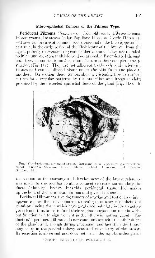
TUMORS OF THE BREAST 105
Fibro-epithelial Tumors of the Fibrous Type.
Periductal Fibroma (SI/HGHI/UIH: Adenofibroma, Fibro-adenoma,
Fibromyxoma, Intracanalicular Papillary Fibroma, Cystic Fibroma).1
These tumors are of common occurrence and make their appearance,as a rule, in the early period of the life-history of the breast from the
age of puberty to twenty-five years or thereabouts. They are rounded,nodular tumors, often multiple, and occasionally disseminated throughboth breasts, and their most constant feature is their complete encap-sulation (Fig. 117). They are not adherent to the skin and underlyingtissues and can be slipped about under the skin from one place to
another. On section these tumors show a glistening fibrous surface,
cut up into irregular patterns by the branching and irregular clefts
produced by the distorted epithelial ducts of the gland (Fig. 1.18). In
the section on the anatomy and development of the breast reference
was made to/ the peculiar hyaline connective tissue surrounding the
ducts of the virgin breast. It is this "periductal" tissue which makes
up the bulk of the periductal fibroma and gives it its name.Periductal fibromata, like the tumors of ovarian and testicular origin,
appear to owe their development to embryonic rests (Cohnheim) of
gland-producing tissue which have awakened only late in life; to active
growth and thus failed to fulfil their original purpose but remain with-
out function as a foreign element in the otherwise normal gland. Theducts of a periductal fibroma do not 'communicate with the other ductsof the gland, and, though during pregnancy and lactation the tumor
may share in the general enlargement and vascularity of the breast,
its secretion is abnormal and does not reach the nipple, although an
n^ertels: Deutsdi. f. ( 'liir., 1013, oxxiv, 9-40.
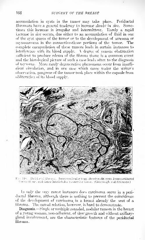
166 SURGERY OF THE BREAST
accumulation in cysts in the tumor may take place. Periductal
fibromata have a general tendency to increase slowlv in size. Some-times this increase is irregular and intermittent. Rarely a rapidincrease in size occurs, due either to an accumulation of fluid in one
of the cyst spaces of the tumor or to the development of sarcoma or
myxosarcoma in the connective-tissue portions of the tumor. The
complete encapsulation of these tumors leads in certain instances to
interference with its blood supply. A degree of venous obstruction
sufficient to produce edema of the fibrous tissue is a common event
and the histological picture of such a case leads often to the diagnosisof myxoma. More rarely degenerative phenomena occur from insuffi-
cient circulation, and in one case which came under the writer's
observation, gangrene of the tumor took place within the capsule from
obliteration of its blood supply.
I IS. Penduotal fibi Intracaimhcular type, showing slit cysts, loose periductallense mterlobular connective tissue. (Greenough and Simmons.)
In only the very rarest instances does carcinoma occur in a peri-ductal fibroma, although there is nothing to prevent the coincidenceof the development of carcinoma^ in a breast already the seat of a
fibroma. The causal relation, however, is hard to demonstrate.
Diagnosis. Single or multiple rounded, nodular tumors in the breast
of a young woman, non-adherent, of slow growth and without axillary-
gland involvement, are the characteristic features of the periductalfibroma.
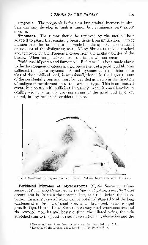
TUMORS OF THE BREAST 167
Prognosis. The prognosis is for slow but gradual increase in size.
Sarcoma may develop in such a tumor but carcinoma very rarely
does so.
Treatment. The tumor should be removed by the method best
adapted to guard the remaining breast tissue from mutilation. Direct
incision over the tumor is to be avoided in the upper inner quadranton account of the disfiguring scar. Many fibromata can be reached
and removed by the Thomas incision from the axillary border of the
breast. When completely removed the tumor will not recur.
Periductal Myxoma and Sarcoma. 1 Reference has been made aboveto the development of edema in the fibrous tissue of a periductal fibroma
sufficient to suggest myxoma. Actual myxomatous tissue (similar to
that of the umbilical cord) is occasionally found in the larger tumorsof the periductal group and must be regarded as a step in the direction
of malignant transformation to the sarcoma type. This is an unusual
event, but occurs with sufficient frequency to merit consideration in
dealing with any rapidly growing tumor of the periductal type, or,
indeed, in any tumor of considerable size.
FIG. 119. Periductal myxosurcoma of breast. (Massachusetts General Hospital.)
Periductal Myxoma or Myxosarcoma (Cystic Sarcoma, Adeno-
sarcoma (Williams),2
Cystosarcoma Proliferum, Cijstosarcoma Phyllodes]occurs later in life than the fibroma, but, as a rule, before the meno-
pause. In many cases a history can be obtained suggestive of the longexistence of a fibroma, of small size, which later took on more rapid
growth (Figs. 119 and 120). Such tumors may reach enormous size andthe rounded, nodular and bossy outline, the dilated veins, the skin
stretched thin to the point of ready excoriation and ulceration and the
1 Greenough and Simmons: Aim. Surg., October, 1911, p. 517.2 Diseases of the Breast, 1894, London, John Bale & Sons.
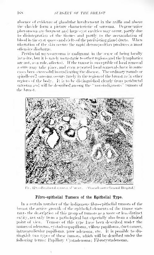
His Xl'h'dKKY
absence of evidence of glandular involvement in the axilla and abovethe clavicle form a picture characteristic of sarcoma. Degenerative
phenomena are frequent and large cyst cavities may occur, partly dueto disintegration of the tissues and partly to the accumulation of
blood in the cyst spaces and clefts of the preexisting gland ducts. Whenulceration of the skin occurs the rapid decomposition produces a most
offensive discharge.I'eriductal myxosarcoma is malignant in the sense of being locally
invasive, but it is rarely metastatic to other regions and the lymphaticsare not, as a rule, affected. If the tumor is susceptible of local removal
a cure may take place, and even repeated local removals have in somecases been successful in eradicating the disease. The ordinary round- or
spindle-cell sarcoma occurs rarely in the region of the breast as in other
region-, of the body. Jt is to be distinguished clearly -from periductalsarcoma and will be described among the "non-indigenous" tumors of
the breast.
l^'&V*'. < ' *f 5f M-IV ..'
^Iv*--/ "*-' r^ A^C^ML- . --?
II*Sbv, j*
'\ >i ' ' '* '
.
^r^V---^^
Fibro- epithelial Tumors of the Epithelial Type.
Jn a certain number of the indigenous fibro-epithelial tumors of the
breast the active growth of the epithelial elements of the tumor war-
rants the description of this group of tumors as a more or less distinct
entity, not only from a pathological but especially also from a clinical
point of view. Tumors of this type have been described under the
names of adenoma, cystadenopapilloma, villous papilloma, duct cancer,
intracanalicnlar papilloma, pure adenoma, etc. It is possible to dis-
tinguish two types of these tumors, which will be descibed tinder the
following terms: Papillary ( Ystadenoma; Fibrocystadenoma.
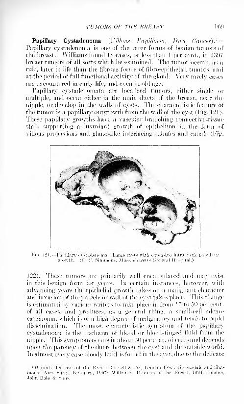
revolts or THE BHKAHT 1 09
Papillary Cystadenoma (r///o//.v Papilloma, Duel (lancer)}
Papillary cystadenoma is one of the rarer forms of benign tumors of
the breast. Williams found IS cases, or less than 1 per cent., in 2)597
breast tumors of all sorts which lie examined. The tumor occurs, as a
rule, later in life than the fibrous forms of fibro-epithelial tumors, andat the period of full functional activity of the giant 1. Very rarely cases
are encountered in early life, and even in old age.
Papillary cystadenomata are localized tumors, either single or
multiple, and occur either in the main ducts of the breast, near the
nipple, or develop in the walls of cysts. The characteristic feature of
the tumor is a papillary outgrowth from the Avail of the cyst (Fig. 121 j.
These papillary growths have a vascular branching connective-tissue
stalk supporting a luxuriant growth of epithelium in the form of
villous projections and gland-like interlacing tubules and canals (Tig.
122). These tumors are primarily well encapsulated and may exist
in this benign form for years. In certain instances, however, with
advancing years the epithelial growth takes on a malignant character
and invasion of the pedicle or wall of the cyst takes place. This changeis estimated by various writers to take place in from 1") to 50 per cent,
of all cases, and produces, as a general thing, a small-cell adeno-
carcinoma, which is of a high degree of malignancy and tends to rapid
dissemination. The most characteristic symptom of the papillary
cystadenoma is the discharge of blood or blood-tinged lluid from the
nipple. This symptom occurs in about .">() per cent, of cases and depends
upon the patency of the ducts between the cyst and the outside world.
In almost everv case bloodv fluid is found in the cvst, due to the delicate
'Bryant: Diseases of the Breast, ( 'a->ell ^ Co., London IS.s?; (.'.reonounli and Sim-
mons: Ann. Surti., February, l'J()7; Williams: Diseases of the Breast, 1S!)4, London,John Bale A: Sons.
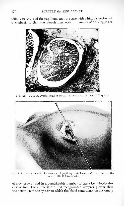
170 SURGERY OF THE BREAST
villous structure of the papilloma and the ease with which laceration or
thrombosis of the bloodvessels may occur. Tumors of this type are
FIG. 122. Papillary cystadenoma of breast. (Massachusetts General Hospital.)
I'IG. 123. Areolar incision for removal of papillary cystadenoma of breast near to the
nipple. (R. B. Greeiiough.)
of slow growth and in a considerable number of cases the bloody dis-
charge from the nipple is the first recognizable symptom; even then
the detection of the cyst from which the blood comes may be extremely
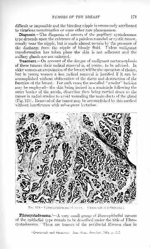
TUMORS OF THE BREAST 171
difficult or impossible and the bleeding nipple is erroneously attributed
to vicarious menstruation or some other rare phenomenon.Diagnosis. The diagnosis of tumors of the papillary cystadenoma
type depends upon the existence of a painless rounded or cystic tumor,
usually near the nipple, but is made almost certain by the presence of
the discharge from the nipple of bloody fluid. Unless malignanttransformation has taken place the skin is not adherent and the
axillary glands are not enlarged.Treatment.- On account of the danger of malignant metamorphosis
of these tumors their radical removal is, of course, to be advised. In
older women an amputation of the breast will be the operation of choice,
but in young women a less radical removal is justified if it can be
accomplished without obliteration of the ducts and destruction of the
function cf the breast. For such cases the so-called "areolar"' incision
may be employed the skin being incised in a semicircle following the
outer border of the areola, dissection then being carried down to the
tumor in radial strokes to avoid wounding the main ducts of the gland
(Fig. 123). Removal of the tumor may be accomplished by this methodwithout interference with subsequent lactation.
* Wi ^O1' -'* ^"i^afe^-T^V^
:.^^^S^VA/ :**&*
^'M^
lie. 124. Fibrocyatadeiioma of breast. (Greenough ami Simmons.)
Fibrocystadenoma. 1 - A very small group of fibro-epithelial tumorsof the epithelial type remain to be described under the title of Fibro-
cystadenoma. These are tumors of the periductal fibroma class in
1 Greenougb and Simmons: Ann. S.ur^., October, 1911, p. 517.
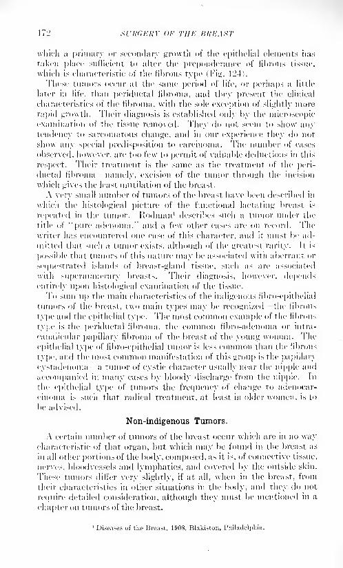
172 Xl'/tGEItY OF THE KHKAST
which a primary or secondary growth of the epithelial elements has
taken place sufficient to alter the preponderance of fibrous tissue,
which is characteristic of the fibrous type (Fig. 124).
These tumors occur at the same period of life, or perhaps a little
later in life, than periductal fibroma, and they present the clinical
characteristics >f the fibroma, with the sole exception of slightly more
rapid growth. Their diagnosis is established only by the microscopicexamination of the tissue removed. They do not seem to show anytendency to sarcomatous change, and in our experience they do not
show any >pecial predisposition to carcinoma. The number of cases
observed, however, are too few to permit of valuable deductions in this
respect. Their treatment is the same as the treatment of the peri-
ductal fibroma namely 3excision of the tumor through the incision
which gives the least mutilation of the breast.
A very small number of tumors of the breast have been described in
which the histological picture of the functional lactating breast is
repeated in the tumor. Rodman 1 describes such a tumor under the
title of "pure adenoma," and a few other cases are on record. Thewriter has encountered one case of this character, and it must be ad-
mitted that such a tumor exists, although of the greatest rarity. It is
possible that tumors of this nature may be associated with aberrant or
sequestrated islands of breast-gland tissue, such as are associated
with .supernumerary breasts. Their diagnosis, however, dependsentirely upon histological examination of the tissue.
To sum up the main characteristics of the indigenous fibro-epithelial
tumors of the breast, two main types may be recognized the fibrous
type and the epithelial type. The most common example of the fibrous
type is the periductal fibroma, the common fibro-adenoma or intra-
canalicular papillary fibroma of the breast of the young woman. The
epithelial type of fibro-epithelial tumor is less common than the fibrous
type, and the most common manifestation of this group is the papillary
cystadenoma a tumor of cystic character usually near the nipple and
accompanied in many cases by bloody discharge from the nipple. In
the epithelial type of tumors the frequency of change to adenocar-
cinoma is such that radical treatment, at least in older women, is to
be advised.
Non-indigenous Tumors.
A certain number of tumors of the breast occur which are in no waycharacteristic of that organ, but which may be found in the breast as
in all other portions of the body, composed, as it is, of connective tissue,
nerves, bloodvessels and lymphatics, and covered by the outside skin.
These tumors differ very slightly, if at all, when in the breast, from
their characteristics in other situations in the body, and they do not
require detailed consideration, although they must be mentioned in a
chapter on tumors of the breast.
1 Diseases of the Breast, 1908, Blakiston, Philadelphia.
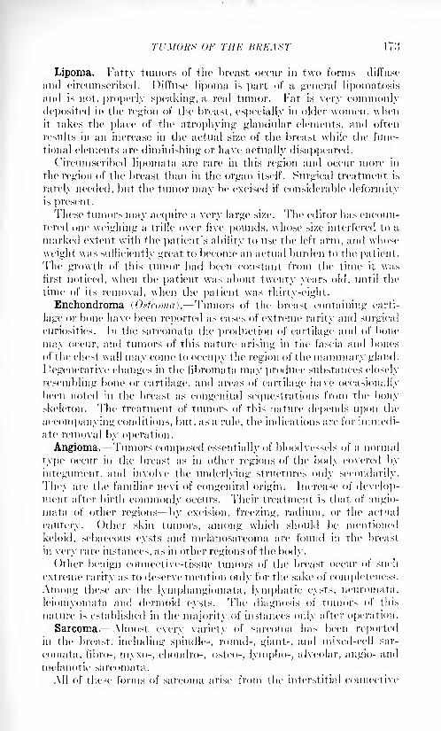
TUMORH OF TllK BRKAKT 17.'5
Lipoma. Fatty tumors of the breast occur in two forms diffuse
and circumscribed. Diffuse lipoma is part of a general lipomatosisand is not, properly speaking, a real tumor. Fat is very commonlydeposited in the region of the breast, especially in older women, whenit takes the place of the atrophying glandular elements, and often
results in an increase in the actual si/e of the breast while the func-
tional elements are diminishing or have actually disappeared.Circumscribed lipomata are rare in this region and occur more in
the region of the breast than in the organ itself. Surgical treatment is
rarely needed, but the tumor may be excised if considerable deformityis present.
These tumors may acquire a very large si/.e. The editor has encoun-
tered one weighing a trifle over five pounds, whose si/e interfered to a
marked extent with the patient's ability to use the left arm, and whose
weight was sufficiently great to become an actual burden to the patient.The growth of this tumor had been constant from the time it was
first noticed, when the patient was about twenty years old, until the
time of its removal, when the patient was thirty-eight.
Enchondroma (Osteovnd). Tumors of the breast containing carti-
lage or bone have been reported as cases of extreme rarity and surgical
curiosities. In the sarcomata the production of cartilage and of hone
may occur, and tumors of this nature arising in the fascia and bones
of the chest wall may come to occupy the region of the mammary gland.
Degenerative changes in the fibromata may produce substances closely
resembling bone or cartilage, and areas of cartilage have occasionallybeen noted in the breast as congenital sequestrations from the bonyskeleton. The treatment of tumors of this nature depends upon the
accompanying conditions, but, as a rule, the indications are for immedi-
ate removal by operation.
Angioma. Tumors composed essentially of bloodvessels of a normal
type occur in the breast as in other regions of the body covered byintegument, and involve the underlying structures only secondarily.
They are the familiar nevi of congenital origin. Increase of develop-ment after birth commonly occurs. Their treatment is that of angio-mata of other regions by excision, freezing, radium, or the actual
cautery. Other skin tumors, among which should be mentioned
keloid, sebaceous cysts and melanosarcoma are found in the breast
in very rare instances, as in other regions of the body.Other benign connective-tissue tumors of the breast occur of such
extreme rarity as to deserve mention only for the sake of completeness.
Among these are the lymphangiomata, lymphatic cysts, neuromata,
leiomyomata and dermoid cysts. The diagnosis of tumors of this
nature is established in the majority of instances only after operation.
Sarcoma. Almost every variety of sarcoma has been reportedin the breast, including spindle-, round-, giant-, and mixed-cell sar-
comata, fibro-, myxo-, chondro-, osteo-, lymph o-, alveolar, augio- and
melanotic sarcomata.
All of these forms of sarcoma arise from the interstitial connective
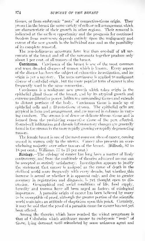
174 SURGERY OF THE BREAST
tissues, or from embryonic "rests" of connective-tissue origin. Theypresent in the breast the same variety of cells or cell arrangement whichare characteristic of their growth in other regions. Their removal is
indicated at the earliest opportunity and the prognosis for continued
freedom from recurrence depends entirely lipon the malignancy andextent of the new growth in the individual case and on the possibility
of its complete removal.
The non-indigenous sarcomata form less than one-half of all sar-
comata of the breast and all of the sarcomata together produce onlyabout 1 per cent, of all tumors of the breast.
Carcinoma. Carcinoma of the breast is one of the most commonand most dreaded diseases of women which is known. Every aspectof the disease has been the subject of exhaustive investigation, and its
origin is yet a mystery. The term carcinoma is applied to malignantdisease of epithelial origin, but the more popular term of cancer is also
frequently used in the same connection.
Carcinoma is a malignant new growth which takes origin in the
epithelial gland tissue of the breast, and by its atypical growth andextreme proliferative power, infiltrates surrounding tissues and extends
to distant portions of the body. Carcinoma tissue is made up of
epithelial cells and a fibrous-tissue stroma. The epithelial cells are
atypical in form and arrangement, and are massed in columns in vary-
ing numbers. The stroma is of dense or delicate fibrous tissue and is
formed from the preexisting connective tissue of the part affected.
Round-cell infiltration and chronic inflammatory changes are generallyfound in the stroma in the more rapidly growing or rapidly degeneratingtumors.
The female breast is one of the most common sites of cancer, comingsecond in women only to the uterus. Cancer also presents an over-
whelming majority over other tumors of the breast. (Billroth, 82 to
IS per cent.; "Williams, 77 to 23 per cent.)
Etiology. The etiology of cancer has long been a matter of lively
controversy, and from the multitude of theories advanced no one can
be accepted as entirely satisfactory. Investigation appears to justify
the statement that cancer is assigned as the cause of death in the
civilixed world more frequently with every decade, but whether^thisincrease is actual or whether it is apparent only, and due to greater
accuracy in registration and diagnosis, is yet thought open to dis-
cussion. Geographical and social conditions of life, food supply,
heredity and trauma have all been urged as factors of etiological
importance. A parasitic origin of cancer has been believed by manyto be susceptible of proof, although the greater portion of the scientific
world maintains an attitude of skepticism upon this point. Certainly,
it may be said that the proof of a parasitic cause for cancer has not yet
been offered.
Among the theories which have reached the widest acceptance is
that of Cohnheirn which attributes cancer to embryonic "rests" of
tissue, lying dormant until stimulated by some unknown agent and
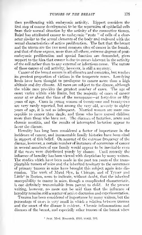
TUMORS OF THE BREAST 175
then proliferating with embryonic activity. Rippert considers the
first step of cancer development to be the separation of epithelial cells
from their normal situation by the activity of the connective tissues.
Baird has attributed cancer to embryonic "rests"
of cells of a char-
acter similar to the sexual elements of the body and endowed with the
sexual characteristics of active proliferation. The fact that the breast
and the uterus are the two most common sites of cancer in the female,and that of these organs, more than all others, extreme degrees of post-
embryonic proliferation and special function are demanded, gives
support to the idea that cancer is due to causes inherent in the activityof the cell rather than to any external or infectious cause. The nature
of these causes of cell activity, however, is still a mystery.Cancer of the breast occurs in all climates and countries, but reaches
its greatest proportion of victims in the temperate zones. Low-lyinglands have been thought to predispose to cancer more than a highaltitude and dry climate. All races are subject to the disease, althoughthe white race provides the greatest number of cases. The age of
onset varies within wide limits, but the majority of cases of cancer
occur at or about the time of the menopause at forty-five or fifty
years of age. Cases in young women of twenty-one and twenty-twoare very rarely reported, but among the very old, seventy to eighty
years of age, it is not so infrequent. Married women are more sus-
ceptible to cancer than single, and those who have nursed children
more than those who have not. The diseases of lactation, acute andchronic mastitis, and the results of interrupted lactation appear to
favor the disease.
Heredity has long been considered a factor of importance in the
incidence of cancer, and innumerable family histories have been cited
in support of this belief. On account of the extreme frequency of the
disease, however, a certain number of instances of occurrence of cancer
in several members of one family would appear to be inevitable even
if the cases were distributed purely by chance. Until recently the
influence of heredity has been viewed with skepticism by many writers.
The studies which have been made in the past ten years of the trans-
plantable tumors of mice and the inherited tendency to the occurrence
of primary tumors in mice have brought this subject again into dis-
cussion. The work of Maud Slye, in Chicago, and of Tyzzer and
Little 1 in Boston, seem to indicate, without doubt, that the inherited
susceptibility to cancer in mice, though a complicated characteristic,
is one definitely transmissible from parent to child. At the present
writing, however, no more can be said than that the influence of
heredity remains still a matter of active discussion and experimentation.Trauma has been considered of importance by many writers, but the
percentage of cases is very small in which a relation between traumaand the onset of the disease is evident. Chronic inflammations and
diseases of the breast, and especially other tumors of the breast when
1 Jour. Mod. Ri-aeiirch, 1910, xxxiii, ,393.
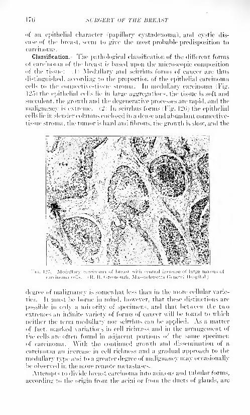
176 SURGERY OF THE BREAST
of an epithelial character (papillary cystadenoma), and cystic dis-
ease 1 of The breast, seem to give the most probable predisposition to
carcinoma.
Classification. The pathological classification of the different formsof carcinoma of the breast is based upon the microscopic compositionof the tissue: i 1 ) Medullary and seirrhus forms of cancer are thus
distinguished, according to the proportion of the epithelial carcinomacells to the connective-tissue stroma. In medullary carcinoma (Fig.
12.")) the epithelial cells lie in large aggregations, the tissue is soft and
succulent, the growth and the degenerative processes are rapid, and the
malignancy is extreme. c2) In seirrhus forms (Fig. ll2(J) the epithelialcells lie in slender columns enclosed in a dense and abundant connective-
tissue stroma, the tumor is hard and fibrous, the growth is slow, and the
^%i;|fi
]~>. Medullary carcinoma of breast with central increase of large nin
R. B. Greenough, Massachusetts General Hospital.)
FlG. 12
degree of malignancy is somewhat less than in the more cellular varie-
ties. It must be borne in mind, however, that these 1 distinctions are
possible in only a minority of specimens, and that between the two
extremes an infinite variety of forms of cancer will be found to which
neither the term medullary nor seirrhus can be applied. As a matter
of fact, marked variations in cell richness and in the arrangement of
the cells are often found in adjacent portions of the same specimenof carcinoma. With the continued growth and dissemination of a
carcinoma an increase in cell richness and a gradual approach to the
medullary type and to a greater degree of malignancy may occasionally
be observed in the more remote metastases.
Attempts to divide breast carcinoma into acinous and tubular forms,
according to the origin from the acini or from the ducts of glands, are
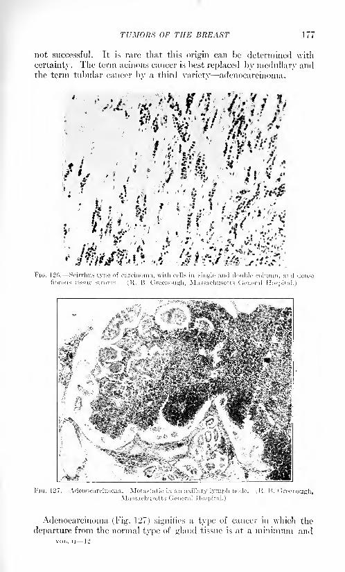
TUMORS OF THE BREAST 111
not successful. It is rare that this origin can be determined with
certainty. The term acinous cancer is best replaced by medullary andthe term tubular cancer bv a third variety adenocarcinoma.
V
Adenocarcinoma (Fig. 127) signifies a type of cancer in which the
departure from the normal type of gland tissue is at a minimum andVOL. II 12
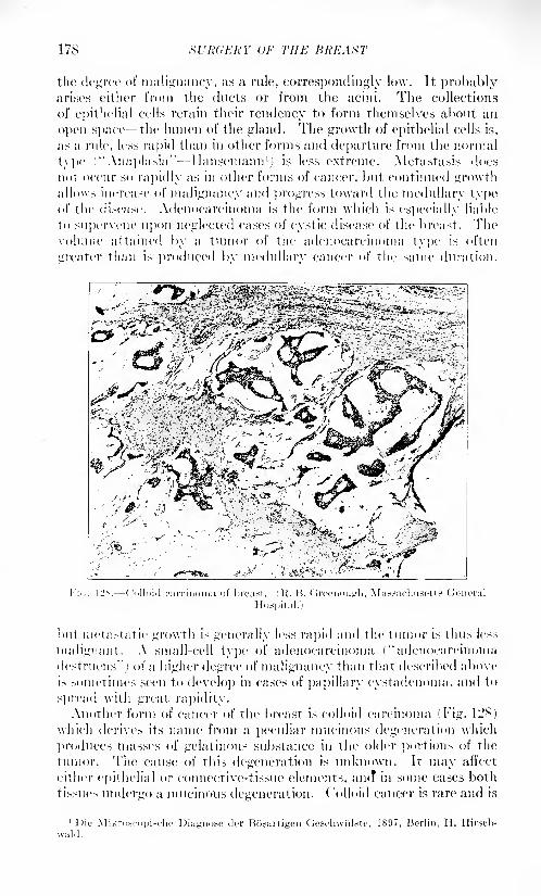
178 SURGERY OF THE BREAST
the degree of malignancy, ;is a rule, correspondingly lo\v. It probablyarises either from the duets or from the acini. The collections
of epithelial cells retain their tendency to form themselves about an
open space the lumen of the gland. The growth of epithelial cells is,
as a rule, less rapid than in other forms and departure from the normal
type ("Anaplasia" Ilansemann 1
) is less extreme. Metastasis does
not occur so rapidly as in other forms of cancer, but continued growthallows increase of malignancy and progress toward the medullary typeof the disease. .\denocarcinoma is the form which is especially liable
to supervene upon neglected cases of cystic disease of the breast. Thevolume attained by a tumor of the adenocarcinoma type is often
greater than is produced by medullary cancer of the same duration,
J'n,. 12S. Culloid carcinoma of breast, i R. H. ( Irccuoiitrli, Massachusetts General
Hospital.)
but metastatie growth is generally less ra])id and the tumor is thus less
malignant. A small-cell type of adenocarcinoma ("adenocarcinoma
destruens"j of a higher degree of malignancy than that described aboveis sometimes seen to develop in cases of papillary cystadenoma. and to
s] tread with great rapidity.Another form of cancer of the breast is colloid carcinoma (Fig. 12$)
which derives its name from a peculiar mucinous degeneration which
produces masses of gelatinous substance in the older portions of the
tumor. The cause of this degeneration is unknown. It may ail'ect
either epithelial or connective-tissue elements, am? in some cases both
tissues undergo a mucinous degeneration. Colloid cancer is rare and is
11 tie .Miknj.-eupische Diagnose der Bosartigcu Geschwiilste, 1897, Berlin, H. Hirsch-
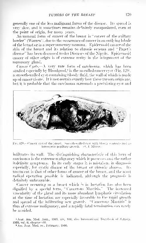
TUMORS OF THE BREAST 179
generally one of the less malignant forms of the disease. Its spread is
very slow, and it sometimes remains definitely encapsulated, even at
the point of origin, for many years.
An nnnsual form of cancer of the breast is "cancer of the axillary
border" (Warren1
), due to the occurrence of cancer in an outlying lobule
of the breast or in a supernumerary mamma. Epidermoid cancer of the
skin of the breast and its relation to chronic ecxema and ''Faget's
disease" has been discussed under Diseases of the Nipple. Kpidermoidcancer of other origin is of extreme rarity in the integument of the
mammary gland.
Cancer Ci/xte. A very rare form of carcinoma, which has been
studied especially by Blood good.2
is the so-called cancer cyst ' Fig. ll'Oi,
a smooth-walled cyst containing bloody fluid, the wall of which is made
up of cancer tissue. It is not certain exactly how these tumors originate,
but it is probable that the carcinoma surrounds a preexisting cyst and
infiltrates its wall. The distinguishing characteristic of this form of
carcinoma is the extreme malignancy which it possesses and the rather
indefinite symptoms. In its early stages it is mistaken, in diagnosis
especially, for cystic disease of the breast or chronic abscess. Its
treatment is that of other forms of cancer of the breast, and the most
radical operation possible is indicated, although the prognosis is
definitely unfavorable.
Cancer occurring in a breast which is in lactation has also been
dignified by a special term, ''Cancerous Mastitis." The increased
vascularity of the gland and its more abundant lymphatic circulation
at the time of lactation are especially favorable to the rapid growthand spread of the infiltrating new growth. ''Cancerous Mastitis" is
thtis of extreme malignancy, and a rapidly fatal termination can rarely
be avoided.
'Jour. Am. Med. Assn., 1905, xiv, 160; also International Text-book of Surgery,
1900, vol. ii, chapter viii.
8 Am. Jour. Med. Sc., February, 1908.
ISO SURGERY OF THE BREAST
The degenerative dinners in the tissues of cancer of the breast are
of great variety. The older and more' central portions of the epithelialcolumns first heroine deprived of nourishment, and undergo anemicnecrosis and fatty degeneration. Interference with the blood supply
may cause necro>i.- of whole portions of the tumor, and all varieties of
degenerative processes are thus produced. Ilemorrhagic areas are not
infrequent, and infection by microorganisms may occur, with sloughingand suppuration, by extension through the overlying integument.Tumors showing extensive cavities filled with fluid necrotic material
have occasionally been described as ''cystic carcinoma.'' Small areas
of necrosis occasionally undergo inspiration, and in slow-growingcarcinomata the deposit of lime salts in such areas has been recorded.
Growth. The main characteristic of cancer of the breast, like that, of
other organs, is the faculty of progressive infiltrating growth, and of
extension by metastascs, To other parts. Invasion of tissues by direct
continuity produces extension of the tumor to the skin above, andto the muscles, fascia 1
, ribs, sternum and pleural cavity below. In
this manner the tumor mass becomes more and more firmly fixed in its
position. Extension to other regions is produced especially by way of
the lymphatics, although penetration of the wall of a vein, and metas-
tasis by the blood current, may rarely occur in the late stages of the
disease. The lymphatics have long been recogni/ed as the chief carriers
of cancer growth. For this reason the anatomy of the lymphatics of
the breast has received special consideration, as outlined in the section
on Anatomy of the Breast. Cells or groups of cells appear to make their
way into the lymphatic vessels and to grow along them (permeation
Handley') or be carried to the nearest lymph nodes, where they find
conditions suitable for growth, and produce a tumor similar to that
from which they were derived. Blocking of one set of lymphatic chan-
nels by metastatic growth may produce new channels of compensatorycirculation, until these are again blocked by further metastases. In
this way cases of apparent retrograde lymphatic metastasis are pro-
duced. Extension from a node once infected to that nearest above it
mav continue until the free circulation in the thoracic duct is reached,
and u'"iieral metastasis or carcinomatosis is produced.The susceptibility of the different organs and tissues of the body to
carcinomatous metastasis appears to be subject to variation. Amongthe tissues in which this susceptibility is most conspicuous may be
mentioned the lungs and pleura, the liver, the bony skeleton, and the
brain. The bones most frequently involved are the sternum and ribs,
the humerus, femur, and spinal column.
Symptoms. The symptoms of carcinoma of the breast are insidious.
A woman of middle age notices a hard mass in the substance of the
breast. Attention is generally called to the tumor by accident, and
pain is rarely felt, although a certain amount of tenderness may be
present. Often the tumor has been noticed for a long time, and dread
1
Siir^., (iynec. and Obst., 191"), xx, 72.
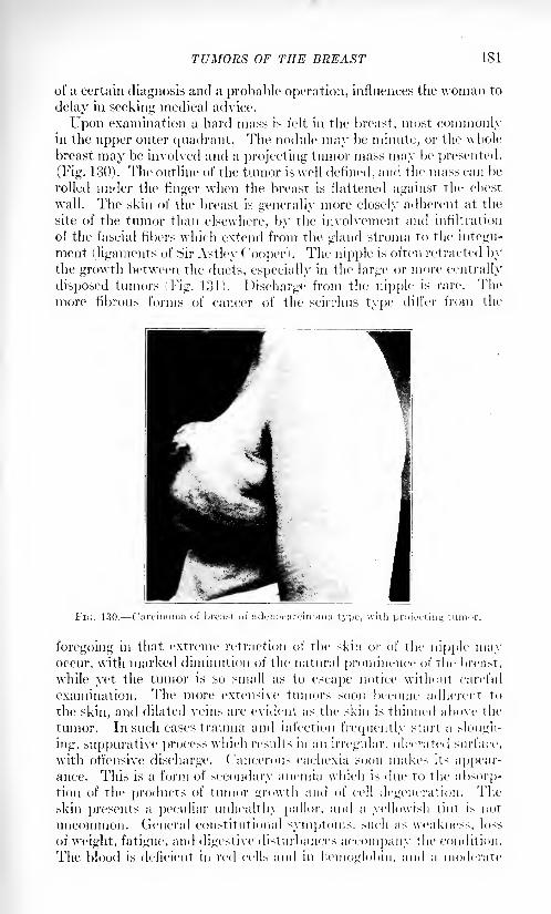
TUMORS OF THE BREAST 181
of a certain diagnosis and a probable operation, influences the woman to
delay in seeking medical advice.
Upon examination a hard mass is felt in the breast, most commonlyin the upper outer quadrant. The nodule may be minute, or the whole
breast may be involved and a projecting tumor mass may be presented.
(Fig. 130). The outline of the tumor is well defined, and the mass can be
rolled under the finger when the breast is flattened against the chest
wall. The skin of the breast is generally more closely adherent at the
site of the tumor than elsewhere, by the involvement and infiltration
of the fascial fibers which extend from the gland stroma to the integu-
ment (ligaments of Sir Astley ( 'ooper). The nipple is often retracted bythe growth between the ducts, especially in the large or more centrally
disposed tumors (Fig. 131). Discharge from the nipple is rare. Themore fibrous forms of cancer of the scirrhus type differ from the
FK,. 130
foregoing in that extreme retraction of the skin or of the nipple mayoccur, with marked diminution of the natural prominence of the breast,
while yet the tumor is so small as to escape notice without careful
examination. The more extensive tumors soon become adherent to
the skin, and dilated veins are evident as the skin is thinned above the
tumor. In such cases trauma and infection frequently start a slough-
ing, suppurative process which results in an irregular, ulcerated surface,
with offensive discharge. Cancerous cachexia soon makes its appear-ance. This is a form of secondary anemia which is due to the absorp-tion of the products of tumor growth and of cell degeneration. 1'he
skin presents a peculiar unhealthy pallor, and a yellowish tint is not
uncommon. (General constitutional symptoms, such as weakness, loss
of weight, fatigue, and digestive disturbances accompany the condition.
The blood is deficient in red cells and in hemoglobin, and a moderate
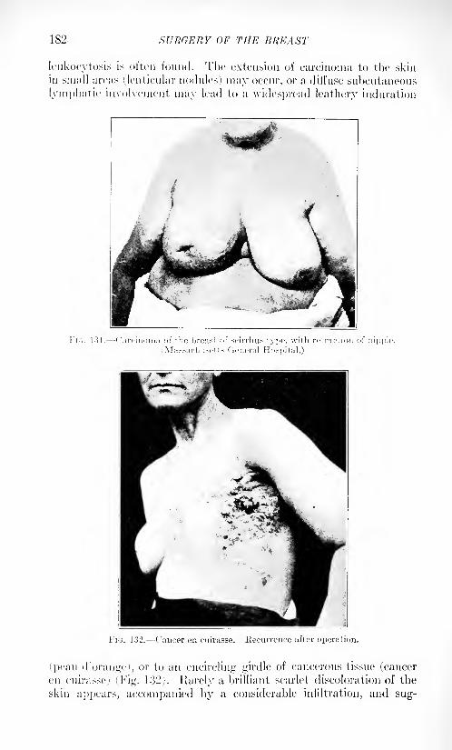
182 SURGERY OF THE BREAST
leukocytosis is often found. The extension of carcinoma to the skin
in small areas (lenticular nodules) may occur, or a diffuse subcutaneous
lymphatic involvement may lead to a widespread leathery induration
FIG. 131. Carcinoma of the breast of seirrhus type, with retraction of nipple.
Massachusetts General Hospital.)
FIG. 132. Cancer en cuirasse. Recurrence after operation.
fpeau d'orange), or to an encircling girdle of cancerous tissue (canceren cuirasse) (Fig. 132j. Rarely a brilliant scarlet discoloration of the
skin appears, accompanied by a considerable infiltration, and sug-
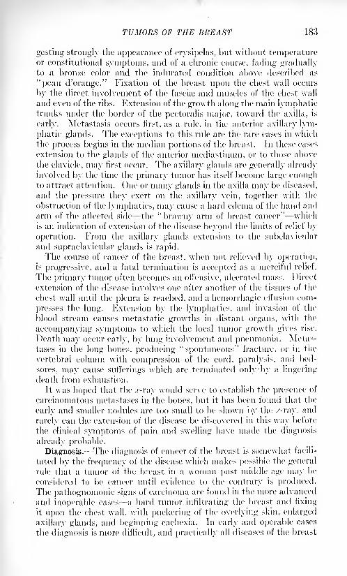
TUMORS OF THE BREAST 183
gesting strongly the appearance of erysipelas, hut without temperatureor constitutional symptoms, and of a chronic course, fading graduallyto a bronze color and the indurated condition above described as
"peau d'orange." Fixation of the breast upon the chest wall occurs
by the direct involvement of the fascine and muscles of the chest wall
and even of the ribs. Extension of the growth along the main lymphatictrunks under the border of the pectoralis major, toward the axilla, is
early. Metastasis occurs first, as a rule, in the anterior axillary lym-
phatic glands. The exceptions to this rule are the rare cases in which
the process begins in the median portions of the breast. In these cases
extension to the glands of the anterior mediastinum, or to those above
the clavicle, may first occur. The axillary glands are generally alreadyinvolved by the time the primary tumor has itself become large enoughto attract attention. One or many glands in the axilla may be diseased,
and the pressure they exert on the axillary vein, together with the
obstruction of the lymphatics, may cause a hard edema of the hand and
arm of the affected side the "brawny arm of breast cancer" which
is an indication of extension of the disease beyond the limits of relief by
operation. From the axillary glands extension to the subclavicular
and supraclavicular glands is rapid.The course of cancer of the breast, when not relieved by operation,
is progressive, and a fatal termination is accepted as a merciful relief.
The primary tumor often becomes an offensive, ulcerated mass. 1 Direct
extension of the disease involves one after another of the tissues of the
chest wall until the pleura is reached, and a hemorrhagic effusion com-
presses the lung. Extension by the lymphatics, and invasion of the
blood stream causes metastatic growths in distant organs, with the
accompanying symptoms to which the local tumor growth gives rise.
Death may occur early, by lung involvement and pneumonia. Metas-
tases in the long bones, producing "spontaneous" fracture, or in the
vertebral column with compression of the cord, paralysis, and bed-
sores, may cause sufferings which are terminated only -by a lingering
death from exhaustion.
It was hoped that the .r-ray would serve to establish the presence of
carcinomatous metastases in the bones, but it has been found that the
early and smaller nodules are too small to be shown by the .r-ray, and
rarely can the extension of the disease be discovered in this way before
the clinical symptoms of pain and swelling have made the diagnosis
already probable.
Diagnosis. -The diagnosis of cancer of the breast is somewhat facili-
tated by the frequency of the disease which makes possible the general
rule that a tumor of the breast in a woman past middle age may be
considered to be cancer until evidence to the contrary is produced.The pathognomonic signs of carcinoma are found in the more advanced
and inoperable cases - a hard tumor infiltrating the breast and fixing
it upon the chest wall, with puckering of the overlying skin, enlarged
axillary glands, and beginning cachexia. In early and operable cases
the diagnosis is more difficult, and practically all diseases of the breast
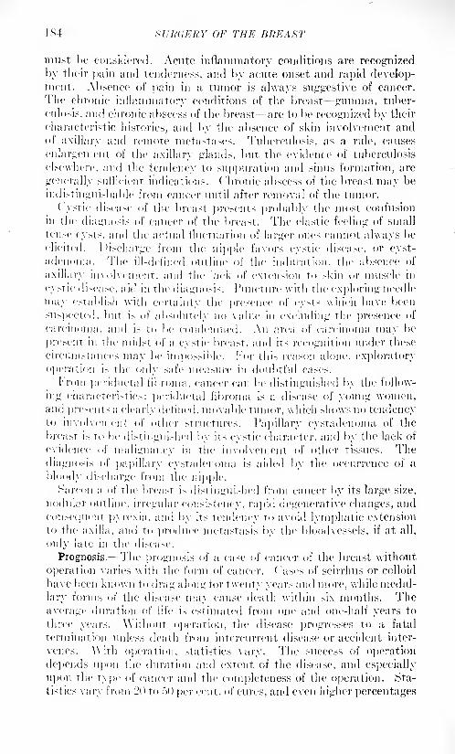
1M SURGERY OF THE BREAST
must be considered. Acute inflammatory conditions are recognized
by their pain and tenderness, and by acute onset and rapid develop-ment. Absence of pain in a tumor is always suggestive of cancer.
The chronic inflammatory conditions of the breast gumma, tuber-
culosis, and chronic abscess of the breast -are to be recognized by their
characteristic histories, and by the absence of skin involvement andof axillary and remote metastases. Tuberculosis, as a rule, causes
enlargement of the axillary glands, but the evidence of tuberculosis
elsewhere, and the tendency to suppuration and sinus formation, are
generally sufficient indications. Chronic abscess of the breast may be
indistinguishable from cancer until after removal of the tumor.( ystic disease of the breast presents probably the most confusion
in the diagnosis of cancer of the breast. The elastic feeling of small
tense cysts, and the actual fluctuation of larger ones cannot always be
elicited. Pischarge from the nipple favors cystic disease, or cyst-adenoma. The ill-defined outline of the induration, the absence of
axillary involvement, and the lack of extension to skin or muscle in
cystic disease, aid in the diagnosis. Puncture with the exploring needle
may establish with certainty the presence of cysts which have been
suspected, but is of absolutely no value in excluding the presence of
carcinoma, and is to be condemned. An area of carcinoma may be
present in the midst of a cystic breast, and its recognition under these
circumstances may be impossible. For this reason alone, exploratory
operation is the only safe measure in doubtful cases.
From periductal fibroma, cancer can be distinguished by the follow-
ing characteristics: periductal fibroma is a disease of voting women,and present- a clearly defined, movable tumor, which shows no tendencyto involvement of other structures. Papillary cystadenoma of the
breast is to be distinguished by its cystic character, and by the lack of
evidence of malignancy in the involvement of other tissties. The
diagnosis of papillary cystadenoma is aided by the occurrence of a
bloody discharge from the nipple.Sarcoma of the breast is distinguished from cancer by its large size,
nodular outline, irregular consistency, rapid degenerative changes, and
consequent pyrexia, and by its tendency to avoid lymphatic extension
to the axilla, and to produce metastasis by the bloodvessels, if at all,
only late in the disease.
Prognosis. The prognosis of a case of cancer of the breast without
operation varies with the form of cancer. Cases of scirrhus or colloid
have been known to drag along for twenty years and more, while medul-
lary forms of the disease may cause death within six months. The
average duration of life is estimated from one and one-half years to
three years. Without operation, the disea.se progresses to a fatal
termination unless death from iiitercurrent disease or accident inter-
venes. \Yith operation, statistics vary. The success of operation
depends upon the duration and extent of the disease, and especially
upon the type of cancer and the completeness of the operation. Sta-
tistics vary from 20 to f>0 per cent, of cures, and even higher percentages
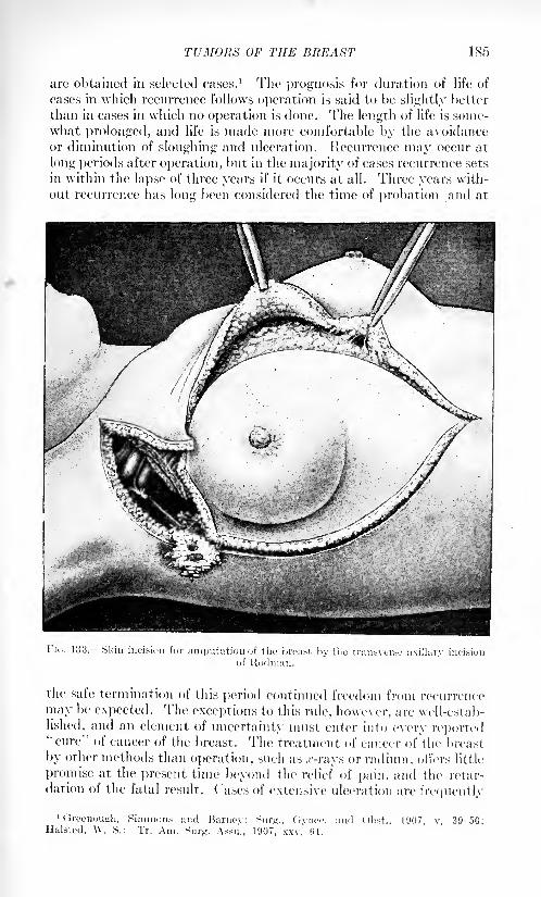
TUMORS OF THE BREAST 185
are obtained in selected cases. 1 The prognosis for duration of life of
cases in which recurrence follows operation is said to he slightly better
than in cases in which no operation is done. The length of life is some-what prolonged, and life is made more comfortable by the avoidanceor diminution of sloughing and nlceration. Recurrence may occur at
long periods after operation, but in the majority of cases recurrence sets
in within the lapse of three years if it occurs at all. Three years with-
out recurrence has long been considered the time of probation and at
I" ic.. 133. Skin incision for amputation of the breast by the transverse axillary incision
of Rodman.
the safe termination of this period continued freedom from recurrence
may be expected. The exceptions to this rule, however, are well-estab-
lished, and an element of uncertainty must enter into every reported"cure" of cancer of the breast. The treatment of cancer of the breast
by other methods than operation, such as .r-rays or radium, oilers little
promise at the present time beyond the relief of pain, and the retar-
dation of the fatal result. Cases of extensive ulceration are frequently
1 Greenough, Simmons and Barney: Surn., Gynce. ami Obst., 1907, v, 39-50;Halsted, W. S.: Tr. Am. Sni^. Assn., 1907, xxv, (>1.
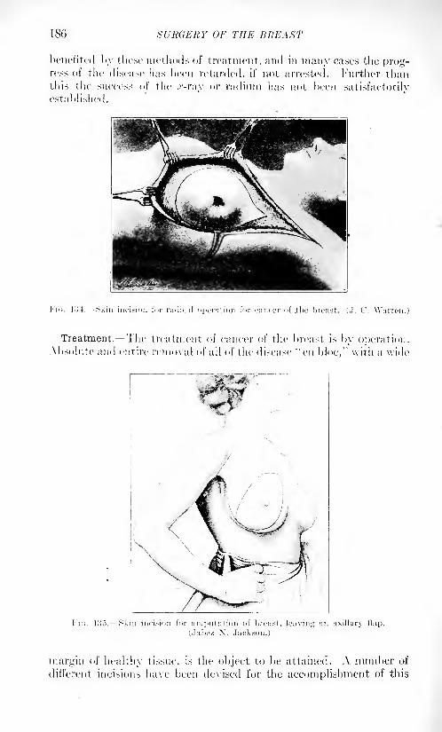
186 SURGERY OF THE BREAST
benefited l>y these methods of treatment, and in many cases the prog-ress of the disease has Keen retarded, if not arrested. Further thanthis the success of the .r-ray or radium has not been satisfactorilyestablished.
Treatment.r
lhe treatment of cancer of the breast is by operation.Absolute and entire removal of all of the disease "en bloc," with a wide
FIG. 135. Skin incision for amputation of breast, leaving an axillary flap.
margin of healthy tissue, is the object to be attained. A number of
different incisions have been devised for the accomplishment of this
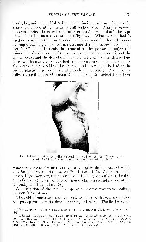
TUMORS OF THE BREAST 187
result, beginning with Ilalsted's 1
curving incision in front of the axilla,
a method of operating which is still widely used. Many surgeons,
however, prefer the so-called "transverse axillary incision," the typeof which is Rodman's operation.
2(Fig. loo). Whatever method is
used one consideration must remain supreme, namely, that all tumor-
bearing tissue be given a wide margin, and that the tissues be removed
"en bloc.'' This demands the removal of the pectoralis major and
minor, and the dissection of the axilla, as well as the amputation of the
whole breast and the deep fascia of the chest wall. When this is done
there will be many cases in which a sufficient amount of skin to close
the wound entirely will not be present, and resort must be had to the
use of plastic flaps, or skin graft, to close the defect. A number of
different methods of obtaining flaps to close the defect have been
Fi(i. 130. Scar left after radical operation; cloned by flap and Thiersch <rratt.
(Method of J. C. Warren, Massachusetts General Hospital.)
suggested, no one of which is universally applicable but each of which
may be effective in certain cases (Figs, lo 1 and loo). Where the defect
is very large, however, the closure by Thiersch graft, either at the first
operation, or at the end of two to three weeks as a secondary operation,is usually employed (Fig. 13(>)-
A description of the standard operation by the transverse axillary
incision is as follows:
The field of operation is shaved and scrubbed with soap and water,
and put up with a sterile dressing the night before. The field covers a
1
Halsted, W. S.: Ann. Sur^., November, 1X1)4; .lour. Am. Med. Assn., February S,
1913.2 Rodman: Diseases of the Breast, 1908, Phila. Warren: Jour. Am. Med. Assn.,
1905, xiv, 160, also Inter. Text-book of Sur^., 1900, ii, chapter viii. Meyer: Jour. Am.Med. Assn., July 29, 1905. Jackson, J. N.: Jour. Am. Med. Assn., March 3, 19()C>, and
1910, liv, 178-183. Stewart, F. T.: Ann. Surg., 1915, Ixii, 250.
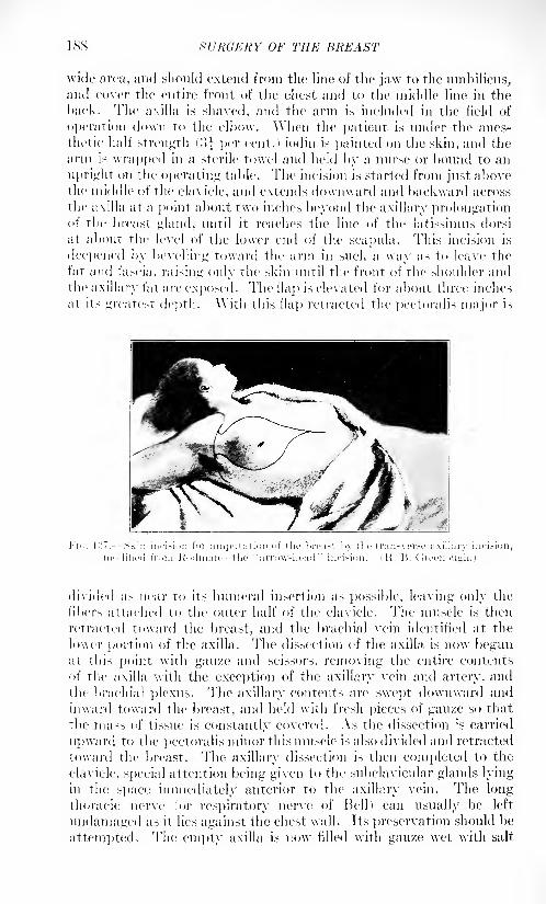
1SS SURGERY OF THE BREAST
wide area, and should extend from the line of the jaw to the umbilicus,and cover the entire front of the chest and to the middle line in the
hack. The axilla is shaved, and the arm is included in the field of
operation down to the elbow. When the patient is under the anes-
thetic half strength (',]} per cent.) iodin is painted on the skin, and the
arm is wrapped in a sterile towel and held by a nurse or bound to an
upright on the operating table. The incision is started from just abovethe middle' of the clavicle, and extends downward and backward across
the axilla at a point about two inches beyond the axillary prolongationof the breast gland, until it reaches the line of the latissimus dorsi
at about the level of the lower end of the scapula. This incision is
deepened by bevelling toward the arm in such a way as to leave the
fat and fascia, raising only the skin until the front of the shoulder andthe axillary fat arc exposed. The flap is elevated for about three inches
at its greatest depth. With this flap retracted the pectoralis major is
divided as near to its humeral insertion as possible, leaving only the
libers attached to the outer half of the clavicle. The muscle is then
retracted toward the breast, and the brachial vein identified at the
lower portion of the axilla. The dissection of the axilla is now begunat this point with gauze and scissors, removing the entire contents
of the axilla with the exception of the axillary vein and artery, andthe brachial plexus. The axillary contents are swept downward and
inward toward the breast, and held with fresh pieces of gauze so that
the mass of tissue is constantly covered. As the dissection is carried
upward to the pectoralis minor this muscle is also divided and retracted
toward the breast. The axillary dissection is then completed to the
clavicle, special attention being given to the subclavicular glands lyingin the space immediately anterior to the axillary vein. The longthoracic nerve for respiratory nerve of Hell) can usually be left
undamaged as it lies against the chest wall. Its preservation should be
attempted. The empty axilla is now filled with gauze wet with salt
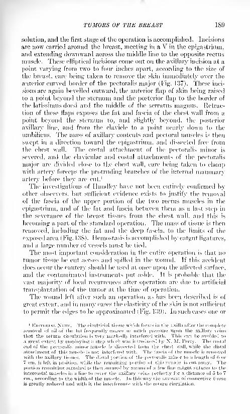
TUMORS OF THE BREAST 189
solution, and the first stage of the operation is accomplished. Incisions
are now carried around the breast, meeting in a V in the epigastrium,and extending downward across the middle line to the opposite rectus
muscle. These elliptical incisions come out on the axillary incision at a
point varying from two to four inches apart, according to the si/e of
the breast, care being taken to remove the skin immediately over the
anterior curved border of the pectoralis major (Fig. 137). These inci-
sions are again bevelled outward, the anterior flap of skin being raised
to a point beyond the sternum and the posterior flap to the border of
the latissimus clorsi and the middle of the serratus magnus. Retrac-
tion of these flaps exposes the fat and fascia of the chest wall from a
point beyond the sternum to, and slightly beyond, the posterior
axillary line, and from the clavicle to a point nearly down to the
umbilicus. The mass of axillary contents and pectoral muscles is then
swept in a direction toward the epigastrium, and dissected free fromthe chest wall. The costal attachment of the pectoralis minor is
severed, and the clavicular and costal attachments of the pectoralis
major are divided close to the chest wall, care being taken to clampwith artery forceps the protruding branches of the internal mammaryartery before they are cut. 1
The investigations of Handler have not been entirely confirmed byother observers, but sufficient evidence exists to justify the removalof the fascia of the upper portion of the two rectus muscles in the
epigastrium, and of the fat and fascia between them as a last step in
the severance of the breast tissues from the chest wall, and this is
becoming a part of the standard operation. The mass of tissue is then
removed, including the fat and the deep fascia, to the limits of the
exposed area (Fig. K!S). Ilemostasis is accomplished by catgut ligatures,
and a large number of vessels must be tied.
The most important consideration in the entire operation is that no
tumor tissue be cut across and spilled in the wound. If this accident
does occur the cautery should be used at once upon the affected surface,
and the contaminated instruments put aside. It is probable that the
vast majority of local recurrences after operation are due to artificial
transplantation of the tumor at the time of operation.The wound left after such an operation as has been described is of
great extent, and in many cases the elasticity of the skin is not sufficient
to permit the edges to be approximated (Fig. U>9). In such cases one or
1 EDITORIAL XOTK. The cicatrieial tissue which forms in the axilla after the completeremoval of all of the tat frequently causes so much pressure upon the axillary veins
that the return circulation is very markedly interfered with. This can be avoided to
a great extent by employing a .step which was introduced by N. M. Percy. The costal
end of the pectoralis minor muscle is dissected from the chest wall, while the distal
attachment of this muscle is not interfered with. The fascia of the muscle is removedwith the axillary tissues. The distal portion of the pectoralis minor to a length of (i or
7 cm. is left in position, while the remaining portion of tin's muscle is cut away. Theportion remaining attached is then sutured by means of a few fine catgut sutures to the
intercostal muscles in a line to cover the axillary veins perfectly for a distance of 5 to 7
cm., according to the width of the muscle. In this way the amount of connective tissue
is greatly reduced and with it the interference with the return circulation.
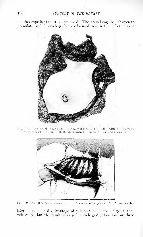
SURGERY OF THE HREAST
another expedient must be employed. The wound may be left open to
granulate, and Thiersch grafts mav be used to close the defect at some
i'i<,. |:iv I'.reast and accessnry tissues ivmovod in radical operation with the trun.sver.-u" arrow-head
"incision. <}'. l'>. (Irecuouuli, Massachu.sott.s General Hospital.)
later date. The disadvantage of this method is the delay in con-
valescence, but the result after a Thiersch graft, done two or three
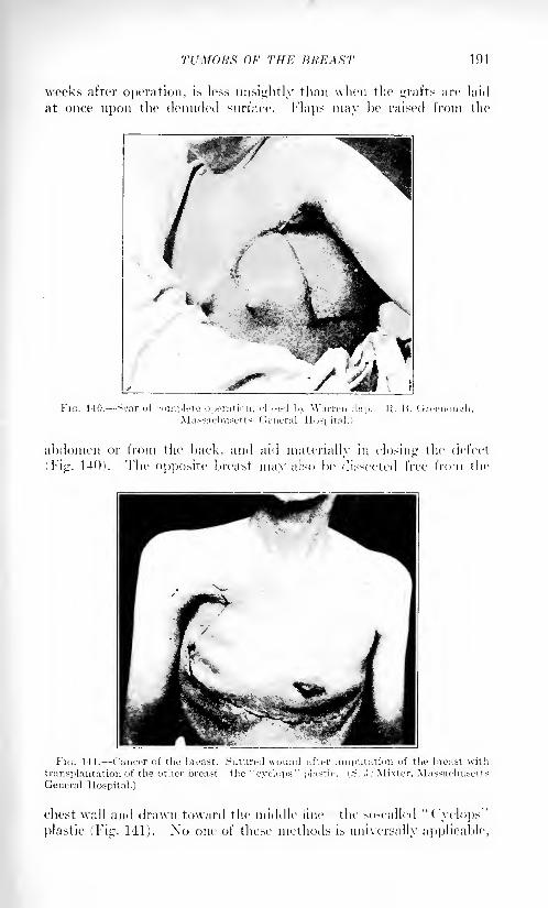
TUMORS OF THE BREAST 191
weeks after operation, is less unsightly than when the grafts are laid
at once upon the denuded surface. Flaps may be raised from the
abdomen or from the hack, and aid materially in closing the defect
i Fig. 140). The opposite breast may also be dissected free from the
FIG. 141. Cancer of the breast. Sutured wound after amputation oi the breast with
transplantation of the other breast the "cyclops" plastic. i,.S. .1." Mixter, MassachusettsGeneral Hospital.)
chest wall and drawn toward the middle line the so-called "Cyclops"
plastic (Fig. 141). Xo one of these methods is universally applicable,
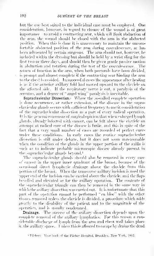
192 SURGERY OF THE BREAST
but the our best suited to the individual case must be employed. Oneconsideration, however, in regard to closure of the wound is of great
importance- to avoid a contracting scar, which will limit abduction of
the arm, the wound should be closed with the arm in the abducted
position. \\hen this is done it is unnecessary to maintain the uncom-fortable abducted position of the arm during convalescence, as has
been advocated by certain surgeons. The arm should not, however, be
included within the dressing but should be held by a wrist sling for the
first two or three days, and should then be given gentle passive motion
in abduction and rotation during the rest of the convalescence. Thereturn of function in the arm, when both pectorals have been divided,
is prompt and almost complete if the contracting scar binding the armto the chest is avoided. In successful cases the appearance after healingis as if the anterior axillary fold had moved upward to the clavicle on
the atl'ected side. If the respiratory nerve is cut, a paralysis of the
serratus. and a degree of "angel wing" paralysis is inevitable.
Supraclavicular Dissection. Where the so-called complete operationi> done recurrence, or rather extension, of the disease in the supra-clavicular glands occurs with sufficient frequency to merit consideration
of the supraclavicular dissection as a part of every radical operation.It is the general consensus of surgical opinion that when enlarged lymphglands, already infected with cancer, can be felt above the clavicle an
attempt at radical cure of the disease is futile, and this in spite of the
fact that a very small number of cases are recorded of perfect cures
under these conditions. In early cases the routine supraclaviculardissection is still under debate, but it does not seem unreasonable
when the condition of the glands in the upper portion of the axilla is
such as to indicate probable microscopic disease already present in
the supraclavicular glands beyond.1
The supraclavicular glands should also be removed in every case
of cancer in the upper inner quadrant of the breast, because of the
occasional direct lymphatic drainage above the clavicle from this
portion of the breast. When the transverse axillary incision is used the
upper end of the incision can be carried above the clavicle, and the flaps
bevelled and elevated as for the axillary operation. The contents of
the supraclavicular triangle can then be removed in the same way in
which the axillary dissection was carried out. It is unfortunate that this
part of the operation cannot be performed "en bloc" with the other
tissues removed unless the clavicle is divided, a procedure which adds
greatly to the disability of the patient and to the magnitude of the
operation, and is usually condemned.
Drainage. The success of the axillary dissection depends upon the
complete removal of the axillary lymphatics. For this reason a con-
siderable discharge of lymph from the arm and chest wall takes placein the axillary space. I "nless this is allowed to escape by drains the dead
iPilcher: Year-book of tho Pileher Hospital, Brooklyn, New York, 1912.
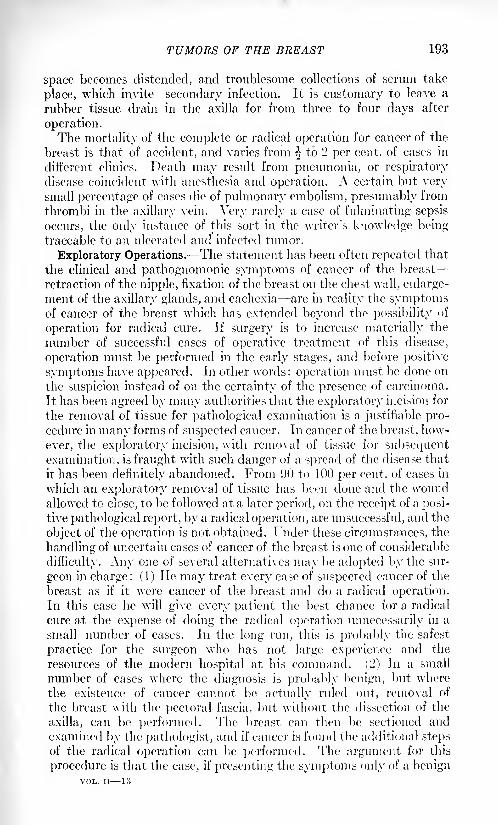
TUMORS OF THE BREAST 193
space becomes distended, and troublesome collections of serum take
place, which invite secondary infection. It is customary to leave a
rubber tissue drain in the axilla for from three to four days after
operation.The mortality of the complete or radical operation for cancer of the
breast is that of accident, and varies from ^ to 2 per cent, of cases in
different clinics. Death may result from pneumonia, or respiratory
disease coincident with anesthesia and operation. A certain but verysmall percentage of cases die of pulmonary embolism, presumably from
thrombi in the axillary vein. Very rarely a case of fulminating sepsis
occurs, the only instance of this sort in the writer's knowledge beingtraceable to an ulcerated and infected tumor.
Exploratory Operations.- The statement has been often repeated that
the clinical and pathognomonic symptoms of cancer of the breast
retraction of the nipple, fixation of the breast on the chest wall, enlarge-
ment of the axillary glands, and cachexia are in reality the symptomsof cancer of the breast which has extended beyond the possibility of
operation for radical cure. If surgery is to increase materially the
number of successful cases of operative treatment of this disease,
operation must be performed in the early stages, and before positive
symptoms have appeared. In other words: operation must be done on
the suspicion instead of on the certainty of the presence of carcinoma.
It has been agreed by many authorities that the exploratory incision for
the removal of tissue for pathological examination is a justifiable pro-cedure in many forms of suspected cancer. In cancer of the breast, how-
ever, the exploratory incision, with removal of tissue for subsequentexamination, is fraught with such danger of a spread of the disease that
it has been definitely abandoned. From 90 to 100 per cent, of cases in
which an exploratory removal of tissue has been done and the woundallowed to close, to be followed at a later period, on the receipt of a posi-
tive pathological report, by a radical operation, are unsuccessful, and the
object of the operation is not obtained. Under these circumstances, the
handling of uncertain cases of cancer of the breast is one of considerable
difficulty. Any one of several alternatives may be adopted by the sur-
geon in charge : (1) He may treat every case of suspected cancer of the
breast as if it were cancer of the breast and do a radical operation.In this case he will give every patient the best chance for a radical
cure at the expense of doing the radical operation unnecessarily in a
small number of cases. In the long run, this is probably the safest
practice for the surgeon who has not large experience and the
resources of the modern hospital at his command. (2) In a small
number of cases where the diagnosis is probably benign, but where
the existence of cancer cannot be actually ruled out, removal of
the breast with the pectoral fascia, but without the dissection of the
axilla, can be performed. The breast can then be sectioned and
examined by the pathologist, and if cancer is found the additional stepsof the radical operation can be performed. The argument for this
procedure is that the case, if presenting the symptoms only of a benignVOL. II 13
194 XURdERY OF THE BREAST
tumor, is not likely to be such an advanced case of carcinoma as to
have extended already beyond the limits of the gland itself, and the
amputation of the breast is likely to have removed the whole of the
diseased area. This is a method of procedure which will be restricted
to surgeons who have had considerable experience in diagnosis of
diseases of the breast and involves some risk to the patient's chancesin any case. (3) Exploratory incision followed at the same sitting
by the necessary operation: This method is practiced in many clinics
by surgeons who have had long training in gross pathology, and
apparently with very little danger to the individual patient. Incision
is carried down directly into the tumor, and its cut surface is inspected.If necessary, fragments for frozen sections can be excised. The woundis then cauterized, or packed with formalin gauze, and the operationindicated by the pathological condition is immediately performed.This method, also, will of necessity be restricted to surgeons who have
thoroughly trained themselves in gross and microscopic pathology.(4) An expedient is offered by the electrocautery by which nodules of
suspicious character can safely be excised without danger of dissemina-
tion. The writer has made use of this method in a number of cases,
and it is very satisfactory. The cautery is used to incise or excise
the suspicious tissue, and an immediate diagnosis is obtained if neces-
sary by frozen section. If it prove malignant, the radical operationis done at once. If non-malignant, the cauterized tissues can be
excised with the knife and the wound closed with expectation of heal-
ing by first intention. Of the four methods suggested for dealing with
doubtful but suspicious cases of cancer of the breast, the cautery
exploratory is, in the opinion of the writer, the safest and most
satisfactory for general use.
Palliative Operation. Palliative operations in cancer of the breast
are occasionally indicated in patients where no hope of a radical cure
can be entertained. In certain cases the so-called radical operationcan be done in apparently hopeless cases in order that the patient maybe given every possible chance. Such a procedure affects unfavorablythe statistics of the individual operator, but very rarely an unexpectedlysuccessful result may be obtained. As a rule, the only cases requiring
a definitely palliative operation are those in which a large ulcerating
tumor gives so much discomfort to the patient and her attendants that
its removal is indicated as a purely temporary relief. In such cases the
operation performed with the cautery has many advantages, and the
affected mass of tumor tissue can be removed by this method without
producing a rapid spread of the disease, which would be likely to follow
the opening up of fresh spaces to infection if the operation were done
with the knife. The combination of the cautery operation and subse-
quent .T-ray treatment has yielded most satisfactory results as a pallia-
tive measure in main' advanced cases. Under palliative operation
may be mentioned the rare but occasional case in which an ampu-tation of the upper extremity is indicated, either for gangrene or
for the pain and disability of the "brawny arm," due to axillary
TUMORS OF THE BREAST 195
obstruction of the veins and lymphatics, and pressure on the brachial
plexus.Recurrence after operation takes place in from 50 to 70, and even SO
per cent, of cases. Recurrences can ordinarily be distinguished as of
three different classes: Local, regional and remote.
Local recurrences take place in the scar, or at least within the
boundaries of the area exposed at the primary operation. They are
due either to an incomplete removal of the diseased tissue or to arti-
ficial operative implantation at the time of the operation, produced byaccidental or unavoidable incision into tumor tissue in the wound. Alarge proportion of recurrences are of this character, and they usuallymake their appearance within the three-year period, and even in the
first year after operation, although instances of local recurrence are
recorded many years after the primary operation. \Vith improvedtechnic and wider operations, the proportion of local recurrences has
been somewhat diminished.
Regional recurrence takes place in the lymph nodes adjacent to, but
bevond the area of the primary operation. Recurrences of this char-
acter must be atttributed to infection of the lymph nodes prior to or
at the time of the operation, but of such microscopic si/e that no
enlargement of the lymph nodes could be detected. Recurrences of
this nature are not at all uncommon in the supraclavicular glands after
radical operations in which the upper axilla is badly involved. The
experiments of Tyzzer on mouse tumors have established beyond dis-
pute that metastases can be produced in animals by rough handlingand squeezing of the primary tumor. Under these conditions regionaland remote metastases, after operation, in human beings may occasion-
ally be attributed to rough handling of the tumor before and dur-
ing the operation. At any rate, the lesson is sufficiently obvious
that the utmost gentleness of manipulation should be employed both
in the examination of the tumor for diagnosis, and during its removal.
Lor this reason, also, the axillary incision with the division of the
axillary lymphatics, as the first step in the operation, has distinct
advantages.Remote metastases in bones, mediastinal glands, the liver and lung.
occur late after operation, and provide some of the greatest disappoint-ments in the surgery of breast cancer. It is probable that some of the
more remote metastases are due, like some of the regional metastases,to manipulation before and during the radical operation. The greater
number, however, are probably already established, although of micro-
scopic si/e, at the time of the primary operation. The internal metas-
tases of carcinoma can be detected only after a considerable period of
growth, as they produce symptoms only by their interference with
normal function, and a considerable bulk of tumor tissue must be
present in order that function may be materially ati'ected. Lven the
,?'-ray has been a disappointment in the detection of the early stagesof internal metastases. A period of years is sometimes needed with a
slow growing tumor before a sufficient mass of tumor tissue is produced
196 SURGERY OF THE BREAST
to make its presence evident, and yet these remote metastases producesome of the most distressing deaths from cancer of the breast.
The treatment of recurrent cases of cancer of the breast, and of cases
too far advanced for even a palliative operation, is very largely symp-tomatic. Radium is apparently capable of destroying superficialnodules of cancer which are readily accessible, but has little effect onthe deeper manifestations of the disease, as used at the present time.
The use of strong filtered .r-ray treatment, given with the Coolidgetube, appears to hold in check the deeper development of the disease
more effectively than any other treatment. By the .T-ray treatment
sloughing and offensive discharges are diminished, and the patient's
general condition, physical as well as mental, is improved. Theultimate results, however, are not materially affected, and the patient
goes on to the fatal termination the direct cause of death being
dependent upon which vital function is most seriously interfered with
by the internal manifestations of the disease. Finally, in the later
stages of the disease resort to opiates usually becomes necessary, and,
indeed, there would seem to be few cases more entitled to the relief of
opium than those in the terminal stages of recurrent or inoperablecancer of the breast.
SURGERY OF THE MALE BREAST.
Thi' male breast is structurally of the same constitution as that of
the female, and is subject to the same diseases and new growths. Thesmall size and imperfect development, however, of the male breast
render it much less liable than that of the female to pathologicalconditions.
Malformations of the male breast are of the same nature as in the
female. Ama/ia is equally frequent in both sexes, and is generally
accompanied by defects in development of the chest wall. Micro-
ma/ia, or incomplete development of the breast, may be considered the
normal condition in the male. Supernumerary breasts probably occur
in the male as often as in the female, but owing to the lack of glandular
development their true nature is often not suspected. Gynecomaziahas already been discussed under the diseases of the female breast.
Injuries of the breast or in the mammary region are more commonin the male than in the female. Contusions are especially frequent,hematoma is not unusual, and abscess may result.
I Hseases of the skin of the breast and of the nipple and areola occur
as in the female, and do not require special consideration. Diffuse
hypertrophy of the male breast, such as occurs in the female, is exces-
sively rare if it ever occurs at all. Enlargement of the male breast is
more likely to be due to a new growth. The acute mastitis of infancyand that occurring at the time of puberty are equally common in the
male and in the female. Abscesses are unusual but may occur as a
result of such infections. Abscess due to extension from necrosis of the
ribs, or from empyema, affects both sexes equally, but at best is a rare
condition.
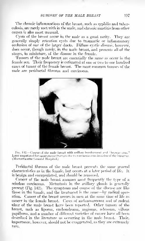
SURGERY OF THE MALE BREAST 197
The chronic inflammations of the breast, such as syphilis and tuber-
culosis, are rarely met with in the male, and chronic mastitis from other
causes is also most unusual.
Cysts of the breast occur in the male as a great rarity. They arc
generally simply retention cysts due to traumatic or inflammatoryocclusion of one of the larger ducts. Diffuse cystic disease, however,does occur, though rarely, in the male breast, and presents all of the
stages, in miniature, of the disease in the female.
Tumors of the male breast are essentially the same as occur in the
female sex. Their frequency is estimated at one or two to one hundredcases of tumor of the female breast. The most common tumors of the
male are periductal fibroma and carcinoma.
;ous fracture due to carcinomatouj_iiiuui aiiiyuiuieu lui spuIlUilHJUUS n
(Massachusetts General Hospital.)
Periductal fibroma of the male breast presents the same generalcharacteristics as in the female, but occurs at a later period of life. It
is benign and encapsulated, and should be removed.
Cancer of the male breast assumes most frequently the type of a
scirrhus carcinoma. Metastasis in the axillary glands is generally
present (Fig. 142). The symptoms and course of the disease are like
those in the female, and the treatment is the same by radical oper-ation. Cancer of the breast occurs in men at the same time of life as
cancer in the female breast. Cases of melanosarcoma and of rodent
ulcer of the male breast have been reported. Other tumors of the
breast, such as lipoma, enchondroma, angioma, adenoma, villous
papilloma, and a number of different varieties of cancer have all been
described in the literature as occurring in the male breast. Their,
importance, however, should not be exaggerated, as the}' are extremelyrare.

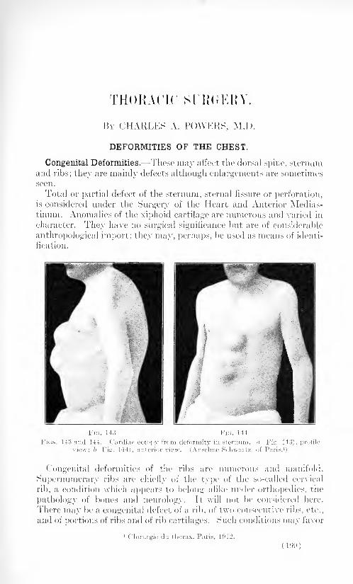
r rrHOBACIC St RiJKHY.
BY CHARLES A. POWERS, M.I).
DEFORMITIES OF THE CHEST.
Congenital Deformities. These may affect the dorsal spine, sternumand ribs; they are mainly defects although enlargements are sometimesseen.
Total or partial defect of the sternum, sternal fissure or perforation,is considered under the Surgery of the Heart and Anterior Medias-tinum. Anomalies of the xiphoid cartilage are numerous and varied in
character. They have no surgical significance but are of considerable
anthropological import; they may, perhaps, be used as means of identi-
fication.
143 and 144. Cardiac cctopy from deformity in sternum.
(Anselme Schwartz, of Paris. 1
)
FIG
Congenital deformities of the ribs are numerous and manifold.
Supernumerary ribs are chiefly of the type of the so-called cervical
rib, a condition which appears to belong alike under orthopedics, the
pathology of bones and neurology. It will not be considered here.
There may be a congenital defect of a rib, of two consecutive ribs, etc.,
and of portions of ribs and of rib cartilages. Such conditions may favor
1 Chirurgie du thorax, Paris, 1912.
(199)
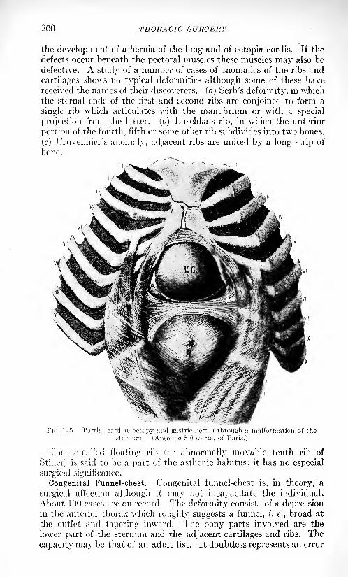
200 THORACIC SURGERY
the development of a hernia of the lung and of ectopia cordis. If the
defects occur beneath the pectoral muscles these muscles may also be
defective. A study of a number of cases of anomalies of the ribs and
cartilages shows no typical deformities although some of these havereceived the names of their discoverers, (a) Serb's deformity, in whichthe sternal ends of the first and second ribs are conjoined to form a
single rib which articulates with the manubrium or with a special
projection from the latter. (6) Luschka's rib, in which the anterior
portion of the fourth, fifth or some other rib subdivides into two bones.
(c) Cruveilhier's anomaly, adjacent ribs are united by a long strip of
bone.
FIG. 14.". Partial cardiac octopv and gastric hernia through a malformation of the
(Anselme Schwartz, of Pans.)
The so-called floating rib (or abnormally movable tenth rib of
Stiller) is said to be a part of the asthenic habitus; it has no especial
surgical significance.
Congenital Funnel-chest. Congenital funnel-chest is, in theory, a
surgical affection although it may not incapacitate the individual.
About 100 eases are on record. The deformity consists of a depressionin the anterior thorax which roughly suggests a funnel, i. e., broad at
the outlet and tapering inward. The bony parts involved are the
lower part of the sternum and the adjacent cartilages and ribs. The
capacity may be that of an adult fist. It doubtless represents an error
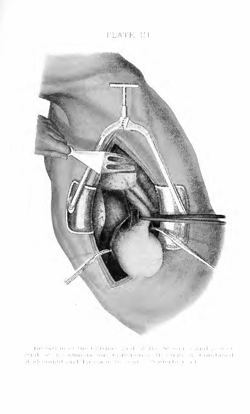
PLATE II!

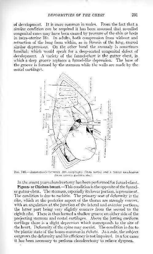
DEFORMITIES OF THE CHEST 201
of development. It is more common in males. From the fact that a
similar condition can be acquired it has been assumed that so-called
congenital cases may have been caused by pressure of the chin or heels
in intra-uterine life. In adults, both compression from without and
retraction of the lung from within, as in fibrosis of the lung, caused
similar depressions. On the other hand the anomaly is sometimes
familial; which would speak for a deep-seated congenital defect of
development. A variety of the funnel-chest is the gutter chest, in
which a deep groove replaces a funnel-like depression. The base of
the groove is formed by the sternum while the walls are made by the
costal cartilages.
FIG. 146. Anastomosis Between Art. esophagea (from aorta) and a ramus esopbageus(from arteria gastrica sin.)
Ill the recent years chondrectomy has been performed for funnel-chest.
Pigeon- or Chicken-breast. This condition is the opposite of the funnel-
or gutter-chest. The sternum, especially its lower portion, is prominent.The condition is due to rachitis. The primary seat of deformity is the
ribs, which at the posterior aspect of the thorax are strongly convex,with an angulation at the junction of the lateral and anterior portions,the latter part being very slightly concave from the second to the
eighth ribs. There is thus formed a shallow groove on either side of the
projecting sternum and costal cartilages. Above the jutting ensiform
cartilage there is a slight depression which corresponds to the site of
the heart. ] )eformity of the spine may coexist. The condition is due to
the plastic state of the bones common in rickets. As a rule, the subject
outgrows the deformity and his efficiency is not impaired. In a few cases
it has been necessary to perform chondrectomy to relieve dyspnea.
202 THORACIC SURGERY
Congenital Thoracic Rigidity and Emphysema. Simple mention is
made of the fact that congenital rigidity of the costal cartilages due to
premature ossification may, rarely, be encountered.
Congenital Narrowing of the Superior Thoracic Aperture. This condi-
tion, and its surgical management are discussed under Tuberculosis.
THORACIC DEFORMITIES FROM DISEASE OF THE VERTEBRA.
These are secondary deformities in some of which surgical aid has
recently been invoked. In scoliosis, kvphosis and spondylosis rhizo-
melique, the rigidity of the thorax may at times be overcome bychondrectomy or costectomy in order that the lungs and heart maybetter perform their functions.
Scoliosis. Scoliosis or lateral curvature of the spine may be con-
genital or acquired, although it is now believed that much of the
so-called acquired scoliosis is actually founded on a latent congenital
condition. The causes are numerous and varied. Jn addition to the
neuromuscular forms a large number of cases are secondary to somesuch unilateral deformity as wry-neck, Sprengel's deformity of the
scapula, sacro-iliac disease, etc. In other cases general anomalies of
the skeleton exist; rickets, acromegaly; or a spinal anomaly, Pott's
disease. Jn turn, scoliosis causes other deformities, and in marked
degrees the thorax is badly deformed even more so than in severe
cases of kyphosis. The vertebral curve is not a simple one but in
general lias an S-shape. The upper section, or dorsal curve, is usuallyconvex to the right, while the lumbar curve is convex to the left. In
addition there is a distinct rotation imparted to the spine. The dorsal
curve, convex to the right, causes the ribs to bulge somewhat, and as
thN renders the scapula more prominent a boss or hump results. This
is enhanced by a corresponding depression in the opposite scapular
region. Anteriorly, on the contrary, the chest is more flattened on
the right side and more convex on the left. The average patient with
this deformity suffers much as a result of the displacement of the
thoracic viscera. These are compressed against the abdomen; the
heart is crowded to the left; both lungs, especially the right, are
compressed.Kyphosis. This deformity, which is a bulging backward of the
dorsal vertebra 1
, consists of two types known respectively as the round
and angular. Angular kyphosis is another term for Pott's disease of
the spine. When seated in the cervical or upper dorsal region the
deformity is not apt to be excessive. In the mid-dorsal region kyphosiscauses a deformity of the entire thorax; the ribs may be so depressedthat, they reach the ilium. The chest becomes shortened, as in severe
scoliosis, flattened laterally and more prominent in front. It becomes
very inelastic so that abdominal breathing is necessitated. Specialforms of kyphosis which tend to immobilize the chest are the so-called
heredo-traumatic form or von Bechterew's disease and the progressive
kyphosis following fractures ( Yerneuil-Kummel disease). The round
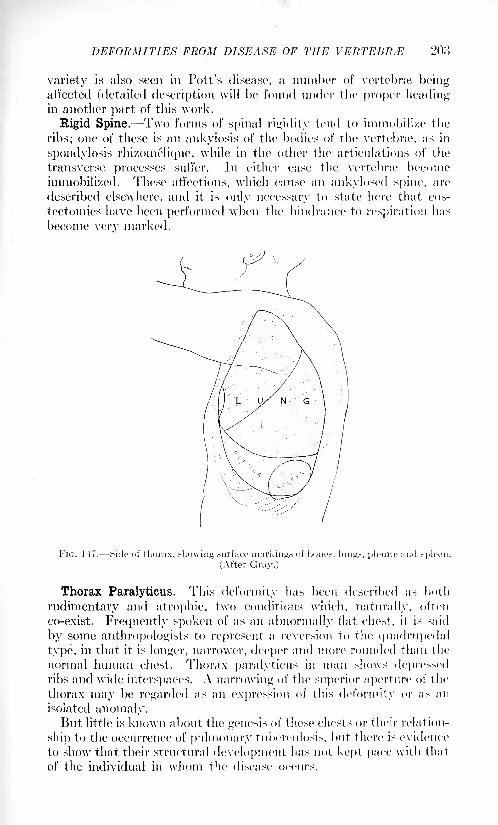
DEFORMITIES FROM DISEASE OF THE VERTEHR/E 203
variety is also seen in Pott's disease, a number of vertebrae beingaffected (detailed description will be found under the proper headingin another part of this work.
Rigid Spine. Two forms of spinal rigidity tend to immobili/e the
ribs; one of these is an ankylosis of the bodies of the vertebra?, as in
spondylosis rhizomelique, while in the other the articulations of the
transverse processes suffer. In either case the vertebme becomeimmobilized. These affections, which cause an ankylosed spine, are
described elsewhere, and it is only necessary to state here that eos-
tectomies have been performed when the hindrance to respiration has
become very marked.
FIG. 147. Side of thorax, showing surface markings of bones, lungn, pleura:! and spleen.
(After Gray.)
Thorax Paralyticus. This deformity has been described as both
rudimentary and atrophic, two conditions which, naturally, often
co-exist. Frequently spoken of as an abnormally flat chest, it is said
by some anthropologists to represent a reversion to the quadrupedal
type, in that it is longer, narrower, deeper and more rounded than the
normal human chest. Thorax paralyticus in man shows depressedribs and wide interspaces. A narrowing of the superior aperture of the
thorax may be regarded as an expression of this deformity or ;is an
isolated anomaly.But little is known about the genesis of these chests or their relation-
ship to the occurrence of pulmonary tuberculosis, but there is evidence
to show that their structural development has not kept pace with that
of the individual in whom the disease occurs.
204 THORACIC SURGERY
The surgery of thorax paralyticus is that of pulmonary tuberculosis
after the disease has developed.Thoracic Contraction (Pleuritic Scar Contraction).When a very
large pleuritic exudate is absorbed without full expansion of the lung,the negative pressure in the chest causes the thorax to be displacedtoward the lung. A somewhat similar condition occurs in an old
empyema after an operative cure. Recovery is accompanied by a
pronounced contraction of the thickened pleura and the adherent
lung.
The symptoms vary; as a rule, the affected side of the thorax looks
smaller and is drawn inward. Measurements show a difference bet\veen
the two sides. The ribs touch each other and may overlap. The spinal
column may show various types of curvature. Compensatory curva-
tures are present as a rule. On the affected side the various land-
marks shoulder, nipple, last rib seem lowered. The head may lean
toward the affected side. Without careful history, the condition maybe confused with a rhachitis of the thorax.
When it occurs in a young subject, the condition may be outgrownin time. In older subjects, the process reaches its limits in six months,on an average.Xo rational surgical treatment seems to have been developed as yet.
GENERAL PRINCIPLES OF INTRATHORACIC SURGERY.
Introduction. Before taking up chest injuries and diseases as a
whole, it seems appropriate to cast a glance over the general field of
thoracic surgery; a field which has been broadened steadily and which
promises very wide development in the future.
Surgery of the thorax failed to advance beyond a certain limit until
definite study was made of the means of preventing the collapse of a
given lung when an intact pleural cavity was opened. The studyinvolved the equalization of pressure differences. Without this equali-
zation the surgeon feared a fatality; indeed, until the so-called pneumo-thorax danger was completely or almost completely offset, fear, on
opening the pleura, was far from imaginary.This problem of opening an intact pleura is not parallel to that
found in a given case of empyema, pleurotomy for the relief of which
has been practiced since the earliest days. Early danger resulting
from the evacuation of a pleural abscess, was chiefly one of infection
and was greatly minimized by modern asepsis together with suitable
rib resection. It is difficult to explain the occasional fatalities whenthe intact pleura is opened. Neither the size of the opening, nor the
suddenness of the inrush of air, nor the rapidity of contraction of the
given lung can, alone or together, explain the development of fatal
shock. A fatality may occur even when all conditions seem favorable.
Ordinarily, the smaller the opening the less the reaction.
An important period in the development of modem thoracic surgerywas that of the decade 1895 to 1905. The early efforts were made
GENERAL PRINCIPLES OF INTRATHORACIC SURGERY 205
toward the prevention of pneumothorax and the inflation of the
collapsed lung. Bazy,1 in 1895, introduced the finger into the wound
immediately on opening the pleura, thus occluding the wound itself
and at the same time exploring the plcural cavity. At about the same
time several surgeons introduced pneumopexy; drawing the contracted
lung to the pleural opening and suturing it to the chest wall. In 1896
Tuffier and Hallion2 discovered that the pressure difference between
the exterior and interior of the lung was about 6 mm. of mercury.Further efforts toward the solution of the pneumothorax problem had
to do with increasing the pressure difference within the lung, either byforced expiration of the closed glottis or by the insufflation of air;
compressed air being used at times (Reference should be made to the
fact that O'Dwyer's important discovery of the intubation tube wasdescribed in 1SSO.)
In 1893 Fell, of Buffalo, exhibited at the International Congress an
apparatus designed for artificial respiration in opium narcosis. It
consisted of a hand bellows, and a mask which fitted tightly over the
mouth and nostrils. O'Dwyer modified the apparatus by substitutinga laryngeal tube for the air-tight mask. Dr. Blum, of Xew Orleans,
applied the Fell-0'Dwyer apparatus for re-animation in asphyxianeonatorum. He joined to the bellows a tube and bag of oxygen.A little later Matas further modified the apparatus by substituting a
pump for the bellows, thus giving an automatic respiration. Withthis apparatus an ordinary anesthetic could be employed. So far as
may be learned, the first definite operation with the aid of such an
apparatus, was performed by Milton,8 of Cairo, in 1S97. This operation
was done for a mediastinal affection; the apparatus consisted of an
intubation tube and a hand bellows. A very important resection of
the thoracic wall for malignancy was done by Parham in 1S9S; the
modified apparatus of Matas put in use as soon as the pleura was
opened caused an expansion of the lung. In 1898, both Matas and
Parham made important contributions to the literature of this impor-tant subject; indeed, the entire procedure owes much in its develop-ment to the careful work of these Xew Orleans surgeons, to whose
names should be added those of their colleagues Souchon and Blum.
Kiittner rightly considers all early efforts which are embraced under
the head of "artificial respiration" as a necessary step in the develop-ment of pressure differences. He speaks with much commendationof the Fell-O'Dwyer apparatus and of the improvements made byMatas. A little after the appearance of the published work of Matasand Blum, Kiihn 1 advised an insufflation tube, this tube going as far
as the larynx. The important work of Friedrich, Sauerbruch and
Brauer was commenced about 1904 and has gone on steadily. Finally,
1 Asso. frun<;. de cliir., 1S9~>, ix, 711.
- Soc. de biol., isyii. iii. 10-17.
:i Lancet, March 27, 1S97.4 Report in 1908. In this same year Volhard described a similar apparatus for re-ani-
mating animals which had been subjected to the action of curare, this by (lie insufflation
of oxyjien.
'20(\ THORACIC SURGERY
and as an exceedingly important contribution, Meltzer and Auerdescrihed their method of intratraeheal insufflation anesthesia in 1010.
It does not seem possible to accurately estimate at this time 1 the
relative advantages of overpressure and underpressure cabinets, or
of the two combined, as ingeniously devised by Willy Meyer. Perhapsit may be sufficient to say that, at the present day, the tendency in
this country seems to be toward the discarding of complicated cabinet
apparatus, and the increasing employment of intratraeheal insufflation
anesthesia.
Since the great war there has been a definite trend toward the
abolition of all complicated apparatus for anesthesia in chest cases.
It has been found that the opening of a pleura is not so grave an
operation as was formerly supposed, and while it is still true that
debilitated persons do not stand pletirotomy well, the great majorityof cases can be dealt with under open anesthesia. This is especially true
when adhesions hold the pleura! surfaces together, preventing collapseof the lung, hi the absence of pleurisy of some duration it is wise to
employ gas-oxygen anesthesia or a positive pressure apparatus as the
Shipway for volatile anesthetics. FD.
A Preliminary Discussion of the General Principles of Thoracic
Technic. Before the Cavity of the Chest is Opened. Prcpm-dlnni <>f ihc
I''ift< nt. Sauerbriich does not operate on his patients (deliberate,
intrathoracic operations) without a formal preparation. He examines
the heart and lungs by the ordinary physical diagnostic methods and
by the use of the r-rays. looking especially for cardiac enlargementand for displacements. lie measures the blood-pressure. If there is
any doubt about the efficiency of the heart it is tested for its responseto digitalis. lie feels that the condition most to be feared is myo-carditis with valvular incompetency. Bronchial catarrh, even the
slightest, is an absolute contra-indication to operation because of the
great danger of postoperative pneumonia. Bronchitis may be sec-
ondary to a cardiac lesion. It is best not to operate in the presenceof intranasal and pharyngeal catarrhs. These, as well as bronchitis,
must be treated, if necessary, for weeks the former, if necessary,
by suitable operative procedures. Iron and quinine, once used as a
routine in preoperative management, may be employed. (The treat-
ment of troubles of the breathing passages may be left outright to the
laryngologist and the rhinologist.) For a low blood-pressure before
operation digitalis may be given several days in advance. Arterio-
sclerosis with a good blood-pressure is not a contra-indication to
operation.
Hloud-pressnre in Tlxirofic N//rr/rr//.-- As already mentioned, the
blood-pressure should be measured before operation and, if too low, it
may be brought up with suitable cardiac stimulants. Forlanini refrains
from even making an artificial pneumothorax, until the blood-pressureis approximately normal.
1 S.-c |.arauraph on ano.stbcsia in the section on Wounds of the Chest, p. 319.
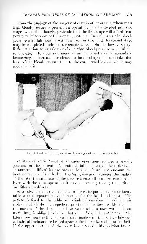
GENERAL PRINCIPLES OF INTRATHORACIC SURGERY 207
From the analogy of the surgery of certain other organs, whenever a
high blood-pressure is present an operation may be divided into two
stages when it is thought probable that the first stage will afford tem-
porary relief to some of the worst symptoms. In such cases, the blood-
pressure may fall notably within a week or two, and the second stage
may be completed under better auspices. Sauerbruch, however, payslittle attention to arteriosclerosis or liif/li blood-pressure when about
to operate. He does not mention an increased risk of secondary
hemorrhage. Increased tendency to fatal collapse is, he thinks, due
less to high blood-pressure than to the cardiorenal lesions, which mayaccompany it.
FIG. 148. Position of patient in thorax operation
Position of Patient. Most thoracic operations require a special
position for the patient. Xo suitable table has as yet been devised,
as numerous difficulties are present here which are not encountered
in other regions of the body. The form, si/e and diameter, the qualityof the ribs, the situation of the disease-focus; all must be considered.
Even with the same operation, it may be necessary to vary the positionfor different subjects.
As a rule, it is most convenient to place the patient on an ordinarytable with a separate movable section for the head and chest. The
patient is fixed to the table by cylindrical cushions or ordinary air
cushions which do not impede respiration, since they readily yield to
the motion of the ribs. This is of value when a man with but. one
useful lung is obliged to lie on that side. When the patient is in the
lateral position the thighs form a right angle with the body, while t\vo
cylindrical cushions are braced against the buttocks with a broad belt.
If the upper portion of the body is depressed, this position favors
208 THORACIC SURGERY
drainage from the lungs when there are secretions in the bronchi.
Elevation of the chest relieves dyspnea, etc.
Aniisepsis, Asepsis. There is not much to be said under this head-
ing. In surgery of the thorax nothing must be allowed to chill the
patient. The iodin-alcohol method of disinfection is good. It is quicklyand thoroughly accomplished and does not burden the patient. Appar-ently it has no drawbacks. In emergency cases iodin may be paintedon the operating field.
Disinfection of the surgeons' and assistants' hands, the dressings,
etc., does not differ from that practiced in other surgical work.
Silk is chiefly used as a suture material; only in deep wounds of the
lung and other conditions in which it does not give the requisite support,is fairly heavy catgut substituted.
Choice of Anesthetic* The following general rules may be laid
down for the use of anesthesia in thoracic surgery. Local anesthesia
suffices for all extrapleural interventions, including resection of ribs
and even maximal thoracoplasty. The technic of its use must be very
thorough. If it falls short of the desired object, a little ether may be
used.
In transpleural intervention local anesthesia is indicated, if the
pleura is adherent, in the operative treatment of abscesses and gangreneof the lung. General narcosis is contra-indicated because it interferes
with the evacuation of the fluid contents of the cavities. The lungitself is analgesic.
"Whenever there is a broad opening to be made in the pleura generalnarcosis is definitely indicated because it prevents the reflex spasm of
the glottis due to the pleural incision. The imperfect expiration raises
the intrabronchial and intra-alveolar pressure, with the immediate
result of increasing the labor of the right heart. Even the equalizationof pressure is unable to control this type of respiration which mechani-
cally injures the alveoli and thereby predisposes the patient to lobular
pneumonia, etc.
It occasionally happens that the incision of the pleura in a non-
narcotized patient causes death by arrest of the heart. This is perhapsconnected with traumatism to the vagus or its branches.
In regard to the choice of a general anesthetic, ether is contra-
indicated in the actual presence of bronchitis or pneumonia. Chloro-
form is best given in combination with oxygen. Timid patients maybe given morphin. No scopolamin should be used if the patient must
expectorate soon after the operation.The use of the pressure-difference procedure favors anesthetization;
very little anesthetic is necessary and postoperative nausea is less
marked than usual. The blood-pressure remains unchanged. After
the operation the patient is apt to be slightly cyanosed and to have
severe postoperative pains which require morphin.Local Anesthesia in Thoracic Surgery. Garre, since he makes only
a limited use of cabinets, recommends nerve blocking and infiltration
in all subjects who are seriously ill or who have suppuration within
GENERAL PRINCIPLES OF INTRATHORACIC SURGERY 209
the lung; in which case pus or putrid matter may be aspirated into the
lungs under general narcosis. This local anesthesia is, in a measure,contra-indicated if much handling of the pleura is expected, for reflex
compressed breathing may be started as a result of a spasmodic closure
of the glottis. The blood-pressure may be suddenly lowered. Thefield of intervention in thoracic surgery is so large that one must, so
far as possible, depend on a nerve blocking of the intercostals. At the
posterior portion of the chest, the intercostal nerve consists of a single
trunk which lies in the middle of the intercostal space. At the junctureof the posterior and middle thirds, this trunk divides into two branches
which run, respectively, along the lower and upper borders of the ribs
by which the space is bounded. It may suffice to anesthetize the maintrunk close to the vertebral column in order that the several posterior
sensory twigs may become affected, as well as the anterior subdivisions.
For the sake of thoroughness a further injection may be made into the
intercostal space in the axillary line, thus anesthetizing both the upperand lower branches. The technic of this later maneuver is not simple.An infiltration wheal is first established, after which the syringe,
plunged into the wheal, is turned upward and then downward; thus
the branches can hardly fail to be blocked.
There is danger of injuring the pleura, especially in the act of infil-
tration; but this should be obviated by pushing the cannula forward,so that, in the process of infiltration, the injection fluid precedes the
needle and forces the pleura backward. In any case, it should suffice
to pierce only the external intercostal muscle. Infiltration is further
extended along the incision lines. The solution employed may be a
0.4 per cent, to O.o per cent, novocain with adrenalin.
Anoci-associatiun in Thoracic Surgery. Forlanini blocks the inter-
costal nerves before making his operation for artificial pneumothorax,and (Jarre regards this blocking as commendable in operating without
cabinets, and in all local anesthesia operations, i See Local Anesthesia. )
So far as the writer knows ( 1!)HM, ('rile and his associates have not
as yet developed an anoci-association technic in thoracic surgery. In
the abolishment of pleural reflexes, a complete nerve blocking would
seem indicated, as well as in all cases in which one desires or expectsthe lung to contract. In operating without pressure differences in
traumatic cases in which it is desired to prevent a retraction of the
lung, nerve blocking, in diminishing pleural reflexes, should in theory
prevent the compressed breathing which results from coughing with
a closed glottis. This phenomenon is regarded by some surgeons as
conservative and hence, as one which should not be prevented.1
Diagnosis <>fthe Deijree <>f Pleura! Adhesion. In pulmonary tuber-
culosis some adhesions are almost always present; a perfectly intact
pleura is very rare. These adhesions may result from old pleurisies and
are then usually extensive sometimes total. In other cases they form
over foci of lung disease and arc circumscribed. The best test for both
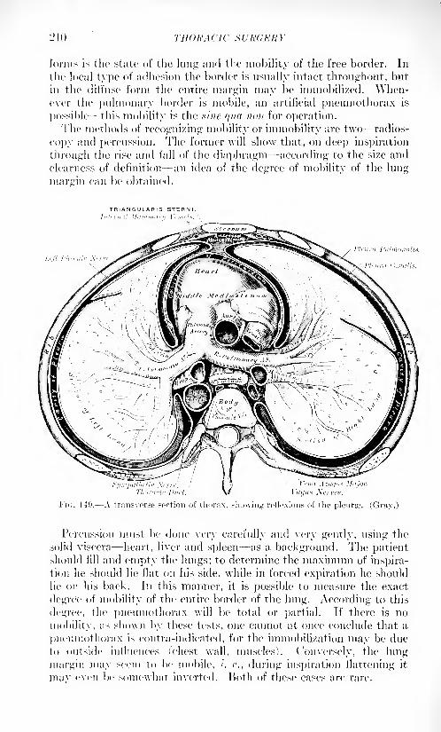
210 THOUA ('1C ,SURCER Y
forms is the state of the lung and the mobility of the free border. In
the local type of adhesion the border is usually intact throughout, but
in the dill'use form the entire margin may be immobilized. When-ever the pulmonary border is mobile, an artificial pneumothorax is
possible this mobility is the sine qua nou for operation.The methods of recognizing mobility or immobility are two radios-
copy and percussion. The former will show that, on deep inspiration
through the rise and fall of the diaphragm according to the size and
clearness of definition an idea of the degree of mobility of the lung
margin can be obtained.
TRIANGULARIS STERNIIntirn.il Mainmn
FIG. 149. A transverse section of thorax, showing reflexions of the pleurae. (Gray.)
Percussion must be done very carefully and very gently, using the
solid viscera heart, liver and spleen as a background. The patientshould fill and empty the lungs; to determine the maximum of inspira-tion he should lie flat on his side, while in forced expiration he should
lie on his back. In this manner, it is possible to measure the exact
degree of mobility of the entire border of the lung. According to this
degree, the pneumothorax will be total or partial. If there is no
mobility, as shown by these tests, one cannot at once conclude that a
pneumothorax is contra-indicated, for the immobilization may be dueto outside influences (Chest wall, muscles). Conversely, the lung
margin may seem to be mobile, /. <"., during inspiration flattening it
mav even be somewhat inverted. Both of these cases are rare.
GENERAL PRINCIPLES OF INTRATHORACIC SURGERY 211
Pressure Differences. There is normally within the thorax a nega-tive pressure of from 7 to 8 mm. of mercury, which is offset promptly,if a pleural sac be opened broadly, by the inrushing air. Meanwhilethe pressure in the opposite pleural sac is undisturbed and is still
negative. \Yhen one pleural sac is opened the lung of that side appar-ently collapses, but in reality retracts from its own elasticity. The
pressure in the open pleural sac eqjials that of the atmosphere, while
that of the closed side remains negative. Tnspiratory efforts now cause
overpressure on the affected side and, displacing the mediastinum,
including the heart, compresses the intact side. Expiratory efforts, onthe contrary, reduce the overpressure on the pneumothorax side, andthe mediastinum with the compressed lung regain their normalboundaries. This pendulum-like behavior of the mediastinum greatlyinterferes with the interchange of gases. The respiration is at first
convulsive from reflex apnea, but the retention of CO- soon leads to
stimulation of the respiratory, cardiac and vasomotor centers. Respira-tion becomes slow and deep and the pulse slow and of increased volume.The contracted lung meanwhile becoming congested, the result of these
various disorders is dyspnea.Retarded respiration leads to a gradual cessation of expiration, as
the intact lung becomes unable to empty itself when the thorax is in
an expiratory position. Cessation of inspiration then follows, with
death by suffocation. The circulation in the retracted lung is superiorto that of its intact fellow. If both pleural cavities are opened, both
lungs collapse and death supervenes rapidly from suffocation, althoughtracheal insufflation is able to maintain the respiration indefinitely.
From the clinical viewpoint, an incision of the pleura is at once fol-
lowed by an inrush of air, so rapid as to cause a hissing sound. The
lung contracts suddenly, its natural rosy color becomes a dark gray, andits glossy surface wrinkled. The retraction of the lung causes a reflex
apnea, or arrest of circulation, which is followed by deep forcible
breathing whicb lasts for one or two minutes ('this stage is termed con-
vulsive and irregular). Respiration then becomes quiet and progres-
sively slow until it ceases. (This state may be antagonized by artifi-
cial respiration.) If, however, the opening into the pleura is smaller
than the lumen of the trachea, the pressure differences are not muchdisturbed. For this reason punctures and small incisions for empyemafin recent years the establishment of an artificial pneumothorax) have
always been practicable and regarded as minor measures.
In a large proportion of cases of diseased lungs there are extensive
pleuritic adhesions. It has thus been found possible to attack the lungs
surgically in a large number of instances without the fear or possibility
of a collapse of the given lung.
True intrathoracic surgery has, however, dated only from the time
when it became possible to prevent the collapse of an intact lung, uponfreely incising the pleura. In theory this could be accomplished bytwo radically different methods: ( 1 ) The subject's body could be placedin a pneumatic cabinet with a pressure eqtial to the intrathoracic
212 THORACIC SURGERY
pressure. (2) Air could be respired under pressure through an air-
tight mask, or insufflated into the trachea. In either case, if the pro-cedure were properly carried out, the lung could not well collapse uponincision of the pleura. As a matter of fact two separate schools of
intrathoracic surgery have grown up upon this double foundation.
It is naturally possible to install an elaborate pneumatic cabinet
from which the required amount of air can be pumped. The patient's
head lies outside the cabinet and he may be anesthetized in the ordi-
nary manner. The surgeon operates within the cabinet, unmindful
of the slightly rarified air. The cabinet may be ventilated, lighted,
equipped with a telephone, and may be roomy enough for assistants.
Such a cabinet could at once suggest itself in its numerous require-
ments, but it could hardly be evolved beyond a certain point. Its
expense, bulk, isolation, etc., tend to limit its use, at this time, to certain
hospitals.
The overpressure method was at first difficult of execution because
it appeared to interfere with anesthesia, and, in fact, with the act of
respiration in subjects with presumably crippled lungs. Herein it
differs greatly from the underpressure method, for here the subject's
breathing is not disturbed and he can be anesthetized in a simple way.The overpressure method was, further, very cumbrous at first, involv-
ing a small overpressure cabinet, with a detached anesthetic apparatus.This was succeeded by an air-tight mask in which a constant pressure
could be maintained. Air or oxygen may enter the mask, and the
former is .mixed with anesthetic vapor in a special apparatus. The
oxygen enters the mask by a special pipe. Waste air escapes by a
special arrangement. The most vital portion of the apparatus is an
elastic rubber bag which by preventing labored expiration insures a
natural, easy respiration. (See special descriptions of apparatus.)The I'n'Jcrjjrcsfiurc Procedure. The Zurich Cabinet. This consists
of a transportable, light metal framework which can be quickly erected.
The walls, made of the thinnest, finest material, are readily screwed
upon the framework. There is no separate floor, as the cabinet is
moved about like a screen. On the lower extremity of the wall is a
wide rubber flange, which, when placed on a flat floor, at once adheres
by suction when the cabinet is pumped out.
The wall for the patient's head is so arranged that its lateral aspects
bend outward in such manner that there is no interference with the
operator, while the midsection carries in its aperture a head ring which
can readily be moved up and down. The head is passed through this
ring and the head aperture then narrowed by a certain device, which
renders a neck cuff unnecessary. The head piece can be raised or
lowered whenever it becomes necessary to move the body within the
cabinet. A former argument against the underpressure cabinet lay
in the fact that the operator and the anesthetist, could not hear each
other speak, but this has been obviated by the insertion of balloon
fabric in the anterior wall. This does away with the need of a special
telephone or speaking tube. The side pieces of the anterior wall have
GENERAL PRINCIPLES OF INTRATHORACIC SURGERY 213
windows through which the anesthetist can see the operation and the
operator watch the anesthetist. The cabinet is ample within, and has
a ventilating apparatus which prevents stagnation of air. It is so
illuminated by electric bulbs that all portions are visible. When not
in use the apparatus may be taken apart and folded up.
Sauerbruch 1 and others have extemporized underpressure cabinets
by using small operating rooms, calking all of the seams and crevices
and painting the walls. The air of the room is exhausted to the requisite
degree. The head of the operating table is placed at the door which is
specially constructed for the administration of anesthesia. (An air
lock should be provided to permit of entrance to and exit from the
room.)The Drtiger Combined Apparatus. This was introduced in 1911
and was so termed because the administration of the anesthesia-
oxygen mixture, and the methods of maintaining increased pressureand of automatically obtaining artificial respiration are combined in
one. Either part of the apparatus may be switched on or oft'.
The apparatus for the automatic maintenance of artificial respiration
is the pulmotor and is intended for use in suspended animation from
gas poisoning, smoke suffocation, etc., as well as for the reanimation
of stillborn infants. (See Artificial Respiration.) The apparatus as a
whole has recently been so modified that by a slight increase in pressure,
the principles of Yolhard, Kiihn and Melt/er may be carried out. Theanesthetic portion of the apparatus is that known as the Roth-Drager.The overpressure apparatus consists of a cylinder of oxygen of 1000
liters capacity. The oxygen under compression is conveyed by a spiral
pipe to the rest of the apparatus. A cylinder trap valve is opened
allowing the oxygen to pass through the tube into the reducing valve
which reduces the original high pressure to one of 8 atmospheres. The
gas then enters the Roth-Drager mask. Devices make it possible to
cause either a vacuum or a high pressure in the mask.
Overpressure Apparatus.- Thus far several distinct principles have
been utilized for this purpose, vi/,., a small cabinet or chest, a ma>k and
an insufflation tube. The negative pressure is overcome by forcing
air or oxygen into the lung; hence the overpressure apparatus must
necessarily include or be associated with the anesthesia apparatus.The overpressure principle is very simply carried out, for it is only
necessary that the patient's head be placed within a small air-tight
chest in which is constantly maintained an overpressure of about
7 mm. to offset the negative pressure of the lungs. This alone rapidly
inflates the lungs. The head-chest is kept filled by means of a small
air-blast engine driven by a motor; this delivers, on an average.TOO liters of air-pressure through a tube of l> cm. diameter to a large
bellows. The air passes through a manometer and into the head cabinet
where the pressure is shown by a water manometer. The air then
escapes through a valve, the necessary resistance being furnished bya running weight.
1 Krgehnisso (1. Chirurgie, 1910.
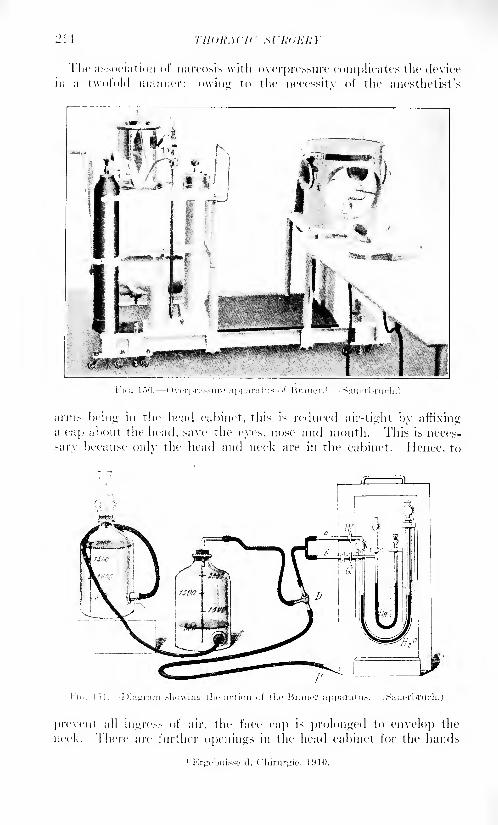
21 I
riic association of narcosis with overpressure complicates the device
in a twofold manner; owing to the necessitv of the anesthetist's
arms being in the head cabinet, this is reduced air-tight by affixinga cap about the head, save the eyes, nose and mouth. This is neces-
sary because only the head and neck are in the cabinet. Hence, to
prevent all mgres> of air, the face cap is prolonged to envelop the
neck. There are further openings in the head cabinet for the hands
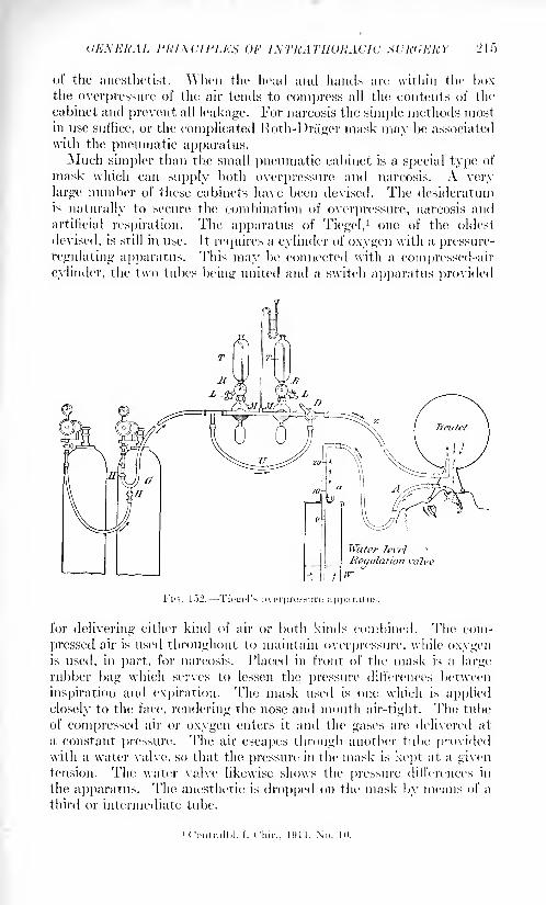
UKXKHAL L'lUNCll'LKK OF INTRATHORACIC KURUKltY 1*1 f>
of the anesthetist. When the head and haucls are within the l><>\
the overpressure of the air tends to compress all the contents of the
cabinet and prevent all leakage. For narcosis the simple methods mostin use suffice, or the complicated Roth-Dniger mask may he associated
with the pneumatic apparatus.Much simpler than the small pneumatic cabinet is a special type of
mask which can supply both overpressure and narcosis. A very
large number of these cabinets have been devised. The desideratumis naturally to secure the combination of overpressure, narcosis andartificial respiration. The apparatus of Tiegel,
1 one of the oldest
devised, is still in use. It requires a cylinder of oxygen with a pressure-
regulating apparatus. This may be connected with a compressed-air
cylinder, the two tubes being united and a switch apparatus provided
Water 1evfl
Regulation valve
ir
I'"K;. 152. Tieirel's overpressure apparatus.
for delivering either kind of air or both kinds combined. The com-
pressed air is used throughout to maintain overpressure, while oxygenis used, in part, for narcosis. Placed in front of the mask is a largerubber bag which serves to lessen the pressure differences between
inspiration and expiration. The mask used is one which is applied
closely to the face, rendering the nose and month air-tight. The tube
of compressed air or oxygen enters it and the gases are delivered at
a constant pressure. The air escapes through another tube providedwith a water valve, so that the pressure in the mask is kept at a giventension. The water valve likewise shows the pressure differences in
the apparatus. The anesthetic is dropped on the mask by means of a
third or intermediate tube.
1 Central I>1. f. ( 'liir., I'll I. \"o. 10.
210 THORACIC SURGERY
The Willy Mi'i/er Cabinet. The idea of the Sauerbruch under-
pressure cabinet was further developed by Meyer' who devised eabinets
representing both over- and underpressure, in other words, a universal
apparatus for the application of differential pressure.It consists of an inner, positive pressure chamber which can be used
for narcosis, and a large external, negative pressure chamber, which
serves as the operating room. The internal chamber can be used
independently as an overpressure apparatus as both chambers have
separate systems for ventilation and the estimation of pressure. Onecan enter and leave the external or negative chamber directly, because
at the moment when the door is opened the pressure difference between
the two chambers can be correspondingly graduated. The inner
chamber, however, is provided with an air-lock. The operating, or
negative-pressure room, is large and gives plenty of space for the
operator and his required assistants. The narcosis cabinet of iron wire
and balloon cloth keeps the anesthetist in close touch with the operatorwithout the aid of a telephone or speaking tube.
liitrat radical Insufflation,- The original apparatus for intratracheal
insufflation was very simple, consisting of a foot bellows connected
by tubes with a bottle of ether and a mercury manometer. Air forced
into the bottle became impregnated with ether vapor. It was forced
into the larynx at a pressure of 1.1 to '20 mm., the tube being muchsmaller than the trachea, so that the etherixed air could readily find
its way out of the trachea. The intrathoracic pressure was increased to
such a degree that interruption of the jet from three to six times a
minute was required in order to enable the lung to contract and thus
expel some of the residual air.
This constant inflow and outflow of air maintained the lung in a state
of normal air exchange. Incidentally it was found that incision of
the pleura was not followed by pnenmothorax; hence, it is possibleto use the apparatus instead of overpressure cabinets.
As modified for use in mankind, an automatic exhaust is added to
prevent overpressure in the tubes. There is a tank for wanning the
gas. A motor is connected with the blower. Intratracheal anesthesia,
in thoracic surgery, seems well fitted for the prevention of a collapse
of the lung; further, it practically constitutes a general method of
securing narcosis.
The method consists of the insufflation of air and ether into the
trachea. To quote Melt/er, the essentials consist of the introduction
of a flexible elastic tube deeply into the trachea, the tube being muchsmaller than the trachea itself. Through this tube, a nearly continuous
stream of air is forced, and this air returns through the unoccupied
portion of the trachea. The pure air from without naturally forces
outward some of the 1 vitiated air in the bronchi, including that which
is present in the larynx, pharynx and mouth. Thus, the insufflation
of air not only does not interfere with ordinary respiration, but greatly
i Jour. Am. Mod. Assn., 1909, liii, 197S.
GENERAL PRINCIPLES OF INTRATIIOUACIC SURGERY 217
assists it; as a matter of fact it is quite able to replace it for a time.
Moreover, the recurrent air-stream tends to prevent the entrance of
bacteria.
Intratracheal insufflation, then, seems to be a substitute for respira-
tion; its association with anesthesia represents but one of its uses in
medicine. The difficulties in introducing the tube into the glottis,
even the impossibility of this without general narcosis, have not led
to the preliminary performance of a tracheotomy. Elsberg (the first
to use the method on human beings) advises a beginning general nar-
cosis with ether. The patient then lies on his back, head downwardat the end of the table, and a mouth-gag is inserted. The insufflation
tube is passed with the aid of direct laryngoscopy, in which the instru-
ment, really a speculum, is passed directly into the glottis.
In intrathoracic surgery the method may be used with the over-
pressure apparatus; indeed, some claim that the combination of the
two methods gives an advantage over the underpressure method.
(See Overpressure, etc., p. 2.13.)
Present Status of Pressure-difference Procedures. Dreyer,1 has
attempted to reach an estimate of this by four routes, viz., clinical
material, a stud}' of the literature, animal experimentation and cir-
cular letters. In regard to experimentation, every one who has tested
Sauerbruch's principle, recognizes in it a great advance in thoracic
surgery. Whoever expects to do much experimentation should,
therefore, have a cabinet and should use it as often as possible, for
practice. The majority of those who use cabinets prefer the over-
pressure apparatus, even while regarding underpressure procedures as
physiological. The overpressure apparatuses most commonly in use
are the Tiegel-IIenle, Shoemaker, the combination-apparatus of the
Dra'ger Works, and that of Lotsch and Mollgard. In all intrathoracic
operations, from beginning to end, there should be deep narcosis, for
any act of struggling is disturbing to the operator and dangerous to
the patient; further, at the end of the operation the lung more readily
expands. For the same reason there should be sufficient air available
in order that the inspiratory lowering of pressure may be restricted as
much as possible. The Mollgard and I )rager apparatus both tend to
maintain constancy of pressure. The Tiegel apparatus is providedwith an extra large bag and a wider tube than usual. Those apparatusesare to be preferred which make it possible to apply a pure oxygen
overpressure. (Oxygen does not require much overpressure duringthe intervention: 3 to 5 cm. water; the danger of inflating the stomach
with a high pressure is therefore much lessened. The swallowed air
can easily be removed after the operation.)Another form of overpressure, differing in type, is the "\ olhard-
Meltzer-Auer tracheal insufflation.- (Much used at this time in this
country.) This has two difficulties: (1) The catheter must be neither
too thick nor thin; if it be too thick there is no dead space; if too
1 Bcrl. kliti. Wchnschr., 1914, li, 1605.2 MiinpluMi. ined. Wchnschr., 1908, No. 5.
-18 THORACIC M.'ItUKKY
thin iniicli overpressure is wasted. (Insufflation can also bu carried
by other forms of overpressure apparatus.) (2) 'The introductionof the catheter; Jackson's laryngoscope is used; without some suchresource the technic presents marked difficulties.
Drawbacks of Pressure-difference Surgery; Surgery Without Pressure
Differences. Pressure-difference cabinets are used most extensivelyin Germany. German surgeons, almost without exception, state that
no one who has ever operated in certain classes of cases with pressure-difl'erence apparatus, will ever dispense with them. Further, somethoracic surgeons who originally pronounced against them have becomeconverts after testing them. On the other hand, Sauerbriich, who is
chiefly responsible for their introduction, frequently operates without
them, apparently reserving them for special classes of cases and for
patients in which shock is most to be feared.
The testimony of the leading thoracic surgeons, in other countries,
seems to be very largely against the use of over- and underpressure
apparatus as unnecessary, cumbersome and expensive. Many surgeons,
further, are warm advocates of the Meltzer-Auer intratracheal
insufflation.
Is a Pressure-difference Apparatus Unphysiological? According to
Garre, in none of the methods employed in maintaining pressuredifferences is the gas exchange normal. The respiration occurs with
the chest in the inspinitory position, and strong expiration is lacking;hence residual air which is poor in oxygen, tends to accumulate.
In the Meltzer-Auer procedure, air or oxygen forced into the lungtinder pressure ventilates the lung and mechanically forces out the
residual CO->. Thus a quasi-normal exchange of gases is produced.Even in the Sauerbriich cabinet, breathing is said not to be physio-
logical. The negative pressure causes a more or less permanent posi-
tion of the vocal cords on inspiration. There is no strong expiration.If the patient were under local anesthesia the breathing might proceednaturally, but he is under general narcosis.
All overpressure apparatuses cause the same type of respirationas insufflation.
Inconveniences and Dangers of Pressure-difference Procedures.
Garre states plainly that he is far from being a partisan of this method,and that he has done most of his operations without recourse to it.
His objection is, in part, that asepsis cannot be properly carried out
in the tise of the neck cuff in the underpressure procedure. (This
objection is absent in the overpressure procedures.)In the use of masks in the overpressure procedure there is danger
of vomiting. Should the patient start to vomit it will be necessary to
remove the mask, whereupon pressure changes will be in evidence.
As a result of this the operation may be delayed. Further, vomiting
may be feared because of the danger of the aspiration of vomitus.
In insufflation, under proper conditions, vomiting should rarely
occur.
Any differential pressure apparatus may break down at a critical
GENERAL I'KIXCII'LIM (>I< /A TRATHORACK! HUlidKRY 219
moment. I [(Mice, operators must hold in rea'diness tampon material
fen1 the prevention of a total collapse.In incomplete collapse of the lung, pus or Mood can he aspirated
from the lung to the pleura and rice, verxa. Since collapse of the lungleads to impoverishment of the Mood in oxygen, insufflation of oxygeninto the lung is indicated.
Pressure differences sometimes cause acute dilatation of the stomach.Tin's may happen when the head and neck are under different pressures
(as when the manchette lies close beneath the chin). When the pres-sure on the neck is relieved, the higher tension of the air leads to a
blowing up of the esophagus and the stomach. Acute, fatal gastricileus lias occurred. The meteorism in the intestines may compress the
diaphragm ajid heart, and thereby interfere with cardiac activity.
Secondary infections of the lung may develop.Inflation of the lung compresses the lung capillaries and renders
difficult the work of the right heart. Hence, in the case of a weak or
diseased heart and in emphysema of the lung the pressure difference
should be very slight; not over 4 to ."> mm.Pressure differences which exceed 10 or 12 mm. should be avoided
in all chest and mask apparatus, although, in insufflation, a pressure of
'2(1 mm. may be applied unhesitatingly if the stream of air is interruptednow and then. Garre regards a prolonged intervention under narcosis
with pressure difference as dangerous. Patients may die at the close
of such intervention, and even before such close, with no evidence of
any causal factors save a so-called "shock.'' The lungs inflate fullybut there is an absence of strong expiration. The residual air is poorin oxygen and the residuum tends to increase constantly. The blood
becomes more and more charged with CO^ until it begins to act uponthe respiratory and vagus centers. A vagus pulse then develops,followed by a lowered blood-pressure and a pulse becoming uniformly
rapid. Respiration is undisturbed, a favorable symptom calculated
to deceive. As a matter of routine, in these long operations, Garre
always keeps oxygen on hand for insufflation, and the lungs arc often
allowed to go to the position of expiration.Garre implies that the dangers from pneumothorax are not so formi-
dable as to force the use of pressure-difference apparatus. He has
made use of the apparatuses of Sauerbruch and JJrauer, and feels
that they are valuable in large thoracoplasties and in cases of injuryto the lung. They do not seem necessary, however, for operations in
gangrene, abscess, bronchiectasis, etc., when it is desired to locate a
given focus.
The Writer'* Attempt to Estimate the Value of Pressure-differenceProcedures. It is not easy to make this attempt. A careful review of
the literature, together with a not inconsiderable surgical observation
in this country and in Europe, leaves the impression that the entire
subject is as yet undetermined. At this time, it is probable that Sauer-
bruch has had the greatest experience in thoracic surgery; it has
already been said that he uses pressure-difference cabinets in certain
220 THORACIC SURGERY
procedures and discards them in others. With the exception of Willy
Meyer,1 whose work should be accorded the greatest credit, American
surgeons seem to look with disfavor upon all cabinets. Some haveinstalled them only to discard them later. Robinson,
2 whose experi-mental work on animals and actual work on humans have been veryconsiderable, told the writer in 1915 that he believes cabinets devoid of
practical value. The surgeons of no country have traveled for observa-
tion and improvement more extensively than have those of America,and these latter are definite partisans of the intratracheal insufflation
procedures. The whole situation must be considered as in a formative
stage, remembering ever that the surgery of tomorrow will be better
than is that of today, even as the surgery of today is better than wasthat of yesterday.
INJURIES OF THE THORAX.
Contusion of the Thorax. This term implies the combined results
of blunt violence in all possible degrees, from the merest bruise to
severest compression and crushing. The term contusion, however,can be applied only to sudden violence. If the thorax is compressed
very slowly no severe damage need actually occur, even if the sternumbe forced nearly as far backward as the spine. The organs readily
adapt themselves to the changed conditions.
While the minor phenomena of contusion need hardly receive men-
tion, the violence in extreme cases is of much importance. For example,a man may be struck on the chest by a heavy, thrown stone or other
projectile; he may fall on his chest from a height, or be struck, while
lying supine, by a heavy, falling object; he may be run over by a heavyvehicle or compressed between buffers.
This type of injury can damage any of the thoracic structures or
contents, and, naturally, may injure nearby organs. The parts which
give way most readily are the lungs and pleura. While sudden violence
tends to cause a collapse of the lung, spasmodic closure of the glottis
antagonizes the collapse and the lung may rupture at some point,
generally into the pleural cavity. These ruptures are followed byhemorrhage, blood being coughed up or forming a hemothorax. Thevictim is usually badly shocked, presenting all of the phenomena of
collapse.
When pneumothorax develops the air escapes from the lung onlywhen a large bronchial twig is ruptured. Pneumo- and hemothorax
may occur alone or in combination.
Rupture of the diaphragm may be followed by the prolapse of some
abdominal viscus into the thorax (see Thoraco-abdorninal Wounds).
Emphysema (subcutaneous) may occur without the coexistence of
fracture. Similarly, a hernia of the lung may take place in the absence
of fracture.
1 Munchen. med. Wchnschr., 1909, No. 47.
2S'urg., Gynoc. and Obst., March, 1909, p. 255.
INJURIES OF THE THORAX 221
These early symptoms are often followed by other delayed phe-nomena. When pneumothorax does not soon disappear, it may cause
marked pressure symptoms. Absorption of blood from a hemothorax
may cause a temperature rise.
So-called "traumatic breathing" which is present at the outset
(cyanosis, dyspnea, etc.) is often followed by severe pain. Infection of
a collection of air or blood in the pleural cavity may lead to a trau-
matic empyema. Contusion-pneumonia is known to develop in some
cases.
Prognosis.- These injuries, while causing a high mortality, often end
favorably without sequela1
. Exact statistics of uncomplicated injuries
seem to be wanting.Treatment.- -The treatment consists of rest in bed with external
applications of ice. If much shock and hemorrhage have occurred,
saline infusion and cardiac stimulants are indicated; if the hemo-
thorax is very extensive it should be aspirated, but this only after
suitable delay, for the compression symptoms may not become aggra-vated. I )yspnea may sometimes be controlled by morphin.
Thoracotomy for internal hemorrhage has regularly been done in
the great war.
Commotion of the Thorax. This is a concussion unaccompaniedby external or internal evidence of injury. A patient may receive a
sudden blow as from a stone on the chest, and feel few, if any, effects.
Others, from the same violence, may gasp and fall, yet without serious
consequences, while others are severely shocked. Some recover from
this state rapidly, while in other cases the state of collapse is variously
prolonged. Death has occurred in this way.
Judging by effects rather than by the degree of violence, two typesof commotion are readily recognizable, to wit: mild and severe.
In the severe cases, the violence nearly always occurs over the
sternum, so that the heart, rather than the lungs, bears the brunt.
The exact mechanism is as yet unknown. The collapsed condition is
due to a sudden lowering of the blood-pressure, but whether this is
directly or indirectly due to irritation of the vagus, is not known. If
the violence be severe enough the heart's action may be arrested in
diastole. That the_ blood-pressure may remain subnormal for a long
period is held to be due to active congestion of the mesenteric vessels
as a result of irritation to the depressor and sympathetic nerves.
Death is believed to be due directly to changes in the circulation
in the nervous centers, from arrest of the heart's action, and from lower-
ing of the blood-pressure."When a patient is injured by blunt violence over the sternum or
anterior chest wall, contusion and commotion may coexist to some
degree.Treatment.- The treatment is that of shock.
Suffocative Compression of the Thorax. This accident, when
fatal, constitutes one of the types of suffocation. ( 'ompression may not
be restricted to the thorax but mav involve the abdomen, which tends
222 THORACIC SURGERY
to force the diaphragm upward and immobilize it. Further, compres-sion of the abdomen alone may cause suffocation. This type of acci-
dent is of great forensic importance because it gives rise to a charac-
teristic syndrome. A sudden, severe compression of the thorax forces
the blood into the veins which form the superior vena cava, and since
this system of veins is not provided with valves, extensive punctiform
ecehymoses, or petechue, are caused in the head, face, conjunctiva,neck, shoulders, upper arms and upper part of the thorax. The indi-
vidual ecehymoses are very small, but they are so closely approximatedthat the entire integument appears cyanotic. The subject is compara-tively a recent one in literature, no complete systematic accounts
having appeared until 1S99, although scattered observations were madeas far back as 1S>7. The material on record consists, very largely,
of crushing injuries occurring in panics, in which individuals were
densely packed together or trod under foot. Any force that causes a
sudden and violent compression of the trunk may produce the same
injury; the "avalanche" type of accident, or that due to the passageover the body of a heavy vehicle. The violence need be but a few
seconds in duration. Analysis of a small number of cases shows that
the thorax alone was compressed in S cases, the abdomen in ?> and
jointly in 11. Animal experimentation has shown that this type of
injury can be 1
produced at will.
The age of the patient possesses much significance in these cases.
(Compression fracture of the thoracic cage is a separate condition.)
The great majority of cases occur in the young, before the period of
calcification of the costal cartilages. Of 10 cases cited by Schwartz,1
9 occurred before the age of twenty-one and five in the third decennium.
Only 2, or 12| per cent., occurred after the age of thirty.
The reason the greater part of the upper extremity escapes the
ecchymosis is evidently due to the presence of valves in the veins of the
arms. The brain, also, is for the most part spared, but in rare cases,
retinal and other intraocular hemorrhages are seen; while orbital
hemorrhages are fairly frequent. The veins which form, or discharge
into, the inferior vena cava are well provided with valves, and the lower
extremities rarely show the presence of ecehymoses.The purely mechanical theory in regard to these ecehymoses is by
no means final. Some writers affirm their belief in the existence of
vaso-dilation in the cervico-facial region as a result of contusion of the
splanchnic nerves. Others deny that compression of the trunk can
empty the large veins without causing severe visceral lesions, which,
we know, seldom develop. To explain this compression they believe
it necessary to invoke the cooperation of the abdominal muscles and
the reflex closure of the glottis. The enormous increase of intrathoracic
and intra-abdominal tension thereby produced would then act as the
direct cause of the expulsion of venous blood; the compression of the
trunk being the indirect cause. The immunitv of the cranial sinuses
INJURIES OF THE THORAX 223
from rupture is probably due to the fact that their walls are not dis-
tensible, so that the blood forced into them is simply passed throughand reenters the veins of the face by the natural anastomoses.
Symptoms. Patients thus crushed sustain more or less shock, the
pulse becoming small and the respirations short and irregular. Con-
sciousness may or may not be lost. The face and neck at once assume
a blue or violet hue. In a typical case, there is a sharp line of demarca-
tion which corresponds to the upper margin of the shirt. This is not
a coincidence, for, curiously, if the shirt is unbuttoned at the time,
the ecchymoses descend much lower and may reach the abdomen.The face, and especially the eyelids appear swollen, and there may be
exophthalmos due to intra-orbital hemorrhage'. Escape of blood from
the nose and mouth, sometimes present, will perhaps suggest fracture
of the base of the skull, especially if the patient be unconscious.
Prognosis.-^Yhile nearly all reported cases ended fatally, death byno means always follows. In benign cases the patient may rapidly
recover, but a week or ten days is required for the absorption of the
ecchymoses proper. In a typical case the ecchymoses may be localized
especially in the eyelids and conjunctivse, extending outward uponthe temples. Epistaxis may coexist.
Treatment. The only active treatment is that for shock.
Fractures of the Ribs. This injury is of frequent occurrence,
statistics showing that it constitutes over 10 per cent, of all fractures.
The kind of violence producing it is similar to that which producescontusions and compressions, but, while the latter occur in the rela-
tively young, fractures are particularly apt to take place after the ageof thirty-five. Naturally, the same violence often causes both contu-
sion and fracture. The diminished resistance of the ribs, which tends
to increase with age, is due to a twofold cause ossification of the costal
cartilages and rarefaction of the bones. Over one-half of the cases
of fracture of the ribs occur after the age of forty. The incidence of
sex is: male, 5 or 6; female, 1.
The causes, as elsewhere with fractures, are direct violence and mus-cular action. The forms of direct violence are numerous. Falling upona projecting object, buffer accidents, automobile injuries, avalanche
accidents, crushing in crowds, blows, etc., are familiar examples.Muscular action seldom figures as a cause and generalizations concern-
ing this are based on but few cases. The lower ribs, nearly always a
single rib, are involved in the majority of cases. These fractures, at
times, occur during the latter months of pregnancy, even with no
definite history of violence. They may, rarely, follow violent coughingor sneezing or other muscular effort. In rare cases these fractures are
not only multiple but bilateral.
Victims of intrathoracic disease, elderly people and alcoholic sub-
jects, are particularly prone to fracture.
There are two distinct mechanisms in direct fractures. In the first
form the violence directed against the rib tends to bend it or force its
convexity inward. When it breaks, the fragments project against the
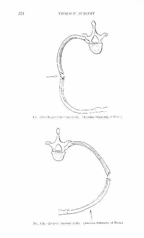
THORACIC SURGERY
FIG. 153. Internal fracture of rib. (Anselme Schwartz, of Pari.-.)
FIG. 154. External fracture of rib. (Adeline Schwartz, of Paris.)
INJURIES OF THE THORAX 225
pleura and lung. This mechanism is seen in blows upon the chest.
In other cases, as in crushing injuries, violence may be applied over
the front of the chest, compressing it. The ribs are then bent outwardand when fracture occurs the fragments tend to project into the sub-
cutaneous tissue and skin. If the interior surface of the thorax is
compressed, the break tends to occur in the anterior third of the rib,
near the cartilage. If the violence is received at the back of the chest,
the ribs tend to break near the spine. These findings, confirmed byexperiment, do not agree with theoretical considerations.
But little is known of the mechanism of fractures from muscular
action.
The ribs most commonly broken are the fourth to seventh, since
these are most exposed to violence. The upper ribs are protected bythe clavicle and by muscular masses, while the lower ones are muchmore yielding and thus undergo fracture less readily.
There are several forms of incomplete fracture, all of which occur
infrequently. They appear as fissures of the outer bony plate, as green-stick and subperiosteal fractures. The majority are complete, even
though there is no discoverable separation of the fragments. It rarely
happens that a rib breaks transversely or in a short line; as a rule,
the fracture is oblique and the fractured ends are dentated so that
overlapping does not readily occur.
With but little violence, or localized violence, it is usually a single
rib that is broken, but if the entire thorax be compressed, several con-
tiguous ribs give way, thus giving rise to the mobilization of a consider-
able portion of the chest wall. Bilateral fractures occur under the sameconditions.
The violence which serves to break several contiguous ribs may,on the contrary, cause notable displacement. The ribs are forced
inward and rise with each inspiration. When a solitary rib is broken
displacement of the fragments rarely occurs, at least, in the personalobservation of the writer. Other surgeons of wide experience, however,state that displacement is by no means infrequent. This divergenceof opinion is best explained by the view that displacement is never
marked, and when present may undergo spontaneous improvement.Callus forms in a majority of cases, and when there is much displace-
ment, as in multiple fractures, it may be exuberant. Pseudarthrosis
rarely occurs, and when present gives rise to little or no disturbance.
Symptoms. Pain and dyspnea are the most constantly present, the
latter arising from a painful inhibition of inspiration. Pain is usuallyintense and is much aggravated by a slight movement, especially bysuch acts as coughing and sneezing. Respiration thus instinctively
becomes shallow and superficial. The pain and dyspnea after a bad
contusion, likewise may be equally severe.
Deformity is not usually in evidence save in certain multiple frac-
tures. At the seat of fracture an induced pain is elicited by pressingthe fingers along the ribs: this tenderness and pain are quite charac-
teristic. The tender point may be directly over the break or at a pointVOL. II 15
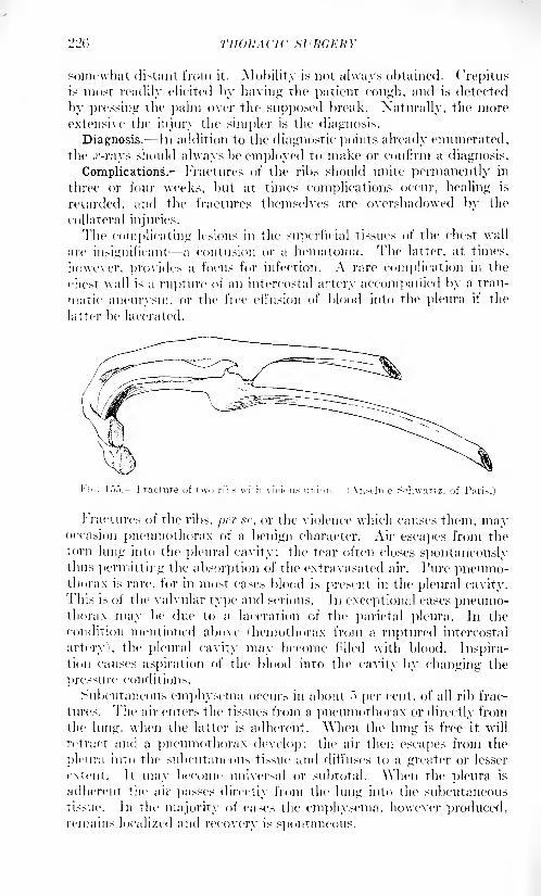
220 THORACIC SURGERY
somewhat distant from it. Mobility is not always obtained. Crepitusis most readily elicited by having the patient cough, and is deteeted
by pressing the palm over the supposed break. Naturally, the moreextensive the injury the simpler is the diagnosis.
Diagnosis. In addition to the diagnostic points already enumerated,the .r-rays should always be employed to make or confirm a diagnosis.
Complications. Fractures of the ribs should unite permanently in
three or four weeks, but at times complications occur, healing is
retarded, and the fractures themselves are overshadowed by the
collateral injuries.
The complicating lesions in the superficial tissues of the chest wall
are insignificant a contusion or a hematoma. The latter, at times,
however, provides a focus for infection. A rare complication in the
chest wall is a rupture of an intercostal artery accompanied by a trau-
matic aneurysm, or the free effusion of blood into the pleura if the
latter be lacerated.
Schwartz, of P;iri~.)
Fractures of the ribs, ]>cr tsc, or the violence which causes them, mayoccasion pnemnothorax of a benign character. Air escapes from the
torn lung into the pleural cavity; the tear often closes spontaneouslythus permitting the absorption of the extravasated air. Pure pnemno-thorax is rare, for in most cases blood is present in the pleural cavity.This is of the valvular type and serious. In exceptional cases pneumo-thorax may be due to a laceration of the parietal pleura. In the
condition mentioned above (hemothorax from a ruptured intercostal
artery), the pleural cavity may become filled with blood. Inspira-tion causes aspiration of the blood into the cavity by changing the
pres>ure conditions.
Subcutaneous emphysema occurs in about ~) per cent, of all rib frac-
tures. The air enters the tissties from a pneumothorax or directly fromthe lung, when the latter is adherent. When the lung is free it will
retract and a pneumothorax develop; the air then escapes from the
pleura into the subcutaneous ti>sue and diffuses to a greater or lesser
extent. It may become universal or subtotal, "\\hen the pleura is
adherent the air passes directly from the lung into tin' subcutaneous
tissue. In the majority of eases the emphysema, however produced,remains localized and recovery is spontaneous.
IXJURIES OF THE THORAX 227
Hernia of the lung, traumatic emphysema, etc., are complications\vhich are considered elsewhere.
Treatment. In the exceptional instances of multiple fractures, of
bilateral breaks or of bony lesions complicated by emphysema, pneumo-thorax, hemothorax, etc., rest in bed for a longer or shorter space of
time may be necessary. In the ordinary case of fracture of a single rib
ambulatory management is proper, the chest wall at the seat of frac-
ture being immobilized, so far as this is possible, by adhesive strips
carefully applied. These strips should be of zinc oxide plaster about
three inches wide, the successive layers overlapping each other and
extending entirely around the chest. The strips should embrace the
chest wall above the fracture and well below it. They should be very
tightly applied, at the time of forced expiration, with the patient in
the standing or sitting posture. At the end of a week, the first dressing
is to be renewed. If the chest is a hairy one it must be shaved before
application of the plaster. Three or four weeks usually suffices for
firm union although delayed union sometimes occurs, especially in
elderly people and in those whose bones are very easily broken. Ordi-
nary roller bandages, binders, etc., are of little aid in the treatment of
broken ribs.
In case a number of ribs are broken, the mechanism of breathing
may be so completely interfered with that then 1 is danger of a fatal
result. Because of inability to maintain respiration, the motion of
the chest wall may become extremely erratic, so that it will be impos-sible to count the respiration. The secondary effect upon the heart
will cause an increase in the number of beats to two hundred or over
per minute, and the patient may be in great danger of losing his life
because of an inability to restore effective respiratory action. In such
cases, it is well to apply rubber adhesive plaster strips, not to exceed
two inches in width, and overlapping about one-half of the width of
the plaster at each turn and forming an adhesive plaster corset extend-
ing from the axilla to the border of the tenth rib. The fixation of the
chest wall obtained in this manner will permit, respiration to be
established by means of the diaphragm, which frequently results in
an almost complete restoration of the patient within a very short time.
The editor has seen a number of apparently hopeless cases of this con-
dition restored by this simple method,- which is infinitely more effective
than if the rubber strips are placed only part way around the chest.
Fractures of the Costal Cartilage.-This lesion is of less frequentoccurrence, the etiology being the same as that for fractured ribs. The
patients are nearly always males, and the cause* is generally direct
violence. Muscular action may be a cause.
The cartilages usually broken are the seventh to the tenth, the break
occurring nearer to the rib than to the sternum, and often at the junc-tion of the cartilage with the rib. A broken cartilage may be commi-nuted. The fracture is nearly always vertical, the cartilage breaking
transversely; displacement is the rule, one fragment often overridingthe other. Ordinarily the sternal segment overrides the costal.
THORACIC SURGERY
Symptoms. All of the symptoms of fractured ribs are present save
crepitus, absence of the latter being due to the smooth character of
the fractured ends.
These fractures undergo repair in several ways; a fibrous or fibro-
cartilaginous tissue may form between the fragments, or regeneration
may occur directly from the perichondrium.Prognosis. The prognosis is good.Treatment. Adhesive strapping, as in fractured ribs.
Dislocations of the Ribs and Cartilages. Dislocations of the ribs
at the vertebral articulations, and of the cartilages at the sternal
articulations have been, known to occur, but are of such infrequencythat but few cases have been placed on record; according to Schwart/ 1
but 9 of the first named and 12 of the latter. Even more rare are dis-
locations at the costochondral articulation, of which but 4 are on record.
Costovertebral dislocations have all occurred in young subjects and
as a result of extreme violence, this having been sufficient to cause the
death of 7 out of 9 cases. In a majority of the cases a diagnosis wasnot made during life
1
. The lower ribs, especially the eleventh, seem
predisposed to this rare form of dislocation.
Chrondrosternal dislocations, on the contrary, are benign and occur
in the upper ribs. The cartilage projects beside the sternum; it is
readily reduced and held in place by adhesive strips.
Costochondral dislocations occur chiefly in the sixth to ninth ribs.
Mo>t of them are due to violent muscular contraction, probably of the
transversalis abdominis muscle and diaphragm. The cartilage may be
displaced forward or backward. It should be reduced and immobilized.
Fracture of the Sternum. This lesion is rare, especially in child-
hood and adolescence. It is due, in the great majority of cases, to
sudden external violence and therefore rarely occurs by itself. Thecauses are those of thoracic injuries in general: crushes, buffer acci-
dents, projectiles, horse kicks and the like. Injury to the spine maycoexist. Conversely, direct violence to the spine may injure the
sternum. The mechanism is not always clear, especially the relative
parts played by contra nnij>, muscular violence, increased spinal exten-
sion and direct impact. In some of the mild types of injury, perhapsisolated, the part played by muscular traction seems evident. These
are of the type of laceration fracture, and of those due to heavy lifting,
hanging by an arm. etc.
The site of the fracture may be at any part of the sternum, but it is
generally at the junction of the manubrium with the sternum proper.
I'Yacture at the lower of the two articulations is very rare. The frac-
ture^ are very seldom double, oblique or incomplete. Transverse
single fracture, complete, is the ordinary type.
I'Yacture of the sternum is generally sufficiently obvious on inspec-
tion and palpation. In most cases, the violence is inflicted upon other
portions of the chest and its contents, and the symptoms vary with
the case.
1 Ohirunrie du Thorax, Paris, 1912.
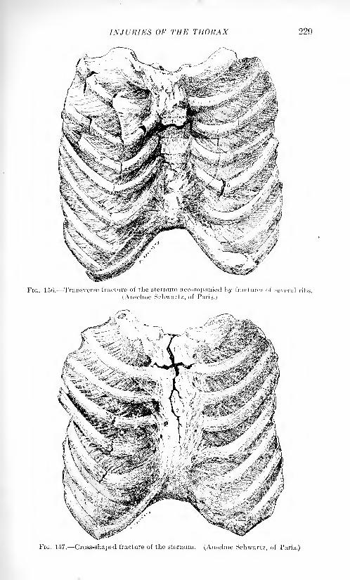
INJURIES OF THE THORAX 229
FIG. 156. Transverse fracture of the sternum accompanied by fractures of several ribs.
(Anselme Sclnvartz, of Paris..;
fc ^%<Mr- ; <-tm&\ '-;':: '
FIG. 1,17. Cross-shaped fracture of the sternum. (Ausulme Schwaru, of Paris.)
!':>() THORACIC SUltUERY
Diagnosis. Diagnosis is based on the separated segments of bone,
together with crepitus, pain and extravasation of blood in situ.
Fracture of the xiphoid cartilage results in pain and displacement,
A'-ray pictures should be made.As a rule these sternal fractures unite rapidly with but little callus
formation. In marked displacement it may be difficult to reduce the
parts, but even in the absence of reduction union occurs.
Treatment. In the simplest type of lesion no retentive dressingsare indicated. The patient lies in bed with simple dressings, and, if
necessary, a sedative for the cough. If the fragments overlap slightly,
the patient lies on a double inclined plane thus overstretching the
spine; the overlapping may disappear spontaneously in a few days. If
the overriding is very extensive, the projecting end of the bone may be
chiselled off, after detachment of the periosteum. Replacement will
then, as a rule, be made easily.
I'liion generally occurs in the ordinary length of time.
Non-penetrating Wounds of the Thorax. Under this heading maybe included burns and scalds, which occur occasionally in severe
degrees on the back or chest. These are most apt to take place in
irresponsible subjects such as young children, drunken people and the
insane, In very deep burns the pleura may be implicated. The treat-
ment of these lesions differs in no wise from that of burns and scalds
in general. Cicatrization is often extreme so that extensive transplan-tation of skin may be necessary, the flaps coming from the arms.
Punctured and incised wounds of the thorax are largely minimized
by the presence of the bony framework and hence, must often be
regarded as benign in character. Like similar wounds elsewhere, theyare not serious per se unless they chance to sever such a large blood-
vessel as one of the intercostal arteries. The internal mammary artery,
in these injuries, may be divided. These superficial wounds tend to
heal promptly unless infected. Streptococcic infections, phlegmon and
erysipelas, sometimes develop, and the pleura may be involved, espe-
cially in the latter; this from lymphatic propagation.The division of the large bloodvessels, especially of the internal
mammary, is much more commonly associated with penetrating than
with non-penetrating wounds. Such wounds generally occur in com-
plicated injuries of the thorax, and hence, these hemorrhages should
be studied by themselves.
Treatment. The treatment of superficial wounds of the thorax differs
in no wise from that of similar wounds elsewhere. Suspicious woundsshould not be completely sutured and those already infected should be
left wholly or partly open. (For wounds of the chest, especially those
due to battle casualties, see article by Dr. Dennis W. Crile, p. 313.)
Traumatic and Surgical Pneumothorax. This is a natural conse-
quence of any wound of the pleura. The presence of adhesions modifies
the condition. It may originate from an external wound or from a
rupture of the lung. It may be partial or complete. It may be uni-
lateral or bilateral, this latter condition tending to a speedy fatality,
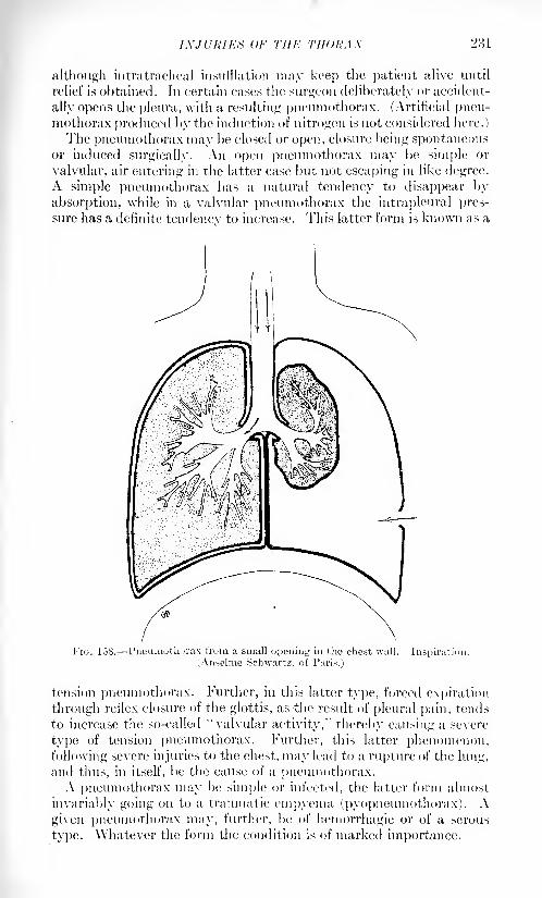
IXJURIKti OF Till-: THORAX 231
although intratracheal insufflation may keep the patient alive until
relief is obtained. In certain cases the surgeon deliberately or accident-
ally opens the pleura, with a resulting pneumothorax. (Artificial pneu-mothorax produced by the induction of nitrogen is not considered here.)
The pneumothorax may be closed or open, closure being spontaneousor induced surgically. An open pneumothorax may be simple; or
valvular, air entering in the latter case but not escaping in like degree.A simple pneumothorax has a natural tendency to disappear byabsorption, while in a valvular pneumothorax the intrapleural pres-
sure has a definite tendencv to increase. This latter form is known as a
FIG. 158. -Pneumothorax from a small opening in the chest wall. Inspiration.(An.selme Schwartz, of Pari.<.)
tension pneumothorax. Further, in this latter type, forced expiration
through reflex closure of the glottis, as the result of pleural pain, tends
to increase the so-called ''valvular activity," thereby causing a severe
type of tension pneumothorax. Further, this latter phenomenon,following severe injuries to the chest, may lead to a rupture of the lung,
and thus, in itself, be the cause of a pneumothorax.A pneumothorax may be simple or infected, the latter form almost
invariably going on to a traumatic empyema (pyopneumothorax) . A
given pneumothorax may, further, be of hemorrhagic or of a serous
type. Whatever the form the condition is of marked importance.
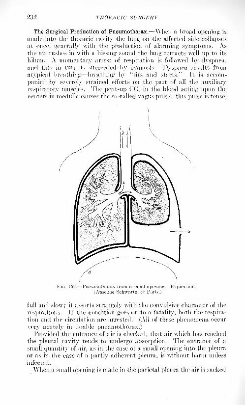
232 THORACIC S I <RC,KR Y
The Surgical Production of Pneumothorax. "When a broad opening is
made into the thoracic cavity the lung on the affected side collapsesat, once, generally with the production of alarming symptoms. Asthe air rushes in with a hissing sound the lung retracts well up to its
hiluin. A momentary arrest of respiration is followed by dyspnea,and this in turn is succeeded by cyanosis. Dyspnea results from
atypical breathing breathing by "fits and starts." It is accom-
panied by severely strained efforts on the part of all the auxiliary
respiratory muscles. The pent-up CQ 2 in the blood acting upon the
((Miters in medulla causes the so-called vagus pulse; this pulse is tense,
FIG. 159. Pneumothorax from a small opening. Expiration.
(Anselme Schwartz, of Paris.)
full and slow; it assorts strangely with the convulsive character of the
respirations. Jf the condition goes on to a fatality, both the respira-
tion and the circulation are arrested. (All of these phenomena occur
very acutely in double pneumothorax.)Provided the entrance of air is checked, that air which has reached
the pleural cavity tends to undergo absorption. The entrance of a
small quantity of air, as in the case of a small opening into the pleuraor as in the case of a partly adherent pleura, is without harm unless
infected.
When a small opening is made in the parietal pleura the air is sucked
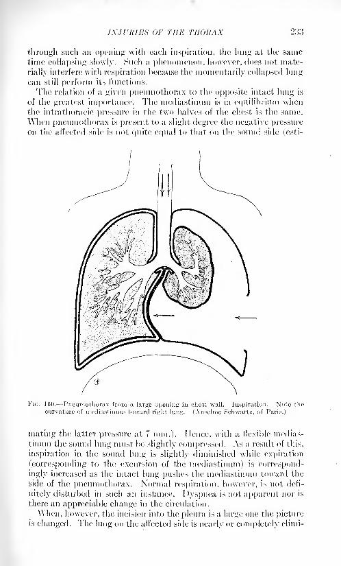
INJURIES OF THE THORAX 2: ',3
through such an opening with each inspiration, the lung at the sametime collapsing slowly. Such a phenomenon, however, does not mate-
rially interfere with respiration because the momentarily collapsed lungcan still perform its functions.
The relation of a given pneumothorax to the opposite intact lung is
of the greatest importance. The mediastinum is in equilibrium whenthe intrathoracic pressure in the two halves of the chest is the same.
When pneumothorax is present to a slight degree the negative pressureon the affected side is not quite equal to that on the sound side (esti-
FIG. 160. Pneumothorax from a large opening in chest wall. Inspiration. Note the
curvature of mediastinum toward right lung. (Anselme Schwartz, of Paris.)
mating the latter pressure at 7 mm.). Hence, with a flexible medias-
tinum the sound lung must be slightly compressed. As a result of this,
inspiration in the sound lung is slightly diminished while expiration
(corresponding to the excursion of the mediastinum) is correspond-
ingly increased as the intact lung pushes the mediastinum toward the
side of the pneumothorax. Normal respiration, however, is not defi-
nitely disturbed in such an instance. Dyspnea is not apparent nor is
there an appreciable change in the circulation.
AVhen, however, the incision into the pleura is a large one the pictureis changed. The lung on the affected side is nearly or completely elimi-
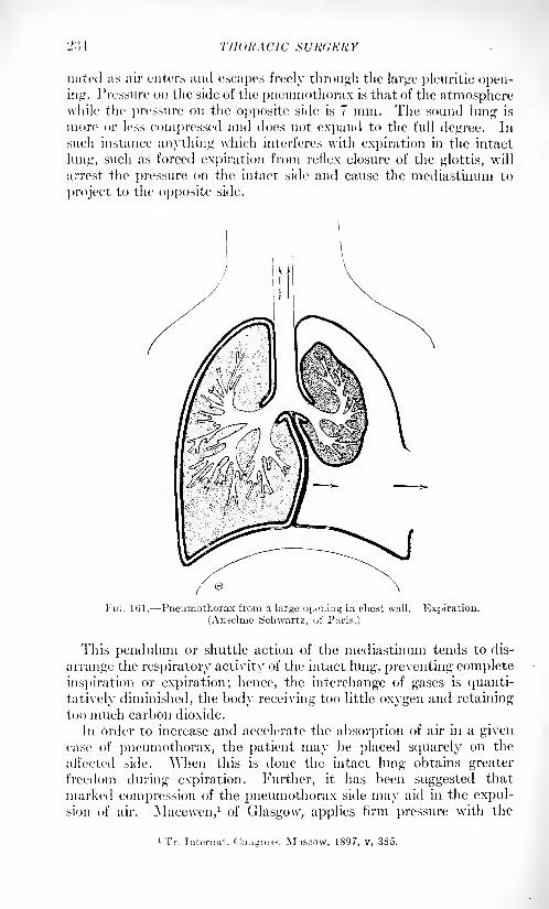
2: 11 THORACIC SURGERY
nated as air enters and escapes freely through the large pleuritic open-ing. Pressure on the side of the pneumothorax is that of the atmospherewhile the pressure on the opposite side is 7 mm. The sound lung is
more or less compressed and does not expand to the full degree. In
such instance anything which interferes with expiration in the intact
lung, such as forced expiration from reflex closure of the glottis, will
arrest the pressure on the intact side and cause the mediastinum to
project to the opposite side.
Fit;. 161. Pneumothorax from a large opening in chest wall. Expiration.
(Anselme (Schwartz, of Paris.)
This pendulum or shuttle action of the mediastinum tends to dis-
arrange the respiratory activity of the intact lung, preventing complete
inspiration or expiration; hence, the interchange of gases is quanti-
tatively diminished, the body receiving too little oxygen and retaining
too much carbon dioxide.
In order to increase and accelerate the absorption of air in a givencase of pneumothorax, the patient may be placed squarely on the
affected side. '\Ylien this is done the intact lung obtains greaterfreedom during expiration. Further, it lias been suggested that
marked compression of the pneumothorax side may aid in the expul-sion of air. Macewen, 1 of Glasgow, applies firm pressure with the
1 Tr. Internat. Co.igre-is, M , 1897, v, 385.
IXJVRIEK OF THE THORAX 2:!.")
patient lying on the affected side. In sucli instance it is necessary to
have a broad opening in the pleura. Macewen asserts that molecular
cohesion will secure adhesion of the plenral surfaces. Moisture andsmoothness of the contact surfaces are necessary in gaining cohesion,
these tending to produce adhesions. Adhesion does not take placewhen the surfaces are too dry, and thus a little water or glycerin maybe injected. Pathological products tend to prevent adhesions.
Quincke lays marked stress on the fact that rightsided pneumothoraxis more severe than that seen on the opposite side, because the right
lung is the larger and the more important.In tension pneumothorax the air-pressure within the lung may
exceed that of the atmosphere, thus tending to a displacement of the
mediastinum and the heart.
Thoracico-abdominal Wounds : Diaphragmatic Hernia. The former
term is applied to wounds of the abdomen in which the thorax is
traversed by the penetrating object. The diaphragm is involved andthe abdominal contents may prolapse into the thorax.
Any projectile or other penetrating object which penetrates the
thorax at or below the level of the ninth rib may enter the abdominal
cavity. In exceptional cases the diaphragm may be penetrated from
below without previous lesion of the thorax. In stab wounds of this
region, the abdominal viscera not infrequently escape injury. In a
large number of projectile wounds cited by Schwartz 1 131 there washernia of the omen turn into the thorax in 37, the liver was wounded in
18, the stomach in 23 and the spleen in 20. In but 4 was there a woundof the lung.The hernia 1 protrusion through the diaphragmatic wound may at
once undergo strangulation. The protruding part is, as a rule, the
omentum. If this tissue does not undergo strangulation it graduallybecomes adherent to the wound and prevents the latter from closing.
Next in frequency to the omentum, and at times associated with it,
is a hernia of the intestine. Strangulation has been known to occur
secondarily upon the delayed entrance into the wound of the omentumor intestine.
At times the extent of the external wound has been such that the
diaphragmatic opening and hernial protrusion have been recognizable
by the eye. The ordinary evidences of abdominal injury are generally
present. Pain in the shoulder of the injured side is not uncommonThe exact lesion is often overlooked and only becomes apparent at the
time of operation.
Prognosis. The prognosis of these injuries, in the absence of inter-
vention, is very grave; the chief cause of death being strangulation.
The mortality in 70 reported cases was So per cent.
Treatment. In the treatment of these injuries, operation should at
once be resorted to for the purpose of liberating the strangulation, if
such has occurred, and for the further purpose of suturing the wound.
1 Chirurgie du thorax, Paris, 1912.
2:>(i THORACIC SURGERY
An exploratory operation should be performed in those cases in whichhernia is suspected.
In operating, the lesion may be reached by a direct laparotomy or
the transpleural route. The latter is generally preferable. There are
at this time (T.I 15) about 200 cases on record, with a mortality of
about ]() per cent. It is comparatively easy to expose and suture
the diaphragm by this method, reducing the hernia when present.
According to Lenormant,1 the surgical pneumothorax which follows
this intervention has given but little trouble. Should an abdominalvisctis be wounded, the diaphragmatic opening may be enlarged andthe viscus inspected and repaired.The operating surgeon must choose between simple thoracotomy with
retraction of the ribs, and extensive resection of the ribs proper. In
any case, operative procedure will prove one of the difficult surgical
problems, and a supplemental laparotomy may be necessary. Suture of
the diaphragm will be made according to the portion injured.Stab wounds and bullet wounds which involve the thorax, the
diaphragm, and the abdomen, always demand an exploratory lapa-
rotomy. In these cases, a most careful inspection of the abdominalcontents is to be made, followed by the necessary repair of all lesions.
Traumatic Hemorrhages of the Thorax. -Internal MammaryArtery. This branch of the subclavian divides at the level of the sixtli
costal cartilage into its two branches. AYhen the parent trunk or one
of its brandies is severed, the division usually occurs in connection with
wounds of the pleura, pericardium or mediastinum; chiefly wounds of
the former. Profuse hemorrhage occurs at once. If the pleura be
wounded a hemothorax generally obtains. Secondary hemorrhages of
sudden, copious and very serious type may occtir, if the wound becomesinfected .
In securing hemostasis, it may be necessary to resect costal cartilages.
Both ends of the divided vessel should be ligated, after which the
wound may be completely or partially closed.
Intercostal Arteries. These are most often injured in penetratingwounds of the pleura, and are seldom lacerated in fractures of the ribs,
When active bleeding takes place from an intercostal vessel, the arteryor vein should be exposed and ligated. This little procedure is a simple
one; when it alone is necessary a local anesthetic may suffice. Thedetached periosteum at the lower border of the rib will carry the vessels
with it. Resection of a contiguous portion of a given rib may at times
be required. In certain instances a firm taniponade may be made; in
general, exposure and ligation are preferable.
Hemorrhages from the great thoracic vessels are considered under
the general subject of Cardiac Surgery.Pleural Reflexes, Gas Embolism, Emphysema and Other Accidents
due to Pleural Intervention. Two different sets of reflex phenomenaare produced by irritation of the pleura. One is seen in injuries which
1 Bull, et mem. Soc. de chir., 1914, xl, 43.
INJURIES OF THE THORAX 237
cause pneumothorax and is described at length under that condition.
The same phenomena are also seen when the pleura is much handled.
In extreme cases one sees shock, dyspnea, cardiac weakness and a
rapid, feeble pulse. Less marked phenomena consist in pain increased
by inspiration, cough, reflex closure of the glottis and compressedbreathing; the latter being due to coughing with the glottis closed.
In wounds of the pleura, retraction of the lung is sometimes prevented
by this mechanism.Reflexes of the preceding character are of common occurrence and
in some degree inevitable. Very different in character is another set of
phenomena which occur with great infrequency, regarded as partlyreflex and as in part, due to air embolism. These phenomena havebeen seen in connection with thoracentesis, pleurotomy, irrigation,
the manipulation of drainage tubes, and, in recent years, in the oper-ation of artificial pneumothorax. (There is no reason to believe that
these differ radically from the symptoms which sometimes follow
puncture of hydatid cysts of the liver: in other words, the pleura is
not necessarily the starting point!)In 1SSO Piiedinger
1 described these reflexes as follows: Aside from
embolic processes and mere coincidences like a uremic attack, manifold
and severe nervous phenomena are to be considered. These occur
during incision or puncture of the pleura, and especially during irri-
gation of it. Simple attacks of unconsciousness belong here, these
often with a fatal ending, at times with the loss of consciousness andwith tonic and clonic spasms. These may occur di'ring irrigation,
perhaps weeks or months after a thoracotomy. The symptoms include
pallor, slowed respiration, feeble pulse, tonic followed by clonic con-
vulsions. There is sometimes a unilateral cyanosis accompanied byfoaming at the mouth. The duration of the seizure may be several
minutes. The attacks may recur speedily or at subsequent irrigations.
Fatal cases have occurred. Autopsies show no constant findings. Thebrain may be found normal.
In a certain number of cases hemiplegia persists. Monoplegia (arm)
may occur. In hemiplegia the arm is more often involved than the leg.
( 'hange of drainage tubes has induced attacks, as have also too rapidinflow and too high tension of irrigation fluid. Pressure on the lungs,
whether these are sound or diseased, or on the heart and the large
vessels can cause a cerebral anemia ending in convulsions.
I.eudet saw aphasia (transitory). Cerebral anemia, however pro-
duced, is a factor in these cases. Hence general anemia, cardiac weak-
ness and pressure within the thorax alike act as determining factors.
Schwartz 2(1012) discusses the accidents of pleurotomy as follows:
Since simple incision of the pleura is the essential part of the operation,accidents should not be expected save in cases of technical blunders.
In rare instances the surgeon's incision has wounded the diaphragm.The intercostal artery naturally is to be avoided. In certain cases,
1 Di-utsch. Chir., vol. xliii.
-riiirurgie <lu thorax, Paris, 1912.
2i 58 THORACIC SURGERY
pleurotomy seems to have determined secondary infection and purulentarthritis, with further secondary abscess of the brain. Most strikingare the nervous phenomena described under the three heads: syncopal,convulsive and liemiplegic.Of the syncopal form, ( 'estan (1-SflX) was able to h'nd records of but
20 cases; of this number 1(1 cases ended fatally. The accident followed
both slight and extensive intervention as well as irrigation. The con-
vulsive form represents a typical epilepsy with aura. The spasms maybe unilateral (homolateral) or bilateral. The postconvulsive period
may be that of a fatal coma: ( 'estan found records of 20 such cases
with 1-') deaths. The patients who recover are prone to suffer from
vasomotor, nervous and psychic disorders. The liemiplegic form maybe primary, or it may follow either of the preceding forms. Theseaccidents have been attributed to the action of toxins, to embolism, to
traumatic neurosis, etc. Zesas 1 describes these unfortunate cases as
follows: Nervous phenomena are seen after pleura 1 intervention. Thereflex theory is commonly held because of negative autopsy findings.
Experimentally, pleurogenic irritation may cause fatal phenomena in
animals. A given reflex can be prevented or lessened if a sedative such
as morphin is given. If iodin is injected into the pleiiral cavity,
morphin being injected before, the alleged reflex is prevented anddeath occurs without convulsions or other nervous manifestation.
Leclerc punctured the pleura, causing an epileptiform attack. Before
puncturing again he injected morphin, and, after twenty to twenty- five
minutes, punctured without trouble. Zesas had a similar experience.Brauer 2 holds the theory that all of these pleurogenic phenomena are
embolic in origin, beginning in the veins. The air bubble goes throughthe lesser circulation into the blood of the major circulation. It maylodge in an important structure some portion of the central nervous
system. Zesas states that in his opinion air embolism can hardly occur
unless the lung parenchyma is injured. lie is inclined to credit both
hypotheses, the reflex one however, accounting for the majority of the
cases.
Forlanini describes the accident of gas embolism, during the minor
operation of artificial pneumothorax, as follows: When nitrogen is
introduced into the pleura, it may accidentally enter the pulmonarycirculation, pass into the systemic circulation and cause embolism.
In most of the cases this involves the brain, especially the cortex. Such
cases occurred, or should have occurred, only in the historical periodof the operation, the use of a safety syringe and manometer tending to
prevent such accident. (The writer has personal knowledge of one
fatality, this in the practice of a very experienced physician.)
Pleural Eclampsia. This is fortunately of rare occurrence Taking
place during the operation of artificial pneumothorax, it is the same
phenomenon which is encountered at times, in thoracentesis, irrigation
of empyemata, the introduction of drainage tubes, etc. It may occur
INJURIES OF THE THORAX 2)59
spontaneously in acute pleurisy and can be produced experimentally in
animals by introducing irritating substances into the pleural cavity.
^Yhen it appears in pneumothorax operations it is generally benign
(writer's experience). It occurs most often on the introduction of the
needle but it may be seen at a later period (within fifteen to twentyminutes).
Forlanini, in one report, had inserted the needle in the pleura over
10,000 times and had seen but 12 cases of pleural eclampsia.
Symptoms. The symptoms of this eclampsia are as follows: psy-chical (comprising confusion, with occasional loss of consciousness),
and motor, consisting of diffuse tonic spasm (clonic being seldom seen)
involving the upper extremity, the neck, the thorax, the eyeball andthe lower extremity; these on the side of the pneumothorax. Flaccid
paralysis may be seen in association with contractures, the type beingmono- or hemiplegic. There is no paresis of the sphincters; the
patients sometimes vomit. There are disturbances also of the pulseand respiration not seen in ordinary epilepsy; the pulse being rapid,
soft, irregular, small and often imperceptible. The skin is pale with
cyanotic flecks on the face, the neck and the upper parts of the chest.
In a minority of cases the respiration is shallow and irregular.
A special form is that which occurs regularly at each puncture. In
one recorded case, the patient passed, at each puncture, into a state of
eclampsia which lasted from ten to fifteen minutes. Another patienttwice developed attacks of pure idiopathic asthma.
Generally speaking, attacks of pleural eclampsia last from a few
minutes to a half hour, but may be continued several hours.
This accident occurs most often in the weak and the nervous. It
does not at once form a contra-indication to the slight operation, but
the surgeon must exercise very great care. The blood-pressure should
be taken before the prospective intervention and if it be too low a
cardiac stimulant given.Pleural eclampsia is, in all probability, of reflex, pleurogenic origin;
therefore, the wound should be as slight as possible, a pointed needle
being used. A local anesthetic may be employed to dull the sensibility
of the pleura.A given attack is treated by artificial respiration, rhythmic traction
on the tongue, the inhalation of oxygen, massage of the heart and
cardiac stimulants. Further, moderate inhalation of ether will at
times arrest the pleurogenic spasms.
Lillington1 states that many sudden deaths have occurred as a result
of the induction of artificial pneumothorax. Among the causes of
death, he cites pleural reflexes (including laryngeal spasm), gas embo-lism, aspiration pneumonia, and pneumothorax of pulmonary origin.
It has been proved that gas embolism can cause death with loss of
consciousness and bilateral convulsions. Enough has been said to
emphasize the importance of any pleural irritation, however slight this
may be.
1 Lancet, 1913, ii, 790.
-MO THORACIC SURdKHY
Subcutaneous and Subpleural Emphysema Occurring as a Resultof Artificial Pneumothorax. According to Forlanini, subcutaneous
emphysema never occurs during the introduction of the needle but
upon the extraction of it or at some time following. After the operationis completed, and as the needle is withdrawn, some of the nitrogen maypass through the puncture-canal into the subcutaneous connective
tissue and there diffuse itself within the surrounding tissues. Theaccident is favored if the needle is large, if the tension of the pneumo-thorax is high and if cough is present; it is also favored by a flabbiness
of the subcutaneous tissues. The emphysema appears, as a rule, close
to the wound, and the gas follows the lines of least resistance, i. e.,
where the tissue is most lax and abundant- the axilla, the subelavieular
region, the base of the thorax. It may involve half of the thorax but
very seldom crosses the middle line.
Subcutaneous emphysema is recognized by a characteristic (pal-
pable), grating sound. ( 'repitation is heard upon auscultation and uponpercussion there is a characteristic tympanitic sound of changing
(usually high) pitch. If enough gas is present, the chest wall may bulgeand give a doughy feeling on palpation. As time goes on, some of the
gas is absorbed and crepitus may then no longer be heard during pal-
pation; persisting, however, during auscultation. Within a few daysthe gas will generally have been absorbed. Massage will accelerate
this absorption. The condition is entirely benign.
Dee]) and subpleural emphysema occurs very infrequently and at a
later period than does the subcutaneous form. It is determined by a
high tension in the pnenmothorax plus the act of coughing. Another
element, sometimes invoked to explain a given subcutaneous emphy-sema, is the supposed closure of the external needle wound, any
escaping air which follows the needle being diverted into the loose
connective tissue. Dec]) emphysema occurs at times when the pleural
cavity is obliterated. This form appears first in the throat and the
supraclavicular fossa, occasionally crossing the middle line. The
patient may complain of a constriction in the throat and of difficulty
in breathing and swallowing. Crepitus. both on palpation and on
auscultation, may be heard, with tympanites on percussion. The gas
is generally absorbed within a few days. Dee]) emphysema of limited
extent has followed the attempt to distend a partially adherent pleura.
Subpleural emphysema is of rare occurrence, aside from this type, and
generally occurs in the puncture of a successful pneumothorax for the
purpose of the introduction of more nitrogen.
SURGICAL DISEASES OF THE THORAX. SUPPURATIONS OFTHE CHEST -WALL.
Furuncles and Carbuncles. These tend to occur in the localities
which predispose to affections of the cutaneous glands, i. <'., the back
and >houlders. Furuncles are frequently found here and are extremely
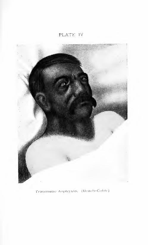
PLATE IV
Traumatic Asphyxia. (Beach-Cobb.)

SURGICAL DISEASES OF THE THORAX 241
painful because of the lack of elasticity of the skin on either side of the
median line.
It is said that these dorsal boils cause swelling and tenderness of the
inguinal lymph nodes, although this is difficult of explanation. lien-
schen 1 believes indirect infection of the hair follicles by pyogenic bacteria,
and states that the "mother furuncle" is always recognizable because
of its greater size. The virulence of the bacilli is increased by passage.In general furunculosis, the "daughter" boils continue to disseminate
the infection. Ilenschen mentions the likelihood of various metastases
from the escape of these pyogenics into the blood, but seemingly
ignores metastatic boils of the skin from internal sources.
If an aseptic major operation of any kind is carried out on a patientwith a florid furuncle, infection of the blood may follow during con-
valescence. (Ilenschen cites a case of appendectomy in a "boil
carrier" who fell a victim to a staphylococciis endocarditis directlyafter the operation. The writer lias more than once encountered
alarming infection in the case of a patient who was the subject of
relapsing furunculosis.) A furuncle at its onset may be aborted byvarious simple measures, but when it has reached the second stagesuitable incisions should be made directly through the necrotic center.
When a certain depth is reached it gapes and a little gauze should be
inserted to keep the wound open and favor the expulsion of the core.
The gauze should be soaked in a mild antiseptic solution.
Carbuncles on the back may indicate such metabolic disturbances
as diabetes and nephritis, the subjects, as a rule, being elderly. Occur-
ring in younger subjects without apparent cause, the disease may be
afebrile or it may present the picture of septicopyemia. Carbuncles
may be regarded as boils which develop so near together as to coalesce,
or as a simple boil which enlarges by concentric implication of nearbyfollicles.
The constitutional carbuncle is the form which carries witli it a mostserious prognosis, on account of the underlying metabolic disorder.
This form often attains a vast size.
Treatment. In the non-virulent carbuncle of young, robust subjects,cross-section or undermining of the lesions answers well. The incision
should be made deep, and the tissue thoroughly incised. The affected
fascia may be dissected out. Moist sterile dressings are appropriateand should be changed frequently. Fortunately, for cosmetic reasons,
these deep furuncles are much more seen in young men than in youngwomen, and this fact may be accounted for by the necessary recourse
of the former to barbers, as well as t,> the wearing of stiff shirt bands,
collars and the like.
In individuals who are in poor condition the entire carbuncle may be
excised under local anesthesia. Diabetes is not a contra-indication.
The incision is carried through the red areola down to the fascia andthe .mass dissected awav from the latter with rapid strokes. The
242 THORACIC Waffected fascia may be excised. Antiseptic or aseptic dressings are
applied. These should be changed frequently. The severe symptomsgenerally subside speedily. The defect may be repaired by skin
grafting.
Phlegmon of the Thoracic Wall. Different affections may be
described under this title, differing mainly in their causation. Theso-called primary diffuse phlegmon is an extremely rare affection, and
a review of the published cases shows that not all can be considered as
primary, since they may have originated at a distance. Secondarydiffuse phlegmon simply represents the burrowing of a septic abscess
into the cellular tissue of the chest wall, and may include the meta-
static phlegmon.
Diffuse ]'/ nidi'!/ phlegmon of the chest wall is in some respects a
condition *ni (/ciicrix. It is extremely rare -hardly over a do/en cases
having been recorded. Several French graduation theses were written
on the subject between 1.S75 and 1SS2. In a few cases, the primarylesion was a wound or felon on a linger, which was followed by a sup-
purative adenitis of the axilla, the pus burrowing into the cellular tissue
of the thorax. In other cases, the lesion followed an incision for enipy-ema or gangrene of the lung. The germ found is generally the strepto-
coccus. The patients have as a rule been in a poor, debilitated
condition.
The usual site of the phlegmon is the anterolateral portion of the
thorax, either close to the axilla or at a lower level. From the original
site, the entire half of the chest, may be involved, from the sternum to
the spine and from the axilla to the base of the thorax. Then the
edematous swelling gradually becomes purulent, and the cellular tissue
necrotic. The pus makes its way slowly between bundles of muscle,some of which may undergo necrosis. In a few cases, the pus has
burrowed into the throat or a secondary pleurisy has developed.
Symptoms. The general health is markedly depreciated. Some
patients have shown progressive adynamic conditions with diarrhea
and vomiting. The complexion is pale and earthy, the tongue dry and
fissured, and the teeth covered with sordes. The thirst is intense. The
pulse is rapid (110 to 120) and the temperature may go to 40 (\ The
picture is, therefore, much like that of a typhoid fever. The chest is
partly immobilized by pain and reflex action. The skin may be
edematous and pink. At a later period, when gangrene; is developing,it may be of a dee]) red. It may be marbled in texture and btilLe mayappear. As the skin becomes involved in the necrosis these bulhe
rupture and give exit to pus and gangrenous shreds of connective tissue.
Erysipelas may occur in association with such a phlegmon. Nearlyall of the reported cases have ended fatally.
Treatment. If there be an initial wound it should be promptlydisinfected and aseptically dressed. The phlegmon itself should be
treated by multiple, dee]) incisions made with a bistoury or Paquelin
cautery. The dressings should be changed frequently. The patient
may require saline infusion and active stimulation.
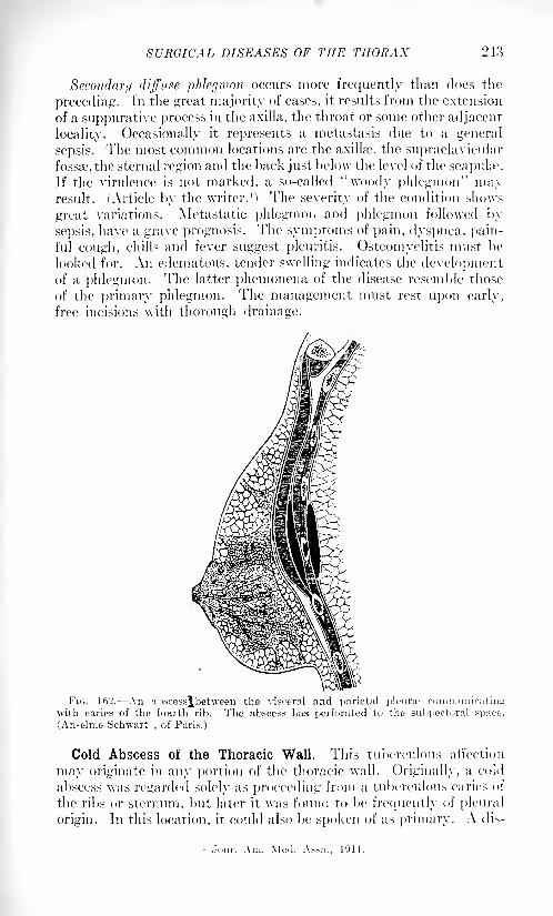
SURGICAL DISEASES OF THE THORAX 243
Secondary diffuse phlegmon occurs more frequently than does the
preceding. In the great majority of cases, it results from the extension
of a suppurative process in the axilla, the throat or some other adjacent
locality. Occasionally it represents a metastasis due to a general
sepsis. The most common locations are the axilla?, the supraclavicular
fossae, the sternal region and the back just below the level of the scapula1
.
If the virulence is not marked, a so-called "woody phlegmon" mayresult. (Article by the writer. 1
) The severity of the condition shows
great variations. Metastatic phlegmon and phlegmon followed by
sepsis, have a grave prognosis. The symptoms of pain, dyspnea, pain-ful cough, chills and fever suggest pleuritis. Osteomyelitis must be
looked for. An edematous, tender swelling indicates the developmentof a phlegmon. The latter phemonena of the disease resemble those
of the primary phlegmon. The management must rest upon early,
free incisions with thorough drainage.
l'"i<;. 162. An abscessjbetween the visceral and parietal pleune communicatingwith caries of the fourth rib. The abscess has perforated to the subpeotoral space.
(Anselme Schwart,of Paris.)
Cold Abscess of the Thoracic Wall. This tuberculous affection
may originate in any portion of the thoracic wall. Originally, a cold
abscess was regarded solely as proceeding from a tuberculous caries of
the ribs or sternum, but later it was found to be frequently of pleural
origin. In this location, it could also be spoken of as primary. A dis-
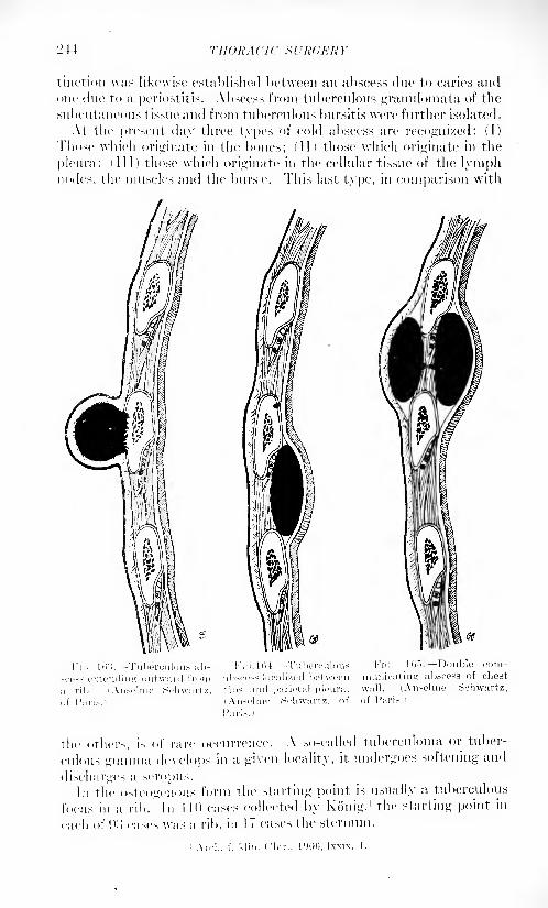
L'44 THORACIC SURGERY
tinction was likewise established between an abscess due to caries andone due to a periostitis. Abscess from tuberculous granulomata of the
subcutaneous tissue and from tuberculous bursitis were further isolated.
At the present, day three types of cold abscess are recognized: (I)
Those which originate in the bones; (II) those which originate in the
pleura; (III) those which originate in the cellular tissue of the lymphnodes, the muscles and the bursx*. This last type, in comparison with
ffff-
thc others, is of rare occurrence. A so-called tuberculoma or tuber-
culous gunnna develops in a given locality, it undergoes softening and
discharges a scropus.In the osteogenous form the starting point is usually a tuberculous
focus in a rib. In 1 10 cases collected by Kouig,1 the starting point in
each of (
.)o cases was a rib, in 1 7 cases the sternum.
SURGICAL DISEASES OF THE THORAX 245
In the majority of cases, tin; situation was anterior "(77) ;in '20 eases
it was posterior (cold abscess of the vertebra1is not considered here) and
in 13 cases it was lateral.
As a result of burrowing, the abscess may point at a considerable
distance from the diseased focus in the bone.
(The writer recalls the case of a young physician who was under his
care some years ago, in whom a cold abscess originating in the chest wall
pointed over the right sacro-iliac articulation. The diagnosis was onlymade at the time of operation. Thorough operative procedure, follow-
ing up the tract to the original focus, resulted in healing.)
The foci in the bone may diil'er greatly. There may be only a denu-
dation of the bone, or the latter may be the seat of a circumscribed
osteitis involving more or less of the thickness of the bone, which in
some cases undergoes spontaneous fracture. Several adjacent ribs
may at times be involved. The lesion may appear on the posteriorsurface of a rib in which case the abscess usually projects inward.
The bacilli may first manifest their activity within a given rib and
involve the periosteum secondarily. The cartilage and the osteo-
cartilaginous junction may be the seat of the original process (this
usually in elderly subjects). When an articulation is involved a typicalosteo-arthritis develops. When the sternum is involved it is usually
the body which is attacked. A sternal abscess may be superficial,
dee]) or retrosternal. There may be much destruction of bone and
spontaneous perforation is sometimes seen.
In the plenrogenoiis form two collections of pus are generally found;
one superficial, lying outside the thoracic wall, and a deep, retrocostal
collection communicating with this. This latter pocket lies between
the bony wall and the parietal pleura, which latter structure is almost
invariably thickened, adherent to the visceral pleura and studded with
tuberculous nodules. The adjacent lung may be involved in the process.
The sinus which unites the two pockets of pus may be short and direct
or long and tortuous. The plenrogenoiis abscess occurs by preferencein one of three localities, viz.: near the sternal margin in front, in the
axillary line laterally and at the angles of the ribs posteriorly. Thethird to tenth rib space is the usual seat.
The subjects in whom tuberculous abscesses usually occur are those
who, in general, are most subject to tuberculosis of the bones and lungs;children of ten or more years, adolescents and adults up to the age of
forty. Abscess in a lymph node is generally secondary to a tuber-
culous pleurisy.
Symptoms. When th '
patients are first seen they may be either in
the abscess stage or a fistula may be present. The abscess generally
undergoes evolution without marked local or general reaction. It is,
as a rule, firm at the outset and gradually undergoes softening, whenfluctuation may be obtained. The tumefaction is apt to be somewhat
sensitive.
In the recognition of a sacculate pleura! abscess a history and
symptoms of tuberculosis of the lung, or pleura, or both will be of
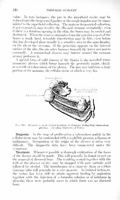
246 THORACIC SURGERY
value. In rare instances, the pus in the superficial cavity may be
reduced into the deep-seated pocket or the cough impulse may be trans-
mitted to the superficial collection. The main or deep-seated collection,it not evacuated, may involve the ribs and sternum secondarily; evenit there is a fistulous opening in the skin, the bones may be eroded and
destroyed. \\ hen the abscess originates from the anterior aspect of the
bones a small, hard, immobile tumefaction may be felt; even before
this has developed there usually is a sensitive area in the same localityon the rib or the sternum. If the periostitis appears on the internal
surface of the ribs, the pus often burrows beneath the latter and points
externally. A retrosternal abscess may burrow around the sternumor may perforate it.
A special form of cold abscess of the thorax is the so-called retro-
mammary abscess which forms beneath the pectoralis major, chieflyas a result of tuberculosis of the pleura. The ptis may infiltrate a large
portion of the mamma, the cellular tissue of which is very lax.
FIG. 106. Diagram to show typical locations of abscesses arising from tuberculous
pleurisy. (Anselme Schwartz, of P:ins.)
Diagnosis. In the stage of proliferation a tuberculous nodule in the
cellular tissue may be confounded with a syphilitic gtunma, a lipoma or
malignancy. Recognition of the origin of the abscess is often verydifficult. The diagnostic data have been ('numerated under the
symptoms.Treatment, \\nenever possible a thorough extirpation of the focus
of the disease should be made. This will generally be found to rest on
the removal of diseased bone. The resulting wound together with the
wall of the abscess cavity, may be mopped with pure carbolic acid
followed by alcohol. The introduction of a cigarette drain or rubber
drainage tube will generally be a wise measure. In isolated instances,
the writer has been able to attain apparent healing by aspiration
together with the injection of a formalin solution or of iodoform in
glycerin; these were probably easels in which there was no diseased
bone.
SURGICAL DISEASES OF THE THORAX 247
In certain cases, a complete removal of the wall of the abscess is to be
attempted. Search should always be made for secondary pocketswhich can usually be located most conveniently by injecting the sinuses
very slowly but very thoroughly with bismuth paste and making an
.c-ray plate which will show all the ramifications and all of the pockets.
Emphasis is laid on the absolute necessity for the greatest thoroughness.
Resulting tuberculous fistula may at times be cured by the use of
Beck's bismuth paste.
Osteoperiosteitis of the Ribs and Sternum. True Osteomyelitis.
This is generally of hematogenous origin but at times follows injuries.
It represents a metastasis from almost any conceivable locality; the
ordinary causes of suppuration being responsible. In most cases, a
single rib is attacked with the costochondral junction as a favorite
location. Another preferred locality is that part of the rib lying close
to the spine. The affection attacks children up to ten years of age and
young adults (twenty to twenty-five).
Clinically it is a severe, painful affection, appearing insidiously and
without apparent cause. However, the source of the metastasis can
usually be found in some primary focus: a boil, a felon, an attack of
influenza or of gonorrhea. There is circumscribed swelling, tender-
ness, pain and painful respiration. The pus finds its way through the
bone, forming a subperiosteal abscess. The periosteum is perforatedsooner or later; in some cases, pus forms between the bone and the
pleura, closely simulating a sacculated empyema. More frequently,the pus from the abscess escapes beneath the skin where it may burrow
downward. The affected bone often undergoes necrosis, diastasis or
spontaneous fracture. A fistula frequently results after evacuation
of the pus. Should the osteomyelitis extend along the length of the
rib the latter may undergo necrosis en manse.
Diagnosis. It will be seen from the foregoing that the clinical mani-
festations may be manifold. ( 'arefully made .r-ray pictures will usually
establish an accurate diagnosis.
Treatment. Whether the bone is extensively or slightly involved,
resection is indicated. A long incision is made and a sufficiently exten-
sive resection done. This tends to remove the disease focus and to
prevent fistula formation.
Typhoid Osteoperiostitis. In this form of osteomyelitis, the cartilages
are often involved to a considerable extent (osteochondritis). Typhoidfever is frequently followed by osteomyelitis and the costochondral
junction is one of its localizations. It is likely to occur in the protractedand relapsing forms of typhoid fever and it is even said that its pres-
ence may at times be foreshadowed during the course of the disease.
The typhoid bacillus may be present in pure culture or in association
with some of the pyogenic germs, which often suppress its activity.
Typhoid osteochondritis is a much more benign affection than is the
pyogenic variety. Pus formation is, as a rule, relatively slight and
delayed; the bony and cartilaginous tissues appear to retain their
integrity for a long period. There is often a prolonged incubation or
IMS
latent period suggesting that certain intercurreiit conditions mayactivate tlic bacilli. Jn the pure typhoid case, the course of the disease
is not unlike thai of tuberculosis. The cortical bone first thickens andis then absorbed, while granulation tissue develops, with caseation
at the medulla. The greater the element of progenies present the
more the picture approximates that of pyogenic osteomyelitis, althoughthe cartilage, even the whole of it, may be the seat of the lesion. Thiscondition may be- self-limited without sequel or may recover after
discharge of the pus; healing of the fistula. In such cases, bits of deadbone or cartilage may come away.
Diagnosis. This should rest upon certification of the typhoid bacilli,
although necrosis of a rib or costocartilage, shortly after the occurrence
of the typhoid fever, should strongly point to the typhoid form.
Treatment. A very thorough surgical extirpation is indicated.
Numerous cases of the condition have been under the care of the writer
and he has been impressed with the necessity for a most radical extir-
pation. It is but proper to say that in one of the writer's cases, that
of a college professor residing in a neighboring town, repeated andextensive operations were without avail and healing took place duringthe administration of autogenous vaccines.
Tuberculosis of the Ribs. Tuberculous caries of the ribs causes
various affections of the adjacent soft parts (cold abscesses, fistula?) .
In certain subjects apparently sound, a tuberculous rib may be the
only active manifestation of a latent tuberculosis. In 70 per cent, of
all patients with tuberculous rib, other tuberculous lesions coexist.
The lesion is much more frequently secondary than primary; it is
often derived from nearby lesions by lymphatic propagation; some-
times from remote lesions. Mediastinal tuberculous glands are believed
to be a frequent source of bone infection. Injuries to the thorax
sometimes result in tuberculosis of the ribs and not a few cases have
originated thus among fighting troops.'I hese lesions comprise many pathological changes which involve the
periosteum, the cortex and the medulla of the ribs, but which can be
considered jointly. Spontaneous recovery is rare, for, as a rule, the
tuberculous focus in the bone gives rise to local and burrowing abscesses
which point in a great variety of localities (snbpectoral, lumbar,
abdominal, intrathoracic). Fistulous tracts are formed, and fragmentsof bone sometimes come away. \Ylien the tuberculous focus is first
apparent, a fusiform swelling is seen and felt. This is the first stage
and is due to the formation of granulation tissue. \\hen the latter
softens and forms a caseous purulent mass the second stage of abscess
torn ation begins The skin may or may not assume a reddish color.
A soft swelling is apparent, over the lesion and may be shown to con-
tain pus. Spontaneous opening leaves a fistula.
Diagnosis. Diagnosis during the first and second stages should
easily be made; the ./-ray gives information of a primary affection of
the marrow, and puncture may show the presence of bacilli. After
fistula formation, the probe may find rough bone. To map out the
fistula, bismuth paste is injected and an .r-ray picture taken.
sritfllCAL DIXKAXEK OF THE THORAX 249
Prognosis. Prognosis is generally good, although cure may be
difficult.
Treatment.- This should rest, upon thorough extirpation of the
diseased focus or foci. If a fistula be present, its tract should be well
dissected out. The affected rib or ribs should be divided through sound
tissue, after removing the periosteum in the usual way. Care should
be taken not to injure the pleura which will generally appear to be
thickened. Almost invariably, pleura! preservation will be found,
however, to be easy. All secondary pockets of pus are to be thoroughly
evacuated, curetted and mopped with pure carbolic acid, followed byalcohol.
Abscess of the Subpleural Connective Tissue (Peripleuritis). Sup-
puration of the cellular tissue between the chest wall and parietal
pleura is practically always secondary to some inflammatory process in
the tissues above or beneath it, and is, therefore, to be regarded as a
complication. It is generally ushered in by a chill, by pain and respira-
tory embarrassment. With the progress of the affection, the pain and
dyspnea increase and fever appears. Inspection may reveal a slight
bulging at certain intercostal spaces, there is flatness on percussionand a lessening or absence of the vesicular murmur. When left to itself,
the pus generally discharges externally after the skin has becomethinned and reddened. An obstinate fistula may persist. In certain
cases the pus escapes into the pleura, causing an empyema.Diagnosis. The diagnosis of this condition is difficult, since it is
readily confounded with pleurisy, especially the encysted form. Tho
former, as a rule, occurs at the side of the thorax and sufficiently high to
permit of the recognition of a /one of lung tissue beneath which a clear
percussion note and normal auscultatory sounds are found. Thelocation of encysted pleurisy is generally lower. Peripleuritis, as a
rule, runs the more rapid course.
Treatment.- This consists of thoracotomy and drainage, perhaps with
the resection of a portion of one or more ribs.
Actinomycosis of the Thorax. When this affection occurs in the
chest-wall it is almost invariably secondary to actinomycosis in the
lungs or in some of the contiguous areas (neck, mediastinum, vertebra',
abdominal cavity). Primary actinomycosis occurs, very rarely, in the
chest-wall as a result of inoculation. Such cases are atypical.
Whatever its origin, an actinomycosis of the chest-wall closely
resembles woody phlegmon; it undergoes multiple softening, cicatri-
zation, and contraction of the thorax, which, as a result, shows con-
siderable deformity. Occasional fistuhe arc present.In the rare instances of primary implication of the skin, the growth
may be excised. In the secondary forms, all foci of softening, all
fistulye, etc., must be curetted and potassium iodide given in large
doses. Lugol's solution may be used locally. The prognosis dependson the site and extent of the lesion.
Actinomycosis of the pleura is generally secondary to that of the
lungs. The pleura develops adhesions, becomes thickened, and serous
250 THORACIC SURGERY
or purulent exudates form. Pleural actinomycosis is a stage in the
extension of the disease from the lungs to the chest-wall. Fistula? maycommunicate with the pleiiral area, and, at times, the ray fungi will be
found ID the discharge.
Actinomycosis of the thorax, therefore, is primarily a pulmonaryaffection, which, after ravaging the lung tissue, invades the pleura andthe chest wall. Like tuberculosis and cancer of the lungs, it most often
originates in the bronchial tree, whence it invades the outlying paren-
chyma. Further, the introduction into the bronchi of various sub-
stances containing the ray fungi recalls in some degree the action of
other minute foreign bodies, which, having reached the bronchioles,
may set up a number of conditions as a result of the mechanical irri-
tation; obstruction, infection, etc. (See Foreign Bodies.)
It is said that, for a time, the actinomycosis may be manifested onlyas a chronic catarrhal bronchitis. The process then traverses th?
bronchial walls and extends into the pulmonary alveoli, causing
respectively peribronchitis and localized bronchopneumonia. The foci
of the latter condition tend to soften and form cavities, while the
peribronchitis causes extensive proliferation of the connective tissue,
followed by induration and contraction. The affected area of lungtissue becomes therefore the seat of a fibrosis.
Eventually, the process reaches the pleura, causing either a dry
pleurisy with adhesions, a serothorax, or both. Thence the processinvades the chest-wall, as already stated. The disease, while thus
moving along the lines of least resistance, still continues to extend from
its original foci. It infiltrates the thorax and the pictures may then
become extremely diversified.
The disease may pursue one of several courses and hence is often
confused, in diagnosis, with a number of conditions, chief of which is
pulmonary tuberculosis. Sputum examination is by no means decisive,
for while bacillus findings may not be forthcoming, neither will the
fungus be demonstrated, in a large proportion of cases. Actinomycosisseldom attacks the apex of the lung; it is most commonly found at the
base. At this period the .r-ray pictures may show a diffuse shadow in
which are to be seen small dark and light areas.
When the process has extended to the pleura and the thoracic wall,
an empyema may be simulated. In trial puncture for suspected
empyema, the granules of actinomycosis may be brought away. If
the disease undergoes softening with fistula formation the suggestionof empyema is still more pronounced. Actinomycosis of the chest-wall
may readily simulate a sarcoma.
The tendency of the affection to an unlimited invasion of neighboringtissues and the lack of reliable resources to arrest its spread, makes the
prognosis definitely bad although the patient may live for many years.
In the earliest stages, a greater or lesser degree of surgical relief is avail-
able; at later periods the disease may be favorably influenced by drugs.
Fven in cases of years' standing, the lesions are sometimes operable.
According to Garre, 8 cases have been cured surgically.
CYSTS AND TUMORS OF THE THORAX 251
Cure may occur under one of two conditions: (1) the diseased focus
is still confined to the lung and the surgeon proceeds exactly as in the
case of any other diseased focus, by resecting the chest-wall and a
portion of the lung; (2) (and this is by far the more common situation),
the chest-wall is already secondarily involved; in this case, the fistuhe
are slit open and all of the cavities are evacuated and curetted. Nodules
of diseases are to be extirpated. The chest-wall is resected, and, if
practicable, the cavities in the lung are not only opened and drained
but the disease area is extirpated. In some cases, indeed in manyinstances, these measures must be avowedly palliative. Large doses of
potassium iodide may be given as an adjuvant to surgical measures.
In the experience of the editor the following plan of treatment has been
most effective in these cases: Ninety grains of potassium iodide are
given in half a pint of milk at A.M., 2 P.M., and 10 P.M. This is immedi-
ately followed by a pint of hot water which results in the rapid absorp-tion of the remedy without giving any gastric disturbance. 'Fhe
treatment is continued for four days, then interrupted for one week
during which time tonics are administered; then the treatment is
repeated again for four days. This plan is continued until the patientis apparently well. The administration of potassium iodide is then
repeated for four days once a month for several months, in order to
destroy any ray fungi which might not have been reached by the early
treatment. By continuing this treatment, a number of apparently
hopeless cases have recovered permanently, although the use of smaller
doses of potassium iodide has previously been tried without benefit.
CYSTS AND TUMORS OF THE THORAX.
Echinococcus of the Chest. Echinococcus cysts have been found
in the subcutaneous tissues of the chest, in the ribs and the sternum,
and in various muscles, notably the pectorals. A number of cases of
p -unary echinococcus have occurred in the pleura! cavity, but pleura!
echinococcus is usually secondary to a pulmonary lesion and due to a
rupture of the cyst into the pleura! cavity. Primary echinococcus of
the lung is much more common than that of the chest-wall. It is not
of uncommon occurrence and represents a direct infection of the lungfrom the intestine, being second in frequency only to echinococcus of
the liver.
Echinococcus of the Chest-wall. For location see the foregoing.
The diagnosis is probably not often made in advance of treatment,
although trial puncture may reveal scolices. Puncture may give the
symptoms of anaphylactic shock while operative infection with the
scolices is to be borne in mind. Kchinococens of the chest-wall is gener-
ally diagnosticated first as a tumor or an abscess. The hydatid thrill
is seldom to be obtained.
The fluid obtained on puncture is clear and free from coloring matter
or albumin; eosinophilia is present in the blood.
L'.VJ THORACIC Xl'KCKRY
Treatment. If operable, the tumor should be enucleated. In other
cases the sac should be cut down upon, punctured and disinfected with
foni-aliii solution or incised and drained the last named resource, an
old one, being attended with danger of infection.
Echinococcus of the Lungs. When there is intestinal infection with
this parasite, the lung is a favorite seat of metastasis. In most cases a
single cyst is present and the right lung is the most common seat (the
left lung is said to be involved only about one-half as frequently). The
cyst gradually continues to increase in si/e, and, until rupture or
infection occurs, produces only pressure symptoms and irritation. If it
compresses a bronchial tube, bronchial irritation comes on. If com-
pressing the pleura at a given point, a localized dry pleurisy maydevelop. The cyst is, at times, responsible for the developmentof a frank pleurisy or pneumonia and this without, previous rup-ture.
Termination in rupture occurs owing to the tendency of the cystwall to become thinned where pressure is the greatest. Rupture is
often brought on by forced inspiration (as after overexertion) or byexternal violence. The tissues upon which the cyst has been pressingare also thinned and vulnerable so that a cyst may rupture directly
into a bronchial tube. In such a case the patient coughs up a thin tluid
mixed with blood. Daughter-cells and scoliees may be found in this
fluid. (The writer recalls a case in which an echinococcus cyst of the
liver found its way through the diaphragm and through the adherent
right lung, burrowing into a bronchus through which it was discharged.The scoliees were readily found in the sputum. Operation on the
original cyst in the liver was followed by a complete cure.) If a cyst
ruptures into the pleural sac, an acute pleural effusion is set up. After
a cyst has ruptured it may close permanently, or it may refill andfurther rupture eventually recur.
A ruptured cyst becomes infected and undergoes suppuration.
Hence, abscess or gangrene of the lung may accompany the condition.
These suppurating cysts behave similarly to the sterile ones, until
rupture occurs. If rupture occurs into a bronchial tube, a large
quantity of pus is expectorated; if into the pleural sac, a pyothoraxresults.
Diagnosis. In making a diagnosis, it is to be remembered that
sputum containing scoliees does not necessarily come from the lung,
for a cyst in the liver may penetrate the diaphragm and discharge
through a bronchus. A cyst seldom reaches the size of a man's fist
before rupture, and until rupture occurs it simulates a tumor. A small
cyst occurring in an upper lobe has simulated a tuberculous focus. If,
with dnlness on percussion, there is no disturbance of respiratory
rhythm one may think of an echinococcus cyst.
Treatment. The operative treatment closely resembles that of
abscess or gangrene of the lung.
CYSTS AXD TUMORS OF THE THORAX 253
Benign Tumors of the Thorax.
The superficial growths wliich occur upon the thorax have little or
nothing to distinguish them from those of other localities. Keloid
over the sternum is perhaps the most characteristic. The nevi whichoccur over the thorax, the wen, fibroma inolluscum, angiomata, etc.,
have a strong congenital and familial tendency. The bones and car-
tilages may be the seat of exostoses, enchondroses and acquired
osteophytes. So far as is known, the pleural cavity is attacked by but
one form of benign tumor, the subserous lipoma. The various con-
nective-tissue tumors may occur in the lung but they rarely cause
trouble. They are most liable to be found at autopsy.
Congenital Presternal Cyst. This is a very rare form of neoplasmof the soft tissues of the thorax, only some 27 cases having been placedon record. The growth is situated over the manubrium, in the median
line, and may be of variable si/e. Of 25 cases analyzed, 21 were dermoid
cysts, 2 were mucoid and 2 were simple serous cysts. The mode of
origin of these formations is conjectural. The most plausible view is
that they are branchiogenous, analogous to the branchiogenetic cysts
and fistula" of the neck. Clinically there is an indolent mass, datingfrom birth, that gives a sense of fluctuation, cannot be reduced andwhich is not adherent above or beneath. The growths show a pro-nounced tendency to suppuration. They should always be thoroughly
extirpated.
Malignant Tumors of the Thorax.
All portions of the thorax may be the seat of malignant growths.The skin and subcutaneous tissues are only too frequently the seat of
local metastases, after operations for mammary cancer, and the sclerotic
changes which they undergo, with the resulting induration, give rise
to the so-called cancer en cuirasse. (See article on the Diseases of the
Breast. ) The ribs may be attacked by sarcoma, the pleura by sarcoma
and endothelioma, the lungs by carcinoma and sarcoma.
TV.Moits OF THE CIIKST-WALI,.
Leaving out of consideration the secondary malignant tumors wliich
appear in this region from direct extension or from metastasis, there
remain certain primary tumors of the ribs and sternum which are
malignant or semimalignant in character.
Neoplasms of the Ribs. These begin, by preference, in the lower
ribs, and tend to grow in the axis of the bones. The tumors pushinward, causing pleuritic adhesions and invading the pleura and lung;
while at the same time they grow outward through the soft parts,
undergoing nlceration. When seated low, as is often the case, the
diaphragm and liver may be involved secondarily. Metastases are the
rule. Over 100 cases of these growths are on record, the majority
being examples of enchondroma or sarcoma. Mixed forms of sarcoma
2,54 THORACIC SURGERY
have been seen in a certain number of cases. In 1898 Quenu 1 collected
notes of 5^ cases of tumor of the ribs; about equally divided betweensarcoma and enchondroma.
Symptoms. The first symptom is pain. This pain may be local or
diffused, spontaneous or induced. When seated in the lower ribs, these
tumors sometimes cause pain in the shoulder of the same side. At first,
the pains are intermittent and slight, later they become more constant
and severe. The chest-wall is thus slightly immobilized causing a
certain interference with inspiration. Fixation to the ribs serves to
distinguish the neoplasms. Their size, form and consistency show all
degrees of variation.
Sarcoma of the Chest-wall. -Chief emphasis is placed here uponsarcomata of the bones, as those of the superjacent soft parts are rare.
Sarcoma of the ribs may originate in the marrow or in the periosteum.The former variety soon induces a fracture of the rib and both forms
grow rapidly, undergoing metastasis early. The tumors may projectoutward, forming a mass approximately the si/e of a child's head, which
tends to break down and ulcerate. They may penetrate the thorax andcause the so-called "tumor pleurisy'' with symptoms of pressure or
irritation. Within the thorax, they produce metastases.
The sarcoma of the chest-wall occurs as a rule, in men, in the lower
ribs and on the anterolateral aspect.
Treatment. For treatment see Resection of Wall of Chest.
Enchondroma of the Chest-wall. -This semimalignant affection is
not a rare lesion of the ribs; it is about one-half as frequent as sarcoma
in the same locality. The growths tend to occur on the right side and
to involve the lower six ribs anterolaterally. In spreading, they maypass from one rib to the next, and from the lower ribs they may invade
the various vise-era (liver, lungs, heart, etc.) Clinically, these are found
as large, broad-based, irregular tumor-masses, which may cause no
early symptoms of pressure or irritation. At tiir.es, they give rise to
pressure pains through some of the local nerves. Eventually, the
tumors form great round or fusiform bosses.
The course is generally extremely slow, with no tendency to heal,
although ossification may take place; when this does occur, malignant
degeneration appears to be prevented. The chief dangers are from
internal growth, from metastases and from recurrence after intervention.
All chondromas, however innocent in appearance, should be extir-
pated when operable.
MAUGXAXT TUMORS OF THE PLKURA.
These may be primary or secondary, the latter being much the more
frequent. Whatever their nature, they give rise to much the same
symptoms. Whether originating in the pleura or invading it secon-
darily, a so-called "tumor-pleurisy" is set up; this is usually associated
1 Hev. de chir., 1898, xviii, 365.
CYSTS AND TUMORS OF THE THORAX 2.15
with a hemothorax. Sooner or later, therefore, there is dulness on
percussion.Fibrosarcoma and Myxosarcoma of the Pleura. These growths are
malignant and may attain very great dimensions. As a rule they show
little tendency to invade nearby tissues and therefore the chief symp-toms are those of pressure. Recognized at first as autopsy findings,
they were brought into the domain of operative surgery by (iarre whoremoved one successfully. (This growth weighed over 12 pounds.
Originating in the pleura, it slowly enlarged until it nearly filled one-
half of the thorax. In this case, dulness on percussion increased in
proportion as breathing sounds diminished. The diagnosis was made
by .r-ray pictures and by exclusion. Microscopically, it was a spindle-
cell sarcoma.)
Sarcoma of the Pleura. -The typical primary sarcoma of the pleurais said to be practically limited to the round-cell form. ^Vhen seen
early in the history of the disease, there are flattened nodules on the
pleura. Recognition at this stage is extremely difficult. If the diag-
nosis is made during this early period, whether accidentally or other-
wise, prompt extirpation of the nodules may effect a cure. The rate of
growth of these initial lesions is said, however, to be extremely rapid.
The growth, invades the chest-wall and clinically resembles sarcoma of
it. Primary sarcoma of the pleura is extremely rare, and the great
majority of cases are secondary to sarcoma of the chest-wall and thus
may be considered under that head. ( In the writer's experience secon-
dary sarcoma of the pleura and lung forms the most frequent compli-cation of sarcoma- of the testis. Almost all of the orchitic sarcomata
which the writer has removed have later succumbed to intrathoracic
metastasis.)
Endothelioma of the Pleura. This is a malignant endothelioma
originating from the endothelium of the serosa. It is said to behave
more like a productive inflammation than like a tumor. Appearing, as
it does, on either leaflet of the pleura the two opposed lesions may fuse
and produce adhesions. The growths give rise to but few symptomsat the start, although they rapidly invade the neighboring tissues
thoracic wall, diaphragm, lung.
Secondary Carcinosis of the Pleura. -It has been seen that true
carcinoma rarely attacks the pleura primarily. Secondary metastases,on the other hand, are very common.As is usual with the other seros;e, cancerous metastases represent a
carcinosis, and even at times a miliary Carcinosis simulating tuber-
culosis.
MALIGXAXT TU.MOHS OF TIIK Li:.\<;.
The writer believes that malignancy of the lungs is seldom recognizeduntil the; disease is well advanced. Then, certain obstructive and irri-
tative symptoms begin; pain and oppression in the chest, with a dry
cough. When marked pressure symptoms occur, dyspnea, cardiac
disorders and neuralgias from nerve compression result. The lung
LMf) THORACIC XURGKRY
tends to shrink in size and retraction of the chest-wall occurs (obstruc-tion of a bronchus may cause atelectasis of an entire lobe.) The chief
physical signs are dulness on percussion and bronchial breathing.When the tumor mass breaks down, bronchopneumonia or pleurisy
may develop.
Diagnosis.- -1 >iagnosis in even the moderately advanced stagesshould not be difficult. Lenhartz 1 made twenty-four consecutive
diagnoses. lie availed himself of repeated .r-ray and sputum exami-
nations.
In making a diagnosis it is necessary to exclude syphilis, tuberculosis,
actinomycosis and other chronic inflammatory processes, as well as
leukemic tumors, mediastinal growths, aneurysms, etc.
Treatment.- The operation of resection of the chest-wall for the
removal of tumors is described elsewhere. Tumors of the lung, byreason of their bronchial origin, appear to constitute a special varietyof thoracic growths, which, as statistics show, are with the greatest
difficulty successfully reached by such resection. It must not be
forgotten, however, that the cortex of the lung may be attacked secon-
darily by malignant diseases of the chest-wall and pleura, and it is
probable that in such cases, the future will show a constantly increasing
percentage of encouraging results.
Since the introduction of the pressure-difference procedures a rela-
tively considerable number of resections of the chest-wall have been
made for the removal of pulmonary growths. Before the introduction
of pressure-differences 51 interventions were recorded with 17 deaths;
it is believed that under the employment of pressure-differences statis-
tics have very materially improved. The writer is without experience
here, but he firmly believes, as stated elsewhere in this article, that
future results in this form of intrathoracic surgery, as in other forms,
will show constantly improved statistics. He believes that surgeons in
the future will be able to prolong or save life, in cases where malignantdiseases of the pleura, chondroma of the chest-wall, etc., have attacked
the lung.
Primary Cancer of the Lung. Garre 2 states that he has collected
note's of 130 cases of primary cancer of the lungs with but 9 operations.
Only one patient survived the intervention long enough to be con-
sidered as relatively cured.
Little is known of the special causal factors. The right side is muchmore frequently attacked than the left, and in the vast majority of
cases (00 per cent.), the disease begins in a bronchus (transition from
the second to the third twigs) and, hence, in reality is a bronchial cancer.
Three-fourths of all patients are males. An association with tuber-
culosis is more frequent than mere coincidence would seem to explain.
On account of their bronchial origin, it is said that these growths, mayat. times, be recognized by the bronehoscope.
In the great majority of cases the new growth is not an epithelioma,
1 Munrheii. med. \\Ylmsrhr., 1SSI7, vol. xliv.
2 Sunrery of the Lunus, 1912, second edition.
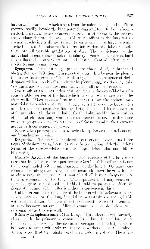
CYSTS AND TUMORS OF THE THORAX 257
but an adenosarcoma which arises from the submucous glands. These
growths readily invade the lung parenchyma and tend to form circum-
scribed, uneven masses or cancerous foci. In other cases, the process
creeps along the bronchi, and, in this way, infiltrates the lung paren-
chyma, producing a diffuse type. From a smaller or larger circum-
scribed mass in the hilus to the diffuse infiltration of a lobe or lobule,
there are all possible gradations of size. The consistency of the
individual lesions shows much dissimilarity. Some masses are as hard
as cartilage while others are soft and elastic. Central softening and
cavity formation may occur.
Symptoms. The initial symptoms are those of slight bronchial
obstruction and irritation, with reflected pains. If it be near the pleura,the cancer focus sets up a "tumor pleurisy." The coexistence of slight
dyspnea with a bloody effusion into the pleura, suggests the diagnosis.Weakness and cachexia are significant, as in all cases of cancer.
One result of the obstruction of a bronchus is the consolidation of a
considerable portion of the lung which may cause a retraction of the
chest-wall. When cavities form in cancerous tissue the broken-downmaterial may reach the sputum. Cancer cells, however, are but seldom
found; the most suggestive findings being blood and large spherulesof fat granules. On the other hand, the fluid obtained by the aspirationof pleural effusions may contain actual cancer tissue. In due time
pressure symptoms develop in the veins of the neck and in the recurrent
nerves with consequent cyanosis.
Fever, when present, is due to a toxic absorption or to actual cancer-
ous bronchopneumonia.Diagnosis. The .r-ray has rendered great service in diagnosis: three
types of shadow having been described in connection with the various
forms of the disease lobar (usually upper lobe) hilus and diffuse
bilateral type.
Primary Sarcoma of the Lung. Typical sarcoma of the lung is so
rare that but 30 cases are upon record (Garre). (This affection is not
to be confounded with lymphosarcoma of the lung.) Ordinary sar-
coma almost always occurs as a single focus, although the growth mayattain a very great sixe. A "tumor pleurisy" is more frequent here
than in carcinoma of the lung. The aspirated fluid may contain a
so-called giant vacuole cell and this is said to possess considerable
diagnostic value. (The writer is without experience in this.)
Unlike certain forms of cancer of the lung in which cachexia appears
slowly, sarcoma of the lung invariably metastasizes early and this
with early cachexia. There is as yet no successful case of the removal
of a pulmonary sarcoma. Alleged examples have doubtless been
sarcomas of the thoracic wall.
Primary Lymphosarcoma of the Lung. ----This affection was formerlyclassed with the primary sarcomata of the lung, but of late yearsit has taken on new significance as an occupational malady, since it
is known to occur with fair frequency in workers in certain mines,
and as a result of the inhalation of arsenic-bearing dust. The affec-
VOL. II 17
258 THORACIC SURGERY
tioii is primarily one of the bronchial glands and the peribronchialtissues. Like others of the cancer types already described, it spreads
along the bronchial tree, occasionally giving rise to one or more specialnodules of disease, these often of considerable sixe.
It is thus apparent that lymphosarcoma shows more resemblance to
carcinoma than to the ordinary primary sarcoma of the lung, since it is
propagated along the bronchial tree and gives rise to bronchial
symptoms.Secondary Malignant Growths of the Lung. Wliile primary cancers
and certain sarcomata (lymphosarcoma) of the lung have a bronchial
origin and only exceptionally reach the pleura, it may be repeated that
various malignant growths of the chest-wall and pleura can involve the
lung by extension; hence, there is a continuous series of malignanttumors, primary or secondary, which involve the entire thickness of the
thoracic wall, the pleura and the cortex of the lung. Not a few of these
cases are examples of enchondroma. As has already been stated, the
writer believes that the surgery of the future will present in this region
results which are more and more encouraging.It is not possible to place too much emphasis on the well-known fact
that carcinoma of the breast is often complicated by carcinoma of the
lung. A dry cough is one of the first symptoms and should always be
looked upon with suspicion.
Serotherapy.^ The writer has employed this widely in inoperable
malignant growths and following operation for such growths. His ownresults have been far from encouraging.
The Use of Radium and the X-rays.- At this time (June, 1020 it maysuffice to say that these measures are undergoing widespread investi-
gation.
SURGICAL DISEASES OF THE PLEURA.
It has been stated that traumatic hemothorax causes an aseptic
fever during its absorption. The surgeon regards the great majority
of cases of pleurisy with effusion (serothorax) as sterile; these effusions
often accompany other conditions of the lungs as tumors and cysts
and also follow pneumothorax. The so-called rheumatic pleurisies,
which are apt to be tuberculous, are doubtless sterile. A large sero-
thorax may cause pressure symptoms in the chest demanding para-
centesis, but, as a rule, the fluid portion of the effusion is absorbed,
leaving the exudate behind. This exndate should be carefully watched
for evidences of absorption, and puncture should be made 1 both for
diagnostic aid and for aspiration. In this way a bacterial infection
may be revealed when present. A trial puncture may be made with a
small syringe and suitable needle. If it be desired to evacuate the
pleura, a large aspirating needle and suitable apparatus should be used.
Air must not be allowed to enter the pleural cavity during the escape
of the exudate.
It has long been known that the sudden change of intrathoracic
pressure may be followed by syncope as a result of circulatory disturb-
ances.- When the cavity is emptied, the compressed lung suddenly
SURGICAL DISEASES OF THE PLEURA 259
expands. To prevent too sudden changes an apparatus like that of
Dieulafoy or Potain is used. In the latter, the exudate is sucked into
a bottle in which there is a partial vacuum.
Purulent Pleurisy or Empyema. These terms are generic, includinginfected serothorax, and cases which are purulent from the start.
Sharp distinctions cannot be left to the microscope, and it is difficult to
classify the various forms of the condition.
Collections of pus in the pleura may be primary or secondary. Theymay be large enough to cause pressure symptoms. They frequentlyoccasion toxic absorption fever and often give rise to a rapid general
septic infection. Secondary empyema is a metastasis of septic infection.
Despite the character of the pus it can at times be slowly absorbed. In
all untreated cases, however, the pleura becomes greatly thickened and
undergoes cicatricial retraction at the expense of the lung. The pus of
an empyema at times, breaks into the lung while air from the lungitself enters the pleura! cavity; in this way one type of a pyopneumo-thorax may develop. A tension pneumothorax may follow as a further
complication. In some cases of pyopneumothorax the bronchi art'
filled with pus with attendant danger of aspiration and suffocation.
The empyema may discharge outwardly with entrance of air into the
pleura. The so-called sacculated empyemata are small collections of
pus encapsulated in the thickened pleura; their diagnosis is, at times,
very difficult. A'-ray pictures may be of aid.
The following rough etiological classification of empyema may be
made:
Primary traumatic
Pulmonary (metapneumonic) .
Mediastinal.
Metastatic.
Abdominal.
Forms of Empyema Surgically Considered. \a) Traumatic- There is
usually an infected wound of the pleura and the effusion is generally
purulent from the start because pyogenie microorganisms are trans-
mitted directly into the pleural cavity. Since, as a result of the injury,
hemothorax usually coexists, the blood plasm furnishes an excellent
culture medium for germs. A pneumothorax also accompanies this
in many cases and thus weakens the resistance of the pleura. In other
words, the large pleura! cavity is rapidly filled with a highly infected
fluid. This soon begins to undergo absorption, with a resulting severe
Toxemia, (ieneral sepsis supervenes, icterus and other symptoms comeon and the prognosis becomes grave. Prompt and thorough operative
procedure is imperative. Free, dependent drainage is necessary. This
should be accomplished by passing two drainage tubes at least 1 cm. in
diameter entirely through the chest-wall. The portion of these tubes
within the chest cavity should contain numerous small preforations.Sections of one or two ribs should be removed in the posterior axillary
line while the anterior opening for the drainage tubes may consist of
an incision between the costal cartilages. Frequent or constant irriga-
tion of the cavitv with normal salt solution mav be wise in certain
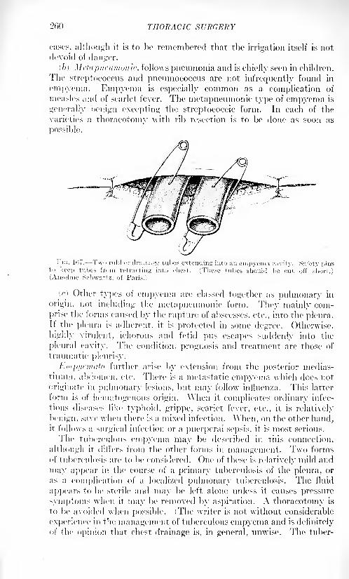
2f)() THORACIC SURGERY
cases, although it is to be remembered that the irrigation itself is notdevoid of danger.
(b) Mctapni'umonic, follows pneumonia and is chiefly seen in children.
The streptococcus and pneumococcus are not infrequently found in
empyema. Empyema is especially common as a complication of
measles and of scarlet fever. The metapneumonic type of empyema is
generally benign excepting the streptococcic form. In each of the
varieties a thoracotomy with rib resection is to be done as soon as
possible.
I- IG. 107. Two rubber drainage tubes extending into an empvema c;
keep tubes from retracting into chest. (These tubes should(Anselme Schwartz, of Paris.)
(c) Other types of empyema are classed together as pulmonary in
origin, not including the metapneumonic form. They mainly com-
prise the forms caused by the rupture of abscesses, etc., into the pleura.If the pleura is adherent, it is protected in some degree. Otherwise,
highly virulent, ichorous and fetid pus escapes suddenly into the
pleural cavity. The condition, prognosis and treatment are those of
traumatic pleurisy.
Empyemafa further arise by extension from the posterior medias-
tinum, abdomen, etc. There is a metastatic empyema which does not
originate in pulmonary lesions, but may follow influenza. This latter
form is of hematogenous origin. When it complicates ordinary infec-
tious diseases like typhoid, grippe, scarlet fever, etc., it is relatively
benign, save when there is a mixed infection. When, on the other hand,it follows a surgical infection or a puerperal sepsis, it is most serious.
The tuberculous empyema may be described in this connection,
although it differs from the other forms in management. Two forms
of tuberculosis arc to be considered. One of these is relatively mild and
may appear in the course of a primary tuberculosis of the pleura, or
as a complication of a localized pulmonary tuberculosis. The fluid
appears to be sterile and may be left alone unless it causes pressure
symptoms when it may be removed by aspiration. A thoracotomy is
to be avoided when possible. (The writer is not without considerable
experience in the management of tuberculous empyema and is definitely
of the opinion that chest drainage is, in general, unwise. The tuber-
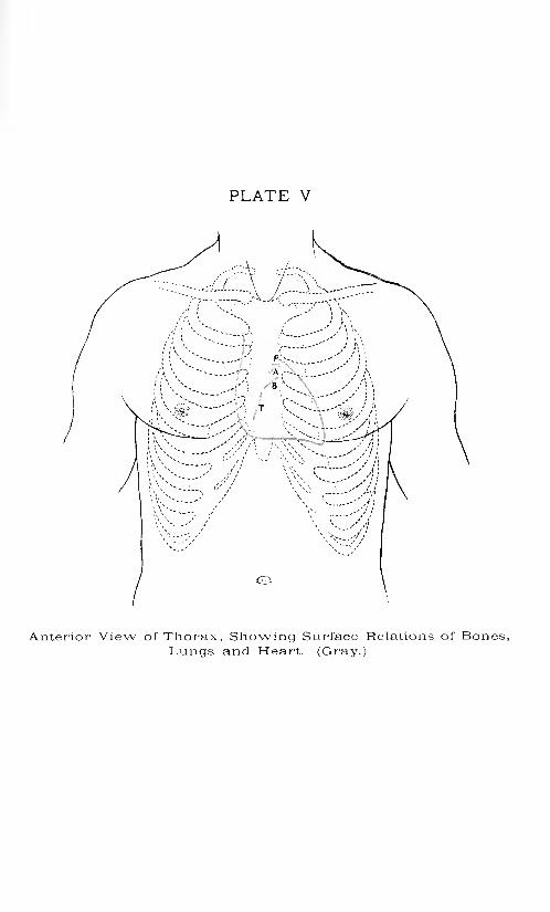
PLATE V
Anterior View of Thorax, Showing Surface Relations of Bones,Lungs and Heart. (Gray.)

SURGICAL DISEASES OF THE PLEURA 261
culous pleural fistula is not apt to heal and even extensive thoraco-
plastic operations are often unavailing.)
Since the introduction of Beck's bismuth paste, consisting of from
10 to 30 per cent, of pure subnitrate of bismuth in yellow vaseline,
many of the fistulse resulting from the drainage of tuberculous empye-mata, have healed permanently. The method of treatment consists
in the injection under low, long-continued pressure of a sufficient
amount of bismuth paste to fill thoroughly the cavity. At first the
injection is done once a week; later once in two weeks, and still later,
less frequently. If the cavity is large, a low percentage of subnitrate
of bismuth should be used in order to prevent bismuth poisoning. The.
important point in this treatment consists in using but very little
force but continuing this sufficiently long to insure the filling of all
crevices with the bismuth paste. A patient may expectorate a portionof the paste for a considerable period of time, but this seems to be quiteharmless. A large number of apparently hopeless cases have respondedto this treatment.
A further form of tuberculous empyema is that which is secondaryto caries of the vertebne. Here, also, an empyema fistula is highlyundesirable.
AYhen a tuberculous empyema becomes infected with the strepto-
coccus, etc., it must be treated like any other infected form, by free
incision. This form is apt to leave a pleural thickening.
Surgery of Recent Empyema. The primary indication is to evacuate
the pus and supplementary to this to provide drainage for any reaccu-
mulation (if this is foreseen) and to favor rapid closure of the cavitywith as little permanent damage to the lung as possible.
It has been found that when rib resection is done while a pneumoniaremains active, the mortality is much greater than when the same
operation is performed after the beginning of resolution.
For this reason, as well as for the reason that adhesions will have
formed tending to prevent lung collapse, it is advisable, when possible,
to wait until pneumonia has passed its crisis. This holds especiallyfor the small empyema where the patients show a "blue dyspnea," as
contrasted with the cases showing a "white dyspnea"1 where the
empyema may be of larger extent and the pulmonary and generalcondition less alarming.
During the stage of empyema complicating active pulmonary dis-
ease it is wisest to treat the patient expectantly, drawing oil' the fluid
through a trocar at intervals. However, if the empyema cannot be con-
trolled by this means and the heart becomes embarrassed by pressureof the accumulation or if the accumulation reduces the available air
space of the lungs (by pressure) it becomes imperative to drain. Local
anesthesia must be used and quick, dextrous operation done. It does
not seem that in these cases rib resection is advisable. One has the
object of saving life the question of the maintainance of drainage
202 THORACIC SURGERY
becomes secondary and the question of cavity and sinus formation is
entirely eclipsed by the greater issue. Undoubtedly rib resection
occasions more shock than intercostal incision and likewise the imme-diate postoperative mortality is greater. For this reason it seems best
to do simple, quick, dextrous intercostal incision, cutting through withone sweep to the pleura and following closely the upper border of a rib
so as to avoid the vessels. If the relief following tides the patient over
his crisis, rib resection can be done after a few days for the productionof efficient drainage. One must sacrifice ideal operating to beneficial
procedures, no matter how crude or simple.It the emergency incision is not demanded, it is wise to continue
aspiration whenever sufficient fluid collects to be available. Often,
if the 1 causative organism be pneumococcus, aspiration done early and
repeatedly will result in cure without open operation.1
It sometimes happens that the- first aspiration may show the presenceof bacteria while subsequent tappings are sterile. These 1
findings
definitely argue' against open operation. When the' reveTse is true 1
, it
is advisable to do open operation whenever the organism proves to
be streptococcus (which, following influenza, is very frequently found) .
The finding of a hemolytie- streptocejccus is an urge-lit indication for
radical drainage in all conditions as, if left undrained, this infection
is very lethal. YMien the pneumex-occus is the agent, and some-times
in the pivse-nce of staphylocexrus when the reaction is slight, it is pos-sible- to thoroughly cleanse the pleural cavity of all fibrin, pus and
debris and to close the chest at once. One must always attend these
case's closely so that sutures can be remove-d at once if reaecumulation
and reaction oe.-cur.
\\ he'ii the 1
process is well established with thick granular or fibrinous
reactions of the pleura already advanced, it is seldom possible to
close the parie-tes. It is best to provide double- drainage in these- cases
by making a secondary incision at the- lowest point of the' diaphragmatic-
parietal angle-, which point is most frequently in the- antero-lateral
aspect of the- chest.
Afte-r the- che-st has bevn ope-ned by the regular posterior, axillary
se-ve-nth to ninth rib route', and afte-r the- e-emditions indicating most
efficient drainage' have be-en discovered, a second resection is done
opposite this lo\\est point as determined by palpation through the
first ope-ning. Long tubes of large calibe-r (split) can then be drawn
through from front to back and se-cure-d <tf hnth rm/.v by safety pins.
When he-niolytic streptococcus is pre-sent it is good to provide Carrel's
irrigation through a small intercostal buttonhole- in the fourth or fifth
space in front so that the- fluid can trickle- over the 1 surfaces. When the
fluid is encapsulated this is unnecessary.I )ry dressings, without valve- aids, are- most satisfactory. If valve
aids are- indicated such wounds would, in nearly all cases, have healed
by primary suture of the parietes.
1 St'inc, W. ,!.: The Management of Post/pneumonic Kmpyema Based upon 310
Cases, Am. Jour. Mod. So., 191!). clviii, 1.
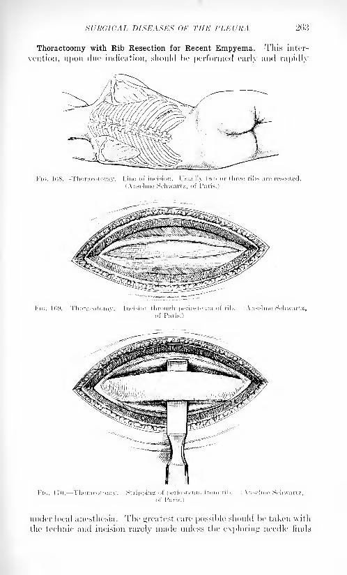
SURGICAL DISEASES OF THE PLEURA 203
Thoractoomy with Rib Resection for Recent Empyema. This inter-
vention, upon due indication, should he performed early and rapidly
FIG. 108. Thoracotomy. Line of incision. Usually two or throe ribs are resected
(Ansclme Schwartz, of Paris.)
Fi<;. 109. Thoracotomy. Incision through periosteum of rib. (Atiselme Schwartz,of Paris.)
FK;. 170. Thoracotomy. Stripping of periosteum from rib. (Anselme Schwartz,
under local anesthesia. The greatest care possihle should he taken with
the technic and incision rarely made unless the exploring needle finds
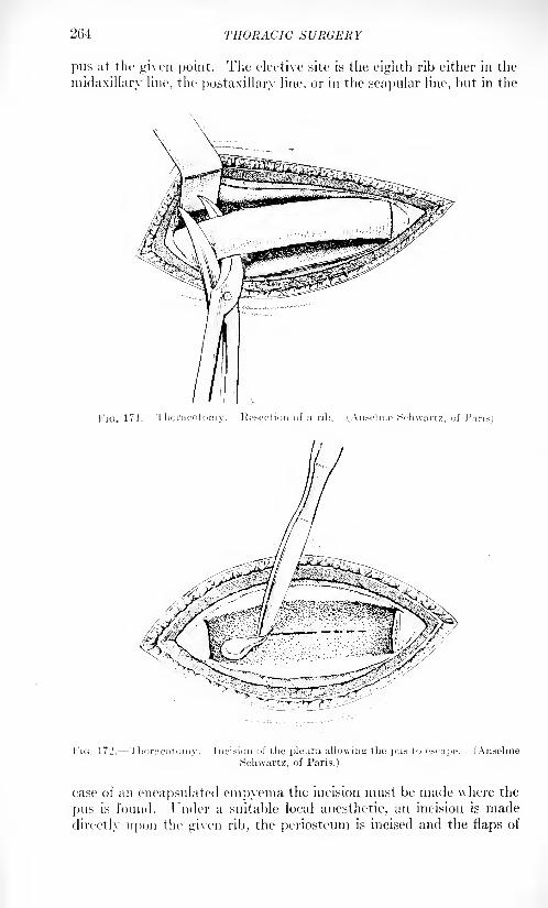
THORACIC SURGERY
pus at the given point. The elective site is the eighth rib either in the
midaxillary line, the postaxillary line, or in the scapular line, but in the
FIG. 171. Thoracotomy. lie-section of a rib. i.\nseline Schwartz, of Paris)
FIG. 172. Thoracotomy. Incision of the pleura allowing the pus to escape. (Anselinc
Schwartz, of Paris.)
case of an encapsulated empyema the incision must be made where the
pus is found. 1'nder a suitable local anesthetic, an incision is made
directly upon the given rib, the periosteum is incised and the flaps of
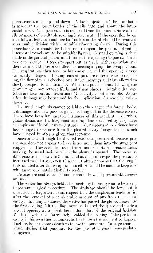
SURGICAL DISEASES OF THE PLEURA 265
periosteum turned up and down. A local injection of the anesthetic
is made at the lower border of the rib, into and about the inter-
costal nerve. The periosteum is removed from the inner surface of the
rib by means of a suitable reaming instrument. If the operation be on
an adult, at least two and one-half inches of the rib should be removedafter double division with a suitable rib-cutting shears. During this
procedure care should be taken not to open the pleura. Bleeding-intercostal vessels are to be suitably ligated. A small opening is then
made in the parietal pleura, and through this opening the pus is allowed
to escape slowly. It tends to spurt out, as a rule, with respiration, andthere is a slight pressure difference accompanying the escaping pus.The respirations then tend to become quiet, and the opening is very
cautiously enlarged. If symptoms of pressure-difference seem menac-
ing, the flow of pus is checked by suitable dressings and thus allowed to
slowly escape into the dressing. When the pus has ceased flowing the
gloved finger may remove fibrin and tissue shreds. Suitable drainagetubes are then put in. Irrigation of the cavity is not advisable. Aspir-ation drainage may be secured by the application of a so-called valve-
dressing.Too much emphasis cannot be laid on the danger of a foreign body,
a drainage tube or a piece of gauze, getting lost in the thoracic cavity.
There have been innumerable instances of this accident. All tubes,
gauze, drains and the like, must be scrupulously secured by very large
clasp-pins and in other ways (suture). All Surgeons of experience have
been obliged to remove from the pleural cavity foreign bodies which
have slipped in after a given thoracotomy.
Sauerbruch, although he devised excellent pressure-difference pro-
cedures, does not appear to have introduced them into the surgery of
empyema. However, he uses them under certain circumstances,
making the usual incision when the pleura is opened. The pressure-difference used is but '2 to 3 mm.; and as the pus escapes the pressure is
increased to S, 10 and even 12 mm. It often happens that the lung is
fully inflated after this escape and an effort should be made to keep it so
with an approximately air-tight dressing.Fistula? are said to occur more commonly when pressure-differences
are used.
The writer has always held a thoracotomy for empyema to be a very
important surgical procedure. The drainage should be low, but it
must not be forgotten in this respect that the diaphragm tends to rise
after the removal of a considerable amount of pus from the pleural
cavity. In many instances, the writer has passed the gloved finger into
the first opening, felt the diaphragm, estimated the space and made a
second opening at a point lower than that of the original incision.
AYhile the writer has fortunately avoided the opening of the peritoneal
cavity in his own thoracotomies, he has known the accident to happen.
Further, he has known death to follow the puncture of a large thoracic
vessel during trial puncture for the pus of a small, encapsulated
empyema.
'2(\(\ THORACIC Z
A proper incision can hardly be made without the resection of oneor more ribs. As the pus escapes the air tends to enter the pleura,
causing a surgical pneiunothorax. In acute empyema, the pleura has
undergone hut little pathological alteration. The pneiunothorax causes
collapse of the lung, and, to oll'set this, suction without or insufflation of
air within must he used. Simple expiration, with narrowing of the
glottis, partially inflates the collapsed lung and, as more air is inspiredthan expired, the latter should continue to expand. All this pre-
supposes an equalization of pressure due to the open pneiunothorax.If gauze is placed over the external incision the inflation of the lungwill force a little air through the gauze. Xot all of this air returns, andin time the valve-like action tends to hring the two pleural surfaces
together, resulting in adhesions.
Placing the patient in a prone position immediately after the chest
has heen opened, or during the process of opening the chest, assists
greatly in reducing the extent to which a pneiunothorax will form, and
greatly decreases the shock and discomfort of the patient. This
positioji is also usually hy far the most comfortable for a numher of
hours after the operations, which have just heen described, have heen
performed. The dressing must he so applied that the valve-like prin-
ciple can act continuously.If hy reason of delay the empyema has become chronic, with a
residual collection of pus and dense, firm adhesions of the pleura to the
lung thus crippling the play of the latter (this with the continuous
formation and escape of pus), the danger of amyloidosis is great, and
radical treatment is indicated.
As in the acute case, thoracotomy including resection of one or more
ribs, is the operation of necessity. It has been described under other
thoracotomies with resection (page1 20.'!). The writer resects the eighth
rib at the scapular line.
Naturally, operation under pressure-differences solves the problemof pneumothorax. The pus is first allowed to escape with a pressure-
diil'erence of '1 or ! mm., increasing to S, 10 or \'l. To hold the lung
inflated, the external wound should be hermetically closed. The expan-sion of the lung is, at times, complete when the next dressing is made.
USE OF ASPIRATION DRAINAGE.
Schmcr/ 1 describes a plan for treating the cavity of the chest in em-
pyema by means of a negative-pressure apparatus which was developedin the clinic of Prof. v. Hacker of the University of (iraz which seems
to merit especial attention from the theoretical standpoint, because
mechanically the plan seems to possess many desirable qualities, and
from the fact that the method has been successfully employed in this
clinic in a very large number of cases. This method accomplishes the
following results :
1. The pus is evacuated from the chest cavity without the intro-
duction of air into the cavity.
' Zentralbl. f. Chir., January 8, 1916.
USE OF ASPIRATION DRAINAGE 2(57
2. The chest cavity remains hermetically sealed throughout the
entire course of treatment.
3. This prevents the soiling of the wound in the soft tissues through-out the course of treatment and avoids the necessity of frequent redres-
sing of the wound.4. There is constantly a negative pressure in the chest cavity from
the very beginning of the evacuation of pus.0. There is a rapid expansion of the lung because of the continued
negative pressure which may be increased to any desired degree above
the normal.
6. The patient may be out of bed immediately after the operation,
provided his strength has not been too severely taxed before the oper-ation by the long-continued existence of the empyema.
7. The period of convalescence is greatly reduced in cases in which
the treatment is applicable.Two distinct methods are described which can be applied to suitable
cases.
1. Treatment by Means of Trephining of Rib. 1. This operation is
performed under local anesthesia with novocain. A cylinder l-^-cm.
in diameter and containing all of the soft tissues down to the eighth,
ninth or tenth rib is excised in the posterior axillary line or in the
scapulary line, by means of a sharp narrow-bladed knife, exposing the
rib as indicated in Fig. 173-1.
2. The periosteum is split and deflected upward and downward,
exposing the rib which is perforated by means of a trephine exactly
through the middle, the opening being slightly less in diameter than the
rib, as shown in Fig. 173-2, great care being taken not to injure the
periosteum on the inner surface of the rib.
3. The presence of pus is then tested by means of an exploringneedle inserted through the membrane composed of periosteum and
pleura on the inner surface of the rib, Fig. 1 73-?.
4. A drainage tube, slightly larger in diameter than the opening in
the rib, is now stretched upon a probe as indicated in. Fig. 173 ->, in
order to reduce its diameter. Jt is then rapidly forced into the cavityof the chest through the opening in the rib.
5. The probe is then removed, permitting the rubber tube to becomerestored to its normal size, which being slightly larger than the openingin the rib, causes a perfect occlusion in this opening, Fig. 173-4-
6. The pus is now withdrawn into a Wolff's bottle under the influence
of negative pressure, the rapidity of the flow being regulated by pres-
sure upon the rubber tube.
7. Attachment of the drainage tube (Fig. 173-4), ^'ith a portablevacuum bottle with negative pressure, as shown in Fig. 173-J.
N. The wound in the soft tissues is now tamponed with gau/e about
the drainage tube, which is held in place by means of rubber adhesive
straps. The vacuum bottle, which is S cm. wide and 4 cm. in its smaller
diameter, contains two necks a and />, which are inserted into rubber
tubes a and b, and clamped by means of forceps c and c. A vacuum
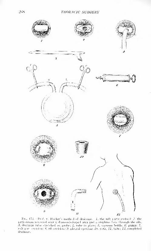
THORACIC SURGERY
, ^-x*$^xr*+l.
Vic,. 173. --Prof. v. Hacker',- method of drainage. 1, the soft parts excised; 2, the
periosteum removed over a diamond-shaped area and a trephine hole through the rib;
3, drainage tube stretched on probe; 4- tube in place; J, vacuum bottle; 6, pump; 7,
soft part excision; 8. rib excision; .9, pleurnl opening; 1, cork; 11, tube; 12, completeddrainage.
USE OF ASPIItATJOX DRAINAGE 2(>9
is established by means of an air-pump (Fig. 173 ). The drainagetube (Fig. 173-4), is then attached to drainage tube a (Fig. 173-5).
The clamp c on drainage tube a is then removed, enabling the vacuumin the glass bottle to exhaust the pus from the chest cavity.
Negative pressure is continued in the bottle constantly by meansof the air-pump (Fig. 173-^- The bottle is attached to the patient's
clothing so that he may be about without discomfort. The method of
empyting the glass bottle is self-evident.
In cases in which the pus is thin, so that it can be evacuated throughan opening such as described, this method may bring ideal results.
II. Operation Number Two. In cases in which the pus is too thick
to be evacuated in this manner, the author recommends a second
method. The method which has just been described should be tried
at first, and if the pus is found too thick or if there are coagulations
present which obstruct the rubber tube, then the operation can be
changed to the second type, which is carried out by the following
steps:
1. Again under anesthesia with novocain, a portion of soft tissue
from 5 to cm. in diameter in adults is excised as in F'ig. 173-7, and in
a manner to produce a conical space of the proper shape to be fully
occupied by the rubber cork, as shown in Fig. 173-10. The spaceshould be only just large enough so that the cork will fit very tightly.
2. A portion of the rib from 5 to G cm. in length is then removed after
reflecting the periosteum, as shown in Fig. 173-5.
3. A puncture is then made to confirm the presence of pus after the
cork has been forced in place tightly between the cut ends of the rib.
The cork should be conical in shape and from -") to (3 cm. in diameter in
the adult, at its larger end, and from 3^ to 4 cm. at its smaller end, while
in children, at the larger end it should be 3 cm. in diameter and the
smaller end - cm. The central opening in the cork should be from
1^ to 2 cm. in diameter. A glass drainage tube containing lateral
openings at the end which is to be introduced into the chest cavity,
and corrugations at the opposite end, Fig. 173-11, for attaching a
rubber tube, is passed through the opening in the rubber cork andattached to the Wolff's bottle as in the previous operation. The rubber
cork is held in place by means of silkworm-gut sutures passed throughthe soft parts and over the rubber cork. It is important to place the
rubber cork so that it completely fills the opening and so that it is
crowded in between the ends of the rib, as shown in Fig. 173-12.
The remaining steps of the treatment are the same as in the previous
operation, except that in order to prevent the rubber cork containingthe glass tube from being forced out by means of coughing, it is fastened
in place by the following method : The cork is first covered with stripsof gau/e, then rubber adhesive straps are placed partly around the
chest and over the cork and the glass drainage tube. Other adhesive
straps are passed in various directions so as to thoroughly fasten the
cork in place.
The operation on old empyenia residua may be done according to the
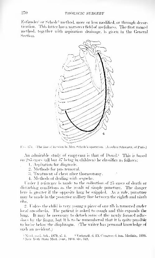
THORACIC SURGERY
Estlander 1 or Schede 2 method, more or less modified, or through decor-tication. This latter has a narrower field of usefulness. The first namedmethod, together with aspiration drainage, is given in the GeneralSection.
Fii;. 174.- -The line of incision in Max Schedc's operation. (Ansclme Schwartz, of Paris.)
Aii admirable study of empyema is that of Dowd. 3 This is basedon 2.s."> eases (all but 47 being in children) he classifies as follows:
1. Aspiration for diagnosis.
2. Methods for pus removal.
>. Treatment of chest after thoracotomy.4. Methods of dealing with sequelae.
l'nder 1 reference is made to the collection of 25 cases of death or
disturbing conditions as the result of simple puncture. The dangerhere is greater if the opposite lung be crippled. As a rule, puncture
may be made in the posterior axillary line between the eighth and ninth
ribs.
2. 1 Uless the child is very young a piece of one rib is removed under
local anesthesia. The patient is asked to cough and this expands the
lung. It may be necessary to detach some of the newly formed adhe-
sions by the finger, but it is to be remembered that it is quite possibleto incise below the diaphragm. (The writer has personal knowledge of
such an accident.)
' Xonl. HUM!. Ark., 1879, xi, 1.2 Verhandl. (1. IX Congress, d. inn. Medizin, 1890.
3 Xe\v York State Mcd. Jour., 1914, xiv, 342,
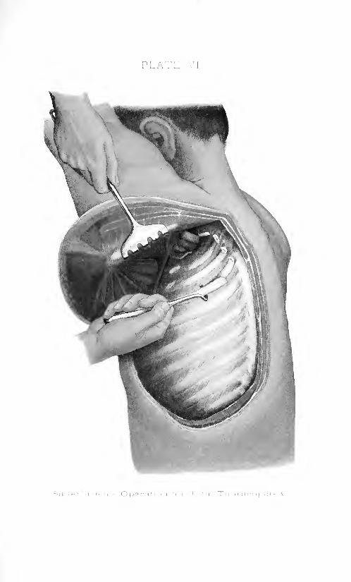
PLATE VI

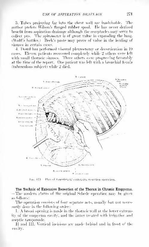
USE OF ASPIRATION DRAINAGE 271
3. Tubes projecting far into the chest wall are inadvisable. Theauthor prefers Wilson^s flanged rubber spool. lie has never derived
benefit from aspiration drainage although the receptacles may serve to
collect pus. The spirometer is of great value in expanding the lung.
(Woltl"s bottles.) Beck's paste may prove of value in the healing of
sinuses in certain cases.
4. Powd has performed visceral pleurectomy or decortication in 1!)
cases. Eleven patients recovered completely while 2 others were left
with small thoracic sinuses. Three others were progressing favorablyat the time of the report. One patient was left with a bronchia] fistula
(tuberculous subject) while 2 died.
I-'K;. 17").- -I'lau of Sauerbruch's extensive resection operation.
The Technic of Extensive Resection of the Thorax in Chronic Empyema.-The modern status of the original Schede operation may be givenas follows:
The operation consists of four separate acts, usually but not neces-
sarily done in the following order:
I. A broad opening is made in the thoracic wall at the lower extrem-
ity of the empyema cavity, and the latter treated with irrigation and
aseptic tamponade.11 and 111. Vertical incisions are made behind and in front of the
cavitv.
079 THORACIC SURGERY
I\ . The skin and muscular flap thus formed is detached from the
ribs, the bleeding vessels are ligated, the ribs, intercostal muscles and
parietal pleura are divided over a suitable area, front and back, byvery heavy shears. The bone Hap thus formed is removed and all
intercostal vessels are scrupulously ligated. The thickened pleura of
the lung is criss-crossed with incisions; this pleura may be removed(decortication.) The skin and muscular flap is dropped down on the
collapsed lung, pressed in place and sutured with drainage.
Operation of Decortication of the Lung or Visceral Pleurectomy ( Delorme 1
and Fowler2). While the Schede operation mobilizes the chest wall,
thus dropping it down on the lung and obliterating the empyema cavity,
decortication seeks to pare oil' the callous formation on the lung and
thereby allow it to re-expand. The lung is exposed by a large thoracic
flap. The exposed lung surface is then incised in various localities and
the callosity "pried up" from the lung by blunt dissection. The lung
expands and the thoracic flap now lies in apposition with it.
Sauerbruch states that there are not many old empyemata in which
the lung can be made To expand in this manner. The procedure maywell be a useful accessory to other operative measures. It is a very
bloody intervention when carried out in full, and maybe done only to
the extent of stripping oft' the first layers, in connection with the
Schede operation. When the operation is done on the left lung the
heart at times shows notable weakness.
Despite all possible care operations on an inflamed pleura may result
in very severe local or general, spreading infection.
According to Mayo and Beekman3(1914) this operation was then
recorded in literature but twenty-four times. The intervention is said to
be of littlevalue if undertaken beyond a certain period, because the lung
cannot expand. The authors, however, report one personal case in which
the operation succeeded nearly four years after the original empyema ;in
a second case the interval was six months. Four or five months, in
general, is considered the time limit. (I)owd operated on lo of the 24
cases mentioned by Mayo.) It may be stated that the exact status of
the operation is, as yet, hardly determined. The best results seem to
be in cases that are operated upon fairly early, in which the given lung
has not been very much compressed. (In some of these cases the lung
might have expanded of itself.) It seems logical to assume that in
all cases the operation must at least hasten expansion. The greatest
relief seems to be desired in those extreme cases in which the lung is a
firm, contracted mass held against the spinal column.
In the ordinary cases the surgeon finds that he can quite readily
separate the visceral pleura from the lung, and that this separation will
be followed by a greater or lesser degree of lung expansion. A primary
expansion, however, may not persist and successive operations mayhave to be performed. I)owd seems to have quite as good results as
SURGICAL DISEASES OF THE LUNG 273
any other surgeon. In most of his cases the sinuses closed and remained
closed, the spine in a given case continued to be straight and the generalhealth continued good. In making later examinations it is to be remem-bered that the breath sounds over the affected side often are misleading;
they may be transmitted from the sound side.
Dowd gives the technic of decortication as follows: Under ether
anesthesia, a free incision is made at the opening of the sinus and somefour inches of the two adjacent ribs are resected. This gives such access
to the sinuses proper that their boundaries can be fairly determined.
The incision should extend to the apex of the pleural cavity. (It is
important to remember this.) The incision may now be carried upwardin the anterior axillary line to the third or the second rib, and the ribs
and the attached pleura divided by strong shears. An inch or so is
removed from the second and third ribs, 4 to G inches from the tenth
rib, and corresponding segments from the intermediate ribs. Withsuitable retraction an excellent view of the sinus is thus obtained.
At this stage of the procedure the patient is allowed to so nearly
emerge from the anesthesia as to permit of his spontaneous coughing,when the lung will be seen to bulge. The membrane is then separatedand removed from the surface of the lung over as large an area as seems
wise. (The writer thinks it proper to err on the side of quite extensive
separation.) It is hardly necessary to say that the form and extent of
the given operation will vary with the individual case. The chest-wall
is to be fairly well pressed in and the flap of soft parts is to be suitablysutured with appropriate drainage.
(For complicating pulmonary fistula the reader is referred to the
section on that subject.)
[Tuffier has had remarkable success in curing pleural fistulse and
cavities by Carrel sterilization with complete closure of the thoracic
opening. In April, 1918, he exhibited a considerable number of such
cases before a meeting of American army sunreons held in Paris.
C, A. P.]
SURGICAL DISEASES OF THE LUNG.
Infections (Suppurations) of the Lungs. The lungs are often
infected from the mucous membranes (and, indeed, most pulmonary
suppurations have this origin), but the resulting lesions seldom possessmuch surgical significance since the pus finds an outlet by the mouth.
An exception is found in the aspiration and impaction of foreign bodies
in the bronchial rami, which are frequently very small and often may be
multiple. Such may cause no symptoms, and in healthy bronchial
tubes they may be carried upward by ciliary motion and expelled by
coughing. If the bronchi are the seat of a catarrhal affection this con-
servative process is less in evidence, the foreign particles readily lodg-
ing in a small bronchial sacculation. These aspirated particles may,however, lead to infection and the infection may become putrescent.
However, the percentage of suppurative foci caused by aspirated foreignbodies seems to be less than that due to bronchopneunionia and other
VOL. ii IS
274 THORACIC SURGERY
extensive diseases of the lungs, in which there is mixed infection. Pul-
monary emboli may give rise to foci of suppuration. (Quincke fails
to mention in this connection the suppurative foci which develop in
the lung as a sequel to perforative wounds of the chest.)
The condition of the sputum is always to be carefully considered in
estimating a given case of suppuration in the lung. When abscess or
gangrene is of recent date, it is very difficult to separate the lesions.
The surgeon may find it hard to distinguish in symptomatology,
diagnosis and treatment between chronic abscesses of the lung and
bronchiectasis, be the latter of the simple form or of the putrid form.
(For the sake of simplicity foreign bodies in the lungs and the infections
resulting from them will be considered together in this article.)
Pulmonary Abscess and Gangrene. From the surgical standpointthese two conditions may be considered in common. In each case a
focus of inflammation in the lung parenchyma undergoes a central
softening. Pus may be present in one case, while necrotic debris are
found in the other. The foci may be of almost any size, solitary or
multiple; they may be encapsulated or may communicate with a
bronchiole.
Gangrenous changes may come about, in one of two ways: (1)
ordinary pus may become infected by putrid bacteria, and (!') the
sound lung may be attacked primarily by the same microorganisms.
Clinically there arc; no hard and fast lines, for all possible transitions are
seen between simple abscess formation, secondary putrid infection, and
true gangrene.
Whenever, in the course of other inflammatory processes, cavities
form by a softening of lung tissue the lesions are classed as abscesses
bronchopneurnonia, embolism, the presence of foreign bodies, etc.
Kmbolic abscesses may be purulent or putrid.
Foreign body abscesses are due to the impaction of various sub-
stances somewhere in the bronchial tree and gangrene may result in
this way. Primary gangrene of the lung soon leads to cavity formation,
the cavities being single or multiple, variable in size and often com-
municating.
Diagnosis.^ Abscess and gangrene are generally differentiated readily
by the presence or absence of gangrenous sputum. The pus maybe creamy, inodorous or having a fetid and putrid smell; it may be
very dark, like chocolate or prune juice. (As the latter forms are
due to the admixture of blood they do not necessarily indicate the
existence of gangrene.) Shreds of lung tissue with elastic fibers and
pigment may be found in the sputum. In other respects, the symptoms
may not differ from those present with other cavities.
In primary lung gangrene, there is an odor of carrion combined with
the systemic results of putrid absorption. Naturally, these symptomsoccur in the gangrenous infection of an abscess, but here they are milder
or evolve more slowly.
In localization-diagnosis, the use of the .r-ray has largely supplanted
other and older methods. Various negatives from different angles
should be made. It is not to be forgotten that the .r-rays have been
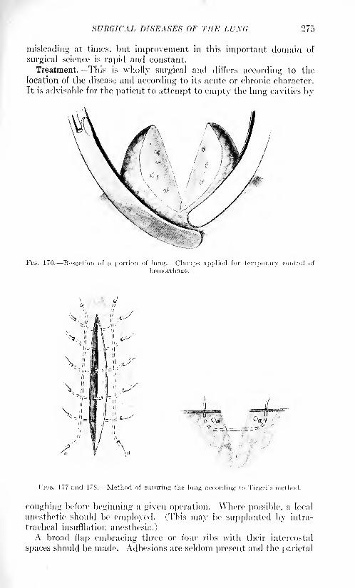
SURGICAL DISEASES OF THE LUNG 275
misleading at times, but improvement in this important domain of
surgical science is rapid and constant.
Treatment. This is wholly surgical and differs according to the
location of the disease and according to its acute or chronic character.
It is advisable for the patient to attempt to empty the lung cavities by
FIG. 176. Resection of a portion of lung. Clamps applied for temporary control of
hemorrhage.
FIGS. 177 and 178. Method of suturing the lung according to Tiegel's method.
coughing before beginning a given operation. Where possible, a local
anesthetic should be employed. (This may be supplanted by intra-
tracheal insufflation anesthesia.)
A broad flap embracing three or four ribs with their intercostal
spaces should be made. Adhesions are seldom present and the parietal
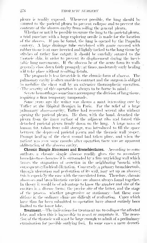
2<l) THOUAC1C SURGERY
pleura is readily exposed. \Ylieuever possible1
,the lung should be
sutured to the parietal pleura to prevent collapse and to prevent the
contents of the abscess cavity from soiling the general pleura.\\ hether or not it be possible to suture the lung to the parietal pleura,
a trial puncture with a large exploring needle is made for the location
of the abscess. If pus be found, the lung is opened by the Paquelin
cautery. A large drainage1 tube enveloped with gauze covered with
rubber tissue is at once inserted and lightly tacked to the lung tissue bystitches of rather fine catgut; it should be carefully sutured to the
thoracic skin, in order to prevent its displacement during the inevi-
table lung movements. If the abscess be of the acute form its walls
generally close down fairly promptly; at times, moderately early healingwill take place without resulting fistula.
The prognosis is less favorable in the chronic form of abscess. The
pulmonary cavity is often unable to contract and the surgeon is obligedto mobilize the chest wall by an extensive thoracoplastic operation.(The severity of this operation is always to be borne in mind.)
Severe hemorrhages sometimes accompany the division of lung tissue,
requiring a firm temporary tamponade.Some years ago the writer was shown a most interesting case by
Tuffier at. the Ilopital Beanjon in Paris. For the relief of a large
pulmonary abscess-cavity, Tuffier had resected several ribs without
opening the parietal pleura, lie then, with the hand, detached the
pleura from the inner surface of the adjacent ribs and forced this
detached parietal pleura firmly down on the lung. A large mass of
human fat, taken from cold storage, was introduced to fill the spacebetween the depressed parietal pleura and the thoracic wall proper.
Prompt heal:
ng of the chest wound had taken place, and when the
patient was seen some 1 months after operation then 1 was an apparentobliteration of the abscess cavity.
Chronic Simple Abscesses and Bronchiectases. According to some
authors, a chronic simple abscess readily gives rise to secondarybronchiectases because it is surrounded by a firm unyielding wall which
favors the stagnation of secretion in the neighboring bronchi with
consequent cylindrical dilatation. Conversely, a primary bronchiectasis,
through ulceration and perforation of its wall, may set up an abscess;
this is especially the case with the saceulated form. Therefore, chronic
abscesses and bronchiectatic cavities are almost always found together.In theory it would be of advantage to know the number and size of the
cavities in a disease focus; the precise site of the latter, and the stageof the process whether progressive or stationary but these con-
ditions in the ordinary clinic art 1 difficult of realization. Cases which
have thus far been submitted to operation have almost entirely been
limited to tin 1 lower lobe.
Treatment. -The indications for treatment are to collapse the affected
lobe, and when this is impossible to resect or amputate it. The resec-
tion of the thoracic wall must be large enough to admit of a preliminaryexamination for possible outlying foci. In some eases a mere decorti-
SURGICAL DISEASES OF THE LUXG
cation may enable the lobe to collapse, but should the given lobe be
permeated with many small cavities and dilated bronchioles, collapse
may be impossible because of the great amount of indurated tissue in
it. In such a condition Quincke favors the establishment of a bronchial
fistula for drainage. (This operation naturally belongs to the pneu-
motomies.) If none of these measures seem indicated a thoracotomywith extensive resection of the ribs, accompanied, if advisable, bydecortication, may lead to the desired collapse of the affected lung.
If the pleura is not involved in the original process, and if it is not
adherent, an artificial pneumothorax may be instituted.
These cases may remain under surgical treatment for weeks and
months, during which time a succession of operative procedures are
made until the desired collapse is attained.
Putrid Chronic Abscesses and Bronchiectases. In many ways this
condition corresponds to the preceding. Secondary foci are more proneto occur in remote areas, however, and there is a further tendency to
the development of acute abscess and gangrene; these being the affec-
tions which first cause the patient to present himself for treatment.
The odor of the breath and the character of the .sputum are not always
characteristically putrid; elastic fibers are often absent from the sputa.
Absorption of putrid matter is not without influence on the generalhealth. The subject is not as good a surgical risk as is the patientof the preceding type, but the indications are practically the same.
"Collapse" surgery is probably less rational than is the direct incision
with drainage and resection of the affected portion of the lung. In
bilateral cases, surgical intervention would seem to be contra-indicated.
(See also Bronchiectases, The Surgical Treatment of.)
Local Sclerosis of the Lung. Aside from a generalized condition
like fibroid phthisis, which is not thus far in the province of surgery,there are numerous circumscribed areas in which sclerotic portions of
lung form about various lesions, notably abscesses or suppurationfrom any cause, bronchiectasis, pleural contraction, etc. In this
manner, an entire lobe of the hmg may be rendered useless. Such a
form of sclerosis frequently is associated with a fistula of the lung.In certain cases the detachment of a contracted lung from its thick-
ened pleura may enable it to re-expand in part, or in whole.
In many cases this sclerosis is a salutary procedure and is broughtabout for the cure of cavities. In other cases, when a chronic abscess,
bronchiectatic cavity, lung fistula, etc., continues to discharge pus,thus threatening life by amyloidosis, the entire lobe may be resected.
Foreign Bodies in the Bronchi and Lung. These interest the surgeonfrom a twofold viewpoint. The larger bodies which lodge in a bronchial
twig may require immediate removal, while smaller, finer particles, of
many kinds, may be arrested in the bronchioles and there set upinfection and suppuration. While the majority of foreign bodies reach
the lung through the natural passages, others, such as bullets, frag-
ments of shell, shreds of clothing, etc., are forced into the lung from
without. These are often infected and cause suppurating foci in the
lungs.
278 THORACIC SURGERY
When large smooth objects are swallowed they may, if sterile, remainin a bronchial tube or twig for long periods, without causing reaction;
delayed reaction, however, may supervene. In some cases the foreignbodies cause pressnre-ulceration and occasion an abscess. Angularor pointed objects usually cause a prompt reaction and become immob-ilized, thus tending to cause obstructive symptoms.The immediate 1 results of the swallowing of a large foreign bod}' are
fright and pain. There is a weakening of breathing corresponding to
the occluded area, as shown by physical signs. In the case of completeatelectasis or infiltration, there may be dulness on percussion. Hemop-tysis is common and may be profuse if an artery is damaged; this is
most likely to occur when there is ulceration or necrosis. Peculiar
sounds may be heard on auscultation. Reaction symptoms show great
variety dyspnea, paroxysms of cough and suffocation, vomiting,convulsions and syncope. Bronchial irritation may be marked. In
many cases there is an absence of striking symptoms.Whenever there is even a suspicion of a foreign body in a lung, suit-
able .r-ray pictures should be taken immediately. These plates will
show pieces of glass, metal objects, fragments of bone, etc., but shreds
of clothing, etc., are hardly ever revealed on them.
Removal by the bronchoscope is the method of choice, when such
removal is possible, and the writer believes that it is not going too far
to say that in bronchial foreign bodies bronchoscopy should always be
attempted.1 When bronehoseopic removal fails, thoracotomy must be
carefully considered. In certain instances this may be transpleural, in
other cases the extraplenral route may be employed. Bryant,2 Schwartz3
and others have devised extrapleural procedures, but thus far the mor-
tality in these operations has been extremely high. With the constant
development of precision in thoracic surgery the mortality will be
correspondingly lowered.
Braham 4reports the case of a pin aspirated into the left lower lobe.
Operating by means of the fluorescent screen he removed portions of
the fifth and sixth ribs and incised the pleura, causing an artificial
pneumothorax. lie extended the incision, drew out the contracted
lung and removed the pin. Recovery took place.
Very different from the large type of foreign body, are the small
objects, aspirated unconsciously and often in numbers, along some
bronchial route. Such include a large variety of substances proceed-
ing from the air and food passages, as well as from diseased foci
in the lungs themselves. These substances, at times, immediately
cause an acute aspiration pneumonia. (As a chronic, insidious process,
the phthisis of knife grinders or stonecutters may be mentioned.)
Certain writers regard the insidious aspiration of particles of various
kinds as a definite cause of abscess and gangrene. Under this head
may be enumerated certain kinds of dust, minute particles of food,
1 The published articles by Chevalier Jackson, of Philadelphia, should be consulted.
2 Med. News, July 28, 1900. 3 Chirurgie du thorax, Paris, 1912.
4 British Med. Jour., 1914, i, 1123.
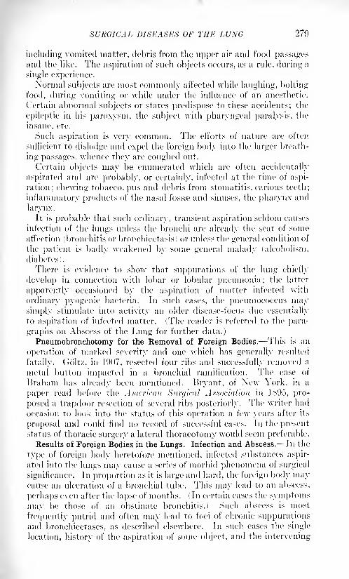
SURGICAL DISEASES OF THE LVXG 279
including vomited matter, debris from the upper air and food passagesand the like. The aspiration of such objects occurs, as a rule, during a
single experience.Normal subjects are most commonly affected while laughing, bolting
food, during vomiting or while under the influence of an anesthetic.
Certain abnormal subjects or states predispose to these accidents; the
epileptic in his paroxysm, the subject with pharyngeal paralysis, the
insane, etc.
Such aspiration is very common. The efforts of nature are often
sufficient to dislodge and expel the foreign body into the larger breath-
ing passages, whence they are coughed out.
Certain objects may be enumerated which are often accidentally
aspirated and are probably, or certainly, infected at the time of aspi-
ration; chewing tobacco, pus and debris from stomatitis, carious teeth;
inflammatory products of the nasal fossa? and sinuses, the pharynx and
larynx.It is probable that such ordinary, transient aspiration seldom causes
infection of the lungs unless the bronchi are already the seat of some
affection (bronchitis or bronchiectasis) or unless the general condition of
the patient is badly weakened by some general malady (alcoholism,
diabetes).
There is evidence to show that suppurations of the lung chiefly
develop in connection with lobar or lobular pneumonia; the latter
apparently occasioned by the aspiration of matter infected with
ordinary pyogenic bacteria. Jn such cases, the pneumococcus maysimply stimulate into activity an older disease-focus due essentially
to aspiration of infected matter. (The reader is referred to the para-
graphs on Abscess of the Lung for further data.)
Pneumobronchotomy for the Removal of Foreign Bodies. This is an
operation of marked severity and one which has generally resulted
fatally. Goltz, in 1007, resected four ribs and successfully removed a
metal button impacted in a bronchial ramification. The case of
Braham has already been mentioned. Bryant, of New \ork, in a
paper read before' the American Surgical Association in 1S95, pro-
posed a trapdoor resection of several ribs posteriorly. The writer had
occasion to look into the status of this operation a few years after its
proposal and could find no record of successful cases. In the presentstatus of thoracic surgery a lateral thoracotomy would seem preferable.
Results of Foreign Bodies in the Lungs. Infection and Abscess. In the
type of foreign body heretofore mentioned, infected substances aspir-
ated into the lungs may cause a series of morbid phenomena of surgical
significance. In proportion as it is large and hard, the foreign body maycause an ulceration of a bronchial tube. This may lead to an abscess,
perhaps even after the lapse of months. (In certain cases the symptomsmay be those of an obstinate bronchitis.) Such abscess is most
frequently putrid and often may lead to foci of chronic suppurationsand bronchiectases, as described elsewhere. In such cases the single
location, history of the aspiration of some object, and the intervening
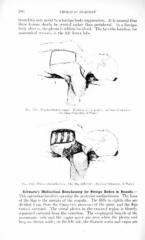
2X0 THORACIC SURGERY
bronchitis may point to a foreign-body suppuration. It is natural thatthese lesions should be central rather than peripheral. In a foreign-
body abscess, the pleura is seldom involved. The favorite location, for
anatomical reasons, is the left lower lobe.
FIG. 170. Pneiimobronchotomy. Position of the patient and line of incision
(Anselmc Schwartz, of Paris.)
FIG. 180. Pneuinobronchotomy. Skin flap reflected. (Anselme Schwartz, of Paris.)
Schwartz's Mediastinal Bronchotomy for Foreign Bodies in Bronchi.
This operation involves opening the posterior mediastinum. The base
of the flap is the margin of the scapula. The fifth to eighth ribs are
divided 4 cm. from the transverse processes of the spine, and the flap
turned outward. The costal pleura in the exposed region is bluntly
separated outward from the vertebrae. The esophageal branch of the
innominate vein and the vagus nerve are seen when the pleura and
lung arc drawn aside; on the left side, the thoracic aorta and vagus are
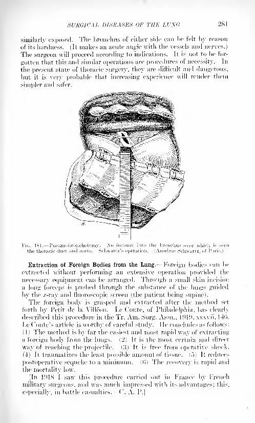
SURGICAL DISEASES OF THE LUNG 281
similarly exposed. The bronchus of either side can be felt by reason
of its hardness. (It makes an acute angle with the vessels and nerves.)
The surgeon will proceed according to indications. It is not to be for-
gotten that this and similar operations are procedures of necessity. In
the present state of thoracic surgery, they are difficult and dangerous,
but it is very probable that increasing experience will render them
simpler and safer.
FK;. 181. Pneuinobronchotoinv. An incision inlo the brorichu
the thoracic duet and aorta. Schwartz's operation. (Anselme Schwartz, of Paris.)
Extraction of Foreign Bodies from the Lung. Foreign bodies can be
extracted without performing an extensive operation provided the
necessary equipment can be arranged. Through a small skin incision
a long forceps is pushed through the substance of the lungs guided
by the .r-ray and fluoroscopic screen (the patient being supine).The foreign body is grasped and extracted after the method set
forth by Petit de la Yilleon. Le Conte, of Philadelphia, has clearly
described this procedure in the Tr. Am. Surg. Assn., 1<)H), xxxvii, 14(>.
Le Tonte's article is worthy of careful study. lie concludes as follows:
( 1 ) The method is by far the easiest and most rapid way of extracting
a foreign body from the lungs. (2) It is the most certain and direct
way of reaching the projectile, (.'ij It is free from operative shock.
(4) It traumatizes the least possible amount of tissue. (.">) It reduces
postoperative sequela1 to a minimum. ((>) The recovery is rapid and
the mortality low.
[In 1918 I saw this procedure carried out in France by French
military surgeons, and was much impressed with its advantages; this,
especially, in battle casualties. ('. A. P.]
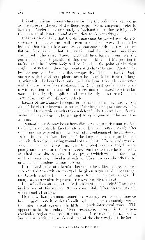
L'82 THORACIC SURGERY
It is often advantageous when performing the ordinary open opera-tion to resort to the use of the fluoroscope. Some surgeons prefer to
locate the foreign body accurately beforehand and to locate it by boththe anatomical situation and its relation to skin markings.
It is very important that the skin markings be plaeed according to
system so that every case will present a similar survey. It must be
insi-ted that the patient occupy one constant position 'for instance
flat on his back' while both the vertical and the horizontal markingsare plaeed on his skin. These marks will be utterly inaccurate if the
patient changes his position during the marking. If his position is
maintained the foreign body will be found at the point of the right
angle constructed on these two points as its hypothennse. Anatomical
localizations can be made rluoroscopically. Thus a foreign bodymoving \\ith the visceral pleura must be imbedded in it or the lung.
Moving with the heart beat but outside the heart fixes it in connection
with the great vessels or mediastinum. Such and similar facts locate
it with relation to anatomical structures and this together with skin
marks intelligently applied and intelligently interpreted- makeextraction easy by ordinary methods.
Hernia of the Lung.- Prolapse of a segment of a lung through the
wall of the chest is known as a hernia of the lung, or a pnenmoeele. The
congenital form which results from a defect in the thorax, is considered
under malformations. The acquired form is generally the result of
trauma.
Traumatic hernia may be an immediate or a consecutive matter, i. e.,
the lung may protrude directly into a newly made wound, or only after
some time has expired and as a result of a weakening of the chest-wall.
In the immediate form, hernia of the lung should be regarded as a
complication of penetrating wounds of the chest. The secondary cases
occur in connection with imperfectly healed wounds, fragile scars,
poorly united fractures of the ribs, etc. Similar to these latter are the
acquired cases due to some disease process which weakens the chest-
wall (suppuration, muscular atrophy). There are certain other cases
in which the etiology is quite obscure.
In the production of a hernia, there must be sufficient force or pres-
sure exerted from within, to expel the given segment of lung through
the breach: such a factor is. at times, found in a severe cough. In
many cases an evidently provocative factor is often absent.
In a miscellaneous collection of 41 cases of pnemnocele,1 17 occurred
in children; of this number 10 wen 1
congenital. There were '.} eases in
women and -\ in men.
Hernia without trauma, sometimes wrongly termed spontaneous
hernia, may occur in various localities, but is most commonly seen in
the anterolateral region at the fifth and sixth intercostal space. This
appears to be the locality of least resistance. (Hernia in the supra-
clavicular region was seen 9 times in 41 cases.) The si/e of the
hernia varies with the weakened area of the chest-wall. If the hernia
1 Dcbienne: Th6.se dc Paris, 1S97.
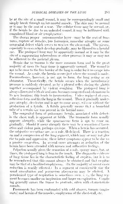
SURGICAL DISEASES OF THE LUNG 2S!5
be at the site of a small wound, it may be correspondingly small and
simply break through an intercostal muscle. The skin may be normalor it may be the seat of a scar. The cellular tissue may be normal, or,
if the hernia be due to an unhealed wound, it may be infiltrated with
coagulated blood or air (emphysema).The thorax proper osteomtiscular layer may be the seat of frac-
tures, rupture of muscles, pus formation, muscular atrophy or some
congenital defect which serves to weaken the chest-wall. The pleura,
especially in cases which develop gradually, may be likened to a hernial
sac. The prolapse: 1 lung may be apparently normal or it may be the
site of a suppurating tuberculous or other process; it may or may not
be adherent to the parietal pleura.Hernia due to trauma is the more common form and in the great
majority of cases the lung tissue is apparently normal. The wound is
apt to be close to the free border of the lung in order for it to escape into
the wound. As a rule, the hernia occurs just where the wound is made.
Pneumothorax, however, is not apt to form, the lung acting as an
obturator. Theoretically, the hernia, occurs when the intrathoracic
pressure is suddenly elevated, as by a pleural reflex closing the glottis
together accompanied by violent coughing. The prolapsed lung is
always distended with air and soon becomes congested and edematous in
its new location; this tends to incarceration. I'nless replaced at once,
adhesions form and fix the lung in the wound. Then it gradually under-
goes atrophy, dessicates and is apt to come away, with or without the
production of a fistula. A fistula generally means that a bronchial
tube of a certain size was present in the hernial mass.
The congenital form of pulmonary hernia, associated with defects
in the chest wall, is apparent at birth. The traumatic form usually
appears abruptly, while the spontaneous form is apt to come on
gradually. Should it occur abruptly there may be a sensation of lacer-
ation and violent pain, perhaps syncope. When a hernia has occurred,the subjective sensations are, as a rule, ill-defined. There is a traction
on and a compression of the lung segment, which may or may not giverise to pain and oppression; these latter symptoms appear to be due to
a passive congestion. In several cases attempts at reduction of the
hernia have been attended with nausea and suffocative feelings.
Palpation usually gives the sensation of a soft, spongy mass, but, at
times, the prolapsed lung is firm to the touch. The diagnostic evidence
of lung tissue lies in the characteristic feeling of crepitus, but it is to
be remembered that this cannot always be obtained and that crepitus
may be that of a localized emphysema. (In the majority of recent cases
reduction is readily effected.) In a segment of considerable size the
usual auscultation and percussion phenomena may be elicited. Aparadoxical type of respiration is sometimes seen, /. r., the lung seg-
ment becomes smaller on inspiration and larger on expiration. ( 'ough-
ing generally causes the tumor to protrude, often with characteristic
sounds.
Pneumocele has been confounded with cold abscess, tumors (angio-
mata) and hernia of the muscles, emphysema of the chest-wall, etc.
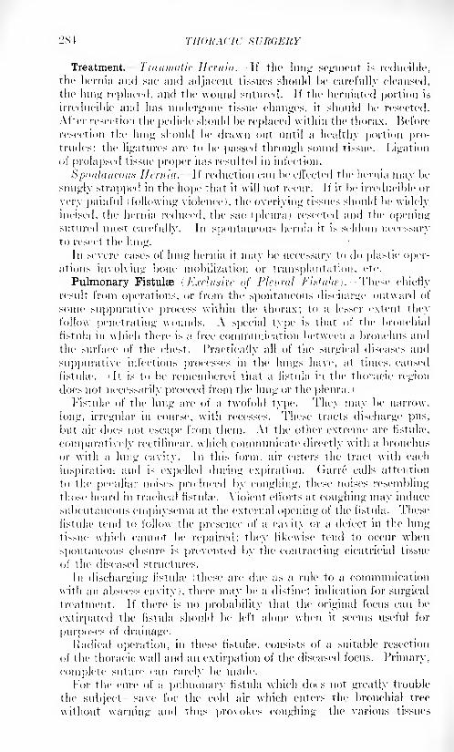
L-'Sl TI10KACIC HURGEHY
Treatment. Trmnualic llcr/tia. If the lung segment is reducible,
the hernia and sae and adjacent tissues should be carefully cleansed,
the lung replaced, and the wound sutured. If the herniated portion is
irreducible and has undergone tissue changes, it should be resected.
After resection the pedicle should be replaced within the thorax. Before
resection the lung should be drawn out until a healthy portion pro-trudes: the ligatures are to be passed through sound tissue. Ligatiouof prolapsed tissue proper has resulted in infection.
Spontaneous Hernia. If reduction can be etl'ected the hernia may be
snugly strapped in the hope that it will not recur. If it be irreducible or
very painful (following violence), the overlying tissues should be widely
incised, the hernia reduced, the sac (pleura) resected and the openingsutured most carefully. In spontaneous hernia it is seldom necessaryto resect the lung.
In severe cases of lung hernia it may be necessary to do plastic oper-
ations involving bone mobilization or transplantation, etc.
Pulmonary Fistulse (Exclusive, of Pleura! FixtiiUv). These chiefly
result from operations, or from the spontaneous discharge outward ot
some suppurative process within the thorax; to a les>er extent theyfollow penetrating wounds. A special type is that of the bronchial
fistula in which there is a free communication between a bronchus and
the surface of the chest. Practically all of the surgical diseases and
suppurative infectious processes in the lungs have, at times, caused
fistnhe. (It is to be remembered that a fistula in the thoracic region
does not necessarily proceed from the lung or the pleura.'
Fistuhe of the lung are of a twofold type. They may be narrow,
long, irregular in course, with recesses. These tracts discharge pus,
but air does not escape from them. At the other extreme are fistuhe,
comparatively rectilinear, which communicate directly with a bronchus
or with a lung cavity. In this form, air enters the tract with each
inspiration and is expelled during expiration, (iarre calls attention
to the peculiar noises produced by coughing, these noises resemblingthose heard in trachea! fistuhe. Violent eil'orts at coughing may induce
subcutaneous emphysema at the external opening of the fistula. These
fistnhe tend to follow the presence of a cavity or a defect in the lungtissue which cannot be repaired; they likewise tend to occur when
spontaneous closure is prevented by the contracting cicatricial tissue
of the diseased structures.
In discharging fistuhe (these are due as a rule to a communication
with an abscess cavity), there may be a distinct indication for surgical
treatment. If there is no probability that the original focus can be
extirpated the fistula should be left alone when it seems useful for
purposes of drainage.Kadical operation, in these fistuhe, consists of a suitable resection
of the thoracic wall and an extirpation of the diseased focus. Primary,
complete suture can rarely be made.
For the cure of a pulmonary fistula which docs not greatly trouble
the subject- save for the cold air which enters the bronchial tree
without warning and thus provokes coughing the various tissues
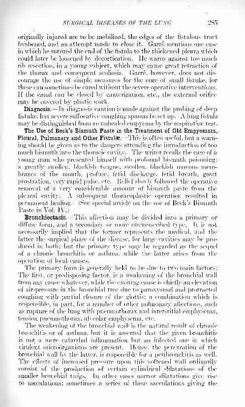
SURGICAL DISEASES OF THE LUXC 285
originally injured are to be mobilized, the edges of the fistulous tract
freshened, and an attempt made to close it. Garre mentions one case
in which he sutured the end of the fistula to the thickened pleura which
could later be loosened by decortication. He warns against too muchrib resection, in a young subject, which may cause great retraction of
the thorax and consequent scoliosis. Garre, however, does not dis-
courage the use of simple measures for the cure of small fistula1, for
these can sometimes be cured without the severe operative interventions.
If the canal can be closed by cauterization, etc., the external orifice
may be covered by plastic work.
Diagnosis. In diagnosis caution is made against the probing of deepfistula 1
, lest severe suffocative coughing spasms be set up. A lung fistula
may be distinguished from an unhealed empyema by the respiration test.
The Use of Beck's Bismuth Paste in the Treatment of Old Empyemata,Pleural, Pulmonary and Other Fistulse. This is often useful, but a warn-
ing should be given as to the dangers attending the introduction of too
much bismuth into the thoracic cavity. The writer recalls the case of a
young man who presented himself with profound bismuth poisoning:a greatly swollen, blackish tongue, swollen, blackish mucous mem-branes of the mouth, profuse, fetid discharge, fetid breath, great
prostration, very rapid pulse, etc. Relief slowly followed the operativeremoval of a very considerable amount of bismuth paste from the
pleiiral cavity. A subsequent thoracoplastic operation resulted in
permanent healing. (See special article on the use of Beck's BismuthPaste in Vol. IV.)
Bronchiectasis. This affection may be divided into a primary or
diffuse form, and a secondary or more circumscribed type. It is not
necessarily implied that the former represents the medical, and the
latter the surgical phase of the disease, for large cavities may be pro-duced in both; but the primary type may be regarded as the sequelof a chronic bronchitis or asthma, while the latter arises from the
operation of local causes.
The primary form is generally held to be due to two main factors:
The first, or predisposing factor, is a weakening of the bronchial wall
from any cause whatever, while the exciting cause is chiefly an elevation
of air-pressure in the bronchial tree due to paroxysmal and protracted
coughing with partial closure of the glottis; a combination which is
responsible, in part, for a number of other pulmonary affections, such
as rupture of the lung with pneumothorax and interstitial emphysema,tension pneumothorax, alveolar emphysema, etc.
The weakening of the bronchial wall is the natural result of chronic
bronchitis or of asthma, but it is asserted that the given bronchitis
is not a mere catarrhal inflammation but an infected one in which
virulent microorganisms are present. Hence, the penetration of the
bronchial wall by the latter, is responsible for a peribronchitis as well.
The effects of increased pressure upon this softened wall ordinarilyconsist of the production of certain cylindrical dilatations of the
smaller bronchial twigs. In other cases narrow dilatations give rise
to sacculations; sometimes a series of these sacculations giving the
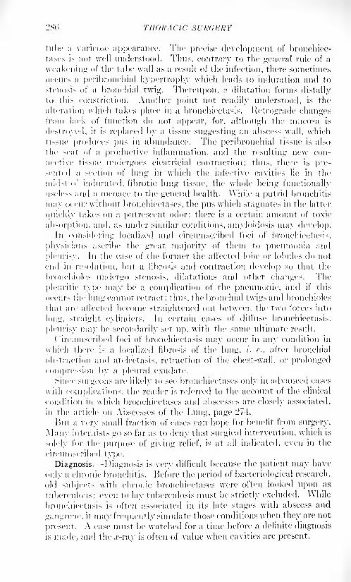
280 THORACIC SURGERY
till);1 M varicose appearance. The precise development of bronchiec-
tases is not well understood. Thus, contrary to the general rule of a
weakening of the tube wall as a result of the infection, there sometimesoccurs a peribronchial hypertrophy which leads to induration and to
stenosis of a bronchial twig. Thereupon, a dilatation forms distally
to this constriction. Another point not readily understood, is the
alteration which takes place in a bronchiectasis. Retrograde changesfrom lack of function do not appear, for, although the mucosa is
destroyed, it i> replaced by a tissue suggesting an abscess wall, which
tissue produces pus in abundance. The peribronchial tissue is also
the seat of a productive inflammation, and the resulting new con-
nective tissue undergoes eicatricial contraction; thus, there is pre-sented a section of lung in which the infective cavities lie in the
midst of indurated, fibrotic lung tissue, the whole being functionallyuseless and a menace to the general health. While a putrid bronchitis
may occur without bronchiectases, the pus which stagnates in the latter
quickly takes on a putrescent odor; there is a certain amount of toxic
absorption, and, as tinder similar conditions, amyloidosis may develop.In considering localized and circumscribed foci of bronchiectasis,
physicians ascribe the great majority of them to pneumonia and
pleurisy. In the case of the former the affected lobe or lobules do not
end m resolution, but a Hbrosis and contraction develop so that the
bronchioles undergo stenosis, dilatations and other changes. The
pleuritic t\ pe may be a complication of the pneumonic, and if this
occurs the lung cannot retract; thus, the bronchial twigs and bronchioles
that are affected become straightened out between the two forces into
long, straight cylinders. In certain cases of diffuse bronchiectasis.
pleurisy may be secondarily set up, with the same ultimate result.
Circumscribed foci of bronchiectasis may occur in any condition in
which there is a localized fibrosis of the lung, /. f., after bronchial
obstruction and atelectasis, retraction of the chest-wall, or prolonged
compression by a pleural exudate.
Since surgeons are likely to see bronchiectases only in advanced cases
with complications, the reader is referred to the account of the clinical
condition in which bronchiectases and abscesses are closely associated,
in the article on Abscesses of the Lung, page 1274.
But a very small fraction of cases can hope for benefit from surgery.
Many internists go so far as to deny that surgical intervention, which is
solely for the purpose of giving relief, is at all indicated, even in the
circumscribed type.
Diagnosis. 1 Diagnosis is very difficult because the patient may have
only a chronic bronchitis. Before the period of bacteriological research,
old subjects with chronic- bronchiectases were often looked upon as
tuberculous; even to:lay tuberculosis must be strictly excluded. While
bronchiectasis is often associated in its late stages with abscess and
gangrene, it may frequently simulate those conditions when they are not
present. A case must be watched for a time before a definite diagnosis
is made, and the ,r-ray is often of value when cavities are present.
SURGICAL DISEASES OF THE LUNG 287
Untreated bronchiectasis seldom kills outright, but so many danger-ous complications arise, that death may occur in a variety of ways, both
general and local. I'lider the former are comprised various results of
toxin absorption, expressed as rheumatoid and even septic conditions;
metastatic abscesses may also develop. Amyloidosis and degenerationof the myocardium are not uncommon sequehe. General weakness and
cachexia develop in due time.
Very menacing local symptoms may be in evidence, such as gangrenewith profuse hemorrhage, or rupture of a cavity into the pleura with
the development of a threatening type of empyema. Cancer has
developed in a bronchiectatic lung.
All of these conditions tend to prevent serious thought of surgical
intervention save when special circumstances are present. The lower
lobe being the favorite seat of large cavities, however, suggests thoughtof operative measures. In the treatment of chronic empyema, the
secondary bronchiectases are sometimes cured by suitable managementof the primary condition. Simple opening and drainage of pulmonarycavities is hardly to be considered seriously. If, on the other hand,
there is a circumscribed focus of bronchiectasis (several cavities co-
existing in a contracted, fibrotic section of a lung) pneumectomy or
lobectomy may be of value in prolonging life, while the various forms of
cavitary or "collapse" surgical-therapy may be applicable, as is the
case in tuberculous cavities.
Surgical Management.- A bronchiectatic cavity is most difficult of
management. Pneumotomy with drainage is indicated in theory,
applying the technic of operations on chronic pulmonary abscesses
(a two-stage intervention being preferable.) The first stage would
comprise some form of resection of the thoracic wall, and if dense pleuraladhesions are encountered, it may be possible to finish the operation in
this one stage by opening the cavity through the adherent lung. If no
adhesions are present, the lung can be drawn out of the resection open-
ing and the cavity, located by palpation, opened and packed with gauze.
(See Abscess of Lung, page 274.) Pleural adhesions are seldom absent.
In these cases a pulmonary fistula generally persists.
In cases without adhesions, the operation may be done under pressure-dift'erences (insufflation); the pleura must be walled oil', the cavity
located, drained and sutured, and the lung anchored to prevent its
collapse. Artificial adhesions are set up by gauze mull or some other
mild irritant.
If there are several cavities in a single lung, "collapse" therapy is
apparently indicated, but it has not thus far given satisfactory results.
If there is evidence that the chief cause of the cavities is pleural
adhesions, mobilization of the chest-wall is indicated; a thoracoplastyto effect a collapse. Should the walls of the cavities be unusuallythick or rigid, collapse cannot occur, although the operation may givea measure of relief.
A resource similar to that of the "collapse" therapy is ligation of the
pulmonary artery, causing an atrophy of the lung. This should be done
288 THORACIC SURGERY
at a relatively early period. The operation, like so many others in
thoracic surgery, is in a formative stage.
Piieumotomy and collapse treatment are only adapted to certain
cases of bronchiectasis. In advanced cases of multiple cavities where
none of the methods already mentioned hold out hope, the entire
lung may he mobilized hy an extensive resection of the rihs, detach-
ment of the adherent lohes of the lung, and suture of the lower marginof the latter to the diaphragm. The complemental space should he
tamponed and the lobe allowed to atrophy.Resection of a lobe containing a cavity may be thought of. (See
(ieneral Technic of Lobectomy and Pnenmectomy.)Emphysema of the Lungs.
1
Typical emphysema, otherwise knownas the large-lunged form of emphysema, is the only type which need be
considered in detail here. The two factors winch are most active in
its production recall those of bnmchieetasis, viz.: some weakening,
perhaps congenital, of the walls of the alveoli, and increased tension in
the bronchi and alveoli (forced expiration). This latter is producedmost frequently by spasmodic, persistent coughing with the glottis
partly closed. It is also seen in chronic bronchitis; indeed, the latter
would seem to be the chief forerunner of emphysema, as it is of bron-
chiectasis. I'nlike the latter, emphysema first makes itself felt in the
upper lobes of the lung where the intravesicular pressure is most marked.
The disease is seen in certain occupations, such as glass-blowers and
players of wind instruments, but is then of a somewhat different typein that forced inspiration must precede forced expiration.
In considering the form which follows bronchitis and asthma, it is
found that the persistent result of forced expiration is to alter the shapeof the thorax and confer upon it a characteristic acquired deformity;the sternum and costal cartilages being thrown forward while the
position of the ribs may in extreme cases become almost horizontal.
This type of thorax may be congenital as a result of premature calci-
fication and ossification (see preceding sections). True emphysemaoccurs in the young, principally as the result of whooping-cough and
develops rapidly, but is capable of partial or complete recovery. The
emphysema of the elderly forms a special type representing atrophy of
the lungs. The typical form begins chiefly in adult life; it is of slow
evolution and eventually becomes a chronic, incurable affection.
I pon opening the thorax of an emphysematous subject the lungdoes not at once collapse because the elastic fibers are few, while the
thorax itself has become rigid and the costal cartilages calcified. If
the condition has been chiefly unilateral, the affected lung compressesthe mediastinum and the sound lung.
Symptoms.- In considering the symptoms, very many patients
present a history of an original bronchitis or asthma, dyspnea beingthe most common symptom; this is slight at first but becomes pro-
gressive. It is an expiratory dyspnea, and is due to the inability of the
1 Surgical asthma or asthma amenable to surgery.
SURGICAL DISEASES OF THE LUXG 289
lung to contract; thus, the air has to be expelled by the extrinsic forces
of expiration; the patient at times seeks to expel the air by compressingthe thorax with his hands. This form of breathing, which must increase
the retained air, naturally interferes with the interchange of gases, andthe paroxysms of coughing further increase the disorder. The right
heart is with difficulty able to force the blood through the pulmonarycircuit, slight cyanosis develops during paroxysms, and dilatation of
the heart may begin early, despite a stage of compensatory hyper-
trophy. As the disease advances the thorax can hardly be said to
expand; its sides are elevated during inspiration. Emphysematouscells have a tendency, during paroxysms of coughing, to rupture into
the pleura or into the interstitial tissues of the lungs.
The Surgical Management of Emphysema of the Lungs. An early
surgical thought was to partly overcome the rigidity of the thorax by
mobilizing it through rib resection. This was originated in 1S59 byFreund/but it was not carried out until comparatively recently. While
favorable results have been reported by some the benefit has been con-
sidered doubtful by others, and even the principle has been pronouncedfaulty. Sauerbruch, in a measure, shares this latter view on theoretic
ground. Friedrich,2 on the other hand, saw a case benefited by opera-
tion, the benefit continuing even after an unexpected restoration of
continuity at the point of resection. The partisans of the operationclaim that it gives the ribs free play, removes the inspiratory position,
and secures an expiratory reduction in the capacity of the thorax.
Selection of proper cases for operation seems to be very difficult.
The subjective symptoms demanding operation are very vague
dyspnea on exertion, cough, bronchitis. Objectively there is the barrel-
shaped chest, ribs running almost horizontally, and an epigastric angleof 90 degrees.The vital capacity is reduced by one-half. Palpation and careful
puncture with a needle show the abnormal condition of the rib car-
tilages. Inspiration is forced, and all of the auxiliary muscles are used
in it. Expiration is prolonged, and at times stridulous. Cough is
severely paroxysmal but there is very little expectoration. The
spirometer and .r-rays give information of value. Confusion is occas-
ioned by the fact that similar changes in the costal cartilages occur in
the paralytic thorax and in advanced age. Operation is by no meanswithout risk, death having resulted in several instances.
Despite possible theoretical fallacies the immediate results of the
operation are at times startling. Within twenty-four hours benefit maybe felt. The vital capacity rapidly increases. Both inspiration and
expiration generally show improvement. In some cases, however, this
benefit is not maintained. The cartilages are capable of undergoing
regeneration. The status of the operation must be therefore 1 considered
as still unsettled.
'Dor ZusnmmeiiluiUK siowisscr LiniKenkrankheiteii init |>rim:iivn Rip|ionkm>rprl-
anomalien, Krliinncn. 1S59.
-Arch. f. klin. Cliir., 1907, \<>. 4, vol. Ixxxii.
VOL. ii I!)
290 THORACIC SURGERY
Sonic internists have declared that all changes observed in the
thorax are secondary to the pulmonary affection, and that conse-
quently no operation for enlarging the thorax will be of benefit. Never-
theless, in lUll', report was made on fifty-seven subjects operated byFreund's chondrectoiny with remarkably good average results. Merelythe division of the last of the four costal cartilages has at times beensufficient to mobilize the thorax and notably increase the breathing onthe side of the operation, although this breathing has fallen somewhatshort of that on the opposite side.
Freund s operation has a twofold field one for decompression: apical tuberculosis), the other for emphysema and asthma, mobili-
xation of the rigid chest. (The latter principle may also be applied in
various deformities, both congenital and acquired.1
)
The operation of chondrectomy is a thoracoplasty which removesthe costosternal articulation and the costal cartilages and substitutes
a fibrous or hinge-like union of the ribs and sternum.
In chondrectomy, it is the aim of the operator to detach from their
sternal connections the .second, third, fourth and fifth ribs, and to makethem permanently mobile by cutting out their cartilages. These ribs
are the only ones which unite directly with the sternum, excepting the
first rib. The exsected pieces become replaced by a hinge-joint. Thefree ends begin a to-and-fro movement, separating during inspiration
and approximating during expiration. In time there is a tendency to
narrow down to a hinge-joint and even to close up. (This event must be
prevented during the surgical intervention.)
Tin- <>i>t'rati<i is usually unilateral -if bilateral it is done in two stages.
A longitudinal parasternal incision is preferable. The periosteum or
perichondrium must be destroyed. The anterior periosteum comes
away readily with the rib, but the internal periosteum must be detached
cleanly from the pleura by a suitable instrument. Accidental punctureof the pleura does no harm but is to be closed promptly. Bits of peri-
chondrium are usually left behind, and from these new cartilage forms.
To prevent this Andrews obliterates with sutures the groove or depres-
sion left by the cartilage.
The relief, as already stated, is often surprising; the thorax expandsand contracts well. ( Yanosis often leaves the patient while he is on the
operating table; he can lie down as he has not done in years and he can
rest his diaphragm and neck muscles.
Only chests which are both rigid and dilated are apt to be helped
by this operation. The ribs must be hori/ontal, the sternum pushedforward and the cartilages bowed and rigid. Inspiration cannot
increase the chest circumference, expiration is slow and stridulous, there
is cyanosis, and flattening of the diaphragm. The vital capacity is
often less than 2.~>00 c.c. at times it is much less. The operation causes
return of the power to expire.
Contra-indications are severe cardiac and vascular disease, fetid
1
^ee article by E. W. Andrews: Jour. Am. Med. Assn., 1914, Ixiii, 1065.

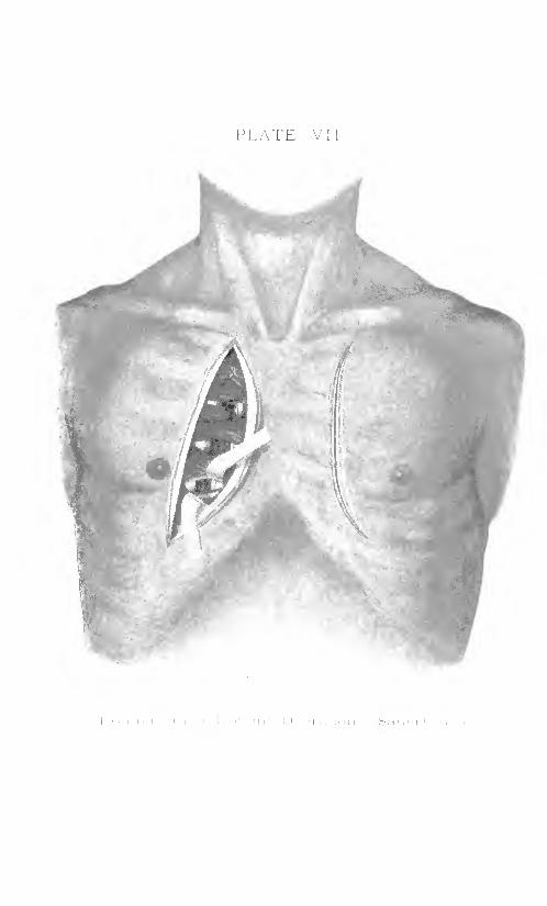
PLATE VII
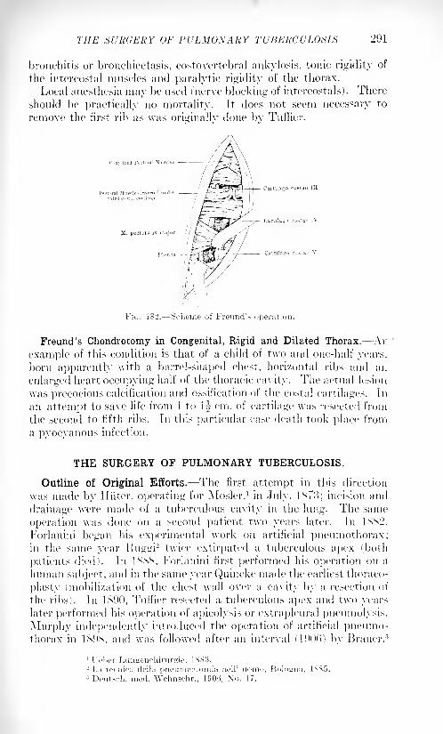
THE SURGERY OF PULMONARY TUBERCULOSIS 291
bronchitis or bronchiectasis, costovertebral ankylosis, tonic rigidity of
the intercostal muscles and paralytic rigidity of the thorax.
Local anesthesia may be used (nerve blocking of intercostals) . There
should be practically no mortality. It does not seem necessary to
remove the first rib as was originally done bv Tuffier.
FIG. 182. Scheme of Freund'?
Freund's Chondrotomy in Congenital, Rigid and Dilated Thorax. An
example of this condition is that of a child of two and one-half years,
born apparently with a barrel-shaped chest, horizontal ribs and an
enlarged heart occupying half of the thoracic cavity. The actual lesion
was precocious calcification and ossification of the costal cartilages. In
an attempt to save life from 1 to 1^- cm. of cartilage was resected from
the second to fifth ribs. In this particular case death took place from
a pyocyanous infection.
THE SURGERY OF PULMONARY TUBERCULOSIS.
Outline of Original Efforts. The first attempt in this direction
was made by Iliiter, operating for Mosler,1 in July, 1873; incision and
drainage were made of a tuberculous cavity in the lung. The same
operation was done on a second patient two years later. In 1SS2,
Forlanini began his experimental work on artificial pneumothorax;in the same year Ruggi
2 twice extirpated a tuberculous apex (both
patients died). In 1SSS, Forlanini first performed his operation on a
human subject, and in the same year Quincke made the earliest thoraco-
plasty (mobilization of the chest wall over a cavity by a resection of
the ribs). In 1S90, Tuffier resected a tuberculous apex and two yearslater performed his operation of apicolysis or extrapleural pneumolysis.
Murphy independently introduced the operation of artificial pneumo-thorax in 1&9S, and was followed after an interval (1900) by Brauer. 3
1 I'chor Ltmgenchirurgie, 1883.2 L;t tocniea rlella piieumectomia nell* uorno, Bologna, 1S85.3 Deutsch. med. Wchnschr., 1906, Xo. 17,
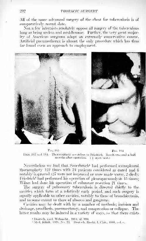
202 THORACIC SURGE It Y
All of the more advanced surgery of the chest for tuberculosis is of
comparatively recent date.
Not a few internists resolutely oppose all surgery of the tuberculous
lung as being useless and meddlesome. Further, the very great major-
ity of American surgeons adopt an extremely conservative course.
Artificial pneumothorax is almost the only procedure which lias thus
far found even an approach to employment.
Fit;. 183 FIG. 1S4
FIGS. 183 and 184. Thoracoplasty according to Friedrich. Ke.sult two and a half
months after operation. (-)- apox heat.)
Nevertheless we find that Sauerbruch1 had performed extrapleural
thoracoplasty 122 times with 24 patients considered as cured and 6
notably improved (27 were not improved or were made worse, 2 died);Friedrich 2 had performed his operation of pleuropneumolysis 4o times;\Vilms had done his operation of columnar resection 21 times.
The surgery of pulmonary tuberculosis is directed chiefly to the
cavities which form at a relatively early period, and such surgery is
equally applicable to other cavities, notably to those of bronchiectasis,and to some extent to those of abscess and gangrene.
Cavities may be dealt with by a number of methods; incision and
drainage, prosthesis, pneumectomy, and compression or collapse. Thelatter results may be induced in a variety of ways, so that there exists
1 Deutsch. mod. Wchnschr., 1914, xl, 090.2 Mcd. Klinik. 190X. No. 33. Dcutsdi. /tschr. f. Cliir., 190!), vol. c.
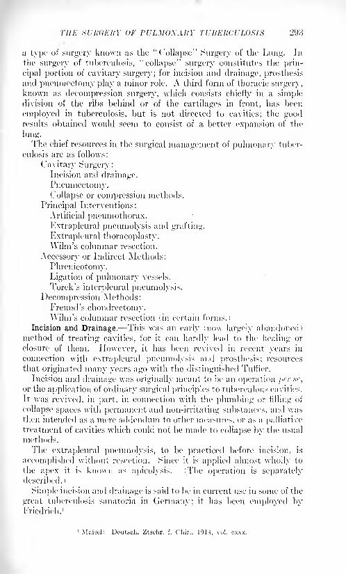
THE SURGERY OF PULMONARY TUBERCULOSIS 293
a type of surgery known as the "Collapse" Surgery of the Lung. In
the surgery of tuberculosis, "collapse" surgery constitutes the prin-
cipal portion of cavitary surgery; for incision and drainage, prosthesisand pneumectomy play a minor role. A third form of thoracic surgery,known as decompression surgery, which consists chiefly in a simpledivision of the ribs behind or of the cartilages in front, has been
employed in tuberculosis, but is not directed to cavities; the goodresults obtained would seem to consist of a better expansion of the
lung.
The chief resources in the surgical management of pulmonary tuber-
culosis are as follows:
Cavitary Surgery:Incision and drainage.
Pneumectomy.Collapse or compression methods.
Principal Interventions :
Artificial pneumothorax.
Extrapleural pneumolysis and grafting.
Extrapleural thoracoplasty.Wilm's columnar resection.
Accessory or Indirect Methods :
Phrenicotomy.
Ligation of pulmonary vessels.
Torek's interpleural pneumolysis.
Decompression Methods:
Freund's chondrectomy.Wilm's columnar resection (in certain forms.)
Incision and Drainage. This was an early (now largely abandoned)method of treating cavities, for it can hardly lead to the healing or
closure of them. However, it has been revived in recent years in
connection with extrapleural pneumolysis and prosthesis; resources
that originated many years ago with the distinguished Tuffier.
Incision and drainage was originally meant to be an operation per se,
or the application of ordinary surgical principles to tuberculous cavities.
It was revived, in part, in connection with the plumbing or filling of
collapse spaces with permanent and non-irritating substances, and wasthen intended as a mere addendum to other measures, or as a palliative
treatment of cavities which could not be made to collapse by the usual
methods.
The extrapleural pneumolysis, to be practiced before incision, is
accomplished without resection. Since it is applied almost wholly to
the apex it is known as apicolysis. (The operation is separatelydescribed, i
Simple incision and drainage is said to be in current use in some of the
great tuberculosis sanatoria in Germany; it has been employed byFriedrich. 1
1 Muisel: Dcutsdi. Ztschr. f. Chir., 1914, vol. cxxx.
294 THORACIC SURGERY
Pneumectomy. Pneumectomy ami lobectomy as typical operationsaiv considered elsewhere. As applied to pulmonary tuberculosis such
operations represent a type of intervention when cavities can be dis-
posed of in no other way.In Iss2 Ruggi performed two pneumectomies : he was followed in
I\s4 by Kronlein 1 and Block. 2 In 1S01 Tuffier made a successful resec-
tion and was followed by Lawson in 1X92. Writing in 19(K->, Vautrin
could collect but (> cases, which probably corresponded with these cited.
These early pneumectomies, however, were not all performed for the
cure of diseased foci.
The work done by Macewen3 in this direction has been phenomenal in
character. As far back as IsOo, he removed nearly the whole of an
entire lung which was the seat of huge cavities. The patient was living
and well in 1911 (Quincke and Garre). In lM)o (collapse methodshad not then come into use), Macewen removed the fourth to eighthribs, ligated the vessels at the hilus, and extirpated what was left of a
luii"' save a small portion of the apex which was untouched for fear of
wounding the subclavian artery.
Artificial Pneumothorax in the Treatment of Pulmonary Tuberculosis.
The general principles of this operation are considered elsewhere.
It is believed in many quarters that the operation originated with
Forlanini of Pa via in 1SS2, this author having been incited to the work
through a perusal of a French thesis by Toussaint (1SMJ) which dealt
with the improvement in tuberculosis as a result of natural pneumo-thorax. As a matter of fact, however, the idea is much older, having
probably originated with ("arson of Liverpool, in 1x21. lie showedthat a lung, in animal experiment, could be collapsed with impunity,and proposed pneumothorax as a rest measure for pulmonary tuber-
culosis. Carson's ideas were not allowed to die; they were often
mentioned as reasonable and were supported by the citation of cases
in which traumatic pneumothorax and pleural effusions exerted a
favorable influence on a diseased tuberculous lung.
Forlanini experimented on animals for years before he ventured to
operate on the human subject (1SSS4) and several more years were
allowed to elapse before he had enough clinical material to warrant
submitting his results to the profession (1S94). The operation wasnot taken up by the surgeons generally but was soon afterward inde-
pendently set forth by Murphy.6
Sachs6
reports on the results obtained by 24 carefully selected
American observers. There wen; 1122 cases reported upon in the last
few years, the incidence of various complications occurring in the
course of treatment, being as follows:
1 Berl. klin. Wchnschr., 1884, xxi, 129.2 Deutsch. mod. Wchnschr., 1881, vii, 634.3 Tr. Internal. Cunjnvss, Moscow, 1*97, v, 'jV>.
4 Potabi is said to have made a therapeutic pneumothorax in 1888.1 Denver Meeting of the Am. Mod. Assn., 1898.6 Jour. Am. Med. Assn., November 27, 1915, p. 1801.
THE SURGERY OF PULMONARY TUBERCULOSIS 295
Complications. Number.
Pleural effusion 113
Extension of process in opposite lung . . 58
Bleeding of other lung 7
Pleural shock . . .... 26
Gas embolism
Spontaneous piieumothorax 10
Bilateral pneumothorax 2
Pyopneumothorax 13
Cardiac dilatation and heart failure 4
Torsion of heart and bloodvessels
Total 237'
One is forcibly struck by the very large percentage, 21 per cent,
of complications occurring in the course of this assumedly simple
procedure.
Murphy (1898) within a brief interval was able to report a very
large number of cases through his associate Lempke, and this excel-
lent presentation of the subject influenced Brauer to introduce the
operation into Europe (outside of Italy). In the meantime Forlanini's
work in Italy had begun to accumulate, where his operation had been
accepted as a rational procedure. Despite his thirty-five years of
experience, however, his amount of material is not large very muchbelow that of Murphy and his associates. Forlanini has made a very
great number of individual injections, however, and is the most
exhaustive and systematic writer on the subject, not only from his
own viewpoint but from that of his contemporaries, to whom he has
always been ready to award full credit. His writings are classic andthe writer has freely borrowed from them a statement of indications
and centra-indications. (Elsewhere will be found an account of the
accidents which have resulted from the operation and the ways bywhich they may be avoided.) In addition to Forlanini and Murphy,others have operated on very many cases. The writer has definite
knowledge of many cases operated on by physicians of Denver andColorado Springs.
Indications for and Contra-indications to Artificial Pneumothorax.
Theoretically there are two principles involved: (1) the pneumothoraxtends directly to relieve the destructive processes and the results of the
same (cavities). This is purely a local activity. (2) Its action is
unilateral. In other words it is a strictly local treatment of a uni-
lateral tuberculosis. Nevertheless, experience has taught that the
operation may not only improve the state of the opposite lung whenit is slightly involved, but also that it tends to benefit extrapulmonaryfoci.
Pneumothorax is contra-indicated in affections of the circulatory
system and kidneys because it exerts a disturbing influence upon the
heart.
Factors of individual significance are (1) the character of the tuber-
1 Twenty-one per cent, of the total number of cases.
29(5 THORACIC Sl'ltdXHY
culoiis process, (12) the condition of the pleura, (/>) the co-existence of
other diseases, (4) bilateral character of the disease, (.">) stage of the
phthisical ])rocess.
1. Character of the Tuberculous /Vomv.s-. The operation is contra-
indicated in general miliary tuberculosis, and in all acute forms. It
may not harm the patient but it can hardly help him, as his life is
directly menaced by the disease. Either the other lung or the organismat large is threatened. Generally speaking, the operation is restricted
to cases in which life is not immediately threatened.
'2. State f the Pleura.- A free pleura is a desideratum for a successful
treatment, but this is not indispensable. It is, however, necessarythat adhesions be not too extensive or too dense. It is often possibleto recognize the presence of adhesions, but it is often very difficult to
learn their amount and degree. Experience teaches that the pneumo-thorax often removes the adhesions; if not wholly, at least in part.
Old, total symphysis of the pleura a condition generally to be recog-nized furnishes the only absolute centra-indication. In all other cases
the operation maybe attempted, and relinquished only when it is foundthat the amount of gas which can enter is quite insufficient for the
purpose.'.>. Co-existence of Oilier Diseases. In theory, pneumothorax is
contra- indicated in diseases of the circulatory system and kidneys.The pressure of the gas in the pleura not only compresses the heart butit prevents the ingress into the thorax of venous blood, thus increasingrenal congestion. In practice, however, Forlanini has not experiencedsuch ill-results; he does not hesitate to operate when there is good
compensation in the heart and kidneys. In two cases of nephritis,the patients did very well following the operation. Diabetes con-
stitutes a theoretical contra-indication not yet borne out in practice.
The same may be said of laryngeal and intestinal tuberculosis. If
these lesions arc not severe Forlanini does not hesitate to institute
treatment.
4. Bilateralism of Pulmonary Tuberculosis. The unilateral location
is simply the ideal condition. Bilateralism per se does not absolutelycentra-indicate. If there is but a single circumscribed focus of disease
in the second lung the operation may favor healing or arrest. Therationale of this successful outcome is not clear, for only in a minorityof cases is it successful. The rule is for the focus in the second lung to
progress, but while the presence of such a focus might seem to form a
contra-indication the author would give his patient the benefit of the
doubt. If there is a small circumscribed focus in each lung Forlanini
operates first on one side and then on the other, counting upon the
first lung to re-expand before operating on the second. Naturally the
alternation of sides has some field in the more advanced bilateral
cases, but these must be carefully selected for treatment.
5. Stage of the Phthisis. The operation may be indicated in all three
stages; in other words, the stage per sc is immaterial. The more
advanced the disease the greater the likelihood of bilateralism and the
THE SURGERY OF PULMONARY TUBERCULOSIS 297
greater the likelihood of dense, extensive pleural adhesions; conversely,
the more circumscribed the lesion, the greater the amount of good lungtissue. When there are cavities there are likewise caseous foci present.
These combined lesions form the cause of the most serious compli-cation of the operation, a perforation of the lung.
Even in the simplest cases the treatment is long and exacting for
both physician and patient and pleural infection may possibly take
place. Medical treatment should by all means be instituted in begin-
ning lesions. The moment ulceration of tissue begins the practitioner,
however, may have recourse to the artificial pneumothorax.Murphy's Technic for Artificial Pneumothorax. This account is taken
from an article by Murphy and Kreuscher in the Tuberculosis Number
(March) of the Interstate Medical Journal, 1914.
"The patient is placed in a comfortable sitting position and the
chest is bared over the side to be injected. The point of insertion of the
needle varies somewhat in individual cases. If the apex of the lung be
the site of the lesion, the needle should be inserted in the fifth or sixth
interspace between the anterior and midaxillary lines. The best pointis the fifth interspace at or near the anterior axillary line. If it be a
middle or lower lobe tuberculosis, the injection should be made over
the upper lobe, preferably in the fourth interspace just outside of the
mammary line."
Local anesthesia (novocain infiltration) involving the injection zone
gives very satisfactory results. A tenotome puncture through the
skin precedes the introduction of the needle, this introduction beingmade before the tube is coupled on. In order to make sure that the
needle is in the pleural cavity the patient is asked to take a series of
deep inspirations. If the cavity has been entered an inrush of air will
be heard, and after the forced respirations are under way air will be
heard moving in both directions. The distal opening of the needle
should be covered with a little cotton to serve as an air filter. The gasdoes not always readily enter the pleiiral cavity, doubtless because of
the natural cohesion of the two laminae of the pleura. It may be neces-
sary to overcome a pressure estimated at 12^ mm. of mercury. (Theauthor apparently does not employ a manometer.)The needle used is blunt, of moderate size and with a special side
opening near the tip. Hence, if the tip chances to be occluded the gascan still enter the pleural cavity. AVhen ready for use the needle is
attached to a tube which communicates at its other extremity with a
cylinder of nitrogen. From oO to 1200 cubic inches of the latter may be
introduced at a time, the operator being guided by the symptoms of
dyspnea and distress and the displacement of the mediastinal contents
and the diaphragm. The needle is now withdrawn and the punctureis sealed with collodion over which is placed a firm compress which is
intended to prevent a subcutaneous emphysema.Itt'xulfx of Operation.- The average number of injections required
appears, from Murphy's experience, to be about f). Repetition is
necessary because of the gradual absorption of the gas. The injec-
29S THORACIC SURGERY
turns are continued until, if possible, the following typical results are
obtained:
1. The decline or disappearance of fever, which marks the dis-
appearance of toxic material from the lung cavities.
'2. The diminution or disappearance of expectoration, which also
points to the closure of the cavities; the bacilli of the sputum may also
have disappeared.o. A gradual increase in weight.4. Improvement in hemoptysis."). (General amelioration in a relatively short time.
The main desideratum, at present, is the earliest possible applicationof treatment in these cases. One should not wait for cavities to form
because the lung can be collapsed and placed at rest in advance of a
cavity formation; thus, the latter may be prevented. Rest is reallythe primary requirement, a collapse of the cavities is a secondary one.
Pneumolysis and Grafting in the Treatment of Cavities.- This combinedresource is best described by Tufh'er (see the Tufh'er and Loewy article
in the Tuberculosis Number of the Interstate Medical Journal, March.1K14 1. It has the advantage of immobilizing and compressing only the
diseased portion of the lung.
In 1S91 Tnffier resected the second rib with the idea of extirpating a
tuberculous apex. The parietal pleura was found to be adherent to the
ribs, but it was readily detached with the fingers. It then becameevident that this pnenmolysis could readily lie used for diagnostic pur-
poses. The apex could also be mobilized and the depression filled with
grafts. This procedure Tuffier terms "extrapleural pneumothorax"; he
regards it as superior to the typical or intrapleural form. (One judgesthat he has operated in this manner some 1C) times since 1912.)
The completed technic of this procedure is as follows: first stage,
"apieolysis" or detachment of the apex. An incision (> cm. long is
made in the second intercostal space down to the pleura, the muscles
being divided separately. The author's intercostal separator, with a
toothed rack, is then applied, and the parietal pleura, usually adherent,is exposed. The tip of the index finger now separates the pleura from
the inner aspect of the ribs. The anterior surface of the apex is first
freed, the posterior surface later. (This step is difficult, especially if
there are vertebral and mediastinal adhesions.) The lung tissue, if
pervious, collapses with each inspiration, so that in the process of the
apieolysis cavities often sink in under the finger. As the apex is
isolated it is protected with a layer of silk dipped in sterile vaseline.
(This is intended to prevent rupture into the walls of a cavity.)
The apex, now freed, tends to collapse, leaving a cavity between the
parietal pleura and the ribs. This cavity is to be filled with fat, fresh
or from storage. (Paraffin mixed with bismuth has been used.) These
substances are used to distend the artificial cavity and thus compressthe apex. The intrapulmonary cavity is obliterated as the lung retracts
toward the hilus. The various superjacent tissues are to be separately
sutured.
THE SURGERY OF PULMONARY TUBERCULOSIS 299
Evolution and Technic of Sauerbrach's Operation for Extrapleural
Thoracoplasty. The oldest and simplest method of rendering the
thorax mobile and causing a depression is that recommended byQuincke and Spengler ( ISSN). Everything subsequent in this direction
seems to have been based thereon. Quincke recommended that one or
several ribs over a cavity should be resected. (A piece of the second
rib or of the second and third ribs sufficed as a rule.) Bier made an
incision from the coracoid process to the sternal border of the first
rib cartilage and subperiosteally resected the second and third ribs.
Spengler thought that the amount resected should agree with the
amount of diseased lung and spoke of "total resection." lie removedoo cm. from the third and fourth ribs at the anterior axillary line. (It
is not necessary that the resection be over a cavity, although this
should be the case when possible.) Turban 1 stated that a resection
anywhere benefited the entire lung, and that a resection over the
lower lobe was of benefit to the apex. He resected, at the lower lateral
region, a wedge-shaped piece with its base downward. In one case
his incision was at the anterior axillary line, removing 24 cm. from the
fourth to seventh ribs. One year later he removed 41 cm. from the
sixth to the ninth ribs. Landerer repudiated typical operations and
operated on each case individually. lie felt that one resected rib should
be outside of the area of the cavity. lie resected ribs both above and
below -the three or four upper ribs or from the fourth to ninth, both
anterior and posterior. None of these operations aimed at the collapseof anything more than the cavity itself.
Brauer repudiated all partial resections with a view to the securingof a total collapse. Friedrich and he developed a special technic which
resembles in its incision the older Schede operation for empyema.(The operative details are given elsewhere. At least 160 cm. of ribs are
resected, 25 of which are from the lowest (ninth or tenth) rib.)
After taking the ribs from the thorax the lung retracts "hilusward."
Total collapse is the rule, but, in some cases, the apex remains uncol-
lapsed, although the operation is usually intended to secure this result.
This failure of the apex to collapse is due partly to the fact that the
first rib is not included in those resected. Friedrich, in his earlier cases,
instead of resecting the first rib preferred to detach the costal pleura
by a blunt dissection iapicolysis). As a result of this the apex sinks
in but no dead space is formed. Xo one else seems to have tried
apicolysis, and Friedrich abandoned it for fear of a cardiac collapse
from the ''circulatory reflex." The only remaining course was to
resect the first rib with the others, and this seems to have been first
done by Sauerbruch. Brauer went further and advocated resection of
the clavicle. Sauerbruch had seen benefit to the apex from an acci-
dental fracture' of the clavicle, and so resected it in a series of cases.
Friedrich was the third to include this step. Sauerbruch performedthis operation four times; he had two excellent results, and one fair
1 Herl. klin. \\Vlin.<rhr., ix'j'l, N'o. 21.
:>oo THORACIC SURGERY
result, l)iit lost his fourth patient. Friedrich's operative mortality wasalso high. This was due, in part, to general causes, but especially to
a state of affairs which corresponds to an open pneumothorax with
paradoxic breathing and mediastinal fluttering.
It seemed necessary to improve the technic. Friedrich tried sub-
periostea! resection with regeneration, but this did not diminish the
acute dangers. Firm bandage's and retentive apparatus, overpressure,
etc., were tested for the prevention of the paradoxic respiration, but
something more radical was needed. Brauer selected the so-called
Quenu-Tietze operation for old empyema cavities. This left a thoracic
wall while admitting of some collapse.
The Boiffin method of paravertebral division of the ribs, also for
empyema, allows of considerable retraction of the lungs. This methodwas introduced by AVilms in 1911 for the relief of pulmonary tuber-
culosis. This paravertebral division of the ribs reduces the capacityof the chest by 150 to 180 cm. YVilms substituted for simple division
the resection of a certain number of ribs, parasternal as well as para-vertebral.
Compression or collapse therapy differs radically from decompression
therapy. The collapse of the diseased lung is encouraged. The earliest
resource for this purpose was artificial pneumothorax which has alreadybeen described. This operation is practically limited to cases without
pleural adhesions. Brauer was, therefore, moved to devise a procedureto produce collapse in cases with pleural adhesions. lie chose an old
resource tested by Quincke in 1SSS, and by Spengler in 1S90. This
was a resection of the ribs over a given tuberculous cavity (such a
procedure as had been in use for an abscess of a lung), with the aini of
securing a diminution in the size of a cavity and its cicatrization.
Quincke operated in front, while Spengler made his resection in the
interscapular space. The latter, with others, advocated a wide resec-
tion and called the procedure extrapleural thoracoplasty. The aim was
to secure some retraction of the chest not a collapse of it.
Brauer sought collapse and felt that the resection should be as
extensive as possible. He advised a flap incision which encircled the
scapula and exposed the chest-wall. Ribs, from the third to the tenth,
were then removed in great extent. The intervention was a new one
and Brauer termed it "Pleuropneumolysis thoracoplastica," but
Spengler 's original term has survived.
The operation was performed successfully by Friedrich,1 but its
mortality proved to be high, and technical modifications were neces-
sary. It was at first done in two or even three stages. (The Kiister-
Kstlander operation for old empyema t a may also be performed for
tuberculosis. \Vilm 's columnar paravertebral resection may be
regarded as an outgrowth of empyema surgery as practiced by Boifh'n.)
In considering the rationale of extrapleural thoracoplasty, it may be
1 Friedrich being the Director of the Surgical .Clinic at Marburg while Brauer was
the Director of the Medical Clinic.
THE SURGERY OF PULMONARY TUBERCULOSIS 801
well to discuss again the artificial pneumothorax. In this latter pro-
cedure, the gas introduced tends to abolish the normal adhesion of the
pleural laminae. The chest no longer produces traction on the lungand the elasticity of the latter causes it to retract. It becomes smaller
and smaller and if enough air is allowed to enter the pleural cavity it
may attain a minimum of size. Atelectasis does not result until the
air has been absorbed. With the retraction of the lung, given cavities
collapse, the respiration is retarded and the lung tends to be placedin a state of functional rest. The type of respiration is reversed. Air
from the sound lung passes, during expiration, into the still distensible
collapsed lung. During inspiration this air again escapes, as the latter
contracts.
The collapsed lung can no longer make traction on its environment,and as a result the mediastinum is thrown from its equilibrium and is
displaced to the sound side so that the sound lung has its excursions
diminished. In a like manner, the diaphragm descends during inspir-
ation as a result of the change in intrapleural pressure. Hence, slight
dyspnea and acceleration of the heart's action may result for a brief
interval or until an equilibrium is regained.Functional rest and diminution in the size of the cavities are impor-
tant therapeutic factors, akin in every way to the placing at rest of
tuberculous joints. There can naturally be no collapse if the walls of
the cavities are rigid. Other phenomena are not so readily understood,such as the action of retraction of the lung on the bloodvessels. Thelatter must undergo some compression and the heart is displaced with
the mediastinum. Less blood reaches the right ventricle, but acceptedauthorities differ widely as to the exact rationale of that which occurs.
In an open pneumothorax the blood circulates freely through the
collapsed lung and the writer, for the sake of argument, assumes that
this must be the case with a closed pneumothorax. This view is dis-
puted, however, by those who disbelieve in a free circulation througha given collapsed lung. There is blood enough in the latter, but theythink it stagnant.
Atelectasis occurs after the alveolar air has been absorbed. Thevessels must undergo contraction, and as a matter of fact the lungreceives less blood. (The argument refers to the period before atelec-
tasis.) Brauer's view of an active hyperemia of the collapsed lungreceives aid from Cloetta's researches, from which it appears that in
the expansion of the normal lung there is a lesser blood supply than
in a retracted lung. The greatest amount of blood is found at a pres-
sure of about 3 cm. Ilg. Sauerbruch confesses himself obliged to
leave the matter undecided.
More is known of the lymph circulation. A retracted lung causes a
lymph stasis. After collapse from pneumothorax there may be an
initial fever from the absorption of toxins in the lymph. All are agreedthat in a given pneumothorax lung changes occur in the pulmonarytissue. This becomes firmer and quite devoid of air. Very soon evi-
dences of a transformation into connective tissue are present; tuber-
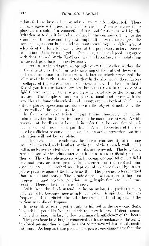
THORACIC
minus foci arc invested, encapsulated and finally obliterated. These
changes agree with those seen in any tissue. When recovery takes
place as a result of a connective-tissue proliferation caused by the
irritation of toxins it is probably due, in the contracted lung, to the
stimulus of the toxic and stagnant lymph, although To some degree the
same changes occur in a sound pneumothorax lung. A high degree of
sclerosis of the lung follows ligation of the pulmonary artery (Sauer-brnch) and of the vein (Tiegel). The changes in a collapsed lung agreewith those caused by the ligation of a main bronchus; the metabolismin the collapsed lung is much lessened.
To return to the old Quincke-Spengler operation of rib resection, the
authors mentioned the indurated thickening of the walls of the cavities
and their adhesion to the chest wall, factors which prevented the
collapse of the cavities, and stated that in the absence of these factors
a collapse of the cavities would doubtless occur. Fit the more elastic
ribs of youth these factors are less important than in the case of a
rigid thorax in which the ribs are an added obstacle to the closure of
cavities. This simple reasoning appears rational, and agrees with the
conditions in bone tuberculosis and in empyema, in both of which con-
ditions plastic operations are done with the object of mobili/ing the
outer walls of the given cavities.
In the operation of Friedrich and Brauer, however, not merelyisolated cavities but the entire lung must be made to contract. A wide
resection of the ribs must be made in order that the condition of arti-
ficial pneumothorax may be paralleled. A small resection of the ribs
may be sufficient to cause a collapse, /'. e., an active retraction, but this
retraction will not be complete.Under physiological conditions the normal contractility of the lung
cannot be exerted, as it is oll'set by the pull of the thoracic wall. This
pull is no longer exerted when entire ribs are removed. The lung then
retracts toward the hilus exactly as it does in an artificial pneumo-thorax. The other phenomena which accompany and follow artificial
pneumothorax are also present (displacement of the mediastinum,
dyspnea, etc.). The soft tissues deprived of bone are forced by atmos-
pheric pressure against the lung beneath. (The pressure is less markedthan in pneumothorax.) The paradoxic respiration, akin to that seen
in open pneumothorax (contraction during inspiration, etc.), is charac-
teristic. Hence, the immediate danger.Aside from the shock attending the operation, the patient's color,
at first pale, becomes increasingly cyanotic. Respiration becomes
frequent and superficial; the pulse becomes small and rapid and the
patient may die of dyspnea.In favorable cases the patient adapts himself to the new conditions.
The critical period is from the sixth to seventh day. If death occurs
during this time, it is largely due to primary insufficiency of the heart.
The parodoxic breathing is connected with the mediastinal fluttering
in closed pneumothorax, and does not occur save with a supple medi-
astinum. As long as these phenomena persist one cannot say that the
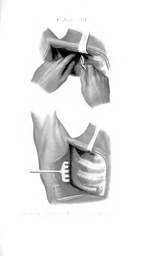
FLAT!-: VI! I

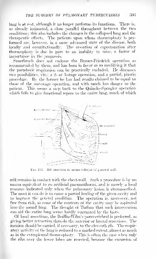
THE SURGERY OF PULMONARY TUBERCULOSIS 303
lung is at rest, although it no longer performs its functions. There is,
as already intimated, a close parallel throughout between the two
conditions; this also includes the changes in the collapsed lung and the
therapeutic effects. The patients upon whom thoracoplasty is per-
formed are, however, in a more advanced state of the disease, both
locally and constitutionally. The cessation of expectoration after
thoracoplasty is due in part to an inability to raise, a factor of
importance in the prognosis.Sauerbruch does not endorse the Brauer-Friedrich operation as
recommended by them, and has been in favor of so modifying it that
the paradoxic respiration can be practically excluded. lie discusses
two possibilities, vi/.: a '2- or 3-stage operation, and a partial, plastic
procedure. By the former he has had results claimed to be equal to
those of the one-stage operation, and with much less danger to the
patient. This seems a step back to the Quincke-Spengler operationwhich fails to give functional repose to the entire lung, much of which
K. poft-ir.il
Mm intercostal
i \^ \\ /- .L^4c..V A\ .^-^/ \\\
FIG. 1S5. Ilib resection to secure collapse of parietal wall.
still remains in contact with the chest-wall. Such a procedure is by no
means equivalent to an artificial pneumothorax, and is merely a local
resource indicated only when the pulmonary lesion is circumscribed.
The most it can do is to cause a partial healing of the given cavity and
to improve the general condition. The operation is, moreover, not
free from risk, as some of the contents of the cavity may he aspiratedinto the sound lung. The thought of Turban that such intervention
can aid the entire lung seems hardly warranted by the facts.
Of local resections, the Boiffin-Wilm's paravertebral is preferred, as
giving better retraction than do the anterior or lateral resections. Theincision should be carried, if necessary, to the eleventh rib. The respir-
atory activity of the lung is reduced to a marked extent, almost as muchas in the extrapleural thoracoplasty. This is often the case when onlythe ribs over the lower lobes are resected, because the excursion of
o()4 THORACIC Hl'RCERY
these ribs is much greater than is that of the upper ribs. Resection
of the lower ribs is often of benefit to the upper lobes.
In regard to the blood and lymph circulation in the partially retracted
lung, the former is probably most complete at a pressure of 3 mm. Ilg,
or at about that of expiration. There is also probably a slowing of the
lymph circulation.
Sauerbruch has resected the eighth to tenth and eleventh ribs for
tuberculosis of the upper part of a lung. An ordinary paravertebralincision is bent at its lower end to form a hood. The scapula, free
behind, can be turned forward. Beginning at the eleventh or tenth rib,
10 cm. are excised, and this length is reduced as succeeding ribs are
rejected. The number of these resected ribs depends upon circum-
stances. In total cases, all of the ribs up to the second or first are
removed, the amount resected being 4 or 5 cm. over the upper portionof the lung. In certain cases, the first and second ribs may be resected
in a second stage. A portion of the clavicle is sometimes removed.
Concerning the effect of lung retraction on the sound lung, it is
found that the latter is forced to do more work and at the same time
it is mechanically hindered from so doing. There results a tendency to
emphysema from the dilatation of alveoli. Sometimes compensation is
so marked that one may speak of the lung as being hypertrophied.If the lung is already the seat of old, closed foci these may be thrown
into activity. As regards the disappearance of expectoration, it must
again be remembered that for the first few days this may be due to an
inability to cough. This retained sputum must be expectorated, and
after this there may be a sudden or a gradual diminution. The char-
acter of the sputum undergoes a radical change. The temperature,after a brief rise from absorption, begins to fall. A gain in weight is
generally to be noticed.
The Evolution of Friedrich's Large Flap Operation (Including Division
of the Phrenic and Intercostal Nerves). In 1907 Friedrich 1first prac-
ticed his extensive resection in the thought of securing for the diseased
lung a complete rest, and its elimination from respiration. At the same
time the cavities were to be closed by compression. The results seemed
to justify the operation although the operative mortality was 25 per
cent. In 11)1 1 he was able to report 28 cases.
In different articles Friedrich discussed possible changes in technic:
decreasing the amount of resected rib, operating in stages, the choice
of the area to be resected, etc. Other surgeons, notably Sauerbruch
and Wilms, took up the work and modified the indications so that
milder cases were included. It gradually became apparent that too
small an area of resection had its drawbacks, and Sauerbruch and
Wilms began to employ the "hook incision" and "columnar" resections
respectively. The former corresponded largely to Friedrich's incision
of Ji)()x. Danish, Swedish and French surgeons have performed the
operation, especially such tuberculosis specialists as Turban, L.
Spengler, "\ an Voornfeld, von Muralt.
THE M'KdERY OF PULMONARY TUBERCULOSIS 305
According to these authors, the fixing of the indications does not
seem to be so very difficult. Even patients who have been bedridden
for months and who have a high, hectic, intermittent fever and night-sweats may be included if the active lung affection is strictly unilateral
and the other lung efficient for function. The better the general
nutrition, as estimated by the state of the muscles, the better mustbe the prognosis. A yellow, pallid, withered skin which suggestsintestinal tuberculosis, is a strong centra-indication. Any recent
tuberculous metastases contra-indicate operation. Vascular erethism
and a rapid, small-wave pulse, however, do not contra-indicate. (Such
may be improved by digalin given before operation.)The condition of the lung seeming to indicate operation is that in
which a tendency to retraction is already present. The affected side is
flattened, the clavicular fossa is retracted, the intercostal spaces are
narrowed, and the muscles are sclerotic with an increased resistance to
pressure; the intercostal spaces are narrower. Percussion and x-rayexaminations show an attraction of the heart and mediastinum towardthe contracted side; a high and at times a fixed position of the dia-
phragm; a shortening of the transverse diameter of the thorax on the
affected side.
The lung evidences which contra-indicate the operation are pro-
gressive infiltrative processes of great extent, and miliary tuberculosis.
A progressive increase in sputum raises a question, much more so than
do increased bacilli. Cavities are not the main indication, for non-
cavernous cases demand consideration as does the question as to
whether one lobe or the entire lung is involved. If the sound lung has
pleural adhesions, the outlook is not as good as when such adhesions
are absent. Decision must finally depend on the most careful exami-
nation of the sound lung. The latter need not be absolutely intact.
Old, passive foci, even when of considerable size, do not entirely contra-
indicate operation. Even in cases of recent infection of the sound lungan improvement has been secured, as it has been produced by an
artificial pneumothorax. The operation should, however, generallybe confined to the unilateral cases. Although in cases with oOO cm. of
daily sputum an operation has caused the disappearance of this within
a few days, the fact remains that the "drier" the case the less will be
the dangers of the after-treatment period. Cases with not over 10 cm.
of daily sputum offer a favorable prognosis.It cannot be said that, at this time, there is a typical operation. In
extensive tuberculosis of one side, especially with cavities, an extensive
rib resection will, of course, be required. The question of one, two or
three stages depends on the resistance of the patient and the skill of the
operator. In non-operative cases a tuberculosis of the upper lobe often
leads to aspiration tuberculosis in the lower lobe, but this does not
imply the existence of a physiologic law. It is not easy to understand
why it is that, after a low resection, aspiration tuberculosis does not
occur more frequently, respiratory movements being still present. In
order to cause a maximal retraction at the apex, it is probably best to
cause ;i retraction of the lower lobe as well. In a two-stage operationthe lower resection should be practiced first.
In order to secure a compression from behind, Friedrich has carried
his resection as nearly as possible to the vertebra*. If large segments of
ribs are left a collapse is hindered. Preservation of the anterior partsof the ribs, on the other hand, serves to protect the mediastinum, while
resection here is not of much value in securing collapse, the anterior
stumps of the ribs in reality following the pull of the lung.Before operation (which may and perhaps should be done under
local anesthesia), the patient clears his lungs of sputum. In order to
prevent the chance of aspiration the operation should, in theory, be
performed as rapidly as possible. (Friedrich was formerly proud of his
ability to remove ten ribs in twenty-five to forty minutes!)
Beginning with 1 !)()!>, Friedrich used an axillary incision, in manycases removing from the first or second to the seventh to ninth ribs.
In disease limited exclusively to one lobe, a limited resection may be
permitted; this when a cavity is present.The condition of affairs is simplest in cases of isolated tuberculosis
of the lower lobes. Here a resection is indicated which includes the
tenth and fifth ribs, with the retention of the periosteum and a strong,
obliterating suture of the soft parts, in order to obtain a firm chest-
wall, good lung compression and rest.
In 1 !()!), Friedrich described extrapleural apicolysis. or blunt sepa-ration of the apex and both pleural membranes from their environment.
This may be used as an accessory to rib resection. Bar 1 has associated
apicolysis with a plumbing of given cavities with paraffin i 1(
.M:>'. Both
Friedrich and Sauerbruch advise against such plumbing.In regard to Stiirt/.'s operation of phrenicotomy, Friedrich investi-
gated the subject and made animal experiments. lie also studied the
relations of the phrenic nerve in subjects with enlarged cervical lymphnodes. (In one patient the nerve adhered to the internal jugular vein
and in seeking to free it a fatal air embolism occurred.) I nder normal
conditions division of the nerve is simple; it should be divided as nearly
a> possible to the plenral capsule in order to get below the fourth
cervical nerve.
Stiirt/ introduced phrenicotomy as a substitute for extrapleural
thorocoplasty in cases of adherent pleura. The negative pressure in the
thorax ".-.iicks up" the paralyzed diaphragm on inspiration, while the
abdominal pressure is increased by the compensation of the sound half
of the diaphragm and thus forces upward the paralyzed side. Stiirt/.
believes that the paralyzed half adapts itself to the contraction of the
lung which it follows. I lellin assails the views of Stiirt/, and states that
division of either one or both of the phrenics does not cause paralysis.
Friedrich strongly disbelieved in abolishing the function of the phrenicnerve which is so essential for the act of coughing. Instead of cutting
it he crushed it with an artery forceps after a diagnostic electrical
THE SURGERY OF I'l'LMOXAHY TUBERCULOSIS :>()7
stimulation that caused a sense of dyspnea. The patients on whom he
did this received no benefit either subjective or objective. The .r-ray
showed that the half of the diaphragm on the affected side at once rose
and that it moved in unison with the sound half while the intact half
of the chest showed a notable increase in volume. Pleuritic adhesions
at the base of the lung prevented the change of place of the diaphragm.\\ith basal adhesions there arc usually adhesions of the mediastinumwith a consequent rigidity.
If phrenic division be followed by marked decrease in expectoration,this can well be due to decreased ability to cough. Hence, in any con-
sideration of this procedure, it should not be performed in the presenceof profuse expectoration.
In 2 of the (> cases in which the diaphragm ascended after phreni-
cotomy, Friedrich could find no evidences of improvement.Consideration must be made 1 of the question as to whether division
or resection of the intercostal nerves tends to favor contraction of a
given lung; it is known that division of these nerves renders the act of
coughing nearly or quite impossible.
Friedrich, in one of his cast's, divided the phrenic nerve on one side
and resected long segments of each of the intercostal nerves. (This
intercostal nerve excision was easily made through a long, vertical
incision.) In a second case this surgeon combined resection of the
intercostal nerves with an extensive resection of the ribs. In the first
case his patient found it very difficult to cough. In his second case
remarkable benefit is said to have resulted. It should be remarked that
the pleura may be wounded in this nerve excision, in which event it
should be repaired promptly.
Schepelmann, in H)lo, divided the phrenic nerve and the twelve
intercostal nerves, together with all of the nerve filaments leading to
the scaleni muscles from the cervical and brachial plexuses, so that the
lung was entirely functionless. Improvement is said to have resulted.
Wilm's Operation of Columnar Resection of the Ribs to Narrow the
Thorax in Tuberculosis. 1- This is not a decompression operation but one
of "collapse" (compression) surgery, \\ilms has operated under local
anesthesia on '24 cases, with morphin (seopolamin, when necessary)
given in advance. A longitudinal incision is made through the skin and
muscles (separation of muscle fibers) from the second to the ninth rib.
o to 4 cm. from the spinous processes of the vertebra:1
. Despite their
division or separation, the trape/ius and rhomboid muscles generallyretain their functions, this perhaps due in part to the sinking in of the
chest. (Newly formed scar tissue may give the muscles a new point of
purchase.)In freeing the ribs the intercostal vessels are to be spared. (In but
2 of \\ilms's cases was the pleura injured.) In from seven to nine
ribs, from 4 to ."> cm. of bone are removed. Approximately six to eightweeks later, this first operation is followed, in severe cases, by a para-sternal resection.
1 Thcnip. (1. ( Jctrciiw., 101.'!. liv, 17.
:>OS THORACIC SURGERY
The derive of compression here obtained is inferior to that producedby an artificial pneumothorax. Tlie operation should be performedonly after artificial pneumothorax has failed. The indications com-
prise conditions of the upper lobes in which sclerosis is accompaniedby cavity formation. (Note the value of .r-ray pictures in this
connection.)
Pneumolysis Interpleuralis. F. Torek, of New York, refers to cases of
very dense and extensive pleural adhesions, in pulmonary tuberculosis,that cannot be remedied by the artificial pneumothorax. Even if the
lo\ver portions of the lung show collapse the heavy pleural adhesions
about the upper lobe may prevent its necessary collapse. (In fact, it
is pointed out that complete collapse in this locality is very difficult
to obtain by the ordinary "collapse" treatment.)
The operation of extrapleural thoracoplasty may be said to be indi-
cated when nitrogen, which enters the pleural cavity tinder pressure,is unable to separate the adhesions. Such intervention may be verysevere in broken-down patients, although the two-stage procedurelessens this severity to some extent. Torek has, therefore, believed
that these adhesions might be separated by the finders without sacrifice
of the thoracic wall. This process, which should not be confounded with
extraplenral detachment of the lung, is termed by him,"interplenral
pneumolysis."The operation is performed with intratracheal insufflation narcosis
or with the pressure-difference cabinet. A long incision (15 cm.) is
made in the sixth or seventh intercostal space, at the' posterolateral
portion and carried down to the pleura. The latter is opened, after
careful hemostasis. (The patient's head hangs well downward so that
the contents of a given cavity can escape by the month without enteringthe opposite lung.) The ribs having been well retracted the finger-
tips beuin to separate the adhesions close at hand. Gradually the
fingers and even the entire hand may be introduced in order to completethe isolation of the lobe. If band-like adhesions have formed it is best
to divide them rather than tear them. When the adhesions have been
well broken up the lung tisstie partially collapses. (It is best to leave
this undisturbed.) The external wound is closed by periosteal, muscle
and skin sutures, without drainage. This operation may be looked
upon as preliminary to an artificial pneumothorax, if an ,r-ray diagnosis
shows the latter to be practicable.
The possibility of rupture of a large, thin-walled cavity into the
pleural sac occurs in various operations. The most common result of
such an accident is a valvular pneumothorax, which in the case of an
air-tight suture may lead to an emphysema or to an infection. Both
of the accidents happened in a case of Torek's.
Further Remarks on Phrenicotomy as an Accessory Procedure in the
Collapse Therapy of Pulmonary Tuberculosis. It has already been stated
that this procedure was suggested by Stiirtz 1 for severe, chronic, uni-
1 Doutsrh. mod. Wchnschr., lid. xlviii.
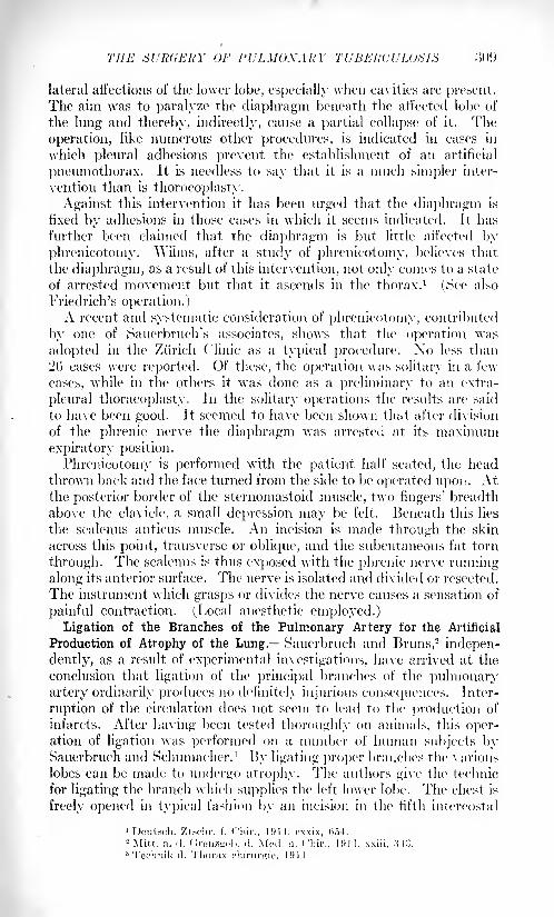
THE SURGERY OF PULMONARY TUBERCULOSIS 309
lateral affections of the lower lobe, especially when cavities are present.The aim was to paralyze the diaphragm beneath the affected lobe of
the lung and thereby, indirectly, cause a partial collapse of it. The
operation, like numerous other procedures, is indicated in cases in
which pleural adhesions prevent the establishment of an artificial
pneumothorax. It is needless to say that it is a much simpler inter-
vention than is thorocoplasty.
Against this intervention it has been urged that the diaphragm is
fixed by adhesions in those cases in which it seems indicated. It has
further been claimed that the diaphragm is but little affected byphrenicotomy. \Vilms, after a study of phrenicotomy, believes that
the diaphragm, as a result of this intervention, not only comes to a state
of arrested movement but that it ascends in the thorax. 1
(See also
Friedrich's operation.)A recent and systematic consideration of phrenicotomy, contributed
by one of Sauerbruch's associates, shows that the operation was
adopted in the Zurich Clinic as a typical procedure. Xo less than
2(> cases were reported. Of these, the operation was solitary in a few
cases, while in the others it was done as a preliminary to an extra-
pleural thoracoplasty. In the solitary operations the results are said
to have been good. It seemed to have been shown that after division
of the phrenic nerve the diaphragm was arrested at its maximumexpiratory position.
Phrenicotomy is performed with the patient half seated, the headthrown back and the face turned from the side to be operated upon. Atthe posterior border of the sternomastoid muscle, two fingers' breadth
above the clavicle, a small depression may be felt. Beneath this lies
the scalenus anticus muscle. An incision is made through the skin
across this point, transverse or oblique, and the subcutaneous fat torn
through. The scalenus is thus exposed with the phrenic nerve running
along its anterior surface. The nerve is isolated and divided or resected.
The instrument which grasps or divides the nerve causes a sensation of
painful contraction. (Local anesthetic employed.)
Ligation of the Branches of the Pulmonary Artery for the Artificial
Production of Atrophy of the Lung. Sauerbruch and Bruns,2
indepen-
dently, as a result of experimental investigations, have arrived at the
conclusion that ligation of the principal branches of the pulmonaryartery ordinarily produces no definitely injurious consequences. Inter-
ruption of the circulation does not seem to lead to the production of
infarcts. After having been tested thoroughly on animals, this oper-ation of ligation was performed on a number of human subjects bySauerbruch and Schumacher. :i By hgating proper branches the various
lobes can be made to undergo atrophy. The authors give the technic
for ligating the branch which supplies the left lower lobe. The chest is
freely opened in typical fashion by an incision in the fifth intercostal
1 Dcutsch. Ztsohr. I'. C'hir., 11)14, cxxix, 6~>4.
2 Mitt, a, (1. Cren/wb. <1. Mcd. u. Chir., 11)11, xxiii, .'it:!.
3 Technik d. Thorax chiniruio, 191 I.
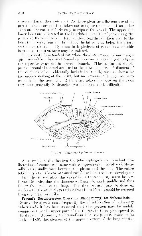
space (ordinary thoracotomy.) As dense pleuritic adhesions are often
present great care must he taken not to injure the lung. If no adhe-
sions are present it is fairly easy to expose the vessel. The upper andlower lobes arc' separated at the interlobar notch therein* exposing the
pedicle of the lower lobe. Here lie, close together on their way to the
lobe, the artery, vein and bronchus, the latter lying below the arteryand above the vein. By using little pledgets of gauze on a suitable
instrument the structures may be isolated.
On account of anatomical variations these structures are not always
quite accessible. In one of Sauerbrnch's cases he was obliged to ligate
the separate twigs of the arterial branch. (The ligature is simply
passed around the vessel and tied in the usual manner.) A filament of
the vagus may be accidentally included in the ligature, as shown bythe sudden slowing of the heart, but no permanent damage seems to
result from this accident. If there are adhesions between the lobes
thev max u'enerallv be detached without very much difficulty.
As a result of this ligation the lobe undergoes an abundant pro-
liferation of connective tissue with compression of the alveoli; dense
adhesions usually form between the pleura and the lung. The entire
lobe contracts. ( In one of Sauerbruch's patients a scoliosis developed.)
In order to complete this operation a thoracoplasty must be per-
formed in order that the thoracic wall may be made mobile and thus
follow the "pull" of the lung. This thoracoplasty may be done six
weeks after the original operation; from 10 to lo cm. should be resected
from each of several ribs.
Freund's Decompression Operation ( Chondrotomy ) for Tuberculosis.
Because the apex is most frequently the initial location of pulmonarytuberculosis it has been assumed that this portion may be unduly
compressed by the upper part of the thorax, in those predisposed to
the disease. According to Freund's original conjecture, made as far
back as 1 *.">(>, this stenosis of the upper aperture of the lung consists
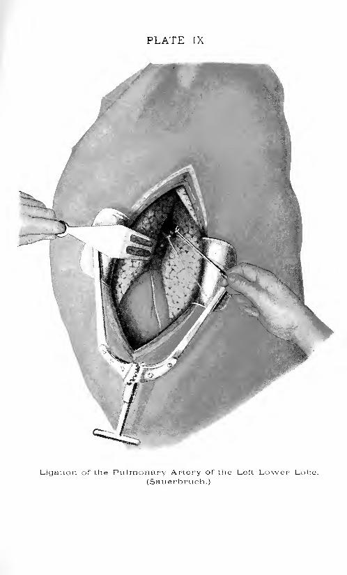
PLATE IX
Ligation of the Pulmonary Artery of the Left Lower Lobe.
(Sauerbruch.)

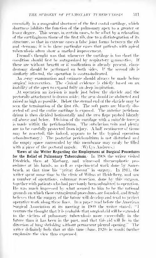
TllK HUHUKHY <>!'' I'l'LMOXMi'Y Tl'liKKCl'LOXIX oil
essentially in a congenital shortness of the first costal cartilage, \vhich
shortness inhibits the function of the pulmonary apex to a greater or
lesser degree. This seems, in certain cases, to he offset by a relaxation
of the cartilaginous tissue of the first rib, due to a disintegration of its
structure, so that in extreme cases a false joint forms between the rib
and sternum; it is in these particular cases that patients \vith apicaltuberculosis often sho\v a marked improvement.
Freund's thought was that whenever the cartilage is too short the
condition should first be antagonized by respiratory gymnastics. If
these are without benefit or if ossification is already present, chon-
drotomy should be performed on both sides. If the second rib is
similarly affected, the operation is contra-indicated.
An .r-ray examination and estimate should always be made before
surgical intervention. The clinical evidence is chiefly based on an
inability of the apex to expand fully on dee]) inspiration.At operation an incision is made just below the clavicle and the
pectoralis attachment is drawn aside; the arm should be abducted and
raised as high as possible. Below the sternal end of the clavicle may be
seen the termination of the first rib. The soft parts are bluntly dis-
sected off and the entire cartilage is exposed. The anterior perichon-drinm is then divided horizontally and the two flaps pushed bluntlyoff above and below. Division of the cartilage with a suitable forcepsis made within the perichondrium. The subclavian vein and pleuraare to be carefully protected from injury. A half centimeter of tissue
may be resected; this indeed, appears to be the typical operation
(chondrectoiny.) The posterior perichondrium is not disturbed andthe empty space surrounded by this membrane may easily be filled
with a piece of the pectoral muscle. (Wyllys Andrews.)
Views of the Writer Regarding the Employment of Surgical Procedures
for the Relief of Pulmonary Tuberculosis. In 190S the writer visited
Friedrich, then at Marburg, and witnessed thoracoplastic pro-cedures at his hands, as well as experimental work done by Sauer-
bruch, at that time his "privat docent" in surgery. In 11)11, the
writer spent some time in the clinic of Wilms at Heidelberg, and sawa number of operations, columnar resection, done by this surgeon,
together with patients who had previously been submitted to operation.He was much impressed by what seemed to him to be the rational
grounds on which these extrapleural procedures are founded, and firmlybelieves that the surgery of the future will develop and tend to perfect
operative work along these lines. In a paper read before the American
Surgical Association at its meeting in 190!) the writer stated: "I
venture the thought that it is probable that surgical aid will be extended
to the victims of pulmonary tuberculosis more successfully in the
future than it has been in the past, and that this aid will lie in the
direction of lung shrinking without permanent pleural opening." Thewriter definitely feels that at this time (.June, 1020) he would further
emphasize the view thus expressed.

\Yor\I)S OF THE CHEST.
BY DENNIS W. CHILE, B.S., M.D.
Introduction. Wounds of the chest range all the way from con-
tusions and abrasions of the skin or superficial tissues with fractures
of the ribs or scapula or sternum to extensive blown out wounds of
the chest wall with tearing and mangling of the lung, diaphragm,liver, pericardium and heart, and fractures of the spine. Rarely the
esophagus or thoracic duct is injured. Nearly all wounds of the lungsare complicated by wounds of the bloodvessels. However, one seldom
sees living patients with wounds of the great vessels of the chest.
Generally in wounds of the lung, there is hemoptysis, indicating that
a bronchus or bronchiole, or at all events an alveolus has also been
injured, thus allowing blood to escape through the bronchial system.Since we shall deal with major wounds of the chest, the lesions will
be chiefly complex, involving skin, muscle, pleura, lung, air spaces,bloodvessels and sometimes bone. And since the missile or a secondarymissile, such as clothing or dirt, often fails to pass out again, we havealso to deal with foreign bodies.
"Wounds of the lung or pleura may be accompanied by wounds of
the mediastinum, the heart, the thoracic duct and of the diaphragm.The possibilities of complications, then, are:
1. Hemorrhage.2. Hemothorax.3. Anemia.4. Pneumothorax.
(a) Pulmonary in origin.
(6) Parietal in origin.5. Infection.
(a) Parietal in origin (sucking wounds).
(b) Pulmonary in origin.(i. Collapse of lung (including the phenomenon of contra-lateral
collapse) .
7. Insufficient aeration.
8. Displacement of mediastinum.9. Hernia.
(a) Of abdominal organs through diaphragm.(b) Of lung through parietal wound.
10. Emphysema.11. Fractures.
12. Any combination of the above.
(313)
II or.Y/),s- or T11K CIIKST
The vicious results of these complications may be:
1. Death, the result of:
(a) Hemorrhage.(/>) Shock.
Insufficient aeration.
Sepsis.
1 . Local phlegmon.'2. Infected hemothorax.'.). Septicemia.4. Pneumonia."). Secondary hemorrhage.
2. Loss of function, the result of:
i) Organization of large' hemothorax.
(!>} Adhesions walling off a large eni])yema.(c) Fihrosis of the lung,
o. Chronic sinuses, the result of:
(a) Retained foreign bodies.
(/;) Retained sequestra.(O Adhesions preventing the lung from filling large dead space.
4. Pain, the result of:
(a) Pleura! and diaphragmatic adhesions.
(/;) Retained foreign bodies.
.">. 1 )yspnea, the result of:
(a) An unexpanded lung.(/ Diaphragmatic adhesions.
It. An acute exacerbation of latent tuberculosis (rare).
The methods used to prevent these bad results and of dealing with
them when present are discussed hereafter.
Superficial Wounds. It will not be necessary to occupy much spacewith a description of the simple superficial wounds of the chest-wall
as they require no specialized treatment- but one cannot pass themover without saying a. few words about the differential diagnosis of
non-penetrating and penetrating wounds.
Diagnosis. A wound which is open- that is, one in which the
entrance and exit wounds merge into a single wound -can be inspected.One can see the scratched or grooved ribs at its bottom and can trace
its course. If the ribs have been shattered and severely comminuted,one may be practically certain that the pleura has been damaged and
that infection may have entered the cavity. This tyjx* of wound,often knocks the patient down upon the ground where heat once claps
his hand over the large wound and thereby fills it with microbes.
Thus this type of wound is practically certain to be infected. The
projectile passing through the clothes and skin makes a tiny hole
hardly discernible that does not offer so excellent an opportunity for
auto-inoculation. A single lacerated wound is more apt to be non-
penetrating than the double punctate wounds of entrance and exit.
It is true that some entrance and exit wounds are not penetrating,but they are generally wounds through some thick muscle, as the pec-
siaxs or PENETRATION :nr>
toralis, latissimus dorsi, trapezius, erector spina1 or the shoulder. The
location usually tells whether a wound has penetrated or not. If its
course lies through the ordinary boundaries of the chest cavity, par-
ticularly if well inside those boundaries, it has penetrated in almost
all cases. 'Fhc author has seen many cases in which the course ol
the projectile had apparently passed through the ordinary boundaries
of the pleura but which proved at operation to be entirely cxtraplcural.This can be. best explained by the assumption that the victim was in
an extraordinary attitude when the missile struck him. Hut it is
difficult to accept this theory in all cases.
The Entrance Wound. -In this connection, it seems proper to mention
the fact that penetrating wounds of the chest may have their pointof entrance almost anywhere on the body. The author has had a case
of machine-gun bullet lodged in the Avail of the left auricle, the entrance
of which was in the deltoid muscle near its insertion into the humerus,and the track of which led through the arm, through the pectorals,
between the ribs and across the thorax without causing any notable
pain or discomfort. The patient walked many miles to a dressing-
station, was dressed there- again at a clearing station- and travelled for
two days to the base where he was treated as a lightly wounded patientfor a week and was to be sent back to the firing line without further ado
when a slight discoloration in the axilla betrayed the course taken by the
bullet and led to its detection in the heart. A small lymph gland
appeared after ten days in the third costal interspace just to the left
of the sternum which attained the sixe of a lima bean in about a monthand then gradually disappeared. The man was kept in bed for this
time and then gradually allowed to get up and go about his business.
He had no further trouble during the next two years.
Wounds of the back of the neck, the shoulders and even the forearm
may penetrate the chest without showing a secondary entrance.
Wounds of the upper abdomen are often complicated by chest wounds.
Wounds of the spinal column may also penetrate the chest, though, as
a rule, the missile lodges in the spine. These complicated wounds are
apt to lead to fatal results.
Signs of Penetration. Hemoptysis usually occurs at the time or
shortly after the wound is received and is present in about '.)() per cent,
of penetrating wounds. Often only a single bloody expectoration
occurs, while sometimes hemoptysis continues for several days.
Hemoptysis coming on after a week indicates secondary hemorrhageand is a grave sign.
Dulness at the base on the affected side may indicate hemothoraxand practically always means that the pleura has been penetrated. If
the dulness extends above the angle of the scapula it is probable that
a pulmonary vessel has been bleeding and aspiration should not be
attempted until one is sure that the clot in the cut end of the vessel
is firm so that hemorrhage' will not recur.
The collapse of the lung which follows, results in apical dulness,
old \\'OU\DS Or THE CIIKXT
absence of breath sounds or intense bronchial breathing. A sharp
upper border of dnlness the level of which is horizontal and which is
surmounted by increased resonance indicates pneumohemothorax.Splashing heard when the patient breathes or moves quickly is another
sign of pneumohemothorax. The coin sound is pathognomonic of
pneumothorax. Bulging of the intercostal spaces and displacementof the mediastinal dulness to the opposite side may indicate intrapleu-ral pressure and sometimes it is due to the action of gas-forming
organisms.Vesicular or heavy bronchial breathing may be heard at the base
when the hemothorax compresses the lung but some function remains.
The heart sounds may be transmitted with surprising clearness in a largehemothorax. The liver dulness will be low in right-sided lesions with
large amounts of fluid in the chest, or when paralysis of the diaphragmexists, the liver may rise into the increased diaphragmatic arch.
Signs of compression may be present in the opposite lung indicatingthe condition called contralateral collapse, a condition poorly under-
stood at present.Pneumothorax simplex may suppress the breath sounds, alter the
fremitus, increase tympany and change voice transmission.
The coin sound indicates pneumothorax, but is very difficult to
obtain in most cases.
Any of these signs may be contradictory but usually no difficulty
will be found in recognizing hemothorax or pneumothorax. Respira-tion is generally increased in rate often up to 50 per minute andshallow. The motion of the costochondral angle is often limited and the
ribs of the injured side may not function.
The Use of X-ray. Examination with the nuoroscopic screen is
more valuable than the examination of .r-ray plates. With the screen
the respiratory movements can be observed while with the plate
only permanent shadows show. It is very important to know if the
diaphragm is paralyzed, whether it retains its normal arch, or it is flat-
tened or cupped and if it moves, that its excursions are equal on the
two sides. Further, under the Huoroscope one can tell whether
shadows represent free fluid or an organized clot. Free fluid can be
seen splashing when pneumothorax exists, if the patient is moved.
With any change of the patient's position the shadow maintains a
horizontal upper margin, while clotted blood does not shift its borders.
The free fluid can be withdrawn by aspiration while the organized clot
cannot be removed except by operation.Sometimes in hemothorax or lung collapse the mediastium may be
seen to move toward the affected side during inspiration.
It is always necessary to examine the patient in the sitting position
under the screen, since otherwise a small amount of the fluid being
spread over a large area will not cast a shadow. In the sitting posi-
tion the same amount of fluid, being concentrated at the bases, will be
readily discernible.
Foreign bodies should be discovered by fluoroscopic examination and
IXDICATIOXS FOU EMERGENCY OI'ERATIOXS :\\7
localized as to their anatomical positions. It is well always to have
.r-ray plates taken when any foreign body is present so that the platecan be referred to as well as the skin markings. The skin markingsshould always be placed with the patient lying on his back, since that
position can be duplicated at the time of operation. This is a very
important point and disregard of it is one reason why many surgeonshave great difficulty in locating a foreign body by means of skin
markings.Indications for Emergency Operations. The most dangerous chest
wound is that caused by a large fragment of bomb or shell, in which
the parietes have been torn away over a large area. These woundsallow air to be sucked in and out, blood to dribble over the skin edges,
where it becomes soiled and inoculated and then to be sucked back
into the pleural cavity on inspiration. The edges of these blown out
wounds are ragged and devitalized, often with strings of muscle and
fascia hanging in shreds, discolored and dead. These wounds are
attended by a grave degree of shock, insufficient aeration, and often
extreme exsanguination, as immediate complications; by infection, as
an intermediate complication; and by sinuses, cavities and adhesions,
as late complications. The mortality in this type of wound can safely
be said to be 50 per cent, at the present time, while in the early yearsof the great war, the mortality of these wounds seems to have been
about SO per cent. Y\ hen possible, these parietal defects should be
closed immediately and one often does this without anesthesia of anysort and without moving the patient from the bed in which he wasfirst seen. In urgent cases the skin only need be sutured. This, of
course 1
, is an emergency measure and this impromptu operation is onlydone on patients whose condition demands an increase of aeration before
they are fit for a more formal operation. One must exercise judgmentand whenever possible defer operation until it can be done under
more favorable circumstances so that with the benefits of anesthesia
the wound can be rendered more thoroughly aseptic; the pleura cleansed
of all clot and any bleeding points properly controlled so that further
oo/ing will not produce a hemothorax. However, it is better generallyto close such a wound at the earliest moment since leaving it open
only tends to disseminate and make general whatever inoculation
may be harbored. If subsequent effusion develops, aspiration should
be carried out whenever there are signs of fluid gathering in the pleura,a sample of each tapping being cultured so that at the first sign of
sepsis beginning, measures can be taken to combat it. All devitalized
tissue, of course, will have been removed with scissors or scalpel at
the time of closure 1 and should an abscess form in the chest wall, it
should be dealt with at once. One should attempt to close the chest
wall when the lung is in its expanded stage and this expansion can be
augmented if the patient will close his month and hold his nostrils
shut with his fingers while attempting forced expiration.Sometimes it is possible to close 1 the chest by means of carefully
applied broad adhesive bands but one must be careful while applying
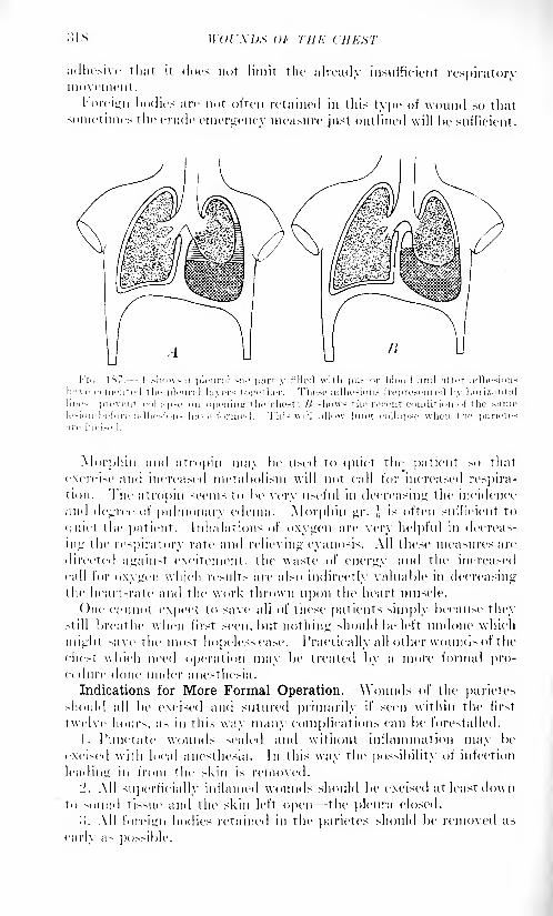
.SIS \\()l'.\l)S OF THE CHEST
it docs not limit the already insufficient respiratoryadhesive that
movement.
Foreign bodies are not often retained in this type of wound so thatsometimes the crude emergency measure just outlined will be sufficient.
a pleura! sac partly filled with pus or blood and after adhesion
merited the pleural hirers together. These adhesions (represented 1 v horizontal
n opening the chest: B shows the repent condition of the sanulesion before adhesions have formed. This will allow luntr collapse when the parietc
Morphm and atropin may l>e used to quiet the patient so that
exercise and incrca>cd metabolism will not call for increased respira-
tion. The atropin seems to be very useful in decreasing the incidence
and degree of pulmonary edema. Morphin gr. ,',
is often sufficient to
(|iiiet the patient. Inhalations of oxygen are very helpful in decreas-
ing the respiratory rate and relieving cyanosis. All these measures are
directed against excitement, the waste of energy and the increased
call for oxygen which results are also indirectly valuable in decreasingthe heart-rate and the work thrown upon the heart muscle.
One cannot expect to save all of these patients simply because they>till breathe when first seen, but nothing should be left undone which
might save the most hopeless case. Practically all other wounds of the
chest which need operation may be treated by a more formal pro-cedure done under anesthesia.
Indications for More Formal Operation. Wounds of the parietesshould all be excised and sutured primarily if seen within the first
twelve hours, as in this way many complications can be forestalled.
1. Punctate wounds sealed and without inflammation may be
excised with local anesthesia. In this way the possibility of infection
leading in from the skin is removed.
'2. All superficially inflamed wounds should be excised at least downto sound tissue and the skin left open the pleura closed.
'>. All foreign bodies retained in the parietes should be removed as
early a- possible.
ANESTHESIA .'U9
4. Open pneumothorax sliould he closed if possible after thoroughexcision of all damaged tissue.
"). Wounds with comminuted fractures of the rib should be treated
by extensive excision of the bone fragments and damaged tissue and the
wound closed so that open pneumothorax does not exist.
l>. All infections of the pleura! cavity should be operated upon at
the earliest possible moment, the type of operation depending uponconditions as enumerated below.
7. Retained foreign bodies if as large as a marble and rough indicate
operative removal at the earliest moment compatible with safety.
5. Continued hemorrhage from the lung or parietes should be
explored and the bleeding stopped,U. All wounds of the diaphragm or the abdominal organs should be
repaired.10. Gangrene of the lung demands resection.
Indications for Extrapleural Operation. 1. Small punctate woundsof entrance without extensive hemothorax are best treated by purely
extrapleural operation, /. c., excision of the skin wounds and suture
if sterile or excision of the skin wounds without suture if inflamed.
Small amounts of sterile fluid when present should be aspirated through
uninjured skin at the time of operation or before.
'2. Bullets and shrapnel balls imbedded in the lung or free in the
pleural cavity unless other indications coexist, c. (/., large clotted sterile
hemothorax or infected hemothorax, should not be followed throughthe pleura. Rough pieces of metal or clothing should always be
removed.
Indications for Non-operative Treatment. ( 1) Profound shock. (2)
Cases of dry entrance and exit bullet wound seen for the first time
after three days and without signs of pleural infection. One can gain
nothing by excising these old wounds in which primary healing has
already begun. 05) If the weather has been extraordinarily fine and
dry and the patient has been in an exceedingly cleanly condition
when wounded and the wound has been made by a bullet or shrapnelball, there being very little hemothorax, operation may be omitted
with comparative safety for example, bullet wounds of the chest
received in the deserts of Palestine and Mesopotamia were very seldom
infected. The same holds true of wounds at sea, in mineral mines
and often among the higher classes of the population who bathe
regularly.
Anesthesia. -The past few years have demonstrated that while
the various cabinet arrangements for anesthetizing chest cases are
fine in theory, they are unnecessary in the vast majority of cases.
They were not used to any extent by army surgeons during the great
war.
The best anesthesia in the simple resection of a rib for drainageis local anesthesia combined with the previous administration of mor-
phin gr. ] and atropin gr. 1('
M) . Infiltration is made with 1 percent,novocain in a linear fashion intradermallv in the line of incision. Fol-
:!-<> \VOrXDX OF THE CHEHT
lowing this, injection of the intercostal nerve above, intermediate andItelow the proposed incision is done, so that two ribs can be removedinstead of one if found advisable. The intercostal nerve is best reached
by piercing the skin at the posterior edge of the linear skin injectionand feeling for the lower border of the rib. The nerve will be found
just inside the lower border lying between the intercostal muscles.
Sometimes it is quite near the pleural side of the rib, so that care mustbe taken that the needle does not go through and pierce the pleurathus making the injection into the pleural cavity and not about the
nerve. If the injection is made midway between the ribs the anes-
thetic will not reach the nerve and the result will be unsatisfactory.An abundance of the novocain solution should be used.
After the nerve has been injected, it is wise to pierce the periosteumon the outer surface of the rib at about its middle and make a sub-
periosteal injection of novocain. If one succeeds in making these
three injections perfectly, the pleural cavity may be opened without
pain. The subperiosteal injection often causes a slight stinging ofwhichthe patient may complain. If the work to be done on the chest wall
is very extensive, it will be advisable to inject f) or (i or more nerves
at their point of exit from the spine. After the rib has been
resected, it may be necessary to inject the parietal pleura before
making the incision into the pleural cavity but this necessity onlyarises when the intercostal nerves have not been blocked. The normalvisceral surface of the pleura is practically anesthetic, particularly the
pleural surface of the lung, which can be handled, incised or sutured
painlessly.
One of the great advantages of using local anesthesia is that the
lung may be inflated by the patient's holding his nose and blowingwhen air from the sound side of the chest is transmitted to the oppo-site lung which is thus expanded to fill its pleural cavity. When one
is ready to close the wound, the patient is simply asked to do this,
and while the lung is held in expansion, the sutures, which have been
already placed, are tied. A further advantage of local anesthesia is
that no mucus or secretions are aspirated into the lung which in its
injured condition is particularly susceptible to infection and post-
operative pneumonia.A still further advantage is that the patient can move himself about
so that the operator gains the advantage of having the patient's
position changed to suit his purposes. This is often of very real
advantage when one desires to empty out a very extensive hemothorax.
Next to local anesthesia, nitrous oxide and oxygen is best. It
enables one to inflate the lung by using a positive pressure and the
anesthesia can be regulated more quickly than is the case with ether or
chloroform. The ease with which oxygen can be administered is a
very important point since the cyanosis which these patients show is
often very alarming but can be controlled nicely by the gas-oxygenmethod. Even though both sides of the chest are open anesthesia can
be continued by alternately increasing and decreasing the pressure
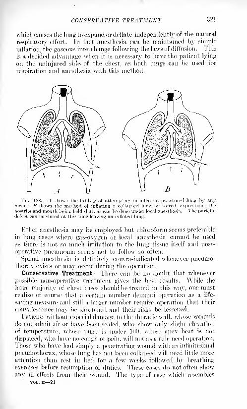
CONSERVATIVE TREATMENT 321
which causes the lung to expand or deflate independently of the natural
respiratory effort. In fact anesthesia can be maintained by simple
inflation, the gaseous interchange following the laws of diffusion. This
is a decided advantage when it is necessary to have the patient lying
on the uninjured side- of the chest, as both lungs can be used for
respiration and anesthesia with this method.
FIG. 188. A shows the futility of attempting to inflate a punctured lung by anymeans; B shows the method of inflating a collapsed lung by forced expiration the
nostrils and mouth being held shut, as can be done under local anesthesia. The parietal
defect can be closed at this time leaving an inflated lung.
Ether anesthesia may be employed but chloroform seems preferablein lung cases where gas-oxygen or local anesthesia cannot be used
as there is not so much irritation to the lung tissue itself and post-
operative pneumonia seems not to follow so often.
Spinal anesthesia is definitely contra-indicated whenever pneumo-thorax exists or may occur during the operation.
Conservative Treatment. There can be no doubt that whenever
possible non-operative treatment gives the best results. While the
large majority of chest cases should be treated in this way, one mustrealize of course that a certain number demand operation as a life-
saving measure and still a larger number require operation that their
convalescence may be shortened and their risks be lessened.
Patients without especial damage to the thoracic wall, whose woundsdo not admit air or have been sealed, who show only slight elevation
of temperature, whose pulse is under 100, whose apex beat is not
displaced, who have no cough or pain, will not as a rule need operation.Those who have had simply a penetrating wound with an infinitesimal
pneumothorax, whose lung has not been collapsed will need little moreattention than rest in bed for a few weeks followed by breathingexercises before resumption of duties. These cases do not often show
any ill effects from their wound. The type of case which resembles
VOL. II 21
WOUXDS OF THE CHEST
the OIK- already mentioned but has in addition the fracture of a rib
or an extensive hemothorax, will require a firm adhesive strappingaround the whole chest (both sides) to immobilize this fracture and free
the patient from pain, and aspiration of the hemothorax should be
undertaken before it begins to organize.It makes little difference whether pneumothorax occurs with hemo-
thorax or not, except that with pneumothorax the injured lung more
frequently collapses and for this reason more difficulty is experiencedin getting the lung to expand normally again. Some of these patientsseveral years after receiving their wound, complain of pain and dyspneaon exertion. They often show the physical signs of thickened
pleura after a few years, and one finds that many of them have lungs
firmly bound by pleural adhesions.
These patients should have the fluid withdrawn on about the third
day. If intense coughing or fainting occurs the aspiration should be
discontinued and repeated on the next day. It is often possible to
withdraw as much as three pints of bloody fluid from a chest at one
sitting without causing cough, dyspnea, pain or faintness. It often
happens that only a small amount can be withdrawn at the first
attempt and that an equal or greater amount may be withdrawn next
day. At all events it is not advisable to leave large amounts of blood
in the pleura! cavity until organization occurs.
The following dangers and precautions should not be forgotten
in aspiration:
The expansion of the lung which follows may dislodge the clot,
which has been plugging a torn vessel and the hemorrhage may recur.
For this reason, it is wise to wait forty-eight hours before aspirating
so that the clot may become firmly attached and there will be little
danger of this accident.
There is the danger of infecting a sterile hemothorax but in the
author's experience this never occurs, if proper precautions are always
taken.
It is wise to use a very large needle when aspirating as a small needle
readily becomes plugged. It is MTV important to make the vacuum
in the suction bottle one's self as we have seen two cases in which the
bottle had been carelessly pumped full of air under great pressure and
this air had been injected into the pleural cavity. It is always well to
test the apparatus by placing the needle in sterile water immediately
before using it.
If the patient is a nervous individual, it is well to make a local infil-
tration before inserting the large needle. The puncture should gener-
ally be made in the eighth space in the posterior axillary line unless
the hemothorax seems to be circumscribed which is rare if seen early.
These pat ients should be treated in the sitting position and should not
be transported for at least a week if possible. There may be fever as
high as 101 due to the absorption of hemothorax without any active
infection, so that one cannot definitely determine infection by the tem-
perature alone unless it be very marked. It is wise to take a culture
COXSERVA 77 1 'E TREATMEXT 323
from every aspiration so that one can anticipate or diagnose the
presence of a septic involvement.
Streptococcus Infections. When sepsis occurs, any of the pus-
forming organisms or the anaerobic organisms and hemolytic strepto-
coccus may be present. The hemolytic coccus is most to be feared and
cases developing sepsis with this organism have a very serious prog-nosis. They demand radical resection of t\vo ribs, complete washingout of plenral cavity and drainage from the most dependent part. It
is well in this type of case to use from 3 to (\ Carrel's tubes reachinginto every crevice and corner of the pleura and in very severe cases,
even introducing three to six or more of Carrel's tubes through an
intercostal incision between the upper ribs in front, so that the solu-
tion max trickle over the entire pleural surface. The same procedureshould be adopted when the gas-forming organisms are discovered.
The odor from infection with these organisms is very characteristic
and foul and the fluid which one finds is thin, reddish-brown in color
and serous rather than purulent.When a hemothorax becomes infected with ordinary pus-forming
organisms drainage should be instituted in the usual manner, resecting
one or two ribs in the midaxillary line. Every effort should be directed
toward the rapid elimination of infection so that early secondary closure
can be done. In this way large cavities may be obliterated by an
expanding lung, whereas if drainage had been prolonged with the result-
ant thickening of the pleura and pulmonary adhesions, healing would
have been very slow.
Posture. If one were 1 asked to name one single prescription for the
treatment of penetrating wounds of the chest which was better than
the rest, one should say that the sitting posture is that treatment, since
every case 1 should be placed in that position unless shock is too severe.
In such a condition one should elevate the head of the bed while the
patient lies flat, and raise him to a sitting position at the earliest pos-sible opportunity. In fact, the sitting position should be attempted at
once in every case and if the patient faints (as those in extreme shock
may do) he should be allowed to lie back again. It may seem that this
experiment should be a voided by some mysterious diagnostic maneuver,but as a matter of fact while one can sometimes see at a glance that an
apparently moribund patient would be harmed by the slightest move-
ment, in general it is difficult to say that even the most hopeless case
will not be benefited by the sitting position. This is explained by the
fact that this posture allows the venous blood to drain away from the
lungs, so that congestion there is decreased, \\hen this is accom-
plished the resistance to the How of arterial blood through the lungs
is diminished and the heart has less work to do and consequently the
general circulation improves.Further than this, the sitting position allows the diaphragm to func-
tionate better and frees the ribs which are compressed when the patient
lies on either side. When the patient lies down flat he has a tendencyto lie on the uninjured side and thus compress the side upon which the
324 WOUXDS OF THE CHERT
burden of respiration falls the injured lung generally being allowed
to rest in part, or completely as in open pneumothorax.Another benefit gained by the sitting posture is the lessened ten-
dency for hemorrhage to occur from the wound of the lung. Just as
congestion increases the work of the heart, so it increases the pressurein the vessels of the lung and therefore hemorrhage may occur
whereas it would have been avoided had the pressure in the pulmonaryvessels been less. As a matter of fact a patient who is first seen with
the thready, rapid pulse of hemorrhage, sometimes regains a full pulsein a very short time after he sits up in bed; and concomitantly the
hemorrhage ceases. The author has seen hemorrhage from the lungoccur within five minutes after a patient who had been left in the
sitting posture had assumed the supine position. This case was an
exceptional one a conscientious objector, who not only objected to
fighting but also objected to medicine, surgery, hospitals, religion,
the government and his meals and the flat position was his ownidea, lie would assume it whenever the opportunity presentednot because he thought it good for himself, but hccaii>e nobody wantedhim to do it. He died of hemorrhage and general septic pleurisy.As a universal rule the patient who has been raised to the sitting
position will stay in that position because he is then most comfortable.
The respirations become easier, slower, the expression changes from an
anxious Ilippocratic one often with cyanosis- to a more contented
type, and often to a cheerful one. If great care is used in the- first
days the skin over the sacrum becomes harder and bed-sores are
unlikely to develop.When there is infection in the pleural cavity this position prevents its
spread to the entire cavity and tends to localize it to the lower margins.When there is drainage of the ple.ira this position favors thorough-
ness since then the tubes can be placed at the actual bottom of the
cavity and pocketing cannot occur.
Thus the sitting posture provides: (1) against spread of infection;
(2) for drainage of the pleura; CV) a guard against hemorrhage; (4)
comfort; '.")) relief of dyspnea; (CO relief of cardiac overloading.
However, since no rule in medicine is without exception, one will
infrequently sec a case where the foot of the bed may be elevated with
apparent benefit. Th.is when the circulatory distress is very great as
after exsanguination and after the bleeding has definitely ceased,
raising the foot of the bed seems to benefit the- failing circulation.
Transfusion. It is far better to treat these cases with transfusion
of blood, however, with ligature of the bleeding point at once. Trans-
fusion can be done, gas and oxygen anesthesia given, and ligature done
with as much wound toilet as possible in passing and the patient
emerge in considerably better shape than beforehand. It is often
advisable to give the transfusion in two parts, the latter half being
given after the operative maneuver-, are complete so that the new7
blood is not lost through hemorrhage. At least 800 c.c. of blood should
be given.
CONFER VA TI YE THEATMEXT 325
General Type of Intrathoracic Operation. The skin is scrubbed with
soap and water, dried thoroughly with alcohol or benzine and
painted with tincture of iodine. The usual asepsis must be strictly
carried out and further than this, when entering the chest through a
dirty lacerated wound of the parietes, it is well to make a complete
change of instruments, gloves and linen after the parietal wound has
been rendered aseptic by the careful excision of all damaged tissue,
followed by disinfection with some antiseptic such as tincture of iod:ne,
alcohol, picric acid or the like. Some prefer to rub the edge of the
cleansed wound with bismuth iodoforni paraffin paste.When the operation is one for removing a foreign body and evacu-
ation of a hemothorax, it is well to be guided as to the site of incision
by he location of the foreign body, bearing in mind the fact that
entrance through the fourth or fifth space gives access to the entire
lung including the hilus. When incision is made through sound tissue
without regard to the wound of entrance, it is well to reserve excision
of the entrance wound until the chest has been closed again, so that
any possible surface contamination near the entrance wound cannot
be implanted within the pleura. Xo matter by which route it is decided
to enter the chest advantage should be taken of a large incision, at
least six inches long through the skin with removal of four inches
of the rib, since one can close the chest easily if this is done. Theintercostal muscles above and below7
give good anchorage to sutures.
Having opened the chest, whatever hemothorax is present should
be emptied by allowing the fluid to be splashed out by the respiratorymovements or swabbed out with large dry pads. Clots may be scoopedout with the hand. If the operation is done under local anesthesia
one enjoys the cooperation of the patient who rolls himself so that the
wound is dependent, but when general anesthesia is used, it is some-times difficult to turn the patient without causing a serious fall of
bloodpressure as has been shown by Major Shattuck and CaptainBlair. 1
Any large clots or deposits of fibrin should be cleaned out at this
time. It is wise to palpate thoroughly every clot for if one does this
as a routine he will sometimes find that a foreign body which was
apparently buried in the lung was really free in the pleural cavity andhas been swabbed out with the hemothorax. Thus one may be saved
a long search for a foreign body which has already been recovered, the
patient will be saved the consequent shock and the surgeon muchembarrassment.
The next step is to retract the ribs widely and inspect the interior
of the thorax next the hand may be passed into the chest and the
lung palpated. Large foreign bodies are readily felt in the lung bygrasping it between the thumb and fingers and also large rents in the
visceral pleura may be felt. However, the small wound made bybullets may easily pass unseen and unfelt. When the foreign body is
1
Blair, R. B. and Shuituuk, G. C.: Penetrating Wounds of the Cheat, Jour, of theR. A. M. C., September, 1918.
320 \\'OU\DS OF THE CHEST
located, the lung is grasped firmly and held with a piece of gauze to
prevent slipping and a small incision made directly down to the
foreign body which is then squeezed out through the opening. Anylarge
1 tears may be repaired quickly by passing a catgut stitch through it,
using a large round needle.
Gangrene of Lung. ---If there is hemorrhage from the lung the same
procedure is adopted but one must be careful not to include too large
a piece of tissue in one ligature or else peripheral gangrene may occur
and thus set up a focus of infection. If gangrene is already present,resection of the gangrenous area must be done. It is best to pass a
series of figure-of-eight stitches above the gangrene. These are tied
and then the gangrenous portion is cut away with scissors or scalpel.
WOUNDS OF THE DIAPHRAGM.
If the diaphragm is ruptured, it must be repaired and this is easily
done by direct suture from above. Foreign bodies may be removed
from the liver by transpleural and transdiaphragmatie incision quite
readily. The liver can also be nicely drained by the transdiaphragmatieroute in which case the diaphragm is sutured to the parietal pleura at
a level higher than the parietal incision. If necessary a strip of the
diaphragm may be removed from its lateral attachment and attached
to a higher level, and, by using this principle, it is sometimes possible
to repair actual defects in the diaphragm. The pleural cavity should
always be shut oft' from the abdominal cavity. Transdiaphragmatiewounds of the hollow viscera should be attended to either by the
transdiaphragmatie route or by abdominal section if they cannot be
reached through the diaphragm,Closure. TYhenever possible the chest wall should be closed
tighth". The one notable exception to this rule is the presence of
streptococcic infection, when radical drainage must be provided. It is
well to secure ample drainage when the gas-forming organisms are
present. Whenever there is doubt as to whether the chest should be
closed or not, closure should be attempted as the postoperative recovery
is much easier with a closed chest. If the temperature and pulse-rate
rise, the chest may be reopened by simply removing the sutures at
any time. In this connection it is advantageous to have the incision
as near as possible to the bottom of the pleural cavity since if the woundmust be reopened it will drain better from the lowest possible point.
When a wound has been reopened Carrel's tubes may be placed within
it and irrigated at frequent intervals. If the patient's condition
permits, he should be made to take breathing exercises, the salient
point of which is that forced expiration against resistance be employedso that the collapsed lung will be made to inflate by the pressure
developed in the sound lung on expiration. For this purpose Wolff's
bottles or breathing tubes are very valuable. If breathing exercises are
disregarded at this period, fibrosis of the lung and large pleural cavity
formation will most likely result. Another point about closing all
WOUNDS OF THE DIAPHRAGM 327
doubtful cases is that in the short periods during which the cavityhas been closed, fibrinous adhesions will be formed between the visceral
and the parietal pleura, and these adhesions will help to hold the lungin its expanded condition. This makes for good function and early
healing by granulation since a smaller cavity remains. The use of
various valve apparatus has not been generally thought satisfactory,
due to the difficulty in making an air-tight junction between the woundand the mechanism.
Whenever it is necessary to drain the pleura, time will be saved andthe unfortunate results minimized, if half measures are discarded. Oneshould not hesitate to resect two ribs if by doing so freer drainage is
established. Tubes of large diameter should be used for drainage1
pur-
poses and should be kept free by daily irrigation with some warm mild
antiseptic solution such as Dakin's. When using Dakin's solution, it is
well to have the solution made double strength and to dilute with an
equal volume of warm water immediately before irrigation, as in this
way the chlorine which it contains is not volatilized and lost.
Postoperative Care. The diet should be a fluid one for the first
twenty-four hours, including water, tea and warm milk with eggs.
Rectal saline is of benefit, and in case of severe shock blood trans-
fusion is of great value, from 400 to 800 c.c. of blood being transfused.
Intravenous infusion or hypodermoclysis of normal salt solution maybe done if transfusion cannot be accomplished. It is not advisable to
give intravenous infusions of sodium bicarbonate and glucose solution
as this solution in excess produces pulmonary edema.
It is important to keep the patient quiet after operation. For this
purpose morphin and atropin may be given freely during the first dayunless the patient shows the peculiar gray color which indicates a
condition in which morphin is contra-indicated. Morphin may be
used when ordinary cyanosis is present.These cases should not be transported any distance either before or
after operation.A specimen of the fluid which accumulates after operation should
be obtained twenty-four hours afterward and cultured. If a large
amount of fluid accumulates, it should be withdrawn and it may be
necessary to repeat the aspiration for several days. When the culture
shows hemolytic streptococcus secondary drainage should be estab-
lished at once. Or, if low grade sepsis ensues and aspiration gives only
momentary relief, secondary drainage should be secured.
Sometimes aspiration repeated daily and combined with aspiration-
irrigation, as recommended by Major George Shattuck of the Harvard
Fnit, is sufficient to combat a low grade infection. Aspiration-irri-
gation is done by alternately emptying and filling the chest with Knsol
solution using the aspiration apparatus.When the chest is open and draining and when the lung is not held
fast by pleural adhesions, it is well to arrange the dressing so that
as little air enters and leaves the cavity as is possible. Much can
be accomplished by using jaconet or oiled silk for this purpose. When
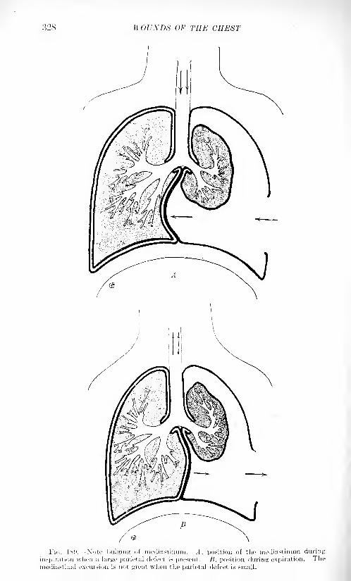
WOUXDS OF THE CHEST
WOUNDS OF THE DIAPHRAGM 329
the lung is adherent to the parietal surface and walls off the septic
cavity, open drainage can be used. This is an advantage in closing the
chest primarily, /. e., adhesions form so that secondary drainage does
not result in lung collapse.
Late Treatment. Old empyema cavities can be obliterated bycollapsing the parietes. This crude procedure is seldom necessary if
the early treatment has been correct. Foreign bodies whenever theycause inconvenience, pain, dyspnea or mental discomfort to the patient
may be removed. Accurate .r-ray localization should be demandedbefore the attempt is made.
Beck's,paste is valuable in clearing up cavities, but careful watch
must be maintained that bismuth poisoning does not occur.

SURGERY OF THE HEART AMI PERI-
CARDIUM.
BY HARRY M. SHERMAN, A.M., M.I).
THE HEAJIT.
THE surgery of the heart must include lesions originating in that
organ, hut its relation to the precardium, pleura and lungs is such that
these parts must participate in the lesion or in its surgical repair, so
that a complex of lesser or greater extent is the final stage.
For surgical considerations the heart lies behind the middle of the
sternum, the inner ends of the right third, fourth and fifth costal car-
tilages, and all but the outer ends of the left third, fourth and fifth costal
cartilages. Its association with the chest wall, the pleura, the lungs andthe pericardium are shown (Fig. 190) in the composite illustration. 1
The precordial space (after Testut) is included within a line which
begins "on the upper border of the third right costal cartilage, 1 cm.
from the right border of the sternum;" it extends, with a slight con-
vexity to the right, to a point "on the level of the sternal articulation
of the fifth right costal cartilage," thence it passes, with a slight con-
vexity downward, to a "point at the apex of the heart, a location easily
made out in the living. On the cadaver it is placed on the upperborder of the fifth left costal cartilage S cm. from the midsternal
line." Hence the bounding line passes upward, with a slight convexityto the left, to a "point in the second left intercostal space, midwaybetween the cartilages above and below, and 2 cm. to the left of the
left sternal border." The pericardium has a somewhat triangular pro-
jection on the chest wall: the base line commences at the right, 2 cm.
from the right sternal border in the middle of the fourth space, and
passes, slightly curved downward, cutting the top of the xiphoid, to the
left and terminates (> cm. to S cm. from the left sternal border in the
fifth space. The pericardia! limit on the right side, slightly convex to
the right, begins at the right end of the base line and passes upward,1 cm. to 2 cm. from the border of the sternum, to the second cartilage,
then it approaches and passes beneath the sternum. The pericardia!limit on the left begins at the left end of the base line, and passes
obliquely upward toward a point which ought to be a little outside the
left sternoclavicular articulation.
On these projections lie, on either side, the lungs and the two pleurae.
1 Terrier et Keyinuiid : Chinirnie du Cceur ct (hi I'ericurde, Paris
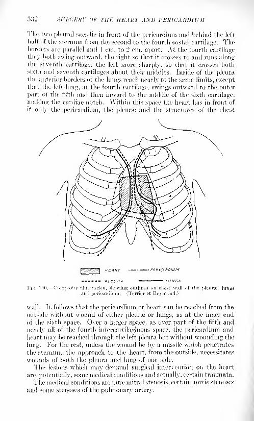
SURGERY or THE HEART A^7D PERICARDIUM
The two pleural sacs lie in front of the pericardium and behind the left
half of the sternum from the second to the fourth costal cartilage. Theborders are parallel and 1 cm. to 1' cm. apart. At the fourth cartilage
they both swing outward, the right so that it crosses to and runs alongthe seventh cartilage, the left more sharply, so that it crosses bothsixth and seventh cartilages about their middles. Inside of the pleurathe anterior borders of the lungs reach nearly to the same limits, exceptthat the left lung, at the fourth cartilage, swings outward to the outer
part of the fifth and then inward to the middle of the sixth cartilage,
making the cardiac notch. Within this space the heart has in front of
it only the pericardium, the pleurae and the structures of the chest
PERICARDIUM
wall. It follows that the pericardium or heart can be reached from the
outside without wound of either pleurae or lungs, as at the inner end
of the sixth space. Over a larger space, as over part of the fifth and
nearly all of the fourth intercartilaginous space, the pericardium andheart may be reached through the left pleura but without wounding the
lung. For the rest, unless the wound be by a missile which penetratesthe sternum, the approach to the heart, from the outside, necessitates
wounds of both the pleura and lung of one side.
The lesions which may demand surgical intervention on the heart
are, potentially, some medical conditions and actually, certain traumata.
The medical conditions are pure mitral stenosis, certain aortic stenoses
and some stenoses of the pulmonary artery.

THE HEART 333
"Pure mitral stenosis is a lesion limited to the valves, the cardiac
muscle remaining long unaffected as well as the peripheral circulation.
The free border of the valve is alone affected, the valves themselves
remaining soft and supple." It is the lesion which attracted Bruntonand led him to observe that
"incision of the stricture could remove the
obstacle which had formed the sole barrier to a normal circulation."
Aortic stenosis, not of either the supra-aortic nor of the subaortic
varieties, but pure"valvular stenosis, characterized by adhesions be-
tween the free borders of the valves, with thickening and deformation
of their margins" may be considered truly a surgical variety. Andstenosis of the pulmonary artery, of the same valvular type, when "the
fibrous arc of insertion is intact, and the welding of the valvular
margins creates a sort of diaphragm, convex toward the pulmonaryartery, the more or less narrow central orifice of which is susceptibleof enlargement. . . . There maybe pure stenosis of the pulmonaryorifice in young subjects without any alteration of the myocardiumor congenital malformation of any other orifice. This condition is one
eminently favorable for mechanical treatment." 1
All of these lesions with the primary pathological process limited to
the valves, and with the cardiac muscle unaffected except by a com-
pensatory hypertrophy, both of which conditions may continue for a
long time without danger to health or life, may be found to lend them-
selves to adroit surgery, but the mere existence of the complex does not
call for help immediately nor without consideration, because of their
possible long continuance. "The lesion which tends to provoke graveor fatal trouble in the near future is the genuine indication that pointsto the adoption of surgical measures which can change such an ominous
state into a relatively benign insufficiency." If this was found on trial
to be an improvement it is impossible to avoid the question as to the
permanence of the gain, and equally impossible to answer the questionwithout the trial. That the trial will some day be made is a safe
prophecy.A surgical procedure upon the heart may be attempted with the heart
in action, but it can best be done with the flow of blood through the
heart stopped and with the heart at rest. This condition can be attained
by clamping the great vessels of the pedicle of the heart. Through an
ample incision in the -pericardium one blade of a Doyen clamp, rubber
sheathed, is slipped in behind the great vessels from right to left, the
other blade being outside the pericardium ;this clamp when closed can
completely control the circulation through the heart and may be keptin place for from two to two and a half minutes without harm, providedthe respiration has been kept up by a positive pressure apparatus and
that the blood has been overaerated just prior to the closure of the
clamp. \Vhile the clam]) is in place the heart may show slight pul-sations or may be perfectly quiet.
Compression of individual vessels or of combinations has effects
1 Quoted from Carrel and Tuffier: Mod. Press and Circ., 1914, xcvii, 539, 506.
.SCAV /A'//!' 01- THE HEART AM) PERICARDIUM
which vary with the vessels selected. Compression of the pulmonaryartery and aorta alone can he maintained for but a very short time
because of the great dilatation of the right heart. Partial closure of
the vessels continued for ten minutes produced no bad effect on the
animal and the aorta has been heated for a period of six minutes with-
out resulting harm. "Simultaneous compression of the pulmonaryveins produces death after some minutes through default of oxygen-ation of the cardiac muscle." "Of all the procedures of cardiac hemo-stasis . . . that which is best is the compression of both vena*
cava-. The blood within the caval veins no longer flows into the rightauricle and right ventricle; the intracardiac circulation is arrested,
but not the coronary circulation. The latter continues for some time" 1
and the coronary arteries have been seen to beat, the blood already in
the lungs continuing to return through the pulmonary vein into the
left heart and thence passing into the general circulation, ('aval
closure must not exceed three and a half minutes on account of the
danger to the cerebral circulation, but oxygenated Ringer's solution
may be injected into the carotids and so reach the brain, and this per-mits the time of closure to be extended to ten minutes. "With regardto the arrest of the circulation in the substance of the cardiac wall,
that is to say, the arrest of the coronary circulation, this cannot be
prolonged beyond one or two minutes. Oxygenation is absolutely
necessary for maintenance of the cardiac irritability, and accordinglythe occurrence of congestion is extremely dangerous.
"-
In planning or executing a surgical act upon the heart there are
certain regions which must be avoided as being danger /ones. These
are regions of coronary distribution, for the corollaries are terminal
arteries. Accidental section or ligation results in h'brillary contractions
and death if the vessel be a large one, or of degenerative changes in the
muscle if the vessel be smaller. Therefore, "the part of their course
which extends from their origin to their earliest divisions representan area of noli >///' fain/cri
1
. The septum interauriculare is a structure
of extreme sensibility on the slightest contact with which the heart
stops in diastole. Section of the auriculoventricular border provokesimmediate arrest of the left ventricle in diastole; the respiration never-
theless undergoes no modification, and the auricles and right ventricle
continue to beat. At the level of the septum auriculoventrieulare there
is a definite locus, the point of Kronecker and Schmey, a wound of
which provokes immediate arrest of the heart." And there is a point,
"situated in the auriculoventricular region near the left margin of the
heart . . . of which a wound is dangerous and even fatal, even a
slight pressure over this part provokes an extrasystole."
"On the other hand, there is a point situated in the anterior longi-
tudinal groove, at the junction of the middle and upper thirds, of which
the mechanical irritation may determine a brusque arrest of the heart."
The base of the right auricle must be regarded with respect as an
1 LI ic. cit.2 Loc. cit.
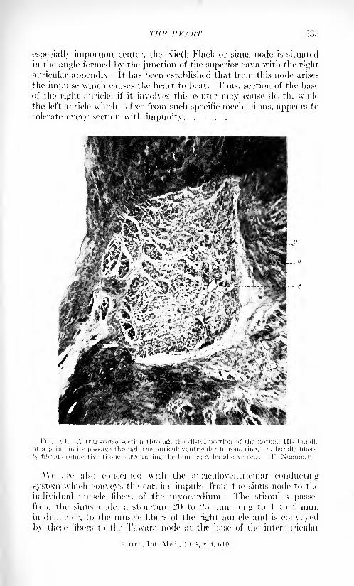
THE HEART 335
especially important center, the Kieth-Flack or sinus node is situated
in the angle formed by the junction of the superior cava with the right
auricular appendix. It has been established that from this node arises
the impulse which causes the heart to beat. Thus, section of the base
of the right auricle, if it involves this center may cause death, while
the left auricle which is free from such specific mechanisms, appears to
tolerate every section with impunity
Fit;. 11M.---A transverse section through the distal portion of the normal His bundleat a point in its passage through the auriculoventricular fibrous ring. <i, bundle fibers;
l>, fibrous connective tissue surrounding the bundle: r, bundle vessels, i F, Xuzuiu.)'
\\e an 1 also concerned with the auriculoventricular conducting
system which conveys the cardiac impulse from the sinus node to the
individual muscle fibers of the myocardium. The stimulus passesfrom the sinus node, a structure 20 to 25 mm. long to 1 to 2 mm.in diameter, to the muscle fibers of the right auricle and is conveyedby these fibers to the Ta\vara node at th^ base of the interaiiricular
i Arch. Int. Mod., 1914, xiii, 040.
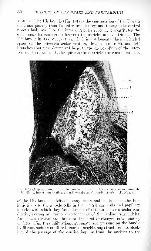
33(5 SURGERY OF THE HEART AND PERICARDIUM
septum. The His bundle (Fig. 191) is the continuation of the Tawaranode and passing from the interauricnlar septum, through the central
fibrous body and into the interventricular septum, it constitutes the
only muscular connection between the auricles and ventricles. TheHis bundle in its distal portion, which is just beneath the undefended
space of the interventricular septum, divides into right and left
branches that pass downward beneath the endocardium of the inter-
ventricular septum. At the apices of the ventricles these main branches
in,. l\)'2. Adipose tissue in the His bundle, a, central fibrous
bundle; '>, intact bundle libers; r, adipose tissue; <7, bundle vessels. (!'. Nuzum.)
of the His bundle subdivide many times and continue as the Pur-
kinje fibers to the muscle cells in the ventricular walls and papillarymuscles with which they fuse. Lesions of this auriculoventricular con-
ducting system arc responsible for many of the cardiac irregularities.
Among such lesions are fibrous or degenerative changes, inflammatoryor fatty (Fig. 192) infiltrations, gummata and pressure on the bundle
by fibrous nodules or other tumors in neighboring structures. A block-
ing of the passage of the cardiac impulse from the auricles to the
THE HEART 337
ventricles may result. A type of irregularity known as heart block is
then established in which the auricles and ventricles beat independentlyof each other.
"Finally, the endocardium is much more sensitive than the other
cardiac tissues, doubtless through the influence of the subendocardial
nerve plexus."1
In other regions of the heart the walls may be cut, or a portion maybe resected and after suture, healing occurs, but it is by subepicardialand intermuscular fibrous tissue and not by muscle regeneration.
Transplantations may be done but can only be of very doubtful value.
Inside the heart, "although the endocardium is sensitive the valves
can be divided, detached, ligatured or fixed. The orifices of the greatvessels are surrounded by arteries and nerve branches which must be
taken care of. Knowing the position of the coronary arteries at their
origin, we can attack the anterior aspect of the base of the aorta without
fear of wounding them. The walls of the great vessels, especially those
of the aorta, are very friable, so that a secondary gaping of the line of
suture is possible; accordingly, it is necessary to reinforce with a layer
of transplanted fascia or ... with a segment from the wall of a
vein.'' 2
The procedures called for in the conditions previously mentioned are
all of them to enlarge stenotic orifices when the stenosis is due to lesions
of the valves alone. Section of the adherent valves, valvulotomy
may be done by internal or external methods or by grafting on a new
path from cavity to cavity around the stricture. Internal valvulotomy,
belongs to the period of experimentation pure and simple, done, as it
is, in the dark by a long instrument with a sheathed knife passsed into
one of the vessels of the neck. The total inaccuracy of the maneuvermust forbid its use on a human being. Vessel grafting, to offer the
blood a detour around the stricture, is a more direct method. Lengthsof any large vein may be used, the patient's own saphenous vein would
be appropriate, and valves must be in the piece and so disposed as to
permit the blood to pass only in the direction desired. If no valves
chance to exist a valvular action can be gotten by making a slight
intussusception and fixing it with a little suture. Such a length of
vein may be placed so as to offer a detour around the pulmonary valve,
from the right ventricle to the pulmonary artery, or around the mitral
valve from the left auricle to the left ventricle, or around the aortic
valves, from the left ventricle to the braehiocephalic trunk. These
maneuvers again are so complex and depend on such inconstant factors
that they, too, must be thought of as being experimental matters only.
Kxternal valvulotomy is based on the principle of incising the valve
from without inward and also permitting an increase in the si/e of the
orifice 1
; the operation divides "the border of the aortic orifice and the
valve at the same time. This direct section of the valve should be
carried out with precision, on account of the fact that the cardiac /one,
1' 'atTel and Tu flier: Lor. cit.
VOL. II 22
;>!S srnaERY or TUP: HEART AND PERICARDIUM
which surrounds the origins of the aorta and pulmonary artery, is
specially sensitive and very readily originates reflex movements. Weshould keep as far as possible from the vicinity of the coronary arteries;
those vessels arise, however, in positions sufficiently far apart to enable
us to pass readily between them on the anterior aspect of the aorta." 1
"The operation consisted in suturing on to the surface of the .
artery a patch of vein preserved in cold storage and in cutting the edgeof the orifice underneath the patch ... A piece of vena cava or
of any other vein ... is cut into the shape of a rectangular flap
about I'..") cm. by 2 cm. This flap is put on the anterior part of the
. artery in such a way that its middle corresponds about to the
pulmonary orifice, the lower part being on the surface of the ventricular
wall. Then the two lateral sides and the upper side are fixed to the
surface of the heart and the pulmonary artery by means of a con-
tinuous suture. The longer blade of a special pair of scissors with a
longer sharp pointed and a shorter blade, are "introduced beneath the
lower side of the flap and the sharp point is introduced into the lumen
of the pulmonary orifice. Then the wall is cut and dark blood escapesbetween the surface of the heart and the lower part of the flap, but
the hemorrhage is immediately controlled by the index finger of the
operator which presses the flap down on the wound. The fourth side
of the flap is next fixed to the surface of the heart by a continuous
suture."
:
The flap immediately is lifted up and stretched by the pressure of
the blood, but it has lived with its inner side in contact with the blood
and its outer with the pericardium, nor does thrombosis take place, as
the flap is lined with endothelium.
This operation may be done without stopping the circulation throughthe heart; but it should be more easily done, and within the sight of
the operator, by clamping the pedicle of the heart and stopping the
circulation for a very short time. Under this method a valve has been
sectioned and then immediately resutured, or two contiguous valves
have been sutured together, or the border of a valve has been cau-
terized, and then the wound has been closed and the action of the
heart has been resumed.
Entrance of air into heart cavities necessarily occurs if they are
opened under cessation of the circulation. If this is in the right heart
no harm comes, air emboli in the branches of the pulmonary artery
causing no trouble. If the left heart has been opened and air has
entered it must be taken out by aspiration as the sutures are tied to
prevent its entering the coronary arteries, for it here obstructs the flow
of blood and fibrillary contraction ensues with a fatal outcome. In
some instances it has been possible to puncture the artery with a needle
and let out blood and the air.
These operations require general anesthesia and positive pressurein the lungs, a combination which is best produced by the Meltzer-
Lor. cit. -f'arrrl: Ami. Surjr., July, 1011.
THE HEART 3M9
Auer method. In dealing with the heart itself local anesthesia may be
added to control reflexes originating in that organ. Shock preventionand the maintenance of asepsis call for no new principles, but the pro-tection of the exposed viscera from all unnecessary contact with the
air and rough textiles by covering them with sterile silk napkins,vaseline soaked, is a method of great excellence. Suture material in
the heart or vessel walls should always be of vaselined silk and the sameis true of the pericardium, and the pleurre too, if they have to be sutured
after being accidentally opened. All needles should be round.
Laboratory experience, upon which all the foregoing teclmic is based,
shows that none but healthy, vigorous young animals are suitable for
the work, though one relatively old dog, eight years, was successfully
operated upon. The lesson of this is that the plan could be considered
only for the stronger and younger of human patients.1
All of the actual surgery of the human heart has been for the repairof wounds or of ruptures. The first repair of a heart wound was done in
1896 by Farina, and since then about 200 cases have been reported.The heart has been wounded by knife wounds or by bullet wounds,there being about five of the former to one of the latter, with a mortalityin the stab-wound cases in which repair was attempted of about 49
per cent., and in the bullet-wound cases of bout 61 per cent. 2
For bullet wounds of the heart the external wound of entrance maybe anywhere on the thorax. Stab wounds must be within the lengthof an ordinary knife blade of the heart. LeConte and Stewart 3 found
that the wound was between the sternal left border and the left mid-
axillary line, and the second and seventh ribs in about eleven-twelfths
of the cases, and of the other twelfth, two-thirds were in the upperabdomen, and one-third to the right of the sternum. There was but
one case in which the sternum was penetrated. The nearer the external
wound is to the heart the greater the likelihood of a heart wound too,
but not a few stabs may enter the thorax and pass between the heart
and the lung, with wounds of neither.
Inside the chest the pericardium is, of course, always wounded with
the heart, and the left pleura participates in fully nine-tenths of the
cases, the left lung in a slightly lesser amount.
The wound in the pericardium may be smaller than that in the heart,
and the escape of blood from it may be slow so that the pericardia! sac-
becomes distended. As the intrapericardial tension increases the pres-sure on the heart interferes with its function, and finally, as the pressurein the pericardium reaches the blood-pressure in the pulmonary veins
and the auricles, blood is unable to enter the latter and the circulation
of 'necessity ceases. This tamponade of the heart may occur with or
without a wound of the pericardium. If the wound in the pericardium
1 Tuffier and Carrel: Jour. Expcr. Mod., UH-J, No. 1, xx. Canvl: Surir.. (iynoc.,and Obst., August, 1914.
- Wagner: Doutsfh. Ztschr. f. Chir., 1912. cxix, 221.3 Am. Pract. Sun:., vii, 103.
340 Xl'KGEKY OF THE HEART AXD PERICARDIUM
is large the blood escapes from it as fast as it pours from the heart
wound, and it then may appear in part or wholly on the surface, bythe external wound, especially if there has been no collateral injury to
the pleura. Jf there has been a wound of the pleura a pneumothoraxis usually immediately produced and the mutual proximity of the peri-cardia! and the pleural wound offers a path for the blood into the emptypleura and the pneumothorax becomes a pneumohemothorax. In
these circumstances the lung is wounded one-half or two-thirds as often
as the pleura.Inside the pericardium wounds of the great vessels are practically not
reported, but wounds of all the chambers of the heart are. Le Conteand Stewart found that the ventricles are wounded with equal fre-
quency, that the ventricles are wounded about fifteen times as often as
the auricles and that the right auricle is wounded nearly four times as
often as the left. Combination wounds also occur, ventricle with
ventricle and ventricle with auricle. Stewart 1 notes the difficulty in
some instances of finding the wound in the heart, but usually this is
easy for there is a systolic spurt of blood, and this though the wound
may not open a heart cavity. Wounds may be pure stabs, or incisions,
even as long as 7 cm.
\Yhile wounds from the outside are usually made by knives, a small
percentage may be by needles, and needles swallowed may, in their
travels, (Miter the pericardium and the heart. Knife wounds in the
heart are possibly larger than the external wound, for the point of the
knife may swing through an arc inside the chest, or the punctured heart
may tear or cut itself on the knife in the act of beating. Occasionallyknife blades are broken off and the point left sticking in the heart
cavity or wall.
Bullet wounds are commonly, though not necessarily, double, and
the missile may be encapsuled in the heart wall, or remain in a cavity,
or may be rolled along the artery with the blood, or, again, the progress
along the vessel may be against the blood stream and must be due to
gravity, the opportunities for rolling along declivities in the vessels
being offered by changes in the positions of the patients.
The diagnosis of surgical conditions has to count on the constant
presence of variables in unusual quantity, but sufficient can be learned
from an examination to warrant an exploration, and it is always better,
liehn says, to do an exploratory pericardiotoiny, even if useless, than
to overlook a wound of the heart.
The history of the receipt of a wound and its presence in or near the
precordia may or may not mean a wound of the heart, but the nearer the
external wound is to the sternum the greater is the likelihood of a heart
wound, and bullet wounds which penetrate that bone are most liable
to injure the heart too. Bleeding from such a wound may come from
an artery of the chest wall, or from a wound of the lung, or of the great
vessels or of the heart. The bleeding may be in spurts or in a con-
1 Ann. Surg., July, 1913.
THE HEART 341
tinuous flow, and be much or little without definite diagnostic value as
regards a wound of the heart. An acute general anemia must meanthe loss of much blood, but this may be concealed in the thorax, as in a
hemothorax, whatever be its source. A distended pericardium, after a
trauma, penetrating or not penetrating, must mean a wound of the
heart or the great vessels inside the pericardium, but it is not possibleto say which is injured nor, in the case of the heart, which cavity is
implicated. The history of a non-penetrating injury, as of a crush of the
thorax, or the history of a wound and its presence, with or without
external bleeding, but with an acute extreme anemia, with or without
a hemothorax or a pneumohemothorax, with or without a distended
pericardium would justify an exploration. The lung condition can be
easily made out by an ordinary physical examination. Stewart 1 has
shown that with a pneumothorax the cardiac dulness disappears and
tympany replaces it. The heart sounds may, however, usually be
heard but are faint, and adventitious sounds are rare, especially
splashing sounds supposed to indicate air in the pericardium with the
blood. Associated with these physical signs are the general ones: a
weak and rapid, or no pulse and an incompetent circulation, mild or
severe dyspnea, pain in the precordia and the left arm felt more acutelyat expiration and made worse by pressure on the heart, violent left-
sided pain in the abdomen and rigidity of the upper abdominal muscles.
Finally, as tamponade of the heart supervenes, distention of the
cervical veins and cyanosis, and stupor passing into coma.If the external wound was an incised one and large enough, a finger
might be passed in, or if the wound is small it might be enlarged suffi-
ciently to admit a finger and indeed must be if exploration is done. Theuse of the probe is deprecated and Stewart 2 has pointed out that a
stab wound through the chest wall has an angular course when the
recipient lies down, due to the sliding upward of the skin and the movingupward of the heart in recumbency. Exploration of the wound shouldbe carried far enough only to demonstrate that it entered the thorax
and then, if there was evidence of serious hemorrhage the thorax shouldbe opened to bring the parts inside into view.
If the wound is in the neighborhood of the heart it may be madethe starting-point for an incision through the chest wall in the inter-
cartilaginous space to the sternum, and then up or down along its left
border and out in the second or third space so reached. Tin's quadri-lateral flap can be turned out by breaking the chondrocostal junction.In the case of a pneumothorax existing the lung has withdrawn out of
danger, but if the pleura and lung are intact they should be pushedoutward by gauze dissection and protected from injury. If the peri-cardium is distended it must be opened, whether there is a wound in it
or not. If it is wounded the opening must be enlarged and the incision
must be in the axis of the heart. All blood and clots in the pericardiumshould be rapidly swept out and the heart may be lifted and the fingers
1 Ann. Surg., July, 1913. 2 Lor. cit.
.'.42 Sl'IffiEIrt' or Tiff': HEART A.\D PKRirAUDIT
of the left hand slipped into the pericardium behind it. Here the
inferior vena eava can be felt and compressed between the first andsecond fingers, and by an upward pressure the superior eava can beclosed. 1 With these vessels under control hemorrhage is controlled
and a wound of the heart, if it is not at once obvious, can be thoroughlylooked for. If one is found it must be sutured, and the material of
choice is silk though catgut is permissible. The choice of the suture
material is bound up in the question of sepsis. If the wounds are not
infected, and one can expect the normal healing of the sutured heart
and pericardium, the use of silk is beyond criticism. If, however, the
pericardium has been opened widely and air has entered, or if the
vulnerating instrument has been infected, suppuration inside the peri-
cardium must be counted as a potential and then the silk suture is
forbidden. Stewart2 had infection persist in one of his cases until the
silk suture which had been put into the pericardium was extruded. lie
afterward used catgut satisfactorily. Whatever material is selected,
a close continuous suture is preferable as it is more quickly applied and
has fewer knots; it should not penetrate the endocardium even in the
case of penetrating wounds. With the hand behind the heart it may be
lifted up to receive the suture and the first insertion may, after tying,
be left long and used as a suspension for the heart and to steady it for
the succeeding insertions. In experimental work on dogs it was found
convenient to put into the heart two long suspension loops, dipped w?ll
into the myocardium, about 2.5 cm. apart. The experimental incision
was then made between these opening a ventricle. The incision could
be held open by drawing the loops apart, or it could be closed by cros-
sing the loops and pulling the incision edges together. Hemorrhagecould thus be controlled and sutures for the wound could be put in
without haste. 3 It is not at all improbable that this method could be
used in the case of large wounds bleeding freely, for two easily and
rapidly placed sutures would control the loss of blood and the careful
insertion of permanent sutures could follow. The needles used should
be round, full curved medium size for the ventricles and smaller, of the
same kind, for the auricles. Xo attention should be paid to the phase of
the cardiac cycle when inserting or tying a suture. Great attention
should be shown, on the other hand, to the coronary arteries and punc-
tures of them or their inclusion within the bight of a suture should be
avoided. The coronary veins, on the other hand, may be tied with
practical impunity, the blood finding its way back into the heart
through the foramina (Carrel and Tufh'er). There is no practical
difference in the way the ventricles behave when they are sutured,
whether it be for stab wounds or for bullet wounds (Helm). The
auricles are more difficult to suture because of then- slea?y structure
and thin walls. Stewart has successfully sutured the right auricle even
though ( 'arrel and Tufh'er name it as one of the dangerous regions of the
heart 1 and others have done the same and had healing follow the suture.
1 Helm: Mel. Press and Circ., 190S. xxii, 1.2 Loc. cit.
3 Sherman: Jour. Am. Mod. Assn., June 14, 1902.4 Loc. cit.
THE HEART :|-t:
The closure of the pericardium after the suture of the heart wound is,
like the suture material, bound up in the possible incidence of infection.
Rehn's1 statistics show little difference in results between the cases withdrained and undrained pericardium, but this very fact is an argumentfor closure in the absence of obvious contra-indications. If it is closed
without drainage a satisfactory number will have a normal healing. If
suppuration occurs a secondary opening is possible. Drainage then
may be by cigarette drain or by tube, and in some instances neither
will be satisfactory. The drain should be near the heart apex and it
should but just enter the pericardium to avoid the heart rubbing againstit at each beat, though the same end may be attained by leaving the
pericardium open at the apex and putting a drainage tube into the
mediastinum to that point but not entering the pericardial sac.
However the pericardium is closed and heals, there will probably be
extensive adhesions between it and the heart, nearly or quite obliter-
ating the sac.2
"What should be done with the pneumothorax was formerly a majorproblem. Practically every one of the patients who lived long enoughto develop an empyema did so, and so it was advised that the pleurashould be drained at the first operation to anticipate infection andobviate a secondary thoracotomy. At the present day, with the posi-tive pressure methods of giving anesthetics and preventing lung
collapse when the thorax is opened, it is, of course, the method of choice
for these cases, and at the end of the operation the lung should be com-
pletely distended and the pleura sutured and then the chest wall tightly
closed. Even this will not surely prevent an empyema; the pleura is
credited with less resistance to infection than has the peritoneum andits anatomy and physiology is such that its two surfaces, visceral and
parietal, are always rubbing against each other in the respiratory act
and opportunity for the walling in of a local infection is denied. More-
over, the pleura was not provided with an omentum. But the traumataof handling and suturing the parts must result in some adhesions, which
may limit infection to a certain extent, and the vacant space of a
pneumothorax is avoided by the method of closure, so that an infection
would result in an ordinary or in an encysted empyema which could be
relatively easily managed at a secondary minor intervention.
Rehn gives 40 per cent, as the incidence of infection, the form being
septic pericarditis or empyema of the pleura chiefly, but he also sawabscesses of the lungs, pulmonary infarcts, phlebitis, purulent menin-
gitis and cerebral abscess. There is naught more disheartening to a
surgeon than to have an otherwise exemplary technic stultified byinfection. The operator on emergency cases has the saving clause in
the fact of this mischance, that he may divide the responsibility with
the original wounding instrument. At the present time when skin
disinfection can be practically properly accomplished in a moment
by tincture of iodin, infection at the time of the operation should have
1 Lor. cit. -Stewart: Lor. rit.
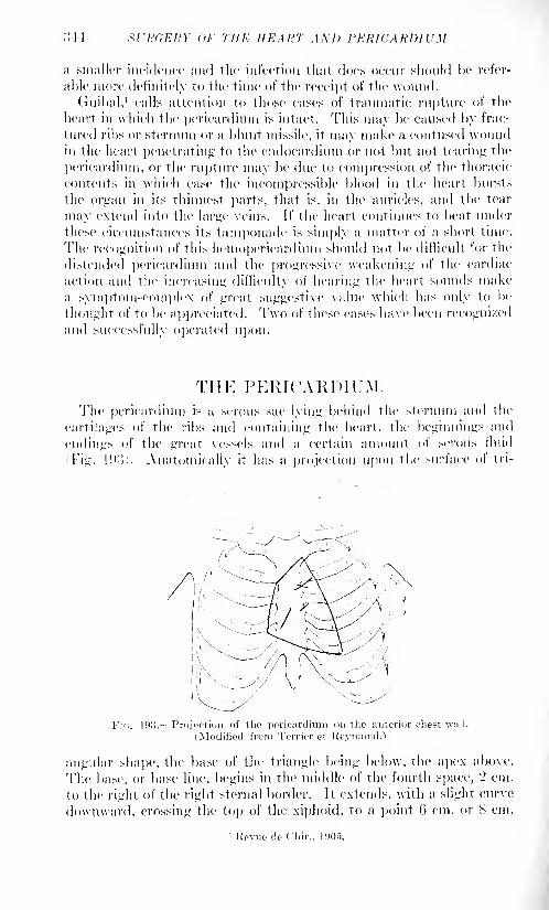
:U4 SURGERY (>r THE HEART AND PERICARDIUM
a smaller incidence and the infection that does occur should be refer-
able more definitely to the time of the receipt of the wound.
Guibal, 1calls attention to those cases of traumatic rupture of the
heart in which the pericardium is intact. This may be caused by frac-
tured ribs or sternum or a blunt missile, it may make a contused woundin the heart penetrating to the endocardium or not but not tearing the
pericardium, or the rupture may be due to compression of the thoracic
contents in which ease the incompressible blood in the heart bursts
the organ in its thinnest parts, that is, in the auricles, and the tear
may extend into the large veins. If the heart continues to beat under
these circumstances its tamponade is simply a matter of a short time.
The recognition of this hemopericardium should not be difficult for the
distended pericardium and the progressive weakening of the cardiac
action and the increasing difficulty of hearing the heart sounds makea symptom-complex of great suggestive value which has only to be
thought of to be appreciated. Two of these cases have been recognizedand successfully operated upon.
THE PERICARDIUM.The pericardium is a serous sac lying behind the sternum and the
cartilages of the ribs and containing the heart, the beginnings and
endings of the great vessels and a certain amount of serous fluid
(Fig. 1 !).')). Anatomically it has a projection upon the surface of tri-
Fif;. 193. Projection of the pericardium on the anterior rhest wall.
(Modified from Terrier et Roymond.')
angular shape, the base of the triangle being below, the apex above.
The base, or base line, begins in the middle of the fourth space, 2 cm.
to the right of the right sternal border. It extends, with a slight curve
downward, crossing the top of the xiphoid, to a point G cm. or S cm.
1 Revue do Cliir., 1905.
THE PERICARDIUM 34-")
to the left of the left sternal border in the fifth space. On the right side
the line of the triangle begins at the beginning of the base line andextends upward, with a slight convexity to the right, 1 cm. or 2 cm.
from the right sternal border to the second cartilage, then it approachesand crosses the sternum toward the cartilage of the left first rib. Theleft side of the triangle begins at the left end of the base line and extends
with a slight convexity to the left, to the same termination as the right
side. Behind this triangle the pericardium lies. Anteriorly it is loosely
bound to and is close to the back of the sternum, and posteriorly it is
separated from the spine only by the contents of the posterior medi-
astinum ; on the right and the left it has sides which are in contact with
the right and left pleura? and lungs respectively, and it is extensivelyattached below to the diaphragm. It has, then, the general form of an
irregularly-shaped wedge, at the truncated upper point of which the
aorta and the pulmonary artery pass out, while above and below
toward the right the superior and inferior vense cavte enter, and behind
the four pulmonary veins do the same. All of these vessels between
the heart and the pericardium have a share of the serous coat which
wholly or partly surrounds them. These vessels passing through the
pericardial sac to the heart, divide the potential space inside the peri-
cardium into chambers, or sinuses, which can be demonstrated bydilating the sac. There is one upon the right between the two vena 1
cavse, one upon the left, in which the heart itself is to be found and
above a space between the outgoing vessels and the pericardial wall.
Both of the lateral spaces extend around toward the back.
Normally the pericardium contains about !."> c.c. of serous fluid, only
enough to make a thin film between the epicardium and the pericardium.If by a trauma hemorrhage occurs into the sac, or by a pathological
process there is effusion, these spaces are developed and distended,
and then the heart, separated from its sac, takes certain locations in
the fluid. Yomitch-Sianojentzky demonstrated tin's on the cadaver,
sitting or supine, and with an open or an intact pericardium. If a
small amount of excess fluid, as 12 to 20 c.c. is introduced or accumu-lates in an opened pericardium, it will be found in the antero-inferior
cul-de-sac, chiefly at the left side if the patient is upright, or on the
right side and in the auriculoventricular furrow and about the origins
of the great vessels of the base if the patient is supine. If, however,
the opened pericardium is filled with fluid it envelopes the heart but is
chiefly around the right auricle and ventricle, and also in the postero-lateral spaces if the patient is supine, and is between the heart and the
diaphragm if the patient is erect.
In the intact pericardium there is some variation in the heart's place
depending upon the amount of fluid which accumulates, \\ith about
40 c.c. of liquid and the patient supine, the liquid collects above and
below: above where the serosa is reflected upon the great vessels, and
below in the antero-inferior limit of the sac (Fig. 194). AVith the same
amount of fluid and the patient erect, the fluid collects about the apexof the heart (Fig. 195).
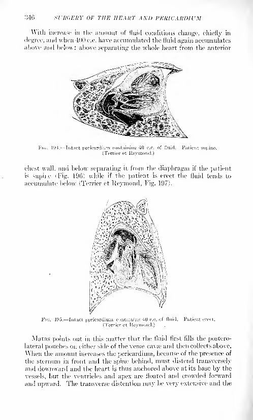
346 OF THE HEART AXD PERICARDIUM
\\ith increase in the amount of fluid conditions change, chiefly in
degree, and when 400 c.c. have accumulated the fluid again accumulatesabove and below: above separating the whole heart from the anterior
FIG. 194. Intact pericardium containing 40 c.c. of fluid. Patient supine.
(Terrier et Raymond.)
chest wall, and below separating it from the diaphragm if the patientis supine ( Fig. 196) while if the patient is erect the fluid tends to
accumulate below (Terrier et Reymond, Fig. 197).
FIG. 105. Intact pericardium containing 40 c.c. of fluid. Patient erect.
(Terrier et Reymond.)
Matas points out in this matter that the fluid first fills the postero-
lateral pouches on either side of the venre cava? and then collects above.
When the amount increases the pericardium, because of the presence of
the sternum in front and the spine behind, must distend transversely
and downward and the heart is thus anchored above at its base by the
vessels, but the ventricles and apex are floated and crowded forward
and upward. The transverse distention may be very extensive and the
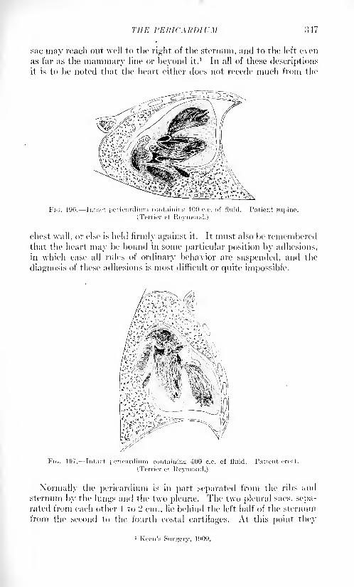
THE PERICARDIUM 347
sac may reach out well to the right of the sternum, and to the left even
as far as the mammary line or beyond it.1 In all of these descriptions
it is to be noted that the heart either does not recede much from the
FH;. 190. Intact peiicardium containing 400 e.c. of fluid. Patient supine.(Terrier et Reymond.)
chest wall, or else is held firmly against it. It must also be rememberedthat the heart may be bound in some particular position by adhesions,
in which case all rules of ordinary behavior are suspended, and the
diagnosis of these adhesions is most difficult or quite impossible.
FIG. 197. Intact pericardium containing 400 c.c. of fluid. Patient erect.
(Terrier et Reymond.)
Normally the pericardium is in part separated from the ribs and
sternum by the lungs and the two pleura*. The two pleiiral sacs, sepa-rated from each other 1 to '2 cm., lie behind the left half of the sternum
from the second to the fourth costal cartilages. At this point they
1 Keen's Surgery, 1909.
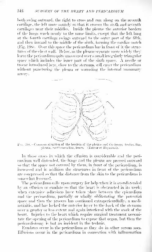
348 SURGERY OF THE HEART AND PERICARDIUM
both swing outward, the right to cross and run along on the seventh
cartilage, the left more acutely so that it crosses the sixth and seventh
cartilages near their middles, inside the pleura1 the anterior borders
of the lungs reach nearly to the same limits, except that the left lungat the fourth cartilage swings outward to the outer part of the fifth,
and then inward to the middle of the sixth, forming the cardiac notch
i, Fig. 19S>. Over this space the pericardium has in front of it the struc-
tures of the chest wall. Below, as the pleura1
separate more widely theyleave the pericardium quite uncovered over a small irregularly triangular
space which includes the inner part of the sixth space. A needle or
trocar introduced here, close to the sternum, will enter the pericardiumwithout puncturing the pleura or wounding the internal mammaryarterv.
FK;. 108. Common situation of the borders of the pleune and the him
pleura;, continuous line, lunjrs. (Terrier et Reymond).broken lino,
In those cases in which the effusion is considerable and the peri-
cardium well distended, the lungs and the pleura? are pressed outwardso that the space not covered by them, in front of the pericardium, is
increased and in addition the structures in front of the pericardiumare compressed so that the distance from the skin to the pericardium is
somewhat lessened.
The pericardium calls upon surgery for help when it is overdistended
by an etl'usion or exudate so that the heart: is obstructed in its work;when extensive adhesions have taken place between the epicardiumand the pericardium, partially or wholly obliterating the potential
space and then the process has continued extrapericardially, a medi-
astinitis, and has locked the anterior layer to the back of the sternum
over a greater or less extent and again interfered with the work of the
heart. Injuries to the heart which require surgical treatment necessi-
tate the opening of the pericardium to expose that organ, but then the
pericardiotomy is but an incident in the technic.
Exudates occur in the pericardium as they do in other serous sacs.
Effusions occur in the pericardium in connection with inflammation
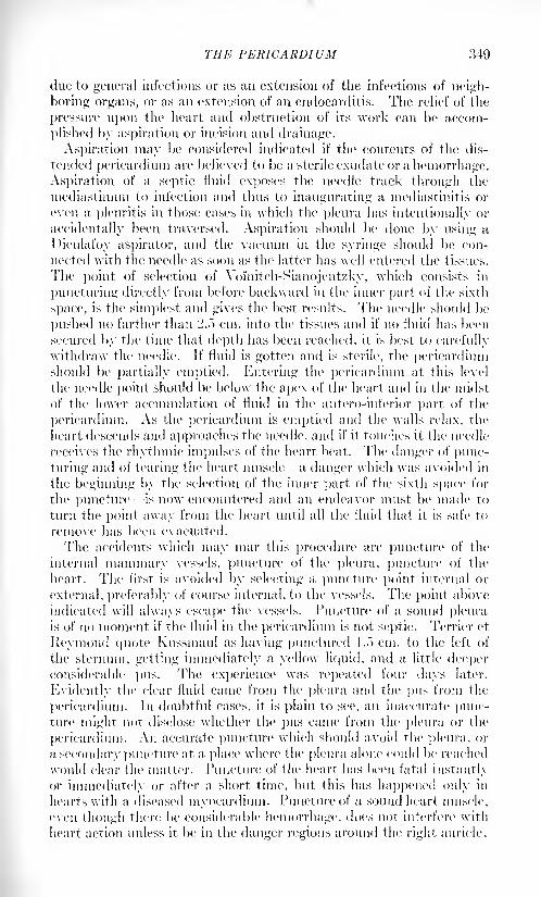
THE PERICARDIUM 349
due to general infections or as an extension of the infections of neigh-
boring organs, or as an extension of an endocarditis. The relief of the
pressure upon the heart and obstruction of its work can be accom-
plished by aspiration or incision and drainage.
Aspiration may be considered indicated if the contents of the dis-
tended pericardium are believed to be a sterile exudate or a hemorrhage.
Aspiration of a septic fluid exposes the needle track through the
mediastinum to infection and thus to inaugurating a mediastinitis or
even a pleuritis in those cases in which the pleura has intentionally or
accidentally been traversed. Aspiration should be done by using a
I Heulafoy aspirator, and the vacuum in the syringe should be con-
nected with the needle as soon as the latter has well entered the tissues.
The point of selection of Yoinitch-Sianojentzky, which consists in
puncturing directly from before backward in the inner part of the sixth
space, is the simplest and gives the best results. The needle should be
pushed no farther than 2..") cm. into the tissues and if no fluid has been
secured by the time that depth has been reached, it is best to carefully
withdraw the needle. If fluid is gotten and is sterile, the pericardiumshould be partially emptied. Entering the pericardium at this level
the needle point should be below the apex of the heart and in the midst
of the lower accumulation of fluid in the antero-inferior part of the
pericardium. As the pericardium is emptied and the walls relax, the
heart descends and approaches the needle, and if it touches it the needle
receives the rhythmic impulses of the heart beat. The danger of punc-
turing and of tearing the heart muscle a danger which was avoided in
the beginning by the selection of the inner part of the sixth space for
the puncture is now encountered and an endeavor must be made to
turn the point away from the heart until all the fluid that it is safe to
remove has been evacuated.
The accidents which may mar this procedure are puncture of the
internal mammary vessels, puncture of the pleura, puncture of the
heart. The first is avoided by selecting a puncture point internal or
external, preferably of course internal, to the vessels. The point aboveindicated will always escape the vessels. Puncture of a sound pleurais of no moment if the fluid in the pericardium is not septic. Terrier et
Reymond quote Kussmaul as having punctured 1 .o cm. to the left of
the sternum, getting immediately a yellow liquid, and a little deeperconsiderable pus. The experience was repeated four days later.
Evidently the clear fluid came from the pleura and the pus from the
pericardium. In doubtful cases, it is plain to see, an inaccurate punc-ture might not disclose whether the pus came from the pleura or the
pericardium. An accurate puncture which should avoid the pleura, or
a secondary puncture at a place where the pleura, alone could be reached
would clear the matter. Puncture of the heart has been fatal instantlyor immediately or after a short time, but this has happened only in
hearts with a diseased myocardium. Puncture of a sound heart muscle,
even though there be considerable hemorrhage, does not interfere with
heart action unless it be in the danger regions around the right auricle,
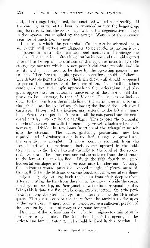
;j,">0 Sl'ltdRJtY OF THE HEART AND PERICARDIUM
and, other things being equal, the punctured wound heals readily. If
the coronary artery of the heart be wounded or torn the hemorrhagemay be serious, but the real danger will be the degenerative changesin the myocardium supplied by the artery. Wounds of the coronaryvein are of much less moment.
In cases in which the pericardial effusion can be affirmed, on a
sufficiently well worked out diagnosis, to be septic, aspiration is not
competent to control the condition and incision and drainage are
needed. The same is manifest if aspiration is done and the fluid secured
is found to be septic. Operations of this type are most likely to be
emergency matters which do not permit elaborate technic, and, in
addition, they may need to be done by the operating general prac-titioner. Therefore the simplest possible procedure should be followed.
The debatable point is that at which the chest wall should be openedto permit the uncovering of the pericardium. The method which
combines direct and simple approach to the pericardium, and also
gives opportunity for extensive uncovering of the heart should that
prove to be necessary, is that of Kocher. lie "makes an incision
down to the bone from the middle line of the sternum outward toward
the left side at the level of and following the line of the sixth costal
cartilage. If required the incision may extend to the left mammaryline. Separate the perichondrium and all the soft parts from the sixth
costal cartilage and excise the cartilage. This exposes the triangularmuscle of the sternum with the mammary vessels which are ligated if
necessary. Divide the tendinous insertion of the triangular muscle
into the sternum. The dense, glistening pericardium now lies
exposed, and if drainage alone is required it may be opened andthe operation is complete. If more room is required, from the
sternal end of the horizontal incision cut upward in the mid-
sternal line to the desired extent (usually to the level of the second
rib). Separate the periosteum and soft structures from the sternumto the left of the median line. Divide the fifth, fourth and third
left costal cartilages at their insertions into the sternum. Throughthe horizontal wound push the exposed margin of pleura outward.
Gradually lift up the fifth and even the fourth and third costal cartilages
slowly and gently pushing back the pleura from their deep surface.
After separating the flap from the pleura, fracture or divide the costal
cartilages in the flap, at their junction with the corresponding ribs.
When this is done the flap can be completely reflected. Split the peri-
cardium along the sternal margin and laterally along the fifth inter-
space. This gives access to the heart from the auricles to the apexof the ventricles. If more room is desired excise a sufficient portion of
the sternum by means of rongeur or bone forceps."1
Drainage of the pericardium should be by a cigarette drain of suffi-
cient size or by a tube. The drain should go to the opening in the
pericardium but not enter it, and should be fixed in this location. If
1 Binnie: Operative Surgery.
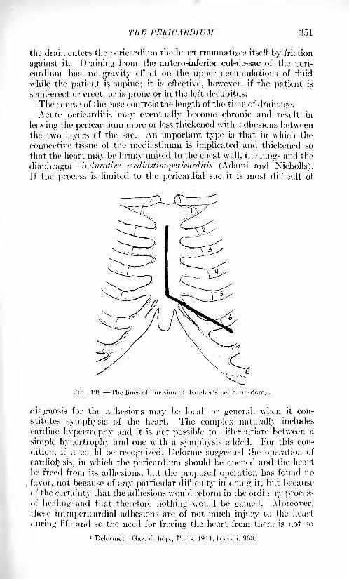
THE PERICARDIUM 351
the drain enters the pericardium the heart traumatizes itself by friction
against it. Draining from the antero-inferior cul-de-sac of the peri-
cardium has no gravity effect on the upper accumulations of fluid
while the patient is supine; it is effective, however, if the patient is
semi-erect or erect, or is prone or in the left decubitus.
The course of the case controls the length of the time of drainage.Acute pericarditis may eventually become chronic and result in
leaving the pericardium more or less thickened with adhesions betweenthe two layers of the sac. An important type is that in which the
connective tissue of the mediastinum is implicated and thickened so
that the heart may be firmly united to the chest wall, the lungs and the
diaphragm induratice mediastinopericarditis (Adami and Xicholls).
If the process is limited to the pericardial sac it is most difficult of
FIG. 199. The lines of incision of Koclier's pericardiotomy.
diagnosis for the adhesions may be local 1 or general, when it con-
stitutes symphysis of the heart. The complex naturally includes
cardiac hypertrophy and it is not possible to differentiate between a
simple hypertrophy and one with a symphysis added. For this con-
dition, if it could be recognized, Delorme suggested the operation of
cardiolysis, in which the pericardium should be opened and the heart
be freed from its adhesions, but the proposed operation has found no
favor, not because of any particular difficulty in doing it, but because
of the certainty that the adhesions would reform in the ordinary processof healing and that therefore nothing would be gained. Moreover,these intrapericardial adhesions are of not much injury to the heart
during life and so the need for freeing the heart from them is not so
1 Delorme: Gaz. d. hop., Paris, 1914, Ixxxvii, 963.
iioL' HL'RGEItY OF THE HEART AXD PEIUCARDIUM
great.1
It is different if the adhesions are also extrapericardial, Adami'sindurative mediastinopericarditis. In this case the heart makes a
direct pull upon the rigid sternum and ribs at each systole, and as theycan yield but little if any the base must be moved rather than the
apex, that is, the relatively fixed part of the heart is the apex and not
the base.
1 )elageniere- gives an excellent symptomatology and includes both
many signs of common presence in cardiac complexes, and some \vhirh
are less ordinary and more indicative of the adhesions. The ordinary
symptoms are dyspnea, precorclial pain and tenderness. The 1
excep-tional are a bulging of the precordium with a retraction during the
systole and a systolic deepening of the epigastric hollow, the former beingdue to tin 4 heart's systolic pull upon the sternum and the latter's recoil
during diastole, while the epigastric depression is due to the pull uponthe diaphragm. As evidence of the cardiac hypertrophy is the increase
in precordial dulness and murmurs at the various orifices, due to the
valvular incompetency of the dilated though hypertrophied heart, anda tendency to embryocardia, a reduplication of the heart sounds.
In general there is edema, ascites, hypertrophy of the liver and
albuminuria.
Radioscopy is of great value in the diagnosis for it shows the heart
shadow enlarged, and that the apex and sides are immobile. 1 '
For this condition an operation has been suggested by a medical man,
Brauer, of Marbourg, which he called cardiolysis, but which Delagenierecalls pericardiolysis, leaving the word cardiolysis to apply to the oper-ation inside the pericardium suggested by Delorme. kocher called it
thoracolysc prccardiaque and Thorburn precardial thoracosloiny.4 The
operation, suggested in 1002, had been done fifteen times according to
Thorburn in 1010, and to these Thorburn added one case; of these' all
but two were markedly benefited and some were restored to the ability
to do heavy work. The operation is simple and wholly extrathoracic.
A curved incision is made along the left border of the sternum uncover-
ing the third, fourth and fifth cartilages, and the soft tissues are turned
back to the ribs or beyond if needed. These' cartilages are resected and
some of the ribs, in all s cm. or 10 cm. of each. It is advised to remove
the superficial perichondrium and periosteum, but not the dee]). If
the heart now impinges against the edge of the sternum that bone is
cut away with rongeurs enough to free the heart. The musculocuta-
neous flap is replaced. The heart is thus freed from its attachment to the
rigid chest wall; the flexible' layer of tissues to which it remains fixed
readily follows its movements and the obstacle to its competent func-
tion is removed.. The operation has most commonly been done under a
general anesthetic, but local anesthesia is practicable.
Primary tumors of the pericardium are most difficult of appreciation
1 Lecene: Arch, do mal. do cceur, etc., Paris, 1909, ii, G73.- Bull, de 1'Aoad. de Mod., 1913, 539, ct seq.? Vaquoz ct Bordet. Quoted by Dehisetiiero.4 Brit, Mod. Jour., 1910, i, 10.
THE PERICARDIUM of)))
during life, and arc generally necropsy discoveries. They are frequently
malignant and may occur at any age from fifteen to seventy- five. 1
POSTSCRIPT. The surgical treatment of wounds of the pericardiumand heart in war surgery reports has followed the lines already laid
down by civil practice. This is really as it should be, and General
Cuthbert Wallace, of the Royal Army Medical Corps, is quoted as
saying: "The more war surgery approximates to civil surgery the
better it will be."
Numerous operations have been done and have demonstrated the
simplicity of uncovering the organs. The incisions for exposuretraverse practically only connective tissues, an intact pleura can be
pushed out of the way, the internal mammary artery is the onlyvessel likely to be encountered, the cartilages of the ribs are not diffi-
cult to deal with and the chest wall after closure is equally competentif these be retained and restored to place or not. Lavage of the peri-
cardial sac has been done, drainage of it has sometimes been practised,
usually it has been closed, and this last is the proper practice unless
there has been an obvious infection. Once the pericardium was
packed to control an otherwise uncontrollable hemorrhage with, of
course, a fatal issue.
Infection of the opened pleura has not been uncommon, but has
usually been easily managed.This operative work can be and has been done under local anes-
thesia, and the fact of the insensibility of the pericardium and heart
to pain by being handled was noted. 2 That, manipulation was pain-
less, but that lavage of the pericardium by eusol, by hypertonic salt
solution and by physiological salt solution was very painful, while a
weak solution of iodin was painless, was pointed out by Jones. 3
The importance of operating in those cases in which the diagnosis
was not convincing, but in which there was a reasonable belief of the
need existing, was as great as in analogous abdominal matters. Rehninsists on the obligation of the surgeon under these circumstances.
That the surgeon must think of and be able to recognize heart tampon-ade is evident, and that heart tamponade may exist without an ex-
ternal wound must not be forgotten. Jones 4
reports a successful inter-
vention for tamponade following a stab wound and Rhodes-"' reports
two, one succeeding and one failing.
The importance of looking for small wounds in the heart is insisted
on. One case in which a small wound was not found at the operation,the patient bleeding to death, is recorded,
1'' arid another in which such a
wound was found and successfully closed. 7
1 Fonii. Tumore, March, April, vol. iv, No. 5.
2 Gray, Birbock and Lorinior: British Med. Jour., October 10, 1915, p. 501.:! British Jour. Sur., No. 13, vol. iv, p. 103.1 Ann. Surtr., 1917, Jxv, 119.; >
Ibid., Ixvi, 44.1 Miiller: Muncliun. mod. Wdm.-dir., July 2S, 1914, No. 30, p 1079.7 Dixon and McEuen: British Mod. Jour., 1910, i, 755.
VOL. ii 23
354 SURGEKY OF THE HEART AND PERICARDIUM
A number of cases of the removal of pieces of shrapnel or of bullets
from the heart muscle at primary or secondary interventions
are in ven. The fact of the presence of the missiles was shown byradiograms, or the missile could be seen to move with the heart bythe fluoroscope. "\arious symptom-complexes were recounted; easy
fatigue, inability to stand erect, dyspnea, absence or presence of pain,cardiac disability, are some of the symptoms suggestive of a heart
wound with a retained bullet. Most of these missiles were located in
the ventricles. In one instance a bullet near the ventricular apex wasassociated with injury to the bundle of His, with complete heart-
block, due To the latter lesion. Wounds of the right auricle have been
reported without the necessary fatalities experimental work wouldseem to call for. One right auricular wall carried a bullet six months,when it was removed because it disturbed heart function. This
patient died,1 but Fraser'-' and Nassau3 both report successful sutures
of the right auricle.
Nothing is said, of course, about the danger regions in the heart.
If a wound by a bullet was made in a noli rue tangcre place in the heart,
that heart never came for surgical help, and a heart that came for
help would be entitled to help no matter where the wound was, but
(and this advice is not based on any experience gained in war surgery
particularly) the surgeon closing a wound in a heart should be careful
not to wound a coronary artery by his needle or to include them or
any of their major branches in his sutures.
In the case of the heart, when traversed by a high-velocity bullet,
death is usually instantaneous, the dynamic disturbance causing it
to stand still. Failing an instant paralysis, the wounded heart bleeds
so freely and rapidly that death occurs in a few minutes. In big-
game shooting it occasionally happens that a large beast is broughtto the ground by a broken back. When such an animal is deliberatelyfinished by a bullet sent through its chest, either fore and aft, or from
side to side, it dies instantly. On opening the chest, the heart will
be found firmly contracted, and often there is no effusion of blood,
either in the cavity of the pericardium or of the pleura. The effects
of a sword plunged into the heart may be watched in the bull-ring.
When the matador thrusts the weapon directly into the heart, the
bull sinks instantly on the floor of the ring; but when the sword
wounds the big vessels at the base of the heart, the bull may moveabout for many seconds while the blood escapes.
In previous wars little attention was given to gunshot wounds of
the heart; they have been regarded as nearly always fatal. Indian
sportsmen have often drawn attention to the fact that a tiger, shot
in the heart, has had the strength to run for many yards before falling
dead. Some of the hearts in the collection show the destructive effects
1 Rev. do Ohir., Vans, December, 191.").
- Edin. Mod. Jour.. January, 1917. :i Ann. Sur<r., 1917, Ixv, 119,
THE PERICARDIUM 355
which modern missiles exert upon the thick walls of the ventricles.
One heart extensively lacerated near the apex by an armor-piercing
bullet, merits attention. It belonged to an officer who was hit when
flying over the German lines. He brought the aeroplane to earth in
his own lines, but was dead when removed from the machine.
Farina, in 1896, proved that wounds of the heart are amenable to
surgery; and his example influenced the treatment of such wounds
during the Great War; there are specimens illustrating surgical enter-
prise in this direction. But the surprise of the collection is a series
of specimens indicating the methods and routes by which missiles
reach the cavities of the heart; they prove that missiles occasionallyenter the cardiac cavities by directly penetrating the walls of the
heart, or arrive as flotsam and jetsam in the blood stream. Theevidence supplied by the specimens shows that fragments of metal,
and entire bullets shrapnel and rifle penetrate large veins, such
as the pulmonary, iliac, hepatic and caval, and are transported as
emboli to the heart. Some of the missiles, after reaching the auricu-
lar or ventricular cavities have been retained. A few, embedded in
clot, have been discovered post mortem. In several instances, bullets
have been detected radioscopically in the heart cavities during life,
and their swirlings and gyrations in the blood current during systole
and diastole, have been watched with the same interest that astro-
nomical observers bestow on minor planets.In some instances, bullets round and conoidal, or irregular frag-
ments of metal, after reaching the cavity of a ventricle, are dischargedand become dangerous emboli. There are specimens in the collection,
exquisitely prepared and displayed, showing metallic emboli impactedin the pulmonary artery producing infarction of the lung. Others,
lodged in the iliac arteries, caused ischemia of the lower limb.
Even more remarkable are the specimens proving that bullets maypenetrate large bloodvessels, like the aorta, and become centrifugal
emboli, being propelled by the arterial current, or slip along the
vessels by their own weight, until they become impacted in the
common or the external iliac artery. These extraordinary events
and sequences are not only represented by specimens, but completeclinical histories are available; the names of the officers who madethe observations and carried out the treatment are duly recorded.
In a few cases the missiles within the chambers of the heart cameas a postmortem surprise, but in many instances the nature of the
embolus has been recognized during life; in one instance a missile wasobserved in the heart radioscopically, and a few hours later it wasdetected by the same method in the common iliac artery (O'Neill).The parts belonging to this remarkable case are preserved in the
Museum. British Journal of Surgery, July, 1919, "Introductory,"bv J. Bland-Sutton.

SURGERY OF THE LIVER, AND GALL-BLADDER.
BY JOSEPH RANSOIIOFF, M.I)., F.A.C.S., F.R.C.S., (Exo.)
AND
J. LOUIS RANSOIIOFF, Ml)., F.A.C.S.
THE LIVER.
ANATOMY OF THE LIVER.
The liver which develops from the foregut is part of the digestive
system, both from an embryological and a functional viewpoint. It, is
the largest gland in the body, and its average weight in the adult maleis about 1520 grams. The liver is situated in the upper portion of the
abdominal cavity, occupying almost the entire right hypochondrium.It extends through the epigastrium and into the left hypochondriumto a variable degree. It is behind the right seventh, eighth, ninth,tenth and eleventh ribs and their cartilages, the ensiform cartilage, andthe sixth, seventh, eighth and ninth left costal cartilages. In this wayit is fairly well protected from all external injuries except those of
great force.
The liver is movable to a limited extent, following the diaphragmin its respiratory excursions. During inspiration the upper surface of
the right lobe reaches the upper border of the eighth dorsal vertebra.
Unless the liver is enlarged its lower right border lies under the thoracic
margin and cannot be felt except in very thin subjects during deepinspiration. The lower border of the left lobe can usually be felt in
the epigastrium, directly below the costal angle.
The position of the liver varies with posture in the erect adult; its
lower border follows a line drawn from the eighth left to the ninth
right costal cartilage. In children the liver occupies a much largerrelative space, filling a large part of the abdomen, often reaching the
umbilicus. This is of surgical importance in planning abdominalincisions in young children.
The relations of the pleura to the liver are important from a surgical
viewpoint. On the right side the pleura is reflected from the chest
wall to the diaphragm, along a line extending from the back of the
ensiform cartilage, downward and outward along the seventh costal
cartilage. In the nipple line this reflection is at the level of the eighthcostal articulation. In the midaxillary line it is beneath the tenth
intercostal space, while near the spine it is at the level of the eleventh
(357)
.Sr>S Sl'RCERY OF THE LIVER AXD CALL-BLADDER
rib or slightly below. The lowest point of the pleura! reflection is in
the inidaxillary line. This reflection is, as a rule, but two inches fromthe lower bonier of the liver. When the liver is enlarged upward the
diaphragmatic and the parietal pleura are brought into close contact.
This is of assistance in certain forms of abscess of the liver pointingon the superior surface, where the parietal and diaphragmatic pleurabecome adherent, obliterating the costophrenic space. This permitsthe opening of these abscesses without invasion of the pleural cavity.
The liver is connected to the diaphragm by the coronary and sus-
pensory ligaments. Except between these ligaments the liver is
entirely covered by peritoneum . The anterior reflection of the coronaryor lateral ligament forms the falciform ligament, which attaches the
liver to the diaphragm and the anterior thoracic wall. This ligamentextends to the umbilicus and encloses in its anterior border the liga-
mentum teres, which represents the remains of the obliterated umbilical
vein; except at the umbilicus it is situated to the right of the middle
line.
The anterior border of the liver is sharp and thin and contains about
in its center, opposite the ninth costal cartilage, the notch for the gall-
bladder. To the left of this is the notch where the round ligamententers. Its under surface is irregular, to conform to the organs with
which it is in close relation, or perhaps, more correctly stated, on which
it rests. The right and left lobes are separated by the longitudinalfissure. Besides the right and left lobes there are three others, which
are really subdivisions of the right lobe: the lobus Spigelii, the lobus
caudatus and the lobus quadratus. Anatomical variations of these
small lobes are not infrequent and are usually of only academic interest.
Accessory lobules are not uncommon. One of these is occasionallyfound attached to the neck of the gall-bladder.
In some individuals a long, tongue-shaped prolongation of the right
lobe is found, called Riedel's lobe. It may be attached either to the
right or to the left side of the gall-bladder, which it may completelyconceal. In some instances it is connected to the liver by an atten-
uated pedicle, so that it may form a distinctly movable abdominaltumor. It may be mistaken for enlarged gall-bladder, tumor of the
liver or even for a tumor of the right kidney. This Riedel's lobe has
been regarded by sonic as the result of tight lacing. This theory, how-
ever, is untenable, as it is not infrequently found in men, and rnayeven grow from the left lobe. From observations it has been supposedto be invariably associated with gall-stones. This, however, is not true.
The liver rests directly on the stomach, transverse colon and right
kidney, and there are niches on its lower surface into which these
organs fit. The chief support of the liver is its intimate connection
with the inferior vena cava, through the openings of the large hepaticveins. Passing upward to the heart the vena cava is intimatelyadherent to the posterior abdominal wall and passes deeply throughthe liver tissue, where it receives the large tributaries of the hepaticvein. In this way the liver is firmly fastened to the posterior abdominal
DISPLACEMENTS OF THE LIVER 359
wall and the diaphragm . The coronary ligaments also assist in support-
ing the liver. The general intra-abdommal pressure and the correct
position of the organs on which the liver rests also play a distinct role.
The liver is only displaced in extreme degrees of enteroptosis.The liver is surrounded by a dense fibrous capsule called Glisson's
capsule. Prolongations of the capsule enter the liver at the transverse
fissure with the hepatic and portal vessels. These capsular prolonga-tions follow the vessels into their smallest ramifications and form the
capsules of the individual lobules.
The liver is supplied by the hepatic artery, which enters the liver
through the gastrohepatic omentum, with the portal vein and the
common duct.
The liver is rarely in anomalous position except in complete trans-
position of the viscera. Occasionally gall-stones have occurred in
these cases and have required operation. Such cases have been
reported by Beck, Billings and Mayo. Robson described a case in
which the liver assumed an almost vertical position.
DISPLACEMENTS OF THE LIVER.
Prolapse of the liver is a not infrequent condition, and occurs mostlyin conjunction with general abdominal ptosis. It is, as a rule, rather a
medical than a surgical condition. The frequency may be estimated
by a report of Einhorn, who in the course of a year's work found 21
cases of hepatoptosis in 369 females and 9 cases among 439 males. It
must be remembered, however, that his clinical material is largely
gastrointestinal.The causes are those common to abdominal ptosis. It usually
occurs in women whose abdominal walls have become relaxed throughemaciation or repeated childbirth. There is a well-recognized enterop-totic habit winch has a peculiar conformation of the body. Thethorax is long and thin and the neck also is long and thin. The costal
arch is acute and the costo-iliac interval very long. These are the
patients who very frequently are supposed to be threatened with tuber-
culosis, and are rarely in perfect health.
Albu 1 showed that hepatoptosis is a very frequent accompaniment of
general enteroptosis. lie found among 3400 patients examined that it
was twice as frequent as left nephroptosis and half as frequent as right
nephroptosis.
Hepatoptosis may be divided into five classes, according to symptoms(Einhorn
2) : In the first class of cases the hepatoptosis is found and
causes no symptoms. The second class is dyspeptic, the third class
has pain in the liver and a dragging pain up the right abdomen andshoulder. The fourth class may have attacks resembling gall-stone
colic. The fifth class has attacks resembling asthma.
1
15erl. klin. Wclmsehr., 1'JO'J. vol. vii.
-Jour. Am. Med. Assn., 1910, Ixvi, 1908.
3GO SURGERY OF THE LIVER AND CALL-BLADDER
Symptoms.- As a rule the symptoms of hepatoptosis are overshadowed
by the general symptoms of enteroptosis. These patients are classed
as dyspeptics, or, more frequently, as neurasthenics, and are not
infrequently very difficult to relieve. The diagnosis is easily made bythe palpation of the displaced liver, which, of course, may he con-
firmed by .r-rays.
Gouba 1 has described a very interesting case of hepatoptosis occur-
ring in a child, aged nine years. There is reason to believe that a low
stand of the liver with elongation of the suspensory ligament may be
congenital. Abnormal elongation of the vena cava may also, accordingto Fatire,- be a factor in the descent of the liver. There is no questionthat the relaxed abdominal wall is one of the most important causes
for this condition.
Treatment.^ The treatment is, as a rule, purely orthopedic, dietetic
or medical, and should be directed toward an improvement in the
patient's general condition. Rest and fat cures are particularly ad-
visable, in addition to that a well-fitting abdominal band should be
worn, to keep the viscera in proper place.
During recent years the surgical treatment of enteroptosis has comeinto great prominence, but seems rather to be losing than gainingfavor. Billroth was the first, in 1SS4, to practice the surgical correction
of hepatoptosis. Werelius collected (>(> operations of this kind in 1912.
Of these cases only 2 were in males, and there seems no question that
repeated pregnancy had a great deal to do with the condition, as only4 of the patients were nullipanr. The enlarged Riedel's lobe, which
may attain great six.e and extend far down into the pelvis, cannot be
considered a true hepatoptosis.The following different methods of suspension have been used:
Billroth placed deep sutures through the liver tissue and capsule,
which he carried through to the abdominal wall and tied over iodo-
form gauze. Treves sutures the liver to the xiphoid process, Ramseyto the seventh ribs. Le Ford, Poppert and others have fastened the
liver by means of the gall-bladder. Depage aimed at the correc-
tion of the displacement by a plastic operation of the pendulousabdomen.
Various methods have been used to promote adhesions between the
upper surface of the liver and the lower surface of the diaphragm by
scarification, by the knife, or by cauterization with the thermocauteryor even by packing.
Cross in a case of extreme prolapse of the liver used the ligamentumteres with good result. After separating the ligament close to the
umbilicus he raises the liver by sharp upward traction, the free end of
the band was then passed through the ninth intercostal space, where
it was held in place by a chromic catgut or kangaroo tendon suture,
lie was able to raise the position of the free border of the liver at least
10 cm. by this maneuver. After all is said and done there are prob-
1 Deutseh. mod. Wehnschr., 1915, xli, 450.
M'Hepatoptose et 1'hepatopcxie, Th6se do Paris, 1892.
INJURIES OF THE LIVER 361
ably but few cases of hepatoptosis which need be considered from a
surgical point of view, as the vast majority of the cases are only partof the general condition of enteroptosis.
INJURIES OF THE LIVER.
Injuries of the liver are not uncommon in civil practices. There are
three varieties of injuries of the liver: (Ij crush wounds; (1) shot
wounds; (3) stab wounds. In civil practice the most important and
by far the most frequent of these injuries are the crush wounds.
This may be explained in several ways. When the abdomen is struck
by a powerful force, any one of the organs of the peritoneal cavity
may be injured. However, of the solid organs of the abdomen the
liver is the one most frequently injured in this way, either alone
or combined with other abdominal injuries. Of 305 subcutaneous
injuries of the solid abdominal viscera collected by Tilton 1 there were
189 cases of liver injuries.
The reason for the frequent injury of the liver is its fixed positionto the diaphragm and chest wall. Because of this the liver is unable
by slipping and changing its position to evade the injuring force.
Those liver injuries which are combined with injuries of other organs,
particularly the hollow viscera, are not of great surgical importance,as the combined injuries usually lead to a rapid fatal termination.
In his article on liver injuries, Kehr- divides these subcutaneous
liver injuries into three groups: (1) True ruptures of the liver with a
tear of the capsule; (2) the separation of the capsule from the liver
substance by subscapular bleeding; (3) central subscapular rupturesof the liver, which may lead later to cyst formation and to multipleliver abscesses. This division, however, cannot be followed out
clinically, as it is impossible to differentiate between classes (2) and (3).
Finsterer3 divides these injuries of the liver into two classes: (1)
The penetrating liver rupture, in which the capsule is torn and the
rupture opens freely into the peritoneal cavity; (2) the subscapular
rupture, which include classes (2) and (3) of Kehr's classification. It
is easily seen that the symptoms of these t\vo groups will be entirely
different, as in group 1 the symptoms will depend largely on the free
hemorrhage into the peritoneal cavity, which is impossible in group 2.
It seems wise to take up the diagnosis and prognosis of this class of
cases apart from the stab and shot wounds of the liver, as the sympto-
matology and diagnosis is almost entirely different. The comparative
frequency of these subcutaneous ruptures of the liver in civil practiceis brought out by a few statistics compiled from certain Germanclinics. Of the cases reported from three large German clinics there
were 25 ruptures, 11 shot wounds and 4 stab wounds. However, in
other countries in which the carrying of knives or revolvers is more
1 Aim. Surjj;., xli, 20.2 Mtinchen. nied. "VYchn.sehr., 1903.3 Deutsoh. Ztsohr. f. Thin, v, 10.
302' SURGERY OF THE LIVER AND GALL-BLADDER
prevalent, stab and shot wounds assume a relatively more importantrole. This is particularly true in Russia, where stab wounds of the
liver are very frequent.
Symptoms. The symptomatology of these subcutaneous rupturesof the liver depend largely on the severity of the hemorrhage. As theyare usually caused by very severe trauma, as, for instance, the crush-
ing of the body between two cars or fall from a height, these injuriesare nearly always accompanied by severe shock, which for the time
being masks the special or localizing symptom. However, in certain
cases shock may be entirely wanting. Cases have been reported in
which, with a most severe injury of the liver, the patient walked into
the clinic to die a few hours later. Von Ilippel reports the case of a
twenty-eight year-old officer whose liver was ruptured by a fall from
his horse. The officer was able to walk to the clinic. Operation dis-
closed a complete tearing oft' of the left liver lobe.
The most important symptoms of liver rupture depend on the
extravasation of free blood into the peritoneal cavity. These symptomsare common, of course, to injuries of all abdominal organs. Theabdomen becomes rigid and tender. However, in injuries of the liver
in common with injuries of the upper abdomen the rigidity is prin-
cipally localized in the upper recti muscles above the umbilicus. Asliver injuries are the most common of the subcutaneous injuries of
this region a great rigidity of the tipper recti after a crushing injury
is very suggestive of liver injury. If the hemorrhage is a severe one
the symptoms of severe intra-abdominal hemorrhage are significant;
that is, weak pulse, pallor and progressing weakness.
Finsterer has pointed to a very important sign on the part of the
pulse in injuries of the liver: that is, the comparative or even absolute
bradycardia. All other forms of intra-abdominal bleeding are accom-
panied by rapidly increasing pulse-rate. In Finsterer's experience, in
nearly all of his cases, particularly when they were watched carefully,
the first shock was followed by a definite and often absolute slowing.of the pulse. If a pulse of 80 to 90 accompanies a profuse intra-
abdominal hemorrhage this must be considered a true bradycardia,as in the usual case the pulse here might range between 120 and 150.
Of 13 cases collected by Finsterer from the literature there was a slow-
ing of the pulse in each one, varying from 64 to SO to 90. All these
cases were shown either by operation or postmortem to have sustained
a very severe injury of the liver. This bradycardia is an important
diagnostic symptom, particularly when accompanied by symptoms of
intra-abdominal hemorrhage. This bradycardia must be considered
rather a negative than a positive diagnostic symptom : that is, a brady-cardia accompanying symptoms of intra-abdominal injury should not
militate against the probability of intra-abdominal hemorrhage, but
should rather point to the possibility of hemorrhage from the liver.
Pain, if present in the liver region, particularly if reflected to the
back and shoulder, is extremely suggestive of a liver injury. Thecharacter of the injury must be taken into consideration, and it must
INJURIES OF THE LIVER 363
also be borne in mind that the liver is the most frequently injured of
all the intra-abdominal solid organs. Frequently the severe shock which
accompanies all these injuries make an exact diagnosis impossible.Jaundice when present is a significant symptom, as it points almost
conclusively to a liver injury. As it does not commonly occur until
several days after the injury it is not of essential diagnostic impor-tance. The diagnosis must then be made by the direction of the offend-
ing force, the shock, the pain in the liver region and the symptoms of
intra-abdominal hemorrhage. The prognosis depends entirely on the
severity of the hemorrhage. In many instances slight liver injuries
undoubtedly recover without sufficient symptoms on which to base a
diagnosis. On the other hand, injuries with profuse intra-abdominal
hemorrhage are fatal unless surgical interference is carried out.
In civil practice an exploration should be made in every case in
which the symptoms point to severe intra-abdominal injury. Evenin the presence of shock, when symptoms of grave intra-abdominal
hemorrhages are present, operation should not be delayed. If necessaryblood transfusion should precede or follow the operation.
Shot wounds of the liver are common in modern warfare, and their
severity depends entirely on the nature of the projectile. The small
caliber modern bullet may completely penetrate the liver with mini-
mum destruction of tissue and but slight hemorrhage. In these cases
the diagnosis can only be made by noting the course of the bullet. How-ever, the characteristic pain may be present.
These liver injuries are frequently complicated by a coexisting injuryof the lung, which may completely mask the symptoms of liver injury.
In some instances, however, death has ensued from a comparatively
slight liver injury: that is, when the bullet has made a superficial
furrow in the liver surface, with an accompanying fatal hemorrhage.Shell wounds of the liver are far more serious. There is often seen
an explosive action on the liver with tremendous destruction of liver
tissue leading to a rapidly fatal termination. In other instances, after
tearing a great jagged hole, the projectile may come to rest in the
liver tissue after carrying with it some foreign material.
The symptoms of liver injuries in war are much like the symptomsof liver crushes, except that the course of the projectile simplifies the
diagnosis. In case the wound of entrance and exit are large the escapeof bile from the wound is not an uncommon symptom. Jaundice is
frequent in shot wounds of the liver. Of 37 cases observed by Makins1
in a base hospital, 12 had marked jaundice. It is probable that whensevere hemorrhage occurs most of those cases die before aid can be
brought. However, secondary hemorrhage occurring from ten days to
four weeks after the injury is not an unusual occurrence. This, of
course, is most frequently due to an accompanying sepsis. In a numberof cases which came to postmortem ten days to six weeks after the
injury a wide spread of infection was observed. The track of the pro-
1 British Jour. Sur^., 1910, p. 645.
3(14 St'li'dEltY OF THE LIVER AX1) GA LL-HLADDER
jeetile is surrounded by necrotic bile-stained liver tissue, extending;i varying de])th into the injured liver.
Landau has observed a very peculiar pathological condition which
developed after an injury of the liver. The patient made a completerecovery from the original wound. Four weeks after the injury, whenthe wound had closed, the patient began to show signs of great dis-
ability and rapid emaciation. There was no icterus. The abdomenwas swollen and seemed to contain fluid. Operation showed a large
accumulation of slightly bile-stained gelatinous fluid in the abdominal
cavity. There was no inflammation and no evidence of peritonitis.
The removal of this fluid was followed by an uninterrupted recovery.Landau has called this condition cholaskos.
Treatment.- -Unless symptoms due to hemorrhage are urgent the
treatment must be absolutely conservative in war times. This mustinclude al solute rest from four to six weeks. Even in those cases in
which there is alarming hemorrhage, operation is usually impossiblebecause of unfavorable conditions. If abscesses develop later theyshould be opened and drained under the usual conditions. If the
.r-ray shows a foreign body in the liver which seems to keep up suppu-ration the wound of entrance may be enlarged. On no account should a
ne\v wound of exploration be made because of the great danger of
spreading infection. These cases should be kept drained for a longtime so that the accumulated neerotic liver tissue may have a chance
to slough away. In those cases, accompanied by severe fever, general
sepsis, little can be done except in the way of supportive and anti-
septic treatment. If operation is done for injury of the liver, either in
civil or military practice, the same methods of stopping hemorrhageand liver suture are tised which were described under Resections of
the Liver.
DISEASES OF THE LIVER.
Syphilis of the Liver. Syphilis of the liver is not of surgical impor-
tance, except where a mistake in diagnosis is made. It usually occurs
during the tertiary stage of the disease in the form of either single or
multiple gummata (Fig. 200). J. L. Allen collected SO cases of gummataof the liver, in 11 of which the tumor was single. It is this form, which is
frequently confused with new growths of the liver, or, if it occurs in the
region of the gall-bladder, with disease of the gall-bladder. The routine
use of the \\assermann test in abdominal cases will usually preventthese diagnostic errors. It is probable that a large number of primarycarcinomata of the liver reported cured by excision were in reality
gummata.If an operation is done with the diagnosis of cancer of the liver the
presence of scars in the liver substance will be extremely suggestive of
syphilis. Syphilis may simulate portal cirrhosis, tumor of the liver,
cyst, enlargement of the gall-bladder, abscess of the liver or hyper-
trophic biliary cirrhosis. These conditions must all be ruled out bycareful examination. (lumrnata of the liver will nearly always respond
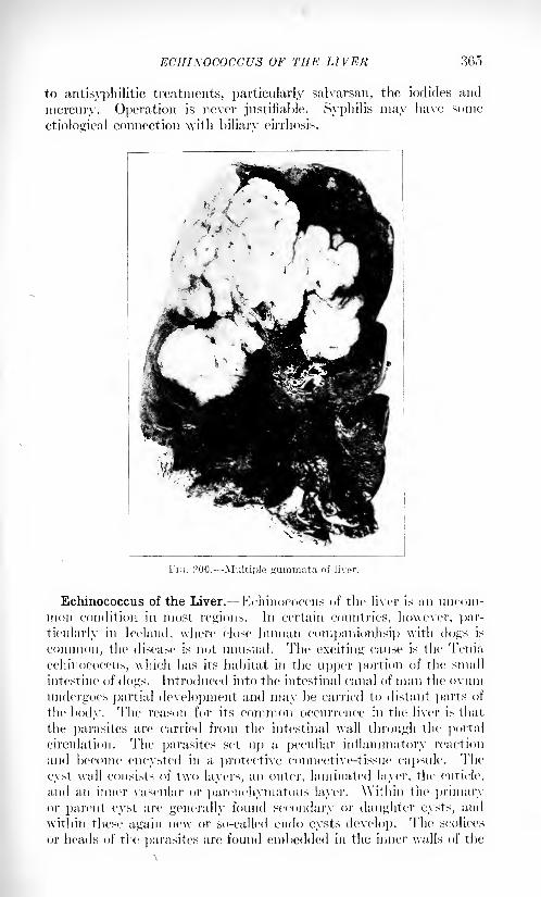
ECHINOCOCCUS OF THE LIVER 3(5;-)
to antisyphilitic treatments, particularly salvarsan, the iodides and
mercury. Operation is never justifiable. Syphilis may have some
etiolo;ical connection with biliarv cirrhosis.
. 200. -Multiple guinmata of liver.
Echinococcus of the Liver. Echinococcus of the liver is an uncom-mon condition in most regions. In certain countries, however, par-
ticularly in Iceland, where close human companionhsip with dogs is
common, the disease is not unusual. The exciting cause is the Tenia
echinococcus, which has its habitat in the upper portion of the small
intestine of dogs. Introduced into the intestinal canal of man the ovum
undergoes partial development and may be carried to distant parts of
the body. The reason for its common occurrence in the liver is that
the parasites are carried from the intestinal wall through the portal
circulation. The parasites set up a peculiar inflammatory reaction
and become encysted in a protective connective-tissue capsule. The
cyst wall consists of two layers, an outer, laminated layer, the cuticle,
and an inner vascular or parenchymatous layer. Within the primaryor parent cyst are generally found secondary or daughter cysts, and
within these again new or so-called endo cysts develop. The scoliees
or heads of the parasites are found embedded in the inner walls of the
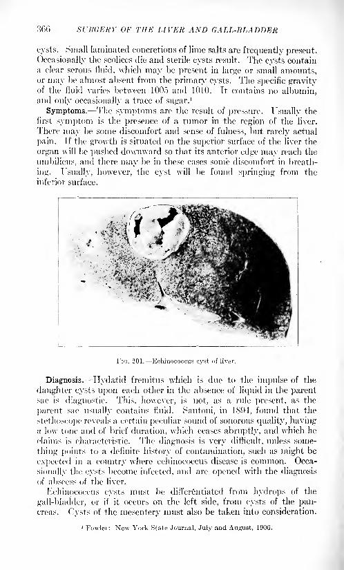
300 SURGERY OF THE LIVER AND GALL-BLADDER
cysts. Small laminated concretions of lime salts are frequently present.
Occasionally the scolices die and sterile cysts result. The cysts contain
a clear serous iluid, which may be present in large or small amounts,or may he almost absent from the primary cysts. The specific gravityof the fluid varies between 1005 and 1010. It contains no albumin,and only occasionally a trace of sugar.
1
Symptoms. The symptoms arc the result of pressure. Usually the
first symptom is the presence of a tumor in the region of the liver.
There may be some discomfort and sense of fulness, but rarely actual
pain. If the growth is situated on the superior surface of the liver the
organ will be pushed downward so that its anterior edge may reach the
umbilicus, and there may be in these cases some discomfort in breath-
ing. Usually, however, the cyst will be found springing from the
inferior surface.
Fir;. 201. Echinococcus cyst of liver.
Diagnosis. Hydatid fremitus which is due to the impulse of the
daughter cysts upon each other in the absence of liquid in the parentsac is diagnostic. This, however, is not, as a rule present, as the
parent sac- usually contains fluid. Santoni, in 1S94, found that the
stethoscope reveals a certain peculiar sound of sonorous quality, havinga low tone and of brief duration, which ceases abruptly, and which he
claims is characteristic. The diagnosis is very difficult, unless some-
thing points to a definite history of contamination, such as might be
expected in a country where echinococcus disease is common. Occa-
sionally the cysts become infected, and are opened with the diagnosisof abscess of the liver.
Echinococcus cysts must be differentiated from hydrops of the
gall-bladder, or if it occurs on the left side, from cysts of the pan-creas. Cysts of the mesentery must also be taken into consideration.
1 Fowler: New York State Journal, July and August, 1906.
TUBERCULOSIS OF THE LIVER 367
In all these instances an .r-ray taken after the regulation barium meal
and injection of the colon, will usually, by discriminating the ana-
tomical relations clear up the diagnosis. Ilydatid cyst may also
simulate carcinoma or syphilis of the liver.
Treatment.- The operations are either incision and drainage, or
incision and enucleation. Incision and drainage is a very simple
operation. The abdomen is opened, preferably by a transverse inci-
sion over the most prominent portion of the tumor, and the peritoneal
cavity entirely Availed off by gauze. An aspirating needle is intro-
duced and the fluid evacuated. The cavity is now explored with the
fingers and as much as possible of the daughter cyst and endocystsremoved. The edges of the cavity are united to the parietal peri-
toneum and the sac packed. This is called marsupialization. The
packing is continued until the cavity is obliterated by contraction.
The radical removal of the cyst, though far more dangerous, is prob-
ably the preferable operation. The abdominal incision should be
made large and the contents of the cyst removed, as in aspiration.
The parent cyst is then enucleated by gradual traction on its wall and
blunt dissection. These cases, as a rule, recover. Oliver 1 had a
case of this kind which recovered. If possible the cavity left in the
liver should be obliterated by deeply buried mattress sutures and a
drain inserted. The soft cigarette drains are preferable, as a hard-
rubber drain may induce erosion of large arterial or venous radicals,
with consequent severe or even fatal hemorrhage.In case the hydatid is situated on the upper surface of the liver
it is necessary to resect one or more ribs and incise the diaphragm.The pleural cavity should be walled off by packing and the cyst
removed a day or so later, when pleural adhesions have had a chance
to form. Or the pleural cavity may be obliterated by suturing both
layers of the pleura to the upper surface of the diaphragm, above the
site selected for the incision. It is necessary during operations for
echinococcus to carefully protect the wound against contamination bythe cyst contents, as hydatid infection of the wound surfaces mayoccur. It is also wise to thoroughly disinfect the wound margins with
iodin before completing the operation.Tuberculosis of the Liver. Tuberculosis of the liver is an uncommon
affection, except as part of a generalized tuberculosis where it is com-mon. In 470 tuberculous subjects which came to autopsy, Simmonds'2
found tuberculosis of the liver in 82 per cent. The liver was more
frequently found infected in children. There were only 2 cases of
solitary tuberculosis. Elleisen, in another series of autopsies, con-
firmed these observations. The great majority of these cases took
the form of miliary tuberculosis. The reason for the rarity of solitary
or conglomerate tuberculosis of the liver is partly due to the antiseptic
action of the bile and partly to the great vascularity of the liver.
It is thought by some observers that the miliary tuberculosis so fre-
1 Southern Med. Jour., Mardi, 1'Jll, pp. 22") 228.
-Zeutralbl. v. allg. Path. u. path. Anat., 1S9S, Hd. ix.
3r>$ SURGEltY OF THE LIVEK AND CALL-BLADDER
quently found in the liver is only a temporary infection, and that
during the course of a general tubercular infection several crops of
miliary tubercles niay form in the liver and may be destroyed.
Conglomerate tuberculosis of the liver may be solitary or multiple,or in some instances they may form abscesses. It probably beginsas an arterial infection or embolus, usually secondary To some focus
in another part of the body. The infection spreads in the interlobular
connective tissue, encroaching on but only later invading the biliary
passages. As in other forms of tuberculosis, caseation and later
breaking down and abscess formation results.
Lotheisen 1 collected 47 cases of conglomerate tubercle, 15 of which
came to operation. In only ?> cases, however, was the correct diagnosismade before operation. Jn many instances when a distinct tumorcan be felt the condition closely simulates carcinoma. Tuberculosis
of the liver may occur at any age. Barbier discovered a large con-
glomerate tubercle at autopsy in an infant, aged nine months. Teitze
reported 2 cases of diffuse tuberculosis of the liver occurring in children
of five years. Both cases were accompanied by ascites. In both
recovery followed the opening of the abdomen, the withdrawal of
the fluid and a typical Talma operation.A not uncommon sequel of tuberculosis of the liver is the develop-
ment of a large1 ab>cess which frequently presents in the subphrenic
space, or may even involve the pleural cavity. \Yhen this occurs
the abscess should be opened, drained and packed as in any other form
of liver abscess.
Diagnosis. The diagnosis is exceedingly difficult, particularlywhen the tuberculoma is situated in the interior of the liver. The
long-continued course of the disease and the evidence of a primaryfocus elsewhere are of course, suggestive. In the abscess cases, unless
secondary infection has occurred, the low fever curve and the absence
of a high leukocyte count may point to the condition.
The .-olitary tuberculoma must be differentiated from neoplasmsand giimmata. In the latter instance the \Vassermann is of great
importance. In the differentiation from carcinoma the low leuko-
cyte count is of suggestive value, as carcinoma of the liver is almost
invariably accompanied by a very high leukocytosis.
Prognosis.- '\ he prognosis is, of course, grave, as these conglomer-ate tuberculoma tend to be multiple. In case a tuberculous abscess
forms, the success of the treatment depends entirely on early evacua-
tion and thorough drainage. If this is done a large number of cases
can be saved. In case the abscess must be opened through the dia-
phragm, if adhesions between the parietal and visceral pleura have not
formed, the operation must be done in two sittings.
Treatment. In the abscess cases the treatment must not be too
radical. The abscess should be widely opened and thoroughly drained
and packed with iodoform gauxc, in addition to which the ordinary
1Jii-itr. /,. kliu. Cliir.. Ixxxi. 1.
CIRRHOSIS OF THE LIVER 369
climatic and supportive treatment of tuberculosis in general should
be carried out. Solitary conglomerate tuberculoma, if accessible,
should be resected. Ransohofl' 1 resected a solitary tuberculoma of
the liver under these conditions. The operation was not difficult, but
the patient succumbed to gastric hemorrhage a few days later, probablydue to a retrograde venous thrombosis.
Cirrhosis of the Liver. Cirrhosis of the liver is primarily a medical
disease. However, in certain forms surgery seems to have a definite
place. The hypertrophic cirrhosis (Hanot) with jaundice has been
treated in some instances by prolonged drainage of the gall-bladder.
In 2 personal cases some temporary benefit was noted. However,in both cases the final outcome was fatal. In a recent case in which
the gall-bladder was drained the liver was reduced to half its former
size, the patient regained his strength and has remained well for a
year. The atrophic cirrhosis (Laennec) accompanied by ascites has
for many years come under the realm of surgery, with, however,rather meager results. The usual cause of cirrhosis of the liver is
alcohol, although occasional cases occur in which an alcoholic history
is entirely absent. In these cases some history of prolonged infection
of one kind or another is usually present.
Pathology. The pathology of cirrhosis of the liver is a marked over-
growth of the interstitial connective tissue. It begins as scattered
areas of round-cell infiltration around the radicals of the portal vein.
In the beginning the liver is somewhat enlarged and hard. The sub-
sequent contraction of the connective tissue causes the tremendous
shrinking of the liver so characteristic of the disease. This stage is
accompanied by the compression and destruction of the liver tissue.
Hand in hand with this destruction of liver tissue goes a new forma-
tion of liver cells, which gives the peculiar mottled appearance of the
cut surface. The cut surface of a cirrhotic liver is of a granular
appearance, with small areas of liver tissue projecting above the cut
surfaces. In some cases the process is not equally distributed over
the entire extent of the liver, but may be more marked in one or
more parts. The scar formation results in a venous congestion of
the radicals of the portal vein, with a hyperemia of the mucosa of the
stomach and gastro-intestinal tract and a swelling of the spleen.
This enlargement of the spleen is invariable. This is accompanied bythe formation of ascites of a varying degree. The hyperemia of the
stomach and intestinal tract may cause severe and occasionally fatal
hemorrhages, although the latter are usually due to the rupture of an
esophageal varix. In some cases icterus is present.An attempt at collateral circulation always occurs: "(1) Between
the accessory portal veins in the falciform ligament and the diaphrag-matic para-umbilical and epigastric veins (the distended para-umbili-cal veins forming the caput Medusa); (2) the veins of Retzius and the
retroperitoneal veins; (:j) between the inferior mesenteric and the
VOL. ii 24
370 Xl'JtdKKY 01' Till': LIVER AM) CALI.-HLADDER
hemorrlioidal; and (4) between the gastric and esophageal veins."
(Piersol.)
Treatment. This compensatory anastomosis was undoubtedlywhat suggested the so-called Talma operation to both Talma 1 and
Drummond. Morrison, 2 who operated for Drummond, reported 2
cases in 1N90. Talma's first operation was made in 1S89, althoughhe did not publish it until 1898. The result of the Talma operation
though occasionally brilliant, is far from reliable. However, the con-
dition of a patient with cirrhosis in which the ascites rapidly reforms
after successive tappings is so hopeless that this operation should be
more frequently attempted. The operation is in itself easy to per-
form, and carries with it the removal of the abdominal effusion. It
is barely possible that the improvement of the 1
patient is partially due
to the increased nutrition of the liver through the vascular radicals,
carried in the newly formed adhesions.
The abdomen is opened under general or local anesthesia above
the umbilicus, to the right, of the middle line or by a transverse inci-
sion. The fluid is drained away until the peritoneum is dry. 'Flic
peritoneal surfaces of the liver, spleen and abdominal parietes are
roughened by gauze friction or scarification, and the great omeutum is
extensively sutured to the parietal peritoneum. The wound must be
most carefully and accurately sutured to prevent the effusion of aseitic
fluid, and subsequent infection or even evisceration.
Brant, of Louisville 1
, collected 1 14 operative cases, with the followingresult: 17, or 11 per cent., died as a result of the operation; 17, or
1 1 per cent., reported cured after three years; 04, or 44 per cent.
were improved; ~-\'2, or '22 per cent., were dead or unimproved.
Hopfner3 collected 700 cases in which the Talma operation was
done. lie believed that, in the hands of capable operators in large
clinics perhaps o() per cent, of these cases can be cured, by cure mean-
ing a permanent relief from ascites.
Out of 10.") cases collected by Greeno 1 the mortality was 29. .1 percent., and only 9 showed improvement after two years. Montprofit
(quoted by Moynihan) collected 224 cases, in 21o of which the results
were known. The mortality was 20 per cent. Improvement occurred
in 12 per cent, and recovery in .'>.'> per cent. It is questionable,
however, whether the latter can be given much credence. In a
recent case of the author's, in an enormous woman weighing oOO
pounds, this operation was attended by a brilliant result. It has
lasted well over a year, with the patient still under observation.
Naturally, with its meager successes, this operation has been exten-
sively modified.
Lan//' devised a new operation, which, however, has not been
adopted. His idea was to transplant the right testicle and spermatic
1 Burl. kliu. \\dinschr., September, 1 M)-' British Men. .Jour.. September IS, 189(5.
CIRRHOSIS OF THE LIVER 371
cord into the peritoneal cavity and enclose it in a bed of oraental
tissue. lie expected that an anastomosis would occur between the
veins of the pampiniform plexus and the omentum. lie also justified
his operation by the enormous amount of lymph channels in the sper-
matic cord. His first case was one of effusion, with tuberculous
peritonitis, in which the effusion did not recur, in the five years
during which the patient lived. At the postmortem there was seen
a tremendous imbibition of the spermatic cord and retroperitoneal
connective tissue, with serous effusion, a large increase in the si/.e of
vessels of the spermatic cord and an anastomosis between the spermaticveins and the branches of the vena cava. Lorentx 1
attempted this
operation in '2 cases, in which the patients were completely cured after
two years. This operation unquestionably seems worthy of further
tests.
Kumaris- further modifies the Talma operation in that he denudesthe renal pelvis, part of the diaphragm and fastens the omentum to the
denuded areas, in the hope that the adhesions and subsequent anas-
tomosis will be more extensive.
Aside from the Talma principles of operation two principles have
recently been advanced: (1) The direct communication between the
portal and systemic circulation by a von Eck fistula. This, however,is a very grave and dangerous operation and is rarely justified. Al-
though it has been attempted by Vidol, Lenoir, Martal and Ilosensteiu
there has been no case in which the operation has been successful.
Somewhat less dangerous is the establishment of an anastomosis be-
tween the superior mesenteric and the right spermatic vein, after the
method of Yillard Tabermeier.
liontte attempted to drain the peritoneum through the great
saphenous vein. lie severed the vein o cm. below its termination in the
vena cava, tied off the lower end and made an anastomosis between
the proximal end of the vein and the parietal peritoneum. The opera-
tion, however, was not successful, and a few months afterward there
was a complete obstruction of the vein. T. Morir 1 and Bernheim, of
Baltimore, also attempted this operation, with like results.
The third method, which seems as simple as the Talma operation,and perhaps as efficacious, consists in the drainage of the ascitic fluid
into the subcutaneous connective tissue. Several means for accom-
plishing this have been devised. A small incision in the peritoneum,
through which a small piece of omentum is drawn and sutured snb-
cutaneonsly, drainage of the peritoneum with silk threads, silver
tubes, glass tubes, rubber tubes, but permanent results have not been
achieved.
K. Schepelman1 has been using for this purpose sections of blood-
vessels which have been preserved in formalin. If possible pieces of
human veins, which have been removed at operation for varicose veins
are used. If these are not available, calf arteries seem equally effective,
1 Wuin. im-d. Wrhiibdir., 191-1. Ixiv, 1.
3 Deutsch. Zt.schr. f. f'hir., vol. cxiv.
372 si'h'dEin' OF Tin': LIVER A\D CALL-BLADDER
Under local anesthesia, to the right or to the left of the navel, a small
incision is made, the peritoneum opened and the effusion relieved. Oneend of the hardened artery is placed inside the peritoneal cavity andthe other end allowed to project into the subcutaneous connective
tissue and the wound closed without drainage. lie has had four cases
in which, however, the result does not seem to justify the operation.Before the operation the hardened artery is soaked in water and numer-ous side holes are cut in it. The operation is done on both sides.
The result of all these new operations for ascites do not seem to
justify the additional risks, as in no case does the result seem to equalthat accomplished through the old Talma operation or one of its simple
modifications, where the technic is simple, the death-rate low and the
promise of some good fair. The patients submitted to operation should
be carefully selected. Severe jaundice, cardiac, renal or pulmonarycomplications or severe emaciation must be regarded as absolute contra-
indications.
Abscess of the Liver. Abscess of the liver may be divided into three
general classes: (T) solitary abscess, which is usually a sequel of dysen-
tery; (2) multiple abscess of pyemic origin; (3) multiple abscesses
due to suppuration in the portal system. The most interesting, from a
solitary point of view, are the solitary or tropical abscesses. The
solitary abscesses may be divided again into two forms: Those due to
amebic dysentery and those due to some other form of intestinal
infection. The amebic abscesses are, of course, very rare, except in
tropical countries, where liver abscesses are a very common compli-cation of dysentery. l"]> to the past few years it was thought that the
amebic dysentery was the cause of most of these abscesses and that
the ameba could be isolated in a large number of the so-called amebic
dysenteries. 1 hiring the last few years they have isolated in dysenterya bacillus, which goes under the name of Bacillus shiga, which probablyforms an important etiological factor in tropical dysenteries.
M. Muhlman, 1 among 120 cases of amebic abscess, was only able to
find the ameba in 11 cases. A very interesting phenomenon is that
these abscesses are frequently absolutely sterile: for instance, in the
above 120 cases in only 7 cases were microorganisms other than the
ameba found. The abscess seems to be more of a necrosis with lique-
faction than true suppuration. The abscesses are nearly always
solitary, and are, as a rule, located in the right lobe. The typical
tropical abscess presents three different zones: the first /one a central
area of detritus, the second /one of softened liver tissue, with newlyformed liver cells, the third a /one of connective-tissue cells with
leukocytotic infiltration and formation of new capillaries. The
contents of the abscess is typical, looking very much like anchovy paste.
Some very interesting work in the etiology of amebic abscess was done
by Okinschewitz,2 ile injected the ameba; of dysentery into the
portal veins of various experimental animals without being able to
1 Xiedor's Beitriise, Ivii, o.jl.2 St. Petersburg, 1910.
ABSCESS OF THE LIVER 373
produce an abscess. Thinking that perhaps abscesses were due to the
absorption of toxins, he injected aniebic toxins into the portal cir-
culation and was able to produce definite areas of softening without,
however, true abscess formation.
Repeating his work, Muhlman, after damaging the liver with
injections of alcohol into the portal circulation, was then able to pro-duce real areas of liver abscess. Although the aineba is not so fre-
quently localized in liver abscesses as was formerly thought, there is
little question of the etiological connection between amebic dysenteryand liver abscess, as in most cases which come to the postmortem table;
distinct evidences of dysentery are found in the large intestine. Soli-
tary abscess may also occur in the course of other intestinal disease. It
is a rare complication of typhoid fever. The last case of this kind was
reported by A. E. Ilalstead. 1 In his case the typhoid bacillus was
isolated from the abscess in pure culture. In 1911 Ebits collected 30
cases of solitary abscess of the liver following typhoid. In 12 of these
the typhoid bacillus was isolated. In only 8, however, in pure culture.
These cases not infrequently develop some weeks after seeming con-
valescence from typhoid. Liver abscess is a not infrequent compli-cation of appendicitis. However, they tend, as a rule, to assume the
multiple form, and as such are not amenable to surgical intervention.
However, the subphrenic abscess, which closely simulates liver abscess
in its symptoms, is a very frequent complication of suppurative
appendicitis, particularly of the retrocecal variety. In fact, liver
abscess may follow any form of infection in the area of the portalcirculation.
Tropical liver abscess may reach an enormous size. Fasley2reports
2 cases in which the enlarged liver almost completely filled the ab-
dominal cavity. In one of these cases the lobe of the border of the
liver extended to the pubes. Both of these cases recovered after the
abscess was drained.
Symptoms. The chief symptoms of solitary abscess of the liver are
fever, pain, enlargement of the liver and symptoms of septic infection.
The patients are usually in a state of great weakness and extreme
emaciation. The fever runs a septic course, with wide intermission.
There is usually pain located in the right hypochondrium, often
extending toward the right shoulder. There is, of course, tenderness
over the involved area. Blood examination shows pronounced leuko-
cytosis with an increased proportion of the polymorphonuclear type.r
lhese amebic abscesses of the liver are occasionally complicated bybrain abscess. Among 384 hepatic abscesses treated by Kartulis,
3
in Egypt, he found 11 cases of brain abscess, or about 3 per cent.
Abscess of the brain as a complication of dysentery is invariably the
sequel of a liver abscess. The course of solitary hepatic abscess maybe either subacute or exceedingly chronic. In some cases reported the
history has run over a period of three or four years. In other cases
- Lancet, 1915, p. 107G.
:',74 SURGERY OF THE LIVER AXD CALL-BLADDER
tlic course is rapid, with rapid emaciation and debility. The abscess
may cure spontaneously by breaking through the chest wall in the
axillary line, or, what is still more uncommon, there may be pul-
monary involvement. That is, the abscess may rupture through the
diaphragm into the pleural cavity, causing a complicating einpyema.1'helin 1 describes a case of this kind in which the liver abscess rup-
tured through the diaphragm into the lung and was discharged throughthe bronchus, with complete recovery of the patient. Thompson'- has
found that different authors estimate the pulmonary complication of
hepatic abscess from .">.") per cent, to 40. S per cent., the latter, however.
seeming ridiculously high. Of course, recovery through this means is
exceedingly rare, as the patient usually succumbs to the pulmonarycomplication.
Abscess of the liver in infants, though uncommon, is not of exceeding
rarity. Pritchard and Turner 3 found a postmortem abscess the si/,e of
an orange in the right lobe in an infant of five weeks. The bacilli coli
and a Gram-positive of diplococcus were isolated.
Le Grande 4 was able to collect 112 cases of liver abscess in children;
11 were due to dysentery, 11) were traumatic, 9 of doubtful etiologyand 1 influenzal. The rest followed abdominal infection, particularly
appendicitis.A very unusual case of liver abscess was described by Rolliston. 1"'
After aspirating an empyema the child's condition grew worse, with a
fatal outcome. Postmortem showed that the aspirating needle had
been forced through the pleural cavity, diaphragm and into the liver,
causing an abscess, \vhich proved fatal. It is not impossible that this
accident occurs more frequently, without, as a rule, serious conse-
quences.The prevention of hepatic and snbphrenic abscess after retrocecal
appendicitis is perhaps the most important element of these cases.
This is best accomplished by long-continued drainage through the
(lank. The onset is very characteristic. The complication usually
occurs Two or three weeks after the original abscess has been opened,and when convalescence seems well established. A rise in temperaturewith increased leukocyte count and absence of infection around the
wound are always suspicious. Pain is very rarely a pronounced symp-tom. Percussion over the lower part of the chest reveals an area of
dulness extending upward to the scapula in a convex line, with the
absence of vocal fremitus and breath sounds. An .r-ray clinches the
diagnosis, showing the typical convex line. These cases, if operatedon early before the abscess has reached large si/e and has progressed
extensively into the subdiaphragmatic space, offer a fair prognosis.The multiple abscess of pylephlebitis occurring from appendicitis
or other forms of abdominal infections are invariably fatal. The course
is one of typical sepsis with remittent fever, frequent chills and pro-
; Bed. klin. AVrlmschr., 1914, li, 477. -Ann. Surjr., 1914, vol. lix.
: > I'rnc. Koynl Society of Mod., viii, 339.< An-h. dc rned. dcs cnfants, 1900, ix, 129. * British Me<l. Jour., 1913, p. 847.
ABSCESS OF THE LIVER 375
grossing emaciation and debility. Surgical treatment here has been
attempted, that is the opening of numerous foci of infection, but with-
out result. These cases should be treated by supportive methods and
stimulants. Autogenous vaccines should be used although the results
have not been encouraging.Edwin Beer in a case of this kind ligated the portal vein in an attempt
to shut off the source of infection. The operation was well borne, but
death ensued in a few days from sepsis. The idea, of course, is an
excellent one, and was based on the ligation of the pelvic veins in cases
of postpartum uterine infection. In the diagnosis of liver abscess of
all kinds the ,r-ray is important.
Solitary abscess ma}' follow direct extension of inflammation from
suppuration below the diaphragm. They may be secondary to peri-
nephretic abscess, to abscess of the gall-bladder or perforating gastric
ulcers. The most common abscesses of this kind are those following
retrocecal appendicitis. Here, however, the subphrenic abscess is far
more common than the hepatic. These abscesses are probably due to
a continuation of infection along the retroperitoneal lymph channels.
Treatment. Although a certain number of liver abscesses rupture
spontaneously, this result must never be awaited, as the majority
rupture upward through the space in the diaphragm, between the
ligaments of the liver uncovered by peritoneum, and so extend into the
pleural cavity. The best treatment for liver abscess is the open
surgical drainage, although many cases have been reported cured byMalison's method. This consists in the introduction of a large trocar,
preferably through the tenth intercostal space in the axillary line, or
wherever the abscess seems to point. A large drainage tube is inserted
through the trocar, the trocar withdrawn and the contents allowed to
flow into a bottle. This method, however, should not be used wherethere is opportunity for radical surgical intervention. Transperitoneal
aspiration for the location of these abscesses should never be under-
taken unless preparation for immediate operation has been made.Jacobson very strongly recommends the aspiration treatment. The
sheath of the aspirating needle is made of rubber tubing and is left
in the abscess cavity. This is connected with an aspirator throughwhich the pus is withdrawn whenever it reforms. After the emptyingof the pus the cavity is washed out with a mild solution of sulphate of
quinin, containing about 5 grains to the ounce.
Muhhnan's Operation As soon as a hepatic abscess is strongly
suspected and einetin injections have failed the liver is explored as
already described, and, if possible, under an anesthetic, so that this
can be thoroughly done. Failing a general anesthetic, morphin,gr. 3, and scopolomine gr. y^y, should be injected an hour before the
exploration and novocain used locally. In all cases the surgeon mustbe prepared to evacuate the pus at once, otherwise the contents of the
abscess, often under great tension, may escape into the peritoneumthrough the track of the needle. The skin is incised for three-quartersof an inch at the site of the puncture and the trocar and cannula four
:>7('i sriidERY <>i' Tin-: LIVER A\D UALL-HLADDEK
and a half inches long and one-third of an inch in diameter is plungedinto the abscess along the track of the exploring needle. As the trocar
is withdrawn and pus escapes the end of the cannula is closed with the
thumb until a rubber tube can be introduced. It is better for the pusnot to escape too rapidly, and it is necessary to prevent the entry of the
air which interferes with the siphonage. The rubber tube, which is
about nine inches long and has been boiled, is somewhat larger thanthe cannula, so that it fits snugly into the puncture in the liver. It is
necessary to stretch the tube before it can be introduced. This is done
by threading it over a metal rod with a hook at one end. Two side
holes are made near the inner end of the tube for better dranage.When the tube has been introduced well into the abscess, first the can-
nula and then the metal rod are withdrawn. About four inches of the
tube now project, and the end of it is at once attached to a glass tube
connected with another piece of tubing long enough to reach the bottomof a vessel on the floor near the bed. The end of the tube is weightedand kept under the surface of an antiseptic solution, otherwise proper
siphonage is impossible. The tube is sewed to the skin to prevent it
slipping out. I )rainage is continued as long as pus escapes.The favorite way of treating liver abscess is by direct operation.
That is, an incision is made over the upper portion of the abdomen,
through one of the incisions used in exploration of the gall-bladder.The best incision is probably a transverse incision through the upper
part of the right rectus. If adhesions are present the abscess may be
opened immediately. In case adhesions are not present the liver is
drawn well into the abdominal wound at the site of the incision andwell packed around with gauze. The following day, or forty-eighthours later, if the case is not urgent, the packing is removed andadhesions will be found to have formed. An aspirator is then thrust
into the abscess cavity, which is followed by wide opening of the abscess
either by knife or preferably by a thermocautery. The bleedingwhich is, as a rule, not profuse, can easily be checked by hot packs.If the bleeding is troublesome it may be checked by deep mattress
sutures of catgut. The abscess cavity should be drained with cigarette
drains and iodoform gauze, and if necessary washed out with a quininsolution after adhesions have formed.
The treatment of all amebic abscesses should be combined with the
administration of emetin, either by mouth or preferably hypoder-
mically, as this seems to have a specific action on the ameba.
Actinomycosis of the Liver.- Actinomycqsis of the liver is rare It
may spread to the liver from a primary lesion of a contiguous organ,for instance, the gall-bladder or from an actinomycosis of the pleura,
extending through the diaphragm. The disease produces numerousareas of broken-down caseous material and pus. Large areas of the
liver may become involved. As in all forms of actinomycosis there are
tremendous adhesions between the diseased area of the liver and the
neighboring organ. These cases are usually opened under the diagnosis
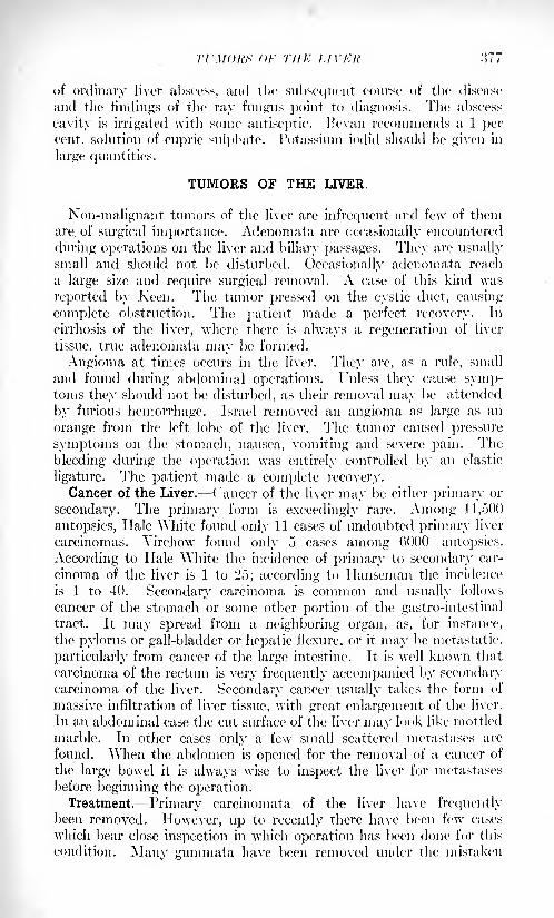
rr.MOKX OF THE LIYKR :>/7
of ordinary liver abscess, and the subsequent course of the disease
and the findings of the ray fungus point to diagnosis. The abscess
cavity is irrigated with some antiseptic. Bevan recommends a 1 percent, solution of cupric sulphate. Potassium iodid should be given in
large quantities.
TUMORS OF THE LIVER.
Non-malignant tumors of the liver are infrequent and few of themare. of surgical importance. Adenomata are occasionally encountered
during operations on the liver and biliary passages. They are usuallysmall and should not be disturbed. Occasionally adenomata reach
a large size and require surgical removal. A case of this kind was
reported by Keen. The tumor pressed on the cystic duct, causing
complete obstruction. The patient made a perfect recovery. In
cirrhosis of the liver, where there is always a regeneration of liver
tissue, true adenomata may be formed.
Angioma at times occurs in the liver. They are, as a rule, small
and found during abdominal operations. Unless they cause symp-toms they should not be disturbed, as their removal may be attended
by furious hemorrhage. Israel removed an angioma as large as an
orange from the left lobe of the liver. The tumor caused pressure
symptoms on the stomach, nausea, vomiting and severe pain. The
bleeding during the operation was entirely controlled by an elastic
ligature. The patient made a complete recovery.Cancer of the Liver.- -Cancer of the liver may be either primary or
secondary. The primary form is exceedingly rare. Among 11,500
autopsies, Hale White found only 11 cases of undoubted primary liver
carcinomas. Virchow found only 5 cases among GOOO autopsies.
According to Hale ^Yhite the incidence of primary to secondary car-
cinoma of the liver is 1 to 25; according to Ilanseman the incidence
is 1 to 40. Secondary carcinoma is common and usually follows
cancer of the stomach or some other portion of the gastro-intestinal
tract. It may spread from a neighboring organ, as, for instance,
the pylorus or gall-bladder or hepatic flexure, or it may be metastatic,
particularly from cancer of the large intestine. It is well known that
carcinoma of the rectum is very frequently accompanied by secondarycarcinoma of the liver. Secondary cancer usually takes the form of
massive infiltration of liver tissue, with great enlargement of the liver.
In an abdominal case the cut surface of the liver may look like mottled
marble. In other cases only a few small scattered metastases are
found. AVhen the abdomen is opened for the removal of a cancer of
the large bowel it is always wise to inspect the liver for metastases
before beginning the operation.Treatment. Primary carcinomata of the liver have frequently
been removed. However, up to recently there have been few cases
which bear close inspection in which operation has been done for this
condition. Many gummata have been removed under the mistaken
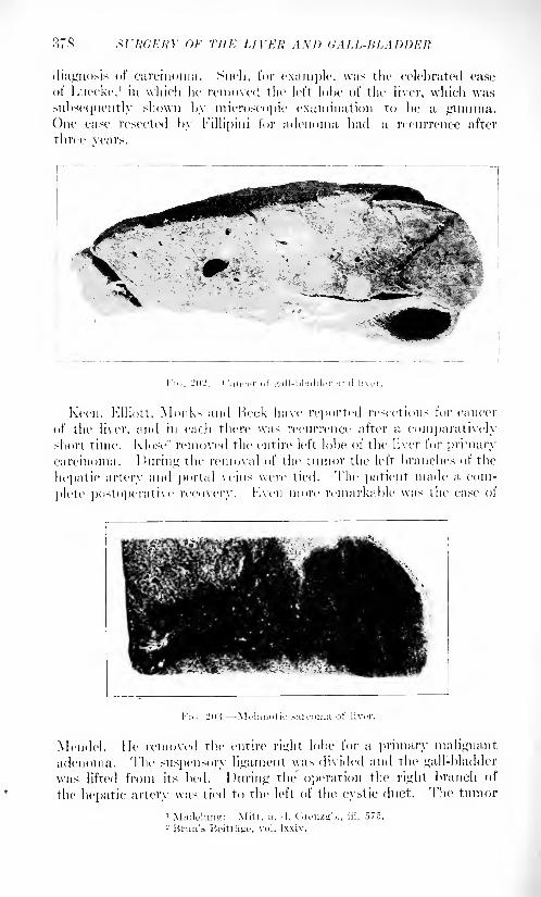
37S WRGERY OP THE LIVER AXD GALL-BLADDER
diagnosis of carcinoma. Such, for example, was the celebrated case
of Luecke, 1 in which he removed the left lobe of the liver, which was
subsequently shown by microscopic examination to be a gumma.One case resected by Fillipini for adenoma had a recurrence after
three Years.
Keen, Klliott, Monks and Beck have reported resections for cancer
of the liver, and in each there was recurrence after a comparatively>hort time. Klose- removed the entire left lobe of the liver for primarycarcinoma. During the removal of the tumor the left branches of the
hepatic artery and portal veins were tied. 'The patient made a com-
plete postoperative recovery. Kven more remarkable was the case of
Mendel, lie removed the entire right lobe for a primary malignantadenoma. The suspensory ligament was divided and the gall-bladder
was lifted from its bed. During the operation the right branch of
the hepatic artery was tied to the left of the cystic duct. The tumor
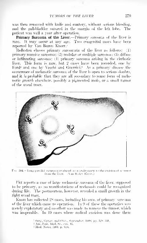
TUMORS OF THE LIVER 370
was then removed with knife and cautery, without serious bleeding,and the gall-bladder sutured in the margin of the left lobe. The
patient was well a year after operation.
Primary Sarcoma of the Liver. Primary sarcoma of the liver is
rare. It may occur at any age. Two congenital cases have been
reported by Van Buren Knott. 1
Rolleston classes primary sarcomata of the liver as follows: (1)
primary massive sarcoma; (2) nodular or multiple sarcoma; (3) diffuse
or infiltrating sarcoma; (4) primary sarcoma arising in the cirrhotic
liver. This form is rare, but 2 cases have been recorded, one byFord2 and one by Vecchi and Guerrini. 3 As a primary disease the
occurrence of melanotic sarcoma of the liver is open to serious doubts,
and it is probable that they are all secondary to some form of mela-
notic growth elsewhere, possibly a pigmented mole, or a small tumorof the uveal tract.
FIG. 204. Long parallel sutures introduced as a preliminary to the excision of a tumor
1'itt reports a case of large melanotic sarcoma of the liver, supposedto be primary, as no manifestations of melanosis could be recognized
during life. The postmortem, however, revealed a small growth in the
right uveal tract.
Knott has collected 2S cases, including his own, of primary sarcomaof the liver which came to operation. In i) of these the operation w;>s
merely exploratory and no effort was made to remove the tumor, which
was inoperable. In 19 cases where radical excision was done there
lynef. and Ohst., September. HUH, pp.ir Mod Si' cvv .I!-!

St'ttGERY OF THE LIVER AM) GALL-BLADDER
were <) deaths. In these cases of primary sarcoma, ,r-ray medications
and treatments with the Coolidge tubes may be beneficial.
Secondary Carcinoma of the Liver. -Symptoms. The symptoms of
secondary cancer of the liver are not distinctive, particularly whenthe primary disease lias not been recognized. There is usually pro-
gressive loss of weight, emaciation and in over half the cases, jaundice.If this is not prescrt there is, as a rule, a subicteric skin discoloration.
If the tumor grows rapidly there is usually severe pain from tension
on the capsule. In some instances of secondary carcinoma the pri-
mary disease may be so small as to have caused no symptoms, andthe first evidence of trouble may be the occurrence of symptomstraceable to the liver. The liver may be enormously enlarged, filling
a large part of the abdomen. These advanced cases are nearly always
accompanied by ascites, which may often somewhat obscure the
presence of the tumor. There is, of course, a loss of appetite, pro-
gressive loss of weight and marked secondary anemia.
The jaundice of cancer of the liver differs from that occurring with
obstruction of the common duct, as the stools are of normal color.
A very important sign in cancer of the liver, which is often overlooked,is the enormous leukocyte count, which may in some cases reach as
high as 75 to 100,000. The symptoms of primary carcinoma of the
liver develop more slowly than those of secondary carcinoma. Theabsence of a primary focus in some other part of the body is, of course,
suggestive of primary carcinoma.
Diagnosis. The diagnosis of a well-advanced case of cancer of the
liver is an easy matter, as the symptoms are distinctive. However,the diagnosis of the early case is somewhat difficult. The respiratorymovement of tumors of the liver is, of course, suggestive, also the
position of the tumor must be taken into account. Gumma is easily
differentiated by the Wassermann test. However, the possibility of
the coexistence of syphilis and cancer must not be overlooked. Cancer
of the liver may be mistaken for right hydronephrosis, tumors of the
stomach and hepatic flexure. These may, as a rule, be differentiated
by .r-ray examination.
Ilydatid disease develops far more slowly than cancer, is usuallyfound in early life and is not attended by the severe secondary symp-toms. The treatment is purely symptomatic. In some few instances
operation is warranted in primary cancers, as some cures have been
reported.Resection of the Liver. Three principal methods have been used to
control hemorrhage, which is the chief difficulty in resections of the
liver. These methods are: (1) elastic ligature; (2) thennocautery; (3)
suture; (4) combination of these.
The elastic ligature is only applicable in cases in which the tumoris more or less pedunculated. The abdomen is opened by a trans-
verse incision and the rest of the abdominal cavity is protected bypacking. The tumor mass is then encircled by a tight elastic ligature.
After two or three days, if the tumor has not completely sloughed off,
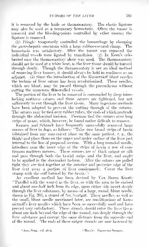
TUMORS OF THE LIVEIf 881
it is removed by the knife or thermocautery. The elastic ligature
may also be used as a temporary hemostatic. Often the tumor is
removed and the bleeding-points controlled by other means; the
ligature is removed.
(1) Pringle1
temporarily controlled the hemorrhage by clampingthe gastrohepatic omentum with a large rubber-covered clamp. Thehemostasis was satisfactory. After the tumor was removed the
individual vessels were ligated by transfixion. (2) In Keen's cele-
brated case the thermocautery alone was used. The thermocauteryshould not be used at a white heat, as the liver tissue should be burned
through slowly. Though the thermocautery is not an ideal methodof removing liver tumors, it should always be held in readiness as an
adjunct. (3) Since the introduction of the Ivusnetzioff blunt needles
the technic of liver suture has been revolutionized. These needles,
which are blunt, may be passed through the parenchyma without
cutting the numerous thin-walled vessels.
The portion of the liver to be removed is surrounded by deep inter-
locking mattress sutures and these sutures are tightened, but not
sufficiently to cut through the liver tissue. Many ingenious methodshave been adopted to prevent the cutting through of the sutures.
The sutures may be tied over rubber tubes, the ends of which protrude
through the abdominal incision. Freeman tied the sutures over long
strips of gauze, which, however, he found rather difficult to remove.
Kornew and Schaack have frequently successfully removed largemasses of liver in dogs, as follows: "Take two broad strips of fascia
(obtained from any convenient place on the same patient, e. g., the
thigh) and place them on the upper and under surface of the liver justinternal to the line of proposed section. "With a long rounded needle,
introduce near the inner edge of the strips of fascia a row of con-
tinuous mattress sutures. These sutures are of thick catgut or silk
and pass through both the fascial strips and the liver, and oughtto be applied in the shoemaker fashion. After the sutures are pulled
tight they are tied together at the anterior and posterior edge of the
liver (cut away a portion of liver condemned). Cover the liver
stump with the cuff formed by the fascia." 2
An excellent method has been devised by Van Buren Knott:
"Parallel with the wound in the liver, or with the area to be excised
and about one-half inch from its edge, upon either side insert deeply
through the liver substance, by means of a large, round, blunt needle,
shown in Fig. 205, a strand of No. !> catgut. These needles, as well as
the small, blunt needle mentioned later, are modifications of Kous-nietzofTs liver needles which have been so successfully used and have
proved very satisfactory. These strands should enter the liver tissue
about one inch beyond the edge of the wound, run deeply through the
liver substance and emerge the same distance from the opposite end
of the wound. The ends of these catgut strands are now fastened by
'Ann. Surg., vol. xlvii. 2 Binnie: Operative Surtiery.
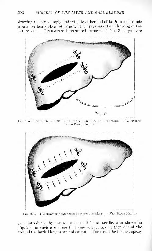
3S2 Sl'RGERY OF THE LIVER AXD GALL-BLADDER
drawing them up snugly and tying to either end of both small strands
a Mnall ordinary skein of catgut, \vhich prevents the indrawing of the
suture ends. Transverse interrupted sutures of No. o catgut are
ic sunken output strands in the ti>sue parallel to the wound to bo sutured
Fn;. L'Oii. The transverse interru]>ted sutures introduced. (Van Huron Knott.)
no\v introduced by means of a small blunt needle, also shown in
Fig. L; f'<'i, in such a manner that they engage upon either side of the
wound the buried long strand of catgut. These may be tied as rapidly
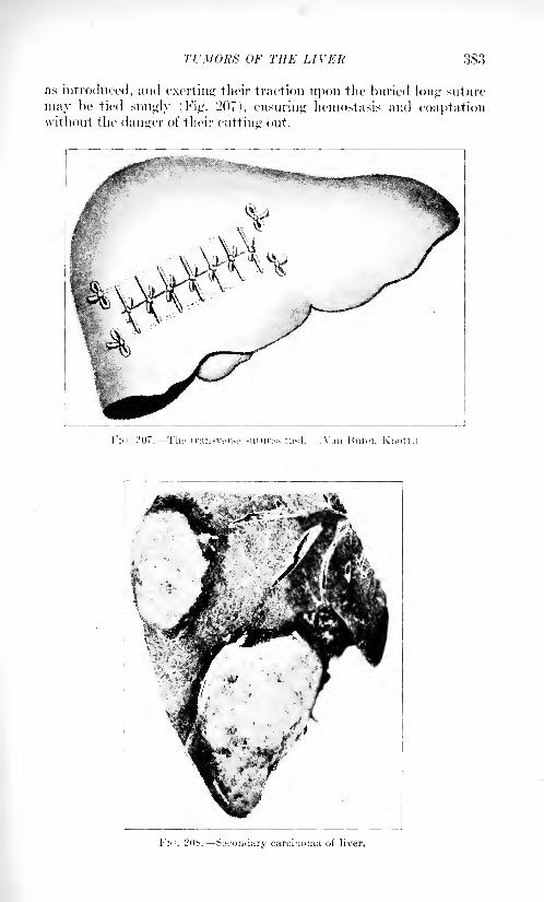
TUMORS OF THE LIVER .383
as introduced, ;md exerting their traction upon the buried long suture
may be tied snugly (Fig. 207), ensuring hemostasis and coaptationwithout the danger of their cutting out.
Fir;. 207. The transverse sutures tied. (Van Bureii Knott.)
Fx,. L'OS. Secondary carcinoma of liver.
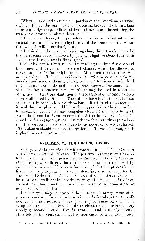
384 Sl'RGKRY OF THE LIVEN A.\D GALL-HLADDER
""When it is desired to remove a portion of the liver tissue carryingwith it a tumor, this may be done by excising between the buried longsutures a wedge-shaped ellipse of liver substance and introducing the
transverse sutures as above described.
"Hemorrhage during this procedure may be controlled either bymanual pressure or by elastic ligature until the transverse sutures are
tied, when it will immediately cease.
"If desired any large veins presenting along the cut surface may be
tied, as recommended by Keen, by placing a ligature about them with
a small needle carrying the fine catgut."Kocher lias excised liver tumors by crushing the liver tissue around
the tumor with large rubber-covered clamps, which he allowed to
remain in place for forty-eight hours. After their removal there wasno hemorrhage. If this method is used it is wise to loosen the clampsone day and remove them the next, so as not to disturb fresh blood
clots. In addition to the methods described above the ordinary meansof controlling parenchymatic hemorrhage may be used in resections
of the liver. The transplantation of a free strip of omentum has been
successfully used by Stucke. The authors have found the applicationof a free strip of muscle very efficacious. If either of these methodsis used the transplant should be held in apposition to the raw surface
by packing. Hot water and coagulen (Kocher) may also be used.
After the tumor has been removed the defect in the liver should be
closed by deep catgut sutures. In order to facilitate this approxima-tion the portion removed should, so far as possible, be wedge shaped.The abdomen should be closed except for a soft cigarette drain, whichis placed over the suture line.
ANEURYSM OF THE HEPATIC ARTERY.
Aneurysm of the hepatic artery is a rare condition. In 1906 Gruenert
was able to collect only 3G cases. The patients were mostly males over
forty years of age. A large majority of the cases in Gruenert's 1 series
(73 per cent.) were directly due to the invasion of the arterial wall byan infectious process, either secondary to an infectious process in the
liver or to a septicopyemia. A very interesting case was reported byBit-hart and Schuman.2 The aneurysm was directly attributable to the
invasion of the walls of the hepatic artery by a tuberculoina of the liver.
In another of their cases there was an infectious process, secondary to an
osteomyelitis of the tibia.
The aneurysm may be located either in the main artery or one of its
primary branches. In some instances it may be intrahepatic. Syphilisand general arteriosclerosis may play a predominating role. The
symptoms are more or less definite in character and resemble very
closely gall-stone disease. Pain is invariable and is usually intense.
It is felt in the epigastrium and is frequently of a colicky nature,
1 Deutsche Zeitschr. f. Cliir., vol. Ixxi. - Deutsche. Arch. f. Klin., 90.
AXATOMY OF THE GALL-BLADDER 385
closely resembling gall-stone colic. There may he, in addition, a sen-
sation of fulness in the epigastrium and at times a feeling of throbbing.Jaundice is present in GO per cent, of all cases. The jaundice is due
to direct pressure on the hepatic or common duct, and is progressive,intense and unremitting, more like the jaundice of carcinoma than
that of common duct stone. Hemorrhage is common, appearing from
the stomach or bowel. It follows, as a rule, the erosion of the aneurysminto the common duct, the gall-bladder or the duodenum. This hemor-
rhage may prove fatal shortly after its first appearance, A pulsatingtumor can frequently be felt in the epigastrium. Despite these sug-
gestive symptoms the diagnosis is rarely made. The reason for this
is the rarity of the condition and the fact that it is riot borne in mind.
G. A. Friedman 1 made a diagnosis of hepatic aneurysm before oper-
ation. The symptoms were typical. At operation (ierster found the
hepatic artery dilated, tortuous and elongated. Aneurysm s of the
hepatic artery must be differentiated from tumors and cysts of the
liver, gumma, gall-stones, cancer and duodenal ulcers.
Treatment. In those cases following or complicating a severe sepsis
nothing can be done. Up to 11)03 there were only 3 cases in which
operation was done for this condition. All of them resulted fatally.
In 11)03 the first successful case was operated on by Kehr. The diag-
nosis of gall-stones was made. Incision revealed an aneurysm of the
right branch of the hepatic artery, which had ruptured into the gall-
bladder, causing distention of the gall-bladder, jaundice and liema-
temesis. The aneurysm was the si/e of a hen's egg. The main branch
of the hepatic artery was divided between two ligatures and the sac
was opened and packed. The patient made an uneventful recovery.
This is probably the only case in which successful operation was done
for tliis condition.
THE GALL-BLADDER.
ANATOMY OF THE GALL BLADDER.
The gall-bladder, pyriform in shape, surrounded below and on each
side by peritoneum, is closely held in a groove, provided for it by loose
connective tissue in relation with the under surface of the liver. In
4 per cent, of subjects it is entirely surrounded by peritoneum and
may have a mesentery of its own. In these cases the gall-bladder maybe bifid, or a septum more or less complete extends from the base
toward the apex. In a few instances two gall-bladders with two cystic
ducts are found. The fnndus of the gall-bladder, on a lower le-vel than
its apex, when we are in the erect posture, is in relation with the
abdominal wall, opposite the tip of the eighth or ninth costal cartilage.
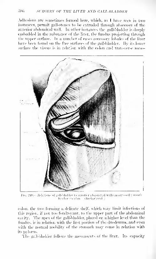
386 srh'dERY OF THE LIVER AXD CALL-BLADDER
Adhesions are sometimes formed here, which, as T have seen in twoinstances, permit gall-stones to be extruded through abscesses of the
anterior abdominal wall. In other instances the gall-bladder is deeplyembedded in the substance of the liver, the fundus projecting throughthe upper surface. In a number of cases accessory lobules of the liver
have been found on the free surfaces of the gall-bladder. By its lower
surface the viscus is in relation with the colon and transverse meso-
,. 20!).- -Kelulions of frall-bladder to anterior abdominal walls as exposed
colon, the two forming a delicate shelf, which may limit infections of
this region, if not too foudroyant, to the upper part of the abdominal
cavity. 'Flic apex of the gall-bladder, placed on a higher level than the
fundus. is in relation with the first portion of the duodenum, and even
with the normal mobility of the stomach may come in relation with
its pylorus.The gall-bladder follows the movements of the liver. Its capacity

ANATOMY OF THE GALL-BLADDER 387
varies from one to two ounces, but may by disease be enlarged until it
has been mistaken for a hydronephrosis or even an ovarian tumor.
Partly covered by peritoneum the gall-bladder has a fibrous wall
with considerable admixture of muscle fiber, and is lined by a reddish
mucosa during life, but this becomes bile-stained soon after death.
Within the mucosa are numerous glands which secrete a clear viscid
fluid which, when it accumulates in large quantities, after long con-
tinued obstruction of the cystic duct, was known to Hippocrates as
"white bile.'5
In rare cases this has been found to result from obstruc-
tion in the lymphatics at the hilum.
The neck of the gall-bladder, on a higher level than the fundus,
makes a sharp turn upward and to the left and a recurve downward to
the beginning of the cystic duct. In the normal condition these curves
are permanent, but in distention may be somewhat straightened out.
These curves, together with certain spiral elevations of the mucosa,have the function of a valve (Heister), above which stones are liable to
lodge and assume a saddle form when fixed here for any length of time.
The blood supply of the gall-bladder comes chiefly from the cystic
artery and in slight measure from the liver bed in which it is placed.
The cystic artery derived from the right branch of the hepatic runs
downward and forward along the cystic duct and usually behind it,
and divides into two branches, of which the left is the larger. Its close
relation to the cystic duct will explain the occlusion of the artery byan obstructing stone in the duct and account for gangrene of the gall-
bladder when an infection already exists. In rare instances the main
artery of the gall-bladder comes from the pancreatico-duodenal. It
is then likely to be wounded in operations on the cystic or commonducts.
The cystic duct, about one and a half inches in length, runs down-ward and to the left, near the edge of and between the layers of the
lesser omentum, where it joins the common hepatic duct to form the
common bile duct. The angle of union is very oblique, the hepaticduct forming a nearly direct continuation with the common duct.
Therefore, in distention of the common duct the hepatic duct is more
likely to be dilated and by lateral pressure occludes the cystic duct.
This, therefore, does not participate in distention of the common bile
duct. Parallelism of the cystic and hepatic ducts accounts for injuries
and postoperative stricture unless great care be taken during opera-tions. The cystic duct is the narrowest of the bile ducts.
The hepatic duct, formed by the union of the larger right and the
smaller left branch, is about two inches in length, and widens, as it
descends to the right and in front of the portal vein, to become contin-
uous with the common bile duct. The latter, a little over three inches
in length, empties through a papilla in the lower part of the perpendic-ular portion of the duodenum. This papilla is at the upper end of a
little perpendicular ledge and is under cover of the first transverse
valve of the duodenum. As a rule the pancreatic duct empties into
the duodenum through this papilla.
38S SURGERY OF THE LIVER AND GALL-BLADDER
In its course the common duct may be divided into three portions,the relations of which are of the very greatest importance. The supra-duodenal portion extends from the confluence of the cystic and hepaticducts to the upper portion of the duodenum. Tt is contained within the
free edge of the lesser omentum, the hepatic artery lying to the left
and the portal vein behind. In the peritoneal duplicature are also
contained a number of lymph nodes, the uppermost of which is situ-
ated in the hilum of the liver, and is known as the gland of Ilartman.
This is frequently enlarged in infections of the bile ducts, and mayreadily be mistaken for a neoplasm. In this part of its course the
common duct can easily be palpated between the thumb and forefingerof the left hand when the forefinger is introduced into the foramen of
\Vinslow, the back of the operator being turned to the edge of the
table. In old-standing cases of cholelithiasis this method is often
impracticable by reason of adhesions.
The second portion of the common duct, usually a little over an
inch in length, lies behind the duodenum, between it and the head of
the pancreas, or in a groove or within a tunnel of the latter. In 55 out
of 5s cases Hunger1 found the common bile duct completely surrounded
by pancreatic tissue. The division of it would be necessary before the
duct could be reached by any operative procedure. For direct attack
this part of the duct is the least accessible.
The third or interstitial part of the duct runs obliquely through the
wall of the duodenum for a distance of about three-quarters of an inch.
Near its termination there is a slight widening, the diverticulum of
Yater, in which stones are liable to lodgment and growth, and from
which, in cast's of spontaneous cure, they may ulcerate through the
mucosa. The entrance of the pancreatic into the terminus of the com-
mon duct accounts for the not infrequent complication of acute or
chronic inflammation of the pancreas as a sequel of primary infection
in the biliary ways. The diameter of the common bile duct, except for
the little ampulla of Yater, becomes smaller from above downward
and is narrowest at the papilla. In its widest part it measures about
one-quarter of an inch.
Surrounding the outlet of the common duct is a circular muscle, the
sphincter of Oddi, and the longitudinal fibers within the duct wall are
continuous with those of the gall-bladder.
Function. The function of the gall-bladder is far from final solution.
That it is not essential is evident from studies of comparative anatomy,
and from the fact that it is not missed after removal for or obliteration
from disease. As can be seen in the larger fowls, it possesses a rhythmic
contraction. Okada2 found that during digestion spontaneous contrac-
tions occur about two to five times per minute, the greater increase
being during the height of digestion. On the other hand he has seen no
contraction after an hour or more of observation.
Although the capacity of the gall-bladder is limited to one or two
! Boiir. /.. klin. f'hir., vol. xxxix. 2 Jour. Physiol., 1915, i, 42.
CONGENITAL ANOMALIES OF THE GALL-liLADDElt 389
ounces it probably acts as a storehouse for bile during the intervals of
digestion, in order that a certain quantity may be poured into the
duodenum as soon as this begins to receive the first chyme dischargefrom the stomach. In the interval of digestion the pressure in the ducts
is unequal to the task of overcoming the sphincter and bile accumulates
in the gall-bladder. This is evidenced by the bile flow in drainage cases.
It is greater and most annoying during the night. By a late feedingthe excessive night drainage can be evaded.
That some storage function seems to be desirable in the human beingis proved by the fact that secondary gall-bladders, though of small size,
not infrequently form after the gall-bladder has been removed. "While
in the gall-bladder the bile becomes darker in color and thickened, its
solid matter increases from 1 to 300 per cent.
The gall and bile ducts are supplied with nerves from the hepatic
plexus, into which pass filaments from the pneumogastric.The pain in the right shoulder, so often complained of in gall-stone
colic and diseases of the liver, is due to the partial origin of the phrenicnerve from the fourth cervical, which also sends branches to the
shoulder. A pain in the region of the scapula in lesions of the gall-
bladder is probably a reflex, and can be accounted for through the
irritation of the intercostal filaments to the diaphragm and from which
the pain is reflected to the posterior dorsal cutaneous.
CONGENITAL ANOMALIES OF THE GALL BLADDER.
Until the surgical procedures on the gall-bladder and ducts becameof daily occurrence the congenital anomalies of the gall-bladder were
only of academic interest. Today it is essential that every surgeonshould be fairly familiar with the congenital anomalies, man}' of which
may be encountered during an operation. Although long recognizedas occurring from time to time, Kehr1 was the first to call attention
to this subject. More recently Schachner has collected a number of
more recent cases, including one of his own. He has shown that
anomalies in this region follow the rule obtaining elsewhere, namely,in not occurring singly.
Any anomaly of the gall-bladder ma}' have attending it one of the
peritoneal coat, of the septum or of an unusual blood supply. The
gall-bladder may be double or bilobed or simply have a diverticulum.
In its location it may be anomalous in being intrahepatic, left-sided or
entirely floating.
Of particular importance is the intrahepatic gall-bladder, which maybe partly or entirely embedded in the liver substance, and so surrounded
that it can only be found with difficulty if at all. A case is recorded byLemon, in which the fundus alone projected and in which gall-stones
were found.
Deve 2 in the study of 120 livers of infants found 3 in which the
1 Minn-hen, med. Wchnschr., 1903. 2 Bull, do la Sue. Anat., KMKJ.
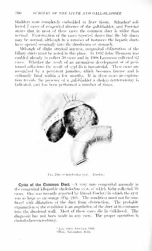
SURGERY OF THE LIVER AXD GALL-BLADDER
bladders were completely embedded in liver tissue. Schacher 1 col-
lected 7 cases of congenital absence of the gall-bladder, and Foerster
states that in most of these cases the common duct is wider than
normal. Examination of the cases reported shows that the bile ducts
may be normal, although in a number of instances the hepatic ducts
have opened seemingly into the duodenum or stomach.
Although of slight surgical interest, congenital obliteration of the
biliary ducts must be noted in this place. In IS'.L* John Thomson wasenabled already to collect .">() cases and in 1!)OS Lavenson collected (>:2
cases, \\hether the result of an anomalous development or of peri-
toneal adhesions the result of syphilis is immaterial. These cases are
recogni/.ed by a persistent jaundice, which becomes intense and is
ordinarily fatal within a few months. If in these cases an explora-tion reveals the presence of a gall-bladder a cholecystenterostomy is
indicated, and has been performed a number of times.
FIG. 210. Choledochus cyst. (Fowler.)
Cysts of the Common Duct. -A very rare congenital anomaly is
the congenital idiopathic choledochus cyst, of which Kehr collected 11)
cases. One was recently reported by Kussel Fowler,- in which the cyst
\vas as large as an orange (Fig. -]()). The condition must not be con-
fused with dilatation of the duct from obstruction. The probable
explanation of the condition is an angulation of the duct at its entrance
into the duodenal wall. Most of these cases die in childhood. The
diagnosis has not been made in any case. The proper operation is
choledocho-enterostomy.
1 Ann. Sur-:., October. 19 Hi.
2Ibid., November, 1910.
WOUNDS AND PERFORATIONS OF THE GALL-BLADDER 391
WOUNDS AND PERFORATIONS OF THE GALL BLADDER.
Rupture of a distended gall-bladder lias been occasionally encounteredas the result of direct violence, such as being caught between bumpersor being run over. The abdomen has usually presented injuries of
which the ruptured gall-bladder was the least important. In the same
way perforating wounds of the gall-bladder have been recorded as
complications of other visceral injuries. Direct violence exerted upona diseased gall-bladder has not infrequently caused its rupture and in
many of the cases stone was found. McLaren reports such a case in
which the tear readily admitted a finger. In another case reported byus an acute gangrene of the gall-bladder followed an injury sustained
two weeks before, but had not gone on to perforation.Most perforations of the gall-bladder and of the bile ducts are the
result of diseased conditions, which cause either great distention, with
thinning of the gall-bladder wall, or have been associated with the
perforation of stone. In a few instances rupture has occurred duringan attack of typhoid fever in which the typhoid condition of the patient
precluded the making of a diagnosis. In a case reported by Mason 1 the
greatly distended gall-bladder was drained in the second week of the
disease. The presence of bile-stained fluid on the outside spoke for a
perforation. A culture showed a pure typhoid infection. Erdman andKeen2 collected 34 cases of primary typhoidal perforation, of which 7
were operated on, with 4 recoveries. The 27 not operated upon died.
Symptoms. Perforation of the gall-bladder and bile ducts usually
develops slowly, the diseased process being ushered in with a peri-
cystitis, due to ulceration or even localized gangrene of the bladder
wall. From the statistics of llobson, Mayo and Ochsner I may infer
that in about 0.5 per cent, of gall-stone cases perforation occurs either
acutely or with the formation of extracystic abscesses. As in perfora-tive appendicitis, there are usually premonitory symptoms, indicative
of something wrong with the biliary ways. When time has been giventhe perforation leads to abscess formation without the bladder wall,
therefore not infrequently a stone is found outside the biliary tract.
In other cases, however, the rupture takes place without a warning
signal, and at once gives to the patient the appearance of one with a
grave abdominal disaster.
The symptoms are most likely to be severe when the rupture occurs
in one of the bile ducts. When the rupture occurs in a diseased gall-
bladder the fact that this is often associated with occlusion of the
cystic duct, only a relatively small amount of bile, pure or mixed with
pus, escapes into the peritoneal cavity, which probably has already been
shut off by adhesions, as already referred to. When, on the other hand,the rupture occurs in one of the ducts, usually the common, the out-
pour of bile is very profuse and continuous. Owing to the absence of
adhesions it is more likely to be poured into the general peritoneal
2 Ann. Surg., 1903.
;>i)2 SURGERY OF THE LIVER AND GALL-BLADDER
cavity. As a sign of this, I have had an experience in two instances of
calling attention to a localized jaundice strictly limited to the umbilicus
and the skin immediately surrounding it. When present in suddenly
arising grave abdominal conditions, this sign I believe to be pathog-nomonic of rupture of the gall-bladder or one of the larger ducts, with
free bile in the abdominal cavity. The localized jaundice is probably
altogether due to imbibition through the thinnest part of the abdominal
wall, but it is possible that, owing to the relation of the round ligamentto the transverse fissure, there is a retrograde flow of bile through the
former toward the navel, just as the caput medusa is produced in
cirrhosis.
Lewerenz 1 was enabled to collect GO cases from the literature.
Twenty-three cases were doubtless of the gall-bladder and in 8 cases
there was intrahepatic rupture of the large bile ducts. The commonduct was ruptured 9 times, the hepatic duct 6 times, whereas the cystic
duct was torn in only one. In the remaining cases the exact nature
of the tear is uncertain.
The pathological changes which follow rupture of the biliary passages
depend upon the condition of the bile. If this contains pathogenic
organisms a general septic peritonitis or a localized peritonitis tendingto abscess formation will follow. If the bile is sterile or nearly so a
subacute peritonitis with a plastic exudate occurs, as a result of which
encapsulation of the escaped bile takes place. Uhde 2
reports an autopsyin which twelve years after a ruptured gall-bladder, two cysts, contain-
ing nearly pure bile, were found.
When the patient has recovered from shock, icterus is almost sure
to develop in about Go per cent, of the cases, which is in contrast with
the jaundice following rupture of the liver, of which about 5 per cent.
show jaundice.The other symptoms attending perforation of the bile passages
depend largely on the condition of the effused bile. When this harbors
virulent organisms a more or less diffuse and acute peritonitis results
although it rarely has the rapidly fatal ending seen after perforation'
of the appendix. When by reason of long retention in the gall-bladder
or from their initial lesser virulence the organisms pass through a
perforation, there is a marked tendency to walling in of the infection
and abscess formation.
Prognosis. The prognosis of rupture of the biliary ways depends on
the condition of the bile and upon the place of rupture. Tears in the
common duct, and particularly in the hepatic duct, are more fatal
than those of the gall-bladder, the mortality being 34 per cent., and 20
per cent, respectively.
Treatment. In the early cases repeated tapping was followed by
gratifying results, but more recently it is certain that the proper
procedure in all cases in which a rupture is suspected is an exploratory
laparotomy. If the rupture is in an accessible part of the gall-bladder,
1 Arch. f. klin. Chir., Ixxi, 117.2 Langenbeck's Archiv, xxviii, 271.
SURGICAL DISEASES OF THE GALL-BLADDER 393
partial closure with drainage is the normal procedure. \Mien this is
unfeasible, or if the gall-bladder is diseased, as it often is, its extirpa-
tion is indicated.
In tears of the common duct the difficulties grow, particularly if
the tear is in the retro- or intraduodenal part of the duct, as in one of
the cases above referred to. If the rupture is not complete, drainagewill suffice. If the rupture is complete a permanent biliary fistula
will probably result which may terminate in death after many monthsunless relieved. In such cases, as has been suggested by Terrier and
Auvray, a cholecystenterostomy may have to be done.
SURGICAL DISEASES OF THE GALL-BLADDER.
Cholecystitis. Inflammation of the gall-bladder and bile ducts is
most protean in its manifestation. Our knowledge of the various
pathological changes to be found has been greatly increased by our
study through operations on the living subject. It is proper to
describe various forms of inflammation of these parts, with the distinct
understanding that they are in large measure interchangeable, andthat one form originally involving only the mucosa may at any time
become destructive of the gall-bladder, and that per contra, even the
severest types may by resolution disappear entirely or be changedinto one involving the mucosa only. In the same way an acute
process may here, as elsewhere, terminate in a chronic lesion, and this
again by renewed infection or from some unknown cause may flame
up into a rapidly destructive acute gangrenous condition.
Acute Catarrhal Cholecystitis. It is exceedingly likely that this is
a very common affection, consequent upon an inflammation or irri-
tation of the duodenum, as from sDme indiscretion in diet or from the
excessive use of alcohol. With the spelling of the papilla its lumenbecomes obstructed and there is a distention of the gall-bladder, with
jaundice, painless, as a rule, as the chief symptom.Since few opportunities of studying the condition in the living
have occurred the condition of the gall-bladder and biliary ways is
largely a matter of conjecture. Eppinger,1 in 1 case, fatal on the
eighth day from accident, found a hyperplasia of the lymphoid tissue
of the mucosa in the wall of the intestine around the common duct, and
suggests that since this is analogous to the tonsil, believes that the
disease may be analogous to tonsilljtis. "When at operation few
changes may be apparent in the gall-bladder itself; an enlargementof the regional lymphatics is corroborative of the diagnosis.
When the bile in these cases is retained in the gall-bladder for anylength of time and the case comes to operation there is usually found
the well-known hard fine biliary sand, which consists largely of bile
pigments. Most of it is contained in the apex of the bladder, but
not infrequently it is closely adherent to the wall and may form nuclei
of stones.1 Klin. Wrlmsrhr., xxi, 4X0.
394 SURGERY OF THE LIVER AND GALL-BLADDER
Suppurative cholecystitis, or, us it is often called, empyemu of
the gall-bladder, results from the greater virulence of the infection
or from an inability of the contents of the "'all-bladder to escape. It
is most often associated with stones, but may occasionally be encoun-tered without them. This is particularly true of the empyema of
the gall-bladder which develops during the course of typhoid fever.
If the disease occurs in a gall-bladder thereunto normal, the gall-
bladder is found greatly distended, with walls softened, edematous,
congested, and, as a rule, of a darkish color, shaded in severest cases
to the black of gangrene. The inucosa is covered with a fibrinous,
a purulent or sometimes a bloody exndate. AVhen stones are found,liberations varying in extent are not uncommon. The ulcer usuallyis due to pressure from the stone. In rare cases the ulcer is like the
round ulcer of the stomach, and like this is probably of einbolic origin.
Owing to the edematous condition of the inucosa the cystic duct is
almost invariably occluded. If from any reason the process developsin a gall-bladder which has harbored stone for a long time, and has
therefore become thickened in its walls, the distention of the gall-
bladder may be very much less; indeed, the gall-bladder may be
shrunken far below its normal sixe. In these cases of chronic empyemait is common to find pericystic adhesions so dense that the finding of
the gall-bladder may cause embarrassment even to a skilled operator.The acuter the attack the greater is the tendency to distention of the
viscus. When the gall-bladder is acutely inflamed it is common to
find the reddened peritoneal surface covered with flakes of plastic
lymph and fresh adhesions in process of formation.
From empyema to ulcerative and gangrenous cystitis the gradationis simple and rapid. Indeed, in the acutest cases of empyema it is
not unusual to find a gangrenous patch, preferably on the fundus
which is farthest removed from the nutrient artery. The gangrene
may be limited to the inucosa, which, in a number of cases that wehave seen, was exfoliated in its entirety. In most cases, however,
the gangrene involves the entire thickness of the bladder wall, althoughit is unusual for the entire viscus to become necrotic. "Without somelesion near the cystic duct, as by an impacted stone or obstruction of
the main artery, total gangrene of the gall-bladder can hardly occur.
In a man, aged twenty-one years, we saw a complete gangrene of
the gall-bladder follow severe and prolonged vomiting which probablycaused a twist in the neck of the gall-bladder and involved the lumenof both the duct and the artery. The S-shaped curve of the cystic
duct becomes more acute and twisted, the condition being analogousto a twisted ovarian pedicle.
1 Total gangrene is a rare condition.
Kehr and Korte2 do not mention it as an independent affection. In
the case above alluded to the bile in the bladder was found sterile.
In this case the whole gangrenous gall-bladder was exfoliated some
weeks after operation.
1 Ransolioff, J. L.: Ann. Surg. October, 1915.
'Korte, 1905.
SUPPURATIVE CUOLANdlTlS 395
In a few cases there has been described a membranous cholecystitis
in which there is formed a membrane, covering more or less of the
mucosa, and which may be thrown oil' after a drainage operation. Themembrane is not unlike that encountered in membranous colitis, and
may be associated with this.
Chronic Cholecystitis. Whereas, as already indicated, this maybe the sequel of an acute inflammation of the gall-bladder. There
are many cases in which the disease exists in a subacute form from the
beginning and is continued to a chronic state. Whereas, as a rule, it
is associated with and maintained by the presence of gall-stones, the
instances are not few in which stones are not found at the time of
operation, either because they never existed or had been expelled,
leaving the gall-bladder in a condition of chronic inflammation.
Although, in rare instances, the chronically inflamed gall-bladder maybe distended, it is, as a rule, diminished in size, with walls very muchthickened and rigid. The organ has lost its natural color and lias
become white and leathery in appearance. On section the mucosais very much thickened, closely granular in appearance and not
infrequently encrusted with minute concrements of cholesterin or
calcium salts.
In a thickened gall-bladder wall microorganisms often lie dormantand the disease is quiescent for years. Whether in consequence of
renewed infection or perhaps from the moving of a stone long quiescentan inflammatory process lights up acutely and main' of the features
of an acute cystitis return.
The cystic duct may or may not be paiulous, but in any event the
chronically inflamed gall-bladder rarely contains normal bile. Aboutit there are usually more or less dense adhesions, which bind the gall-
bladder to the organs with which it is in relation. The first portionof the duodenum and the pylorus are most frequently involved. This
is of importance in that the binding pericystic adhesions influence
the skiagraph of the barium-filled stomach, thus permitting the
diagnosis of chronic disease of the gall-bladder, even if the plate is
negative as to stone.
Suppurative Cholangitis. Although suppuration within the larger
bile ducts, as a rule, is associated with like disea.se in the gall-bladder,it occasionally develops independent of them, and on account of its
great gravity merits separate consideration. Always the result of
a bacterial infection ascending or descending the disease affects not
only the larger bile ducts but spreads to the smaller ones in the liver
tissue. Small abscesses form in the larger bile ducts and many of
them may form within the liver substance itself. The bile ducts are
filled with a purulent exudate; often they are dilated and the walls
greatly thickened. The liver usually enlarged, on section, presents
many minute abscesses, soire of which may contain stones. Theabscesses are always about the bile radicals. Suppurative cholangitisis most often a sequel of masked gall-stone disease, and when fully
developed by its nearly always grave manifestations may obscure the
primary disease in the gall-bladder.
:!<)(> SURGERY OF THE LIVER AND GALL-BLADDER
The Etiology of Gall-bladder Disease and Stone. Whereas, the latter
will be considered under separate caption, the etiology of the two is
identical. Inflammation of the gall-bladder, and particularly stones,
are exceedingly rare in very young children, although small stones
have been found in the newborn. Still collected 20 cases, of which10 were in infants. In a study of 409 cases Walton 1 found no stones
under the twentieth year. Although the}" may form in early life,
They give rise to no manifestation. They are usually found betweenthe fourth and fifth decades. Ninety per cent, of gall-stones operated
upon occur in women, and it is a significant fact that the causative
primary infection of the gall-bladder preeminently occurs in youngmarried women, and in relation to some parturient period. To this
fact Bettman2 called attention from a study of 120 cases. Mayo3
states that of all the married women who have gall-stones, 90 percent, identify the beginning of symptoms with some particular preg-
nancy. The fact that during the last months of pregnancy hemor-rhoids are common may help to explain this relationship. Appendi-citis, typhoid fever, ulcer of the stomach and duodenum and entero-
colitis, in the order named, may have as sequelse cholecystitis and
eventually gall-stones. Chiari 4 found the typhoid bacillus in the
gall-bladder in 19 out of 22 cases of typhoid fever. Infections far
removed from the portal circulation may likewise involve the gall-
bladder through the arterial blood stream, as illustrated by the presenceof the specific germs of pneumonia and influenza.
The routes taken by the inflammation are three: (1) By direct
continuation of the infection through the duodenal papilla or its lym-
phatics. This route, 1 believe, is the one taken in the cases follow-
ing acute gastroduodenal catarrh. That it is a possible route is shown,
although the illustration is gross by the presence of round worms in
acutely inflamed gall-bladders with or without the presence of a
stone. While organisms, and they are rare in the duodenum, may not
ascend against a normal bile stream, they may grow in an obstructed
bile duct. This, notwithstanding the observation by Lippman,5 who
isolated organisms from the common duct in numbers decreasing
upward. (2) Through the portal circulation and elimination with
the bile, organisms of attenuated virulence may find lodgment in the
gall-bladder and instigate a low-grade inflammation. For the colon
and typhoid bacilli this was believed until very recently to be the
route of invasion of the gall-bladder. (3) Since it was established that
certain pathogenic germs show a special affinity for some particular
organs and tissues and their dissemination through the arterial cir-
culation, it can no longer be doubted that this is the principal route
through which the gall-bladder is infected. Rosenow has conclusively
shown that streptococci and colon bacilli from acute cholecystitis
1 Ann. Surg.. 1911, p. 103. - Lancet Clinic, September 10, 1916.s Jour. Am. Med. Assn., 1911, Ivi, 1021. 4 Ztsclir. f. Heilk., xv, 199.5 Microorganism, Biliar, P<ms, 1904. 6 Jour. Infect. Dis., 1916, xix, 527.

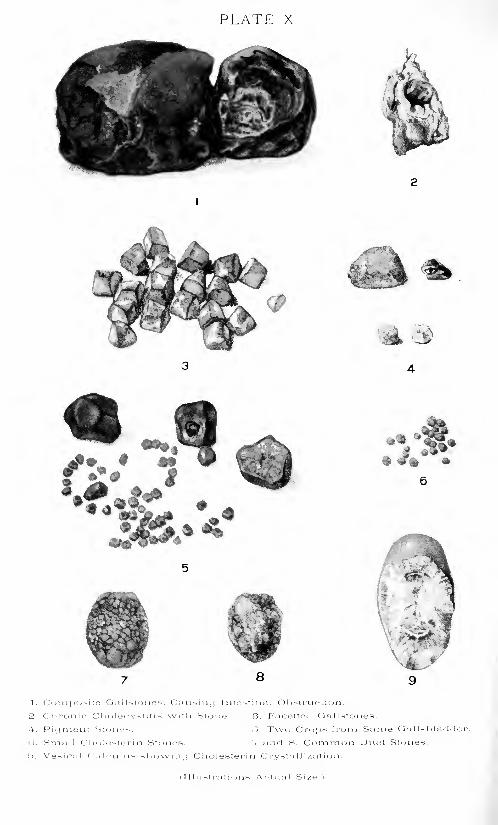
PLATE X
1. Composite On 1 Kt- >ne^, Causiini Intestinal Obstruction.
2. Cliroiiif Cl )' >l'_M>y^t it i -; \vith Stoiio. 3. Facetted Gallstones.
4. Piijment Stones. r>. Two Crops from Same Ga I I-
1 >lad< ler
(i. Small Ci io!(>s!eri n Stones. 7 and S. Comnion Duel Stones.
'>. Vesieal Cal<3ulns -.howiiii) Cholesterin Crystallization.
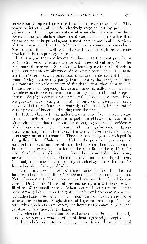
PATHOGENEMS OF (iALL-STONES 397
intravenously injected give rise to a like disease in animals. This
power to infect a gall-bladder electively may be lost by prolongedcultivation. In a large percentage of even chronic cases the deep
layers of the gall-bladder show streptococci, and it is probable that
this organism is the primal agent in most, though not in all, infections
of this viscns and that the colon bacillus is commonly secondary.
Nevertheless, this, as well as the typhoid, may through the systemic
circulation, be the primary cause.
In this regard the experimental findings as to the great prevalenceof the streptococcus is at variance with those of cultures from the
gall-stones themselves. Since Gallipe found germs in cholelithiasis in
1886, innumerable examinations of them have been made. In a little
less than 30 per cent, cultures from them are sterile, so that the epi-
gram of Moynihan is only partly true namely, that every gall-stoneis a tombstone to the memory of the dead germs that lie within it.
In their order of frequency the germs buried in gall-stones and cul-
turable even after years are colon bacillus, typhus bacillus and strepto-
coccus. Staphylococcus is rather unusual. Occasionally, stones fromone gall-bladder, differing apparently in age, yield different cultures,
showing that a gall-bladder chronically inflamed may be the seat of
varying types of infection, differing from the first.
In 1898 I observed that gall-stones removed from a recent case
resembled each other as peas in a pod. In old-standing cases it is
often self-evident that the stones are of varying shades and evidentlyof different crops. The lamination of many gall-stones, in layers
varying in composition, further illustrates this factor in their etiology.
Pathogenesis of Gall-stones. They are practically all developed in
the gall-bladder. Cholesterin, which is the principal constituent of
most gall-stones, is not derived from the bile even when it is stagnant,but from the excessive function of the cells lining the gall-bladderwhen this is the seat of infection. Since there is no cholestcrin-bearingmucosa in the bile ducts, cholelithiasis cannot be developed there.
It is only the stone made up mostly of coloring matter that can be
formed outside of the gall-bladder.The number, size and form of stones varies enormously. To find
hundreds of stones beautifully facetted and glistening is not uncommon.Xot infrequently 1000 or more stones have been found, and in one
case of Dr. Samuel Mixter, of Boston, nearly a quart measure wasfilled by 37,000 small stones. "When a stone is long retained in the;
neck of the gall-bladder or the cystic duct it not infrequently assumes
a saddle shape. Stones, in the common duct, when single are apt to
be ovate or globular. Single stones of large size, made up of choles-
terin with a calcium salt cortex, not infrequently completely fill the
gall-bladder and assume its shape.The chemical composition of gall-stones has been particularly
studied by Xaunyn, whose division of them is generally accepted.1. Pure cholesterin stones, varying in si/e from a bean to that of
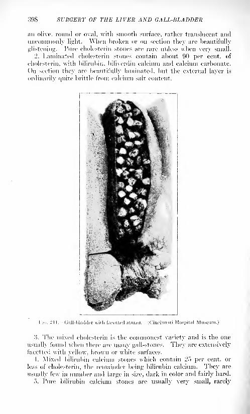
898 SURGERY OF THE LIVER AND GALL-BLADDER
an olive, round or oval, with smooth surface, rather translucent and
uncommonly light. When broken or on section they are beautifully
glistening. Pure cholesterin stones are rare unless when very small.
'2. Laminated cholesterin stones contain about 90 per cent, of
cholesterin, witli bilirubin, biliverdin calcium and calcium carbonate.
On section they are beautifully laminated, but the external layer is
ordinarily quite brittle from calcium salt content.
!'](,. 211. Gall-bladder with facetted stones. (Cincinnati Hospital Museum.)
'.
}). The mixed cholesterin is the commonest variety and is the one
usually found when there are many gall-stones. They are extensively
facetted with yellow, brown or white surfaces.
4. Mixed bilirubin calcium stones which contain 25 per cent, or
less of cholesterin, the remainder being bilirubin calcium. They are
usually few in number and large in si/e, dark in color and fairly hard.
o. Pure bilirubin calcium stones are usually very small, rarely
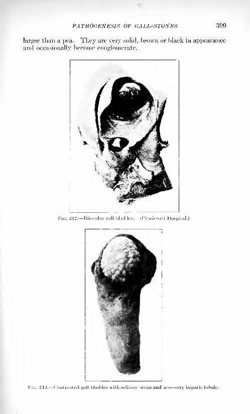
PATHOGEN'ESI8 OF CALL-STONES 390
larger than a pea. They are very solid, brown or black in appearanceand occasionally become conglomerate.
FIG. 212. Bilocular gall-bladder. (Cincinnati Hospital.)
!'!<;. 2 13, -Contracted <j;;i 11-1 'ladder with solitary stone and accessory hepatic lobule.
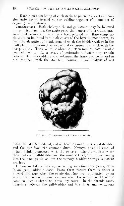
400 SURGERY OF THE LIVER. AND GALL-BLADDER
6. Rare stones consisting of cholesterin or pigment gravel and con-
glomerate stones, formed by the welding together of a number of
originally small stones.
Complications. Both cholecystitis and gall-stones may be followed
by complications. In the acute cases the danger of ulceration, gan-
grene and perforation has already been referred to. Kare complica-tions are to be found in the abscesses of the liver in single form, as
from the ulceration of a gall-stone through the bladder wall or in the
multiple form from involvement of and extension upward through the
bile passages. These multiple abscesses, often minute, have likewise
been alluded to. As a result of perforations, fistula* may remain
between the gall-bladder and duodenum, the transverse colon and in
rare instances with the stomach. Xaunvn in an analvsis of 384
fistula* found 10^ duodenal, and of these 93 came from the gall-bladderand the rest from the common duct. Xaunyn gives 12 cases of
biliary fistula? connected with the pylorus. The rarest fistula* are
those between gall-bladder and the urinary tract, the stones passinginto the renal pelvis or into the urinary bladder through a patenturachus.
Cutaneous biliary fistula*, continuing sometimes for years, often
follow gall-bladder disease. From these fistula? there is either a
nmcoid discharge when the cystic duct has been obliterated, or an
intermittent or continuous bile flow when the natural outlet of the
common duct is obstructed from any cause. In the chronic cases,
adhesions between the gall-bladder and bile ducts and contiguous
ACUTE CHOLECYSTITIS 401
structures are vn-y common and frequently cause symptoms after
the inflammation in the gall-bladder or the gall-stones have been
removed. As elsewhere, malignant disease often follows prolongedirritation from stone in the gall-bladder.
Stenosis of the common, and particularly of the cystic, duct is a
very common sequel of long impaction of stone in one or the other.
When a large stone ulcerates into the duodenum it may give rise to
gall-stone ileus, the point of obstruction depending on the size of the
stone. They measure M x 1 inch. In the series of 409 cases of Waltonthere were lo acute obstructions, in X of which the previous history
pointed to gall-stones.
As a complication of gall-stone disease, acute and particularly chronic
pancreatitis may develop by reason of the common termination of the
biliary and pancreatic ducts. Since no investigations point to the
infection of the pancreas through the arterial circulation, as seems to
be the case in most gall-bladder infections, it must be borne in mindthat in most cases of acute and of chronic pancreatitis there is a
history or at the time present disease of the biliary ways.
Symptoms and Diagnosis of Acute Cholecystitis. Acute cholecystitis
is usually inaugurated with nausea and vomiting of moderate severity
and short duration. There is fever, which is low in the catarrhal
state, and lasts for only a few days. Pain in the region of the gall-
bladder or in the epigastrium and in the back is an almost con-
stant symptom and with it are tenderness in the region of the
gall-bladder and rigidity over the overlying muscles. The painvaries very much with the general condition of the patient. Whenthe sensorium is involved, as in typhoid fever it may be altogether
absent, therefore the condition often fails to be recognized for a
considerable length of time. The pain in most cases is apt to be
spasmodic with periods of intense exacerbation, doubtless due to
overdistention, and muscular spasm. When the gall-bladder is muchdistended, it can easily be felt or percussed through the rigid muscles,
but in many cases the tenderness can only be elicited by dec]) palpa-
tion, the examining fingers being hooked over the lower costal arch.
Inhibition of respiration on that side is not an uncommon symptom.If the gall-bladder enlarges it ordinarily does so toward the umbilicus,
but if there is a ptosis of the liver it may be found so low as to simulate
an appendiceal abscess. In very rare cases the distention takes a
backward turn and may present in the costo-iliac interval. Acute
cholecystitis of the catarrhal type, when it is associated \vith gastro-
duodenitis is attended by a painless jaundice.
Ordinarily, however, the disease is not associated with jaundiceunless there is an obstruction in the common duct. The disease maygo on to empyema, ulceration, gangrene and perforation without anymanifestation whatever of jaundice.
Ordinarily, acute cholecystitis is not associated with very high
temperatures until empyema and ulceration have resulted. TheVOL. ii 26
402 SURGEItY OF THE LIVER AND GALL-BLADDER
pulse likewise remains nearly normal, as it does in many infections
of the liver.
The polynuclear leukocytosis rarely exceeds 12,01)0 unless the
disease involves contiguous structures and pericystic suppuration or
extensive invasion of the small bile ducts has taken place.The diagnosis of acute cholecystitis ordinarily offers no difficulties
even when the symptoms are not very pronounced. ( 'areful palpationwill reveal a tender and enlarged gall-bladder, which can scarcely be
mistaken for anything else.
Complications arising in the course of the disease can be recognized
by exacerbations of pain, increased pyrexia and its concomitant
acceleration of the pulse, increased leukocytosis and, if perforation be
imminent, by the usual representation symptoms of localized peri-
tonitis.
The symptoms of chronic cholecystitis are in many ways those of
gall-stones, because the two conditions are, as a rule, associated.
Hither the inflammation begets the stones or these, by their presencecause the chronic inflammation. It is only through expert radiog-
raphy that in about 50 per cent, of the subjects a differential diagnosiscan be made. Nor is this necessary, since the treatment of the twoconditions are the same.
Since the use of the duodenal tube of Hinhorn has made it possibleto obtain the secretions direct from the papilla of Vater the character
of the bile may be directly studied. By such examination Hinhorn
diagnosticated probable cholecystitis in 40 cases by the turbidity of
the bile found in the fasting condition.
Cholelithiasis. The symptoms of cholelithiasis may be divided
into two classes: ('1 ) Those pertaining to the passage or attempted
passage of a stone from the gall-bladder through the ducts, and (2)
those pertaining to the-quiescent periods of the disease. The first are
associated with vomiting, nausea and intense pain in the region of
the epigastrium, between the shoulder-blades and occasionally in the
right shoulder. The pain ordinarily supposed to be due to the pas-
sage of a stone is probably not due to this at all except when the
stone is very rough. Probing the bile ducts through a fistula is not,
as a rule, painless. The pain of the gall-stones attack probably
depends upon the acute distention of the musculofibrous coat of the
gall-bladder, with spasmodic contraction of the muscular fibers. I lere,
as elsewhere, for example, in acute hydronephrosis, the pain is due to
acute distention caused by obstruction. The acute symptoms lasting
for from a few hours to as many days subside as soon as the stone has
passed into the duodenum or has fallen back into the gall-bladder.
Frequently a stone may be recovered from the feces. I have found
an easy way of recovering this stone to cover the seat of the toilet
with a loose mesh gau/e net, within which the stone is caught, as in
a sieve, after the rest of the stool has been washed away by repeated
flushings. This certainly is a more esthetic way of searching for gall-
stones than the ordinary one of using a sieve. The passage of gall-
ACUTE CHOLECYSTITIS 4(KJ
stones is not infrequently followed by a mild and evanescent jaundice,
although in very many instances jaundice is conspicuous by its absence.
Inipaction for even four or five days in the common duct is not of
necessity followed by jaundice. Quite recently the condition of dis-
sociated jaundice has been recognized, in which without any tingeingof the conjunctiva an excess of bile salts may be found in the blood.
The chronic symptoms of gall-stones, except for recurring colickyattacks are very indefinite. As a rule, they are interpreted by the
patient, and frequently by the physician, as due to some chronic gas-tric disturbance. The pain, usually in the epigastrium, comes onseveral hours after eating and is rarely of a severity to require medi-
cation. Tenderness, however, on dee]) inspiration can always be
elicited by deep pressure over the gall-bladder. Enlargement of
the gall-bladder is usually absent, since, as Courvoisier showed over
thirty years ago, in 80 per cent, of cases in which the gall-bladderis distended the disease is malignant in character and obstructive.
The cystic duct is not distensible to any considerable extent. In
extreme dilatation of the gall-bladder from cancer of the pancreasthe duct remains nearly of normal size, whereas the common and
hepatic ducts may be an inch or more in diameter. After removal
of the gall-bladder there is a distinct tendency to dilatation of the
common and particularly of the hepatic duct. The recurring inflam-
mation of the gall-bladder in cholelithiasis produces such thickeningof its walls that distention does not occur.
The position of the stone can in main" cases be determined by the
symptoms. The most characteristic are those in which a recurring
jaundice is produced by a stone in the common duct acting as a ball
valve. This action was first demonstrated by Fenger. These commonduct stones are often associated with rigors and high temperatures,and were formerly supposed to simulate malarial attacks, and wereknown as hepatic intermittent fever. The true nature of this inter-
mittent fever was discovered by Charcot.
In many instances there is a pronounced sepsis which is in reality
the clinical expression of an acute ascending cholangitis, even to the
small bile ducts. It is initiated by a rigor, high fever and mental
hebetude. It may be difficult to elicit any marked tenderness over
the liver. The chills return irregularly. Jaundice of slight char-
acter is usually present even in the absence of calculous obstruction
and may be of hematogenous origin, as it is in sepsis from other than
hepatic lesions, \\hen the jaundice is persistent, as it always is in
the gravest cases, common duct obstruction may be taken for
granted.In the absence of febrile reaction the intermittent jaundice of
common duct stone gravely affects the patient's general condition bythe exclusion of bile from the intestine. The icterus is often attended
by intractable itching, which prevents sleep. Loss of weight rapidlyfollows. The heart's action becomes slow and weak and the cholemia
doubtless reduces the resistance of the capillary walls and the coagu-
404 SCRGERY Or THE LIVER AND GALL-BLADDER
lability of the blood thereby conducing to spontaneous hemorrhagesand postoperative oo/ing.
Stones in the cystic duct rarely produce jaundice except when, bytheir si/.c, they cause compression of the hepatic duct or swelling of
the inucosa invades the common duct. There is no way in which the
diagnosis of stone in the hepatic duct can be made. As a rule, hepaticduct stones are found with stones in the common duel, from which
they have ascended. These stones are most apt to be overlooked in
operations.
Intrahepatic Gall-stone.- In cases of cholangitis that terminate in
death there are often found many minute gall-stones or accumulations
of biliary sand in the liver substance. According to Beer,'1 s per cent,
of gall-stone cases show intrahepatic stone-. This, of course, refers
to autopsy reports and is not true of operative cases. These stones
are. for the most part, devoid of cholesterin and consist of bilirubin
calcium. In other instances stones have ulcerated through the gall-
bladder and found lodgment in the liver, where' they usually are
contained in an abscess. Hut in rare instances, solitary stones have
been found and removed from the abscesses of the liver by operation,in which the conereinent contained cholcsteriu. In these cases it i-
certain that a stone, by obstruction of the common duct, ascended
through the hepatic and become lodged in one of the larger intrahe-
patic ducts. If active infection has not attended the process the --tone
may not be within an abscess cavity. Xoguchi- has recently reported-itch a case in which no other stone was found cither in the gall-
bladder or the ducts. Intrahepatic stones, as a rule, are in the larger
brandies of the bile ducts, where they are, of course, inaccessible or
overlooked. It is in these cases that, after operation, -tones appearin the wound sometimes for weeks afterwards, very much like the
boys in Yorkshire after the breaking up of Squeers's school, in Xirholax
Xic]:lchi/. This illustrates the necessity of careful examination of the
hepatic duct at every operation, and the danger of crushing a commonduct stone, which may leave nuclei for the later development of
intrahepatic -tones.
The value of the ,r-ray in the diagnosis of gall-stones cannot be
overrated. In expert hands 50 per cent, of the cases show positive
shadows, and many times the exact location and even the number of
-tone- may be demonstrable; but even in negative cases a distortion of
the duodenal cap and of the pylorus and the limited mobility of the
latter, as seen with the fluoroscope, indicate with much certainty the
presence of -tone or at least of pericystic adhesions.
The diagnosis of gall-stones ordinarily offers no difficulties, and even
in the absence of positive .r-ray findings the cases are rare in which,
where stones are suspected, there is not found at operation somelesion of the gall-bladder or its passages that fully accounts for the
symptoms. Ileer of the stomach or duodenum and chronic pancre-
1 Laiipcnheck's Archives, Ixxi, 110. 2Ibid., xcvi, 633.
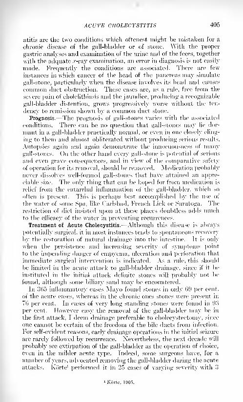
ACUTE CHOLECYSTITIS 405
atitis are the two conditions which oftenest might be mistaken for a
chronic disease of the gall-bladder or of stone. With the proper
gastric analyses and examination of the urine and of the feces, togetherwith the adqnate .r-ray examination, an error in diagnosis is not easily
made. Frequently the conditions are associated. There are few
instances in which cancer of the head of the pancreas may simulate
gall-stone, particularly when the disease involves its head and causes
common duct obstruction. These cases are, as a rule, free from the
severe pain of cholelithiasis and the jaundice, producing a recognizable
gall-bladder distention, grows progressively worse without the ten-
dency to remission shown by a common duct stone.
Prognosis. The prognosis of gall-stones varies with the associated
conditions. There can be no question that gall-stones may lie dor-
mant in a gall-bladder practically normal, or even in one closely cling-
ing to them and almost obliterated without producing serious results.
Autopsies again and again demonstrate the innoeuousness of main'
gall-stones. On the other hand every gall-stone is potential of serious
and even grave consequences, and in view of the comparative safety
of operation for its removal, should be removed. Medication probablynever dissolves well-formed gall-stones that have attained an appre-ciable size. The only thing that can be hoped for from medication is
relief from the catarrhal inflammation of the gall-bladder, which so
often is present. This is perhaps best accomplished by the use of
the water of some Spa, like Carlsbad, French Lick or Saratoga. Therestriction of diet insisted upon at these places doubtless adds muchto the efficacy of the water in preventing recurrences.
Treatment of Acute Cholecystitis. Although this disease is always
potentially surgical, it in most instances tends to spontaneous recovery
by the restoration of natural drainage into the intestine. It is onlywhen the persistence and increasing severity of symptoms pointto the impending danger of empyema, ulceration and perforation that
immediate surgical intervention is indicated. As a rule, this should
be limited in the acute attack to gall-bladder drainage, since if it be
instituted in the initial attack definite stones will probably not be
found, although some biliary sand may be encountered.
In 305 inflammatory cases Mayo found stones in only 09 per cent.
of the acute cases, whereas in the chronic ones stones were present in
70 per cent. In cases of very long standing stones were found in 93
per cent. However easy the removal of the gall-bladder may be in
the first attack, I deem drainage preferable to cholecysteetomy, since
one cannot be certain of the freedom of the bile ducts from infection.
For self-evident reasons, early drainage operations in the initial seizure
are rarely followed by recurrence. Nevertheless, the next decade will
probably see extirpation of the gall-bladder as the operation of choice,
even in the milder acute type. Indeed, some surgeons have, for a
number of years, advocated removing the gall-bladder during the acute
attacks, korte 1
performed it in 25 cases of varying severity with 3
1 K6rte, 1905.
ii)i') sr/taKii'Y or TIII-: LIVER AXD GALL-HI.ADDEK
deaths due To causes remote from the operation or the gall-bladderdisease itself.
If the patient's condition is grave from the primary infection, such
as typhoid, or from the extent and decree of local invasion, such as
gangrene or perforation with abscess, a quickly performed drainage
operation with protective packing is to be preferred to a radical
operation, which, if it becomes necessary later, can be performedunder more propitious conditions. In the hyperacute cases the
judgment and experience of the surgeon must dictate how far he maysafely go.
Treatment of Chronic Cholecystitis and Gall-stones. - Inless extreme
age or an unusual operative risk from cardiac or renal disease contra-
indicates an operation, every case of chronic cystitis and of gall-stone
should be made a subject of exploration and radical treatment. This
statement is made with full consciousness that many patients with
chronic cystitis and gall-stones live through many years and succumbto some independent affection.
Although the operation may therefore be classed among the elec-
tive, it is to be borne in mind that the less the actual necessity for
interference the greater is the probability of its easy performance and
safe outcome. Consequently, the difficulties, and hence the dangersof operation, increase as the demand for operation becomes imperative.Stones impacted in the cystic, and particularly in the common, duct
are removed with much less certainty of success than are those from
the gall-bladder. \Vith few exceptions it may be said that everycommon duct stone testifies to an operation neglected. In every
operation for gall-stone a choice must be made between simple chole-
cystostomy with drainage and removal of the gall-bladder.
Four years after Marion Sims,1 in isTS, put cholecystostomy on a
firm basis, Langenbuch advocated the removal of the gall-bladder.
For thirty-five years, therefore, it has been a question as to which
operation is to be preferred as the normal procedure. It is now
approaching solution, very much like that of the appendix. Toachieve an ideal result in cholelithiasis it is not only necessary to
remove all stones, but to remove that from which and wherein theyare formed, that is, the infected gall-bladder.
(iall-stone operations of all kinds are followed by a recurrence of
symptoms in a little over 10 per cent. If they recur within a few
weeks or a year it is reasonable to suppose, and a second operation
usually reveals, a stone overlooked in the cystic, common or hepaticducts. That this may happen to most distinguished specialists is
shown by Kehr, who admits leaving stones in H) out of 491 operations.If free stones are found in the gall-bladder at a later period they mayfairly be charged to new infection. All statistics agree in the mainthat recurrence of symptoms is about five times more frequent after
drainage than after removal of the gall-bladder. When the gall-
1 Langenbeck's Archives, Ixi, 471.
BILIARY FISTULAE 40 1
bladder is removed in animals, together with the eystic duct, there
may be slight dilatation of the common and hepatic ducts, but no
new gall-bladder is formed. When, however, a vestige of the gall-
bladder itself or much of the cystic duct is left a repullulation of the
gall-bladder may take place, and in this a stone may form.
The chief objection to cholecystectomy is its greater mortality,
even in expert hands. Peaver had 36 deaths in 360 operations,
whereas there were only 12 deaths following 427 drainage operations.
Since the removal of the gall-bladder is reserved for difficult cases,
in many of which it probably could not be saved, and drainage opera-tions are reserved for the more recent and therefore easier cases, it is
evident that much of the excess mortality of the one operation is
due to the class of cases in which it may be a necessity. If the removal
of the gall-bladder were in a given series of cases to be uniformly
practised in the easy and difficult cases alike the mortality would
unquestionably be materially reduced below present records. Further-
more, secondary operations, in which a gall-bladder has finally to be
removed, obviously become necessary.That the operation of gall-bladder removal is more difficult than
drainage should not militate against its more general adoption. Theoccasional surgeon may be advised in the average case to restrict his
interference to drainage. It is to be borne in mind that, as Mayohas shown, the mortality after cholecystectomy in proper hands is
very little if any higher than cholecystostomy with drainage.
Biliary Fistulse. Fistula connected with gall-bladder or commonduct may be cutaneous or internal. The former, although found
occasionally after spontaneous perforation of the gall-bladder, usuallyfollows a drainage operation. Such fistula? may be either mucous or
biliary in character. If the former discharges no bile it is always due
to a stricture of the cystic duct or to an overlooked impacted stone.
The biliary fistula, on the other hand, indicates a patent cystic duct
if the gall-bladder has not been removed, or an overlooked stone or
stricture in the common duct. Cutaneous fistuhe were very commonin the early history of gall-stone surgery when the gall-bladder wassewed to the skin. To obviate it the gall-bladder should not be
attached too low to the peritoneum but should be attached as far
upward as possible without undue tension. A complete external
biliary fistula sometimes follows removal of the gall-bladder, whenin tying off the cystic duct too much traction is made on the commonduct and the latter is either wounded or included in the ligature.
Treatment. The proper treatment of cutaneous mucous fistuhr is
extirpation of the gall-bladder. This should not be too long delayed,since it may, by reason of adhesions, be thereby made one of the most
difficult operations on the bile passages. In the very old-standingcases the subperitoneal excision of the inucosa, as first recommended
by Roser, might be attempted. To be successful it would necessi-
tate removing the entire mucosa, leaving the musculofibrous coat to
become changed into a fibrous cord.
10S Sl'h'GERY OF T11K LIVER AND CALL BLADDER
For true biliary fistula- it is necessary to remove the cause, whetherthis be a stricture or an overlooked stone. They always close if there
is no obstruction to the normal passages.Stricture of the common duct, either from scar tissue or from
wounds inflicted at a previous operation, is most successfully treated
by a cholecystenterostomy when the gall-bladder has not been removed,
although a number of operators prefer making the anastomosis with
the stomach just above the pylorus. \Vhen there are many adhesions
about \he duodenum the stomach can more readily be approximated.\\ hen the gall-bladder has been removed a choledochoduodenostomy is
indicated, provided the duct be sufficiently dilated TO permit an anas-
tomo^is by suture. If the duct at a previous operation has been
completely divided an end-to-end anastomosis with circular suture
and drainage of the hepatic duct should be attempted, provided the
field for operation can be sufficiently freed of adhesions. -I. II. .Jacob-
son 1 collected .'54 cases of reconstruction of the bile ducts, L'l of whichwere thus treated. ^ hen it is difficult to make anastomosis because
the distal end of the duct cannot be found the hepatic duct will usuallybe found dilated. An opening should be made into the duodenum,and into this and into the proximal end of the common or hepaticduct a rubber tube must be placed with one or two reveres, in the
proximal end and five or six inches of length extend through the
new duodenal opening into the intestine, after the method promul-
gated by Sullivan.'-' I'rainage, of course, is essential after all of these
operations.The internal biliary fistnhe are more numerous than the external,
although they do not so appear because they do not reveal themselves
by symptoms. In a study of 4D l.> cases ( 'ourvoisier found liMi external.
Of the internal there were No into the duodenum, o!) in the colon, 1)5
in the stomach, 7'<> in connection with abscesses of the peritoneum and
_' 1 with the pleura and lung. Schlasinger and Kichler' have brought
ii]to 17 the recorded cases of bronchobiliary fistula, in many of which
gall-stones were expectorated.In the analysis of 41 cases by Klauber 1
:2o' were associated with
stone. lie believed that fistula develops either after the formation
of a subphrenie aliscess or after the direct adhesion of the gall-bladder
to the diaphragm.
CANCER OF THE GALL BLADDER AND BILE DUCTS.
To avoid repetition an important factor in the pathology of chronic
cholecystitis was reserved for consideration in the etiology of primarytumors. Jn the chronically inflamed mncosa there are quite often
found papillary excrescences which, to the unaided eye, appearas papillomata projecting varying distances into the lumen of the
CANCER OF THE CALL-HLM)1)KI{ ,l.Y/> 1ULE DUCTS 409
gall-bladder, and particularly into its narrow neck. Pels-Louden,1
and more recently the pathologists of the Rochester Clinic, have
shown that careful study of many gall-bladders reveals an epithelial
overgrowth in the deeper strata of the mucosa and minute papillo-
mata on the surfaces. From these and from old ulcerations springthe cancers. Slade- in a careful study of :>)> gall-bladders containingstones at autopsy found MO per cent, presenting gross microscopicevidences of cancer. It is again the story of malignancy following
prolonged and changing irritation from stone, infection and disten-
tion singly or combined. Cancer of the bladder is six times as com-
mon as is that of the bile ducts. Primary sarcoma are of pathologicalinterest only, not more 1 than 5 or (i clear cases having been recorded.
Of these, too, most were associated with stone. According to Rollis-
ton from 4 to 14 per cent, of gall-stones develop cancer. Mayo found
4 per cent, of cancers in IsOO gall-bladder case-. That the stones
are the initial factor can hardly be questioned, for in cancer of the
gall-bladder they are found in from til) per cent, according to Musser.
to 0") per cent. Siegert and UK) per cent. Janowski. Call-stones are
formed in early or middle life; cancers occur only after long irritation,
that is to say, rarely before fifty. Like gall-stones, biliary cancer is
three times as common in women as it is in men. Furthermore, it
rarely occurs when by timely drainage or gall-bladder removal the
irritation has been taken away. In his very large experience, Moyni-han knows of only two of his cases developing cancers after operation.If gall-stones are the cause of cancer, as has been indubitably shown.
they should evidently be less common in the bile ducts. Accordingly.Musser found TOO cancers of the gall-bladder and only Is of the ducts.
In the latter the sexes were equally represented, perhaps because in
my experience in proportion to the total number of gall-stone cases
in each sex common duct stone occurs oftener in the male. Probablyit is because of the greater rigidity and firmer fixation of the abdominalviscera in the male. In cancers of the bile ducts, stones are less
frequently found- in 'l'.\ per cent, of the cases. Mayo-Robson3sug-
gests that in many of them the stone probably ulcerates into the
intestine. Phillips4
recently reported ."> cases of duct cancer, each of
which showed stone. Of the duct cancers about SO per cent, occur in
the eholedoehus and over 50 per cent, of them occur in the ampullaor the Yaterian papilla.
Biliary cancel 1 may be of the infiltrating ulcerated type or of the
fungating papillomatous type, as it appears in the urinary bladder.
This type is oftenest found in the gall-bladder, whereas in duct cancers
the growth often appears in the form of an annular stricture, with
dense infiltration of the walls. The tumor is then usually small and
very hard.
Ilistologically the cancer is nearly always of the cylindrical colum-nar cell type. Lymphatic involvement comes rather late, but the
410 Xl'ltdEItY 01'' Till': LIVER AM) GALL BLADDER
liver and head of the pancreas, especially in duct cancers, are involved
early.
Symptoms.- -Until a firm tumor is detected in the gall-bladder regionthere may be nothing to make the diagnosis certain. It may be
suspected when a person with long-standing gall-stone history developsa painless and rapidly deepening jaundice without the symptomsusually attending obstruction by stone. Jaundice, of course, may be
a late manifestation or rarely altogether absent when the cancel-
involves the gall-bladder alone. In cancers of the papilla the jaundice
may be intermittent.
( 'ancer of the head of the pancreas may not be distinguishable from
duct cancers. It is evident, therefore, that the diagnosis is rarelymade except at operation or autopsy.
Treatment. The treatment of biliary cancer is preventive. Early
operation for gall-stones reduce its incidence. Only in exceptionalcases can the disease, while still limited to the gall-bladder, be success-
fully cured by removing the gall-bladder, with perhaps an adherent
and involved wedge of the liver. Cancers of the duct have been
removed and the hepatic or the proximal end of the common duct
implanted into the duodenum. Cancer of the papilla has been radi-
cally removed, according to Outerbridge,1 in 22 cases, in l(i of which
the duodenum was opened anteriorly; 9 of these cases died within a
few days of the operation. So far as known no cases survived morethan three and one-half years.
OPERATIONS ON THE LIVER, GALL BLADDER AND BILEDUCTS.
Operations on the liver, gall-bladder and bile ducts demand the
preliminary preparations usually made for abdominal section. It is
especially necessary, however, to see that the stomach is emptied and
that the colon, particularly, has been cleaned from above and below.
A special reason for emptying the stomach is the greater tendency to
acute dilatation after gall-bladder operations as compared with those
on other abdominal viscera. A greatly distended colon may seriously
hamper the operator by getting into the field of operation. In cases
of long-standing jaundice the tendency to bleeding during and after
operations must be overcome by the use of calcium chloride in 2-gramdoses, three times a day, for four or five days, or preferably by the
injection of 20 to '>() grains of horse serum or of diphtheria antitoxin
three or four hours before operation. In a number of deeply jaundiced
patients I have found the latter reduce the bleeding to the normal.
Unless there is an imperative necessity, operation must be declined
if there are recent petechial hemorrhages or bleeding from any mucosa.
The large mortality of gall-stone surgery in its early history was
chiefly due to postoperative bleeding.
1 Ami. Siirfr., Ivii, 414.
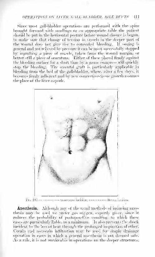
OPEKATHtXX OX LITER, <;,\LL KLADDEK. fifLE Dl'CTX 411
Since most gall-bladder operations are performed \vith the spine
brought forward with sandbags or an appropriate table the patientshould be put in the horizontal posture before wound closure is begun,to make sure that change of tension in vessels in the deeper part of
the wound does not give rise to concealed bleeding. If oozing is
general and not relieved by pressure it can be most successfully stopped
by ingrafting a piece of muscle, taken from the wound margin, or
better still a piece of omentum. Either of these placed firmly againstthe bleeding surface for a short time by a gau/.e compress will quickly
stop the bleeding. The omental graft is particularly applicable in
bleeding from the bed of the gall-bladder, where, after a few days, it
becomes firmly adherent and by new connective-tissue growth assumes
the place of the liver capsule.
Anesthesia. Although any of the usual methods of inducing anes-
thesia may be used we prefer gas oxygen, expertly given, since it
reduces the probability of postoperative vomiting, to which these
cases are particularly liable, to a minimum. It also prevents the shock
incident to the loss of heat through the prolonged inspiration of ether.
Cocain and novocain infiltration may be used for simple drainage
operation in cases in which a general anesthesia is not deemed safe.
As a rule, it is not serviceable in operations on the deeper structures.
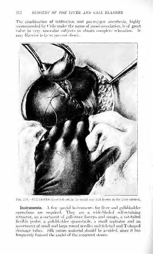
412 SURGERY OF THE LIVER AND GALL-BLADDER
The combination of infiltration and gas-oxygen anesthesia, highlyrecommended by (.
1
rile under the name of anoci-association, is of greatvalue in very muscular subjects to obtain complete relaxation. It
may likewise help to prevent shock.
Instruments. A few special instruments for liver and gall-bladder
operations are required. They are a \vide-bladed self-retaining
retractor, an assortment of gall-stone forceps and scoops, a rat-tailed
flexible probe, a gall-bladder spoon-ladle, a small aspirator and an
assortment of small and large round needles and fish-tail and T-shapeddrainage tubes. Silk suture material should be avoided, since it has
frequently formed the nuclei of the recurrent stones.
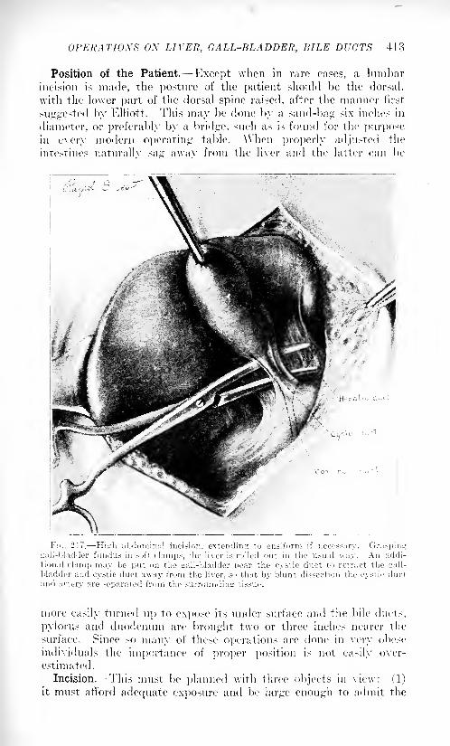
OPERATIONS OX LIVER, GALL-BLADDER, BILE DUCTS 413
Position of the Patient. Except when in rare cases, a lumbar
incision is made, the posture of the patient should be the dorsal,
with the lower part of the dorsal spine raised, after the manner first
suggested by Elliott. This may be done by a sand-bag six inches in
diameter, or preferably by a bridge, such as is found for the purposein every modern operating table. ^Vhen properly adjusted the
intestines naturallv sag awav from the liver and the latter can be
1'i'j. 217. Hitdi abdominal incision, extending to ensiform if necossary. Graspinggall-bladder fundus in soft clamps, the liver is rolled out in the usual way. An addi-
tional clamp may be put on the gall-bladder near the cystic duct to retract the trail-
bladder and cystic duct away from the liver, so that by blunt dissection the cystic ductand a'-;ery are -eparated from the surrounding tissue.
more easily turned up to expose its under surface and the bile ducts,
pylorus and duodenum are brought two or three inches nearer the
surface. Since so many of these operations are done in very obese
individuals the importance of proper position is not easily over-
estimated.
Incision. This must be planned with three objects in view: (1)
it must afford adequate exposure and be large enough to admit the
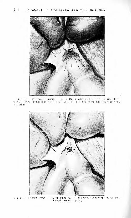
414 SURGERY OF THE LIVER A\D GALL-BLADDER
i<;. 218. Liver raised upward. End of the hepatic duct free an 1 sutures placi
eudv to draw duodenum into position. Note that gall-bladder was removed at previou
ixation sut i ires tied, duodenum incised and posterior row of through-mii lirou<rh su 1 un-s in phi
OPERATIOXS OX LIVER, GALL-BLADDER, BILE DUCTS 415
operator's hand, for thorough exploration of the gall-bladder, bile
ducts, pylorus, head of the pancreas and duodenum; (2) it must be
easily closed, and thereby (/>) must be made safe as to the occurrence
of a hernia by dividing as fe\v of the terminal intercostal branches as
possible. The second and third requisites are best met by a gridironor muscle- splitting operation, like that performed in the average
appendectomy. In gall-bladder work it is of little value, owing to
the insufficient exposure it affords.
Most operators make a longitudinal incision through the outer
third of the rectus or through the linea semilunaris, beginning an
inch or more to the right and a little below the xiphoid cartilage;
it extends downward for five or more inches, across the thickness of
the fat layer. A long wound heals as quickly as a short wound. If
it is necessary to get additional room, Bevan enlarges the incision
above by dividing the rectus upward and inward, and downward andoutward below, thus giving the incision an inverted Z shape. Thewave incision of Kehr begins in the median line below the ensiform
cartilage and extends midway to the umbilicus. It is then continued
obliquely downward and outward, through the rectus to its outer
third, and is thence continued downward a varying distance throughthe muscle. This incision I deem objectionable on account of the
free bleeding and the liability to hernia.
Czerny, and later Konig, devised a trap-door exposure, combininga median incision with a transverse one, in the direction of the trans-
versalis fibers, joining the former one at its lower end. Of the per-
pendicular incision the one in the linea semilunaris, while easy and
causing least bleeding, conduces most to hernia, since there are only
aponeuroses that can be approximated in the closure of the wound.All of these incisions have the further objection that especially in fat
subjects, with very thin peritoneum, the wound closure is difficult,
and often takes more time than the operation proper. I have found
that a procedure first advised by Southers and later by MeArthurovercomes this objection to a very large extent. By dividing the
transversalis fascia and peritoneum transversely, wound closure is
easily effected. An advantage of the perpendicular incision is that it
permits relatively easy examination of the appendix, which is so often
involved, even if it is not as has recently been supposed, its actual
precursor. Kocher makes his incision parallel to the ribs. Theincision which we have preferred for a number of years is the trans-
verse or nearly transverse one. like that recommended by Rutherford
Morrison and Sprengel. Beginning at the costal margin it extends
transversely from the costo-iliac interval to the outer border of the
rectus, and is continued in the same direction through this muscle as
far as may be necessary. It can easily be carried across the medianline to permit operation on the stomach. When the gall-bladder can
be felt the outer third of the incision is laid directly over it. \\ e have
never found it necessary to place holding sutures in the rectus to
prevent retraction. The divided muscle margins can easily be brought
410 SURGERY OF THE LIVER AND CALL-BLADDER
and held together by mattress sutures. We have seen no hernia follo\v
the transverse incision after operations either on the gall-bladder or
stomach. A signal advantage of the transverse incision is that it
permits, if it is desired, easy drainage through a stab wound in the
loin. It' carried far enough to the left it permits palpation of the
spleen. The only valid objection to this incision is that in obese
subjects it makes search for the appendix at times very difficult.
Ideal Cholecystotomy of Bernays. The chnlecystendysis of Cour-voisier was designed for the removal of gall-stones from a normal
gall-bladder, with immediate closure of the incision in the latter by
purse-string or layers of Lembert's suture. To many operators it
seems safer to anchor the closed gall-bladder to the parietal perito-
neum, and insert a cigarette drain to the angle of the wound for two or
three days. Whereas some brilliant results may be achieved by this
operation, which can be performed quickly and through a small
incision, in view of the now accepted views of the pathology of gall-
stone disease and the involvement of the bladder wall, ideal chole-
cystotomy must be reserved for the few cases in which stones are
accidentally found in operations for other condition-- in which the
gall-bladder seems normal.
CholecystOStomy. The abdomen being opened through one of the
incisions described the gall-bladder is looked for by sight and touch.
In most cases it can be seen and felt in its usual place, and stories, if
present, can be felt. In many instances, however, their presencecannot be positively made out until a later step in the operation. It
must be borne in mind that the gall-bladder may be in the midline, or
even to the left of it without there being a transposition of the viscera.
The color and the thickness of its wall must be carefully studied,
for it is only a steel-blue gall-bladder with walls that can be graspedbetween the fingers like a fold of skin that ordinarily should be sub-
jected to the drainage operation when the disease is a chronic one.
If the disease is of the acute type and the gall-bladder reddened,
thickened and perhaps covered with fibrin flakes, it is the custom of
most operators to use drainage alone, although, as already stated,
the modern tendency is to extirpation.In most chronic cases the examining finger can detect concretions
from without. At this stage of the operation the ducts, the hilinn of
the liver and the duodenum should be carefully palpated before
the gall-bladder is opened. Since in this examination one finger should
be placed in the foramen of Winslow, it is well to remember that this
is not a patulous opening at any time, but only an elongated slit, the
margins of which are in touch until separated by the examining finger.
If during this examination a stone can be felt impacted in the cystic
duct a gentle effort should be made to strip it into the gall-bladder.
Its dislodgment from within later may be more difficult.
Difficulties multiply when the gall-bladder is contracted and buried
in adhesions. These; must be severed with great care, the care
increasing as the region of the ducts, and therefore of important vessels,
OPERATIONS ON LIVER, GALL-BLADDER, BILE DUCTS 417
is approached. Unless, however, a thorough inspection or at least
palpation of the field is secured the operation is incomplete, and the
operator will always be justly in fear of symptom recurrence.
As the next step of the operation the gall-bladder is caught betweentwo intestinal catch forceps and aspirated if it is distended, otherwise
an incision large enough to admit the finger is at once made into it.
The contents pour out at once and should be caught in gau/e or an
appropriate ladle which has been put in place to receive it. Whenthere are many small stones this prevents their gliding into the peri-
toneal cavity. Soiling of this should be prevented as much as possible.
The stones are best removed by a scoop guided by a finger on the
outside of the gall-bladder. When the latter is contracted it is often
bilocular with stones in each pocket, and the division of the separatingwall may be necessary to evacuate the deeper one. Such gall-bladders,
however, should ordinarily be removed. If a stone cannot be removedfrom the cystic duct without too much laceration of the mucosa the
duct should be divided over the stone and drained.
A drainage tube, usually one-quarter of an inch in diameter, is
next inserted into the gall-bladder and the margins of the incision
fixed to it by a catgut suture. Pushing the tube into the gall-bladderand invagination of the wound margins is easily effected and main-
tained, by one or two Lembert sutures, or, better still, by one purse-
string suture placed over another. The gall-bladder is thus made
water-tight, leakage being almost impossible. The last suture should
be left long for anchoring the gall-bladder to the parietal peritone mi,
which step, except for the wound closure, ends the operation. This
method of invaginating the gall-bladder wound is like that of Stam-
Kader in gastrostomy. When the gall-bladder cannot be broughtinto the abdominal parietes it is safe to drop it and surround the intra-
peritoneal portion of the drainage tube with gauze. It is unnecessary,and I believe harmful, although it has been recommended, to loosen
the parietal peritoneum for a varying distance to meet the gall-bladder.Even when it is feasible it is unnecessary to anchor the gall-bladderto the abdominal wall. The adhesions formed may give rise to later
trouble. A number of operators bring the drainage tube through a
separate stab wound and completely close the original incision.
Ruth 1 advises passing the drainage tube through a separate stab
wound, thereby lessening the danger of the hernia. He also advises
against anchoring the drained gall-bladder, since this tends to the
formation of adhesions. The transverse incision obviates the necessityfor a separate opening. While drainage outside of the gall-bladder is,
as a rule, not indicated a cigarette drain may profitably be used whenthere has been much soiling of the peritoneum.
Cystocotomy. -Cystocotomy is an operation made necessary by the
irnpaction of a stone which cannot be removed from within the gall-
bladder. Generally, the stone can be felt and directly cut down upon.
'.Tour. Am. Mc<l. Assn., September :>>. 1914.VOL. ii 27
41S SURGERY OF THE LIVER AND GALL-BLADDER
In other instance's, however, it may be necessary to split the gall-
bladder along its entire length until the stone is reached. This pro-cedure seems needlessly severe and should be reserved for extreme
cases, although Terrier and Ilartnmn recommend it even for commonduct stones. If this method is followed, there can be no question as
to reaching the stone. When there it is probable that stricture will
follow the impaction. It is better to do a cystectomy at once. I )rain-
age is better after opening the duct than suture, since it is improbablethat the sutures will hold in the friable duct wall. Incidentally it
may be stated that probably many of the reported choledochotomies
are really cysticotomies over stones impacted in the lower reach of
the cystic duct.
Cholecystectomy. Earlier in this article the indications for removalof the gall-bladder were set forth. Here it is necessary, therefore,
only to state when it is contra-indicated. First and foremost, whenin cases in which the patulousness of the common duct opening in the
duodenum is in doubt, for if this is impermeable the gall-bladder lends
itself more readily to an anastomosis than either of the bile ducts.
Gall-bladder excision should not be begun until the surgeon has
assured himself on this point. The second centra-indication exists
when there is a necessity for draining the infected bile ducts. Althoughthis is a less positive contra-indication it is safer to act upon; howeverit is easily possible to drain the ducts without it. Third, cystectomyis contra-indicated in all cases in which the general condition of the
patient makes a prolonged operation hazardous. In these cases a
preliminary drainage will make the radical operation, if it proves
necessary, a safe undertaking. The first steps of cholecystectomyare those for exposing the gall-bladder, as already described. Toremove this organ one may begin at the fundus, gradually separatingit from its liver bed after dividing the peritoneum on each side. It is
be>t to do this by sharp dissection. The gall-bladder is slowly freed
until the cystic duct is freely exposed. The artery and duct are tied
together and divided between two forceps, When drainage is desir-
able it may be possible to tie the cystic artery or its two branches
separately and fix a drainage tube over the stump of the duct.
A better method of removing the gall-bladder, doubtless, is that of
the lvoche>ter Clinic, in which after the liver is tilted by traction with
forceps on the fundus the dissection is begun by the exposure of the
cystic duct at it> junction with the common duct. By traction on
the neck of the gall-bladder this and the cystic duct are straightenedout and a curved forceps may readily be pushed underneath the duct
and artery, so that they may be lifted away from the liver. Theduct and ait cry are clamped and tied off together. A second clampis then applied to the distal end of the duct and used as a tractor, the
gall-bladder being slowly separated from the liver from its apex to
the fundus. The oozing, if it does not yield promptly to compression,
quickly ceases if a muscle, or preferably an omental graft, be held in
place for a few minutes with a gauze compress. While gauze packing
OPERATIONS ON LIVER, GALL-BLADDER, BILE DUCTS 419
will control the hemorrhage it may be dangerous, as a severe secondary
hemorrhage may follow its removal. We have seen this occur on the
tenth day. The operation is completed by suturing the cut edges of
the peritoneum in a manner to cover the denuded surface of the liver.
Owing to the frequent parallelism of the cystic and hepatic ducts,
and particularly the danger of drawing the common duct into the
ligature, it is safer in eholeeystectomy to leave more than is actually
necessary of the cystic duct than to remove too much. If drainage is
desired the stump of the duct is left open and a drainage tube anchored
over it with a catgut suture. In every case some drainage of the
wound is to be secured.
In removing the gall-bladder there are two special dangers: (1)
bleeding from a poorly placed ligature about a cystic artery, and (2)
inclusion in the ligature or wound of the common duct. (i. \\righthas especially emphasized this danger. To avoid, the artery should
be tied by itself and the 1 duct either remain untied or be tied with
readily absorbable plain catgut. If there is any doubt as to the
holding of a ligature the artery may be held in a clamp forceps for a
day or two. Mayo has wisely suggested that the clamp be openedfor a day before it is finally removed, lest its immediate removal start
bleeding afresh.
Choledochotomy. This operation, probably first performed byMarcy, of Cambridge, in 1SS2, was introduced into Germany by
Langenbuch and Kummel and into England by Knowsly Thornton.
The operation is indicated for stones in any part of the common duct
and for drainage in cases of cholangitis. For purposes of explorationthe duct may be made accessible in any of its three portions. Thatis to say, (1) in the supraduodenal part, (2) in the retroduodenal
portion and (3) in the intramural portion of the duct. It is most
accessible, of course, in the first part. The duct is exposed throughthe usual incision, which must here be made adequate. It is essential
next to rotate the gall-bladder, by traction on the gall-bladder or
properly applied gauze pads, in order to bring the common duct as
near the surface as possible. The duct lies here in front of and on
the outer portion of the large portal vein, and, therefore, although this
should be carefully avoided, there is really very little danger of wound-
ing it. The duct, together with the portal vein, is now lifted with the
index finger of the left hand and placed in the foramen of Winslow.At this step there is frequently a very decided drop in blood-pressure,which, as we have shown experimentally and clinically, is entirely dueto traction or compression of the portal vein by the examining finger.
The stone is caught, if possible, between the thumb and the finger
and an incision made directly over it. If the stone is not impactedit may be quite elusive and then an incision must be made longitudi-
nally into the duct, sufficiently large to admit the examining finger.
Since in most of these cases the duct is quite distended this usuallyis easily done. \Vith the finger from without or appropriate scoop or
forceps the stone is removed. In every case it is essential to search
for concrements in the hepatic ducts and likewise to determine by
120 SURGERY OF THE LIVER AND GALL-BLADDER
probing that the papillary orifice is patent. It is usually possible bythe supraduodenal choledochotomy to remove stones from the lower
readies of the duct. Depressing the duodenum with gauze packshelps to expose more of the duct. The operation may be completedby suturing the incision, although, as a rule, since these cases are
infected, it is safer to drain. A fish-tail tube may be placed directlyinto the common duct, but it is better to buttonhole it an inch or twofrom its end and insert this into the hepatic duct, thus instituting a"hepaticus drainage," which German surgeons particularly have
insisted upon so much. A very satisfactory tube for this purpose is
a T-shaped soft-rubber tube the size of a Xo. 12 English catheter.
Even when the duct wound is sutured, drainage is necessary.Retroduodenal Choledochostomy. It is only when a stone cannot be
removed through the operation just described, and this is rarely the
case, that the second portion of the duodenum must be exposed andincised. This is accomplished with varying ease or difficulty, as the
case may be, by loosening the duodenum through an incision one anda half or two inches, carefully made through the reflexion of the peri-
toneum from the duodenum to the posterior abdominal wall. Bygentle traction on the descending portion of the gut this is easily
drawn to the left until the common duct is reached. As will be seen
(Fig. 221) this position of the duct may be surrounded by pancreatictissue or be contained in a groove. Dissections through this must lie
carefully made to reach the duct. Bleeding from small pancreaticvessels may be difficult to check. When the stone is reached it is
removed as in the previous operation. Drainage here is best main-
tained through a lumbar stab.
\Vhen the colon is closely adherent to the duodenum it is safer, for
self-evident reasons, to begin mobilizing the latter by an incision to
the outer side of the colon, since one cannot foresee the extent to
which the duet may be buried in pancreatic tissue. One may have
to abandon the duodenal mobilization operation for a transverse
duodenal one. Indeed, it is questionable whether the operationshould not be abandoned for the former as soon as it becomes evi-
dent that the stone cannot be reached without too extensive dissec-
tion, necessary to lift the duodenum and head of the pancreas.The transverse duodenal choledochostomy was devised and
first performed by MeBurney in 1S()1. Its preferable indication is for
common duct stone, immovably impacted in the ampulla, the intra-
mural or the retroduodenal parts of the duct. A more worthy advan-
tage of the operation is its leaving a patent duct lumen large enoughto preclude closure at any time from scar contracture.
\Yhen in an operation it becomes evident that a stone cannot be
reached or removed from above, the finger introduced into the foramen
of AVinslow lifts the lower part of the duct, with the stone and the
duodenum, as near as possible to the abdominal incision. The ante-
rior wall of the duodenum is then incised in its axis, for an inch or an
inch and a half, and the duodenum is then incised from the papilla,
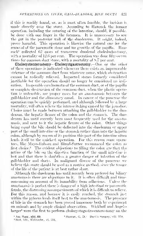
OPERATIONS OX LIVER, GALL-BLADDER, BILE DUCTS 421
if this is rejulily found, or, as is most often feasible, the ineision is
made directly over the stone. According to Hancock the former
operation, including the suturing of the intestine, should, if possible,
be done with one finger in the foramen. It is unnecessary to sew
the gut in the posterior wall of the duodenum. It might, indeed,
prove harmful. This operation is likewise the natural one for the
removal of the pancreatic stone and for growths of the papilla. Han-cock 1 collected 02 cases of transverse duodenal choledochostomy,with a mortality of 12.0 per cent. The operation was done fifty-seventimes for common duct stone, with a mortality of S.7 per cent.
Cholecystenterostomy Cholecystgastrostomy. One or the other
of these operations is indicated whenever there exists an impermeablestricture of the common duct from whatever cause, which obstruction
cannot be radically relieved. Impacted stones formerly considered
justifiable for the operation should no longer be considered an indi-
cation. Inoperable eareinomata of the common duct, or of the papillaor complete destruction of the common duct, when the plastic opera-tion is unfeasible, are proper cases for an anastomosis between the
gall-bladder and the alimentary canal. In cancer of the pancreas the
operation can be quickly performed, and although followed by a large
mortality, will often relieve the intense itching caused by the jaundice.Choice must be made between attaching the gall-bladder to the duo-
denum, the hepatic flexure of the colon and the stomach. The duo-
denum has until recently been most frequently used for the anasto-
mosis, and next to it the hepatic flexure of the colon. Other things
being equal the bile should be deflected into the duodenum or upper
part of the small intestine or the stomach rather than into the hepatic
colon, although by reason of its position this part of the intestine often
lends itself to the quickest operation. For this reason some opera-
tors, like Mayo-Robson and Bland-Sutton recommend the colon as
first choice. 2 The evident objections to filling the colon are that the
action of the bile on the digestive function of the small intestine is
lost and that there is doubtless a greater danger of infection of the
gall-bladder and ducts. In malignant disease of the pancreas webelieve this route should be used as a routine method, since the tenure
of the life of the patient is at best rather short.
Although the duodenum has until recently been prefered for biliary
anastomosis there are objections to it. It is often difficult and time-
consuming on account of its immobility from adhesions. Unless the
anastomosis is perfect there is danger of a high intestinal or pancreatic-
fistula, the distressing accompaniments of which it is difficult to relieve.
For this reason, and because it is easily reached, the stomach just
within the pylorus lends itself best to the anastomosis. The presenceof bile in the stomach has been proved innocuous both by experimenton animals and by ample clinical observation. Wickhoff and Angel s-
berger3 were the first to perform cholecystogastro-enterostomy on the
1 Am. Surg., xliii, 69. - Stewart, G. D.: Buck's Surgery, viii, 270.3 Berl. klin. Wchnschr., vi, 133.
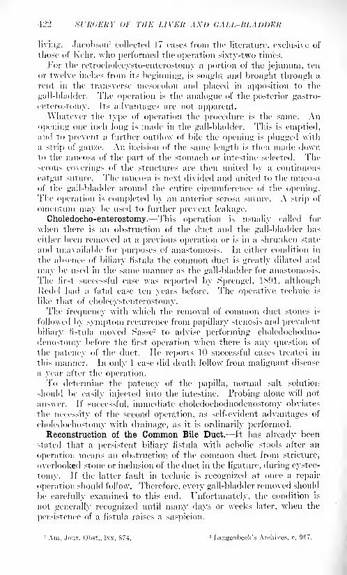
lL'12 XfltdEItY 01- THE LIVER A.\l) CALL-BLADDER
living. Jacobson 1 collected 17 cases from the literature, exclusive of
those of Kehr, \vlio performed the operation sixty-two times.
For the retroc'holecysto-enterostomy a ])ortiou of the jejunum, ten
or twelve inches from its beginning, is sought and brought through a
rent in the transverse mesocolon and placed in apposition to the
gall-bladder. The operation is the analogue of the posterior gastro-
enterostoHiy. Its advantages are not apparent.\\hatever the type of operation the procedure is the same. An
opening one inch long is made in the gall-bladder. This is emptied,and to prevent a further outflow of bile the opening is plugged with
a strip of gau/.e. An incision of the same length is then made downto the mucosa of the part of the stomach or intestine selected. Theserous coverings of the structures are then united by a continuous
catgut suture. The mucosa is next divided and united to the mticosa
of the gall-bladder around the entire circumference of the opening.The operation is completed by an anterior serosa suture. A strip of
omentnm may be used to further prevent leakage.
Choledocho-enterostomy.- This operation is usually called for
when there is an obstruction of the duct and the gall-bladder has
either been removed at a previous operation or is in a shrunken state
and unavailable for purposes of anastomosis. In either condition in
the absence of biliary fistula the common duct is greatly dilated and
may be used in the same manner as the gall-bladder for anastomosis.
The first successful case was reported by Sprengel, IM)!, althoughRedel had a fatal case ten years before. The operative technic is
like that of eholecystenterostomy.The frequency with which the removal of common duct stones is
followed by symptom recurrence from papillary stenosis and prevalent
biliary fistula moved Sasse- to advise performing choledochoduo-
denostomy before the first operation when there is any question of
the patency of the duct. lie reports 10 successful cases treated in
this manner. In only 1 case did death follow from malignant disease
a year after the operation.To determine the patency of the papilla, normal salt solution
should be easily injected into the intestine. Probing alone will not
answer. If successful, immediate choledochoduodenostomy obviates
the necessity of the second operation, as self-evident advantages of
choledochostomy with drainage, as it is ordinarily performed.Reconstruction of the Common Bile Duct. It has already been
stated that a persistent biliary fistula with acholic stools after an
operation means an obstruction of the common duct from stricture,
overlooked stone or inclusion of the duct in the ligature, during cystec-
tomy. If the latter fault in technic is recogni/ed at once a repair
operation should follow. Therefore, every gall-bladder removed should
be carefully examined to this end. Unfortunately, the condition is
not generally recognized until many days or weeks later, when the
persistence of a fistula raises a suspicion.
1 Am. Jour. Obst., Ixx, 874. 2 Langeiibcck's Archives, c, 967.
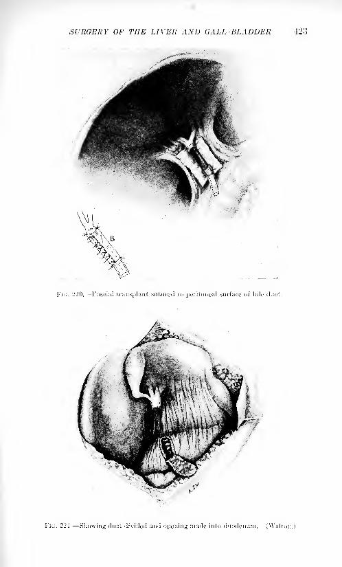
SURGERY OF THE LIVER AND GALL-BLADDER 423
Fin. 220. Fascial transplant sutured to peritoneal surface of bile duct.
FIG. 221 Showing duct divided and opening made into duodenum. (\Valti
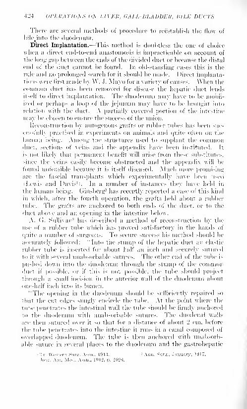
-1J4 U1'J-:J!ATJ(L\^ di\ LlYKli, CALL- HLADDElt, HILK Dl't'TX
There are several methods of procedure to reestablish the flow of
bilejnto the duodenum.Direct Implantation. This method is doubtless the one of ehoiee
when a direct end-to-end anastomosis is impracticable on account of
the long gap between the ends of the divided duet or because the distal
end of the duct cannot be found. In old-standing cases this is the
rule and no prolonged search for it should be made. 1 >irect implanta-tions were first made by W. J. Mayo for a variety of causes. When the
cnminon duet lias been removed for disease the hepatic duct lends
itself to direct implantation. The duodenum may have to be mobil-
ixed or perhaps a loop of the jejunum may have to be brought into
relation with the duct. A partially covered portion of the intestine
may be chosen to ensure the success of the union.
Reconstruction by autogenous grafts or rubber tubes has been suc-
cessfully practised in experiments on animals and quite often on the
human being. Among the structures used to supplant the commonduet, sections of \eins and the appendix have been instituted. It
is not likely that permanent benefit will arise from these substitutes,
since the veins easily become obstructed and the appendix will be
found unfeasible because it is itself diseased. Much more promisingare the fascial transplants which experimentally have been used
Lewi- and Davis 1
'. In a number of instances they have held in
the human being. Ginsberg2 has recently reported a case of tin's kind
in which, after the fourth operation, the grafts held about a rubber
tube. The grafts are anchored to both ends of the duct, or to the
duct above and an opening in the intestine below.
A. (T. Sullivan3 has described a method of reconstruction by the
use of a rubber tube which has proved satisfactory in the hands of
quite a number of surgeons. To secure success his method should be
accurately followed: "Into the stum]) of the hepatic duct an elastic
rubber tube is inserted for about half an inch and securely sutured
to it with several unabsorbable sutures. The other end of the tube is
pushed down into the duodenum through the stum]") of the commonduet if possible, or if this i> not possible, the tube should project
through a small incision in the anterior wall of the duodenum about
one-half inch into its lumen.
"The opening in the duodenum should be sufficiently repaired so
that the cut edges snugly encircle the tube. At the point where the
lube penetrates the intestinal wall the tube should be firmly anchored
to the duodenum with unabsorbable sutures. The duodenal walls
are then sutured over it so that for a distance of about '2 cm. before
the tube penetrates into the intestine it runs in a canal composed of
overlapped duodenum. The tube is then anchored with unabsorb-
able suture in several places to the duodenum and the gastrohepatic
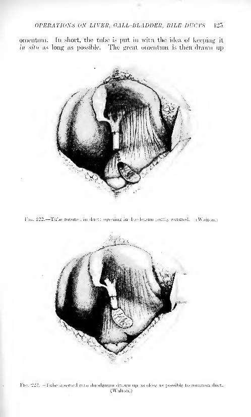
OPERATION'S ON LIVER, CALL-HI,ADDER, BILE DUCTS 125
omentnin. In short, the tube is put in with the idea of keeping it
/// .sv'/// as long as possible. The great omentnm is then dra\vn up
FIG. 222. Tube suture:! in duct; opening in duodenum partly sutured. (Walton.)
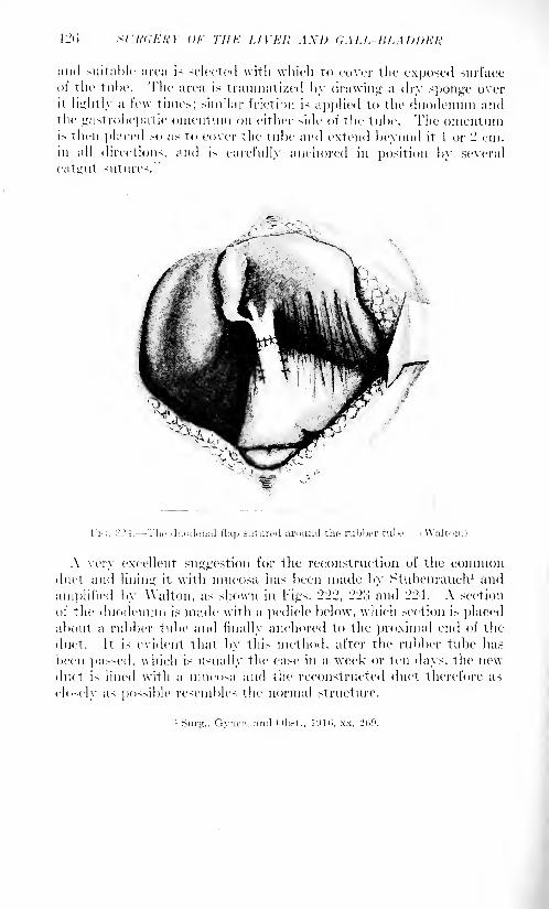
12(1 Sl'WiKKY or THE I. IVEU AX1) <;ALL-KLAl)l)EH
and suitable area is selected \vitli which to cover the exposed surface
ot the tube. The area is traumatized by drawing a dry sponge overit lightly a few times; similar friction is applied to the duodenum andthe gastrohepatic omentuin on either side of the tube. The omentumis then placed so as to cover the tube and extend beyond it 1 or - cm.in all direction-, and is carefully anchored in position by several
cat u-
i it sutures."
A very excellent suggestion for the reconstruction of the commonduct and lining it with mucosa has been made by Stubeiirauch 1 and
amplified by Walton, as shown in Figs. 222, 223 and 224. A section
of the duodenum is made with a pedicle below, which section is placedabout a rubber tube and finally anchored to the proximal end of the
duct. It is evident that by this method, after the rubber tube has
been passed, which is usually the case in a week or ten days, the newduct is lined with a mncosa and the reconstructed duet therefore as
closely as possible resembles the normal structure.
1Sur<r., Gyncr. rind <>b.-t., Kill), xx, 209.
sriWKRY OF THE .PANCREAS.
BY JOIIX B. DKAYKH, M.D.,
AM)
DAMOX B. PFEIFFER, M.I).
ANATOMICAL CONSIDERATIONS.
The pancreas lies transversely across the posterior abdominal wall
at the level of the bodies of the first and second lumbar vertebra 1
. In
the midline it lies directly beneath an area ."> to 10 cm. above the
normally placed umbilicus. The head is an expanded portion of the
gland which is embraced by the concave loop of the duodenum, to
which it is intimately adherent. A short narrow portion, known as the
neck, immediately joins the head to the long tapering body and tail,
the latter two divisions presenting no anatomical demarcation from
each other.
During development in early embryonic life the pancreas is com-
pletely invested by peritoneui.1
,but at a later period by the rotation
of the gut and fusion of the peritoneum it Comes to lie entirely behind
the peritoneum. Here it is anchored by its vascular connections and
surrounding areolar tissue to the posterior abdominal wall and neigh-
boring structures. It is important to note that these connections permita considerable degree of mobility which can be appreciated both with
the hand in the abdomen and when conditions are such that the pan-creas can be felt through the abdominal wall, provided that it is not
the seat of an inflammatory or malignant process that causes it to lie
firmly adherent to its surroundings.
Posteriorly the pancreas crosses, from right to left, the vena cava,
aorta, renal vessels, suprarenal gland and the upper pole of the left
kidney. The tail is in close relation to the lower pole of the spleen and
to the splenic flexure of the colon. The superior mesenteric vessels
emerge beneath the neck of the pancreas in the angle formed by the
neck and the lower prolongation of the head. The splenic artery andvein course above and behind its superior margin. These relations
to the great abdominal vessels are responsible at times for severe, even
fatal secondary hemorrhage, due to erosion by the combined infective
and digestive action of the inflammatory process, in acute pancreatitis.
Anteriorly the pancreas is covered by the liver, the stomach, the
gastrohepatic omentum, the gastrocolic omentnm and the transverse
colon. The relation which these structures hold to the pancreas is veryvariable. Korte, 1 in an examination of MO cadavers, found that upon
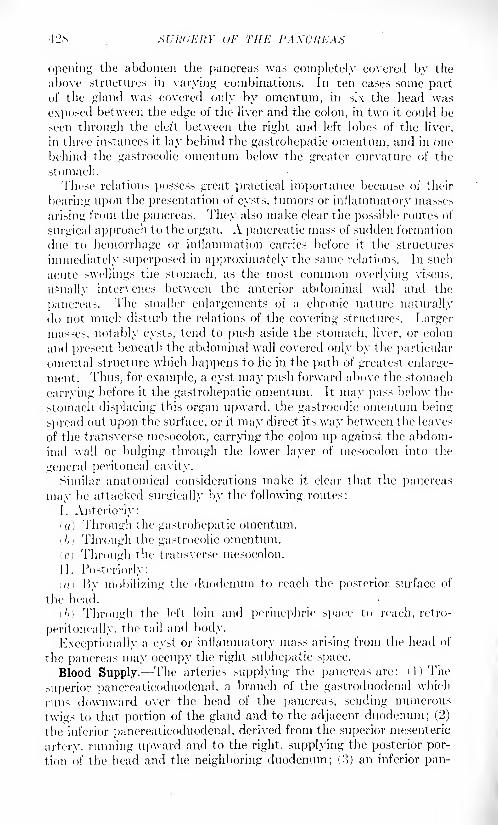
ll2,S SL'h'dKRY OF THE I'AXC'ltEAX
opening the abdomen the pancreas was completely covered by the
above structures in varying combinations. In ten cases some partof the gland was covered only by omentum, in six the head was
exposed between the edge of the liver and the colon, in two it could be
seen through the cleft between the right and left lobes of the liver,
in three instances it lay behind the gastrohepatic omentum, and in one
behind the gastrocolic omentum below the greater curvature of the
stomach.
These relations possess great practical importance because of their
bearing upon the presentation of cysts, tumors or inflammatory masses
arising from the pancreas. They also make clear the possible routes of
surgical approach to the organ. A pancreatic mass of sudden formation
due to hemorrhage or inflammation carries before it the structures
immediately superposed in approximately the same relations. In such
acute swellings the stomach, as the most common overlying viscus,
usually intervenes between the anterior abdominal wall and the
pancreas. The smaller enlargements of a chronic nature naturallydo not much disturb the relations of the covering structures. Larger
masses, notably cysts, tend to push aside the stomach, liver, or colon
and present beneath the abdominal wall covered only by the particular
omental structure which happens to lie in the path of greatest enlarge-
ment. Thus, for example, a cyst may push forward above the stomach
carrying before it the gastrohepatic omentum. It may pass below the
stomach displacing this organ upward, the gastrocolic omentum being
spread out upon the surface, or it may direct its way between the leaves
of the transverse inesocolon, carrying the colon up against the abdom-inal wall or bulging through the lower layer of inesocolon into the
general peritoneal cavity.
Similar anatomical considerations make it clear that the pancreas
may be attacked surgically by the following routes:
I. Anteriorly:
(a) Through the gastrohepatic omentum.l> ' Through the gastrocolic omentum.
(c) Through the transverse inesocolon.
II. Posteriorly:
\a> By mobilizing the duodenum to reach the posterior surface of
the head.
(/;) Through the left loin and perinephric space to read), retro-
peritoneally, the tail and body.
Exceptionally a cyst or inflammatory mass arising from the head of
the pancreas may occupy the right subhepatic space.
Blood Supply. The arteries supplying the pancreas are: (l)The
superior pancreaticoduodenal, a branch of the gastroduodenal which
runs downward over the head of the pancreas, sending numerous
twigs to that portion of the gland and to the adjacent duodenum; (2)
the inferior pancreaticoduodenal, derived from the superior mesenteric
artery, running upward and to the right, supplying the posterior por-
tion of the head and the neighboring duodenum; (3) an inferior pan-
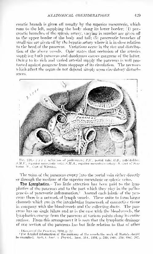
ANATOMICAL CONSIDERATIONS 429
creatic branch is given oft' usually by the superior mesenteric, whichrims to the left, supplying the body along its lower border; (4) pan-creatic branches of the splenic artery, varying in number are given off
to the upper border of the body and tail; (5) pancreatic branches of
small size are given off by the hepatic artery where it is in close relation
to the head of the pancreas. Variations occur in the size and distribu-
tion of the above vessels. Opie1 states that occlusion of the arteries
supplying both pancreas and duodenum causes gangrene of the latter.
Owing to its rich and varied arterial supply the pancreas is well pro-tected against gangrene from stoppage of its circulation. The necroses
which affect the organ do not depend simply upon circulatory disturb-
ances.
x x x x, reflection of peritoneum; P.V., portal vein; G.B., gall-bladdeit>.M.\ ., superior mesenteric vein; S.M.A., superior mesenteric artery; S, duct of San
torim; Tl ., duct of Wirsung.
The veins of the pancreas empty into the portal vein either directlyor through the medium of the superior mesenteric or splenic veins.
The Lymphatics. Too little attention has been paid to the lym-
phatics of the pancreas and to the part which they play in the patho-
genesis of pancreatic inflammation. 2 Around each lobule of the pan-creas there is a network of lymph vessels. These unite to form larger
channels which run in the interlobular framework of connective tissue
in company with the bloodvessels and the collecting ducts. The pan-creas has no single hilum and as is the case with the bloodvessels, the
lymphatics emerge from the pancreas at various points along its entire
surface. From this arrangement it is seen that the lymphatic drainageof one section of the pancreas has but little relation to that of other
1 Diseases of the Pancreas, 1910, p. 22.2 For detailed information of the anatomy of the vessels the work of Bartels should
be consulted. Arch. f. Anat. u. Physiol., Anat, Aht., 1904, p. 299; 1900, 250; 1907, 207.
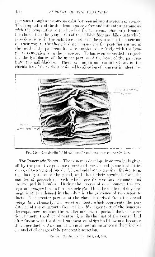
SURGERY OF THE PAXCItKAS
portions, though anastomoses exist between adjacent systems of vessels.
The lymphatics of the duodenum possess free and intimate anastomoseswith the lymphatics of the head of the pancreas. Similarly Franke 1
has shown that the lymphatics of the gall-bladder and bile ducts which
pass downward in the right free border of the gastrohepatic omentumon their way to the thoracic duct course over the posterior surface of
the head of the pancreas, likewise anastomosing freely with the lym-phatics emerging from the pancreas. He lias even succeeded in inject-
ing the lymphatics of the upper portion of the head of the pancreasfrom the gall-bladder. These are important considerations in the
elucidation of the pathogenesis and localization of pancreatic, infectious.
The Pancreatic Ducts.- -The pancreas develops from two buds givenoil' by the primitive gut, one dorsal and one ventral (some authorities
speak of two ventral buds). These buds by progressive division form
the duct systems of the gland, and about their terminals form the
mantles of parenchyma cells which are its secreting elements and
are grouped in lobules. During the process of development the two
separate anlaycn fuse to form a single gland but the method of develop-ment is still evidenced in the, adult in the existence of two separateducts. The greater portion of the gland is derived from the dorsal
(inl(if/c but, strangely, the secretory duct, which represents the per-
sistence of the outgrowth from which this larger part of the pancreas
develops, now becomes the smaller and less important duct of excre-
tion, namely, the duct of Santorini, while the duct of the ventral budafter fusion with the dorsal rudiment outstrips its fellow and becomesthe larger duct of AVirsung, which in almost all instances is the principal
channel of discharge of the pancreatic secretion.
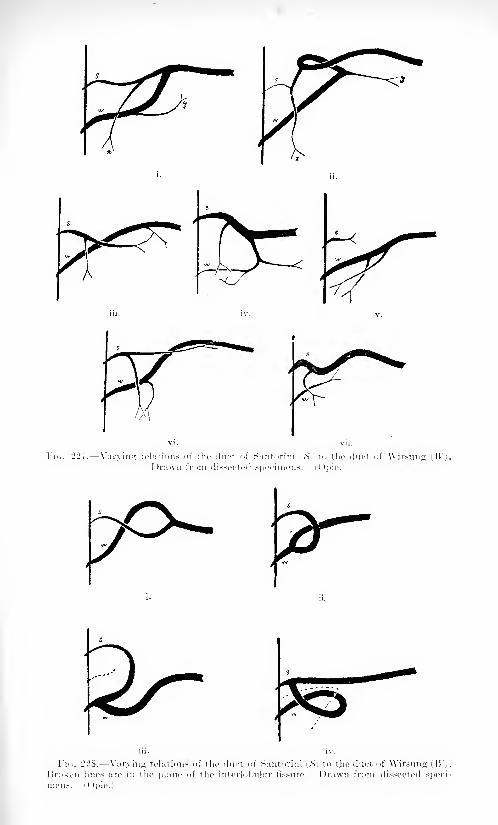
sj ^r~
^*
/r
"7S
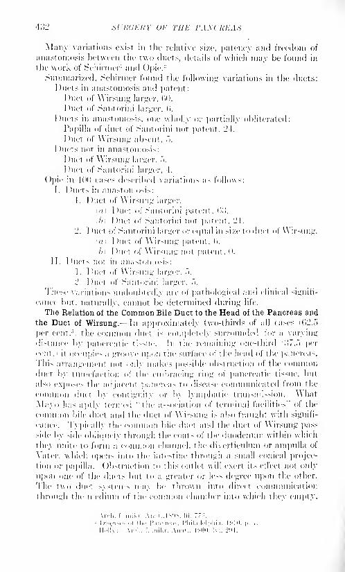
SL'M.'XKY OF THE I'AXCKKAX
Many variations exist in the relative size, patency and freedom of
anastomosis between tlie two ducts, details of which may be found in
the work of Schirmer 1 and Opie.-
Smnniarized, Schirmer found the following variations in the ducts:
Ducts in anastomosis and patent:1 hict of Wirsung larger, ISO.
1 hict of Santorini larger, ('.
Duets in anastomosis, one wholly or partially obliterated:
Papilla of duct of Santorini not patent, '24.
Duct of Wirsung absent, .">.
I >ucts not in anastomosisI )uct of Wirsung larger, ."j.
Duct of Santorini larger, -1.
()])ie in 100 cases described variations as follows:
I. 1 hicts in anastomosis:
1. Duct of Wirsung larger.
) Duct of Santorini patent, ti:>.
:l Duct of Santorini not patent, L'l.
1'. I )uct of Santorini larger or e(|iial in size to duct of Wirsung.
(a) Duct of Wirsung patent, C>.
//! Duct of Wirsung not patent, 0.
II. Ducts not in anastomosis:
1. Duct of Wrsung larger, o.
-. Duct of Santorini larger. ~>.
r
rhese variation- undoubtedly are of pathological and clinical signifi-
cance but. naturally, cannot be determined (hiring life.
The Relation of the Common Bile Duct to the Head of the Pancreas and
the Duct of Wirsung. In approximately two-thirds of all cases (02.5
per cent. 3) the common duct i- completely surrounded for a varying
distance by pancreatic tN>uc. hi the remaining one-third 'o7..~> percent. it occupies a groove upon the surface of the head of the pancreas.This arrangement not only makes possible obstruction of the commonduct by tumefaction of the embracing ring of pancreatic tissue, but
also exposes the adjacent pancreas To disease communicated from the
common duct by contiguity or by lymphatic transmission, \\hat
Mayo lias aptly termed "the association of terminal facilities" of the
common bile duct and the duct of Wirsung is also fraught with signifi-
cance. Typically the common bile duct and the duct of Wirsung passside by side obliquely through the coats of the duodenum within which
they unite to form a common channel, the diverticulum or ampulla of
\ater. which o]>rn> into the intestine through a small conical projec-tion or papilla. Obstruction to this outlet will exert its ell'ect not only
upon one of the ducts but to a greater or less degree upon the other.
The two duct systems may be thrown into direct communication
through the medium of the common chamber into which they empty.
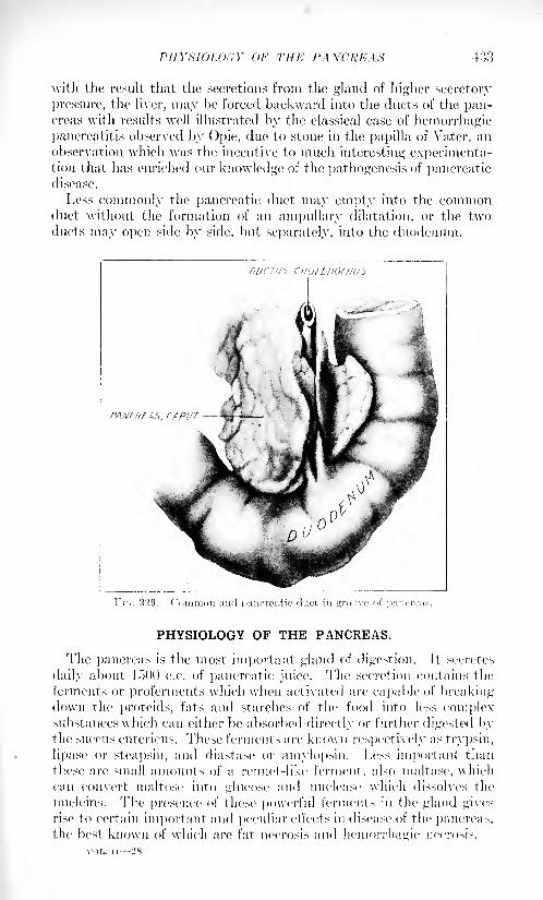
PHYSIOLOGY OF THE PANCREAS 433
with the result that the secretions from the gland of higher secretory
pressure, the liver, may be forced backward into the ducts of the pan-creas with results wr
ell illustrated by the classical case of hemorrhagicpancreatitis observed by Opie, due to stone in the papilla of Vater, anobservation which was the incentive to much interesting experimenta-tion that has enriched our knowledge of the pathogenesis of pancreaticdisease.
Less commonly the pancreatic duct may empty into the commonduct without the formation of an ampullary dilatation, or the twoducts may open side by side, but separately, into the duodenum.
PHYSIOLOGY OF THE PANCREAS.
The pancreas is the most important gland of digestion. It secretes
daily about 1500 c.c. of pancreatic juice. The secretion contains the
ferments or preferments which when activated are capable of breakingdown the proteids, fats and starches of the food into less complexsubstances which can cither be absorbed directly or further digested bythe succus entericus. These ferments are known respectively as trypsin,
lipase or steapsin, and diastase or amylopsin. Less important than
these are small amounts of a rennet-like ferment, also maltase, whichcan convert maltose into glucose and nuclease which dissolves the
nucleins. The presence of these powerful ferments in the gland gives
rise to certain important and peculiar effects in disease of the pancreas,the best known of which are fat necrosis and hemorrhagic necrosis.
VOL. ii 25
}:il .sTAv/AV.'y OF Tin-: PAXCRKAX
In itdilition To its external secretion the ])iincre;is supplies an internal
secretion which is essential to carbohydrate metabolism. There is
evidence also of the presence of a second internal secretion which is
necessary in order that the intestine may properly exercise its function
of absorption.
Pathological Physiology. In the case of an organ with such extra-
ordinarily varied and important functions it is natural to inquire
whether its varied disorders are reflected in disturbances of function,
whether these functional disturbances are distinctive in kind and pro-
portionate in decree to the underlying disease and JUT anitrii. whether
demonstrable abnormalities of function may be of practical service in
diagnosis. The two striking phenomena just mentioned, fat necrosis
and pancreatic hemorrhage, are examples jxir I'.rccllrncc of such etfects
but are unavailable for diagnosis except in the serious types of the
disease and only during operation.The tests designed to demonstrate the functional activity of the
pancreas fall naturally into three groups:1. Tests of external secretion in,
ia) 1 hiodenal contents.
(//) (iastric contents.
Stool.
\ rine.
HI 1.
Action ii])on test materials introduced into the alimentarytract.
'2. Tests of internal secretion in,
( a i I rine.
! 1> I StOol.
(r) Infects exercised through medium of sympathetic nervous
system..'!. Metabolic studies on absorption of fat, nitrogen, etc., from
intestines.
Attention was called early to the abnormal character of the stools
in certain cases of advanced pancreatic disease. Bright,1 in IS.'!.'!,
clearly described the excessive amount of fat occasionally found in the
stools in this condition (steatorrhea). At the same time he recognizedthe inconstancy and unreliability of the symptom as indicative of
pathological changes in the gland. The "frequent, bulky motions,
pale in color, offensive and obviously greasy," as described by Robson,are strongly suggestive of deficiency or absence of the external secretion
of the pancrea- with consequent failure of digestion. The abundance
of fat and fatty acids is due to a deficiency of lipase, and the lack of
tryptic activity is shown by the presence of striated muscle fibers in a
more or less incomplete state of digestion (creatorrhea).
The attempt lias been made to increase the value of such derange-
ments by more accurate metabolic studies. The intake and output
I'lIYMOLOUY OF THE PANCREAS 4M5
of fat and nitrogenous material are easily capable of estimation, and it
may be roughly stated that the more serious and advanced the disease
of the pancreas the greater the loss of fat and nitrogen. Unfortunatelyfor the practical utility of such estimation it has been found that the
factors involved are of such complexity and their interrelationship so
uncertain that clinical deductions are untrustworthy. The bodypossesses certain "factors of safety" which may prove sufficient to
carry on digestion in the complete absence of the external secretion of
the pancreas. Moreover, absorption may be greatly impaired from
other causes even when the pancreatic ferments are abundantlysecreted and fully capable of playing their full part in digestion.
Evidence exists to show that the pancreas itself normally possessesan internal secretion or "hormone" which is essential to proper absorp-tion of the food from the intestine and that this may be deficient
even though the external secretion be normal. Other disease, such
as abdominal tuberculosis, may interfere with intestinal absorption.Crolm. 1 from a resume of the literature and his own metabolic studies,
concludes that: 1. The nature of the disease (of the pancreas) docs
not determine the degree of fat and nitrogen loss.
2. The amount of food absorption is independent of the patency of
the duct or the activity of the enzymes of the external secretion of the
gland.M. The degree of interference with intestinal absorption is dependent
on organic disease or functional derangement of the parenchyma of the
gland, the pancreas probably normally controlling intestinal absorption
by an internal secretion or hormone.
The classical experiments of von Mering and Minkowski2proved
that an internal secretion of the pancreas was essential to carbohydratemetabolism. Complete pancreateetomy is invariably followed bya rapid and fatal diabetes. Pancreatic disease in certain cases placed,
by Fitz 3 at MS per cent, and by Deaver 4 in an operative series com-
prising earlier cases, at o.M per cent., is associated with glycosuria.
There is no reason to doubt that glycosuria in these conditions is
dependent upon a diminution or perversion of the internal secretion
of the pancreas. It may be intermittent and called forth by exacerba-
tions of the pancreatic disease. More recent studies, notably those of
Opie, have indicated that the islands of Langerhans are the anatomical
elements of the pancreas chiefly, if not solely, concerned with this
particular secretion. It is not possible in all cases of diabetes to demon-strate disease of the islands, nor is it believed that all glycosuria is of
pancreatic origin. The islands may undergo extensive degenerationwithout evidence of disease of the secretory parenchyma and converselythe acini may show marked pathological changes without apparent
change in the islands or disturbance of their function, ft may be said,
Arch. Int. Mod., 19i:>, xv, GUI'.
2 Arch. f. cxpor. Path. u. Pharm., 189!), xxvi, 371.3 Tr. Contrr. Am. Phys. and Sun;., 1903, vi, 3ti.
Jour. Am. Med. Assn., 1911, Ivi, 1079.
4:>(> SL'RdERY OF THE I'AXCIfEAS
however, that glycosuria when associated with otlier e\'idence of dis-
ease of the pancreas tends to substantiate the diagnosis.
The Cammidge reaction in the liands of its originator is credited with
a high degree of accuracy in tlie diagnosis of pancreatitis. But in over
1000 cases of upper abdominal disease in the Lankenau (formerly the
(ierman) Hospital of Philadelphia we have not found it helpful. So;ne
favorable opinions are to be found in the literature, notably Robsonand Moynihan, with the former of whom Cammidge lias been asso-
ciated, but the weight of experience in other clinics has been against
the reliability of its results.
Loewi's 1
pupillary reaction is founded on the fact that in panereatee-tomi/ed animals, dilatation of the pupil is caused by the instillation
of adrenalin into the eye, which under normal conditions produces
myosis. lie attributes the reaction to an increased irritability of the
central nervous system. A number of authors have observed this
phenomenon in man, though inconstantly and in a varying percentageof cases. Other similar effects of sympathicotonia, such as the ocular
phenomenon more commonly associated with hyperthyroidism, and
tremor, as well a> Ivocher's sign of tenderness of the thyroid have been
found associated with pancreatic disease but only as inconstant, incon-
spicuous and relatively valueless signs.
The presence and the activity of the external secretion of the pan-creas may be tested for indirectly by introducing into the alimentarytract certain substances which undergo recognizable changes whenacted upon by the pancreatic ferments or, directly, by estimating the
ferment activity of the duodenal contents obtained /// xilii or after
regurgitation into the stomach, or of the ferments in the stool, urine or
blood.
Examples of the indirect method are Sahli's glutoid capsules and the
sic gelodurat capsule of Miiller and Schlecht which are unaffected bythe gastric juice but are dissolved by active pancreatic secretion in the
intestine, liberating substances chemically recognizable when absorbed
and excreted in the urine, or observed free in the stool after dissolu-
tion of the capsule. Schmidt's nuclein test consists in the admin-
istration of thymus or other tissues rich in nuclei, which are later
searched for in the feces, incomplete digestion indicating a deficiency
of the nuclease of the pancreas. Atchley- found Schmidt's test nega-tive in a case of complete occlusion of the pancreatic duct and con-
eluded that in the human subject the digestion of nuclear material,
the purin metabolism and the uric acid formation do not depend on
the presence of the pancreatic secretion in the intestinal canal. In
Ehrmann's palmitin test the digestion of this fat can be determined
by a chemical color reaction of the liberated fatty acid with copperacetate. Still other tests based on similar principles have been pro-
posed. But all these tests have been shown to be subject to disturbing
factor-- that cannot be eliminated and which greatl reduce their value.
ABNORMALITIES OF THE PANCREAS 437
I'nder normal circumstances trypsin, steapsin and diastase can be
demonstrated in the feces and their activity determined quantitatively.
"While an approximate normal can be determined the attempt to corre-
late departures from that normal to disease of the pancreas has been
in the main unsatisfactory.
Boldyreff1 discovered that duodenal contents could be caused to
regurgitate into the stomach by ingest ion of a large amount of olive oil.
Volhard2 has applied this method of obtaining pancreatic juice in the
clinic. More accurate and ingenious is the method introduced byEinhorn for the recovery of duodenal contents, which as now modified
consists in the introduction into the stomach of a rubber tube of fine
bore with a perforated metal olive-shaped tip. The tube is of sufficient
length to reach the duodenum, into which, in a few hours, the metal
tip usually finds its way. Duodenal juice may then be aspirated andtested directly for ferments. More constant results have been claimed
for the results of examinations thus made.
The findings with this method during the last five years, however,show conclusively that the problem of correlating functional abnor-
malities with the underlying pathology of the pancreas remains yetto be solved so far as results of practical utility are concerned.
Wohlgemuth3 found that in experimental occlusion of the pancreatic
duct the amount of diastase in the urine rose greatly. lie observed
the same phenomena in acute pancreatitis and functional disturbance
of the pancreas. Wohlgemuth and Xoguehi1 attach great importance
to a marked rise in the diastase of the blood and urine as indicative of
injury to the pancreas in cases of abdominal traumatism.
ABNORMALITIES OF THE PANCREAS.
Abnormalities of the pancreas are not uncommon. Minor altera-
tions in size and shape are the rule. One or more deep constrictions
may be found. The tail is sometimes forked. Fusion of the two anlaycn
may fail to occur with the result that two separate glands are found.
Accessory Pancreas. Aberrant islands of pancreatic tissue have
been found many times and are probably more common than is sus-
pected. As a rule they are in the wall of the stomach or duodenum,but they have been observed in various portions of the intestinal tract
as far down as the lower ileum. They always have a duct connectingthem with the bowel. 5 A number of cases of gastric or intestinal divcr-
ticula associated with aberrant pancreatic tissue have been described.
Such diverticula may be the seat of acute inflammation and have been
known to cause intestinal obstruction.
Annular Pancreas. A curious though uncommon anomaly which
may possess surgical significance through the possibilities of obstruc-
' Zrntrulbl. f. PhvMu].. 11)04, xviii. 457.- Mimrhi-n. HUM!. Wdinsclir., 1907. llv, 401',.
" Hiorhem. Ztsdir., 190S, ix, 1.
4 Berl. klin. Wrlmsrhr., 1912, i, lOii!).
;' Zonker: Virrhmvs Arch., 1 S(>1 , xxi, .'!(>!).
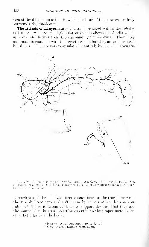
438 SURGERY OF THE PANCREAS
(ion of the duodenum is that in which the head of the pancreas entirelysurrounds the duodenum.The Islands of Langerhans. Centrally situated within the lobules
of the pancreas are small globular or ovoid collections of cells which
appear quite distinct from the surrounding parenchyma. They havean origin
1 in common with the secreting acini but they are not arrangedin t ubtilcs. They are not encapsulated or cut irely independent from the
parenchyma of the acini as direct connections can be traced between
the two different types of epithelium by means of slender cords or
tubules.- There is strong evidence to support the idea that they are
the source of an internal secretion essential to the proper metabolism
of carbohydrates in the bodv.
1 I'ojircc: Am. Jour. Anat,., 1903, xi, 445.-Opio, Poarce. Karakascheff, Cecil.
INJURIES OF THE PANCREAS
INJURIES OF THE PANCREAS.
Wounds. \Younds of the pancreas occur in three forms: (1) Pene-
trating wounds, (2) gunshot wounds, and ('.}) subcutaneous rupture.
Penetrating Wounds. Penetrating wounds most often result from a
stab. Gunshot wounds are common in war and are almost uniformlyfatal under the conditions of military, surgery.
1 In civil life they are
less common than subcutaneous rupture or laceration of the organwhich is due to the application of blunt force to the abdominal wall
over the pancreas crushing the organ between the object inflicting
the violence in front and the vertebral column behind. The relative
frequency of these types of injury is indicated by Mikulic/'s- statistics.
lie found in 4.1 cases of injury to the pancreas 24 subcutaneous rup-
tures, 12 gunshot wounds and 9 stab wounds.
In all types of pancreatic injury there are usually more or less serious
wounds of other viscera. Instances of isolated injury of the pancreashave been reported in all the above types, but, as a rule, the associated
lesions are severe and often attract the most immediate attention. So
true is this that all who have been interested in the subject have
emphasized the necessity of considering carefully the condition of the
pancreas in all cases where it has been exposed to the possibility of
injury in order to avoid overlooking a lesion.
Gunshot Wounds. Luxembourg-" collected .'51 cases of gunshot woundof the pancreas. Of the 24 operated upon i:> died and 11 recovered;
7 not operated upon died. Famous and sad cases of fatal gunshotwounds of the pancreas are those of Presidents Garfield and McKinley.
Holzwarth 4reports the case of a soldier who was shot through the
pancreas, who made an immediate recovery but later was operated
upon for pancreatic cyst which followed the track of the bullet and
required incision both anteriorly and posteriorly. In Luxembourg'sseries the pancreas alone was injured only three times with two
recoveries and one death. Other organs injured were, liver 12, stomach
14, intestines ,'->. left kidney 2, heart 1, rib 1, vertebral column 1,
scapula 1.
Treatment.- There are no immediate symptoms by which the partici-
pation of the pancreas in gunshot or stab wounds may be known. As
1 Observations made during the late war substantiate this statement. Thus WallaceWar Surgery of the Abdomen. .1. and A. Churchill, London. 1918. p. 1(J." states that
"in 965 operated cases the pancreas was wounded o times. Very few of such injuries
have been noted. Probably many are missed and diagnosed as retro peritoneal hematnma.In addition, many may be rapidly fatal: the association of the <dand with biu: vessels
needs hardly be mentioned . . . the wound was associate 1 three times with other
viscera; stomach: stomach and liver; stomach and kidney -all fatal; alone on two acca-
sioiis. One went to base without symptoms, the other died. There are no distinctive
symptoms; the lesions are found at exploration and are sometimes accompanied by fat
necrosis." Makins (Brit. Jour. Surg., March, 1916, p. (>">">) says that "injuries to the
pancreas are difficult to detect except by direct exposure of the or<_ran . . . the
typical results of the escape of the pancreatic secretion into the tissues and peritoneal
cavity have been observed. Such injuries have usually proved fatal."- Mitt. a. d. Gren/ub. d. Chir u. Med.. 190:!, xii. 1.
Deutsch. Ztschr. f. Chir.. 1912, cxvii, 2S4.4 1'ester med-chir. Presse, 191"), xxi-xxii, 64.
440 SURCKHY (>!'' Till-: 1'AXCKKAS
all >ucli injuries a iv to be treated hy immediate laparotomy it remains
only for the surgeon to hear in mind the possibility and to explore for
evidences of injury to the pancreas. If in doubt the posterior peri-
toneum should be opened and areas suffused with blood, carefully
searched. Failure to detect and properly drain pancreatic wounds is
apt tore-ult fatally. Moynihan recommends posterior drainage. AYhile
tin's is probably preferable in \vounds of the tail or body, wounds of
the head, can be better drained anteriorly.
Subcutaneous Rupture.- Subcutaneous rupture occurs most often
from the kick of a horse, the passing of a wheel over the body, a blowin the epigastrium by a wagon tongue or shaft or by being squeezedbetween two cars.
Symptoms. Extreme pain and collapse commonly follow subcu-
taneous rupture. Vomiting is usually present. Early, the temperatureand pulse are subnormal as in shock; later they tend to rise. The epi-
gastrium is tender and the muscles moderately rigid. Free fluid in the
abdomen may be demonstrable and after a time, in some cases, dulness
in the left hypochondrium and loin indicate the presence of blood or an
elfusion in the lesser peritoneal cavity.
Wohlgemuth and Xoguchi1 have demonstrated an increase of diastase
in the blood and urine after pancreatic injury and maintain that in all
cases where this increase is markedly above the normal, following a
blow upon the abdomen, one can be sure that the pancreas is injured.
Subsequent symptoms depend much upon the turn taken by the
lesion. Hemorrhage from the pancreas itself is rarely so severe as to
be alarming. The injury may, however, cause hemorrhage into the
pancreas itself and extravasation of pancreatic secretion can set upall the symptoms of pancreasvergiftitng as seen in acute hemorrhagic
pancreatitis.
Localized abscess, peritonitis or retroperitoneal suppuration mayoccur. In Mime instances a sterile pseudocyst forms and is discovered
as a mass several weeks or months after the injury.
Prognosis ,- Ileinecke- collected 10 cases of rupture of the pancreasof which five died without operation. Of the 14 cases operated uponnine recovered. No case died in which the lesion received timely recog-
nition and treatment. (hileke 3 likewise recommends immediate
operation but states that when the symptoms are evidently subsidingit is better to wait until adhesions have formed about the extravasate.
Treatment.- The arguments in favor of immediate operation are
unanswerable, as only in this way can the dangers of pancreatic intoxi-
cation, necrosis and infection be anticipated. Doubtless in cases that
eventuate in sterile psetulocysts the necessity is less pressing, but there
is no way of forecasting such an outcome in the individual case. At
operation blood or serosanguineous fluid in moderate amount is usuallyfound free in the abdominal cavity. The mesentery and omenta may
1 Merl. klin. Wclmsohr., 1912, xlix. lOlii).
-Arch. f. klin. fhir., 11)07, Ixxxiv, 4.
3 MiniflicTi. mod. Woluix-br., 1910, v, 75.
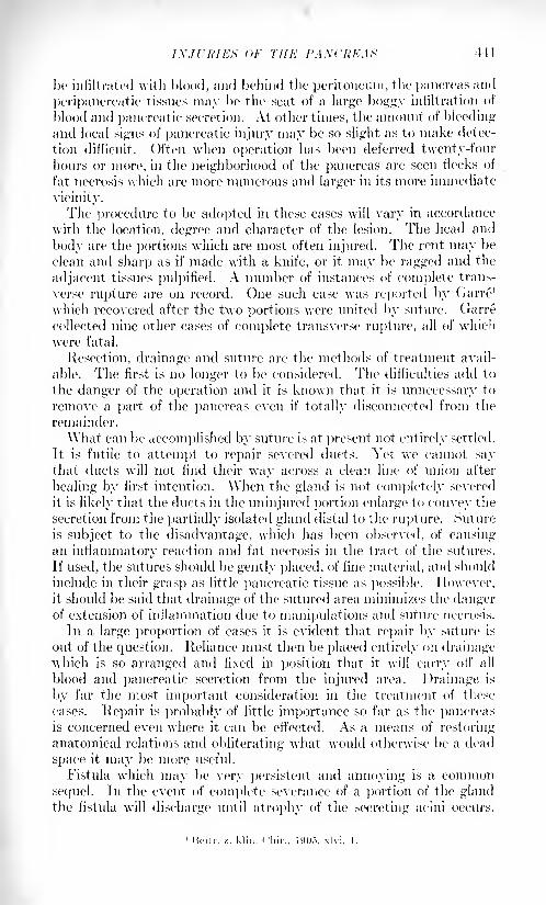
IXJl'HIKS OF Till': PAXCREAS
be infiltrated with blood, and behind the peritoneum, the pancreas and
peripancreatic tissues may be the seat of a large boggy infiltration of
blood and pancreatic secretion. At other times, the amount of bleeding
and local signs of pancreatic injury may be so slight as to make detec-
tion difficult. Often when operation has been deferred twenty-fourhours or more, in the neighborhood of the pancreas are seen flecks of
fat necrosis which are more numerous and larger in its more immediate
vicinity.
The procedure to be adopted in these cases will vary in accordance
with the location, degree and character of the lesion. The head and
body are the portions which are most often injured. The rent may be
clean and sharp as if made with a knife, or it may be ragged and the
adjacent tissues pulpified. A number of instances of complete trans-
verse rupture are on record. One such case was reported by Garre 1
which recovered after the two portions were united by suture. Garre
collected nine other cases of complete transverse rupture, all of which
were fatal.
Resection, drainage and suture are the methods of treatment avail-
able. The first is no longer to be considered. The difficulties add to
the danger of the operation and it is known that it is unnecessary to
remove a part of the pancreas even if totally disconnected from the
remainder.
What can be accomplished by suture is at present not entirely settled.
It is futile to attempt to repair severed ducts. Yet we cannot saythat ducts will not find their way across a clean line of union after
healing by first intention. When the gland is not completely severed
it is likely that the ducts in the uninjured portion enlarge to convey the
secretion from the partially isolated gland distal to the rupture. Suture
is subject to the disadvantage, which has been observed, of causingan inflammatory reaction and fat necrosis in the tract of the sutures.
If used, the sutures should be gently placed, of fine material, and should
include in their grasp as little pancreatic tissue as possible. However,it should be said that drainage of the sutured area minimizes the dangerof extension of inflammation due to manipulations and suture necrosis.
In a large proportion of cases it is evident that repair by suture is
out of the question. Reliance must then be placed entirely on drainagewhich is so arranged and fixed in position that it will carry off all
blood and pancreatic secretion from the injured area. Drainage is
by far the most important consideration in the treatment of these
cases. Repair is probably of little importance so far as the pancreasis concerned even where it can be effected. As a means of restoring
anatomical relations and obliterating what would otherwise be a dead
space it may be more useful.
Fistula which may be very persistent and annoying is a commonsequel. In the event of complete severance of a portion of the glandthe fistula will discharge until atrophy of the secreting acini occurs.
1-lL' M'lWKRY OF THE PANCREAS
In other cases the discharge lasts until there is marked cicatrization of
the injured surface and formation of sufficient collateral ducts. In
chronic cases the Wohlgemnth diet should he tried. Ileinecke' reportsa case which had persisted eighty days, but closed in three days after
the adoption of an antidiabetic diet. Konig- also had a strikingly
favorable example of the efficiency of this diet. As fistula formation and
persistence does not in every case depend upon the amount and irri-
tating character of the secretion, the diet will not prove efficacious in
every instance. Korte ingeniously and successfully dissected out a
chronic fibrous sinus and joined it to the bowel (fistulo-anastomosis).
In all cases the skin should be protected at once from the action of
the discharge by vaselin or a bland ointment. Kroiss3reports a success-
ful case of subcutaneous rupture of both duodenum and pancreas in
which difficulty was experienced during convalescence owing to the
external discharge of practically all the pancreatic secretion and con-
sequent defective digestion and nourishment. lie observed excellent
results from the use of erepton, and others have recommended pre-
digested foods in similar conditions.
INFLAMMATION OF THE PANCREAS.
Pathogenesis of Pancreatic Inflammation. 'Fhe retroperitoneal posi-
tion of the pancreas, the difficulty of gaining by inspection or palpationan exact idea of the condition of the organ during life even when the
abdomen is opened, the complexity of the factors introduced by its
numerous and extraordinary functions, as well as the indifference and
neglect with which it has been treated until very recent times, have
left many gaps to be filled before a complete understanding of its dis-
eases can be said to have been readied. However, by a combination
of recent clinical and pathological observations with data supplied
by experiment we are in a position to draw a number of very importantconclusions.
A difficult question has been the role of infection in pancreatitis. It
must be admitted that under certain conditions the pancreas has the
property of destroying itself by means of its own ferments. Theearliest observations were made upon cases in which the ferment
phenomena were the striking features. Fat necrosis and hemorrhageshowed no obvious relation to infection. Such infections as were found
could readily be explained as secondary invasion of injured tissues. Theclassical case reported by Opie, of a gall-stone impacted in the papilla
of Yater in a case of hemorrhagic pancreatitis led to the discovery that
the disease could be reproduced by injection of bile and other sterile
materials into the pancreatic duct. Since Opie's case a number of
similar occurrences have been noted and in the ideas of pancreatitis
suggested by this mode of causation and by the type of experiments to
INFLAMMATION OF THE PANCREAS 443
\vliich it gave the incentive, the factor of infection was either dis-
regarded or assigned a very subsidiary role. Cases of traumatic pan-creatitis also emphasized the fact that infection was not a necessary
agent in the production of the essential features of acute pancreatitis.
Gradually, however, it became clear that at least some of the cases of
acute pancreatitis were to be accounted for in some other way than bythe impaction of a gall-stone in such a manner as to cause retrojectionof bile into the pancreatic duct, since in a percentage of cases gall-
stones were absent. Bobit/ki 1
reported a case of fatal acute pancreatitisassociated with cholelithiasis. The ducts were dissected out and found
to enter the duodenum side by side, no ampulla being present. Two-thirds of the gland were destroyed. Bacteria could not be demon-strated. Carwardine and Short'2 describe a case of acute pancreatitis
affecting an accessory pancreas in the wall of the jejunum one and one-
half inches beyond the duodenojejimal junction. Simple obstruction
of the duct was shown to produce atrophy without the characteristic
inflammatory changes. The hypothesis of regurgitation of duodenal
contents into the duct received considerable attention. Hlava 3 found
that duodenal contents injected into the duct produced acute lieinor-
rhagic pancreatitis. Seidel 4 showed that occasionally it is possiblefor duodenal contents to enter the duct of Wirsung. Other observers
have been unable to obtain this result and maintain that the obliquityof that portion of the duct which penetrates the duodenum is sufficient
defense against reflux of the duodenal contents. Korte,5 Oser6 and
others attempted to reproduce the disease by various methods of
altering and disturbing the blood supply but nothing comparable to
the clinical condition resulted. The net results of the vast amount of
experimental work along these and similar lines established the fact
that pancreatitis could be produced by the injection of various sub-
stances into the pancreatic duct, the only feature which these methods
possess in common being found in injury of the tissues and activation
of the pancreatic juice within the substance of the gland. The onlydemonstrated clinical parallel remained in the circumstances illustrated
by Opie's case of stone impacted in the common ampulla of the bile
and pancreatic ducts. 7 To account for the residue of cases in whichthis mechanism was ruled out, more recent work has reverted to the
role of infective processes which play the chief part in the inflammationof all other glands which by their position are exposed to the chance of
infection. Another circumstance which directed attention to infection
1 Arch. f. klin. Chir., 1912, xcvii. 143. 2 Ann. Sur/., 1913, i, 053.3 Bull, intermit, de 1'acad. des science de Boheme, 1897.4 Beitr. z. klin. Chir., 1913. Ixxxv, 350.5 Berl. Klin., December, 1896.6 Nothnagel's spec. Path. u. Ther.. p. 338.7 Archibald, of Montreal, believes that the forcible injection of bile into the pancreatic
duct may occur as a result of excessive toxicity or spasm of the sphincter of ( )ddi. whichcontrols the outlet of the papilla of Vator. He lias shown that this sphincter may resist
a pressure of 300 to 700 mm. of water and that in cats it was possible to inject the pan-creatic duct from the choledochus by suddenly raising the pressure in the bile duct or
by throwing the spincter into spasm by the application of acid-
HI SCItdKItY OF Till': 1'AXCRKAS
of the pancreas was tlio observation that chronic and subaeute pan-creatitis, as frequently observed at operation, is associated in the
vast majority of cases with frankly infective processes of the biliarytract and to a less extent with pylor.ic or duodenal ulcer and with such
other lesions of the Castro-intestinal tract as give rise to retroperitonealinflammation. The conditions found in the pancreas could be explainedon no other basis than that of bacterial activity. Still the idea of duct-
borne agencies ruled medical thought and it was assumed that infection
extended by way of the ducts. In those cases of pancreatitis associated
with disease of the biliary tract it was easy to picture a eatarrhal int'ec-
t ion propagated directly from the bile duct up the pancreatic duct whichis in such close relation to it. While this did not explain pancreatitisassociated with other lesions, it was still possible to assume direct
infection of the duct from the duodenum. Deaver and Pfeitl'er1 have
pointed out several factors which strongly militate against this facile
explanation. In their experience about one-third C>(> per cent.) of
all instances of chronic pancreatitis, and 12 per cent, of acute cases
were not accompanied by demonstrable disease of the biliary passages.This is evidence that in a large proportion of cases the inflammatory
process is not communicated to the pancreas from an infected bile
duct. Moreover, there is a lack of parallelism between pancreatic and
biliary disease in their sex incidence. It is well known that gall-
bladder disease is more frequent among women, the ratio being about
three to two. In pancreatitis the ratio is reversed. This correspondswith the original observations of Fit/ and with subsequent experienceof surgeons the world over who agree that pancreatitis is more commonin the male sex. Evidently pancreatitis is something other than a dis-
ease dependent upon primary inflammation or lithiasis of the biliary
tract.
Furthermore, in the early cases, which are evidently secondary to
cholecystitis, it is generally agreed that it is the head and often only
the upper portion of the head of the pancreas that is chiefly affected,
the remainder of the gland frequently being normal, so far as can be
determined by inspection and touch. Now this portion of the gland is
not in the closest relation to the duct of Wirsung, and, in any event,
it is difficult to understand why an infection arriving by way of the
duct should not involve symmetrically the entire gland rather than
merely a portion of the head.
On the other hand, if we ascribe the infection of the pancreas to
those great carriers of bacteria and infective products, the lymphatics,
these peculiarities are readily explained. Bartels showed that an extra-
ordinarily intimate relation exists between the lymphatics of the
duodenum and other abdominal organs and those of the head of the
pancreas.Franke 2 made a special study of the lymphatics of the gall-bladder
and was able to show a similar close connection to those of the pan-
1 Am. Jour. Mod. Sc., 1912, cliii, 473.
MVutsrh. /tsdir. f. ("liir.. September. 1911.
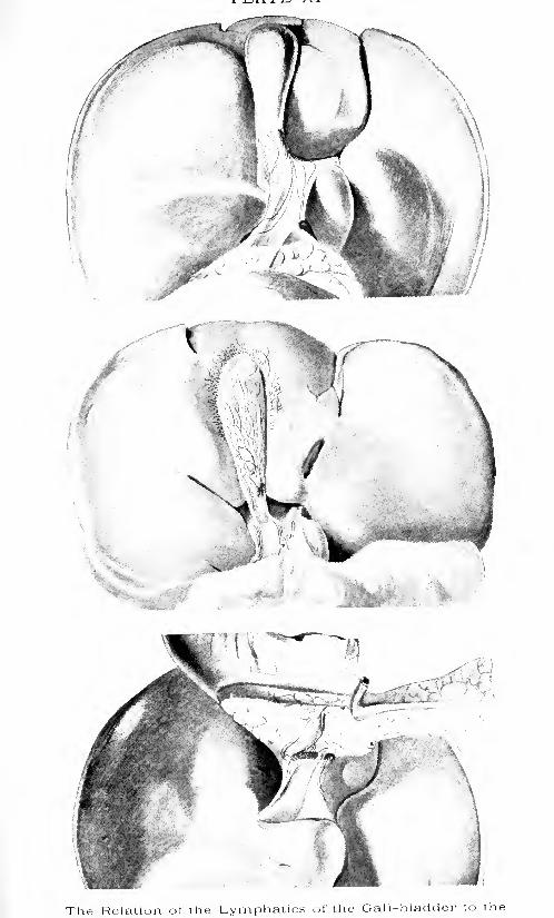
The Relation of the Lymphatics of the Gall-bladder to the

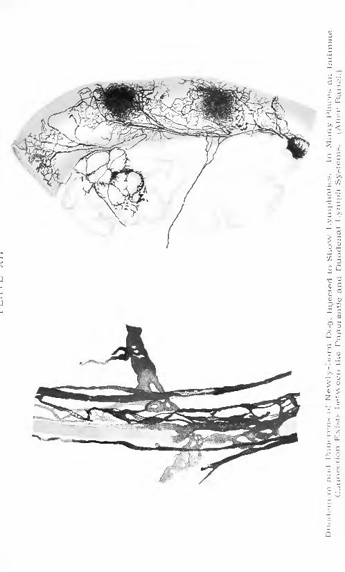

INFLAMMATION OF THE PANCREAS 445
creatic head. Indeed, he was able to inject from the gall-bladder the
lymphatics of the upper part of the head of the pancreas, the portionwhich we have seen is most frequently involved in association with
biliary disease.
Bartels was acute enough to suggest the possibility of transmission
of disease from the duodenum or neighboring organs to the pancreasbut possessed no illustrative clinical material. Arnsperger
1
reportedseveral cases of this type, ascribing the condition of the pancreas to a
lymphangitis, and we have urged the adoption of the term, pancreatic
lymphangitis, to apply to this condition as most descriptive both of its
origin and its nature, and as covering the most common inflammatorycondition met in the gland.At operation the sequence of events is often clearly pointed out as
follows: cholecystitis, lymphangitis and lymphadenitis, observed in the
gastrohepatic omentum along the course of the cystic and commonducts, peripancreatic lymphadenitis, pancreatic lymphangitis. Herein
lies the rationale of the treatment of this form of pancreatitis by meas-
ures designed to correct the primary infective process, its subsidence
being analogous to that which occurs in the treatment of primary foci
of lymphatic infection elsewhere in the body.As to the subsequent course of such infections of the pancreas it is
not yet possible to speak with the certainty of demonstrated fact.
But it does not seem too much to assume that here as elsewhere the
secondary infection may prove to be more severe and serious than in
its primary focus. Dele^enne,2Polya
3 and others have shown that
bacteria can activate trypsin. An infection severe enough to cause
necrosis of the parenchyma is also able to activate the ferments, since
self-activation occurs after death of the pancreatic cells. Here wehave the stage set for hemorrhagic and necrotic pancreatitis.
It has been demonstrated and should be clearly realized by the
surgeon that once the pathological ferment activity of the gland is
begun no further process, bacterial or otherwise is needed to explainthe striking local effects seen in the pancreas and its vicinity in acute
pancreatitis, and also the severe and often fatal systemic intoxication.
If a portion of dog's pancreas be removed during life and again under
sterile conditions be dropped into the abdominal cavity of a second dog,
this animal will probably be dead within twenty-four hours, with all
the symptoms of a severe prostrating toxemia and within the abdomenwill be found fat necrosis and a quantity of "dirty" exudate (Sweet 1
).
In the autolyzed pancreas Lattes5 found a substance able to activate
pancreatic juice or, in other words, to change the fresh secretion, which
is unable to act upon protein, to a state 1 in which it attacks proteinwith great violence, injuring it if living, digesting and dissolving it
when dead. This activating substance is more stable toward heat
1 Miindion. mod. Wchnschr., 1911, liii, 72!).
2 ('ompt. rend Soc. dc biol., 1902, liii, 104.3 Mitt. a. d. Grenzpib. d. Mod. u. C'.'hir., 1912. i. fit.
4 Surgery of the Panrrens, 1915, Philadelphia.5 Yin-hows Arch., 1913, coxi, 1.
44(1 XL'ItGKItY Or THE 1>A.\CRKAS
than the proteolytic ferment, and can he obtained free from ferment
by heating to 00 C. for fifteen minutes. Now, if to sterile inactivated
pancreatic juice a portion of the activating fluid he added and the
mixture injected into an animal the typical fatal intoxication occurs.
The activating substance may he destroyed by heating to 7") (\ for
fifteen minutes. Adding to inactivated pancreatic juice the atitol-
ysate which no longer ])ossesses the ]>ower of activating trypsinogen,and injecting this fluid into an animal, nothing happens except a tran-
-itory fall in blood-pressure, which is a non-specific consequence of
injection of organ extracts. From this and a large amount of work byother observers the conclusion is irresistible that activated ferment
action is the key to the clinical and pathological picture of acute
licmorrhagic pancreatitis. Fat necrosis is the visible sign of activity of
the fat-splitting ferment. This process, however, generates no toxins.
It is in the proteolytic process that the poisonous decomposition pro-ducts are liberated which cause systemic intoxication. The condition,
therefore, is one of pancreatic poisoning (Pancreawergiftujig).If the infection is not sufficiently severe to inaugurate this more
serious outcome it may yet be able to maintain itself upon an inde-
pendent footimr causing interlobular inflammation and that series of
alterations of the gland to the end-results of which we have given the
name chronic interlobular pancreatitis. Whether or not this is related
to inleracinar pancreatitis is still uncertain.
Classification.- As a classification of pancreatitis which conforms
more closely to the clinical conditions than any heretofore given wewould propose the following:
The more we see of pancreatitis the more do we realize that here as
elsewhere there exist all grades and variations of the process. Ilemor-
rhatre may be extensive, sudden and dill'use, or it may be slight and
locali/ed. Severe inflammation may occur unaccompanied by hemor-
rhage, (langrene, as recognized by Fit/, is but the outcome of hcmor-
rhau'ic necrosis; it may be partial or complete. Suppuration may be
a >e(|uel of gangrene, the necrotic tissue affording an opportunity for
development of the causative infection or inviting secondary invasion.
Suppuration, on the other hand, may be localized to a single focus or
be scattered through the gland as multiple foci. Pancreasvcrg'ftung
is not commonly associated with this form of suppurative pancreatitis.
Fat necrosis is often absent even in severe affections, or it may be wide-
spread in what is apparently a less marked condition of pancreatic
IXFLAMMAT10\' OF THE PANCREAS 447
inflammation. Slighter degrees of involvement grade off into the most
inconspicuous lesions, hi its less severe and less characteristic forms
pancreatitis is a common disease and its symptoms are as a rule, mis-
interpreted.
Acute Pancreatitis. --Acute pancreatitis may be subdivided patho-
logically into:
o- 4.- ( Lvmphangitic.1. Simple non-suppurative T/
llematogenous.1'. Primary suppurative.:>. Ilemorrhagic.4. Gangrenous.."). Secondary suppurative.
All these varieties may be localized, spreading or diffuse.
The inflammations which affect the pancreas are modified in manyinstances by the phenomena of hemorrhage and fat necrosis as pre-
viously noted. These are conditions peculiar to this gland since theyresult from its own specific composition and activity. Their importancein the history of the development of our knowledge of the subject, the
aid which they give in diagnosis and their association with the most
spectacular instances of acute pancreatitis, have all served to focus
attention chiefly upon that form of acute inflammation which is
characterized by these phenomena. It should be recognized, however,that acute pancreatitis can and does occur without either or both of
these accompaniments.Simple Non-suppurative Pancreatitis. Acute pancreatitis may present
itself as a simple inflammatory affection showing the usual signs of
inflammation, namely, swelling, edema, congestion and perhapsassociated tenderness, if the organ be palpated. At operation this
type of pancreatitis is seen most often affecting the head or a portionof the head of the pancreas.
It may, however, be diffuse with swelling of the whole gland. Asseen by the surgeon it is always accompanied by cholelithiasis, chole-
cystitis, duodenal ulcer or duodenitis, and the weight of evidence and
analogy points conclusively to the fact that this type of the disease is
the result of infection involving the pancreas possibly by way of the
ducts, but in more' instances doubtless by way of the lymphatic inter-
communication with neighboring infected viscera. That toxic proc-esses and infections reach the gland also by way of the blood stream
is certain. It is probable that this occurs at times in mumps, scarlet
fever, diphtheria and many other general infectious diseases in the
course of which many observers have reported clinical conditions that
can hardly be interpreted otherwise than as a complicating acute pan-creatitis. Thus, (loldie 1
reports five illustrative cases in children
ranging from five to nine years of age. Two cases were in the midst
of an attack of scarlatina. One case was convalescing from the samedisease and had developed chicken-pox, and one had faucial diphtheria.
1 Lancet, 1912, ii, 1295.
44S Sf'ltdKltY OF THE
All the cases had an associated rhinorrhea. Suddenly there was com-
plaint of abdominal pain, usually nausea and vomiting with swellingof the upper abdomen and tenderness over the head of the pancreas.Jaundice and clay-colored stools followed promptly. Gradual recoveryensued in all cases. Robson and Kehr have suggested that the disease,
commonly known as catarrhal jaundice, is in reality pancreatitis with
resulting obstruction of the common duct. Such conditions are neither
surgical nor fatal, so that as yet the autopsy in fin or postmortem is
lacking as confirmatory evidence of the pathological basis. \Ve feel
safe, however, in saying that clinical observations point to the possi-
bility that simple inflammation of the pancreas is not rare, occurs often
in early childhood, and like chronic nephritis is now recognized as a
long-deferred sequel of scarlet fever in certain cases. Likewise certain
scleroses of the pancreas, first observed in recent years will no doubt
be .ascribed to a process inaugurated by earlier infective pancreatitis.
It is worthy of note that at least a part of the symptoms now ascribed
to gall-bladder or duodenal disease must be due to the associated pan-creatitis. The marked swelling of the pancreas sometimes found at
operation is certainly productive of symptoms though it is usually
impossible to separate the pancreatic from the biliary symptoms.The profession is still napping in respect to the realization of the
actual frequency of pancreatic disease. We have been misled by the
infrequency with which pathologists have found postmortem evidence
of disease of the pancreas. This is due to several reasons. The ten-
dency of the gland to digest itself after death soon destroys all evidences
of any parenchymatous changes. The supposed immunity of the
pancreas to disease has caused it to be slighted in routine examinations.
Again, most instance's of less severe pancreatic disease are not diffuse
but atl'ect only a portion of the gland which may easily be missed in
the specimen removed for study. Finally, there is evidence that an
extensively diseased pancreas may so recover itself that demonstration
of previous inflammation is difficult. Thus Korte reported a case
which he operated upon in IS!).") before the characteristic appearanceof the disease had become known. Fat necrosis and serosanguineousexudate were present. The abdomen was closed without attackingthe pancreas. The next year, having to operate on the same patient
for a stone in the common duct, the pancreas was investigated and
found normal in size, appearance and consistency. Other observers
have had similar experiences. The pancreas therefore should be more
often thought of in connection with upper abdominal disturbances,
whether mild or severe.
Primary Suppurative Pancreatitis.- Primary suppurative pancreatitisis the result of pyogenic infection. The term primary is used in this
connection to distinguish the condition from that form of suppurative
pancreatitis in which the suppuration represents the natural termina-
tion of gangrene of the gland, whether primarily infective or not. The
simple type of lymphangitis, above described, does not often suppurate.Like the salivary glands the pancreas is resistant to abscess formation.
INFLAMMATION OF THE I'ANCREAti 449
Occasionally, however, conditions are such that abscesses, usually
multiple, sometimes single, will form within its substance. Obstruc-
tion of the duct of Wirsung is a strong predisposing factor. \Ye have
observed at autopsy one case of multiple pancreatic calculi and one
case of gall-stone impacted in the papilla of Vater associated with
multiple abscesses scattered throughout the pancreas. We have seen
one case (at operation) of single abscess in the head of the pancreasof a child developing during convalescence from acute suppurative
appendicitis and peritonitis. In neither of these cases was there evi-
dence of previous hemorrhage or necrosis. Dieckhoff reported similar
cases and drew the conclusion that such processes represent the advance
of infection from the larger to the smaller ducts into the substance of
the gland where they set up multiple foci of suppuration. lie believes
that the infection gained eiitrar.ee to the duct of Wirsung from the
duodenum and suggested that this could readily happen after the
entrance to the duct had been dilated by the passage of a gall-stone.
The presence of an irritant such as a stone with the stasis which it
might cause could very very readily be conceived as the cause of an
infection of the duct, more or less grave in character, and in this case
the route of travel of the infection would be along the duct, whether
on its surface or in the subepithelial lymphatics makes little difference.
It seems probable at the present time that infections other than those
of the duct can give rise to suppuration. In the case of single abscess
of the head of the pancreas occurring secondary to suppurative appen-
dicitis, a more plausible explanation is infection by way of the retro-
peritoneal lymphatics. Hemic infection cannot be ruled out.
Hemorrhagic Pancreatitis. Though Rokitansky1 in 1803 and Klebs,
2
in 1809 had described the condition of acute hemorrhagic pancreatitis,it was not until the publication of Fitz (in 1882) that attention wascalled to its frequency and importance. This curious condition has
been the subject of much speculation and investigation and only
recently has the problem seemed to be in a fair way of solution. The
hemorrhage may be sudden and profuse overshadowing all evidences
of infection or other injury to the gland. In other cases it is clearly
but the accompaniment of a train of pathological changes which are
recognizable as inflammatory in character. These marked differences
in clinical behavior led Fitz, and many others after him, to recognize
pancreatic hemorrhage as a condition independent from hemorrhagic
pancreatitis. ]\Iayo Robson has proposed that hemorrhagic pancrea-titis be divided into two groups (a] ultra-acute, in which the hemorrhageprecedes the inflammation, the bleeding being profuse and both within
and outside the gland; (/>) acute, in which inflammation precedes the
hemorrhage, which is less profuse and is distributed in patches through-out the gland.
r
l he name of Pancreatic Apoplexy has been employedto emphasize the non-inflammatory nature of the simple hemorrhagic
process. Robson has pointed out a general tendency to hemorrhage
4o() SURGERY OF THE PANCREAS
associated with lesions of the pancreas as evidenced by ecchyrnosesand at tin:es extensive sjjontaneons hemorrhage at a distance from the
pancreas. Experimentally hemorrhagic pancreatitis has been producedby an extraordinary variety of methods. Lattes 1 has summarizedthese various experiments which have resulted in hemorrhagic pan-creatitis with its accompanying clinical manifestations as follows:
artificial ischemia (Blume, Oser, IMilisch, Lewit, \Yulf), injection of
various substances into the duct: oil (Oser, Hess, Guleke, Eppinger),bile (Guleke, Elexner, Opie, Oser, Polya), hydrochloric acid and intes-
tinal secretion (Flexner and Pearce, lliklebrand, Rosenbach), intestinal
secretions and activated pancreatic juice, commercial trypsin, calcium
and sodium chloride (Polya), papain (Carnot), adrenalin (Rosenbach),zinc chloride (Thiroloix, Oser) sulphuric acid, chromic acid, formalin,soda ( ElexnerX or into the vessels (air, paraffin, wax, lycopodium, oil)
(Panuni, Lepine, Bunge, Guleke) and by ligation of all pancreatic ducts
during digestion (ITess), mass ligation and gross injury to the pancreas
(Katz, AVinkler). The introduction of pathogenic bacteria will at
times accomplish the same result. Recently Sweet 2 in a well-considered
paper based upon the literature and the results of his own experimentshas pointed out that the single and efficient factor common to all such
conditions is the liberation of trypsin. Deaver and Pfeiffer,3arguing
from anatomical and physiological facts and clinical observations, hadreadied a similar conclusion. Normally the proteolytic ferment exists
in the gland and its duct as a preferment or zymogen. In this condition
it is incapable of acting injuriously upon the tissues with which it comesin contact. \Yhen activated, however, it becomes a highly destructive
agent and when brought into direct contact with the tissue proteids it
not only causes necrosis but proceeds to digest and liquefy the tissues
attached. Injury to the bloodvessels causes diapedesis of the red cells
as well as the leukocytes. Actual erosion of the vessels gives rise to
free hemorrhage of greater or less volume according to the size of the
vessels that are opened. In this light it is readily understood that a
small focus of diseased or injured pancreatic tissue could cause an
extensive hemorrhage entirely obliterating all marks of the seat and
the nature of the primary insult. Normally the trypsinogen of the
pancreatic secretion is activated by the enterokinase of the duodenal
contents, a consideration which has led to the suggestion that pan-creatitis may be the result of intraglandular activity of its own ferments
induced by the regurgitation of duodenal contents into the duct. It
is, however, quite unnecessary to assume that enterokinase must be
present in order to convert the preferment into its active state since
this maybe accomplished by other means. Delezenne 4 and Ilekma 5
have shown that an activating substance is present in many varieties
of bacteria and Lattes" finds a similar substance in normal pancreatic
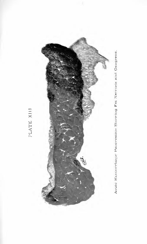
X
cu

INFLAMMATION OF THE PANCREAS 451
tissue that has undergone self-digestion. Mcllanby and Wooley1 have
shown that calcium, barium and strontium salts, neutralization with
hydrochloric acid, precipitation of the juice with alcohol and resolution
of the precipitate, all activate the pancreatic ferments. They putforth the hypothesis that enterokinase in varying amounts is always
present in pancreatic secretion, but is inhibited by the alkalinity. Neu-tralization permits activation. As necrosis is always accompanied byacid formation and many bacteria generate acid it is evident that
innumerable combinations of injury, chemical, bacterial and traumatic,
can cause activation and interstitial liberation of trypsin which is
sufficient to explain hemorrhage, necrosis and fatal intoxication of the
subject. We subscribe to the conclusions of Sweet 2 that:
1. Xo other explanation covers the clinical cases associated with
infection and the clinical cases associated with traumatism of the
gland.2. No other explanation affords an understanding of the fact that
injury to the gland during digestion is more serious than injury at other
times.
3. The picture of an acute pancreatitis can be fully reproduced by
placing the sterile pancreas of one normal dog into the belly of another
normal dog; or
4. By isolating a portion of the dog's own pancreas from its blood
supply.5. The ducts of the pancreas can be tied without causing anything
but simple sclerosis; but
G. A fatal pancreatitis may follow the ligation of the ducts, if anygreat amount of gland tissue is so included in the ligation of the ducts
that autolysis occurs in the ligated bit of tissue (own experiment).This view reconciles differences of opinion based on varying clinical
conditions. Injury of the pancreas may cause necrosis and set in motion
the destructive effects of ferment poisoning or it may fail to do so.
Infective processes also may spread their effects in the gland without
setting up this condition for diffusion and activity of the ferments or
they may show themselves chiefly in the initiation of hemorrhage and
pancreasvergiftung. Fat necrosis may occur without hemorrhage bydiffusion of the pancreatic juice containing lipase which requires no
activator, the trypsinogen remaining inactivated and inert.
Fat Necrosis. The observations of Balser3 and Fit/'s 1
suggestion and
the work of Langerhans,5 Flexner and others have established the fact
that these distinctive, opaque, whitish or yellowish flecks seen most
often in the peripancreatic fat but occasionally widely disseminated
throughout the abdomen or even in the subcutaneous fat are the results
of digestion of these areas of fat by the lipase of the pancreas. The fat
is split into glycerin and fatty acids. The glycerin is absorbed and the
fatty acids combine with the calcium of the blood and tissue juices.
2 Lor. cit.. p. Mil.
' Mr<l. H.-rnnl. 1SS9, xxv, 197.
4o2 SURGERY OF THE PANCREAS
The resulting appearance is characteristic and is, practically speaking,pathognomonic of a lesion of the pancreas.
Just as hemorrhage or fat necrosis or both may be entirely absentin acute pancreatitis, so do we find that the part they play may be moreor less prominent. Hemorrhage may be small in amount, locali/ed to
one spot or distributed in multiple foci throughout the gland, or it maydiffuse through the entire pancreas which will be found immenselyswollen and as red as beef or it may occasion large hemorrhages in
the neighborhood of the gland with accumulation of blood beneaththe peritoneum and infiltrate into the mesenteries and omenta. In the
most severe cases a serosanguineous exudate is usually found in the
peritoneal cavity.
Gangrenous Pancreatitis. Gangrenous pancreatitis under clinical
conditions is a sequel of the hemorrhagic condition. Doubtless it
depends upon very much the same conditions as hemorrhage. It is
quite likely that a certain amount of necrosis involving bloodvessels
is a prerequisite of hemorrhage. However that may be. the hemor-
rhage which follows is frequently sudden and massive, involving a
large part or the whole of the gland. The resulting disturbance plusferment activity determine the death en masse of more or less of the
tissue and a sphacelus. The prognosis is very closely connected withthis process. So violent may be the initial trauma and the activities to
which it gives rise that the doom of the patient is sealed. Practicallythe entire pancreas undergoes rapid destruction and the picture of
fatal panereasvergiftung develops at once and in spite of any efforts to
prevent it or abate its effects. If the extent of pancreatic injury and
resulting intoxication falls just short of being lethal the patient survives
the immediate effects only to harbor a gangrenous mass which invari-
ably is or becomes infected and can be relieved only by drainage and
sequestration. The chances of recovery from such a condition involv-
ing the cellular retroperitoneal tissues, formed with such rapidity that
encapsulation is impossible, involving the most essential structures
found in this location and so situated that efficient drainage of exudate
and sloughs is difficult or impossible, is reflected in the high mortality.
Secondary Suppurative Pancreatitis. Secondary suppurative pancre-atitis is in turn a sequel of hemorrhagic necrosis. It is distinguishedfrom primary suppurative pancreatitis in that the latter is suppurationwithin the pancreas and the former is suppuration of the pancreas. It
is the natural consequence of gangrene and in a sense is salutary in that
by softening and disintegrating the gangrenous tissue and by followingthe well-known characteristics of pus it tends to search for an exit,
thus providing a means for the evacuation of the diseased process.
Retroperitoneal gravitation abscesses may form in the left loin
and very rarely in the right loin. The lesser peritoneal cavity maybe converted into an abscess sac. Occasionally, the gastrohepaticomentum is penetrated and an abscess may form above the stomach
and beneath the liver, as in one of our cases, or it may spread tipwardover the liver, forming a subdiaphragmatic abscess. A generalized
INFLAMMATION OF THE PANCREAS 453
peritonitis may occur but is less common than localized abscess in
one of the situations mentioned. A great difference exists in the toxic
and digestive properties of such collections. While at times they
spread rapidly and increase greatly in size, they may, on the other hand,become stationary, encapsulated and recede. Or they may very slowly
enlarge making themselves known finally as a pseudocyst.Abscesses after localization may ulcerate into adjacent viscera or
into the general abdominal cavity. Korte has met with 3 cases in
which the abscess ruptured into the duodenum or colon and 3 in which
rupture took place into the general peritoneal cavity.An appreciation of the vast number of permutations of these pro-
cesses will enable one to understand quite readily the clinical differ-
ences which are to be expected in different cases.
Symptoms and Diagnosis. Acute pancreatitis in its classical form
with hemorrhage and extensive involvement is one of the most agoniz-
ing seizures known to mankind. Sudden and serious collapse mayoccur. As a rule, when the patient is first seen collapse, if present at
the onset, has passed, though the pain remains intense. So great is
its severity that the patient is unable at times to ascribe to it a definite
locality except that it is abdominal. It overflows its natural courses
and irradiates all the adjacent spinal segments. Usually it is clear
that the maximum intensity is in the epigastrium. At times it is felt
chiefly in the right hypochondrium, less frequently in the left hypo-chondrium and occasionally it pierces through to the back and lumbar
region and radiates into the chest or into the iliac fossa.
In the early stages the pulse and temperature are usually subnormalor normal, contrasting strangely with the evident seriousness of the
condition. Only later, with the onset of infection, toxemia or weaknessdo the pulse and temperature rise. Finally, as in most terminal con-
ditions the pulse becomes very rapid and there is a marked rise of
temperature. So uniform are the primary slowing of the pulse-rate andthe subnormal or normal temperature that they possess considerable
diagnostic importance.
Vomiting is an early and persistent symptom. Usually the patientvomits the contents of the stomach with the initial pain. Thereafter
uncontrollable nausea and vomiting come on at intervals. After the
first insult the vomitus is usually less in amount and biliary in char-
acter. Never does it become fecal unless the paralytic ileus of peri-
tonitis follows as a result of the pancreatic extravasation and infection,
in which case it represents the usual regurgitant vomiting of peritonitis
and is an evidence of impending death. Rarely vomiting may be
absent.
Halsted first drew attention to a peculiar cyanosis which was presentover the abdomen in one of his acute cases and other observers haveobserved the same appearance in a considerable percentage of cases of
ultra-acute pancreatitis. The cyanosis, however, is not limited to the
abdomen but may be seen in the face or other portions of the body.As a rule, when first seen by the surgeon some hours or days after
4.")! Sl'RCERY OF THE PAXCREAS
onset, rigidity of the abdominal muscles is not extreme. There is rather
a marked feeling of resistance in the epigastrium and perhaps anindefinite mass extending transversely across the upper abdomen in
the region of the pancreas. There may, however, be pronouncedrigidity, as in one case seen by us within four hours after the attack
in which the whole abdomen was boardlike as is more commonly seen
in perforative peritonitis, a diagnosis which the circumstances inducedus to make in this case. Possibly, if more cases were seen immediatelyafter onset, rigidity would be more often noted as a reflex phenomenonassociated with the severe primary injury to the pancreas and its
immediate surroundings. It remains true, however, that after a time
there is less rigidity than resistance. Deep pressure over the region of
the pancreas causes pain and spasm. Light pressure is not so muchresented as it is in perforative peritonitis.
Meteorism (A the upper abdomen soon appears. That region is
seen and felt to be more prominent and distended than the lower
abdomen, later this difference disappears as general peritoneal irrita-
tion causes paralysis and distent ion of the intestines. After emptyingthe bowel by enema this difference at times may again appear. Thebowels are usually obstinately constipated, though at times and in
certain stages diarrhea may be present owing to irritation of the pelviccolon and rectum.
The leukocyte count may be normal but is more often increased
and in general the longer the duration of the condition and the moremarked the associated infective processes, the higher the leukocytosis.It tends to be lower, however, than in most conditions likely to be
confused with it.
We have observed in a number of instances that the percussion note
over the bases of the lungs, particularly on the left side, shows consider-
able impairment. The cause of this phenomenon is not entirely clear.
Whether it is due to atelectasis of the base due to restricted movementsof the diaphragm or to the accumulation of a small amount of serous
fluid within the pleura or to both, we have not been able to demon-strate. One must not lose sight of the fact that an accumulation of
exudate in the lesser peritoneal cavity, not an infrequent occurrence,
might cause impairment of the percussion note at the left base, andalso that owing to the proximity of this intensely irritating material
to the diaphragm its pleural surface may be involved and throw out a
quantity of serum into the pleural cavity. When this phenomenonis recognized and understood it may be of some assistance in diagnosis,
but we have known it to obscure the real state and to lead to an
erroneous diagnosis of diaphragmatic pleurisy or pneumonia affecting
the lower lobe. Jaundice is not often pronounced, but an icteroid hue
of the skin with slight tingeing of the sclera is not uncommon. It maybe entirely absent. Hiccoughing may be very troublesome in somecases. In one case, personally observed in the University Hospitalunder Dr. Stengel's care, it was the obstinate singultus that was the
chief complaint and brought the patient to the hospital. Death
INFLAMMATION OF THE PAXCREAti 155
occurred suddenly shortly after admission. Autopsy showed a gall-
stone impacted in the papilla of Vater with multiple abscesses scattered
through the })anereas, peripancreatitis and fat necrosis, hut no evidence
of hemorrhage in the pancreas or its vicinity.
The patient is usually a male in the third or fourth decade of life
who is inclined to obesity and gives a history of previous digestivedisturbances. Many exceptions exist to these generalities. Acute
pancreatitis is about twice as frequent in males as in females. Fitz
found 17 males to 4 females, Korte '}() males to 14 females. In 18 cases
of this type we have found an even balance, 1) males to 9 females. In
extended statistics, however, it is clear that, there is a considerable
preponderance of males. \o reason can be assigned with certainty for
this sex incidence and it remains a strange phenomenon that acute
pancreatitis, admittedly a disease preeminently associated with and
dependent upon cholelithiasis in the majority of instances should
occur more often in the sex which is less liable to gall-bladder disease.
A plausible explanation can be advanced in the greater frequency of
gastric and duodenal disease in males, often due to excesses in tobacco
and alcohol.
The age incidence is given as follows by Oser:
Years.
1 to 10 JO days' . . .
21 to 30 ...31 to 40 .. ....41 to 50 ....
and bv Fitz.
Years.
9 to i; 5
456 SURGERY OF THE PANCREAS
reports two cases of acute hemorrhagic pancreatitis occurring two daysafter: (1) resection of the sigmoid for carcinoma; ('2) removal of an
ovarian cyst. Hoffman, quoted by Seidel 1
re]>orted ',} cases of acute
pancreatitis following operation for (1) appendicular abscess, C2)
appendicular peritonitis, "2 cases. AYe have observed abscess of the
head of the pancreas in a child occurring after operation for appen-dicular peritonitis. In our view these arc instances of retroperitoneal
lymphatic infection.
No one of these signs or symptoms nor any combination of them will
unfailingly make one aware of the existence of acute pancreatitis.Morrison has correctly placed the mental attitude when he wrote that
"if the history, symptoms and signs do not exactly fit acute intestinal
obstruction, or stomach or duodenal perforation, perforated appendixor acute cholecystitis, and yet have some resemblance to each of them,
pancreatitis is the most probable cause."
In the more severe cases the severity of the initial symptoms, the
obstinate vomiting, the slow pulse-rate, the low fever or its absence,the appearance of a tumefaction in the epigastrium, or even morecharacteristic when present, the signs of an inflammatory mass in the
loin, will point with a considerable degree of certainty to acute
pancreatitis.
The differential diagnosis is concerned chiefly with the conditions
mentioned above by Morrison. It must be admitted that a positivedistinction cannot always be made and many cases are operated uponunder the wrong clinical diagnosis. This error is not often serious in
view of the fact that all these conditions call for prompt operation andthe correct surgical diagnosis is now promptly made from the well-
know]! characters of the exudate with fat necrosis and the palpable
enlargement of the pancreas. The gastric crisis of late syphilis should
be considered and excluded by history and examination of the pupillaryreactions and the reflexes. The Wassermann reaction should be madein all cases when time permits. It is not wise to wait for the time-
consuming investigation of pancreatic activity, which when obtained
is far from conclusive. Loewi's pupillary mydriasis reaction is simpleand rapid; Glaessner2
reports a case in which a positive reaction con-
firmed this opinion. ]ts value is as yet uncertain. AVhen positive it
is not pathognomonic of pancreatitis. Most observers agree that the
Cammidge reaction is unreliable. In our hands it has been of no
assistance. (Jlyeosuria is suggestive when present, but is very variable
in the frequency of its appearance. Thus Korte found it in S of 44
cases while in our 18 cases it was present in none at the time of opera-
tion, though one patient who lost a large portion of his pancreas bynecrosis, afterward died of diabetes.
Treatment.- Acute pancreatitis is not invariably fatal without opera-
tion, a fact which has been established by our increased knowledge
concerning the variation in the severity and extent of the lesions. It
1 Arc-li. f. klin. Cliir.. 1913, Ixxxv, 384.2 Doulscli. Klin., 1913, xiv, L'ofi.
INFLAMMATION OF THE PANCREAS 457
is not very infrequent in cases operated upon for acute pancreatitis to
find a history of one or more similar seizures, from which temporary
recovery ensued. Certain pseudocysts of the pancreas are in reality in-
stances of recovery from acute pancreatitis, the residuum of the process
forming the basis for the cyst. Again, cases in every way identical
clinically with those known to be pancreatitis, with glycosuria and evi-
dence of pancreatic insufficiency connected with the attacks, have been
known to recover perfectly without operation, or in still other instances
after a period of waning health due to diabetes following immediatelyor shortly after the attack, have come to autopsy and revealed the
evidences of previous severe pancreatitis atrophy of the organ, inter-
stitial fibrosis or stone impacted in the head of the pancreas. The
original prognosis of Fitz therefore was too pessimistic and the estimate
of Musser of 100 per cent, mortality without surgical treatment wastoo high. Still, in the truly severe cases it is certain that recoverywithout operation is rare. There is no medical treatment which can
in any way influence the chief factors upon which the prognosis depends,
namely, the extent of the destruction of the pancreas and the absorp-tion of the toxic products engendered by the ferment activity abetted
by infection. We may safely conclude that the cases that recover
under expectant treatment, for medical treatment is nothing else,
would also recover if submitted to operation, while of those that die
without operation at least a certain proportion would survive as a
result of the assistance which appropriate surgical procedures givethe body in dealing with the process.
The operative mortality in severe cases is also high. Korte in 34
cases had 18 recoveries and 16 deaths (47.4 per cent, mortality). In
collective statistics he found the mortality to be 60 per cent. Drees-
mann, in 1909, collected 118 operative cases with a mortality of 55
per cent. Of these, 40 cases operated upon early, showed a mortalityof only 20 per cent. Mettin, reporting 22 cases of Neumann, found the
mortality to be 69 per cent. In 18 cases we have had 12 recoveries
and 6 deaths (mortality 33f per cent.).
To understand the various procedures which are of use in pancreatic
surgery we must be able to answer several questions, (1) what may weaim and legitimately hope to accomplish by operation; (2) when should
we operate; (3) how should we operate?It is obvious that partial or total resection of the inflamed pancreas
is out of the question. The disease cannot be removed in tutu. It
should be the aim of the surgeon to prevent the further developmentof the condition and so far as possible to relieve the body from the task
of caring for its products. Can any beneficial effect be exerted by treat-
ment of the cause'/ Unfortunately the exact determining cause of
the majority of cases of acute pancreatitis is not at once apparentand, again, we have seen that experimentally at least, the initial injurysets in motion a train of circumstances that proceeds to the most severe
consequences even after the cause is no longer active. Unquestionably
any apparent cause should receive appropriate treatment, if the con-
15S SVlidEIlY. Or THE PANCREAS
ditioji of the patient warrants. We know definitely that in some eases
one effective mechanical cause consists of the presence of a stone
impacted in the papilla of Yater. It is not too much to hope that in
the event of such a cause being recognized removal of the stone andrelief of the hack pressure upon, and regurgitation of, the bile into
the pancreatic duct would have a beneficial influence. Stones in the
common duct should arouse suspicion that this mechanism is or hasbeen acting, and their presence justifies eholedoehotomy with removalof the stones from the duct and the passage of a probe to make sure
that its outlet is patulous. In (i cases Korte found stones in the commonduct, '.) of which were at the top of the papilla of Yater. Two of these
he removed at operation and 1 was removed at autopsy.In a case operated upon recently the patient, a man of spare physique,
aged forty-two years, was suddenly seized with violent epigastric painsfollowed by vomiting. 'When first seen he was sulVering intensely, andwas in partial collapse. The physician who was called in gave him a
huge dose of morphin and sent him to the hospital ( Abington Memorial),where he was admitted within four hours after the attack began. Twohours later he was operated upon, his condition at that time beingsomewhat more comfortable. The abdomen was generally rigid as a
board. There was tenderness in the epigastrium but no distention.
A diagnosis of perforated duodenal ulcer was made. On opening the
abdomen the stomach and duodenum were normal. There was a small
amount of free serous fluid in the upper abdomen. The gall-bladdercontained numerous small stones and several could be felt in the
common duct. These structures showed no signs of acute inflammation.
The pancreas was enormously swollen throughout and the adjacentareolar tissue and omenta were thickened and edematous. Xo hemor-
rhage being present, the pancreas was not opened, but the stones were
removed from the gall-bladder and the common duct, both of which
were drained. A probe was passed down into the duodenum thinking
perhaps to dislodge a calculus in the papilla. There was no evidence of
this nor could any stone be discovered subsequently in the feces. Con-valescence was uneventful. It is possible that a stone had been
impacted in the outlet but had slipped back into the dilated portionof the duct.
Stones in the gall-bladder, on the other hand, do not act mechanically
upon the pancreas. Yet they are by far the most common finding in
acute pancreatitis, and it is impossible to escape the conclusion that
in some manner they are responsible for the inauguration of pancre-atitis in at least a large proportion of cases. Much less frequently
simple, non-calculous cholecystitis is found. In our 18 cases gall-
stones were present 15 times. In one case it, was impossible to deter-
mine the condition of the gall-bladder, the incision being in the left
loin. Korte found that in 44 cases personally observed disease of the
gall passages was present in 22 (50 per cent.); 21 cases showed chole-
lithiasis and one non-calculous cholecystitis. Osier found in 105 cases
of pancreatitis 45 instances of complicating cholelithiasis. Kgdahl
INFLAMMATION OF THE PANCREAS 459
found an exactly similar relationship. Queiui in 128 collected cases
found 47 instances of gall-bladder involvement. The role of gall-
stones in the gall-bladder may very well be a primary one through the
exacerbations of inflammation which are a well-known accompani-ment of cholelithiasis. Holding, as we do, the view that pancreatitisis in many cases the result of an inflammation propagated from the
gall-bladder, we feel that it is highly desirable in every case, conditions
permitting, not only to remove any stones that are present in the
gall-bladder but also to drain it for the influence that drainageexerts over infection in its walls. Exceptionally, the gall-bladder
may be removed and the common duct drained, but, as a rule, the
surgeon should content himself with drainage of the gall-bladderwhich is safer, quicker and answers even' temporary indication.
It is rarely true that any case which can stand operation at all will
not at the same time permit of the simple operation of opening and
draining the gall-bladder. We have performed cholecystostomy in 1 1
cases with 3 deaths; cholecystostomy and choledochotomy in '2 cases
both of whom recovered and of 4 cases in which the bile tract wasuntouched '2 died and 2 recovered. One case collapsed and died on the
table before anything but the abdominal incision had been done. Wehave not been able to attribute any of the deaths to the operations
upon the bile passages. It seems likely that the relief of the bile ten-
sion in the ducts acts in certain cases in a favorable manner. Without
being able therefore to assert that the original cause of the pancreatitis
continues to act from the bile passages as a base, or to explain in an
incontrovertible manner the modus opcrandl of action it is neverthe-
less our belief that no transperitoneal operation for pancreatitis is
complete without (a) opening the bile passages and removing anystones that are found, and (6) assuring one's self of the patulous condi-
tion of the common duct and (c) establishing drainage. Experienceand the proper circumstances are assumed, for time is precious in these
cases.
In the presence of associated duodenal ulcer or duodenitis, nothingin an operative way can be done to relieve these conditions thoughthey may be the starting point of the entire process. We can only hopethat the cause has ceased to act, and direct our attention to the relief
of the pancreas and the conditions to which it may have given rise.
In other instances, no clue to the origin of the process will be demon-strable and in these also, as well as in cases of gall-bladder disease,
just discussed, the question arises as to the proper procedure to employupon the pancreas and peripancreatic tissues. The answer to the
question will depend largely upon the conditions present and these in
turn upon the severity of the attack, over which we have no control,
and the stage of the disease. Is it advisable to attack the condition
at the earliest possible moment or should one wait for more favorable
local and general conditions? Formerly, in common with many other
surgeons, we counselled early operation but advised against operationin the presence of shock. If we now except the primary fainting attack
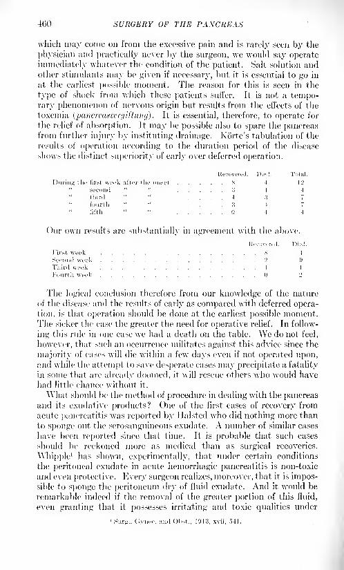
4tiO SURGERY OF THE PANCREAS
which may come on from the excessive pain and is rarely seen by the
physician and practically never by the surgeon, we would say operate
immediately whatever the condition of the patient. Salt solution andother stimulants may be given if necessary, but it is essential to go in
at the earliest possible moment. The reason for this is seen in the
type of shock from which these patients suffer. It is not a tempo-rary phenomenon of nervous origin but results from the effects of the
toxemia (pancreasvergiftung). It is essential, therefore, to operate for
the relief of absorption. It may be possible also to spare the pancreasfrom further injury by instituting drainage. Korte's tabulation of the
results of operation according to the duration period of the disease
shows the distinct superiority of early over deferred operation.
During the first week ;ifter the onset
second "
third
fourth"
fifth
Our own results are substantially in agreement with the above.
Recovered.
INFLAMMATION OF THE PANCREAS 461
certain conditions as the reddened condition of the intestines mayindicate, were often to turn the scale in favor of a patient who still
retained behind the peritoneum one of the most virulent and toxic
conditions known. At the very best interpretation to be placed uponsimple sponging away of exudate even if coupled with peritoneal
drainage, we must admit that the pancreatic process must be relatively
mild, unaccompanied by the extensive necrosis often seen, and there-
fore lacking in the essentials which go to make up fatal pancreatictoxemia. Liek 1
reports a case of acute pancreatitis in a woman aged
twenty-eight years, who was operated without diagnosis on the second
day of the attack. Bloody fluid was found and wiped away. The
pancreas felt enlarged but no fat necrosis was present. The subsequentcourse was stormy and after eleven days operation was again performedand drainage of a large amount of necrotic material from the pancreaswas effected through the gastrocolic omentum. The earlier operationdid not arrest the process in and about the pancreas. It is worthy of
note that, while fat necrosis may be present at the end of twenty-fourhours, gangrene and softening of the pancreas is not commonly demon-strable before the third or fourth day. Korte observed only one case
which showed spots of softening as early as the fourth day. It is
difficult, therefore, in early operations to prognosticate what will be
the condition of the pancreas in a few days and in the event of pro-
gressive inflammation, destruction or pus formation a second operationwill be necessary.One of our fatal cases is of interest in this connection. E. T., aged
thirty years, had complained of pain in the upper abdomen for six
years. Pain came in sudden severe attacks and felt like a pressureunder the right costal margin anteriorly followed by pain in the backand right shoulder. Attacks at first lasted two to three hours, but of
late two to three days. There was much indigestion and flatulence.
Chills and fever at times accompanied the pains.The present series of attacks began about six weeks previously
culminating in a most severe seizure the day before admission. The
patient was in a state of shock with rapid pulse and low temperature.The face was cyanotic and there was considerable dyspnea. Dulnesswas noted at the left base posteriorly. The abdomen was distended
There was extreme tenderness over the gall-bladder region, and a masswhich was regarded as the gall-bladder could be felt in the right epi-
gastrium. The case was regarded as probably one of acute cholecystitisand in view of the general condition it was thought best to defer opera-tion. On the fourth, fifth and ninth days after admission gall-stoneswere passed by rectum. Eleven days after admission operation wasdone. The gall-bladder and common duct contained stones, a fewflecks of fat necrosis were observed in the omentum but the pancreasfelt firm though it was enlarged. Cholecystotomy and choledochos-
tomy were done, the stones removed and drainage placed in both gall-
462 SURGERY OF THE PANCREAS
bladder and common duct. Convalescence was fairly satisfactory
though slightly febrile until the thirteenth day when the temperaturerose and the patient had a chill, the pulse also rising to 156. Three
days later tenderness was noted at the left costal margin in the loin
with bulging of the ribs at this point. The stools were oily and con-
tained many fatty acid crystals and a few muscle fibers. There wasno glycosuria. Cammidge reaction, which was negative before opera-tion, was positive seventeen days after operation. The day followingthe discovery of tenderness in the loin an incision was made at this
point and a large amount of foul, dark material evacuated. Deathoccurred the following day. An autopsy was not obtainable, but with
the hand in the wound the pancreas could be felt, the body and tail
being very necrotic and soft.
At the present time we feel that the pancreas in this case should have
been explored and drainage placed in such manner as to conduct awayany extidate.
The question of direct drainage of the pancreas and peripancreatictissues cannot be regarded as settled. Doubtless the relief of tension
within and about the gland and the discharge externally of this poison-ous autolysates and extravasated ferments are desiderata of the highest
importance. Unfortunately the structure of the pancreas is such that
limited incision or puncture of its substance cannot be expected to
exert much more than a local effect. Also, the situation of the pancreasand its relation to important nerve, vascular and other structures
render anything like ideal isolation by drainage a fatuous hope. Still,
it remains true that such measures accomplish something and mayserve to tide over the acute situation. The peritoneum covering the
affected area of the pancreas should be opened. If the typical swollen,
beefy appearance of hemorrhagic pancreatitis presents the surface
should be superficially scarified and a number of blunt punctures madein its substance. Deep incisions provoke some bleeding and are
inadvisable. The surrounding cellular tissues also should be opened
cautiously in a few places by a hemostat. Gauze drainage of the
affected area should then be instituted either by large cigarette drains
or by gauze protected by an enveloping layer of rubber dam. Such
drainage should not be removed too soon, but should be allowed to
loosen, so as to permit removal without forcible traction.
^Yhen gangrenous suppuration is present drainage is always to be
employed in amount and character dictated by the well-known prin-
ciples governing such conditions.
Jn the case of localized subacute or chronic areas of softening or
suppuration simple drainage by tube or cigarette drain in accordance
with the conditions will suffice. Korte had two recoveries in this typeof case by draining the head of the pancreas from behind after mobiliz-
ing the duodenum. It is in the early acute severe conditions, whentime and trauma are a very definite factor, that the question and
application of the principle of drainage to the pancreas tax the skill
and judgment of the surgeon. Drainage here is desirable, but con-
servation is necessary in its application.
INFLAMMATION OF THE PANCREAS 463
Hemorrhage is a special danger during convalescence. The close
relation of the pancreas to numerous large arteries and veins and the
erosive action of the exudate make secondary hemorrhage not uncom-mon. This is an additional reason for not being too active in mak-
ing incisions in or about the pancreas and packing the neighborhood
overly full of gauze. Necrosis of vessels is more likely to occur when
they are in contact with drainage. Korte had six fatalities from this
cause during convalescence, and another case was saved only by rapid
tamponing of the wound. We have been fortunate enough to meetwith no fatal hemorrhage from large vessels. One fatal case that wasmuch jaundiced before operation, on the eighth day showed the well-
known hemorrhagic tendency seen in that condition, with bleedingfrom the wound surfaces, gradually growing weaker until death. The
pancreas in this case was the seat of numerous small abscesses.
The loin incision is not applicable to acute pancreatitis as such but
only to gravitation abscesses or collections in the lesser omentum whichare the result of the disease. In these cases it may give excellent
service. In one remarkable case recovery ensued after a slough com-
prising a large part of the body and tail came away. The subsequent
history of the patient corresponds with that of a number of other cases
who have lost a large part of the pancreas as a result of this disease.
For a time he was in good health. In two years sugar appeared in
the urine, and four years later he died of diabetes. It may have beena coincidence that his father died of diabetes. Diabetes may follow
even when a considerable portion of the pancreas remains and is
apparently normal. This but emphasizes the fact that we are not in
possession of all the facts concerning the connection of the pancreaswith carbohydrate metabolism and therefore do not know at what
points the chain may be broken. Robson has suggested that diabetes
may at times be cured by operation designed to' relieve pancreaticinflammation. While it is true that intermittent glycosuria will often
yield to this treatment, we have not observed a case of true persistent
glycosuria of the diabetic type that lias recovered after operation uponthe pancreas or bile ducts.
For the sinuses resulting from drainage expectant treatment will
usually suffice. In resistant cases, especially those where the dischargeof irritating secretion plays a part in preventing healing, as in the case
of sinuses from pancreatic cysts the Wohlgemuth diet is to be recom-mended.Chronic Pancreatitis.- Only a decade ago chronic pancreatitis was
scarcely considered by the clinicians. Fitz had written appreciativelyof the condition and contributions to the subject had been made byKorte, Oser, Lancereaux, Opie, Moynihan, Lenhart, Lazarus and manyothers, among whom Mayo Kobson deserves special mention for his
insistence upon the importance of the condition and contribution to its
therapy. Still the profession considered chronic pancreatitis a rarity.
Opie, from his pathological experience wrote that "the lesion is seldomassociated with such definite symptoms that it is recognizable during
404 SURGERY OF THE PAXCREAS
life." Kobson and Cammidge, on the other hand, attacking the prob-lem from the clinical side, claimed that "from the information obtainedfrom a careful examination of the patient, a knowledge of the historyof the case, and the results of a chemical and microscopic examinationof the excreta, a correct opinion may be formed in a large majority of
these cases." According to our observations the true state of affairs
with respect to the possibilities of clinical recognition is located some-where between these pessimistic and optimistic expressions. Certainly
a reasonable 1
certainty of the existence of pancreatic disease may be
entertained in a considerable percentage of cases, but it is equallytrue that our present criteria are too uncertain and inconstant to
warrant a claim for great accuracy. What has been gained is the
recognition of the fact that the pancreas is not infrequently subjectto chronic disease, that it plays a part in the syndrome of much that
has been commonly denominated indigestion or attributed wholly to
the adjacent viscera, and therefore that in all such cases the pancreasis to be considered and should when practicable be examined carefully
in all upper abdominal operations and given a proper valuation in
respect to surgical and after-treatment.
Pathologically and histologically we distinguish:
1. Chronic lymphangitic pancreatitis.
'2. Chronic interlobular pancreatitis.
.'!. Chronic interacinar pancreatitis.
4. Cirrhosis of the pancreas (Ilobson).
It is well to recogni/e that the last three divisions are based not
upon the process of pancreatitis but rather upon its products or end-
results. Chronic interlobular pancreatitis shows an increase of the
interlobular fibrous tissue with or without evidences of active inflam-
mation. As observed at autopsy the signs of inflammation are usually
absent but its results are evident in the deposit of fibrous tissue between
the lobules. In interacinar pancreatitis the interlobular septa may be
relatively normal while there is within the lobule an increase of fibrous
tissue 1 which insinuates itself between the individual acini, usually
accompanied by more or less distortion and degeneration of these
parenchymatous structures. It is in this form of the disease, as Opiewas the first to point out, that the islands of Langerhans are most
likely to be affected by atrophic or degenerative processes, a condition
especially likely to be accompanied by disturbance of carbohydrate
metabolism, which is the chief basis at present for the "island theory"of diabetes. These divisions are not absolutely hard and fast. Inter-
mediate processes exist in which the fibrosis is seen both in the inter-
lobular septa and within the lobules. In such a condition, well
advanced, the pancreas is usually reduced in size and firm, giving rise
to the term cirrhosis of the pancreas or chronic indurative pancreatitis.
Such a classification while useful for descriptive purposes is still lacking
in usefulness since the types of lesions here described are not, as yet,
correlated with their etiology.
As observed /// riro the processes affecting the pancreas have not
INFLAMMATION OF THE PANCREAS 465
been thoroughly studied histologically, but it has been determined that
they may affect but a limited portion of the gland, usually the heador a part of the head. Edema, congestion and absorbable exudates
may be the only pathology present, all of which may subside leavingno recognizable trace. It seems a fair assumption, however, and one
which is based not only upon analogy but upon the clinical historythat such processes may inaugurate a series of changes in the glandwhich will later be recognized as one or the other above-mentioned
forms of induration. As yet no complete chain of evidence has been
forged to enable us to say just what relation the frequent operative
findings of pancreatitis bear to the end-products, but the role of infec-
tion is now almost generally considered to be the important agent in
the causation of chronic pancreatitis. To cover this localized and
early inflammatory change without fibrosis it seems advisable to
employ another term. Maugeret, Ansperger, Deaver and Pfeiffer,
Sweet and others have called attention to the probability that such
changes are in reality a lymphangitis transmitted to the gland from
adjacent infected structures and that some gain would accrue if the
process were called, as suggested by Ansperger, pancreatic lymphan-gitis.
Another end-result of conditions that we poorly understand is
lipomatosis of the pancreas. Normally there is a small amount of
fatty tissue in the pancreatic stroma but occasionally this is enormouslyincreased. In some cases almost the entire pancreas is replaced by fat.
It appears to be a replacement process, the fat being deposited as the
parenchyma and stroma undergo atrophy and disappear. It accom-
panies both the interlobular and interacinar types of inflammation
but it is uncertain to what extent it depends upon them. In 3 of 90
cases of diabetes described by Cecil advanced lipomatosis occurred
in association with interacinar pancreatitis (Opie). It occurs more
commonly in the obese, but has been found also in the emaciated
(Diechoff, Kasahara, Lepine and Cornil). No relation to surgical
disease has been demonstrated.
An etiological classification of chronic pancreatitis is impracticablein the present state of our knowledge. As above stated infection playsthe leading role. The avenues by which infection may reach the pan-creas are ( Ij the lymphatics, (2) blood stream. (':->) the duct or ducts, and(4) by direct contiguity. Doubtless instances of invasion by each of
these portals of entry occur. Occasionally ulceration of the second
portion of the duodenum will extend through the wall of the intestine
and reach the pancreas immediately adjacent, causing an inflamma-
tion of that portion of the organ. The pancreas is able to protect itself
against this slow attack by exudate and fibrosis which spare the
remainder of the gland. What may occur, however, is well illustrated
by the following case.
Mrs. F. C., aged forty-seven years, was admitted to the I niversity
Hospital December 22, 191."). She had been perfectly well up to three
weeks ago. She then had an attack of severe upper abdominal painVOL. it 30
4()G SURGERY OF THE PANCREAS
radiating to the back and both shoulder blades. Two similar attacks
since then accompanied by vomiting. She was admitted during anintermission and showed nothing except tenderness in the epigastriumand in both hypochondria.At operation the gall-bladder appeared normal but contained one
small stone. Nothing could be felt in the common duct. The pancreas
presented no abnormality. A well-defined chronic duodenal ulcer was
present in the anterior wall about one inch below the pylorus. This
was excised with the pylorus. The gall-bladder was drained and a
posterior gastrojejunostomy made. The stump of the duodenum was
implanted into the denuded surface of the pancreas. The patient at
first did well, but in about two weeks began to complain of severe
upper abdominal pain and vomiting. She gradually lost weight and
strength, the temperature tended to be subnormal. Death occurred
two weeks later. At autopsy the duodenal stump had ulcerated into
the head of the pancreas which throughout showed numerous focal
abscesses.
Infection through the blood stream is also a possibility, but, as a
rule, the pancreas seems immune to the ordinary bacteremias. It
remains to be seen whether organisms with selective affinities for
pancreatic tissue, such as Rosenow has demonstrated for the gall-
bladder, duodenum and other tissues, will be found to play a role in
pancreatitis. Tuberculous and syphilitic lesions are occasionally found,and some authors have laid great stress upon the vascular lesions as a
cause of both acute and chronic pancreatitis, which if true would hark
back, in the majority of cases, to microorganismal injury through the
systemic circulation. It can be said, however, that clinical evidence
at present does not tend to support this view.
Infection through the ducts from the duodenum or propagated fromthe closely associated common bile duct has received much moreattention. We have seen that in acute pancreatitis it is certain that
irritants introduced into the pancreas experimentally or under clinical
conditions may cause most severe injury or even destruction of the
organ. It is easy to assume that acting in lesser degree the samefactors could give rise to slight inflammatory alteration in the nature of
chronic pancreatitis. The frequent association of biliary disease with
pancreatitis gives color to this idea. Desjardins, championing the
ascending duct infection from the duodenum suggested that the sameinfection arising from the intestine and traveling up the bile and pan-creatic ducts will in the former situation induce a stone-formingcatarrh, but in the pancreas will cause a chronic interstitial inflamma-
tion. ( ']>to the present time, in one form or another, the duct avenue
of infection has been believed to be operative in the majority of cases
of chronic pancreatitis. This, however, does not suffice to explainchronic pancreatitis as we see it under surgical conditions, when mostoften it is associated with an inflammatory lesion of another abdominal
organ and is affected only in that region which is in close lymphatic
relationship with the affected viscus and not diffusely, as wrould seem
INFLAMMATION OF THE PANCREAS 467
to be necessary if the duct system were the seat of a catarrhal inflam-
mation conveying infection to all parts of the pancreas. While not
excluding1
, therefore, infection from other portals of entry we believe
that the forms of pancreatitis now so often noted by the surgeon are
instances of retroperitoneal infection secondary to infection of the
common sites of primary abdominal localization, namely, the gall-
bladder, the pyloric region, the duodenum and at times probably the
appendix and other portions of the intestinal tract whose lymphaticsBartels has shown come into close relation in their retroperitoneal
course with the pancreas.That the pancreas may receive injury in other ways than by imme-
diate bacterial invasion is possible. It is subject to a hormone andneural balance about which but little is known. Disturbances of
metabolic or of nervous origin or of what Crile speaks of as the kinetic
mechanism may possibly after a time be reflected in changes not onlyof function but also of structure. It remains for the future to developthis phase of the subject. At present no direct surgical significance
attaches to the various speculations regarding them nor has operation
proved beneficial in that form of the disease characterized chiefly bydisturbances of the metabolic functions unless it has appeared that
such disturbances were dependent directly upon disease secondary to
demonstrable surgical disease or infections.
Duct obstruction, when present, is an important factor in the
causation of chronic pancreatitis. Experimentally occlusion of the
main pancreatic duct causes an inflammatory reaction and atrophy of
the tubules. The islands of Langerhans persist longest and may be
preserved apparently intact in the midst of fatty and fibrous tissue
which replaces the acini. Under clinical conditions also duct obstruc-
tion is usually, if not always, associated with infection. It gives rise
to interlobular pancreatitis and more or less destruction of parenchymain accordance with the degree and duration of the obstruction and the
severity of the accompanying infection. Sources of obstruction are
pancreatic calculi, biliary calculi in the duodenal portion of the commonduct, neoplasms, inflammatory tumefactions and cicatricial processes
involving the duct. The duct of Santorini is occasionally a safetyvalve as it anastomoses within the gland with the duct of Wirsung in
9 out of 10 cases, and in one-tenth of all cases is even larger than the
duct of Wirsung. In the majority of cases, however, it is too small or
its orifice too minute to assume the function of the main excretoryduct (Opie). In one-third of Opie's cases observed at postmortemsome one of these forms of obstruction was present as the cause of the
chronic pancreatitis. In the type of chronic pancreatitis commonlyseen at operation, as previously stated, obstruction plays a smaller
role.
Chronic pancreatitis is more common in the male sex. In 13ohm's
large statistics (if) per cent, were males. Of our cases f>S per cent.,
and in Opie's series two-thirds of the cases were in the male sex.
4 OS SURGERY OF THE PANCREAS
It is a disease of adult life, though it may occasionally ail'ect children.
Opie found:
Cases.
1
In 08 cases we found:
Ca
4
11
11
S
Naturally the age incidence is lower in an operative than in a post-mortem series. As many cases have a history of disturbance extendingover a number of years it follows that the onset of the condition occurs
in the majority of cases below the age of forty.
A large number of predisposing causes have been mentioned. Most
firmly established is the relation to disease of the bile passages andthe liver, particularly cholelithiasis. ^Yhen stones are present in the
common duct the liability to pancreatic disease is much increased.
Cirrhosis of the liver is not uncommonly accompanied by chronic
pancreatitis and in hemochromatosis it is always involved. Duodenaland pyloric disease not infrequently affects the pancreas. Other factors
are duct obstruction, as already mentioned, alcoholism, lues, tuber-
culosis, especially the visceral form as described by ( Gilbert and other
French writers, and trauma.
Symptoms and Diagnosis. Chronic pancreatitis is not often observed
as an isolated lesion. No analysis of a pure series of such cases is
available. Ordinarily the symptoms presented during life by the
subjects of pancreatitis show a large admixture of symptoms due to
associated disease of other organs, usually the biliary tract. ( 'ammidge1
has classified on clinical grounds four groups: (I) a dyspeptic type, in
Avhich the disease is due to a morbid condition of the intestines and the
symptoms are mainly referred to the digestive organs; ('2) a chole-
lithic type, most commonly associated with the presence of gall-stones
in the common bile duct, in which there is usually, but by no means
necessarily, chronic jaundice, and the predominant symptoms are
referred to the biliary system; (.'!) a miscellaneous group where the
pancreatitis is secondary to malignant disease or is the result of disease
of the circulatory system, etc.; and (4) a diabetic group, where there is
more or less glycosuria and into which the members of the preceding
groups are apt to merge in the course of time. Much confusion exists
owing to the lack of any symptom which is reasonably pathognomonicof chronic pancreatitis. Cammidge regards the reaction which bears
i Lancet, 1911, ii, 1495.
IXFLAMMATIOX OF THE PAXCREAS 469
his name as practically conclusive, but other observers have been
unable to subscribe to his opinion of its value. The findings at opera-
tion are also extremely difficult to interpret, depending as they do uponthe operator's opinion of the size and consistency of the gland which
varies in its conformation and is ahvays very firm and often nodular
under normal circumstances. Korte admits that sections which he
removed at operation from the head of a pancreas considered by him
to be diseased, showed no pathological lesions when examined later byBenda. On the other hand a case is authentically reported from which
a portion of pancreatic tissue was removed for microscopical diagnosis
which was considered normal by the pathologist. Heath following
soon, the pancreas was found to be the seat of multiple abscesses.
Apparently lesions may be sharply localized in the pancreas and tissue
but a short distance away may show no microscopical abnormalities.
Still alterations do exist which are unquestionably abnormal and
inflammatory in character and will no doubt be recognized after a
sufficient degree of experience with the normal gland has accumulated.
These factors partly account for the extraordinary difference in fre-
quency with which various operators have recognized the existence of
pancreatitis. Thus Mayo, in 1908, reported that in 2200 operations
upon the gall-bladder and biliary passages the pancreas was also dis-
eased in 141 instances ((.! per cent.). Robson, in much the same typeof cases, found evidence of pancreatic disease in approximately Go percent. Much depends upon the attention devoted to the pancreas
during upper abdominal sections. Thus Kehr before 1909 found pan-creatitis associated with biliary disease in 10 per cent., but since then
in 30 per cent, of the cases. We have found presumptive evidence of
pancreatitis in approximately 35 per cent, of operations upon the
biliary tract.
It is probable that the simple dyspeptic manifestations of the disease
are more frequent than is realized. In all operative statistics the
frequency of the more striking and disagreeable symptoms such as pain,
vomiting and jaundice is magnified since it requires severe symptoms,as a rule, to bring cases to operation.
Loss of weight is a fairly constant symptom. It may be so markedas to excite suspicion of malignant disease, and particularly when
coupled with persistent jaundice the simulation of carcinoma of the
head of the pancreas may be complete. Strength is not usually lost
in proportion to the diminution in weight. The secondary anemiawhich accompanies well-marked pancreatitis is usually slight andcachexia is lacking in all but the far advanced cases. Often the general
well-being suffers but little and at times, especially when pancreatitisis coupled with biliary catarrh, there may be a history of recent rapid
gain in weight which is not uncommon in the latter condition.r
l here is nothing characteristic of the type of indigestion which
accompanies pancreatitis. Statistical studies show, however, that
vomiting is a rather more common and prominent feature 1 than in other
diseases which are likely to be confounded with pancreatitis. Loss
of appetite is usually not extreme, and seldom is disgust expressed
470 SURGERY OF THE PANCREAS
for particular articles of diet. Eructations, flatulence and nauseaoccur in many instances but usually without any definite relation
to the taking of food or of special foods, though some authors havestated that the carbohydrates are not so well borne as other classes
of food. 1 Hsturbances of the functions of the bowel are common.I )iarrhea with the passage of stools that are pale, bulky, offensive andat times greasy is the classical and typical feature of advanced cases
with pronounced pancreatic insufficiency. Such cases are rare and
usually denote a condition that is no longer remediable. More often
constipation is found, though at times diarrhea may alternate with
costivcncss. Some form of distress is commonly complained of. This
varies from mere discomfort, a feeling of tightness or distention in the
upper abdomen to actual severe pain in paroxysmal attacks whichcome on at irregular intervals. The pain may be colicky or sharp and
lancinating. Colicky pain is more apt to be present when the gall-
bladder is diseased and probably is to be attributed to that organ moreoften than to the pancreas itself. Painful sensations are felt, as a rule,
in the epigastrium but also frequently in the right hypochondrium.Less commonly this pain is felt in the left hypochondrium and occa-
sionally in the lumbar region or back. Radiations of pain may occur
to the right shoulder, the back and left shoulder, or to the lower
abdomen respectively in the order of frequency.
Tenderness, we have found in our operative series, is found most
often below the right costal margin, probably owing to the fact that the
gall-bladder was diseased in 05 per cent, of the cases and was the most
painful point on palpation. Often the epigastrium is tender and at
times tenderness is present in the left hypochondrium over the bodyand tail of the pancreas. 1 )esjardins has described a polntc pancreatiquewhich is situated 5 to 7 cm. from the umbilicus in a line running to the
apex of the right axilla. This is presumed to correspond with the outlet
of the duct of Wirsung.Jaundice 1 is present, or a history of previous jaundice is obtained
frequently in any operative series, though as a symptom of pancreaticdisease i>cr .sr it is probably of less importance than would seem to be
the case. \Yith jaundice are seen the usual accompaniments of obstruc-
tion to the flow of bile, namely, clay-colored stools and the appearanceof bile in the blood serum and in the urine. Cammidge has pointedout that the obstruction of pancreatitis is rarely complete, so that a
small amount of bile reaches the intestine and can be demonstrated
as stercobilin in the feces. The presence of stools which appear acholic
owing to the excess of fat and fatty acids, coupled with chemical evi-
dence of bile in the intestinal tract indicates incomplete obstruction
of the common bile duct and is suggestive of pancreatic disease. Themanner in which pancreatitis produces jaundice is still a subject of
controversy. While it is true that the common bile duct in two-thirds
of all cases is completely surrounded by pancreatic tissue for a part of
its course behind the duodenum, it is also true that this enveloping
tissue in most cases involves but a very small segment of the duct, and
instances are few indeed in which actual obstruction of the duct by
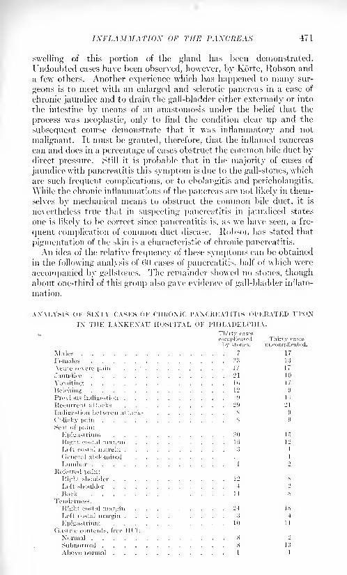
INFLAMMATION OF THE PANCREAS 471
swelling of this portion of the gland has been demonstrated.
Undoubted cases have been observed, however, by Korte, Robson anda few others. Another experience which has happened to many sur-
geons is to meet with an enlarged and sclerotic pancreas in a case of
chronic jaundice and to drain the gall-bladder either externally or into
the intestine by means of an anastomosis under the belief that the
process was neoplastic, only to find the condition clear up and the
subsequent course demonstrate that it was inflammatory and not
malignant. It must be granted, therefore, that the inflamed pancreascan and does in a percentage of cases obstruct the common bile duct bydirect pressure. Still it is probable that in the majority of cases of
jaundice with pancreatitis this symptom is due to the gall-stones, wrhich
are such frequent complications, or to cholangitis and pericholangitis.
While the chronic inflammations of the pancreas are not likely in them-
selves by mechanical means to obstruct the common bile duct, it is
nevertheless true that in suspecting pancreatitis in jaundiced states
one is likely to be correct since pancreatitis is, as we have seen, a fre-
quent complication of common duct disease. Robson has stated that
pigmentation of the skin is a characteristic of chronic pancreatitis.
An idea of the relative frequency of these symptoms can be obtained
in the following analysis of GO cases of pancreatitis, half of w'hich were
accompanied by gallstones. The remainder showed no stones, thoughabout one-third of this group also gave evidence of gall-bladder inflam-
mation.
ANALYSIS OF SIXTY CASES OF CHRONIC PANCREATITIS OPERATED UPONIN THE LANKENAU HOSPITAL OF PHILADELPHIA.
Thirty cases
complicated Thirty cases
by stones. uncomplicated.
Mules 7 17
Females 23 13
Acute severe pain 17 17
Jaundice 21 10
Vomiting 1(5 17
Belching 12 9
Previous indigestion 9 14
Recurrent attacks 29 21
Indigestion between attacks 8 9
Colicky pain 8 9
Seat of pain:
Epigastrium 20 15
Right costal margin 1 (i 12
Left costal margin 3 1
General abdominal 1
Lumbar 1 2
Referred pain:
Right shoulder 12
Left shoulder 4 2
. Back 14 8
Tenderness:
Right costal margin 24 18
Left costal margin .... 3 4
Epigastrium 10 11
Gastric contents, free IK'l:
Normal 8 2
Subnormal 8 13
Above normal 1 1
472 SURGERY OF THE PANCREAS
The pancreas can be palpated only in exceptional instances. Thetension of the recti and the interposition of the stomach, colon or liver,
as a rule, prevent detection of even considerable enlargement. Occa-
sionally, it is possible to feel the organ in individuals with very lax andthin abdominal Avails, and at times it can be felt tinder anesthesia, whenit has not been palpable before. It should be noted that the pancreas
possesses a fair range of mobility in its bed of loose areolar tissue and is
not, as commonly stated, fixed in position. It does not, however, movewith respiration. In jaundiced states or in coincident hepatic disease
the liver may extend downward over the region of the pancreas. The
gall-bladder is not ordinarily palpable. Obstruction of the commonduct due to pancreatitis is only in the most extreme and rare cases
sufficiently complete and permanent to cause palpable distention of the
gall-bladder. Another feature which militates against this condition is
the frequently associated cholecystic disease which renders the gall-
bladder firm and devoid of elasticity.
Early pancreatitis rarely gives any recognizable symptoms of func-
tional disturbance. AVhile this is regrettable from the standpoint of
diagnosis, it proves the great reserve powers of the gland and is fortu-
nate for the individual affected. For a summary of the methods
employed to detect functional abnormalities the reader is referred
to p. 4.'!4. Thus far these tests occupy a very subordinate position in
diagnosis, and are not to be compared with the history, physical signs
and clinical deductions based on the known characteristics of the
disease. At times, however, they are suggestive or at least confirmatory.Most important is the constant or intermittent presence of sugar in
the urine, ^'e have found glycosuria in 7 per cent, of our operativecases. Probably repeated observations carried out over a long periodof time and particularly during exacerbations of the disease would give
greater importance to this symptom. We have observed a number of
cases that during attacks showed glycosuria which cleared up after
operation.The pancreatic reaction of Cammidge upon the urine, though
strongly championed by its originator and heartily commended bysuch excellent observers as Robson and Moynihan, we must reluctantly
admit has not been of service to us, and in this conclusion most sur-
geons agree. Cammidge mentions the presence of large numbers of
calcium oxalate crystals in the urine as tending to confirm a diagnosis
of chronic pancreatitis. An increased amount of diastase has been
noted in the urine by some observers but is not constantly present.
The best evidence of absence or deficiency of pancreatic ferments in
the intestine is that given by the constant presence of the typical stools
of advanced pancreatitis, described above and but seldom seen. Refine-
ments of these gross observations, consisting in microscopic exami-
nation of the constituents of the stools for faulty digestion of muscle
fibers, nuclei and fat; chemical determination of the amount of neutral
fats and fatty acids; estimation of ferment activity of trypsin, lipase
and amylase in the stools; indirect determination of pancreatic activity
1.\FLAWATIOX OF THE PAXCREAS 473
within the bowel by means of Sahli's desinoid^test or tJie gelodurat (sic)
capsule all of which have been the subject of a large number of
ingenious experiments and observations when carried out with rigid
precautions to eliminate disturbing factors and carefully criticised
with reference to factors that cannot in the nature of things be elimi-
nated permit certain presumptions as to the condition of the pancreas.The same may be said of the more direct methods of examination of
pancreatic juice obtained from the stomach after regurgitation of
abdominal contents, or from the duodenum by aspiration through the
duodenal tube. The net results of all this work so far, when comparedwith the condition of the pancreas at the autopsy in vico or postmortem,show that functional tests are unreliable as a criterion of the anatomical
condition or the pathological process affecting the pancreas. Further-
work may increase their value in diagnosis or lead to new and importantdiscoveries, but at the present time it is a mistake to lay too muchstress on the laboratory means of diagnosis in the recognition of chronic
pancreatitis. It remains a diagnosis to be made from grosser observa-
tions of symptoms and signs with a reasoned conclusion from the prob-abilities of the case. In the majority of cases exploration alone will
decide.
In differential diagnosis there are to be considered infective andcalculous disease of the bile passages and liver, duodenal and gastric
ulcer, general debilitated conditions with associated derangements of
digestion, such as the anemias, tuberculosis, syphilis and nephritis,abdominal tuberculosis and arteriosclerosis, chronic appendicitis,tumors of the vertebra 1 and tumors of the duodenum, bile ducts, liver,
lymph glands and of the pancreas itself.
General disease can be ruled out by a thorough-going examination.
Pancreatitis, however, may coexist. Other conditions must be differ-
entiated, as a rule, by developing their essential characteristics since
pancreatitis itself possesses fewer characteristic features than most
other abdominal affections. Pancreatitis at times, however, closely
simulates the other diseases which may be confounded with it. It is
most important to distinguish advanced pancreatitis from malignantdisease of the head of the pancreas or of other organs in the neighbor-hood which may give rise to chronic jaundice. At operation it is usually
easy to distinguish tumors primary in other locations but when primaryin the pancreas it may be difficult or impossible at the time to differen-
tiate carcinoma from non-malignant indurative pancreatitis. It is a
mistake, therefore, to conclude that such a process is malignant andto terminate the operation with exploration.
Treatment. The treatment of chronic pancreatitis, as a rule, is that
of the disease to which it is secondary. If it depends upon general
conditions, such as syphilis, the treatment naturally is medical, and in
any case general hygienic measures and proper regulation of diet andof the bowels are beneficial to a degree. At times the exhibition of
preparations derived from the pancreas and containing pancreaticferments is productive of symptomatic improvement. Relative func-
474 SntClEllY Or THE PAXCREAS
tioiinl rest is best ol)taine(l medically by the Wohlgeinuth diet. It mustbe thoroughly understood, however, that in the vast majority of cases
chronic pancreatitis depends for its origin and continuance upon the
presence of some surgical disease of the abdominal viscera. Jt is there-
fore hazardous to defer operative intervention, for the anatomicalalterations which eventually occur as a result of pancreatitis yieldneither to curative nor to palliative measures. When by the size,
appearance and consistency of the gland the surgeon has determinedthe existence of chronic pancreatitis, he should make a careful search
for all possible atria of infection within the abdomen. The bile passagesshould first receive attention. If the common duct contains stones,
choledochostomy is indicated with removal of the calculi and the
passage of a large duct probe into the duodenum to make sure that the
opening is patulous and to secure full dilatation. If the gall-bladderis diseased it should be treated according to conditions. If the pancreasis not diseased to the degree of causing common duct obstruction, the
gall-bladder should be removed, as it is in many instances the primaryfocus of infection, and as shown by Rosenow it harbors within its walls
bacteria which simple drainage may not always suffice to eradicate.
If, on the other hand, jaundice is a feature of the disease, and unless
clearly due to stone obstruction without any element of pancreatic
pressure, it. is safer to provide for permanent internal drainage of the
bile by means of a cholecystoduodenostomy. If mechanical conditions
do not favor this operation, an anastomosis may be made with the
stomach (Kehr), or with the jejunum brought up through the gastro-colic omentum (Ochsner) or with the intestine lower down. Naturallythe surgeon will select a point for anastomosis which most nearly
approaches the normal point of discharge of the bile. The gall-bladder
may be in such condition that it is unsafe for anastomosis. In that
case the surgeon may choose between stitching it to the abdominal wall
to provide for long-continued drainage or he may remove it and trust
to drainage of the common duct by means of a T-tube which will
remain in position as long as desired. In one case referred to us byDr. Kiesman the patient, after three operations upon the commonduct for cicatricial stenosis and pancreatitis has refused to part with
the T-tube for fear of recurrence and has now worn it for nearly two
years with practically no inconvenience and but little discharge.1 )rainage for the cure of pancreatic disease should be maintained
for a longer period of time than when operation is done merely for chole-
lithiasis. Drainage in this condition acts in two ways: It relieves
infection of the biliary tract and gives the pancreas an opportunity to
throw ofl' the inflammation communicated to it from this source.
Secondly, in case the pancreatic and common bile ducts anastomose
before discharging through the papilla of Vater, the relief of pressurewithin the ducts of the biliary system at the same time releases pressurewithin the pancreatic ducts and favors restitution of the pancreasitself. This will occur independently of stenosis of the orifice of the
papilla of Vater or contraction or spasm of the sphincter (of Oddi)
CYSTS OF THE PAXCREAS 475
which surrounds it. Assured and long-continued drainage is the key-note of sueeess, and this must be accomplished with the means at
command. Occasionally the rarer operations, such as choledocho-
duodenostomy or duct reconstruction must be employed. The biliary
tract is the most promising avenue of approach to the pancreas in these
conditions, and in cases which show no demonstrable disease of tin-
gall-bladder or of the bile ducts, unless other primary foci from which
pancreatitis can be derived are demonstrated, it will usualh be wise to
drain by means of a cholecystostomy in the hope that infection of the
biliary tract exists unrecognized from gross appearances, or that
drainage may, in the second manner mentioned, favorably influence
the process in the pancreas.Duodenal and gastric disease when present should receive their
appropriate treatment. Excision of an ulcer is preferable when it can
be done. L leers perforating subacutely into the pancreas cannot be
excised with safety. They should be treated by exclusion of the pylorusand posterior gastrojejunostomy.The appendix should always be removed. In women the pelvis
should be examined and any inflammatory tissues should if possiblebe excised.
The experimental work of Cofl'ey, Sweet and others shows that in
animals, at least, the pancreas admits of a variety of operative maneu-vers. Drainage of the organ can be effected through the tail by meansof pancreato-enterostomy which can be done by amputating a portionof tissue, exposing the duct and inserting the pancreas into the lumenof the intestine which is brought tip around it, in much the same manneras in an ordinary enterostomy with a tube. It is possible that such an
operation may occasionally find a clinical application. Link has
reported a successful pancreatostomy in a case of pancreatic lithiasis in
which he brought the tail out through the anterior abdominal wall
which allowed the secretion and the stones to discharge themselves
externally through the opened and dilated end of the duct of Wirsuug.
CYSTS OF THE PANCREAS.
The earliest lesions of the pancreas to attract attention were the
cysts which form in and about that organ. The evident symptoms and
striking physical signs of the larger cysts could not escape notice and
for a long time they held the center of interest. Thus Oser, in Xoth-
nagel's Encyclopedia in 190.3, wrote that "cysts without doubt belongto the few diseases of the pancreas that can rightly claim clinical and
practical importance." That they are infrequent conditions can be
judged from the fact that Segre found but two pancreatic cysts in 1 1 ,500
autopsies and Hale White only three instances in G70S autopsies. Oser
was able to collect 1.34 cases from the literature, and since that time
in the neighborhood of 100 have been added. While they are infre-
quent findings they are still of great importance in that they are often
amenable to operative treatment with brilliant results.
4<t> SURGERY OF THE PAXCREAS
Two varieties of cysts are to be distinguished, (1) the true cysts, (2)
the pseudocysts.True Cysts. The true cysts arise within the substance of the gland
and in the beginning, at least, have an epithelial lining. Later the liningcells may disappear as the result of pressure.The pseudocysts are collections of fluid within, or more commonly,
in tlie immediate neighborhood of, the pancreas, which have becomesurrounded by a capsule of inflammatory tissue.
A third variety of cysts which are not truly cysts of the pancreasbut rather cysts /// the pancreas are the parasitic or echinococcus cysts.
These are very uncommon. The pancreas is relatively immune to
echinococcus disease. Ilanser,1 in HM2, was able to collect only 28
cases. In the large series of echinococcus disease reviewed, the pancreaswas affected in only about 1 per cent, of the cases.
True cysts of the pancreas may arise in several ways.1. Congenital Malformations. Garrod mentions instances of cyst
formations in the fetal pancreas and Boklt reports a case of epigastrictumor which was found at operation to be a polycystic pancreas asso-
ciated with polycystic disease of the left kidney. The right kidney wasnot explored. Such malformations are rare and present little clinical
interest.
2. Retention Cysts.- The earliest attempts to explain the genesis of
cysts laid stress upon the factor of obstruction of the main or tributary
pancreatic ducts with consequent dilatation and cyst formation bysecretory distention of the distal ducts and acini. Semi showed that
no such simple process could be responsible for the larger cysts as found
under clinical conditions. That cysts do form as a result of obstruction
is known, and eases have been reported by Opie, Cruveilhier, ^ irchow
and others in which duct obstruction by stone, tumor or the fibrous
constriction of chronic pancreatitis, stood in evident causative relation
to cyst formation in the pancreas. Such cysts, however, are usually
multiple, small and of themselves of little clinical significance. Some
exceptions exist. Thus "\Yoln"- pictures and reports a case of multiple
cysts of the pancreas, which was excised by Kehr. The patient was
syphilitic and the pancreas was the seat of a chronic pancreatitis to
which the cystic condition was doubtless to be ascribed. Occasionallytrue cysts of larger sixe are found in a pancreas which is the seat of
chronic indurative pancreatitis to which the cyst formation, with
reason, seems to be attributable. Korte 3 as a result of his observation
inclines to this view.
.'>. Cystic Neoplasms.- Proliferation cysts or cystadenomas occur in
the pancreas. Kleinschrnidt,4 in 1907, collected 21 cases. In 4 cases
the growth occupied the entire gland, in 2 the middle portion was
all'ected and in the remaining 15 the tail only was involved. Such
tumors are attached by a broad base to the pancreas but are separatedfrom the parenchyma of the pancreas by a capsule. Occasionally
2Ibid., 1911, Ixxiv, 487.
4 Med.-natunviss. Arch., 1907, i, 177.
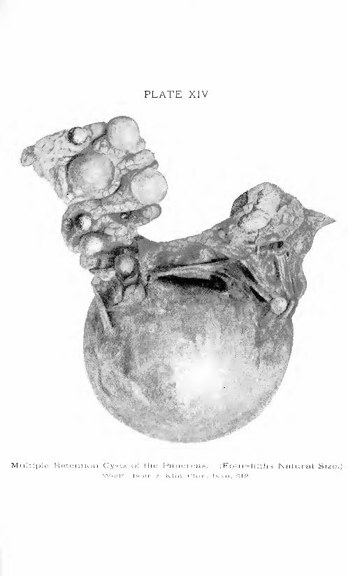
PLATE XIV
Multiple Retention Cysts oi the Pancreas. (Four-fitihs Natural Size.)

CYSTS OF THE PANCREAS 477
malignant change occurs with infiltration of the pancreas and sur-
rounding structures. Speese reports a curious case 1 which after opera-tion and drainage ten years previously again came to operation with the
cystadenomatous growth extending up to and involving the abdominalwall.
Such growths consist of a multitude of cysts, of varying sizes. Their
contents are clear and serous and arc practically devoid of the ferments
so characteristic of other types of pancreatic cysts. They usually
develop slowly without history of previous trauma or inflammation
and, as a rule, give rise to few symptoms unless they attain consider-
able size or grow with unusual rapidity. The histology bears a close
resemblance to the common cystadenoma of the ovary.
Pseudocysts. The pseudocysts of the pancreas may be divided into:
1. Cysts by Softening. A hemorrhage into the substance of the gland
may subsequently soften, its blood constituents may be absorbed in
whole or part and the focus converted into a cyst cavity. Malignantfoci also may undergo degenerative processes forming a cyst of irregularcontour with grumous contents. Such formations, though they are
in reality cystic in character, do not attract clinical attention as such.
They are by-products of pathological processes in which the cyst forma-
tion plays a secondary part. More important are the
2. Cysts by Self-digestion.- The importance of activated pancreaticferments in the pathogenesis of pancreatic disease has been discussed.
Instances are not rare in which a large part of the pancreas, and at
times probably, the entire pancreas undergoes rapid necrosis and gan-
grene as the result of a combination of injury, chemical, bacterial or
traumatic, with en/ymic action. The autodigestive process does not
stop with the necrosis of the gland and its surrounding tissues, but
proceeds actively to break down and dissolve the structures affected.
Naturally, in the more severe cases the process is interrupted by the
death of the patient, but in cases of less severity which are compatiblewith continuance of life there results a cavity containing disintegrating
tissue, blood, fluid and cellular exudates and bounded by a limiting
inflammatory wall. The resulting formation is dependent upon the
activity of the contents as they relate to the process of exudation and
absorption exercised by the surrounding tissues. It is not uncommonfor such cysts to fluctuate in size in accordance with the alteringbalance of these processes. Such changes in volume are of some diag-nostic importance. Usually there is a tendency for an increase in size
but at greatly varying rates of speed in individual cases. Heiberg2
quotes Hulke as having observed a case of forty years' duration. Morecommonly the development of such a cyst to a condition where it is
noticeable for its size and symptoms is a matter of several weeks or
several months. After a long time the contents of these pseudocysts
may become relatively clear and serous. As a rule, however, when
investigated they are bloody and contain more or less undissolved
1 Ann. $111-2., 1915, Ixi, 759.2 Die Krankheiten des Pankreas, Wiesbaden, 1914, p. 141.
478 SURGERY OF THE PANCREAS
tissue fragments. The interior wall is ragged and may show masses
of adherent necrotic tissue. This variety of cyst is the most commonand important from a clinical standpoint.
8. Exudative Cysts of the Omental Sac. The location of the pancreas
immediately beneath the floor of the lesser peritoneal cavity frequentlydetermines extravasation of blood and inflammatory exudates into that
cavity. The foramen of Winslow may have been previously obliterated
as a result of upper abdominal inflammation, to which indeed the pan-creatitis may be a sequel or concomitant. Or closure of the opening
may rapidly take place as a result of the violent and widespread inflam-
mation engendered by acute pancreatitis. As a rule, such collections
are infected or become invaded secondarily by microorganisms and
appear more truly as an abscess than as a cyst, but occasionallybacterial activity is low or absent and the collection may exist for a
considerable time without producing serious systemic symptoms.Size and Situation. Occasionally pancreatic cysts are of great size.
Parke's case, mentioned by Ochsner, contained 14 liters of fluid, and
Trendelenburg met with one which held about 20 liters. Cases of such
great size, always rare, are now, owing to the prevalence of surgical
treatment, practically unknown. Ordinarily, they vary from a size that
is just palpable to that of a child's head, and contain one to three liters
of fluid. Their location shows wide variations from the epigastric or left
epigastric position, which may be described as typical. Both the true
cysts or pseudocysts may arise from the head, the body or the tail, or
they may involve the entire organ. The point of presentation is influ-
enced not only by the site of origin but by anatomical considerations
which direct its course in accordance with the law of expansion alongthe line of least resistance. The most frequent direction of growth is
directly forward from its point of origin beneath the floor of the lesser
peritoneal cavity, causing it to protrude into that sac. Here it maycarry the stomach directly before it, and in inflated or distended con-
ditions of the stomach the cyst can be palpated only indefinitely
through that viscus, and percussion may give the tympanitic note of
an air-containing cavity. As the cyst enlarges, however, it tends to
push the stomach aside and present against the abdominal wall
covered only by the gastrocolic omentum. The colon also is pusheddownward and in extreme cases may be dislocated into the pelvis. Theanatomical relations can be best appreciated by a study of Zucker-
kandl's schematic representations.
In a second form the enlarging cyst pushes the stomach downwardand presents against the anterior abdominal wall covered by the gastro-
hepatic omentum. This is most apt to occur in conditions of gastrop-tosis. Korte, in an examination of .10 cadavers, found a portion of the
pancreas exposed above the lesser curvature of the stomach covered
only by the gastrohepatic omentum in ."> cases (10 per cent.). In such
cases gastric tympany will be found below the area of dulness due to
the cyst, and if the tumor be well toward the left the physical findings
can be readily mistaken for those due to an enlarged kidney, or if it be
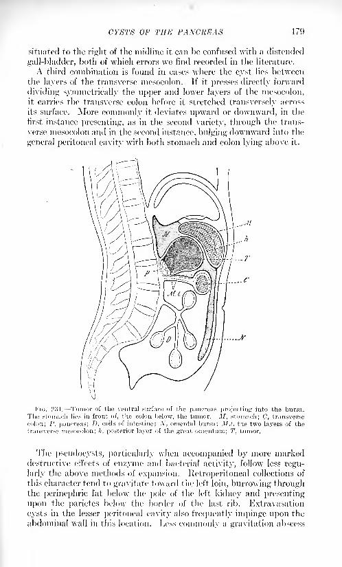
CYSTS OF THE PANCREAS 479
situated to the light of the midline it can be confused with a distended
gall-bladder, both of which errors we find recorded in the literature.
A third combination is found in cases where the cyst lies between
the layers of the transverse mesocolon. If it presses directly forward
dividing symmetrically the upper and lower layers of the niesocolon,
it carries the transverse colon before it stretched transversely across
its surface. More commonly it deviates upward or downward, in the
first instance presenting, as in the second variety, through the trans-
verse mesocolon and in the second instance, bulging downward into the
general peritoneal cavity with both stomach and colon lying above it.
FIG. 231. Tumor of the ventral surface of the pancreas projecting into the bursa.
The stomach lies in front of, the colon below, the tumor. M, stomach; C, transverse
colon; l\ pancreas; D, coils of intestine; Ar, omental bursa; M.t, the two layers of the
transverse mesocolon; h, posterior layer of the great omontum; T, tumor.
The pseudocysts, particularly when accompanied by more markeddestructive effects of en/yme and bacterial activity, follow less regu-
larly the above methods of expansion. Retroperitoneal collections of
this character tend to gravitate toward the left loin, burrowing throughthe perinephric fat below the pole of the left, kidney and presenting
upon the parietes below the border of the last rib. Extravasation
cysts in the lesser peritoneal cavity also frequently impinge upon the
abdominal wall in this location. Less commonlv a gravitation abscess
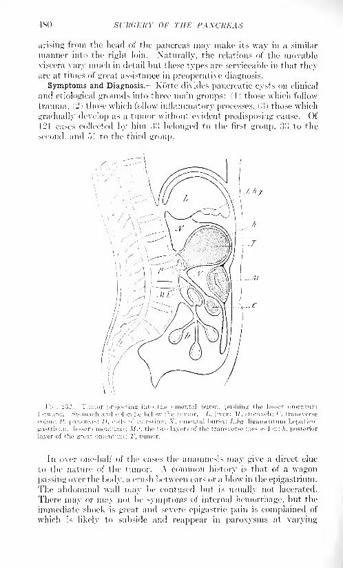
4SO SURGERY OF THE PAXCRKAX
arising from the head of the pancreas may make its way in a similar
manner into the right loin. Xatnrally, the relations of the movableviscera vary much in detail but these types are serviceable in that theyare at times of great assistance in preoperative diagnosis.
Symptoms and Diagnosis.- Korte divides pancreatic cysts on clinical
and etiological grounds into three main groups: d) those which follow
trauma. ('2) those which follow inflammatory processes, ('.}) those which
gradually develop as a tumor without evident predisposing cause. Of121 cases collected by him :>'! belonged to the first group, }'> to the
second, and ">1 to the third group.
Fin. 232. 'rumor projecting into the omental bursn. pushing the lessor omontuniforward. Stomach and colon lie below the tumor. L, liver: .!/. stomach; ('. transverse
colon: /", pancreas; 1). coils of intestine; -V, omental bursa; L.hfj. ligamentum hepatico-
gastricum (lesser omentum) ; .I/.', the two layers of the transverse mesocolon: h. posterior
layer of the Lrreat rimentuni; T, tumor.
Jn over one-half of the cases the anamnesis may give a direct clue
to the nature of the tumor. A common history is that of a wagonpassing over the body, a crush between cars or a blow in the epigastrium.
The abdominal wall may be contused but is usually not lacerated.
There may or may not be symptoms of internal hemorrhage, but the
immediate shock is great and severe epigastric pain is complained of
which N likely to subside and reappear in paroxysms at varying
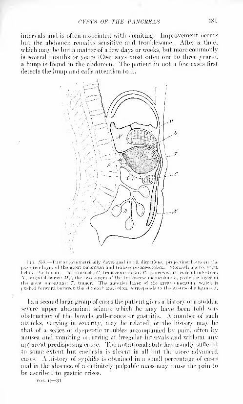
CYSTS OF THE PANCREAS 481
intervals and is often associated with vomiting. Improvement occurs
but the abdomen remains sensitive and troublesome. After a time,
which may be but a matter of a few days or weeks, but more commonlyis several months or years (Oser says most often one to three years),
a lump is found in the abdomen. The patient in not a few cases first
detects the him]) and calls attention to it.
l-'i<;. 233. Tumor symmetrically developed in all directions, projecting between the
posterior layer of the great omentum and transverse mesocolon. Stomach above, colon
below, the tumor. M, stomach; C, transverse colon; P, pancreas; D, coils of intestine;
.V, omental bursa; M.t, the two layers of the transverse mesocolon; h, posterior layer of
the great omentum: T, tumor. The anterior layer of the great omentum. which is
pushed forward between the stomach and colon, corresponds to the gast rocolic ligament.
In a second large group of cases the patient gives a history of a sudden
severe upper abdominal seizure which he may have been told wasobstruction of the bowels, gall-stones or gastritis. A number of such
attacks, varying in severity, may be related, or the history may be
that of a series of dyspeptic troubles accompanied by pain, often bynausea and vomiting occurring at irregular intervals and without any
apparent predisposing cause. The nutritional state has usually suffered
to some extent but cachexia is absent in all but the more advancedcases. A history of syphilis is obtained in a small percentage of cases
and in the absence of a definitely palpable mass may cause the pain to
be ascribed to gastric crises.
VOL. II 31
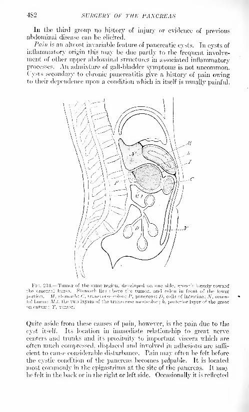
482 SURGERY OF THE PANCREAS
In the third group no history of injury or evidence of previousabdominal disease can be elicited.
Pain is an almost invariable feature of pancreatic cysts. In cysts of
inflammatory origin this may be due partly to the frequent involve-ment of other upper abdominal structures in associated inflammatoryprocesses. An admixture of gall-bladder symptoms is not uncommon.Cysts secondary to chronic pancreatitis give a history of pain owingto their dependence upon a condition which in itself is usually painful.
FIG. 234. Tumor of the same region, developed on one side, growth largely towardthe omental bursa. Stomach lies above the tumor, and colon in front of the lower
portion. Jlf, stomach; C, transverse colon; P, pancreas; D, coils of intestine; N, omen-tal bursa; M.t, the two layers of the transverse mesocolon; h, posterior layer of the greatomentum; T, tumor.
Quite aside from these causes of pain, however, is the pain due to the
cyst itself. Its location in immediate relationship to great nerve
centers and trunks and its proximity to important viscera which are
often much compressed, displaced and involved in adhesions are suffi-
cient to cause considerable disturbance. Pain may often be felt before
the cystic condition of the pancreas becomes palpable. It is located
most commonly in the epigastrium at the site of the pancreas. It maybe felt in the back or in the right or left side. Occasionally it is reflected
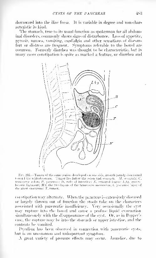
CYSTS OF THE PANCREAS 483
downward into the iliac fossa. It is variable in degree and non-char-
acteristic in kind.
The stomach, true to its usual function as spokesman for all abdom-inal disorders, commonly shows signs of disturbance. Loss of appetite,
pyrosis, nausea, vomiting, cardialgia and other sensations of discom-
fort or distress are frequent. Symptoms referable to the bowel are
common. Formerly diarrhea was thought to be characteristic, but in
many cases constipation is quite as marked a feature, or diarrhea and
FIG. 235. Tumor of the same region developed on one side, growth largely downwardtoward the mid-abdomen. Tumor lies below the colon and stomach. 3/, stomach; C,
transverse colon; P, pancreas; D, coils of intestine: .V. omental bursa; LJuj, gastro-
liepatic ligament; M.I, the two layers of the transverse mesocolon; h, posterior layer of
the great onientmn; T, tumor.
constipation may alternate. When the pancreas is extensively diseased
or largely thrown out of function the stools take on the characters
associated with pancreatic insufficiency. Very occasionally the cyst
may rupture into the bowel and cause a profuse liquid evacuation
simultaneously with the disappearance of the cyst. Or, as in Pepper'scase, the rupture may be into the stomach or upper intestine, and the
contents be vomited.
I'tyalism has been observed in connection with pancreatic cysts,
but is an uncommon and unimportant symptom.A great variety of pressure effects may occur. Jaundice, due to
484 SURGERY OF THE PANCREAS
pressure on the common bile duct, edema of the extremities and ascites
due to pressure on venous channels, renal colic, as a result of compres-sion of the ureters, as well as other phenomena have been noted in
individual instances. These are chance occurrences and but seldom
possess diagnostic value.
Loss of weight due to disturbance of pancreatic function as well as
of the alimentary tract is common.Fluctuation in size of the tumor, a general characteristic of cysts,
has been observed many times.
Glycosuria was noted by Korte 9 times in oo traumatic cysts, butcollective reports of all varieties of cysts do not show such a high per-
centage of this symptom. When present it is strongly confirmatory of
the diagnosis.( "ammidge acknowledges that his reaction is of no assistance in the
diagnosis of this condition and the functional tests in general are of less
use than in certain other diseases. At times cystic disease of the pan-creas is the outcome of a condition which has produced marked diffuse
disease of the organ, in which case the signs of pancreatic insufficiency
may be pronounced.Pancreatic tumors do not move with respiration. If inflammatory
they cannot be moved by the hand. When they develop*
withoutinflammation they may possess the usual mobility of the pancreaswhich, contrary to the general idea, is quite freely movable within a
short radius.
The .r-ray has been employed in diagnosis and is of some assistance
in showing the relation of the stomach and intestines to the mass, but
no direct evidence as to its nature is obtainable by this means.
Exploratory puncture, formerly much used, has led to some verydisastrous results due to infection of the peritoneal cavity. It must be
emphatically condemned. The only field for puncture which Korte
allows is in the cysts which present in the loin when the mass can be
reached extraperitoneally, and even in these cases it should be done
only when it is possible to proceed at once with the operation.The investigation of the character of the cyst contents is of consider-
able importance in determining the nature of the condition. It is not
always possible by operation to establish the fact that such cysts are
in reality of pancreatic origin. Grossly the contents vary from clear
serous fluid to bloody, purulent, grumous or necrotic material.
The demonstration of the characteristic ferments of the pancreasis sufficient to make the diagnosis. The presence of small amounts of a
lipase or amylase is not necessarily indicative of pancreatic origin.
Trypsin, however, is distinctive. It may exist only in the prefermentform, and must be activated before it will produce its digestive effects.
As pancreatic juice is ordinarily collected from the sinuses remainingafter drainage of the cyst, the trypsin is activated by the admixture
of tissue juices and bacteria. Clinically, the ferment activity of the
secretion can be only too well observed in its corrosive effects uponthe skin or other tissues with which it comes into prolonged contact.
CYSTS OF TI1K PAXOREAS 4S5
Slight febrile attacks are common, but in general the temperatureis normal as is also the pulse. The leukocytes range from a normal
number in the majority of instances to a moderately high leukocytosis
during attacks or when infective processes are present.The diagnosis of pancreatic cyst can usually be made from a fe\v
salient features after exclusion of conditions with which it may be
confused. The most characteristic positive features are a historyindicative of previous pancreatic injury or disease, the location in the
neighborhood of the pancreas, the gradual and symmetrical growth and
occasional fluctuations in size, the pain, digestive disturbances and
wasting without cachexia, and the relations of the tumor to the stomachand intestines, (dycosuria and evidence of faulty pancreatic digestion,
if present, are of strong confirmatory value, but are found in only a
minority of cases.
The conditions with which it may be confounded are:
I. Aneurysm of the abdominal aorta, from which it may be distin-
guished by the absence of transmitted pulsation when the patient is
placed in the knee-chest position.
'2. Abscess of the abdominal wall, the superficial position of which
can be made out by careful examination.
3. Echinococcus cyst of the liver. This will be located in front of
and above the stomach. A pancreatic cyst may in rare instances
present above the stomach but it can never lie in front of that organ.The hydatid thrill may be present and in a percentage of cases a com-
plement-fixation reaction using hydatid fluid as antigen will be positive.
Less frequently echinococcus cysts occur in this region in connection
with other organs. They cannot always be differentiated. The history
may be significant.
4. Tumors of the liver are usually clearly continuous with liver
dulness and occupy the same position as described above with relation
to the stomach. They are movable with respiration which is contraryto the usual finding in the case of a pancreatic cyst.
5. Enlarged gall-bladder is distinguished by its superficial location,
its mobility with respiration, a lateral motion usually being demon-
strated, as well as the more marked upward and downward excursions.
G. Retroperitoneal hygroma or lipoma cannot be certainly distin-
guished, but is rare.
7. Cyst of the round ligament, a rare condition, is more movable
laterally and moves with the abdominal wall.
8. Splenic cysts are never covered by stomach or bowel.
9. Cysts of the mesentery are lower in position and possess a much
greater range of mobility.10. Pancreatic abscesses give a history of stormy onset with toxemia
and evidences of an inflammatory process.
II. Collections in the lesser peritoneal cavity are often secondaryto acute pancreatitis and give the history and systemic symptoms of
that disease. When due to perforated gastric ulcer, the type of indi-
gestion preceding the attack is of assistance and the pronounced signs
of an inflammatory process dilfer materially from the effects of a cyst.
486 SURGERY OF THE PANCREAS
12. Tumors and cysts of the kidney and adrenal bodies by bimanual
palpation are found to lie chiefly in the region of the loin. They movewith respiratory excursions and in the case of the kidney the composi-tion of the urine and a combination of x-ray and cystoscopic examina-tion will make the condition clear.
13. Tumors of the vertebral column can be ruled out by their con-
sistency, their absolute fixity, and by axray findings.14. Retroperitoneal tuberculous collections or glandular masses are
infrequent in adults.
15. Tumors and inflammatory masses of the peritoneum or omentumare more superficial and usually differ in contour and consistency.
In the individual cases many other differentiating points will suggest
themselves, but after all due care has been used it must still be said
that a considerable percentage of cases will wait for diagnosis by explor-
atory operation. No pathognomonic symptoms or set of symptomsexists, but to the surgical mind the symptoms and signs will always
point to the advisability of operation.Treatment. Pancreatic cyst is purely a surgical disease and is to
be attacked directly and boldly by open operation. In almost all
cases a transperitoneal operation must be done to reach the cyst.
Occasionally cysts may present in the loin when they can be attacked
retroperitoneally. This concerns only the pseudocysts. We find nomention of true cyst development in this manner. Rarely do pan-creatic cysts, protected as they are by overlying viscera and omenta,contract sufficient adhesions to the abdominal wall against which they
present to justify the attempt at direct incision through parietes into
the cyst. Such a condition, if present, can be determined only bycareful exploration. The incision should be made over the most promi-nent portion of the cyst and prolonged sufficiently to secure ampleexposure and control. If in doubt as to the nature of the swelling, at
this point an exploratory needle may be introduced. It may be neces-
sary first to divide the overlying omentum or mesocolon to secure
exposure. If the mesocolon is opened especial care must be taken not
to injure the middle colic artery, as this is very likely to cause gangreneof the transverse colon, an accident which has happened in operatingfor pancreatic cyst. If the artery has been unwittingly or intention-
ally sacrificed, it is best to resect the bloodless portion and make the
necessary anastomosis. It is highly undesirable to be obliged to do
this, and in general if at all possible one should avoid opening a cyst
through the lower layer of the transyerse mesocolon since this exposesthe general peritoneal cavity to contamination and increases the diffi-
culties of drainage.The relations of the cyst may now be explored. Before any operative
procedure is attempted it is wise to wall off the surrounding intestines
both for purposes of control and for protection against possible danger-ous contamination by the cyst contents. Exploration and subsequent
procedure will often be facilitated by draining off with a trocar and
cannula a portion of the cyst contents. Only three methods of dealing
CYSTS OF THE PANCREAS 487
with the cyst are to be considered: (1) drainage by marsupialization,
(2) excision, (3) partial excision and drainage. The method to be
chosen will be indicated by conditions. As a rule, the first method is
the easiest, safest and most satisfactory. It is impossible to excise
the pseudocyst as its wall is made up of the structures which wall it in.
But many of the true cysts also contract such intimate adhesions with
essential structures that to attempt to excise them is extremely hazard-
ous if not impossible. After completely emptying the cyst, by suction
or by mopping, a view may be had of its interior. If time and the con-
dition of the patient are pressing this step would be omitted. The open-
ing into the cyst is now sewed to the peritoneum and transversalis
fascia with a running or interlocking stitch placed close enough to
prevent immediate leakage into the peritoneal cavity. The peritoneumabove and below is closed and drainage of the cyst cavity instituted.
If the contents are believed to be sterile and the interior of the cyst
reasonably free from necrotic masses of tissue, a large tube will be
sufficient. Closure under these circumstances may take place with
remarkable rapidity, healing being complete in two to three weeks.
If any doubt exists as to the liability to pocketing or retention of tissue
that may later be discharged, it is best to use tampons of gauze as well
as one or more tubes. If gauze is used it should not be removed too soon
or too forcibly as it may cause troublesome bleeding or dislodge anembolus. The best rule is to remove the gauze gradually as it is cast
off by the tissues as a slough. At the same time care should be taken
that the gauze should not act as a plug to dam back retained secretions.
This may be prevented by loosening the gauze at the outlet before the
attempt is made to extract it from the depths.If excision is to be attempted it should be done only after a careful
survey of its possibilities. Pancreatic cysts are almost never pedun-culated. Occasionally a cyst of the tail may be capable of removal,the tail itself acting as a pedicle. Mayo1 resected the distal half of the
pancreas for a cystic tumor, encountering bleeding that could not be
checked with ligature or sutures. He left clamps in situ, and packedwith gauze. The patient recovered. In case of doubt- excision should
not be attempted. If the pancreas is subjected to operative manipu-lations, these should be carried out with all due gentleness, bearing in
mind the possibilities of hemorrhage and ferment activation after
simple trauma. Drainage should always be employed, for a short
period at least, when the gland is injured.. In one variety of cyst,
namely, the cystadenoma or proliferation cyst, excision is indicated if
it can be carried out. Simple drainage does not cure these cysts because
they are multilocular and cannot be completely drained and, because
of the activity of the epithelial lining, it is difficult to secure an oblitera-
tion of the secreting surface. Cystadenoma if densely adherent to
important structures should be considered inoperable or the larger
cysts drained for relief of size and pressure symptoms. It is quite
1 Ann. Surg., 1913, Iviii, 145.
488 SURGERY OF THE PANCREAS
permissible, however, to assume some risk in the hope of making a
complete excision. The pancreas as a whole should never be removed.Partial excision and drainage is a compromise operation which enjoys
the advantages neither of excision nor of marsupialization. It increases
the difficulty of safe and thorough drainage of the contents of the sac
and should rarely, if ever, be employed.In a number of cases drainage has been done in two stages, first
attaching the cyst to the abdominal wall and later, after the peritoneal
cavity has been walled off, opening the sac to provide drainage. It is
an inferior measure in that it does not give immediate relief during the
most trying postoperative period, and practically the method of pro-
tecting the peritoneum by sewing the edge of the marsupialized pouchto the peritoneum has been found effective. In individual cases there
may be good reason for preferring the two-stage operation.The sinuses which exist after drainage of a pancreatic cyst are at
times very troublesome. This is due to two causes: (1) obstruction to
normal flow of pancreatic secretion into the duodenum causing it to
seek the path of less resistance through the artificial opening, and (2)
because of the irritating qualities of the pancreatic ferments which
erode the sinus walls and hinder or prevent granulation and closure.
In many cases the drainage of the cyst appears to act favorably uponthe remaining pancreas, so that after a time the normal channels
resume their functions, less and less secretion is directed into the sinus
which at the same time by its attempt at closure encourages the secre-
tion to take its normal course and gradually the discharge ceases andthe sinus walls unite. In case of persistent fistula, Wohlgemuth pro-
poses that the patient be placed upon a strict antidiabetic diet with
large doses of sodium bicarbonate after meals, on the theory that in
this way the pancreas would be less stimulated to secrete and the cyst
cavity and sinus walls be given a better opportunity to granulate and
close. A number of surgeons have reported excellent results of this
treatment, though not all experiments have been so favorable. Korte
has pointed out that its theoretical basis does not accord with the classi-
cal work of Pawlow on the secretion of the pancreas, since he found that
carbohydrates were more stimulating to the secretory output than fat.
It is advisable, in troublesome cases to give the method a trial since
no form of local treatment has any influence in promoting healing.
It is well in dressing all cases of this kind after operation to begin at
once to protect the skin wherever the secretion may come into contact
with it by a heavy coating of zinc oxide ointment or vaseline as the
erosions which occur are painful and after once formed are almost
impossible to heal so long as the discharge exists.
PANCREATIC CALCULI.
Concretions in the pancreas are infrequent. Opie found 3 instances
in 1500 autopsies and Kretz estimated that they occurred on the aver-
age once in 3000 cases. Giudeceandra encountered 2 in 122 cases.
PANCREATIC CALCULI .. 489
Oser, in 1903, found 70 cases on record and since then the numberrecorded has increased to approximately 90.
Etiology. But little is definitely known of the etiology of calculus
formation in the pancreas. Stasis and infection are believed to be the
chief factors, though Pende, 1 from his experimental investigations into
the process of stone formation, came to the conclusion that infective
processes were not responsible for the condition and assigned the lead-
ing role to pancreatic sclerosis induced by the circulating toxins of lues
gout, alcoholism and other metabolic disorders.
Pancreatic stones are usually multiple and small. The largest is
recorded by Schupmann which measured two and a half inches longand a half inch in diameter. More often they are stated to be the size
of a pea or bean and vary down to almost impalpable sand which maybe present in abundance, either free or incrusting the lining of the ducts.
In a few cases putty-like masses have been found, but ordinarily the
pancreatic stone is quite hard being composed chiefly of the carbonate
and the phosphate of calcium. They are rarely facetted but may be
very irregular in shape. Round, oval, branching, mulberry and
spiculated stones have been described. Commonly they are white,
gray or yellowish, but dark and even black stones have been found.
Occasionally gall-stones have made their way into the head of the pan-creas by ulceration from the common duct. Dowd reports such a case.
They can be distinguished by the cholesterin and bile pigments of which
they are chiefly composed, constituents which are rare or absent in
pancreatic calculi.
The stones are found most often in the head but may be located in
any portion of the organ. In a number of cases they were present in
such numbers as to fill the ducts throughout the organ.
By far the greatest number of stones occur in the male sex (26 males
and 6 females Oser) and have been discovered most often in indi-
viduals between the ages of thirty-five and forty-five years.
Symptoms. Kretz states that barely one-third of the subjects of
pancreatic calculi show symptoms during life. As his conclusions were
based on cases observed at postmortem we cannot accept the statement
as entirely reliable since this method of drawing conclusions has been
shown to be fallacious in many other conditions, notably cholelithiasis.
It is true, however, that distinctive symptoms are lacking in many cases.
Lazarus divides the symptoms into five groups: (1) essential symptomsof stone, (2) symptoms due to sequelse, (3) functional disturbances,
(4) symptoms due to effects upon other organs, (5) general symptoms.Of symptoms due to the presence of the calculus the most important
is pain. This is often severe and colicky, and usually located in the
epigastrium and left hypochondrium. It tends to radiate to the backand left shoulder. During attacks vomiting often occurs and at times
chills, moderate fever and sweats are indicative of accompanyinginfective processes. Collapse may be the result of a severe seizure.
>I1 Policlinico, 1905, vol. xii; Sea. Med., p. 122.
490 SURGERY OF THE PANCREAS
When the stone also obstructs the common duct the pain may have the
characteristics of gall-stone colic. In a number of cases the calculi havebeen found in the stool after attacks or in the interim.
The results of pancreatic lithiasis are various: chronic pancreatitis,
complete atrophy of the pancreas, hemorrhagic pancreatitis and
necrosis, the formation of true cysts, pseudocysts and fistulse have all
been reported. In such cases the symptoms take on the characteristics
of the resulting conditions.
Disturbances of the function of the pancreas are common. Zesas,1 in
a study of 70 cases, found that glycosuria was the most frequent symp-tom, but concluded that it was a late manifestation. It may be slight,
intermittent or may possess all the characteristics and severity of true
diabetes. In extreme cases indeed the destruction of the pancreas is
so complete as to amount to a pancreatectomy so far as function is
concerned. Bissell2 in one instance was unable to discover any pancre-atic parenchyma by the aid of the microscope. The pancreas existed
only as a distended system of ducts, v. Recklinghausen found the pan-creas reduced entirely to a fatty mass without parenchyma. On the
other hand, Walker3reported a case, observed for twenty-six years,
which at autopsy showed pancreatic calculi blocking the duct and
advanced atrophy of the parenchyma, yet never developed glycosuriaor other evidences of disease of the pancreas. More often the late cases
with extreme alterations in the pancreas show more or less pancreatic
Insufficiency, with steatorrhea, azotorrhea and disturbance of absorp-tion.
Symptoms due to involvement or disturbance of function of related
organs, are seen most often in involvement of the biliary tract byobstruction or ascending infection which gives rise to jaundice and
biliary colic. Pigmentation of the skin has been noted. In a case
operated upon by Moynihan this disappeared after removal of the
calculus. The stomach and duodenum may be interfered with byadhesions or by reflex influences and the bowels are disordered.
Ptyalism has occasionally been observed.
General symptoms are loss of weight and of strength, anemia, in
some cases a veritable cachexia, and the evidences of infection. Duringexacerbations, fever, chills, sweats and leukocytosis may be present.
It is noteworthy that a large proportion of subjects of pancreatic stone
die of tuberculosis.
Diagnosis-. Diagnosis is difficult or impossible. Of the 70 cases
reported by Zesas only 7 had been diagnosed during life. It depends
upon persistently recurring upper abdominal indigestion with painful
exacerbations, plus pronounced symptoms of pancreatic insufficiency.
If stones corresponding in character to pancreatic calculi are found in
the stools, the diagnosis is a reasonable certainty. Einhorn, Glaessner
and others have reported such cases. The x-ray may prove to be of
1 Centralbl, f. d. Grenzgb. d. Med. u. Chir., 1903, vi, 801.2 Tr. Chicago Path. Soc., 1914, ix, 1-67.
' Med.-Chir. Tr., 1889, Ixxii, 257.
PANCREATIC CALCULI 491
some service though the difficulties of detecting stones as well as of
ruling out confusing shadows in this location are great. Pforringer
reports a case in which he detected a shadow just to the right of the
midline in a case thought to be one of renal calculus. He considered it
too near the midline and advised further examinations. The patientwent elsewhere and was operated on for renal calculus, which was not
found, but death occurred from hemorrhage. At autopsy a pancreaticcalculus was present. Assmann, in a review of plates made for the
purpose of an intestinal study, upon a case found at operation to have
pancreatic calculi, discovered the definite shadows of a calculus which
had been overlooked during the earlier examinations.
Treatment. Treatment of pancreatic lithiasis is greatly limited bythe difficulties of diagnosis both before and during operation. The firm
consistency of the gland may make it impossible to feel stones even
when present. Again the variations in size, situation and number of
calculi make it impossible to standardize a method of procedure. Still
it remains true that this condition is properly surgical, and medical
treatment cannot be regarded as anything but palliative and expectant.Various regimina of medical treatment have been advised. Eichhorst
in one case administered pilocarpin to stimulate the flow of secretion
and reported the patient much improved. On the other hand,Glaessner and others have tried to inhibit the flow of pancreatic juice
by the "Oelkur" and large amounts of alkaline fluids, likewise reporting
good results. The truth seems to be that the disease is characterized
by exacerbations and remissions, though naturally measures directed
at the control of symptoms and the alleviation of gastric and duodenal
irritation will prove beneficial.
The earliest operative case is that of Gould,1 who opened the abdomen
for symptoms of cholelithiasis. Feeling a mass behind the lesser
omentum he explored, incised and removed a branching stone from the
duct of Wirsung. The patient recovered, but the obstruction to the
common bile duct was still evident from the fact that all the bile con-
tinued to discharge from the cholecystostomy opening made at the
operation. Seven months later he again operated, located a stone in
the head of the pancreas, removed it through an incision in front andwas able to pass a probe into the duodenum. Unfortunately, the
patient succumbed to infection on the twelfth day.
Moynihan2 relates a singularly successful case in which he diagnosedthe condition and was able to extract the calculus from the papilla of
Vater through the duodenum. Dalziel is said by Robson and
Cammidge to have had a similar successful case, and the same authors
mention another instance in which recovery followed the removal of
four stones from the pancreas. One stone was removed from the duct
of Santorini and one from the duct of Wirsung by direct incision into
the gland and two were extracted from the papilla of Vater by forceps.
Stoeblin and Roeber3 incised the pancreas and successfully removed
1 Lancet, 1898, ii, 1632. * Lancet, 1902, ii, 355.3 New York Med. Jour., 1905, Ixxxii, 904.
492 SURGERY OF THE PANCREAS
several stones from the duct of Wirsung. Lisanti is credited by Korte 1
with having removed a stone from the pancreas. Ruth2operated for
gall-stones and found the pancreas filled with concretions, the largest
being over two niches in length and weighing 280 gms. Recoveryfollowed removal of the stones by direct incision with drainage of the
pancreas. Link3 in a somewhat similar case performed the unusual
operation of pancreatostomy by mobolizing the tail of the pancreasand bringing it through the incision in the abdominal wall. Drainageof pancreatic secretion and discharge of numerous concretions followed.
On a number of occasions the drainage of a pancreatic cyst or abscess
has permitted the discharge of calculi whose presence had not been
suspected. From the work already done it is apparent that in fortunate
instances the stone may be removed through the papilla of Vater andthis incised or dilated if necessary to further drainage and the dischargeof such concretions as may remain. Furthermore, it is shown that
under proper conditions the pancreas may be incised and the ducts
opened for the extraction of calculi. The procedure cannot be said to
be without risk, but the precise mortality cannot be estimated at
present. Freedmann courageously reported a case operated upon byPeck with a diagnosis of pancreatic stone in which exploration of the
pancreas by incision of the gland and opening the duodenum wasfollowed by death. The symptoms were typical. No stone was found
either at operation or at autopsy. Doubtless it is unwise to attemptto explore the entire duct by incision, but a suspected focus may be
opened with every prospect of recovery and a probe will determine
the patulous condition of the duct. It is easy to make false passagesin the pancreas with a small probe and if the duct is not found dis-
tended it may be hard to find and difficult to explore. Great gentleness
and clean dissection are advisable particularly in the light of our
knowledge of trauma and infection as causes of hemorrhagic pancrea-titis. Mayo has shown that bleeding from the cut surfaces of the pan-creas may be safely controlled by oversewing the bleeding surfaces
with catgut on a curved needle.
The safest rule is to dram every case of incision into the pancreas,
particularly if the duct has been reached. A combination of gauze and
tube may be required, but in cases of minor injury a small cigarette
drain may be sufficient. To prevent the drain from being displaced a
simple stitch of fine plain catgut may be used to fasten it temporarilyto the area which it is desired to drain.
It is probable that with greater regard to the condition of the
pancreas both before and during operation, more cases will be dis-
covered and additions made to our knowledge of the safest and most
salutary method of dealing with pancreatic calculi.
1 Entwick u. Fortschr. d. Chir., 1909, p. 310.2 Colorado Med., 1907, iv, 421.
"Ann. Surg., 1911, liii, 768.
TUMORS OF THE PANCREAS 493
CHRONIC GRANULOMAS OF THE PANCREAS.
Tuberculosis. The pancreas is not immune to tuberculosis, but
rarely does it play any considerable part in simple pancreatic disease
unless it be true, as maintained by some, that the toxemia of tuber-
culosis is responsible for sclerotic changes leading to functional insuffi-
ciency. Chabrol1
distinguishes the following varieties: (1) sclerosing
pancreatitis due to toxemia and infection without specific lesions; (2)
the caseous form; (3) a granuloma form in which tuberculous granu-lation tissue forms masses of small or large size; (4) a forme infiltree
characterized by diffuse and marked round-cell formation likely to be
mistaken for a sarcoma (pseudoneoplasique). The entire pancreas
may be imbedded hi a mass of tuberculous and sclerotic tissue and maycontain tuberculous glandular nodules in its substance. In one instance
these were successfully removed from the tail of the organ by opera-tion. Guinaud2
reported one case in which tuberculous peripancreaticsclerosis involved the common bile duct and caused fatal jaundice.In general, tuberculosis of the pancreas possesses little surgical signifi-
cance.
Muroya3reported a curious case operated upon for indurative
pancreatitis which appeared at operation to be the seat of miliarytuberculosis. A small section removed for microscopical study showedareas resembling tubercles but due to a diffuse deposit of eggs of the
ascaris lumbricoides. The patient recovered and three months later
was much improved.
Syphilis. Syphilis affects the pancreas either by a diffuse sclerosing
process or by the formation of gummas, which are rare. In hereditary
syphilis developmental anomalies can be seen histologically and at
times may produce gross alterations. Duperire4reports a multiple
cystic condition of the tail of the pancreas in a child four months old
who suffered from hereditary syphilis. A Wassermann reaction is
part of a proper study of all cases of chronic pancreatic disease and
specific treatment should be employed in positive or doubtful cases.
By this means such cases should be eliminated before operation.
TUMORS OF THE PANCREAS.
Benign Tumors. The most common benign tumor of the pancreasis the cystadenoma. This has been discussed under the heading of
cysts. Non-malignant solid tumors of the pancreas are exceedinglyrare. Adenoma, fibroma and fibro-adenoma have been observed. In
several instances benign tumors have been successfully removed from
the pancreas. Biondi5 excised a fibro-adenoma from the head of the
1 Revue de la Tuberculose, Serie 2, viii, 279.* These de Paris, 1911.
Deutsch. Ztschr. f. Chir., 1912, cxix, 21.4 Jour, de med. de Bordeaux, 1912, Ixii, 428.6 Riforma med., 1896.
494 SURGERY OF THE PANCREAS
pancreas and reported the patient well two years after operation.Korte1 removed a fibroma the size of a child's head (kindskopfgrosse)which was attached by a broad pedicle to the pancreas. The so-called
adenomas of the islands of Langerhans, of which a number have been
described, do not attain large size and are of no surgical importance.
Malignant Tumors. Carcinoma is the most common tumor of the
pancreas. Combined statistics indicate that it forms about 1 per cent,
of all cases of carcinoma. It occurs both as a primary and as a sec-
ondary growth. Secondary carcinoma is somewhat more frequentthan the primary form owing chiefly to the extension of. cancer to the
pancreas from the stomach. This occurs according to Oser in about
10 per cent, of all cases of gastric carcinoma. During ten years Germers-
hausens observed in Munich 25 primary carcinomas, 32 metastatic
carcinomas, 3 sarcomas (1 primary, 2 secondary), 1 cystofibro-adenoma,1 gumma and 1 hemorrhagic cyst. In England and Wales, 1901-1907,there were 8 cases of pancreatic cancer to each 100 cases of gastric
cancer in men. Fuchs, in a series of 67 cases, found secondary pan-creatic cancer to be derived from the pylorus (29), remainder of stomach
16, duodenum 7, bile passages 13, and esophagus 2. It appears that
the pancreas is frequently involved by carcinomatous growths in the
adjacent viscera but seldom by metastasis from a distance.
Primary cancer arises from the ducts, the cells of the secreting acini
and, according to Fabozzi, from the islands of Langerhans. The head
is the portion most often affected as is seen in the subjoined figures.
Mirallid, Sayre, Oser,68 cases. 57 cases. 32 cases.
Head .39 35 20
Diffuse 19 1'9 1
Body I 2 2
Tail 4 1 3
Head and body 3
Body and tail 1
Head and tail 1
Uncertain 6
The disease is more frequent in men than in women in the proportionof about 5 to 3.
To describe the parts which may be affected by the extension of the
pancreatic growth would be to enumerate all the important structures
in this vicinity so rich in essential organs. All the neighboring viscera,
the bloodvessels, lymphatics, ducts and nerves may occasionally be
attacked in accordance with the site of the growth and the direction of
its extension. All such involvements may play a role in the symp-tomatology.
Symptoms and Diagnosis. The cancers of the head of the pancreas
give a fairly definite clinical picture. Not infrequently, however,
growths in this region and especially those arising in other portions of
the pancreas present the greatest variations from anything that maybe regarded as typical.
1 Deutscb. med. Wchnschr., 1909, xlix, 2153.
TUMORS OF THE PANCREAS 495
Pain is not usually a striking feature of pancreatic carcinoma, par-
ticularly when situated in the head of the organ. It may be entirelyabsent. Commonly there is some distress particularly in the later
stages when involvement is widespread. When the solar plexus is
invaded, which occurs more often in carcinoma of the body, the pain
may be excruciating, simulating gastric crises as pointed out byChauffard. 1 The pain of pancreatic cancer is deep-seated and tends to
radiate to the back and lumbar region. It is not often colicky unless
the biliary tract is also involved.
Jaundice is a cardinal symptom owing to the frequent compressionof the common duct by the growth. It occurs in 60 to 70 per cent, of
all cases. The time of its appearance is determined by the stage at
which the growth permeates the periductal tissues. In cancer of the
head this may be so early as to constitute the first sign of the disease.
In other instances it may be lacking throughout. Occasionally jaundicecomes on suddenly without prodromal symptoms, but the commonexperience is to observe first a slight tinge which gradually and steadily
deepens into a dark greenish yellow that remains constant. Coinci-
dently bile appears in the body fluids and disappears in the stools whichtake on the familiar character of acholic evacuations.
By obstruction of its own ducts of secretion carcinoma of the pan-creas often adds to the signs and.symptoms of biliary obstruction those
of pancreatic insufficiency. Doubtless a chronic toxemia also results
from retention of secretory products of the liver or the pancreas or
both. In such cases the disturbance of digestion and absorption are
extreme. Rapid emaciation and loss of strength occur and death
usually follows in four to six months after onset.
Marked enlargement of the gall-bladder occurs hi about half the
cases of obstruction of the common duct owing to the unremittingback pressure within the hepatic ducts. When the gall-bladder has
been diseased and robbed of its normal elasticity this does not take
place. When present and palpable it is a valuable sign of obstruction
of the common duct due to causes other than stone in the duct, as
pointed out by Courvoisier, and when coupled with the symptoms of
neoplasm the diagnosis is practically certain. It is important to note,
however, that exceptions to this rule are occasionally seen.
Whether or not the tumor of the pancreas is palpable depends upona number of conditions. Ordinarily pancreatic cancers do not attain
a large si/e and are subject to the disadvantage that palpation as noted
previously, is difficult owing to the deep situation of the growth, the
overlying viscera and the thickness and resistance of the abdominalwall. As wasting occurs the tumor can be felt in many cases (j to
Mirallie; 20 of 50 cases, Boldt). As an early sign it is practicallyvalueless.
Bard and Pic lay stress on a small and non-palpable liver in associa-
tion with obstruction of the common duct as indicative of cancer.
'Bull, de 1'Acad. de m6d., 1908, xxii, 913.
496 SURGERY OF THE PANCREAS
Other observers do not agree as to the importance of this sign, but it is
true that the liver is less apt to be enlarged in this disease than in con-
ditions associated with interstitial hepatitis, such as cirrhosis, gall-
stones and cholangitis.
Glycosuria is more common than in inflammatory affections of the
pancreas. It is rarely an early symptom. On the other hand, it Jias
been observed in the earlier stages only to disappear with the advent
of extreme cachexia. Mirallie, in 50 collected cases found glycosurianoted in 13. In 2 of the remaining 37 there was noted a lowered toler-
ance for glucose.1 In a small percentage of cases it may occur in a
transitory and intermittent form and occasionally in diffuse carcino-
matosis of the organ or when a high-grade destructive inflammatory
process has been engrafted upon the carcinomatous obstruction of the
duct, true diabetes with its characteristic symptoms are seen.
Signs of interference with pancreatic digestion, such as undigestedfood particles in the stools and diminished ferment content of stools
and duodenal contents go hand in hand with blockage of the pan-creatic duct.
Edema and ascites are rare manifestations due to compression of the
portal vein or the vena cava.
Loss of appetite and anemia are progressive. The temperature does
not vary greatly from normal.
Bearing in mind the many variations which may occur, the charac-
teristic picture of the disease begins with mild but increasing symptomsof indigestion soon followed by jaundice which is not ushered in by an
exacerbation of pain but gradually and steadily increases without
remission. Intense pain, if it occurs, follows the well-marked signs of
serious disorders. Loss of weight, and of appetite with weakness and
prostration rapidly come on and the whole course is commonly run
within a year.
From a practical standpoint the chief surgical interest in malignantdisease of the pancreas has to do with its differentiation from other
conditions which are remediable by operation. Cancer of the pancreas
may occasionally be simulated by chronic pancreatitis, atypical cases
of stone obstruction of the common duct and, rarely, by periductal
tuberculosis or glandular enlargement of inflammatory nature. Car-
cinoma of other organs in the neighborhood also may be indistinguish-
able but the prognosis is not improved thereby.
The safest rule is to explore such cases early in the hope that a non-
malignant condition may be found and remedied. This is particularly
imperative in cases that are in any way atypical.
Sarcoma is a very infrequent primary tumor of the pancreas. Guleke2
in 1912 was able to collect only 12 cases reported in the previous ten
years. Kakels3 had prior to that time collected 10 cases. Secondarysarcoma occasionally occurs in general sarcomatosis. In its course and
prognosis sarcoma does not differ materially from carcinoma.
1 Gaz. d. H6p., 1893, Ixvi, 389. 2 Loc. cit.
3 Am. Jour. Med. Sc., 1902, cxxiii, 471.
TUMORS OF THE PANCREAS 497
Treatment. No permanent recovery from pancreatic cancer has
been reported. In view of the insidious nature of the disease and the
impossibility of carrying out the cardinal principle of the treatment of
cancer, namely, the removal of the regional lymphatics with the
primary growth, it does not seem hopeful that surgery, however early,
can accomplish this result, except perhaps in fortunately discovered
early and local instances. Partial and total extirpation of the organ are,
however, surgical possibilities. Billroth in 1884 removed the whole
pancreas for malignant disease and the patient recovered temporarily.Franke in 1901 excised the whole organ for cancer. The patient died
of recurrence five and a half months later. According to his report
glycosuria appeared for a few days after the operation and then dis-
appeared entirely. The difference between this behavior and that
which from animal experimentation we believe to be the result of pan-
createctomy may be explained by his noting a small mass beneath the
head of the pancreas which Franke thought to be a supernumerary
pancreas and left untouched. Or it may be due to the possibility of
metastatic carcinomatous nodules maintaining the carbohydratemetabolism by their internal secretions, or finally it is possible that a
vicarious function may be set up in such cases which obviates the usual
metabolic consequences of pancreatectomy. We have previouslymentioned a case of pancreatic stone which apparently caused total
destruction of the pancreas, yet without diabetes: These factors await
further observations. At present there is no good reason to supposethat total pancreatectomy is anything but
"jumping out of the frying
pan into the fire."
Partial extirpation of the pancreas has been carried out, accordingto Glaessner1 28 times, with 14 operative recoveries and 14 deaths.
The pancreas is not the noli me tangere that it was thought to be a
few years ago. Mayo has shown that excision of superficial portionsof the pancreas when it is involved by gastric carcinoma but slightly
increases the mortality of operation, which is in sharp contrast to the
much quoted opinion and statistics of Mikulicz and Haberkant. Super-ficial wounds of the pancreas may be treated by oversewing with
catgut to stop the bleeding after which the area should be covered
by a portion of mobilized omentum. In gastric resections Mayo2
stitches the stump of the duodenum to the cut surface of the pancreas.At first he drained, but has now found it unnecessary. In these cases
the main ducts of the pancreas were not opened. In 3 cases Mayowounded the tail of the pancreas during splenectomy. In 1 case the
injured surface was covered with omentum. Drainage was used but
not needed. In a second case the tail was tied in the pedicle. Owingto the condition of the patient the pedicle was dropped back without
further precaution. The patient recovered. In a third case the tail
was crushed and tied and a second ligature applied about the pancreasone inch nearer the head. The patient recovered without symptoms.
1 Deutsch. Klin., 1913-14, Erganzgsb., iii, 217.* Ann. Surg., 1913, Iviii, 145.
VOL. it 32
498 SURGERY OF THE PANCREAS
These experiences serve to show that under proper conditions wounds,
ligatures and resections of the pancreas are not attended by prohibitive
dangers.
Desjardins1 has devised an ingenious method of resection of the head
of the pancreas. Sauve2 has reported three cases in which the operationwas done, and Lotheissen3 succeeded once in removing the pancreatichead for tumor. In this operation the duodenum is removed, a poste-rior gastrojejunostomy made and the common duct and the stumpof the pancreas placed in communication with the intestine by one of
several maneuvers which have succeeded experimentally. The pan-creatic stump has also been drained externally. None of these opera-tions can at the present time be commended as other than experimentaland to us it would seem to be unjustifiable to perform them in any case
which did not hold out hope of ultimate recovery.Partial resection of the body or tail of the organ is a much simpler
surgical procedure and will be indicated in certain instances. As a
means of treatment of malignant disease of this portion of the pan-creas its employment is limited by the fact that disease in this situation
gives no early diagnostic symptoms of its presence. Pancreatic tumorsdo not occur in such form that enucleation is possible. In the event
that excision or transection of the pancreas is necessary what is to be
done with the defect? Shall suture or drainage be employed? Andshould that portion of the pancreas which is distal to the disease be
removed when its ducts are severed? Korte advises against limited
excisions. The results of subcutaneous rupture show, however, that
pancreatic tissue which is disconnected from the duodenal end of the
duct of Wirsung may be retained without permanent fistula or other
ill-effects. It should be remembered also that the tail of the pancreasis the portion which is richest in the islands of Langerhans and there-
fore probably most important from the aspect of its internal secretion.
These do not disappear even though the acini undergo complete atrophy.
Finney4reports a successful case of resection of the body of the pancreas
for removal of a cystadenoma. The two ends were accurately united
by mattress sutures of catgut. A cigarette drain of iodoform gauzewas placed about the line of suture. A fistula developed after the opera-tion which discharged pancreatic fluid for about three months. Six
months later the patient was in excellent health.
At present we may say that the indications are that drainage should
always be employed in cases that suffer injury to the mam pancreatic
duct, or in case of doubt : that repair of defects may be undertaken whenit can be done without doing violence to the remaining pancreas, not
with the idea of again placing that portion in communication with the
severed ducts, but in order to restore anatomical relations and obliterate
dead spaces, and lastly, that those portions of the pancreas which are
1 Rev. de chir., 1907, xxxv, 945.2Ibid., 1908, xxxvii, 113, 335.
Wien. klin. Wchnschr., 1903, xvi, 409.4 Ann. Surg., 1910, i, 818.
TUMORS OF THE PANCREAS 499
not diseased should be left because of their possible importance in
metabolism.
In general the operations employed in the case of tumors of the pan-creas resolve themselves into three classes:
1. Exploratory. A legitimate doubt as to the diagnosis exists in
many cases and a slight doubt in almost all. This justifies explorationin all cases where it is not contra-indicated by incontrovertible signs of
malignant disease or by the condition "of the patient.2. Radical operation may be undertaken when there is a reasonable
belief that it is possible to remove the disease in toto. In benign con-
ditions this should always be considered in relation to the special,
operative difficulties to be met. In malignant disease we leave the
question open for the future. We have never seen a case which seemedto offer any hope of permanent cure, and in our opinion to operate
radically upon a hopeless condition is unjustifiable unless palliation
be the object. There is no evidence at the present time to encourage
operation on this account.
3. Palliative Operation. Obstruction to the common bile duct maybe overcome by cholecystoduodenostomy preferably, or by chole-
cystogastrostomy or by cholecysto-enterostomy. Mechanical condi-
tions will indicate which operation is to be employed. Ochsner recom-
mends cholecystojejunostomy through an opening in the transverse
mesocolon when the duodenum cannot be used for the anastomosis.
Pancreatico-enterostomy may be thought of for relief of pancreaticobstruction. Experimentally it is performed readily and with success
upon animals. In the human subject the technic is more difficult
because of anatomical conditions and the chances of success are less.
Of both these operations it may be said that they are likely to be
less productive of improvement than is expected. Certainly this is
the experience with side-tracking the bile into the intestine. By the
time the disease has caused obstruction of the ducts and attained
sufficient development to justify a positive diagnosis the involvement
of other viscera has occurred and the patient is started on the rapiddown grade that cannot be palliated by this simple procedure. The
mortality of the operation, particularly in cases of chronic jaundiceis considerable. Itching is a prominent symptom of many cases of
carcinoma of the head of the pancreas and if intolerable, warrants a
cholecystoduodenostomy.A legitimate field for such operations exists in cases of reasonable
doubt as to the diagnosis. It is not always possible for the surgeon to
distinguish between benign and malignant sclerosis of the head of the
pancreas. A palliative operation under such conditions may have an
unexpectedly favorable outcome. Cholecystotomy is usually to be
avoided for the reason that it fails to preserve for the patient the
secretion of bile, and if the obstructive process too be lasting, as is
usually the case, his misery is increased by the persistent fistula.

SURGEKY OF THE SPLEEN.
BY WILLIAM J. MAYO, M.D.,
AND
ALEXANDER ARCHIBALD, M.D.
ANATOMY OF THE SPLEEN.
THE greater part of the spleen is situated in the left hypochondrium.The upper pole extends inward to within one inch of the vertebral
column at the level of the ninth dorsal spine and thus comes to lie
in the epigastric region. The lower pole corresponds to the level
of the first lumbar spine, further from the spine than the upper poleand more anteriorly. The axis of the organ is therefore downward,forward and outward and corresponds with the long axis of the tenth
rib.
The spleen resembles an irregular tetrahedron in its shape which,
however, varies greatly according to the pressure exerted on it bysurrounding organs. It is well protected by the ribs, lies deep in the
abdomen back of the stomach and splenic flexure, and is held in
position by folds of the peritoneum. The layers of the gastrosplenicomentum separate as they reach the hilum of the spleen. The pos-terior layer turns backward, crosses the anterior surface of the left
kidney and continues to the right, forming the posterior wall of
the lesser peritoneal sac. The anterior layer envelops the organ to
which it is firmly attached and when it reaches the hilum posteriorlyit is flexed backward to the anterior surface of the left kidney andthen to the diaphragm. These two layers form a double fold, which
constitutes the lienorenal ligament. Another fold of peritoneum
extending from the diaphragm to the colon, the phrenocolic ligament,forms a support to the spleen.The spleen has two coats, the outer peritoneal and the inner fibro-
elastic. At the hilum the inner coat is prolonged inward and formsthe sheaths for the bloodvessels. From these sheaths as well as fromthe inner aspect of the fibro-elastic coat trabeculse are given off whichunite to form the true framework of the spleen (Fig. 236). The
spaces so formed contain the splenic pulp. The spleen contains non-
striated muscle fibers.
The normal movements of the spleen are usually more limited
than those of the liver. The position of the spleen is affected to
some extent by the respiratory movements and also by pathologicalconditions of the chest and abdomen.
(501)
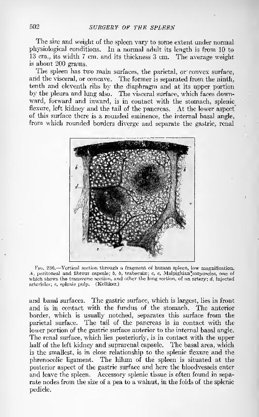
502 SURGERY OF THE SPLEEN
The size and weight of the spleen vary to some extent under normal
physiological conditions. In a normal adult its length is from 10 to
13 cm., its width 7 cm. and its thickness 3 cm. The average weightis about 200 grams.The spleen has two main surfaces, the parietal, or convex surface,
arid the visceral, or concave. The former is separated from the ninth,tenth and eleventh ribs by the diaphragm and at its upper portion
by the pleura and lung also. The visceral surface, which faces down-
ward, forward and inward, is in contact with the stomach, splenic
flexure, left kidney and the tail of the pancreas. At the lower aspectof this surface there is a rounded eminence, the internal basal angle,from which rounded borders diverge and separate the gastric, renal
FIG. 236. Vertical section through a fragment of human spleen, low magnification.A, peritoneal and fibrous capsule; b, b, trabeculse; c, c, Malpighian'corpuscles, one of
which shows the transverse section, and other the long section, of an artery; d, injected
arterioles; e, splenic pulp. (Kolliker.)
and basal surfaces. The gastric surface, which is largest, lies in front
and is in contact with the fundus of the stomach. The anterior
border, which is usually notched, separates this surface from the
parietal surface. The tail of the pancreas is in contact with the
lower portion of the gastric surface anterior to the internal basal angle.
The renal surface, which lies posteriorly, is in contact with the upperhalf of the left kidney and suprarenal capsule. The basal area, which
is the smallest, is in close relationship to the splenic flexure and the
phrenocolic ligament. The hilum of the spleen is situated at the
posterior aspect of the gastric surface and here the bloodvessels enter
and leave the spleen. Accessory splenic tissue is often found in sepa-rate nodes from the size of a pea to a walnut, in the folds of the splenic
pedicle.
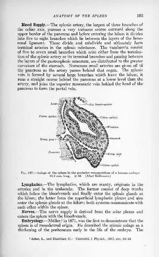
ANATOMY OF THE .SPLEEN 503
Blood Supply. The splenic artery, the largest of three branches of
the celiac axis, pursues a very tortuous course outward along the
upper border of the pancreas and before entering the hilum it divides
into five to eight branches which lie between the layers of the lieno-
renal ligament. These divide and subdivide and ultimately form
terminal arteries in the splenic substance. The vasabrevia consist
of five to seven small branches which arise either from the termina-
tion of the splenic artery or its terminal branches and passing between
the layers of the gastrosplenic omentum, are distributed to the greatercurvature of the stomach. Numerous small arteries are given off to
the pancreas as the artery passes behind that organ. The splenic
vein is formed by several large branches which leave the hilum; it
runs a straight course behind the pancreas at a lower level than the
artery, and joins the superior mesenteric vein behind the head of the
pancreas to form the portal vein.
Celom.
JLig. hepato-gastric
Celom. epithel.
Mesog. post.*
Pancreas
-annofax-Celom epithel.
FIG. 237. Anlage of the spleen in the posterior mesogastrium of a human embryo10.5 mm. long. X 30. (After Kollmann.)
Lymphatics. The lymphatics, which are scanty, originate in the
arteries and in the trabeculse. The former consist of deep trunks
which follow the bloodvessels and finally enter the splenic glands at
the hilum; the latter form the superficial lymphatic plexus and also
enter the splenic glands at the hilum; both systems communicate with
each other within the spleen.
Nerves. The nerve supply is derived from the solar plexus andenters the spleen with the bloodvessels.
Embryology. Muller,1 in 1871, was the first to demonstrate that the
spleen is of mesodermal origin. He described the splenic anlage as a
thickening of the peritoneum early in the life of the embryo. The
1 Asher, L., and Ebnother, G.: Centralbl. f. Physiol., 1915, xxx, 61-64.
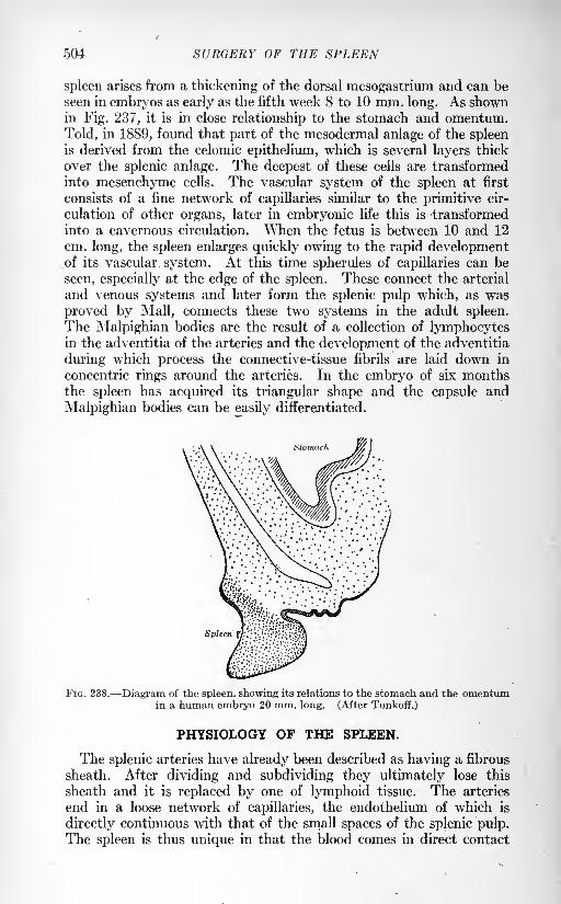
504 SURGERY OF THE SPLEEN
spleen arises from a thickening of the dorsal mesogastrium and can be
seen in embryos as early as the fifth week 8 to 10 mm. long. As shownin Fig. 237, it is in close relationship to the stomach and omentum.
Told, in 1889, found that part of the mesodermal anlage of the spleenis derived from the celomic epithelium, which is several layers thick
over the splenic anlage. The deepest of these cells are transformed
into mesenchyme cells. The vascular system of the spleen at first
consists of a fine network of capillaries similar to the primitive cir-
culation of other organs, later in embryonic life this is transformed
into a cavernous circulation. When the fetus is between 10 and 12
cm. long, the spleen enlarges quickly owing to the rapid developmentof its vascular system. At this time spherules of capillaries can be
seen, especially at the edge of the spleen. These connect the arterial
and venous systems and later form the splenic pulp which, as was
proved by Mall, connects these two systems in the adult spleen.
The Malpighian bodies are the result of a collection of lymphocytesin the adventitia of the arteries and the development of the adventitia
during which process the connective-tissue fibrils are laid down in
concentric rings around the arteries. In the embryo of six monthsthe spleen has acquired its triangular shape and the capsule and
Malpighian bodies can be easily differentiated.
Spl
FIG. 238. Diagram of the spleen, showing its relations to the stomach and the omentumin a human embryo 20 mm. long. (After Tonkoff.)
PHYSIOLOGY OF THE SPLEEN.
The splenic arteries have already been described as having a fibrous
sheath. After dividing and subdividing they ultimately lose this
sheath and it is replaced by one of lymphoid tissue. The arteries
end in a loose network of capillaries, the endothelium of which is
directly continuous with that of the small spaces of the splenic pulp.The spleen is thus unique in that the blood comes in direct contact
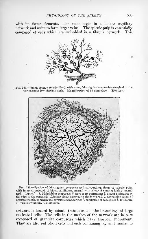
505
with its tissue elements. The veins begin in a similar capillary
network and unite to form larger veins. The splenic pulp is essentially
composed of cells which are embedded in a fibrous network. This
FIG. 239. Small splenic arterly (dog), with many Malpighian corpuscles attached to the
perivascular lymphatic sheath. Magnification of 10 diameters. (Kolliker.)
FIG. 240. Section of Malpighian corpuscle and surrounding- tissue of splenic pulp,with injected network of blood capillaries, treated with silver chromate; highly magni-fied. (Oppel.) 1, Malpighian corpuscle; 2, part of its reticulum; 8, denser reticulum at
the edge of the corpuscle; 4> looser tissue external to the former; 6, 6, connective tissue of
arterial sheath, to which the corpuscle is adhering ; 7, capillaries of corpuscle ; 8, reticulumof pulp surrounding the arteriole.
network is formed by minute trabeculse and the branchings of largenucleated cells. The cells in the meshes of the network are in part
composed of granular corpuscles which have ameboid movement.
They are also red blood cells and cells containing pigment similar to
506 SURGERY OF THE SPLEEN
hematin. The Malpighian bodies are situated on the small arteries
(Figs. 239 and 240). They are outgrowths of the lymphoid sheath
and can be seen by the naked eye as small whitish spots.The spleen is the largest of the ductless glands and, like other
members of this group, very little is definitely known with regard to
its function. On reviewing the literature, which is very abundant,one is impressed with the great confusion that exists. In nearly
every sphere of research concerning the spleen conclusions reached
by one physiologist have been contradicted by the experience of
another. This may be partly accounted for by the fact that theories
are often advanced which lack sufficient experimental evidence.
Pearce and his co-workers have demonstrated that numerous intri-
cate and complex problems must be solved if any advance is to bemade in the knowledge of the functions of the spleen. Very little
has been attained that would aid us in the treatment of diseases
apparently due to some abnormality in the splenic function. We rely
mainly on clinical observations for our methods and procedures.Whatever the function of the spleen may be, it would seem that its
removal does not materially affect the health and welfare of the
human being. Numerous cases of injury of the spleen are reportedin which splenectomy was necessary and those patients who recovered
from the accident and the operation apparently suffered no ill effects
and were able to resume their usual duties. As will be mentioned
later, it is possible that the loss of the spleen is compensated for byother organs, especially the lymphatic and hemolymphatic glands.
Clinical experience teaches us that changes in the blood are moremarked after removal of a recently injured spleen than after removal
of the diseased spleen. This can be explained by the fact that in
diseased conditions compensatory changes have been in progressover a more or less prolonged period previous to splenectomy. It
has been stated that splenectomized persons are more liable to con-
tract current infectious diseases. The truth of this would seemdoubtful. The resistance against infectious diseases once they are
contracted is evidently not affected by splenectomy. Postoperativeinfections are no more serious following splenectomy than other
operations. A few cases have been reported in which the spleenwas congenitally absent.
The most important- changes observed after splenectomy for injuryto normal spleens are those which occur in the blood. There is an
increase in the number of leukocytes characterized by a relative
increase of mononuclears. The maximum number is usually reached
in about twelve days. At first the erythrocytes decrease in number,but in a few days they increase with the simultaneous rise in per-
centage of hemoglobin. It has been observed that a moderate eosino-
philia usually occurs some months after the operation and may per-
sist several years.
Functions. Most observers agree that the spleen has the power to
destroy red blood cells which have completed their life cycle. This
PHYSIOLOGY OF THE SPLEEN 507
view has a more uniform support than any other function with which
the spleen is credited and has for its basis the following points of
evidence: (1) The presence in the spleen of red blood cells in various
stages of disintegration; (2) the presence in the spleen of large ameboidcells which contain broken down red blood cells; (3) the abundanceor organic compounds rich in iron, probably derived from the red
blood cells which have been digested; (4) the fact that after splenec-
tomy the lymphatic and hemolymphatic glands become hyperplastic,;
that they even increase in number and possess an augmented ability
to devour erythrocytes. As a result of experimental work, Asher1
concludes that the spleen contains a substance which is but slightly
hemolytic in itself, yet when acting in combination with liver extract
forms a powerful hemolytic agent.It has been asserted that the spleen takes part in the production of
red blood cells in adult life. This is probably one of the most doubt-
ful functions assigned to the spleen and has little to substantiate it.
Such a function may exist in fetal life and perhaps in early childhood.
As shown by Bizzozero,2 the red blood cells in fish and lower reptiles
are formed in the spleen exclusively. In birds the spleen and bonemarrow both take part in this function; but as we ascend the scale
of vertebrates the bone marrow becomes more important and in manit probably is the only source of red blood cells. It is possible, how-
ever, that the spleen may exert some controlling influence over the
production of erythrocytes; if such a role is carried out by the spleen,
we are still ignorant as to the manner in which it is accomplished.That the spleen aids in the formation of leukocytes is advanced by
Halliburton3chiefly because a greater number of white corpuscles is
found in the splenic vein than in the artery and because of the fact
that the spleen is hypertrophied, and especially the Malpighian
corpuscles in the leukemias in which white blood cells exist in such
abnormally large numbers. In splenic anemia Banti believes that
the leukopenia is due to a decreased output of leukocytes, a result of
the diseased condition of the spleen.
Because of the presence of such nitrogenous compounds as xanthin,
adenin, uric acid, etc., it is thought that the spleen plays an important
part in metabolism. It was shown by Horbaczewsky that the spleencontains substances from which uric acid can be formed by the action
of the spleen substance itself. More recently it has been shown that
the spleen contains enzymes oxidase, adenase and guanase^-whichcan produce uric acid from the split products of the nucleins. Another
theory is supported by Schiff who asserts that the spleen contains an
enzyme which, when carried to the pancreas, converts trypsinogeninto trypsin.
During digestion the spleen is supposed to act as a kind of reservoir
to the portal system and helps to regulate the blood supply. Soon
1 Asher, L., and Ebnother, G.: Centralbl. f. Physiol., 1915, xxx, 61-64.2 Virchows Arch., xcv, 1-25. Quoted in Keen's Surgery, Philadelphia, 1908, ii, 1070.3 Handbook of Physiology, Philadelphia, 1914.
508 SURGERY OF THE SPLEEN
after the meal the spleen begins to enlarge and reaches its maximumin five or six hours; it then returns gradually to normal size. With
regard to the changes in the size of the spleen, the demonstrations of
Roy are probably the most important. He has shown that in cats
and dogs a rhythmical contraction occurs about once every minute.
This is brought about by the non-striated muscular fibers which are
present in the capsule and trabeculse. This rhythmical movement
probably helps to promote circulation through the large splenic
spaces; its real significance, however, remains undetermined.
Pearce and his co-workers1 found that on the injection of a power-ful hemolytic serum an extreme laking of the erythrocytes occurred
in both splenectomized and non-splenectomized dogs; on the injectionof a weak serum jaundice was more readily produced in non-splenec-tomized than in splenectomized dogs. Further experiments led
them to the conclusion that this result was due in some way to the
anemia that usually follows splenectomy because anemic animals
do not readily develop jaundice after the injection of hemolytic serumwhether or not the spleen has been removed. If anemia does not
exist, such a serum will produce jaundice shortly after splenectomy.The difficulty of producing jaundice in dogs one month or more after
splenectomy seems to be due to an increased resistance of the red
cells. Pearce and Karsner2 think that the anemia and the increased
resistance of the red cells are not due to any acquired property of the
serum and conclude that the spleen controls and regulates blood
destruction and possibly regeneration. In an attempt to explainthe production of anemia, Pearce and Austin3 studied the lymphnodes and liver after splenectomy. They found a great increase in
the endothelial cells of the lymphatic glands which were phagocyticfor red cells. The stellate cells in the capillaries of the liver also hadthe same property. From this it was suggested that the lymphatic
gland and possibly the liver compensate to some extent for the loss
of the spleen but they are unable to say if the changes in the lymphglands account for anemia.
Warthin, 4working in the sheep, found a marked increase in the
number of lymphatic glands as well as hyperplasia and also found
new formation of hemolymphatic glands following splenectomy.It is a well-known fact that the spleen is probably always enlarged
in infectious diseases and in intoxication. Very often, indeed, it is
so enlarged as to be palpable below the costal margin. Such spleens
are markedly congested and may show areas of inflammation and
sclerosis. We are still ignorant as to the significance of this enlarge-
ment and change in structure but it would seem probable that the
spleen, with the other lymphatic structures of the body, acts as a
barrier and protects the body against microorganisms and their
products.
1 Pearce, R. M., Austin, J. H., and Krumbhaar, E. B.: Jour. Exper. Med., 1912, xvi,
363-394; Pearce, R. M., Austin, J. H., Musser, J. H.: Ibid., pp. 758-768.2Ibid., pp. 769-779. 3 Ibid., pp. 780-788.
4 Contributions to Medical Research dedicated to Victor C. Vaughan, Ann Arbor,
Wahr, 1903, pp. 216-236.
INJURIES OF THE SPLEEN 509
INJURIES OP THE SPLEEN.
Injury to the spleen may be of three kinds: (1) protrusion or
prolapse of the organ through a rent in the abdominal or chest walls;
(2) penetrating wounds; (3) subcutaneous rupture. The spleen maypenetrate the diaphragm in traumatic diaphragmatic hernia.
Prolapse. Prolapse of the spleen is exceedingly rare It mayoccur through a rent in the upper abdominal wall or the lower left
chest. The organ itself may be seriously damaged or it may be
unharmed. If the organ has remained uninjured it should be replacedafter thorough cleansing. If the injuries are extensive, splenectomyshould be performed. Minor injuries may be attended to by suturing,
crushing the edges of the wound or tamponade and, after careful
search for bleeding points, the spleen returned within the abdomen.
Splenectomy, however, would seem to be the rational treatment onaccount of the complications that may result from hemorrhage or
secondary infection after other forms of treatment.
Penetrating Wounds. Penetrating wounds may be due either to
gunshot or stabbing. In the large majority of cases other organs also
are injured. Any of the several organs of the abdomen or chest maybe damaged, depending on the direction of the bullet or instrument
of injury. Death, as a rule, speedily follows on account of the exten-
sive hemorrhage so that immediate operation is indicated. Judgingfrom the literature on the subject, it would seem that suture of the
wounded organ is attended with better results than splenectomy. It is
possible, however, that in these cases in which splenectomy was per-formed the injury was extensive and the patients in a very critical
condition.
Injuries to the spleen during the World War carry apparently the
same indications for treatment as injuries in civil practice. Available
statistics show that when the spleen alone is injured at least 50 percent, of patients will recover by surgical treatment, which may be bysuture, by tamponage or by splenectomy.The experience of those who have seen the largest number of such
cases indicates that splenectomy is the preferable operation if the
spleen is badly lacerated. It would seem reasonable that this is true
because of the low mortality associated with the removal of the spleen.
The fact that the majority of cases, however, are complicated byinjuries to other organs in the abdomen means a higher mortality.
Wallace,1 for example, reported 63.6 per cent, in complicated cases.
Subcutaneous Rupture. The normal spleen is well protected from
external injury by the lower left chest wall. In accidents where the
spleen is ruptured it will very often be found that a pathological con-
dition existed previously and resulted in enlargement and friability of
the .organ. As a rule, the lesions are very extensive and may be pro-duced by slight external violence. Cases have been reported in
1 A study of 1200 cases of gunshot wounds of the abdomen. Brit. Jour. Surg., 1917,
iv, 679-743.
510 SURGERY OF THE SPLEEN
which the organ was torn into several parts, some of which were free
in the abdominal cavity. Severe hemorrhage is apt to occur, pro-
ducing speedy death but at operation cases with marked lesions
accompanied by but slight loss of blood have been encountered.
The hemorrhage in these cases is usually limited by adhesions whichserve to control the flow of blood and favor clotting. Fauntleroy
1
reports a case in which the patient fell forty feet, receiving markedcontusions of the left abdomen and left chest. The patient's generalcondition remained favorable and there were no signs of hemorrhagebut on account of persistent left abdominal rigidity in connection with
pain and a slight tendency to nausea, the abdomen was opened three
hours after the accident. The spleen was found completely torn across
about its middle, but there had been very little hemorrhage. Onlyone large clot was found surrounding the spleen and it was thoughtthat the extensive adhesions which were present helped to control
the hemorrhage. Scheult2reported a case which presented very
slight symptoms at the end of forty-eight hours notwithstanding the
fact that the patient had a long and rough journey before admittance
to the hospital. At operation the spleen was found divided into three
parts.
Spontaneous Rupture. Spontaneous rupture of an acutely swollen
organ may occur as a rare complication in some of the acute specific
fevers. It seems to take place with greatest frequency in malarial
fever but has been encountered in typhoid fever, typhus fever and
relapsing fever. Noland and Watson3 found that in 30,000 malarial
cases admitted to the Colon Hospital in eight years there were 3
cases only of spontaneous rupture. Conner and Downes4reported
spontaneous rupture in a typhoid fever patient and were able to collect
only 12 other cases.
Symptoms. The symptoms may be divided into those caused bythe local condition together with irritation of the peritoneum, and
those due to severe internal hemorrhage. In traumatic rupture, of
course, there is a history of injury. The site of injury is usually
localized in the left hypochondrium or lower left chest. There maybe severe external contusions or there may be no signs whatever of
external violence. Recorded cases show that the death-rate is very
high and generally death occurs within twenty-four hours. Almost
immediately after the injury the patient experiences severe sharp painin the left hypochondriac region. Symptoms and signs of severe
hemorrhage develop very rapidly, e. g., vomiting, rapid weak pulse,
shallow rapid respiration, cold and clammy skin. On the other hand,
urgent symptoms may not be present and the patient feels well exceptfor local pain in the region of the spleen. In these cases hemorrhageis very slight. Fauntleroy's
5patient experienced severe deep-seated
pain in the left shoulder. Fauntleroy attaches a great deal of impor-
1 Ann. Surg., 1913, Ivii, 68-71. 2 Lancet, 1913, ii, 794-795.3 Ann. Surg., 1913, Ivii, 72-80. 4 Am. Jour. Med. Sc., 1914, clxvii, 332-344.
Am. Surg., 1913, Ivii, 6S-71.
WANDERING SPLEEN 511
tance to this symptom, especially as it was relieved after operation.The case reported by Conner and Downes also displayed a similar
symptom so that when present it may aid in making a diagnosis. Onexamination, the entire abdominal wall may be found more or less
rigid, or the rigidity may be localized to the left hypochondrium and
accompanied by tenderness. On percussion, there is dulness in both
flanks usually elicited if the hemorrhage has been extensive. Accord-
ing to Ballance persistent dulness in the left flank even when the
patient is turned over on the right side should at once turn our atten-
tion to the spleen.
Diagnosis. It is very difficult to arrive at a definite diagnosis of
rupture of the spleen, especially of spontaneous rupture. The 3
cases reported by Njland and Watson were all operated on for sup-
posed intestinal perforation. In the case reported by Conner andDownes the diagnosis of ruptured spleen was made previous to
operation. In traumatic rupture the site of injury will attract atten-
tion to the spleen and if the above signs and symptoms are present
diagnosis is possible.
Treatment. The treatment of ruptured spleen is entirely surgical.
Even if a diagnosis has not been arrived at there are other abdominal
signs and symptoms which indicate immediate surgical procedure.Several methods may be adopted: (1) suture of the tear; (2) tampo-nade; (3) crushing the edges of the wound followed by suture; (4)
splenectomy. Where the rent is small, suturing may suffice, but a
careful search must be made for bleeding points before the spleenis returned to the abdomen. Senn,
1
experimenting with dogs, found
that hemorrhage could be arrested by crushing the edges of the woundand stitching the crushed edges together.
WANDERING SPLEEN.
As already stated, the spleen is held in place by the lienorenal
ligament, costocolic ligament and the gastrosplenic omentum. Undernormal conditions its range of movement is very limited. It is
extremely unusual to be able to palpate the edge of the normal spleen,
save in children; where this is possible in the adult an enlargement
probably exists. The pedicle of the spleen, however, may become so
elongated as to allow the organ to occupy any part of the abdominal
cavity, and even the pelvis. The wandering spleen usually possesses
greater freedom than any other abdominal organ which may assumesimilar characteristics. In the majority of cases its abnormal move-ments are limited to the left abdomen, but cases are reported in whichthe spleen has been found in the right iliac fossa and bony pelvis. In
one case it was found to occupy the sac of an inguinal hernia.
Etiology. It is probable that abnormal mobility of the spleen
may be due to congenital elongation of the pedicle as was present in a
'Jour. Am. Med. Assn., 1903, xli, 1241-1245.
512 SURGERY OF THE SPLEEN
boy, aged twelve years, reported by Moynihan.1 An abnormal laxity
of the splenic ligaments must be considered as the chief factor in
many cases because a wandering spleen may be present in the strong,
healthy individual whose abdominal muscles possess proper tonicityand whose organs with the exception of the spleen are in normal posi-tion. In most cases, however, some secondary condition is present,
possibly a flabby abdominal musculature resulting from repeated
pregnancies, ascites, etc., or a ptosis of all the abdominal organs mayexist. Enlargement of the organ would seem to be only an excitingcause. In conditions such as splenic anemia and leukemia in whichthe largest spleens are found, abnormal mobility very seldom exists;
this fact is due partly no doubt, to the perisplenitis and adhesions
which accompany these diseases. In most cases of wandering spleenthere is enlargement of the organ but the increase in size may be the
effect rather than the cause of the abnormal mobility.
Pathology. The pathological changes are usually due to mechanical
influences on the circulation. As a result of kinking or twisting of the
pedicle the spleen is very often enlarged and congested and an increase
in connective tissue results. Where the pedicle has become acutelytwisted large subcapsular hemorrhages or even rupture of the spleen
may be found. This may be followed by necrosis and the formation
of abscess. Perisplenitis may be present, causing adhesions and fixa-
tion of the organ in some abnormal position.
Symptoms. The spleen may have an extremely wide range of move-ments and produce no symptoms whatever. Such spleens have been
discovered in the course of routine examinations. The most commonsymptom is a dull dragging pain in the left hypochondriac region andin the majority of cases the patient has already discovered a tumorwhich may be pushed about in the abdominal cavity. Occasionallythe organ becomes fixed and may produce a variety of symptoms,depending on its location and the organs with which it interferes.
There may be nausea and vomiting, loss of appetite due to draggingof the stomach. There may be renal, vesical, intestinal or uterine
disturbance. In the case reported by Webster and Tieken2 there
were at times symptoms from the kidney, bladder, intestines and
uterus, during the course of the illness. The renal symptoms were so
marked that a pathological condition of the right kidney was diagnosed.In this case, the spleen was fixed in the right iliac fossa and during a
period of seventeen years the patient experienced severe attacks of
pain with vomiting, fever and shock. At operation, an enlargedadherent spleen was found having three complete twists in the pedicle.
It was adhered to the omentum, intestines and right iliac fossa, the
uterus and right appendages. Under such conditions one can readily
understand the variety of symptoms that can be produced and the
difficulty of arriving at a diagnosis. If the pedicle suddenly becomestwisted the patient presents acute symptoms and very rapidly becomes
1 Loc. cit., p. 1073.* Jour. Am. Med. Assn., 1903, xl, 887-891.
TUMORS OF THE SPLEEN 513
seriously ill. Sudden intense pain is experienced in the region of the
tumor, vomiting, weak, rapid pulse and symptoms of shock. Imme-diate surgical measures are usually indicated but if the torsion becomesundone and the pedicle relaxes the symptoms may all subside.
Repeated attacks, however, are liable to occur as in the case reported.
Diagnosis. The diagnosis is often very difficult especially if the
organ becomes fixed in some remote position such as the right abdo-
men or pelvis. If in such a case an acute torsion of the pedicle takes
place difficulties may arise which render a definite diagnosis impossible.In the uncomplicated cases, diagnosis is usually easy. A tumor havingthe contour edge and notch of the spleen which' can be replaced in
the splenic region is easily identified. The absence of normal splenic
dulness may be suggestive.Treatment. In those cases in which a wandering spleen is dis-
covered accidentally and the patient is free from symptoms, it is
probably advisable to refrain from surgery. Abdominal supports of
various kinds may be tried but their efficacy is questionable. If the
patient is inconvenienced by this condition, splenopexy may be resorted
to; wandering spleens treated in this way seem to have remained in
the normal position in reported cases. As a rule, the spleen is enlargedand so diseased as to indicate splenectomy. In recent years this
operation has been attended with a very low mortality and in the
majority of cases is the one to be selected.
Mayo1 advises splenectomy as splenopexy does not always relieve
symptoms. He has operated on 3 cases, 2 by splenectomy, 1 bysplenopexy. In splenectomy cases the patients obtained completerelief. In the third case the spleen remained in place, but the patientsuffered more or less pain afterward.
SURGICAL DISEASES OF THE SPLEEN.
Tumors. The spleen is very rarely the site of tumor formation either
primarily or secondarily to some other focus. Its mechanical protection
against injury may partially account for its immunity to primarygrowths. No satisfactory explanation, however, has as yet been
advanced as to why such a vascular organ is so rarely involved in
neoplastic processes. Simple tumors such as fibroma and adenomahave been described by pathologists, but apparently have never been
encountered by the surgeon. Malignant tumors may be primary or
secondary. There has never been a convincing case of primary cancer
of the spleen reported (Bush2), so that primary malignant growth is
confined to the consideration of the sarcomas. It is doubtful that all
the reported cases are true sarcomas. Some cases have been reportedas such and later were found to be the result of a marked proliferationof endothelioid or lymphoid elements which may exist in other forms
of splenomegaly. Some of the cases of surgical cure may belong
1Surg., Gynec. and Obst., 1913, xvi, 233-239.
2 Jour. Am. Med. Assn., 1910, liv, 453-456.
VOL. ii 33
514 SURGERY OF THE SPLEEN
to this category. There are three different types of connective tissue
from which sarcoma may originate: (1) capsular and trabecular; (2)
lymphoid; (3) endotheloid, giving rise respectively to fibrosarcomas,
lymphosarcomas and endothelioid sarcomas.
Symptoms and Diagnosis. The patient may seek medical aid onaccount of the abdominal growth. Pain, although not a constant
symptom, is usually present and may be very severe at times. Thechief clinical characteristic of the condition is a rapidly growing tumorin the left side of the abdomen. If the tumor is hard, nodular and
irregular it is almost safe to assume that a malignant condition exists
(Council1
). There is no sign or symptom on which absolute reliance
can be placed, so that in order to arrive at a diagnosis we may be
compelled to exclude other forms of splenomegaly. The first twocases of sarcoma of the spleen were recorded by Weichselbaum,
2 in
1881. Up to 1912 Council collected 37 cases; 17 of these patientswere operated on by enucleation and 16 by splenectomy. Out of
the 17 enucleated, 1 patient died seven days after operation; of the
16 splenectomies, 1 patient was lost track of, 3 died following opera-
tion, 5 died of recurrence at indefinite periods, 7 were well when
reported at times varying from four months to six and one-half yearsafter operation. In view of the great malignancy of the sarcomas
in general there must remain a doubt as to the diagnosis in some of
these cases.
Cysts. Cysts of the spleen may be divided into: (1) dermoid; (2)
echinococcus; (3) non-parasitic.1. Dermoid cysts are exceedingly rare. The only case found in the
literature was reported by Andral3 in 1830.
2. Echinococcus cysts are not uncommon in countries where hydatiddisease prevails. According to Thomas the spleen is affected in 2
per cent, of all cases of hydatid disease. He collected 88 cases of
hydatid cysts of the spleen and in 45 of these the spleen alone wasinvolved. It was thought that the embryos reach the spleen byboring their way directly through the intestines or the gastric wall
where those organs are in contact with the spleen. In hydatid disease
of the liver the embryos have been found in the portal circulation and
doubtless enter the liver by the portal route. It is quite improbablethat the spleen is attacked in this way as it would be necessary for
the embryos to travel against the blood stream. The cysts may be
situated anywhere in the organ; they may be unilocular or multilocular,
single or multiple; they contain a thin, pale fluid with a specific gravityof 1004 to 1010 which abounds in organic salts and usually contains
scolices of the parasite.
3. Fowler 4gives a very careful detailed account of all reported
cases of non-parasitic cysts up to 1912. He is of the opinion that the
1 Ann Surg., 1912, Ivi, 915-924.2 Virchows Arch., 1881, Ixxxv, 562. Quoted in Keen's Surgery, Philadelphia, 1908,
iii, 1093.
'Quoted by Osier and MacCrae, Modern Medicine, 1915, iv, 953.
Ann. Surg., 1913, Ivii, 658-690.
CYSTS OF THE SPLEEN 515
classifications suggested to the present time are unsatisfactory, on
account of our lack of knowledge concerning their origin. Usually
they are subdivided into : (a) hemorrhagic; (6) serous; (c) lymphatic
cysts.
(a) The hemorrhagic cyst is the most common variety and may be
due to one of several causes. From a study of the reported cases it
would appear that trauma is one of the most frequent factors. Irt
some cases there is evidence of the occurrence of spontaneous ruptureof diseased and degenerated bloodvessels while in other cases hemor-
rhage takes place into other varieties of cysts. Congestion of the
spleen as in malaria and typhoid fever may result in spontaneous
rupture of the bloodvessels and lead to cyst formation. These enlarged
spleens are also more liable to injury. The contents depend on the
age and the size of the cyst; at first they consist of fresh blood. At
operation, however, rarely is there evidence of recent or active hemor-
rhage. Depending on the degree of alteration, the contents may be
thin, pale, and liquid or remain thick and black. Old cysts usuallycontain hematoidin and cholesterin crystals.
(6) Serous cysts of the large unilocular type may be said to arise
secondarily from hemorrhagic cysts. The contents, at first bloody,become thin and have a clear, pale straw color. Boettcher claims
that small, deep, multilocular cysts are due to degeneration of small
areas of splenic tissue that result from amyloid changes in the blood-
vessels. Bencke considers small multilocular serous surface cysts
as due to infoliation of peritoneal endothelium resulting from the
traumatic rupture of the splenic capsule. He claims that splenic
tissue protrudes through the small rupture and becomes snared off
between neighboring areas of parenchyma forming cystic cavities.
Renggli believes that certain cysts are formed as the result of fusion of
proliferated areas of endothelium following inflammatory processes.The fused endothelium becomes snared off at different points andlater becomes subject to cyst formation.
(c) Lymphatic cysts probably arise from dilatation of the lymph. spaces. Fink suggests this origin and in some varieties it would seemto be a reasonable explanation, especially where an extensive multi-
locular cyst formation exists. The liquid in lymphatic cysts has a
high specific gravity with a tendency to coagulation.
Symptoms. From the clinical standpoint the most important varie-
ties are of the hemorrhagic and serous type. The other varieties are
usually accidental postmortem findings as they do not produce symp-toms. No specific changes or symptoms take place as the result
of destruction of splenic tissue. The most important and constant
symptom is pain. This is usually located in the left hypochondriumbut it may be referred to the epigastrium. As the result of peritonealirritation the pain may be acute, causing nausea and vomiting; usu-
ally, however, it is of a dragging character. Sometimes the patient
accidentally discovers a tumor which may be symptomless. There
may be digestive disturbance due to dragging or pressure of the
516 SURGERY OF THE SPLEEN
stomach or intestines. There may be dyspnea as the result of upwardpressure. The tumor is usually located in the left hypochondriumand is of slow growth. Cysts of large size may reach below the
umbilicus; they may be smooth or irregular, soft and doughy or tense
and elastic.
Diagnosis. As has been pointed out, the most decisive symptom is
the discovery of a cystic tumor in the left hypochondrium which is
movable and whose dulness is continuous with that of the spleen. Acareful history must be taken as to trauma, the site where the tumorwas first noticed, rapidity of growth, and the site and character of
the pain. In most cases a more refined diagnosis than splenic enlarge-ment or abdominal cyst cannot be made until the abdomen is opened.
Enlargements of the kidney and cysts of the pancreas, the left lobe
of the liver, omentum and mesentery must be differentiated from
cyst of the spleen. Renal conditions are usually excluded by the
use of colloidal silver and the roentgen ray together with ureteral
catheterization. If the cyst of the spleen is displaced, a diagnosis
may be impossible and under such conditions has been mistaken even
for ovarian cyst.
Treatment. Within recent years the majority, if not all, cases
have been treated by splenectomy. Up to 1912 Fowler1 had collected
27 cases treated in this way. In 2 the results are not reported; 1 died
and 24 recovered. Other methods such as puncture, incision and
drainage and excision have been employed. Where extensive adhe-
sions exist and hi some cases of parasitic cyst, incision and drainage
may be the operation of choice. One may safely say that splenectomy
is, as a rule, the operation to be preferred and is probably attended
by the best results.
In case the surgeon desires to treat a cyst of the spleen by punctureand drainage, it is important to guard against the spilling of any fluid
contained in the cyst into the peritoneal cavity for fear of producing
secondary infection in case the cyst should prove to be due to echino-
coccus, a complication which has been reported by several surgeons.
In case of any doubt, the fluid should at once be examined microscopi-
cally to determine the presence of booklets and scolices. In case these
are present, the lining of the cyst should be peeled out in order to
remove all of the scolices attached to the inner surface. The remain-
ing cavity should be tamponed with gauze and permitted to contract
and heal from the bottom.
Angioma. Angioma of the spleen is a rare condition and is often
accompanied by angiomatous involvement of other organs. The liver
is frequently affected, in some instances to a greater extent than the
spleen. Dowd2reports a case of cavernous angioma of the spleen
and has collected 12 other cases which, in his opinion, belong to this
group. He states that certain cases may be recorded under different
headings. The condition is liable to be confused with hemorrhagic
1 Ann. Surg., 1913, Ivii, 658-690. * Ann. Surg., 1915, Ixii, 177-181.
ABSCESS OF THE SPLEEN 517
cyst. Two of the cases are classified as sarcomatous angiomas because
of the presence of areas of cells similar to the spindle cells found in
sarcoma. In Dowd's own case and in 5 of the collected cases, other
organs than the spleen were involved. The liver was affected in 4
patients in 1 of whom there were eighty-five angiomatous spots on theskin. The omentum was affected in one patient, liver, stomach and
lungs in one. Three of the remaining 7 patients recovered from their
splenectomies and whether or not the disease existed in other organsis uncertain. Four cases were reported from autopsies, nothing beingsaid of other organs involved in the process.As will be readily understood, diagnosis of angioma of the spleen
is very difficult and in most cases impossible. Patients usually com-
plain of pain in the splenic region, associated with tumor formation.
Pain may be constant or in acute attacks. In 1 case pulsation could
be felt on the tumor and on auscultation a murmur could be heard.
Exploratory operation will usually be necessary to establish a diag-nosis and to ascertain the extent of the process. If the angiomatouscondition involves other organs than the spleen, nothing can be donefor the patient. If, however, the spleen alone is affected, splenectomyshould be performed.
Abscess. Abscess of the spleen may be primary or secondary.Both conditions are rare; in fact some writers question the occur-
rence of primary abscess. Moynihan1
says the occurrence of primaryabscess is a possibility, but a fanciful one, and further states that hehad not discovered any indisputable evidence of its existence.
In the majority of cases secondary abscesses are the result of septic
infarcts and metastatic infection carried to the spleen in the blood
stream during the course of some pyogenic invasion. Emboli fre-
quently result from septic endocarditis. Metastatic infection maybe secondary to septic foci in any other part of the body but especi-
ally to those foci within the portal area. Fauntleroy2reports a case
which was evidently secondary to acute appendicitis. Other instances
of splenic abscess have been reported as occurring during the acute
specific infections, especially typhoid fever and malaria. Ander-
son3 draws attention to the occurrence of splenic abscess in malarial
fever. He states that in 77,949 cases of malarial fever only 2 cases
of abscess of the spleen were diagnosed during life. In available
postmortem records he found 5 cases only of splenic abscess. In
3 of these 5 cases, there was evidence that the abscess was dueto infarction of the organ. Secondary infection of a hydatid cyst or a
hematoma may result in abscess formation. Lastly, infection of the
spleen resulting in abscess may be due to the direct extension of
inflammatory processes from adjacent organs; for example, as a
sequence of perforating gastric ulcer or thoracic empyema.Pathology. The spleen is usually greatly enlarged and easily
palpable; in some instances, however, it is only slightly increased in
1 Keen's Surgery, Philadelphia, 1908, iii, 1076.8 Jour. Am. Med. Assn., 1911, Ivi, 260-261. 3 Lancet', '1906, ii, 1159-1160.
518 SURGERY OF THE SPLEEN
size and not palpable. Abscess formation may be single or multiple.In the non-embolic form it is usually single and, as a rule, not largerthan a goose egg. In rare cases the abscess may attain large dimen-sions and the entire spleen may be replaced by an accumulation of pus.Embolic abscesses are usually small and multiple; sometimes theycoalesce to form large abscess cavities. In most cases there is marked
perisplenitis with extensive adhesions which tend to limit the growthof the abscess.
Symptoms and Diagnosis. In many instances splenic abscess is a
postmortem finding. There may be absolutely no symptoms indicat-
ing splenic involvement. In Fauntleroy's case splenic infection wasnot considered though all other probable foci were ruled out. In
general, the symptoms are those of suppuration elsewhere, togetherwith pain localized in the splenic area associated with rapid enlarge-ment of the organ. In the embolic form the splenic symptoms may be
entirely masked by the original condition, thus obscuring the diagnosis.
Perisplenitis can sometimes be identified by the recognition of a friction
rub. Splenic puncture as a method of diagnosis is generally regardedas a dangerous procedure, and should not be attempted.
Rupture of an abscess may take place into any of the adjacenthollow organs, which may have become adherent to the spleen or,
again, the abscess may rupture into the abdominal cavity or plural
cavity, causing peritonitis or empyema. On very rare occasions, the
abscess may rupture externally, leading to ultimate recovery.
Prognosis. The prognosis in splenic abscess is very bad. Therecorded death-rate in reported cases varies from 70 to 80 per cent.
However, in many instances the primary sepsis was in reality a suffi-
cient cause of death.
Treatment. If the abscess is localized hi the abdominal wall it
should, of course, be drained; in other cases it may be necessary to
open the abdomen to establish drainage. Splenectomy hi an occa-
sional case may be the best procedure. The mortality is high, the
best results being obtained where direct infection has occurred.
Tuberculosis. Winternitz 1 states that tuberculosis of the spleen
may be primary or secondary. By primary it is not meant that the
spleen is the portal of entry of the organism, or even the first tissue
of the body in which the disease manifests itself, but that the disease
process may localize in the spleen and not only bring about various
lesions but act as a focus for the dissemination of the bacilli while
the original focus may have become completely healed. It is prob-able that in all cases of tuberculosis of the spleen other tuberculous
foci exist which may or may not be active. However, in cases con-
sidered as tuberculosis of the spleen, the spleen shows the most markedand advanced lesions and after splenectomy many patients become
well. Previous to 1846 when Coley reported a case of primary tuber-
culosis in the spleen, it was thought that all cases were secondary to
lArch. Int. Med., 1912, ix, 680-697.
TUBERCULOSIS OF THE SPLEEN 519
more advanced foci and that treatment was of no avail. Since then,
however, numerous cases have been reported, a large percentage of
which have been cured by splenectomy, showing that nature can
successfully cope with minor lesions when the principal site of the
disease is removed. Winternitz collected 51 cases; in those instances
in which the liver was examined 80 per cent, showed involvement;the lymphatics were found diseased in 57 per cent, of the cases; 40
per cent, showed complications of the lungs and lesions were found
elsewhere in 66 per cent. In 1 case only was the spleen alone affected
and in 3 only were the spleen and liver involved. From these data
we may conclude that it is possible for true primary tuberculosis of
the spleen to exist; but as there is only 1 case reported in which, after
a careful search, the disease was found to be confined to the spleen it
must be concluded that in reality tuberculosis of the spleen is always
secondary to some other focus which may or may not be healed.
Pathology. In acute cases, the spleen is greatly enlarged; it maybe firm and resistant or very soft. Numerous tubercles are scattered
throughout the organ and microscopically these show extensive necrosis
and are surrounded by epithelioid cells or merely by splenic pulp. In
the more chronic type the spleen is firm and indurated and usuallybound down by many adhesions due to perisplenitis. Tubercles
varying in size are present and consist of firm yellow caseous material.
Others are soft and of a grayish color. The tubercles are surrounded
by epithelioid cells and giant cells. There may be marked necrosis
in the center of the tubercles with absorption, resulting in cyst forma-
tion. In the chronic form there is always a marked increase of fibrous
tissue, giving the organ its firm and resistant consistence.
Symptoms. The disease is most common between the ages of twentyand forty years but may occur at any age. The onset of the disease
is usually insidious and pursues a chronic course. The symptomsmay be divided into three groups: acute, chronic and subacute. Asmall number of acute cases have been reported in which the symp-toms were extremely severe and suggestive of an acute infectious
process or the spleen was enlarged and the clinical course simulated
that of typhoid fever. In the chronic form of the disease the patient
usually consults the physician on account of tumor and pain in the
splenic region. Gastric and respiratory disturbances may also be
present. Other symptoms which, however, are common to manydiseases are progressive weakness, loss of weight, lassitude, etc. The
finding of tuberculous foci in other organs together with the above
symptoms aids in the diagnosis. The roentgenologic examination of
the chest may prove valuable. Giffin1
reports a case in which splenec-
tomy was done. At operation the spleen was found to be studded
by numerous abscesses and tuberculous deposits. The history
extended over a period of sixteen months and no definite evidence of
tuberculosis was found elsewhere in the body before operation. The
i Am. Jour. Med. Sc., 1913, cxlv, 781-795.
520 SURGERY OF THE SPLEEN
patient died four months later, however, with evidence of disseminated
tuberculosis. A postmortem was not obtained. In the cases analyzedby Winternitz no detailed account is given as to the occurrence of fever.
In some cases fever is mentioned as being present while in other
cases it was intermittent or of the septic type with evening exacerba-
tion. It is probable that fever exists in every case at some time duringthe course of the disease. The blood findings are very inconstant.
Anemia is never marked, the lowest red cell count reported being3,200,000. In Winternitz's 51 cases the blood findings were mentionedin 26; in 6 of these the count was characteristic of polycythemia.Some observers attach great importance to the relationship betweentuberculosis of the spleen and polycythemia; the relationship, however,would seem to be very indefinite. Numerous cases of polycythemiahave been reported in which no tuberculous process was found in the
spleen, and vice versa. The third group of cases is intermediate
between the acute and chronic forms and the symptoms are not as
intense and overwhelming as in the acute cases and not as insidious
as in the chronic form.
Treatment. Winternitz holds that tuberculosis of the spleen,whether acute or chronic, is an absolute indication for splenectomy;that by removal of the spleen 59 per cent, of patients recover; other-
wise death invariably takes place. Splenectomy would seem to be
indicated, however, only when the extent and activity of tuberculous
lesions elsewhere are carefully considered with reference to the prob-able operative and postoperative mortality in the individual case.
As in all other tuberculous conditions, the patient should be placedin the best hygienic surroundings.
Splenic Anemia and Banti's Disease. The literature of surgeryabounds with discussions on the spleen. Surgeons may be temptedto become overenthusiastic and to remove the spleen without dueconsideration. Every case* with splenomegaly demands thorough
study and an effort should be made to establish an exact diagnosisbefore surgical treatment is considered. The diseases associated with
splenomegaly are apt to be confused and it is likely that many recorded
cases have been placed in certain defined groups without sufficient
clinical evidence. Only in recent years are we beginning to dis-
tinguish certain syndromes as clinical entities. Cases of syphilitic
splenomegaly, chronic septic splenomegaly and Gaucher's disease,
which were formerly classified as splenic anemia, are gradually being
separted from the splenic anemia group. Cases of an indefinite
nature should remain unclassified until experience has given us further
knowledge of the spleen and the diseases associated with it. Splenec-
tomy as a curative agent for splenic anemia and hemolytic jaundicehas been definitely established and experienced surgeons are unanimousin their plea for the removal of the spleen as soon as a diagnosis has
been made. Medical treatment has not been of permanent avail
and the literature demonstrates that a majority of patients have been
cured by splenectomy, while those not cured have obtained marked
SPLENIC ANEMIA AND BANTI'S DISEASE 521
benefit even in an advanced stage of the disease. Splenectomy for
Gaucher's disease, chronic septic splenomegaly and certain hepaticcirrhoses has not been followed by results that are clearly satisfactory;
however, they are encouraging and, as medical treatment has failed
to produce satisfactory and lasting benefit, splenectomy in the latter
conditions would seem to be justifiable in certain cases. In primarypernicious anemia, splenectomy has been followed by variable results.
At present we can at least say that splenectomy has produced in acertain number of cases marked temporary benefit when medical
treatment has failed, fn recent years the mortality of splenectomyhas been steadily decreasing. If surgical treatment be resorted to
early, before the patient has become markedly debilitated, the opera-tion can be performed with little risk to the patient. Even patientsthat are anemic and weak withstand the operation remarkably well.
When profound anemia exists, resistance may be materially improvedand the risks of operation lessened by blood transfusion.
The relationship between splenomegaly and the secondary typeof anemia with leukopenia was first demonstrated by Gretzel1 in
1866. Since then several types have been described and reasonablywell defined from a clinical standpoint.
Banti,2 in 1894, described a disease, the salient characteristics of
which were splenomegaly, secondary anemia and cirrhotic liver with
ascites. This condition we now know to be the final stage of true
splenic anemia; in other words, splenic anemia and Banti's disease
are one and the same clinical entity. Splenic anemia may be defined
as a chronic affection characterized by a progressive or remittent
enlargement of the spleen for which as yet no cause has been assigned.An anemia of the secondary type is present, usually accompanied
by leukopenia, a tendency to hemorrhage especially from the
stomach and in many cases a terminal stage with cirrhosis of the
liver and often ascites. No case should be classified as splenic anemiain which, after careful clinical study, the disease does not seem to be
primary in the spleen, or in which the spleen, on pathological exami-
nation, does not show chronic, hypertrophic fibrosis. Chronic septic
splenomegaly, syphilitic splenomegaly, cirrhosis of the liver, and splenicanemia of infancy must be differentiated clinically, and Gaucher's dis-
ease, lymphoma, lymphosarcoma and the various types of tumor mustbe differentiated pathologically.
Etiology. The primary factor in the production of splenic anemiais unknown. Many different theories have been advanced, the
majority of which seem to hold that the spleen is the instrument
through which some unknown agent produces the disease.
Cholelithiasis has occasionally occurred with splenic anemia andGiffin states that the etiological relationship of gall-bladder disease
may be important both by reason of the possibility of infection andalso through some influence of circulation in the closely related organsof the upper abdomen.
1 Berl. klin. Wchnschr., 1866. .2 La Sperimentale, 1894, Sez. Biol., 48.
522 SURGERY OF THE SPLEEN
Yates, Bunting and Kristjanson1 obtained pure cultures of a diph-
theroid organism from the spleens of 2 cases of splenic anemia. Theyare of the opinion that certain cases of splenic anemia may be due to
these organisms. After citing some clinical points which indicate a
parallelism between Hodgkin's disease and splenic anemia they
suggest that splenic anemia might be a Hodgkin's disease of the
spleen. (They believe their findings are significant, especially in
view of Gibbon's2 work in which he found a Gram-staining strepto-trichal organism in 6 cases of splenomegaly.)
Syphilis, malaria, passive congestion of the spleen as a result of
obstruction to the portal circulation, and thrombosis of the splenicor portal veins have all been considered and no doubt are responsiblefor certain cases of splenomegaly. These should, however, be placedin a group separate from the splenic anemia described by Banti,Osier3 and others.
Various writers have reported two or more cases of splenic anemia
occurring in one family. However, that heredity should be considered
a causative factor seems questionable. Osier and Giffin, who haveboth reported large series of cases, did not find any evidence of heredi-
tary influence.
Pathological findings have not yet aided us in our search for an
etiological factor in splenic anemia. The picture most commonlypresented would seem to indicate that the changes in the spleen are
due to some form of chronic poisoning.That the spleen is either the primary cause of splenic anemia or the
chief agent through which some unknown factor exerts its influence
seems evident in view of the fact that splenomegaly always precedesanemia and cirrhosis of the liver and that splenectomy is followed by cure.
Pathology. Pathologists usually find a diffuse overgrowth of all
the tissue elements, particularly the connective tissue. This is
accompanied by sclerosis and atrophy of the pulp and Malpighianbodies. Wilson4 states that 13 of his 18 cases showed a general hyper-
plasia of all the tissues, sometimes one or more predominating. These
13 correspond to those chosen from GifKn's6 series as representing
typical splenic anemia. In the majority, however, a marked fibrosis
was a constant picture. In the advanced stage of splenic anemia
he found a great tendency to fibrosis together with dilatation of the
capillaries and arteriosclerosis.
Incidence. The disease usually comes under observation duringthe middle period of life. It may, however, occur at both extremes.
In GirBn's series, the average age was forty years, the youngest being
twenty-two and the oldest fifty years. Recently we have performed
splenectomy on a child, aged two and a half years, that clinically
1 Jour. Am. Med. Assn., 1914, Ixiii, 2225. 2 Quart. Jour. Med., 1914, vii, 153.s British Med. Jour., 1908, ii, 1151-1158.
<Surg., Gynec. and Obst., 1913, xvi, 240-251.s Am. jour. Med. Sc., 1913, cxlv, 781-795. Only thirteen of Giffin's cases were later
found to be typical of splenic anemia ag far as could be determined clinically and
pathologically.
SPLENIC ANEMIA AND BANTI'S DISEASE 523
and pathologically conformed to the picture of splenic anemia. Bothsexes are equally affected.
Symptoms. One of the most extraordinary features of this disease
is its remarkable chronicity. Histories have been obtained wherethere was definite and sufficient evidence that the disease had existed
for more than twenty years. An enlarged spleen may be discovered
and yet the patient have enjoyed reasonably good health with the
exception of an occasional hemorrhage from which the patient slowlyrecovers. In other cases evidences of the disease have been presenta comparatively short time. In Giffin's series, the splenic tumor hadbeen noted in 5 patients less than one year previous to operation,
irrespective of the age of the patient. In a majority of instances
splenomegaly had existed previous to the onset of the anemia.
Giffin was satisfied that 9 out of 13 had been aware of the abdominal
tumor previous to the occurrence of symptoms of anemia and it is
quite probable that the spleen had been enlarged but unrecognized for
a longer period. In no cases did anemia definitely occur before
splenomegaly. The enormous dimensions the spleen usually attains
is a striking feature. Of the 13 cases showing chronic splenitis chosen
from Giffin's series, Wilson reports the largest spleen as weighing1670 grams, the smallest, as weighing 425 grams, the average weight
being 1089 grams. Other observers also direct attention to the
marked degree of splenic enlargement. The largest spleens with the
possible exception of leukemia and Gaucher's disease are found in
splenic anemia. The spleen, as a rule, lies within the limits of the left
half of the abdominal cavity. Cases have been reported, however, in
which it extended beyond the midline and even occupied the rightiliac fossa. The abdomen may be enormously distended. Pain in
the region of the tumor is a common complaint, occasionally occurringin acute attacks. Perisplenitis with adhesions is usually found at
operation and no doubt accounts for the occurrence of pain. The
position of the tumor, its contour and edge, and presence of notches
usually determine the nature of the growth. There are, however,instances in which special methods of diagnosis are necessary to a
definite conclusion. These will be discussed later.
Hematemesis. Hematemesis is a frequent occurrence in splenicanemia. Sometimes the bleeding is so excessive as to cause death.
Occasional instances of hemorrhage from the bowel, epistaxis, bleed-
ing from the gums, and retinal hemorrhage have been reported. In
a majority of cases, however, the bleeding is gastric in origin. It
may recur over a period of years, often with long intervals. In
Osier's series of 15 patients, 8 had attacks of hematemesis. The cause
of hemorrhage would seem to be due to mechanical influences related
to the enlarged spleen and not necessarily to cirrhosis of the liver.
At operation some of the patients who had had severe hemorrhageswere found to have normal livers. Congestion and oozing from the
gastric mucosa, gastric erosions, rupture of varicose veins of the esoph-
agus, are all cited as causes.
524 SURGERY OF THE SPLEEN
Anemia. There is no pathognomonic change in the blood picture.An anemia of the secondary type is present. In Giffin's series 11
cases had hemoglobin beloV 75 per cent., 1 of 20 per cent, and anotherof 35 per cent.
;the average was 57 per cent. Osier draws attention to
the low percentage of hemoglobin and states that it is rather more
striking than in other secondary anemias. The average in 13 of his
cases was 47 per cent, with extremes of 25 and 75 per cent. Thereduction in erythrocytes is usually moderate, the average in Osier's
series being 3,425,000. The average in 12 of Giffin's cases was
3,904,166, but in 2 cases the blood count was over 5,000,000 at time
of observation. In 1 case the red blood cells were reduced to 1,200,000.
With marked anemia, an occasional normoblast and megaloblast maybe found. Slight poikilocytosis and polychromophilia may also be
present. In uncomplicated cases the leukocytes are, as a rule, reduced
in number. After a hemorrhage, however, the usual leukocytosis
may be present. The average count is usually between 3000 and5000. A differential count of the white cells shows no characteristic
change. As in other anemias, the patient usually complains of lack
of energy and ambition, weakness and debility.
A brownish yellow pigmentation of the skin is a common occurrence
and has been noted by most observers. It is usually diffuse in
character, rarely intense. Jaundice has not been noted as occurringin any of the cases examined at the Mayo Clinic. In the literature,
however, we find occasional reports of jaundice having been presentwith and without cirrhosis of the liver. It is possible that these cases
were not splenic anemia but rather belong to the hemolytic group.Ascites may occur as a terminal condition and is usually associated
with cirrhosis of the liver. One of Osier's patients showed no cirrhosis
at postmortem although there had been recurring attacks of ascites.
In Giffin's series an operative diagnosis of cirrhosis of the liver wasmade in 5 cases. Three of these patients had reached the ascitic
state of the disease. None of his cases showed abdominal fluid
without evidence of hepatic change at operation. It is possible that
the marked anemia and the effect of the large spleen on the circula-
tion may account in some instances for the ascites.
Digestive Disturbances. During the course of the disease and
especially in the later stages, the patients may suffer from loss of
appetite, nausea and sometimes vomiting and diarrhea.
Cardiac Symptoms. As hi other forms of anemia, theremay be trouble-
some cardiac symptoms such as shortness of breath and palpitation.
Renal Complications. Rarely is there evidence of renal complicationother than the presence of a trace of albumin in the urine.
Fever. It would seem that the majority of cases run a normal or
nearly normal temperature throughout the course of the disease.
Cases with fever have been reported, especially in the terminal stage.
It is usually safe to assume that patients with fever are either suffer-
ing from intercurrent infection or do not belong to the splenic anemia
group.
SPLENIC ANEMIA AND BANTI'S DISEASE 525
Diagnosis. In typical cases presenting long progressive histories of
splenic enlargement and hemorrhages, and showing an anemia of the
secondary type without leukocytosis, we are usually able to arrive
at a definite diagnosis. In many instances, however, there is somedoubt as to the exact condition. Frequently the mistakes of experi-
enced clinicians are revealed at operation or postmortem. The most
important diseases which add to the difficulties of diagnosis are as follows :
Splenomegaly Associated with Syphilis and Accompanied by Second-
ary Anemia. That this condition has been confused with splenic
anemia has been the experience of those interested in diseases of the
spleen. It is, therefore, necessary that all patients with enlarged
spleens should have Wassermann tests in order so far as possible to
exclude syphilis. The history should also be reviewed very carefully
for evidence of infection. Splenectomy has been done at the MayoClinic in 3 cases of this character, in which the symptoms simulated
splenic anemia and Banti's disease. It is, of course, possible for
syphilis and splenic anemia to exist in the same patient. It seems
certain, however, that a condition closely simulating splenic anemia
is produced by syphilis, thus a careful review of the history for specific
infection and Wassermann tests are imperative.A Group of Cases which Simulate Splenic Anemia but Present a
History and Signs of Subacute or Chronic Infection. In this groupa history of chronic sepsis can be obtained. In some instances the
onset of the illness is characterized by symptoms of acute infection
followed by a history of chronic or subacute infection which mayextend over a period of years. There may be evidence of cardiac or
renal disease, streptococcal invasion, or the infection may be of pelvic
origin. Blood cultures and tests for syphilis, malaria and tuberculosis
will be negative. Attacks may last two or three months and mayoccur at intervals of several months. Between attacks the splenic
tumor may be greatly reduced in size. Leukopenia is usually presentsave at the time of exacerbations. Four such patients have been
operated on at the Mayo Clinic. This group should be clearly sepa-rated from splenic anemia. On account of the widespread infection
in some of these cases, splenectomy will not be followed by entirely
satisfactory results and each case should be carefully considered
before surgery is contemplated.
Splenomegaly without Anemia. Osier calls attention to this condi-
tion which may exist for years without apparent injury to health.
The spleen is not as large as that of splenic anemia. During routine
examinations we have frequently found moderately large spleens,
especially in inhabitants of southern Europe. Such conditions are
most likely to be mistaken for splenic anemia in the early stages.
It is likely, indeed, that some of these cases are in reality incipient
splenic anemia. A definite diagnosis, however, cannot be made.Pernicious Anemia. Only occasionally does the spleen in perni-
cious anemia become so large as to suggest splenic anemia. If at the
same time there is an atypical blood count, a diagnosis may not be
526 SURGERY OF THE SPLEEN
possible. Such circumstances, however, are very rare. The blood
picture will usually guide us to a proper conclusion.
Portal Cirrhosis Associated with Splenomegaly. In some instances
it is very difficult to differentiate this condition from splenic anemia.In cirrhosis the change in the liver precedes that in the spleen, while
the reverse holds true in splenic anemia. With cirrhosis we usually find
a much smaller spleen and the anemia is not so marked. Ascites as aresult of portal obstruction is an early feature. Very often there is anevident etiological factor.
Biliary Cirrhosis of the Infectious Type. Biliary cirrhosis is not an
exceedingly rare sequence in chronic infectious processes of the upperabdomen. Chronic jaundice is, however, a most prominent sign in
biliary cirrhosis, while it rarely if ever occurs in splenic anemia. The
signs of portal obstruction appear late.
Biliary Cirrhosis of the Hanoi Type. A primary hematogenousbiliary cirrhosis seems to occur very rarely. The signs of biliary
cirrhosis of infectious origin will be present without, however, a
demonstrable infectious focus.
Gaucher's Disease. Clinically Gaucher's disease can be definitely
differentiated from splenic anemia only by demonstrating the charac-
teristic endothelioid cells on splenic puncture or in an excised lym-
phatic gland. Splenic puncture will not be frequently attempted onaccount of danger from hemorrhage. The recurrence of Gaucher's
disease in two or more members of the same family and its frequent
appearance in childhood are noteworthy. The differentiation from
splenic anemia is, however, essentially a pathological one.
Lymphoma and Lymphosarcoma. Lymphoma and lymphosarcomamay be rarely suspected upon clinical examination. The superficial
gands and the chest are especially apt to be sites of metastasis.
Hemolytic Jaundice. Cases of hemolytic jaundice have frequentlybeen diagnosed splenic anemia. They may be seen when jaundice is
absent and a very careful inquiry is necessary to elicit information
concerning jaundice when its degree has been slight. In congenitalcases the history of recurrent anemia and jaundice can be traced to
infancy. An increased fragility of the erythrocytes and the presenceof urobilin in the urine are most important clinical evidence.
Diseases of the Left Kidney. Tumors of the left kidney have often
been mistaken for large spleens and vice versa. If there is the least
doubt as to the nature of the tumor a cystoscopic examination togetherwith a pyelogram will usually eliminate the difficulty.
Treatment. As in other types of anemia, temporary improvementis usually obtained in splenic anemia by drug therapy. The arsenic
and iron compounds are chiefly employed. Blood transfusion has
proved efficacious. By these means, together with fresh air and a
wholesome and liberal diet, the patients are usually benefited a great
deal. Unfortunately, however, improvement is only temporary.
Symptoms are certain to return and the patients ultimately die of
the disease or some intercurrent affection.
SPLENIC ANEMIA IN CHILDREN 527
From a study of the literature and especially of records embracinga comparatively large group of cases, one is justified in concluding that
splenectomy is the rational method of treatment. In the early stagesof the disease, splenectomy should be attended with the best results;
but even when changes in the liver have occurred, the patient mayreturn to apparently normal health. Several patients have been
closely observed for as long as twelve years after operation without
recurrence of the original symptoms. Patients presenting very marked
changes in the liver and ascites may derive benefit from the Talma
operation in conjunction with splenectomy.Of the 13 definite splenic anemia cases in Giffin's group, 8 are in
excellent health for periods varying from six months to seven years.
In this group 3 cases had reached the Banti stage, 1 with and 2 with-
out ascites. These cases are in excellent health, 1 as long as five yearsafter operation.The mortality due to splenectomy in splenic anemia seems to be
decreasing year by year. Rodman 1 states that up to 1908 the collected
cases showed a death-rate of 17 per cent. From 1908 to 1912 the
percentage fell to 10.125 per cent. These statistics represent for the
most part single cases by many operators. Giffin2reports 27 splenec-
tomies for splenic anemia with 3 operative deaths. Hemorrhagewas the cause of postoperative death- in 2 instances, in 1 case occurringone year after operation, in the other five and a half years after opera-tion. In 3 other instances hemorrhages occurred two or three times
after operation but the patients are at present in good health.
Adult Type of Splenic Anemia in Children. Although much confu-
sion exists in literature regarding the anemias associated with spleno-
megaly in children, it is concluded by observers who have carefully
studied the subject that a condition conforming in every detail to the
splenic anemia of adults, occurs in children. Very often the adult
form of splenic anemia in children is grouped and confused with the
splenomegaly of rickets or inherited syphilis. A study of the litera-
ture indicates, however, that there are occasional instances of anemiaand splenomegaly with apparently no relation to rickets or inherited
syphilis, which from a clinical standpoint are similar to splenic anemiaas it occurs in adults. Indeed, there is evidence in some instances of
the adult type of splenic anemia that the disease dated from child-
hood. Splenic anemia of the adult type not infrequently occurs duringthe first decade. Rarely, however, has splenectomy been performedin patients of this age. Giffin records a case of splenectomy for splenic
anemia of the adult type in a girl, aged two and a half years (oper-ated on by Balfour3
), and states that he is able to find reports of 5
other cases in literature in which splenectomy was done. One patientwas well eighteen years after splenectomy, one in excellent condition
nine months after operation, and the third was greatly improved two
1 Rodman, J., Stewart and Williard, De Forest, P.: Ann. Surg., 1913, Iviii, 601-615.2 Ann. Surg., 1915, Ixii, 166-171.3 Surgery of the Spleen, Internat. Abst. Surg., 1918, xxvi, 1-15.

528 SURGERY OF THE SPLEEN
months after operation. The remaining 2 patients died, one four daysand the other six weeks after the spleen was removed.
It has already been indicated that the adult type of splenic anemiaas it occurs in childhood has been confused with other forms of spleno-
megaly. It is necessary, therefore, that each case be thoroughlystudied and that rickets, inherited syphilis, and gastro-intestinal dis-
'orders be excluded as primary etiological factors. Gaucher's disease,
lymphosarcoma, septic splenomegaly, particularly as associated with
endocarditis, hemolytic jaundice and the splenic anemia of infancy
(von Jaksch's disease) are also to be included in the differential
diagnosis.
Splenic Anemia of Infancy or von Jaksch's Disease. In dealing with
diseases of the blood in the young, it must be remembered that the
normal blood of early childhood and particularly of infants under two
years of age, differs from the normal blood of the adult. The leuko-
cyte count may reach 14,000 under normal conditions. This moder-ate leukocytosis is essentially a lymphocytosis, the normal lymphocytepercentage usually reaching 40 or 50. It is also generally recognizedthat the blood of infants reacts differently than the blood of adults to
various toxic agents. A lower red cell count with a consequent highercolor index, the presence of normoblasts, and occasionally a few
megaloblasts, and a leukocytosis .with the occurrence of eosinophilesand myelocytes are often observed in the anemias of infancy. Shaw1
states that secondary anemias associated with splenomegaly in chil-
dren are mostly the result of inherited syphilis, rickets and digestiveand metabolic disturbances. Discussing these different groups, he
points out that in congenital syphilis the blood picture may resemble
that of pernicious anemia, splenic anemia or even leukemia. In
rickets the blood picture of splenic anemia or leukemia may be present.All the anemias may show an atypical blood picture at times, so that
having to rely mainly on the blood count for a diagnosis in certain
conditions, it is not surprising that diseases associated with spleno-
megaly in children are confused.
Literature concerning the anemias of childhood is very abundantand shows that in many instances, rickets, inherited syphilis, and
gastro-intestinal disorders are probable etiological factors. It is
agreed, however, by many authorities, that there is a certain groupof cases occurring in infancy which on account of the marked spleno-
megaly, severe secondary anemia, evidence of blood destruction andwith a more or less characteristic blood picture, should, for the presentat least, be considered as a separate clinical entity.
Splenic anemia of infancy or von Jaksch's disease was referred to byGretzel in 1846. In 1889 von Jaksch discussed it in greater detail and
separated it from other conditions. It is evident from the literature
that it is a disease of infancy and early childhood. No cases are
reported in which the patient was more than two and a half years of
1 Lancet, 1904, ii, 1560-1562.
GAUCHER'S DISEASE 529
age. Giffin concludes that there is a close relationship between the
splenic anemia of childhood and the splenic anemia of infancy and the
blood picture in the latter indicates a reaction of toxins which is
peculiar to the hemopoietic system of the infant. The disease closely
resembles the adult form of splenic anemia of children in some of its
clinical features as well as possessing certain distinctive differences.
It is a chronic disease with a greatly enlarged spleen, hemoglobin andreds are reduced and evidently good results are procured by splenec-
tomy. In contradistinction to the adult type of splenic anemia, there
is a marked leukocytosis. The increase in lymphocytes is usuallymost prominent. The number of leukocytes varies from 10,000 to
100,000. In the splenic anemia of infancy, blood destruction is more
pronounced than in the splenic anemia of adults. The hemoglobinis greatly reduced, sometimes as low as 25 per cent. The red cells
are always diminished and vary in size and shape. Normoblasts are
usually present and megaloblasts may be numerous. In the majorityof instances, the liver does not seem to be affected; it may, however, be
enlarged. Only 4 cases are reported in which splenectomy has been
performed; 1 respectively by Fowler,1
Graff,2Wolff,
3 and Pool. 4 Threeof these patients were apparently restored to good health by the
operation. Fowler's case showed marked immediate improvementand the other 2 were well, Graff's patient nine months and Wolff's
three years after splenectomy. Pool's case was a boy baby of nine
months, who although very delicate and suffering a great deal fromnutritional disturbance, showed considerable improvement imme-
diately after operation.
Large Cell Splenomegalia or Gaucher's Disease. The pathological
picture of the spleen in this condition was first described by Gaucher,5
in 1882. The disease is not of common occurrence. In an extensive
review of the literature, Brill and Mandelbaum6 collected only 14
cases. These have all been proved to be Gaucher's disease by histo-
logical examination. Of 207 splenectomies performed at the MayoClinic from April 1, 1904, to January 1, 1919, only 3 cases belong to this
group, and 2 of these have since been questioned. The disease was
formerly classified with splenic anemia, and even at the present time
is frequently referred to as Gaucher's type of splenic anemia. The
histological picture of Gaucher's disease is different from that of
splenic anemia. Clinically, however, the two diseases are difficult
to differentiate.
Etiology. As in other diseases attended by splenomegaly, manytheories have been advanced in an effort to explain the nature and
origin of Gaucher's disease. When first described by Gaucher, it was
1 New York State Jour. Med., 1914, xiv, 435-437.2 Verhandl. d. deutsch. Gesellsch. f. Chir., 1908, xxxvii, 248-253.3 Deutsch. med. Wchnschr., 1906, ii, 2051-2053.4 Ann. Surg., 1915, Ixi, 349-350.s De 1'epithelioma primitif de la rate, hypertrophie, idiopathique de la rate sans leu-
cemia, Paris, 1882, p. 31.
Am. Jour. Med. Sc.. 1913, cxlvi, 803-883.
VOL. ii 34
530 SURGERY OF THE SPLEEN
thought to be a malignant disease of the spleen. This opinion may be
discarded, however, in consideration of the clinical course and the
long duration of the disease. Theories analogous to those cited as
the cause of splenic anemia have been advanced. The histological
picture in Gaucher's disease may be the result of a peculiar reaction
of the constituent tissues of the spleen to an unknown toxin. Thefact that the liver, lymphatic glands and bone marrow are also affected
must not be overlooked.
Pathology. The organs affected in Gaucher's disease are the spleen,
liver, lymphatic glands and bone marrow. The histological pictureof these organs is characteristic and distinctive in the majority of
instances so that, pathologically, the disease can usually be separatedfrom other forms of splenomegaly. The substance of the spleen is
usually firm and resistant. On cut section, numerous white areas
are seen which extend deeply into the substance of the organ. Theremainder of the surface has the appearance of normal spleen. The
microscopic picture of the white areas is very different from the partsthat resemble normal splenic structure. The latter consist mainlyof large spaces which vary greatly in shape and are more or less filled
with large, brightly-staining cells. The walls of the spaces are com-
posed of fine connective-tissue fibrils, but where the cells are thickest
there is a network of small capillaries, the endothelium of which con-
sists of cells having small, round deeply-staining nuclei and resembling
lymphoid cells. The characteristic feature is the presence of large
round or oval cells in the spaces. These cells may lie free in the cavi-
ties of the spaces, or may be connected with their walls. When the
cells are present in large numbers, they vary greatly in shape as a
result of compression. The nucleus is small in comparison .with
the size of the cell and varies markedly in form. Many cells contain
from two to four nuclei. The staining capacity of both the cells andthe nuclei varies greatly; some stain deeply, while others take a faint
stain. The spaces may be packed with these characteristic cells 'or
there may be only a few cells present, the remainder of the space beingfilled with blood. Bovaird 1 claims that a study of the condition shows
the spaces to be the pulp spaces of the spleen and that the cells are
derived from their walls. The normal pulp cells have disappeared,but the Malpighian bodies remain unchanged. Sections from the
white areas consist of dense connective tissue. The large cells just
described are absent.
On microscopic examination of the lymph nodes, the normal cells of
the medulla have entirely disappeared in areas and the transformed
structure consists of a fine reticular network. In the irregular spacesof this network lie large round or polygonal cells whose nuclei are of
fair size, round, oval or irregular and faintly staining. Each follicle
is surrounded by a deposit of rather dark pigment. The liver, also,
presents interesting changes. In many of the areas of perilobular
1 Am. Jour. Med. Sc., 1900, cxx, 377-402.
GAV'CHER'S DISEASE 531
connective tissue a picture similar to that of the lymph nodes is
present. The capillaries contain pigment similar to that 'in the
lymphatic glands. The liver cells and ducts are normal.
Characteristic large cells are found also in the bone marrow. Theymay be present singly or in large groups. Bovaird gives a clear
description of the pathology of this disease and definitely proves that
the disease is not malignant in nature.
Incidence. The disease usually occurs in early life. In the majorityof the reported cases, the patients are under the age of twelve; the
youngest was one year and oldest thirty-seven years of age. Thecondition may exist for a long time without the patient's knowledge,so that in many instances no definite information can be obtained as
to the onset. It is probable, therefore, that those cases reported as
being seen in early adult life may have existed since childhood.
Sex. Women are affected more often than men. In the 14 cases
collected from the literature, there were only 2 males.
Familial Occurrence. Frequently more than one member of the
family are affected. In the cases definitely proved to be Gaucher's
disease, there seems to be no hereditary tendency.
Symptoms. Gaucher's disease is insidious in onset and runs a chronic
course. The condition may exist for years, the patient evidently
enjoying normal health and able to perform daily duties. One case
is reported in which the condition had existed thirty-six years after
the detection of the disease. The average duration of the disease
in all reported cases where death was not the result of splenectomywas nineteen and three-tenths years. As a rule, death results from
some intercurrent affection. Cases have been discovered accidentally
during the course of routine examination, the patient having soughtmedical aid on account of some other disease. While there are noindividual symptoms pathognomonic of the disease, there seems to be
a definite association of certain symptoms in some instances sufficient
to enable one, clinically, to diagnose its presence.
Splenic Enlargement. This is one of the most characteristic features
of Gaucher's disease and occurs in every case. The spleen usuallyattains great dimensions, even exceeding that of splenic anemia. In
1 case the weight was 7400 grams, and the average weight in 13
authentic cases was 3600 grams (Mandelbaum). Notwithstandingthe presence of this greatly hypertrophied organ, it is unusual for the
patient to complain of pain or other discomfort. During the late
stages, however, pain may occur at intervals and sometimes in severe
attacks. The size of the spleen depends on the duration of the disease.
In long-standing cases the abdomen protrudes on the left side to a
degree dependent on the size of the spleen.
Liver. Changes in the liver cannot be determined clinically until
the disease has become well developed. The spleen is always enlarged
previous to the onset of hypertrophy of the liver. The liver then
slowly and gradually increases in size and, in the late stage of the
disease, may reach below the umbilicus. The increase in size in
532 SURGERY OF THE SPLEEN
both spleen and liver is, for the most part, due to the presence of
immense numbers of the characteristic large cells to which we have
already referred. Notwithstanding this marked enlargement of the
liver, ascites very rarely occurs and in this respect Gaucher's disease
differs from splenic anemia. Jaundice probably is never present, butthere is a peculiar discoloration of the skin (to be mentioned later)
which may be mistaken for jaundice.
Lymphatic Glands. Lymphatic glands, as a rule, are not palpable.A few cases have been reported in which small, shotty glands were
found in the axillary regions.
Blood. Severe secondary anemia very rarely occurs in Gaucher's
disease. The disease may exist many years and very little changetake place either in the hemoglobin or in the erythrocytes. In the
late stages a severe secondary anemia may develop. The size, shapeand contour of the red cells are retained. Occasionally a normo-blast is present; megaloblasts have never been found. In mostinstances there is marked leukopenia. As few as 500 leukocytes in
one instance have been reported. This decrease in leukocytes takes
place early and exists throughout the disease. There is nothingcharacteristic in the differential white count.
Hemorrhages. As has been noted in splenic anemia, hemorrhagesare usually gastric in origin, whereas in Gaucher's disease hemorrhageis most liable to take place from the mucous surface. Epistaxis,
together with oozing from the gums, is of frequent occurrence. Bleed-
ing from the intestines has also been observed.
Skin. Early in the disease the areas of the skin exposed to the
light become discolored and, as the disease progresses, discoloration
becomes more intense and is of a brownish-yellow tinge. Ecchymosisin the skin due to a slight trauma is often present and the occurrence
of hemorrhagic furunculosis has been recorded.
Eyes. In a great number of the reported cases, a peculiar changehas been noted in the conjunctiva. Brill and Mandelbaum emphasizethis clinical finding and consider it of great importance as an aid in
diagnosis and state that it was present in their 2 cases. The lesion
consists of a brownish-yellow wedge-shaped thickening of the con-
junctiva, the base of the wedge being limited by the cornea in con-
tradistinction to pterygium. The process occurs early in the disease
and affects the nasal side first. Later in the disease the temporal
aspects shows a similar change.
Diagnosis. We have seen that the pathology of Gaucher's disease
is characteristic in the majority of cases. A few instances in which the
spleen was carefully examined histologically and reported as Gaucher's
disease have since been questioned. It is evident, therefore, that
even a pathological diagnosis in an occasional case may be doubtful.
Owing to the lack of clinical manifestations in the early stages of
certain diseases associated with splenomegaly, a definite diagnosis is
very often impossible. This is true of Gaucher's disease. It is a
chronic disease which at the onset is symptomless and clinically no
HEMOLYTIC JAUNDICE 533
lesions may be present save enlargement of the spleen. During the
whole course of the disease, but especially in the early stages, it is
most likely to be confused with splenic anemia. A diagnosis has
been made from tissue obtained by puncture of the spleen. This
procedure, however, is not without danger and should not be generally
employed. The most satisfactory tissue can be obtained by excision
of an enlarged lymphatic gland. When Gaucher's disease has existed
a sufficient length of time to produce a distinctly enlarged spleen,
other signs have usually developed which make a diagnosis possible.
These are': Beginning enlargement of the liver, lesions of the con-
junctivse and absence of anemia and ascites. If more than one
member of a family be affected, the diagnosis will be strengthened.Treatment. Up to the present time medical treatment has been
unsatisfactory. Drug therapy consists chiefly of the iron and arsenic
compounds. Roentgen-ray exposures may produce a decrease in the
size of the spleen, but the improvement is only temporary. In recent
years splenectomy has been practised in an effort to check the disease,
but too much must not be expected because of the wide affection
of the hemopoietic system. The pathological characteristics are as
evident in the liver, bone marrow and lymphatic glands as in the
spleen, so that the benefit following the removal of only one of the
affected organs is questionable. In those instances in which splenec-
tomy was performed there seemed to be at least temporary improve-ment. On account of the marked temporary improvement followingremoval of the spleen, the failure of drug therapy, and because of the
fact that a positive clinical diagnosis usually cannot be attained, one
is justified in advocating splenectomy.Hemolytic Jaundice Congenital and Acquired. The relationship
between congenital or familial and acquired hemolytic jaundice is an
open question. Clinically, they resemble each other so closely that
they may be satisfactorily studied together. As suggested by the
nomenclature, the disease in the one instance dates from birth and maypossess familial characteristics; in the other it is not manifest until
later in life, usually during the third decennium. When the acquiredform has developed, the clinical manifestations are very similar to
those of the congenital form and the treatment of both is identical.
Etiology. In recent years much experimental work has been doneon the spleen and, although great confusion yet exists, the results of
this work have opened up new fields of research which no doubt will
ultimately lead to more definite knowledge of the etiology of the
different diseases of the spleen. With regard to the cause of hemolysis,which is the principal pathological process taking place in hemolytic
jaundice, the following are among the suggestions offered:
1. Widal1 believes that the destruction of the erythrocytes takes
place in the blood and that the decreased resistance of the red cells
is the essential factor, splenomegaly being of secondary importance.
1 Widal, F., Abrami, P., and Brute, M.: Presse m6d., 1907, xv, 641-644.
534 SURGERY OF THE SPLEEN
2. King1 found (circulating in the blood in diseases associated with
hemolysis and especially in hemolytic jaundice and pernicious anemia)an increased amount of the unsaturated fatty acids as expressed bytheir iodin-combining value. He is of the opinion that the presenceof unsaturated fatty acids in the blood is a probable cause of hemolysisand that the spleen has some important influence in regulating and
controlling these acids. This hypothesis is supported by the fact that
after splenectomy the iodin-combining value of the unsaturated fattyacids becomes very low and thus accounts for the increased resistance
of the red cells and the cessation of hemolysis. He also draws atten-
tion to the fact that cholesterin and the total fats are decreased in
amount in hemolytic jaundice and pernicious anemia, and that after
splenectomy there is marked increase in cholesterin. He states that
as cholesterin possesses antihemolytic powers the resistance of the red
cells after splenectomy may in part be due to the increased cholesterin
value.
3. Many observers believe the spleen alone is responsible for hemol-
ysis. If the spleen is not the sole cause of the disease, it must be
granted that its presence is essential when the results of splenectomyare considered. Up to the present time the curative effect of splenec-
tomy is one of the most important clues in our search for the cause of
the disease, and it would seem fair to assume that if the pathological
process is not active in the spleen itself, the presence of the spleenin some way controls the etiological factor. In view of Asher's workwhich demonstrated that splenic extract, although only slightly hemo-
lytic in itself, when acting in conjunction with liver extract forms
a powerful hemolytic agent, it is probable that the red blood cells
are influenced in some way by the liver and spleen, thereby increasing
hemolysis.
Pathology. Unlike the spleen in splenic anemia, there is very
little, if any, proliferation of connective tissue. There is a markedloss of follicles and no germinal centers. The splenic sinuses are
dilated and, as Eppinger2reports, show a marked hyperemia. Usu-
ally there is infiltration of the splenic pulp; the endothelium shows
proliferation and contains hemosiderin. The changes in the liver
are very slight. There is a deposit of pigment which contains iron.
This pigment is also seen in the kidneys and bone marrow. In the
last mentioned organ a picture of active blood formation is present.
Symptoms. As previously stated, the symptoms of the congenital
form of the disease are similar to those of the acquired form. In
the acquired form the progress of the disease is more acute, and
severe crises more common. In the familial type, the affected mem-bers of the family usually suffer from a more severe type of the disease
than the parent and are more likely to have acholuric crises. It
has been noted, however, that the disease may exist without any
1 Arch. Int. Med., 1914, xiv, 145-167.2 Berl. klin. Wchnschr., 1913, ii, 1572-1576.
HEMOLYTIC JAUNDICE 535
inconvenience to the patient. In such instances there may be onlya very slight icteric tinge of the skin and eyes and the patient maylive to old age. In those cases, congenital or acquired, severe enoughto require treatment, the clinical manifestations may be classified as
follows: (1) Jaundice; (2) splenomegaly, usually accompanied byenlarged liver; (3) anemia; (4) decreased resistance of the red blood
cells; (5) urobilinuria; (6) an increased urobilin content of the stool.
A synopsis of the history and clinical findings in a boy, aged nine
years, recently examined and operated on1 at the Mayo Clinic, will
serve to illustrate the typical features occurring in hemolytic jaundice :
(1) Jaundice since birth with acholuric crises. (2) Large spleen (reach-
ing below the umbilicus). (3) Large liver (4.5 inches below the costal
margin) . (4) Increased fragility of the erythrocytes, complete hemolysis
taking place at 0.4 per cent. (5) Complete blood count: hemoglobin24 per cent., erythrocytes 1,340,000, color index, 0.8 per cent., leuko-
cytes 15,200, number of cells counted 300, with a differential count of:
polynuclear neutrophils 59 per cent., normoblasts 57, small lympho-cytes 29 per cent., large lymphocytes 40 per cent., eosinophils, 2.3
per cent., basophils 2.7 per cent., neutrophil myelocytes 3 per cent.
There were marked anisocytosis, slight poikilocytosis, marked degen-eration of the red cells, marked polychromatophilia; coagulation time
ten minutes. (6) Recent nosebleed. (7) No urobilin in urine, probablyon account of the fact that the patient had had no recent acholuric
crises. (8) Wassermann test negative.Jaundice. The jaundice is acholuric in character. In the familial
type, it exists from birth; in the acquired type, the history is of shorter
duration. The jaundice is essentially chronic in character, markedat intervals by a definite deepening of the jaundice, the so-called
acholuric crises, accompanied by headache, fever, malaise, weakness
and sometimes severe abdominal pain simulating gall-stone colic.
It is indeed probable that the attacks of pain are in some instances
due to gall-stones or infection in the gall-bladder and ducts. Duringthese crises the spleen becomes enlarged and tender. There is evi-
dence of marked hemolysis as expressed by the presence of urobilin in
the urine, bile pigment in the blood in some cases and deepening of
the jaundice. The jaundice is pigmentary in character and does not
produce the toxic symptoms usually connected with obstructive jaun-dice. There is no itching, no bradycardia, no petechia; the stool
is of normal color and there is no bile in the urine. If the commonduct is obstructed during this crisis by the passage of gall-stones,
the clinical picture will be complicated by evidence of obstructive
jaundice. Crises may occur frequently or be absent for several
years. In the intervals, urobilinuria may be absent and no evidence
of hemolysis exist except as shown by the blood picture. To illus-
trate the irregularity of the occurrence of attacks we may say that the
1 Balfour, D. C.: Surgery of the Spleen, Internat. Abstr. Surg., 1918, xxvi, 1-15.
536 SURGERY OF THE SPLEEN
above-mentioned case in the boy, aged nine, gave a history of slight
jaundice since birth. Up to three years of age he had spells of fever
accompanied by weakness, lassitude and loss of appetite, but was then
free from the spells until one year ago, since which time the attacks
have occurred every two or three months, accompanied by deepeningof the jaundice.
Splenomegaly. During the so-called acholuric crises the spleen
usually becomes markedly enlarged and somewhat tender. This
temporary splenomegaly may be evident to the patient as indicated
by a sense of fulness or a definite protrusion of the abdominal wall.
Following the crises the splenic tumor subsides, but probably fails to
reach its former size. In 47 cases of splenomegaly, congenital and
acquired, collected by Elliott and Kanavel,1 the weight of the spleen
was noted in 28. The average weight was over 1000 grams; 1 spleenwas reported as weighing 4500 grams; in only 7 was the weight under
950 grams. It is very difficult to determine the time of the onset of
splenomegaly; it is probable, however, that it occurs early in the
disease.
Blood. The changes in the blood will, of course, depend on the
degree of activity of the disease at the time of the examination and
and the ability of the hematopoietic system to produce new blood.
Regeneration of the blood is indicated in a majority of cases by the
presence of nucleated red cells. There is no blood picture which is
characteristic of the disease. The number of red cells is found to
vary widely in a given group of cases. In some instances they maynumber as low as 1,000,000; or, on the other hand, may be present in
nearly normal numbers. Usually, however, there is a decrease, the
average being 3,000,000 to 4,000,000 with the color index of 1 or in
the neighborhood of 1. In severe cases a higher color index may be
present, suggesting pernicious anemia. It has been noted that the
erythrocytes are usually decreased in size but show very little, if any,
change in shape. The presence of normoblasts has been reportedon several occasions. Richards and Johnson2
reported the presenceof Jolly bodies in some instances and also large, coarse, irregular-
shaped basophile granules in the red cells. If the blood be treated
by one of the methods of postvital staining, a peculiar network of
granules or threads, termed "substantia granuloreticulo-filamentosa"
may be observed. These structures are thought by some observers
to indicate active regeneration; their exact significance is yet undeter-
mined. The leukocytes usually exist in normal numbers, and the
differential count does not show any characteristic change. Duringthe crisis, a slight temporary leukocytosis is usually present.
Increased Fragility of the Erythrocytes. It has been demonstrated
that red blood cells in obstructive jaundice possess an increased resist-
ance to hemolysis by hypotonic salt solution. Applying this test
1Surg., Gynec. and Obst., 1915, xxi, 21-37.
* Jour. Am. Med. Assn., 1913, Ixi, 158&-1590.
HEMOLYTIC JAUNDICE 537
(P. Ribierre's method 1) in hemolytic jaundice, we find almost con-
stantly a decreased resistance to hemolysis and increased fragility
of the erythrocytes. In a few reported cases the resistance of the
red cells was normal. Decreased resistance of the red cells to hypo-tonic salt solution has been found to exist in other conditions, but
evidently it is of very rare occurrence. The test has been cited as an
aid in differentiating hemolytic jaundice from pernicious anemia and
splenic anemia.
Urobilinuria. In practically every case, and especially during a
crisis, urobilinogen is present in the urine. In contrast to obstructive
jaundice, bile is not found in the urine except in cases complicated bythe passage of gall-stones. In a few cases bile pigment is reported as
present in the blood. The amount of urobilin in the urine may be
used as an index of the extent of hemolysis. It is generally con-
ceded that the hemoglobin is split up in the liver-spleen circulation
into an iron-containing element (hematin) and pigment-containingelement which is excreted by the liver as bilirubin. In the intestine
bilirubin is converted into urobilin, absorbed and excreted as such
by the kidneys.Stool. Urobilin is always found increased in quantity in the stool.
The output may rise from the daily normal, 0.15 gm. to 4 gm.Diagnosis. A diagnosis can usually be made without great diffi-
culty. In some instances it may be impossible to state whether a
given case is congenital or acquired. This is due to the fact that the
light form of congenital hemolytic jaundice may have existed for years
unrecognized by tha patient, so that we are unable to obtain a definite
history. A long-continued history of remittent or intermittent
jaundice, together with marked splenomegaly, anemia and increased
fragility of the red cells, constitute a definite clinical pictuie. Cho-
langeitis with cirrhosis of the liver, which is discussed under splenic
anemia, will sometimes present difficulty. In this condition the
erythrocytes possess an increased resistance to hemolysis, the spleenis not so large, and bile is present in the urine. Many cases have
1 We have taken from Sahli's Diagnostic Methods (edited by Potter, 1911, Saunders,
Philadelphia, 2d ed.) the following copy of Ribierre's method of testing the fragility of
the red cells: "Ribierre uses a 0.5 per cent, solution, and places in a very small test-
tube 50 drops; in a second, 48; in a third, 46 drops, etc., and then adds distilled watersufficient to make 50 drops in each tube. A definite amount of blood (20 c.mm.) is putinto each tube from the capillary measuring pipette. Normally, the hemolysis beginsin a tube containing 44 drops of salt solution and 6 drops of distilled water, and is com-plete in the fifth or sixth following tube. The results are described as 'slight,' 'distinct,'
'very distinct,' and 'complete,' and one states in which of the dilutions these grades of
hemolysis are found. French writers previously represented the results of these deter-
minations in the form of a curve, the abscissa of which represented the number of dropsof salt solution used, and the ordinates represented the four grades of hemolysis."One can then easily calculate at what percentage concentration of the hemolysis
begins. If, for instance, the tube containing 44 drops express the extreme concentra-
tion, the percentage will be 44/50. 0.5 per cent. = 0.44 per cent.
"With a sensitive blood, Ribierre begins with stronger saline solutions, e. g., 0.7
per cent., and puts in the first tube 70 drops; in the second, 68; then 66, 64, etc., andadds distilled water up to 70 drops in each case. If hemolysis begin in the tube con-
taining 64 drops, the saline concentration is 64/70. 0.7 per cent. = 0.64 per cent."
538 SURGERY OF THE SPLEEN
probably been overlooked in the past and placed in the splenic anemia
group chiefly because the physician failed to obtain a history of
jaundice.
Prognosis. In the mild form of the disease, that is, when symptomsare absent and only a slight icterus exists, the prognosis is very favor-
able even without medical intervention. Such patients suffer noinconvenience and are able to perform their daily duties, living to old
age. In the more severe forms the illness is progressive and gall-
bladder complications are likely to occur. The patient graduallybecomes weak and emaciated and ultimately dies of the disease.
Treatment. In the mild type of the disease, treatment may not be
required. Cases of familial jaundice have been known to exist whenthe patient lived to an old age. In the more severe forms of the familial
type and in the acquired type medication is only of temporary benefit.
On the other hand, splenectomy has been followed by such gratifyingresults that operation should be advised in every case. Even whenthe disease is of the mild type, but progressive, surgery should be
considered. So far as can be learned from statistics, it would seemcertain that splenectomy interrupts the progressive nature of the
disease and is followed by excellent results. Of the 47 cases collected
by Elliott and Kanavel, only 2 patients died;1 shortly after operation
and the other six weeks after the operation from sepsis. Such a small
mortality is very satisfactory, especially when taking into considera-
tion the serious condition which existed in many of these patients at
the time of operation.In regard to the immediate effects of splenectomy, it is striking
how quickly the jaundice begins to subside. In a few days after
operation a distinct clearing of the skin takes place and the jaundice
entirely disappears in two or three weeks. The acholuric crises cease.
Among the 47 cases collected are those of Spencer Wells reported byDawson, the patients operated on twenty-seven years ago, Roth's case
six years ago, Bland-Sutton's case ten years ago, and Banti's case
eleven years ago. Forty-six of the patients are reported as cured.
The effect of splenectomy on the blood picture is very striking.
Within a few weeks the red cells approximately reach normal and the
increase is always accompanied by a rise in the hemoglobin. The
leukocytes also reach normal in a very short time except when com-
plications are present. With regard to the fragility of the erythro-
cytes, apparently there is little or no change in many of the patients.
In all the splenectomized patients the fragility of the red cells wasincreased previous to operation. One patient was reported as return-
ing to normal in five weeks after operation, and several at the end of
three months. In Spencer Wells's patient, however, increased fragility
still existed twenty-seven years after operation and in Roth's three
and one-half years after operation. Sufficient time has not elapsedto show the effect of the gall-bladder complications. It is reasonable
to assume, however, that the tendency to such complications would
at least be decreased.
PERNICIOUS ANEMIA 539
Pernicious Anemia. Pernicious anemia is a recent acquisition to
the field of surgery. It is our purpose, therefore, not to deal in detail
with its clinical manifestations, but rather to limit ourselves chiefly
to treatment with special reference to the influence of splenectomy onthe progress of the disease. In view of the fact that medical treat-
ment has been followed by such unsatisfactory ultimate results, it is
not surprising that splenectomy has been undertaken in a considerable
number of cases.
Pernicious anemia may be defined as a systemic disease of unknown
origin, having for its chief clinical characteristics the following cardinal
manifestations : (1) Anemia of a progressive course usually with recur-
rent exacerbations; (2) a blood picture in which there is revealed a
marked decrease in erythrocytes with a relatively high hemoglobincontent; (3) the presence of anisocytosis, poikilocytosis, polychromato-
philia, megalocytes, normoblasts and megaloblasts. The skin is of a
lemon-yellow tinge and parasthesias are common. The patient is
apt to be very weak and debilitated. Cardiac and gastro-intestinal
symptoms may be prominent. In a majority of cases there is nomarked enlargement of the spleen and liver. Since our attention has
been centered upon the spleen as an etiological factor in various
diseases of the blood, splenic enlargement is being demonstrated in
many cases of pernicious anemia. t In those cases submitted to opera-tion the spleen was usually found to be enlarged, but in many instances
not sufficiently enlarged to allow of its palpation at the time of exami-
nation.
Etiology. In pernicious anemia, the essential pathological processis not in the spleen, but in the bone marrow. Moffit1 believes with
Warthin that the infective agent or toxin of pernicious anemia mayact differently at different tunes, or that there may be several hemo-
lytic poisons and that there may be splenic, lymphatic, hemolymphaticor marrow hemolysis. Moffit continues and says: "That the shifting
clinical picture, the varying reaction at different times to remedial
measures like arsenic, thorium X, salvarsan, splenectomy, the marked
hemolysis in some cases (urobilinuria, icterus) and its absence in
others, the inconstancy of marrow reaction all speak for a wide-
spread influence of the poisons in the economy with maximum effects
apparent now in this organ and now in the other." Since it has been
proved that anemia of the pernicious type is produced by chemical
substances and the poisons elaborated by certain parasites, sugges-tions have been made that certain cases of pernicious anemia may be
due to toxins which are formed in or reach the blood stream by wayof the alimentary tract. The theory of chemical poisoning is sup-
ported by King's work previously mentioned in the chapter on hemo-
lytic jaundice, with reference to the occurrence of unsaturated fattyacids in the blood. The toxins are supposed to be influenced in some
way and possibly intensified by the spleen. The fact that the removal
1 Am. Jour. Med. Sc., 1914, cxlviii, 817-827.
540 SURGERY OF THE SPLEEN
of the spleen in the late stages of the disease has been followed by a
cessation of hyperhemolysis, tends to prove that the spleen is at least
a link in the chain necessary for the production of the disease. Someobservers compare the spleen with other glands of internal secretion
and speak of its activity in the hemolytic anemias as hypersplenism.The exact status of this condition has not as yet been definitely deter-
mined, nor are we certain of the location or manner in which hemol-
ysis takes place. It has been suggested that the corpuscles are sen-
sitized through the influence of the spleen and are later destroyedin the liver, lymph glands and bone marrow. As Roblee1
concludes,
however, it is probable that no one pathological process is responsiblefor all cases.
Pathology. Until recently our knowledge of the pathology of the
spleen has been acquired from a study of the organ at postmortemexamination. Since splenectomy has been practised in pernicious
anemia, it has been found that the spleen is usually enlarged during the
early stage of the disease. In the late stages or at death the spleen is
usually described as being atrophic and showing marked fibrosis and
pigmentation.
Wilson,2 in an analysis of 7 cases of splenectomy for primary per-
nicious anemia, found that in 6 cases the spleen was larger than nor-
mal. In only 1 instance was the spleen smaller than normal, and
weighed 195 grams. The average weight in the 7 cases was 463
grams. The organ is usually firm, but not tough, and cuts freely.
With one exception Wilson found on microscopic examination a
marked increase in lymphoid tissue. There was only slight fibrosis
and arteriosclerosis was absent. Hyperemia of the pulp and thicken-
ing of the follicular arteries have been noted by Eppinger and others.
In Wilson's series, however, these features were absent and there was
slight pigmentation in only 1 case. In the spleen which showed only
slight increase in lymphoid tissue, there was a marked degree of
diffuse hypertrophic fibrosis and slight arteriosclerosis.
Diagnosis. Pernicious anemia may usually be easily recognized.A severe progressive anemia, for which no cause can be assigned,
weakness, languor, debility, a blanched, waxy appearance of the skin,
which has a faint lemon tint, a preservation of the subcutaneous
tissues and the body weight to a remarkable degree, tendency to
gastric intestinal disorders, and the frequent occurrence of signs and
symptoms of involvement of the central nervous system, constitute a
clinical picture which should at least make us suspicious of the pres-
ence of pernicious anemia. The diagnosis is further strengthened
by a history of one or more remissions occurring during the course of
the disease. In order to confirm the diagnosis, however, a microscopicexamination of the blood is essential. In the early stage and duringa remission, the blood may not show a typical picture. Under these
conditions, mistakes in diagnosis are most likely to be made.
1 Jour. Am. Med. Assn., 1915, Ixiv, 796-800.8 Ann. Surg., 1915, Ixii, 158-165.
PERNICIOUS ANEMIA 541
Severe anemia of the secondary type accompanying certain con-
ditions and diseases is liable at first sight to occasionally be confused
with pernicious anemia. If complete clinical and laboratory methodsare employed, mistakes in diagnosis will seldom be made. Defining
pernicious anemia as a disease of unknown origin, it is our duty in all
cases of severe anemia to make an exhaustive search for a possible
etiological factor. Cancer of the alimentary tract, and especially of
the stomach, is usually credited as most commonly confused with per-nicious anemia. If a thorough microscopic examination of the blood
is made and the roentgen rays employed to prove or disprove the
presence of gastro-intestinal lesions, a correct diagnosis will usuallyfollow.
Intestinal parasites, such as dibothriocephalus latus, uncinariasis
and Entamebse histolytica, may be eliminated by careful examination
of the stool. In syphilis we must depend on the Wassermann test,
together with careful inquiries for evidence of the disease.
In the anemias of infancy, with a blood picture resembling that
of pernicious anemia, etiological factors such as rickets, inherited
syphilis, nutritional disturbances, should be sought.Careful search must be made for points of hemorrhage. Occasion-
ally severe grades of secondary anemia may result from repeated
hemorrhages, which are disregarded by the patient, so that careful
inquiry for such causes is necessary.On examining the duodenal contents by special methods, Schneider1
found urobilin and urobilinogen in large quantities in cases of per-nicious anemia, whereas in secondary anemias urobilinogen was
absent, and there were only small amounts of urobilin.
Treatment. Before treatment is begun we should employ everyknown means to discover the possible existence of some factor which
may produce a clinical picture resembling that of pernicious anemia.
In this respect it is most important to have the stools carefully exam-ined for parasites. There are cases of severe anemia difficult to differ-
entiate from pernicious anemia which are associated with Entamebse
histolytica. Entamebae may be present and bear no causal relation-
ship to the anemia. The Wassermann test should be done in everycase. It is well known, especially since the Wassermann test has
been introduced, that occasional cases of anemia indistinguishablefrom pernicious anemia are the result of syphilitic infection.
The complete cure of pernicious anemia by medical means has
never been attained. Our chief aim has been to abort exacerbations
and prolong remissions. Absolute rest in the treatment is essential.
Rest, together with an abundance of fresh air, a liberal and nourishing
diet, will often produce a decided Improvement. With regard to drugs,the majority of physicians rely on the various arsenic compoundsand large doses of iron; Fowler's solution is usually employed andshould be given in large doses. Salvarsan has been given with no
1 Arch. Int. Med., 1916, xvii, 32-41.
542 SURGERY OF THE SPLEEN
very positive results. Idiosyncrasies to the o!rug and poison from its
prolonged use should be recognized. In our own experience the
intramuscular injection of iron arsenite has been followed by very
satisfactory improvement in some of the severest cases. Othermethods of treatment such as oxygen inhalation, ingestion of bone
marrow, thorium X, have no advantage over the arsenic therapy.
Transfusion of Blood. Transfusion of blood has recently received
a great deal of attention and is advocated for anemias of both the
primary and secondary type. Its employment seems to be attended
by satisfactory temporary results. With repeated transfusion the
blood picture will often improve in patients who do not respond to the
arsenic compounds. The variability in results depends for the most
part on the stage of the disease and the tune at which the treatment
is instituted. When the patient is at the height of a recurrence,
efforts to obtain relief may be absolutely fruitless, and on the other
hand, brilliant temporary results are usually obtained by any methodat a time when the patient is entering upon a state of remission.
Ottenberg1 and Libman2 state that of 25 cases of pernicious anemia
treated by transfusion, 14 showed, for a time, progressive improve-ment, but all continued to present evidence of the disease. In 11
cases transfusion was of no avail. They conclude that blood trans-
sion induces a remission in about one-half of the patients and that, if
the first transfusion fails, the blood from another donor should be
used. Many observers hold that transfusion should be allowed a
thorough trial before splenectomy is resorted to and that in splenec-
tomized patients blood should be transfused before and after opera-tion. If untoward symptoms occur after the first transfusion, a second
transfusion from the same donor should not be attempted.The technic of blood transfusion by the citrate method (after
Lewisohn) is as follows:
The arm of the donor is prepared in the usual manner, a tourniquetis lightly applied above the elbow and the vein (medium cephalic or
medium basilic) is either punctured with a large-sized needle or exposed
by a small incision and a cannula introduced. The blood is received
in a sterile graduated glass jar containing 25 cubic centimeters of a
2 per cent, sterile solution of sodium citrate at the bottom. While
the. blood is running it is well mixed with the citrate solution bymeans of a glass rod. After the blood has reached the 250 c.c. markanother 25 c.c. of the citrate solution is added and the blood permittedto flow until there are 500 c.c. of the mixture. If more blood is desired
a sufficient amount of the citrate solution is added to maintain the
ratio at 0.2 per cent.
The blood may be carried to a recipient's room or the recipient
brought into the operating room. The recipient's vein is then exposed
by a small incision; the cannula introduced and attached to a salvar-
san flask or irrigating flask. The rubber tubing and the bottom of
1 Jour. Am. Med. Assn., 1915, Ixiv, 613. *Ibid., 2163-2164.
PERNICIOUS ANEMIA 543
the flask are filled with saline solution to prevent air from getting into
the circulation. The -citrated blood is then transferred into the flask
and permitted to flow into the vein of the recipient. There is nooccasion for hurry for the blood will not clot, but on the other handit is advisable to have the blood run in slowly in order to guard against
suddenly overloading the right heart and in order to watch for anyuntoward effect on the patient. The marked slowing of the pulse,
syncopal attacks, dyspnea, cyanosis, sensation of cardiac oppression,or "peculiar" or "funny" pains throughout the body should be
interpreted as danger signals, and if these persist after temporarily
stopping the flow it is advisable to conclude the operation at once.
Robertson1 studied 4 cases of primary pernicious anemia treated byblood transfusion with a view of determining the effect of the treat-
ment in the excessive output of urobilin. Three of the 4 patientstransfused gave evidence of a resulting bone-marrow stimulation andat the same time showed a temporary increase of urobilin excretions.
In one instance there was no change in the output of urobilin.
Blood. The varying degrees of reaction and the ultimate behavior
of the blood may depend upon the condition of other organs at the
time of operation and especially on the condition of the liver, lymphatic
glands and bone marrow. In no instance has the blood definitely
departed from the pernicious type, but there is usually great improve-ment in the degree of anisocytosis and poikilocytosis. Megaloblastsdecrease and the color index fails. Megalocytes disappear more
slowly and even when the patient feels quite well, the blood will
remain megalocytic in type. The immediate occurrence after opera-tion of immense numbers of normoblasts, reticulated red cells, and
erythrocytes with Howell's Jolly bodies is characteristic of practicallyall cases and indicates a marked marrow reaction. Normoblasts
usually persist three to four weeks and then gradually disappear.Howell's Jolly bodies may be found several months after operation.The leukocytes may or may not increase after operation.
Hemolysis. In the larger number of operations, hyperhemolysiswas present, as indicated by urobilin in the urine, an excess of uro-
bilinogen in the stools and an icteroid tinge of the skin. With few
exceptions, these features were more or less definitely absent very
shortly after splenectomy. In a study of 6 cases of primary per-nicious anemia in which there was an increased output of urobilin
previous to splenectomy, Robertson found that there was a decided
decrease in the output of urobilin in all. In 2 of the cases the urobilin
later returned to a high point and in one the output remainedabout normal. These 3 patients received less benefit from the opera-tion than 2 patients who had a normal output twenty-seven monthsafter splenectomy. In those instances where gastric disturbances
were pronounced, several observers have noticed that after operation,the appetite immediately improves and gastric symptoms subside.
'Arch. Int. Med., 1915, xvi, 429-436.
544 SURGERY OF THE SPLEEN
SPLENECTOMY. Any form of treatment for pernicious anemia mayprove, or at least may appear to be, beneficial. Even without treat-
ment these patients have their ups and downs, and it is not an infre-
quent clinical experience to have a patient present himself with symp-toms which might be construed as being those of an early pernicious
anemia, and then with or without treatment recover and remain
well. In eliciting the history the physician finds that the symptomsare often indefinite in the earlier stages, before the blood changesbecome characteristic.
Splenectomy in pernicious anemia does not appear to be based onsound reasoning, and there seems to be no foundation for the belief
that the procedure will cure pernicious anemia. Eppinger first sug-
gested splenectomy as a cure for pernicious anemia, and the early
reports with the abundant testimony as to temporary relief were quitesufficient to give the operation a fair trial in this hopeless disease.
Considering the confusion which so often attends the early diagnosis,it seems probable that obscure cases of hemolytic icterus and splenic
anemia have been accidentally included in the pernicious anemia group.Removal of the spleen in such cases has given the impression that
splenectomy cures pernicious anemia. At the same time in the investi-
gation of our cases of splenectomy for pernicious anemia we see great,
though temporary, improvement. There is a gain in weight and an
improvement in the blood from an average hemoglobin of 38 to 72
per cent., and the reds from two to four millions. Splenectomy seems
at least to have instituted a marked palliation.
In our experience in the cases in which the results were most favor-
able the symptoms were less characteristic of pernicious anemia. In
young and middle-aged persons in whom the disease is most rapid,
especially if hemolysis is shown to be marked, splenectomy is worthyof trial. Taken as a whole, it may be said that whenever perniciousanemia has developed to the stage in which the blood is characteristic
it is incurable, and splenectomy is a means of palliation and not of
cure. Since there is an operative mortality, good reasons must exist
for substituting operation for repeated blood transfusions. We have
splenectomized 50 patients with pernicious anemia, with three deaths
(6 per cent.).
Although the general condition of the patient as well as the blood
rapidly improves after splenectomy, yet it would seem probable that
the essential hemolytic agent has not been eliminated. As proof of
this, we have the following facts:
1. Persistence of a pernicious type of blood picture, even thoughthe patient attains a prolonged clinical remission.
2. Failure of a few patients to respond to the operation.
3. The occurrence of relapses in a few instances following an initial
improvement after splenectomy.4. Persistence and progressive nature of cord lesions. The patient
or his family should receive a full and frank explanation of the prog-nosis of the disease and the present status of surgical treatment before
splenectomy is decided on.
SPLENOMEDULLARY LEUKEMIA 545
If all patients with primary pernicious anemia were carefully con-
sidered and proper pre-operative treatment instituted, there is no
apparent reason why the mortality should exceed that of splenectomyin other conditions. Of 207 splenectomies at the Mayo Clinic, up to
January 1, 1919, for the various diseases associated with splenomegaly,there was a death-rate of 9.1 per cent. Fifty-one of the 207 splenec-tomies were performed for primary pernicious anemia with three
operative deaths. Robles collected 40 cases and found that 9 patientshad died either at operation or shortly after.
Splenomedullary Leukemia. The results of the surgical treatment
of splenomedullary leukemia in the past have been very discouraging.The earlier operative experience was associated with such a high initial
mortality that the operation was practically abandoned.
Warren, who in 1911 reported one case of splenectomy for this con-
dition, followed by recovery, and referred to the 42 cases collected byHagen, with 4 recoveries, attributed the serious operative risk to
secondary hemorrhage from the wound. It must be remembered that
at the time these unfavorable results were reported splenectomy was
performed during the active period of the disease, and when the numberof white cells was large, showing a high ratio to the red cells. Therehave been since that time isolated reports of splenectomy for spleno-
medullary leukemia, in some of which the diagnosis was rather seriously
doubted (Richardson).The surgical profession has hesitated, therefore, to adopt operative
measures in this disease, but since it has been demonstrated that
radium, ar-rays and benzol exert a specific, although temporary, effect
on the symptoms of myelogenous leukemia (Billings and others),
splenectomy has again come up for consideration. Radium appearsto exert the most powerful influence on the spleen and on the blood
picture, and there have been several cases reported of remissions that
have been produced by radium in conditions entirely resistant to z-raysand benzol (Ordway,
1 Giifin2).
The therapeutic value of these agents being sufficient to bring the
blood picture to normal, greatly to improve the patient's general con-
dition and gradually to reduce the size of the spleen (in some instances
until it is non-palpable), suggested the possibility that removal of the
spleen at this stage might have some influence on the course of the
disease, particularly if these therapeutic agents were used as adjuncts.It has already been demonstrated that under such circumstances
splenectomy may be done with a very low operative mortality. In the
Mayo Clinic between October 2, 1916, and September 1, 1917, 17
patients were operated on who had splenomedullary leukemia in whichthe blood picture had been first brought to normal by the use of radium,
arrays or benzol or a combination of these agents. There have been no
1 Remissions in leukemia produced by radium in cases completely resistant to x-rayand benzol treatment. Tr. Asso. Am. Phys., 1916, xxxi, 177-207.
2 Observations on the treatment of myelocytic leukemia by radium, Boston Med.and Surg. Jour., 1917, clxxvii, 686-691.
VOL. ii 35
546 SURGERY OF THE SPLEEN
operative deaths in this series, but we have as yet no knowledge as to
the ultimate results of the operation.
Splenomegaly and Anemia Associated with Syphilis. Under splenic
anemia, we have directed attention to a group of cases in which there
are marked splenomegaly and anemia of the secondary type asso-
ciated with syphilis. Enlargement of the spleen is not uncommon in
early syphilis, but it tends to disappear. In certain cases, however,the splenomegaly persists and ultimately these patients may presenta clinical picture which is identical with that of splenic anemia. Gum-mata of the liver are often present, or there may be a definite cirrhosis,
with or without ascites, thus simulating the Banti type of splenicanemia. In all patients, therefore, having an enlarged spleen, careful
inquiry for syphilitic infection and repeated Wassermann tests are
imperative before a diagnosis is made. It is probable that marked
splenomegaly may be present in patients with syphilis, in which syphilis
bears no etiological relationship to the splenomegaly. To obtain a
satisfactory classification, however, it would seem quite proper to
separate from other forms of splenomegaly those cases in which there
is evidence of syphilis, or in which repeated positive Wassermanntests are obtained. Three cases of this type have been, carefullyobserved over varying periods of time at the Mayo Clinic and splenec-
tomy was ultimately performed. Giffin 1reports these cases in detail
and collects 5 unoperated and 3 splenectomized cases from the litera-
ture. The unoperated cases simulated the Banti type of splenicanemia. Three of them came to autopsy. Of the 3 patients not
operated on, 2 received antisyphilitic treatment with no benefit. Fol-
lowing operation improvement was prompt. The third died two
years after operation, as a result of severe hematemesis. Of the 5
patients examined and operated on at the Mayo Clinic, only 1 gave a
history of primary infection. The Wassermann test, however, was
strongly positive in the other 4.
In studying these cases, we were impressed with the similarity of
the clinical picture to that of splenic anemia and recognize the fact
that without the aid of the Wassermann test, mistakes in diagnosiswould often be made. The 3 patients received antisyphilitic treat-
ment with only slight benefit. The condition of the blood did not
improve and the splenomegaly persisted. After splenectomy, improve-ment was prompt and progressive in each. Reports were received
from our patients one year, eight months and two months, respectively,after splenectomy. In each instance, the patients were enjoyingexcellent health and the anemia, which previous to operation hadbeen marked, had entirely disappeared.
Cirrhosis of the Liver. While an etiological classification of cir-
rhosis of the liver is highly desirable, our present knowledge does not
lead to a satisfactory grouping. The chief clinical characteristics
are clarified by the pathological conception of biliary and portal
1 The Treatment by Splenectomy of Splenomegaly with Anemia Associated with
Syphilis, Am. Jour. Med. Sc., 1916, clii, 5-16.
MALARIAL SPLEEN 547
types. Biliary cirrhosis resulting from infectious cholangitis of
ascending origin and probably occasionally from a hematogenousinfection causes chronic jaundice which is an early and a most pro-nounced clinical sign. The symptoms and signs of portal obstruction
come late or not at all. On the other hand, portal cirrhosis probablyhas its origin in the portal system as a result of infectious material or
toxic substances obtained from the gastro-intestinal tract. Ascites,
other signs of portal obstruction, come early and jaundice is a late
manifestation. There are many minor distinctions and mixed types
undoubtedly occur. Varying grades of hemolytic activity may be
found in both types but are especially noted in biliary cirrhosis. Theterms "hypertrophic," "atrophic," "alcoholic," etc., often lead to
confusion when applied to cirrhosis of the liver.
Eppinger has reported splenectomy in 4 cases of the splenomegalic
type of hypertrophic cirrhosis of the liver associated with chronic
jaundice and evidences of increased hemolytic activity. Completesubjective recovery resulted in all the cases. The icterus in 3 cases
vanished completely.
Splenectomy in portal cirrhosis was first performed in patients onwhom a diagnosis of the Banti type of splenic anemia had been made.At operation more or less advanced cirrhosis of the liver, out of pro-
portion to the changes in the spleen, was found. The procedure is-
not as yet well founded, as an increased hemolytic activity has not
been demonstrated in these cases. That definite improvement has
followed splenectomy in advanced splenic anemia associated with
cirrhosis of the liver is not an indication that similar results will be
obtained in cases of primary portal cirrhosis. The course of the
disease may, however, be affected by the decreased amount of blood
that is carried through the liver after splenectomy; or some unknownfactor may be active in these cases.
In the Mayo Clinic 14 patients with cirrhosis of the liver have been
splenectomized. While the immediate results have been promisingin some instances, and there are reasons to warrant the hope that in
certain selected cases splenectomy may give a permanently good result,
there is as yet no proof that removal of the spleen gives results whichwill be permanent.
Malarial Spleen. In malarial fever the spleen is often greatly
enlarged. Splenomegaly may persist in spite of medical treatment
and may cause great discomfort and pain. It may possibly be a
source of chronic malarial infection. A severe grade of anemia of
the secondary type may develop in these patients, producing a clinical
picture resembling that of splenic anemia. Enlarged and diseased
spleens are liable to rupture even from slight trauma or they maybecome movable, so that the patient's life is further endangered bycomplications as a result of twisting of the pedicle.
The medical treatment recommended for malaria usually fails to
reduce the size of the spleen. Datt1 has used red iodide of mercury in
1 Indian Med. Gaz., 1914, xlix, 372.
548 SURGERY OF THE SPLEEN
the treatment of enlarged malarial spleens. He claims to havereduced the size of the spleen and to have improved the general con-dition of the patient by this drug.When the splenomegaly persists in spite of medical treatment and
the enlarged organs continues to be a source of pain and danger to
the life of the patient, splenectomy is advisable. Kopylow1
reports the
results of splenectomy in 13 patients who had large malarial spleensand collected 187 cases of splenectomy from the literature. Thedeath-rate for the 200 cases was 25 per cent. Ten of the 13 patientswere in excellent health at periods varying from six to twelve monthsafter operation. Kopylow states that splenectomy is contra-indicated
if the patient is weak, anemic and debilitated, and if there is muchcachexia and hydremia.On opening the abdomen, the enlarged spleen may be found firmly
adhered to the diaphragm and surrounding organs. Under these
conditions splenectomy may not be advisable because of the difficulty
and increased risk of the operation.
Egyptian Splenomegaly. A disease common in Egypt and evidentlystill limited to that country is described by Richards2 as Egyptian
splenomegaly. Data on the subject have also been contributed
by Day and Ferguson3 and Richards and Day.
4 The prominentfeatures of the disease are: (1) Greatly enlarged spleen (2) anemia;
(3) low leukocyte count; (4) attacks of fever; (5) cirrhosis of the liver,
and, later, ascites. The condition is more common in early adult life.
In Richards' series of 22 cases the age period ranged from seven to
fifty years; 1 patient only being over thirty years. The average was
twenty years. All but 3 of the patients were males. There seemsto be a seasonal factor in the occurrence of the disease, 18 of the
patients in this series having sought medical aid from March to
August. Patients complained chiefly of abdominal enlargement andthe pain and gastro-intestinal disturbance produced by the' greatly
enlarged spleen. Almost all experienced attacks of fever. The
splenic enlargement evidently followed the onset of fever and rapidlyincreased in size with or without recurrence of fever. Cirrhosis of
the liver occurs early and with rapid progress, leading to ascites andemaciation. In cases with marked ascites death usually takes placein about six months. Anemia is not extreme but is often aggravated
by bilharzia and ankylostoma infection. Hematemesis occurs but
rarely.
No cause has been assigned for this disease. Richards believes the
splenomegaly is primary and the hepatic cirrhosis secondary. Hedraws attention to the similarity which exists between Egyptian
splenomegaly and splenic anemia; but at the same time he emphasizesthe fact that the clinical course of the two diseases is quite different.
1 Arch. f. klin. Chir., 1901, ci, 708-734. Abstract, Jour. Am. Med. Assn., 1913, Ixi, 312.2 British Jour. Surg., 1914, i, 419-433.3 Ann. Trop. Med. and Parasit., 1909-1910, iii, 379-394.4 Tr, Soc. Trop. Med. and Hyg., 1911-1912, v, 333-363.
KALA-AZAR 549
In splenic anemia hepatic cirrhosis is the terminal state, whereas in
Egyptian splenomegaly hepatic cirrhosis occurs early and is present in
every case. In splenic anemia fever is practically absent; there is noseasonal variation and gastric hemorrhage is a common occurrence.
In fact, the histories of the two diseases, when carefully considered,are quite different. In Egyptian splenomegaly tests for malarial
parasites, Leishman-Donovan bodies and syphilis are negative.
Quinin and salvarsan fail to produce any benefit or change the course
of the disease. In the majority of instances the disease is progressiveand death ultimately results from hepatic cirrhosis. A few patientsrecover spontaneously. Richards advocates splenectomy as the
proper method of treatment. Of 22 patients operated on by Richards,18 survived; 2 of 4 that died had reached the advanced stage of the
disease with ascites. Richards now believes that patients who havereached the ascitic stage should not be submitted to operation. Owingto the great difficulty in tracing his patients after operation andbecause some of the operations had been performed recently, Richards
gives a detailed account of postoperative results in 10 only of his
series. Six of these were clinically cured: Several were doing hard
physical work at periods varying from nine months to two and a half
years after operation. One patient died five months after operation,cause unknown, and three could not be traced. He states that in
properly chosen cases the mortality following splenectomy should be
about 10 per cent. The largest spleen in the series was 2780 grams,the smallest 535 grams. The average weight was 1350 grams. The
changes in the spleen are those of general hyperplasia. The liver
shows a multilobular cirrhosis.
Kala-azar. Splenectomy for kala-azar has been performed in a
few instances. The disease is widespread in India, occurring in
endemic, sporadic and epidemic form. Cases have also been reportedfrom China, Egypt, Arabia and many countries bordering on the
Mediterranean Sea. In the Mediterranean districts infants and
young children are more susceptible to the disease than adults.
The parasite Leishmania donovani now accepted as the causal agentwas first observed by Leishman in 1900 and his observations wereconfirmed by Donovan in 1903. It is found most numerously in the
spleen, liver and bone marrow. It is also found in the blood and maybe present in any organ of the body. Clinical and experimentalevidence seem to indicate that the parasite is transmitted to the humanbody by the bed-bug. The onset of the disease is characterized byhigh fever, usually preceded by a rigor. The fever may be continued,remittent or intermittent in type and lasts several weeks. Duringthis period the spleen rapidly increases in size and the patient becomes
weak, anemic and emaciated. After a period varying from two to six
weeks or longer, the acute symptoms subside, the temperature falls
and the patient's general condition improves. This defervescence is
in turn followed by a second period of fever and these periods of pyrexiaand apyrexia alternate as the disease progresses. The spleen becomes
550 SURGERY OF THE SPLEEN
greatly enlarged and in the majority of instances extends below the
umbilicus. The liver is also enlarged. The blood shows a secondarytype of anemia which is rarely severe. A marked leukopenia is char-
acteristic, the leukocyte count not infrequently dropping to 1000.
Other symptoms are delirium, headache, edema of the extremities,
swelling of the joints, petechia and gastric and intestinal indigestion.The duration of the disease is usually from six to nine months but
may be prolonged from two to three years. Spontaneous cures have
occurred; the mortality, however, is very high, reaching from 96 to
98 per cent.
The diagnosis is made certain only by the demonstration of the
parasite. For this purpose, repeated examinations of the blood maybe necessary. If the blood proves negative, splenic puncture or liver
puncture may be resorted to and in most cases will be positive at all
stages. This procedure, however, is not without danger.
During the early stage, the disease is apt to be confused with typhoidfever, malta fever and even splenic anemia and in some instances it is
impossible to arrive at a diagnosis until the Leishman-Donovan bodies
have been demonstrated.
Medical treatment, as will be seen from the high death-rate, is
unsatisfactory. Various drugs have been used; atoxyl has been
especially recommended. Bassett-Smith reports a case in detail,
treated by intramuscular injection of atoxyl. After four and a half
months of treatment, parasites could not be found in the liver or
blood but were yet present in the spleen. The patient's generalcondition remained fairly satisfactory but anemia persisted and the
spleen and liver remained enlarged. Kokoris states that splenectomyeliminates the chief focus of infection and should be done early.
Kokoris,1 de Souza2 and Cochran3
report a total of 10 cases in which
splenectomy was done with variable but relatively satisfactory results.
The Technic of Splenectomy.4 The more frequent recognition of
certain chronic disorders of the blood, particularly splenic anemia,
hemolytic jaundice and pernicious anemia, and the therapeutic value
of splenectomy in these and other diseases, have recently greatlyextended the indications for splenectomy and suggested some obser-
vations on the technic of the operation. In our experience (especially
in those cases in which technical difficulties are encountered) the
operation has been facilitated by following a routine plan and bythe precision with which the details of such a plan are carried out.
Through a left Bevan incision (Fig. 241), its length depending on
the size of the spleen, the abdomen is explored. The suggestive fr,-
quency with which jaundice, attacks of epigastric pain simulating
biliary colic, cirrhosis, and gall-stones, occur in many of the diseases
for which splenectomy is advocated, necessitates an accurate record
Mtinchen. med. Wchnschr., 1915, Ixii, 1008-1009.2 Arch, de med. des enfants, 1915, vol. xviii.
3 China Med. Jour., 1915, vol. xxix.4 We are indebted to Donald C. Balfour for the description of our technic.
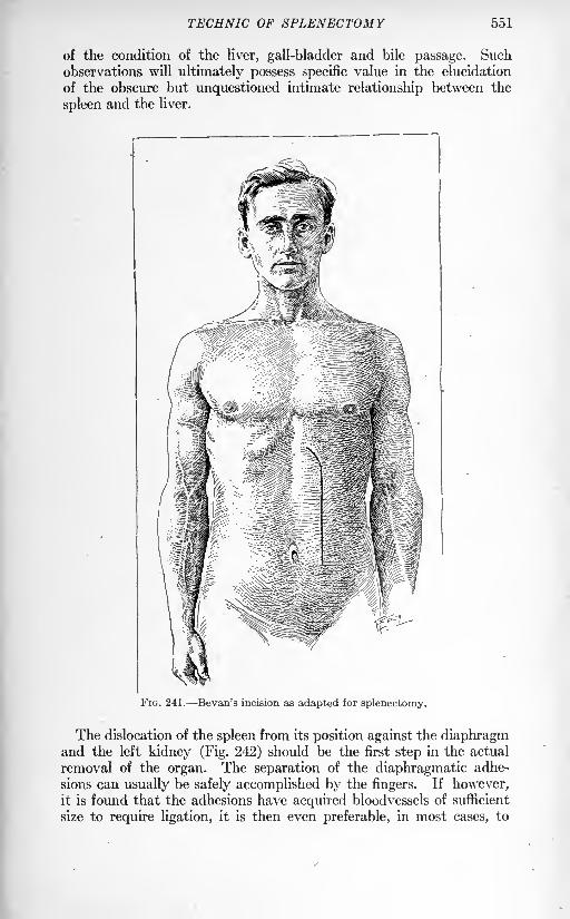
TECHNIC OF SPLENECTOMY 551
of the condition of the liver, gall-bladder and bile passage. Suchobservations will ultimately possess specific value in the elucidation
of the obscure but unquestioned intimate relationship between the
spleen and the liver.
r*-
FIG. 241. Bevan's incision as adapted for splenectomy.
The dislocation of the spleen from its position against the diaphragmand the left kidney (Fig. 242) should be the first step in the actual
removal of the organ. The separation of the diaphragmatic adhe-
sions can usually be safely accomplished by the fingers. If however,it is found that the adhesions have acquired bloodvessels of sufficient
size to require ligation, it is then even preferable, in most cases, to
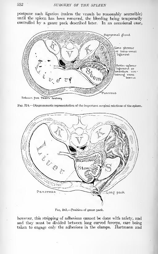
552 SURGERY OF THE SPLEEN
postpone such ligation (unless the vessels be reasonably accessible)until the spleen has been removed, the bleeding being temporarilycontrolled by a gauze pack described later. In an occasional case,
Suprarenal gland
Lieno- phrenicor Ueno-renal,
Gastro -
spT-ervbo
Ugametxt or'-omen-turn, con--
fca.wu.Kgvasa"brevm
Pancreas
Redrawn, from Toldt's Anatomy
FIG. 224. Diagrammatic representation of the important surgical relations of the spleen.
Partcreas pack
FIG. 243. Position of gauze pack.
however, this stripping of adhesions cannot be done with safety, andand they must be divided between long curved forceps, care beingtaken to engage only the adhesions in the clamps. Hartmann and
TECHNIC OF SPLENECTOMY 553
others have advised that the operation be abandoned when these
adhesions appear formidable. However, we have not recently found
such conditions to be prohibitive to splenectomy, although in somecases absolute hemostasis has been secured with considerable effort.
Immediately the spleen has been dislocated, a long hot abdominal
pack is efficiently arranged in the space formerly occupied by the
spleen, until the entire area with which the organ has been in apposi-
tion, is under firm pressure by the gauze (Fig. 243). This accomplishestwo purposes: (1) Most important, the oozing surfaces are com-
pressed and, as the bleeding is usually venous, it is controlled without
subsequent ligation, if the pack (a point emphasized by W. J. Mayo)is left undisturbed until the actual operation is completed. (2) Anexcellent support is provided for the safe manipulation and mobiliza-
tion of the organ, the division of adhesions and ligation of the main
pedicle.
The spleen having been elevated in this manner, its further con-
nections are the main splenic pedicle with its peritoneal investment
(the lineorenal ligament), the gastrosplenic omentum, and in spleno-
megaly of long standing, various adventitious adhesions. The most
satisfactory isolation and treatment of the splenic pedicle is obtained
by the preliminary division of the accessory adhesions, as well as the
peritoneal attachments and reflections. The gastrosplenic omentumshould first be divided, in sections as close as possible to the spleen,
between ligatures. The only named vessels encountered are the
vasa brevia, which arise from the splenic artery at variable points,
pass* to the greater curvature of the stomach, in this peritoneal fold,
and finally anastomose with the left gastro-epiploic. In dealing with
the upper edge of this gastrosplenic omentum, it must be rememberedthat here the fundus of the stomach is normally in very close apposi-tion to the spleen. It is necessary, therefore, always to determine
the exact relationship and protect the stomach from injury in its
separation from the spleen. In an earlier experience I accidentallyincluded in a clamp and excised a small area of the wall of the stomach,the resultant opening, however, being readily closed without post-
operative complication. At the lower pole of the spleen there is an
occasional fibrous attachment, derived from the phrenocolic ligament,which with the other adhesions, should be separately ligated.
The spleen may now be further mobilized by careful dissection of
the peritoneal and fibrous coverings of the splenic pedicle. Thelocalization of the tail of the pancreas is the important feature of this
mobilization, and as the relationship of the organ is not constant, it
is necessary to inspect it in all cases. In some instances the tail is
short, lying against the renal surface of the spleen, on the posterior
aspect of the pedicle, and it may be fitted so closely .into the hilus of
the spleen as to have acquired a concave edge. In other cases the
tail is attenuated, it is in front of the splenic vessels, and in contact
with the gastric surface, while often it does not extend into the operativefield.
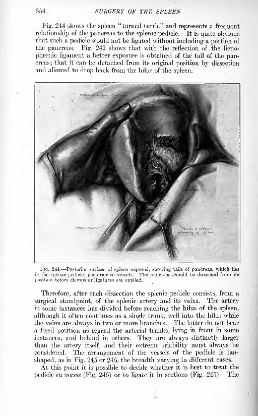
554 SURGERY OF THE SPLEEN
Fig. 244 shows the spleen "turned turtle" and represents a frequent
relationship of the pancreas to the splenic pedicle. It is quite obviousthat such a pedicle would not be ligated without including a portion of
the pancreas. Fig. 242 shows that with the reflection of the lieno-
phrenic ligament a better exposure is obtained of the tail of the pan-creas; that it can be detached from its original position by dissection
and allowed to drop back from the hilus of the spleen.
FIG. 244. Posterior surface of spleen exposed, showing tails of pancreas, which lies
in the splenic pedicle, posterior to vessels. The pancreas should be dissected from its
position before clamps or ligatures are applied.
Therefore, after such dissection the splenic pedicle consists, from a
surgical standpoint, of the splenic artery and its veins. The arteryin some instances has divided before reaching the hilus of the spleen,
although it often continues as a single trunk, well into the him* while
the veins are always in two or more branches. The latter do not bear
a fixed position as regard the arterial trunks, lying in front in some
instances, and behind in others. They are always distinctly larger
than the artery itself, and their extreme friability must always be
considered. The arrangement of the vessels of the pedicle is fan-
shaped, as in Fig. 245 or 246, the breadth varying in different cases.
At this point it is possible to decide whether it is best to treat the
pedicle en masse (Fig. 246) or to ligate it in sections (Fig. 245). The
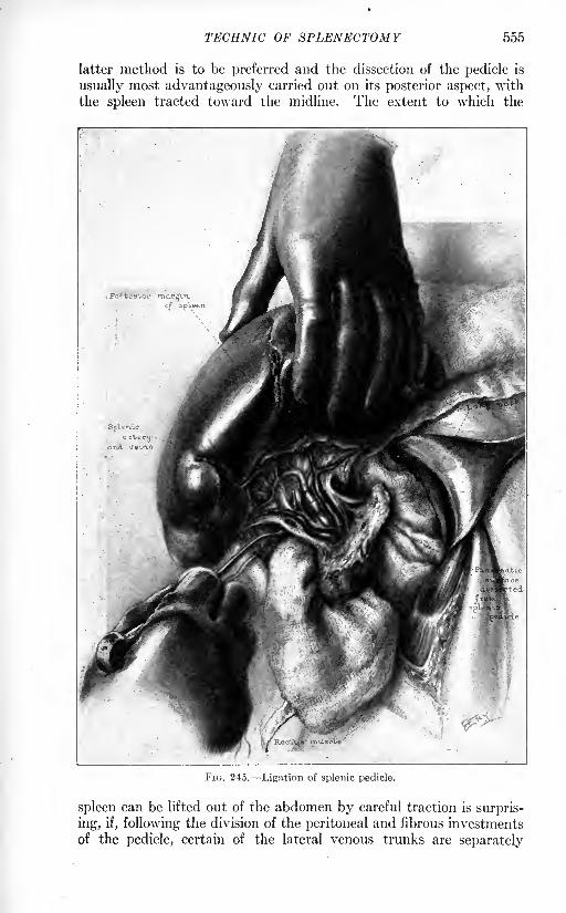
TSCENIC OF SPLENECTOMY 555
latter method is to be preferred and the dissection of the pedicle is
usually most advantageously carried out on its posterior aspect, with
the spleen tracted toward the midline. The extent to which the
FIG. 245. Ligation of splenic pedicle.
spleen can be lifted out of the abdomen by careful traction is surpris-
ing, if, following the division of the peritoneal and fibrous investments
of the pedicle, certain of the lateral venous trunks are separately
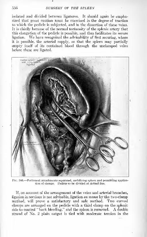
556
isolated and divided between ligatures. It should again be empha-sized that great caution must be exercised in the degree of traction
to which the pedicle is subjected, and in the dissection of these veins.
It is chiefly because of the normal tortuosity of the splenic artery that
this elongation of the pedicle is possible, and thus facilitates its secure
ligation. We have recognized the advisability of first securing, whereit is possible, the arterial supply, so that the spleen may partially
empty itself of its contained blood through the undamped veins
before these are ligated.
FIG. 246. Peritoneal attachments separated, mobilizing spleen and permitting applica-tion of clamps. Pedicle to be divided at dotted line.
If, on account of the arrangement of the veins and arterial branches,
ligation in sections is not advisable, ligation en masse by the two-clampmethod, will prove a satisfactory and safe method. Two curved
clamps are arranged on the pedicle with a third clamp on the splenic
side to control"back bleeding," and the spleen is removed. A double
strand of No. 2 plain catgut is tied with moderate tension in the
TECHNIC OF SPLENECTOMY 557
crushed line of the inner clamp, as a partial control, and a second
strand is transfixed below the distal forcep as the actual control.
Having made certain, by either of these methods, that the pedicleis securely ligated, the large hemostatic pack is removed, the newly
exposed surfaces inspected, any oozing points being separately secured
by fine catgut on a needle, and absolute hemostasis assured.
The disease or condition for which splenectomy is done governs
largely the technical difficulties and risk of the operation. I have
found, for example, that in pernicious anemia, the removal of the
spleen can be accomplished with comparative ease and safety.
Although enlargement of the organ has been present in those cases in
which we have advocated surgical treatment, adhesions are few and
usually insignificant. The pedicle is, as a rule, small, and in a rela-
tively small percentage is its treatment complicated by the relation-
ship of the pancreas. Furthermore, the risk of the operation in this
disease can be minimized by intelligent pre-operative treatment and
observation, as well as by the careful selection of the cases. There
must be a rational basis upon which splenectomy in pernicious anemia
is advocated, as well as strict recognition of the limits of operative
safety. Splenectomy is definitely contra-indicated during an acute
crisis, or in a period when the patient shows a steady decrease in
hemoglobin and red blood cells, or when mental torpor, cord changesand edema mark the terminal stages of the disease. Repeated trans-
fusions from a suitable donor will frequently carry the patient throughmost critical exacerbations of the disease and the proper interpreta-
tion of the reaction following transfusion is essential. Further, it is
important that a satisfactory donor be available after operation.In splenic anemia, splenectomy is associated with a higher operative
risk and greater technical difficulties than it is in any of the morecommon diseases of the spleen. In the cases I have seen, the spleenhas been usually of large size, the vessels very friable and adhesions
occasionally troublesome. Patients in the late stages of the disease
are prone to develop fever, ascites, cirrhotic liver or severe anemia,which conditions contribute to the seriousness of the operation. The
only instance in which I thought it inadvisable to attempt splenectomy,was in splenic anemia with great ascites, atrophic cirrhosis, and
aneurysmal splenic vessels. The friability of the veins, due to a
thrombophlebitis, which is a rather constant factor in such cases,
was especially demonstrated in a baby twenty-two months old, uponwhom I performed splenectomy for typical splenic anemia of the
adult type.The operation in hemolytic jaundice is of relatively little risk and
there has been no particular difficulty in the removal of the spleen,
although the organ is occasionally very large. It is, however, most
important to avoid splenectomy during an exacerbation of the symp-toms. Of the cases I have seen of this disease, the only operativedeath occurred in a patient in whom I failed to realize the import of
a subacute and subsiding acholuric crisis.
558 SURGERY OF THE SPLEEN
In those cases of cirrhosis of the liver, associated with splenomegaly,in which splenectomy is to be considered, the spleen is often firmly
adherent, and this fact, together with the poor general condition of
the patient, makes the operation rather hazardous. Nevertheless,
in the cases we have selected for operation, there has been no operative
mortality; and in 2 cases of my own, most striking benefit followed
the removal of the spleen.
In the rarer conditions, such as syphilitic spleen and idiopathic
splenomegaly, the operation itself, is not of great risk. It has been
interesting to note the result of splenectomy in 2 cases of splenomegalywith specific history and positive Wassermann. In both of these it
had been previously possible to obtain a negative Wassermann bysalvarsan, but it would become positive on discontinuing the treat-
ment. Since splenectomy the Wassermann has remained negative.
From my own experience I have found that the features to be
emphasized in the technic of splenectomy are (1) the abdominal
exploration; (2) the dislocation of the spleen; (3) the use of a hot
gauze pack; (4) the protection of stomach and pancreas from injury;
(5) the preliminary ligation of adhesions; and (6) the treatment of
the splenic pedicle.
DIAGNOSIS OF SURGICAL DISEASES OP THESTOMACH AND THE DUODENUM.
BY FRANK SMITHIES, M.D., F.A.C.P.
General Considerations. Disease of these portions of the alimentarytract may be congenital or acquired. Either type may give rise to acute
or to chronic manifestations.
Acute ailments of the stomach or the duodenum are such as endangerlife from starvation, intoxication, infection, loss of blood or rapidextension of disease to adjacent major viscera. In this group may be
included acute stenoses of the orifices of the stomach, lacerations of the
mucous membrane, wounds, rupture, sudden dilatation, tetany, and
copious hemorrhage.Chronic ailments of the stomach or duodenum, broadly considered,
comprise that class in which patients seek relief from affections whose
progression has resulted in stenoses and malformations of the viscera.
Such affections are commonly associated with cachexia, malnutrition,
anemia, abdominal distress, dyspepsia or imperfect function of related
parts of the alimentary tract. Not rarely an acute manifestation maybe superimposed upon a chronic disease, and may, in fact, whollymask the primary ailment. Chronic surgical affections of the stomachand the duodenum are mainly those associated with the developmentof ulcer and cancer in these sections of the alimentary tract.
PART I.
CLINICAL HISTORY AND ITS INTERPRETATION.
As a guide to the proper management of patients, there is no clinical
procedure which is of such importance as a properly developedanamnesis. While in many acute ailments of the stomach and duo-
denum, the history of the attack for which the patient seeks a physician,suffices for the determination of the immediately indicated surgical
maneuver, yet to secure a comprehensive understanding of the case,
it should be emphasized that careful inquiry should be made into the
digestive upsets which have occurred duripg the months or years
previous to the present complaint. This is to be urged especially in
elucidating the nature of pathologic processes associated with chronic
ulcer and with malignancy. Not infrequently it is quite difficult to have
(559)
560 SURGICAL DISEASES OF STOMACH AND DUODENUM
the patient describe his indigestion during its earlier phases. The
present symptoms may be so severe as entirely to occupy his mind. In
such event, patience and intelligence are required to bring out the nature
of the underlying affection. In all cases, and particularly in individuals
in adult life, a determined effort should be made to detach patientsfrom their immediate upsets and to have them give details respectingailments which have occurred so long ago as almost to be forgotten.
By this mode of inquiry, one is able to observe the nature of initial
attacks of distress, the similarity or dissimilarity of attacks, the inter-
mittence or constancy of the disease, the relative severity of the presentand past exacerbations, the association with the disease of acute infec-
tions, trauma, dietetic errors, mental stress, the swallowing of foreign
bodies, operative procedures and the influence of non-surgical treat-
ment.
If such complaints are systematically tabulated, it is frequently sur-
prising how readily similar chronic disease of the stomach or the duo-
denum classifies itself into definite symptom-groups and how definitely
extragastric disease, associated with dyspepsia, can be segregated. In
obtaining clinical histories, certain data regularly should be recorded.
Sex. Up to within the past decade, groups of statistics were avail-
able which appeared to indicate that affections of the stomach and the
duodenum were more common, or at least quite as common, in females
as in males. The popularity of exploratory laparotomy and the
systematic tabulation of pathologic findings at such operations, have
demonstrated that surgical ailments of the duodenum and the stomachare more than twice as common in males as in females. Many of the
ailments which, previous to the extensive resort to laparotomy, were
considered, primarily, affections of the stomach or duodenum, have
been shown to be gastric upsets, secondary to affections of the gall-
bladder, appendix or both. In females, inquiry should be made with
regard to the influence of pregnancy and parturition either with respectto the initiation of the dyspeptic upset or their association with periodsof exacerbation. In males, note should be made of mental and physical
stress, exposure, the habitual or periodic use of excessive amounts of
tobacco or alcohol, acquirement of venereal disease, chronic over-
eating and injuries received as a consequence of occupation or in
pursuit of pleasure.
Age. While surgical affections of the stomach and duodenum mayarise at any age, it is safe to say that 75 per cent, of this type of patient,
comes under observation after the age of thirty. Malignant disease
occurs most commonly after the fourth decade of life. It may, however,be observed earlier. We have reported elsewhere 19 cases of gastric
carcinoma below the age of thirty-one. Instances have been recorded
as early as the tenth year. It need only be mentioned in passing that
the majority of surgical affections in infants are likely to have their
anlagen before birth.
Infectious Diseases. It is frequently observed that acute infections
influenza, tonsillitis, pneumonia, typhoid fever, parasitic dysentery
CLINICAL HISTORY AND ITS INTERPRETATION 561
and the like either bear close relationship to dyspeptic attacks or
they precipitate such.
The researches of Dieulafoy, Letulle, Chantamesse and Widal,
Rodet, Zaidman and of Rosenow furnish significant facts regarding the
bearing of local or systemic infection in the production or maintenance
of ulceration of the stomach or duodenum. It is more than likely
that the toxins associated with local infection or parasitic ailments
diphtheria, tetanus, anthrax have etiologic significance. The historyof chronic infections of the head sinuses and about the teeth mayestablish primary foci in these localities which may have more than
accidental value. The discovery of a chronic pelvic or renal inflam-
mation sometimes proves in part the origin of irritation or ulceration
of the duodenum or stomach.
Operative Procedures. Several years ago we emphasized the fact
that in a series of 500 operatively demonstrated ulcers of the stomach,coincident disease was demonstrated in the appendix or gall-bladderor both, in rather more than 50 per cent, of the cases. The experi-ments of Rosenow would appear to indicate that associated or similar
organisms are capable of producing lesions in the stomach, duodenum,appendix or gall-bladder. From studies of histories it would seem that
not rarely an infection arising primarily about the mouth, throat or
nose, becomes secondarily established in the walls of the appendix or
the gall-bladder and that after a varying interval of time such lesion
as ulceration follows in the stomach or the duodenum. The signifi-
cance of early removal of a diseased appendix or gall-bladder uponthe subsequent course of an infection of the stomach or duodenumseems worthy of emphasis. Very often the so-called, "hyperacidity
syndrome," commonly and carelessly diagnosed as peptic ulcer, entirely
disappears following operative procedures upon the appendix or the
gall-bladder. Not infrequently, the history of appendix or gall-bladderdisease which has not been alleviated by operation, can be obtained
for years preceding the definite development of ulcer symptoms.In instances of dyspepsia which present themselves following lapa-
rotomy, it is generally quite important to know whether or not surgical
maneuvers have been carried out upon the stomach or duodenum andthe exact nature of such. For example, in an instance of suspectedcarcinoma of the stomach that has seemingly been preceded by a vary-
ing term of"ulcer dyspepsia," it is of vital value to know whether or
not at a previous laparotomy the ulcer has been extirpated, has been
infolded or cauterized, or simple gastro-enterostomy has been per-formed with or without closure of the pylorus.
Gastric Malfunction. The presenting complaints generally requireclose analysis. The acuteness of symptoms associated with dyspepsiaor the evidences of hemorrhage or shock may be so pronounced as to
mask the nature of the underlying pathologic process. While the imme-diate disability may require prompt treatment, unless one has a clear
conception of the possibilities in the clinical variation of lesions of the
stomach or the duodenum, proper measures for permanent relief are
VOL. it 36
562 SURGICAL DISEASES OF STOMACH AND DUODENUM
impossible. Careful inquiry must be made into the events that have
led up to the presenting signs and symptoms. This should be done
in an orderly sequence: the story of the ailment should be developedfrom the time of the first departure from gastric health. Experiencewill demonstrate how frequently patients who claim a dyspepsia exist-
ing for a few months or years, can cite earlier periods of ill-health if
their memories are awakened by intelligent questioning. The history-
taker must particularly scrutinize disabilities styled "cramps," "con-
gestive chills," "bilious attacks,""sour stomach,"
"nervous dyspepsia,"
"bloody flux," "peritonitis," "ptomain poisoning," "rheumatism of
the stomach," etc., etc. Such terms are often fanciful but usually can
be translated readily into terms of clinical pathology, involving the
stomach, duodenum, gall-tract or appendix, if proper effort be
made.Pain. This symptom causes fully 90 per cent, of dyspeptic patients
to seek relief. In all instances its location should be determined. This
can be best discovered by having the subject indicate with the hand the
areas of distress. While positions of the digestive viscera vary, yet, in
general, lesions of the stomach or the duodenum give rise to epigastric
discomfort. Except in acute disturbances (penetrating ulcer or cancer,
obstructions, rupture and the like) surgical ailments of the stomach
or duodenum rarely evidence well defined areas of pain: the zone of
distress is usually moderately widespread without points of pronouncedmaximum severity. When the peritoneum has been attacked, however,well circumscribed spots of pronounced pain can be delimited. Radia-
tion of pain is not without diagnostic significance. Except for
transmission to the back (between the shoulders or the tip of either
scapula or to the lower thoracic or the upper lumbar sections of the
spine) or the rib margins, pain associated with gastroduodenal disease
is usually local. Observation of this fact aids in segregating ailments
of these portions of the alimentary tract from those of a pain-producingnature associated with lesions of the gall-tract, appendix, kidneys,
pancreas, diaphragm, or the nervous or vascular system (central or
spinal cord disease, embolism, thrombosis, aneurysm).Time of Pain. Distress associated with the act of deglutition com-
monly points to affections of the esophagus or the cardiac end of the
stomach. Not rarely, pain immediately after the swallowing act indicates
types of gastric spasm dependent upon hyperesthesia of the mucosa or
pathologic processes at or near the pylorus or located extragastrically, as
ir the duodenum, gall-bladder or appendix. In ulcer of the duodenumor the stomach, pain-time may vary in different attacks. While the
rule does not uniformly hold true, in general, uncomplicated ulcers of
these portions of the viscera give rise to distress from two to six hours
following the ingestion of an ordinary meal, i. e., when the stomach
is emptying or nearly empty. Night pain (from 12 M. to 2 A.M.),
when periodically recurring, is fairly regularly a manifestation of duo-
denal or pyloric ulcer. Complicated lesions (ulcer, cancer) give rise
to more or less constant distress, not uncommonly punctuated with
CLINICAL HISTORY AND ITS INTERPRETATION 563
phases of exacerbation. It wouM seem that such are due to extensions
of the lesion, hemorrhage, sloughing or perforation.
Duration of Pain. A striking feature of distress concomitant
with benign abdominal pathology producing gastric symptoms is what
may be termed its "periodicity." Until complications (stenoses,
malignancy, perforation or peritoneal involvement) have taken place,
pyloric irritability in association with ulcer of the antral end of the
stomach or of the duodenum or with disease of the gall-bladder or
appendix, generally occurs in "attacks" or "spells." There is usually
good gastric health in the interim. These attacks of a periodic nature
not uncommonly have seasonal relation (spring and fall being favor-
ites). They would seem to be associated in some way with extension of
infective processes or to reinfection of tissue whose defensive mechan-ism has been previously damaged. The nature of the underlying
pathology causes these recrudescences or "attacks" to be chiefly
self-limited in their duration, although this factor varies greatly with
the individual. Chronic gastric and duodenal ulcers, of the uncom-
plicated type, manifest "periodicity" so constantly as to warrant this
feature of the symptomatology as being of much diagnostic significance.
It was present in 82 per cent, of 1700 operatively demonstrated gastricand duodenal ulcers which were analyzed by the writer. These recrudes-
cences are generally clean-cut each like its predecessor in its mainfeatures until complications ensue. The duration of the disabilityvaries considerably in the various attacks, though the chief manifes-
tations are similar. The appearance of a dyspeptic storm of a typeunusual to the given individual who previously has had definite andlike attacks, usually means that the pathology of his ailment is altering
and'complications may be expected. In chronic sufferers, complicationssuch as stenosis, peritoneal invasion, hemorrhage or malignancy maybe looked for. In primary malignant disease but rarely is the patient
granted periods of freedom from his dyspepsia : the ailment is generally
continuous, and perniciously and progressively downward in character.
The development of gastric malformation or obstruction due to the
activity of benign lesions is always associated with continuous gastric
malfunction of some form. The dyspepsias associated with chronic
gastroduodenitis, primary achylia gastrica, disease of the liver, gall-
tract, blood-vascular system or kidneys are generally continuous and
only rarely intermittent.
Character of Pain. The distress associated with open ulcer of the
stomach or the duodenum is commonly described as "burning," "sore,"
"raw," "sticking," "gnawing" or "dull, heavy ache." When ulcers
rapidly extend and involve the peritoneal coats, invade adjacent
organs, are in or near the gastric orifices, undergo malignant transition
or are complicated by hemorrhage, the abdominal distress is mentionedas being "boring," "stabbing," "piercing," or "cutting." It is often
sudden and acutely prostrating, causing the patient to double up lie
over a pillow go into shock or seek relief from local heat, lavage or
opiates. Prostrating pain in the course of an ulcer-dyspepsia speaks
564 SURGICAL DISEASES OF STOMACH AND DUODENUM
most commonly for peritoneal invasion, protected perforation or
hemorrhage. Incorrectly, this type of abdominal distress has beentoo frequently described as the characteristic pain of peptic nicer: it
is the pain of a complication in the course of gastric ulcer be such
acute or chronic. The pain associated with well established gastric
malignancy is generally spoken of as "heaviness," "dull ache," "feelingof weight or lump," "bloated sensation." However, acute exacerba-
tions with intense suffering are not rare.
When stenoses have developed, the gastric distress is described as
"fulness," "heaviness," "distention" and "suffocation." If the tone
of the stomach walls remains strong,"grinding,"
"scraping" or
"cramp-
like" pains develop as peristaltic activity becomes abnormal.
Pyloric spasm (often associated with disease of the appendix or gall-
bladder or both) generally produces right epigastric distress varyingfrom vague discomfort to acute, cramp-like pains that are not uncom-
monly difficult to differentiate from those associated with active
pyloric ulcer. They usually come on immediately or soon after food
ingestion and are rarely long-continued. Inasmuch as pyloric spasmmay be present with superficial peptic ulcer in the antrum or pyloric
channel, when pains of the spasm type are evidenced, one must exclude
ulcer before he can ascribe such distress solely to extragastric pathology.
Relief of Pain. The discomfort associated with simple, uncompli-cated peptic ulcer is so uniformly relieved by food ingestion as to render
this phenomenon of diagnostic value. Food relief was present in 87 percent, of the writer's series of peptic ulcer. While patients generallystate that eating causes pain, close inquiry will elicit the information
that food rarely causes immediate discomfort but that the pain pro-duced (?) appears usually from two to five hours post cibo. As a rule
the greater the bulk of the meal the longer delayed is the onset of
pain. When such comes on, then food ingestion commonly bringsabout cessation of distress. Often patients learri this fact and keepthemselves comfortable by frequent munchings of crackers, biscuits,
chocolate, etc. When stenoses, adhesions or malignant transition
ensue, food relief of distress is rarely complete. The discomfort is then
constant and relief is generally secured by partial starvation, (diet),
alkalies, lavage, vomiting, frequent belching, catharsis and, in about
14 per cent., by opiates. In a given case, the disappearance of food
relief, with the necessity for relief of discomfort by some of the agentsabove mentioned, speaks for a change in the type of the previously
existing lesion. Every effort should be made to early determine the
pathologic nature and the extent of this change.
Vomiting and Nausea. In the course of a chronic dyspepsia, nausea
and vomiting are symptoms of prime diagnostic importance. Inter-
mittent vomiting generally indicates pyloric or cardiac spasm. These
spasms are usually associated with ulcer near the orifices or infec-
tive processes in the gall-tract or the appendix. There is a coin-
cident gastritis or duodenitis. The vomiting occurs commonly soon
after food ingestion or several hours later when food is leaving the
CLINICAL HISTORY AND ITS INTERPRETATION 565
stomach, acidity is high or pain is experienced. The vomitus in such
instances is composed of food making up the meal just eaten, sour
gastric juice and frequently partly oxidized bile.
When stenoses in the gastric or duodenal lumen have been produced
by the development of hypertrophic muscle layers, ulcer-scar, or neo-
plasm, then regular vomiting occurs if the patient remain on a bulky diet.
Keeping a dyspeptic on"baby food" may for a long time mask a stenosis
or, where one is known to exist, may lead to a cessation of vomiting and
thus give rise to the opinion that diet or some form of medication is bring-
ing about cure of the ailment. The establishment of a full-meal schedule
promptly precipitates vomiting and proves the true state of affairs.
Where stenoses are pronounced and food intake of even moderate quan-
tity, then gastric dilatation and stagnation lead to the appearance of
"delayed vomiting." In this event, food that has lain in the stomachfor eight or more hours is vomited quite regularly. Morning or late
evening vomiting is quite common. The amount vomited may be at
times strikingly copious, and seemingly far in excess of the quantityof food ingested. The vomitus is generally made up of poorly chymified
food, strings or gobs of mucus, and has a foul, yeasty (benign reten-
tion) or a peculiarly penetrating, rancid or rotten odor (malignant
retention). With the few exceptions of extreme gastroduodenal atony,
persistent "delayed" vomit always signifies some obstruction to the
free onward progression of food : thus proof of the occurrence of retention
vomiting is an important point diagnostically.
Constant nausea is not infrequent in gastric malignancy. It may be
the only symptom. Its persistence should always urge careful scrutinyof the causes of the dyspepsia from every angle. Except in instances
of dietetic error (professional banqueting, alcoholism) and certain
physiologic states (pregnancy, catamenia) morning nausea commonlyindicates gastric stagnation (ulcer, neoplasm). The nausea associated
with gall-tract disease, achylia, intestinal stasis, or cardiorenal dis-
ease is apt to be intermittent. Hemorrhage may produce nausea
and if the bleeding is not profuse or sudden, signs of shock maybelacking and unless the stools be examined the cause of the nausea maylong go unexplained. Pyloric relaxation, with free regurgitation of
duodenal contents into the stomach, may result in distressing andobstinate morning nausea. This cause of nausea is of frequent occur-
rence as a sequel to operations performed upon the stomach, duodenum,
gall-bladder or appendix.
Hemorrhage. Under "Gastric Hemorrhage" is a discussion of this
sign (page 612) . Unless bleeding from the stomach or the duodenum has
been copious and accompanied by evidences of shock, its occurrence mayreadily escape the patient's notice and may be missed in casual history-
taking. Special inquiry should be made respecting the appearance of
unusual symptoms during dyspeptic attacks: unaccountable nausea,or sweating, with or without pain, dizziness or fainting, passage of
dark-colored or even black stools when no medicine (bismuth, iron,
tannic acid, etc.) is being' given, unusual weakness or pallor following
566 SURGICAL DISEASES OF STOMACH AND DUODENUM
an attack of dyspepsia similar in other ways to those previous. Gross
gastric hemorrhage is of more common incidence in peptic ulcer of the
stomach or the duodenum than it is in frank malignancy. Where
neoplasm exists, constant seepage is the rule. Vomiting of blood is
usually indicative of a gastric lesion, but not necessarily so. Duodenalulcer situated near the pylorus may be attended by copious hema-temesis. Helena alone may occur in ulcer or cancer located in anyportion of the stomach wall, but persistent melena without hematem-esis generally points to a pyloric or postpyloric lesion. In a chronic
dyspepsia, associated with cardiorenal or liver disease or blood dys-crasia (cirrhosis of the liver, leukemia, Banti's syndrome, etc.) gross
hemorrhage is not infrequently observed. It may be an initial sign
and^ for a time may cloud the diagnosis. Toxic, gastric hemorrhage,whether due to drugs, poisons, burns or biochemical products arisingwithin the body, frequently arouses suspicion that a primary lesion is
located in the stomach wall. Gastrorrhagia following abdominal opera-tions may be a puzzling and alarming complication. Vicarious hemor-
rhage, associated with nervous instability or anomalies of the genital
apparatus, may lead to unnecessary operations upon females.
Weight. Persistent weight loss, other things being equal, speaks for
malignancy or for starvation due to painful peptic ulcer, stenoses,
irrational dieting or drug taking. Malignancy is always accompanied
by weight loss, steadily maintained and generally early associated with
evidences of cancer intoxication unless ascites or edema complicates.Intermittent weight loss is common in attacks of dyspepsia of the pepticulcer type, but until complications have occurred (stenoses, adhesions,
perforation, severe hemorrhage, etc.) weight gain is usually rapid in the
periods between attacks. The weight loss of benign ulcer is, however,
rarely accompanied by indications of cachexia.
Appetite. Benign lesions of the stomach or the duodenum are fre-
quently accompanied by loss of appetite during attacks. This may be
due to dread of bringing on pain by eating, by fear of producing hemor-
rhage, by partial or complete fasting according to dietetic instructions,
or as result of intragastric conditions (stagnation, bile regurgitation,
alterations in the secretions of the digestive glands). Between attacks
the appetite is usually very keen and may induce to overeating. Malig-
nancy is early accompanied by appetite diminution. There may be
failure of desire or aversion to all food. Not rarely the appetite is
capricious salt, sour, highly-seasoned or unusually-flavored viands
being craved. The distaste for meats or meat-products or of sweets
may be manifested very early, in fact, may be the cause for directing
the patient to a physician. As gastric stagnation develops, be the lesion
malignant or benign, the appetite is generally much reduced. The
vomiting known to be induced by free feeding, not rarely leads to a
poor-appetite habit. Change in the kind of food may promptly lead
to desire approaching normal.
Bowels. Constipation was observed in more than 60 per cent, of
instances of benign peptic ulcer in our series. Its history not infre-
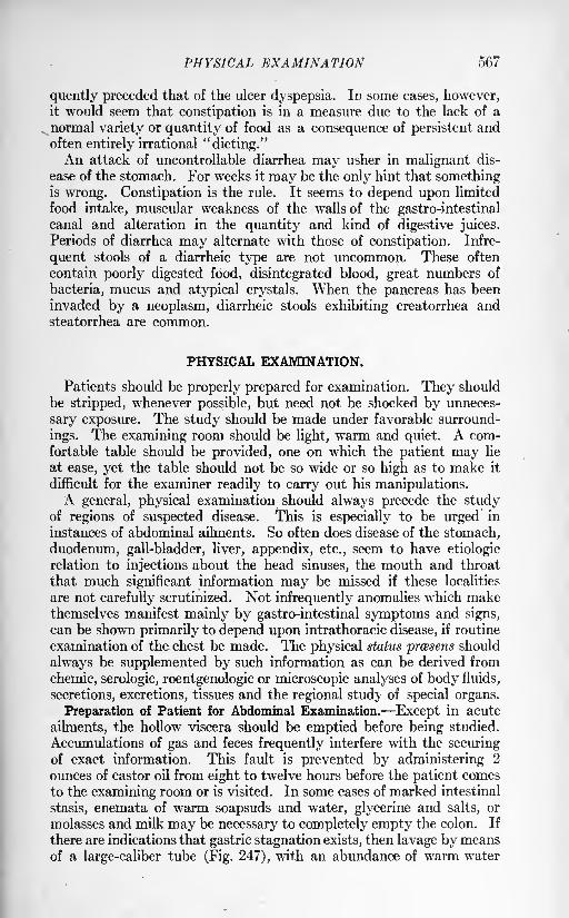
PHYSICAL EXAMINATION 567
quently preceded that of the ulcer dyspepsia. In some cases, however,it would seem that constipation is in a measure due to the lack of a
normal variety or quantity of food as a consequence of persistent andoften entirely irrational "dieting."An attack of uncontrollable diarrhea may usher in malignant dis-
ease of the stomach. For weeks it may be the only hint that somethingis wrong. Constipation is the rule. It seems to depend upon limited
food intake, muscular weakness of the walls of the gastro-mtestinalcanal and alteration in the quantity and kind of digestive juices.
Periods of diarrhea may alternate with those of constipation. Infre-
quent stools of a diarrheic type are not uncommon. These often
contain poorly digested food, disintegrated blood, great numbers of
bacteria, mucus and atypical crystals. When the pancreas has been
invaded by a neoplasm, diarrheic stools exhibiting creatorrhea andsteatorrhea are common.
PHYSICAL EXAMINATION.
Patients should be properly prepared for examination. They should
be stripped, whenever possible, but need not be shocked by unneces-
sary exposure. The study should be made under favorable surround-
ings. The examining room should be light, warm and quiet. A com-fortable table should be provided, one on which the patient may lie
at ease, yet the table should not be so wide or so high as to make it
difficult for the examiner readily to carry out his manipulations.A general, physical examination should always precede the study
of regions of suspected disease. This is especially to be urged in
instances of abdominal ailments. So often does disease of the stomach,
duodenum, gall-bladder, liver, appendix, etc., seem to have etiologic
relation to injections about the head sinuses, the mouth and throat
that much significant information may be missed if these localities
are not carefully scrutinized. Not infrequently anomalies which makethemselves manifest mainly by gastro-intestinal symptoms and signs,
can be shown primarily to depend upon intrathoracic disease, if routine
examination of the chest be made. The physical status prasens should
always be supplemented by such information as can be derived from
chemic, serologic, roentgenologic or microscopic analyses of body fluids,
secretions, excretions, tissues and the regional stud^ of special organs.
Preparation of Patient for Abdominal Examination. Except in acute
ailments, the hollow viscera should be emptied before being studied.
Accumulations of gas and feces frequently interfere with the securingof exact information. This fault is prevented by administering 2
ounces of castor oil from eight to twelve hours before the patient comes
to the examining room or is visited. In some cases of marked intestinal
stasis, enemata of warm soapsuds and water, glycerine and salts, or
molasses and milk may be necessary to completely empty the colon. If
there are indications that gastric stagnation exists, then lavage by meansof a large-caliber tube (Fig. 247), with an abundance of warm water
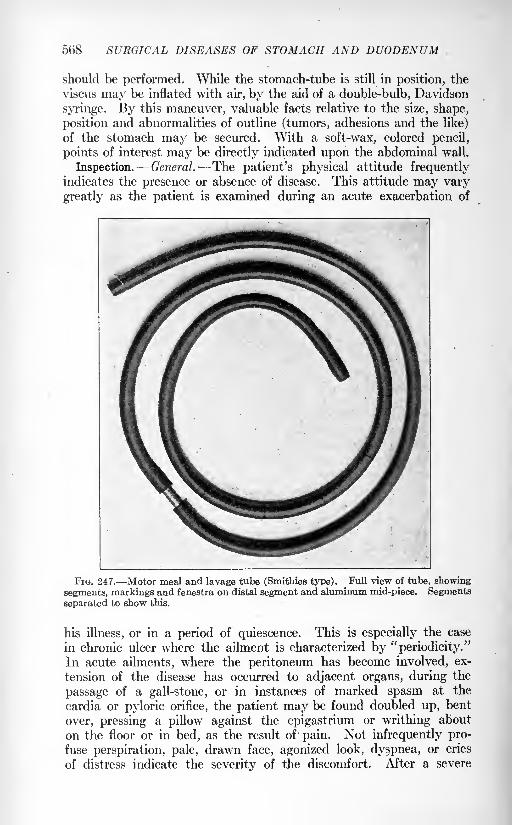
568 SURGICAL DISEASES OF STOMACH AND DUODENUM
should be performed. While the stomach-tube is still in position, the
viscus may be inflated with air, by the aid of a double-bulb, Davidson
syringe. By this maneuver, valuable facts relative to the size, shape,
position and abnormalities of outline (tumors, adhesions and the like)
of the stomach may be secured. With a soft-wax, colored pencil,
points of interest may be directly indicated upon the abdominal wall.
Inspection. General. The patient's physical attitude frequentlyindicates the presence or absence of disease. This attitude may vary
greatly as the patient is examined during an acute exacerbation of
FIG. 247. Motor meal and lavage tube (Smithies type). Full view of tube, showing
segments, markings and fenestra on distal segment and aluminum mid-piece. Segmentsseparated to show this.
his illness, or in a period of quiescence. This is especially the case
in chronic ulcer where the ailment is characterized by "periodicity."
In acute ailments, where the peritoneum has become involved, ex-
tension of the disease has occurred to adjacent organs, during the
passage of a gall-stone, or in instances of marked spasm at the
cardia or pyloric orifice, the patient may be found doubled up, bent
over, pressing a pillow against the epigastrium or writhing about
on the floor or in bed, as the result of pain. Not infrequently pro-
fuse perspiration, pale, drawn face, agonized look, dyspnea, or cries
of distress indicate the severity of the discomfort. After a severe
PHYSICAL EXAMINATION 569
hemorrhage, the patient may lie in a dead faint, with cold, clammy,
pale skin, dilated pupils, sighing respiration and with flabby muscles.
Where the gall-bladder or peritoneum has been involved by infection,
the subject may be found in an actual chill with or without jaundice,
or, later on, with a high temperature. In instances of malignant disease
or in benign ailments where there has been an obstruction to the free
onward progression of food, the patient may be caught during an
attack of vomiting or of severe nausea or retching.
In both benign and malignant disease associated with obstruction,
evidences of weight loss can readily be seen. The weight loss in benignaffections may be as marked as in those where malignancy is present, but
as a rule patients with well established malignancy exhibit not onlyevidences of starvation, but also those of cancerous intoxication. Such
are noted in the shuffling gait, collapse on sitting in a chair or lying
on a table, breathlessness on exertion, pale, muddy, or sallow skin,
absence of subcutaneous fat on cheeks, neck or over the body, luster-
less eye and dry mucous surfaces. Not rarely, traces of edema are
observed about the ankles, and occasionally anasarca is noted.
Visible glands should be searched for. These are most commonlyseen, in instances of malignancy, above the left clavicle, about the navel
and in the inguinal region. Such glands are usually discrete, but maybe confluent. They rarely ulcerate.
Special Abdominal Examination. The contour of the abdomen mayvary markedly. In cases where such affections as chronic ulcer of the
duodenum or stomach exist no abnormality may be noted. Whereobstruction at the cardia, or of the lumen of the stomach or duodenum,has become marked not rarely the evidence of starvation is shown by the
abdomen being flat or scaphoid. If the obstruction is at the pylorusand of high degree, should the muscle tone yet be good, visible peri-
staltic waves may be observed to run from the left rib margin across
the epigastrium to the right. More rarely, waves of reverse peristalsis
can be seen passing slowly from the right side or in the region of the
pylorus, leftward, across the epigastrium.After there has been much weight loss associated with lack of tone
in the abdominal muscles in instances of marked pyloric stenosis or
hour-glass stomach, the much dilateoT or distended stomach may be
seen lying in the mid-abdomen. In cases where the pyloric obstruc-
tion is due to malignant disease the normal tone is early lost. In such
event, while the much distended stomach is visualized, it is uncommonto observe visible peristalsis. Only very uncommonly can waves of
reverse peristalsis be noted.
Where there has been involvement of the portal circulation, exten-
sion of disease (malignant or benign) to the peritoneum, or obstruction
to the lymph-stream, the well rounded abdomen with bulging in the
flanks or in the dependent portions, should the patient stand erect, indi-
cates accumulation of fluid. This well rounded, abdominal contour is
frequently in striking contrast to the evidences of emaciation seen
elsewhere. The skin over such abdomens may be tightly stretched
570 SURGICAL DISEASES OF STOMACH AND DVODENVM
and present a mottled, reddish blue, or brawny appearance. Some-times, greatly distended superficial veins and venous capillaries are
seen, and where the circulatory obstruction has been long continued,
general edema may be noted. In instances of thrombosis of the portal
vein, its radicals, or the veins in the mesentery, localized plaques of
edema and discoloration can be made out in various portions of the
abdominal wall.
Tumors. These may result from a filled and distended stomach or
bowel, a neoplasm involving portions of the gut, liver, kidneys, pan-creas, retroperitoneal tissues, omentum, abdominal aorta, mesentery,
spleen, or the structures forming the abdominal wall.
Gastric tumors are usually seen above the navel : in fact, 92 per cent,
of all tumors lying above the navel are associated with the stomachand the duodenum. Gastric tumors usually move upon respiration,inflation of the stomach or the colon with air, and not infrequently uponchange of the patient's position. These points frequently serve to differen-
tiate such growths from those involving the adjacent abdominal viscera.
Large tumors of the stomach may be hidden by their position (fundus,
posterior gastric wall, lesser curvature, beneath* the ribs, or behind the
liver), by accumulations of abdominal fluid, or by thick, edematous
parietes. Metastases of malignant growths primary to the stomach,
may be noted in the enlarged liver, through the omentum, about the
navel and in the inguinal nodes. Not infrequently, accumulations of
material in the stomach and bowel, especially in emaciated or neurotic
individuals, make it extremely difficult to say that these organs are not
the seats of tumors. In such instances, it is of the greatest importance,as we have emphasized above, that the examination be made onlyafter thoroughly emptying the abdominal viscera. In acute ailments
of the stomach or the duodenum, or in acute exacerbations occurringin the course of chronic affections, the abdominal wall may be so rigid
or distended as to prevent visualization of parts which, if seen, mightotherwise indicate the disease. In these instances, the examination is
greatly facilitated by having the patient lie for from fifteen minutes to
a half hour in a bath with the water at a temperature above 100 F.
Palpation. General palpation gives useful information regardingthe body temperature, the state of the skin (cold, clammy, the skin of
shock; hot, dry, the skin of fever; loose, inelastic, dry, wrinkled, the
skin of cachexia and starvation), the thickness of the subcutaneous fat,
or diminution in tone of the skeletal muscles. A careful search should
be made for palpable lymph-nodes above the clavicles, in the axilla,
or inguinal regions, about the navel or in the rectovesical space. In the
absence of blood dyscrasias, such nodes usually point toward malig-
nancy and generally a malignancy of inoperable extent. General
anasarca or traces of edema about the malleoli as a rule point to gravedisorders.
The tension of the abdominal wall is usually markedly diminished
in cases of maHgnant disease where there has not occurred an accumu-
lation of ascitic fluid. Where there is a large volume of fluid in the
PHYSICAL EXAMINATION 571
peritoneal sac, the tension of the abdominal wall may be enormouslyincreased. Where there has been perforation of acute or chronic ulcer
of the duodenum or the stomach, general or localized zones of increased
tension aid one in roughly delimiting the extent of peritonitis.
In late gastric cancer an abdominal tumor was palpable in morethan 71 per cent, of 921 cases which the writer studied. In but few
instances was the tumor located below the navel. It was usually in
the mid-epigastrium or to the right, and as a rule could be movedabout by the palpating hand to some extent. Such tumors generallyvaried in size from that of a common marble to as large as a baby'shead. Only rarely were they very tender to the touch, the palpating
ringers usually causing but vague discomfort. Generally such tumors
are made up of multiple nodules, but at times they may be smoothand easily outlined. There are instances where it is very difficult to
be sure whether or not an epigastric tumor is connected with the
stomach, pancreas, gall-bladder, liver or the omentum. The gastric
tumors as a rule, however, .are associated with a more definite
gastric history than are tumors in other locations, and with the excep-tion of tumors of the omentum are more freely movable. After inflation
of the stomach with air, gastric tumors may be noticeably displacedor may vary greatly in size.
Abdominal Tenderness. In chronic affections of the stomach and the
duodenum, definite points of abdominal tenderness are largely depend-ent upon whether or no the patient is examined during a period of
extension or recrudescence of his disease. If the former exists, then
local areas of distress upon moderate pressure may be readily out-
lined. Unless the peritoneum has become involved, however, these
areas of tenderness are rarely small. As a rule there is diffuse, epi-
gastric muscle-spasm associated with added spasm of distress in certain
sections of the general zone. It is a mistake to expect that palpationwill definitely outline points of abdominal tenderness which have loca-
tions with respect to certain portions of the stomach or the duodenum.In but a general way, can palpation of a zone of tenderness in the
right upper quadrant indicate that the duodenum is involved, if
such tender point is taken in connection with a clinical history of the
duodenal ailment. The course of the normal duodenum varies so
greatly that tenderness in the right upper quadrant -may be in no wayrelated to the anatomic position of the duodenum in a given individual.
These facts readily can be proved by outlining areas of tenderness over
what are thought to be parts of the stomach or duodenum, and then
examining the patient by means of the fluoroscopic screen. It will
be found frequently that the points of tenderness supposed to be over
these viscera have no relation whatever to such upon fluoroscopicexamination. These observations indicate how misleading and fruitless
it is to attempt definitely to locate position, in the stomach or the
duodenum of ulcer or cancer, by placing reliance upon the locations of
tender areas upon abdominal examination. Unfortunately masses of
statistics with respect to the location of ulcers or cancers of the duo-
572 SURGICAL DISEASES OF STOMACH AND DUODENUM
denum or stomach have been tabulated after this fashion. Mentionis made of them here only to point out their utter uselessness.
It is true that fully 60 per cent, of gastric ulcers lie in the pyloricfourth of the stomach, and fully 80 per cent, of the duodenal ulcers
lie in the first third of the duodenum. It is also true that in the major-
ity of individuals these portions of the viscera are situated above the
navel or to the right side of the epigastrium. A knowledge of these facts,
coupled with a certain amount of imagination make it not a difficult
thing for an examiner to prophesy that in a patient with a typical ulcer
history, the lesion is in the stomach's pyloric fourth or the proximalthird of the duodenum. It is absolutely impossible, however, for one
to say before laparotomy or autopsy whether the ulcer is on the duo-
denal or the gastric side of the pylorus, unless the physical examination
is supplemented by that by the x-ray. Even then, those most expertare often in doubt.
In an abdominal crisis, dependent upon accident in the course of
disease of the duodenum and the stomach, unless the patient is seen
very early, the rapid extension of peritoneal involvement may prevent
localizing the original process by abdominal palpation. Very often the
patient's subjective description of his earliest pain is of greater aid in
localization than is the information obtained by abdominal palpation.Where acute exacerbation, in the course of a chronic duodenal or
gastric affection ulcer, carcinoma has occurred (perforation, fistula,
severe hemorrhage), there is generally an attempt made to wall off the
infection by rapid formation of peritoneal adhesions. This occurs gen-
erally in all instances of perforation where the ulcer or cancer is not
located on the anterior wall of the viscus. Palpation, in these circum-
stances usually discloses a normal abdomen excepting for pain andincreased tension in rather definite areas of the epigastrium. With the
history, this anomalous finding of a locally "acute abdomen" existingin a portion of a "chronic abdomen" is of great diagnostic significance.
Rectal Examination. This never should be neglected, particularlyin cases giving the history of melena, chronic dyspepsia of the ulcer
type, diarrhea, obstinate constipation with painful passage of stool,
or where the physical examination points to malignancy. Not infre-
quently, rectal examination will demonstrate that so-called melena is
due to bleeding hemorrhoids, ulcerations of the rectum or sigmoid
(parasitic, specific, tuberculous, malignant, or in association with
diverticula), new growths, foreign bodies, varicosities as a result of
displacement of the pelvic organs, or to chronic interference with the
portal circulation.
Metastases. Metastases in patients affected with malignant dis-
ease of the stomach or adjacent viscera are palpable quite early in the
rectovesical space. There they form a collection of nodules of various
sizes that lie close behind the bladder. These are readily palpable at
the end of the examining finger as a sort of ledge or shelf-like protrusion,to which Blumer has given the name of the "rectal shelf." In late
instances of the disease, the entire rectovesical space may be filled
PHYSICAL EXAMINATION 573
with firm, discrete or confluent masses. It is of particular importanceto identify these nodules, inasmuch as when they are present, radical
operations for the cure of malignant disease of the stomach are out of
the question in so far as permanent relief from the disease is concerned.
Even if by wide dissection it is possible to remove the disease from
the stomach wall, and, mechanically, a perfect operation can be per-
formed, yet on account of the scattering of cancer cells throughoutthe lymph-stream, recurrence is sure to take place within a short time.
Percussion. When the stomach has been filled with fluid or air,
light percussion enables one to outline the relative size and position of
the viscus. Indications of dilatation of the stomach are supplied bythe estimation of the distance between the outlines of its lesser and
greater curvatures, and not entirely by the demonstration of the loca-
tion of the greater curvature. When the normal stomach is inflated,
the average distance between the lesser and greater curvatures, in the
midline, is rather more than 3 inches. In instances of dilatation, as
result of pyloric obstruction or of marked gastric atony, the distance
between the two curvatures may be as great as eight to ten inches.
The relationship of neither of the curvatures to the umbilicus indicates
definitely either the size of the stomach or whether or not it is in"nor-
mal position," or is ptosed. Slight experience will/demonstrate the
variation in the position of the umbilicus with regard to bony skeletal
landmarks. Many hugely dilated stomachs do not descend below the
patient's navel, whereas numerous instances of stomachs with normal
capacity can be demonstrated where the stomach is wholly below the
navel.
It is of but slight significance, diagnostically, to percuss the outline
of a gastric tumor, but considerable information regarding the size and
position that such tumor occupies in the stomach wall can be derived
by percussing the outline of such tumor-dulness before and after infla-
tion of the stomach with air. Not rarely, posterior wall or deep lyingtumors along the curvatures are lost after gastric inflation, but in manycases, examination with the stomach empty will enable their readydemonstration. It is not uncommon to have the pyloric half of the
stomach noticeably rotate as the stomach is filled with air. In this
way, tumors which could not be outlined before inflation are thrust
forward against the anterior abdominal wall and then* outlines can
then be percussed in definite relationship to the stomach. If, after
inflation of the stomach, the outlines of the liver and other solid
abdominal viscera are determined by percussion, it frequently will be
seen whether or no secondary tumors have arisen in the liver, pancreasor omentum, especially if percussion outlines before and after gastricinflation are compared or contrasted.
Accumulation of ascitic fluid in the abdomen may entirely mask even
extensive gastric tumors, the presence of free fluid being determined
by percussing the outlines of dulness with the patient in various posi-
tions lying on the back, sitting, standing, or lying on either side. At
times, however, the accumulation of ascitic fluid may be so great as to
574 SURGICAL DISEASES OF STOMACH AND DUODENUM
result in neither fluctuation waves or the percussion of a movabledulness: the entire abdomen may be dull to percussion.
After paracentesis, and the withdrawal of all or some of the fluid,
gastric outlines and tumors connected with the stomach or adjacentviscera may be outlined.
Auscultation. This maneuver is of relatively slight diagnostic value
in lesions of the stomach and the duodenum. In instances of greatdilatation of the stomach, with retention, splashing sounds can be
heard over the stomach many hours after soft or liquid food has been
given, if the stomach zone is sharply tapped while the stethoscope lies
over some portion of it. One should differentiate such splashingsounds from the gurgling sounds which occur over the bowels of nor-
mal individuals, or of individuals of an excitable type who are mentally
perturbed over the steps of an examination.
After gastro-enterostomy has been performed, if the patient be
directed to drink freely, by listening over the middle third of the greatercurvature of the stomach, gurgling or distinct squirting sounds mayprove the patency of a gastro-enterostomy stoma. In instances of
"hour-glass stomach" the escape of fluids or gas from an upper chamberto a lower, may, at times, be made out by auscultation. Occasionally,
splashing sounds in two definite locations, separated by a zone in which
no splashing is heard, can be demonstrated. This is particularly aptto occur in instances where a large ulcer or cancer of the saddle typehas brought about a tube-like constriction in the middle third of the
stomach. In instances of obstruction at the cardia, due to partial
stenosis as result of chronic ulcer or cancer, if the stethoscope is appliedat about the level of the eleventh dorsal vertebra, posteriorly, in the
midline, and the patient directed to swallow a glass of water, the
second swallowing sound may be either greatly delayed, imperfectlyheard or not recognized at all.
To demonstrate the presence or the absence of free hydrochloric acid,
it has been suggested that the patient be given a half dram of sodium
bicarbonate in a small amount of water. The stethoscope is then
placed over the region of the stomach. If hydrochloric acid is present,
sizzling sounds can be heard. This test is of but relative value, and
certainly has limited practical bearing.
Esophagoscopy and Gastroscopy. These procedures in the hands
of an expert are of considerable diagnostic worth. The esophagusand the stomach should be completely emptied before any instru-
ment of this type be passed. The instruments devised by Suss-
man of Berlin are of the greatest service. Sussman's apparatus is
of flexible type, and can be inserted into the esophagus or stomach
with the least discomfort to the patient. For esophagoscopy and
gastroscopy, however, it would seem best that the patient be given a
general anesthetic, and that the instrument be inserted with the patient
lying in the dorsal position with the head low over the edge of the table
as in performing tonsillectomy. In well established cancer and ulcer of
the lower esophagus or stomach, visualization through the esophago-
EXAMINATION OF GASTRO-INTESTINAL FUNCTION 575
scope or gastroscope is not without certain advantages from an educa-
tional standpoint or in obscure ailments for the purpose of removingbits of tissue for microscopic study. However, since the developmentof the roentgen ray, the patient is served practically as well by fluoros-
copy or the making of several radiograms as by direct vision of the
diseased viscus through instruments passed per orem.
METHODS OF EXAMINATION OF GASTRO-INTESTINAL FUNCTION.THEIR INTERPRETATION.
GASTRIC CONTENTS.
In ailments of the stomach and the duodenum, the clinical historyand the physical examination are often sufficient to enable one to makecorrect diagnoses. However, there are numerous instances wherethe routine anamnesis and status prcesens give but a hint as to the
nature of the disease or fail to tell the extent of an affection known to
exist. In such event, not rarely, the absolute diagnosis can be learned
from clinical laboratory tests, particularly from physical, chemical,biochemical and microscopic analyses of gastric extracts and of feces.
Even in cases where the diagnosis can be roughly made from clinical
history and physical examination, one cannot obtain an adequate
conception of the abnormalities associated with the ailment without
the data that may be secured from intelligent scrutiny of vomitus,stomach contents and feces. On the other hand, caution is to be urged
against placing laboratory analyses paramount in the making of diag-noses of ailments of the stomach and the duodenum. Data from such
studies should be interpreted rigidly in the light of facts established bythe anamnesis and the physical examination. In doubtful instances, it
will be found, not infrequently, that more reliance may be placed uponthe clinical observations than upon uncommon or novel laboratorytests. Sometimes, however, in obscure cases, e. g., physically inac-
cessible cancer of the posterior wall or of the fundus of the stomach,test-meal analyses will establish the correct diagnosis in a few minutes.
Investigation of Gastric Emptying Power. Methods. The emptyingpower of the stomach is best estimated by the administration of a physio-
logical meal of mixed food after the patient has been taken off"diet"
and his stomach is, as far as possible, empty. Experience has taughtus that a twelve-hour time interval is of greater diagnostic worththan the four- to six-hour interval recommended by Riegel and others.
Physiological Motor Meal. Its object is to demonstrate the free
entrance into and exit of ordinary food from the stomach. The mealshould be cautiously administered following recent, severe hemorrhage,clinical evidences of ulcer or cancer perforation, coma, extreme physical
weakness, delirium, or severe ailments of the heart, bloodvessels or the
kidneys.Method. Thorough lavage with warm water should completely
empty the stomach. Two ounces of castor oil are poured into the
stomach through the stomach tube after the lavage. Three hours later
576 SURGICAL DISEASES OF STOMACH AND DUODENUM
the patient is given a fairly large meal of mixed food. In addition to
other viands, the meal should contain 50 grams of meat, 2 leaves of
lettuce and 20 raw raisins. (The skins of the raisins should be eaten.)
Tea, water, or milk are allowed. After from eight to twelve hours the
stomach is emptied and search made for remnants of the meal admin-
istered. No food should have been taken in the interim.
The secretory efficiency is determined by means of the Ewald breakfast
of second-day bread or the shredded biscuit meal suggested by Dock.In the ordinary case the time limit for removal is fifty minutes after
administration. Where clinical symptomatology indicates abnormally
rapid emptying power, the stomach is examined from twenty-five to
forty minutes after the test-meal has been given. The size and positionof the stomach are determined by inflation through the stomach tube,
with the patient recumbent. (We use an inflation bulb, expelling one
and a half ounces of air at each compression. The boundaries of the
stomach are determined by the auscultatory method while the infla-
tion is in progress.)In the space allotted, only a brief summary of the interpretation of
test-meals can be attempted. These observations are based upon the
personal analysis of 7041 consecutive gastric extracts. It is difficult
to present the work without its being merely statistical. An attempthas, however, been made to emphasize the clinical pointings.
Size of Stomach. The average capacity of the stomach is 27 ounces
in females and 33 ounces in males where dilatation does not exist.
In instances revealing dilatation the average capacity in females is
41 ounces and 52 ounces in males. The greatest gastric capacity is
associated with non-malignant pyloric stenosis.
The Macroscopic Appearance of Removed Gastric Extracts. In addi-
tion to evidences of gastric retention this observation is not infrequentlyof significance.
Color. Traumatic blood was noted in 6.4 per cent, of our cases.
Its presence was quite as constant when simple spasm existed at the
cardia as when ulceration was present with or without spasm. Its
appearance upon lavage had no consistent relation to any form oi gastric
disease other than cancer. Forty-four cases of gastric ulcer had lavageless than forty-eight hours following hemorrhage, and in these cases
there was no recurrence of either gross or microscopic bleeding from
the maneuver. In the cancer cases (96 per cent, being of the medullary
type) traumatic blood on lavage was observed in more than half of 218
consecutive cases.
Bile Coloring. Various shades of green or yellow were observed in
11 per cent, of the cases in the series. The yellowish shades were
the results of straining as a consequence of tubing or occurred in 74
per cent, of the patients who had had previous gastro-enterostomy
(seventy-six). The green coloration from bile was noted quite con-
stantly in cases of gastric atony, ptosis with relaxed pylorus, dilatation
with or without ptosis, intermittent pyloric spasm, induration about
the pylorus due to ulcer or cancer, and from obstruction below the
EXAMINATION OF G'ASTRO-INTESTINAL FUNCTION 577
papilla of Vater. The green shades usually indicated that bile had been
present in the stomach for some hours and had become oxidized. In
two instances, the growth of a chlorophylaceous mould was mistaken
for bile before chemical tests or microscopic examination had been
made. In 126 instances the presence of much traumatic blood rapidly
hid evidences of bile coloring.
Of the cancer cases, it is interesting to observe that coffee-colored or
dark-brown extracts were noted in but 19 per cent. When such were
observed the cases were inoperable. The color of gastric contents was
quite as apt to be tan or brownish in partial stenosis with dilatation or
atony from non-malignant causes as from cancer. As will be shown below,
however, the odor was a fairly reliable differentiating point between
the two conditions. There were 314 cases of benign achylia gastrica.
In 96 per cent, of this group the dead-white color of the gastric extracts,
associated with markedly absent chymification, was a characteristic
finding.
Odors Extracts from normal stomachs have a peculiarly bland andsomewhat sweetish odor. Modifications quantitative and qualitative
in acidity cause variations. In non-retention cases, the sharpness of
the sour odor is usually a reliable gauge of the free hydrochloric acid
content. In retention cases, high or low free hydrochloric acidity maybe masked by the odor from fermentative or putrefactive processes. In84 per cent, of the cancer group, acrid, rancid odors due to volatile,
organic acids were almost pathognomonic. In 76 per cent, of the non-
malignant retention class (ulcers, gall-bladder disease, or ptosis with
or without atony) the yeasty aroma was almost similarly characteristic.
This simply elicited sign proved in our experience to be extremelyuseful in differential diagnosis. Putrefactive odors are generally dueto sloughing of cancer tissue, deterioration of a quantity of blood in
the presence of low hydrochloric acidity, fecal contents from obstruc-
tion below the duodenum or from perforation of ulcer or cancer into
an adjacent viscus.
Amount of Gastric Extract. The average quantity of test-meal
removed in our entire series was 108 c.c. The average quantity in the
non-retention group was 76 c.c. The average quantity in the reten-
tion class was 350 c.c. So-called"hypersecretion" was as frequently
observed in pyloric spasm from appendix or gall-bladderdisease as fromduodenal or gastric ulcer. It is not difficult to mistake partial pyloricstenosis for irritative hypersecretion. But 21 per cent, of the non-
stenosing ulcers (duodenal or gastric) exhibited what might be termed
hypersecretion. In young adults of both sexes the most commoncause of hypersecretion was pyloric spasm due to diseased appendixor gall-bladder. Some degree of such was constantly present if the
symptoms had persisted longer than an average time of 2.8 years.Mucus. Mucus is a sign of doubtful diagnostic worth. It is more
commonly in excess in stenosing cancers or ulcers than when the pyloricchannel is free. While but 1 per cent, of hyperacidity and hyper-secretion cases were associated with pathological increase in mucus, 46
VOL. n 37
578 SURGICAL DISEASES OF STOMACH AND DUODENUM
per cent, of the malignant retention group and 29 per cent, of the benignretention class revealed such increase.
Chymification. Chymification is an index of masticatory thorough-ness, of the character of food ingested, of the combining power of hydro-chloric acid, of the presence of normal gastric ferments, and of varia-
tions in gastric peristalsis and emptying power. The last factor appearsto carry the greatest weight in all cases except those of primary or sec-
ondary achylia. While high hydrochloric acid content generally assures
good ch^mification, the presence of organic acids and foreign micro-
organisms (as in gastric cancer) frequently accomplishes similar ends.
The Incidence of Retarded Gastric Emptying Power. This term
"emptying power," is purposely chosen.' '
Hypermotility" should be
largely limited to what is indicated by "hyperperistalsis." It does not
necessarily follow that hyperperistalsis is associated with increased
or even normal rate of the stomach's emptying. In fact, our records
demonstrate that hyperperistalsis is most commonly found in instances
of pyloric spasm or pyloric stenosis which have gastric or extragastricfoundation.
Of our entire series, 12 per cent, of the cases showed some grade of
retarded gastric emptying power macroscopically. In 8 per cent, of the
series this was a constant finding. In 4 per cent, it was an intermittent
manifestation. In 87 per cent, of the group when gastric retention
was a persistent observation, some degree of gastric dilatation was an
associated complication. In 2 out of every 3 cases where this occurred
there was clinical history of delayed vomiting. Persistent demonstra-
tion of gastric retention is an indication for surgical intervention. It
denotes mechanical abnormalities or obstruction. In these instances,
dilatation of the stomach is a sign and not the disease. In a givencase the degree of gastric retention may vary markedly accordingas the stenosis is or is not associated with gastric or pyloric spasm.Dilatation of the stomach may likewise vary greatly in degree.In our series the causes of persistent gastric retention were, in the order
of the frequency of their demonstration, gastric cancer, duodenal
ulcer, gastric ulcer, cholecystitis with adhesions, gastric atony, tumorof the pancreas, tumor of the liver, hypernephroma, tuberculous
peritonitis, and retroperitoneal sarcoma. The causes of intermittent
gastric retention were pyloric spasm associated with appendicitis or
cholecystitis, gall-stones, duodenitis or gastritis in the presence of
increased free hydrochloric acid, and gastroptosis.
In gastric cancer, emptying power was retarded in more than 70
per cent, of instances, irrespective of the location of the growth. In
surgical duodenal ulcer, approximately 2 out of 3 cases revealed gastric
stagnation. In surgical gastric ulcer in rather more than 50 per cent,
of instances some grade of retention was proved. Of the pyloric spasms
(associated with appendicitis, cholecystitis, gall-stones, or duodenitis),
in rather more than 3 per cent, of instances retention was demonstrated.
The sign of differential worth in this class of case is that of the variability
EXAMINATION OF GASTRO-INTESTINAL FUNCTION 579
of the exhibition of retention at different examinations. This sign is
of special value to the surgeon who contemplates gastro-enterostomyin such instances. Unless definite obstruction exists, at or near the
pylorus the best operative results are only to be expected whenartificial stricture is surgically produced. We have records of the
examination of 482 cases of pyloric spasm with intermittent gastric
retention, when later the appendix had been removed or operations
upon the gall-tract had been performed. In but twenty-one instances
was any form of gastric stagnation demonstrated postoperatively.These observations of the incidence of gastric retention, cannot in
any way be compared with such information returned from examina-
tion by means of the roentgen ray. It should be urged that, in the
method outlined, we are dealing with physiological problems. No one
has yet shown that the examinations of gastric function by means of
bismuth or barium compounds with the aid of the roentgen ray is
physiological. Consequently, only a long series of observations that
have been checked up by surgical procedure or by the subsequent
history in medical cases, extending over several years' time, will enable
actinologists to formulate statements that will indicate what meaningwe can attribute to retention of bismuth or barium in the stomach at
the end of, say, a six-hour interval.
The most common causes of increased rate of emptying of the
stomach in our series occurred in conjunction with achylia gastrica
(primary or associated with severe anemia), vagus hypertonia, tempo-rary or permanent atony without marked gastric dilatation, non-
obstructing gastric cancer or syphilis, colitis, and in numerous nervous
states of the young adult.
. Gastric Acidity. During the past five years, attempts have been
made to belittle the significance of estimations of gastric acidity. This
attitude has been brought about largely because of the persistence of
surgical teachings. It is to be expected that those who anticipatedthat a simple color test would serve in place of clinical cerebration
would be doomed to disappointment. While we have been compelledto revise some of our previously held opinions regarding the diagnosticworth of acid estimations of gastric extracts, yet when considered as a
part of evidence making for proper conception of disease, it would seemthat such information can in no wise be neglected. The confusion has
arisen chiefly because it was not until recently shown just what effect
upon gastric physiology had extragastric disease. Consequently, the
findings in disease extrinsic to the stomach were interpreted, clinically,
in the light of known primary gastric ailments.
For the purpose of brevity we would classify our acidity findingsinto three groups: (1) that comprising recognized disease of the stomach
itself; (2) that including lesions of the duodenum, gall-bladder, appen-dix, and the large bowel; (3) that comprising so-called "functional" or
central disturbances.
Acidities were all estimated by the Toepfer method, as follows:
580 SURGICAL DISEASES OF STOMACH AND DUODENUM
Tests for Estimating Gastric Acidity. When sufficient amount of
gastric extract is available, quantitative estimations should be madeupon the filtrate. Unfiltered gastric contents return readings higherthan when analyses of filtered extracts are made."Free" HydrochloricAcidity. Method. Place 10 c.c. of gastric filtrate
in a white porcelain evaporating dish. To this add 2 drops of a 0.5 percent, alcoholic solution of dimethylamidoazobenzol. If HC1 is present,the mixture becomes cherry red. The acidity is neutralized by slowly
adding from a burette solution of & sodium hydroxide. The end
point is reached when the mixture in the evaporating dish takes on a
lemon yellow color. The number of cubic centimeters (or parts thereof)of Y^J- sodium hydroxide required to bring about this color change is
read from the burette. This figure, multiplied by 10, gives the free
HC1 acidity per hundred parts. If acidity percentage is desired, the
resultant figure is multiplied by 0.00365."Total" Acidity. Method. Place 10 c.c. of gastric filtrate in a
white porcelain evaporating dish. Add 2 drops of a 1 per cent, alcoholic
solution of phenolphthalein. There is usually no color change unless
the total acidity is low or the extract alkaline. yV sodium hydroxidesolution is added to the mixture from a burette. When the total
acidity has been neutralized by the soda solution, the contents of the
evaporating dish become pinkish or purplish red. The degree of acidityis computed in the manner employed in estimating "free" HC1.
"Combined" Acidity. Method. As an indicator a 1 per cent,
watery solution of alizarin is employed. Ten cubic centimeters of
gastric filtrate are placed in a white porcelain evaporating dish. Twodrops of the alizarin solution are added. The mixture is titrated against
Y^ sodium hydroxide solution until the fluid is colored violet. Quan-titatively the estimations are made as in the analysis for free HC1.
Lactic Acid. Tests for lactic and organic acids generally, are fre-
quently positive in gastric retention contents, and particularly when
malignancy is the cause of the stagnation.Method. Six drops of liquor ferri sesquichlorati are added to 3 drops
of 95 per cent, solution of carbolic acid. This mixture is diluted with
distilled water until it assumes a lively amethyst-blue color. To this
last solution, are added 5 to 10 drops of gastric filtrate (or the ether
extract of such). If lactic acid is present, the amethyst-blue color
is discharged and a canary-yellow color is seen.
Volatile Fatty Acids. Method. Five cubic centimeters of gastric
filtrate are placed in a test-tube of 20 c.c. capacity. Over the mouthof the test-tube are placed two strips of moistened litmus paper: red
and blue. The gastric filtrate is then slowly heated over a Bunsen
flame. If volatile fatty acids are present, the blue litmus paper over
the test tube mouth turns red as the fumes react upon it. The original
strip of red litmus paper acts as a control.
Tests for Altered ("Occult") Blood. See under Examination of Feces,
page 591.
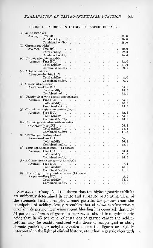
EXAMINATION OF GASTRO-INTESTINAL FUNCTION 581
GROUP I. ACIDITY IN INTRINSIC GASTRIC DISEASE.
(a) Acute gastritis:
Average Free HC1 32.0Total acidity .
' 50.0Combined acidity . . 18.0
(6) Chronic gastritis:
Average Free HC1 42.0Total acidity 62.0Combined acidity . 14.0
(c) Chronic atropbic gastritis:
Average Free HC1 :...."... 12.
Total acidity'
. . 21.0Combined acidity ; . / . .9.0
(d) Achylia gastrica:
Average No free HC1Total acidity . 8.0Combined acidity 6.0
(e) Gastric ulcer acute:
Average Free HC1 64 .
Total acidity ............ 78.0Combined acidity . . 12.0
(/) Gastric ulcer with recent hemorrhage:Average Free HC1 . . . 35.0
Total acidity 48.0Combined acidity 13.0
(g) Chronic non-retention gastric ulcer:
Average Free HC1 . 42 . 5
Total acidity 55.0Combined acidity . 13.5
(h) Chronic gastric ulcer with retention:
Average Free HC1 . . . . . . . 56.4Total acidity 74.2Combined acidity 17.8
(t) Chronic perforating ulcer:
Average Free HC1 f.
. 7 . 64.1Total acidity . ...... 76.2Combined acidity '; . . 12.0
(j) Ulcus carcinomatosum (64 cases):
Average Free HC1 ... . / . . 33.4Total acidity ....*'........ 51.0Combined acidity . . . . . 16.0
(k) Primary gastric cancer (122 cases):
Average Free HC1 ;"' . ... . 7.4Total acidity .".... 28.3Combined acidity 21.0
(Z) Ulcerating primary gastric cancer (14 cases) :
Average Free HC1 2.0Total acidity . . . . . 34.0Combined acidity 30.0
SUMMARY. Group I. It is shown that the highest gastric acidities
are uniformly determined in acute and subacute perforating ulcer of
the stomach; that in simple, chronic gastritis the picture from the
standpoint of acidity closely resembles that of ulcus carcinomatosumor of simple gastric ulcer when recent bleeding has occurred; that only54 per cent, of cases of gastric cancer reveal absent free hydrochloric
acid; that in 45 per cent, of instances of gastric cancer the acidityreturns may be readily confused with those of simple gastric ulcer,
chronic gastritis, or achylia gastrica unless the figures are rigidly
interpreted in the light of clinical history, etc. ; that in gastric ulcer with
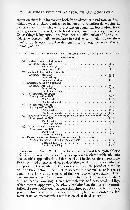
582 SURGICAL DISEASES OF STOMACH AND DUODENUM
retention there is an increase in both free hydrochloric and total acidity,
which fact is in sharp contrast to instances of retention developing in
gastric cancer, in which event, as retention comes on, free hydrochloricis progressively lowered, while total acidity simultaneously increases.
Other things being equal, in a given case, the diminution of free hydro-chloric associated with an increase in total acidity, with the develop-ment of obstruction and the demonstration of organic acids, speaksfor malignancy.
GROUP II. ACIDITY WHERE THE LESIONS ARE MAINLY OUTSIDE THESTOMACH.
(a) Duodenitis with pyloric spasm:Average Free HC1 68 .
Total acidity 74.0
Combined acidity 6.0
(6) Duodenal ulcer without stenosis:
Average Free HC1 59 . 2
Total acidity . 73.0
Combined acidity 14.0
(c) Duodenal ulcer with stenosis:
Average Free HC1 46.0
Total acidity 84.0
Combined acidity 28 .
(d) Cholecystitis, subacute:
Average Free HC1 66.0
Total acidity 71.0
Combined acidity 5.0
(e) Cholecystitis, chronic:
Average Free HC1 32.0
Total acidity 46.0
Combined acidity 14.0
(/) Appendicitis, subacute or chronic (usually in young adults both sexes) :
Average Free HC1 64.0
Total acidity 75.0
Combined acidity 8.0
(0) Colitis, subacute or chronic:
Average Free HC1 35.0
Total acidity . . 42 .
Combined acidity'
7.0
(h) Following gastro-enterostomy for gastric or duodenal ulcer:
Average reduction in free hydrochloric acid 17.0
Total acidity 32.0
Combined acidity 12.0
SUMMARY. Group 2. Of this division the highest free hydrochloricacidities are present in cases of pyloric spasm associated with subacute
cholecystitis, appendicitis and duodenitis. The figures closely resemble
those returned in gastric ulcer, as does also the clinical history with the
exception of the incidence of hemorrhage, chemical proof of bleeding
and the time factor. The onset of stenosis in duodenal ulcer increases
combined acidity at the expense of the free hydrochloric acidity. After
gastro-enterostomy for non-malignant stenosis there is a consistent
and noticeable lowering of free hydrochloric and also total acidity,
which cannot, apparently, be wholly explained on the basis of regurgi-
tation of succus entericus. In more than three out of four such instances,
proof of this having occurred, can, however, be demonstrated by fer-
ment tests or microscopic examination of stained smears.
EXAMINATION OF GASTRO-INTESTINAL FUNCTION 583
GROUP III. FUNCTIONAL AND CENTRAL DISTURBANCES.
(a) In epilepsy of the young adult:
Average Free HC1 56.0Total acidity 62.0Combined acidity 5.0
(fe) In neurasthenia, with or without moderate gastroptosls:
Average Free HC1 34.0Total acidity .' , . 46.0Combined acidity 9.0
(c) In marked gastroptosis:
Average Free HC1 37.0Total acidity 52.0
Combined acidity 14.0
These figures interpret themselves and require no special comment.
The demonstration of altered or occult blood in gastric extracts by the
benzidin or the guaiac tests was possible in 25 per cent, of the cases in
the entire series. It was present in 42 per cent, of all retention cases
irrespective of the causative lesion. It was demonstrated in 75 percent, of 712 cases of gastric cancer. It was almost as frequently shownin duodenal as in gastric ulcer. It would seem that apart from the
association of occult blood with malignant processes, its demonstration
in gastric extracts has very slight diagnostic worth.
The Incidence and Significance of Organic Acids in Gastric Extracts.
These are rarely demonstrated in non-retention cases, either malignantor non-malignant. In malignant disease, associated with partial
stenosis and gastric dilatation, lactic acid was present in 53 per cent,
of 712 operatively demonstrated instances. It was rarely demonstrated
when free hydrochloric acid rose higher than 10, and in 92 per cent, of
cases where lactic acid was found, extensive growths, generally inoper-able were present. Only 3 per cent, of the non-malignant achylias
were associated with lactic acid. In but 7 per cent, of the non-malig-nant retention cases was lactic acid present. It was found in less than
0.5 per cent, of instances of ptosis with atony.Volatile fatty acids were present in 61 per cent, of the malignant
cases and in but 3.2 per cent, of the instances of benign retention or
atony. In no instance of non-retention or of simple achylia or hypo-
acidity were volatile fatty acids demonstrated.
The Significance of the Demonstration of Specific Ferments in Gastric
Contents. Space does not permit our going into detail respecting the
incidence and diagnostic worth of proving the absence or presence of
pepsin and rennin in gastric extracts. It would seem that proteolysisis closely associated with the presence of free hydrochloric acid so longas the acid concentration is below 0.4 per cent, and that milk-curdlingferment follows similar laws. In malignancy, peptolysis appears to be
increased at the expense of proteolysis.
Edestin Test ("Peptic Index") of Fuld and Levison. According to
these investigators the peptic power of gastric juice in cancer of the
stomach is diminished. Confirmatory observations have been made bySchryver and Singer. Edestin is very difficult to obtain pure, com-
584 SURGICAL DISEASES OF STOMACH AND DUODENUM
mercially, but may be made in a well equipped laboratory. Schryverand Singer's modification of the original test is as follows:
Method. A pure preparation of edestin may be obtained in the
laboratory by recrystallization from warm salt solution. From this
is made a solution of 0.1 per cent, of edestin in 0.12 per cent, hydro-chloric acid (i. e., 30 c.c. normal HC1 in 1 liter distilled water.) (Sucha solution of edestin need not be made freshly for each observation,
but, if used as stock, it must be stored at zero temperature.) Into each
of 10 small test-tubes 2.5 c.c. of this solution are pipetted and left to
take the temperature of the room (10 to 20 C.). Into each test-tube
are now dropped 0.1, 0.2, 0.3, 0.4, . . . to 1.0 c.c. (in ascending
series) of the gastric juice under investigation, previously diluted to
one-tenth of its natural strength. Each test-tube is shaken and left
to stand. After thirty minutes have elapsed 0.3 c.c. of a saturated
solution of sodium chloride is added to each tube. If digestion has
proceeded to a certain point, the solution remains clear, while with
lower degrees of digestion a white cloudiness immediately develops.The Peptic Index in any case is designated as the number of tenths
of a cubic centimeter of a diluted juice added to the first clear test-
tube and divided into 100. This gives as a range of possible readingsthe ten numbers 100 (i. e., 100/1), 50 (i. e., 100/2), 33 (i. e., 100/3),
25, 20, 17, 14, 12, 11 and 10. For greater accuracy intermediate
amounts of diluted gastric juice may be added, as 0.125, 0.150, 0.175
(between 0.1 and 0.2) etc., but with practice it becomes possible to
read such intermediate numbers without recourse to actual experiment.The average of healthy cases is about 50. Cases do occasionally presentthemselves with an index above 100 (i. e., in which even the test-tube
containing only 0.1 c.c. of diluted juice remains clear on the addition
of the salt solution). In such instances recourse must be had to the
use of more diluted juice. At the other extreme, however, it is seldom
necessary to take readings below an index of 10 (i. e., in which even
the test-tube containing 1.0 c.c. of diluted gastric juice developscloudiness on the addition of salt solution), for in such cases our
experience seems to show that digestive power is probably almost
absent.
This test appears to be a valuable supplementary procedure where
the gastric extracts indicate that malignant transition of an ulcer maybe present.We have examined 108 cases for proteolytic ferments by the edestin
method, suggested by Fuld and Levison. The results thus far would
appear to indicate that early cases of cancer of the stomach, where the
free hydrochloric acid is shown to be low, reveal high peptolysis and
low proteolysis. In the simple ulcers both peptolysis and proteolysis
are low.
We have made observations upon 827 instances of gastric disease
for the detection of specific ereptases of malignant ulcer. We have
used the formaldehyde titration method as suggested by Sorenson and
Schiff. In our experience the average formal titration index in 87
EXAMINATION OF GASTRO-INTESTINAL FUNCTION 585
instances of proved gastric cancer was 22.3; the average index in 22
cases of ulcus carcinomatosum was 19.8; the average in 99 cases of
duodenal ulcer was 12.4; of 57 cases of simple gastric ulcer 11.6; of
32 cases of benign achylia, 14.1; of 16 instances of pernicious anemia,
14.5; and of 5 cases of cancer of the liver 4.25. It would appear that in
certain instances the estimation of the ereptic power of gastric juicetoward peptone solutions is of considerable value when interpreted in
the light of clinical history and symptomatology.
Glycyltryptophan and Tryptophan Tests. In rather more than 40
per cent, of our proved cases of cancer (186) the reaction was positive.In about 7 per cent, the tryptophan test was positive. Diagnosis of
malignant disease of the stomach was in each case quite possible,
independent of the chemical test. While gastric abnormalities other
than cancer exhibited positive glycyltryptophan reactions, in no class
of the disease of the stomach was the test so frequently obtained as
in cancer. Low free hydrochloric acid was frequently associated with
positive glycyltryptophan tests, and in more than 50 per cent, of these
reactions the gastric extracts contained bile or blood or both.
Wolff-Junghans' Test for Soluble Albumin in Gastric Extracts.
Methods for the estimation of the soluble albuminous products of diges-tion have frequently been devised with the hope that such might proveof practical service in the differential diagnosis of gastric ailments. Ofthese methods, the well-known procedure advanced by Salomon, hadfor a time the greatest vogue. Esbach's reagent and tubes proved,
however, unsatisfactory and inaccurate from a clinical viewpoint.More recently the problem has been approached from the practical
quantitative side and encouraging work recorded.
Wolff and Junghans report a method for estimation of the amountof soluble albumin in gastric extracts which they claim has givenexcellent clinical information in Ewald's service at the Augusta Hos-
pital, Berlin.
Theoretically, their procedure has the following basis: In the normal
aspirated test-meal there are demonstrable relatively large quantities of
soluble albumin by means of precipitating reagents. This soluble
albumin appears only through the agency of the gastric enzymes.This fact is proved by testing for soluble albumin a similar test-meal
which has been chymified but not swallowed. In such event, onlyminute quantities of dissolved albumin are present.The writer tested the gastric extracts of 15 cases of ulcus carcinoma-
tosum. In 11 instances (73.3 per cent.), units of precipitable albumin
ranged between 200 to 400. In 3 (20 per cent.), 100 units were shown.
In other words, of the 15 cases of malignant gastric ulcer, 14 (93.3 per
cent.) were either definitely positive or suspiciously so to the Wolff-
Junghans test. One case (6.6 per cent.) exhibited below 100 units of
albumin. In this group, motor stagnation of some degree was presentin 86.6 per cent.
Microscopic Estimation of Gastric Extracts. Unfiltered stomach con-
tents should always be studied. The examination is of particular
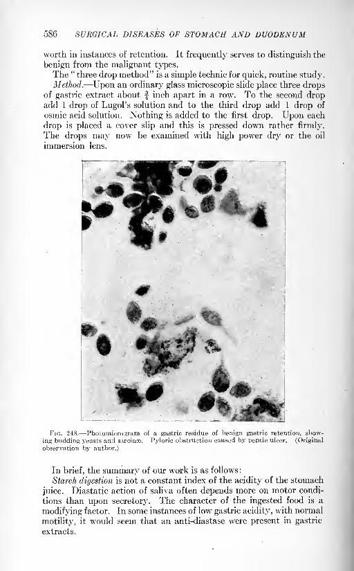
586 SURGICAL DISEASES OF STOMACH AND DUODENUM
worth in instances of retention. It frequently serves to distinguish the
benign from the malignant types.The "
three drop method" is a simple technic for quick, routine study.Method. Upon an ordinary glass microscopic slide place three drops
of gastric extract about inch apart in a row. To the second dropadd 1 drop of Lugol's solution and to the third drop add 1 drop of
osmic acid solution. Nothing is added to the first drop. Upon each
drop is placed a cover slip and this is pressed down rather firmly.
The drops may now be examined with high power dry or the oil
immersion lens.
**
FIG. 248. Photomicroiram of a gastric residue of benign gastric retention, show-
ing budding yeasts and sircinae. Pyloric obstruction caused by peptic ulcer. (Original
observation by author.)
In brief, the summary of our work is as follows :
Starch digestion is not a constant index of the acidity of the stomach
juice. Diastatic action of saliva often depends more on motor condi-
tions than upon secretory. The character of the ingested food is a
modifying factor. In some instances of low gastric acidity, with normal
motility, it would seem that an anti-diastase were present in gastric
extracts.
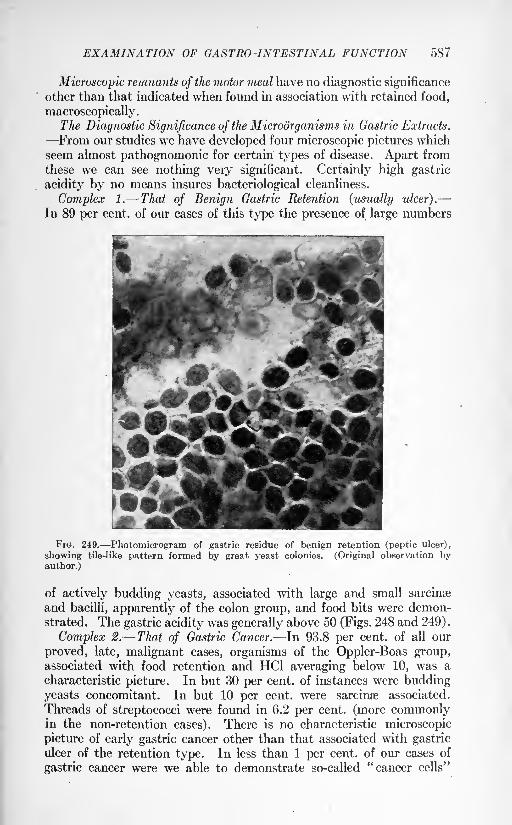
EXAMINATION OF GASTRO-INTESTINAL FUNCTION 587
Microscopic remnants of the motor meal have no diagnostic significance
other than that indicated when found in association with retained food,
macroscopically.The Diagnostic Significance of the Microorganisms in Gastric Extracts.
From our studies we have developed four microscopic pictures whichseem almost pathognomonic for certain types of disease. Apart fromthese we can see nothing very significant. Certainly high gastric
acidity by no means insures bacteriological cleanliness.
Complex 1. That of Benign Gastric Retention (usually ulcer).
In 89 per cent, of our cases of this type the presence of large numbers
FIG. 249. Photomicrogram of gastric residue of benign retention (peptic ulcer),
showing tile-like pattern formed by great yeast colonies. (Original observation byauthor.)
of actively budding yeasts, associated with large and small sarcinse
and bacilli, apparently of the colon group, and food bits were demon-strated. The gastric acidity was generally above 50 (Figs. 248 and 249) .
Complex 2. That of Gastric Cancer. In 93.8 per cent, of all our
proved, late, malignant cases, organisms of the Oppler-Boas group,associated with food retention and HC1 averaging below 10, was a
characteristic picture. In but 30 per cent, of instances were budding
yeasts concomitant. In but 10 per cent, were sarcime associated.
Threads of streptococci were found in 6.2 per cent, (more commonlyin the non-retention cases). There is no characteristic microscopic
picture of early gastric cancer other than that associated with gastriculcer of the retention type. In less than 1 per cent, of our cases of
gastric cancer were we able to demonstrate so-called "cancer cells"
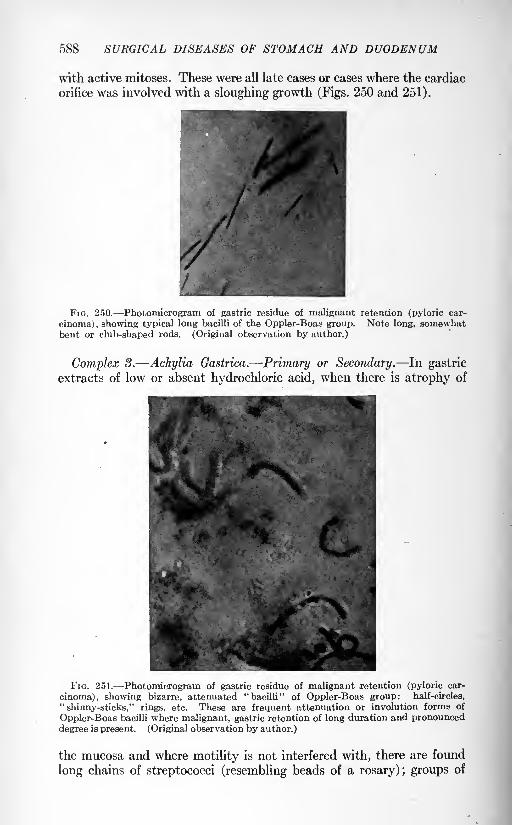
588 SURGICAL DISEASES OF STOMACH AND DUODENUM
with active mitoses. These were all late cases or cases where the cardiac
orifice was involved with a sloughing growth (Figs. 250 and 251).
FIG. 250. Photomicrogram of gastric residue of malignant retention (pyloric car-
cinoma) , showing typical long bacilli of the Oppler-Boas group. Note long, somewhatbent or club-shaped rods. (Original observation by author.)
Complex 3. Achylia Gastrica. Primary or Secondary. In gastric
extracts of low or absent hydrochloric acid, when there is atrophy of
FIG. 251. Photomicrogram of gastric residue of malignant retention (pyloric car-
cinoma), showing bizarre, attenuated "bacilli" of Oppler-Boas group: half-circles,
"shinny-sticks," rings, etc. These are frequent attenuation or involution forms of
Oppler-Boas bacilli where malignant, gastric retention of long duration and pronounceddegree is present. (Original observation by author.)
the mucosa and where motility is not interfered with, there are found
long chains of streptococci (resembling beads of a rosary); groups of
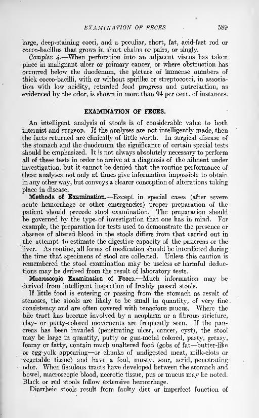
EXAMINATION OF FECES 589
large, deep-staining cocci, and a peculiar, short, fat, acid-fast rod or
cocco-bacillus that grows in short chains or pairs, or singly.
Complex 4- When perforation into an adjacent viscus has taken
place in malignant ulcer or primary cancer, or where obstruction has
occurred below the duodenum, the picture of immense numbers of
thick cocco-bacilli, with or without spirillse or streptococci, in associa-
tion with low acidity, retarded food progress and putrefaction, as
evidenced by the odor, is shown in more than 94 per cent, of instances.
EXAMINATION OF FECES.
An intelligent analysis of stools is of considerable value to both
internist and surgeon. If the analyses are not intelligently made, then
the facts returned are clinically of little worth. In surgical disease of
the stomach and the duodenum the significance of certain special tests
should be emphasized. It is not always absolutely necessary to performall of these tests in order to arrive at a diagnosis of the ailment under
investigation, but it cannot be denied that the routine performance of
these analyses not only at times give information impossible to obtain
in any other way, but conveys a clearer conception of alterations taking
place in disease.
Methods of Examination. Except in special cases (after severe
acute hemorrhage or other emergencies) proper preparation of the
patient should precede stool examination. The preparation should
be governed by the type of investigation that one has in mind. For
example, the preparation for tests used to demonstrate the presence or
absence of altered blood in the stools differs from that carried out in
the attempt to estimate the digestive capacity of the pancreas or the
liver. As routine, all forms of medication should be interdicted duringthe time that specimens of stool are collected. Unless this caution is
remembered the stool examination may be useless or harmful deduc-
tions may be derived from the result of laboratory tests.
Macroscopic Examination of Feces. Much information may be
derived from intelligent inspection of freshly passed stools.
If little food is entering or passing from the stomach as result of
stenoses, the stools are likely to be small in quantity, of very fine
consistency and are often covered with tenacious mucus. Where the
bile tract has become involved by a neoplasm or a fibrous stricture,
clay- or putty-colored movements are frequently seen. If the pan-creas has been invaded (penetrating ulcer, cancer, cyst), the stool
may be large in quantity, putty or gun-metal colored, pasty, greasy,
foamy or fatty, contain much unaltered food (gobs of fat butter-like
or egg-yolk appearing or chunks of undigested meat, milk-clots or
vegetable tissue) and have a foul, musty, sour, acrid, penetratingodor. When fistulous tracts have developed between the stomach and
bowel, macroscopic blood, necrotic tissue, pus or mucus may be noted.
Black or red stools follow extensive hemorrhage.Diarrheic stools result from faulty diet or imperfect function of
590 SURGICAL DISEASES OF STOMACH AND DUODENUM
stomach, liver, pancreas or small bowel. They may accompanysevere hemorrhage. In pronounced asthenia, cardiorenal disease,
neuroses, or organic nervous disease, diarrheic stools may be persistentor occur at irregular periods. Infectious ailments (miliary tubercu-
losis, typhoid fever, bacillary or protozoic dysentery) commonlycause systemic wasting associated with diarrhea. In atypic instances
of malignancy of the stomach or bowel, it may be difficult to rule out
such as the primary cause of the loose stools.
Color. The color of stools varies widely. After severe gastricor duodenal hemorrhage, red, black or greenish movements often mixedwith fleshy clots or spongy masses of blood may be passed for several
days. Green, tan, cream, yellow or putty-colored stools are not uncom-
monly observed in ailments involving viscera in the upper abdomen.In serious disease of the pancreas, foul-smelling, bulky, asbestos-colored
stools, greasy with unsplit fat, are almost pathognomonic.Chemical Examination of Feces. Reaction of Feces. Healthy stools
are commonly faintly acid or amphoteric. Malignancy of the stomachor pancreas is frequently associated with very alkaline feces.
Bile pigment in feces is recognized conveniently by the Schmidt
bichloride test.
Method. Intimately mix 5 grams' (or 10 c.c. in case the stool is
fluid) of feces in about four times its volume of saturated solution of
bichloride of mercury. This can be done in a small mortar. Allow to
stand five minutes after mixing. Pour off the supernatant fluid into a
Petri dish; cover, and let stand from two to twenty-four hours. Hydro-bilirubin is indicated by the fluid assuming a salmon to deep red shade.
If olive green change occurs, biliverdin is present.
This test is of especial worth in instances where light-colored stools
are passed, and disease of the stomach secondarily invading the liver,
pancreas or gall-ducts is suspected.Demonstration of Altered or "Occult" Blood in the Stools. The
patient's intestinal canal should be first completely emptied by the
administration of 2 ounces of oleum ricini or a bottle of citrate of
magnesia. The patient should then be given a diet free from meator its products or an excess of green vegetables, for two days. On the
third day nothing but milk or cereal gruel should be given. On the
morning of the fourth day, a half bottle of citrate of magnesia should
be administered, and the second stool passed collected and sent to the
laboratory in a covered vessel for analysis.
Tests for "Occult" Blood. Numerous tests are in vogue. If the
stool has been properly collected and the tests carefully performed,
they are all of worth clinically. On account of abortive reactions,
not infrequently given by the use of poor preparations of aloin,
guaiac or phenolphthalein, routinely, it has seemed that benzidin"
solutions have returned the most reliable information. The pinkish-
gray powder should be used, and this should be kept in a brown bottle,
carefully stoppered.The benzidin test is performed as follows:
EXAMINATION OF FECES 591
Benzidin Test. A standard solution of benzidin for use may be
made by dissolving one gram of pure benzidin powder in 50 c.c. of 95
per cent, alcohol. If this solution is preserved in a brown glass-
stoppered bottle and not kept in a very warm place it will remain
dependable. Other reagents necessary for the test are glacial acetic
acid, pure ether and a fresh solution of hydrogen peroxide.The test is set up as follows: Into a test-tube of 15 to 25 c.c. capacity
are placed 3 c.c. or one gram of feces. To this are added 5 to 10 dropsof glacial acetic acid. The tube is thoroughly shaken. Next are added
5 to 10 c.c. of pure ether. An intimate mixture is obtained by pouringthe contents of the tube into another, and vice versa, several times.
The mixture is allowed to stand a minute or two until the clear ether
layer separates at the top. This ether layer is poured off into another
clean test-tube. To this ether extract are added 15 to 30 drops of the
2 per cent, solution of benzidin in 95 per cent, alcohol. The mixture
is thoroughly shaken, and finally are added 15 to 30 drops of hydrogen
peroxide. The tube is again shaken. If blood pigment has been
present in the feces, even in as great dilution as 1 to 12,000, its pres-ence will be indicated by the fluid assuming a lively emerald-greencolor that rapidly gives place to deep ultramarine. This color changeshould occur within one minute. If there is not a decided color changewithin this time, in our experience, color changes occurring later can
be safely discarded as being of no clinical significance.
Interpretation of Positive "Occult" Blood Tests. A positive responseto chemical test for blood in the stool only signifies that there is a bleed-
ing point somewhere between the lips and the external anal ring. It
is thus evident that tests should be rigidly controlled and interpreted
strictly in relation to the clinical history of the case. Positive tests
are most commonly constant in malignancy. They are intermittently
present in the stools from chronic, recurring peptic ulcer.
The negative test for "occult" blood in feces is of aid in excluding
malignancy, particularly in instances where, with a dubious clinical
history, an atypic, abdominal tumor can be demonstrated.
Ferment Tests in Feces. The main worth of these lies in gaining
knowledge respecting the function of the pancreas. This function
may be seriously limited by disease from the stomach or duodenumwhich has extended to the gland (ulcer, cancer, etc.) . Feces prepared
by the administration of Schmidt's test diet1 are commonly analyzed for
the presence o f diastase and tryspin . Special methods, requiring expert
1 Schmidt test diet:
MORNING, 8 A.M. | liter (500 c.c.) parboiled milk (or if milk disagrees, 5 liter cocoamade from 400 c.c. water, 100 c.c. milk, 20 grams cocoa powder 10 grams sugar) and3 pieces toasted Graham bread with 1 ounce butter.
FORENOON, 10 A.M.: $ liter oatmeal gruel (40 grams oatmeal, 10 grams butter, 200c.c. milk, 300 c.c. water), 1 egg poached, 2 Graham crackers.
NOON, 12 o'clock: 125 grams chopped beef (raw weight), broiled rare, with 30
grams butter, and 250 grams potato broth (made of 190 grams mashed potato, 100 c.c.
milk and 10 grams butter).
MID-AFTEFNOON, 3 P.M.: Same as morning (8 A.M.).
EVENING, 6 P.M.: Same as forenoon (10 A.M.).
592 SURGICAL DISEASES OF STOMACH AND DUODENUM
skill and care in their manipulations are necessary. The technics
devised by Wohlgemuth and by Gross-Wynhausen are dependable.
Space does not permit the rather elaborate procedures being givenhere. They are fully outlined by Cammidge,
1 whose work should beconsulted by clinicians especially interested.
Microscopic Examination of Feces. Only specimens of the freshly
passed stool give reliable information. They should be arranged for
examination, by the "three drop method" (see above under Gastric
Extracts, page 586) stained with methylene blue or by colored agar.2
For the observation of motile organisms, unstained feces, diluted with
warm normal salt solution, are examined.
ROENTGEN EXAMINATION.
With the aid of the roentgen ray, information of considerable diag-nostic worth may be added to the clinical analysis of surgical diseases
of the stomach and the-duodenum. To obtain the best results, a propertechnic must be followed, and if roentgenograms are made, they should
be interpreted by an expert. If all interpretations are made in the
light of clinical historyand physical and laboratory data, a well-balanced
picture of disease may be secured. However, the roentgen method of
examination is not infallible. If other clinical findings are positive,
approved methods of treatment should not give way to negative or
doubtful roentgen reports. Whenever possible, the clinician and the
actinologist should work in close cooperation: certainly the clinician
should study radiograms and observe fluoroscopic examinations, if for
no other purpose than to compare such shadowgrams with operativedisclosures and physical findings.
Methods. The patient should be prepared for roentgen examination
of the alimentary tract so as to have his hollow viscera as nearly emptyas possible. If the clinical history indicates gastric stagnation, the
stomach should be emptied by lavage. The bowels may be freed of
their contents by the administration of from 2 to 4 ounces of oleumricini twelve hours before the patient appears for study. In specialinstances an enema may be required. No food, or only small quanti-ties of liquids, should be given after the purge or the lavage.
The roentgen examination has for its objects: (1) the demonstra-
tion of the patency of the gastric orifices and of the lumina of the
stomach and the duodenum ; and (2) the visualization of abnormalities
in their contour, position, size, mobility, relation to adjacent viscera
or the presence of foreign bodies within them. With exception of the
search for foreign bodies, these objects are attained by the introduction
into the viscera of substances opaque to the roentgen ray. Commonly,mixtures or suspensions of pure bismuth subcarbonate or oxychlorid,or of barium sulphate are employed. Occasionally the viscera or the
peritoneal sac are distended with air or with gas. Necessary obser-
1 Feces of Children and Adults, W. Wood & Co., 1915.* Smithies: Arch. Int. Med.
ROENTGEN EXAMINATION 593
vations are made during examination with the fluoroscopic screen or
from shadows cast upon photographic plates or films. Each type of
examination has its own value and each should supplement the other,
particularly in doubtful cases.
Fluoroscopic Examination. Numerous types of dependable appa-ratus are upon the market. The essential requirements are a high-
grade water-cooled or Coolidge tube, a first-class fluoroscopic screen
and adequate protection for both patient and operator. With the
recently devised apparatus for controlling the Coolidge tube, manyobservations may be made within a short time without the incon-
venience of frequently changing tubes and with the minimum of
breakage.Estimation of the Patency of the Gastric Orifices. The Emptying Power
of the Stomach. To prove whether or no the stomach is able to free
itself of contents, a motor meal is administered. After an arbitrarytime interval (usually six hours) has elapsed, the abdomen is fluoro-
scoped and definite note is made of the position of the previouslyadministered motor-meal.
Many types of motor-meals are used. A convenient meal is com-
posed of 8 ounces (cooked weight) of lump-free cream of wheat, to
which are added from 5 to 7 ounces of pure bismuth oxychlorid, bis-
muth subcarbonate or barium sulphate. The mixture should be as
intimate a one as possible. It is best to remove all lumps or to finely
divide them by trituration. A meal may be rendered more palatable
by the addition of fruit-juice flavors, cream and sugar. It should be
administered when the patient's stomach is known to be empty andthe patient should be instructed to eat or drink nothing until after
the fluoroscopic examination.
Information Derived from Observations of the Fluoroscopic Motor-
meal. In cases where there is mechanical interference to the onward
progress of food (hour-glass, benign or malignant stenoses, diverticula,
crater formations, marked or persistent muscle spasms or fistulse)
varying quantities of the motor-meal administered six hours pre-
viously may be demonstrated as residue in the stomach. Not infre-
quently, definite organic stenoses are aggravated by the coincidence
of spasm of the stomach or the duodenum, and in such event a con-
dition of complete obstruction may apparently exist which at later
observations, particularly after the administration of belladonna or
atropin, is disclosed as partial stenosis with partial stagnation. This
following up of the evidence secured by the demonstration of gastric
residue is quite essential if one wishes to be sure of the degree of obstruc-
tion that may be demonstrated at laparotomy. Unless this is done
many surprises are in store for those roentgenographers who placeabsolute reliance upon *six hour residues as indication of anatomic
stenoses. Frequently, marked spasm at the cardia or the pylorus, or,
in the course of the duodenum, and more rarely, in the course of the
stomach itself, result in six hour motor-meal residues. Especially in
this type of case should frequent observations be made, particularlyVOL. ii 38
594 SURGICAL DISEASES OF STOMACH AND DUODENUM
after the administration of atropin or of belladonna. In this fashion,
one can readily segregate instances of retention due to spasm alone
from those where retention is dependent upon anatomical faults.
In cases of marked general or local sesthenia, at times, persistentsix hour gastric residues may be demonstrated, where at laparotomyno organic obstruction can be found. In these cases, manipulation
during the fluoroscopic examination usually proves .that the orifices
are patent, inasmuch as the motor-meal or the succeeding suspensionof an opaque fluid meal, can be forced readily through the cardia or
the pylorus.The amount of motor meal remaining in the stomach after six hours
is a rough index of the degree of obstruction at the pylorus, providedone eliminates from the consideration the possibility of stenosis com-
plicated by added spasm.Local residues are sometimes discovered in pockets formed by
malignant or benign disease, diverticula, fistulse, or in the diverticula-
like pouchings, associated with marked bilocular or multilocular
hour-glass stomach.
Observations should be made of the position of that portion of the
motor-meal which has left the stomach. In the normal individual, at
the end of six hours, the portion of the motor meal that first left the
stomach, can be demonstrated in the terminal ileum or the proximalsections of the colon. Occasionally the meal may have advanced as
far as the sigmoid or the rectum.
Duodenal Retention. Duodenal retention is less common than is
gastric retention as demonstrated by the opaque motor-meal. Wherethere is obstruction in the first part of the jejunum, or in the second
part of the duodenum, or where crater-ulcer or perforation to adjacentviscera exists, flecks or irregular bits of opaque mixture may be seen.
Where marked stenosis of the duodenum is present, as result of ulcer
and its complications, a residue may remain within the stomach,which residue is associated with no demonstrable gastric pathology,but secondarily results from the duodenal stenosis. Not rarely,
partial duodenal stenosis is associated with marked duodenal spasm,and in such cases, gastroduodenal residue may be out of all proportionto the extent of the actual lesion discovered at laparotomy.
Observations of the Physical Contour of the Stomach and the Duodenum.This examination is made fluoroscopically while the patient's stomach
is being filled with an opaque meal and after the opaque meal has
been taken, completely filling the viscus.
Opaque Meals. These are mixtures or suspensions of bismuth
oxychlorid, bismuth subcarbonate or of barium sulphate. Many types
exist, but the essentials for a meal to serve its purpose properly are that.
the consistency be not too thick, that the meal be palatable, and that
the shadow-casting ingredients remain in good suspension.For outlining the pyloric zone, noting the irregularities of the duo-
denum before pyloric spasm has interfered with the proper filling of the
duodenum, and for locating small irregularities in gastric or in duodenal
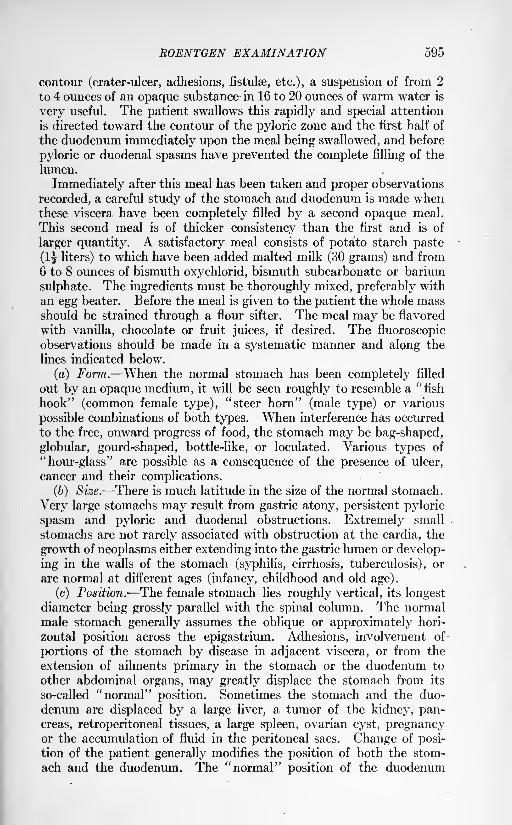
ROENTGEN EXAMINATION 595
contour (crater-ulcer, adhesions, fistulse, etc.), a suspension of from 2
to 4 ounces of an opaque substance in 16 to 20 ounces of warm water is
very useful. The patient swallows this rapidly and special attention
is directed toward the contour of the pyloric zone and the first half of
the duodenum immediately upon the meal being swallowed, and before
pyloric or duodenal spasms have prevented the complete filling of the
lumen.
Immediately after this meal has been taken and proper observations
recorded, a careful study of the stomach and duodenum is made whenthese viscera have been completely filled by a second opaque meal.
This second meal is of thicker consistency than the first and is of
larger quantity. A satisfactory meal consists of potato starch paste
(1^ liters) to which have been added malted milk (30 grams) and from
6 to 8 ounces of bismuth oxychlorid, bismuth subcarbonate or barium
sulphate. The ingredients must be thoroughly mixed, preferably with
an egg beater. Before the meal is given to the patient the whole massshould be strained through a flour sifter. The meal may be flavored
with vanilla, chocolate or fruit juices, if desired. The fluoroscopicobservations should be made in a systematic manner and along the
lines indicated below.
(a) Form. When the normal stomach has been completely filled
out by an opaque medium, it will be seen roughly to resemble a "fish
hook" (common female type), "steer horn" (male type) or various
possible combinations of both types. When interference has occurred
to the free, onward progress of food, the stomach may be bag-shaped,
globular, gourd-shaped, bottle-like, or loculated. Various types of
"hour-glass" are possible as a consequence of the presence of ulcer,
cancer and their complications.
(6) Size. There is much latitude in the size of the normal stomach.
Very large stomachs may result from gastric atony, persistent pyloric
spasm and pyloric and duodenal obstructions. Extremely small
stomachs are not rarely associated with obstruction at the cardia, the
growth of neoplasms either extending into the gastric lumen or develop-
ing in the walls of the stomach (syphilis, cirrhosis, tuberculosis), or
are normal at different ages (infancy, childhood and old age).
(c) Position. The female stomach lies roughly vertical, its longestdiameter being grossly parallel with the spinal column. The normal
male stomach generally assumes the oblique or approximately hori-
zontal position across the epigastrium. Adhesions, involvement of
portions of the stomach by disease in adjacent viscera, or from the
extension of ailments primary in the stomach or the duodenum to
other abdominal organs, may greatly displace the stomach from its
so-called "normal" position. Sometimes the stomach and the duo-
denum are displaced by a large liver, a tumor of the kidney, pan-
creas, retroperitoneal tissues, a large spleen, ovarian cyst, pregnancyor the accumulation of fluid in the peritoneal sacs. Change of posi-
tion of the patient generally modifies the position of both the stom-
ach and the duodenum. The "normal" position of the duodenum
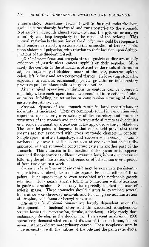
596 SURGICAL DISEASES OF STOMACH AND DUODENUM
varies widely. Sometimes it extends well to the right under the liver,
again it turns sharply backward and runs posterior to the stomach.Not rarely it descends almost vertically from the pylorus, or may goanteriorly and loop irregularly in the region of the pylorus. Thisnormal variation in the position of the duodenum should be recognized,as it renders extremely questionable the association of tender points,
upon abdominal palpation, with relation to their location upon definite
portions of the duodenum itself.
(d) Contour. Persistent irregularities in gastric outline are usuallyevidences of gastric ulcer, cancer, syphilis or their sequelse. Morerarely the contour of the stomach is altered as the result of disease in
adjacent organs: gall bladder, tumors of the liver, pancreas, spleen,
colon, left kidney and retroperitoneal tissues. In low-lying stomachs,
especially in females, occasionally, pelvic growths or inflammatory
processes produce abnormalities in gastric outline.
After surgical operations, variations in contour can be observed,
especially where such operations have consisted in resections of ulcer
or cancer, infolding, cauterization or compression suturing of ulcers,
gastro-enterostomy, etc.
Spasms. Spasms of the stomach result in local constrictions or
indentations (incisurse). They are commonly found in association with
superficial open ulcers, over-activity of the secretory and muscular
structures of the stomach and such extragastric ailments as duodenitis
or chronic inflammatory alterations in the appendix or the gall-bladder.
The essential point in diagnosis is that one should prove that these
spasms are not associated with gross anatomic changes in contour.
Simple spasm is often transitory, and successive fluoroscopic exami-
nations may prove that the spasm seen at one examination has dis-
appeared, or that spasmodic contracture exists in another part of the
stomach. This variation in the location of the spasm or its appear-ance and disappearance at different examinations, is best demonstrated
following the administration of atropine or of belladonna over a periodof from two days to a week.
Spasm at the pylorus or at the cardia may be extremely persistent
so persistent as closely to simulate organic lesion at either of these
points. Such spasm may be even associated with noticeable gastric
retention. It is nearly always found in connection with alterations
in gastric peristalsis. Such may be especially marked in cases of
pyloric spasm. These stomachs should always be examined several
times at two- or three-day intervals and following the administration
of atropine, belladonna or benzyl benzoate.
Alterations in duodenal contour are largely dependent upon the
development of duodenal ulcer, and its associated complications
(crater formation, penetration, fistulse, adhesions). Only rarely does
malignancy develop in the duodenum. In a recent analysis of 1200
operatively demonstrated cases of disease of the duodenum, in but
seven instances did we note primary cancer. These neoplasms were in
close association with the orifices of the bile and the pancreatic ducts.
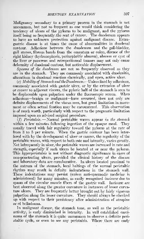
ROENTGEN EXAMINATION 597
Malignancy secondary to a primary process in the stomach is not
uncommon, but not so frequent as one would think considering the
tendency of ulcers of the pylorus to be malignant, and the pylorusitself being so frequently. the seat of cancer. The duodenum appearsto have an unknown protection against malignant disease. Extra-
gastric disease is at times the cause of abnormalities in duodenal
contour. Adhesions between the duodenum and the gall-bladder,
gall stones, fibrous bands from the omentum or colon, disease of the
right kidney (hydronephosis, perinephritic abscess), abscess or cyst of
the liver or pancreas and retroperitoneal tumors may not only cause
deformity of duodenal contour, but noticeable displacement.
Spasms of the duodenum are not so frequently observed as theyare in the stomach. They are commonly associated with duodenitis,
alterations in duodenal reaction chemically, and open, active ulcer.
(e) Mobility of Stomach and the Duodenum. Unless fixed by adhesions,
commonly associated with gastric ulcer, or by the extension of ulcer
or cancer to adjacent viscera, the pyloric half of the stomach is seen to
be displaceable upon palpation under the fluoroscopic screen. Whenadhesions benign or malignant have occurred, then not only are
definite displacements of the viscus seen, but great limitation in move-ment or often actual fixation may be encountered. This observation
is of much worth, particularly with respect to the prognostic limitation
imposed upon an advised surgical procedure.
(/) Peristalsis. Normal peristaltic waves appear in the stomach
within a few minutes following ingestion of the opaque meal. Theyusually travel with fair regularity toward the pylorus at the rate of
from 3 to 5 per minute. When the gastric contour has been inter-
fered with by the development of ulcer or cancer, the regularity of the
peristaltic waves, with respect to both rate and intensity, varies greatly.
Not infrequently in ulcer, the peristaltic waves are increased in rate and
strength, especially if such ulcers be located at or near the pylorus.This hyperperistalsis is not without diagnostic significance in cases of
non-penetrating ulcers, provided the clinical history of the disease
and laboratory data are corroborative. In ulcers located proximal to
the antrum of the stomach, local baitings of the normal peristaltic
rhythm may result in definite indentations in the stomach wall.
These indentations may persist (unless anti-spasmodic medicine is
administered) for many minutes, as easily recognized incisurce due to
spasm of the circular muscle fibers of the gastric wall. Incisurse are
best observed along the greater curvature in instances of lesser curva-
ture ulcers. They are frequently better brought out by fairly vigorous
palpation along the lesser curvature. They should always be checked
up with respect to their persistency after administration of atropineor of belladonna.
In malignant disease, the stomach tone, as wrell as the peristaltic
activity, is early diminished in intensity. In well established carci-
noma of the stomach it is quite uncommon to observe a definite peri-staltic cycle, or even to see any peristalsis. Where there are early
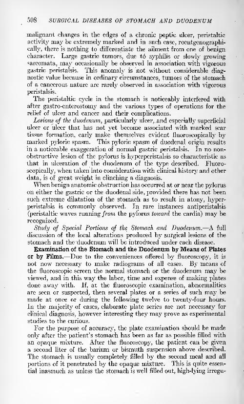
598 SURGICAL DISEASES Of STOMACH AND DUODENUM
malignant changes in the edges of a chronic peptic ulcer, peristaltic
activity may be extremely marked and in such case, roentgenographi-cally, there is nothing to differentiate the ailment from one of benigncharacter. Large gastric tumors, due t6 syphilis or slowly growingsarcomata, may occasionally be observed in association with vigorous
gastric peristalsis. This anomaly is not without considerable diag-nostic value because in ordinary circumstances, tumors of the stomachof a cancerous nature are rarely observed in association with vigorous
peristalsis.
The peristaltic cycle in the stomach is noticeably interfered withafter gastro-enterostomy and the various types of operations for the
relief of ulcer and cancer and their complications.Lesions of the duodenum, particularly ulcer, and especially superficial
ulcer or ulcer that has not yet become associated with marked scar
tissue formation, early make themselves evident fluoroscopically bymarked pyloric spasm. This pyloric spasm of duodenal origin results
in a noticeable exaggeration of normal gastric peristalsis. In no non-
obstructive lesion of the pylorus is hyperperistalsis so characteristic as
that in ulceration of the duodenum of the type described. Fluoro-
scopically, when taken into consideration with clinical history and other
data, is of great weight in clinching a diagnosis.When benign anatomic obstruction has occurred at or near the pylorus
on either the gastric or the duodenal side, provided there has not been
such extreme dilatation of the stomach as to result in atony, hyper-
peristalsis is commonly observed. In rare instances antiperistalsis
(peristaltic waves running from the pylorus toward the cardia) may be
recognized.
Study of Special Portions of the Stomach and Duodenum. A full
discussion of the local alterations produced by surgical lesions of the
stomach and the duodenum will be introduced under each disease.
Examination of the Stomach and the Duodenum by Means of Plates
or by Films. Due to the conveniences offered by fluoroscopy, it is
not now necessary to make radiograms of all cases. By means of
the fluoroscopic screen the normal stomach or the duodenum may be
viewed, and in this way the labor, time and expense of making platesdone away with. If, at the fluoroscopic examination, abnormalities
are seen or suspected, then several plates or a series of such may be
made at once or during the following twelve to twenty-four hours.
In the majority of cases, elaborate plate series are not necessary for
clinical diagnosis, however interesting they may prove as experimentalstudies to the curious.
For the purpose of accuracy, the plate examination should be made
only after the patient's stomach has been as far as possible filled with
an opaque mixture. After the fluoroscopy, the patient can be givena second liter of the barium or bismuth suspension above described.
The stomach is usually completely filled by the second meal and all
portions of it penetrated by the opaque mixture. This is quite essen-
tial inasmuch as unless the stomach is well filled out, high-lying irregu-

CONGENITAL ANOMALIES OF THE STOMACH 599
larities, due to ulcer, tumor or abnormalities in adjacent viscera, mayescape notice by not being visualized on the roentgen plate. The plateshould be exposed at once and at desired intervals after swallowingthe meal.
Position of Patient During Plate Examination. Opinions differ.
Some actinologists prefer making plates with the patient standing,while others prefer to make the exposure with the patient lying face
downward on the plate. Probably plates made in both positions will
give the greatest amount of information. For purposes of special
study, the patient may be placed in either the right or left lateral, or
in the Trendelenburg positions.
If plate series are made, then the patient should be instructed to
eat nothing, or very little, during the entire period covered by the
observations.
Interpretation of Roentgen Plates. On all patients, interpretationsshould be made by individuals who have knowledge of how the sub-
ject was prepared, of how the plates were made, and who has morethan a theoretic acquaintance with clinical medicine and surgical
pathology. If plates are interpreted after this fashion, then there
will occur the minimum of error. While the roentgenographic interpre-
tation should be as nearly as possible objective, yet the ability to trans-
late roentgen abnormalities into clinical terms renders the actinologist
a more useful aid to the clinician.
In general, roentgen plates demonstrate alterations from the normal
shape, position, size, contour and peristalsis of the stomach andthe duodenum, and also give indications respecting emptying power,relations to adjoining viscera and alterations that have been broughtabout as the result of previous operations. In the following sections,
consideration of these variations from the normal in disease will be
specified.
PART II.
CONGENITAL ANOMALIES OF THE STOMACH.
Atresia. Instances of absent gastric lumen are very rare. In
such cases, there is commonly imperfect formation of esophagus, duo-
denum and jejunum. The stomach is represented by a thick fibrous
cord, which, histologically examined, may reveal the normal layers of
the gastric wall, with exception of the mucosa. This is generally
incompletely developed. Specimens showing extremely small stomachlumens are occasionally described. In Heller's case the cavity of the
stomach held scarcely a hen's egg. Congenital strictures of the stomach
may occur. The cavity of the viscus may be separated into several
chambers usually of small capacity. Diverticulum-like pouches some-
times result and persist through life. Atresias are commonly found in
the pyloric half of the stomach. Little and Helmholz report such a
600 SURGICAL DISEASES OF STOMACH AND DUODENUM
case in detail. A mere cord may connect the body of the stomach withthe small gut.
Clinical Symptoms. Infants with pronounced atresias are commonlyeither born dead or survive but a brief time after birth. They suc-
cumb from starvation. Vomiting, constipation, diminished urine
output and emaciation suggest lesions of an obstructive type. Thevomitus or lavage fluid may be free from bile coloring. Digestive
enzymes or hydrochloric acid are commonly reduced or absent.
Obstruction to the free passage of a small stomach-tube may be encoun-
tered. Gastric lavage may demonstrate contents retained from pre-vious feedings in atypic loculi. Lanugo hairs may be absent from the
stools of the young infant. Roentgen examination graphically reveals
the shape, size, position and patency of the altered viscus. The stom-
ach may be filled with the opaque meal through a large catheter or a
small-caliber stomach-tube.
Prognosis. The prognosis depends upon the causes of the atresia,
its degree and the general condition of the patient.Contracted Stomach (Angustatio Ventriculi). The stomach cavity
may hold but a few ounces. Both secretory and motor functions
may be normal. Congenital stricture of the esophagus, failure of the
gastric segment of endoderm fully to develop or insufficient generalnourishment of the infant may be responsible for the anomaly.
Symptoms. Clinically, the symptoms and signs are not characteristic.
The routine feeding of normal quantities of food may produce obstinate
regurgitation and vomiting. The infant fails to gain in weight.
Emaciation, with dry skin and mucous surfaces, follows. Constipationand small urine output are common. Roentgen examination, when the
stomach has been filled with a barium or bismuth mixture, shows the
nature of the affection.
Prognosis. The prognosis is rarely bright with respect to the future
development of a lusty child. Under proper feeding the capacity of
the stomach sometimes can be increased. As a life-saving measure
gastrojejunostomy or gastrostomy may be indicated.
Pyloric Stenosis in Infancy ("Congenital Hypertrophic Pyloric
Stenosis"). Definition. A gastric affection of the new-born, charac-
terized by the presence of a stenosing, pyloric tumor, comprised mainlyof hypertrophied muscle tissue. Secondarily, there occur dilatation
of the stomach, obstructive vomiting, rapid emaciation, diminution of
body fluids, anemia and, unless the pyloric stenosis be early relieved,
death from starvation.
Etiology . Age. The child is born with a narrowed gastric outlet.
Symptoms appear just so soon as mouth feeding is begun. The sexes
are equally affected. Other evidences of congenitally anomalous
development may be not lacking. Harelip, cleft palate, club-foot,
imperforate anus and the like have been described in association with
the pyloric fault.
Genesis of the Stenosis. Scudder has emphasized that the pyloric
tumor represents a congenital, local overgrowth of muscle tissue.
CONGENITAL ANOMALIES OF THE STOMACH 601
Inasmuch as the pyloric musculature is differentiated before the fourth
month of fetal life, it is conceivable that sufficient time elapses before
birth for this group of muscle bundles to assume the proportions of a
definite tumor previous to the initiation of gastric function. Denthas described such a tumor in a seven months' fetus. Its structure waslike that found in cases of hypertrophic stenosis examined after birth.
Just what underlying causes bring about such myomatoid change in
the pyloric muscle tissue of the fetus, is not known.
Pathology. A tumor composed mainly of dense, firm, circular muscle
fibers involves the pyloric sphincter. According to Scudder the result-
ant nodule is about the size of the terminal phalanx of an adult fingeror thumb. There are no external evidences of inflammatory reaction;
the pylorus is free from adhesions. The hypertrophied muscle tissue
narrows the lumen of the pyloric channel. It may cause complete
obstruction, particularly when there is associated extensive hyper-
trophy of the longitudinal folds of mucous membrane.The stomach wall may early appear thickened. It later becomes
stretched, thin and atonic. The intestines are commonly empty or
poorly filled, their peritoneal surfaces, dry and shiny. The esophagus
may be dilated.
Histologically, the pyloric tumor is shown to be made up chiefly
of closely packed, hypertrophied bundles of muscle fibers, enclosed in
dense, fibrous envelopes. The mucosa is thickened and aberrant.
Brunner's glands are sometimes recognized.
Symptoms. Unless other gross congenital anomalies exist, the infant
appears normal at birth. Appetite may be normal or capricious. In
some cases it is ravenous. Shortly after birth it is noted that a usual
feeding routine provokes vomiting. The vomiting comes on soon after
the meal. It commonly follows every feeding and it is evident that
feeding produces it. In no ailment are cause and effect more definitely
emphasized. At first, vomiting may be sudden and projectile, the
contents of the stomach sometimes being thrown several feet. Atthis time vomitus consists of the food swallowed but a few minutes
previously. Later, where obstruction is pronounced and when gastric
dilatation and muscular atony have become marked, vomitus is not
projectile and retained food (tough milk curds, etc.) is vomited or
washed from the stomach. The vomitus is commonly bile-free, has a
sour odor, may be hyperacid and contain an excess of mucus.
Pain. Until gastric atony has occurred, the infant manifests distress
before and during emesis by cries and grimaces. Pain is produced bypowerful hyperactive gastric peristalsis. When the stomach is empty,the child is comparatively comfortable, although it may be hungry.When gastric atony and dilatation have taken place, starvation andits resultant weakness render the child listless, cLrowsy and apathetic.
Constipation is pronounced. The stools are of small volume, dark
in color and nearly or entirely food-free.
Urine output is diminished. The urine is of high color and deeplystains the diapers.
602 SURGICAL DISEASES OF STOMACH AND DUODENUM
Signs. Weight loss is commonly progressive and pronounced.Emaciation may become startling. The skin is pale, dusky, dry and
inelastic; the hair lusterless and brittle. The mucosse are pale, dry,
scaly and often fissured. Panniculus disappears early. The muscles
become small, soft and flabby. Lack of body fluids is seen in all
tissues provided rectal alimentation has not been instituted.
Abdominal Tumor. When the infant is stripped, visible peristaltic
waves may be seen to pass rhythmically across the epigastrium from
left to right. During, or shortly following feeding, these waves are
pronounced, particularly in the early stages of the affection when
gastric musculature is still capable of powerful contractions. As the
waves approach the right upper abdominal quadrant, a definite tumor-
like bulging may be seen. This is best observed when the belly is
viewed with the eye at its level. Palpation frequently reveals a small,
firm, movable tumor above the navel and toward the right rib margin.This tumor is best felt as the peristaltic waves pile up toward the
pylorus. The pylorus is at that time rotated downward and forward.
Inflation of the empty stomach by means of a double-bulb, Davidson
syringe, through a small-calibered stomach-tube, occasionally permitsthe palpation of a tumor that lies up under the right ribs or the liver.
Examination of the belly after the patient has lain in a tub of water
at 100 to 105 F. for ten or fifteen minutes further facilitates the
palpation of the tumor. This maneuver also aids in demonstratingthe movability of the pyloric nodule. At such time the tumor maybe grasped readily between the fingers.
Carmin Test. Two grains of carmin may be suspended in a half
ounce of hot, sweetened water and given the infant by mouth. If no
red coloration appears in the stools, pyloric obstruction is complete.In cases of partial stenosis, the stools may be but faintly colored or the
colored stool may not be seen for as long as thirty-six to seventy-twohours.
Roentgen Examination. When the stomach is filled with an opaquemixture (barium, bismuth) fluoroscopic examination discloses a dilated
stomach, hyperactive early in the course of the ailment, but atonic
in the late stages; opaque-meal residue for longer than six hours; well
defined, ring-like, filling defect at the pylorus; imperfect or abserft
visualization of the duodenal bulb and poorly filled or empty small
or large gut. The stomach can be shown to be freely movable.
Copious regurgitation or spasmodic squirting of the opaque-meal into
the esophagus may be striking. Roentgenograms furnish a convenient
graphic record of the major fluoroscopic observations.
Diagnosis. When a newly born infant constantly vomits its food
soon after eating, hypertrophic stenosis of the pyloric must be con-
sidered to exist until definitely proved to be absent. If, as Scudder
emphasizes, the obstructive vomiting is associated with palpable,
epigastric tumor, visable gastric peristalsis, dilatation of the stomach,
progressive weight loss, emaciation, constipation, mecomum-like
stools, deficient urine output, absent carmin coloration or barium or
CONGENITAL ANOMALIES Of THE STOMACH 603
bismuth crystals in the stools and the roentgen proof of stenosis at the
pylorus, diagnosis amounts to almost a certainty.
Pyloric spasm may occur in infants who have been improperly fed.
It rarely complicates true hypertrophic pyloric stenosis. Pyloric
spasm usually comes on weeks after birth, whereas stenosis in infants
manifests itself from birth. In pyloric spasm the picture is a variable
one: feeding causes vomiting intermittently; the vomitus is rarely con-
stantly projectile; not all the meal eaten is vomited; the stools contain
considerable partly digested food; carmin and bismuth crystals are
readily detected in the feces; palpable epigastric tumor is seldomdefinite and constant; visible peristalsis is poorly determined; the
roentgen examination shows a normal pyloric outline and the pyloriclumen is patent; all symptoms and signs may cease following the ad-
ministration of belladonna, bromides or proper diet. The infant may,but rarely does, die of starvation.
Intussusception, volvulus, appendicitis, infectious ailments, cere-
brospinal affections, intestinal parasites, nephritis or indigestion due to
improper feeding may cause vomiting in early life. The history of these
ailments differs markedly from that of congenital, pyloric stenosis.
Such affections rarely appear in the first few days of life. Their
course, clinically, the character of the vomitus, the appearance of the
stools and the roentgen observations usually serve readily to differen-
tiate them from hypertrophic stenosis. Cases occasionally prove
puzzling, but careful anamneses, physical examinations, and clinical
observation over a few days generally establish their diagnoses.
Prognosis. Congenital hypertrophic stenosis of the pylorus rapidlycauses death unless the obstruction be removed or gastro-enterostomyis performed.
Congenital Enlargement of the Stomach. Congenital enlargment of
the stomach is very rare. It represents a relative overdevelopmentof the endodermal anlage, probably as a consequence of an anomalyin innervation and circulation. Occasionally, cicatricial adhesions
forming, or pressure, as result of unusual position of the fetus in intra-
uterine life, would appear to cause dilatation proximal to that portionof the gastric lumen stenosed. Soon after birth, the initiation of feed-
ing produces dilatation of the weakened stomach walls.
Diagnosis. It is uncommon for such to be made previous to lapa-
rotomy or autopsy. The symptoms are those of obstruction : vomiting,
constipation, lessened urine output, possibly visible abdominal peristalsis
and emaciation. The roentgen examination demonstrates the abnor-
mally large stomach.
Prognosis, Death ensues from starvation and exhaustion, unless
surgical procedures or, in rare cases, careful dietetic regimen, are pos-sible.
Congenital Faulty Position of the Stomach. Situs Inversux.
The position of all the abdominal viscera may be reversed. Thefundus of the stomach may lie wholly or partially to the right of the
midline, the pylorus being directed to the left (Fig. 252). A full
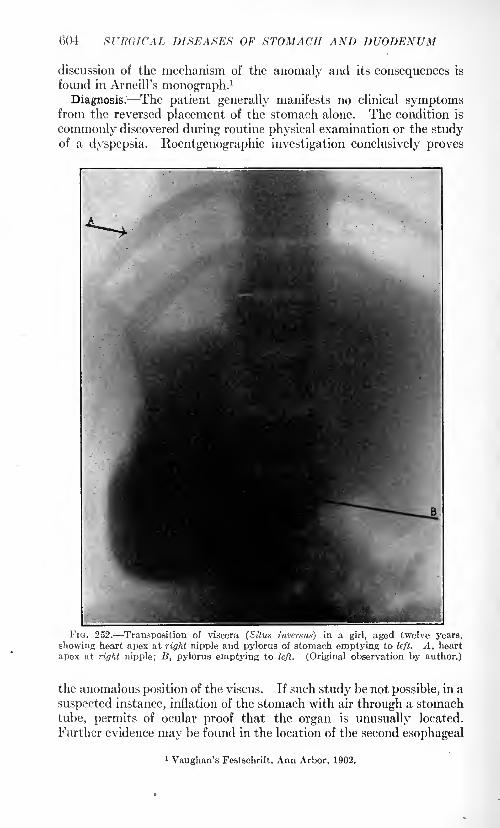
604 SURGICAL DISEASES OF STOMACH AND DUODENUM
discussion of the mechanism of the anomaly and its consequences is
found in Arneill's monograph.1
Diagnosis. The patient generally manifests no clinical symptomsfrom the reversed placement of the stomach alone. The condition is
commonly discovered during routine physical examination or the studyof a dyspepsia. Roentgenographic investigation conclusively proves
FIG. 252. Transposition of viscera (Situs inversus) in a girl, aged twelve years,
showing heart apex at right nipple and pylorus of stomach emptying to left. A, heart
apex at right nipple; B, pylorus emptying to left. (Original observation by author.)
the anomalous position of the viscus. If such study be not possible, in a
suspected instance, inflation of the stomach with air through a stomach
tube, permits of ocular proof that the organ is unusually located.
Further evidence may be found in the location of the second esophageal
1 Vaughan's Festschrift, Ann Arbor, 1902.
CONGENITAL ANOMALIES OF THE STOMACH 605
swallowing murmur to the right of the vertebral column and escape of
gas or fluid from a pylorus located to the left of the midabdominal line.
Prognosis. Situs inversus is not of itself incompatible with perfecthealth. Associated lesions (ulcer, cancer, stenoses, etc.) determine the
outcome.
Congenital Elevation of the Diaphragm or"Eventration," commonly
results in the stomach occupying a wholly or partially"intrathoracic"
position. The condition is important, clinically, chiefly on account of
the readiness with which the anomaly is confused with congenitalor acquired hernia of the stomach through an imperfectly developedor an accidentally injured diaphragm. The condition has been care-
fully described by Glaser, Leichtenstein and by Sailer and Rhein. About25 authentic cases are recorded in the literature.
Etiology and Morbid Anatomy. Meckel mentions an instance in a
female fetus. Thoma described the findings at autopsy in a man of
seventy-five. It is thus apparent that age is but of relative signifi-
cance in grouping the cases. Males appear to be about twice as fre-
quently affected as are females. In two-thirds of the patients diagnosiswas not suspected during life. Heredity does not appear to havedefinite significance. Other congenital anomalies may coexist: viz.,
lack of proper development of the left lung, atypically shaped pan-creas, deficiency in development of the muscles of the left side of the
body, particularly the diaphragm, etc.
Anatomic Variations. The left half of the diaphragm is commonlyinvolved. It is abnormally high-placed, congenitally. Its dome mayreach to the level of the third rib. Its continuity is unbroken. Thereis frequently an associated displacement of the heart toward the rightand a hypoplasia of the left lung. The muscle layers of the left
diaphragm are sometimes thin and their bundle-formation fragmented.The stomach and adjacent viscera may lie high in the thorax. Theydo not, however, protrude through the diaphragm into the thoracic
cavity. The respiratory excursion of the left half of the diaphragmmay be limited.
Clinical Symptoms and Signs. Nothing abnormal may be noted
during life. Affections of the lungs, heart or abdominal viscera com-
monly lead to examinations at which unusual anatomic placementsof organs are noted. Operative procedures may be performed onthe supposition that the high position of hollow viscera, the stomachor the colon, indicate hernia through the left half of the diaphragm.
The symptoms of an associated gastric lesion (ulcer, cancer, etc.)
may be anomalously referred to the left thoracic region. Gaseousdistention of the stomach with pressure upon the heart, dyspnea, thoracic
gurgling or pain, dysphagia, constipation a'nd occasionally, obstinate
vomiting, have been described.
Signs of the affection are not lacking in well marked instances.
Percussion of the upper part of the left lung may be normal, but belowthe third or the fourth rib tympany may be striking. The heart is
pushed to the right. Litten's sign is absent or limited on the left side.
606 SURGICAL DISEASES OF STOMACH AND DUODENUM
The area of thoracic tympany may be displaced downward by deep
inspiration. Auscultation over the left lower thorax reveals splashingand -gurgling sounds, particularly if the patient is shaken or he swallows
fluid during the examination. Amphoric voice tone and the "coin
sound" are absent over the tympanitic thoracic zone. If the stomachbe filled with solid food or fluid the area of thoracic tympany gives
place to one of dulness. Splenic dulness may not be outlined. Whena stomach-tube is passed it commonly enters the viscus at less than
30 cm. from the teeth. Inflation of the stomach through a tube maycause left thoracic bulging, dyspnea, added cardiac displacement,increased zone of thoracic tympany and auscultatory phenomena in
the left thorax.
When the patient is fluoroscoped, normal diaphragm contours maybe seen on both sides. On the left side, the diaphragm is seen to
occupy an abnormally high position. During respiration its move-ments may be synchronous with those on the right side but its
excursions are usually less rapid and complete. Below the high left
diaphragm, the gas bubble in the stomach, gas in the splenic loop of
the colon or the shadow of a high-lying spleen may be noted. When an
opaque mixture of barium or bismuth is swallowed, displacement of
the esophagus, anomalously located cardiac orifice and a "thoracic"
lying stomach are demonstrated. Roentgenograms furnish interesting
graphic records of the unusual locations of the abdominal and thoracic
organs.
Differential Diagnosis. Eventration of the diaphragm with con-
genitally high-placed stomach is rare. It is most important to dif-
ferentiate the condition from congenital or acquired diaphragmatichernia. The latter affection is potentially if not actually surgical;
eventration is surgical only accordingly as anomalously placed viscera
become diseased.
Diaphragmatic Hernia. Congenital. In such instance the dia-
phragm fails to properly develop, usually on one side. Occasionallythe lack of development is symmetric and, as a consequence, the nor-
mal openings in the diaphragm are of greater size than when develop-ment has been complete. Usually, when there is improper formation
of the diaphragm, the overlying peritoneal layers are absent. It thus
follows that congenital hernias of abdominal viscera through the dia-
phragm are not true hernias, because the viscera are not enclosed
in the peritoneal sac.
Congenital hernia of the stomach through the diaphragm is rare,
but a few authentic cases are on record. An instance has recently
been described by Seymour Baasch. In this case, there were no gastric
symptoms or any evidences of disability until the patient reached adult
life. At that time the symptoms were those of chronic obstruction.
The obstruction was clinically similar to that caused by ulcer of the
stomach producing hour-glass. The gastric analyses pointed toward
peptic ulcer and the axray examination revealed an atypical hour-glass
stomach, the pyloric end of which seemed displaced upward into the
CONGENITAL ANOMALIES OF THE STOMACH 607
right lower thoracic cavity. Exploratory laparotomy disclosed the true
nature of the affection. The patient died within thirty-six hours after
an attempt to relieve the hernia.
Acquired Hernia of the Stomach through the Diaghragm. While
uncommon, it is of sufficient frequency at large surgical clinics to be of
considerable interest. Instances of the affection have been reportedand described by Beckman, Giffin and by Sailer and Rhein. The lesion
is usually on the left side, but occasionally the stomach has been found
in the right side of the thorax. About 650 cases of diaphragmatichernia have been reported but in relatively few of them has the stomachbeen herniated.
The clinical history usually points to severe trauma initiating the
hernia. Such traumata are commonly a severe blow to the upperabdomen or lower thorax, a sudden twisting, a stab wound, or a doub-
ling-up or crushing injury. According to Giffin, the acute manifesta-
tions of the anomaly are sudden, severe, abdominal or thoracic painwith or without hematemesis. There is usually pronounced dyspneaand symptoms of intestinal obstruction soon follow.
If the causative trauma does not immediately bring about a fatal
issue, the condition of hernia of the stomach through the diaphragmmay become chronic. In such event the symptomatology may closely
resemble that of eventration of the diaphragm. Sometimes there are
no symptoms but the hernia of the stomach into the thoracic cage maybe accidentally discovered in the course of routine examination or
during surgical procedures. The majority of cases, however, exhibit
attacks of abdominal pain, vomiting, dyspepsia with or without
hematemesis and occasionally, periodic dyspnea.
Pneumothorax, hydropneumothorax, tuberculous cavity formation
in the lung, subdiaphragmatic pneumothorax, pyloric stenosis, or dis-
ease of the gall-bladder may at times simulate diaphragmatic hernia.
These conditions may be difficult to differentiate unless emphasis be
placed upon careful anamnesis, physical examination and roentgen
study.
Physical Signs. In diaphragmatic hernia, Litten's sign may be
greatly diminished or absent on the affected side usually the left.
On percussion of the left thorax, tympanitic note may be demonstrated
as high as the fourth rib when the stomach is empty, but when the
stomach is filled there may be an anomalous area of dulness ascending
high into the left thorax. If the stomach is inflated by means of a
Seidlitz powder, thoracic tympany and hissing, splashing or gurglingsounds may be heard practically all over the left thorax. The signs
may differ when the patient's position is changed. Dyspnea may be
pronounced, particularly in cases of acute herniation.
If a stomach tube be passed, gastric content may be obtained whenthe tube is at but a short distance (15 to 20 cm.) from the teeth.
X-ray Evidences of Gastric Herniation through the Diaphragm. Fluo-
roscopic examination may reveal a stomach gas-bubble lying above the
line of shadow cast by the diaphragm. This gas-bubble may move up
608 SURGICAL DISEASES OF STOMACH AND DUODENUM
and down during respiratory movement and may change in size at
different examinations. The diaphragm contour may be definitelyseen to be broken. The diaphragm may apparently lie higher than nor-
mally and the movements during respiration may be limited. A para-doxical expiratory displacement of the diaphragm has been described.
In such event, during deep inspiration, the unaffected diaphragmdescends normally while the diaphragm on the affected side ascends.
During forced expiration, the unaffected diaphragm ascends normally,while the diaphragm on the affected side descends. The rate of excursion
of the diaphragm on the two sides may differ.
When the stomach is filled with an opaque medium, all or part of
the stomach-shadow may lie above the shadow cast by the diaphragmon the affected side. There is usually deformity in the stomach con-
tour: this may frequently amount to actual hour-glass constriction.
A greatly dilated stomach, which has herniated through the left dia-
phragm, may displace the heart to the right and may cause partial
collapse of the lower left lung. Usually, however, cardiac displacementis not great and at the fluoroscopic examination, normal lung tissue
may be observed through the gas-bubble distending the upper partof the stomach-shadow. When the stomach is not entirely filled by the
opaque meal, at fluoroscopic examination, palpation of the stomach
may enable one to demonstrate fluctuation waves in the stomach lyingat a level higher than the diaphragm-shadow on the affected side.
When, in addition to the hernia of the stomach through the dia-
phragm, there is also hernia of the large or small gut or the spleen, then
the fluoroscopic signs are complicated. Inflation of the stomach or the
colon alternately with air may reveal definite differences in the size
and shape of the gas-bubbles lying in the thoracic cavity, above the
diaphragm shadow on the affected side. In such event, the physical
signs upon auscultation and percussion vary greatly.
It is important to recognize hernia of the stomach through the
diaphragm because this condition is amenable to surgical treatment.
As has been emphasized above, it is particularly desirable to differ-
entiate the affection from congenitally high-lying diaphragm on one
side (so-called "eventration"), which is never a surgical condition
unless it be that the highly placed diaphragm is also the site of hernia.
GASTROPTOSIS.
Definition. An anomaly associated with an abnormally low posi-
tion of the stomach, particularly of the pyloric third of the viscus.
Gastroptosis is doubtless most commonly congenital, but, as a conse-
quence of incorrect dress, posture, habits or of infectious or constitu-
tional ailments, unusually low position of the stomach may be acquired.
Congenitally low stomachs may functionate normally throughoutlife. Discover^' of the anomaly may be accidental. Intercurrent
disease, may, however, produce disability in a low-lying stomach,whereas such intercurrent disease might cause little or no incon-
GASTROPTOSIS 609
venience in a stomach in so-called normal position. When gastroptosishas resulted from faulty modes of living or has been brought about
by a systemic or constitutional disease, then dyspepsia not rarelybecomes evident.
The position of the normal stomach is subject to such wide varia-
tions in individuals who are apparently healthy that it is difficult
to state just what constitutes "gastroptosis." While it is the popularrule to consider all stomachs whose pyloric thirds lie below the level
of the iliac crests as being ptosed, yet the routine examination of large
groups of individuals demonstrates that such positions of stomachs are
by no means unusual or inconvenient. It would seem best to limit
the term gastroptosis to those instances in which the ptosis is a part of
a generally low-lying, gastro-intestinal tract and in which definite signsand symptoms directly referable to such unusual position can beascribed. In general, when the major portion of the stomach andintestines lies in the pelvis, inconveniences amounting to actual mal-
function frequently arise. It is surprising, however, in how many such
cases of markedly low-lying viscera there occurs no anomaly in secretion,
motility or assimilation.
Gastroptosis is more common in females than in males. In a recent
study of the position of the stomach in 600 males and females (300
each), gastroptosis was noted in but 36 males while it was present in
162 females. These figures would seem to indicate that the low-lyingstomach is almost a normal event in females. While modes of dress,
child bearing, lack of exercise and frequently insufficient nourishment
may be factors in producing low gastric position in females, yet such
explanation is by no means sufficiently comprehensive.Clinical Symptoms and Signs. As briefly hinted above, marked
gastroptosis may occur without manifesting itself in any way, clin-
ically. While low position of the stomach may be proved, this low
position is not of itself indicative of any upset. In some groups of
cases in which stomach ptosis exists, chronic dyspepsia is manifested.
Patients frequently attribute the onset of this dyspepsia to a "nervous
breakdown," a recent illness, childbirth, an injury or prolonged mentalor physical stress.
Symptomatically, there are not infrequently sensations of discomfort
in the region of the stomach. These rarely amount to actual, clean-cut
pain. The distress is most commonly pronounced shortly after food
ingestion. The discomfort is variously described as "dragging" or
"fulness" or "pressure" or a vague soreness. Colicky pains are
unusual. When the stomach is empty, abdominal discomfort is gen-
erally slight, although individuals of the nervous type not rarely are
annoyed by burning or aching sensations.
Nausea and vomiting are quite regularly experienced in markedinstances of the affection. Vomiting may come on shortly after food
ingestion, but morning vomiting with nausea may be annoying. Inthis gr6up of cases, the vomitus may contain retained food, strings or
gobs of mucus and bile, more or less altered. Morning vomiting is
VOL. n 39
610 SURGICAL DISEASES OF STOMACH AND DUODENUM
sometimes copious and associated with troublesome "gagging."
In youngindividuals, eructations, pyrosis or water-brash may be particularly
distressing.
Appetite is usually poor or capricious. Malnutrition may be of par-
ticularly severe degree so marked as to lead to the clinical suspicionof malignancy especially in people at the so-called "cancer age."This malnutrition can quite commonly be attributed to insufficient
ingestion of food and liquids. Weakness and weight-loss are some-tunes striking. Patients may be actually bed-ridden. Constipation is
the rule. There may be diminution in urine output. The urine is
often high-colored and rich in uric acid, phosphates and substances of
the indican group.
Signs of Gastroptosis. Physically, many patients with low-lyingstomachs are of slender build. Their bones are apt to be small and their
posture incorrect. When such patients stand erect, flat foot may be
noted, there may be a drooping of the shoulders, flattening out of the
normal lumbar curve of the spine, a narrowing of the epigastric angle,and not infrequently a definite, transverse crease in the abdominalmuscles at the level of the navel. While the epigastrium may be flat,
it is not uncommon to observe a globular bulging of the abdomenbelow the navel. This bulging may be very noticeable. In some
instances, owing to the thinness of the abdominal muscles, practicallythe entire stomach, together with portions of the large and small intes-
tines, may be well visualized. Occasionally, peristaltic waves are seen
in both stomach and intestines. The patients appear starved, as a rule,
the skin and hair are apt to be dry, the panniculus is thin and the
muscles soft and flabby. This muscular weakness is apt to be most
pronounced in the anterior abdominal group.Abdominal palpation generally demonstrates a lessening of the muscle
tone, a dilated, atonic and frequently splashy stomach, areas of distress
over the stomach or bowel and not infrequently, points of tenderness
over the gall-bladder or appendix regions.Test meals returns show eight hour food stagnation as constant in
about 3 per cent, of patients. In approximately 11 per cent, inter-
mittent, eight hour retention can be demonstrated. Bile staining of
gastric extracts is not uncommon where an atonic stomach with
relaxed pylorus exists. Sometimes excessive amounts of mucus are
present in the wash water. The gastric acidity varies greatly not onlyin groups of cases, but, frequently, in the same individual examined at
different times. In young patients, increases in the free hydrochloricacid are apt to be recorded, but where patients are past midlife, free
hydrochloric acid may be low or absent. When the stomach is inflated
with air through a stomach-tube, by means of a double-bulbed Davidson
syringe, its position together with evidences of its increase in size, andnot uncommonly its lack of muscle tone, can be readily determined.
X-ray Examination. There is no clinical procedure that has created
such unnecessary apprehension among both patients and physicians as
has the ocular proof of the so-called "low-lying stomach." At the same
GASTROPTOSIS 611
time, to physicians of judicious mind and not of faddist mentality, the
radiological studies have proved particularly instructive in teachinghow wide may be the variations in the positions of stomachs and yetin such stomachs no malfunction exist. Gastroptosis has, however,become firmly established as a clinical entity in the minds of the laity
mainly on account of roentgen examinations and the injudicious com-ments upon such by physicians who had certain types of treatment for
this ailment. Many ingenious operations have been devised for the
purpose of lifting and anchoring the so-called ptosed stomach to a
position which according to physiology primers seemed correct, and
many forms of medical regimen have been instituted with a similar
object. With few exceptions the benefits of such treatments have been
more apparent to the physician doing the treating, than to his subject.
Fluoroscopic Examination. Six-hour residue of the opaque meal maybe a variable factor. In instances where there is marked gastric atony,some degree of six-hour retention may be a constant finding.
Ptosed stomachs are generally of the so-called "fish-hook" shape.The lumen is apt to be narrowed but not necessarily so. Both greaterand lesser curvatures are commonly below the level of the navel or
the iliac crests. The gastric contour is usually normal. In someinstances of gastroptosis peristaltic waves are absent or poorly devel-
oped. Many examples of the affection, however, exhibit vigorous
peristalsis. The stomach can be freely moved up and down and from
side to side, by palpatory massage. Sometimes it can be shown that
the pylorus is permanently relaxed, but this should be always corrob-
orated by repeated examinations, inasmuch as the tone of the pylorus
may vary greatly in the same individual at different times. Otherinstances occur in which not only the gastric tone generally but the
tone of the pyloric sphincter is greatly increased. In such affection,
the pyloric spasm may be so pronounced as to result in intermittent
gastric retention and difficulty in filling out the duodenum during fluoro-
scopic examination. The duodenum is commonly well visualized andsometimes is greatly dilated, particularly if part of that gut is involved
in pericholecystitic adhesions. It may then empty slowly. Thesefactors are, however, variable.
Rarely during the fluoroscopic examination can adhesions, fibrous
bands or abnormal position of abdominal viscera other than the stomachbe demonstrated to be the direct cause of the ptosis.
JT-ray plates are of comparatively little value in the clinical estima-
tion of the degree of gastric function existing in the low-lying stomach.
Certainly, a single plate never should furnish evidence upon which
therapeutic procedures are to be based or to demonstrate the effects
of such procedures.Plates made in series generally corroborate fluoroscopic examination
but their value is not so great as is that procedure. Such plates are,
however,- useful in certain instances, in demonstrating extra-gastricconditions that have primarily instituted the systemic disturbance of
which the gastroptosis is a part.
612 SURGICAL DISEASES OF STOMACH AND DUODENUM
GASTRIC HEMORRHAGE. (GASTRORRHAGIA.)
Bleeding from the stomach may be acute and copious or chronic
and in small quantity. Both types of hemorrhage may be continuous
or intermittent, with complete cessation in the intervals. A combina-tion of the forms is possible.
Sudden, copious gastric hemorrhage commonly leads to bloody vomit
(hematemesis) and to blood-stained stools (melena). However, even ex-
tensive gastrorrhagia may be unaccompanied by hematemesis, but mani-fested by melena alone. Chronic seepage of small quantities of blood
is, of itself, rarely productive of hematemesis. While this form of gas-
trorrhagia may give rise to obvious melena, quite commonly the presenceof blood pigment in the stools can be demonstrated only by chemic,
microscopic or spectroscopic tests (so-called "altered" or "occult"
hemorrhage).Etiology. Sudden, copious bleeding from the stomach is dependent
upon accident or is a complication of a previous local, general or
constitutional ailment. It may be so acute and extensive as to prove
rapidly fatal. Moderate or abundant hemorrhages may occur at fre-
quent or infrequent intervals. There is often a peculiar tendency to
recurrence. The quantity of blood lost at each attack may vary
greatly in a given case.
Seepage or "occult" bleeding from the stomach is generally associated
with primary progressive pathological changes in the gastric mucosaor is a secondary, local manifestation of a systemic or constitutional
fault.
In the different types of gastrorrhagia, the histopathological con-
dition of the stomach mucosa varies from simple, capillary congestion,with rupture (or even cell diapedesis) of minute capillary networks,
locally or generally, to extensive loss of surface epithelium with de-
struction of the layers of the stomach wall and the erosion of large
bloodvessels. Either process may begin intramurally, in which event
the mucosa first suffers, or extramurally, when the bloodvessels are
primarily affected. There is a type of gastrorrhagia which periodically
occurs in females, in definite relationship to the menstrual cycle,
where copious hematemesis may be associated with no gross gastric
pathology. In the hematemesis accompanying certain anemias, infec-
tious ailments, acute and chronic systemic toxic states or following
operations upon intra-abdominal viscera other than the stomach. or
the duodenum, there may likewise be no grossly apparent lesion in the
stomach mucosa.
Gastrorrhagia is associated with hematemesis or melena or both as
follows :
(a) In primary gastric disease: acute or chronic gastritis, simple or
phlegmonous, acute gastric erosion, acute or chronic benign ulcer, ulcus
carcinomatosum, carcinoma, sarcoma, syphiloma, tuberculoma, poly-
posis, ulcerated gastric cirrhosis, varicosities or aneurysm of intrinsic
gastric arteries or intramural parasite (echinococcus, nematodes.)
GASTRIC HEMORRHAGE 613
(6) In conjunction with chronic disease of the heart or bloodvessels.
(c) Complicating chronic disease of the liver, gall-bladder or ducts
(cirrhosis, neoplasm, portal thrombosis, Banti's disease, cholelithiasis,
cholecystitis.)
(d) Disease of the spleen (chronic splenitis, leukemia, Gaucher
spleen).
(e) Malfunction of the pancreas (acute and chronic pancreatitis).
(/) Toxemias associated with ailments of the kidney, central nervous
system, thyroid gland, adrenal, hemolymph nodes.
(g) Bacteremias or protozoemias : typhoid fever, smallpox, measles,
malaria, yellow fever, cholera.
(A) Chemic poisons: phosphorus, arsenic, hydrocyanic acid, split-
proteins or their products, burns.
(i) Local injury (1) falls, foreign bodies, blows, crushing injuries or
stab wounds (2) poisons introduced by mouth.
(j) Disordered blood states: hemophilia, purpura, chronic anemia
"secondary" or "pernicious" acute or chronic cholemia.
(k) In connection with certain vague neuropathic states: hysteria,
angioneurotic edema, tabetic crises, epilepsy, progressive paralysis.
(I) Associated with physiological cycles of females: the catamenia
and climacteric.
(m) Following operations upon organs other than the stomach,
particularly when such operations are intra-peritoneal.
(ri) Blood may enter the stomach from a diseased lung, esophagus,
adjacent viscus, (duodenum, jejunum or colon, by fistula) or a nearby,
great vessel.
Morbid Anatomy in Gastrorrhagia. Gross or microscopic alterations
in the stomach wall bear but an irregular relation to the quantity of
blood present in the viscus, vomited or passed by the bowel. Abun-
dant, even fatal, hemorrhage may occur and yet no visible source of
such can be demonstrated in the mucosa. This is apt to be so where
profuse bleeding is associated with infectious disease, cirrhosis of the
liver, surgical operations not on the stomach, hysteria, burns on the
surface of the body, blood dyscrasias or traumata to the abdominal
wall. Commonly, however, gastrorrhagia is found in connection with
some type of peptic ulcer or carcinoma. In such ailments, fissures,
granulating ulcer edges or necrotic tumor areas can be noted in definite
relationship to bloodvessels.
After severe hemorrhage the stomach mucosa appears pale, yellowish-
pink, often with scattered areas of reddish-brown mottling in the
mucosa and the submucosa. Some grade of general edema is present.The mucosa dries rapidly when exposed to the air and then has a
smooth, shiny appearance. The rugse are indefinite and the muscle
wall is flabby and inelastic. When peptic ulcers bleed profusely and
intermittently, local destruction of the stomach wall often associated
with peritoneal adhesions, is common. Of 185 operated gastric ulcers
in our series where gross hemorrhage has been recorded in 78 (42 per
cent.) some grade of protected perforation of the stomach wall was noted.
614 SURGICAL DISEASES OF STOMACH AND DUODENUM
In acute, profusely bleeding ulcers, tissue necrosis may take placeso rapidly that peritoneal protection is impossible and perforation occurs,
even before hematemesis or melena appear. The symptoms and signsof the perforation may mask those of severe hemorrhage. At lapa-
rotomy, the stomach is seen to be filled with clotted blood and food,
while between the coils of the intestines or in the peritoneal sacs maybe found blood-clots, serous fluid, pus or food.
Seepage of blood into the stomach is most frequently noted in con-
nection with granulating ulcer edges, ulcerating, necrosing cancer,
in achylias (primary as result of chronic atrophy of gastric mucosa,or secondary to idiopathic anemias or blood disorders) or in extragastric
malignancy. With exception of the achylias, the mucosa is generally
edematous, congested and bleeds readily on handling. In the achylias,
the mucosa is often thin, pale and smooth. If it is scraped with a knife,
a pale, reddish-brown fluid can be collected, which chemically or
physically, can be proved to contain blood pigment.
Signs and Symptoms. (a) Hematemesis. Sudden vomiting of
large quantities of blood may occur without noticeable premonitory
symptoms or previous evidences of disease. The vomited blood may be
bright-red and fluid. It is commonly mixed with undigested food and
mucus. Clots may be present. They usually occur toward the end of
emesis or when the blood has lam in the stomach for a time before beingvomited. In general, the higher the gastric acidity the smaller and
fewer the clots. When the acidity has previously been low or where
hemorrhage has neutralized free hydrochloric acid, coagula form
relatively quickly. The quantity of blood vomited varies greatly in
different patients or in the same individual on different occasions. Thehematemesis may not represent the extent of the gastric hemorrhage :
a cupful may be vomited and yet the stomach remain filled with free or
partly clotted blood which is later vomited, passed by the bowel or
washed from the stomach. Such condition often leads to a false sense of
security. Clots in the gastric lumen may temporarily stop hemor-
rhage or may mask a gradual, but persistent, bleeding which, subse-
quently, as a result of error in diagnosis or treatment, may prove fatal.
Enormous hematemesis sometimes occurs. In one of our cases, nearly
eight quarts of free and clotted blood were vomited.
Slow seepage of blood rarely precipitates hematemesis. In such
instances vomiting may result from other causes. When this happens,the vomitus is colored reddish-tan, brown or even black. In
advanced gastric carcinoma or in pyloric obstruction caused by eroded
ulcer, such vomitus is not uncommon.
(6) Melena. Practically all instances of blood seepage from the
stomach and nearly all cases of profuse gastrorrhagia are accompanied
by stools more or less colored with blood or blood pigment. Where
rapid loss of blood occurs, the stools may be bright red and mixed with
clots. Commonly, however, the stools are dark brown or slate black.
Not infrequently abundant hemorrhage from the stomach may be
unaccompanied by hematemesis, but melena may be observed either
GASTRIC HEMORRHAGE 615
within a few hours of other clinical evidences of internal bleeding or
after an interval of a day or more. Unless stools are carefully scru-
tinized, such hemorrhage may be overlooked, and serious consequencesfollow.
Where seepage of blood occurs, the stools may be but slightly altered
in gross appearance and unless search for blood pigment be made in
the laboratory, the existence of an "occult" gastrorrhagia may remain
unsuspected.Chemical Tests for "Occult" Hemorrhage. In retention stomach
contents, positive occult blood tests are not rarely caused by the
food contained therein. In such an instance, it can readily be seen that
the test has no clinical value, viz., the positive tests may be due entirely
to retained food in the absence of a gastric lesion or it may be due both
to a bleeding gastric lesion and to the food admixture. Similar cau-
tions apply with respect to making occult blood tests upon stools. It is
quite necessary, when performing chemical tests for blood pigment in
feces, to be sure that errors are not due to foodstuffs, medicines andother alimentary tract residues.
A reliable procedure is to place the patient upon a diet free from meat,meat extracts and chlorophylaceous vegetables for at least two days.
During this time foods or medicines containing iron compounds, should
be interdicted. On the day upon which the feces are to be collected, the
patient should receive nothing but simple cereals, gruels, or milk pre-
parations. The second stool passed, preferably following brisk catharsis
by salines, should furnish the specimen to be tested for blood. Thetechnic for such test has already been given (vide supra, page 591).
Of course it should be understood that the positive tests for occult
blood in feces merely indicate that there is a bleeding point somewherebetween the lips and the external anal ring. It is the business of the
physician to locate this bleeding point. To do this, there must be
careful scrutiny of the clinical history and a careful physical examina-
tion must be made. Stools passed during the menstrual period are not
infrequently admixed with blood. This should be particularly appre-ciated in the examination of stools from females.
(c) Systemic Evidences of Bleeding. If the hemorrhage has been of
small quantity, there may be but slight physical evidences of such.
Where a large amount of blood has been lost in a short time, pallor, cold,
clammy skin, sighing respiration, with or without rapid pulse, low
blood-pressure and dilated heart are usually evident. Fainting fre-
quently occurs. In some instances there is delirium. The temperatureis usually subnormal at first, but in serious cases, there may be an
abruptly developing hyperpyrexia, particularly shortly before death.
Gastric hemorrhage is not infrequently accompanied by severe abdom-inal pain. Unless the hemorrhage has been sudden and severe, the dis-
comfort produced by the pain may mask the clinical evidences of
threatened shock and collapse. Patients not uncommonly suffer exten-
sive hemorrhage without hematemesis. The blood passes into the
bowel and remains unrecognized. In these patients shock, collapse and
616 SURGICAL DISEASES OF STOMACH AND DUODENUM
death may occur without the underlying causes being discovered until
laparotomy or autopsy is performed. In a chronic dyspeptic or in the
course of an acute attack of indigestion, the appearances of anemia andshock should always be taken to signify hemorrhage until such signsare proved to be due to other causes.
Where hemorrhage complicates the actual perforation of a gastriclesion ulcer or cancer the development of perigastric abscess, general
peritonitis or fistula may cloud the diagnosis.
Prognosis. About 3 per cent, of instances of gastrorrhagia are acutelyfatal. Often repeated hemorrhages, occurring within a brief interval
raise the total early mortality to about 8 per cent. Sometimes the
hemorrhage itself is not fatal, but death is caused as a consequence of
the lighting up of infection, by perforation with peritonitis or by heart
or kidney failure due to sudden lowering of systemic resistance.
GASTRIC ULCER.
Definition. A form of gastric malfunction associated with a local
loss, first, of tissues making up the mucous membrane, and later; of the
deeper structures of the stomach wall. The defect is usually singlebut multiple lesions are by no means rare. The ailment has a pecu-liar tendency to chronicity with respect both to the local histological
change and the duration of its continuance.
Etiology. Recent and experimental clinical studies point to a wide
variety of systemic, constitutional or gastric upsets which may be
associated with peptic ulcer. There is considerable evidence indicatingthat the ailment is not a gastric disease sui generis but that most com-
monly it is, gastrically, the local manifestation of a systemic disorder.
Relative Frequency. Until modern surgery returned definite informa-
tion respecting peptic ulcers, it was commonly held that gastric ulcer
is of more frequent occurrence than is duodenal. In a recent studymade by the writer of 1842 operatively demonstrated instances of peptic
ulcer, duodenal ulcer occurred approximately two and one-half times
as frequently as did gastric.
Age. The greatest number of cases occurs between thirty and sixty
years; 27.2 per cent, occurred between thirty and forty years of age.Sex. Males are affected nearly three times as frequently as are
females. In males, the greatest number of cases occurs at nearly a
decade later than in females.
Nationality. In Europe gastric ulcer is most common among the
Scandinavians. In America their descendants likewise contribute a
large proportion to the ulcer class. In Southern Europe and in warmclimates generally, the affection is comparatively infrequent. Germany,Austria, England and northern France exhibit a frequency of inci-
dence quite comparable to that found in the United States and Canada.
No occupation is apparently immune from or causative of the
disease. It is perhaps noteworthy that nearly 32 per cent, of the cases
studied by the writer were in individuals from rural communities.
GASTRIC ULCER 617
Habits. In 26 per cent, of instances, spirits had been used to excess,
while many of the histories indicated the use of tobacco; in but 3 percent, had its excessive use been indulged in. Dietetic errors were
commonly recorded, but it was not observed that this feature had
particular etiologic significance.
Previous Infectious Diseases. In nearly 60 per cent, of the cases,
there occurs a history of frequently recurring acute infectious ailments
(tonsillitis, la grippe, sinus disease, scarlet fever, rheumatism, malaria,
typhoid fever, etc.) or the patient dates the beginning of his disorder
to an acute exanthematous disease. Seasonal recurrences of discom-
fort were noted in nearly 40 per cent., Spring and Fall being favorites.
When it is recalled' that in these seasons acute infectious diseases are
very common, the relationship of microorganisms in respect to the
production of peptic ulcer or with regard to reestablishing activity in
healed foci, seems worthy of more than casual note. The experimentalresearches of Rosenow would apparently substantiate such association.
Morbid Anatomy. Fromwhatevermethods gastric ulcers are produced,
experimentally or from constitutional upsets, the resultant lesions are
essentially similar. There occur local loss of surface epithelium, necrosis,
inflammatory edema, hemorrhage and glandular destruction. If the
underlying causes of the affection persist, then such, coupled with the
digestive action of the gastric juice, bacterial infection, food trauma-
tism and more or less altered gastric peristalsis, may permit of the lesion
extending through the submucosa, the muscularis and even the
serosa. In such event, perforation may occur, but most commonlywhen the peritoneal layer is reached, local, low grade peritonitis sets
in and actual perforation may be for a long time prevented. Not
infrequently, the perforation is prevented by inflammatory reaction
in the peritoneal coverings of adjacent viscera liver, spleen, pancreas,small or large bowel, or the omentum. Adhesions to these structures
follow with fixation of the stomach. Such are often productive of gastric
deformity and frequently interfere with proper gastric motor function.
Not rarely the peritoneal protection is but temporary. The adhesions
give way, fatal peritonitis follows or direct extension of the processoccurs into the parenchyma of adjacent viscera, sometimes with
resultant abscess or fistula formation. Quite commonly the effects of
ulcer extension are more serious than is the actual damage done to
the stomach wall by the ulcer. In the healing of ulcers, irrespectiveof their causes, the essential feature consists in the development of
protective connective tissue by hyperplasia or of new mucosa. Scar
results and causes destruction of the essential structures forming the
gastric wall.
Location. Nearly 70 per cent, of gastric ulcers lie in the pyloric third
of the stomach. Forty-three per cent, actually involve the pylorus,while rather more than 30 per cent, originate on or extend to the lesser
curvature. Approximately 6.5 per cent, of the ulcers invade the cardia,
having primarily had their origin there or having extended to it from
the lesser curve. The posterior wall is the seat of the lesion in nearly 9
618 SURGICAL DISEASES OF STOMACH AND DUODENUM
per cent, and the anterior wall is involved in rather more than 3 percent, of instances.
Association of Gastric Ulcer with Other Abdominal Pathology. Ofthe cases making up the writer's series above mentioned, in 34 per cent,
there was a history of previous appendicectomy, or at laparotomy for
ulcer diseased appendix was demonstrated. In 12 per cent., operationfor the relief of disease of the gall-bladder, with or without stones,had previously been performed, or at laparotomy for ulcer cholecystitisor cholelithiasis was demonstrated. In other words, in 46 per cent, of
the cases of ulcer, inflammatory affections of the appendix or the gall-
bladder coexisted at the time that the ulcer was demonstrated or the
previous history indicated that the patients had experienced such ail-
ments. This observation is of sufficient importance to warrant emphasison three points : (a) The etiological significance of local foci of disease
in the appendix and the gall-tract in relation to coincident or subse-
quent changes in the stomach wall; (6) The prognostic limitations
respecting gastric function following appendicectomy, cholecystectomyor cholecystostomy; (c) The advisability of careful anamnesis andabdominal examination of individuals whose only ailment, symptomat-ically appears to be located in the region of the appendix, the gall-
bladder or the stomach.
Symptoms. Character of the Complaint. In nearly 5 per cent, of
cases, no symptoms that could be attributed to the stomach lesion were
recorded. In such patients, the ulcers were discovered in the course of
general examination, laparotomy for other affections or at necropsy.
Commonly, however, peptic ulcer is associated with a dyspepsia, chronic
in duration and intermittently manifested.
Periodicity. Until complications (stenoses, adhesions, perforations,
malignant change) have occurred, 82 per cent, of gastric ulcer cases givehistories of discomfort occurring intermittently ("attacks," "spells,"
etc.). The attacks are variously stated as occurring at intervals of from
three to five years, every year or two, two to six months or from two to
five weeks apart. They are often noted as lasting but a few hours but
the discomfort may persist for weeks or months, the duration being
frequently but indefinitely influenced by medical or other treatment.
Character of the Digestive Upset. The presence of a peptic ulcer
produces a dyspepsia in more than 90 per cent, of instances. Patients
often appear with a self-given diagnosis of "indigestion," this usuallyof the chronic type. While "indigestion" is a symptom and not a dis-
ease, yet the gastric malfunction of ulcer exhibits certain peculiarities
which are of valuable aid in diagnosis.
1. PAIN. It is very uncommon for the peptic ulcer case to be free
from some form of discomfort. This discomfort is variously described
as "burning" "gnawing" "dull ache" or vague discomfort. In about
20 per cent, of instances there is a history of severe pain ("cramps"
"doubling up" "tearing" or "knife-like"). Such acute pains generally
indicate marked gastric peristaltic activity or complications such as
perforation or hemorrhage. In nearly half of the patients, at times
GASTRIC ULCER 619
there are described sensations of"fulness" or
"upward pressure." These
not usually occur shortly after feeding and are probably caused byincreases in intragastric pressure as the processes of digestion are
initiated.
Subjective Location of Abdominal Discomfort. In eight out of ten
instances distress is described as being epigastric. It is usually in the
midline or somewhat to the right. In about 6 per cent, of cases the
distress is beneath the xyphoid cartilage; in 2 per cent, "in the small of
the back," in 3 per cent, in the region of the navel and in about 6 per
cent., general, throughout the abdomen.Transmission of Pain. In more than one-third of instances, the
pain is limited entirely to the epigastrium. In nearly 17 per cent, the
pain is referred posteriorly to the tip of either scapula, usually the right;
in about 11 per cent., to the rib margins, most commonly the right, andin 8 per cent, "burning" or "gripping" sensations are complained of in
the lower esophagus, in association with, or following, the epigastricdiscomfort.
Time of Pain Occurrence. In nearly 85 per cent, of peptic ulcer
cases, the onset of pain occurs in relation to the ingestion of food.
In approximately half the cases, epigastric distress begins two to four
hours after eating. Less than 15 per cent, of uncomplicated ulcer
cases experience discomfort within one hour after eating, provided a
diet of moderate bulk is being ingested.Causes of Pain in Peptic Ulcer. It was formerly thought that the
discomfort occurring when ulcer of the stomach existed was due to
food traumatism of the ulcer area, together with so-called (a loosely
applied term)"corrosive action" of the gastric juice. Recent researches
conducted by Carlson and others, have established the fact that the
majority of ulcer patients experience pain, when the stomach is practi-
cally empty and when the pepsin-hydrochloric acid content of the
gastric juice is considerably below the maximum. Physiological experi-
mentation, particularly that series conducted by Cannon, appearsto indicate that much of the peptic ulcer distress can be explained onthe basis of exaggerated "hunger contractions" of the stomach wall,
coming on generally as the stomach becomes empty. With these"hunger contractions" occur noticeable increases in intragastric tension.
This explanation appears to explain the relief of pain in instances of
uncomplicated ulcer, by food ingestion and by agents relaxing hyper-tonic gastric musculature. Food in the stomach relieves the intragas-tric tension by counteracting the powerful "hunger contractions." It
is a significant fact clinically, and strongly emphasized by the researches
of Cannon and of Carlson, that quite commonly, the character or the
reaction of the ingested food has little bearing upon the relief of the
ulcer pain. A meal of mixed food, a glass of alkaline water, a drink of
spirits, an opiate or a carminative act equally well. It is a clinical
observation, that a bulky meal gives longer continued relief from painthan does a liquid meal of small volume: thus apparently, due to the
fact that where there is much food in the stomach, the onset of painful
620 SURGICAL DISEASES OF STOMACH AND DUODENUM
"hunger contractions" is delayed longer than in instances where a fluid
meal, of small bulk, quickly leaves the stomach. From the above, it is
readily apparent why the time of occurrence of pain following food
ingestion is but a relative, clinical indication of the location of the
peptic ulcer. The appearance of "hunger contractions" initiates gas-tric distress in whatever part of the gastric wall the ulcer is located.
The time of appearance of pain is largely dependent upon the nature of
the food ingested, bulky meals delaying the manifestations of distress
and small meals allowing the pain to appear earlier, provided the
ulcer is of uncomplicated type.Pain Control in Gastric Ulcer. "Food Ease." Except where power-
ful gastric peristalsis is initiated by vagus hypertonia or, reflexly, in
association with extra-gastric ailments (appendix, gall-bladder), the
relief of pain by the taking of food into the stomach is of considerable
diagnostic value, especially so, if this phenomenon is continuously
present in attack after attack of dyspepsia. "Food-relief" is noted in
nearly 85 per cent, of instances of uncomplicated ulcer. Other forms
of pain relief are alkali, lavage, vomiting, fasting, liquors or opiates.
When complications have occurred in the course of an ulcer (stenoses,
perforation, malignant change, hemorrhage) or pathology in other
abdominal organs has become advanced, complete relief of discomfort
is rarely obtained by the above mentioned agents.2. VOMITING. About 70 per cent, of peptic ulcer cases, give a his-
tory of at some time having vomited sour fluid, food or blood. The
vomiting is not uncommonly induced to bring about gastric comfort.
Vomiting usually occurs at irregular intervals, provided hour-glassstomach or stenoses are not present. In the latter event, vomiting is
a more or less constant happening (vide infra, Benign Pyloric Stenosis).
Vomiting commonly occurs within three hours after food ingestion.
Where there is much gastric irritability and spasm, the vomiting mayoccur immediately or shortly after food has reached the stomach.
Where ulcers are located near the cardia or at or near the pylorus, the
peristalsis initiated during swallowing or when food reaches the stomach,
may bring about particularly severe gastric spasms and early vomit-
ing. This may happen even when there is no actual stenosis at either
cardia or pylorus.
"Delayed vomiting," i. e., the persistent emesis of food that has lain
in the stomach longer than eight hours, is a constant accompaniment of
pyloric stenosis or of hour-glass types of stomach, should the patientbe kept upon a bulky diet. Occasionally, occur instances of gastric
fatigue with resulting atony and dilatation of the stomach where reten-
tion vomiting does not signify stenosis.
3. 'HEMORRHAGE. In rather more than 30 per cent, of instances of
peptic ulcer, there is a history of the wmiling of blood or of the passageof stools containing fresh blood or clots. Certain types of ulcer seem more
prone to bleed copiously than do others. This appears to be especially
so with respect to ulcers located at the cardia, on the lesser curvature
and at the pylorus. The amount of blood lost varies greatly both in
GASTRIC ULCER 621
different patients and the same patient in different attacks. It may varyfrom a few teaspoonfuls to as much as eight quarts. The blood is com-
monly fluid, but may be mixed with food and, where the hemorrhageis copious, is of alkaline reaction. In about 7 per cent, of instances
small or large clots of blood are vomited. These usually occur after
the primary hematemesis and may be vomited some time after it has
been thought that all bleeding has ceased. This rule is, however, not
absolute. Vomiting of clots may precipitate other hemorrhages.Helena.- Melena may occur wholly irrespective of the position in
the stomach wall occupied by the ulcer. Persistent melena without
hematemesis commonly indicates an ulcer situated near the pylor-us.
About 12 per cent, of ulcers that bleed exhibit melena alone.
The stools may be pitch black or slate gray. The entire stool maybe discolored at several passages not rarely extending over many days.Portions of the stool may be mixed with red blood or small or large
tough clots are scattered irregularly through the feces. Blood-colored
stools may appear only after considerable time following actual hemor-
rhage. If there have been few subjective symptoms at the period of
hemorrhage, the blood-stained stool may be overlooked or the melena
may be difficult to account for, clinically.
Subjective Evidences of Hemorrhage in Ulcer. The bleeding may be
accompanied by severe colicky, abdominal pain, but in nearly 18 percent, of cases the patient experiences no particular inconvenience.
The hematemesis comes as a surprise during an ordinary vomiting
spell or initiates vomiting in the course of a dyspeptic attack. In about
40 per cent, of the cases, there is a feeling of faintness; in a rather
larger number actual fainting occurs and symptoms of shock supervene
(vide supra, Gastric Hemorrhage).4. NAUSEA, with or without vomiting, is riot rarely annoying. It
occurs in more than 20 per cent, of cases. It commonly occurs within
two hours after eating a meal of moderate bulk, but may be experiencedalmost constantly.
5. PYKOSIS, ERUCTATIONS, or "water brash" are frequently distress-
ing during "spells" or "attacks" of ulcer dyspepsia. Such symptomsmay be entirely absent in the intervals between attacks. When stenosis
has supervened, then the patient may be constantly affected. Some-times nocturnal pyrosis and "water brash" are especially distressing.
6. BOWELS. Nearly 60 per cent, of ulcer patients are constipated.
Sluggishness of the bowels may precede the periodic attack of dyspepsia.Free catharsis may ward off the gastric upset or render the discomfort
less. When stenoses exist, constipation, due to partial starvation, maybe obstinate. Diarrhea is associated with ulcer dyspepsia in about 1 1
per cent, of patients. It is probably caused by rapid gastric emptying,
complications involving the pancreas or the gall-tract, deficient gastricor duodenal secretions, putrefactive processes in the bowels, or to
imperfect vagus or splanchnic function.
7. APPETITE is maintained in more than one-third of the patients,
even during the dyspeptic storms. Not uncommonly the dread of
622 SURGICAL DISEASES OF STOMACH AND DUODENUM
bringing on pain explains a poor-appetite habit. However, mostulcer patients are large eaters, often in spite of warnings. In their
periods of good digestion, they readily forget admonitions againstexcesses and partake greedily of foods that tickle their palates. In
instances of stenosis, the appetite is capricious, or it is restrained
through dread of precipitating an attack of vomiting. Not infre-
quently, however, even such sufferers eat heartily and rely upon relief
from the use of the stomach tube.
In a few instances appetite is poor from the beginning of the affec-
tion. Females are apt to exhibit this anorexia more commonly than
are males. When ulcus carcinomatosumis present, capricious or constant
poor food-desire may be sufficiently striking to assume the importanceof a diagnostic hint.
8. WEAKNESS, HEADACHE and MALAISE are commonly complained of
in those affected with peptic ulcer. Anemia, weight-loss, starvation,
disturbed sleep, exhaustion from prolonged pain or vomiting, consti-
pation and lack of exercise account for the physical unfitness.
Signs of Peptic Ulcer. (1) Physical Examination. (a) Nutri-
tion. Until complications set in, the ulcer patient is generally well
nourished : he rarely exhibits a toxic or cachectic appearance. Duringattacks or "spells" of dyspepsia, weight is commonly lost. In a series
of 500 operatively demonstrated cases of gastric ulcer studied by the
writer, weight loss averaged 18.2 pounds. The minimum loss was 5
pounds; the maximum 65 pounds. Some patients lose weight with
striking rapidity. However, even marked decrease is rarely associated
with the cachexia, common to malignancy: the patients appear starved
but not toxic. Night pain, with loss of sleep, constant discomfort with
the fear of aggravating such by eating, caution in diet as a result of
previous hemorrhage and annoying nausea, eructations or vomiting are
prominent factors in producing rapid weight loss.
Sometimes weight gain is constant in spite of a distressing ulcer
syndrome: patients discover that eating relieves pain and overfeed as
a result. This occurred in nearly 8 per cent, of the writer's series.
Malignant degeneration, pyloric or cardiac stenosis or interference
with the free passage of food through the gastric lumen (hour-glass,
diverticula-like sacculations) is commonly accompanied by persistent
and steadily progressive weight loss.
(6) Abdominal Tenderness. In more than 90 per cent, of gastric
ulcer patients, some degree of discomfort is experienced upon abdominal
palpation. The entire epigastrium may be tender to either light or
firm pressure. In 7 out of 10 cases, the areas of tenderness are at or
to the right of the midline. In approximately 11.5 per cent, the pointof maximum distress is to the left of the midline. Tender ridges are
palpab le in about 2 per cent, of cases. They are commonly associated
with large, chronically perforating ulcers which have extended outward
to the peritoneum. In patients with low-lying stomachs the abdominal
tenderness on palpation may lie below the navel. In the writer's
series this finding was recorded in rather more than 3 per cent,
GASTRIC ULCER 623
The relation of areas of abdominal tenderness to the situation of ulcers
in the stomach wall is of but relative significance. While it is true
that seven out of ten patients exhibit epigastric distress upon palpa-tion of the midepigastrium, and that in this zone is commonly situated
the pyloric third of the stomach which is the seat of two-thirds of the
peptic ulcers, yet tenderness in this region does not of necessity abso-
lutely indicate the existence or the location of ulcer. Such areas of
tenderness are quite frequently found in instances of gall-bladder dis-
ease, duodenal irritability or ulceration, pyloric spasm, right kidney and
pancreatic ailments, inflamed peripyloric lymph nodes, chronic consti-
pation or appendicitis. It would, therefore, appear that only whentaken into consideration with .numerous other clinical observations,are points of abdominal tenderness of real worth toward actually indi-
cating the positions in the gastric wall at which ulcers are located or
that ulcer is actually present. It requires but a brief experience at
the laparotomy table to further emphasize this statement.
Perforating ulcers, or ulcers which have frequently bled, are commonlythe seat of well-delimited abdominal tenderness. This type of ulcer
usually brings about inflammatory reactions in the overlying peritoneum,with resultant distress to even light palpation. When protected
perforation has occurred, well walled off by peritoneal adhesions, the
anomalous manifestation of a locally acute abdominal condition in a
patient with an otherwise chronic abdominal disturbance, is quite strik-
ing. Such areas of local peritonitis are often exquisitely tender to touch,and if pus has accumulated fluctuation and dulness to percussion maybe evident. Usually, however, spasm of the overlying muscles of the
abdominal wall prevent accurate delimitation of the area actuallyinvolved and frequently, but not always, exaggerate the picture,
clinically. If perforation of a peptic ulcer takes place without peri-
toneal walling off, then the freed contents of the stomach rapidly
produce general peritontitis with fatal result, even though promptsurgical aid be obtained. This complication is particularly serious
where ulcers are located on the anterior wall or the greater curvature
of the stomach.
(c) Dilated, "splashy stomach" can be demonstrated in instances where
pyloric stenosis is of extreme grade and when muscular fatigue has
resulted in gastric atony. At times moderate stenoses caused byulcera carcinomatosa produce gastric atony, apparently as a consequenceof toxic substances acting on the muscle bundles or upon the nerve-
endings. In such event, although there may be a lumen of consider-
able caliber to the pyloric channel, food retention, with splashing sounds
on tapping in the region of the stomach, may be pronounced.
(d) Visible peristalsis is observed where stenoses of an obstructive
degree are associated with a gastric musculature as yet unfatigued. Thestomach is commonly enlarged, but of a tone even greater than normal :
the obstructing lesion has the primary effect of a stimulus to the
gastric motor mechanism.
Visible peristalsis occurs in about 11 per cent, of instances of
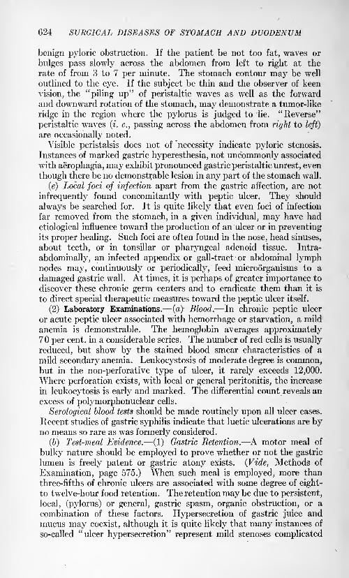
624 SURGICAL DISEASES OF STOMACH AND DUODENUM
benign pyloric obstruction. If the patient be not too fat, waves or
bulges pass slowly across the abdomen from left to right at the
rate of from 3 to 7 per minute. The stomach contour may be well
outlined to the eye. If the subject be thin and the observer of keen
vision, the "piling up" of peristaltic waves as well as the forward
and downward rotation of the stomach, may demonstrate a tumor-like
ridge in the region where the pylorus is judged to lie. "Reverse"
peristaltic waves (i. e., passing across the abdomen from right to left)
are occasionally noted.
Visible peristalsis does not of "necessity indicate pyloric stenosis.
Instances of marked gastric hyperesthesia, not uncommonly associated
with aerophagia, may exhibit pronounced gastric peristaltic unrest, even
though there be no demonstrable lesion in any part of the stomach wall.
(e) Local foci of infection apart from the gastric affection, are not
infrequently found concomitantly with peptic ulcer. They should
always be searched for. It is quite likely that even foci of infection
far removed from the stomach, in a given individual, may have had
etiological influence toward the production of an ulcer or in preventingits proper healing. Such foci are often found in the nose, head sinuses,
about teeth, or in tonsillar or pharyngeal adenoid tissue. Intra-
abdominally, an infected appendix or gall-tract or abdominal lymphnodes may, continuously or periodically, feed microorganisms to a
damaged gastric wall. At times, it is perhaps of greater importance to
discover these chronic germ centers and to eradicate them than it is
to direct special therapeutic measures toward the peptic ulcer itself.
(2) Laboratory Examinations. (a} Blood* In chronic peptic ulcer
or acute peptic ulcer associated with hemorrhage or starvation, a mild
anemia is demonstrable. The hemoglobin averages approximately70 per cent, in a considerable series. The number of red cells is usually
reduced, but show by the stained blood smear characteristics of a
mild secondary anemia. Leukocystosis of moderate degree is common,but in the non-perforative type of ulcer, it rarely exceeds 12,000.
Where perforation exists, with local or general peritonitis, the increase
in leukocytosis is early and marked. The differential count reveals an
excess of polymorphonuclear cells.
Serological blood tests should be made routinely upon all ulcer cases.
Recent studies of gastric syphilis indicate that luetic ulcerations are byno means so rare as was formerly considered.
(6) Test-meal Evidence. (1) Gastric Retention. A motor meal of
bulky nature should be employed to prove whether or not the gastric
lumen is freely patent or gastric atony exists. (Vide, Methods of
Examination, page 575.) When such meal is employed, more than
three-fifths of chronic ulcers are associated with some degree of eight-
to twelve-hour food retention. The retention may be due to persistent,
local, (pylorus) or general, gastric spasm, organic obstruction, or a
combination of these factors. Hypersecretion of gastric juice and
mucus may coexist, although it is quite likely that many instances of
so-called "ulcer hypersecretion" represent mild stenoses complicated
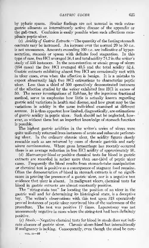
GASTRIC ULCER 625
by pyloric spasm. Similar findings are not unusual in such extra-
gastric ailments as intermittently active disease of the appendix or
the gall-tract. Confusion is easily possible when such affections com-
plicate peptic ulcer.
(c) Acidity of Gastric Extracts. The quantity of the fasting stomachcontents may be increased. An increase over the normal 20 to 50 c.c.
is not uncommon. Amounts exceeding 100 c.c. are indicative of hyper-secretion, stenosis or spasm with definite food stagnation. In this
type of case, free HC1 averaged 56.4 and total acidity 74.2 in the writer's
study of 336 instances. In the non-retention or atonic group of ulcers
(164 cases) the free HC1 averaged 40.5 and the total acidity 52.4.
Gastric extracts exhibiting absent free HC1 are occasionally met with
in ulcer cases, even when the affection is benign. It is a mistake to
expect abnormally high free HC1 estimations to characterize pepticulcer. Less than a third of 500 operatively demonstrated instances
of the affection studied by the writer exhibited free HC1 in excess of
50. The newer investigations of Rehfuss, by the ingenious fractional
method, serve to emphasize how little is actually known regarding
gastric acid variations in health and disease, and how great may be the
variations in acidity in the same individual examined at different
seances. It is thus apparent how limited, diagnostically, are estimations
of gastric acidity in peptic ulcer. Such should not be neglected, how-
ever, as without them but an imperfect knowledge of stomach function
is possible.
The highest gastric acidities in the writer's series of ulcers were
quite uniformly returned from instances of acute and subacute perforat-
ing ulcer. In the ordinary chronic ulcer, the acidity figures closely
resemble such as are returned by cases of chronic gastritis and earlyulcera carcinomatosa. Where gross hemorrhage has recently occurred
there is an average reduction in free HC1 acidity of approximately 10.
(d) Macroscopic blood or positive chemical tests for blood in gastricextracts are recorded in rather more than one-third of peptic ulcer
cases. Frequently the blood results from stomach-tube manipulationor chemical test is positive as a consequence of stagnant food remains.
Often the demonstration of-blood in stomach extracts is of no signifi-
cance in proving the presence of a gastric ulcer, nor is a negative test
evidence that ulcer is absent. In malignant ulcers, chemical tests for
blood in gastric extracts are almost constantly positive.The "string-stain test" for locating the position of an ulcer in the
gastric wall and for determining its histological state, is a deceptive
toy. The writer's observations with, this test upon 318 operatively
proved instances of peptic ulcer convinced him of the uselessness of the
procedure. The test was positive 17 times. Laparotomy was not
uncommonly negative in cases where the string-test had been definitely
positive.
(e) Stools. Negative chemical tests for blood in stools does not indi-
cate absence of gastric ulcer. Chronic ulcers bleed but intermittentlyif malignancy is lacking. Consequently, even though the stool be care-
VOL. ii 40

626 SURGICAL DISEASES OF STOMACH AND DUODENUM
fully prepared for analysis, negative tests are quite apt to be recorded.
In periods of ulcer dyspepsia, when stools are collected after the
patient has been on meat- and green-vegetable-free diet for at least two
days and upon milk or cereal gruel diet for twenty-four hours preced-
ing the actual collection of the specimen, positive "occult" blood tests
were obtained from 39 per cent, of cases in the writer's series. Thechemical finding of altered blood in the stools of a patient with an ulcer
type of dyspepsia is of more significance with regard giving informa-
tion respecting the histological state of such ulcer, than it is in informingone that ulcer is present. In malignant ulcers, altered blood can be
demonstrated constantly in the stools of more than 95 per cent, of
cases. Almost two-thirds of benign gastric ulcers must be diagnosedwithout positive chemic test for blood in the stools.
(3) Roentgen Evidence of Gastric Ulcer. At surgical clinics, the
x-ray examination of chronic ulcer cases returns evidence of consider-
able relative clinical worth toward rounding out diagnoses. This has
led to an exaggerated opinion of the value of the method as an aid to
diagnosis of peptic ulcer generally. In uncomplicated gastric ulcer,
the value of either fluoroscopy or serial radiography is chiefly inferen-
tial. By uncomplicated peptic ulcer is meant ulcer in which pyloric
or cardiac stenoses have not occurred, where hour-glass contraction
has not taken place or where malformations in the stomach's contour
have not resulted as a consequence of crater formation, perforation,
adhesion, fixation or fistula. In surgical clinics, such accidents in the
course of gastric ulcer occur in about 60 per cent, of ulcers; in general
diagnostic clinics, the routine run of cases exhibits a much less frequentincidence.
It is evident that positive roentgen manifestations of peptic ulcer
furnish a valuable aid in quickly segregating the group of ulcers
likely to be most greatly benefited by surgical procedures from that
upon which non-surgical therapy is indicated. The decision lies in
the roentgen proof of gastric deformity. It is thus seen, that in
surgical clinics where this fact has been appreciated, the laparotomy
findings will roughly agree with the roentgen reports, in a high percent-
age of cases. Carman states that of 47 instances where ulcer wasfound at laparotomy 30 (83 per cent.) were diagnosable upon radio-
logic evidence. The percentage is not given with respect to absence of
ulcer at laparotomy where radiologic signs had pointed to the presenceof such, nor is it stated in how many instances clinical diagnosis of
peptic ulcer had been made without the radiological report beingconsidered. No statistics are furnished relative to how many patients
negative for ulcer by roentgen study, were proved to have ulcer at
operation, or by clinical history and later observation. Carman's
experience is quite similar to that of other technicians whose workis checked up either by laparotomy or by autopsy.The roentgen signs of gastric ulcer are (a) inferential and (b) positive.
(a) Inferential Signs. (1) Incisura, i. e., local evidence of persistent
indrawing of a portion of the stomach contour. It should be proved
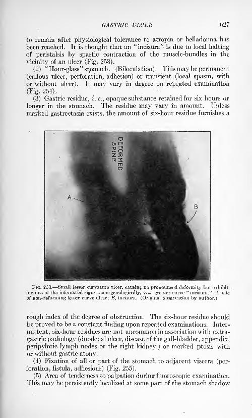
GASTRIC ULCER 627
to remain after physiological tolerance to atropin or belladonna has
been reached. It is thought that an "incisura" is due to local halting
of peristalsis by spastic contraction of the muscle-bundles in the
vicinity of an ulcer (Fig. 253).
(2) "Hour-glass" stomach. (Biloculation) . This may be permanent(callous ulcer, perforation, adhesion) or transient (local spasm, with
or without ulcer). It may vary in degree on repeated examination
(Fig. 254).
(3) Gastric residue, i. e., opaque substance retained for six hours or
longer in the stomach. The residue may vary in amount. Unless
marked gastrectasia exists, the amount of six-hour residue furnishes a
FIG. 253. Small lesser curvature ulcer, causing no pronounced deformity but exhibit-
ing one of the inferential signs, roentgenologically, viz., greater curve "incisura." A, site
of non-deforming lesser curve ulcer; B, incisura. (Original observation by author.)
rough index of the degree of obstruction. The six-hour residue should
be proved to be a constant finding upon repeated examinations. Inter-
mittent, six-hour residues are not uncommon in association with extra-
gastric pathology (duodenal ulcer, disease of the gall-bladder, appendix,
peripyloric lymph nodes or the" right kidney.) or marked ptosis with
or without gastric atony.
(4) Fixation of all or part of the stomach to adjacent viscera (per-
foration, fistula, adhesions) (Fig. 255).
(5) Area of tenderness to palpation during fluoroscopic examination.
This may be persistently localized at some part of the stomach shadow
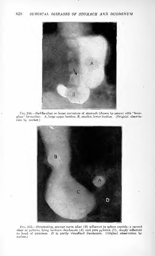
628 SURGICAL DISEASES OF STOMACH AND DUODENUM
FIG. 254. Saddle-ulcer on lesser curvature of stomach (shown by arrow) with "hour-
glass" formation: A, large upper loculus; B, smaller, lower loculus. (Original observa-
tion by author.)
FIG. 255. Penetrating, greater curve ulcer (B) adherent to spleen capsule; a second
ulcer at pylorus, lying between duodenum (A) and pars pylorica (C), deeply adherent
to head of pancreas. D is partly visualized duodenum. (Original observation byauthor.)
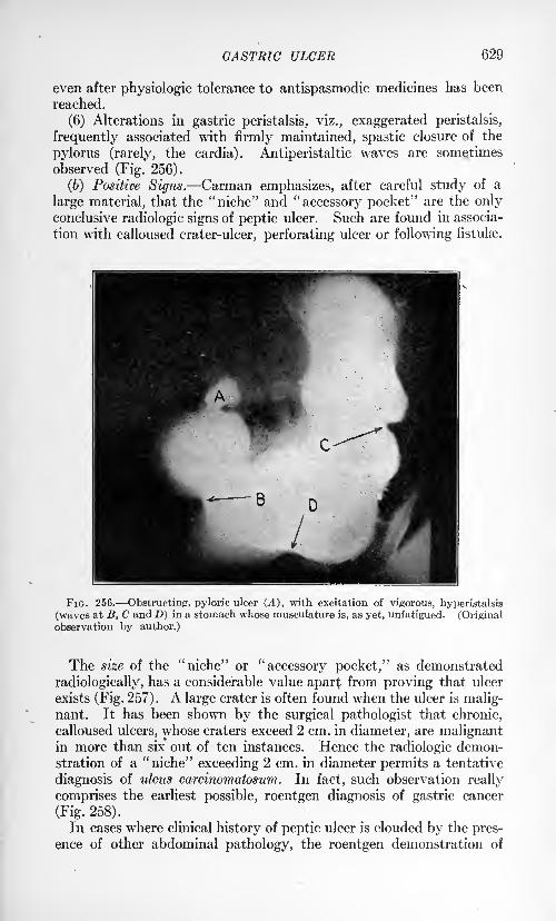
GASTRIC ULCER 629
even after physiologic tolerance to antispasmodic medicines has been
reached.
(6) Alterations in gastric peristalsis, viz., exaggerated peristalsis,
frequently associated with firmly maintained, spastic closure of the
pylorus (rarely, the cardia). Antiperistaltic waves are sometimes
observed (Fig. 256).
(6) Positive Signs. Carman emphasizes, after careful study of a
large material, that the "niche" and "accessory pocket" are the onlyconclusive radiologic signs of peptic ulcer. Such are found in associa-
tion with calloused crater-ulcer, perforating ulcer or following fistulse.
FIG. 256. Obstructing, pyloric ulcer (A), with excitation of vigorous, hyperistalsis
(waves at B, C and D) in a stomach whose musculature is, as yet, unfatigued. (Original
observation by author.)
The size of the "niche" or "accessory pocket," as demonstrated
radiologically, has a considerable value apart from proving that ulcer
exists (Fig. 257). A large crater is often found when the ulcer is malig-nant. It has been shown by the surgical pathologist that chronic,
calloused ulcers, whose craters exceed 2 cm. in diameter, are malignantin more than six out of ten instances. Hence the radiologic demon-stration of a "niche" exceeding 2 cm. in diameter permits a tentative
diagnosis of ulcus carcinomatosum. In fact, such observation really
comprises the earliest possible, roentgen diagnosis of gastric cancer
(Fig. 258).
In cases where clinical history of peptic ulcer is clouded by the pres-
ence of other abdominal pathology, the roentgen demonstration of
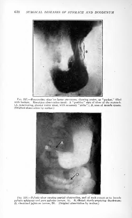
630 SURGICAL DISEASES OF STOMACH AND DUODENUM
FIG. 257. Penetrating ulcer on lesser curvature, showing crater, or "pocket," filled
with barium. Roentgen observation meal: A "positive" sign of ulcer of the stomach.(A, penetrating, greater curve ulcer, with accessory "niche"); B, area of muscle spasm.(Original observation by author.)
FIG. 258. Pyloric ulcer causing partial obstruction, and of such extent as to invade
pyloric sphincter and pars pylorica (arrow, A). B, dilated, slowly emptying duodenum;D, visualized jejunum (arrow, D). (Original observation by author.)
GASTRIC ULCER 631
crater-ulcer in the stomach is of much worth. In this event, the actual
location of the ulcer is determined previous to laparotomy and a differ-
ent prognosis can be given from that which would be offered were the
lesion on the duodenal side of the pylorus.
Complications. (1) Pyloric Obstruction. This occurs in approxi-
mately 25 per cent, of instances of the affection. Its clinical manifesta-
tions are fully considered in the section on Benign Pyloric Stenosis
(vide infra, page 633.)
(2) Hour-glass Contraction. The surgical treatment of peptic ulcer
has demonstrated a greater frequency of this complication than was
formerly considered to exist. Nearly 10 per cent, of gastric ulcers of a
chronic nature produce hour-glass. The contraction may occur at
almost any part of the viscus but usually takes place at the parsmedia or distal to it. It is thus evident, that loculi of unequal size maybe present. The greater loculus is most commonly toward the cardia
because the greater number of gastric ulcers occur toward the pylorus.While peptic ulcer may primarily initiate hour-glass formation, the
malformation may be greatly exaggerated as a consequence of perfora-tion or adhesions with or without extensive fixation of the stomach to
adjacent viscera (Fig. 254).
Symptoms and Signs of "Hour-glass" vary. There is commonly a
long history of peptic ulcer, early intermittent in its manifestation but
later continuous. Obstructive vomiting and evidences of gastric
retention (food stagnation, weight and strength loss, constipationand diminished urine output) are present. The test meal mayreveal twelve-hour food retention, increase in free HC1, blood-stained
gastric extracts or, microscopically, an excess of budding yeasts andsarcinse. Physical examination may demonstrate tender abdominal
(usually epigastric) ridge, splashy stomach or anomalies in the auscul-
tatory murmur when the stomach is filled with air or water through a
tube. Upon gastric lavage, a large amount of fluid may be pouredinto the stomach and but a small quantity be recovered, or when the
stomach is emptied, contents are first obtained which differ in quantityand kind from those secured toward the end of the procedure.
(Woelfer). If the stomach be inflated rather tightly with air, an anoma-
lously shaped viscus may be seen in the epigastrium. Again, one partof the distended stomach may contain ah* only, while the other maycontain both air and fluid. Over the latter portion, splashing sounds
are heard when the abdomen is vigorously tapped. (Taworski.)
Fluoroscopic or roentgen plate examinations usually give ocular
proof of the presence of a biloculated stomach and may definitely
indicate the cause of such. Comparison of the radiological findings
before and after the use of antispasmodic medicines (atropin or bella-
donna) is necessary to eliminate errors possible from powerful gastric
spasm, with or without peptic ulcer.
(3) Malignant Metamorphosis in Peptic Ulcer. This subject is con-
sidered in detail on page 640. (The Relationship Between Gastric
Ulcer and Gastric Cancer).
632 SURGICAL DISEASES OF STOMACH AND DUODENUM
(4) Perforation. Protected perforation is common during the courseof chronic gastric ulcer. It occurs in from 20 to 25 per cent, of instancesin surgical services handling ambulatory material. Its incidence is
probably lower if considered in connection with all types of gastric ulcer.
Chronic protected perforation commonly gives rise to no symptomsdiffering from those associated with progressive, gastric ulcer in general.If closely questioned, patients may give histories of particularly severe
attacks of dyspepsia, often associated with cramp-like, abdominal painand may indicate that such attacks have occurred frequently duringthe recent course of the ailment. At times, gross hemorrhage may com-
plicate the periods of acute distress. Frequent hemorrhages furnish
significant information, even though abdominal pain may not havebeen severe. Where such have occurred, fully 60 per cent, of ulcers
reveal some grade of perforation at laparotomy.Acute perforation complicates about 3 per cent, of ulcers. There
may have been but few symptoms of dyspepsia previous to the initial
sudden, sharp, doubling-up, prostrating pain. In other instances, the
acute crisis happens as a new event in the course of an ordinary ulcer
attack. Gross hemorrhage (hematemesis or melena) may occur con-
comitantly.The main characteristic of acute perforation is sudden, severe pain.
It is usually epigastric. Initially well localized, later it assumes
aspects of a continuous, general, hard "belly-ache." Fixation of
the abdominal muscles, pallor, shock, subnormal temperature at first,
(later high fever), and associated peritonitis, follow. Vomiting maybe obstinate and exhausting. Leukocytosis, chill or chilly sensa-
tions, and evidences of exudate in the peritoneal sac rapidly superveneunless immediate aid can be secured. Occasionally, extensive peri-
toneal involvement is prevented by a bit of omentum or an adjacentviscus plugging-up the ulcer base. In such event, localized peritonitis
occurs, the escaping contents of the stomach are walled off and althoughabscess or fistula may develop, the patient frequently recovers.
(5) Hemorrhage. The clinical features of this complication are con-
sidered fully on page 612. (Gastric Hemorrhage.)(6) Adhesions. Perigastritis sometimes occurs without its resulting
as a consequence of perforating ulcer. In such event, inflammatory
change in the subserosa follows chronic, bacterial activity in the lym-
phatic channels. While the streptococcus is the most frequent organismassociated with perigastritis, yet the greatest degree of gastric deformityoccurs in luetic affections.
The effect of perigastritis is to produce gastric deformity. This
deformity varies greatly in degree. There may be irregularities in gastric
contour, marked thickening of the stomach wall, or adhesions so that
the inflammatory process involves adjacent tissues (pancreas, large or
small intestine, liver, spleen or omentum). Commonly, scar-tissue
develops early. The contraction of scar-tissue often results in fixa-
tion of all or part of the stomach to adjacent organs, with consequent
displacement of the viscus and resultant deformity.
BENIGN PYLORIC STENOSIS 633
In rare instances, where there is rapid spread of invading organismsunder the serosa, actual purulent peritonitis follows. There may then
occur numerous abscesses in the wall of the stomach or about it.
The diagnosis of perigastric adhesions is most accurately made at
laparotomy. Fluoroscopic examination may, however, show malforma-
tions in gastric contour, displacement of the stomach, or adhesions to
neighboring viscera.
BENIGN PYLORIC STENOSIS.
Incidence. Of 8581 patients with digestive upsets, and upon whomgastric as well as general examinations had been made by the writer,
there were 698 such individuals (8.1 per cent.) affected with some typeof benign, pyloric stenosis. Definite proof of the nature and degree of
the pyloric narrowing was possible at laparotomy or autopsy in 622
cases (89 per cent.). In the remaining patients, history, clinical
examinations and subsequent course were reasonably definite toward
establishing the non-malignancy of the ailment.
Causation. Pyloric stenosis most commonly results from peptic
ulcer, duodenal or gastric (84 per cent.). Occlusion of the gastric exit
varies greatly in degree and extent: at times, there occur local or
tubular constrictions amounting to nearly complete obstruction;
again, patches of fibrous, ulcer base are surrounded by dense, hyper-
trophied muscle bundles and excessively vascularized, edematous,areolar tissue and mucosa, with moderately patent lumen; or small,
puckered ulcers result in marked tortuosity or fixation: the pyloricorifice is then stenosed only as a consequence of inflammatory reaction
about, and sometimes quite distantly removed from, the ulcer itself.
In their order of frequency, causes of benign pyloric stenosis other
than ulcer were : gall-bladder disease and its complications, myomatojdhypertrophy of the pyloric and antral musculature (apparently conse-
quent upon long maintained pyloric spasm), gastric syphilis, enlarged
peripyloric lymph glands, ailments of the pancreas (chronic inflamma-
tion, cyst, gumma), cirrhosis of the liver, hepatic syphilis, hydronephrosisof right kidney, foreign body in the stomach, gastric myoma, pyloric
polyp, volvulus (infant), cyst of the liver, pyloric varicosities and
aneurysm of abdominal aorta.
Effects of Pyloric Stenosis. (a) Local. The patency of all or part of
the pyloric lumen is interfered with. The stricture may be so small
as to admit but a probe. There may be several points of narrowing,with ampulla-like spaces between. The stenosis not infrequentlyextends so as to involve the pyloric fourth of the antrum or the first
third of the duodenum. Quite commonly, the actual narrowing of the
pyloric channel as a result of scar-tissue in the wall of the stomach
may be of relatively low degree, but when muscle spasm is associated
with it, such stenosis may be nearly complete. Perforation, with
gastric fixation or malformation, may further aggravate the local
pyloric defect.
634 SURGICAL DISEASES OF STOMACH AND DUODENUM
Pyloric obstruction cannot long continue, even of low grade, without
a definite reaction taking place in the entire stomach. This reaction
occurs as a consequence of a demand for increased gastric function.
Not only must food be more than ordinarily chymified to facilitate
its passage through a narrow exit, but added muscular effort is neededin order that the dryme may freely negotiate the stenotic pylorus.
Very early, are observed congested, thickened gastric mucosa, edema-
tous, areolar tissue and hypertrophied musculature. If the stenosis
increases in degree, muscular hypertrophy reaches its limit and atonyand dilatation of the stomach result. The mucosa and submucosathen become thin and atrophic. Persistent food stagnation follows.
(6) General. Systemic malnutrition eaily becomes manifest. Dryskin and mucous membranes, anemia, muscular flabbiness, loss of
panniculus, weakness, decrease in weight, intestinal atony, diminution
in urine output and mental sluggishness are soon apparent as a con-
sequence of insufficient food assimilation, constant discomfort or painand loss of sleep.
Symptoms. (a) Vomiting. The patient may have previously expe-rienced occasional attacks of vomiting at the time of an aggravation of
his dyspepsia. The characteristic of vomiting associated with pyloricstenosis is that, when the diet is of moderate quantity and normal con-
sistency, emesis is a routine and generally, daily event. Small, infrequentmeals of the pap variety may for a long time mask the clinical evidences
of pyloric narrowing and gastric dilatation. On such, a patient mayactually gain weight and strength and both he and his medical adviser
long remain under the impression that "cure" of such ailment as
stenosing ulcer is being brought about. However, just so soon as a
meal of ordinary size and consistency be given, retention is proved byprompt vomiting. Patients frequently find this out themselves.
Doctors, not rarely, ignore this obvious effect of full feeding and con-
sider the vomiting as being due to "an indiscretion in diet." The"indiscretion" can often be laid to the medical adviser, for while he
is relieving the patient in numerous "indiscretions," not infrequently,
pyloric stenosis is becoming more marked, gastrectasia of greater degree,
or accidents such as perforation, .hemorrhage or malignancy are beingcatered to.
In pyloric narrowing, vomiting is usually daily, and most frequent in
the afternoon or evening. If dilatation of the stomach is pronouncedand muscular weakness has supervened, then night vomiting or vomit-
ing of large quantities of material several times weekly are commonlyexperienced. The characteristic feature is that instead of being an
intermittent dyspeptic with only occasional periods of vomiting, the
patient becomes a constant dyspeptic with regularly recurring vomitingwhen fed on food of ordinary bulk. In no type of gastric malfunction
are cause and effect so clearly manifest.
The vomitus is generally copious. It is composed of poorly broken
up lumps of food intermixed with mucus. Its color varies with the
food eaten. Its odor is usually yeasty and but rarely acrid or rancid
BENIGN PYLORIC STENOSIS 635
as in the event of malignant stenpsis. The odor is generally so char-
acteristically yeasty (indicating simple fermentation), that upon this
observation a fairly accurate, rapid diagnosis between benign and
neoplastic disease may be made. When stomach contents are not
obtainable, then the yeasty odor of a chronic dyspeptic's breath has
valuable diagnostic significance. The so-called "old-fashioned" physi-cian who made a practice of smelling the breaths of his gastric patientswas certainly justified in so doing. It was possible for him to obtain
considerable information quickly: certainly, as much information as
often follows many of our modern "refinements of diagnosis."Evidences that food eaten longer than twelve hours previously
persistently appears in the vomitus or gastric extracts indicates definite
organic interference to the passage of food through the alimentarycanal. The most common cause for such interference is benign
pyloric stenosis. Pyloric spasm patients vomit retained food inter-
mittently; pathologic pyloric narrowing always causes "delayed" vom-
iting of retained food when the patient is on a bulky diet. Pyloric
spasm may be associated with pyloric stenosis. In such event, while
the quantity of the retention-vomitus may vary at different examina-
tions, the factor of food stagnation is a constant one.
Vomiting in pyloric stenosis is a painful act just so long as gastric
musculature is not atonic. The pain is commonly most annoying
previous to or during the emesis. When the stomach is emptied, the
patient is usually comfortable. Gastric lavage may ward off attacks
of vomiting. In such way, this procedure often assumes the dignityof a therapeutic or even curative maneuver. WT
hile lavage is justified
as a measure toward relieving a sufferer, its careless use may for a longtime mask a diagnosis and lead to the establishment of a sense of false
security.
Constant vomiting limits body nourishment. Weight loss logically
follows. In our cases of benign pyloric stenoses, it averaged twenty-one
pounds. The weight loss may be quite as great as in malignant disease.
However, while pyloric stenosis patients usually look starved, they
rarely have added to this unfed appearance the evidences of systemic
poisoning, such as rapidly dominates the picture in malignancy. Onlyrarely does gastric stagnation of the benign type lead to intoxication
of such marked grade as to produce tetany.
(b) Persistent, copious vomiting depletes the body tissues of fluids.
As a consequence, diminished urine output and constipation are commonsecondary symptoms in benign pyloric stenosis.
Eructations, pyrosis, "water-brash" and regurgitation were distress-
ing enough to warrant special mention in 57 per cent, of our cases.
Commonly, these symptoms were most annoying later in the day thanthe midafternoon. They were sometimes particularly severe at night,
especially in that group of individuals whose stomach musculature
was not of sufficient strength to permit easy vomiting.
(c) Gross Hemorrhage. This occurred in 31 per cent, of our ulcer
cases. Not rarely, bleeding followed excessive vomiting and retching.
Only three patients succumbed to severe hematemesis.
636 SURGICAL DISEASES OF STOMACH AND DUODENUM
When gastrorrhagia occurs in association with pyloric stenosis, the
blood vomited may be mixed with food and mucus. It is commonlyunclotted and has acid reaction. Later vomitus may be made up of
large tough clots. It is then generally alkaline and food-free.
(d) Pain. Some form of abdominal discomfort was experienced
by 98 per cent, of our patients. When pyloric stenosis is associated
with active, open, peptic ulcer then the abdominal distress may be
extreme. This is due largely to excessive pyloric, gastric or duodenal
spasms and not to food passing over the ulcer itself. The painis often greatest when the stomach is empty. At such time, abnor-
mally vigorous "hunger contractions" occur, as pointed out by Cannon.
Night pain, after emesis, may be so severe as to require opiates, par-
ticularly in the early stages of pyloric stenosis, when compensatory
hypertrophy of the gastric musculature is being established.
W7hen gastric atony, with consequent stagnation, has occurred, the
abdominal discomfort is variously described as a "load," "fulness,"
"up-pressure," "bloat," "soreness" and "ache."
Signs of Benign Pyloric Stenosis. (a) We have already commented
upon the systemic effects of benign narrowing of the pyloric orifice.
It is only necessary to emphasize the evidences of emaciation without
obvious cachexia, the dried out body-tissues, the weakness, anemia andthe so-called 'dyspeptic fades' : sallow, drawn face skin, exaggerationof normal wrinkles delimiting muscles of expression, dropped mouth
corners, dried lips and lack-lustre, sunken eyes. Jaundice is rare.
It may complicate a perforated pyloric ulcer, where the common duct
is involved, an old gall-bladder ailment or an enlarged pancreas.
(6) Visible Gastric Peristalsis. Visible gastric peristalsis was a sign
of diagnostic significance in rather more than 11 per cent, of our cases.
It occurs as a consequence of a stout, gastric musculature endeavoringto force chyme through a narrowed, stomach exit. When gastric
atony and dilatation supervene, visible peristalsis is rarely present.
In favorable cases i. e., where the abdominal wall is thin and peri-
stalsis is vigorous waves or bulges pass across the epigastrium or navel
regions, from left to right, at a rate of from 3 to 7 per minute. Theentire stomach contour may be definitely outlined to the eye. Not
rarely, there is not a definite sequence of waves, but numerous, appar-
ently uncoordinated, rounded, epigastric bulgings. If the subject is
thin and the observer's keenness of vision be properly intermixed with
imagination, following strong peristaltic waves, a tumor may appearin the region where the pylorus is supposed to lie. In infants affected
with hypertrophy of the pyloric sphincter and stenosis, such a tumor,
appearing soon after food ingestion and before emesis, is of much value
with regard to both diagnosis and prognosis.
(c) Abdominal Palpation. Tenderness to pressure in the right,
upper quadrant was noted in 79 per cent, of our cases. It was most
pronounced in instances where the cause of the stenosis was active
or perforating, peptic ulcer, distended gall-bladder or inflammatorydisease of the peripyloric glands or the pancreas. When gastric fer-
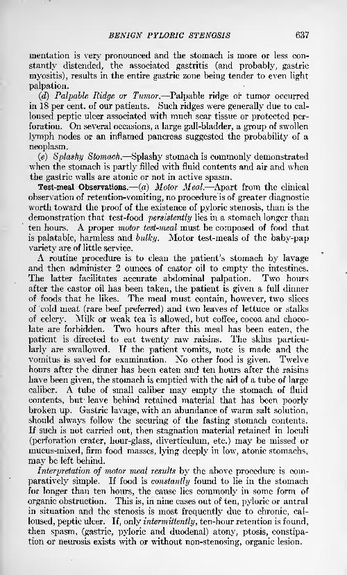
BENIGN PYLORIC STENOSIS 637
mentation is very pronounced and the stomach is more or less con-
stantly distended, the associated gastritis (and probably, gastric
myositis), results in the entire gastric zone being tender to even light
palpation.
(d) Palpable Ridge or Tumor. Palpable ridge or tumor occurred
in 18 per cent, of our patients. Such ridges were generally due to cal-
loused peptic ulcer associated with much scar tissue or protected per-foration. On several occasions, a large gall-bladder, a group of swollen
lymph nodes or an inflamed pancreas suggested the probability of a
neoplasm.
(e) Splashy Stomach. Splashy stomach is commonly demonstratedwhen the stomach is partly filled with fluid contents and air and whenthe gastric walls are atonic or not in active spasm.
Test-meal Observations. (a) Motor Meal. Apart from the clinical
observation of retention-vomiting, no procedure is of greater diagnosticworth toward the proof of the existence of pyloric stenosis, than is the
demonstration that test-food persistently lies in a stomach longer than
ten hours. A proper motor test-meal must be composed of food that
is palatable, harmless and bulky. Motor test-meals of the baby-papvariety are of little service.
A routine procedure is to clean the patient's stomach by lavageand then administer 2 ounces of castor oil to empty the intestines.
The latter facilitates accurate abdominal palpation. Two hours
after the castor oil has been taken, the patient is given a full dinner
of foods that he likes. The meal must contain, however, two slices
of cold meat (rare beef preferred) and two leaves of lettuce or stalks
of celery. Milk or weak tea is allowed, but coffee, cocoa and choco-
late are forbidden. Two hours after this meal has been eaten, the
patient is directed to eat twenty raw raisins. The skins particu-
larly are swallowed. If the patient vomits, note is made and the
vomitus is saved for examination. No other food is given. Twelvehours after the dinner has been eaten and ten hours after the raisins
have been given, the stomach is emptied with the aid of a tube of largecaliber. A tube of small caliber may empty the stomach of fluid
contents, but leave behind retained material that has been poorlybroken up. Gastric lavage, with an abundance of warm salt solution,
should always follow the securing of the fasting stomach contents.
If such is not carried out, then stagnation material retained in loculi
(perforation crater, hour-glass, diverticulum, etc.) may be missed or
mucus-mixed, firm food masses, lying deeply in low, atonic stomachs,
may be left behind.
Interpretation of motor meal results by the above procedure is com-
paratively simple. If food is constantly found to lie in the stomachfor longer than ten hours, the cause lies commonly in some form of
organic obstruction. This is, in nine cases out of ten, pyloric or antral
in situation and the stenosis is most frequently due to chronic, cal-
loused, peptic ulcer. If, only intermittently, ten-hour retention is found,then spasm, (gastric, pyloric and duodenal) atony, ptosis, constipa-tion or neurosis exists with or without non-stenosing, organic lesion.
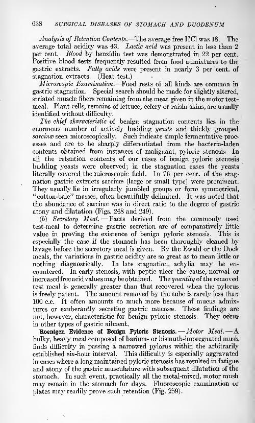
638 SURGICAL DISEASES OF STOMACH AND DUODENUM
Analysis of Retention Contents. The average free HC1 was 18. Theaverage total acidity was 43. Lactic acid was present in less than 2
per cent. Blood by benzidin test was demonstrated in 22 per cent.
Positive blood tests frequently resulted from food admixtures to the
gastric extracts. Fatty acids were present in nearly 3 per cent, of
stagnation extracts. (Heat test.)
Microscopic Examination. Food rests of all kinds are common in
gastric stagnation. Special search should be made for slightly altered,
striated muscle fibers remaining from the meat given in the motor test-
meal. Plant cells, remains of lettuce, celery or raisin skins, are usuallyidentified without difficulty.
The chief characteristic of benign stagnation contents lies in the
enormous number of actively budding yeasts and thickly groupedsarcincB seen microscopically. Such indicate simple fermentative proc-esses and are to be sharply differentiated from the bacteria-laden
contents obtained from instances of malignant, pyloric stenosis In
all the retention contents of our cases of benign pyloric stenosis
budding yeasts were observed; in the stagnation cases the yeasts
literally covered the microscopic field. In 76 per cent, of the stag-
nation gastric extracts sarcinae (large or small type) were prominent.
They usually lie in irregularly jumbled groups or form symmetrical,"cotton-bale" masses, often beautifully delimited. It was noted that
the abundance of sarcinse was in direct ratio to the degree of gastric
atony and dilatation (Figs. 248 and 249) .
(6) Secretory Meal. Facts derived from the commonly used
test-meal to determine gastric secretion are of comparatively little
value in proving the existence of benign pyloric stenosis. This is
especially the case if the stomach has been thoroughly cleaned bylavage before the secretory meal is given. By the Ewald or the Dock
meals, the variations in gastric acidity are so great as to mean little or
nothing diagnostically. In late stagnation, achylia may be en-
countered. In early stenosis, with peptic ulcer the cause, normal or
increased free acid valuesmaybe obtained. The quantity of the removed
test meal is generally greater than that recovered when the pylorusis freely patent. The amount removed by the tube is rarely less than
100 c.c. It often amounts to much more because of mucus admix-
tures or exuberantly secreting gastric mucosae. These findings are
not, however, characteristic for benign pyloric stenosis. They occur
in other types of gastric ailment.
Roentgen Evidence of Benign Pyloric Stenosis. Motor Meal. Abulky, heavy meal composed of barium- or bismuth-impregnated mushfinds difficulty in passing a narrowed pylorus within the arbitrarily
established six-hour interval. This difficulty is especially aggravatedin cases where a long maintained pyloric stenosis has resulted in fatigue
and atony of the gastric musculature with subsequent dilatation of the
stomach. In such event, practically all the metal-mixed, motor mush
may remain in the stomach for days. Fluoroscopic examination or
plates may readily prove such retention (Fig. 259).
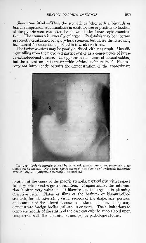
BENIGN PYLORIC STENOSIS 639
Observation Meal. When the stomach is filled with a bismuth or
barium suspension, abnormalities in contour, size or position or fixation
of the pyloric zone can often be shown at the fluorescopic examina-
tion. The stomach is generally enlarged. Peristalsis may be vigorousin recently established benign pyloric stenosis, but where the narrowinghas existed for some time, peristalsis is weak or absent.
The bulbus duodeni may be poorly outlined, either as result of insuffi-
cient filling from the narrowed gastric exit or as a consequence of intra-
or extraduodenal disease. The pylorus is sometimes of normal caliber,
but the stenosis occurs in the first third of the duodenum itself. Fluoros-
copy not infrequently permits the demonstration of the approximate
FIG. 259. Pyloric stenosis caused by calloused, greater curvature, prepyloric ulcer
(indicated by arrow). Note large, atonic stomach, the absence of peristalsis indicatingmuscle fatigue. (Original observation by author.)
location of the cause of the pyloric stenosis, particularly with respectto its gastric or extra-gastric situation. Prognostically, this informa-
tion is often very valuable. It likewise assists surgeons in planning
operative relief. Plates or films of the barium- or bismuth-filled
stomach, furnish interesting visual records of the shape, size, positionand contour of the altered stomach and the duodenum. They maydemonstrate foreign bodies, gall-stones or cysts. Their limitations as
complete records of the status of the case can only be appreciated uponcomparison with the laparotomy, autopsy or pathologic studies.
640 SURGICAL DISEASES OF STOMACH AND DUODENUM
THE RELATION BETWEEN GASTRIC ULCER AND GASTRIC CANCER.
Clinically the significance of gastric ulcer of the chronic type with
respect to gastric cancer is still under discussion. The subject is of vast
importance both as a diagnostic and a prognostic problem. To the sur-
geon the diagnostic value of detecting ulcera carcinomatosa lies in the
knowledge that the extirpation of such defects yields the greatest
proportion of malignant growths of the stomach that are operativelycurable. Upon the internist rests the grave burden of segregatingfrom the group of ulcer patients which appear for treatment, those
amenable to non-surgical procedures from those in whom atypic cell
changes exist and whose only hope of relief depends upon prompt,radical removal of the focus of disease. The problem is a difficult one.
While internists, with meager operating-room or laboratory experi-
ence, claim that a comparatively small number of chronic gastric ulcers
ultimately undergo malignant transition, the admission that such
ulcers do at times appear to be forerunners of cancers, indicates emphat-ically that, given an instance where calloused ulcer is proved to exist,
the prospect of its being an anlage for a neoplasm must be admitted.
In such event, the- queer admixture of pride in personal opinion andmental bias dependent largely upon impressions gained from semi-
ancient literature, which seemingly exists in some quarters, results in a
professional attitude that is not in accord with modern facts and which
is most pernicious with respect to the welfare of patients seeking aid.
There has been much old and recent controversy regarding the
etiologic significance of gastric ulcer and gastric cancer. In 1882,
Zenker stated that, in his belief, all gastric cancers originated from
previous ulcers. Seven years later, Rosenheim supported Zenker's
opinion. In 1902, Fiitterer, after extensive experimental studies,
summarized his results by the observation that gastric cancer de-
velops with great frequency from ulcer located in the pyloric fourth
of the stomach, but that malignant transition is less common when the
ulcer is situated in other parts of the viscus. Fenwick, from an
apparently limited operating-room and pathologic experience, claims
that but 3 per cent, of ulcers become cancerous. Friedenwald, after
a study of one thousand cases, clinically gastric cancer (but 26.6 percent, came to laparotomy or autopsy), places the frequency of cancer's
arising upon previous ulcer at 7.3 per cent. Moynihan claims that
fully 66 per cent, of his cases of gastric cancer had previously been
affected with chronic gastric ulcer. Sapeska, after a careful review
of 100 cases of gastric cancer, could find but 10 instances where pre-
viously calloused ulcer had apparently not existed.
The wide diversity of opinion shown by the above observations
needs explanation. There has been a confusion of viewpoint, partly,
because certain clinicians attempt to establish facts from pathologically
unproved diagnoses and, partly, because from the statement that, for
example, more than 60 per cent, of gastric cancers apparently exhib-
RELATION BETWEEN GASTRIC ULCER AND GASTRIC CANCER 641
ited a previous history of chronic ulcer, it is loosely assumed that the
reverse is true : namely, that fully as large a percentage of benign gastric
ulcers undergo malignant change. In order to properly comprehendthe difficulties associated with determining the interrelation between
peptic ulcer and cancer of the stomach, it is quite necessary to under-
stand the nature of both gastric ulcer and gastric cancer. As an aid
to interpreting some of the aspects of the problem, the writer analyzed921 gastric cancers and 500 benign gastric ulcers. The diagnoses in
all instances were operatively and pathologically established.
1. The Nature of Gastric Ulcer. (a) Origin. All types of benign
gastric ulcer may be produced experimentally by a wide variety of
methods. Bolton classifies some of the more familiar causative agentsas (1) bacteria (pneumococci by Dieulafoy, staphylococci by Widaland Meslay, staphylococcus aureus by Letulle, bacillus pyocyaneus
by Charrin and Ruffer, bacillus dysenterii by Chantemesse and Widal,lactic acid bacillus by Wurtz and Leudet, bacillus of Pfeiffer byStatineano, meningococcus by Weichselbaum and byGaudy and Griffon,
Eberth's bacillus and bacillus coli by Rodet and Zaidmann, strep-
tococcus by Rosenow); (2) bacterial toxins (pyemias by Lebert and
by Cohn, diphtheria toxin by Enriguez and Hallion, and Rosenau and
Anderson); (3) cutaneous burns (Welty, Ponfick, Silbermann, Paras-
candolo); (4) poisons of metabolic origin (Bolton's"gastrotoxin"
injected intraperitoneally or subcutaneously) ; (5) extrinsic poisons
(mercury salts, arsenious acid, cantharidine, vegetable alkaloids,
pilocarpine, atropine, copper sulphate); (6) poisons introduced into
the stomach (corrosives and caustics) and (7) alterations in the stom-
ach's circulation (vascular blocking, thrombi, emboli, arterioscle-
rosis, nervous inhibition, or external pressure). The type of ulcer
produced experimentally, varies slightly according to the method
employed in causing it. The resultant lesions are, however, essentially
similar. There is loss oflmrface epithelium, necrosis, inflammatory
edema, hemorrhage and glandular destruction. In the healing of
ulcers, irrespective of the way they may have been produced, the
essential feature consists in development of new epithelium, protectiveconnective-tissue by hyperplasia, with resultant scar.
(b) Course. In neither experimental animals nor the human can
the life history of any gastric ulcer be prognosed. This applieswith respect both the extent of the lesion and the amount of tissue
destruction taking place. That most gastric ulcers have a natural
tendency to heal is shown by the rapid appearance of scar formation
in experimental animals, the discovery of healed ulcers at laparotomyor necropsy, and the clinical "cure" of ulcers by widely varying modesof medical or surgical treatment. The time required for ulcer forma-
tion or the healing of such seems to have wide limits. There are factors
concerned which appear to be largely individual. Certainly, chronicityof an ulcer, pathologically, does not necessarily coincide with chronicityin the sense of duration in terms of months or years. Huge, excavated
calloused ulcers may form in a few weeks while superficial, bleedingVOL. II 41
642 SURGICAL DISEASES OF STOMACH AND DUODENUM
erosions or small calloused ulcers may exist for years with no evident
extension or marked connective-tissue hyperplasia.
(c) Malignant Change. That some element other than the persistent
presence of an ulcer in a functionating stomach is necessary as the
forerunner to the transformation of a benign process to a malig-nant one is indicated by the fact that no experimenter has ever pro-
duced, in animal or man, a cancerous ulcer. Chronic irritation, infec-
tion, artificial anemia, starvation, overfeeding or local application of
chemicals have all failed to cause such histologic picture as seen in
ulcus carcinomatosum, i. e., the power of atypic, unlimited cell growthwith ability to cause death of the host by malignant intoxication
and metastases. Just how such change occurs in man we are not
able to state. Herein lies one of the strong points of argumentfor those clinicians and pathologists who claim that benign gastriculcer rarely becomes malignant. No one has ever seen the actual
transition from a benign process to cancer. In a given specimenexamined microscopically, one can only say that cancer is or is not
present. Atypic hyperplasias may render a diagnosis of early malig-
nancy highly probable by one experienced in the examination of fresh
tissue, but it appears impossible to definitely state that an ulcer whichshows cancerous changes in a few spots of its edge was ever anythingelse than malignant. Certainly, one cannot say unqualifiedly, that the
part of the mucosa that has sloughed away was benign. Such part
may have been cancerous and have been prevented from wider incur-
sions into the gastric wall by the very atypic hyperplasia, so-called,
that remains for our study. What we see, may be an indication of a
histologic battle, already a partial victory. The subsequent spreadof the few malignant rests which are present, may follow as a conse-
quence of exhaustion of the protective mechanism through the first
attempt at defence. When an ulcer is excised at laparotomy the
histologic contest is interrupted. One cannot satisfactorily prognosewhat the ultimate outcome would have been had the conflict been
allowed to proceed. It is only when one observes cancerous ulceration,
with evidences of perigastric, lymph-gland invasion, that it is possible
to state that the disease will progress to the death of the host.
(d) Frequency of the Transition of Benign Gastric Ulcer to Cancer.
There has been much misunderstanding in this regard. Publica-
tion of reports, claiming that the clinical type of dyspepsia which
frequently precedes what is commonly recognized as a malignant form
of gastric disease, is often not to be differentiated from chronic peptic
ulcer, has given rise to a widespread impression that large numbersof chronic gastric ulcers terminate as cancers. Clinical experienceand pathologic facts are far from lending support to this conclusion.
While the writer's study of the early histories of 921 instances of
proved gastric cancer indicates that more than 65 per cent, of the
cases had a long dyspeptic course (apparently of benign type), pre-
ceding that which clinically, was malignant, it does not establish
the fact that a like proportion of benign gastric ulcers eventually termi-
RELATION BETWEEN GASTRIC ULCER AND GASTRIC CANCER 643
nate as cancers. Granted, that the so-called precarcinomatous mani-
festations of gastric cancer closely simulate those of benign peptic
ulcer, it is not unlikely that the common conception, clinically, of the
symptomatology of ulcer and of cancer of the stomach is an imperfectone. As there is, histologically, a group of ulcers in which sections donot permit of accurate differentiation between benignity and malig-
nancy, it is not improbable that there are stages in both gastric ulcer
and cancer where clinical recognition of each ailment is impossible.Our conception of how ulcer and cancer of the stomach make themselves
manifest clinically, may be too narrow.
Pathologically, the strongest proof that exists of the frequency of
the histologic transition of benign ulcer to malignant ulcer is advanced
by MacCarty. From a most complete and painstaking study he has
demonstrated that of 280 resected, chronic, calloused gastric ulcers,
in which there was no clinical or gross surgical hint of malignancy,63 per cent, showed evidences of atypic or undifferentiated cell-groupsin their- hyperplastic edges. This is an important array of facts andhas highly suggestive application, clinically, but etiologically, as
MacCarty frankly admits, it carries no proof that those ulcers showingatypic cell arrangement and structure were ever benign, histologically.
While the length of the period of dyspepsia associated with the demon-stration of these ulcers might, clinically, indicate a previously benign
process, it does not prove that such actually existed. Our conceptionof the time of possible duration of malignancy may require readjust-ment. On purely empiric grounds, the clinical picture of gastriccancer has become firmly, established as one of a perniciously and
progressively downward process, writh fatal termination within fromone to three years. How long malignancy can exist in a stomachbefore evidences are clinically present, we do not know. The disease
may lie "latent" for many years before local or constitutional changesbecome apparent. More than 1.5 per cent, of the writer's cases
of well-advanced gastric cancer gave rise to no symptoms directly,
pointing to a gastric upset. The knowledge that gastric juicers mayexist without producing so-called "ulcer dyspepsia," is proved by the
not infrequent finding of calloused gastric ulcers at laparotomy for
other ailments, or at necropsy. It is thus evident, that it is frequently
impossible to judge the histology of gastric mucosa, from clinical mani-festations alone, previous to the time when clinical proof indicates that
cancer has developed : foundation for the neoplasm may have long been
present, either as ulcer or as some yet unknown cellular malarrangementor intracellular metabolic fault.
The writer's study of 500 surgically demonstrated cases of benigngastric ulcer in no way indicates the frequency of cancer formationfrom such. That this is to be expected is emphasized by the fact
that our cases are all classified upon a pathological basis, i. e., a speci-men is either benign ulcer, undetermined ulcer histologically or gastric
cancer, with or without the association of ulceration. Presuming thatour 500 cases of benign gastric ulcer form part of a numerous group
644 SURGICAL DISEASES OF STOMACH AND DUODENUM
which appeared at some time previously, it is manifestly impossibleto tell what course the original ulcer group has taken namely, howmany have healed spontaneously or with the aid of medical care, howmany have survived as benign ulcer cases, on how many have surgi-cal procedures been performed, and how many have terminated as
cancers. In the writer's opinion the above facts furnish insurmountable
obstacles to the possibility of any, even approximately correct, esti-
mate being made with regard to the frequency with which benign gastriculcer becomes malignant. Whatever may be individual opinion or
clinical experience, it would seem that this phase of the subject remains
unsettled.
(e) Clinical Variations in the Symptom-complex of Gastric Ulcer.
That a difference of opinion exists among clinicians with regard to whatset of symptoms and signs actually indicate peptic ulcer is readily noted
upon the perusal of the opinions of those men who have had experiencewith any considerable material. Symptom-complexes, based solely
upon individual interpretation of a group of complaints, signs or tests,
cannot expect unqualified acceptance. However, when certain clinical
pictures of disease are found in association with the pathologic proofthat such disease exists, symptom-complexes can be rationally formu-
lated, and only when measured by this rule are these above criticism.
The close simulation of the symptomatology of uncomplicated gastric
ulcer by disease of other abdominal organs (gall-bladder, appendix,
colon, etc.) would appear to suggest that the clinical 'manifestations
often associated with ulcer are often the evidences of an abdominal
or a constitutional disarrangement, in the course of which gastric
ulceration is an incident or an end-result. Certain support to this
conception of the disease is furnished by the experimental productionof gastric ulcers by many and widely different methods and agents.It is within the experience of all who have had a liberal training, that
not infrequently laparotomy or necropsy, at the most competent hands,fails to demonstrate ulceration in patients presenting so-called charac-
teristic clinical manifestations of the ailment. Conversely, it is
equally surprising to discover well-advanced gastric ulcer which has
produced no symptoms referable to the stomach, at laparotomy for
extra-gastric disease or at necropsy following deaths due to accident
or extra-abdominal affections. From the foregoing, it would appear
likely that the symptomatology which is commonly associated with
gastric ulcer, clinically, is the manifestation of a group of derange-ments among which ulcer of the stomach is included. The above
would appear to impose another limitation to the accuracy with which
it is possible to determine the relationship between gastric ulcer and
gastric cancer. It is not beyond probability that much evidence which
has been adduced from merely clinical symptomatology determining
diagnoses is not admissible, inasmuch as in many instances peptic ulcers
were not present.
(/) Malignant Change in Duodenal Ulcer. In a consecutive series,
the writer's 500 operatively demonstrated instances of gastric ulcer
RELATION BETWEEN GASTRIC ULCER AND GASTRIC CANCER 645
were found to exist in 1725 cases of peptic ulcer, gastric and duodenal.
It is thus seen that the ratio existing between the frequency of gastricand of duodenal ulcer is as 1 : 2.45. While duodenal ulcer is muchmore frequent than is gastric ulcer, cancer of the duodenum is veryuncommon. From a careful perusal of records, the writer noted it
seven times. It has been advanced, that, if gastric ulcer frequently
undergoes malignant transition, then one should expect a high ratio
of malignant duodenal ulcers.
When primary cancer of the duodenum is found, it is usually located
at or near the papilla of Vater. In such situations, the viscus is
liable to injury from gall-stones, altered secretions of the liver andthe pancreas, and infective processes from the gall-tract. Moreover,at the papilla of Vater, there may be stagnation of duodenal contents
or slowing of the rate of discharge of the digestive juices from the
liver and the pancreas. It should be also emphasized that ordinarily,
there is little opportunity for traumata to the duodenal mucosa fromretarded food progression. Food remains for- a very brief time in the
duodenum, provided stenoses do not exist. The opposite condition
exists in the stomach, in which event, there is abundant time for bac-
terial, chemical or mechanical injury to a gastric lining already robbedof some of its resistance. That the duodenum appears to have an
inherent protective mechanism against malignancy appears to be dem-onstrated by the observation that only rarely does cancer of the
stomach greatly involve the duodenum by direct extension. Thedifferent character of the tissue of this part of the gut seems to be in-
dicated by the fact that, not infrequently, duodenal ulcers, which haveextended to the pylorus, assume malignant characteristics in their
stomach edges and nowhere else.
(g) Gastro-enterostomy as a Protection Against Malignant Transition
of Gastric Ulcer. Given a gastric ulcer, proved to exist by laparotomy,if such ulcer be not excised, it appears to have been shown by compe-tent observers that after gastro-enterostomy malignancy rarely super-venes. In our series, there were nine patients, with apparently benign
gastric ulcers, who appeared with cancers of the stomach after anterior
or posterior gastro-enterostomy had been performed. Of course, inas-
much as none of these ulcers had been microscopically examined before
gastro-enterostomy, it is not possible to state that such were not malig-nant at the time that the operations were performed.
It should be recalled that modern surgeons do not consider gastro-
enterostomy as an operation solely of"drainage." It is well recognized,
that after such surgical procedure, the physiology of the stomach has
been altered in many particulars: not rarely, the stomach's emptyingpower is accelerated and thus stagnant food, often very foul bac-
teriologically, remains for a shorter time in contact with a damagedmucosa; blood and lymph circulations may be appreciably changedas a consequence of relief from dilatation of the stomach and altera-
tions in the peristaltic rhythm; in nearly 85 per cent, of gastric extracts
of cases where gastro-enterostomy has been done, the writer demon-
646 SURGICAL DISEASES OF STOMACH AND DUODENUM
strated, by chemical or microscopic means, both duodenal and jejunalcontents material from parts of the gut relatively immune to malig-nant disease, and finally, after gastro-enterostomy, he was able to
demonstrate in a series of 121 consecutive instances of gastric ulcer,
an average decrease of 17 free hydrochloric acid, together with
appreciable diminution in peptolysis. Clinically, it is the general
experience that in malignant disease of the stomach, not associated
with stenosis, gastro-enterostomy appears to grant a longer lease of
life than where such has not been performed.II. The Nature of Gastric Cancer. (a) Origin. No one has
ever seen the actual beginnings, histologically, of malignant disease
of the stomach. While experiments in tumor transmission, by Rousand Murphy, reveal certain tissue-reactions to the presence of arti-
ficially introduced foreign cells, it cannot be said that such reactions
are entirely comparable to those occurring when a tumor arises spon-
taneously from native tissue. MacCarty has apparently shown that
malignancy begins as a.form of hyperplasia of already existing cell-
structures. This hyperplasia results in functionally undifferentiated,
and architecturally imperfect, primary cell elements. This hyperplasiavaries in degree, and at certain stages cannot be segregated, histologi-
cally, from the forms of hyperplasia common in processes of so-called
benign nature. Apparently, in certain of these grades of hyperplasia, the
line of demarcation between the malignant and the benign is, with our
present methods of examination, extremely indefinite. In the case of
gastric cancer, however, once this"halting stage" is passed, thehistologic
picture is well defined and the disease progresses rapidly. Evidence
strongly supporting the pathologic proof that malignancy may developin gastric ulcers that were apparently benign, is furnished by the
histological observation of all degrees of hyperplasia benign, inde-
terminate and malignant in sections through different portions of
a given excised ulcer.
(6) Course. Unless total extirpation is possible, gastric cancer
produces death of its host. Unlimited growth, with the property of
developing metastases in near or distant organs, rapidly results in fatal
malignant intoxication, hemorrhage, starvation or exhaustion. Rarelydo proved instances of gastric cancer live longer than three years.
Unless there is early surgical interference, the majority of hosts die
within a year following the onset of symptoms. In the writer's cases
of cancer of the stomach, the average duration of all symptoms, of a
clinically malignant type, was 6.9 months previous to the patients'
coming for relief. There are wide variations in the rate of progressof the disease. The same type of ailment, histologically, progresses with
strikingly different speeds in different individuals. The factors con-
cerned in the ability of gastric mucosa to resist cancer invasion are
not as yet understood. One individual may be overwhelmed with
the disease in a few weeks while another may successfully resist the
affection for several years.
The above observations offer suggestions respecting the possibilities
RELATION BETWEEN GASTRIC ULCER AND GASTRIC CANCER 647
for variability in the symptom-complex of gastric cancer. Our records
show that more than 1.5 per cent, of operatively demonstrated instances
of the disease, gave no pre-laparotomy manifestations that pointed to
an ailment of the stomach. Clinically, however, the major portionof the cases fall into two great groups, with respect to course: (1)
Instances where a chronic dyspeptic disturbance, clinically benign in
character, was followed by an ailment of the stomach, which, from its
inception, appeared clinically malignant, and (2) instances where a
continuous and progressively downward gastric disease, clinically malig-
nant, arose in individuals who had had no previous digestive faults.
GROUP I. There were 520 cases (56.4 per cent.) comprising this
class. In 436 instances (47.3 per cent.) the portion of the history
preceding the apparently carcinomatous phase was, clinically, that whichis commonly accepted as indicating benign gastric ulcer. In 84 cases
(9.12 per cent.) the dyspepsia previous to the onset of clinical malig-
nancy, was clinically of benign type, and of the character which is
commonly associated, clinically, with irregular gastric ulcer. The
average duration of all symptoms in the apparently benign portion of
the history was 11.2 years for both divisions. The average duration
of all symptoms which clinically indicated the duration of a malignantdisease was nearly six months.
On account of the variability in a clinical complex indicating abso-
lutely the presence of gastric ulcer, the above figures do not actually
prove that peptic ulcer had existed before the beginning of a gastric
disease which was later shown to be cancer. However, it is significant,
to recall that, clinically, the segregation of the cases, making up this
group, conforms to the accepted clinical complex of gastric ulcer, andthat in large series of cases when patients presenting such symptomsare operated upon, chronic gastric ulcers are found. Moreover, on
further analysis of the laparotomy findings, in cases making up this
group, above defined, a disease process, still localized and frequently
presenting the macroscopic characteristics of chronic indurated ulcer
was demonstrated. Cancerous ulcers were found in nearly 42 per cent,
of instances, and with such was associated the minimum of perigastric
lymph-gland invasion. Finally, certain interesting, and perhaps sug-
gestive, facts may be adduced from note of the incidence of hemorrhagein our series of gastric cancer. There were 151 instances where, duringthe course of the entire ailment, gross hemorrhage had occurred
(16.4 per cent.) . Of the whole number bleeding, 93 cases (62 per cent.)
were included in the group where dyspepsia of the ulcer type, clinically,
had preceded that evidently cancerous. Of the whole number with
history of melena or hematemesis, 77 cases (51 per cent.) had bled at
least two years previous to their coming under observation. Of the
instances having hemorrhage within two years of coming under obser-
vation, 55 cases (76 per cent.) gave histories classifying them into the
ulcer-dyspepsia group, clinically, preceding clinical cancer.
From the above it is apparent that in the large majority of gastric
cancers, there is a previous long-term history of a type which it is
648 SURGICAL DISEASES OF STOMACH AND DUODENUM
impossible to separate clinically from benign, gastric ulcer dyspepsia;that the minority of gastric cancers arise in the stomachs of individuals
who have previously had no gastric upsets, and that recognition of
these facts permits laparotomy at a stage of the disease when the
maximum advantage, surgically, can be taken of a localized process,with a corresponding high proportion of cures.
GROUP II. In this class that forming the group commonly con-
sidered, clinically, gastric cancer there were 294 cases (31.9 per
cent.). The average duration of all symptoms of stomach malfunction
was 7.2 months. Careful questioning failed to elicit dyspepsia of anyclinical variety, previous to the inception of that which was clinically
malignant. This observation does not prove that such individuals
had had no gastric pathology prior to their appearing ^with cancer.
While, clinically, it is considered relatively uncommon for ulcer or cancer
to exist without the production of digestive upset, yet, as we have
already mentioned, such a condition is not absolutely impossible. That
gastric ulcer had not existed in this group for any considerable period
previous to the onset of cancer, appears probable when we recall that
nearly 4 of every 5 instances of surgically proved cases of ulcer are
associated with fairly definite signs and symptoms. That gastric cancer
had lain "latent" for a considerable time before giving rise to clinical
manifestations is of course possible, but in view of our knowledge of
malignant processes in other body tissues, scarcely probable. Inas-
much as we have very little definite information with regard to the rate
of growth either of gastric ulcer or cancer in terms of weeks, monthsor years, it is not possible to state that the whole gamut of initial traumato the gastric lining, ulceration and malignant change was not (in cases
of this group) run through in relatively short time. In this group of
patients at laparotomy cancerous ulcers were demonstrated in but 56 (19
per cent.). These may have resulted from ulcers previously benign,from necrosis and sloughing of primary cancers, or the malignancy mayhave arisen primarily in ulcer form. From our present histologic know-
ledge there is nothing revealed upon the examination of these neoplasmsthat indicates the nature of the initial process.
(c) Position in the Stomach Watt of Gastric Ulcers and Gastric
Cancers. In 37 per cent, of the writer's cases of cancer, the turn :>r waslocated at the pylorus; in 28.2 percent, on the lesser curvature or in the
antrum; in 18.3 per cent, it was general; in 6.8 per cent, on the posterior
wall; in 5 per cent, at the cardia; in 1.3 per cent, on the greater curva-
ture; in 0.65 per cent, at the fundus and in 0.5 per cent, on the anterior
wall. In approximately 3 per cent, of instances the location of the
growth was not definitely described.
The above figures are to be contrasted with those given for the
situation of gastric cancers by observers who have studied mainly
post-mortem material (Welch, Brinton, Lebert and others). To any-one who has examined the end-results of gastric cancer at necropsy, it
is not necessary to state .that at such times precise evidence of the pointof origin of the disease is not available. Figures for the situation of
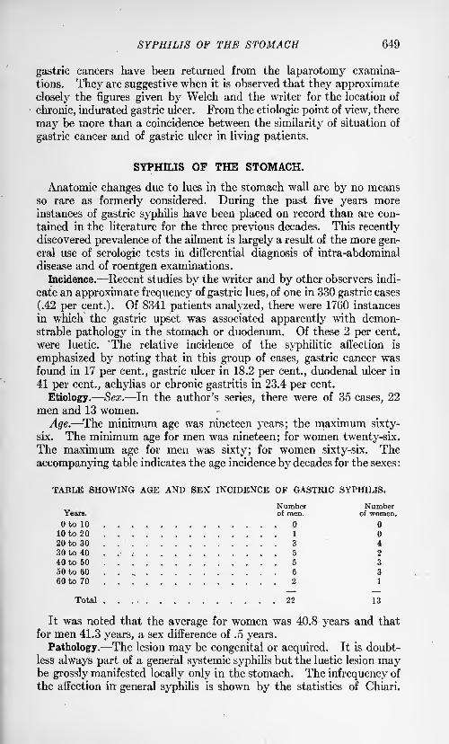
SYPHILIS OF THE STOMACH 649
gastric cancers have been returned from the laparotomy examina-
tions. They are suggestive when it is observed that they approximate
closely the figures given by Welch and the writer for the location of
chronic, indurated gastric ulcer. From the etiologic point of view, there
may be more than a coincidence between the similarity of situation of
gastric cancer and of gastric ulcer in living patients.
SYPHILIS OF THE STOMACH.
Anatomic changes due to lues in the stomach wall are by no meansso rare as formerly considered. During the past five years moreinstances of gastric syphilis have been placed on record than are con-
tained in the literature for the three previous decades. This recentlydiscovered prevalence of the ailment is largely a result of the more gen-eral use of serologic tests in differential diagnosis of intra-abdominal
disease and of roentgen examinations.
Incidence. Recent studies by the writer and by other observers indi-
cate an approximate frequency of gastric lues, of one in 330 gastric cases
(.42 per cent.). Of 8341 patients analyzed, there were 1760 instances
in which the gastric upset was associated apparently with demon-strable pathology in the stomach or duodenum. Of these 2 per cent,
were luetic. 'The relative incidence of the syphilitic affection is
emphasized by noting that in this group of cases, gastric cancer wasfound in 17 per cent., gastric ulcer in 18.2 per cent., duodenal ulcer in
41 per cent., achylias or chronic gastritis in 23.4 per cent.
Etiology. Sex. In the author's series, there were of 35 cases, 22
men and 13 women.
Age. The minimum age was nineteen years; the maximum sixty-
six. The minimum age for men was nineteen; for women twenty-six.The maximum age for men was sixty; for women sixty-six. The
accompanying table indicates the age incidence by decades for the sexes :
TABLE SHOWING AGE AND SEX INCIDENCE OF GASTRIC SYPHILIS.
Number NumberYears. of men. of women.
to 10
10 to 20 1
20 to 30 .. 3 430 to 40 5 2
40 to 50 5 3
50 to 60 . . 6 3
60 to 70 2 1
Total 22 13
It was noted that the average for women was 40.8 years and that
for men 41.3 years, a sex difference of .5 years.
Pathology. The lesion may be congenital or acquired. It is doubt-
less always part of a general systemic syphilis but the luetic lesion maybe grossly manifested locally only in the stomach. The infrequency of
the affection in general syphilis is shown by the statistics of Chiari.
650 SURGICAL DISEASES OF STOMACH AND DUODENUM
From necropsies in 243 pathologically demonstrated cases of lues, of
which in 145 cases the disease was hereditary and 98 cases it was ac-
quired, he noted that, while indirect changes in the stomach wall
(circulatory anomalies, interstitial hemorrhages, toughness of the tissues,
etc.) are relatively common, lesions directly attributable to lues (ulceration,
gummas and scarring) were infrequent.Careful descriptions of luetic lesions in the stomach have been made by
Flexner, Neumann, and by Chiari and Weichselbaum, while clinical
classifications relating thereto have been advanced by Hemmeter,Einhorn, Kohn, Morgan, Downes and LeWald, McNeil and by Euster-
man. It would seem from the nature of the ailment pathologically,that a close histoclinical grouping were possible.
Recent observations of Warthin at the Pathological Laboratory of the
University of Michigan promise to throw an interesting and valuable
light uponthe prevalence of syphilitic lesions inthe gastro-intestinal tract.
Warthin has definitely demonstrated the existence of histopathological
changes in the pancreas similar in every respect to those existing in
chronic syphilitic foci in heart-muscle, aorta, etc. In many of
Warthin's specimens, careful staining by the Levaditi method reveals
spirochetse. It is more than likely that similar studies made uponsections of extirpated gastric walls, from patients with lues presenting
dyspeptic abnormalities, will disclose lesions very similar to those that
Warthin has shown in the pancreas.These definite pathologic observations should be of great value
clinically. They should lead to earlier thought of the possibility of
syphilis producing numerous types of gastric upset and should more
frequently suggest anti-luetic therapy in cases which have resisted
ordinary routine treatment.
Apparently activity of the syphilitic virus first causes a dense
round-cell infiltration of the loose, areolar tissue of the stomach wall.
There is frequently an associated endarteritis. When the disease has
once become firmly established, it appears to produce four rather
characteristic types of lesion: (1) Diffuse infiltrations, confined to
the areolar tissue, causing thickening and stiffening of the gastric wall
("cirrhosis gastrica"); (2) local changes in submucosa and mucosain the form of dense groupings of lymphocytes, which may end as
miliary gummas, or coalesce, necrose and slough forming ragged-edged
ulcers, not infrequently multiple; (3) single or multiple inflammatory
nodules, composed of exuberant connective tissue and lymphocytes;these may involve the entire stomach wall and produce extensive tumors
(ulcerating or non-ulcerating), stenoses or malformations in contour,
and (4) subperitoneal and peritoneal invasions, resulting in perigastritis
associated with thickening of the wall of the viscus and dense peri-
gastric adhesions. Doubtless by proper manipulation of the Levaditi
technic, spirochetse will ultimately be demonstrated in all the types of
lesion above described.
As a consequence of these pathological variations, the clinical mani-
festations of the malady differ widely. Grossly, they may be sum-
SYPHILIS OF THE STOMACH 651
inarized as those associated with chronic gastritis, ulceration (with or
without stenosis), gastric tumor (with or without obstruction) and
perigastritis, sometimes with involvement of adjacent viscera.
The Luetic Lesion. It was often difficult to get a history of the
primary sore. This is particularly apt to be the case with women.Of the men, genital chancres were recorded in 9, buboes in 4,
mouth or lip lesions in 2. In the women genital chancre was reportedthree tunes. In 2 cases there had been mouth sores; in 1, buboes.
Definite knowledge of veneral disease in the husband was obtained in 5
instances; in 2, the husband's condition was questionable. Of the
entire group, there were 9 cases in which no history positive for or
suggestive of lues could be obtained, nor in these patients could exter-
nal evidences of previous lesions be seen. The Wassermann-Noguchitests were, however, definitely positive.
Time of Gastric Disturbance After Acquirement of Lues. Informa-
tion in this line is but relatively correct, especially for the women.The average shows that there was but slight variation in the sexes.
Two years was the minimum quiescent period for both sexes, while
the maximum, was, for men, thirty-eight years and for women, forty-five years.
Stage of the Disease. So far as we were able to judge, we had noinstance of a congenital affection. In three males and two females,the ailment appeared late in the secondary stage of lues. In the
remaining cases (30) the gastric disorder was, etiologically considered,a tertiary manifestation.
Previous Specific Therapy. Of the 21 cases who gave definite or
suspicious history of chancres or of buboes, but 1 1 had had anti-luetic
treatment. Of these only 5 had received a thorough course of mercuryand iodid. Four had been given salvarsan for "anemia" within five
years of their coming under observation.
Wassermann-Noguchi Reactions. These were positive in all our
tabulated cases and furnish the basis of our classification. Inasmuchas the nature of the ailment demands diagnosis apart from surgical or
from sections of tissue removed at laparotomy or necropsy, we fail
to see how another basis for classification can be adopted. Certainly,
dependence solely on the "therapeutic test" of the effect of antiluetic
medicines is not sufficient proof of the disease, for it has been shownthat occasionally sarcomas, tuberculous lesions or other inflammatoryaffections are favorably influenced by salvarsan, mercury or iodids.
If carefully controlled, only rarely is the positive serologic test associ-
ated with disease other than syphilis. In grave anemias, malignantcachexia or alcoholism, dubious Wassermann reactions are occasionallyobtained. However, such factors were ruled out in our group of cases.
The Gastric Malfunction. Duration of All Gastric Disturbances.
Facts of considerable significance are brought out by analyses of this
phase. It has been stated frequently that gastric syphilis manifests
itself rather abruptly in late life without any previous digestive upset
having been noted. Other observers have suggested that the ailment
652 SURGICAL DISEASES OF STOMACH AND DUODENUM
causes great cellular damage to the stomach wall early in life. Our
study indicates that the average duration of dyspepsia was 7.2 years for
the group. The minimum duration was six months a male agedsixty; the maximum, twenty-five years a female aged fifty-two years.There was practically no difference in the average duration between the
sexes.
Duration of the Presenting Gastric Complaint. The average time
for the group was 9.7 months. The minimum was four weeks and the
maximum 4.2 years. In women, dyspepsia caused the patient to seek
relief rather earlier than in men.
Types of Gastric Histories. The wide variations in the pathologyof syphilis of the stomach indicate that a great range is possible in the
clinical picture presented. In the literature too much emphasis has
been placed upon gastric syphilis at its tumor stage ("surgical
syphilis"). It will be recalled that, histopathologically, gummatousformation indicates an approach to the terminal damage which the
syphilitic virus is capable of producing. It is quite evident that the
maximum worth of medical treatment of this affection is possible onlybefore gross deformity of the stomach wall and loss of secretory function
have occurred.
The writer's cases group themselves quite readily, with regard to the
clinical course of the dyspepsia, into three divisions: (1) Instances in
which a persistent gastric derangement appeared in the individuals
who had previously experienced no digestive upset; (2) instances in
which a constant dyspepsia followed years of antecedent indigestionof the intermittent type; (3) instances in which continuous gastric
upset arose in persons who had been affected gastrically at some past
period, but who had been, for years, free from digestive disturbances.
In luetics it is often difficult to ascribe gastric upsets to definite
pathological change in the stomach wall unless the dyspeptic disturb-
ance has preceded evidences of cerebrospinal damage. The roentgendemonstration of gastric deformity is of much service in the doubtful
cases. Each group will be summarized briefly.
GROUP 1. Three of the thirty-five instances of gastric syphilis
studied composed this group. All were men who gave histories of
genital, primary lesions. In one, the dyspepsia appeared shortly
after secondary skin lesions had subsided, in the others it was the onlyevidence of the tertiary stage. The affection was characterized byrather abrupt onset of epigastric pain and soreness, constant in charac-
ter, but varying in intensity; by anorexia, moderate weight loss, con-
stipation, pyrosis, eructations, epigastric tenderness, and in one instance
a freely movable, epigastric nodule and with "delayed vomiting."Gastric analyses revealed a fairly low formol index, absent Wolff test
and moderate hypersecretion, without hyperacidity. In the case
associated with epigastric nodule, fluoroscopic examination showed a
filling-defect involving the pylorus and the antrum. In the other
patients, roentgenoscopy pointed to pyloric ulcers of the chronic,
uncomplicated variety.
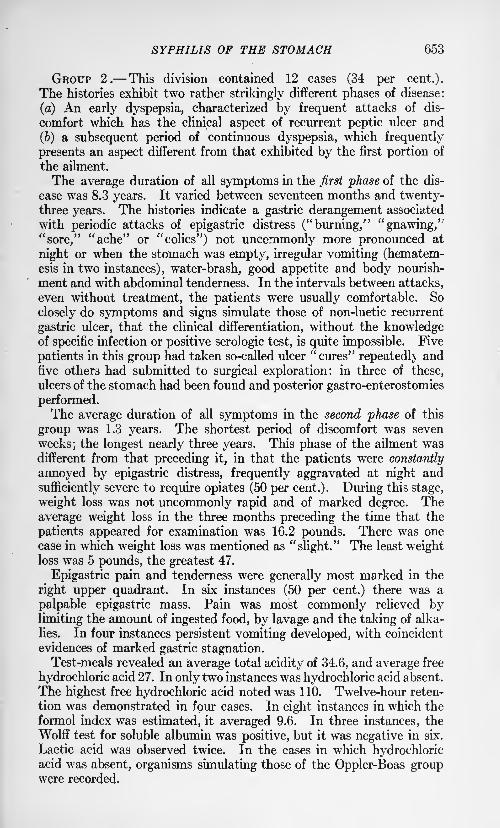
SYPHILIS OF THE STOMACH 653
GROUP 2. This division contained 12 cases (34 per cent.).
The histories exhibit two rather strikingly different phases of disease:
(a) An early dyspepsia, characterized by frequent attacks of dis-
comfort which has the clinical aspect of recurrent peptic ulcer and
(&) a subsequent period of continuous dyspepsia, which frequently
presents an aspect different from that exhibited by the first portion of
the ailment.
The average duration of all symptoms in the first phase of the dis-
ease was 8.3 years. It varied between seventeen months and twenty-three years. The histories indicate a gastric derangement associated
with periodic attacks of epigastric distress ("burning," "gnawing/'
"sore," "ache" or "colics") not uncommonly more pronounced at
night or when the stomach was empty, irregular vomiting (hematem-esis in two instances), water-brash, good appetite and body nourish-
ment and with abdominal tenderness. In the intervals between attacks,
even without treatment, the patients were usually comfortable. So
closely do symptoms and signs simulate those of non-luetic recurrent
gastric ulcer, that the clinical differentiation, without the knowledgeof specific infection or positive serologic test, is quite impossible. Five
patients in this group had taken so-called ulcer "cures" repeatedly andfive others had submitted to surgical exploration: in three of these,
ulcers of the stomach had been found and posterior gastro-enterostomies
performed.The average duration of all symptoms in the second phase of this
group was 1.3 years. The shortest period of discomfort was seven
weeks; the longest nearly three years. This phase of the ailment wasdifferent from that preceding it, in that the patients were constantly
annoyed by epigastric distress, frequently aggravated at night and
sufficiently severe to require opiates (50 per cent.). During this stage,
weight loss was not uncommonly rapid and of marked degree. The
average weight loss in the three months preceding the time that the
patients appeared for examination was 16.2 pounds. There was one
case in which weight loss was mentioned as "slight." The least weightloss was 5 pounds, the greatest 47.
Epigastric pain and tenderness were generally most marked in the
right upper quadrant. In six instances (50 per cent.) there was a
palpable epigastric mass. Pain was most commonly relieved bylimiting the amount of ingested food, by lavage and the taking of alka-
lies. In four instances persistent vomiting developed, with coincident
evidences of marked gastric stagnation.Test-meals revealed an average total acidity of 34.6, and average free
hydrochloric acid 27. In only two instances was hydrochloric acid absent.
The highest free hydrochloric acid noted was 110. Twelve-hour reten-
tion was demonstrated in four cases. In eight instances in which the
formol index was estimated, it averaged 9.6. In three instances, the
Wolff test for soluble albumin was positive, but it was negative in six.
Lactic acid was observed twice. In the cases in which hydrochloricacid was absent, organisms simulating those of the Oppler-Boas groupwere recorded.
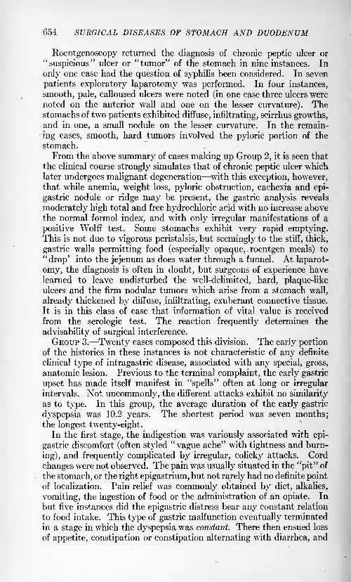
654 SURGICAL DISEASES OF STOMACH AND DUODENUM
Roentgenoscopy returned the diagnosis of chronic peptic ulcer or
"suspicious" ulcer or "tumor" of the stomach in nine instances. In
only one case had the question of syphilis been considered. In seven
patients exploratory laparotomy was performed. In four instances,
smooth, pale, calloused ulcers were noted (in one case three ulcers were
noted on the anterior wall and one on the lesser curvature). Thestomachs of two patients exhibited diffuse, infiltrating, scirrhus growths,and in one, a small nodule on the lesser curvature. In the remain-
ing cases, smooth, hard tumors involved the pyloric portion of the
stomach.
From the above summary of cases making up Group 2, it is seen that
the clinical course strongly simulates that of chronic peptic ulcer whichlater undergoes malignant degeneration with this exception, however,that while anemia, weight loss, pyloric obstruction, cachexia and epi-
gastric nodule or ridge may be present, the gastric analysis reveals
moderately high total and free hydrochloric acid with no increase abovethe normal formol index, and with only irregular manifestations of a
positive Wolff test. Some stomachs exhibit very rapid emptying.This is not due to vigorous peristalsis, but seemingly to the stiff, thick,
gastric walls permitting food (especially opaque, roentgen meals) to
"drop' into the jejenum as does water through a funnel. At laparot-
omy, the diagnosis is often in doubt, but surgeons of experience havelearned to leave undisturbed the well-delimited, hard, plaque-likeulcers and the firm nodular tumors which arise from a stomach wall,
already thickened by diffuse, infiltrating, exuberant connective tissue.
It is in this class of case that information of vital value is received
from the serologic test. The reaction frequently determines the
advisability of surgical interference.
GROUP 3. Twenty cases composed this division. The early portionof the histories in these instances is not characteristic of any definite
clinical type of intragastric disease, associated with any special, gross,
anatomic lesion. Previous to the terminal complaint, the early gastric
upset has made itself manifest in "spells" often at long or irregular
intervals. Not. uncommonly, the different attacks exhibit no similarity
as to type. In this group, the average duration of the early gastric
dyspepsia was 10.2 years. The shortest period was seven months;the longest twenty-eight.
In the first stage, the indigestion was variously associated with epi-
gastric discomfort (often styled "vague ache" with tightness and burn-
ing), and frequently complicated by irregular, colicky attacks. Cord
changes were not observed. The pain was usually situated in the "pit" of
the stomach, or the right epigastrium, but not rarely had no definite pointof localization. Pain relief was commonly obtained by diet, alkalies,
vomiting, the ingestion of food or the administration of an opiate. In
but five instances did the epigastric distress bear any constant relation
to food intake. This type of gastric malfunction eventually terminated
in a stage in which the dyspepsia was constant. There then ensued loss
of appetite, constipation or constipation alternating with diarrhea, and
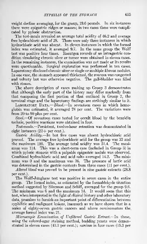
SYPHILIS OF THE STOMACH 655
weight decline averaging, for the group, 19.6 pounds. In six instances
there were epigastric ridges or masses;in two cases these were compli-
cated by pyloric obstruction.
The test-meals revealed an average total acidity of 46.2 and averagefree hydrochloric acid of 28. There were only three instances in which
hydrochloric acid was absent. In eleven instances in which the formol
index was estimated, it averaged 9.7. In the same group the Wolff
test was positive four times. Roentgen records of an intragastric con-
dition simulating chronic ulcer or tumor were obtained in eleven cases.
In the remaining instances, the examination was not made or its results
were questionable. Surgical exploration was performed in ten cases.
Laparotomy disclosed chronic ulcer or single or multiple fibrous nodules.
In one case, the stomach appeared thickened, the mucosa was congestedand velvety but was otherwise negative. The gall-bladder was filled
with stones.
The above description of cases making up Group 3 demonstrates
that although the early part of the history may differ markedly from
that composing the first portion of that outlined in Group 2, the
terminal stage and the laparotomy findings are strikingly similar to it.
LABORATORY DATA. Blood. In seventeen cases in which hemo-
globin was estimated, it averaged 78 per cent. The variations were
from 20 to 90 plus per cent.
Stool. Of seventeen cases tested for occult blood by the benzidin
technic, positive reactions were obtained in four.
Test-meals. Persistent, twelve-hour retention was demonstrated in
eight instances (22+ per cent.).
Gastric Acidity. In but five cases was absent hydrochloric acid
proved. The average free hydrochloric acid for the group was 33 andthe maximum 110. The average total acidity was 51.4. The maxi-
mum was 114. This was a short-term case (included in Group 3) in
which pyloric stenosis with a palpable epigastric nodule was observed.
Combined hydrochloric acid and acid salts averaged 14.2. The mini-
mum was and the maximum was 36. The presence of lactic acid
was determined in the gastric contents from three cases (11 per cent.).
Altered blood was proved to be present in nine gastric extracts (28.8
per cent.).
The Wolff-Junghans test was positive in seven cases in the entire
group. The formol index, as estimated by the formaldehyde titration
method suggested by Sorenson and Schiff, averaged for the group 9.6.
The minimum was 6 and the maximum 14. It would seem that this
test, when interpreted in the light of clinical history and other laboratory
data, promises to furnish an important point of differentiation between
syphilitic and malignant lesions, inasmuch as we have shown that in a
series of eighty-seven gastric cancers and ulcera carcinomatosa the.
average formol index was 21.
Microscopic Examination, of Unfiltered Gastric Extract. In these,
using the colored-agar staining method, budding yeasts were demon-strated in eleven cases (43.3 per cent.) ;
sarcinse in four cases (15.3 per
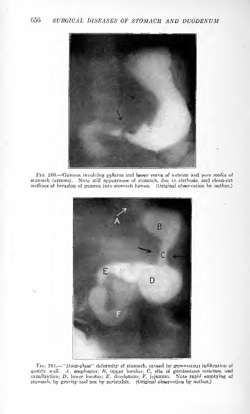
656 SURGICAL DISEASES OF STOMACH AND DUODENUM
FIG. 260. Gumma involving pylorus and lesser curve of antrum and pars media of
stomach (arrows). Note stiff appearance of stomach, due to cirrhosis, and clean-cut
outlines of invasion of gumma into stomach lumen. (Original observation by author.)
FIG. 261. "Hour-glass" deformity of stomach, caused by gummatous infiltration of
gastric wall. A, esophagus; B, upper loculus; C, site of gummatous stricture and
canalization; D, lower loculus; E, duodenum; F, jejunum. Note rapid emptying of
stomach, by gravity and not by peristalsis. (Original observation by author.)
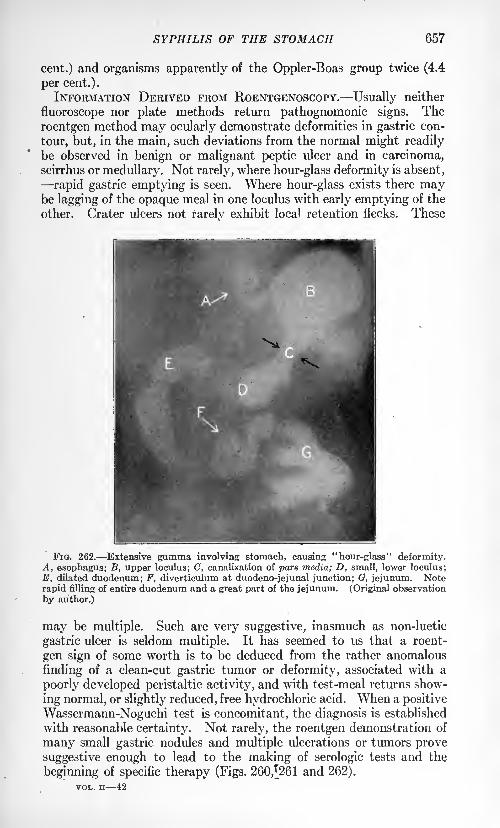
SYPHILIS OF THE STOMACH 657
cent.) and organisms apparently of the Oppler-Boas group twice (4.4
per cent.).
INFORMATION DERIVED FROM ROENTGENOSCOPY. Usually neither
fluoroscope nor plate methods return pathognomonic signs. The
roentgen method may ocularly demonstrate deformities in gastric con-
tour, but, in the main, such deviations from the normal might readily
be observed in benign or malignant peptic ulcer and in carcinoma,scirrhus or medullary. Not rarely, where hour-glass deformity is absent,
rapid gastric emptying is seen. Where hour-glass exists there maybe lagging of the opaque meal in one loculus with early emptying of the
other. Crater ulcers not rarely exhibit local retention flecks. These
FIG. 262. Extensive gumma involving stomach, causing "hour-glass" deformity.A, esophagus; B, upper loculus; C, canalization of pars media; D, small, lower loculus;
E, dilated duodenum; F, diverticulum at duodeno-jejunal junction; G, jejunum. Noterapid filling of entire duodenum and a great part of the jejunum. (Original observation
by author.)
may be multiple. Such are very suggestive, inasmuch as non-luetic
gastric ulcer is seldom multiple. It has seemed to us that a roent-
gen sign of some worth is to be deduced from the rather anomalous
finding of a clean-cut gastric tumor or deformity, associated with a
poorly developed peristaltic activity, and with test-meal returns show-
ing normal, or slightly reduced, free hydrochloric acid. When a positive
Wassermann-Noguchi test is concomitant, the diagnosis is established
with reasonable certainty. Not rarely, the roentgen demonstration of
many small gastric nodules and multiple ulcerations or tumors prove
suggestive enough to lead to the making of serologic tests and the
beginning of specific therapy (Figs. 260,[261 and 262).
VOL. ii 42
658 SURGICAL DISEASES OF STOMACH AND DUODENUM
TUBERCULOSIS OP THE STOMACH.
This affection is very rare. It commonly manifests itself pathologicallyas ulceration, but it may be difficult to exclude a tuberculous etiologyin certain instances of diffuse gastric cirrhosis.
The tuberculous lesion is probably most commonly caused by tubercle
bacilli being deposited from the blood or lymph streams in the loose
areolar tissue of the submucosa. A small round cell infiltration follows,
circulatory disturbances result, and necrosis of the overlying mucusmembrane occurs. In this way crater-ulcer, usually shallow but with
ragged undermined edges, is formed. The edges are usually very vas-
cular. The lesion is commonly solitary, but in rare instances of miliary
tuberculosis, multiple small bleeding ulcers are found. It is uncom-mon for tubercle bacilli that are swallowed during the course of a lungtuberculosis to produce peptic ulcer. The pepsin and hydrochloricacid of the stomach usually render such swallowed bacilli inert. In
instances where the sputum is particularly rich in bacilli and rapidemaciation has caused gastric juice of low hydrochloric acid content,direct invasion of the gastric mucosa at points of local injury may be
possible.
Diagnosis. The diagnosis of tuberculous ulcer of the stomach should
be suspected in all instances where an extra-gastric tuberculous focus
has been proved to exist and where there occurs a dyspepsia exhibitingan ulcer syndrome. In tuberculous ulcer, the symptoms are quitesimilar to those manifested by gastric ulcer due to other causes.
Fluoroscopy usually demonstrates a large shallow crater-ulcer with
gastric deformity and retention of flecks of the opaque roentgen-meal.In rare instances, tubercle bacilli may be found in the gastric contents
or the stools, when such have been well extracted with normal salt
solution and centrifugalized fractionally, prepared for staining by the
antiformin method and then colored by the Ziehl-Neelson technic.
CANCER OF THE STOMACH.
Definition. A disease of the stomach, produced by the growth of a
neoplasm in its wall. The affection is commonly of a perniciously pro-
gressive nature and occurs with the greatest frequency between the
ages of forty and seventy years. Fatal termination is the rule. Thedisease is characterized clinically by imperfect gastric function. This
malfunction is generally manifested by abdominal distress or pain,
associated with loss of appetite and decrease in body weight, by weak-
ness, anemia, cachexia, vomiting and the appearance of a supra-umbilical tumor.
The gastric extracts or the vomitus often exhibit deficient gastric
emptying power, absent free HC1, altered blood and the presence of
foreign organic acids, microorganisms and ferments.
When the stomach is filled with substances opaque to the roentgen
ray, alterations from the normal contour are not infrequently demon-
CANCER OF THE STOMACH 659
strable upon fluoroscopic examination or when roentgenograms are
made.Incidence. The recently compiled statistics of Hoffmann indicate
that gastric cancer is increasing in frequency. The increase is real andnot apparent merely as a consequence of more carefully tabulated
mortality figures. In the civilized portion of the world it is computedthat the general cancer death-rate has increased 23.3 per cent, in the
past decade. It is estimated that there were 75,000 deaths from cancer
in the United States in 1913. Of this number more than 28,500 (38
per cent.) were instances of cancer of the stomach.
The writer's study of 82,000 hospital admissions for all forms of
medical and surgical ailment indicates that gastric cancer existed in
approximately 1 out of 89 cases.
Age. In the study of 921 operatively and pathologically provedinstances of the disease, the writer found that the greatest numberof cases was in the sixth decade of life. Between ages of forty to sixty-
nine occurred 84.9 per cent, of all cases. Nineteen instances were
recorded at or below the age of thirty-one. The youngest patient was
eighteen (a female). In males, 40 per cent, of cases were betweenthe ages fifty to fifty-nine; in females the greatest frequency was in
the fifth decade (78 per cent, between ages forty to sixty-nine).
Sex. Authors, studying material collected a half century ago,
appear to establish that there is little difference in the sex incidence
of cancer of the stomach. Recent observations would indicate that the
disease is much more frequent in males than in females. Of 150 cases
reported by Osier and McCrae, there were 126 males and 24 females.
In the writer's 921 cases there were 693 males and 228 females, a ratio
of approximately 3 to 1.
Race. It would seem that people far removed from so-called civil-
ized zones are but infrequently victims of the disease. It is doubtful
if they are immune. More thorough medical scrutiny of causes of
death among such people will probably disclose the occurrence of gastric
cancer. Hoffmann's statistics indicate that negroes below the sixth
decade are particularly non-resistant to cancer of the stomach. OfFriedenwald's cases, 5.2 per cent, were negroes. Miyake has reported the
ailment among orientals. Our series indicates its occurrence amongItalians, Mexicans, Portuguese and Alaskans, Hendley records 102
cancerous patients at Jeypore, India. It has frequently been stated
that Jews are relatively free from gastric cancer. From a compara-tively small number of Jewish patients, there were seventeen instances
of the disease, 1.8 per cent, in the writer's 921 cases.
Heredity. In our series a family or blood relationship history of
cancer generally, was obtained in 8.5 per cent. The stomach wasstated as being the location of the disease in 58.9 per cent, of this group.While heredity appears to play a part in the predisposition to cancer in
certain families, yet its exact significance is not settled. The transmis-
sion of the same type of malignant tumor to homologous organs was
emphasized by Virchow with respect to anal melanosis of horses. Slye
660 SURGICAL DISEASES OF STOMACH AND DUODENUM
has proved the susceptibility to tumor implantation in certain strains
of laboratory animals (mice).
With regard to heredity in humans, it is quite likely that the trans-
mission by parents to offspring of a mode and conduct of life are
factors. The conjugal state appears at times to influence the develop-ment of gastric cancer. In our series there were seven instances of the
disease developing in man or wife within five months of its inceptionor fatal issue in the mate.
Social Status. Material abundance in some way seems to favor the
development of malignancy. Gastric cancer is relatively of more com-mon incidence among the prosperous, overfed and physically under-
worked than it is among people who are actually struggling for existence.
Hoffmann states that at the ages forty to fifty the proportionate
mortality from cancer for males was 5.1 per cent, in the wage-earning
group and 6.7 per cent, in the well-to-do division; it was for females
at the same decade 13.7 for the wage-earners and 18.5 for the prosperousAt the age of forty-five and over, the cancer mortality for overweightswas 15.6 per 10,000 and for underweights 12.0 per 10,000.
Occupation. No type of work appears to have direct influence uponthe frequency of the disease. A sedentary life following a previousactive career seems to favor gastric disturbances and with such, cancer.
The ailment is very common among farmers or people dwelling in rural
communities. These made up more than 40 per cent, of our series.
Ochsner has emphasized the relationship of excessive ingestion of
contaminated fruits and vegetables to malignant disease of the stomach.
Such food is consumed inlarge quantities bythosedwellinginruralregions.Habits. There is no actual proof that excessive indulgence in alcohol
or the use of tobacco predisposes to gastric cancer. Groups of statistics
supporting or refuting this statement may be readily compiled. In
our series, but 5.3 per cent, of the patients were heavy drinkers andbut 15.1 per cent, were excessive smokers.
Trauma. Injury to the stomach may be mechanical, chemical,
biochemical or parasitic. The injury may be internal or external. If
the trauma is internal, it must doubtless be long maintained and be
preceded by alterations from the normal in the gastric mucosa, before
it is capable of causing malignant change. It is conceivable that in
such circumstances, irritation from very hot or cold foods, hard pieces
of food, foreign bodies, alcohol, tobacco juice, substances formed as a
consequence of food deterioration or parasites (e. g., nematodes as
shown by Fibiger in rats) may have influence in initiating cancerous
change or may aggravate an already existing lesion and prevent its
spontaneous cure.
External traumata (blows, falls, crushing accidents, etc.) would
at times seem to arouse latent processes to activity. In patients giving
history of a chronic dyspeptic ailment, which has recently becomecontinuous and is associated with constant weight decline, the factor
of recent abdominal injury should be always considered. Of the
writer's cases of gastric cancer, there was a history of external traumain 1.5 per cent.
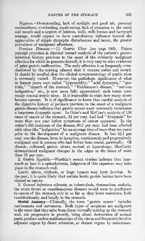
CANCER OF THE STOMACH 661
Hygiene. Overcrowding, lack of sunlight and good air, personal
uncleanliness, overfeeding, rapid eating, lack of attention to the teeth
and mouth and a neglect of latrines, wells, milk houses and barnyard
seepage, would appear to have contributory influence toward the
aggravation of simple dyspeptic disturbances and hence, the greater
prevalence of malignant affections.
Previous Diseases. (1) Gastric Ulcer (see page 640). Unless
especial attention is directed toward analysis of the patient's gastro-intestinal history previous to the onset of the evidently malignantaffection for which he presents himself, it is very easy to miss evidences
of prior gastric malfunction. The early affection is so frequently over-
shadowed by the existing ailment that it escapes notice and record.
It should be recalled that the clinical symptomatology of peptic ulcer
is extremely varied. Moreover, the pathologic significance of whatin former years were called "hyperacidity," "acid dyspepsia," "gas-
tritis," "catarrh of the stomach," "Reichmann's disease," "nervous
indigestion," etc., is now more fully appreciated: such terms com-
monly conceal peptic ulcer. It is impossible to state how many ulcers
become cancers. It is of significance to know that careful analysis of
the digestive history of patients previous to the onset of a malignant
gastric disease indicates that gastric cancer most commonly terminates
a long-term dyspeptic upset. In Osier's and McCrae's analysis of 150
cases of cancer of the stomach, 22 per cent, had had "dyspepsia" for
more than one year before symptoms of cancer appeared. In the
writer's 921 instances of the disease, 60.5 per cent, had been affected
with ulcer-like"indigestion" for an average time of more than ten years
prior to the development of a malignant disease. In but 32.1 percent, was the disease, from its inception, continuously and progressively
malignant and hi persons who had before been sound, gastrically. Of
chronic, calloused, gastric ulcers, excised at laparotomy, MacCartydemonstrated malignant changes in the edges or the bases of morethan 70 per cent.
2. Gastric Syphilis. Warthin's recent studies indicate that inas-
much as lues is a spirochetemia, lodgment of this organism may take
place in the stomach wall.
Leutic ulcers, cirrhosis, or huge tumors may later develop. In
the past, it is quite likely that certain leutic gastric lesions have been
classed as cancer.
3. General infectious ailments, as tuberculosis, rheumatism, malaria,
the acute fevers or exanthematous diseases would seem to predisposeto cancer of the stomach only in so far as they lower resistance, both
constitutionally and locally in the stomach.
Morbid Anatomy. Clinically, the term "gastric cancer' includes
carcinomata and sarcomata. Both types of neoplasm are malignantin the sense that they arise from tissue normally making up the stomach
wall, are progressive in growth, bring about destruction of normal
parts, produce useless malformations of the viscus and frequently involve
adjacent organs by direct extension, or distant organs by metastases.
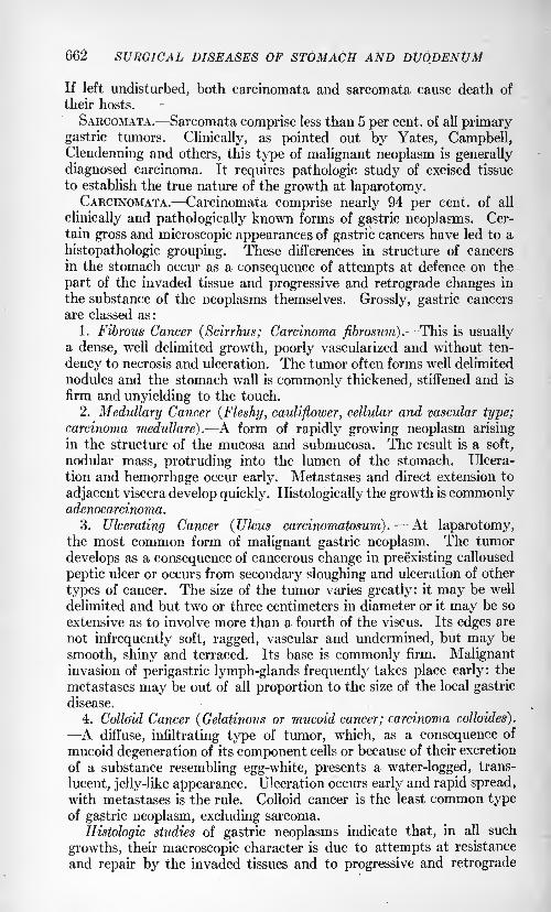
662 SURGICAL DISEASES Of STOMACti Atit> DVODENVM
If left undisturbed, both carcinomata and sarcomata cause death of
their hosts.
SARCOMATA. Sarcomata comprise less than 5 per cent, of all primarygastric tumors. Clinically, as pointed out by Yates, Campbell,Clendenning and others, this type of malignant neoplasm is generally
diagnosed carcinoma. It requires pathologic study of excised tissue
to establish the true nature of the growth at laparotomy.CARCINOMATA. Carcinomata comprise nearly 94 per cent, of all
clinically and pathologically known forms of gastric neoplasms. Cer-
tain gross and microscopic appearances of gastric cancers have led to a
histopathologic grouping. These differences in structure of cancers
in the stomach occur as a consequence of attempts at defence on the
part of the invaded tissue and progressive and retrograde changes in
the substance of the neoplasms themselves. Grossly, gastric cancers
are classed as:
1. Fibrous Cancer (Scirrhus; Carcinoma fibroswri). This is usuallya dense, well delimited growth, poorly vascularized and without ten-
dency to necrosis and ulceration. The tumor often forms well delimited
nodules and the stomach wall is commonly thickened, stiffened and is
firm and unyielding to the touch.
2. Medullary Cancer (Fleshy, cauliflower, cellular and vascular type;
carcinoma medullare). A form of rapidly growing neoplasm arisingin the structure of the mucosa and submucosa. The result is a soft,
nodular mass, protruding into the lumen of the stomach. Ulcera-
tion and hemorrhage occur early. Metastases and direct extension to
adjacent viscera develop quickly. Histologically the growth is commonlyadenocarcinoma.
3. Ulcerating Cancer (Ulcus carcinomatosum) . At laparotomy,the most common form of malignant gastric neoplasm. The tumor
develops as a consequence of cancerous change in preexisting calloused
peptic ulcer or occurs from secondary sloughing and ulceration of other
types of cancer. The size of the tumor varies greatly: it may be well
delimited and but two or three centimeters in diameter or it may be so
extensive as to involve more than a fourth of the viscus. Its edges are
not infrequently soft, ragged, vascular and undermined, but may be
smooth, shiny and terraced. Its base is commonly firm. Malignantinvasion of perigastric lymph-glands frequently takes place early: the
metastases may be out of all proportion to the size of the local gastric
disease.
4. Colloid Cancer (Gelatinous or mucoid cancer; carcinoma colloides).
A diffuse, infiltrating type of tumor, which, as a consequence of
mucoid degeneration of its component cells or because of their excretion
of a substance resembling egg-white, presents a water-logged, trans-
lucent, jelly-like appearance. Ulceration occurs early and rapid spread,
with metastases is the rule. Colloid cancer is the least common typeof gastric neoplasm, excluding sarcoma.
Histologic studies of gastric neoplasms indicate that, in all such
growths, their macroscopic character is due to attempts at resistance
and repair by the invaded tissues and to progressive and retrograde
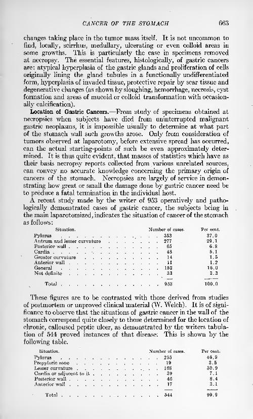
CANCER OF THE STOMACH 663
changes taking place in the tumor mass itself. It is not uncommon to
find, locally, scirrhus, medullary, ulcerating or even colloid areas in
some growths. This is particularly the case in specimens removedat necropsy. The essential features, histologically, of gastric cancers
are: atypical hyperplasia of the gastric glands and proliferation of cells
originally lining the gland tubules in a functionally undifferentiated
form, hyperplasia of invaded tissue, protective repair by scar tissue and
degenerative changes (as shown by sloughing, hemorrhage, necrosis, cystformation and areas of mucoid or colloid transformation with occasion-
ally calcification).
Location of Gastric Cancers. From study of specimens obtained at
necropsies when subjects have died from uninterrupted malignant
gastric neoplasms, it is impossible usually to determine at what partof the stomach wall such growths arose. Only from consideration of
tumors observed at laparotomy, before extensive spread has occurred,
can the actual starting-points of such be even approximately deter-
mined. It is thus quite evident, that masses of statistics which have as
their basis necropsy reports collected from various unrelated sources,
can convey no accurate knowledge concerning the primary origin of
cancers of the stomach. Necropsies are largely of service in demon-
strating how great or small the damage done by gastric cancer need be
to produce a fatal termination in the individual host.
A recent study made by the writer of 953 operatively and patho-
logically demonstrated cases of gastric cancer, the subjects being in
the main laparotomized, indicates the situation of cancer of the stomachas follows:
Situation. Number of cases. Per cent.
Pylorus ..... 353 37.0
Antrum and lesser curvature 277 29 . 1
Posterior wall 65 6.8Cardia 48 5.1
Greater curvature 14 1.5
Anterior wall 11 1.2
General 152 16.0
Not definite 33 3.3
Total 953 100.0
These figures are to be contrasted with those derived from studies
of postmortem or unproved clinical material (W. Welch). It is of signi-
ficance to observe that the situations of gastric cancer in the wall of the
stomach correspond quite closely to those determined for the location of
chronic, calloused peptic ulcer, as demonstrated by the writers tabula-
tion of 544 proved instances of that disease: This is shown by the
following table.
Situation. Number of cases. Per cent.
Pylorus 255 46.9
Prepyloric zone 19 3.5Lesser curvature . 168 30.9Cardia or adjacent to it 39 7.1
Posterior wall 46 8.4Anterior wall 17 3.1
Total . 544 99.9
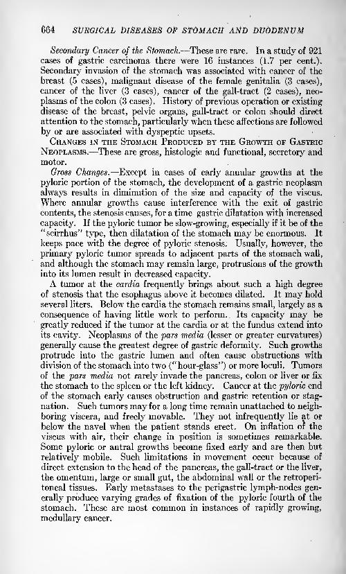
664 SURGICAL DISEASES Of STOMACH AND DUODENUM
Secondary Cancer of the Stomach. These are rare. In a study of 921
cases of gastric carcinoma there were 16 instances (1.7 per cent.).
Secondary invasion of the stomach was associated with cancer of the
breast (5 cases), malignant disease of the female genitalia (3 cases),
cancer of the liver (3 cases), cancer of the gall-tract (2 cases), neo-
plasms of the colon (3 cases). History of previous operation or existingdisease of the breast, pelvic organs, gall-tract or colon should direct
attention to the stomach, particularly when these affections are followed
by or are associated with dyspeptic upsets.CHANGES IN THE STOMACH PRODUCED BY THE GROWTH OF GASTRIC
NEOPLASMS. These are gross, histologic and functional, secretory andmotor.
Gross Changes. Except in cases of early annular growths at the
pyloric portion of the stomach, the development of a gastric neoplasmalways results in diminution of the size and capacity of the viscus.
Where annular growths cause interference with the exit of gastric
contents, the stenosis causes, for a time gastric dilatation with increased
capacity. If the pyloric tumor be slow-growing, especially if it be of the
"scirrhus" type, then dilatation of the stomach may be enormous. It
keeps pace with the degree of pyloric stenosis. Usually, however, the
primary pyloric tumor spreads to adjacent parts of the stomach wall,
and although the stomach may remain large, protrusions of the growthinto its lumen result in decreased capacity.A tumor at the cardia frequently brings about such a high degree
of stenosis that the esophagus above it becomes dilated. It may hold
several liters. Below the cardia the stomach remains small, largely as a
consequence of having little work to perform. Its capacity may be
greatly reduced if the tumor at the cardia or at the fundus extend into
its cavity. Neoplasms of the pars media (lesser or greater curvatures)
generally cause the greatest degree of gastric deformity. Such growths
protrude into the gastric lumen and often cause obstructions with
division of the stomach into two ("hour-glass") or more loculi. Tumorsof the pars media not rarely invade the pancreas, colon or liver or fix
the stomach to the spleen or the left kidney. Cancer at the pyloric end
of the stomach early causes obstruction and gastric retention or stag-
nation. Such tumors may for a long time remain unattached to neigh-
boring viscera, and freely movable. They not infrequently lie at or
below the navel when the patient stands erect. On inflation of the
viscus with air, their change in position is sometimes remarkable.
Some pyloric or antral growths become fixed early and are then but
relatively mobile. Such limitations in movement occur because of
direct extension to the head of the pancreas, the gall-tract or the liver,
the omentum, large or small gut, the abdominal wall or the retroperi-
toneal tissues. Early metastases to the perigastric lymph-nodes gen-
erally produce varying grades of fixation of the pyloric fourth of the
stomach. These are most common in instances of rapidly growing,
medullary cancer.
CANCER Of THE STOMACH 665
Metastases in Gastric Cancer. Enlargement of perigastric lymphaticsoccurs in more than 70 per cent, of instances of the disease. As
MacCarty and Blackford have pointed out, the extent of perigastric
lymph-node invasion bears little relation to the size of the primary
process in the stomach wall: the size of the individual lymph-glands is
no "evidence as to whether or no they contain malignant tissue; malig-nant glands are usually hard to the touch and their cut surfaces appear
glistening white. Upon the extent of lymph-gland invasion dependsthe prognosis, in a given case. This is wholly independent of the size,
mobility or histopathologic character of the primary stomach tumor.
Lymph-glands most commonly involved, in addition to those directly
about the stomach, are those along the spine and in the rectovesical
space, in the mediastinum and above the left clavicle and the inguinal
group. The navel may be fixed and the seat of a firm, occasionallyulcerated growth.
Metastases to the liver occur in from 15 to 35 per cent, of all cases
of gastric cancer, depending upon the stage of the disease at which the
growth is interrupted and upon the histologic nature of the tumor. In
the writer's series, the liver was involved in more than 18 per cent.
Liver and gall-tract metastases may result in portal obstruction
and ascites. This may develop very rapidly, especially if there has
been early spread of the cancer directly to the peritoneum. Involve-
ment of the pleura, pericardium, mediastinal lymphatics or the
lungs occurs in from 5 to 11 per cent, of all instances of the disease.
When the pleura is invaded serous exudation appears early. The punc-ture fluid not infrequently contains red blood-cells, endothelial cells
presenting atypical, mitotic figures and, as Roger Morris has pointed
out, an increase in the volume of incoagulable nitrogen. If the lungtissue be invaded, malignant consolidation or necrosis with hemorrhageor abscess occur.
Symptoms and Signs. As a result of recent pathological and clinical
investigations, it is now possible to state that much of the burden of
the early diagnosis of gastric cancer rests upon the observations of
the skilled surgical pathologist; that such diagnosis is best made from
careful study of the histological variations occurring in recently extir-
pated tissue; that the malignant departures from the normal are least
evident in tissue removed from subjects whose gastric history has beenthat of recurring peptic ulcer, and in whom the ailment appeared to
be but ulcer of this type to both the eye and the hand at laparotomy.These ulcers are commonly greater than 2 cm. in diameter.
While these recently established facts impose a new responsibility
upon the pathologist, it should be emphasized strongly that the labora-
tory worker is dealing with a picked material: the segregation of
patients must be made by clinical analyst, internist and surgeon. The
patient's fate depends in direct ratio upon the completeness of the coop-eration existing among the three. In their hands, likewise, rests the
problem of substantially reducing the mortality of the race from gas-tric cancer. As an attempt to further this object, the writer presents
666 SVRGICAL DISEASES OF STOMACH AtfD DUODENUM
briefly certain facts derived from a clinical study of his records of 921
operatively and pathologically demonstrated instances of cancer of
the stomach. This study indicates that the cases of gastric cancer
arrange themselves into the following well-defined clinical order:
Clinical Grouping. (1) A symptom-complex of two very definite and
different stages: (a) gastric disturbance, non-malignant and generally
chronic; (6) gastric malfunction of malignant type. (2) A symptom-complex generally of a distinctly apparent, pernicious nature. (3) Agroup of cases in which there is usually evidence of years of gastricdisturbance of irregular form clinically, on which is superimposed a
type of affection easily recognized as malignant.GROUP 1. Symptom-complex wherein Two Definite and Different
Stages of Disease are Present. This group furnished 61 per cent, of
our series. The average duration of all symptoms in the first stage was11.7 years. The average duration of all symptoms in the second periodis nearly six months. The two periods differ markedly. The anamnesis
brings out very well the characteristic manifestations of each period.It is only by disengaging the patient from his present state and endeav-
oring, definitely, to locate his first departure from gastric health that
one gets the key to the situation.
In 61 per cent, of our proved cancer cases the total time of gas-tric complaint was somewhat over twelve years. For practically
11^ years the symptomatology was that which, clinically, we asso-
ciate with chronic gastric ulcer. This first period was characterized
by a long series of attacks of "indigestion." These attacks were asso-
ciated with epigastric distress or pain in 88 per cent, of instances.
In 85 per cent, the distress had some relation to the taking of food,
the pain-free interval being long or short according to kind of food
eaten. After a solid, mixed meal, the pain relief was longer, but whenit recurred it was unmistakably more severe than when lighter forms
of nourishment had been indulged in. Other forms of pain relief were
alkalies, vomiting and diet. In 54 per cent, of instances the pain wasreferred to some part of the back. The appetite was usually limited
by the fear of takipg food and thus causing pain, or by the dread of
bringing on bleeding. The food desire in this first stage was generally
strong. Weight was lost in the attacks but gained between them . The
average weight loss at this period was 17.4 pounds. Constipation was
noted in 52 per cent, of instances. In 8 per cent, there was diarrhea.
Vomiting occurred in 60 per cent. Bather more than 70 per cent,
vomited in attacks. The time of vomiting was generally when distress
was most marked. Bleeding (hematemesis or melena) was noted in
17 per cent, of instances during this stage. Of the whole number
bleeding, 49 per cent, had bled at least two years prior to coming under
observation. Of those who bled within the two-year period over 65
per cent, were of the group with long previous history. In this groupanemia was rarely marked, the average hemoglobin being about 80
per cent. (Tallquist or Dare).If this class of case pursues the above outlined history, the fol-
OP THE STOMACH 66?
lowing may ensue: (1) Complete healing with stoppage of all symp-toms; (2) typical chronic, benign gastric ulcer may result, or (3) gastric
cancer may form, whereas previously there had been but a benign
process. And no one can prognose in a given case presenting the
clinical picture of gastric ulcer just which ulcer will heal or continue
its benign course or which will ultimately form the suitable ground
for cancer. When we recall the pathology in these cases we can hardlyblame ourselves for not catching the ulcer clinically in transition to
cancer. All ulcers have about and in them hyperplastic tissue tissue
of defense and of healing. No one can tell in a given case when this
hyperplasia will change over from the simple type to the malignantform. Clinically we are only warned by the appearance in a patientof a more or less continuous gastric disorder, where attacks had before
been the rule. This may occur with or without the onset of pyloric
obstruction. Where previously there was trouble in spells, with food
relief, we note food aggravation, night distress, delayed vomiting, con-
tinuous presence of occult blood in the stools, anemia, cachexia, weightloss steadily maintained, weakness, and that peculiar, hopeless apathyexhibited by the fades. Pain is rarely marked. Vague distress,
"heavy feeling" or soreness in the epigastrium is the rule. Severe
pain was observed in but 19 per cent, of our cases. It was of "colicky"nature in 14.2 per cent. Gassy and bloated sensations are generally con-
stant and annoying.In this second stage vomiting becomes more constant and is fre-
quently of greater amount. The odor of the vomitus changes from dis-
tinctly sour or yeasty to cheesy or rancid. The color of the vomitus
may exhibit little if any alteration. Inasmuch as more than 94 per cent,
of our cases of gastric cancer were of the medullary type, oozing of
blood was not uncommon. This pathologic state usually imparts a
tan to deep brown color to vomitus or stomach extracts. In rather
more than 17 per cent, of our cases of gastric cancer was the vomitus
of the classic "coffee ground" color. When it presented these charac-
teristics the disease was far advanced in 84.2 per cent, of instances,
and inoperable (so far as resection was concerned) in 77 per cent. Thevomitus generally exhibits excess of thick, tenacious mucus, intermixed
with sizable food remnants, frequently poorly chymified.The stools rarely show macroscopic changes unless hemorrhage
complicates, or liver, pancreas or adjacent hollow viscera becomeinvolved. On a milk diet, chemical tests for blood were positive in
93.4 per cent, of cases tested.
GROUP 2. Symptom-complex in Cases Clinically Malignant from the
Inception of Gastric Malfunction. Of the 921 cases 31.6 per cent, fell in
this group. Careful history taking failed to elicit any clinical type of
gastric complaint in these patients previous to an average time of 6.4
months before they came under observation. It should be emphasizedthat because no gastric disturbance had been previously noted, the con-
clusion should not be forced that these patients had had no gastric
pathology previously. On the contrary, our postmortem or laparotomy
668 SURGICAL DISEASES OP STOMACH AND DUODENUM
findings of healed ulcers, and even of gastric new growths in individuals
who had no clinical symptoms referable to gastric disease, should lead
one to exercise caution in insisting when or how malignant processesarise in the stomach. The longest history of gastric symptoms in this
group extended over nearly three and one-fourth years; the shortest,
barely three weeks.
In this group, which we have, from the pathologic view of the disease,
styled "primary" gastric cancer, often it is extremely difficult to state
from clinical presentings just when cancer began. Many so-called
"primary" cancers have histories extending over weeks of time, wherethe symptom-complex closely approximates that which our present-
day vogue associates with gastric ulcer of the continuously active and
progressive form. The clinical picture is different, however, in one
important respect : to that symptom-complex are added the manifes-
tations of a malignant systemic poisoning. Instead of fairly definite
attacks of ulcer type of dyspepsia, and these alone, the process is con-
tinuous and associated with progressive weakness, anemia, weight-losswith cachexia and vomiting, changing rapidly to the rancid kind. Epi-
gastric pain may be severe and prostrating at first, but later moderatesin severity. It is not relieved, but is aggravated, by food intake. It
ultimately becomes continuous. The writer has frequently had strikingly
impressed upon him the ''ulcer features," clinically, in primary gastric
cancer. The hurried progression to definite malignancy has seemed to
emphasize the necessity for separate recognition clinically of the term"chronicity" in its pathologic sense and in its time-duration sense, that
is, signifying weeks, months or years continuation of disease. In manyof these "primary" gastric cancers, the clinical gamut of "gastritis,""ulcer" and "cancer" may be run in a few months or even weeks.
The whole course of the ailment is then compressed into a sort of tab-
loid with regard to time: from this course, parts are readily segregatedwhich closely approximate, clinically, those stretching over years, as
exhibited by the great mass of cancers forming Group 1. With this
state of affairs, it is not to be wondered at that patients present them-
selves to the specialist or to the surgeon only when tumor appears in
the epigastrium or when hopeless cachexia warns of impending death.
While the above type of onset of "primary" gastric cancer is not
infrequent, fully two-thirds of this group have more or less vague recol-
lections of how they were affected gastrically before loss of appetite,
weakness, mental inertia, abdominal distress or epigastric "hardness,"
"knob," or "lump" warned them that something graver than "chronic
indigestion," existed. Rarely is the transition from perfect gastric
health to their present state a rapid one. Aversion to certain forms of
food (meats, milk, sweets or legumes) is not infrequently an early
symptom. While the appetite for certain foods may be keen, the
aversion to others may be marked and, for the individual, radical.
Eructations, often of extremely disagreeable taste, may become pro-nounced early. Gassy abdominal distention may be troublesome.
Unexplained nausea with or without physical weakness is manifested.
CANCER OF THE STOMACH 669
In 17 per cent, of the cases of this group, diarrheic stools appeared with-
out apparent cause. Vomiting may be the first symptom and its
copiousness pronounced and alarming. It is commonly bitter, sour,
cheesy, or is described as "rotten" in taste. Vomiting was observed
in 79 per cent, of this class. In 64 per cent, of those vomiting, it was a
daily event. In more than 80 per cent, of these "delayed" vomitingwas exhibited.
Consideration of the types of case making up our grouping of
"primary" gastric cancer would be incomplete without mention of
that class which it is appropriate to style "fulminant" cancer. In
this series, such comprised 7.7 per cent, of cases. The whole his-
tory of the affection up to the time of laparotomy covered less than
ten weeks. The average age of these patients was forty-six years.In this subgroup, profound cancer intoxication was pronounced fromthe time of the first departure from gastric health. In 11 instances
fever rose as high as 102 F. The leukocytes averaged 12,400. Anemiawas of lower degree than is usual in gastric cancer. The hemoglobin
averaged 56 per cent. Weakness, weight-loss, mental dulness, foul
eructations and copious vomiting, marked gassy distention of the epi-
gastrium, abdominal soreness and the appearance of a rapidly enlarging
supra-navel mass occurred with astonishing speed. At operation, the
malignant process was always irremovable and perigastric lymph-gland invasion was extensive. The pathologic examination of tissue
removed, showed cancer of the medullary type.GROUP 3. Gastric Malignancy following Long History of Clinically
Atypical Dyspepsia or Dyspepsia Considered Dependent upon Extra-
gastric Causes. Five and one-half per cent, of cases make up this
division. Careful history taking brings out two markedly different
types of gastric malfunction. The clinical manifestations precedingthose evidently malignant, extended over an average period of rather
more than nine years. The time taken up by the supervening malig-nant stage averaged rather more than five and one-half months.The clinical symptomatology of the affections, while they were
apparently of benign nature, usually pointed toward disease of the gall-
bladder, appendix, large bowel or the pancreas.The clinical symptomatology of this class of case, was, when the
malignant period supervened, in every way similar to that of the second
period of the cases making up Group 1 or of some portion of that
exhibited by those in Group 2. The point to be emphasized is, that after
years of gastric departure from health, of atypic form but evidently
benign so far as our clinical knowledge extends, the picture changedabruptly to that of definite malignancy. It is only in those patients whohad but recently submitted to laparotomy for extragastric disease,
however, that we can positively state that gastric ulcer or gastric
cancer, with atypical manifestations, had not existed for some time
previous to that terminal stage of evident malignancy, clinically.
Prominent Clinical Signs and Symptoms. (a) HEMORRHAGE. Macro-
scopic. Of the whole number of cases making up this series, gross
670 SURGICAL DISEASES OF STOMACH AND DUODENUM
evidences of bleeding were recorded in 18.5 per cent. Of the wholenumber bleeding, 17.4 per cent, had exhibited hematemesis or melenaat least two years before coming under observation. In the large
majority of instances, where such early bleeding had been noted, the
"precarcinomatous" history conformed to that which we interpret,
clinically, as chronic peptic ulcer.
Chemical Test for Blood. In the gastric extracts, such was positivein rather more than 88 per cent, when tested by the guaiac or the
benzidin methods. The stools examined after patients had been uponmeat-free diet gave positive chemical blood test in rather more than93 per cent, of cases.
The interpretation of chemical tests for blood in stool must be care-
fully controlled. In those instances where there is a slowly progress-
ing malignant ulcer, benzidin or guaiac tests are apt to be positive onlyintermittently. Given an ulcer history in an individual past middle life,
the persistent demonstration of altered blood in the stools is of con-
siderable significance toward the probability of malignancy. It is
thus seen, that positive occult blood-tests are of greater service in
giving information with respect the histopathology of a peptic ulcer
than in merely demonstrating that an ulcer exists. It would also
appear that chemical tests for blood, in the properly prepared stools,
furnish a valuable and early diagnostic sign of gastric cancer. This is
to be especially emphasized when it is recalled that ulcera carcinomatosa
are the forms of gastric cancer most amenable to surgical treatment.
In cases where gastric cancer was well advanced chemical tests for
blood were positive in more than 98 per cent, of the stools examined.
Of course when this finding was noted, other evidences of gastric cancer
were usually not lacking.
(6) VOMITING. This is generally due to a persistent mechanical
fault in the emptying power of the stomach. The mechanical hin-
drance to the passage of food from the stomach most commonly occurs
earliest as a consequence of varying grades of stenosis, associated with
what, clinically, appears chronic peptic ulcer. In this type of case,
vomiting is at first intermittent but later becomes persistent. This
clinical hint, especially if it is associated with a history of ulcer dys-
pepsia, in any patient should point to prompt surgical procedures. Such
radical measure often leads to the demonstration of well localized
gastric cancer and permits of its complete extirpation. When cancer
has become far advanced, vomiting is a prominent symptom in nearly80 per cent, of cases. When the tumor involves the cardia, the fundus
or the pars media (with or without hour-glass formation) vomiting is
very apt to occur shortly after food has been swallowed. The vomitus
is then rarely copious or of disagreeable appearance or odor. When the
lesion is at the antrum or the pylorus, the vomitus is often tan or
brownish in color, cheesy or rancid in odor, may contain chunks of
poorly chymified food and is commonly copious. "Coffee-groundvomitus" generally occurs in instances of hopeless malignancy. Diag-nosis of gastric cancer should not be postponed until the appearanceof such vomitus.
CANCER OF THE STOMACH 671
(c) THE ABDOMINAL TUMOR. In instances of gastric cancer where
the diagnosis rests upon microscopic examination of extirpated tissue,
but rarely is it possible to demonstrate abdominal tumors. The few
cases where ridges or suspicious indurations are made out, occur in
conjunction with chronic, perforating ulcers of malignant type, asso-
ciated with exuberant scar-tissue proliferation. In the writer's series
33.7 per cent, of the cases exhibited no abdominal tumor prior to
laparotomy. In this group of cases, occurred the highest percentageof operative cures. Abdominal tumors or ridges were definitely madeout at the first examination, or after air inflation of the stomach, in
66.2 per cent. In rather less than one third of such instances, were
the surgical advantages of a localized disease present at laparotomy.From these facts it is quite evident that diagnosis early enough to be of
definite service to the patient must usually be made before it is pos-sible to palpate an abdominal mass. In about 9 per cent, of even well
advanced gastric cancers, it is not possible to palpate an abdominaltumor. This class is made up of growths located at the cardia, highon the lesser curvature, at the fundus, or on the posterior wall. Wherethe neoplasm is of the diffusely infiltrating type (scirrhus or colloid) ,
the abdominal wall is tense or the peritoneal sac distended with free
fluid, even extensive gastric cancers with metastases may be not
associated with palpable abdominal tumor.
Location of the Abdominal Tumor. Of our series, the palpablenodular mass was located in the epigastrium in 85.7 per cent. In 13
per cent, the growth was about the navel. In but 1.3 per cent, was the
tumor demonstrated below the navel.
The following table demonstrates the positions in which we deter-
mined palpable tumors :
Location. Number of cases. Per cent.
Right upper quadrant 199 32.6Left upper quadrant 165 27.1
Midepigastrium 73 11.9"Pit
"of stomach 47 7.7
Epigastrium, general .... 31 5.1At navel 27 4.4
Right of navel 38 6.2Left of navel 21 3.4Below navel 8 1.3
Total 609 99.7
(d) CACHEXIA. Cachexia is a manifestation of late gastric cancer.
When pallor, definite anemia, physical weakness, mental lassitude,
and loose, dry skin, with or without edema about the malleoli, are noted
the cases are hopelessly involved by the neoplasm in more than three
out of four instances. Cachexia is rarely a sign of definite note in early
gastric cancer, whether such growth follows a previous long-term
dyspepsia or has arisen abruptly from what we are apt to consider a
healthy gastric mucosa. The diagnosis which requires evidences of
systemic cancer poisoning for its substantiation is of interest only so
672 SURGICAL DISEASES OF STOMACH AND DUODENUM
far as personal satisfaction is concerned. Early, fatal termination of
such case can usually be safely prognosed. In these instances, palliative
operations only are possible.
(e) TEST-MEAL ANALYSES. Persistent interference with the free
emptying power of the stomach is a sign of great worth in the early
diagnosis of gastric malignancy. It at once indicates a mechanical
fault that cannot be permanently treated by non-surgical procedures.If the individual in whom such interference to free gastric emptyingis noted has been a chronic dyspeptic, and his dyspepsia has been
clinically of the ulcer type, one must consider that he is dealing with a
condition which is potentially cancer, if not actually so. In this very
group of cases, surgical exploration reveals the greatest percentage of
early gastric cancers, and from this class, are returned the greatestnumber of permanent cures of the disease.
Proof of gastric emptying power should always be determined by the
administration of a mixed meal consisting of a liberal allowance of
ordinary food. Meals of the pap variety are practically of no value
in determining gastric emptying power. The persistent demonstration
of test-food remnants in a stomach from eight to twelve hours after
taking, means a mechanical fault.
In our gastric cancer series, gastric emptying power was interfered
with in nearly 71 per cent, of instances. Food stagnation was demon-strated approximately 10 per cent, more frequently in instances of
the disease that had been shown to be malignancy following long-term,
clinically benign dyspepsia than where the ailment was "primary"cancer.
Gastric acidity estimations are of considerable worth if properly
interpreted. The quantitative fluctuations in free hydrochloric acid
are of greater value prognostically than diagnostically. A carelessly
compiled literature has conveyed the impression that gastric cancer andabsent or low free hydrochloric acidity go hand in hand. As a conse-
quence, not a small portion of the mortality from cancer of the stomach
can be traced to the physician's persistently treating the dyspeptic
medically until free hydrochloric acid has entirely disappeared andwith it the patient's chance for cure.
In but 52.4 per cent, of our cases of gastric cancer was free hydro-chloric acid absent from the removed test-meals. Where free acid
was present, it ranged from a trace to as high as 160. Approximatelyone out of five cases had free hydrochloric acidity between 20 and 50.
In inoperable, ulcerated, primary gastric cancer, the average free HC1was 2.4. The average total acidity was 28.
In operable gastric cancer without gross evidences of ulceration, the
average free HC1 was 6.4. The average total acidity was 28.
In ulcera carcinomatosa (commonly operable, with a high percentageof permanent cures), the average free HC1 was 31. The average total
acidity was 48. These figures closely resemble those returned from the
analyses of gastric extracts in instances of chronic benign peptic ulcer,
CANCER OF THE STOMACH 673
even when interpreted in the light of clinical history. However, in a
given case, the progressive lowering of the free HC1 and a correspond-
ing increase in the combined HC1, associated with permanent inter-
ference with the gastric emptying power and the persistence of altered
blood in the properly prepared stool, necessitates the advice that
laparotomy should be performed on the suspicion that malignancy is
present.Lactic acid, clinically, means gastric stagnation in the presence of
diminished free HC1. It is rarely demonstrated when free HC1 is
higher than 15. It was demonstrated in rather more than 52 per cent,
of the cases in our entire series. In nine out of ten instances wherelactic acid was present in the gastric extracts, irremovable malignancy(stomach, perigastric lymph glands, adjacent viscera) was found at
laparotomy. It is thus seen, that the proof that lactic acid exists in
test-meal specimens has an interesting prognostic value. It usuallymeans that hopeless cancer is present. In about 48 per cent, of gastric
cancers, however, lactic acid cannot be demonstrated in the test-meal.
In this very group we have those instances of the disease most amen-able to surgical treatment.
The Significance of the Demonstration of Specific Ferments in Gastric
Cancer. The Formal Index. In the early diagnosis of neoplasms of
the stomach, it would seem that the estimation of the ereptic power of
gastric juice toward peptone solutions is of considerable value in supple-
menting the clinical history and the other examinations. In a recent
study, using the formaldehyde titration method suggested by Sorensonand Schiff, we were able to demonstrate rather characteristic findingsin gastric cancer, particularly in ulcus carcinomatosum. The averageformol index of 87 cases of proved gastric cancer was 22.3. The
average index in 22 instances of ulcus carcinomatosum was 19.18;
the average index in 99 cases of duodenal ulcer was 12.4; of 57 cases of
benign gastric ulcer, 11.6; of 32 cases of benign achylia, 14.1; of 16
cases of pernicious anemia, 14.5, and of 5 cases of cancer of the liver, 4.25.
The Edestin Test (Fuld-Levison). Our experience covers 108 cases.
The work appears to indicate that early cases of gastric cancer, wherethe free hydrochloric acidity is low, are associated with high peptolysisand low proteolysis. In benign gastric ulcers, low readings for both
peptolysis and proteolysis are obtained.
The Glycyltryptophan Test. This reaction proved positive in
approximately 40 per cent, of 186 operatively demonstrated instances
of gastric cancer. The reaction has but a chemically interesting value,
apparently, inasmuch as, in our cases, where positive tests were
obtained, diagnosis was generally quite possible from the physicalexaminations.
The Wolff-Junghans' Test for Soluble Albumin. From the examina-tion of 230 instances of pathologically demonstrated gastric cancer
(well established types of the disease and ulcera carcinomatosa) weobtained positive manifestations of the test in 80.1 per cent. There
VOL. ii 43 .
674 SURGICAL DISEASES OF STOMACH AND DUODENUM
were 15 instances of ulcera carcinomatosa examined consecutively in
which positive Wolff-Junghans' reaction was positive in more than90 per cent. Positive manifestations of the test were rarely obtained
in cases of non-malignant achylia (extragastric malignancy, pernicious
anemia, and the like). It would appear that this test, when taken as a
part of evidence making for a complete clinical diagnosis, has suggestive
pointings. The high percentage of positive reactions obtained in
instances of ulcera carcinomatosa and early gastric malignancy of a
fulminant type is not without clinical significance. It would appearto be an indication for laparotomy at a stage when the maximumadvantage from localization of the disease can be secured.
THE MICROSCOPIC EXAMINATION OF GASTRIC CONTENTS. There is
no characteristic microscopic picture of early gastric cancer other than
that associated with gastric stagnation consequent upon the develop-ment of stenosing, calloused, peptic ulcer. In such event, the microscopic
findings are those indicating simple, gastric fermentation as a result of
active proliferation of yeasts and sarcinee. When gastric cancer has
become well established, and with it, varying grades of gastric retention
are found, there is no microscopic picture more characteristic. Usingthe colored-agar method which we suggested some seven years ago, wewere able to demonstrate organisms of the Oppler-Boas type in 94.1 percent, of 172 consecutive instances of the disease. (See Figs. 249 and 250.)
Yeasts were concomitant in nearly 31 per cent. Sarcinse were associated
in rather more than 10 per cent. Threads of streptococci were found in
nearly 7 per cent., and were especially common in instances of non-
stenosing malignancy. It is interesting to observe that when organismsof the Oppler-Boas type are present in gastric extracts, free hydro-chloric acidity is below 10 in more than 90 per cent, of instances, andthat an abdominal mass can be palpated in more than 3 out of 4 cases.
The finding of an abundance of Oppler-Boas bacilli has a certain
prognostic value, inasmuch as our study shows that in seven out of
ten instances where such organisms were present palliative operations
only were possible. The curable cases of gastric cancer are rarely
associated with the demonstration of Oppler-Boas bacilli in gastric
extracts. It is extremely uncommon to find bacilli of the Oppler-Boas
type in instances of non-malignant disease associated with gastric
stagnation.
Epithelial cells showing atypical mitoses, so-called "cancer-cells"
were demonstrated in rather more than 1 per cent, of our cases. Occa-
sionally, a growth situated at or near the cardia permits of the promptrecognition of such abnormal cells, and an early cancerous process maybe diagnosed therefrom. As a rule, however, the finding of
"cancer-
cells" in the gastric extracts, indicates an extensively sloughing processor one so located anatomically (cardia, high lesser curvature, fundus)that excision is impossible, even though the disease be not far advanced.
(/) BLOOD CELL CHANGES. The Erythrocytes. Of 267 cases the
erythrocyte count averaged 4,380,000. This number is rather higher
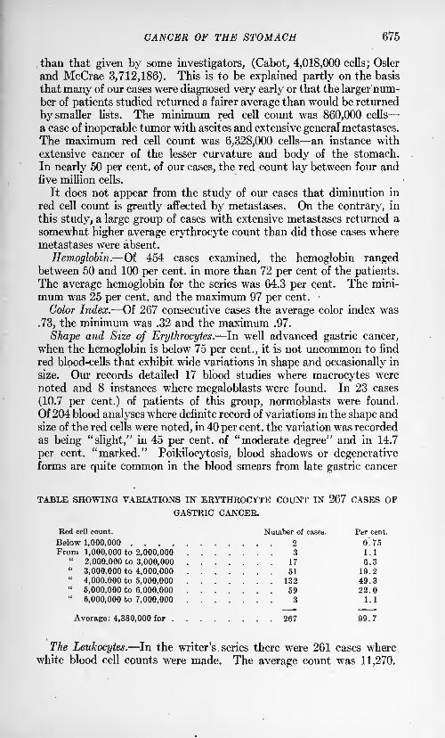
CANCER OF THE STOMACH 675
than that given by some investigators, (Cabot, 4,018,000 cells; Osier
and McCrae 3,712,186). This is to be explained partly on the basis
that many of our cases were diagnosed very early or that the larger num-ber of patients studied returned a fairer average than would be returned
by smaller lists. The minimum red cell count was 860,000 cells
a case of inoperable tumor with ascites and extensive generafmetastases.The maximum red cell count was 6,328,000 cells an instance with
extensive cancer of the lesser curvature and body of the stomach.
In nearly 50 per cent, of our cases, the red count lay between four andfive million cells.
It does not appear from the study of our cases that diminution in
red cell count is greatly affected by metastases. On the contrary, in
this study, a large group of cases with extensive metastases returned a
somewhat higher average erythrocyte count than did those cases wheremetastases were absent.
Hemoglobin. Of 454 cases examined, the hemoglobin rangedbetween 50 and 100 per cent, in more than 72 per cent of the patients.
The average hemoglobin for the series was 64.3 per cent. The mini-
mum was 25 per cent, and the maximum 97 per cent.
Color Index. Of 267 consecutive cases the average color index was
.73, the minimum was .32 and the maximum .97.
Shape and Size of Erythrocytes. In well advanced gastric cancer,
when the hemoglobin is below 75 per cent., it is not uncommon to find
red blood-cells that exhibit wide variations in shape and occasionally in
size. Our records detailed 17 blood studies where macrocytes were
noted and 8 instances where megaloblasts were found. In 23 cases
(10.7 per cent.) of patients of this group, normoblasts were found.
Of 204 blood analyses where definite record of variations in the shape andsize of the red cells were noted, in 40 per cent, the variation was recorded
as being "slight," in 45 per cent, of "moderate degree" and in 14.7
per cent, "marked." Poikilocytosis, blood shadows or degenerativeforms are quite common in the blood smears from late gastric cancer
TABLE SHOWING VARIATIONS IN ERYTHROCYTE COUNT IN 267 CASES OFGASTRIC CANCER.
Per cent.
0.751.1
6.319.249.322.01.1
Average: 4,380,000 for 267 99.7
The Leukocytes. In the writer's . series there were 261 cases wherewhite blood cell counts were made. The average count was 11,270,
Re
BelFro
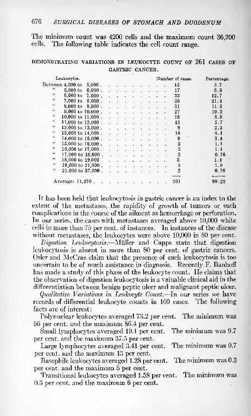
676 SURGICAL DISEASES OF STOMACH AND DUODENUM
The minimum count was 4200 cells and the maximum count 36,200cells. The following table indicates the cell count range.
DEMONSTRATING VARIATIONS IN LEUKOCYTE COUNT OF 261 CASES OFGASTRIC CANCER.
Percentage.
5.76.512.721.411.510.36.95.72.36.1
3.41.1
1.1
0.761.1
1.9
0.76
Leukocytes. Number of cases.
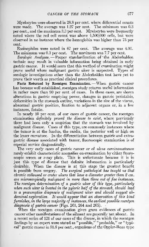
CANCER OF THE STOMACH 677
Myelocytes were observed in 28.8 per cent, where differential counts
were made. The average was 1.27 per cent. The minimum was 0.3
per cent., and the maximum 3.3 per cent. Myelocytes were frequentlynoted where the red cell count was above 4,500,000 cells, but were
observed in no instance where the hemoglobin was higher than 75 percent.
Eosinophiles were noted in 67 per cent. The average was 4.01.
The minimum was 0.3 per cent. The maximum was 7.7 per cent.
Serologic Analyses. Proper standardization of 'the Abderhalden
technic may result in valuable information being obtained in early
gastric cancer. It would seem that this method of examination might
prove useful where malignant gastric ulcer is suspected. Types of
serologic investigations other than the Abderhalden test have yet to
prove their worth as practical clinical procedures.Facts Returned by Roentgen Examination. When gastric cancer
has become well established, roentgen study returns useful information
in rather more than 90 per cent, of cases. In these cases, are shownalterations in gastric emptying power, changes in gastric peristalsis,
deformities in the stomach outline, variations in the size of the viscus,
abnormal gastric position, fixation to adjacent organs or, in a few
instances, fistulse.
In nearly 10 per cent, of our cases of gastric cancer, the roentgenexamination definitely proved the disease to exist, where previouslythere had been only a suspicion that the stomach was involved bymalignant disease. Cases of this type, are commonly instances wherethe tumor is at the fundus, the cardia, the posterior wall or high onthe lesser curvature. In the differentiation between gastric and extra-
gastric disease associated with tumor, fluoroscopic examination is of
especial service diagnostically.The very early cases of gastric cancer or of ulcus carcinomatosum
rarely exhibit characteristic anomalies on examination by either fluoro-
scopic screen or z-ray plate. This is unfortunate because it is in
just this type of disease that definite information is particularlydesirable. When the disease is at this stage the greatest benefit
is possible from surgery. The surgical pathologist has taught us that
chronic calloused or crater ulcers that have a diameter greater than 2 cm.
are microscopically malignant in more than three out of five instances.
The roentgen determination of a gastric ulcer of this type, particularlywhen such ulcer is located in the pyloric half of the stomach, should lead
to a presumptive diagnosis of malignant uker and should suggest ab-
dominal exploration. It would appear that an observation of this kind
furnishes, in the large majority of instances, the earliest possible roentgen
diagnosis of gastric cancer (Figs. 263, 264 and 265).
When the roentgen examination gives gross evidences of gastric
cancer other manifestations of the ailment are generally not absent. In
a recent series of 121 of our cases of the disease, in which the roentgen
findings by an expert were stated as "possible," "probable," or "typi-cal" gastric cancer in 91.8 per cent., organisms of the Oppler-Boas type
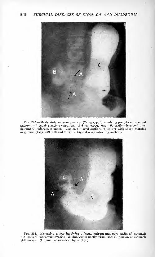
678 SURGICAL DISEASES OF STOMACH AND DUODENUM
FIG. 263. Moderately extensive cancer ("ring type") involving prepyloric zone andantrum and causing gastric retention. AA, cancerous area; B, partly visualized duo-
denum; C, enlarged stomach. Contrast ragged outlines of cancer with sharp marginsof gumma (Figs. 259, 260 and 261). (Original observation by author.)
FIG. 264. Extensive cancer involving pylorus, antrum and pars media of stomachAA, zone of cancerous invasion; B, duodenum partly visualized; C, portion of stomachstill intact. (Original observation by author.)
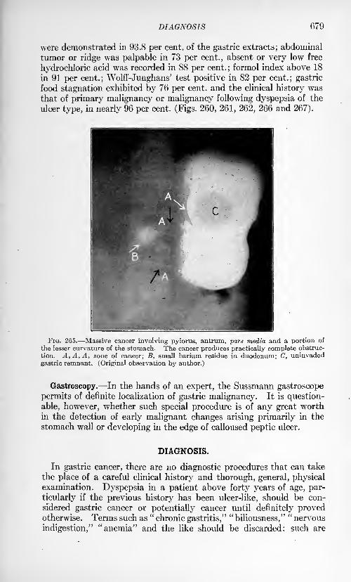
DIAGNOSIS 079
were demonstrated in 93.8 per cent, of the gastric extracts; abdominal
tumor or ridge was palpable in 73 per cent., absent or very low free
hydrochloric acid was recorded in 88 per cent. ; formol index above 18
in 91 per cent.; Wolff-Junghans' test positive in 82 per cent.; gastric
food stagnation exhibited by 76 per cent, and the clinical history wasthat of primary malignancy or malignancy following dyspepsia of the
ulcer type, in nearly 96 per cent. (Figs. 260, 261, 262, 266 and 267).
FIG. 265. Massive cancer involving pylorus, antrum, pars media and a portion of
the lesser curvature of the stomach. The cancer produces practically complete obstruc-
tion. A, A, A, zone of cancer; B, small barium residue in duodenum; C, uninvaded
gastric remnant. (Original observation by author.)
Gastroscopy. In the hands of an expert, the Sussmann gastroscope
permits of definite localization of gastric malignancy. It is question-
able, however, whether such special procedure is of any great worthin the detection of early malignant changes arising primarily in the
stomach wall or developing in the edge of calloused peptic ulcer.
DIAGNOSIS.
In gastric cancer, there are no diagnostic procedures that can take
the place of a careful clinical history and thorough, general, physicalexamination. Dyspepsia in a patient above forty years of age, par-
ticularly if the previous history has been ulcer-like, should be con-
sidered gastric cancer or potentially cancer until definitely provedotherwise. Terms such as
"chronic gastritis,"
"biliousness,"
"nervous
indigestion," "anemia" and the like should be discarded: such are
680 SURGICAL DISEASES OF STOMACH AND DUODENUM
descriptive terms for symptoms or parts of disease and not of them-selves disease entities. If every clinical laboratory aid is employedroutinely in the study of dyspepsias and if such aids are interpreted
intelligently in the light of clinical history and known pathologic
facts, few cases of gastric cancer need go unrecognized and many will
be detected before they are surgically hopeless. For the patient's
sake, early exploratory laparotomy should be urged where the suspicionor possibility of gastric malignancy exists and when, in spite of rational
therapeutic procedures, no progress toward gastric health is beingmade.
Persistent weight loss by individuals with obstinate dyspepsia should
strongly suggest malignancy, especially if the patient is in the fifth
decade of life and it is known that his daily food intake and retention
have exceeded 2000 calories. Our records show no instance of proved
malignancy that has not lost weight previous to laparotomy. Weightgain in gastric cancer (?) has been reported. In such instances the
diagnosis is open to question: weight gain may occur in tumor of the
stomach if such tumor be due to lues, cyst, aneurysm, inflammatory
swellings or the tumor be about the stomach; e.g., pyloric lymph-nodes, head of pancreas, etc. Weight loss is more rapid in obstructive
cancers than in those permitting free food entrance to and exit from the
stomach. This is doubtless due to malignant systemic poisoning andto metastatic growths in extragastric viscera, plus starvation. Vomit-
ing accelerates weight decline.
Persistent "occult" blood in the meat-free stool, if associated with
weight loss and obstinate dyspepsia, is a valuable hint that a malignant
process exists. Constant, positive, chemical tests for blood in the fast-
ing stomach contents rarely occur in disease other than malignancy. Attimes achylias associated with severe anemia, Bright's disease, cirrhosis
of the liver or circulatory anomalies (varicosities) may be associated
with positive, occult blood tests, but in such ailments, the history andclinical examinations differ from those of gastric cancer.
Roentgen studies may early give evidence of a primary intragastric
tumor or may disclose calloused ulcer whose diameter exceeds 2 cm.
The latter finding is strongly suggestive of malignancy. Obstructive
lesions may be demonstrated, but when they can be shown by roentgen
studies, the clinical history and test-meal records are usually quitedefinite proof that gastric deformity will be roentgenologically demon-strated.
Test-meal analyses may return nothing of diagnostic significance
other than retention of some part of the physiologic motor-meal. If
this evidence of stagnation is associated with low or absent free HC1 and
the microscopic proof of organisms of the Oppler-Boas group, then the
diagnosis of malignancy is practically sure. It should be emphasized
again, that in the surgically curable stage of gastric cancer, free HC1
may be normal or even increased and Oppler-Boas bacilli absent; in
fact, it is extremely uncommon to find such organisms when the free
HC1 is higher than 15.
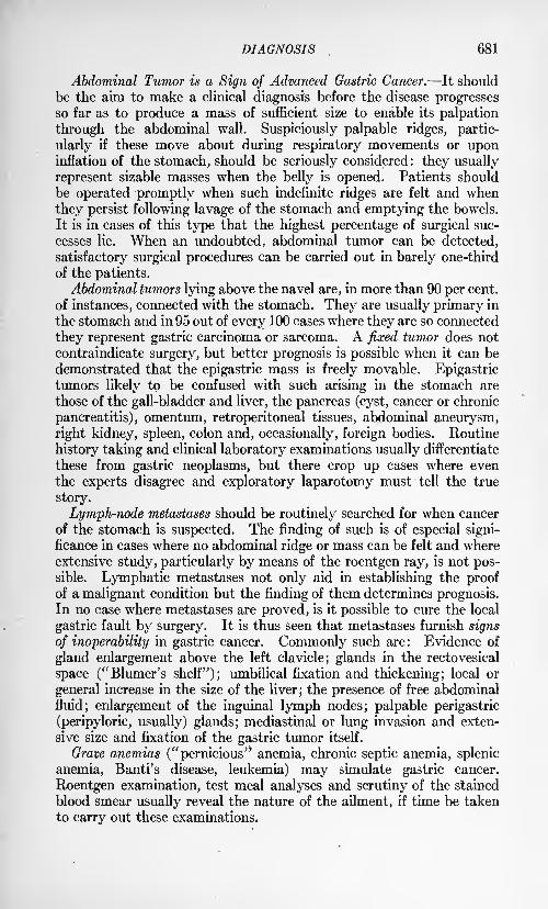
DIAGNOSIS 681
Abdominal Tumor is a Sign of Advanced Gastric Cancer. It should
be the aim to make a clinical diagnosis before the disease progressesso far as to produce a mass of sufficient size to enable its palpation
through the abdominal wall. Suspiciously palpable ridges, partic-
ularly if these move about during respiratory movements or uponinflation of the stomach, should be seriously considered : they usually
represent sizable masses when the belly is opened. Patients should
be operated promptly when such indefinite ridges are felt and when
they persist following lavage of the stomach and emptying the bowels.
It is in cases of this type that the highest percentage of surgical suc-
cesses lie. When an undoubted, abdominal tumor can be detected,
satisfactory surgical procedures can be carried out in barely one-third
of the patients.
Abdominal tumors lying above the navel are, in more than 90 per cent,
of instances, connected with the stomach. They are usually primary in
the stomach and in 95 out of every 100 cases where they are so connected
they represent gastric carcinoma or sarcoma. A fixed tumor does not
contraindicate surgery, but better prognosis is possible when it can be
demonstrated that the epigastric mass is freely movable. Epigastrictumors likely to be confused with such arising in the stomach are
those of the gall-bladder and liver, the pancreas (cyst, cancer or chronic
pancreatitis), omentum, retroperitoneal tissues, abdominal aneurysm,
right kidney, spleen, colon and, occasionally, foreign bodies. Routine
history taking and clinical laboratory examinations usually differentiate
these from gastric neoplasms, but there crop up cases where even
the experts disagree and exploratory laparotomy must tell the true
story.
Lymph-node metastases should be routinely searched for when cancer
of the stomach is suspected. The finding of such is of especial signi-
ficance in cases where no abdominal ridge or mass can be felt and where
extensive study, particularly by means of the roentgen ray, is not pos-sible. Lymphatic metastases not only aid in establishing the proofof a malignant condition but the finding of them determines prognosis.In no case where metastases are proved, is it possible to cure the local
gastric fault by surgery. It is thus seen that metastases furnish signs
of inoperability in gastric cancer. Commonly such are: Evidence of
gland enlargement above the left clavicle; glands in the rectovesical
space ("Blumer's shelf"); umbilical fixation and thickening; local or
general increase in the size of the liver; the presence of free abdominal
fluid; enlargement of the inguinal lymph nodes; palpable perigastric
(peripyloric, usually) glands; mediastinal or lung invasion and exten-
sive size and fixation of the gastric tumor itself.
Grave anemias ("pernicious5 *
anemia, chronic septic anemia, splenic
anemia, Banti's disease, leukemia) may simulate gastric cancer.
Roentgen examination, test meal analyses and scrutiny of the stained
blood smear usually reveal the nature of the ailment, if tune be taken
to carry out these examinations.
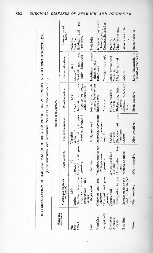
682 SURGICAL DISEASES OF STOMACH AND DUODENUM
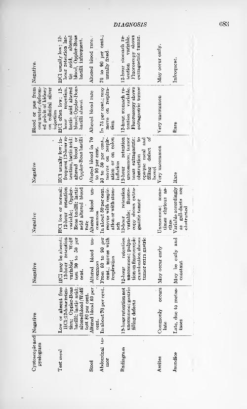
DIAGNOSIS 683
Negative.
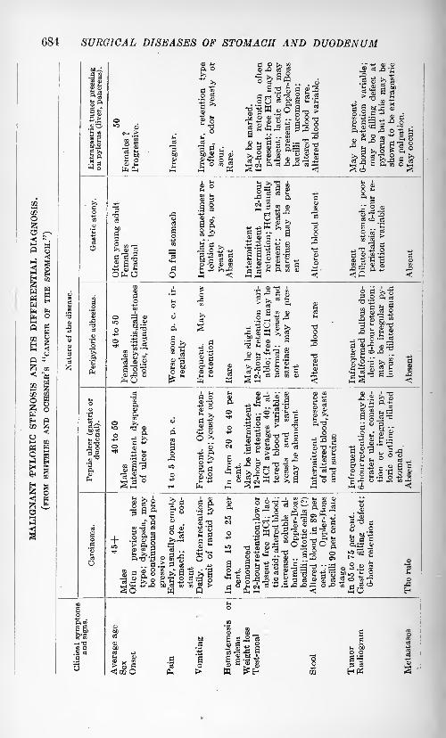
684 SURGICAL DISEASES OF STOMACH AND DUODENUM
02i i
03
l
W HS hE* O
3
O
DIAGNOSIS 685
Gall-bladder disease in the aged, may simulate gastric cancer, par-
ticularly if inflammatory or malignant involvement of the peripyloric
nodes produces disturbance in gastric emptying. The early history of
the affection should be gone into: there is rarely ulcer-like dyspepsia;the early ailments have not rarely followed typhoid fever, chronic nose
and throat disorders, pregnancy, dysentery, malaria, etc., and the
presenting disease may have been associated with early jaundice. One-third such cases give histories of stone colics and icterus. The roentgen
study, analysis of gastric extracts and stools give valuable information
toward excluding a primary stomach disease.
Cancer of the pancreas, unless it be secondary to ulcer or cancer of
the stomach, and malignancy of the gall-tract or retroperitoneal tissues
is comparatively rare. Primary cancer of the pancreas, lues, cyst or
stone may be difficult of detection unless very careful examination be
made. Functional tests upon the stool may disclose early diminution
or absence of the specific pancreatic ferments.
Tumors of the kidney, large bowel, amentum, abdominal lymph-nodes or small intestine, not infrequently offer problems in exclusion.
Their rarity and their atypic situation combined with a clinical history,
not primarily dyspeptic, aid in segregating them.
Achylia gastrica is a condition of absent gastric secretion associated
with many toxic, infectious and malignant maladies. Formerly,when it was deemed necessary to have absent free HC1 in stomachextracts before a diagnosis of gastric cancer was considered, achylia
gastrica almost approached the dignity of a disease sui generis. Care-
fully taken history, physical examination and laboratory analysesnow readily differentiate the achylias that are benign from those in
association with malignant disease of the stomach itself.
Lues, gastric granulomata and tuberculosis sometimes exhibit clinical
pictures of malignant disease especially if associated with tumor for-
mation. Serologic tests, tuberculin injections, roentgen studies and,in some instances, microscopic examination of extirpated tissue are
necessary before the true nature of the affection is known. These ail-
ments are not associated with metastases, even though the primary
gastric mass may be extensive. This fact is of great service in provingtheir non-malignancy.
Foreign bodies in the stomach, ("hair-ball," spoons, knives, etc.) maysimulate gastric cancer if they have caused malnutrition by starvation.
Their detection is commonly prompt upon roentgen examination.
Chronic constipation, protozoic dysenteries, and drug taking some-
times result in weight loss, cachexia, vomiting and anemia. Instances
occur where it requires great ingenuity and skill to prove the absence
of malignant disease.
Complications. In instances of the disease that are operated uponearly, few of the commonly described text-book complications are metwith.
Pyloric Obstruction. Pyloric obstruction occurred in some grade of
nearly 72 per cent, of our cases. Gastric stagnation may be enormous,the stomach holding many quarts.
686 SURGICAL DISEASES OF STOMACH AND DUODENUM
Obstruction at the Cardia. Obstruction at the cardia occurred in 1.8
per cent. Dysphagia, vomiting soon after food ingestion, rapid emacia-
tion, scanty urine, constipation with roentgen proof of obstruction
usually establish the diagnosis.
Malignant "Hour-glass". Malignant "hour-glass" occurs in approxi-
mately 1 out of 15 cases of the disease. It is graphically demonstrated
by fluoroscopy and roentgenograms.Gross Hemorrhage. Gross hemorrhage occurred in nearly 19 per cent,
of cases. Copious bleeding is more common in those gastric cancers
which have had a long "precarcinomatous" history of dyspepsia (ulcer?)
than it is in cases where a rapidly developing neoplasm has arisen in
an individual previously sound, gastrically. Fatal hemorrhage occurs
in rather more than 5 per cent, of instances.
Perforation. Perforation of gastric cancer is usually fatal, but
instances where well localized perigastric abscess develops are by nomeans uncommon. Perforation is a complication of from 2.5 per cent,
to 6 per cent, of instances, depending upon the stage of growth,situation and histologic type of tumor.
Fistula. Fistula complicates perforation. The communication is
commonly to the large bowel, gall-bladder, liver, pancreas or to the
lesser peritoneal sac. Gastro-colic fistulse occur in nearly 2 per cent,
of all late gastric cancers. Fecal vomiting, lienteric diarrhea, gross
hemorrhage or the speedy appearance of red-colored stools followingthe administration of a carmine capsule suggest the diagnosis. The
roentgenograms prove the existence, size and direction of the fistulous
tract.
Jaundice. Jaundice occurs in about 3 per cent, of the early gastric
cancers and in more than 12 per cent, of instances where the disease is
advanced. Direct extension to the gall-tract or liver or pressure uponthe bile ducts is commonly responsible for the icterus. In some cases
metastases to the peripyloric nodes produce jaundice as a consequenceof common bile-duct obstruction. The head of the pancreas may be
invaded with similar consequences.Thrombosis. Thrombosis is infrequent. It usually occurs as a
terminal event. The femoral, saphenous, external iliacs, subclavian
and brachial are the vessels commonly involved.
Enteritis. Enteritis may early complicate gastric cancer and be
particularly exhausting. It is commonly manifested by abdominal
pain and diarrhea. Such symptoms may represent the earliest mani-
festation of gastric malignancy. When they appear in a chronic
dyspeptic or in any individual beyond the fifth decade of life, careful
examination of the stomach should invariably be made.
DUODENAL ULCER 687
DUODENAL ULCER.
Definition. A form of dyspepsia, usually chronic in course, and occur-
ring intermittently, associated with local loss of the tissues forming the
wall of the duodenum.Incidence. Until within the last decade, it was thought that the
lesion was relatively uncommon. Surgical explorations made for
the relief of chronic dyspepsias haye revealed the true frequency of
the affection. Study of 8341 cases of chronic dyspepsia made bythe writer, indicates that there were 1257 operatively demonstratedinstances of duodenal ulcer (15.1 per cent.). In this group, there
were 513 cases of gastric ulcer (6.1 per cent.). These figures indicate
that of peptic ulcers, duodenal ulcer is operatively demonstrated nearlytwo and a half times as frequently as is gastric ulcer. Graham's statistics
return a similar ratio: Of 1751 cases of peptic ulcer, this investigatorhad operatively proved 1300 instances where the lesion was duodenal
and 451 where the ulcer was on the gastric side of the pyloric vein.
Age. Moynihan records 31 cases of duodenal ulcer of the hemor-
rhagic type occurring in children below the age of four years. In 28
instances the patients were less than eight months old. Several fatal
terminations took place within a few hours following birth. The clini-
cal picture was that commonly associated with melena neonatorum.
Commonly the affection is exhibited in adult life. Instances are
most frequent in the fourth and fifth decades. In Moynihan's series
of 115 cases, 58 patients were between the ages of thirty to fifty-
one years. In the writer's study of 1257 cases, the average age was
just over forty-six, and approximately 61 per cent of patients werebetween thirty and sixty years.
Sex. Males are much more commonly affected than are females.
This fact has been abundantly shown by the observations of Graham,W. J. Mayo, Moynihan and others. In the writer's series of opera-
tively demonstrated cases, there were more than four and one half
times as many males as females. In Moynihan's group the sex ratio
was that of six males to one female.
Race. The Caucasian family appear to be peculiarly prone to
duodenal ulcer. It furnishes the bulk of cases at any busy clinic.
Doubtless the future will disclose that other races are more commonlyaffected than is at present thought, when surgery among such becomes
general. Heredity appears to play only a relatively small part in
duodenal ulcer incidence. While occasionally a family is met with in
which the disease has appeared through several generations, yet apartfrom the families with hemophiliac or luetic taint, it would seem that
the transmission of modes and habits of life can explain the family
appearance of the ailment.
Previous Diseases. In more than 56 per cent, of the patients studied
by the writer, a history of frequently recurring infectious disease wasnoted (tonsillitis, la grippe, head sinus affections, etc.) or the patients
dated the beginning of their dyspepsias from an acute exanthematous
688 SURGICAL DISEASES OF STOMACH AND DUODENUM
or septic ailment. Seasonal occurrence of symptoms was recorded in
nearly 40 per cent. Spring and Fall being favorites. When it is recalled
that during these seasons acute infectious diseases are widespread andare frequently inadequately treated, the relationship of microorganismsto the production of duodenal ulcer or toward reestablishing bacterial
activity in tissues already damaged seems worthy of more than casual
note. The experimental studies of Rosenow upon the production of
ulcer by repeated injection of streptoccoci lend a certain amount of
support to such view.
Concomitant disease of the gall-bladder or the appendix, or both,is frequent in duodenal ulcer. It occurred in nearly 50 per cent, of the
writer's cases. The clinical histories indicate that, not uncommonly,ailments of these portions of the digestive apparatus preceded the onset
of damage in the duodenum. The researches of Hamburger uponexperimental pyloric spasm with respect to the production of pepticulcer would appear to emphasize the importance of affections of the
appendix or the gall-bladder in the production of duodenal ulcer. It
is well known that on account of the embryologic relationship betweenthe pylorus, the appendix and the gall-bladder, disease in the twolatter may produce marked pyloric spasm, through vagus and sympa-thetic irritation. Such prolonged and intermittently active spasms are
quite capable of bringing about disturbances in circulation and peri-
stalsis in both the stomach and the duodenum, with probably resultant
ulceration. It would also appear that the presence of infection in
either gall-bladder or appendix over long periods is a not unimportantfactor in the spread of microorganisms to the duodenal lining, throughthe abdominal, freely interconnecting lymph-channels. If damagehas already been done to the duodenal wall as a consequence of circula-
tory upsets, traumata, etc., such microorganisms may lodge, multiply,and be a factor in the causation of ulcer. The ulceration may be of
extremely rapid development, and of the fatally bleeding type, should
the invading bacteria be of the hemolytic variety.
General tuberculosis is occasionally associated with duodenal ulcer.
Some 40 cases have been tabulated by Moynihan. However, ulceration
of primarily tuberculous origin is rare in the duodenum. About a
dozen instances have been described. Moynihan mentions 2 cases,
and states that in three other patients suspicion of the ailment's
being tuberculous was entertained.
Ulceration of the duodenum in association with burns has been fre-
quently commented upon. Perry and Shay state that of patients
dying as a consequence of severe burns or scalds, in 3.3 per cent,
duodenal lesions were demonstrable. In the study of a group of
patients dying from other ailments, these writers found duodenal ulcer
present in but 0.4 per cent. Ulcers of the duodenum in association with
cutaneous burns or scalds are apparently the result of congestion of
the capillaries in the duodenal mucosa, as a consequence of toxic
products or from minute emboli. The erosions resulting, as necrosis
takes place, are aggravated by the peptic activity of the gastric juice
and the tryptic power of the pancreatic secretions and bile.
DUODENAL ULCER 689
Uremia, grave anemias, cardiac lesions, toxins (the result of dis-
ordered metabolism or of chemical changes foreign to the human
economy), local trauma to the duodenal lining from improper food or
from foreign bodies, or abdominal traumata, are not rarely factors in
the production of duodenal ulcer. It is doubtful if alterations in the
chemistry of either the gastric or the pancreatic juice are, of them-
selves, capable of causing duodenal ulcer, until local loss of epithelial
lining has previously been brought about through other agents. This
loss need be but small, and quite incapable, of itself, of producing
signs or symptoms.Habits. Duodenal ulcer commonly occurs in full-blooded, sturdily-
built people. Frequently, these give a long history of careless living and
of dietetic and hygienic irregularities. But this is by no means the
rule : leading a model life does not appear to confer immunity to
the disease. History of the excessive use of alcohol and tobacco is
not rarely obtained. Many patients have, however, been abstainers
from these drugs. Moreover, alcohol and tobacco are so universally
used, that, were such the sole or an important factor in the productionof duodenal ulcer, the affection would be of much more commonincidence. In such event, also, most females and children would be
practically immune.Morbid Anatomy. Superficial erosions of the duodenum may be
recognized with difficulty, even when the gut is spread out. Occasion-
ally, areas of congestion may be recognized, and if these are minutely
scrutinized, fissures and shallow surface-ulcers may be seen. Thelatter resemble the herpetic spots which are sometimes seen on the
tongue or inside the cheek. From the serous side of the duodenum,there may be no evidence of such ulcers, either to sight or to touch.
Acute bleeding ulcers may be recognized as local areas of multiple,
punctate hemorrhage on which small clots or blood pigment may be
noted. If the ulceration be associated with hemolytic cocci, then
local points of hemorrhage may be seen in the submucosa, and occa-
sionally in the subserosa.
Duodenal ulcers that have for years been associated with chronic
intermittent dyspepsia are practically always associated with such a
highly developed scar-tissue limiting-zone as to be readily visible and
palpable when the gut is exposed at laparotomy or at autopsy. Morethan nine out of ten such ulcers are located in the first two inches of the
duodenum beyond the pyloric vein (Mayo's vein), and the ulceration
is most commonly on the anterior-superior wall of the gut. In shapethe ulcer may be round, oval or somewhat stellate. The serous mem-brane covering it is pearly gray and shiny at the center, becoming white
or yellowish toward the periphery. Through the serosa may be seen
dot-like areas of congestion or hemorrhage. The limiting margin of
the ulcer is commonly thickened, and may be corrugated and highlyvascularized. The area of the duodenum immediately adjacent to
the ulcer is usually more vascular than is the remainder of the gut.
On palpation, these ulcers have a disc-like feel. They often feel larger
than they appear. Where there has been considerable chronicity, theVOL. ii 44
690 SURGICAL DISEASES OF STOMACH AND DUODENUM
examining finger may detect a definite crater when the luminal aspectof the ulcer is explored. The ulcers vary greatly in size. Some are
barely the size of the head of a lead pencil; others may be as large as
the palm of one's hand. The larger ulcers are commonly on the anterior
wall of the intestine; -such is, however, rarely the site of as deep craters
as are those on the posterior wall of the bowel. This fact has been
emphasized by W. J. Mayo.In instances where ulcers have been particularly active, deformity
of the duodenum may be extensive. Contraction of the scar-tissue
may bring about hour-glass duodenum, local out-pouchings, resembling
diverticula, or inward bulgings, with puckering, angulation or stenosis
of the intestine. Extension of the ulcer to the pylorus, may be fol-
lowed by malignant transition of the ulcer at its pyloric edge. If
perforation takes place, a clean-cut, punched-out orifice may representthe ulcer base. Commonly, perforation has been protected. In this
event, strings or tags of omentum may be glued to the serous base
of the ulcer, or a mass of adhesions may fix the ulcer firmly to adjacentviscera (liver, gall-bladder, large intestine, pancreas) or to the omen-
tum, great vessels or anterior abdominal wall. Perforation may occur
in spite of massive adhesions. When this happens, fistulous tracts,
with or without abscess or fatal hemorrhage, are established.
Chronic, calloused duodenal ulcers viewed from the lumen of the gut,
vary greatly, accordingly as they are discovered in activity or quiescence.If there has been a recent exacerbation of the ailment, then a crater
ulcer, with thickened, ragged, sometimes overhanging or terraced,
puckered margins may be seen. The margins may be veryvascular and bleed easily. The base is generally clean. About the
ulcer there commonly exists a zone of congestion, in which swollen
bloodvessels are usually visible. If the duodenal ulcer is definitely
quiescent, then it appears as a shallow crater (especially anterior wall
ulcer), pale, shiny, and with irregular, smooth margin, overhanging or
ridged. The periphery of the ulcer scar is commonly well delimited,
and about it the duodenal wall may be thickened and congested.Ulcer of the duodenum is commonly solitary. Multiple lesions are
however, not of sufficient rarity as to be curiosities. In all operative
explorations, the entire gut should be investigated for multiple ulcers,
even though one is readily discovered and may seemingly account for
all the symptoms of the case in hand. Unless this routine is carried out,
operative procedures may fail to give the desired or expected relief.
Secondary Effects of Duodenal Ulcer. Carcinomatous transformationis exceedingly rare. It occurs about 6 times in 1000 cases. Exten-
sion of the ulcer to the pylorus may result in malignant change at the
pyloric margin of the ulcer. This is by no means rare, and furnishes
an argument for those who believe that, whenever possible, duodenal
ulcers should be excised.
Stenosis of the duodenum or of the pyloric exit is the most commonsecondary effect of the presence of chronic, calloused, duodenal ulcer.
It may vary from slight stenosis, associated with more or less pro-nounced spasm, to practical closure of the duodenal or pyloric lumina.
DUODENAL ULCER 691
The stomach is then commonly dilated, and its muscle layers may be
atonic.
Perforation, if acute and unprotected, may result in general and fatal
peritonitis or, if adjacent viscera are entered, in fistula or abscess.
In the latter event, grave damage generally results to the invaded
viscus. If perforation involves one of the great vessels (abdominal
aorta, portal vein, renal artery, etc.) sudden fatal termination as a
result of hemorrhage may be expected.Anemia of the secondary type usually occurs and persists where
hemorrhage from the ulcer is copious or when small, frequent hemor-
rhages take place. Malnutrition may be an associated complication.
Symptoms. From 3 to 5 per cent, of duodenal ulcers give no evi-
dences of their existence. They are usually discovered at laparotomyfor other ailments or are found at postmortem examination of patients
dying from accident or extra-duodenal disease. Commonly, however,duodenal ulcer manifests itself either by an acute crisis (hemorrhage,
perforation) or by chronic dyspepsia.THE ACUTE CRISIS. Hemorrhage. This may come on suddenly
without evident, causative factor. In other instances, there is a historyof cutaneous burn, operative procedure, external trauma, physical
stress, swallowing of poisons or foreign bodies, an acute infectious
ailment or of indiscretion in eating or drinking. Hematemesis may be
copious and exhausting, or signs of hemorrhage into the bowel, with the
passage of dark stools shortly apparent. Pallor, shock, collapse anddeath may quickly follow. Pain, often agonizing in character, occurs in
many of the cases, particularly if perforation of the gut accompanies
hemorrhage. Signs of peritonitis, local or general, supervene.
Perforation. Perforation is perhaps the most serious event in the
course of duodenal ulcer, even though protection is afforded by omen-turn or neighboring viscera. The onset is commonly sudden. The chief
clinical manifestation is pain. This may be so pronounced as to
render the patient uncontrollable. It is evidenced by distressed cries,
doubled-up posture, protective pressure of the hands over the epigas-
trium, dyspnea, drops of sweat upon the contracted brow, cold, pale
skin, subnormal temperature, and rapid pulse. If prompt relief is
not at hand, peritonitis quickly develops. The temperature rises, the
abdomen becomes fixed and distended, the pulse small and thready,delirium follows and death ensues.
Protected perforation may be initiated in a manner similar to
unprotected, fatal perforation. After the initial shock, symptomssubside, and although abdominal tenderness, vomiting, pain, and
dyspepsia remain, the patient ultimately recovers fair health, lingersuntil relieved by operative procedures or he succumbs to disturbances
in function in the organs that have acted as temporary protection
against perforation into the peritoneal sac.
CHRONIC, CALLOUSED DUODENAL ULCER. This presents a sympto-matology of such characteristic type as to permit a positive diagnosis
being made, in fully seven out of ten instances, from the clinical history.The essential feature of the affection is a chronic dyspepsia, commonly
692 SURGICAL DISEASES OF STOMACH AND DUODENUM
occurring intermittently, in adult males. The prime points typifyingthis dyspepsia are:
(a) Periodicity of Complaint. In non-stenosing or otherwise uncom-
plicated duodenal ulcer, the intermittent appearance of dyspeptic storms
is striking and of great diagnostic value. This peculiar periodicitywas noted in 82 per cent, of our duodenal ulcers of the uncomplicated
variety. The "attacks" correspond to recrudescences of activity in the
ulcer, its extension or the formation of new ulcers. They may hint
at the onset of complications (hemorrhage, perforation, or malignant
change) in those chronically suffering. This is especially true in such
instances where attacks or "spells" come on with but short dyspepsia-free intervals.
The duodenal ulcer patient is commonly comfortable between attacks
so long as the ulcer remains uncomplicated. In the latter instance, the
affection manifests itself by a continuous digestive upset.
(6) Epigastric Pain or Distress. Epigastric pain or distress is
experienced by about 96 per cent, of patients. It varies in character
from vague discomfort to prostration, gnawing or colics. The severe
attacks of pain very commonly presage hemorrhage or perforation.
They may, however, be only indications of pronounced pyloric,
duodenal or gastric spasms. In such cases, the colicky attacks maypunctuate a more or less constant sensation of epigastric "burning."The distress in duodenal ulcer is commonly most intense at or to the
right of the midline and above the navel. There may, however, be
general epigastric or abdominal discomfort. In ptotic females the
distress may be located far below the navel.
A point of subjective maximum intensity of pain is usually recorded.
It may have comparatively little relationship to the actual anatomic
situation of the duodenum and it may in no wise correspond to areas
of tenderness delimited upon abdominal palpation. The local, sub-
jective pain point is in the right upper abdominal quadrant in morethan 90 per cent, of instances of duodenal ulcer.
(c) Pain-time. Pain-time in duodenal ulcer of the uncomplicated
type, bears such striking relation to food ingestion that the phenome-non furnishes a most important point in diagnosis. Fully 85 per cent,
of ulcers of the uncomplicated type cause pain within two to five
hours following the taking of a meal. Commonly, the more bulky the
meal, the greater the time-interval elapsing before the appearance of
pain. While in many cases of duodenal ulcer, some discomfort appearssoon after eating, the distress does not amount to actual pain until
four hours post cibo. Pain appearing regularly at night is experienced
by about 17 per cent, of cases.
With exception of the acute pain crises associated with hemorrhage or
perforation, the pain of duodenal ulcer is apparently due, in the main, to
exaggerated peristaltic movements of the stomach and of the duodenum
itself, as a consequence of local, neuromuscular irritation. This local
irritation may be in part due to the forceful injection of gastric contents
rich in HC1 and pepsin, into the first part of the duodenum (the seat
of the great majority of duodenal ulcers), but that this is not the only
factor concerned with pain production is abundantly demonstrated by
DUODENAL ULCER 693
the recent studies of Rehfuss and Carlson and Hamburger. These
investigators have demonstrated that at the time that peptic ulcer
pain is most pronounced, the HC1 and pepsin content of the gastric
juice is by no means at the maximum. However, at the time of maxi-
mum pain intensity, intragastric (and probably intraduodenal) pressureis at its highest point and muscular contractions most marked.
(d) "Food-ease" of Pain. "Food-ease" of pain in duodenal ulcer is of
diagnostic importance in nearly 80 per cent, of uncomplicated duodenal
ulcers. The pain may be most annoying just before the time that
patients are ordinarily accustomed to eat, or it may awake them at night,
between midnight and 2 A.M. Eating or drinking usually producesentire amelioration of symptoms. A large meal brings about longerrelief than does a small one. Relief by alkalies, lavage, vomiting,a drink of spirits, an orange, or by posture is quite common. Themechanism of relief, hence, does not appear to be explained wholly bythe theory that neutralization of the acid of gastric or duodenal contents
is brought about by the above measures: numerous agents capableof causing relief of pain are not alkaline nor do they empty the stomachor duodenum and thereby remove an offending gastric or duodenal
content. It would seem that the chief factors concerned in pain relief
in duodenal ulcer are those connected with counteracting abnormal
peristaltic or muscular activity of the stomach and duodenum, with the
resultant diminution of intragastric and intraduodenal pressure."Food-ease" of pain is so constantly manifested in duodenal ulcer of
the uncomplicated variety, that its constant exhibition in "spells" of
dyspepsia, attack after attack, is almost pathognomonic. It assuredlyindicates peptic ulcer, and most commonly the ulcer is duodenal or
pyloric, particularly if the chronic dyspeptic is an adult male.
(e) Vomiting. Vomiting is recorded in practically eight out of ten
instances of duodenal ulcer of all classes, and in about 6 out of 10 cases
where theulcer is uncomplicated. Vomiting generally comes on at
the height of the gastric distress, i. e., when duodenal and gastric spasmand peristalsis are at their maximum intensity. The vomitus is com-
monly composed of acid fluid intermixed with mucus or food or stained
with bile. In stenosis cases, food that has lain in the stomach for
longer than eight hours ("delayed vomit") may be constantly vomitedif the patient is on diet of ordinary bulk. Sometimes, where there is
much dilatation of the stomach with atony, the vomitus is particularly
copious and may contain food eaten so long before that it requiresan effort on the part of the patient to recall when certain vomitedmaterials were taken.
(/) Pyrosis, Waterbrash, Eructations and Belching of Sour Gas are
distressing accompaniments of the dyspeptic attacks in more than70 per cent, of uncomplicated duodenal ulcers. When stenoses, with
gastric retention have become established, these symptoms may beconstant and annoying.
(gr) Gross Hemorrhage (Helena and Hematemesis). Gross hemor-
rhage was recorded in 33 per cent, of the writer's series. Helena wasnoted nearly three times as frequently as was hematemesis, but there
were instances where melena had not been observed, but the vomiting
694 SURGICAL DISEASES OF STOMACH AND DUODENUM
of blood had been very copious. It is evident from the above namedincidence of gross hemorrhage, that at least twice as many cases of
duodenal ulcer must be diagnosed without history of melena or hema-
temesis, as with it. Given an ulcer symptom-complex, however, the
occurrence of melena or hematemesis clinches the diagnosis in 95 percent, of instances.
"Occult hemorrhage," i. e., the chemical demonstration of altered
blood, was recorded in 40 per cent, of properly prepared stools, at sometime in the course of the affection, of those duodenal ulcers so tested.
It is evident that one must not delay the diagnosis of duodenal ulcer
until the stools return positive chemical tests for blood-pigment.
(K) Weakness. Weakness is experienced by the majority of duodenal
ulcer patients during their attacks of dyspepsia. It is commonly dueto partial starvation, pain with loss of sleep and rest, exhausting
vomiting, hemorrhage with anemia, and to free administration of
opiates. In the dyspepsia-free intervals of uncomplicated ulcer
strength is commonly regained rapidly. In duodenal ulcer associated
with stenoses, perforation or frequent hemorrhage or constant seepingof blood, weakness may be constant and progressive. Exhaustion
may cause death.
(i) Constipation. Constipation, particularly shortly before or duringthe period of indigestion, is noted in more than 55 per cent, of duodenal
ulcer cases. Between the attacks, the bowels may move normally.In stenosis cases, constipation is likely to be obstinate as a consequenceof reduction in the quantity of food that passes out of the stomach.
(j) Appetite. Except in the dyspeptic periods, food desire is well
maintained in the majority of instances of the affection. Manypatients develop a "poor-appetite habit" as a consequence of dread
of precipitating pain, vomiting or hemorrhage by normal eating.
During the attacks, food desire is usually reduced. In stenosis cases,
appetite is generally poor, inasmuch as nausea may be frequent or
constant.
Signs of Duodenal Ulcer. (a) Physical. Except in the complicated
cases, duodenal ulcer patients are commonly well nourished and fre-
quently of the full-blooded type. The physical appearance may be
much altered as a result of limited diet, pain, loss of sleep, hemor-
rhage, stenoses or perforation. In such events, malnutrition may be
marked and anemia pronounced. However, even the desperate cases
rarely present more than a picture of starvation and exhaustion. This
is only exceptionally complicated by such indications of toxic cachexia
as are common in malignant disease.
Weight loss is apt to be constant during the attacks of dyspepsia.
In some instances it amounts to but a few pounds; in others fifteen to
sixty pounds may be lost with'n a few weeks. In the dyspepsia-free
interval, weight is generally gained rapidly. Where stenoses, perfora-
tion or hemorrhage have occurred, weight loss may be constant and
alarming. Malignant change in the edge of an ulcer that encroaches
upon the pylorus may result in progressive decrease in weight and con-
stantly maintained dyspepsia. The average weight loss in the writer's
series of duodenal ulcers was 17.3 pounds.
DUODENAL ULCER 695
Anemia of a mild degree and of the secondary type is common in all
duodenal ulcers that have become chronic. It is not the rule, however,since numerous cases appear where there is plethora instead of anemia.
After severe gross hemorrhages or as a consequence of constant seepageof blood, the prognosis may be grave. Nucleated red cells may be
present in the stained smear. In the uncomplicated duodenal ulcers
forming the writer's series, the hemoglobin averaged 75 to 80 per cent.
In the complicated cases (stenoses, perforations, malignant change)the hemoglobin averaged 60 to 65 per cent.).
(6) Abdominal Tenderness. There is commonly present an area of
tenderness to palpation. It usually lies to the right of the midline.
It corresponds to the anatomic position of the first thir4 of the duo-
denum and the terminal fifth of the stomach. Areas of tenderness
are rarely outside the limitations of the right upper abdominal quad-rant. In about 60 per cent, of instances of duodenal ulcer, points of
maximum tenderness in zones that are the location of rather widespread
palpatory discomfort can be determined. As has been pointed out byHead, such zones do not necessarily correspond to the situation of the
duodenal lesion. Ulcers that have extended so as to involve the
peritoneum are the most sensitive to pressure. Where there has been
acute extension, points of exquisite tenderness can be noted. Tumor-like epigastric ridges, usually not movable, are exhibited in about 4
per cent, of duodenal ulcers where there has been chronic, protected
perforation, or extension of inflammation to adjacent viscera. Quite
commonly, right upper abdominal quadrant tenderness is not due to
duodenal ulcer, even though such may be present, but is caused by an
inflamed or distended gall-bladder, gall-stones or pyloric spasm asso-
ciated with such ailments or occurring in connection with lesion of the
appendix. Duodenitis, not complicated with ulcer formation, maygive rise to not only abdominal tender areas but also to clinical syn-dromes closely simulating ulcer.
(c) Dilated Splashy Stomach. Dilated, splashy stomach, with or
without "visible peristalsis" may be demonstrated where there is per-sistent pyloric spasm or pyloric obstruction of high grade, should the
subject be not too fat. Visible peristalsis was noted in 8 per cent, of
the instances of pronounced pyloric or duodenal stenosis in the writer's
series.
Information Returned by Laboratory Examinations. TEST-MEAL.
Apart from the observations connected with disturbances in gastric
or duodenal emptying rate, common test-meal data may be of but
little value toward the establishment of a diagnosis of duodenal ulcer.
Certainly, contrary to generally printed text-book statements, one
cannot depend upon the finding of so-called "hyperacidity" or "hyper-
secretion," in the majority of cases of uncomplicated peptic ulcer. The
secretory test-meal data may closely simulate the findings in simple
duodenitis, gastritis, pyloric spasm associated ailments of the appendixof gall-bladder, or of simple gastri^ ulcer.
The average free HC1 in the writer's series was 49. There were
wide variations. In nearly 2 per cent, of cases free HC1 was absent. The
highest free HC1 estimation was 190. Stomach contents removed after
696 SURGICAL DISEASES OF STOMACH AND DUODENUM
a full, mixed meal has been undergoing digestion for several hours,
generally return acid estimations that are higher than when the
acidity is tested after the administration of so bland a meal as the
Ewald or the Dock breakfast. If the stomach is emptied in the morn-
ing before food has been given, there is not infrequently more thanthe normal amount of "fasting" contents (20 to 30 c.c.). It is not
unusual to remove as high as 350 c.c. When great quantities of"fast-
ing contents" are constantly removed, then laparotomy or fluoroscopicexamination generally discloses that such cases are instances of pyloric
stenosis, with or without added pyloric spasm, or they are examples of
pronounced gastric atony.Total acidity estimations vary greatly in duodenal ulcer of the simple
type. The total acidity is commonly higher than in cases of the similar
type of gastric affection. It averaged 70 in the writer's series.
In Complicated Duodenal Ulcer stenosis cases returned average free
HC1 estimations of 54. The total acidity averaged 78. The highesttotal acidity was 255. There were 1.2 per cent, in which free HC1 wasabsent. In acute perforation, for obvious reasons, acid estimations are
commonly not available. In cases of chronic perforation, the acid esti-
mations closely approximate the figures returned by similar gastric
lesions, this doubtless because stenoses and gastric retention are usuallyconcomitant.
All types of duodenal ulcer require study by the fractional methodof analysis devised by Rehfuss. It is more than likely that when a
great number of operatively demonstrated instances of the affection
have been investigated by this procedure, a better knowledge of the
secretory activity of both stomach and duodenum will be obtained.
Duodenal tubes as they are commonly used, return little positiveinformation in instances of duodenal ulcer. This has in the main been
emphasized by the studies of Crohn.
MOTOR FUNCTION IN DUODENAL ULCER. This may be tested bythe roentgen method or by the administration of a physiological meal
of mixed food, bulky in character. (See Part I.) The latter procedureis undoubtedly the better, inasmuch as it reveals the emptying rate of
the stomach under physiological conditions. The roentgen motor-
meal does not. Certainly it is not the kind of food that the patient is
going to subsist on. Another advantage of the physiologic motor test-
meal is that it can be administered at frequent intervals for the pur-
pose of testing individual variations or the effects of therapy at verylittle inconvenience or cost to the patient. The roentgen motor-meal
is expensive, disagreeable and the patient frequently has to journey
long distances to have the proper observations made.
Some degree of eight- to twelve-hour gastric retention is exhibited in
approximately 57 per cent, of all chronic duodenal ulcers. In those
cases of stenosis, with or without associated pyloric or duodenal spasm,twelve hour retention of some degree is a constant observation in 88
per cent. The quantity of.-stagnatjpn contents is subject to wide fluc-
tuations, in groups of patients and in the same patient examined at
different times. This is especially noted if the results observed before
the administration of an antispasmodic medicine (atropine or bella-
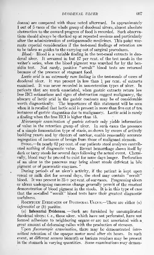
DUODENAL ULCER 697
donna) are compared with those noted afterward. In approximately1 out of 5 cases of the whole group of duodenal ulcers, almost absolute
obstruction to the onward progress of food is recorded. Such observa-
tions should always be checked up at repeated sessions and particularlyafter the administration of antispasmodic medicines. This point war-
rants especial consideration if the test-meal findings of retention are
to be taken as guides to the carrying out of surgical procedures.Blood. Blood is a variable finding in the test-meal extracts in duo-
denal ulcer. It occurred in but 17 per cent, of the test meals in the
writer's series, when the blood pigment was searched for by the ben-
zidin test. Not rarely, positive "occult" blood tests are returned
because of the presence of stagnant food.
Lactic acid is an extremely rare finding in the test-meals of cases of
duodenal ulcer. It was present in less than 1 per cent, of extracts
examined. It wras never recorded in non-retention types of ulcer. In
patients that are much emaciated, when gastric extracts return lowfree HC1 estimations and signs of obstruction suggest malignancy, the
absence of lactic acid in the gastric contents may be of considerable
worth diagnostically. The importance of this statement will be seen
when it is recalled that lactic acid is present in more than five out of ten
instances of gastric stagnation due to malignancy. Lactic acid is rarelya finding when the free HC1 is higher than 15.
Microscopic examination of gastric extracts only yields information
of value in the retention group of ulcer. In such cases the presenceof a simple fermentation type of stasis, as shown by excess of actively
budding yeasts and by clusters of sarcinse, enable reasonably accurate
segregation of instances of benign from those of malignant stenosis.
STOOL. In nearly 62 per cent, of our patients stool analyses contrib-
uted nothing of diagnostic value. Recent hemorrhage shows itself bydark or tarry stools for several days following the actual event. Chemi-
cally, blood may be proved to exist for some days longer. Perforation
of an ulcer to the pancreas may bring about stools deficient in bile
pigment or of pancreatic enzymes.*
During periods of an ulcer's activity, if the patient is kept uponcereal or milk diet for several days, the stool may contain "occult"
blood. It was present in 33+ per cent, of our cases. Progressing ulcers
or ulcers undergoing cancerous change generally permit of the constant
demonstration of blood pigment in the stools. It is in this type of case
that the so-called "occult" blood tests have their greatest diagnosticusefulness.
ROENTGEN EVIDENCES OF DUODENAL ULCER. These are either (a)
inferential or (6) positive.
(a) Inferential Evidences. Such are furnished by uncomplicatedduodenal ulcers: i. e., those ulcer, which have not perforated, have not
formed adhesions to neighboring organs or are not associated with a
great amount of deforming callus with the production of stenoses.
Upon flnoroscopic examination, there may be demonstrated inter-
mittent retention of the opaque motor meal after six hours. In such
event, at different seances bismuth or barium residues may be presentin the stomach in varying quantities. Some examinations may demon-
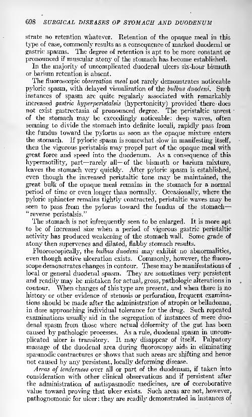
698 SURGICAL DISEASES OF STOMACH AND DUODENUM
strate no retention whatever. Retention of the opaque meal in this
type of case, commonly results as a consequence of marked duodenal or
gastric spasms. The degree of retention is apt to be more constant or
pronounced if muscular atony of the stomach has become established.
In the majority of uncomplicated duodenal ulcers six-hour bismuthor barium retention is absent.
The fluoroscopic observation meal not rarely demonstrates noticeable
pyloric spasm, with delayed visualization of the bulbus duodeni. Suchinstances of spasm are quite regularly associated with remarkablyincreased gastric hyperperistalsis (hypertonicity) provided there -does
not exist gastrectasia of pronounced degree. The peristaltic unrest
of the stomach may be exceedingly noticeable: deep waves, often
seeming to divide the stomach into definite loculi, rapidly pass fromthe fundus toward the pylorus as soon as the opaque mixture enters
the stomach. If pyloric spasm is somewhat slow in manifesting itself,
then the vigorous peristalsis may propel part of the opaque meal with
great force and speed into the duodenum. As a consequence of this
hypermotility, part rarely all of the bismuth or barium mixture,leaves the stomach very quickly. After pyloric spasm is established,even though the increased peristaltic tone may be maintained, the
great bulk of the opaque meal remains in the stomach for a normal
period of time or even longer than normally. Occasionally, where the
pyloric sphincter remains tightly contracted, peristaltic waves may beseen to pass from the pylorus toward the fundus of the stomach"reverse peristalsis .
' '
The stomach is not infrequently seen to be enlarged. It is more aptto be of increased size when a period of vigorous gastric peristaltic
activity has produced weakening of the stomach wall. Some grade of
atony then supervenes and dilated, flabby stomach results.
Fluoroscopically, the bulbus duodeni may exhibit no abnormalities,
even though active ulceration exists. Commonly, however, the fluoro-
scope demonstrates changes in contour. These may be manifestations of
local or general duodenal spasm. They are sometimes very persistent
and readily may be mistaken for actual, gross, pathologic alterations in
contour. When changes of this type are present, and when there is no
history or other evidence of stenosis or perforation, frequent examina-
tions should be made after the administration of atropin or belladonna,in dose approaching individual tolerance for the drug. Such repeatedexaminations usually aid in the segregation of instances of mere duo-
denal spasm from those where actual deformity of the gut has been
caused by pathologic processes. As a rule, duodenal spasm in uncom-
plicated ulcer is transitory. It may disappear of itself. Palpatory
massage of the duodenal area during fluoroscopy aids in eliminating
spasmodic contractures or shows that such areas are shifting and hence
not caused by any persistent, locally deforming disease.
Areas of tenderness over all or part of the duodenum, if taken into
consideration with other clinical observations and if persistent after
the administration of antispasmodic medicines, are of corroborative
value toward proving that ulcer exists. Such areas are not, however,
pathognomonic for ulcer: they are readily demonstrated in instances of
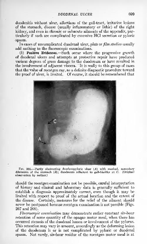
DUODENAL ULCER 699
duodenitis without ulcer, affections of the gall-tract, irritative lesions
of the stomach, disease (usually inflammatory or lithic) of the right
kidney, and even in chronic or subacute ailments of the appendix, par-
ticularly if such are complicated by excessive HC1 secretion or pyloric
spasm.In cases of uncomplicated duodenal ulcer, plate or film studies usually
add nothing to the fluoroscopic examinations.
(b) Positive Evidences. Such occur where the progressive growthof duodenal ulcers and attempts at protective repair have producedvarious degrees of gross damage to the duodenum or have resulted in
the involvement of adjacent viscera. It is really to this group of cases
that the value of roentgen ray, as a definite diagnostic procedure toward
the proof of ulcer, is limited. Of course, it should be remembered that
FIG. 266. Partly obstructing duodenopyloric ulcer (A) with marked, secondarydilatation of the stomach (B); duodenum adherent to gall-bladder at C. (Originalobservation by author.)
should the roentgen examination not be possible, careful interpretationof history and clinical and laboratory data is generally sufficient to
establish a diagnosis approximately correct, even though it may be
limited with respect to proof of the actual location and the extent of
the disease. Certainly, measures for the relief of the ailment should
never be postponed because roentgen examination is not possible (Figs.
267 and 268).
Fluoroscopic examination may demonstrate rather constant six-hour
retention of some quantity of the opaque motor meal, when there has
occurred stenosis of the duodenal lumen or involvement of the pylorus.This retention ma^ vary in amount, accordingly as the deforming lesion
of the duodenum is or is not complicated by pyloric or duodenal
spasm. Not rarely, six-hour residue of the roentgen motor meal is at
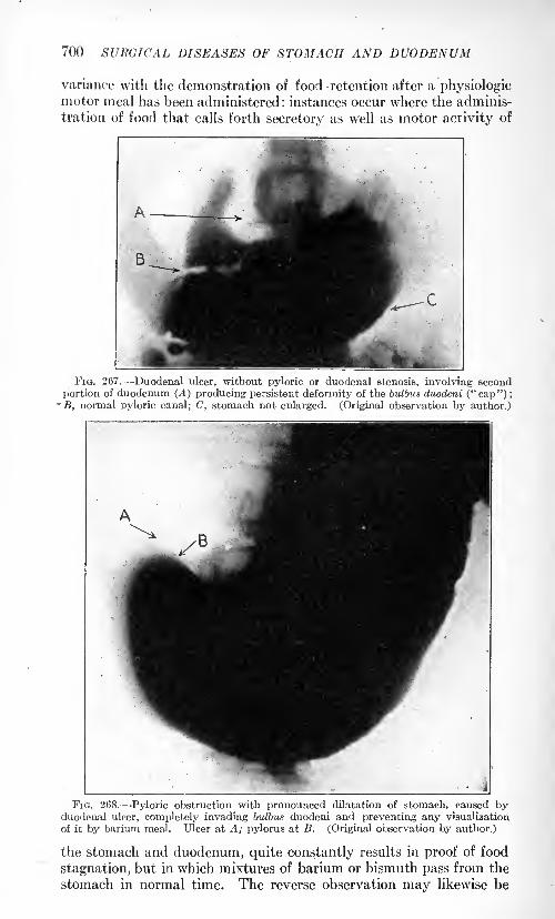
700 SURGICAL DISEASES OF STOMACH AND DUODENUM
variance with the demonstration of food -retention after a physiologicmotor meal has been administered: instances occur where the adminis-
tration of food that calls forth secretory as well as motor activity of
FIG. 267. Duodenal ulcer, without pyloric or duodenal stenosis, involving second
portion of duodenum (A) producing persistent deformity of the bulbus duodeni (" cap") ;
'
B, normal pyloric canal; C, stomach not enlarged. (Original observation by author.)
FIG. 268. Pyloric obstruction with pronounced dilatation of stomach, caused byduodenal ulcer, completely invading bulbus duodeni and preventing any visualization
of it by barium meal. Ulcer at A; pylorus at B. (Original observation by author.)
the stomach and duodenum, quite constantly results in proof of food
stagnation, but in which mixtures of barium or bismuth pass from the
stomach in normal time. The reverse observation may likewise be
. DUODENAL ULCER 701
made. For this reason, care should be exercised in the interpretationof the functions of the stomach or of the duodenum when the roentgenevidence of six-hour motor meal is present. It should be recalled that
the patient's digestive apparatus is not one that is to be called uponto take care of the comparatively small, but relatively very heavy,meal of uniform consistency, temperature and reaction, such as bariumor bismuth mixtures, during the course of his life.
Evidence of six-hour motor stagnation should be searched for at
several seances, and opinion as to the grade of deformity causing the
stagnation never should be given unless observations have been madeboth before and after the administration .of atropin or of belladonna
to the point of individual tolerance.
Enlargement of the stomach is quite commonly associated with proofof six-hour, opaque-meal stagnation. Such enlargements may be of
marked degree in cases where prolonged stenoses have resulted in
fatigue of gastric musculature with consequent dilatation of the stomach.
In stomachs of this type, the stenosis is not uncommonly found to be
far from complete, but the duodenal lesion may be so located as to havecaused a high degree of pyloric or of duodenal spasm. In such event, a
slightly stenotic lesion, when complicated by spasm, may bring about
long periods of increased gastric peristalsis, edema of antral, pyloricor duodenal mucosa, and, functionally, practically complete obstruc-
tion to the passage of food from the stomach. If this condition be long
maintained, either constantly or at frequent intervals, gastric fatigueand dilatation of the stomach follow.
Retention "flecks" in the duodenum may occur locally, following the
administration of a motor opaque meal, with or without six-hour
gastric residue. Such usually represent lodgement of the barium or
bismuth mixture in the crater of an ulcer, about its ragged edge, or
in ampulla-like dilatations near local points of stenosis or puckering.
Occasionally, fistulse between the duodenum and gall-bladder, bowel,
pancreas or liver may be represented by such retention "flecks." Careshould be exercised lest shadows from foreign bodies, calcified gall-
stones, lymph nodes, stones in the right kidney or "flecks" about the
ampulla of Vater or in diverticula of the duodenum 'or jejunum are
interpreted as retention "flecks" due to duodenal ulcer.
In complicated duodenal ulcers, the fluoroscopic observation-meal
quite constantly demonstrates enlargement or dilatation of the stomach
with or without atony. The stomach may be pulled to the right and
partly fixed there, provided chronic, perforative processes in the duo-
denum have resulted in protective peritonitis. There is frequently
fixation to the region of the gall-bladder or pancreas in this type of
ailment. Displacement of the duodenum or stomach may likewise be
manifested where fistulse have become established. Gastric hyperperi-stalsis is a common observation in recent pyloric or duodenal stenosis.
It may be of remarkable vigor. Reverse peristalsis is more frequentlyseen in stenotic lesions than where the pylorus is closed by simple
spasm. Vigorous hyperperistalsis may be accompanied by rapid
emptying of the "observation" opaque meal provided the stenosis or
deformity of the duodenum is not of high degree.
702 SURGICAL DISEASES OF STOMACH AND DUODENUM
Any extensive lesion in or about the duodenum causes imperfectvisualization of the bulbus duodeni. Instead of the "chocolate drop" or
"liberty cap" -like duodenal bulb, there may be little or no gut visual-
ized. Such occurs in cases of pronounced stenosis. In instances wherestenosis is not marked, deformity of the bulbus duodeni may usuallybe seen if carefully searched for. This deformity may be missed if
only an anterior-posterior view be taken but may be readily provedif the duodenum be fluoroscoped diagonally, laterally or with the
subject in different positions. Not rarely, the apparent deformity is
better brought out by palpatory massage of the duodenal and pyloric
regions. Its extent, with respect to the cause, can be only correctlydetermined upon examination after the administration of antispasmodic
drugs. Delayed emptying of the duodenum may be observed in cases
where the ulcer is situated rather distantly from the pylorus or wherethe ulcer is accompanied by marked duodenal spasm.
Palpatory massage of the duodenum not rarely proves definite
fixation of the gut to the peritoneum, omentum, gall-bladder, liver,
bowel or pancreas. It also aids in more clearly proving deformity byfilling out ulcer-craters, ampullae-like dilatations or fistulae. It mayestablish the fact that deformity of the duodenum is only apparent, such
being actually due to spasms or to inflammation or tumors, stones or
cysts in adjacent viscera (gall-bladder, liver, right kidney, pancreas).Local areas of duodenal tenderness commonly accompany chronic
ulceration. These areas may in no way correspond to such areas as
have been outlined upon palpation of the abdomen without the aid of
the fluoroscope; in fact, the duodenum may actually lie far removedfrom those zones. Definite areas of tenderness are most readily foundin association with anterior wall ulcers or ulcers that have involved
the peritoneum. Not rarely, the duodenum is not tender to palpation,but at the fluoroscopic examination a tender area supposed to havebeen connected with duodenal disease may be shown to occur from a
distended gall-bladder, an inflamed right kidney or a gastric ailment.
Plate or film studies usually confirm the fluoroscopic findings in
complicated duodenal ulcer. The information from them is rarelyof as much value as is the- fluoroscopic observation unless the subject
happens to be obese, very weak, or unless a series of from 6 to 24 platesor films are made either in a short time or over an interval of twenty-four hours. To be of definite value, diagnostically, the roentgenogramsshould be made before and after the administration of antispasmodic
drugs and with the patient in various positions.
The roentgenograms may furnish visual evidences of stagnation,
malposition, enlargement and peristaltic abnormalities of the stomach.
They may demonstrate malformation, fixation, obstruction or fistulas
of the duodenum. In certain instances, even though nothing pathologic
may be shown in the duodenum, the roentgenograms reveal abnormali-
ties in neighboring viscera.
SUEGICAL TREATMENT OF DISEASESOF THE STOMACH.
/
By J. M. T. FINNEY, M.D.
IN the embryo, the entire stomach is practically vertical : As growthprogresses, there is a turn on the axis, and the pyloric end slips
toward the right hypochondrium until the pyloric axis is almost at
right angles to the cardiac. The empty stomach tends to assume a
vertical position. The position of the stomach is also influenced bythe pressure of neighboring organs and tumors, and by adhesions. In
great dilatations of the stomach, it may occupy practically the entire
anterior portion of the abdomen.
Gastroptosis is usually associated with a general splanchnoptosis.The portion of the stomach which sags most is usually the central
portion, the cardiac and pyloric orifices being more or less firmly fixed.
This prolapse may be due to inherent causes, developmental or other-
wise, such as dilatation from overloading the stomach with food; but
the common causes are from without, such as the dragging of a dilated
colon, the weight of tumors, pressure from above as from tight corsets,
enlarged liver, disturbed innervation, profound neurasthenia, etc.
Perhaps more than any other viscus, excepting possibly the colon,
the stomach is subject to variations in size. Its normal variation is
great, reaching a maximum of perhaps 1500 c.c. In case of either
acute or chronic dilatation its capacity may reach as high as three to
six liters. The walls are thick or thin, depending upon the chronic or
acute character of the trouble.
VARIATIONS.
Contraction. Contraction is found as a congenital condition or
associated with some forms of chronic gastritis, scirrhous carcinoma,
syphilis, linitis plastica, etc.
Atrophy. This is sometimes the result of chronic congestion or of
chronic inflammation, but it is more often due to a stenosis of the
gastro-intestinal tract above the stomach. This is not a true atrophybut rather a functional inactivity resulting in contraction.
Hypertrophy. Hypertrophy of the stomach wall is usually due to
stenosis of the pylorus. It probably always denotes overwork from this
cause, except in case of the congenital form observed at the pylorus in
infants. It may be found at times of a pretty marked grade, associated
with a chronic appendix or gall-stones, due to a spastic pylorus fromreflex nervous irritation.
(703)
704 SURGICAL TREATMENT OF DISEASES OF THE STOMACH
CONGENITAL ANOMALIES.
These anomalies are of rare occurrence. In acephalous monsters,the stomach may be absent. Again, the stomach may not be fully
developed, appearing no larger than the small intestine. There may bea deep constriction near the middle, though true "hour-glass stomach"is usually regarded only as an acquired condition.
The cardiac or the pyloric orifice may be stenosed or entirely closed.
The stomach may be found in a hernial protrusion, either through the
diaphragm into the thorax or through the abdominal wall.
Diverticula are rare, but have been observed.
Occasionally, congenital displacement occurs, due to lack of develop-ment of the diaphragm. This is not a true hernia.
Atresia and stenosis of the duodenum has been reported, but is veryrare. It is difficult to decide between inflammatory and developmentalcauses for thfe condition.
Diverticula of the duodenum are occasionally found near the outlets
of the pancreatic ducts. They are not covered by the muscular coats
and have been described as herniae of the mucous membrane, false
diverticula, in contradistinction from the true Meckel's diverticula, the
latter being covered by a muscular coat. These diverticula may pos-
sibly be due to an obstruction further down. They may be congenital,
though no case has been recorded at birth. When recognized, theyshould be excised when possible, or opened and drained.
Volvulus. Volvulus of the stomach has been observed although it
is an extremely rare affection. It is usually found to be associated with
some preceding pathological condition; such as cicatricial contraction
following ulcer, an "hour-glass" stomach, for instance, or contraction
of the lesser omentum, or a too short or misplaced gastrocolic liga-
ment. Kocher describes two types of volvulus, "mesenterio-axial;" i. e.,
tension around the vertical axis of the stomach, and "organo-axial,"
i. e., along the longitudinal axis.
These conditions are difficult to diagnose, due to an indefinite
clinical picture and are rarely recognized except at operation. Thetreatment is directed toward disengaging and replacing the twisted
viscus, and operative measures designed to prevent a recurrence of the
trouble by removing the cause.
BACTERIOLOGY OF THE STOMACH AND DUODENUM.
Since the excellent work of Gushing and Livingood, it is believed
that bacterial life may persist in the stomach and upper portion of the
small intestine only under abnormal conditions, that is, where there is
an obstruction of the pylorus, new growth or perhaps ulcer.
For this reason, operations on the fasting stomach and upper portion
of the small intestine are, from the standpoint of infection, less serious
than operations upon the large bowel. A few hours after eating,
cultures taken from the normal stomach and intestine are, as a rule,
INJURIES OF THE STOMACH 705
found to be negative. Repeated observations along this line have been
made by the writer during the progress of operations for pyloroplasty,and he has been able to confirm Cushing's results. Cultures were
invariably found to be sterile when taken from both duodenum and
stomach, after the patient has been given the usual routine preparationof sterile feeding, and mouth washes for forty-eight hours previously.
In cases of advanced carcinoma, however, even after this preliminary
preparation, streptococci have been found present on several occasions.
One case of gastro-enterostomy in which the chains of cocci were found
present in cultures taken from the stomach at operation, died on the
third day of acute Streptococcus peritonitis.
INJURIES OF THE STOMACH.
The stomach and duodenum, from their more or less exposed
position in the abdomen, are liable to various forms of injury.
Especially is this true of the stomach when full of food. In case of
doubt as to the nature and extent of any severe injury to the stomach or
duodenal wall, it is always safer tomake an exploratory incision. Where
injury is found that endangers the nutrition of a portion of the wall
that part had better be resected or drained. After resection or closure
of wounds or perforations, particularly if the lumen of the pylorus is
in any way encroached upon, it is always safer to perform a gastro-
enterostomy before closure of the abdomen. It should not be for-
gotten that drainage directly to a line of suture in stomach or intestine
is almost invariably followed by the development of a fecal fistula
upon the withdrawal of the drain. Where possible, a bit of omentumshould be interposed between the line of suture and the drain, or used
to reinforce the line of suture itself. Care should be taken to remove all
particles of food from the peritoneal cavity when cleansing it. Hemos-tasis should also be rigidly enforced. Ligature of individual vessels
is surer than reliance upon a continuous catgut suture.
Rupture. Rupture of the stomach is invariably the result of severe
trauma, such as blows upon the abdomen, or falls. The stomach is
usually distended at the time. McClure reports an interesting case
that happened recently. During an intratracheal anesthesia, the tube
slipped unnoticed from the trachea into the esophagus of the patient.
The stomach became distended with ether vapor. It was then observed
that the abdomen had become ballooned up, the percussion note every-where tympanitic, and the liver dulness obliterated. The cranial
operation for which the patient had been anesthetized was postponedand the patient was observed for an hour and a hah*. In this time,
however, the leukocytes had begun to rise and an exploratory operationwas performed. The abdomen was found to be filled with gas, very-
little fluid being present. A rupture 4 cm. in length was found in the
lesser curvature of the stomach, between the layers of the mesenteryand close to the esophageal opening. This was sutured and the patientmade an uneventful recovery.
VOL. ii 45
706 SURGICAL TREATMENT OF DISEASES OF THE STOMACH
Wounds. Gunshot and other perforating wounds are usually fol-
lowed by fatal peritonitis unless operation is performed early. Expe-riences in the European war seem further to confirm this position.
Prognosis. The prognosis depends somewhat upon the contents of
the stomach, being less serious if the stomach is empty at the time of
injury.
Treatment. As a rule the abdominal cavity need not be drained after
an early operation, with slight extravasation of stomach contents. If
there is but a small perforation, adhesions may close it and it may heal
without further trouble, or a localized abscess may result, and possiblylater a gastric fistula. This, however, is rare.
Whenever there is good reason to suspect impending or existing
perforation, whether traumatic or pathological, exploratory operationshould be performed and the edges of the perforation, if present,should be inverted whenever possible, by a purse-string or mattress
suture, with as little tension as possible, and the suture line reinforced
with an omental graft stitched over it or wrapped loosely about it.
In case of impending perforation, as shown by weakened or thinned
intestinal walls, the same procedure should be carried out.
FOREIGN BODIES IN THE STOMACH.
Most commonly, these bodies are swallowed accidentally, or by the
insane or mentally deficient, sometimes by professional "sword
swallowers," so-called "human ostriches" and the like. Many remark-
able cases have been reported from time to time where the stomach
has been found to contain junk of all kinds. Hair balls are occasionally
observed, particularly in women, due to the habitual swallowing of
hair. Many remarkable cases of this condition are reported in the
literature.
Treatment. Foreign bodies in the stomach may have to be removed
surgically through a gastrotomy opening, though remarkably large
solid objects even of jagged outline have been observed to pass throughthe entire gastro-intestinal tract, without injury. The writer has on
several occasions observed open safety-pins swallowed by children
passed without difficulty.
DISEASES OF THE STOMACH.
Syphilis of the Stomach. This has, until recently, been rarely
recognized either before or at the operation. The most characteristic
manifestations have been gumma and cicatricial contractions.
Recent observations show that this condition may be diagnosed
earlier than is generally supposed and that it is also more common.
Treatment. The treatment is that usually employed for syphilis
elsewhere, except in those rare cases of cicatricial contraction where a
gastro-enterostomy or partial gastrectomy may be indicated. It is not
always easy to differentiate a gummatous tumor of the stomach from
carcinoma, either clinically or at the operation, as the symptoms and
TUMORS OF THE STOMACH 707
general appearance of the one may very closely resemble the other.
Indeed, cases have been reported where the two conditions were found
to be co-existent.
Tuberculosis of the Stomach. Tuberculosis of the stomach is a
rare condition and in the vast majority of cases is secondary to and
complicated by a preexisting pulmonary infection. Not infrequently,
too, it is associated with intestinal tuberculosis, more or less extensive.
These facts very materially influence the surgical treatment. Cases
have been reported, however, where the tuberculous lesion seems to
have been confined entirely to the stomach. The various avenues of
infection are through the swallowing of the tubercle bacilli with the
food, through the blood stream or through direct extension from some
neighboring organ. The normal gastric juice together with the secre-
tions from the mucosa furnish a certain amount of protection to it,
hence the comparative rarity of the affection. Tuberculous ulcerations
of the stomach are subject to the same complications and sequelae as
the ordinary so-called peptic variety. The same operative treatment
is indicated as in the latter case and the same general rules apply. It
should be borne in mind in planning the operative procedure, however,
that owing to the nature of the infection and the probable involvement
of other organs, these patients do not stand operation well. Preference,
therefore, should be given to the simpler procedures where possible.
Linitis Plastica (Cirrhosis of the Stomach). This term was originally
suggested by Brinton to designate a particular disease of the stomach,
benign in nature, and characterized pathologically by a diffuse or cir-
cumscribed increase in the connective tissue, especially the submucous
layer. This gives rise to a marked thickening of the stomach walls,
resulting in a narrowing of its lumen, a lessening of its capacity and a
disturbance of its function. The exact nature of the process is not as
yet agreed upon by pathologists. Probably several different patho-
logical processes have been from time to time included under this one
heading. Whatever pathologically it may prove to be to start with,
certain it is that the vast majority of these cases terminate as carcinoma.
It would be well then at the outset to treat all these cases as if they were
carcinoma or the precancerous stage of a definite condition. Syphilishas been found to be a factor in at least one case observed by the
writer. The disease is fairly common, although the diagnosis is rarely
definitely made except at the operation or autopsy. The z-ray is
valuable as an aid to diagnosis. Gastrectomy, more or less complete,is indicated in cases where the conditions are found to be sufficiently
favorable. Where the process is limited to the pyloric portion, gastro-
enterostomy is the operation of choice. If this is impracticable, jeju-
nostomy may be considered.
TUMORS OF THE STOMACH.
Myomata, Papillomata. Myomata, papillomata, etc., are reported
in the literature with relative frequency. Their presence gives rise to
708 SURGICAL TREATMENT OF DISEASES OF THE STOMACH
few symptoms, unless from their size or position they may produce ob-struction to the passage of the food. When the tumor is pedunculatedand situated just in front of the pylorus, it may give rise to attacksof intermittent obstruction, more or less complete, as in a very interest-
ing case recently observed by the writer. The constant appearance of
blood in the stools, not otherwise accounted for, is most suggestive.The chief significance of these tumors is their proneness to degenerateinto malignant disease, the myomata into sarcomata, and the papillo-mata into carcinomata. Hence, where they are recognized, they shouldbe promptly and completely excised. This is especially true of the
bases of the tumors, as it is here that the malignant process has its
origin. Excision of the entire thickness of the gastric wall should
always be done with a wide margin of normal tissue when local measuresare deemed sufficient. Otherwise, the usual partial or more or less
complete gastrectomy should be performed, as for carcinoma.
Sarcoma of the Stomach. This is a rare affection although the
literature of the subject contains reports of an ever-increasing numberof cases, suggesting the possibility that the condition may have been
overlooked in the past. It is rarely recognized before operation, usuallynot until after histological examination. There is no marked difference
generally noted in the clinical course from that of carcinoma, unless it
may be that the loss of weight, general debility and cachexia are not
so pronounced as is usually observed in a case of gastric carcinoma
equally advanced. The operative indications are the same as those for
carcinoma. Resection of the tumor mass is to be carried out where
possible. Metastasis takes place relatively late as compared with
carcinoma, making the prognosis, on the whole, rather better.
Cancer of the Stomach. Medical Treatment. Medical treatment
offers absolutely nothing so far as a cure is concerned and can have no
place in this disease except as a means of alleviating the discomfort
of the inoperable cases.
Surgical Treatment. At the present time surgery offers the only
possibility of a cure and for the patients upon whom radical operationcannot be performed it may be a means of prolonging life and relieving
suffering, due chiefly in these cases to pyloric obstruction. From the
standpoint of treatment cases of carcinoma of the stomach fall into two
groups: (1) Those cases in which there is evidence of metastasis, i. e.,
supraclavicular glands, nodular liver, marked cachexia, etc., and whenthere is no obstruction at the cardia or pylorus. (2) All other cases in
which a diagnosis of carcinoma of the stomach has been made or where
it is suspected.
Nothing is to be gained by operation in the first group and such
cases should be treated symptomatically along medical lines. In this
connection, Woolsey has well said: "Indiscriminate exploration in
advanced cases is to be condemned, especially if no obstruction of the
pylorus is present that may be relieved by a palliative operation. These
explorations give a considerable mortality, do no good, and discredit
gastric operations in general." At the same time, the patient has
TUMORS OF THE STOMACH 709
-
little to lose and everything to gain and should be given the benefit
of any reasonable doubt.
The second group should be treated surgically by one or more of the
following methods :
1. Exploratory incision.
2. Gastro-enterostomy.3. Two-stage operation, i. e., gastro-enterostomy, followed later by
partial gastrectomy.4. Gastrectomy: (a) Partial:
Pylorectomy.
(6) Complete or subtotal.
5. Gastro-esophagostomy.6. Gastrostomy.7. Jejunostomy.
The types of operation selected for any given case will depend onthe conditions found at operation, the general physical condition of the
patient and the probable benefit to be derived.
Indications and Centra-indications for Operation. Exploratory Incision,
(a) Exploratory laparotomy should be performed in every case in
which there is any possibility of the presence of carcinoma if the
patient is seen by the surgeon early enough in the course of the disease
to offer any hope of cure or adequate relief. Especially is this true in
cases that give a long history of gastric disturbance, in which there has
recently been some change in symptoms which would lead one to suspectthat an existing ulcer had undergone some malignant change.
(6) The existence of a palpable or large tumor does not necessarilymean that the tumor is inoperable, as was formerly supposed. If a
tumor is palpable, it oftentimes is an indication that it has a certain
amount of mobility while, on the other hand, tumors which are fixed
or involve the lesser curvature are often so highly placed as to be out
of reach of the palpating fingers. It has also quite frequently been
observed that large, slowly growing tumors do not metastasize or
become adherent to neighboring organs as early as the smaller andmore malignant growths. An exploratory incision, therefore, should
unhesitatingly be performed in these cases when possibly a few years
ago it would not have been advised.
Gastro-enterostomy. (a) Undoubtedly it is the operation of choice
where there is a tumor at the pylorus which is causing obstruction and
which, owing to extensive invasion of surrounding structures, cannot
safely be excised. (6) In cases in which the tumor seems inoperablebut in which the patient has been much debilitated as a result of
starvation from pyloric obstruction, it may, by the relief of such
obstruction, so improve the patient's general condition that a partial
gastrectomy can later be performed with much greater safety. It has
occasionally been observed that a tumor which was fixed and seemed
inoperable at the time the gastro-enterostomy was performed has,
following such an operation, become movable and possibly operable.
710 SURGICAL TREATMENT ON DISEASES OF THE STOMACH
(e) The benefit to be derived from gastro-enterostomy in cases withoutobstruction is doubtful, as there seems to be a considerable mortalityin these advanced cases. Operative statistics show conclusively that
if the growth is confined to one or other of the curvatures, and if neither
stenosis nor stasis is present, gastro-enterostomy offers little. It will
not prolong life nor will it give a greater degree of comfort and, there-
fore, should not be performed under these circumstances.
Gastrectomy. (a) Gastrectomy, partial, subtotal or complete, should
be performed in every case of carcinoma of the stomach in whichthere is any reasonable possibility of securing a radical cure, provided
always that the patient's general condition is such as to warrant it.
(6) There are many surgeons who believe that partial gastrectomy is
the operation of choice even where it is impossible to remove all of the
metastatic glands, especially when there is a large sloughing growthwhich is causing obstruction, the argument being that the removal of
the growth and the obstruction at one and the same time relieves the
patient to a great extent of the toxic symptoms, and that the recurrence
of the growth does not usually take place in the stomach and conse-
quently the existence of the patient is made much more comfortable.
Those who favor this procedure in such cases, state that the mortalityfrom such operations in competent hands is not any greater than that
following simple gastro-enterostomy. W. J. Mayo makes the statement
that the radical operation is justified in these cases, as some of themhave lived beyond the five-year limit. It should also be borne in mindthat in some cases the glandular hyperplasia is the result of the infection
rather than metastasis.
The centra-indications to the performance of partial gastrectomy as
a radical cure for cancer of the stomach may be given as follows:
When the growth is firmly fixed to neighboring organs and has directly
invaded them ; when it is evident that it would be impossible to remove
all of the involved lymphatic structures and when there are metastases
in the liver. Fixation of the growth to the anterior surface of the
pancreas which von Mikulicz thought increased the mortality almost
to the point of prohibition has since been proved not to be strictly true,
and experience has shown that a superficial portion of the pancreas
may be removed with the growth without greatly increasing the
mortality. Direct involvement of the transverse colon, also, is not to
be considered an absolute centra-indication, as a number of cases are
now on record in which a portion of the transverse colon has been
resected, together with the tumor in the stomach with subsequent
recovery.
Gastro-esophagostomy. This operation for the radical cure of car-
cinoma of the cardia which has been successfully performed in a few
instances has recently been more closely brought to the attention of
American surgeons by Willy Meyer. In view of the high mortality,
the technical difficulties of the operation and the almost inevitable
certainty of recurrence it would appear that gastrostomy offers more
to the patient at the present time than this very extensive operation.
TUMORS OF THE STOMACH 7ll
Gastrostomy. Gastrostomy by any of the well-known methodsshould be performed in cancer of the cardia when the obstruction has
reached a point where liquids cannot be swallowed. By means of it
thirst is to a very great extent relieved and sufficient nourishment maybe given to prevent death from starvation.
Jejunostomy. Jejunostomy is indicated when there is such an exten-
sive involvement of the stomach that gastrostomy or gastro-enteros-
tomy cannot be performed. The most evident need of it perhaps is
seen in advanced cases of linitis plastica.
The Relationship between the Lymphatics of the Stomach and Gastrec-
tomy. The course of the lymphatics which originate in the pyloric
region and the situation of the lymphatic glands has determined the
lines of section of the stomach and duodenum in the radical cure of
cancer in that region. The greater number of the lymphatics from the
pyloric area have an oblique course toward the cardia and emptyinto the coronary group of glands lying in the gastrohepatic omentumand along the lesser curvature. These glands, in turn, relay to the
upper coronary gland in the falx coronaria. The smaller number of
lymphatics draining the pyloric region take two courses, one set
drains to the glands in the gastrocolic omentum (the gastro-epiploic
glands) and from these glands the drainage is into the subpyloric glandsor those situated along the upper border of the pancreas. A few
lymphatics follow the course of the pyloric artery and enter one or two
glands along its course, these, in turn, draining into a set of glands alongthe stem of the hepatic artery. It can therefore be stated in generalterms that the lymphatic glands with which we are concerned in the
radical operation for cancer of the pylorus are found along the course
of the main vessels and quite closely associated with them. As the
coronary glands are chiefly involved, and as they extend along the
whole of the lesser curvature, it is essential that the whole of the lesser
curvature be removed. The gastro-epiploic glands do not reach beyondthe middle of the greater curvature of the stomach, so that it is only
necessary to carry the line of section to this point or slightly beyond it.
The subpyloric glands are removed by carrying the dissection on the
duodenum for from one to two inches beyond the pylorus to the
superior border of the pancreas. The glands along the course of the
pyloric artery, if present, will probably be near the stomach andremoved with it. To undertake the dissection and removal of the
secondary groups of glands, such as those around the cardiac orifice
and along the course of the hepatic vessels, is not feasible.
Partial Gastrectomy or Pylorectomy. This operation may con-
veniently be divided into two steps: (1) the removal of the affected
portion of the stomach and (2) the method of union between the
stomach and the duodenum. The abdominal cavity having been
opened through a right rectus incision and palpation and examina-
tion of the growth, lymphatic glands, and surrounding organs havingshown that it is operable, the first step of the operation is carried out as
follows: The coronary vessels are doubly ligated and divided just at
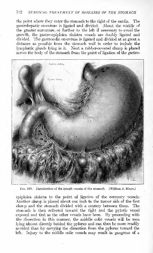
712 SURGICAL TREATMENT OF DISEASES OF THE STOMACH
the point where they enter the stomach to the right of the cardia. The
gastrohepatic omentum is ligated and divided. About the middle of
the greater curvature, or further to the left if necessary to avoid the
growth, the gastro-epiploica sinistra vessels are doubly ligated anddivided. The gastrocolic omentum is ligated and divided at as great a
distance as possible from the stomach wall in order to include the
lymphatic glands lying in it. Next a rubber-covered clamp is placedacross the body of the stomach from the point of ligation of the gastro-
Gastrtc Artery
FIG. 269. Distribution of the stomach. (William J. Mayo.)
epiploica sinistra to the point of ligation of the coronary vessels.
Another clamp is placed about one inch to the tumor side of the first
clamp and the stomach divided with a cautery between them. Thestomach is then reflected toward the right and the pyloric vessel
exposed and tied as the other vessels have been. By proceeding with
the dissection in this manner, the middle colic vessels will be seen
lying almost directly behind the pylorus and can thus be more readily
avoided than by carrying the dissection from the pylorus toward the
left. Injury to the middle colic vessels may result in gangrene of a
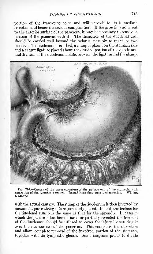
TUMORS OF THE STOMACH 713
portion of the transverse colon and will necessitate its immediate
resection and hence is a serious complication. If the growth is adherent
to the anterior surface of the pancreas, it may be necessary to remove a
portion of the pancreas with it The dissection of the duodenal wall
should be carried well beyond the pylorus, possibly as much as two
inches. The duodenum is crushed, a clamp is placed on the stomach side
and a catgut ligature placed about the crushed portion of the duodenumand division of the duodenum made, between the ligature and the clamp,
FIG. 270. Cancer of the lesser curvature of the pyloric end of the stomach, with
separation of the lymphatic groups. Dotted lines show proposed resection. (WilliamJ. Mayo.)
with the actual cautery. The stump of the duodenum is then inverted bymeans of a purse-string suture previously placed. Indeed, the technic for
the duodenal stump is the same as that for the appendix. In cases in
which the pancreas has been injured or partially resected the free end
of the duodenum should be utilized to cover the injury by suturing it
over the raw surface of the pancreas. This completes the dissection
and allows complete removal of the involved portion of the stomach,
together with its lymphatic glands. Some surgeons prefer to divide
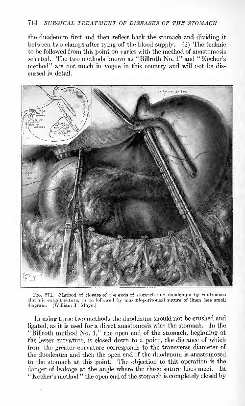
714 SURGICAL TREATMENT OF DISEASES OF THE STOMACH
the duodenum first and then reflect back the stomach and dividing it
between two clamps after tying off the blood supply. (2) The technic
to be followed from this point on varies with the method of anastomosis
selected. The two methods known as "Billroth No. 1" and "Kocher's
method" are not much in vogue in this country and will not be dis-
cussed in detail.
FIG. 271. Method of closure of the ends of stomach and duodenum by continuous
chromic catgut suture, to be followed by musculoperitoneal suture of linen (see small
diagram. (William J . Mayo.)
In using these two methods the duodenum should not be crushed and
ligated, as it is used for a direct anastomosis with the stomach. In the
"Billroth method No. 1," the open end of the stomach, beginning at
the lesser curvature, is closed down to a point, the distance of which
from the greater curvature corresponds to the transverse diameter of
the duodenum and then the open end of the duodenum is anastomosed
to the stomach at this point. The objection to this operation is the
danger of leakage at the angle where the three suture lines meet. In"Kocher's method "
the open end of the stomach is completely closed by
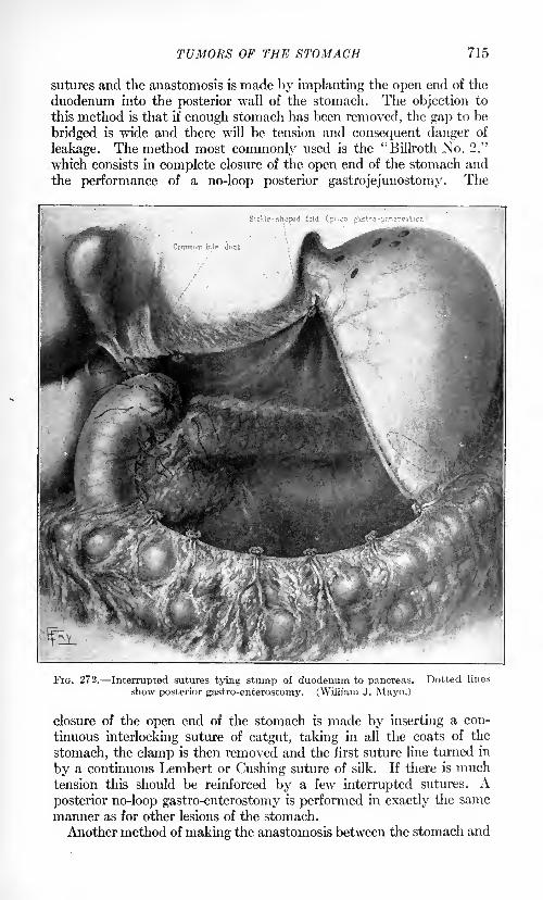
TUMORS OF THE STOMACH 715
sutures and the anastomosis is made by implanting the open end of the
duodenum into the posterior wall of the stomach. The objection to
this method is that if enough stomach has been removed, the gap to be
bridged is wide and there will be tension and consequent danger of
leakage. The method most commonly used is the "Billroth No. 2,"
which consists in complete closure of the open end of the stomach and
the performance of a no-loop posterior gastrojejunostomy. The
FIG. 272. Interrupted sutures tying stump of duodenum to pancreas. Dotted lines
show posterior gastro-enterostomy. (William J. Mayo.)
closure of the open end of the stomach is made by inserting a con-
tinuous interlocking suture of catgut, taking in all the coats of the
stomach, the clamp is then removed and the first suture line turned in
by a continuous Lembert or Gushing suture of silk. If there is muchtension this should be reinforced by a few interrupted sutures. Aposterior no-loop gastro-enterostomy is performed in exactly the same
manner as for other lesions of the stomach.
Another method of making the anastomosis between the stomach and
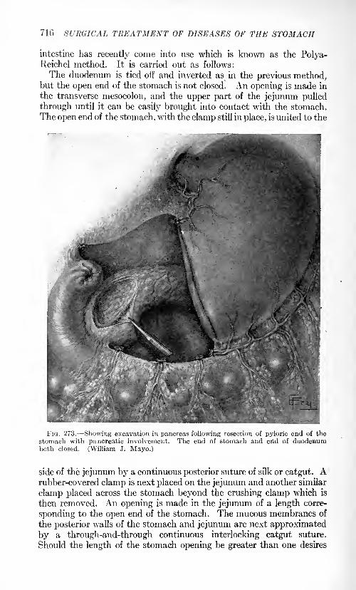
716 SURGICAL TREATMENT OF DISEASES OF THE STOMACH
intestine has recently come into use which is known as the Polya-Reichel method. It is carried out as follows:
The duodenum is tied off and inverted as in the previous method,but the open end of the stomach is not closed. An opening is made in
the transverse mesocolon, and the upper part of the jejunum pulled
through until it can be easily brought into contact with the stomach.
The open end of the stomach, with the clamp still in place, is united to the
FIG. 273. Showing excavation in pancreas following resection of pyloric end of the
stomach with pancreatic involvement. The end of stomach and erid of duodenumboth closed. (William J. Mayo.)
side of the jejunum by a continuous posterior suture of silk or catgut. Arubber-covered clamp is next placed on the jejunum and another similar
clamp placed across the stomach beyond the crushing clamp which is
then removed. An opening is made in the jejunum of a length corre-
sponding to the open end of the stomach. The mucous membranes of
the posterior walls of the stomach and jejunum are next approximated
by a through-and-through continuous interlocking catgut suture.
Should the length of the stomach opening be greater than one desires
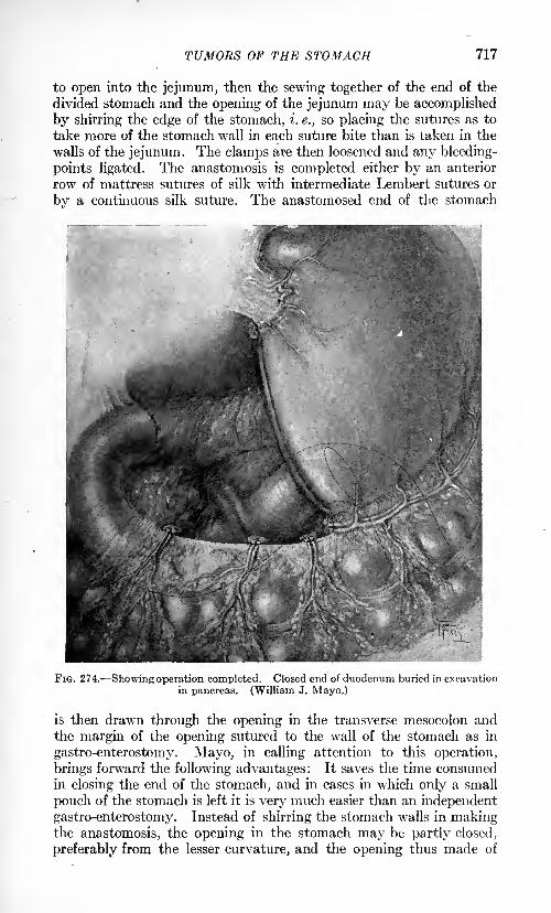
TUMORS OF THE STOMACH 717
to open into the jejunum, then the sewing together of the end of the
divided stomach and the opening of the jejunum may be accomplished
by shirring the edge of the stomach, i. e., so placing the sutures as to
take more of the stomach wall in each suture bite than is taken in the
walls of the jejunum. The clamps are then loosened and any bleeding-
points ligated. The anastomosis is completed either by an anterior
row of mattress sutures of silk with intermediate Lembert sutures or
by a continuous silk suture. The anastomosed end of the stomach
FIG. 274. Showing operation completed. Closed end of duodenum buried in excavationin pancreas. (William J. Mayo.)
is then drawn through the opening in the transverse mesocolon andthe margin of the opening sutured to the wall of the stomach as in
gastro-enterostomy. Mayo, in calling attention to this operation,
brings forward the following advantages: It saves the time consumedin closing the end of the stomach, and in cases in which only a small
pouch of the stomach is left it is very much easier than an independent
gastro-enterostomy. Instead of shirring the stomach walls in makingthe anastomosis, the opening in the stomach may be partly closed,
preferably from the lesser curvature, and the opening thus made of
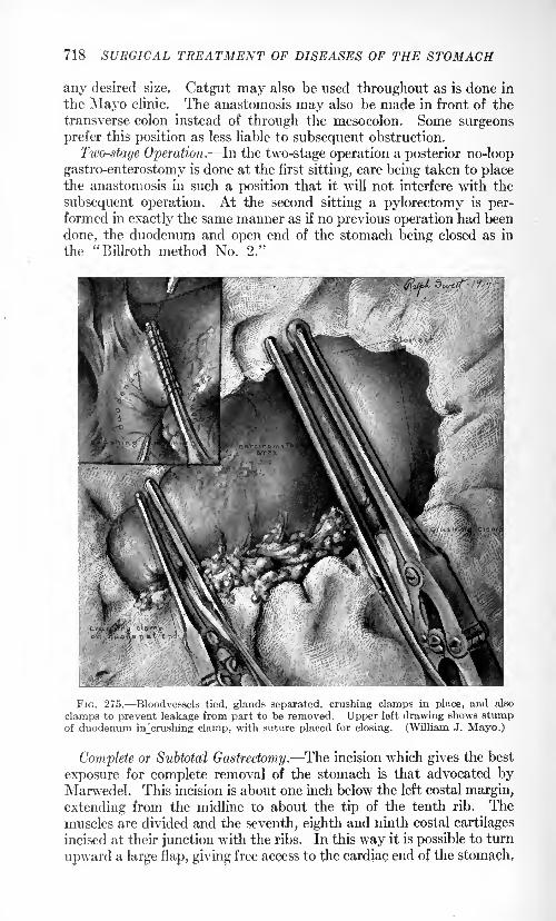
718 SURGICAL TREATMENT OF DISEASES OF THE STOMACH
any desired size. Catgut may also be used throughout as is done in
the Mayo clinic. The anastomosis may also be made in front of the
transverse colon instead of through the mesocolon. Some surgeons
prefer this position as less liable to subsequent obstruction.
Two-stage Operation. In the two-stage operation a posterior no-loop
gastro-enterostomy is done at the first sitting, care being taken to placethe anastomosis in such a position that it will not interfere with the
subsequent operation. At the second sitting a pylorectomy is per-formed in exactly the same manner as if no previous operation had been
done, the duodenum and open end of the stomach being closed as in
the "Billroth method No. 2."
FIG. 275. Bloodvessels tied, glands separated, crushing clamps in place, and also
clamps to prevent leakage from part to be removed. Upper left drawing shows stumpof duodenum uTcrushing clamp, with suture placed for closing. (William J. Mayo.)
Complete or Subtotal Gastrectomy. The incision which gives the best
exposure for complete removal of the stomach is that advocated byMarwedel. This incision is about one inch below the left costal margin,
extending from the midline to about the tip of the tenth rib. Themuscles are divided and the seventh, eighth and ninth costal cartilages
incised at their junction with the ribs. In this way it is possible to turn
upward a large flap, giving free access to the cardiac end of the stomach,
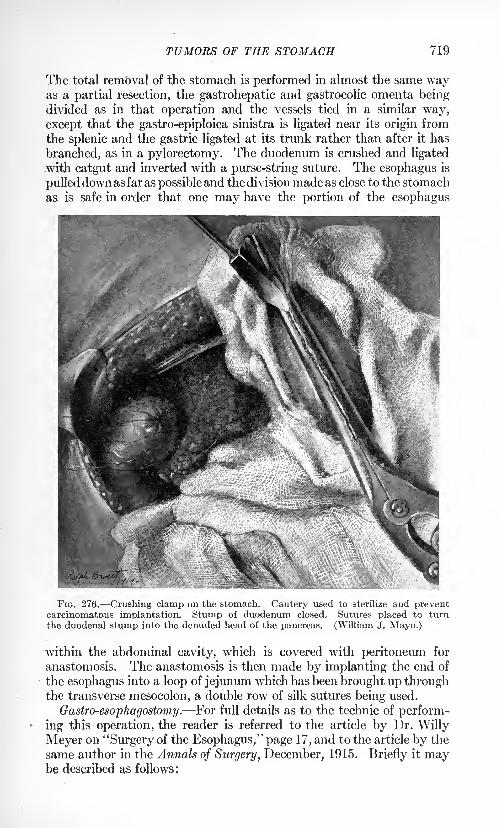
TUMORS OF THE STOMACH 719
The total removal of the stomach is performed in almost the same wayas a partial resection, the gastrohepatic and gastrocolic omenta beingdivided as in that operation and the vessels tied in a similar way,
except that the gastro-epiploica sinistra is ligated near its origin from
the splenic and the gastric ligated at its trunk rather than after it has
branched, as in a pylorectomy. The duodenum is crushed and ligated
with catgut and inverted with a purse-string suture. The esophagus is
pulleddown as far as possible and the division made as close to the stomach
as is safe in order that one may have the portion of the esophagus
FIG. 276. Crushing clamp on the stomach. Cautery used to sterilize and preventcarcinomatous implantation. Stump of duodenum closed. Sutures placed to turnthe duodenal stump into the denuded head of the pancreas. (William J. Mayo.)
within the abdominal cavity, which is covered with peritoneum for
anastomosis. The anastomosis is then made by implanting the end of
the esophagus into a loop of jejunum which has been brought up throughthe transverse mesocolon, a double row of silk sutures being used.
Gastro-esophagostomy. For full details as to the technic of perform-
ing this operation, the reader is referred to the article by Dr. Willy
Meyer on "Surgery of the Esophagus," page 17, and to the article by the
same author in the Annals of Surgery, December, 1915, Briefly it maybe described as follows;
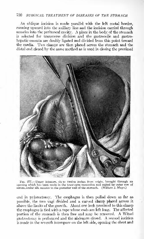
720 SURGICAL TREATMENT OF DISEASES OF THE STOMACH
An oblique incision is made parallel with the left costal border,
running upward into the axillary line and the incision carried throughmuscles into the peritoneal cavity. A place in the body of the stomachis selected for transverse division and the gastrocolic and gastro-
hepatic omenta are doubly ligated and divided from this point towardthe cardia. Two clamps are then placed across the stomach and the
distal end closed by the same method as is used in closing the proximal
FIG. 277. Upper jejunum, six to twelve inches from origin, brought through an
opening which has been made in the transverse mesocolon and united by outer row of
seromuscular silk sutures to the posterior wall of the stomach. (William J. Mayo.)
end in pylorectomy. The esophagus is then pulled down as far as
possible, the two vagi divided and a curved clamp placed across it
above the limits of the growth. About one inch proximal to this clampthe esophagus is tied with a tape whose ends are left long. The affected
portion of the stomach is then free and may be removed. A Witzel
gastrostomy is performed and the abdomen closed. A second incision
is made in the seventh interspace on the left side, opening the chest and
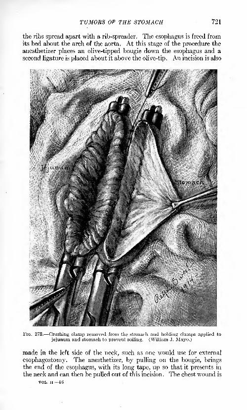
TUMORS OF THE STOMACH 721
the ribs spread apart with a rib-spreader. The esophagus is freed fromits bed about the arch of the aorta. At this stage of the procedure the
anesthetizer places an olive-tipped bougie down the esophagus and a
second ligature is placed about it above the olive-tip. An incision is also
FIG. 278. Crushing clamp removed from the stomach and holding clamps applied to
jejunum and stomach to prevent soiling. (William J. Mayo.)
made in the left side of the neck, such as one would use for external
esophagostomy. The anesthetizer, by pulling on the bougie, bringsthe end of the esophagus, with its long tape, up so that it presents in
the neck and can then be pulled out of this incision. The chest wound is
VOL. ii 46
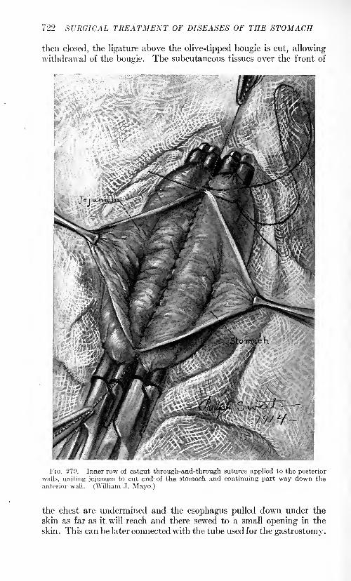
722 SURGICAL TREATMENT OF DISEASES OF THE STOMACH
then closed, the ligature above the olive-tipped bougie is cut, allowingwithdrawal of the bougie. The subcutaneous tissues over the front of
v?-W-
FIG. 279. Inner row of catgut through-and-through sutures applied to the posterior
walls, uniting jejunum to cut end- of the stomach and continuing part way down theanterior wall. (William J. Mayo.)
the chest are undermined and the esophagus pulled down under the
skin as far as it will reach and there sewed to a small opening in the
skin. This can be later connected with the tube used for the gastrostomy.
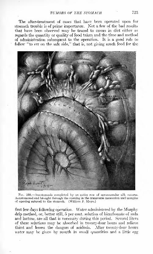
TUMORS OF THE STOMACH 723
The after-treatment of cases that have been operated upon for
stomach trouble is of prime importance. Not a few of the bad results
that have been observed may be traced to errors in diet either as
regards the quantity or quality of food taken and the time and methodof administration subsequent to the operation. It is a good rule to
follow "to err on the safe side," that is, not giving much food for the
FIG. 280. Anastomosis completed by an entire row of seromuscular silk sutures.
Anastomosed end brought through the opening in the transverse mesocolon and marginsof opening sutured to the stomach. (William J. Mayo.)
first few days following operation. Water administered by the Murphydrip method, or, better still, 5 per cent, solution of bicarbonate of soda
and lactose, are all that is necessary during this period. Several liters
of these solutions may be absorbed in twenty-four hours and relieve
thirst and lessen the dangers of acidosis. After twenty-four hours
water may be given by mouth in small quantities and a little egg
724 SURGICAL TREATMENT OF DISEASES OF THE STOMACH
albumen or broth. Fluids only should be allowed for the first four or
five days, then gruels, eggs, fruit juices and other soft articles of diet.
After a week, tender meats, purees of vegetables and stewed fruits, etc.
The quantity should be carefully regulated as well as the quality for
some time. Posture has considerable effect upon these cases. Moststomach cases do better after operation in the sitting posture than
prone. This is particularly true of the first few days. By payingcareful attention to the diet immediately after the operation one canlessen the amount of gastric distress and the troublesome postoperativediarrheas that have been observed, particularly in that class of cases in
which prolonged starvation has been present. If one has been careful
and painstaking in placing one's sutures there is no centra-indication
to the use of the stomach-tube after operation upon the stomach, andit is often of great service when carefully used. There is no valid
objection to the use of sufficient morphin to keep these patients rea-
sonably comfortable for the first few days after operation when theyneed it.
Cysts. These are rare and do not usually occur in connection with
the stomach and duodenum but are generally ascribed to a detached
Meckel's diverticulum, even when found in the mesentery. These
cysts occasionally reach a very large size, and their walls include all
the walls of the intestine. The epithelium is sometimes smooth and
ciliated, but it may exhibit more or less perfect glands and villi. Thetreatment is complete excision where possible. If this cannot be done,then evacuation of the contents of the cyst, cauterization of its walls
and packing of the cavity with gauze in order to stimulate the growthof granulation tissue and the contraction and obliteration of the
cavity, should be employed.
HEMORRHAGE FROM THE STOMACH.
Except in case of trauma, it is rarely necessary to operate for gastric
or duodenal hemorrhage. The hemorrhage may be very profuse, even
fatal, from a large bloodvessel eroded by an ulcer or by a carcinoma.
Deaths are now and then reported from postoperative hemorrhagewhere a gastric or intestinal resection or suture has been done. Such
unfortunate results are usually due to the fact that the cut bloodvessels
along the line of suture have not been ligated at all, or insecurely so.
Operations upon the stomach, in case of gastric hemorrhage from con-
gestion of the portal circulation from any cause, are not indicated.
Treatment. Too much care cannot be taken in the absolute control
of hemorrhage after incisions into the stomach wall. Too much reliance
should not be placed in the continuous suture to accomplish this, as it
is uncertain at best. It is safer always before suturing to loosen the
clamps and ligate each individual vessel separately. In the majorityof cases, this may not be necessary, but it should not be forgotten that
it is the hundredth case that may need it, and hence it should not be
disregarded. The presence of occult blood in the stools, while not
PYLORIC STENOSIS 725
absolute evidence of the presence of ulceration of the stomach or
intestine, is so strongly suggestive of it that when persistent and other-
wise unaccounted for, it constitutes sufficient indication upon which to
advise exploratory laparotomy and gastrotomy in order to establish
a definite diagnosis, or to apply treatment.
'-'/
OBSTRUCTION.
Obstruction to the entrance of food into the stomach through the
cardiac orifice may be due to a muscular spasm or to organic stricture
at that point, congenital or acquired. Some years ago the writer wascalled to see a newborn baby with a history of persistent vomiting of
all the food it had taken since birth. The passage of a small catheter
into the esophagus showed an impervious stricture just at the cardiac
orifice. I therefore operated and found a very small stomach showinga complete stricture at the cardia. A gastrostomy was done. The babydid not survive, dying some days later, although the gastrostomyworked satisfactorily. This condition is rare.
Treatment. Various methods of treatment have been suggested to
relieve muscular spasm . The operation of Mikulicz dilates very thor-
oughly the cardiac orifice. Through a gastrotomy incision in the
anterior wall a pair of dilating forceps are passed up into the cardia
and that orifice is thoroughly stretched. Later less radical measures
have become popular, such as dilatation by means of a water-bagintroduced through the mouth and placed in position in the cardia,
which is then subjected to hydrostatic pressure. Plummer has broughtthis method prominently before the Profession and reports satisfactory
results. It should always be tried before resorting to more radical
measures.
PYLORIC STENOSIS.
Except for those cases of spastic contraction of the pylorus due to
reflex irritation from a chronic appendix, or gall-bladder disease, pyloricstenosis in the adult is, in general, due to the cicatrization of gastric
ulcers in the process of healing. Obstruction may also be caused bycarcinoma or inflammations, as with acute ulcer in this region. Pedun-culated papillomatous growths arising from the gastric mucosa near
the pylorus may occasionally produce intermittent obstruction bybecoming engaged in it.
Congenital pyloric stenosis may persist until adult life. Recentlyan undoubted case of this condition was seen and treated at the Johns
Hopkins Hospital in a girl, aged seventeen years. The patient for
years had been bothered by vomiting. The stomach held about two
quarts, and always had a large retention. The pylorus was found to
be much thickened and hypertrophied, with no sign of ulcer. A Finney
pyloroplasty was done, with complete cure of the condition.
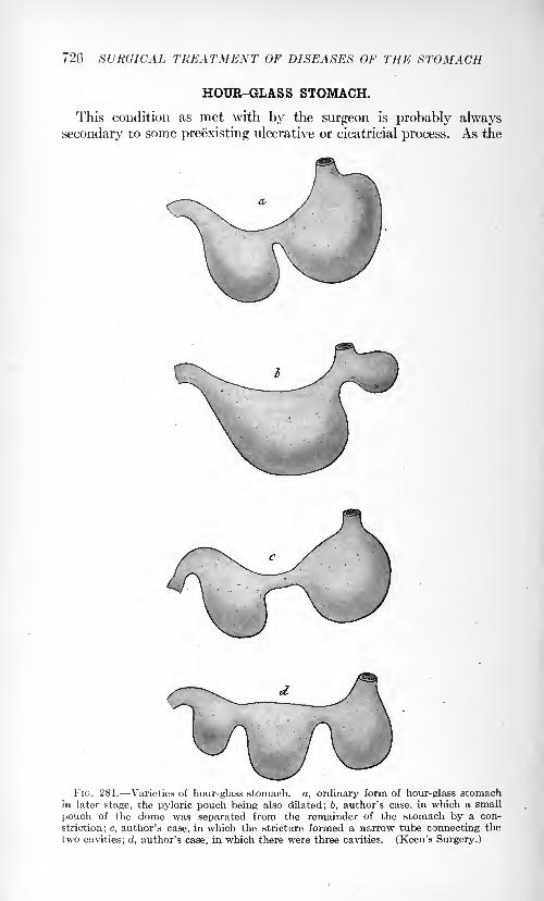
720 SURGICAL TREATMENT OF DISEASES OF THE STOMACH
HOUR-GLASS STOMACH.
This condition as met with by the surgeon is probably always
secondary to some preexisting ulcerative or cicatricial process. As the
FIG. 281. Varieties of hour-glass stomach, a, ordinary form of hour-glass stomachin later stage, the pyloric pouch being also dilated; b, author's case, in which a small
pouch of the dome was separated from the remainder of the stomach by a con-
striction; c, author's case, in which the stricture formed a narrow tube connecting the
two cavities ; d, author's case; in which there were three cavities. (Keen's Surgery.)
ACUTE DILATATION OF THE STOMACH 727
name implies, the stomach is divided into two more or less well-defined
pouches of varying sizes, with a band of cicatricial tissue between,
sufficiently contracted and inelastic to produce appreciable obstruction
to the passage of the food. The surgical problem involved is to enlargethe opening present sufficiently to allow the free passage of the food
or to make a new one. Occasionally, pyloric stenosis is observed in
connection with the hour-glass contraction. The writer has recently
observed such a combination. This materially complicates the situ-
ation and involves the necessity of performing in addition a pyloro-
plasty or a gastro-enterostomy. The two gastric pouches are united,
preferably by a pyloroplasty (Fig. 281), as the simplest and usually
the most satisfactory procedure, or the cicatricial bridge may be excised
and the two segments of stomach united, or a gastrogastrostomy or
a simple gastrostomy may be done.
ACUTE DILATATION OF THE STOMACH.
Following abdominal operations, acute dilatation of the stomach is a
complication the possibility of which should ever be kept in mind, for
unless prompt and vigorous treatment is instituted a fatal issue mayresult. This affection is fortunately not of frequent occurrence but
always serious. It has been observed where there has been no ante-
cedent surgical operation. Frequently, at autopsies, the stomach is
found to be markedly dilated, but the prevailing opinion is that this
is due to postmortem conditions.
Acute dilatation of the stomach is accompanied by and possibly dueto filling up with gas and great overdistention with fluid.
The dilatation may be of reflex nervous origin, with simple loss of
tone of the gastric walls. In perhaps one-half of the recorded cases a
more or less definite mechanical obstruction has been made out. Some-times it has been found at the pylorus, but usually in the last portion of
the duodenum where it is constricted between the taut mesenteric
vessels and the vertebral column, the so-called gastromesenteric ileus.
Kinks, contractions and adhesions and pressure of misplaced organs
may all tend to bring this about.
Acute dilatation, as a rule, comes on shortly after operation, althoughsudden dilatation has been observed to take place during the course of
an operation and can usually be promptly and completely relieved byappropriate measures. The symptoms are liable to develop insidiously.
The most striking feature, perhaps, in the clinical picture is the frequent
vomiting which cannot be otherwise explained. The patient does not
strain much during the vomiting, but rather regurgitates without
effort. The pulse-rate increases and there may soon be symptoms of
collapse. An unquenchable thirst is often a prominent symptom. The
patient looks ill and the whole picture impresses one with its seriousness.
A curious feature that has been observed at tunes is the fact that
although the stomach may be enormously distended there may be no
728 SURGICAL TREATMENT OF DISEASES OF THE STOMACH
visible distention of the abdomen indeed, it may be retracted. Theamount of urine is always markedly diminished.
Among the etiological factors supposed to favor the production of
acute dilatation of the stomach are:
1. Operations under general anesthesia.
2. Severe or prolonged and wasting diseases.
3. Indiscretions in diet.
4. Diseases or deformities of the spine.5. Thrombosis.
6. Visceroptosis.7.. Large abdominal tumors.
Moynihan reports 12 cases out of 44 in which the dilatation was not
limited to the stomach but involved the duodenum also, and in one case
the upper few inches of the jejunum. Box and Wallace think there is
perhaps first a paralytic condition of the stomach leading to distention
and then pressure on the duodenum where it crosses the spinal column
by the heavy distended stomach, producing obstruction.
Some observers think the distention due merely to excessive
secretion, while others believe it to be the result of a spasm of the
pylorus.Albrecht was the first to suggest that the constricting agent may be
the superior mesenteric vessels which, owing to the downward draggingof the intestines, press upon and obstruct the duodenum as it crosses
the spinal column. This explanation is favored by the majority of
writers, but it certainly does not explain all cases, as the writer has
had occasion several times to observe at operation that the mesenteric
vessels were not at all taut nor the duodenum compressed while the
stomach was enormously distended.
It has been shown experimentally that section of the pneumogastricnerves in a dog is followed by dilatation of the stomach.
In acute dilatation occurring postoperatively, frequently the first
symptom to be observed is a sudden increase in the number of heart
beats per minute and great difficulty in breathing. Physical examina-tion demonstrates marked upward displacement of the heart which is
corrected immediately upon the introduction of a stomach-tube with
the rapid, noisy expulsion of gas through the tube.
Treatment. As soon as this condition is recognized, or indeed as
soon as its presence is suspected, the stomach-tube should be passedand care taken that the stomach is entirely emptied. Owing to the
loss of muscular tone of its walls, this must usually be done by siphon-
age or gravity. It is well to attempt to stimulate the muscle by irri-
gating with very warm water or salt solution, our practice being to use
a temperature of 110 F. The foot of the bed should be elevated andif possible, the patient turned on his side or abdomen. The stomach
should be irrigated as often as is necessary, preferably every hour
until its tone is restored and the obstruction relieved. This may be a
matter of several days. Operative procedures, such as gastrotomy and
gastro-enterostomy, are not indicated and not to be performed; at
OPERATIONS ON THE STOMACH 729
any rate, not until the continued use of the stomach-tube has been of noavail. Drugs appear to have little, if any, value in this condition.
A chronic form of gastric and duodenal dilatation has been described.
The same pathological processes are supposed to be present and active
as in the acute form, and the same treatment is to be employed. If
the condition were always due to mechanical obstruction of the duo-
denum from taut mesenteric vessels a gastro-enterostomy ought alwaysto give immediate and complete relief. Such, however, is not the case.
In acute gastric dilatation the patient should always be placed in a
prone position at once unless a stomach-tube is immediately at handbecause frequently this maneuver alone will bring prompt and per-manent relief while the delay which may be occasioned by efforts to
procure a stomach-tube may be sufficient to result in a fatal issue. Theeditor encountered one case in which a patient, aged forty-nine years,
with partial occlusion of the coronary arteries died because of the
sudden severe displacement of the heart before the acute dilatation
could be relieved by the passage of a stomach-tube.
OPERATIONS ON THE STOMACH.
For convenience of description the stomach is divided into two
portions, the cardiac and the pyloric. The axis of the cardiac portionis practically vertical, converging slightly toward the midline below
while the axis of the pyloric part is almost horizontal. It is safe to
say that three-fourths at least of the surgery of the stomach has to do
with the pyloric portion. This is fortunate, both because of its accessi-
bility and movability as compared with the cardiac portion. It will
be observed that, as a rule, the more the symptoms from which relief
is sought through surgery have their origin in obstructive processesabout the pylorus the earlier will they manifest themselves and the
more complete will be the relief from surgery. The converse of this
proposition is also true, namely, that the further removed the patho-
logical process is from the pylorus the less early do the resulting diag-
nostic symptoms appear and the less complete the relief experiencedfrom surgical measures. It is also well to bear in mind that the more
pronounced macroscopically and the more easily demonstrable the
pathological lesion for the relief of which the operation is undertaken
the more certain and complete is the subsequent relief. Of course,
we are not including in this category inoperable cancerous growths.Where careful inspection of the stomach at the time of operation fails
to reveal a definite, demonstrable lesion, further operation is contra-
indicated.
Surgery is of little avail in the so-called functional disturbances of
the stomach, not infrequently it aggravates the symptoms.It is assumed that the operator is familiar with the fundamental
principles of aseptic surgery and the healing of wounds, and with the
contents of the armamentarium of the abdominal surgeon. Time and
space will therefore not be taken in order to describe the various
730 SURGICAL TREATMENT OF DISEASES OF THE STOMACH
methods of intestinal suture and suture materials of which use is madein gastro-intestinal surgery. The same is true of the various special
instruments, such as needles, stomach and intestinal clamps, retractors
and the like. When occasion is had to refer to any of them theywill be called by name without further description of their individual
characteristics.
In the consideration of the surgery of the stomach it should not be
forgotten there is a vast range of pathological conditions that havetheir origin outside of the stomach; indeed, many of them entirelyoutside of the abdomen, which not infrequently present as one of their
earliest manifestations nausea and vomiting, or some form of gastricdisorder or distress. Every effort, therefore, should be made to arrive
at a definite and correct diagnosis before surgical measures are resorted
to, in order to avoid making the grievous mistake of an unnecessary
operation. There is no class of cases in which it can be more truly said
that the combined skill of the physician and surgeon is productive of
better results indeed, is so absolutely necessary as in the case of
suspected disease of the stomach. Hearty cooperation, therefore,
between the physician and surgeon in the diagnosis and treatment of
this class of cases is most desirable. The writer never operates upon a
patient suffering from supposed stomach trouble without first havinghim thoroughly studied out and reported upon by a competent internist,
and then not until he has satisfied himself that no further improvementis to be expected from a continuance of medical measures. While it is
undoubtedly true that many cases of "stomach trouble" suffer for
lack of surgical treatment, or from such treatment having been too
long deferred, it is equally true there is a great deal of unnecessary and
unjustifiable operating being done upon the stomach. The chief
offenders in this respect, perhaps, are those occasional operators who,
by reason of defective training, lack the judgment and skill necessary
properly to perform operations upon the gastro-intestinal tract, and
without which they should never be undertaken.
When a surgeon is consulted by a patient with the history of some
gastric disorder he is confronted with the necessity of making a decision,
sooner or later, upon four points: (1) the diagnosis, (2) whether the
case can better be treated by medical or surgical measures, (3) if
surgical the character of the operation indicated and (4) the manner in
which such a surgical operation is to be performed. This includes a con-
sideration of the proper preparation and care of the patient, both before
and after operation. Concerning the first of these questions, namely,
"diagnosis," we are not especially concerned in this chapter; for it has
been ably presented in the preceding one. But one should never
for a moment lose sight of the fact that the diagnosis in any given case
of stomach trouble is not only not always easy, but it not infrequently
happens that it must be left to be determined at the autopsy or by the
exploratory incision of the surgeon, a rather humiliating confession in
these days when through the arrays, the laboratory and other special
tests, our diagnostic ability in general has reached such a high state of
OPERATIONS ON THE STOMACH 731
scientific development. A supposed stomach case may, after careful
consideration, prove to be almost anything from tabes or an inflamed
appendix to a brain tumor.
Before considering the choice of operation in a given case of gastric
disorder, it is well to determine first whether or not any operation is
really necessary. The persistence and severity of the symptoms andthe progressive nature of the disease, together with the failure of
properly applied medical measures to afford relief, are the factors
which decide this question in the affirmative. One should then con-
sider "what it is expected to accomplish by the operation. Is it to be
simply a palliative procedure undertaken to give temporary relief to
the patient or are we justified in expecting and offering a cure? Theseare important questions which the patient is likely to ask, and when he
does, he is entitled to an honest and intelligent answer. Upon what
basis, too, are we going to determine the operative procedure to be
carried out? Is it to be on the basis of habit, because we are all more or
less creatures of habit, or is it to be a matter of fashion, because there
are fashions in surgery just as in dress; or is every individual case to
be decided upon its own merits and due weight to be given to the
relative advantages or disadvantages that are generally accepted as
belonging to any particular type of operation? The surgeon ought to
have a valid reason for every operative procedure that he undertakes,and should be able successfully to defend his position.
Except in cases of emergency no operation should be performed on
the stomach or duodenum without adequate preliminary preparationof the patient. Gushing has shown that it is possible, through sterile
feeding and antiseptic mouth washes, to render the stomach and upper
portion of the intestinal tract sterile. These observations have been
abundantly confirmed by our own clinical experience. Likewise, no
surgical operation should be undertaken, except in case of emergency,without proper attention to details of technic, asepsis, competent assist-
ants, suitable instruments, etc. These facts are so fundamental that
it seems hardly necessary to call attention to them. As regards the
details of the technic of a given operation on the stomach, they vary
largely with the operator. This is true also of the different instruments,
such as stomach clamps, etc., that are to be used; but too much care
cannot be taken in matters of detail, as not infrequently the ultimate
result is -determined by the conscientious manner in which these are
carried out by the operator and his assistants. The question of the
anesthetic and of the skill with which it is admmistered should never
be overlooked. Here, again, individual methods differ, but there are
certain fundamental rules which are common to all and which should
never be disregarded. Crile's anoci-association as practised in his clinic
commends itself to careful consideration. Local anesthesia seems to be
gaining advocates, particularly on the continent, but for stomach oper-ations it does not seem to be as well suited as for certain others.
In the after-care of all cases of operation upon the stomach the most
effective agents at our coir,mand are the use of the stomach-tube and
732 SURGICAL TREATMENT OF DISEASES OF THE STOMACH
proctoclysis by the method of Murphy. These are two of the mostefficient life-savers contained in the armamentarium of the surgeon.The addition of lactose and bicarbonate of soda to the fluid to be
injected adds much to its efficiency by lessening the dangers of acidosis
and by improving the nutrition. The complications that may follow
operations on the stomach and duodenum are those common to all
abdominal operations and need not be referred to in detail. All the
time, as our knowledge increases and our technic becomes simpler andmore effective, these accidents of operation are becoming less and less
frequent, until now one undertakes a major operation upon the stomachwith a feeling that borders on assurance. Lung complications will
never be completely abolished so long as we have to use anesthetics.
Secondary hemorrhage and peritonitis are usually the result of faultytechnic and, as such, are gradually being eliminated. Acute dilatation
of the stomach can be recognized if watched for and may be controlled
by the early and continued use of the stomach-tube. Phlebitis andthrombosis are always serious when they occur, but, fortunately,
their occurrence is rare. They cannot as yet be entirely prevented but
careful handling of the tissues and the control of sepsis seem to tend
very materially toward lessening the frequency of their appearance.The different forms of anemia certainly are in some way concerned in
the production of thrombophlebitis, as one or other of them is almost
always found to be present.As regards drainage, in general the tendency seems all the while
more directed against the use of drainage of any kind, except in cases
in which pus or extravasation of intestinal contents are actually
present. In operations on the stomach and duodenum this is rarely the
case. Hence it is very seldom that it will be found necessary to drain
the wound. The exception to this is usually in cases of perforation.
Here, not infrequently, it will be found to be good judgment to drain
at least to the neighborhood of the perforation after closure. A drain
applied directly to the suture line will almost surely be followed by a
gastric fistula.
The stomadi in health varies within very wide limits as regards its
size, shape and position, depending largely upon the age, sex and habits
of the individual and upon the amount of food or drink which it con-
tains. Under pathological conditions these variations may be muchwider and its position and relation to surrounding structures may be
very far from normal, depending in turn upon the nature of the patho-
logical processes present, either in the stomach itself, the neighboringviscera or the abdominal walls. Generally speaking its long axis is
inclined obliquely downward and to the right from the cardiac orifice,
which is its only fixed point. This axis in turn varies from almost
vertical to the horizontal. The pylorus from being a movable point
within certain physiological limits, may become firmly attached byadhesions to the under surface of the liver, the gall-bladder, the colon
or the abdominal wall. On the other hand, the pylorus is occasionally
seen so freely movable that in extreme cases it may be displaced
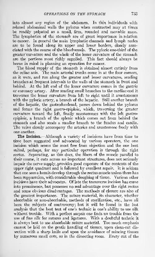
OPERATIONS ON THE STOMACH 733
into almost any region of the abdomen. In thin individuals with
relaxed abdominal walls the pylorus when contracted may at times
be readily palpated as a small, firm, rounded and movable mass.
The lymphatics of the stomach are of great importance in relation
to cancer. In general the main lymphatic channels and lymph nodes
are to be found along its upper and lower borders, closely asso-
ciated with the course of the bloodvessels. The pyloric one-third of the
greater"curvature and the whole of the lesser curvature of the stomach
are the portions most richly supplied. This fact should always be
borne in mind in planning an operation for cancer.
The blood supply of the stomach is obtained almost entirely from
the celiac axis. The main arterial trunks come in at the four corners,
as it were, and run along the greater and lesser curvatures, sendingbranches .at frequent intervals to the walls of the stomach in front andbehind. At the left end of the lesser curvature comes in the gastricor coronary artery. After sending small branches to the cardiac end it
traverses the lesser curvature from left to right until it anastomoses
with the pyloric artery, a branch of the hepatic. Still another branch
of the hepatic, the gastroduodenal, passes down behind the pylorusand forms the right gastro-epiploic, which, traversing the greatercurvature toward the left, finally anastomoses with the left gastro-
epiploic, a branch of the splenic which comes out from behind the
stomach and also sends a smaller branch to the fundus and cardia.
The veins closely accompany the arteries and anastomose freely with
one another.
The Incision. Although a variety of incisions have from time to
tune been suggested and advocated by various operators, the one
incision which seems the most free from objection and the one best
suited, perhaps, for any particular operation is through the right
rectus. Separating, as this does, the fibers of the muscle parallel to
their course, it cuts across no important structures, does not seriously
impair the nerve supply, provides good exposure of the contents of the
upper right quadrant and is followed by excellent repair. It is seldom
that one sees a hernia develop through the rectus muscle unless there has
been suppuration, with considerable sloughing of tissue. Various other
incisions have their advocates. Of late the transverse incision has comeinto prominence, but possesses no real advantage over the right rectus
and some obvious disadvantages. The methods of closure are also of
the greatest importance. The suture material, its character, whether
absorbable or non-absorbable, methods of sterilization, etc., have all
been the subjects of controversy; but it will be found in the last
analysis that the best test of one's technic is one's ability to use silk
without trouble. With a perfect asepsis one finds no trouble from the
use of fine silk for sutures and ligatures. With a doubtful technic it
is always best to use absorbable suture material. Too much emphasiscannot be laid on the gentle handling of tissues, upon clean-cut dis-
section with a sharp knife and upon the avoidance of mincing tissues
by numerous small cuts, as in the dissecting room. Every cut of the
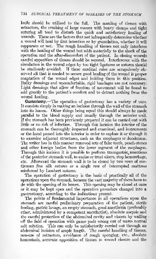
734 SURGICAL TREATMENT OF DISEASES OF THE STOMACH
knife should be utilized to the full. The mauling of tissues with
retractors, the crushing of large masses with heavy clamps and tight
suturing all tend to disturb the quick and satisfactory healing of
wounds. These are the factors that not infrequently determine whethera wound will heal by first intention or by granulation, whether it will
suppurate or not. The rough handling of tissues not only interferes
with the healing of the wound but adds materially to the shock of the
operation and the after-discomfort of the patient. In closing woundscareful apposition of tissues should be secured. Interference with the
circulation in the wound edges by too tight ligatures or sutures shouldbe studiously avoided. If these cardinal rules are scrupulously ob-served all that is needed to secure good healing of the wound is proper
coaptation of the wound edges and holding them in this position.
Bulky dressings and uncomfortable, rigid bandages are not necessary.
Light dressings that allow of freedom of movement will be found to
add greatly to the patient's comfort and to detract nothing from the
wound healing.
Gastrotomy. The operation of gastrotomy has a variety of uses.
It consists simply in making an incision through the wall of the stomachinto its lumen. Other things being equal this should always be made
parallel to the blood supply and usually through the anterior wall.
If the stomach has been previously prepared it can be carried out with
little or no risk of infection. Through this incision the interior of the
stomach can be thoroughly inspected and examined, and instruments
or the hand passed into the interior in order to explore it or through it
to examine adjacent structures, such as the esophagus or duodenum.The writer has in this manner removed sets of false teeth, peach-stonesand other foreign bodies from the lower segment of the esophagus.
Through this incision it is possible to perform transgastric. resections
of the posterior stomach wall, to excise or treat ulcers, stop hemorrhage,etc. Afterward the stomach wall is to be closed by two rows of con-
tinuous fine silk sutures or a single row of interrupted mattress
reinforced by Lembert sutures.
The operation of gastrotomy is the basis of practically all of the
operations upon the stomach, because the vast majority of them have to
do with the opening of its lumen. This opening may be closed at once
or it may be kept open and the operative procedure changed into a
gastrostomy, according to the indications present.The points of fundamental importance in all operations upon the
stomach are careful preliminary preparation of the patient, sterile
feeding, gastric lavage, an empty stomach, good anesthesia (preferably
ether, administered by a competent anesthetist), absolute asepsis and
the careful protection of the abdominal cavity and viscera by wallingoff the field of operation with gauze pads wrung out of warm normal
salt solution. This can only be satisfactorily carried out through an
abdominal incision of ample length. The careful handling of tissues,
non-use of retractors, avoidance of rough sponging, etc., absolute
hemostasis, accurate apposition of tissues in wound closure and the
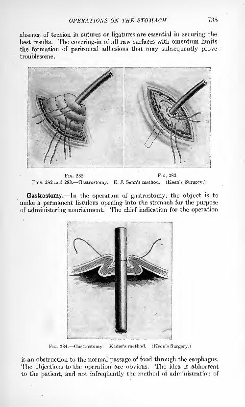
735
absence of tension in sutures or ligatures are essential in securing the
best results. The covering-in of all raw surfaces with omentum limits
the formation of peritoneal adhesions that may subsequently provetroublesome.
FIG. 282 FIG. 283
FIGS. 282 and 283. Gastrostomy. E. J. Senn's method. (Keen's Surgery.)
Gastrostomy. In the operation of gastrostomy, the object is to
make a permanent fistulous opening into the stomach for the purposeof administering nourishment. The chief indication for the operation
FIG. 284. Gastrostomy. Kader's method. (Keen's Surgery.)
is an obstruction to the normal passage of food through the esophagus.The objections to the operation are obvious. The idea is abhorrent
to the patient, and not infrequently the method of administration of
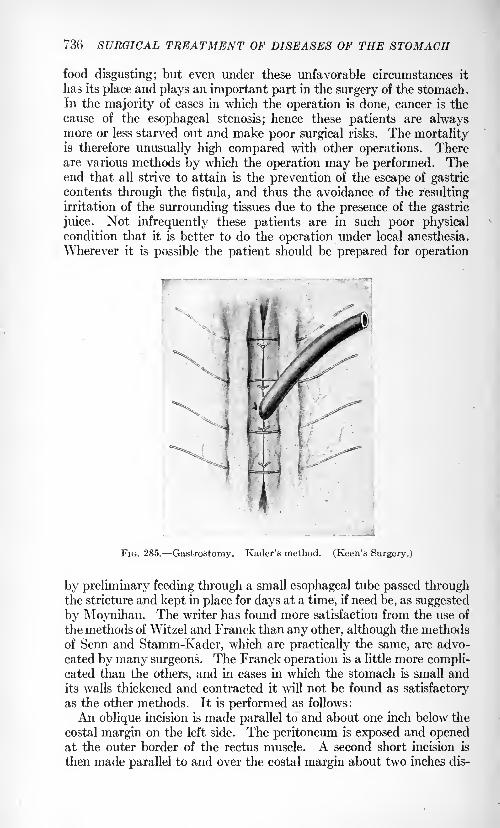
736 SURGICAL TREATMENT OF DISEASES OF THE STOMACH
food disgusting; but even under these unfavorable circumstances it
has its place and plays an important part in the surgery of the stomach.In the majority of cases in which the operation is done, cancer is the
cause of the esophageal stenosis; hence these patients are alwaysmore or less starved out and make poor surgical risks. The mortalityis therefore unusually high compared with other operations. Thereare various methods by which the operation may be performed. Theend that all strive to attain is the prevention of the escape of gastric
contents through the fistula, and thus the avoidance of the resultingirritation of the surrounding tissues due to the presence of the gastric
juice. Not infrequently these patients are in such poor physicalcondition that it is better to do the operation under local anesthesia.
Wherever it is possible the patient should be prepared for operation
FIG. 285. Gastrostomy. Kader's method. (Keen's Surgery.)
by preliminary feeding through a small esophageal tube passed throughthe stricture and kept in place for days at a time, if need be, as suggested
by Moynihan. The writer has found more satisfaction from the use of
the methods of Witzel and Franck than any other, although the methodsof Senn and Stamm-Kader, which are practically the same, are advo-
cated by many surgeons. The Franck operation is a little more compli-cated than the others, and in cases in which the stomach is small andits walls thickened and contracted it will not be found as satisfactory
as the other methods. It is performed as follows:
An oblique incision is made parallel to and about one inch below the
costal margin on the left side. The peritoneum is exposed and openedat the outer border of the rectus muscle. A second short incision is
then made parallel to and over the costal margin about two inches dis-
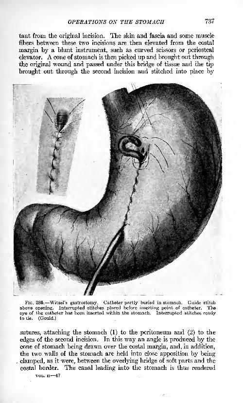
OPERATIONS ON THE STOMACH 737
tant from the original incision. The skin and fascia and some muscle
fibers between these two incisions are then elevated from the costal
margin by a blunt instrument, such as curved scissors or periosteal
elevator. A cone of stomach is then picked up and brought out throughthe original wound and passed under this bridge of tissue and the tip
brought out through the second incision and stitched into place by
FIG. 286. Witzel's gastrostomy. Catheter partly buried in stomach. Guide stitch
above opening. Interrupted stitches placed before inserting point of catheter. Theeye of the catheter has been inserted within the stomach. Interrupted stitches readyto tie. (Gould.)
sutures, attaching the stomach (1) to the peritoneum and (2) to the
edges of the second incision. In this way an angle is produced by the
cone of stomach being drawn over the costal margin, and, in addition,
the two walls of the stomach are held into close apposition by being
clamped, as it were, between the overlying bridge of soft parts and the
costal border. The canal leading into the stomach is thus rendered
VOL. ii 47
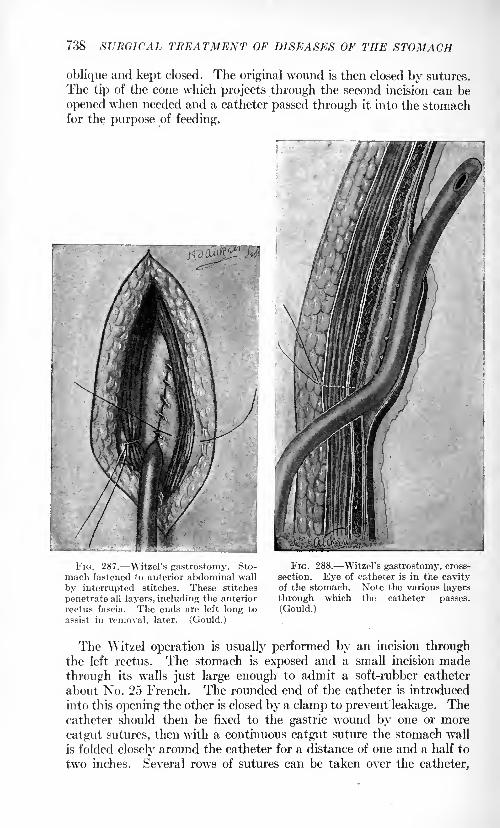
738
oblique and kept closed. The original wound is then closed by sutures.
The tip of the cone which projects through the second incision can be
opened when needed and a catheter passed through it into the stomachfor the purpose of feeding.
FIG. 287. Witzel's gastrostomy. Sto-
mach fastened to anterior abdominal wall
by interrupted stitches. These stitches
penetrate all layers, including the anterior
rectus fascia. The ends are left long to
assist in removal, later. (Gould.)
FIG. 288. Witzel's gastrostomy, cross-
section. Eye of catheter is in the cavityof the stomach. Note the various layers
through which the catheter passes.
(Gould.)
The Witzel operation is usually performed by an incision throughthe left rectus. The stomach is exposed and a small incision made
through its walls just large enough to admit a soft-rubber catheter
about No. 25 French. The rounded end of the catheter is introduced
into this opening the other is closed by a clamp to prevent leakage. Thecatheter should then be fixed to the gastric wound by one or more
catgut sutures, then with a continuous catgut suture the stomach wall
is folded closely around the catheter for a distance of one and a half to
two inches. Several rows of sutures can be taken over the catheter,
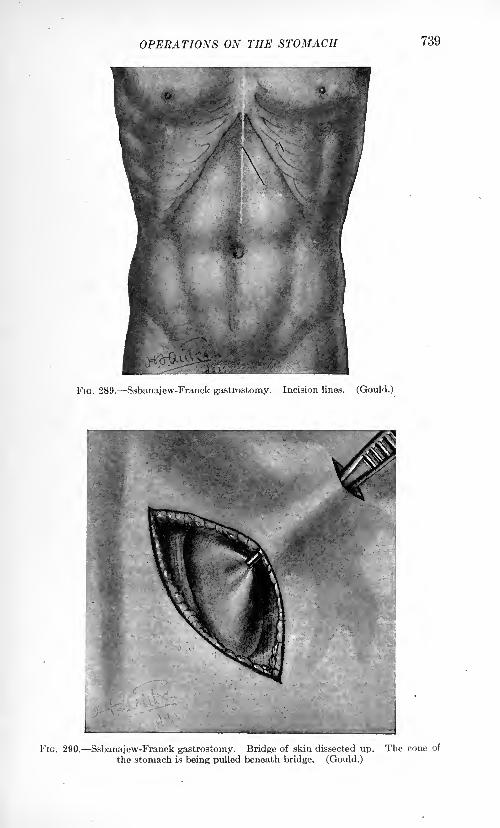
OPERATIONS ON THE STOMACH 739
FIG. 289. Ssbanajew-Franck gastrostomy. Incision lines. (Gould.)
Kia. 290. Ssbanajew-Franck gastrostomy. Bridge of skin dissected up. The cone of
the stomach is being pulled beneath bridge. (Gould.)
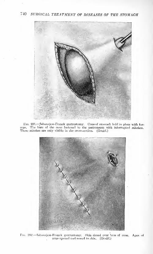
740 SURGICAL TREATMENT OF DISEASES OF THE STOMACH
.
FIG. 291. Ssbanajew-Franck gastrostomy. Cone of stomach held in place with for-
ceps. The base of the cone fastened to the peritoneum with interrupted stitches.These stitches are only visible in the cross-section. (Gould.)
FIG. 292. Ssbanajew-Franck gastrostomy. Skin closed over base of cone. Apex of
cone opened and sewed to skin. (Gould.)
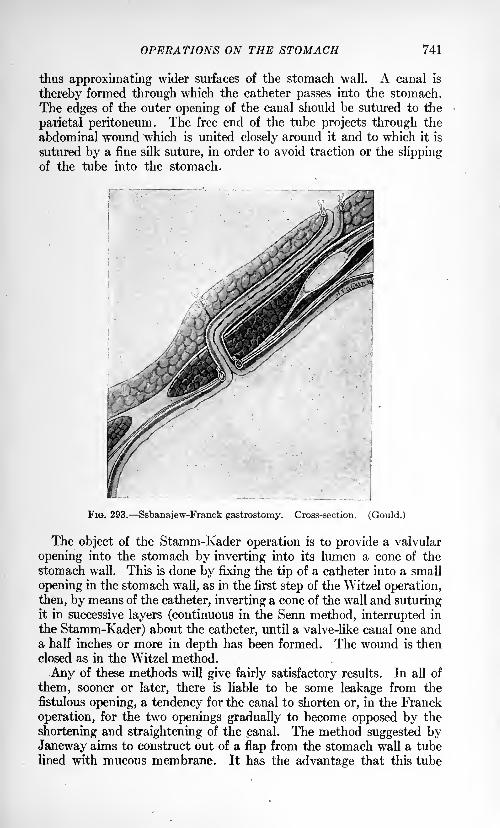
OPERATIONS ON THE STOMACH 741
thus approximating wider surfaces of the stomach wall. A canal is
thereby formed through which the catheter passes into the stomach.
The edges of the outer opening of the canal should be sutured to the
parietal peritoneum. The free end of the tube projects through the
abdominal wound which is united closely around it and to which it is
sutured by a fine silk suture, in order to avoid traction or the slippingof the tube into the stomach.
FIG. 293. Ssbanajew-Franck gastrostomy. Cross-section. (Gould.)
The object of the Stamm-Kader operation is to provide a valvular
opening into the stomach by inverting into its lumen a cone of the
stomach wall. This is done by fixing the tip of a catheter into a small
opening in the stomach wall, as in the first step of the Witzel operation,
then, by means of the catheter, inverting a cone of the wall and suturingit in successive layers (continuous in the Senn method, interrupted in
the Stamm-Kader) about the catheter, until a valve-like canal one anda half inches or more in depth has been formed. The wound is then
closed as in the Witzel method.
Any of these methods will give fairly satisfactory results. In all of
them, sooner or later, there is liable to be some leakage from the
fistulous opening, a tendency for the canal to shorten or, in the Franck
operation, for the two openings gradually to become opposed by the
shortening and straightening of the canal. The method suggested byJaneway aims to construct out of a flap from the stomach wall a tube
lined with mucous membrane. It has the advantage that this tube
742 SURGICAL TREATMENT OF DISEASES OF THE STOMACH
does not tend to contract to any great extent, but it also has the dis-
advantage that it is more complicated than the other methods and is
only applicable when the gastric wall is soft and pliable and the stomachnot too contracted. The operation is somewhat along the general lines
of the Heineke-Mikulicz operation by pyloroplasty. A rectangular
flap one and three-fourths by one-half inches, with its long axis corre-
sponding to the long axis of the stomach is turned up. The stomach
wall is folded upon itself and the short sides of the parallelogram are
opposed to each other and sutured together, as are the edges of the
everted flaps of the gastric wall, thus forming a tube about two inches
long lined with mucosa. This tube is then brought out through the
abdominal wound and its base sutured to the parietal peritoneum and
the margin of the rectus sheath and its tip to the skin edges. If desired
the base of the tube may be inverted into the lumen of the stomach in
order to form a valve, as in the Stamm-Kader operation.
Gastroplication. This operation, while it has been performed a few
times by various surgeons, certainly has a very limited sphere of useful-
ness. The idea of the operation is unscientific and unsurgical and its
performance is seldom if ever justified. As the name implies it has to
do with the lessening of the size of the stomach in cases of markedchronic gastric dilatation. As described by its advocates it consists in
the introduction into the anterior wall of the stomach of several rows
of continuous sutures of any desired variety. These sutures are placedmore or less vertically and passed from the greater to the lesser curva-
ture. When they are tied the anterior wall of the stomach is turned in
upon itself into several folds, thus reducing the caliber of the stomach,but having no other effect. The operation has nothing to commend it.
Gastropexy. Notwithstanding the fact that the operation of gastro-
pexy has received the sanction of a number of well-known surgeons,
still, there are so many objections to it that it is of very doubtful utility.
In the first place the sagging of the stomach is usually but one mani-
festation of a general enteroptosis. The question naturally arises,"If this is the case, what benefit can be derived from suspending the
stomach and paying no attention to the rest of the displaced viscera?"
In the second place the majority of these patients are of the profoundlyneurotic type in whom, as a class, surgery offers the least hope of relief.
There are obvious disadvantages to any fixation of the stomach, either
to the anterior abdominal wall or to the liver, or to any other more or
less fixed structure, because the stomach in performing its physiological
function is continually changing its size and position, and anythingwhich tends to restrict these motions is liable to be followed by dis-
comfort to the patient and a disturbance of the gastric function. Theindications for the operation are therefore certainly limited, and one
should hesitate before performing it, especially until after having tried
the effect of massage, postural treatment and external mechanical
supports, from the use of which very satisfactory results can some-
times be obtained..The original idea of the operation, as suggested by
Durst, was to fix the stomach at its upper border along the lesser
OPERATIONS ON THE STOMACH 743
curvature to the abdominal wall, approximately, in its proper position.This was done by suturing the peritoneum of the upper portion of the
wound, after stripping it away from the shealths of the recti to the lesser
curvature of the stomach. The suture was a continuous one of silk
passed horizontally through the various coats of the abdominal wall
on the one hand, and the serous and muscular coats of the stomach onthe other. Rovsing reports a long series of cases which he has operated
upon by a method which consists in passing three or four parallel silk
sutures horizontally through the outer coats of the stomach. The ends
of the sutures are tied over a gauze pad or over a glass rod after havingbeen passed through the entire thickness of the abdominal wall. Al-
though he reports good results from his operation, it strikes one as beingrather crude and open to serious objections.
The operations of Coffey and Beyea are less objectionable than the
preceding methods, because they do not fix the stomach to the
abdominal wall, while at the same time furnishing it an additional
support. The idea of Coffey's operation is to fix the omentum alongthe greater curvature to the abdominal wall along a line above the
level of the umbilicus.
This is done by ordinary sutures. The stomach proper is not
adherent to the abdominal wall, and more play is in this way allowed.
Beyea, by plicating the gastrohepatic and gastrophrenic ligamentsand thus taking a reef in them, as it were, hopes to accomplish the enddesired without incurring the objections raised to the other methods.
Several rows of mattress sutures were introduced. It is desired thus
to elevate the stomach a little above the normal. This operation is
facilitated by the use of the Trendelenburg position.It is conceivable that, under certain circumstances, gastroplication
may be indicated, but they certainly are rare. The cases in which anyof these procedures are to be performed should be selected with the
greatest care.
Gastric Adhesions. It not infrequently happens that the surgeonis called upon to perform operations upon cases which, owing to
previous inflammatory or ulcerative conditions, or following surgical
operations of one sort or another, have become the subject of dense andextensive adhesions. Why it is that one case will present well-organized
fibrous adhesions and another will not, after apparently similar con-
ditions of peritoneal irritation, has never been satisfactorily explained ;
or why it is that dense adhesions in one individual will produce no
appreciable disturbance, while in another similar adhesions, or those
much less marked and extensive, will give rise to a great deal of dis-
comfort and annoyance. There are few conditions in the proper
management of which a greater amount of surgical judgment is called
for than in the treatment of abdominal adhesions. It will be found to be
an extremely difficult matter to decide just which cases are suitable
for operation. Not infrequently, to separate dense, well organized
adhesions, and to dig out the different organs without subjecting themto rough handling or severe injury, is a practical impossibility. The
744 SURGICAL TREATMENT OF DISEASES OF THE STOMACH
problem is to free the organs from distorting adhesions without undue
trauma, and then to limit or prevent, so far as possible, their subse-
quent formation. Up to the present time comparatively little has been
accomplished in this direction. Various solutions, foreign substances,
etc., have been advocated from time to time, but with little success,
so that none of them are to be recommended. The means at our dis-
posal which seem to offer the best results are the use of the omentum
interposed between surfaces which it is desired to prevent from adher-
ing to one another. While the omentum, of course, remains adherent
to the raw surfaces with which it comes in contact, it is a movablestructure and allows considerable play in muscular, respiratory andother physiological movements. The writer has had abundant oppor-
tunity to observe the satisfactory results following its use in this waywhere it has been available. Unfortunately it cannot always be so
used. Frequent changing of the position of the patient, high elevation
of the foot of the bed, setting the patient upright, rolling him fromside to side, the early administration of cathartics in order to excite
peristalsis, and massage, all these measures when applied early, before
adhesions have had the opportunity to become thoroughly organized,
undoubtedly exert a beneficial effect. There is no special method of
separating adhesions further than experience has taught that it is far
easier and safer to work from within out rather than from without in.
In other words, if the operator will extend his incision until he has
opened the general peritoneal cavity beyond the adherent area, then
passing a finger or, better, the whole hand into the cavity through the
abdominal incision and behind the adhesions, pulling them forward
into the incision, he can thus better identify and separate the different
structures the one from the other. After all the adhesions have been
carefully separated the omentum can then be wrapped around or laid
over the surface to be protected and held in place by a few sutures.
These latter are usually not necessary if care is taken in the closure
of the abdominal wounds not to disarrange the omentum. Subsequentto the operation the frequent change in posture of the patient should
not be neglected. A great many of the discomforts subsequent to
operations upon the stomach are ascribed, whether rightly or wrongly,to the formation of adhesions, and many secondary operations are
performed for the relief of these adhesions. It is questionable just howmuch discomfort ordinary adhesions may cause, but there is no reason
to doubt that they do play a more or less important part in preventingthe securing of a perfectly satisfactory result. Every effort, therefore,
should be made by careful and painstaking attention to the details of
operative technic in order to limit, so far as possible, the formation of
postoperative abdominal adhesions. A very considerable percentageof gastric surgery has to do with the separation of these adhesions, a
fact that should be borne in mind. It is a good plan always, where
possible, to cover all suture lines in the stomach and intestines with
omentum for two reasons: (1) it fortifies and renders more secure the
suture line and (2) it prevents the formation of adhesions between two
OPERATIONS ON THE STOMACH 745
more or less fixed structures, a condition which very materially con-
duces to postoperative discomfort, which often means just the differ-
ence between a result that is perfectly satisfactory and one that is not.
Gastrectomy. The operation of gastrectomy may be partial or
complete. The indications for the former are tumor of the gastric wall
or ulcer. The operation is to be carried out just as in the case of gastrot-
omy. Hemorrhage is to be controlled and the suture made, after
removal of the resected portion, just as in gastrotomy. There is one
precaution to be observed in all resections of portions of the gastric
wall, namely, the incisions should always be made transversely so far
as possible. Especially is this true of the line of suture as a longi-tudinal closure tends to narrow the lumen of the stomach and maygive rise to a subsequent hour-glass constriction of its walls.
Ulcers situated upon the posterior wall of the stomach were, until
recently, considered beyond the reach of direct surgical treatment.
The advocacy by W. J. Mayo of the so-called transgastric route for the
direct access to these ulcers has, to a certain extent, popularized this
method. Through a vertical incision in the anterior wall of the stomachthe ulcer on the posterior wall is examined and then excised from within
out. Mayo advises the removal of a slice of pancreas if it is found to be
densely adherent to the base of the ulcer. Care should be taken in the
closure of the posterior wall with, first, a serous and then a through-
and-through chromicized catgut suture from the inside of the stomach.
The anterior wall is then sutured as in ordinary gastrotomy. Thewriter has not always found it advisable to resect ulcers on the posteriorwall of either the stomach or duodenum. The method that he has
frequently employed is through his usual pyloroplasty incision in the
anterior walls of the stomach or duodenum to carefully inspect the
ulcers upon the posterior wall of either viscus, as the case may be, bylifting up the posterior wall into the wound, on the fingers of the left
hand from behind. The base of the ulcer is then incised with multiplecrucial incisions just as one treats a chronic indolent ulcer of the leg or
elsewhere. The overhanging mucous membrane around the edges of the
ulcer is then carefully trimmed off to make the edges of the ulcer flat
and thus hasten its healing. Hemorrhage is then stopped and the
edges of the ulcer folded upon themselves by catgut sutures. Ulcers
treated in this way seem to do better than when untreated, at least the
symptoms of pain, indigestion, etc., seem to subside more rapidly and
completely under this treatment than without it. No opportunity hasas yet offered to examine post mortem an ulcer so treated, as they haveall recovered.
Complete gastrectomy has only been performed a few times and will
rarely be found to be justified, as it is not indicated where there is
extensive involvement of neighboring structures, a rare condition wherethe stomach itself is so much involved as to suggest this operation.The chief points to be borne in mind in performing the operation are
an incision sufficiently ample to insure good exposure of the parts.Free the stomach thoroughly by division of all adhesions, and of the
746 SURGICAL TREATMENT OF DISEASES OF THE STOMACH
greater and lesser omenta. Divide the esophagus, after clamping it off
as high as possible. Divide the duodenum between two clamps. It
will usually be found easier to anastomose the end of the esophagusinto the side of the jejunum after closure of the duodenum, rather thanto unite the cut end of duodenum and esophagus end to end. Thetoilet of the peritoneum and the closure of the abdomen are the sameas in gastrotomy.The most important point in this operation lies in the prevention of
tension. A portion of the jejunum should fee chosen a sufficient dis-
tance below its origin to prevent tension.
Excision of Gastric Ulcers. Considerable difference of opinionexists at the present time as to the advisability of excising gastric
ulcers. Some surgeons make a practice of excising them whenever
possible. Those who favor excision cite the frequency of the occurrence
of malignant degeneration following ulcerative processes in the stomachas sufficient indication for the removal of the ulcer whenever it is pos-sible. There would seem to be considerable force in their argument,
especially since the scars of apparently healed ulcers seem to be not
entirely free from the possibility of becoming foci for the developmentof cancer.
On the other hand, those who do not make a practice of excision of
ulcers oppose it upon the ground that it is unnecessary and a useless
complication of the more typical operations.While it cannot be denied that malignant degeneration is a fairly
common complication of chronic gastric ulcer, it is also equally sure
that many cases of chronic ulcer are curable by properly applied
surgical measures. There remains then that relatively small number of
cases in which the cancer has developed at the site of the supposedlyhealed ulcer. From the available statistics it .would seem as if these
constituted a sufficient number to make excision advisable whenever
feasible. Other things being equal, therefore, excision of ulcers is
recommended as a routine practice. They may be excised throughan ordinary gastrotomy incision or through an elliptical incision,
including the ulcer. One of the special advantages of the writer's
pyloroplasty operation is the opportunity which it affords for the
excision of ulcers situated on the anterior wall of the stomach or
duodenum. Ulcers situated in this locality are very readily treated byexcision because of their accessibility and the mobility of the anterior
walls in general. Those situated on the lesser curvature or the posterior
wall offer more difficulties, especially when adherent, as they not
infrequently are, to adjacent organs. For the treatment of these
forms several methods of procedure have been suggested. Transverse
resection of the stomach for ulcer has the sanction of such authorities
as von Eiselsberg, Perthes and Enderlen. They report very satis-
factory results in quite long series of cases, both as regards immediate
mortality (several series without a death) and subsequent functional
results. In addition to this, o>ray examination showed apparent cure
with contracted but quick emptying stomachs. The operative pro-
OPERATIONS ON THE STOMACH 747
cedure is the resection of a zone of stomach wall containing the ulcer,
much as in the case of hour-glass stomach, with careful hemostasis
and suture, first of the posterior and then of the anterior gastric walls,as in any other form of resection.
Rodman's operation, or the resection of the ulcer-bearing area,
has much to be said hi its favor. It is a rather radical and probablyunjustifiable procedure for routine practice, but when reserved for
those patients whose physical constitution is good, or where, as sug-
gested by Mayo, the possibility of cancer becoming later engrafted
upon the ulcer seems to amount to a probability, it is a very valuable
procedure. It is performed as an ordinary pyloric resection. Theexcision of the gastric wall should be sufficiently wide to include the
whole of what Rodman calls the "ulcer-bearing area." Instead of
using the Billroth No. 2 method of anastomosing the stomach, it will
probably be better to use the newer method of Polyea.
Transgastric Resection. This operation, advocated by W. J. Mayo,has been found to be satisfactory in the treatment of ulcers on the
posterior wall of the stomach. As the name implies the anterior wall
of the stomach is incised, as in ordinary gastrotomy. Through this
incision the ulcer on the posterior wall is excised, together with a
margin of healthy tissue. After careful hemostasis the rent in the
posterior wall is closed from within with catgut sutures. If it is pos-sible to expose the posterior wall sufficiently a few musculoperitonealsutures may be used to reinforce those placed from within, although
experience has shown that these are not actually necessary. If the
base of the ulcer is formed by pancreatic tissue, Mayo recommends that
the exposed surface be shaved off with a sharp knife or removed with
the actual cautery. In such cases drainage should be provided bymeans of a small strip of folded rubber tissue carried down to the
pancreatic lesion, which should also be protected, as far as possible,
by omental grafts. While these cases do not often leak pancreatic
fluid, it is safer to provide an outlet for it in case of need. It does noharm. The gastrotomy wound is closed in the usual manner.
Excision by Actual Cautery. Balfour, from the Mayo clinic, recom-
mends the use of the actual cautery for excisions of ulcers of the
lesser curvature and in selected cases of duodenal ulcer as doing
away with the possibility of hemorrhage. The gastrohepatic omen-turn is carefully dissected from the lesser curvature in the regionof the ulcer. The ulcer is then burned through with the cautery
point at a dull red heat and the whole area of the ulcer thoroughlyburned out to healthy tissue. The opening is then closed with
interrupted catgut and mattress-silk sutures. The reflected gastro-
hepatic omentum is then replaced over the line of suture and held there
by a few fine silk stitches. Gastro-enterostomy is then performed as
usual.
If it is true, as the statistics quoted by Mayo from European clinics
seem to show, namely, that 32 per cent, of all deaths occurring amongthose who had had an operation previously for gastric ulcer were from
748 SURGICAL TREATMENT OF DISEASES OF THE STOMACH
gastric cancer, and since many cases of supposed cure of ulcer following
operation are not cured at all but simply a temporary cessation of
symptoms has taken place, there would appear to be a good deal of
force in his argument that if a permanent cure does not take place in a
reasonable time under medical treatment, other things being equal,the patient should have the benefit of excision.
Pylorodiosis. As in spastic contractions of the cardia, dilatation
of the orifice by one means or another has been employed, so with the
pylorus, but without such satisfactory results. The older methods of
digital dilatation by invagination of the anterior gastric wall (Loreta's
operation) or through a gastrotomy incision, have not been without
danger, and the results have been such as not to warrant a further
continuance of their use. They are not to be employed and are of onlyhistoric interest, and will, therefore, not be further described. Anyoneinterested will find them figured in the older text-books.
Pyloroplasty. The indications for this operation are pyloric obstruc-
tion of a benign nature, from whatever cause, functional or organic.There are two general types of the operation described, the Heineke-
Mikulicz and that of the writer. The former method, when performedas originally described by its authors, is attended with a fair degree of
success; but as it was usually performed, it was later found that in a
considerable number of cases the symptoms recurred. The reasons for
this unsatisfactory condition were that the incision was usually made
through the anterior walls of the stomach and duodenum and not the
under surface, as recommended by the authors, and the original incision
was not made sufficiently long to allow for subsequent cicatricial
contraction of the new pylorus. For these reasons the operation fell
into disrepute. The idea of the operation is to enlarge the caliber of
the pylorus by converting a longitudinal incision through the pylorusinto a transverse wound by sutures so placed as to bring together the
two ends of the incision. It is performed as follows:
Heineke-Mikulicz Operation. Through the usual right rectus incision
expose and examine thoroughly the stomach and duodenum, freeing
all adhesions. Bring the pylorus well out of the abdomen and pack off
with gauze pads. Make a longitudinal incision through the under
surface of the pylorus, continuing it in each direction well down throughthe gastric and duodenal walls. Stop all hemorrhage by ligature of
individual vessels. Then bring the two arms of the incision together
by opposing the gastric and duodenal walls, making a certain amountof angulation at the old pylorus. Complete the operation by suturing
the cut edges of the opposing surfaces, preferably with mattress sutures,
but with two rows of continuous sutures if so desired. It is well alwaysto reinforce the suture line by wrapping loosely about it an omental
flap. This has the additional advantage of preventing its becomingadherent to surrounding structures.
Finney's Operation. In the writer's hands the results following
pyloroplasty in benign stricture of the pylorus and in simple ulcer of
the stomach and duodenum, especially when situated in the anterior
OPERATIONS ON THE STOMACH 749
wall, have been, on the whole, decidedly more satisfactory than those
following gastro-enterostomy. The mortality rate has been appreciablylower and the results have been more prompt and complete. Then,too, there always remains the opportunity to do a subsequent gastro-
enterostomy if the pyloroplasty has proved unsatisfactory, a condition
which has been observed in a small percentage of cases. Pyloroplastyhas one distinct advantage over gastro-enterostomy, namely, that if
it does no good it at least does no harm, a comforting feeling for the
surgeon.
Objection has been raised to the term "pyloroplasty" by some writers
on the ground that the operation is rather a gastroduodenostomy.While it is true of necessity that the operation brings about an anasto-
mosis between the stomach and duodenum, still it has to do primarilywith an increase in the caliber of the pylorus while a gastroduodenos-
tomy does not necessarily disturb the existing pylorus in any way.The term
"gastropyloroduodenostomy" would be a better descriptive
term, but it is too cumbersome for common use. Hence, the original
term has been retained.
Since this operation was first described by the writer in 1902 a numberof similar methods or modifications of the original have been suggested
(Narath, Jaboulay, Henle, Kocher, Horsley, etc.). None of these,
however, present any material advantage over the original method.
The operation is performed as follows:
Adhesions binding the pylorus to neighboring organs should be
thoroughly freed as well as the pyloric end of the stomach and first
portion of the duodenum.The thoroughness with which the pyloric end of the stomach and the
upper end of the duodenum are freed determines largely the ease andsuccess of the operation. Much advantage is gained from this so-called
mobilization of the duodenum. Kocher later emphasized the great
importance of this procedure. A suture to be used as a retractor is
taken in the upper wall of the pylorus, which is then retracted upwardby means of this suture. A second suture is then placed in the anterior
wall of the stomach and a third in the anterior wall of the duodenumat equidistant points about two inches from the suture just described
in the pylorus. The second and third sutures are taken as near the
opposing free borders of the stomach and duodenum as possible andmark the lower ends of the gastric and duodenal incisions respectively.
Traction is then made upward on the pyloric suture and downwardin the same plane on the gastric and duodenal sutures. This keeps the
stomach and duodenal walls elevated and taut and allows the placingof the subsequent sutures with greater facility than if they remained
retracted and relaxed. The peritoneal surfaces of the duodenum andstomach along its greater curvature are then sutured as far posteriorlyas possible. For this row the continuous silk suture is to be preferred,
although catgut may be substituted.
After the posterior line of sutures has been placed and tied an
anterior row of mattress sutures is taken which are not tied. These
750 SURGICAL TREATMENT OF DISEASES OF THE STOMACH
sutures after they have been placed are drawn aside, thus exposing the
line of incision.
After all the anterior stitches have been placed and retracted the
incision is made in the shape of a horseshoe. The anterior and posteriorlines of sutures should be placed far enough apart to give ample roomfor the incision to be made between them. Beginning in the gastric
wall, the incision is carried upward to and through the pylorus andaround into the duodenum. The two arms of the incision, gastric and
duodenal, should be approximately the same length. Hemorrhage is
then stopped by catgut ligature of each individual bleeding vessel.
Scar tissue or active ulcers present in the gastric or duodenal anterior
wall may be excised through this incision.
Those in the posterior wall we have been treating of late just as
other chronic ulcers, i. e., excising the overhanging edges of mucousmembrane and incising the dense fibrous base, care being exercised to
avoid perforation of the posterior wall of the stomach and to stop all
hemorrhage by ligature or suture.
The opportunity thus afforded to explore digitally and inspect the
ulcerated area and then excise it through this incision is in our judg-ment one of the strongest points in favor of the operation.A continuous catgut suture is now taken through all the coats of
the stomach and duodenum on the posterior side of the incision in
order to reinforce this line of sutures. The anterior sutures are then
straightened out and tied and the operation thus completed. Themattress sutures may be reinforced with as many Lembert sutures as
may be thought best. All the stitches are thus placed and the posteriorrow tied before the bowel is opened and it remains open only long
enough to control the hemorrhage and excise any ulcer that may be
present, thus giving the minimum of exposure of possibly infected
surfaces.
The size of the newly formed pylorus may be made as large or small
as desired. Unless the stomach is very much dilated or has descended
to an unusual extent the lower limit of the new pylorus will be found
to be at or near the level of its most dependent part.This operation has its greatest indication in the relief of benign
stenosis of the pylorus, due either to spastic conditions set up by ulcers
situated at or near and on either side of that orifice or cicatricial con-
ditions following the healing of such ulcers. The relief afforded by the
operation seems to be equally satisfactory whether the obstruction is
of functional or organic origin.
Among the special advantages offered by this operation is that it
retains to a large extent the normal anatomical relationship between
the stomach and duodenum. It also affords the opportunity of excising
all ulcers situated on the anterior walls of the part affected, also the
opportunity to apply appropriate treatment, such as excision of edges,
scarification or curettement of base, etc., to ulcers situated in the
posterior walls and which cannot, therefore, be readily excised as in the
anterior walls. In case of spastic contraction of the pylorus, of reflex
OPERATIONS ON THE STOMACH 751
nervous origin caused by an irritation in the immediate neighborhood
(gastric or duodenal ulcers) or from a distance (chronic appendicitis,
gall-stones, etc.), the most gratifying results may be confidently
expected by the division of the pyloric sphincter.
The operation is contra-indicated in all forms of so-called nervous
dyspepsias not dependent upon demonstrable organic disease; andwhere the duodenum cannot be thoroughly mobilized owing to too
dense adhesions and too pronounced thickening and infiltration about
the pylorus.
Gastroduodenostomy. Various methods of joining the duodenumto the stomach have been from time to time suggested, the idea of the
operation being to afford an outlet to the stomach as near as possible to
the normal one and to prevent the regurgitation of bile into the stomach
by making the intestinal anastomosis above the opening of the commonbile duct. These operations (Jaboulay, Kummell, Villars, Kocher)have all fallen into disuse and are now of historic interest only, havingbeen superseded by gastro-enterostomy and pyloroplasty. The reason
for this is that they, while physiologically correct, are more difficult to
perform than the latter and the results in the long run are not so satis-
factory.
Gastro-enterostomy. This is probably the most frequently per-formed of all operations upon the stomach. Although physiologicallyand mechanically unsound, in properly selected cases the results are
usually quite satisfactory. There are, however, certain well-defined
limits to the usefulness of the operation which should be carefully
observed. There are as well certain unpleasant sequelae to which moreextended reference will be made later. The mortality rate, thanks to
the perfection of our modern technic, is small, so small, in fact, as byits very insignificance to constitute somewhat of a menace in the wayof a temptation to do the operation at times when the indications for it
are not sufficiently clear. Particularly is this true in that class of cases
in which the pathological findings at operation do not correspond with
the clinical history of severe gastric distress. Under such circumstances
and without adequate pathological evidence of disease, especially of
obstruction at the pylorus, the operation of gastro-enterostomy has
not infrequently been done, especially by inexperienced operators, with
the natural sequence of a large percentage of unsatisfactory results. It
is this fact unquestionably which has helped to place the operation in
undeserved disrepute. When performed with proper indications it is
a good operation and followed not infrequently by the most brilliant
results. But it should not be forgotten that it is capable of doing a
great deal of harm in unsuitable cases and when carelessly performed.The chief indications for the operation are obstruction at the pylorusfrom any cause and ulceration of the stomach or duodenum. It is
performed at times under other conditions, such as hemorrhage, per-
foration, certain forms of gastric dilatation (gastromesenteric ileus),
after resections of the stomach wall, etc. It will be found to be an
invariable rule that the more pronounced the pathological process
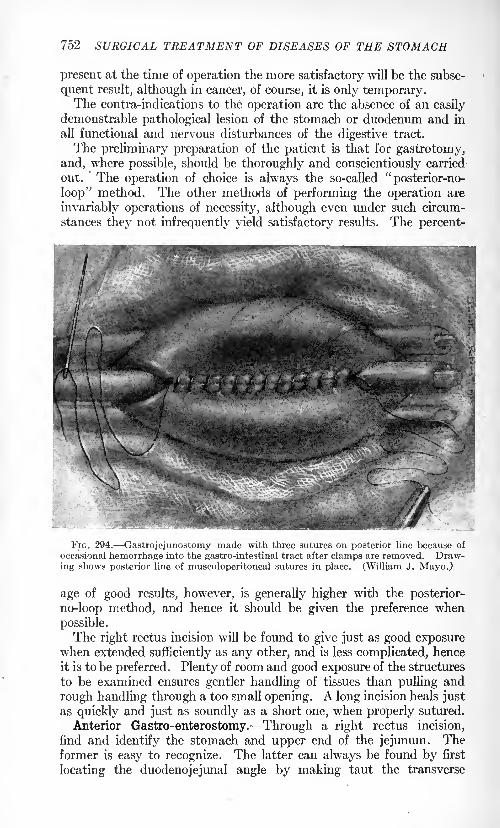
752 SURGICAL TREATMENT OF DISEASES OF THE STOMACH
present at the time of operation the more satisfactory will be the subse-
quent result, although in cancer, of course, it is only temporary.The centra-indications to the operation are the absence of an easily
demonstrable pathological lesion of the stomach or duodenum and in
all functional and nervous disturbances of the digestive tract.
The preliminary preparation of the patient is that for gastrotomy,
and, where possible, should be thoroughly and conscientiously carried
out. The operation of choice is always the so-called"posterior-no-
loop" method. The other methods of performing the operation are
invariably operations of necessity, although even under such circum-
stances they not infrequently yield satisfactory results. The percent-
FIG. 294. Gastrojejunostomy made with three sutures on posterior line because of
occasional hemorrhage into the gastro-intestinal tract after clamps are removed. Draw-ing shows posterior line of musculoperitoneal sutures in place. (William J. Mayo.)
age of good results, however, is generally higher with the posterior-
no-loop method, and hence it should be given the preference when
possible.
The right rectus incision will be found to give just as good exposurewhen extended sufficiently as any other, and is less complicated, hence
it is to be preferred. Plenty of room and good exposure of the structures
to be examined ensures gentler handling of tissues than pulling and
rough handling through a too small opening. A long incision heals just
as quickly and just as soundly as a short one, when properly sutured.
Anterior Gastro-enterostomy. Through a right rectus incision,
find and identify the stomach and upper end of the jejunum. Theformer is easy to recognize. The latter can always be found by first
locating the duodenojejunal angle by making taut the transverse
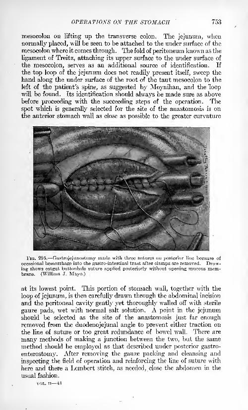
OPERATIONS ON THE STOMACH 753
mesocolon on lifting up the transverse colon. The jejunum, when
normally placed, will be seen to be attached to the under surface of the
mesocolon where it comes through . The fold of peritoneum known as the
ligament of Treitz, attaching its upper surface to the under surface of
the mesocolon, serves as an additional source of identification. If
the top loop of the jejunum does not readily present itself, sweep the
hand along the under surface of the root of the taut mesocolon to the
left of the patient's spine, as suggested by Moynihan, and the loopwill be found. Its identification should always be made sure as abovebefore proceeding with the succeeding steps of the operation. The
spot which is generally selected for the site of the anastomosis is on
the anterior stomach wall as close as possible to the greater curvature
FIG. 295. Gastrojejunostomy made with three sutures on posterior line because of
occasional hemorrhage into the gastro-intestinal tract after clamps are removed. Draw-ing shows catgut buttonhole suture applied posteriorly without opening mucous mem-brane. (William J. Mayo.)
at its lowest point. This portion of stomach wall, together with the
loop of jejunum, is then carefully drawn through the abdominal incision
and the peritoneal cavity gently yet thoroughly walled off with sterile
gauze pads, wet with normal salt solution. A point in the jejunumshould be selected as the site of the anastomosis just far enoughremoved from the duodenojejunal angle to prevent either traction on
the line of suture or too great redundance of bowel wall. There are
many methods of making a junction between the two, but the same
method should be employed as that described under posterior gastro-
enterostomy. After removing the gauze packing and cleansing and
inspecting the field of operation and reinforcing the line of suture with
here and there a Lembert stitch, as needed, close the abdomen in the
usual fashion.
VOL. ii 48
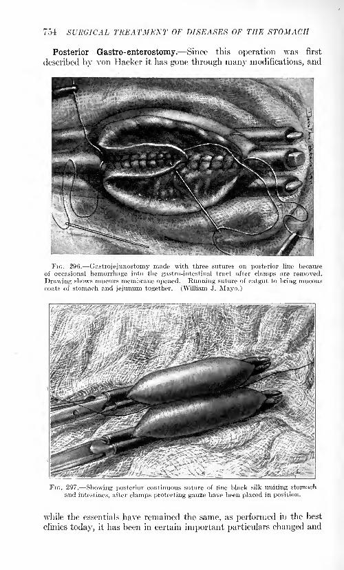
754 SURGICAL TREATMENT OF DISEASES OF THE STOMACH
Posterior Gastro-enterostomy. Since this operation was first
described by von Hacker it has gone through many modifications, and
FIG. 296. Gastrojejunostomy made with three sutures on posterior line becauseof occasional hemorrhage into the gastro-intestinal tract after clamps are removed.
Drawing shows mucous membrane opened. Running suture of catgut to bring mucouscoats of stomach and jejunum together. (William J. Mayo.)
FIG. 297. Showing posterior continuous suture of fine black silk uniting stomachand intestines, after clamps protecting gauze have been placed in position.
while the essentials have remained the same, as performed in the best
clinics today, it has been in certain important particulars changed and
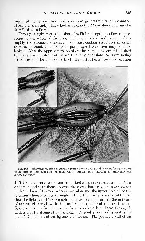
OPERATIONS ON THE STOMACH 755
improved. The operation that is in most general use in this country,
at least, is essentially that which is used in the Mayo clinic, and may be
described as follows:
Through a right rectus incision of sufficient length to allow of easyaccess to the whole of the upper abdomen, expose and examine thor-
oughly the stomach, duodenum and surrounding structures in order
that no anatomical anomaly or pathological condition may be over-
looked. Note the approximate point on the stomach where it is desired
to make the anastomosis, separating any adhesions to surroundingstructures in order to mobilize freely the parts affected by the operation
FIG. 298. Showing anterior mattress sutures drawn aside and incision for new stomamade through stomach and duodenal walls. Small figure showing anterior mattress
sutures in place.
Lift the transverse colon and its attached great oirentum out of the
abdomen and turn them up over the costal border so as to expose the
under surface of the transverse mesocolon and the upper portion of the
jejunum where it comes through. If the transverse colon is held up so
that the light can shine through its mesocolon one can see the network
of mesenteric vessels with their arches and thus be able to avoid them.
Select an area as free as possible from bloodvessels and tear through it
with a blunt instrument or the finger. A good guide to this spot is the
line of attachment of the ligament of Treitz. The posterior wall of the
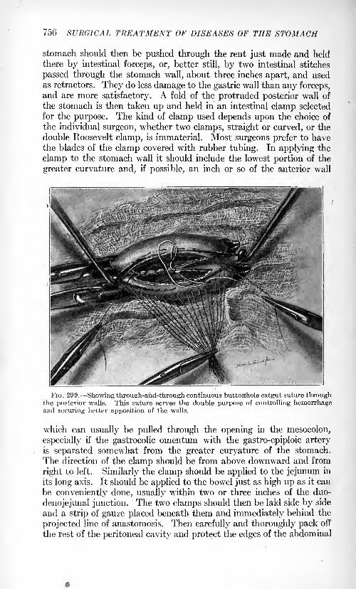
756 SURGICAL TREATMENT OF DISEASES OF THE STOMACH
stomach should then be pushed through the rent just made and held
there by intestinal forceps, or, better still, by two intestinal stitches
passed through the stomach wall, about three inches apart, and usedas retractors. They do less damage to the gastric wall than any forceps,and are more satisfactory. A fold of the protruded posterior wall of
the stomach is then taken up and held in an intestinal clamp selected
for the purpose. The kind of clamp used depends upon the choice of
the individual surgeon, whether two clamps, straight or curved, or the
double Roosevelt clamp, is immaterial. Most surgeons prefer to havethe blades of the clamp covered with rubber tubing. In applying the
clamp to the stomach wall it should include the lowest portion of the
greater curvature and, if possible, an inch or so of the anterior wall
FIG. 299. -Showing through-and-through continuous buttonhole catgut suture throughthe posterior walls. This suture serves the double purpose of controlling hemorrhageand securing better apposition of the walls.
which can usually be pulled through the opening in the mesocolon,
especially if the gastrocolic omentum with the gastro-epiploic arteryis separated somewhat from the greater curvature of the stomach.
The direction of the clamp should be from above downward and from
right to left. Similarly the clamp should be applied to the jejunum in
its long axis. It should be applied to the bowel just as high up as it can
be conveniently done, usually within two or three inches of the duo-
denojejunal junction. The two clamps should then be laid side by side
and a strip of gauze placed beneath them and immediately behind the
projected line of anastomosis. Then carefully and thoroughly pack off
the rest of the peritoneal cavity and protect the edges of the abdominal
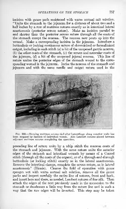
OPERATIONS ON THE STOMACH 757
incision with gauze pads moistened with warm normal salt solution.
"Unite the stomach to the jejunum for a distance of about two and a
half inches by a row of mattress sutures exactly as in intestinal lateral
anastomosis (posterior serous suture). Make an incision parallel to
and shorter than the posterior serous suture through all the coats of
the stomach except the mucosa. The mucosa now pouts up into the
wound. Make a corresponding incision in the jejunum. Introduce a
buttonhole or locking continuous suture of chromicized or formalinized
catgut, including in each stitch (a) a bit of the unopened gastric mucosa,
(6) the other coats of the stomach, (c) the serous and muscular coats of
the jejunum, (d) a bit of the unopened jejunal mucosa. This line of
suture unites the posterior edges of the stomach wound to the corre-
sponding wound in the jejunum. Incise the mucosa of the stomach and
jejunum and with the same needle and catgut suture used in the
FIG. 300. Showing mattress sutures tied after hemorrhage along anterior walls hasbeen stopped by ligation of individual vessels. Also Lembert sutures placed between
adjacent mattress sutures completing the operation.
preceding line of suture unite by a whip stitch the mucous coats of
the stomach and jejunum. With the same suture unite the anterior
edges of the stomach and intestinal wounds by means of a Connel
stitch (through all the coats of the organs), or of a through-and-throughbuttonhole (or locking stitch) exactly as in the lateral anastomosis.
Remove the intestinal clamps, complete the serous suture, as in lateral
anastomosis" (Binnie). Cleanse the field of operation with gauze
sponges wet with warm normal salt solution, remove all the gauze
packs and inspect carefully the entire line of sutures, front and back,and insert here and there, as needed, Lembert sutures of fine silk. Thenattach the edges of the rent previously made in the mesocolon to the
stomach or duodenum a little way from the suture line and in such a
way that the raw edges will be inverted. This step may be taken
758 SURGICAL TREATMENT OF DISEASES OF THE STOMACH
before instead of after the anastomosis. Care should be taken to
avoid puncture of the walls of mesenteric veins with these sutures
otherwise a troublesome hematoma may develop. Replace the
stomach and intestine as nearly as possible in their normal position.Close the abdomen in the usual manner without drainage.This method as just described is modified in slight particulars by
different surgeons, i. e., some surgeons still use a continuous suture
throughout the anastomosis. Mayo found that this continuous suture
when of silk or linen gave rise, in a considerable number of cases, to
later trouble by partially sloughing through into the lumen of the
stomach while still remaining attached to the margin of the stoma,and thus causing irritation and secondary ulceration. For this reason
the use of silk has been recently abandoned altogether in the Mayoclinic in gastric surgery. It is well always to make the anastomosis
sufficiently large, say two inches in length and to bear in mind
Moynihan's injunction, to excise thoroughly the free edges of the
gastric and intestinal mucosa to prevent subsequent cicatricial
stenosis. The operation can be very well performed without the aid
of gastro-intestinal clamps, indeed some surgeons prefer this way of
doing it. There are objections to either method, but they can usuallybe overcome by care and attention to details, such as control of hemor-
rhage, prevention of soiling of peritoneal cavity with stomach andintestinal contents, etc.
Many different methods of posterior gastro-enterostomy have been
described from time to time; none of them are in very general use,
however, except the one just described, hence they will not be described
in detail but only the salient and distinguishing features in which theydiffer from it.
Roux's Operation. The "operation en Y," as it is sometimes called,
differs in this respect, that instead of anastomosing the duodenum to
the posterior wall of the stomach a loop of jejunum a few inches from
the duodenojejunal juncture is lifted up and divided straight across
between two intestinal clamps. The cut end of the lower segment is
anastomosed directly into the stomach at the usual gastro-enterostomy
point of election, next anastomose the lower end of the upper fragmentinto the side of the jejunum a few inches below the point of its junctionto the stomach. The special idea of this operation is to prevent the
formation of the "vicious circle."
Instead of this procedure of Roux, but in very similar fashion, some
surgeons hope to accomplish the same result by anastomosing the
afferent to the efferent loop a few inches below the gastro-enterostomy
opening.Mikulicz and Czerny call attention to the fact that, owing to the
anatomical relationship existing between the posterior wall of the
stomach and the upper three or four inches of the jejunum, an anas-
tomosis can be made at this point by a transverse incision in the
jejunum and one close to the greater curvature of the stomach which
OPERATIONS ON THE STOMACH 759
will drain it at its lowest point, and will, at the same time, prevent the
possibility of kinking of the intestine. Petersen emphasizes the
necessity of making the anastomosis as close as possible to the origin
of the jejunum in order to prevent obstruction. The results, with this
method, have not fulfilled expectations, and hence it has fallen into
disuse.
The Magraw elastic ligature and the method of Maury which sub-
stitutes heavy twine string for the rubber tubing and places the
stitches in a triangular fashion on either side are both open to the
serious objection that they rely upon the sloughing through of the
tissues included in the ligature and that the opening up of the newstoma is delayed, a serious drawback in case of a debilitated patient.
The method of performing posterior gastro-enterostomy invariably
used by the writer is practically that of the intestinal suture first
suggested by Halsted, with some slight modification. The steps of
the operation are the same as the ordinary posterior operation as far
as the placing of the clamps, then the gastric and intestinal loops are
sutured together just as in the writer's pyloroplasty method by a con-
tinuous fine silk suture, wrhich should include the submucous coat of
the intestine. This suture should be placed as far posteriorly as possiblein order to give plenty of room for the anastomotic opening between it
and the line of anterior sutures. These latter should consist of mattress
sutures, which should be placed far enough forward of the posterior
line, say not less than one-half an inch, in order to allow of sufficient
space between the two suture lines for the incision. All the mattress
sutures are placed and the loops drawn aside and then parallel incisions
of equal length made through the walls of the stomach and jejunum.Next apply a continuous buttonhole catgut suture (through all the
coats of the stomach and intestine) to the posterior edges of the incision
through their walls. Loosen the gastro-intestinal clamps. Tie off
with catgut every bleeding-point. Not infrequently it will be found
necessary to ligate vessels in the posterior walls, which are to be seen
bleeding through and in spite of the continuous buttonhole catgutsuture and uncontrolled by it. It is this fact that the writer has so often
observed that has convinced him that reliance cannot be placed in the
continuous suture for the absolute control of hemorrhage, and which has
caused him to abandon the use of the continuous suture in the anterior
walls and to adhere to this method of anastomosis. Pull taut and tie the
mattress sutures previously placed. It will be observed that when inter-
rupted mattress sutures are used the raw edges of the wounds in the intes-
tinal walls always spontaneously invert when they are tied. A Lembertsuture is placed between adjoining mattress sutures for additional
security. No suture is placed in the mucosa of the anterior walls.
Experience has shown it to be unnecessary. The entire suture line is
then carefully inspected and an additional suture placed here and there,
as may seem to be needed. The operation is then completed in the
usual fashion.
760 SURGICAL TREATMENT OF DISEASES OF THE STOMACH
Pyloric Exclusion. There still remain sharp differences of opinion
among surgeons as to the advisability of excluding the pylorus after
gastro-enterostomy. The weight of surgical authority, however, wouldseem rather to incline against it, as adding to the risks of the operationand further complicating it without correspondingly increasing its
effectiveness. The advocates of the procedure recommend it in prac-
tically all cases as obviating most, if not all of the unpleasant sequelaethat have been from time to time observed. Since von Eiselsbergfirst recommended the operation, various modifications have been
brought forward. All sorts of materials have been used to bring aboutthe occlusion.
The idea of the operation is to block the passage of food from the
stomach into the duodenum after gastro-enterostomy, thus forcing it to
escape through the new stoma. The occlusion should take place proxi-mal to the ulcer. There are several types of operation described.
Complete division of the duodenum, pylorus or pyloric antrum, as the
case may be, has been advised and practised by some, with differing
methods, (von Eiselsberg, Doyen, Bartlett.) The duodenum, or
better the stomach, is divided between two clamps which have been
applied proximal to the ulcer and through openings that are made for
the purpose in the lesser and gastrocolic omenta respectively. The cut
ends are then closed with two rows of sutures.
Recently a number of cases have been reported following this oper-ation of the formation of a pouch from the closed pyloric end of the
stomach, necessitating some operative procedure, e. g., the shorteningof the hepatic ligament to prevent further sagging and dilatation.
Simple ligation of the pylorus, either with or without the reflection of
cuffs of the serous and muscular -coats, has many advocates. Foreign
materials, such as silk, catgut tape, metallic bands, etc., or strips of
fascia from the rectus or fascia lata, the round ligament of the liver
and strands of omentum, have all been recommended from time to
time as materials with which to occlude the pylorus. Successful cases
have been reported after the use of each one of them. It is proba-ble that the occlusion of the pylorus renders somewhat more sure
the success of the gastro-enterostomy and makes less likely the
development of the "vicious circle." Other things being equal,
however, it should not be performed as a routine but only in cases of
hemorrhage or impending perforation of duodenal ulcer. The best of
the methods as suggested at the present time are probably those that
use a band of omentum or other fresh connective tissue as the occludingmaterial. In many of the cases in which ligation without division of
the pylorus has been used the lumen of the bowel has been observed
later to become reestablished to a certain extent. This does not seemto constitute a serious objection, since the "vicious circle" is an earlyrather than a late complication of gastro-enterostomy.
Vicious Circle. This is a very serious and intractable complicationof gastro-enterostomy. Fortunately, owing to the perfection of present-
OPERATIONS ON THE STOMACH 761
day methods, this grave sequel is observed far less commonly than
formerly. It is caused by the passage of food from the stomach into
the afferent instead of the efferent loop of bowel. It is usually due to
faulty technic, the junction of the bowel to the stomach at a wrongangle or in a wrong place giving rise to obstruction of the efferent loopfrom angulation or pressure or rotation upon its long axis, making a
valve formation. The no-loop operation when properly done is par-
ticularly free from this complication, and hence should always be giventhe preference when possible. The only really effectual means of
preventing its occurrence are primarily unfailing care and attention
to every detail of the operation and the joining together of the afferent
and efferent loops of jejunum by a lateral anastomosis between the
two, followed by complete division of the afferent loop and closure of
the cut ends by suture. Roux's "Y" operation is also used for this
purpose, but it is not so satisfactory. Occasionally one observes a
case presenting all the symptoms of "vicious circle" recover under
lavage and change of position, but, as a rule, when once well established,
prompt secondary operation is indicated.
Peptic Ulcer. One of the unpleasant sequelae that have been ob-
served to follow gastro-enterostomy is the development of secondaryulcer or ulcers. They occur most often in the segment of the jejunumproximal to the anastomosis. They follow with far greater frequencythe anterior than the posterior no-loop method, and usually just at
or in the immediate neighborhood of the anastomotic opening. Fatal
cases of perforation and hemorrhage have been observed. Although it
is a comparatively infrequent complication it is always serious, and its
presence should always be suspected in those cases where pain andother symptoms develop subsequent to gastro-enterostomy. It is
probable that faulty technic, especially the employment of continuous
non-absorbable sutures, may give rise to the condition in a considerable
number of cases. When its presence has been diagnosed, operation is
always indicated as the only effective form of treatment. The operative
procedure to be carried out depends largely upon the nature of the
pathological process found present. The most radical method of deal-
ing with the situation, other things being equal, probably yields the
best results, namely, excision of the old anastomotic area, stomachand jejunum, including all ulcerated areas, closing the openings in both
and restoring the continuity of the bowel by end-to-end or lateral
anastomosis, as may seem best, in the individual case. When the con-
tinuity of the bowel cannot be readily restored, or when, for any other
reason, it may be inadvisable, the posterior anastomosis may be per-formed at a new site or a simple anterior gastro-enterostomy may be the
best that one can do with safety. Rarely it may be found possible
simply to excise the ulcer without disturbing the anastomosis.
Intestinal Obstruction Following Gastro-enterostomy. One of the rarer
complications observed as a result of gastro-enterostomy, particularlythe posterior-no-loop method, is the obstruction produced by a coil of
762 SVR01CAL TREATMENT OF DISEASES OF THS STdMACtt
intestine slipping into the foramen formed by the afferent loop of the
jejunum which is attached both at the duodenojejunal junction andat the stoma. When subsequent to a gastro-enterostomy operation
symptoms of persistent intestinal obstruction develop, this possibility
should be borne in mind. In order to prevent its occurrence, the fora-
men should be obliterated by suture of the afferent loop to the under
surface of the transverse mesocolon. If the condition is recognized
early enough the engaged loop of bowel can be readily released. Whenthe operation has been too long delayed it may be necessary to resect
the injured segment as in other cases of intestinal obstruction.
SURGERY OF THE DUODENUM.
BY ALBERT J. OCHSNER, M. D., LL.D., F.A.C.S.
EMBRYOLOGICALLY the duodenum is a part of'the stomach, as it
develops from the foregut and maintains physiologically and func-
tionally its position as a part of the stomach by continuing the process,
initiated in the stomach, of preparing the food for absorption in its
passage through the alimentary canal.
The stomach acts as a reservoir, a retort, a mixing and a grinding
apparatus. The duodenum continues the mixing process after addingthe alkaline secretions of the liver and pancreas.
DUODENAL ULCER.
The most common surgical disease of the duodenum correspondsto the same condition in the stomach in the form of ulcer.
The diagnosis of this condition has been fully discussed in a previous
chapter.
Symptoms. Regarding the history of duodenal ulcer, what is knownas "hunger pain" is the most important factor.
While in gastric- ulcer the pain appears soon after the ingestion of
food, in duodenal ulcer it rarely appears earlier than two hours after
taking food, and more commonly later than four hours. In manycases severe pain appears six to seven hours after dinner, the interval
between breakfast and luncheon, and between luncheon and dinner
being too short for pain to appear. Many of these patients regularly
awaken between 1 and 4 A.M. with severe pain, which subsides upon
taking food or bicarbonate of soda. The physical examination results
in pain upon pressure from 2 to 4 cm. to the right and a little above
the umbilicus.
In case of referred pain in the back, which occurs when the ulcer is
situated on the posterior surface of the duodenum, the pain is likely
to be located to the right of the midline often as high as the angle of the
scapula, but it may be located so tow as to be confounded with the
pain which is commonly encountered in gall-stone disease.
Vomiting may occur, but this is not so common as in gastric ulcer;
and vomiting of blood is extremely uncommon, although this may occur.
Mayo has established the fact that duodenal ulcer is about three
times as common as gastric ulcer, a fact which had been overlooked
by earlier observers, probably because these ulcers were formerly classi-
fied with gastric ulcers, because most of them are located with their
margin in the duodenum directly outside of the pylorus.A history of a recent cutaneous burn accompanying the symptoms
of ulcer speaks for duodenal and against gastric ulcer.
This condition can usually be confirmed by making a fluoroscopical
examination, the patient having been given a quantity of gruel or
(763)
764 SURGERY OF THE DUODENUM
buttermilk in which barium sulphate or bismuth subcarbonate has been
suspended.It seems wise to caution the surgeon against making a diagnosis
upon the evidence contained in x-ray plates, because these may be
entirely misleading, and consequently such a diagnosis would be
dangerous to the patient because it frequently results in unnecessary
operation. On the other hand, if a positive diagnosis has been madeupon evidence obtained from a carefully taken history, and a physicalexamination by the experienced surgeon himself, then it may be per-
fectly proper to confirm this diagnosis by the reading of axray plates,
because in this case the procedure is, of course, entirely harmless andit may give both the roentgenologist and the patient a high degree of
satisfaction which may have a certain amount of value. In any case
the rr-ray examination should always follow the taking of a concise
history and a painstaking physical examination. In almost everyinstance it is possible then to confirm the diagnosis, especially by meansof the fluoroscopical examination.
By following this system one can avoid errors which are otherwise
sure to occur because of clouding or confusing the diagnosis directlyas a result of conclusions drawn from the z-ray appearance. It is for
this reason that diagnosticians should invariably be present at the
operation so that they may see the diagnosis confirmed or discredited,
and for the same reason the surgeon should invariably make his own
independent diagnosis.Treatment. The surgical treatment of duodenal ulcer must be
carried out on the exact lines described in the treatment of gastriculcer in a previous chapter with the exception that pyloroplasty canbe employed only in very rare cases, and that excision of the ulcer is
only very rarely possible.In case the use of non-irritating liquid or semi-liquid food to which
an alkaline substance such as milk of magnesia, magnesia carbonate,lime water or bismuth subcarbonate has been added, does not per-
manently relieve a patient suffering from duodenal ulcer, surgicalinterference is definitely indicated.
It is, however, important that this form of non-surgical treatment be
given a fair trial, that it be carried out with regularity and that the
patient actually keep alkaline food in his stomach practically all dayand all night. If he receives alkalinized non-irritating food like milk,
gruel or soup, to which one of the above remedies has been added, everytwo or three hours during the daytime, say for twelve hours, and then
exposes his ulcer to the arid gastric juice during the remaining twelve
hours out of the twenty-four the effect will be satisfactory only in a
very small proportion of cases.
In other cases in which the treatment has been carried out syste-
matically night and day until all of the pain has disappeared, if the
patient at once resumes his careless habits of eating, there is almost
certain to be a recurrence, while the same patient might be readily
expected to obtain a permanent cure in case he had continued the
treatment for several months, and had later abstained from eating
DUODENAL ULCER 765
articles of food of the kind that had presumably given rise to the
original ulcer.
We have had under our observation a very large number of patientswho have remained free from recurrence of gastric and duodenal ulcer
for many years by strictly adhering to a diet list, of which the followingis a sample with modifications suited to the individual case.
Read this diet list once a week and follow absolutely.Take 1 pint of hot milk with 2 to 4 teaspoonfuls of milk of magnesia
at 6, 9, 12, 3, 6, 9 and 1, or take | pint hot milk with 1 to 2 teaspoonfulsof milk of magnesia at 2, 6, 8, 10, 12, 2, 4, 6, 8 and 10.
Later you may take the other articles of food in addition at 12 M.
and at 6 P.M.
When you come to eat solids, chew them a very long time to mixwith saliva.
1st Week. Take 4 to 6 pints of hot milk daily with medicine; if
this is not sufficient to keep up your strength, you may add from 1 to
4 raw eggs.
2d Week. Take same as 1st, and 2 to 4 raw or soft-boiled eggs in
addition.
3d Week. Take same as 2d, and 2 to 6 pieces of very dry toast in
addition, which must be chewed very fine.
4:th Week. Take same as 3d, and all kinds of milk or cream soup in
addition.
5th Week. Take same as 4th, aod all kinds of mush or boiled rice
in addition.
6th Week. Take same as 5th, and broiled, stewed or boiled beef
or mutton in addition; chew and swallow the juice but not the fiber.
Later you may swallow the beef or mutton, but not until you havebeen free from pain for at least three months.Even after recovering fully, do not eat pastry, pie, pancakes, pickles,
pork or puddings. No cake, candy, or canned goods. No raw
vegetables or raw fruits, unless the latter are perfectly ripe and not
sour.
In case the milk of magnesia acts too freely on the bowels, use 4
tablespoonfuls of lime water for each pint of hot milk instead.
Use the above diet for at least six weeks and go back to it at anytime when your stomach may be giving trouble.
USE THE FOLLOWING DIET AFTER SEX WEEKS.
You should drink as nearly as possible 2 quarts of rich milk, pre-
ferably hot, each day, always adding from 2 to 4 teaspoonfuls of milk
of magnesia, or 4 tablespoonfuls of lime water to each pint of milk.
You should eat from 2 to 6 raw or soft-boiled or poached eggs a day.Aside from this, you should eat broiled, stewed or boiled beef,
mutton or fresh fish, cooked vegetables or cooked fruits and all kinds
of thoroughly cooked mush and breakfast foods, also very ripe non-
acid fruits. You may chew the meat and swallow the juice but not
the pulp.
766 SURGERY OF, THE DUODENUM
You should eat no candy, sugar or vinegar; no pastry, pie, pickles,
pancakes, pork or spiced foods, and no unripe or acid fruits, no vege-tables that have not been thoroughly cooked or contain hard fibers.
You should chew all food for a long time in order to mix it thoroughlywith saliva.
You should drink neither tea nor coffee, nor anything containingalcohol.
You may drink hot water with cream, or hot milk with fresh cream,or buttermilk with fresh cream with your meals or half way between
meals or at bed time.
You should rest at least fifteen minutes before dinner and supper.You should sleep with wide open windows, and go to bed early.
In the first case the patients take 1 pint of alkalinized milk, which
may be varied with gruel or puree every three hours, with four and
five hours' intervals at night; in the second case \ pint with two hours'
interval during the day and four hours at night.
This plan of treatment is so simple that it can easily be followed byany patient of reasonable intelligence, and it does not expose the
patient to unnecessary hardship.The same plan is followed after recovery from operation for the cure
of duodenal or gastric ulcer.
The. patient is instructed to adhere to part one of this diet so longas he suffers from any digestive disturbance, and then to change
gradually to part two. In case of any return of trouble the patient
immediately returns to part one and continues with this until entirely
relieved, and then he changes gradually to part two and continues with
this diet so long as he is free from gastric or duodenal disturbance.
The surgery of the duodenum is confined almost exclusively, aside
from the treatment of duodenal ulcer, to carcinoma of the commonbile duct, implicating the papilla which may necessitate the excision
of this portion of the duodenum, but this condition is usually so far
advanced when diagnosed that one rarely encounters an operable case.
The following operation has been suggested, and in case one should
meet a circumscribed carcinoma early enough to justify any operation,it seems worth while to bear this method in mind.
The tumor should be approached from the side of the mucous mem-brane of the duodenum in order to obtain a complete removal, togetherwith a sufficient amount of the surrounding mucous membrane. Thesame preliminary preparation is employed that has been described in
connection with operations upon the stomach, especial care beingtaken to make careful gastric lavage and leaving the tube in the
stomach in order to siphon out all fluid.
Operation. Incision. A median incision extending from the end
of the sternum to a point 3 cm. below the umbilicus provides an excel-
lent approach to all operations upon the duodenum. The anterior
wall of the intestine is caught at two points, 5 cm. and 10 cm. from
the pylorus and well to the left of the middle of the anterior surface.
The lumen of the intestine is then opened by means of a longitudinalincision extending from one to the other of these points.
DUODENAL ULCER 767
The edges of this incision are then caught with four to six fine
mouse-tooth forceps, of the Allis type, care being taken to catch all
layers mucosa, muscularis and serosa in order that these may not
be separated during the subsequent manipulations.The tumor, together with the surrounding tissues for a distance of
2 cm. beyond the margin of the growth, is then separated by means of
the electric or Paquelin cautery, leaving an opening in the duodenumat the point of entrance of the common duct.
This opening is then closed by placing a transverse row of interruptedConnell sutures of fine silk from within the lumen of the intestine.
In this manner there will be no narrowing of the duodenum at this
point. A few interrupted Lembert sutures may be added from the
outside to increase the security of the Connell sutures.
This operation is indicated only in very circumscribed tumors, andin this stage the involvement of the common duct will be limited
sufficiently to leave enough uninvolved duct so that this can be im-
planted into the side of the duodenum at a point necessitating little
or no tension.
This can be accomplished readily so long as the original incision in
the intestine has not been closed.
Virtually the identical operation which has been described for the
implantation of the ureter in the side of the bladder can be employed.A small puncture is made in the wall of the intestine. The common
duct which has been freed while it was still attached to the tumor is
drawn through the punctured wound for a distance of 0.5 cm. and the
mucous membrane of the duodenum is sutured to the outside of the
duct by means of four to six fine silk sutures on fine curved needles.
The serous surface of the intestine is then sutured in the same man-ner to the outside of the duct, at least two rows of fine interruptedsilk sutures being employed in order to prevent leakage.A small piece of omentum is then sutured over the. suture line for
further security.
In case it is not possible to utilize the remaining portion of the com-mon duct after removing the tumor, this should be securely ligated
by the use of chromic catgut or strong fine silk, two distinct ligatures
being employed, then the stump should be covered with peritoneum.In order to secure drainage for the bile, it now becomes necessary
to make use of the gall-bladder by performing cholecystenterostomydescribed in another chapter.This operation can also be performed most conveniently before the
original wound in the duodenum has been closed. The point at which
the gall-bladder is to be implanted in the side of the duodenum should
be selected so as to reduce tension to a minimum. In this class of
cases the obstruction to the common duct, caused by the presence of the
tumor, has usually caused a dilatation of the common duct and the gall-
bladder so that this part of the operation can be readily accomplished.
Embryologically, as stated before, the duodenum is a part of the
foregut in common with the stomach; it should consequently be treated
as part of the stomach.
768 SURGERY OF THE DUODENUM
There is, however, this one distinction : that ulcers are more commonin the duodenum than in the stomach, while carcinoma is of exceed-
ingly rare occurrence in the duodenum, whereas it is most common in
the stomach.
So long as we are in ignorance of the cause of carcinoma, it wouldseem scarcely worth while to mention any theories in explanation of
the marked difference in frequency of carcinoma on the stomach andon the duodenal sides of the pylorus. There are, however, these markeddifferences :
1. The gastric fluid has an acid reaction while the contents of the
duodenum are rendered alkaline by the bile and pancreatic fluid.
Cancer is supposed not to thiive except in acid media.
2. The pyloric end of the stomach is exposed to trauma because it
is in this part of the stomach that the food is ground.3. The mucous membrane of the stomach is exposed to irritation due
to the ingestion of hot food, while the mucous membrane of the duo-
denum is free from this source of irritation.
4. Unclean food remains in contact with the mucous membraneof the stomach for a considerable period of time, while it passes rapidlyover the mucous membrane of the duodenum.Because carcinoma develops so rarely upon the base of duodenal
ulcers, it becomes necessary only very rarely to excise a duodenal ulcer.
This is a fortunate circumstance, because these ulcers are very com-
monly adherent to the pancreas.It may happen that a duodenal ulcer threatens to perforate even
after gastro-enterostomy has been performed in cases in which the
ulcer is located upon the anterior surface of the duodenum. In these
cases the ulcer may be covered by uniting the exposed surface of the
duodenum beyond the ulcer to the anterior surface of the stomach byone or two rows of interrupted Lembert sutures, or the ulcer may be
excised and the wound treated according to the method described in
the treatment of gastric ulcer with excision. In either case healingwill take place if the passage of the acid contents of the stomach over
the mucous membrane of the duodenum is inhibited by the use of a
gastro-enterostomy according to the method described in a previous
chapter, because this prevents the accumulation of large quantitiesof acid gastric contents in the stomach, and consequently this irritat-
ing fjuid will no longer be forced against the ulcerated surface immedi-
ately outside of the pylorus. In performing gastro-enterostomy for
the relief of duodenal ulcer, it seems wise to choose the lowest point
upon the greater curvature of the stomach and at the same time
approach as near as possible to the pyloric end of the stomach, because,
as has been shown by the experiments of Hartman, this will ensure the
passage of food through the gastro-enterostomy opening, while a greateror less amount of food will continue to pass through the pylorus if the
opening in the stomach is made near the cardiac end of the stomach.
Although the presence of a gastro-enterostomy will in any case
reduce the accumulation of irritating acid material in the stomach and
consequently relieve, in a measure, the harmful effect upon the duo-
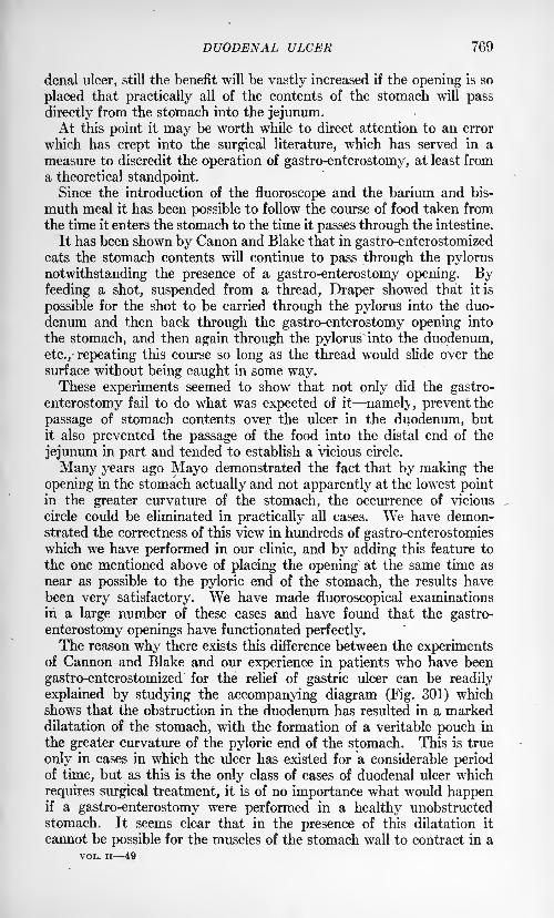
DUODENAL ULCER 769
denal ulcer, still the benefit will be vastly increased if the opening is so
placed that practically all of the contents of the stomach will pass
directly from the stomach into the jejunum.At this point it may be worth while to direct attention to an error
which has crept into the surgical literature, which has served in a
measure to discredit the operation of gastro-enterostomy, at least from
a theoretical standpoint.Since the introduction of the fluoroscope and the barium and bis-
muth meal it has been possible to follow the course of food taken from
the time it enters the stomach to the time it passes through the intestine.
It has been shown by Canon and Blake that in gastro-enterostomizedcats the stomach contents will continue to pass through the pylorus
notwithstanding the presence of a gastro-enterostomy opening. Byfeeding a shot, suspended from a thread, Draper snowed that it is
possible for the shot to be carried through the pylorus into the duo-
denum and then back through the gastro-enterostomy opening into
the stomach, and then again through the pylorus into the duodenum,etc., repeating this course so long as the thread would slide over the
surface without being caught in some way.These experiments seemed to show that not only did the gastro-
enterostomy fail to do what was expected of it namely, prevent the
passage of stomach contents over the ulcer in the duodenum, butit also prevented the passage of the food into the distal end of the
jejunum in part and tended to establish a vicious circle.
Many years ago Mayo demonstrated the fact that by making the
opening in the stomach actually and not apparently at the lowest pointin the greater curvature of the stomach, the occurrence of vicious
circle could be eliminated in practically all cases. We have demon-strated the correctness of this view in hundreds of gastro-enterostomieswhich we have performed in our clinic, and by adding this feature to
the one mentioned above of placing the opening at the same time as
near as possible to the pyloric end of the stomach, the results havebeen very satisfactory. We have made fluoroscopical examinations
in a large number of these cases and have found that the gastro-
enterostomy openings have functionated perfectly.The reason why there exists this difference between the experiments
of Cannon and Blake and our experience in patients who have been
gastro-enterostomized for the relief of gastric ulcer can be readily
explained by studying the accompanying diagram (Fig. 301) whichshows that the obstruction in the duodenum has resulted in a markeddilatation of the stomach, with the formation of a veritable pouch in
the greater curvature of the pyloric end of the stomach. This is true
only in cases in which the ulcer has existed for a considerable periodof time, but as this is the only class of cases of duodenal ulcer which
requires surgical treatment, it is of no importance what would happenif a gastro-enterostomy were performed in a healthy unobstructed
stomach. It seems clear that in the presence of this dilatation it
cannot be possible for the muscles of the stomach wall to contract in a
VOL. it 49
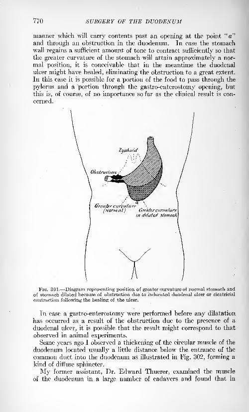
770 SURGERY OF THE DUODENUM
manner which will carry contents past an opening at the point "a"and through an obstruction in the duodenum. In case the stomachwall regains a sufficient amount of tone to contract sufficiently so that
the greater curvature of the stomach will attain approximately a nor-
mal position, it is conceivable that in the meantime the duodenal
ulcer might have healed, eliminating the obstruction to a great extent.
In this case it is possible for a portion of the food to pass through the
pylorus and a portion through the gastro-enterostomy opening, but
this is, of course, of no importance so far as the clinical result is con-
cerned.
curvature
(normal) Grfat&r curvaturein etrfated stomach
FIG. 301. Diagram representing position of greater curvature of normal stomach andof stomach dilated because of obstruction due to indurated duodenal ulcer or cicatricial
contraction following the healing of the ulcer.
In case a gastro-enterostomy were performed before any dilatation
has occurred as a result of the obstruction due to the presence of a
duodenal ulcer, it is possible that the result might correspond to that
observed in animal experiments.Some years ago I observed a thickening of the circular muscle of the
duodenum located usually a little distance below the entrance of the
common duct into the duodenum as illustrated in Fig. 302, forming a
kind of diffuse sphincter.
My former assistant, Dr. Edward Thuerer, examined the muscle
of the duodenum in a large number of cadavers and found that in
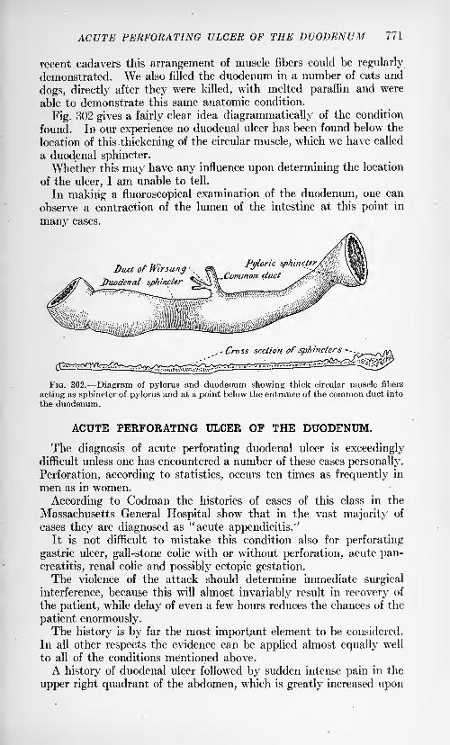
ACUTE PERFORATING ULCER OF THE DUODENUM 771
recent cadavers this arrangement of muscle fibers could be regularly
demonstrated. We also filled the duodenum in a number of cats and
dogs, directly after they were killed, with melted paraffin and were
able to demonstrate this same anatomic condition.
Fig. 302 gives a fairly clear idea diagrammatically of the condition
found. In our experience no duodenal ulcer has been found below the
location of this thickening of the circular muscle, which we have called
a duodenal sphincter.
Whether this may have any influence upon determining the location
of the ulcer, I am unable to tell.
In making a fluoroscopical examination of the duodenum, one can
observe a contraction of the lumen of the intestine at this point in
many cases.
7i ^ / ~7 /Duodenal spluncier
s5
^ii^^.,- Crv 55 section of sphincters
- - . .
FIG. 302. Diagram of pylorus and duodenum showing thick circular muscle fibers
acting as sphincter of pylorus and at a point below the entrance of the common duct into
the duodenum.
ACUTE PERFORATING ULCER OF THE DUODENUM.
The diagnosis of acute perforating duodenal ulcer is exceedinglydifficult unless one has encountered a number of these cases personally.
Perforation, according to statistics, occurs ten times as frequently in
men as in women.
According to Codman the histories of cases of this class in the
Massachusetts General Hospital show that in the vast majority of
cases they are diagnosed as "acute appendicitis/3
It is not difficult to mistake this condition also for perforating
gastric ulcer, gall-stone colic with or without perforation, acute pan-
creatitis, renal colic and possibly ectopic gestation.The violence of the attack should determine immediate surgical
interference, because this will almost invariably result in recovery of
the patient, while delay of even a few hours reduces the chances of the
patient enormously.The history is by far the most important element to be considered.
In all other respects the evidence can be applied almost equally well
to all of the conditions mentioned above.
A history of duodenal ulcer followed by sudden intense pain in the
upper right quadrant of the abdomen, which is greatly increased upon
772 SURGERY OF THE DUODENUM
the slightest pressure, with hard, tense abdominal muscles, indicates
the presence of actual or threatened perforation of a duodenal ulcer.
Operation. A longitudinal incision should be made over the middle
of the right rectus abdominis muscle, which should be split lengthwise.
Upon incising the transversalis fascia some fluid and gas usually
escapes immediately, confirming the diagnosis. It is best to make a
very long incision in order that it may be possible to sponge away the
fluid and to close the opening in the intestine without causing unduetraumatism to the tissues.
It is wise -to apply a large gauze tampon wrung out of warm normalsalt solution so placed that there can be no future soiling. The fluid
is then carefully sponged away.The opening in the intestine is located and closed by placing the
end of a finger over it. Then a stomach-tube is introduced and the
contents of the stomach siphoned out in order that this may not leak
through the opening into the peritoneal cavity.If the opening is very small, a circular silk suture may be applied
about the edges of the opening and tied to bring the edges together.Then one or two rows of sutures of fine silk are applied so as to cause
the suture line to extend in a transverse direction to prevent further
narrowing of the lumen of the intestine. A small piece of omentumis sutured over the part of the duodenum involved.
When this has been accomplished, careful gastric lavage is madewith water at a temperature of 105 to 110 F., in order to remove anyblood or mucus which may have accumulated in the stomach duringthe operation. It is usually well to examine the appendix in these
cases, because acute appendicitis has occurred simultaneously in one
of our cases, and in several others with perforation of a duodenal ulcer.
If the ulcer is extensive and the operation is performed soon after
the perforation occurred, one may add gastro-enterostomy to the
operation just described, but if there is any doubt about the patient's
condition being sufficiently good to bear the shock of the operation, it
is better to postpone this portion of the operation to some later time.
A fine feeding tube may be inserted through the stomach-tube and
passed through the pylorus and 10 to 15 cm. beyond the point of the
ulcer in the duodenum, through which predigested liquid food can be
introduced for a week or longer after the operation until the ulcer has
had time to heal thoroughly. The wound is closed with the exceptionof a small space through which one or more cigarette drains are passedinto the kidney pouch. The same after-treatment is carried out after
operations for chronic duodenal or gastric ulcer. Unless a feedingtube has been inserted it is best to nourish the patient for several days
by the use of exclusively rectal alimentation, and then to begin givingalkalinized liquid nourishment by mouth, and later following the diet
list described above.
CONGENITAL HYPERTROPHIC PYLORICSTENOSIS.
BY ROBERT E. McKECHNIE, M.D.
VARIOUS names have been given for this condition, among which
we find hypertrophic pyloric stenosis of infants, congenital spastic
pyloric stenosis, congenital pyloric stenosis, spastic hypertrophic pyloric
stenosis, etc. The name which, besides being eminently descriptive of
the condition, agrees most nearly with the views of modern observers,
as confirmed by their experience, is congenital hypertrophic pyloricstenosis.
The earliest record of an example of this disease was unearthed byOsier,
1 and is contained in Cases and Observations by the Medical Society
of New Haven in-the State of Connecticut, 1718, by Hezekiah Beardsley.The following description of the case gives an undoubted picture:"A child of Mr. Joel Grannis, a respectful farmer in the town of
Southington, in the first week of its infancy, was attacked with a
puking, or ejection of the milk, and of every other substance it received
into its stomach almost instantaneously, and very little changed.The feces were in small quantity and of an ash color, which continued
with very little variation till its death. For these complaints a physi-cian was consulted, who treated it as a common case arising from acidityin the prima via; the testaceous pow
rders and other absorbents andcorrectors of acid acrimony were used for a long time without anyapparent benefit.
"I was at first inclined to attribute the disorder to a deficiency of bile
and gastric juices, so necessary to digestion and chylification, joinedwith a morbid relaxation of the stomach, the action of which seemed
wholly owing to the weight and pressure of its contents, as aliment taken
in small quantities would often remain on it, till, by the addition of
fresh quantities, the whole, or nearly all, was ejected; but his thirst,
or some other cause, most commonly occasioned his swallowing such
large draughts as to cause an immediate ejection, and oftentimes before
the cup was taken from the mouth."A number of the most respectable medical characters were consulted
and a variety of medicines were used to little or no effect. His death,
though long expected, was sudden, which I did not learn till the second
day after it took place. This late period, the almost intolerable stench,and the impatience of the people who had collected for the funeral
prevented so thorough an examination of the body as might otherwise
have been made. . . .
1 Am, Jour. Dis. of Children, 1911, ii, 159.
(773)
774 CONGENITAL HYPERTROPHIC PYLORIC STENOSIS
"I next examined the stomach, which was unusually large, the coats
were about the thickness of a hog's bladder when fresh and distended
with air; it contained about a wine pint of fluid exactly resembling that
found in the vesicles before mentioned, and which I supposed to havebeen received just before its death. The pylorus was invested with a
hard compact substance, or scirrhosity, which so completely obstructed
the passage into the duodenum, as to admit with the greatest difficulty
the finest fluid; whether this was the original disorder, or only a conse-
quence, may perhaps be a question. In justice to myself I ought to
mention that I had pronounced a scirrhosity in that part before the
child's death."
Williamson1
published the account of the second case, and a third
case was reported by Dawaski in 1842. Then followed forty-five years
during which no other case seems to have been observed. Then in
1887 Hirschsprung of Copenhagen reported 2 cases before the GermanPediatric Society. Following this the profession seems to have become
slowly aware of the existence of this condition and cases began to be
reported. Thus in 1906 there had been reported 121 cases. At this
time, dealing with a series of 113 cases Ibrahim states that 50 of these
had been reported in Germany and 49 in Great Britain. As evidence
of an increased awakening in the subject, in the next two years Ibrahim
had collected a series of 400 cases and at the present time reports of
cases are of frequent occurrence in the journals. This shows that the
disease is of comparative frequency and merits a separate treatment in
any surgical work.
That the disease is still frequently overlooked is suggested byCoombe,2 from the fact that 6 out of 20 cases reported by Hutchinson
in private practice, were the children of medical men. Thus 30 percent, of these cases of Hutchinson's were in medical households. If
we estimate 1 physician to 1000 of the population or one physician's
family to 200 other families it would mean that this disease is 30 times
more prevalent in doctor's families or else that it is 30 times less likely
to be discovered in non-medical families. This latter must be the morereasonable to believe, and, being accepted, means that an immensenumber of these cases pass on to a fatal termination undiagnosed.
It occurs in the earliest weeks of life. Thus Pfaundler states that
50 per cent, of cases began to show symptoms between the fourth and
fourteenth days, 25 per cent, between the second and third week and
25 per cent, between the third and sixth week.
The infants, as a rule, are well developed at birth and present nothingabnormal. A majority are first-born as well as a majority being males.
Thus Holt3 noted 49 males to 8 females. In his same series there were
52 breast-fed children to 5 fed artificially, a point to be remembered in
the discussion on the causation of the condition and rather eliminating
indigestion from faulty feeding as a causative factor.
1 London and Edinburgh Jour. Med., 1841.2 Ann. Surg., 1911, liv, 167.8 Murphy's Clinics, No. 1, vol. ii, p. 157.
PATHOLOGICAL ANATOMY 775
Normal Anatomy.1
Although the pyloric canal is by no means con-
stant in form, and undergoes striking changes in accordance with later
physiological conditions of the stomach, there is no part of the stomachwhich is more definite and distinct. The pyloric canal is a cylinderabout one inch in length, and in a full-term child is generally found
contracted and its lumen filled with longitudinal folds of mucousmembrane. During life the pyloric canal is kept closed except during
digestion, when it is periodically relaxed to allow the passage of acid
gastric contents into the duodenum. If the contents are too acid, the
quantity allowed to pass is lessened and the pylorus does not againrelax until the contents in the duodenum have been neutralized by the
alkaline juices there. This constitutes the duodenal reflex.
The pyloric canal is characterized by the great development of both
longitudinal and circular muscular fibers. These latter completelysurround the pylorus and constitute the sphincter. The longitudinalfibers take their origin in the wall of the pyloric end of the stomachand become more numerous as they reach the pylorus and there form a
distinct longitudinal coat. The more superficial fibers pass on to the
duodenum, while many of the others spread out in the form of divergent
fasciculi, some of which reach to the submucosa. Thus the longitudinalfibers have an action antagonistic to the sphincteric fibers and act as a
dilator of the pylorus when the nervous mechanism concerned in inhibi-
tion brings about a relaxation of the sphincter. Specimens of the
pyloric canal of a full time fetus and of the child, show very distinctly
that there is no sudden or abrupt transition between the muscular
substance of the sphincteric cylinder and that of the sphincteric ring,
but the fasciculi of the latter are quite cut off from the circular muscular
coat of the duodenum by a distinct connective-tissue septum.In discussing the action of the musculature of the pyloric canal,
Stiles quotes Professor Cunningham: "The question which naturally
suggests itself is whether the entire length of the sphincteric cylinderis to be reckoned, with the expelling or with the retaining forces of the
stomach, or whether it is to be regarded as acting in both wr
ays, and the
sphincteric ring as being alone endowed with a continuous sphinctericfunction. Clinical evidence would seem to point to the entire muscular
cylinder being under certain circumstances employed as a sphincterand thereby closing the whole length of the pyloric canal, against the
entrance of material from the stomach. In these cases of pyloricstenoses in the infant, which of late years have attracted so muchattention, it is not the sphincteric ring alone which is at fault and
prevents the passage of the gastric contents into the duodenum.The circular musculature of the entire length of the pyloric canal,
by its spasmodic contraction, leads to the closure of this section of the
stomach."
Pathological Anatomy. Pfaundler2 in 1897, in criticising Stern's
operation for this condition, came to the conclusion that there is no
1 Stiles: British Med. Jour., October, 1906, p. 943.2 Hill: Surg. Gynec. and Obst., 1914, p. 616.
776 CONGENITAL HYPERTROPHIC PYLORIC STENOSIS
warrant for assuming the existence of an organic congenital pyloricstenosis. He believed that in the cadaver of an infant, a permanentcontraction of the musculature at the pylorus can persist after death,which may appear as a hyperplastic tumor obstructing the lumen.
And thus he started the spasm theory.
Cantley,1 while accepting the view that there is such a condition as
pyloric spasm in infants, says there is no evidence that spasm could
cause hypertrophy. Spasm of the pylorus does occur at all ages, butit does not produce the condition of hypertrophy which is peculiar to
the first few weeks of life. Recovering from pyloric spasm undermedical treatment, in some cases mistaken for true hypertrophy, has
led to an unduly favorable estimate of the value of medical treatment
in true hypertrophy. True hypertrophy varies from a degree which is
fatal, unless relieved surgically, to a slight hyperplasia which is com-
patible with life. To those who have seen both the clinical picture, andthe operative, and postmortem findings, in these cases, there is no
longer any question about the existence of a definite organic obstruction
as the cause of the clinical picture, and it is also quite as clear that the
only hope of relief in well-defined cases is some surgical operation which
will overcome the obstruction.
This statement may seem to be too sweeping in the light of the
statistics published by those who have employed the medical treat-
ment. In the Schorstein Lecture (1910) by Robert Hutchinson, are
quoted 19 recoveries out of 21 cases, 11 out of 12 and 30 out of 32, with
medical treatment; but I do not think we can find similar favorable
results furnished by the American practitioners. And even among the
English writers we find the adoption of medical treatment discouraged.Thus at a meeting of the Royal Society of Medicine, Section for Studyof the Diseases of Children, a specimen was shown from a case whichhad died from the condition we are discussing. Dr. Cantley, the
chairman, remarked that it was a good illustration of one of the usual
results of attempting to cure the condition by medical means, and
that there were still many people who supported the view that a cure
could be obtained by that means.
Holt* does not believe in persistent spasm of the pylorus without
hypertrophy, and says that it would be well if the term "pylorospasm,"as defining a,group of these cases, were dropped from our nomenclature.
He thinks it is responsible for neglect of operation when operation
might have saved life. He also states that definite persistent spasm of
the pylorus without hypertrophy is yet to be proved.At operation, or postmortem, there is found a tumor consisting of a
firm olive-shaped body, roughly one-half to three-quarters of an inch
in length, covered by smooth, glistening peritoneum. There is no
external evidence of inflammatory reaction and no attempt at fixation.
This tumor has the firmness of cartilage, is never soft or pliable, andin no case was its consistency such as to make the finding questionable,
i British Med. Jour., February, 1908, p. 323.* Jour. Am. Med. Assn., 1914, Ixii, 353.
PATHOLOGICAL ANATOMY 777
no case in which a gradation could be made out between a tumor massand a mere thickening of the part involved or between the tumor massand the stomach and duodenum. It was always sharply defined.
This tumor is formed entirely by the hypertrophied circular muscle
fibers of the pylorus. The mucous membrane lies in longitudinal folds
in the lumen and is much thickened and arranged in a more or less
spiral manner. The pylorus is not absolutely closed but the tumormass blocks the lumen as an enlarged prostate blocks the urethra.
So in many cases food in small quantities may find its way past andso life be prolonged. That bismuth may at times be forced throughor that a suggestion of a milk stool occur must not, therefore, rule out
the diagnosis. It is particularly important that the roentgen rayas a diagnostic measure be limited to determining the rate of emptyingthe stomach, not the patency of the pylorus. To exclude a diagnosisof hypertrophic stenosis on the basis of the passage of bismuth is sure
to lead to serious error.
So it may be conceded that sufficient patency may persist to main-
tain life, even after temporary total blocking has been evident. Onlyon this basis can we account for the recoveries that have been reported
by competent observers. The recovery does not imply the disap-
pearance of the tumor. There is nothing in the nature of hypertrophicstenosis that essentially predicates a permanent pyloric closure. Nogood reason has been given why these tumors should not ultimately
disappear. They are not neoplasms, but simple muscular hyper-
trophies. The indication for operation is not strengthened by provingthe permanency of the mass. The fact that it does not disappear quickly
enough is the essential absolute indication for surgical interference.
Downes1gives the sequence of events which, in his opinion, leads up
to the closure of the pylorus. The presence of the muscular hyper-
trophy reduces the lumen of the pyloric canal, and therefore, the
stomach in order to empty itself contracts more forcibly than normal.
This abnormal contraction soon causes the mucous membrane to
become thickened and edematous, and assume a more or less spiral
arrangement as it passes through the narrowed pyloric canal. Theresult is a valvular action which gradually produces complete closure.
That the hypertrophy exists at birth, that it was formed before food
could produce irritation and so provoke hypertrophy seems proved.Pfaundler states that 50 per cent of the cases develop between the
fourth and the fourteenth days. Murphy2 notes a case which started
at the second day while McKechnie3 had one with complete obstruction
dating from birth.
As to the permanency of the hypertrophy and of the obstruction
Ibrahim 4 states that by the examination of the stomachs of infants
who have recovered under medical treatment, from what has been
1 Holt: Jour. Am. Med. Assn., 1914, Ixii, 353.2 Murphy's Clinics, vol. ii, No. 1, p. 157.* Canadian Med. Assn. Jour., July, 1913.4 Am. Jour. Med. Sc., 1912, cxliii, 14.
778 CONGENITAL HYPERTROPHIC PYLORIC STENOSIS
clinically diagnosed as hypertrophic pyloric stenosis, and died from
other accidental causes, he found that the pyloric hypertrophy still
remained, but that there had been developed a compensating hyper-
trophy of the stomach walls.
Eagleson,1 of Seattle, proved by skiagrams that in 2 cases four years
after operation, and 1 case six years after, the bismuth was passing
by the anastomotic opening and not by the pylorus. These twoauthorities would tend to contradict those advocates of medical
treatment who presuppose that there will be an absorption of the
hypertrophy and so a cure of the stenosis.
Symptoms. The symptoms of a case of congenital hypertrophic
pyloric stenosis are very definite, and few mistakes in diagnosis should
be made. No laboratory tests or other procedures which rely on the
services of a specialist, need be called on and the general practitioner
is as well able to diagnose the case as the staff doctor in a children's
hospital, if he only has the fundamental points of these cases in his
mind.
The patient is an infant, from one day to six weeks old, oftener a
male than a female; oftener a first-born than one later and oftener
breast-fed than bottle-fed. The symptoms2usually begin abruptly
and the mother can give the day and often the hour when they first
appeared.This apparently healthy infant begins to vomit its food, without
apparent cause, and at the same time there is noted a correspondingdiminution in the feces. The child does not look sick, and even in an
advanced case one cannot help remarking the bright appearance of
the face. There is no evidence of a gastritis, no tenderness over the
stomach and the food does not act on reaching the stomach as it would
if it there met an inflamed mucosa. Sometimes, when the stomach is
already nearly filled, additional food will be followed soon by vomiting,but at other times food may be retained for most of the day, to be later
all or nearly all returned. If the obstruction is not complete somefeces will be passed, but in the majority of cases the stenosis will be so
pronounced, when the case is once under way, that practically onlybile and mucous in the form of a dark green paste will be occasionally
found on the napkins. The vomitus will Consist only of the food taken,
bile is not found in it. But as bile is found in the scanty stool, at once
the point of obstruction is suggested as placed below the stomach, but
above the entrance of the bile duct into the duodenum. With the
continuance of the obstruction, there will be dilatation of the stomach
and a state of emptiness of the intestines. This gives a characteristic
appearance to the abdomen, as the upper part is full and distended,
while the lower half is empty and concave. Waves of peristalsis are
easily provoked in the stomach by manipulations or by giving a drink
to the child.
1 Paper not published. Read at the meeting of the North Pacific Surgical Association,
at Vancouver, 1913.* Holt: Jour. Am. Med. Assn., 1914, Ixii, 353.
SYMPTOMS 779
Following the beginning of the symptoms, there sets in a progres-sive loss of weight until finally the child presents a picture of extreme
emaciation.
In a given well developed case, if careful record is taken of the
amounts of liquids given, and care is taken to catch what is vomited,and an allowance made for what will be absorbed by wipes or towels,
it will be found that the vomitus about equals the ingesta. The
vomiting is apparently not accompanied by nausea, as there is no
pallor or sweating and after the stomach is emptied the child looks
as bright as before, and is quite ready to be fed again.A record should be kept of the weight which will show a steady daily
loss.
The urine is greatly reduced in amount, owing to the diminution in
retention of fluids. Cases of anuria have been noted and a diagnosisof renal trouble made, the vomiting being considered uremic.
In addition, a tumor may be felt in the region of the pylorus in the
majority of cases. Thus Marsh1 notes that he found it in 4 out of
6 cases. A big liver may so overhang the pylorus as to shield it or
if the infant is crying, the rigid muscles may hide it; but patience and
gentleness will usually find it. It lies to the right of the vertebral
column, about one and a half to two inches above the navel. As it is
freely movable, it recedes before the examining finger and can be gentlytouched and felt as a small rounded and hard body. It feels like a small
hard gland, and at the operation one is surprised to find it much largerthan palpation has suggested.McCracken2
pointed out that, "as the emaciation was due to actual
starvation from obstructed pylorus in an otherwise healthy organism,it was not surprising that this wasting showed certain almost diagnosticfeatures in the face, abdomen, and nature and color of the skin, which
distinguished it from cases resulting from true toxic wasting diseases.
Peristalsis was fairly uniform in all cases at. slightly over three and a
half waves per minute, and in a case under suspicion if one saw a slight
pucker of brows and flicker on the upper lid recurring about that rate,
one would on gently exposing the epigastrium see the peristalsis.
Mothers, though as a rule quick at observing epigastric swellings from
other causes, were seldom the first to observe this phenomenal wave.
Sometimes peristalsis was fickle; from half an hour to an hour after
feeding was, he found, the likeliest time for observing it; in one case, keptunder the closest observation, peristalsis was only seen once and that
by chance. It was of importance, in all suspected cases, to find out
if there was or was not a lump at the pylorus; and if unable to decide
otherwise one was justified in giving an anesthetic, though cautiously,because in some cases acetone was present; but putting the infant in
a warm bath, as suggested by a late resident of theirs, had alwaysbeen sufficient to soothe the child, overcome muscular rigidity, andallow of a satisfactory and satisfying examination being made. Sum-
1 Jour, of Children's Dis., 1911, viii, 53.* British Med. Jour., March, 1912, p. 487.
780 CONGENITAL HYPERTROPHIC PYLORIC STENOSIS
ming up the symptomatology, vomiting, wasting, and constipation were
indications of possible hypertrophy; suspicion ought to be intensified
if these occurred in a first-birth and male infant. Visible peristalsis
and a tumor at the pylorus were the rivets which clinched the diagnosis.
Of these two, the tumor was the more important; either of the two byitself was an indication for medical treatment; but in the absence of
both, one was not at the present date ever justified in making a diag-
nosis of pyloric hypertrophy. The stenosis at the pylorus was only
partially due to hypertrophy of the circular fibers; it was augmented
by the gastritis in the vestibule and pyloric canal, accumulation of
mucus in the vestibule, and probably by superadded spasm ;and the
last three factors accounted for the complete blocking which sometimes
occurred. The cause of this hypertrophy was not yet determined."
Pyloric stenosis usually results fatally in from six to ten weeks.
In a certain portion of recoveries evidence of incomplete obstruction
persists for months or years.
Treatment. Owing to the teachings of Pfaundler, that the condition
was not due to a genuine hypertrophy but to a spasm of the pyloric
canal, treatment was entirely medicinal at first, accompanied by efforts
at giving an easily digested and non-irritating diet, and by lavage to
get rid of the hypothetical irritating stomach contents. Indeed,
Marsh states that the German school to this day practically never
operates. This teaching apparently has influenced the English school.
Thus Marsh1
quotes 91j per cent., recovered without operation.
Other English statistics could be produced with much the same results.
The weak point in these cases is that the condition was not proved,since the observer had no opportunity to verify his diagnosis, by either
operation or postmortem. In the light of knowledge, gained by sur-
geons, it is impossible to believe that so large a number of cases could
have been true cases of congenital hypertrophic pyloric stenosis, or
even of the type in which we could suppose that the hypertrophy wasbut slightly developed; and clinical evidence is not forthcoming, to
prove that pylorospasm is so prevalent a condition in infants, as to be
able to produce so large a number of cases as is quoted by various
authors. Hence we are drawn to the conclusion, that a certain groupof cases which contain some irritating lesion of the stomach have been
wrongly included under the head of congenital hypertrophy.
However, with any given case, the treatment resolves itself into either
medical or surgical. Both the German and the British schools incline
toward medical treatment although surgical treatment is being more
practised and recently with good results.
McCracken2 writes as follows: "The aim of all treatment was to
promote the passage of chyme from stomach to intestine, and thus
permit of absorption. This could be done surgically, but if one relieved
gastritis, subdued spasm, and periodically removed the stringy mucus,then in a little time chyme, if free of coarse curd, could in most cases
1 Jour, of Children's Dis., 1911, viii, 53.2 British Med. Jour., March, 1912, p. 487.
TREATMENT 781
pass naturally through the pylorus; and these points could be obtained
medically by stomach washing and judicious feeding. This line of
treatment should be tried first in all cases, and sensible rules for guid-ance are given in all text-books. Unless the patient were in articulo
mortis, one should never despair, of any case; but the development of
some intuitive instinct toward an early diagnosis of this condition
was 'a consummation devoutly to be wished/ by everyone who hadmuch experience in its treatment. In a case under medical treatment,once peristalsis has been seen and a lump felt, one ought never to elicit
or feel for either symptom again, and should look to it also that no one
else did; all manipulation must of a necessity stimulate the very spasmwhich the treatment aimed at subduing. It was notorious that better
medical results were obtained in private practice than in hospital.
One authority had pointed out that these eases became, 'hospitalized,'
an indefinite status too often seen in marasmic infants; but probablytheir being 'lionized' accounted for a good many of the disappoint-ments. In Germany good results had been obtained by less washingthan we practiced, but keeping warm poultices constantly over the
epigastrium."Dent1
apparently represents the transition from medical to surgical
views on treatment as in the following extract."In some mild cases
medical treatment might cure it; but if they waited for the establish-
ment of all the classical symptoms before operating, the percentage of
mortality would be even higher than it was now, whereas if operationwere done on many cases before the symptoms grew serious the results
would be still better. Probably medical treatment when the child
was not doing well was rather more dangerous than surgical."
Holt2gives perhaps the most impartial discussion on this vexed
question of treatment and points out that there are medical risks as
well as surgical risks. Among medical risks are acute inanition, slow
inanition or marasmus, during which state the physician frequentlydoes not realize on what thin ice he is skating; risk from intercurrent
disease attacking an infant of low resisting power and lastly risk of
sudden death, unexpected and unaccountable.
And if these medically treated cases do get well without operation,do they completely recover or does the lesion persist?
Holt quotes seven of his own cases which recovered under medical
treatment, and whose after histories were so good that he concludes
that the tumors were absorbed. But Downes, Marsh and others have
proved by postmortem findings on children who have died of other
ailments, after recovering through medical treatment, that the tumor
persists. Eagleson also by bismuth meals and skiagrams proved the
persistence of the obstruction, years after cure by operation. So it
seems probable, that the tumor persists after apparent cure by medical
treatment. And it has been shown by other observers, that cases of
malnutrition in adults, traced from infancy to adolescence, suffer
1 British Med. Jour., March, 1907, p. 627.* Jour. Am. Med. Assn., June, 1914, p. 2015.
782 CONGENITAL HYPERTROPHIC PYLORIC STENOSIS
from a persisting congenital hypertrophic pylorus, which has later onbeen proved and rectified successfully by operation.
Surgical risks are plainly seen, shock, non-union from poor nutri-
tion, exhaustion due to digestion not reestablishing itself soon enough,accidental risks from faulty technic as hemorrhage, sepsis, obstruction,
etc. But shock is low in infants and the technic has improved. Evenif the surgical risks are considerable they are short, while the medical
risks are prolonged and the dangers multiply with the duration of the
symptoms.Given an early diagnosis, a patient in private practice, under condi-
tions which make the best medical treatment possible, operation maynot in most cases be required; but in hospital practice, in cases seen
later, in those with the most acute symptoms who are losing rapidly,and (lastly) when circumstances do not admit of the best medical
treatment, there is no question but that immediate resort to surgeryoffers a better chance of recovery. To persist with medical treatment,week after week, when forcible vomiting and marked peristalsis are
either continually present or keep recurring, when the weight shows
only a slight loss, seems to be incurring far greater risks with the child
than those of operation.The crux of the whole question between medical and surgical treat-
ment of pyloric stenosis seems to be are the symptoms and conditions
such as to make it probable that the patient will or will not live long
enough for the pathological condition to subside?
If the child is in fairly good condition, if it is not losing weight rapidly,
if some food in the stools proves that the stenosis is not absolute, andif in addition the environment be suitable writh proper hygienic sur-
roundings and no lack of suitable food and skilled nursing, then one is
justified in recommending medical treatment.
If this treatment fails to check the downward course of the case,
then one is not justified in waiting till the case is almost hopeless,
before deciding on an operation. Surgery should be given a chance
while there is yet a reasonable hope of success. This decision will
usually lie with the family physician, who will sometimes find it hard,
on account of partial remissions giving rays of hope; but if in spite of
these remissions the general tendency is downward, then he takes uponhimself a serious responsibility if he delays in recommending surgery.
When the average case comes to a surgeon there is but little else to
do but to advise operation. Seldom does a surgeon see a case in the
early stages, too often he sees a picture of skin and bones due to starva-
tion, with hardly power enough to turn its head or utter more than faint
cries. Still, even in such cases operation is very often successful andshould not be denied to the weakest.
As regards prognosis Holt states that mortality is about 50 per cent,
by either medical or surgical treatment. This was in 1914, but already
surgeons were announcing much more favorable results.
Thus Scudder,1 in 1910, states that medical treatment is responsible
1Surg. Gynec. and Obst., September, 1910, p 373.
TREATMENT 783
for 80 to 90 per cent, of deaths, while he quotes 1 death in fourteen of
his own surgically treated and ends by saying,"Gentlemen, in view
of the evidence, I believe that surgical measures should be instituted
as soon as the diagnosis is made. Delay is dangerous."This attitude is the one adopted by the most advanced men of the
profession today as regards appendicitis. When first advanced it was
universally hailed as extreme but its adoption has led to an immense
saving in lives.
As regards the adoption of a similar rule in regard to congenital
hypertrophic pyloric stenosis, it should be pointed out that the
mortality of cases treated medically will remain constant, 50 per cent,
according to Holt, 90 to 80 per cent, according to Scudder. In sur-
gery it is different. Technic has improved and men have improvedin their own technic so that now there are a large number of surgeons
capable of handling these cases with a minimum of risk. Scudder,as we have seen, had in 1910 one death in 14 cases. Richter1 in
1914 quotes 19 successful cases in 22 operated on, and other surgeonsshow like favorable results. These figures show that surgery givesfar more favorable results than medical treatment. And if we remem-ber that the majority of the cases treated surgically are about in extre-
mis, after a prolonged course of medical treatment, it is self-evident
that if all cases were referred to the surgeon for operation, as soon as
diagnosed, the percentage of mortality would be still further lowered.
It needs only a little further pressing of the argument, to reach the rule,
that in the interests of the lives of such patients, the adoption of surgi-
cal treatment at the earliest possible moment should be advised.
But if medical treatment be decided on it will consist of lavage andcareful feeding. The stomach should be washed out twice a day with
water at 112 F. to remove irritating particles of food and if possiblethe stringy mucus blocking the pylorus. Breast milk is the best food
procurable but should not be rich in fat or if not obtainable a modified
milk low in fat should be given. As to the time and quantity, this varies
with the patient, small quantities and often being better for some, while
others can take one to two ounces every three to four hours. If the
case is greatly prostrated, hypodermoclysis twice daily of normalsaline solution may be used. Rectal feeding is of little avail. Drugsare useless.
Surgical treatment has evolved through various operations. Sur-
geons seemed to think that these young patients would stand but little
and accordingly divulsion of the pylorus, pyloroplasty, etc., were earlytried as being less dangerous than gastro-enterostomy. But shock does
not often follow operations on infants, and experience has proved that
the operation of choice today, posterior gastro-enterostomy, can be
performed about as quickly in skilled hands as these other operationswith vastly better results. Accordingly today this operation is the one
almost universally adopted. The technic does not differ from that of
i Jour, Am. Med, Assn., 1914, Ixii, 353,
784 CONGENITAL HYPERTROPHIC PYLORIC STENOSIS
the same operation performed on the adult, but owing to the smallness
of the parts it takes more delicacy of touch and handling. It will befound that no superabundant mucosa need be cut away from the
margins of the jejunal opening.Sometimes considerable trouble is had with the bowels escaping
through the wound at the time of operation. If the incision is madehigh up this tendency will be minimized. After operation it has
sometimes happened that the wound has given way, owing to early
absorption of the catgut layers or the sutures cutting through the post-
operative edematous fascia. It is therefore recommended, that instead
of closing the skin separately with horse-hair or silkworm gut, the
latter be used and placed first as through-and-through sutures, includingall the structures of the wall. The various layers can then be approxi-mated with plain and chromic gut and the silkworm gut finally
tightened.In the postoperative treatment, the first thing to be attended to is
position. The trunk should be elevated to an angle of one hundredand thirty-five degrees from the horizontal, both to facilitate expulsionof gases from the stomach and also to prevent obstruction due to adhe-
sions, if the jejunum is allowed to bunch itself up under the mesocolon.
The upright position allows the bowel to drape itself as it should hangafterward.
Feeding should be started in four hours and although most authori-
ties advise mother's milk as the most preferable, the danger of curds,
blocking the anastomotic opening, has led others to prefer whey or
barley-water for the first few days. One to three ounces can be givenat three- to four-hour intervals with water between, although somechildren seem to do better with half an ounce hourly. Hypodermoc-lysis of 100 to 200 c.c. of normal saline solution is also important, as
the tissues of these patients are starved for want of water, and it is
good practice to give this just before the operation as well. Rectal
feeding is of but little use. Castor oil can be given at the end of
thirty-six hours. This is the treatment as recommened by Holt,1
wrho concludes his article with, "Aspiration of the stomach to determine
the degree and rate of emptying is of much assistance in deciding the
frequency with which these children should be fed and the amount of
food offered at one time. Its value is not appreciated."
Although posterior gastrojejunostomy is the generally acceptedmethod of treating this condition, the procedure developed by Dr.
Alfred A. Strauss, of the Michael Reese Hospital, Chicago, bids fair
to supersede it. His record of only 3 deaths in 101 operations has
nowhere been nearly equalled. Reports from other surgeons are almost
uniformly favorable. His technic will be described later.
Dr. Strauss divides his cases into medical and surgical by a--ray
observations. Thus in 137 cases he classed 101 as surgical and 36 as
medical. The latter did uniformly well in treatment and the former,
1 Jour. Am. Med. Assn., 1914, Ixii, 353.
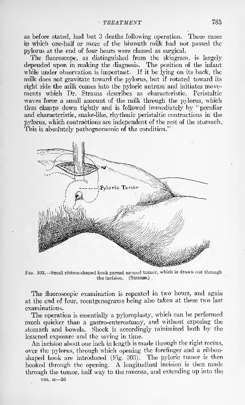
TREATMENT 785
as before stated, had but 3 deaths following operation. Those cases
in which one-half or more of the bismuth milk had not passed the
pylorus at the end of four hours were classed as surgical.
The fluoroscope, as distinguished from the skiagram, is largely
depended upon in making the diagnosis. The position of the infant
while under observation is important. If it be lying on its back, the
milk does not gravitate toward the pylorus, but if rotated toward its
right side the milk comes into the pyloric antrum and initiates move-ments which Dr. Strauss describes as characteristic. Peristaltic
waves force a small amount of the milk through the pylorus, which
thus clamps down tightly and is followed immediately by "peculiarand characteristic, snake-like, rhythmic peristaltic contractions in the
pylorus, which contractions are independent of the rest of the stomach.
This is absolutely pathognomonic of the condition."
FIG. 303. Small ribbon-shaped hook passed around tumor, which is drawn out throughthe incision. (Strauas.)
The fluoroscopic examination is repeated in two hours, and again
at the end of four, roentgenograms being also taken at these two last
examinations.
The operation is essentially a pyloroplasty, which can be performedmuch quicker than a gastro-enterostomy, and without exposing the
stomach and bowels. Shock is accordingly minimized both by the
lessened exposure and the saving in time.
An incision about one inch in length is made through the right rectus,
over the pylorus, through which opening the forefinger and a ribbon-
shaped hook are introduced (Fig. 303). The pyloric tumor is then
hooked through the opening. A longitudinal incision is then made
through the tumor, half way to the mucosa, and extending up into the
VOL. ii 50
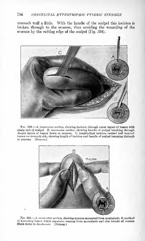
786 CONGENITAL HYPERTROPHIC PYLORIC STENOSIS
stomach wall a little. With the handle of the scalpel this incision is
broken through to the mucosa, thus avoiding the wounding of the
mucosa by the cutting edge of the scalpel (Fig. 304).
FIG. 304. A, transverse section, showing incision through outer layers of tumor with
sharp end of scalpel. B, transverse section, showing handle of scalpel breaking throughdeeper layers of tumor down to mucosa. C, longitudinal incision carried well beyondtumor on stomach side, showing length of incision and handle of scalpel breaking throughto mucosa. (Strauss.)
FIG. 305. A, transverse section, showing mucosa separated from muscularis. B, methodof spreading tumor which separates mucosa from muscularis and also breaks all musclefibers down to duodenum. (Strauss.)
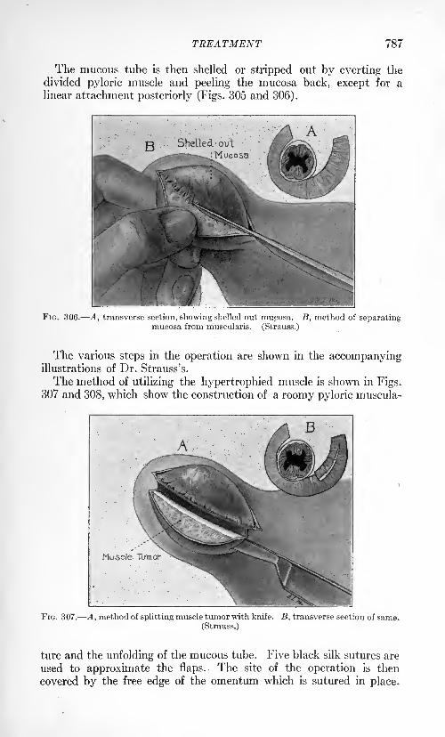
TREATMENT 787
The mucous tube is then shelled or stripped out by everting the
divided pyloric muscle and peeling the mucosa back, except for alinear attachment posteriorly (Figs. 305 and 306).
FIG. 306. A, transverse section, showing shelled out mucosa. B, method of separatingmucosa from muscularis. (Strauss.)
The various steps in the operation are shown in the accompanyingillustrations of Dr. Strauss's.
The method of utilizing the hypertrophied muscle is shown in Figs.307 and 308, which show the construction of a roomy pyloric muscula-
FIG. 307. A, method of splitting muscle tumor with knife. B, transverse section of same.
(Strauss.)
ture and the unfolding of the mucous tube. Five black silk sutures are
used to approximate the flaps. The site of the operation is then
covered by the free edge of the omentum which is sutured in place.
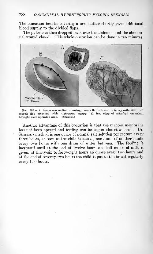
788 CONGENITAL HYPERTROPHIC PYLORIC STENOSIS
The omentum besides covering a raw surface shortly gives additional
blood supply to the divided flaps.
The pylorus is then dropped back into the abdomen and the abdomi-nal wound closed. This whole operation can be done in ten minutes.
FIG. 308. A transverse section, showing muscle flap sutured on to opposite side. B,muscle flap attached with interrupted suture. C, free edge of attached omentumbrought over operated area. (Strauss.)
Another advantage of this operation is that the mucous membranehas not been opened and feeding can be begun almost at once. Dr.
Strauss's method is one ounce of normal salt solution per rectum everythree hours, as soon as the child is awake, one dram of mother's milk
every two hours with one dram of water between. The feeding is
increased until at the end of twelve hours one-half ounce of milk is
given, at thirty-six to forty-eight hours an ounce every two hours and
at the end of seventy-two hours the child is put to the breast regularly
every two hours.
SURGERY OF THE GREAT OMENTUM.
BY KENNETH A. J. MACKENZIE, M.D.
THE development of the great omentum follows that of the bursa
omentalis or omental pouch which is formed from the primitive
mesogastrium. The latter is attached to the greater curvature of
the storrach and grows out with that organ from the spine, becoming
elongated and forming a space behind the stomach which becomes
the lesser peritoneal sac, the entrance to which is the foramen of
Win slow. At this stage the mid-gut is being greatly lengthened and
coils upon itself, necessitating an increase in the width of the attach-
ment of its mesentery. Both the large and the small intestines are
now attached right and left to the vertebral column by a common
mesentery; the gut rotates upon itself, the large intestine is carried in
front of the small, and the cecum behind the liver, which descends,
in the sixth month, into the right iliac fossa. The bursa omentalis
first reaches as far as the greater curvature, but at a later period, it
grows downward to form the great omentum, which lies in front of the
transverse colon and the small intestine. The posterior wall of the
bursa omentalis appears, at first, separate and distinct from the
anterior layer of the transverse mesocolon. Fusion of these layers
takes place, however, so that the great omentum appears not only to
cover but to be intimately adherent to the transverse colon .
ANATOMY AND PHYSIOLOGY OF THE GREAT OMENTUM.
Anatomy. From the greater curvature of the stomach and the
transverse colon hangs, like an apron, a duplicature of the peritoneum,which overlies the coils of the small intestine and is termed the great,
or gastrocolic omentum. Subject to great variations in size, it appearssometimes as a small film-like fringe, at others, as a formation redun-
dant in size and length. Normally it is a delicate structure of sym-metrical form, its lower border often serrated, the projecting end
having fimbriated edges. The omentum is a peritoneal membrane
and, however thin it may appear, four separate folds of peritoneumenter into its formation. In most cases the four layers are fused into
two which enclose the omental bursa. The layer which drops from
the anterior surface of the stomach, when it reaches the greater curva-
ture, turns upon itself, ascends, and is reflected over the transverse
colon, becoming fused with the transverse mesocolon and forming the
posterior wall of the omental bursa. With the downward growth of
the great omentum from the lower border of the stomach, the bursa
(789)
790 SURGERY OF THE GREAT OMENTUM
omentalis extends downward a variable distance between the layersof the omentum. In rare instances the space can be demonstratedin the adult showing the prolongation of the lesser peritoneal spacedownward between the layers of the great omentum.The vascular supply of the great omentum is derived from the
right and left gastro-epiploic arteries which form a rich plexus betweenits two anterior layers. Their veins empty into the portal vein.
The lymphatic current is active in the omentum and its contents are
swept into the glands on the greater curvature of the stomach. Nonerves have been isolated in the substance of the great omentum.
Histology. The omentum possesses a stroma of fine connective
tissue containing many white elastic fibers distributed in the form of
a fine interlacing network. Its meshes contain many small vessels, a
variable deposit of fat, and a vast number of connective-tissue cells
and nuclei. The microscope reveals in its tracery many germinatingcells springing from the fixed cells of the connective tissue or its endo-
thelial lining. These cells have been observed in the act of division
and possess ameboid movement. It is believed that the omentumpossesses a special function to produce leukocytes when they are
needed in the economy for offensive or defensive purposes.Mechanics or Movements of the Omentum. The mobility of the
omentum is governed by factors which may be extrinsic or intrinsic.
Extrinsic influences are: (1) The movements of respiration; (2) the
action of gravity; (3) the reactions of intra-abdominal pressure.Intrinsic influences are: (1) The peristaltic wave which impartsdefinite and constant movement with definite direction (it probablycauses the omentum to twist and to insinuate itself into gaps in the
abdominal wall). (2) The peculiar structure of the omentum, its
smooth and slippery surface and pendulous position, impart to it a
tendency to glide readily into any breach in the walls or toward
perforations, strongly suggesting design for conservation. (3) Asingular activity is observed in its relation to infections, namely, a
tendency to deploy toward the zone of infection and to envelop it;
this movement is like that of an octopus, its arms aloft to grope andreach toward its prey. (Proof of this is found in the fact that in the
presence of multiple perforations of the intestine, the omentum has
been known to throw out its fimbriated prolongations in various direc-
tions to protect all points.) (4) The omentum probably possesses a
movement which is peculiarly its own. (5) The rich plexus of vessels
held in the. meshes of the omentum probably impart a definite move-ment. (6) The phagocytic activities, colossal at times, are said to
impart a movement like that of a tide directed toward the hosts of
invading bacteria.
Functions. The great omentum acts as a splint to exert pressure
upon the subjacent coils of intestine to hold them in place and rests
as a buffer to protect the contained organs of the abdomen from
shock without or within. Thus the force of a hard blow upon the
abdomen could be spent upon the omentum and save the intestine
ANATOMY AND PHYSIOLOGY Of THE GREAT OMfiNTUM 791
from contusion or rupture. Its gliding and sliding movements,
together with its cohesive quality, cause it to be directed into aperturesin the abdominal wall, closing them up. This function is well seen
in stab wounds. Its rich vascular supply, and the loose network of
veins that contains it, makes the omentuna a reservoir for the storageof blood of special service when the pressure index is high, and the
exigencies of the circulation require control. The veins of the omen-tum empty into the portal vein and any factor causing delay of the
portal circulation instantly causes congestion of the veins and transii-
dation follows. The omentum itself alone, under the stress of retarded
venous circulation could readily give rise to ascites. This well-known
fact in pathological anatomy forms the basis of the operation of omen-
topexy, originally designed by Talma, of Utrecht, for the sympto-matic cure of ascites. The omentum has been known to furnish,
through the medium of adhesions, the blood supply for organs or new-
growths completely deprived of their circulation. This fact has been
demonstrated in relation to fibroid tumors of the uterus and cysts of
the ovaries shorn of their blood supply by twisted pedicles.
Absorption. The omentum, through its bloodvessels and lym-phatics, has great power of absorbing fluids and even solids. It is
stimulated in this activity by the pressure of the abdominal muscles,the movements of tihe diaphragm and also by peristalsis. Normallythe omentum plays the most active part in the absorption of all
substances in the peritoneal cavity. If its endothelial lining be
intact, absorption takes place through the lymphatics; if not intact,
it takes place through the vascular system.
Phagocytic Role of the Omentum. The peritoneum is exposed onall sides and at all times to the hazards of bacterial invasion and if the
most effective barriers were not provided, abdominal disease wouldbe the constant lot of man. The barrier that protects the organismis the omentum which can pour out, at short notice, countless hosts of
leukocytes into the peritoneal cavity to combat invading bacteria.
The normal blood of the peritoneum contains the neutrophiles which,in the presence of infection, assemble in large numbers on the omentalsurface and borders, collecting in large clusters and held together in
the meshes of a fibrinous layer. The eosinophiles mingle in these
masses with other varieties of cells. The experiments of Dudgeonand Sargent show that with the initial irritation of the peritoneum,the Staphylococcus albus appears before there is even any solution
of continuity, and provokes at once a rapid formation of phagocytes.Their concentration at threatened points shows, when virulent
organisms appear, that an offensive has begun.Omental Buttress. Associated with the phagocytic function of the
omentum is the predilection to shield points of infection or irritation.
Thus an infected Fallopian tube will be found instantly enveloped
by a blanket of omentum which imprisons it completely, or, in like
manner, a perforated appendix, in the shortest interval, will find its
point of perforation firmly sealed by one of the fimbriated processes
792 SURGERY OF THE GREAT OMENTUM
of the great omentum. The rich exudative layer thrown out at the
time, strengthens the barrier and resists further encroachments
beyond it.
INFLAMMATORY DISORDERS OF THE GREAT OMENTUM.
Epiploitis. Omentitis. This condition is nearly always part of a
general peritonitis traceable to some definite lesion within the abdo-
men. Rarely will it appear that the process begins in the tissues of
the omentum, but frequently it will be found that a local abdominallesion has caused -a local omental inflammation, the characters of
which are identical. In such states the tissues of the omentum tend
to proliferate actively and form nodes of granulation tissue whichtend to coalesce and form masses large enough to be mistaken for new
growths. This constitutes one variety of inflammatory tumor of the
omentum. Another variety is made up of omental fabric rolled upin a knot and held together by a stroma of granulation tissue which
imparts the sensation of a newgrowth, dense in structure. Such
tumors form slowly, as a rule, but rapidly at times, and in acute cases,
follow surgical operations. They pursue a variable course. (1)
They disappear by gradual absorption of the exudate and the attenu-
ation of the granulation tissue. (2) At times the tumor persists, its
elements having become organized. (3) The tumor breaks down andan abscess forms in the center with dense walls of omental and granu-lation tissue. (4) The tumor becomes nodular and indurated having
undergone interstitial fibroid degeneration with marked chronic
features. These tumors not infrequently follow operations and have
been traced to the use of non-absorbable ligatures and also to faulty
technic, such as massive ligations during resection. They form
attachments, at times, to the abdominal walls, and if not adherent,
possess a striking degree of mobility.
Diagnosis. The diagnosis of these disorders must be considered in
relation to ovarian tumors with long pedicles, displaced organs, and
any of the various tumors, benign and malignant, which involve the
omentum.Abscess of the Omentum. Abscess of the omentum is nearly
always the result of local lesions of the peritoneum, and infections of
the appendix and Fallopian tubes are frequent causes. The formation
of abscesses has also been traced to the use of non-absorbable liga-
tures. Occasionally an omental abscess takes place independentlyof peritonitis and local lesions of the abdomen.
Diagnosis. Diagnosis is not usually difficult. The history fur-
nishes the most important clue. A mass can usually be felt which
may, or may not be movable. Pain is constantly present and a sensa-
tion of dragging, and when pressure is made upon the mass pain of a
dull character is elicited. The dense walls will seldom permit the
detection of fluctuation. A moderate degree of temperature is presentand also a constant state of leukocytosis. The abscesses terminate

NEOPLASMS OF THE OMENTUM 793
sometimes by forming adhesions to the walls of neighboring organs and
perforating into the hollow viscera. Cases have been known to point
externally and others to become permanently encysted and to undergo
gradual absorption.Treatment. The proper treatment when the diagnosis is made is
extirpation. Obviously it would be wrong to explore these abscesses
with needles or trocars.
The study of the sequels of omental inflammation is very importantbecause of the effects of movement upon the exudate in forming bandsor adhesions with neighboring tissues. The drag which is exerted
upon adhesions causes them to become hypertrophied and converts
them into bands as thick and tense as whipcord, which endanger the
organs, especially the intestines, by causing kinks, or even by actingas snares to seize exposed loops. Instances are on record where
they have caused by traction, obstruction of the pylorus, the ureter
and the common duct.
NEOPLASMS OF THE OMENTUM.
1. Inflammatory. (See Inflammation.)2. Benign:
(a) Solid:
Lipoma, fibroma.
(6) Cystic:
Cystic lymphangioma : Cystic disease of the omenturnassociated with congenital cystic kidney.
Pseudo cysts: (I) Serous; (II) sanguineous; (III) peri-
toneal (inflammatory).Retention cysts: (I) Lymphatic; (II) chyle.
3. Malignant.
(a) Solid:
Sarcoma, the different types including myxosarcoma.All forms have been described as affecting the greatomentum developing as primary growths.
Carcinoma; all varieties have been identified but always
secondary manifestations of the disease.
(6) Cystic:
Cystic sarcoma and other types of cystic malignantdisease.
4. Teratoma:
Dermoids, fetal inclusions, embryonic cysts.
5. Parasitic cysts:
Hydatid disease of the omentum (echinococcus cysts).
Lipoma. Lipoma must be studied apart from the inflammatorytumor which is very often made up of normal fat matted together in
clumps. Small fat tumors are frequently observed. Occasionally
they assume rapid growth stimulated by the rich vascular supply andattain enormous dimensions. Waldeyer and Spencer Wells reportedtwo cases weighing sixty-three and twenty pounds respectively.
794 SVRGERY Of PUB GREAT dMENTVM
Fibroma. Fibroma is usually found as a small dense encapsuledtumor and may assume rapid growth and attain great size. Theomenturn has been known to adopt tumors from neighboring organs,the circulation of which has been cut off by torsion of their pedicles.Thus the great omentum has been known to foster fibromata and
cystomata of uterine and ovarian origin.
Simple Retention Cysts.- Simple retention cysts have been observed
and also cysts of inflammatory origin with peritoneal investment.
Sanguineous cysts have been noted as the result of trauma and imper-fect hemostasis. The existence of large lymph circulation justifies
the formation of serous and chyle cysts but it is doubtful that the
latter can exist unless adopted by the omentum from the neighboring
mesentery.Cystic Lymphangioma. The literature of these tumors has been
enriched by Stillman, of San Francisco, in an excellent study whichmakes a review of upward of 20 recorded cases. The etiology is
obscure. They are regarded by some authorities as being merelythe dilatation of the lymph vessels of the omentum, by others, as
endotheliomata, while others again regard them as strictly lymphan-giomata. The cysts are found between the peritoneal layers andare numerous, thin-walled, tortuous and often elongated. They maybe few in number or distributed throughout the omentum. Thethin walls are transparent and lined within and without with endo-
thelium. The fluid is usually clear, having, at times, very attractive
tints. It is albuminous and coagulates. There is nothing to showthat the cysts metastasize or invade neighboring organs.
Cystic Omentum. Cystic omentum associated with congenital
cystic kidney: A case of cystic kidney of extraordinary size and
coloring which came under my observation was recorded by Blood-
good in the Johns Hopkins Hospital Reports. The omentum was
extensively involved, and in a case only recently, while applying
Rovsing's method of treatment to the cystic kidney, I found the
omentum studded with cysts.
Sarcoma. Sarcoma of the different types including myxosarcomahave been observed frequently. Bonamy reports 15 cases of primarysarcoma of the omentum in which 12 operations were performed.Five survived the operation for one year or more. I observed with
the late Dr. J. M. Brook, of Portland, a case which, because of the
acute overt symptoms and peculiar localization, was thought to be a
case of acute appendicitis. Operation revealed a tumor occupyingthe free end of the omentum which was matted and adherent to the
neighboring organs including the bladder. There was a good deal
of free blood in the peritoneum and the lymph glands throughoutwere enlarged. The tumor was removed and was demonstrated to
be myxosarcoma. The case afterward proved rapidly fatal. A few
cases of primary endothelioma have been reported although the
records do not seem to bear the stamp of authenticity. It is rather
strange that there are no authentic cases of these tumors recorded
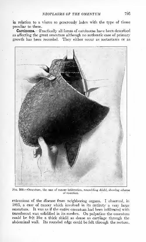
NEOPLASMS Of THE OMENTUM 795
in relation to a viscus so generously laden with the type of tissue
peculiar to them.
Carcinoma. Practically all forms of carcinoma have been described
as affecting the great omentum although no authentic case of primarygrowth has been recorded. They either occur as metastases or as
FIG. 309. Omentum, the seat of cancer infiltration, resembling shield, showing schemeof resection.
extensions of the disease from neighboring organs. I observed, in
1895, a case of cancer which involved in its entirety a very largeomentum. It was as if the entire omentum had been infiltrated withtranslucent wax solidified in its meshes. On palpation the omentumcould be felt like a thick shield as dense as cartilage through the
abdominal wall. Its rounded edge could be felt through the rectum.
796 SURGERY OF THE GREAT OMENTUM
Because of the pain that it caused, a surgical operation was performedand the incision for its relief extended from the ensiform cartilage to
the pubes. It was sixteen inches wide and twelve inches in length andresembled a turtle back. It was probably a scirrhus which directly
invaded the omentum from the stomach. (See Fig. 309.)
Cystic Sarcoma. Cystic sarcoma and other types of cystic malig-nant disease have been reported but the records so far are immature.
Cysts develop in connection with both sarcoma and carcinoma.
Teratoma. This variety of tumor has been found in the greatomentum but it is very doubtful that it occurs as a primary tumor.
It is more likely that the omentum, with its predatory tendency, has
appropriated the tumor from some neighboring organ, the normalhabitat of the growth. The literature, however, shows seeminglyauthentic cases of dermoid and even fetal inclusions in the greatomentum and embryonic cysts from the remains of fetal organs, such
as the Wolffian and Miillerian bodies.
Parasitic Cysts. Hydatid disease of the omentum is proved bythe relative frequency of the disorder. I have observed 2 cases of
echinococcus cysts of the great omentum both of which are recorded
in the pathological reports of the Medical Department of the Univer-
sity of Oregon. In 1 case the major cyst, as large as a child's head,was in the omentum and smaller cysts were found in the liver and
spleen and in the peritoneum above the bladder. They were success-
fully removed. In the other case, the major cyst was within the
liver and smaller cysts were distributed in the peritoneum. The
major cyst was treated successfully by marsupialization. Both cases
developed in men of foreign birth.
Diagnosis and General Symptoms. The tumors lie at, or below, the
umbilicus and are pendulous, possessing a wide radius of lateral and
upward movement. This range of movement is almost typical. In
the case of the omental cyst referred to previously, the tumor could
be swept from the right iliac fossa to the left hypochondrium, and also
from left to right. The physical characters will always depend uponthe nature of the tumor. Solid tumors may be smooth, nodular or
lobulated, according to their character. Cystic tumors are round,elastic and smooth and are found to fluctuate on palpation. Themovements of these tumors are synchronous with those of the dia-
phragm unless, of course, they are anchored by adhesions. Percussion
reveals a dull note over the tumor and it is generally encircled by a
resonant area. The subjoined table, which differentiates ovarian
from omental tumors, will be found a general aid in diagnosis of tumorsof the omentum.
OVARIAN TUMORS. OMENTAL TUMORS.
Grow from below upward. Grow from above downward.Draw the uterus upward. Force the uterus downward.Range of movement lateral and Range of movement lateral anddownward. upward.
Easily mobilized downward. Easily mobilized upward.Movement not influenced by respira- Movement influenced by respiration.
tion.
Radius of movement narrow and Radius of movement wide and unlim-limited. ited.
TORSION OF THE GREAT OMENTUM 797
Operative Treatment of Tumors of the Great Omentum. A few general
principles are established. In all cases of benign growth of the omen-
turn, where it is possible the tumor should be extirpated without
unnecessary sacrifice of the omentum. This principle predicatesthat in the operative treatment of some benign tumors such as the
cystic lymphangioma, the entire omentum may be removed. In all
cases of malignant growth of the omentum, the entire omentumshould be removed flush with its attachments.
These principles being borne in mind, the technic of the operationbecomes of paramount importance. The operation should be per-formed bloodlessly, the vessels being always tied before their division.
Sutures of delicate catgut are applied to each vessel encountered
by means of a fine curved needle, and carefully tied. This is done in
detail across the entire area to be resected. Preliminary to the divi-
sion of the omentum in resection, the part about to be cut should be
compressed by a powerful hemostat of the Ferguson type with narrowblades. This displaces the fat of the omentum in both directions
leaving a thin mesh of tissue which is readily seized and any oozingcontrolled. This process is continued throughout until the resection
is completed. Thereupon the edges may be whipped together bydelicate sutures, thus eliminating any raw surface to invite adhesions.
This technic is applicable to all operations upon the omentum andis designed to prevent the evils of the formation of adhesions andbands and inflammatory states which are well known to cause such
astounding morbidity.
TORSION OF THE GREAT OMENTUM (OMENTOVOLVULUS) .
Torsion of the great omentum is a rare condition. It is most fre-
quently found in association with hernia, the inguinal variety pre-
dominating. Adhesions of the omentum within the sac is a constant
concomitant although it is possible for torsion to take place without
the association of hernia, and even without peripheral adhesions.
It may be single, double or even multiple. When single it is usuallyin a hernial sac and the spiral may extend gradually upward until a
great part of the omentum becomes involved. When double, one
twist will be found in the sac and the other may be entirely indepen-dent in the body of the omentum.
Lejars defines two general classes of cases: (1) That favored bythe descent of the omentum into the hernial sac to which it becomesanchored by adhesions; (2) that of torsion having no hernial associa-
tion. In the first class, twisting may take place in two regions, within
the sac (parasaccular) or near the colon (paracolic) (Fig. 310).
Richardson makes a clinical and anatomical classification. Of the
clinical there are acute and chronic forms. The onset of the acute
is violent, if not fulminating, resembling acute strangulation. It
may refer to an acute torsion in a hernial sac, or one affecting a free,
normal omentum. The chronic form has no definite symptoms;
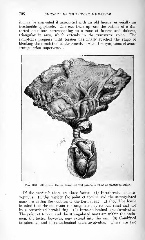
798 SURGERY OF THE GREAT OMENTUM
it may be suspected if associated with an old hernia, especially an
irreducible epiplocele. One can trace upward the outline of a dis-
torted omentum corresponding to a zone of fulness and dulness,
triangular in area, which extends to the transverse colon. The
symptoms progress until torsion has finally reached the stage of
blocking the circulation of the omentum when the symptoms of acute
strangulation supervene. .
X
FIG. 310. Illustrates the parasaccular and paracolic forms of omentovolvulus.
Of the anatomic there are three forms: (1) Intrahernial omento-
volvulus: In this variety the point of torsion and the strangulatedmass are within the confines of the hernial sac. It should be borne
in mind that the omentum is strangulated by its own twist and not
by a constricted hernial ring. (2) Intra-abdominal omentovolvulus:
The point of torsion and the strangulated mass are within the abdo-
men, the latter, however, may extend into the sac. (3) Combinedintrahernial and intra-abdominal omentovolvulus : There are two
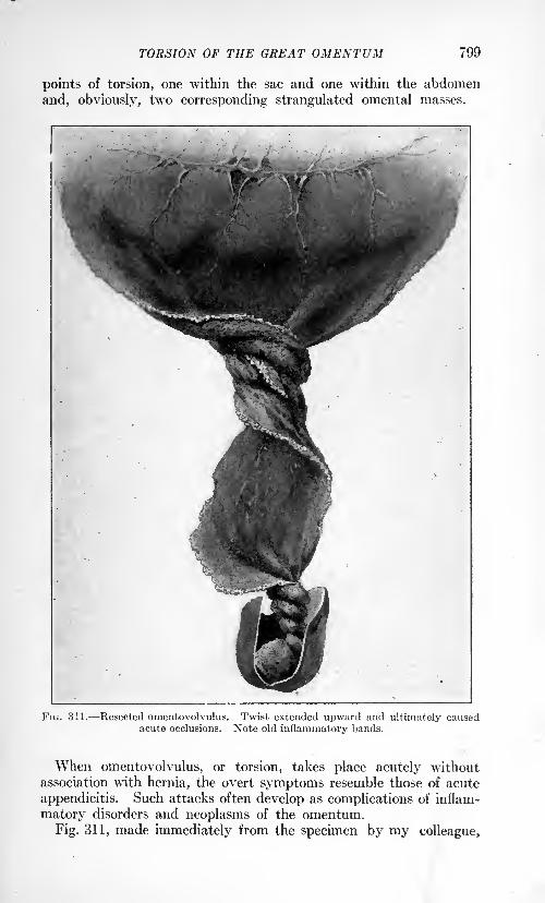
TORSION OF THE GREAT OMENTUM 799
points of torsion, one within the sac and one within the abdomen
and, obviously, two corresponding strangulated omental masses.
FIG. 311. Resected omentovolvulus. Twist extended upward and ultimately causedacute occlusions. Note old inflammatory bands.
When omentovolvulus, or torsion, takes place acutely withoutassociation with hernia, the overt symptoms resemble those of acute
appendicitis. Such attacks often develop as complications of inflam-
matory disorders and neoplasms of the omentum.
Fig. 311, made immediately from the specimen by my colleague,
800 SURGERY OF THE GREAT OMENTUM
Dr. S. M. Gellert, illustrates a case which I observed in St.
Vincent's Hospital, in 1907. The man suffered from an oblique
inguinal hernia for many years and wore a truss constantly. Acute
symptoms set in which called for radical operation. They were
suggestive of strangulation rather than obstruction and there were
no signs of intestinal occlusion. Section revealed a tightly twisted
omentum in the hernial sac the spiral extending upward into the
abdomen blocking the circulation completely. The voluminous
omentum, although completely thrombosed was not gangrenous andwas removed successfully at its junction with the transverse colon.
The man still lives and has had perfect health.
Causes of Torsion. The peristaltic movement of the intestines
doubtless imparts to the omentum not only the tendency to twist but
also its direction. An elongated, redundant omentum predisposes to
torsion just as it predisposes to hernia. Its matting or inflammatory
thickening from any cause favors the tendency to torsion. Once a
free end of the omentum becomes adherent, torsiori is favored with two
points of twisting. Newgrowths of the omentum, also, especially
those which hang from its fimbriated ends, or edges, predispose to
twist.
Hernia of all varieties is the most constant etiological factor. In
proof of this Hedley reported 93 cases, of which details were obtained
in 73, and out of this number 60 were associated with hernia, in 48
of which the twisted omentum was actually found in the hernial sac.
Diagnosis. The diagnosis of omental torsion, whether of the acute
or the chronic form, is very difficult. It can only be made by careful
study of the history and painstaking physical examination. It
should be suspected in the presence of an irreducible epiplocele, or
omental hernia. Careful examination reveals an omental tumormore frequently on the right side which, unlike the tumor of a diseased
appendix may be grasped by the hand and moved from side to side,
and a fan-shaped, doughy mass may be traced upward to its base
which rests against the transverse colon. There is no rigidity andthe mass will be found surrounded on all sides by a resonant area.
There is no tension in the sac, no sign of gas or liquid content, and no
evidence of strangulated hernia. In severe cases when the torsion is
acute, vomiting is present but there are no signs of acute intestinal
obstruction, the lumen of the intestine being preserved. In the
parasaccular form, torsion often being acute, the initial symptoms are
severe with pain referred to the sac and with signs impending of acute
strangulation. In all cases the fever is very moderate, the toxic
phenomena mild, and the peritoneal reactions very slight.
Treatment. It is always surgical. Indeed, gangrene is always so
much to be feared that operation is essential in all chronic cases
associated with hernia and a radical operation should be done, the
omentum untwisted, divested of any faults and replaced. If it is
notably elongated, it should be shortened. The greatest care should
be taken to prevent matting or clumping, and the formation of adhe-
SPECIAL SURGERY OF THE GREAT OMENTUM 801
sions. In acute cases with strangulation, delays are fraught with
danger and the quickest steps should be taken which should begin with
an attack on the hernia. The incision should be extended so as to
explore and determine the condition of the omentum. The zone of
obliterated circulation should be minutely defined and resection madewell above it.
In paracolic forms, if the circulation be still intact, the omentumshould be carefully untwisted, first within the sac, and then above it
within the abdomen. If there be manifest tendencies for the twist
to recur, it should be prevented by the removal of redundant parts or,
if need be, by the resection of the entire omentum. If in any case
the omentum be found thrombosed throughout, the entire viscus maybe removed flush with its attachment to the colon. The technic laid
down previously should be scrupulously followed.
SPECIAL SURGERY OF THE GREAT OMENTUM.
Omentopexy, Epiplopexy, the Talma Operation. This operation wasfirst proposed by Talma, of Utrecht, in 1889, for the symptomatic relief
of ascites in cirrhosis of the liver. Drs. Drummond and Morrison of
Newcastle-on-Tyne quite independently reported, in 1896, a case of
ascites due to cirrhosis of the liver cured by operation. The surgicalintent in this operation is to cause a short circuit between the portaland the systemic circulation and the relief of the impasse which causes
the transudation of fluid very largely from the omentum into the
peritoneal space (ascites). The operation, although attended by a
large mortality, is approved by many surgeons as a means of relievingthe most distressing symptom of an incurable disorder.
The operation of choice still follows closely the lines originally
proposed by Talma. The following technic is advised: An incision
is made through the abdominal wall four inches in length beginningat a point one inch below the umbilicus. To one or the other side
of the incision, six or eight inches of the omentum is brought down anda fold of it, from one to three inches in width, is stitched, by means of
silk sutures, to the same width of peritoneal border. A broad area of
adhesions is needed to make the requisite collateral circulation. This
fusion of the tissues brings about an anastomosis between the radicals
of the portal vein and those of the deep epigastric. The peritoneum is
closed in the usual way and the wound sutured in layers.
Narath's modification of the Talma procedure deserves mentionbecause of its safety and ease of execution. It brings the two sets of
vessels into direct contact. Under local anesthesia, a short medianincision is made above the umbilicus large enough to explore with the
index finger and to deliver a strip of the oinentum, three to six inches
in length, which is anchored and carefully tucked by stitches betweenthe fascial layer and the skin. The incision throughout is large
enough to permit the passage of the omental strip and small enoughVOL. II 51
802 SURGERY OF THE GREAT OMENTUM
to prevent hernia. The abdominal incision is carefully closed in
layers.
One of the great advantages of this operation is that it permits
drainage of the peritoneal fluid into the connective tissue of the
abdominal wall.
Surgery of the Cavity of the Great Omentum. Tumors are nowknown to have their origin in the cavity of the great omentum. Eightcases have been reported by Ziembecki all of which were cancers.
Hernia. Hernia of the abdominal contents, chiefly loops of the
small intestine but also of other viscera, into the foramen of Winslowhas been observed from time to time. Ziembecki has reported 20
cases prior to 1906 together with one case of hernia of the gall-bladder.Fat Necrosis. The entire cavity of the great omentum has been
found involved in fat necrosis from pancreatic disease.
Cysts. Pancreatic Cysts. Pancreatic cysts have been known to
invade the cavity of the great omentum, and also hydatid cysts.
Pseudocysts. Encysted fluid (peritoneal) has been observed in
the cavity of the great omentum in connection with local or generalforms of peritonitis.
Pseudocysts (Sanguineous}. Encysted blood has been observed in
the cavity of the great omentum as the result of severe abdominal
injuries involving the spleen, pancreas, and the great vessels.
Abscess. The cavity of the great omentum has been found to be
the seat of abscesses springing from the liver and also from the perfora-
tion of ulcers of the duodenum and caries of the vertebrae.
THE SUEGERY OF ILEUS.
BY FRED T. MURPHY, M.D.
THERE is scarcely a medical writer of note who has not recorded
his observations and opinions regarding the conditions which cause
a cessation of the rhythmical advance of the content of the intestinal
tract and the resulting symptoms. The literature of the subject is
exhaustive,1 so limitless that any attempt to abstract it can lead
only to confusion. The very multiplicity of the varying opinionshas tended both to obscure the relatively simple pathological changefound in mechanical obstruction, and also to add to the difficulties of
grouping satisfactorily the abnormal conditions which lead to an
acute retention of the content of the intestine.
Certain authorities2 have felt that the use of the term "ileus," even
though modified by such adjectives as "dynamic," "adynamic," and"mechanical" has added to this confusion. From its derivation, ileus
means to roll, to twist. In the minds of the early writers, an ileus
was a rolling up or a twisting of the intestine, and therefore became
synonymous with grievous colic. As representing the syndrome of
symptoms accompanying the acute retention of the bowel content
the term "ileus" is comprehensive, and it will be used in that sense.
But the terms "intestinal paralysis" and "intestinal obstruction" define
more clearly the reasons for the acute retention and to emphasize the
underlying pathological conditions. These terms will, therefore, be
used in this discussion.
In order that no confusion may arise in the use of certain terms,
they are defined. By "occlusion" or "obstruction" of the intestine
is meant the primary interference with the onward peristaltic action
of the intestine independent of any primary circulatory change. By"strangulation" of the intestine is meant the interference with the
normal peristaltic action associated with the change from the normalcirculation of the gut. In the later consideration of this matter of
circulatory disturbance it will be seen that simple occlusion or obstruc-
tion may change to an actual condition of strangulation independentof pressure upon or torsion of the mesenteric vessels. This possibility
should be emphasized, for it explains many of the clinical symptomswhich are otherwise difficult to interpret.
This chapter will deal with those conditions producing an actual
obstruction of the bowel: (1) By mechanical interference; (2) bytonic contraction of the muscles of the wall; and (3) those acute
.
1 Wilms: Der Ileus. Treves: Intestinal Obstruction.2Nothnagel: Diseases of the Intestines and Peritoneum.
(803)
804 THE SURGERY OF ILEUS
conditions, except peritonitis (to be discussed in another section)
which cause a paralysis of the intestine leading to the developmentof the cardinal symptoms of acute stoppage. The spastic and atonic
conditions of the large intestine underlying obstinate constipationwill not be considered here. They are essentially diseases for medical
treatment. Idiopathic dilatation of the colon, or Hirschsprung'sdisease, may be associated with an intractable paralysis of the large
bowel, but it is so distinct both in its pathology and symptomatologyfrom the conditions which are characterized by the symptom-complexdefined by ileus that it will also be omitted from this discussion.
The cases in which there is a mechanical obstruction of the intestine
are those amenable to and demanding early surgical interference.
This group is, therefore, considered first and in greater detail, thoughit includes only about 28 per cent. 1 of all cases of ileus.
Causes of Mechanical Obstruction. The causes of mechanical obstruc-
tion of the intestine may be irreducible hernias, either internal or
external, new growths involving the gut, constricting bands acquiredor congenital, invagination of the intestine within itself, intussuscep-
tion, volvulus, cicatricial strictures, pressure upon the intestinal
lumen from intra-abdominal tumors, congenital defects, and foreignbodies within the lumen. Extensive circulatory disturbances such
as a mesenteric thrombosis may cause, to all intents and purposes, a
mechanical obstruction, though actually this obstruction is due to a
paralysis of the thrombosed segment, and it should be so classified.
It is difficult to establish trustworthy percentages as to the relative
frequency of the various causes of mechanical intestinal obstruction,
because of the wide variation which is to be found in the character of
the material admitted to hospitals of different types. In the experi-ence of every one, however, the obstruction is caused most frequently
by irreducible hernias, constricting bands and new growths involvingthe wall of the intestine. When we consider the almost innumerable
anatomical anomalies in the form of peritoneal fossae, defects, such
as slits in the mesentery, and the large number of possible external
hernise, it is readily seen why this type of obstruction is more commonthan the others. To enumerate and describe in detail each anatomical
possibilityis a futile task. In considering obstruction from hernise,
we need only to remember that a loop of intestine may be caught in
any one of these congenital or pathological pouches and obstructed
and possibly strangulated. Because of the relatively fixed position
of the large intestine, it is infrequently found obstructed in hernise,
but, excepting the stomach, it is the most common site of new growthin man; hence the high percentage of occlusions from this cause to
be found there. In the small intestine, on the contrary, cancer is of
such rarity that it may be disregarded as a cause of obstruction,
Venot and Parcelier2having been able to collect only 50 cases to
1913.
1 Murphy, J. B. : Kelly and Noble's Abdominal Surgery, vol. ii,
2 Revue de Chir., 1913, xlviii, 436.
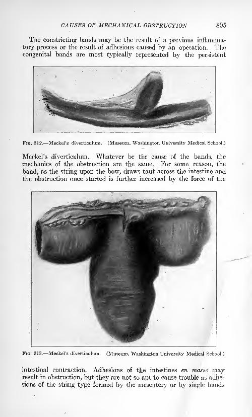
CAUSES OF MECHANICAL OBSTRUCTION 805
The constricting bands may be the result of a previous inflamma-
tory process or the result of adhesions caused by an operation. Thecongenital bands are most typically represented by the persistent
Fio. 312. Meckel's diverticulum. (Museum. Washington University Medical School.)
Meckel's diverticulum. Whatever be the cause of the bands, the
mechanics of the obstruction are the same. For some reason, the
band, as the string upon the bow, draws taut across the intestine andthe obstruction once started is further increased by the force of the
FIG. 313. Meckel's diverticulum. (Museum, Washington University Medical School.)
intestinal contraction. Adhesions of the intestines en masse mayresult in obstruction, but they are not so apt to cause trouble as adhe-
sions of the string type formed by the mesentery or by single bands
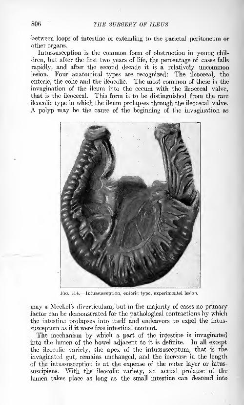
806 THE SURGERY OF ILEUS
between loops of intestine or extending to the parietal peritoneum or
other organs.
Intussusception is the common form of obstruction in young chil-
dren, but after the first two years of life, the percentage of cases falls
rapidly, and after the second decade it is a relatively uncommonlesion. Four anatomical types are recognized: The ileocecal, the
enteric, the colic and the ileocolic. The most common of these is the
invagination of the ileum into the cecum with the ileocecal valve,
that is the ileocecal. This form is to be distinguished from the rare
ileocolic type in which the ileum prolapses through the ileocecal valve.
A polyp may be the cause of the beginning of the invagination as
FIG. 314. Intussusception, enteric type, experimental lesion.
may a Meckel's diverticulum, but in the majority of cases no primaryfactor can be demonstrated for the pathological contractions by which
the intestine prolapses into itself and endeavors to expel the intus-
susceptum as if it were free intestinal content.
The mechanism by which a part of the intestine is invaginatedinto the lumen of the bowel adjacent to it is definite. In all except
the ileocolic variety, the apex of the intussusceptum, that is the
invaginated gut, remains unchanged, and the increase in the lengthof the intussusception is at the expense of the outer layer or intus-
suscipiens. With the ileocolic variety, an actual prolapse of the
lumen takes place as long as the small intestine can descend into
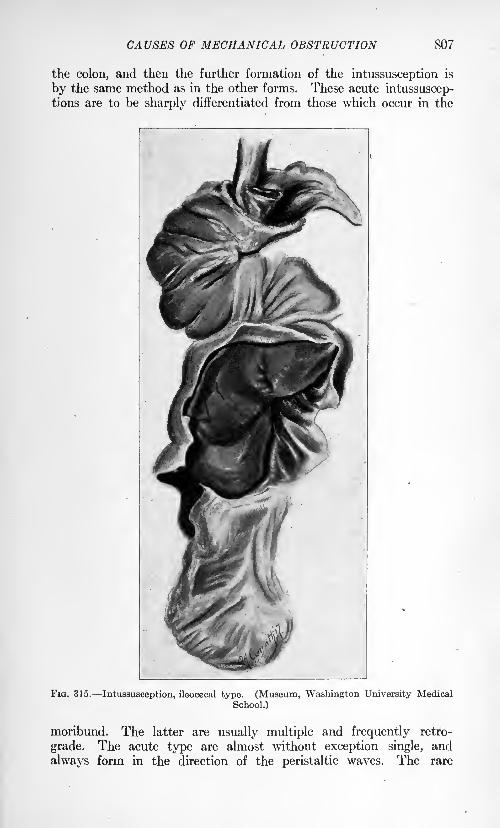
CAUSES OF MECHANICAL OBSTRUCTION 807
the colon, and then the further formation of the intussusception is
by the same method as in the other forms. These acute intussuscep-tions are to be sharply differentiated from those which occur in the
FIG. 315. Intussusception, ileocecal type. (Museum, Washington University Medical
School.)
moribund. The latter are usually multiple and frequently retro-
grade. The acute type are almost without exception single, and
always form in the direction of the peristaltic waves. The rare
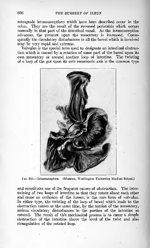
808 THE SURGERY OF ILEUS
retrograde intussusceptions which have been described occur in the
colon. They are the result of the reversed peristalsis which occurs
normally in that part of the intestinal canal. As the intussusception
advances, the pressure upon the mesentery is increased. Conse-
quently the circulatory disturbances in all the bowel which is involved
may be very rapid and extreme.
Volvulus is the special term used to designate an intestinal obstruc-
tion which is caused by a rotation of some part of the bowel upon its
own mesentery or around another loop of intestine. The twistingof a loop of the gut upon its own mesenteric axis is the common type
FIG. 316. Intussusception. (Museum, Washington University Medical School.)
and constitutes one of the frequent causes of obstruction. The inter-
twining of two loops of intestine so that they rotate about each other
and cause an occlusion of the lumen, is the rare form of volvulus.
In either type, the twisting of the loop of bowel which leads to the
obstruction causes at the same time, by the torsion of the mesentery,serious circulatory disturbances in the portion of the intestine so
rotated. The result of this mechanical process is to cause a simpleobstruction of the intestine above the level of the twist and also
strangulation of the rotated loop.
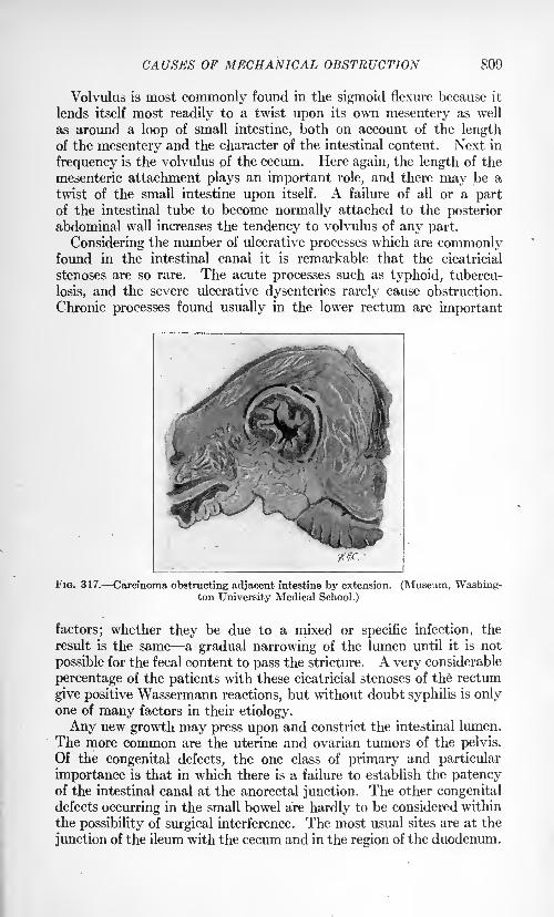
CAUSES OF MECHANICAL OBSTRUCTION 809
Volvulus is most commonly found in the sigmoid flexure because it
lends itself most readily to a twist upon its own mesentery as well
as around a loop of small intestine, both on account of the lengthof the mesentery and the character of the intestinal content. Next in
frequency is the volvulus of the cecum. Here again, the length of the
mesenteric attachment plays an important role, and there may be a
twist of the small intestine upon itself. A failure of all or a partof the intestinal tube to become normally attached to the posterior
abdominal wall increases the tendency to volvulus of any part.
Considering the number of ulcerative processes which are commonlyfound in the intestinal canal it is remarkable that the cicatricial
stenoses are so rare. The acute processes such as typhoid, tubercu-
losis, and the severe ulcerative dysenteries rarely cause obstruction.
Chronic processes found usually in the lower rectum are important
FIG. 317. Carcinoma obstructing adjacent intestine by extension. (Museum, Washing-ton University Medical School.)
factors; whether they be due to a mixed or specific infection, the
result is the same a gradual narrowing of the lumen until it is not
possible for the fecal content to pass the stricture. A very considerable
percentage of the patients with these cicatricial stenoses of the rectum
give positive Wassermann reactions, but without doubt syphilis is onlyone of many factors in their etiology.
Any new growth may press upon and constrict the intestinal lumen.
The more common are the uterine and ovarian tumors of the pelvis.
Of the congenital defects, the one class of primary and particular
importance is that in which there is a failure to establish the patencyof the intestinal canal at the anorectal junction. The other congenitaldefects occurring in the small bowel are hardly to be considered within
the possibility of surgical interference. The most usual sites are at the
junction of the ileum with the cecum and in the region of the duodenum.
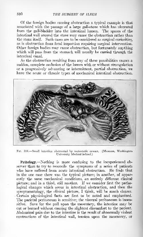
810 THE SURGERY OF ILEUS
Of the foreign bodies causing obstruction a typical example is that
associated with the passage of a large gall-stone which has ulcerated
from the gall-bladder into the intestinal lumen. The spasm of the
intestinal wall around the stone may cause the obstruction rather thanthe stone itself. Such cases are to be considered as surgical curiosities,
as is obstruction from fecal impaction requiring surgical intervention.
Other foreign bodies may cause obstruction, but fortunately anythingwhich will pass from the stomach will usually be carried through the
intestinal canal.
As the obstruction resulting from any of these possibilities causes a
sudden, complete occlusion of the lumen with or without strangulationor a progressively advancing or intermittent, partial obstruction, wehave the acute or chronic types of mechanical intestinal obstruction.
FIG. 318. Small intestine obstructed by metastatic cancer.
University Medical School.)
(Museum, Washington
Pathology. Nothing is more confusing to the inexperienced ob-
server than to try to reconcile the symptoms of a series of patientswho have suffered from acute intestinal obstruction. He finds that
in the one case there was the typical picture; in another, of appar-
ently the same mechanical conditions, an entirely different clinical
picture; and in a third, still another. If we consider first the patho-
logical changes which occur in intestinal obstruction, and then the
symptomatology, the clinical picture, I think, will be much clearer.
Certain physiological facts are first to be noted and emphasized.The parietal peritoneum is sensitive; the visceral peritoneum is insen-
sitive. Save for the pull upon the mesentery, the intestine may be
cut or burned without causing the slightest discomfort to the patient.
Abdominal pain due to the intestine is the result of abnormally violent
contractions of the intestinal wall, tension upon the mesentery, or
PATHOLOGY OF INTESTINAL OBSTRUCTION 811
involvement of the parietal peritoneum by a secondary inflammatory
process. Vomiting is either reflex or regurgitant; that is, the stomachcontracts either from the stimulus of nervous impulse, or, when over-
distended, empties itself by letting the content seek the outlet of
least resistance. Except in the large bowel, the rhythmical peri-
staltic action of the intestine propels the content toward the rectal
outlet. In the proximal part of the large bowel, as demonstrated
by Cannon,1 there is a normal reversed peristalsis, and it has been
shown by many observers that under certain conditions, this reversed
peristalsis may carry the intestinal content for a considerable distance
above the ileocecal valve. Nothnagel accepts the theory of a reversed
peristalsis in the small intestine, basing his conclusions upon the
experiments of Muhsam, Enderlen and Hess. The work of Mall,2
however, seems to refute absolutely this opinion. Without doubt,a section of the intestine may be reversed and there may follow nor-
mal evacuations of the bowel, but it will be found on examining this
reversed loop of intestine at a considerable time after the reversal
that it takes no part in propelling the intestinal content. It acts
simply as a tube, the content being pushed over this inert or possiblyfor a time actively obstructing segment by the force of the peristalsis
in the normal intestine. In the laboratory we have repeatedly reversed
loops of intestine in animals and have found without exception that
later they came to be dilated segments without peristaltic activity.
The question of the possibility of a reversed peristalsis has a most
important bearing upon the symptom of vomiting and its diagnostic
significance. Vomiting in intestinal obstruction represents, then,either a reflex contraction of the stomach, or a running over of the
stomach due to the escape of the intestinal content back through the
relaxed pylorus. With violent peristaltic waves pushing the intestinal
content against an obstruction, there is a recoil as demonstrated byCannon and Murphy3 in their experimental work on certain phasesof intestinal obstruction, but not a true reversed peristalsis. Whileclinical reports of cases of unquestioned fecal vomiting are not to be
discredited, they must be accepted sub judice. As an example of the
errors in observation which may occur, the following case is cited:
This patient had been operated upon several times; first for an appen-dicitis, later for a resection of a carcinoma of the hepatic flexure.
At this operation, the upper part of the ascending colon and a part of
the transverse colon had been removed. The ileum had beep joinedto the remaining part of the transverse colon by a lateral anastomosis.
This left a blind pouch formed by the cecum and a part of the ascend-
ing colon. Shortly after the operation, the patient experienced muchpain and a tumor mass was felt in the right lower quadrant. This
later disappeared. On account of the presence of the mass, a later
1 Am. Jour. Physiol., 1902, No. 5, vol. vi.
* Johns Hopkins Hosp. Reports, vol. i, p. 93.3 Ann. Surg., April, 1906, No. 4, vol. xliii.
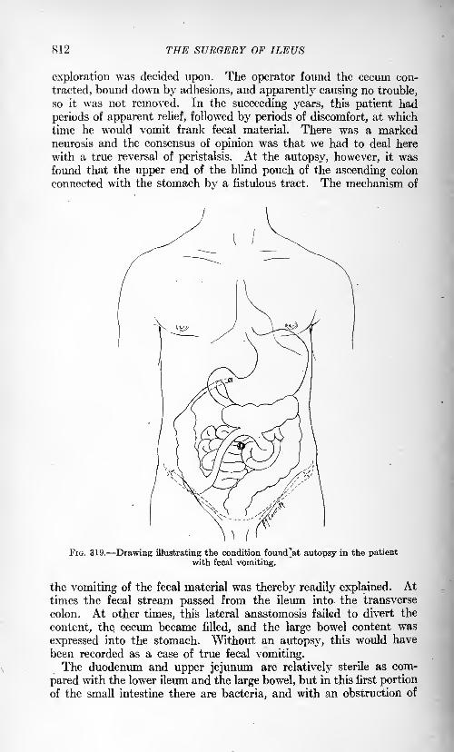
812 THE SURGERY OF ILEUS
exploration was decided upon. The operator found the cecum con-
tracted, bound down by adhesions, and apparently causing no trouble,
so it was not removed. In the succeeding years, this patient had
periods of apparent relief, followed by periods of discomfort, at whichtime he would vomit frank fecal material. There was a markedneurosis and the consensus of opinion was that we had to deal here
with a true reversal of peristalsis. At the autopsy, however, it wasfound that the upper end of the blind pouch of the ascending colon
connected with the stomach by a fistulous tract. The mechanism of
FIG. 319. Drawing illustrating the condition found'at autopsy in the patientwith fecal vomiting.
the vomiting of the fecal material was thereby readily explained. Attimes the fecal stream passed from the ileum into the transverse
colon. At other times, this lateral anastomosis failed to divert the
content, the cecum became filled, and the large bowel content was
expressed into the stomach. Without an autopsy, this would have
been recorded as a case of true fecal vomiting.The duodenum and upper jejunum are relatively sterile as com-
pared with the lower ileum and the large bowel, but in this first portion
of the small intestine there are bacteria, and with an obstruction of
PATHOLOGY OF INTESTINAL OBSTRUCTION 813
this segment they multiply rapidly. The secretions of the upper
part of the small intestine are abundant, and they persist for an
indefinite period after the physiological activity of the intestine has
been withdrawn. The secretions of the ileum are comparatively small
in amount and they do not continue for any considerable time after
there is no longer a functional demand.Under normal conditions, the rhythmical peristalsis of the intestine
cannot be followed through the abdominal wall, except where the
wall is very thin, and then the normal peristaltic action may be plainly
evident. In a chronic condition where there has occurred a hyper-
trophy of the intestinal wall from the effort to overcome the partial
occlusion, this visible peristalsis is marked. In an acute condition,
on the other hand, the hyperactivity may be marked, but a paralysis
of the intestinal wall may result from this overstimulation and lead
to a paralysis.
Inability to make up for the body fluids which are lost throughexcessive vomiting leads to a rapid dehydration of the whole body,
accompanied by a more or less complete suppression of urine. McQuarrieand Whipple
1 have shown experimentally that animals with obstructed
loops of intestine or animals which had intravenous administration of
the toxic contents of obstructed loops of gut show a marked diminu-
tion in the renal function and a corresponding high non-protein
nitrogen retention in the blood. This is believed to be a direct effect
of the toxin on the renal epithelium. Gas is normally producedwithin the intestinal lumen, and with a normal metabolism a properbalance is maintained within the intestine. Either a pathologicalincrease of gas due to putrefactive changes or a pathological decrease
in its absorption leads to a marked local accumulation.
The pathological changes causing acute symptoms due to absorp-tion of the toxic bowel content, which occur in an obstructed loop of
intestine depend either upon the pressure within the lumen or upondirect interference with the circulation by pressure on the veins or
arteries supplying this segment. Pressure upon the bloodvessels, that
is strangulation of the mesentery, has been recognized generally as
the important factor in the production of these toxic symptoms.Pressure within the lumen of the gut has not, it seems to me, been
sufficiently emphasized. The pressure within the intestine "may lead
to exactly the same pathological change so far as the production of
the symptoms is concerned, as actual pressure upon the mesentery.This abnormal pressure within the intestine depends upon the amountof material within the obstructed portion of the gut, be it fluid or gas,
plus the tension of the muscle wall. As this pressure increases, there
occurs first the venous stasis in the small vessels of the intestinal wall.
This is followed first by an edema of the mucosa, and later by its
destruction. With the mucosa destroyed, there no longer exists
1 Jour. Exp. Med., 1919, xxix, 397.
814 THE SURGERY OF ILEUS
the protective filter of the normal mucous membrane, and the absorp-tion of any toxic substance from the intestinal lumen is rapid. The
possible rapidity and amount of this absorption is difficult to appre-ciate. Given a septic focus in the hand and a virulent organism, andall realize how fulminating may be the ascending lymphangitis. The
absorption from such a focus must be, however, infinitesimal as
compared with the absorption from the intestine when the normal
protective mechanism has been destroyed. The same series of changesoccur in the intestinal mucosa when the circulatory disturbance is
secondary to direct interference with the blood supply by strangula-tion of the mesentery. Appreciation of the importance of this pres-sure within the lumen of the intestine combined with that of the
circulatory change will explain in great measure the variations seen
between obstructions high and obstructions low, obstructions with
and without strangulation. As has been stated, the secretions of the
upper small bowel are rapid and persistent. In simple high obstruc-
tions, then, there follows a relatively rapid distention of the intestine,
which leads to an early circulatory disturbance. With simple obstruc-
tion of the lower bowel, the degree of distention is relatively slight,
and the onset of the circulatory change is late. Therefore, comparinga high obstruction with a low obstruction, we may have the widest
variations in the time of the onset of acute symptoms of absorption.Obstruction of the large intestine provides a still larger reservoir and
the distention leading to symptoms from absorption of the toxic
content is still further delayed. These circulatory changes can be
demonstrated definitely only in experimental work, since in clinical
cases the operator can never be sure of the exact time nor the exact
degree of venous or arterial pressure which has existed. The circu-
latory changes which occur in cases of volvulus or strangulation maybe of any degree.
In a series of experiments, Murphy and Vincent1 demonstrated
that obstructions of the venous return from a closed loop of intestine
caused the most severe and most rapidly appearing symptoms. The
change in the mucous membrane in the intestine with an obstruction
to the venous return showed marked distention and destruction of the
mucous coat within four hours and explained the rapidity of the
absorption because the content was under pressure and the portals
of absorption widely open. They demonstrated also that a completeanemia of the intestine led to the production of the same symptoms,but that these were less rapid in their onset.
The observations of Hartwell and Hoguet2 showed most graphi-
cally how small a part simple obstruction plays provided there is jio
destruction of the mucous membrane. In their experiments the
small bowel was obstructed. 10 to 30 cm. from the pylorus by a clamp
1 Boston Med. and Surg. Jour., 1911, No. 18, clxv, 684.2 Am. Jour. Med. Sc., 1912, cxliii, 357.
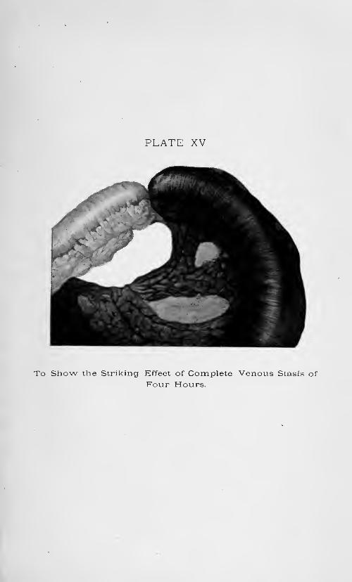
PLATE XV
To Show the Striking Effect of Complete Venous Stasis of
Four Hours.

PATHOLOGY OF INTESTINAL OBSTRUCTION 815
so applied as not to injure the mucous membrane. With an obstruc-
tion so high, the content of the obstructed intestine overflowed rapidlyinto the stomach through the relaxed pylorus, from which it was
expelled by vomiting or removed by lavage. Therefore no distention
of the intestine was caused which might lead to circulatory changefrom the increase of tension. Under such conditions they were able,
by supplying water to their animals subcutaneously, to keep themalive for ten days.The adherents to the so-called reflex nervous theory as a cause of
death in intestinal obstruction have been impressed by the suddenness
and the intensity of symptoms which, in the fulminating cases, seem
to be of such a character that they must be of nervous origin. Experi-
mentally, however, no direct proof in support of this theory has been
produced. On the contrary, the connection with the central nervous
system may be severed, and yet the production and degree of symp-toms remains unchanged.The more recent and active investigators of this subject are agreed
that death in cases of acute mechanical obstruction without perfora-
tion, is caused by the absorption of some toxic substance from the
lumen of the gut. As to the exact nature of this substance and the
essentials for its production, differences of opinion exist. Whipple1
and his co-workers at Baltimore and at the University of California,
believe that this toxic substance is primarily a perverted secretion of
the intestinal mucosa. Murphy, Vincent and Brooks, on the other
hand, believe that this toxic substance is the result of bacterial action
upon the content of the intestine. Whipple2 has recently isolated a
proteose which he considers to be the substance especially concerned
in the intoxication in these cases of intestinal obstruction. Other
experimenters3 have believed that the toxic substance was some one
of the various ptomaines. From a practical standpoint, the patho-
logical changes are important as they have a direct bearing on the
production of symptoms and upon the treatment of the condition.
If it is agreed that the toxic substance has been produced in the
intestinal lumen, then the changes which permit of the absorption of
this toxin are crucial. Kocher, 4years ago, called attention to the
small ulceration which may be found in the mucosa of an obstructed
intestine. The belief of that time was that death was Caused in
these cases through the passage of bacteria through the wall of the
gut, causing thereby a peritonitis, the absorption from the peritoneal
cavity playing a part no less important than the absorption from the
lumen of the intestine. This contention has been disproved byexperimental work, because it is possible to cause death by intestinal
obstruction and to have the fluid within the abdominal cavity both
1 Bull. Johns Hopkins Hosp., 1912, xxiii, 159; Ann. Surg., lix, 714.2 Jour. Am. Med. Assn., No. 1, Ixvii, 15.
Von Albeck: Arch. f. klin. Chir., 1901, Ixv, 569.4 Mitt. a. d. Grenzgb. d. Med. u. Chir., 1899, iv, 201.
816 THE SURGERY OF ILEUS
free from bacteria and harmless on injection. These ulcers are
important in that they represent breaks in the mucous membraneand openings for the absorption of the toxic content. They are
secondary to circulatory changes, not primarily the result of pressure
except as this pressure causes an anemia. That this toxic material is
not a specific secretion of any part of the intestinal tract as has been
maintained by some has been demonstrated experimentally by Murphyand Brooks. 1 That it is dependent upon the action of bacteria com-bined with the degenerative changes in the mucus-lined cavities seems
equally clear from their work. They were able, after prolonged
drainage, to secure a loop of intestine, which was closed at one endand opening .through the abdominal wall at the other, in which there
were no bacteria. They obstructed this loop. After a period of
time at which, under the usual conditions of the intestinal content,
there should have been formed a toxic substance, this content wasremoved though the experimental animal had shown no symptomsof toxic absorption. On injection this content was found to be non-
toxic. The same loop of intestine was again obstructed after it hadbeen infected and the content later was proved to be toxic both bythe symptoms of absorption in the host and also by injection of the
content into other animals.
The experiments of Dradstedt, Moorhead, and Burcky2 bear directly
on this question as to whether the bacteria had or had not played an
important role in the formation of the toxic substance found in the
lumen of the gut in cases of intestinal obstruction. They confirmed
the results of other investigators as to the fatal results of immediate
closure of a duodenal or jejunal loop, the animals dying in forty-eight
to ninety-six hours. After washing the segment of the jejunum,
however, with either sterile water or salt solution they were able to
close both ends of the intestine and to have a considerable numberof these animals live indefinitely. They found that when isolated,
closed loops of the jejunum were sterile, complete occlusion of the
bloodvessels had no effect on the animal, but if the loops were not
sterile the occlusion of the circulation caused death in twenty-four to
forty-eight hours, with the usual symptoms of complete intestinal
obstruction. In other experiments they placed open loops of the
duodenum, sectioned just below the inferior pancreatic duct, in the
abdominal cavity. In 50 per cent, of these experiments the animals
lived without symptoms, and later the ends of the intestinal loops
were found completely closed by adhesions and the lumen seemed
distended with a sterile fluid. These experiments confirmed the
opinion that the normal secretions of the duodenum and jejunumare not toxic and that bacterial activity plus necrotic tissue or the
results of the action of the bacteria on the necrotic tissue is an essential
factor in producing symptoms in intestinal obstruction.
i Arch. Int. Med., vol. xv, p. 392.* Proc. Soc. Exp. Biol. and Med., No. 1, xiv, 17.
PATHOLOGY OF INTESTINAL OBSTRUCTION 817
Since these sterile intestinal loops were produced only after the
greatest difficulty, gall-bladders were used as receptacles by Murphyand Brooks. 1 If the circulation of a sterile gall-bladder were altered
and the cystic duct obstructed the content remained non-toxic to
injection even though the gall-bladder had begun to be gangrenous.
If, on the contrary, these same changes were brought about and the
content of the gall-bladder infected then this content at the end of
two days had the same toxicity and produced the same symptomsand pathological changes which were found upon the injection of the
toxic content of obstructed loops of intestine. Chemically it was not
proved that this toxin was identical with that found in the obstructed
intestine.
These pathological changes emphasize the importance clinically of
a circulatory change in the obstructed intestine, since by that circu-
latory change the normal filter of the mucous membrane is destroyed.With the mucous membrane intact there is no absorption from the
intestine of this virulent toxin found in the obstructed intestine.
This can be shown by confining the toxic material within an occluded
loop of intestine in which no changes have occurred in the mucosa.
Under such conditions the experimental animal continues without
clinical symptoms of absorption. This fact has been even further
substantiated by the experiments of Brooks,2 who showed that if a
long segment of jejunum and ileum were isolated and occluded at both
ends that distention of such loop very gradually developed, and the
animal showed only slight symptoms of absorption of the toxic loopcontent at the end of twenty-one days, in spite of the fact that the
occluded loop of gut contained as much as two hundred times the
lethal dose of the toxin. That the content of an obstructed bowel
may be extremely toxic and still not produce symptoms of rapid
absorption is well illustrated by the following case:
R. E., white, female, aged nineteen years, nurse, S. N. 3021. Oper-ated on May 21, 1916, for subacute appendicitis. Surrounding the
appendix numerous old adhesions were found, which increased the
difficulties of the operation. Postoperative convalescence for first
forty-eight hours was uneventful. At this time the temperature was
normal and the pulse ranged from 98 to 115. The patient then
began to have an unusual amount of abdominal pain. 'Vomitingcontinued even though nothing was given by mouth, and the stomach
was lavaged. On the seventh day following operation the tempera-ture was normal and the pulse slightly over 100. There was no dis-
tention. Enemata were followed by bowel discharges, which were
considered fecal. The urine had decreased to about 300 c.c. in amountand contained acetone, but was without albumin or casts. Theblood count ranged from 20,000 to 35,000. The abdomen was not
distended. There. was an unusually violent attack of vomiting, and
1 Arch. Int. Med., vol. xv, p. 392.> Ann. Surg., 1918, Ixvii, 210.
VOL. ii 52

818 THE SURGERY OF ILEUS
serum was expressed from the abdominal wound at this time, whichseemed to confirm the opinion that the continued vomiting andabdominal pain might be due to absorption from some local infection.
The abdominal pain later, however, continued to occur in such sharp
paroxysms and so rhythmically that exploration was decided upon.At operation a considerable amount of free fluid was found in the
abdominal cavity. About two feet from the ileocecal valve the
intestine was found to be sharply occluded by adhesions. Above this
level the gut was markedly distended and below was collapsed. Thedistention was so great that it was possible to complete the operationwithout trauma to the intestine only after withdrawal of a consider-
able amount of the content above the obstruction. The adhesions
were separated and the raw surface covered with peritoneum. Fol-
lowing the operation the patient made an uninterrupted recovery.Fluid obtained from the intestine at the end of operation was injectedinto animals. It was found to be virulently toxic.
There can be no doubt that this patient suffered for several daysfrom obstruction of the ileum. The content was proved to be verytoxic. The distention had not been sufficient to destroy the mucousmembrane and the absorption had been practically nil. After relief
of the obstruction the toxic content passed into the normal intestine,
but caused no symptoms.Symptoms. Confused as may have been the ideas as to the etiology,
pathology and treatment the essential clinical symptoms of intestinal
obstruction have been recognized and emphasized since the earliest
days of the practice of the art of surgery. These essentials are pain,
nausea and vomiting, distention and collapse. The absence of visible
peristalsis, the presence or absence of fecal movements, the variations
in the temperature, pulse and blood count and the changes in the
urine are of secondary importance. The typical case of acute mechan-ical obstruction may show all of the essential as well as the less
important signs and symptoms. The correct diagnosis of the atypicalcase has been the interesting problem which modern surgery has tried
to solve. As was stated earlier in this section the chronic cases have
a symptomatology quite distinct from the acute. In fact, until the
chronic case becomes acute the symptomatology is not characteristic.
The pain and the difficulty in moving the bowels may be simulated byan extreme constipation. Provided the obstruction is so low in the
rectum that the fecal mass has not time to reform before it is evacu-
ated, then we have the typical misshapen stool; but the importanceof this as an aid to diagnosis has been much overestimated. Increas-
ing difficulty in moving the bowels with recurring attacks of colic are
suggestive of a chronic obstruction of some degree. The history of
attacks of constipation followed by diarrhea or the passage of stools
mixed with mucus and blood suggests some mechanical obstruction.
Ultimately these chronic cases may become acute. Since the majority
of chronic obstructions occur, however, in the large bowel the onset
SYMPTOMS OF INTESTINAL OBSTRUCTION 819
of even the acute symptoms may be delayed for a considerable periodafter there is a complete mechanical obstruction.
In striking contrast to this indeterminate symptomatology of chronic
intestinal obstruction is the clear-cut symptom-complex found in the
acute cases. The onset is sudden, with colicky pain and nausea or
vomiting. The pain is due to the hyperactivity of the intestine in
its endeavor to overcome the obstruction. The rhythmical recur-
rence of the pain is coincident with the contraction and the intervals
of relief represent the periods at which the intestine is at rest. (Thedemonstration of visible peristalsis under these conditions is pathog-nomonic of intestinal obstruction. The failure to demonstrate it
has relatively little diagnostic value.) With the resulting paralysisof exhaustion there may occur a relief which is most misleading. In
the early stages the contractions of the intestine are evidenced by the
presence of visible peristalsis, unless there is an extremely thick
abdominal wall. Later, as the intestine may become exhausted, this
visible peristalsis may no longer be evident.
The vomiting is at first reflex and later, as has been explained, is the
result of a running over of the intestinal content into the stomach.
The term "fecal vomiting" is often used in connection with intestinal
obstruction. As the vomitus may represent intestinal content it is
rightly so considered, but I question very much whether formed
particles of feces have ever been returned from the intestine to the
stomach save by pathological fistulse.
The collapse varies in degree, depending upon the amount of absorp-
tion, this absorption depending in its rapidity upon the character andlocation of the obstruction. With a high obstruction the absorptionis rapid; with a low obstruction, slow. With the strangulation of the
intestine at whatever level it is rapid. This is so because, as has been
stated, with the strangulation the circulatory change leads to an earlydestruction of the protective power of the mucous membrane, as is the
case also with the rapid distention of the jejunum on account of the
very abundant secretions. Save for acute pancreatitis there is no other
intra-abdominal condition which may cause such extreme collapse as
acute intestinal obstruction. On the other hand, in contrast to the
hippocratic facies with the sunken eyes, with the dark circles under-
neath them, and the anxious expression so difficult of description and
yet so typical when seen, there may be a surprising absence of the
collapse. This is illustrated by the following case history:L. R., aged thirty-five years, negro, male, S. N. 496. Eight days
before entrance the patient had normal bowel movements. Twodays later he noticed that the trousers band was tighter than usual
and he had considerable abdominal pain. For five days before
entrance had had cramping pains in the abdomen, but had not vomited.
At entrance the pulse was 70, temperature 99, white blood cells 20,000.
Examination of the abdomen showed the liver dulness to be com-
pletely obliterated and shifting dulness could be demonstrated in the
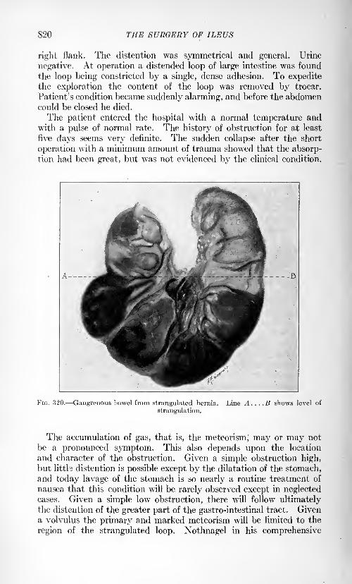
820 THE SURGERY OF ILEUS
right flank. The distention was symmetrical and general. Urine
negative. At operation a distended loop of large intestine was found
the loop being constricted by a single, dense adhesion. To expeditethe exploration the content of the loop was removed by trocar.
Patient's condition became suddenly alarming, and before the abdomencould be closed he died.
The patient entered the hospital with a normal temperature andwith a pulse of normal rate. The history of obstruction for at least
five days seems very definite. The sudden collapse after the short
operation with a minimum amount of trauma showed that the absorp-tion had been great, but was not evidenced by the clinical condition.
FIG. 320. Gangrenous bowel from strangulated hernia. Line A. . . .B shows level of
strangulation.
The accumulation of gas, that is, the meteorism,' may or may not
be a pronounced symptom. This also depends upon the location
and character of the obstruction. Given a simple obstruction high,but litth distention is possible except by the dilatation of the stomach,and today lavage of the stomach is so nearly a routine treatment of
nausea that this condition will be rarely observed except in neglectedcases. Given a simple low obstruction, there will follow ultimatelythe distention of the greater part of the gastro-intestinal tract. Givena volvulus the primary and marked meteorism will be limited to the
region of the strangulated loop. Nothnagel in his comprehensive
SYMPTOMS OF INTESTINAL OBSTRUCTION 821
Encyclopedia emphasizes the importance of the definite location of
meteorism in the diagnosis of mechanical obstructions.
Interesting as is the speculation as to the site of the obstruction,
as evidenced by local meteorism, and positive as the findings may be
in certain cases, yet this may be masked in so many ways that prac-
tically it is a refinement in diagnosis which in the acute cases is unes-
sential and in the chronic cases may be demonstrated more exactly
by the a>ray examination of the intestinal tract after a bismuth meal
or enema.
As to the degree, collapse varies, and for the same reason varies
the rate and the character of the pulse. As an index of the condition
of the patient the pulse, considering the rate as well as the character,
is more reliable than is any other single symptom. The rate is
usually increased, and as the absorption becomes profound the qualitybecomes correspondingly poorer. The thready, barely perceptible
pulse, with the cyanotic, cold extremities, both unmistakable evidence
of the terminal collapse, are in striking contrast to the sense of euphoria.This sense of well-being and the active, unclouded mind in the last
stages complete the typical ileus syndrome. An infection of the
peritoneum may result at this time from an actual perforation of the
obstructed intestine or from the permeation of the bacteria throughthe damaged wall of the gut. When this occurs the associated peri-
tonitis masks the primary obstruction and intensifies all symptoms.The blood-pressure follows, in general, the pulse change, showing the
primary riss and later a fall.
The temperature is inconstant. In the extreme cases it may be
subnormal, but, as a rule, it is slightly elevated. The white blood
count usually is increased, but in fulminating cases and in the late
stages of an obstruction there may be seen the same failure to react as
may be found in a spreading peritonitis. To illustrate how meagermay be the symptom-complex in intestinal obstruction I cite the
following case :
C. T., white male, aged twenty-two years, S. N. 2897. Twelvehours before entrance to the hospital the patient had been seized with
sudden abdominal pain and had vomited. The bowels were satis-
factorily moved twice by enemata. The pains had been cramp-like; had occurred at intervals of about one-half hour for periods of
five to ten minutes. Nine months previous to admission he hadbeen operated on for appendicitis. At entrance the temperatureand pulse were normal. The wr
hite blood cells, 9600. The abdomenwas not distended. There was no visible peristalsis. Because of
the recurring cramp-like pain and the history of a previous operationimmediate exploration was decided upon. At operation the lumenof the ileum was found to be obstructed by a single band of adhesions
just above the cecum. The intestine above this level was distended
and below collapsed. The adhesion was cut and the bowel below
immediately filled with the content from the obstructed intestine.
Uneventful recovery.
822 THE SURGERY OF ILEUS
The examination in this case was negative. From the historywere obtained three facts of importance: The patient had been
operated upon, he had been seized with severe abdominal pain whichhad recurred at intervals and he had vomited.
The urine becomes scanty, as might be expected with the extreme
dehydration. It may contain certain chemicals, such as phenol and
indican, but the presence or absence of these is rarely of importance,
though indican theoretically should be found in occlusion of the small
intestine and be absent in obstruction of the large.
Generally much stress is laid on the presence or absence of the
passage of feces or gas. With an acute obstruction a complete paral-
ysis of the intestinal movements may result below the level of the
obstruction. In my opinion the information obtained as to the
passage of feces or gas may be very misleading. Because they are
returned from the large bowel after an enema does not signify there
may not be an obstruction higher up. Not infrequently the propertreatment of cases which otherwise would have been correctly diag-nosed has been delayed because of the emptying of the bowel below
the level of the obstruction. The character of the fecal discharge
may be all but pathognomonic of certain conditions causing ileus.
They are intussusception, with the mucous or bloody mucous stool;
progressive narrowing of the lumen in cancer, with the increasing
constipation and blood-streaked movements alternating with diarrhea;
the rice-water discharges of mesenteric thrombosis and the absence of
lanugo hairs in the meconium of the newborn in congenital occlusion
of the intestine.
Diagnosis. Of first importance in considering the diagnosis of a
suspected case of intestinal obstruction is the history. This should
include the present acute attack and also a careful review of the
patient's previous illnesses and condition. The statement that a
hernia has existed in times past but is not now present may suggesteither an obstruction from a reduction en bloc or from a constricting
band. A previous laparotomy always suggests the possibility of an
obstruction from adhesions. This is especially true for the cases
which have been drained for a considerable time after the operation.
During ths period when drainage of the general peritoneal cavityfor spreading infections was attempted by multiple wicks and incisions,
surgeons had an opportunity to observe postoperative obstruction to a
degree unknown today. Likewise the history of an old inflammatory
process, such as an appendicitis or salpingitis, suggests, the possibility
of a constricting band. Recurring attacks of severe abdominal pain,
sudden in onset and suddenly relieved, may indicate that from a
twist of the intestine a partial volvulus has existed in the past or a
beginning intussusception which has been spontaneously reduced.
Recurring attacks of pain with increasing difficulty in moving the
bowels, especially if blood has been noted with the movements, points
strongly toward a progressive obstruction of the large bowel from
cancer. By such a careful history the possibility of poisoning from
DIAGNOSIS OF INTESTINAL OBSTRUCTION 823
lead or one of the ptomains would be at least indicated, as should
also be the evidences of an old specific process which had resulted in
a nerve lesion. A painstaking analysis of the acute attack should
disclose any possible causes for a reflex paralysis save the intra-
abdominal conditions other than acute mechanical obstruction, which
may cause a similar syndrome of symptoms. The superficial physicalexamination may be negative and fail to demonstrate any local con-
dition which might not be simulated by a simple colic or an acute
gastro-intestinal attack. The findings may still be negative even
after a most careful examination of all the possible sites of hernise,
together with a proctoscopic examination of the rectum and sigmoid;
but, as a rule, inspection of the abdomen in a good light will reveal
in the cases of mechanical obstruction either an extreme general dis-
tention or the local distention of an isolated loop or loops of intestine,
with evidence of hyperactive peristalsis. During this inspectionvisible peristalsis may be seen, particularly if the abdominal wall
has been flicked with the finger or a wet towel. If this sign is presentwith a normally thick abdominal wall the diagnosis of a mechanical
obstruction is positive.
Light percussion is extremely valuable in outlining the tympaniticareas as well as in localizing the areas over which light pressure causes
pain, and by palpation a mass may be outlined or a sense of deepresistance obtained which is very different from the protective muscle
spasm found over an acute inflammatory process.Auscultation may be of great value in determining not only the
presence or absence of intestinal movements, but also in locating the
region of an obstruction by the maximum intensity of these move-ments. The abdominal wall lacks the characteristic telltale protec-tive spasms to be found in inflammatory conditions save as the parietal
peritoneum may have become secondarily involved by such an inflam-
matory process. Pressure, as with simple colic, sometimes givesrelief. Even though extreme collapse may not have occurred and the
vomiting may not have reached the stage of persistent regurgitationso striking in the extreme conditions and the pulse and temperatureas well as the white count may be indeterminate, there is an inde-
scribable evidence of anxiety and tension which these patients show
that, coupled with the history and the examination, usually makesthe diagnosis relatively easy and positive.
Certain cases of intestinal obstruction should be considered indi-
vidually when discussing the diagnosis, so specific are their symp-toms. Congenital defects, such as an imperforate anus or an occlu-
sion of the small intestine, are evidenced either by a total absence
of the normal passage of meconium or the passage of an abnormalcontent for the newborn infant. With a volvulus the symptoms are
usually strikingly acute. The pain is not only that to be referred
to the intestinal contractions, but also to the torsion of and the tension
upon the mesentery. This mesenteric involvement may cause a
pain referred to the back. As the lesion is from the beginning asso-
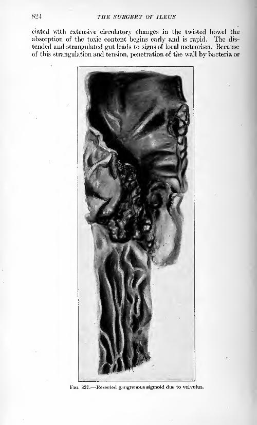
824 THE SURGERY OF ILEUS
ciated with extensive circulatory changes in the twisted bowel the
absorption of the toxic content begins early and is rapid. The dis-
tended and strangulated gut leads to signs of local meteorism. Because
of this strangulation and tension, penetration of the wall by bacteria or
FIG. 321. Resected gangrenous sigmoid due to volvulus.
DIAGNOSIS OF INTESTINAL OBSTRUCTION 825
gross perforation causing peritonitis is an early and a not uncommon
sequel.
Intussusception has a symptomatology which is unique. The strik-
ing features are that an infant rarely an older child or an adult
who has previously been well, vomits, cries out with sudden abdominal
pain and later passes a mucous or blood-tinged mucous stool. Asausage-shaped tumor may be palpated in the abdomen or a cervix-
like mass felt in the rectum by the examining finger. Still later the
abdomen may become distended and the collapse is extreme because
of the circulatory disturbance caused by the strangulation of the
invaginated intestine. The absence of the collapse in the first few
hours and the change in pulse-rate may be most misleading. When all
these signs and symptoms can be demonstrated the patient is usually
beyond surgical relief. A diagnosis should be made irrespective of
the profound collapse, and only after an examination under an anes-
thetic is the statement that no tumor is present justifiable. Specific
as is this symptom-complex the following case illustrates a possibleconfusion between intussusception and appendicitis.
J. B., negro, male, aged thirty-eight years, S. N. 2652< Patient
entered the hospital at seven o'clock in the evening with a historyof having been seized -at ten that morning with severe sharp abdom-inal pain. He had vomited. There had been no bowel movementsince the onset of the pain. The temperature at entrance was 100.2,
pulse 70, white blood cells 13,500. There was distinct tenderness on
deep palpation in the right lower quadrant. By rectum an indefinite
mass could be felt on the right side high up. The diagnosis of acute
appendicitis was made and immediate operation advised. At opera-tion a normal appendix was found and an ileocecal intussusception.This intussusception was reduced without undue trauma. The patientmade an uneventful recovery.
This case is atypical in that the patient was an adult, and there
had been no movement of the bowels.
Chronic intussusception generally seen in adults is like the diag-nosis of a chronic obstruction from new growth most difficult, beingcharacterized only by indefinite recurring attacks of abdominal pain,
possibly accompanied by vomiting and alternating constipation anddiarrhea.
When a complete obstruction is superimposed upon -a slowly
advancing occlusion caused by a new growth the symptoms so over-
shadow all that might have been noticed before that not infrequentlyno suggestion of any previous intestinal trouble can be elicited. With-out the demonstration of other probable causes of obstruction of the
large bowel in an individual beyond forty this may be assumed to be
malignant, so frequent is the large bowel the site of cancer. In con-
trast the studies of Fitz1 and of Leichtenstern2 show that in 90 percent, of all cases of strangulation the small bowel is involved. When
1 Osier's Practice of Med., Intestinal Obstruction.* Ziemssen's Handbuch, No. 1, vol. viii.
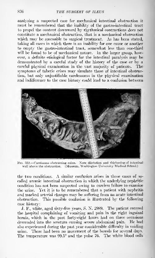
826 THE SURGERY Of ILEUS
analyzing a suspected case for mechanical intestinal obstruction it
must be remembered that the inability of the gastro-intestinal tract
to propel the content downward by rhythmical contractions does not
constitute a mechanical obstruction, that is a mechanical obstruction
which may be amenable to surgical treatment. As has been stated,
taking all cases in which there is an inability for one cause or another
to empty the gastro-intestinal tract, somewhat less than one-third
will be found to be of mechanical nature. In the larger group, how-
ever, a definite etiological factor for the intestinal paralysis may be
demonstrated by a careful study of the history of the case or by a
careful physical examination in the vast majority of patients. The
symptoms of tabetic crises may simulate those of intestinal obstruc-
tion, but only unjustifiable carelessness in the physical examination
and indifference to the case history could lead to a confusion between
FIG. 322. Carcinoma obstructing colon. Note dilatation and thickening of intestinal
wall above the obstruction. (Museum, Washington University Medical School.)
the two conditions. A similar confusion arises in those cases of so-
called uremic intestinal obstruction in which the underlying nephritic
condition has not been suspected owing to careless failure to examine
the urine. Yet it is to be remembered that a patient with nephritis
and marked arterial changes may be suffering from an acute intestinal
obstruction. This possible confusion is illustrated by the following
case history:J. F., white, aged sixty-five years, S. N. 2989. The patient entered
the hospital complaining of vomiting and pain in the right inguinal
hernia, which in the past forty-eight hours had on three occasions
descended into the scrotum causing severe abdominal pain. He had
also experienced during the past year considerable difficulty in voidingurine. There had been no movement of the bowels for several days.
The temperature was 99.5 and the pulse 76. The white blood cells
DIAGNOSIS OF INTESTINAL OBSTRUCTION 82?
were 9600 at entrance. The physical examination showed relaxed
inguinal rings, with a large left inguinal hernia which was readily
reduced. Heart sounds irregular and weak. Patient was drowsy.Urine showed a large amount of albumin and many hyaline and
granular casts. Blood-pressure 160. The patient was transferred
to the medical service with a diagnosis of chronic interstitial nephritis
and beginning uremia. Shortly after transfer the patient was sud-
denly attacked with severe abdominal pain and vomited. On the
right side, which had been negative at entrance, except for a relaxed
inguinal ring, appeared an irreducible inguinal hernia. The patientwas immediately sent to the operating room, and the hernial -sac
exposed. It contained a moderate amount of blood-tinged fluid anda strangulated loop of small intestine. The bowel was dark, but there
was no loss of luster and no question as to its viability. It wastherefore returned to the abdominal cavity and the hernia closed.
The patient made an uninterrupted recovery. After the operationthere was no vomiting. The amount of albumin in the urine decreased,
but it continued to be pathological.To ignore a possible reflex paralysis of the intestine in a patient
suffering with renal or hepatic colic would be to ignore a well-recog-
nized cause of intestinal paresia.Acute intrathoracic processes may also simulate an acute mechani-
cal obstruction. This is especially prone to occur when ther3 is
a diaphragmatic pleurisy. A positive differential diagnosis is par-
ticularly difficult when there is a central pneumonic process which
may give no physical signs. The picture of ileus from its intensity
overshadows at the first glance the true etiology, but the sharp rise
in temperature, which is never present at the beginning of the intes-
tinal obstruction and the increase in tjhe rate of respiration, togetherwith the facial expression, should indicate that the case is not one
of a mechanical obstruction of the bowel.
It may be impossible to differentiate sharply between the mechanical
obstruction and certain acute intra-abdominal inflammatory con-
ditions. A paresis of the bowels occurs with the spreading peritonitis
and may occur with a local process. This paresis associated with
peritonitis as has been stated, will be considered in detail in another
section. The typical protective spasm of the abdominal muscles whichis to be found with an inflammatory process is absent in the cases of
obstruction. The total absence of peristalsis may suggest a paresis
rather than an exhaustion from a mechanical obstruction. Thewhite count, except in the terminal stages of a spreading infection, is
higher, as a rule, than with the cases of mechanical occlusion, and the
amount of free fluid in the peritoneal cavity is greater with a primary
inflammatory process than with the obstruction. With the onset of a
peritonitis secondary to the obstruction all possibility of a differentia-
tion is lost except by the analysis of the case history and the progressof the disease. Torsion or strangulation of the mesentery or torsion
of a tumor pedicle may give a typical ileus syndrome. Trauma of
828 THE SURGERY OF ILEUS'
the testis or of the ovary may cause a reflex splanchnic paralysis.
Thrombosis of the mesenteric vessels causes primarily a paralysisof the affected segment of the gut. Until the signs of peritonitis
appear the condition is really that of an obstruction. In the second
type of case, as recognized by Reich1 in his study of mesenteric throm-
bosis, in which there are watery or blood-tinged fluid stools, a differ-
ential diagnosis may be made from a mechanical obstruction, but
nearly all of the cases of mesenteric thrombosis have been operated
upon with a diagnosis of acute mechanical obstruction.
Acute pancreatitis is another one of the acute intra-abdominal
inflammatory conditions in which a positive diagnosis can be made
only at operation. On opening the abdomen the finding of the pearl-
white areas of fat necrosis is pathognomonic. Acute dilatation of
the stomach also simulates an acute intestinal obstruction in the sud-
denness of the onset and the degree of the collapse, but the diagnosisof this condition is readily made by the use of the stomach-tube.
Treatment. A discussion of possible medical treatment is pertinent
only in a consideration of the cases of ileus without a mechanical
obstruction. For a mechanical obstruction of the bowel there is no
logical treatment other than operation for the relief of the obstruc-
tion. The failure to differentiate sharply between ileus caused by a
paralytic or spastic condition of the bowel and that caused by a
mechanical obstruction has led to confusion of ideas as regards the
possibilities of cure by non-operative means. In no other intra-
abdominal condition is the early diagnosis and proper treatment so
essential, if life is to be saved, as in these cases of acute mechanical
obstruction.
With a primary inflammatory process the natural protective mech-
anism may limit the progress of the inflammation. The resulting
paralysis of the intestine is without doubt an effort on the part of
Nature to limit the extent of the peritonitis. With a mechanical
obstruction there is no protective mechanism. Every reaction tends
to augment the absorption of the toxic content from the bowel byincreasing the tension within the lumen. As a first possible step in
relieving this tension within the gastro-intestinal tract the stomach
should be emptied, since with an empty stomach the overflow from
the intestine is favored.
Any attempt by cathartics to stimulate a peristalsis sufficiently
violent to overcome the obstruction is contra-indicated. In certain
types of obstruction, by the very violence of the peristalsis, the obstruc-
tion is made complete. In such cases a relaxation of the intestinal
tone from morphin or atropin might be theoretically beneficial, but
by the use of opiates the symptoms may be dangerously masked.
Drugs should not be given until a positive diagnosis has been made and
a definite course of treatment determined upon. The usual heart
stimulants, caffein, camphor or strychnin, are of questionable value.
1 Ergeb. d. Chir. u. Orth., 1913, xii, 515.
TREATMENT OF INTESTINAL OBSTRUCTION 829
Except for those cases which are seen before there has been anyconsiderable absorption these patients are toxic and many of themon the verge of collapse, even though there may be little or no evidence
of this from the pulse and temperature. Therefore, every effort must
be made to reduce to a minimum the operative shock. When it is
possible a local anesthetic is always to be preferred to a general:
(1) Because of the elimination of the depression of the general anes-
thetic, and (2) to minimize the danger of inhalation of the regurgitantstomach content while the protective reflexes are lost. Except in
children and in the very unusually neurotic patients a local anesthetic
should always suffice for all operations on strangulated hernise. For
the exploration of the peritoneal cavity a general anesthetic will be
necessary, but this may with advantage be supplemented by usingthe local for the section of the abdominal wall. The choice in the
general anesthetic, provided it is not chloroform, may be left to the
preference of the individual operator. Personally I prefer gas and oxy-
gen with an amount of ether sufficient only to afford proper relaxation.
Since the dehydration in these cases may be extreme, active meas-
ures should be instituted to replace this loss. A subpectoral infusion
of 1000 c.c. normal salt solution before the operation is an admirable
preparation. Transfusion of blood is without special effect in these
cases, just as it is in other cases toxic from infections. The operationmust be performed with a minimum amount of trauma and without
hemorrhage. Excessive trauma of the normal intestine may cause
a paralysis which exists for a considerable time. Even moderatetrauma in these cases of obstruction may cause a serious paresis,
and trauma also tends to increase the amount of absorption from the
lumen. With the extreme distention of a considerable portion of
the intestine or with a large distended loop from a strangulation or a
volvulus the avoidance of excessive manipulation is most difficult.
To facilitate this aspiration of the bowel content after a most careful
preparation to prevent soiling of the peritoneum may change a diffi-
cult or impossible operation into a relatively easy one. The insertion
of a tube into the coils of intestine for the withdrawal of the content,as has been advocated by some, is I believe, a most dangerous pro-cedure. By this procedure the mucous membrane is still further
traumatized, a more rapid absorption of the content is made possible,
and it is inconceivable that all of the toxic content can be removed.
Apprehension has been felt lest there be absorption of this toxic
content after it had passed into the intestine below the obstruction.
This fear is groundless. No absorption of this toxic substance fromthe bowel lumen occurs with a normal mucous membrane.
Probably no single manipulation in these cases adds so greatlyto the operation shock as evisceration. If evisceration is necessarythe intestine must be most carefully protected by Hot compresses.The operator who will judiciously aspirate distended loops and exer-
cise due care in his exploration will find it rarely necessary to eviscerate
any considerable part of the intestine.
830 THE SURGERY OF ILEUS
Bearing in mind that operations in these cases are life-savingmeasures on patients with very little reserve, a lateral anastomosisbetween the distended and constricting intestine, as near as possibleto the site of the obstruction, may be successful when a prolonged
operation for the removal of the obstruction would have been fatal.
Prolongation of an operation beyond the absolute minimum of time
necessary for relieving the obstruction is unjustifiable. The value
of an enterostomy above the level of the obstruction has been muchdiscussed and many operators are of the opinion that it is a valuable
procedure. Provided the mechanical obstruction has been removedI question if the bowel content will drain any more rapidly throughthe enterostomy opening than into the normal intestine. The drain-
age of any considerable part of the intestinal tube is possible onlywith a return of the peristaltic activity of the bowel. With a return
of this propelling power the content will be forced into the normalintestine and be advanced by the action of the gut below the obstruc-
tion. In consequence, only, with a paralysis from the strangulationof a loop in which the viability may be questioned or a prolonged
paresis feared is enterostomy drainage indicated, and then only with
the purpose of draining that immediate loop. Enterostomy has a
particular value in cases of volvulus when the prolonged strangula-tion may tend to a paralysis of that part of the bowel which has been
twisted.
No other operative problem in surgery requires greater judgmentor greater skill than the successful treatment of a loop of bowel whichhas been strangulated. The temporary replacement within the
abdominal cavity or protection with warm compresses may cause a
rapid return to normal in the appearance of the gut. In the frank
cases in which the luster of the intestine has been lost and when there is
evident thrombosis of the mesenteric vessels with beginning gangreneof the bowel wall, there is no alternative but to resect. With the
questionable cases, however, especially if the strangulation has
involved only a limited extent of bowel, the danger of later necrosis
is not to be compared with the incomparably greater danger of imme-diate resection, particularly if the general condition of the patientis not good. These questionable loops should be returned to the
'abdominal cavity and protected by wrapping the omentum around
them. By this procedure a limited amount of the intestinal wall
may retain its viability when it seemed lost. In such cases, drainageto the region of the damaged loop may be indicated.
When resection is imperative the method of operating must varywith the experience and skill of the operator. As routine the two-step
operation will make it possible to save cases in which the completed
operation of section and resuture of the bowel would be fatal. In
the cases of simple obstruction so toxic from the continued absorption
that a removal of the obstruction or a lateral anastomosis around
the obstruction is not possible, the distended intestine should be
drained by an enterostomy tube. In the cases in which a section
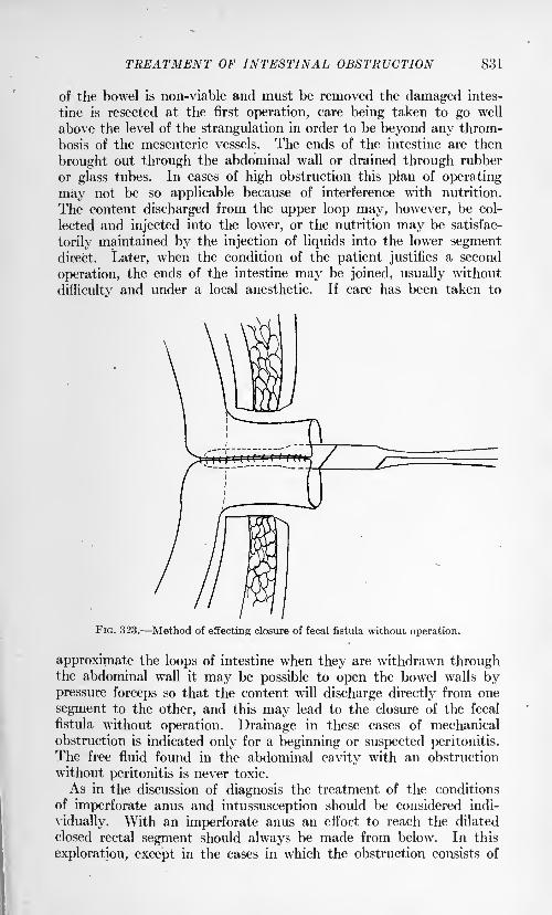
TREATMENT OF INTESTINAL OBSTRUCTION 831
of the bowel is non-viable and must be removed the damaged intes-
tine is resected at the first operation, care being taken to go well
above the level of the strangulation in order to be beyond any throm-
bosis of the mesenteric vessels. The ends of the intestine are then
brought out through the abdominal wall or drained through rubber
or glass tubes. In cases of high obstruction this plan of operating
may not be so applicable because of interference with nutrition.
The content discharged from the upper loop may, however, be col-
lected and injected into the lower, or the nutrition may be satisfac-
torily maintained by the injection of liquids into the lower segmentdirect. Later, when the condition of the patient justifies a second
operation, the ends of the intestine may be joined, usually without
difficultv and under a local anesthetic. If care has been taken to
FIG. 323. Method of effecting closure of fecal fistula without operation.
approximate the loops of intestine when they are withdrawn throughthe abdominal wall it may be possible to open the bowel walls bypressure forceps so that the content will discharge directly from one
segment to the other, and this may lead to the closure of the fecal
fistula without operation. Drainage in these cases of mechanical
obstruction is indicated only for a beginning or suspected peritonitis.
The free fluid found in the abdominal cavity with an obstruction
without peritonitis is never toxic.
As in the discussion of diagnosis the treatment of the conditions
of imperforate anus and intussusception should be considered indi-
vidually. With an imperforate anus an effort to reach the dilated
closed rectal segment should always be made from below. In this
exploration, except in the cases in which the obstruction consists of
832 THE SURGERY OF ILEUS
a relatively thin diaphragm in the anal canal, the dissection must be
continued in the medial line posteriorly and upward toward the hollow
of the sacrum. If the distended rectal pouch is not readily reached
it should be remembered that a fistulous opening through the vaginain girls or an artificial anus in either sex is to be preferred to a pro-
hibitively long operation on those young children in whom operative
shock, especially if the loss of heat has not been guarded against,is great. The rare recovery effected by the sloughing into the bowel
of the intussusception is the exception like the perforation of a strangu-lation of a part of the gut wall in hernise, to emphasize the rule,
"Always operate for intussusception, and at once, as soon as the
diagnosis is made."The statistics of Clubbe1
emphasize most graphically the effect
of time upon the prognosis in intussusception in children. In his
series of cases the average time from the onset of symptoms to opera-tion was in the first 50, in the cases that lived, twenty-four hours;and in the cases that died, sixty-eight hours; in the second 50, in the
cases that lived, twenty-three hours, and in the cases that died, forty-
eight hours. To delay the diagnosis into the second twenty-four-hour period is to ensure almost a fatal result.
Opinions differ as to the wisdom of attempting in these cases to
reduce the intussusception by the distention of the bowel by warmoil or salt solution enemata. Unquestionably, intussusceptionshave been reduced by this method. To me it has always seemed to
be an uncertain and dangerous procedure. The mechanism of the
procedure of intussusception must be remembered in operations
attempting its reduction. Pressure upon the apex of the intussus-
ceptum, with gentle taxis, will almost always be effective when a
direct pull upon the bowel will cause a rupture of the peritoneal
coats or possibly a complete tear across the wall. Every effort mustbe made to reduce the intussusception rather than to resect the entire
mass, so prohibitively high is the mortality after these extensive
operations. If a resection is necessary the same procedure as described
for the treatment of the bowel after resections for strangulation maybe used to advantage. When any cause for the intussusception can
be demonstrated this must be remedied.
Following ths operation in every case of intestinal obstruction the
salt solution should be continued subpectorally or tap water or salt
solution given per rectum by any one of the accepted methods. Bythe drop method of the late John B. Murphy enormous quantities of
liquid may be introduced into and absorbed from the rectum. Bythe use of small enemata, not to exceed 300 to 400 c.c., given into
the rectum every three to six hours, an ample amount of fluid is
supplied. The essential in any method of proctoclysis is not to
overdistend the bowel. When the rectum becomes irritable deodor-
ized tincture of opium should be added to the enema. A sufficient
1 British Med. Jour., June 17, 1905.
PARALYTIC ILEUS 833
amount of morphin should be given to relieve the patient from excru-
ciating pain. The paralyzing effect upon the intestine, so dreaded bysome, need not be apprehended with the intelligent use of moderate
amounts of this drug. Attempts to simulate peristalsis by cathartics
are, as a rule, absolutely ineffectual. Unless the use of enemata is
contra-indicated by some local condition of the large bowel some one
of the stimulating enemata should be used to empty this section of
the gastro-intestinal tract. Cathartics are indicated and effective
only after the intestine has recovered its normal tone.
The position of the patient should be that affording the greatestcomfort. This is usually a moderately high bed rest. Especially is
this position acceptable if the distention embarrasses in any way the
movements of the diaphragm.Spastic Ileus. A spastic contraction of the muscles of the gastro-
intestinal tract, together with edema of the tissues, may so constrict
the lumen of the bowel that intestinal obstruction results. This
condition, called spastic or dynamic ileus, is so constantly referred
to that it seems unwise not to mention it in a discussion of intestinal
obstruction. Although an actual obstruction of the intestinal lumendoes occur and the ileus syndrome is simulated, these cases should
not be considered primarily as cases of intestinal obstruction. Free-
man 1 describes the appearance of the intestine in a patient who hadan operation for the symptoms or acute intestinal obstruction. Asegment of ilium, five or six inches in length, was contracted to the
point which it appeared bloodless. The bowel proximal to the con-
tracted segment was distended and the bowel distal to the contrac-
tion was collapsed. The contracted segment relaxed after the
abdomen was opened. The patient recovered without further symp-toms. Freeman believes this condition is to be regarded as a reflex
phenomenon, and that it is more frequent than generally supposed.
Payr2 believes that these localized spastic contractions of the intes-
tines are the result of embolism of the mesenteric vessels. The con-
dition is most commonly associated with poisoning from lead or
a ptomain. In an experimental study of the effect of lead-poison-
ing, Harnack3emphasizes the important role which the circulatory
change plays in the production of the edema of the gut wall. The
pain is attributed to violent contractions of the intestinal muscles.
The diagnosis of this condition depends essentially upon the establish-
ment of the etiology. The combination of pain, vomiting and col-
lapse may simulate closely the results of a mechanical intestinal
obstruction. The treatment is that indicated for the special toxin.
Paralytic Ileus. A paralysis of the intestine may result from con-
ditions affecting the intestine directly, as trauma or intra-abdominal
infection, or from reflex paralysis of the motor mechanism, or as a
part of a severe general systemic depression. This condition of acute
paralysis of the intestine is to be differentiated sharply from the
1 Ann. Surg., 1918, Ixviii, 196. * Quoted by Freeman.3 Arch. f. exp. Path., vol. ix.
VOL. ii 53
834 THE SURGERY OF ILEUS
so-called atonic inertia, which is associated with a general ptosis.
Clinically, acute paralytic ileus forms more than two-thirds of all
cases which present the ileus syndrome. It is in this group of cases
in which recoveries without operation are reported and it is the clini-
cal experience in this group of cases which has led to 'the dangerousdoctrine of delay in cases of mechanical obstruction in the hope that
recovery may take place without operation.Of the causes of acute paralytic ileus the most frequent is a spread-
ing intra-abdominal infection. The paralysis resulting from a spread-
ing intraperitoneal infection might be considered as the result of the
general systemic depression, but it is so characteristically associated
with infection within the abdominal cavity and plays such an impor-tant role in the localization of this infection that it seems more likely
to be the result of a direct influence on the intestinal motor mechanism.As was stated previously this phase of intestinal obstruction will be
dealt with in another section.
Etiology. The studies of Magnus,1
Bayliss and Starling2 and
Cannon3 have demonstrated that the movements of the gastro-intes-tinal tract are controlled by intrinsic and extrinsic nerves. Theessential part of the intrinsic nervous mechanism is formed by Auer-
bach's plexus, which lies between the circular and longitudinal coats.
Governed by this intrinsic innervation, the intestine will perform the
normal movements of rhythmic contraction though completelyisolated from the central nervous system. The intrinsic nervous
mechanism may be temporarily interfered with by a section of the
nerves connecting it with the central nervous system, but later the
normal intestinal movements are resumed. The extrinsic innerva-
tion of the intestine is received through the vagi, the sympatheticand the sacral autonomic systems. In general the sympathetic
system tends to inhibit and the vagus and sacral nerves to stimulate
gastro-intestinal movements. Theoretically, then, a paralysis of the
gastro-intestinal tract may result from an abnormal overactivity of
the sympathetic system or an abnormal inactivity of the vagi or
sacral nerves.
Pathology. The pathology depends upon the etiology. Inflam-
matory reaction or interference with the nutrition of the intestine
may interfere with the intrinsic innervation; lesions of the central
nervous system may interfere with the extrinsic nerves. Severe
general depression may so reduce the muscle tone of the intestine
that normal movement is inhibited. The paralysis resulting from
the manipulation of the bowel or from the trauma secondary to incar-
ceration or strangulation of the intestine is evidently the result of
interference with the intrinsic nervous mechanism. This was demon-
strated experimentally by the observations of Cannon and Murphy.4
1 Arch. f. d. ges. Physiol., 1904, vol. ciii; 1906, vol. cxi.
2 Jour. Physiol., 1899, vol. xxiv; 1900, vol. xxvi.3 Mechanical Factors of Digestion.4 Ann. Surg., 1906, vol. xliji; Jour. Am. Med. Assn., 1907, vol. xlix.
PARALYTIC ILEUS 835
They found that the intestine could be paralyzed for variable periods
of time, depending upon the degree of trauma inflicted, and that this
paralysis was not influenced by section of the connections between
the intrinsic nervous mechanism and the central nervous system.Lesions in the mesentery may affect both the intrinsic and extrinsic
nerves. Thus, for example, following mesenteric thrombosis, there
is primarily a paralysis of the portion of intestine which is deprived of
its nutrition, and, secondly, a paralysis of other parts of the intestine
due to the effects of the spreading infection. Any lesion which
interferes with the vagi or a lesion of the spinal cord which interferes
with the sympathetic or sacral autonomic system interferes directly
with the .extrinsic nerve control. Reflex inhibition of peristalsis,
which is received by way of the splanchnic nerve, is usually associated
with sudden severe pain, as, for example, that of hepatic or renal colic
or following trauma to the testis or ovary or as the result of torsion
of the omentum or a tumor with a pedicle. Trauma of the abdominal
wall may also cause complete reflex inhibition of intestinal movements.
Overwhelming acute infections, such as acute hemorrhagic pancrea-
titis, may also cause cessation of intestinal movements. The intestinal
paralysis of typhoid, uremia and other general depressive conditions
is to be attributed to general loss of muscle tone.
Symptoms. In all these conditions of acute paralysis of the intes-
tine the most striking symptoms may be those of ileus, yet the general
appearance of the patient, as a rule, guides the careful observer awayfrom error. Save for cases of mesenteric thrombosis or overwhelmingintra-abdominal infection the extreme collapse seen in true intestinal
obstruction is never found. The distention, though it be extreme,
is characterized by flaccidity. Visible peristalsis is not present, andeven auscultation fails to demonstrate any evidence of peristalsis.
Diagnosis.- The diagnosis consists primarily in eliminating the
possibility of a mechanical obstruction and recognizing the primarycause of the paralysis. The patient's history must be considered
most carefully for any possible etiology. If there has been direct
trauma to the abdomen the differential diagnosis is between a reflex
paralysis of the intestine and a rupture of an intra-abdominal organ.Given such a problem an, experienced surgeon will prefer to explorerather than speculate about the intra-abdominal condition, because
of the great danger of delay and the relative safety of exploration.If the symptoms of acute paralytic ileus develop after an intra-abdom-
inal operation the paralysis may be due to trauma to the intestines
or mesentery or may be the result of a spreading intra-abdominal
infection. Fortunately, postoperative paralytic ileus is not now
frequently seen. The technic of handling the intestines has been
perfected and also modern aseptic methods have practically elimin-
ated the danger of intra-abdominal infection. An appreciation of
the physiology of the nervous control of the intestinal tract explains
the paralytic distention following an injury to the spinal cord. Thesame is true of the distention, associated with renal or hepatic colic
836 THE SURGERY OF ILEUS
or trauma to the testis. With such intra-abdominal conditions as
torsion of the omentum or twisting of a tumor pedicle the differential
diagnosis may not be possible save by exploration, a step necessaryin the treatment of either condition. Acute intrathoracic diseases,
especially pneumonia of the lower lobes or a pleuritis of the dia-
phragmatic pleura, may be associated with reflex intestinal paralysis,
which is explained only after the recognition of the intrathoracic
disease.
Treatment. The first and most important step in the treatment of
acute paralytic ileus is to remove the cause of the paralysis. Symp-tomatic treatment may then be carried out as indicated. The actual
effect of drugs may be questioned. With interference .with the
intrinsic nervous mechanism, local irritants may help. Atropin mayprevent inhibition. Eserine and the recently introduced glandularextracts are of questionable value; in short, there is no specific treat-
ment for acute paralytic ileus. When the disturbed mechanism of
the innervation is reestablished or when the system has recovered
from the effect of general depression, peristalsis automatically be-
comes normal. The failure to treat these conditions correctly is
usually due to a failure to appreciate the fact that the condition is
a paralysis and not a true obstruction, or to the placing of too great
reliance on the efficacy of treatment by drugs. Much more reliance
should be placed in the judicial use of the stomach-tube, enemata and
application of hot turpentine stupes directly to the abdomen. Jutte 1
recommends the use of transduodenal lavage with a warm hypertonicsalt solution by means of the duodenal tube. He believes that such
a procedure is a stimulus to peristalsis of the entire intestinal canal.
Theoretically the administration of morphin is to be avoided, but
practically if the patient is restless a sufficient dose of morphin given
hypodermically, to give the patient a few hours complete rest^ often
leads to an immediate resuming of the normal intestinal peristalsis.
1 Jour. Am. Med. Assn., 1919, Ixxii, 929.
INDEX
ABBE'S string method of treatment of
cicatricial stenosis of esophagus, 72
Abdomen, examination of, in diagnosis-of diseases of stomach, 567
in splenic anemia, 523Abdominal aorta, aneurysm of, diagnosis
of, from cysts of pancreas, 485palpation in pyloric stenosis, 636tenderness in gastric ulcer, 622
in ulcer of duodenum, 695tumor in cancer of stomach, 671
in pyloric stenosis in infancy,602
wall, abscess of, diagnosis of, fromcysts of pancreas, 485
Abnormal involution of breast, 157Abnormalities of pancreas, 437Abscess of abdominal wall, diagnosis of,
from cysts of pancreas, 485of breast, 150of cavity of great omentum, 802of esophagus, 46of liver, 372
complications of, 374
diagnosis of, x-rays in, 375fever in, 373
leukocytosis in, 373Muhlman's operation in, 375pain in, 373
symptoms of, 373treatment of, 375
of omentum, 792of pancreas, diagnosis of, from cysts,
485
pulmonary, 274chronic simple, bronchiectasis
and, treatment of, 276
diagnosis of, 274
x-rays in, 274
prognosis of, 276
putrid, bronchiectasis and, 277resection of lung in, 275treatment of, 275
of spleen, 517
diagnosis of, 518
pathology of, 517
prognosis of, 518
symptoms of, 518treatment of, 518
of subpleural connective tissue, 249of thoracic wall, 243
diagnosis of, 246
Abscess of thoracic wall, symptoms of, 245treatment of, 246
Accessory pancreas, 437Ach's esophageal extractor, 90
Achylia gastrica, diagnosis of, fromcancer of stomach, 685
Acidity, gastric, 579tests for, 580
Actinomycosis of breast, 155of liver, 376of thorax, 249
Adenocarcinoma of breast, 177of stomach, 662
Adenofibroma of breast, 165Adenosarcoma of breast, 167Adhesions in gastric ulcer, 632
Air-tight thoracic drainage after thoracic
operations for carcinoma of esophagus,106, 107
Amastia, 141
Amazia, 141
Anemia, pernicious, 539blood in, 543
diagnosis of, 540from splenic anemia, 525
etiology of, 539
hemolysis in, 543
pathology of, 540
splenectomy in, 544transfusion of blood in, 542treatment of, 541
splenic, 520abdomen in, 523adult type of, in children, 527anemia in, 524ascites in, 524cardiac symptoms in, 524cholelithiasis and,,521
diagnosis of, 525
etiology of, 521fever in, 524hematemesis in, 523
Hodgkin's disease and, 522incidence of, 522of infancy, 528
pain in, 523
pathology of, 522renal complications in, 524
splenectomy in, 527
symptoms of, 523treatment of, 526
in ulcer of duodenum, 690, 694
Anemias, grave, diagnosis of, from cancerof stomach, 681
(837)
838 INDEX
Anesthesia in thoracic surgery, 208
Aneurysm of abdominal aorta, diagnosisof, from cysts of pancreas, 485
aortic, compression stenosis of
esophagus and, 60of hepatic artery, 384
traumatic, fractures of ribs and, 226
Angioma of breast, 173of liver, 377of spleen, 516
Angustatio ventriculi, 600Annular pancreas, 437Anoci-association in intrathoracic sur-
gery, 209Anomalies of esophagus, congenital, 49
of stomach, congenital, 599Anterior gastro-enterostomy, 752
Aorta, abdominal, aneurysm of, diag-nosis of, from cysts of pancreas, 485
Aortic aneurysm, compression stenosis
of esophagus and, 60
Apoplexy, pancreatic, 499
Appetite, diagnosis of diseases of stomach
and, 566in gastric ulcer, 621in ulcer of duodenum, 694
Arteriosclerosis, acute pancreatitis and,455
Ascites in carcinoma of pancreas, 496in splenic anemia, 524
Athelia, 141
Atresia of duodenum, 704of stomach, 599
Atrophy of breast, 147of stomach, 703
B
BACTERIOLOGY of duodenum, 704of stomach, 704
Balfour-Gossit retractor, 91Banti's disease, 520. See Anemia,
splenic.von Bechterew's disease, 202Beck-Jianu operation for carcinoma of
esophagus, 121Beck's bismuth paste in treatment of
empyema, pleuraland pulmonary fis-
tulse, 285of wounds of dia-
phragm, 329
Belching of sour gas in ulcer of duode-
num, 693
Benign pylqric stenosis, 633Sevan's incision in splenectomy, 550
Beyea's method of gastropexy, 743Bile ducts, carcinoma of, 408
direct implantation of, 423inflammation of, 393
operations on, 410reconstruction of, 422
Biliary fistula, 407Blood in cancer of stomach, 674
Blood in gastric ulcer, 624, 625in Gaucher's disease, 532in hemolytic jaundice, 536
"occult," diagnosis of diseases of
stomach and, 580, 591in pernicious anemia, 543
supply of spleen, 503in syphilis of stomach, 655transfusion of, in pernicious anemia,
542in wounds of chest, 324
Blood-pressure, intrathoracic surgeryand, 206
Bradycardia in injuries of liver, 362Brauer's overpressure apparatus in intra-
thoracic surgery, 214
Breast, abnormal involution of, 157abscess of, 150
treatment of, 152
actinomycosis of, 155adenocarcinoma of, 177adenofibroma of, 165adenosarcoma of, 167
anatomy of, 139
angioma of, 173
atrophy of, 147
broken, 150carcinoma of, 174
classification of, 176
colloid, 178
diagnosis of, 183
etiology of, 174
growth of, 180
prognosis of, 184
symptoms of, 180treatment of, 186
drainage in, 192
exploratory operations in,
193
palliative operations in, 194radium in, 186skin incision in, 186
supraclavicular dissection
in, 192
x-rays in, 186contusions of, 143
cystosarcoma phyllodes of, 167
proliferum of, 167
cysts of, 157
cancer, 179due to new growths, 163
development of, 139diseases of, cystic, 157
treatment of, 162
functional, 146
inflammatory, 149echinococcus of, 155enchondroma of, 173fibre-adenoma of, 165
fibrocystadenoma of, 171
fibroma of, cystic, 165
fibromyxoma of, 165
foreign bodies in, 143
hematpma of, 143
hysterical, 146
hypertrophy of, diffuse, 147
INDEX 839
Breast, hypertrophy of, infantile, 148senile parenchymatous, 157
injuries of, 143intracanalicular papillary fibroma
of, 165
lipoma of, 173
male, cysts of, 197diseases of, 196
injuries of, 196malformations of, 196tumors of, 197
malformations of, 141
neuralgia of, 146osteoma of, 173
papillary cystadenoma of, 169
papilloma of, villous, 169sarcoma of, 173
cystic, 167
syphilis of, 153tuberculosis of, 154tumors of, 163
fibre-epithelial, 165, 168
non-indigenous, 172wounds of, 143
"Broken" breast, 150
Bronchi, foreign bodies in, 277
bronchoscopy for, 278mediastinal bronchotomy
for, 280
Bronchiectasis, 285
"collapse" therapy in, 287
diagnosis of, 286
x-rays in, 286
pneumotomy in, 287
pulmonary abscesses and, 277treatment of, surgical, 287
Bronchoscopy for foreign bodies in
bronchi, 278
Bronchotpmy, mediastinal, for foreignbodies in bronchi, 280
Briining's illuminating handle for esoph-agoscope, 34
saliva aspirator, 35
Burns, duodenal ulcer and, 764
CALCULI, pancreatic, 488
diagnosis of, 490x-ray in, 490
etiology of, 489
glycosuria in, 490pain in, 489
symptoms of, 489treatment of, 491
vomiting in, 489Callman's sound, 32Cancer cysts of breast, 179
duct, 169of stomach, 658, 708
abdominal tumor in, 671
age and, 659blood in, 674cachexia in, 671
Cancer of stomach, changes in stomachproduced by, 664
colloid, 662
complications of, 685course of, 646definition of, 658
diagnosis of, 679from achylia gastaica, 685from chronic constipation,
685from foreign bodies, 685from gall-bladder disease,
681from gastric granuloma,
685from grave anemias, 681from lues, 685from tuberculosis, 685from tumors of kidney, 685
of large intestine, 685of omentum, 685
enteritis in, 686
fibrous, 662fistula in, 686gastric acidity in, 672
contents in, microscopicexamination of, 674
ulcer and, 661
gastroscopy in, 679
gelatinous, 662 . ,
habits and, 660
hemorrhage in, 669
heredity and, 659
"hour-glass" in, 686
hygiene and, 661incidence of, 659
jaundice in, 686location of, 663malaria and, 661
medullary, 662metastases in, 665morbid anatomy of, 661
mucoid, 662nature of, 646obstruction at cardia in, 685
occupation and, 660
operations for, centra-indica-
tions to, 709
exploratory incision in, 709
gastrectomy, 710
complete, ,718
partial, 711
subtotal, 718
gastro-enterostomy, 709
gastro-esophagostomy, 710,719
gastrostomy, 711indications for, 709
jejunostomy, 711
pylorectomy, 711
origin of, 646
perforation in, 686
pyloric obstruction in, 685race and, 659rheumatism and, 661
roentgen examination in, 677
840 INDEX
Cancer of stomach, scirrhus, 662
secondary, 663sex and, 659
signs of, 665social status and, 660symptoms of, 665
syphilis and, 661test-meal in, 672thrombosis in, 686trauma and, 660treatment of, 708tuberculosis and, 661
ulcerating, 662
vomiting in, 670Cancerous mastitis, 179Carbuncles of chest-wall, 240Carcinoma. See also Cancer,
of bile-ducts, 408of breast, 174
classification of, 176
colloid, 178
diagnosis of, 183
etiology of, 174
growth of, 180
prognosis of, 184treatment of, 186
of esophagus, 78
esophagoplasty for, 85, 117, 119occurrence of, 78
. posterior mediastinotomy for,88
preliminary gastrostomy for, 89resection for, 83, 87
of cardia for, 107
symptoms of, 79transthoracic operation for, 89treatment of, 80
operative, palliative, 117
radical, 82
type of, 78of gall-bladder, 408of liver, 377
secondary, 380treatment of, 377
of lung, 256of omentum, 795of pancreas, 494
ascites in, 496
diagnosis of, 494edema in, 496
extirpation in, 497
glycosuria in, 496
jaundice in, 495
pain in, 495
symptoms of, 494treatment of, 497
operative, 499of pleura, 255
diagnosis of, 257
symptoms of, 257of stomach, 658. See Cancer of
stomach.
Cardia, resection of, for carcinoma of
esophagus, 107Cardiac symptoms in splenic anemia, 524
Cardiodilator, Einhorn's, 53
Cardiodilator, Plummer's hydrostatic,53
Cardioplasty in treatment of spasticstenosis of esophagus, 57
Cardiospasm, 50
cardioplasty for, 57
diagnosis of, 51
espphagogastrostomy for, 58
etiology of, 51treatment of, 52
Carmin test in pyloric stenosis in infancy,602
Catarrhal cholecystitis, acute, 393
Chest, deformities of, 199
congenital, 199
phlegmon of, 242wounds of, 313
penetrating, 315
collapse of lung in, 316gangrene of lung and, 326
hemoptysis in, 315
operations for, 317pneumothorax in, 316
signs of penetration in, 315streptococcic infection in,324
transfusion in, 324
x-rays in, 316
superficial, 314
Chest-wall, carbuncles of, 240cold abscess of, 243
diagnosis of, 246treatment of, 246
cysts of, echinococcus, 251enchondroma of, 254furuncles of, 240sarcoma of, 254
suppurations of, 240tumors of, 253
Chicken-breast, 201
Chicken-pox, acute pancreatitis and, 447
Cholecystectomy, 418
Cholecystenterostomy, 421
Cholecystgastrostomy, 421
Cholecystitis, 393acute catarrhal, 393
complications of, 402
diagnosis of, 401
pain, 401
pancreatitis and, 447
symptoms of, 401treatment of, 405
chronic, 395treatment of, 406
complications of, 400
etiology of, 398
suppurative, 394, 395
Cholecystostomy, 416
Cholecystotomy of Bernay's, 416
Choledocho-enterostomy, 422
Choledochotomy, 419
retroduodenal, 420holedochus cyst, congenital idiopathic,390
holelithiasis, 402. See also Gall-stones,
acute pancreatitis and, 447
INDEX 841
Cholelithiasis, heart in, 403
jaundice in, 403
pain in, 403
splenic anemia and, 521
symptoms of, 402
Chondrectomy in treatment of emphy-sema of lungs, 290
Chondrosternal dislocations, 228
Chondrotomy in treatment of congenital,
rigid and dilated thorax, 291Cicatricial stenosis of esophagus, 67
treatment of, 68
operative, 70
Cirrhosis, Hanot's, diagnosis of, fromsplenic anemia, 526
of liver, 369, 546
diagnosis of, from splenic
anemia, 526
pathology of, 369
splenectomy in, 547treatment of, 370
of stomach, 707
gastrectomy for, 707
x-rays in diagnosis of, 707
Coffey's method of gastropexy, 743Cold abscess of thoracic wall, 243
Collapse of lung in wounds of chest, 316
therapy in treatment of bronchiec-
tasis, 287Colloid carcinoma of breast, 178
of stomach, 662"Combined" acidity, diagnosis of dis-
eases of stomach and, 580Commotion of thorax, 221
Compression stenosis of esophagus, 59of thorax, suffocative, 221
Concretions of pancreas, 488
Congenital anomalies of esophagus, 49of gall-bladder, 389of stomach, 599, 704
deformities of chest, 199of ribs, 199
diaphragmatic hernia, 606elevation of diaphragm, 605
enlargement of stomach, 603
faulty position of stomach, 603
funnel-chest, 201
hypertrophic pyloric stenosis, 600,773
idiopathic choledochus cyst, 390malformations of pancreas, cysts
and, 476
narrowing of superior thoracic aper-
ture, 202
presternal cyst, 253
pyloric stenosis, 725, 773
rigidity of costal cartilages, 202
spastic pyloric stenosis, 773thoracic rigidity and emphysema,
202
Constipation in chronic pancreatitis, 470
diagnosis of, diseases of stomach
and, 566in gastric ulcer, 621in pyloric stenosis in infancy, 601in ulcer of duodenum, 694
Contracted stomach, 600, 703Contraction of thorax, 204Contusions of breast, 143
of thorax, 220Costal cartilages, dislocations of, 228
fractures of, 227
prognosis of, 228
symptoms of, 228treatment of, 228
rigidity of, 202Costochondral dislocations, 228Costovertebral dislocations, 228Cracks of nipple, 144
Crump's stilette, 38Cruveilhier's deformity of ribs, 201
Cyanosis in acute pancreatitis, 453
Cyst, choledochus, congenital idiopathic,390
Cystadenoma of breast, 157
papillary, 169
diagnosis of, 171treatment of, 171
of pancreas, 493
Cystic fibroma of breast, 165
lymphangioma of omentum, 794
mastitis, chronic, 157
neoplasms of pancreas, 476
omentum, 794sarcoma of breast, 167
of omentum, 796
Cystocotomy, 417
Cystosarcoma phyllodes of breast, 167
proliferum of breast, 167
Cysts of breast, 157
cancer, 179of cavity of great omentum, pan-
creatic, 802
sanguineous, 802of chest-wall, echinococcus, 251
congenital presternal, 253of kidney, diagnosis of, from cysts
of pancreas, 486of liver, echinococcus, diagnosis of
from cysts of pancreas, 485of lungs, 252of male breast, 197of mesentery, diagnosis of, from
cysts of pancreas, 485of omentum, parasitic, 796
retention, 794of pancreas, 475
congenital malformation and,476
diagnosis of, 480
x-rays in, 484
drainage in, 488exudative, of omental sac, 478
pain in, 482
proliferation, 476
retention, 476
by self-digestion, 477situation of, 478size of, 478
by softening, 477
symptoms of, 480treatment of, 486
842 INDEX
Cysts of pancreas, true, 476of round ligament, diagnosis of, from
cysts of pancreas, 485of spleen, 514
dermoid, 514
diagnosis of, 516from cysts of pancreas, 485
dyspnea in, 516
echinococcus, 514
hemorrhagic, 515
lymphatic, 515
non-parasitic, 514
pain in, 515
serous, 515
symptoms of, 515treatment of, 516
of stomach, 724of thorax, 251
echinococcus, 251
DECORTICATION of lung for empyema>272
Dowd's technic of, 273Deformities of chest, 199
congenital, 199of ribs, congenital, 199of thorax, diseases of vertebrae and,202
Depression of nipple, 143Dermoid cysts of spleen, 514
Diaphragm, congenital elevation of, 605anatomic variations in, 605differential diagnosis of,606
etiology of, 605morbid anatomy of, 605
signs of, 605
symptoms of, 605wounds of, 326
closure of, 326
postoperative care of, 327treatment of, Beck's bismuth
paste in, 329
late, 329
Diaphragmatic hernia, 235
acquired, 607clinical history in, 607
fluoroscopic examination
in, 607Litten's sign in, 607
physical signs in, 607
congenital, 606
prognosis of, 235treatment of, 235
Diarrhea in chronic pancreatitis, 470
diagnosis of diseases of stomach and,567
in gastric ulcer, 621Diffuse hypertrophy of breast, 147
Digestive disturbances in splenic anemia,524
Dilatation of stomach, acute, 727
Dilated thorax, treatment of, chondrot-
omy in, 291
Diphtheria, acute pancreatitis and, 447
Dislocations, chondrosternal,. 228of costal cartilage, 228
costovertebral, 228
costrochondral, 228of ribs, 228
Displacements of liver, 359Diverticula of duodenum, 704
of esophagus, pressure, 61
pulsion, 61of stomach, 704
Dowd's technic for decorticationoflung,273
Drager combined apparatus, intratho-racic surgery and, 213
Duct cancer, 169of Wirsung, relation of, to common
bile-duct, 432
Dunham, Theodore, instruments for
treatment of cicatricial stricture of
esophagus, 73 ,74Duodenal retention, fluoroscopic obser-
vation of, 594
Duodenitis, acute pancreatitis and, 447
Duodenum, atresia of, 704
bacteriology of, 704contour of, fluoroscopic observa-
tion of, 594diverticuli of, 704examination of, by roentgen plates
or films, 598
injuries of, 705lesions of, diagnosis of, 598
mobility of, 597
spasms of, diagnosis of, 597stenosis of, 704
in ulcer, 690
surgery of, 763ulcer of, 686, 763
abdominal tenderness in, 695
acute, 691
pancreatitis and, 447
perforating, 771
age and, 687anemia in, 690, 694
appetite in, 694
belching of sour gas in, 693
calloused, 691
chronic, 691
constipation in, 694cutaneous burns and, 764definition of, 686
diagnosis of, 764 .
dilated "splashy stomach" in,
695
enlargement of stomach in, 701eructations in, 693food-ease of pain in, 692habits and, 689hematemesis in, 693
hemorrhage in, 691incidence of, 686
laboratory examination in, 695
malignant change in, 644
INDEX 843
Duodenum, ulcer of, melena in, 093morbid anatomy of, 689motor function in, 696"occult" hemorrhage in, 693
pain in, 692, 763
pain-time in, 692
perforation in, 690, 691
previous disease and, 687
pyrosis in, 693race and, 687
roentgen evidence in, 697
secondary effects of, 690sex and, 687
signs of, 694stenosis of duodenum in, 690stool in, 697
symptoms of, 691, 763test-meal in, 695total acidity in, 696treatment of, 764
vomiting in, 693, 763"water-brash" in, 693weakness in, 694
Burst's method of gastropexy, 742
Dysphagia in ulceration of esophagus, 47
Dyspnea in cysts of spleen, 516in fractures of ribs, 225
ECHINOCOCCUS cyst of breast, 155of chest, 251of chest-wall, 251of liver, 365
diagnosis of, from cysts of
pancreas, 485
symptoms of, 366treatment of, 367
of lungs, 252
diagnosis of, 252treatment of, 252
of spleen, 514Eclampsia, pleural, 238Eczema of nipple, 144Edema in carcinoma of pancreas, 496Egyptian splenomegaly, 548Einhorn's cardiodilator, 53Elevation of diaphragm, congenital, 605Embolism, gas, 236
Embryology of spleen, 503
Emphysema, 236of lungs, 288
chondrectomy for, 290dyspnea in, 288Freund's operation for, 290symptoms of, 288treatment of, surgical, 289
subcutaneous, 240fractures of ribs and, 226
subpleural, 240Empyema, 259
chronic, extensive resection ofthorax in, technic of, 271
decortication of lung for, 272of gall-bladder, 394
Empyema, metapneumonic, 260
old, Schede's operation for, 270
recent, surgery of, 261v. Hacker's method of drainage
in, 268
thoracotomy with rib resection
for, 263
trephining of rib for, 267
traumatic, 259treatment of, Beck's bismuth paste
in, 285
tuberculous, 260Enchondroma of breast, 173
of chest-wall, 254Endothelioma of pleura, 255Enteric intussusception, 806Enteritis in cancer of stomach, 686
Epiploitis, 792
Epiplopexy, 801
Epistaxis in Gaucher's disease, 532Eructations in gastric ulcer, 621
in pyloric stenosis, 635in ulcer of duodenum, 693
Esophageal dilator, Mosher's, 53
extractor, Meyer's, 116
Esophagitis, acute, 45
chronic, 46
Esophagogastrostomy in treatment of
spastic stenosis
ofesophagus, 58
transpleural, 59
Esophagoplasty for carcinoma of esoph-agus, 85, 117, 119
antithoracic to form a total
esophagoplasty, 134
combined, partial, superior,and inferior, 134
extrathoracic, 136
inferior, 129, 131, 132
intrathoracic, 135
by means of skin-plasty,'
129
partial, 120
inferior, 120
superior, 128
total, 128
Esophagoscope, Jackson's, 34
Esophagoscopy in diagnosis of diseases
of stomach, 574in examinations of esophagus, 33
Esophagus, abscess of, 46 *
anatomy of, 18arteries of, 21bullet wounds of, 43carcinoma of, 78
esophagoplasty for, 85, 117, 119
gastrostomy for, 89occurrence of, 78
posterior mediastinotomy for,
88resection for, 83, 87
of cardia for, 107
symptoms of, 79transthoracic operation for, 89treatment of, 80
non-operative, 81
844 INDEX
Esophagus, carcinoma of, treatment of,
operative, 82
type of, 78
congenital anomalies of, 49
diagnosis of, 50treatment of, 50
diverticulum of, pressure, 61
diagnosis of, 63
etiology of, 61location of, 61
symptoms of, 62treatment of, 64
pulsipn, 61examination of, 31
esophagoscopy in, 33
fluoroscopy in, 36newer methods of, 33older methods of, 31
sounding in, 31
instruments, 31
posture of patient, 31
radiography in, 36
foreign bodies in, 39
hemorrhage into, 45inflammation of, acute, 45
chronic, 46
tuberculous, 47
injuries of, 242innervation of, 19
lymphatic glands of, 25vessels of, 24
nerves of, 26
perforation of, acute, 42
gradual, 45
physiologic points of, 29sarcoma of, 77
spontaneous rupture of, 44stenosis of, cicatricial, 67
treatment of, 68
operative, 70
compression, 59aortic aneurysm and, 60
posterior mediastinal tu-
mor and, 61
tuberculous, specific or
malignant peribronchial
lymph nodes and, 61
spastic, 50
cardioplasty for, 57
diagnosis of, 57
esophagogastrostomy for,
58
etiology of, 51
treatment of, 52
non-operative, 52
operative, 55
vagolysis combined with
esophagoplication for, 56stricture of, 48
tumors and, 77
surgical pathology of, 39ulceration of, 46
dysphagia in, 47
pain in, 47
peptic, 47
syphilitic, 47
Esophagus, ulceration of, treatment of,
47veins of, 23
Eventration of diaphragm, 605Extrapleural operations for wounds of
chest, 319
Eyes in Gaucher's disease, 532
F
FAT necrosis, 451of cavity of great omentum, 802
Feces, examination of, diagnosis of dis-
eases of stomach and, 589Fever in abscess of liver, 373
in acute pancreatitis, 453in splenic anemia, 524
Fibro-adenoma of breast, 165
Fibrocystadenoma of breast, 171
Fibro-epithelial tumors of breast, 165Fibroma of breast, cystic, 165
intracanalicular, 165of omentum, 794
periductal, 165
Fibromyxoma of breast, 165Fibrosarcoma of pleura, 255Fibrous cancer of stomach, 662
Finney's operation for pyloroplasty, 748Fissures of nipple, 144
Fistula, biliary, 407in cancer of stomach, 686
rupture of pancreas and, 441Fistulae of pleura, treatment of, Beck's
bismuth paste in, 285
pulmonary, 284
Fluoroscopic examination, diagnosis of
diseases of stomach and, 593in diaphragmatic hernia, 607in gastroptosis, 610
motor-meal, information derivedfrom observation of, 593
observation of contour of duodenum,594
of stomach, 594of duodenal retention, 594of opaque meal, 594
Fluoroscopy in examination of esophagus,36
Foci of infection in gastric ulcer, 624"Food-ease" of pain in ulcer of duo-
denum, 692
Foreign bodies in breast, 143in bronchi, 277
bronchoscopy for, 278mediastinal bronchotomy
for, 280in esophagus, 39in lung, 277
extraction of, 281removal of, pneumobron-chotomy for, 279
results of, 279
thoracotomy for, 278in stomach, 706
diagnosis of, from cancer,685
INDEX 845
Foreign bodies in stomach, treatment of,
706Fractures of costal cartilages, 227
kyphosis following, 202of ribs, 223
complications of, 226
diagnosis of, 226
dyspnea in, 225
external, 224
internal, 224
pain in, 225
pneumothorax and, 226subcutaneous emphysema and,
226
symptoms of, 225traumatic aneurysm and, 226treatment of, 227
of sternum, 228
cross-shaped, 229
diagnosis of, 230
transverse, 229treatment of, 230
"Free" hydrochloric acidity, diagnosisof diseases of stomach and, 680
Freund's chondrotomy in congenital,rigid and dilated thorax, 291
decompression operation in treat-
ment of pulmonary tuberculosis,310
Friedrich's large flap operation in treat-
ment of pulmonary tuberculosis,304
rib spreader, 90Fuld and Levison test for edestin in gas-
tric extracts, 583Functional diseases of breast, 146
Funnel-chest, 207Furuncles of chest-wall, 240
G
GALACTOCELE, 157
Gall-bladder, anatomy of, 385carcinoma of, 408
symptoms of, 410treatment of, 410
congenital anomalies of, 389- diseases of, diagnosis of, from cancer
of stomach, 681
enlarged, diagnosis of, from cysts of
pancreas, 485empyema of, 394function of, 388inflammation of, 393
operations on, 410perforations of, 391
rupture of, 391
prognosis of, 392
symptoms of, 391treatment of, 393
surgical diseases of, 393
etiology of, 398wounds of, 391
Gall-stones, 398. See also Cholelithiasis,
complications of, 400
Gall-stones, diagnosis of, x-rays in, 404
etiology of, 398
intrahepatic, 404
pathogenesis of, 399
prognosis of, 405treatment of, 406
Gangrene of lung in wounds of chest,326
pulmonary, 274
diagnosis of, 274
prognosis of, 276resection of lung in, 275treatment of, 275
Gangrenous pancreatitis, 452Gas embolism, 236
Gastrectomy, 745for cancer of stomach, 710, 711, 718for cirrhosis of stomach, 707
Gastric acidity, 579in cancer of stomach, 672
diagnosis of diseases of stomachand, 579
in syphilis of stomach, 655tests for, 580
adhesions, operations for, 749
emptying power, diagnosis of dis-
eases of stomach and,575
investigation of, 575
physiological motor meal
and, 575size of stomach and, 576
retarded, 578
extracts, acidity of, in gastric ulcer,625
macroscopic appearance of,
diagnosis of diseases of
stomach and, 576
microorganisms in, diagnosisof diseases of stomach and,585
in syphilis of stomach, 655
granuloma, diagnosis of, from cancerof stomach, 685
hemorrhage, 612
etiology of, 612morbid anatomy of, 613
"occult," 613
prognosis of, 616
signs of, 614
symptoms of, 614 ^
malfunction, diagnosis of diseases of
stomach and, 561in syphilis of stomach, 651
orifices, estimation of patency of, in
diagnosis of diseases of stomach,593
ulcer, 616abdominal tenderness in, 622
acidity of gastric extracts in,
625adhesions in, 632
age and, 616
appetite in, 621blood in, 624cancer and, 640, 661
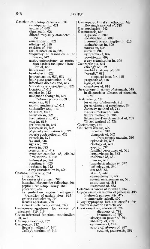
846 INDEX
Gastric ulcer, complications of, 631
constipation in, 621course of, 640diarrhea in, 621dilated "splashy stomach" in,
623eructations in, 621
etiology of, 616excision of, 746foci of infection in, 624
frequency of transition of, to
cancer, 642
gastro-enterostomy as protec-tion against malignant trans-ition of, 645
habits and, 617headache in, 622
hemorrhage in, 620, 632hour-glass contraction in, 631infectious diseases and, 617
laboratory examination in, 624location of, 617malaise in, 622
malignant change in, 642
metamorphosis in, 63 1
melena in, 621morbid anatomy of, 617
nationality and, 616nature of, 640nutrition in, 622
occupation and, 616
pain in, 618
perforation in, 632
peristalsis in, 623
physical examination in, 622
pyloric obstruction in, 631
pyrosis in, 621sex and, 616
signs of, 622stools in, 625
symptoms of, 618
symptom-complex of, clinical
variations in, 644test-meal in, 624
vomiting in, 620weakness in, 622
x-ray examination in, 626
Gastro-enterostomy, 751
anterior, 752for cancer of stomach, 709intestinal obstruction following, 761
peptic ulcer complicating, 761
posterior, 754as protection against malignant
transition of gastric ulcer, 645
pyloric exclusion in, 760Roux's operation, 758vicious circle complicating, 760
Gastro-esophagostomy for cancer of
stomach, 710, 719Gastro-intestinal function, examination
of, 575
Gastrojejunostomy, 752
Gastropexy, 742
Beyer's method of, 743
Coffey's method of, 743
Gastropexy, Burst's method of, 742
Rovsing's method of, 743
Gastroplication, 742
Gastroptosis, 608
appetite in, 609
constipation in, 609
fluoroscopic examination in, 610malnutrition in, 609nausea in, 608
signs of, 609
symptoms of, 608
vomiting in, 608
x-ray examination in, 609
Gastrorrhagia, 612
etiology of, 612morbid anatomy of, 613
"occult," 613chemical tests for, 615
prognosis of, 616
signs, of, 614
symptoms of, 614
Gastroscopy in cancer of stomach, 679in diagnosis of diseases of stomach,
574
Gastrostomy, 735for cancer of stomach, 711for carcinoma of esophagus, 89
Janeway method of, 741Kader's method of, 735Senn's method of, 735
Ssbanajew-Franck method of, 739Witzel method of, 736
Gastrotomy, 134Gaucher's disease, 529
blood in, 532
diagnosis of, 532from splenic anemia, 526
epistaxis in, 532
etiology of, 529
eyes in, 532familial occurrence of, 531
hemorrhages in, 532incidence of, 531liver in, 531
lymphatic glands in, 532
pathology of, 530sex and, 531skin in, 532
splenectomy in, 533
splenic enlargement in, 531
symptoms of, 531treatment of, 533
Gelatinous cancer of stomach, 662
Glycpsuria in carcinoma of pancreas, 496in chronic pancreatitis, 472in pancreatic calculi, 490
Glycyltryptophan test for specific fer-
ments in gastric extracts, 585Great omentum, abscess of, 792
diagnosis of, 792treatment of, 793
absorption power of, 791
anatomy of, 789carcinoma of, 795
cavity of, abscess of, 802
cysts of, pancreatic, 802
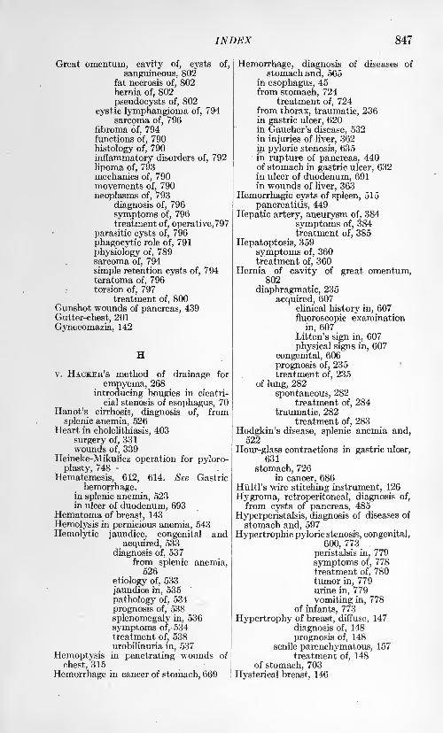
INDEX 847
Great omentum, cavity of, cysts of,
sanguineous, 802fat necrosis of, 802hernia of, 802
pseudocysts of, 802
cystic lymphangioma of, 794sarcoma of, 796
fibroma of, 794functions of, 790
histology of, 790
inflammatory disorders of, 792
lipoma of, 793mechanics of, 790movements of, 790
neoplasms of, 793
diagnosis of, 796
symptoms of, 796treatment of, operative,797
parasitic cysts of, 796
phagpcytic role of, 791
physiology of, 789sarcoma of, 794
simple retention cysts of, 794teratoma of, 796torsion of, 797
treatment of, 800Gunshot wounds of pancreas, 439
Gutter-chest, 201
Gynecomazia, 142
H
v. HACKER'S method of drainage for
empyema, 268
introducing bougies in cicatri-
cial stenosis of esophagus, 70Hanot's cirrhosis, diagnosis of, from
splenic anemia, 526Heart in cholelithiasis, 403
surgery of, 331wounds of, 339
Heineke-Mikuh'cz operation for pyloro-plasty, 748 -
Hematemesis, 612, 614. See Gastric
hemorrhage,in splenic anemia, 523in ulcer of duodenum, 693
Hematoma of breast, 143
Hemolysis in pernicious anemia, 543
Hemolytic jaundice, congenital andacquired, 533
diagnosis of, 537from splenic anemia,
526
etiology of, 533
jaundice in, 535
pathology of, 534
prognosis of, 538
splenomegaly in, 536
symptoms of, -534
treatment of, 538urobilinuria in, 537
Hemoptysis in penetrating wounds of
chest, 315
Hemorrhage in cancer of stomach, 669
Hemorrhage, diagnosis of diseases ofstomach and, 565
in esophagus, 45from stomach, 724
treatment of, 724from thorax, traumatic, 236in gastric ulcer, 620in Gaucher's disease, 532in injuries of liver, 362in pyloric stenosis, 635in rupture of pancreas, 440of stomach in gastric ulcer, 632in ulcer of duodenum, 691in wounds of liver, 363
Hemorrhagic cysts of spleen, 515
pancreatitis, 449
Hepatic artery, aneurysm of, 384
symptoms of, 384treatment of, 385
Hepatoptosis, 359
symptoms of, 360treatment of, 360
Hernia of cavity of great omentum,802
diaphragmatic, 235
acquired, 607clinical history in, 607
fluoroscopic examination
in, 607Litten's sign in, 607
physical signs in, 607
congenital, 606
prognosis of, 235treatment of, 235
of lung, 282
spontaneous, 282treatment of, 284
traumatic, 282treatment of, 283
Hodgkin's disease, splenic anemia and,522
Hour-glass contractions in gastric ulcer,631
stomach, 726in cancer, 686
Hiiltl's wire stitching instrument, 126
Hygroma, retroperitoneal, diagnosis of,
from cysts of pancreas, 485
Hyperperistalsis, diagnosis of diseases of
stomach and, 597
Hypertrophic pyloric stenosis, congenital,
600, 773
peristalsis in, 779
symptoms of, 778treatment of, 780tumor in, 779urine in, 779
vomiting in, 778of infants, 773
Hypertrophy of breast, diffuse, 147
diagnosis of, 148
prognosis of, 148senile parenchymatous, 157
treatment of, 148of stomach, 703
Hysterical breast, 146
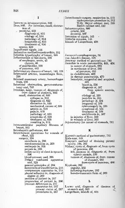
848 INDEX
ILEOCECAL intussusception, 806Ileus, 803. See Intestine, small, obstruc-
tion of.
, paralytic, 833
diagnosis of, 835
etiology of, 834
pathology of, 834symptoms of, 835treatment of, 836 .
spastic, 833Imperforate nipple, 144Indurative mediastinopericarditis, 351Infantile hypertrophy of breast, 148Inflammation of bile-ducts, 393
of esophagus, acute, 45
chronic, 46of gall-bladder, 393of pancreas, 442
Inflammatory diseases of breast, 149,153Intercostal arteries, hemorrhages from,
236Internal mammary artery, hemorrhages
from, 236Intestinal obstruction, gastro-enteros-tomy and, 763
Intestine, large, tumors of, diagnosis of,from cancer of stomach, 685
small, obstruction of, 803
collapse in, 818
diagnosis of, 822distention in, 818
mechanical, causes of, 804nausea in, 818
pain in, 818
pathology of, 820
symptoms of, 818treatment of, 828
vomiting in, 818Intracanalicular papillary fibroma of
breast, 165
Intrahepatic gall-stones, 404Intrathoracic operations for wounds of
chest, 325
surgery, 204anesthesia in, 208anoci-assoeiation in, 209
antisepsis in, 208
asepsis in, 208before cavity of chest is opened,206
blood-pressure and, 206
Drager combined apparatusand, 223
general principles of, 204intratracheal insufflation in, 216
overpressure apparatus in, 213
pleural adhesion in, diagnosis of
degree of, 209
position of patient in, 207
preparation of patient in, 206
pressure differences in, 211
apparatus for, 217
present status of, 217
procedures in, 217
Intrathoracic surgery, respiration in, 211
underpressure procedure in, 212
Willy Meyer cabinet and, 216Zurich cabinet and, 212
Intussusception, 806
enteric, 806
ileocecal, 807Inversion of nipple, 143Irritable mamma, 146Islands of Langerhans, 438
JACKSON'S esophagoscope, 34von Jaksch's disease, 528
Janeway method of gastrostomy, 741Jaundice in acute pancreatitis, 448, 454
in cancer of stomach, 686in carcinoma of liver, 380
of pancreas, 495in cholelithiasis, 403in chronic pancreatitis, 470
hemolytic, congenital and acquired,533
blood in, 536
diagnosis of, 537from splenic anemia,
526
etiology of, 533
jaundice in, 535
pathology of, 534
prognosis of, 538
splenomegaly in, 536
symptoms of, 534treatment of, 538urobilinuria in, 537
in injuries of liver, 363in wounds of liver, 363
Jejunostomy for cancer of stomach, 711
KADER'S method of gastrostomy, 735Kala-azar, 549
Kenyon's method of draining pleural
cavity, 104, 105
Kidney, cysts of, diagnosis of, from cystsof pancreas, 486
left, diseases of, diagnosis of, from
splenic anemia, 526tumors of, diagnosis of, from cancer
of stomach, 685from cysts of pancreas, 486
Kyphosis, 202
angular, 202
following fractures, 202heredo-traumatic form of, 202
LACTIC acid, diagnosis of diseases of
stomach and, 580
Langerhans, islands of, 438
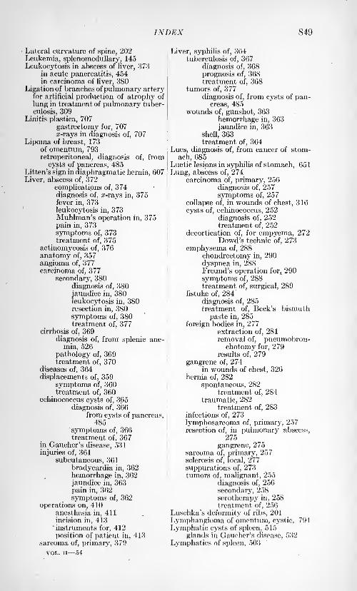
INDEX 849
Lateral curvature of spine, 202
Leukemia, splenomedullary, 145
Leukocytosis in abscess of liver, 373in acute pancreatitis, 454in carcinoma of liver, 380
Ligation of branches of pulmonary arteryfor artificial production of atrophy of
lung in treatment of pulmonary tuber-
culosis, 309Linitis plastica, 707
gastrectomy for, 707
x-rays in diagnosis of, 707
Lipoma of breast, 173of omentum, 793
retroperitoneal, diagnosis of, fromcysts of pancreas, 485
Litten's sign in diaphragmatic hernia, 607
Liver, abscess of, 372
complications of, 374
diagnosis of, x-rays in, 375fever in, 373
leukocytosis in, 373Muhlman's operation in, 375
pain in, 373
symptoms of, 373treatment of, 375
actinomycosis of, 376
anatomy of, 357
angipma of, 377carcinoma of, 377
secondary, 380diagnosis of, 380jaundice in, 380
leukocytosis in, 380resection in, 380
symptoms of, 380treatment of, 377
cirrhosis of, 369
diagnosis of, from splenic ane-mia, 526
pathology of, 369treatment of, 370
diseases of, 364
displacements of, 359
symptoms of, 360treatment of, 360
echinococcus cysts of, 365
diagnosis of, 366from cysts of pancreas,
485
symptoms of, 366treatment of, 367
in Gaucher's disease, 531
injuries of, 361
subcutaneous, 361
bradycardia in, 362
hemorrhage in, 362
jaundice in, 363
pain in, 362
symptoms of, 362
operations on, 410anesthesia in, 411incision in, 413"instruments for, 412
position of patient in, 413sarcoma of, primary, 379
VOL. ii 54
Liver, syphilis of, 364tuberculosis of, 367
diagnosis of, 368
prognosis of, 368treatment of, 368
tumors of, 377
diagnosis of, from cysts of pan-creas, 485
wounds of, gunshot, 363
hemorrhage in, 363
jaundice in, 363
shell, 363treatment of, 364
Lues, diagnosis of, from cancer of stom-
ach, 685Luetic lesions in syphilis of stomach, 651
Lung, abscess of, 274carcinoma of, primary, 256
diagnosis of, 257
symptoms of, 257
collapse of, in wounds of chest, 316
cysts of, echinococcus, 252
diagnosis of, 252treatment of, 252
decortication of, for empyema, 272Dowd's technic of, 273
emphysema of, 288
chondrectomy in, 290
dyspnea in, 288Freund's operation for, 290
symptoms of, 288treatment of, surgical, 289
fistulae of, 284'
diagnosis of, 285treatment of, Beck's bismuth
paste in, 285
foreign bodies in, 277extraction of, 281removal of, pneumobron-chotomy for, 279
results of, 279
gangrene of, 274in wounds of chest, 326
hernia of, 282
spontaneous, 282treatment of, 284
traumatic, 282treatment of, 283
infections of, 273
lymphosarcoma of, primary, 257resection of, in pulmonary abscess,
275
gangrene, 275sarcoma of, primary, 257sclerosis of, local, 277
suppurations of, 273tumors of, malignant, 255
diagnosis of, 256
secondary, 258
serotherapy in, 258treatment of, 256
Luschka's deformity of ribs, 201
Lymphangioma of omentum, cystic, 794
Lymphatic cysts of spleen, 515
glands in Gaucher's disease, 532
Lymphatics of spleen, 503
850 INDEX
Lymphoma, diagnosis of, from splenic jMyxoma of stomach, 707
anemia, 526 Myxosarcoma, periductal, 167
Lymphosarcoma of lung, 257
MMALAISE in gastric ulcer, 622
Malaria, cancer of stomach and, 661Malarial spleen, 547Malformations of breast, 141
of male breast, 196
Mamma, irritable, 146treatment of, 146
Mastitis, acute, 149
cancerous, 179
chronic, 155
cystic, 157
diagnosis of, 156of mumps, 323
neonatorum, 149of puberty, 150
puerperal, 150milk engorgement and, 150
Mastodynia, 146treatment of, 146
Mayo's transgastric resection for gas-tric ulcers, 747
Meckel's diverticulum, 805Mediastinal bronchotomy for foreign
bodies in bronchi, 280
Mediastinitis, subacute posterior sup-
puratiye, 46
Mediastinopericarditis, indurative, 351
Mediastinotomy, posterior, for carci-
noma of esophagus, 89
Mediastinum, posterior, tumors of, com-pression stenosis of esophagus and, 61
Medullary cancer of stomach, 662
Melena, 612, 614. See Gastric hemor-rhage.
in ulcer of duodenum, 693
Mesentery, cysts of, diagnosis of, fromcysts of pancreas, 485
Metapneumonia empyema, 260
Metastases, diagnosis of diseases of
stomach and, 572Meteorism in acute pancreatitis, 454Meyer's cabinet in intrathoracic sur-
gery, 216
esophageal extractor, 90, 116
operating table for thoracic opera-tions, 93
Micromastia, 141
Micromazia, 141Mosher's esophageal dilator, 53
Motor-meal, fluoroscopic, informationderived from observation of, 593
Mucoid cancer of stomach, 662Muhlrnan's operation in abscess of liver,375
Mumps, acute pancreatitis and, 447mastitis of, 153
Murphy's technic for artificial pneumo-thorax, 297
Myxoma, periductal, 167
of pleura, 255
N
NAUSEA in acute pancreatitis, 448in chronic pancreatitis, 470
diagnosis of diseases of stomach and,564
in gastroptosis, 608in ileus, 820in wandering spleen, 512
Necrosis, fat, 451
Neoplasms of omentum, 793of pancreas, cystic, 476of ribs, 253
Nerves of spleen, 503
Neuralgia of breast, 146
Nipple, cracks of, 144
depression of, 143eczema of, 144fissures of, 144
imperforate, 144inversion of, 143
Paget's disease of, 144
diagnosis of, 145
pathology of, 145
prognosis of, 145treatment of, 145
retraction of, 143tumors of, 145
Non-parasitic cysts of spleen, 514
Non-penetrating wounds of thorax, 230
Non-suppurative pancreatitis, 447Nutrition in gastric ulcer, 622
OBESITY, acute pancreatitis and, 455Obstruction of stomach, 725"Occult" blood, diagnosis of diseases of
stomach and, 580, 591
gastrorrhagia, 613
hemorrhage in ulcer of duodenum,693
Omentitis, 792
diagnosis of, 792
Omentopexy, 801
Omentovolvulus, 797
paracolic, 798
parasaccular, 798
resected, 799Omentum. See Great omentum.
tumors of, diagnosis of, from cancerof stomach, 685
from cysts of pancreas, 486
Opaque meals, fluoroscopic observation
of, 594
Osteochondritis, typhoid, 247Osteoma of breast, 173
Osteomyelitis, 247
Osteoperiosteitis of ribs, 247of sternum, 247
typhoid, 247
INDEX 851
FACET'S disease of nipple, 144Pain in abscess of liver, 373
in acute cholecystitis, 401
pancreatitis, 448, 453in carcinoma of pancreas, 495in cholelithiasis, 403in chronic pancreatitis, 470in cysts of pancreas, 482
of spleen, 515
diagnosis of disease of stomach and,562
in fractures of ribs, 225in gastric ulcer, 618in ileus, 818in injuries of liver, 362in pancreatic calculi, 489in pyloric stenosis, 636
in infancy, 601in rupture of pancreas, 440in splenic anemia, 523in tumors of spleen, 514in ulcer of duodenum, 692, 763
of esophagus, 47in wandering spleen, 512
Pain-time in ulcer of duodenum, 692Pancreas, abnormalities of, 437
abscess of, diagnosis of, from cysts,485
accessory, 437
anatomy of, 427
annular, 437blood supply of, 428carcinoma of, 494
ascites in, 496
diagnosis of, 494edema in, 496
extirpation in, 497
glycosuria in, 496
jaundice in, 495pain in, 495
symptoms of, 494 .
treatment of, 497
operative, 499concretions of, 488. See Pancreatic
calculi,
congenital malformations of, cystsand, 476
cystadenomas of, 476, 493
cysts of, 475
congenital malformations and,476
diagnosis of, 480from abscess of abdominal
wall, 483of pancreas, 485
from aneurysm of abdomi-nal aorta, 483
from cysts of kidney,485
of mesentery, 485of round ligament, 485of spleen, 485
from echinococcus cyst of
liver, 483
Pancreas, cysts of, diagnosis of, from en-
larged gall-bladder, 485from retroperitoneal hy-
groma, 485
lipoma, 485
tuberculosis, 486from tumors of kidney, 486
of liver, 483of omentum, 486of peritoneum, 486of vertebral column,486
x-ray in, 482
drainage in, 488
exudative, of omental sac,478
pain in, 480
proliferative, 476retention, 476by self-digestion, 477situation of, 478size of, 478
by softening, 477
symptoms of, 480true, 476treatment of, 486
head of, relation of, to common bile
duct, 432inflammation of, 442
injuries of, 439
lymphatics of, 429
pathological physiology of, 434physiology of, 433
pseudocysts of, 477
rupture of, subcutaneous, 440fistula and, 441
hemorrhage in, 440
pain, 440prognosis of, 440symptoms of, 440treatment of, 440
vomiting in, 440syphilis of, 493tuberculosis of, 493tumors of, 493wounds of, 439
Pancreatic apoplexy, 449
calculi, 488
diagnosis of, 490
x-rays in, 490
etiology of, 489
glycosuria in, 490
pain in, 489
symptoms of, 489treatment of, 491
vomiting in, 489
cysts of cavity of great omentum,802
ducts, anatomy of, 430Pancreatitis, acute, 447
alcoholism and, 455arteriosclerosis and, 455
chicken-pox and, 447
cholecystitis and, 447cholelithiasis and, 447
cyanosis in, 453
852 INDEX
Pancreatitis, acute, diagnosis of, 453from syphilis, 456
diphtheria and, 447
drainage in, 462duodenal ulcer and, 447duodenitis and, 447fever in, 453
jaundice in, 448, 454
leukocytosis in, 454meteorism in, 454mumps and, 447nausea in, 448
obesity'and, 455pain in, 448, 453
pregnancy and, 455pulse in, 453scarlet fever and, 447
symptoms of, 453treatment of, 456
vomiting in, 448, 453
chronic, 463
constipation in, 470
diagnosis of, 468
differential, 473diarrhea in, 470drainage in, 474
glycosuria in, 472
jaundice in, 470nausea in, 470pain in, 470
symptoms of, 468treatment of, 473urine in, 472
vomiting in, 469
gangrenous, 452
hemorrhagic, 447
simple non-suppurative, 447
suppurative, 448, 452
Papillary cystadenoma of breast, 169
diagnosis of, 171
treatment of, 171fibroma of breast, intracanalicular,
165
Papilloma of breast, villous, 169of stomach, 707
Paracolic omentovolvulus, 798
Paralytic ileus, 833
diagnosis of, 835
etiology of, 834
pathology of, 834
symptoms of, 835treatment of, 836
Parasaccular omentovolvulus, 798Parasitic cysts of omentum, 796
Parenchymatous hypertrophy of breast,
senile, 157
Penetrating wounds of pancreas, 439of spleen, 509
Peptic index for specific ferments in
gastric extracts, 584ulcers of esophagus, 47
gastro-enterostomy and, 762Perforation of esophagus, acute, 42, 45
of stomach in gastric ulcer, 632
Pericarditis, acute, 351
Pericardium, drainage of, 350
Pericardium, tumors of, 352wounds of, 340
Periductal fibroma, 165
myxoma, 167
.myxosarcoma, 167
sarcoma, 167
Peripleuritis, 249
Peristalsis, diagnosis of diseases ofstomach and, 597
in gastric ulcer, 623in pyloric stenosis, 636
Peritoneum, tumors of, diagnosis of, from
cysts of pancreas, 486Pernicious anemia, 539
blood in, 543
diagnosis of, 540from splenic anemia, 525
etiology of, 539
hemolysis in, 543
pathology of, 540
splenectomy in, 544transfusion of blood in, 542treatment of, 541
Phlegmon of chest-wall, 242
Physiological motor meal, 575
Pigeon-breast, 201
Pleomastia, 141
Pleura, carcinoma of, secondary, 255endothelioma of, 255fibrosarcoma of, 255fistulae of, treatment of, Beck's bis-
muth paste in 285
myxosarcoma of, 255sarcoma of, 255
surgical diseases of, 258tumors of, malignant, 254
Pleural adhesions, diagnosis of degree of,
in intrathoracic surgery, 209
eclampsia, 238
symptoms of, 239
reflexes, 236
Pleurisy, purulent, 259Pleuritic scar contraction, 204Plummer's double olive bougie, 68
hydrostatic cardiodilator, 53
Pneumectomy in treatment of pulmon-ary tuberculosis, 294
Pneumobronchotomy for removal of for-
eign bodies from lung, 279
Pneumolysis interpleuralis in treatmentof pulmonary tuberculosis, 308
Pneumothorax, artificial, 294centra-indications for, 295indications for, 295
Murphy's technic for, 297fracture of ribs and, 226
surgical, 232
traumatic, 230in wounds of chest, 316
Pneumotomy in treatment of bronchiec-
tasis, 287
Polymastia, 141
Polymazia, 141
Polythelia, 141Pott's disease of spine, 202Precardial thoracostomy, 352
INDEX 853
Pregnancy, acute pancreatitis and, 455Presternal cyst congenital, 253Pressure diverticulum of esophagus, 61
Prolapse of spleen, 509
Pseudocysts of cavity of great omentum,802
of pancreas, 476
Puberty, mastitis of, 150
Puerperal mastitis, 150milk engorgement and, 150
Pulmonary abscess, 274chronic simple bronchiectasis
and, 276
diagnosis of, 274
prognosis of, 276
putrid, bronchiectasis and, 277resection of lung in, 275treatment of, 275
fistulae, 284
diagnosis of, 285treatment of, Beck's bismuth
paste in, 285
gangrene, 274
diagnosis of, 274
prognosis of, 276resection of lung in, 275treatment of, 275
tuberculosis, artificial pneumothoraxin, 294
chondrotomy for, 310
drainage in, 293
extrapleural thoracoplasty for,
299Friedrich's large flap operation
for, 304
grafting in, 298incision in, 293
ligation of branches of pulmon-ary artery for artificial pro-duction of atrophy of lung in
treatment of, 309
pneumectomy in, 294
pneumolysis for, 298
surgery of, 291outline of original effort in,
291Tuffier's operation in treatment
of, 298Wilm's pperatioh of columnar
resection of ribs to narrowthorax in treatment of, 307
Pulsion diverticulum of esophagus, 61Purulent pleurisy, 259
Pylorectomy for cancer of stomach, 711
Pyloric canal, anatomy of, 775
pathological anatomy of, 775obstruction in cancer of stomach,
685in gastric ulcer, 631
spasm, diagnosis of, from pyloricstenosis in infancy, 603
stenosis, 725
benign, 633abdominal palpation in,
636causation of, 633
Pyloric stenosis, benign, effects of, 633eructations in, 635
hemorrhage in, 635incidence of, 633motor-meal in, 638observation-meal in, 639
pain in, 636
peristalsis in, 636
pyrosis in, 635
regurgitation in, 635
secretory meal in, 638
signs of, 636
"splash stomach" in, 637
symptoms of, 634test-meal in, 637
vomiting in, 634"water brash" in, 635
x-ray examination in, 638
congenital, 725
hypertrophic, 600, 773
peristalsis in, 779
symptoms of, 778treatment of, 780tumor in, 779urine in, 779
vomiting in, 778in infancy, 600
abdominal tumor in, 602
age and, 600carmin test in, 602
constipation in, 601definition of, 600
diagnosis of, 602from pyloric spasm, 603
etiology of, 600genesis of, 600loss of weight in, 602
pain in, 601
pathology of, 601
prognosis of, 603
roentgen examination in,
602
signs of, 602
symptoms of, 601urine in, 601
Pylorodiosis, 748
Pyloroplasty, 748
Finney's operation, 748Heineke-Mikulicz operation, 748
Pyrosis in gastric ulcer, 621in pyloric stenosis, 635in ulcer of duodenum, 693
RACHITIS, chicken-breast and, 201
Radiography in examination of esopha-gus, 36
Radium in carcinoma of breast, 186Rectal examination, diagnosis of dis-
eases of stomach and, 572
Regurgitation in pyloric stenosis, 635Renal complications in splenic anemia,
524Resected omentovolvulus, 799
854 INDEX
Resection for carcinoma of esophagus,83, 87
of cardia for carcinoma of esophagus107
of lung in pulmonary abscess, 275
gangrene, 275of thorax for empyema, 271
Respiration, intrathoracic surgery and,211
Retention cysts of omentum, 796Retraction of nipple, 143
Retroduodenal, choledochotomy, 420Retroperitoneal hygroma, diagnosis of,
from cysts of pancreas, 485
lipoma, diagnosis of, from cysts of
pancreas, 485
tuberculosis, diagnosis of, from cystsof pancreas, 486
Rheumatism, cancer of stomach and,661
Rib, trephining of, for empyema, 267
Ribs, deformities of, congenital, 199
Cruveilhier's, 201
Luschka's, 201
Serb's, 201dislocations of, 228fractures of, 223
complications of, 226
diagnosis of, 226
dyspnea in, 225
external, 224
internal, 224
pain in, 225
pneumothorax and, 226subcutaneous emphysema and,
226
symptoms of, 225traumatic aneurysm and, 226treatment of, 227
neoplasms of, 253
symptoms of, 254
osteoperiosteitis of, 247
diagnosis of, 247treatment of, 247
sarcoma of, 254tuberculosis of, 248
diagnosis of, 248
prognosis of, 249treatment of, 249
Rigid spine, 203
thorax, treatment of, chondrotomyin, 291
Rodman's operation for gastric ulcers,747
Roentgen examination in cancer of
stomach, 679
diagnosis of diseases of stomachand, 592
in pyloric stenosis in infancy,602
in ulcer of duodenum, 697
plates, examination of duodenumby, 598
of stomach by, 598
interpretation of, in diagnosisof diseases of stomach, 599
Round ligament, diagnosis of, from cystsof pancreas, 485
Rovsing's method of gastropexy, 743
Rupture of esophagus, spontaneous, 44of gall-bladder, 391of pancreas, 440of spleen, spontaneous, 510
subcutaneous, 509of stomach, 705
SANGUINEOUS cysts of cavity of great
omentum, 802Sarcoma of breast, 173
cystic, 167of chest-wall, 254
treatment of, 254of esophagus, 77of liver, primary, 379of lung, 257of omentum, 794
cystic, 796
periductal, 167of pleura, 255of ribs, 254of stomach, 707
Sauerbruck's operation for extrapleuralthoracoplasty in treatment of pul-monary tuberculosis, 299
Scarlet fever, acute pancreatitis and, 447Schede's operation for empyema, 270Schwartz's mediastinal bronchotomy for
foreign bodies in bronchi, 280Scirrhous cancer of stomach, 662Sclerosis of lung, local, 277
Scoliosis, 202Serb's deformity of ribs, 201
Serotherapy in treatment of malignanttumors of lung, 258
Serous cysts of spleen, 515Skin in Gaucher's disease, 532
Sounding in examination of esophagus,31
Spasms of duodenum, diagnosis of, 597
pyloric, diagnosis of, from pyloricstenosis in infancy, 603
of stomach, diagnosis of, 596
Spastic hypertrophic pyloric stenosis, 773
ileus, 833stenosis of esophagus, 50
cardioplasty for, 57
diagnosis of, 51
esophagogastrostomy for,
58
etiology of, 51treatment of, 52
Spine, lateral curvature of, 202Pott's disease of, 202
rigid, 203
"Splash stomach" in pyloric stenosis, 607
Spleen, abscess of, 517
diagnosis of, 518
pathology of, 517
prognosis of, 518
INDEX 855
Spleen, abscess of, symptoms of, 518treatment of, 518
anatomy of, 501
angioma of, 516
diagnosis of, 517treatment of, 517
blood supply of, 503
cysts of, 514
diagnosis of, 516from cysts of pancreas, 485
dyspnea in, 516
echinococcus, 514
lymphatic, 515
hemorrhagic, 515
non-parasitic, 514
pain in, 515
serous, 515
symptoms of, 515treatment of, 516
embryology of, 503
enlargement of, in Gaucher's dis-
ease, 531functions of, 506
injuries of, 509
lymphatics of, 503
malarial, 547nerves of, 503
physiology of, 504
prolapse of, 509
rupture of, spontaneous, 510
diagnosis of, 511
symptoms of, 510treatment of, 511
subcutaneous, 509tuberculosis of, 518
pathology of, 519
symptoms of, 519treatment of, 520
tumors of, 513
diagnosis of, 514
pain in, 514
symptoms of, 514
wandering, 511
diagnosis of, 513
etiology of, 511nausea in, 512
pain in, 512
pathology of, 512
symptoms of, 512treatment of, 513
vomiting in, 512wounds of, penetrating, 509
Splenectomy, Bevan incision in, 550Splenic anemia, 520
abdomen in, 523adult type of, in children, 527anemia in, 524ascites in, 524cardiac symptoms in, 524cholelithiasis and, 521
diagnosis of, 525
digestive disturbances in, 524
etiology of, 521fever in, 524hematemesis in, 523
Hodgkin's disease and, 522
Splenic anemia, incidence of, 522of infancy, 528
pain in, 523
pathology of, 522renal complications in, 524
splenectomy in, 527
symptoms of, 523treatment of, 526
Splenomedullary leukemia, 545
Splenomegalia, large cell, 529blood in, 532
diagnosis of, 532
epistaxis in, 532
etiology of, 529
eyes in, 532familial occurrence of, 531
hemorrhages in, 532incidence of, 531liver in, 531
lymphatic glands in, 532
pathology of, 530sex and, 531skin in, 532
splenectomy in, 533
splenic enlargement in, 531
symptoms of, 531treatment of, 533
Splenomegaly and anemia, syphilis and,546
Egyptian, 548in hemolytic jaundice, 536
Spontaneous rupture of spleen, 510
Ssbanajew-Franck method of gastros-
tomy, 739
Stenosis, congenital hypertrophic pyloric,773
peristalsis in, 779
symptoms of, 778treatment of, 780tumor in, 779urine in, 779
vomiting in, 778of duodenum, 704
in ulcer, 690of esophagus, cicatricial, 67
treatment of, 68
compression, 59aortic aneurysms and, 60
posterior mediastinal tu-
mor and, 61
tuberculous, specific or
malignant 'peribronchial
lymph nodes and, 61
spastic, 50
cardioplasty for, 57
diagnosis of, 51
esophagogastrostomy for,
58
etiology of, 51treatment of, 52
pyloric, 600, 725
benign, 633abdominal palpation in,
636causation of, 633effects of, 633
856 INDEX
Stenosis, pyloric, benign, eructations in,635
hemorrhage in, 635incidence of, 633motor-meal in, 638observation-meal in, 629pain in, 636
peristalsis in, 636pyrosis in, 635
regurgitation in, 635
secretory-meal in, 638signs of, 636"splash stomach" in, 637
symptoms of, 634test-meal in, 637
vomiting in, 634"water brash" in, 635x-ray examination in,
638in infancy, 600
abdominal tumor in, 602carmin test in, 602constipation in, 601definition of, 600diagnosis of, 602
etiology of, 600genesis of, 600loss of weight in, 602pain in, 601
pathology of, 601
prognosis of, 603
roentgen examination in,
602signs of, 602symptoms of, 601urine in, 601
Sternum, fracture of, 228
cross-shaped, 229diagnosis of, 230
transverse, 229treatment of, 230
osteoperiosteitis of, 247diagnosis of, 247treatment of, 247
Stomach, adenocarcinoma of, 662adhesion of, operation for, 743atresia Of, 599
prognosis of, 600symptoms of, 600
atrophy of, 703
bacteriology of, 704cancer of, 658, 708
abdominal tumor in, 671blood in, 674cachexia in, 671
changes in stomach producedby, 664
colloid, 662
complications of, 685course- of, 646
diagnosis of, 679from achylia gastrica,685
from chronic constipation,'
685from foreign bodies, 685
Stomach, cancer of, diagnosis of, from
gall-bladder disease, 681from gastric granuloma,
685from grave anemias, 681from lues, 685from tuberculosis, 685from tumors of kidney, 685
of large intestine, 685of omentum, 685
enteritis in, 686
fibrous, 662fistula in, 686gastric acidity in, 672
tests for, 673contents in, microscopicexamination of, 674
syphilis and, 661ulcer and, 661
gastrpscopy in, 679
gelatinous, 662habits and, 660
hemorrhage in, 669
heredity and, 659
"hour-glass "in, 686
hygiene and, 661incidence of, 659jaundice in, 686location of, 663malaria and, 661
medullary, 662metastases in, 665morbid anatomy of, '661
mucoid, 662nature of, 646obstruction at cardia in, 685occupation and, 660operations for, centra-indica-
tions to, 709
exploratory incision in, 709
gastrectomy, 710, 711, 718
gastro-enterostomy, 709
gastro-esophagostomy,710,719
gastrostomy, 711indications to, 709
jejunostomy, 711
pylorectomy, 711
perforation in, 686pyloric obstruction in, 685 .
race and, 659rheumatism and, 661
roentgen examination in, 677
scirrhus, 662
secondary, 664sex and, 659
signs of, 665social status and, 660symptoms of, 665
clinical grouping of, 667test-meal in, 672thrombosis in, 686trauma and, 660treatment of, 708tuberculosis and, 661
ulcerating, 662
INDEX 857
Stomach, cancer of, vomiting in, 670carcinoma of, 658. See Stomach,
cancer of.
cirrhosis of, 707
congenital anomalies of, 599, 704
enlargement of, 603
faulty position of, 603contour of, fluoroscopic observation
of, 594
contracted, 600
prognosis of, 600
symptoms of, 600contraction of, 703
cysts of, 724dilatation of, acute, 727
etiology of, 728treatment of, 728
dilated "splashy," in gastric ulcer,
623in ulcer of duodenum, 695
diseases of, abdominal examination
in, preparation of patientfor, 567
tenderness and, 571
age and, 560
appetite and, 566auscultation in, 574"combined" acidity and, 580
constipation and, 566
diagnosis of, 559diarrhea and, 567
esophagoscopy and, 574examination of feces in, 589
fluoroscopic examination in,
593"free hydrochloric acidicy and,580
gastric acidity and, 579
emptying power and, 575
retarded, 578extracts and, macroscopic
appearances of
removed, 576
amount, 577bile coloring, 576
chymification,578color, 576
mucus, 577
odor, 577
microorganisms in,
diagnostic signifi-cance of, 587
microscopic estima-tion of, 585
organic acids in, 583
specific ferments in,583
edestin test of
Fuld and Levi-son for, 583
glycyltryptophantest for, 585
peptic index for,
584
tryptophan test
for, 585
Stomach, disease of, gastric extracts
and, Wolff-Junghans'test for soluble albumin
in, 585malfunction and, 561
gastroscopy and, 574
hemorrhage and, 565infectious diseases* and, 560
inspection in, 568lactic acid and, 580metastases and, 572nausea and, 564"occult" blood and, 580
operative procedures and, 561
pain and, 562
palpation in, 570
percussion in, 573
physical examination in, 567rectal examination in, 572
roentgen examination in, 592sex and, 560"total" acidity and, 580volatile fatty acids and, 580
vomiting and, 564
weight and, 566diverticula of, 704
enlargement of, in ulcer of duo-
denum, 701examination of, by roentgen plates
or films, 598
foreign bodies in, 706
diagnosis of, from cancer,685
treatment of, 706
hemorrhage of, 612, 724. See Gas-tric hemorrhage,
in gastric ulcer, 632in pyloric stenosis, 635treatment of, 724
hour-glass, 726in cancer, 686in gastric ulcer, 631
hypertrophy of, 703injuries of, 705
lymphatics of, gastrectomy and,
mobility of, 597
myxomata of, 707obstruction of, 725
treatment of, 725
operations in, 729
gastrectomy, 745
gastroduodenostomy, 751
gastro-enterostomy, 751
anterior, 752
posterior, 754
gastrojejunostomy, 752
gastropexy, 742
gastroplication, 742
gastrostomy, 729
gastrotomy, 729incision in, 729
pylorodiosis, 748
pyloroplasty, 748
papillomata of, 707
perforations of, in gastric ulcer, 623

858 INDEX
Stomach, pyloric stenosis of, benign,633. See Pyloric stenosis.
in infancy, 600. .See Pyloricstenosis in infancy,
rupture of, 705sarcoma of, 708spasms of, diagnosis of, 596"splash," in pyloric stenosis, 637syphilis of, 649r 706
age and, 649blood in, 655cancer and, 661
etiology of, 649gastric acidity in, 655
extracts in, 655malfunction in, 651
incidence of, 649
laboratory examination in, 655luetic lesion in, 651
pathology of, 649sex and, 649stool in, 655test-meals in, 655treatment of, 706
Wassermann-Noguchi reac-tions in, 651
x-ray examination in, 657tuberculosis of, 658, 707
cancer and, 661
diagnosis of, 658from cancer, 685
tumors of, 707ulcer of, 616. See Gastric ulcer,
variations in, 703volvulus of, 704wounds of, 706
prognosis of, 706treatment of, 706
Stool in gastric ulcer, 625in syphilis of stomach, 665
Strauss treatment of congenital hyper-trophic pyloric stenosis, 785
Streptococcic infection in wounds of
chest, 324Stricture of esophagus, 48Subacute posterior suppurative rr.edias-
tinitis, 46Subcutaneous emphysema, 240
rupture of spleen, 509
Subpleural connective tissue, abscess of,249
emphysema, 240Suffocative compression of thorax, 221
Suppurations of lungs, 273
Suppurative mediastinitis, subacute pos-teriorj 46
pancreatitis, primary, 448
secondary, 452Surgical pneumothorax, 232Syphilis of breast, 153
diagnosis of, from acute pancreatitis,456
from splenic anemia, 525of liver, 364of pancreas, 493of stomach, 649, 706
Syphilis of stomach, age and, 649blood in, 655cancer and, 661
etiology of, 649
gastric acidity in, 655 .
extract in, 655malfunction in, 651
incidence of, 649
laboratory examination in, 655luetic lesion in, 651
pathology of, 649sex and, 649stool in, 655test-meals in, 655treatment of, 706
Wassermann-Noguchi reactions
in, 651
x-ray examination in, 657
Syphilitic ulcers of esophagus, 47
TALMA operation, 801Teratoma of omentum, 796Test-meal in cancer of stomach, 672
in gastric ulcer, 624in pyloric stenosis, 637in syphilis of stomach, 655in ulcer of duodenum, 695
Thoracico-abdominal wounds, 235
Thoracoplasty, extrapleural, in treat-
ment of pulmonary tuberculosis, 299
Thoracostomy for foreign bodies in lung,278
precardial, 352with rib resection for empyema, 263
Thorax, actinomycosis of, 249commotion of, 221
treatment of, 221
compression of, suffocative, 221
prognosis of, 223
symptoms of, 223treatment of, 223
congenital, treatment of, chondrot-
omy in, 291contraction of, 204contusions of, 220
prognosis of, 221
symptoms of, 221treatment of, 221
cysts of, 251
echinococcus, 251deformities of, disease of vertebrae
and, 202
dilated, treatment of, chondrotomyin, 291
injuries of, 220
paralyticus, 203resection of, for empyema, 271
rigid, treatment of, chondrotomy in,
291
rigidity of, congenital, 202
superior aperture of narrowing of,
202
surgical diseases of, 240
INDEX 859
Thorax, traumatic hemorrhage from, 236tumors of, 251
benign, 253wounds of, non-penetrating, 230
Thrombosis in cancer of stomach, 686
Tiegel's overpressure apparatus in intra-
thoracic surgery, 215thoracic flexible metal drain, 103,
105Torek's incision in thoracic operations
for carcinoma of esophagus, 94Torsion of omentum, 797"Total" acidity in diagnosis of diseases
of stoma,ch, 580Transfusion of blood in pernicious
anemia, 542in wounds of chest, 324
Transgastric resection for gastric ulcers,747
Transpleural esophagogastrostomy in
treatment of spastic stenosis of esoph-agus, 59
Transthoracic operation for carcinomaof esophagus, 89
after-treatment in, 103closure of thoracicwound in, 100, 102
exposure of tumor in,
95extraction of stumpand tumor together,100
incision in, 93instruments for, 91
posture of patient in,
91of surgeon in, 93
preparation for opera-tion, 90
of patient, 89resection of, tumor in,
98technic of, 89
Traumatic aneurysm, fractures of ribs
and, 226
empyema, 259
hemorrhage from thorax, 236hernia of lung, 282
pneumothorax, 230Trephining of rib for empyema, 267Tryptophan test for specific ferments in
gastric extracts, 585Tuberculosis of breast, 154
of liver, 367of pancreas, 493
pulmonary, surgery of. See Pul-
monary tuberculosis,
retroperitoneal, diagnosis of, fromcysts of pancreas, 486
of ribs, 248of spleen, 518of stomach, 658, 707
cancer and, 661
diagnosis of, 658from cancer, 685
Tuberculous empyema, 260
Tuberculous ulcers of esophagus, 47Tuffier's operation for pulmonary tuber-
culosis, 298
Tumor, abdominal, in cancer of stomach,671
in pyloric stenosis in infancy,602
in congenital hypertrophic pyloric
stenosis, 779Tumors of breast, 163
of chest-wall, 253of kidney, diagnosis of, from cancer
of stomach, 685from cysts of pancreas, 486
of large intestine, diagnosis of, fromcancer of stomach, 685
of liver, 377
diagnosis of, from cysts of pan-creas, 485
of lungs, malignant, 255of male breast, 197of mediastinum, posterior, compres-
sion stenosis of esophagus and, 61of nipple, 145of omentum, 793of pancreas, 493of pericardium, 352of peritoneum, diagnosis of, from
cysts of pancreas, 486of pleura, malignant, 254of spleen, 513of stomach, 707of thorax, 251
benign, 253
malignant, 253of vertebral column, diagnosis of,
from cysts of pancreas, 486
Typhoid osteochondritis, 247. osteoperiostitis, 247
diagnosis of, 248treatment of, 248
ULCER of duodenum, 686, 763abdominal tenderness in, 695
acute, 691
pancreatitis and, 447
perforating, 771
age and, 687anemia in, 690, 694
appetite in, 694
belching of sour gas in, 693
calloused, 691
chronic, 691
constipation in, 694cutaneous burns and, 764
diagnosis of, 764dilated "splashy" stomach in,
695
enlargement of stomach in, 701eructations in, 693"food-ease" of pain in, 692habits and, 689hematemesis in, 693
800 INDEX
Ulcer of duodenum, hemorrhage in, 691incidence of, 686
laboratory examination in, 695
malignant change in, 644melena in, 693morbid anatomy of, 689"occult" hemorrhage in, 693
pain in, 692, 763
pain-time in, 692perforation in, 690, 69-1
previous disease and, 687
pyrosis in, 693
roentgen evidence in, 697
secondary effects of, 690sex and, 687
signs of, 694stenosis of duodenum in, 690stool in, 697
symptoms of, 691, 763test-meal in, 695total acidity in, 696treatment of, 764
vomiting in, 693, 763"water-brash" in, 693weakness in, 694
esophagus, 46
dysphagia in, 47
pain in, 47
peptic, 47
syphilitic, 47treatment of, 47
tuberculous, 47
gastric, 616abdominal tenderness in, 622
acidity of gastric extracts in,625
adhesions in, 632
age and, 616
appetite in, 621 *
blood in, 624cancer of stomach and, 640, 661
complications of, 637
constipation in, 621course of, 640diarrhea in, 621dilated "splashy stomach" in,
623eructations in, 621
etiology of, 616excision of, 746
actual cautery in, 747Rodman's operation for,
747
transgastric resection for,
747foci of infection in, 624
frequency of transition of, to
cancer, 646
gastro-enterostomy as protec-tion against malignant transi-tion of, 645
habits and, 617headache in, 622
hemorrhage in, 620, 632
subjective evidences of,621
Ulcer, gastric, hour-glass contraction in,
631infectious diseases and, 617
laboratory examination in, 624location of, 617malaise in, 622
malignant change in, 642
metamorphosis in, 631melena in, 621morbid anatomy of, 617
nationality and, 616nature of, 640nutrition in, 622
occupation and, 616
pain in, 618
perforation in, 632
peristalsis in, 623
physical examination in, 622
pyloric obstruction in, 631
pyrosis in, 621sex and, 616
signs of, 622stools in, 625
symptoms of, 618
symptom-complex of, clinical
variations in, 644test-meal in, 624
vomiting in, 620weakness in, 622
x-ray examination in,. 626
peptic, gastro-enterostomy and, 760
Ulcerating cancer of stomach, 662Urine in chronic pancreatitis, 472
in congenital hypertrophic pyloric
stenosis, 779in pyloric stenosis in infancy, 601
Urobilinuria in hemolytic jaundice, 537
VAGOLYSIS combined with esophagopli-cation in treatment of spastic stenosis
of esophagus, 56
Valvulotomy, external, 337Verneuil-Kummel's disease, 202
Vertebra?, diseases of, deformities of
thorax and, 202Vertebral column, tumors of, diagnosis
of, from cysts of pancreas, 486Villous papilloma of breast, 169Visceral pleurectomy, 272Volatile fatty acids, diagnosis of dis-
eases of stomach and, 580
Volvulus, 808of stomach, 704
Vomiting in acute pancreatitis, 448, 453in cancer of stomach, 670in chronic pancreatitis, 469in congenital hypertrophic pyloric
stenosis, 778
diagnosis of diseases of stomach and,564
in gastric ulcer, 620in gastroptosis, 608in ileus, 820
INDEX 861
Vomiting in pancreatitis calculi, 489in pyloric stenosis, 634in rupture of pancreas, 440in ulcer of duodenum, 693, 763in wandering spleen, 512
WWANDERING spleen, 511
diagnosis of, 513
etiology of, 511nausea in, 512
pain in, 512
pathology of, 512
symptoms of, 512treatment of, 513
vomiting in, 512
Wassermann-Noguchi reactions in
syphilis of stomach, 651"Water-brash" in gastric ulcer, 621
in pyloric stenosis, 635in ulcer of duodenum, 693
Weight, diagnosis of diseases of stomach
and, 566WT
ilm's operation of columnar resection
of ribs to narrow thorax in pulmonarytuberculosis, 307
Wirsung, duct of, relation of, to commonbile duct, 432
Witzel's gastrostomy, 736
Wplff-Junghans' test for soluble albuminin gastric extracts, 585
Wounds of breast, 143of chest, 313
penetrating, 315
operations for, 319, 325
x-ray in, 316
superficial, 314of diaphragm, 326of esophagus, 43of gall-bladder, 391of heart, 339of liver, gunshot, 363
shell, 363
Wounds of liver, treatment of, 364of pancreas, 439
gunshot, 439
penetrating, 439treatment of, 439
of pericardium, 340of spleen, penetrating, 509of stomach, 705
thoracico-abdominal, 235of thorax, non-penetrating, 230
X-RAYS in carcinoma of breast, 186
in diagnosis of abscess of liver, 375of bronchiectasis, 286of cirrhosis of stomach, 707of cysts of pancreas, 484of gall-stones, 404of pancreatic calculi, 490of penetrating wounds of chest,
316of pulmonary abscess, 274of tuberculosis of ribs, 248
examination in cancer of stomach,679
of esophagus, 36in gastric ulcer, 626in gastroptosis, 609in pyloric stenosis, 638in syphilis of stomach, 657
YANKAUER'S electric suction apparatus,35
ZURICH cabinet, intrathoracic surgeryand, 212


o
3*CMcn
<D
-D
-P
,0rH>4
<D
CW
ÔO
4->~
e:
&-4
1
DC
University of Toronto
Library
DO NOTREMOVETHECARDFROM
THIS
Acme Library Card Pocket
Under Pat. "Ref. Index Fllr"
Made by LIBRARY BUREAU