Embed Size (px)
Citation preview
CASE REPORT Open Access
Successful staged hip replacement in septic hiposteoarthritis in osteopetrosis: a case reportGiovanni Manzi1, Delia Romanò1, Laura Moneghini2 and Carlo L Romanò1*
Abstract
Background: Osteopetrosis is a rare, inherited, bone disorder, characterized by osteosclerosis, obliteration of themedullary cavity and calcified cartilage. The autosomal dominant form is compatible with a normal life span,although fractures often result from minimal trauma, due to the pathologic nature of bone. Osteomyelitis iscommon in patients with osteopetrosis because of a reduced resistance to infection, attributed to the lack ofmarrow vascularity and impairment of white cell function. Only one case of osteomyelitis of the proximal third ofthe femur has been previously reported, treated with several repeated debridements and finally with femoral headresection. Here we present for the first time a case of a staged implant of a cementless total hip prosthesis for thetreatment of a septic hip in femoral neck nonunion in osteopetrosis.
Case presentation: A 36-years-old woman, affected by autosomal dominant osteopetrosis was referred to ourdepartment because of a septic hip arthritis associated with femoral neck septic non-union, with draining fistulas.The infection occurred early after a plate osteosynthesis for a closed perthrocanteric fracture of the femur andpersisted in spite of osteosynthesis removal, surgical debridement and external fixation. In our hospital the patientunderwent accurate debridement, femoral head and greater trochanter resection, preparation of the diaphysealintramedullary canal and implant of an antibiotic-loaded cement spacer. The spacer was exchanged after onemonth, due to infection recurrence and four months later, a cementless total hip arthroplasty was implanted, withno clinical and laboratory signs of infection recurrence at two years follow-up.
Conclusions: In case of hip septic arthritis and proximal femur septic non-union, femoral head resection may notbe the only option available and staged total hip arthroplasty can be considered.
Keywords: Osteopetrosis, Infection, Osteomyelitis, Total hip arthroplasty, Non-union
BackgroundOsteopetrosis is a rare inherited bone disorder originallydescribed in 1904 by Albers-Schonberg, a German radi-ologist [1]; this is a group of sclerosing bone dysplasiadue to diminished osteoclast-mediated skeletal resorp-tion. The disorder is characterized by osteosclerosis,obliteration of the medullary cavity and calcified carti-lage [2]. Despite the sclerotic radiographic appearance ofthe thickened cortices and its material hardness, osteo-petrotic bone is weak and prone to fracture by minortrauma [3-6]. The sclerosis of bone is, in fact, the resultof increased thickness and disorganization, not anincrease in mineralization [7-9]. Areas of concentrated
stress such as the femoral neck and subtrochantericareas are especially susceptible [10-12].Osteopetrosis has been categorised clinically into three
primary types: infantile, or “malignant” osteopetrosis,inherited in an autosomal recessive pattern; “intermedi-ate” autosomal recessive osteopetrosis and “benign”autosomal dominant osteopetrosis [2]. The severe infan-tile forms of osteopetrosis are associated with dimin-ished life expectancy, with most untreated childrendying in the first decade as a complication of bone mar-row suppression. Orthopaedic surgeons most commonlyencounter patients with the benign autosomal-dominanttype of osteopetrosis (Albers-Schönberg disease), pre-viously known as adult osteopetrosis or osteopetrosistarda. In fact autosomal dominant osteopetrosis typicallyonsets in late childhood or adolescence and is compati-ble with a normal life span; blood studies show that acid
* Correspondence: [email protected] di Chirurgia Ricostruttiva e delle Infezioni Osteo-articolari,Istituto Ortopedico I.R.C.C.S. Galeazzi, Via Riccardo Galeazzi 4-20166, MilanoFull list of author information is available at the end of the article
Manzi et al. BMC Musculoskeletal Disorders 2012, 13:50http://www.biomedcentral.com/1471-2474/13/50
© 2012 Manzi et al; licensee BioMed Central Ltd. This is an Open Access article distributed under the terms of the Creative CommonsAttribution License (http://creativecommons.org/licenses/by/2.0), which permits unrestricted use, distribution, and reproduction inany medium, provided the original work is properly cited.

phosphatase, calcitriol, and creatine phosphokinase BBvariant levels are elevated, but as many as 40% ofpatients with the benign form may remain asympto-matic, while most of them first learn of their diagnosisafter a fracture [13-15]. Life-threatening symptomsinclude anemia, pancytopenia, osteomyelitis and sepsisdue to poorly developed bone marrow and impairedmedullary hematopoiesis, associated with secondaryhematopoiesis in liver and spleen that causes hepatos-plenomegaly [16-19].Fractures typically occur in the appendicular skeleton,
most commonly in the proximal femur [5,20,21], as wellas in the femoral shaft, tibia, and upper extremities [3].These fractures may result from minimal trauma due tothe pathologic nature of bone [3,15]. Fracture-healingabnormalities have been noted both histologically andclinically. Because of dysfunctional remodeling, the callusdoes not attain haversian organization even by one yearpostfracture. Broad cement lines persist, creating areas oflowered resistance where microfractures may propagate[11]. Some authors report delayed union and nonunionfollowing fractures [2,15]. In some patients, nonunion offemoral neck fractures may lead to coxa vara [2,3,22,23].Long-bone deformities are also possible, in particular, lat-eral bowing of the femur [2]. Coxa vara deformities typi-cally appear during childhood, apparently caused bystress-induced microfractures in the brittle femoral neck[2,22]. Degenerative osteoarthritis also may develop sec-ondary to coxa vara deformity [24,25]. When fracturesare encountered, fixation is extremely difficult.Two of these problems, osteoarthritis and certain peri-
articular nonunions, may be recalcitrant to other treat-ment options and may be considered for treatment withtotal joint arthroplasty. It is recognized that the hard brit-tle bone, often without a normal medullary canal, makesarthroplasty implantation difficult, may compromise theoutcome and lead to more frequent complications [26].Osteomyelitis is common in patients with osteopetro-
sis because of a reduced resistance to infection Theincreased incidence of osteomyelitis has been attributedto the lack of marrow vascularity in osteopetrotic boneand impairment of white cell function [2,4]. The mand-ible followed by the maxilla, scapula, and extremities ismost frequently involved [27-29]. The cause of osteo-myelitis of the jaws is usually odontogenic infection andpolymicrobial in nature, as opposed to long bone osteo-myelitis, in which classically Staphylococcus aureusremains the main responsible organism [2].To date, only one case of osteomyelitis of the proxi-
mal third of the femur has been described and treatedsuccessfully with femoral head resection [30]. This is, toour knowledge, the first description of a staged implantof a cementless total hip prosthesis for the treatment ofa septic hip in femoral neck nonunion in osteopetrosis
Case presentationA 36-years-old woman C.I. was referred to our depart-ment in November 2008 because of a septic left hiparthritis and femoral neck non-union, with a drainingfistula. Her medical history included the disorder ofosteopetrosis (autosomal dominant disorder type IIAlbers- Schonberg), which was diagnosed at the age of9. In her lifetime she had suffered a right femoral neckand right tibia diaphyseal fractures.The infection, caused by Gemella morbillorum, occurred
early after a plate osteosynthesis for a femoral intertro-chanteric neck fracture, 12 months before the admissionto our department. Following the infection occurrence,she underwent removal of the osteosynthesis, surgical deb-ridement and external fixation. In March 2008 the externalfixator was removed, due to infection persistence anddraining from the proximal pins. When she came to ourobservation (November 2008) she presented great func-tional limitation of the left hip, pain and persistence of theinfection, with a draining fistula. X-ray films showedincreased bone radiodensity, coxa vara and femoral necknon-union with misalignment of the stumps and the com-plete absence of an intramedullary canal (Figure 1).We performed a femoral head and greater trochanter
resection. During the operation we found a septic pseu-doarthrosis of the femoral neck without callus but justfibrotic tissue, that had no mechanic function, and sev-eral bone fistulas; it seemed that the infection came outfrom the femoral head, where they put a cephalic screw.We isolated Staphylococcus aureus and warnerii. Histo-logical findings are shown in Figure 2.The brittle and hard osteopetrotic bone had sealed off
the intramedullary canal completely. Nevertheless, re-canalisation of the diaphyseal intramedullary canal wasachieved with a pneumatic burr and under radiologicalcontrol. After accurate bone preparation, we positioneda preformed antibiotic-loaded (gentamicin and vancomi-cin) custom-made cement spacer (Figure 3). Systemicantibiotic therapy, vancomicin for two weeks, followedby levofloxacin and rifampicin, was then administered.However, one month later surgery, the patient presentedat the Emergency Department of our Institute becauseof hyperpyrexia and two draining fistulas at the operatedhip (Figure 4). A second debridement and soft tissuecurettage was then performed, with the removal of theold spacer and the exchange with a new one, loadedwith gentamicin and vancomycin. Microbiological cul-tures of surgical samples grew Enterococcus spp.Four months later, in the absence of clinical signs of
infection and normal C-reactive protein serum levels,we removed the spacer and implanted a cementlessmodular hip revision prosthesis (S-ROM, Pinnacle,Johnson & Johnson-DePuy Inc) (Figure 5). All the intra-operative microbiological samples were negative.
Manzi et al. BMC Musculoskeletal Disorders 2012, 13:50http://www.biomedcentral.com/1471-2474/13/50
Page 2 of 6

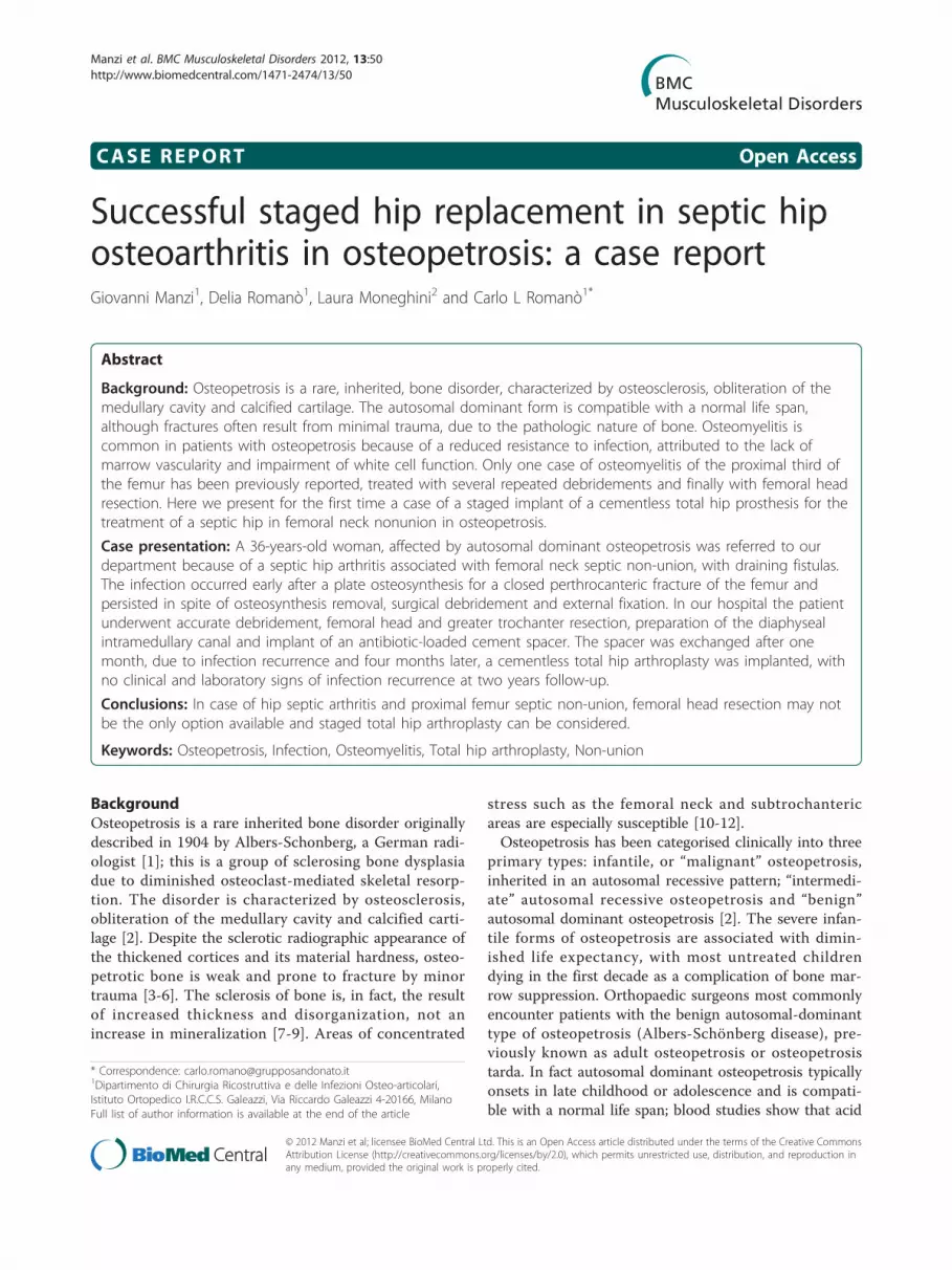
At 2-year follow-up, there are no clinical signs ofinfection recurrence while laboratory tests remain in thenormal range value and the prosthetic components donot show any sign of osteolysis at the radiographicexamination (Figure 6). The patient, although pain-free,still requires one crutch, due to a persistent weakness ofthe abductor muscles and a positive Trendelenburg sign.
DiscussionAt present, no effective medical treatment for osteope-trosis exists. Treatment is largely supportive and isaimed at providing multidisciplinary surveillance andsymptomatic management of complications. Fractures
and arthritis are common and require treatment by anexperienced orthopaedic surgeon due to the brittlenessof the bone, and the relatively frequent occurrence ofsecondary complications such as delayed union or non-union of fractures and osteomyelitis [19].Degenerative changes often occur after the age of 40
[2,5,24,31-33] in the absence of deformity. Articular car-tilage is not affected by the disorder, and it has thereforebeen suggested that the degeneration occurs because ofthe hard unyielding subchondral bone [24,32].Many intraoperative and postoperative difficulties have
been encountered in patients with osteopetrosis. Thepoor quality of bone complicates surgery because of itsresistance to drilling and tendency to fracture with mini-mal trauma, resulting in increased surgical time.In case of osteomyelitis, surgical débridement and “en
bloc” resection are necessary [23,30]. Aggressive treat-ment is indispensable because once chronic foci areformed, they are extremely difficult to eradicate [21].Another challenge in the management of osteopetrotic
patients is arthroplasty. The hardness of osteopetroticbone and obliteration of the medullary cavity precludesthe use of hand reamers [34]. The surgeon must
Figure 1 Antero-posterior (A) and axial (B) x-ray views, pre-operatively, after previous failed plate osteosynthesis andexternal fixator. Septic non-union of the femoral neck and septichip arthritis.
Figure 2 Trabecular bone made up by remodeling withreactive bone formation (A) and reduction or disappearance ofinter-trabecular spaces (B). Some residual inter-trabecular spaceshows micro-aggregation of granulocytes, expression of an acuteinflammatory process (C), while other inter-trabecular spaces arefilled up with bone matrix and fibrosis (D). At the periphery of thebone, in the sub-synovial tissue, fibrosis with chronic flogosisaround bone debris may be observed (E).
Manzi et al. BMC Musculoskeletal Disorders 2012, 13:50http://www.biomedcentral.com/1471-2474/13/50
Page 3 of 6

recreate a medullary canal with a highspeed burr orpower drill, a difficult process with many technical pro-blems [26,32]. So it could be useful a small femoralstem. There is a high risk of iatrogenic fracture duringimplant placement, so it could be useful fluoroscopicguidance. Of course there is an increased risk of
infection intraoperatively. Even with these modifications,the average surgical time in primary total hip arthro-plasty is 5 hours [26].Despite difficulties in femoral canalization in these
patients, total joint arthroplasty has proved to be effec-tive in treating osteoarthritis and periarticular fractures.Three total knee and 15 total hip arthroplasties havebeen reported in the English-language literature to date[23,26,32,34]. Of these 15 cases, only 2 had postopera-tive complications (although 1 was lost to follow-up)[26]. Cementless total hip arthroplasty also has beenattempted. Cementless acetabular component fixationhas been used because it does not require cancellousbone, which is sparse in osteopetrosis patients, forbone-cement fixation [34].Rolauffs et al. reported the only case available in the
English literature, that, similarly to ours, did presented a
Figure 3 The custom-made spacer is implanted (A). X-rax onemonth after surgery: antero-posterior (B) and axial view (C).
Figure 4 Clinical picture at the time of infection recurrence,one month after the spacer implant.
Figure 5 Intra-operative picture, at the time of cementless hipprosthesis implant.
Manzi et al. BMC Musculoskeletal Disorders 2012, 13:50http://www.biomedcentral.com/1471-2474/13/50
Page 4 of 6
chronic osteomyelitis following internal fixation of aproximal femoral fracture in a patient affected by osteo-petrosis; in that case, the authors report that four debri-dements were required to manage the recurrentinfection. Weakening of the bone finally occurred, andthe patient suffered a femoral neck fracture, that obligedto a resection arthroplasty. In the light of our result, wecannot agree with the statement of these authors that“treatment options such as arthroplasty of the osteomye-litic bone” should be ruled out in these patients, whilewe may suggest to consider a staged hip cementlessimplant.
ConclusionsTotal joint arthroplasty may be considered as a lastoption to treat osteopetrosis associated with osteoarthri-tis. However, many intraoperative and postoperativechallenges need to be overcome when performingarthroplasty in patients with osteopetrosis. The greatestchallenge in all these surgical procedures is the creationof an intramedullary canal in osteopetrotic bone withouta semblance of an intramedullary canal. This is the firstreport, to our knowledge, that shows staged total hipreplacement as a successful option for the treatment ofchronic osteomyelitis of the proximal third of the femurand septic hip arthritis.
ConsentWritten informed consent was obtained from the patientfor publication of this case report and any accompany-ing images. A copy of the written consent is availablefor review by the Editor-in-Chief of this journal.
Author details1Dipartimento di Chirurgia Ricostruttiva e delle Infezioni Osteo-articolari,Istituto Ortopedico I.R.C.C.S. Galeazzi, Via Riccardo Galeazzi 4-20166, Milano.2Servizio di Anatomia Patologica, Ospedale San Paolo, Via A.Di Rudinì 8-20142, Milano.
Authors’ contributionsGM wrote the draft of the manuscript and participated in the follow-upexamination of the patient and clinical material. DR and NL participated inthe surgical and medical treatment and followed up the patient. They alsohave been involved in drafting the manuscript or revising it critically. CLRperformed the surgery, coordinated and helped to draft and finalize themanuscript. All authors read and approved the final manuscript.
Authors’ informationCLR is the Director of the Centro di Chirurgia delle Infezioni Osteo-articolariof the research orthopaedic institute Galeazzi in Milano, Italy. Past -presidentof the Italian Studygroup on Osteoarticular Infecitons, he actually serves asPresident of the European Bone and Joint Infection Society.
Competing interestsThe authors declare that they have no competing interests.
Received: 14 October 2011 Accepted: 2 April 2012Published: 2 April 2012
References1. Albers-Schonberg H: Rottgenbilder einer selten Knochenerkrankung.
Munchen Med Wchnschr 1904, 51:365.2. Shapiro F: Osteopetrosis: current clinical considerations. Clin Orthop 1993,
294:34-44.3. Armstrong DG, Newfield JT, Gillespie R: Orthopedic management of
osteopetrosis: results of a survey and review of the literature. J PediatrOrthop 1999, 19:122-132.
4. Chhabra A, Westerlund LE, Kline AJ, McLaughlin R: Management ofproximal femoral shaft fractures in osteopetrosis: a case series usinginternal fixation. Orthopedics 2005, 28:587-592.
5. Milgram JW, Jasty M: Osteopetrosis: a morphological study of twenty-onecases. J Bone Joint Surg Am 1982, 64:912-929.
6. Del Fattore ACA, Teti A: Genetics, pathogenesis and complications ofosteopetrosis. Bone 2008, 42:19-29.
7. Manolagas SC: Birth and death of bone cells: basic regulatorymechanisms and implications for the pathogenesis and treatment ofosteoporosis. Endocr Rev 2000, 21:115-137.
Figure 6 X-ray (A) and clinical picture two years after surgery.
Manzi et al. BMC Musculoskeletal Disorders 2012, 13:50http://www.biomedcentral.com/1471-2474/13/50
Page 5 of 6

8. Walker DG: The classic: osteopetrosis cured by temporary parabiosis. ClinOrthop Relat Res 1982, 162:2-3.
9. Kovanlikaya A, Loro ML, Gilsanz V: Pathogenesis of osteosclerosis inautosomal dominant osteopetrosis. AJR Am J Roentgenol 1997,168:929-932.
10. Casden AM, Jaffe FF, Kastenbaum DM, Bonar SF: Osteoarthritis associatedwith osteopetrosis treated by total knee arthroplasty: report of a case.Clin Orthop Relat Res 1989, 247:202-207.
11. de Palma L, Tulli A, Maccauro G, Sabetta SP, del Torto M: Fracture callus inosteopetrosis. Clin Orthop Relat Res 1994, 308:85-89.
12. el-Tawil T, Stoker DJ: Benign osteopetrosis: a review of 42 cases showingtwo different patterns. Skeletal Radiol 1993, 22:587-593.
13. Bhargava A, Vagela M, Lennox CM: “Challenges in the management offractures in osteopetrosis” Review of literature and technical tips learnedfrom long-term management of seven patients. Injury 2009,40(11):1167-1171.
14. Marks SC: Pathogenesis of osteopetrosis in the rat: reduced boneresorption due to reduced osteoclast function. Am J Anat 1973,138:165-178.
15. Bollerslev J, Mosekilde L: Autosomal dominant osteopetrosis. Clin OrthopRelat Res 1993, 294:45-51.
16. Van Hul W, Vanhoenacker F, Balemans W, Janssens K, De Schepper AM:Molecular and radiological diagnosis of sclerosing bone dysplasias. Eur JRadiol 2001, 40:198-207.
17. Whyte MP: Osteopetrosis. In Connective tissue and its heritable disorders:medical, genetic, and molecular aspects. Ed2. Edited by: Royce PM, SteinmanB. New York: Wiley-Liss, Inc; 2002:753-770.
18. Van Wesenbeeck L, Van Hul W: Lessons from osteopetrotic mutations inanimals: impact on our current understanding of osteoclast biology. CritRev Eukaryot Gene Expr 2005, 15:133-162.
19. Key LL, Rodriguiz RM, Willi SM, et al: Long-term treatment of osteopetrosiswith recombinant human interferon gamma. N Engl J Med 1995,332:1594-1599.
20. Landa J, Margolis N, Di Cesare P: Orthopaedic management of the patientwith osteopetrosis. J Am Acad Orthop Surg 2007, 15:654-662.
21. Gupta R, Gupta N: Femoral fractures in osteopetrosis: case reports. JTrauma 2001, 51:997-999.
22. Gwynne-Jones DP, Hodgson BF, Hung NA: Bilateral, uncemented total hiparthroplasty in osteopetrosis. J Bone Joint Surg Br 2004, 86:276-278.
23. Ashby ME: Total hip arthroplasty in osteopetrosis: a report of two cases.Clin Orthop Relat Res 1992, 276:214-221.
24. Cameron HU, Dewar FP: Degenerative osteoarthritis associated withosteopetrosis. Clin Orthop Relat Res 1977, 127:148-149.
25. Girard J, Vendittoli PA, Lavigne M, Roy AG: Resurfacing arthroplasty of thehip in osteopetrosis. J Bone Joint Surg Br 2006, 88:818-821.
26. Strickland JP, Berry DJ: Total joint arthroplasty in patients withosteopetrosis: a report of 5 cases and review of the literature. JArthroplasty 2005, 20:815-820.
27. Tabrizi R, Arabi AM, Arabion HR, Gholami M: Jaw osteomyelitis as acomplication in osteopetrosis. J Craniofac Surg 2010, 21:136Y141.
28. Hwang JM, Kim IO, Wang KC: Complete visual recovery in osteopetrosisby early optic nerve decompression. Pediatr Neurosurg 2000, 33:328.
29. Kocher M, Kasser J: Osteopetrosis. Am J Orthop 2003, 32:222.30. Rolauffs B, Bernhardt TM, von Eiff C, Hart ML, Bettin D: Osteopetrosis,
femoral fracture, and chronic osteomyelitis caused by Staphylococcusaureus small colony variants (SCV) treated by Girdlestone resection: 6-year follow-up. Arch Orthop Trauma Surg 2002, 122:547-550.
31. Janecki CJ, Nelson CL: Osteoarthritis associated with osteopetrosis treatedby total hip replacement arthroplasty: report of a case. Cleve Clinic Q1971, 38:169-177.
32. Casden AM, Jaffe FF, Kastenbaum DM, Bonar SF: Osteoarthritis associatedwith osteopetrosis treated by total knee arthroplasty: report of a case.Clin Orthop 1989, 247:202-207.
33. Siegal A, Delling G: Total hip joint endoprosthesis in osteoporosis. Chirug1992, 63:984-987.
34. Matsuno T, Katayama N: Osteopetrosis and total hip arthroplasty: reportof two cases. Int Orthop 1997, 21:409-411.
Pre-publication historyThe pre-publication history for this paper can be accessed here:http://www.biomedcentral.com/1471-2474/13/50/prepub
doi:10.1186/1471-2474-13-50Cite this article as: Manzi et al.: Successful staged hip replacement inseptic hip osteoarthritis in osteopetrosis: a case report. BMCMusculoskeletal Disorders 2012 13:50.
Submit your next manuscript to BioMed Centraland take full advantage of:
• Convenient online submission
• Thorough peer review
• No space constraints or color figure charges
• Immediate publication on acceptance
• Inclusion in PubMed, CAS, Scopus and Google Scholar
• Research which is freely available for redistribution
Submit your manuscript at www.biomedcentral.com/submit
Manzi et al. BMC Musculoskeletal Disorders 2012, 13:50http://www.biomedcentral.com/1471-2474/13/50
Page 6 of 6


