Embed Size (px)
Citation preview

For personal use. Only reproduce with permission from The Lancet Publishing Group.
THE LANCET Neurology Vol 1 November 2002 http://neurology.thelancet.com426
Vascular dementia is the second most common type ofdementia. The subcortical ischaemic form (SIVD) frequentlycauses cognitive impairment and dementia in elderly people.SIVD results from small-vessel disease, which produceseither arteriolar occlusion and lacunes or widespreadincomplete infarction of white matter due to critical stenosisof medullary arterioles and hypoperfusion (Binswanger’sdisease). Symptoms include motor and cognitivedysexecutive slowing, forgetfulness, dysarthria, moodchanges, urinary symptoms, and short-stepped gait. Thesemanifestations probably result from ischaemic interruptionof parallel circuits from the prefrontal cortex to the basalganglia and corresponding thalamocortical connections.Brain imaging (computed tomography and magneticresonance imaging) is essential for correct diagnosis. Themain risk factors are advanced age, hypertension, diabetes,smoking, hyperhomocysteinaemia, hyperfibrinogenaemia,and other conditions that can cause brain hypoperfusionsuch as obstructive sleep apnoea, congestive heart failure,cardiac arrhythmias, and orthostatic hypotension. Cerebralautosomal dominant arteriopathy with subcortical infarctsand leucoencephalopathy (CADASIL) and some forms ofcerebral amyloid angiopathy have a genetic basis.Treatment is symptomatic and prevention requires control oftreatable risk factors.
Lancet Neurology 2002; 1: 426–36
In order of prevalence, Alzheimer’s disease, vasculardementia, and Lewy-body disease are the most commoncauses of dementia in elderly people.1 Vascular dementiaresults from ischaemic, hypoperfusive, or haemorrhagicbrain lesions that are manifest as numerous clinicalsyndromes (panel 1). Subcortical ischaemic vasculardementia (SIVD), due to small-artery disease andhypoperfusion, is clinically homogeneous and a major causeof vascular cognitive impairment and dementia. In thisarticle, we review the progress made in the understanding ofSIVD during the past decade.
Definitions and terminology The term subcortical refers to lesions, and theirmanifestations, that predominantly involve the basalganglia, cerebral white matter, and the brainstem (asopposed to cortical dementias). Dementia in SIVD is causedby ischaemic injury, which includes both completeinfarction (lacunar infarcts and microinfarcts) andincomplete infarction of deep cerebral white matter.Lacunar infarcts or lacunes are small cavitated ischaemic
infarcts of less than 15 mm in diameter. They are typicallylocated in the basal ganglia, internal capsule, thalamus,pons, corona radiata, and centrum semiovale. White-matterlacunes can overlap with non-confluent areas of ischaemicwhite-matter changes. Microinfarcts are mostly non-cavitated and are found in cortical and subcorticalstructures. Their size can range from a few microns to aboutone-tenth of the size of lacunes.
Review Subcortical ischaemic vascular dementia
GCR is at the University of Texas at San Antonio and the Audie LMurphy Memorial Veterans Hospital, San Antonio, Texas, USA. TE isat the Helsinki University Central Hospital, Helsinki, Finland. AW is atthe University of Göteborg and Sahlgrenska University Hospital atMölndal, Sweden. LP is at the University of Florence, Italy. HCC is atthe University of Southern California at Los Angeles and Rancho LosAmigos National Rehabilitation Center, Downey, California, USA.
Correspondence: Prof Gustavo C Román, University of Texas HSCat San Antonio, Medicine/Neurology, 7703 Floyd Curl Drive,San Antonio, TX 78284–7883, USA. Tel +1 210 617 5161; fax +1 210 499 4646; email [email protected]
Subcortical ischaemic vascular dementia
Gustavo C Román, Timo Erkinjuntti, Anders Wallin, Leonardo Pantoni, and Helena C Chui
Panel 1. Clinicopathological classification of vasculardementia
Large-vessel vascular dementiaMulti-infarct dementia—multiple large complete infarcts, cortical orsubcortical in location, usually with perifocal incomplete infarction involvingthe white matter
Strategic infarct dementia—a single infarct in functionally critical areas ofthe brain (angular gyrus, thalamus, basal forebrain, or territory of theposterior cerebral artery or anterior cerebral artery)
Small-vessel vascular dementiaSIVD
Binswanger’s diseaseLacunar dementia or lacunar state (état lacunaire)Multiple lacunes with extensive perifocal incomplete infarctionsCerebral autosomal dominant arteriopathy with subcortical infarcts and leucoencephalopathy (CADASIL)
Cortical-subcorticalHypertensive and arteriolosclerotic angiopathyCerebral amyloid angiopathies (including familial British dementia)Other hereditary formsCollagen-vascular disease with dementiaVenous occlusions
Ischaemic-hypoperfusive vascular dementiaDiffuse anoxic-ischaemic encephalopathyRestricted injury due to selective vulnerabilityIncomplete white-matter infarctionBorder-zone infarction
Haemorrhagic vascular dementiaTraumatic subdural haematomaSubarachnoid haemorrhageCerebral haemorrhage
Modified with permission from the Taylor and Francis Group.2

For personal use. Only reproduce with permission from The Lancet Publishing Group.
THE LANCET Neurology Vol 1 November 2002 http://neurology.thelancet.com 427
Pathological features of white-matter lesions inBinswanger’s disease include: diffuse myelin pallor (thatspares U fibres), astrocytic gliosis, widening of perivascularspaces (état crible), and lacunes in the basal ganglia andpons; loss of oligodendrocytes leading to rarefaction,spongiosis (vacuolisation), and loss of myelin and axonswithout definite necrosis (incomplete white-matterinfarction), which finally culminates in white-matternecrosis and lacunes. The term Binswanger’s disease iscontroversial and many other names have been proposed.However, we agree with Pryse-Phillips3 that “the eponympresumes less and is preferred for its brevity”. SIVDincorporates two old neuropathological and clinicalconditions, the lacunar state (état lacunaire) andBinswanger’s disease.
Pure incomplete white-matter infarction—similar tothat observed in the penumbra of large infarcts—is typicallyseen in hypoperfusive disease.
Magnitude of the problemIn clinical studies, the proportion of vascular dementiacaused by small-vessel disease ranges from 36% to 67%.4
Lacunar infarcts are found in 10% to 31% of symptomaticstrokes, with a population-based prevalence of 13·4 per100 000 in white people,5 although the prevalence is higherin oriental (Japanese and Korean), hispanic, and blackpopulations and mixed ethnic groups.4,6,7
A significant proportion of subcortical lacunes areclinically silent.8,9 In the population-based CardiovascularHealth Study, about a quarter of 3660 participants aged 65or older had one or more lacunes on magnetic resonanceimaging (MRI).10 Most lacunes (89%) were clinically silentor were manifest as gait problems and subtle cognitiveimpairments that were not recognised as stroke. In otherpopulation-based studies the prevalence of silent lacunesranged from 11% to 24%.9,11–13
Incomplete or non-cavitating ischaemic white-matterlesions of the brain—with or without lacunes—are commonin elderly people. For example, only 4·4% of 3301participants in the Cardiovascular Health Study did not havewhite-matter lesions; about 20% had extensive lesions anddid worse on timed tests of manual dexterity, gait, andcognitive performance than individuals who had mildlesions.14 According to several population-based studies, theprevalence of cerebral white-matter hyperintensities on MRIin elderly people is in the range of 62–95%.15–18 These lesionsare associated with advancing age, lacunes, hypertension,heart disease, orthostatic hypotension, smoking, and lowerincome and education.14–19 Cognitive dysfunction and gaitimpairment are related to lesion severity.19–21
Pathophysiology of ischaemic brain injuryFigure 1 shows the two main pathophysiological pathwaysinvolved in SIVD. In the first, occlusion of the arteriolarlumen due to arteriolosclerosis leads to the formation oflacunes, which results in a lacunar state (état lacunaire). Inthe second, critical stenosis and hypoperfusion of multiplemedullary arterioles causes widespread incompleteinfarction of deep white matter22 with a clinical picture of
Binswanger’s disease.23 In practice, the two clinical pathwayscan overlap; lacunes and white-matter lesions are often seentogether, which is not surprising given their commonorigins. In addition, a combination of small-vessel and large-vessel cerebrovascular disease in the same patient is notunusual. In over half of these cases, cortical and basal-ganglia microinfarctions may be present, even though theselesions are not apparent on MRI.24
Important physical principles and pathophysiologicalmechanisms involving the microcirculation in SIVD aresummarised in panel 2. These mechanisms includehaemorheological factors, increased resistance to flow,decreased autoregulation, endothelial changes, dysfunctionof the blood–brain barrier, and dilatation of perivascularspaces. Their combined effects result in hypoperfusion andincomplete infarction of deep white matter.
Determinants of ischaemiaIschaemia develops when tissue perfusion and the supply ofessential nutrients such as oxygen and glucose becomeinadequate for the support of cell metabolism. The balancebetween supply and demand is influenced by differences inthe oxygen and glucose requirements of different brain cells,regional differences in cerebral blood flow (CBF), andduration of hypoperfusion. Energy requirements areconsidered higher for neurons than for glia (neurons>oligodendrocytes>astrocytes>endothelial cells). However,experimental work has shown that oligodendrocyte swellingand vacuolar changes in myelin sheaths occur earlier andindependently of neuronal injury.25,26
Incomplete infarctionBelow a critical perfusion threshold, selective cell loss mayoccur without pronounced infarction or cystic necrosis.Selective neuronal loss occurs in the penumbra surrounding
ReviewSubcortical ischaemic vascular dementia
Hypertension
Arteriolosclerosis
Occlusion Hypoperfusion
Complete infarct Incomplete infarct
LacuneWhite matter
signal hyperintensities
Lacunar state Binswanger’s disease
Risk factor
Cause ofischaemia
Type ofbrain injury
Lesion on MRI
Clinicalsyndrome
Resultantcerebrovascular
disease
Figure 1. Two pathophysiological pathways of ischaemic brain injury. Thepathway on the left is initiated by occlusion of an arterial lumen. Thisleads to discrete areas of complete infarction (ie, lacunar infarcts) andfunctional disruption within a distributed network (eg, dementia). Thepathway on the right is defined by critical stenosis and hypoperfusioninvolving multiple small arterioles mainly in deep white matter. These twopathways often coexist in the same patient.

For personal use. Only reproduce with permission from The Lancet Publishing Group.
THE LANCET Neurology Vol 1 November 2002 http://neurology.thelancet.com428
acute infarcts,25 whereas selective loss of oligodendrocytes,myelin, and axons occurs in deep white matter of patientswith severe stenosis of medullary arterioles.27,28 This selectiveloss of tissue elements due to ischaemia is known asincomplete infarction22,25,29 and may occur when systemicblood pressure drops below autoregulatory reserve,intracranial pressure exceeds mean arterial pressure, or thereis severe stenosis of several arteries or arterioles.27–29
Data from experiments in animals suggest that thethreshold for ischaemia is not fixed but depends on theduration of hypoperfusion. Reduction of CBF to 30–60% for1–3 months in rats30,31 and gerbils32,33 produces impairmentsin memory and behaviour and patchy loss of neurons in thehippocampus, striatum, and cerebral cortex. Within3 weeks, changes in the monoaminergic system can bedetected,34 and by 2 months, demyelination and gliosis arealso observed.35
Haemorheological factorsAs summarised in panel 2, oxygen delivery to tissuesdepends on blood flow and the concentration of red bloodcells. A high concentration of red blood cells and raised
plasma viscosity are important in the pathogenesis ofBinswanger’s disease.36 Other clinically relevant haemorheo-logical factors in SIVD include hyperglycaemia, hyper-fibrinogenaemia, polycytaemia, hyperlipidaemia, andhyperviscosity.36,37
Decreased autoregulatory reserve in SIVDUnder normal conditions, autoregulatory mechanismscompensate for variations in mean arterial pressure of60–150 mm Hg. In patients with chronic hypertension,the curve is shifted upwards—ie, these individuals areunable to compensate for rapid decreases in bloodpressure.38,39 In patients with Binswanger’s disease, therange is narrowed and vasodilator capacity is impaired inresponse to carbon dioxide or acetazolamide.40,41 Patientswith small-artery disease and compromised auto-regulatory reserve can be at increased risk of ischaemia iftheir blood pressure is abruptly lowered by posturalchanges (orthostatic hypotension), by overly aggressiveantihypertensive treatment, or by cardiac failure with lowsystolic blood pressure (congestive heart failure andarrhythmias).
Decreased CBF in SIVD results, at least partly, fromcompromised vascular reserve. By use of positron emissiontomography (PET) with oxygen-15-labelled water,Kuwabara and co-workers41 found similar 25% decreases inresting CBF in patients with Alzheimer’s and Binswanger’sdiseases. However, only those patients with vascular diseasehad an impaired vasoreactive response to hypercapnia (CBFincreased by 1·8% in Binswanger’s disease vs 5·7% inAlzheimer’s disease). Impaired vasoreactivity in response toacetazolamide is found only in SIVD and not in multi-infarct dementia.40
Increased oxygen extraction fraction (OEF) in SIVDIncreased OEF is a marker of ongoing ischaemia andpending infarction. By use of 15O-PET, Yao and colleagues42
found that patients with Binswanger’s dementia hadreduced CBF and cerebral metabolic rate for oxygen(CMRO2; 20–30% lower than normal in grey matter and30–40% lower in white matter). Non-demented patientswith Binswanger’s disease had no significant changes inCBF and CMRO2 in grey matter; however, a 30% reductionin CBF and a 130% increase in OEF were found inwhite matter. In a more recent 15O-PET study,43 CBF tothe cerebral cortex and deep grey nuclei was decreasedin patients with silent lacunes. These studies suggestthe presence of occult misery perfusion in patientswith SIVD.
Relationship between ischaemia and dementiaSeverity of dementia in SIVD correlates more strongly withthe degree of hippocampal and cerebral atrophy than withseverity of white-matter hyperintensities.44–46 Nonetheless,cerebral atrophy and white-matter lesions are related.Quantitative MRI reveals widespread atrophy in SIVD that isnot solely due to focal infarction. Possible causes includeconcomitant Alzheimer’s disease, deafferentation ormetabolic idling, and hypoperfusion.
Review Subcortical ischaemic vascular dementia
Panel 2. Relevant haemorheological and autoregulatorycirculatory factors in white-matter hypoperfusion
Haemorheological factorsOxygen delivery to tissues depends on blood flow and red-cellconcentration. The rate of blood flow through a tubular vessel isdetermined by Poiseuille’s law.
Blood flow=perfusion pressure���radius4/8���length
Blood flow declines as the length of the vessel increases and its radiusdecreases. A salient feature of the equation is the overriding influence ofvessel radius (��radius4).
In vessels with a small, fixed radius (eg, in small-artery disease) the flow ofblood is determined mainly by systolic blood pressure and by bloodviscosity (�); ie, by the force (sheer stress) required to overcome theresistance of the tube wall to the fluid at a given flow velocity.
Blood is a non-homogeneous and non-newtonian fluid; ie, viscosityincreases at slow velocities in the arteriolar bed and microcirculation. Invessels affected by arteriolosclerosis, the resulting hyperviscosity mayslow down or halt blood flow.
In arterioles, there is disproportionate increase of blood viscosity withincreases in haemoglobin concentration, plasma viscosity, red-celldeformability, hyperglycaemia and hyperlipidaemia.
Autoregulatory changes Owing to anatomical features, local cerebral blood flow is lowest inperiventricular and deep white-matter regions perfused by long, narrow,non-collateral end-arterioles.
Perfusion threshold for ischaemic injury increases if hypoperfusion persistsover longer periods of time.
With autoregulation, when cerebral perfusion pressure falls, blood vesselsdilate, increasing blood volume and initially maintaining regional cerebralblood flow (rCBF).
When blood flow starts to decline, oxygen extraction fraction (OEF)increases to maintain cerebral oxygen metabolism (rCMRO2).
rCMRO2=OEF�arterial oxygen content�rCBF
When perfusion fails, pressure exceeds compensatory mechanisms andoxygen metabolism is compromised. Neuronal and glial function is lostfollowed by irreversible necrosis. In infarcted tissue, both CMRO2 and OEFvalues are minimal. Thus, increased OEF is a transitional feature signallinga critical period of incipient ischaemia.

For personal use. Only reproduce with permission from The Lancet Publishing Group.
THE LANCET Neurology Vol 1 November 2002 http://neurology.thelancet.com 429
NeuropathologyMicroangiopathySIVD has been called small-vesseldementia because changes in cerebralmicrocirculation have a central role inits pathogenesis.29 Microangiopathyis mainly related to ageing,47 arterialhypertension,29 and diabetes mellitus48
but other conditions, such as hyper-homocysteinaemia,49 may also beimportant.
Changes of cerebral blood vesselswith ageingThe occurrence of lacunes and white-matter changes increases exponentiallyafter 65 years of age, which indicatesthe importance of morphologicalchanges of cerebral microvasculaturethat occur with ageing.47,50 In additionto lengthening and tortuosity(figure 2), the lumen of medullaryarterioles in elderly individuals isprogressively reduced due to arteriolo-sclerosis (figure 2). Concentriclamellar collagen fibres and depositionof fibrohyaline substance in the sub-adventitia, with negligible changes inmedia and intima, are seen on light microscopy.47,50,53 Electron micro-scopy51,54 shows proliferation ofcollagen fibres and accumulation ofcellular debris and amorphousmaterial in the subadventitia; amyloidis not present and angionecrosis is seldom seen.
The cause of this senile arteriolosclerosis remainsunknown. It begins late in the fourth decade of life, increasesin severity with age, is more prominent in the frontal lobe,and is followed by arteriolosclerosis in the parietal, occipital,and temporal lobes.47 The severity of white-matter lesions inpatients with Binswanger’s disease increases in directproportion to the degree of stenosis due to arteriolosclerosisof medullary arterioles.55–57
Hypertensive arteriopathyThe microangiopathy of arterial hypertension has been wellstudied.8,58 The main lesions are microatheromata,lipohyalinosis, and fibrinoid necrosis. Minute foci ofmicroatheromatosis (100–400 �m) produce stenosis orocclusion of arterioles in hypertensive individuals.Lipohyalinosis is a progressive disorganisation of small-artery walls, most commonly present in vessels of less than200 �m in diameter, with subintimal deposits of a hyalinefibrinoid substance. Lipohyalinosis leads either tothrombotic occlusion of the lumen and lacunar stroke or tomural destruction with formation of microaneurysms andhypertensive cerebral haemorrhage. Fibrinoid angionecrosisoccurs with extreme hypertension producing segmentalnarrowing, dilatation, and necrosis of the vessel wall with
deposits of a brightly eosinophilic substance. Theperivascular tissues and neuropil around the constrictedspastic segments are destroyed and astrocytic oedemais present.59
White-matter lesionsVascular lesions of the cerebral white matter are best visualisedin whole-brain, myelin-stained sections that typically showmyelin pallor sparing U fibres, astrogliosis, rarefaction of theneuropil, spongiosis,60 état criblé, and loss of oligodendrocytes,myelin, and axons without definite necrosis.61,62 In these areas,arteriolosclerosis of medullary vessels is invariably found, withthickened vessel walls, narrow lumens, and late calcification.Extensive white-matter lesions can also occur in cerebralamyloid angiopathy.63 These patients carry an increased risk ofwarfarin-related intracerebral haemorrhage after ischaemicstroke (odds ratio=12·9).64
Collagenous thickening and occlusion of deepperiventricular-draining veins has been postulated to beanother factor in the production of deep hemispheric white-matter lesions.65 Subependymal lesions66 in the immediatevicinity of the ventricles correspond to decreased myelin,loss ependymal cells, reactive gliosis, and increasedextracellular fluid. Subependymal lesions can be found atany age and are probably non-pathological.
ReviewSubcortical ischaemic vascular dementia
A B C D
Ischaemia
Hypoperfusion Occlusion
Incompletewhite matter
infarction
Lacunarstroke
Figure 2. Factors leading to brain ischaemia and hypoperfusion in elderly people. Striking age-dependent morphological changes in brain vessels include tortuosity with formation of skeins incortical arterioles (A) and elongation, which results in loops and tangles (B), particularly of longpenetrating arterioles supplying deep white matter. Increase in blood-vessel length raises the blood-pressure threshold for perfusion of periventricular white matter at the distal end of these vessels.Furthermore, the lumen is stenosed by senile concentric arteriolosclerosis (D) often with calcificationof vessels (C, top right). Under normal conditions, autoregulatory mechanisms induce vasodilation inresponse to decreases in mean arterial perfusion pressure; these mechanisms become inoperative inpatients with arteriolosclerosis and calcified vessels (C). The elderly brain is therefore moresusceptible to hypotension and pump failure (cardiac arrhythmias and congestive heart failure).Delivery of oxygen to tissues, and other metabolic exchanges, are impeded by increased thickness ofvessel walls (D) and widespread état criblé (C) with enlargement of perivascular spaces of Virchow-Robin, which results from tortuosity of elongated arterioles. A, B, C, and D are reprinted withpermission from Elsevier Science.51,52

For personal use. Only reproduce with permission from The Lancet Publishing Group.
THE LANCET Neurology Vol 1 November 2002 http://neurology.thelancet.com430
LacunesLacunes result from occlusion of lenticulostriate, thalamo-perforating, and long medullary arterioles and must bedistinguished from dilated perivascular spaces (état criblé).67
Microscopically, état criblé cavities have a small vessel withinthe lacune (figure 2) and show no evidence of necrosis,macrophages, or tissue debris. On rare occasions single orwidespread dilatations of Virchow-Robin spaces presentwith subcortical cognitive deficits.68,69
SIVD is commonly associated with cortical hypo-metabolism and hypoperfusion,70 which result in corticaland hippocampal atrophy. A neuropathological study of 20patients with ischaemic vascular dementia showed lacunarstrokes and microinfarctions (11/20; 55%) and ischaemichippocampal injury.24 Arteriolosclerosis, severe large-vesselatherosclerosis, and microemboli causing corticalmicroinfarctions were also observed.
Neurochemical changesDamage to the blood–brain barrier and chronic leakage offluid and macromolecules, particularly in hypertensivepatients, could contribute to white-matter injury. Increasedconcentrations of CSF proteins were found in individualswith white-matter lesions detected by brain imaging70–72
and in brains from patients with Binswanger’s diseaseat autopsy.73,74 Increased concentrations of proteases,complement, immunoglobulins, and inflammatorycytokines may also contribute to glial and axonaldamage.75–77
Genetic factorsCerebral autosomal dominant arteriopathy with subcorticalinfarcts and leucoencephalopathy (CADASIL) is the mostcommon genetic form of vascular dementia. This autosomaldominant disorder of small cerebral vessels maps tochromosome 19q1278 and is caused by mutations in theNotch3 gene.79,80 The diagnosis can be established by skinbiopsy81 with confirmation by immunostaining with aNotch3 monoclonal antibody.82 Widespread loss of smooth-muscle cells results in decreased regional cerebral bloodvolume; these values correlate inversely with cognitiveperformance.83
Autosomal dominant forms of cerebral amyloidangiopathy are characterised by deposition of amyloid inthe walls of leptomeningeal and cerebral corticalblood vessels and are manifest clinically as recurrentlobar haemorrhages, cognitive deterioration, andischaemic strokes.84 Dementia in cerebral amyloidangiopathy of Dutch type correlates with severity of�-amyloid deposits in stenotic vessels but not withthe number of plaques and tangles.85 In familialBritish dementia with amyloid angiopathy,86 Binswanger-type deep white-matter hyperintensities and lacunarinfarcts are seen on MRI in the absence of intracerebralhaemorrhages.
Other potential genetic factors in SIVD includepolymorphisms in the genes encoding angiotensin-convertingenzyme,87 paraoxonase,88 5,10-methylenetetrahydrofolatereductase,89 and apolipoprotein-E.90
Clinical featuresThe clinical manifestations of SIVD include psychomotorslowness due to loss of control of executive cognitivefunctioning, forgetfulness, and changes in speech, affect, andmood.91 Symptoms are caused by the interruption ofprefrontal-subcortical circuits by ischaemic lesions.92,93 Thesecircuits are known to be involved in executive control ofworking memory, organisation, language, mood, regulationof attention, constructional skills, motivation, and sociallyresponsive behaviours.94–96
A recent population-based study of executive dysfunctionin elderly people97 showed a prevalence of mild impairment inone in three (33·7%) individuals and moderate to severedeficits in one in six (16·4%) individuals over the age of 60.More severe executive dysfunction was associated withadvanced age, lower education, and hispanic ethnicity.97
Detection of the clinical features and symptom patternof SIVD can vary according to the medical setting. In strokeand neurology services, focal neurological signs that resultfrom cerebrovascular events are most commonly recognised.However, psychiatrists may put emphasis on a broader rangeof cognitive, mood, and psychiatric symptoms, whereasgeneral practitioners and gerontologists may be moreconcerned with the impact of behavioural manifestations onthe patient’s carer.
Unfortunately, the mini-mental state examination(MMSE)98 and the Cambridge cognition examination(CAMCOG)99—widely used bedside tests for dementiascreening—overlook executive dysfunction. Clinicalexperience with appropriate tests indicates that frontal-system involvement predominates in vascular dementia. Forinstance, Wallin and co-workers100,101 used the STEP analysisto show the presence of frontal or prefrontal-subcorticalsymptoms in 91% of patients with this dementia.
Frontal executive control Frontal executive cognitive functions control volition,planning, programming, anticipation, inhibition ofinappropriate behaviours, and monitoring of complexgoal-directed, purposeful activities (eg, cooking, dressing,shopping, and housework).91,93,95–97,102 Loss of executivefunction is a major component of cognitive disability anddementia, due to the loss of planning capacity, workingmemory, attention, concentration, stimuli discrimination,abstraction, conceptual flexibility, and self-control. Somepatients are unable to initiate the required behaviour,whereas others fail to inhibit irrelevant behaviours. Mostpatients can perform the individual steps of a complexproblem but are unable to come up with a correctsolution.
Prefrontal-subcortical loops can be interrupted bylacunes in the striatum, globus pallidus, or thalamus or bywhite-matter lesions that disconnect the prefrontal oranterior cingulate cortices from their basal-ganglia orthalamocortical connections.95,96 Interruption of thedorsolateral prefrontal-subcortical loop results in executivedysfunction; orbitofrontal-subcortical circuit lesionspreclude frontal inhibition of the limbic system and aremanifested by uninhibited behaviours, impulsivity, and
Review Subcortical ischaemic vascular dementia

For personal use. Only reproduce with permission from The Lancet Publishing Group.
THE LANCET Neurology Vol 1 November 2002 http://neurology.thelancet.com 431
personality change.96 The anterior cingulate (medial frontal)cortex mediates motivation, thus lesions of this circuitcommonly result in apathy, abulia, and even akineticmutism.102
Clinical manifestationsThe clinical manifestations of SIVD can be separated into twogroups. In the first, symptomatic lacunes present with acutesensory-motor deficits (pure motor hemiplegia, pseudobulbarpalsy, and other lacunar syndromes). In the second, subacutemanifestations include cognitive impairment, personality andmood disorders, gait disturbances, motor dysfunction, andurinary symptoms.23,36 In clinical practice, however, mostpatients with SIVD present with a gradual course punctuatedby acute deficits leaving residual subtle focal signs (arm drift,central facial weakness, and reflex asymmetry) as well asparkinsonian signs, small-step gait, unsteadiness, or unilateralincoordination.
The clinical picture of acute single-strategic lacunardementia is characterised by the abrupt onset of cognitiveimpairment. This impairment is in many cases associatedwith confusion, apathy, psychomotor retardation,inattention, abulia, and other features of frontal-lobedysfunction but with mild focal findings (eg, hemiparesis,central facial weakness, or dysarthria). This clinical picture isin most cases the result of lacunar strokes involving theinferior genu of the internal capsule,103,104 thalamus,105 orcaudate nucleus.106 Underlying white-matter changes andsilent lacunes may also be present. Extensive frontalhypometabolism with decreased CBF has been documentedin these patients as a result of diaschisis.107
Cognitive impairment in SIVD with pronouncedexecutive dysfunction may be clinically “silent” to thephysician. However, relatives and carers may reportabnormal behaviour resulting from lack of strategicplanning and reduced speed of cognitive processing.Memory disturbances are less severe than in Alzheimer’sdisease, and mainly include forgetfulness and problemswith spontaneous recall that improve with cuesand prompting. Language, calculation ability, and otherhigher cortical functions are preserved. Intact recognitionand verbal fluency separate SIVD from Alzheimer’sdisease.108
A severe dementia syndrome is uncommon in SIVD.Recently, Kramer and colleagues109 used formal measures ofexecutive function to demonstrate executive dysfunction innon-demented patients with subcortical lacunes. Executivedysfunction was related to the extent of white-matterabnormalities but not to the number of lacunes.
Personality and mood disorders include apathy,irritability, and so-called vascular depression.110 Dysarthriaand pseudobulbar palsy may be also be present. Patientscommonly complain of urinary symptoms such as nocturiaand urge incontinence.
Gait disturbance in SIVD has been traditionally classedas “marche à petits pas”—a short-stepped, wide-based,apraxic gait with a tendency to fall.111 Patients typically turnslowly on one leg (the compass sign). Slowing of motorfunction—as well as dysarthria, dysphagia, and mild focal
motor deficits—are commonly found and seizures mayoccur. In some patients there is a striking preponderance ofextrapyramidal features, such as hypomimia, hypokinesia,axial and limb rigidity, loss of postural reflexes, and frequentfalls, generally without tremor.112
As the disease progresses, patients with SIVD limit theirfield of interest, show emotional instability, attentional loss,decreased ability to make associations, and difficulties inshifting from one idea to another, resulting in perseveration.At this stage, initiation of gait is still preserved and speed oflocomotion is quite good, but in many cases magnetic gaitand freezing may occur. As the disease progresses, posturalinstability can compromise gait initiation, equilibrium,turning, and mobility.
Assessment of patientsPopulations at riskClinicians should suspect SIVD in patients who havebehavioural changes suggestive of executive dysfunction,particularly in elderly patients with a history of hyper-tension, diabetes, cigarette smoking, hyperfibrinogenaemia,or obstructive sleep apnoea.113 The presence of congestiveheart failure,114,115 cardiac arrhythmias,116 or orthostatic hypo-tension117 is also important in elderly patients. Hypo-perfusion due to congestive heart failure is increasinglyrecognised as a significant risk factor for cognitive decline inold people, as well as a source of cerebral embolism.Congestive heart failure is the leading cause of hospitaladmissions in more developed countries and a growingproblem in less developed countries.118 A large Italian study114
recently showed cognitive impairment in 26% of patientsdischarged from hospitals after treatment for heart failure.In older patients with heart failure, cognitive impairmentwas correlated with the degree of left-ventriculardysfunction and with systolic blood pressures below130 mm Hg.115,119
Other populations at risk of unrecognised SIVD includepatients in cardiac rehabilitation services who have had acoronary-artery bypass graft120–122 and those in convalescenceunits recovering from major surgery,123 particularly hip-fracture repair.124
Neuropsychological assessmentIn a recent systematic review of the neuropsychologicalfeatures of vascular dementia, Looi and Sachdev125 confirmedthat, despite some similarities with Alzheimer’s disease,patients with vascular dementia have superior function inverbal long-term memory but greater impairment of frontalexecutive function.
Few bedside tests are available for the assessment ofexecutive dysfunction. We have found the following testsuseful in clinical settings: the CLOX test (a brief measure ofexecutive control based on a clock drawing task);126 the trail-making test part B; the behavioural dyscontrol scale97 (basedon Luria’s kinetic melody);127 the EXIT-25128 (a structuredinterview for clinical assessment of frontal symptoms); andSTEP101 (a method for the assessment of cognitive, psychiatric,and neurological frontal subcortical symptoms and signs).Ferris129 proposed a modification of the Alzheimer’s disease
ReviewSubcortical ischaemic vascular dementia

For personal use. Only reproduce with permission from The Lancet Publishing Group.
THE LANCET Neurology Vol 1 November 2002 http://neurology.thelancet.com432
assessment scale cognitive portion (ADAS-cog) called thevascular dementia assessment scale cognitive portion (VAS-cog) to include tests that assess executive domains for use intrials of vascular dementia. Román and Royall91 emphasisedthat gait, balance, micturition control, and manual dexterityshould be assessed in patients with vascular dementia todetermine their functional status. A depression scale shouldalso be used routinely in patients with SIVD.
ImagingBrain imaging is crucial as a confirmatory test for SIVD sincesilent lesions commonly occur. A diagnosis of vasculardementia can rarely be reached in the absence of vascularlesions (stroke, lacunes, and white-matter changes) as shownon brain imaging. However, the ischaemic nature of lesionsin SIVD must be assumed because the images are non-specific and their cause could be non-vascular.
Lacunar infarcts and white-matter lesions can be detectedby computed tomography (CT) and MRI of the brain, butthe two methods differ in sensitivity and, possibly, inspecificity.130,131 White-matter lesions are seen as bilaterallysymmetrical areas of hypodensity on CT and ashyperintensities in the periventricular or deep subcorticalwhite matter on FLAIR or T2-weighted MRI. They can bedistinguished from territorial infarcts by their lack of
correspondence with a specific vascular territory, well-defined margins, lack of wedge shape, lack of corticalinvolvement, and association with enlargement of ipsilateralsulci or ventricles. The abnormalities detected by CT andMRI are not identical in terms of number, site, andextension.132 There is good interobserver agreement on visualscales, but different scales attribute different significance tothe same radiological picture.133
MRI has higher sensitivity than CT for the detection ofwhite-matter lesions, but some of the changes detected onMRI are thought to represent normal radiological featureswithout pathological significance.
Lacunar infarcts are seen as round or oval cavitatedlesions with a diameter less than 15 mm. In radiologicalstudies, a limit of 3–20 mm is generally used, although thesize of a radiologically detected lacunar infarct is slightlylarger than that found at autopsy. In the chronic stage,lacunar infarcts are hypodense on CT scans, hyperintense onFLAIR or T2-weighted MRI, and hyperintense relative toCSF on proton-density MRI. Most of the CSF-isointenselesions on proton-density MRI at the level of anteriorcommissural or inferior putamen are perivascular spaces orétat criblé. Lesions smaller than 1�2 mm are more likely tobe enlarged perivascular spaces than infarcts.
Alzheimer’s disease and vascular dementiaThe clinical differentiation of vascular dementia fromAlzheimer’s disease with cerebrovascular disease can bedifficult.134 Over 60% of older patients with Alzheimer’sdisease present with incomplete white-matter infarction22
and patients with anterior-choroidal-artery stroke may meetcriteria for Alzheimer’s disease.135 The ischaemic score mayprovide additional elements for the diagnosis of the multi-infarct form of vascular dementia.136 Stepwise deterioration,fluctuating course, history of hypertension, history of stroke,and focal neurological symptoms occur more commonly invascular dementia than in Alzheimer’s disease.
Careful interview of relatives and carers for theidentification of progressive memory loss that occurred beforethe ictus can enable a diagnosis of prestroke dementia inabout 16% of patients with apparent poststroke dementia.137
Pre-existing mild cognitive impairment,138 with isolateddeficits in episodic and semantic memory, is anotherpredisposing factor; both cognitive decline and incidentAlzheimer’s disease occur at a higher rate among patientswith mild cognitive impairment than in cognitively intactage-matched controls.
Vascular risk factors may predispose not only to vasculardementia but also to the development of Alzheimer’sdisease.139 These factors include hypertension, carotid-arterywall thickness, hypercholesterolaemia, peripheral vasculardisease, apolipoprotein E �4 allele, and hyperhomo-cysteinaemia. However, a major risk factor in older patientswith mild Alzheimer’s disease lesions is the presence of oneor two basal-ganglia lacunes that increase the risk of clinicalexpression of the dementia more than 20 times.140
Recently, Du and co-workers141 showed that patientswith SIVD have smaller volumes of the entorhinal cortexand hippocampus than normal controls. However, despite
Review Subcortical ischaemic vascular dementia
Panel 3. Criteria for SIVD
The criteria for the clinical diagnosis include all of the following:
Cognitive syndrome Dysexecutive syndrome—impairment in goal formulation, initiation,planning, organising, sequencing, executing, set-shifting andmaintenance, abstractingMemory deficit—impaired recall, relatively intact recognition, moderateforgetfulness, and benefit from cues; may be mildDeterioration from a previous higher level of functioning, interference withcomplex (executive) occupational and social activities not due to physicaleffects of cerebrovascular disease alone
Cerebrovascular diseaseEvidence of relevant cerebrovascular disease by brain imagingPresence or history of neurological signs consistent with subcorticalcerebrovascular disease (such as hemiparesis, lower facial weakness,Babinski sign, sensory deficit, dysarthria, gait disorder, and extrapyramidalsigns)
Clinical features supporting the diagnosis of SIVD include thefollowing:Episodes of mild upper motor-neuron involvement such as drift, reflexasymmetry, and incoordinationEarly presence of a gait disturbance (small-step gait or marche à petits-pas magnetic, apraxic-ataxic, or Parkinsonian gait)History of unsteadiness and frequent, unprovoked fallsEarly urinary frequency, urgency, and other urinary symptoms notexplained by urological diseaseDysarthria, dysphagia, extrapyramidal signs (hypokinesia, rigidity)Behavioural and psychological symptoms such as depression, personalitychange, emotional incontinence, and psychomotor retardation
Features that make the diagnosis of SIVD uncertain or unlikelyinclude:Early onset of memory deficit and progressive worsening of memory andother cognitive cortical functions, such as language (transcortical sensoryaphasia), motor skills (apraxia), and perception (agnosia), in the absenceof corresponding focal lesions on brain imagingAbsence of relevant cerebrovascular disease lesions on brain CT scan or MRI
Modified with permission from Springer-Verlag.143
For personal use. Only reproduce with permission from The Lancet Publishing Group.
THE LANCET Neurology Vol 1 November 2002 http://neurology.thelancet.com 433
similar degrees of dementia severity, these MRI volumeswere significantly smaller in Alzheimer’s disease than in SIVD. When hippocampal and global atrophymeasurements were included, substantial improvementin accuracy was achieved in the classification ofnormal controls and patients with Alzheimer’s diseaseor SIVD.
Potential biomarkersWallin and Sjögren142 studied the concentrations of twocytoskeletal proteins—tau and the light subunit ofneurofilament protein (NFL)—in the CSF of patients withSIVD. NFL concentrations in CSF were raised, whereas tauconcentrations were normal. The serum/CSF albumin ratiowas increased, indicating vessel-wall damage and breakdownof the blood–brain barrier. By contrast, increasedconcentrations of tau in CSF are generally found inAlzheimer’s disease. Combination of CSF biomarkers andCBF could aid differential diagnosis, but the sensitivity andspecificity await confirmation.
Diagnostic criteriaCurrent criteria for vascular dementia are notinterchangeable and their sensitivity and specificity arevariable. Furthermore, none of them can distinguish mixedforms of dementia, such as Alzheimer’s disease pluscerebrovascular disease, and prospective validation ismissing. Panel 3 summarises the proposed clinical criteriafor the diagnosis of patients with SIVD143 based on amodification of the National Institute of NeurologicalDisorders and Stroke and the Assocation Internationalepour la Recherche et l’Enseignement en Neurociences(NINDS-AIREN) criteria for probable vascular dementia.144
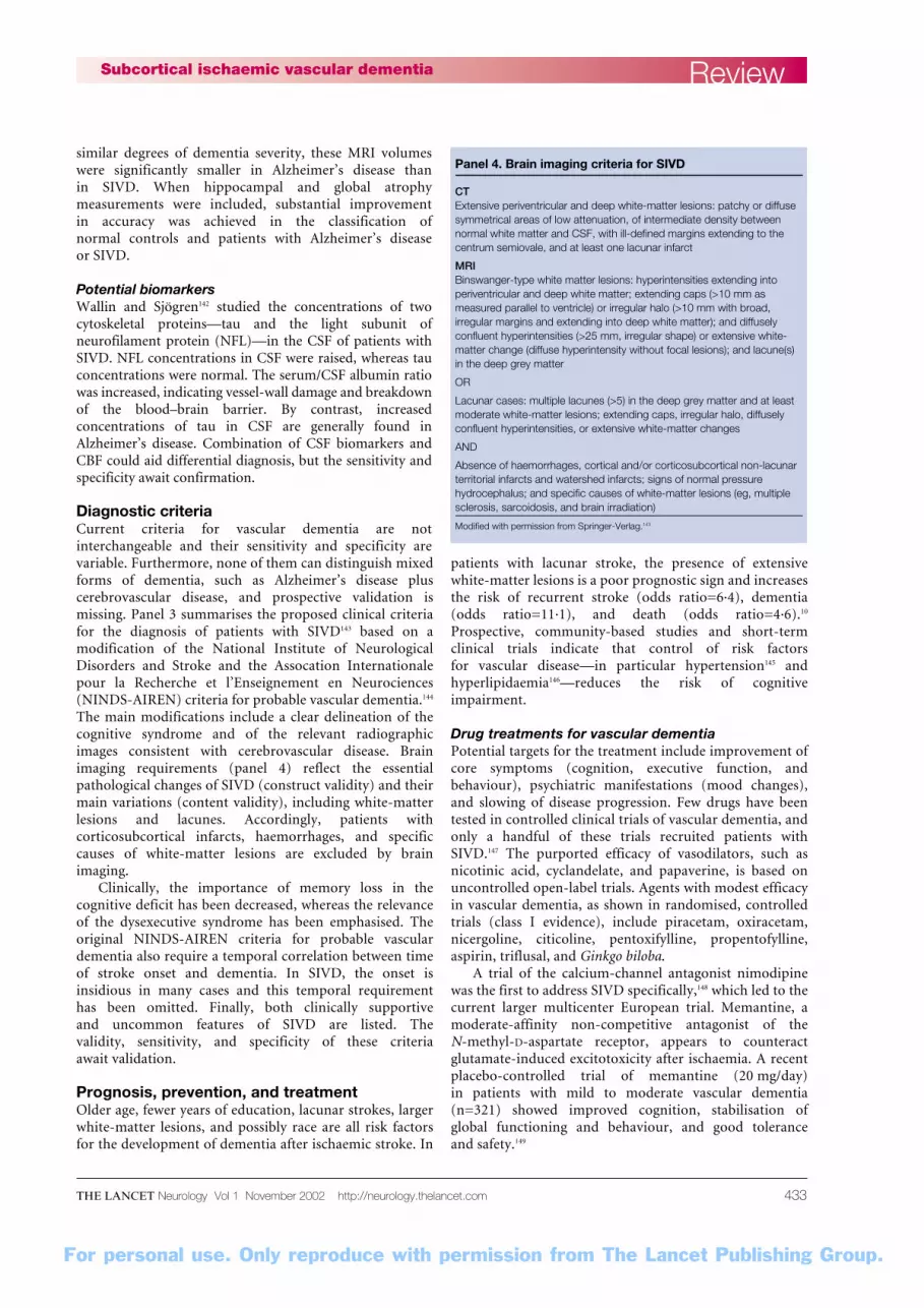
The main modifications include a clear delineation of thecognitive syndrome and of the relevant radiographicimages consistent with cerebrovascular disease. Brainimaging requirements (panel 4) reflect the essentialpathological changes of SIVD (construct validity) and theirmain variations (content validity), including white-matterlesions and lacunes. Accordingly, patients withcorticosubcortical infarcts, haemorrhages, and specificcauses of white-matter lesions are excluded by brainimaging.
Clinically, the importance of memory loss in thecognitive deficit has been decreased, whereas the relevanceof the dysexecutive syndrome has been emphasised. Theoriginal NINDS-AIREN criteria for probable vasculardementia also require a temporal correlation between timeof stroke onset and dementia. In SIVD, the onset isinsidious in many cases and this temporal requirementhas been omitted. Finally, both clinically supportiveand uncommon features of SIVD are listed. Thevalidity, sensitivity, and specificity of these criteriaawait validation.
Prognosis, prevention, and treatment Older age, fewer years of education, lacunar strokes, largerwhite-matter lesions, and possibly race are all risk factorsfor the development of dementia after ischaemic stroke. In
patients with lacunar stroke, the presence of extensivewhite-matter lesions is a poor prognostic sign and increasesthe risk of recurrent stroke (odds ratio=6·4), dementia(odds ratio=11·1), and death (odds ratio=4·6).10
Prospective, community-based studies and short-termclinical trials indicate that control of risk factorsfor vascular disease—in particular hypertension145 andhyperlipidaemia146—reduces the risk of cognitiveimpairment.
Drug treatments for vascular dementiaPotential targets for the treatment include improvement ofcore symptoms (cognition, executive function, andbehaviour), psychiatric manifestations (mood changes),and slowing of disease progression. Few drugs have beentested in controlled clinical trials of vascular dementia, andonly a handful of these trials recruited patients withSIVD.147 The purported efficacy of vasodilators, such asnicotinic acid, cyclandelate, and papaverine, is based onuncontrolled open-label trials. Agents with modest efficacyin vascular dementia, as shown in randomised, controlledtrials (class I evidence), include piracetam, oxiracetam,nicergoline, citicoline, pentoxifylline, propentofylline,aspirin, triflusal, and Ginkgo biloba.
A trial of the calcium-channel antagonist nimodipinewas the first to address SIVD specifically,148 which led to thecurrent larger multicenter European trial. Memantine, amoderate-affinity non-competitive antagonist of the N-methyl-D-aspartate receptor, appears to counteractglutamate-induced excitotoxicity after ischaemia. A recentplacebo-controlled trial of memantine (20 mg/day)in patients with mild to moderate vascular dementia(n=321) showed improved cognition, stabilisation ofglobal functioning and behaviour, and good toleranceand safety.149
ReviewSubcortical ischaemic vascular dementia
Panel 4. Brain imaging criteria for SIVD
CTExtensive periventricular and deep white-matter lesions: patchy or diffusesymmetrical areas of low attenuation, of intermediate density betweennormal white matter and CSF, with ill-defined margins extending to thecentrum semiovale, and at least one lacunar infarct
MRIBinswanger-type white matter lesions: hyperintensities extending intoperiventricular and deep white matter; extending caps (>10 mm asmeasured parallel to ventricle) or irregular halo (>10 mm with broad,irregular margins and extending into deep white matter); and diffuselyconfluent hyperintensities (>25 mm, irregular shape) or extensive white-matter change (diffuse hyperintensity without focal lesions); and lacune(s)in the deep grey matter
OR
Lacunar cases: multiple lacunes (>5) in the deep grey matter and at leastmoderate white-matter lesions; extending caps, irregular halo, diffuselyconfluent hyperintensities, or extensive white-matter changes
AND
Absence of haemorrhages, cortical and/or corticosubcortical non-lacunarterritorial infarcts and watershed infarcts; signs of normal pressurehydrocephalus; and specific causes of white-matter lesions (eg, multiplesclerosis, sarcoidosis, and brain irradiation)
Modified with permission from Springer-Verlag.143

For personal use. Only reproduce with permission from The Lancet Publishing Group.
THE LANCET Neurology Vol 1 November 2002 http://neurology.thelancet.com434
Cholinesterase inhibitorsSeveral studies have addressed the potential use ofcholinergic agents, such as donepezil hydrochloride,rivastigmine tartrate, and galantamine hydrobromide forthe treatment of vascular dementia. These agents havealready been approved for the treatment of Alzheimer’sdisease. Rivastigmine and galantamine have beeninvestigated in patients with mild to moderate Alzheimer’sdisease with stroke and vascular risk factors. In a smallopen-label trial (n=16) of rivastigmine,150 stabilisation andmild improvement in the CLOX test was seen in patientswith SIVD after a year. In a recent galantamine trial(n=592), about 40% of patients had probable vasculardementia according to NINDS-AIREN criteria.151
Cognitive function, as measured by the ADAS-cog test, wassignificantly improved compared with placebo. Theclinician’s interview-based impression of change pluscaregiver input (CIBIC-plus), behavioural measures(neuropsychiatric inventory), and activities of daily living(disability assessment in dementia) improved or remainedstable in the treated group. However, the study was notpowered for subgroup analyses of the patients with purevascular dementia.
Donepezil was studied in two large trials (n=1219) ofpure, probable, and possible vascular dementia152 in whichpatients were recruited according to the NINDS-AIRENcriteria. Patients were randomly assigned to either a lowdose (5 mg/day), a high dose (10 mg/day), or placebo for24 weeks. The patients selected clearly differed from casesof Alzheimer’s disease plus cerebrovascular disease in thatthey showed less cognitive decline in the placebo groupthan the cases of Alzheimer’s disease. The two treatedgroups showed significant improvement in cognitivescores (ADAS-cog, MMSE) and global scores (CIBIC-plus), compared with placebo. Donepezil was welltolerated by patients with a high frequency ofcardiovascular and cerebrovascular pathology.
Other agentsTreatment of depression and anxiety in patients with SIVDmay require antidepressants such as the serotonin-specificreuptake inhibitors sertraline and citalopram. The use of
tricyclic antidepressants in elderly patients with vasculardementia is discouraged owing to their anticholinergiceffects, including orthostatic hypotension. Atypicalantipsychotic drugs, such as risperidone and olanzapine,can be useful in patients showing agitation and disruptivebehaviours. However, cholinergic medications help tocontrol these problems in many patients.
ConclusionThe subcortical form of vascular dementia is one of thecommonest causes of cognitive decline in elderly people.SIVD is commonly not recognised and remainsundiagnosed, but it accounts for a significant number ofcases of dementia, recurrent falls in old age (andsubsequent hip fractures), and incontinence and results inmany admissions to nursing homes. It is therefore a heavyburden on public health; better recognition of the diseaseis necessary for maximum benefit to be derived fromtreatments that are currently available to delay diseaseprogression, as well as the introduction of primary andsecondary prevention measures.
Authors’ contributionsGCR contributed the overall plan of the review, clinical andepidemiological aspects, pathophysiology (including figure 2),prevention and treatment, and the final version of the paperincorporating the revisions from all the coauthors. TE planned thereview, contributed to the sections on radiological findings, clinicalfeatures, diagnostic criteria, treatment, and prevention. AWcontributed to the sections on clinical findings, neuropsychologicalassessment, differential diagnosis, biomarkers, prevention, andtreatment. LP contributed to the sections on definitions,neuropathology, pathophysiology, treatment, and prevention. HCCcontributed the section on pathophysiology (including figure 1), and tothe sections on neuropathology, epidemiology, assessment of patients,and prevention.
Conflict of interest GCR is adviser and member of the speaker bureau for Eisai, Pfizer, andJanssen, and participated in the clinical trial of donepezil in vasculardementia. TE has been a member of the speaker bureau for Astra-Zeneca, Boehringer-Ingelheim, Novartis, Merz, Pfizer, and Janssen-Cilag, and an adviser for Aventis, Gedeon Richter, Janssen-Cilag,Lundbeck, and Novartis; he has participated in clinical trials ofnimodipine and galantamine. AW is an adviser and a member of thespeaker bureau for Eisai, Pfizer, Janssen, and Novartis-Sweden, andparticipated in the Nordic nimodipine trial. LP is an adviser for Eisai,Pfizer, Janssen, and Bayer and participated in clinical trials ofnimodipine in vascular dementia. The above-named companies had norole in the preparation of this review or the decision to submit thepaper for publication. HCC has no conflicts of interest to disclose.
Role of the funding sourceGCR is supported by the University of Texas and the VeteransAdministration, TE by Helsinki University Central Hospital, AW byGöteborg University and the Swedish Medical Research Council(09946), LP by the Azienda Ospedaliera Careggi and the University ofFlorence, and HCC by the National Institute on Aging (P01 AG12435).None of these funding sources had a role in the preparation of thisreview or in the decision to submit it for publication.
Review Subcortical ischaemic vascular dementia
Search strategy and selection criteriaArticles were identified by searches of Medline, CurrentContents, and from relevant books and the authors’ extensivefiles. The search terms “vascular dementia”, “subcorticaldementia”, “vascular cognitive impairment”, “lacunar stroke”,and “Binswanger’s disease” were used. Recent articles werepreferentially selected.
References1 Dubois MF, Herbert R. The incidence of vascular
dementia in Canada: a comparison with Europe andEast Asia. Neuroepidemiology 2001;20: 179–87.
2 Brun A. The neuropathology of vascular dementia.In: Chiu E, Gustafson L, Ames D, Folstein MF, eds.Cerebrovascular disease and dementia. London:Martin Dunitz, 2000: 69–76.
3 Pryse-Phillips W. Companion to clinical neurology.Boston: Little, Brown and Company, 1995.
4 Chui H. Dementia due to subcortical ischaemicvascular disease. Clin Cornerstone 2001;3: 40–51.
5 Sacco SE, Whisnant JP, Broderick JP, Phillips SJ,O’Fallon WM. Epidemiological characteristics oflacunar infarcts in a population. Stroke 1991; 22:1236–41.
6 Saposnik G, González L, Lepera S, et al. SouthernBuenos Aires stroke project. Acta Neurol Scand 2001;104: 130–35.
7 Lee BI, Nam HS, Heo JH, Kim DI and The Yonsei
Stroke Team. Yonsei Stroke Registry: analysis of 1000patients with acute cerebral infarctions. CerebrovascDis 2001; 12: 145–51.
8 Fisher CM. Lacunes: small deep cerebral infarcts.Neurology 1965; 15: 774–84.
9 Shinkawa A, Ueda K, Kiyohara Y, et al. Silent cerebralinfarction in a community-based autopsy series inJapan: the Hisayama Study. Stroke 1995; 26: 380–85.
10 Longstreth WT Jr, Bernick C, Manolio T, Bryan N,Jungreis CA, Price TR. Lacunar infarcts defined bymagnetic resonance imaging of 3660 elderly people:

For personal use. Only reproduce with permission from The Lancet Publishing Group.
THE LANCET Neurology Vol 1 November 2002 http://neurology.thelancet.com 435
the Cardiovascular Health Study. Arch Neurol 1998;55: 1217–25.
11 Kase CS, Wolf PA, Chodosh EH, et al. Prevalence ofsilent stroke in patients presenting with initial stroke:the Framingham Study. Stroke 1989; 20: 850–52.
12 Giroud M, Gras P, Milan C, et al. Natural history oflacunar syndromes: contribution of the Dijonregistry of cerebrovascular complications. Rev Neurol(Paris) 1991; 147: 566–72.
13 Vermeer SE, Koudstaal PJ, Oudkerk M, Hofman A,Breteler MM. Prevalence and risk factors of silentbrain infarcts in the population-based RotterdamScan Study. Stroke 2002; 33: 21–25.
14 Longstreth WT Jr, Manolio T, Arnold A, et al.Clinical correlates of white matter findings on cranialmagnetic resonance imaging of 3301 elderly people:the Cardiovascular Health Study. Stroke 1996; 27:1274–82.
15 Breteler MMB, van Swieten JC, Bots ML, et al.Cerebral white matter lesions, vascular risk factors,and cognitive function in a population-based study:the Rotterdam Study. Neurology 1994; 44: 1246–52.
16 Ylikoski A, Erkinjuntti T, Raininko R, Sarna S,Sulkava R, Tilvis R. White matter hyperintensities onthe MRI in the neurologically non-demented elderly:analysis of cohorts of consecutive subjects aged 65 to85 years living at home. Stroke 1995; 26: 11781–87.
17 Lindgren A, Roijer A, Rudling O, et al. Cerebrallesions on magnetic resonance imaging, heartdisease, and vascular risk factors in subjects withoutstroke: a population-based study. Stroke 1994; 25:929–34.
18 Liao D, Cooper L, Cai J, et al. The prevalence andseverity of white matter lesions, their relationshipwith age, ethnicity, gender, and cardiovascular riskfactors: the ARIC study. Neuroepidemiology 1997;16: 149–62.
19 Román GC. From UBOs to Binswanger’s disease:impact of magnetic resonance imaging on vasculardementia research. Stroke 1996; 27: 1269–73.
20 de Groot JC, de Leeuw FE, Oudkerk M. Hofman A,Jolles J, Breteler MM. Cerebral white matter lesionsand subjective cognitive dysfunction: the RotterdamScan Study. Neurology 2001; 56: 1539–45.
21 Guo X, Skoog I, Matousek M, et al. A population-based study on motor performance and white matterlesions in older women. J Am Geriatr Soc 2000;48: 967–70.
22 Englund E, A, Person B. Correlations betweenhistopathologic white matter changes and protonMR relaxation times in dementia. Alzheimer DisAssoc Disord 1987; 1: 156–70.
23 Román GC. Senile dementia of the Binswanger type:a vascular form of dementia in the elderly. JAMA1987; 258: 1782–88.
24 Vinters HV, Ellis WG, Zarow C, et al.Neuropathological substrate of ischemic vasculardementia. J Neuropathol Exp Neurol 2000;59: 931–45.
25 Garcia JH, Lassen NA, Weiller C, Sperling B,Nakagawara J. Ischemic stroke and incompleteinfarction. Stroke 1996; 27: 761–65.
26 Pantoni L, Garcia JH, Gutierrez JA. Cerebral whitematter is highly vulnerable to ischemia. Stroke 1996;27: 1641–47.
27 Tomonaga M, Yamanouchi H, Tohgi H, Kameyama M. Clinicopathologic study ofprogressive subcortical vascular encephalopathy(Binswanger type) in the elderly. J Am Geriatr Soc1982; 30: 524–29.
28 Ferrer I, Bella R, Serrano MT, Marti E, Guionnet N.Arteriolosclerotic leucoencephalopathy in the elderlyand its relation to white matter lesions inBinswanger’s disease, multi-infarct encephalopathyand Alzheimer’s disease. J Neurol Sci 1990; 99: 37–50.
29 Pantoni L, García JH. The significance of cerebralwhite matter abnormalities 100 years afterBinswanger’s report: a review. Stroke 1995; 26:1293–301.
30 de la Torre JC, Fortin T, Park GAS, et al. Chroniccerebrovascular insufficiency induces dementia-likedeficits in aged rats. Brain Res 1992; 582: 186–95.
31 Tanaka K, Wada N, Hori K, Asanuma M, Nomura M,Ogawa N. Chronic cerebral hypoperfusion disruptsdiscriminative behavior in acquired-learning rats. J Neurosci Methods 1998; 84: 63–68.
32 Naritomi H. Experimental basis of multi-infarctdementia: memory impairments in rodent models ofischemia. Alzheimer Dis Assoc Disord 1991; 5: 103–11.
33 Kudoh T, Takeda M, Tanimukai S, Nashimura T.Neuropathologic changes in the gerbil brain afterchronic hypoperfusion. Stroke 1993; 24: 259–65.
34 Tanaka K, Wada N, Ogawa N. Chronic cerebralhypoperfusion induces transient reversiblemonoaminergic changes in the rat brain. NeurochemRes 2000; 25: 313–20.
35 Hattori H, Takeda M, Kudo T, et al. Cumulativewhite matter changes in the gerbil brain underchronic cerebral hypoperfusion. Acta Neuropathol1992; 84: 437–42.
36 Caplan LR. Binswanger’s disease revisited. Neurology1995; 45: 626–33.
37 Schneider R, Ringelstein EB, Zeumer H, KJiesewetter H, Jung F. The role of plasmahyperviscosity in subcortical arterioscleroticencephalopathy (Binswanger’s disease). J Neurol1987; 234: 67–73.
38 Strandgaard S, Paulson OB. Regulation of cerebralblood flow in health and disease. J CardiovascPharmacol 1992; 19 (suppl): S89–93.
39 Baron JC. Perfusion thresholds in human cerebralischemia: historical perspective and therapeuticimplications. Cerebrovasc Dis 2001; 11 (suppl 1): 2–8.
40 DeReuck J, Decoo D, Hasenbroekx MC, et al.Acetazolamide vasoreactivity in vascular dementia:a positron emission tomographic study. Eur Neurol1999; 41: 31–36.
41 Kuwabara Y, Ichiya Y, Sasaki M, Yoshida T, Masuda K. Time dependency of the acetazolamideeffect on cerebral hemodynamics in patients withchronic occlusive cerebral arteries: early stealphenomenon demonstrated by 15O-H2O positronemission tomography. Stroke 1995; 26: 1825–29.
42 Yao H, Sadoshima S, Kuwabara Y, et al. Cerebralblood flow and oxygen metabolism in patients withvascular dementia of the Binswanger type. Stroke1990; 21: 1694–99.
43 Nakane H, Ibayashi S, Fujii K, et al. Cerebral bloodflow and metabolism in patients with silent braininfarction: occult misery perfusion in cerebral cortex.J Neurol Neurosurg Psychiatry 1998; 65: 317–21.
44 Fein G, Di Sclafani V, Tanabe J, et al. Hippocampaland cortical atrophy predict dementia in subcorticalischemic vascular disease. Neurology 2000; 55:1626–35.
45 Mungas D, Jagust WJ, Reed BR, et al. MRI predictorsin subcortical ischemic vascular disease andAlzheimer’s disease. Neurology 2001; 57: 2229–35.
46 Pohjasvaara T, Mantyla R, Salonen O, et al. Howcomplex interaction of ischemic brain infarcts, whitematter lesions and atrophy relate to post-strokedementia. Arch Neurol 2000; 57: 1295–300.
47 Furuta A, Ishii N, Nishihara Y, Horie A. Medullaryarteries in aging and dementia. Stroke 1991; 22:442–46.
48 Alex M, Baron EK, Goldenberg S, Blumenthal HT.An autopsy study of cerebrovascular accident indiabetes mellitus. Circulation 1962; 25: 663–73.
49 Bertsch T, Mielke O, Holy S, et al. Homocysteine incerebrovascular disease: an independent risk factorfor subcortical vascular encephalopathy. Clin ChemLab Med 2001; 39: 721–24.
50 Ravens JR. Vascular changes in the human senilebrain. Adv Neurol 1978; 20: 487–501.
51 Duvernoy HM, Delon S, Vanson JL. Cortical bloodvessels of the human brain. Brain Res Bull 1981;7: 519–79.
52 Román GC. Vascular dementia. In: Clinical atlas ofcerebrovascular disorders, Fisher M, ed. London:Wolfe/Mosby-Year Book Europe, 1994: 13.1–13.23.
53 Klassen AC, Sung JH, Stadlan EM. Histologicalchanges in cerebral arteries with increasing age. J Neuropathol Exp Neurol 1968; 27: 607–23.
54 Akima M, Nonaka H, Kagesawa M, Tanaka K.A study on the microvasculature of the cerebralcortex. Lab Invest 1986; 55: 482–89.
55 Okeda R. MorphometrischeVergleichsuntersuchungen an Hirnarterien beiBinswangerscher Encephalopathie undHochdruckencephalopathie. Acta Neuropathol (Berl)1973; 26: 23–43.
56 Moody DM, Bell MA, Challa VR. The corpuscallosum, a unique white-matter tract: anatomicfeatures that may explain sparing in Binswangerdisease and resistance to flow of fluid masses. AJNRAm J Neuroradiol 1988; 9: 1051–59.
57 van Swieten JC, van Den Hout JHW, van Ketel BA,Hijdra A, Wokke JHJ, van Gijn J. Periventricularlesions in the white matter on magnetic resonanceimaging in the elderly: a morphometric correlationwith arteriolosclerosis and dilated perivascularspaces. Brain 1991; 114: 761–74.
58 Fisher CM. The arterial lesions underlying lacunes.Acta Neuropathol (Berl) 1969; 12: 1–15.
59 Cervós-Navarro J, Matakas F, Roggendorf W,Christmann U. The morphology of spasticintracerebral arterioles. Neuropathol Appl Neurobiol1978; 4: 369–79.
60 Erkinjuntti T, Benavente O, Eliasziw M, et al. Diffusevacuolization (spongiosis) and arteriolosclerosis inthe frontal white matter occurs in vascular dementia.Arch Neurol 1996; 53: 325–32.
61 Pantoni L, Garcia JH. Pathogenesis of leukoaraiosis:a review. Stroke 1997; 28: 652–59.
62 Révész T, Hawkins CP, du Boulay EPGH, Barnard RO, McDonald WI. Pathological findingscorrelated with magnetic resonance imaging insubcortical arteriosclerotic encephalopathy(Binswanger’s disease). J Neurol Neurosurg Psychiatry1989; 52: 1337–44.
63 Gray F, Dubas F, Roullet E, Escourolle R.Leukoencephalopathy in diffuse hemorrhagiccerebral amyloid angiopathy. Ann Neurol 1985;18: 54–59.
64 Smith EE, Rosand J, Knudsen KA, Hylek EM,Greenberg SM. Leukoaraiosis is associated withwarfarin-related hemorrhage following ischemicstroke. Neurology 2002; 59: 193–97.
65 Moody DM, Brown WR, Challa VR, Anderson RL.Periventricular venous collagenosis: association withleukoaraiosis. Radiology 1995; 194: 469–76.
66 Sze G, De Armond SJ, Brant-Zawadzki M, Davis RL,Norman D, Newton TH. Foci of MRI signal(pseudolesions) anterior to the frontal horns:histologic correlations of a normal finding.AJNR Am J Neuroradiol 1986; 7: 381–87.
67 Olsson Y, Brun A, Englund E. Fundamentalpathological lesions in vascular dementia.Acta Neurol Scand 1996; 68 (suppl 1): 31–38.
68 Uggetti C, Egitto MG, Pichiecchio A, et al.Subcortical dementia associated with strikingenlargement of the Virchow-Robin spaces andtransneural degeneration of the left mammillo-thalamic tract. Cerebrovasc Dis 2001; 12: 287–90.
69 Vital C, Julien J. Widespread dilatation ofperivascular spaces: a leukoencephalopathy causingdementia. Neurology 1997; 48: 1310–33.
70 Capizzano AA, Schuff N, Amend DL, et al.Subcortical ischemic vascular dementia: assessmentwith quantitative MR imaging and H1 MRspectroscopy. AJNR Am J Neuroradiol 2000;21: 621–30.
71 Pantoni L, Inzitari D, Pracucci G, et al. Cerebrospinalfluid proteins in patients with leucoaraiosis: possibleabnormalities in blood-brain barrier function. J Neurol Sci 1993; 115: 125–31.
72 Wallin A, Sjogren M, Edman A, Blennow K,Regland B. Symptoms, vascular risk factors and blood-brain barrier function in relation to CT white-matterchanges in dementia. Eur Neurol 2000; 44: 229–35.
73 Akiguchi I, Tomimoto H, Suenaga T, Wakita H,Budka H. Alterations in glia and axons in the brainsof Binswanger’s disease patients. Stroke 1997; 28:1423–29.
74 Akiguchi I, Tomimoto H, Suenaga T, Wakita H,Budka H. Blood-brain barrier dysfunction inBinswanger’s disease: an immunohistochemicalstudy. Acta Neuropathol (Berl) 1998; 95: 78–84.
75 Tarkowski E, Issa R, Sjogren M, et al. Increasedintrathecal levels of the angiogenic factors VEGF andTGF-beta in Alzheimer’s disease and vasculardementia. Neurobiol Aging 2002; 23: 237–43.
76 Tarkowski E, Wallin A, Regland B, Blennow K,Tarkowski A. Local and systemic GM-CSF increase inAlzheimer’s disease and vascular dementia.Acta Neurol Scand 2001; 103: 166–74.
77 Tarkowski E, Blennow K, Wallin A, Tarkowski A.Intracerebral production of tumor necrosis factor-alpha, a local neuroprotective agent in Alzheimer’sdisease and vascular dementia. J Clin Immunol 1999;19: 223–30.
78 Tournier-Lasserve E, Joutel A, Melki J, et al. Cerebralautosomal dominant arteriopathy with subcorticalinfarcts and leukoencephalopathy maps tochromosome 19q12. Nat Genet 1993; 3: 256–59.
79 Joutel A, Vahedi K, Corpechor C, et al. Strongclustering and stereotyped nature of mutations inCADASIL patients. Lancet 1997; 350: 1511–15.
80 Dichgans M, Herzog J, Gasser T. Notch3 mutationinvolving three cysteine residues in a family withtypical CADASIL. Neurology 2001; 57: 1714–17.
81 Ruchoux MM, Guerouaou D, Vandenhaute B, et al.Systemic vascular smooth muscle impairment incerebral autosomal dominant arteriopathy withsubcortical infarcts and leukoencephalopathy. ActaNeuropathol (Berl) 1995; 89: 500–12.
82 Joutel A, Favrole P, Labauge P, et al. Skin biopsyimmunostaining with a Notch3 monoclonal antibodyfor CADASIL diagnosis. Lancet 2001; 358: 2049–51.
83 Bruening R, Dichgans M, Berchtenbreiter C, et al.CADASIL: decrease in regional rCBV in hyperintensesubcortical lesions inversely correlates with disabilityand cognitive performance. AJNR Am J Neuroradiol2001; 22: 1268–74.
84 Vinters HV. Cerebral amyloid angiopathy: amicrovascular link between parenchymal andvascular dementia? Ann Neurol 2001; 49: 691–93.
85 Natte R, Maaat-Schieman ML, Haan J, Bornebroek M,
ReviewSubcortical ischaemic vascular dementia

For personal use. Only reproduce with permission from The Lancet Publishing Group.
THE LANCET Neurology Vol 1 November 2002 http://neurology.thelancet.com436
Roos RA, van Duinen SG. Dementia in hereditarycerebral hemorrhage with amyloidosis-Dutch type isassociated with cerebral amyloid angiopathy but isindependent of plaques and neurofibrillary tangles.Ann Neurol 2001; 50: 765–72.
86 Mead S, James-Galton M, Revesz T, et al. FamilialBritish dementia with amyloid angiopathy: earlyclinical, neuropsychological and imaging findings.Brain 2000; 123: 975–91.
87 Markus HS, Barley J, Lunt R, et al. Angiotensin-converting enzyme gene deletion polymorphism: a new risk factor for lacunar stroke but not carotidatheroma. Stroke 1995; 26: 1329–33.
88 Schmidt R, Schmidt H, Fazekas F, et al. MRI cerebralwhite matter lesions and paraoxonase PON1polymorphisms: three-year follow-up of the AustrianStroke Prevention Study. Arterioscler Thromb VascBiol 2000; 20: 1811–16.
89 Fassbender K, Mielke O, Bertsch T, Nafe B, Froschen S, Hennerici M. Homocysteine in cerebralmacroangiopathy and microangiopathy. Lancet 1999;353: 1586–87.
90 Bronge L, Fernaeus S-E, Blomberg M, et al. Whitematter lesions in Alzheimer patients are influencedby apolipoprotein E genotype. Dement Geriatr CognDisord 1999; 10: 89–96.
91 Román GC, Royall DR. Executive control function: a rational basis for the diagnosis of vasculardementia. Alzheimer Dis Assoc Disord 1999; 13(suppl 3): S69–80.
92 Ishii N, Nishihara Y, Imamura T. Why do frontallobe symptoms predominate in vascular dementiawith lacunes? Neurology 1986; 36: 340–45.
93 Wolfe N, Linn R, Babikian VL, et al. Frontal systemsimpairment following multiple lacunar infarcts. ArchNeurol 1990; 47: 129–32.
94 Alexander GE, DeLong MR, Strick PL. Parallelorganization of functionally segregated circuitslinking basal ganglia and cortex. Annu Rev Neurosci1986; 9: 137–81.
95 Cummings JL. Frontal-subcortical circuits andhuman behavior. Arch Neurol 1993; 50: 873–80.
96 Mega MS, Cummings JL. Frontal-subcortical circuitsand neuropsychiatric disorders. J NeuropsychiatryClin Neurosci 1994; 6: 358–70.
97 Grigsby J, Kaye K, Shetterly SM, Baxter J,Morgenstern NE, Hamman RF. Prevalence ofdisorders of executive cognitive functioning amongthe elderly: findings from the San Luis Valley Healthand Aging Study. Neuroepidemiology 2002; 21:213–20.
98 Folstein MF, Folstein SE, McHugh PR. A practicalmethod for grading the cognitive state of patients forthe clinician. J Psychiatr Res 1975; 12: 189–98.
99 Huppert FA, Brayne C, Gill C, Paykel ES, Beardsall L.CAMCOG: a concise neuropsychological test toassist dementia diagnosis: sociodemographiccharacteristics in an elderly population sample. Br J Clin Psychol 1995; 34: 529–41.
100 Wallin A, Blennow K, Gottfries CG. Subcorticalsymptoms predominate in vascular dementia. Int J Geriatr Psychiatry 1991; 6: 137–46.
101 Wallin A, Edman A, Blennow K, et al. Stepwisecomparative status analysis (STEP): a tool for theidentification of regional brain syndromes indementia. J Geriatr Psychiatry Neurol 1996; 4:185–99.
102 Royall DR. Executive cognitive impairment: a novelperspective on dementia. Neuroepidemiology 2000;19: 293–99.
103 Tatemichi TK, Desmont DW, Prohovnik I, et al.Strategic infarcts in vascular dementia: a clinical andimaging experience. Arzneimittel-Forschung/DrugResearch 1995; 45: 371–85.
104 Pantoni L, Basile AM, Romanelli M, et al. Abulia andcognitive impairment in two patients with capsulargenu infarct. Acta Neurol Scand 2001; 104: 185–90.
105 Castaigne P, Buge A, Cambier J, et al. Démencethalamique d’origine vasculaire par ramollissementbilatéral, limité au territoire du pédunculeretromamillaire: a propos de deux observationsanatomo-cliniques. Rev Neurol (Paris) 1966;114: 89–107.
106 Caplan LR, Schmahmann JD, Kase CS, et al. Caudateinfarcts. Arch Neurol 1990; 47: 133–43.
107 Chukwudelunzu FE, Meschia JF, Graff-Radford NR,Lucas JA. Extensive metabolic andneuropsychological abnormalities associated with
discrete infarction of the genu of the internalcapsule. J Neurol Neurosurg Psychiatry 2001;71: 658–62.
108 Tierney MC, Black SE, Szalai JP, et al. Recognitionmemory and verbal fluency differentiate probableAlzheimer disease from subcortical ischemic vasculardementia. Arch Neurol 2001; 58: 1654–59.
109 Kramer JH, Reed BR, Mungas D, Weiner MW,Chui HC. Executive dysfunction in subcorticalischaemic vascular disease. J Neurol NeurosurgPsychiatry 2002; 72: 217–20.
110 Alexopoulos GS, Kiosses DN, Klimstra S, Kalayam B,Bruce ML. Clinical presentation of the ‘depression-executive dysfunction syndrome’ of late life. Am J Geriatr Psychiatry 2002; 10: 98–106.
111 Bazner H, Oster M, Daffertshofer M, Hennerici M.Assessment of gait in subcortical vascularencephalopathy by computerized analysis: a cross-sectional and longitudinal study. J Neurol 2000;247: 841–49.
112 Foltynie T, Barker R, Brayne C. Vascularparkinsonism: a review of the precision andfrequency of the diagnosis. Neuroepidemiology 2002;21: 1–7.
113 Richert A, Ansarin K, Baran AS. Sleep apnea andhypertension: pathophysiologic mechanisms.Semin Nephrol 2002; 22: 71–77.
114 Zuccalà G, Onder G, Pedone C, et al. Hypotensionand cognitive impairment: selective association inpatients with heart failure. Neurology 2001;57: 1986–92
115 Pullicino PM, Hart J. Cognitive impairment incongestive heart failure? Embolism vshypoperfusion. Neurology 2001; 57: 1945–46.
116 de Leeuw FE, de Groot JC, Ouderk M, et al. Atrialfibrillation and the risk of cerebral white matterlesions. Neurology 2000; 54: 1795–801.
117 Guo Z, Viitanen M, Fratiglioni L, Winblad B. Lowblood pressure and dementia in elderly people: theKungsholmen project. BMJ 1996; 312: 805–08.
118 Mendez GF, Cowie MR. The epidemiologicalfeatures of heart failure in developing countries: areview of the literature. Int J Cardiol 2001;80: 213–19.
119 Pullicino P, Mifsud V, Wong E, Graham S, Ali I,Smajlovic D. Hypoperfusion-related cerebralischemia and cardiac left ventricular systolicdysfunction. J Stroke Cerebrovasc Dis 2001; 10:178–82.
120 Barclay LL, Weiss EM, Mattis S, Bond O, Blass JP.Unrecognized cognitive impairment in cardiacrehabilitation patients. J Am Geriatr Soc 1988; 36:22–28.
121 McKhann GM, Goldsborough MA, Borowicz LM Jr,et al. Cognitive outcome after coronary artery bypass:a one-year prospective study. Ann Thorac Surg 1997;63: 510–15.
122 Newman MF, Kirchner JL, Phillips-Bute B, et al.Longitudinal assessment of neurocognitive functionafter coronary-artery bypass surgery. N Engl J Med2001; 344: 395–402.
123 Abildstrom H, Rasmussen LS, Rentowl P, et al.Cognitive dysfunction 1-2 years after non-cardiacsurgery in the elderly: International Study of Post-Operative Cognitive Dysfunction. Acta AnaesthesiolScand 2000; 44: 1246–51.
124 Cree M, Carriere KC, Soskolne CL, Suarez-Almazor M. Functional dependence afterhip fracture. Am J Phys Med Rehabil 2001; 80:736–43.
125 Looi JC, Sachdev PS. Differentiation of vasculardementia from AD on neuropsychological tests.Neurology 1999; 53: 670–78.
126 Royall DR, Cordes JA, Polk M. CLOX: an executivedrawing task. J Neurol Neurosurg Psychiatry 1998;64: 588–94.
127 Luria AR. Higher cortical functions in man.New York: Basic Books, 1966.
128 Royall DR, Mahurin RK, Gray KF. Bedsideassessment of executive cognitive impairment: theexecutive interview. J Am Geriatr Soc 1992; 40:1221–26.
129 Ferris S. Cognitive outcome measures for VaD.Alzheimer Dis Assoc Disord 1999; 13 (suppl 3):S140–43.
130 van Swieten JC, Hijdra A, Koudstaal PJ, van Gijn J.Grading white matter lesions on CT and MRI: a
simple scale. J Neurol Neurosurg Psychiatry 1990;53: 1080–83.
131 Wahlund LO, Barkhof F, Fazekas F, et al. A newrating scale for age-related white matter changesapplicable to MRI and CT. Stroke 2001;32: 1318–22.
132 Mantyla R, Erkinjuntti T, Salonen O, et al. Variableagreement between visual rating scales for whitematter hyperintensities on MRI: comparison of 13rating scales in a post-stroke cohort. Stroke 1997;28: 1614–23.
133 Scheltens P, Erkinjunti T, Leys D, et al. Whitematter changes on CT and MRI: an overview ofvisual rating scales: European Task Force on Age-Related White Matter Changes. Eur Neurol 1998;39: 80–89.
134 Jagust W. Untangling vascular dementia. Lancet2001; 358: 2097–98.
135 Sarangi S, San Pedro EC, Mountz JM. Anteriorchoroidal artery infarction presenting as aprogressive cognitive deficit. Clin Nucl Med 2000;25: 187–90.
136 Moroney JT, Bagiella E, Desmond DW, et al. Meta-analysis of the Hachinski ischemic score inpathologically verified dementias. Neurology 1997;49: 1096–105.
137 Hénon H, Durieu I, Guerouaou D, et al. Post-strokedementia: incidence and relationship to pre-strokecognitive decline. Neurology 2001; 57: 1216–22.
138 Bennett DA, Wilson RS, Schneider JA, et al. Naturalhistory of mild cognitive impairment in olderpersons. Neurology 2002; 59: 198–205.
139 de la Torre JC. Alzheimer disease as a vasculardisorder. Stroke 2002; 23: 1152–62.
140 Snowdon DA, Greiner LH, Mortimer JA, et al. Braininfarction and the clinical expression of Alzheimer’sdisease: the Nun Study. JAMA 1997; 277: 813–17.
141 Du AT, Schuff N, Laakso MP, et al. Effects ofsubcortical ischemic vascular dementia on AD onentorhinal cortex and hippocampus. Neurology 2002;58: 1635–41.
142 Wallin A, Sjögren M. CSF cytoskeleton proteins inpatients with subcortical white-matter dementia.Mech Ageing Dev 2001; 122: 1937–49.
143 Erkinjuntti T, Inzitari D, Pantoni L, et al. Researchcriteria for subcortical vascular dementia in clinicaltrials. J Neural Transmission 2000; 59 (suppl 1):23–30.
144 Román GC, Tatemichi TK, Erkinjuntti T, et al.Vascular dementia: diagnostic criteria for researchstudies: report of the NINDS-AIREN InternationalWorkshop. Neurology 1993; 43: 250–60.
145 Birkenhager WH, Forette F, Seux ML, et al. Bloodpressure, cognitive functions, and prevention ofdementias in older patients with hypertension.Arch Intern Med 2001; 161: 152–56.
146 Jick H, Zornberg GL, Jick SS, Seshadri S, et al.Statins and the risk of dementia. Lancet 2000;356: 1627–31.
147 Román GC. Perspectives in the treatment ofvascular dementia. Drugs Today 2000; 36: 641–53.
148 Pantoni L, Rossi R, Inzitari D, et al. Efficacy andsafety of nimodipine in subcortical vasculardementia: a subgroup analysis of the ScandinavianMulti-Infarct Dementia Trial. J Neurol Sci 2000;175: 124–34.
149 Orgogozo JM, Rigaud A-S, Stoffler A, Mobius H-J,Forette F. Efficacy and safety of memantine inpatients with mild to moderate vascular dementia: arandomized placebo controlled trial (MMM 300).Stroke 2002; 33: 1834–39.
150 Moretti R, Torre P, Antonello RM, Cazzato G.Rivastigmine in subcortical vascular dementia: a comparison trial on efficacy and tolerability for 12months follow-up. Eur J Neurol 2001; 8: 361–62.
151 Erkinjuntti T, Kurtz A, Gauthier S, Bullock R,Lillenfeld S, Damaraju CV. Efficacy of galantaminein probable vascular dementia and Alzheimer’sdisease combined with cerebrovascular disease: a randomised trial. Lancet 2002; 359: 1283–90.
152 Pratt R, Perdomo CA, and The Donepezil StudyGroup. Results of clinical studies with donepezil invascular dementia. Am J Geriatr Psychiatry 2002;10 (suppl 1): 88–89.
Review Subcortical ischaemic vascular dementia


