Embed Size (px)
Citation preview
Journal of Magnetism and Magnetic Materials 347 (2013) 39–44
Contents lists available at ScienceDirect
Journal of Magnetism and Magnetic Materials
0304-88http://d
n CorrE-m
journal homepage: www.elsevier.com/locate/jmmm
Study of specific absorption rate of strontium doped lanthanummanganite nanoparticles for self-controlled hyperthermia applications
Amin ur Rashid a, Ashfaq Ahmed a, S.N. Ahmad b, S.A. Shaheen c, Sadia Manzoor a,n
a Magnetism Laboratory, Department of Physics, COMSATS Institute of Information Technology, Islamabad, Pakistanb National Center for Physics, 44000 Islamabad, Pakistanc Department of Physics, Florida State University, Tallahassee, FL 32310, USA
a r t i c l e i n f o
Article history:Received 24 March 2013Received in revised form11 June 2013Available online 26 July 2013
Keywords:Magnetic hyperthermiaManganiteSpecific absorption rateLinear response theory
53/$ - see front matter & 2013 Elsevier B.V. Ax.doi.org/10.1016/j.jmmm.2013.07.045
esponding author. Tel.: +92 3459129093.ail address: [email protected] (
a b s t r a c t
Magnetic and magnetothermal properties of strontium doped lanthanum manganite La1�xSrxMnO3
nanoparticles have been studied with the strontium concentration x varying between 0.15 and 0.45.La1�xSrxMnO3 nanoparticles were prepared by the citrate gel route. XRD results show that all samplesexhibit the characteristic perovskite structure with average particle sizes between 33 and 44 nm.Adiabatic magnetothermia measurements were carried out in an RF magnetic field of 800 A/m and214 kHz. Both magnetic and thermomagnetic behaviors are governed by the strontium content of thesamples. The saturation magnetization, Curie temperature and specific absorption rate (SAR) vary non-monotonically with x. The SAR of the nanoparticles has been calculated using the linear response theoryand good agreement with the experimental data has been observed. The intrinsic loss power (ILP) hasbeen obtained from the SAR values. ILP values of our samples are comparable to those of magnetite.
& 2013 Elsevier B.V. All rights reserved.
1. Introduction
Magnetic hyperthermia requires nanoparticles that have a highspecific absorption rate (SAR). This is the amount of heat dis-sipated per unit mass of the magnetic material in an RF magneticfield. A high SAR minimizes the dosage of the hyperthermia agent.Self-controlled hyperthermia is a more sophisticated modality ofmagnetic hyperthermia in which the Curie temperature TC of thehyperthermia agent is tuned to lie between 315 and 320 K [1]. Ithas been found that cancerous cells can be effectively destroyed atthese temperatures with minimal damage to healthy cells [2]. Bytuning the Curie temperature of the magnetic material, it can beused as a self-controlling heating agent in the desired temperaturerange, and overheating in the process can be avoided. This is dueto the fact that this type of magnetic material provides moreheating in the ferromagnetic state (below TC) and less heating inthe paramagnetic state (above TC). Thus the Curie temperature actsas a temperature control switch and maintains a constant desiredtemperature in the tumor region.
Strontium doped lanthanum manganite (La1�xSrxMnO3) nano-particles are one of the best candidates for self-controlled hyperther-mia applications. Their magnetization is higher than that of mostmaterials for such applications and their Curie temperature can betuned to lie between 315 and 320 K [3–6]. In this double-exchange
ll rights reserved.
S. Manzoor).
coupled ferromagnet (for x≥0.1), the strength of the ferromagneticexchange and associated magnetic characteristics, the saturationmagnetization MS and Curie temperature TC depend on the Srcontent. As a consequence, the SAR should also vary with the levelof Sr doping. Biocompatibility of lanthanummanganite nanoparticleswhen dispersed in culture media has been confirmed for particularconcentrations [7,8] and can be improved further by coating withsuitable biocompatible polymers such as dextran and human serumalbumin [9]. Most experimental studies of magnetic hyperthermia inthis system have focused on the concentration x¼0.25, because it isreported to have the highest saturation magnetization value [10–12]and TC values in the range required for self-controlled hyperthermia.
Recently, some very interesting theoretical studies have beendone elucidating the role of different magnetic and structuralparameters in determining the specific absorption rate of magneticnanoparticles [13,14]. These studies give explicit dependences of theSAR of the nanoparticles on parameters such as the saturationmagnetization MS, the effective anisotropy constant Keff, particlevolume V etc., and show that the optimum particle size formaximizing the SAR depends upon the values of MS and Keff [13].
We have measured the dependence of the SAR on Sr concen-tration for a series of La1�xSrxMnO3 (0.15≤x≤0.45) nanoparticlesand compared the results with values calculated using the linearresponse theory [13]. SAR depends on parameters (saturationmagnetization and particle size) and experimental parameters(RF amplitude and frequency) in a complicated manner and it isvery difficult to predict its response. However if the applied ACfield amplitude Hmax≈HK (where HK is the anisotropy field of the
ity (a
.u) x = 0.27
x = 0.33
x = 0.45
A. ur Rashid et al. / Journal of Magnetism and Magnetic Materials 347 (2013) 39–4440
ferromagnetic nanoparticles) or if Hmax⪡HK, Stoner–Wohlfarth basedmodels or the linear response theory (LRT) respectively can be usedto obtain analytical expressions to calculate the SAR [13]. Working ata very low AC field amplitude (800 A/m) and using linear responsetheory, a good quantitative agreement between the calculated andmeasured values of the SAR was observed. We have also obtained theintrinsic loss power or ILP, which gives the intrinsic materialdependent heating power, independent of experimental parameterssuch as AC field amplitude and frequency. We find our values of theILP comparable to those of magnetite and commercial ferrofluids asreported in Refs. [15,16].
20 30 40 50 60 70 80
(104
)
(128
)
(116
)
(300
)
(024
)
(202
)(110
)
(012
)
Inte
ns
x = 0.15
x = 0.20
(208
)
Fig. 1. XRD of La1�xSrxMnO3 samples annealed at 800 1C for 3 h.
Table 1Specific surface area AS, effective anisotropy constant Keff and particle size D ofLa1�xSrxMnO3 samples using BET, XRD and TEM.
Sample AS (m2 g�1) ⟨D⟩ (nm) Keff (J/m3)
DBET DXRD DTEM
La0.85Sr0.15MnO3 – – 44 – 610La0.80Sr0.20MnO3 29.09 30 37 39 680La0.73Sr0.27MnO3 30.93 29 38 36 770La0.67Sr0.33MnO3 26.93 33 33 – 1270La0.55Sr0.45MnO3 26.23 33 33 – 1500
2. Experimental details
2.1. Synthesis
Strontium doped lanthanum manganite nanoparticles La1�x
SrxMnO3 (0.15≤x≤0.45) were prepared using the citrate gel tech-nique. The following concentrations of Sr were prepared: x¼0.15,0.2, 0.27, 0.33 and 0.45. Lanthanum nitrate, strontium nitrate andmanganese acetate were used as raw materials; citric acid wasused as a fuel, and ethylene glycol was used as the gel formingagent. The precursor salts were mixed in distilled water. Citric acidand ethylene glycol were added to the solution which was heatedat 120 1C for 1 h under constant stirring. After gel formation,heating was continued at 160 1C until a gray powder was obtained.Calcination was done at 400 1C for 3.5 h to complete the reaction.The samples were annealed at 800 1C for 3 h to remove defectsand improve crystallinity. Both calcination and annealing weredone in air in a box furnace.
2.2. Characterization
The nanoparticles were characterized by XRD using a PanalyticalX'Pert Pro diffractomter (Philips) with Cu-Kα radiation. The crystallitesize was calculated from the peak half-widths of the XRD patternusing the Scherrer equation. Brunauer–Emmett–Teller (BET) surfacearea measurements were performed on a Micromeritics Geminianalyzer. The powders were degassed at 120 1C in N2 for 12 h priorto BET analyses.
The morphology, particle size and size distribution of theselected samples x¼0.20 and 0.27 were examined with a JEOLJEM 1200-EX transmission electron microscope operated at anacceleration voltage of 120 kV. Samples were prepared by drop-ping the aqueous dispersion onto a carbon-coated copper grid andallowed to air-dry. The morphology of the samples was alsostudied by using a field emission scanning electron microscope(FE-SEM) JEOL JSM-7401F.
Magnetic measurements were made using a vibrating samplemagnetometer (Quantum Design VersaLab). An RF induction unitoperating at a frequency of 214 kHz and AC field amplitude of800 A/m was used for the adiabatic magnetothermia measure-ments. Powder samples were placed in a thermally insulated vialand the heating curves were measured using a Neoptix fiber optictemperature sensor.
3. Results and discussion
3.1. Crystal structure
Powder X-ray diffraction data of the samples are shown inFig. 1. All samples retained the characteristic perovskite structureand no secondary phases were observed. Table 1 shows theaverage particle sizes obtained from the XRD spectra. These lie
between 33 and 44 nm and exhibit a slowly decreasing trend withincreasing Sr concentration, which is in agreement with previousreports [3,17]. The specific surface area AS measured with BETanalysis was used to approximate the average particle sizediameter (DBET) assuming dense, spherical particles and usingthe relation DBET¼6/ρAS with the density ρ¼6.66 g/cm3 [18]. Theparticle size determined from BET (DBET) was found to be in goodagreement with the crystallite size obtained from XRD for samplesx¼0.33 and 0.45. However, for other two samples x¼0.20 and0.27, the crystallite size from XRD analysis is slightly larger thanobtained by BET. The crystallite size derived from XRD is due to thereflection from the coherently scattering volumes of the particles(i.e. single crystal grains), whereas the BET isotherm reflects thesurface area of the powders available for gas adsorption and can beaffected by micro-pores, aggregate and agglomerate formation.
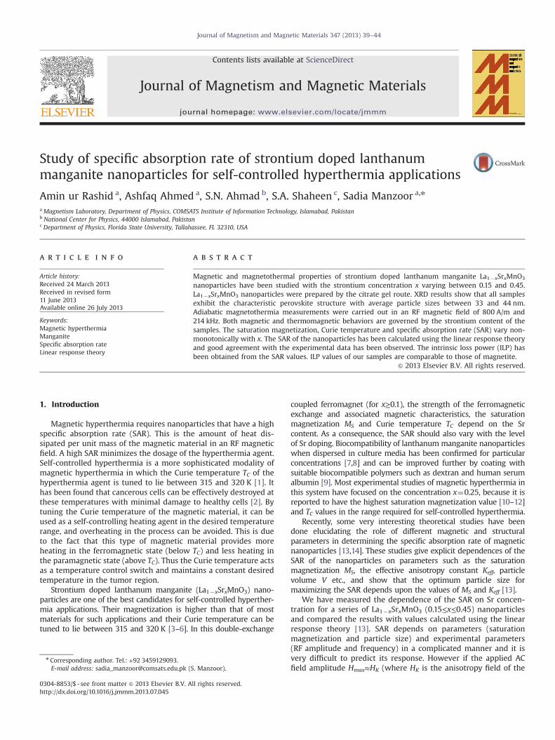
The morphology, particle size and size distribution of selectedsamples x¼0.20 and 0.27 were examined with a JEOL JEM1200-EX transmission electron microscope (TEM) operating at120 kV. Fig. 2(a) and (b) shows a typical TEM picture of thesamples x¼0.20 and 0.27, where the scale bar is 200 nm withthe inset at 100 nm scale. The corresponding size distributionhistograms each one was obtained for over 200 particles frommultiple images are shown in Fig. 2(c) and (d) along with alognormal fit. The obtained average particle sizes DTEM were 39 nmand 36 nm for samples x¼0.20 and 0.27 respectively which are ingood agreement with sizes obtained from XRD.
The morphology was also studied by using FE-SEM and theobtained micrographs are shown in Fig. 3. It can be seen that with
0
10
20
30
40
50
Cou
nt
D (nm)
<D> = 36 nm
20 30 40 50 6010 20 30 40 50 60 70 80 900
10
20
30
40
50
60
Cou
nt
D (nm)
<D> = 39 nm
Fig. 2. TEM micrographs of La1�xSrxMnO3 (a) x¼0.20 and (b) x¼0.27; the inset shows images at scale 100 nm. The corresponding size distribution histograms are shown inimages (c) and (d) for x¼0.20 and 0.27 respectively, together with a lognormal distribution fit.
A. ur Rashid et al. / Journal of Magnetism and Magnetic Materials 347 (2013) 39–44 41
increasing Sr content from Fig. 3(a)–(d), the morphology changeslightly and clusters of small spherical particles appear. A roughestimate of the grain sizes can be obtained from FE-SEM imagesand it can be seen that it lies somewhat in the range determinedfrom XRD. Agglomeration prevented exact determination of thegrain sizes from these images.
3.2. Magnetic measurements
Magnetization hysteresis loops of all samples measured at 300 Kin fields up to 1590 kA/m are shown in Fig. 4. These results show thatall samples are ferromagnetic at room temperature. The saturationmagnetization MS and coercivity HC were extracted from the M(H)loops and are shown in Fig. 5. MS increases strongly with increasingSr content from x¼0.15 to x¼0.27, and then decreases slowly athigher x values. Similar behavior has been reported earlier [4] forLa1�xSrxMnO3 nanoparticles and is well understood in terms of themixed valence states Mn3+ and Mn4+ of the Mn ions.
It can be seen in Fig. 5 that MS and HC behave inversely to eachother as x increases. An increase in MS is accompanied by a decreasein HC and vice-versa. This is an expected behavior for singe domainnanoparticles [19] and is illustrated by the following equation.
HC ¼ ð2Kef f =MSÞ½1�ðVC=VÞ1=2�; V4VC ð1Þhere HC is the coercivity,MS is the saturation magnetization, VC is thecritical particle volume below which superparamagnetism sets in, Vis the particle volume and Keff is the effective anisotropy constant.
We have used Eq. (1) to evaluate Keff which together with theparticle volume V determines the effective relaxation time τR of
the nanoparticles in an AC field. The critical particle volume VC atT¼300 K has been roughly estimated as VC∼5�10–24 m3 (usingresults obtained in Ref. [12]). The calculated values of Keff areshown in Table 1. Rostemnejadi et al. [20] have calculated theeffective anisotropy constant for La0.67Sr0.33 nanoparticles from ACsusceptibility measurements and found a sharp decrease in Keff
with increasing particles size. For the particles of 21 nm diameterthe value of Keff was found to be 2.24�103 J/m3. The valuesobtained in the present study are compatible with this result asthe particle sizes are much larger.
The thermal demagnetization of these samples was measuredin the zero field cooled (ZFC) condition in the temperature range55–400 K with a measuring field of 7.96 kA/m. These measure-ments were used to obtain the Curie temperature TC as theminimum of the (dM/dT) curve [21]. The results of these measure-ments are shown in Fig. 6. The inset of Fig. 6 shows the Curietemperatures TC for different values of x obtained by the abovementioned method. It can be seen that TC increases from 320 K forx¼0.15 and appears to saturate at 350 K for the Sr concentrationx¼0.33 and above. The results show that samples with x¼0.15 andx¼0.20 are suitable candidates for self-controlled hyperthermiaapplications, while samples with higher Sr concentrations arebetter suited to thermo-ablation [22].
3.3. Heating measurements
The heating measurements were carried out on 18 mg sampleeach in powder form. Fig. 7 shows the heating curves of all samplesin an AC magnetic field of amplitude Hmax¼800 A/m and frequency
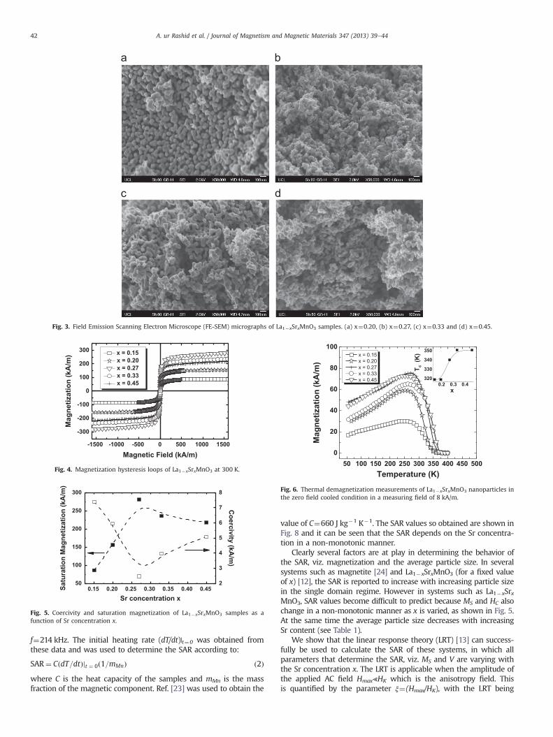
Fig. 3. Field Emission Scanning Electron Microscope (FE-SEM) micrographs of La1�xSrxMnO3 samples. (a) x¼0.20, (b) x¼0.27, (c) x¼0.33 and (d) x¼0.45.
-1500 -1000 -500 0 500 1000 1500
-300
-200
-100
0
100
200
300 x = 0.15 x = 0.20 x = 0.27 x = 0.33 x = 0.45
Mag
netiz
atio
n (k
A/m
)
Magnetic Field (kA/m)
Fig. 4. Magnetization hysteresis loops of La1�xSrxMnO3 at 300 K.
0.15 0.20 0.25 0.30 0.35 0.40 0.4550
100
150
200
250
300
2
3
4
5
6
7
8
Coercivity (kA
/m)
Satu
ratio
n M
agne
tizat
ion
(kA
/m)
Sr concentration x
Fig. 5. Coercivity and saturation magnetization of La1�xSrxMnO3 samples as afunction of Sr concentration x.
50 100 150 200 250 300 350 400 450 5000
20
40
60
80
100
0.2 0.3 0.4320
330
340
350
Mag
netiz
atio
n (k
A/m
)
Temperature (K)
x = 0.15 x = 0.20 x = 0.27 x = 0.33 x = 0.45
T c (
K)
x
Fig. 6. Thermal demagnetization measurements of La1�xSrxMnO3 nanoparticles inthe zero field cooled condition in a measuring field of 8 kA/m.
A. ur Rashid et al. / Journal of Magnetism and Magnetic Materials 347 (2013) 39–4442
f¼214 kHz. The initial heating rate (dT/dt)|t¼0 was obtained fromthese data and was used to determine the SAR according to:
SAR¼ CðdT=dtÞjt ¼ 0ð1=mMnÞ ð2Þwhere C is the heat capacity of the samples and mMn is the massfraction of the magnetic component. Ref. [23] was used to obtain the
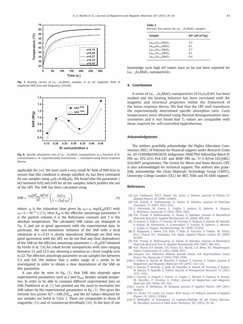
value of C¼660 J kg�1 K�1. The SAR values so obtained are shown inFig. 8 and it can be seen that the SAR depends on the Sr concentra-tion in a non-monotonic manner.
Clearly several factors are at play in determining the behavior ofthe SAR, viz. magnetization and the average particle size. In severalsystems such as magnetite [24] and La1�xSrxMnO3 (for a fixed valueof x) [12], the SAR is reported to increase with increasing particle sizein the single domain regime. However in systems such as La1�xSrxMnO3, SAR values become difficult to predict because MS and HC alsochange in a non-monotonic manner as x is varied, as shown in Fig. 5.At the same time the average particle size decreases with increasingSr content (see Table 1).
We show that the linear response theory (LRT) [13] can success-fully be used to calculate the SAR of these systems, in which allparameters that determine the SAR, viz. MS and V are varying withthe Sr concentration x. The LRT is applicable when the amplitude ofthe applied AC field Hmax⪡HK which is the anisotropy field. Thisis quantified by the parameter ξ¼(Hmax/HK), with the LRT being
0 50 100 150 200 250 300
25303540455055606570
Tem
pera
ture
(°C
)
Time (s.)
x=0.15 x=0.20 x=0.27 x=0.33 x=0.45
Fig. 7. Heating curves of La1�xSrxMnO3 samples in an AC magnetic field ofamplitude 800 A/m and frequency 214 kHz.
0.15 0.20 0.25 0.30 0.35 0.40 0.45
0.4
0.6
0.8
1.0
1.2
1.4
1.6
SAR
(W/g
)
Sr concentration x
Fig. 8. Specific absorption rate of La1�xSrxMnO3 nanoparticles as a function of Srconcentration x; ●: experimentally determined, ○: calculated using linear responsetheory.
Table 2Intrinsic loss power for La1�xSrxMnO3 samples.
Sample ILP (nH m2/kg)
La0.85Sr0.15MnO3 3.3La0.80Sr0.20MnO3 6.1La0.73Sr0.27MnO3 5.7La0.67Sr0.33MnO3 4.1La0.55Sr0.45MnO3 9.4
A. ur Rashid et al. / Journal of Magnetism and Magnetic Materials 347 (2013) 39–44 43
applicable for ξ⪡1. We have used a very small AC field of 800 A/m toensure that this condition is always satisfied. HK has been estimatedfor our samples using μ0HC≈0.48μ0HK. We found that the parameter ξlies between 0.02 and 0.09 for all the samples, which justifies the useof the LRT. The SAR has been calculated using
SAR¼ πμ20H2maxM
2SVf
kT2πf τR
1þ ð2πf τRÞ2
!ð3Þ
where τR is the relaxation time given by τR¼τ0 exp(KeffV/kT) withτ0¼5�10–11 s [13]. Here Keff is the effective anisotropy parameter, Vis the particle volume, k is the Boltzmann constant and T is theabsolute temperature. The calculated SAR values are displayed inFig. 8, and are in good agreement with the experimental data. Inparticular, the non-monotonic behavior of the SAR with a localminimum at x¼0.33 is clearly reproduced. Although we find verygood agreement with the LRT, we do not find any clear dependenceof the SAR on the effective anisotropy parameter s¼KeffV/kT obtainedby Verde et al. [14] for cobalt ferrite nanoparticles with sizes rangingbetween 3.1 and 13.5 nm, showing a variation in s from roughly zeroto 22. The effective anisotropy parameter in our samples lies between5.3 and 6.8. We believe that a wider range of s needs to beinvestigated in order to obtain a clear dependence of the SAR onthis parameter.
It can also be seen in Eq. (3), that SAR also depends uponexperimental parameters such as f and Hmax besides sample proper-ties. In order to be able to compare different experimental data onSAR, Pankhurst et al. [1] has pointed out the need to normalize theSAR values by the experimental parameters in Eq. (3). This gives theintrinsic loss power ILP ¼ SAR=f H2
max and the ILP values obtained forour samples are listed in Table 2. These are comparable to those ofmagnetite [15] and of commercial ferrofluids [16]. To the best of our
knowledge such high ILP values have so far not been reported forLa1�xSrxMnO3 nanoparticles.
4. Conclusions
A series of La1�xSrxMnO3 nanoparticles (0.15≤x≤0.45) has beenstudied and the heating behavior has been correlated with themagnetic and structural properties within the framework ofthe linear response theory. We find that the LRT well reproducesthe experimentally determined specific absorption rates. Curietemperatures were obtained using thermal demagnetization mea-surements and it was found that TC values are compatible withthose required for self-controlled hyperthermia.
Acknowledgments
The authors gratefully acknowledge the Higher Education Com-mission (HEC) of Pakistan for financial support under Research Grantno. 20-1338/R&D/09/2670, Indigenous 5000 PhD fellowship Batch IVPIN no. 072-2111-Ps4-335 and IRSIP PIN no. 17-5-4(Ps4-335)/HEC/Sch/2007 programmes. The Centre for Micro and Nano devices, CIIT,is also acknowledged for technical support. The authors also grate-fully acknowledge the Clean Materials Technology Group (CMTG),University College London (UCL) for BET, TEM and FE-SEM support.
References
[1] Q.A. Pankhurst, N.K.T. Thanh, S.K. Jones, J. Dobson, Journal of Physics D:Applied Physics 42 (2009) 224001.
[2] N.K. Prasad, K. Rathinasamy, D. Panda, D. Bahadur, Journal of MaterialsChemistry 17 (2007) 5013–5112.
[3] E. Natividad, M. Castro, G. Goglio, I. Andreu, R. Epherre, E. Duguet,A. Medianoc, Nanoscale 4 (2012) 3954–3962.
[4] N.K. Prasad, K. Rathinasamy, D. Panda, D. Bahadur, Journal of BiomedicalMaterials Research: Applied Biomaterials 85 (2008) 409–416.
[5] O. Kaman, E. Pollert, P. Veverka, M. Veverka, E. Hadová, K. Knižek, M. Maryšk,P. Kašpar, M. Klementov, V. Grunwaldov, S. Vasseur, R. Epherre, S. Mornet,G. Goglio, E. Duguet, Nanotechnology 20 (2009) 275610.
[6] R. Rajagopal, J. Mona, S.N. Kale, T. Bala, R. Pasricha, P. Poddar, M. Sastry,B.L.V. Prasad, D.C. Kundaliya, S.B. Ogale, Applied Physics Letters 89 (2006)023107.
[7] N.K. Prasad, K. Rathinasamy, D. Panda, D. Bahadur, Journal of BiomedicalMaterials Research Part B: Applied Biomaterials 85B (2007) 409–416.
[8] N.D. Thorat, K.P. Shinde, S.H. Pawar, K.C. Barick, C.A. Betty, R.S. Ningthoujam,Dalton Transactions 41 (2012) 3060–3071.
[9] Anupam Giri, Abhindan Makhal, Barnali Ghosh, A.K. Raychaudhuri, SamirKumar Pal, Nanoscale 2 (2010) 2704–2709.
[10] E. Pollert, K. Knižek, M. Maryško, P. Kašpar, S. Vasseur, E. Duguet, Journal ofMagnetism and Magnetic Materials 316 (2007) 122–125.
[11] O. Kaman, P. Veverka, Z. Jirák, M. Maryško, K. Knižek, M. Veverka, P. Kašpar,M. Burian, V. Šepelák, E. Pollert, Journal of Nanoparticle Research 13 (2011)1237–1252.
[12] S. Vasseur, E. Duguet, J. Portier, G. Goglio, S. Mornet, E. Hadová, K. Knižek,M. Maryško, P. Veverka, E. Pollert, Journal of Magnetism and MagneticMaterials 302 (2006) 315–320.
[13] J. Carrey, B. Mehdaoui, M. Respaud, Journal of Applied Physics 109 (2011)083921.
[14] E.L. Verde, G.T. Landi, J.A. Gomes, M.H. Sousa, A.F. Bakuzis, Journal of AppliedPhysics 111 (2012) 123902.
[15] B. Behdadfar, A. Kermanpur, H. Sadeghi-Aliabadi, M. del Puerto Morales,M. Mozaffari, Journal of Solid State Chemistry 187 (2012) 20–26.

A. ur Rashid et al. / Journal of Magnetism and Magnetic Materials 347 (2013) 39–4444
[16] M. Kallumadil, M. Tada, T. Nakagawa, M. Abe, P. Southern, Q.A. Pankhurst,Journal of Magnetism and Magnetic Materials 321 (2009) 1509–1513.
[17] N.D. Lipham, G.M. Tsoi, L.E. Wenger, IEEE Transactions on Magnetics 43 (2007)3088–3090.
[18] S. Dutz, R. Hergt, J. Mürbe, R. Müller, M. Zeisberger, W. Andrä, J. Töpfer,M.E. Bellemann, Journal of Magnetism and Magnetic Materials 308 (2007)305–312.
[19] E. Kneller, Theory of magnetization curve of small crystals, in Encyclopedia ofPhysics, in: H.P.J. Wijn (Ed.), Ferromagnetism, vol. XVIII/2, Springer-Verlag,New York, 1966, pp. 438–544.
[20] A. Rostamnejadi, H. Salamati, P. Kameli, 2009, p. 2815, ⟨http://arxiv.org/pdf/0907⟩.
[21] A. Rostamnejadi, M. Venkatesan, P. Kameli, H. Salamati, J.M.D. Coey, Journal ofMagnetism and Magnetic Materials 323 (2011) 2214–2218.
[22] I. Hilger, W. Andrä, R. Hergt, R. Hiergeist, H. Schubert, W.A. Kaiser, Radiology218 (2001) 570–575.
[23] D. Kim, B.L. Zink, F. Hellman, J.M.D. Coey, Physical Review B 65 (1994) 214424.[24] M. Ma, Y. Wu, J. Zhou, Y. Sun, Y. Zhang, N. Gu, Journal of Magnetism and
Magnetic Materials 268 (2004) 33–39.