Embed Size (px)
Citation preview
Structure–photoluminescence relationship in Eu(III) b-diketonate-basedorganic–inorganic hybrids. Influence of the synthesis method: carboxylicacid solvolysis versus conventional hydrolysis
Lianshe Fu,a R. A. Sa Ferreira,a N. J. O. Silva,a A. J. Fernandes,b Paulo Ribeiro-Claro,b I. S. Goncalves,b
V. de Zea Bermudezc and L. D. Carlos*a
Received 15th March 2005, Accepted 26th May 2005
First published as an Advance Article on the web 21st June 2005
DOI: 10.1039/b503844h
Organic–inorganic hybrids incorporating Eu(nta)3?bpy (where nta and bpy stand for 1-(2-
naphthyl)-4,4,4-trifluoro-1,3-butanedionate and 2,29-bipyridine, respectively) were prepared
either by acetic acid solvolysis or a conventional hydrolysis sol–gel route. The host framework of
these materials, classed as di-ureasil, consists of a siliceous network grafted, through urea cross-
linkages, to both ends of poly(ethylene oxide) chains. The resulting Eu(III)-based di-ureasils were
investigated by small angle X-ray scattering, X-ray diffraction, Fourier transform mid-infrared
spectroscopy, 29Si and 13C nuclear magnetic resonance, and photoluminescence spectroscopy,
with particular attention paid to the effect of the adopted synthesis strategy on the relationship
between structure and emission properties. The dimensions and the degree of condensation of the
siloxane nanodomains depend noticeably on the synthesis route and the overall emission quantum
yield decreases from 15 (conventional hydrolysis) to 6% (solvolysis route). The broad white-light
emission typical of the di-ureasil host was not detected here suggesting, therefore, the activation of
energy transfer channels between the hybrid host’s emitting centres and the Eu(III) ions. As the
first coordination shell of Eu(III) is essentially independent of the synthesis method employed, the
significant decrease in the emission quantum yield for the di-ureasil prepared by acetic acid
solvolysis might be explained by the interaction between the hybrid emitting centres and the nta
ligand levels, favouring a larger non-radiative transition probability.
Introduction
The sol–gel process is a promising technique for the develop-
ment of organic–inorganic hybrids due to its mild reaction
conditions, versatility of processing and potential for mixing
the inorganic and organic precursor components at the
nanometer scale.1,2 When functional active molecules, such
as optical, electronic, magnetic and biological species, are
incorporated into the hybrid structure, functional organic–
inorganic hybrid nanocomposites may be thus synthesized.
In this context, it must be emphasized that the incorporation
of luminescent molecules into organic–inorganic hybrid
matrices has made great progress in both fundamental
luminescence spectroscopic studies and the development of
advanced optical materials.2–6
Although lanthanide complexes exhibit a much more
efficient emission under ultraviolet excitation,7 up to the
present day they have been excluded from practical applica-
tions as tuneable solid-state lasers or phosphor devices due to
their poor thermal stability and mechanical properties.8 In
order to circumvent these shortcomings, the lanthanide com-
plexes can be incorporated into polymers and/or organic–
inorganic matrices using low-temperature soft-chemistry
processes, such as the sol–gel route. Indeed, much work has
been focused on this field to date, and many lanthanide com-
plexes have been incorporated into sol–gel derived matrices
or other solid hosts such as zeolite, layered or mesoporous
matrices.9
The di-urea or di-urethane cross-linked poly(ethylene oxide)
(PEO)–siloxane structures (named di-ureasils or urethanesils,
respectively) are promising hybrids for the fabrication of large
area neutron detectors,10 as nanocomposite gel electrolytes
for dye-sensitized photoeletrochemical cells11 and as efficient
white-light room temperature emitters (quantum yield of
10–20%).12–19 These materials can be prepared through
hydrolysis and condensation of the corresponding organic–
inorganic hybrid precursors obtained from the reaction of the
terminal amine groups of PEO-containing diamines (or the
hydroxyl groups of poly(ethylene glycol) for di-urethanesils)
with the isocyanate group of 3-isocyanatopropyltriethoxy-
silane (ICPTES).12 Alternatively, di-ureasils and di-urethane-
sils can be produced via acetic acid (AA) or valeric acid
solvolysis,13,14 displaying an emission quantum yield 27–35%
higher than that calculated for the analogues synthesised via
conventional sol–gel technique.14 Furthermore, transparent
and optically uniform di-ureasil films doped with a Eu3+
complex with thenoyltrifluoroacetone and 2,29-bipyridine
aDepartamento de Fısica, CICECO, Universidade de Aveiro, 3810-193,Aveiro, Portugal. E-mail: [email protected]; Fax: +351 234424695;Tel: +351 234 424370356bDepartamento de Quımica, CICECO, Universidade de Aveiro,3810-193, Aveiro, PortugalcDepartamento de Quımica and CQ-VR, Universidade de Tras-os-Montes e Alto Douro, 5000-911, Vila Real Codex, Portugal
PAPER www.rsc.org/materials | Journal of Materials Chemistry
This journal is � The Royal Society of Chemistry 2005 J. Mater. Chem., 2005, 15, 3117–3125 | 3117
(bpy) ligands were prepared by AA solvolysis.18 The Eu3+
emission, whose maximum intensity value is approximately
60% of that of Rhodamine-B, results from excitation on the
ligand levels and subsequent intramolecular energy transfer to
the 4f states. Although the organic–inorganic matrix also
seems to contribute to these energy transfer processes, the
nature of this contribution was unclear.18
The white-light photoluminescence (PL) of di-ureasils
results from a convolution of donor–acceptor pairs recombi-
nations that occur in the NH groups of the urea linkages
and in ?O–O–SiM(CO2) oxygen-related defects of the siliceous
nanodomains.3,15–17 Energy transfer between these hybrids’
emitting centres and the Eu3+ ions has been quantitatively
discussed elsewhere.19–21 The activation of these energy
transfer mechanisms noticeably depends on the Eu3+ local
coordination to the carbonyl group of the urea cross-linkages.
Moreover, that activation induces a decrease on the emission
quantum yield (relatively to that of the undoped nanohybrids)
and permits a fine-tuning of the emission chromaticity across
the CIE (Comission Internacionalle d’Eclairage) diagram.19–21
The present work aims at gaining a deeper understanding
of the hybrid host–Eu3+ energy transfer mechanisms by
comparing the luminescence features of Eu(nta)3?bpy-doped
di-ureasils synthesized through conventional hydrolysis and
AA solvolysis sol–gel routes. The emission component
associated with the hybrid host’s emitting centres could not
be detected, clearly suggesting the presence of active energy
transfer channels between them and the Eu3+ ions. The
efficiency of these energy transfer processes should be larger
compared with that of the di-ureasils with Eu(CF3SO3)3, where
the host-related emission is only absent for large amounts of
incorporated salt.19–21 On the other hand, the Eu3+ coordina-
tion in the Eu(nta)3?bpy-doped di-ureasils involves the
carbonyl oxygen of the urea bridges, independently of the
synthesis method adopted. However, the dimension and
the condensation degree of the siloxane nanodomains depend
on the synthesis route. Thus, promoting differences in the
dimensions and structure of the siloxane domains we change
the hybrid host-to-ligands-to-Eu3+ ion energy transfer
channels (essentially those connected with the nta ligand
levels) with the subsequent changes in the overall emission
quantum yield of the hybrids.
Experimental
Materials and synthesis
The diamine a, v-diaminepoly(oxyethylene-co-oxypropylene)
with a molecular weight of about 600 g mol21—corresponding
to approximately 8.5 (OCH2CH2) repeat units and commer-
cially designated as Jeffamine ED-6001, Fluka—was dried
over molecular sieves (4 A, 1.6 mm pellets, Aldrich) before use.
ICPTES (Fluka, 95%) and AA (Aldrich, 99.7%) were used
without further purification. Tetrahydrofuran (THF), chloro-
form (CHCl3) and absolute ethanol (CH3CH2OH) were dried
over molecular sieves at room temperature before use.
The europium complex Eu(nta)3?bpy22 (Scheme 1) was
synthesized by adding a solution of bpy (0.055 g, 0.36 mmol)
in 5 mL of CHCl3 to a solution of Eu(nta)3?2H2O (0.34 g,
0.35 mmol) in 15 mL of CHCl3 at room temperature. The
reaction mixture was stirred for 3 h at room temperature. Then
the solvent was removed, and the resulting solid was washed
with n-hexane. After drying in vacuum, 0.27 g of an orange
powder was obtained (yield = 71%). Elemental analysis,
Fourier transforms infrared (FTIR), Raman and nuclear
magnetic resonance (NMR) spectra confirm that the resulting
compound is the target product.
Anal. calc. for EuC52H32F9N2O6: C, 56.58; H, 2.92; N, 2.54;
Found C, 56.50; H, 2.59; N, 2.90. Selected umax/cm21 (KBr):
2977 (w), 2911 (w), 1640 (s), 1621 (s), 1614 (vs), 1568 (s), 1530
(s), 1509 (s), 1472 (m), 1463 (m), 1384 (m), 1299 (vs), 1198 (s),
1189 (s), 1135 (s), 1123(s), 793 (s), 763 (m), 684 (m), 568 (m).
Selected Raman (cm21) 3059 (m), 2990 (m), 1622 (s), 1595 (s),
1570 (w), 1466 (s), 1430 (w), 1386 (vs), 1356 (w), 1312 (m),
1292 (m), 1218 (m), 1201 (m), 1015 (m), 771 (m), 517 (m), 346
(w), 228 (w). uH (300 MHz, CDCl3, SiMe4) 12.95 (br, 2H, bpy),
10.43 (d, J = 7 Hz, 2H, bpy), 9.74 (br, 2H, Bpy), 8.52 (d,
J = 8 Hz, 2H, Bpy), 7.91 (d, J = 8 Hz, 3H, Naphth), 7.79, 7.76
(s + d, 9H, Naphth), 7.55 (d, J = 8 Hz, 6H, Naphth), 7.42 (d,
J = 8 Hz, 3H, Naphth), 3.45 (s, 3H, CH).
Thompson et al. earlier reported the synthesis of two
different crystalline structures of Eu(nta)3?bpy, one neat, and
the other with an isopropanol crystallization molecule, that
correspond to two different conformational isomers.23
Eu(nta)3?bpy was incorporated into d-U(600) di-ureasils
either by conventional hydrolysis or AA solvolysis. The
resultant luminescent hybrids were designated as d-U(600)–
Eu(nta)3bpy and d-U(600)–Eu(nta)3bpy–AA, respectively. The
Eu(nta)3?bpy content versus the total hybrid’s mass is ca. 4.5%,
corresponding to a ether-type oxygen atoms of PEO chains
per Eu3+ atom ratio equal to ca. 180. The structure of the
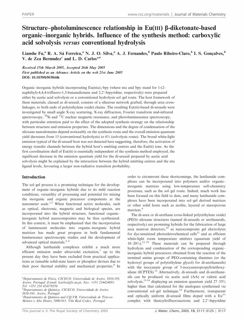
di-ureasils is outlined in Scheme 1. For the synthesis of
the d-U(600)–Eu(nta)3bpy or d-U(600)–Eu(nta)3bpy–AA, two
steps were involved. A typical synthetic procedure was as
follows:
Step 1. Synthesis of the di-ureasil precursor d-UPTES(600).
The procedure used was similar to that previously reported in
the literature.14 A volume of 0.40 mL (0.70 mmol) of Jeffamine
ED-6001 was added to a flask, followed by addition of 3.8 mL
Scheme 1 Chemical structure of (a) d-U(600) di-ureasil hybrid and
(b) Eu(nta)3?bpy complex.
3118 | J. Mater. Chem., 2005, 15, 3117–3125 This journal is � The Royal Society of Chemistry 2005
of dried THF in a fume cupboard. A volume of 0.36 mL
(1.40 mmol) of ICPTES was then added to this precursor
solution under stirring. The molar ratio of Jeffamine ED-6001
to ICPTES was 1 : 2. The flask was sealed and the solution was
stirred at room temperature in N2 atmosphere for 24 h. The
grafting process was monitored by infrared through the
observation of the progressive reduction and ultimate dis-
appearance of the band located at 2274 cm21, attributed to the
vibration of the MSi(CH2)3NCO group, and the growth of the
band envelope characteristic of the urea (urethane) group
(1800–1500 cm21 interval).
Step 2. Synthesis of d-U(600)–Eu(nta)3bpy and d-U(600)–
Eu(nta)3bpy–AA by conventional sol–gel method or carboxylic
acid solvolysis process. The THF in the precursor solution
was evaporated under vacuum, and a transparent precursor
d-UPTES(600) oil was thus obtained. For synthesis of
d-U(600)–Eu(nta)3bpy by conventional sol–gel method,
36.2 mg (0.0328 mmol) of Eu(nta)3?bpy was dissolved in
10 mL of CHCl3 and then 0.32 mL (5.48 mmol) of
CH3CH2OH and 0.075 mL (4.17 mmol) of pH = 2.0 HCl
was added to this solution. The molar ratio of ICPTES :
CH3CH2OH : H2O is 1 : 4 : 3. Finally, this mixed solution
was added to the precursor under stirring in air at
room temperature. The solution was further stirred for
24 h. For synthesis of d-U(600)–Eu(nta)3bpy–AA, 36.2 mg
(0.0328 mmol) of Eu(nta)3?bpy was dissolved in 10 mL of
CHCl3, and then 0.32 mL (5.48 mmol) of CH3CH2OH was
added to this solution. The mixture was added to the
precursor. Finally 0.24 mL of AA was added under stirring
in N2 at room temperature, and the mixture was stirred for
24 h. The following procedures are similar to the preparation
of the corresponding pure di-ureasils.
Experimental techniques
Mid-infrared spectra were recorded at room temperature using
a MATTSON 7000 FTIR Spectrometer. The spectra were
collected over the range 4000–400 cm21 by averaging 64 scans
at a maximum resolution of 4 cm21. The compounds were
finely ground (about 2 mg), mixed with approximately 175 mg
of dried potassium bromide (Merck, spectroscopic grade) and
pressed into pellets. Prior to recording the spectra the discs
were stored in an oven under vacuum at 80 uC for several days
in order to reduce the levels of by-product, solvent and
adsorbed water. Consecutive spectra were recorded until
reproducible results were obtained.
X-ray diffraction patterns were recorded using a Philips
X’Pert MPD Powder X-ray diffractometer system. The
powders were exposed to the Cu Ka radiation (l = 1.54 A)
at room temperature in a 2h range (scattering angle) between
1 and 80u. The xerogel samples, analyzed as films, were not
submitted to any thermal pre-treatment.
X-ray scattering measurements were performed using the
synchrotron SAXS beamline of the National Synchrotron
Light Laboratory (LNLS, Campinas, Brazil) with mono-
chromatic (l = 1.608 A) and horizontally focused beam. The
scattering intensity was recorded as a function of the modulus
of the scattering vector q, q = (4p/l)sin(h). The parasitic
scattering intensity from the air, slits and windows was
subtracted from the total intensity.29Si magic-angle spinning (MAS) and 13C cross-polarization
(CP) MAS NMR spectra were recorded on a Bruker Avance
400 (9.4 T) spectrometer at 79.49 and 100.62 MHz, respec-
tively. 29Si MAS NMR spectra were recorded with 2 ms
(equivalent to 30u) rf pulses, a recycle delay of 60 s and at a
5.0 kHz spinning rate. 13C CP/MAS NMR spectra were
recorded with 4 ms 1H 90u pulse, 2 ms contact time, a recycle
delay of 4 s and at a spinning rate of 8 kHz. Chemical shifts are
quoted in ppm from tetramethyl silane (TMS).
The emission, PL, and excitation, PLE, spectra and
lifetime measurements were detected between 10 K and
room temperature on a modular double grating excitation
spectrofluorimeter with a TRIAX 320 emission mono-
chromator (Fluorolog-3, Jobin Yvon-Spex) coupled to a
R928 Hamamatsu photomultiplier, in the front face acquisi-
tion mode. All the photoluminescence spectra were corrected
for optics and detection spectral response. The absolute
emission quantum yields (w) were measured at room tempera-
ture using the technique for powered samples described by
Brill et al.,24 through the following expression:
w~1{rst
1{rx
� �Ax
Ast
� �wst (1)
where rst and rx are the diffuse reflectance (with respect to a
fixed wavelength) of the hybrids and of the standard phosphor,
respectively, and wst is the quantum yield of the standard
phosphor. The terms Ax and Ast represent the area under the
di-ureasils and the standard phosphor emission spectra,
respectively. Diffuse reflectance and emission spectra were
acquired with the experimental setup aforementioned to detect
photoluminescence. In order to have absolute intensity values
BaSO4 was used as reflecting standard (r = 91%). The same
experimental conditions, i.e., the position of the hybrids/
standard holder, excitation and detection monochromator
slits (0.3 mm) and optical alignment, were fixed. To prevent
insufficient absorption of the exciting radiation, a powder
layer around 3 mm was used and utmost care was taken in
order to ensure that only the sample was illuminated, in order
to diminish the quantity of light scattered by the front sample
holder. The standard phosphor used was sodium salicylate
(Merck P.A.), whose emission spectra are formed by a large
broad band peaking around 425 nm, with a constant w value
(60%) for excitation wavelengths between 220 and 380 nm.
Three measurements were carried out for each sample, so that
the presented w value corresponds to the arithmetic mean
value. The errors in the quantum yield values associated with
this technique were estimated within 10%.24
Results and discussion
Small angle X-ray scattering and powder X-ray diffraction
The X-ray diffraction patterns of d-U(600)–AA, d-U(600)–
Eu(nta)3bpy and d-U(600)–Eu(nta)3bpy–AA are shown in
Fig. 1. These spectra show a main broad peak centred at ca.
q = 1.5 A21 associated with order within the siloxane domains,
corresponding to structural unit distances of ca. 4.1 A and
revealing that the compounds are highly amorphous. The
This journal is � The Royal Society of Chemistry 2005 J. Mater. Chem., 2005, 15, 3117–3125 | 3119

coherent length L associated with this peak was estimated
using a modified Scherrer equation (see, for instance ref. 14)
to be ca. of 9 ¡2 A. This value is identical to the one reported
for similar solvolysis-derived undoped hybrids14 and to the
diameter of the siliceous domains in organically modified
silicates obtained by SAXS.25 The second-order of the above
peak appears as a broad weak hump around 3 A21. The peak
appearing at ca. q = 0.23 A21 in XRD and SAXS patterns has
been assigned to an interference effect between siliceous
domains25 located at the ends of the polymer chain and
spatially correlated at a mean distance of 27 ¡ 1 A. The low
q-range of the SAXS patterns of the d-U(600)–Eu(nta)3bpy
and d-U(600)–Eu(nta)3bpy–AA (inset of Fig. 1) are dominated
by a power law regime: I(q) 3 q2D, with D = 2.6 and 3.2,
respectively. This low q-range scattering can be associated
with the siliceous domains, with D corresponding to their
dimensionality. Therefore, the different D values point out
distinct siloxane nanostructures for d-U(600)–Eu(nta)3bpy and
d-U(600)–Eu(nta)3bpy–AA.
Fourier transform infrared spectra (FTIR)
The FTIR spectra of d-U(600)–AA, d-U(600)–Eu(nta)3bpy,
d-U(600)–Eu(nta)3bpy–AA, and Eu(nta)3?bpy are shown in
Fig. 2. The shoulders around 920 cm21 in all the spectra
provide the evidence that in the di-ureasils the PEO chains
attain complete disorder. Comparing with the undoped di-
ureasil, the intensities of the peaks at around 920 cm21 show a
very slight decrease with the incorporation of the complex,
indicting a very weak Eu3+–PEO chains interaction. The bands
at 1324 cm21 in the spectrum of d-U(600)–AA are charac-
teristic of the amorphous state. The 1354 cm21 bands, ascribed
to the CH2 wagging vibrations, can be clearly seen in all the
spectra.
It is known that the nCO mode of PEO is an excellent tool to
probe the changes undergone by the polymer chains of the
hybrids upon incorporation of the guest compounds.26 In this
spectral region (1180–970 cm21), if complexation of the
cations by the oxygen atoms of the polyether chains occurs,
there will be an evident shift of the strong nCO band to lower
wavenumbers. The fact that peaks at around 1110 cm21,
characteristic of noncoordinated oxyethylene moieties, almost
unchanged in the spectra of the Eu3+-based di-ureasils
suggests that the PEO chains of the host materials persist in
an uncomplexed state.
In order to study in detail the vibrations of the urea groups
(the complex incorporation may induce Eu3+ coordination to
the carbonyl oxygen atoms of the urea cross-links), spectral
deconvolutions to the so called ‘‘amide I’’ (1800–1600 cm21)
and ‘‘amide II’’ regions (1600–1500 cm21) were carried out
using Gaussian band shapes as reported elsewhere.14,27 Three
components were isolated for the ‘‘amide I’’ envelope of
d-U(600)–AA at ca. 1720, 1668 and 1640 cm21 previously.
According to the literature,26,27 the former two components
are due to the vibrations of urea–poly(ether) hydrogen-bonded
structure, whereas the last one is ascribed to the strong
self-associated hydrogen-bonded urea–urea associations.
When Eu(nta)3?bpy was incorporated into d-U(600), either
by conventional sol–gel or AA solvolysis, besides the three
mentioned peaks, a new one at around 1620 cm21 appears,
indicating interactions between the Eu3+ ions and the carbonyl
oxygen atoms of the urea cross-links. Compared to the
undoped di-ureasil, the former three components have a
small red-shift peaking at ca. 1713–1716, 1657–1659 and 1632–
1636 cm21. The absence of an individual band at ca. 1750 cm21
in all the spectra indicates that neither CLO or N–H
groups from urea cross-linkages are left free in the hybrid
materials. The ‘‘amide II’’ mode is a mixed contribution of the
N–H in-plane bending, C–N stretching, and C–C stretching
vibrations. The bands for all the samples in this region appear
at ca. 1565 cm21.
NMR spectra
The 29Si MAS NMR spectra of d-U(600)–AA, d-U(600)–
Eu(nta)3bpy and d-U(600)–Eu(nta)3bpy–AA exhibit broad
Fig. 1 XRD patterns of (a) d-U(600)–AA, (b) d-U(600)–Eu(nta)3bpy
and (c) d-U(600)–Eu(nta)3bpy–AA.
Fig. 2 FTIR spectra of (a) d-U(600)–AA, (b) d-U(600)–Eu(nta)3bpy,
(c) d-U(600)–Eu(nta)3bpy–AA and (d) Eu(nta)3?bpy.
3120 | J. Mater. Chem., 2005, 15, 3117–3125 This journal is � The Royal Society of Chemistry 2005

signals characteristic of T1, T2, and T3 units (Fig. 3a).
These sites are labelled using the conventional Tn notation,
(R’Si(OSi)n(OR)32n), where n (n = 1, 2, 3) is the number of
Si-bridging oxygen atoms. The absence of T0 indicates that,
although the polycondensations in most cases do not proceed
to completion, no precursor is left unreacted. In addition, the
absence of signals due to Qn notation (Si(OSi)n(OH)42n)
species between 290 and 2120 ppm, indicates that no cleavage
of the urea groups or Si–C bonds occurred under the
experimental conditions employed and that all the silicon
atoms are covalently connected to carbon atoms. The
dominate environments (at 258 and 266 ppm, respectively)
for all the samples are clearly seen, showing the presence of
two main types of local structures. The very weak shoulders
displayed in these hybrids at ca. 252 ppm are ascribed to T1
sites. Compared to the undoped sample, there is a relative
intensity inversion between T2 and T3 for d-U(600)–
Eu(nta)3bpy and d-U(600)–Eu(nta)3bpy–AA, indicating that
the complex incorporation affected the polycondensation
process. The relative populations of the various silicon sites
quantitatively estimated after deconvolutions are listed in
Table 1. The d-U(600)–Eu(nta)3bpy di-ureasil has T1 and T2
ratios higher than the d-U(600)–Eu(nta)3bpy–AA sample,
indicating that the hydrolysis favours structures with
lower dimension compared with the AA solvolysis, in
agreement with SAXS results. The condensation degree
obtained, c = 1/3(%T1 + 2%T2 + 3%T3), is ca. 68 and 78%,
for d-U(600)–Eu(nta)3bpy and d-U(600)–Eu(nta)3bpy–AA,
respectively (Table 1). Compared to the undoped sample, the
condensation degree decreases, suggesting that there are some
interactions between the complex and the matrix (urea cross-
links) and that the relative larger molecular size for complex
sterically prevents the polycondensation process and results in
the lower condensation degree. This is in agreement with the
results previously obtained when the lanthanide complex was
formed a in silicon–base matrix via a conventional sol–gel
route.28
The 13C CP MAS NMR spectra of d-U(600)–AA,
d-U(600)–Eu(nta)3bpy and d-U(600)–Eu(nta)3bpy–AA are
shown in Fig. 3b. Peak assignments were done previously
elsewhere for undoped di-ureasil.14 The most intense peak at
about 70.6 ppm is attributed to the main-chain carbons of
ethylene oxide, whereas the shoulders at about 75.0 ppm are
originated from the main-chain carbons of propylene oxide.
These two bands are partially overlapped. The peaks at ca.
45.5, 24.5 and 10.8 ppm are characteristics of the (CH2)3
aliphatic chains. The weak peak at approximately 160 ppm is
associated with the CLO groups in the urea-linkages.27,28 The
signal at 18.4 ppm is assigned to different –CH3 groups of the
polymer chains, while the shoulder at 17.4 ppm is ascribed
to the ethoxy groups of the carbons. The spectra of the two
doped hybrids exhibit similar profiles compared with those
of the undoped di-ureasil, with the exception of a decrease in
the intensity of the peaks at ca.160 ppm and those arising
from (CH2)3 aliphatic chains. This supports the interaction
between the complex and the CLO groups of the hybrid
matrix, in agreement with the results obtained from FT–IR
and 29Si NMR.
Photoluminescence measurements
Fig. 4 shows the room temperature PL spectra for several
excitation wavelengths for the d-U(600)–Eu(nta)bpy–AA. The
spectra are mainly composed of a series of straight lines typical
of the Eu3+ energy level structure, assigned to the 5D0 A 7F0–4
transitions. The spectra also present a very low-intensity
emission band in the blue-green spectral region. Such emission
must arise from the ligands triplet levels or from the hybrid
host emitting levels. The undoped di-ureasil presents a large
broad band in the blue spectral region, similar to that
previously observed in other amine-functionalized organic–
inorganic hybrids prepared through solvolysis and hydro-
lysis.3,14–17 This band was previously attributed to the
Fig. 3 29Si MAS NMR spectra (a) and 13C CP/MAS NMR spectra (b) of (i) d-U(600)–AA, (ii) d-U(600)–Eu(nta)3bpy and (iii) d-U(600)–
Eu(nta)3bpy–AA.
Table 1 29Si NMR chemical shifts (ppm), population of different Tn
(n = 1,2,3) species (%), and degree of condensation (%), c, of d-U(600)–AA, d-U(600)–Eu(nta)3bpy and d-U(600)–Eu(nta)3bpy–AA.
Samples T1 T2 T3 c
d-U(600)–AA 255.5 (11) 258.9 (28) 266.9 (61) 8314
d-U(600)–Eu(nta)3bpy 252.5 (31) 258.5 (34) 266.6 (35) 68d-U(600)–Eu(nta)3bpy–AA 252.6 (15) 258.3 (37) 266.4 (48) 78
This journal is � The Royal Society of Chemistry 2005 J. Mater. Chem., 2005, 15, 3117–3125 | 3121

convolution of donor–acceptor pair recombinations that
occur in the NH groups of the urea linkages and in the
siliceous nanodomains.3,14–17 It was recently proposed that
the mechanism responsible for the NH-related component is
associated with photoinduced proton-transfer between NH2+
and N2 defects, whereas the PL mechanism subjacent to the
component associated with the siliceous nanodomains involves
oxygen-related defects.18 One particular property of such
hybrid-related emission is the strong emission energy depen-
dence on the selected excitation energy, in such a way that an
emission red shift is observed when the excitation wavelength
is increased.3,14–17 Whilst the Eu3+-based di-ureasils’ broad
band appears in the same spectral region as that of the
undoped host emission it is almost independent of the
excitation energy. This suggests that the broad band in
the d-U(600)–Eu(nta)3bpy–AA hybrids may be mainly
ascribed to emission arising from the ligands excited states.
In order to further verify it, the PLE spectra for the Eu3+-
based hybrids were monitored along the large broad band
(see inset of Fig. 4) and compared with that characteristic of
the undoped host.15 The spectra are formed of two low-
intensity peaks at ca. 275 and 320 nm and of a main band
peaking around 395 nm. These PLE characteristics differ
from those of the hybrid host, whose PLE spectra are
formed of a large broad between 300 and 500 nm,3,15
reinforcing that the broad band in the PL spectra of the
d-U(600)–Eu(nta)3bpy–AA must be essentially related with the
ligands excited states.
The non-observation of the emission component associated
with the hybrid host’s emitting centres clearly suggests the
presence of active energy transfer channels between them
and the Eu3+ ions, a claim in agreement with the suggested
interaction between the Eu3+ ions and the urea cross-links
through the carbonyl groups. However, for similar di-ureasils
incorporating Eu(CF3SO3)3 (in which the local coordination
also involves the carbonyl groups) the host-related emission
is only absent for large amounts of incorporated salt
(approximately three times).19–21 Therefore, in the di-ureasils
incorporating Eu(nta)3?bpy the efficiency of the energy
transfer processes should be larger compared with that of
the di-ureasils with Eu(CF3SO3)3. We will return to this
point later.
The PLE spectra of the Eu3+-di-ureasils and of the
Eu(nta)3?bpy complex were also monitored around the more
intense line of the 5D0 A 7F2 transition, as exemplified in
Fig. 5a. The spectra are similar, presenting a large broad band
between 240 and 450 nm with three main components peaking
at ca. 275, 325 and 395 nm, respectively, and one very low-
intensity peak ascribed to an intra-4f6 line. The origin of
these PLE components may be related both to the ligands
and the hybrid host emitting centres. The spectrum of the
Eu(nta)3?bpy complex is red-shifted with respect to that of the
Eu3+-based di-ureasil hybrids, presenting mainly a large broad
band peaking around 395 nm, reinforcing that the incorpora-
tion of the complex in the d-U(600) changed the Eu3+
surroundings. Moreover, the lower relative intensity of the
Eu3+ line (,0.5%) strongly suggests that the metal ions are
essentially excited via an efficient sensitized process rather
than by direct population of the intra-4f6 levels. It is worth
noting that the efficiency of the sensitized process in the
hybrids is almost constant in a very broad UV-Vis spectral
region (250–395 nm).
With the goal of further investigating the changes induced in
the Eu3+ first coordination shell due to complex incorporation
and the influence of the synthesis method, Fig. 5b compares
the Eu3+ PL features of the two hybrids with those of the
complex Eu(nta)3?bpy. All the spectra show the Eu3+ charac-
teristic 5D0 A 7F0–4 transitions. Comparing the emission
Fig. 4 Room temperature PL spectra excited at (1) 272, (2) 325, (3)
370, and (4) 395 nm of d-U(600)–Eu(nta)3bpy–AA. The inset shows
the room temperature PLE monitored at (1) 435, (2) 467, and (3)
500 nm of the same hybrid. The higher intensity in the lower
wavelength region (250–280 nm) is due to correction effects.
Fig. 5 (a) Room temperature PLE spectra monitored around 615 nm
for the (i) d-U(600)–Eu(nta)3bpy, (ii) d-U(600)–Eu(nta)3bpy–AA, and
(iii) Eu(nta)3?bpy; (b) room temperature PL spectra for the previous
samples excited around 382 nm for the hybrids, and around 398 nm for
the complex. The inset shows the 5D0 A 7F0 transition and (circles) the
respective fit to a single Gaussian function.
3122 | J. Mater. Chem., 2005, 15, 3117–3125 This journal is � The Royal Society of Chemistry 2005

spectra of the hybrids, no significant changes were observed
in the energy peak position and in the full-width at half-
maximum (fwhm) of each line. The higher number of Stark
components (e.g., 3 and 4 clearly express components
for the 5D0 A 7F1,2 transitions) and the higher intensity of
the 5D0 A 7F2 transition suggests that the Eu3+ local
coordination site in the hybrids has lower symmetry without
an inversion centre. The similarity between the spectra of the
two hybrids points out that the synthesis route did not
significantly affects the Eu3+ first coordination shell. However,
different PL features were observed in the spectra of
Eu(nta)3?bpy, namely in the energy and fwhm of the 5D0 A7F0–4 transitions. For instance, the energy (E00) and the fwhm
(fwhm00) of the non-degenerated 5D0 A 7F0 line transition
were estimated by deconvoluting the PL spectra of the di-
ureasils and the complex, assuming a single Gaussian function.
The analysis to the 5D0 A 7F0 energy is quite important since
E00 is usually correlated with the sum of the nephelauxetic
effects arising from the Eu3+ first neighbours.19–21,29,30 A good
quality fit was obtained for all the spectra, independently to
the excitation wavelength used (see inset in Fig. 5b), revealing
similar results for the di-ureasils, E00 = 17257.6 ¡ 0.1 cm21
and fwhm00 = 26.2 ¡ 0.2 cm21, and lower values for the
Eu(nta)3?bpy precursor complex, E00 = 17232.6 ¡ 0.1 cm21
and fwhm00 = 5.6 ¡ 0.1 cm21. These results unequivocally
suggest one Eu3+ local coordination site in all of the
materials, and the substantial increase of the fwhm00 values
characteristic of the hybrids, relative to that of Eu(nta)3?bpy,
indicates that the incorporation of the complex into the
d-U(600) host induces a higher distribution of Eu3+-closed
symmetry sites. This is additional evidence supporting
the effective interaction between the Eu3+ ions and the di-
ureasil host.
The 5D0 lifetime for all the materials was monitored within
the 5D0 A 7F2 transition and at 395 nm excitation wavelength
for different temperatures between 14 and 300 K. All the decay
curves are well reproduced by a single exponential function,
revealing lifetime values around 0.711 ¡ 0.003 and 0.824 ¡
0.004 ms (14 K) and 0.589 ¡ 0.003 and 0.698 ¡ 0.006 ms
(300 K), for the d-U(600)–Eu(nta)3bpy–AA and d-U(600)–
Eu(nta)3bpy, respectively. For both hybrids, the lifetimes
display the same temperature dependence, being approxi-
mately constant in the temperature range 14–200 K. The
room temperature decay curve of the Eu(nta)3?bpy complex
revealed a typical single exponential behaviour with a
lifetime value around 0.620 ms, which is different from that
obtained for the hybrids, reinforcing the occurrence of
structural changes as the complex is incorporated into the
hybrid host.
Emission quantum yield and 5D0 quantum efficiency
Quantum yields of 15.0 ¡ 1.5 and 6.0 ¡ 0.6% were measured
for d-U(600)–Eu(nta)3bpy and d-U(600)–Eu(nta)3bpy–AA,
respectively. As the Eu3+ first coordination shell is essentially
independent of the synthesis route adopted, the significant
decrease in the emission quantum yield of d-U(600)–
Eu(nta)3bpy–AA might be explained by a different interaction
between the hybrid emitting centres and the nta and bpy
ligands, favouring thus a larger non-radiative transition
probability, compared to that of d-U(600)–Eu(nta)3bpy. In
fact, comparing the three excitation spectra of Fig. 5a with
that of the Eu(nta)3?2H2O complex,31 we can conclude that the
solvolysis process changes essentially the ligands-to-Eu3+
energy transfer channels involving nta ligand levels (wave-
length region lower than 300 nm). In order to further check if
the dependence of the quantum yield on the synthesis route
is basically connected with that modification in the nta
ligand levels, the 5D0 quantum efficiency, q, the radiative, kr,
and non-radiative, knr, transition probabilities were calculated
for the hybrids and compared with those of the Eu(nta)3?bpy
complex.
The lanthanide luminescence quantum efficiency is defined
by the competition between knr and kr processes:
q~kr
krzknr(2)
For the Eu3+, the value of kr can be estimated using20,21
kr~A0{1Bv0{1
S0{1
X6
j~0
S0{j
Bv0{j
(3)
where j represents the final (7F0–6) levels, S is the integrated
intensity of the particular emission lines and hv stands for the
corresponding transition energies. A0A1 is the Einstein’s
coefficient of spontaneous emission between the 5D0 and the7F1 Stark levels. The branching ratio for the 5D0 A 7F5,6
transitions must be neglected as they are not observed
experimentally. Therefore, we can ignore their influence in
the depopulation of the 5D0 excited state. The 5D0 A 7F1
transition does not depend on the local ligand field seen by
Eu3+ ions and, thus, may be used as a reference for the whole
spectrum, in vacuo A(5D0 A 7F1) = 14.65 s21.32 An average
index of refraction of 1.5 was considered for both compounds
leading to A(5D0 A 7F1) ca. 50 s21.20,21,33 Appropriate analysis
of the Eu3+ emission lines yielded to the values collected
in Table 2 for the kr,, knr, and q. The value of knr can be
calculated using the experimental lifetime of the 5D0 state. The
lower q value found for the hybrid prepared via solvolysis is
mainly due to an increase in knr (ca. 60%), meaning that a
more efficient non-radiative channel involving the first excited
triplet state exists in the solvolysis derived hybrid. The higher
temperature dependence of the 5D0 lifetime for this hybrid,
with respect to that found for the di-ureasil prepared through
conventional sol–gel, is an additional argument supporting
the hypothesis that the phonon-assisted back-transfer from
Eu3+ to the triplet states is more important for the solvolysis
derived material.
Table 2 5D0 quantum efficiency (q) for the d-U(600)–Eu(nta)3bpy,d-U(600)–Eu(nta)3bpy–AA and for the Eu(nta)3?bpy complex. Thenumbers in parenthesis indicate the excitation wavelength used.
Eu(nta)3bpy(395 nm)
U(600)–Eu(nta)3bpy(383 nm)
U(600)–Eu(nta)3bpy–AA (370 nm)
kexp/ms21 1.613 1.432 1.699knr/ms21 0.797 0.580 0.925kr/ms21 0.816 0.852 0.774q (%) 50.6 59.5 45.6
This journal is � The Royal Society of Chemistry 2005 J. Mater. Chem., 2005, 15, 3117–3125 | 3123
It is worth noting, however, that the decrease of the 5D0
quantum efficiency of the di-ureasil prepared through AA
solvolysis (approximately 30% relative to that of the hybrid
synthesised by conventional hydrolysis), is not enough to
describe the larger reduction of its overall emission quantum
yield (approximately 150%).
Conclusions
Di-ureasil organic–inorganic hybrids incorporating
Eu(nta)3?bpy were prepared by AA solvolysis and the
conventional hydrolysis–polycondensation sol–gel route.
SAXS, XRD, FT-IR, 29Si and 13C MAS NMR results
demonstrate an effective interaction between the
Eu(nta)3?bpy complex and the carbonyl groups of the urea
linkages. Moreover the dimension and the degree of condensa-
tion of the siloxane nanodomains noticeably depend on this
synthesis route. The PL spectra of these Eu(III)-based di-
ureasils display essentially the typical 5D0 A 7F0–4 intra-4f6
Eu3+ transitions. Indeed the broad emission band typical of
amine-functionalized hybrid hosts was not detected suggesting,
therefore, the activation of energy transfer channels between
the hybrid host’s emitting centres and the Eu(III) ions. The
efficiency of these energy transfer channels depends on the
synthesis strategy adopted as the overall emission quantum
yield and the 5D0 quantum efficiency strongly decrease for the
di-ureasil prepared through solvolysis, relative to that synthe-
sised by conventional hydrolysis (from 15 to 6% and from 60
to 46%, respectively). Furthermore, as the Eu3+ first coordina-
tion shell is essentially independent of the synthesis method,
the changes detected on the hybrid host-to-ligands-to-Eu3+ ion
energy transfer channels should be primarily induced by the
interaction between the hybrid emitting centres (NH groups of
the urea linkages and oxygen-related defects of the siliceous
nanodomains) and the nta and bpy ligands, favouring,
therefore, larger non-radiative transition probability in the
di-ureasils prepared through AA solvolysis. Thus, the tuning
of the efficiency of the hybrid host-to-ligands-to-Eu3+ ion
energy transfer channels with the subsequent changes in the
overall emission quantum yields might be achieved by
promoting differences in the dimensionally and structure of
the siloxane domains through the embracing of different
synthesis strategies. A suitable choice of ligands that better
sensitize the Eu3+ emission together with a fine control of
the synthesis process attending to the optimization of the
radiative hybrid host-to-Eu3+ energy transfer efficiency,
definitely endorse the design of nanohybrids with better
emission conversion performances and higher absolute quan-
tum yields.
Acknowledgements
The authors acknowledge the assistance of K. Dahmouche,
C. V. Santilli and LNLS staff during SAXS measurements
and the collaboration of J. Rocha for NMR results. This
work was supported by FEDER and Fundacao para a
Ciencia e Tecnologia, POCTI/CTM/46780/02. L.S.F. and
N.J.O.S. thank FCT for post-doctoral (SFRH/BPD/5657/
2001) and PhD grants (SFRH/BD/10383/2002).
References
1 L. Hench and J. K. West, Chem. Rev., 1990, 90, 33.2 C. Sanchez and B. Lebeau, Mater. Res. Soc. Bull., 2001, 26, 377
(Hybrid Organic–Inorganic Materials).3 L. D. Carlos, R. A. Sa Ferreira and V. de Zea Bermudez, in
Handbook of Organic–Inorganic Hybrid Materials andNanocomposites, ed. H. S. Nalwa, American Scientific Publishers,Morth Lewis Way, California, Vol. 1, Ch. 9, 2004, p. 353.
4 H. Maas, A. Currao and G. Calzaferri, Angew. Chem., Int. Ed.,2002, 41, 2495.
5 R. Reisfeld, Struct. Bonding, 2004, 106, 209.6 C. Sanchez, B. Lebeau, F. Chaput and J.-P. Boilot, Adv. Mater.,
2003, 15, 1969.7 G. F. de Sa, O. L. Malta, C. de Mello Donega, A. M. Simas,
R. L. Longo, P. A. Santa-Cruz and E. F. da Silva, Jr., Coord.Chem. Rev., 2000, 196, 165.
8 D. W. Dong, S. C. Jiang, Y. F. Men, X. L. Ji and B. Z. Jiang, Adv.Mater., 2000, 12, 646.
9 (a) L. R. Matthews and E. T. Knobbe, Chem. Mater., 1993, 5,1697; (b) A. C. Franville, D. Zambon, R. Mahiou and Y. Troin,Chem. Mater., 2000, 12, 428; (c) M. Bredol, T. Justel and S. Gutzov,Opt. Mater., 2001, 18, 337; (d) H. H. Li, S. Inoue, K. Machida andG. Adachi, Chem. Mater., 1999, 11, 3171; (e) K. Binnemans,P. Lenaerts, K. Driesen and C. Gorller-Walrand, J. Mater. Chem.,2004, 14, 191; (f) G. D. Qian, M. Q. Wang, M. Wang, X. P. Fanand Z. L. Hong, J. Mater. Sci. Lett., 1997, 16, 322; (g) L. S. Fu,Q. G. Meng, H. J. Zhang, S. B. Wang, K. Y. Yang and J. Z. Ni,J. Phys. Chem. Solids, 2000, 61, 1877; (h) I. L. V. Rosa, O. A. Serraand E. J. Nassar, J. Lumin., 1997, 72–74, 532; (i) Q. H. Xu, L. S. Fu,L. S. Li, H. J. Zhang and R. R. Xu, J. Mater. Chem., 2000, 10,2532; (j) Q. H. Xu, L. S. Li, X. S. Liu and R. R. Xu, Chem. Mater.,2002, 14, 549; (k) M.-S. Zhang, W. Yin, Q. Su and H.-J. Zhang,Mater. Lett., 2002, 57, 940.
10 H. J. Im, C. Willis, A. C. Stephan, M. D. Pawel, S. Saengkerdsuband S. Dai, Appl. Phys. Lett., 2004, 84, 2448.
11 E. Stathatos, P. Lianos, U. Lavrencic-Stangar and B. Orel, Adv.Mater., 2002, 14, 354.
12 (a) V. de Zea Bermudez, L. D. Carlos, M. C. Duarte, M. M. Silva,C. J. R. Silva, M. J. Smith, M. Assuncao and L. Alcacer, J. AlloysCompd., 1998, 275–277, 21; (b) V. de Zea Bermudez, L. D. Carlosand L. Alcacer, Chem. Mater., 1999, 11, 569.
13 T. Brankova, V. Bekiari and P. Lianos, Chem. Mater., 2003, 15,1855.
14 L. S. Fu, R. A. Sa Ferreira, N. J. O. Silva, L. D. Carlos, V. deZea Bermudez and J. Rocha, Chem. Mater., 2004, 16, 1507.
15 L. D. Carlos, V. de Zea Bermudez, R. A. Sa Ferreira, L. Marquesand M. Assuncao, Chem. Mater., 1999, 11, 581.
16 L. D. Carlos, R. A. Sa Ferreira, V. de Zea Bermudez andS. J. L. Ribeiro, Adv. Funct. Mater., 2001, 11, 111.
17 L. D. Carlos, R. A. Sa Ferreira, R. N. Pereira, M. Assuncao andV. de Zea Bermudez, J. Phys. Chem. B, 2004, 108, 14924.
18 R. Moleski, E. Stathatos, V. Bekiari and P. Lianos, Thin SolidFilms, 2002, 416, 279.
19 L. D. Carlos, R. A. Sa Ferreira, V. de Zea Bermudez, C. Molina,L. A. Bueno and S. J. L. Ribeiro, Phys. Rev. B, 1999, 60, 10042.
20 L. D. Carlos, Y. Messaddeq, H. F. Brito, R. A. Sa Ferreira, V. deZea Bermudez and S. J. L. Ribeiro, Adv. Mater., 2000, 12, 594.
21 R. A. Sa Ferreira, L. D. Carlos, R. R. Goncalves, S. J. L. Ribeiroand V. de Zea Bermudez, Chem. Mater., 2001, 13, 2991.
22 R. G. Charles and A. Perrotto, J. Inorg. Nucl. Chem., 1964, 26,373.
23 L. C. Thompson, F. W. Atchison and V. G. Young, J. AlloysCompd., 1998, 275–277, 765.
24 A. Bril and A. W. De Jager-Veenis, J. Electrochem. Soc., 1976, 123,396.
25 (a) K. Dahmouche, C. V. Santilli, S. H. Pulcinelli andA. F. Craievich, J. Phys. Chem. B, 1999, 103, 4937; (b)K. Dahmouche, L. D. Carlos, V. De Zea Bermudez,R. A. Sa Ferreira, C. V. Santilli and A. F. Craievich, J. Mater.Chem., 2001, 11, 3249.
26 V. de Zea Bermudez, R. A. Sa Ferreira, L. D. Carlos, C. Molinaand S. J. L. Ribeiro, J. Phys. Chem. B, 2001, 105, 3378.
27 A. C. Franville, R. Mahiou, D. Zambon and J. C. Cousseins, SolidState Sci., 2001, 3, 211.
3124 | J. Mater. Chem., 2005, 15, 3117–3125 This journal is � The Royal Society of Chemistry 2005
28 M. C. Goncalves, V. de Zea Bermudez, R. A. Sa Ferreira,L. D. Carlos, D. Ostrovskii and J. Rocha, Chem. Mater., 2004, 16,2530.
29 S. T. Frey and W. De Horrocks, Jr., Inorg. Chim. Acta, 1995, 229, 383.30 O. L. Malta, H. J. Batista and L. D. Carlos, Chem. Phys., 2002,
282, 21.
31 L. D. Carlos, C. De Mello Donega, R. Q. Albuquerque,S. Alves, Jr., J. F. S. Menezes and O. L. Malta, Mol. Phys.,2003, 101, 1037.
32 M. H. V. Werts, R. T. F. Jukes and J. W. Verhoeven, Phys. Chem.Chem. Phys., 2002, 4, 1542.
33 M. F. Hazenkamp and G. Blasse, Chem. Mater., 1990, 2, 105.
This journal is � The Royal Society of Chemistry 2005 J. Mater. Chem., 2005, 15, 3117–3125 | 3125


