Embed Size (px)
Citation preview

Comparative Biochemistry and Physiology, Part B 157 (2010) 16–25
Contents lists available at ScienceDirect
Comparative Biochemistry and Physiology, Part B
j ourna l homepage: www.e lsev ie r.com/ locate /cbpb
Structure of hemocyanin from garden snail Helix lucorum
Ludmila Velkova a, Ivan Dimitrov a, Heinz Schwarz b, Stefan Stevanovic c, Wolfgang Voelter d,Benedeto Salvato e, Pavlina Dolashka-Angelova a,⁎a Institute of Organic Chemistry, Bulgarian Academy of Sciences, Acad. G. Bonchev str bl.9, Sofia 1113, Bulgariab Max-Planck-Institut für Entwicklungsbiologie, Elektronenmikroskopisches Labor, Spemannstr. 35, D-72076, Tübingen, Germanyc Department of Immunology, Institute for Cell Biology, University of Tübingen, Auf der Morgenstelle 15, D-72076, Tübingen, Germanyd Interfacultary Institute of Biochemistry, University of Tübingen, Hoppe-Seyler-Strasse 4, D-72076 Tübingen, Germanye Department of Biology, University of Padova, via G. Colombo 3, 35131 Padova, Italy
⁎ Corresponding author. Tel.: +359 29606163; fax: +E-mail address: [email protected] (P. Dolashka-Ang
1096-4959/$ – see front matter © 2010 Elsevier Inc. Aldoi:10.1016/j.cbpb.2010.04.012
a b s t r a c t
a r t i c l e i n f oArticle history:Received 4 February 2010Received in revised form 21 April 2010Accepted 22 April 2010Available online 28 April 2010
Keywords:Electron microscopyHemocyaninHelix lucorumFunctional unitsIsoforms
Hemocyanins are giant extracellular oxygen carriers in the hemolymph of many molluscs and arthropodswith different quaternary structure. They are represented in the hemolymph of molluscs with one, two orthree isoforms, as decameric, didecameric, multidecameric and tubules aggregates. We describe here thestructure of the hemocyanin Helix lucorum (HlH), species in the series of molluscan hemocyanins. In contrastwith other molluscan hemocyanins, three different hemocyanin isopolypeptides were isolated from thehemolymph of the garden snail H. lucorum, named as β-HlH, αD-HlH and αN-HlH. Their molecular masseswere determined by size exclusion chromatography to be 1068 kDa (β-HlH) and 1079 kDa (αD-HlH, and αN-HlH). Native HlH exhibits a predominant didecameric structure as revealed by electron microscopy andadditionally few tridecamers are shown in the electron micrographs of HlH resulting from the association ofa further decamer with one didecamer. The three isoforms are represented mainly as homogeneousdidecamers, but they have different behaviour after dissociation and reassociation in the pH-stabilizingbuffer, containing 20 mM CaCl2. All isoforms were reassociated into didecamers and tubules with differentlength, but in contrast to αD-HlH isoform, longer tubules were observed in β-HlH. Moreover the structure ofβ-HlH was analysed after limited proteolysis with trypsin followed by FPLC and HPLC separation of thecleavage products. Eight different functional units were identified by their N-terminal sequences andmolecular masses. The protein characteristics, including UV absorption at 340 nm, fluorescence and CDspectra of the native molecule and its units confirmed the structure of multimer protein complexes.
359 8700225.elova).
l rights reserved.
© 2010 Elsevier Inc. All rights reserved.
1. Introduction
The constituents of multimer protein complex can interact non-covalently through electrostatic forces, hydrogen bonds, or hydro-phobic forces or by disulfide bonds. Hemocyanins (Hcs) areoligomeric blue copper-containing respiratory proteinswith extreme-ly high molecular weight and complex quaternary structure that playa role as dioxygen carriers in the hemolymph of different species ofmolluscs and arthropods (Van Holde et al., 2001; Burmester, 2004;Salvato and Beltramini, 1990). The structure of arthropodan Hcs(Stoeva et al., 1995; 1999; Dolashka-Angelova et al., 2000a; 1999;2005a, b; Ali et al., 2000; Paoli et al., 2007) is quite different from thestructure of molluscs. Molluscan Hcs result in hollow cylinders fromthe oligomerisation of the 11 S basic units (structural subunit) with amolecular mass of ca. 400 kDa in gastropods and ca. 350 kDa incephalopods (Schütz et al., 2001; Dolashki et al., 2005; Cuff et al.,
1998; Miller et al., 1998; Lieb and Todt, 2008). Their exact molecularmasses were also determined by mass spectrometry multi-angle laserlight scattering (MALLS) and Electrospray ionization (ESI-MS)(Bruneaux et al., 2008). As elucidated by 3D cryoelectron microscopy,the decamer of Haliotis tuberculata (HtH) contains five subunitdimers; within each dimer the two subunits are in an antiparallelarrangement (Gatsogiannis and Markl, 2009).
The basic assembly of the native molecule of molluscan Hcs is ahollow cylindrical decamer of ca. 35 nm in diameter and 18 nm inheight, of similar topology, but with different dimensions andsedimentation coefficients (105 S, 57 S, and 49 S) (Salvato andBeltramini, 1990). The largest hemocyanin quaternary structure ofcephalopods such as Octopus dofleini (OdH), Nautilus pompilius (NpH)and Sepia officinalis (SoH) and chiton Lepidochiton sp. (Lambert et al.,1994) are represented as a single decamer arranged by one isoform(Miller et al., 1998; Bergmann et al., 2006; Lamy et al., 1998).However, quite a different structure was observed for hemocyanins ofgastropods and the few hemocyanins from bivalves containingdidecamers formed by face-to-face assembly of two decamers. Inmanymarine species they are accompanied by tubular multidecamers

17L. Velkova et al. / Comparative Biochemistry and Physiology, Part B 157 (2010) 16–25
of varying length (Gatsogiannis and Markl, 2009; Bergmann et al.,2007; Lieb et al., 2004).
Although most hemocyanins of marine gastropods occur in thehemolymph as didecamers, they have different structures (Gatso-giannis and Markl, 2009; Lieb et al., 2000; Bergmann et al., 2007; Liebet al., 2004; De Ioannes et al., 2004; Dolashka-Angelova et al., 2003a).For example the native molecules of Aplycia californica (ApH) andMurex fulvascens (MfH) are didecamers, containing only one struc-tural subunit, in contrast to Megathura crenulata (KLH), H. tuberculata(HtH), Nucula nucleus (NnH), Concholepas concholepas (CcH) andRapana venosa (RvH) hemocyanins, which revealed differentiallyexpressed two structurally and functionally distinct hemocyaninisoforms (Lieb et al., 2000; Bergmann et al., 2007; Lieb et al., 2004; DeIoannes et al., 2004; Dolashka-Angelova et al., 2000b; 2003a; 2007;Stoeva et al., 1997; Dolashka et al., 1996; Altenhein et al., 2002).Moreover, two isoforms in the structure of Nautilus hemocyanin wasconfirmed by the recent comparative cryoEM three-dimensional (3D)analysis at 9-Å resolution (Gatsogiannis et al., 2007). Also, twohemocyanin variants were detected in O. dofleini, but they share 98%sequence identity (Miller et al., 1998).
Until now three hemocyanin isoforms have been reported only inonemollusc. In the caseof hemocyaninof theRoman snailHelix pomatia,three components have been identified, one β-component and two α-components: αD-Hc (dissociating α-Hc) and αN-Hc (non-dissociatingα-Hc) (Lambert et al., 1995; Lontie, 1983; Wood et al., 1985). The di-decamericmolecule ofαD andβC-hemocyanins, are very similar and arecomposed of a cylindrical wall, typical for gastropodan hemocyanins,comprising 20 subunits with a molecular mass of approximately450 kDa each. The comparison between the αD-hemocyanin and theβC-di-decameric hemocyanin at high thresholds suggests that in the βC-hemocyanin the oblique wall units of each half molecule may be linkedby two connections, whereas inαD-hemocyanin theremay be only one.This difference in the number of connectionsmay be responsible for thelower stability of theαDmolecule at high salt concentration (Lambert etal., 1995).
Despite the difference in the quaternary structure of molluscanhemocyanin, the subunits (11 S units) are composed of seven or eightcovalently linked functional units (FUs). The functional units containabout 400 amino acid residues and are different in their sequencesand genomic pattern (Bergmann et al., 2006; 2007; Lieb et al., 2000;2004). The domains (the FUs) with molecular masses of about 50 kDcontain two copper atoms and are able to bind one dioxygen.Hemocyanin sequences and properties of several FUs of Vetigastro-poda (Haliotis), Caenogastropoda (Rapana), Heterobranchia (Aplysia)and from the cephalopod O. dofleini, N. pompilius and S. officinalis Hcs,are very well studied (Lieb et al., 2000; 2004; Bergmann et al., 2007;De Ioannes et al., 2004; Dolashka-Angelova et al., 2003a; 2007;Dolashka et al., 1996; Altenhein et al., 2002).
Analyses of additional sequences from other hemocyanins willsupply additional information about the quaternary structure of theselarge complexes. Therefore the aim of this study was to isolate a newhemocyanin from the garden snail Helix lucorum (HlH) and elucidateits structure.
2. Materials and methods
2.1. Isolation of the hemocyanin from garden snail H. lucorum
Hemolymph was collected from the foot of H. lucorum gardensnails (25 g), centrifuged at 1000 g and 4 °C for 20 min for removal ofrough particles. The crude heamocyanin in the clear supernatant wassiphoned off and diluted with an equal volume of 0.4 M sodiumacetate buffer, pH 5.2, precipitated with half-saturated ammoniumsulphate and centrifuged at 8000 g for 60 min. After decantation of thesupernatant liquid, the sediment, containing the total heamocyanin(Hc), was solubilized at a concentration of about 5% in 0.1 M sodium
acetate buffer, pH 5.7, containing 0.02% NaN3 to avoid microbialgrowth. The solution was dialysed at 4 °C against the same buffer for24 h and then dialysed against a solution containing 1.5 M NaCl and50 mM sodium acetate buffer, pH 5.7, for 24 h at 4 °C in order toeliminate the ammonium sulphate, the buffer was removed once after12 h.
2.2. Isolation of the isoforms of H. lucorum hemocyanin
Insolubilized β-hemocyanin was obtained by 4–5 days dialysisagainst 10–20 vol. of 10 mM sodium acetate buffer, pH 5.2, at 4 °C andthe buffer was renewed every 12 h. β-hemocyaninwas sedimented bycentrifugation of the solution at 15,000 g, at 4 °C for 30 min. The pelletwas redissolved in 0.1 M sodium acetate buffer, pH 5.7, and Hc waspurified by anion-exchange chromatography on a DEAE-SepharoseCL-6B column in 50 mM Tris–HCl buffer, pH 8.0. Elution wasperformed with a linear gradient of 0.2–1.0 M NaCl with an elutionrate of 2 mL/min. Finally, the pure β-hemocyanin fraction wasdesalted, concentrated by ultrafiltration (30 kDa Amicon® PMmembranes), dialyzed against 0.1 M phosphate buffer (pH 6.5) andstored at 4 °C.
After removal of the sedimented β-fraction, (see above), both, αD-Hc and αN-Hc, which were dissolved in the supernatant, wereseparated from each other on a FPLC system, equipped with ananion-exchange Fast Flow Sepharose Q column, using a stepwise NaClgradient (0.0–1.0 M) in 50 mM Tris–HCl buffer, pH8.2. The isolatedcomponents were concentrated by ultrafiltration (10 kDa Amicon®PMmembranes) and further purified by gel filtration chromatographyon a Sephacryl S 300 column.
2.3. Electron microscopic measurements
Studies of EM specimens were performed using a Philips® CM10Transmission Electron Microscope with a 30 mm objective aperture.Samples were adsorbed for 60 s to a glow-discharged pistoform/carbon-coated support film, washed three times with droplets ofdistilled water to remove buffer salts and then negatively stainedwith1% uranyl acetate. Electronmicrographswere routinely recorded at aninstrumental magnification of 52,000.
2.4. Polyacrylamide gel electrophoresis (PAGE) and2D-gel electrophoresis
The purity and approximately masses of the isoforms of HlH wereanalysed by polyacrylamide gel electrophoresis, after boiling theproteins at 100 °C for 5 min. Electrophoresis was carried out on 5%acrylamide gels. The protein used as a standard for molecular massdetermination was ferritin (MW=440 kDa) and the gel was stainedby Coomassie blue R-250.
Second-dimension IEF was performed on a Flat Bed Aparatus FBE3000, Pharmacia LKB. 10 μg protein from each of the isolated threefractions was resuspended in 2-DE rehydration buffer (350 μL, 8 Murea, 2% CHAPS, 18 mM DTT, 0.5% immobilized pH gradient [IPG]buffer, bromophenol blue) and applied on the 8% gel. Gels were runfor 4–6 h, fixed, stained with Coomassie blue R-250 (10 h) and thendestained overnight and then scanned.
2.5. Gel filtration
The molecular mass determination of isoforms of H. lucorumhemocyanin was performed by gel filtration chromatography on aSephacryl S300 column, equilibrated with 50 mM Tris–HCl buffer, pH7.5, containing 0.5 M NaCl. The proteins were eluted with the samebuffer at a flow rate of 1 mL min−1. The fractions of 3.5 mL/tube werecollected and the absorption was measured on a Shimadzu spectro-photometer at 280 nm. The masses of the subunits of H. lucorum

Fig. 1. Shell of mollusc garden snail Helix lucorum.
Fig. 2. Purification of the β-HlH component by an anion exchange chromatography on aDEAE-Sepharose CL-6B column, 50 mM Tris–HCl buffer, pH 8.0. Elution was performedwith a linear gradient of 0.2–1.0 M NaCl, with an elution rate of 1 mL min−1. Collectedin fractions of 2 mL/tube.
18 L. Velkova et al. / Comparative Biochemistry and Physiology, Part B 157 (2010) 16–25
hemocyanin were determined by standard plot. The standard curveplotting, displaying elution time of the standards against theirmolecular weight logarithm, was performed based on the elutiontimes of the standards, as follows: Ferritin (480 kDa), t=00:36:06 min,catalase from bovine liver (240 kDa), t=00:52:17 min, albumin(66 kDa), t=01:02:08 min.
2.6. Isolation of functional units of the structural subunit β-HlH
Multiunit fragments as well as individual FUs were obtained bylimited proteolysis of β-hemocyanin with TPCK-trypsin at a ratio —
400/1 (w/w), performed in 50 mM Tris, pH 8.0, containing 1 mMEDTA for 4 h at a temperature of 37 °C. The components of theobtained hydrolyses were separated on an anion exchange FPLCsystem applying on a Q Sepharose high performance column (HR 10/10, Pharmacia) using a stepwise NaCl gradient (0–1.0 M) in 50 mMTris–HCl buffer, pH 8.2. Some fragments were additionally purified byrechromatography on the same column.
Isolated FUs were additionally purified on a Hypersil column(250 mm×4.6 mm; 5 μm HyPURITY C18, Thermo Quest), eluted witheluent A (0.1% TFA in water) and eluent B (80% acetonitrile in bufferA), using a gradient program of 0% B for 5 min and then 0–100% B in60 min; the flow rate was 0.6 mL/min. All fragments (FUs) werecharacterized by 10% SDS-polyacrylamide gel electrophoresis (SDS-PAGE). The proteins used as standards for molecular mass determi-nation were: myosin (200 kDa), β-galactosidase (116.3 kDa), phos-phorylase b (97.4 kDa), ovotransferrin (78 kDa), glutamatedehydrogenase (56 kDa), and ovalbumin (42.7 kDa).
2.7. Mass spectrometric analyses (MALDI-TOF)
Mass spectrometric analyses of the isolated functional units fromβ-HlH were performed by MALDI-TOF (Voyager, PerSeptive Biosys-tems, Wiesbaden, Germany). The sample (10–50 pmol), obtainedafter additional purification on a HPLC system, was dissolved in 0.1%(v/v) TFA. Analysis was carried out using 1 μl α-cyano-4-hydroxycin-namic acid (α-CHCA) as matrix (α-cyano-4-hydroxycinnamic acidMALDI matrix was prepared by dissolving recrystallized α-CHCA(Sigma) in water-acetonitrile (50:50) containing 0.1% TFA and 0–50 mM ammonium phosphate) and 1 μl of the sample to be applied tothe target. A total of 4500 shots were acquired in the MS mode.Chicken egg ovalbumin (44,400 Da) and bovine serum albumin(66,430 Da) were used for mass scale calibration.
2.8. Amino acid sequence determination
Peak fractions were dried and dissolved in 40% methanol and 1%formic acid prior to subject on to an automated Edman N-terminal
sequencer (Procise 494A Pulsed Liquid Protein Sequencer, AppliedBiosystems GmbH, Weiterstadt, Germany).
2.9. Spectroscopic properties
Fluorescence measurements of β-HlH were performed with aPerkin Elmermodel LS 5 spectrofluorimeter. The optical absorbance ofthe solutions was lower than 0.05 at the excitation wavelength toavoid inner filter effects. Excitation at 295 nm was used formeasurement of tryptophyl fluorescence.
CD spectra were recorded in the range between 200 and 250 nm at0.2 nm intervalswith a bandwidth of 1 nm, a scan speed of 50 nmmin−1,and a time constant of 8.0 s on a Jasco 720 dichrograph. Concentration oftheprotein solutionswasaround0.2 mgmL−1 in20 mMTris/HCl, 10 mMCaCl2 buffer, pH 8.2.
3. Results
3.1. Isolation of HlH and its structural subunits
Hemolymph was collected from the garden snail H. lucorum, livingin Bulgaria (Fig. 1). Three isoforms, one β-HlH and two α-HlH (αN-and αD-hemocyanin), were isolated. The difference between theseisoforms to precipitate or crystallize during dialysis against sodiumacetate buffer, at a low ionic strength, was used to isolate β-HlH fromthe hemolymph of H. lucorum. The β-Hc component was lesscrystallized after the fourth to fifth change of the dialyzing buffer(10 mM Na-acetate buffer, pH 5.2). After centrifugation small crystalsof β-Hc were obtained which were dissolved in 100 mM acetatebuffer, pH 5.8, and purified by anion exchange chromatography on aDEAE Sepharose CL-6B column in 50 mM Tris–HCl buffer, pH 8.0, bylinear gradient elution of 0.2–1.0 M NaCl (Fig. 2).
After removal of the β-Hc component from the supernatant, the α-components (αD-Hc and αN-Hc) were isolated by anion exchangechromatography on a Flow Sepharose Q column. Two fractions wereseparated with a stepwise gradient of NaCl (0.0–1.0 M) in 50 mMTris–HCl buffer, pH 8.2, corresponding to pure αD-isoform and αN-isoforms. In the chromatogram, shown in Fig. 3, the first elutedfraction contains a pureαD-isoform, asαD- is dissociated (“αD-” refersto dissociated α-isoform) and therefore could be isolated at a lowerionic strength. The second one contains a pure αN-isoform whichcorresponds to a non-dissociated α-isoform which needs strongerionic strength for isolation. The absorption spectra of the isolated
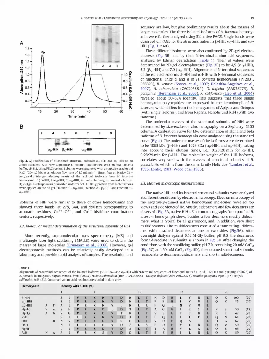
Fig. 3. A) Purification of dissociated structural subunits αD-HlH and αN-HlH on ananion-exchange Fast Flow Sepharose Q column, equilibrated with 50 mM Tris/HClbuffer, pH 8.2, using FPLC system. Subunits were separated with a stepwise gradient ofNaCl (0.0–1.0 M), at an elution flow rate of 1.5 mL min−1 (inset figure). Native 5% –
polyacrylamide gel electrophoresis of the isolated isoforms from H. lucorumhemocyanin: 1) β-HlH; 2) αD-HlH; 3) αN-HlH; 4) molecular weight standard – ferritin.B) 2-D gel electrophoresis of isolated isoforms of HiH. 10 μg protein from each fractionswere applied on the 8% gel. Fraction 1 – αD-HiH, Fraction 2 – βC-HiH and Fraction 3 –
αN-HiH.
19L. Velkova et al. / Comparative Biochemistry and Physiology, Part B 157 (2010) 16–25
isoforms of HlH were similar to those of other hemocyanins andshowed three bands, at 278, 344, and 550 nm corresponding toaromatic residues, Cu2+–O2−, and Cu2+-histidine coordinationcenters, respectively.
3.2. Molecular weight determination of the structural subunits of HlH
More recently, supramolecular mass spectrometry (MS) andmultiangle laser light scattering (MALLS) were used to obtain themasses of large molecules (Bruneaux et al., 2008). However, gelelectrophoresis methods can be relatively easily developed in alaboratory and provide rapid analysis of samples. The resolution and
Table 1Alignments of N-terminal sequences of the isolated isoforms β-HlH, αN- and αD-HlH with NH. pomatia hemocyanin, Rapana venosa, RvH1 (26,28), Haliotis tuberculata (HtH1, CAC2058californica, AcH (23). Conserved amino acid residues are shaded in dark gray.
accuracy are low, but give preliminary results about the masses oflarger molecules. The three isolated isoforms of H. lucorum hemocy-anin were further analysed using 5% native PAGE. Single bands wereobserved on PAGE for the structural subunits β-HlH, αD-HlH, and αN-HlH (Fig. 3 inset).
These different isoforms were also confirmed by 2D-gel electro-phoresis (Fig. 3B) and by their N-terminal amino acid sequences,analysed by Edman degradation (Table 1). Their pI values weredetermined by 2D-gel electrophoreses (Fig. 3B) to be 4,5 (αD-HiH),5,2 (βC-HiH) and 7.0 (αN-HiH). Alignments of N-terminal sequencesof the isolated isoforms β-HlH and α-HlH with N-terminal sequencesof functional units d and g of H. pomatia hemocyanin (P12031,P56823), R. venosa (Stoeva et al., 1997; Dolashka-Angelova et al.,2007), H. tuberculata (CAC20588.1), O. dofleini (AAK28276), N.pompilius (Bergmann et al., 2006), A. californica (Lieb et al., 2004)revealed about 50–67% identity. This suggests that three singlehemocyanin polypeptides are expressed in the hemolymph of H.lucorum, which differs from the hemocyanins of Aplysia and Octopus(with single isoform), and from Rapana, Haliotis and KLH (with twoisoforms).
The molecular masses of the structural subunits of HlH weredetermined by size-exclusion chromatography on a Sephacryl S300column. A calibration curve for Mw determination of alpha and betaisoforms of H. lucorum hemocyanin were analysed using the standardcurve (Fig. 4). The molecular masses of the isoforms were determinedto be 1068 kDa (β-HlH) and 1079 kDa (αD-HlH, and αN-HlH), takinginto account their elution times, i.e.: 0:28:20 min for α-HlH,0:28:29 min for β-HlH. The molecular weight of the HlH isoforms,correlates very well with the masses of structural subunits of H.pomatia Hc which is from the same family Helicidae (Lambert et al.,1995; Lontie, 1983; Wood et al.,1985).
3.3. Electron microscopic measurements
The native HlH and its isolated structural subunits were analysedat different conditions by electronmicroscopy. Electronmicroscopy ofthe negatively-stained native hemocyanin molecules revealed topviews and side views of Hc. Mostly, didecamers and few decamers areobserved (Fig. 5A, native HlH). Electron micrographs from purified H.lucorum hemolymph show, besides a few decamers mostly dideca-mers, what is typical for all gastropods, and, in addition, very shortmultidecamers. The multidecamers consist of a “nucleating” dideca-mer with attached decamers at one or two sides (Fig.5A). Afterovernight dialysis against 0.13 M Gly buffer, pH 9.6, the decamericforms dissociate in subunits as shown in Fig. 5B. After changing theconditions with the stabilizing buffer, pH 7.0, containing 20 mM CaCl2(Fig. 5C) and 50 mM CaCl2 (Fig. 5D), the obtained structural subunitsreassociate to decamers, didecamers and short multidecamers.
-terminal sequences of functional units d (HpHd, P12031) and g (HpHg, P56823) of8.1), Octopus dofleini (OdH, AAK28276), Nautilus pompilius, NpH1 (18), Aplysia

Fig. 4. Mw determination of a-HlH and β-HlH by gel filtration chromatography on aSephacryl S300 column. Subsequent Mw determination by a standard curve plottingdisplaying elution time of the standarts used against their Molecular Weight logarithm.Standards used and elution time: Ferritin (480 kDa), t=00:36:06 min; Catalase frombovine liver (240 kDa), t=00:52:17 min; Albumin (66 kDa), t=01:02:08 min.
20 L. Velkova et al. / Comparative Biochemistry and Physiology, Part B 157 (2010) 16–25
The three isoforms of HlH, β-HlH, αD-HlH and αN-HlH wereisolated and studied by electron microscopy. Electron micrographsfrom the purified β-subunit of H. lucorum Hc in stabilizing buffershowed, besides a few decamers and subunits, mostly didecamers,
Fig. 5. Gallery of transmission electron microscopy of HlH. A) Negatively stained native HlHside view (black arrow) and threedecamers (dash arrows); B) dissociated protein in 0.13containing 20 mM CaCl2; D) against the SB, containing 50 mM CaCl2. Decamers (dash arrowobserved in C and D. Staining with 1% uranyl acetate was performed as described in Mater
typical for all gastropods (Fig. 6A). After reassociation of β-HlH in thestabilizing buffer, containing 10 mM CaCl2, in addition, short tubuleswere observed (Fig. 6B). The tubules consist of a didecamer withattached decamers at one or both sides. In the presence of 10 and20 mM CaCl2 in the buffer, subunits and didecamers associated totubules of different length (Fig. 6B and C, respectively).
Also, the subunits-dissociated into α-component exhibited thesame behavior in the pH-stabilizing buffer as β-HlH. As is shown inFig. 6D, under the influence of 20 mM CaCl2, α-HlH reassociated intodidecamers and tubules of different length but shorter ones than inreassociated β-HlH (Fig. 6C). A similar capacity for the formation oftubules was observed for other molluscan hemocyanins, as muchthose from Rapana and H. pomatia (Dolashka-Angelova et al., 2003a;Lambert et al., 1995), however, no long multidecamers wereidentified in the isoforms of HlH.
3.4. Analysis of the subunit organization of β-HlH
Our sequence data show that β-H. lucorum hemocyanin containseight FUs that correspond structurally to the eight different FUs of H.pomatia hemocyanin. For the purified β-HlH cleavage we used lowconcentrations of trypsin (400:1) which in the case of RvH1 and RvH2allowed to characterise several functional units. Using a FPLC systemwith a Q Sepharose High Performance column, tryptic cleavageproducts were obtained in sufficient purity (Fig. 7) consisting of amixture of single FUs and smaller and larger fragments containingtwo, three or more FUs. The cleavage products were additionally
didecamers visible in side views (rectangular) and in top views (circles). Didecamers inM Gly/NaOH buffer at pH 9.6; C) reassociated HlH after dialysis against SB, pH 7.0,s), didecamers (black arrow) and multidecamers of varying length (width arrow) are
ials and methods.
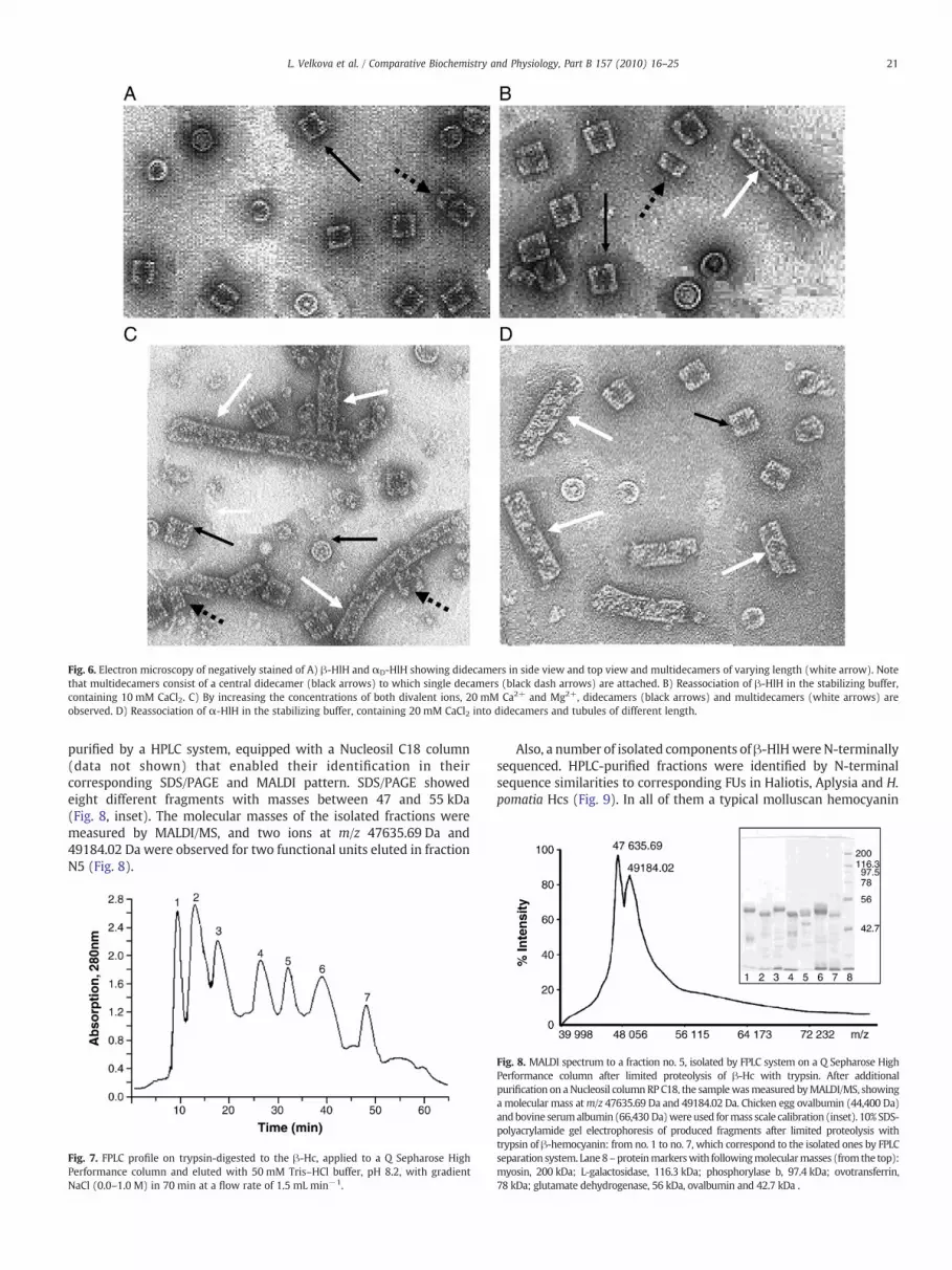
Fig. 6. Electron microscopy of negatively stained of A) β-HlH and αD-HlH showing didecamers in side view and top view and multidecamers of varying length (white arrow). Notethat multidecamers consist of a central didecamer (black arrows) to which single decamers (black dash arrows) are attached. B) Reassociation of β-HlH in the stabilizing buffer,containing 10 mM CaCl2. C) By increasing the concentrations of both divalent ions, 20 mM Ca2+ and Mg2+, didecamers (black arrows) and multidecamers (white arrows) areobserved. D) Reassociation of α-HlH in the stabilizing buffer, containing 20 mM CaCl2 into didecamers and tubules of different length.
21L. Velkova et al. / Comparative Biochemistry and Physiology, Part B 157 (2010) 16–25
purified by a HPLC system, equipped with a Nucleosil C18 column(data not shown) that enabled their identification in theircorresponding SDS/PAGE and MALDI pattern. SDS/PAGE showedeight different fragments with masses between 47 and 55 kDa(Fig. 8, inset). The molecular masses of the isolated fractions weremeasured by MALDI/MS, and two ions at m/z 47635.69 Da and49184.02 Da were observed for two functional units eluted in fractionN5 (Fig. 8).
Fig. 7. FPLC profile on trypsin-digested to the β-Hc, applied to a Q Sepharose HighPerformance column and eluted with 50 mM Tris–HCl buffer, pH 8.2, with gradientNaCl (0.0–1.0 M) in 70 min at a flow rate of 1.5 mL min−1.
Also, a number of isolated components of β-HlHwere N-terminallysequenced. HPLC-purified fractions were identified by N-terminalsequence similarities to corresponding FUs in Haliotis, Aplysia and H.pomatia Hcs (Fig. 9). In all of them a typical molluscan hemocyanin
Fig. 8. MALDI spectrum to a fraction no. 5, isolated by FPLC system on a Q Sepharose HighPerformance column after limited proteolysis of β-Hc with trypsin. After additionalpurification on aNucleosil columnRP C18, the samplewasmeasured byMALDI/MS, showinga molecular mass atm/z 47635.69 Da and 49184.02 Da. Chicken egg ovalbumin (44,400 Da)andbovine serumalbumin (66,430 Da)wereused formass scale calibration (inset). 10% SDS-polyacrylamide gel electrophoresis of produced fragments after limited proteolysis withtrypsin of β-hemocyanin: from no. 1 to no. 7, which correspond to the isolated ones by FPLCseparation system. Lane8–proteinmarkerswith followingmolecularmasses (from the top):myosin, 200 kDa; L-galactosidase, 116.3 kDa; phosphorylase b, 97.4 kDa; ovotransferrin,78 kDa; glutamate dehydrogenase, 56 kDa, ovalbumin and 42.7 kDa .

Fig. 9. Alignment of N-terminal sequences of functional units, isolated from Helixlucorum (HlH), Helix pomatia (HpH), Haliotis tuberculata (HtH) and Aplysia californica(ApH) hemocyanins.
Fig. 10. A) Fluorescence spectra of the native HlH and isolated functional unit weremeasured at λext 295. The absorptions of both proteins were at 280 nm=0.05, B) CDspectrum of the native molecule of HlH in the region 195 nm to 250 nm. Absorption ofprotein was 0.20 and the path length of the cuvette was 0.2 cm.
22 L. Velkova et al. / Comparative Biochemistry and Physiology, Part B 157 (2010) 16–25
fragment (-Val Arg Lys Asp-) was identified. Only five N-terminalsequences of the FU (HlH-a, HlH-b, HlH-d, HlH-f and HlH-g) from β-HlH revealed high homology with the sequences of Aplysia Hc. Ourresults proved that the hemocyanin sequences of H. lucorum aresignificantly more closely related to A. californica and H. pomatia (Liebet al., 2004; Altenhein et al., 2002; Wood et al., 1985).
Additionally, α-HlH and β-HlH isoforms and isolated functionalunits were analysed by UV, fluorescence spectroscopy and circulardichroism. Each functional unit contains about 7–8 tryptophanresidues and one active site with two copper ions. Therefore, twomaxima, at 278 and 347 nm, were observed in the absorption spectraof isoforms and FUs of HlH, originating from aromatic residues andCu2+–O2−, respectively (data are not shown). These absorptionspectra are similar to other molluscan and arthropodan hemocyaninsand the bands at 347 nm disappeared with the addition of reducingagents (Dolashka et al., 1996; Fan et al., 2009). Also, the nativemolecule and isolated isoforms were analysed by fluorescencespectroscopy after excitation at 295 nm, where the tryptophyl sidechains are selectively excited. The fluorescence parameters of the oxy-forms of the native H. lucorum hemocyanin and functional units areshown in Fig. 10A. The fluorescence spectrum for HlH has a maximumat (335)±1 nm, which is typical for deeply buried tryptophans in ahydrophobic environment. However, the fluorescence maximum ofFU was shifted to 342±1 nm, due to the exposed tryptophyl sidechains on the surface of the molecule.
Two negative Cotton effects at 222 and at 208 nm were digitalizedin the CD spectra of the hemocyanin solutions, recorded from 190 to250 nm, as observed for Rapana hemocyanin and due to the α-helixand β-sheet structures of the proteins (Velkova et al., 2009a; Dolashkiet al., 2008).
4. Discussion
Hemocyanins are dissolved in the hemolymph of molluscs andaggregated in various complexes, which still are not very wellunderstood. Hemocyanins from the marine gastropod M. crenulata, C.
concholepas and R. venosa occur in the hemolymph in two isoforms andtheir protein structures and disassembly/reassembly behavior have beenextensively studied (Gatsogiannis and Markl, 2009; De Ioannes et al.,2004; Velkova et al., 2009a;Dolashki et al., 2008;Hristova et al., 2008). Atpresent, there is a growing interest in hemocyanins because of theirimmunological properties and potential application as promising tumorvaccine carriers (Dolashka-Angelova et al., 2009; Velkova et al., 2009b;Yossifova et al., 2009; Iliev et al., 2008; Toshkova et al., 2006; Toshkova etal., 2007; Dolashka-Angelova et al., 2008;Moltedo et al., 2006;Wuhrer etal., 2004). Structure, evolution, and diversity of hemocyanins is ofscientific interest and the relationship between structural features andimmunotherapeutic effects is of biomedical concern. Therefore, wepresent in this communication structural characteristics of hemocyanin,dissolved in the hemolymph of garden snailH. lucorum and demonstratethat its special structure differs from the other molluscan hemocyanins.
In contrast to the marine gastropods of the genera, Megathura,Haliotis, Aplicia, Nucula, Concholepas, or Rapana (Gatsogiannis et al.,2009; Lieb et al., 2000; Bergmann et al., 2007; Lieb et al., 2004; DeIoannes et al., 2004; Dolashka-Angelova et al., 2003a; Lambert et al.,1995), revealing the presence of one, or two structurally andfunctionally distinct hemocyanin isoforms, three different isoforms(β-HlH, αD-HlH and αN-HlH) were identified to be dissolved in thehemolymph of H. lucorum. The evidence for three isoforms, αD-HiH,βC-HiH, and αN-HiH are based on different pI values (4,5, 5,2 and 7.0,respectively), determined by 2D-gel electrophoreses (Fig. 3B).αD-HiHand βC-HiH show very clos pI values (4,5 and 5,2, respectively)compared to the α- and β-isoforms of Helix aspersa Hc (4,6 and 5,2,respectively) but different ones to those of H. pomatia (5,3 and 5,4,respectively).
In fact, the presence of three different isoforms in only twomolluscs,H. lucorum andH. pomatia, raises thequestion of how they assemble intothe functional molecules. Therefore, three isoforms β-HlH, αD-HlH andαN-HlH have been isolated and characterized by different methods in

Fig. 11. Organization of the quaternary structure of molluscan hemocyanins into decamers (Hcs with one isoform) and didecamers (Hcs with one, two and three isoforms).
23L. Velkova et al. / Comparative Biochemistry and Physiology, Part B 157 (2010) 16–25
comparison with other molluscan Hcs (Fig. 11). Analyzing the purityand apparent molecular weights of the eluted proteins by gelelectrophoresis only one band at ≈450 kDa was detected for eachisoform(β-HlH,αD-HlHandαN-HlH).However, the calculated resultsbyMALDI-TOF and SDS gel electrophoresis gavemore precise results aboutthemolecularmass ofβ-HlH. Itwas determined to be about 420 kDa. Allof the isolated isoforms are present in the hemolymph mainly asdidecamers that resemble the quaternary structure of the hemocyanin.However, only in the electron micrographs of the native HlH a fewtridecamers are shown, resulting from the association of a furtherdecamer with one didecamer. It was confirmed that although thehemocyanins from Vetigastropoda (Haliotis), Caenogastropoda(Rapana), Heterobranchia (Aplicia and H. pomatia) and bivalve Nuculacontain one, twoor three isoforms, all of themoccur as didecamers (Liebet al., 2000; Bergmann et al., 2007; Lieb et al., 2004; Dolashka-Angelovaet al., 2003a; Meissner et al., 2000; Lambert et al., 1995). Theirdidecameric structure is formed by homogeneous or heterogeneousdecamers. The only examples to date of hemocyanins with a hetero-didecameric structure are thoseof themolluscsMurex,Concholepas andRapana (De Ioannes et al., 2004; Dolashka-Angelova et al., 2003a).
The three isoforms of H. lucorum hemocyanin (β-HlH, αD-HlH andαN-HlH) are composed of only one type of organized as subunit,formed didecamers by homogeneous decamers, as was observed forKLH, RvH, HtH (Harris et al., 2004; Dolashka et al., 1996; Altenhein etal., 2002). However, they reveal a different behaviour after dissoci-ation and reassociation. In pH-stabilizing buffer with 20 mM CaCl2,dissociated subunits reassociate into didecamers and tubules withdifferent length. Long tubules are observed in β-HlH, in contrast toαD- and αN-HlH, which reassociate into shorter tubules. Although, asimilar capacity for the formation of tubules is observed in Rapana andH. pomatia hemocyanins (Dolashka et al., 1996; Lambert et al., 1995),a striking different behaviouer of HlH isoforms was observed. No longmultidecamers were identified in the isoforms of HlH.
So far, the analyses have given no clue to an explanation for whycertain hemocyanins form very long multidecamers, tubules, othersform very short ones, and still others are restricted to didecamers, orjust decamers, as in the gastropod chitons. Strictly didecameric
hemocyanins are observed for those from H. pomatia and S. officinaliswhich are heavily glycosylated (Gielens et al., 2004). Studies on theoligosaccharide structures of other hemocyanins, such as RvH, Arionlusitanicus, Aplysia,M. crenulata and partiallyH. lucorum hemocyaninsshow the existence of a variety of glycan side chains, exposed ondifferent positions in the molecule (Gielens et al., 2004; Gutternigg etal., 2004; Dolashka-Angelova et al., 2004; Kurokawa et al., 2002;Lommerse et al., 1997; Beck et al., 2007; Sandra et al., 2007; Dolashka-Angelova et al., 2003b). It was found that hemocyanins Haliotis2,Aplysia, Megathura, and Octopus, which form long multidecamers,potential N-glycosylation sites are missing in the case of FU-c. Incontrast to HtH2, the isoform HtH1 showed different reassociatedbehaviour and no long multidecamers were found. However, HtH1-h has two potential N-glycosylation sites in quite unusual positions,localized on the surface of the decamer (Lieb et al., 2000). In all otherFUs, including HtH2-h, these two positions are not N-glycosylated.Based on these data, it may be speculated that on the surface of theHlH decamer there is a putative glycosylated site, which may be areason that decamers do not interact to longer multidecamers.
Additionally, the structure of one isoform, β-HlH, was analysedafter enzymatic digestion with trypsin. The obtained fractions wereanalysed and compared with other Hcs. On the basis of limitedproteolytic cleavage, SDS/PAGE and N-terminal sequencing, weidentified eight different functional units with molecular masses inthe region of 47 to 55 kDa for β-HlH hemocyanin. In phylogenetictrees, derived from sequence alignments, topologically correspondingFUs of different molluscan hemocyanins from discrete branches leadto the concept of an early origin of the eight functional units (Lieb etal., 2000; Bergmann et al., 2007). We have clearly demonstrated, thatH. lucorum hemocyanin contains eight FUs (Fu-a to FU-h), in contrastto the FUs of N. pompilius and O. dofleini hemocyanins, consists ofseven FUs only. The multiple sequence alignment of N-terminalsequences of the separated FUs shows that HlH shares a higheridentity with Aplysia Hc than with Haliotis.
Additionally, analysed by UV, fluorescence spectroscopy andcircular dichroism, which are suitable tools for the identification ofaromatic residues and studying the structure and conformation in

24 L. Velkova et al. / Comparative Biochemistry and Physiology, Part B 157 (2010) 16–25
solution of the proteins, α-HlH and β-HlH isoforms exhibited verysimilar properties to other molluscan and arthropodan hemocyanins.
In summary, we describe here the structures of hemocyaninsisolated from the gastropod H. lucorum, emphasizing some attributesthat make it interesting among molluscan hemocyanins. This, in turn,would provide essential information on the path of the elongatedsubunits within the didecamers and tubules which is still largelyunknown for any molluscan Hc. Further studies on the gene sequenceand oligosaccharide structure of the three isoforms of HlH arerequired to better understand the structure and molecular basis ofthe specific association pattern.
Acknowledgement
This work was supported by a research grant by the BulgarianNational Science Fund TK01-496/2009, UV-L-301 and НТС01-187(Ukraine), DAAD-17/2007 and DFG-01/2008 (Germany).
References
Ali, S., Abbasi, A., Stoeva, S., Kayed, R., Dolashka-Angelova, P., Schwarz, H., Voelter, W.,2000. Oxygen transport proteins: III. Structural studies of the scorpion (Buthussindicus) hemocyanin, partial primary structure of its subunit Bsin1. Comp.Biochim. Physiol. Part B 126, 361–376.
Altenhein, B., Markl, J., Lieb, B., 2002. Gene structure and hemocyanin isoform HtH2from the mollusc Haliotis tuberculata indicate early and late intron hot spots. Gene301, 53–60.
Beck, A., Hillen, N., Dolashki, A., Stevanovic, S., Salvato, B., Voelter, W., Dolashka-Angelova, P., 2007. Oligosaccharide structure of a functional unit RvH1-b of Rapanavenosa hemocyanin using HPLC/electrospray ionization mass spectrometry.Biochimie 89, 938–949.
Bergmann, S., Lieb, B., Ruth, P., Markl, J., 2006. The hemocyanin from a living fossil, thecephalopod Nautilus pompilius: protein structure, gene organization, and evolution.J. Mol. Evol. 62, 362–374.
Bergmann, S., Markl, J., Lieb, B., 2007. The first complete cDNA sequence of thehemocyanin from a bivalve, the protobranch Nucula nucleus. J. Mol. Evol. 64,500–510.
Bruneaux, M., Rousselot, M., Leize, E., Lallier, F., Zal, F., 2008. The structural analysis oflarge noncovalent oxygen binding proteins by MALLS and ESI-MS: a review onannelid hexagonal bilayer hemoglobin and crustacean hemocyanin. Curr. ProteinPept. Sci. 9, 150–180.
Burmester, T., 2004. Evolutionary history and diversity of arthropod hemocyanins.Micron 35 (1–2), 121–122.
Cuff, M., Miller, K., Van Holde, K., Hendrickson, W., 1998. Crystal structure of afunctional unit from Octopus hemocyanin. J. Mol. Biol. 278, 855–870.
De Ioannes, P., Moltedo, B., Oliva, H., Pacheco, R., Faunes, F., De Ioannes, A., Becker, M.,2004. Hemocyanin of the molluscan Concholepas concholepas exhibits an unusualheterodecameric array of subunits. J. Biol. Chem. 279, 26134–26142.
Dolashka, P., Genov, N., Pervanova, K., Voelter, W., Geiger, M., Stoeva, S., 1996. Rapanathomasiana grosse (gastropoda) haemocyanin: spectroscopic studies of thestructure in solution and the conformational stability of the native protein andits structural subunits. J. Biochem. 315, 139–144.
Dolashka-Angelova, P., Stoeva, S., Hristova, R., Schuetz, J., Beltramini, M., Salvato, B.,Schwartz, H., Voelter, W., 1999. Structural organization of hemocyanin from lobsterHomarus americanus and spectroscopic studies of the native protein and structuralsubunits. Curr Topics Pept. Prot. Res. 3, 19–36.
Dolashka-Angelova, P., Stoeva, S., Hristova, R., Schuetz, J., Voelter, W., 2000a. Structuraland spectroscopic studies of the native hemocyanin from Maia squinado and itsstructural subunits. Spectrochim. Acta A 56, 1985–1999.
Dolashka-Angelova, P., Schick, M., Stoeva, S., Voelter, W., 2000b. Isolation and partialcharacterization of the N-terminal functional unit of subunit RtH1 from Rapanathomasiana grosse hemocyanin. Int. J. Biochem. Cell Biol. 32, 529–538.
Dolashka-Angelova, P., Schwarz, H., Dolashki, A., Beltramini, M., Salvato, B., Schick, M.,Saeed, M., Voelter, W., 2003a. Oligomeric stability of Rapana venosa hemocyanin(RvH) and its structural subunits. Biochim. Biophys. Acta 1646, 77–85.
Dolashka-Angelova, P., Beck, A., Dolashki, A., Beltramini, M., Stevanovic, S., Salvato, B.,Voelter, W., 2003b. Characterization of the carbohydrate moieties of the functionalunit RvH1-a of Rapana venosa haemocyanin using HPLC/electrospray ionization MSand glycosidase digestion. Biochem. J. 374, 185–192.
Dolashka-Angelova, P., Beck, A., Dolashki, A., Beltramini, M., Stevanovic, S., Salvato, B.,Hristova, R., Velkova, L., Voelter, W., 2004. Carbohydrate moieties of molluscanRapana venosa hemocyanin. Micron 35, 101–104.
Dolashka-Angelova, P., Dolashki, A., Savvides, S., Hristova, R., Van Beeumen, J., Voelter,W., Devreese, B., Weser, U., Di Muro, P., Salvato, B., Stevanovic, S., 2005a. Structureof hemocyanin subunit CaeSS2 of the crustacean Mediterranean crab Carcinusaestuarii. J. Biochemistry 138, 303–312.
Dolashka-Angelova, P., Dolashki, A., Stevanovic, S., Hristova, R., Atanasov, B., Nicolov, P.,Voelter, W., 2005b. Structure and stability of arthropodan hemocyanin Limuluspolyphemus. Spectrochim. Acta 61, 1207–1217.
Dolashka-Angelova, P., Stefanovic, St, Dolashki, A., Devreese, B., Tzvetkova, B., Voelter,W., Van Beeumen, J., Salvato, B., 2007. A challenging insight on the structural unit 1of molluscan Rapana venosa hemocyanin. Arch. Biochem Biophys 459, 50–58.
Dolashka-Angelova, P., Stefanova, T., Livaniou, E., Velkova, L., Klimentzou, P.,Stevanovic, S., Neychev, H., Schwarz, H., Voelter, W., 2008. Immunological potentialof Helix lucorum and Rapana venosa hemocyanins. Immunol. Invest. 37, 822–840.
Dolashka-Angelova, P., Lieb, B., Velkova, L., Heilen, N., Sandra, K., Nikolaeva-Glomb, L.,Dolashki, A., Galabov, A., Van Beeumen, J., Stevanovic, S., Voelter, W., Devreese, B.,2009. Identification of glycosylated sites in Rapana hemocyanin by massspectrometry and gene sequence, and their antiviral effect. Bioconjug. Chem. 20,1315–1322.
Dolashki, A., Schütz, J., Hristova, R., Voelter, W., Dolashka-Angelova, P., 2005.Spectroscopic properties of non-glycosilated functional unit KLH2-c of keyholelimpet hemocyanin. World J Agricult Sci 1, 129–136.
Dolashki, A., Velkova, L., Atanasov, B., Hristova, R., Voelter, W., Stevanovic, S., Schwarz,H., Di Muro, P., Dolashka-Angelova, P., 2008. Reversibility and “pH-T phasediagrams” of Rapana venosa hemocyanin and its structural subunits. BiochimBiophys Actra 1784, 1617–1624.
Fan, T., Zhang, Y., Yang, L., Yang, X., Jiang, G., Yu, M., Cong, R., 2009. Identification andcharacterization of a hemocyanin-derived phenoloxidase from the crab Charybdisjaponica. Comp. Biochem. Physiol. B Biochem. Mol. Biol. 152, 44–49.
Gatsogiannis, C., Markl, J., 2009. Keyhole limpet hemocyanin: 9-A CryoEM structure andmolecular model of the KLH1 didecamer reveal the interfaces and intricatetopology of the 160 functional units. J. Mol. Biol. 385, 963–983.
Gatsogiannis, C., Moeller, A., Depoix, F., Meissner, U., Markl, J., 2007. Nautilus pompiliushemocyanin: 9 Å cryo-EM structure andmolecularmodel reveal the subunit pathwayand the interfaces between the 70 functional units. J. Mol. Biol. 374, 465–486.
Gielens, C., De Geest, N., Compernolle, F., Préaux, G., 2004. Glycosylation sites ofhemocyanins of Helix pomatia and Sepia officinalis. Micron 35, 99–100.
Gutternigg, M., Ahrer, К., Grabher-Meier, H., Burgmayr, S., Staudacher, E., 2004. NeutralN-glycans of the gastropod Arion lusitanicus. Eur. J. Biochem. 271, 1348–1356.
Harris, J.R., Meissner, U., Gebauer, W., Markl, J., 2004. 3D reconstruction of thehemocyanin subunit dimer from the chiton Acanthochiton fascicularis. Micron 35(1–2), 23–26.
Hristova, R., Dolashki, A., Voelter, W., Stevanovic, S., Dolashka-Angelova, P., 2008. O-diphenol oxidase activity of molluscan hemocyanins. Comp. Biochem. Physiol. B149, 439–446.
Iliev, I., Toshkova, R., Dolashka-Angelova, P., Yossifova, L., Hristova, R., Yaneva, J.,Zacharieva, S., 2008. Haemocyanins from Rapana venosa and Helix lucorum displayan antitumour activity via specific activation of spleen lymphocytes. Compt. Rend.Acad. Bulg. Sci. 61, 203–210.
Kurokawa, T., Wuhrer, M., Lochnit, G., Geyer, H., Markl, J., Geyer, R., 2002. Hemocyaninfrom the keyhole limpet Megathura crenulata (KLH) carries a novel type of N-glycans with Gal(b1-6)Man-motifs. Eur. J. Biochem. 269, 5459–5473.
Lambert, O., Boisset, N., Taveau, J.-C.h., Lamy, J.N., 1994. Three-dimensional recon-struction from a frozen-hydrated specimen of the chiton Lepidochiton sp.Hemocyanin J. Mol. Biology 244 (5), 640–647.
Lambert, O., Boisset, N., Taveau, J.C., Préaux, G., Lamy, J.N., 1995. Three-dimensionalreconstruction of the alpha D and beta C-hemocyanins of Helix pomatia fromfrozen-hydrated specimens. J Mol Biol. 248, 431–448.
Lamy, J., You, V., Taveau, J.C., Boisset, N., Lamy, J.N., 1998. Intramolecular localization ofthe functional units of Sepia officinalis hemocyanin by immunoelectronmicroscopy.J. Mol. Biol. 284 (4), 1051–1074.
Lieb, B., Altenhein, B., Markl, J., 2000. The sequence of a gastropod hemocyanin (HtH1from Haliotis tuberculata). J. Biol. Chem. 275, 5675–5681.
Lieb, B., Boisguérin, V., Gebauer, W., Markl, J., 2004. cDNA sequence, protein structure,and evolution of the single hemocyanin from Aplysia californica, an opisthobranchgastropod. J. Mol. Evol. 59, 536–545.
Lieb, B., Todt, C., 2008. Hemocyanin in molluscs — a molecular survey and new data onhemocyanin genes in Solenogastres and Caudofoveata. Mol. Phylogenet. Evol. 49,382–385.
Lommerse, J., Thomas-Oates, J., Gielens, C., Preaux, G., Kamerling, J., Vliegenthart, J.,1997. Primary structure of 21 novel monoantennary and diantennary N-linkedcarbohydrate chains from alpha-D-hemocyanin of Helix pomatia. Eur. J. Biochem.249, 195–222.
Lontie, R., 1983. Components, functional units, and active sites of Helix pomatiahemocyanin. Life Chem. Rep. Suppl. 1, 109–120.
Meissner, U., Dube, P., Harris, J.R., Stark, H., Markl, J., 2000. Structure of a molluscanhemocyanin didecamer (HtH1 from Haliotis tuberculata) at 12 A resolution bycryoelectron microscopy. J. Mol. Biol. 298 (1), 21–34.
Miller, K., Cuff, M., Lang, W., Varga-Weisz, P., Field, K., Van Holde, K., 1998. Sequence ofthe Octopus dofleini hemocyanin subunit: structural and evolutionary implications.J. Mol. Biol. 278, 827–842.
Moltedo, B., Faunes, F., Haussmann, D., De Ioannes, P., De Ioannes, A.E., Puente, J.,Becker, M., 2006. Immunotherapeutic effect of Concholepas hemocyanin in themurine bladder cancer model: evidence for conserved antitumor properties amonghemocyanins. J. Urol. 176, 2690–2695.
Paoli, M., Giomi, F., Hellmann, N., Jaenicke, E., Decker, H., Di Muro, P., Beltramini, M.,2007. The molecular heterogeneity of hemocyanin: structural and functionalproperties of the 4×6-meric protein of Upogebia pusilla (Crustacea). Gene 398,177–182.
Salvato, B., Beltramini, M., 1990. Hemocyanin: molecular architecture, structure andreactivity of the binuclear copper active site. Life Chem. Rep. 8, 1–47.
Sandra, K., Dolashka-Angelova, P., Devreese, B., Van Beeumen, J., 2007. New insights inRapana venosa hemocyani N-glycosylation resulting from on-line mass spectro-metric analyses. Glycobiology 17, 141–156.
25L. Velkova et al. / Comparative Biochemistry and Physiology, Part B 157 (2010) 16–25
Schütz, J., Dolashka-Angelova, P., Abrashev, R., Nicolov, P., Voelter, W., 2001. Isolationand spectroscopic characterization of the structural subunits of keyhole limpethemocyanin. Biochim. Biophys. Acta 1546, 325–336.
Stoeva, S., Dolashka, P., Bankov, B., Voelter, W., Salvato, B., Genov, N., 1995.Spectroscopic properties of Callinectes sapidus hemocyanin subunits. Spectrochim.Acta Part A 51, 1965–1974.
Stoeva, S., Dolashka, P., Hristova, R., Genov, N., Voelter, W., 1999. Subunit compositionand N-terminal analysis of arthropod hemocyanins. Comp. Biochem. Physiol. B 122,69–75.
Stoeva, S., Dolashka, P., Pervanova, K., Genov, N., Voelter, W., 1997. Multidomainstructure of the Rapana thomasiana (Gastropod) hemocyanin structural subunitRHSS1. Comp. Biochem. Physiol. B 118, 927–934.
Toshkova, R., Ivanova, E., Nastke, M.-D., Stevanovic, S., Velkova, L., Voelter, W.,Dolashka-Angelova, P., 2006. Hemocyanins as immunostimulators. IDOSY. Global J.Mol. Sci. 1, 22–32.
Toshkova, R., Velkova, L., Voelter, W., Dolashka-Angelova, P., 2007. Protective effect ofRapana venosa hemocyanin (RvH) on survivability of hamsters with transplantedmyeloid Graffi tumours. Compt. Rend. Acad. Bulg. Sci. 59, 977–982.
Van Holde, K., Miller, K., Decker, H., 2001. Hemocyanins and invertebrate evolution.J. Biol. Chem. 276, 15563–15566.
Velkova, L., Dolashka-Angelova, P., Dolashki, A., Voelter, W., Atanasov, B., 2009a.Thermodynamic analysis and molecular modeling of Ranana venosa hemocyanin-functional unit RvH2-e. Biotech. Biotech. Equip. 23, 601–605.
Velkova, L., Todorov, D., Dimitrov, I., Shishkov, S., Van Beeumen, J., Dolashka-Angelova,P., 2009b. Rapana venosa hemocyanin with antiviral activity. Biotech. Biotech.Equip. 23, 606–610.
Wood, E., Chaplin, M., Gielens, C., De Sadeleer, J., Préaux, G., Lontie, R., 1985. Relativemolecular mass of the polypeptide chain of bc-haemocyanin of Helix pomatia andcarbohydrate composition of the functional units. Comp Biochem Physiol B 82,179–186.
Wuhrer, M., Robijn, M., Koeleman, C., Balog, C., Geyer, R., Deelder, A., Hokke, C., 2004. Anovel Gal(β1-4)Gal(β1-4)Fuc(α1-6)-core modification attached to the proximalN-acetylglucosamine of keyhole limpet hemocyanin (KLH) N-glycans. Biochem. J.378, 625–632.
Yossifova, L., Iliev, I., Petkova, S., Dolashka-Angelova, P., Mihov, L., Zacharieva, S., 2009.Imunological research on the protective properties of a conjugate of total larvalantigen with hemocyanin derived from Helix lucorum against infection withTrichinella spiralis. Biotech. Biotech. Equip. 23, 597–600.


