Embed Size (px)
Citation preview

Structural Polymorphism of 441-Residue Tauat Single Residue ResolutionMarco D. Mukrasch
1[, Stefan Bibow
1[, Jegannath Korukottu
1, Sadasivam Jeganathan
2, Jacek Biernat
2,
Christian Griesinger1
, Eckhard Mandelkow2
, Markus Zweckstetter1,3*
1 Department for Nuclear Magnetic Resonance (NMR)-Based Structural Biology, Max Planck Institute for Biophysical Chemistry, Gottingen, Germany, 2 Max Planck Unit for Structural
Molecular Biology, Hamburg, Germany, 3 Deutsche Forschungsgemeinschaft (DFG) Research Center for the Molecular Physiology of the Brain (CMPB), Gottingen, Germany
Alzheimer disease is characterized by abnormal protein deposits in the brain, such as extracellular amyloid plaquesand intracellular neurofibrillary tangles. The tangles are made of a protein called tau comprising 441 residues in itslongest isoform. Tau belongs to the class of natively unfolded proteins, binds to and stabilizes microtubules, andpartially folds into an ordered b-structure during aggregation to Alzheimer paired helical filaments (PHFs). Here weshow that it is possible to overcome the size limitations that have traditionally hampered detailed nuclear magneticresonance (NMR) spectroscopy studies of such large nonglobular proteins. This is achieved using optimal NMR pulsesequences and matching of chemical shifts from smaller segments in a divide and conquer strategy. The methodologyreveals that 441-residue tau is highly dynamic in solution with a distinct domain character and an intricate network oftransient long-range contacts important for pathogenic aggregation. Moreover, the single-residue view provided bythe NMR analysis reveals unique insights into the interaction of tau with microtubules. Our results establish that NMRspectroscopy can provide detailed insight into the structural polymorphism of very large nonglobular proteins.
Citation: Mukrasch MD, Bibow S, Korukottu J, Jeganathan S, Biernat J, et al. (2009) Structural polymorphism of 441-residue Tau at single residue resolution. PLoS Biol 7(2):e1000034. doi:10.1371/journal.pbio.1000034
Introduction
Tau protein was originally discovered as a neuronalmicrotubule-associated protein (MAP) that stabilizes micro-tubules (MTs) and supports the outgrowth of axons [1,2]. Theprotein can modulate the transport of vesicles and organellesalong MTs, serves as an anchor for enzymes, and regulates thedynamics of MTs [3,4]. In Alzheimer disease (AD), taubecomes excessively phosphorylated, looses its ability to bindto MTs, and aggregates into neurofibrillary tangles thatconsist of paired helical filaments (PHFs) of tau. Mutations inthe tau gene cause tau aggregation and frontotemporaldementia with parkinsonism linked to Chromosome 17 [5,6].
The human central nervous system contains six isoforms oftau, generated from a single gene by alternative splicing andranging between 352 to 441 amino acid residues [7,8]. Theisoforms differ by two inserts near the N-terminal end andthe presence of either four or three imperfect repeatsequences in the C-terminal half of the protein (Figure 1A).The repeat domain represents the core of the MT interaction[9] and also forms the core of PHFs [10]. For PHF aggregationtwo hexapeptides at the beginning of the second and thirdrepeats (275VQIINK280 and 306VQIVYK311) are crucial becausethey are able to initiate the aggregation process [11].
It was recognized early on that tau has an unusual characteras a protein, because it was resistant to heat and acidtreatment without loosing its function and had a very lowcontent of secondary structure [12]. These properties can betraced back to the high fraction of basic and hydrophilicamino acid residues (Figure 1B), which resist the compactfolding typical of most proteins. In fact, a number ofbiophysical studies revealed that tau is a prototypical‘‘natively unfolded’’ protein [13]. In recent years, this typeof protein emerged as a major fraction in the humanproteome (termed ‘‘natively unfolded’’ or ‘‘intrinsically
unstructured proteins’’ [IUPs] [14]). Apart from tau, most‘‘fibrous’’ MAPs have the signature of the natively unfoldedstate, whereas other MT-binding proteins show conventionalfolding (e.g., motor proteins).Since disordered proteins tend to be highly flexible and
have variable conformations, they have not been amenablefor structure analysis by crystallography. Thus nuclearmagnetic resonance (NMR) spectroscopy is the only methodthat allows a description of their conformations anddynamics with high resolution [15]. The lack of an orderedstructure, however, causes dramatic signal overlap. There-fore, we and others have previously performed NMR studieson fragments of tau or studied full-length tau but only on thebasis of partial assignment that was scattered throughout thesequence [16–25]. In particular by studying tau fragments thatcontain only the repeat domain (K19 or K18) or the repeatdomain and the flanking regions (K32), we and others showedthat the hexapeptides in repeats R2 and R3 populate b-structure and bind to MTs and polyanions [16–18], that shortstretches in the repeat domain assume highly populated turn
Academic Editor: Gregory A. Petsko, Brandeis University, United States of America
Received May 21, 2008; Accepted January 7, 2009; Published February 17, 2009
Copyright: � 2009 Mukrasch et al. This is an open-access article distributed underthe terms of the Creative Commons Attribution License, which permits unrestricteduse, distribution, and reproduction in any medium, provided the original authorand source are credited.
Abbreviations: AD, Alzheimer disease; FRET, Forster resonance energy transfer;HSQC, heteronuclear single quantum coherence; htau40, longest tau isoformfound in the human central nervous system; MAP, microtubule-associated protein;MT, microtubule; MTSL, (1-oxy-2,2,5,5-tetramethyl-D-pyrroline-3-methyl)-methane-thiosulfonate; NMR, nuclear magnetic resonance; PHF, paired helical filament; PRE,paramagnetic relaxation enhancement; RDC, residual dipolar coupling; SAXS, smallangle x-ray scattering
* To whom correspondence should be addressed. E-mail: [email protected]
[ These authors contributed equally to this work.
PLoS Biology | www.plosbiology.org February 2009 | Volume 7 | Issue 2 | e10000340399
PLoS BIOLOGY
conformations [19], that the repeat domain of tau folds intoan a-helical conformation upon binding to lipid surfaces [20],and that PHFs formed in vitro by the three-repeat-domain(K19) of tau consist of three major b-strands [21]. Moreover,using a partial assignment of full-length tau (less than 40%)Lippens and coworkers investigated the binding of tau toMTs [22], the phosphorylation pattern of tau as induced bycAMP dependent kinase [23], tau aggregated into PHFs [24],and the impact of binding of heparin to tau [25]. In addition,small angle x-ray scattering (SAXS) and Forster resonanceenergy transfer (FRET) was used to obtain insight into thestructure of the tau protein [26,27].Despite the wealth of information from previous studies on
the conformational properties of tau, they were alwayslimited because they were either not of high resolution(SAXS, circular dichcroism, electron microscopy), wererestricted to fragments of tau (liquid-state and solid-stateNMR, x-ray crystallography of a complexed tau peptide [28]),or were limited to a subset of residues (NMR, FRET). Incontrast, we show here that it is possible to obtain thecomplete backbone assignment of 441-residue tau (thelongest tau isoform found in the human central nervoussystem, htau40; Figure 1B) and thus to overcome the sizelimitation that in the past has limited detailed NMR studies of
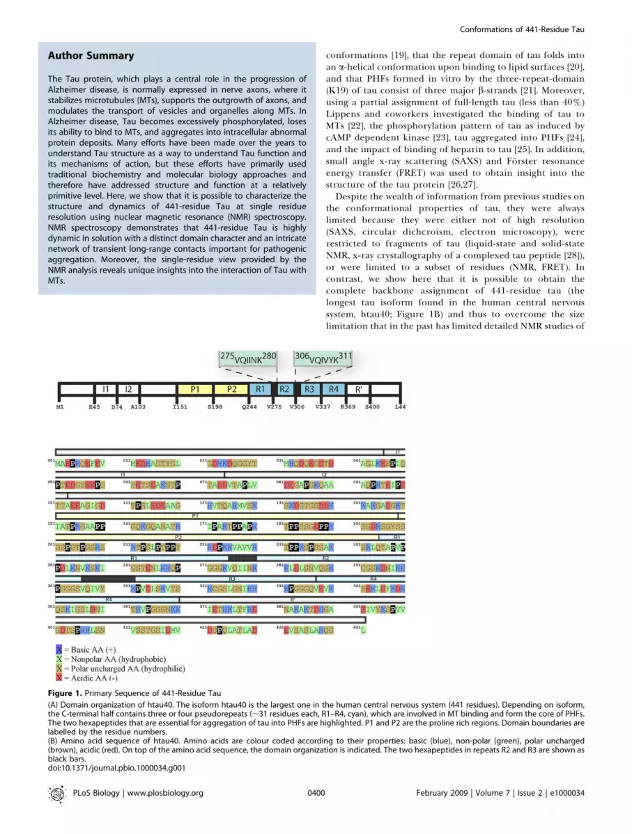
Figure 1. Primary Sequence of 441-Residue Tau
(A) Domain organization of htau40. The isoform htau40 is the largest one in the human central nervous system (441 residues). Depending on isoform,the C-terminal half contains three or four pseudorepeats (;31 residues each, R1–R4, cyan), which are involved in MT binding and form the core of PHFs.The two hexapeptides that are essential for aggregation of tau into PHFs are highlighted. P1 and P2 are the proline rich regions. Domain boundaries arelabelled by the residue numbers.(B) Amino acid sequence of htau40. Amino acids are colour coded according to their properties: basic (blue), non-polar (green), polar uncharged(brown), acidic (red). On top of the amino acid sequence, the domain organization is indicated. The two hexapeptides in repeats R2 and R3 are shown asblack bars.doi:10.1371/journal.pbio.1000034.g001
PLoS Biology | www.plosbiology.org February 2009 | Volume 7 | Issue 2 | e10000340400
Conformations of 441-Residue Tau
Author Summary
The Tau protein, which plays a central role in the progression ofAlzheimer disease, is normally expressed in nerve axons, where itstabilizes microtubules (MTs), supports the outgrowth of axons, andmodulates the transport of vesicles and organelles along MTs. InAlzheimer disease, Tau becomes excessively phosphorylated, losesits ability to bind to MTs, and aggregates into intracellular abnormalprotein deposits. Many efforts have been made over the years tounderstand Tau structure as a way to understand Tau function andits mechanisms of action, but these efforts have primarily usedtraditional biochemistry and molecular biology approaches andtherefore have addressed structure and function at a relativelyprimitive level. Here, we show that it is possible to characterize thestructure and dynamics of 441-residue Tau at single residueresolution using nuclear magnetic resonance (NMR) spectroscopy.NMR spectroscopy demonstrates that 441-residue Tau is highlydynamic in solution with a distinct domain character and an intricatenetwork of transient long-range contacts important for pathogenicaggregation. Moreover, the single-residue view provided by theNMR analysis reveals unique insights into the interaction of Tau withMTs.

unfolded proteins to fewer than 200 amino acids (Figure 2A)[16,29]. The complete backbone assignment of htau40 allowedus to probe the structure and dynamics of the full-lengthsoluble protein, including the 198 residues of the N-terminalhalf and the 47 residues of the C-terminal domain, anddetermine at single residue-resolution the residues involvedin the interaction between tau and MTs. Most importantly,the data provide unique insights into long-range interactionsbetween remote regions of tau that can be studied only in thecontext of the full-length protein.
Results
Backbone Resonance Assignment of 441-Residue TauFor 441-residue htau40, we observed a narrow, highly
congested cluster of amide proton signals in a 1-D NMR
spectrum (Figure 2B). Correlation with the directly attached15N-amides in a two-dimensional 1H-15N heteronuclear singlequantum coherence (HSQC) spectrum, only partially resolvedthe degeneracy (Figure 2B): the number of overlappingsignals was still a factor of 3.5 higher than in the largestcurrently assigned globular protein, the 731-residue malatesynthase G (Figure 2C). The large number of proline residues(43 out of 441 residues) and the strongly repetitive primarysequence in the repeat domain of htau40 also complicatedthe analysis of sequential connectivity. Within a range of þ/�0.2 ppm we observed 20.1 residues on average (Figure S1).
To obtain the sequence-specific assignment of the back-bone resonances of htau40, we recorded 3-D (HA)CANNH[30] and HNN [31] experiments. For nitrogen and Ca nuclei,very high resolution was obtained at the second highest
Figure 2. 441-Residue Tau at Single-Residue Resolution
(A) Number of intrinsically disordered proteins with NMR backbone assignment.(B) 1H-15N HSQC spectra of htau40 with 1H projection on top.(C) Comparison of spectral overlap observed in HSQC spectra of htau40 (solid line) and 731-residue malate synthase G (dashed line). Black, blue, andgreen indicate 15N chemical shift tolerances of 0.2, 0.15, and 0.1 ppm, respectively.(D) Superposition of a selected portion of the 1H-15N HSQCs of the three tau fragments K25 (yellow), K32 (red), and K10 (green), and of 441-residuehtau40 (blue). The domain organization of the three tau constructs is indicated.(E) Assignment strategy for htau40. 2-D strips of high-resolution 3-D HCANNH (left) and HNN spectra (right). The connectivity path linking residues V306
to V309 is marked in green.doi:10.1371/journal.pbio.1000034.g002
PLoS Biology | www.plosbiology.org February 2009 | Volume 7 | Issue 2 | e10000340401
Conformations of 441-Residue Tau

currently commercially available magnetic field (21.14 T)(Figure 2E), and more than 98% of non-proline backboneresonances for the full length htau40 protein were assigned.Thus, htau40 exceeds more than 2-fold the largest currentlyassigned disordered protein. Only Gly272, Gly303, Gly334, andGly366, which are found at the C-terminal end of each repeatregion and are surrounded by two glycines in the sequencemotif PGGG, as well as Gly304 and Gly335 could not beassigned unambiguously owing to severe signal overlap. Inaddition, the resonances originating from Met1 and Ala2 werenot observed in 1H-15N HSQC spectra. In case of proline,more than 83% of Ca frequencies were assigned. Theassignment was corroborated by producing three overlappingfragments (Figure 2D): (i) a 185-residue fragment comprisingthe N-terminal half up to the repeat region, but excluding thetwo inserts that are affected by alternative splicing (I1, I2,encoded by exons 2 and 3); (ii) a 198-residue fragmentcontaining the repeat region and the two proline-richflanking regions; (iii) a 168-residue fragment covering mostof the C-terminal half except for the second repeat R2. Thetwo 29-residue inserts in the N-terminal half were onlypresent in htau40. Superposition of HSQCs of the threefragments with that of htau40 showed that many resonancesobserved in the three fragments were found at identicalpositions as in htau40 (Figure 2D), in agreement with its highflexibility. This dataset forms the basis for probing intra-molecular interactions and studying the interactions betweentau and its binding partners.
Secondary Structure Propensity in 441-Residue TauNMR spectroscopy provides a variety of probes that are
highly sensitive to backbone dihedral angles in both globularand disordered proteins [32]. We used experimental Ca
chemical shifts and 3J(HNHa) couplings, from which randomcoil values were substracted to reveal the presence of helicalor b-structure. For all residues of htau40, Ca secondarychemical shifts were below 1.5 ppm (positive or negative)(Figure 3A), indicating that rigid secondary structure ele-ments are not present in htau40. However, several continuousstretches (containing 7–11 residues) with negative Ca secon-dary chemical shifts were observed in the repeat region(Figure 3A and 3C), indicative of a propensity to adopt b-structure. According to a quantitative analysis, the b-structure-like conformations are populated 12%, 22%,25%, 19%, and 12% of the time for residues 256VKSKIG262
(in R1), 274KVQIINKKLDL284 (in R2), 305SVQIVYKPVDL315
(in R3), 336QVEVKSEKLD345 (in R4), and 351QSKIGSL357 (inR4), respectively. In addition, stretches of negative Ca
secondary chemical shifts consisting of more than fiveamino acids were found for 86GKQAAAQ92 (17% in I2),161GQKGQA166 (17% in P1), and 224KKVAVVR230 (18% inP2). Thus, the highest b-structure content was found forresidues 274KVQIINKKLDL284 and 305SVQIVYKPVDL315 inrepeats R2 and R3, comprising the two aggregation-pronehexapeptides 275VQIINK280 and 306VQIVYK311. Formationof b-structure in the homologues region of R1(243LQTAVMPDL253) is hindered by the presence of threeproline residues (Figure 1B).
Continuous stretches of positive secondary chemical shiftsreport on a-helical propensity and were observed for114LEDEAAGHVT123 (between insert 2 [I2] and the proline-rich region P2) and 428LADEVSASLA437 in immediate
proximity to the C terminus (Figure 3A, 3D, and 3E).Quantitative analysis revealed 18% of a-helical populationfor 114LEDEAAGHVT123 and 25% for 428LADEVSASLA437.Mapping of these two residue stretches onto a helical wheelreveals two amphiphatic helices with more hydrophobicresidues on one side of the helical cylinder and an excess ofnegative charges on the opposite side (Figure 3D and 3E).The 3JNH-aH-coupling of a residue depends on its /
backbone torsion angle. Positive D3JNH-aH (Jexp�Jrandom coil)values indicate a tendency towards extended and b-structure,low or even negative values indicate turns or helicalpropensity. In htau40, positive D3JNH-aH values dominatealong the entire sequence indicating its overall extendedchain conformation (Figure 3B). The largest 3JNH-aH valueswere detected for residues 305SVQIVYKPVDL315, in agree-ment with the highest b-structure propensity (25%) ases t imated f rom C a secondary chemica l sh i f t s .274KVQIINKKLDL284 also showed increased positive D3JNH-aH
values, but the effect was less pronounced. In contrast,negative D3JNH-aH values or values close to zero were foundfor 114LEDEAAGHVT123 and 428LADEVSASLA437 (Figure 3B,3D, and 3E), supporting the preferential population ofa-helical conformations by these residues. In addition, small3JNH-aH-couplings were detected for several non-prolineresidues in the two proline-rich regions P1 and P2 (Figure3B and 3F): Thr175, Ala178, Thr181, Ser184, Thr217, Thr220,Glu222, Lys224, Lys234, and Ser235. Most of these residues alsoshowed negative or very small positive Ca secondary chemicalshifts (Figure 3F), suggesting that 175TPPAPKTPPS184,216PTPPTREP223, and 232PPKSPSSA239 transiently assumepolyproline II helical conformations (Figure 3G). Two ofthese motifs (216PTPPTREP223 and 232PPKSPSSA239) areseparated by a stretch of positive D3JNH-aH values (Figure3F), consistent with the b-structure propensity of224KKVAVVR230 that was suggested by Ca chemical shifts.Residual dipolar couplings (RDCs) [33] report on time and
ensemble-averaged conformations [34] and can be used tounderstand both the structure and dynamics of disorderedproteins [35]. By weakly aligning htau40 in Pf1 bacteriophage,we could determine 262 residual one-bond H-N dipolarcouplings (Figure 4). For other residues, peak overlapprohibited a quantitative analysis. In addition, most residuesin the proline-rich region P2 (residues 198–244) as well asresidues 171–183 in the proline-rich region P1 showed verystrong alignment prohibiting a quantitative analysis of theirdipolar couplings. Large residual one-bond H-N dipolarcouplings were observed in the repeat domain and theproline-rich regions P1 and P2. In the repeat region, thelargest values were found for the hexapeptide 306VQIVYK311
in the beginning of R3 (Figure 4A). Large positive H-N RDCsare associated with locally more extended conformations [35],in qualitative agreement with b-structure propensity of306VQIVYK311.Negative H-N RDCs were observed for 43 residues (Figure
4A). In particular, residues 430–438 showed negative RDCvalues, with the most negative value (�12.4 Hz) found for S433(Figure 4A and 4C). The sign inverted RDCs indicate that theH-N internuclear vectors of 428LADEVSASLA437 are parallelto the long axis of this segment [36], in agreement with thepresence of a helical conformation. Isolated residues withsign-inverted RDCs are characteristic for the presence ofturns in disordered proteins [19]. In htau40, most of the
PLoS Biology | www.plosbiology.org February 2009 | Volume 7 | Issue 2 | e10000340402
Conformations of 441-Residue Tau

negative H-N RDCs belong to residues in the N-terminal part
(residues 1–200), suggesting a higher number of turns in this
region. Previously, we showed by a combination of H-N RDCs
and molecular dynamics simulation that the four peptides252DLKN255, 283DLSN286, 314DLSK317, and 345DKFD348, em-
bedded in a fragment that only comprised the repeat domain
of tau (K18), showed high propensities to form turns [19]. In
htau40, the peak overlap was strongly increased (Figure 2),
and we could analyze reliably only K347, which showed a H-N
RDC value of�4.6 Hz (Figure 4B). However, the Ca secondary
Figure 3. Residual Secondary Structure of Tau in Solution
(A) Secondary chemical shifts for Ca. Regions of b-structure (a-helical) propensity are identified by negative (positive) values extending over severalresidues and are highlighted in yellow (red). Regions that preferentially populate polyproline II helix have negative Ca secondary chemical shifts and aremarked in green. The domain organization of htau40 is shown above. Repeat boundaries are indicated by vertical dashed lines.(B) Differences between experimental 3J(HNHa) scalar couplings and random coil values as a function of sequence number. Regions of b-structure (a-helical) propensity are identified by positive (negative) values extending over several residues. Regions that preferentially populate polyproline II helixhave negative D3J(HNHa) values and can therefore readily be distinguished from b-structure.(C) Ca secondary chemical shifts of regions with a propensity to adopt b-structure.(D,E) Comparison of Ca secondary chemical shifts with D3J(HNHa) values in regions of transient helical structure. On top, the estimated population of a-helical structure is indicated. Mapping onto a helical wheel reveals two amphiphatic helices.(F) Comparison of Ca secondary chemical shifts with D3J(HNHa) values in regions that transiently populate polyproline II helical conformations.(G) Schematic representation of the elements of transient secondary structure in htau40: b-structure (yellow), a-helical (red), polyproline II (green). Incase of b-structure propensity, only regions with populations of 17% or more are shown.doi:10.1371/journal.pbio.1000034.g003
PLoS Biology | www.plosbiology.org February 2009 | Volume 7 | Issue 2 | e10000340403
Conformations of 441-Residue Tau

chemical shifts observed for 252DLKN255, 283DLSN286,314DLSK317 in K32 and htau40 were nearly identical (Figure5A), suggesting that the four peptides also populate turnconformations in htau40.
Temperature and Construct Dependence of Secondary
Chemical Shifts
Comparison of the H-N HSQC spectra showed that mostcross peaks of the three fragments (K25, K32, and K10; Figure1) were found in very similar positions as in the htau40spectrum. To further probe the influence of flanking domainson the structural propensities of different regions of htau40,we compared secondary chemical shifts of the three frag-ments with values observed in full-length tau. Close agree-ment was found between Ca secondary chemical shifts of K25,K32, K10, and htau40 (Figure 5A and S2). The largestdeviation between any of these fragments and htau40 was0.32 ppm. The rmsd values were 0.04, 0.05, 0.04 ppm for the
comparisons K32-htau40 (171 residues), K25-htau40 (168residues), and K10-htau40 (150 residues), respectively.To further probe the robustness of the local conforma-
tional properties of htau40, we determined the sequence-specific assignment of backbone resonances at 20 8C. Back-bone resonance assignment at 20 8C was achieved byfollowing the shifts of cross peaks in H-N HSQCs of htau40supported by 3D (HA)CANNH spectra of K25, K32, K10, andhtau40 at 20 8C. The Ca secondary chemical shifts observed at20 8C in htau40 were highly similar to the values observed at 58C (Figure 5B). We conclude that the structural propensitiesof monomeric tau are highly specific conformational finger-prints.
Flexibility of the Backbone of htau40To probe the dynamics of the backbone of htau40, we
measured spin relaxation rates. 15N R1q spin relaxation ratesallow quantification of motions that occur on timescales ofpico- to nanoseconds and micro- to milliseconds and reflect
Figure 4. RDCs Observed in Weakly Aligned Tau
(A) Profile of 1D(HN) dipolar couplings observed in htau40 aligned in Pf1 bacteriophage at 5 8C. Increased positive (for the b-structures and polyproline IIhelix regions) as well as negative (for the a-helices) RDCs indicate rigidity on the nano- to microsecond time scale.(B,C) Comparison of 1D(HN) dipolar couplings (black bars) with Ca secondary chemical shifts (red line) in the turn region 345DKFD348 (B) and the helicalregion 428ladevsasla437 (C). Sign-inversion of RDCs indicates that the H-N internuclear vectors are parallel to the long axis of the corresponding segment.doi:10.1371/journal.pbio.1000034.g004
Figure 5. Robustness of Secondary Structure Propensity of Tau
(A) Superposition of Ca secondary chemicals shifts observed in 441-residue Tau (black) and in a 198-reside fragment (K32) comprising only the repeatdomain and its flanking regions (shown as red bars).(B) Correlation of Ca secondary chemicals shifts observed in htau40 at 278K and 293K.doi:10.1371/journal.pbio.1000034.g005
PLoS Biology | www.plosbiology.org February 2009 | Volume 7 | Issue 2 | e10000340404
Conformations of 441-Residue Tau

the flexibility of the protein in solution [37]. For the N-terminal domain up to residue 170, R1q rates were below 4.7Hz with an average value of 3.8 Hz (Figure 6), indicating thatthis part of tau is highly mobile. In the proline-rich region P2,the R1q values increased and reached a maximum of 5.2 Hzfor S235, indicating increased rigidity. The observed max-imum is part of the 232Pro-Ala239 stretch that transientlypopulates polyproline II helical conformations, suggestingthis forms a more periodic and less flexible structure.Similarly, we also observed R1q spin relaxation rates above4.7 Hz for many residues that belong to elements of transientb-structure in the repeat domain (see above): Ile277, Asn279,and Leu282 (in R2), 309Val-Lys321 (in R3), and 343Lys-Lys353 (inR4). The largest R1q rates were detected for 370Lys-Lys395 inthe region downstream of the repeat domain.
RDCs are not only excellent probes for structure, but arealso sensitive to motions from picoseconds to milliseconds[38]. The large H-N RDC values, which were observed in therepeat domain (Figure 4), arise from locally more extended
conformations that at the same time increase the rigidity ofthis region. Interestingly, R1q relaxation rates are not elevatedin the proline-rich domain P1, whereas H-N RDCs in thisregion are a factor of two or more larger than for residues 1–140. This suggests that proline residues restrict the mobilityof the backbone in the time window between the globalcorrelation time of the protein and 50 ls that is invisible toNMR relaxation measurements.
Global Folding of Soluble TauTo study the global folding of htau40, we employed
paramagnetic relaxation enhancement (PRE) of NMR signals[39]. The primary sequence of htau40 contains two cysteines(C291 and C322) that provide convenient attachment pointsfor the nitroxide radical (1-oxy-2,2,5,5-tetramethyl-D-pyrro-line-3-methyl)-methanethiosulfonate (MTSL). In addition,five different mutants, which harbour only a single cysteinein the projection domain (A15C or A72C), in the proline-richregion (A239C), or near the C terminus (A384C or A416C),were constructed and labelled with MTSL. Figure 7 shows thePRE profile (ratio of NMR signal intensities in the para-magnetic and diamagnetic state) of the amide protons ofhtau40 for the six different MTSL-labelled htau40 samples.For a fully extended chain, the NMR signal intensities in theparamagnetic and diamagnetic state should be identical forresidues that are more than ten to 15 residues away from theposition of the spin label. Thus, if htau40 would be a fullyextended chain most residues will show a PRE intensity ratioof one. In clear contrast, we observed for many residues farfrom the site of spin-labelling PRE intensity ratios below one,indicative of transient long-range contacts between the spin-label and distant areas of sequence (Figure 7). When the spinlabel was attached to position 15, intensity ratios ofapproximately 0.8 were observed in the proline-rich domainP2 (residues 200–240) as well as residues downstream of P2(residues 130–200) and residues in the repeat region up toresidue 330 (Figure 7A). A transient long-range interaction
Figure 7. PRE of Amide Protons in Spin-Labelled Tau
(A–F) PRE profiles of amide protons in spin-labelled htau40. wt htau40 and five single-cysteine mutants (A15C, A72C, A239C, A384C, and A416C) werelabelled with MTSL at residue (A) 15, (B) 72, (C) 239, (D) 291 and 322, (E) 384, and (F) 416. Intensity ratios were averaged over a three-residue window.Decreases in peak intensity ratios that occur far from the site of spin-labelling (.10 residues) are indicative of long-range contacts (,25 A) between thespin-label and distant areas of sequence.doi:10.1371/journal.pbio.1000034.g007
Figure 6. Intrinsic Flexibility of Tau
Plot of 15N R1q spin relaxation rates along the amino acid sequence. HighR1q rates reflect increased rigidity on a pico- to nanosecond time scaleand are mostly found in regions that transiently populate extendedstructures (b-structure or polyproline II helix).doi:10.1371/journal.pbio.1000034.g006
PLoS Biology | www.plosbiology.org February 2009 | Volume 7 | Issue 2 | e10000340405
Conformations of 441-Residue Tau

between the N-terminal region and its central domain isfurther supported by the PRE profile of C239-MTSL labelledhtau40, for which intensity ratios of 0.6–0.8 were observed forresidues 1–80 and weaker broadening extended up to residue150 (Figure 7C). On the other hand, signal intensity ratios incase of C291/C322- and C384-MTSL labelled htau40 indicatethat the C-terminal domain (residues 360–441) transientlycontacts the repeat region and the 40 N-terminal residues(Figure 7D and 7E).
Ensemble of Structures Populated by htau40 in SolutionTo obtain more direct insight into the ensemble of
structures populated by htau40 in solution, we converted allNMR signal intensity ratios ,0.9 into distance restraintsusing the r�6 dependence of the PRE effect on the electron–proton distance [39]. In this way we obtained—from the sixPRE profiles shown in Figure 3—1,224 intramolecular long-range contacts ranging between 0 A and 25 A. In addition,PRE intensity ratios close to one (here .0.9) indicate that thecorresponding amide proton is on average more than 25 Aaway from the spin label, allowing lower distance boundariesof 25 A for these residues. The total of 2,288 PRE distancerestraints was subjected to a structure calculation usingsimulated annealing [40]. Structure calculations of proteinsare challenging when the protein exchanges rapidly betweendifferent conformations, such that only a single NMR signal isobserved. Rapid exchange between multiple conformations isclearly present in the highly dynamic tau protein and the PREintensity ratios shown in Figure 7 are values averaged over alarge ensemble of conformations. Moreover, due to the r�6
dependence, conformations with short intramolecular dis-tances contribute more strongly to the PRE broadening thanmore extended structures. To take into account the dynamicnature of htau40 we performed both single molecule
calculations, in which all distance restraints were enforcedsimultaneously onto a single molecule, as well as ensemblecalculations in which the PRE distance restraints had to befulfilled not by single structure but collectively by anensemble of 30 conformations, respectively [41,42]. Clearly,even the 30 conformer calculations are a compromise andunderestimate the number of conformations htau40 canassume in solution. Nevertheless they better reflect theensemble nature of the PRE distances, i.e., the observedPRE broadening arises from a very large number of differentconformations and each conformation only fulfils a smallsubset of distance restraints at any given time. On the otherhand, single molecule calculations can allow direct access tothe more compact conformations that htau40 could poten-tially assume in solution.The structure calculations in which all distance restraints
were enforced onto a single molecule resulted in an ensembleof compact conformations (Figure 8A). The shown structuresfulfil all 2,288 experimental distance constraints within 0.5 A.It is readily apparent that many different conformations arein agreement with the experimental PRE distance restraints(structures shown in light grey). The conformation high-lighted as ribbon diagram in Figure 8A has a radius ofgyration Rg of 48 A and is therefore at the lower end of thedistribution of Rg values obtained from SAXS [26]. Sub-sequently, this conformation was used in the ensemblecalculations. In agreement with the fact that a single compactstructure could fulfil all distance restraints, the same was truefor structures calculated by ensemble averaging. However, asdistance restraints only had to be fulfilled by an ensemble ofstructures, more expanded conformations were obtained.The average radius of gyration of the ensemble of calculatedstructures was approximately 65 A, in agreement with theaverage value obtained for htau40 from SAXS [26].
Figure 8. Native-State Conformations of 441-Residue Tau in Solution
(A) Representation of the conformations of htau40 calculated from PRE data. Left panel: Colour coding follows the domain organization diagram shownabove. Regions of transient a-helical structure (H1[114–123] and H2[428–437]) and b-structure (B2[274–284], B3[305–315] and B4[336–345]) are shown inred and yellow, respectively. Polyproline II stretches (PPP[175–184], PP[216–223], and P[232–239]) are coloured green. In the background, an ensembleof 20 conformations is shown. Right panel: Same conformation as in left panel, but colour coding according to the domain organization of tau.(B) Average contact map for the seven lowest-energy structures obtained from a calculation in which all distance restraints were enforced onto a singlemolecule. A continuous grey scale from 3 A (black) to 22 A (white) is used.doi:10.1371/journal.pbio.1000034.g008
PLoS Biology | www.plosbiology.org February 2009 | Volume 7 | Issue 2 | e10000340406
Conformations of 441-Residue Tau

htau40 is a highly dynamic protein and many conforma-tions are in agreement with the experimental PRE profiles(Figure 8A). To extract long-range interactions that occur inmany of the calculated structures, we determined all Ca
distances in each structure and averaged the resulting contactmap over the ensemble of structures (Figure 8B). Thus, darkspots in the contact map shown in Figure 8B indicateconserved intramolecular interactions. In detail, the follow-ing structural properties of htau40 were revealed: (i) The N-terminal 50 residues favour a compact conformation, asindicated by strong contacts within the residue stretch 1–20and from this region to residues 30–50 (lower left corner inFigure 8B). (ii) The N-terminal 50 residues contact the C-terminal domain, as indicated by the contacts betweenresidues 1–50 and residues 380–400 and seen in the PREprofile of C384-MTSL htau40 (Figure 7E). (iii) Inserts I1 andI2 fold back onto each other, as indicated by the shortantidiagonal crossing the diagonal of the contact map atapproximately the boundary between I1 and I2. (iv) Residues113–124 interact with the N-terminal end of I1 (residues E45–D74). (v) The region separating I2 from the proline-richregion has a high propensity for compaction. (vi) Largeregions of the N-terminal domain interact with the proline-rich region P2 and repeats R1 to R3. (vii) Residue stretches inthe proline-rich domains, which transiently assume polypro-line II helical conformations, are in contact. (viii) Theproline-rich regions P1 and P2 interact with the hexapeptidein repeat R3. (iv) Repeats R1 and R2 assume compactconformations, favoured by the presence of turns in thisregion [19]. (v) Repeats R3 and R4 contact the C-terminaldomain.
Dependence of Long-Range Contacts on Ionic Strengthand Urea
To obtain insight into the nature of the long-rangeinteractions observed in htau40, we performed NMR diffu-sion measurements in 50 mM phosphate buffer as well as inthe presence of 600 mM NaCl and 8 M urea. NMR diffusion
experiments allow estimation of the hydrodynamic radius ofa protein in solution and therefore allow a global assessmentof intramolecular long-range interactions [43]. For htau40 inbuffer, the diffusion properties indicate a hydrodynamicradius of 54 A (Figure 9A). Taking into account that fornatively unfolded proteins the radius of gyration (Rg) is about1.2 to 1.5 times larger than the hydrodynamic radius [44], thisis consistent with an Rg value of 65 A of htau40 as determinedby SAXS [26]. In the presence of 600 mM NaCl thehydrodynamic radius of htau40 was increased to 57 A, andin the presence of 8 M urea further to 64 A (Figure 9A). Inagreement with the increased hydrodynamic radius values,PRE-broadening between the spin label attached to residue239 and the N-terminal domain was strongly reduced whenthe ionic strength was raised to 600 mM NaCl (Figure 9B).
Interaction of Tau with MTsThe binding of htau40 to MTs was characterized using
NMR chemical shift perturbation in 2-D 1H-15N HSQCspectra. As shown previously, taxol-stabilized MTs are stableat 5 8C for several hours, sufficient for the time course of theNMR experiment [16]. Upon addition of taxol stabilized MTsto monomeric htau40 a nonuniform reduction of signalintensities in a 1H-15N HSQC of htau40 was observed (Figure10A and 10B). The broadening is caused by an exchange oftau molecules between the free and the MT-bound state thatis intermediate on the NMR time scale. Strong signalbroadening was observed for residues in the proline-richregion P2 and in repeats R1 to R3. For residues 214 to 241 inP2, signal intensities were reduced to below 70%. Within thisregion two minima were present, comprising residues Leu215
and 225KVAVVRT231 (Figure 10D). In the unbound state,224KKVAVVR230 preferentially populate b-structure, whereasthe two neighbouring residue stretches (216PTPPTREP223 and232PPKSPSSA239) have a propensity for polyproline II helix(Figure 3G). In P1, 170RIPAKTPPAPKT181 showed a morepronounced broadening than other residues (Figure 10D).Interestingly, part of this stretch preferentially populates
Figure 9. Influence of Ionic Strength and Urea on Intramolecular Long-Range Interactions
(A) Hydrodynamic radius values of htau40 in buffer (99.9 % D2O, 50 mM phosphate buffer [pH 6.9]), upon addition of 600 mM NaCl and in the presenceof 8 M urea.(B) PRE profiles of amide protons in spin-labelled htau40 in buffer (red bars) and in the presence of 600 mM NaCl (blue bars). The single-cysteine mutantA239C was labelled with MTSL. Intensity ratios were averaged over a three-residue window.doi:10.1371/journal.pbio.1000034.g009
PLoS Biology | www.plosbiology.org February 2009 | Volume 7 | Issue 2 | e10000340407
Conformations of 441-Residue Tau
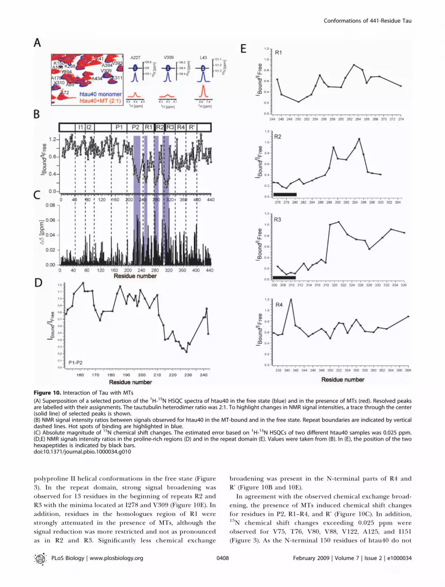
polyproline II helical conformations in the free state (Figure3). In the repeat domain, strong signal broadening wasobserved for 13 residues in the beginning of repeats R2 andR3 with the minima located at I278 and V309 (Figure 10E). Inaddition, residues in the homologues region of R1 werestrongly attenuated in the presence of MTs, although thesignal reduction was more restricted and not as pronouncedas in R2 and R3. Significantly less chemical exchange
broadening was present in the N-terminal parts of R4 andR’ (Figure 10B and 10E).In agreement with the observed chemical exchange broad-
ening, the presence of MTs induced chemical shift changesfor residues in P2, R1–R4, and R’ (Figure 10C). In addition,15N chemical shift changes exceeding 0.025 ppm wereobserved for V75, T76, V80, V88, V122, A125, and I151(Figure 3). As the N-terminal 150 residues of htau40 do not
Figure 10. Interaction of Tau with MTs
(A) Superposition of a selected portion of the 1H-15N HSQC spectra of htau40 in the free state (blue) and in the presence of MTs (red). Resolved peaksare labelled with their assignments. The tau:tubulin heterodimer ratio was 2:1. To highlight changes in NMR signal intensities, a trace through the center(solid line) of selected peaks is shown.(B) NMR signal intensity ratios between signals observed for htau40 in the MT-bound and in the free state. Repeat boundaries are indicated by verticaldashed lines. Hot spots of binding are highlighted in blue.(C) Absolute magnitude of 15N chemical shift changes. The estimated error based on 1H-15N HSQCs of two different htau40 samples was 0.025 ppm.(D,E) NMR signals intensity ratios in the proline-rich regions (D) and in the repeat domain (E). Values were taken from (B). In (E), the position of the twohexapeptides is indicated by black bars.doi:10.1371/journal.pbio.1000034.g010
PLoS Biology | www.plosbiology.org February 2009 | Volume 7 | Issue 2 | e10000340408
Conformations of 441-Residue Tau

strongly contribute to binding and assembly of MTs [22,45],the chemical shift changes might be attributed to weaktransient contacts with MTs or to changes—as a result of MTbinding—in intramolecular long-range interactions inhtau40.
Discussion
Intrinsically Disordered TauTau is important for neuronal cell biology because it
stabilizes MTs and promotes axonal outgrowth, and forneurodegeneration because it undergoes abnormal aggrega-tion in AD and other brain disorders [3,5,6]. However, themode of action of tau is still enigmatic. As soon as the proteinwas discovered [1], its unusual behaviour became apparentbecause it retained its function even after heat denaturation.Subsequent biophysical characterization revealed that tauwas highly soluble and almost devoid of secondary structure[12]. Cloning of the protein confirmed a high fraction ofhydrophilic amino acid residues and an overall basiccharacter, complementary to the acidic surface of MTs [7,8].It also revealed three or four semiconserved repeats of ;31residues that were involved both in the interactions with MTsand in the assembly of Alzheimer PHFs. However, thefunction of MTs was curiously distributed over manyresidues, each contributing only weakly [9]. Electron micro-scopy studies of tau showed that the molecule had very littlecontrast, and only special surface-rendering methods re-vealed tau as irregular elongated molecules [46,47]. X-rayscattering, circular dichroism (CD), and Fourier transforminfrared (FTIR) studies all pointed to a seemingly randomstructure in solution that was termed ‘‘natively unfolded’’[13].
NMR spectroscopy provided now a detailed view of thenatively unfolded nature of 441-residue tau at single residueresolution. 343 out of 441 residues of the htau40 monomerare in a nonperiodic, disordered conformation (Figure 3).Transient elements of secondary structure were restricted tosmall regions (Figure 3G): (i) 274Lys-Leu284, 305Ser-Asp315, and336Gln-Asp345 transiently populate b-structure, in agreementwith previous studies on fragments covering the repeatregion [17,18]. The high propensity of b-structure in theparts that are essential for PHF formation (274Lys-Leu284
and 305Ser-Asp315) underpins the fact that these residuesserve as seeds of aggregation. (ii) 175TPPAPKTPPS184,216PTPPTREP223, and 232PPKSPSSA239 in the proline-richregions P1 and P2, transiently assume polyproline II helicalconformation. These are the only short residue stretches inhtau40 that comprise at least three prolines of which two aresequential (Figure 1B). Within the motifs 175TPPAPKTPPS184,216PTPPTREP223, and 232PPKSPSSA239 in the proline-richregion, there are several phosphorylation sites that arecharacteristically elevated in AD, that is, Thr175, Thr181,Thr231, Ser235 [48]. Moreover, several antibodies againstphosphorylated tau require dual phosphorylation, separatedby three to four residues, e.g., 202þ205, or 231þ235. When apolyproline II helical structure is formed the two phosphory-lated residues would be facing the same way on the helix.However, in the free state of htau40 the site that is recognizedby the AT8 antibody (residues 199SPGSPGT205) does not showa clear propensity to adopt polyproline II helix. (iii) 114Leu-Thr123 transiently populates a-helical structure. The helical
structure might promote intramolecular long-range inter-actions in tau and might be important for interaction withthe dynein-activator complex dynactin [49]. Notably, Thr123 isone of only a few residues in the N-terminal domain that isphosphorylated in PHF tau [50]. (iv) 428Leu-Ala437 have a highpropensity to form a-helical structure. Notably, truncation ofthe C terminus behind Asp421 was suggested to be an earlymolecular event in tau aggregation [51], suggesting that theconformational properties of 428Leu-Ala437 can influenceproteolytic cleavage.Based on their functional differences three different
domains of tau were defined: (i) the projection domaincomprising residues 1–200, i.e., the N-terminal part of tau upto the proline-rich region P2, (ii) the central regioncomprising the repeat domain and its flanking regions P2and R’, and (iii) the 40–50 C-terminal residues [52]. NMRdipolar couplings (Figure 4) demonstrated that the functionaldifferences are associated with strong differences in theintrinsic flexibility of the three domains. Whereas the repeatdomain and its flanking proline rich regions have a lowerintrinsic flexibility on a time scale in the nanosecond tomicrosecond range detected by dipolar couplings, which isimportant for formation of secondary structure, in agree-ment with an increased propensity to populate polyproline IIor b-structure, residues in the projection domain as well as inthe C-terminal region more rapidly interconvert betweendifferent conformations, which is detected by relaxationmeasurements. The differences in intrinsic mobility areassociated with a decreased number of hydrophobic andincreased number of negatively charged residues in theprojection domain (Figure 1B). Moreover, using NMR dipolarcouplings a reduced mobility in regions that harbour manyproline residues had been previously observed in the C-terminal tails of a- and b-synuclein [53,54]. Importantly,Figure 4 is also very suggestive of a possible folding of the N-tail and C-tail over the middle domain of tau, previouslytermed the paperclip model [27].
Intricate Network of Long-Range Interactions in Soluble TauThe appearance of tau as an elongated molecule by some
EM methods [46,47] suggested that the conformation insolution was extended in agreement with the nativelyunfolded state of tau and the accessibility to multiple kinasesthroughout the chain. However, evidence for global foldingbegan to emerge from several antibodies that had discontin-uous epitopes comprising residues near the N terminus andwithin the repeat domain (e.g., Alz-50, MC-1 [55]). Thisevidence was further confirmed by FRET studies showing thattau was able to form a double hairpin, leading to a‘‘paperclip’’ structure whereby both N- and C terminus werefolded into the vicinity of the repeat domain [27]. Thisconcept is now substantially expanded and refined by theNMR analysis (Figure 8). Whereas fluorescence resonanceenergy transfer combined with electron paramagnetic reso-nance requires two labels, one label acting as donor and theother one as acceptor, PRE monitored by NMR requires onlya single paramagnetic centre such as MTSL attached to a freecysteine. Even more important, whereas in FRET only a singledistance can be measured for each donor-acceptor pair, allnuclei in the protein serve as acceptor. Thus, a large numberof intraresidual distances (.100) can be measured from asingle MTSL-labelled sample. In this study, six uniformly
PLoS Biology | www.plosbiology.org February 2009 | Volume 7 | Issue 2 | e10000340409
Conformations of 441-Residue Tau

distributed MTSL positions provided a total of 2,288 distancerestraints. The distance restraints were integrated into astructural model (Figure 8A), which shows tau in a muchmore compact form than previously expected from the EMimages. However, the molecule is still loosely packed, highlyflexible, and exchanges between a large number of con-formations, consistent with large average values of thehydrodynamic radius (Figure 9A).
The Ca contact map, which reports on transient inter-actions that are found in many of the calculated structures,reveals an intricate network of long-range interactions insoluble tau (Figure 8B). In particular the hexapeptide in R3—a residue stretch that is essential for aggregation of tau intoPHFs—is strongly involved into intramolecular contacts withboth the C-terminal and N-terminal domain of tau. Thisincludes a transient interaction with the amphiphatic C-terminal helix (Figures 7F and 8B). The second residue stretchwith increased propensity for formation of an amphiphatichelix, 114Leu-Thr123, interacts with the N-terminal end of I1as well as with repeats R1 and R2. Striking is also thecompaction in the N-terminal 50 residues, in the regionbetween I2 and P1, in P1 and P2, in repeats R1 and R2, and inrepeats R3 and R4. In agreement with the paper clip modelproposed by FRET measurements [27], the N terminus weaklyinteracts with the C terminus (Figures 7E, 7F, 8B, and S4).Interestingly, the proline rich region P2 has many contactswith distant areas of the sequence, such as R1, R2, R4, and theamphiphatic helix at the C terminus, suggesting thatphosphorylation of residues in P2 may modify the ensembleof tau conformations, thereby promoting or delayingaggregation into PHFs. Moreover, the intramolecular inter-actions between its repeat and proline-rich regions mightprime the tau protein for MT binding [45].
Single-Residue Definition of the Hot Spots of the Tau-MTInteraction
The MAP tau is a critical regulator of diverse MT functions[9,45,52]. The repeat domain with its four repeats is essentialfor MT assembly, however, in the absence of the two flankingregions P2 and R’, the repeat domain binds only weakly toMTs. The flanking domains, on the other hand, bind to MTseven in the absence of the repeats. This has led to theproposition of the ‘‘jaws’’ model of tau whereby the regionsflanking the repeats are considered as targeting domains,responsible for positioning tau on the MT surface, and therepeats that act as catalytic domains for MT assembly [52,56].
Here we probed the interaction of htau40 with MTs usingsolution-state NMR spectroscopy. Despite the fact that taumolecules are invisible to solution-state NMR when they arebound to MTs owing to the high molecular weight of thecomplex, information about the residues of tau that areimportant for binding to MTs can be obtained when theexchange between the fully bound form and the free state issufficiently fast. In this case, the observed NMR signals will bean average of the resonances originating from the unboundand bound forms of tau, causing changes in NMR signalposition and intensity. The strength of these changes willdepend on the conformation and chemical environment inthe bound state and the concentration of the bound species.Particularly striking was the pattern of NMR signal intensityratios in the presence and absence of MTs (Figures 10B and11A). Four highly localized regions were revealed, in which,
because of chemical exchange between the unbound and MT-bound state, HN signal intensities were broadened below40% of their value in the unbound state: 225KVAVVRT231,245TAPVPMPDL253, 275VQIINKKLDLSNV287, and 306VQI-VYKPVDLSKV318. In these regions, intensity minima werefound for V228, M250, I278, and V309, respectively (Figure11B and 11C). In agreement with the NMR data, deletionanalysis mapped the MT-binding activity of the proline-richregion to residues K224-N255 and in particular to the stretch225KVAVVRT231. Moreover, site-directed mutagenesis indi-cated that K224, K225, and R230 are important for MT-bindingand -assembly [45]. It is noteworthy that the region225KVAVVRT231 is conserved between tau and two otherMAPs, MAP-2 and MAP-4 [45]. The importance of275VQIINKKLDLSNV287 for MT-binding is supported bybiochemical analyses that reported a strong reduction ofMT binding affinity upon mutation of K274, K280, K281 toalanine [57]. Importantly, the hot spots of interaction areseparated by residue stretches that show smaller chemicalshift changes and less signal broadening (Figures 10 and 11).These residue stretches might act as flexible linker sequencesand suggest that tau protein can assume multiple conforma-tions on the surface of MTs. Moreover, the flexible structuremay allow tau to be easily displaced from the MT lattice,consistent with the rapid diffusion of tau in neurons [58].In addition to the hot spots of MT-interaction, signal
broadening and chemical shift changes were observed formost residues in R4 and R’ and extended to a weaker degreeeven up to the C terminus, indicative of transient interactionsof these regions with MTs. However, in contrast to theproline-rich region P1 and repeats R1, R2, and R3, no clearminimum in NMR signal intensities was observed in repeatR4, indicating that R4 may not be very important to theinteraction between tau and MTs. This observation is inagreement with biochemical studies that suggested a core-MTbinding domain comprising the N-terminal side of the repeatregion [59]. On the other hand, it is in contrast to the viewthat tau possesses multiple independent tubulin-binding sites[7]. As far as the projection domain is concerned, significantchemical shift changes were observed for residues in insert I2and in the region with helical propensity (114Leu-Thr123),consistent with the finding that the projection domainregulates the spacing of MTs [60].Little is known about the nature of the cognate tau binding
sites in tubulin. Based on digestion experiments it is believedthat tau binds to the acidic carboxyl tail of tubulin, which issupposed to be exposed on the surface of MTs [61]. Moreover,mutational analysis pointed to the importance of positivelycharged lysine residues (K274, K280, K281) in tau for MT-binding, suggesting that electrostatic interactions are impor-tant for the tau-MT binding [45]. On the other hand, thehomologues region in R1 contains only a single positivelycharged residue (K254) at the edge of the most affectedresidue stretch, but does contain a negatively charged residue(D252). Similarly, the hot spots of MT-binding in R2 and R3also contain a negative charge (Figures 1B and 11), suggestingthat the tau-MT interaction might be more complex. Indeed,there is a striking correlation between the NMR-basedMT-interaction profile and the hydrophobicity patternof tau (Figure 11A). The 13-residue stretch in R3 (306VQI-VYKPVDLSKV318), which shows the strongest chemicalexchange contribution in the presence of MTs, is the most
PLoS Biology | www.plosbiology.org February 2009 | Volume 7 | Issue 2 | e10000340410
Conformations of 441-Residue Tau

hydrophobic region of 441-residue tau (Figure 11C). Con-sistent with the importance of hydrophobic interactions,substitution of the tyrosine residue in this residue stretch byan asparagine (Y310-.N) reduced the MT affinity of tau [59].Maxima are also found in the hydrophobicity profile forthe other three hot spots of MT-interaction (225KVAVVRT231,245TAPVPMPDL253, 275VQIINKKLDLSNV287), whereas thehomologous region in repeat R4 only has a few hydrophobicresidues but has many charged residues, and is less affected bythe presence of MTs (Figure 11C). Further support for theimportance of hydrophobic interactions for formation of thetau-MT complex comes from the MT-binding site in P2:
225KVAVVRT231 is the most hydrophobic residue stretch inthe proline-rich regions P1 and P2 (Figure 11B). Takentogether, the NMR and biochemical data suggest a complexmechanism of tau-MT interaction involving both electrostaticand hydrophobic contacts.Over 30 phosphorylation sites have been identified in tau,
many of which are elevated in AD [62,63]. Prominent sites arelocated in the flanking domains, e.g., S199, S202, T205, T212, S214,T231, S235 before the repeats, S396, S404, S422, and others afterthe repeats (Figures 11B and 11C). The major sites within therepeats are located in the KXGS motifs, i.e., S262, S293, S324,and S356. These sites are phosphorylated by the kinase MARK,
Figure 11. Comparison of MT-Binding Profile with Hydrophobicity Pattern of Tau
(A) NMR signal intensity ratios between signals observed for htau40 in the MT-bound and in the free state (see also Figure 10B) are shown as stars andconnected by lines. Hydrophobicity values calculated according to the Kyte-Doolittle scale are shown as green bars. Hydrophobic regions have positiveor small negative hydrophobicity values. The location of transient secondary structure observed in unbound htau40 (i.e., as monomer in solution) isindicated as schematic on top.In (B) and (C) the regions that are important for interaction with MTs are shown in detail. The amino acid sequence is shown below. It is colour codedaccording to Figure 1B. The regions of tau that transiently populate b-structure and polyproline II helix in the unbound state are marked by yellowarrows and green bars. Phosphorylation sites are marked by yellow circles.doi:10.1371/journal.pbio.1000034.g011
PLoS Biology | www.plosbiology.org February 2009 | Volume 7 | Issue 2 | e10000340411
Conformations of 441-Residue Tau

which results in the detachment of tau from MTs [64].Interestingly, S262, S293, S324, and S356 are not part of the hotspots of MT-interaction, suggesting that phosphorylation atthese sites might inhibit MT-binding by long-range electro-static interactions. Alternatively, or in combination, phos-phorylation can induce conformational changes that areincompatible with MT-binding. On the other hand, T231 isright in the middle of the MT-binding region in the proline-rich domain P2, making even a steric inhibition of MT-binding possible. Another potential mechanism is stabiliza-tion of a polyproline II helix by phosphorylation. Phosphor-ylation of residues within the fragments 216PTPPTREP223
and 232PPKSPSSA239 might stabilize their nascent polyprolineII helical propensity, such that the resulting conformation isno longer able to efficiently bind to MTs.
Why would a natively unfolded protein evolve to stabilizeaxonal MTs? To consider this, we note that MTs bind a varietyof proteins, some of which are natively unfolded (e.g., the tau-MAP2-MAP4 family), but others are typical well-foldedmolecules. Two cases in point are kinesin and doublecortin,both of which bind to the MT surface in a periodic and well-defined fashion. By contrast, the MAPs are rather diffuselydistributed over the MT surface, there is little detectableperiodicity, the MAPs bind relatively weakly, and they diffuserapidly off MTs and along them [65]. To complicate mattersfurther, binding of MAPs involves the C-terminal tails oftubulin subunits, which are themselves natively unfolded [61].Not surprisingly, much of the N- and C-terminal domains oftau are highly flexible even when the repeat domain isattached to MTs [22]. Several explanations have been invokedto explain the functions of unfolded proteins such as MAPs[66]: They can act as entropic bristles to keep the spacingbetween MTs and other cell components (and indeed largeMAPs keep larger spacings than small MAPs [60]); serve asassemblers for multisubunit structures (e.g., to pre-assembletubulin into oligomers for incorporation into MT); serve asdocking sites for enzymes (e.g., kinases and phosphatases forthe case of MT-bound MAPs); and may even have regulatoryfunctions for MT-related functions (e.g., interaction withmotor proteins of axonal transport [67]). Thus, the multi-plicity of functions would correspond to a multiplicity ofconformations. Many of the above functions are inferredfrom biochemical evidence without detailed knowledge of theresponsible residues and conformations of tau. The identi-fication of residues reported here will provide a basis forfuture experiments to clarify the interactions of tau withinteraction partners in cells, and hopefully the changes thatoccur during neurodegenerative tauopathies.
Materials and Methods
Disordered protein statistics. Assignments of disordered proteins(IDPs) resolved by other groups with the corresponding protein sizewere found either in the BMRB databank (http://www.bmrb.wisc.edu/)or in publications listed in the PubMed (http://www.ncbi.nlm.nih.gov/pubmed/) databank.
Assembly of MTs. Porcine brain tubulin was purified andincubated at concentrations higher than 200 M in MT assemblybuffer (100 mM Pipes, [pH 6.9], 1 mM EDTA, 1 mM MgSO4, 1 mMdithiothreitol) in the presence of 1 mM GTP at 37 8C for 5 min. Afteraddition of 100 M Paclitaxel (Sigma-Aldrich) the polymerization wasperformed for 20 min at 37 8C. Analysis of MTs showed that MTsremained stable over the entire duration of the NMR experiments.
Spin labelling of tau. To label htau40 cysteine-containing mutantswith the nitroxide spin label MTSL (Toronto Research Chemicals),
DTT was removed before labelling from the buffer by using sizeexclusion chromatography (PD-10 columns, GE Healthcare), and theproteins were equilibrated in PBS buffer (pH 7.4). Free sulfhydrylgroups were reacted with a 5-fold molar excess of the MTSL solubilizedin ethyl acetate, at 21 8C for 2.5 h. Unreacted spin label was removedby using PD-10 columns equilibrated in 50 mM Na phosphate buffer(pH 6.8), and spin-labelled proteins were concentrated by usingAmicon Ultra-15 (molecular weight cutoff, 3,000) (Millipore).
NMR spectroscopy. Protein concentrations were between 0.2–0.9mM of htau40. NMR spectra were acquired on a Bruker Avance 900spectrometer equipped with a cryogenic probe. Aggregation did notoccur under these low temperature conditions. 3-D (HA)CANNH [30](100 [F1]372 [F2]31 K [F3] complex data points) and HNN [31] (100[F1] 3 100 [F2] 3 1 K [F3] complex data points) experiments (fourscans, 1.2-s recovery delay, for each experiment roughly one day ofmeasurement time) were collected. To enable and validate assign-ment of MT-bound htau40, a 3-D (HA)CANNH experiment wasmeasured at 20 8C (total experiment time: ;1.5 d). NMR data wereprocessed and analyzed using NMRPipe [68] and Sparky 3 (T. D.Goddard and D. G. Kneller, http://www.cgl.ucsf.edu/home/sparky).
Secondary shift values were calculated as the differences betweenmeasured Ca chemical shifts and the empirical random coil value forthe appropriate amino acid type [69]. Random coil values forhistidines, glutamates, and aspartates were taken from Wishart andSykes [70], as the chemical shifts of these residues are particularlysensitive to pH. To estimate the secondary structure propensity incontiguous segments of htau40, the observed Ca chemical shifts werenormalized by the empirically determined secondary shift expectedfor that residue type in a regular secondary structure (b-sheet or a-helix) conformation [70], summed and normalized by the number ofresidues in the segment.
3J(HNHa) scalar couplings were measured using an intensitymodulated HSQC [71] on a Bruker 900 Avance spectrometer (32scans, relaxation delay 1.2 ms, 2s ¼ time for evolution of 3JHNHa: 18ms). Coupling values were calculated from the intensity ratios usingthe relation Scross/Sdiag ¼ cos(p3JHNHa2s). Secondary
3J(HNHa) scalarcouplings were calculated as the difference between experimental3J(HNHa) scalar couplings and random coil values [72].
15N R1q relaxation rates were measured at 5 8C on a Bruker Avance700-MHz spectrometer using a spinlock frequency of 2 kHz andrelaxation periods of 20, 100, 220, and 300 ms. Relaxation times werecalculated by fitting an exponential function to the decaying signalintegrals.
One-bond N-H RDCs (DNH) were determined by using an inphase-antiphase (IPAP)-HSQC [73]. DNH values were calculated as thedifference between splittings measured in the isotropic phase and ina sample, in which htau40 had been aligned in 5 mg/ml Pf1bacteriophage (Asla). Errors estimated on the basis of the signal-to-noise ratio are 0.2 Hz for D(NH) and 0.4 Hz for 3J(HNHa) couplings,respectively.
NMR diffusion measurements. For determining the hydrodynamicradius, htau40 was dissolved in 99.9 % D2O, 50 mM phosphate buffer(pH 6.9). The samples contained dioxane (concentration ;2%) as aninternal radius standard and viscosity probe [43]. 1-D 1H spectra werecollected employing the standard Bruker pulse program ledbpgp2s.The gradient strength was linearly increased from 2% to 95% of themaximum gradient strength in 16 steps, with 100% gradient strengthcorresponding to 56.9 G/cm. For each 1H spectrum 128 scans and 16K complex with a spectral width of 7,200 Hz were acquired. Signalintensities corresponding to the aliphatic region of the 1H spectra(3.3–0.5 ppm) were readout with the TOPSPIN T1/T2 Relaxationmodule (Bruker Instruments). The diffusion data (signal intensityversus gradient strength) were fitted to exponential functions usingIgor Pro 5.01 (WaveMetrics). From the apparent diffusion coefficientsof htau40 and dioxane and the known Stokes radius of dioxane (2.12A), Stokes radii of monomeric htau40 were calculated [74].
Measurement of PRE. PRE broadening was investigated using 15N-labelled htau40 at a concentration of 15 lM (MTSL at A15C, A239C,and C291þC322) and 50 lM (MTSL at A416C, A384C, and A72C) in50 mM phosphate buffer at (pH 6.8). PRE effects were measured fromthe peak intensity ratios between two 2D 15N-1H HSQC NMR spectraacquired in the presence of the nitroxide radical and after additionof 4 mM DTT (heated to 45 8C for 15 min before measurement) to thesame sample. Addition of DTT will cleave the MTSL tag from thecysteine residue, such that the spin label is no longer attached to theprotein and the protein is in the diamagnetic state. Oxidation of theMTSL tag with ascorbic acid, gave very similar results (Figure S3B).
To exclude intermolecular contacts as cause for PRE line-broad-ening, a mixture of 15 lM 15N-labelled htau40-(C291A/C322A)mutant (without any cysteine residues) and 15 lM 14N-labelled
PLoS Biology | www.plosbiology.org February 2009 | Volume 7 | Issue 2 | e10000340412
Conformations of 441-Residue Tau

htau40-A239C-MTSL was measured at 5 8C (Figure S3A). In addition,intramolecular PRE broadening at htau40 concentrations of 15 and50 lM were compared (Figure S3C).
Calculation of distance restraints. Distance restraints were calcu-lated as described from the intensity ratio between two 2D 15N-1HHSQC NMR spectra, in the diamagnetic and paramagnetic states ofthe protein [39]. To reduce the impact of peak overlap, for eachresidue the average of its own intensity ratio Ipara /Idia and that of thepreceding and following residue was calculated. These smoothedintensity ratios were linearly fit for the enhancement of the transverserelaxation rate by the unpaired electron [75]. For calculation ofdistance restraints, amide proton R2 values were approximated byexperimental amide nitrogen R1q values [76]. The correlation time forthe electron-nuclear interaction was set to 4 ns, in agreement withprevious studies [39]. For peaks broadened beyond detection, distanceswere set to 7 6 5 A. Peaks with intensity ratios below 0.95 wererestrained to the calculated distance 65 A by using a harmonic squarewell potential. For residues that were not broadened in the para-magnetic state, a lower distance bound of 25 A was used. All distanceswere imposed as restraints between the Ca atom of the residue with thecysteine-MTSL group and residue-specific amide protons.
Structure calculation and analysis. Structure calculations wereperformed using XPLOR-NIH, version 2.9.7 [40]. An all-atomrepresentation of htau40 was used. Structural energy terms fromsteric repulsion, bond length, bond angles, dihedral angles, andfavoured regions of the Ramachandran map were employed.
For restraining a single molecule simultaneously by all PREdistance restraints, torsion angle dynamics were started at 3,000 Kwith the temperature reduced to 20 K, followed by a short energyminimization. 50 structures were calculated starting from a randomcoil. The seven lowest-energy structures that satisfied the 2,288distance restraints with no violations greater than 1 A were used forcalculation of the average contact map shown in Figure 8B.
Single molecule calculations were followed by ensemble calcu-lations, in which distance restraints do not have to be fulfilled by asingle molecule, but collectively by the ensemble of molecules.Ensemble calculations were started from the lowest-energy structureobtained in the single molecule calculation (see above) andperformed in two rounds. Initially, distance restraints were enforcedonto an ensemble of 30 molecules [41]. Torsion angle dynamics wereused with the temperature reduced from 10,000 K to 5,000 K. Thelowest energy structure obtained from this first round of ensembleaveraging was subjected to another round of structure calculationusing an ensemble size of 5. Torsion angle dynamics was used and thetemperature was reduced from 3,000 K to 1,000 K. A total of 100structures were calculated. The average contact map obtained fromthe seven lowest-energy structures of the ensemble was very similar tothe one obtained from single molecule calculations (Figure S4).
Supporting Information
Figure S1. Degeneracy of Ca Connectivity
(A) Residue Y18 points back to T17, but for 18 other residues the Ca
frequency also lies within a range of þ/� 0.2 ppm (red strip) of theCa(i-1)peak of Y18.(B) Comparison of Ca chemical shift degeneracy observed in htau40(441 residues) with values reported for the globular proteins
calmodulin (148 residues), N-terminal domain enzyme I (259residues), maltose-binding protein (370 residues), and malatesynthase G (723 residues).
Found at doi:10.1371/journal.pbio.1000034.sg001 (411 KB PDF).
Figure S2. Robustness of Residual Secondary Structure of htau40 inSolution
(A),(C),(D), Comparison of Ca secondary chemical shifts, DCa,observed in htau40 and the tau fragments K32, K25, and K10,respectively (all at 5 8C).(B) Comparison of Ca secondary chemical shifts, DCa, observed inhtau40 at 278K and 293K. Red lines indicate y¼ x.
Found at doi:10.1371/journal.pbio.1000034.sg002 (337 KB PDF).
Figure S3. Control PRE Measurements
(A) Intensity ratio (Iparam/Idiam) obtained from HSQC spectrarecorded on 15 lM 15N-labelled htau40-(C291A/C322A) (i.e., htau40without any cysteine residues) in the presence of 15 lM 14N-labelledhtau40-A239C-MTSL in the paramagnetic and diamagnetic state. Nosignificant deviations from unity were observed, indicating thatdecreases in intensity ratios are not due to aggregation, and can beattributed instead to intramolecular long-range contacts in nativehtau40.(B) Comparison of PRE intensity ratios for different diamagneticreference states. For the data shown by blue bars, DTT was added tothe sample, whereas for the red line ascorbic acid was used to quenchthe spin label.(C) Comparison of intensity ratios obtained from HSQC spectrarecorded on 15 lM (red line) and 50 lM (blue bars) 15N-labelledhtau40-A239C-MTSL in the presence and absence of DTT.
Found at doi:10.1371/journal.pbio.1000034.sg003 (202 KB PDF).
Figure S4. Average Contact Map for the Seven Lowest-EnergyStructures Obtained from Ensemble Calculations with an EnsembleSize of 5 (see Materials and Methods)
A continuous grey scale from 3 A (black) to 22 A (white) is used.
Found at doi:10.1371/journal.pbio.1000034.sg004 (2.14 MB PDF).
Acknowledgments
The authors wish to thank Lukasz Skora and Ilka Lindner forexcellent technical assistance and Eva-Maria Mandelkow for dis-cussions.
Author contributions. EM and MZ conceived the project. MDM, SB,JK, SJ, and JB performed the experiments. MDM, CG, EM, and MZwrote the paper.
Funding. This work was supported by the Max Planck Society, theFonds der Chemischen Industrie, the Boehringer Ingelheim Fonds,and the DFG through GRK 782 and through a DFG Heisenbergscholarship (ZW 71/2–1 and 3–1). The funders had no role in studydesign, data collection and analysis, decision to publish, orpreparation of the manuscript.
Competing interests. The authors have declared that no competinginterests exist.
References1. Weingarten MD, Lockwood AH, Hwo SY, Kirschner MW (1975) A protein
factor essential for microtubule assembly. Proc Natl Acad Sci U S A 72:1858–1862.
2. Drubin DG, Kirschner MW (1986) Tau protein function in living cells. J CellBiol 103: 2739–2746.
3. Garcia ML, Cleveland DW (2001) Going new places using an old MAP: tau,microtubules and human neurodegenerative disease. Curr Opin Cell Biol13: 41–48.
4. Cassimeris L, Spittle C (2001) Regulation of microtubule-associatedproteins. Int Rev Cytol 210: 163–226.
5. Goedert M, Spillantini MG (2006) A century of Alzheimer’s disease. Science314: 777–781.
6. BallatoreC, Lee VM, Trojanowski JQ (2007) Tau-mediated neurodegenerationin Alzheimer’s disease and related disorders. Nat Rev Neurosci 8: 663–672.
7. Lee G, Cowan N, Kirschner M (1988) The primary structure andheterogeneity of tau protein from mouse brain. Science 239: 285–288.
8. Goedert M, Spillantini MG, Potier MC, Ulrich J, Crowther RA (1989)Cloning and sequencing of the cDNA encoding an isoform of microtubule-associated protein tau containing four tandem repeats: differentialexpression of tau protein mRNAs in human brain. Embo J 8: 393–399.
9. Butner KA, Kirschner MW (1991) Tau protein binds to microtubulesthrough a flexible array of distributed weak sites. J Cell Biol 115: 717–730.
10. Wischik CM, Novak M, Edwards PC, Klug A, Tichelaar W, et al. (1988)Structural characterization of the core of the paired helical filament ofAlzheimer-disease. Proc Natl Acad Sci U S A 85: 4884–4888.
11. von Bergen M, Friedhoff P, Biernat J, Heberle J, Mandelkow EM, et al.(2000) Assembly of tau protein into Alzheimer paired helical filamentsdepends on a local sequence motif ((306)VQIVYK(311)) forming betastructure. Proc Natl Acad Sci U S A 97: 5129–5134.
12. Cleveland DW, Hwo SY, Kirschner MW (1977) Physical and chemicalproperties of purified tau factor and the role of tau in microtubuleassembly. J Mol Biol 116: 227–247.
13. Schweers O, Schonbrunnhanebeck E, Marx A, Mandelkow E (1994)Structural studies of tau-protein and Alzheimer paired helical filamentsshow no evidence for beta-structure. J Biol Chem 269: 24290–24297.
14. Dunker AK, Silman I, Uversky VN, Sussman JL (2008) Function and structureof inherently disordered proteins. Curr Opin Struct Biol 18: 756–764.
15. Dyson HJ, Wright PE (2005) Intrinsically unstructured proteins and theirfunctions. Nat Rev Mol Cell Biol 6: 197–208.
16. Mukrasch MD, von Bergen M, Biernat J, Fischer D, Griesinger C, et al.(2007) The ‘‘jaws’’ of the tau-microtubule interaction. J Biol Chem 282:12230–12239.
PLoS Biology | www.plosbiology.org February 2009 | Volume 7 | Issue 2 | e10000340413
Conformations of 441-Residue Tau

17. Mukrasch MD, Biernat J, von Bergen M, Griesinger C, Mandelkow E, et al.(2005) Sites of tau important for aggregation populate fbetag-structureand bind to microtubules and polyanions. J Biol Chem 280: 24978–24986.
18. Eliezer D, Barre P, Kobaslija M, Chan D, Li X, et al. (2005) Residualstructure in the repeat domain of tau: echoes of microtubule binding andpaired helical filament formation. Biochemistry 44: 1026–1036.
19. Mukrasch MD, Markwick P, Biernat J, Bergen M, Bernado P, et al. (2007)Highly populated turn conformations in natively unfolded tau proteinidentified from residual dipolar couplings and molecular simulation. J AmChem Soc 129: 5235–5243.
20. Barre P, Eliezer D (2006) Folding of the repeat domain of tau upon bindingto lipid surfaces. J Mol Biol 362: 312–326.
21. Andronesi OC, von Bergen M, Biernat J, Seidel K, Griesinger C, et al. (2008)Characterization of Alzheimer’s-like paired helical filaments from the coredomain of tau protein using solid-state NMR spectroscopy. J Am Chem Soc130: 5922–5928.
22. Sillen A, Barbier P, Landrieu I, Lefebvre S, Wieruszeski JM, et al. (2007)NMR investigation of the interaction between the neuronal protein Tauand the microtubules. Biochemistry 46: 3055–3064.
23. Landrieu I, Lacosse L, Leroy A, Wieruszeski JM, Trivelli X, et al. (2006) NMRanalysis of a Tau phosphorylation pattern. J Am Chem Soc 128: 3575–3583.
24. Sillen A, Leroy A, Wieruszeski JM, Loyens A, Beauvillain JC, et al. (2005)Regions of tau implicated in the paired helical fragment core as defined byNMR. Chembiochem 6: 1849–1856.
25. Sibille N, Sillen A, Leroy A, Wieruszeski JM, Mulloy B, et al. (2006)Structural impact of heparin binding to full-length Tau as studied by NMRspectroscopy. Biochemistry 45: 12560–12572.
26. Mylonas E, Hascher A, Bernado P, Blackledge M, Mandelkow E, et al. (2008)Domain conformation of tau protein studied by solution small-angle X-rayscattering. Biochemistry 47: 10345–10353.
27. Jeganathan S, von Bergen M, Brutlach H, Steinhoff HJ, Mandelkow E (2006)Global hairpin folding of tau in solution. Biochemistry 45: 2283–2293.
28. Sevcik J, Skrabana R, Dvorsky R, Csokova N, Iqbal K, et al. (2007) X-raystructure of the PHF core C-terminus: insight into the folding of theintrinsically disordered protein tau in Alzheimer’s disease. FEBS Lett 581:5872–5878.
29. Hiller S, Wasmer C, Wider G, Wuthrich K (2007) Sequence-specificresonance assignment of soluble nonglobular proteins by 7D APSY-NMRspectroscopy. J Am Chem Soc 129: 10823–10828.
30. Zweckstetter M, Bax A (2001) Single-step determination of proteinsubstructures using dipolar couplings: aid to structural genomics. J AmChem Soc 123: 9490–9491.
31. Panchal SC, Bhavesh NS, Hosur RV (2001) Improved 3D triple resonanceexperiments, HNN and HN(C)N, for H-N and N-15 sequential correlationsin (C-13, N-15) labeled proteins: application to unfolded proteins. J BiomolNMR 20: 135–147.
32. Dyson HJ, Wright PE (2001) Nuclear magnetic resonance methods forelucidation of structure and dynamics in disordered states. MethodsEnzymol 339: 258–270.
33. Tjandra N, Bax A (1997) Direct measurement of distances and angles inbiomolecules by NMR in a dilute liquid crystalline medium. Science 278:1111–1114.
34. Prestegard JH, al-Hashimi HM, Tolman JR (2000) NMR structures ofbiomolecules using field oriented media and residual dipolar couplings. QRev Biophys 33: 371–424.
35. Blackledge M (2005) Recent progress in the study of biomolecular structureand dynamics in solution from residual dipolar couplings. Prog NMRSpectrosc 46: 23–61.
36. Mohana-Borges R, Goto NK, Kroon GJ, Dyson HJ, Wright PE (2004)Structural characterization of unfolded states of apomyoglobin usingresidual dipolar couplings. J Mol Biol 340: 1131–1142.
37. Wang CY, Palmer AG (2003) Solution NMR methods for quantitativeidentification of chemical exchange in N-15-labeled proteins. Magn ResonChem 41: 866–876.
38. Lange OF, Lakomek NA, Fares C, Schroder GF, Walter KF, et al. (2008)Recognition dynamics up to microseconds revealed from an RDC-derivedubiquitin ensemble in solution. Science 320: 1471–1475.
39. Gillespie JR, Shortle D (1997) Characterization of long-range structure inthe denatured state of staphylococcal nuclease. I. Paramagnetic relaxationenhancement by nitroxide spin labels. J Mol Biol 268: 158–169.
40. Schwieters CD, Kuszewski JJ, Tjandra N, Clore GM (2003) The Xplor-NIHNMR molecular structure determination package. J Magn Reson 160: 65–73.
41. Iwahara J, Schwieters CD, Clore GM (2004) Ensemble approach for NMRstructure refinement against (1)H paramagnetic relaxation enhancementdata arising from a flexible paramagnetic group attached to a macro-molecule. J Am Chem Soc 126: 5879–5896.
42. Lindorff-Larsen K, Kristjansdottir S, Teilum K, Fieber W, Dobson CM, et al.(2004) Determination of an ensemble of structures representing thedenatured state of the bovine acyl-coenzyme a binding protein. J AmChem Soc 126: 3291–3299.
43. Wilkins DK, Grimshaw SB, Receveur V, Dobson CM, Jones JA, et al. (1999)Hydrodynamic radii of native and denatured proteins measured by pulsefield gradient NMR techniques. Biochemistry 38: 16424–16431.
44. Sherman E, Haran G (2006) Coil-globule transition in the denatured stateof a small protein. Proc Natl Acad Sci U S A 103: 11539–11543.
45. Goode BL, Denis PE, Panda D, Radeke MJ, Miller HP, et al. (1997)Functional interactions between the proline-rich and repeat regions of tauenhance microtubule binding and assembly. Mol Biol Cell 8: 353–365.
46. Hirokawa N, Shiomura Y, Okabe S (1988) Tau proteins: the molecularstructure and mode of binding on microtubules. J Cell Biol 107: 1449–1459.
47. Wille H, Drewes G, Biernat J, Mandelkow EM, Mandelkow E (1992)Alzheimer-like paired helical filaments and antiparallel dimers formedfrom microtubule-associated protein tau in vitro. J Cell Biol 118: 573–584.
48. Mi K, Johnson GV (2006) The role of tau phosphorylation in thepathogenesis of Alzheimer’s disease. Curr Alzheimer Res 3: 449–463.
49. Magnani E, Fan J, Gasparini L, Golding M, Williams M, et al. (2007)Interaction of tau protein with the dynactin complex. Embo J 26: 4546–4554.
50. Gong CX, Liu F, Grundke-Iqbal I, Iqbal K (2005) Post-translationalmodifications of tau protein in Alzheimer’s disease. J Neural Transm 112:813–838.
51. Gamblin TC, Chen F, Zambrano A, Abraha A, Lagalwar S, et al. (2003)Caspase cleavage of tau: linking amyloid and neurofibrillary tangles inAlzheimer’s disease. Proc Natl Acad Sci U S A 100: 10032–10037.
52. Gustke N, Trinczek B, Biernat J, Mandelkow EM, Mandelkow E (1994)Domains of tau protein and interactions with microtubules. Biochemistry33: 9511–9522.
53. Bertoncini CW, Jung YS, Fernandez CO, Hoyer W, Griesinger C, et al.(2005) Release of long-range tertiary interactions potentiates aggregationof natively unstructured alpha-synuclein. Proc Natl Acad Sci U S A 102:1430–1435.
54. Sung YH, Eliezer D (2007) Residual structure, backbone dynamics, andinteractions within the synuclein family. J Mol Biol 372: 689–707.
55. Jicha GA, Bowser R, Kazam IG, Davies P (1997) Alz-50 and MC-1, a newmonoclonal antibody raised to paired helical filaments, recognizeconformational epitopes on recombinant tau. J Neurosci Res 48: 128–132.
56. Preuss U, Biernat J, Mandelkow EM, Mandelkow E (1997) The ‘jaws’ model oftau-microtubule interaction examined in CHO cells. J Cell Sci 110: 789–800.
57. Panda D, Goode BL, Feinstein SC, Wilson L (1995) Kinetic stabilization ofmicrotubule dynamics at steady state by tau and microtubule-bindingdomains of tau. Biochemistry 34: 11117–11127.
58. Konzack S, Thies E, Marx A, Mandelkow EM, Mandelkow E (2007)Swimming against the tide: mobility of the microtubule-associated proteintau in neurons. J Neurosci 27: 9916–9927.
59. Goode BL, Chau M, Denis PE, Feinstein SC (2000) Structural and functionaldifferences between 3-repeat and 4-repeat tau isoforms. Implications fornormal tau function and the onset of neurodegenetative disease. J BiolChem 275: 38182–38189.
60. Chen J, Kanai Y, Cowan NJ, Hirokawa N (1992) Projection domains ofMAP2 and tau determine spacings between microtubules in dendrites andaxons. Nature 360: 674–677.
61. Nogales E, Whittaker M, Milligan RA, Downing KH (1999) High-resolutionmodel of the microtubule. Cell 96: 79–88.
62. Morishima-Kawashima M, Hasegawa M, Takio K, Suzuki M, Yoshida H, et al.(1995) Proline-directed and non-proline-directed phosphorylation of PHF-tau. J Biol Chem 270: 823–829.
63. Hanger DP, Betts JC, Loviny TL, Blackstock WP, Anderton BH (1998) Newphosphorylation sites identified in hyperphosphorylated tau (paired helicalfilament-tau) from Alzheimer’s disease brain using nanoelectrospray massspectrometry. J Neurochem 71: 2465–2476.
64. Drewes G, Ebneth A, Preuss U, Mandelkow EM, Mandelkow E (1997) MARK,a novel family of protein kinases that phosphorylate microtubule-associated proteins and trigger microtubule disruption. Cell 89: 297–308.
65. Santarella RA, Skiniotis G, Goldie KN, Tittmann P, Gross H, et al. (2004)Surface-decoration of microtubules by human tau. J Mol Biol 339: 539–553.
66. Tompa P (2002) Intrinsically unstructured proteins. Trends Biochem Sci27: 527–533.
67. Stamer K, Vogel R, Thies E, Mandelkow E, Mandelkow EM (2002) Taublocks traffic of organelles, neurofilaments, and APP vesicles in neuronsand enhances oxidative stress. J Cell Biol 156: 1051–1063.
68. Delaglio F, Grzesiek S, Vuister GW, Zhu G, Pfeifer J, et al. (1995) Nmrpipe -a multidimensional spectral processing system based on unix pipes. JBiomol NMR 6: 277–293.
69. Schwarzinger S, Kroon GJA, Foss TR, Chung J, Wright PE, et al. (2001)Sequence-dependent correction of random coil NMR chemical shifts. J AmChem Soc 123: 2970–2978.
70. Wishart DS, Sykes BD (1994) The C-13 chemical-shift index - a simplemethod for the identification of protein secondary structure using C-13chemical-shift data. J Biomol NMR 4: 171–180.
71. Permi P, Kilpelainen I, Annila A, Heikkinen S (2000) Intensity modulatedHSQC and HMQC: two simple methods to measure 3J(HNH)alpha inproteins. J Biomol NMR 16: 29–37.
72. Plaxco KW, Morton CJ, Grimshaw SB, Jones JA, Pitkeathly M, et al. (1997)The effects of guanidine hydrochloride on the ‘random coil’ conformationsand NMR chemical shifts of the peptide series GGXGG. J Biomol NMR 10:221–230.
73. Ottiger M, Delaglio F, Bax A (1998) Measurement of J and dipolar couplingsfrom simplified two-dimensional NMR spectra. J Magn Reson 131: 373–378.
74. Jones JA,Wilkins DK, Smith LJ, DobsonCM (1997) Characterisation of proteinunfolding by NMR diffusion measurements. J Biomol NMR 10: 199–203.
75. Battiste JL, Wagner G (2000) Utilization of site-directed spin labeling andhigh-resolution heteronuclear nuclear magnetic resonance for global folddetermination of large proteins with limited nuclear overhauser effectdata. Biochemistry 39: 5355–5365.
76. Ishima R, Torchia DA (2003) Extending the range of amide protonrelaxation dispersion experiments in proteins using a constant-timerelaxation-compensated CPMG approach. J Biomol NMR 25: 243–248.
PLoS Biology | www.plosbiology.org February 2009 | Volume 7 | Issue 2 | e10000340414
Conformations of 441-Residue Tau


![[ebook] Marketing pe intelesul tau - [].pdf](https://img.dokumen.tips/doc/110x75/636103754b9aa63a9e008ed1/ebook-marketing-pe-intelesul-tau-pdf.jpg)


![IAC 6/20/18 Human Services[441] Ch 24, p.1 CHAPTER 24](https://img.dokumen.tips/doc/110x75/63222f5a28c445989105a8c0/iac-62018-human-services441-ch-24-p1-chapter-24-.jpg)