Embed Size (px)
Citation preview
Structural Characterization of the M* Partly Folded Intermediateof Wild Type and P138A Aspartate Aminotransferase fromEscherichia coli*
Received for publication, January 22, 2002, and in revised form, February 26, 2002Published, JBC Papers in Press, March 1, 2002, DOI 10.1074/jbc.M200650200
Leila Birolo‡, Fabrizio Dal Piaz, Piero Pucci, and Gennaro Marino
From the Dipartimento di Chimica Organica e Biochimica, Universita Federico II di Napoli,Complesso Universitario Monte Sant’Angelo, Via Cinthia, 80126 Napoli, Italy
A combination of spectroscopic techniques, hydrogen/deuterium exchange, and limited proteolysis experi-ments coupled to mass spectrometry analysis was usedto depict the topology of the monomeric M* partly foldedintermediate of aspartate aminotransferase from Esch-erichia coli in wild type (WT) as well as in a mutant formin which the highly conserved cis-proline at position 138was replaced by a trans-alanine (P138A). Fluorescenceanalysis indicates that, although M* is an off-pathwayintermediate in the folding of WT aspartate aminotrans-ferase from E. coli, it seems to coincide with an on-pathway folding intermediate for the P138A mutant.Spectroscopic data, hydrogen/deuterium exchange, andlimited proteolysis experiments demonstrated the oc-currence of conformational differences between the twoM* intermediates, with P138A-M* being conceivablymore compact than WT-M*. Limited proteolysis datasuggested that these conformational differences mightbe related to a different relative orientation of the smalland large domains of the protein induced by the pres-ence of the cis-proline residue at position 138. Thesedifferences between the two M* species indicated that inWT-M* Pro138 is in the cis conformation at this stage ofthe folding process. Moreover, hydrogen/deuterium ex-change results showed the occurrence of few differencesin the native N2 forms of WT and P138A, the spectro-scopic features and crystallographic structures ofwhich are almost superimposable.
The mechanism by which proteins fold into their uniquenative structures is still a central problem in structural biology.It is increasingly recognized that the structure of non-nativestates of proteins can provide significant insight into funda-mental issues such as the relationship between protein se-quences and three-dimensional structures, the nature of pro-tein folding pathways, the stability of proteins and theirturnover in the cell, and the transport of proteins across mem-branes (1). Moreover, intermediate states experienced by pro-teins in vivo often play a major role in protein association andaggregation, leading to the “so-called” conformational diseases(2). In contrast to the large amount of structural information
available on native folded proteins, however, too few partlyfolded intermediates have been characterized thoroughlyenough to propose general models on how the native state isattained and which is the structure of transient folding states.Investigation of a wide range of non-native states would thenbe of considerable value. To meet this need, new analyticalstrategies able to characterize transient species and to definethe molecular details through which diverse proteins fold arerequired.
Recently, structural biologists have turned their attention tointegrated strategies for the definition of the surface topologyof proteins and protein complexes. Although these approachesprovide low resolution data, they are amenable to the analysisof transient species and partly folded intermediates. Limitedproteolysis and amide hydrogen exchange experiments in com-bination with mass spectrometry have been employed to inves-tigate the surface topology and the conformational flexibility ofproteins and protein complexes. Amide protons are exchangedwith deuterium with kinetics mainly depending on both solventaccessibility and stabilization of the associated protein back-bone region (3–5). Comparative hydrogen/deuterium exchange(H/D)1 measurements can then be used to monitor proteinstructural changes in different experimental conditions (6), aswell as the effects of binding or aggregation (7). Althoughhydrogen exchange has most often been measured by NMR,monitoring by mass spectrometry (MS) has become increas-ingly common (8–13). As deuterium atoms replace protonsduring the hydrogen exchange reaction, the mass of the proteinincreases. The extent as well as the rate of exchange can thenbe determined by measuring the increase in protein mass.
Proteolytic cleavages on a protein substrate can occur only ifthe polypeptide chain is exposed and flexible enough to adapt tothe specific protease active site. The stable conformation ofproteins then provides some stereochemical barriers to enzy-matic attack, leaving the exposed and flexible regions accessi-ble to proteases and preventing the occurrence of proteolyticcleavages within the highly structured core of the molecule.Consequently, when these experiments are performed using aseries of proteases with different specificity under conditions
* This work was supported by Ministero dell’ Universita e dellaRicerca Scientifica Progetti di Rilevante Interesse Nazionale 1999 and2000 grants, Consiglio Nazionale delle Ricerche Progetto Finalizzato“Biotecnologie” (to P. P. and G. M.), and Regione Campania Grant LR41/94. The costs of publication of this article were defrayed in part bythe payment of page charges. This article must therefore be herebymarked “advertisement” in accordance with 18 U.S.C. Section 1734solely to indicate this fact.
‡ To whom correspondence should be addressed. Tel.: 39-081-674315;Fax: 39-081-674313; E-mail: [email protected].
1 The abbreviations used are: H/D, hydrogen/deuterium exchange;EcAspAT, aspartate aminotransferase from E. coli; WT, wild type as-partate aminotransferase from E. coli; P138A, aspartate aminotrans-ferase from E. coli mutant form in which the cis-proline at position 138was replaced by a trans-alanine; mAspAT, rat liver mitochondrial as-partate aminotransferase; CD, circular dichroism; WT-N2, native formof wild type; P138A-N2, native form of P138A; WT-M*, M* intermediatefrom wild type; P138A-M*, M* intermediate from P138A; ANS, 8-ani-linonaphthalene-1-sulfonic acid; GdmHCl, guanidinium hydrochloride;PLP, pyridoxal 5�-phosphate; PMP, pyridoxamine 5�-phosphate; HPLC,high performance liquid chromatography; DTT, dithiothreitol; ES, elec-trospray; MS, mass spectroscopy; LC, liquid chromatography.
THE JOURNAL OF BIOLOGICAL CHEMISTRY Vol. 277, No. 20, Issue of May 17, pp. 17428–17437, 2002© 2002 by The American Society for Biochemistry and Molecular Biology, Inc. Printed in U.S.A.
This paper is available on line at http://www.jbc.org17428
by guest on January 28, 2016http://w
ww
.jbc.org/D
ownloaded from
that favors a single bond cleavage (complementary proteolysis),the pattern of preferred cleavage sites will depict the exposedregions in the protein molecule (14, 15). This strategy was alsoemployed to investigate conformational changes occurring inprotein structure under different experimental conditions (16–19) or during quaternary forms interchange (20, 21), as well asfor the definition of the interface regions in protein complexes(22–24).
This paper reports the application of these integrated strat-egies to the structural characterization of partly folded inter-mediates of wild type and a mutant form of Escherichia coliaspartate aminotransferase (EcAspAT), in which the highlyconserved cis-proline in position 138 had been replaced by atrans-alanine (P138A; Ref. 25). EcAspAT is a homodimer withtwo identical and independent active sites located at the sub-unit interface. Each subunit consists of an N-terminal arm, alarge cofactor-binding domain, and a small domain (26). Pro-138 is located in a loop region of the large domain close to theinterface with the small domain (27). The replacement of acis-proline by a trans-alanine did not significantly affect eitherthe activity or the stability of the protein, as the catalyticefficiency of P138A is of the same order of magnitude of WT,and the thermal unfolding curves of the mutant and the WT arealmost superimposable (25). Additionally, the GdmHCl-in-duced unfolding equilibrium of P138A mutant follows the samepathway as the wild type protein (25). Nevertheless, the mu-tant enzyme shows interesting folding features, because, de-spite the replacement of a cis peptide bond with a trans one, therefolding process is slower than wild type (25). Moreover, themonomeric P138A-M* intermediate (25) detectable in theGdmHCl-induced unfolding at equilibrium exhibited differentspectroscopic properties as compared with the wild type M*species and resembles a kinetic folding intermediate.
EXPERIMENTAL PROCEDURES
Materials—Recombinant wild type and P138A mutant EcAspATswere isolated from the overproducing E. coli strain TY103 (28) asdescribed previously (25). The proteins were stored at �80 °C in thepresence of 1 mM DTT and an excess of PMP and 2-oxoglutarate.
All experiments were carried out on the PLP form of the enzymes,obtained by removal of excess PMP and 2-oxoglutarate either by exten-sive dialysis against appropriate buffer or by gel filtration on a Super-ose 12 PC column (3.2 � 30 mm) using the SMART system (AmershamBiosciences). Subunit concentration of EcAspATs was determined spec-trophotometrically on a Beckmann DU7500 spectrophotometer, using�280 � 49,935 M�1 cm�1 (29).
L-1-Tosylamido-2-phenylethyl chloromethyl ketone-treated trypsin,chymotrypsin, and subtilisin were purchased from Sigma; endoproteaseLys C was from Roche Molecular Biochemicals. Reverse phase-HPLCC4 columns (25 � 0.46 cm, 5 mm) were purchased from Vydac (TheSeparation Groups). All other reagents and solvents were HPLC-gradefrom Carlo Erba.
Spectroscopic Analysis—Native EcAspAT holoenzymes (in 10 mM
HEPES, pH 7.5, 1 mM DTT, and 0.15 M NaCl) were incubated at 25 °Cat the required protein and GdmHCl concentrations for 1 h (folding/unfolding equilibrium was attained within 30 min, as determined inpreliminary experiments by following with time the enzymatic activityand fluorescence), and protein solutions were filtered just before theanalysis on 0.22-�m pore size polyvinylidene difluoride membrane(Millipore).
Far-UV CD spectra were recorded on a Jasco J715 spectropolarime-ter equipped with a Peltier thermostatic cell holder (Jasco model PTC-348), in a quartz cell (0.1-cm light path) at a protein concentration of 1.0�M. Temperature was measured directly in the quartz cell, the solutionswere filtered just before use on 0.22-�m pore size polyvinylidene diflu-oride membrane (Millipore), and data corrected by subtracting a controlfrom which the protein was omitted. Spectra were recorded at 25 °Cfrom 280 to 200 nm at 0.2-nm resolution, 16-s response, at a scan rateof 20 nm/min. All data are the averages of three measures, and theresults are expressed as mean residue ellipticity [�], which is defined as[�] � 100 �obs/lc, where �obs is the observed ellipticity in degrees, c is the
concentration in residue moles/liter, and l is the length of the light pathin centimeters.
Fluorescence measurements were carried out on a PerkinElmerLB50S fluorimeter, using an optical cuvette of 10-mm light path lengthwith thermostatically controlled cell holder. Tryptophan emission spec-tra were obtained at a protein concentration of 1.0 �M using an excita-tion wavelength of 295 nm, with excitation and emission bandwidths of10 and 2.5 nm, respectively. Tryptophan emission spectra were re-corded between 310 and 480 nm at a scan rate of 100 nm/min. Eachspectrum is the average of three emission scans, and data were cor-rected by subtracting a blank from which the enzyme was omitted. ANSfluorescence emission spectra were recorded on samples that had beenincubated at determined GdmHCl concentration for 30 min at 25 °C ata protein concentration of 1.0 �M before adding ANS to a final concen-tration of 100 �M. ANS emission spectra were recorded using an exci-tation wavelength of 375 nm with excitation and emission bandwidthsof 5 and 5 nm, respectively between 400 and 600 nm at a scan rate of100 nm/min. Each spectrum is the average of three emission scans, anddata were corrected by blank subtraction.
Single-jump refolding experiments were carried out by concentrationjumps of GdmHCl at 5 °C by rapid dilution of protein previously incu-bated at required higher denaturant concentrations to the desired oneat a final protein concentrations of 0.39 �M, and the subsequent time-dependent changes in tryptophan or ANS fluorescence intensity weremonitored, under continuos stirring, at 333 or 475 nm, respectively.
Tryptophan Fluorescence Quenching—Fluorescence quenching titra-tions with either acrylamide or iodide were performed at 25 °C bysequential addition of aliquots of concentrated acrylamide (8 M) orpotassium iodide (5.34 M) stock solution into the protein solution. Forthe native state, protein concentration was 3.9 �M in 10 mM Na-HEPES,pH 7.5, 1 mM DTT, 0.15 M NaCl in a range of acrylamide concentrationfrom 0 to 1.03 M. For the M* intermediate, iodide was added to theprotein (0.39 �M previously incubated at 25 °C for 30 min in 10 mM
HEPES, pH 7.5, 1 mM DTT, and 0.15 M NaCl at 1.3 M GdmHCl forP138A-M*, and 1.5 M GdmHCl for WT-M*) to obtain final iodide con-centration ranging from 0 to 0.2 M. Fluorescence was measured at 25 °Cat the wavelength of the maximum in the fluorescence spectrum re-corded in the absence of the quenching agent. Excitation was at 295 nm(10-nm excitation bandwidth, 2.5-nm emission bandwidth), correctedfor the minor dilution caused by addition of the quenching agent and forthe background fluorescence of both solvent and quenching agent. Cor-rection for the inner filter effect that resulted from the absorption ofacrylamide at 295 nm was applied by multiplying the fluorescenceintensity by 10A/2, where A is the absorbance of the solution at 295 nmmeasured in a 1-cm path length cuvette. In the iodide experiments, 0.1mM Na2S2O3 was included to prevent I3� ion formation.
The fluorescence-quenching data in the presence of iodide quenchingdata were analyzed according to the Stern-Vollmer equation (Equation1) (30).
Fo/F � 1 � KQ� �Q� (Eq. 1)
Fo and F are the fluorescence intensities in the absence and presence ofquencher, respectively. KQ is the Stern-Vollmer quenching constant,and [Q] is the concentration of the quencher. The plot of Fo/F versus [Q]is linear for an apparently homogeneous population of emitting fluoro-phores. Alternatively, a Stern-Vollmer-derived equation (Equation 2)was used to calculate the fraction of accessible fluorescence, that is thefraction of tryptophans that are exposed and then accessible to thequenching agent.
Fo/�Fo � F� � 1/��Q��fa�KQ� � 1/fa (Eq. 2)
fa is the fraction of accessible fluorescence.The fluorescence-quenching data in the presence of acrylamide were
analyzed according to the Stern-Vollmer equation (Equation 3), modi-fied to take into account a static contribution to quenching (31).
Fo/F � �1 � KQ��Q])exp�V��Q�� (Eq. 3)
V is the static constant.Gel Filtration—Gel filtration experiments were carried out as fol-
lows. 50 �l of the samples, 10 �M in 10 mM HEPES, pH 7.5, 1 mM DTT,and 0.15 M NaCl, equilibrated for 30 min at different concentrations ofGdmHCl, were loaded on a Superose 12 PC (3.2 � 300 mm) gel filtrationcolumn installed on the Smart System (Amersham Biosciences), andisocratically eluted in 10 mM HEPES, pH 7.5, 1 mM DTT, and 0.15 M
NaCl at the same concentration of GdmHCl used in the equilibration.All experiments were carried out at 25 °C at a flow rate of 40 �l/min.
A Partly Folded Intermediate of EcAspAT 17429
by guest on January 28, 2016http://w
ww
.jbc.org/D
ownloaded from
The column was calibrated in 10 mM HEPES, pH 7.5, 1 mM DTT, and0.15 M NaCl in the absence of GdmHCl with the following proteins ofknown molecular mass: �-amylase (200,000 Da), cAspATp (92,000 Da),bovine hemoglobin (67,000 Da), cytochrome c (12,300 Da).
Hydrogen/Deuterium Exchange—The H/D reaction was conducted asfollows; proteins were dissolved in 50 mM HEPES, pH 7.5, 1 mM DTT,and 0.15 M NaCl at 25 °C, in the presence of 1.4 M GdmHCl in the caseof the two M* species, at a 8 �M protein concentration, and allowed toequilibrate for 30 min. Deuterium exchange reactions were initiated by10-fold dilution of the sample with 50 mM HEPES, pH 7.5, D2O (con-taining 1.4 M GdmHCl in the case of the two M* species). The exchangereaction was allowed to proceed for lengths of time ranging from 30 to60 min, and, at each time point, 1 nmol of protein was removed from thelabeling solution and rapidly injected into a 30 mm � 0.46 mm (innerdiameter) perfusion column (POROS 10 R2 media, Applied Biosystems)on an HPLC equipped with two Series 200 LC isocratic pumps (AppliedBiosystems) coupled to an API-100 single quadrupole mass spectrome-ter (Applied Biosystems). The protein was eluted at a flow rate of 1ml/min with a gradient of 25–95% acetonitrile in 0.1% trifluoroaceticacid in 1.0 min. The HPLC step was performed with cold protiatedsolvents, thereby reducing the back-exchange kinetics and removingdeuterium from side chains and N/C termini that exchange much fasterthan amide linkages (32). Therefore, the increase in molecular massprovided a direct measurement of the deuteration at peptide amidelinkages. Data were acquired and elaborated using the Biomultiviewer(Applied Biosystems) program. Non-deuterated and totally deuteratedsamples were used as control. The fully deuterated samples were pre-pared by incubation for 1 h at 25 °C in 50 mM HEPES, pH 7.5, 1 mM
DTT, 0.15 M NaCl, 5 M GdmHCl in D2O at 8 �M protein concentration.LC/MS analysis was performed as above.
Limited Proteolysis Experiments—Limited proteolysis experimentswere carried out by incubating P138A-M*, WT-M*, P138A-N2, andWT-N2 with trypsin, chymotrypsin, subtilisin, and endoprotease Lys Cusing enzyme to substrate ratios ranging from 1:1000 to 1:5 (w/w).Enzymatic digestions were all performed in 50 mM HEPES, pH 7.5, 1mM DTT, and 0.15 M NaCl at 25 °C, in the presence of the desiredconcentration of GdmHCl (1.3 M for P138A-M*, 1.5 M for WT-M*), andwithout GdmHCl for the two native forms), at a 1 �M protein concen-tration. In the case of P138A-M* and WT-M*, each protein was incu-bated for 30 min in the reaction solution, before adding the selectedenzyme. The extent of the reactions was monitored on a time-coursebasis by sampling the incubation mixture at different time intervals.Proteolytic fragments were analyzed by LC/MS performed on an API100 single quadrupole electrospray (Applied Biosystems) equipped withtwo Series 200 LC isocratic pumps (Applied Biosystems). Chromato-graphic separation was obtained on a C4 column by the means of a40-min linear gradient from 20 to 70% of acetonitrile in 2% formic acidand 0.1% trifluoroacetic acid. Mass spectra were acquired on a m/zinterval ranging from 600 to 1800. Data were elaborated using theBiomultiviewer program, purchased from PE-Sciex. Mass calibrationwas performed by means of multiply charged ions from a separateinjection of horse heart myoglobin (Sigma; average molecular mass:16,951.5 Da); all masses are reported as average values.
RESULTS AND DISCUSSION
The M* Intermediate—Previous studies suggested that Gd-mHCl-induced unfolding at equilibrium of EcAspAT at 20 °C isreversible and proceeds through the formation of at least twomonomeric intermediates along the unfolding pathway (33).
N2º 2Mº 2M�º 2U
REACTION 1
N2 is the native dimer, M and M* are distinct “structured”monomers, and U is the unfolded state. As described by Heroldand Kirschner (33), the first intermediate M, the folded mono-mer of EcAspAT, is partially populated at 0.6 M GdmHCl andonly at low protein concentrations. The second monomeric in-termediate, M*, accumulates at 1 M GdmHCl and constitutes a“molten globule”-like species, which retains part of the tertiaryand secondary structure of the native state.
The unfolding process of both WT EcAspAT and the P138Amutant was investigated by fluorescence, gel filtration, and CDanalysis and the corresponding M* species, identified according
to the results reported by Herold and Kirschner (33) and tosize-exclusion chromatography data in GdmHCl at equilibrium(34), were thoroughly characterized. Preliminary experiments(25) showed that folding/unfolding equilibrium was attainedwithin 30 min and that, under the experimental conditionsused, the folding intermediates were stable. No precipitation ofprotein was observed after incubation at the protein concentra-tion used, and gel filtration analysis ruled out the presence ofsoluble aggregates (see below).
Because the enzyme has five tryptophan residues located invarious regions of its three-dimensional structure, with onepositioned in the active site pocket, tryptophan fluorescencewas used as a probe for global changes in tertiary structure ofEcAspAT during protein unfolding and to distinguish betweenM and M* (33). In our experimental conditions, i.e. relativelyhigh protein concentration (in the 1–10 �M range) and thepresence of pyridoxal 5�-phosphate, the N2 º 2M transitionescaped fluorescence detection. This transition, indeed, isshifted toward higher denaturant concentrations by increasingthe protein concentration and by the presence of the cofactor(33), thus overlapping with the M to M* transition.
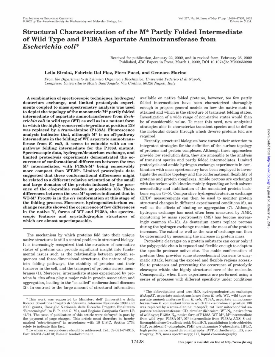
Fig. 1 shows the bathochromic shift of the fluorescence max-imum for WT EcAspAT and the mutant as the concentration ofGdmHCl increased. The unfolding process is clearly biphasicfor both proteins, and a stable intermediate state, correspond-ing to the M* species, could be detected at given GdmHClconcentrations. Therefore, the first fluorescence-detected tran-sition observed included both the dissociation of the dimer andthe conversion to M*. Accordingly, the change in the fluores-cence spectrum reflects both structural changes and the loss ofthe coenzyme (PLP) during unfolding.
A clear difference in the emission spectra of the M* interme-diates of the two proteins was observed. The fluorescence spec-trum of the M* from P138A displayed a maximum at 337 nm,red-shifted by 5 nm as compared with the wild type enzyme(�max at 342 nm). This change of the maximum wavelength issignificant because no variation greater than 0.5 nm was
FIG. 1. GdmHCl-induced unfolding curves of EcAspAT. Figureshows zoom of the transition occurring in the 0.5–1.5 M range of theunfolding process of WT (f) and P138A (*), as monitored by the changesin tryptophanyl emission maximum upon excitation at 295 nm. Theentire unfolding curve is reported in the inset.
A Partly Folded Intermediate of EcAspAT17430
by guest on January 28, 2016http://w
ww
.jbc.org/D
ownloaded from
observed in several spectra recorded for each sample. On thecontrary, in the native and in the unfolded states, the emissionspectra of P138A and WT were nearly coincident.
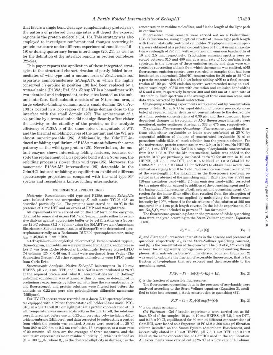
Gel filtration analysis of EcAspAT carried out at differentGdmHCl concentrations (Fig. 2) showed that the elution vol-ume of the enzyme increased to a broad maximum between 0.7and 1.2 M GdmHCl and then gradually decreased at higherdenaturant concentrations, according to previous reports (33,34). In all conditions, the protein eluted as a rather symmetricpeak, consistent with homogeneous species. Moreover, no dif-ferences were detected in the elution volume of WT-M* andP138A-M* at 1.4 M GdmHCl, indicating that no differences inthe compactness of the intermediates could be inferred by theseanalyses. However, both M* forms showed an elution volume inbetween that of the native N2 and that expected for the foldedmonomer M, thus indicating that in these conditions the M*species are monomeric and have a compact structure, albeit notas structured as the folded monomeric protein. Therefore, all
the experiments described below were carried out at equal orlower EcAspAT concentrations to ensure the monomeric stateof the protein.
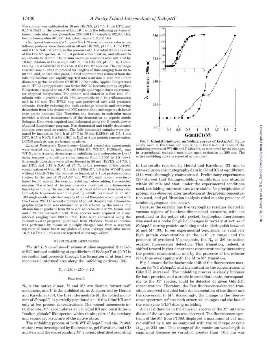
The native, M*, and denatured states of the two proteinswere then analyzed by circular dichroism. Fig. 3 shows the CDspectra of the three forms of P138A, which were coincident withthose recorded for the respective WT forms (data not shown). Inboth cases, the M* species showed a CD signal at 222 nm thatis 58% of the native protein, indicating that these intermedi-ates have a considerable amount of apparent secondarystructure.
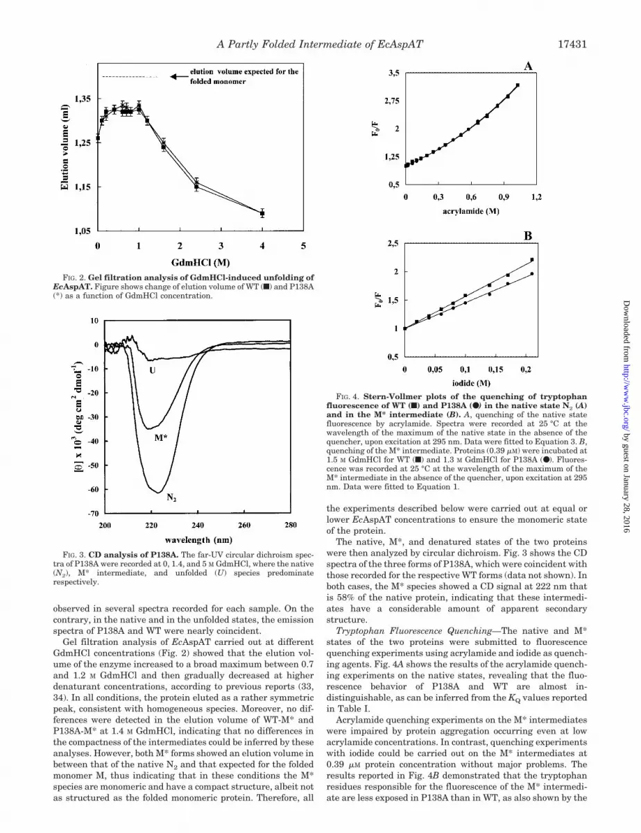
Tryptophan Fluorescence Quenching—The native and M*states of the two proteins were submitted to fluorescencequenching experiments using acrylamide and iodide as quench-ing agents. Fig. 4A shows the results of the acrylamide quench-ing experiments on the native states, revealing that the fluo-rescence behavior of P138A and WT are almost in-distinguishable, as can be inferred from the KQ values reportedin Table I.
Acrylamide quenching experiments on the M* intermediateswere impaired by protein aggregation occurring even at lowacrylamide concentrations. In contrast, quenching experimentswith iodide could be carried out on the M* intermediates at0.39 �M protein concentration without major problems. Theresults reported in Fig. 4B demonstrated that the tryptophanresidues responsible for the fluorescence of the M* intermedi-ate are less exposed in P138A than in WT, as also shown by the
FIG. 3. CD analysis of P138A. The far-UV circular dichroism spec-tra of P138A were recorded at 0, 1.4, and 5 M GdmHCl, where the native(N2), M* intermediate, and unfolded (U) species predominaterespectively.
FIG. 4. Stern-Vollmer plots of the quenching of tryptophanfluorescence of WT (f) and P138A (●) in the native state N2 (A)and in the M* intermediate (B). A, quenching of the native statefluorescence by acrylamide. Spectra were recorded at 25 °C at thewavelength of the maximum of the native state in the absence of thequencher, upon excitation at 295 nm. Data were fitted to Equation 3. B,quenching of the M* intermediate. Proteins (0.39 �M) were incubated at1.5 M GdmHCl for WT (f) and 1.3 M GdmHCl for P138A (●). Fluores-cence was recorded at 25 °C at the wavelength of the maximum of theM* intermediate in the absence of the quencher, upon excitation at 295nm. Data were fitted to Equation 1.
FIG. 2. Gel filtration analysis of GdmHCl-induced unfolding ofEcAspAT. Figure shows change of elution volume of WT (f) and P138A(*) as a function of GdmHCl concentration.
A Partly Folded Intermediate of EcAspAT 17431
by guest on January 28, 2016http://w
ww
.jbc.org/D
ownloaded from
KQ values reported in Table I. Iodide quenching data wereanalyzed to define the number of tryptophan residues involvedin quenching. From the fa values calculated by Equation 2, 4.30( 0.30) of 5 total tryptophan residues could be quenched in theM* intermediate of WT as compared with only 3.05 ( 0.25) inP138A. The quenching data obtained for both WT and P138A inthe denatured state were interpolated with the same equationas a control, giving an fa value of 1, thus indicating that alltryptophans are exposed in the denatured form (data notshown). Again these data suggested a different arrangement ofthe three-dimensional structure in the M* intermediates fromWT and P138A.
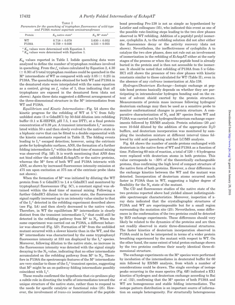
Equilibrium and Kinetic Intermediates—Fig. 5A shows thekinetic progress for the refolding of WT at 5 °C from fullyunfolded state (5 M GdmHCl) by 50-fold dilution into refoldingbuffer (0.1 M K-HEPES, pH 7.5, 1 mM DTT), at a final proteinconcentration of 0.39 �M. A transient intermediate I1* accumu-lated within 50 s and then slowly evolved to the native state ina biphasic curve that can be fitted to a double exponential withthe kinetic constants reported in Table II. The folding eventsleading to I1* escaped detection; however, using the extrinsicprobe for hydrophobic surfaces, ANS, the formation of a furtherfolding intermediate I2* within the dead time of manual mixingwas observed (Fig. 5B). It is worth mentioning that ANS doesnot bind either the unfolded EcAspATs or the native proteins,whereas the M* form of both WT and P138A interacts withANS, as shown by increased fluorescence intensity emission at475 nm upon excitation at 375 nm of the extrinsic probe (datanot shown).
When the formation of M* was initiated by diluting the WTprotein from 5 M GdmHCl to 1.4 M GdmHCl and monitored bytryptophanyl fluorescence (Fig. 5C), a constant signal was ob-tained within the dead time of manual mixing. Following afurther GdmHCl dilution to the native state, the fluorescencesignal rapidly increased up to an intensity value similar to thatof the I1* detected in the refolding experiment described above(see Fig. 5A) and then slowly decreased to the native value.Therefore, in WT the equilibrium M* intermediate is clearlydistinct from the transient intermediate I1* that could still bedetected in the refolding pathway from M* to N2. When thesame experiment was carried out on P138A, a different behav-ior was observed (Fig. 5D). Formation of M* from the unfoldedmutant occurred with a slower kinetic than in the WT, and theM* intermediate was characterized by the same tryptophanylfluorescence intensity at 333 nm displayed by the I1* species.Moreover, following dilution to the native state, no increase inthe fluorescence intensity was detected with the signal simplydecaying to the N2 value, indicating that no other intermediateaccumulated on the refolding pathway from M* to N2. There-fore in P138A the spectroscopic features of the M* intermediateare very similar to those of the I1* transient species, suggestingthat M* might be an on-pathway folding intermediate possiblycoincident with I1*.
These results confirmed the hypothesis that cis-prolines playa subtle role in directing the traffic of intermediates toward theunique structure of the native state, rather than to respond tothe needs for specific catalytic or functional roles (25). How-ever, the involvement of trans-cis isomerization of the peptide
bond preceding Pro-138 is not as simple as hypothesized byLeistler and colleagues (35), who indicated this event as one ofthe possible rate-limiting steps leading to the two slow phasesobserved in WT refolding. Addition of a peptidyl prolyl isomer-ase, cyclophilin A, to the refolding solution did not alter eitherthe fluorescence decay or the activity recovery (data notshown). Nevertheless, the ineffectiveness of cyclophilin A tocatalyze the two slow phases, does not rule out an involvementof isomerization in the refolding of EcAspAT either at the earlystages of the process or when the trans peptide bond is alreadyburied in the protein and is then not accessible to the isomer-ase. It should be noted that refolding of P138A from 5 M Gdm-HCl still shows the presence of two slow phases with kineticconstants similar to those calculated for WT (Table II), even inthe absence of any cis/trans isomerization at Ala-138.
Hydrogen/Deuterium Exchange—Isotopic exchange at pep-tide bond protons basically depends on whether they are par-ticipating in intramolecular hydrogen bonding and on the ex-tent of solvent shield exerted by the protein structure.Measurements of protein mass increase following hydrogen/deuterium exchange may then be used as a sensitive probe tomonitor conformational changes in protein structure. A com-parative characterization of N2 and M* species from WT andP138A was carried out by hydrogen/deuterium exchange exper-iments followed by ES/MS analysis. Protein solutions (20 �M)were 10-fold diluted by the addition of the appropriate D2Obuffers, and deuterium incorporation was monitored by sam-pling the incubation mixture at different interval times fol-lowed by cold acid quenching and fast LC/MS analysis.
Fig. 6A shows the number of amide protons exchanged withdeuterium in the native form of WT and P138A as a function oftime. Following 60 min of reaction, a total of 120 6 hydrogenswere replaced with deuterium atoms in both proteins. Thisvalue corresponds to 30% of the theoretically exchangeableprotons, thus confirming the high level of compact structure ofthe native form of both proteins. However, a clear difference inthe exchange kinetics between the WT and the mutant wasdetected. Incorporation of deuterium atoms occurred muchfaster in P138A than in WT, suggesting a higher degree offlexibility for the N2 state of the mutant.
The CD and fluorescence studies of the native state of thetwo proteins reported above had yielded almost indistinguish-able spectroscopic characteristics. Moreover, inspection of x-ray data indicated that the crystallographic structures ofP138A and WT are superimposable but for a small regionsurrounding the mutation site (25). Nevertheless, some differ-ences in the conformation of the two proteins could be detectedby H/D exchange experiments. These differences should verylikely be related to the dynamics of the proteins and are thennot readily observed in static three-dimensional structures.The faster kinetics of deuterium incorporation observed inP138A could in fact be interpreted in terms of a wider proteinbreathing experienced by the mutant with respect to WT. Onthe other hand, the same extent of total proton exchange shownby the two proteins confirms their nearly identical three-di-mensional structure.
The exchange experiments on the M* species were performedby incubation of the intermediates in deuterated buffer for 60min followed by ES/MS analysis, from which a number ofconsiderations could be drawn. The single envelope of isotopepeaks occurring in the mass spectra (Fig. 6B) indicated a EX1kinetics of hydrogen and deuterium exchange according to Baiet al. (32), confirming that the M* species of both P138A andWT are homogeneous and stable folding intermediates. Theisotope pattern distribution is an important source of informa-tion on sample heterogeneity. For structurally heterogeneous
TABLE IParameters for the quenching of tryptophan fluorescence of wild-type
and P138A mutant aspartate aminotransferases
Protein KQ native statea KQ M* stateb
WT 0.718 0.078 5.630 0.052P138A 0.759 0.036 4.533 0.055
a KQ values were determined with Equation 3.b KQ values were determined with Equation 1.
A Partly Folded Intermediate of EcAspAT17432
by guest on January 28, 2016http://w
ww
.jbc.org/D
ownloaded from
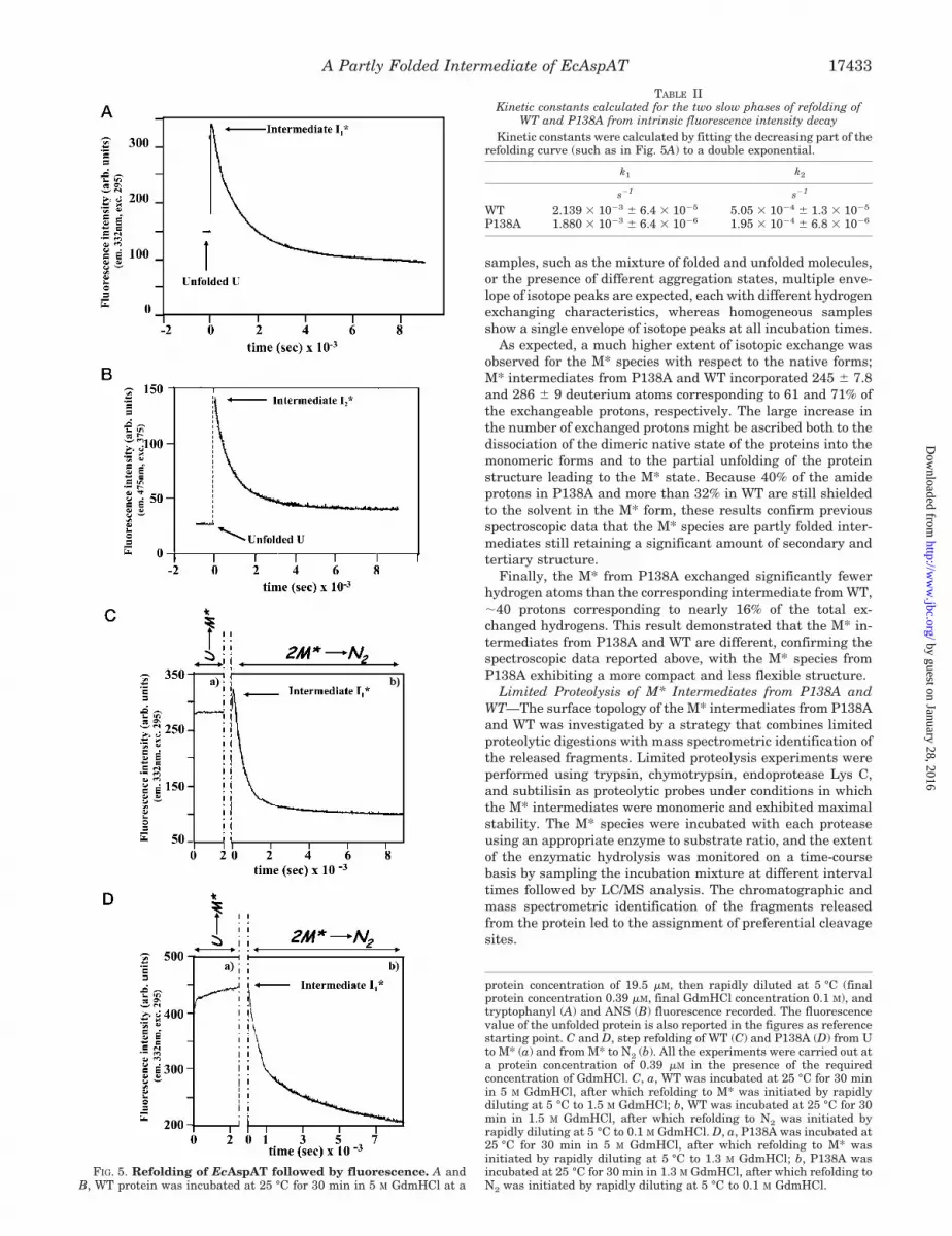
samples, such as the mixture of folded and unfolded molecules,or the presence of different aggregation states, multiple enve-lope of isotope peaks are expected, each with different hydrogenexchanging characteristics, whereas homogeneous samplesshow a single envelope of isotope peaks at all incubation times.
As expected, a much higher extent of isotopic exchange wasobserved for the M* species with respect to the native forms;M* intermediates from P138A and WT incorporated 245 7.8and 286 9 deuterium atoms corresponding to 61 and 71% ofthe exchangeable protons, respectively. The large increase inthe number of exchanged protons might be ascribed both to thedissociation of the dimeric native state of the proteins into themonomeric forms and to the partial unfolding of the proteinstructure leading to the M* state. Because 40% of the amideprotons in P138A and more than 32% in WT are still shieldedto the solvent in the M* form, these results confirm previousspectroscopic data that the M* species are partly folded inter-mediates still retaining a significant amount of secondary andtertiary structure.
Finally, the M* from P138A exchanged significantly fewerhydrogen atoms than the corresponding intermediate from WT,40 protons corresponding to nearly 16% of the total ex-changed hydrogens. This result demonstrated that the M* in-termediates from P138A and WT are different, confirming thespectroscopic data reported above, with the M* species fromP138A exhibiting a more compact and less flexible structure.
Limited Proteolysis of M* Intermediates from P138A andWT—The surface topology of the M* intermediates from P138Aand WT was investigated by a strategy that combines limitedproteolytic digestions with mass spectrometric identification ofthe released fragments. Limited proteolysis experiments wereperformed using trypsin, chymotrypsin, endoprotease Lys C,and subtilisin as proteolytic probes under conditions in whichthe M* intermediates were monomeric and exhibited maximalstability. The M* species were incubated with each proteaseusing an appropriate enzyme to substrate ratio, and the extentof the enzymatic hydrolysis was monitored on a time-coursebasis by sampling the incubation mixture at different intervaltimes followed by LC/MS analysis. The chromatographic andmass spectrometric identification of the fragments releasedfrom the protein led to the assignment of preferential cleavagesites.
protein concentration of 19.5 �M, then rapidly diluted at 5 °C (finalprotein concentration 0.39 �M, final GdmHCl concentration 0.1 M), andtryptophanyl (A) and ANS (B) fluorescence recorded. The fluorescencevalue of the unfolded protein is also reported in the figures as referencestarting point. C and D, step refolding of WT (C) and P138A (D) from Uto M* (a) and from M* to N2 (b). All the experiments were carried out ata protein concentration of 0.39 �M in the presence of the requiredconcentration of GdmHCl. C, a, WT was incubated at 25 °C for 30 minin 5 M GdmHCl, after which refolding to M* was initiated by rapidlydiluting at 5 °C to 1.5 M GdmHCl; b, WT was incubated at 25 °C for 30min in 1.5 M GdmHCl, after which refolding to N2 was initiated byrapidly diluting at 5 °C to 0.1 M GdmHCl. D, a, P138A was incubated at25 °C for 30 min in 5 M GdmHCl, after which refolding to M* wasinitiated by rapidly diluting at 5 °C to 1.3 M GdmHCl; b, P138A wasincubated at 25 °C for 30 min in 1.3 M GdmHCl, after which refolding toN2 was initiated by rapidly diluting at 5 °C to 0.1 M GdmHCl.
FIG. 5. Refolding of EcAspAT followed by fluorescence. A andB, WT protein was incubated at 25 °C for 30 min in 5 M GdmHCl at a
TABLE IIKinetic constants calculated for the two slow phases of refolding of
WT and P138A from intrinsic fluorescence intensity decayKinetic constants were calculated by fitting the decreasing part of the
refolding curve (such as in Fig. 5A) to a double exponential.
k1 k2
s�1 s�1
WT 2.139 � 10�3 6.4 � 10�5 5.05 � 10�4 1.3 � 10�5
P138A 1.880 � 10�3 6.4 � 10�6 1.95 � 10�4 6.8 � 10�6
A Partly Folded Intermediate of EcAspAT 17433
by guest on January 28, 2016http://w
ww
.jbc.org/D
ownloaded from
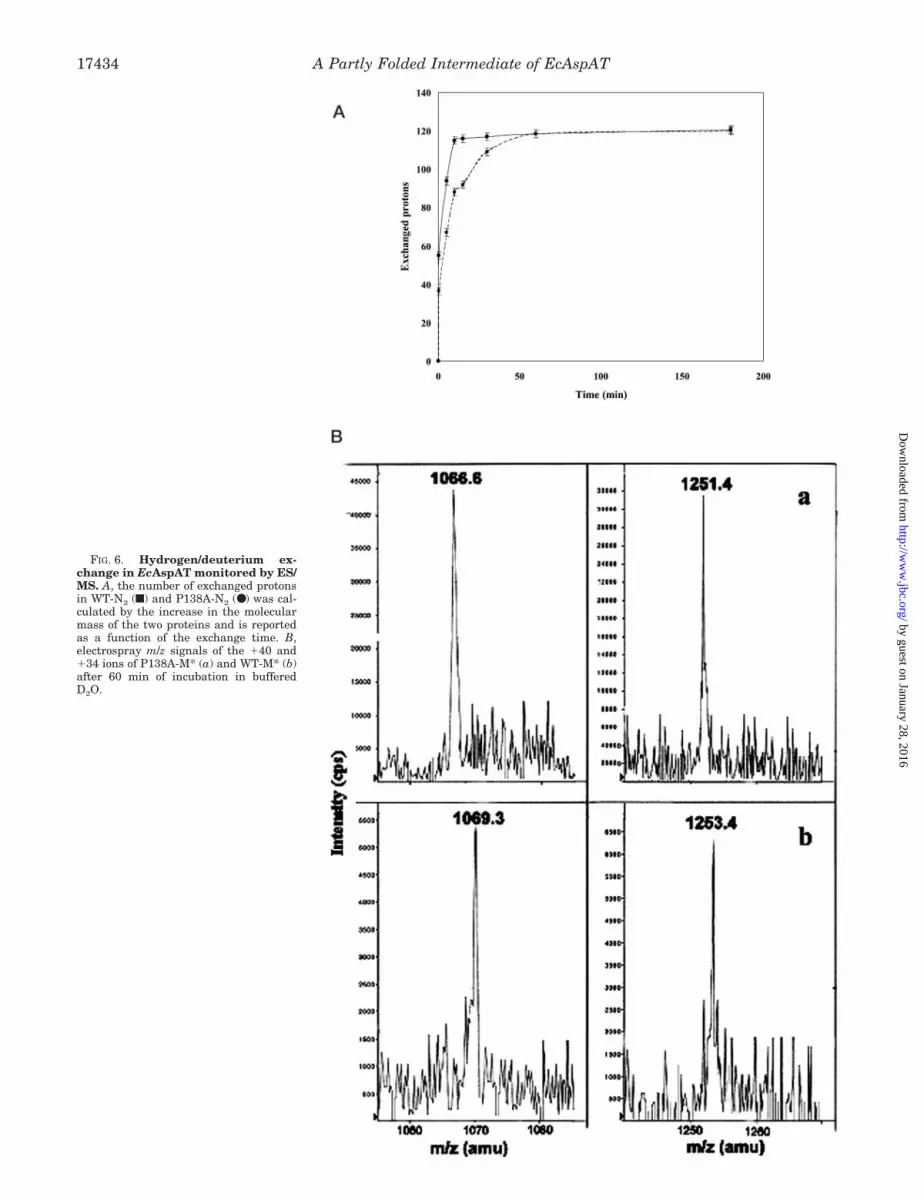
FIG. 6. Hydrogen/deuterium ex-change in EcAspAT monitored by ES/MS. A, the number of exchanged protonsin WT-N2 (f) and P138A-N2 (●) was cal -culated by the increase in the molecularmass of the two proteins and is reportedas a function of the exchange time. B,electrospray m/z signals of the �40 and�34 ions of P138A-M* (a) and WT-M* (b)after 60 min of incubation in bufferedD2O.
A Partly Folded Intermediate of EcAspAT17434
by guest on January 28, 2016http://w
ww
.jbc.org/D
ownloaded from
As an example, Fig. 7 shows the LC/MS chromatograms ofthe aliquots withdrawn following 30, 60, and 90 min of chymo-tryptic digestion of M* from P138A, performed using an en-zyme to substrate ratio of 1:500 (w/w). After 30 min of hydrol-ysis, four major peaks appeared (fractions 1–4 in Fig. 7), theintensity of which increased at later stages of incubation. Massspectrometric analysis of these fractions identified the twocomplementary pairs 1–59/60–411 (peaks 1 and 3 in Fig. 7) and1–380/381–411 (peaks 2 and 3), and the undigested protein(peak 4). These data clearly indicated Tyr-59 and Tyr-380 aspreferential chymotryptic cleavage sites, suggesting that theseresidues are located in a flexible and exposed region of the M*structure.
The overall data from the complementary proteolysis exper-iments are summarized in Table III and Fig. 8. A small numberof preferential cleavage sites was observed in both M* species,indicating the occurrence of a rather structured conformationexhibiting low accessibility to proteases. In combination withspectroscopic and H/D exchange data, these results demon-strate that the two M* intermediates possess a stable and welldefined three-dimensional structure that prevent the occur-rence of a diffuse and random distribution of proteolytic cleav-ages. Moreover, very few cleavages were observed at residuesthat in the native structure are located within the subunitinterfaces, indicating that the exposure of M* regions was notmerely the result of dissociation of the dimeric structure. Fi-nally, proteolytic sites were identified both in the small and inthe large domain of the protein, thus excluding the possibilitythat M* originated from the unfolding of only one of the twodomains.
The preferential cleavage sites observed in M* from P138Aessentially gathered into two separate regions of the protein,i.e. the segments 98–121 and 266–288, whereas few isolatedsites were detected at Tyr-59, Ser-136, Ala-150, and Tyr-380. Itshould be noted that no cleavage was detected in the N-termi-nal arm that in the dimeric form of EcAspAT embraces thepartner subunit concurring in the formation of the active site.This observation suggests that, following dissociation of N2 intoM, the monomeric form should undergo conformationalchanges to generate the M* species in which the N-terminaltail is forced to interact with the protein body and is thenprotected from the proteases. Otherwise, the flexible N-termi-nal arm would have been cleaved by proteases at several sites.
A greater number of cleavage sites and a faster kinetic ofhydrolysis was observed in all the experiments on the M*species from WT, confirming that the two intermediates exhibita different three-dimensional arrangement with the conforma-tion of M* from WT being generally more flexible and lessstructured than P138A-M*. These results are in excellentagreement with previous H/D exchange experiments depictinga higher flexibility of the WT-M* species that showed a largerextent of deuterium incorporation.
Accordingly, besides the exposed regions already observed inP138A-M*, additional proteolytic sites in WT-M* were identi-fied within the segments 20–25 and 39–41 with few isolatedcleavages observed at Tyr-59, Tyr-161, and Phe-188. Particu-larly, the region 39–41 and Tyr-161 are located at the inter-domain interface. Occurrence of proteolytic cleavages at thisregion only in WT-M* suggests that the structural differencesbetween the two M* intermediates might be ascribed to adifferent relative orientation of the two domains. The relativerotation of the two domains might also affect the conformationof the N-terminal tail, thus explaining the accessibility of thesegment 20–25, located in the small domain close to the Nterminus of the protein.
Comparative proteolysis experiments were also performedon the native dimeric forms of P138A and WT, using the sameconditions employed for the M* intermediates. Both proteinswere completely resistant to proteases, and no cleavage wasdetected at any time of incubation even when the E/S ratio wasincreased up to 50-fold.
Conclusion—A combination of spectroscopic techniques, H/Dexchange, and limited proteolysis experiments coupled to massspectrometry analysis were used to depict the topology of theM* partly folded intermediate of EcAspAT in WT as well as inthe mutant protein P138A.
The role of intermediates in the protein folding is a contro-versial issue. In some cases, they appear to be important mile-stones for productive folding (i.e. they are on-pathway),whereas, in other cases, they might arise by nonspecific col-lapse of the polypeptide chain or accumulate because they aretrapped by non-native interactions (i.e. they are off-pathway).M* is an off-pathway intermediate in the folding process of WTEcAspAT, as already pointed out by Leistler et al. (35), whereasspectroscopic data provided in this study suggest that the M*species from the mutant P138A is coincident with the on-pathway folding intermediate I1*.
Gel filtration and CD analysis did not show major differencesbetween the two M* species. On the contrary, the increaseddeuterium incorporation level and the higher accessibility toproteases shown by WT-M* as compared with P138A-M* dem-onstrated the occurrence of conformational differences betweenthe M* intermediates. These results together with fluorescencedata indicate that P138A-M* is conceivably more compact thanWT-M*.
In particular, limited proteolysis results suggested that
FIG. 7. Time-course analysis of P138A-M* digested with chy-motrypsin under controlled conditions. Figure shows HPLC pro-files of the aliquots withdrawn from the incubation mixture at 30 min(A), 60 min (B), and 90 min (C). Individual fractions were identified byES/MS.
A Partly Folded Intermediate of EcAspAT 17435
by guest on January 28, 2016http://w
ww
.jbc.org/D
ownloaded from
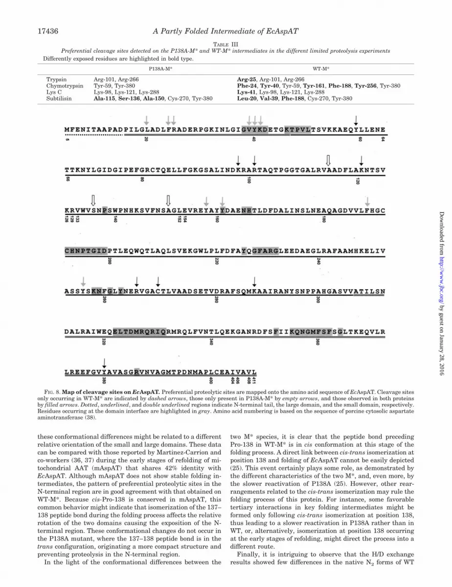
these conformational differences might be related to a differentrelative orientation of the small and large domains. These datacan be compared with those reported by Martinez-Carrion andco-workers (36, 37) during the early stages of refolding of mi-tochondrial AAT (mAspAT) that shares 42% identity withEcAspAT. Although mAspAT does not show stable folding in-termediates, the pattern of preferential proteolytic sites in theN-terminal region are in good agreement with that obtained onWT-M*. Because cis-Pro-138 is conserved in mAspAT, thiscommon behavior might indicate that isomerization of the 137–138 peptide bond during the folding process affects the relativerotation of the two domains causing the exposition of the N-terminal region. These conformational changes do not occur inthe P138A mutant, where the 137–138 peptide bond is in thetrans configuration, originating a more compact structure andpreventing proteolysis in the N-terminal region.
In the light of the conformational differences between the
two M* species, it is clear that the peptide bond precedingPro-138 in WT-M* is in cis conformation at this stage of thefolding process. A direct link between cis-trans isomerization atposition 138 and folding of EcAspAT cannot be easily depicted(25). This event certainly plays some role, as demonstrated bythe different characteristics of the two M*, and, even more, bythe slower reactivation of P138A (25). However, other rear-rangements related to the cis-trans isomerization may rule thefolding process of this protein. For instance, some favorabletertiary interactions in key folding intermediates might beformed only following cis-trans isomerization at position 138,thus leading to a slower reactivation in P138A rather than inWT, or, alternatively, isomerization at position 138 occurringat the early stages of refolding, might direct the process into adifferent route.
Finally, it is intriguing to observe that the H/D exchangeresults showed few differences in the native N2 forms of WT
TABLE IIIPreferential cleavage sites detected on the P138A-M* and WT-M* intermediates in the different limited proteolysis experiments
Differently exposed residues are highlighted in bold type.
P138A-M* WT-M*
Trypsin Arg-101, Arg-266 Arg-25, Arg-101, Arg-266Chymotrypsin Tyr-59, Tyr-380 Phe-24, Tyr-40, Tyr-59, Tyr-161, Phe-188, Tyr-256, Tyr-380Lys C Lys-98, Lys-121, Lys-288 Lys-41, Lys-98, Lys-121, Lys-288Subtilisin Ala-115, Ser-136, Ala-150, Cys-270, Tyr-380 Leu-20, Val-39, Phe-188, Cys-270, Tyr-380
FIG. 8. Map of cleavage sites on EcAspAT. Preferential proteolytic sites are mapped onto the amino acid sequence of EcAspAT. Cleavage sitesonly occurring in WT-M* are indicated by dashed arrows, those only present in P138A-M* by empty arrows, and those observed in both proteinsby filled arrows. Dotted, underlined, and double underlined regions indicate N-terminal tail, the large domain, and the small domain, respectively.Residues occurring at the domain interface are highlighted in gray. Amino acid numbering is based on the sequence of porcine cytosolic aspartateaminotransferase (38).
A Partly Folded Intermediate of EcAspAT17436
by guest on January 28, 2016http://w
ww
.jbc.org/D
ownloaded from
and P138A, the spectroscopic features and crystallographicstructures of which are almost superimposable. Flipping of the137–138 peptide bond from the cis conformation of WT to thetrans conformation of P138A resulted in an overall increased“motility” of the fraction of the protein that is accessible to thesolvent in the native form.
Limited proteolysis and H/D exchange experiments in con-junction with mass spectrometry analysis well complementedspectroscopic results, thus providing a wealth of data thatallowed a detailed structural analysis of EcAspAT folding in-termediates. On a more general ground, these results suggestthat the integration of spectroscopic investigations and massspectrometric procedures might be instrumental in providingsubtle structural details on transient conformations such asthose present along a folding pathway.
Acknowledgment—We thank Dr. Alessia Errico for preliminary lim-ited proteolysis experiments.
REFERENCES
1. Dobson, C. M., and Karplus, M. (1999) Curr. Opin. Struct. Biol. 9, 92–1012. Chiti, F., Taddei, N., Bucciantini, M., White, P., Ramponi, G., and Dobson,
C. M. (2000) EMBO J. 3, 1441–14493. Englander, S. W., and Kallenbach, N. R. (1984) Q. Rev. Biophys. 16, 521–6554. Li, R., and Woodward, C. (1999) Protein Sci. 8, 1571–15905. Woodward, C., Simon, I., and Tuchsen, E. (1982) Mol. Cell. Biochem. 48,
135–1606. Johnson, R. S., and Walsh, K. A. (1994) Protein Sci. 3, 2411–24187. Smith, D. L., Deng, Y., and Zhang, Z. (1997) J. Mass Spectrom. 32, 135–1468. Katta, V., and Chait, B. T. (1993) Rapid Commun. Mass Spectrom. 5, 214–2179. Zhang, Z., and Smith, D. L. (1993) Protein Sci. 2, 522–531
10. Miranker, A., Robinson, C. V., Radford, S. E., Aplin, R. T., and Dobson, C. M.(1993) Science 262, 896–900
11. Zhang, Z., and Smith, D. L. (1996) Protein Sci. 5, 1282–128912. Engen, J. R., Gmeiner, W. H., Smithgall, T. E., and Smith, D. L. (1999)
Biochemistry 38, 8926–893513. Halgand, F., Dumas, R., Biou, V., Andrieu J. P., Thomazeau, K., Gagnon, J.,
Douce, R., and Forest, E. (1999) Biochemistry 38, 6025–603414. Zappacosta, F., Pessi, A., Bianchi, E., Venturini, S., Sollazzo, M., Tramontano,
A., Marino, G., and Pucci, P. (1996) Protein Sci. 5, 802–81315. Scognamiglio, R., Notomista, E., Barbieri, P., Pucci, P., Dal Piaz, F.,
Tramontano, A., and Di Donato, A. (2001) Protein Sci. 10, 482–49016. Orru, S., Dal Piaz, F., Casbarra, A., Biasiol, G., De Francesco, R., Steinkuhler,
C., and Pucci, P. (1999) Protein Sci. 8, 1445–145417. Bianchi, E., Orru, S., Dal Piaz, F., Ingenito, R., Casbarra, A., Biasiol, G., Koch,
U., Pucci, P., and Pessi, A. (1999) Biochemistry 38, 13844–1385218. Urbani, A., Biasiol, G., Brunetti, M., Volpari, C., Di Marco, S., Sollazzo, M.,
Orru, S., Dal Piaz, F., Casbarra, A., Pucci, P., Nardi, C., Gallinari, P.,De Francesco, R., and Steikuhler, C. (1999) Biochemistry 38, 5206–5215
19. Esposito, G., Michelutti, R., Verdone, G., Viglino, P., Hernandez, H., Robinson,C. V., Amoresano, A., Dal Piaz, F., Monti, M., Pucci, P., Mangione, P.,Stoppini, M., Merlini, G., Ferri, G., and Bellotti, V. (2000) Protein Sci. 9,831–845
20. De Lorenzo, C., Dal Piaz, F., Piccoli, R., Di Maro, A., Pucci, P., and D’Alessio,G. (1998) Protein Sci. 7, 2653–2658
21. Piccoli, R., De Lorenzo, C., Dal Piaz, F., Pucci, P., and D’Alessio, G. (2000)J. Biol. Chem. 275, 8000–8006
22. Scaloni, A., Miraglia, N., Orru, S., Amodeo, P., Motta, A., Marino, G., andPucci, P. (1998) J. Mol. Biol. 277, 945–958
23. Scaloni, A., Monti, M., Acquaviva, R., Tell, G., Damante, G., Formisano, S., andPucci, P. (1999) Biochemistry 38, 64–72
24. Atkinson, R. A., Joseph, C., Dal Piaz, F., Birolo, L., Stier, G., Pucci, P., andPastore, A. (2000) Biochemistry 39, 5255–5264
25. Birolo, L., Malashkevich, V. N., Capitani, G., De Luca, F., Moretta, A.,Jansonius, J. N., and Marino, G. (1999) Biochemistry 38, 905–913
26. Jager, J., Moser, M., Sauder, U., and Jansonius, J. N. (1994) J. Mol. Biol. 239,285–305
27. McPhalen, C. A., Vincent, M. G., Picot, D., Jansonius, J. N., Lesk, A. M., andChothia, C. (1992) J. Mol. Biol. 22, 197–213
28. Yano, T., Kuramitsu, S., Tanase, S., Morino, Y., Hiromi, K., and Kagamiyama,H. (1991) J. Biol. Chem. 266, 6079–6085
29. Kuramitsu, S., Hiromi, K., Hayashi, H., Morino, Y., and Kagamiyama, H.(1990) Biochemistry 29, 5469–5476
30. Lehrer, S. S. (1971) Biochemistry 10, 3254–326331. Eftink, M. R., and Ghiron, C. A. (1976) Biochemistry 15, 672–68032. Bai Y., Milne, J. S., Mayne, L., and Englander, S. W. (1993) Proteins 17, 75–8633. Herold, M., and Kirshner, K. (1990) Biochemistry 29, 1907–191334. Herold, M., and Leistler, B. (1992) FEBS Lett. 308, 26–2935. Leistler, B., Herold, M., and Kirschner, K. (1992) Eur. J. Biochem. 205,
603–61136. Mattingly, J. R., Jr., Torella, C., Iriarte, A., and Martinez-Carrion, M. (1998)
J. Biol. Chem. 273, 23191–2320237. Torella, C., Mattingly, J. R., Jr., Artigues, A., Iriarte, A., and Martinez-
Carrion, M. (1998) J. Biol. Chem. 273, 3915–392538. Ovchinnikov, Y. A., Egorov, C. A., Aldanova, N. A., Feigina, M. Y., Lipkin,
V. M., Abdulaev, N. G., Grishin, E. V., Kiselev, A. P., Modyanov, N. N.,Braunstein, A. E., Polyanovsky, O. L., and Nosikov, V. V. (1973) FEBS Lett.29, 31–34
A Partly Folded Intermediate of EcAspAT 17437
by guest on January 28, 2016http://w
ww
.jbc.org/D
ownloaded from
Leila Birolo, Fabrizio Dal Piaz, Piero Pucci and Gennaro Marino Escherichia coliP138A Aspartate Aminotransferase from
Structural Characterization of the M* Partly Folded Intermediate of Wild Type and
doi: 10.1074/jbc.M200650200 originally published online March 1, 20022002, 277:17428-17437.J. Biol. Chem.
10.1074/jbc.M200650200Access the most updated version of this article at doi:
Alerts:
When a correction for this article is posted•
When this article is cited•
to choose from all of JBC's e-mail alertsClick here
http://www.jbc.org/content/277/20/17428.full.html#ref-list-1
This article cites 38 references, 5 of which can be accessed free at
by guest on January 28, 2016http://w
ww
.jbc.org/D
ownloaded from


![Fine tuning of folded conformation by change of substituents: 1H NMR and crystallographic evidence for folded conformation due to arene interactions in pyrazolo[3,4-d]pyrimidine core](https://img.dokumen.tips/doc/110x75/635b0f7d26d04ababa021d97/fine-tuning-of-folded-conformation-by-change-of-substituents-1h-nmr-and-crystallographic.jpg)


