Embed Size (px)
Citation preview
Materials 2014, 7, 7217-7225; doi:10.3390/ma7117217
materials ISSN 1996-1944
www.mdpi.com/journal/materials
Article
Structural Aspects LiNbO3 Nanoparticles and
Their Ferromagnetic Properties
Carlos A. Diaz-Moreno 2, Rurik Farias-Mancilla 1, Jose T. Elizalde-Galindo 1,
Jesus González-Hernández 4, Abel Hurtado-Macias 4, Daniel Bahena 3,
Miguel José-Yacamán 3 and Manuel Ramos 1,2,*
1 Departamento de Física y Matemáticas, Instituto de Ingeniería y Tecnología,
Universidad Autónoma de Cd. Juárez, Avenida del Charro #450 N. Cd. Juárez, Chihuahua,
C.P. 32310, Mexico; E-Mails: [email protected] (R.F.-M.); [email protected] (J.T.E.-G.) 2 Materials Research and Technology Institute, University of Texas at El Paso, 500 W,
University Ave, El Paso, TX 79968, USA; E-Mail: [email protected] 3 Kleberg Advanced Microscopy Center, University of Texas at San Antonio, One UTSA Circle,
San Antonio, TX 78249, USA; E-Mails: [email protected] (D.B.);
[email protected] (M.J.-Y.) 4 Centro de Investigación en Materiales Avanzados S.C., Laboratorio Nacional de Nanotecnología,
Miguel de Cervantes 120, Complejo Industrial Chihuahua, Chihuahua, Apdo. Postal 31109, Mexico;
E-Mails: [email protected] (J.G.-H.); [email protected] (A.H.-M.)
* Author to whom correspondence should be addressed; E-Mail: [email protected] or
[email protected]; Tel.: +1-915-747-6175; Fax: +1-915-747-6007.
External Editor: Beatriz Noheda
Received: 1 May 2014; in revised form: 12 June 2014 / Accepted: 26 June 2014 /
Published: 28 October 2014
Abstract: We present a solid-state synthesis of ferromagnetic lithium niobate nanoparticles
(LiNbO3) and their corresponding structural aspects. In order to investigate the effect of
heat treatments, two batches of samples with a heat-treated (HT) and non-heat-treated
(nHT) reduction at 650 °C in 5% of hydrogen/argon were considered to investigate the
multiferroic properties and their corresponding structural aspects; using magnetometry and
scanning transmission electron microscopy (STEM). Results indicate the existence
of ferromagnetic domains with a magnetic moment per unit cell of 5.24 × 10⁻³ μB;
caused mainly due to voids and defects on the nanoparticle surface, as confirmed by
STEM measurements.
OPEN ACCESS
Materials 2014, 7 7218
Keywords: scanning transmission electron microscopy (STEM); nanoparticles;
lithium niobate; ferroelectric; ferromagnetic
1. Introduction
Currently, it is relatively easy to find information regarding perovskite structures, such as ABO3
compounds (i.e., PbTiO3 (PT), BaTiO3 (BT), Pb(Zr,Ti)O3 (PZT) and LiNbO3 (LNO)) as principal
materials for nonlinear optical applications [1]. The study of those particular systems has arisen due to
their large dielectric constant values [2]. In the past four years, crystalline lithium niobate (LNO) has
been considered a material with enormous potential for materials engineering applications, such as
optoelectronics material [3]. It is relatively easy to find extensive information in the literature about
routes for synthesis and characterization [4], along with experimental techniques to measure the
ferromagnetic/ferroelectric response, especially nano-scaled structures (spheres, nanorods) [5,6],
as well as their corresponding electronic structure using computational methods [7]. In the search for
a fundamental understanding of ferromagnetism in the LNO crystalline structure, theoretical
hypotheses have been constituted according to Wei et al., who attributed it to an addition of
contaminants into the LNO structure (the effect of dopants). This seems to agitate the Li-Nb-O
electronic structure [8]. Kong et al. argue the formation of ferromagnetism when implanting Ni+, Fe+ or
Mn+ and Co+ ions into the LiNbO3 crystal structure [9]. A preliminary conclusion made by Sundaseran
and Rao indicates that ferromagnetic properties can be caused in LNO nanostructures by the formation of
oxygen vacancies near the surface [10], also because of polarization values of 70 μC/cm2 found in LNO
crystals at room temperature, with the existence of the ferroelectric phase at high transition temperature
values of 1483 K [11]. The latter, an important path to design and determine the fundamental study of
the magnetic behavior, due to structural defects, has led to the search for new materials. Such is the
case of the high temperature ferromagnetic behavior reported in LiNbO3 nanocrystal, due to heat
treatment reduction, causing defect structures near the surface of nanoparticles [12]. One can find in
the literature two main manuscripts regarding multiferroic properties in LiNbO3: Song et al.,
who reported multiferroic properties when LiNbO3 was doped with cobalt (Co), calling it a two-phase
material [11]. Additionally, the other work done by this group reported multiferroic properties in one
phase in nanoparticles of LiNbO3 [13]. Furthermore, the recent advances in small-scaled electronics
combined with computational equipment allowed microscopy analysis with point resolutions of
0.08~0.11 nm and a magnification of 150,000,000× in STEM Cs-corrected instruments [14];
consequently, new information regarding nanoparticle structural aspects is possible to obtain using these
characterization techniques. For example, using microanalysis principles, Borisevich et al. were able to
detect with high success the localization sites for oxygen vacancies on metallic interfaces [15].
Furthermore, the usage of irradiation energy from a field emission gun source while surveying
specimens in STEM microanalysis allowed the formation of new crystallographic phases, as presented
by Sepulveda-Guzman et al., in the formation of bismuth nanoparticles at 200 kV using a NaBiO3
powder precursor [16]. Here, we present the solid-state reaction synthesis of lithium niobate
nanoparticles from lithium carbonate and niobium pentoxide precursors, followed by heat treatments at
Materials 2014, 7 7219
650 °C in a hydrogen atmosphere. In order to investigate both the ferromagnetic properties and
structural aspects, two batches of samples (heat treated and non-heat treated) were considered for
powder X-ray diffraction, Cs-corrected STEM and magnetometry analysis.
2. Results and Discussion
2.1. Differential Scanning Calorimetry
In order to find the temperature window at which the formation of lithium niobate exists during
calcination process, a series of differential scanning calorimetry (DSC) was done for the solid-state
reaction with various milling times ranging from 2 to 1200 min. The results indicate the existence of
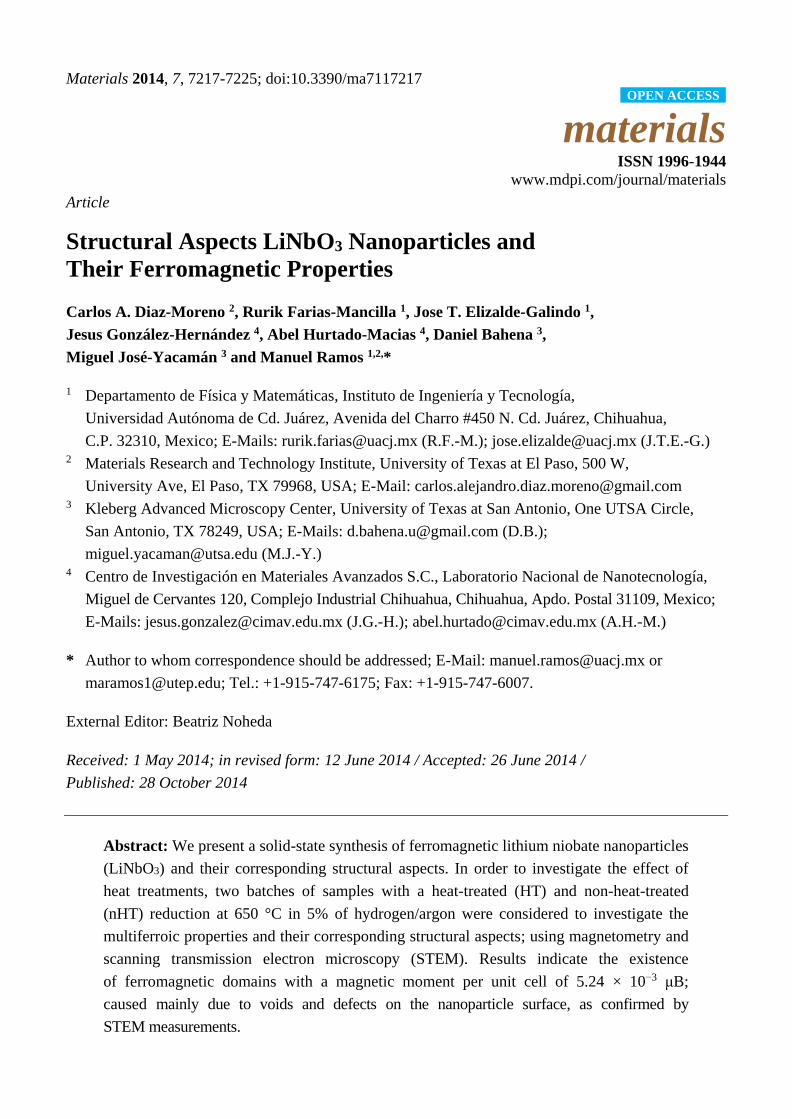
exothermic reactions as marked at the A, B and C sites, as presented in Figure 1. The main change
observed corresponds to 300 min at 317 °C, 435 °C and 486 °C, as presented in the inset of Figure 1
(three main peaks).
Figure 1. Differential scanning calorimetry of Lithium Niobate (LiNbO3) for 3 to
1200 min of milling. Inset: exothermic peaks for 300 min of milling time.
One brief explanation could be that a larger milling time (300 min) can provoke the increase of the
surface energy and, therefore, the lower energy or temperature required for crystallization, contrary to
the low milling time, as observed, with no peaks near 300 °C to 600 °C.
2.2. Powder X-ray Diffraction Pattern
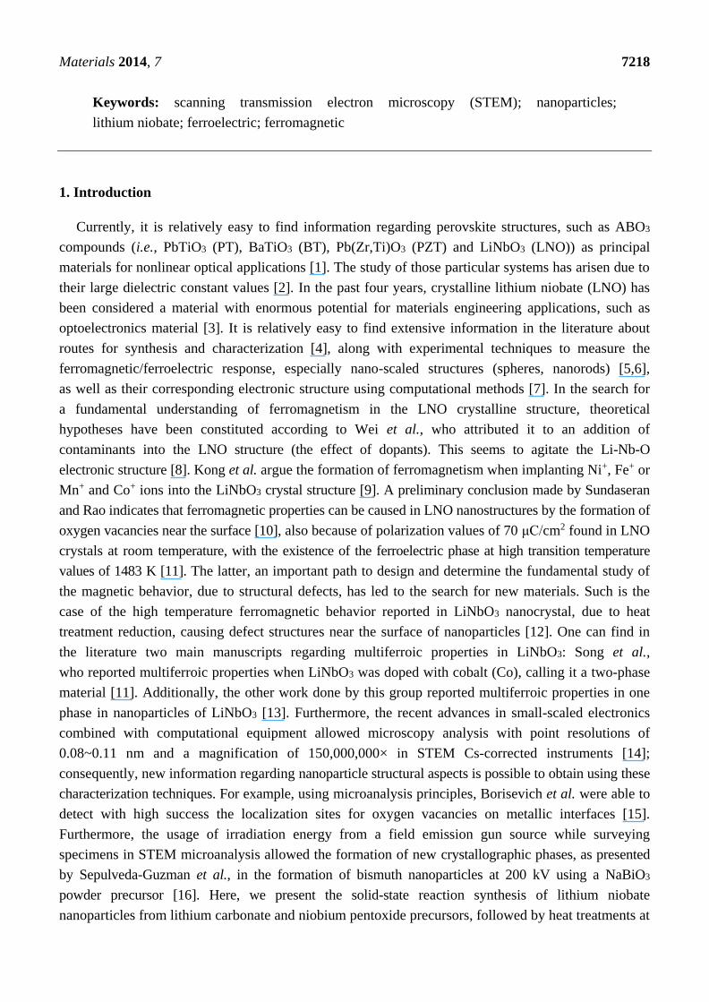
To correlate data found from DSC measurements, a series of powder X-ray diffraction was
performed, focusing on samples processed at 300 min of milling and calcination at 350 °C, 450 °C and
650 °C. The results indicate a seed material corresponding to niobium pentoxide (Nb2O5) as the
observed poor crystallinity presented in Figure 2A; then, a mix phase found, made out of Nb2O5 and
lithium niobate (LiNbO3), as presented in Figure 2B; finally, a pure crystalline phase of LiNbO3
is found with (012), (104) and (110) principal refractions in Figure 2C, as indexed using
ICDD #00-074-2238 cards, in agreement with Shimada et al. [17].
0 200 400 600 800 1000 1200
1200 min
600 min
300 min
210 min
90 min
45 min
30 min
9 min
3 min
T
( °C
) E
xoth
erm
ic
TEMPERATURE o
C
AB C
0 200 400 600 800 1000 1200
300 min
T
( °C
) E
xoth
erm
ic
TEMPERATURE o
C
A
B
C
( C
)
( C
)
Materials 2014, 7 7220
Figure 2. Powder XRD for samples prepared at 300 min of milling and corresponding to
calcination temperatures at (A) 350 °C, (B) 450 °C and (C) 650 °C. Inset: three phases
generated for different calcination temperatures, which determine the ideal temperature
conditions to form spherical LiNbO3 nanoparticles, as confirmed by XRD measurements.
2.3. Magnetometry Measurements
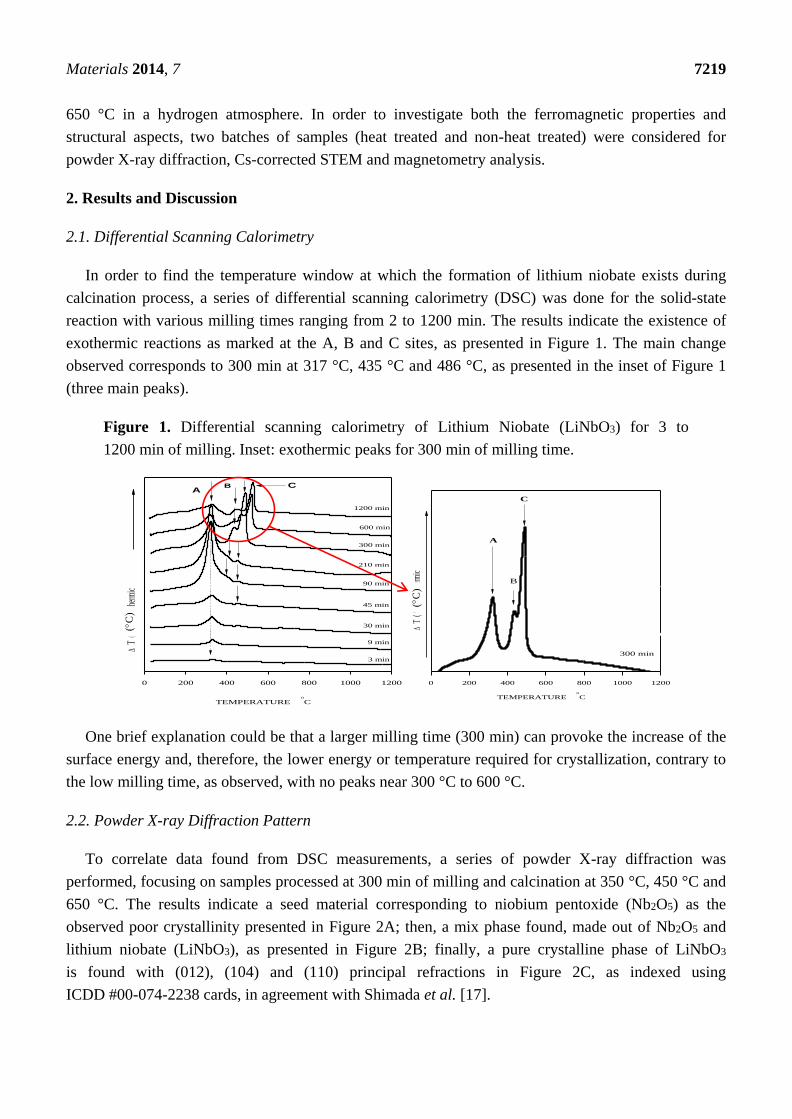
After the calcination process, just the sample under calcination at 650 °C was subjected to
an additional heat treatment reduction at 650 °C in hydrogen/argon (5%/95%) for 20 min. A grey
coloration is observed in comparison with the white powder before the sintering process. The two
samples were subjected to magnetization measurements at 9 T external field at room temperature
(300 K) using the Quantum Design physical properties measurement equipment. A hysteresis loop is
observed for the sample under heat treatment reduction, as presented in Figure 3; that magnetization is
attributed practically to a non-coercivity caused due to structural defects or voids in the LiNbO3
structure. Coey et al. explained that these phenomena could be attributed to magnetic regions rich in
defects, which tend to concentrate at interfaces and grain boundaries [18]; which, in the case
of the sample without heat treatment reduction, do not present a hysteresis loop; consequently,
a non-magnetic behavior.
Figure 3. Magnetization curves measured at 9 T and 300 K for the sample under heat
treatment reduction at 650 °C and non-heat treatment conditions.
Nb2O5
Nb2O5 + LN
LiNbO3
-300 -200 -100 0 100 200 300
-1.0
-0.5
0.0
0.5
1.0
H ( kOe )
M
(
10
-3 x
emu/
g )
nHT
HT
Materials 2014, 7 7221
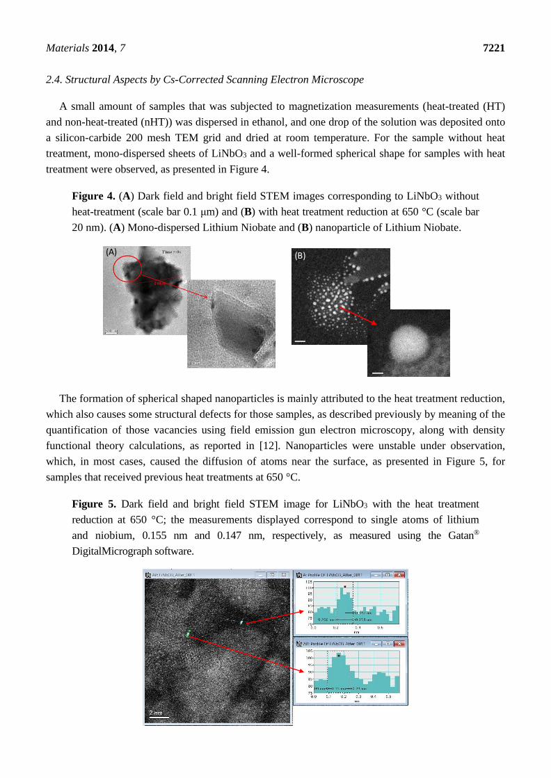
2.4. Structural Aspects by Cs-Corrected Scanning Electron Microscope
A small amount of samples that was subjected to magnetization measurements (heat-treated (HT)
and non-heat-treated (nHT)) was dispersed in ethanol, and one drop of the solution was deposited onto
a silicon-carbide 200 mesh TEM grid and dried at room temperature. For the sample without heat
treatment, mono-dispersed sheets of LiNbO3 and a well-formed spherical shape for samples with heat
treatment were observed, as presented in Figure 4.
Figure 4. (A) Dark field and bright field STEM images corresponding to LiNbO3 without
heat-treatment (scale bar 0.1 μm) and (B) with heat treatment reduction at 650 °C (scale bar
20 nm). (A) Mono-dispersed Lithium Niobate and (B) nanoparticle of Lithium Niobate.
The formation of spherical shaped nanoparticles is mainly attributed to the heat treatment reduction,
which also causes some structural defects for those samples, as described previously by meaning of the
quantification of those vacancies using field emission gun electron microscopy, along with density
functional theory calculations, as reported in [12]. Nanoparticles were unstable under observation,
which, in most cases, caused the diffusion of atoms near the surface, as presented in Figure 5, for
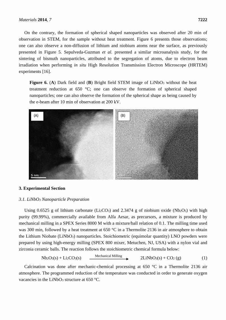
samples that received previous heat treatments at 650 °C.
Figure 5. Dark field and bright field STEM image for LiNbO3 with the heat treatment
reduction at 650 °C; the measurements displayed correspond to single atoms of lithium
and niobium, 0.155 nm and 0.147 nm, respectively, as measured using the Gatan®
DigitalMicrograph software.
2 0 n m2 0 n m
2 n m2 n m
(A) (B)
Materials 2014, 7 7222
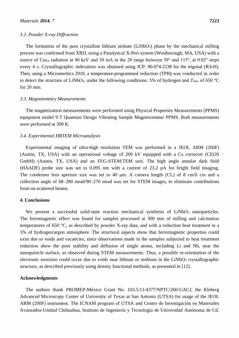
On the contrary, the formation of spherical shaped nanoparticles was observed after 20 min of
observation in STEM, for the sample without heat treatment. Figure 6 presents those observations;
one can also observe a non-diffusion of lithium and niobium atoms near the surface, as previously
presented in Figure 5. Sepulveda-Guzman et al. presented a similar microanalysis study, for the
sintering of bismuth nanoparticles, attributed to the segregation of atoms, due to electron beam
irradiation when performing in situ High Resolution Transmission Electron Microscope (HRTEM)
experiments [16].
Figure 6. (A) Dark field and (B) Bright field STEM image of LiNbO3 without the heat
treatment reduction at 650 °C; one can observe the formation of spherical shaped
nanoparticles; one can also observe the formation of the spherical shape as being caused by
the e-beam after 10 min of observation at 200 kV.
3. Experimental Section
3.1. LiNbO3 Nanoparticle Preparation
Using 0.6525 g of lithium carbonate (Li2CO3) and 2.3474 g of niobium oxide (Nb2O5) with high
purity (99.99%), commercially available from Alfa Aesar, as precursors, a mixture is produced by
mechanical milling in a SPEX Series 8000 M with a mixture/ball relation of 0.1. The milling time used
was 300 min, followed by a heat treatment at 650 °C in a Thermolite 2136 in air atmosphere to obtain
the Lithium Niobate (LiNbO3) nanoparticles. Stoichiometric (equimolar quantity) LNO powders were
prepared by using high-energy milling (SPEX 800 mixer, Metuchen, NJ, USA) with a nylon vial and
zirconia ceramic balls. The reaction follows the stoichiometric chemical formula below:
Nb2O5(s) + Li2CO3(s) 2LiNbO3(s) + CO2 (g) (1)
Calcination was done after mechanic-chemical processing at 650 °C in a Thermolite 2136 air
atmosphere. The programmed reduction of the temperature was conducted in order to generate oxygen
vacancies in the LiNbO3 structure at 650 °C.
(A) (B)
Mechanical Milling
Materials 2014, 7 7223
3.2. Powder X-ray Diffraction
The formation of the pure crystalline lithium niobate (LiNbO3) phase by the mechanical milling
process was confirmed from XRD, using a Panalytical X-Pert system (Westborough, MA, USA) with a
source of CuKα radiation at 40 keV and 30 mA in the 2θ range between 30 and 115 , at 0.02 steps
every 4 s. Crystallographic indexation was obtained using JCP: 00-074-2238 for the trigonal (R3cH).
Then, using a Micromeritics 2910, a temperature-programmed reduction (TPR) was conducted in order
to detect the structure of LiNbO3, under the following conditions: 5% of hydrogen and Tmax of 650 °C
for 20 min.
3.3. Magnetometry Measurements
The magnetization measurements were performed using Physical Properties Measurements (PPMS)
equipment model 9 T Quantum Design Vibrating Sample Magnetrometer PPMS. Both measurements
were performed at 300 K.
3.4. Experimental HRTEM Microanalysis
Experimental imaging of ultra-high resolution TEM was performed in a JEOL ARM (200F)
(Austin, TX, USA) with an operational voltage of 200 kV equipped with a Cs corrector (CEOS
GmbH) (Austin, TX, USA) and an FEG-STEM/TEM unit. The high angle annular dark field
(HAADF) probe size was set to 0.095 nm with a current of 23.2 pA for bright field imaging.
The condenser lens aperture size was set to 40 μm. A camera length (CL) of 8 cm/6 cm and a
collection angle of 68–280 mrad/90–270 mrad was set for STEM images, to eliminate contributions
from un-scattered beams.
4. Conclusions
We present a successful solid-state reaction mechanical synthesis of LiNbO3 nanoparticles.
The ferromagnetic effect was found for samples processed at 300 min of milling and calcination
temperatures of 650 °C, as described by powder X-ray data, and with a reduction heat treatment in a
5% of hydrogen/argon atmosphere. The structural aspects show that ferromagnetic properties could
exist due to voids and vacancies, since observations made in the samples subjected to heat treatment
reduction show the poor stability and diffusion of single atoms, including Li and Nb, near the
nanoparticle surface, as observed during STEM measurements. Thus, a possible re-orientation of the
electronic structure could occur due to voids near lithium or niobium in the LiNbO3 crystallographic
structure, as described previously using density functional methods, as presented in [12].
Acknowledgments
The authors thank PROMEP-México Grant No. 103.5/11/4377/NPTC/260-UACJ, the Kleberg
Advanced Microscopy Center of University of Texas at San Antonio (UTSA) for usage of the JEOL
ARM (200F) instrument. The ICNAM program of UTSA and Centro de Investigación en Materiales
Avanzados-Unidad Chihuahua, Instituto de Ingeniería y Tecnología de Universdad Autónoma de Cd.
Materials 2014, 7 7224
Juárez and the Materials Research and Technology Institute of the University of Texas at El Paso.
An primary author thank Consejo Nacional de Ciencia y Tecnología (CONACyT-México) for
economical support through the Postdoctoral Scholarship Program, Solicitation #232453.
Author Contributions
Carlos Diaz-Moreno contributed with mechanical synthesis of LiNbO3 nanoparticles and
magnetization measurements. Rurik Farias-Mancilla, Jose T. Elizalde-Galindo, Jesus González-Hernández,
and Abel Hurtado-Macias were responsible for the XRD, magnetization and DSC measurements and
supervision. Daniel Bahena, Miguel José-Yacamán and Manuel Ramos were in charge of performing
in situ STEM measurements. Carlos Diaz-Moreno, Abel Hurtado-Macias and Manuel Ramos mainly
typed this article.
Conflicts of Interest
The authors declare no conflict of interest.
References
1. Zhu, X. Recent patents on perovskite ferroelectric nanostructures. Recent Pat. Nanotechnol. 2009,
3, 42–52.
2. Xue, D.; Betzler, K.; Hesse, H. Dielectric properties of lithium niobate and tantalate crystals.
Solid State Commun. 2000, 115, 581–585.
3. Volk, T.; Wöhlecke, M. Lithium Niobate, 1st ed.; Springer-Verlag: Berlin, Germany, 2008; p. 258.
4. Knabe, B.; Schütze, D.; Jungk, T.; Svete, M.; Assenmacher, W.; Mader, W.; Buse, K.
Synthesis and characterization of Fe-doped LiNbO3 nanocrystals from a triple-alkoxide method.
Phys. Status Solidi A 2011, 208, 857–862.
5. Rüdiger, A.; Waser, R. Size effects in nanoscale ferroelectrics. J. Alloys Compd. 2008, 449, 2–6.
6. Hölscher, R.; Schmidt, W.G.; Sanna, S. Modelling LiNbO3 surfaces at ambient conditions.
Phys. Rev. C 2014, 118, 10213–10220.
7. Veithen, M.; Ghosez, Ph. First-principles study of the dielectric and dynamical properties of
lithium niobate. Phys. Rev. B 2002, 65, doi:10.1103/PhysRevB.65.214302.
8. Wei, D.T.Y.; Lee, W.W.; Bloom, L.R. Large refractive index change induced by ion implantation
in lithium niobate. Appl. Phys. Lett. 1974, 25, 329–331.
9. Kong, Y.; Liu, S.; Xu, J. Recent advances in the photorefraction of doped lithium niobate crystals.
Materials 2012, 5, 1954–1971.
10. Sundaresan, A.; Rao, C.N.R. Ferromagnetism as a universal feature of inorganic nanoparticle.
Nanotoday 2009, 4, 96–106.
11. Song, C.; Zeng, F.; Shen, Y.X.; Geng, K.W.; Xie, Y.N.; Wu, Z.Y.; Pan, F. Local Co structure and
ferromagnetism in ion-implanted Co-doped LiNbO3. Phys. Rev. B 2006, 73, doi:10.1103/
PhysRevB.73.172412.
Materials 2014, 7 7225
12. Díaz-Moreno, C.A.; Farías-Mancilla, R.; Matutes-Aquino, J.A.; Elizalde-Galindo, J.;
Espinosa-Magaña, F.; González-Hernández, J.; Hurtado-Macías, A. Magnetic behavior in LiNbO3
nanocrystallites caused by oxygen vacancies. J. Magn. Magn. Mater. 2014, 356, 82–86.
13. Díaz-Moreno, C.; Farias, R.; Hurtado-Macias, A.; Elizalde-Galindo, J.; Hernandez-Paz, J.
Multiferroic response of nanocrystalline lithium niobate. J. Appl. Phys. 2012, 111, doi:10.1063/
1.3673434.
14. Zhou, W.; Oxley, M.P.; Lupini, A.R.; Krivanek, O.L.; Pennycook, S.J.; Idrobo, J.-C. Single atom
microscopy. Microsc. Microanal. 2012, 18, 1342–1354.
15. Borisevich, A.Y.; Lupini, A.R.; He, J.; Eliseev, E.A.; Morozovska, A.N.; Svechnikov, G.S.; Yu, P.;
Chu, Y.-H.; Ramesh, R.; Pantelides, S.T.; et al. Interface dipole between two metallic oxides
caused by localized oxygen vacancies. Phys. Rev. B 2012, 86, doi:10.1103/PhysRevB.86.140102.
16. Sepulveda-Guzman, S.; Elizondo-Villarreal, N.; Ferrer, D.A.; Torres-Castro, A.; Gao, X.; Zhou, J.P.;
José-Yacamán, M. In situ formation of bismuth nanoparticles trough beam irradiation in
transmission electron microscope. Nanotechnology 2007, 18, doi:10.1088/0957-4484/18/
33/335604.
17. Shimada, S.; Kodaira, K.; Matsuchita, T. A study of the formation of LiNbO3 in the system
Li2CO3-Nb2O5. Thermochim. Acta 1978, 23, 135–144.
18. Stamenov, C.P.; Gunning, R.D.; Venkatesanand, M.; Paul, K. Ferromagnetism in defect-ridden
oxides and related materials. New J. Phys. 2010, 12, doi:10.1088/1367-2630/12/5/053025.
© 2014 by the authors; licensee MDPI, Basel, Switzerland. This article is an open access article
distributed under the terms and conditions of the Creative Commons Attribution license
(http://creativecommons.org/licenses/by/4.0/).


